Antigen-binding Molecule Comprising Altered Antibody Variable Region Binding Cd3 And Cd137
Shimizu; Shun ; et al.
U.S. patent application number 16/769299 was filed with the patent office on 2020-12-03 for antigen-binding molecule comprising altered antibody variable region binding cd3 and cd137. This patent application is currently assigned to Chugai Seiyaku Kabushiki Kaisha. The applicant listed for this patent is Chugai Seiyaku Kabushiki Kaisha. Invention is credited to Naoka Hironiwa, Shu Wen Samantha Ho, Tomoyuki Igawa, Taro Miyazaki, Mika Sakurai, Shun Shimizu.
| Application Number | 20200377595 16/769299 |
| Document ID | / |
| Family ID | 1000005085445 |
| Filed Date | 2020-12-03 |


View All Diagrams
| United States Patent Application | 20200377595 |
| Kind Code | A1 |
| Shimizu; Shun ; et al. | December 3, 2020 |
ANTIGEN-BINDING MOLECULE COMPRISING ALTERED ANTIBODY VARIABLE REGION BINDING CD3 AND CD137
Abstract
Antigen-binding domains that are capable of binding to CD3 and CD137 but do not bind to CD3 and CD137 at the same time and methods of using the same are provided. Methods to obtain antigen binding domains which bind to two or more different antigen more efficiently are also provided.
| Inventors: | Shimizu; Shun; (Shizuoka, JP) ; Ho; Shu Wen Samantha; (Singapore, SG) ; Hironiwa; Naoka; (Shizuoka, JP) ; Sakurai; Mika; (Shizuoka, JP) ; Miyazaki; Taro; (Kanagawa, JP) ; Igawa; Tomoyuki; (Shizuoka, JP) | ||||||||||
| Applicant: |
|
||||||||||
|---|---|---|---|---|---|---|---|---|---|---|---|
| Assignee: | Chugai Seiyaku Kabushiki
Kaisha Tokyo JP |
||||||||||
| Family ID: | 1000005085445 | ||||||||||
| Appl. No.: | 16/769299 | ||||||||||
| Filed: | December 4, 2018 | ||||||||||
| PCT Filed: | December 4, 2018 | ||||||||||
| PCT NO: | PCT/JP2018/044493 | ||||||||||
| 371 Date: | June 3, 2020 |
| Current U.S. Class: | 1/1 |
| Current CPC Class: | C07K 16/2809 20130101; C07K 2317/24 20130101; C07K 16/2878 20130101; C07K 2317/33 20130101; C07K 2317/55 20130101; C07K 16/2863 20130101; C07K 2317/56 20130101; C07K 2317/92 20130101; C07K 2317/75 20130101; C07K 2317/71 20130101; C07K 2317/622 20130101; C07K 2317/526 20130101; C07K 2317/31 20130101; C40B 30/04 20130101; C07K 16/303 20130101 |
| International Class: | C07K 16/28 20060101 C07K016/28; C07K 16/30 20060101 C07K016/30 |
Foreign Application Data
| Date | Code | Application Number |
|---|---|---|
| Dec 5, 2017 | JP | 2017-233104 |
Claims
1. An antigen-binding molecule comprising: an antibody variable region that is capable of binding to CD3 and CD137, but does not bind to CD3 and CD137 at the same time; and a variable region binding to a third antigen different from CD3 and CD137.
2. The antigen-binding molecule of claim 1, wherein the third antigen is a molecule specifically expressed in a cancer tissue.
3. The antigen-binding molecule of claim 1 or 2, wherein the variable region that does not bind to CD3 and CD137 at the same time is a variable region that does not bind to CD3 and CD137 each expressed on a different cell, at the same time.
4. The antigen-binding molecule of any one of claims 1 to 3, further comprising an antibody Fc region.
5. The antigen-binding molecule of claim 4, wherein the Fc region is an Fc region having reduced binding activity against Fc gamma R as compared with the Fc region of a naturally occurring human IgG1 antibody.
6. The antigen-binding molecule of any one of claims 1 to 5, wherein the antigen-binding molecule has at least one characteristic selected from the group consisting of (1) to (4) below: (1) the variable region binds to an extracellular domain of CD3 epsilon comprising the amino acid sequence of SEQ ID NO: 91, (2) the antigen-binding molecule has an agonistic activity against CD137, (3) the antigen-binding molecule induces CD3 activation of a T cell against a cell expressing the molecule of the third antigen, but does not induce CD3 activation of a T cell against a cell expressing CD137, and (4) the antigen-binding molecule does not induce a cytokine release from PBMC in the absence of a cell expressing the molecule of the third antigen.
7. The antigen-binding molecule of any one of claims 1 to 6, which competes for binding to CD137 with an antibody selected from the group consisting of: (a) an antibody comprising a VH sequence having the amino acid sequence of SEQ ID NO: 30 and a VL sequence having the amino acid sequence of SEQ ID NO: 51, (b) an antibody comprising a VH sequence having the amino acid sequence of SEQ ID NO: 46 and a VL sequence having the amino acid sequence of SEQ ID NO: 53, (c) an antibody comprising a VH sequence having the amino acid sequence of SEQ ID NO: 40 and a VL sequence having the amino acid sequence of SEQ ID NO: 56, (d) an antibody comprising a VH sequence having the amino acid sequence of SEQ ID NO: 30 and a VL sequence having the amino acid sequence of SEQ ID NO: 58, and (e) an antibody comprising a VH sequence having the amino acid sequence of SEQ ID NO: 40 and a VL sequence having the amino acid sequence of SEQ ID NO: 61.
8. The antigen-binding molecule of any one of claims 1 to 7, comprising an amino acid sequence resulting from introducing alteration of one or more amino acids into a template sequence consisting of a heavy chain variable domain sequence described in SEQ ID NO: 92 and/or a light chain variable domain sequence described in SEQ ID NO: 93, wherein the one or more amino acids comprise at least one amino acid selected from the following positions: H chain: 31, 52b, 52c, 53, 54, 56, 57, 61, 98, 99, 100, 100a, 100b, 100c, 100d, 100e, 100f, and 100g (Kabat numbering); and L chain: 24, 25, 26, 27, 27a, 27b, 27c, 27e, 30, 31, 33, 34, 51, 52, 53, 54, 55, 56, 74, 77, 89, 90, 92, 93, 94, and 96 (Kabat numbering), wherein the HVR-H3 of the altered heavy chain variable domain sequence comprises at least one amino acid selected from: Ala, Pro, Ser, Arg, His or Thr at amino acid position 98; Ala, Ser, Thr, Gln, His or Leu at amino acid position 99; Tyr, Ala, Ser, Pro or Phe at amino acid position 100; Tyr, Val, Ser, Leu or Gly at amino acid position 100a; Asp, Ser, Thr, Leu, Gly or Tyr at amino acid position 100b; Val, Leu, Phe, Gly, His or Ala at amino acid position 100c; Leu, Phe, Ile or Tyr at amino acid position 100d; Gly, Pro, Tyr, Gln, Ser or Phe at amino acid position 100e; Tyr, Ala, Gly, Ser or Lys at amino acid position 100f; Gly, Tyr, Phe or Val at amino acid position 100g (Kabat numbering).
9. The antigen-binding molecule of any one of claims 1 to 8, comprising (a) a VH sequence having at least 95% sequence identity to the amino acid sequence of SEQ ID NO: 41, 30, 46 or 40; (b) a VL sequence having at least 95% sequence identity to the amino acid sequence of SEQ ID NO: 51, 52, 53, 54, 55, 56 or 57; or (c) the VH sequence of (a) and the VL sequence of (b).
10. A pharmaceutical composition comprising the antigen-binding molecule according to any of claims 1 to 9 and a pharmaceutically acceptable carrier.
11. A method of screening for an antigen-binding domain which binds to at least two or more different antigens of interest, comprising: (a) providing a library comprising a plurality of antigen-binding domains, (b) contacting the library provided in step (a) with a first antigen of interest and collecting antigen-binding domains bound to the first antigen, (c) contacting the antigen-binding domains collected in step (b) with a second antigen of interest and collecting antigen-binding domains bound to the second antigen, and (d) amplifying genes which encode the antigen binding domains collected in step (c) and identifying a candidate antigen-binding domain, wherein the method does not comprise amplifying nucleic acids that encode the antigen-binding domains collected in step (b) between step (b) and step (c).
12. The method of claim 11, wherein the antigen-binding domains are fusion polypeptides formed by fusing antigen-binding domains with scaffolds to cross-link the antigen-binding domains with the nucleic acids that encode the antigen-binding domains.
13. The method of claim 12, wherein the scaffolds are bacteriophages.
14. The method of claim 12, further comprising, between steps (b) and (c), a step comprising translating nucleic acids that encode the antigen-binding domains collected in step (b)
15. The method of claim 12 or 14, wherein the scaffolds are ribosomes, RepA proteins or DNA puromycin linkers.
Description
TECHNICAL FIELD
[0001] The present invention relates to antigen-binding molecules binding to CD3 and CD137 (4-1BB) and methods of using the same.
BACKGROUND ART
[0002] Antibodies have received attention as drugs because of having high stability in plasma and producing few adverse reactions (Nat. Biotechnol. (2005) 23, 1073-1078 (NPL 1) and Eur J Pharm Biopharm. (2005) 59 (3), 389-396 (NPL 2)). The antibodies not only have an antigen-binding effect and an agonist or antagonist effect, but induce cytotoxic activity mediated by effector cells (also referred to as effector functions), such as ADCC (antibody dependent cytotoxicity), ADCP (antibody dependent cell phagocytosis), or CDC (complement dependent cytotoxicity). Particularly, antibodies of IgG1 subclass exhibit the effector functions for cancer cells. Therefore, a large number of antibody drugs have been developed in the field of oncology.
[0003] For exerting the ADCC, ADCP, or CDC of the antibodies, their Fc regions must bind to antibody receptors (Fc gamma R) present on effector cells (such as NK cells or macrophages) and various complement components. In humans, Fc gamma RIa, Fc gamma RIIa, Fc gamma RII, Fc gamma RIIIa, and Fc gamma RIIb isoforms have been reported as the protein family of Fc gamma R, and their respective allotypes have also been reported (Immunol. Lett. (2002) 82, 57-65 (NPL 3)). Of these isoforms, Fc gamma RIa, Fc gamma RIIa, and Fc gamma RIIIa have, in their intracellular domains, a domain called ITAM (immunoreceptor tyrosine-based activation motif), which transduces activation signals. By contrast, only Fc gamma RIIb has, in its intracellular domain, a domain called ITIM (immunoreceptor tyrosine-based inhibitory motif), which transduces inhibition signals. These isoforms of Fc gamma R are all known to transduce signals through cross-linking by immune complexes or the like (Nat. Rev. Immunol. (2008) 8, 34-47 (NPL 4)). In fact, when the antibodies exert effector functions against cancer cells, Fc gamma R molecules on effector cell membranes are clustered by the Fc regions of a plurality of antibodies bound onto cancer cell membranes and thereby transduce activation signals through the effector cells. As a result, a cell-killing effect is exerted. In this respect, the cross-linking of Fc gamma R is restricted to effector cells located near the cancer cells, showing that the activation of immunity is localized to the cancer cells (Ann. Rev. Immunol. (1988). 6. 251-81 (NPL 5)).
[0004] Naturally occurring immunoglobulins bind to antigens through their variable regions and bind to receptors such as Fc gamma R, FcRn, Fc alpha R, and Fc epsilon R or complements through their constant regions. Each molecule of FcRn (binding molecule that interacts with an IgG Fc region) binds to each heavy chain of an antibody in a one-to-one connection. Hence, two molecules of FcRn reportedly bind to one IgG-type antibody molecule. Unlike FcRn, etc., Fc gamma R interacts with an antibody hinge region and CH2 domains, and only one molecule of Fc gamma R binds to one IgG-type antibody molecule (J. Bio. Chem., (20001) 276, 16469-16477). For the binding between Fc gamma R and the Fc region of an antibody, some amino acid residues in the hinge region and the CH2 domains of the antibody and sugar chains added to Asn 297 (EU numbering) of the CH2 domains have been found to be important (Chem. Immunol. (1997), 65, 88-110 (NPL 6), Eur. J. Immunol. (1993) 23, 1098-1104 (NPL 7), and Immunol. (1995) 86, 319-324 (NPL 8)). Fc region variants having various Fc gamma R-binding properties have previously been studied by focusing on this binding site, to yield Fc region variants having higher binding activity against activating Fc gamma R (WO2000/042072 (PTL 1) and WO2006/019447 (PTL 2)). For example, Lazar et al. have successfully increased the binding activity of human IgG1 against human Fc gamma RIIIa (V158) to approximately 370 times by substituting Ser 239, Ala 330, and Ile 332 (EU numbering) of the human IgG1 by Asn, Leu, and Glu, respectively (Proc. Natl. Acad. Sci. U.S.A. (2006) 103, 4005-4010 (NPL 9) and WO2006/019447 (PTL 2)). This altered form has approximately 9 times the binding activity of a wild type in terms of the ratio of Fc gamma RIIIa to Fc gamma IIb (A/I ratio). Alternatively, Shinkawa et al. have successfully increased binding activity against Fc gamma RIIIa to approximately 100 times by deleting fucose of the sugar chains added to Asn 297 (EU numbering) (J. Biol. Chem. (2003) 278, 3466-3473 (NPL 10)). These methods can drastically improve the ADCC activity of human IgG1 compared with naturally occurring human IgG1.
[0005] A naturally occurring IgG-type antibody typically recognizes and binds to one epitope through its variable region (Fab) and can therefore bind to only one antigen. Meanwhile, many types of proteins are known to participate in cancer or inflammation, and these proteins may crosstalk with each other. For example, some inflammatory cytokines (TNF, IL1, and IL6) are known to participate in immunological disease (Nat. Biotech., (2011) 28, 502-10 (NPL 11)). Also, the activation of other receptors is known as one mechanism underlying the acquisition of drug resistance by cancer (Endocr Relat Cancer (2006) 13, 45-51 (NPL 12)). In such a case, the usual antibody, which recognizes one epitope, cannot inhibit a plurality of proteins.
[0006] Antibodies that bind to two or more types of antigens by one molecule (these antibodies are referred to as bispecific antibodies) have been studied as molecules inhibiting a plurality of targets. Binding activity against two different antigens (first antigen and second antigen) can be conferred by the modification of naturally occurring IgG-type antibodies (mAbs. (2012) Mar. 1, 4 (2)). Therefore, such an antibody has not only the effect of neutralizing these two or more types of antigens by one molecule but the effect of enhancing antitumor activity through the cross-linking of cells having cytotoxic activity to cancer cells. A molecule with an antigen-binding site added to the N or C terminus of an antibody (DVD-Ig, TCB and scFv-IgG), a molecule having different sequences of two Fab regions of an antibody (common L-chain bispecific antibody and hybrid hybridoma), a molecule in which one Fab region recognizes two antigens (two-in-one IgG and DutaMab), and a molecule having a CH3 domain loop as another antigen-binding site (Fcab) have previously been reported as molecular forms of the bispecific antibody (Nat. Rev. (2010), 10, 301-316 (NPL 13) and Peds (2010), 23 (4), 289-297 (NPL 14)). Since any of these bispecific antibodies interact at their Fc regions with Fc gamma R, antibody effector functions are preserved therein.
[0007] Provided that all the antigens recognized by the bispecific antibody are antigens specifically expressed in cancer, the bispecific antibody binding to any of the antigens exhibits cytotoxic activity against cancer cells and can therefore be expected to have a more efficient anticancer effect than that of the conventional antibody drug that recognizes one antigen. However, in the case where any one of the antigens recognized by the bispecific antibody is expressed in a normal tissue or is a cell expressed on immunocytes, damage on the normal tissue or release of cytokines occurs due to cross-linking with Fc gamma R (J. Immunol. (1999) Aug. 1, 163 (3), 1246-52 (NPL 15)). As a result, strong adverse reactions are induced.
[0008] For example, catumaxomab is known as a bispecific antibody that recognizes a protein expressed on T cells and a protein expressed on cancer cells (cancer antigen). Catumaxomab binds, at two Fabs, the cancer antigen (EpCAM) and a CD3 epsilon chain expressed on T cells, respectively. Catumaxomab induces T cell-mediated cytotoxic activity through binding to the cancer antigen and the CD3 epsilon at the same time and induces NK cell- or antigen-presenting cell (e.g., macrophage)-mediated cytotoxic activity through binding to the cancer antigen and Fc gamma R at the same time. By use of these two cytotoxic activities, catumaxomab exhibits a high therapeutic effect on malignant ascites by intraperitoneal administration and has thus been approved in Europe (Cancer Treat Rev. (2010) October 36 (6), 458-67 (NPL 16)). In addition, the administration of catumaxomab reportedly yields cancer cell-reactive antibodies in some cases, demonstrating that acquired immunity is induced (Future Oncol. (2012) January 8 (1), 73-85 (NPL 17)). From this result, such antibodies having both of T cell-mediated cytotoxic activity and the effect brought about by cells such as NK cells or macrophages via Fc gamma R (these antibodies are particularly referred to as trifunctional antibodies) have received attention because a strong antitumor effect and induction of acquired immunity can be expected.
[0009] The trifunctional antibodies, however, bind to CD3 epsilon and Fc gamma R at the same time even in the absence of a cancer antigen and therefore cross-link CD3 epsilon-expressing T cells to Fc gamma R-expressing cells even in a cancer cell-free environment to produce various cytokines in large amounts. Such cancer antigen-independent induction of production of various cytokines restricts the current administration of the trifunctional antibodies to an intraperitoneal route (Cancer Treat Rev. 2010 October 36 (6), 458-67 (NPL 16)). The trifunctional antibodies are very difficult to administer systemically due to serious cytokine storm-like adverse reactions (Cancer Immunol Immunother. 2007 September; 56 (9): 1397-406 (NPL 18)).
[0010] The bispecific antibody of the conventional technique is capable of binding to both antigens, i.e., a first antigen cancer antigen (EpCAM) and a second antigen CD3 epsilon, at the same time with binding to Fc gamma R, and therefore, cannot circumvent, in view of its molecular structure, such adverse reactions caused by the binding to Fc gamma R and the second antigen CD3 epsilon at the same time.
[0011] In recent years, a modified antibody that causes cytotoxic activity mediated by T cells while circumventing adverse reactions has been provided by use of an Fc region having reduced binding activity against Fc gamma R (WO2012/073985).
[0012] Even such an antibody, however, fails to act on two immunoreceptors, i.e., CD3 epsilon and Fc gamma R, while binding to the cancer antigen, in view of its molecular structure.
[0013] An antibody that exerts both of cytotoxic activity mediated by T cells and cytotoxic activity mediated by cells other than the T cells in a cancer antigen-specific manner while circumventing adverse reactions has not yet been known.
[0014] T cells play important roles in tumor immunity, and are known to be activated by two signals: 1) binding of a T cell receptor (TCR) to an antigenic peptide presented by major histocompatibility complex (MHC) class I molecules and activation of TCR; and 2) binding of a costimulator on the surface of T cells to the ligands on antigen-presenting cells and activation of the costimulator. Furthermore, activation of molecules belonging to the tumor necrosis factor (TNF) superfamily and the TNF receptor superfamily, such as CD137(4-1BB) on the surface of T cells, has been described as important for T cell activation (Vinay, 2011, Cellular & Molecular Immunology, 8, 281-284 (NPL 19)).
[0015] CD137 agonist antibodies have already been demonstrated to show anti-tumor effects, and this has been shown experimentally to be mainly due to activation of CD8-positive T cells and NK cells (Houot, 2009, Blood, 114, 3431-8 (NPL 20)). It is also understood that T cells engineered to have chimeric antigen receptor molecules (CAR-T cells) which consist of a tumor antigen-binding domain as an extracellular domain and the CD3 and CD137 signal transducing domains as intracellular domains can enhance the persistence of the efficacy (Porter, N ENGL J MED, 2011, 365; 725-733 (NPL 21)). However, side effects of such CD137 agonist antibodies due to their non-specific hepatotoxicity have been a problem clinically and non-clinically, and development of pharmaceutical agents has not advanced (Dubrot, Cancer Immunol. Immunother., 2010, 28, 512-22 (NPL 22)). The main cause of the side effects has been suggested to involve binding of the antibody to the Fc gamma receptor via the antibody constant region (Schabowsky, Vaccine, 2009, 28, 512-22 (NPL 23)). Furthermore, it has been reported that for agonist antibodies targeting receptors that belong to the TNF receptor superfamily to exert an agonist activity in vivo, antibody crosslinking by Fc gamma receptor-expressing cells (Fc gamma RII-expressing cells) is necessary (Li, Proc Natl Acad Sci USA. 2013, 110(48), 19501-6 (NPL 24)). WO2015/156268 (PTL 3) describes that a bispecific antibody which has a binding domain with CD137 agonistic activity and a binding domain to a tumor specific antigen can exert CD137 agonistic activity and activate immune cells only in the presence of cells expressing the tumor specific antigen, by which hepatotoxic adverse events of CD137 agonist antibody can be avoided while retaining the anti-tumor activity of the antibody. WO2015/156268 further describes that the anti-tumor activity can be further enhanced and these adverse events can be avoided by using this bispecific antibody in combination with another bispecific antibody which has a binding domain with CD3 agonistic activity and a binding domain to a tumor specific antigen. A tri-specific antibody which has three binding domains to CD137, CD3 and a tumor specific antigen (EGFR) has also been reported (WO2014/116846 (PTL 4)). However, an antibody that exerts both cytotoxic activity mediated by T cells and activation activity of T cells and other immune cells via CD137 in a cancer antigen-specific manner while circumventing adverse reactions has not yet been known.
[0016] Techniques of obtaining binding domains to any antigens using libraries are well known (Clackson et al., Nature 352:624-628 (1991) (NPL 25); Marks et al., J. Mol. Biol. 222:581-597 (1991) (NPL 26)). For example, phage display, ribosome display, mRNA display, CIS display, E. coli display, cell display, and yeast display are known as techniques of obtaining binding domains using libraries (Nat Biotechnol. 1996 March; 14(3):309-14 (NPL 27); Nat Biotechnol. 2000 December; 18 (12): 1287-92 (NPL 28); Nucleic Acids Res. 2006; 34 (19): e127 (NPL 29); Proc Natl Acad Sci USA. 2004 March 2; 101 (9): 2806-10 (NPL 30); Proc Natl Acad Sci USA. 2004 Jun. 22; 101 (25): 9193-8 (NPL 31); Protein Eng Des Sel. 2008 April; 21 (4): 247-55 (NPL 32); Proc Natl Acad Sci USA. 2000 Sep. 26; 97 (20): 10701-5 (NPL 33); MAbs. 2010 September-October; 2 (5): 508-18 (NPL 34); and Methods Mol Biol. 2012; 911: 183-98 (NPL 35)).
[0017] A binding domain which binds to two different antigens has also been acquired with a library method (Bostrom et al., Science 323:1610-4 (2009) (NPL 36)). There are some reported techniques to acquire such domains binding to two different antigens, such as a method of using different antigens alternately in different panning rounds, and a method of first obtaining a binding domain to the first antigen and then obtaining a binding domain to the second antigen from a library which is made by the randomization of the binding domain to the first antigen. However, those strategies require a gene amplification step after recovery of the first antigen-binding domains to amplify the recovered polynucleotides.
[0018] A phage display method in which selective pressure for one antigen is applied twice sequentially without an intervening step of amplifying nucleic acids, called double round selection, has been reported (Hawkins et al., J. Mol. Biol. 226:889-96 (1992) (NPL 37)). However, there is no known method to collect binding domains to two or more different antigens more efficiently by applying selective pressure for two or more different antigens twice or more times sequentially.
CITATION LIST
Patent Literature
[0019] PTL 1: WO2000/042072 [0020] PTL 2: WO2006/019447 [0021] PTL 3: WO2015/156268 [0022] PTL 4: WO2014/116846
Non Patent Literature
[0022] [0023] NPL 1: Nat. Biotechnol. (2005) 23, 1073-1078 [0024] NPL 2: Eur J Pharm Biopharm. (2005) 59 (3), 389-396 [0025] NPL 3: Immunol. Lett. (2002) 82, 57-65 [0026] NPL 4: Nat. Rev. Immunol. (2008) 8, 34-47 [0027] NPL 5: Ann. Rev. Immunol. (1988). 6. 251-81 [0028] NPL 6: Chem. Immunol. (1997), 65, 88-110 [0029] NPL 7: Eur. J. Immunol. (1993) 23, 1098-1104 [0030] NPL 8: Immunol. (1995) 86, 319-324 [0031] NPL 9: Proc. Natl. Acad. Sci. U.S.A. (2006) 103, 4005-4010 [0032] NPL 10: J. Biol. Chem. (2003) 278, 3466-3473 [0033] NPL 11: Nat. Biotech., (2011) 28, 502-10 [0034] NPL 12: Endocr Relat Cancer (2006) 13, 45-51 [0035] NPL 13: Nat. Rev. (2010), 10, 301-316 [0036] NPL 14: Peds (2010), 23 (4), 289-297 [0037] NPL 15: J. Immunol. (1999) Aug. 1, 163 (3), 1246-52 [0038] NPL 16: Cancer Treat Rev. (2010) October 36 (6), 458-67 [0039] NPL 17: Future Oncol. (2012) January 8 (1), 73-85 [0040] NPL 18: Cancer Immunol Immunother. 2007 September; 56 (9): 1397-406 [0041] NPL 19: Vinay, 2011, Cellular & Molecular Immunology, 8, 281-284 [0042] NPL 20: Houot, 2009, Blood, 114, 3431-8 [0043] NPL 21: Porter, N ENGL J MED, 2011, 365; 725-733 [0044] NPL 22: Dubrot, Cancer Immunol. Immunother., 2010, 28, 512-22 [0045] NPL 23: Schabowsky, Vaccine, 2009, 28, 512-22 [0046] NPL 24: Li, Proc Natl Acad Sci USA. 2013, 110(48), 19501-6 [0047] NPL 25: Clackson et al., Nature 352:624-628 (1991) [0048] NPL 26: Marks et al., J. Mol. Biol. 222:581-597 (1991) [0049] NPL 27: Nat Biotechnol. 1996 March; 14(3):309-14 [0050] NPL 28: Nat Biotechnol. 2000 December; 18 (12): 1287-92 [0051] NPL 29: Nucleic Acids Res. 2006; 34 (19): e127 [0052] NPL 30: Proc Natl Acad Sci USA. 2004 Mar. 2; 101 (9): 2806-10 [0053] NPL 31: Proc Natl Acad Sci USA. 2004 Jun. 22; 101 (25): 9193-8 [0054] NPL 32: Protein Eng Des Sel. 2008 April; 21 (4): 247-55 [0055] NPL 33: Proc Natl Acad Sci USA. 2000 Sep. 26; 97 (20): 10701-5 [0056] NPL 34: MAbs. 2010 September-October; 2 (5): 508-18 [0057] NPL 35: Methods Mol Biol. 2012; 911: 183-98 [0058] NPL 36: Bostrom et al., Science 323:1610-4 (2009) [0059] NPL 37: Hawkins et al., J. Mol. Biol. 226:889-96 (1992)
SUMMARY OF INVENTION
Technical Problem
[0060] Tri-specific antibodies comprising a tumor-specific antigen (EGFR)-binding domain, a CD137-binding domain, and a CD3-binding domain were already reported (WO2014116846). However, since antibodies with such a molecular format can bind to three different antigens at the same time, the present inventors speculated that those tri-specific antibodies could result in cross-linking between CD3 epsilon-expressing T cells and CD137-expressing cells (e.g. T cells, B cells, NK cells, DCs etc.) by binding to CD3 and CD137 at the same time.
[0061] Furthermore, it was already reported that bispecific antibodies against CD8 and CD3 epsilon induced mutual cytotoxicity among CD8 positive T cells because the antibodies cross-linked them (Wong, Clin. Immunol. Immunopathol. 1991, 58(2), 236-250). Therefore, the present inventors speculated that bispecific antibodies against a molecule expressed on T cells and CD3 epsilon would also induce mutual cytotoxicity among T cells because they would cross-link cells expressing the molecule and CD3 epsilon.
[0062] There are some previously reported techniques to acquire antigen domains binding to two different antigens, such as a method of using different antigens alternately in different panning rounds, and a method of first acquiring a binding domain to the first antigen and then acquiring a binding domain to the second antigen from a library which is made by the randomization of the binding domain to the first antigen. However, those strategies require a step of recovering binding domains to the first antigen and then amplifying the recovered nucleotides which encode the binding domains to the first antigen, and further recovering binding domains which can also bind to the second antigen and amplifying their nucleic acids. The present inventors considered that as a result of this step, each panning round step would end up concentrating binding domains which show stronger binding to one of the different antigens used therein than the other antigens more specifically than binding domains which show binding to each of the different antigens, and would therefore prevent desired molecules from being recovered efficiently.
[0063] It is understood that in some methodologies like cell display, yeast display or bacteria display, which can use FACS (fluorescence activated cell sorting) for selection, it is possible to apply two or more selective pressures for two or more different antigens at the same time. However, the present inventors considers that it has been difficult to apply two or more selective pressures for two or more different antigens at the same time in methodologies like phage display, ribosome display, mRNA display or CIS display, which cannot use FACS.
Solution to Problem
[0064] The present invention provides antigen-binding domains binding to CD3 and CD137 and methods of using the same. The invention also provides methods to obtain antigen binding domains which bind to two or more different antigens more efficiently.
[0065] In some embodiments, an antigen-binding molecule of the present invention is an antigen-binding molecule comprising an antibody variable region that is capable of binding to CD3 and CD137 (4-1BB) but does not bind to CD3 and CD137 at the same time, and a variable region binding to a third antigen different from CD3 and CD137.
[0066] In some embodiments, an antigen-binding molecule of the present invention is an antigen-binding molecule comprising an antibody variable region that is capable of binding to a T cell receptor and CD137 (4-1BB) but does not bind to the T cell receptor and CD137 at the same time; and a variable region binding to a third antigen different from the T cell receptor and CD137.
[0067] In some embodiments, an antigen-binding molecule of the present invention is an antigen-binding molecule comprising an antibody variable region that is capable of binding to CD3 and CD137 but does not bind to CD3 and CD137 at the same time, and a variable region binding to a molecule specifically expressed in a cancer tissue.
[0068] In some embodiments, an antigen-binding domain of the present invention is a variable region that is capable of binding to CD3 and CD137 but does not bind to CD3 and CD137 at the same time. In some embodiments, an antibody variable region of the present invention is a variable region that is capable of binding to CD3 and CD137 but does not bind to CD3 and CD137 at the same time.
[0069] In some embodiments, the present invention also provides an antigen-binding domain that does not bind to CD3 and CD137 at the same time, which is a variable region that does not bind to CD3 and CD137 each expressed on a different cell, at the same time.
[0070] In some embodiments, an antigen-binding molecule of the present invention comprises an antibody Fc region. In further embodiments, an antigen-binding molecule of the present invention comprises an antibody Fc region having reduced binding activity against Fc gamma R as compared with the Fc region of a naturally occurring human IgG1 antibody.
[0071] In some embodiments, an antigen-binding molecule of the present invention has at least one characteristic selected from the group consisting of (1) to (4) below:
[0072] (1) the variable region binds to an extracellular domain of CD3 epsilon comprising the amino acid sequence of SEQ ID NO: 91,
[0073] (2) the antigen-binding molecule has an agonistic activity against CD137,
[0074] (3) the antigen-binding molecule induces CD3 activation of a T cell against a cell expressing the molecule of the third antigen, but does not induce activation of a T cell against a cell expressing CD137, and
[0075] (4) the antigen-binding molecule does not induce a cytokine release from PBMC in the absence of a cell expressing the molecule of the third antigen.
[0076] In some embodiments, an antigen-binding molecule of the present invention has at least one characteristic selected from the group consisting of (1) to (2) below:
[0077] (1) the antigen-binding molecule does not compete for binding to CD137 with CD137 ligand, and
[0078] (2) the antigen-binding molecule induces cytotoxicity of a T cell against a cell expressing the molecule of the third antigen, but does not induce cytotoxicity of a T cell against a cell expressing CD137.
[0079] In some embodiments, an antigen-binding molecule of the present invention competes for binding to CD137 with an antibody selected from the group consisting of:
[0080] (a) an antibody comprising a VH sequence having the amino acid sequence of SEQ ID NO: 30 and a VL sequence having the amino acid sequence of SEQ ID NO: 51,
[0081] (b) an antibody comprising a VH sequence having the amino acid sequence of SEQ ID NO: 46 and a VL sequence having the amino acid sequence of SEQ ID NO: 53,
[0082] (c) an antibody comprising a VH sequence having the amino acid sequence of SEQ ID NO: 40 and a VL sequence having the amino acid sequence of SEQ ID NO: 56,
[0083] (d) an antibody comprising a VH sequence having the amino acid sequence of SEQ ID NO: 30 and a VL sequence having the amino acid sequence of SEQ ID NO: 58, and
[0084] (e) an antibody comprising a VH sequence having the amino acid sequence of SEQ ID NO: 40 and a VL sequence having the amino acid sequence of SEQ ID NO: 61.
[0085] In some embodiments, an antigen-binding molecule of the present invention comprises an amino acid sequence resulting from introducing alteration of one or more amino acids into a template sequence consisting of a heavy chain variable domain sequence described in SEQ ID NO: 92 and/or a light chain variable domain sequence described in SEQ ID NO: 93, wherein the one or more amino acids comprises at least one amino acid selected from the following positions:
[0086] H chain: 31, 52b, 52c, 53, 54, 56, 57, 61, 98, 99, 100, 100a, 100b, 100c, 100d, 100e, 100f, and 100g (Kabat numbering); and
[0087] L chain: 24, 25, 26, 27, 27a, 27b, 27c, 27e, 30, 31, 33, 34, 51, 52, 53, 54, 55, 56, 74, 77, 89, 90, 92, 93, 94, and 96 (Kabat numbering),
[0088] wherein the HVR-H3 of the altered heavy chain variable domain sequence comprises at least one amino acid selected from:
[0089] Ala, Pro, Ser, Arg, His or Thr at amino acid position 98;
[0090] Ala, Ser, Thr, Gln, His or Leu at amino acid position 99;
[0091] Tyr, Ala, Ser, Pro or Phe at amino acid position 100;
[0092] Tyr, Val, Ser, Leu or Gly at amino acid position 100a;
[0093] Asp, Ser, Thr, Leu, Gly or Tyr at amino acid position 100b;
[0094] Val, Leu, Phe, Gly, His or Ala at amino acid position 100c;
[0095] Leu, Phe, Ile or Tyr at amino acid position 100d;
[0096] Gly, Pro, Tyr, Gln, Ser or Phe at amino acid position 100e;
[0097] Tyr, Ala, Gly, Ser or Lys at amino acid position 100f;
[0098] Gly, Tyr, Phe or Val at amino acid position 100g (Kabat numbering).
[0099] In some embodiments, an antigen-binding molecule of the present invention comprises (a) a VH sequence having at least 95% sequence identity to the amino acid sequence of SEQ ID NO: 41, 30, 46 or 40; (b) a VL sequence having at least 95% sequence identity to the amino acid sequence of SEQ ID NO: 51, 52, 53, 54, 55, 56 or 57; or (c) the VH sequence of (a) and the VL sequence of (b).
[0100] In some embodiments, an antigen-binding molecule of the present invention is a monoclonal antibody. In some embodiments, an antigen-binding molecule of the present invention is a human, humanized, or chimeric antibody. In further embodiments, an antigen-binding molecule of the present invention is a full length IgG1, IgG2, IgG3 or IgG4 antibody.
[0101] The invention also provides isolated nucleic acids encoding an antigen-binding molecule of the present invention. The invention also provides host cells comprising a nucleic acid of the present invention. The invention also provides a method of producing an antibody comprising culturing a host cell of the present invention so that the antibody is produced.
[0102] The invention also provides a pharmaceutical formulation comprising the antigen-binding molecule of the present invention and a pharmaceutically acceptable carrier.
[0103] Antigen-binding molecules of the present invention may be for use as a medicament. Antigen-binding molecules of the present invention may be for use in treating various types of cancer.
[0104] Antigen-binding molecules of the present invention may be used in the manufacture of a medicament. In some embodiments, the medicament is for treatment of various types of cancer.
[0105] The invention also provides a method of treating an individual having various types of cancer. In some embodiments, the method comprises administering to the individual an effective amount of an antigen-binding molecule of the present invention.
[0106] The present inventors have successfully prepared an antigen-binding molecule comprising: an antibody variable region that has binding activity against two different antigens (CD3 and CD137) but does not bind to these antigens at the same time, and a variable region binding to an antigen (third antigen) different from these antigens, and have found that it leads to an enhanced activity induced by this antigen-binding molecule through the use of its binding activity against the three different antigens. In addition, the present inventors have successfully prepared an antigen-binding molecule capable of circumventing the cross-linking between different cells resulting from the binding of a conventional multispecific antigen-binding molecule to antigens expressed on the different cells, which is considered to be responsible for adverse reactions when the multispecific antigen-binding molecule is used as a drug.
[0107] The present inventors have also successfully developed methods to obtain antigen binding domains which bind to two or more different antigens more efficiently.
[0108] In some embodiments, a method for screening an antigen-binding domain which binds to at least two or more different antigens of interest of the present invention comprises:
[0109] (a) providing a library comprising a plurality of antigen-binding domains,
[0110] (b) contacting the library provided in step (a) with the first antigen of interest and collecting antigen-binding domains bound to the first antigen,
[0111] (c) contacting the antigen-binding domain collected in step (b) with the second antigen of interest and collecting antigen-binding domains bound to the second antigen, and
[0112] (d) amplifying genes which encode the antigen binding domains collected in step (c) and identifying a candidate antigen-binding domain,
[0113] wherein the method does not comprise, between step (b) and step (c), amplifying nucleic acids that encode the antigen-binding domain collected in step (b).
[0114] In some embodiments, the antigen-binding domains of the present invention are Fab, scFv, Fab'2, VHH, VH, or VL.
[0115] In some embodiments, the antigen-binding domains of the present invention are fusion polypeptides formed by fusing antigen-binding domains with scaffolds to cross-link the antigen-binding domains with the nucleic acids that encode the antigen-binding domains.
[0116] In some embodiments, the scaffolds of the present invention are bacteriophages. In some embodiments, the scaffolds of the present invention are ribosomes, RepA proteins or DNA puromycin linkers.
[0117] In some embodiments, elution is performed in steps (b) and (c) above using an eluting solution that is an acid solution, a base solution, DTT, or IdeS.
[0118] In some embodiments, the eluting solution used in steps (b) and (c) above of the present invention is EDTA or IdeS.
[0119] In some embodiments, a method for screening an antigen-binding domain which binds to at least two or more different antigens of interest of the present invention comprises:
[0120] (a) providing a library comprising a plurality of antigen-binding domains,
[0121] (b) contacting the library provided in step (a) with the first antigen of interest and collecting antigen-binding domains bound to the first antigen,
[0122] (b)' translating nucleic acids that encode the antigen-binding domains collected in step (b),
[0123] (c) contacting the antigen-binding domains collected in step (b) with the second antigen of interest and collecting antigen-binding domains bound to the second antigen, and
[0124] (d) amplifying genes which encode the antigen binding domains collected in step (c) and identifying a candidate antigen-binding domain,
[0125] wherein the method does not comprise amplifying nucleic acids that encode the antigen-binding domains collected in step (b) between step (b) and step (c).
[0126] In some embodiments, a method for producing an antigen-binding domain which binds to at least two or more different antigens of interest of the present invention comprises:
[0127] (a) providing a library comprising a plurality of antigen-binding domains,
[0128] (b) contacting the library provided in step (a) with the first antigen of interest and collecting antigen-binding domains bound to the first antigen,
[0129] (c) contacting the antigen-binding domains collected in step (b) with the second antigen of interest and collecting antigen-binding domains bound to the second antigen, and
[0130] (d) amplifying genes which encode the antigen binding domains collected in step (c) and identifying a candidate antigen-binding domain,
[0131] (e) linking the polynucleotide that encodes the candidate antigen-binding domain selected in step (d) with a polynucleotide that encodes a polypeptide comprising an Fc region,
[0132] (f) culturing a cell introduced with a vector in which the polynucleotide obtained in step (d) above is operably linked, and
[0133] (g) collecting the antigen-binding molecule from the culture solution of the cell cultured in step (f) above, [0134] wherein the method does not comprise amplifying nucleic acids that encode the antigen-binding domains collected in step (b) between step (b) and step (c).
[0135] In some embodiments, the library provided in step (a) of the present invention is a design library.
[0136] In some embodiments, an antigen-binding molecule of the present invention is an antibody that prepared by the method described above.
[0137] More specifically, the present invention relates to the following:
[0138] [1] An antigen-binding molecule comprising:
[0139] an antibody variable region that is capable of binding to CD3 and CD137, but does not bind to CD3 and CD137 at the same time; and a variable region binding to a third antigen different from CD3 and CD137.
[0140] [2] The antigen-binding molecule of [1], wherein the third antigen is a molecule specifically expressed in a cancer tissue.
[0141] [3] The antigen-binding molecule of [1] or [2], wherein the variable region that does not bind to CD3 and CD137 at the same time is a variable region that does not bind to CD3 and CD137 each expressed on a different cell, at the same time.
[0142] [4] The antigen-binding molecule of any one of [1] to [3], further comprising an antibody Fc region.
[0143] [5] The antigen-binding molecule of [4], wherein the Fc region is an Fc region having reduced binding activity against Fc gamma R as compared with the Fc region of a naturally occurring human IgG1 antibody.
[0144] [6] The antigen-binding molecule of any one of [1] to [5], wherein the antigen-binding molecule has at least one characteristic selected from the group consisting of (1) to (4) below:
(1) the variable region binds to an extracellular domain of CD3 epsilon comprising the amino acid sequence of SEQ ID NO: 91, (2) the antigen-binding molecule has an agonistic activity against CD137, (3) the antigen-binding molecule induces CD3 activation of a T cell against a cell expressing the molecule of the third antigen, but does not induce CD3 activation of a T cell against a cell expressing CD137, and (4) the antigen-binding molecule does not induce a cytokine release from PBMC in the absence of a cell expressing the molecule of the third antigen.
[0145] [7] The antigen-binding molecule of any one of [1] to [6], which competes for binding to CD137 with an antibody selected from the group consisting of:
(a) an antibody comprising a VH sequence having the amino acid sequence of SEQ ID NO: 30 and a VL sequence having the amino acid sequence of SEQ ID NO: 51, (b) an antibody comprising a VH sequence having the amino acid sequence of SEQ ID NO: 46 and a VL sequence having the amino acid sequence of SEQ ID NO: 53, (c) an antibody comprising a VH sequence having the amino acid sequence of SEQ ID NO: 40 and a VL sequence having the amino acid sequence of SEQ ID NO: 56, (d) an antibody comprising a VH sequence having the amino acid sequence of SEQ ID NO: 30 and a VL sequence having the amino acid sequence of SEQ ID NO: 58, and (e) an antibody comprising a VH sequence having the amino acid sequence of SEQ ID NO: 40 and a VL sequence having the amino acid sequence of SEQ ID NO: 61.
[0146] [8] The antigen-binding molecule of any one of [1] to [7], comprising an amino acid sequence resulting from introducing alteration of one or more amino acids into a template sequence consisting of a heavy chain variable domain sequence described in SEQ ID NO: 92 and/or a light chain variable domain sequence described in SEQ ID NO: 93, wherein the one or more amino acids comprise at least one amino acid selected from the following positions:
H chain: 31, 52b, 52c, 53, 54, 56, 57, 61, 98, 99, 100, 100a, 100b, 100c, 100d, 100e, 100f, and 100g (Kabat numbering); and L chain: 24, 25, 26, 27, 27a, 27b, 27c, 27e, 30, 31, 33, 34, 51, 52, 53, 54, 55, 56, 74, 77, 89, 90, 92, 93, 94, and 96 (Kabat numbering), wherein the HVR-H3 of the altered heavy chain variable domain sequence comprises at least one amino acid selected from: Ala, Pro, Ser, Arg, His or Thr at amino acid position 98; Ala, Ser, Thr, Gln, His or Leu at amino acid position 99; Tyr, Ala, Ser, Pro or Phe at amino acid position 100; Tyr, Val, Ser, Leu or Gly at amino acid position 100a; Asp, Ser, Thr, Leu, Gly or Tyr at amino acid position 100b; Val, Leu, Phe, Gly, His or Ala at amino acid position 100c; Leu, Phe, Ile or Tyr at amino acid position 100d; Gly, Pro, Tyr, Gln, Ser or Phe at amino acid position 100e; Tyr, Ala, Gly, Ser or Lys at amino acid position 100f; Gly, Tyr, Phe or Val at amino acid position 100g (Kabat numbering).
[0147] [9] The antigen-binding molecule of any one of [1] to [8], comprising (a) a VH sequence having at least 95% sequence identity to the amino acid sequence of SEQ ID NO: 41, 30, 46 or 40; (b) a VL sequence having at least 95% sequence identity to the amino acid sequence of SEQ ID NO: 51, 52, 53, 54, 55, 56 or 57; or (c) the VH sequence of (a) and the VL sequence of (b).
[0148] [10] A pharmaceutical composition comprising the antigen-binding molecule according to any of [1] to [9] and a pharmaceutically acceptable carrier.
[0149] [11] A method of screening for an antigen-binding domain which binds to at least two or more different antigens of interest, comprising:
(a) providing a library comprising a plurality of antigen-binding domains, (b) contacting the library provided in step (a) with a first antigen of interest and collecting antigen-binding domains bound to the first antigen, (c) contacting the antigen-binding domains collected in step (b) with a second antigen of interest and collecting antigen-binding domains bound to the second antigen, and (d) amplifying genes which encode the antigen binding domains collected in step (c) and identifying a candidate antigen-binding domain, wherein the method does not comprise amplifying nucleic acids that encode the antigen-binding domains collected in step (b) between step (b) and step (c).
[0150] [12] The method of [11], wherein the antigen-binding domains are fusion polypeptides formed by fusing antigen-binding domains with scaffolds to cross-link the antigen-binding domains with the nucleic acids that encode the antigen-binding domains.
[0151] [13] The method of [12], wherein the scaffolds are bacteriophages.
[0152] [14] The method of [12], further comprising, between steps (b) and (c), a step comprising translating nucleic acids that encode the antigen-binding domains collected in step (b)
[0153] [15] The method of [12] or [14], wherein the scaffolds are ribosomes, RepA proteins or DNA puromycin linkers.
BRIEF DESCRIPTION OF DRAWINGS
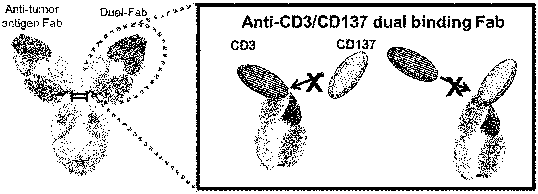
[0154] FIG. 1 is a conceptual diagram of an antibody that binds to CD3 and CD137, but does not bind to these antigens at the same time.
[0155] FIG. 2 is a conceptual diagram of an antibody that does not cause cross-linking because the antibody does not bind to CD3 and CD137 at the same time. On the contrary, a tri-functional antibody to CD3, CD137 and third antigen causes cross-linking of a T cell with a CD137 positive cell.
[0156] FIG. 3 is a conceptual diagram of an antibody that binds to CD3 and CD137, but does not link two cells at the same time.
[0157] FIG. 4 is a conceptual diagram of an antibody that cross-links a third antigen positive cell to a T cell expressing CD3 and CD137.
[0158] FIG. 5 is a conceptual diagram of an antibody that cross-links a third antigen positive cell to a cell expressing CD137.
[0159] FIG. 6 is a scheme diagram of the design and construction flow of dual scFv VH ribosome display library.
[0160] FIG. 7-1 FIG. 7 is a set of graphs showing the results of ELISA of clones obtained with ribosome display to CD3 and CD137. Y axis means the specificity to CD137-Fc and X axis means the specificity to CD3 of each clone. Black colored clones were identified as positive scFv which show binding to both CD137 and CD3.
[0161] FIG. 7-2 Continuation of FIG. 7-1.
[0162] FIG. 8 is a graph showing the result of ECL analysis of IgGs obtained with ribosome display to CD3 and CD137. Y axis means the response to both CD137, CD3 and plate itself.
[0163] FIG. 9-1 FIG. 9 is a set of graphs showing the results of ELISA of clones obtained with ribosome display to CD3 and CD137. Y axis means the specificity to CD137-Fc and X axis means the specificity to CD3 of each clone. Campaign3 means ribosome display panning with double round selection.
[0164] FIG. 9-2 Continuation of FIG. 9-1.
[0165] FIG. 9-3 Continuation of FIG. 9-2.
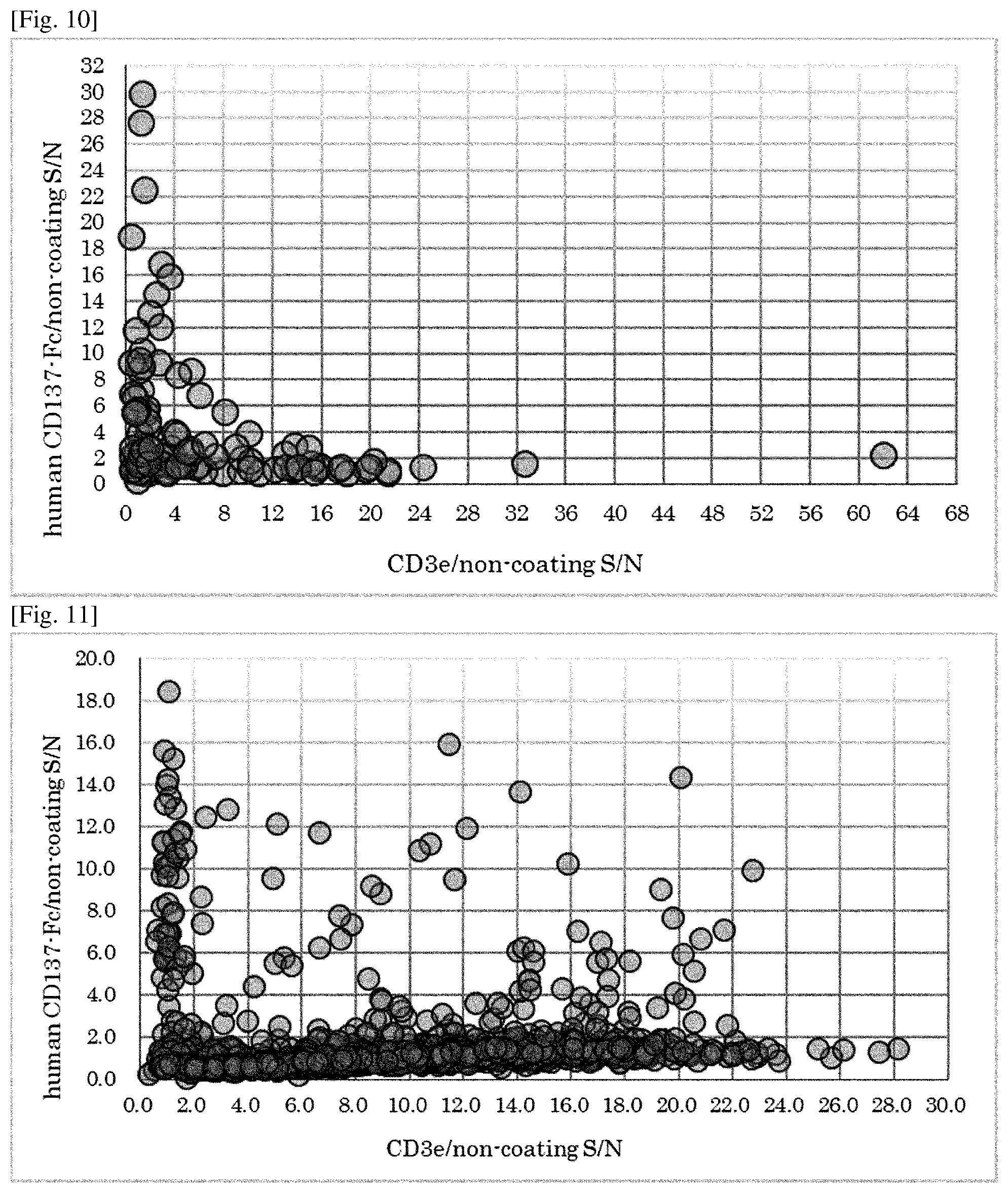
[0166] FIG. 10 is a graph showing the result of ELISA of clones obtained with ribosome display to CD3 and CD137. Y axis means the specificity to CD137-Fc and X axis means the specificity to CD3 of each clone.
[0167] FIG. 11 is a graph showing the result of ELISA of IgGs obtained with ribosome display to CD3 and CD137. Y axis means the specificity to CD137-Fc and X axis means the specificity to CD3 of each clone.
[0168] FIG. 12 is a scheme diagram of the design of dual scFv VL ribosome display library and dual Fab VL ribosome display library.
[0169] FIG. 13 is a graph showing the result of ELISA of IgGs obtained with ribosome display affinity maturation to CD3 and CD137. Y axis means the specificity to CD137-Fc and X axis means the specificity to CD3 of each clone.
[0170] FIG. 14 is a graph showing the result of competitive ELISA of IgGs obtained with ribosome display affinity maturation to CD3 and CD137. Y axis means the response of ELISA to biotin-human CD137-Fc or biotin-human Fc. Excess amount of human CD3 or human Fc were used as competitor.
[0171] FIG. 15 shows a design of C3NP1-27, CD3 epsilon peptide antigen which is biotin-labeled through disulfide-bond linker.
[0172] FIG. 16 is a graph showing the result of phage ELISA of clones obtained with phage display to CD3 and CD137. Y axis means the specificity to CD137-Fc and X axis means the specificity to CD3 of each clone.
[0173] FIG. 17 is a graph showing the result of phage ELISA of clones obtained with phage display to CD3 and CD137. Y axis means the specificity to CD137-Fc in beads ELISA and X axis means the specificity to CD3 in plate ELISA as same as FIG. 16 of each clone.
[0174] FIG. 18 shows a comparison data of human CD137 amino acids sequence with cynomolgus monkey CD137 amino acids sequence.
[0175] FIG. 19 is a graph showing the result of ELISA of IgGs obtained with phage display to CD3 and CD137. Y axis means the specificity to cyno CD137-Fc and X axis means the specificity to human CD137 of each clone.
[0176] FIG. 20 is a graph showing the result of ELISA of IgGs obtained with phage display to CD3 and CD137. Y axis means the specificity to CD3e.
[0177] FIG. 21 is a graph showing the result of competitive ELISA of IgGs obtained with phage display to CD3 and CD137. Y axis means the response of ELISA to biotin-human CD137-Fc or biotin-human Fc. Excess amount of human CD3 or human Fc were used as competitor.
[0178] FIG. 22A is a graph showing the result of phage ELISA of phage display panning output pools to CD3 and CD137. Y axis means the specificity to human CD137. X axis means the panning output pools, Primary is a pool before phage display panning, and R1 to R6 means panning output pool after phage display panning Round1 to Round6, respectively.
[0179] FIG. 22B is a graph showing the result of phage ELISA of phage display panning output pools to CD3 and CD137. Y axis means the specificity to cyno CD137. X axis means the panning output pools, Primary is a pool before phage display panning, and R1 to R6 means panning output pool after phage display panning Round1 to Round6, respectively.
[0180] FIG. 22C is a graph showing the result of phage ELISA of phage display panning output pools to CD3 and CD137. Y axis means the specificity to CD3. X axis means the panning output pools, Primary is a pool before phage display panning, and R1 to R6 means panning output pool after phage display panning Round1 to Round6, respectively.
[0181] FIG. 23-1 FIG. 23 is a set of graphs showing the result of ELISA of IgGs obtained with phage display to CD3 and CD137. Y axis means the specificity to human CD137-Fc and X axis means the specificity to human CD137 or CD3 of each clone.
[0182] FIG. 23-2 Continuation of FIG. 23-1.
[0183] FIG. 23-3 Continuation of FIG. 23-2.
[0184] FIG. 24 is a set of graphs showing the result of ELISA of IgGs obtained with phage display to CD3 and CD137. Y axis means the specificity to human CD137-Fc and X axis means the specificity to human CD137 or CD3 of each clone.
[0185] FIG. 25 is a graph showing the result of competitive ELISA of IgGs obtained with phage display to CD3 and CD137. Y axis means the response of ELISA to biotin-human CD137-Fc or biotin-human Fc. Excess amount of human CD3 were used as competitor.
[0186] FIG. 26 is a graph showing the result of ELISA of IgGs obtained with phage display to CD3 and CD137 to identify the epitope domain of each clones. Y axis means the response of ELISA to each domain of human CD137.
[0187] FIG. 27 is a set of graphs showing the result of ELISA of IgGs obtained with phage display affinity maturation to CD3 and CD137. Y axis means the specificity to human CD137-Fc and X axis means the specificity to human CD137 or CD3 of each clone.
[0188] FIG. 28-1 FIG. 28 is a set of graphs showing the result of competitive ELISA of IgGs obtained with phage display to CD3 and CD137. Y axis means the response of ELISA to biotin-human CD137-Fc or biotin-human Fc. An excess amount of human CD3 was used as a competitor.
[0189] FIG. 28-2 Continuation of FIG. 28-1.
[0190] FIG. 28-3 Continuation of FIG. 28-2.
[0191] FIG. 28-4 Continuation of FIG. 28-3.
[0192] FIG. 28-5 Continuation of FIG. 28-4.
[0193] FIG. 29A shows the mechanism of IL-6 secretion from the activated B cell via anti-human GPC3/Dual-Fab antibodies.
[0194] FIG. 29B presents a graph showing the results of assessing the CD137-mediated agonist activity of various anti-human GPC3/Dual-Fab antibodies by the level of production of IL-6 which is secreted from the activated B cells. Ctrl indicates the negative control human IgG1 antibody.
[0195] FIG. 30A shows the mechanism of Luciferase expression in the activated Jurkat T cell via anti-human GPC3/Dual-Fab antibodies.
[0196] FIG. 30B presents graphs showing the results of assessing the CD3 mediated agonist activity of various anti-human GPC3/Dual-Fab antibodies by the level of production of Luciferase which is expressed in the activated Jurkat T cells. Ctrl indicates the negative control human IgG1 antibody.
[0197] FIG. 31 is a set of graphs showing the results of assessing the cytokine (IL-2, IFN-gamma and TNF-alpha) release from human PBMC derived T cells in the presence of each immobilized antibodies. Y axis means the concentration of secreted each cytokines and X-axis means the concentration of immobilized antibodies. Control anti-CD137 antibody (B), control anti-CD3 antibody (CE115), negative control antibody (Ctrl) and one of the dual antibody (L183L072) were used for assay.
[0198] FIG. 32 is a set of graphs showing the results of assessing the T-cell dependent cellular cytotoxicity (TDCC) against GPC3 positive target cells (SK-pca60 and SK-pca13a) with each bi-specific antibodies. Y axis means the ratio of Cell Growth Inhibition (CGI) and X-axis means the concentration of each bi-specific antibodies. Anti-GPC3/Dual Bi-specific antibody (GC33/H183L072), Negative control/Dual Bi-specific antibody (Ctrl/H183L072), Anti-GPC3/Anti-CD137 Bi-specific antibody (GC33/B) and Negative control/Anti-CD137 Bi-specific antibody (Ctrl/B) were used for this assay. 5-fold amount of effector (E) cells were added on tumor (T) cells (ET5).
[0199] FIG. 33 shows the design and construction procedure of trispecific antibodies (mAb AB).
[0200] FIG. 34 shows the naming rule of prepared trispecific antibodies.
[0201] FIG. 35 is a set of graphs showing the results of Biacore analysis of simultaneous binding of GPC3/CD137xCD3 trispecific antibody and anti-GPC3/dual-Fab antibody. Y-axis means the binding response to each antigen. At first human CD3 (hCD3) was used as analyte, and then also hCD3 (shown as broken line) or mixture of human CD137 (hCD137) and hCD3 (shown as solid line) were used as analyte.
[0202] FIG. 36 is a set of sensorgrams showing the results of FACS analysis to CD137 positive CHO cells or Jurkat cells of each antibodies. FIGS. 35(a) and (c) are the results of binding to human CD137 positive CHO cells, and FIGS. 35(b) and (d) are the results to parental CHO cells. In FIGS. 35(a) and (b), solid line shows the result of anti-GPC3/dual antibody (GC33/H183L072) and filled shows the result of control antibody (Ctrl). In FIGS. 35(c) and (d), solid line, filled with dark gray and filled with light grey shows the results of GPC3/CD137xCtrl trispecific antibody, GPC3/CD137xCD3 trispecific antibody and Ctrl/CtrlxCD3 trispecific antibody, respectively.
[0203] FIGS. 35(e) and (f) are the results of binding to Jurkat CD3 positive cells. In FIG. 35(e), solid line and filled shows the result of anti-GPC3/dual antibody (GC33/H183L072) and control antibody (Ctrl), respectively. In FIG. 35(f), solid line, filled with dark gray and filled with light grey shows the results of GPC3/CtrlxCD3 trispecific antibody, GPC3/CD137xCD3 trispecific antibody and Ctrl/CD137xCtrl trispecific antibody, respectively.
[0204] FIG. 37 presents graphs showing the results of assessing the CD3 mediated agonist activity of various a antibodies to GPC3 positive target cell SK-pca60 by the level of production of Luciferase which is expressed in the activated Jurkat T cells. Six kinds of tri-specific antibodies, anti-GPC3/Dual-Fab antibody (GPC3/H183L072) and control/Dual-Fab antibody (Ctrl/H183L072) were used for this assay. X-axis means the concentration used of each antibodies.
[0205] FIG. 38 presents graphs showing the results of assessing the CD3 mediated agonist activity of various a antibodies to human CD137 positive CHO cells and parental CHO cells by the level of production of Luciferase which is expressed in the activated Jurkat T cells. Six kinds of tri-specific antibodies, anti-GPC3/Dual-Fab antibody (GPC3/H183L072) and control/Dual-Fab antibody (Ctrl/H183L072) were used for this assay. X-axis means the concentration used of each antibodies.
[0206] FIG. 39 is a set of graphs showing the results of assessing the cytokine (IL-2, IFN-gamma and TNF-alpha) release from human PBMCs in the presence of each soluble antibodies. Y axis means the concentration of secreted each cytokines and X-axis means the concentration of antibodies used. Ctrl/CD137xCD3 trispecific antibody and control/Dual-Fab antibody (Ctrl/H183L072) were used for this assay
[0207] FIG. 40 is a graph showing results of cell-ELISA of CE115 for CD3e.
[0208] FIG. 41 is a diagram showing the molecular form of EGFR_ERY22_CE115.
[0209] FIG. 42 is a graph showing results of TDCC (SK-pca13a) of EGFR_ERY22_CE115.
[0210] FIG. 43 is an exemplary sensorgram of an antibody having a ratio of the amounts bound of less than 0.8. The vertical axis depicts an RU value (response). The horizontal axis depicts time.
DESCRIPTION OF EMBODIMENTS
[0211] In one aspect, an antigen-binding molecule of the present invention is an antigen-binding molecule comprising an antibody variable region that is capable of binding to CD3 and CD137 (4-1BB) but does not bind to CD3 and CD137 at the same time, and a variable region binding to a third antigen different from CD3 and CD137.
[0212] In one aspect, an antigen-binding molecule of the present invention is an antigen-binding molecule comprising an antibody variable region that is capable of binding to a T cell receptor and CD137 (4-1BB) but does not bind to the T cell receptor and CD137 at the same time, and a variable region binding to a third antigen different from the T cell receptor and CD137.
[0213] In one aspect, an antigen-binding molecule of the present invention is an antigen-binding molecule comprising an antibody variable region that is capable of binding to CD3 and CD137 but does not bind to CD3 and CD137 at the same time, and a variable region binding to a molecule specifically expressed in a cancer tissue.
[0214] In one aspect, an antigen-binding domain of the present invention is a variable region that is capable of binding to CD3 and CD137 but does not bind to CD3 and CD137 at the same time. In one aspect, an antibody variable region of the present invention is a variable region that is capable of binding to CD3 and CD137 but does not bind to CD3 and CD137 at the same time.
[0215] In some embodiments, the antigen binding molecule of the present invention can activate T cells by its agonistic activity on CD3, and it can induce cytotoxicity of T cells against target cells, and strengthen T-cell activation, survival, and differentiation into memory T cells by its co-stimulatory agonistic activity on CD137 and CD3. Meanwhile, the antigen binding molecule of the present invention can avoid the adverse events caused by cross-linking of CD137 and CD3 because it does not bind to CD3 and CD137 at the same time.
[0216] In some embodiments, the antigen binding molecule of the present invention can also activate immune cells expressing CD137 and strengthen the immune response to target cells by the agonistic activity on CD137.
[0217] In the present invention, the "antibody variable region" usually means a region comprising a domain constituted by four framework regions (FRs) and three complementarity-determining regions (CDRs) flanked thereby, and also includes a partial sequence thereof as long as the partial sequence has the activity of binding to a portion or the whole of an antigen. Particularly, a region comprising an antibody light chain variable domain (VL) and an antibody heavy chain variable domain (VH) is preferred. The antibody variable region of the present invention may have an arbitrary sequence and may be a variable region derived from any antibody such as a mouse antibody, a rat antibody, a rabbit antibody, a goat antibody, a camel antibody, and a humanized antibody obtained by the humanization of any of these nonhuman antibodies, and a human antibody. The "humanized antibody", also called reshaped human antibody, is obtained by grafting complementarity determining regions (CDRs) of a non-human mammal-derived antibody, for example, a mouse antibody to human antibody CDRs. Methods for identifying CDRs are known in the art (Kabat et al., Sequence of Proteins of Immunological Interest (1987), National Institute of Health, Bethesda, Md.; and Chothia et al., Nature (1989) 342: 877). General gene recombination approaches therefor are also known in the art (see European Patent Application Publication No. EP 125023 and WO 96/02576).
[0218] The "antibody variable region" of the present invention that does "not bind to CD3 and CD137 (4-1BB) at the same time" means that the antibody variable region of the present invention cannot bind to CD137 in a state bound with CD3 whereas the variable region cannot bind to CD3 in a state bound with CD137. In this context, the phrase "not bind to CD3 and CD137 at the same time" also includes not cross-linking a cell expressing CD3 to a cell expressing CD137, or not binding to CD3 and CD137 each expressed on a different cell, at the same time. This phrase further includes the case where the variable region is capable of binding to both CD3 and CD137 at the same time when CD3 and CD137 are not expressed on cell membranes, as with soluble proteins, or both reside on the same cell, but cannot bind to CD3 and CD137 each expressed on a different cell, at the same time. Such an antibody variable region is not particularly limited as long as the antibody variable region has these functions. Examples thereof can include variable regions derived from an IgG-type antibody variable region by the alteration of a portion of its amino acids so as to bind to the desired antigen. The amino acid to be altered is selected from, for example, amino acids whose alteration does not cancel the binding to the antigen, in an antibody variable region binding to CD3 or CD137.
In this context, the phrase "expressed on different cells" merely means that the antigens are expressed on separate cells. The combination of such cells may be, for example, the same types of cells such as a T cell and another T cell, or may be different types of cells such as a T cell and an NK cell.
[0219] In the present invention, one amino acid alteration may be used alone, or a plurality of amino acid alterations may be used in combination.
[0220] In the case of using a plurality of amino acid alterations in combination, the number of the alterations to be combined is not particularly limited and can be appropriately set within a range that can attain the object of the invention. The number of the alterations to be combined is, for example, 2 or more and 30 or less, preferably 2 or more and 25 or less, 2 or more and 22 or less, 2 or more and 20 or less, 2 or more and 15 or less, 2 or more and 10 or less, 2 or more and 5 or less, or 2 or more and 3 or less.
[0221] The plurality of amino acid alterations to be combined may be added to only the antibody heavy chain variable domain or light chain variable domain or may be appropriately distributed to both of the heavy chain variable domain and the light chain variable domain.
[0222] One or more amino acid residues in the variable region are acceptable as the amino acid residue to be altered as long as the antigen-binding activity is maintained. In the case of altering an amino acid in the variable region, the resulting variable region preferably maintains the binding activity of the corresponding unaltered antibody and preferably has, for example, 50% or higher, more preferably 80% or higher, further preferably 100% or higher, of the binding activity before the alteration, though the variable region according to the present invention is not limited thereto. The binding activity may be increased by the amino acid alteration and may be, for example, 2 times, 5 times, or 10 times the binding activity before the alteration.
[0223] Examples of the region preferred for the amino acid alteration include solvent-exposed regions and loops in the variable region. Among others, CDR1, CDR2, CDR3, FR3, and loops are preferred. Specifically, Kabat numbering positions 31 to 35, 50 to 65, 71 to 74, and 95 to 102 in the H chain variable domain and Kabat numbering positions 24 to 34, 50 to 56, and 89 to 97 in the L chain variable domain are preferred. Kabat numbering positions 31, 52a to 61, 71 to 74, and 97 to 101 in the H chain variable domain and Kabat numbering positions 24 to 34, 51 to 56, and 89 to 96 in the L chain variable domain are more preferred. Also, an amino acid that increases antigen-binding activity may be further introduced at the time of the amino acid alteration.
[0224] The term "hypervariable region" or "HVR" as used herein refers to each of the regions of an antibody variable domain which are hypervariable in sequence ("complementarity determining regions" or "CDRs") and/or form structurally defined loops ("hypervariable loops") and/or contain the antigen-contacting residues ("antigen contacts"). Generally, antibodies comprise six HVRs: three in the VH (H1, H2, H3), and three in the VL (L1, L2, L3). Exemplary HVRs herein include:
[0225] (a) hypervariable loops occurring at amino acid residues 26-32 (L1), 50-52 (L2), 91-96 (L3), 26-32 (H1), 53-55 (H2), and 96-101 (H3) (Chothia and Lesk, J. Mol. Biol. 196:901-917 (1987));
[0226] (b) CDRs occurring at amino acid residues 24-34 (L1), 50-56 (L2), 89-97 (L3), 31-35b (H1), 50-65 (H2), and 95-102 (H3) (Kabat et al., Sequences of Proteins of Immunological Interest, 5th Ed. Public Health Service, National Institutes of Health, Bethesda, Md. (1991));
[0227] (c) antigen contacts occurring at amino acid residues 27c-36 (L1), 46-55 (L2), 89-96 (L3), 30-35b (H1), 47-58 (H2), and 93-101 (H3) (MacCallum et al. J. Mol. Biol. 262: 732-745 (1996)); and
[0228] (d) combinations of (a), (b), and/or (c), including HVR amino acid residues 46-56 (L2), 47-56 (L2), 48-56 (L2), 49-56 (L2), 26-35 (H1), 26-35b (H1), 49-65 (H2), 93-102 (H3), and 94-102 (H3).
[0229] Unless otherwise indicated, HVR residues and other residues in the variable domain (e.g., FR residues) are numbered herein according to Kabat et al., supra.
[0230] In the present invention, the "loop" means a region containing residues that are not involved in the maintenance of an immunoglobulin beta barrel structure.
[0231] In the present invention, the amino acid alteration means substitution, deletion, addition, insertion, or modification, or a combination thereof. In the present invention, the amino acid alteration can be used interchangeably with amino acid mutation and used in the same sense therewith.
[0232] The substitution of an amino acid residue is carried out by replacement with another amino acid residue for the purpose of altering, for example, any of the following (a) to (c): (a) the polypeptide backbone structure of a region having a sheet structure or helix structure; (b) the electric charge or hydrophobicity of a target site; and (c) the size of a side chain.
[0233] Amino acid residues are classified into the following groups on the basis of general side chain properties: (1) hydrophobic residues: norleucine, Met, Ala, Val, Leu, and Ile; (2) neutral hydrophilic residues: Cys, Ser, Thr, Asn, and Gln; (3) acidic residues: Asp and Glu; (4) basic residues: His, Lys, and Arg; (5) residues that influence chain orientation: Gly and Pro; and (6) aromatic residues: Trp, Tyr, and Phe.
[0234] The substitution of amino acid residues within each of these groups is called conservative substitution, while the substitution of an amino acid residue in one of these groups by an amino acid residue in another group is called non-conservative substitution.
[0235] The substitution according to the present invention may be the conservative substitution or may be the non-conservative substitution. Alternatively, the conservative substitution and the non-conservative substitution may be combined.
[0236] The alteration of an amino acid residue also includes: the selection of a variable region that is capable of binding to CD3 and CD137, but cannot bind to these antigens at the same time, from those obtained by the random alteration of amino acids whose alteration does not cancel the binding to the antigen, in the antibody variable region binding to CD3 or CD137; and alteration to insert a peptide previously known to have binding activity against the desired antigen, to the region mentioned above.
[0237] In the antibody variable region of the present invention, the alteration mentioned above may be combined with alteration known in the art. For example, the modification of N-terminal glutamine of the variable region to pyroglutamic acid by pyroglutamylation is a modification well known to those skilled in the art. Thus, the antibody of the present invention having glutamine at the N terminus of its heavy chain may contain a variable region with this N-terminal glutamine modified to pyroglutamic acid.
[0238] Such an antibody variable region may further have amino acid alteration to improve, for example, antigen binding, pharmacokinetics, stability, or antigenicity. The antibody variable region of the present invention may be altered so as to have pH dependent binding activity against an antigen and be thereby capable of repetitively binding to the antigen (WO2009/125825).
[0239] Also, amino acid alteration to change antigen-binding activity according to the concentration of a target tissue-specific compound may be added to, for example, such an antibody variable region binding to a third antigen (WO2013/180200).
[0240] The variable region may be further altered for the purpose of, for example, enhancing binding activity, improving specificity, reducing p, conferring pH-dependent antigen-binding properties, improving the thermal stability of binding, improving solubility, improving stability against chemical modification, improving heterogeneity derived from a sugar chain, avoiding a T cell epitope identified by use of in silico prediction or in vitro T cell-based assay for reduction in immunogenicity, or introducing a T cell epitope for activating regulatory T cells (mAbs 3: 243-247, 2011).
[0241] Whether the antibody variable region of the present invention is "capable of binding to CD3 and CD137" can be determined by a method known in the art.
[0242] This can be determined by, for example, an electrochemiluminescence method (ECL method) (BMC Research Notes 2011, 4: 281).
[0243] Specifically, for example, a low-molecular antibody composed of a region capable of binding to CD3 and CD137, for example, a Fab region, of a biotin-labeled antigen-binding molecule to be tested, or a monovalent antibody (antibody lacking one of the two Fab regions carried by a usual antibody) thereof is mixed with CD3 or CD137 labeled with sulfo-tag (Ru complex), and the mixture is added onto a streptavidin-immobilized plate. In this operation, the biotin-labeled antigen-binding molecule to be tested binds to streptavidin on the plate. Light is developed from the sulfo-tag, and the luminescence signal can be detected using Sector Imager 600 or 2400 (MSD K.K.) or the like to thereby confirm the binding of the aforementioned region of the antigen-binding molecule to be tested to CD3 or CD137.
[0244] Alternatively, this assay may be conducted by ELISA, FACS (fluorescence activated cell sorting), ALPHAScreen (amplified luminescent proximity homogeneous assay screen), the BIACORE method based on a surface plasmon resonance (SPR) phenomenon, etc. (Proc. Natl. Acad. Sci. USA (2006) 103 (11), 4005-4010).
[0245] Specifically, the assay can be conducted using, for example, an interaction analyzer Biacore (GE Healthcare Japan Corp.) based on a surface plasmon resonance (SPR) phenomenon. The Biacore analyzer includes any model such as Biacore T100, T200, X100, A100, 4000, 3000, 2000, 1000, or C. Any sensor chip for Biacore, such as a CM7, CM5, CM4, CM3, C1, SA, NTA, L1, HPA, or Au chip, can be used as a sensor chip. Proteins for capturing the antigen-binding molecule of the present invention, such as protein A, protein G, protein L, anti-human IgG antibodies, anti-human IgG-Fab, anti-human L chain antibodies, anti-human Fc antibodies, antigenic proteins, or antigenic peptides, are immobilized onto the sensor chip by a coupling method such as amine coupling, disulfide coupling, or aldehyde coupling. CD3 or CD137 is injected thereon as an analyte, and the interaction is measured to obtain a sensorgram. In this operation, the concentration of CD3 or CD137 can be selected within the range of a few micro M to a few pM according to the interaction strength (e.g., KD) of the assay sample.
[0246] Alternatively, CD3 or CD137 may be immobilized instead of the antigen-binding molecule onto the sensor chip, with which the antibody sample to be evaluated is in turn allowed to interact. Whether the antibody variable region of the antigen-binding molecule of the present invention has binding activity against CD3 or CD137 can be confirmed on the basis of a dissociation constant (KD) value calculated from the sensorgram of the interaction or on the basis of the degree of increase in the sensorgram after the action of the antigen-binding molecule sample over the level before the action.
[0247] The ALPHAScreen is carried out by the ALPHA technology using two types of beads (donor and acceptor) on the basis of the following principle: luminescence signals are detected only when these two beads are located in proximity through the biological interaction between a molecule bound with the donor bead and a molecule bound with the acceptor bead. A laser-excited photosensitizer in the donor bead converts ambient oxygen to singlet oxygen having an excited state. The singlet oxygen diffuses around the donor bead and reaches the acceptor bead located in proximity thereto to thereby cause chemiluminescent reaction in the bead, which finally emits light. In the absence of the interaction between the molecule bound with the donor bead and the molecule bound with the acceptor bead, singlet oxygen produced by the donor bead does not reach the acceptor bead. Thus, no chemiluminescent reaction occurs.
[0248] One (ligand) of the substances between which the interaction is to be observed is immobilized onto a thin gold film of a sensor chip. The sensor chip is irradiated with light from the back such that total reflection occurs at the interface between the thin gold film and glass. As a result, a site having a drop in reflection intensity (SPR signal) is formed in a portion of reflected light. The other (analyte) of the substances between which the interaction is to be observed is injected on the surface of the sensor chip. Upon binding of the analyte to the ligand, the mass of the immobilized ligand molecule is increased to change the refractive index of the solvent on the sensor chip surface. This change in the refractive index shifts the position of the SPR signal (on the contrary, the dissociation of the bound molecules gets the signal back to the original position). The Biacore system plots on the ordinate the amount of the shift, i.e., change in mass on the sensor chip surface, and displays time-dependent change in mass as assay data (sensorgram). The amount of the analyte bound to the ligand captured on the sensor chip surface (amount of change in response on the sensorgram between before and after the interaction of the analyte) can be determined from the sensorgram. However, since the amount bound also depends on the amount of the ligand, the comparison must be performed under conditions where substantially the same amounts of the ligand are used. Kinetics, i.e., an association rate constant (ka) and a dissociation rate constant (kd), can be determined from the curve of the sensorgram, while affinity (KD) can be determined from the ratio between these constants. Inhibition assay is also preferably used in the BIACORE method. Examples of the inhibition assay are described in Proc. Natl. Acad. Sci. USA (2006) 103 (11), 4005-4010.
[0249] Whether the antigen-binding molecule of the present invention does "not bind to CD3 and CD137 at the same time" can be confirmed by: confirming the antigen-binding molecule to have binding activity against both CD3 and CD137; then allowing either CD3 or CD137 to bind in advance to the antigen-binding molecule comprising the variable region having this binding activity; and then determining the presence or absence of its binding activity against the other one by the method mentioned above. Alternatively, this can also be confirmed by determining whether the binding of the antigen-binding molecule to either CD3 or CD137 immobilized on an ELISA plate or a sensor chip is inhibited by the addition of the other one into the solution. In some embodiments, the binding of the antigen-binding molecule of the present invention to either CD3 or CD137 is inhibited by binding of the antigen-binding molecule to the other by at least 50%, preferably 60% or more, more preferably 70% or more, more preferably 80% or more, further preferably 90% or more, or even more preferably 95% or more.
[0250] In one aspect, while one antigen (e.g. CD3) is immobilized, the inhibition of the binding of the antigen-binding molecule to CD3 can be determined in the presence of the other antigen (e.g. CD137) by methods known in prior art (i.e. ELISA, BIACORE, and so on). In another aspect, while CD137 is immobilized, the inhibition of the binding of the antigen-binding molecule to CD137 also can be determined in the presence of CD3. When either one of two aspects mentioned above is conducted, the antigen-binding molecule of the present invention is determined not to bind to CD3 and CD137 at the same time if the binding is inhibited by at least 50%, preferably 60% or more, preferably 70% or more, further preferably 80% or more, further preferably 90% or more, or even more preferably 95% or more.
[0251] In some embodiments, the concentration of the antigen injected as an analyte is at least 1-fold, 2-fold, 5-fold, 10-fold, 30-fold, 50-fold, or 100-fold higher than the concentration of the other antigen to be immobilized.
[0252] In preferable manner, the concentration of the antigen injected as an analyte is 100-fold higher than the concentration of the other antigen to be immobilized and the binding is inhibited by at least 80%.
[0253] In one embodiment, the ratio of the KD value for the CD3 (analyte)-binding activity of the antigen-binding molecule to the CD137 (immobilized)-binding activity of the antigen-binding molecule (KD (CD3)/KD (CD137)) is calculated and the CD3 (analyte) concentration which is 10-fold, 50-fold, 100-fold, or 200-fold of the ratio of the KD value (KD(CD3)/KD(CD137) higher than the CD137 (immobilized) concentration can be used for the competition measurement above. (e.g. 1-fold, 5-fold, 10-fold, or 20-fold higher concentration can be selected when the ratio of the KD value is 0.1. Furthermore, 100-fold, 500-fold, 1000-fold, or 2000-fold higher concentration can be selected when the ratio of the KD value is 10.)
[0254] In one aspect, while one antigen (e.g. CD3) is immobilized, the attenuation of the binding signal of the antigen-binding molecule to CD3 can be determined in the presence of the other antigen (e.g. CD137) by methods known in prior art (i.e. ELISA, ECL and so on). In another aspect, while CD137 is immobilized, the attenuation of the binding signal of the antigen-binding molecule to CD137 also can be determined in the presence of CD3. When either one of two aspects mentioned above is conducted, the antigen-binding molecule of the present invention is determined not to bind to CD3 and CD137 at the same time if the binding signal is attenuated by at least 50%, preferably 60% or more, preferably 70% or more, further preferably 80% or more, further preferably 90% or more, or even more preferably 95% or more. (see Example 5-5, 7-5, 8-9, 9-4)
[0255] In some embodiments, the concentration of the antigen injected as an analyte is at least 1-fold, 2-fold, 5-fold, 10-fold, 30-fold, 50-fold, or 100-fold higher than the concentration of the other antigen to be immobilized.
[0256] In preferable manner, the concentration of the antigen injected as an analyte is 100-fold higher than the concentration of the other antigen to be immobilized and the binding is inhibited by at least 80%.
[0257] In one embodiment, the ratio of the KD value for the CD3 (analyte)-binding activity of the antigen-binding molecule to the CD137 (immobilized)-binding activity of the antigen-binding molecule (KD (CD3)/KD (CD137)) is calculated and the CD3 (analyte) concentration which is 10-fold, 50-fold, 100-fold, or 200-fold of the ratio of the KD value (KD(CD3)/KD(CD137) higher than the CD137 (immobilized) concentration can be used for the measurement above. (e.g. 1-fold, 5-fold, 10-fold, or 20-fold higher concentration can be selected when the ratio of the KD value is 0.1. Furthermore, 100-fold, 500-fold, 1000-fold, or 2000-fold higher concentration can be selected when the ratio of the KD value is 10.)
[0258] Specifically, in the case of using, for example, the ECL method, a biotin-labeled antigen-binding molecule to be tested, CD3 labeled with sulfo-tag (Ru complex), and an unlabeled CD137 are prepared. When the antigen-binding molecule to be tested is capable of binding to CD3 and CD137, but does not bind to CD3 and CD137 at the same time, the luminescence signal of the sulfo-tag is detected in the absence of the unlabeled CD137 by adding the mixture of the antigen-binding molecule to be tested and labeled CD3 onto a streptavidin-immobilized plate, followed by light development. By contrast, the luminescence signal is decreased in the presence of unlabeled CD137. This decrease in luminescence signal can be quantified to determine relative binding activity. This analysis may be similarly conducted using the labeled CD137 and the unlabeled CD3.
[0259] In the case of the ALPHAScreen, the antigen-binding molecule to be tested interacts with CD3 in the absence of the competing CD137 to generate signals of 520 to 620 nm. The untagged CD137 competes with CD3 for the interaction with the antigen-binding molecule to be tested. Decrease in fluorescence caused as a result of the competition can be quantified to thereby determine relative binding activity. The polypeptide biotinylation using sulfo-NHS-biotin or the like is known in the art. CD3 can be tagged with GST by an appropriately adopted method which involves, for example: fusing a polynucleotide encoding CD3 in flame with a polynucleotide encoding GST; and allowing the resulting fusion gene to be expressed by cells or the like harboring vectors capable of expression thereof, followed by purification using a glutathione column. The obtained signals are preferably analyzed using, for example, software GRAPHPAD PRISM (GraphPad Software, Inc., San Diego) adapted to a one-site competition model based on nonlinear regression analysis. This analysis may be similarly conducted using the tagged CD137 and the untagged CD3.
[0260] Alternatively, a method using fluorescence resonance energy transfer (FRET) may be used. FRET is a phenomenon in which excitation energy is transferred directly between two fluorescent molecules located in proximity to each other by electron resonance. When FRET occurs, the excitation energy of a donor (fluorescent molecule having an excited state) is transferred to an acceptor (another fluorescent molecule located near the donor) so that the fluorescence emitted from the donor disappears (to be precise, the lifetime of the fluorescence is shortened) and instead, the fluorescence is emitted from the acceptor. By use of this phenomenon, whether or not bind to CD3 and CD137 at the same time can be analyzed. For example, when CD3 carrying a fluorescence donor and CD137 carrying a fluorescence acceptor bind to the antigen-binding molecule to be tested at the same time, the fluorescence of the donor disappears while the fluorescence is emitted from the acceptor. Therefore, change in fluorescence wavelength is observed. Such an antibody is confirmed to bind to CD3 and CD137 at the same time. On the other hand, if the mixing of CD3, CD137, and the antigen-binding molecule to be tested does not change the fluorescence wavelength of the fluorescence donor bound with CD3, this antigen-binding molecule to be tested can be regarded as antigen binding domain that is capable of binding to CD3 and CD137, but does not bind to CD3 and CD137 at the same time.
[0261] For example, a biotin-labeled antigen-binding molecule to be tested is allowed to bind to streptavidin on the donor bead, while CD3 tagged with glutathione S transferase (GST) is allowed to bind to the acceptor bead. The antigen-binding molecule to be tested interacts with CD3 in the absence of the competing second antigen to generate signals of 520 to 620 nm. The untagged second antigen competes with CD3 for the interaction with the antigen-binding molecule to be tested. Decrease in fluorescence caused as a result of the competition can be quantified to thereby determine relative binding activity. The polypeptide biotinylation using sulfo-NHS-biotin or the like is known in the art. CD3 can be tagged with GST by an appropriately adopted method which involves, for example: fusing a polynucleotide encoding CD3 in flame with a polynucleotide encoding GST; and allowing the resulting fusion gene to be expressed by cells or the like harboring vectors capable of expression thereof, followed by purification using a glutathione column. The obtained signals are preferably analyzed using, for example, software GRAPHPAD PRISM (GraphPad Software, Inc., San Diego) adapted to a one-site competition model based on nonlinear regression analysis.
[0262] The tagging is not limited to the GST tagging and may be carried out with any tag such as, but not limited to, a histidine tag, MBP, CBP, a Flag tag, an HA tag, a V5 tag, or a c-myc tag. The binding of the antigen-binding molecule to be tested to the donor bead is not limited to the binding using biotin-streptavidin reaction. Particularly, when the antigen-binding molecule to be tested comprises Fc, a possible method involves allowing the antigen-binding molecule to be tested to bind via an Fc-recognizing protein such as protein A or protein G on the donor bead.
[0263] Also, the case where the variable region is capable of binding to CD3 and CD137 at the same time when CD3 and CD137 are not expressed on cell membranes, as with soluble proteins, or both reside on the same cell, but cannot bind to CD3 and CD137 each expressed on a different cell, at the same time can also be assayed by a method known in the art.
[0264] Specifically, the antigen-binding molecule to be tested has been confirmed to be positive in ECL-ELISA for detecting binding to CD3 and CD137 at the same time is also mixed with a cell expressing CD3 and a cell expressing CD137. The antigen-binding molecule to be tested can be shown to be incapable of binding to CD3 and CD137 expressed on different cells, at the same time unless the antigen-binding molecule and these cells bind to each other at the same time. This assay can be conducted by, for example, cell-based ECL-ELISA. The cell expressing CD3 is immobilized onto a plate in advance. After binding of the antigen-binding molecule to be tested thereto, the cell expressing CD137 is added to the plate. A different antigen expressed only on the cell expressing CD137 is detected using a sulfo-tag-labeled antibody against this antigen. A signal is observed when the antigen-binding molecule binds to the two antigens respectively expressed on the two cells, at the same time. No signal is observed when the antigen-binding molecule does not bind to these antigens at the same time.
Alternatively, this assay may be conducted by the ALPHAScreen method. The antigen-binding molecule to be tested is mixed with a cell expressing CD3 bound with the donor bead and a cell expressing CD137 bound with the acceptor bead. A signal is observed when the antigen-binding molecule binds to the two antigens expressed on the two cells respectively, at the same time. No signal is observed when the antigen-binding molecule does not bind to these antigens at the same time. Alternatively, this assay may also be conducted by an Octet interaction analysis method. First, a cell expressing CD3 tagged with a peptide tag is allowed to bind to a biosensor that recognizes the peptide tag. A cell expressing CD137 and the antigen-binding molecule to be tested are placed in wells and analyzed for interaction. A large wavelength shift caused by the binding of the antigen-binding molecule to be tested and the cell expressing CD137 to the biosensor is observed when the antigen-binding molecule binds to the two antigens expressed on the two cells respectively, at the same time. A small wavelength shift caused by the binding of only the antigen-binding molecule to be tested to the biosensor is observed when the antigen-binding molecule does not bind to these antigens at the same time.
[0265] Instead of these methods based on the binding activity, assay based on biological activity may be conducted. For example, a cell expressing CD3 and a cell expressing CD137 are mixed with the antigen-binding molecule to be tested, and cultured. The two antigens expressed on the two cells respectively are mutually activated via the antigen-binding molecule to be tested when the antigen-binding molecule binds to these two antigens at the same time. Therefore, change in activation signal, such as increase in the respective downstream phosphorylation levels of the antigens, can be detected. Alternatively, cytokine production is induced as a result of the activation. Therefore, the amount of cytokines produced can be measured to thereby confirm whether or not to bind to the two cells at the same time. Alternatively, cytotoxicity against a cell expressing CD137 is induced as a result of the activation. Alternatively, the expression of a reporter gene is induced by a promoter which is activated at the downstream of the signal transduction pathway of CD137 or CD3 as a result of the activation. Therefore, the cytotoxicity or the amount of reporter proteins produced can be measured to thereby confirm whether or not to bind to the two cells at the same time.
[0266] In the present invention, the "Fc region" refers to a region comprising a fragment consisting of a hinge or a portion thereof and CH2 and CH3 domains in an antibody molecule. The Fc region of IgG class means, but is not limited to, a region from, for example, cysteine 226 (EU numbering (also referred to as EU index herein)) to the C terminus or proline 230 (EU numbering) to the C terminus. The Fc region can be preferably obtained by the partial digestion of, for example, an IgG1, IgG2, IgG3, or IgG4 monoclonal antibody with a proteolytic enzyme such as pepsin followed by the re-elution of a fraction adsorbed on a protein A column or a protein G column. Such a proteolytic enzyme is not particularly limited as long as the enzyme is capable of digesting a whole antibody to restrictively form Fab or F(ab').sub.2 under appropriately set reaction conditions (e.g., pH) of the enzyme. Examples thereof can include pepsin and papain.
[0267] In some embodiments, the "antigen-binding molecule" is not particularly limited as long as the molecule comprises the "antibody variable region" of the present invention. The antigen-binding molecule may further comprise a peptide or a protein having a length of approximately 5 or more amino acids. The peptide or the protein is not limited to a peptide or a protein derived from an organism, and may be, for example, a polypeptide consisting of an artificially designed sequence. Also, a natural polypeptide, a synthetic polypeptide, a recombinant polypeptide, or the like may be used.
[0268] In some embodiments, the "antigen-binding molecule" of the present invention is not particularly limited to a molecule comprising the "antibody variable region". In certain embodiments, antigen-binding molecules that are other than antibodies comprising a variable region and can bind to two different antigens, for example, Affibody and so on, may be obtained by methods generally known to those skilled in the art (PLoS One. 2011; 6(10):e25791; PLoS One. 2012; 7(8):e42288; J Mol Biol. 2011 Aug. 5; 411(1):201-19; Proc Natl Acad Sci USA. 2011 Aug. 23; 108(34):14067-72).
[0269] Preferred examples of the antigen-binding molecule of the present invention can include an antigen-binding molecule comprising an antibody Fc region.
[0270] An Fc region derived from, for example, naturally occurring IgG can be used as the "Fc region" of the present invention. In this context, the naturally occurring IgG means a polypeptide that contains an amino acid sequence identical to that of IgG found in nature and belongs to a class of an antibody substantially encoded by an immunoglobulin gamma gene. The naturally occurring human IgG means, for example, naturally occurring human IgG1, naturally occurring human IgG2, naturally occurring human IgG3, or naturally occurring human IgG4. The naturally occurring IgG also includes variants or the like spontaneously derived therefrom. A plurality of allotype sequences based on gene polymorphism are described as the constant regions of human IgG1, human IgG2, human IgG3, and human IgG4 antibodies in Sequences of proteins of immunological interest, NIH Publication No. 91-3242, any of which can be used in the present invention. Particularly, the sequence of human IgG1 may have DEL or EEM as an amino acid sequence of EU numbering positions 356 to 358.
[0271] The antibody Fc region is found as, for example, an Fc region of IgA1, IgA2, IgD, IgE, IgG1 IgG2, IgG3, IgG4, or TgM type. For example, an Fc region derived from a naturally occurring human IgG antibody can be used as the antibody Fc region of the present invention. For example, an Fc region derived from a constant region of naturally occurring IgG, specifically, a constant region (SEQ ID NO: YY004) originated from naturally occurring human IgG1, a constant region (SEQ ID NO: YY005) originated from naturally occurring human IgG2, a constant region (SEQ ID NO: YY006) originated from naturally occurring human IgG3, or a constant region (SEQ ID NO: YY007) originated from naturally occurring human IgG4 can be used as the Fc region of the present invention. The constant region of naturally occurring IgG also includes variants or the like spontaneously derived therefrom.
[0272] The Fc region of the present invention is particularly preferably an Fc region having reduced binding activity against an Fc gamma receptor. In this context, the Fc gamma receptor (also referred to as Fc gamma R herein) refers to a receptor capable of binding to the Fc region of IgG1, IgG2, IgG3, or IgG4 and means any member of the protein family substantially encoded by Fc gamma receptor genes. In humans, this family includes, but is not limited to: Fc gamma RI (CD64) including isoforms Fc gamma RIa, Fc gamma RIb, and Fc gamma RIc; Fc gamma RII (CD32) including isoforms Fc gamma RIa (including allotypes H131 (H type) and R131 (R type)), Fc gamma RIb (including Fc gamma RIIb-1 and Fc gamma RIIb-2), and Fc gamma RIIc; and Fc gamma RIII (CD16) including isoforms Fc gamma RIIIa (including allotypes V158 and F158) and Fc gamma RIIb (including allotypes Fc gamma RIIIb-NA1 and Fc gamma RIIIb-NA2); and any yet-to-be-discovered human Fc gamma R or Fc gamma R isoform or allotype. The Fc gamma R includes those derived from humans, mice, rats, rabbits, and monkeys. The Fc gamma R is not limited to these molecules and may be derived from any organism. The mouse Fc gamma Rs include, but are not limited to, Fc gamma RI (CD64), Fc gamma RII (CD32), Fc gamma RIII (CD16), and Fc gamma RIII-2 (CD16-2), and any yet-to-be-discovered mouse Fc gamma R or Fc gamma R isoform or allotype. Preferred examples of such Fc gamma receptors include human Fc gamma RI (CD64), Fc gamma RIIa (CD32), Fc gamma RIb (CD32), Fc gamma RIIIa (CD16), and/or Fc gamma RIIIb (CD16).
[0273] The Fc gamma R is found in the forms of an activating receptor having ITAM (immunoreceptor tyrosine-based activation motif) and an inhibitory receptor having ITIM (immunoreceptor tyrosine-based inhibitory motif). The Fc gamma R is classified into activating Fc gamma R (Fc gamma RI, Fc gamma RIIa R, Fc gamma RIIa H, Fc gamma RIIIa, and Fc gamma RIIIb) and inhibitory Fc gamma R (Fc gamma RIIb).
[0274] The polynucleotide sequence and the amino acid sequence of Fc gamma RI are described in NM_000566.3 and NP_000557.1, respectively; the polynucleotide sequence and the amino acid sequence of Fc gamma RIIa are described in BC020823.1 and AAH20823.1, respectively; the polynucleotide sequence and the amino acid sequence of Fe gamma RIIb are described in BC146678.1 and AAI46679.1, respectively; the polynucleotide sequence and the amino acid sequence of Fc gamma RIIIa are described in BC033678.1 and AAH33678.1, respectively; and the polynucleotide sequence and the amino acid sequence of Fc gamma RII are described in BC128562.1 and AAI28563.1, respectively (RefSeq registration numbers). Fc gamma RIIa has two types of gene polymorphisms that substitute the 131st amino acid of Fc gamma RIIa by histidine (H type) or arginine (R type) (J. Exp. Med, 172, 19-25, 1990). Fc gamma RIIb has two types of gene polymorphisms that substitute the 232nd amino acid of Fc gamma RIb by isoleucine (I type) or threonine (T type) (Arthritis. Rheum. 46: 1242-1254 (2002)). Fc gamma RIIIa has two types of gene polymorphisms that substitute the 158th amino acid of Fc gamma RIIIa by valine (V type) or phenylalanine (F type) (J. Clin. Invest. 100 (5): 1059-1070 (1997)). Fc gamma RIIb has two types of gene polymorphisms (NA1 type and NA2 type) (J. Clin. Invest. 85: 1287-1295 (1990)).
[0275] The reduced binding activity against an Fc gamma receptor can be confirmed by a well-known method such as FACS, ELISA format, ALPHAScreen (amplified luminescent proximity homogeneous assay screen), or the BIACORE method based on a surface plasmon resonance (SPR) phenomenon (Proc. Natl. Acad. Sci. USA (2006) 103 (11), 4005-4010).
[0276] The ALPHAScreen method is carried out by the ALPHA technology using two types of beads (donor and acceptor) on the basis of the following principle: luminescence signals are detected only when these two beads are located in proximity through the biological interaction between a molecule bound with the donor bead and a molecule bound with the acceptor bead. A laser-excited photosensitizer in the donor bead converts ambient oxygen to singlet oxygen having an excited state. The singlet oxygen diffuses around the donor bead and reaches the acceptor bead located in proximity thereto to thereby cause chemiluminescent reaction in the bead, which finally emits light. In the absence of the interaction between the molecule bound with the donor bead and the molecule bound with the acceptor bead, singlet oxygen produced by the donor bead does not reach the acceptor bead. Thus, no chemiluminescent reaction occurs.
[0277] For example, a biotin-labeled antigen-binding molecule is allowed to bind to the donor bead, while a glutathione S transferase (GST)-tagged Fc gamma receptor is allowed to bind to the acceptor bead. In the absence of a competing antigen-binding molecule having a mutated Fc region, an antigen-binding molecule having a wild-type Fc region interacts with the Fc gamma receptor to generate signals of 520 to 620 nm. The untagged antigen-binding molecule having a mutated Fc region competes with the antigen-binding molecule having a wild-type Fc region for the interaction with the Fc gamma receptor. Decrease in fluorescence caused as a result of the competition can be quantified to thereby determine relative binding affinity. The antigen-binding molecule (e.g., antibody) biotinylation using sulfo-NHS-biotin or the like is known in the art. The Fc gamma receptor can be tagged with GST by an appropriately adopted method which involves, for example: fusing a polynucleotide encoding the Fc gamma receptor in flame with a polynucleotide encoding GST; and allowing the resulting fusion gene to be expressed by cells or the like harboring vectors capable of expression thereof, followed by purification using a glutathione column. The obtained signals are preferably analyzed using, for example, software GRAPHPAD PRISM (GraphPad Software, Inc., San Diego) adapted to a one-site competition model based on nonlinear regression analysis.
[0278] One (ligand) of the substances between which the interaction is to be observed is immobilized onto a thin gold film of a sensor chip. The sensor chip is irradiated with light from the back such that total reflection occurs at the interface between the thin gold film and glass. As a result, a site having a drop in reflection intensity (SPR signal) is formed in a portion of reflected light. The other (analyte) of the substances between which the interaction is to be observed is injected on the surface of the sensor chip. Upon binding of the analyte to the ligand, the mass of the immobilized ligand molecule is increased to change the refractive index of the solvent on the sensor chip surface. This change in the refractive index shifts the position of the SPR signal (on the contrary, the dissociation of the bound molecules gets the signal back to the original position). The Biacore system plots on the ordinate the amount of the shift, i.e., change in mass on the sensor chip surface, and displays time-dependent change in mass as assay data (sensorgram). Kinetics, i.e., an association rate constant (ka) and a dissociation rate constant (kd), can be determined from the curve of the sensorgram, while affinity (KD) can be determined from the ratio between these constants. Inhibition assay is also preferably used in the BIACORE method. Examples of the inhibition assay are described in Proc. Natl. Acad. Sci. USA (2006) 103 (11), 4005-4010.
[0279] In the present specification, the reduced binding activity against an Fc gamma receptor means that the antigen-binding molecule to be tested exhibits binding activity of, for example, 50% or lower, preferably 45% or lower, 40% or lower, 35% or lower, 30% or lower, 20% or lower, or 15% or lower, particularly preferably 10% or lower, 9% or lower, 8% or lower, 7% or lower, 6% or lower, 5% or lower, 4% or lower, 3% or lower, 2% or lower, or 1% or lower, compared with the binding activity of a control antigen-binding molecule comprising an Fc region on the basis of the analysis method described above.
[0280] An antigen-binding molecule having an IgG1, IgG2, IgG3, or IgG4 monoclonal antibody Fc region can be appropriately used as the control antigen-binding molecule. The structure of the Fc region is described in SEQ ID NO: 94 (RefSeq registration No. AAC82527.1 with A added to the N terminus), SEQ ID NO: 95 (RefSeq registration No. AAB59393.1 with A added to the N terminus), SEQ ID NO: 96 (RefSeq registration No. CAA27268.1 with A added to the N terminus), or SEQ ID NO: 97 (RefSeq registration No. AAB59394.1 with A added to the N terminus). In the case of using an antigen-binding molecule having a variant of the Fc region of an antibody of a certain isotype as a test substance, an antigen-binding molecule having the Fc region of the antibody of this certain isotype is used as a control to test the effect of the mutation in the variant on the binding activity against an Fc gamma receptor. The antigen-binding molecule having the Fc region variant thus confirmed to have reduced binding activity against an Fc gamma receptor is appropriately prepared.
[0281] For example, a 231A-238S deletion (WO 2009/011941), C226S, C229S, P238S, (C220S) (J. Rheumatol (2007) 34, 11), C226S, C229S (Hum. Antibod. Hybridomas (1990) 1 (1), 47-54), C226S, C229S, E233P, L234V, or L235A (Blood (2007) 109, 1185-1192) (these amino acids are defined according to the EU numbering) variant is known in the art as such a variant.
[0282] Preferred examples thereof include antigen-binding molecules having an Fc region derived from the Fc region of an antibody of a certain isotype by the substitution of any of the following constituent amino acids: amino acids at positions 220, 226, 229, 231, 232, 233, 234, 235, 236, 237, 238, 239, 240, 264, 265, 266, 267, 269, 270, 295, 296, 297, 298, 299, 300, 325, 327, 328, 329, 330, 331, and 332 defined according to the EU numbering. The isotype of the antibody from which the Fc region is originated is not particularly limited, and an Fc region originated from an IgG1, IgG2, IgG3, or IgG4 monoclonal antibody can be appropriately used. An Fc region originated from a naturally occurring human IgG1 antibody is preferably used.
[0283] For example, an antigen-binding molecule having an Fc region derived from an IgG1 antibody Fc region by any of the following substitution groups of the constituent amino acids (the number represents the position of an amino acid residue defined according to the EU numbering; the one-letter amino acid code positioned before the number represents an amino acid residue before the substitution; and the one-letter amino acid code positioned after the number represents an amino acid residue before the substitution):
[0284] (a) L234F, L235E, and P331S,
[0285] (b) C226S, C229S, and P238S,
[0286] (c) C226S and C229S, and
[0287] (d) C226S, C229S, E233P, L234V, and L235A
[0288] or by the deletion of an amino acid sequence from positions 231 to 238 defined according to the EU numbering can also be appropriately used.
[0289] An antigen-binding molecule having an Fc region derived from an IgG2 antibody Fc region by any of the following substitution groups of the constituent amino acids (the number represents the position of an amino acid residue defined according to the EU numbering; the one-letter amino acid code positioned before the number represents an amino acid residue before the substitution; and the one-letter amino acid code positioned after the number represents an amino acid residue before the substitution):
[0290] (e) H268Q, V309L, A330S, and P331S,
[0291] (f) V234A,
[0292] (g) G237A,
[0293] (h) V234A and G237A,
[0294] (i) A235E and G237A, and
[0295] (j) V234A, A235E, and G237A
[0296] defined according to the EU numbering can also be appropriately used.
[0297] An antigen-binding molecule having an Fc region derived from an IgG3 antibody Fc region by any of the following substitution groups of the constituent amino acids (the number represents the position of an amino acid residue defined according to the EU numbering; the one-letter amino acid code positioned before the number represents an amino acid residue before the substitution; and the one-letter amino acid code positioned after the number represents an amino acid residue before the substitution):
[0298] (k) F241A,
[0299] (l) D265A, and
[0300] (m) V264A
[0301] defined according to the EU numbering can also be appropriately used.
[0302] An antigen-binding molecule having an Fc region derived from an IgG4 antibody Fc region by any of the following substitution groups of the constituent amino acids (the number represents the position of an amino acid residue defined according to the EU numbering; the one-letter amino acid code positioned before the number represents an amino acid residue before the substitution; and the one-letter amino acid code positioned after the number represents an amino acid residue before the substitution):
[0303] (n) L235A, G237A, and E318A,
[0304] (o) L235E, and
[0305] (p) F234A and L235A
[0306] defined according to the EU numbering can also be appropriately used.
[0307] Other preferred examples thereof include antigen-binding molecules having an Fc region derived from the Fc region of a naturally occurring human IgG1 antibody by the substitution of any of the following constituent amino acids: amino acids at positions 233, 234, 235, 236, 237, 327, 330, and 331 defined according to the EU numbering, by an amino acid at the corresponding EU numbering position in the Fc region of the counterpart IgG2 or IgG4.
[0308] Other preferred examples thereof include antigen-binding molecules having an Fc region derived from the Fc region of a naturally occurring human IgG1 antibody by the substitution of any one or more of the following constituent amino acids: amino acids at positions 234, 235, and 297 defined according to the EU numbering, by a different amino acid. The type of the amino acid present after the substitution is not particularly limited. An antigen-binding molecule having an Fc region with any one or more of amino acids at positions 234, 235, and 297 substituted by alanine is particularly preferred.
[0309] Other preferred examples thereof include antigen-binding molecules having an Fc region derived from an IgG1 antibody Fc region by the substitution of the constituent amino acid at position 265 defined according to the EU numbering, by a different amino acid. The type of the amino acid present after the substitution is not particularly limited. An antigen-binding molecule having an Fc region with an amino acid at position 265 substituted by alanine is particularly preferred.
[0310] One preferred form of the "antigen-binding molecule" of the present invention can be, for example, a multispecific antibody comprising the antibody variable region of the present invention.
[0311] A technique of suppressing the unintended association between H chains by introducing electric charge repulsion to the interface between the second constant domains (CH2) or the third constant domains (CH3) of the antibody H chains (WO2006/106905) can be applied to association for the multispecific antibody.
[0312] In the technique of suppressing the unintended association between H chains by introducing electric charge repulsion to the CH2 or CH3 interface, examples of amino acid residues contacting with each other at the interface between the H chain constant domains can include a residue at EU numbering position 356, a residue at EU numbering position 439, a residue at EU numbering position 357, a residue at EU numbering position 370, a residue at EU numbering position 399, and a residue at EU numbering position 409 in one CH3 domain, and their partner residues in another CH3 domain.
[0313] More specifically, for example, an antibody comprising two H chain CH3 domains can be prepared as an antibody in which one to three pairs of amino acid residues selected from the following amino acid residue pairs (1) to (3) in the first H chain CH3 domain carry the same electric charge: (1) amino acid residues at EU numbering positions 356 and 439 contained in the H chain CH3 domain; (2) amino acid residues at EU numbering positions 357 and 370 contained in the H chain CH3 domain; and (3) amino acid residues at EU numbering positions 399 and 409 contained in the H chain CH3 domain.
[0314] The antibody can be further prepared as an antibody in which one to three pairs of amino acid residues are selected from the amino acid residue pairs (1) to (3) in the second H chain CH3 domain different from the first H chain CH3 domain so as to correspond to the amino acid residue pairs (1) to (3) carrying the same electric charge in the first H chain CH3 domain and to carry opposite electric charge from their corresponding amino acid residues in the first H chain CH3 domain.
[0315] Each amino acid residue described in the pairs (1) to (3) is located close to its partner in the associated H chains. Those skilled in the art can find positions corresponding to the amino acid residues described in each of the pairs (1) to (3) as to the desired H chain CH3 domains or H chain constant domains by homology modeling or the like using commercially available software and can appropriately alter amino acid residues at the positions.
[0316] In the antibody described above, each of the "amino acid residues carrying electric charge" is preferably selected from, for example, amino acid residues included in any of the following groups (a) and (b):
[0317] (a) glutamic acid (E) and aspartic acid (D); and
[0318] (b) lysine (K), arginine (R), and histidine (H).
[0319] In the antibody described above, the phrase "carrying the same electric charge" means that, for example, all of two or more amino acid residues are amino acid residues included in any one of the groups (a) and (b). The phrase "carrying opposite electric charge" means that, for example, at least one amino acid residue among two or more amino acid residues may be an amino acid residue included in any one of the groups (a) and (b), while the remaining amino acid residue(s) is amino acid residue(s) included in the other group.
[0320] In a preferred embodiment, the antibody may have the first H chain CH3 domain and the second H chain CH3 domain cross-linked through a disulfide bond.
[0321] The amino acid residue to be altered according to the present invention is not limited to the amino acid residues in the antibody variable region or the antibody constant region mentioned above. Those skilled in the art can find amino acid residues constituting the interface as to a polypeptide variant or a heteromultimer by homology modeling or the like using commercially available software and can alter amino acid residues at the positions so as to regulate the association.
[0322] The association for the multispecific antibody of the present invention can also be carried out by an alternative technique known in the art. An amino acid side chain present in the variable domain of one antibody H chain is substituted by a larger side chain (knob), and its partner amino acid side chain present in the variable domain of the other H chain is substituted by a smaller side chain (hole). The knob can be placed into the hole to efficiently associate the polypeptides of the Fc domains differing in amino acid sequence (WO1996/027011; Ridgway J B et al., Protein Engineering (1996) 9, 617-621; and Merchant A M et al. Nature Biotechnology (1998) 16, 677-681).
[0323] In addition to this technique, a further alternative technique known in the art may be used for forming the multispecific antibody of the present invention. A portion of CH3 of one antibody H chain is converted to its counterpart IgA-derived sequence, and its complementary portion in CH3 of the other H chain is converted to its counterpart IgA-derived sequence. Use of the resulting strand-exchange engineered domain CH3 can cause efficient association between the polypeptides differing in sequence through complementary CH3 association (Protein Engineering Design & Selection, 23; 195-202, 2010). By use of this technique known in the art, the multispecific antibody of interest can also be efficiently formed.
[0324] Alternatively, the multispecific antibody may be formed by, for example, an antibody preparation technique using antibody CH1-CL association and VH-VL association as described in WO2011/028952, a technique of preparing a bispecific antibody using separately prepared monoclonal antibodies (Fab arm exchange) as described in WO2008/119353 and WO2011/131746, a technique of controlling the association between antibody heavy chain CH3 domains as described in WO2012/058768 and WO2013/063702, a technique of preparing a bispecific antibody constituted by two types of light chains and one type of heavy chain as described in WO2012/023053, or a technique of preparing a bispecific antibody using two bacterial cell lines each expressing an antibody half-molecule consisting of one H chain and one L chain as described in Christoph et al. (Nature Biotechnology Vol. 31, p. 753-758 (2013)). In addition to these association techniques, CrossMab technology, a known hetero light chain association technique of associating a light chain forming a variable region binding to a first epitope and a light chain forming a variable region binding to a second epitope to a heavy chain forming the variable region binding to the first epitope and a heavy chain forming the variable region binding to the second epitope, respectively (Scaefer et al., Proc. Natl. Acad. Sci. U.S.A. (2011) 108, 11187-11192), can also be used for preparing a multispecific or multiparatopic antigen-binding molecule provided by the present invention. Examples of the technique of preparing a bispecific antibody using separately prepared monoclonal antibodies can include a method which involves promoting antibody heterodimerization by placing monoclonal antibodies with a particular amino acid substituted in a heavy chain CH3 domain under reductive conditions to obtain the desired bispecific antibody. Examples of the amino acid substitution site preferred for this method can include a residue at EU numbering position 392 and a residue at EU numbering position 397 in the CH3 domain. Furthermore, the bispecific antibody can also be prepared by use of an antibody in which one to three pairs of amino acid residues selected from the following amino acid residue pairs (1) to (3) in the first H chain CH3 domain carry the same electric charge: (1) amino acid residues at EU numbering positions 356 and 439 contained in the H chain CH3 domain; (2) amino acid residues at EU numbering positions 357 and 370 contained in the H chain CH3 domain; and (3) amino acid residues at EU numbering positions 399 and 409 contained in the H chain CH3 domain. The bispecific antibody can also be prepared by use of the antibody in which one to three pairs of amino acid residues are selected from the amino acid residue pairs (1) to (3) in the second H chain CH3 domain different from the first H chain CH3 domain so as to correspond to the amino acid residue pairs (1) to (3) carrying the same electric charge in the first H chain CH3 domain and to carry opposite electric charge from their corresponding amino acid residues in the first H chain CH3 domain.
[0325] Even if the multispecific antibody of interest cannot be formed efficiently, the multispecific antibody of the present invention may be obtained by the separation and purification of the multispecific antibody of interest from among produced antibodies. For example, the previously reported method involves introducing amino acid substitution to the variable domains of two types of H chains to impart thereto difference in isoelectric point so that two types of homodimers and the heterodimerized antibody of interest can be separately purified by ion-exchanged chromatography (WO2007114325). A method using protein A to purify a heterodimerized antibody consisting of a mouse IgG2a H chain capable of binding to protein A and a rat IgG2b H chain incapable of binding to protein A has previously been reported as a method for purifying the heterodimer (WO98050431 and WO95033844). Alternatively, amino acid residues at EU numbering positions 435 and 436 that constitute the protein A-binding site of IgG may be substituted by amino acids, such as Tyr and His, which offer the different strength of protein A binding, and the resulting H chain is used to change the interaction of each H chain with protein A. As a result, only the heterodimerized antibody can be efficiently purified by use of a protein A column.
[0326] A plurality of, for example, two or more of these techniques may be used in combination. Also, these techniques can be appropriately applied separately to the two H chains to be associated. On the basis of, but separately from the form thus altered, the antigen-binding molecule of the present invention may be prepared as an antigen-binding molecule having an amino acid sequence identical thereto.
[0327] The alteration of an amino acid sequence can be performed by various methods known in the art. Examples of these methods that may be performed can include, but are not limited to, methods such as site-directed mutagenesis (Hashimoto-Gotoh, T, Mizuno, T, Ogasahara, Y, and Nakagawa, M. (1995) An oligodeoxyribonucleotide-directed dual amber method for site-directed mutagenesis. Gene 152, 271-275; Zoller, M J, and Smith, M. (1983) Oligonucleotide-directed mutagenesis of DNA fragments cloned into M13 vectors. Methods Enzymol. 100, 468-500; Kramer, W, Drutsa, V, Jansen, H W, Kramer, B, Pflugfelder, M, and Fritz, H J (1984) The gapped duplex DNA approach to oligonucleotide-directed mutation construction. Nucleic Acids Res. 12, 9441-9456; Kramer W, and Fritz H J (1987) Oligonucleotide-directed construction of mutations via gapped duplex DNA Methods. Enzymol. 154, 350-367; and Kunkel, T A (1985) Rapid and efficient site-specific mutagenesis without phenotypic selection. Proc Natl Acad Sci USA. 82, 488-492), PCR mutagenesis, and cassette mutagenesis.
[0328] The "antigen-binding molecule" of the present invention may be an antibody fragment that comprises both of a heavy chain and a light chain constituting the "antibody variable region" of the present invention in a single polypeptide chain, but lacks a constant region. Such an antibody fragment may be, for example, diabody (Db), a single-chain antibody, or sc(Fab')2.
[0329] Db is a dimer constituted by two polypeptide chains (e.g., Holliger P et al., Proc. Natl. Acad. Sci. USA 90: 6444-6448 (1993); EP404,097; and WO93/11161). These polypeptide chains are linked through a linker as short as, for example, approximately 5 residues, such that an L chain variable domain (VL) and an H chain variable domain (VH) on the same polypeptide chain cannot be paired with each other.
[0330] Because of this short linker, VL and VH encoded on the same polypeptide chain cannot form single-chain Fv and instead, are dimerized with VH and VL, respectively, on another polypeptide chain, to form two antigen-binding sites.
[0331] Examples of the single-chain antibody include sc(Fv)2. The sc(Fv)2 is a single-chain antibody having one chain constituted by four variable domains, i.e., two VLs and two VHs, linked via linkers such as peptide linkers (J Immunol. Methods (1999) 231 (1-2), 177-189). These two VHs and VLs may be derived from different monoclonal antibodies. Preferred examples thereof include bispecific sc(Fv)2, which recognizes two types of epitopes present in the same antigen, as disclosed in Journal of Immunology (1994) 152 (11), 5368-5374. The sc(Fv)2 can be prepared by a method generally known to those skilled in the art. For example, the sc(Fv)2 can be prepared by connecting two scFvs via a linker such as a peptide linker.
[0332] Examples of the configuration of the antigen-binding domains constituting the sc(Fv)2 described herein include an antibody in which two VHs and two VLs are aligned as VH, VL, VH, and VL (i.e., [VH]-linker-[VL]-linker-[VH]-linker-[VL]) in this order starting at the N-terminus of the single-chain polypeptide. The order of two VHs and two VLs is not particularly limited to the configuration described above and may be any order of arrangement. Examples thereof can also include the following arrangements:
[0333] [VL]-linker-[VH]-linker-[VH]-linker-[VL],
[0334] [VH]-linker-[VL]-linker-[VL]-linker-[VH],
[0335] [VH]-linker-[VH]-linker-[VL]-linker-[VL],
[0336] [VL]-linker-[VL]-linker-[VH]-linker-[VH], and
[0337] [VL]-linker-[VH]-linker-[VL]-linker-[VH].
[0338] The molecular form of the sc(Fv)2 is also described in detail in WO2006/132352. On the basis of the description therein, those skilled in the art can appropriately prepare the desired sc(Fv)2 in order to prepare the antigen-binding molecule disclosed in the present specification.
[0339] The antigen-binding molecule of the present invention may be conjugated with a carrier polymer such as PEG or an organic compound such as an anticancer agent. Also, a sugar chain can be preferably added to the antigen-binding molecule of the present invention by the insertion of a glycosylation sequence for the purpose of producing the desired effects.
[0340] For example, an arbitrary peptide linker that can be introduced by genetic engineering, or a synthetic compound linker (e.g., a linker disclosed in Protein Engineering, 9 (3), 299-305, 1996) can be used as the linker to link the antibody variable domains. In the present invention, a peptide linker is preferred. The length of the peptide linker is not particularly limited and can be appropriately selected by those skilled in the art according to the purpose. The length is preferably 5 or more amino acids (the upper limit is not particularly limited and is usually 30 or less amino acids, preferably 20 or less amino acids), particularly preferably 15 amino acids. When the sc(Fv)2 contains three peptide linkers, all of these peptide linkers used may have the same lengths or may have different lengths.
[0341] Examples of the peptide linker can include
[0342] Ser,
[0343] Gly-Ser,
[0344] Gly-Gly-Ser,
[0345] Ser-Gly-Gly,
[0346] Gly-Gly-Gly-Ser (SEQ ID NO: 162),
[0347] Ser-Gly-Gly-Gly (SEQ ID NO: 163),
[0348] Gly-Gly-Gly-Gly-Ser (SEQ ID NO: 164),
[0349] Ser-Gly-Gly-Gly-Gly (SEQ ID NO: 165),
[0350] Gly-Gly-Gly-Gly-Gly-Ser (SEQ ID NO: 166),
[0351] Ser-Gly-Gly-Gly-Gly-Gly (SEQ ID NO: 167),
[0352] Gly-Gly-Gly-Gly-Gly-Gly-Ser (SEQ ID NO: 168),
[0353] Ser-Gly-Gly-Gly-Gly-Gly-Gly (SEQ ID NO: 169),
[0354] (Gly-Gly-Gly-Gly-Ser (SEQ ID NO: 164))n, and
[0355] (Ser-Gly-Gly-Gly-Gly (SEQ ID NO: 165))n,
[0356] wherein n is an integer of 1 or larger.
However, the length or sequence of the peptide linker can be appropriately selected by those skilled in the art according to the purpose.
[0357] The synthetic compound linker (chemical cross-linking agent) is a cross-linking agent usually used in the cross-linking of peptides, for example, N-hydroxysuccinimide (NHS), disuccinimidyl suberate (DSS), bis(sulfosuccinimidyl) suberate (BS3), dithiobis(succinimidyl propionate) (DSP), dithiobis(sulfosuccinimidyl propionate) (DTSSP), ethylene glycol bis(succinimidyl succinate) (EGS), ethylene glycol bis(sulfosuccinimidyl succinate) (sulfo-EGS), disuccinimidyl tartrate (DST), disulfosuccinimidyl tartrate (sulfo-DST), bis[2-(succinimidoxycarbonyloxy)ethyl]sulfone (BSOCOES), or bis[2-(sulfosuccinimidoxycarbonyloxy)ethyl]sulfone (sulfo-BSOCOES).
[0358] These cross-linking agents are commercially available.
[0359] Three linkers are usually necessary for linking four antibody variable domains. All of these linkers used may be the same linkers or may be different linkers.
[0360] The F(ab')2 comprises two light chains and two heavy chains containing a constant region (CH1 domains and a portion of CH2 domains) so as to form the interchain disulfide bond between these two heavy chains. The F(ab')2 constituting a polypeptide associate disclosed in the present specification can be preferably obtained by the partial digestion of, for example, a whole monoclonal antibody having the desired antigen-binding domains with a proteolytic enzyme such as pepsin followed by the removal of an Fc fragment adsorbed on a protein A column. Such a proteolytic enzyme is not particularly limited as long as the enzyme is capable of digesting a whole antibody to restrictively form F(ab').sub.2 under appropriately set reaction conditions (e.g., pH) of the enzyme. Examples thereof can include pepsin and ficin.
[0361] The antigen-binding molecule of the present invention can further contain additional alteration in addition to the amino acid alteration mentioned above. The additional alteration can be selected from, for example, amino acid substitution, deletion, and modification, and a combination thereof.
[0362] For example, the antigen-binding molecule of the present invention can be further altered arbitrarily, substantially without changing the intended functions of the molecule. Such a mutation can be performed, for example, by the conservative substitution of amino acid residues. Alternatively, even alteration to change the intended functions of the antigen-binding molecule of the present invention may be carried out as long as the functions changed by such alteration fall within the object of the present invention.
[0363] The alteration of an amino acid sequence according to the present invention also includes posttranslational modification. Specifically, the posttranslational modification can refer to the addition or deletion of a sugar chain. The antigen-binding molecule of the present invention, for example, having an IgG1-type constant region, can have a sugar chain-modified amino acid residue at EU numbering position 297. The sugar chain structure for use in the modification is not limited. In general, antibodies expressed by eukaryotic cells involve sugar chain modification in their constant regions. Thus, antibodies expressed by the following cells are usually modified with some sugar chain:
mammalian antibody-producing cells; and eukaryotic cells transformed with expression vectors comprising antibody-encoding DNAs. In this context, the eukaryotic cells include yeast and animal cells. For example, CHO cells or HEK293H cells are typical animal cells for transformation with expression vectors comprising antibody-encoding DNAs. On the other hand, the antibody of the present invention also includes antibodies lacking sugar chain modification at the position. The antibodies having sugar chain-unmodified constant regions can be obtained by the expression of genes encoding these antibodies in prokaryotic cells such as E. coli.
[0364] The additional alteration according to the present invention may be more specifically, for example, the addition of sialic acid to a sugar chain in an Fc region (mAbs. 2010 September-October; 2 (5): 519-27).
[0365] When the antigen-binding molecule of the present invention has an Fc region, for example, amino acid substitution to improve binding activity against FcRn (J Immunol. 2006 Jan. 1; 176 (1): 346-56; J Biol Chem. 2006 Aug. 18; 281 (33): 23514-24; Int Immunol. 2006 December; 18 (12): 1759-69; Nat Biotechnol. 2010 February; 28 (2): 157-9; WO2006/019447; WO2006/053301; and WO2009/086320) or amino acid substitution to improve antibody heterogeneity or stability ((WO2009/041613)) may be added thereto.
[0366] In the present invention, the term "antibody" is used in the broadest sense and also includes any antibody such as monoclonal antibodies (including whole monoclonal antibodies), polyclonal antibodies, antibody variants, antibody fragments, multispecific antibodies (e.g., bispecific antibodies), chimeric antibodies, and humanized antibodies as long as the antibody exhibits the desired biological activity.
[0367] The antibody of the present invention is not limited by the type of its antigen, its origin, etc., and may be any antibody. Examples of the origin of the antibody can include, but are not particularly limited to, human antibodies, mouse antibodies, rat antibodies, and rabbit antibodies.
[0368] The antibody can be prepared by a method well known to those skilled in the art. For example, the monoclonal antibodies may be produced by a hybridoma method (Kohler and Milstein, Nature 256: 495 (1975)) or a recombination method (U.S. Pat. No. 4,816,567). Alternatively, the monoclonal antibodies may be isolated from phage-displayed antibody libraries (Clackson et al., Nature 352: 624-628 (1991); and Marks et al., J. Mol. Biol. 222: 581-597 (1991)). Also, the monoclonal antibodies may be isolated from single B cell clones (N. Biotechnol. 28 (5): 253-457 (2011)).
[0369] The humanized antibodies are also called reshaped human antibodies. Specifically, for example, a humanized antibody consisting of a non-human animal (e.g., mouse) antibody CDR-grafted human antibody is known in the art. General gene recombination approaches are also known for obtaining the humanized antibodies. Specifically, for example, overlap extension PCR is known in the art as a method for grafting mouse antibody CDRs to human FRs.
[0370] DNAs encoding antibody variable domains each comprising three CDRs and four FRs linked and DNAs encoding human antibody constant domains can be inserted into expression vectors such that the variable domain DNAs are fused in frame with the constant domain DNAs to prepare vectors for humanized antibody expression. These vectors having the inserts are transferred to hosts to establish recombinant cells. Then, the recombinant cells are cultured for the expression of the DNAs encoding the humanized antibodies to produce the humanized antibodies into the cultures of the cultured cells (see European Patent Publication No. EP 239400 and International Publication No. WO1996/002576).
[0371] If necessary, FR amino acid residue(s) may be substituted such that the CDRs of the reshaped human antibody form an appropriate antigen-binding site. For example, the amino acid sequence of FR can be mutated by the application of the PCR method used in the mouse CDR grafting to the human FRs.
[0372] The desired human antibody can be obtained by DNA immunization using transgenic animals having all repertoires of human antibody genes (see International Publication Nos. WO1993/012227, WO1992/003918, WO1994/002602, WO1994/025585, WO1996/034096, and WO1996/033735) as immunized animals.
[0373] In addition, a technique of obtaining human antibodies by panning using human antibody libraries is also known. For example, a human antibody V region is expressed as a single-chain antibody (scFv) on the surface of phages by a phage display method. A phage expressing antigen-binding scFv can be selected. The gene of the selected phage can be analyzed to determine a DNA sequence encoding the V region of the antigen-binding human antibody. After the determination of the DNA sequence of the antigen-binding scFv, the V region sequence can be fused in frame with the sequence of the desired human antibody C region and then inserted to appropriate expression vectors to prepare expression vectors. The expression vectors are transferred to the preferred expression cells listed above for the expression of the genes encoding the human antibodies to obtain the human antibodies. These methods are already known in the art (see International Publication Nos. WO1992/001047, WO1992/020791, WO1993/006213, WO1993/011236, WO1993/019172, WO1995/001438, and WO1995/015388).
[0374] In addition to the phage display technique, for example, a technique using a cell-free translation system, a technique of displaying an antigen-binding molecule on the surface of a cell or a virus, and a technique using an emulsion are known as techniques for obtaining a human antibody by panning using a human antibody library. For example, a ribosome display method which involves forming a complex of mRNA and a translated protein via a ribosome by the removal of a stop codon, etc., a cDNA or mRNA display method which involves covalently binding a translated protein to a gene sequence using a compound such as puromycin, or a CIS display method which involves forming a complex of a gene and a translated protein using a nucleic acid-binding protein, can be used as the technique using a cell-free translation system. The phage display method as well as an E. coli display method, a gram-positive bacterium display method, a yeast display method, a mammalian cell display method, a virus display method, or the like can be used as the technique of displaying an antigen-binding molecule on the surface of a cell or a virus. For example, an in vitro virus display method using a gene and a translation-related molecule enclosed in an emulsion can be used as the technique using an emulsion. These methods have already been known in the art (Nat Biotechnol. 2000 December; 18 (12): 1287-92; Nucleic Acids Res. 2006; 34 (19): e127; Proc Natl Acad Sci USA. 2004 Mar. 2; 101 (9): 2806-10; Proc Natl Acad Sci USA. 2004 Jun. 22; 101 (25): 9193-8; Protein Eng Des Sel. 2008 April; 21 (4): 247-55; Proc Natl Acad Sci USA. 2000 Sep. 26; 97 (20): 10701-5; MAbs. 2010 September-October; 2 (5): 508-18; and Methods Mol Biol. 2012; 911: 183-98).
[0375] The variable regions binding to a third antigen of the present invention can be variable regions that recognize an arbitrary antigen. The variable regions binding to a third antigen of the present invention can be variable regions that recognize a molecule specifically expressed in a cancer tissue.
[0376] In the present specification, the "third antigen" is not particularly limited and may be any antigen. Examples of the antigen include 17-IA, 4Dc, 6-keto-PGF1a, 8-iso-PGF2a, 8-oxo-dG, A1 Adenosine Receptor, A33, ACE, ACE-2, Activin, Activin A, Activin AB, Activin B, Activin C, Activin RIA, Activin RIA ALK-2, Activin RIB ALK-4, Activin RIIA, Activin RIIB, ADAM, ADAM10, ADAM12, ADAM15, ADAM17/TACE, ADAM8, ADAM9, ADAMTS, ADAMTS4, ADAMTS5, Addressins, adiponectin, ADP ribosyl cyclase-1, aFGF, AGE, ALCAM, ALK, ALK-1, ALK-7, allergen, alpha1-antichemotrypsin, alpha1-antitrypsin, alpha-synuclein, alpha-V/beta-1 antagonist, aminin, amylin, amyloid beta, amyloid immunoglobulin heavy chain variable region. amyloid immunoglobulin light chain variable region, Androgen, ANG, angiotensinogen, Angiopoietin ligand-2, anti-Id, antithrombinIII, Anthrax, APAF-1, APE, APJ, apo A, apo serum amyloid A, Apo-SAA, APP, APRIL, AR, ARC, ART, Artemin, ASPARTIC, Atrial natriuretic factor, Atrial natriuretic peptide, atrial natriuretic peptides A, atrial natriuretic peptides B, atrial natriuretic peptides C, av/b3 integrin, Ax1, B7-1, B7-2, B7-H, BACE, BACE-1, Bacillus anthracis protective antigen, Bad, BAFF, BAFF-R, Bag-1, BAK, Bax, BCA-1, BCAM, BcI, BCMA, BDNF, b-ECGF, beta-2-microglobulin, betalactamase, bFGF, BID, Bik, BIM, BLC, BL-CAM, BLK, B-lymphocyte Stimulator (BIyS), BMP, BMP-2 (BMP-2a), BMP-3 (Osteogenin), BMP-4 (BMP-2b), BMP-5, BMP-6 (Vgr-1), BMP-7 (OP-1), BMP-8 (BMP-8a), BMPR, BMPR-IA (ALK-3), BMPR-IB (ALK-6), BMPR-II (BRK-3), BMPs, BOK, Bombesin, Bone-derived neurotrophic factor, bovine growth hormone, BPDE, BPDE-DNA, BRK-2, BTC, B-lymphocyte cell adhesion molecule, C10, C1-inhibitor, C1q, C3, C3a, C4, C5, C5a (complement 5a), CA125, CAD-8, Cadherin-3, Calcitonin, cAMP, Carbonic anhydrase-IX, carcinoembryonic antigen (CEA), carcinoma-associated antigen, Cardiotrophin-1, Cathepsin A, Cathepsin B, Cathepsin C/DPPI, Cathepsin D, Cathepsin E, Cathepsin H, Cathepsin L, Cathepsin O, Cathepsin S, Cathepsin V, Cathepsin X/Z/P, CBL, CCI, CCK2, CCL, CCL1/I-309, CCL11/Eotaxin, CCL12/MCP-5, CCL13/MCP-4, CCL14/HCC-1, CCL15/HCC-2, CCL16/HCC-4, CCL17/TARC, CCL18/PARC, CCL19/ELC, CCL2/MCP-1, CCL20/MIP-3-alpha, CCL21/SLC, CCL22/MDC, CCL23/MPIF-1, CCL24/Eotaxin-2, CCL25/TECK, CCL26/Eotaxin-3, CCL27/CTACK, CCL28/MEC, CCL3/M1P-1-alpha, CCL3L1/LD-78-beta, CCL4/MIP-1-beta, CCL5/RANTES, CCL6/C10, CCL7/MCP-3, CCL8/MCP-2, CCL9/10/MTP-1-gamma, CCR, CCR1, CCR10, CCR2, CCR3, CCR4, CCR5, CCR6, CCR7, CCR8, CCR9, CD1, CD10, CD105, CD11a, CD11b, CD11c, CD123, CD13, CD137, CD138, CD14, CD140a, CD146, CD147, CD148, CD15, CD152, CD16, CD164, CD18, CD19, CD2, CD20, CD21, CD22, CD23, CD25, CD26, CD27L, CD28, CD29, CD3, CD30, CD30L, CD32, CD33 (p67 proteins), CD34, CD37, CD38, CD3E, CD4, CD40, CD40L, CD44, CD45, CD46, CD49a, CD49b, CD5, CD51, CD52, CD54, CD55, CD56, CD6, CD61, CD64, CD66e, CD7, CD70, CD74, CD8, CD80 (B7-1), CD89, CD95, CD105, CD158a, CEA, CEACAM5, CFTR, cGMP, CGRP receptor, CINC, CKb8-1, Claudin18, CLC, Clostridium botulinum toxin, Clostridium difficile toxin, Clostridium perfringens toxin, c-Met, CMV, CMV UL, CNTF, CNTN-1, complement factor 3 (C3), complement factor D, corticosteroid-binding globulin, Colony stimulating factor-1 receptor, COX, C-Ret, CRG-2, CRTH2, CT-1, CTACK, CTGF, CTLA-4, CX3CL1/Fractalkine, CX3CR1, CXCL, CXCL1/Gro-alpha, CXCL10, CXCL11/I-TAC, CXCL12/SDF-1-alpha/beta, CXCL13/BCA-1, CXCL14/BRAK, CXCL15/Lungkine. CXCL16, CXCL16, CXCL2/Gro-beta CXCL3/Gro-gamma, CXCL3, CXCL4/PF4, CXCL5/ENA-78, CXCL6/GCP-2, CXCL7/NAP-2, CXCL8/IL-8, CXCL9/Mig, CXCLlO/IP-10, CXCR, CXCR1, CXCR2, CXCR3, CXCR4, CXCR5, CXCR6, cystatin C, cytokeratin tumor-associated antigen, DAN, DCC, DcR3, DC-SIGN, Decay accelerating factor, Delta-like protein ligand 4, des(1-3)-IGF-1 (brain IGF-1), Dhh, DHICA oxidase, Dickkopf-1, digoxin, Dipeptidyl peptidase IV, DKl, DNAM-1, Dnase, Dpp, DPPIV/CD26, Dtk, ECAD, EDA, EDAA1, EDA-A2, EDAR, EGF, EGFR (ErbB-1), EGF like domain containing protein 7, Elastase, elastin, EMA, EMMPRIN, ENA, ENA-78, Endosialin, endothelin receptor, endotoxin, Enkephalinase, eNOS, Eot, Eotaxin, Eotaxin-2, eotaxini, EpCAM, Ephrin B2/EphB4, Epha2 tyrosine kinase receptor, epidermal growth factor receptor (EGFR), ErbB2 receptor, ErbB3 tyrosine kinase receptor, ERCC, EREG, erythropoietin (EPO), Erythropoietin receptor, E-selectin, ET-1, Exodus-2, F protein of RSV, F10, F11, F12, F13, F5, F9, Factor Ia, Factor IX, Factor Xa, Factor VII, factor VIII, Factor VIIIc, Fas, FcalphaR, FcepsilonRI, FcgammaIIb, FcgammaRI, FcgammaRIIa, FcgammaRIIIa, FcgammaRIIIb, FcRn, FEN-1, Ferritin, FGF, FGF-19, FGF-2, FGF-2 receptor, FGF-3, FGF-8, FGF-acidic, FGF-basic, Fibrin, fibroblast activation protein (FAP), fibroblast growth factor, fibroblast growth factor-10, fibronectin, FL, FLIP, Flt-3, FLT3 ligand, Folate receptor, follicle stimulating hormone (FSH), Fractalkine (CX3C), free heavy chain, free light chain, FZD1, FZD10, FZD2, FZD3, FZD4, FZD5, FZD6, FZD7, FZD8, FZD9, G250, Gas 6, GCP-2, GCSF, G-CSF, G-CSF receptor, GD2, GD3, GDF, GDF-1, GDF-15 (MIC-1), GDF-3 (Vgr-2), GDF-5 (BMP-14/CDMP-1), GDF-6 (BMP-13/CDMP-2), GDF-7 (BMP-12/CDMP-3), GDF-8 (Myostatin), GDF-9, GDNF, Gelsolin, GFAP, GF-CSF, GFR-alphal, GFR-alpha2, GFR-alpha3, GF-beta 1, gH envelope glycoprotein, GITR, Glucagon, Glucagon receptor, Glucagon-like peptide 1 receptor, Glut 4, Glutamate carboxypeptidase II, glycoprotein hormone receptors, glycoprotein IIb/IIIa (GP IIb/IIIa), Glypican-3, GM-CSF, GM-CSF receptor, gp130, gp140, gp72, granulocyte-CSF (G-CSF), GRO/MGSA, Growth hormone releasing factor, GRO-beta, GRO-gamma, H. pylori, Hapten (NP-cap or NIP-cap), HB-EGF, HCC, HCC 1, HCMV gB envelope glycoprotein, HCMV UL, Hemopoietic growth factor (HGF), Hep B gp120, heparanase, heparin cofactor II, hepatic growth factor, Bacillus anthracis protective antigen, Hepatitis C virus E2 glycoprotein, Hepatitis E, Hepcidin, Her1, Her2/neu (ErbB-2), Her3 (ErbB-3), Her4 (ErbB-4), herpes simplex virus (HSV) gB glycoprotein, HGF, HGFA, High molecular weight melanoma-associated antigen (HMW-MAA), HIV envelope proteins such as GP120, HIV MIB gp 120 V3 loop, HLA, HLA-DR, HM1.24, HMFG PEM, HMGB-1, HRG, Hrk, HSP47, Hsp90, HSV gD glycoprotein, human cardiac myosin, human cytomegalovirus (HCMV), human growth hormone (hGH), human serum albumin, human tissue-type plasminogen activator (t-PA), Huntingtin, HVEM, IAP, ICAM, ICAM-1, ICAM-3, ICE, ICOS, IFN-alpha, IFN-beta, IFN-gamma, IgA, IgA receptor, IgE, IGF, IGF binding proteins, IGF-1, IGF-1 R, IGF-2, IGFBP, IGFR, IL, IL-1, IL-10, IL-10 receptors, IL-11, IL-11 receptors, IL-12, IL-12 receptors, IL-13, IL-13 receptors, IL15, IL-15 receptors, IL-16, IL-16 receptors, IL-17, IL-17 receptors, IL-18 (IGIF), IL18 receptors, IL-1alpha, IL-1beta, IL-1 receptors, IL-2, IL-2 receptors, IL-20, IL-20 receptors, IL-21, IL-21 receptors, IL-23, IL-23 receptors, IL-2 receptors, IL-3, IL-3 receptors, IL-31, IL-31 receptors, IL-3 receptors, IL-4, IL-4 receptors IL-5, IL-5 receptors, IL-6, IL-6 receptors, IL-7, IL-7 receptors, IL-8, IL-8 receptors, IL-9, IL-9 receptors, immunoglobulin immune complex, immunoglobulins, INF-alpha, INF-alpha receptors, INF-beta, INF-beta receptors, INF-gamma, INF-gamma receptors, IFN type-I, IFN type-I receptor, influenza, inhibin, Inhibin alpha, Inhibin beta, iNOS, insulin, Insulin A-chain, Insulin B-chain, Insulin-like growth factor 1, insulin-like growth factor 2, insulin-like growth factor binding proteins, integrin, integrin alpha2, integrin alpha3, integrin alpha4, integrin alpha4/beta1, integrin alpha-V/beta-3, integrin alpha-V/beta-6, integrin alpha4/beta7, integrin alpha5/beta1, integrin alpha5/beta3, integrin alpha5/beta6, integrin alpha sigma (alphaV), integrin alpha theta, integrin beta1, integrin beta2, integrin beta3(GPIIb-IIIa), IP-10, I-TAC, JE, kalliklein, Kallikrein 11, Kallikrein 12, Kallikrein 14, Kallikrein 15, Kallikrein 2, Kallikrein 5, Kallikrein 6, Kallikrein L1, Kallikrein L2, Kallikrein L3, Kallikrein L4, kallistatin, KC, KDR, Keratinocyte Growth Factor (KGF), Keratinocyte Growth Factor-2 (KGF-2), KGF, killer immunoglobulin-like receptor, kit ligand (KL), Kit tyrosine kinase, laminin 5, LAMP, LAPP (Amylin, islet-amyloid polypeptide), LAP (TGF-1), latency associated peptide, Latent TGF-1, Latent TGF-1 bp, LBP, LDGF, LDL, LDL receptor, LECT2, Lefty, Leptin, leutinizing hormone (LH), Lewis-Y antigen, Lewis-Y related antigen, LFA-1, LFA-3, LFA-3 receptors, Lfo, LIF, LIGHT, lipoproteins, LIX, LKN, Lptn, L-Selectin, LT-a, LT-b, LTB4, LTBP-1, Lung surfactant, Luteinizing hormone, Lymphotactin, Lymphotoxin Beta Receptor, Lysosphingolipid receptor, Mac-1, macrophage-CSF (M-CSF), MAdCAM, MAG, MAP2, MARC, maspin, MCAM, MCK-2, MCP, MCP-1, MCP-2, MCP-3, MCP-4, MCP-I (MCAF), M-CSF, MDC, MDC (67 a.a.), MDC (69 a.a.), megsin, Mer, MET tyrosine kinase receptor family, METALLOPROTEASES, Membrane glycoprotein OX2, Mesothelin, MGDF receptor, MGMT, MHC (HLA-DR), microbial protein, MIF, MIG, MIP, MIP-1 alpha, MIP-1 beta, MIP-3 alpha, MIP-3 beta, MIP-4, MK, MMAC1, MMP, MMP-1, MMP-10, MMP-11, MMP12, MMP-13, MMP-14, MMP-15, MMP-2, MMP-24, MMP-3, MMP-7, MMP-8, MMP-9, monocyte attractant protein, monocyte colony inhibitory factor, mouse gonadotropin-associated peptide, MPIF, Mpo, MSK, MSP, MUC-16, MUC18, mucin (Mud), Muellerian-inhibiting substance, Mug, MuSK, Myelin associated glycoprotein, myeloid progenitor inhibitor factor-1 (MPIF-I), NAIP, Nanobody, NAP, NAP-2, NCA 90, NCAD, N-Cadherin, NCAM, Neprilysin, Neural cell adhesion molecule, neroserpin, Neuronal growth factor (NGF), Neurotrophin-3, Neurotrophin-4, Neurotrophin-6, Neuropilin 1, Neurturin, NGF-beta, NGFR, NKG20, N-methionyl human growth hormone, nNOS, NO, Nogo-A, Nogo receptor, non-structural protein type 3 (NS3) from the hepatitis C virus, NOS, Npn, NRG-3, NT, NT-3, NT-4, NTN, OB, OGG1, Oncostatin M, OP-2, OPG, OPN, OSM, OSM receptors, osteoinductive factors, osteopontin, OX40L, OX40R, oxidized LDL, p150, p95, PADPr, parathyroid hormone, PARC, PARP, PBR, PBSF, PCAD, P-Cadherin, PCNA, PCSK9, PDGF, PDGF receptor, PDGF-AA, PDGF-AB, PDGF-BB, PDGF-D, PDK-1, PECAM, PEDF, PEM, PF-4, PGE, PGF, PGI2, PGJ2, PIGF, PIN, PLA2, Placenta growt h factor, placental alkaline phosphatase (PLAP), placental lactogen, plasminogen activator inhibitor-1, platelet-growth factor, plgR, PLP, poly glycol chains of different size (e.g. PEG-20, PEG-30, PEG40), PP14, prekallikrein, prion protein, procalcitonin, Programmed cell death protein 1, proinsulin, prolactin, Proprotein convertase PC9, prorelaxin, prostate specific membrane antigen (PSMA), Protein A, Protein C, Protein D, Protein S, Protein Z, PS, PSA, PSCA, PsmAr, PTEN, PTHrp, Ptk, PTN, P-selectin glycoprotein ligand-1, R51, RAGE, RANK, RANKL, RANTES, relaxin, Relaxin A-chain, Relaxin B-chain, renin, respiratory syncytial virus (RSV) F, Ret, reticulon 4, Rheumatoid factors, RLI P76, RPA2, RPK-1, RSK, RSV Fgp, 5100, RON-8, SCF/KL, SCGF, Sclerostin, SDF-1, SDF1 alpha, SDF1 beta, SERINE, Serum Amyloid P, Serum albumin, sFRP-3, Shh, Shiga like toxin II, SIGIRR, SK-1, SLAM, SLPI, SMAC, SMDF, SMOH, SOD, SPARC, sphingosine 1-phosphate receptor 1, Staphylococcal lipoteichoic acid, Stat, STEAP, STEAP-II, stem cell factor (SCF), streptokinase, superoxide dismutase, syndecan-1, TACE, TACI, TAG-72 (tumor-associated glycoprotein-72), TARC, TB, TCA-3, T-cell receptor alpha/beta, TdT, TECK, TEM1, TEM5, TEM7, TEM8, Tenascin, TERT, testicular PLAP-like alkaline phosphatase, TfR, TGF, TGF-alpha, TGF-beta, TGF-beta Pan Specific, TGF-beta RII, TGF-beta RIb, TGF-beta RIII, TGF-beta R1 (ALK-5), TGF-beta1, TGF-beta2, TGF-beta3, TGF-beta4, TGF-beta5, TGF-I, Thrombin, thrombopoietin (TPO), Thymic stromal lymphoprotein receptor, Thymus Ck-1, thyroid stimulating hormone (TSH), thyroxine, thyroxine-binding globulin, Tie, TIMP, TIQ, Tissue Factor, tissue factor protease inhibitor, tissue factor protein, TMEFF2, Tmpo, TMPRSS2, TNF receptor I, TNF receptor II, TNF-alpha, TNF-beta, TNF-beta2, TNFc, TNF-RI, TNF-RII, TNFRSF10A (TRAIL R1 Apo-2/DR4), TNFRSF10B (TRAIL R2 DR5/KILLER/TRICK-2A/TRICK-B), TNFRSF10C (TRAIL R3 DcR1/LIT/TRID), TNFRSF10D (TRAIL R4 DcR2/TRUNDD), TNFRSF11A (RANK ODF R/TRANCE R), TNFRSF11B (OPG OCIF/TR1), TNFRSF12 (TWEAK R FN14), TNFRSF12A, TNFRSF13B (TACI), TNFRSF13C (BAFF R), TNFRSF14 (HVEM ATAR/HveA/LIGHT R/TR2), TNFRSF16 (NGFR p75NTR), TNFRSF17 (BCMA), TNFRSF18 (GITR AITR), TNFRSF19 (TROY TAJ/TRADE), TNFRSF19L (RELT), TNFRSFlA (TNF R1 CD120a/p55-60), TNFRSFlB (TNF RII CD120b/p75-80), TNFRSF21 (DR6), TNFRSF22 (DcTRAIL R2 TNFRH2), TNFRSF25 (DR3 Apo-3/LARD/TR-3/TRAMP/WSL-1), TNFRSF26 (TNFRH3), TNFRSF3 (LTbR TNF RIII/TNFC R), TNFRSF4 (OX40 ACT35/TXGP1 R), TNFRSF5 (CD40 p50), TNFRSF6 (Fas Apo-1/APT1/CD95), TNFRSF6B (DcR3 M68/TR6), TNFRSF7 (CD27), TNFRSF8 (CD30), TNFRSF9 (4-1 BB CD137/ILA), TNFRST23 (DcTRAIL R1 TNFRH1), TNFSF10 (TRAIL Apo-2 Ligand/TL2), TNFSF11 (TRANCE/RANK Ligand ODF/OPG Ligand), TNFSF12 (TWEAK Apo-3 Ligand/DR3 Ligand), TNFSF13 (APRIL TALL2), TNFSF13B (BAFF BLYS/TALL1/THANK/TNFSF20), TNFSF14 (LIGHT HVEM Ligand/LTg), TNFSF15 (TL1A/VEGI), TNFSF18 (GITR Ligand AITR Ligand/TL6), TNFSF1A (TNF-.alpha. Conectin/DIF/TNFSF2), TNFSF1B (TNF-b LTa/TNFSF1), TNFSF3 (LTb TNFC/p33), TNFSF4 (OX40 Ligand gp34/TXGP1), TNFSF5 (CD40 Ligand CD154/gp39/HIGM1/IMD3/TRAP), TNFSF6 (Fas Ligand Apo-1 Ligand/APT1 Ligand), TNFSF7 (CD27 Ligand CD70), TNFSF8 (CD30 Ligand CD153), TNFSF9 (4-1 BB Ligand CD137 Ligand), TNF-alpha, TNF-beta, TNIL-I, toxic metabolite, TP-1, t-PA, Tpo, TRAIL, TRAIL R, TRAIL-R1, TRAIL-R2, TRANCE, transferrin receptor, transforming growth factors (TGF) such as TGF-alpha and TGF-beta, Transmembrane glycoprotein NMB, Transthyretin, TRF, Trk, TROP-2, Trophoblast glycoprotein, TSG, TSLP, Tumor Necrosis Factor (TNF), tumor-associated antigen CA 125, tumor-associated antigen expressing Lewis Y related carbohydrate, TWEAK, TXB2, Ung, uPAR, uPAR-1, Urokinase, VAP-1, vascular endothelial growth factor (VEGF), vaspin, VCAM, VCAM-1, VECAD, VE-Cadherin, VE-Cadherin-2, VEFGR-1 (fit-1), VEFGR-2, VEGF receptor (VEGFR), VEGFR-3 (fit-4), VEGI, VIM, Viral antigens, VitB12 receptor, Vitronectin receptor, VLA, VLA-1, VLA-4, VNR integrin, von Willebrand Factor (vWF), WIF-1, WNT1, WNT10A, WNT10B, WNT11, WNT16, WNT2, WNT2B/13, WNT3, WNT3A, WNT4, WNT5A, WNT5B, WNT6, WNT7A, WNT7B, WNT8A, WNT8B, WNT9A, WNT9B, XCL1, XCL2/SCM-1-beta, XCLl/Lymphotactin, XCR1, XEDAR, XIAP, and XPD.
[0377] Specific examples of the molecule specifically expressed on a T cell include CD3 and T cell receptors. Particularly, CD3 is preferred. In the case of, for example, human CD3, a site in the CD3 to which the antigen-binding molecule of the present invention binds may be any epitope present in a gamma chain, delta chain, or epsilon chain sequence constituting the human CD3. Particularly, an epitope present in the extracellular region of an epsilon chain in a human CD3 complex is preferred. The polynucleotide sequences of the gamma chain, delta chain, and epsilon chain structures constituting CD3 are shown in SEQ ID NOs: 170 (NM_000073.2), 172 (NM_000732.4), and 174 (NM_000733.3), and the polypeptide sequences thereof are shown in SEQ ID NOs: 171 (NP_000064.1), 173 (NP_000723.1), and 175 (NP_000724.1) (RefSeq registration numbers are shown within the parentheses).
[0378] One of the two variable regions of the antibody included in the antigen-binding molecule of the present invention binds to a "third antigen" that is different from the "CD3" and the "CD137" mentioned above. In some embodiments, the third antigen is derived from humans, mice, rats, monkeys, rabbits, or dogs. In some embodiments, the third antigen is a molecule specifically expressed on the cell or the organ derived from humans, mice, rats, monkeys, rabbits, or dogs. The third antigen is preferably, a molecule not systemically expressed on the cell or the organ. The third antigen is preferably, for example, a tumor cell-specific antigen and also includes an antigen expressed in association with the malignant alteration of cells as well as an abnormal sugar chain that appears on cell surface or a protein molecule during the malignant transformation of cells. Specific examples thereof include ALK receptor (pleiotrophin receptor), pleiotrophin, KS 1/4 pancreatic cancer antigen, ovary cancer antigen (CA125), prostatic acid phosphate, prostate-specific antigen (PSA), melanoma-associated antigen p97, melanoma antigen gp75, high-molecular-weight melanoma antigen (HMW-MAA), prostate-specific membrane antigen, carcinoembryonic antigen (CEA), polymorphic epithelial mucin antigen, human milk fat globule antigen, colorectal tumor-associated antigen (e.g., CEA, TAG-72, CO17-A, GICA 19-9, CTA-1, and LEA), Burkitt's lymphoma antigen 38.13, CD19, human B lymphoma antigen CD20, CD33, melanoma-specific antigen (e.g., ganglioside GD2, ganglioside GD3, ganglioside GM2, and ganglioside GM3), tumor-specific transplantation antigen (TSTA), T antigen, virus-induced tumor antigen (e.g., envelope antigens of DNA tumor virus and RNA tumor virus), colon CEA, oncofetal antigen alpha-fetoprotein (e.g., oncofetal trophoblastic glycoprotein 5T4 and oncofetal bladder tumor antigen), differentiation antigen (e.g., human lung cancer antigens L6 and L20), fibrosarcoma antigen, human T cell leukemia-associated antigen Gp37, newborn glycoprotein, sphingolipid, breast cancer antigen (e.g., EGFR (epithelial growth factor receptor)), NYBR-16, NY-BR-16 and HER2 antigen (p185HER2), polymorphic epithelial mucin (PEM), malignant human lymphocyte antigen APO-1, differentiation antigen such as I antigen found in fetal erythrocytes, primary endoderm I antigen found in adult erythrocytes, I (Ma) found in embryos before transplantation or gastric cancer, M18 found in mammary gland epithelium, M39, SSEA-1 found in bone marrow cells, VEP8, VEP9, Myl, VIM-D5, D156-22 found in colorectal cancer, TRA-1-85 (blood group H), SCP-1 found in testis and ovary cancers, C14 found in colon cancer, F3 found in lung cancer, AH6 found in gastric cancer, Y hapten, Ley found in embryonic cancer cells, TL5 (blood group A), EGF receptor found in A431 cells, E1 series (blood group B) found in pancreatic cancer, FC10.2 found in embryonic cancer cells, gastric cancer antigen, CO-514 (blood group Lea) found in adenocarcinoma, NS-10 found in adenocarcinoma, CO-43 (blood group Leb), G49 found in A431 cell EGF receptor, MH2 (blood group ALeb/Ley) found in colon cancer, 19.9 found in colon cancer, gastric cancer mucin, T5A7 found in bone marrow cells, R24 found in melanoma, 4.2, GD3, D1.1, OFA-1, GM2, OFA-2, GD2, and M1:22:25:8 found in embryonic cancer cells, SSEA-3 and SSEA-4 found in 4-cell to 8-cell embryos, cutaneous T cell lymphoma-associated antigen, MART-1 antigen, sialyl Tn (STn) antigen, colon cancer antigen NYCO-45, lung cancer antigen NY-LU-12 variant A, adenocarcinoma antigen ART1, paraneoplastic associated brain-testis-cancer antigen (onconeuronal antigen MA2 and paraneoplastic neuronal antigen), neuro-oncological ventral antigen 2 (NOVA2), blood cell cancer antigen gene 520, tumor-associated antigen CO-029, tumor-associated antigen MAGE-C1 (cancer/testis antigen CT7), MAGE-B1 (MAGE-XP antigen), MAGE-B2 (DAM6), MAGE-2, MAGE-4a, MAGE-4b MAGE-X2, cancer-testis antigen (NY-EOS-1), YKL-40, and any fragment of these polypeptides, and modified structures thereof (aforementioned modified phosphate groups, sugar chains, etc.), EpCAM, EREG, CA19-9, CA15-3, sialyl SSEA-1 (SLX), HER2, PSMA, CEA, and CLEC12A.
[0379] The term "CD137" herein, also called 4-1BB, is a member of the tumor necrosis factor (TNF) receptor family. Examples of factors belonging to the TNF superfamily or the TNF receptor superfamily include CD137, CD137L, CD40, CD40L, OX40, OX40L, CD27, CD70, HVEM, LIGHT, RANK, RANKL, CD30, CD153, GITR, and GITRL.
[0380] In one aspect, an antigen-binding molecule of the present invention has at least one characteristic selected from the group consisting of (1) to (4) below:
[0381] (1) the variable region binds to an extracellular domain of CD3 epsilon comprising the amino acid sequence of SEQ ID NO: 91,
[0382] (2) the antigen-binding molecule has an agonistic activity against CD137,
[0383] (3) the antigen-binding molecule induces CD3 activation of a T cell against a cell expressing the molecule of the third antigen, but does not induce activation of a T cell against a cell expressing CD137, and
[0384] (4) the antigen-binding molecule does not induce release of a cytokine from PBMC in the absence of a cell expressing the molecule of the third antigen.
[0385] In one aspect, an antigen-binding molecule of the present invention has at least one characteristic selected from the group consisting of (1) to (4) below:
[0386] (1) the variable region binds to an extracellular domain of CD3 epsilon comprising the amino acid sequence of SEQ ID NO: 91,
[0387] (2) the antigen-binding molecule has an agonistic activity against CD137,
[0388] (3) the antigen-binding molecule induces cytotoxicity of a T cell against a cell expressing the molecule of the third antigen, but does not induce activation of a T cell against a cell expressing CD137, and
[0389] (4) the antigen-binding molecule does not induce release of a cytokine from PBMC in the absence of a cell expressing the molecule of the third antigen.
[0390] In some embodiments, an antigen-binding molecule of the present invention has at least one characteristic selected from the group consisting of (1) to (2) below:
(1) the antigen-binding molecule does not compete for binding to CD137 with CD137 ligand, and (2) the antigen-binding molecule induces cytotoxicity of a T cell against a cell expressing the molecule of the third antigen, but does not induce cytotoxicity of a T cell against a cell expressing CD137.
[0391] In one aspect, the "CD137 agonist antibody" or "antigen-binding molecule having an agonistic activity against CD137" of the present invention refers to an antibody or an antigen-binding molecule that activates cells expressing CD137 by at least about 5%, specifically at least about 10%, or more specifically at least about 15% when added to the cells, tissues, or living bodies that express CD137, where 0% activation is the background level (e.g. IL6 secretion and so on) of the non-activation cells expressing CD137. In various specific examples, the CD137 agonist antibody for use as a pharmaceutical composition of the present invention can activate the activity of the cells by at least about 20%, 30%, 40%, 50%, 60%, 70%, 80%, 90%, 100%, 125%, 150%, 175%, 200%, 250%, 300%, 350%, 400%, 450%, 500%, 750%, or 1000%.
[0392] In one aspect, the "CD137 agonist antibody" or "antigen-binding molecule having an agonistic activity against CD137" of the present invention also refers to an antibody or an antigen-binding molecule that activates cells expressing CD137 by at least about 5%, specifically at least about 10%, or more specifically at least about 15% when added to the cells, tissues, or living bodies that express CD137, where 100% activation is the level of activation achieved by an equimolar amount of a binding partner under physiological conditions. In various specific examples, the CD137 agonist antibody for use as a pharmaceutical composition of the present invention can activate the activity of the cells by at least about 5%, 10%, 20%, 30%, 40%, 50%, 60%, 70%, 80%, 90%, 100%, 125%, 150%, 175%, 200%, 250%, 300%, 350%, 400%, 450%, 500%, 750%, or 1000%. In some embodiments, "a binding partner" used herein is a molecule which is known to bind to CD137 and induce the activation of cells expressing CD137. In further embodiments, examples of the binding partner include Urelumab (CAS Registry No. 934823-49-1) and its variants described in WO2005/035584A1, Utomilumab (CAS Registry No. 1417318-27-4) and its variants described in WO2012/032433A1, and various known CD137 agonist antibodies. In certain embodiments, examples of the binding partner include CD137 ligands. In further embodiments, the activation of cells expressing CD137 by an anti-CD137 agonist antibody may be determined using an ELISA to characterize IL6 secretion (See, e.g., Example 10-2, herein). The anti-CD137 antibody used as the binding partner and the antibody concentration for the measurements can be referred to Example 10-2, where 100% activation is the level of activation achieved by the antibody. In further embodiments, an antibody comprising the heavy chain amino acid sequence of SEQ ID NO: 69 and the light chain amino acid sequence of SEQ ID NO: 71 can be used at 30 ug/mL for the measurements as the binding partner (See, e.g., Example 10-2, herein).
[0393] As a non-limiting embodiment, the present invention provides a "CD137 agonist antibody" comprising an Fc region, wherein the Fc region has an enhanced binding activity towards an inhibitory Fc gamma receptor.
[0394] As a non-limiting embodiment, the CD137 agonistic activity can be confirmed using B cells, which are known to express CD137 on their surface. As a non-limiting embodiment, HDLM-2 B cell line can be used as B cells. The CD137 agonistic activity can be evaluated by the amount of human Interleukin-6 (IL-6) produced because the expression of IL-6 is induced as a result of the activation of CD137. In this evaluation, it is possible to determine how much % of CD137 agonistic activity the evaluated molecule has by evaluating the increased amount of IL-6 expression by using the amount of IL-6 from non-activating B cells as 0% background level.
[0395] In some embodiments, the antigen-binding molecule of the present invention induces CD3 activation of T cells against cells expressing the molecule of a third antigen, but does not induce CD3 activation of T cells against cells expressing CD137. Whether an antigen-binding molecule induces CD3 activation of T cells against cells expressing a third antigen can be determined by, for example, co-culturing T cells with cells expressing the third antigen in the presence of the antigen-binding molecule, and assaying CD3 activation of the T cells. T cell activation can be assayed by, for example, using recombinant T cells that express a reporter gene (e.g. luciferase) in response to CD3 signaling, and detecting the expression of the reporter gene or the activity of the reporter gene product as an index of the activation of the T cells. When recombinant T cells that express a reporter gene in response to CD3 signaling are co-cultured with cells expressing a third antigen in the presence of an antigen-binding molecule, detection of the expression of the reporter gene or the activity of the reporter gene product in a manner dependent on the dose of the antigen-binding molecule indicates that the antigen-binding molecule induces activation of T cells against cells expressing the third antigen. Similarly, whether an antigen-binding molecule does not induce CD3 activation of T cells against cells expressing CD137 can be determined by, for example, co-culturing T cells with cells expressing CD137 in the presence of the antigen-binding molecule, and assaying CD3 activation of the T cells as described above. When recombinant T cells that express a reporter gene in response to CD3 signaling are co-cultured with cells expressing CD137 in the presence of an antigen-binding molecule, the antigen-binding molecule is determined not to induce activation of T cells against cells expressing CD137 if the expression of the reporter gene or the activity of the reporter gene product is absent or below a detection limit or below that of negative control. In one aspect, when recombinant T cells that express a reporter gene in response to CD3 signaling are co-cultured with cells expressing CD137 in the presence of an antigen-binding molecule, the antigen-binding molecule is determined not to induce activation of T cells against cells expressing CD137 if the expression of the reporter gene or the activity of the reporter gene product is at most about 50%, 30%, 20%, 10%, 5% or 1%, where 100% activation is the level of activation achieved by an antigen-binding molecule which binds to CD3 and CD137 at the same time. In one aspect, when recombinant T cells that express a reporter gene in response to CD3 signaling are co-cultured with cells expressing CD137 in the presence of an antigen-binding molecule, the antigen-binding molecule is determined not to induce activation of T cells against cells expressing CD137 if the expression of the reporter gene or the activity of the reporter gene product is at most about 50%, 30%, 20%, 10%, 5% or 1%, where 100% activation is the level of activation achieved by the same antigen-binding molecule against cells expressing the molecule of a third antigen.
[0396] In some embodiments, the antigen-binding molecule of the present invention does not induce a cytokine release from PBMCs in the absence of cells expressing the molecule of a third antigen. Whether an antigen-binding molecule does not induce release of cytokines in the absence of cells expressing a third antigen can be determined by, for example, incubating PBMCs with the antigen-binding molecule in the absence of cells expressing a third antigen, and measuring cytokines such as IL-2, IFN gamma, and TNF alpha released from the PBMCs into the culture supernatant using methods known in the art. If no significant levels of cytokines are detected or no significant induction of cytokines expression occurred in the culture supernatant of PBMCs that have been incubated with an antigen-binding molecule in the absence of cells expressing a third antigen, the antigen-binding molecule is determined not to induce a cytokine release from PBMCs in the absence of cells expressing a third antigen. In one aspect, "no significant levels of cytokines" also refers to the level of cytokines concentration that is about at most 50%, 30%, 20%, 10%, 5% or 1%, where 100% is the cytokine concentration achieved by an antigen-binding molecule which binds to CD3 and CD137 at the same time. In one aspect, "no significant levels of cytokines" also refers to the level of cytokines concentration that is about at most 50%, 30%, 20%, 10%, 5% or 1%, where 100% is the cytokine concentration achieved in the presence of cells expressing the molecule of a third antigen. In one aspect, "no significant induction of cytokines expression" also refers to the level of cytokines concentration increase that is at most 5-fold, 2-fold or 1-fold of the concentration of each cytokines before adding the antigen-binding molecules.
[0397] In some embodiments, an antigen-binding molecule of the present invention competes for binding to CD137 with an antibody selected from the group consisting of:
[0398] (a) an antibody comprising a VH sequence having the amino acid sequence of SEQ ID NO: 30 and a VL sequence having the amino acid sequence of SEQ ID NO: 51,
[0399] (b) an antibody comprising a VH sequence having the amino acid sequence of SEQ ID NO: 46 and a VL sequence having the amino acid sequence of SEQ ID NO: 53,
[0400] (c) an antibody comprising a VH sequence having the amino acid sequence of SEQ ID NO: 40 and a VL sequence having the amino acid sequence of SEQ ID NO: 56,
[0401] (d) an antibody comprising a VH sequence having the amino acid sequence of SEQ ID NO: 30 and a VL sequence having the amino acid sequence of SEQ ID NO: 58, and
[0402] (e) an antibody comprising a VH sequence having the amino acid sequence of SEQ ID NO: 40 and a VL sequence having the amino acid sequence of SEQ ID NO: 61.
[0403] In some embodiments, an antigen-binding molecule of the present invention binds to the same epitope as an antibody selected from the group consisting of:
[0404] [1] an antibody comprising the amino acid sequence of SEQ ID NO: 98 as the heavy-chain variable region and the amino acid sequence of SEQ ID NO: 99 as the light-chain variable region;
[0405] [2] an antibody comprising the amino acid sequence of SEQ ID NO: 100 as the heavy-chain variable region and the amino acid sequence of SEQ ID NO: 101 as the light-chain variable region;
[0406] [3] an antibody comprising the amino acid sequence of SEQ ID NO: 102 as the heavy-chain variable region and the amino acid sequence of SEQ ID NO: 103 as the light-chain variable region;
[0407] [4] an antibody comprising the amino acid sequence of SEQ ID NO: 104 as the heavy-chain variable region and the amino acid sequence of SEQ ID NO: 105 as the light-chain variable region;
[0408] [5] an antibody comprising the amino acid sequence of SEQ ID NO: 106 as the heavy-chain variable region and the amino acid sequence of SEQ ID NO: 107 as the light-chain variable region;
[0409] [6] an antibody comprising the amino acid sequence of SEQ ID NO: 108 as the heavy-chain variable region and the amino acid sequence of SEQ ID NO: 109 as the light-chain variable region;
[0410] [7] the antibody of any one of [1] to [6], which comprises the amino acid sequence of SEQ ID NO: 110 as the heavy-chain constant region and the amino acid sequence of SEQ ID NO: 111 or the amino acid sequence of SEQ ID NO: 112 as the light-chain constant region; and
[0411] [8] an antibody that has an activity equivalent to that of the antibody of any one of [1] to [7]; and
[0412] [9] an antibody that binds to the same epitope as the epitope bound by the antibody of any one of [1] to [7].
[0413] In the antibody of [8], the "equivalent activity" refers to a CD137 agonist activity that is 70% or more, preferably 80% or more, and more preferably 90% or more of the binding activity of the antibody of any one of [1] to [7].
[0414] Whether a test antibody shares a common epitope with a certain antibody can be assessed based on competition between the two antibodies for the same epitope. The competition between antibodies can be detected by a cross-blocking assay or the like. For example, the competitive ELISA assay is a preferred cross-blocking assay. Specifically, in a cross-blocking assay, the CD137 protein used to coat the wells of a microtiter plate is pre-incubated in the presence or absence of a candidate competitor antibody, and then an anti-CD137 antibody of the present invention is added thereto. The amount of the anti-CD137 antibody of the present invention bound to the CD137 protein in the wells is indirectly correlated with the binding ability of a candidate competitor antibody (test antibody) that competes for the binding to the same epitope. That is, the greater the affinity of the test antibody for the same epitope, the lower the amount of the anti-CD137 antibody of the present invention bound to the CD137 protein-coated wells, and the higher the amount of the test antibody bound to the CD137 protein-coated wells.
[0415] The amount of the antibody bound to the wells can be readily determined by labeling the antibody in advance. For example, a biotin-labeled antibody can be measured using an avidin/peroxidase conjugate and an appropriate substrate. In particular, a cross-blocking assay that uses enzyme labels such as peroxidase is called a "competitive ELISA assay". The antibody can be labeled with other labeling substances that enable detection or measurement. Specifically, radiolabels, fluorescent labels, and such are known.
[0416] Furthermore, when the test antibody has a constant region derived from a species different from that of the anti-CD137 antibody of the present invention, the amount of antibody bound to the wells can be measured by using a labeled antibody that recognizes the constant region of that antibody. Alternatively, if the antibodies are derived from the same species but belong to different classes, the amount of the antibodies bound to the wells can be measured using antibodies that distinguish individual classes.
[0417] If a candidate antibody can block binding of an anti-CD137 antibody by at least 20%, preferably by at least 20% to 50%, and even more preferably, by at least 50%, as compared to the binding activity obtained in a control experiment performed in the absence of the candidate competing antibody, the candidate competing antibody is either an antibody that binds substantially to the same epitope or an antibody that competes for binding to the same epitope as an anti-CD137 antibody of the present invention.
[0418] In another embodiment, the ability of a test antibody to competitively or cross competitively bind with another antibody can be appropriately determined by those skilled in the art using a standard binding assay such as BIAcore analysis or flow cytometry known in the art.
[0419] Methods for determining the spatial conformation of an epitope include, for example, X ray crystallography and two-dimensional nuclear magnetic resonance (see, Epitope Mapping Protocols in Methods in Molecular Biology, G. E. Morris (ed.), Vol. 66 (1996)).
[0420] Whether a test antibody shares a common epitope with a CD137 ligand can also be assessed based on competition between the test antibody and CD137 ligand for the same epitope. The competition between antibody and CD137 ligand can be detected by a cross-blocking assay or the like as mentioned above. In another embodiment, the ability of a test antibody to competitively or cross competitively bind with CD137 ligand can be appropriately determined by those skilled in the art using a standard binding assay such as BIAcore analysis or flow cytometry known in the art
[0421] In some embodiments, favorable examples of an antigen-binding molecule of the present invention include antigen-binding molecules that bind to the same epitope as the human CD137 epitope bound by the antibody selected from the group consisting of: [0422] antibody that recognize a region comprising the SPCPPNSFSSAGGQRTCD ICRQCKGVFRTRKECSSTSNAECDCTPGFHCLGAGCSMCEQDCKQGQELTK KGC sequence (SEQ ID NO: 81), [0423] antibody that recognize a region comprising the DCTPGFHCLGAGCSMCEQDC KQGQELTKKGC sequence (SEQ ID NO: 76), [0424] antibody that recognize a region comprising the LQDPCSNC PAGTFCDNNRNQICSPCPPNSFSSAGGQRTCDICRQCKGVFRTRKECSSTSNA EC sequence (SEQ ID NO: 79), and [0425] antibody that recognize a region comprising the LQDPCSNCPAGTFCDNNRN QIC sequence (SEQ ID NO: 74) in the human CD137 protein.
[0426] Depending on the targeted cancer antigen, those skilled in the art can appropriately select a heavy chain variable region sequence and a light chain variable region sequence that bind to the cancer antigen for the heavy chain variable region and the light chain variable region to be included in the cancer-specific antigen-binding domain. When an epitope bound by an antigen-binding domain is contained in multiple different antigens, antigen-binding molecules containing the antigen-binding domain can bind to various antigens that have the epitope.
[0427] "Epitope" means an antigenic determinant in an antigen, and refers to an antigen site to which various binding domains in antigen-binding molecules disclosed herein bind. Thus, for example, an epitope can be defined according to its structure. Alternatively, the epitope may be defined according to the antigen-binding activity of an antigen-binding molecule that recognizes the epitope. When the antigen is a peptide or polypeptide, the epitope can be specified by the amino acid residues that form the epitope. Alternatively, when the epitope is a sugar chain, the epitope can be specified by its specific sugar chain structure.
[0428] A linear epitope is an epitope that contains an epitope whose primary amino acid sequence is recognized. Such a linear epitope typically contains at least three and most commonly at least five, for example, about 8 to 10 or 6 to 20 amino acids in its specific sequence.
[0429] In contrast to the linear epitope, "conformational epitope" is an epitope in which the primary amino acid sequence containing the epitope is not the only determinant of the recognized epitope (for example, the primary amino acid sequence of a conformational epitope is not necessarily recognized by an epitope-defining antibody). Conformational epitopes may contain a greater number of amino acids compared to linear epitopes. A conformational epitope-recognizing antibody recognizes the three-dimensional structure of a peptide or protein. For example, when a protein molecule folds and forms a three dimensional structure, amino acids and/or polypeptide main chains that form a conformational epitope become aligned, and the epitope is made recognizable by the antibody. Methods for determining epitope conformations include, for example, X ray crystallography, two-dimensional nuclear magnetic resonance spectroscopy, site-specific spin labeling, and electron paramagnetic resonance spectroscopy, but are not limited thereto. See, for example, Epitope Mapping Protocols in Methods in Molecular Biology (1996), Vol. 66, Morris (ed.).
[0430] Examples of a method for assessing the binding of an epitope in a cancer-specific antigen by a test antigen-binding molecule are shown below. According to the examples below, methods for assessing the binding of an epitope in a target antigen by another binding domain can also be appropriately conducted.
[0431] For example, whether a test antigen-binding molecule that comprises an antigen-binding domain for a cancer-specific antigen recognizes a linear epitope in the antigen molecule can be confirmed for example as mentioned below. For example, a linear peptide comprising an amino acid sequence forming the extracellular domain of a cancer-specific antigen is synthesized for the above purpose. The peptide can be synthesized chemically, or obtained by genetic engineering techniques using a region in a cDNA of a cancer-specific antigen encoding the amino acid sequence that corresponds to the extracellular domain. Then, a test antigen-binding molecule containing an antigen-binding domain for a cancer-specific antigen is assessed for its binding activity towards a linear peptide comprising the extracellular domain-constituting amino acid sequence. For example, an immobilized linear peptide can be used as an antigen to evaluate the binding activity of the antigen-binding molecule towards the peptide by ELISA. Alternatively, the binding activity towards a linear peptide can be assessed based on the level at which the linear peptide inhibits binding of the antigen-binding molecule to cancer-specific antigen-expressing cells. The binding activity of the antigen-binding molecule towards the linear peptide can be demonstrated by these tests.
[0432] Whether the above-mentioned test antigen-binding molecule containing an antigen-binding domain towards an antigen recognizes a conformational epitope can be confirmed as below. For example, an antigen-binding molecule that comprises an antigen-binding domain for a cancer-specific antigen strongly binds to cancer-specific antigen-expressing cells upon contact, but does not substantially bind to an immobilized linear peptide comprising an amino acid sequence forming the extracellular domain of the cancer-specific antigen. Herein, "does not substantially bind" means that the binding activity is 80% or less, generally 50% or less, preferably 30% or less, and particularly preferably 15% or less compared to the binding activity to antigen-expressing cells. of ELISA or fluorescence activated cell sorting (FACS) using antigen-expressing cells as antigen.
[0433] In the ELISA format, the binding activity of a test antigen-binding molecule comprising an antigen-binding domain towards antigen-expressing cells can be assessed quantitatively by comparing the levels of signals generated by enzymatic reaction. Specifically, a test antigen-binding molecule is added to an ELISA plate onto which antigen-expressing cells are immobilized. Then, the test antigen-binding molecule bound to the cells is detected using an enzyme-labeled antibody that recognizes the test antigen-binding molecule. Alternatively, when FACS is used, a dilution series of a test antigen-binding molecule is prepared, and the antibody-binding titer for antigen-expressing cells can be determined to compare the binding activity of the test antigen-binding molecule towards antigen-expressing cells.
[0434] The binding of a test antigen-binding molecule to an antigen expressed on the surface of cells suspended in buffer or the like can be detected using a flow cytometer. Known flow cytometers include, for example, the following devices:
[0435] FACSCanto.TM. II
[0436] FACSAria.TM.
[0437] FACSArray.TM.
[0438] FACSVantage.TM. SE
[0439] FACSCalibur.TM. (all are trade names of BD Biosciences)
[0440] EPICS ALTRA HyPerSort
[0441] Cytomics FC 500
[0442] EPICS XL-MCL ADC EPICS XL ADC
[0443] Cell Lab Quanta/Cell Lab Quanta SC (all are trade names of Beckman Coulter).
[0444] Suitable methods for assaying the binding activity of the above-mentioned test antigen-binding molecule comprising an antigen-binding domain towards an antigen include, for example, the method below. First, antigen-expressing cells are reacted with a test antigen-binding molecule, and then this is stained with an FITC-labeled secondary using FACSCalibur (BD). The fluorescence intensity obtained by analysis using the CELL QUEST Software (BD), i.e., the Geometric Mean value, reflects the quantity of antibody bound to the cells. That is, the binding activity of a test antigen-binding molecule, which is represented by the quantity of the test antigen-binding molecule bound, can be measured by determining the Geometric Mean value.
[0445] Whether a test antigen-binding molecule comprising an antigen-binding domain of the present invention shares a common epitope with another antigen-binding molecule can be assessed based on competition between the two molecules for the same epitope. The competition between antigen-binding molecules can be detected by a cross-blocking assay or the like. For example, the competitive ELISA assay is a preferred cross-blocking assay.
[0446] Specifically, in a cross-blocking assay, the antigen coating the wells of a microtiter plate is pre-incubated in the presence or absence of a candidate competitor antigen-binding molecule, and then a test antigen-binding molecule is added thereto. The quantity of test antigen-binding molecule bound to the antigen in the wells indirectly correlates with the binding ability of a candidate competitor antigen-binding molecule that competes for the binding to the same epitope. That is, the greater the affinity of the competitor antigen-binding molecule for the same epitope, the lower the binding activity of the test antigen-binding molecule towards the antigen-coated wells.
[0447] The quantity of the test antigen-binding molecule bound to the wells via the antigen can be readily determined by labeling the antigen-binding molecule in advance. For example, a biotin-labeled antigen-binding molecule can be measured using an avidin/peroxidase conjugate and appropriate substrate. In particular, a cross-blocking assay that uses enzyme labels such as peroxidase is called "competitive ELISA assay". The antigen-binding molecule can also be labeled with other labeling substances that enable detection or measurement. Specifically, radiolabels, fluorescent labels, and such are known.
When the candidate competitor antigen-binding molecule can block the binding of a test antigen-binding molecule comprising an antigen-binding domain by at least 20%, preferably at least 20 to 50%, and more preferably at least 50% compared to the binding activity in a control experiment conducted in the absence of the competitor antigen-binding molecule, the test antigen-binding molecule is determined to substantially bind to the same epitope bound by the competitor antigen-binding molecule, or to compete for binding to the same epitope.
[0448] When the structure of an epitope bound by a test antigen-binding molecule comprising an antigen-binding domain of the present invention is already identified, whether the test and control antigen-binding molecules share a common epitope can be assessed by comparing the binding activities of the two antigen-binding molecules towards a peptide prepared by introducing amino acid mutations into the peptide forming the epitope.
[0449] As a method for measuring such binding activities, for example, the binding activities of test and control antigen-binding molecules towards a linear peptide into which a mutation is introduced are measured by comparison in the above ELISA format. Besides the ELISA methods, the binding activity towards the mutant peptide bound to a column can be determined by passing the test and control antigen-binding molecules through the column, and then quantifying the antigen-binding molecule eluted in the eluate. Methods for adsorbing a mutant peptide to a column, for example, in the form of a GST fusion peptide, are known.
[0450] Alternatively, when the identified epitope is a conformational epitope, whether test and control antigen-binding molecules share a common epitope can be assessed by the following method. First, cells expressing an antigen targeted by an antigen-binding domain and cells expressing an antigen having an epitope introduced with a mutation are prepared. The test and control antigen-binding molecules are added to a cell suspension prepared by suspending these cells in an appropriate buffer such as PBS. Then, the cell suspension is appropriately washed with a buffer, and an FITC-labeled antibody that can recognize the test and control antigen-binding molecules is added thereto. The fluorescence intensity and number of cells stained with the labeled antibody are determined using FACSCalibur (BD). The test and control antigen-binding molecules are appropriately diluted using a suitable buffer, and used at desired concentrations. For example, they may be used at a concentration within the range of 10 micro g/ml to 10 ng/ml. The fluorescence intensity determined by analysis using the CELL QUEST Software (BD), i.e., the Geometric Mean value, reflects the quantity of the labeled antibody bound to the cells. That is, the binding activities of the test and control antigen-binding molecules, which are represented by the quantity of the labeled antibody bound, can be measured by determining the Geometric Mean value.
[0451] In some embodiments, an antigen-binding molecule of the present invention comprises an amino acid sequence resulting from introducing alteration of one or more amino acids into a template sequence consisting of a heavy chain variable domain sequence described in SEQ ID NO: 92 and/or a light chain variable domain sequence described in SEQ ID NO: 93, and the one or more amino acids to be altered are selected from the following positions:
[0452] H chain: 31, 52b, 52c, 53, 54, 56, 57, 61, 98, 99, 100, 100a, 100b, 100c, 100d, 100e, 100f, and 100g (Kabat numbering); and
[0453] L chain: 24, 25, 26, 27, 27a, 27b, 27c, 27e, 30, 31, 33, 34, 51, 52, 53, 54, 55, 56, 74, 77, 89, 90, 92, 93, 94, and 96 (Kabat numbering),
wherein the HVR-H3 of the altered heavy chain variable domain sequence comprises at least one amino acid selected from:
[0454] Ala, Pro, Ser, Arg, His or Thr at amino acid position 98;
[0455] Ala, Ser, Thr, Gln, His or Leu at amino acid position 99;
[0456] Tyr, Ala, Ser, Pro or Phe at amino acid position 100;
[0457] Tyr, Val, Ser, Leu or Gly at amino acid position 100a;
[0458] Asp, Ser, Thr, Leu, Gly or Tyr at amino acid position 100b;
[0459] Val, Leu, Phe, Gly, His or Ala at amino acid position 100c;
[0460] Leu, Phe, Ile or Tyr at amino acid position 100d;
[0461] Gly, Pro, Tyr, Gln, Ser or Phe at amino acid position 100e;
[0462] Tyr, Ala, Gly, Ser or Lys at amino acid position 100f;
[0463] Gly, Tyr, Phe or Val at amino acid position 100g (Kabat numbering).
[0464] In some embodiments, an antigen-binding molecule of the present invention comprises (a) a VH sequence having at least 95% sequence identity to the amino acid sequence of SEQ ID NO: 41, 30, 46 or 40; (b) a VL sequence having at least 95% sequence identity to the amino acid sequence of SEQ ID NO: 51, 52, 53, 54, 55, 56 or 57; or (c) the VH sequence of (a) and the VL sequence of (b).
[0465] The antigen-binding molecule of the present invention can be produced by a method generally known to those skilled in the art. For example, the antibody can be prepared by a method given below, though the method for preparing the antibody of the present invention is not limited thereto. Many combinations of host cells and expression vectors are known in the art for antibody preparation by the transfer of isolated genes encoding polypeptides into appropriate hosts. All of these expression systems can be applied to the isolation of the antigen-binding molecule of the present invention. In the case of using eukaryotic cells as the host cells, animal cells, plant cells, or fungus cells can be appropriately used. Specifically, examples of the animal cells can include the following cells:
(1) mammalian cells such as CHO (Chinese hamster ovary cell line), COS (monkey kidney cell line), myeloma cells (Sp2/O, NS0, etc.), BHK (baby hamster kidney cell line), HEK293 (human embryonic kidney cell line with sheared adenovirus (Ad)5 DNA), PER.C6 cell (human embryonic retinal cell line transformed with the adenovirus type 5 (Ad5) E1A and E1B genes), Hela, and Vero (Current Protocols in Protein Science (May, 2001, Unit 5.9, Table 5.9.1)); (2) amphibian cells such as Xenopus oocytes; and (3) insect cells such as sf9, sf21, and Tn5. The antibody can also be prepared using E. coli (mAbs 2012 March-April; 4 (2): 217-225) or yeast (WO2000023579). The antibody prepared using E. coli is not glycosylated. On the other hand, the antibody prepared using yeast is glycosylated.
[0466] An antibody heavy chain-encoding DNA that encodes a heavy chain with one or more amino acid residues in a variable domain substituted by different amino acids of interest, and a DNA encoding a light chain of the antibody are expressed. The DNA that encodes a heavy chain or a light chain with one or more amino acid residues in a variable domain substituted by different amino acids of interest can be obtained, for example, by obtaining a DNA encoding an antibody variable domain prepared by a method known in the art against a certain antigen, and appropriately introducing substitution such that codons encoding the particular amino acids in the domain encode the different amino acids of interest.
[0467] Alternatively, a DNA encoding a protein in which one or more amino acid residues in an antibody variable domain prepared by a method known in the art against a certain antigen are substituted by different amino acids of interest may be designed in advance and chemically synthesized to obtain the DNA that encodes a heavy chain with one or more amino acid residues in a variable domain substituted by different amino acids of interest. The amino acid substitution site and the type of the substitution are not particularly limited. Examples of the region preferred for the amino acid alteration include solvent-exposed regions and loops in the variable region. Among others, CDR1, CDR2, CDR3, FR3, and loops are preferred. Specifically, Kabat numbering positions 31 to 35, 50 to 65, 71 to 74, and 95 to 102 in the H chain variable domain and Kabat numbering positions 24 to 34, 50 to 56, and 89 to 97 in the L chain variable domain are preferred. Kabat numbering positions 31, 52a to 61, 71 to 74, and 97 to 101 in the H chain variable domain and Kabat numbering positions 24 to 34, 51 to 56, and 89 to 96 in the L chain variable domain are more preferred.
The amino acid alteration is not limited to the substitution and may be deletion, addition, insertion, or modification, or a combination thereof.
[0468] The DNA that encodes a heavy chain with one or more amino acid residues in a variable domain substituted by different amino acids of interest can also be produced as separate partial DNAs. Examples of the combination of the partial DNAs include, but are not limited to: a DNA encoding a variable domain and a DNA encoding a constant domain; and a DNA encoding a Fab domain and a DNA encoding an Fc domain. Likewise, the light chain-encoding DNA can also be produced as separate partial DNAs.
[0469] These DNAs can be expressed by the following method: for example, a DNA encoding a heavy chain variable domain, together with a DNA encoding a heavy chain constant domain, is integrated to an expression vector to construct a heavy chain expression vector. Likewise, a DNA encoding a light chain variable domain, together with a DNA encoding a light chain constant domain, is integrated to an expression vector to construct a light chain expression vector. These heavy chain and light chain genes may be integrated to a single vector.
[0470] The DNA encoding the antibody of interest is integrated to expression vectors so as to be expressed under the control of expression control regions, for example, an enhancer and a promoter. Next, host cells are transformed with the resulting expression vectors and allowed to express antibodies. In this case, appropriate hosts and expression vectors can be used in combination.
[0471] Examples of the vectors include M13 series vectors, pUC series vectors, pBR322, pBluescript, and pCR-Script. In addition to these vectors, for example, pGEM-T, pDIRECT, or pT7 can also be used for the purpose of cDNA subcloning and excision.
[0472] Particularly, expression vectors are useful for using the vectors for the purpose of producing the antibody of the present invention. For example, when the host is E. coli such as JM109, DH5 alpha, HB101, or XL1-Blue, the expression vectors indispensably have a promoter that permits efficient expression in E. coli, for example, lacZ promoter (Ward et al., Nature (1989) 341, 544-546; and FASEB J. (1992) 6, 2422-2427, which are incorporated herein by reference in their entirety), araB promoter (Better et al., Science (1988) 240, 1041-1043, which is incorporated herein by reference in its entirety), or T7 promoter. Examples of such vectors include the vectors mentioned above as well as pGEX-5X-1 (manufactured by Pharmacia), "QIAexpress system" (manufactured by Qiagen N.V.), pEGFP, and pET (in this case, the host is preferably BL21 expressing T7 RNA polymerase).
[0473] The vectors may contain a signal sequence for polypeptide secretion. In the case of production in the periplasm of E. coli, pelB signal sequence (Lei, S. P. et al., J. Bacteriol. (1987) 169, 4397, which is incorporated herein by reference in its entirety) can be used as the signal sequence for polypeptide secretion. The vectors can be transferred to the host cells by use of, for example, a Lipofectin method, a calcium phosphate method, or a DEAE-dextran method.
[0474] In addition to the expression vectors for E. coli, examples of the vectors for producing the polypeptide of the present invention include mammal-derived expression vectors (e.g., pcDNA3 (manufactured by Invitrogen Corp.), pEGF-BOS (Nucleic Acids. Res. 1990, 18 (17), p. 5322, which is incorporated herein by reference in its entirety), pEF, and pCDM8), insect cell-derived expression vectors (e.g., "Bac-to-BAC baculovirus expression system" (manufactured by GIBCO BRL), and pBacPAK8), plant-derived expression vectors (e.g., pMH1 and pMH2), animal virusderived expression vectors (e.g., pHSV, pMV, and pAdexLcw), retrovirus-derived expression vectors (e.g., pZIPneo), yeast-derived expression vectors (e.g., "Pichia Expression Kit" (manufactured by Invitrogen Corp.), pNV11, and SP-Q01), and Bacillus subtilis-derived expression vectors (e.g., pPL608 and pKTH50).
[0475] For the purpose of expression in animal cells such as CHO cells, COS cells, NIH3T3 cells, or HEK293 cells, the vectors indispensably have a promoter necessary for intracellular expression, for example, SV40 promoter (Mulligan et al., Nature (1979) 277, 108, which is incorporated herein by reference in its entirety), MMTV-LTR promoter, EF1 alpha promoter (Mizushima et al., Nucleic Acids Res. (1990) 18, 5322, which is incorporated herein by reference in its entirety), CAG promoter (Gene. (1991) 108, 193, which is incorporated herein by reference in its entirety), or CMV promoter and, more preferably, have a gene for screening for transformed cells (e.g., a drug resistance gene that can work as a marker by a drug (neomycin, G418, etc.)). Examples of the vectors having such properties include pMAM, pDR2, pBK-RSV, pBK-CMV, pOPRSV, and pOP13. In addition, EBNA1 protein may be coexpressed therewith for the purpose of increasing the number of gene copies. In this case, vectors having a replication origin OriP are used (Biotechnol Bioeng. 2001 Oct. 20; 75 (2): 197-203; and Biotechnol Bioeng. 2005 Sep. 20; 91 (6): 670-7).
[0476] An exemplary method intended to stably express the gene and increase the number of intracellular gene copies involves transforming CHO cells deficient in nucleic acid synthesis pathway with vectors having a DHFR gene serving as a complement thereto (e.g., pCHOI) and using methotrexate (MTX) in the gene amplification. An exemplary method intended to transiently express the gene involves using COS cells having an SV40 T antigen gene on their chromosomes to transform the cells with vectors having a replication origin of SV40 (pcD, etc.). A replication origin derived from polyomavirus, adenovirus, bovine papillomavirus (BPV), or the like can also be used. In order to increase the number of gene copies in the host cell system, the expression vectors can contain a selective marker such as an aminoglycoside phosphotransferase (APH) gene, a thymidine kinase (TK) gene, an E. coli xanthine guanine phosphoribosyltransferase (Ecogpt) gene, or a dihydrofolate reductase (dhfr) gene.
[0477] The antibody can be recovered, for example, by culturing the transformed cells and then separating the antibody from within the molecule-transformed cells or from the culture solution thereof. The antibody can be separated and purified by appropriately using in combination methods such as centrifugation, ammonium sulfate fractionation, salting out, ultrafiltration, C1q, FcRn, protein A and protein G columns, affinity chromatography, ion-exchanged chromatography, and gel filtration chromatography.
[0478] The technique mentioned above, such as the knobs-into-holes technology (WO1996/027011; Ridgway J B et al., Protein Engineering (1996) 9, 617-621; and Merchant A M et al., Nature Biotechnology (1998) 16, 677-681) or the technique of suppressing the unintended association between H chains by the introduction of electric charge repulsion (WO2006/106905), can be applied to a method for efficiently preparing the multispecific antibody.
[0479] The present invention further provides a method for producing the antigen-binding molecule of the present invention and specifically provides a method for producing an antigen-binding molecule comprising: an antibody variable region that is capable of binding to two different antigens (first antigen and second antigen), but does not bind to CD3 and CD137 at the same time (this variable region is referred to as a first variable region); and a variable region binding to a third antigen different from CD3 and CD137 (this variable region is referred to as a second variable region), the method comprising the step of preparing an antigen-binding molecule library containing diverse amino acid sequences of the first variable region.
[0480] Examples thereof can include a production method comprising the following steps:
[0481] (i) preparing a library of antigen-binding molecules with at least one amino acid altered in their antibody variable regions each binding to CD3 or CD137, wherein the altered variable regions differ in at least one amino acid from each other;
[0482] (ii) selecting, from the prepared library, an antigen-binding molecule comprising a variable region that has binding activity against CD3 and CD137, but does not bind to CD3 and CD137 at the same time;
[0483] (iii) culturing a host cell comprising a nucleic acid encoding the variable region of the antigen-binding molecule selected in the step (ii), and a nucleic acid encoding a variable region of an antigen-binding molecule binding to the third antigen, to express an antigen-binding molecule comprising the antibody variable region that is capable of binding to CD3 and CD137, but does not bind to CD3 and CD137 at the same time, and the variable region binding to the third antigen; and
[0484] (iv) recovering the antigen-binding molecule from the host cell cultures.
[0485] In this production method, the step (ii) may be the following selection step:
[0486] (v) selecting, from the prepared library, an antigen-binding molecule comprising a variable region that has binding activity against CD3 and CD137, but does not bind to CD3 and CD137 each expressed on a different cell, at the same time.
[0487] The antigen-binding molecules used in the step (i) are not particularly limited as long as these molecules each comprise an antibody variable region. The antigen-binding molecules may be antibody fragments such as Fv, Fab, or Fab' or may be Fc region-containing antibodies.
[0488] The amino acid to be altered is selected from, for example, amino acids whose alteration does not cancel the binding to the antigen, in the antibody variable region binding to CD3 or CD137.
[0489] In the present invention, one amino acid alteration may be used alone, or a plurality of amino acid alterations may be used in combination.
[0490] In the case of using a plurality of amino acid alterations in combination, the number of the alterations to be combined is not particularly limited and is, for example, 2 or more and 30 or less, preferably 2 or more and 25 or less, 2 or more and 22 or less, 2 or more and 20 or less, 2 or more and 15 or less, 2 or more and 10 or less, 2 or more and 5 or less, or 2 or more and 3 or less.
[0491] The plurality of amino acid alterations to be combined may be added to only the antibody heavy chain variable domain or light chain variable domain or may be appropriately distributed to both of the heavy chain variable domain and the light chain variable domain.
[0492] Examples of the region preferred for the amino acid alteration include solvent-exposed regions and loops in the variable region. Among others, CDR1, CDR2, CDR3, FR3, and loops are preferred. Specifically, Kabat numbering positions 31 to 35, 50 to 65, 71 to 74, and 95 to 102 in the H chain variable domain and Kabat numbering positions 24 to 34, 50 to 56, and 89 to 97 in the L chain variable domain are preferred. Kabat numbering positions 31, 52a to 61, 71 to 74, and 97 to 101 in the H chain variable domain and Kabat numbering positions 24 to 34, 51 to 56, and 89 to 96 in the L chain variable domain are more preferred.
[0493] The alteration of an amino acid residue also include: the random alteration of amino acids in the region mentioned above in the antibody variable region binding to CD3 or CD137; and the insertion of a peptide previously known to have binding activity against the CD3 or CD137, to the region mentioned above. The antigen-binding molecule of the present invention can be obtained by selecting a variable region that is capable of binding to CD3 and CD137, but cannot bind to these antigens at the same time, from among the antigen-binding molecules thus altered.
[0494] Whether the variable region is capable of binding to CD3 and CD137, but cannot bind to these antigens at the same time, and further, whether the variable region is capable of binding to both CD3 and CD137 at the same time when any one of CD3 and CD137 resides on a cell and the other antigen exists alone, both of the antigens each exist alone, or both of the antigens reside on the same cell, but cannot bind to these antigens each expressed on a different cell, at the same time, can also be confirmed according to the method mentioned above.
[0495] The present inventors have also successfully developed the methods to obtain antigen binding domains which bind to two or more different antigens more efficiently.
[0496] In some embodiments, a method of screening for an antigen-binding domain which binds to at least two or more different antigens of interest of the present invention comprises:
[0497] (a) providing a library comprising a plurality of antigen-binding domains,
[0498] (b) contacting the library provided in step (a) with a first antigen of interest and collecting antigen-binding domains bound to the first antigen,
[0499] (c) contacting the antigen-binding domains collected in step (b) with a second antigen of interest and collecting antigen-binding domains bound to the second antigen, and
[0500] (d) amplifying genes which encode the antigen binding domains collected in step (c) and identifying a candidate antigen-binding domain,
[0501] wherein the method does not comprise amplifying nucleic acids that encode the antigen-binding domains collected in step (b) between step (b) and step (c).
[0502] In the above method, the number of steps of contacting antigen-binding domains with antigens is not particularly limited. In some embodiments, the method of screening of the present invention may comprise three or more contacting steps when the number of the antigens of interest is two or more. In further embodiments, the method of screening of the present invention may comprise two or more steps of contacting antigen-binding domains with each of one or more of the antigens of interest. In this case, the antigen-binding domains can be contacted with each antigen in an arbitrary order. For example, the antigen-binding domains may be contacted with each antigen twice or more consecutively, or may be first contacted with one antigen once or more times and then contacted with other antigen(s) before being contacted with the same antigen again. Even when the method of screening of the present invention comprises three or more steps of contacting the antigen-binding domains with the antigens, the method does not comprise amplifying nucleic acids that encode the collected antigen-binding domains between any consecutive two of the contacting steps.
[0503] In some embodiments, the antigen-binding domains of the present invention are Fab, scFv, Fab'2, VHH, VH, or VL.
[0504] In some embodiments, the antigen-binding domains of the present invention are fusion polypeptides formed by fusing antigen-binding domains with scaffolds to cross-link the antigen-binding domains with the nucleic acids that encode the antigen-binding domains.
[0505] In some embodiments, the scaffolds of the present invention are bacteriophages. In some embodiments, the scaffolds of the present invention are ribosomes, RepA proteins or DNA puromycin linkers.
[0506] In some embodiments, elution is performed in steps (b) and (c) above using an eluting solution that is an acid solution, a base solution, DTT, or IdeS.
[0507] In some embodiments, the eluting solution used in steps (b) and (c) above of the present invention is EDTA or IdeS.
[0508] In some embodiments, a method of screening for an antigen-binding domain which binds to at least two or more different antigens of interest of the present invention comprises:
[0509] (a) providing a library comprising a plurality of antigen-binding domains,
[0510] (b) contacting the library provided in step (a) with a first antigen of interest and collecting antigen-binding domains bound to the first antigen,
[0511] (b)' translating nucleic acids that encode the antigen-binding domains collected in step (b),
[0512] (c) contacting the antigen-binding domains collected in step (b) with a second antigen of interest and collecting antigen-binding domains bound to the second antigen, and
[0513] (d) amplifying genes which encode the antigen binding domains collected in step (c) and identifying a candidate antigen-binding domain,
[0514] wherein the method does not comprise amplifying nucleic acids that encode the antigen-binding domains collected in step (b) between step (b) and step (c).
[0515] In some embodiments, a method for producing an antigen-binding domain which binds to at least two or more different antigens of interest of the present invention comprises:
[0516] (a) providing a library comprising a plurality of antigen-binding domains,
[0517] (b) contacting the library provided in step (a) with a first antigen of interest and collecting antigen-binding domains bound to the first antigen,
[0518] (c) contacting the antigen-binding domains collected in step (b) with a second antigen of interest and collecting antigen-binding domains bound to the second antigen, and
[0519] (d) amplifying genes which encode the antigen binding domains collected in step (c) and identifying a candidate antigen-binding domain,
[0520] (e) linking the polynucleotide that encodes the candidate antigen-binding domain selected in step (d) with a polynucleotide that encodes a polypeptide comprising an Fc region,
[0521] (f) culturing a cell introduced with a vector in which the polynucleotide obtained in step (d) above is operably linked, and
[0522] (g) collecting the antigen-binding molecule from the culture solution of the cell cultured in step (f) above,
wherein the method does not comprise amplifying nucleic acids that encode the antigen-binding domains collected in step (b) between step (b) and step (c).
[0523] In some embodiments, an antigen-binding molecule of the present invention is an antibody prepared by the method described above.
[0524] In one aspect, the method of screening of the present invention makes it possible to acquire an antigen-binding domain which binds to at least two or more different antigens of interest more efficiently.
[0525] In the present specification, the "library" refers to a plurality of antigen-binding molecules or a plurality of fusion polypeptides comprising the antigen-binding molecules, or nucleic acids or polynucleotides encoding these sequences. The plurality of antigen-binding molecules or the plurality of fusion polypeptides comprising the antigen-binding molecules, included in the library are antigen-binding molecules differing in sequence from each other, not having single sequences, or fusion polypeptides comprising the antigen-binding molecules. In some embodiments, the library of the present invention is a design library. In further embodiments, the design library is a design library disclosed in WO2016/076345.
[0526] In one embodiment of the present invention, a fusion polypeptide of the antigen-binding molecule of the present invention and a heterologous polypeptide can be prepared. In one embodiment, the fusion polypeptide can comprise the antigen-binding molecule of the present invention fused with at least a portion of a viral coat protein selected from the group consisting of, for example, viral coat proteins pIII, pVIII, pVII, pIX, Soc, Hoc, gpD, and pVI, and variants thereof.
[0527] In one embodiment, the antigen-binding molecule of the present invention can be ScFv, a Fab fragment, F(ab).sub.2, or F(ab').sub.2. In another embodiment, the present invention provides a library consisting essentially of a plurality of fusion polypeptides differing in sequence from each other, the fusion polypeptides each comprising any of these antigen-binding molecules and a heterologous polypeptide. Specifically, the present invention provides a library consisting essentially of a plurality of fusion polypeptides differing in sequence from each other, the fusion polypeptides each comprising any of these antigen-binding molecules fused with at least a portion of a viral coat protein selected from the group consisting of, for example, viral coat proteins pIII, pVIII, pVII, pIX, Soc, Hoc, gpD, and pVI, and variants thereof. The antigen-binding molecule of the present invention may further comprise a dimerization domain. In one embodiment, the dimerization domain can be located between the antibody heavy chain or light chain variable domain and at least a portion of the viral coat protein. This dimerization domain may comprise at least one dimerization sequence and/or a sequence comprising one or more cysteine residues. This dimerization domain can be preferably linked to the C terminus of the heavy chain variable domain or constant domain. The dimerization domain can assume various structures, depending on whether the antibody variable domain is prepared as a fusion polypeptide component with the viral coat protein component (an amber stop codon following the dimerization domain is absent) or depending on whether the antibody variable domain is prepared predominantly without comprising the viral coat protein component (e.g., an amber stop codon following the dimerization domain is present). When the antibody variable domain is prepared predominantly as a fusion polypeptide with the viral coat protein component, bivalent display is brought about by one or more disulfide bonds and/or a single dimerization sequence.
[0528] The term "differing in sequence from each other" in a plurality of antigen-binding molecules differing in sequence from each other as described herein means that the individual antigen-binding molecules in the library have distinct sequences. Specifically, the number of the distinct sequences in the library reflects the number of independent clones differing in sequences in the library and may also be referred to as a "library size". The library size of a usual phage display library is 10.sup.6 to 10.sup.12 and can be expanded to 10.sup.14 by the application of a technique known in the art such as a ribosome display method. The actual number of phage particles for use in panning selection for the phage library, however, is usually 10 to 10,000 times larger than the library size. This excessive multiple, also called the "number of equivalents of the library", represents that 10 to 10,000 individual clones may have the same amino acid sequence. Accordingly, the term "differing in sequence from each other" described in the present invention means that the individual antigen-binding molecules in the library excluding the number of equivalents of the library have distinct sequences and more specifically means that the library has 10.sup.6 to 10.sup.14, preferably 10.sup.7 to 10.sup.12, more preferably 10.sup.8 to 10.sup.11, particularly preferably 10.sup.8 to 10.sup.10 antigen-binding molecules differing in sequence from each other.
[0529] The "phage display" as described herein refers to an approach by which variant polypeptides are displayed as fusion proteins with at least a portion of coat proteins on the particle surface of phages, for example, filamentous phages. The phage display is useful because a large library of randomized protein variants can be rapidly and efficiently screened for a sequence binding to a target antigen with high affinity. The display of peptide and protein libraries on the phages has been used for screening millions of polypeptides for ones with specific binding properties. A polyvalent phage display method has been used for displaying small random peptides and small proteins through fusion with filamentous phage gene III or gene VIII (Wells and Lowman, Curr. Opin. Struct. Biol. (1992) 3, 355-362; and references cited therein). Monovalent phage display involves fusing a protein or peptide library to gene III or a portion thereof, and expressing fusion proteins at low levels in the presence of wild-type gene III protein so that each phage particle displays one copy or none of the fusion proteins. The monovalent phages have a lower avidity effect than that of the polyvalent phages and are therefore screened on the basis of endogenous ligand affinity using phagemid vectors, which simplify DNA manipulation (Lowman and Wells, Methods: A Companion to Methods in Enzymology (1991) 3, 205-216).
[0530] The "phagemid" refers to a plasmid vector having a bacterial replication origin, for example, ColE1, and a copy of an intergenic region of a bacteriophage. A phagemid derived from any bacteriophage known in the art, for example, a filamentous bacteriophage or a lambdoid bacteriophage, can be appropriately used. Usually, the plasmid also contains a selective marker for antibiotic resistance. DNA fragments cloned into these vectors can grow as plasmids. When cells harboring these vectors possess all genes necessary for the production of phage particles, the replication pattern of plasmids is shifted to rolling circle replication to form copies of one plasmid DNA strand and package phage particles. The phagemid can form infectious or non-infectious phage particles. This term includes a phagemid comprising a phage coat protein gene or a fragment thereof bound with a heterologous polypeptide gene by gene fusion such that the heterologous polypeptide is displayed on the surface of the phage particle.
[0531] The term "phage vector" means a double-stranded replicative bacteriophage that comprises a heterologous gene and is capable of replicating. The phage vector has a phage replication origin that permits phage replication and phage particle formation. The phage is preferably a filamentous bacteriophage, for example, an M13, f1, fd, or Pf3 phage or a derivative thereof, or a lambdoid phage, for example, lambda, 21, phi80, phi81, 82, 424, 434, or any other phage or a derivative thereof.
[0532] The term "coat protein" refers to a protein, at least a portion of which is present on the surface of a viral particle. From a functional standpoint, the coat protein is an arbitrary protein that binds to viral particles in the course of construction of viruses in host cells and remains bound therewith until viral infection of other host cells. The coat protein may be a major coat protein or may be a minor coat protein. The minor coat protein is usually a coat protein present in viral capsid at preferably at least approximately 5, more preferably at least approximately 7, further preferably at least approximately 10 or more protein copies per virion. The major coat protein can be present at tens, hundreds, or thousands of copies per virion. Examples of the major coat protein include filamentous phage p8 protein.
[0533] The "ribosome display" as described herein refers to an approach by which variant polypeptides are displayed on the ribosome (Nat. Methods 2007 March; 4(3):269-79, Nat. Biotechnol. 2000 December; 18(12):1287-92, Methods Mol. Biol. 2004; 248:177-89). Preferably, ribosome display methods require that the nucleic acid encoding the variant polypeptide has the appropriate ribosome stalling sequence like Eschericha coli. secM (J. Mol. Biol. 2007 Sep. 14; 372(2):513-24) or does not have stop codon. Preferably, the nucleic acid encoding variant polypeptide also has a spacer sequence. As used herein the term "spacer sequence" means a series of nucleic acids that encode a peptide that is fused to the variant polypeptide to make the variant polypeptide go through the ribosomal tunnel after translation and which allows the variant polypeptides to express its function. Any of the in vitro translation systems can be used to ribosome display, e.g., Eschericha coli. S30 system, PUREsystem, Rabbit reticulocyte lysate system or wheat germ cell free translation system.
[0534] The term "oligonucleotide" refers to a short single- or double-stranded polydeoxynucleotide that is chemically synthesized by a method known in the art (e.g., phosphotriester, phosphite, or phosphoramidite chemistry using a solid-phase approach such as an approach described in EP266032; or a method via deoxynucleotide H-phosphonate intermediates described in Froeshler et al., Nucl. Acids. Res. (1986) 14, 5399-5407). Other methods for oligonucleotide synthesis include the polymerase chain reaction described below and other autoprimer methods and oligonucleotide syntheses on solid supports. All of these methods are described in Engels et al., Agnew. Chem. Int. Ed. Engl. (1989) 28, 716-734. These methods are used if the whole nucleic acid sequence of the gene is known or if a nucleic acid sequence complementary to the coding strand is available. Alternatively, a possible nucleic acid sequence may be appropriately predicted using known and preferred residues encoding each amino acid residue, if the target amino acid sequence is known. The oligonucleotide can be purified using polyacrylamide gels or molecular sizing columns or by precipitation.
[0535] The terms "amplification of nucleic acids" refers to an experimental procedure to increase the mole number of nucleic acids. As a non-limiting embodiment, nucleic acids include single-stranded RNA (ssRNA), double-stranded DNA (dsDNA) or single-stranded DNA (ssDNA) As a non-limiting embodiment, PCR (polymerase chain reaction) method is used generically as a method to amplify nucleic acids although any methods which can amplify nucleic acids can be used. Alternatively, nucleic acids can be amplified in host cells when the nucleic acid vector was introduced into those host cells. As a non-limiting embodiment, electroporation, heat shock, infection of phages or viruses which have the vector, or chemical reagents can be used to introduce nucleic acids into cells. Alternatively, transcription of DNA, or reverse transcription of mRNA and then transcription of it can also amplify nucleic acids. As a non-limiting embodiment, introduction of phagemid vectors into Escherichia coli. is generically used to amplify nucleic acids encoding binding domains, but PCR is also able to be used in phage display technique. In ribosome display, cDNA display, mRNA display and CIS display, PCR method or transcription is generically used to amplify nucleic acids.
[0536] The terms "fusion protein" and "fusion polypeptide" refer to a polypeptide having two segments linked to each other. These segments in the polypeptide differ in character. This character may be, for example, a biological property such as in vitro or in vivo activity. Alternatively, this character may be a single chemical or physical property, for example, binding to a target antigen or catalysis of reaction. These two segments may be linked either directly through a single peptide bond or via a peptide linker containing one or more amino acid residues. Usually, these two segments and the linker are located in the same reading frame. Preferably, the two segments of the polypeptide are obtained from heterologous or different polypeptides.
[0537] The terms "scaffold" in "fusion polypeptides formed by fusing antigen-binding domains with scaffolds" refer to a molecule which cross-link the antigen-biding domain with the nucleic acids that encode the antigen-binding domain. As a non-limiting embodiment, phage coat protein in phage display, ribosome in ribosome display, puromycin in mRNA or cDNA display, RepA protein in CIS display, virus coat protein in virus display, mammalian cell membrane anchoring protein in mammalian cell display, yeast cell membrane anchoring protein in yeast display, bacterial cell membrane anchoring protein in bacteria display or E. coli display, etc. can be used as scaffold in each display methodology.
[0538] In the present invention, the term "one or more amino acids" is not limited to a particular number of amino acids and may be 2 or more types of amino acids, 5 or more types of amino acids, 10 or more types of amino acids, 15 or more types of amino acids, or 20 types of amino acids.
[0539] As for fusion polypeptide display, the fusion polypeptide of the variable region of the antigen-binding molecule can be displayed in various forms on the surface of cells, viruses, ribosomes, DNAs, RNAs or phagemid particles. These forms include single-chain Fv fragments (scFvs), F(ab) fragments, and multivalent forms of these fragments. The multivalent forms are preferably ScFv, Fab, and F(ab') dimers, which are referred to as (ScFv)2, F(ab)2, and F(ab')2, respectively, herein. The display of the multivalent forms is preferred, probably in part because the displayed multivalent forms usually permit identification of low-affinity clones and/or have a plurality of antigen-binding sites that permit more efficient selection of rare clones in the course of selection.
[0540] Methods for displaying fusion polypeptides comprising antibody fragments on the surface of bacteriophages are known in the art and described in, for example, WO1992001047 and the present specification. Other related methods are described in WO1992020791, WO1993006213, WO1993011236, and 1993019172. Those skilled in the art can appropriately use these methods. Other public literatures (H. R. Hoogenboom & G. Winter (1992) J. Mol. Biol. 227, 381-388, WO1993006213, and WO1993011236) disclose the identification of antibodies using artificially rearranged variable region gene repertoires against various antigens displayed on the surface of phages.
[0541] In the case of constructing a vector for display in the form of scFv, this vector comprises nucleic acid sequences encoding the light chain variable domain and the heavy chain variable domain of the antigen-binding molecule. In general, the nucleic acid sequence encoding the heavy chain variable domain of the antigen-binding molecule is fused with a nucleic acid sequence encoding a viral coat protein constituent. The nucleic acid sequence encoding the light chain variable domain of the antigen-binding molecule is linked to the heavy chain variable domain nucleic acid of the antigen-binding molecule through a nucleic acid sequence encoding a peptide linker. The peptide linker generally contains approximately 5 to 15 amino acids. Optionally, an additional sequence encoding, for example, a tag useful in purification or detection, may be fused with the 3' end of the nucleic acid sequence encoding the light chain variable domain of the antigen-binding molecule or the nucleic acid sequence encoding the heavy chain variable domain of the antigen-binding molecule, or both.
[0542] In the case of constructing a vector for display in the form of F(ab), this vector comprises nucleic acid sequences encoding the variable domains of the antigen-binding molecule and the constant domains of the antigen-binding molecule. The nucleic acid sequence encoding the light chain variable domain is fused with the nucleic acid sequence encoding the light chain constant domain. The nucleic acid sequence encoding the heavy chain variable domain of the antigen-binding molecule is fused with the nucleic acid sequence encoding the heavy chain constant CH1 domain. In general, the nucleic acid sequence encoding the heavy chain variable domain and constant domain is fused with a nucleic acid sequence encoding the whole or a portion of a viral coat protein. The heavy chain variable domain and constant domain are preferably expressed as a fusion product with at least a portion of the viral coat protein, while the light chain variable domain and constant domain are expressed separately from the heavy chain-viral coat fusion protein. The heavy chain and the light chain may be associated with each other through a covalent bond or a non-covalent bond. Optionally, an additional sequence encoding, for example, a polypeptide tag useful in purification or detection, may be fused with the 3' end of the nucleic acid sequence encoding the light chain constant domain of the antigen-binding molecule or the nucleic acid sequence encoding the heavy chain constant domain of the antigen-binding molecule, or both.
[0543] As for vector transfer to host cells, the vectors constructed as described above are transferred to host cells for amplification and/or expression. The vectors can be transferred to host cells by a transformation method known in the art, including electroporation, calcium phosphate precipitation, and the like. When the vectors are infectious particles such as viruses, the vectors themselves invade the host cells. Fusion proteins are displayed on the surface of phage particles by the transfection of host cells with replicable expression vectors having inserts of polynucleotides encoding the fusion proteins and the production of the phage particles by an approach known in the art.
[0544] The replicable expression vectors can be transferred to host cells by use of various methods. In a non-limiting embodiment, the vectors can be transferred to the cells by electroporation as described in WO2000106717. The cells are cultured at 37 degrees C., optionally for approximately 6 to 48 hours (or until OD at 600 nm reaches 0.6 to 0.8) in a standard culture medium. Next, the culture medium is centrifuged, and the culture supernatant is removed (e.g., by decantation). At the initial stage of purification, the cell pellet is preferably resuspended in a buffer solution (e.g., 1.0 mM HEPES (pH 7.4)). Next, the suspension is centrifuged again to remove the supernatant. The obtained cell pellet is resuspended in glycerin diluted to, for example, 5 to 20% V/V. The suspension is centrifuged again for the removal of the supernatant to obtain cell pellet. The cell pellet is resuspended in water or diluted glycerin. On the basis of the measured cell density of the resulting suspension, the final cell density is adjusted to a desired density using water or diluted glycerin.
[0545] Examples of preferred recipient cells include an E. coli strain SS320 capable of responding to electroporation (Sidhu et al., Methods Enzymol. (2000) 328, 333-363). The E. coli strain SS320 has been prepared by the coupling of MC1061 cells with XL1-BLUE cells under conditions sufficient for transferring fertility episome (F' plasmid) or XL1-BLUE into the MC1061 cells. The E. coli strain SS320 has been deposited with ATCC (10801 University Boulevard, Manassas, Va.) under deposition No. 98795. Any F' episome that permits phage replication in this strain can be used in the present invention. Appropriate episome may be obtained from strains deposited with ATCC or may be obtained as a commercially available product (TG1, CJ236, CSH18, DHF', ER2738, JM101, JM103, JM105, JM107, JM109, JM110, KS1000, XL1-BLUE, 71-18, etc.).
[0546] Use of higher DNA concentrations (approximately 10 times) in electroporation improves transformation frequency and increases the amount of DNAs transforming the host cells. Use of high cell densities also improves the efficiency (approximately 10 times). The increased amount of transferred DNAs can yield a library having greater diversity and a larger number of independent clones differing in sequence. The transformed cells are usually selected on the basis of the presence or absence of growth on a medium containing an antibiotic.
[0547] The present invention further provides a nucleic acid encoding the antigen-binding molecule of the present invention. The nucleic acid of the present invention may be in any form such as DNA or RNA.
[0548] The present invention further provides a vector comprising the nucleic acid of the present invention. The type of the vector can be appropriately selected by those skilled in the art according to host cells that receive the vector. For example, any of the vectors mentioned above can be used.
[0549] The present invention further relates to a host cell transformed with the vector of the present invention. The host cell can be appropriately selected by those skilled in the art. For example, any of the host cells mentioned above can be used.
[0550] The present invention also provides a pharmaceutical composition comprising the antigen-binding molecule of the present invention and a pharmaceutically acceptable carrier. The pharmaceutical composition of the present invention can be formulated according to a method known in the art by supplementing the antigen-binding molecule of the present invention with the pharmaceutically acceptable carrier. For example, the pharmaceutical composition can be used in the form of a parenteral injection of an aseptic solution or suspension with water or any other pharmaceutically acceptable solution. For example, the pharmaceutical composition may be formulated with the antigen-binding molecule mixed in a unit dosage form required for generally accepted pharmaceutical practice, in appropriate combination with pharmacologically acceptable carriers or media, specifically, sterilized water, physiological saline, plant oil, an emulsifier, a suspending agent, a surfactant, a stabilizer, a flavoring agent, an excipient, a vehicle, a preservative, a binder, etc. Specific examples of the carrier can include light anhydrous silicic acid, lactose, crystalline cellulose, mannitol, starch, carmellose calcium, carmellose sodium, hydroxypropylcellulose, hydroxypropylmethylcellulose, polyvinyl acetal diethylaminoacetate, polyvinylpyrrolidone, gelatin, medium-chain fatty acid triglyceride, polyoxyethylene hydrogenated castor oil 60, saccharide, carboxymethylcellulose, cornstarch, and inorganic salts. The amount of the active ingredient in such a preparation is determined such that an appropriate dose within the prescribed range can be achieved.
[0551] An aseptic composition for injection can be formulated according to conventional pharmaceutical practice using a vehicle such as injectable distilled water. Examples of aqueous solutions for injection include physiological saline, isotonic solutions containing glucose and other adjuvants, for example, D-sorbitol, D-mannose, D-mannitol, and sodium chloride. These solutions may be used in combination with an appropriate solubilizer, for example, an alcohol (specifically, ethanol) or a polyalcohol (e.g., propylene glycol and polyethylene glycol), or a nonionic surfactant, for example, polysorbate 80.TM. or HCO-50.
[0552] Examples of oily solutions include sesame oil and soybean oil. These solutions may be used in combination with benzyl benzoate or benzyl alcohol as a solubilizer. The solutions may be further mixed with a buffer (e.g., a phosphate buffer solution and a sodium acetate buffer solution), a soothing agent (e.g., procaine hydrochloride), a stabilizer (e.g., benzyl alcohol and phenol), and an antioxidant. The injection solutions thus prepared are usually charged into appropriate ampules. The pharmaceutical composition of the present invention is preferably administered parenterally. Specific examples of its dosage forms include injections, intranasal administration agents, transpulmonary administration agents, and percutaneous administration agents. Examples of the injections include intravenous injection, intramuscular injection, intraperitoneal injection, and subcutaneous injection, through which the pharmaceutical composition can be administered systemically or locally.
[0553] The administration method can be appropriately selected depending on the age and symptoms of a patient. The dose of a pharmaceutical composition containing a polypeptide or a polynucleotide encoding the polypeptide can be selected within a range of, for example, 0.0001 to 1000 mg/kg of body weight per dose. Alternatively, the dose can be selected within a range of, for example, 0.001 to 100000 mg/body of a patient, though the dose is not necessarily limited to these numeric values. Although the dose and the administration method vary depending on the weight, age, symptoms, etc. of a patient, those skilled in the art can appropriately select the dose and the method.
[0554] The present invention also provides a method for treating cancer, comprising the step of administering the antigen-binding molecule of the present invention, the antigen-binding molecule of the present invention for use in the treatment of cancer, use of the antigen-binding molecule of the present invention in the production of a therapeutic agent for cancer, and a process for producing a therapeutic agent for cancer, comprising the step of using the antigen-binding molecule of the present invention.
[0555] The three-letter codes and corresponding one-letter codes of amino acids used herein are defined as follows: alanine: Ala and A, arginine: Arg and R, asparagine: Asn and N, aspartic acid: Asp and D, cysteine: Cys and C, glutamine: Gln and Q, glutamic acid: Glu and E, glycine: Gly and G, histidine: His and H, isoleucine: Ile and I, leucine: Leu and L, lysine: Lys and K, methionine: Met and M, phenylalanine: Phe and F, proline: Pro and P, serine: Ser and S, threonine: Thr and T, tryptophan: Trp and W, tyrosine: Tyr and Y, and valine: Val and V.
[0556] Those skilled in the art should understand that one of or any combination of two or more of the aspects described herein is also included in the present invention unless a technical contradiction arises on the basis of the technical common sense of those skilled in the art.
[0557] All references cited herein are incorporated herein by reference in their entirety.
[0558] The present invention will be further illustrated with reference to Examples below. However, the present invention is not intended to be limited by Examples below.
EXAMPLES
[Example 1] Concept of Altered Immunoglobulin Variable (Fab) Region that Binds CD3 and CD137, but does not Bind to CD3 and CD137 at Same Time
[0559] T cells play important roles in tumor immunity, and are known to be activated by two signals: 1) binding of a T cell receptor (TCR) to an antigenic peptide presented by major histocompatibility complex (MHC) class I molecules and activation of TCR; and 2) binding of a costimulator on the surface of T cells to the ligands on antigen-presenting cells and activation of the costimulator. Furthermore, activation of molecules belonging to the tumor necrosis factor (TNF) superfamily and the TNF receptor superfamily, such as CD137(4-1BB) on the surface of T cells, has been described as important for T cell activation (Vinay, 2011, Cellular & Molecular Immunology, 8, 281-284).
[0560] CD137 agonist antibodies have already been demonstrated to show anti-tumor effects, and this has been shown experimentally to be mainly due to activation of CD8-positive T cells and NK cells (Houot, 2009, Blood, 114, 3431-8). It is also understood that T cell engineered to have chimeric antigen receptor molecules (CAR-T cells) which consist of a tumor antigen-binding domain as an extracellular domain and CD3 and CD137 signal transducing domains as intracellular domains can enhance the persistence of the efficacy (Porter, N ENGL J MED, 2011, 365; 725-733). However, side effects of such CD137 agonist antibodies due to their non-specific hepatotoxicity have been a problem clinically and non-clinically, and development of pharmaceutical agents has not advanced (Dubrot, Cancer Immunol. Immunother., 2010, 28, 512-22). The main cause of the side effects has been suggested to involve binding of the antibody to the Fc gamma receptor via the antibody constant region (Schabowsky, Vaccine, 2009, 28, 512-22). Furthermore, it has been reported that for agonist antibodies targeting receptors that belong to the TNF receptor superfamily to exert an agonist activity in vivo, antibody crosslinking by Fc gamma receptor-expressing cells (Fc gamma RII-expressing cells) is necessary (Li, Proc Natl Acad Sci USA. 2013, 110(48), 19501-6). WO2015/156268 describes that a bispecific antibody which has a binding domain with CD137 agonistic activity and a binding domain to a tumor specific antigen can exert CD137 agonistic activity activate immune cells only in the presence of cells expressing the tumor specific antigen, by which hepatotoxic adverse events of CD137 agonist antibody can be avoided while retaining the anti-tumor activity of the antibody. WO2015/156268 further describes that the anti-tumor activity can be further enhanced and these adverse events can be avoided by using this bispecific antibody in combination with another bispecific antibody which has a binding domain with CD3 agonistic activity and a binding domain to a tumor specific antigen. A tri-specific antibody which has three binding domains to CD137, CD3 and a tumor specific antigen (EGFR) has also been reported (WO2014/116846). However it is expected that this molecule will cross-link CD3 epsilon-expressing T cells and CD137 expressing cells (T cells, B cells, NK cells, DCs etc.) even in the absence of tumor specific antigen expressing cells because it will bind to CD3 epsilon and CD137 at the same time (FIG. 2). In fact, it has been reported that a bispecific antibody to CD3 and CD8 cross-linked CD8 positive T cells and induced cytotoxicity among those cells (Wong, Clin. Immunol. Immunopathol. 1991, 58(2), 236-250). So it is also expected that the cross-link of CD3 and an antigen which is expressed on T cells will induce cross-link of T cells and cause the T cells to kill each other.
[0561] Catumaxomab is known as a bispecific antibody that recognizes a protein expressed on T cells and a protein expressed on cancer cells (cancer antigen) and it binds, at two Fabs, to the cancer antigen (EpCAM) and a CD3 epsilon chain expressed on T cells, respectively, is known to bind to CD3 epsilon and Fc gamma R at the same time even in the absence of a cancer antigen and therefore cross-link CD3 epsilon-expressing T cells to Fc gamma R-expressing cells even in a cancer cell-free environment to produce various cytokines in large amounts. Such cancer antigen-independent induction of production of various cytokines restricts the current administration of the trifunctional antibodies to an intraperitoneal route (Cancer Treat Rev. 2010 October 36 (6), 458-67 (NPL 16)). So the trifunctional antibodies are very difficult to administer systemically due to serious cytokine storm-like adverse reactions (Cancer Immunol Immunother. 2007 September; 56 (9): 1397-406 (NPL 18)).
[0562] Meanwhile, a conventional multispecific antibody binds to a plurality of antigens at the same time. Depending on the combination of the antigens, the binding to a plurality of antigens at the same time may not be favorable. An antibody that exerts both of cytotoxic activity mediated by T cells and activation activity of T cells and other immune cells via CD137 in a cancer antigen-specific manner while circumventing adverse reactions has not yet been known.
[0563] Accordingly, a possible method for controlling such unfavorable cross-linking reaction was dual binding Fab, which is one variable (Fab) region that binds to the CD3 through a portion thereof and binds to the CD137 through a different portion that does not participate in this binding to the first antigen (FIG. 1). Provided that two proximally positioned moieties in one variable (Fab) region are essential for the binding to their respective antigens, as shown in FIG. 1, the binding to the CD3 inhibits the binding to the CD137 while the binding to the CD137 also inhibits the binding to the CD3. Thus, a modified antibody having the properties of such dual binding Fab cannot bind to the CD3 and the CD137 at the same time and therefore, presumably causes no cross-linking reaction between the CD3 and the CD137 (FIG. 2). Also, the dual binding Fab is considered to be capable of binding to both the CD3 and the CD137 at the same time when the CD3 and the CD137 are not expressed on cell membranes, as with soluble proteins, or both reside on the same cell, but to neither bind to these antigens each expressed on a different cell at the same time nor cross-link these two cells (FIG. 3). On the other hand, an antigen (third antigen) binding to another variable (Fab) region may undergo cross-linking reaction with the CD3 and CD137 on T cells (FIG. 4) or may undergo cross-linking reaction with the CD137 on CD137 positive immune cells (FIG. 5). For this antibody, an Fc region binding to Fc gamma R may be used as a constant region, or an Fc region having reduced binding activity against Fc gamma R may be used as a constant region.
By use of the properties of such dual CD3/CD137 binding Fab, for example, a technique of damaging cancer cells expressing a cancer antigen by the antibody-mediated redirection of T cells can be further provided with a function of activation of T cells, NK cells and/or other immune cells and thereby achieve higher anti-cancer potential.
[0564] Briefly, if a variable (Fab) region can be modified as dual binding Fab to confer the following properties, an antibody having the effects as shown in FIG. 1 can be developed:
[0565] 1. having binding activity against the CD3;
[0566] 2. having binding activity against the CD137; and
[0567] 3. not binding to the CD3 and the CD137 at the same time.
[0568] The phrase "not bind to the CD3 and the CD137 at the same time" also includes not cross-linking a cell expressing the CD3 to a cell expressing the CD137, or not binding to the CD3 and the CD137 each expressed on a different cell, at the same time. This phrase further includes the case where the variable region is capable of binding to both the CD3 and the CD137 at the same time when the CD3 and the CD137 are not expressed on cell membranes, as with soluble proteins, or both reside on the same cell, but cannot bind to the CD3 and the CD137 each expressed on a different cell, at the same time.
[0569] Likewise, if a variable (Fab) region can be modified as dual binding Fab to confer the following properties, an antibody having, for example, the effects as shown in both FIGS. 3,4 and 5 can be developed:
[0570] 1. having binding activity against the CD3 on a T cell;
[0571] 2. having binding activity against the CD137 on the CD137 expressing cell; and
[0572] 3. not binding to the CD3 and the CD137 at the same time.
[Example 2] Construction of Dual scFv Library for Ribosome Display
[0573] The antibody library fragments synthesized in Reference Example 3 was used to construct the dual scFv library for ribosome display. The dual library was prepared as a library in which H chains are diversified as shown in Table 38 (in Reference Example 4) while L chains are fixed to the original sequence GLS3000 (SEQ ID NO: 1). The design of ribosome display dual antibody library is shown in FIG. 6. A part of bacteriophage lambda gpD gene and Escherichia coli secM gene were used as spacer gene to display scFv library on ribosome efficiently (SEQ ID NO: 2). The VL fragment of GLS3000 was assembled with that spacer gene and Gly/Ser rich linker gene by PCR (SEQ ID NO: 3). The synthesized antibody VH library fragments were then fused to the VL-spacer gene at 3' terminus and T7 promoter with 5' untranslated region (UTR) (SEQ ID NO: 4) at 5' terminus by PCR amplification.
[Example 3] Obtainment of scFv Domain Binding to CD3 Epsilon and Human CD137 from Dual scFv Library
[0574] (3-1) Obtainment of scFv Domain Binding to Human CD137
[0575] scFv domains binding to human CD137 were identified from the dual scFv library designed and constructed in Example 2. Biotin-labeled human CD137 fused to human IgG1 Fc fragment (Called as human CD137-Fc, SEQ ID NO 16) was used as an antigen.
[0576] scFv ribosome display library constructed in Example 2 was used for in vitro transcription (T7 RiboMAX.TM. Express Large scale RNA production system, P1320, Promega) to prepare mRNA scFv library. Synthesized mRNA are purified by RNeasy mini kit (Cat. No. 74104, QIAGEN). Obtained mRNA library was translated by PUREfrex1.0 (PF001-0.25, Genefrontier) cell-free in vitro translation system with DnaK GroE Mix and DS supplement (PF003-0.5, PF004-0.5, PF005-0.5, Genefrontier). After that WBTH buffer (50 mM Tris-Acetate, 150 mM NaCl, 70 mM Mg-Acetate, 0.1% Tween, 2.5 mg/mL Heparin) was added to stop the translation and Blocking buffer (One pack of SuperBlock Dry Blend Blocking buffer in TBS (Cat. No. 37545, Pierce) in 200 mL milliQ) was also added. The panning method was performed with reference to a general panning method with magnetic beads (Nat Methods. 2007 March; 4(3):269-79; Methods Mol Biol. 2012; 805:261-86). The magnetic beads used were NeutrAvidin coated beads (Sera-Mag Speed Beads NeutrAvidin-corted or FG NeutrAvidin beads) or Streptavidin coated beads (Dynabeads M-280 Streptavidin or Dynabeads MyOne Streptavidin T1 beads).
[0577] Specifically, 250 pmol of the biotin-labeled antigen and 2 nmol of free human IgG Fe domain (in those "free" means "non-biotin-labeled") was added to the prepared ribosome display library solution and thereby contacted with the library solution at 4 degrees Celsius for 60 minutes. After addition of SuperBlock-blocked magnetic beads, the antigen-scFv complex were attached to the magnetic beads at 4 degrees Celsius for 15 minutes. The beads were washed three times with WBT buffer (50 mM Tris-Acetate, 150 mM NaCl, 50 mM Mg-Acetate, 0.1% Tween). After addition of Elution buffer (50 mM Tris-Acetate, 150 mM NaCl, 50 mM EDTA and 50 micro g/mL S. cerevisiae RNA(SIGMA)), the beads were suspended at 50 degrees Celsius for 15 minutes, immediately after which the beads were separated using a magnetic stand to recover mRNA solution. Purified mRNA library was converted to cDNA reverse transcription using the primer of SEQ ID NO: 147 and SuperScript III Reverse Transcriptase (Thermo Fisher Scientific) and then amplified by PCR with the primer of SEQ ID NO: 148 and KOD-FX polymerase (TOYOBO). T7 promoter gene was added to the amplified DNA library by PCR with the primers of SEQ ID NOs: 149 and 150. This cycle, called panning, was repeated several times. In the second and subsequent rounds of panning, 150 to 50 pmol of the biotin-labeled human CD137-Fc was used, and either Elution buffer (named as EDTA elution campaign) or FabRICATOR (IdeS, protease for hinge region of IgG, GENOVIS)(named as IdeS elution campaign) was used to recover mRNA. In that procedure, 10 units/micro L Fabricator 5 uL with 95 uL WBT buffer was added and beads were suspended at 37 degrees Celsius for 10 minutes, immediately after which the beads were separated using a magnetic stand to recover mRNA solution. And then 100 micro L of Elution Buffer was added to the recovered mRNA and incubated at 50 degrees Celsius for 10 minutes.
[0578] (3-2) Binding of scFv Domain to CD3 Epsilon or CD137 (scFv ELISA)
[0579] To evaluate the binding of scFv domain by ELISA, FLAG-tag was added to the recovered DNA library in round five and six by PCR with the primers of SEQ ID NOs: 148 and 151. Obtained scFv-FLAG DNA fragment was ligated into the TOPO TA cloning kit dual promoter (Invitrogen) vector and DH5alpha Escherichia coli was transformed. The VH sequence from each single colony of the E. coli was analyzed. Then five to seven clones which had different VH sequence each other from both EDTA and IdeS elution campaign in round five and round six were picked up. Each scFv-FLAG gene were amplified from each colony with the primers of SEQ ID NOs: 148 and 151. PUREfrex1.0ss was added to the each amplified scFv gene and incubated at 37 degrees Celsius for 2 hours. After addition of 2% Skim-milk buffer, the scFv containing solution was subjected to ELISA by the following procedures: StreptaWell 96 microtiter plate (F. Hoffmann-La Roche Ltd.) was coated with biotinylated CD3 epsilon peptide (SEQ ID NO: 6) or biotinylated human CD137-Fc at room temperature for 30 minutes. Each well of the plate was washed with TBST (TBS containing 0.1% Tween 20; TBS was available from Takara Bio Inc.) to remove unbound antigen. Then, the well was blocked with 250 micro L of 2% skim-milk-TBS for 1 hour or longer. After removal of 2% skim-milk-TBS, the prepared scFv solution was added to each well, and the plate was left standing at room temperature for 1 hour so that the scFv bound to the antigen contained in each well. Each well was washed with TBST, MONOCLONAL ANTI-FLAG.RTM. M2, ANTIBODY (SIGMA, 1000-fold diluted with TBS) was then added to each well. The plate was incubated for 1 hour. Each well was washed with TBST, Goat Anti-Mouse IgG, IgA, IgM (H+L), HRP Conjugate (Invitrogen, 2000-fold diluted with TBS) was then added to each well. The plate was incubated for 1 hour. After washing with TBST, TMB single solution (ZYMED Laboratories, Inc.) was added to the well. The chromogenic reaction of the solution in each well was terminated by the addition of sulfuric acid. Then, the developed color was assayed on the basis of absorbance at 450 nm. The results are shown in FIG. 7. Most of scFv bound to either CD3 epsilon or CD137 but two scFv, painted as black in FIG. 7, from IdeS elution campaign, showed binding to both human CD137 and CD3 epsilon. In other words, clones exhibiting binding activity against the second antigen (human CD137) were successfully selected by use of the dual scFv library.
[0580] (3-3) Analysis of Binding of IgG to CD3 Epsilon or CD137
[0581] Eleven clones(dBBDu_001 to 011) were selected to be evaluated further. These clones were converted to IgG (the VH and VL sequences of each clone are linked to human H chain and L chain constant domains, respectively), and evaluated for their binding activity against CD3 epsilon and CD137. The VH fragment of each clone was amplified by PCR using primers specifically binding to the H chain in the library. The amplified VH fragment was assembled to CH1 gene of human IgG1 and integrated into an animal expression plasmid. The prepared plasmids were used for expression in animal cells by the method of Reference Example 1. GLS3000 was used as Light chain and its expression plasmid was prepared as shown in Reference Example 4-2.
[0582] Antigen binding of each molecule was tested by the electrochemiluminescence method (ECL method). Specifically, biotinylated CD3 epsilon peptide or biotinylated human CD137 diluted to 18 pmol/mL with a TBS solution containing 0.1% Tween 20, referred as to TBST, and each antibody solution adjusted to 2 micro g/mL, and SULFO-TAG labeled (MESO SCALE DIAGNOSTICS, Ruthenium (II) trisbipyridine, N-hydroxysuccinimide) anti-human IgG antibody (Invitrogen #628400) adjusted to 18 pmol/mL were each added at 25 micro L/well to Nunc-Immuno.TM. MicroWell.TM. 96 well round plates (Nunc), and mixed, and the plate was then incubated for one hour at room temperature to form an antibody-antigen complex. A TBST solution containing 0.5% BSA was added at 150 micro L/well to streptavidin plate (MSD K.K., L15SA-1), and the plate was incubated overnight at 4 degrees C. After removal of the blocking solution, each well was washed three times with 250 micro L of a TBST solution. The antibody-antigen complex solution was added thereto at 75 micro L/well, and the plate was incubated at room temperature for one hour so that the biotin-anti human IgG Ab bound to the streptavidin plate. After removal of the antibody-antigen complex solution, each well was washed three times with a TBST solution, and READ buffer (MSD K.K.) was added thereto at 150 micro L/well, followed by the detection of the luminescence signal of the sulfo-tag using Sector Imager 2400 (MSD K.K.).
[0583] Among those 11 clones, clone 011 (SEQ ID NO: 5) showed obvious binding to both CD3 epsilon and human CD137, and some other clones also showed binding to both CD3 epsilon and human CD137 (FIG. 8), so this result proves those dual antibodies which bind to two different antigen could be obtained with this designed dual scFv library.
[Example 4] Obtainment of scFv Domain Binding to CD3 Epsilon and Human CD137 from Dual scFv Library with Double Round Selection
[0584] (4-1) Panning Strategy to Improve the Efficiency to Obtain scFv Domain Binding to Human CD137
[0585] scFv domain binding to CD3 epsilon and CD137 were successfully obtained in Example 3, but the acquisition efficiency was not so high. One of the considerable strategy to improve it is alternative panning, in which different antigens would be used in different panning rounds. By this method selection pressure to both CD3 epsilon and CD137 could be put on dual scFv library in each different round, but not simultaneously. To resolve this flaw, we used double round selection which was reported that twice panning was conducted to an antigen in 1 round (1 round mean from phage recovery from E. coli to phage infection to E. coli. in phage display and from in vitro transcription to PCR amplification in ribosome display) (J Mol Biol. 1992 Aug. 5; 226(3):889-96). They used only one type of antigen in each panning round, but we considered we will be able to recover two different antigen specific antibodies more efficiently by double round selection in which two different antigen were used in each panning procedure.
[0586] (4-2) Obtainment of scFv Domain Binding to Human CD137 with Double Round Selection
[0587] Panning conditions were shown in Table 1. Campaign.1 and 2 were alternative panning condition and Campaign 3 were double round selection condition in which double round selection was conducted in round 3, 5 and 7. Biotin-labeled CD3 epsilon peptide antigen (amino acid sequence: SEQ ID NO: 6) and biotin-labeled human CD137 fused to human IgG1 Fc fragment (named as human CD137-Fc) was used as an antigen. In the Table 1, CD3 means panning with biotin-labeled CD3 peptide, CD137 means panning with biotin-labeled human CD137-Fe, and Double means double round selection.
TABLE-US-00001 TABLE 1 Cam- paign name Round1 Round2 Round3 Round4 Round5 Round6 Round7 1 CD3 CD137 CD137 CD3 CD137 CD137 CD137 2 CD137 CD3 CD137 CD3 3 Double Double CD137 Double
[0588] scFv ribosome display library constructed in Example 2 was used for in vitro transcription (T7 RiboMAX.TM. Express Large scale RNA production system, P1320, Promega) to prepare mRNA scFv library. Synthesized mRNA are purified by RNeasy mini kit (Cat. No. 74104, QIAGEN). Obtained mRNA library was translated by PUREfrex1.0 (PF001-0.25, Genefrontier) cell-free in vitro translation system with DnaK GroE Mix and DS supplement (PF003-0.5, PF004-0.5, PF005-0.5, Genefrontier). After that WBTH buffer (50 mM Tris-Acetate, 150 mM NaCl, 70 mM Mg-Acetate, 0.1% Tween, 2.5 mg/mL Heparin) was added to stop the translation and Blocking buffer (One pack of SuperBlock Dry Blend Blocking buffer in TBS(Cat. No. 37545, Pierce) in 200 mL milliQ) was also added. The panning method was performed with reference to a general panning method with magnetic beads (Nat Methods. 2007 March; 4(3):269-79; Methods Mol Biol. 2012; 805:261-86). The magnetic beads used were NeutrAvidin coated beads (Sera-Mag Speed Beads NeutrAvidin-corted or FG NeutrAvidin beads) or Streptavidin coated beads (Dynabeads M-280 Streptavidin or Dynabeads MyOne Streptavidin T1 beads).
[0589] Specifically, 250 pmol of the biotin-labeled antigen and 2 nmol of free human IgG Fe domain (when biotin-labeled antigen was human CD137-Fc) was added to the prepared ribosome display library solution and thereby contacted with the library solution at 4 degrees Celsius for 60 minutes. After addition of SuperBlock-blocked magnetic beads, the antigen-scFv complex were attached to the magnetic beads at 4 degrees Celsius for 15 minutes. The beads were washed two or three times with WBT buffer (50 mM Tris-Acetate, 150 mM NaCl, 50 mM Mg-Acetate, 0.1% Tween). After addition of Elution buffer (50 mM Tris-Acetate, 150 mM NaCl, 50 mM EDTA and 50 micro g/mL S. cerevisiae RNA(SIGMA)), the beads were suspended at 50 degrees Celsius for 15 minutes, immediately after which the beads were separated using a magnetic stand to recover mRNA solution. The recovered mRNA was purified using High Pure RNA Isolation kit (Roche). Purified mRNA library was converted to cDNA reverse transcription using the primer of SEQ ID NO: 147 and SuperScript III Reverse Transcriptase (Thermo Fisher Scientific) and then amplified by PCR with the primer of SEQ ID NO: 148 and KOD-FX polymerase (TOYOBO). T7 promoter gene was added to the amplified DNA library by PCR with the primers of SEQ ID NOs: 149 and 150. This cycle was repeated several times. In the second and subsequent rounds of panning, 150 to 50 pmol of the biotin-labeled human CD137-Fc or 250 pmol of the biotin-labeled CD3 epsilon peptide was used.
[0590] In the round 3, 5 and 7 of Campaign3, double round selection was conducted. Specifically, 250 pmol of the biotin-labeled CD3 epsilon peptide was added to the prepared ribosome display library solution and thereby contacted with the library solution at 4 degrees Celsius for 60 minutes. After addition of SuperBlock-blocked magnetic beads, the antigen-scFv complex were attached to the magnetic beads at 4 degrees Celsius for 15 minutes. The beads were washed six to ten times (depend on the panning round) with WBT buffer. After addition of Elution buffer, the beads were suspended at 50 degrees Celsius for 15 minutes, immediately after which the beads were separated using a magnetic stand to recover mRNA solution. The recovered mRNA was purified using High Pure RNA Isolation kit (Roche). Purified mRNA library was subsequently translated by PUREfrex1.0 (GeneFrontier) again. 250 pmol or 100 pmol of the biotin-labeled CD137-Fc and 2 nmol of free human IgG Fc domain was added to the prepared ribosome display library solution and thereby contacted with the library solution at 4 degrees Celsius for 60 minutes. After addition of SuperBlock-blocked magnetic beads, the antigen-scFv complex were attached to the magnetic beads at 4 degrees Celsius for 15 minutes. The beads were washed three to ten times (depend on the panning round) with WBT buffer. After addition of Elution buffer, the beads were suspended at 50 degrees Celsius for 15 minutes, immediately after which the beads were separated using a magnetic stand to recover mRNA solution. The recovered mRNA was purified using High Pure RNA Isolation kit (Roche). Purified mRNA library converted to cDNA reverse transcription using the primer of SEQ ID NO: 147 and SuperScript III Reverse Transcriptase (Thermo Fisher Scientific) and then amplified by PCR with the primer of SEQ ID NO: 148 and KOD-FX polymerase (TOYOBO). T7 promoter gene was added to the amplified DNA library by PCR with the primers of SEQ ID NOs: 149 and 150.
[0591] (XX-2) Binding of scFv Domain to CD3 Epsilon or CD137 (scFv ELISA)
[0592] To evaluate the binding of scFv domain by ELISA, FLAG-tag was added to the recovered DNA library in round six and seven by PCR with the primers of SEQ ID NOs: 149 and 151. Obtained scFv-FLAG DNA fragment was ligated into the TOPO TA cloning kit dual promoter (Invitrogen) and transformed to DH5alpha Escherichia coli. The VH sequence from each single colony of the E. coli was analyzed. Then some clones which had different VH sequence each other from each panning campaign in round six and seven were picked up. Each scFv-FLAG gene were amplified from each colony with the primers of SEQ ID NOs: 149 and 151. PUREfrex1.0ss was added to the each amplified scFv gene and incubated at 37 degrees Celsius for 2 hours. After addition of 2% Skim-milk buffer, the scFv containing solution was subjected to ELISA by the following procedures: StreptaWell 96 microtiter plate (F. Hoffmann-La Roche Ltd.) was coated with biotinylate CD3 epsilon peptide or biotinylated human CD137-Fc at room temperature for 30 minutes. Each well of the plate was washed with TBST (TBS containing 0.1% Tween 20; TBS was available from Takara Bio Inc.) to remove unbound antigen. Then, the well was blocked with 250 micro L of 2% skim-milk-TBS for 1 hour or longer. After removal of 2% skim-milk-TBS, the prepared scFv solution was added to each well, and the plate was left standing at room temperature for 1 hour so that the scFv bound to the antigen contained in each well. Each well was washed with TBST, MONOCLONAL ANTI-FLAG.RTM. M2, ANTIBODY(SIGMA, 1000-fold diluted with TBS) was then added to each well. The plate was incubated for 1 hour. Each well was washed with TBST, Goat Anti-Mouse IgG, IgA, IgM (H+L), HRP Conjugate (Invitrogen, 2000-fold diluted with TBS) was then added to each well. The plate was incubated for 1 hour. After washing with TBST, TMB single solution (ZYMED Laboratories, Inc.) was added to the well. The chromogenic reaction of the solution in each well was terminated by the addition of sulfuric acid. Then, the developed color was assayed on the basis of absorbance at 450 nm. The results are shown in FIG. 9. All scFv in campaign 1 and 2 bound to either CD3 epsilon or CD137. On the other hand, many scFv in campaign 3 showed binding to both human CD137 and CD3 epsilon. In other words, the efficiency to obtain clones exhibiting binding activity against both CD3 epsilon and the second antigen (human CD137) was successfully improved by double round selection in which two different antigen were used in each panning round.
[0593] (4-3) Additional Panning to Obtain More scFv Domain Binding to Human CD137 with Double Round Selection
[0594] To produce much more scFv domain binding to human CD137 and CD3 epsilon, additional round of panning was conducted on campaign2 and 3. In round 8, both conventional selection and double round selection was used on campaign2 round 7 output library, and only double round selection was conducted on campaign3 round 7 output library. The each panning procedure was as same as Example 4-2.
[0595] (4-4) Obtainment of scFv Domain Binding to Human CD137 with Double Round Selection and IdeS Elution.
[0596] Panning conditions were shown in Table 2. Campaign.4 and 5 were alternative panning condition and Campaign 6 were double round selection condition in which double round selection was conducted in round 3, 5 and 7. Biotin-labeled CD3 epsilon peptide antigen (amino acid sequence: SEQ ID NO: 6) and biotin-labeled human CD137 fused to human IgG1 Fc fragment (named as human CD137-Fc) was used as an antigen. In the Table 2, CD3 means panning with biotin-labeled CD3 peptide, CD137 means panning with biotin-labeled human CD137-Fe, and Double means double round selection.
TABLE-US-00002 TABLE 2 Cam- paign Round1 Round2 Round3 Round4 Round5 Round6 Round7 4 CD3 CD137 CD137 CD3 CD137 CD137 CD137 5 CD137 CD3 CD137 CD3 6 Double Double CD137 Double
[0597] scFv ribosome display library constructed in Example 2 was used for in vitro transcription (T7 RiboMAX.TM. Express Large scale RNA production system, P1320, Promega) to prepare mRNA scFv library. Synthesized mRNA are purified by RNeasy mini kit (Cat. No. 74104, QIAGEN). Obtained mRNA library was translated by PUREfrex1.0 (PF001-0.25, Genefrontier) cell-free in vitro translation system with DnaK GroE Mix and DS supplement (PF003-0.5, PF004-0.5, PF005-0.5, Genefrontier). After that WBTH buffer (50 mM Tris-Acetate, 150 mM NaCl, 70 mM Mg-Acetate, 0.1% Tween, 2.5 mg/mL Heparin) was added to stop the translation and Blocking buffer (One pack of SuperBlock Dry Blend Blocking buffer in TBS (Cat. No. 37545, Pierce) in 200 mL milliQ) was also added. The panning method was performed with reference to a general panning method with magnetic beads (Nat Methods. 2007 March; 4(3):269-79; Methods Mol Biol. 2012; 805:261-86). The magnetic beads used were NeutrAvidin coated beads (Sera-Mag Speed Beads NeutrAvidin-corted or FG NeutrAvidin beads) or Streptavidin coated beads (Dynabeads M-280 Streptavidin or Dynabeads MyOne Streptavidin T1 beads).
[0598] Specifically, 250 pmol of the biotin-labeled antigen and 2 nmol of free human IgG Fe domain (when biotin-labeled antigen was human CD137-Fc) was added to the prepared ribosome display library solution and thereby contacted with the library solution at 4 degrees Celsius for 60 minutes. After addition of SuperBlock-blocked magnetic beads, the antigen-scFv complex were attached to the magnetic beads at 4 degrees Celsius for 15 minutes. The beads were washed two or three times with WBT buffer. After addition of Elution buffer, the beads were suspended at 50 degrees Celsius for 15 minutes, immediately after which the beads were separated using a magnetic stand to recover mRNA solution. The recovered mRNA and the mRNA was purified using High Pure RNA Isolation kit (Roche). Purified mRNA library was converted to cDNA reverse transcription using the primer of SEQ ID NO: 147 and SuperScript III Reverse Transcriptase (Thermo Fisher Scientific) and then amplified by PCR with the primer of SEQ ID NO: 148 and KOD-FX polymerase (TOYOBO). T7 promoter gene was added to the amplified DNA library by PCR with the primers of SEQ ID NOs: 149 and 150. This cycle was repeated several times. In the second and subsequent rounds of panning, 150 to 50 pmol of the biotin-labeled human CD137 Fc or 250 pmol of the biotin-labeled CD3 epsilon peptide was used. When antigen was the biotin-labeled human CD137-Fc in second and subsequent round, FabRICATOR (IdeS, protease for hinge region of IgG, GENOVIS) was used to recover mRNA. In that procedure, 10 units/micro L Fabricator 5 micro L with 95 micro L WBT buffer was added and beads were suspended at 37 degrees Celsius for 10 minutes, immediately after which the beads were separated using a magnetic stand to recover mRNA solution. And then 100 micro L of Elution Buffer was added to the recovered mRNA and incubated at 50 degrees Celsius for 10 minutes. When antigen was the biotin-labeled CD3 epsilon peptide, only elution buffer was used as same as round 1.
[0599] In the round 3, 5 and 7 of Campaign6, double round selection was conducted. Specifically, 250 pmol of the biotin-labeled CD3 epsilon peptide was added to the prepared ribosome display library solution and thereby contacted with the library solution at 4 degrees Celsius for 60 minutes. After addition of SuperBlock-blocked magnetic beads, the antigen-scFv complex were attached to the magnetic beads at 4 degrees Celsius for 15 minutes. The beads were washed six to ten times (depend on the panning round) with WBT buffer. After addition of Elution buffer, the beads were suspended at 50 degrees Celsius for 15 minutes, immediately after which the beads were separated using a magnetic stand to recover mRNA solution.
[0600] Ribosome (GeneFrontier) was added to the recovered mRNA and the mRNA was purified using High Pure RNA Isolation kit (Roche). Purified mRNA library was subsequently translated by PUREfrex1.0 (GeneFrontier) again. 250 pmol or 100 pmol of the biotin-labeled CD137-Fc and 2 nmol of free human IgG Fc domain was added to the prepared ribosome display library solution and thereby contacted with the library solution at 4 degrees Celsius for 60 minutes. After addition of SuperBlock-blocked magnetic beads, the antigen-scFv complex were attached to the magnetic beads at 4 degrees Celsius for 15 minutes. The beads were washed three to ten times (depend on the panning round) with WBT buffer. 10 units/micro L Fabricator 5 micro L with 95 micro L WBT buffer was added and beads were suspended at 37 degrees Celsius for 10 minutes, immediately after which the beads were separated using a magnetic stand to recover mRNA solution. And then 100 micro L of Elution Buffer was added to the recovered mRNA and incubated at 50 degrees Celsius for 10 minutes. The recovered mRNA was purified using High Pure RNA Isolation kit (Roche). Purified mRNA library converted to cDNA reverse transcription using the primer of SEQ ID NO: 147 and SuperScript III Reverse Transcriptase (Thermo Fisher Scientific) and then amplified by PCR with the primer of SEQ ID NO: 148 and KOD-FX polymerase (TOYOBO). T7 promoter gene was added to the amplified DNA library by PCR with the primers of SEQ ID NOs: 149 and 150.
[0601] (4-5) Additional Panning to Obtain More scFv Domain Binding to Human CD137 with Double Round Selection and IdeS Elution
[0602] To produce much more scFv domain binding to human CD137 and CD3 epsilon, additional round of panning was conducted as same as Example 4-3 on campaign4, 5 and 6. Both conventional selection and double round selection was used on both campaign5 and campaign6 round 7 output library and double round selection was conducted on campaign4 round 6 output library. The each panning procedure was as same as Example. 4-4.
[0603] (4-6) Binding of scFv Domain to CD3 Epsilon or CD137 (scFv ELISA)
[0604] To evaluate the binding of scFv domain by ELISA, FLAG-tag was added to the recovered DNA library in round six to eight in Example. 4-3, 4 and 5 by PCR with the primers of SEQ ID NOs: 149 and 151. Obtained scFv-FLAG DNA fragment was ligated into the TOPO TA cloning kit dual promoter (Invitrogen) and transformed to DH5alpha Escherichia coli. The VH sequence from each single colony of the E. coli was analyzed. Then we picked up some clones which had different VH sequence each other from each panning campaign. Each scFv-FLAG gene were amplified from each colony with the primers of SEQ ID NOs: 149 and 151. PUREfrex1.0ss was added to the each amplified scFv gene and incubated at 37 degrees Celsius for 2 hours. After addition of 2% Skim-milk buffer, the scFv containing solution was subjected to ELISA by the following procedures: StreptaWell 96 microtiter plate (F. Hoffmann-La Roche Ltd.) was coated with biotinylate CD3 epsilon peptide or biotinylated human CD137-human IgG1 Fc fusion at room temperature for 30 minutes. Each well of the plate was washed with TBST (TBS containing 0.1% Tween 20; TBS was available from Takara Bio Inc.) to remove unbound antigen. Then, the well was blocked with 250 micro L of 2% skim-milk-TBS for 1 hour or longer. After removal of 2% skim-milk-TBS, the prepared scFv solution was added to each well, and the plate was left standing at room temperature for 1 hour so that the scFv bound to the antigen contained in each well. Each well was washed with TBST, MONOCLONAL ANTIFLAG.RTM. M2, ANTIBODY(SIGMA, 1000-fold diluted with TBS) was then added to each well. The plate was incubated for 1 hour. Each well was washed with TBST, Goat Anti-Mouse IgG, IgA, IgM (H+L), HRP Conjugate (Invitrogen, 2000-fold diluted with TBS) was then added to each well. The plate was incubated for 1 hour. After washing with TBST, TMB single solution (ZYMED Laboratories, Inc.) was added to the well. The chromogenic reaction of the solution in each well was terminated by the addition of sulfuric acid. Then, the developed color was assayed on the basis of absorbance at 450 nm.
[0605] The results are shown in FIG. 10. Many scFvs showed binding to both human CD137 and CD3 epsilon in each panning campaign.
[0606] (4-7) Conversion of Antibody Format into IgG1 and Preparation of Each IgG1 Molecules.
[0607] Eleven pools (shown in Table 3) were selected to be evaluated further. scFvs included in these pools were converted to IgG (the VH and VL sequences of each clone are linked to human H chain and L chain constant domains, respectively), and evaluated for their binding activity against CD3 epsilon and CD137. The VH fragments of each pool were amplified by PCR using primers specifically binding to the H chain in the library (SEQ ID NOs: 152 and 153). The amplified VH fragment was integrated into an animal expression plasmid which have already had human IgG1 CH1-Fc region. The prepared plasmids were used for expression in animal cells by the method of Reference Example 1. GLS3000 (SEQ ID NO: 1) was used as Light chain and its expression plasmid was prepared as shown in Reference Example 4-2.
TABLE-US-00003 TABLE 3 Campaign Additional panning Round Picked colony number 3 -- 6 288 3 -- 7 192 3 Double round 8 288 4 -- 6 96 4 Double round 7 288 5 -- 7 192 5 Conventional 8 96 5 Double round 8 96 6 -- 6 96 6 -- 7 96 6 Conventional 8 96
[0608] (4-8) Assessment of the Obtained Antibodies for their CD3 Epsilon and Human CD137 Binding Activity
[0609] The prepared antibodies were subjected to ELISA to evaluate their binding capacity to CD3 epsilon and human CD137.
[0610] First, a Streptavidin-coated microplate (384 well, Greiner) was coated with 20 micro L of TBS containing biotin-labeled CD3 epsilon peptide or biotin labeled human CD137-Fc at room temperature for one or more hours. After removing biotin-labeled antigen that are not bound to the plate by washing each well of the plate with TBST, the wells were blocked with 20 micro L of Blocking Buffer (2% skim milk/TBS) for one or more hours. Blocking Buffer was removed from each well. 10 micro L each of the IgG containing mammalian cell supernatant twice diluted with 1% Skim milk/TBS were added to the wells, and the plate was allowed to stand at room temperature for one hour to allow each IgG to bind to biotin-labeled antigen in each well. After that each well was washed with TBST. Goat anti-human kappa Light chain alkaline phosphatase conjugate (BETHYL, A80-115AP) diluted with TBS was added to each well. The plate was incubated for one hour. After washing with TBST, the chromogenic reaction of the solution in each well added with Blue Phos Microwell Phosphatase Substrate System (KPL) was terminated by adding Blue Phos Stop Solution (KPL). Then, the color development was measured by absorbance at 615 nm. The measurement results are shown in FIG. 11.
[0611] Many clones showed binding to both CD3 epsilon and human CD137 even in the IgG1 format. Then The VH sequence from each single colony of the E. coli was analyzed. In total, 19 different HCDR3 sequence were contained in these antibodies that bind to both CD3 epsilon and CD137, and in which many HCDR1 or 2 sequence variants were existed.
[0612] This suggests that antibodies that bind to two different antigen, CD3 epsilon and human CD137, were obtained from the rationally designed library constructed using a CD3 binding antibody as a template as described in Reference Example 4, and double round selection could improve the efficiency to obtain such antibodies much more. In conventional alternative panning, selection pressure changes in each panning round so condensation of ideal clones become slower and slower or the loss of ideal clones might be occurred. By using double round selection with two different antigens we can uniform the selection pressure in each panning round so that ideal clones prone to collect smoothly and more directly.
[Example 5] Affinity Maturation of Antibody Domain Binding to CD3 Epsilon and Human CD137 from Dual scFv Library with Designed Light Chain Library
[0613] (5-1) Construction of Light Chain Library with Obtained Heavy Chain
[0614] Many antibodies which bind to both CD3 epsilon and human CD137 were obtained in Example 4, but their affinity to human CD137 were still weak so affinity maturation to improve their affinity was conducted.
[0615] To achieve that, designed light chain library described in Reference Example 4 was combined with Heavy chain of one candidate antibodies which bind to both CD3 epsilon and human CD137. We selected an antibodies, named as dBBDu_115 (SEQ ID NO: 7), as candidate antibody for affinity maturation because of its better binding to both CD3 epsilon and human CD137 among these 19 antibodies described in Example 4.
[0616] We constructed two different antibody format library, VL-GS linker-VH scFv format and Fab format. The design of ribosome display dual antibody library is shown in FIG. 12. A part of bacteriophage lambda gpD gene and Escherichia coli secM gene were used as spacer gene to display scFv or Fab library on ribosome efficiently as same as Example 2.
[0617] The synthesized antibody VL library fragments described in Reference Example 4 were fused with Gly/Ser rich linker-dBBDu_115 VH-Spacer gene (SEQ ID NO: 8) at 3' terminus and T7 promoter with 5' untranslated region (SEQ ID NO: 4) at 5' terminus by PCR amplification to create VL-VH scFv format library.
[0618] The synthesized antibody VL library fragments described in Reference Example 4 were fused with CL-Spacer gene (SEQ ID NO: 9) at 3' terminus and T7 promoter with 5' untranslated region (SEQ ID NO: 4) at 5' terminus by PCR amplification to create Fab format library. VH gene fragment of dBBDu_115 was also fused with CH1 gene (SEQ ID NO: 10) at 3' terminus and T7 promoter with 5' untranslated region (SEQ ID NO: 4) at 5' terminus by PCR amplification to create Hch fragment of Fab format.
[0619] (5-2) Obtainment of Antibody Domain Binding to Both CD3 Epsilon and Human CD137
[0620] Panning conditions were shown in Table 4. In this Table 4, double means double round selection and CD137Fc means conventional panning against biotin-labeled human CD137-Fc. Biotin-labeled CD3 epsilon peptide antigen (amino acid sequence: SEQ ID NO: 6) and biotin-labeled human CD137 fused to human IgG1 Fc fragment (named as human CD137-Fc) was used as an antigen.
TABLE-US-00004 TABLE 4 Antibody Campaign format name Elution Round1 Round2 Round3 Round4 Round5 Round6 Round7 Round8 Fab 5 Elution CD137Fc double CD137Fc double CD137Fc double double double buffer Fab 6 IdeS CD137Fc double CD137Fc double CD137Fc double CD137Fc double Fab 9 IdeS CD137Fc double CD137Fc double CD137Fc double double double VL-VH 10 IdeS CD137Fc CD137Fc double CD137Fc double double double scFv
[0621] scFv ribosome display library, Fab Light chain ribosome display library and Fab Heavy chain constructed in Example 5-1 was used for in vitro transcription (T7 RiboMAX.TM. Express Large scale RNA production system, P1320, Promega) to prepare mRNA library and Heavy chain mRNA. Synthesized mRNA are purified by RNeasy mini kit (Cat. No. 74104, QIAGEN). Obtained mRNA library and Fab Heavy chain were translated by PUREfrex1.0 (PF001-0.25, Genefrontier) cell-free in vitro translation system with DnaK GroE Mix and DS supplement (PF003-0.5, PF004-0.5, PF005-0.5, Genefrontier). After that WBTH buffer was added to stop the translation and Blocking buffer (2.times.c-block-e, Beacle) was also added. The magnetic beads used were NeutrAvidin coated beads (Sera-Mag Speed Beads NeutrAvidin-corted or FG NeutrAvidin beads) or Streptavidin coated beads (Dynabeads M-280 Streptavidin or Dynabeads MyOne Streptavidin T1 beads).
[0622] Specifically, 60 pmol of the biotin-labeled human CD137-Fc was added to the magnetic beads at 4 degrees Celsius for 60 minutes and then c-block-e (Beacle) was added to block the beads at 4 degrees Celsius for 60 minutes. This antigen coated magnetic beads and 2 nmol of free human IgG Fc domain was added to the prepared ribosome display library solution and thereby contacted with the library solution at 4 degrees Celsius for 75 minutes. The beads were incubated with WBT buffer for 2 (Campaign 10) or 10 (Campaign 5, 6 and 9) minutes and then discord the WBT buffer. This wash procedure was repeated ten times with WBT buffer. After addition of Elution buffer, the beads were suspended at 50 degrees Celsius for 15 minutes, immediately after which the beads were separated using a magnetic stand to recover mRNA solution. The recovered mRNA was purified using High Pure RNA Isolation kit (Roche). Purified mRNA library was converted to cDNA reverse transcription using the primer of SEQ ID NO: 147 and SuperScript III Reverse Transcriptase (Thermo Fisher Scientific) and then amplified by PCR with the primer of SEQ ID NO: 148 and KOD-FX polymerase (TOYOBO). T7 promoter gene was added to the amplified DNA library by PCR with the primers of SEQ ID NOs: 149 and 150. This cycle, called panning, was repeated several times.
[0623] In the second and subsequent rounds of panning, amount of the biotin-labeled human CD137-Fc was changed as shown in Table 5, and the number of washing was also changed as shown in Table 6. When antigen was the biotin-labeled human CD137-Fc in second and subsequent round in Campaign 6, 9 and 10, FabRICATOR (IdeS, protease for hinge region of IgG, GENOVIS) was used to recover mRNA. In that procedure, 10 units/micro L Fabricator 5 micro L with 95 micro L WBT buffer was added and beads were suspended at 37 degrees Celsius for 10 minutes, immediately after which the beads were separated using a magnetic stand to recover mRNA solution. And then 100 micro L of Elution Buffer was added to the recovered mRNA and incubated at 50 degrees Celsius for 10 minutes.
TABLE-US-00005 TABLE 5 The amount of biotin-labeled human CD137-Fc (pmol) Antibody Campaign format name Elution Round1 Round2 Round3 Round4 Round5 Round6 Round7 Round8 Fab 5 Elution 60 30 30 buffer Fab 6 IdeS 60 30 30 30 Fab 9 IdeS 60 3 3 VL-VH 10 IdeS 100 60 60 scFv
TABLE-US-00006 TABLE 6 The number of wash Antibody Campaign format name Elution Round1 Round2 Round3 Round4 Round5 Round6 Round7 Round8 Fab 5 Elution 10 minutes 10 minutes 10 minutes 10 minutes 10 minutes 10 minutes 10 minutes 10 minutes buffer 10 times 10 times 16 times 10 times 16 times 10 times 10 times 10 times Fab 6 IdeS 10 minutes 10 minutes 10 minutes 10 minutes 10 minutes 10 minutes 10 minutes 10 minutes 10 times 10 times 16 times 10 times 16 times 10 times 16 times 10 times Fab 9 IdeS 10 minutes 10 minutes 10 minutes 10 minutes 10 minutes 10 minutes 10 minutes 10 minutes 10 times 10 times 16 times 10 times 16 times 10 times 10 times 10 times VL-VH 10 IdeS 2 minutes 10 minutes 10 minutes 10 minutes 10 minutes 10 minutes 10 minutes scFv 10 times 10 times 10 times 10 times 10 times 10 times 10 times
[0624] Double round selection was conducted in some campaign. Specifically, 150 pmol of the biotin-labeled CD3 epsilon peptide was added to the magnetic beads at 4 degrees Celsius for 60 minutes and then c-block-e (Beacle) was added to block the beads at 4 degrees Celsius for 60 minutes. This antigen coated magnetic beads were added to the prepared ribosome display library solution and thereby contacted with the library solution at 4 degrees Celsius for 60 minutes. The beads were washed ten times with WBT buffer. After addition of Elution buffer, the beads were suspended at 50 degrees Celsius for 15 minutes, immediately after which the beads were separated using a magnetic stand to recover mRNA solution. The recovered mRNA was purified using High Pure RNA Isolation kit (Roche). Purified mRNA library was subsequently translated by PUREfrex1.0 (GeneFrontier) again. The biotin-labeled CD137-Fc was added to the magnetic beads at 4 degrees Celsius for 60 minutes and then c-block-e (Beacle) was added to block the beads at 4 degrees Celsius for 60 minutes. This antigen coated magnetic beads and 2 nmol of free human IgG Fc domain were added to the prepared ribosome display library solution and thereby contacted with the library solution at 4 degrees Celsius for 60 minutes. The beads were incubated with WBT buffer for 10 minutes and then discord the WBT buffer. This wash procedure was repeated ten times with WBT buffer. In Campaign 6, 9 and 10, 10 units/micro L Fabricator 5 micro L with 95 micro L WBT buffer was added and beads were suspended at 37 degrees Celsius for 10 minutes, immediately after which the beads were separated using a magnetic stand to recover mRNA solution. And then 100 micro L of Elution Buffer was added to the recovered mRNA and incubated at 50 degrees Celsius for 10 minutes. The recovered mRNA was purified using High Pure RNA Isolation kit (Roche). Purified mRNA library converted to cDNA reverse transcription using the primer of SEQ ID NO: 147 and SuperScript III Reverse Transcriptase (Thermo Fisher Scientific) and then amplified by PCR with the primer of SEQ ID NO: 148 and KOD-FX polymerase (TOYOBO). T7 promoter gene was added to the amplified DNA library by PCR with the primers of SEQ ID NOs: 149 and 150.
[0625] (5-3) Conversion of Antibody Format into IgG1 and Preparation of Each IgG1 Molecules.
[0626] Light chain gene of scFv or Fab domain library from the affinity maturation panning written in Example 5-2 were converted to IgG, and evaluated for their binding activity against CD3 epsilon and CD137. The VL-CL fragments of Campaign 5, 6 and 9 pool were amplified by PCR using primers specifically binding to the L chain in the library (SEQ ID NOs: 154 and 155). The amplified VL-CL fragment was integrated into an animal expression plasmid and DH5 alpha Escherichia coli strain were transformed. Obtained plasmid was also used for construction of Light chain expression vector from Campaign 10. VL region gene was eliminated from obtained expression vector with restriction enzyme SfiI and KpnI. The VL fragments of Campaign 10 pool were amplified by PCR using primers specifically binding to the VL region in the library (SEQ ID NOs: 154 and 156). Prepared VL fragment was introduced into digested expression plasmid. Picked colony number was shown in Table 7. The prepared plasmids were used for expression in animal cells by the method of Reference Example 1. dBBDu_115 Heavy chain expression plasmid constructed in Example 4 was also used to express full length IgG.
TABLE-US-00007 TABLE 7 Campaign Picked clone number 5 288 6 288 9 384 10 192
[0627] (5-4) Assessment of the Obtained Antibodies for their CD3 Epsilon and Human CD137 Binding Activity
[0628] The prepared antibodies were subjected to ELISA to evaluate their binding capacity to CD3 epsilon and human CD137.
[0629] First, a Streptavidin-coated microplate (384 well, Greiner) was coated with 20 micro L of TBS containing biotin-labeled CD3 epsilon peptide, biotin labeled human CD137-Fc and biotin labeled human IgG1 Fc region at room temperature for one or more hours. After removing biotin-labeled antigen that are not bound to the plate by washing each well of the plate with TBST, the wells were blocked with 20 micro L of Blocking Buffer (2% skim milk/TBS) for one or more hours. Blocking Buffer was removed from each well. 10 micro L each of the 20 micro g/mL IgG containing mammalian cell supernatant twice diluted with 2% Skim milk/TBS were added to the wells, and the plate was allowed to stand at room temperature for one hour to allow each IgG to bind to biotin-labeled antigen in each well. After that each well was washed with TBST. Goat anti-human kappa Light chain alkaline phosphatase conjugate (BETHYL, A80-115AP) diluted with TBS was added to each well. The plate was incubated for one hour. After washing with TBST, the chromogenic reaction of the solution in each well added with Blue Phos Microwell Phosphatase Substrate System (KPL) was terminated by adding Blue Phos Stop Solution (KPL). Then, the color development was measured by absorbance at 615 nm. The measurement results are shown in FIG. 13.
[0630] (5-5) Evaluation of Binding of IgG Having Obtained Fab Domain to CD3 Epsilon and Human CD137 at Same Time
[0631] Five antibodies (shown in Table 8) were selected to evaluate further. These antibodies were expressed and purified according to Example 5-3 and Reference Example 1. Purified antibodies were subjected to ELISA to evaluate their binding capacity to CD3 epsilon and human CD137 at same time.
TABLE-US-00008 TABLE 8 Lch name SEQ ID NO L008 11 L014 12 L016 13 L035 14 L039 15
[0632] First, a MyOne-T1 streptavidin beads were mixed with 0.625 pmol of biotin-labeled human CD137-Fc or biotin-labeled human Fc and incubated at room temperature for 10 minutes, then 2% skim-milk/TBS was added to block the magnetic beads. Mixed solution was dispended to each well of 96 well plate (Corning, 3792 black round bottom PS plate) and incubated at room temperature for 60 minutes or more. After that magnetic beads were washed by TBS once. 100 ng of purified IgG was mixed with 62.5, 6.25 or 0.625 pmol of free human CD3 epsilon or 62.5 pmol of free human Fe (in this example "free" means "non-biotin-labeled") or TBS and then added to the magnetic beads in each well, and the plate was allowed to stand at room temperature for one hour to allow each IgG to bind to biotin-labeled antigen in each well. After that each well was washed with TBST. Goat anti-human kappa Light chain alkaline phosphatase conjugate (BETHYL, A80-115AP) diluted with TBS was added to each well. The plate was incubated for one hour. After washing with TBST, APS-5 (Lumigen) was added to each well. 2 minutes later the fluorescence of each well was detected. The measurement results are shown in FIG. 14 and Table 9.
TABLE-US-00009 TABLE 9 biotin-human CD137-Fc Free CD3e Free Fc Signal 62.5 pmol 62.5 pmol decrease dBBDu_H115/L008 1448 76909 98.12% dBBDu_H115/L014 1511 72060 97.90% dBBDu_H115/L016 1541 46828 96.71% dBBDu_H115/L035 1466 70610 97.92% dBBDu_H115/L039 1546 55829 97.23% dBBDu_H115/Lwt 1512 1728 12.50%
[0633] Inhibition of binding to human CD137-Fc by free CD3 epsilon peptide was observed in all tested antibodies but not observed by free Fc domain. This results means those obtained antibodies could not bind to human CD137-Fc in the presence of CD3 epsilon peptide, in other words, these antibody do not bind to human CD137 and CD3 epsilon peptide at same time. So it was proved that Fab domains which can bind to two different antigens, CD137 and CD3 epsilon, but not bind to at same time were successfully obtained with designed library and ribosome display double round selection.
[Example 6] Obtainment of Fab Domain Binding to CD3 Epsilon and Human CD137 from Dual Fab Phage Display Library
[0634] (6-1) Construction of Heavy Chain Phage Display Library with GLS3000 Light Chain
[0635] The antibody library fragments synthesized in Reference Example 4 was used to construct the dual Fab library for phage display. The dual library was prepared as a library in which H chains are diversified as shown in Reference Example 4 while L chains are fixed to the original sequence GLS3000 (SEQ ID NO: 1). The H chain library sequences derived from CE115HA000 by adding the V11L/L78I mutation to FR (framework) and further diversifying CDRs as shown in Table 38 (in Reference Example 4) were entrusted to the DNA synthesizing company DNA2.0, Inc. to obtain antibody library fragments (DNA fragments). The obtained antibody library fragments were inserted to phagemids for phage display amplified by PCR. GLS3000 was selected as L chains. The constructed phagemids for phage display were transferred to E. coli by electroporation to prepare E. coli harboring the antibody library fragments.
[0636] Phage library displaying Fab domain were produced from the E. coli harboring the constructed phagemids by infection of helper phage M13KO7TC/FkpA which code FkpA chaperone gene and then incubate in the presence of 0.002% arabinose at 25 degrees Celsius (this phage library named as DA library) or 0.02% arabinose at 20 degrees Celsius (this phage library named as DX library) for overnight. M13KO7TC is a helper phage which has an insert of the trypsin cleavage sequence between the N2 domain and the CT domain of the pIII protein on the helper phage (see National Publication of International Patent Application No. 2002-514413). Introduction of insert gene into M13KO7TC gene have been already disclosed elsewhere (see National Publication of International Patent Application No. WO2015046554).
[0637] (6-2) Obtainment of Fab Domain Binding to CD3 Epsilon and Human CD137 with Double Round Selection
[0638] Fab domains binding to CD3 epsilon and human CD137 were identified from the dual Fab library constructed in Example 6-1. Biotin-labeled CD3 epsilon peptide antigen (amino acid sequence: SEQ ID NO: 6), CD3 epsilon peptide antigen biotin-labeled through disulfide-bond linker (FIG. 15, called C3NP1-27; amino acid sequence: SEQ ID NO: 145, synthesized by Genscript), biotin-labeled human CD137 fused to human IgG1 Fc fragment (named as human CD137-Fc) and SS-biotinylated human CD137 fused to human IgG1 Fc fragment (named as ss-human CD137-Fc) was used as an antigen. ss-human CD137-Fc was prepared by using EZ-Link Sulfo-NHS-SS-Biotinylation Kit (PIERCE, Cat. No. 21445) to human CD137 fused to human IgG1 Fc fragment. Biotinylation was conducted in accordance with the instruction manual.
[0639] Phages were produced from the E. coli harboring the constructed phagemids for phage display. 2.5 M NaCl/10% PEG was added to the culture solution of the E. coli that had produced phages, and a pool of the phages thus precipitated was diluted with TBS to obtain a phage library solution. Next, BSA (final concentration: 4%) was added to the phage library solution. The panning method was performed with reference to a general panning method using antigens immobilized on magnetic beads (J. Immunol. Methods. (2008) 332 (1-2), 2-9; J. Immunol. Methods. (2001) 247 (1-2), 191-203; Biotechnol. Prog. (2002) 18 (2) 212-20; and Mol. Cell Proteomics (2003) 2 (2), 61-9). The magnetic beads used were NeutrAvidin coated beads (Sera-Mag SpeedBeads NeutrAvidin-coated) or Streptavidin coated beads (Dynabeads M-280 Streptavidin). To eliminate antibodies displaying phage which bind to magnetic beads itself or human IgG1 Fc region, subtraction for magnetic beads and biotin labeled human Fc was conducted.
[0640] Specifically, Phage solution was mixed with 250 pmol of human CD137-Fc and 4 nmol of free human IgG1 Fc domain and incubated at room temperature for 60 minutes. Magnetic beads was blocked by 2% skim-milk/TBS with free Streptavidin (Roche) at room temperature for 60 minutes or more and washed three times with TBS, and then mixed with incubated phage solution. After incubation at room temperature for 15 minutes, the beads were washed three-times with TBST (TBS containing 0.1% Tween 20; TBS was available from Takara Bio Inc.) and then further washed twice with 1 mL of TBS. 5 micro L of 100 mg/mL Trypsin and 495 micro L of TBS were added and incubated at room temperature for 15 minutes, immediately after which the beads were separated using a magnetic stand to recover phage solution. The E. coli strain was infected by the phages through the gentle spinner culture of the strain at 37 degrees C. for 1 hour. The infected E. coli was inoculated to a plate of 225 mm.times.225 mm. Next, phages were recovered from the culture solution of the inoculated E. coli to prepare a phage library solution.
[0641] In this panning round 1 procedure antibody displaying phages which bind to human CD137 was concentrated. In the 2.sup.nd round of panning, 250 pmol of ss-human CD137-Fc was used as biotin-labeled antigen and wash was conducted three-times with TBST and then two-times with TBS. Elution was conducted with 25 mM DTT at room temperature for 15 minutes and then digested by Trypsin.
[0642] In the 3.sup.rd round and 6.sup.th round of panning, 62.5 pmol of C3NP1-27 was used as biotin-labeled antigen and wash was conducted three-times with TBST and then two-times with TBS. Elution was conducted with 25 mM DTT at room temperature for 15 minutes and then digested by Trypsin.
[0643] In the 4.sup.th, 5.sup.th and 7.sup.th round of panning, 62.5 pmol of ss-human CD137-Fc was used as biotin-labeled antigen and wash was conducted three-times with TBST and then two-times with TBS. Elution was conducted with 25 mM DTT at room temperature for 15 minutes and then digested by Trypsin.
[0644] (6-3) Binding of Fab Domain Displayed by Phage to CD3 Epsilon or Human CD137
[0645] A phage-containing culture supernatant was recovered according to a general method (Methods Mol. Biol. (2002) 178, 133-145) from each 96 single colony of the E. coli obtained by the method described above. The phage-containing culture supernatant was subjected to ELISA by the following procedures: Streptavidin-coated Microplate (384 well, greiner, Cat #781990) was coated overnight at 4 degrees C. or at room temperature for 1 hour with 10 micro L of TBS containing the biotin-labeled antigen (biotin-labeled CD3 epsilon peptide or biotin-labeled human CD137-Fc). Each well of the plate was washed with TBST to remove unbound antigens. Then, the well was blocked with 80 micro L of TBS/2% skim milk for 1 hour or longer. After removal of TBS/2% skim milk, the prepared culture supernatant was added to each well, and the plate was left standing at room temperature for 1 hour so that the phage-displayed antibody bound to the antigen contained in each well. Each well was washed with TBST, and HRP/Anti M13 (GE Healthcare 27-9421-01) were then added to each well. The plate was incubated for 1 hour. After washing with TBST, TMB single solution (ZYMED Laboratories, Inc.) was added to the well. The chromogenic reaction of the solution in each well was terminated by the addition of sulfuric acid. Then, the developed color was assayed on the basis of absorbance at 450 nm. The results are shown in FIG. 16.
As shown in FIG. 16, all clones showed binding to human CD3 epsilon but did not show binding to human CD137 even though panning procedure to human CD137 was conducted 5-times. It might depend on the less sensitivity of this phage ELISA analysis with Streptavidin-coated Microplate so phage ELISA with Streptavidin coated beads was also conducted.
[0646] (6-4) Binding of Fab Domain Displayed by Phage to Human CD137 (Phage Beads ELISA)
[0647] First, Streptavidin-coated magnetic beads MyOne-T1 beads was washed three-times with blocking buffer including 0.5.times. block Ace, 0.02% Tween and 0.05% ProClin 300 and then blocked with this blocking buffer at room temperature for 60 minutes or more. After washing once with TBST, 0.625 pmol of ss-human CD137-Fc was added to magnetic beads and incubated at room temperature for 10 minutes or more and then magnetic beads were applied to each well of 96 well plate (Corning, 3792 black round bottom PS plate). 12.5 micro L each of the Fab displaying phage solution with 12.5 micro L of TBS was added to the wells, and the plate was allowed to stand at room temperature for 30 minutes to allow each Fab to bind to biotin-labeled antigen in each well. After that each well was washed with TBST. Anti-M13(p8) Fab-HRP diluted with blocking buffer including 0.5.times. block Ace, 0.02% Tween and 0.05% ProClin 300 was added to each well. The plate was incubated for 10 minutes. After washing 3-times with TBST, LumiPhos-HRP (Lumigen) was added to each well. 2 minutes later the fluorescence of each well was detected. The measurement results are shown in FIG. 17.
[0648] Some clones showed obvious binding to human CD137. This result showed that some Fab domains which bind to both human CD3 epsilon and CD137 were also obtained from this designed library with phage display panning strategy. Nonetheless the binding to human CD137 was still weak compared to CD3 epsilon peptide. The VH fragment of each human CD137 binding clones were amplified by PCR using primers specifically binding to the phagemid vector (SEQ ID NOs: 157 and 158) and the DNA sequences were analyzed. The result showed all binding clones have same VH sequence, it meant only one Fab clone showed binding to both human CD137 and CD3 epsilon. To improve this, double round selection was also applied to phage display strategy in next experiment.
[Example 7] Obtainment of Fab Domain Binding to CD3 Epsilon and Human CD137 from Dual Fab Phage Display Library with Double Round Selection Method
[0649] (7-1) Construction of Heavy Chain Phage Display Library with GLS3000 Light Chain
[0650] Phage library displaying Fab domain were produced from the E. coli harboring the constructed phagemids by infection of helper phage M13KO7TC/FkpA which code FkpA chaperone (SEQ ID NO: 17) and then incubate in the presence of 0.002% arabinose at 25 degrees Celsius (this phage library named as DA library) or 0.02% arabinose at 20 degrees Celsius (this phage library named as DX library) for overnight. M13KO7TC is a helper phage which has an insert of the trypsin cleavage sequence between the N2 domain and the CT domain of the pIII protein on the helper phage (see Japanese Patent Application Kohyo Publication No. 2002-514413). Introduction of insert gene into M13KO7TC gene have been already disclosed elsewhere (see WO2015/046554).
[0651] (7-2) Obtainment of Fab Domain Binding to CD3 Epsilon and Human CD137 with Double Round Selection
[0652] Fab domains binding to CD3 epsilon and human CD137 were identified from the dual Fab library constructed in Example 7-1. Biotin-labeled CD3 epsilon peptide antigen (amino acid sequence: SEQ ID NO: 6), CD3 epsilon peptide antigen biotin-labeled through disulfide-bond linker (C3NP1-27: SEQ ID NO: 145) and biotin-labeled human CD137 fused to human IgG1 Fc fragment (named as human CD137-Fc) was used as an antigen.
[0653] To produce much more Fab domain binding to human CD137 and CD3 epsilon, double round selection was also applied for phage display panning at panning round 2 and subsequent round.
[0654] Phages were produced from the E. coli harboring the constructed phagemids for phage display. 2.5 M NaCl/10% PEG was added to the culture solution of the E. coli that had produced phages, and a pool of the phages thus precipitated was diluted with TBS to obtain a phage library solution. Next, BSA (final concentration: 4%) was added to the phage library solution. The panning method was performed with reference to a general panning method using antigens immobilized on magnetic beads (J. Immunol. Methods. (2008) 332 (1-2), 2-9; J. Immunol. Methods. (2001) 247 (1-2), 191-203; Biotechnol. Prog. (2002) 18 (2) 212-20; and Mol. Cell Proteomics (2003) 2 (2), 61-9). The magnetic beads used were NeutrAvidin coated beads (Sera-Mag SpeedBeads NeutrAvidin-coated) or Streptavidin coated beads (Dynabeads M-280 Streptavidin). To eliminate antibodies displaying phage which bind to magnetic beads itself or human IgG1 Fc region, subtraction for magnetic beads and biotin labeled human Fc was conducted.
[0655] Specifically, at panning round 1, magnetic beads was blocked by 2% skim-milk/TBS at room temperature for 60 minutes or more and washed three times with TBS. Phage solution of DA library or DX library were added to blocked magnetic beads and incubated at room temperature for 60 minutes or more, then supernatant was recovered. 500 pmol of biotin labeled human IgG1 Fc was added to new magnetic beads and incubated at room temperature for 15 minutes and then add 2% skim-milk/TBS. After blocking at room temperature for 60 minutes or more, magnetic beads was washed three times with TBS. Recovered phage solution were added to blocked magnetic beads and incubated at room temperature for 60 minutes or more, then supernatant was recovered. 500 pmol of the biotin-labeled CD137-Fc was added to new magnetic beads and incubated at room temperature for 15 minutes and then add 2% skim-milk/TBS. After blocking at room temperature for 60 minutes or more, magnetic beads was washed three times with TBS. Recovered phage solution were added to blocked magnetic beads and 8 nmol of free human IgG1 Fc domain was also added, and then incubated at room temperature for 60 minutes. The beads were washed twice with TBST (TBS containing 0.1% Tween 20; TBS was available from Takara Bio Inc.) and then further washed once with 1 mL of TBS. After addition of 0.5 mL of 1 mg/mL trypsin, the beads were suspended at room temperature for 15 minutes, immediately after which the beads were separated using a magnetic stand to recover a phage solution. The recovered phage solution was added to an E. coli strain ER2738 in a logarithmic growth phase (OD600: 0.4-0.5). The E. coli strain was infected by the phages through the gentle spinner culture of the strain at 37 degrees C. for 1 hour. The infected E. coli was inoculated to a plate of 225 mm.times.225 mm. Next, phages were recovered from the culture solution of the inoculated E. coli to prepare a phage library solution.
[0656] In this panning round 1 procedure antibody displaying phages which bind to human CD137 was concentrated so from next round of panning procedure double round selection was conducted to recover antibody displaying phages which bind to both CD3 epsilon and human CD137.
[0657] Specifically, at panning round 2, magnetic beads was blocked by 2% skim-milk/TBS at room temperature for 60 minutes or more and washed three times with TBS. Phage solution were added to blocked magnetic beads and incubated at room temperature for 60 minutes or more, then supernatant was recovered. 500 pmol of biotin labeled human IgG1 Fc was added to new magnetic beads and incubated at room temperature for 15 minutes and then add 2% skim-milk/TBS. After blocking at room temperature for 60 minutes or more, magnetic beads was washed three times with TBS. Recovered phage solution were added to blocked magnetic beads and incubated at room temperature for 60 minutes or more, then supernatant was recovered. 500 pmol of the biotin-labeled CD137-Fc was added to new magnetic beads and incubated at room temperature for 15 minutes and then add 2% skim-milk/TBS. After blocking at room temperature for 60 minutes or more, magnetic beads was washed three times with TBS. Recovered phage solution were added to blocked magnetic beads and then incubated at room temperature for 60 minutes. The beads were washed three times with TBST (TBS containing 0.1% Tween 20; TBS was available from Takara Bio Inc.) and then further washed twice with 1 mL of TBS. FabRICATOR (IdeS, protease for hinge region of IgG, GENOVIS)(named as IdeS elution campaign) was used to recover antibody displaying phages. In that procedure, 10 units/micro L Fabricator 20 micro L with 80 micro L TBS buffer was added and beads were suspended at 37 degrees Celsius for 30 minutes, immediately after which the beads were separated using a magnetic stand to recover phage solution.
[0658] In this 1.sup.st cycle of panning procedure antibody displaying phages which bind to human CD137 was concentrated so then move on to 2.sup.nd cycle panning procedure to recover antibody displaying phages which also bind to CD3 epsilon before phage infection and amplification. 500 pmol of the biotin-labeled CD3 epsilon was added to new magnetic beads and incubated at room temperature for 15 minutes and then add 2% skim-milk/TBS. After blocking at room temperature for 60 minutes or more, magnetic beads was washed three times with TBS. Recovered phage solution, 50 micro L of TBS and 250 micro L of 8% BSA blocking buffer were added to blocked magnetic beads and then incubated at 37 degrees Celsius for 30 minutes, at room temperature for 60 minutes, 4 degrees Celsius for overnight and then at room temperature for 60 minutes to transfer antibody displaying phage from human CD137 to CD3 epsilon. The beads were washed three times with TBST (TBS containing 0.1% Tween 20; TBS was available from Takara Bio Inc.) and then further washed twice with 1 mL of TBS. The beads supplemented with 0.5 mL of 1 mg/mL trypsin were suspended at room temperature for 15 minutes, immediately after which the beads were separated using a magnetic stand to recover a phage solution. The phages recovered from the trypsin-treated phage solution were added to an E. coli strain ER2738 in a logarithmic growth phase (OD600: 0.4-0.7). The E. coli strain was infected by the phages through the gentle spinner culture of the strain at 37 degrees C. for 1 hour. The infected E. coli was inoculated to a plate of 225 mm.times.225 mm. Next, phages were recovered from the culture solution of the inoculated E. coli to recover a phage library solution.
[0659] In the third and fourth round of panning, wash number increased to fifth with TBST and then twice with TBS. In 2.sup.nd cycle of double round selection, C3NP1-27 antigen was used instead of biotin labeled CD3 epsilon peptide antigen, and elution was conducted by DTT solution to cleave the disulfide bond between CD3 epsilon peptide and biotin. Precisely, after washing with TBS twice, 500 micro L of 25 mM DTT solution was added and beads were suspended at room temperature for 15 minutes, immediately after which the beads were separated using a magnetic stand to recover phage solution. 0.5 mL of 1 mg/mL trypsin were added to recovered phage solution and incubated at room temperature for 15 minutes
[0660] (7-3) Binding of IgG Having Obtained Fab Domain to Human CD137 and Cynomolgus Monkey CD137
[0661] 96 clones were picked from each panning output pools of DA and DX library at round 3 and round 4 and their VH gene sequence were analyzed. Twenty-nine VH sequence was obtained so all of them were converted into IgG format. The VH fragments of each clones were amplified by PCR using primers specifically binding to the phagemid vector (SEQ ID NOs: 157 and 158). The amplified VH fragment was integrated into an animal expression plasmid which have already had human IgG1 CH1-Fc region. The prepared plasmids were used for expression in animal cells by the method of Reference Example 1. GLS3000 was used as Light chain and its expression plasmid was prepared as shown in Reference Example 4-2).
[0662] The prepared antibodies were subjected to ELISA to evaluate their binding capacity to human CD137 (SEQ ID NO: 146) and cynomolgus monkey (called as cyno) CD137 (SEQ ID NO: 18). FIG. 18 shows the amino acids sequence difference between human and cynomolgus monkey CD137. There are 8 different residues among them.
[0663] First, 20 micro g of Streptavidin-coated magnetic beads MyOne-T1 beads was washed three-times with blocking buffer including 0.5.times. block Ace, 0.02% Tween and 0.05% ProClin 300 and then blocked with this blocking buffer at room temperature for 60 minutes or more. After washing once with TBST, magnetic beads were applied to each well of white round bottom PS plate (Corning, 3605) and 0.625 pmol of biotin labeled human CD137-Fc, biotin labeled cyno CD137-Fc or biotin labeled human Fc was added to magnetic beads and incubated at room temperature for 15 minutes or more. After washing once with TBST, 25 micro L each of the 50 ng/micro L purified IgG was added to the wells, and the plate was allowed to stand at room temperature for one hour to allow each IgG to bind to biotin-labeled antigen in each well. After that each well was washed with TBST. Goat anti-human kappa Light chain alkaline phosphatase conjugate (BETHYL, A80-115AP) diluted with TBS was added to each well. The plate was incubated for one hour. After washing with TBST, each sample were transferred to 96 well plate (Corning, 3792 black round bottom PS plate) and APS-5 (Lumigen) was added to each well. 2 minutes later the fluorescence of each well was detected. The measurement results are shown in Table 10 and FIG. 19. Among them, clones DXDU01_3#094, DXDU01_3#072, DADU01_3#018, DADU01_3#002, DXDU01_3#019 and DXDU01_3#051 showed binding to both human and cyno CD137. On the other hand, DADU01_3#001, which showed strongest binding to human CD137, did not show binding to cyno CD137.
TABLE-US-00010 TABLE 10 RLU S/N ratio human cyno human cyno SEQ CD137- CD137- CD137- CD137- ID Fc Fc Fc Fc/Fc Fc/Fc NO DADU01_3#031 2122 1633 1783 0.7696 0.8402 DXDU01_3#053 1935 1469 1555 0.7592 0.8036 DADU01_3#006 3202 1842 1886 0.5753 0.5890 DXDU01_3#035 2005 1424 1484 0.7102 0.7401 DXDU01_3#064 1826 1369 2150 0.7497 1.1774 DADU01_3#036 1960 1491 2173 0.7607 1.1087 DXDU01_3#043 2311 1533 1919 0.6633 0.8304 DXDU01_3#094 2367 24241 19145 10.2412 8.0883 23 DADU01_3#003 2349 1596 1658 0.6794 0.7058 DADU01_3#051 2276 1595 1534 0.7008 0.6740 DADU01_4#089 3578 1970 1894 0.5506 0.5293 DADU01_3#013 2770 1707 1710 0.6162 0.6173 DXDU01_3#049 2586 1559 1578 0.6029 0.6102 DXDU01_3#072 2148 14137 3348 6.5815 1.5587 24 DADU01_3#042 2570 1779 1600 0.6922 0.6226 DADU01_3#020 1970 1640 1641 0.8325 0.8330 DADU01_3#050 2246 1785 1689 0.7947 0.7520 DADU01_3#018 1899 32770 6205 17.2565 3.2675 25 DADU01_3#002 1924 39141 10775 20.3436 5.6003 26 DADU01_3#058 1931 1461 1363 0.7566 0.7059 DADU01_3#078 1689 1374 1326 0.8135 0.7851 DADU01_3#044 1992 1647 1606 0.8268 0.8062 DXDU01_3#019 3264 77805 5093 23.8373 1.5604 27 DADU01_3#001 1760 95262 1209 54.1261 0.6869 28 DADU01_3#071 3389 1927 1860 0.5686 0.5488 DADU01_3#024 3131 1783 1763 0.5695 0.5631 DXDU01_3#051 2914 38065 10870 13.0628 3.7303 29 DADU01_3#004 3053 1918 1802 0.6282 0.5902 DADU01_3#045 1988 1662 1573 0.8360 0.7912
[0664] (7-4) Binding of IgG Having Obtained Fab Domain to Human CD3 Epsilon
[0665] Each antibodies were also subjected to ELISA to evaluate their binding capacity to CD3 epsilon.
[0666] First, a MyOne-T1 streptavidin beads were mixed with 0.625 pmol of biotin-labeled CD3 epsilon and incubated at room temperature for 10 minutes, then blocking buffer including 0.5.times. block Ace, 0.02% Tween and 0.05% ProClin 300/TBS was added to block the magnetic beads. Mixed solution was dispended to each well of 96 well plate (Corning, 3792 black round bottom PS plate) and incubated at room temperature for 60 minutes or more. After that magnetic beads were washed by TBS once, 100 ng of purified IgG was added to the magnetic beads in each well, and the plate was allowed to stand at room temperature for one hour to allow each IgG to bind to biotin-labeled antigen in each well. After that each well was washed with TBST, Goat anti-human kappa Light chain alkaline phosphatase conjugate (BETHYL, A80-115AP) diluted with TBS was added to each well. The plate was incubated for one hour. After washing with TBST, APS-5 (Lumigen) was added to each well. 2 minutes later the fluorescence of each well was detected. The measurement results are shown in Table 11 and FIG. 20. All clones showed obvious binding to CD3 epsilon peptide. These data proves the Fab domain which bind to both CD3 epsilon, human CD137 and cyno CD137 could be efficiently obtained by designed Dual Fab antibody phage display library with double round selection procedure with higher hit-rate than with conventional phage display panning procedure conducted in example 6.
TABLE-US-00011 TABLE 11 S/N ratio RLU CD3 peptide/ Non coating CD3 peptide non coating DADU01_3#031 1505 142935 70.13 DXDU01_3#053 2082 148836 120.32 DADU01_3#006 3843 127079 107.42 DXDU01_3#035 3302 119726 103.03 DXDU01_3#064 3901 171861 147.52 DADU01_3#036 1562 159897 139.65 DXDU01_3#043 1147 168793 143.65 DXDU01_3#094 2473 164780 140.72 DADU01_3#003 3104 151738 115.65 DADU01_3#051 2489 135224 109.85 DADU01_4#089 1366 150267 127.67 DADU01_3#013 4688 136821 111.78 DXDU01_3#049 3205 141259 114.94 DXDU01_3#072 2168 176615 147.67 DADU01_3#042 4271 135203 108.86 DADU01_3#020 1454 197301 153.18 DADU01_3#050 1564 166509 132.05 DADU01_3#018 2293 181896 148.73 DADU01_3#002 2954 173838 156.47 DADU01_3#058 2618 136587 118.05 DADU01_3#078 1754 146653 124.49 DADU01_3#044 1091 196612 180.88 DXDU01_3#019 1919 190761 161.12 DADU01_3#001 1840 198383 146.41 DADU01_3#071 4237 144562 109.60 DADU01_3#024 3782 152018 129.38 DXDU01_3#051 1904 169289 144.69 DADU01_3#004 2310 166261 141.26 DADU01_3#045 1730 154444 127.85
[0667] (7-5) Evaluation of Binding of IgG Having Obtained Fab Domain to CD3 Epsilon and Human CD137 at Same Time
[0668] Six antibodies (DXDU01_3#094(#094), DADU01_3#018(#018), DADU01_3#002(#002), DXDU01_3#019(#019), DXDU01_3#051(#051) and DADU01_3#001(#001 or dBBDu_126)) were selected to evaluate further. An anti-human CD137 antibody (SEQ ID NO: 19 for the Heavy chain and SEQ ID NO: 20 for the Light chain) described in WO2005/035584A1(abbreviated as B) was used as a control antibody. Purified antibodies were subjected to ELISA to evaluate their binding capacity to CD3 epsilon and human CD137 at same time.
[0669] First, a MyOne-T1 streptavidin beads were mixed with 0.625 pmol of biotin-labeled human CD137-Fc or biotin-labeled human Fe and incubated at room temperature for 10 minutes, then 2% skim-milk/TBS was added to block the magnetic beads. Mixed solution was dispended to each well of 96 well plate (Corning, 3792 black round bottom PS plate) and incubated at room temperature for 60 minutes or more. After that magnetic beads were washed by TBS once. 100 ng of purified IgG was mixed with 62.5, 6.25 or 0.625 pmol of free CD3 epsilon peptide or 62.5 pmol of free human Fc or TBS and then added to the magnetic beads in each well, and the plate was allowed to stand at room temperature for one hour to allow each IgG to bind to biotin-labeled antigen in each well. After that each well was washed with TBST. Goat anti-human kappa Light chain alkaline phosphatase conjugate (BETHYL, A80-115AP) diluted with TBS was added to each well. The plate was incubated for one hour. After washing with TBST, APS-5 (Lumigen) was added to each well. 2 minutes later the fluorescence of each well was detected. The measurement results are shown in FIG. 21 and Table 12.
TABLE-US-00012 TABLE 12 biotin-human CD137-Fc Free CD3e Free Fc Signal 62.5 pmol 62.5 pmol decrease B 182548 184279 0.94% #001 15125 80997 81.33% #002 9966 154791 93.56% #018 9024 116919 92.28% #019 12850 171835 92.52% #051 10804 128260 91.58% #094 9664 108313 91.08%
[0670] Inhibition of binding to human CD137-Fc by free CD3 epsilon peptide was observed in all tested antibodies but not in control anti-CD137 antibody, and inhibition was not observed by free Fc domain. This results demonstrates those obtained antibodies could not bind to human CD137-Fc in the presence of CD3 epsilon peptide, in other words, these antibody do not bind to human CD137 and CD3 epsilon at same time. So it was proved that Fab domains which can bind to two different antigen, CD137 and CD3 epsilon, but not bind to at same time were successfully obtained with designed library and phage display double round selection.
[Example 8] Obtainment of Fab Domain Binding to CD3 Epsilon, Human CD137 and Cyno CD137 from Dual Fab Library with Double Round Alternative Selection or Quadruple Round Selection
[0671] (8-1) Panning Strategy to Improve the Efficiency to Obtain Fab Domain Binding to Cyno CD137
[0672] Fab domain binding to CD3 epsilon, human CD137 and cyno CD137 were successfully obtained in Example 7, but binding to cyno CD137 was weaker than to human CD137. One of the considerable strategy to improve it is alternative panning with double round selection, in which different antigens would be used in different panning rounds. By this method selection pressure to both CD3 epsilon, human CD137 and cyno CD137 could be put on dual Fab library in each round with favorable antigen combination, CD3 epsilon with human CD137, CD3 epsilon with cyno CD137 or human CD137 with cyno CD137. And another strategy to improve it is the triple or quadruple round selection in which we can use all necessary antigens in one panning round.
[0673] In the double round selection procedure in Example 7, over-night incubation was used to make antibody displaying phage transfer from 1.sup.st antigen to 2.sup.nd antigen. This methods worked well, but when affinity to 1.sup.st antigen is stronger than to 2.sup.nd antigen, transfer may be hardly occur (for example when 1.sup.st antigen was CD3 epsilon in this dual library). To deal with this, elution of binding phage with base solution was also conducted. The campaign names and conditions of each panning procedure are described in Table 13.
[0674] Fab domains binding to CD3 epsilon, human CD137 and cyno CD137 were identified from the dual Fab library constructed in Example 6-1. Biotin-labeled CD3 epsilon peptide antigen (amino acid sequence: SEQ ID NO: 6, CD3 epsilon peptide antigen biotin-labeled through disulfide-bond linker (C3NP1-27; amino acid sequence: SEQ ID NO: 145), heterodimer of biotin-labeled human CD3 epsilon fused to human IgG1 Fc fragment and biotin-labeled human CD3 delta fused to human IgG1 Fc fragment (named as CD3ed-Fc, amino acid sequence: SEQ ID NO: 21, 22), biotin-labeled human CD137 fused to human IgG1 Fc fragment (named as human CD137-Fc), biotin-labeled cynomolgus monkey CD137 fused to human IgG1 Fc fragment (named as cyno CD137-Fc) and biotin-labeled cynomolgus monkey CD137 (named as cyno CD137) was used as an antigen.
TABLE-US-00013 TABLE 13 Campaign Cycle 1 Cycle 2 Cycle 3 Cycle 4 Name Round panning Antigen Elution Antigen Elution Antigen Elution Antigen Elution DU05 Round1 Double human CD137-Fc IdeS C3NP1-27 DTT Round2 Double cyno CD137-Fc IdeS C3NP1-27 DTT Round3 Double human CD137-Fc IdeS C3NP1-27 DTT Round4 Double cyno CD137-Fc IdeS C3NP1-27 DTT MP09 Round1 Double cyno CD137-Fc IdeS CD3ed-Fc IdeS Round2 Double human CD137-Fc IdeS cyno CD137 Trypsin Round3 Quadruple human CD137-Fc IdeS CD3ed-Fc IdeS cyno CD137-Fc IdeS CD3ed-Fc IdeS Round4 Quadruple cyno CD137-Fc IdeS CD3ed-Fc IdeS human CD137-Fc IdeS CD3ed-Fc IdeS MP11 Round1 Double cyno CD137-Fc IdeS CD3ed-Fc IdeS Round2 Quadruple human CD137-Fc IdeS CD3ed-Fc IdeS cyno CD137-Fc IdeS CD3ed-Fc IdeS Round3 Quadruple cyno CD137-Fc IdeS CD3ed-Fc IdeS human CD137-Fc IdeS CD3ed-Fc IdeS DS01 Round1 Single human CD137-Fc Trypsin Round2 Double CD3 peptide TEA human CD137-Fc Trypsin Round3 Double CD3 peptide TEA human CD137-Fc Trypsin Round4 Double CD3 peptide TEA cyno CD137-Fc Trypsin Round5 Double CD3 peptide TEA human CD137-Fc Trypsin Round6 Double CD3 peptide TEA cyno CD137-Fc Trypsin
[0675] (8-2) Obtainment of Fab Domain Binding to CD3 Epsilon, Human CD137 and Cyno CD137 with Double Round Selection and Alternative Panning
[0676] Panning condition named as campaign DU05 was conducted to obtain Fab domain binding to CD3 epsilon, human CD137 and cyno CD137 with double round selection and alternative panning as shown in Table 13.
[0677] Human CD137-Fc was used in even-numbered round and cyno CD137-Fc was used in odd-numbered round. Detailed panning procedure of double round selection was as same as it shown in Example 7. In DU05 campaign, double round selection was conducted since the 1.sup.st round of panning.
[0678] (8-3) Obtainment of Fab Domain Binding to CD3 Epsilon, Human CD137 and Cyno CD137 with base-elution double round selection and alternative panning
[0679] In previous double round selection with different antigens shown in Example 7, antibody displaying phages were eluted as the complex with its 1.sup.st antigen because IdeS or DTT cleaved the linker region between antigen and biotin, so 1.sup.st antigen were also brought to the 2.sup.nd cycle of double round selection and compete with 2.sup.nd antigen. To suppress the carry-in of 1.sup.st antigen, elution with base buffer, which induce dissociation of binding antibodies from antigen and is very popular method in conventional phage display panning, was also conducted (name as campaign DS01).
[0680] Detailed panning procedure of panning round 1 was as same as it shown in Example 7. In round 1, conventional panning with biotin labeled human CD137-Fc was conducted.
[0681] In panning round 1 Fab displaying phages which bind to human CD137 were accumulated so from panning round 2 base-elution double round selection was conducted to obtain Fab domain which bind to CD3 epsilon, human CD137 and cyno CD137.
[0682] Specifically, at panning round 2, magnetic beads was blocked by 2% skim-milk/TBS at room temperature for 60 minutes or more and washed three times with TBS. Phage solution were added to blocked magnetic beads and incubated at room temperature for 60 minutes or more, then supernatant was recovered. 500 pmol of biotin labeled human IgG1 Fc was added to new magnetic beads and incubated at room temperature for 15 minutes and then add 2% skim-milk/TBS. After blocking at room temperature for 60 minutes or more, magnetic beads was washed three times with TBS. Recovered phage solution were added to blocked magnetic beads and incubated at room temperature for 60 minutes or more, then supernatant was recovered. 500 pmol of the biotin-labeled CD3 epsilon peptide was added to new magnetic beads and incubated at room temperature for 15 minutes and then add 2% skim-milk/TBS. After blocking at room temperature for 60 minutes or more, magnetic beads was washed three times with TBS. Recovered phage solution were added to blocked magnetic beads and then incubated at room temperature for 60 minutes. The beads were washed three times with TBST (TBS containing 0.1% Tween 20; TBS was available from Takara Bio Inc.) and then further washed twice with 1 mL of TBS. 0.1 M Triethylamine (TEA, Wako 202-02646) was used to recover antibody displaying phages. In that procedure, 500 micro L of 0.1 M TEA was added and beads were suspended at room temperature for 10 minutes, immediately after which the beads were separated using a magnetic stand to recover phage solution. 100 micro L of 1M Tris-HCl (pH 7.5) was added to neutralize phage solution for 15 minutes.
[0683] In this 1.sup.st cycle of panning procedure antibody displaying phages which bind to CD3 epsilon was concentrated so then move on to 2.sup.nd cycle panning procedure to recover antibody displaying phages which also bind to CD137 before phage infection and amplification. 500 pmol of the biotin-labeled human CD137-Fc was added to new magnetic beads and incubated at room temperature for 15 minutes and then add 2% skim-milk/TBS. After blocking at room temperature for 60 minutes or more, magnetic beads was washed three times with TBS. Recovered phage solution, 50 micro L of TBS and 250 micro L of 8% BSA blocking buffer were added to blocked magnetic beads and then incubated at room temperature for 60 minutes. The beads were washed three times with TBST (TBS containing 0.1% Tween 20; TBS was available from Takara Bio Inc.) and then further washed twice with 1 mL of TBS. The beads supplemented with 0.5 mL of 1 mg/mL trypsin were suspended at room temperature for 15 minutes, immediately after which the beads were separated using a magnetic stand to recover a phage solution. The phages recovered from the trypsin-treated phage solution were added to an E. coli strain ER2738 in a logarithmic growth phase (OD600: 0.4-0.7). The E. coli strain was infected by the phages through the gentle spinner culture of the strain at 37 degrees C. for 1 hour. The infected E. coli was inoculated to a plate of 225 mm.times.225 mm. Next, phages were recovered from the culture solution of the inoculated E. coli to recover a phage library solution.
[0684] In the 2.sup.nd cycle of double round selection in fourth and sixth round of panning, biotin labeled cyno CD137-Fc was used instead of biotin labeled human CD137-Fc. Through panning round 4 to round 6, 250 pmol of biotin labeled human or cyno CD137-Fc was used in the 2.sup.nd cycle of double round selection.
[0685] (8-4) Obtainment of Fab Domain Binding to CD3 Epsilon, Human CD137 and Cyno CD137 with Quadruple Round Selection
[0686] In previous double round selection only two different antigens could be used in the panning one round. To break through this limitation, quadruple round selection was also conducted (name as campaign MP09 and MP11, shown in Table 13).
[0687] In panning round 1 of both MP09 and MP11 and panning round 2 of MP9, double round selection was conducted.
[0688] Specifically, magnetic beads was blocked by 2% skim-milk/TBS at room temperature for 60 minutes or more and washed three times with TBS. Phage solution were added to blocked magnetic beads and incubated at room temperature for 60 minutes or more, then supernatant was recovered. 500 pmol of biotin labeled human IgG1 Fc was added to new magnetic beads and incubated at room temperature for 15 minutes and then add 2% skim-milk/TBS. After blocking at room temperature for 60 minutes or more, magnetic beads was washed three times with TBS. Recovered phage solution were added to blocked magnetic beads and incubated at room temperature for 60 minutes or more, then supernatant was recovered. 268 pmol of the biotin-labeled cyno CD137-Fc was added to new magnetic beads and incubated at room temperature for 15 minutes and then add 2% skim-milk/TBS. After blocking at room temperature for 60 minutes or more, magnetic beads was washed three times with TBS. Recovered phage solution were added to blocked magnetic beads and then incubated at room temperature for 60 minutes. The beads were washed three times with TBST (TBS containing 0.1% Tween 20; TBS was available from Takara Bio Inc.) and then further washed twice with 1 mL of TBS. FabRICATOR (IdeS, protease for hinge region of IgG, GENOVIS)(named as IdeS elution campaign) was used to recover antibody displaying phages. In that procedure, 10 units/micro L Fabricator 20 micro L with 80 micro L TBS buffer was added and beads were suspended at 37 degrees Celsius for 30 minutes, immediately after which the beads were separated using a magnetic stand to recover phage solution.
[0689] In this 1.sup.st cycle of panning procedure antibody displaying phages which bind to cyno CD137 was concentrated so then move on to 2.sup.nd cycle panning procedure to recover antibody displaying phages which also bind to CD3 epsilon before phage infection and amplification. To remove IdeS protease from phage solution, 40 micro L of helper phage M13KO7 (1.2E+13 pfu) and 200 micro L of 10% PEG-2.5M NaCl was added and a pool of the phages thus precipitated was diluted with TBS to obtain a phage library solution. 500 pmol of the biotin-labeled CD3ed-Fc was added to new magnetic beads and incubated at room temperature for 15 minutes and then add 2% skim-milk/TBS. After blocking at room temperature for 60 minutes or more, magnetic beads was washed three times with TBS. Recovered phage solution and 500 micro L of 8% BSA blocking buffer were added to blocked magnetic beads and then incubated at room temperature for 60 minutes. The beads were washed three times with TBST (TBS containing 0.1% Tween 20; TBS was available from Takara Bio Inc.) and then further washed twice with 1 mL of TBS. 10 units/micro L Fabricator 20 micro L with 80 micro L TBS buffer was added and beads were suspended at 37 degrees Celsius for 30 minutes, immediately after which the beads were separated using a magnetic stand to recover phage solution. 5 micro L of 100 mg/mL trypsin and 395 micro L of TBS were added and incubated at room temperature for 15 minutes. The phages recovered from the trypsin-treated phage solution were added to an E. coli strain ER2738 in a logarithmic growth phase (OD600: 0.4-0.7). The E. coli strain was infected by the phages through the gentle spinner culture of the strain at 37 degrees C. for 1 hour. The infected E. coli was inoculated to a plate of 225 mm.times.225 mm. Next, phages were recovered from the culture solution of the inoculated E. coli to recover a phage library solution.
[0690] In the second round of panning campaign of MP09, biotin-labeled human CD137-Fc was used as 1.sup.st cycle panning antigen and biotin-labeled cyno CD137 with elution by Trypsin was used as 2.sup.nd cycle panning antigen as shown in Table 13.
[0691] Quadruple panning was conducted in panning round 3 and round 4 of MP09 campaign and panning round 2 and round 3 of MP11 campaign.
[0692] In panning round 3 of MP09 and round 2 of MP11 campaign, magnetic beads was blocked by 2% skim-milk/TBS at room temperature for 60 minutes or more and washed three times with TBS. Phage solution were added to blocked magnetic beads and incubated at room temperature for 60 minutes or more, then supernatant was recovered. 500 pmol of biotin labeled human IgG1 Fc was added to new magnetic beads and incubated at room temperature for 15 minutes and then add 2% skim-milk/TBS. After blocking at room temperature for 60 minutes or more, magnetic beads was washed three times with TBS. Recovered phage solution were added to blocked magnetic beads and incubated at room temperature for 60 minutes or more, then supernatant was recovered. 250 pmol of the biotin-labeled human CD137-Fc was added to new magnetic beads and incubated at room temperature for 15 minutes and then add 2% skim-milk/TBS. After blocking at room temperature for 60 minutes or more, magnetic beads was washed three times with TBS. Recovered phage solution were added to blocked magnetic beads and then incubated at room temperature for 60 minutes. The beads were washed three times with TBST (TBS containing 0.1% Tween 20; TBS was available from Takara Bio Inc.) and then further washed twice with 1 mL of TBS. FabRICATOR (IdeS, protease for hinge region of IgG, GENOVIS) (named as IdeS elution campaign) was used to recover antibody displaying phages. In that procedure, 10 units/micro L Fabricator 20 micro L with 80 micro L TBS buffer was added and beads were suspended at 37 degrees Celsius for 30 minutes, immediately after which the beads were separated using a magnetic stand to recover phage solution.
[0693] To remove IdeS protease from phage solution, 40 micro L of helper phage M13KO7 (1.2E+13 pfu) and 200 micro L of 10% PEG-2.5M NaCl was added and a pool of the phages thus precipitated was diluted with TBS to obtain a phage library solution. 250 pmol of the biotin-labeled CD3ed-Fc was added to new magnetic beads and incubated at room temperature for 15 minutes and then add 2% skim-milk/TBS. After blocking at room temperature for 60 minutes or more, magnetic beads was washed three times with TBS. Recovered phage solution and 500 micro L of 8% BSA blocking buffer were added to blocked magnetic beads and then incubated at room temperature for 60 minutes. The beads were washed three times with TBST (TBS containing 0.1% Tween 20; TBS was available from Takara Bio Inc.) and then further washed twice with 1 mL of TBS. 10 units/micro L Fabricator 20 micro L with 80 micro L TBS buffer was added and beads were suspended at 37 degrees Celsius for 30 minutes, immediately after which the beads were separated using a magnetic stand to recover phage solution. In 3.sup.rd cycle of quadruple round selection, 40 micro L of helper phage M13KO7 (1.2E+13 pfu) and 200 micro L of 10% PEG-2.5M NaCl was added and a pool of the phages thus precipitated was diluted with TBS to obtain a phage library solution. 250 pmol of the biotin-labeled cyno CD137-Fc was added to new magnetic beads and incubated at room temperature for 15 minutes and then add 2% skim-milk/TBS. After blocking at room temperature for 60 minutes or more, magnetic beads was washed three times with TBS. Recovered phage solution and 500 micro L of 8% BSA blocking buffer were added to blocked magnetic beads and then incubated at room temperature for 60 minutes. The beads were washed three times with TBST (TBS containing 0.1% Tween 20; TBS was available from Takara Bio Inc.) and then further washed twice with 1 mL of TBS. 10 units/micro L Fabricator 20 micro L with 80 micro L TBS buffer was added and beads were suspended at 37 degrees Celsius for 30 minutes, immediately after which the beads were separated using a magnetic stand to recover phage solution.
[0694] In 4.sup.th cycle of quadruple round selection, 40 micro L of helper phage M13KO7 (1.2E+13 pfu) and 200 micro L of 10% PEG-2.5M NaCl was added and a pool of the phages thus precipitated was diluted with TBS to obtain a phage library solution. 500 pmol of the biotin-labeled CD3ed-Fc was added to new magnetic beads and incubated at room temperature for 15 minutes and then add 2% skim-milk/TBS. After blocking at room temperature for 60 minutes or more, magnetic beads was washed three times with TBS. Recovered phage solution and 500 micro L of 8% BSA blocking buffer were added to blocked magnetic beads and then incubated at room temperature for 60 minutes. The beads were washed three times with TBST (TBS containing 0.1% Tween 20; TBS was available from Takara Bio Inc.) and then further washed twice with 1 mL of TBS. 10 units/micro L Fabricator 20 micro L with 80 micro L TBS buffer was added and beads were suspended at 37 degrees Celsius for 30 minutes, immediately after which the beads were separated using a magnetic stand to recover phage solution. 5 micro L of 100 mg/mL trypsin and 395 micro L of TBS were added and incubated at room temperature for 15 minutes. The phages recovered from the trypsin-treated phage solution were added to an E. coli strain ER2738 in a logarithmic growth phase (OD600: 0.4-0.7). The E. coli strain was infected by the phages through the gentle spinner culture of the strain at 37 degrees C. for 1 hour. The infected E. coli was inoculated to a plate of 225 mm.times.225 mm. Next, phages were recovered from the culture solution of the inoculated E. coli to recover a phage library solution.
[0695] In panning round 4 of MP09 and round 3 of MP11 campaign, biotin labeled human CD137-Fc was used as 1.sup.st cycle antigen and biotin labeled cyno CD137-Fc was used as 3.sup.rd cycle antigen.
[0696] (8-5) Binding of Fab Domain Displayed by Phage to Human and Cyno CD137 (Phage ELISA)
[0697] Fab displaying phage solution were prepared through panning procedure in Example 8-2, 8-3 and 8-4. First, 20 micro g of Streptavidin-coated magnetic beads MyOne-T1 beads was washed three-times with blocking buffer including 0.4% block Ace, 1% BSA, 0.02% Tween and 0.05% ProClin 300 and then blocked with this blocking buffer at room temperature for 60 minutes or more. After washing once with TBST, magnetic beads were applied to each well of 96 well plate (Corning, 3792 black round bottom PS plate) and 0.625 pmol of biotin labeled human CD137-Fc, biotin labeled cyno CD137-Fc or biotin labeled CD3 epsilon peptide was added to magnetic beads and incubated at room temperature for 15 minutes or more. After washing once with TBST, 250 nL each of the Fab displaying phage solution with 24.75 micro L of TBS was added to the wells, and the plate was allowed to stand at room temperature for one hour to allow each Fab to bind to biotin-labeled antigen in each well. After that each well was washed with TBST. Anti-M13(p8) Fab-HRP diluted with TBS was added to each well. The plate was incubated for 10 minutes. After washing with TBST, LumiPhos-HRP (Lumigen) was added to each well. 2 minutes later the fluorescence of each well was detected. The measurement results are shown in FIG. 22.
[0698] The binding to each antigens, human CD137, cyno CD137 and CD3 epsilon, were observed in each panning output phage solution. This result showed that double round selection with base elution worked as well as previous double round selection with IdeS elution method, and that double round selection with alternative panning also worked well to obtain Fab domain which bind to three different antigens. Nonetheless the binding to cyno CD137 was still weak compared to human CD137 although these methods collect Fab domains which bind to three different antigens. On the other hand, in MP09 or MP11 campaign, the binding to CD3 epsilon, human CD137 and cyno CD137 were observed at same round point and their binding to cyno CD137 was higher than other campaign. This result demonstrated that quadruple round selection can concentrate Fab domain which bind to three different antigens more efficiently.
[0699] (8-6) Preparation of IgG Having Obtained Fab Domain
[0700] 96 clones were picked from each panning output pools and their VH gene sequence were analyzed. Thirty-two clones were selected because their VH sequence were appeared more than twice among all analyzed pools. Their VH gene were amplified by PCR and converted into IgG format. The VH fragments of each clones were amplified by PCR using primers specifically binding to the H chain in the library (SEQ ID NOs: 157 and 158). The amplified VH fragment was integrated into an animal expression plasmid which have already had human IgG1 CH1-Fc region. The prepared plasmids were used for expression in animal cells by the method of Reference Example 1. These sample were called as clone converted IgG. GLS3000 was used as Light chain.
[0701] VH genes of each panning output pools were also converted into IgG format. Phagemid vector library were prepared from the E. coli of each panning output pools DU05, DS01 and MP11, and digested with NheI and SalI restriction enzyme to extract VH genes directly. The extracted VH fragments were integrated into an animal expression plasmid which have already had human IgG1 CH1-Fc region. The prepared plasmids were introduced into E. coli and 192 or 288 colonies were picked from each panning output pools and their VH sequence were analyzed. In MP09 and 11 campaign, clones which had different VH sequences were picked up as possible. The prepared plasmids from each E. coli colonies were used for expression in animal cells by the method of Reference Example 1. These sample were called as bulk converted IgG. GLS3000 was used as Light chain.
[0702] (8-7) Assessment of the Obtained Antibodies for their CD3 Epsilon, Human CD137 and Cyno CD137 Binding Activity
[0703] The prepared bulk converted IgG antibodies were subjected to ELISA to evaluate their binding capacity to CD3 epsilon, human CD137 and cyno CD137.
[0704] First, a Streptavidin-coated microplate (384 well, Greiner) was coated with 20 micro L of TBS containing biotin-labeled CD3 epsilon peptide, biotin labeled human CD137-Fc or biotin labeled cyno CD137-Fc at room temperature for one or more hours. After removing biotin-labeled antigen that are not bound to the plate by washing each well of the plate with TBST, the wells were blocked with 20 micro L of Blocking Buffer (2% skim milk/TBS) for one or more hours. Blocking Buffer was removed from each well. 20 micro L each of the IgG containing mammalian cell supernatant twice diluted with 2% Skim milk/TBS were added to the wells, and the plate was allowed to stand at room temperature for one hour to allow each IgG to bind to biotin-labeled antigen in each well. After that each well was washed with TBST. Goat anti-human kappa Light chain alkaline phosphatase conjugate (BETHYL, A80-115AP) diluted with TBS was added to each well. The plate was incubated for one hour. After washing with TBST, the chromogenic reaction of the solution in each well added with Blue Phos Microwell Phosphatase Substrate System (KPL) was terminated by adding Blue Phos Stop Solution (KPL). Then, the color development was measured by absorbance at 615 nm. The measurement results are shown in FIG. 23.
[0705] Many IgG clones which showed binding to both CD3 epsilon, human CD137 and cyno CD137 were obtained from each panning procedure so it proves that both double round selection with alternative panning, double selection with base elution and quadruple round selection were all worked as expected. Especially, Most of all clones from quadruple round selection which bound to human CD137 showed equality level of binding to cyno-CD137 compared to other two panning conditions. In those panning conditions it was likely to be obtained less clones which showed binding to both CD3 epsilon and human CD137, it mainly because clones which had same VH sequences each other were not picked up on purpose as possible in this campaign. Fifty-four clones which showed better binding to each protein and had different VH sequences each other were selected and evaluated further.
[0706] (8-8) Assessment of the Purified IgG Antibodies for their CD3 Epsilon, Human CD137 and Cyno CD137 Binding Activity
[0707] The binding capability of purified IgG antibodies were evaluated. Thirty-two clone converted IgGs in Example 8-5 and fifty-four bulk converted IgGs which was selected in Example 8-6 were used.
[0708] First, 20 micro g of Streptavidin-coated magnetic beads MyOne-T1 beads was washed three-times with blocking buffer including 0.4% block Ace, 1% BSA, 0.02% Tween and 0.05% ProClin 300 and then blocked with this blocking buffer at room temperature for 60 minutes or more. After washing once with TBST, magnetic beads were applied to each well of white round bottom PS plate (Corning, 3605) and 0.625 pmol of biotin labeled CD3 epsilon peptide, 2.5 pmol of biotin labeled human CD137-Fc, 2.5 pmol of biotin labeled cyno CD137-Fc or 0.625 pmol of biotin labeled human Fc was added to magnetic beads and incubated at room temperature for 15 minutes or more. After washing once with TBST, 25 micro L each of the 50 ng/micro L purified IgG was added to the wells, and the plate was allowed to stand at room temperature for one hour to allow each IgG to bind to biotin-labeled antigen in each well. After that each well was washed with TBST. Goat anti-human kappa Light chain alkaline phosphatase conjugate (BETHYL, A80-115AP) diluted with TBS was added to each well. The plate was incubated for one hour. After washing with TBST, each sample were transferred to 96 well plate (Corning, 3792 black round bottom PS plate) and APS-5 (Lumigen) was added to each well. 2 minutes later the fluorescence of each well was detected. The measurement results are shown in FIG. 24. Many clones showed equal level of binding to both human and cyno CD137 and also showed binding to CD3 epsilon.
[0709] (8-9) Evaluation of Binding of IgG Having Obtained Fab Domain to CD3 Epsilon and Human CD137 at Same Time
[0710] Thirty-seven antibodies which showed obvious binding to both CD3 epsilon, human CD137 and cyno CD137 in Example 8-7 were selected to evaluate further. Seven antibodies obtained in Example 7-3 were also evaluated (these 7 clones were renamed as in Table 14). Purified antibodies were subjected to ELISA to evaluate their binding capacity to CD3 epsilon and human CD137 at same time. Anti-human CD137 antibody named as B described in Example 7-5 was used as control antibody.
TABLE-US-00014 TABLE 14 Old name New name DXDU01_3_#094 dBBDu121 DXDU01_3_#072 dBBDu122 DADU01_3_#018 dBBDu123 DADU01_3_#002 dBBDu124 DXDU01_3_#019 dBBDu125 DADU01_3_#001 dBBDu126 DXDU01_3_#051 dBBDu127
[0711] First, 20 micro g of Streptavidin-coated magnetic beads MyOne-T1 beads was washed three-times with blocking buffer including 0.4% block Ace, 1% BSA, 0.02% Tween and 0.05% ProClin 300 and then blocked with this blocking buffer at room temperature for 60 minutes or more. After washing once with TBST, magnetic beads were applied to each well of black round bottom PS plate (Corning, 3792). 1.25 pmol of biotin-labeled human CD137-Fc was added and incubated at room temperature for 10 minute. After that magnetic beads were washed by TBS once. 1250 ng of purified IgG was mixed with 125, 12.5 or 1.25 pmol of free CD3 epsilon peptide or TBS and then added to the magnetic beads in each well, and the plate was allowed to stand at room temperature for one hour to allow each IgG to bind to biotin-labeled antigen in each well. After that each well was washed with TBST. Goat anti-human kappa Light chain alkaline phosphatase conjugate (BETHYL, A80-115AP) diluted with TBS was added to each well. The plate was incubated for 10 minutes. After washing with TBST, APS-5 (Lumigen) was added to each well. 2 minutes later the fluorescence of each well was detected. The measurement results are shown in FIG. 25 and Table 15.
TABLE-US-00015 TABLE 15 biotin-human CD137-Fc free CD3e (pmol/well) 0 125 Signal decrease dBBDu133 16927 2373 85.98% dBBDu139 9436 1924 79.61% dBBDu140 19960 1923 90.37% dBBDu142 13665 1786 86.93% dBBDu149 3915 1962 49.89% dBBDu165 75488 1954 97.41% dBBDu167 25731 1937 92.47% dBBDu171 7394 1819 75.40% dBBDu172 7589 2241 70.47% dBBDu173 6544 2041 68.81% dBBDu178 6777 2126 68.63% dBBDu179 61009 2625 95.70% dBBDu181 3241 1990 38.60% dBBDu182 9081 2178 76.02% dBBDu183 34000 2369 93.03% dBBDu184 16701 1888 88.70% dBBDu186 34783 2497 92.82% dBBDu189 27434 2193 92.01% dBBDu191 12863 2230 82.66% dBBDu193 18193 2278 87.48% dBBDu195 9715 2361 75.70% dBBDu196 33099 2222 93.29% dBBDu197 54367 2111 96.12% dBBDu199 40880 2372 94.20% dBBDu202 12055 1930 83.99% dBBDu204 43663 1879 95.70% dBBDu205 45191 2194 95.15% dBBDu206 6967 1697 75.64% dBBDu207 7466 1844 75.30% dBBDu209 12051 1779 85.24% dBBDu211 7284 1732 76.22% dBBDu214 12852 1701 86.76% dBBDu217 19093 2416 87.35% dBBDu222 7188 3236 54.98% dBBDu166 3437 1844 46.35% dBBDu174 4804 1884 60.78% dBBDu175 3257 1755 46.12% dBBDu121 3609 1826 49.40% dBBDu122 2698 1882 30.24% dBBDu123 2746 1840 32.99% dBBDu124 6621 2116 68.04% dBBDu125 61364 2058 96.65% dBBDu126 116289 2613 97.75% dBBDu127 3232 2198 31.99% Du115/DUL008 86183 2620 96.96% Du103/DUL050 5273 5297 -0.46% B 99359 98110 1.26% blank 1860 1850 0.54%
[0712] The binding to human CD137 of all tested clones except for control anti-CD137 antibody B was inhibited by excess amount of free CD3 epsilon peptide, it demonstrated that obtained antibodies with dual Fab library did not bind to CD3 epsilon and human CD137 at same time.
[0713] (8-10) Evaluation of the Human CD137 Epitope of IgGs Having Obtained Fab Domain to CD3 Epsilon and Human CD137
[0714] Twenty-one antibodies in Example 8-8 were selected to evaluate further (Table 17). Purified antibodies were subjected to ELISA to evaluate their binding epitope of human CD137.
[0715] To analyze the epitope, a fusion protein of the fragmentation human CD137 and the Fc region of an antibody that domain divided by the structure formed by Cys-Cys called CRD reference (Table 16) as described in WO2015/156268. Fragmentation human CD137-Fc fusion protein to include the amino acid sequence shown in Table 16, the respective gene fragments by PCR from a polynucleotide encoding the full-length human CD137-Fc fusion protein (SEQ ID NO: 16) It Gets, incorporated into a plasmid vector for expression in animal cells by methods known to those skilled in the art. Fragmentation human CD137-Fc fusion protein was purified as an antibody by the method described in WO2015/156268.
TABLE-US-00016 TABLE 16 Name of the fragmented Domanins SEQ human Amino acid sequence of the that are ID CD137 fragmented human CD137 included NO Full length LQDPCSNCPAGTFCDNNRNQICSPCPPNSFSSA CRD1,2,3,4 16 GGQRTCDICRQCKGVFRTRKECSSTSNAECDCT PGFHCLGAGCSMCEQDCKQGQELTKKGCKDC FGTFNDQKRGICRPWTNCSLDG KSVLVNGTKER DVVCGPSPADLSPGASSVTPPAPAREPGHSPQ CRD1 LQDPCSNCPAGTFCDNNRNQIC CRD1 74 CRD2 SPCPPNSFSSAGGQRTCDICRQCKGVFRTRKEC CRD2 75 SSTSNAEC CRD3 DCTPGFHCLGAGCSMCEQDCKQGQELTKKGC CRD3 76 CRD4 KDCCFGTFNDQKRGICRPWTNCSLDGKSVLVNG CRD4 77 TKERDVVCGPSPADLSPGASSVTPPAPAREPGH SPQ CRD1-3 LQDPCSNCPAGTFCDNNRNQICSPCPPNSFSSA CRD1,2,3 78 GGQRTCDICRQCKGVFRTRKECSSTSNAECDCT PGFHCLGAGCSMCEQDCKQGQELTKKGC CRD1-2 LQDPCSNCPAGTFCDNNRNQICSPCPPNSFSSA CRD1,2 79 GGQRTCDICRQCKGVFRTRKECSSTSNAEC CRD2-4 SPCPPNSFSSAGGQRTCDICRQCKGVFRTRKEC CRD2,3,4 80 SSTSNAECDCTPGFHCLGAGCSMCEQDCKQGQ ELTKKGCKDCCFGTFNDQKRGICRPWTNCSLDG KSVLVNGTKERDVVCGPSPADLSPGASSVTPPA PAREPGHSPQ CRD2-3 SPCPPNSFSSAGGQRTCDICRQCKGVFRTRKEC CRD2,3 81 SSTSNAECDCTPGFHCLGAGCSMCEQDCKQGQ ELTKKGC CRD3-4 DCTPGFHCLGAGCSMCEQDCKQGQELTKKGCK CRD3,4 82 DCCFGTFNDQKRGICRPWTNCSLDGKSVLVNGT KERDVVCGPSPADLSPGASSVTPPAPAREPGHS PQ
[0716] First, 20 micro g of Streptavidin-coated magnetic beads MyOne-Ti beads was washed three-times with blocking buffer including 0.4% block Ace, 1% BSA, 0.02% Tween and 0.05% ProClin 300 and then blocked with this blocking buffer at room temperature for 60 minutes or more. After washing once with TBST, magnetic beads were applied to each well of black round bottom PS plate (Corning, 3792). 1.25 pmol of biotin-labeled human CD137-Fc, human CD137 domain1-Fc, human CD137 domain1/2-Fc, human CD137 domain2/3-Fc, human CD137 domain2/3/4-Fc, human CD137 domain3/4-Fc and human Fc was added and incubated at room temperature for 10 minute. After that magnetic beads were washed by TBS once. 1250 ng of purified IgG was added to the magnetic beads in each well, and the plate was allowed to stand at room temperature for one hour to allow each IgG to bind to biotin-labeled antigen in each well. After that each well was washed with TBST. Goat anti-human kappa Light chain alkaline phosphatase conjugate (BETHYL, A80-115AP) diluted with TBS was added to each well. The plate was incubated for 10 minutes. After washing with TBST, APS-5 (Lumigen) was added to each well. 2 minutes later the fluorescence of each well was detected. The measurement results are shown in FIG. 26.
[0717] Each clones recognized different epitope domain of human CD137. Antibodies which recognize only domain1/2 (e.g. dBBDu183, dBBDu205), both domain1/2 and domain2/3 (e.g. dBBDu193, dBBDu 202, dBBDu222), both domain2/3, 2/3/4 and 3/4 (e.g. dBBDu139, dBBDu217), broadly human CD137 domains (dBBDu174) and which do not bind to each separated human CD137 domains (e.g. dBBDu126). This result demonstrates many dual binding antibodies to several human CD137 epitopes can be obtained with this designed library and double round selection procedure.
[0718] The practice epitope region of dBBDu126 cannot be decided by this ELISA assay, but it can be guessed that it will recognize position(s) in which human and cynomolgus monkey have different residues because dBBDu126 cannot cross-react with cyno CD137 as described in Example 7-3. As shown in FIG. 18, there are 8 different position between human and cyno, and 75E (75G in human) was identified as occasion which interfere the binding of dBBDu126 to cyno CD137 by the binding assay to cyno CD137/human CD137 hybrid molecules and the crystal structure analysis of binding complex. Crystal structure also reveal dBBDu126 mainly recognize CRD3 region of human CD137.
TABLE-US-00017 TABLE 17 Clone name SEQ ID NO dBBDu126 28 dBBDu183 30 dBBDu179 31 dBBDu196 32 dBBDu197 33 dBBDu199 34 dBBDu204 35 dBBDu205 36 dBBDu193 37 dBBDu217 38 dBBDu139 39 dBBDu189 40 dBBDu167 41 dBBDu173 43 dBBDu174 44 dBBDu181 45 dBBDu186 46 dBBDu191 47 dBBDu202 48 dBBDu222 49 dBBDu125 27
[Example 9] Affinity Maturation of Antibody Domain Binding to CD3 Epsilon and Human CD137 from Dual Fab Library with Designed Light Chain Library
[0719] (9-1) Construction of Light Chain Library with Obtained Heavy Chain
[0720] Many antibodies which bind to both CD3 epsilon and human CD137 were obtained in Example 8, but their affinity to human CD137 were still weak so affinity maturation to improve their affinity was conducted.
[0721] Thirteen VH sequences, dBBDu_179, 183, 196, 197, 199, 204, 205, 167, 186, 189, 191, 193 and 222 were selected for affinity maturation. In those, dBBDu_179, 183, 196, 197, 199, 204 and 205 have same CDR3 sequence and different CDR1 or 2 sequences so these 7 phagemids were mixed to produce Light chain Fab library. dBBDu_191, 193 and 222 three phagemids were also mixed to produce Light chain Fab library although they had different CDR3 sequences. The list of light chain library was shown in Table 18.
TABLE-US-00018 TABLE 18 Library name VH Library 2 dBBDu_179, 183, 196, 197, 199, 204, 205 Library 3 dBBDu_167 Library 4 dBBDu_186 Library 5 dBBDu_189 Library 6 dBBDu_191, 193, 222
[0722] The synthesized antibody VL library fragments described in Reference Example 4 were amplified by PCR method with the primers of SEQ ID NO: 159 and 160. Amplified VL fragments were digested by SfiI and KpnI restriction enzyme and introduced into phagemid vectors which had each thirteen VH fragments. The constructed phagemids for phage display were transferred to E. coli by electroporation to prepare E. coli harboring the antibody library fragments.
[0723] Phage library displaying Fab domain were produced from the E. coli harboring the constructed phagemids by infection of helper phage M13KO7TC/FkpA which code FkpA chaperone gene and then incubation with 0.002% arabinose at 25 degrees Celsius for overnight. M13KO7TC is a helper phage which has an insert of the trypsin cleavage sequence between the N2 domain and the CT domain of the pIII protein on the helper phage (see Japanese Patent Application Kohyo Publication No. 2002-514413). Introduction of insert gene into M13KO7TC gene have been already disclosed elsewhere (see WO2015/046554).
[0724] (9-2) Obtainment of Fab Domain Binding to CD3 Epsilon and Human CD137 with Double Round Selection
[0725] Fab domains binding to CD3 epsilon, human CD137 and cyno CD137 were identified from the dual Fab library constructed in Example 9-1. CD3 epsilon peptide antigen biotin-labeled through disulfide-bond linker (C3NP1-27), biotin-labeled human CD137 fused to human IgG1 Fc fragment (named as human CD137-Fc) and biotin-labeled cynomolgus monkey CD137 fused to human IgG1 Fc fragment (named as cyno CD137-Fc) was used as an antigen.
[0726] Phages were produced from the E. coli harboring the constructed phagemids for phage display. 2.5 M NaCl/10% PEG was added to the culture solution of the E. coli that had produced phages, and a pool of the phages thus precipitated was diluted with TBS to obtain a phage library solution. Next, BSA (final concentration: 4%) was added to the phage library solution. The panning method was performed with reference to a general panning method using antigens immobilized on magnetic beads (J. Immunol. Methods. (2008) 332 (1-2), 2-9; J. Immunol. Methods. (2001) 247 (1-2), 191-203; Biotechnol. Prog. (2002) 18 (2) 212-20; and Mol. Cell Proteomics (2003) 2 (2), 61-9). The magnetic beads used were NeutrAvidin coated beads (Sera-Mag SpeedBeads NeutrAvidin-coated) or Streptavidin coated beads (Dynabeads M-280 Streptavidin).
[0727] Specifically, Phage solution was mixed with 100 pmol of human CD137-Fc and 4 nmol of free human IgG1 Fc domain and incubated at room temperature for 60 minutes. Magnetic beads was blocked by 2% skim-milk/TBS with free Streptavidin (Roche) at room temperature for 60 minutes or more and washed three times with TBS, and then mixed with incubated phage solution. After incubation at room temperature for 15 minutes, the beads were washed three-times with TBST (TBS containing 0.1% Tween 20; TBS was available from Takara Bio Inc.) for 10 minutes and then further washed twice with 1 mL of TBS for 10 minutes. FabRICATOR (IdeS, protease for hinge region of IgG, GENOVIS)(named as IdeS elution campaign) was used to recover antibody displaying phages. In that procedure, 10 units/micro L Fabricator 20 micro L with 80 micro L TBS buffer was added and beads were suspended at 37 degrees Celsius for 30 minutes, immediately after which the beads were separated using a magnetic stand to recover phage solution. 5 micro L of 100 mg/mL Trypsin and 400 micro L of TBS were added and incubated at room temperature for 15 minutes. The recovered phage solution was added to an E. coli strain ER2738 in a logarithmic growth phase (OD600: 0.4-0.5). The E. coli strain was infected by the phages through the gentle spinner culture of the strain at 37 degrees C. for 1 hour. The infected E. coli was inoculated to a plate of 225 mm.times.225 mm. Next, phages were recovered from the culture solution of the inoculated E. coli to prepare a phage library solution.
[0728] In this panning round 1 procedure antibody displaying phages which bind to human CD137 was concentrated. In the 2.sup.nd round of panning, 160 pmol of C3NP1-27 was used as biotin-labeled antigen and wash was conducted seven-times with TBST for 2 minutes and then three-times with TBS for 2 minutes. Elution was conducted with 25 mM DTT at room temperature for 15 minutes and then digested by Trypsin.
[0729] In the 3.sup.rd round of panning, 16 or 80 pmol of biotin-labeled cyno CD137-Fc were used as antigen and wash was conducted seven-times with TBST for 10 minutes and then three-times with TBS for 10 minutes. Elution was conducted with IdeS as same as round 1.
[0730] In the 4.sup.th round of panning, 16 or 80 pmol of biotin labeled human CD137-Fc were used as antigen and wash was conducted seven-times with TBST for 10 minutes and then three-times with TBS for 10 minutes. Elution was conducted with IdeS as same as round 1.
[0731] (9-3) Binding of IgG Having Obtained Fab Domain to Human CD137 and Cyno CD137
[0732] Fab genes of each panning output pools were converted into IgG format. The prepared mammalian expression plasmids were introduced into E. coli and 96 colonies were picked from each panning output pools and their VH and VL sequence were analyzed. Most of VH sequence in Library 2 had concentrated to dBBDu_183 and most of VH sequence in Library6 had concentrated to dBBDu_193, respectively. The prepared plasmids from each E. coli colonies were used for expression in animal cells by the method of Reference Example 1.
[0733] The prepared IgG antibodies were subjected to ELISA to evaluate their binding capacity to CD3 epsilon, human CD137 and cyno CD137.
[0734] First, a Streptavidin-coated microplate (384 well, Greiner) was coated with 20 micro L of TBS containing biotin-labeled CD3 epsilon peptide, biotin labeled human CD137-Fc or biotin labeled cyno CD137-Fc at room temperature for one or more hours. After removing biotin-labeled antigen that are not bound to the plate by washing each well of the plate with TBST, the wells were blocked with 20 micro L of Blocking Buffer (2% skim milk/TBS) for one or more hours. Blocking Buffer was removed from each well. 20 micro L each of the 10 ng/micro L IgG containing mammalian cell supernatant twice diluted with 1% Skim milk/TBS were added to the wells, and the plate was allowed to stand at room temperature for one hour to allow each IgG to bind to biotin-labeled antigen in each well. After that each well was washed with TBST. Goat anti-human kappa Light chain alkaline phosphatase conjugate (BETHYL, A80-115AP) diluted with TBS was added to each well. The plate was incubated for one hour. After washing with TBST, the chromogenic reaction of the solution in each well added with Blue Phos Microwell Phosphatase Substrate System (KPL) was terminated by adding Blue Phos Stop Solution (KPL). Then, the color development was measured by absorbance at 615 nm. The measurement results are shown in FIG. 27.
[0735] Many IgG clones which showed binding to both CD3 epsilon, human CD137 and cyno CD137 were obtained from each panning procedure. Ninety-six clones which showed better binding were selected and evaluated further.
[0736] (9-4) Evaluation of Binding of IgG Having Obtained Fab Domain to CD3 Epsilon and Human CD137 at Same Time
[0737] Ninety-six antibodies which showed obvious binding to both CD3 epsilon, human CD137 and cyno CD137 in Example 9-3 were selected to evaluate further. Purified antibodies were subjected to ELISA to evaluate their binding capacity to CD3 epsilon and human CD137 at same time.
[0738] First, 20 micro g of Streptavidin-coated magnetic beads MyOne-T1 beads was washed three-times with blocking buffer including 0.5.times. block Ace, 0.02% Tween and 0.05% ProClin 300 and then blocked with this blocking buffer at room temperature for 60 minutes or more. After washing once with TBST, magnetic beads were applied to each well of black round bottom PS plate (Corning, 3792). 0.625 pmol of biotin-labeled human CD137-Fc was added and incubated at room temperature for 10 minute. After that magnetic beads were washed by TBS once. 250 ng of purified IgG was mixed with 62.5, 6.25 or 0.625 pmol of free CD3 epsilon or 62.5 pmol of free human IgG1 Fc domain and then added to the magnetic beads in each well, and the plate was allowed to stand at room temperature for one hour to allow each IgG to bind to biotin-labeled antigen in each well. After that each well was washed with TBST. Goat anti-human kappa Light chain alkaline phosphatase conjugate (BETHYL, A80-115AP) diluted with TBS was added to each well. The plate was incubated for 10 minutes. After washing with TBST, APS-5 (Lumigen) was added to each well. 2 minutes later the fluorescence of each well was detected. The measurement results are shown in FIG. 28 and Table 19. The binding to human CD137 of most tested clones was inhibited by excess amount of free CD3 epsilon peptide, it demonstrated that obtained antibodies with dual Fab library did not bind to CD3 epsilon and human CD137 at same time.
TABLE-US-00019 TABLE 19 biotin-human CD137-Fc Free CD3e Free Fc Signal 62.5 pmol 62.5 pmol decrease dBBDu183/L057 2732 9025 69.73% dBBDu183/L058 2225 11115 79.98% dBBDu183/L059 2134 100126 97.87% dBBDu183/L060 2169 37723 94.25% dBBDu183/L061 2118 2723 22.22% dBBDu183/L062 2777 27880 90.04% dBBDu183/L063 2943 28858 89.80% dBBDu183/L064 2206 13474 83.63% dBBDu183/L065 2725 6024 54.76% dBBDu183/L066 2325 34020 93.17% dBBDu183/L067 2936 19722 85.11% dBBDu197/L068 2786 105219 97.35% dBBDu183/L069 2463 31769 92.25% dBBDu183/L070 3267 92395 96.46% dBBDu183/L071 2297 8670 73.51% dBBDu183/L072 2840 54764 94.81% dBBDu183/L073 2876 6724 57.23% dBBDu196/L074 2724 12891 78.87% dBBDu183/L075 2568 8029 68.02% dBBDu196/L076 2188 5037 56.56% dBBDu179/L077 3147 8018 60.75% dBBDu167/L078 2378 27120 91.23% dBBDu167/L079 2269 5869 61.34% dBBDu167/L080 2236 95870 97.67% dBBDu167/L081 2508 44240 94.33% dBBDu167/L082 2398 177750 98.65% dBBDu167/L083 2164 78935 97.26% dBBDu167/L084 2182 18392 88.14% dBBDu167/L085 2202 8724 74.76% dBBDu167/L086 2627 135762 98.06% dBBDu167/L087 2168 106703 97.97% dBBDu167/L088 2040 2163 5.69% dBBDu167/L089 2424 10161 76.14% dBBDu167/L090 2595 181795 98.57% dBBDu167/L091 11345 124409 90.88% dBBDu167/L092 2924 123122 97.63% dBBDu167/L093 4934 139388 96.46% dBBDu167/L094 4374 140938 96.90% dBBDu167/L095 2207 112225 98.03% dBBDu186/L096 37273 84887 56.09% dBBDu186/L097 9006 114399 92.13% dBBDu186/L098 15908 114905 86.16% dBBDu186/L099 2367 19583 87.91% dBBDu186/L100 88856 102097 12.97% dBBDu186/L101 2340 37392 93.74% dBBDu186/L102 2427 2685 9.61% dBBDu186/L103 21977 74203 70.38% dBBDu186/L104 2165 2145 -0.93% dBBDu186/L105 13426 89231 84.95% dBBDu186/L106 3088 9857 68.67% dBBDu186/L107 2104 2047 -2.78% dBBDu186/L108 50796 83558 39.21% dBBDu189/L109 3000 76770 96.09% dBBDu189/L110 3836 119618 96.79% dBBDu189/L111 2568 49623 94.82% dBBDu189/L112 4768 91051 94.76% dBBDu189/L113 3357 89648 96.26% dBBDu189/L114 2158 2512 14.09% dBBDu189/L115 4058 141183 97.13% dBBDu189/L116 3149 109316 97.12% dBBDu189/L117 2625 102489 97.44% dBBDu189/L118 2446 19372 87.37% dBBDu189/L119 20377 88058 76.86% dBBDu189/L120 3778 113755 96.68% dBBDu189/L121 3300 37197 91.13% dBBDu189/L122 3949 141349 97.21% dBBDu189/L123 4950 22574 78.07% dBBDu189/L124 3282 111075 97.05% dBBDu189/L125 6494 121498 94.66% dBBDu189/L126 9750 75082 87.01% dBBDu193/L127 2471 6084 59.39% dBBDu193/L128 3197 120777 97.35% dBBDu193/L129 2773 5310 47.78% dBBDu193/L130 3055 124130 97.54% dBBDu193/L131 15481 109233 85.83% dBBDu193/L132 10414 115982 91.02% dBBDu193/L133 2388 33076 92.78% dBBDu193/L134 3046 109154 97.21% dBBDu193/L135 2284 54304 95.79% dBBDu193/L136 2092 113254 98.15% dBBDu193/L137 2458 6602 62.77% dBBDu193/L138 8165 100690 91.89% dBBDu193/L139 2077 2190 5.16% dBBDu222/L140 2721 22972 88.16% dBBDu193/L141 2166 5582 61.20% dBBDu193/L142 12085 103522 88.33% dBBDu193/L143 2338 50082 95.33% dBBDu193/L144 1952 2366 17.50% dBBDu193/L145 2739 2820 2.87%
[0739] (9-5) Evaluation of Affinity of IgG Having Obtained Fab Domain to CD3 Epsilon, Human CD137 and Cyno CD137
[0740] The binding of each IgG obtained in the Example 9-4 to human CD3ed, human CD137 and cyno CD137 was confirmed using Biacore T200. Sixteen antibodies were selected by the results in Example 9-4. Sensor chip CM3 (GE Healthcare) was immobilized with an appropriate amount of sure protein A (GE Healthcare) by amine coupling. The selected antibodies were captured by the chip to allow interaction to human CD3ed, human CD137 and cyno CD137 as an antigen. The running buffer used was 20 mmol/1 ACES, 150 mmol/1 NaCl, 0.05% (w/v) Tween20, pH 7.4. All measurements were carried out at 25 degrees C. The antigens were diluted using the running buffer.
[0741] Regarding human CD137, the selected antibodies were assessed for its binding at antigen concentrations of 4000, 1000, 250, 62.5, and 15.6 nM. Diluted antigen solutions and the running buffer which is the blank were loaded at a flow rate of 30 micro L/min for 180 seconds to allow each concentration of the antigen to interact with the antibody captured on the sensor chip. Then, running buffer was run at a flow rate of 30 micro L/min for 300 seconds and dissociation of the antigen from the antibody was observed. Next, to regenerate the sensor chip, 10 mmol/L glycine-HCl, pH 1.5 was loaded at a flow rate of 30 micro L/min for 10 seconds and 50 mmol/L NaOH was loaded at a flow rate 30 micro L/min for 10 seconds.
[0742] Regarding cyno CD137, the selected antibodies were assessed for its binding at antigen concentrations of 4000, 1000 and 250 nM. Diluted antigen solutions and the running buffer which is the blank were loaded at a flow rate of 30 micro L/min for 180 seconds to allow each of the antigens to interact with the antibody captured on the sensor chip. Then, running buffer was run at a flow rate of 30 micro L/min for 300 seconds and dissociation of the antigen from the antibody was observed. Next, to regenerate the sensor chip, 10 mmol/L glycine-HCl, pH 1.5 was loaded at a flow rate of 30 micro L/min for 10 seconds and 50 mmol/L NaOH was loaded at a flow rate 30 micro L/min for 10 seconds.
[0743] Regarding human CD3ed, the selected antibodies were assessed for its binding at antigen concentrations of 1000, 250, and 62.5 nM. Diluted antigen solutions and the running buffer which is the blank were loaded at a flow rate of 30 micro L/min for 120 seconds to allow each of the antigens to interact with the antibody captured on the sensor chip. Then, running buffer was run at a flow rate of 30 micro L/min for 180 seconds and dissociation of the antigen from the antibody was observed. Next, to regenerate the sensor chip, 10 mmol/L glycine-HCl, pH 1.5 was loaded at a flow rate of 30 micro L/min for 30 seconds and 50 mmol/L NaOH was loaded at a flow rate 30 micro L/min for 30 seconds.
[0744] Kinetic parameters such as the association rate constant ka (1/Ms) and the dissociation rate constant kd (1/s) were calculated based on the sensorgrams obtained by the measurements. The dissociation constant KD (M) was calculated from these constants. Each parameter was calculated using the Biacore T200 Evaluation Software (GE Healthcare). The results are shown in Table 20.
TABLE-US-00020 TABLE 20 human CD137 Hch Name Lch name SEQ ID NO ka (1/Ms) kd (1/s) KD (M) dBBDu_183 dBBDu_L063 50 2.05E+03 3.58E-03 1.74E-06 dBBDu_183 dBBDu_L072 51 1.76E+03 4.25E-03 2.41E-06 dBBDu_167 dBBDu_L091 52 2.72E+03 1.85E-02 6.79E-06 dBBDu_186 dBBDu_L096 53 2.46E+02 5.58E-04 2.27E-06 dBBDu_186 dBBDu_L098 54 2.31E+02 5.34E-04 2.31E-06 dBBDu_186 dBBDu_L106 55 1.30E+02 4.47E-04 3.44E-06 dBBDu_189 dBBDu_L116 56 7.07E+02 2.91E-03 4.12E-06 dBBDu_189 dBBDu_L119 57 1.48E+02 4.02E-04 2.71E-06 dBBDu_183 dBBDu_L067 58 1.38E+03 4.51E-03 3.26E-06 dBBDu_186 dBBDu_L100 59 3.91E+02 7.46E-04 1.91E-06 dBBDu_186 dBBDu_L108 60 3.35E+02 8.10E-04 2.41E-06 dBBDu_189 dBBDu_L112 61 1.18E+03 3.13E-03 2.66E-06 dBBDu_189 dBBDu_L126 62 1.34E+03 6.88E-04 5.13E-07 dBBDu_167 dBBDu.L094 63 1.21E+03 1.02E-02 8.43E-06 dBBDu_193 dBBDu.L127 64 4.40E+02 1.45E-03 3.30E-06 dBBDu_193 dBBDu.L132 65 4.71E+02 2.11E-03 4.48E-06 dBBDu_183 dBBDu_L063 50 1.47E+03 4.57E-03 3.12E-06 dBBDu_183 dBBDu_L072 51 1.22E+03 5.93E-03 4.87E-06 dBBDu_167 dBBDu_L091 52 2.43E+03 1.01E-02 4.17E-06 dBBDu_186 dBBDu_L096 53 1.09E+01 2.23E-03 2.05E-04 dBBDu_186 dBBDu_L098 54 8.84E+00 1.19E-03 1.34E-04 dBBDu_186 dBBDu_L106 55 2.05E+01 1.26E-03 6.13E-05 dBBDu_189 dBBDu_L116 56 7.44E+02 8.23E-03 1.11E-05 dBBDu_189 dBBDu_L119 57 3.42E+01 1.22E-03 3.57E-05 dBBDu_183 dBBDu_L067 58 1.31E+03 8.13E-03 6.20E-06 dBBDu_186 dBBDu_L100 59 2.95E+01 2.08E-03 7.04E-05 dBBDu_186 dBBDu_L108 60 2.25E+02 3.61E-03 1.61E-05 dBBDu_189 dBBDu_L112 61 4.98E+03 2.86E-02 5.76E-06 dBBDu_189 dBBDu_L126 62 8.07E+02 2.47E-03 3.06E-06 dBBDu_167 dBBDu.L094 63 1.08E+04 7.48E-02 6.92E-06 dBBDu_193 dBBDu.L127 64 1.12E+02 3.16E-03 2.81E-05 dBBDu_193 dBBDu.L132 65 8.06E+00 6.10E-03 7.57E-04 dBBDu_183 dBBDu_L063 50 5.69E+04 1.57E-02 2.76E-07 dBBDu_183 dBBDu_L072 51 3.61E+04 7.85E-03 2.17E-07 dBBDu_167 dBBDu_L091 52 5.24E+04 2.16E-02 4.13E-07 dBBDu_186 dBBDu_L096 53 1.12E+04 1.02E-01 9.11E-06 dBBDu_186 dBBDu_L098 54 1.11E+04 2.09E-02 1.88E-06 dBBDu_186 dBBDu_L106 55 1.03E+04 3.18E-02 3.09E-06 dBBDu_189 dBBDu_L116 56 2.08E+04 4.34E-03 2.09E-07 dBBDu_189 dBBDu_L119 57 1.25E+04 2.58E-02 2.06E-06 dBBDu_183 dBBDu_L067 58 8.89E+04 1.93E-02 2.17E-07 dBBDu_186 dBBDu_L100 59 1.62E+04 5.46E-02 3.36E-06 dBBDu_186 dBBDu_L108 60 1.36E+04 4.08E-02 3.01E-06 dBBDu_189 dBBDu_L112 61 3.03E+04 1.00E-02 3.31E-07 dBBDu_189 dBBDu_L126 62 1.09E+04 2.81E-02 2.57E-06 dBBDu_167 dBBDu.L094 63 6.02E+04 2.10E-02 3.49E-07 dBBDu_193 dBBDu.L127 64 1.26E+04 1.91E-02 1.51E-06 dBBDu_193 dBBDu.L132 65 9.89E+03 2.01E-02 2.03E-06
[Example 10] Preparation of Anti-Human GPC3/Dual-Fab Trispecific Antibodies and Assessment of their Human CD137 Agonist Activities
[0745] (10-1) Preparation of Anti-Human GPC3/Anti-Human CD137 Bispecific Antibodies and Anti-Human GPC3/Dual-Fab Trispecific Antibodies
[0746] The anti-human GPC3/anti-human CD137 bispecific antibodies and the anti-human GPC3/Dual-Fab Trispecific antibodies carrying human IgG1 constant regions were produced by the following procedure. Genes encoding an anti-human CD137 antibody (SEQ ID NO: 19 for the H chain, and SEQ ID NO: 20 for the L-chain) described in WO2005/035584A (abbreviated as B) was used as a control antibody. The anti-human GPC3 side of the antibodies shared the heavy-chain variable region H0000 (SEQ ID NO: 66) and light-chain variable region GL4 (SEQ ID NO: 67). Sixteen dual-Ig Fab described in Example 9 and Table 20 was used as candidate dual-Ig antibody. For these molecules, the CrossMab technique reported by Schaefer et al. (Schaefer, Proc. Natl. Acad. Sci., 2011, 108, 11187-11192) was used to regulate the association between the Hand L-chains and efficiently obtain the bispecific antibodies. More specifically, these molecules were produced by exchanging the VH and VL domains of Fab against human GPC3. For promotion of heterologous association, the Knobs-into-Holes technology was used for the constant region of the antibody H chain. The Knobs-into-Holes technology is a technique that enables preparation of heterodimerized antibodies of interest through promotion of the heterodimerization of H chains by substituting an amino acid side chain present in the CH3 region of one of the H chains with a larger side chain (Knob) and substituting an amino acid side chain in the CH3 region of the other H chain with a smaller side chains (Hole) so that the knob will be placed into the hole (Burmeister, Nature, 1994, 372, 379-383). Hereinafter, the constant region into which the Knob modification has been introduced will be indicated as Kn, and the constant region into which the Hole modification has been introduced will be indicated as H1. Furthermore, the modifications described in WO2011/108714 were used to reduce the Fc gamma binding. Specifically, modifications of substituting Ala for the amino acids at positions 234, 235, and 297 (EU numbering) were introduced. Gly at position 446 and Lys at position 447 (EU numbering) were removed from the C termini of the antibody H chains. A histidine tag was added to the C terminus of the Kn Fc region, and a FLAG tag was added to the C terminus of H1 Fc region. The anti-human GPC3 H chains prepared by introducing the above-mentioned modifications were GC33(2)H-G1dKnHS (SEQ ID NO: 68). The anti-human CD137 H chains prepared were BVH-G1dHIFS(SEQ ID NO: 69). The antibody L chains GC33(2)L-k0 (SEQ ID NO: 70) and BVL-k0 (SEQ ID NO: 71) were commonly used on the anti-human GPC3 side and the anti-CD137 side, respectively. The H chains and L chains of Dual antibodies are also shown in Table 20. The VH of each dual antibody clones were fused to G1dHIFS (SEQ ID NO: 83) CH region and the VL of each dual antibody clones were fused to k0 (SEQ ID NO: 84) CL region, respectively, as same as BVH-G1dHIFS and BVL-k0. The antibodies having the combinations shown in Table 22 were expressed to obtain the bispecific antibodies of interest. An antibody having received irrelevant was used as control (abbreviated as Ctrl). These antibodies were expressed by transient expression in FreeStyle293 cells (Invitrogen) and purified according to "Reference Example 1".
[0747] (10-2). Assessment of the In Vitro GPC3-Dependent CD137 Agonist Effect of Anti-Human GPC3/Dual-Fab Trispecific Antibodies
[0748] The agonistic activity for human CD137 was evaluated on the basis of the cytokine production using ELISA kit (R&D systems, DY206). In order to avoid the effect of CD3 epsilon binding domain of the anti-human GPC3/Dual-Fab antibodies, the B cell strain HDLM-2 was used, which did not express the CD3 epsilon neither GPC3, but express CD137 constitutively. The HDLM-2 was suspended in 20% FBS-containing RPMI-1640 medium at a density of 8.times.10.sup.5 cells/ml. The mouse cancer cell strain CT26-GPC3 which expressed GPC3 (Reference Example 5) was suspended in the same medium at a density of 4.times.10.sup.5 cells/ml. The same volume of each cell suspension was mixed, the mixed cell suspension was seeded into the 96-well plate at a volume of 200 ul/well. The anti-GPC3/Ctrl antibodies, the anti-GPC3/anti-CD137 antibodies, and eight anti-GPC3/Dual-Fab antibodies prepared in Example 10-1 were added at 30 micro g/ml, 6 micro g/ml, 1.2 micro g/ml, 0.24 micro g/ml each. The cells were cultured under the condition of 37 degrees C. and 5% C02 for 3 days. The culture supernatant was collected, and the concentration of human IL-6 contained in the supernatant was measured with Human IL-6 DuoSet ELISA (R&D systems, DY206) to assess the HDLM-2 activation. ELISA was performed by following the instructions provided by the kit manufacturer (R&D systems).
[0749] As a result (FIG. 29 and Table 21), seven of eight anti-GPC3/Dual-Fab antibodies showed the activation of IL-6 production of HDLM-2 as well as anti-GPC3/anti-CD137 antibodies depending on antibody concentration. In Table 21, agonistic activity compared to Ctrl means the increase level of hIL-6 secretion beyond the background level in the presence of Ctrl. Based on this result, it was thought that these Dual-Fab antibodies have the agonistic activity on human CD137.
TABLE-US-00021 TABLE 21 Agonistic activity Agonistic activity Antibody hIL-6 (pg/mL) compared to B compared to Ctrl (.mu.g/mL) 30 6 30 6 30 6 Ctrl 906.060814 1012.42048 0.00% 0.00% B 4344.80386 4524.76696 100.00% 100.00% 379.53% 346.93% L063 1129.89262 967.744207 6.51% -1.27% 24.70% -4.41% L072 1447.54151 1125.01544 15.75% 3.21% 59.76% 11.12% L091 944.057133 934.684418 1.10% -2.21% 4.19% -7.68% L096 1736.82678 1681.25602 24.16% 19.04% 91.69% 66.06% L098 1753.61596 1501.11166 24.65% 13.91% 93.54% 48.27% L106 1573.01967 1476.44391 19.40% 13.21% 73.61% 45.83% L116 1566.84383 1303.26238 19.22% 8.28% 72.93% 28.73% L119 1606.92382 1255.50299 20.38% 6.92% 77.35% 24.01%
[Example 11] Assessment of the Human CD3 Epsilon Agonist Activities of Anti-human GPC3/Dual-Fab Trispecific Antibodies
[0750] (11-1) Preparation of Anti-Human GPC3/Anti-Human CD3 Epsilon Bispecific Antibodies and Anti-Human GPC3/Dual-Fab Trispecific Antibodies
[0751] The anti-human GPC3/Ctrl bispecific antibodies and the anti-human GPC3/Dual-Fab Trispecific antibodies carrying human IgG1 constant regions were produced in Example 10-1, and the anti-human GPC3/anti-human CD3 epsilon bispecific antibody was also prepared as same construct. CE115 VH(SEQ ID NO:72) and CE115 VL (SEQ ID NO:73) produced in Reference Example 2 was used for anti-human CD3 epsilon antibody Heavy chain and Light chain. The antibodies having the combinations shown in Table 22. These antibodies were expressed by transient expression in FreeStyle293 cells (Invitrogen) and purified according to "Reference Example 1".
TABLE-US-00022 TABLE 22 Antibody name Hch gene1 Lch gene1 Hch gene1 Lch gene1 GPC3 ERY22-B GC33(2)H-G1dKnHS GC33(2)L-k0 BVH-G1dHIFS BVL-k0 GPC3 ERY22-dBBDu_183/L063 GC33(2)H-G1dKnHS GC33(2)L-k0 dBBDu_183VH-G1dHIFS L063VL-k0 GPC3 ERY22-dBBDu_183/L072 GC33(2)H-G1dKnHS GC33(2)L-k0 dBBDu_183VH-G1dHIFS L072VL-k0 GPC3 ERY22-dBBDu_167/L091 GC33(2)H-G1dKnHS GC33(2)L-k0 dBBDu_167VH-G1dHIFS L091VL-k0 GPC3 ERY22-dBBDu_186/L096 GC33(2)H-G1dKnHS GC33(2)L-k0 dBBDu_186VH-G1dHIFS L096VL-k0 GPC3 ERY22-dBBDu_186/L098 GC33(2)H-G1dKnHS GC33(2)L-k0 dBBDu_186VH-G1dHIFS L098VL-k0 GPC3 ERY22-dBBDu_186/L106 GC33(2)H-G1dKnHS GC33(2)L-k0 dBBDu_186VH-G1dHIFS L106VL-k0 GPC3 ERY22-dBBDu_189/L116 GC33(2)H-G1dKnHS GC33(2)L-k0 dBBDu_189VH-G1dHIFS L116VL-k0 GPC3 ERY22-dBBDu_189/L119 GC33(2)H-G1dKnHS GC33(2)L-k0 dBBDu_189VH-G1dHIFS L119VL-k0 GPC3 ERY22-dBBDu_183/L067 GC33(2)H-G1dKnHS GC33(2)L-k0 dBBDu_183VH-G1dHIFS L067VL-k0 GPC3 ERY22-dBBDu_186/L100 GC33(2)H-G1dKnHS GC33(2)L-k0 dBBDu_186VH-G1dHIFS L100VL-k0 GPC3 ERY22-dBBDu_186/L108 GC33(2)H-G1dKnHS GC33(2)L-k0 dBBDu_186VH-G1dHIFS L108VL-k0 GPC3 ERY22-dBBDu_189/L112 GC33(2)H-G1dKnHS GC33(2)L-k0 dBBDu_189VH-G1dHIFS L112VL-k0 GPC3 ERY22-dBBDu_189/L126 GC33(2)H-G1dKnHS GC33(2)L-k0 dBBDu_189VH-G1dHIFS L126VL-k0 GPC3 ERY22-dBBDu_167/L094 GC33(2)H-G1dKnHS GC33(2)L-k0 dBBDu_167VH-G1dHIFS L094VL-k0 GPC3 ERY22-dBBDu_193/L127 GC33(2)H-G1dKnHS GC33(2)L-k0 dBBDu_193VH-G1dHIFS L127VL-k0 GPC3 ERY22-dBBDu_193/L132 GC33(2)H-G1dKnHS GC33(2)L-k0 dBBDu_193VH-G1dHIFS L132VL-k0 GPC3 ERY22-CE115 GC33(2)H-G1dKnHS GC33(2)L-k0 CE115VH-G1dHIFS CE115VL-k0 GPC3 ERY22-Ctrl GC33(2)H-G1dKnHS GC33(2)L-k0 CtrlVH-G1dHIFS CtrlVL-k0
[0752] (11-2) Assessment of the In Vitro GPC3-Dependent CD3 Agonist Effect of Anti-Human GPC3/Dual-Fab Trispecific Antibodies
[0753] The agonistic activity to human CD3 was evaluated by using GoResponse.TM. NFAT-luc2 Jurkat Cell Line (Promega, CS #176401) as effector cell. Jurkat cell is an immortalized cell line of human T lymphocyte cells derived from human acute T cell leukemia and it expresses human CD3 on itself. In NFAT luc2_jurkat cell, the expression of Luciferase was induced by the signal from CD3 activation. SK-pca60 cell line which express human GPC3 on the cell membrane (Reference Example 5) was used as target cell.
[0754] Both 5.00E+03 SK-pca69 cells (target cells) and 3.00E+04 NFAT-luc2 Jurkat Cells (Effector cells) were added on the each well of white-bottomed, 96-well assay plate (Costar, 3917), and then 10 micro L of each antibodies with 0.1, 1 or 10 mg/L concentration were added on each well and incubated in the presence of 5% C02 at 37 degrees Celsius for 24 hours. The expressed Luciferase was detected with Bio-Glo luciferase assay system (Promega, G7940) in accordance with the attached instruction. 2104 EnVIsion was used for detection. The result was shown in FIG. 30.
[0755] Most Dual Fab clones showed obvious CD3 epsilon agonist activity and some of them showed equal level of activity with CE115 anti-human CD3 epsilon antibody. It demonstrated that addition of CD137 binding activity to Dual-Fab domain did not induce loss of CD3 epsilon agonist activity and that Dual-Fab domain showed not only binding to two different antigen, human CD3 epsilon and CD137 but also the agonist activity of both human CD3 epsilon and CD137 by only one domain.
[0756] Some Dual-Fab domain with Heavy chain dBBDu_186 showed weaker CD3 epsilon agonist activity than others. These antibodies also showed weaker affinity to human CD3 epsilon in biacore analysis in Example 9-5. It demonstrates that the CD3 epsilon agonist activity of Dual-Fab from this Dual Fab library only depends on its affinity to human CD3 epsilon, it means the CD3 epsilon agonist activity was retained in this library design.
[Example 12] Assessment of the Human CD3 Epsilon/Human CD137 Synergistic Activities of Dual-Fab Antibodies in PBMC T Cell Cytokine Release Assay
[0757] (12-1) Antibody Preparation
[0758] Anti-CD137 antibodies described in WO2005/035584A1 (abbreviated as B), Ctrl antibodies described in Example 10-1 and anti-CD3 epsilon CE115 antibody, described in Example 12 were used as single antigen specific controls. Dual-Fab, H183L072 (Heavy chain: SEQ ID NO 30, Light chain: SEQ ID NO 51) described in Table 20 was selected for further evaluation and was expressed by transient expression in FreeStyle293 cells (Invitrogen) and purified according to "Reference Example 1".
[0759] (12-2) PBMC T Cell Assay
[0760] In order to investigate the synergistic effect of Dual-Fab antibody on CD3 epsilon and CD137 activation, total cytokine release was evaluated using cytometric bead array (CBA) Human Th1/T2 Cytokine kit II (BD Biosciences #551809). Relevant to CD137 activation, IL-2 (Interleukin-2), IFN gamma (Interferon gamma) and TNF alpha (Tumor Necrosis Factor-alpha) were evaluated from T cells were isolated from frozen human peripheral blood mononuclear cells (PBMC) purchased frozen (STEMCELL).
[0761] (12-2-1) Preparation of Frozen Human PBMC and Isolation of T Cells
[0762] Cryovials containing PBMCs were placed in the water bath at 37 degrees C. to thaw cells. Cells were then dispensed into a 15 mL falcon tube containing 9 mL of media (media used to culture target cells). Cell suspension was then subjected to centrifugation at 1,200 rpm for 5 minutes at room temperature. The supernatant was aspirated gently and fresh warmed medium was added for resuspension and used as the human PBMC solution. T cells were isolated using Dynabeads Untouched Human T cell kit (Invitrogen #11344D) following manufacturer's instructions.
[0763] (12-2-2) Cytokine Release Assay
[0764] 30 micro g/mL and 10 micro g/mL of antibodies prepared in Example 12-1 were coated on maxisorp 96-well plate (Thermofisher #442404) overnight. 1.00E+05 T cells were added to each well containing antibodies and incubated at 37 degrees C. for 72 hours. Plates were centrifuged at 1,200 rpm for 5 minutes and supernatant was collected. CBA was performed according to manufacturer's instructions and the results are shown in FIG. 31.
[0765] Only dual-Fab, H183L72 antibody showed IL-2 secretion by T cells. Neither anti-CD137(B) not anti-CD3 epsilon antibody (CE115) alone could result in induction of IL-2 from T cells. In addition, anti-CD137 antibody alone did not result in detection of any cytokine. As compared to anti-CD3 epsilon antibody, Dual-Fab antibody resulted in increased levels of TNF alpha and similar secretion of IFN gamma. These results suggest that dual-Fab antibody could elicit synergistic activation of both CD3 epsilon and CD137 for functional activation of T cells.
[Example 13] Assessment of the Cytotoxicity of Anti-GPC3/Dual-Fab Trispecific Antibodies
[0766] (13-1) Anti-GPC3/Dual-Fab and Anti-GPC3/CD137 Bi-Specific Antibody Preparation
[0767] Anti-GPC3 or Ctrl antibodies described in Example 11 and Dual-Fab (H183L072) or anti-CD137 antibodies were used to generate four antibodies, Anti-GPC3/dual-Fab, anti-GPC3/CD137, Ctrl/H183L072, and Ctrl/CD137 antibodies using Fab-arm exchange (FAE) according to a method described in (Proc Natl Acad Sci USA. 2013 Mar. 26; 110(13): 5145-5150). The molecular format of all four antibodies is the same format as a conventional IgG. Anti-GPC3/H183L072 is tri-specific antibody that is able to bind GPC3, CD3, and CD137, anti-GPC3/CD137 is bi-specific antibody that is able to bind GPC3 and CD137, and Ctrl/H183L072, and Ctrl/CD137 were used as control. All four antibodies generated consist of a silent Fc with attenuated affinity for Fc gamma receptor (L235R,G236R,S239K) and deglycosylated (N297A).
[0768] (13-2) T-Cell Dependent Cellular Cytotoxicity (TDCC) Assay
[0769] Cytotoxic activity was assessed by the rate of cell growth inhibition using xCELLigence Real-Time Cell Analyzer (Roche Diagnostics) as described in Reference Example (2-5-2). 1.00E+04 SK-pca60 or SK-pca13a, both transfectant cell lines expressing GPC3 were used as target (abbreviated as T) cells (Reference Examples 5 and 2 respectively) and co-cultured with 5.00E+04 frozen human PBMCs effector (abbreviated as E) cells that were prepared as described in Example (12-2-1). It means 5-fold amount of effector cells were added on tumor cells, so it is described here as ET 5. Anti-GPC3/H183L072 antibodies and GPC3/CD137 antibodies were added at 0.4, 5 and 10 nM while Ctrl/H183L072 antibodies and Ctrl/CD137 antibodies were added at 10 nM each well. Measurement of cytotoxic activity was conducted similarly as described in Reference Example 2-5-2. The reaction was carried out under the conditions of 5% carbon dioxide gas at 37 degrees C. 72 hours after the addition of PBMCs, Cell Growth Inhibition (CGI) rate (%) was determined using the equation described in Reference Example 2-5-2 and plotted in the graph as shown in FIG. 32. Anti-GPC3/H183L72 dual-Fab antibody which showed CD3 activation on Jurkat cells in Example 11-2 but not Control/H183L072 dual-Fab antibody which did not show CD3 activation and anti-GPC3/CD137 antibody resulted in strong cytotoxic activity of GPC3-expressing cells at all concentrations in both target cell lines, suggesting that Dual-Fab tri-specific antibodies can result in cytotoxic activity.
[Example 14] Assessment of the Off-Target Cytotoxicity of Anti-GPC3/CD3/Human CD137 Trispecific Antibodies and Anti-GPC3/Dual-Fab Antibodies
[0770] (14-1) Preparation of Anti-GPC3/CD3/Human CD137 Trispecific Antibodies
[0771] To investigate target independent cytotoxicity and cytokine release, trispecific antibodies were generated by utilizing CrossMab and FAE technology (FIG. 33). Tetravalent IgG-like molecule, Antibody A (mAb A) which of each arm has two binding domains resulting in four binding domains in one molecular was generated with CrossMab as mentioned above. Bivalent IgG, Antibody B (mAb B) is the same format as a conventional IgG. Fc region of both mAb A and mAb B was a Fc gamma R silent with attenuated affinity for Fc gamma receptor (L235R,G236R,S239K) and deglycosylated (N297A) and applicable for FAE. Six trispecific antibodies were constructed. The target antigen of each Fv region in six trispecific antibodies was shown in Table 23. The naming rule of each of binding domain of mAb A, mAb B, and mAb AB are shown in FIG. 34. The pair of mAb A and mAb B to generate each of six trispecific antibodies, mAb AB, and their SEQ ID NOs were shown in Table xx19 and Table xx20, respectively. Antibody CD3D(2)_il21 which was described in WO2005/035584A1 (abbreviated as AN121) was used as anti-CD3 epsilon antibody. All six trispecific antibodies were expressed and purified by the method described above.
TABLE-US-00023 TABLE 23 Target of each arm of antibodies Name of mAb AB Fv A1 Fv A2 Fv B GPC3/CD137 .times. CD3 Anti-CD137 Anti-CD3.epsilon. Anti-GPC3 GPC3/CD137 .times. Ctrl Anti-CD137 Ctrl Anti-GPC3 GPC3/Ctrl .times. CD3 Ctrl Anti-CD3.epsilon. Anti-GPC3 Ctrl/CD137 .times. CD3 Anti-CD137 Anti-CD3.epsilon. Ctrl Ctrl/CD137 .times. Ctrl Anti-CD137 Ctrl Ctrl Ctrl/Ctrl .times. CD3 Ctrl Anti-CD3.epsilon. Ctrl
TABLE-US-00024 TABLE 24 Name of mAb VHA1 VLA1 VHA2 VLA2 Name of mAb VHB VLB A to generate (SEQ ID (SEQ ID (SEQ ID (SEQ ID B to generate (SEQ ID (SEQ ID Name of mAb mAb AB NO) NO) NO) NO) mAb AB NO) NO) GPC3/CD137xCD3 CD137xCD3 85 86 87 88 GPC3 89 90 GPC3/CD137xCtrl CD137xCtrl 85 86 Ctrl Ctrl GPC3 89 90 GPC3/CtrlxCD3 CtrlxCD3 Ctrl Ctrl 87 88 GPC3 89 90 Ctrl/CD137xCD3 CD137xCD3 85 86 87 88 Ctrl Ctrl Ctrl Ctrl/CD137xCtrl CD137xCtrl 85 86 Ctrl Ctrl Ctrl Ctrl Ctrl Ctrl/CtrlxCD3 CtrlxCD3 Ctrl Ctrl 87 88 Ctrl Ctrl Ctrl
TABLE-US-00025 TABLE 25 SEQ Name of ID VH or VL NO Amino acid sequence CD137 VHA1 85 QVQLQQWGAGLLKPSETLSLTCAVYGGSFSGYYWS WIRQSPEKGLEWIGEINHGGYVTYNPSLESRVTISVD TSKNQFSLKLSSVTAADTAVYYCARDYGPGNYDWY FDLWGRGTLVTVSS CD137 VLA1 86 EIVLTQSPATLSLSPGERATLSCRASQSVSSYLAWY QQKPGQAPRLLIYDASNRATGIPARFSGSGSGTDFT LTISSLEPEDFAVYYCQQRSNWPPALTFGGGTKVEIK CD3 VHA2 87 QVQLVESGGGLVQPGRSLRLSCAASGFTFSNAWMH WVRQAPGKGLEVVVAQI KDRANSYNTYYAESVKGRF TISRDDSKNSIYLQMNSLKTEDTAVYYCRYVHYTTYA GSSFSYGVDAWGQGTTVTVSS CD3 VLA2 88 DIVMTQSPLSLPVTPGEPASISCRSSQPLVHSNRNTY LHWYQQKPGQAPRLLIYKVSNRFSGVPDRFSGSGS GTDFTLKISRVEAEDVGVYYCGQGTQVPYTFGQGTK LEIK GPC3 VHB 89 QVQLVQSGAEVKKPGASVTVSCKASGYTFTDYEMH WIRQPPGEGLEWIGAIDGPTPDTAYSEKFKGRVTLT ADKSTSTAYMELSSLTSEDTAVYYCTRFYSYTYVVGQ GTLVTVSS GPC3 VLB 90 DIVMTQSPLSLPVTPGEPASISCRSSQPLVHSNRNTY LHWYQQKPGQAPRLLIYKVSNRFSGVPDRFSGSGS GTDFTLKISRVEAEDVGVYYCGQGTQVPYTFGQGTK LEIK
[0772] (14-2) Evaluation of the Binding of GPC3/CD3/Human CD137 Trispecific Antibodies
[0773] Binding affinity of trispecific antibodies to human CD3 and CD137 were assessed at 37 degrees C. using Biacore T200 instrument (GE Healthcare). Anti-human Fe antibody (GE Healthcare) was immobilized onto all flow cells of a CM4 sensor chip using amine coupling kit (GE Healthcare). Antibodies were captured onto the anti-Fc sensor surfaces, then recombinant human CD3 or CD137 was injected over the flow cell. All antibodies and analytes were prepared in ACES pH 7.4 containing 20 mM ACES, 150 mM NaCl, 0.05% Tween 20, 0.005% NaN3. Sensor surface was regenerated each cycle with 3M MgCl2. Binding affinity were determined by processing and fitting the data to 1:1 binding model using Biacore T200 Evaluation software, version 2.0 (GE Healthcare).
[0774] Binding affinity of trispecific antibodies to recombinant human CD3 and CD137 are shown in Table 26.
TABLE-US-00026 TABLE 26 CD137 CD3 Ab name ka (M.sup.-1s.sup.-1) kd (s.sup.-1) KD (M) ka (M.sup.-1s.sup.-1) kd (s.sup.-1) KD (M) GPC3/CD137xCD3 5.47E+05 2.06E-02 3.77E-08 8.18E+04 1.61E-03 1.97E-08 GPC3/CD137xCtrl 5.72E+05 2.04E-02 3.57E-08 no binding GPC3/CtrlxCD3 no binding 8.50E+04 1.51E-03 1.78E-08 Ctrl/CD137xCD3 5.48E+05 1.82E-02 3.31E-08 8.24E+04 1.52E-03 1.85E-08 Ctrl/CD137xCtrl 5.59E+05 1.79E-02 3.21E-08 no binding Ctrl/CtrlxCD3 no binding 8.37E+04 1.47E-03 1.75E-08
[0775] (14-3) Evaluation of the Simultaneous Binding of GPC3/CD137xCD3 Trispecific Antibodies and Anti-GPC3/Dual-Fab to Human CD137 and CD3
[0776] Biacore in-tandem blocking assay was performed to characterize simultaneous binding of Trispecific antibodies or Dual-Fab antibodies for both CD3 and CD137. The assay was performed on Biacore T200 instrument (GE Healthcare) at 25 degrees C. in ACES pH 7.4 buffer containing 20 mM ACES, 150 mM NaCl, 0.05% Tween 20, 0.005% NaN3. Anti-human Fc antibody (GE Healthcare) was immobilized onto all flow cells of a CM4 sensor chip using amine coupling kit (GE Healthcare). Antibodies were captured onto the anti-Fc sensor surfaces, then 8 micro M CD3 was injected over the flow cell followed by an identical injection of 8 micro M CD137 in the presence of 8 micro M CD3. An increased of binding response for second injection was indicative of binding to different paratopes therefore a simultaneous binding interactions; whereas no enhancement or decreased of binding response for the 2nd injection was indicative of binding to the same or overlapping or adjacent paratopes, therefore a non-simultaneous binding interactions.
[0777] Results of this assay are shown in FIG. 35 where GPC3/CD137xCD3 Trispecific antibody but not anti-GPC3/Dual-Fab antibody showed simultaneous binding characteristics to CD3 and CD137.
[0778] (14-4) Evaluation of the Binding of GPC3/CD137xCD3 Trispecific Antibodies and Anti-GPC3/Dual-Fab Antibodies to Human CD137 Expressing CHO Cells or Jurkat Cells
[0779] FIG. 36 show binding of tri-specific antibodies and Dual-Fab antibodies to hCD137 transfectant, parental CHO cells generated in Reference Example 5 or binding to hCD3 expressed on Jurkat cells (reference Example 11-2) determined by FACS analysis. Briefly, tri-specific antibodies and Dual-Fab antibodies were incubated with each cell line for 2 hours at room temperature and washed with FACS buffer (2% FBS, 2 mM EDTA in PBS). Goat F(ab')2 anti-Human IgG, Mouse ads-PE (Southern Biotech, Cat. 2043-09) was then added and incubated for 30 minutes at 4 degrees C. and washed with FACS buffer. Data acquisition was performed on an FACS Verse (Becton Dickinson), followed by analysis using the FlowJo software (Tree Star).
[0780] FIG. 36 shows that 50 nM of anti-GPC3/H183L072 (black line) antibody binds hCD137 specifically on hCD137 transfectant (FIG. 36a) but no binding is observed for CHO parental cells (FIG. 36b), relative to Ctrl antibody (grey filled). Similarly, 2 nM of anti-GPC3/CD137xCD3 (dark grey filled) and anti-GPC3/CD137xCtrl (black line) tri-specific antibodies showed specific binding to hCD137 on transfectant cells (FIG. 36c) relative to Ctrl/CtrlxCD3 tri-specific control antibody (light grey, filled). No non-specific binding was observed in CHO parental cells (FIG. 36d).
[0781] 50 nM of both anti-GPC3/H183L072 (black line) antibodies in FIG. 36e and GPC3/CD137xCD3 (dark grey filled) or GPC3/CD137xCtrl (black line) trispecific antibodies in FIG. 36f was shown to bind CD3 expressed on Jurkat cells relative to their respective controls (light grey filled).
[0782] (14-5) Assessment of CD3 Activation on T Cell to Human GPC3 Expression Cells of GPC3/CD137xCD3 Tri-Specific Antibodies and Anti-GPC3/Dual-Fab Tri-Specific Antibodies.
[0783] To investigate if both formats of tri-specific antibodies and anti-GPC3/Dual-Fab antibodies can activate effector cells in a target-dependent manner, NFAT-luc2 Jurkat luciferase assay was conducted as described in Example 11-2. 5.00E+03 SK-pca60 cells (reference Example 5) were used as target cells and co-cultured with 2.50E+04 NFAT-luc2 Jurkat cells for 24 hours in the presence of 0.1, 1 and 10 nM of tri-specific antibodies or Dual-Fab antibodies. 24 hours later, luciferase activity was detected with Bio-Glo luciferase assay system (Promega, G7940) according to manufacturer's instructions. Luminescence (units) was detected using GloMax(registered trademark) Explorer System (Promega #GM3500) and captured values were plotted using Graphpad Prism 7. As shown in FIG. 37, only tri-specific antibodies which comprised of both anti-GPC3 and anti-CD3 binding such as GPC3/CD137xCD3, GPC3/CtrlxCD3 or anti-GPC3/H183L072 resulted in dose-dependent activation of Jurkat cells in the presence of target cells. Of note, anti-GPC3/H183L72 antibodies could elicit similar extent of Jurkat activation as GPC3/CD137xCD3 or GPC3/CtrlxCD3 antibodies even though binding of anti-GPC3/H183L072 antibodies on Jurkat cells by FACS analysis in Example (14-4) is weaker. Altogether, both tri-specific antibodies and anti-GPC3/Dual-Fab antibodies can result in target dependent activation of effector cells.
[0784] (14-6) Assessment of CD3 Activation on T Cell to Human CD137 Expression Cells of GPC3/CD137xCD3 Tri-Specific Antibodies and Anti-GPC3/Dual-Fab Antibodies.
[0785] To investigate if both tri-specific antibody formats and anti-GPC3/Dual-Fab antibodies can result in cross-linking of hCD137 expressing cells to hCD3 expressing effector cells, 5.00E+03 hCD137 expressing CHO was co-cultured with 2.50E+04 NFAT-luc2 Jurkat cells for 24 hours in the presence of 0.1, 1 and 10 nM of tri-specific antibodies as described in Example (14-5). FIG. 38 showed no non-specific activation of Jurkat cells by all tri-specific antibodies when co-cultured with parental CHO cells. However, it was observed that both GPC3/CD137xCD3 and Ctrl/CD137xCD3 trispecific antibodies can activate Jurkat cells in the presence of hCD137 expressing CHO cells. Anti-GPC3/H183L072 antibodies did not result in activation of Jurkat cells when co-cultured with hCD137 expressing CHO cells. Anti-GPC3/H183L072 antibody with 10 nM showed about 0.96% Luminescense of that of GPC3/CD137xCD3 trispecific antibody with 10 nM and anti-GPC3/H183L072 antibody with 1 nM showed about 1.93% Luminescence of that of GPC3/CD137xCD3 trispecific antibody with 1 nM. When it compared with the CD3 activation against GPC3 positive cells evaluated in Example 14-5, about 1.36% or 1.89% Luminescence were detected against CD137 positive cells when 10 nM or 1 nM of anti-GPC3/H183L072 antibodies were used although GPC3/CD137xCD3 trispecific antibody with 10 and 1 nM showed about 127.77% and 107.22% Luminescence against CD137 positive cells compared to that against GPC3 positive cells respectively.
Taken together, this suggests that tri-specific format anti-GPC3/CD137xCD3, which binds to CD3 and CD137 at the same time, can result in Jurkat cell activation against hCD137 expressing cells independent of target or tumor antigen binding, giving rise to off-target cytotoxicity unlike that of anti-GPC3/Dual-Fab format which does not bind to CD3 and CD137 at the same time. Those results shown in Example 13, 14-5 and 14-6 prove that only antibodies which does not bind to CD3 and CD137 at the same time can kill target antigen expressing cells specifically.
[0786] (14-7) Assessment of Off Target Cytokine Release of Ctrl/CD137xCD3 Tri-Specific Antibodies and Ctrl/Dual-Fab Antibodies from PBMCs
[0787] Comparison of tri-specific antibody formats and Dual-Fab antibodies for off-target toxicity was also assessed using human PBMC solution. Briefly, 2.00E+05 PBMCs prepared as described in Example (12-2-1) were incubated with 80, 16 and 3.2 nM of tri-specific antibodies or Dual-Fab antibodies in the absence of target cells for 48 hours. IL-2, IFN gamma and TNF alpha in the supernatant was measured using cytokine release assay as described in Example (12-2-2). As shown in FIG. 39, Ctrl/CD137xCD3 trispecific antibodies but Ctrl/Dual-Fab antibodies can result in IL-2, IFN gamma and TNF alpha release from PBMCs. 80 nM Ctrl/Dual-Fab antibodies showed about 50% IL-2 concentration of that of 80 nM Ctrl/CD137xCD3 trispecific antibodies and less than 10% IL-2 concentration was observed when 16 nM antibodies were used. As for IFN gamma and TNF alpha, Ctrl/Dual-Fab antibodies showed less than 10% IL2 concentration of that with Ctrl/CD137xCD3 trispecific antibodies in each antibody concentration.
[0788] These results suggest that Ctrl/CD137xCD3 tri-specific format resulted in non-specific activation of PBMCs in the absence of target cells. Finally, the data showed that Dual-Fab format can confer target-specific effector cell activation without off-target toxicity.
REFERENCE EXAMPLES
[Reference Example 1] Preparation of Antibody Expression Vector and Expression and Purification of Antibody
[0789] Amino acid substitution or IgG conversion was carried out by a method generally known to those skilled in the art using QuikChange Site-Directed Mutagenesis Kit (Stratagene Corp.), PCR, or In fusion Advantage PCR cloning kit (Takara Bio Inc.), etc., to construct expression vectors. The obtained expression vectors were sequenced by a method generally known to those skilled in the art. The prepared plasmids were transiently transferred to human embryonic kidney cancer cell-derived HEK293H line (Invitrogen Corp.) or FreeStyle 293 cells (Invitrogen Corp.) to express antibodies. Each antibody was purified from the obtained culture supernatant by a method generally known to those skilled in the art using rProtein A Sepharose.TM. Fast Flow (GE Healthcare Japan Corp.). As for the concentration of the purified antibody, the absorbance was measured at 280 nm using a spectrophotometer, and the antibody concentration was calculated by use of an extinction coefficient calculated from the obtained value by PACE (Protein Science 1995; 4: 2411-2423).
[Reference Example 2] Preparation of Anti-Human and Anti-Cynomolgus Monkey CD3 Epsilon Antibody CE115
[0790] (2-1) Preparation of Hybridoma Using Rat Immunized with Cell Expressing Human CD3 and Cell Expressing Cynomolgus Monkey CD3
[0791] Each SD rat (female, 6 weeks old at the start of immunization, Charles River Laboratories Japan, Inc.) was immunized with Ba/F3 cells expressing human CD3 epsilon gamma or cynomolgus monkey CD3 epsilon gamma as follows: at day 0 (the priming date was defined as day 0), 5.times.10.sup.7 Ba/F3 cells expressing human CD3 epsilon gamma were intraperitoneally administered together with a Freund complete adjuvant (Difco Laboratories, Inc.) to the rat. At day 14, 5.times.10.sup.7 Ba/F3 cells expressing cynomolgus monkey CD3 epsilon gamma were intraperitoneally administered thereto together with a Freund incomplete adjuvant (Difco Laboratories, Inc.). Then, 5.times.10.sup.7 Ba/F3 cells expressing human CD3 epsilon gamma and Ba/F3 cells expressing cynomolgus monkey CD3 epsilon gamma were intraperitoneally administered thereto a total of four times every other week in an alternate manner. One week after (at day 49) the final administration of CD3 epsilon gamma, Ba/F3 cells expressing human CD3 epsilon gamma were intravenously administered thereto as a booster. Three days thereafter, the spleen cells of the rat were fused with mouse myeloma cells SP2/0 according to a routine method using PEG1500 (Roche Diagnostics K.K.). Fusion cells, i.e., hybridomas, were cultured in an RPMI1640 medium containing 10% FBS (hereinafter, referred to as 10% FBS/RPMI1640).
[0792] On the day after the fusion, (1) the fusion cells were suspended in a semi-fluid medium (Stemcell Technologies, Inc.). The hybridomas were selectively cultured and also colonized.
[0793] Nine or ten days after the fusion, hybridoma colonies were picked up and inoculated at 1 colony/well to a 96-well plate containing a HAT selective medium (10% FBS/RPMI1640, 2 vol % HAT 50.times. concentrate (Sumitomo Dainippon Pharma Co., Ltd.), and 5 vol % BM-Condimed H1 (Roche Diagnostics K.K.)). After 3- to 4-day culture, the culture supernatant in each well was recovered, and the rat IgG concentration in the culture supernatant was measured. The culture supernatant confirmed to contain rat IgG was screened for a clone producing an antibody specifically binding to human CD3 epsilon gamma by cell-ELISA using attached Ba/F3 cells expressing human CD3 epsilon gamma or attached Ba/F3 cells expressing no human CD3 epsilon gamma (FIG. 40). The clone was also evaluated for cross reactivity with monkey CD3 epsilon gamma by cell-ELISA using attached Ba/F3 cells expressing cynomolgus monkey CD3 epsilon gamma (FIG. 40).
[0794] (2-2) Preparation of Anti-Human and Anti-Monkey CD3 Epsilon Chimeric Antibody
[0795] Total RNA was extracted from each hybridoma cell using RNeasy Mini Kits (Qiagen N.V.), and cDNA was synthesized using SMART RACE cDNA Amplification Kit (BD Biosciences). The prepared cDNA was used in PCR to insert the antibody variable region gene to a cloning vector. The nucleotide sequence of each DNA fragment was determined using BigDye Terminator Cycle Sequencing Kit (Applied Biosystems, Inc.) and a DNA sequencer ABI PRISM 3700 DNA Sequencer (Applied Biosystems, Inc.) according to the method described in the instruction manual included therein. CDRs and FRs of the CE115 H chain variable domain (SEQ ID NO: 113) and the CE115 L chain variable domain (SEQ ID NO: 114) were determined according to the Kabat numbering.
[0796] A gene encoding a chimeric antibody H chain containing the rat antibody H chain variable domain linked to a human antibody IgG1 chain constant domain, and a gene encoding a chimeric antibody L chain containing the rat antibody L chain variable domain linked to a human antibody kappa chain constant domain were integrated to expression vectors for animal cells. The prepared expression vectors were used for the expression and purification of the CE115 chimeric antibody (Reference Example 1).
[0797] (2-3) Preparation of EGFR_ERY22_CE115
[0798] Next, IgG against a cancer antigen (EGFR) was used as a backbone to prepare a molecule in a form with one Fab replaced with CD3 epsilon-binding domains. In this operation, silent Fc having attenuated binding activity against FcgR (Fc gamma receptor) was used, as in the case mentioned above, as Fc of the backbone IgG. Cetuximab-VH (SEQ ID NO: 115) and Cetuximab-VL (SEQ ID NO: 116) constituting the variable region of cetuximab were used as EGFR-binding domains. G1d derived from IgG1 by the deletion of C-terminal Gly and Lys, A5 derived from G1d by the introduction of D356K and H435R mutations, and B3 derived from G1d by the introduction of a K439E mutation were used as antibody H chain constant domains and each combined with Cetuximab-VH to prepare Cetuximab-VH-G1d (SEQ ID NO: 117), Cetuximab-VH-A5 (SEQ ID NO: 118), and Cetuximab-VH-B3 (SEQ ID NO: 119) according to the method of Reference Example 1. When the antibody H chain constant domain was designated as H1, the sequence corresponding to the antibody H chain having Cetuximab-VH as a variable domain was represented by Cetuximab-VH-H1.
[0799] In this context, the alteration of an amino acid is represented by, for example, D356K. The first alphabet (which corresponds to D in D356K) means an alphabet that represents the one-letter code of the amino acid residue before the alteration. The number (which corresponds to 356 in D356K) following the alphabet means the EU numbering position of this altered residue. The last alphabet (which corresponds to K in D356K) means an alphabet that represents the one-letter code of an amino acid residue after the alteration.
[0800] EGFR_ERY22_CE115 (FIG. 41) was prepared by the exchange between the VH domain and the VL domain of Fab against EGFR. Specifically, a series of expression vectors having an insert of each polynucleotide encoding EGFR ERY22_Hk (SEQ ID NO: 120), EGFR ERY22_L (SEQ ID NO: 121), CE115_ERY22_Hh (SEQ ID NO: 122), or CE115_ERY22_L (SEQ ID NO: 123) was prepared by a method generally known to those skilled in the art, such as PCR, using primers with an appropriate sequence added in the same way as the aforementioned method.
[0801] The expression vectors were transferred in the following combination to FreeStyle 293-F cells where each molecule of interest was transiently expressed:
[0802] Molecule of interest: EGFR_ERY22_CE115
[0803] Polypeptides encoded by the polynucleotides inserted in the expression vectors: EGFR ERY22_Hk, EGFR ERY22_L, CE115_ERY22_Hh, and CE115_ERY22_L
[0804] (2-4) Purification of EGFR_ERY22_CE115
[0805] The obtained culture supernatant was added to Anti FLAG M2 column (Sigma-Aldrich Corp.), and the column was washed, followed by elution with 0.1 mg/mL FLAG peptide (Sigma-Aldrich Corp.). The fraction containing the molecule of interest was added to HisTrap HP column (GE Healthcare Japan Corp.), and the column was washed, followed by elution with the concentration gradient of imidazole. The fraction containing the molecule of interest was concentrated by ultrafiltration. Then, this fraction was added to Superdex 200 column (GE Healthcare Japan Corp.). Only a monomer fraction was recovered from the eluate to obtain each purified molecule of interest.
[0806] (2-5) Measurement of Cytotoxic Activity Using Human Peripheral Blood Mononuclear Cell
[0807] (2-5-1) Preparation of Human Peripheral Blood Mononuclear Cell (PBMC) Solution
[0808] 50 mL of peripheral blood was collected from each healthy volunteer (adult) using a syringe pre-filled with 100 micro L of 1,000 units/mL of a heparin solution (Novo-Heparin 5,000 units for Injection, Novo Nordisk A/S). The peripheral blood was diluted 2-fold with PBS(-) and then divided into four equal parts, which were then added to Leucosep lymphocyte separation tubes (Cat. No. 227290, Greiner Bio-One GmbH) pre-filled with 15 mL of Ficoll-Paque PLUS and centrifuged in advance. After centrifugation (2,150 rpm, 10 minutes, room temperature) of the separation tubes, a mononuclear cell fraction layer was separated. The cells in the mononuclear cell fraction were washed once with Dulbecco's Modified Eagle's Medium containing 10% FBS (Sigma-Aldrich Corp.; hereinafter, referred to as 10% FBS/D-MEM). Then, the cells were adjusted to a cell density of 4.times.10.sup.6 cells/mL with 10% FBS/D-MEM. The cell solution thus prepared was used as a human PBMC solution in the subsequent test.
[0809] (2-5-2) Measurement of Cytotoxic Activity
[0810] The cytotoxic activity was evaluated on the basis of the rate of cell growth inhibition using xCELLigence real-time cell analyzer (Roche Diagnostics). The target cells used were an SK-pca13a cell line established by forcing an SK-HEP-1 cell line to express human EGFR. SK-pca13a was dissociated from the dish and inoculated at 100 micro L/well (1.times.10.sup.4 cells/well) to an E-Plate 96 plate (Roche Diagnostics) to start the assay of live cells using the xCELLigence real-time cell analyzer. On the next day, the plate was taken out of the xCELLigence real-time cell analyzer, and 50 micro L of each antibody adjusted to each concentration (0.004, 0.04, 0.4, and 4 nM) was added to the plate. After reaction at room temperature for 15 minutes, 50 micro L (2.times.10.sup.5 cells/well) of the human PBMC solution prepared in the preceding paragraph (2-5-1) was added thereto. This plate was reloaded to the xCELLigence real-time cell analyzer to start the assay of live cells. The reaction was carried out under conditions of 5% CO.sub.2 and 37 degrees C. 72 hours after the addition of human PBMC. The rate of cell growth inhibition (%) was determined from the cell index value according to the expression given below. A numeric value after normalization against the cell index value immediately before the addition of the antibody defined as 1 was used as the cell index value in this calculation.
Rate of cell growth inhibition (%)=(A-B).times.100/(A-1), wherein
[0811] A represents the average cell index value of wells non-supplemented with the antibody (only the target cells and human PBMC), and B represents the average cell index value of the wells supplemented with each antibody. The test was conducted in triplicate.
[0812] The cytotoxic activity of EGFR_ERY22_CE115 containing CE115 was measured with PBMC prepared from human blood as effector cells. As a result, very strong activity was confirmed (FIG. 42).
[Reference Example 3] Antibody Alteration for Preparation of Antibody Binding to CD3 and Second Antigen
[0813] (3-1) Study on Insertion Site and Length of Peptide Capable of Binding to Second Antigen
[0814] A study was conducted to obtain a dual binding Fab molecule capable of binding to a cancer antigen through one variable region (Fab) and binding to the first antigen CD3 and the second antigen through the other variable region, but not capable of binding to CD3 and the second antigen at the same time. A GGS peptide was inserted to the heavy chain loop of the CD3 epsilon-binding antibody CE115 to prepare each heterodimerized antibody having EGFR-binding domains in one Fab and CD3-binding domains in the other Fab according to Reference Example 1.
[0815] Specifically, EGFR ERY22_Hk/EGFR ERY22_L/CE115_CE31 ERY22_Hh/CE115_ERY22_L ((SEQ ID NO: 120/121/124/123) with GGS inserted between K52B and S52c in CDR2, EGFR ERY22_Hk/EGFR ERY22_L/CE115_CE32 ERY22_Hh/CE115_ERY22_L ((SEQ ID NO: 120/121/125/123) with a GGSGGS peptide (SEQ ID NO: 126) inserted at this position, and EGFR ERY22_Hk/EGFR ERY22_L/CE115_CE33 ERY22_Hh/CE115_ERY22_L ((SEQ ID NO: 120/121/127/123) with a GGSGGSGGS peptide (SEQ ID NO: 128) inserted at this position were prepared. Likewise, EGFR ERY22_Hk/EGFR ERY22_L/CE115_CE34 ERY22_Hh/CE115_ERY22_L ((SEQ ID NO: 120/121/129/123) with GGS inserted between D72 and D73 (loop) in FR3, EGFR ERY22_Hk/EGFR ERY22_L/CE115_CE35 ERY22_Hh/CE115_ERY22_L ((SEQ ID NO: 120/121/130/123) with a GGSGGS peptide (SEQ ID NO: 126) inserted at this position, and EGFR ERY22_Hk/EGFR ERY22_L/CE115_CE36 ERY22_Hh/CE115_ERY22_L ((SEQ ID NO: 120/121/131/123) with a GGSGGSGGS peptide (SEQ ID NO: 128) inserted at this position were prepared. In addition, EGFR ERY22_Hk/EGFR ERY22_L/CE115_CE37 ERY22_Hh/CE115_ERY22_L ((SEQ ID NO: 120/121/132/123) with GGS inserted between A99 and Y100 in CDR3, EGFR ERY22_Hk/EGFR ERY22_L/CE115_CE38 ERY22_Hh/CE115_ERY22_L ((SEQ ID NO: 120/121/133/123) with a GGSGGS peptide inserted at this position, and EGFR ERY22_Hk/EGFR ERY22_L/CE115_CE39 ERY22_Hh/CE115_ERY22_L ((SEQ ID NO: 120/121/134/123) with a GGSGGSGGS peptide inserted at this position were prepared.
[0816] (3-2) Confirmation of Binding of GGS Peptide-Inserted CE115 Antibody to CD3 Epsilon
[0817] The binding activity of each prepared antibody against CD3 epsilon was confirmed using Biacore T100. A biotinylated CD3 epsilon epitope peptide was immobilized to a CM5 chip via streptavidin, and the prepared antibody was injected thereto as an analyte and analyzed for its binding affinity.
[0818] The results are shown in Table 27. The binding affinity of CE35, CE36, CE37, CE38, and CE39 for CD3 epsilon was equivalent to the parent antibody CE115. This indicated that a peptide binding to the second antigen can be inserted into their loops. The binding affinity was not reduced in GGSGGSGGS-inserted CE36 or CE39. This indicated that the insertion of a peptide up to at least 9 amino acids to these sites does not influence the binding activity against CD3 epsilon.
TABLE-US-00027 TABLE 27 Insertion Sample ka kd KD position Linker CE115_M 1.5E+05 9.8E-03 6.7E-08 CE31 2.3E+05 3.5E-02 1.5E-07 K52b-S52c GS3 GE32 8.5E+04 1.8E-02 2.1E-07 K52b-S52c GS6 CE33 4.9E+05 1.1E-01 2.3E-07 K52b-S52c GS9 CE34 1.1E+05 1.3E-02 1.2E-07 D72-D73 GS3 CE35 1.3E+05 1.1E-02 8.7E-08 D72-D73 GS6 CE36 1.2E+05 1.2E-02 9.9E-08 D72-D73 GS9 CE37 2.2E+05 2.0E-02 9.4E-08 A99-Y100 GS3 CE38 2.0E+05 1.7E-02 8.7E-08 A99-Y100 GS6 CE39 1.6E+05 1.4E-02 9.1E-08 A99-Y100 GS9
[0819] These results indicated that the antibody capable of binding to CD3 and the second antigen, but does not bind to these antigens at the same time can be prepared by obtaining an antibody binding to the second antigen using such peptide-inserted CE115.
[0820] In this context, a library can be prepared by altering at random the amino acid sequence of the peptide for use in insertion or substitution according to a method known in the art such as site-directed mutagenesis (Kunkel et al., Proc. Natl. Acad. Sci. U.S.A. (1985) 82, 488-492) or overlap extension PCR, and comparing the binding activity, etc., of each altered form according to the aforementioned method to determine an insertion or substitution site that permits exertion of the activity of interest even after alteration of the amino acid sequence, and the types and length of amino acids of this site.
[Reference Example 4] Library Design for Obtaining Antibody Binding to CD3 and Second Antigen
[0821] (4-1) Antibody Library for Obtaining Antibody Binding to CD3 and Second Antigen (Also Referred to as Dual Fab Library)
[0822] In the case of selecting CD3 (CD3 epsilon) as the first antigen, examples of a method for obtaining an antibody binding to CD3 (CD3 epsilon) and an arbitrary second antigen include the following 6 methods:
[0823] 1. a method which involves inserting a peptide or a polypeptide binding to the second antigen to a Fab domain binding to the first antigen (this method includes the peptide insertion shown in Example 3 or 4 in WO2016076345A1 as well as a G-CSF insertion method illustrated in Angew Chem Int Ed Engl. 2013 Aug. 5; 52 (32): 8295-8), wherein the binding peptide or polypeptide may be obtained from a peptide- or polypeptide-displaying library, or the whole or a portion of a naturally occurring protein may be used;
[0824] 2. a method which involves preparing an antibody library such that various amino acids appear positions that permit alteration to a larger length (extension) of Fab loops as shown in Example 5 in WO2016076345A1, and obtaining Fab having binding activity against an arbitrary second antigen from the antibody library by using the binding activity against the antigen as an index;
[0825] 3. a method which involves identifying amino acids that maintain binding activity against CD3 by use of an antibody prepared by site-directed mutagenesis from a Fab domain previously known to bind to CD3, and obtaining Fab having binding activity against an arbitrary second antigen from an antibody library in which the identified amino acids appear by using the binding activity against the antigen as an index;
[0826] 4. the method 3 which further involves preparing an antibody library such that various amino acids appear positions that permit alteration to a larger length (extension) of Fab loops, and obtaining Fab having binding activity against an arbitrary second antigen from the antibody library by using the binding activity against the antigen as an index;
[0827] 5. the method 1, 2, 3, or 4 which further involves altering the antibodies such that glycosylation sequences (e.g., NxS and NxT wherein x is an amino acid other than P) appear to add thereto sugar chains that are recognized by sugar chain receptors (e.g., high-mannose-type sugar chains are added thereto and thereby recognized by high-mannose receptors; it is known that the high-mannose-type sugar chains are obtained by the addition of kifunensine at the time of antibody expression (mAbs. 2012 July-August; 4 (4): 475-87)); and
[0828] 6. the method 1, 2, 3, or 4 which further involves adding thereto domains (polypeptides, sugar chains, and nucleic acids typified by TLR agonists) each binding to the second antigen through a covalent bond by inserting Cys, Lys, or a non-natural amino acid to loops or sites found to be alterable to various amino acids or substituting these sites with Cys, Lys, or a non-natural amino acid (this method is typified by antibody drug conjugates and is a method for conjugation to Cys, Lys, or a non-natural amino acid through a covalent bond (described in mAbs 6: 1, 34-45; January/February 2014; WO2009/134891 A2; and Bioconjug Chem. 2014 Feb. 19; 25 (2): 351-61)). The dual binding Fab that binds to the first antigen and the second antigen, but does not bind to these antigens at the same time is obtained by use of any of these methods, and can be combined with domains (referred to as the other variable region, which is described in Example 1) binding to an arbitrary third antigen by a method generally known to those skilled in the art, for example, common L chains, CrossMab, or Fab arm exchange.
[0829] (4-2) Preparation of One-Amino Acid Alteration Antibody of CD3 (CD3 Epsilon)-Binding Antibody Using Site-Directed Mutagenesis
[0830] A VH domain CE115HA000 (SEQ ID NO: 135) and a VL domain GLS3000 (SEQ ID NO: 136) were selected as template sequences for a CD3 (CD3 epsilon)-binding antibody. Each domain was subjected to amino acid alteration at a site presumed to participate in antigen binding according to Reference Example 1. Also, pE22Hh (sequence derived from natural IgG1 CH1 and subsequent sequences by the alteration of L234A, L235A, N297A, D356C, T366S, L368A, and Y407V, the deletion of a C-terminal GK sequence, and the addition of a DYKDDDDK sequence (SEQ ID NO: 161); SEQ ID NO: 137) was used as an H chain constant domain, and a kappa chain (SEQ ID NO: 138) was used as an L chain constant domain. The alteration sites are shown in Table 28. For CD3 (CD3 epsilon)-binding activity evaluation, each one-amino acid alteration antibody was obtained as a one-arm antibody (naturally occurring IgG antibody lacking one of the Fab domains). Specifically, in the case of H chain alteration, the altered H chain linked to the constant domain pE22Hh, and Kn010G3 (naturally occurring IgG1 amino acid sequence from position 216 to the C terminus having C220S, Y349C, T366W, and H435R alterations; SEQ ID NO: 139) were used as H chains, and GLS3000 linked at the 3' side to the kappa chain was used as an L chain. In the case of L chain alteration, the altered L chain linked at the 3' side to the kappa chain was used as an L chain, and CE115HA000 linked at the 3' side to pE22Hh, and Kn010G3 were used as H chains. These sequences were expressed and purified in FreeStyle 293 cells (which employed the method of Reference Example 1).
TABLE-US-00028 TABLE 28 H chain alteration site Domain FR1 CDR1 FR2 CDR2 Kabat numbering 11 16 19 28 29 30 31 32 33 35 43 50 51 52 52a 52b 52c 53 54 55 Amino acid V R R T F S N A W H K Q I K A K S N N Y before substitution Domain CDR2 Kabat numbering 56 57 58 59 60 61 62 64 65 Amino acid A T Y Y A E S K G before substitution Domain FR3 CDR3 FR4 Kabat numbering 72 73 74 75 76 77 78 82a 95 96 97 98 99 100 100a 100b 100c 101 102 105 Amino acid D D S K N S L N V H Y G A Y Y G V D A Q before substitution L chain alteration site Domain CDR1 FR2 Kabat numbering 24 25 26 27 27a 27b 27c 27d 27e 28 29 30 31 32 33 34 35 Amino acid R S S Q S L V H S N R N T Y L H Q before substitution Domain CDR2 FR3 CDR3 FR4 Kabat numbering 50 51 52 53 54 55 56 74 77 89 90 91 92 93 94 95 96 97 107 Amino acid K V S N R F S K R G Q G T Q V P Y T K before substitution
[0831] (4-3) Evaluation of Binding of One-Amino Acid Alteration Antibody to CD3
[0832] Each one-amino acid altered form constructed, expressed, and purified in the paragraph (Reference Example 4-2) was evaluated using Biacore T200 (GE Healthcare Japan Corp.). An appropriate amount of CD3 epsilon homodimer protein was immobilized onto Sensor chip CM4 (GE Healthcare Japan Corp.) by the amine coupling method. Then, the antibody having an appropriate concentration was injected thereto as an analyte and allowed to interact with the CD3 epsilon homodimer protein on the sensor chip. Then, the sensor chip was regenerated by the injection of 10 mmol/L glycine-HCl (pH 1.5). The assay was conducted at 25 degrees C., and HBS-EP+ (GE Healthcare Japan Corp.) was used as a running buffer. From the assay results, the dissociation constant K.sub.D (M) was calculated using single-cycle kinetics model (1:1 binding RI=0) for the amount bound and the sensorgram obtained in the assay. Each parameter was calculated using Biacore T200 Evaluation Software (GE Healthcare Japan Corp.).
[0833] (4-3-1) Alteration of H Chain
[0834] Table 29 shows the results of the ratio of the amount of each H chain altered form bound to the amount of the corresponding unaltered antibody CE115HA000 bound. Specifically, when the amount of the antibody comprising CE115HA000 bound was defined as X and the amount of the H chain one-amino acid altered form bound was defined as Y, a value of Z (ratio of amounts bound)=Y/X was used. As shown in FIG. 43, a very small amount bound was observed in the sensorgram for Z of less than 0.8, suggesting the possibility that the dissociation constant K.sub.D (M) cannot be calculated correctly. Table 30 shows the dissociation constant K.sub.D (M) ratio of each H chain altered form to CE115HA000 (=KD value of CE115HA000/KD value of the altered form).
[0835] When Z shown in Table 29 is 0.8 or more, the altered form is considered to maintain the binding relative to the corresponding unaltered antibody CE15HA000. Therefore, an antibody library designed such that these amino acids appear can serve as a dual Fab library.
[0836] (4-3-2) Alteration of L Chain
[0837] Table 31 shows the results of the ratio of the amount of each L chain altered form bound to the amount of the corresponding unaltered antibody GLS3000 bound. Specifically, when the amount of the GLS3000-containing antibody bound was defined as X and the amount of the L chain one-amino acid altered form bound was defined as Y, a value of Z (ratio of amounts bound)=Y/X was used. As shown in FIG. 43, a very small amount bound was observed in the sensorgram for Z of less than 0.8, suggesting the possibility that the dissociation constant K.sub.D (M) cannot be calculated correctly. Table 32 shows the dissociation constant K.sub.D (M) ratio of each L chain altered form to GLS3000.
[0838] When Z shown in Table 31 is 0.8 or more, the altered form is considered to maintain the binding relative to the corresponding unaltered antibody GLS3000. Therefore, an antibody library designed such that these amino acids appear can serve as a dual Fab library.
[0839] (4-4) Evaluation of Binding of One-Amino Acid Alteration Antibody to ECM (Extracellular Matrix)
[0840] ECM (extracellular matrix) is an extracellular constituent and resides at various sites in vivo. Therefore, an antibody strongly binding to ECM is known to have poorer kinetics in blood (shorter half-life) (WO2012093704 A1). Thus, amino acids that do not enhance ECM binding are preferably selected as the amino acids that appear in the antibody library.
[0841] Each antibody was obtained as an H chain or L chain altered form by the method described in the paragraph (Reference Example 4-2). Next, its ECM binding was evaluated according to the method of Reference Example 6. The ECM binding value (ECL reaction) of each altered form was divided by the ECM binding value of the antibody MRA (H chain: SEQ ID NO: 140, L chain: SEQ ID NO: 141) obtained in the same plate or at the same execution date, and the resulting value is shown in Tables 33 (H chain) and 34 (L chain). As shown in Tables 33 and 34, some alterations were confirmed to have tendency to enhance ECM binding.
[0842] Of the values shown in Tables 33 (H chain) and 34 (L chain), an effective value up to 10 times was adopted to the dual Fab library in consideration of the effect of enhancing ECM binding by a plurality of alterations.
[0843] (4-5) Study on Insertion Site and Length of Peptide for Enhancing Diversity of Library
[0844] Reference Example 3 showed that a peptide can be inserted to each site using a GGS sequence without canceling binding to CD3 (CD3 epsilon). If loop extension is possible for the dual Fab library, the resulting library might include more types of molecules (or have larger diversity) and permit obtainment of Fab domains binding to diverse second antigens. Thus, in view of presumed reduction in binding activity caused by peptide insertion, V11L/D72A/L78I/D101Q alteration to enhance binding activity against CD3 epsilon was added to the CE115HA000 sequence, which was further linked to pE22Hh. A molecule was prepared by the insertion of the GGS linker to this sequence, as in Reference Example 3, and evaluated for its CD3 binding. The GGS sequence was inserted between Kabat numbering positions 99 and 100. The antibody molecule was expressed as a one-arm antibody. Specifically, the GGS linker-containing H chain mentioned above and Kn010G3 (SEQ ID NO: 139) were used as H chains, and GLS3000 (SEQ ID NO: 136) linked to the kappa sequence (SEQ ID NO: 138) was adopted as an L chain. These sequences were expressed and purified according to Reference Example 1.
[0845] (4-6) Confirmation of Binding of GGS Peptide-Inserted CE115 Antibody to CD3
[0846] The binding of the GGS peptide-inserted altered antibody to CD3 epsilon was confirmed using Biacore by the method described in Reference Example 3. As shown in Table 35, the results demonstrated that the GGS linker can be inserted to loops. Particularly, the GGS linker was able to be inserted to the H chain CDR3 region, which is important for antigen binding, and the binding to CD3 epsilon was maintained as a result of any of the 3-, 6-, and 9-amino acid insertions. Although this study was conducted using the GGS linker, an antibody library in which various amino acids other than GGS appear may be acceptable.
TABLE-US-00029 TABLE 35 Inserted amino acid sequence (99-100) CD3_KD[M] GGS 6.31E-08 GGSGGS (SEQ ID NO:126) 3.46E-08 GGSGGS (SEQ ID NO:126) 3.105E-08 GGSGGGS (SEQ ID NO:142) 4.352E-08 GGSGGGS (SEQ ID NO:142) 3.429E-08 GGGSGGGS (SEQ ID NO:143) 4.129E-08 GGGSGGGS (SEQ ID NO:143) 3.753E-08 GGSGGSGGS (SEQ ID NO:128) 4.39E-08 GGSGGSGGS (SEQ ID NO:128) 3.537E-08 No insertion 6.961E-09 CE115HA000 1.097E-07
[0847] (4-7) Study on Insertion for Library to H Chain CDR3 Using NNS Nucleotide Sequence
[0848] The paragraph (Reference Example 4-6) showed that the 3, 6, or 9 amino acids can be inserted using the GGS linker, and inferred that a library having the 3-, 6-, or 9-amino acid insertion can be prepared to obtain an antibody binding to the second antigen by use of a usual antibody obtainment method typified by the phage display method. Thus, a study was conducted on whether the 6-amino acid insertion to CDR3 could maintain binding to CD3 even if various amino acids appeared at the 6-amino acid insertion site using an NNS nucleotide sequence (which allows every type of amino acid to appear). In view of presumed reduction in binding activity, primers were designed using the NNS nucleotide sequence such that 6 amino acids were inserted between positions 99 and 100 (Kabat numbering) in CDR3 of a CE115HA340 sequence (SEQ ID NO: 144) having higher CD3 epsilon-binding activity than that of CE115HA000. The antibody molecule was expressed as a one-arm antibody. Specifically, the altered H chain mentioned above and Kn010G3 (SEQ ID NO: 139) were used as H chains, and GLS3000 (SEQ ID NO: 136) linked to the kappa sequence (SEQ ID NO: 138) was adopted as an L chain. These sequences were expressed and purified according to Reference Example 1. The obtained altered antibody was evaluated for its binding by the method described in the paragraph (Reference Example 4-6). The results are shown in Table 36. The results demonstrated that the binding activity against CD3 (CD3 epsilon) is maintained even if various amino acids appear at the site extended with the amino acids. Table 37 shows results of further evaluating the presence or absence of enhancement in nonspecific binding by the method described in Reference Example 6. As a result, the binding to ECM was enhanced if the extended loop of CDR3 was rich in amino acids having a positively charged side chain. Therefore, it was desired that three or more amino acids having a positively charged side chain should not appear in the loop.
TABLE-US-00030 TABLE 36 CDR 3 CD3_ VH KD[M] 9 1 0 CE115HA340 2.0E-08 5 6 7 8 9 0 a b c d e f g h i k 1 1 2 CE115HA340 2.7E-08 V H Y A A X X X X X X Y Y G V - - D A NHS6f17 7.4E-08 . . . . . W G E G V V . . . . . . . . NNS6f27 3.8E-08 . . . . . V W G S V W . . . . . . . . NHS6f29 9.0E-08 . . . . . I Y Y P T N . . . . . . . . NNS6f47 3.1E.08 . . . . . H F M W W G . . . . . . . . NHS6f50 7.1E-08 . . . . . L T G G L G . . . . . . . . NNS6f51 3.1E-08 . . . . . G F L V L W . . . . . . . . NHS6f52 5.2E-08 . . . . . Y M L G L G . . . . . . . . NNS6f54 2.9E-08 . . . . . F E W V G W . . . . . . . . NNS6f55 3.1E-08 . . . . . A G R W L A . . . . . . . . NNS6f56 2.1E-08 . . . . . R E A T R W . . . . . . . . NNS6f58 4.4E-08 . . . . . S W Q V S R . . . . . . . . NNS6f59 2.0E-07 . . . . . L L V Q E G . . . . . . . . NNS6f62 6.1E-08 . . . . . N G G T R H . . . . . . . . NNS6f63 6.9E-08 . . . . . G G G G W V . . . . . . . . NNS6f64 7.8E-08 . . . . . L V S L T V . . . . . . . . NNS6f67 3.6E-08 . . . . . G L L R A A . . . . . . . . NNS6f68 4.5E-08 . . . . . V E W G R W . . . . . . . . NNS6f71 5.1E-08 . . . . . G W V L G S . . . . . . . . NNS6f72 1.5E-07 . . . . . E G I W W G . . . . . . . . NNS6f73 2.6E-08 . . . . . W V V G V R . . . . . . . .
TABLE-US-00031 TABLE 37 ECL reaction CDR 3 ECM Ratio 9 1 0 H chain 3 .mu.g/ml MRA ECM vs MRA 5 6 7 8 9 0 a b c d e f g h i k 1 1 2 CE115HA340 394 448 0.9 V H Y A A X X X X X X Y Y G V - - D A NHS6f17 409 448 0.9 . . . . . W G E G V V . . . . . . . . NNS6f27 3444 448 7.7 . . . . . V W G S V W . . . . . . . . NHS6f29 481 448 1.1 . . . . . I Y Y P T N . . . . . . . . NNS6f47 94137 448 210.3 . . . . . H F M W W G . . . . . . . . NHS6f50 385 564 0.7 . . . . . L T G G L G . . . . . . . . NNS6f51 20148 564 35.7 . . . . . G F L V L W . . . . . . . . NHS6f52 790 564 1.4 . . . . . Y M L G L G . . . . . . . . NNS6f54 1824 564 3.2 . . . . . F E W V G W . . . . . . . . NNS6f55 14183 564 25.1 . . . . . A G R W L A . . . . . . . . NNS6f56 6534 564 11.6 . . . . . R E A T R W . . . . . . . . NNS6f58 2700 564 4.8 . . . . . S W Q V S R . . . . . . . . NNS6f59 388 564 0.7 . . . . . L L V Q E G . . . . . . . . NNS6f62 554 564 1.0 . . . . . N G G T R H . . . . . . . . NNS6f63 624 564 1.1 . . . . . G G G G W V . . . . . . . . NNS6f64 603 564 1.1 . . . . . L V S L T V . . . . . . . . NNS6f67 1292 564 2.3 . . . . . G L L R A A . . . . . . . . NNS6f68 2789 564 4.9 . . . . . V E W G R W . . . . . . . . NNS6f71 618 564 1.1 . . . . . G W V L G S . . . . . . . . NNS6f72 536 564 0.9 . . . . . E G I W W G . . . . . . . . NNS6f73 2193 564 3.9 . . . . . W V V G V R . . . . . . . .
[0849] (4-7) Design and Construction of Dual Fab Library
[0850] On the basis of the study described in Reference Example 4, an antibody library (dual Fab library) for obtaining an antibody binding to CD3 and the second antigen was designed as follows:
[0851] step 1: selecting amino acids that maintain the ability to bind to CD3 (CD3 epsilon) (to secure 80% or more of the amount of CE15HA000 bound to CD3);
[0852] step 2: selecting amino acids that keep ECM binding within 10 times that of MRA compared with before alteration; and
[0853] step 3: inserting 6 amino acids to between positions 99 and 100 (Kabat numbering) in H chain CDR3.
[0854] The antigen-binding site of Fab can be diversified by merely performing the step 1. The resulting library can therefore be used for identifying an antigen-binding molecule binding to the second antigen. The antigen-binding site of Fab can be diversified by merely performing the steps 1 and 3. The resulting library can therefore be used for identifying an antigen-binding molecule binding to the second antigen. Even library design without the step 2 allows an obtained molecule to be assayed and evaluated for ECM binding.
[0855] Thus, for the dual Fab library, sequences derived from CE15HA000 by adding the V11L/L78I mutation to FR (framework) and further diversifying CDRs as shown in Table 38 were used as H chains, and sequences derived from GLS3000 by diversifying CDRs as shown in Table 39 were used as L chains. These antibody library fragments can be synthesized by a DNA synthesis method generally known to those skilled in the art. The dual Fab library may be prepared as (1) a library in which H chains are diversified as shown in Table 38 while L chains are fixed to the original sequence GLS3000 or the L chain having enhanced CD3 epsilon binding described in Reference Example 4, (2) a library in which H chains are fixed to the original sequence (CE115HA000) or the H chain having enhanced CD3 epsilon binding described in Reference Example 4 while L chains are diversified as shown in Table 39, and (3) a library in which H chains are diversified as shown in Table 38 while L chains are diversified as shown in Table 39. The H chain library sequences derived from CE115HA000 by adding the V11L/L78I mutation to FR (framework) and further diversifying CDRs as shown in Table 38 were entrusted to the DNA synthesizing company DNA2.0, Inc. to obtain antibody library fragments (DNA fragments). The obtained antibody library fragments were inserted to phagemids for phage display amplified by PCR. GLS3000 was selected as L chains. The constructed phagemids for phage display were transferred to E. coli by electroporation to prepare E. coli harboring the antibody library fragments.
Based on Table 39 we designed the new diversified library for GLS3000 as shown in Table 40. The L chain library sequences was derived from GLS3000 and diversified as shown in Table 40 (DNA library). The DNA library was constructed by DNA synthesizing company. Then the L chain library containing various GLS3000 derived sequences and the H chain library containing various CE115HA000 derived sequences were inserted into phagemid to construct phage display library.
TABLE-US-00032 TABLE 38 CDR 1 CDR 2 CDR 3 Kabat 3 5 5 6 9 1 0 numbering 1 2 3 4 5 0 1 2 a b c 3 4 5 6 7 8 9 0 1 2 3 4 5 5 6 7 8 9 0 a b c d e f g h i 1 2 Before N A W M H Q I K A K S N N Y A T Y Y A E S V K G V H Y G A x x x x x x Y Y G V D A substitution Library I A W M H Q I K D R A Q A Y L A Y Y A P S V K G V H Y A A A A G A L P A Y G V D A N K G S G N N E G L V V S V G G S F S L N L A T P S G G T L S S Q G Q Q S Q S S Y G Y Y K V S T T T T F S F F Y S N Q Y Y D Y G H F F F D
TABLE-US-00033 TABLE 39 Domain CDR1 FR2 CDR2 FR3 CDR3 FR4 Kabat 2 3 4 5 7 9 10 numbering 4 5 6 7 a b c d e 8 9 0 1 2 3 4 5 0 1 2 3 4 5 6 4 7 9 0 1 2 3 4 5 6 7 7 Before R S S Q S L V H S N R N T Y L H Q K V S N R F S K R G Q G T Q V P Y T K substitution Library R S S Q S L V H S N R N T Y L H Q K V S N R F S K R G Q G T Q V P Y T K A A D D E I L A F I A A G A P P A G T S A E S A A F E E P E P P G H I G I Q W G H N S D G T V I M V T Y H I T N Q L Q V K L S S M Y N M T T N P N Y P Y P Q Q T T V V Y
TABLE-US-00034 TABLE 40 Region CDR1 CDR2 CDR3 Kabat 2 3 5 9 numbering 4 5 6 7 a b c d e 8 9 0 1 2 3 4 0 1 2 3 4 5 6 9 0 1 2 3 4 5 6 7 Original R S S Q S L V H S N R N T Y L H K V S N R F S G Q G T Q V P Y T Library R S S Q S L V H S N R N T Y L H K V S N R F S G Q G T Q V P Y T A A D E L A F I A A G A E S A A F E T E G H G Q H N S D G I M T Y I T N Q L Q V L S S M Y M T T N N Y Q Q T T V V Y
[Reference Example 5] Experimental Cell Lines
[0856] The human GPC3 gene was integrated into the chromosome of the mouse colorectal cancer cell line CT-26 (ATCC No. CRL-2638) by a method well known to those skilled in the art to obtain the high expression CT26-GPC3 cell line. The expression level of human GPC3 (2.3.times.10.sup.5/cell) was determined using the QIFI kit (Dako) by the manufacturer's recommended method. To maintain the human GPC3 gene, these recombinant cell lines were cultured in ATCC-recommended media by adding Geneticin (GIBCO) at 200 micro g/ml for CT26-GPC3. After culturing, these cells were detached using 2.5 g/L trypsin-1 mM EDTA (nacalai tesque), and then used for each of the experiments
[0857] The human CD137 gene was integrated into the chromosome of the Chinese Hamster Ovary cell line CHO-DG44 by a method well known to those skilled in the art to obtain the high expression CHO-hCD137 cell line. The expression level of human CD137 was determined by FACS analysis using the PE anti-human CD137 (4-1BB) Antibody (BioLegend, Cat. No. 309803) by the manufacturer's recommended method.
[Reference Example 6] Evaluation of Binding of Antibody to ECM (Extracellular Matrix)
[0858] The binding of each antibody to ECM (extracellular matrix) was evaluated by the following procedures with reference to WO2012093704 A1: ECM Phenol red free (BD Matrigel #356237) was diluted to 2 mg/mL with TBS and added dropwise at 5 micro L/well to the center of each well of a plate for ECL assay (L15XB-3, MSD K.K., high bind) cooled on ice. Then, the plate was capped with a plate seal and left standing overnight at 4 degrees C. The ECM-immobilized plate was brought to room temperature. An ECL blocking buffer (PBS supplemented with 0.5% BSA and 0.05% Tween 20) was added thereto at 150 micro L/well, and the plate was left standing at room temperature for 2 hours or longer or overnight at 4 degrees C. Next, each antibody sample was diluted to 9 micro g/mL with PBS-T (PBS supplemented with 0.05% Tween 20). A secondary antibody was diluted to 2 micro g/mL with ECLDB (PBS supplemented with 0.1% BSA and 0.01% Tween 20). 20 micro L of the antibody solution and 30 micro L of the secondary antibody solution were added to each well of a round-bottomed plate containing ECLDB dispensed at 10 micro L/well and stirred at room temperature for 1 hour while shielded from light. The ECL blocking buffer was removed by inverting the ECM plate containing the ECL blocking buffer. To this plate, a mixed solution of the aforementioned antibody and secondary antibody was added at 50 micro L/well. Then, the plate was left standing at room temperature for 1 hour while shielded from light. The sample was removed by inverting the plate, and READ buffer (MSD K.K.) was then added thereto at 150 micro L/well, followed by the detection of the luminescence signal of the sulfo-tag using Sector Imager 2400 (MSD K.K.).
INDUSTRIAL APPLICABILITY
[0859] The present invention provides antigen-binding domains that are capable of binding to CD3 and CD137 but do not bind to CD3 and CD137 at the same time and methods of using the same. Antibody-binding molecules comprising such an antigen-binding domain according to the present invention may be useful as a medicament, in particular, for treating various types of cancer.
Sequence CWU 1
1
1751112PRTArtificial Sequencean artificially synthesized sequence
1Asp Ile Val Met Thr Gln Ser Pro Leu Ser Leu Pro Val Thr Pro Gly1 5
10 15Glu Pro Ala Ser Ile Ser Cys Arg Ser Ser Gln Ser Leu Val His
Ser 20 25 30Asn Arg Asn Thr Tyr Leu His Trp Tyr Gln Gln Lys Pro Gly
Gln Ala 35 40 45Pro Arg Leu Leu Ile Tyr Lys Val Ser Asn Arg Phe Ser
Gly Val Pro 50 55 60Asp Arg Phe Ser Gly Ser Gly Ser Gly Thr Asp Phe
Thr Leu Lys Ile65 70 75 80Ser Arg Val Glu Ala Glu Asp Val Gly Val
Tyr Tyr Cys Gly Gln Gly 85 90 95Thr Gln Val Pro Tyr Thr Phe Gly Gln
Gly Thr Lys Leu Glu Ile Lys 100 105 1102169PRTArtificial Sequencean
artificially synthesized sequence 2Thr Ala Thr Ala Pro Gly Gly Leu
Ser Ala Lys Ala Pro Ala Met Thr1 5 10 15Pro Leu Met Leu Asp Thr Ser
Ser Arg Lys Leu Val Ala Trp Asp Gly 20 25 30Thr Thr Asp Gly Ala Ala
Val Gly Ile Leu Ala Val Ala Ala Asp Gln 35 40 45Thr Ser Thr Thr Leu
Thr Phe Tyr Lys Ser Gly Thr Phe Arg Tyr Glu 50 55 60Asp Val Leu Trp
Pro Glu Ala Ala Ser Asp Glu Thr Lys Lys Arg Thr65 70 75 80Ala Phe
Ala Gly Thr Ala Ile Ser Ile Val Gly Gly Gly Ser Glu Gly 85 90 95Gly
Gly Ser Glu Gly Gly Gly Ser Glu Gly Gly Gly Ser Glu Gly Gly 100 105
110Gly Ser Gly Ser Phe Ser Thr Pro Val Trp Ile Ser Gln Ala Gln Gly
115 120 125Ile Arg Ala Gly Pro Gln Arg Leu Thr Gly Ser Ala Ala Phe
Ser Thr 130 135 140Pro Val Trp Ile Ser Gln Ala Gln Gly Ile Arg Ala
Gly Pro Gln Arg145 150 155 160Leu Thr Val Lys Ala Arg Gly Thr Ser
1653141PRTArtificial Sequencean artificially synthesized sequence
3Ser Ser Gly Gly Gly Ser Gly Gly Gly Ser Gly Gly Ser Ala Asp Ile1 5
10 15Val Met Thr Gln Ser Pro Leu Ser Leu Pro Val Thr Pro Gly Glu
Pro 20 25 30Ala Ser Ile Ser Cys Arg Ser Ser Gln Ser Leu Val His Ser
Asn Arg 35 40 45Asn Thr Tyr Leu His Trp Tyr Gln Gln Lys Pro Gly Gln
Ala Pro Arg 50 55 60Leu Leu Ile Tyr Lys Val Ser Asn Arg Phe Ser Gly
Val Pro Asp Arg65 70 75 80Phe Ser Gly Ser Gly Ser Gly Thr Asp Phe
Thr Leu Lys Ile Ser Arg 85 90 95Val Glu Ala Glu Asp Val Gly Val Tyr
Tyr Cys Gly Gln Gly Thr Gln 100 105 110Val Pro Tyr Thr Phe Gly Gln
Gly Thr Lys Leu Glu Ile Lys Ala Ala 115 120 125Ser Gly Ala Gly Gly
Gly Ser Gly Gly Gly Ser Gly Ser 130 135 140489DNAArtificial
Sequencean artificially synthesized sequence 4atacgaaatt aatacgactc
actataggga gaccacaacg gtttccctct agaaataatt 60ttgtttaact ttaagaagga
gatatatcc 895129PRTArtificial Sequencean artificially synthesized
sequence 5Gln Val Gln Leu Val Glu Ser Gly Gly Gly Leu Val Gln Pro
Gly Arg1 5 10 15Ser Leu Arg Leu Ser Cys Ala Ala Ser Gly Phe Thr Phe
Ser Asn Ala 20 25 30Trp Met His Trp Val Arg Gln Ala Pro Gly Lys Gly
Leu Glu Trp Val 35 40 45Ala Gln Ile Lys Asp Lys Ser Ser Leu Tyr Asn
Thr Tyr Tyr Ala Pro 50 55 60Ser Val Lys Gly Arg Phe Thr Ile Ser Arg
Asp Asp Ser Lys Asn Ser65 70 75 80Ile Tyr Leu Gln Met Asn Ser Leu
Lys Thr Glu Asp Thr Ala Val Tyr 85 90 95Tyr Cys Arg Tyr Val His Tyr
Gly Ala Ser Tyr Gly Phe Leu Phe Ala 100 105 110Gly Tyr Gly Val Asp
Ala Trp Gly Gln Gly Thr Thr Val Thr Val Ser 115 120
125Ser615PRTHomo sapiens 6Gln Asp Gly Asn Glu Glu Met Gly Gly Ile
Thr Gln Thr Pro Tyr1 5 10 157129PRTArtificial Sequencean
artificially synthesized sequence 7Gln Val Gln Leu Val Glu Ser Gly
Gly Gly Leu Val Gln Pro Gly Arg1 5 10 15Ser Leu Arg Leu Ser Cys Ala
Ala Ser Gly Phe Thr Phe Ser Asn Ala 20 25 30Trp Met His Trp Val Arg
Gln Ala Pro Gly Lys Gly Leu Glu Trp Val 35 40 45Ala Gln Ile Lys Asp
Lys Ala Asn Ser Tyr Asn Thr Tyr Tyr Ala Pro 50 55 60Ser Val Lys Gly
Arg Phe Thr Ile Ser Arg Asp Asp Ser Lys Asn Ser65 70 75 80Ile Tyr
Leu Gln Met Asn Ser Leu Lys Thr Glu Asp Thr Ala Val Tyr 85 90 95Tyr
Cys Arg Tyr Val His Tyr Gly Ser Ser Tyr Gly Phe Leu Phe Ala 100 105
110Gly Tyr Gly Val Asp Ala Trp Gly Gln Gly Thr Thr Val Thr Val Ser
115 120 125Ser8306PRTArtificial Sequencean artificially synthesized
sequence 8Ser Ser Gly Gly Gly Ser Gly Gly Gly Ser Gly Gly Ser Ala
Gln Val1 5 10 15Gln Leu Val Glu Ser Gly Gly Gly Leu Val Gln Pro Gly
Arg Ser Leu 20 25 30Arg Leu Ser Cys Ala Ala Ser Gly Phe Thr Phe Ser
Asn Ala Trp Met 35 40 45His Trp Val Arg Gln Ala Pro Gly Lys Gly Leu
Glu Trp Val Ala Gln 50 55 60Ile Lys Asp Lys Ala Asn Ser Tyr Asn Thr
Tyr Tyr Ala Pro Ser Val65 70 75 80Lys Gly Arg Phe Thr Ile Ser Arg
Asp Asp Ser Lys Asn Ser Ile Tyr 85 90 95Leu Gln Met Asn Ser Leu Lys
Thr Glu Asp Thr Ala Val Tyr Tyr Cys 100 105 110Arg Tyr Val His Tyr
Gly Ser Ser Tyr Gly Phe Leu Phe Ala Gly Tyr 115 120 125Gly Val Asp
Ala Trp Gly Gln Gly Thr Thr Val Thr Val Ser Ser Ser 130 135 140Ala
Ala Ser Gly Ala Gly Gly Gly Ser Gly Gly Gly Ser Gly Ser Thr145 150
155 160Ala Thr Ala Pro Gly Gly Leu Ser Ala Lys Ala Pro Ala Met Thr
Pro 165 170 175Leu Met Leu Asp Thr Ser Ser Arg Lys Leu Val Ala Trp
Asp Gly Thr 180 185 190Thr Asp Gly Ala Ala Val Gly Ile Leu Ala Val
Ala Ala Asp Gln Thr 195 200 205Ser Thr Thr Leu Thr Phe Tyr Lys Ser
Gly Thr Phe Arg Tyr Glu Asp 210 215 220Val Leu Trp Pro Glu Ala Ala
Ser Asp Glu Thr Lys Lys Arg Thr Ala225 230 235 240Phe Ala Gly Thr
Ala Ile Ser Ile Val Gly Ser Gly Ser Phe Ser Thr 245 250 255Pro Val
Trp Ile Ser Gln Ala Gln Gly Ile Arg Ala Gly Pro Gln Arg 260 265
270Leu Thr Gly Ser Ala Ala Phe Ser Thr Pro Val Trp Ile Ser Gln Ala
275 280 285Gln Gly Ile Arg Ala Gly Pro Gln Arg Leu Thr Val Lys Ala
Arg Gly 290 295 300Thr Ser3059268PRTArtificial Sequencean
artificially synthesized sequence 9Arg Thr Val Ala Ala Pro Ser Val
Phe Ile Phe Pro Pro Ser Asp Glu1 5 10 15Gln Leu Lys Ser Gly Thr Ala
Ser Val Val Cys Leu Leu Asn Asn Phe 20 25 30Tyr Pro Arg Glu Ala Lys
Val Gln Trp Lys Val Asp Asn Ala Leu Gln 35 40 45Ser Gly Asn Ser Gln
Glu Ser Val Thr Glu Gln Asp Ser Lys Asp Ser 50 55 60Thr Tyr Ser Leu
Ser Ser Thr Leu Thr Leu Ser Lys Ala Asp Tyr Glu65 70 75 80Lys His
Lys Val Tyr Ala Cys Glu Val Thr His Gln Gly Leu Ser Ser 85 90 95Pro
Val Thr Lys Ser Phe Asn Arg Gly Glu Ala Ala Ser Gly Ala Gly 100 105
110Gly Gly Ser Gly Gly Gly Ser Gly Ser Thr Ala Thr Ala Pro Gly Gly
115 120 125Leu Ser Ala Lys Ala Pro Ala Met Thr Pro Leu Met Leu Asp
Thr Ser 130 135 140Ser Arg Lys Leu Val Ala Trp Asp Gly Thr Thr Asp
Gly Ala Ala Val145 150 155 160Gly Ile Leu Ala Val Ala Ala Asp Gln
Thr Ser Thr Thr Leu Thr Phe 165 170 175Tyr Lys Ser Gly Thr Phe Arg
Tyr Glu Asp Val Leu Trp Pro Glu Ala 180 185 190Ala Ser Asp Glu Thr
Lys Lys Arg Thr Ala Phe Ala Gly Thr Ala Ile 195 200 205Ser Ile Val
Gly Ser Gly Ser Phe Ser Thr Pro Val Trp Ile Ser Gln 210 215 220Ala
Gln Gly Ile Arg Ala Gly Pro Gln Arg Leu Thr Gly Ser Ala Ala225 230
235 240Phe Ser Thr Pro Val Trp Ile Ser Gln Ala Gln Gly Ile Arg Ala
Gly 245 250 255Pro Gln Arg Leu Thr Val Lys Ala Arg Gly Thr Ser 260
2651098PRTArtificial Sequencean artificially synthesized sequence
10Ala Ser Thr Lys Gly Pro Ser Val Phe Pro Leu Ala Pro Ser Ser Lys1
5 10 15Ser Thr Ser Gly Gly Thr Ala Ala Leu Gly Cys Leu Val Lys Asp
Tyr 20 25 30Phe Pro Glu Pro Val Thr Val Ser Trp Asn Ser Gly Ala Leu
Thr Ser 35 40 45Gly Val His Thr Phe Pro Ala Val Leu Gln Ser Ser Gly
Leu Tyr Ser 50 55 60Leu Ser Ser Val Val Thr Val Pro Ser Ser Ser Leu
Gly Thr Gln Thr65 70 75 80Tyr Ile Cys Asn Val Asn His Lys Pro Ser
Asn Thr Lys Val Asp Lys 85 90 95Lys Ala11112PRTArtificial
Sequencean artificially synthesized sequence 11Asp Ile Val Met Thr
Gln Ser Pro Leu Ser Leu Pro Val Thr Pro Gly1 5 10 15Glu Pro Ala Ser
Ile Ser Cys Gln Ser Ser Glu Glu Leu Leu His Ala 20 25 30Asn Arg Lys
Thr Tyr Leu His Trp Tyr Gln Gln Lys Pro Gly Gln Ala 35 40 45Pro Arg
Leu Leu Ile Tyr Lys Val Gln Asn Arg Phe Ser Gly Val Pro 50 55 60Asp
Arg Phe Ser Gly Ser Gly Ser Gly Thr Asp Phe Thr Leu Lys Ile65 70 75
80Ser Arg Val Glu Ala Glu Asp Val Gly Val Tyr Tyr Cys Ala Gln Gly
85 90 95Thr Ala Thr Pro Phe Thr Phe Gly Gln Gly Thr Lys Leu Glu Ile
Lys 100 105 11012112PRTArtificial Sequencean artificially
synthesized sequence 12Asp Ile Val Met Thr Gln Ser Pro Leu Ser Leu
Pro Val Thr Pro Gly1 5 10 15Glu Pro Ala Ser Ile Ser Cys Thr Ser Ser
Gln Ser Leu Leu His Gln 20 25 30Asn Arg Asn Thr Tyr Leu His Trp Tyr
Gln Gln Lys Pro Gly Gln Ala 35 40 45Pro Arg Leu Leu Ile Tyr Lys Val
Ser Asn Arg Phe Asn Gly Val Pro 50 55 60Asp Arg Phe Ser Gly Ser Gly
Ser Gly Thr Asp Phe Thr Leu Lys Ile65 70 75 80Ser Arg Val Glu Ala
Glu Asp Val Gly Val Tyr Tyr Cys Ala Gln Gly 85 90 95Thr Ala Asn Pro
Phe Thr Phe Gly Gln Gly Thr Lys Leu Glu Ile Lys 100 105
11013112PRTArtificial Sequencean artificially synthesized sequence
13Asp Ile Val Met Thr Gln Ser Pro Met Ser Leu Pro Val Thr Pro Gly1
5 10 15Glu Pro Ala Ser Ile Ser Cys Gln Ser Ser Glu Ser Leu Leu His
Ala 20 25 30Asn Arg Asn Thr Tyr Leu His Trp Tyr Gln Gln Lys Pro Gly
Gln Ala 35 40 45Pro Arg Leu Leu Ile Tyr Lys Val Ser Asn Arg Phe Asn
Gly Val Pro 50 55 60Asp Arg Phe Ser Gly Ser Gly Ser Gly Thr Asp Phe
Thr Leu Lys Ile65 70 75 80Ser Arg Val Glu Ala Glu Asp Val Gly Val
Tyr Tyr Cys Ala Gln Gly 85 90 95Thr Ala Asn Pro Tyr Thr Phe Gly Gln
Gly Thr Lys Leu Glu Ile Lys 100 105 11014112PRTArtificial
Sequencean artificially synthesized sequence 14Asp Ile Val Met Thr
Gln Ser Pro Leu Ser Leu Pro Val Thr Pro Gly1 5 10 15Glu Pro Ala Ser
Ile Ser Cys Ala Ala Ser Glu Ser Leu Leu His Thr 20 25 30Asn Arg Asn
Thr Tyr Leu His Trp Tyr Gln Gln Lys Pro Gly Gln Ala 35 40 45Pro Arg
Leu Leu Ile Tyr Lys Val Ser Asn Arg Phe Ser Gly Val Pro 50 55 60Asp
Arg Phe Ser Gly Ser Gly Ser Gly Thr Asp Phe Thr Leu Lys Ile65 70 75
80Ser Arg Val Glu Ala Glu Asp Val Gly Val Tyr Tyr Cys Ala Gln Gly
85 90 95Thr Gln Ser Pro Phe Thr Phe Gly Gln Gly Thr Lys Leu Glu Ile
Lys 100 105 11015112PRTArtificial Sequencean artificially
synthesized sequence 15Asp Ile Val Met Thr Gln Ser Pro Leu Ser Leu
Pro Val Thr Pro Gly1 5 10 15Glu Pro Ala Ser Ile Ser Cys Thr Ser Ser
Gln Ser Leu Leu His Ala 20 25 30Asn Arg Asn Thr Tyr Leu His Trp Tyr
Gln Gln Lys Pro Gly Gln Ala 35 40 45Pro Arg Leu Leu Ile Tyr Lys Val
Ser Asn Arg Phe Ser Gly Val Pro 50 55 60Asp Arg Phe Ser Gly Ser Gly
Ser Gly Thr Asp Phe Thr Leu Lys Ile65 70 75 80Ser Arg Val Glu Ala
Glu Asp Val Gly Val Tyr Tyr Cys Ala Gln Gly 85 90 95Thr Ser Ala Pro
Phe Thr Phe Gly Gln Gly Thr Lys Leu Glu Ile Lys 100 105
11016399PRTHomo sapiens 16Leu Gln Asp Pro Cys Ser Asn Cys Pro Ala
Gly Thr Phe Cys Asp Asn1 5 10 15Asn Arg Asn Gln Ile Cys Ser Pro Cys
Pro Pro Asn Ser Phe Ser Ser 20 25 30Ala Gly Gly Gln Arg Thr Cys Asp
Ile Cys Arg Gln Cys Lys Gly Val 35 40 45Phe Arg Thr Arg Lys Glu Cys
Ser Ser Thr Ser Asn Ala Glu Cys Asp 50 55 60Cys Thr Pro Gly Phe His
Cys Leu Gly Ala Gly Cys Ser Met Cys Glu65 70 75 80Gln Asp Cys Lys
Gln Gly Gln Glu Leu Thr Lys Lys Gly Cys Lys Asp 85 90 95Cys Cys Phe
Gly Thr Phe Asn Asp Gln Lys Arg Gly Ile Cys Arg Pro 100 105 110Trp
Thr Asn Cys Ser Leu Asp Gly Lys Ser Val Leu Val Asn Gly Thr 115 120
125Lys Glu Arg Asp Val Val Cys Gly Pro Ser Pro Ala Asp Leu Ser Pro
130 135 140Gly Ala Ser Ser Val Thr Pro Pro Ala Pro Ala Arg Glu Pro
Gly His145 150 155 160Ser Pro Gln Asp Ile Glu Gly Arg Met Asp Pro
Lys Ser Cys Asp Lys 165 170 175Thr His Thr Cys Pro Pro Cys Pro Ala
Pro Glu Leu Leu Gly Gly Pro 180 185 190Ser Val Phe Leu Phe Pro Pro
Lys Pro Lys Asp Thr Leu Met Ile Ser 195 200 205Arg Thr Pro Glu Val
Thr Cys Val Val Val Asp Val Ser His Glu Asp 210 215 220Pro Glu Val
Lys Phe Asn Trp Tyr Val Asp Gly Val Glu Val His Asn225 230 235
240Ala Lys Thr Lys Pro Arg Glu Glu Gln Tyr Asn Ser Thr Tyr Arg Val
245 250 255Val Ser Val Leu Thr Val Leu His Gln Asp Trp Leu Asn Gly
Lys Glu 260 265 270Tyr Lys Cys Lys Val Ser Asn Lys Ala Leu Pro Ala
Pro Ile Glu Lys 275 280 285Thr Ile Ser Lys Ala Lys Gly Gln Pro Arg
Glu Pro Gln Val Tyr Thr 290 295 300Leu Pro Pro Ser Arg Asp Glu Leu
Thr Lys Asn Gln Val Ser Leu Thr305 310 315 320Cys Leu Val Lys Gly
Phe Tyr Pro Ser Asp Ile Ala Val Glu Trp Glu 325 330 335Ser Asn Gly
Gln Pro Glu Asn Asn Tyr Lys Thr Thr Pro Pro Val Leu 340 345 350Asp
Ser Asp Gly Ser Phe Phe Leu Tyr Ser Lys Leu Thr Val Asp Lys 355 360
365Ser Arg Trp Gln Gln Gly Asn Val Phe Ser Cys Ser Val Met His Glu
370 375 380Ala Leu His Asn His Tyr Thr Gln Lys Ser Leu Ser Leu Ser
Pro385 390
39517270PRTArtificial Sequencean artificially synthesized sequence
17Met Lys Ser Leu Phe Lys Val Thr Leu Leu Ala Thr Thr Met Ala Val1
5 10 15Ala Leu His Ala Pro Ile Thr Phe Ala Ala Glu Ala Ala Lys Pro
Ala 20 25 30Thr Ala Ala Asp Ser Lys Ala Ala Phe Lys Asn Asp Asp Gln
Lys Ser 35 40 45Ala Tyr Ala Leu Gly Ala Ser Leu Gly Arg Tyr Met Glu
Asn Ser Leu 50 55 60Lys Glu Gln Glu Lys Leu Gly Ile Lys Leu Asp Lys
Asp Gln Leu Ile65 70 75 80Ala Gly Val Gln Asp Ala Phe Ala Asp Lys
Ser Lys Leu Ser Asp Gln 85 90 95Glu Ile Glu Gln Thr Leu Gln Ala Phe
Glu Ala Arg Val Lys Ser Ser 100 105 110Ala Gln Ala Lys Met Glu Lys
Asp Ala Ala Asp Asn Glu Ala Lys Gly 115 120 125Lys Glu Tyr Arg Glu
Lys Phe Ala Lys Glu Lys Gly Val Lys Thr Ser 130 135 140Ser Thr Gly
Leu Val Tyr Gln Val Val Glu Ala Gly Lys Gly Glu Ala145 150 155
160Pro Lys Asp Ser Asp Thr Val Val Val Asn Tyr Lys Gly Thr Leu Ile
165 170 175Asp Gly Lys Glu Phe Asp Asn Ser Tyr Thr Arg Gly Glu Pro
Leu Ser 180 185 190Phe Arg Leu Asp Gly Val Ile Pro Gly Trp Thr Glu
Gly Leu Lys Asn 195 200 205Ile Lys Lys Gly Gly Lys Ile Lys Leu Val
Ile Pro Pro Glu Leu Ala 210 215 220Tyr Gly Lys Ala Gly Val Pro Gly
Ile Pro Pro Asn Ser Thr Leu Val225 230 235 240Phe Asp Val Glu Leu
Leu Asp Val Lys Pro Ala Pro Lys Ala Asp Ala 245 250 255Lys Pro Glu
Ala Asp Ala Lys Ala Ala Asp Ser Ala Lys Lys 260 265
27018163PRTMacaca fascicularis 18Leu Gln Asp Leu Cys Ser Asn Cys
Pro Ala Gly Thr Phe Cys Asp Asn1 5 10 15Asn Arg Ser Gln Ile Cys Ser
Pro Cys Pro Pro Asn Ser Phe Ser Ser 20 25 30Ala Gly Gly Gln Arg Thr
Cys Asp Ile Cys Arg Gln Cys Lys Gly Val 35 40 45Phe Lys Thr Arg Lys
Glu Cys Ser Ser Thr Ser Asn Ala Glu Cys Asp 50 55 60Cys Ile Ser Gly
Tyr His Cys Leu Gly Ala Glu Cys Ser Met Cys Glu65 70 75 80Gln Asp
Cys Lys Gln Gly Gln Glu Leu Thr Lys Lys Gly Cys Lys Asp 85 90 95Cys
Cys Phe Gly Thr Phe Asn Asp Gln Lys Arg Gly Ile Cys Arg Pro 100 105
110Trp Thr Asn Cys Ser Leu Asp Gly Lys Ser Val Leu Val Asn Gly Thr
115 120 125Lys Glu Arg Asp Val Val Cys Gly Pro Ser Pro Ala Asp Leu
Ser Pro 130 135 140Gly Ala Ser Ser Ala Thr Pro Pro Ala Pro Ala Arg
Glu Pro Gly His145 150 155 160Ser Pro Gln19121PRTArtificial
Sequencean artificially synthesized sequence 19Gln Val Gln Leu Gln
Gln Trp Gly Ala Gly Leu Leu Lys Pro Ser Glu1 5 10 15Thr Leu Ser Leu
Thr Cys Ala Val Tyr Gly Gly Ser Phe Ser Gly Tyr 20 25 30Tyr Trp Ser
Trp Ile Arg Gln Ser Pro Glu Lys Gly Leu Glu Trp Ile 35 40 45Gly Glu
Ile Asn His Gly Gly Tyr Val Thr Tyr Asn Pro Ser Leu Glu 50 55 60Ser
Arg Val Thr Ile Ser Val Asp Thr Ser Lys Asn Gln Phe Ser Leu65 70 75
80Lys Leu Ser Ser Val Thr Ala Ala Asp Thr Ala Val Tyr Tyr Cys Ala
85 90 95Arg Asp Tyr Gly Pro Gly Asn Tyr Asp Trp Tyr Phe Asp Leu Trp
Gly 100 105 110Arg Gly Thr Leu Val Thr Val Ser Ser 115
12020109PRTArtificial Sequencean artificially synthesized sequence
20Glu Ile Val Leu Thr Gln Ser Pro Ala Thr Leu Ser Leu Ser Pro Gly1
5 10 15Glu Arg Ala Thr Leu Ser Cys Arg Ala Ser Gln Ser Val Ser Ser
Tyr 20 25 30Leu Ala Trp Tyr Gln Gln Lys Pro Gly Gln Ala Pro Arg Leu
Leu Ile 35 40 45Tyr Asp Ala Ser Asn Arg Ala Thr Gly Ile Pro Ala Arg
Phe Ser Gly 50 55 60Ser Gly Ser Gly Thr Asp Phe Thr Leu Thr Ile Ser
Ser Leu Glu Pro65 70 75 80Glu Asp Phe Ala Val Tyr Tyr Cys Gln Gln
Arg Ser Asn Trp Pro Pro 85 90 95Ala Leu Thr Phe Gly Gly Gly Thr Lys
Val Glu Ile Lys 100 10521373PRTArtificial Sequencean artificially
synthesized sequence 21Gln Asp Gly Asn Glu Glu Met Gly Gly Ile Thr
Gln Thr Pro Tyr Lys1 5 10 15Val Ser Ile Ser Gly Thr Thr Val Ile Leu
Thr Cys Pro Gln Tyr Pro 20 25 30Gly Ser Glu Ile Leu Trp Gln His Asn
Asp Lys Asn Ile Gly Gly Asp 35 40 45Glu Asp Asp Lys Asn Ile Gly Ser
Asp Glu Asp His Leu Ser Leu Lys 50 55 60Glu Phe Ser Glu Leu Glu Gln
Ser Gly Tyr Tyr Val Cys Tyr Pro Arg65 70 75 80Gly Ser Lys Pro Glu
Asp Ala Asn Phe Tyr Leu Tyr Leu Arg Ala Arg 85 90 95Val Cys Glu Asn
Cys Met Glu Met Asp Asp Ile Glu Gly Arg Met Asp 100 105 110Pro Lys
Ser Cys Asp Lys Thr His Thr Cys Pro Pro Cys Pro Ala Pro 115 120
125Glu Leu Leu Gly Gly Pro Ser Val Phe Leu Phe Pro Pro Lys Pro Lys
130 135 140Asp Thr Leu Met Ile Ser Arg Thr Pro Glu Val Thr Cys Val
Val Val145 150 155 160Asp Val Ser His Glu Asp Pro Glu Val Lys Phe
Asn Trp Tyr Val Asp 165 170 175Gly Val Glu Val His Asn Ala Lys Thr
Lys Pro Arg Glu Glu Gln Tyr 180 185 190Asn Ser Thr Tyr Arg Val Val
Ser Val Leu Thr Val Leu His Gln Asp 195 200 205Trp Leu Asn Gly Lys
Glu Tyr Lys Cys Lys Val Ser Asn Lys Ala Leu 210 215 220Pro Ala Pro
Ile Glu Lys Thr Ile Ser Lys Ala Lys Gly Gln Pro Arg225 230 235
240Glu Pro Gln Val Cys Thr Leu Pro Pro Ser Arg Asp Glu Leu Thr Lys
245 250 255Asn Gln Val Ser Leu Trp Cys Leu Val Lys Gly Phe Tyr Pro
Ser Asp 260 265 270Ile Ala Val Glu Trp Glu Ser Asn Gly Gln Pro Glu
Asn Asn Tyr Lys 275 280 285Thr Thr Pro Pro Val Leu Asp Ser Asp Gly
Ser Phe Phe Leu Tyr Ser 290 295 300Lys Leu Thr Val Asp Lys Ser Arg
Trp Gln Gln Gly Asn Val Phe Ser305 310 315 320Cys Ser Val Met His
Glu Ala Leu His Asn His Tyr Thr Gln Lys Ser 325 330 335Leu Ser Leu
Ser Pro Gly Gly Ser Gly Gly Ser Gly Glu Asn Leu Tyr 340 345 350Phe
Gln Gly Ser Gly Gly Gly Leu Asn Asp Ile Phe Glu Ala Gln Lys 355 360
365Ile Glu Trp His Glu 37022323PRTArtificial Sequencean
artificially synthesized sequence 22Phe Lys Ile Pro Ile Glu Glu Leu
Glu Asp Arg Val Phe Val Asn Cys1 5 10 15Asn Thr Ser Ile Thr Trp Val
Glu Gly Thr Val Gly Thr Leu Leu Ser 20 25 30Asp Ile Thr Arg Leu Asp
Leu Gly Lys Arg Ile Leu Asp Pro Arg Gly 35 40 45Ile Tyr Arg Cys Asn
Gly Thr Asp Ile Tyr Lys Asp Lys Glu Ser Thr 50 55 60Val Gln Val His
Tyr Arg Met Cys Gln Ser Cys Val Glu Leu Asp Asp65 70 75 80Ile Glu
Gly Arg Met Asp Pro Lys Ser Cys Asp Lys Thr His Thr Cys 85 90 95Pro
Pro Cys Pro Ala Pro Glu Leu Leu Gly Gly Pro Ser Val Phe Leu 100 105
110Phe Pro Pro Lys Pro Lys Asp Thr Leu Met Ile Ser Arg Thr Pro Glu
115 120 125Val Thr Cys Val Val Val Asp Val Ser His Glu Asp Pro Glu
Val Lys 130 135 140Phe Asn Trp Tyr Val Asp Gly Val Glu Val His Asn
Ala Lys Thr Lys145 150 155 160Pro Arg Glu Glu Gln Tyr Asn Ser Thr
Tyr Arg Val Val Ser Val Leu 165 170 175Thr Val Leu His Gln Asp Trp
Leu Asn Gly Lys Glu Tyr Lys Cys Lys 180 185 190Val Ser Asn Lys Ala
Leu Pro Ala Pro Ile Glu Lys Thr Ile Ser Lys 195 200 205Ala Lys Gly
Gln Pro Arg Glu Pro Gln Val Tyr Thr Leu Pro Pro Ser 210 215 220Arg
Cys Glu Leu Thr Lys Asn Gln Val Ser Leu Ser Cys Ala Val Lys225 230
235 240Gly Phe Tyr Pro Ser Asp Ile Ala Val Glu Trp Glu Ser Asn Gly
Gln 245 250 255Pro Glu Asn Asn Tyr Lys Thr Thr Pro Pro Val Leu Asp
Ser Asp Gly 260 265 270Ser Phe Phe Leu Val Ser Lys Leu Thr Val Asp
Lys Ser Arg Trp Gln 275 280 285Gln Gly Asn Val Phe Ser Cys Ser Val
Met His Glu Ala Leu His Asn 290 295 300His Tyr Thr Gln Lys Ser Leu
Ser Leu Ser Pro Asp Tyr Lys Asp Asp305 310 315 320Asp Asp
Lys23128PRTArtificial Sequencean artificially synthesized sequence
23Gln Val Gln Leu Val Glu Ser Gly Gly Gly Leu Val Gln Pro Gly Arg1
5 10 15Ser Leu Arg Leu Ser Cys Ala Ala Ser Gly Phe Thr Phe Ser Asn
Ala 20 25 30Trp Met His Trp Val Arg Gln Ala Pro Gly Lys Gly Leu Glu
Trp Val 35 40 45Ala Gln Ile Lys Asp Lys Gly Asn Ala Tyr Ala Ala Tyr
Tyr Ala Pro 50 55 60Ser Val Lys Gly Arg Phe Thr Ile Ser Arg Asp Asp
Ser Lys Asn Ser65 70 75 80Ile Tyr Leu Gln Met Asn Ser Leu Lys Thr
Glu Asp Thr Ala Val Tyr 85 90 95Tyr Cys His Tyr Val His Tyr Ala Ser
Ala Ala Gly Gly Leu Pro Gly 100 105 110Phe Gly Val Asp Ala Trp Gly
Gln Gly Thr Thr Val Thr Val Ser Ser 115 120 12524128PRTArtificial
Sequencean artificially synthesized sequence 24Gln Val Gln Leu Val
Glu Ser Gly Gly Gly Leu Val Gln Pro Gly Arg1 5 10 15Ser Leu Arg Leu
Ser Cys Ala Ala Ser Gly Phe Thr Phe Ser Asn Ala 20 25 30Trp Met His
Trp Val Arg Gln Ala Pro Gly Lys Gly Leu Glu Trp Val 35 40 45Ala Gln
Ile Lys Asp Arg Ser Gln Leu Tyr Ala Ala Tyr Tyr Ala Pro 50 55 60Ser
Val Lys Gly Arg Phe Thr Ile Ser Arg Asp Asp Ser Lys Asn Ser65 70 75
80Ile Tyr Leu Gln Met Asn Ser Leu Lys Thr Glu Asp Thr Ala Val Tyr
85 90 95Tyr Cys His Tyr Val His Tyr Ala Ser Ala Ser Thr Val Leu Gly
Ala 100 105 110Tyr Gly Val Asp Ala Trp Gly Gln Gly Thr Thr Val Thr
Val Ser Ser 115 120 12525128PRTArtificial Sequencean artificially
synthesized sequence 25Gln Val Gln Leu Val Glu Ser Gly Gly Gly Leu
Val Gln Pro Gly Arg1 5 10 15Ser Leu Arg Leu Ser Cys Ala Ala Ser Gly
Phe Thr Phe Ser Asn Ala 20 25 30Trp Met His Trp Val Arg Gln Ala Pro
Gly Lys Gly Leu Glu Trp Val 35 40 45Ala Gln Ile Lys Asp Arg Ala Asn
Ser Tyr Asn Ala Tyr Tyr Ala Pro 50 55 60Ser Val Lys Gly Arg Phe Thr
Ile Ser Arg Asp Asp Ser Lys Asn Ser65 70 75 80Ile Tyr Leu Gln Met
Asn Ser Leu Lys Thr Glu Asp Thr Ala Val Tyr 85 90 95Tyr Cys His Tyr
Val His Tyr Ser Ala Ala Ser Thr Leu Leu Pro Ser 100 105 110Phe Gly
Val Asp Ala Trp Gly Gln Gly Thr Thr Val Thr Val Ser Ser 115 120
12526128PRTArtificial Sequencean artificially synthesized sequence
26Gln Val Gln Leu Val Glu Ser Gly Gly Gly Leu Val Gln Pro Gly Arg1
5 10 15Ser Leu Arg Leu Ser Cys Ala Ala Ser Gly Phe Thr Phe Ser Asn
Ala 20 25 30Trp Met His Trp Val Arg Gln Ala Pro Gly Lys Gly Leu Glu
Trp Val 35 40 45Ala Gln Ile Lys Asp Lys Gln Asn Leu Tyr Asn Ala Tyr
Tyr Ala Glu 50 55 60Ser Val Lys Gly Arg Phe Thr Ile Ser Arg Asp Asp
Ser Lys Asn Ser65 70 75 80Ile Tyr Leu Gln Met Asn Ser Leu Lys Thr
Glu Asp Thr Ala Val Tyr 85 90 95Tyr Cys His Tyr Val His Tyr Ala Ser
Ala Ser Thr Val Leu Pro Ala 100 105 110Phe Gly Val Asp Ala Trp Gly
Gln Gly Thr Thr Val Thr Val Ser Ser 115 120 12527128PRTArtificial
Sequencean artificially synthesized sequence 27Gln Val Gln Leu Val
Glu Ser Gly Gly Gly Leu Val Gln Pro Gly Arg1 5 10 15Ser Leu Arg Leu
Ser Cys Ala Ala Ser Gly Phe Thr Phe Ser Ile Ala 20 25 30Trp Met His
Trp Val Arg Gln Ala Pro Gly Lys Gly Leu Glu Trp Val 35 40 45Ala Gln
Ile Lys Asp Lys Val Asn Gly Tyr Thr Ala Tyr Tyr Ala Glu 50 55 60Ser
Val Lys Gly Arg Phe Thr Ile Ser Arg Asp Asp Ser Lys Asn Ser65 70 75
80Ile Tyr Leu Gln Met Asn Ser Leu Lys Thr Glu Asp Thr Ala Val Tyr
85 90 95Tyr Cys Arg Tyr Val His Tyr Ala Ala Tyr Tyr Asp Val Leu Gly
Tyr 100 105 110Gly Gly Val Asp Ala Trp Gly Gln Gly Thr Thr Val Thr
Val Ser Ser 115 120 12528128PRTArtificial Sequencean artificially
synthesized sequence 28Gln Val Gln Leu Val Glu Ser Gly Gly Gly Leu
Val Gln Pro Gly Arg1 5 10 15Ser Leu Arg Leu Ser Cys Ala Ala Ser Gly
Phe Thr Phe Ser Ile Ala 20 25 30Trp Met His Trp Val Arg Gln Ala Pro
Gly Lys Gly Leu Glu Trp Val 35 40 45Ala Gln Ile Lys Asp Arg Val Asn
Ala Tyr Glu Ala Tyr Tyr Ala Pro 50 55 60Ser Val Lys Gly Arg Phe Thr
Ile Ser Arg Asp Asp Ser Lys Asn Ser65 70 75 80Ile Tyr Leu Gln Met
Asn Ser Leu Lys Thr Glu Asp Thr Ala Val Tyr 85 90 95Tyr Cys Arg Tyr
Val His Tyr Ala Ser Tyr Tyr Asp Leu Leu Gly Tyr 100 105 110Gly Gly
Val Asp Ala Trp Gly Gln Gly Thr Thr Val Thr Val Ser Ser 115 120
12529128PRTArtificial Sequencean artificially synthesized sequence
29Gln Val Gln Leu Val Glu Ser Gly Gly Gly Leu Val Gln Pro Gly Arg1
5 10 15Ser Leu Arg Leu Ser Cys Ala Ala Ser Gly Phe Thr Phe Ser Asn
Ala 20 25 30Trp Met His Trp Val Arg Gln Ala Pro Gly Lys Gly Leu Glu
Trp Val 35 40 45Ala Gln Ile Lys Asp Arg Gly Gln Gln Tyr Asn Ala Tyr
Tyr Ala Pro 50 55 60Ser Val Lys Gly Arg Phe Thr Ile Ser Arg Asp Asp
Ser Lys Asn Ser65 70 75 80Ile Tyr Leu Gln Met Asn Ser Leu Lys Thr
Glu Asp Thr Ala Val Tyr 85 90 95Tyr Cys His Tyr Val His Tyr Ala Ser
Ala Ser Ser Val Leu Pro Gly 100 105 110Tyr Gly Val Asp Ala Trp Gly
Gln Gly Thr Thr Val Thr Val Ser Ser 115 120 12530128PRTArtificial
Sequencean artificially synthesized sequence 30Gln Val Gln Leu Val
Glu Ser Gly Gly Gly Leu Val Gln Pro Gly Arg1 5 10 15Ser Leu Arg Leu
Ser Cys Ala Ala Ser Gly Phe Thr Phe Ser Asn Ala 20 25 30Trp Met His
Trp Val Arg Gln Ala Pro Gly Lys Gly Leu Glu Trp Val 35 40 45Ala Gln
Ile Lys Asp Lys Gly Asn Ala Tyr Ala Ala Tyr Tyr Ala Pro 50 55 60Ser
Val Lys Gly Arg Phe Thr Ile Ser Arg Asp Asp Ser Lys Asn Ser65 70 75
80Ile Tyr Leu Gln Met Asn Ser Leu Lys Thr Glu Asp Thr Ala Val Tyr
85 90 95Tyr Cys His Tyr Val His
Tyr Ala Ser Ala Ser Thr Val Leu Pro Ala 100 105 110Phe Gly Val Asp
Ala Trp Gly Gln Gly Thr Thr Val Thr Val Ser Ser 115 120
12531128PRTArtificial Sequencean artificially synthesized sequence
31Gln Val Gln Leu Val Glu Ser Gly Gly Gly Leu Val Gln Pro Gly Arg1
5 10 15Ser Leu Arg Leu Ser Cys Ala Ala Ser Gly Phe Thr Phe Ser Asn
Ala 20 25 30Trp Met His Trp Val Arg Gln Ala Pro Gly Lys Gly Leu Glu
Trp Val 35 40 45Ala Gln Ile Lys Asp Arg Ser Asn Gln Tyr Ala Thr Tyr
Tyr Ala Pro 50 55 60Ser Val Lys Gly Arg Phe Thr Ile Ser Arg Asp Asp
Ser Lys Asn Ser65 70 75 80Ile Tyr Leu Gln Met Asn Ser Leu Lys Thr
Glu Asp Thr Ala Val Tyr 85 90 95Tyr Cys His Tyr Val His Tyr Ala Ser
Ala Ser Thr Val Leu Pro Ala 100 105 110Phe Gly Val Asp Ala Trp Gly
Gln Gly Thr Thr Val Thr Val Ser Ser 115 120 12532128PRTArtificial
Sequencean artificially synthesized sequence 32Gln Val Gln Leu Val
Glu Ser Gly Gly Gly Leu Val Gln Pro Gly Arg1 5 10 15Ser Leu Arg Leu
Ser Cys Ala Ala Ser Gly Phe Thr Phe Ser Asn Ala 20 25 30Trp Met His
Trp Val Arg Gln Ala Pro Gly Lys Gly Leu Glu Trp Val 35 40 45Ala Gln
Ile Lys Asp Arg Gly Asn Ala Tyr Asn Ala Tyr Tyr Ala Glu 50 55 60Ser
Val Lys Gly Arg Phe Thr Ile Ser Arg Asp Asp Ser Lys Asn Ser65 70 75
80Ile Tyr Leu Gln Met Asn Ser Leu Lys Thr Glu Asp Thr Ala Val Tyr
85 90 95Tyr Cys His Tyr Val His Tyr Ala Ser Ala Ser Thr Val Leu Pro
Ala 100 105 110Phe Gly Val Asp Ala Trp Gly Gln Gly Thr Thr Val Thr
Val Ser Ser 115 120 12533128PRTArtificial Sequencean artificially
synthesized sequence 33Gln Val Gln Leu Val Glu Ser Gly Gly Gly Leu
Val Gln Pro Gly Arg1 5 10 15Ser Leu Arg Leu Ser Cys Ala Ala Ser Gly
Phe Thr Phe Ser Asn Ala 20 25 30Trp Met His Trp Val Arg Gln Ala Pro
Gly Lys Gly Leu Glu Trp Val 35 40 45Ala Gln Ile Lys Asp Arg Ser Asn
Ala Tyr Ala Ala Tyr Tyr Ala Pro 50 55 60Ser Val Lys Gly Arg Phe Thr
Ile Ser Arg Asp Asp Ser Lys Asn Ser65 70 75 80Ile Tyr Leu Gln Met
Asn Ser Leu Lys Thr Glu Asp Thr Ala Val Tyr 85 90 95Tyr Cys His Tyr
Val His Tyr Ala Ser Ala Ser Thr Val Leu Pro Ala 100 105 110Phe Gly
Val Asp Ala Trp Gly Gln Gly Thr Thr Val Thr Val Ser Ser 115 120
12534128PRTArtificial Sequencean artificially synthesized sequence
34Gln Val Gln Leu Val Glu Ser Gly Gly Gly Leu Val Gln Pro Gly Arg1
5 10 15Ser Leu Arg Leu Ser Cys Ala Ala Ser Gly Phe Thr Phe Ser Asn
Ala 20 25 30Trp Met His Trp Val Arg Gln Ala Pro Gly Lys Gly Leu Glu
Trp Val 35 40 45Ala Gln Ile Lys Asp Lys Gln Ser Gly Tyr Asn Thr Tyr
Tyr Ala Pro 50 55 60Ser Val Lys Gly Arg Phe Thr Ile Ser Arg Asp Asp
Ser Lys Asn Ser65 70 75 80Ile Tyr Leu Gln Met Asn Ser Leu Lys Thr
Glu Asp Thr Ala Val Tyr 85 90 95Tyr Cys His Tyr Val His Tyr Ala Ser
Ala Ser Thr Val Leu Pro Ala 100 105 110Phe Gly Val Asp Ala Trp Gly
Gln Gly Thr Thr Val Thr Val Ser Ser 115 120 12535128PRTArtificial
Sequencean artificially synthesized sequence 35Gln Val Gln Leu Val
Glu Ser Gly Gly Gly Leu Val Gln Pro Gly Arg1 5 10 15Ser Leu Arg Leu
Ser Cys Ala Ala Ser Gly Phe Thr Phe Ser Asn Ala 20 25 30Trp Met His
Trp Val Arg Gln Ala Pro Gly Lys Gly Leu Glu Trp Val 35 40 45Ala Gln
Ile Lys Asp Arg Ser Asn Asn Tyr Ala Thr Tyr Tyr Ala Glu 50 55 60Ser
Val Lys Gly Arg Phe Thr Ile Ser Arg Asp Asp Ser Lys Asn Ser65 70 75
80Ile Tyr Leu Gln Met Asn Ser Leu Lys Thr Glu Asp Thr Ala Val Tyr
85 90 95Tyr Cys His Tyr Val His Tyr Ala Ser Ala Ser Thr Val Leu Pro
Ala 100 105 110Phe Gly Val Asp Ala Trp Gly Gln Gly Thr Thr Val Thr
Val Ser Ser 115 120 12536128PRTArtificial Sequencean artificially
synthesized sequence 36Gln Val Gln Leu Val Glu Ser Gly Gly Gly Leu
Val Gln Pro Gly Arg1 5 10 15Ser Leu Arg Leu Ser Cys Ala Ala Ser Gly
Phe Thr Phe Ser Asn Ala 20 25 30Trp Met His Trp Val Arg Gln Ala Pro
Gly Lys Gly Leu Glu Trp Val 35 40 45Ala Gln Ile Lys Asp Arg Ser Gln
Leu Tyr Ala Ala Tyr Tyr Ala Pro 50 55 60Ser Val Lys Gly Arg Phe Thr
Ile Ser Arg Asp Asp Ser Lys Asn Ser65 70 75 80Ile Tyr Leu Gln Met
Asn Ser Leu Lys Thr Glu Asp Thr Ala Val Tyr 85 90 95Tyr Cys His Tyr
Val His Tyr Ala Ser Ala Ser Thr Val Leu Pro Ala 100 105 110Phe Gly
Val Asp Ala Trp Gly Gln Gly Thr Thr Val Thr Val Ser Ser 115 120
12537128PRTArtificial Sequencean artificially synthesized sequence
37Gln Val Gln Leu Val Glu Ser Gly Gly Gly Leu Val Gln Pro Gly Arg1
5 10 15Ser Leu Arg Leu Ser Cys Ala Ala Ser Gly Phe Thr Phe Ser Asn
Ala 20 25 30Trp Met His Trp Val Arg Gln Ala Pro Gly Lys Gly Leu Glu
Trp Val 35 40 45Ala Gln Ile Lys Asp Arg Ser Asn Gln Tyr Ala Thr Tyr
Tyr Ala Pro 50 55 60Ser Val Lys Gly Arg Phe Thr Ile Ser Arg Asp Asp
Ser Lys Asn Ser65 70 75 80Ile Tyr Leu Gln Met Asn Ser Leu Lys Thr
Glu Asp Thr Ala Val Tyr 85 90 95Tyr Cys His Tyr Val His Tyr Ser Ala
Ala Ser Thr Leu Leu Pro Ser 100 105 110Phe Gly Val Asp Ala Trp Gly
Gln Gly Thr Thr Val Thr Val Ser Ser 115 120 12538126PRTArtificial
Sequencean artificially synthesized sequence 38Gln Val Gln Leu Val
Glu Ser Gly Gly Gly Leu Val Gln Pro Gly Arg1 5 10 15Ser Leu Arg Leu
Ser Cys Ala Ala Ser Gly Phe Thr Phe Ser Asn Ala 20 25 30Trp Met His
Trp Val Arg Gln Ala Pro Gly Lys Gly Leu Glu Trp Val 35 40 45Ala Gln
Ile Lys Asp Arg Gln Gln Gly Tyr Ala Thr Tyr Tyr Ala Pro 50 55 60Ser
Val Lys Gly Arg Phe Thr Ile Ser Arg Asp Asp Ser Lys Asn Ser65 70 75
80Ile Tyr Leu Gln Met Asn Ser Leu Lys Thr Glu Asp Thr Ala Val Tyr
85 90 95Tyr Cys Arg Phe Val His Tyr His Leu Ala Gly Tyr Gly Tyr Tyr
Gly 100 105 110Val Asp Ala Trp Gly Gln Gly Thr Thr Val Thr Val Ser
Ser 115 120 12539127PRTArtificial Sequencean artificially
synthesized sequence 39Gln Val Gln Leu Val Glu Ser Gly Gly Gly Leu
Val Gln Pro Gly Arg1 5 10 15Ser Leu Arg Leu Ser Cys Ala Ala Ser Gly
Phe Thr Phe Ser Asn Ala 20 25 30Trp Met His Trp Val Arg Gln Ala Pro
Gly Lys Gly Leu Glu Trp Val 35 40 45Ala Gln Ile Lys Asp Arg Gly Asn
Ala Tyr Asn Ala Tyr Tyr Ala Glu 50 55 60Ser Val Lys Gly Arg Phe Thr
Ile Ser Arg Asp Asp Ser Lys Asn Ser65 70 75 80Ile Tyr Leu Gln Met
Asn Ser Leu Lys Thr Glu Asp Thr Ala Val Tyr 85 90 95Tyr Cys Arg Phe
Val His Tyr Arg His Pro Leu Leu His Ile Gln Tyr 100 105 110Gly Val
Asp Ala Trp Gly Gln Gly Thr Thr Val Thr Val Ser Ser 115 120
12540128PRTArtificial Sequencean artificially synthesized sequence
40Gln Val Gln Leu Val Glu Ser Gly Gly Gly Leu Val Gln Pro Gly Arg1
5 10 15Ser Leu Arg Leu Ser Cys Ala Ala Ser Gly Phe Thr Phe Ser Asn
Ala 20 25 30Trp Met His Trp Val Arg Gln Ala Pro Gly Lys Gly Leu Glu
Trp Ile 35 40 45Ala Gln Ile Lys Asp Arg Ser Asn Gln Tyr Ala Thr Tyr
Tyr Ala Pro 50 55 60Ser Val Lys Gly Arg Phe Thr Ile Ser Arg Asp Asp
Ser Lys Asn Ser65 70 75 80Ile Tyr Leu Gln Met Asn Ser Leu Lys Thr
Glu Asp Thr Ala Val Tyr 85 90 95Tyr Cys His Tyr Val His Tyr Ala Gln
Ala Ser Ser Leu Leu Pro Ser 100 105 110Phe Gly Val Asp Ala Trp Gly
Gln Gly Thr Thr Val Thr Val Ser Ser 115 120 12541128PRTArtificial
Sequencean artificially synthesized sequence 41Gln Val Gln Leu Val
Glu Ser Gly Gly Gly Leu Val Gln Pro Gly Arg1 5 10 15Ser Leu Arg Leu
Ser Cys Ala Ala Ser Gly Phe Thr Phe Ser Asn Ala 20 25 30Trp Met His
Trp Val Arg Gln Ala Pro Gly Lys Gly Leu Glu Trp Val 35 40 45Ala Gln
Ile Lys Asp Lys Gly Asn Leu Tyr Ala Ala Tyr Tyr Ala Pro 50 55 60Ser
Val Lys Gly Arg Phe Thr Ile Ser Arg Asp Asp Ser Lys Asn Ser65 70 75
80Ile Tyr Leu Gln Met Asn Ser Leu Lys Thr Glu Asp Thr Ala Val Tyr
85 90 95Tyr Cys His Tyr Val His Tyr Ala Ser Ala Gly Gly Gly Leu Pro
Gly 100 105 110Phe Gly Val Asp Ala Trp Gly Gln Gly Thr Thr Val Thr
Val Ser Ser 115 120 12542129PRTArtificial Sequencean artificially
synthesized sequence 42Gln Val Gln Leu Val Glu Ser Gly Gly Gly Leu
Val Gln Pro Gly Ser1 5 10 15Ser Leu Arg Leu Ser Cys Ala Ala Ser Gly
Phe Thr Phe Ser Ser Ala 20 25 30Trp Met His Trp Val Arg Gln Ala Pro
Gly Lys Gly Leu Glu Trp Val 35 40 45Ala Gln Ile Lys Asp Arg Gly Asn
Gln Tyr Ala Thr Tyr Tyr Ala Glu 50 55 60Ser Val Lys Gly Arg Phe Thr
Ile Ser Arg Asp Asp Ser Lys Asn Ser65 70 75 80Ile Tyr Leu Gln Met
Asn Ser Leu Lys Lys Leu Lys Thr Pro Pro Phe 85 90 95Thr Ile Ala Arg
Tyr Val His Tyr Thr Ser Phe Ser Tyr Ala Tyr Phe 100 105 110Lys Phe
Gly Val Asp Ala Trp Gly Gln Gly Thr Thr Val Thr Val Ser 115 120
125Ser43128PRTArtificial Sequencean artificially synthesized
sequence 43Gln Val Gln Leu Val Glu Ser Gly Gly Gly Leu Val Gln Pro
Gly Arg1 5 10 15Ser Leu Arg Leu Ser Cys Ala Ala Ser Gly Phe Thr Phe
Ser Asn Ala 20 25 30Trp Met His Trp Val Arg Gln Ala Pro Gly Lys Gly
Leu Glu Trp Val 35 40 45Ala Gln Ile Lys Asp Lys Gly Asn Leu Tyr Ala
Ala Tyr Tyr Ala Pro 50 55 60Ser Val Lys Gly Arg Phe Thr Ile Ser Arg
Asp Asp Ser Lys Asn Ser65 70 75 80Ile Tyr Leu Gln Met Asn Ser Leu
Lys Thr Glu Asp Thr Ala Val Tyr 85 90 95Tyr Cys His Tyr Val His Tyr
Ala Ser Ala Gly Gly Gly Leu Ser Ala 100 105 110Tyr Gly Val Asp Ala
Trp Gly Gln Gly Thr Thr Val Thr Val Ser Ser 115 120
12544128PRTArtificial Sequencean artificially synthesized sequence
44Gln Val Gln Leu Val Glu Ser Gly Gly Gly Leu Val Gln Pro Gly Arg1
5 10 15Ser Leu Arg Leu Ser Cys Ala Ala Ser Gly Phe Thr Phe Ser Ser
Ala 20 25 30Trp Met His Trp Val Arg Gln Ala Pro Gly Lys Gly Leu Glu
Trp Val 35 40 45Ala Gln Ile Lys Asp Arg Ala Ser Ala Tyr Asn Ala Tyr
Tyr Ala Glu 50 55 60Ser Val Lys Gly Arg Phe Thr Ile Ser Arg Asp Asp
Ser Lys Asn Ser65 70 75 80Ile Tyr Leu Gln Met Asn Ser Leu Lys Thr
Glu Asp Thr Ala Val Tyr 85 90 95Tyr Cys Arg Tyr Val His Tyr Pro Ser
Tyr Val Ser Phe Leu Pro Tyr 100 105 110Tyr Gly Val Asp Ala Trp Gly
Gln Gly Thr Thr Val Thr Val Ser Ser 115 120 12545128PRTArtificial
Sequencean artificially synthesized sequence 45Gln Val Gln Leu Val
Glu Ser Gly Gly Gly Leu Val Gln Pro Gly Arg1 5 10 15Ser Leu Arg Leu
Ser Cys Ala Ala Ser Gly Phe Thr Phe Ser Asn Ala 20 25 30Trp Met His
Trp Val Arg Gln Ala Pro Gly Lys Gly Leu Glu Trp Ile 35 40 45Ala Gln
Ile Lys Asp Arg Gly Asn Asn Tyr Asn Asn Tyr Tyr Ala Pro 50 55 60Ser
Val Lys Gly Arg Phe Thr Ile Ser Arg Asp Asp Ser Lys Asn Ser65 70 75
80Ile Tyr Leu Gln Met Asn Ser Leu Lys Thr Glu Asp Thr Ala Val Tyr
85 90 95Tyr Cys His Tyr Val His Tyr Ala Thr Ala Ser Thr Leu Leu Gly
Ala 100 105 110Tyr Gly Val Asp Ala Trp Gly Gln Gly Thr Thr Val Thr
Val Ser Ser 115 120 12546128PRTArtificial Sequencean artificially
synthesized sequence 46Gln Val Gln Leu Val Glu Ser Gly Gly Gly Leu
Val Gln Pro Gly Arg1 5 10 15Ser Leu Arg Leu Ser Cys Ala Ala Ser Gly
Phe Thr Phe Ser Asn Ala 20 25 30Trp Met His Trp Val Arg Gln Ala Pro
Gly Lys Gly Leu Glu Trp Val 35 40 45Ala Gln Ile Lys Asp Arg Ser Asn
Gln Tyr Ala Thr Tyr Tyr Ala Pro 50 55 60Ser Val Lys Gly Arg Phe Thr
Ile Ser Arg Asp Asp Ser Lys Asn Ser65 70 75 80Ile Tyr Leu Gln Met
Asn Ser Leu Lys Thr Glu Asp Thr Ala Val Tyr 85 90 95Tyr Cys His Tyr
Val His Tyr Ala Gln Ala Ser Thr Val Leu Pro Ala 100 105 110Phe Gly
Val Asp Ala Trp Gly Gln Gly Thr Thr Val Thr Val Ser Ser 115 120
12547128PRTArtificial Sequencean artificially synthesized sequence
47Gln Val Gln Leu Val Glu Ser Gly Gly Gly Leu Val Gln Pro Gly Arg1
5 10 15Ser Leu Arg Leu Ser Cys Ala Ala Ser Gly Phe Thr Phe Ser Asn
Ala 20 25 30Trp Met His Trp Val Arg Gln Ala Pro Gly Lys Gly Leu Glu
Trp Val 35 40 45Ala Gln Ile Lys Asp Arg Ser Ser Ser Tyr Leu Thr Tyr
Tyr Ala Glu 50 55 60Ser Val Lys Gly Arg Phe Thr Ile Ser Arg Asp Asp
Ser Lys Asn Ser65 70 75 80Ile Tyr Leu Gln Met Asn Ser Leu Lys Thr
Glu Asp Thr Ala Val Tyr 85 90 95Tyr Cys His Tyr Val His Tyr Ala Ser
Ser Ser Thr Leu Leu Pro Ser 100 105 110Phe Gly Val Asp Ala Trp Gly
Gln Gly Thr Thr Val Thr Val Ser Ser 115 120 12548128PRTArtificial
Sequencean artificially synthesized sequence 48Gln Val Gln Leu Val
Glu Ser Gly Gly Gly Leu Val Gln Pro Gly Arg1 5 10 15Ser Leu Arg Leu
Ser Cys Ala Ala Ser Gly Phe Thr Phe Ser Asn Ala 20 25 30Trp Met His
Trp Val Arg Gln Ala Pro Gly Lys Gly Leu Glu Trp Val 35 40 45Ala Gln
Ile Lys Asp Lys Gly Asn Ala Tyr Ala Ala Tyr Tyr Ala Pro 50 55 60Ser
Val Lys Gly Arg Phe Thr Ile Ser Arg Asp Asp Ser Lys Asn Ser65 70 75
80Ile Tyr Leu Gln Met Asn Ser Leu Lys Thr Glu Asp Thr Ala Val
Tyr
85 90 95Tyr Cys His Tyr Val His Tyr Ser Ala Ala Ser Thr Val Leu Pro
Ala 100 105 110Phe Gly Val Asp Ala Trp Gly Gln Gly Thr Thr Val Thr
Val Ser Ser 115 120 12549128PRTArtificial Sequencean artificially
synthesized sequence 49Gln Val Gln Leu Val Glu Ser Gly Gly Gly Leu
Val Gln Pro Gly Arg1 5 10 15Ser Leu Arg Leu Ser Cys Ala Ala Ser Gly
Phe Thr Ile Ser Ser Ala 20 25 30Trp Met His Trp Val Arg Gln Ala Pro
Gly Lys Gly Leu Glu Trp Val 35 40 45Ala Gln Ile Lys Asp Arg Ala Asn
Gly Tyr Ala Ala Tyr Tyr Ala Pro 50 55 60Ser Val Lys Gly Arg Phe Thr
Ile Ser Arg Asp Asp Ser Lys Asn Ser65 70 75 80Ile Tyr Leu Gln Met
Asn Ser Leu Lys Thr Glu Asp Thr Ala Val Tyr 85 90 95Tyr Cys Arg Tyr
Val His Tyr Ala Thr Ala Tyr Asp Gly Phe Tyr Gly 100 105 110Phe Gly
Val Asp Ala Trp Gly Gln Gly Thr Thr Val Thr Val Ser Ser 115 120
12550112PRTArtificial Sequencean artificially synthesized sequence
50Asp Ile Val Met Thr Gln Ser Pro Leu Ser Leu Pro Val Thr Pro Gly1
5 10 15Glu Pro Ala Ser Ile Ser Cys Arg Ser Ser Gln Glu Leu Val His
Thr 20 25 30Asn Arg His Thr Tyr Leu His Trp Tyr Gln Gln Lys Pro Gly
Gln Ala 35 40 45Pro Arg Leu Leu Ile Tyr Lys Val Ser Asn Arg Phe Pro
Gly Val Pro 50 55 60Asp Arg Phe Ser Gly Ser Gly Ser Gly Thr Asp Phe
Thr Leu Lys Ile65 70 75 80Ser Arg Val Glu Ala Glu Asp Val Gly Val
Tyr Tyr Cys Ala Gln Gly 85 90 95Thr Gln Val Pro Phe Thr Phe Gly Gln
Gly Thr Lys Leu Glu Ile Lys 100 105 11051112PRTArtificial
Sequencean artificially synthesized sequence 51Asp Ile Val Met Thr
Gln Ser Pro Leu Ser Leu Pro Val Thr Pro Gly1 5 10 15Glu Pro Ala Ser
Ile Ser Cys Gln Ala Ser Gln Glu Leu Val His Met 20 25 30Asn Arg Asn
Thr Tyr Leu His Trp Tyr Gln Gln Lys Pro Gly Gln Ala 35 40 45Pro Arg
Leu Leu Ile Tyr Lys Val Ser Asn Arg Phe Pro Gly Val Pro 50 55 60Asp
Arg Phe Ser Gly Ser Gly Ser Gly Thr Asp Phe Thr Leu Lys Ile65 70 75
80Ser Arg Val Glu Ala Glu Asp Val Gly Val Tyr Tyr Cys Ala Gln Gly
85 90 95Thr Ser Val Pro Phe Thr Phe Gly Gln Gly Thr Lys Leu Glu Ile
Lys 100 105 11052112PRTArtificial Sequencean artificially
synthesized sequence 52Asp Ile Val Met Thr Gln Ser Pro Leu Ser Leu
Pro Val Thr Pro Gly1 5 10 15Glu Pro Ala Ser Ile Ser Cys Ser Ala Ser
Glu Ser Leu Leu His Asn 20 25 30Asn Arg His Thr Tyr Leu His Trp Tyr
Gln Gln Lys Pro Gly Gln Ala 35 40 45Pro Arg Leu Leu Ile Tyr Arg Val
Ser Asn Arg Phe Asn Gly Val Pro 50 55 60Asp Arg Phe Ser Gly Ser Gly
Ser Gly Thr Asp Phe Thr Leu Lys Ile65 70 75 80Ser Arg Val Glu Ala
Glu Asp Val Gly Val Tyr Tyr Cys Gly Gln Gly 85 90 95Thr Gln Val Pro
Phe Thr Phe Gly Gln Gly Thr Lys Leu Glu Ile Lys 100 105
11053112PRTArtificial Sequencean artificially synthesized sequence
53Asp Ile Val Met Thr Gln Ser Pro Leu Ser Leu Pro Val Thr Pro Gly1
5 10 15Glu Pro Ala Ser Ile Ser Cys Arg Thr Ser Gln Glu Leu Val His
Ile 20 25 30Asn Arg Tyr Ile Phe Leu His Trp Tyr Gln Gln Lys Pro Gly
Gln Ala 35 40 45Pro Arg Leu Leu Ile Tyr Lys Val Tyr Asn Arg Phe Gln
Gly Val Pro 50 55 60Asp Arg Phe Ser Gly Ser Gly Ser Gly Thr Asp Phe
Thr Leu Lys Ile65 70 75 80Ser Arg Val Glu Ala Glu Asp Val Gly Val
Tyr Tyr Cys Ala Gln Gly 85 90 95Thr Ser Val Pro Tyr Thr Phe Gly Gln
Gly Thr Lys Leu Glu Ile Lys 100 105 11054112PRTArtificial
Sequencean artificially synthesized sequence 54Asp Ile Val Met Thr
Gln Ser Pro Leu Ser Leu Pro Val Thr Pro Gly1 5 10 15Glu Pro Ala Ser
Ile Ser Cys Ala Ala Ser Glu Glu Leu Val His Ile 20 25 30Asn Arg Tyr
Ile Phe Leu His Trp Tyr Gln Gln Lys Pro Gly Gln Ala 35 40 45Pro Arg
Leu Leu Ile Tyr Lys Val Ser Asn Arg Phe Ile Gly Val Pro 50 55 60Asp
Arg Phe Ser Gly Ser Gly Ser Gly Thr Asp Phe Thr Leu Lys Ile65 70 75
80Ser Arg Val Glu Ala Glu Asp Val Gly Val Tyr Tyr Cys Ala Gln Gly
85 90 95Ser Gln Val Pro Phe Thr Phe Gly Gln Gly Thr Lys Leu Glu Ile
Lys 100 105 11055112PRTArtificial Sequencean artificially
synthesized sequence 55Asp Ile Val Met Thr Gln Ser Pro Leu Ser Leu
Pro Val Thr Pro Gly1 5 10 15Glu Pro Ala Ser Ile Ser Cys Ser Thr Ser
Glu Glu Leu Leu His Ile 20 25 30Asn Arg Phe Ile Phe Leu His Trp Tyr
Gln Gln Lys Pro Gly Gln Ala 35 40 45Pro Arg Leu Leu Ile Tyr Lys Val
Ala Asn Arg Phe Met Gly Val Pro 50 55 60Asp Arg Phe Ser Gly Ser Gly
Ser Gly Thr Asp Phe Thr Leu Lys Ile65 70 75 80Ser Arg Val Glu Ala
Glu Asp Val Gly Val Tyr Tyr Cys Ala Gln Gly 85 90 95Ser Gln Val Pro
Phe Thr Phe Gly Gln Gly Thr Lys Leu Glu Ile Lys 100 105
11056112PRTArtificial Sequencean artificially synthesized sequence
56Asp Ile Val Met Thr Gln Ser Pro Leu Ser Leu Pro Val Thr Pro Gly1
5 10 15Glu Pro Ala Ser Ile Ser Cys Ser Ser Ser Gln Glu Leu Val His
Ala 20 25 30Asn Arg Gln Ile Tyr Leu His Trp Tyr Gln Gln Lys Pro Gly
Gln Ala 35 40 45Pro Arg Leu Leu Ile Tyr Lys Val Ala Asn Arg Phe Pro
Gly Val Pro 50 55 60Asp Arg Phe Ser Gly Ser Gly Ser Gly Thr Asp Phe
Thr Leu Lys Ile65 70 75 80Ser Arg Val Glu Ala Glu Asp Val Gly Val
Tyr Tyr Cys Ala Gln Gly 85 90 95Thr Gln Val Pro Phe Thr Phe Gly Gln
Gly Thr Lys Leu Glu Ile Lys 100 105 11057112PRTArtificial
Sequencean artificially synthesized sequence 57Asp Ile Val Met Thr
Gln Ser Pro Leu Ser Leu Pro Val Thr Pro Gly1 5 10 15Glu Pro Ala Ser
Ile Ser Cys Thr Ala Ser Gln Glu Leu Val His Ile 20 25 30Asn Arg Phe
Ile Phe Leu His Trp Tyr Gln Gln Lys Pro Gly Gln Ala 35 40 45Pro Arg
Leu Leu Ile Tyr Lys Val Ser Asn Arg Phe Ile Gly Val Pro 50 55 60Asp
Arg Phe Ser Gly Ser Gly Ser Gly Thr Asp Phe Thr Leu Lys Ile65 70 75
80Ser Arg Val Glu Ala Glu Asp Val Gly Val Tyr Tyr Cys Ala Gln Gly
85 90 95Ser Gln Val Pro Phe Thr Phe Gly Gln Gly Thr Lys Leu Glu Ile
Lys 100 105 11058112PRTArtificial Sequencean artificially
synthesized sequence 58Asp Ile Val Met Thr Gln Ser Pro Leu Ser Leu
Pro Val Thr Pro Gly1 5 10 15Glu Pro Ala Ser Ile Ser Cys Gln Ser Ser
Glu Ser Leu Val His Val 20 25 30Asn Arg His Thr Tyr Leu His Trp Tyr
Gln Gln Lys Pro Gly Gln Ala 35 40 45Pro Arg Leu Leu Ile Tyr Lys Val
Tyr Asn Arg Phe Pro Gly Val Pro 50 55 60Asp Arg Phe Ser Gly Ser Gly
Ser Gly Thr Asp Phe Thr Leu Lys Ile65 70 75 80Ser Arg Val Glu Ala
Glu Asp Val Gly Val Tyr Tyr Cys Ala Gln Gly 85 90 95Thr Ser Val Pro
Phe Thr Phe Gly Gln Gly Thr Lys Leu Glu Ile Lys 100 105
11059112PRTArtificial Sequencean artificially synthesized sequence
59Asp Ile Val Met Thr Gln Ser Pro Leu Ser Leu Pro Val Thr Pro Gly1
5 10 15Glu Pro Ala Ser Ile Ser Cys Ser Ala Ser Glu Glu Leu Val His
Ile 20 25 30Asn Arg Tyr Ile Phe Leu His Trp Tyr Gln Gln Lys Pro Gly
Gln Ala 35 40 45Pro Arg Leu Leu Ile Tyr Lys Val Tyr Asn Arg Phe Met
Gly Val Pro 50 55 60Asp Arg Phe Ser Gly Ser Gly Ser Gly Thr Asp Phe
Thr Leu Lys Ile65 70 75 80Ser Arg Val Glu Ala Glu Asp Val Gly Val
Tyr Tyr Cys Gly Gln Gly 85 90 95Ser Ser Val Pro Phe Thr Phe Gly Gln
Gly Thr Lys Leu Glu Ile Lys 100 105 11060112PRTArtificial
Sequencean artificially synthesized sequence 60Asp Ile Val Met Thr
Gln Ser Pro Leu Ser Leu Pro Val Thr Pro Gly1 5 10 15Glu Pro Ala Ser
Ile Ser Cys Arg Thr Ser Glu Glu Leu Leu His Ile 20 25 30Asn Arg Phe
Thr Phe Leu His Trp Tyr Gln Gln Lys Pro Gly Gln Ala 35 40 45Pro Arg
Leu Leu Ile Tyr Lys Val Ser Asn Arg Phe Met Gly Val Pro 50 55 60Asp
Arg Phe Ser Gly Ser Gly Ser Gly Thr Asp Phe Thr Leu Lys Ile65 70 75
80Ser Arg Val Glu Ala Glu Asp Val Gly Val Tyr Tyr Cys Ala Gln Gly
85 90 95Ser Ala Val Pro Phe Thr Phe Gly Gln Gly Thr Lys Leu Glu Ile
Lys 100 105 11061112PRTArtificial Sequencean artificially
synthesized sequence 61Asp Ile Val Met Thr Gln Ser Pro Leu Ser Leu
Pro Val Thr Pro Gly1 5 10 15Glu Pro Ala Ser Ile Ser Cys Thr Ser Asp
Gln Glu Leu Val His Met 20 25 30Asn Arg His Thr Tyr Leu His Trp Tyr
Gln Gln Lys Pro Gly Gln Ala 35 40 45Pro Arg Leu Leu Ile Tyr Lys Val
Tyr Asn Arg Phe Pro Gly Val Pro 50 55 60Asp Arg Phe Ser Gly Ser Gly
Ser Gly Thr Asp Phe Thr Leu Lys Ile65 70 75 80Ser Arg Val Glu Ala
Glu Asp Val Gly Val Tyr Tyr Cys Ala Gln Gly 85 90 95Thr Ser Val Pro
Phe Thr Phe Gly Gln Gly Thr Lys Leu Glu Ile Lys 100 105
11062112PRTArtificial Sequencean artificially synthesized sequence
62Asp Ile Val Met Thr Gln Ser Pro Leu Ser Leu Pro Val Thr Pro Gly1
5 10 15Glu Pro Ala Ser Ile Ser Cys Arg Thr Ser Glu Glu Leu Leu His
Ile 20 25 30Asn Arg Tyr Ile Phe Leu His Trp Tyr Gln Gln Lys Pro Gly
Gln Ala 35 40 45Pro Arg Leu Leu Ile Tyr Lys Val Gln Asn Arg Phe Ser
Gly Val Pro 50 55 60Asp Arg Phe Ser Gly Ser Gly Ser Gly Thr Asp Phe
Thr Leu Lys Ile65 70 75 80Ser Arg Val Glu Ala Glu Asp Val Gly Val
Tyr Tyr Cys Ala Gln Gly 85 90 95Ser Ala Val Pro Phe Thr Phe Gly Gln
Gly Thr Lys Leu Glu Ile Lys 100 105 11063112PRTArtificial
Sequencean artificially synthesized sequence 63Asp Ile Val Met Thr
Gln Ser Pro Leu Ser Leu Pro Val Thr Pro Gly1 5 10 15Glu Pro Ala Ser
Ile Ser Cys Tyr Ala Ser Glu Glu Leu Leu His Ile 20 25 30Asn Arg His
Ile Tyr Leu His Trp Tyr Gln Gln Lys Pro Gly Gln Ala 35 40 45Pro Arg
Leu Leu Ile Tyr Lys Val Tyr Asn Arg Phe Asn Gly Val Pro 50 55 60Asp
Arg Phe Ser Gly Ser Gly Ser Gly Thr Asp Phe Thr Leu Lys Ile65 70 75
80Ser Arg Val Glu Ala Glu Asp Val Gly Val Tyr Tyr Cys Ala Gln Gly
85 90 95Ser Ser Val Pro Phe Thr Phe Gly Gln Gly Thr Lys Leu Glu Ile
Lys 100 105 11064112PRTArtificial Sequencean artificially
synthesized sequence 64Asp Ile Val Met Thr Gln Ser Pro Leu Ser Leu
Pro Val Thr Pro Gly1 5 10 15Glu Pro Ala Ser Ile Ser Cys Arg Ala Ser
Glu Glu Leu Leu His Ile 20 25 30Asn Arg His Ile Phe Leu His Trp Tyr
Gln Gln Lys Pro Gly Gln Ala 35 40 45Pro Arg Leu Leu Ile Tyr Lys Val
Tyr Asn Arg Phe Thr Gly Val Pro 50 55 60Asp Arg Phe Ser Gly Ser Gly
Ser Gly Thr Asp Phe Thr Leu Lys Ile65 70 75 80Ser Arg Val Glu Ala
Glu Asp Val Gly Val Tyr Tyr Cys Ala Gln Gly 85 90 95Thr Ser Val Pro
Phe Thr Phe Gly Gln Gly Thr Lys Leu Glu Ile Lys 100 105
11065112PRTArtificial Sequencean artificially synthesized sequence
65Asp Ile Val Met Thr Gln Ser Pro Leu Ser Leu Pro Val Thr Pro Gly1
5 10 15Glu Pro Ala Ser Ile Ser Cys Gln Thr Ser Glu Glu Leu Val His
Ile 20 25 30Asn Arg Phe Ile Phe Leu His Trp Tyr Gln Gln Lys Pro Gly
Gln Ala 35 40 45Pro Arg Leu Leu Ile Tyr Lys Val Ser Asn Arg Phe Ser
Gly Val Pro 50 55 60Asp Arg Phe Ser Gly Ser Gly Ser Gly Thr Asp Phe
Thr Leu Lys Ile65 70 75 80Ser Arg Val Glu Ala Glu Asp Val Gly Val
Tyr Tyr Cys Ala Gln Gly 85 90 95Thr Ser Val Pro Phe Thr Phe Gly Gln
Gly Thr Lys Leu Glu Ile Lys 100 105 11066115PRTArtificial
Sequencean artificially synthesized sequence 66Gln Val Gln Leu Val
Gln Ser Gly Ala Glu Val Lys Lys Pro Gly Ala1 5 10 15Ser Val Lys Val
Ser Cys Lys Ala Ser Gly Tyr Thr Phe Thr Asp Tyr 20 25 30Glu Met His
Trp Ile Arg Gln Pro Pro Gly Gln Gly Leu Glu Trp Ile 35 40 45Gly Ala
Ile Asp Pro Lys Thr Gly Asp Thr Ala Tyr Ser Gln Lys Phe 50 55 60Lys
Gly Arg Val Thr Leu Thr Ala Asp Lys Ser Thr Ser Thr Ala Tyr65 70 75
80Met Glu Leu Ser Ser Leu Thr Ser Glu Asp Thr Ala Val Tyr Tyr Cys
85 90 95Thr Arg Phe Tyr Ser Tyr Thr Tyr Trp Gly Gln Gly Thr Leu Val
Thr 100 105 110Val Ser Ser 11567112PRTArtificial Sequencean
artificially synthesized sequence 67Asp Ile Val Met Thr Gln Ser Pro
Leu Ser Leu Pro Val Thr Pro Gly1 5 10 15Glu Pro Ala Ser Ile Ser Cys
Arg Ser Ser Gln Ser Leu Val His Ser 20 25 30Asn Arg Asn Thr Tyr Leu
His Trp Tyr Gln Gln Lys Pro Gly Gln Ala 35 40 45Pro Arg Leu Leu Ile
Tyr Lys Val Ser Asn Arg Phe Ser Gly Val Pro 50 55 60Asp Arg Phe Ser
Gly Ser Gly Ser Gly Thr Asp Phe Thr Leu Lys Ile65 70 75 80Ser Arg
Val Glu Ala Glu Asp Val Gly Val Tyr Tyr Cys Ser Gln Asn 85 90 95Thr
His Val Pro Pro Thr Phe Gly Gln Gly Thr Lys Leu Glu Ile Lys 100 105
11068450PRTArtificial Sequencean artificially synthesized sequence
68Asp Ile Val Met Thr Gln Ser Pro Leu Ser Leu Pro Val Thr Pro Gly1
5 10 15Glu Pro Ala Ser Ile Ser Cys Arg Ser Ser Gln Ser Leu Val His
Ser 20 25 30Asn Arg Asn Thr Tyr Leu His Trp Tyr Gln Gln Lys Pro Gly
Gln Ala 35 40 45Pro Arg Leu Leu Ile Tyr Lys Val Ser Asn Arg Phe Ser
Gly Val Pro 50 55 60Asp Arg Phe Ser Gly Ser Gly Ser Gly Thr Asp Phe
Thr Leu Lys Ile65 70 75 80Ser Arg Val Glu Ala Glu Asp Val Gly Val
Tyr Tyr Cys Ser Gln Asn 85 90
95Thr His Val Pro Pro Thr Phe Gly Gln Gly Thr Lys Leu Glu Ile Lys
100 105 110Ser Ser Ala Ser Thr Lys Gly Pro Ser Val Phe Pro Leu Ala
Pro Ser 115 120 125Ser Lys Ser Thr Ser Gly Gly Thr Ala Ala Leu Gly
Cys Leu Val Lys 130 135 140Asp Tyr Phe Pro Glu Pro Val Thr Val Ser
Trp Asn Ser Gly Ala Leu145 150 155 160Thr Ser Gly Val His Thr Phe
Pro Ala Val Leu Gln Ser Ser Gly Leu 165 170 175Tyr Ser Leu Ser Ser
Val Val Thr Val Pro Ser Ser Ser Leu Gly Thr 180 185 190Gln Thr Tyr
Ile Cys Asn Val Asn His Lys Pro Ser Asn Thr Lys Val 195 200 205Asp
Lys Lys Val Glu Pro Lys Ser Cys Asp Lys Thr His Thr Cys Pro 210 215
220Pro Cys Pro Ala Pro Glu Ala Ala Gly Gly Pro Ser Val Phe Leu
Phe225 230 235 240Pro Pro Lys Pro Lys Asp Thr Leu Met Ile Ser Arg
Thr Pro Glu Val 245 250 255Thr Cys Val Val Val Asp Val Ser His Glu
Asp Pro Glu Val Lys Phe 260 265 270Asn Trp Tyr Val Asp Gly Val Glu
Val His Asn Ala Lys Thr Lys Pro 275 280 285Arg Glu Glu Gln Tyr Ala
Ser Thr Tyr Arg Val Val Ser Val Leu Thr 290 295 300Val Leu His Gln
Asp Trp Leu Asn Gly Lys Glu Tyr Lys Cys Lys Val305 310 315 320Ser
Asn Lys Ala Leu Pro Ala Pro Ile Glu Lys Thr Ile Ser Lys Ala 325 330
335Lys Gly Gln Pro Arg Glu Pro Gln Val Cys Thr Leu Pro Pro Ser Arg
340 345 350Asp Glu Leu Thr Lys Asn Gln Val Ser Leu Trp Cys Leu Val
Lys Gly 355 360 365Phe Tyr Pro Ser Asp Ile Ala Val Glu Trp Glu Ser
Asn Gly Gln Pro 370 375 380Glu Asn Asn Tyr Lys Thr Thr Pro Pro Val
Leu Asp Ser Asp Gly Ser385 390 395 400Phe Phe Leu Tyr Ser Lys Leu
Thr Val Asp Lys Ser Arg Trp Gln Gln 405 410 415Gly Asn Val Phe Ser
Cys Ser Val Met His Glu Ala Leu His Asn His 420 425 430Tyr Thr Gln
Lys Ser Leu Ser Leu Ser Pro His His His His His His 435 440 445His
His 45069449PRTArtificial Sequencean artificially synthesized
sequence 69Gln Val Gln Leu Gln Gln Trp Gly Ala Gly Leu Leu Lys Pro
Ser Glu1 5 10 15Thr Leu Ser Leu Thr Cys Ala Val Tyr Gly Gly Ser Phe
Ser Gly Tyr 20 25 30Tyr Trp Ser Trp Ile Arg Gln Ser Pro Glu Lys Gly
Leu Glu Trp Ile 35 40 45Gly Glu Ile Asn His Gly Gly Tyr Val Thr Tyr
Asn Pro Ser Leu Glu 50 55 60Ser Arg Val Thr Ile Ser Val Asp Thr Ser
Lys Asn Gln Phe Ser Leu65 70 75 80Lys Leu Ser Ser Val Thr Ala Ala
Asp Thr Ala Val Tyr Tyr Cys Ala 85 90 95Arg Asp Tyr Gly Pro Gly Asn
Tyr Asp Trp Tyr Phe Asp Leu Trp Gly 100 105 110Arg Gly Thr Leu Val
Thr Val Ser Ser Ala Ser Thr Lys Gly Pro Ser 115 120 125Val Phe Pro
Leu Ala Pro Ser Ser Lys Ser Thr Ser Gly Gly Thr Ala 130 135 140Ala
Leu Gly Cys Leu Val Lys Asp Tyr Phe Pro Glu Pro Val Thr Val145 150
155 160Ser Trp Asn Ser Gly Ala Leu Thr Ser Gly Val His Thr Phe Pro
Ala 165 170 175Val Leu Gln Ser Ser Gly Leu Tyr Ser Leu Ser Ser Val
Val Thr Val 180 185 190Pro Ser Ser Ser Leu Gly Thr Gln Thr Tyr Ile
Cys Asn Val Asn His 195 200 205Lys Pro Ser Asn Thr Lys Val Asp Lys
Lys Val Glu Pro Lys Ser Cys 210 215 220Asp Lys Thr His Thr Cys Pro
Pro Cys Pro Ala Pro Glu Ala Ala Gly225 230 235 240Gly Pro Ser Val
Phe Leu Phe Pro Pro Lys Pro Lys Asp Thr Leu Met 245 250 255Ile Ser
Arg Thr Pro Glu Val Thr Cys Val Val Val Asp Val Ser His 260 265
270Glu Asp Pro Glu Val Lys Phe Asn Trp Tyr Val Asp Gly Val Glu Val
275 280 285His Asn Ala Lys Thr Lys Pro Arg Glu Glu Gln Tyr Ala Ser
Thr Tyr 290 295 300Arg Val Val Ser Val Leu Thr Val Leu His Gln Asp
Trp Leu Asn Gly305 310 315 320Lys Glu Tyr Lys Cys Lys Val Ser Asn
Lys Ala Leu Pro Ala Pro Ile 325 330 335Glu Lys Thr Ile Ser Lys Ala
Lys Gly Gln Pro Arg Glu Pro Gln Val 340 345 350Tyr Thr Leu Pro Pro
Ser Arg Cys Glu Leu Thr Lys Asn Gln Val Ser 355 360 365Leu Ser Cys
Ala Val Lys Gly Phe Tyr Pro Ser Asp Ile Ala Val Glu 370 375 380Trp
Glu Ser Asn Gly Gln Pro Glu Asn Asn Tyr Lys Thr Thr Pro Pro385 390
395 400Val Leu Asp Ser Asp Gly Ser Phe Phe Leu Val Ser Lys Leu Thr
Val 405 410 415Asp Lys Ser Arg Trp Gln Gln Gly Asn Val Phe Ser Cys
Ser Val Met 420 425 430His Glu Ala Leu His Asn His Tyr Thr Gln Lys
Ser Leu Ser Leu Ser 435 440 445Pro70222PRTArtificial Sequencean
artificially synthesized sequence 70Gln Val Gln Leu Val Gln Ser Gly
Ala Glu Val Lys Lys Pro Gly Ala1 5 10 15Ser Val Lys Val Ser Cys Lys
Ala Ser Gly Tyr Thr Phe Thr Asp Tyr 20 25 30Glu Met His Trp Ile Arg
Gln Pro Pro Gly Gln Gly Leu Glu Trp Ile 35 40 45Gly Ala Ile Asp Pro
Lys Thr Gly Asp Thr Ala Tyr Ser Gln Lys Phe 50 55 60Lys Gly Arg Val
Thr Leu Thr Ala Asp Lys Ser Thr Ser Thr Ala Tyr65 70 75 80Met Glu
Leu Ser Ser Leu Thr Ser Glu Asp Thr Ala Val Tyr Tyr Cys 85 90 95Thr
Arg Phe Tyr Ser Tyr Thr Tyr Trp Gly Gln Gly Thr Leu Val Thr 100 105
110Val Ser Ser Ala Ser Val Ala Ala Pro Ser Val Phe Ile Phe Pro Pro
115 120 125Ser Asp Glu Gln Leu Lys Ser Gly Thr Ala Ser Val Val Cys
Leu Leu 130 135 140Asn Asn Phe Tyr Pro Arg Glu Ala Lys Val Gln Trp
Lys Val Asp Asn145 150 155 160Ala Leu Gln Ser Gly Asn Ser Gln Glu
Ser Val Thr Glu Gln Asp Ser 165 170 175Lys Asp Ser Thr Tyr Ser Leu
Ser Ser Thr Leu Thr Leu Ser Lys Ala 180 185 190Asp Tyr Glu Lys His
Lys Val Tyr Ala Cys Glu Val Thr His Gln Gly 195 200 205Leu Ser Ser
Pro Val Thr Lys Ser Phe Asn Arg Gly Glu Cys 210 215
22071216PRTArtificial Sequencean artificially synthesized sequence
71Glu Ile Val Leu Thr Gln Ser Pro Ala Thr Leu Ser Leu Ser Pro Gly1
5 10 15Glu Arg Ala Thr Leu Ser Cys Arg Ala Ser Gln Ser Val Ser Ser
Tyr 20 25 30Leu Ala Trp Tyr Gln Gln Lys Pro Gly Gln Ala Pro Arg Leu
Leu Ile 35 40 45Tyr Asp Ala Ser Asn Arg Ala Thr Gly Ile Pro Ala Arg
Phe Ser Gly 50 55 60Ser Gly Ser Gly Thr Asp Phe Thr Leu Thr Ile Ser
Ser Leu Glu Pro65 70 75 80Glu Asp Phe Ala Val Tyr Tyr Cys Gln Gln
Arg Ser Asn Trp Pro Pro 85 90 95Ala Leu Thr Phe Gly Gly Gly Thr Lys
Val Glu Ile Lys Arg Thr Val 100 105 110Ala Ala Pro Ser Val Phe Ile
Phe Pro Pro Ser Asp Glu Gln Leu Lys 115 120 125Ser Gly Thr Ala Ser
Val Val Cys Leu Leu Asn Asn Phe Tyr Pro Arg 130 135 140Glu Ala Lys
Val Gln Trp Lys Val Asp Asn Ala Leu Gln Ser Gly Asn145 150 155
160Ser Gln Glu Ser Val Thr Glu Gln Asp Ser Lys Asp Ser Thr Tyr Ser
165 170 175Leu Ser Ser Thr Leu Thr Leu Ser Lys Ala Asp Tyr Glu Lys
His Lys 180 185 190Val Tyr Ala Cys Glu Val Thr His Gln Gly Leu Ser
Ser Pro Val Thr 195 200 205Lys Ser Phe Asn Arg Gly Glu Cys 210
21572122PRTArtificial Sequencean artificially synthesized sequence
72Glu Val Gln Leu Val Glu Thr Gly Gly Ser Leu Val Gln Pro Gly Lys1
5 10 15Ser Leu Lys Leu Thr Cys Ala Thr Ser Gly Phe Thr Phe Ser Asn
Ala 20 25 30Trp Met His Trp Val Arg Gln Ser Pro Glu Lys Gln Leu Glu
Trp Val 35 40 45Ala Gln Ile Lys Ala Lys Ser Asn Asn Tyr Ala Thr Tyr
Tyr Ala Glu 50 55 60Ser Val Lys Gly Arg Phe Thr Ile Ser Arg Asp Asp
Ser Lys Ser Asn65 70 75 80Ile Tyr Leu Gln Met Asn Ser Leu Lys Glu
Glu Asp Thr Ala Val Tyr 85 90 95Tyr Cys Arg Tyr Val His Tyr Gly Ala
Tyr Tyr Gly Val Asp Ala Trp 100 105 110Gly Gln Gly Thr Ser Val Thr
Val Ser Ser 115 12073112PRTArtificial Sequencean artificially
synthesized sequence 73Asp Ile Val Met Thr Gln Ser Pro Leu Ser Leu
Pro Val Thr Pro Gly1 5 10 15Glu Pro Ala Ser Ile Ser Cys Arg Ser Ser
Gln Ser Leu Val His Ser 20 25 30Asn Arg Asn Thr Tyr Leu His Trp Tyr
Gln Gln Lys Pro Gly Gln Ala 35 40 45Pro Arg Leu Leu Ile Tyr Lys Val
Ser Asn Arg Phe Ser Gly Val Pro 50 55 60Asp Arg Phe Ser Gly Ser Gly
Ser Gly Thr Asp Phe Thr Leu Lys Ile65 70 75 80Ser Arg Val Glu Ala
Glu Asp Val Gly Val Tyr Tyr Cys Gly Gln Gly 85 90 95Thr Gln Val Pro
Tyr Thr Phe Gly Gln Gly Thr Lys Leu Glu Ile Lys 100 105
1107422PRTHomo sapiens 74Leu Gln Asp Pro Cys Ser Asn Cys Pro Ala
Gly Thr Phe Cys Asp Asn1 5 10 15Asn Arg Asn Gln Ile Cys
207541PRTHomo sapiens 75Ser Pro Cys Pro Pro Asn Ser Phe Ser Ser Ala
Gly Gly Gln Arg Thr1 5 10 15Cys Asp Ile Cys Arg Gln Cys Lys Gly Val
Phe Arg Thr Arg Lys Glu 20 25 30Cys Ser Ser Thr Ser Asn Ala Glu Cys
35 407631PRTHomo sapiens 76Asp Cys Thr Pro Gly Phe His Cys Leu Gly
Ala Gly Cys Ser Met Cys1 5 10 15Glu Gln Asp Cys Lys Gln Gly Gln Glu
Leu Thr Lys Lys Gly Cys 20 25 307769PRTHomo sapiens 77Lys Asp Cys
Cys Phe Gly Thr Phe Asn Asp Gln Lys Arg Gly Ile Cys1 5 10 15Arg Pro
Trp Thr Asn Cys Ser Leu Asp Gly Lys Ser Val Leu Val Asn 20 25 30Gly
Thr Lys Glu Arg Asp Val Val Cys Gly Pro Ser Pro Ala Asp Leu 35 40
45Ser Pro Gly Ala Ser Ser Val Thr Pro Pro Ala Pro Ala Arg Glu Pro
50 55 60Gly His Ser Pro Gln657894PRTHomo sapiens 78Leu Gln Asp Pro
Cys Ser Asn Cys Pro Ala Gly Thr Phe Cys Asp Asn1 5 10 15Asn Arg Asn
Gln Ile Cys Ser Pro Cys Pro Pro Asn Ser Phe Ser Ser 20 25 30Ala Gly
Gly Gln Arg Thr Cys Asp Ile Cys Arg Gln Cys Lys Gly Val 35 40 45Phe
Arg Thr Arg Lys Glu Cys Ser Ser Thr Ser Asn Ala Glu Cys Asp 50 55
60Cys Thr Pro Gly Phe His Cys Leu Gly Ala Gly Cys Ser Met Cys Glu65
70 75 80Gln Asp Cys Lys Gln Gly Gln Glu Leu Thr Lys Lys Gly Cys 85
907963PRTHomo sapiens 79Leu Gln Asp Pro Cys Ser Asn Cys Pro Ala Gly
Thr Phe Cys Asp Asn1 5 10 15Asn Arg Asn Gln Ile Cys Ser Pro Cys Pro
Pro Asn Ser Phe Ser Ser 20 25 30Ala Gly Gly Gln Arg Thr Cys Asp Ile
Cys Arg Gln Cys Lys Gly Val 35 40 45Phe Arg Thr Arg Lys Glu Cys Ser
Ser Thr Ser Asn Ala Glu Cys 50 55 6080141PRTHomo sapiens 80Ser Pro
Cys Pro Pro Asn Ser Phe Ser Ser Ala Gly Gly Gln Arg Thr1 5 10 15Cys
Asp Ile Cys Arg Gln Cys Lys Gly Val Phe Arg Thr Arg Lys Glu 20 25
30Cys Ser Ser Thr Ser Asn Ala Glu Cys Asp Cys Thr Pro Gly Phe His
35 40 45Cys Leu Gly Ala Gly Cys Ser Met Cys Glu Gln Asp Cys Lys Gln
Gly 50 55 60Gln Glu Leu Thr Lys Lys Gly Cys Lys Asp Cys Cys Phe Gly
Thr Phe65 70 75 80Asn Asp Gln Lys Arg Gly Ile Cys Arg Pro Trp Thr
Asn Cys Ser Leu 85 90 95Asp Gly Lys Ser Val Leu Val Asn Gly Thr Lys
Glu Arg Asp Val Val 100 105 110Cys Gly Pro Ser Pro Ala Asp Leu Ser
Pro Gly Ala Ser Ser Val Thr 115 120 125Pro Pro Ala Pro Ala Arg Glu
Pro Gly His Ser Pro Gln 130 135 1408172PRTHomo sapiens 81Ser Pro
Cys Pro Pro Asn Ser Phe Ser Ser Ala Gly Gly Gln Arg Thr1 5 10 15Cys
Asp Ile Cys Arg Gln Cys Lys Gly Val Phe Arg Thr Arg Lys Glu 20 25
30Cys Ser Ser Thr Ser Asn Ala Glu Cys Asp Cys Thr Pro Gly Phe His
35 40 45Cys Leu Gly Ala Gly Cys Ser Met Cys Glu Gln Asp Cys Lys Gln
Gly 50 55 60Gln Glu Leu Thr Lys Lys Gly Cys65 7082100PRTHomo
sapiens 82Asp Cys Thr Pro Gly Phe His Cys Leu Gly Ala Gly Cys Ser
Met Cys1 5 10 15Glu Gln Asp Cys Lys Gln Gly Gln Glu Leu Thr Lys Lys
Gly Cys Lys 20 25 30Asp Cys Cys Phe Gly Thr Phe Asn Asp Gln Lys Arg
Gly Ile Cys Arg 35 40 45Pro Trp Thr Asn Cys Ser Leu Asp Gly Lys Ser
Val Leu Val Asn Gly 50 55 60Thr Lys Glu Arg Asp Val Val Cys Gly Pro
Ser Pro Ala Asp Leu Ser65 70 75 80Pro Gly Ala Ser Ser Val Thr Pro
Pro Ala Pro Ala Arg Glu Pro Gly 85 90 95His Ser Pro Gln
10083336PRTArtificial Sequencean artificially synthesized sequence
83Ala Ser Thr Lys Gly Pro Ser Val Phe Pro Leu Ala Pro Ser Ser Lys1
5 10 15Ser Thr Ser Gly Gly Thr Ala Ala Leu Gly Cys Leu Val Lys Asp
Tyr 20 25 30Phe Pro Glu Pro Val Thr Val Ser Trp Asn Ser Gly Ala Leu
Thr Ser 35 40 45Gly Val His Thr Phe Pro Ala Val Leu Gln Ser Ser Gly
Leu Tyr Ser 50 55 60Leu Ser Ser Val Val Thr Val Pro Ser Ser Ser Leu
Gly Thr Gln Thr65 70 75 80Tyr Ile Cys Asn Val Asn His Lys Pro Ser
Asn Thr Lys Val Asp Lys 85 90 95Lys Val Glu Pro Lys Ser Cys Asp Lys
Thr His Thr Cys Pro Pro Cys 100 105 110Pro Ala Pro Glu Ala Ala Gly
Gly Pro Ser Val Phe Leu Phe Pro Pro 115 120 125Lys Pro Lys Asp Thr
Leu Met Ile Ser Arg Thr Pro Glu Val Thr Cys 130 135 140Val Val Val
Asp Val Ser His Glu Asp Pro Glu Val Lys Phe Asn Trp145 150 155
160Tyr Val Asp Gly Val Glu Val His Asn Ala Lys Thr Lys Pro Arg Glu
165 170 175Glu Gln Tyr Ala Ser Thr Tyr Arg Val Val Ser Val Leu Thr
Val Leu 180 185 190His Gln Asp Trp Leu Asn Gly Lys Glu Tyr Lys Cys
Lys Val Ser Asn 195 200 205Lys Ala Leu Pro Ala Pro Ile Glu Lys Thr
Ile Ser Lys Ala Lys Gly 210 215 220Gln Pro Arg Glu Pro Gln Val Tyr
Thr Leu Pro Pro Ser Arg Cys Glu225 230 235 240Leu Thr Lys Asn Gln
Val Ser Leu Ser Cys Ala Val Lys Gly Phe Tyr 245 250 255Pro Ser Asp
Ile Ala Val Glu Trp Glu Ser Asn Gly Gln Pro Glu Asn 260 265 270Asn
Tyr Lys Thr Thr Pro Pro Val Leu Asp Ser Asp Gly Ser Phe Phe 275 280
285Leu Val Ser Lys Leu Thr Val
Asp Lys Ser Arg Trp Gln Gln Gly Asn 290 295 300Val Phe Ser Cys Ser
Val Met His Glu Ala Leu His Asn His Tyr Thr305 310 315 320Gln Lys
Ser Leu Ser Leu Ser Pro Asp Tyr Lys Asp Asp Asp Asp Lys 325 330
33584107PRTArtificial Sequencean artificially synthesized sequence
84Arg Thr Val Ala Ala Pro Ser Val Phe Ile Phe Pro Pro Ser Asp Glu1
5 10 15Gln Leu Lys Ser Gly Thr Ala Ser Val Val Cys Leu Leu Asn Asn
Phe 20 25 30Tyr Pro Arg Glu Ala Lys Val Gln Trp Lys Val Asp Asn Ala
Leu Gln 35 40 45Ser Gly Asn Ser Gln Glu Ser Val Thr Glu Gln Asp Ser
Lys Asp Ser 50 55 60Thr Tyr Ser Leu Ser Ser Thr Leu Thr Leu Ser Lys
Ala Asp Tyr Glu65 70 75 80Lys His Lys Val Tyr Ala Cys Glu Val Thr
His Gln Gly Leu Ser Ser 85 90 95Pro Val Thr Lys Ser Phe Asn Arg Gly
Glu Cys 100 10585121PRTArtificial Sequencean artificially
synthesized sequence 85Gln Val Gln Leu Gln Gln Trp Gly Ala Gly Leu
Leu Lys Pro Ser Glu1 5 10 15Thr Leu Ser Leu Thr Cys Ala Val Tyr Gly
Gly Ser Phe Ser Gly Tyr 20 25 30Tyr Trp Ser Trp Ile Arg Gln Ser Pro
Glu Lys Gly Leu Glu Trp Ile 35 40 45Gly Glu Ile Asn His Gly Gly Tyr
Val Thr Tyr Asn Pro Ser Leu Glu 50 55 60Ser Arg Val Thr Ile Ser Val
Asp Thr Ser Lys Asn Gln Phe Ser Leu65 70 75 80Lys Leu Ser Ser Val
Thr Ala Ala Asp Thr Ala Val Tyr Tyr Cys Ala 85 90 95Arg Asp Tyr Gly
Pro Gly Asn Tyr Asp Trp Tyr Phe Asp Leu Trp Gly 100 105 110Arg Gly
Thr Leu Val Thr Val Ser Ser 115 12086109PRTArtificial Sequencean
artificially synthesized sequence 86Glu Ile Val Leu Thr Gln Ser Pro
Ala Thr Leu Ser Leu Ser Pro Gly1 5 10 15Glu Arg Ala Thr Leu Ser Cys
Arg Ala Ser Gln Ser Val Ser Ser Tyr 20 25 30Leu Ala Trp Tyr Gln Gln
Lys Pro Gly Gln Ala Pro Arg Leu Leu Ile 35 40 45Tyr Asp Ala Ser Asn
Arg Ala Thr Gly Ile Pro Ala Arg Phe Ser Gly 50 55 60Ser Gly Ser Gly
Thr Asp Phe Thr Leu Thr Ile Ser Ser Leu Glu Pro65 70 75 80Glu Asp
Phe Ala Val Tyr Tyr Cys Gln Gln Arg Ser Asn Trp Pro Pro 85 90 95Ala
Leu Thr Phe Gly Gly Gly Thr Lys Val Glu Ile Lys 100
10587128PRTArtificial Sequencean artificially synthesized sequence
87Gln Val Gln Leu Val Glu Ser Gly Gly Gly Leu Val Gln Pro Gly Arg1
5 10 15Ser Leu Arg Leu Ser Cys Ala Ala Ser Gly Phe Thr Phe Ser Asn
Ala 20 25 30Trp Met His Trp Val Arg Gln Ala Pro Gly Lys Gly Leu Glu
Trp Val 35 40 45Ala Gln Ile Lys Asp Arg Ala Asn Ser Tyr Asn Thr Tyr
Tyr Ala Glu 50 55 60Ser Val Lys Gly Arg Phe Thr Ile Ser Arg Asp Asp
Ser Lys Asn Ser65 70 75 80Ile Tyr Leu Gln Met Asn Ser Leu Lys Thr
Glu Asp Thr Ala Val Tyr 85 90 95Tyr Cys Arg Tyr Val His Tyr Thr Thr
Tyr Ala Gly Ser Ser Phe Ser 100 105 110Tyr Gly Val Asp Ala Trp Gly
Gln Gly Thr Thr Val Thr Val Ser Ser 115 120 12588112PRTArtificial
Sequencean artificially synthesized sequence 88Asp Ile Val Met Thr
Gln Ser Pro Leu Ser Leu Pro Val Thr Pro Gly1 5 10 15Glu Pro Ala Ser
Ile Ser Cys Arg Ser Ser Gln Pro Leu Val His Ser 20 25 30Asn Arg Asn
Thr Tyr Leu His Trp Tyr Gln Gln Lys Pro Gly Gln Ala 35 40 45Pro Arg
Leu Leu Ile Tyr Lys Val Ser Asn Arg Phe Ser Gly Val Pro 50 55 60Asp
Arg Phe Ser Gly Ser Gly Ser Gly Thr Asp Phe Thr Leu Lys Ile65 70 75
80Ser Arg Val Glu Ala Glu Asp Val Gly Val Tyr Tyr Cys Gly Gln Gly
85 90 95Thr Gln Val Pro Tyr Thr Phe Gly Gln Gly Thr Lys Leu Glu Ile
Lys 100 105 11089115PRTArtificial Sequencean artificially
synthesized sequence 89Gln Val Gln Leu Val Gln Ser Gly Ala Glu Val
Lys Lys Pro Gly Ala1 5 10 15Ser Val Thr Val Ser Cys Lys Ala Ser Gly
Tyr Thr Phe Thr Asp Tyr 20 25 30Glu Met His Trp Ile Arg Gln Pro Pro
Gly Glu Gly Leu Glu Trp Ile 35 40 45Gly Ala Ile Asp Gly Pro Thr Pro
Asp Thr Ala Tyr Ser Glu Lys Phe 50 55 60Lys Gly Arg Val Thr Leu Thr
Ala Asp Lys Ser Thr Ser Thr Ala Tyr65 70 75 80Met Glu Leu Ser Ser
Leu Thr Ser Glu Asp Thr Ala Val Tyr Tyr Cys 85 90 95Thr Arg Phe Tyr
Ser Tyr Thr Tyr Trp Gly Gln Gly Thr Leu Val Thr 100 105 110Val Ser
Ser 11590112PRTArtificial Sequencean artificially synthesized
sequence 90Asp Ile Val Met Thr Gln Ser Pro Leu Ser Leu Pro Val Thr
Pro Gly1 5 10 15Glu Pro Ala Ser Ile Ser Cys Arg Ser Ser Gln Pro Leu
Val His Ser 20 25 30Asn Arg Asn Thr Tyr Leu His Trp Tyr Gln Gln Lys
Pro Gly Gln Ala 35 40 45Pro Arg Leu Leu Ile Tyr Lys Val Ser Asn Arg
Phe Ser Gly Val Pro 50 55 60Asp Arg Phe Ser Gly Ser Gly Ser Gly Thr
Asp Phe Thr Leu Lys Ile65 70 75 80Ser Arg Val Glu Ala Glu Asp Val
Gly Val Tyr Tyr Cys Gly Gln Gly 85 90 95Thr Gln Val Pro Tyr Thr Phe
Gly Gln Gly Thr Lys Leu Glu Ile Lys 100 105 1109115PRTHomo sapiens
91Gln Asp Gly Asn Glu Glu Met Gly Gly Ile Thr Gln Thr Pro Tyr1 5 10
1592128PRTArtificial Sequencean artificially synthesized sequence
92Gln Val Gln Leu Val Glu Ser Gly Gly Gly Leu Val Gln Pro Gly Arg1
5 10 15Ser Leu Arg Leu Ser Cys Ala Ala Ser Gly Phe Thr Phe Ser Asn
Ala 20 25 30Trp Met His Trp Val Arg Gln Ala Pro Gly Lys Gly Leu Glu
Trp Val 35 40 45Ala Gln Ile Lys Asp Lys Ser Asn Asn Tyr Ala Thr Tyr
Tyr Ala Glu 50 55 60Ser Val Lys Gly Arg Phe Thr Ile Ser Arg Asp Asp
Ser Lys Asn Ser65 70 75 80Ile Tyr Leu Gln Met Asn Ser Leu Lys Thr
Glu Asp Thr Ala Val Tyr 85 90 95Tyr Cys Arg Tyr Val His Tyr Gly Ala
Gly Gly Ser Gly Gly Ser Tyr 100 105 110Tyr Gly Val Asp Ala Trp Gly
Gln Gly Thr Thr Val Thr Val Ser Ser 115 120 12593112PRTArtificial
Sequencean artificially synthesized sequence 93Asp Ile Val Met Thr
Gln Ser Pro Leu Ser Leu Pro Val Thr Pro Gly1 5 10 15Glu Pro Ala Ser
Ile Ser Cys Arg Ser Ser Gln Ser Leu Val His Ser 20 25 30Asn Arg Asn
Thr Tyr Leu His Trp Tyr Gln Gln Lys Pro Gly Gln Ala 35 40 45Pro Arg
Leu Leu Ile Tyr Lys Val Ser Asn Arg Phe Ser Gly Val Pro 50 55 60Asp
Arg Phe Ser Gly Ser Gly Ser Gly Thr Asp Phe Thr Leu Lys Ile65 70 75
80Ser Arg Val Glu Ala Glu Asp Val Gly Val Tyr Tyr Cys Gly Gln Gly
85 90 95Thr Gln Val Pro Tyr Thr Phe Gly Gln Gly Thr Lys Leu Glu Ile
Lys 100 105 11094330PRTHomo sapiens 94Ala Ser Thr Lys Gly Pro Ser
Val Phe Pro Leu Ala Pro Ser Ser Lys1 5 10 15Ser Thr Ser Gly Gly Thr
Ala Ala Leu Gly Cys Leu Val Lys Asp Tyr 20 25 30Phe Pro Glu Pro Val
Thr Val Ser Trp Asn Ser Gly Ala Leu Thr Ser 35 40 45Gly Val His Thr
Phe Pro Ala Val Leu Gln Ser Ser Gly Leu Tyr Ser 50 55 60Leu Ser Ser
Val Val Thr Val Pro Ser Ser Ser Leu Gly Thr Gln Thr65 70 75 80Tyr
Ile Cys Asn Val Asn His Lys Pro Ser Asn Thr Lys Val Asp Lys 85 90
95Lys Val Glu Pro Lys Ser Cys Asp Lys Thr His Thr Cys Pro Pro Cys
100 105 110Pro Ala Pro Glu Leu Leu Gly Gly Pro Ser Val Phe Leu Phe
Pro Pro 115 120 125Lys Pro Lys Asp Thr Leu Met Ile Ser Arg Thr Pro
Glu Val Thr Cys 130 135 140Val Val Val Asp Val Ser His Glu Asp Pro
Glu Val Lys Phe Asn Trp145 150 155 160Tyr Val Asp Gly Val Glu Val
His Asn Ala Lys Thr Lys Pro Arg Glu 165 170 175Glu Gln Tyr Asn Ser
Thr Tyr Arg Val Val Ser Val Leu Thr Val Leu 180 185 190His Gln Asp
Trp Leu Asn Gly Lys Glu Tyr Lys Cys Lys Val Ser Asn 195 200 205Lys
Ala Leu Pro Ala Pro Ile Glu Lys Thr Ile Ser Lys Ala Lys Gly 210 215
220Gln Pro Arg Glu Pro Gln Val Tyr Thr Leu Pro Pro Ser Arg Asp
Glu225 230 235 240Leu Thr Lys Asn Gln Val Ser Leu Thr Cys Leu Val
Lys Gly Phe Tyr 245 250 255Pro Ser Asp Ile Ala Val Glu Trp Glu Ser
Asn Gly Gln Pro Glu Asn 260 265 270Asn Tyr Lys Thr Thr Pro Pro Val
Leu Asp Ser Asp Gly Ser Phe Phe 275 280 285Leu Tyr Ser Lys Leu Thr
Val Asp Lys Ser Arg Trp Gln Gln Gly Asn 290 295 300Val Phe Ser Cys
Ser Val Met His Glu Ala Leu His Asn His Tyr Thr305 310 315 320Gln
Lys Ser Leu Ser Leu Ser Pro Gly Lys 325 33095326PRTHomo sapiens
95Ala Ser Thr Lys Gly Pro Ser Val Phe Pro Leu Ala Pro Cys Ser Arg1
5 10 15Ser Thr Ser Glu Ser Thr Ala Ala Leu Gly Cys Leu Val Lys Asp
Tyr 20 25 30Phe Pro Glu Pro Val Thr Val Ser Trp Asn Ser Gly Ala Leu
Thr Ser 35 40 45Gly Val His Thr Phe Pro Ala Val Leu Gln Ser Ser Gly
Leu Tyr Ser 50 55 60Leu Ser Ser Val Val Thr Val Pro Ser Ser Asn Phe
Gly Thr Gln Thr65 70 75 80Tyr Thr Cys Asn Val Asp His Lys Pro Ser
Asn Thr Lys Val Asp Lys 85 90 95Thr Val Glu Arg Lys Cys Cys Val Glu
Cys Pro Pro Cys Pro Ala Pro 100 105 110Pro Val Ala Gly Pro Ser Val
Phe Leu Phe Pro Pro Lys Pro Lys Asp 115 120 125Thr Leu Met Ile Ser
Arg Thr Pro Glu Val Thr Cys Val Val Val Asp 130 135 140Val Ser His
Glu Asp Pro Glu Val Gln Phe Asn Trp Tyr Val Asp Gly145 150 155
160Val Glu Val His Asn Ala Lys Thr Lys Pro Arg Glu Glu Gln Phe Asn
165 170 175Ser Thr Phe Arg Val Val Ser Val Leu Thr Val Val His Gln
Asp Trp 180 185 190Leu Asn Gly Lys Glu Tyr Lys Cys Lys Val Ser Asn
Lys Gly Leu Pro 195 200 205Ala Pro Ile Glu Lys Thr Ile Ser Lys Thr
Lys Gly Gln Pro Arg Glu 210 215 220Pro Gln Val Tyr Thr Leu Pro Pro
Ser Arg Glu Glu Met Thr Lys Asn225 230 235 240Gln Val Ser Leu Thr
Cys Leu Val Lys Gly Phe Tyr Pro Ser Asp Ile 245 250 255Ala Val Glu
Trp Glu Ser Asn Gly Gln Pro Glu Asn Asn Tyr Lys Thr 260 265 270Thr
Pro Pro Met Leu Asp Ser Asp Gly Ser Phe Phe Leu Tyr Ser Lys 275 280
285Leu Thr Val Asp Lys Ser Arg Trp Gln Gln Gly Asn Val Phe Ser Cys
290 295 300Ser Val Met His Glu Ala Leu His Asn His Tyr Thr Gln Lys
Ser Leu305 310 315 320Ser Leu Ser Pro Gly Lys 32596377PRTHomo
sapiens 96Ala Ser Thr Lys Gly Pro Ser Val Phe Pro Leu Ala Pro Cys
Ser Arg1 5 10 15Ser Thr Ser Gly Gly Thr Ala Ala Leu Gly Cys Leu Val
Lys Asp Tyr 20 25 30Phe Pro Glu Pro Val Thr Val Ser Trp Asn Ser Gly
Ala Leu Thr Ser 35 40 45Gly Val His Thr Phe Pro Ala Val Leu Gln Ser
Ser Gly Leu Tyr Ser 50 55 60Leu Ser Ser Val Val Thr Val Pro Ser Ser
Ser Leu Gly Thr Gln Thr65 70 75 80Tyr Thr Cys Asn Val Asn His Lys
Pro Ser Asn Thr Lys Val Asp Lys 85 90 95Arg Val Glu Leu Lys Thr Pro
Leu Gly Asp Thr Thr His Thr Cys Pro 100 105 110Arg Cys Pro Glu Pro
Lys Ser Cys Asp Thr Pro Pro Pro Cys Pro Arg 115 120 125Cys Pro Glu
Pro Lys Ser Cys Asp Thr Pro Pro Pro Cys Pro Arg Cys 130 135 140Pro
Glu Pro Lys Ser Cys Asp Thr Pro Pro Pro Cys Pro Arg Cys Pro145 150
155 160Ala Pro Glu Leu Leu Gly Gly Pro Ser Val Phe Leu Phe Pro Pro
Lys 165 170 175Pro Lys Asp Thr Leu Met Ile Ser Arg Thr Pro Glu Val
Thr Cys Val 180 185 190Val Val Asp Val Ser His Glu Asp Pro Glu Val
Gln Phe Lys Trp Tyr 195 200 205Val Asp Gly Val Glu Val His Asn Ala
Lys Thr Lys Pro Arg Glu Glu 210 215 220Gln Tyr Asn Ser Thr Phe Arg
Val Val Ser Val Leu Thr Val Leu His225 230 235 240Gln Asp Trp Leu
Asn Gly Lys Glu Tyr Lys Cys Lys Val Ser Asn Lys 245 250 255Ala Leu
Pro Ala Pro Ile Glu Lys Thr Ile Ser Lys Thr Lys Gly Gln 260 265
270Pro Arg Glu Pro Gln Val Tyr Thr Leu Pro Pro Ser Arg Glu Glu Met
275 280 285Thr Lys Asn Gln Val Ser Leu Thr Cys Leu Val Lys Gly Phe
Tyr Pro 290 295 300Ser Asp Ile Ala Val Glu Trp Glu Ser Ser Gly Gln
Pro Glu Asn Asn305 310 315 320Tyr Asn Thr Thr Pro Pro Met Leu Asp
Ser Asp Gly Ser Phe Phe Leu 325 330 335Tyr Ser Lys Leu Thr Val Asp
Lys Ser Arg Trp Gln Gln Gly Asn Ile 340 345 350Phe Ser Cys Ser Val
Met His Glu Ala Leu His Asn Arg Phe Thr Gln 355 360 365Lys Ser Leu
Ser Leu Ser Pro Gly Lys 370 37597327PRTHomo sapiens 97Ala Ser Thr
Lys Gly Pro Ser Val Phe Pro Leu Ala Pro Cys Ser Arg1 5 10 15Ser Thr
Ser Glu Ser Thr Ala Ala Leu Gly Cys Leu Val Lys Asp Tyr 20 25 30Phe
Pro Glu Pro Val Thr Val Ser Trp Asn Ser Gly Ala Leu Thr Ser 35 40
45Gly Val His Thr Phe Pro Ala Val Leu Gln Ser Ser Gly Leu Tyr Ser
50 55 60Leu Ser Ser Val Val Thr Val Pro Ser Ser Ser Leu Gly Thr Lys
Thr65 70 75 80Tyr Thr Cys Asn Val Asp His Lys Pro Ser Asn Thr Lys
Val Asp Lys 85 90 95Arg Val Glu Ser Lys Tyr Gly Pro Pro Cys Pro Ser
Cys Pro Ala Pro 100 105 110Glu Phe Leu Gly Gly Pro Ser Val Phe Leu
Phe Pro Pro Lys Pro Lys 115 120 125Asp Thr Leu Met Ile Ser Arg Thr
Pro Glu Val Thr Cys Val Val Val 130 135 140Asp Val Ser Gln Glu Asp
Pro Glu Val Gln Phe Asn Trp Tyr Val Asp145 150 155 160Gly Val Glu
Val His Asn Ala Lys Thr Lys Pro Arg Glu Glu Gln Phe 165 170 175Asn
Ser Thr Tyr Arg Val Val Ser Val Leu Thr Val Leu His Gln Asp 180 185
190Trp Leu Asn Gly Lys Glu Tyr Lys Cys Lys Val Ser Asn Lys Gly Leu
195 200 205Pro Ser Ser Ile Glu Lys Thr Ile Ser Lys Ala Lys Gly Gln
Pro Arg 210 215 220Glu Pro Gln Val Tyr Thr Leu Pro Pro Ser Gln Glu
Glu Met Thr Lys225 230 235 240Asn Gln Val Ser Leu Thr Cys Leu Val
Lys
Gly Phe Tyr Pro Ser Asp 245 250 255Ile Ala Val Glu Trp Glu Ser Asn
Gly Gln Pro Glu Asn Asn Tyr Lys 260 265 270Thr Thr Pro Pro Val Leu
Asp Ser Asp Gly Ser Phe Phe Leu Tyr Ser 275 280 285Arg Leu Thr Val
Asp Lys Ser Arg Trp Gln Glu Gly Asn Val Phe Ser 290 295 300Cys Ser
Val Met His Glu Ala Leu His Asn His Tyr Thr Gln Lys Ser305 310 315
320Leu Ser Leu Ser Leu Gly Lys 32598119PRTArtificial Sequencean
artificially synthesized sequence 98Glu Val Gln Leu Leu Glu Ser Gly
Gly Gly Leu Val Gln Pro Gly Gly1 5 10 15Ser Leu Arg Leu Ser Cys Ala
Ala Ser Gly Phe Thr Phe Ser Ser Tyr 20 25 30Ala Met Ser Trp Val Arg
Gln Ala Pro Gly Lys Gly Leu Glu Trp Val 35 40 45Ser Ala Ile Ser Gly
Ser Gly Gly Ser Thr Tyr Tyr Ala Asp Ser Val 50 55 60Lys Gly Arg Phe
Thr Ile Ser Arg Asp Asn Ser Lys Asn Thr Leu Tyr65 70 75 80Leu Gln
Met Asn Ser Leu Arg Ala Glu Asp Thr Ala Val Tyr Tyr Cys 85 90 95Ala
Arg Trp Tyr Ser Tyr Met Leu Arg Met Asp Tyr Trp Gly Gln Gly 100 105
110Thr Leu Val Thr Val Ser Ser 11599107PRTArtificial Sequencean
artificially synthesized sequence 99Asp Ile Gln Met Thr Gln Ser Pro
Ser Ser Leu Ser Ala Ser Val Gly1 5 10 15Asp Arg Val Thr Ile Thr Cys
Arg Ala Ser Gln Ser Ile Ser Ser Tyr 20 25 30Leu Asn Trp Tyr Gln Gln
Lys Pro Gly Lys Ala Pro Lys Leu Leu Ile 35 40 45Tyr Ala Ala Ser Ser
Leu Gln Ser Gly Val Pro Ser Arg Phe Ser Gly 50 55 60Ser Gly Ser Gly
Thr Asp Phe Thr Leu Thr Ile Ser Ser Leu Gln Pro65 70 75 80Glu Asp
Phe Ala Thr Tyr Tyr Cys Gln Gln Phe His Ser Phe Pro Leu 85 90 95Thr
Phe Gly Gly Gly Thr Lys Val Glu Ile Lys 100 105100120PRTArtificial
Sequencean artificially synthesized sequence 100Gln Val Gln Leu Leu
Glu Ser Gly Gly Gly Leu Val Gln Pro Gly Gly1 5 10 15Ser Leu Arg Leu
Ser Cys Ala Ala Ser Gly Phe Thr Phe Ser Ser Tyr 20 25 30Ala Met Ser
Trp Val Arg Gln Ala Pro Gly Lys Gly Leu Glu Trp Val 35 40 45Ser Ala
Ile Ser Gly Ser Gly Gly Ser Thr Tyr Tyr Ala Asp Ser Val 50 55 60Lys
Gly Arg Phe Thr Ile Ser Arg Asp Asn Ser Lys Asn Thr Leu Tyr65 70 75
80Leu Gln Met Asn Ser Leu Arg Ala Glu Asp Thr Ala Val Tyr Tyr Cys
85 90 95Ala Arg Asp Arg Ala Met Tyr His Pro Ser Phe Asn Tyr Trp Gly
Gln 100 105 110Gly Thr Leu Val Thr Val Ser Ser 115
120101107PRTArtificial Sequencean artificially synthesized sequence
101Asp Ile Gln Met Thr Gln Ser Pro Ser Ser Leu Ser Ala Ser Val Gly1
5 10 15Asp Arg Val Thr Ile Thr Cys Arg Ala Ser Gln Ser Ile Ser Ser
Tyr 20 25 30Leu Asn Trp Tyr Gln Gln Lys Pro Gly Lys Ala Pro Lys Leu
Leu Ile 35 40 45Tyr Ala Ala Ser Ser Leu Gln Ser Gly Val Pro Ser Arg
Phe Ser Gly 50 55 60Ser Gly Ser Gly Thr Asp Phe Thr Leu Thr Ile Ser
Ser Leu Gln Pro65 70 75 80Glu Asp Phe Ala Thr Tyr Tyr Cys Gln Gln
Phe Asn Ser Tyr Pro Leu 85 90 95Thr Phe Gly Gly Gly Thr Lys Val Glu
Ile Lys 100 105102118PRTArtificial Sequencean artificially
synthesized sequence 102Gln Val Gln Leu Val Gln Ser Gly Ala Glu Val
Lys Lys Pro Gly Ala1 5 10 15Ser Val Lys Val Ser Cys Lys Ala Ser Gly
Tyr Thr Phe Thr Gly Tyr 20 25 30Tyr Met His Trp Val Arg Gln Ala Pro
Gly Gln Gly Leu Glu Trp Met 35 40 45Gly Trp Ile Asn Pro Asn Ser Gly
Gly Thr Asn Tyr Ala Gln Lys Phe 50 55 60Gln Gly Arg Val Thr Met Thr
Arg Asp Thr Ser Ile Ser Thr Ala Tyr65 70 75 80Met Glu Leu Ser Arg
Leu Arg Ser Asp Asp Thr Ala Val Tyr Tyr Cys 85 90 95Ala Arg Met Asp
Tyr Trp Met Leu Pro Asp Val Trp Gly Gln Gly Thr 100 105 110Leu Val
Thr Val Ser Ser 115103107PRTArtificial Sequencean artificially
synthesized sequence 103Asp Ile Gln Met Thr Gln Ser Pro Ser Ser Leu
Ser Ala Ser Val Gly1 5 10 15Asp Arg Val Thr Ile Thr Cys Arg Ala Ser
Gln Ser Ile Ser Ser Tyr 20 25 30Leu Asn Trp Tyr Gln Gln Lys Pro Gly
Lys Ala Pro Lys Leu Leu Ile 35 40 45Tyr Ala Ala Ser Ser Leu Gln Ser
Gly Val Pro Ser Arg Phe Ser Gly 50 55 60Ser Gly Ser Gly Thr Asp Phe
Thr Leu Thr Ile Ser Ser Leu Gln Pro65 70 75 80Glu Asp Phe Ala Thr
Tyr Tyr Cys Gln Gln Phe Asp Asn Phe Pro Leu 85 90 95Thr Phe Gly Gly
Gly Thr Lys Val Glu Ile Lys 100 105104119PRTArtificial Sequencean
artificially synthesized sequence 104Glu Val Gln Leu Leu Glu Ser
Gly Gly Gly Leu Val Gln Pro Gly Gly1 5 10 15Ser Leu Arg Leu Ser Cys
Ala Ala Ser Gly Phe Thr Phe Ser Ser Tyr 20 25 30Ala Met Ser Trp Val
Arg Gln Ala Pro Gly Lys Gly Leu Glu Trp Val 35 40 45Ser Ala Ile Ser
Gly Ser Gly Gly Ser Thr Tyr Tyr Ala Asp Ser Val 50 55 60Lys Gly Arg
Phe Thr Ile Ser Arg Asp Asn Ser Lys Asn Thr Leu Tyr65 70 75 80Leu
Gln Met Asn Ser Leu Arg Ala Glu Asp Thr Ala Val Tyr Tyr Cys 85 90
95Ala Arg Gln Pro Ser Tyr Tyr Pro Trp Met Asp Tyr Trp Gly Gln Gly
100 105 110Thr Leu Val Thr Val Ser Ser 115105107PRTArtificial
Sequencean artificially synthesized sequence 105Asp Ile Gln Met Thr
Gln Ser Pro Ser Ser Leu Ser Ala Ser Val Gly1 5 10 15Asp Arg Val Thr
Ile Thr Cys Arg Ala Ser Gln Ser Ile Ser Ser Tyr 20 25 30Leu Asn Trp
Tyr Gln Gln Lys Pro Gly Lys Ala Pro Lys Leu Leu Ile 35 40 45Tyr Ala
Ala Ser Ser Leu Gln Ser Gly Val Pro Ser Arg Phe Ser Gly 50 55 60Ser
Gly Ser Gly Thr Asp Phe Thr Leu Thr Ile Ser Ser Leu Gln Pro65 70 75
80Glu Asp Phe Ala Thr Tyr Tyr Cys Gln Gln Phe Asn Asn Tyr Pro Leu
85 90 95Thr Phe Gly Gly Gly Thr Lys Val Glu Ile Lys 100
105106118PRTArtificial Sequencean artificially synthesized sequence
106Glu Val Gln Leu Val Gln Ser Gly Ala Glu Val Lys Lys Pro Gly Ala1
5 10 15Ser Val Lys Val Ser Cys Lys Ala Ser Gly Tyr Thr Phe Thr Gly
Tyr 20 25 30Tyr Met His Trp Val Arg Gln Ala Pro Gly Gln Gly Leu Glu
Trp Met 35 40 45Gly Trp Ile Asn Pro Asn Ser Gly Gly Thr Asn Tyr Ala
Gln Lys Phe 50 55 60Gln Gly Arg Val Thr Met Thr Arg Asp Thr Ser Ile
Ser Thr Ala Tyr65 70 75 80Met Glu Leu Ser Arg Leu Arg Ser Asp Asp
Thr Ala Val Tyr Tyr Cys 85 90 95Ala Arg Gly Asn Tyr Tyr Met Phe Met
Asp Leu Trp Gly Gln Gly Thr 100 105 110Leu Val Thr Val Tyr Ser
115107107PRTArtificial Sequencean artificially synthesized sequence
107Asp Ile Gln Met Thr Gln Ser Pro Ser Ser Leu Ser Ala Ser Val Gly1
5 10 15Asp Arg Val Thr Ile Thr Cys Arg Ala Ser Gln Ser Ile Ser Ser
Tyr 20 25 30Leu Asn Trp Tyr Gln Gln Lys Pro Gly Lys Ala Pro Lys Leu
Leu Ile 35 40 45Tyr Ala Ala Ser Ser Leu Gln Ser Gly Val Pro Ser Arg
Phe Ser Gly 50 55 60Ser Gly Ser Gly Thr Asp Phe Thr Leu Thr Ile Ser
Ser Leu Gln Pro65 70 75 80Glu Asp Phe Ala Thr Tyr Tyr Cys Leu Gln
Phe Thr Ser Phe Pro Leu 85 90 95Thr Phe Gly Gly Gly Thr Lys Val Glu
Ile Lys 100 105108119PRTArtificial Sequencean artificially
synthesized sequence 108Glu Val Gln Leu Leu Glu Ser Gly Gly Gly Leu
Val Gln Pro Gly Gly1 5 10 15Ser Leu Arg Leu Ser Cys Ala Ala Ser Gly
Phe Thr Phe Ser Ser Tyr 20 25 30Ala Met Ser Trp Val Arg Gln Ala Pro
Gly Lys Gly Leu Glu Trp Val 35 40 45Ser Ala Ile Ser Gly Ser Gly Gly
Ser Thr Tyr Tyr Ala Asp Ser Val 50 55 60Lys Gly Arg Phe Thr Ile Ser
Arg Asp Asn Ser Lys Asn Thr Leu Tyr65 70 75 80Leu Gln Met Asn Ser
Leu Arg Ala Glu Asp Thr Ala Val Tyr Tyr Cys 85 90 95Ala Arg Asp Arg
Tyr Gly Leu His Met Phe Asp Tyr Trp Gly Gln Gly 100 105 110Thr Leu
Val Thr Val Ser Ser 115109107PRTArtificial Sequencean artificially
synthesized sequence 109Asp Ile Gln Met Thr Gln Ser Pro Ser Ser Leu
Ser Ala Ser Val Gly1 5 10 15Asp Arg Val Thr Ile Thr Cys Arg Ala Ser
Gln Ser Ile Ser Ser Tyr 20 25 30Leu Asn Trp Tyr Gln Gln Lys Pro Gly
Lys Ala Pro Lys Leu Leu Ile 35 40 45Tyr Ala Ala Ser Ser Leu Gln Ser
Gly Val Pro Ser Arg Phe Ser Gly 50 55 60Ser Gly Ser Gly Thr Asp Phe
Thr Leu Thr Ile Ser Ser Leu Gln Pro65 70 75 80Glu Asp Phe Ala Thr
Tyr Tyr Cys Leu Gln Phe Asn Ser Phe Pro Leu 85 90 95Thr Phe Gly Gly
Gly Thr Lys Val Glu Ile Lys 100 105110328PRTArtificial Sequencean
artificially synthesized sequence 110Ala Ser Thr Lys Gly Pro Ser
Val Phe Pro Leu Ala Pro Ser Ser Lys1 5 10 15Ser Thr Ser Gly Gly Thr
Ala Ala Leu Gly Cys Leu Val Lys Asp Tyr 20 25 30Phe Pro Glu Pro Val
Thr Val Ser Trp Asn Ser Gly Ala Leu Thr Ser 35 40 45Gly Val His Thr
Phe Pro Ala Val Leu Gln Ser Ser Gly Leu Tyr Ser 50 55 60Leu Ser Ser
Val Val Thr Val Pro Ser Ser Ser Leu Gly Thr Gln Thr65 70 75 80Tyr
Ile Cys Asn Val Asn His Lys Pro Ser Asn Thr Lys Val Asp Lys 85 90
95Lys Val Glu Pro Lys Ser Cys Asp Lys Thr His Thr Cys Pro Pro Cys
100 105 110Pro Ala Pro Glu Leu Leu Gly Gly Pro Ser Val Phe Leu Phe
Pro Pro 115 120 125Lys Pro Lys Asp Thr Leu Met Ile Ser Arg Thr Pro
Glu Val Thr Cys 130 135 140Val Val Val Asp Val Glu His Glu Asp Pro
Glu Val Lys Phe Asn Trp145 150 155 160Tyr Val Asp Gly Val Glu Val
His Asn Ala Lys Thr Lys Pro Arg Glu 165 170 175Glu Gln Tyr Asn Ser
Thr Tyr Arg Val Val Ser Val Leu Thr Val Leu 180 185 190His Gln Asp
Trp Leu Asn Gly Lys Glu Tyr Lys Cys Lys Val Ser Asn 195 200 205Lys
Ala Phe Pro Ala Pro Ile Glu Lys Thr Ile Ser Lys Ala Lys Gly 210 215
220Gln Pro Arg Glu Pro Gln Val Tyr Thr Leu Pro Pro Ser Arg Asp
Glu225 230 235 240Leu Thr Lys Asn Gln Val Ser Leu Thr Cys Leu Val
Lys Gly Phe Tyr 245 250 255Pro Ser Asp Ile Ala Val Glu Trp Glu Ser
Asn Gly Gln Pro Glu Asn 260 265 270Asn Tyr Lys Thr Thr Pro Pro Val
Leu Asp Ser Asp Gly Ser Phe Phe 275 280 285Leu Tyr Ser Lys Leu Thr
Val Asp Lys Ser Arg Trp Gln Gln Gly Asn 290 295 300Val Phe Ser Cys
Ser Val Met His Glu Ala Leu His Asn His Tyr Thr305 310 315 320Gln
Lys Ser Leu Ser Leu Ser Pro 325111107PRTArtificial Sequencean
artificially synthesized sequence 111Arg Thr Val Ala Ala Pro Ser
Val Phe Ile Phe Pro Pro Ser Asp Glu1 5 10 15Gln Leu Lys Ser Gly Thr
Ala Ser Val Val Cys Leu Leu Asn Asn Phe 20 25 30Tyr Pro Arg Glu Ala
Lys Val Gln Trp Lys Val Asp Asn Ala Leu Gln 35 40 45Ser Gly Asn Ser
Gln Glu Ser Val Thr Glu Gln Asp Ser Lys Asp Ser 50 55 60Thr Tyr Ser
Leu Ser Ser Thr Leu Thr Leu Ser Lys Ala Asp Tyr Glu65 70 75 80Lys
His Lys Val Tyr Ala Cys Glu Val Thr His Gln Gly Leu Ser Ser 85 90
95Pro Val Thr Lys Ser Phe Asn Arg Gly Glu Cys 100
105112106PRTArtificial Sequencean artificially synthesized sequence
112Gly Gln Pro Lys Ala Ala Pro Ser Val Thr Leu Phe Pro Pro Ser Ser1
5 10 15Glu Glu Leu Gln Ala Asn Lys Ala Thr Leu Val Cys Leu Ile Ser
Asp 20 25 30Phe Tyr Pro Gly Ala Val Thr Val Ala Trp Lys Ala Asp Ser
Ser Pro 35 40 45Val Lys Ala Gly Val Glu Thr Thr Thr Pro Ser Lys Gln
Ser Asn Asn 50 55 60Lys Tyr Ala Ala Ser Ser Tyr Leu Ser Leu Thr Pro
Glu Gln Trp Lys65 70 75 80Ser His Arg Ser Tyr Ser Cys Gln Val Thr
His Glu Gly Ser Thr Val 85 90 95Glu Lys Thr Val Ala Pro Thr Glu Cys
Ser 100 105113122PRTRattus norvegicus 113Glu Val Gln Leu Val Glu
Thr Gly Gly Ser Leu Val Gln Pro Gly Lys1 5 10 15Ser Leu Lys Leu Thr
Cys Ala Thr Ser Gly Phe Thr Phe Ser Asn Ala 20 25 30Trp Met His Trp
Val Arg Gln Ser Pro Glu Lys Gln Leu Glu Trp Val 35 40 45Ala Gln Ile
Lys Ala Lys Ser Asn Asn Tyr Ala Thr Tyr Tyr Ala Glu 50 55 60Ser Val
Lys Gly Arg Phe Thr Ile Ser Arg Asp Asp Ser Lys Ser Asn65 70 75
80Ile Tyr Leu Gln Met Asn Ser Leu Lys Glu Glu Asp Thr Ala Val Tyr
85 90 95Tyr Cys Arg Tyr Val His Tyr Gly Ala Tyr Tyr Gly Val Asp Ala
Trp 100 105 110Gly Gln Gly Thr Ser Val Thr Val Ser Ser 115
120114112PRTRattus norvegicus 114Asp Val Val Met Thr Gln Thr Pro
Val Ser Met Ser Val Ser Leu Gly1 5 10 15Gly Gln Val Ser Ile Ser Cys
Arg Ser Ser Gln Ser Leu Val His Asn 20 25 30Asn Gly Asn Thr Tyr Val
Ser Trp Tyr Ile Gln Lys Pro Ser Gln Ser 35 40 45Pro Gln Leu Leu Ile
Tyr Lys Val Ser Asn Arg Phe Ser Gly Ile Ser 50 55 60Asp Arg Phe Ser
Gly Ser Gly Ser Gly Thr Asp Phe Thr Leu Lys Ile65 70 75 80Ser Arg
Val Glu Pro Asp Asp Leu Gly Val Tyr Tyr Cys Gly Gln Gly 85 90 95Thr
Gln Asp Pro Tyr Thr Phe Gly Ala Gly Thr Lys Leu Glu Leu Lys 100 105
110115119PRTMus musculus 115Gln Val Gln Leu Lys Gln Ser Gly Pro Gly
Leu Val Gln Pro Ser Gln1 5 10 15Ser Leu Ser Ile Thr Cys Thr Val Ser
Gly Phe Ser Leu Thr Asn Tyr 20 25 30Gly Val His Trp Val Arg Gln Ser
Pro Gly Lys Gly Leu Glu Trp Leu 35 40 45Gly Val Ile Trp Ser Gly Gly
Asn Thr Asp Tyr Asn Thr Pro Phe Thr 50 55 60Ser Arg Leu Ser Ile Asn
Lys Asp Asn Ser Lys Ser Gln Val Phe Phe65 70 75 80Lys Met Asn Ser
Leu Gln Ser Asn Asp Thr Ala Ile Tyr Tyr Cys Ala 85 90 95Arg Ala Leu
Thr Tyr Tyr Asp Tyr Glu Phe Ala Tyr Trp Gly Gln Gly 100 105 110Thr
Leu Val Thr Val Ser Ala 115116107PRTMus musculus 116Asp
Ile Leu Leu Thr Gln Ser Pro Val Ile Leu Ser Val Ser Pro Gly1 5 10
15Glu Arg Val Ser Phe Ser Cys Arg Ala Ser Gln Ser Ile Gly Thr Asn
20 25 30Ile His Trp Tyr Gln Gln Arg Thr Asn Gly Ser Pro Arg Leu Leu
Ile 35 40 45Lys Tyr Ala Ser Glu Ser Ile Ser Gly Ile Pro Ser Arg Phe
Ser Gly 50 55 60Ser Gly Ser Gly Thr Asp Phe Thr Leu Ser Ile Asn Ser
Val Glu Ser65 70 75 80Glu Asp Ile Ala Asp Tyr Tyr Cys Gln Gln Asn
Asn Asn Trp Pro Thr 85 90 95Thr Phe Gly Ala Gly Thr Lys Leu Glu Leu
Lys 100 105117447PRTArtificial Sequencean artificially synthesized
sequence 117Gln Val Gln Leu Lys Gln Ser Gly Pro Gly Leu Val Gln Pro
Ser Gln1 5 10 15Ser Leu Ser Ile Thr Cys Thr Val Ser Gly Phe Ser Leu
Thr Asn Tyr 20 25 30Gly Val His Trp Val Arg Gln Ser Pro Gly Lys Gly
Leu Glu Trp Leu 35 40 45Gly Val Ile Trp Ser Gly Gly Asn Thr Asp Tyr
Asn Thr Pro Phe Thr 50 55 60Ser Arg Leu Ser Ile Asn Lys Asp Asn Ser
Lys Ser Gln Val Phe Phe65 70 75 80Lys Met Asn Ser Leu Gln Ser Asn
Asp Thr Ala Ile Tyr Tyr Cys Ala 85 90 95Arg Ala Leu Thr Tyr Tyr Asp
Tyr Glu Phe Ala Tyr Trp Gly Gln Gly 100 105 110Thr Leu Val Thr Val
Ser Ala Ala Ser Thr Lys Gly Pro Ser Val Phe 115 120 125Pro Leu Ala
Pro Ser Ser Lys Ser Thr Ser Gly Gly Thr Ala Ala Leu 130 135 140Gly
Cys Leu Val Lys Asp Tyr Phe Pro Glu Pro Val Thr Val Ser Trp145 150
155 160Asn Ser Gly Ala Leu Thr Ser Gly Val His Thr Phe Pro Ala Val
Leu 165 170 175Gln Ser Ser Gly Leu Tyr Ser Leu Ser Ser Val Val Thr
Val Pro Ser 180 185 190Ser Ser Leu Gly Thr Gln Thr Tyr Ile Cys Asn
Val Asn His Lys Pro 195 200 205Ser Asn Thr Lys Val Asp Lys Lys Val
Glu Pro Lys Ser Cys Asp Lys 210 215 220Thr His Thr Cys Pro Pro Cys
Pro Ala Pro Glu Leu Leu Gly Gly Pro225 230 235 240Ser Val Phe Leu
Phe Pro Pro Lys Pro Lys Asp Thr Leu Met Ile Ser 245 250 255Arg Thr
Pro Glu Val Thr Cys Val Val Val Asp Val Ser His Glu Asp 260 265
270Pro Glu Val Lys Phe Asn Trp Tyr Val Asp Gly Val Glu Val His Asn
275 280 285Ala Lys Thr Lys Pro Arg Glu Glu Gln Tyr Asn Ser Thr Tyr
Arg Val 290 295 300Val Ser Val Leu Thr Val Leu His Gln Asp Trp Leu
Asn Gly Lys Glu305 310 315 320Tyr Lys Cys Lys Val Ser Asn Lys Ala
Leu Pro Ala Pro Ile Glu Lys 325 330 335Thr Ile Ser Lys Ala Lys Gly
Gln Pro Arg Glu Pro Gln Val Tyr Thr 340 345 350Leu Pro Pro Ser Arg
Asp Glu Leu Thr Lys Asn Gln Val Ser Leu Thr 355 360 365Cys Leu Val
Lys Gly Phe Tyr Pro Ser Asp Ile Ala Val Glu Trp Glu 370 375 380Ser
Asn Gly Gln Pro Glu Asn Asn Tyr Lys Thr Thr Pro Pro Val Leu385 390
395 400Asp Ser Asp Gly Ser Phe Phe Leu Tyr Ser Lys Leu Thr Val Asp
Lys 405 410 415Ser Arg Trp Gln Gln Gly Asn Val Phe Ser Cys Ser Val
Met His Glu 420 425 430Ala Leu His Asn His Tyr Thr Gln Lys Ser Leu
Ser Leu Ser Pro 435 440 445118447PRTArtificial Sequencean
artificially synthesized sequence 118Gln Val Gln Leu Lys Gln Ser
Gly Pro Gly Leu Val Gln Pro Ser Gln1 5 10 15Ser Leu Ser Ile Thr Cys
Thr Val Ser Gly Phe Ser Leu Thr Asn Tyr 20 25 30Gly Val His Trp Val
Arg Gln Ser Pro Gly Lys Gly Leu Glu Trp Leu 35 40 45Gly Val Ile Trp
Ser Gly Gly Asn Thr Asp Tyr Asn Thr Pro Phe Thr 50 55 60Ser Arg Leu
Ser Ile Asn Lys Asp Asn Ser Lys Ser Gln Val Phe Phe65 70 75 80Lys
Met Asn Ser Leu Gln Ser Asn Asp Thr Ala Ile Tyr Tyr Cys Ala 85 90
95Arg Ala Leu Thr Tyr Tyr Asp Tyr Glu Phe Ala Tyr Trp Gly Gln Gly
100 105 110Thr Leu Val Thr Val Ser Ala Ala Ser Thr Lys Gly Pro Ser
Val Phe 115 120 125Pro Leu Ala Pro Ser Ser Lys Ser Thr Ser Gly Gly
Thr Ala Ala Leu 130 135 140Gly Cys Leu Val Lys Asp Tyr Phe Pro Glu
Pro Val Thr Val Ser Trp145 150 155 160Asn Ser Gly Ala Leu Thr Ser
Gly Val His Thr Phe Pro Ala Val Leu 165 170 175Gln Ser Ser Gly Leu
Tyr Ser Leu Ser Ser Val Val Thr Val Pro Ser 180 185 190Ser Ser Leu
Gly Thr Gln Thr Tyr Ile Cys Asn Val Asn His Lys Pro 195 200 205Ser
Asn Thr Lys Val Asp Lys Lys Val Glu Pro Lys Ser Cys Asp Lys 210 215
220Thr His Thr Cys Pro Pro Cys Pro Ala Pro Glu Leu Leu Gly Gly
Pro225 230 235 240Ser Val Phe Leu Phe Pro Pro Lys Pro Lys Asp Thr
Leu Met Ile Ser 245 250 255Arg Thr Pro Glu Val Thr Cys Val Val Val
Asp Val Ser His Glu Asp 260 265 270Pro Glu Val Lys Phe Asn Trp Tyr
Val Asp Gly Val Glu Val His Asn 275 280 285Ala Lys Thr Lys Pro Arg
Glu Glu Gln Tyr Asn Ser Thr Tyr Arg Val 290 295 300Val Ser Val Leu
Thr Val Leu His Gln Asp Trp Leu Asn Gly Lys Glu305 310 315 320Tyr
Lys Cys Lys Val Ser Asn Lys Ala Leu Pro Ala Pro Ile Glu Lys 325 330
335Thr Ile Ser Lys Ala Lys Gly Gln Pro Arg Glu Pro Gln Val Tyr Thr
340 345 350Leu Pro Pro Ser Arg Lys Glu Leu Thr Lys Asn Gln Val Ser
Leu Thr 355 360 365Cys Leu Val Lys Gly Phe Tyr Pro Ser Asp Ile Ala
Val Glu Trp Glu 370 375 380Ser Asn Gly Gln Pro Glu Asn Asn Tyr Lys
Thr Thr Pro Pro Val Leu385 390 395 400Asp Ser Asp Gly Ser Phe Phe
Leu Tyr Ser Lys Leu Thr Val Asp Lys 405 410 415Ser Arg Trp Gln Gln
Gly Asn Val Phe Ser Cys Ser Val Met His Glu 420 425 430Ala Leu His
Asn Arg Tyr Thr Gln Lys Ser Leu Ser Leu Ser Pro 435 440
445119447PRTArtificial Sequencean artificially synthesized sequence
119Gln Val Gln Leu Lys Gln Ser Gly Pro Gly Leu Val Gln Pro Ser Gln1
5 10 15Ser Leu Ser Ile Thr Cys Thr Val Ser Gly Phe Ser Leu Thr Asn
Tyr 20 25 30Gly Val His Trp Val Arg Gln Ser Pro Gly Lys Gly Leu Glu
Trp Leu 35 40 45Gly Val Ile Trp Ser Gly Gly Asn Thr Asp Tyr Asn Thr
Pro Phe Thr 50 55 60Ser Arg Leu Ser Ile Asn Lys Asp Asn Ser Lys Ser
Gln Val Phe Phe65 70 75 80Lys Met Asn Ser Leu Gln Ser Asn Asp Thr
Ala Ile Tyr Tyr Cys Ala 85 90 95Arg Ala Leu Thr Tyr Tyr Asp Tyr Glu
Phe Ala Tyr Trp Gly Gln Gly 100 105 110Thr Leu Val Thr Val Ser Ala
Ala Ser Thr Lys Gly Pro Ser Val Phe 115 120 125Pro Leu Ala Pro Ser
Ser Lys Ser Thr Ser Gly Gly Thr Ala Ala Leu 130 135 140Gly Cys Leu
Val Lys Asp Tyr Phe Pro Glu Pro Val Thr Val Ser Trp145 150 155
160Asn Ser Gly Ala Leu Thr Ser Gly Val His Thr Phe Pro Ala Val Leu
165 170 175Gln Ser Ser Gly Leu Tyr Ser Leu Ser Ser Val Val Thr Val
Pro Ser 180 185 190Ser Ser Leu Gly Thr Gln Thr Tyr Ile Cys Asn Val
Asn His Lys Pro 195 200 205Ser Asn Thr Lys Val Asp Lys Lys Val Glu
Pro Lys Ser Cys Asp Lys 210 215 220Thr His Thr Cys Pro Pro Cys Pro
Ala Pro Glu Leu Leu Gly Gly Pro225 230 235 240Ser Val Phe Leu Phe
Pro Pro Lys Pro Lys Asp Thr Leu Met Ile Ser 245 250 255Arg Thr Pro
Glu Val Thr Cys Val Val Val Asp Val Ser His Glu Asp 260 265 270Pro
Glu Val Lys Phe Asn Trp Tyr Val Asp Gly Val Glu Val His Asn 275 280
285Ala Lys Thr Lys Pro Arg Glu Glu Gln Tyr Asn Ser Thr Tyr Arg Val
290 295 300Val Ser Val Leu Thr Val Leu His Gln Asp Trp Leu Asn Gly
Lys Glu305 310 315 320Tyr Lys Cys Lys Val Ser Asn Lys Ala Leu Pro
Ala Pro Ile Glu Lys 325 330 335Thr Ile Ser Lys Ala Lys Gly Gln Pro
Arg Glu Pro Gln Val Tyr Thr 340 345 350Leu Pro Pro Ser Arg Asp Glu
Leu Thr Lys Asn Gln Val Ser Leu Thr 355 360 365Cys Leu Val Lys Gly
Phe Tyr Pro Ser Asp Ile Ala Val Glu Trp Glu 370 375 380Ser Asn Gly
Gln Pro Glu Asn Asn Tyr Lys Thr Thr Pro Pro Val Leu385 390 395
400Asp Ser Asp Gly Ser Phe Phe Leu Tyr Ser Lys Leu Thr Val Asp Lys
405 410 415Ser Arg Trp Gln Gln Gly Asn Val Phe Ser Cys Ser Val Met
His Glu 420 425 430Ala Leu His Asn His Tyr Thr Gln Glu Ser Leu Ser
Leu Ser Pro 435 440 445120445PRTArtificial Sequencean artificially
synthesized sequence 120Asp Ile Leu Leu Thr Gln Ser Pro Val Ile Leu
Ser Val Ser Pro Gly1 5 10 15Glu Arg Val Ser Phe Ser Cys Arg Ala Ser
Gln Ser Ile Gly Thr Asn 20 25 30Ile His Trp Tyr Gln Gln Arg Thr Asn
Gly Ser Pro Arg Leu Leu Ile 35 40 45Lys Tyr Ala Ser Glu Ser Ile Ser
Gly Ile Pro Ser Arg Phe Ser Gly 50 55 60Ser Gly Ser Gly Thr Asp Phe
Thr Leu Ser Ile Asn Ser Val Glu Ser65 70 75 80Glu Asp Ile Ala Asp
Tyr Tyr Cys Gln Gln Asn Asn Asn Trp Pro Thr 85 90 95Thr Phe Gly Ala
Gly Thr Lys Leu Glu Leu Lys Ser Ser Ala Ser Thr 100 105 110Lys Gly
Pro Ser Val Phe Pro Leu Ala Pro Ser Ser Lys Ser Thr Ser 115 120
125Gly Gly Thr Ala Ala Leu Gly Cys Leu Val Lys Asp Tyr Phe Pro Glu
130 135 140Pro Val Thr Val Ser Trp Asn Ser Gly Ala Leu Thr Ser Gly
Val His145 150 155 160Thr Phe Pro Ala Val Leu Gln Ser Ser Gly Leu
Tyr Ser Leu Ser Ser 165 170 175Val Val Thr Val Pro Ser Ser Ser Leu
Gly Thr Gln Thr Tyr Ile Cys 180 185 190Asn Val Asn His Lys Pro Ser
Asn Thr Lys Val Asp Lys Lys Val Glu 195 200 205Pro Lys Ser Cys Asp
Lys Thr His Thr Cys Pro Pro Cys Pro Ala Pro 210 215 220Glu Ala Ala
Gly Gly Pro Ser Val Phe Leu Phe Pro Pro Lys Pro Lys225 230 235
240Asp Thr Leu Met Ile Ser Arg Thr Pro Glu Val Thr Cys Val Val Val
245 250 255Asp Val Ser His Glu Asp Pro Glu Val Lys Phe Asn Trp Tyr
Val Asp 260 265 270Gly Val Glu Val His Asn Ala Lys Thr Lys Pro Arg
Glu Glu Gln Tyr 275 280 285Ala Ser Thr Tyr Arg Val Val Ser Val Leu
Thr Val Leu His Gln Asp 290 295 300Trp Leu Asn Gly Lys Glu Tyr Lys
Cys Lys Val Ser Asn Lys Ala Leu305 310 315 320Pro Ala Pro Ile Glu
Lys Thr Ile Ser Lys Ala Lys Gly Gln Pro Arg 325 330 335Glu Pro Gln
Val Cys Thr Leu Pro Pro Ser Arg Asp Glu Leu Thr Lys 340 345 350Asn
Gln Val Ser Leu Trp Cys Leu Val Lys Gly Phe Tyr Pro Ser Asp 355 360
365Ile Ala Val Glu Trp Glu Ser Asn Gly Gln Pro Glu Asn Asn Tyr Lys
370 375 380Thr Thr Pro Pro Val Leu Asp Ser Asp Gly Ser Phe Phe Leu
Tyr Ser385 390 395 400Lys Leu Thr Val Asp Lys Ser Arg Trp Gln Gln
Gly Asn Val Phe Ser 405 410 415Cys Ser Val Met His Glu Ala Leu His
Asn Arg Tyr Thr Gln Lys Ser 420 425 430Leu Ser Leu Ser Pro His His
His His His His His His 435 440 445121226PRTArtificial Sequencean
artificially synthesized sequence 121Gln Val Gln Leu Lys Gln Ser
Gly Pro Gly Leu Val Gln Pro Ser Gln1 5 10 15Ser Leu Ser Ile Thr Cys
Thr Val Ser Gly Phe Ser Leu Thr Asn Tyr 20 25 30Gly Val His Trp Val
Arg Gln Ser Pro Gly Lys Gly Leu Glu Trp Leu 35 40 45Gly Val Ile Trp
Ser Gly Gly Asn Thr Asp Tyr Asn Thr Pro Phe Thr 50 55 60Ser Arg Leu
Ser Ile Asn Lys Asp Asn Ser Lys Ser Gln Val Phe Phe65 70 75 80Lys
Met Asn Ser Leu Gln Ser Asn Asp Thr Ala Ile Tyr Tyr Cys Ala 85 90
95Arg Ala Leu Thr Tyr Tyr Asp Tyr Glu Phe Ala Tyr Trp Gly Gln Gly
100 105 110Thr Leu Val Thr Val Ser Ala Arg Thr Val Ala Ala Pro Ser
Val Phe 115 120 125Ile Phe Pro Pro Ser Asp Glu Gln Leu Lys Ser Gly
Thr Ala Ser Val 130 135 140Val Cys Leu Leu Asn Asn Phe Tyr Pro Arg
Glu Ala Lys Val Gln Trp145 150 155 160Lys Val Asp Asn Ala Leu Gln
Ser Gly Asn Ser Gln Glu Ser Val Thr 165 170 175Glu Gln Asp Ser Lys
Asp Ser Thr Tyr Ser Leu Ser Ser Thr Leu Thr 180 185 190Leu Ser Lys
Ala Asp Tyr Glu Lys His Lys Val Tyr Ala Cys Glu Val 195 200 205Thr
His Gln Gly Leu Ser Ser Pro Val Thr Lys Ser Phe Asn Arg Gly 210 215
220Glu Cys225122458PRTArtificial Sequencean artificially
synthesized sequence 122Glu Val Gln Leu Val Glu Thr Gly Gly Ser Leu
Val Gln Pro Gly Lys1 5 10 15Ser Leu Lys Leu Thr Cys Ala Thr Ser Gly
Phe Thr Phe Ser Asn Ala 20 25 30Trp Met His Trp Val Arg Gln Ser Pro
Glu Lys Gln Leu Glu Trp Val 35 40 45Ala Gln Ile Lys Ala Lys Ser Asn
Asn Tyr Ala Thr Tyr Tyr Ala Glu 50 55 60Ser Val Lys Gly Arg Phe Thr
Ile Ser Arg Asp Asp Ser Lys Ser Asn65 70 75 80Ile Tyr Leu Gln Met
Asn Ser Leu Lys Glu Glu Asp Thr Ala Val Tyr 85 90 95Tyr Cys Arg Tyr
Val His Tyr Gly Ala Tyr Tyr Gly Val Asp Ala Trp 100 105 110Gly Gln
Gly Thr Ser Val Thr Val Ser Ser Ala Ser Thr Lys Gly Pro 115 120
125Ser Val Phe Pro Leu Ala Pro Ser Ser Lys Ser Thr Ser Gly Gly Thr
130 135 140Ala Ala Leu Gly Cys Leu Val Lys Asp Tyr Phe Pro Glu Pro
Val Thr145 150 155 160Val Ser Trp Asn Ser Gly Ala Leu Thr Ser Gly
Val His Thr Phe Pro 165 170 175Ala Val Leu Gln Ser Ser Gly Leu Tyr
Ser Leu Ser Ser Val Val Thr 180 185 190Val Pro Ser Ser Ser Leu Gly
Thr Gln Thr Tyr Ile Cys Asn Val Asn 195 200 205His Lys Pro Ser Asn
Thr Lys Val Asp Lys Lys Val Glu Pro Lys Ser 210 215 220Cys Asp Lys
Thr His Thr Cys Pro Pro Cys Pro Ala Pro Glu Ala Ala225 230 235
240Gly Gly Pro Ser Val Phe Leu Phe Pro Pro Lys Pro Lys Asp Thr Leu
245 250 255Met Ile Ser Arg Thr Pro Glu Val Thr Cys Val Val Val Asp
Val Ser 260 265 270His Glu Asp Pro Glu Val Lys Phe Asn Trp Tyr Val
Asp Gly Val Glu 275 280 285Val His Asn Ala Lys Thr Lys Pro Arg Glu
Glu Gln Tyr Ala Ser Thr 290 295
300Tyr Arg Val Val Ser Val Leu Thr Val Leu His Gln Asp Trp Leu
Asn305 310 315 320Gly Lys Glu Tyr Lys Cys Lys Val Ser Asn Lys Ala
Leu Pro Ala Pro 325 330 335Ile Glu Lys Thr Ile Ser Lys Ala Lys Gly
Gln Pro Arg Glu Pro Gln 340 345 350Val Tyr Thr Leu Pro Pro Ser Arg
Cys Glu Leu Thr Lys Asn Gln Val 355 360 365Ser Leu Ser Cys Ala Val
Lys Gly Phe Tyr Pro Ser Asp Ile Ala Val 370 375 380Glu Trp Glu Ser
Asn Gly Gln Pro Glu Asn Asn Tyr Lys Thr Thr Pro385 390 395 400Pro
Val Leu Asp Ser Asp Gly Ser Phe Phe Leu Val Ser Lys Leu Thr 405 410
415Val Asp Lys Ser Arg Trp Gln Gln Gly Asn Val Phe Ser Cys Ser Val
420 425 430Met His Glu Ala Leu His Asn His Tyr Thr Gln Lys Ser Leu
Ser Leu 435 440 445Ser Pro Asp Tyr Lys Asp Asp Asp Asp Lys 450
455123219PRTArtificial Sequencean artificially synthesized sequence
123Asp Val Val Met Thr Gln Thr Pro Val Ser Met Ser Val Ser Leu Gly1
5 10 15Gly Gln Val Ser Ile Ser Cys Arg Ser Ser Gln Ser Leu Val His
Asn 20 25 30Asn Gly Asn Thr Tyr Val Ser Trp Tyr Ile Gln Lys Pro Ser
Gln Ser 35 40 45Pro Gln Leu Leu Ile Tyr Lys Val Ser Asn Arg Phe Ser
Gly Ile Ser 50 55 60Asp Arg Phe Ser Gly Ser Gly Ser Gly Thr Asp Phe
Thr Leu Lys Ile65 70 75 80Ser Arg Val Glu Pro Asp Asp Leu Gly Val
Tyr Tyr Cys Gly Gln Gly 85 90 95Thr Gln Asp Pro Tyr Thr Phe Gly Ala
Gly Thr Lys Leu Glu Leu Lys 100 105 110Arg Thr Val Ala Ala Pro Ser
Val Phe Ile Phe Pro Pro Ser Asp Glu 115 120 125Gln Leu Lys Ser Gly
Thr Ala Ser Val Val Cys Leu Leu Asn Asn Phe 130 135 140Tyr Pro Arg
Glu Ala Lys Val Gln Trp Lys Val Asp Asn Ala Leu Gln145 150 155
160Ser Gly Asn Ser Gln Glu Ser Val Thr Glu Gln Asp Ser Lys Asp Ser
165 170 175Thr Tyr Ser Leu Ser Ser Thr Leu Thr Leu Ser Lys Ala Asp
Tyr Glu 180 185 190Lys His Lys Val Tyr Ala Cys Glu Val Thr His Gln
Gly Leu Ser Ser 195 200 205Pro Val Thr Lys Ser Phe Asn Arg Gly Glu
Cys 210 215124461PRTArtificial Sequencean artificially synthesized
sequence 124Glu Val Gln Leu Val Glu Thr Gly Gly Ser Leu Val Gln Pro
Gly Lys1 5 10 15Ser Leu Lys Leu Thr Cys Ala Thr Ser Gly Phe Thr Phe
Ser Asn Ala 20 25 30Trp Met His Trp Val Arg Gln Ser Pro Glu Lys Gln
Leu Glu Trp Val 35 40 45Ala Gln Ile Lys Ala Lys Gly Gly Ser Ser Asn
Asn Tyr Ala Thr Tyr 50 55 60Tyr Ala Glu Ser Val Lys Gly Arg Phe Thr
Ile Ser Arg Asp Asp Ser65 70 75 80Lys Ser Asn Ile Tyr Leu Gln Met
Asn Ser Leu Lys Glu Glu Asp Thr 85 90 95Ala Val Tyr Tyr Cys Arg Tyr
Val His Tyr Gly Ala Tyr Tyr Gly Val 100 105 110Asp Ala Trp Gly Gln
Gly Thr Ser Val Thr Val Ser Ser Ala Ser Thr 115 120 125Lys Gly Pro
Ser Val Phe Pro Leu Ala Pro Ser Ser Lys Ser Thr Ser 130 135 140Gly
Gly Thr Ala Ala Leu Gly Cys Leu Val Lys Asp Tyr Phe Pro Glu145 150
155 160Pro Val Thr Val Ser Trp Asn Ser Gly Ala Leu Thr Ser Gly Val
His 165 170 175Thr Phe Pro Ala Val Leu Gln Ser Ser Gly Leu Tyr Ser
Leu Ser Ser 180 185 190Val Val Thr Val Pro Ser Ser Ser Leu Gly Thr
Gln Thr Tyr Ile Cys 195 200 205Asn Val Asn His Lys Pro Ser Asn Thr
Lys Val Asp Lys Lys Val Glu 210 215 220Pro Lys Ser Cys Asp Lys Thr
His Thr Cys Pro Pro Cys Pro Ala Pro225 230 235 240Glu Ala Ala Gly
Gly Pro Ser Val Phe Leu Phe Pro Pro Lys Pro Lys 245 250 255Asp Thr
Leu Met Ile Ser Arg Thr Pro Glu Val Thr Cys Val Val Val 260 265
270Asp Val Ser His Glu Asp Pro Glu Val Lys Phe Asn Trp Tyr Val Asp
275 280 285Gly Val Glu Val His Asn Ala Lys Thr Lys Pro Arg Glu Glu
Gln Tyr 290 295 300Ala Ser Thr Tyr Arg Val Val Ser Val Leu Thr Val
Leu His Gln Asp305 310 315 320Trp Leu Asn Gly Lys Glu Tyr Lys Cys
Lys Val Ser Asn Lys Ala Leu 325 330 335Pro Ala Pro Ile Glu Lys Thr
Ile Ser Lys Ala Lys Gly Gln Pro Arg 340 345 350Glu Pro Gln Val Tyr
Thr Leu Pro Pro Ser Arg Cys Glu Leu Thr Lys 355 360 365Asn Gln Val
Ser Leu Ser Cys Ala Val Lys Gly Phe Tyr Pro Ser Asp 370 375 380Ile
Ala Val Glu Trp Glu Ser Asn Gly Gln Pro Glu Asn Asn Tyr Lys385 390
395 400Thr Thr Pro Pro Val Leu Asp Ser Asp Gly Ser Phe Phe Leu Val
Ser 405 410 415Lys Leu Thr Val Asp Lys Ser Arg Trp Gln Gln Gly Asn
Val Phe Ser 420 425 430Cys Ser Val Met His Glu Ala Leu His Asn His
Tyr Thr Gln Lys Ser 435 440 445Leu Ser Leu Ser Pro Asp Tyr Lys Asp
Asp Asp Asp Lys 450 455 460125464PRTArtificial Sequencean
artificially synthesized sequence 125Glu Val Gln Leu Val Glu Thr
Gly Gly Ser Leu Val Gln Pro Gly Lys1 5 10 15Ser Leu Lys Leu Thr Cys
Ala Thr Ser Gly Phe Thr Phe Ser Asn Ala 20 25 30Trp Met His Trp Val
Arg Gln Ser Pro Glu Lys Gln Leu Glu Trp Val 35 40 45Ala Gln Ile Lys
Ala Lys Gly Gly Ser Gly Gly Ser Ser Asn Asn Tyr 50 55 60Ala Thr Tyr
Tyr Ala Glu Ser Val Lys Gly Arg Phe Thr Ile Ser Arg65 70 75 80Asp
Asp Ser Lys Ser Asn Ile Tyr Leu Gln Met Asn Ser Leu Lys Glu 85 90
95Glu Asp Thr Ala Val Tyr Tyr Cys Arg Tyr Val His Tyr Gly Ala Tyr
100 105 110Tyr Gly Val Asp Ala Trp Gly Gln Gly Thr Ser Val Thr Val
Ser Ser 115 120 125Ala Ser Thr Lys Gly Pro Ser Val Phe Pro Leu Ala
Pro Ser Ser Lys 130 135 140Ser Thr Ser Gly Gly Thr Ala Ala Leu Gly
Cys Leu Val Lys Asp Tyr145 150 155 160Phe Pro Glu Pro Val Thr Val
Ser Trp Asn Ser Gly Ala Leu Thr Ser 165 170 175Gly Val His Thr Phe
Pro Ala Val Leu Gln Ser Ser Gly Leu Tyr Ser 180 185 190Leu Ser Ser
Val Val Thr Val Pro Ser Ser Ser Leu Gly Thr Gln Thr 195 200 205Tyr
Ile Cys Asn Val Asn His Lys Pro Ser Asn Thr Lys Val Asp Lys 210 215
220Lys Val Glu Pro Lys Ser Cys Asp Lys Thr His Thr Cys Pro Pro
Cys225 230 235 240Pro Ala Pro Glu Ala Ala Gly Gly Pro Ser Val Phe
Leu Phe Pro Pro 245 250 255Lys Pro Lys Asp Thr Leu Met Ile Ser Arg
Thr Pro Glu Val Thr Cys 260 265 270Val Val Val Asp Val Ser His Glu
Asp Pro Glu Val Lys Phe Asn Trp 275 280 285Tyr Val Asp Gly Val Glu
Val His Asn Ala Lys Thr Lys Pro Arg Glu 290 295 300Glu Gln Tyr Ala
Ser Thr Tyr Arg Val Val Ser Val Leu Thr Val Leu305 310 315 320His
Gln Asp Trp Leu Asn Gly Lys Glu Tyr Lys Cys Lys Val Ser Asn 325 330
335Lys Ala Leu Pro Ala Pro Ile Glu Lys Thr Ile Ser Lys Ala Lys Gly
340 345 350Gln Pro Arg Glu Pro Gln Val Tyr Thr Leu Pro Pro Ser Arg
Cys Glu 355 360 365Leu Thr Lys Asn Gln Val Ser Leu Ser Cys Ala Val
Lys Gly Phe Tyr 370 375 380Pro Ser Asp Ile Ala Val Glu Trp Glu Ser
Asn Gly Gln Pro Glu Asn385 390 395 400Asn Tyr Lys Thr Thr Pro Pro
Val Leu Asp Ser Asp Gly Ser Phe Phe 405 410 415Leu Val Ser Lys Leu
Thr Val Asp Lys Ser Arg Trp Gln Gln Gly Asn 420 425 430Val Phe Ser
Cys Ser Val Met His Glu Ala Leu His Asn His Tyr Thr 435 440 445Gln
Lys Ser Leu Ser Leu Ser Pro Asp Tyr Lys Asp Asp Asp Asp Lys 450 455
4601266PRTArtificial Sequencean artificially synthesized sequence
126Gly Gly Ser Gly Gly Ser1 5127467PRTArtificial Sequencean
artificially synthesized sequence 127Glu Val Gln Leu Val Glu Thr
Gly Gly Ser Leu Val Gln Pro Gly Lys1 5 10 15Ser Leu Lys Leu Thr Cys
Ala Thr Ser Gly Phe Thr Phe Ser Asn Ala 20 25 30Trp Met His Trp Val
Arg Gln Ser Pro Glu Lys Gln Leu Glu Trp Val 35 40 45Ala Gln Ile Lys
Ala Lys Gly Gly Ser Gly Gly Ser Gly Gly Ser Ser 50 55 60Asn Asn Tyr
Ala Thr Tyr Tyr Ala Glu Ser Val Lys Gly Arg Phe Thr65 70 75 80Ile
Ser Arg Asp Asp Ser Lys Ser Asn Ile Tyr Leu Gln Met Asn Ser 85 90
95Leu Lys Glu Glu Asp Thr Ala Val Tyr Tyr Cys Arg Tyr Val His Tyr
100 105 110Gly Ala Tyr Tyr Gly Val Asp Ala Trp Gly Gln Gly Thr Ser
Val Thr 115 120 125Val Ser Ser Ala Ser Thr Lys Gly Pro Ser Val Phe
Pro Leu Ala Pro 130 135 140Ser Ser Lys Ser Thr Ser Gly Gly Thr Ala
Ala Leu Gly Cys Leu Val145 150 155 160Lys Asp Tyr Phe Pro Glu Pro
Val Thr Val Ser Trp Asn Ser Gly Ala 165 170 175Leu Thr Ser Gly Val
His Thr Phe Pro Ala Val Leu Gln Ser Ser Gly 180 185 190Leu Tyr Ser
Leu Ser Ser Val Val Thr Val Pro Ser Ser Ser Leu Gly 195 200 205Thr
Gln Thr Tyr Ile Cys Asn Val Asn His Lys Pro Ser Asn Thr Lys 210 215
220Val Asp Lys Lys Val Glu Pro Lys Ser Cys Asp Lys Thr His Thr
Cys225 230 235 240Pro Pro Cys Pro Ala Pro Glu Ala Ala Gly Gly Pro
Ser Val Phe Leu 245 250 255Phe Pro Pro Lys Pro Lys Asp Thr Leu Met
Ile Ser Arg Thr Pro Glu 260 265 270Val Thr Cys Val Val Val Asp Val
Ser His Glu Asp Pro Glu Val Lys 275 280 285Phe Asn Trp Tyr Val Asp
Gly Val Glu Val His Asn Ala Lys Thr Lys 290 295 300Pro Arg Glu Glu
Gln Tyr Ala Ser Thr Tyr Arg Val Val Ser Val Leu305 310 315 320Thr
Val Leu His Gln Asp Trp Leu Asn Gly Lys Glu Tyr Lys Cys Lys 325 330
335Val Ser Asn Lys Ala Leu Pro Ala Pro Ile Glu Lys Thr Ile Ser Lys
340 345 350Ala Lys Gly Gln Pro Arg Glu Pro Gln Val Tyr Thr Leu Pro
Pro Ser 355 360 365Arg Cys Glu Leu Thr Lys Asn Gln Val Ser Leu Ser
Cys Ala Val Lys 370 375 380Gly Phe Tyr Pro Ser Asp Ile Ala Val Glu
Trp Glu Ser Asn Gly Gln385 390 395 400Pro Glu Asn Asn Tyr Lys Thr
Thr Pro Pro Val Leu Asp Ser Asp Gly 405 410 415Ser Phe Phe Leu Val
Ser Lys Leu Thr Val Asp Lys Ser Arg Trp Gln 420 425 430Gln Gly Asn
Val Phe Ser Cys Ser Val Met His Glu Ala Leu His Asn 435 440 445His
Tyr Thr Gln Lys Ser Leu Ser Leu Ser Pro Asp Tyr Lys Asp Asp 450 455
460Asp Asp Lys4651289PRTArtificial Sequencean artificially
synthesized sequence 128Gly Gly Ser Gly Gly Ser Gly Gly Ser1
5129461PRTArtificial Sequencean artificially synthesized sequence
129Glu Val Gln Leu Val Glu Thr Gly Gly Ser Leu Val Gln Pro Gly Lys1
5 10 15Ser Leu Lys Leu Thr Cys Ala Thr Ser Gly Phe Thr Phe Ser Asn
Ala 20 25 30Trp Met His Trp Val Arg Gln Ser Pro Glu Lys Gln Leu Glu
Trp Val 35 40 45Ala Gln Ile Lys Ala Lys Ser Asn Asn Tyr Ala Thr Tyr
Tyr Ala Glu 50 55 60Ser Val Lys Gly Arg Phe Thr Ile Ser Arg Asp Gly
Gly Ser Asp Ser65 70 75 80Lys Ser Asn Ile Tyr Leu Gln Met Asn Ser
Leu Lys Glu Glu Asp Thr 85 90 95Ala Val Tyr Tyr Cys Arg Tyr Val His
Tyr Gly Ala Tyr Tyr Gly Val 100 105 110Asp Ala Trp Gly Gln Gly Thr
Ser Val Thr Val Ser Ser Ala Ser Thr 115 120 125Lys Gly Pro Ser Val
Phe Pro Leu Ala Pro Ser Ser Lys Ser Thr Ser 130 135 140Gly Gly Thr
Ala Ala Leu Gly Cys Leu Val Lys Asp Tyr Phe Pro Glu145 150 155
160Pro Val Thr Val Ser Trp Asn Ser Gly Ala Leu Thr Ser Gly Val His
165 170 175Thr Phe Pro Ala Val Leu Gln Ser Ser Gly Leu Tyr Ser Leu
Ser Ser 180 185 190Val Val Thr Val Pro Ser Ser Ser Leu Gly Thr Gln
Thr Tyr Ile Cys 195 200 205Asn Val Asn His Lys Pro Ser Asn Thr Lys
Val Asp Lys Lys Val Glu 210 215 220Pro Lys Ser Cys Asp Lys Thr His
Thr Cys Pro Pro Cys Pro Ala Pro225 230 235 240Glu Ala Ala Gly Gly
Pro Ser Val Phe Leu Phe Pro Pro Lys Pro Lys 245 250 255Asp Thr Leu
Met Ile Ser Arg Thr Pro Glu Val Thr Cys Val Val Val 260 265 270Asp
Val Ser His Glu Asp Pro Glu Val Lys Phe Asn Trp Tyr Val Asp 275 280
285Gly Val Glu Val His Asn Ala Lys Thr Lys Pro Arg Glu Glu Gln Tyr
290 295 300Ala Ser Thr Tyr Arg Val Val Ser Val Leu Thr Val Leu His
Gln Asp305 310 315 320Trp Leu Asn Gly Lys Glu Tyr Lys Cys Lys Val
Ser Asn Lys Ala Leu 325 330 335Pro Ala Pro Ile Glu Lys Thr Ile Ser
Lys Ala Lys Gly Gln Pro Arg 340 345 350Glu Pro Gln Val Tyr Thr Leu
Pro Pro Ser Arg Cys Glu Leu Thr Lys 355 360 365Asn Gln Val Ser Leu
Ser Cys Ala Val Lys Gly Phe Tyr Pro Ser Asp 370 375 380Ile Ala Val
Glu Trp Glu Ser Asn Gly Gln Pro Glu Asn Asn Tyr Lys385 390 395
400Thr Thr Pro Pro Val Leu Asp Ser Asp Gly Ser Phe Phe Leu Val Ser
405 410 415Lys Leu Thr Val Asp Lys Ser Arg Trp Gln Gln Gly Asn Val
Phe Ser 420 425 430Cys Ser Val Met His Glu Ala Leu His Asn His Tyr
Thr Gln Lys Ser 435 440 445Leu Ser Leu Ser Pro Asp Tyr Lys Asp Asp
Asp Asp Lys 450 455 460130464PRTArtificial Sequencean artificially
synthesized sequence 130Glu Val Gln Leu Val Glu Thr Gly Gly Ser Leu
Val Gln Pro Gly Lys1 5 10 15Ser Leu Lys Leu Thr Cys Ala Thr Ser Gly
Phe Thr Phe Ser Asn Ala 20 25 30Trp Met His Trp Val Arg Gln Ser Pro
Glu Lys Gln Leu Glu Trp Val 35 40 45Ala Gln Ile Lys Ala Lys Ser Asn
Asn Tyr Ala Thr Tyr Tyr Ala Glu 50 55 60Ser Val Lys Gly Arg Phe Thr
Ile Ser Arg Asp Gly Gly Ser Gly Gly65 70 75 80Ser Asp Ser Lys Ser
Asn Ile Tyr Leu Gln Met Asn Ser Leu Lys Glu 85 90 95Glu Asp Thr Ala
Val Tyr Tyr Cys Arg Tyr Val His Tyr Gly Ala Tyr 100 105 110Tyr Gly
Val Asp Ala Trp Gly Gln Gly Thr Ser Val Thr Val Ser Ser 115 120
125Ala Ser Thr Lys Gly Pro Ser Val Phe Pro Leu Ala Pro Ser Ser Lys
130 135 140Ser Thr Ser Gly Gly Thr Ala Ala Leu Gly Cys Leu Val Lys
Asp Tyr145 150 155 160Phe Pro
Glu Pro Val Thr Val Ser Trp Asn Ser Gly Ala Leu Thr Ser 165 170
175Gly Val His Thr Phe Pro Ala Val Leu Gln Ser Ser Gly Leu Tyr Ser
180 185 190Leu Ser Ser Val Val Thr Val Pro Ser Ser Ser Leu Gly Thr
Gln Thr 195 200 205Tyr Ile Cys Asn Val Asn His Lys Pro Ser Asn Thr
Lys Val Asp Lys 210 215 220Lys Val Glu Pro Lys Ser Cys Asp Lys Thr
His Thr Cys Pro Pro Cys225 230 235 240Pro Ala Pro Glu Ala Ala Gly
Gly Pro Ser Val Phe Leu Phe Pro Pro 245 250 255Lys Pro Lys Asp Thr
Leu Met Ile Ser Arg Thr Pro Glu Val Thr Cys 260 265 270Val Val Val
Asp Val Ser His Glu Asp Pro Glu Val Lys Phe Asn Trp 275 280 285Tyr
Val Asp Gly Val Glu Val His Asn Ala Lys Thr Lys Pro Arg Glu 290 295
300Glu Gln Tyr Ala Ser Thr Tyr Arg Val Val Ser Val Leu Thr Val
Leu305 310 315 320His Gln Asp Trp Leu Asn Gly Lys Glu Tyr Lys Cys
Lys Val Ser Asn 325 330 335Lys Ala Leu Pro Ala Pro Ile Glu Lys Thr
Ile Ser Lys Ala Lys Gly 340 345 350Gln Pro Arg Glu Pro Gln Val Tyr
Thr Leu Pro Pro Ser Arg Cys Glu 355 360 365Leu Thr Lys Asn Gln Val
Ser Leu Ser Cys Ala Val Lys Gly Phe Tyr 370 375 380Pro Ser Asp Ile
Ala Val Glu Trp Glu Ser Asn Gly Gln Pro Glu Asn385 390 395 400Asn
Tyr Lys Thr Thr Pro Pro Val Leu Asp Ser Asp Gly Ser Phe Phe 405 410
415Leu Val Ser Lys Leu Thr Val Asp Lys Ser Arg Trp Gln Gln Gly Asn
420 425 430Val Phe Ser Cys Ser Val Met His Glu Ala Leu His Asn His
Tyr Thr 435 440 445Gln Lys Ser Leu Ser Leu Ser Pro Asp Tyr Lys Asp
Asp Asp Asp Lys 450 455 460131467PRTArtificial Sequencean
artificially synthesized sequence 131Glu Val Gln Leu Val Glu Thr
Gly Gly Ser Leu Val Gln Pro Gly Lys1 5 10 15Ser Leu Lys Leu Thr Cys
Ala Thr Ser Gly Phe Thr Phe Ser Asn Ala 20 25 30Trp Met His Trp Val
Arg Gln Ser Pro Glu Lys Gln Leu Glu Trp Val 35 40 45Ala Gln Ile Lys
Ala Lys Ser Asn Asn Tyr Ala Thr Tyr Tyr Ala Glu 50 55 60Ser Val Lys
Gly Arg Phe Thr Ile Ser Arg Asp Gly Gly Ser Gly Gly65 70 75 80Ser
Gly Gly Ser Asp Ser Lys Ser Asn Ile Tyr Leu Gln Met Asn Ser 85 90
95Leu Lys Glu Glu Asp Thr Ala Val Tyr Tyr Cys Arg Tyr Val His Tyr
100 105 110Gly Ala Tyr Tyr Gly Val Asp Ala Trp Gly Gln Gly Thr Ser
Val Thr 115 120 125Val Ser Ser Ala Ser Thr Lys Gly Pro Ser Val Phe
Pro Leu Ala Pro 130 135 140Ser Ser Lys Ser Thr Ser Gly Gly Thr Ala
Ala Leu Gly Cys Leu Val145 150 155 160Lys Asp Tyr Phe Pro Glu Pro
Val Thr Val Ser Trp Asn Ser Gly Ala 165 170 175Leu Thr Ser Gly Val
His Thr Phe Pro Ala Val Leu Gln Ser Ser Gly 180 185 190Leu Tyr Ser
Leu Ser Ser Val Val Thr Val Pro Ser Ser Ser Leu Gly 195 200 205Thr
Gln Thr Tyr Ile Cys Asn Val Asn His Lys Pro Ser Asn Thr Lys 210 215
220Val Asp Lys Lys Val Glu Pro Lys Ser Cys Asp Lys Thr His Thr
Cys225 230 235 240Pro Pro Cys Pro Ala Pro Glu Ala Ala Gly Gly Pro
Ser Val Phe Leu 245 250 255Phe Pro Pro Lys Pro Lys Asp Thr Leu Met
Ile Ser Arg Thr Pro Glu 260 265 270Val Thr Cys Val Val Val Asp Val
Ser His Glu Asp Pro Glu Val Lys 275 280 285Phe Asn Trp Tyr Val Asp
Gly Val Glu Val His Asn Ala Lys Thr Lys 290 295 300Pro Arg Glu Glu
Gln Tyr Ala Ser Thr Tyr Arg Val Val Ser Val Leu305 310 315 320Thr
Val Leu His Gln Asp Trp Leu Asn Gly Lys Glu Tyr Lys Cys Lys 325 330
335Val Ser Asn Lys Ala Leu Pro Ala Pro Ile Glu Lys Thr Ile Ser Lys
340 345 350Ala Lys Gly Gln Pro Arg Glu Pro Gln Val Tyr Thr Leu Pro
Pro Ser 355 360 365Arg Cys Glu Leu Thr Lys Asn Gln Val Ser Leu Ser
Cys Ala Val Lys 370 375 380Gly Phe Tyr Pro Ser Asp Ile Ala Val Glu
Trp Glu Ser Asn Gly Gln385 390 395 400Pro Glu Asn Asn Tyr Lys Thr
Thr Pro Pro Val Leu Asp Ser Asp Gly 405 410 415Ser Phe Phe Leu Val
Ser Lys Leu Thr Val Asp Lys Ser Arg Trp Gln 420 425 430Gln Gly Asn
Val Phe Ser Cys Ser Val Met His Glu Ala Leu His Asn 435 440 445His
Tyr Thr Gln Lys Ser Leu Ser Leu Ser Pro Asp Tyr Lys Asp Asp 450 455
460Asp Asp Lys465132461PRTArtificial Sequencean artificially
synthesized sequence 132Glu Val Gln Leu Val Glu Thr Gly Gly Ser Leu
Val Gln Pro Gly Lys1 5 10 15Ser Leu Lys Leu Thr Cys Ala Thr Ser Gly
Phe Thr Phe Ser Asn Ala 20 25 30Trp Met His Trp Val Arg Gln Ser Pro
Glu Lys Gln Leu Glu Trp Val 35 40 45Ala Gln Ile Lys Ala Lys Ser Asn
Asn Tyr Ala Thr Tyr Tyr Ala Glu 50 55 60Ser Val Lys Gly Arg Phe Thr
Ile Ser Arg Asp Asp Ser Lys Ser Asn65 70 75 80Ile Tyr Leu Gln Met
Asn Ser Leu Lys Glu Glu Asp Thr Ala Val Tyr 85 90 95Tyr Cys Arg Tyr
Val His Tyr Gly Ala Gly Gly Ser Tyr Tyr Gly Val 100 105 110Asp Ala
Trp Gly Gln Gly Thr Ser Val Thr Val Ser Ser Ala Ser Thr 115 120
125Lys Gly Pro Ser Val Phe Pro Leu Ala Pro Ser Ser Lys Ser Thr Ser
130 135 140Gly Gly Thr Ala Ala Leu Gly Cys Leu Val Lys Asp Tyr Phe
Pro Glu145 150 155 160Pro Val Thr Val Ser Trp Asn Ser Gly Ala Leu
Thr Ser Gly Val His 165 170 175Thr Phe Pro Ala Val Leu Gln Ser Ser
Gly Leu Tyr Ser Leu Ser Ser 180 185 190Val Val Thr Val Pro Ser Ser
Ser Leu Gly Thr Gln Thr Tyr Ile Cys 195 200 205Asn Val Asn His Lys
Pro Ser Asn Thr Lys Val Asp Lys Lys Val Glu 210 215 220Pro Lys Ser
Cys Asp Lys Thr His Thr Cys Pro Pro Cys Pro Ala Pro225 230 235
240Glu Ala Ala Gly Gly Pro Ser Val Phe Leu Phe Pro Pro Lys Pro Lys
245 250 255Asp Thr Leu Met Ile Ser Arg Thr Pro Glu Val Thr Cys Val
Val Val 260 265 270Asp Val Ser His Glu Asp Pro Glu Val Lys Phe Asn
Trp Tyr Val Asp 275 280 285Gly Val Glu Val His Asn Ala Lys Thr Lys
Pro Arg Glu Glu Gln Tyr 290 295 300Ala Ser Thr Tyr Arg Val Val Ser
Val Leu Thr Val Leu His Gln Asp305 310 315 320Trp Leu Asn Gly Lys
Glu Tyr Lys Cys Lys Val Ser Asn Lys Ala Leu 325 330 335Pro Ala Pro
Ile Glu Lys Thr Ile Ser Lys Ala Lys Gly Gln Pro Arg 340 345 350Glu
Pro Gln Val Tyr Thr Leu Pro Pro Ser Arg Cys Glu Leu Thr Lys 355 360
365Asn Gln Val Ser Leu Ser Cys Ala Val Lys Gly Phe Tyr Pro Ser Asp
370 375 380Ile Ala Val Glu Trp Glu Ser Asn Gly Gln Pro Glu Asn Asn
Tyr Lys385 390 395 400Thr Thr Pro Pro Val Leu Asp Ser Asp Gly Ser
Phe Phe Leu Val Ser 405 410 415Lys Leu Thr Val Asp Lys Ser Arg Trp
Gln Gln Gly Asn Val Phe Ser 420 425 430Cys Ser Val Met His Glu Ala
Leu His Asn His Tyr Thr Gln Lys Ser 435 440 445Leu Ser Leu Ser Pro
Asp Tyr Lys Asp Asp Asp Asp Lys 450 455 460133464PRTArtificial
Sequencean artificially synthesized sequence 133Glu Val Gln Leu Val
Glu Thr Gly Gly Ser Leu Val Gln Pro Gly Lys1 5 10 15Ser Leu Lys Leu
Thr Cys Ala Thr Ser Gly Phe Thr Phe Ser Asn Ala 20 25 30Trp Met His
Trp Val Arg Gln Ser Pro Glu Lys Gln Leu Glu Trp Val 35 40 45Ala Gln
Ile Lys Ala Lys Ser Asn Asn Tyr Ala Thr Tyr Tyr Ala Glu 50 55 60Ser
Val Lys Gly Arg Phe Thr Ile Ser Arg Asp Asp Ser Lys Ser Asn65 70 75
80Ile Tyr Leu Gln Met Asn Ser Leu Lys Glu Glu Asp Thr Ala Val Tyr
85 90 95Tyr Cys Arg Tyr Val His Tyr Gly Ala Gly Gly Ser Gly Gly Ser
Tyr 100 105 110Tyr Gly Val Asp Ala Trp Gly Gln Gly Thr Ser Val Thr
Val Ser Ser 115 120 125Ala Ser Thr Lys Gly Pro Ser Val Phe Pro Leu
Ala Pro Ser Ser Lys 130 135 140Ser Thr Ser Gly Gly Thr Ala Ala Leu
Gly Cys Leu Val Lys Asp Tyr145 150 155 160Phe Pro Glu Pro Val Thr
Val Ser Trp Asn Ser Gly Ala Leu Thr Ser 165 170 175Gly Val His Thr
Phe Pro Ala Val Leu Gln Ser Ser Gly Leu Tyr Ser 180 185 190Leu Ser
Ser Val Val Thr Val Pro Ser Ser Ser Leu Gly Thr Gln Thr 195 200
205Tyr Ile Cys Asn Val Asn His Lys Pro Ser Asn Thr Lys Val Asp Lys
210 215 220Lys Val Glu Pro Lys Ser Cys Asp Lys Thr His Thr Cys Pro
Pro Cys225 230 235 240Pro Ala Pro Glu Ala Ala Gly Gly Pro Ser Val
Phe Leu Phe Pro Pro 245 250 255Lys Pro Lys Asp Thr Leu Met Ile Ser
Arg Thr Pro Glu Val Thr Cys 260 265 270Val Val Val Asp Val Ser His
Glu Asp Pro Glu Val Lys Phe Asn Trp 275 280 285Tyr Val Asp Gly Val
Glu Val His Asn Ala Lys Thr Lys Pro Arg Glu 290 295 300Glu Gln Tyr
Ala Ser Thr Tyr Arg Val Val Ser Val Leu Thr Val Leu305 310 315
320His Gln Asp Trp Leu Asn Gly Lys Glu Tyr Lys Cys Lys Val Ser Asn
325 330 335Lys Ala Leu Pro Ala Pro Ile Glu Lys Thr Ile Ser Lys Ala
Lys Gly 340 345 350Gln Pro Arg Glu Pro Gln Val Tyr Thr Leu Pro Pro
Ser Arg Cys Glu 355 360 365Leu Thr Lys Asn Gln Val Ser Leu Ser Cys
Ala Val Lys Gly Phe Tyr 370 375 380Pro Ser Asp Ile Ala Val Glu Trp
Glu Ser Asn Gly Gln Pro Glu Asn385 390 395 400Asn Tyr Lys Thr Thr
Pro Pro Val Leu Asp Ser Asp Gly Ser Phe Phe 405 410 415Leu Val Ser
Lys Leu Thr Val Asp Lys Ser Arg Trp Gln Gln Gly Asn 420 425 430Val
Phe Ser Cys Ser Val Met His Glu Ala Leu His Asn His Tyr Thr 435 440
445Gln Lys Ser Leu Ser Leu Ser Pro Asp Tyr Lys Asp Asp Asp Asp Lys
450 455 460134467PRTArtificial Sequencean artificially synthesized
sequence 134Glu Val Gln Leu Val Glu Thr Gly Gly Ser Leu Val Gln Pro
Gly Lys1 5 10 15Ser Leu Lys Leu Thr Cys Ala Thr Ser Gly Phe Thr Phe
Ser Asn Ala 20 25 30Trp Met His Trp Val Arg Gln Ser Pro Glu Lys Gln
Leu Glu Trp Val 35 40 45Ala Gln Ile Lys Ala Lys Ser Asn Asn Tyr Ala
Thr Tyr Tyr Ala Glu 50 55 60Ser Val Lys Gly Arg Phe Thr Ile Ser Arg
Asp Asp Ser Lys Ser Asn65 70 75 80Ile Tyr Leu Gln Met Asn Ser Leu
Lys Glu Glu Asp Thr Ala Val Tyr 85 90 95Tyr Cys Arg Tyr Val His Tyr
Gly Ala Gly Gly Ser Gly Gly Ser Gly 100 105 110Gly Ser Tyr Tyr Gly
Val Asp Ala Trp Gly Gln Gly Thr Ser Val Thr 115 120 125Val Ser Ser
Ala Ser Thr Lys Gly Pro Ser Val Phe Pro Leu Ala Pro 130 135 140Ser
Ser Lys Ser Thr Ser Gly Gly Thr Ala Ala Leu Gly Cys Leu Val145 150
155 160Lys Asp Tyr Phe Pro Glu Pro Val Thr Val Ser Trp Asn Ser Gly
Ala 165 170 175Leu Thr Ser Gly Val His Thr Phe Pro Ala Val Leu Gln
Ser Ser Gly 180 185 190Leu Tyr Ser Leu Ser Ser Val Val Thr Val Pro
Ser Ser Ser Leu Gly 195 200 205Thr Gln Thr Tyr Ile Cys Asn Val Asn
His Lys Pro Ser Asn Thr Lys 210 215 220Val Asp Lys Lys Val Glu Pro
Lys Ser Cys Asp Lys Thr His Thr Cys225 230 235 240Pro Pro Cys Pro
Ala Pro Glu Ala Ala Gly Gly Pro Ser Val Phe Leu 245 250 255Phe Pro
Pro Lys Pro Lys Asp Thr Leu Met Ile Ser Arg Thr Pro Glu 260 265
270Val Thr Cys Val Val Val Asp Val Ser His Glu Asp Pro Glu Val Lys
275 280 285Phe Asn Trp Tyr Val Asp Gly Val Glu Val His Asn Ala Lys
Thr Lys 290 295 300Pro Arg Glu Glu Gln Tyr Ala Ser Thr Tyr Arg Val
Val Ser Val Leu305 310 315 320Thr Val Leu His Gln Asp Trp Leu Asn
Gly Lys Glu Tyr Lys Cys Lys 325 330 335Val Ser Asn Lys Ala Leu Pro
Ala Pro Ile Glu Lys Thr Ile Ser Lys 340 345 350Ala Lys Gly Gln Pro
Arg Glu Pro Gln Val Tyr Thr Leu Pro Pro Ser 355 360 365Arg Cys Glu
Leu Thr Lys Asn Gln Val Ser Leu Ser Cys Ala Val Lys 370 375 380Gly
Phe Tyr Pro Ser Asp Ile Ala Val Glu Trp Glu Ser Asn Gly Gln385 390
395 400Pro Glu Asn Asn Tyr Lys Thr Thr Pro Pro Val Leu Asp Ser Asp
Gly 405 410 415Ser Phe Phe Leu Val Ser Lys Leu Thr Val Asp Lys Ser
Arg Trp Gln 420 425 430Gln Gly Asn Val Phe Ser Cys Ser Val Met His
Glu Ala Leu His Asn 435 440 445His Tyr Thr Gln Lys Ser Leu Ser Leu
Ser Pro Asp Tyr Lys Asp Asp 450 455 460Asp Asp
Lys465135122PRTArtificial Sequencean artificially synthesized
sequence 135Gln Val Gln Leu Val Glu Ser Gly Gly Gly Val Val Gln Pro
Gly Arg1 5 10 15Ser Leu Arg Leu Ser Cys Ala Ala Ser Gly Phe Thr Phe
Ser Asn Ala 20 25 30Trp Met His Trp Val Arg Gln Ala Pro Gly Lys Gly
Leu Glu Trp Val 35 40 45Ala Gln Ile Lys Ala Lys Ser Asn Asn Tyr Ala
Thr Tyr Tyr Ala Glu 50 55 60Ser Val Lys Gly Arg Phe Thr Ile Ser Arg
Asp Asp Ser Lys Asn Ser65 70 75 80Leu Tyr Leu Gln Met Asn Ser Leu
Lys Thr Glu Asp Thr Ala Val Tyr 85 90 95Tyr Cys Arg Tyr Val His Tyr
Gly Ala Tyr Tyr Gly Val Asp Ala Trp 100 105 110Gly Gln Gly Thr Thr
Val Thr Val Ser Ser 115 120136112PRTArtificial Sequencean
artificially synthesized sequence 136Asp Ile Val Met Thr Gln Ser
Pro Leu Ser Leu Pro Val Thr Pro Gly1 5 10 15Glu Pro Ala Ser Ile Ser
Cys Arg Ser Ser Gln Ser Leu Val His Ser 20 25 30Asn Arg Asn Thr Tyr
Leu His Trp Tyr Gln Gln Lys Pro Gly Gln Ala 35 40 45Pro Arg Leu Leu
Ile Tyr Lys Val Ser Asn Arg Phe Ser Gly Val Pro 50 55 60Asp Arg Phe
Ser Gly Ser Gly Ser Gly Thr Asp Phe Thr Leu Lys Ile65 70 75 80Ser
Arg Val Glu Ala Glu Asp Val Gly Val Tyr Tyr Cys Gly Gln Gly 85 90
95Thr Gln Val Pro Tyr Thr Phe Gly Gln Gly Thr Lys Leu Glu Ile Lys
100 105 110137336PRTArtificial Sequencean artificially synthesized
sequence 137Ala Ser Thr Lys Gly Pro Ser Val Phe Pro Leu Ala Pro Ser
Ser Lys1 5 10 15Ser Thr Ser Gly Gly Thr Ala Ala Leu Gly Cys Leu
Val Lys Asp Tyr 20 25 30Phe Pro Glu Pro Val Thr Val Ser Trp Asn Ser
Gly Ala Leu Thr Ser 35 40 45Gly Val His Thr Phe Pro Ala Val Leu Gln
Ser Ser Gly Leu Tyr Ser 50 55 60Leu Ser Ser Val Val Thr Val Pro Ser
Ser Ser Leu Gly Thr Gln Thr65 70 75 80Tyr Ile Cys Asn Val Asn His
Lys Pro Ser Asn Thr Lys Val Asp Lys 85 90 95Lys Val Glu Pro Lys Ser
Cys Asp Lys Thr His Thr Cys Pro Pro Cys 100 105 110Pro Ala Pro Glu
Ala Ala Gly Gly Pro Ser Val Phe Leu Phe Pro Pro 115 120 125Lys Pro
Lys Asp Thr Leu Met Ile Ser Arg Thr Pro Glu Val Thr Cys 130 135
140Val Val Val Asp Val Ser His Glu Asp Pro Glu Val Lys Phe Asn
Trp145 150 155 160Tyr Val Asp Gly Val Glu Val His Asn Ala Lys Thr
Lys Pro Arg Glu 165 170 175Glu Gln Tyr Ala Ser Thr Tyr Arg Val Val
Ser Val Leu Thr Val Leu 180 185 190His Gln Asp Trp Leu Asn Gly Lys
Glu Tyr Lys Cys Lys Val Ser Asn 195 200 205Lys Ala Leu Pro Ala Pro
Ile Glu Lys Thr Ile Ser Lys Ala Lys Gly 210 215 220Gln Pro Arg Glu
Pro Gln Val Tyr Thr Leu Pro Pro Ser Arg Cys Glu225 230 235 240Leu
Thr Lys Asn Gln Val Ser Leu Ser Cys Ala Val Lys Gly Phe Tyr 245 250
255Pro Ser Asp Ile Ala Val Glu Trp Glu Ser Asn Gly Gln Pro Glu Asn
260 265 270Asn Tyr Lys Thr Thr Pro Pro Val Leu Asp Ser Asp Gly Ser
Phe Phe 275 280 285Leu Val Ser Lys Leu Thr Val Asp Lys Ser Arg Trp
Gln Gln Gly Asn 290 295 300Val Phe Ser Cys Ser Val Met His Glu Ala
Leu His Asn His Tyr Thr305 310 315 320Gln Lys Ser Leu Ser Leu Ser
Pro Asp Tyr Lys Asp Asp Asp Asp Lys 325 330 335138107PRTArtificial
Sequencean artificially synthesized sequence 138Arg Thr Val Ala Ala
Pro Ser Val Phe Ile Phe Pro Pro Ser Asp Glu1 5 10 15Gln Leu Lys Ser
Gly Thr Ala Ser Val Val Cys Leu Leu Asn Asn Phe 20 25 30Tyr Pro Arg
Glu Ala Lys Val Gln Trp Lys Val Asp Asn Ala Leu Gln 35 40 45Ser Gly
Asn Ser Gln Glu Ser Val Thr Glu Gln Asp Ser Lys Asp Ser 50 55 60Thr
Tyr Ser Leu Ser Ser Thr Leu Thr Leu Ser Lys Ala Asp Tyr Glu65 70 75
80Lys His Lys Val Tyr Ala Cys Glu Val Thr His Gln Gly Leu Ser Ser
85 90 95Pro Val Thr Lys Ser Phe Asn Arg Gly Glu Cys 100
105139230PRTArtificial Sequencean artificially synthesized sequence
139Glu Pro Lys Ser Ser Asp Lys Thr His Thr Cys Pro Pro Cys Pro Ala1
5 10 15Pro Glu Leu Leu Gly Gly Pro Ser Val Phe Leu Phe Pro Pro Lys
Pro 20 25 30Lys Asp Thr Leu Met Ile Ser Arg Thr Pro Glu Val Thr Cys
Val Val 35 40 45Val Asp Val Ser His Glu Asp Pro Glu Val Lys Phe Asn
Trp Tyr Val 50 55 60Asp Gly Val Glu Val His Asn Ala Lys Thr Lys Pro
Arg Glu Glu Gln65 70 75 80Tyr Asn Ser Thr Tyr Arg Val Val Ser Val
Leu Thr Val Leu His Gln 85 90 95Asp Trp Leu Asn Gly Lys Glu Tyr Lys
Cys Lys Val Ser Asn Lys Ala 100 105 110Leu Pro Ala Pro Ile Glu Lys
Thr Ile Ser Lys Ala Lys Gly Gln Pro 115 120 125Arg Glu Pro Gln Val
Cys Thr Leu Pro Pro Ser Arg Asp Glu Leu Thr 130 135 140Lys Asn Gln
Val Ser Leu Trp Cys Leu Val Lys Gly Phe Tyr Pro Ser145 150 155
160Asp Ile Ala Val Glu Trp Glu Ser Asn Gly Gln Pro Glu Asn Asn Tyr
165 170 175Lys Thr Thr Pro Pro Val Leu Asp Ser Asp Gly Ser Phe Phe
Leu Tyr 180 185 190Ser Lys Leu Thr Val Asp Lys Ser Arg Trp Gln Gln
Gly Asn Val Phe 195 200 205Ser Cys Ser Val Met His Glu Ala Leu His
Asn Arg Tyr Thr Gln Lys 210 215 220Ser Leu Ser Leu Ser Pro225
230140449PRTArtificial Sequencean artificially synthesized sequence
140Gln Val Gln Leu Gln Glu Ser Gly Pro Gly Leu Val Arg Pro Ser Gln1
5 10 15Thr Leu Ser Leu Thr Cys Thr Val Ser Gly Tyr Ser Ile Thr Ser
Asp 20 25 30His Ala Trp Ser Trp Val Arg Gln Pro Pro Gly Arg Gly Leu
Glu Trp 35 40 45Ile Gly Tyr Ile Ser Tyr Ser Gly Ile Thr Thr Tyr Asn
Pro Ser Leu 50 55 60Lys Ser Arg Val Thr Met Leu Arg Asp Thr Ser Lys
Asn Gln Phe Ser65 70 75 80Leu Arg Leu Ser Ser Val Thr Ala Ala Asp
Thr Ala Val Tyr Tyr Cys 85 90 95Ala Arg Ser Leu Ala Arg Thr Thr Ala
Met Asp Tyr Trp Gly Gln Gly 100 105 110Ser Leu Val Thr Val Ser Ser
Ala Ser Thr Lys Gly Pro Ser Val Phe 115 120 125Pro Leu Ala Pro Ser
Ser Lys Ser Thr Ser Gly Gly Thr Ala Ala Leu 130 135 140Gly Cys Leu
Val Lys Asp Tyr Phe Pro Glu Pro Val Thr Val Ser Trp145 150 155
160Asn Ser Gly Ala Leu Thr Ser Gly Val His Thr Phe Pro Ala Val Leu
165 170 175Gln Ser Ser Gly Leu Tyr Ser Leu Ser Ser Val Val Thr Val
Pro Ser 180 185 190Ser Ser Leu Gly Thr Gln Thr Tyr Ile Cys Asn Val
Asn His Lys Pro 195 200 205Ser Asn Thr Lys Val Asp Lys Lys Val Glu
Pro Lys Ser Cys Asp Lys 210 215 220Thr His Thr Cys Pro Pro Cys Pro
Ala Pro Glu Leu Leu Gly Gly Pro225 230 235 240Ser Val Phe Leu Phe
Pro Pro Lys Pro Lys Asp Thr Leu Met Ile Ser 245 250 255Arg Thr Pro
Glu Val Thr Cys Val Val Val Asp Val Ser His Glu Asp 260 265 270Pro
Glu Val Lys Phe Asn Trp Tyr Val Asp Gly Val Glu Val His Asn 275 280
285Ala Lys Thr Lys Pro Arg Glu Glu Gln Tyr Asn Ser Thr Tyr Arg Val
290 295 300Val Ser Val Leu Thr Val Leu His Gln Asp Trp Leu Asn Gly
Lys Glu305 310 315 320Tyr Lys Cys Lys Val Ser Asn Lys Ala Leu Pro
Ala Pro Ile Glu Lys 325 330 335Thr Ile Ser Lys Ala Lys Gly Gln Pro
Arg Glu Pro Gln Val Tyr Thr 340 345 350Leu Pro Pro Ser Arg Asp Glu
Leu Thr Lys Asn Gln Val Ser Leu Thr 355 360 365Cys Leu Val Lys Gly
Phe Tyr Pro Ser Asp Ile Ala Val Glu Trp Glu 370 375 380Ser Asn Gly
Gln Pro Glu Asn Asn Tyr Lys Thr Thr Pro Pro Val Leu385 390 395
400Asp Ser Asp Gly Ser Phe Phe Leu Tyr Ser Lys Leu Thr Val Asp Lys
405 410 415Ser Arg Trp Gln Gln Gly Asn Val Phe Ser Cys Ser Val Met
His Glu 420 425 430Ala Leu His Asn His Tyr Thr Gln Lys Ser Leu Ser
Leu Ser Pro Gly 435 440 445Lys141214PRTArtificial Sequencean
artificially synthesized sequence 141Asp Ile Gln Met Thr Gln Ser
Pro Ser Ser Leu Ser Ala Ser Val Gly1 5 10 15Asp Arg Val Thr Ile Thr
Cys Arg Ala Ser Gln Asp Ile Ser Ser Tyr 20 25 30Leu Asn Trp Tyr Gln
Gln Lys Pro Gly Lys Ala Pro Lys Leu Leu Ile 35 40 45Tyr Tyr Thr Ser
Arg Leu His Ser Gly Val Pro Ser Arg Phe Ser Gly 50 55 60Ser Gly Ser
Gly Thr Asp Phe Thr Phe Thr Ile Ser Ser Leu Gln Pro65 70 75 80Glu
Asp Ile Ala Thr Tyr Tyr Cys Gln Gln Gly Asn Thr Leu Pro Tyr 85 90
95Thr Phe Gly Gln Gly Thr Lys Val Glu Ile Lys Arg Thr Val Ala Ala
100 105 110Pro Ser Val Phe Ile Phe Pro Pro Ser Asp Glu Gln Leu Lys
Ser Gly 115 120 125Thr Ala Ser Val Val Cys Leu Leu Asn Asn Phe Tyr
Pro Arg Glu Ala 130 135 140Lys Val Gln Trp Lys Val Asp Asn Ala Leu
Gln Ser Gly Asn Ser Gln145 150 155 160Glu Ser Val Thr Glu Gln Asp
Ser Lys Asp Ser Thr Tyr Ser Leu Ser 165 170 175Ser Thr Leu Thr Leu
Ser Lys Ala Asp Tyr Glu Lys His Lys Val Tyr 180 185 190Ala Cys Glu
Val Thr His Gln Gly Leu Ser Ser Pro Val Thr Lys Ser 195 200 205Phe
Asn Arg Gly Glu Cys 2101427PRTArtificial Sequencean artificially
synthesized sequence 142Gly Gly Ser Gly Gly Gly Ser1
51438PRTArtificial Sequencean artificially synthesized sequence
143Gly Gly Gly Ser Gly Gly Gly Ser1 5144458PRTArtificial Sequencean
artificially synthesized sequence 144Gln Val Gln Leu Val Glu Ser
Gly Gly Gly Leu Val Gln Pro Gly Arg1 5 10 15Ser Leu Arg Leu Ser Cys
Ala Ala Ser Gly Phe Thr Phe Ser Asn Ala 20 25 30Trp Met His Trp Val
Arg Gln Ala Pro Gly Lys Gly Leu Glu Trp Val 35 40 45Ala Gln Ile Lys
Asp Lys Ser Asn Asn Tyr Ala Thr Tyr Tyr Ala Glu 50 55 60Ser Val Lys
Gly Arg Phe Thr Ile Ser Arg Asp Asp Ser Lys Asn Ser65 70 75 80Ile
Tyr Leu Gln Met Asn Ser Leu Lys Thr Glu Asp Thr Ala Val Tyr 85 90
95Tyr Cys Arg Tyr Val His Tyr Ala Ala Tyr Tyr Gly Val Asp Ala Trp
100 105 110Gly Gln Gly Thr Thr Val Thr Val Ser Ser Ala Ser Thr Lys
Gly Pro 115 120 125Ser Val Phe Pro Leu Ala Pro Ser Ser Lys Ser Thr
Ser Gly Gly Thr 130 135 140Ala Ala Leu Gly Cys Leu Val Lys Asp Tyr
Phe Pro Glu Pro Val Thr145 150 155 160Val Ser Trp Asn Ser Gly Ala
Leu Thr Ser Gly Val His Thr Phe Pro 165 170 175Ala Val Leu Gln Ser
Ser Gly Leu Tyr Ser Leu Ser Ser Val Val Thr 180 185 190Val Pro Ser
Ser Ser Leu Gly Thr Gln Thr Tyr Ile Cys Asn Val Asn 195 200 205His
Lys Pro Ser Asn Thr Lys Val Asp Lys Lys Val Glu Pro Lys Ser 210 215
220Cys Asp Lys Thr His Thr Cys Pro Pro Cys Pro Ala Pro Glu Ala
Ala225 230 235 240Gly Gly Pro Ser Val Phe Leu Phe Pro Pro Lys Pro
Lys Asp Thr Leu 245 250 255Met Ile Ser Arg Thr Pro Glu Val Thr Cys
Val Val Val Asp Val Ser 260 265 270His Glu Asp Pro Glu Val Lys Phe
Asn Trp Tyr Val Asp Gly Val Glu 275 280 285Val His Asn Ala Lys Thr
Lys Pro Arg Glu Glu Gln Tyr Ala Ser Thr 290 295 300Tyr Arg Val Val
Ser Val Leu Thr Val Leu His Gln Asp Trp Leu Asn305 310 315 320Gly
Lys Glu Tyr Lys Cys Lys Val Ser Asn Lys Ala Leu Pro Ala Pro 325 330
335Ile Glu Lys Thr Ile Ser Lys Ala Lys Gly Gln Pro Arg Glu Pro Gln
340 345 350Val Tyr Thr Leu Pro Pro Ser Arg Cys Glu Leu Thr Lys Asn
Gln Val 355 360 365Ser Leu Ser Cys Ala Val Lys Gly Phe Tyr Pro Ser
Asp Ile Ala Val 370 375 380Glu Trp Glu Ser Asn Gly Gln Pro Glu Asn
Asn Tyr Lys Thr Thr Pro385 390 395 400Pro Val Leu Asp Ser Asp Gly
Ser Phe Phe Leu Val Ser Lys Leu Thr 405 410 415Val Asp Lys Ser Arg
Trp Gln Gln Gly Asn Val Phe Ser Cys Ser Val 420 425 430Met His Glu
Ala Leu His Asn His Tyr Thr Gln Lys Ser Leu Ser Leu 435 440 445Ser
Pro Asp Tyr Lys Asp Asp Asp Asp Lys 450 45514528PRTHomo
sapiensmisc_feature(1)..(1)Xaa = pyroglutamic acid 145Xaa Asp Gly
Asn Glu Glu Met Gly Gly Ile Thr Gln Thr Pro Tyr Lys1 5 10 15Val Ser
Ile Ser Gly Thr Thr Val Ile Leu Thr Cys 20 25146163PRTHomo sapiens
146Leu Gln Asp Pro Cys Ser Asn Cys Pro Ala Gly Thr Phe Cys Asp Asn1
5 10 15Asn Arg Asn Gln Ile Cys Ser Pro Cys Pro Pro Asn Ser Phe Ser
Ser 20 25 30Ala Gly Gly Gln Arg Thr Cys Asp Ile Cys Arg Gln Cys Lys
Gly Val 35 40 45Phe Arg Thr Arg Lys Glu Cys Ser Ser Thr Ser Asn Ala
Glu Cys Asp 50 55 60Cys Thr Pro Gly Phe His Cys Leu Gly Ala Gly Cys
Ser Met Cys Glu65 70 75 80Gln Asp Cys Lys Gln Gly Gln Glu Leu Thr
Lys Lys Gly Cys Lys Asp 85 90 95Cys Cys Phe Gly Thr Phe Asn Asp Gln
Lys Arg Gly Ile Cys Arg Pro 100 105 110Trp Thr Asn Cys Ser Leu Asp
Gly Lys Ser Val Leu Val Asn Gly Thr 115 120 125Lys Glu Arg Asp Val
Val Cys Gly Pro Ser Pro Ala Asp Leu Ser Pro 130 135 140Gly Ala Ser
Ser Val Thr Pro Pro Ala Pro Ala Arg Glu Pro Gly His145 150 155
160Ser Pro Gln14721DNAArtificial Sequencean artificially
synthesized sequence 147cgacgtacct ctagccttga c
2114898DNAArtificial Sequencean artificially synthesized sequence
148ctatagggag accacaacgg tttccctcta gaaataattt tgtttaactt
taagaaggag 60atatatccat ggcccaagtt caactggtcg aaagcggc
9814945DNAArtificial Sequencean artificially synthesized sequence
149atacgaaatt aatacgactc actataggga gaccacaacg gtttc
4515021DNAArtificial Sequencean artificially synthesized sequence
150cgacgtacct ctagccttga c 2115173DNAArtificial Sequencean
artificially synthesized sequence 151tctcgagtca tttatcgtcg
tcgtctttgt agtcggcacc cgaggcttta atttccagtt 60tggtgccttg acc
7315239DNAArtificial Sequencean artificially synthesized sequence
152gcgcagccgg cgctagccca agttcaactg gtcgaaagc 3915345DNAArtificial
Sequencean artificially synthesized sequence 153cgatgggccc
ttggtcgacg cgctgctgac ggtcacggtg gtgcc 4515442DNAArtificial
Sequencean artificially synthesized sequence 154ctcctggtgt
tggcccagcc ggccatggcg gatatcgtga tg 4215553DNAArtificial Sequencean
artificially synthesized sequence 155catgtctgtc gagcggccgc
tcaacactct cccctgttga agctctttgt gac 5315626DNAArtificial
Sequencean artificially synthesized sequence 156ttaatttcca
gtttggtacc ttgacc 2615718DNAArtificial Sequencean artificially
synthesized sequence 157gcgtcacact ttgctatg 1815821DNAArtificial
Sequencean artificially synthesized sequence 158tgagttccac
gacaccgtca c 2115955DNAArtificial Sequencean artificially
synthesized sequence 159gttattactc gcggcccagc cggccatggc ggatatcgtg
atgacccagt ctccg 5516027DNAArtificial Sequencean artificially
synthesized sequence 160tttaatttcc agtttggtac cttgacc
271618PRTArtificial Sequencean artificially synthesized sequence
161Asp Tyr Lys Asp Asp Asp Asp Lys1 51624PRTArtificial Sequencean
artificially synthesized sequence 162Gly Gly Gly
Ser11634PRTArtificial Sequencean artificially synthesized sequence
163Ser Gly Gly Gly11645PRTArtificial Sequencean artificially
synthesized sequence 164Gly Gly Gly Gly Ser1 51655PRTArtificial
Sequencean artificially synthesized sequence 165Ser Gly Gly Gly
Gly1 51666PRTArtificial Sequencean artificially synthesized
sequence 166Gly Gly Gly Gly Gly Ser1 51676PRTArtificial Sequencean
artificially synthesized sequence 167Ser Gly Gly Gly Gly Gly1
51687PRTArtificial Sequencean artificially synthesized sequence
168Gly Gly Gly Gly Gly Gly Ser1 51697PRTArtificial Sequencean
artificially synthesized sequence 169Ser Gly Gly Gly Gly Gly Gly1
5170549DNAHomo sapiens
170atggaacagg ggaagggcct ggctgtcctc atcctggcta tcattcttct
tcaaggtact 60ttggcccagt caatcaaagg aaaccacttg gttaaggtgt atgactatca
agaagatggt 120tcggtacttc tgacttgtga tgcagaagcc aaaaatatca
catggtttaa agatgggaag 180atgatcggct tcctaactga agataaaaaa
aaatggaatc tgggaagtaa tgccaaggac 240cctcgaggga tgtatcagtg
taaaggatca cagaacaagt caaaaccact ccaagtgtat 300tacagaatgt
gtcagaactg cattgaacta aatgcagcca ccatatctgg ctttctcttt
360gctgaaatcg tcagcatttt cgtccttgct gttggggtct acttcattgc
tggacaggat 420ggagttcgcc agtcgagagc ttcagacaag cagactctgt
tgcccaatga ccagctctac 480cagcccctca aggatcgaga agatgaccag
tacagccacc ttcaaggaaa ccagttgagg 540aggaattga 549171182PRTHomo
sapiens 171Met Glu Gln Gly Lys Gly Leu Ala Val Leu Ile Leu Ala Ile
Ile Leu1 5 10 15Leu Gln Gly Thr Leu Ala Gln Ser Ile Lys Gly Asn His
Leu Val Lys 20 25 30Val Tyr Asp Tyr Gln Glu Asp Gly Ser Val Leu Leu
Thr Cys Asp Ala 35 40 45Glu Ala Lys Asn Ile Thr Trp Phe Lys Asp Gly
Lys Met Ile Gly Phe 50 55 60Leu Thr Glu Asp Lys Lys Lys Trp Asn Leu
Gly Ser Asn Ala Lys Asp65 70 75 80Pro Arg Gly Met Tyr Gln Cys Lys
Gly Ser Gln Asn Lys Ser Lys Pro 85 90 95Leu Gln Val Tyr Tyr Arg Met
Cys Gln Asn Cys Ile Glu Leu Asn Ala 100 105 110Ala Thr Ile Ser Gly
Phe Leu Phe Ala Glu Ile Val Ser Ile Phe Val 115 120 125Leu Ala Val
Gly Val Tyr Phe Ile Ala Gly Gln Asp Gly Val Arg Gln 130 135 140Ser
Arg Ala Ser Asp Lys Gln Thr Leu Leu Pro Asn Asp Gln Leu Tyr145 150
155 160Gln Pro Leu Lys Asp Arg Glu Asp Asp Gln Tyr Ser His Leu Gln
Gly 165 170 175Asn Gln Leu Arg Arg Asn 180172516DNAHomo sapiens
172atggaacata gcacgtttct ctctggcctg gtactggcta cccttctctc
gcaagtgagc 60cccttcaaga tacctataga ggaacttgag gacagagtgt ttgtgaattg
caataccagc 120atcacatggg tagagggaac ggtgggaaca ctgctctcag
acattacaag actggacctg 180ggaaaacgca tcctggaccc acgaggaata
tataggtgta atgggacaga tatatacaag 240gacaaagaat ctaccgtgca
agttcattat cgaatgtgcc agagctgtgt ggagctggat 300ccagccaccg
tggctggcat cattgtcact gatgtcattg ccactctgct ccttgctttg
360ggagtcttct gctttgctgg acatgagact ggaaggctgt ctggggctgc
cgacacacaa 420gctctgttga ggaatgacca ggtctatcag cccctccgag
atcgagatga tgctcagtac 480agccaccttg gaggaaactg ggctcggaac aagtga
516173171PRTHomo sapiens 173Met Glu His Ser Thr Phe Leu Ser Gly Leu
Val Leu Ala Thr Leu Leu1 5 10 15Ser Gln Val Ser Pro Phe Lys Ile Pro
Ile Glu Glu Leu Glu Asp Arg 20 25 30Val Phe Val Asn Cys Asn Thr Ser
Ile Thr Trp Val Glu Gly Thr Val 35 40 45Gly Thr Leu Leu Ser Asp Ile
Thr Arg Leu Asp Leu Gly Lys Arg Ile 50 55 60Leu Asp Pro Arg Gly Ile
Tyr Arg Cys Asn Gly Thr Asp Ile Tyr Lys65 70 75 80Asp Lys Glu Ser
Thr Val Gln Val His Tyr Arg Met Cys Gln Ser Cys 85 90 95Val Glu Leu
Asp Pro Ala Thr Val Ala Gly Ile Ile Val Thr Asp Val 100 105 110Ile
Ala Thr Leu Leu Leu Ala Leu Gly Val Phe Cys Phe Ala Gly His 115 120
125Glu Thr Gly Arg Leu Ser Gly Ala Ala Asp Thr Gln Ala Leu Leu Arg
130 135 140Asn Asp Gln Val Tyr Gln Pro Leu Arg Asp Arg Asp Asp Ala
Gln Tyr145 150 155 160Ser His Leu Gly Gly Asn Trp Ala Arg Asn Lys
165 170174624DNAHomo sapiens 174atgcagtcgg gcactcactg gagagttctg
ggcctctgcc tcttatcagt tggcgtttgg 60gggcaagatg gtaatgaaga aatgggtggt
attacacaga caccatataa agtctccatc 120tctggaacca cagtaatatt
gacatgccct cagtatcctg gatctgaaat actatggcaa 180cacaatgata
aaaacatagg cggtgatgag gatgataaaa acataggcag tgatgaggat
240cacctgtcac tgaaggaatt ttcagaattg gagcaaagtg gttattatgt
ctgctacccc 300agaggaagca aaccagaaga tgcgaacttt tatctctacc
tgagggcaag agtgtgtgag 360aactgcatgg agatggatgt gatgtcggtg
gccacaattg tcatagtgga catctgcatc 420actgggggct tgctgctgct
ggtttactac tggagcaaga atagaaaggc caaggccaag 480cctgtgacac
gaggagcggg tgctggcggc aggcaaaggg gacaaaacaa ggagaggcca
540ccacctgttc ccaacccaga ctatgagccc atccggaaag gccagcggga
cctgtattct 600ggcctgaatc agagacgcat ctga 624175207PRTHomo sapiens
175Met Gln Ser Gly Thr His Trp Arg Val Leu Gly Leu Cys Leu Leu Ser1
5 10 15Val Gly Val Trp Gly Gln Asp Gly Asn Glu Glu Met Gly Gly Ile
Thr 20 25 30Gln Thr Pro Tyr Lys Val Ser Ile Ser Gly Thr Thr Val Ile
Leu Thr 35 40 45Cys Pro Gln Tyr Pro Gly Ser Glu Ile Leu Trp Gln His
Asn Asp Lys 50 55 60Asn Ile Gly Gly Asp Glu Asp Asp Lys Asn Ile Gly
Ser Asp Glu Asp65 70 75 80His Leu Ser Leu Lys Glu Phe Ser Glu Leu
Glu Gln Ser Gly Tyr Tyr 85 90 95Val Cys Tyr Pro Arg Gly Ser Lys Pro
Glu Asp Ala Asn Phe Tyr Leu 100 105 110Tyr Leu Arg Ala Arg Val Cys
Glu Asn Cys Met Glu Met Asp Val Met 115 120 125Ser Val Ala Thr Ile
Val Ile Val Asp Ile Cys Ile Thr Gly Gly Leu 130 135 140Leu Leu Leu
Val Tyr Tyr Trp Ser Lys Asn Arg Lys Ala Lys Ala Lys145 150 155
160Pro Val Thr Arg Gly Ala Gly Ala Gly Gly Arg Gln Arg Gly Gln Asn
165 170 175Lys Glu Arg Pro Pro Pro Val Pro Asn Pro Asp Tyr Glu Pro
Ile Arg 180 185 190Lys Gly Gln Arg Asp Leu Tyr Ser Gly Leu Asn Gln
Arg Arg Ile 195 200 205
D00000
D00001
D00002
D00003
D00004
D00005
D00006
D00007
D00008
D00009
D00010
D00011
D00012

D00013

D00014

D00015

D00016

D00017

D00018

D00019

D00020

D00021

D00022

D00023

D00024

D00025

D00026

D00027

D00028

D00029

D00030

D00031
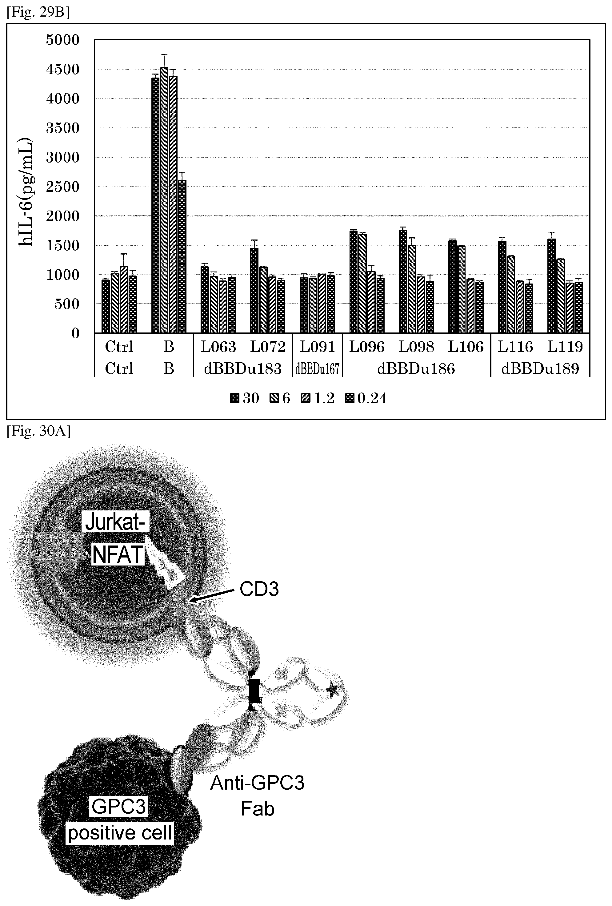
D00032

D00033
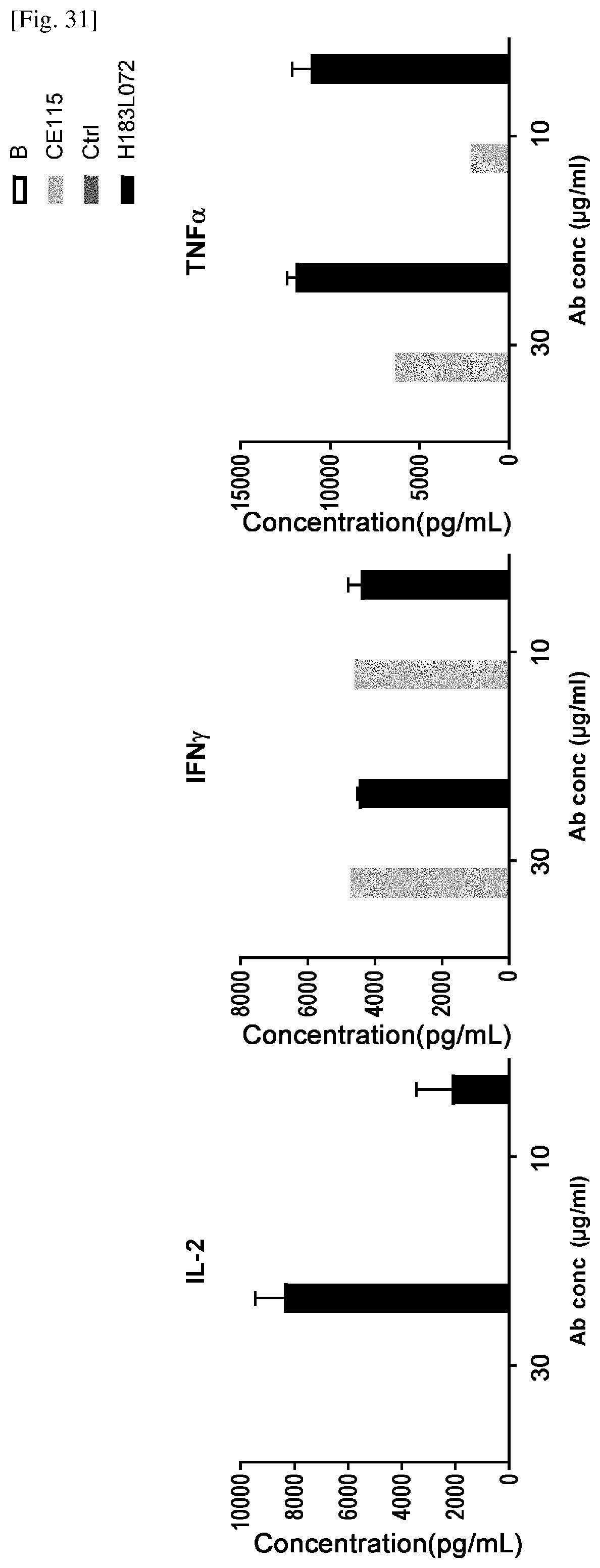
D00034
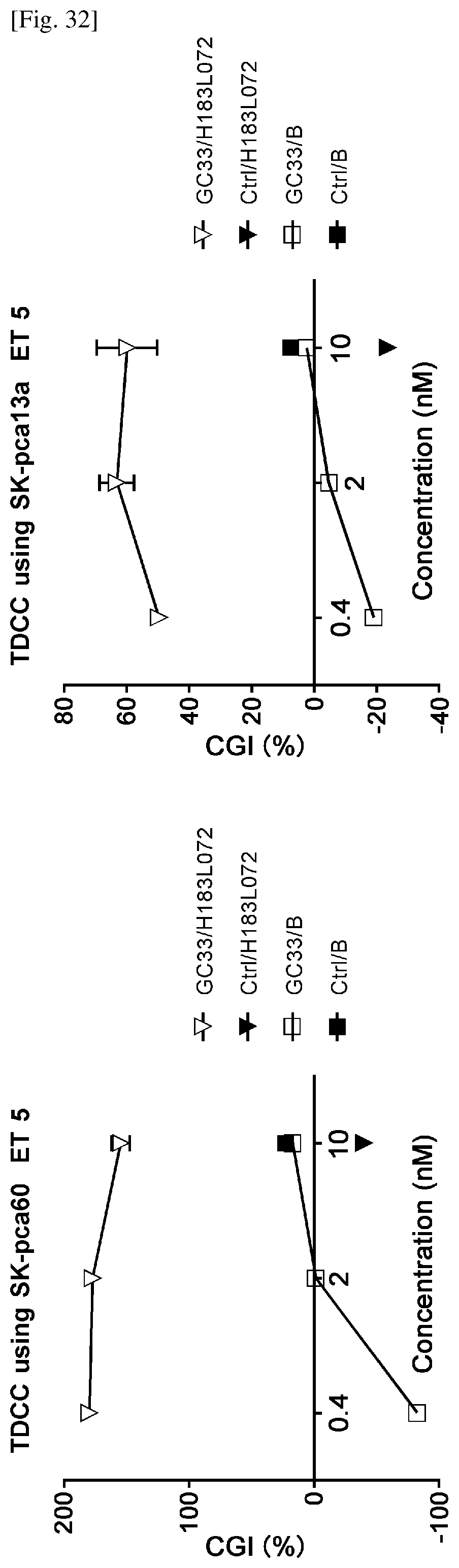
D00035

D00036

D00037

D00038

D00039

D00040

D00041
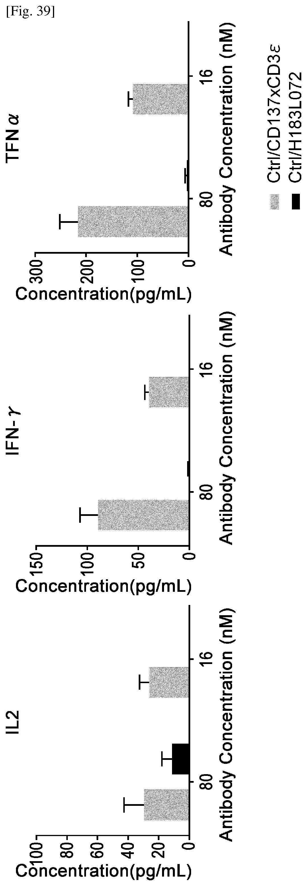
D00042

D00043

D00044
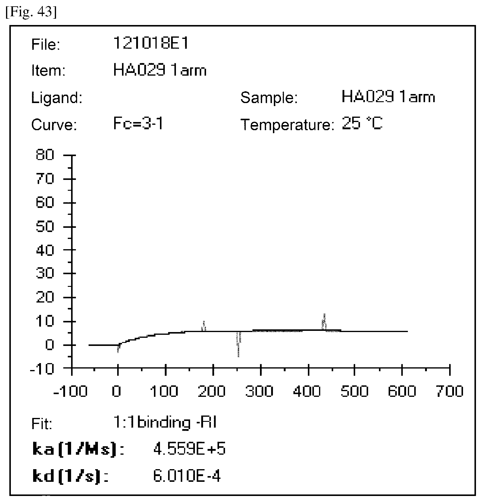
P00001

P00002

P00003

P00004
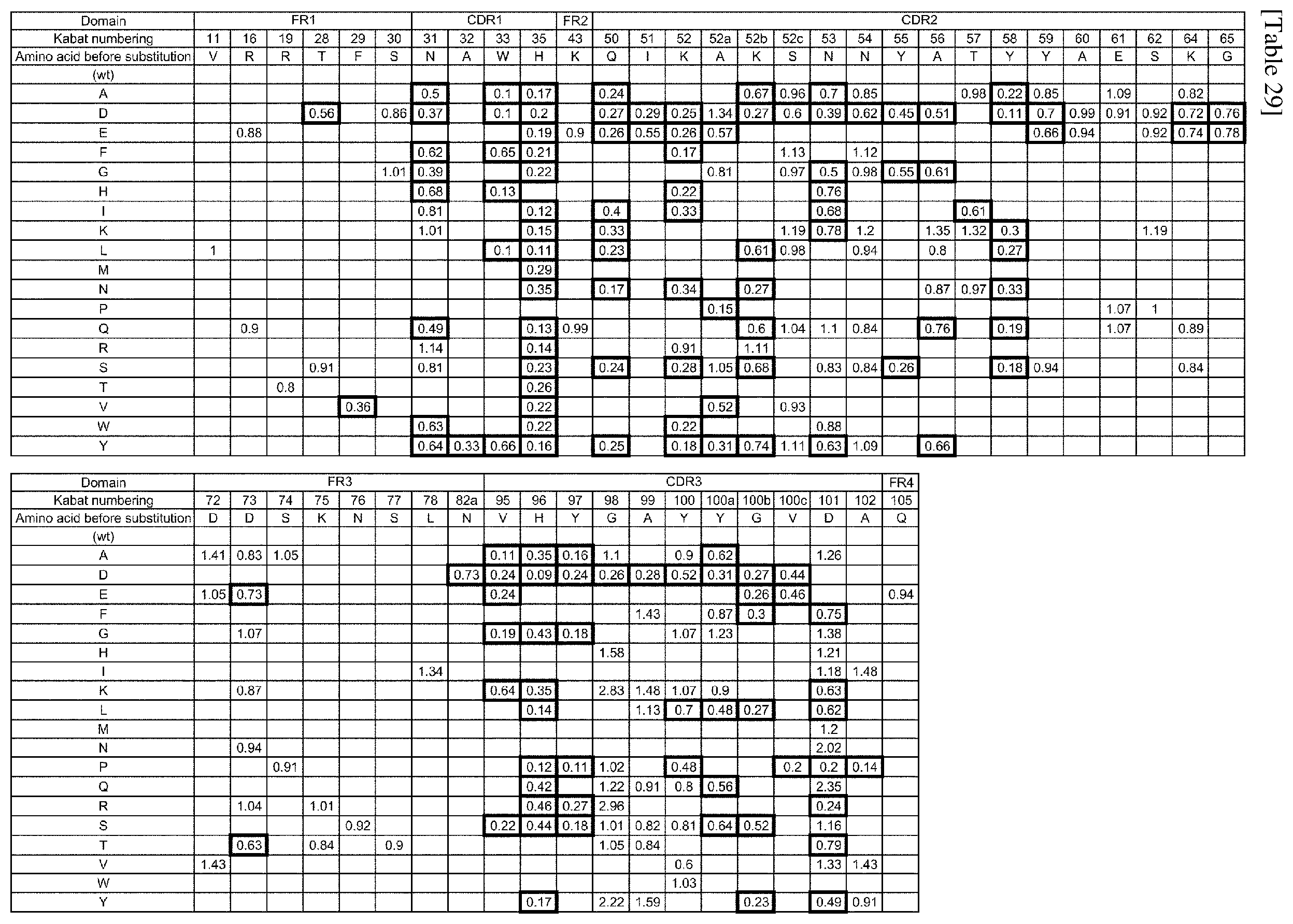
P00005

P00006

P00007

P00008

P00009

S00001
XML
uspto.report is an independent third-party trademark research tool that is not affiliated, endorsed, or sponsored by the United States Patent and Trademark Office (USPTO) or any other governmental organization. The information provided by uspto.report is based on publicly available data at the time of writing and is intended for informational purposes only.
While we strive to provide accurate and up-to-date information, we do not guarantee the accuracy, completeness, reliability, or suitability of the information displayed on this site. The use of this site is at your own risk. Any reliance you place on such information is therefore strictly at your own risk.
All official trademark data, including owner information, should be verified by visiting the official USPTO website at www.uspto.gov. This site is not intended to replace professional legal advice and should not be used as a substitute for consulting with a legal professional who is knowledgeable about trademark law.