Optical Imaging Agents Targeting Inflammation
ZHANG; Yongmin ; et al.
U.S. patent application number 16/968272 was filed with the patent office on 2020-11-26 for optical imaging agents targeting inflammation. The applicant listed for this patent is CENTRE NATIONAL DE LA RECHERCE SCIENTIFIQUE, ECOLE NATIONAL SUPERIEURE DE CHIMIE DE PARIS, INSTITUT NATIONAL DE LA SANTE ET DE LA RESEARCHE MEDICALE, SORBONNE UNIVERSITE, UNIVERSITE DE PARIS. Invention is credited to Michel BESSODES, Nathalie MIGNET, Daniel SCHERMAN, Johanne SEGUIN, Yongmin ZHANG.
| Application Number | 20200368373 16/968272 |
| Document ID | / |
| Family ID | 1000005037555 |
| Filed Date | 2020-11-26 |


View All Diagrams
| United States Patent Application | 20200368373 |
| Kind Code | A1 |
| ZHANG; Yongmin ; et al. | November 26, 2020 |
OPTICAL IMAGING AGENTS TARGETING INFLAMMATION
Abstract
##STR00001## The present invention relates to an optical imaging agent of formula (SIGNAL).sub.n-SUPPORT-(L-BIOVECTOR).sub.m, wherein: SUPPORT represents a physiologically acceptable chemical or biological substrate, with a particle size of between 1 and 100 nm, SIGNAL is a fluorophore, L is a linker of formula --C(X)--R.sup.1--Y--, with X being O, NH or S R.sup.1 being a (C.sub.1-C.sub.6)alkyl group, optionally R.sup.1 being a (C.sub.1-C.sub.6)alkyl group, optionally interrupted by 1 to 3 groups selected from --O--, --NH--, --C(O)--, --NHC(O)--, --(O)CNH--, --C(O)NH--N.dbd.C--, --N.dbd.C--, and (I), Y being NH or a --(O)CNH-- group, and BIOVECTOR is a carbohydrate targeting markers of inflammation, advantageously selected from the group consisting of mannose, sialyl Lewis.sup.X and derivatives thereof, n is greater than or equal to 0.5 and is less than 2, m is between 0 and 30, preferably between 1 and 30, more preferably between 5 and 20, diagnostic compositions comprising same and use thereof as a diagnostic agent (in vivo or ex vivo), or as contrast agent for image-guided surgery.
| Inventors: | ZHANG; Yongmin; (Antony, FR) ; BESSODES; Michel; (Villejuif, FR) ; SEGUIN; Johanne; (Kremlin Bicetre, FR) ; MIGNET; Nathalie; (Clamart, FR) ; SCHERMAN; Daniel; (Paris, FR) | ||||||||||
| Applicant: |
|
||||||||||
|---|---|---|---|---|---|---|---|---|---|---|---|
| Family ID: | 1000005037555 | ||||||||||
| Appl. No.: | 16/968272 | ||||||||||
| Filed: | February 21, 2019 | ||||||||||
| PCT Filed: | February 21, 2019 | ||||||||||
| PCT NO: | PCT/EP2019/054382 | ||||||||||
| 371 Date: | August 7, 2020 |
| Current U.S. Class: | 1/1 |
| Current CPC Class: | A61K 49/0056 20130101; A61K 49/0091 20130101; A61K 49/0052 20130101 |
| International Class: | A61K 49/00 20060101 A61K049/00 |
Foreign Application Data
| Date | Code | Application Number |
|---|---|---|
| Feb 21, 2018 | EP | 18305183.8 |
Claims
1. Optical imaging agent of formula (SIGNAL).sub.n-SUPPORT-(L-BIOVECTOR).sub.m, wherein: SUPPORT represents a physiologically acceptable chemical or biological substrate, with a particle size of between 1 and 100 nm, SIGNAL is a fluorophore, L is a linker of formula --C(X)--R.sup.1--Y--, with X being O, NH or S, R.sup.1 being a (C.sub.1-C.sub.6)alkyl group, preferably a (C.sub.3-C.sub.5)alkyl group, optionally interrupted by 1 to 3 groups selected from --O--, --NH--, --C(O)--, --NHC(O)--, --(O)CNH--, --C(O)--NH--N.dbd.C--, --N.dbd.C--, and ##STR00042## Y being NH or --(O)CNH--, and BIOVECTOR is a carbohydrate able to target markers of inflammation, n is greater than or equal to 0.5 and is less than 2, m is between 1 and 30, or a pharmaceutically acceptable salt, solvate or hydrate thereof.
2. The optical imaging agent of claim 1, wherein SUPPORT is a protein.
3. The optical imaging agent of claim 1, wherein SUPPORT is an albumin with a particle size of between 1 and 100 nm.
4. The optical imaging agent of claim 1, wherein SIGNAL is of formula (I): ##STR00043## wherein q is 0 or 1, r is 0 or 1, R.sub.b1 and R.sub.b2 are H or a C.sub.1-C.sub.4 group, or are bridged to form a --CH.sub.2--CH.sub.2--CH.sub.2-- alkylene group (only when q and r represent 1), R.sub.a1 and R.sub.a2 are identical or different and are independently a (C.sub.1-C.sub.6)alkyl group, optionally substituted by a SO.sub.3.sup.- group, a SO.sub.3K group, a SO.sub.3Na group, or a COOH group, provided that not more than one of R.sub.a1 and R.sub.a2 is substituted by a SO.sub.3.sup.- group, Ring A represents a C.sub.5-C.sub.6 monocyclic aryl or heteroaryl group or a C.sub.8-C.sub.12 fused bicyclic aryl or heteroaryl group, provided that the compound of formula (I) comprises at least one SO.sub.3.sup.- or SO.sub.3Na group, but not more than one SO.sub.3.sup.- group, and that when the compound of formula (I) has an overall positive electric charge, it is provided as a salt, in particular a halogenide salt such as a chloride Cl.sup.- salt.
5. The optical imaging agent of claim 1, wherein X is NH and R.sup.1 is a (C.sub.1-C.sub.6) alkyl, optionally interrupted by a ##STR00044## group.
6. The optical imaging agent of claim 1, wherein BIOVECTOR is of formula (II): ##STR00045## wherein R.sub.2 represents a (C.sub.1-C.sub.6)alkyl group, and X' represents a heteroatom such as O, S or NH, or a compound of formula (III) or (IV) ##STR00046## wherein R is R.sub.2--X', with R.sub.2 being a (C.sub.1-C.sub.6)alkyl group and X' being a heteroatom such as O, S or NH, and Rs is H, SO.sub.3K or SO.sub.3Na.
7. The optical imaging agent of claim 1, wherein BIOVECTOR is of formula (V), (VI) or (VII): ##STR00047## wherein R is R.sub.2--X', with R.sub.2 being a (C.sub.1-C.sub.6)alkyl group and X' being a heteroatom such as O, S or NH.
8. A diagnostic composition comprising at least one optical imaging agent according to claim 1, a pharmaceutically acceptable salt, solvate or hydrate thereof, and at least one pharmaceutically acceptable excipient.
9. An in vivo diagnostic method comprising administering to a patient in need thereof an effective amount of the optical imaging agent according to claim 1.
10. The in vivo diagnostic method according to claim 9, comprising diagnosing diseases or conditions associated with inflammation.
11. The in vivo diagnostic method according to claim 10, wherein the disease or condition associated with inflammation is selected from stroke, renal failure or cancer.
12. The in vivo diagnostic method according to claim 9, wherein said method is applied to surgical resection of tumours in the patient and comprises the following further successive steps: resecting the tumoral tissue identified prior to surgery in an area of interest; imaging the area of interest of the patient, to whom the effective amount of the optical imaging agent has been administered prior to surgery or during surgery, using an optical imaging device, so as to identify remaining tumoral tissues; when light signals are observed in the area of interest defining remaining tumoral tissues, proceeding with the resection of the identified remaining tumoral tissues; when no light signal is observed in the area of interest, not proceeding with any further resection in the area of interest.
13. A method of imaging a biological tissue ex vivo, wherein said biological tissue comprises the optical imaging agent according to claim 1, the method comprising applying optical or fluorescence imaging to said tissue.
14. The method of claim 13, wherein the biological tissue is a biopsy sample.
15. The method of claim 13, further comprising a step of diagnosing a disease or condition associated with inflammation from said optical or fluorescence imaging.
16. The optical imaging agent of claim 1, wherein SUPPORT is a human serum albumin with a particle size of between 1 and 100 nm.
17. The optical imaging agent of claim 4, wherein Ring A represents a C.sub.5-C.sub.6 monocyclic aryl group or a C.sub.8-C.sub.12 fused bicyclic aryl group, such as a phenyl or naphthyl group.
18. The optical imaging agent of claim 4, wherein Ring A is substituted by one SO.sub.3.sup.- or SO.sub.3Na group, provided that not more than one of R.sub.a1 and R.sub.a2 is substituted by a SO.sub.3.sup.- group.
19. The optical imaging agent of claim 6, wherein R.sub.2 represents a (C.sub.1-C.sub.4)alkyl group and X' represents NH.
Description
FIELD OF THE INVENTION
[0001] The present invention concerns optical imaging agents targeting inflammation biomarkers. The optical imaging agents of the invention are in particular useful in the in vivo as well as ex vivo diagnosis of diseases and conditions associated with inflammatory diseases such as cancer, renal failure, stroke.
BACKGROUND OF THE INVENTION
[0002] Recently, optical imaging has drawn increasing interest because of the ease of use of portable imaging devices, and reduced toxicity and risk to the operator and patient compared in particular with X-ray imaging, radiomedicine or magnetic resonance imaging.
[0003] Optical imaging, and more specifically fluorescence imaging, are used for analysis of biological samples (such as biopsies for instance, hereafter referred to as "ex vivo imaging"), in 2 dimensions (2D) as well as 3 dimensions (3D) (for instance using tomography), and can be used in vivo during surgery, for instance using portable devices such as those commercialized by Fluoptics and Novadaq.
[0004] Optical imaging requires administering optical agents to the patient or animal to be imaged (in vivo or ex vivo) prior to imaging
[0005] However, to improve the quality and accuracy of diagnostic using optical imaging, there remains a need for optical imaging agents able to target specific markers in the body. In particular, there is a need for optical imaging agents selectively targeting markers of inflammation, in order to diagnose inflammatory diseases in vitro or ex vivo or in vivo. Such optical imaging agents would thus enable the diagnostic of cancer or inflammatory diseases, and would be useful for image-guided surgery, for instance during tumour resection.
SUMMARY OF THE INVENTION
[0006] The present invention thus concerns an optical imaging agent of formula (SIGNAL).sub.n-SUPPORT-(L-BIOVECTOR).sub.m, wherein:
[0007] SUPPORT represents a physiologically acceptable chemical or biological substrate, with a particle size comprised between 1 and 100 nm,
[0008] SIGNAL is a fluorophore,
[0009] L is a linker of formula --C(X)--R.sup.1--Y--, with
[0010] X being O, NH, or S
[0011] R.sup.1 being a (C.sub.1-C.sub.6)alkyl group, preferably a (C.sub.3-C.sub.5)alkyl group, optionally interrupted by 1 to 3 groups selected from a heteroatom such as O or NH, a --C(O)-- group, a --NHC(O)-- group, a --(O)CNH-- group, --C(O)--NH--N.dbd.C-- group, a --N.dbd.C-- group, a --NH-- SO.sub.2--NH--C(S)-- group and a
##STR00002##
[0012] Y being NH or a --(O)CNH-- group, and
[0013] BIOVECTOR is a hydrophilic molecule preferentially a carbohydrate, in particular targeting markers of inflammation, said carbohydrate being advantageously selected from the group consisting of mannose, glucose, fucose, sialyl Lewis.sup.X (or SLe.sup.X) and derivatives such as neuraminic acid, trisaccharide derivatives thereof,
[0014] n is greater than or equal to 0.5 and is less than 3,
[0015] m is between 0 and 30, preferably between 1 and 30,
[0016] Signal is selected from the group consisting of fluorophores, preferentially fluorophores emitting in the near Infra-red, such as indocyanin green, cyanin-5, cyanin 5,5, cyanin-7, alexa fluors, and derivatives thereof,
[0017] a pharmaceutically acceptable salt, solvate or hydrate thereof.
[0018] The optical imaging agents of the invention thus exhibit several advantages.
[0019] First, the SIGNAL and BIOVECTOR parts of the optical imaging agent of the invention are covalently linked to its SUPPORT part--through the linker L in the case of BIOVECTOR, when present. Such covalent linkage allows for a much-improved selectivity of the optical imaging agent of the invention for inflamed tissues, as compared with agents wherein the BIOVECTOR or the SIGNAL parts are linked to the rest of the imaging agent only through ionic, hydrogen or Van der Waals bonds. Indeed, in the latter case, dissociation of the different parts of the imaging agent could occur in the biological tissues or the body, thus shading doubt as to the accuracy of the observed images.
[0020] Second, the optical imaging agent of the invention selectively targets markers of inflammation, thus allowing for accurate delimitation of inflamed areas of the body using in vivo or ex vivo optical imaging, in particular fluorescence imaging. Thanks to the portable imaging devices available nowadays, such optical imaging agent can thus be used as a real-time aid to surgery: for instance, after resection of a tumour, it is possible to visualize through optical imaging of the area of interest if all of the tumoral tissues have been removed, or if a further resection is necessary. Other imaging techniques are either more complex to use in this context (in particular MRI, scintigraphy, PET), or less sensitive (echography).
[0021] Third, the presence of a SUPPORT linking the SIGNAL part of the optical imaging agent of the invention and its BIOVECTOR part, increases the bioavailability of the optical imaging agent of the invention.
[0022] Finally, it is noteworthy that the optical imaging agent of the invention comprises up to 3 fluorophores. Indeed, the inventors have surprisingly demonstrated that the presence of more than 3 fluorophores have deleterious effect on the intensity of the observed signal, in contrast for instance to what is commonly known for magnetic resonance or scintigraphy imaging agents (see for instance WO 2008/074960).
[0023] According to another aspect, the present invention relates to a diagnostic composition comprising at least one optical imaging agent of formula (SIGNAL).sub.n-SUPPORT-(L-BIOVECTOR).sub.m of the invention, a pharmaceutically acceptable salt, solvate or hydrate thereof, and at least one pharmaceutically acceptable excipient.
[0024] According to another aspect, the present invention relates to the optical imaging agent of formula (SIGNAL).sub.n-SUPPORT-(L-BIOVECTOR).sub.m of the invention or the diagnostic composition of the invention for use as a diagnostic agent, in particular for diagnosing diseases or conditions associated with inflammation.
[0025] According to another aspect, the present invention relates to a method of imaging a biological tissue ex vivo, using optical or fluorescence imaging, said biological tissue comprising the optical imaging agent or the diagnostic composition of the invention.
[0026] According to another aspect, the present invention relates to a method of ex vivo diagnosing a disease or condition associated with inflammation, comprising ex vivo imaging a biological tissue of a patient in need thereof, in particular a biological tissue obtained through biopsy, said biological tissue comprising the optical imaging agent or the diagnostic composition of the invention.
[0027] According to another aspect, the present invention relates to a process for preparing the optical imaging agent of formula (SIGNAL).sub.n-SUPPORT-(L-BIOVECTOR).sub.m of the invention.
DETAILED DESCRIPTION OF THE INVENTION
Optical Imaging Agent
[0028] n and m are chosen so as to avoid any destabilization of SUPPORT. Indeed, the optical imaging agent of the invention should be able to form stable colloidal suspensions in physiologically acceptable solutions.
[0029] As used herein, a "stable suspension" is understood as a suspension which does not form any aggregates or does not sediment over a period of at least 6 hours, preferably at least 12 hours, even more preferably at least 24 hours.
[0030] As used herein, a "physiologically acceptable solution" is understood as a solution that is nontoxic and suitable for administration to a patient via oral route or parenteral route, in particular by injection, without any undesired side-effects. As such, the osmolarity and osmolality of a physiologically acceptable solution should be controlled so as to avoid any undesired side effects. Examples of solutes suitable for forming a physiologically acceptable solution are water (in particular water for injection, saline, and aqueous solutions such as NaCl 150 mM, PBS, glucose 5%, Hepes 20 mM+glucose 5%, lactose 5%, Hepes 20 mM+lactose 5%).
[0031] In the present invention, a "patient" is understood as an animal, preferably a mammal such as a rodent (mice or rats), and preferably a human being.
[0032] Numbers n and m are calculated with mass spectrometry experiments. It is known from the skilled person in the art that mass spectrometry characterization is used to assess grafting of fragments on macromolecules.
[0033] After the grafting of the SIGNAL and/or the BIOVECTOR fragment, a mass spectrometry analysis provides the additional mass of the grafted molecule compared to the mass of the native SUPPORT. The mass difference allows attribution of a mean grafting value regarding the number of SIGNAL and/or BIOVECTOR fragments present on the molecule.
[0034] n is greater than or equal to 0.5 and is less than 3, for instance n is between 0.5 and 2.5. Preferably, n is between 0.5 and 2. According to another embodiment, n is greater than or equal to 1 and is less than 3, for instance n is between 1 and 2, preferably n is equal to 1 or 2.
[0035] m is between 0 and 30, preferably between 1 and 30, more preferably between 5 and 20.
[0036] In a particular embodiment, n is between 0.5 and 2, and m is between 1 and 30, more preferably between 5 and 20.
Support
[0037] In general formula (I), SUPPORT represents a physiologically acceptable chemical or biological substrate, that is to say an entity whose administration is compatible with a living being. Typically, SUPPORT is conjugated to SIGNAL and L-BIOVECTOR through all or part of its amino (NH.sub.2) groups located on its surface.
[0038] Advantageously, SUPPORT has a particle size of between 1 and 100 nm, preferably between 5 and 50, more preferably of between 5 nm and 20 nm. In a particular embodiment, SUPPORT has a particle size of between 5 and 10 nm. SUPPORT may itself have biological activity, or on the contrary be totally inert.
[0039] Advantageously, the SUPPORT is biodegradable.
[0040] As used herein, a "biodegradable" entity is an entity which is biodegraded in vivo through the action of proteins such as enzymes, but which however does not degrade spontaneously in a physiological solution in particular at a pH of between 6 and 8 and at a temperature of between 25.degree. C. and 37.degree. C., generally for a time period of at most 7 days. SUPPORT is preferably selected from proteins. Suitable proteins or protein conjugates are for instance described in WO 2004/071536 (see in particular section "Nature of the carrier material" pages 15-18).
[0041] As used herein, "protein" is meant as a macromolecule composed of a string (or sequence) of amino acids linked together by peptide bonds. Proteins suitable for the present invention may have various molecular weights, in particular ranging from 10 to 500 kDa, preferably from 50 to 100 kDa. They may be chosen from albumin, ovalbumin, lactalbumin, immunoglobulins, macroglobulins, microglobulins, lipoproteins, circulating hormones and factors, hemocyanins, and derivatives thereof, preferably albumin, in particular a human albumin such as human serum albumin (HSA).
[0042] In a preferred embodiment, SUPPORT is a protein, especially albumin, preferably human serum albumin (HSA) or derivatives thereof, with a particle size of between 2 and 60, preferably of between 5' and 20'. In particular, use may be made of albumins commercialized by Vialebex or Baxter. Such a small particle size, combined with the fact that optical agent does not aggregate, is of particular interest in terms of biodistribution and bioavailability. According to this embodiment, the optical imaging agent in which SUPPORT is albumin has a size of between 6 nm and 20 nm.
[0043] Albumin derivatives are in particular ovalbumin and lactalbumin.
[0044] In another embodiment, SUPPORT may also be selected from polysaccharides, nanoparticles (including liposomes), microparticles (including liposomes) or biocompatible polymers.
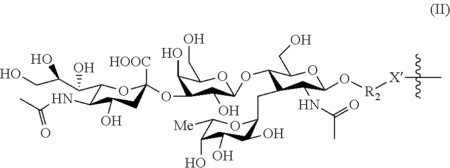
[0045] In the context of the present invention, a polymer is characterized as "biocompatible" if the polymer and its degradation products are non-toxic to the animal or human being to which it is administered, and does not induce adverse effects in the host's body, e.g. immune reaction at the injection site. Biocompatible polymers suitable for the present invention may be chosen from any of the polymers known to those skilled in the art, including poly(N-(2-hydroxypropyl)methacrylamide) (HMPA), polyethylene glycol (PEG), collagen, polysaccharides, poly(2-methoxyethylacrylate) (PMEA), polydimethylsiloxane (PDMS), polyvinyl pyrrolidone (PVP), hyaluronic acid (HA), heparan, heparanesulfates, dextrans, dextransulfates, heparins, cyclodextrins and derivatives thereof.
[0046] When SUPPORT is a nano- and microparticle, said particle may in particular be made of biodegradable organosoluble polymer.
[0047] As used herein, "particles" are understood as particles with an average particle size of between 1 and 6 000 nm, and in particular between 2 and 1000 nm, preferably between 3 and 100 nm, and in particular between 3 and 30 nm. The particles of the invention typically have an organic core (such as a polymeric core), but may also be liposomes.
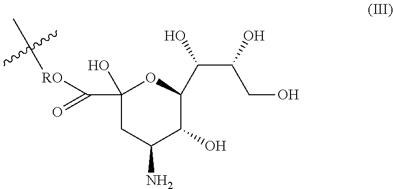
[0048] More specifically, "microparticles" are understood as particles with an average particle size of between 500 nm and 6 000 nm, while "nanoparticles" are understood as particles with an average particle size of between 1 and 500 nm, in particular between 2 and 300 nm, preferably between 3 and 150 nm, and in particular between 5 nm and 100 nm, such as between 3 and 30 nm.
[0049] As biodegradable organosoluble polymer, mention may be made of polyesters such as poly (lactic acid) (PLA), poly (glycolic acid) (PGA), poly( -caprolactone) (PCL), polyanhydrides, poly (hyaluronan), poly(alkylcyanoacrylates), polyorthoesters, poly (alkylene tartrate), polyphosphazenes, polyamino acids, polyamidoamines, polysaccharides, polycarbonates, polymethylidenemalonate, polysiloxane, polyhydroxybutyrate or poly(malic acid), and copolymers thereof.
[0050] Where appropriate, the particle surface may be modified for example to confer to said particle additional properties, such as hydrophilicity, so as to improve their in vivo reactivity, for example by promoting their adsorption of plasma proteins.
[0051] Particularly preferred are "targeted nanoparticles", such as targeted liposomes or targeted biocompatible polymers. As used herein, a "targeted nanoparticle" is able to target markers of inflammation, such as Selectin E. In particular, a "targeted nanoparticle" has a biomarker on its surface targeting markers of inflammation.
[0052] Suitable nanoparticles and microparticles are for instance described in WO 2006/116742.
[0053] Conjugates of specific-binding moieties can be used for detecting specific target molecules in biological samples. The specific-binding portion (in the present invention: BIOVECTOR) of such conjugates binds tightly to a target in the sample and the signal-generating portion (herein SIGNAL) is utilized to provide a detectable signal that indicates the presence/and or location of the target.
Signal
[0054] SIGNAL is a fluorophore, preferably an organic fluorophore, allowing the optical agent of the invention to be detected through optical imaging, in particular fluorescence imaging. In particular, SIGNAL is a hydrophilic fluorophore. Of note, the fluorophore is hydrophilic enough to avoid any precipitation of the optical imaging agent of the invention.
[0055] Preferably, SIGNAL is a hydrophilic cyanine derivative, which are well-known fluorophores.
[0056] In a particular embodiment, SIGNAL is of formula (la) below:
##STR00003##
wherein
[0057] q is 0 or 1,
[0058] r is 0 or 1,
[0059] R.sub.b1 and R.sub.b2 are H or a C.sub.1-C.sub.4 group, or are bridged to form a --CH.sub.2--CH.sub.2--CH.sub.2-- alkylene group (only when q and r represent 1),
[0060] R.sub.a1 and R.sub.a2 are identical or different and are independently a (C.sub.1-C.sub.6)alkyl group, optionally substituted by a SO.sub.3.sup.- group, a SO.sub.3Na or a SO.sub.3K group, or a COOH group, provided that not more than one of R.sub.a1 and R.sub.a2 is substituted by a SO.sub.2.sup.- group.
[0061] Ring A represents a C.sub.5-C.sub.6 monocyclic aryl or heteroaryl group or a C.sub.8-C.sub.12 fused bicyclic aryl or heteroaryl group, preferably a C.sub.5-C.sub.6 monocyclic aryl group or a C.sub.8-C.sub.12 fused bicyclic aryl group, such as a phenyl or naphthyl group, and Ring A is optionally substituted by one SO.sub.3.sup.-, SO.sub.3Na or SO.sub.3K group, provided that not more than one of R.sub.a1 and R.sub.a2 is substituted by a SO.sub.3.sup.- group,
[0062] provided that the compound of formula (I) comprises not more than one SO.sub.3.sup.- group.
[0063] The compound of formula (I) may have an overall electric charge, namely a positive charge. In such case, it is provided as a salt, in particular a halogenide salt such as a chloride Cl.sup.- salt.
[0064] The compound of formula (I) is preferably hydrophilic, and as such advantageously contains at least one SO.sub.3.sup.- or SO.sub.3Na group, preferably it contains one SO.sub.3.sup.- group and SO.sub.3Na group.
[0065] In a particular embodiment, SIGNAL is of formula (Ia) below:
##STR00004##
wherein q, r, R.sub.a1, R.sub.a2, R.sub.b1 and R.sub.b2 are as defined above or below, and
[0066] Ring
##STR00005##
is optional, and when present represents a (C.sub.5-C.sub.6) monocyclic aryl or heteroaryl group (fused with the adjacent indoline group) optionally substituted by one SO.sub.3.sup.- or SO.sub.3Na group, provided that not more than one of R.sub.a1 and R.sub.a2 is substituted by a SO.sub.3.sup.- group, preferably a (C.sub.5-C.sub.6) monocyclic aryl group such as phenyl optionally substituted by one SO.sub.3.sup.-, SO.sub.3Na or SO.sub.3K group,
[0067] provided that the compound of formula (I) comprises not more than one SO.sub.3.sup.- group.
[0068] In a particular embodiment, SIGNAL is of formula (Ia1) below:
##STR00006##
wherein q, r, R.sub.a1, R.sub.a2, R.sub.b1 and R.sub.b2 are as defined above or below.
[0069] In another particular embodiment, SIGNAL is of formula (Ia2) below:
##STR00007##
wherein q, r, R.sub.a1, R.sub.a2, R.sub.b1 and R.sub.b2 are as defined above or below.
[0070] In a particular embodiment of formulae (I), (Ia), (Ia1) and (Ia2):
[0071] R.sub.b1 and R.sub.b2 are advantageously H;
[0072] q is 0 or 1 and advantageously r is 0;
[0073] R.sub.a1 is preferably methyl
[0074] R.sub.a2 is preferably --CH.sub.2--CH.sub.2--CH.sub.2--CH.sub.2--CH.sub.2--COOH.
[0075] In another particular embodiment, of formulae (I), (Ia), (Ia1) and (Ia2):
[0076] R.sub.b 1 and R.sub.b2 are advantageously H;
[0077] q and r are both 1, and R.sub.b1 and R.sub.b2 taken together represent a --CH.sub.2--CH.sub.2--CH.sub.2-- alkylene group;
[0078] R.sub.a1 is preferably methyl
[0079] R.sub.a2 is preferably --CH.sub.2--CH.sub.2--CH.sub.2--CH.sub.2--CH.sub.2--COOH.
[0080] In another particular embodiment, SIGNAL is of formula (Ib) below:
##STR00008##
wherein
[0081] p is an integer of between 1 and 4, preferably between 1 and 2,
[0082] R is a (C.sub.1-C.sub.6)alkyl group, preferably a (C.sub.1-C.sub.4)alkyl group,
[0083] Ring A represents a C.sub.5-C.sub.6 monocyclic aryl or heteroaryl group or a C.sub.8-C.sub.12 fused bicyclic aryl or heteroaryl group, preferably a C.sub.5-C.sub.6 monocyclic aryl group or a C.sub.8-C.sub.12 fused bicyclic aryl group, such as a phenyl or naphthyl group.
[0084] In the compound of formula (I), each ring A is fused with the adjacent pyrrolidine group.
[0085] Advantageously, SIGNAL is of formula (Ib1) below:
##STR00009##
wherein
[0086] p is an integer of between 1 and 2,
[0087] R is a (C.sub.1-C.sub.4)alkyl group
[0088] Ring
##STR00010##
is optional, and when present represents a (C.sub.5-C.sub.6) monocyclic aryl or heteroaryl group (fused with the adjacent indoline group), preferably a (C.sub.5-C.sub.6) monocyclic aryl group such as phenyl.
[0089] In a particular embodiment, SIGNAL is of formula (I2) below:
##STR00011##
wherein
[0090] p is an integer of between 1 and 2, and
[0091] Ring
##STR00012##
is optional, and when present represents a (C.sub.5-C.sub.6) monocyclic aryl or heteroaryl group (fused with the adjacent indoline group), preferably a (C.sub.5-C.sub.6) monocyclic aryl group, such as phenyl.
[0092] In a preferred embodiment, SIGNAL is selected from cyanines and indocyanines of formulae below:
##STR00013##
more specifically, SIGNAL is
##STR00014##
[0093] In another preferred embodiment, SIGNAL is selected from:
##STR00015##
[0094] Of note, when excited with light (in particular at a wavelength of between 450 nm and 1000 nm, preferably in the "close red domain" or "close infrared domain" i.e. between 600 nm and 900 nm), cyanine emits red light, whereas indocyanine emits green light, through fluorescence phenomena.
[0095] The above fluorophores of formula (I) are known in the art and are commercially available, in particular from Luminoprobe.
Biovector
[0096] The BIOVECTOR is a carbohydrate, advantageously targeting selectively a marker of inflammation.
[0097] The term "carbohydrate" as used in the present invention refers to a monosaccharide or polysaccharide, or nitrogen-derivatives thereof. Monosaccharides are in particular erythrose, threose, ribose, arabinose, xylose, lyxose, allose, altrose, glucose, mannose, fucose, gulose, idose, galactose, talose, erythrulose, ribulose, xylulose, psicose, fructose, sorbose, tagatose. The carbohydrate of the invention is preferably not protected (i.e. it does not contain any O-protecting groups). However, the carbohydrate of the invention may contain one or two sulfate groups, i.e. a OSO.sub.3H or preferably a OSO.sub.3Na group in lieu of a OH group.
[0098] Polysaccharides are preferably bi-, tri- or quadrisaccharides, and each monosaccharide group of which it is composed is preferably selected from the monosaccharides listed above.
[0099] Nitrogen derivatives of monosaccharides and polysaccharides are understood herein as compounds having a monosaccharide or polysaccharide structure, wherein 1 to 2 oxygen atoms in a monosaccharide or 1 to 4 oxygen atoms in a polysaccharide are replaced with a NH group.
[0100] Carbohydrates targeting selectively a marker of inflammation are known to the person of skill in the art. In particular, lactose is known not to belong to this category. A typical example is Lewis.sup.X pentasaccharide molecule in which the Lewis.sup.X trisaccharide is linked to a lactose molecule, however the Lewis.sup.X-Lewis.sup.X interaction is due to the Lewis.sup.X trisaccharide, but not to lactose.
[0101] Preferred carbohydrates targeting selectively a marker of inflammation are mannose, glucose, fucose, sialyl Lewis.sup.X (or SLe.sup.X), or any mono- or polysaccharide comprised in sialyl Lewis.sup.X, such as neuraminic acid or and the trisaccharide of formula:
##STR00016##
with Rs being H, SO.sub.3Na or SO.sub.3K, preferably H or SO.sub.3Na, and derivatives thereof.
[0102] Examples of carbohydrate derivatives are carbohydrates wherein one anomeric OH group is replaced by a O--(C.sub.1-C.sub.6)alkyl-X' group, with X' representing O, NH or S, preferably NH. For instance a carbohydrate derivatives is a carbohydrate wherein an anomeric OH group is replaced by a --O--CH.sub.2--CH.sub.2--NH group. When the carbohydrate comprises a COOH group, a particular carbohydrate derivative is a carbohydrate wherein the group COOH is replaced by a group of formula COO--(C.sub.1-C.sub.6)alkyl-X', such as a COOCH.sub.2CH.sub.2NH group.
[0103] In an advantageous embodiment, BIOVECTOR is selected from the group consisting of mannose and sialyl Lewis.sup.X (or SLe.sup.X), and derivatives thereof. Sialyl Lewis.sup.X (or SLe.sup.X) is indeed an efficient ligand for E-selectin, also known as CD62 antigen-like family member E (CD62E), endothelial-leukocyte adhesion molecule 1 (ELAM-1), or leukocyte-endothelial cell adhesion molecule 2 (LECAM2). E-selectin is an adhesion molecule expressed only on endothelial cells activated by cytokines. During inflammation, E-selectin plays an important part in recruiting leukocytes to the site of injury: local release of cytokines IL-1 and TNF-.alpha. by damaged cells induces the over-expression of E-selectin on endothelial cells of nearby blood vessels. Leukocytes in the blood expressing the correct ligand will then bind with low affinity to E-selectin. As such, E-selectin is a particularly useful marker of inflammation, in particular overexpressed in the direct vicinity of tumour metastases.
[0104] In this context, BIOVECTOR is preferably of formula (II):
##STR00017##
wherein R.sup.2 represents a (C.sub.1-C.sub.6)alkyl group, preferably a (C.sub.1-C.sub.4)alkyl group, and X' represents a heteroatom such as O, S or NH, preferably NH.
[0105] In particular, BIOVECTOR is:
##STR00018##
[0106] This particular BIOVECTOR may be obtained from the corresponding compound described for instance described in Lu et al. (Carbohydrate Research 2014, 383, 89-96). Substructures of Sialyl Lewis X are also useful ligand for E-selectin such as neuraminic acid. In this context, BIOVECTOR is advantageously of formula (III):
##STR00019##
wherein R is R.sub.2-X', with R.sup.2 and X' as defined above in connection with formula (II).
[0107] Also considered is a trisaccharides of formula (IV):
##STR00020##
wherein Rs is H or SO.sub.3Na, and R is as defined above in connection with formula (III).
[0108] Mannose is also an efficient ligand for macrophages, overexpressed at a site of inflammation, which is useful as a marker of inflammation. A preferred mannose derivative is a compound of formula (V):
##STR00021##
wherein R is as defined above in connection with formula (III).
[0109] Fucose is also a known to be associated with inflammatory conditions, in particular in cancer. In this context, BIOVECTOR is advantageously of formula (VI):
##STR00022##
wherein R is as defined above in connection with formula (III)
[0110] Because of the link between diabetes and inflammation in particular, glucose is associated with markers of inflammation. In this context, BIOVECTOR is advantageously of formula (VII), or (VII'):
##STR00023##
wherein R is as defined above in connection with formula (III).
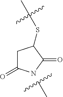
Linker L
[0111] In a particular embodiment, L is a linker of formula --C(X)--R.sup.1--Y--, wherein
[0112] X is O or NH
[0113] R.sup.1 is a (C.sub.1-C.sub.6)alkyl, preferably a (C.sub.3-C.sub.5)alkyl, optionally interrupted by 1 to 3 (preferably 1) groups selected from a heteroatom such as O or NH, a --C(O)-- group, a --NHC(O)-- group, a --(O)CNH-- group, and a
##STR00024##
[0114] Y is NH or a --(O)CNH-- group.
[0115] Advantageously, X is NH.
[0116] Preferably, R.sup.1 is a (C.sub.1-C.sub.6)alkyl, preferably a (C.sub.3-C.sub.5)alkyl, optionally interrupted by a
##STR00025##
Particular Embodiments
[0117] In a preferred embodiment, the optical imaging agent of the invention is of formula (SIGNAL).sub.n-SUPPORT-(L-BIOVECTOR).sub.m, wherein:
[0118] SUPPORT is a protein, especially an albumin such as human serum albumin (HSA) or derivatives thereof, with a particle size of between 1 and 100 nm, preferably of between 5 and 50, more preferably of between 5 nm and 20 nm. In a particular embodiment, SUPPORT has a particle size of between 5 and 10 nm
[0119] SIGNAL is of formula (Ia1), (Ia2), (Ib) as defined above:
[0120] Wherein, preferably,
[0121] p is an integer of between 1 and 4, preferably between 1 and 2,
[0122] R is a (C.sub.1-C.sub.6) alkyl group, preferably a (C.sub.1-C.sub.4) alkyl group,
[0123] Ring A represents a (C.sub.5-C.sub.6) monocyclic aryl or heteroaryl group or a (C.sub.8-C.sub.12) fused bicyclic aryl or heteroaryl group, preferably a (C.sub.5-C.sub.6) monocyclic aryl group or a (C.sub.8-C.sub.12) fused bicyclic aryl group such as phenyl or naphthyl,
[0124] L is a linker of formula --C(X)--R.sup.1--Y--, with
[0125] X being O or NH
[0126] R.sup.1 being a (C.sub.1-C.sub.6)alkyl, preferably a (C.sub.3-C.sub.5)alkyl, optionally interrupted by 1 to 3 groups selected from a heteroatom such as O or NH, a --C(O)-- group, a --NHC(O)-- group, a --(O)CNH-- group, and a
##STR00026##
[0127] Y being NH or a --(O)CNH-- group, and
[0128] BIOVECTOR is a carbohydrate targeting markers of inflammation selected from the group consisting of mannose, glucose, fucose, neuraminic acid, sialyl Lewis.sup.X(SLe.sup.X), the trisaccharide:
[0129] and derivatives thereof, said carbohydrate being preferably a compound of formula (II) or (V) as defined above,
[0130] n is greater than or equal to 0.5 and is less than 2, and is preferably between 0.5 and 1.5,
[0131] m is between 1 and 30, preferably between 5 and 20.
[0132] In this preferred embodiment, SIGNAL is in particular of formula (Ib), (Ib1) or (Ib2),
wherein, preferably,
[0133] p is an integer of between 1 and 4, preferably between 1 and 2,
[0134] R is a (C.sub.1-C.sub.6) alkyl group, preferably a (C.sub.1-C.sub.4) alkyl group,
[0135] Ring A represents a (C.sub.5-C.sub.6) monocyclic aryl or heteroaryl group or a (C.sub.8-C.sub.12) fused bicyclic aryl or heteroaryl group, preferably a (C.sub.5-C.sub.6) monocyclic aryl group or a (C.sub.8-C.sub.12) fused bicyclic aryl group such as phenyl or naphthyl.
[0136] Even more preferably, SIGNAL is indocyanine or sulfo-cyanine 5.
[0137] In this preferred embodiment, L is advantageously a linker of formula --C(X)--R.sup.1--Y--, with X being NH, R.sup.1 being a (C.sub.1-C.sub.6)alkyl, preferably a (C.sub.3-C.sub.5)alkyl, optionally interrupted by
##STR00027##
and Y being NH or a --(O)CNH-- group.
[0138] In a preferred embodiment, the optical imaging agent of the invention is of formula (SIGNAL).sub.n-SUPPORT-(L-BIOVECTOR).sub.m, wherein:
[0139] SUPPORT is human serum albumin (HSA), SIGNAL is
##STR00028##
[0140] L is a linker of formula --C(X)--R.sup.1--Y--, with
[0141] X being O
[0142] R.sup.1 being a (C.sub.3-C.sub.5)alkyl, optionally interrupted by 1 to 3 groups selected from a heteroatom such as O or NH, a --C(O)-- group, a --NHC(O)-- group, a --(O)CNH-- group, and a
##STR00029##
[0143] Y being NH or a --(O)CNH-- group,
[0144] n is between 0.5 and 2, and m is between 1 and 30, more preferably between 5 and 20.
Pharmaceutical and Diagnostic Composition
[0145] The present invention also relates to a pharmaceutical or diagnostic composition comprising at least one optical imaging agent of formula (SIGNAL).sub.n-SUPPORT-(L-BIOVECTOR).sub.m as defined above, a pharmaceutically acceptable salt, solvate or hydrate thereof, and at least one pharmaceutically acceptable excipient.
[0146] The pharmaceutical or diagnostic compositions of the invention are advantageously suitable for administration via oral, sublingual, subcutaneous, intramuscular, intravenous, transdermal, topical or rectal route, more preferably via oral route or by injection. The optical imaging agent of the invention can be administered in unit forms for administration, mixed with conventional pharmaceutical carriers, to animals or to humans.
[0147] When a solid composition is prepared in the form of tablets, the optical imaging agent of the invention is mixed with a pharmaceutical vehicle and other conventional excipients known to those skilled in the art.
[0148] When a composition is formulated for injection, the optical imaging agent of the invention is mixed with a saline solution and other conventional excipients known to those skilled in the art.
[0149] The optical imaging agents of the invention can be used in a diagnostic composition at a dose ranging from 0.01 mg/kg to 1000 mg/kg, for instance between 1 mg/kg and 800 mg/kg, in particular between 5 mg/kg and 500 mg/kg, preferably administered in only one dose. However, it may be necessary to use doses outside these ranges, which will then be designed by the person skilled in the art.
Optical Imaging
[0150] In the optical imaging agent of the invention, BIOVECTOR is a carbohydrate targeting selectively a marker of inflammation, preferably mannose, sialyl Lewis.sup.X (or SLe.sup.X) or derivatives thereof, advantageously the derivatives of formula (II) and (III) as defined above.
[0151] Therefore, the optical imaging agents of the invention will selectively accumulate in tissues or body areas wherein the targeted markers of inflammation (E-selectin for sialyl Lewis.sup.X (or SLe.sup.X) derivatives, macrophages for mannose derivatives) are overexpressed. The present invention thus also relates to the optical imaging agent of formula (SIGNAL).sub.n-SUPPORT-(L-BIOVECTOR).sub.m as defined above or the pharmaceutical or diagnostic composition of the invention for use as a diagnostic agent, in particular for diagnosing diseases or conditions associated with inflammation, preferably for in vivo diagnostic uses.
[0152] The optical imaging agents or diagnostic compositions of the invention are also useful in diagnosing a cardiovascular or inflammatory disease such as myocardial ischemia stroke, inflammatory bowel disease (such as Crohn disease and ulcerative colitis), renal failure, post-operative ileus, brain ischemia, diabetes, diabetic nephropathy, metabolic syndrome, sickle-cell disease, neurodegenerative diseases such as Alzheimer's disease or Parkinson's disease, neuropathic pain, hypertension, pulmonary arterial hypertension, septicemia, septic or endotoxic shock, hemorrhagic shock, multiple sclerosis, cancer and chronic obstructive pulmonary disease, and arthritic diseases (such as arthritis and osteoarthritis).
[0153] Preferably, the optical imaging agent or diagnostic composition of the invention is used for diagnosing cancer (in particular tumours, such as tumours of colorectal cancer), stroke and renal failure.
[0154] In a particular embodiment, the disease or condition associated with inflammation is cancer, and in particular tumours, such as tumours of colorectal cancer. In this embodiment, the optical imaging agent of formula (SIGNAL).sub.n-SUPPORT-(L-BIOVECTOR).sub.m as defined above or the pharmaceutical or diagnostic composition of the invention is useful as an aid to surgery, in particular as contrast agent in image-guided surgery. In this embodiment, the optical imaging agent or pharmaceutical or diagnostic composition of the invention is used in particular for real time diagnosis, allowing to follow evolution of the resection of tumoral tissues.
[0155] In this embodiment, the patient undergoing surgery is a living animal, in particular a human patient, suffering from cancer (such as colorectal cancer), and is in need of surgery for tumour resection. The optical imaging agent or diagnostic composition of the invention is administered to the patient advantageously prior to surgery or during surgery, for instance by injection.
[0156] Therefore, the present invention also relates to the optical imaging agent or diagnostic composition of the invention for use in a method of surgically resecting tumours of a patient comprising the following successive steps: [0157] resecting the tumoral tissue identified prior to surgery in an area of interest; [0158] imaging the area of interest of the patient, to whom an effective amount of the optical imaging agent or diagnostic composition of the invention has been administered prior to surgery or during surgery (for instance by injection), using an optical imaging device (in particular a fluorescence imaging device), so as to identify remaining tumoral tissues; [0159] when light (or fluorescence) signals are observed in the area of interest defining remaining tumoral tissues, proceeding with the resection of the identified remaining tumoral tissues; when no light (or fluorescent) signal is observed in the area of interest, not proceeding with any further resection in the area of interest.
[0160] As used herein, an "area of interest" is understood as an area of tissues or an organ of the patient comprising tumoral tissue to be resected and the vicinity of the tumoral tissue. For instance, in the case of liver cancer, the area of interest may be the whole liver. In the case of colorectal cancer, the area of interest is preferably the colon and/or the rectum, or part of it.
[0161] In this particular embodiment, the optical imaging agent or diagnostic composition of the invention thus proves useful in helping resecting the entirety of the tumoral tissues. Such a method using the optical imaging agent or diagnostic composition of the invention as an aid to surgery also allows for resecting the minimum sane tissues, whereas nowadays, in the absence of such method, surgeons tend to resect a wide area of sane tissues around the tumours as a safety measure.
[0162] The present invention further concerns the use of the optical imaging agent of the invention, a pharmaceutically acceptable salt, solvate or hydrate thereof, for the manufacture of a diagnostic composition, in particular for use for diagnosing diseases or conditions associated with inflammation as detailed above.
[0163] In a particular embodiment, the disease or condition associated with inflammation is cancer, and in particular tumours, such as tumours of colorectal cancer. In this embodiment, the invention concerns the use of the optical imaging agent of the invention, a pharmaceutically acceptable salt, solvate or hydrate thereof, for the manufacture of a diagnostic composition for real time diagnosis during surgery, in particular tumour resection.
[0164] The present invention further concerns a method for diagnosing diseases or conditions associated with inflammation as detailed above, comprising administering an effective amount of the optical imaging agent of the invention, a pharmaceutically acceptable salt, solvate or hydrate thereof, or the diagnostic composition of the invention, to a patient in need thereof.
[0165] In a particular embodiment, the disease or condition associated with inflammation is cancer, and in particular tumours, such as tumours of colorectal cancer. In this embodiment, the method of the invention is for real time diagnosis during surgery, in particular tumour resection.
[0166] The method of the invention further relates to a method of surgically resecting tumours comprising the following successive steps: [0167] resecting the tumoral tissue identified prior to surgery; [0168] imaging the area of interest of the patient (area in the vicinity of the resected tumour(s)) to whom an effective amount of the optical imaging agent or diagnostic composition of the invention has been administered prior to surgery or during surgery (for instance by injection), using an optical imaging device (in particular a fluorescence imaging device), so as to identify tumoral tissues which would not have been resected; [0169] when light (or fluorescence) signals are observed in the area of interest defining remaining tumoral tissues, proceeding with the resection of the identified remaining tumoral tissues; when no light (or fluorescent) signal is observed in the area of interest, not proceeding with any further resection in the area of interest.
Method of Imaging (Ex Vivo)
[0170] The present invention concerns the optical imaging agents as described above for image-guided surgery. The principles of image-guided surgery are for instance described in Lim et al (J Visc Surg. 2014 April;151(2):117-24. doi: 10.1016/j.jviscsurg.2013.11.003. Epub 2014 Jan. 21. "Indocyanine green fluorescence imaging in the surgical management of liver cancers: current facts and future implications").
[0171] The present invention further concerns the optical imaging agents as described above for use in a method for diagnosing an inflammation, in particular non-invasive optical imaging.
[0172] The present invention further concerns a method of imaging a biological tissue ex vivo, using optical or fluorescence imaging, said biological tissue comprising the optical imaging agent or the diagnostic composition of the invention.
[0173] The biological tissue is preferably a biopsy sample.
[0174] In a first embodiment, the patient is first administered with (an effective amount of) the optical imaging agent of the invention or the diagnostic composition of the invention, and the biopsy is then carried out to take a sample of tissue, and the tissue of the biopsy is then fixed under conditions selected by the one of skill in the art so as to interfere neither with the SIGNAL part of the optical imaging agent of the invention, nor in the BIOVECTOR-Marker of inflammation recognition. In other words, the fixation step of the tissue does not degrade it, so that imaging of the tissue faithfully reflects the state of the remaining tissue in the body.
[0175] In this first embodiment, the biopsy tissue is then imaged using an optical imaging device (more specifically a fluorescence imaging device). Devices allowing for 2D or 3D imaging may be used, depending on the thickness of the biopsy, and the condition or disease associated with inflammation from which the patient is suffering.
[0176] Examples of 3D optical imaging techniques are for example optical tomography.
[0177] In a second embodiment, the patient is not administered with the optical imaging agent of the invention or the diagnostic composition of the invention prior to the biopsy. In this second embodiment, the biopsy tissue is thus subjected to the optical imaging agent of the invention or the diagnostic composition of the invention for a time sufficient to allow the optical imaging agent to bind to the targeted marker of inflammation (e.g. E-selectin in the case of BIOVECTOR of formula (II) as defined above, or macrophages in the case of BIOVECTOR of formula (III) as defined above), and then rinsed. Then, the tissue of the biopsy is then fixed under conditions selected by the one of skill in the art so as to interfere neither with the SIGNAL part of the optical imaging agent of the invention, nor in the BIOVECTOR-Marker of inflammation recognition. In other words, the fixation step of the tissue does not degrade it, so that imaging of the tissue faithfully reflects the state of the remaining tissue in the body.
[0178] In this second embodiment, the biopsy tissue is then imaged using an optical imaging device (more specifically a fluorescence imaging device). Devices allowing for 2D or 3D imaging may be used, depending on the thickness of the biopsy, and the condition or disease associated with inflammation from which the patient is suffering.
[0179] The present invention further relates to a method of diagnosing ex vivo a disease or condition associated with inflammation as detailed above, comprising ex vivo imaging a biological tissue of a patient in need thereof, in particular a biological tissue obtained through biopsy as explained above, said biological tissue comprising the optical imaging agent or the diagnostic composition of the invention.
Process of Preparation
[0180] The present invention further provides a process for preparing the optical imaging agent of formula (SIGNAL).sub.n-SUPPORT-(L-BIOVECTOR).sub.m as defined above, a pharmaceutically acceptable salt, solvate or hydrate thereof.
[0181] the process of the invention preferably comprises the following successive steps: [0182] a) mixing SUPPORT with a succinimide derivative of SIGNAL in an aqueous (physiologically acceptable) solution such as a saline solution, preferably a phosphate buffered saline (PBS) solution, to obtain a compound of formula (SIGNAL).sub.n-SUPPORT, and [0183] b) when m is not 0, mixing (SIGNAL).sub.n-SUPPORT with a compound of formula H-L-BIOVECTOR in an aqueous (physiologically acceptable) solution such as a saline solution, preferably a phosphate buffered saline (PBS) solution, and [0184] c) isolating the optical imaging agent of formula (SIGNAL).sub.n-SUPPORT-(L-BIOVECTOR).sub.m.
[0185] Optionally, the process comprises a step d) of washing the optical imaging agent of formula (SIGNAL).sub.n-SUPPORT-(L-BIOVECTOR).sub.m, preferably with water or a NaCl aqueous solution.
[0186] The succinimide derivative of SIGNAL are easily accessible from the corresponding SIGNAL compound using derivatization methods well known in the art.
[0187] In the same way, compounds of formula H-L-BIOVECTOR are easily accessible to the one of skill in the art from the corresponding BIOVECTOR, using methods well known in the field of carbohydrate chemistry, especially when L is of formula--C(X)--R.sup.1--Y--, with X being O, R.sup.1 being a (C.sub.3-C.sub.5)alkyl, optionally interrupted by 1 to 3 groups selected from a heteroatom such as O or NH, a --C(O)-- group, a --NHC(O)-- group, a --(O)CNH-- group, and a
##STR00030##
a and Y a NH or a --(O)CNH-- group.
[0188] BIOVECTOR is easily accessible from the corresponding carbohydrate, in particular when BIOVECTOR is of formula (II), (III), (V), (VI) and (VII), which may be easily prepared from Sialyl Lewis.sup.X, neuraminic acid, mannose, fucose and glucose, respectively.
[0189] The compounds of formula (IV) may be easily prepared from the corresponding trisaccharide, which may be prepared as follows:
Step 1--Preparation of an Azido Derivative:
##STR00031##
[0190] Step 2--Preparation of a Trisaccharide through Carbohydrate Coupling
##STR00032##
[0191] The thus obtained protected azido-trisaccharide may be deprotected as follows:
##STR00033##
[0192] Monosaccharide
##STR00034##
is obtained for instance as follows:
##STR00035##
is for instance obtained as described in Lu et al. Carbohydrate Research 2014, 383, 89-96.
[0193] The compounds BIOVECTORS-LH may then for instance be prepared as follows:
##STR00036##
functionalization of the protein using this compound preferably occurs through a Michael addition of alpha,beta-unsaturated double bond of the maleimide moiety by a free SH group present on the surface of the protein (preferably the albumin). The free SH group is introduced on the albumin for instance by reacting albumin with the Traut's reagent (2-iminothiolane).
Definitions
[0194] The present invention encompasses only stable compounds. In this regard, when "isomers" are referred to, only stable isomers are considered.
[0195] Within the groups, radicals or fragments defined in the description and the claims, the number of carbon atoms is specified inside the brackets. For example, (C.sub.1-C.sub.6)alkyl designates an alkyl group or radical having 1 to 6 carbon atoms.
[0196] In the formulas, indicates the bond linked to the rest of the molecule.
[0197] As used herein, a "--(C.sub.1-C.sub.6)alkyl" designates an acyclic, saturated, linear or branched hydrocarbon chain comprising 1 to 6 carbon atoms. Examples of --(C.sub.1-C.sub.6)alkyl groups include methyl, ethyl, propyl, butyl, pentyl or hexyl. Unless explicitly stated, the definitions propyl, butyl, pentyl and hexyl include all possible isomers, in particular structural isomers. For example, butyl comprises n-butyl, iso-butyl, sec-butyl and tert-butyl.
[0198] The term "aryl" designates an aromatic, monocyclic ring that may be fused with a second saturated, unsaturated or aromatic ring. The term aryl include, without restriction to the following examples, phenyl, indanyl, indenyl, tetrahydronaphtyl and dihydronaphtyl. The most preferred monocyclic aryl is phenyl, while the most preferred bicyclic fused aryl is naphthyl. The aryl group may be substituted, preferably with one or more groups independently selected from the group consisting of alkyl, alkoxy, halogen, hydroxyl, amino, nitro, cyano, trifluoro, carboxylic acid or carboxylic ester.
[0199] The term "heteroaryl" designates aromatic, monocyclic ring that may be fused with a second saturated, unsaturated or aromatic ring where one or more carbon atoms have been replaced with one or more heteroatoms chosen from among N, O and S. Unless explicitly stated, the term "heteroaryl" includes all possible isomers, in particular position isomers. Examples of monocyclic heteroaryl groups include furyl, thienyl, imidazolyl, pyridyl, pyrrolyl, N-alkyl pyrrolyl, pyrimidinyl, pyrazinyl, tetrazolyl, triazolyl and triazinyl. Examples of fused bicyclic heteraryls include indolyl, indolinyl, benzofuryl, benthienyl, quinoleine, isoquinoleine. The heteroaryl group may be substituted, preferably with one or more groups independently selected from the group consisting of alkyl, alkoxy, halogen, hydroxyl, amino, nitro, cyano, trifluoro, carboxylic acid or carboxylic ester. Preferred heteroaryls are those having 5 or 6 atoms in the ring, such as indolyl, pyrrolyl, pyridinyl, pyrrazolyl, triazolyl, furanyl or thienyl.
[0200] For the purpose of the invention, the term "pharmaceutically acceptable" is intended to mean what is useful to the preparation of a pharmaceutical composition, and what is generally safe and non-toxic, for a pharmaceutical use.
[0201] The term <<pharmaceutically acceptable salt, hydrate of solvate>> is intended to mean, in the framework of the present invention, a salt of a compound which is pharmaceutically acceptable, as defined above, and which possesses the pharmacological activity of the corresponding compound. Such salts comprise:
[0202] (1) hydrates and solvates,
[0203] (2) acid addition salts formed with inorganic acids such as hydrochloric, hydrobromic, sulfuric, nitric and phosphoric acid and the like; or formed with organic acids such as acetic, benzenesulfonic, fumaric, glucoheptonic, gluconic, glutamic, glycolic, hydroxynaphtoic, 2-hydroxyethanesulfonic, lactic, maleic, malic, mandelic, methanesulfonic, muconic, 2-naphtalenesulfonic, propionic, succinic, dibenzoyl-L-tartaric, tartaric, p-toluenesulfonic, trimethylacetic, and trifluoroacetic acid and the like, and
[0204] (3) salts formed when an acid proton present in the compound is either replaced by a metal ion, such as an alkali metal ion, an alkaline-earth metal ion, or an aluminium ion; or coordinated with an organic or inorganic base. Acceptable organic bases comprise diethanolamine, ethanolamine, N-methylglucamine, triethanolamine, tromethamine and the like. Acceptable inorganic bases comprise aluminium hydroxide, calcium hydroxide, potassium hydroxide, sodium carbonate and sodium hydroxide.
[0205] In the present invention, the term "size" is understood as the hydrodynamic diameter of the particles.
[0206] In the present description, the carbohydrate Sialyl Lewis.sup.X is abbreviated without any distinction to Sle.sup.X or SLX.
FIGURES
[0207] FIG. 1: A: Mass spectrum of the imaging agent and B: Mass spectrum of albumin (M=90582) as a reference (M=66330). These two spectra show the full conversion of albumin into the functionalized albumin of Example 1.
[0208] FIG. 2: Mass spectrum of the cyanin-5-Albumine-mannose imaging agent of Example 2 (HSA-Mannose).
[0209] FIG. 3: Fluorescence intensity of the albumin reacted with 2 equivalents or 20 equivalents of cyanin. FIG. 3 shows that less equivalents provide a higher fluorescent intensity.
[0210] FIG. 4: Cell Expression of E selectin. Tumor cells are CT26, Endothelial cells are Bend3 (A). In vitro evaluation of the binding (B-C) and the internalization (D) of HSA and HSA-Slx (SLX) in endothelial and tumor cells.
[0211] FIG. 5: Image-guided surgery: images A and B show that imaging under fluorescence (B and C) provides a higher sensitivity to image the tumoral area than white light (1). The Tumor can be resected under normal light (D), the fluorescence is put on again to evaluate residual tumor margin remaining post-surgery (E). The tumor on the side of the animal is also fluorescent.
[0212] FIG. 6: Biodistribution of HSA-Slx in BALB/c mice with subcutaneous colorectal CT 26 tumor.
[0213] FIG. 7: Accumulation of the fluorescent albumin-sialyl lewis X derivative (HSA-Slx) in CT26 colon tumors 24 hours post-injection observed by optical imaging. BG=background, Control=Albumine without biovector (HSA-CY5), sialyl refers to the optical imaging agent of Example 1 (HSA-Slx).
[0214] FIG. 8: Accumulation of HSA-Slx in DBA mice collagen induced arthritis model
[0215] FIG. 9: Distribution of HSA, HSA-Lactose (HSA-L-CYS) and HSA-Mannose (HSA-M-CY5) in the liver of balbc mice as function of time after intravenous injection.
[0216] FIG. 10: Distribution into monomers, dimers, trimers or oligomers of native albumin (Clinical HSA), cyanine-grafted albumin (HSA-Cyanine) and optical imaging agents of the present invention HSA-SLX and HSA-SLX2.
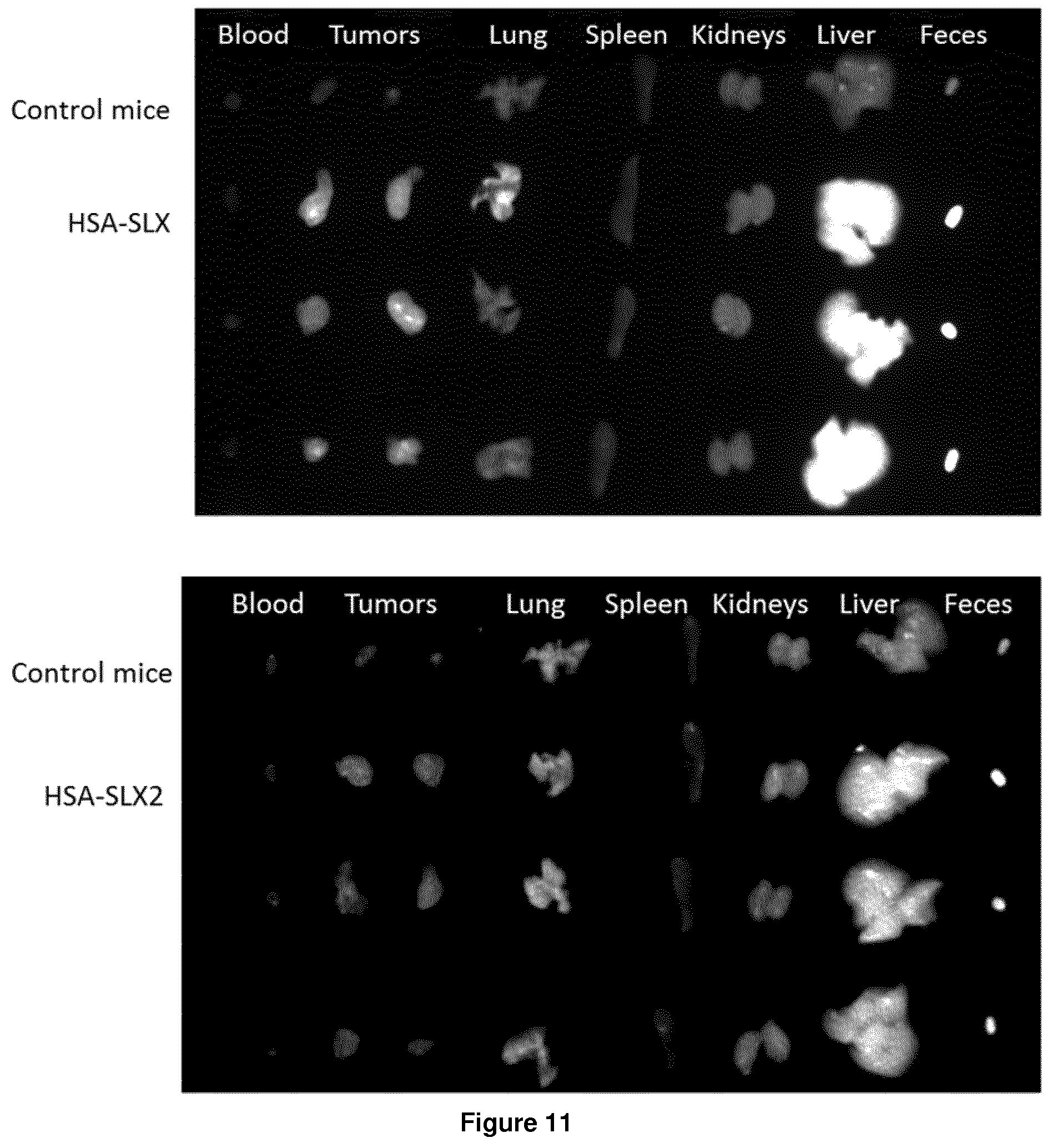
[0217] FIG. 11: Ex vivo imaging of extracted organs 24 h after injection of HSA grafted with the tetrasaccharide Sialyl Lewix X (HSA-SLX) or HSA grafted with the trisaccharide analog of the Sialyl Lewis X of the invention described above (HSA-SLX2).
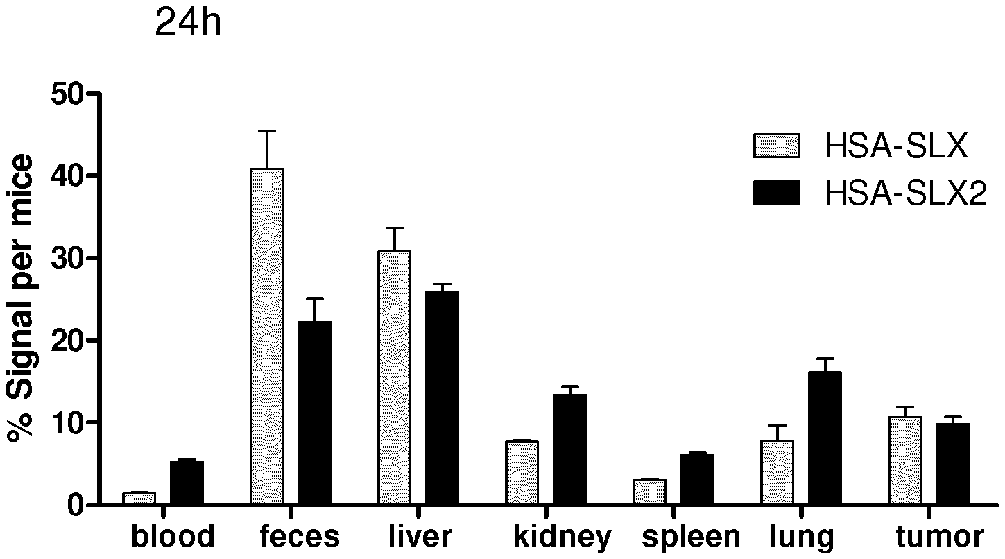
[0218] FIG. 12: Quantification of the ex vivo images with M3vision software given as the percentage of fluorescence per mice in the organs for HSA grafted with the tetrasaccharide Sialyl Lewis X (HSA-SLX) or HSA grafted with the trisaccharide analog of the sialyl Lewis X of the invention (HSA-SLX2).
EXAMPLES
[0219] The following examples are given for illustrative purpose only, and shall not be construed as limiting in any way.
[0220] In the following PBS stands for Phosphate Buffered Saline solution (as known in the art), and EDTA stands for ethylenediaminetetraacetic acid.
Example 1
Synthesis of an Optical Imaging Agent According to the Invention Using a Derivative of SLe.sup.X as BIOVECTOR (Compound HSA-Slx)
[0221] Albumin (2 mg) is coupled to N-N-disulfonate-cyanin-5-N-hydroxysuccinimide:
##STR00037##
available from luminoprobe (2 equivalents per amine) in PBS and left for 30 minutes under gentle stirring. The mixture is washed with an ultrafiltration device with a 50 kDa cut off at 2000 rpm during 30 minutes at 4.degree. C. Sialyl lewis-X-maleimide is then coupled to the labelled albumin in PBS/EDTA at room temperature during 30 minutes in presence of 2-iminothiolane hydrochloride (10 equivalent per amine). A washing step is performed to remove the excess of iminothiolane and obtain the labelled targeting albumin HSA-Slx in NaCl 0.9%. The full functionalisation of albumin was confirmed by mass spectrometry with a mass of 90782 for the imaging agent (FIG. 1).
[0222] Sialyl lewis-X-maleimide is obtained as follows. To Sialyl Lewis X (described in Lu et al. Carbohydrate research 383(2014) 89-96) is added of N-Succinimidyl 3-maleimidopropionate (hereafter NHS-maleimide):
##STR00038##
and triethylamine at room temperature, the reaction mixture is stirred for 2 hours at room temperature. The reaction mixture is then evaporated and the crude product is purified using column chromatography on silica gel.
Example 2
Synthesis of an Optical Imaging Agent According to the Invention Using a Derivative of Mannose as BIOVECTOR (Compound HSA-Mannose)
Step 1:
[0223] Albumin (20 mg) is coupled to N-N-disulfonate-cyanin-5-N-hydroxysuccinimide (0.1 mg) in 1 mL PBS and left for 30 minutes under gentle stirring. The mixture is washed with an ultrafiltration device with a 50 kDa cut off at 2000 rpm during 30 minutes at 4.degree. C. The intermediate compound is the cyanine-labelled albumin, which does not contain any targeting portion (no BIOVECTOR). This intermediate cyanine-labelled albumin is referred to hereinafter as HSA-CY5.
Step 2:
[0224] Mannose-maleimide is then coupled to the labelled albumin HSA--CY5 in PBS/EDTA at room temperature during 30 minutes in presence of 2-iminothiolane hydrochloride (10 equivalent per amine). A washing step is performed to remove the excess of iminothiolane and obtain the labelled targeting albumin in NaCl 0.9%. The expected mass of the imaging agent was confirmed by mass spectrometry (M 79636) (FIG. 2). The reaction of 2 equivalents of cyanin gave the highest intensity of fluorescence as referred to 20 equivalents (FIG. 3).
Mannose-maleimide
##STR00039##
[0225] is obtained as follows. 2-aminoethylmannopyranoside
##STR00040##
is dissolved in DMF. NHS-maleimide (491 mg, 2.2 mmol) is added, and the reaction mixture is stirred overnight. The reaction mixture is then concentrated in vacuo, and the crude product is purified using column chromatography on silica gel (eluent:dichloromethane/MeOH:8/2). 339 mg of purified Mannose-maleimide is obtained.
Example 3
In Vitro Binding Affinity of the Optical Imaging Agent of Example 1
[0226] The in vitro experiments were performed with bEnd.3 mus musculus brain endothelial cells (CRL-2299.TM.) and CT26.WT mus musculus colon carcinoma cells (CRL-2638.TM.) provided by ATCC.RTM.. Those cells were cultured in DMEM-Dulbecco's Modified Eagle Medium (10566-016, Gibco Thermo Fisher) added by 10% of fetal bovine serum (10500056, Gibco Thermo Fisher) and 1% of Penicillin-Streptomycin (5,000 U/mL, 15070063, Gibco Thermo Fisher) antibiotics. A suspension of Lipopolysaccharide stimulated cells (0.1 mg/mL, 4 h, 37.degree. C.), were collected and prepared in binding buffer (50 mM Tris HCl, 150 mM NaCl, 1 mM CaCl.sub.2, 1 mM MgCl.sub.2, 1 mM MnCl2, 1% BSA in water) at 500 000 cell per milliliter. An incubation of 1 h at 4.degree. C. was realized in order to saturate unspecific binding. For evaluation of E-selectin expression (FIG. 4A) cells were incubated successively with purified rat anti-Mouse CD62E antibody (1 .mu.g/mL, 550290 BD Pharmingen.TM.) in PBS/BSA 1% and with goat anti-rat IgG-Alexa Fluor.RTM.488 (1 .mu.g/ml, ab150157 Abcam) for 30 min a 4.degree. C. To study cellular binding and internalization, cells were incubated with 30 ng of HSA or HSA-SLX for 20min at 4.degree. C. (FIG. 4B-C) or 37.degree. C. (FIG. 4D).
[0227] After those different steps, cells were washed twice with binding buffer and analyzed by flow cytometry (Guava.RTM. easyCyte Millipore).
[0228] We can see that both tumor cells and endothelial cells express E-selectin. The imaging agent bind to both type of cells and is internalized mostly by endothelial cells which express E-selectin at a higher level. The non-functionalized albumin (control) neither bind, nor is internalized by endothelial cells or tumor cells.
Example 4
Demonstration of the Efficacy of the Optical Imaging Agent of Example 1 as an Aid to Surgery
[0229] BalbC female mice (from janvier labs) were implanted with 3 mm.sup.2 of CT26 tumour fragment in subcutaneous way. Fifteen day after implantation, mice were anesthetized with a mixture of ketamine (80 mg/kg, Clorketam.RTM. 1 000 Vetoquinol) and xylazine (10 mg/kg, ROMPUN.RTM. 2% Bayer), 200 .mu.l of HSA-SLX-Cy5 (2.7 mg/ml) suspension was then injected into the tail vein. Twenty four hour after injection, an evaluation of image guided surgery (FIG. 5) was realized under mice anaesthesia with Fluobeam.RTM. open Imaging system for in vivo near infrared fluorescence imaging (.lamda. ex 690, .lamda. em>700 nm).lamda..
Example 5
In Vivo Liver Accumulation of the Optical Imaging Agent of Example 1
[0230] Female balbc mice (from janvier labs) were anesthetized with ketamine/xylazine mixture, 5 mice per condition were injected with HSA-CY5 and HSA-Slx suspension (2.7 mg/ml) in the tail vein of Balbc mice 14 days after CT26 tumour implantation. The biodistribution kinetic of formulation was recorded by Fluobeam.RTM. system as function of time (FIG. 6). Twenty four hours after HSA-CY5 and HSA-SLX-CY5 injection, ex vivo quantification of the signal accumulated in the tumour was evaluated after blood elimination by mice PBS perfusion. Those results (FIG. 7) were imaged and quantified by Photon IMAGER.TM. optima (Biospace lab) and finally expressed in photon per second per steradian (ph/s/sr). Of note, HSA-CY5, which does not contain any targeting portion (BIOVECTOR) is used as a control.
Example 6
In Vivo Paw Accumulation of the Optical Imaging Agent in Collagen-Induced Arthritis Model of Example 1
[0231] Collagen Arthritis model was induced in DBA/1 mice (from janvier labs) by injection with bovine type II collagen emulsified in Complete Freund's Adjuvant in the posterior paw articulation. Twenty four hours after arthritis induction, mice were anesthetized and injected with HSA-SLX-CY5, suspension (2.7 mg/ml) in the tail vein. In vivo kinetic of the formulation was recorded by Fluobeam.RTM. system at different time points (FIG. 8A). Twenty for hours after probe injection colocalization signal between probes and arthritis articulation could be evaluated by PhotonlMAGER.TM. optima (Biospace lab) after luminol injection (FIG. 8B).
Example 7
Biodistribution and Ex Vivo Quantification of the Fluorescence as Regard to the Agent without Bio Vector in Healthy Mice
[0232] Female balbc mice (from janvier labs) were anesthetized with ketamine/xylazine mixture, mice were injected with HSA-CY5, HSA-Lactose (HSA-L-CY5, obtained as described in example 1, but substituting lactose for mannose) or HSA-Mannose (HSA-M-CY5) suspension (2.7 mg/ml) in the tail vein of Balbc healthy mice. In vivo liver kinetic of each formulation was recorded and quantified by the use of PhotoniMAGER.TM. optima (Biospace lab). The percent of the fluorescence detected in the liver was calculated as function of the signal of the entire mice (FIG. 9).
Example 8
Oligomers and Aggregates Distributions of Native Albumin, Cyanine Grafted Albumin and Optical Imaging Agents According to the Invention HSA-SLX and HSA-SLX2
[0233] Size exclusion chromatography/UV was performed to evaluate the distribution of monomers, dimers, trimers and oligomers of the optical imaging agents according to the present invention.
Experimental:
[0234] Separation and analysis were performed on a LC-10 liquid chromatography system from Shimadzu (Kyoto, Japan) natively equipped with a vacuum degasser, an auto-sampler, a UV absorbance detector and refractive index detector (RID). The size exclusion chromatography stationary phase was a Shodex Protein LW 803 column provided by Showa Denko (Japan). Between the auto-sampler and HPLC pump, mobile phase was filtered in-line by a 0.1 .mu.m durapore PVDF membrane in a PEEK in-line filter. Detection was realized using UV absorbance at a wavelength of 280 nm. Concomitantly, refractive index quantifications were performed using white light. Mobile phase was CH3COONH4 50 mM NaN3 0.03% in milli-Q water degassed by vacuum pump and filtered on stericup filter units. MALS detection was achieved on a three angle mini-Dawn Treos II from Wyatt Technology equipped with a 658 nm wavelength laser. Data analysis was achieved on Astra 7.1, Wyatt Technology.
[0235] HSA-SLX corresponds to the optical imaging agent described in example 1.
[0236] HSA-SLX2 corresponds to the optical imaging agent described in example 1, except that the SLX moiety has been replaced by the trisaccharide of the invention with the formula below and whose synthesis is described in the above Process of preparation section:
##STR00041##
wherein Rs is SO.sub.3Na.
Results are Illustrated on FIG. 10.
[0237] The optical imaging agents according to the present invention, HSA-SLX and HSA SLX2, exist mainly in the form of monomers and form very few oligomers. Optical imaging agents according to the invention therefore barely form aggregates.
Example 9
Biodistribution of the Probe by Optical Imaging
[0238] Female BALB/cJRj 8 weeks old mice (Janvier labs, Le Genest-Saint-Isle, France) were anesthetized with a mixture of 100 mg kg-1 of ketamine (IMALGENE 1000 Boehringer Ingelheim, France) and 10 mg kg-1 of xylazine (Rompun.TM., Bayer, France) and injected by an intravenous route with 200 .mu.l of HSA-SLX or HSA-SLX2 solutions, with both concentrations at 1 mg mL-1. Twenty four hours after the injection, the mice were sacrificed, their PBS perfused organs were placed under the camera to acquire the fluorescence signal associated with the organs (5 s, .lamda.ex 640 nm, .lamda.em 780 nm). (FIG. 11)
[0239] The images were processed using the M3vision software. The result is expressed as percent of the fluorescence found per mice (FIG. 12).
HSA SLX and HSA SLX 2 are as Described in Example 8.
[0240] Sialyl Lewis X is known in the art as the best agent to target selectin E, due to its high affinity for its target. The trisaccharide analog SLX2 of the invention appears to have the same affinity for the target than SLX and is easier to synthesize. Moreover, as shown here, grafting this analog to albumin confers similar property to the in vivo imaging agent, as a similar signal of fluorescence is observed with either HSA grafted with SLX or SLX2.
* * * * *


D00000
D00001

D00002

D00003

D00004

D00005

D00006

D00007

D00008
D00009

P00001

XML
uspto.report is an independent third-party trademark research tool that is not affiliated, endorsed, or sponsored by the United States Patent and Trademark Office (USPTO) or any other governmental organization. The information provided by uspto.report is based on publicly available data at the time of writing and is intended for informational purposes only.
While we strive to provide accurate and up-to-date information, we do not guarantee the accuracy, completeness, reliability, or suitability of the information displayed on this site. The use of this site is at your own risk. Any reliance you place on such information is therefore strictly at your own risk.
All official trademark data, including owner information, should be verified by visiting the official USPTO website at www.uspto.gov. This site is not intended to replace professional legal advice and should not be used as a substitute for consulting with a legal professional who is knowledgeable about trademark law.