Compositions And Methods For The Depletion Of Cd2+ Cells
BOITANO; Anthony ; et al.
U.S. patent application number 16/768036 was filed with the patent office on 2020-11-26 for compositions and methods for the depletion of cd2+ cells. This patent application is currently assigned to MAGENTA THEERAPEUTICS, INC.. The applicant listed for this patent is MAGENTA THEERAPEUTICS, INC.. Invention is credited to Anthony BOITANO, Michael COOKE, Sean MCDONOUGC, Rahul PALCHAUDHURI.
| Application Number | 20200368363 16/768036 |
| Document ID | / |
| Family ID | 1000005038541 |
| Filed Date | 2020-11-26 |


View All Diagrams
| United States Patent Application | 20200368363 |
| Kind Code | A1 |
| BOITANO; Anthony ; et al. | November 26, 2020 |
COMPOSITIONS AND METHODS FOR THE DEPLETION OF CD2+ CELLS
Abstract
The invention provides anti-CD2 antibodies, antigen-binding fragments thereof, and antibody-drug conjugates thereof, for use as agents to treat a stem cell disorder, cancer, or autoimmune disease, among other hematological and proliferative diseases. The compositions and methods described herein can be used to deplete populations of CD2+ cells, such as CD2+ cancer cells and CD2+ immune cells, and can be used to prepare a patient for hematopoietic stem cell transplantation.
| Inventors: | BOITANO; Anthony; (Newton, MA) ; COOKE; Michael; (Brookline, MA) ; PALCHAUDHURI; Rahul; (Somerville, MA) ; MCDONOUGC; Sean; (Littlton, MA) | ||||||||||
| Applicant: |
|
||||||||||
|---|---|---|---|---|---|---|---|---|---|---|---|
| Assignee: | MAGENTA THEERAPEUTICS, INC. Cambridge MA |
||||||||||
| Family ID: | 1000005038541 | ||||||||||
| Appl. No.: | 16/768036 | ||||||||||
| Filed: | November 29, 2018 | ||||||||||
| PCT Filed: | November 29, 2018 | ||||||||||
| PCT NO: | PCT/US2018/063171 | ||||||||||
| 371 Date: | May 28, 2020 |
Related U.S. Patent Documents
| Application Number | Filing Date | Patent Number | ||
|---|---|---|---|---|
| 62592169 | Nov 29, 2017 | |||
| Current U.S. Class: | 1/1 |
| Current CPC Class: | C07K 2317/73 20130101; A61K 47/6831 20170801; A61K 2039/505 20130101; C07K 2317/92 20130101; A61K 47/6849 20170801; A61P 37/06 20180101; C07K 16/2806 20130101 |
| International Class: | A61K 47/68 20060101 A61K047/68; C07K 16/28 20060101 C07K016/28; A61P 37/06 20060101 A61P037/06 |
Claims
1. A method of depleting a population of CD2+ cells in a human patient, the method comprising administering to the patient an effective amount of an anti-CD2 antibody, or antigen-binding fragment thereof, wherein the antibody, or antigen-binding fragment thereof, is conjugated to a cytotoxin.
2. A method of depleting a population of CD2+ cells in a human patient in need of a hematopoietic stem cell transplant, the method comprising administering to the patient an effective amount of an anti-CD2 antibody, or antigen-binding fragment thereof, wherein the antibody, or antigen-binding fragment thereof, is conjugated to a cytotoxin.
3. A method of preventing rejection of a hematopoietic stem cell graft in a human patient in need of a hematopoietic stem cell transplant, the method comprising administering to the patient an effective amount of an anti-CD2 antibody, or antigen-binding fragment thereof, prior to the human patient receiving a transplant comprising hematopoietic stem cells, wherein the antibody, or antigen-binding fragment thereof, is conjugated to a cytotoxin.
4. A method of depleting a population of CD2+ cells in a human patient in need of a hematopoietic stem cell transplant, the method comprising administering to the patient an effective amount of an anti-CD2 antibody, or antigen-binding fragment thereof, prior to the patient receiving a transplant comprising hematopoietic stem cells, wherein the antibody, or antigen-binding fragment thereof, is conjugated to a cytotoxin.
5. A method comprising administering to a human patient a transplant comprising hematopoietic stem cells, wherein the patient has been previously administered an anti-CD2 antibody or antigen-binding fragment thereof, in an amount sufficient to deplete a population of CD2+ cells in the patient, wherein the antibody, or antigen-binding fragment thereof, is conjugated to a cytotoxin.
6. A method comprising: i) administering to a human patient an antibody, or antigen-binding fragment thereof, that binds to CD2 in an amount sufficient to deplete a population of CD2+ cells in the patient, wherein the antibody, or antigen-binding fragment thereof, is conjugated to a cytotoxin; and ii) subsequently administering to the patient a transplant comprising hematopoietic stem cells.
7. The method of any one of claims 1-6, wherein the antibody or antigen-binding fragment thereof is produced by the hybridoma cell line ATCC HB 11423.
8. The method of any one of claims 1-6, wherein the antibody or antigen-binding fragment thereof comprises a heavy chain variable region CDR set (CDR1, CDR2, and CDR3) and a light chain variable region CDR set (CDR1, CDR2, and CDR3) of antibody LO-CD2A produced by the hybridoma cell line having ATCC accession number HB 11423.
9. The method of any one of claims 1-6, wherein the antibody, or antigen-binding fragment thereof, is i) an anti-CD2 antibody, or antigen binding portion thereof, comprising a heavy chain variable region comprising a CDR-H1 as set forth in SEQ ID NO: 1; a CDR-H2 as set forth in SEQ ID NO: 2; a CDR-H3 as set forth in SEQ ID NO: 3; and comprising a light chain variable region comprising a CDR-L1 as set forth in SEQ ID NO: 4; a CDR-L2 as set forth in SEQ ID NO: 5; and a CDR-L3 as set forth in SEQ ID NO: 6; ii) an anti-CD2 antibody, or antigen binding portion thereof, comprising a heavy chain variable region comprising a CDR-H1 as set forth in SEQ ID NO: 14; a CDR-H2 as set forth in SEQ ID NO: 15; a CDR-H3 as set forth in SEQ ID NO: 16 or 17; and comprising a light chain variable region comprising a CDR-L1 as set forth in SEQ ID NO: 18; a CDR-L2 as set forth in SEQ ID NO: 19; and a CDR-L3 as set forth in SEQ ID NO: 20; iii) an anti-CD2 antibody, or antigen binding portion thereof, comprising a heavy chain variable region as set forth in SEQ ID NO: 7 and comprising a light chain variable region as set forth in SEQ ID NO: 8; iv) an anti-CD2 antibody, or antigen binding portion thereof, comprising a heavy chain variable region as set forth in SEQ ID NO: 9 and comprising a light chain variable region as set forth in SEQ ID NO: 10; or v) an anti-CD2 antibody, or antigen binding portion thereof, comprising a heavy chain variable region as set forth in SEQ ID NO: 21 or 22 and comprising a light chain variable region as set forth in SEQ ID NO: 23.
10. The method of any one of claims 1-6, wherein the antibody, or antigen-binding fragment thereof, competitively inhibits the binding of CD2 to an antibody or antigen-binding fragment thereof of claim 9.
11. The method of any one of claims 1-6, wherein the antibody or antigen-binding fragment thereof is selected from the group consisting of a monoclonal antibody or antigen-binding fragment thereof, a polyclonal antibody or antigen-binding fragment thereof, a humanized antibody or antigen-binding fragment thereof, a bispecific antibody or antigen-binding fragment thereof, an intact antibody, a dual-variable immunoglobulin domain, a single-chain Fv molecule (scFv), a diabody, a triabody, a nanobody, an antibody-like protein scaffold, a Fv fragment, a Fab fragment, a F(ab').sub.2 molecule, and a tandem di-scFv.
12. The method of any one of claims 1-10, wherein the antibody or antigen-binding fragment thereof is a humanized antibody, or antigen-binding fragment thereof.
13. The method of any one of claims 1-12, wherein the antibody has an isotype selected from the group consisting of IgG, IgA, IgM, IgD, and IgE.
14. The method of claim 13, wherein the IgG isotype is an IgG1 or an IgG4.
15. The method of any one of claims 1 to 14, wherein the cytotoxin is selected from the group consisting of pseudomonas exotoxin A, deBouganin, diphtheria toxin, an amatoxin, saporin, maytansine, a maytansinoid, an auristatin, an anthracycline, a calicheamicin, irinotecan, SN-38, a duocarmycin, a pyrrolobenzodiazepine, a pyrrolobenzodiazepine dimer, an indolinobenzodiazepine, and an indolinobenzodiazepine dimer, or a variant thereof.
16. The method of any one of claims 1 to 14, wherein the cytotoxin is an RNA polymerase inhibitor.
17. The method of claim 16, wherein the RNA polymerase inhibitor is an RNA polymerase II inhibitor.
18. The method of claim 17, wherein the RNA polymerase II inhibitor is amatoxin.
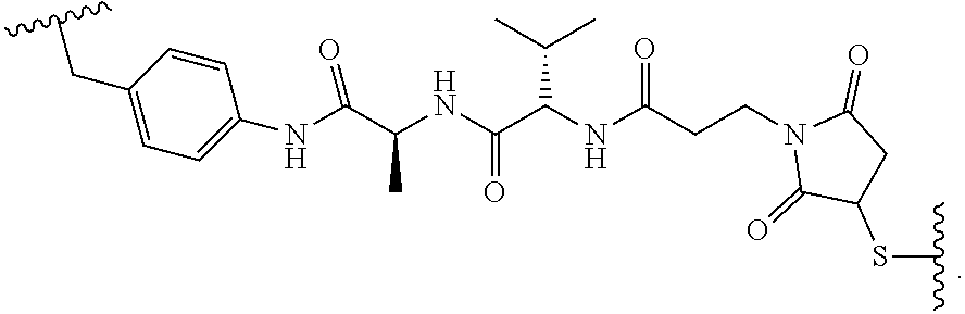
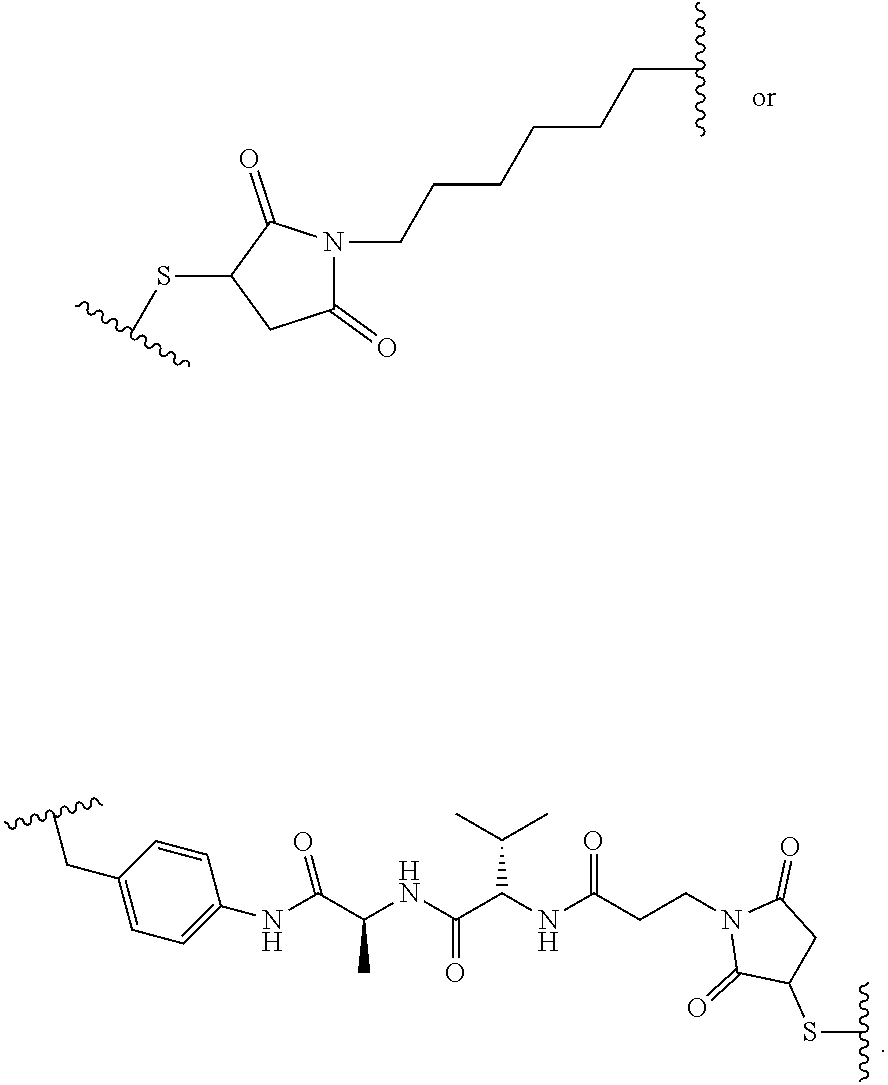
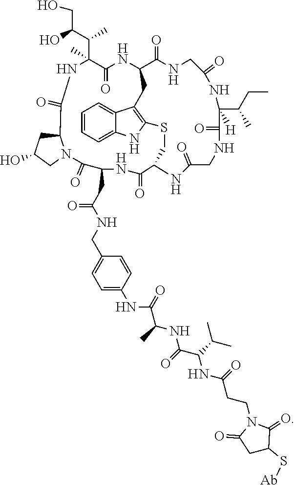
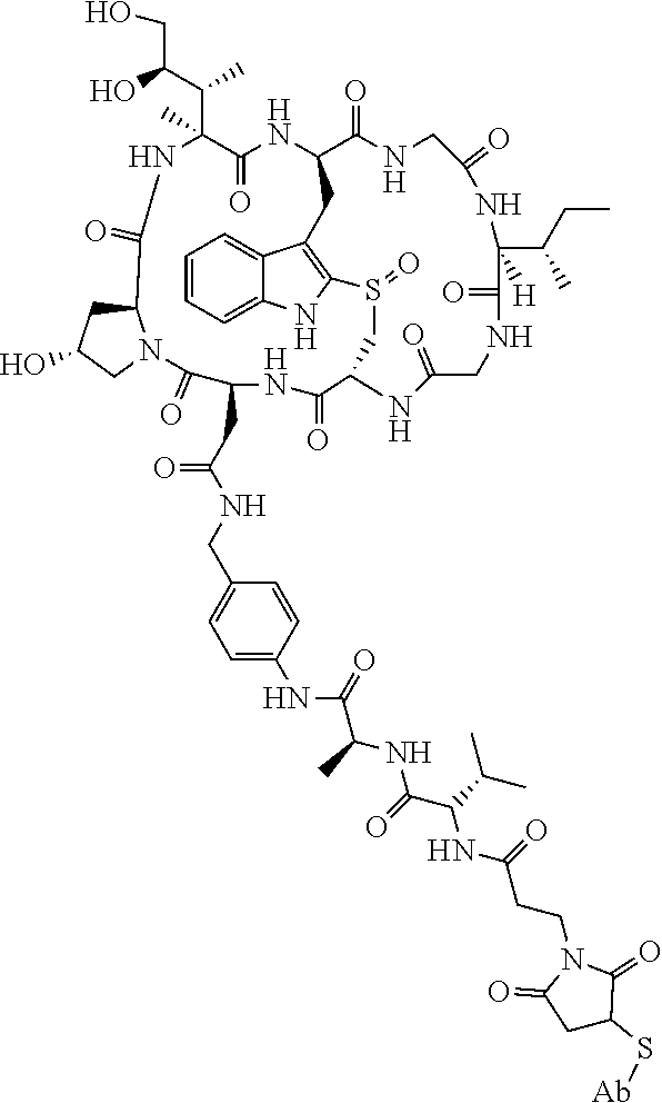
19. The method of any one of claims 1 to 14, wherein the antibody or antigen-binding fragment thereof conjugated to a cytotoxin is represented by the formula Ab-Z-L-Am, wherein Ab is the antibody or antigen-binding fragment thereof, L is a linker, Z is a chemical moiety, and Am an amatoxin represented by formula (I) ##STR00102## wherein R.sub.1 is H, OH, OR.sub.A, or OR.sub.C; R.sub.2 is H, OH, OR.sub.B, or OR.sub.C; R.sub.A and R.sub.B, when present, together with the oxygen atoms to which they are bound, combine to form an optionally substituted 5-membered heterocyclolalkyl group; R.sub.3 is H, R.sub.C, or R.sub.D; R.sub.4, R.sub.5, R.sub.6 and R.sub.7 are each independently H, OH, OR.sub.C, OR.sub.D, R.sub.C, or R.sub.D; R.sub.8 is OH, NH.sub.2, OR.sub.C, OR.sub.D, NHR.sub.C, or NR.sub.CR.sub.D; R.sub.9 is H, OH, OR.sub.C, or OR.sub.D; X is --S--, --S(O)--, or --SO.sub.2--; R.sub.C is -L-Z; R.sub.D is optionally substituted C.sub.1-C.sub.6 alkyl, optionally substituted C.sub.1-C.sub.6 heteroalkyl, optionally substituted C.sub.2-C.sub.6 alkenyl, optionally substituted C.sub.2-C.sub.6 heteroalkenyl, optionally substituted C.sub.2-C.sub.6 alkynyl, optionally substituted C.sub.2-C.sub.6 heteroalkynyl, optionally substituted cycloalkyl, optionally substituted heterocycloalkyl, optionally substituted aryl, or optionally substituted heteroaryl; L is optionally substituted C.sub.1-C.sub.6 alkylene, optionally substituted C.sub.1-C.sub.6 heteroalkylene, optionally substituted C.sub.2-C.sub.6 alkenylene, optionally substituted C.sub.2-C.sub.6 heteroalkenylene, optionally substituted C.sub.2-C.sub.6 alkynylene, optionally substituted C.sub.2-C.sub.6 heteroalkynylene, optionally substituted cycloalkylene, optionally substituted heterocycloalkylene, optionally substituted arylene, optionally substituted heteroarylene, a dipeptide, --C(.dbd.O)--, a peptide. or a combination thereof; and Z is a chemical moiety formed from a coupling reaction between a reactive substituent present on L and a reactive substituent present within the antibody or antigen-binding fragment thereof, wherein Am comprises exactly one R.sub.C substituent.
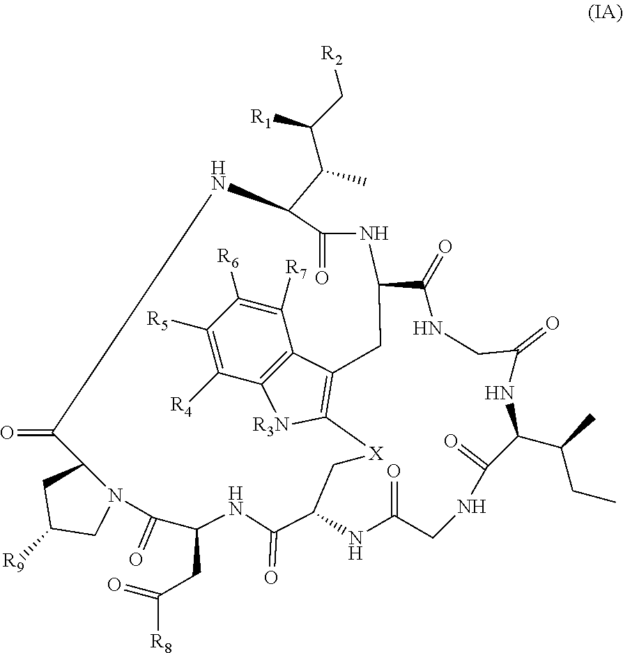
20. The method of claim 19, wherein Am-L-Z is represented by formula (IA). ##STR00103## wherein R.sub.1 is H, OH, OR.sub.A, or OR.sub.C; R.sub.2 is H, OH, OR.sub.B, or OR.sub.C; R.sub.A and R.sub.B, when present, together with the oxygen atoms to which they are bound, combine to form an optionally substituted 5-membered heterocycloalkyl group; R.sub.3 is H, R.sub.C, or R.sub.D; R.sub.4, R.sub.5, R.sub.6 and R.sub.7 are each independently H, OH, OR.sub.C, OR.sub.D, R.sub.C, or R.sub.D; R.sub.8 is OH, NH.sub.2, OR.sub.C, OR.sub.D, NHR.sub.C, or NR.sub.CR.sub.D; R.sub.9 is H, OH, OR.sub.C, or OR.sub.D; X is --S--, --S(O)--, or --SO.sub.2--; R.sub.C is -L-Z; R.sub.D is optionally substituted C.sub.1-C.sub.6 alkyl, optionally substituted C.sub.1-C.sub.6 heteroalkyl, optionally substituted C.sub.2-C.sub.6 alkenyl, optionally substituted C.sub.2-C.sub.6 heteroalkenyl, optionally substituted C.sub.2-C.sub.6 alkynyl, optionally substituted C.sub.2-C.sub.6 heteroalkynyl, optionally substituted cycloalkyl, optionally substituted heterocycloalkyl, optionally substituted aryl, or optionally substituted heteroaryl; L is optionally substituted C.sub.1-C.sub.6 alkylene, optionally substituted C.sub.1-C.sub.6 heteroalkylene, optionally substituted C.sub.2-C.sub.6 alkenylene, optionally substituted C.sub.2-C.sub.6 heteroalkenylene, optionally substituted C.sub.2-C.sub.6 alkynylene, optionally substituted C.sub.2-C.sub.6 heteroalkynylene, optionally substituted cycloalkylene, optionally substituted heterocycloalkylene, optionally substituted arylene, optionally substituted heteroarylene, a dipeptide, --C(.dbd.O)--, a peptide, or a combination thereof; Z is a chemical moiety formed from a coupling reaction between a reactive substituent present on L and a reactive substituent present within the antibody or antigen-binding fragment thereof; and wherein Am comprises exactly one R.sub.C substituent.
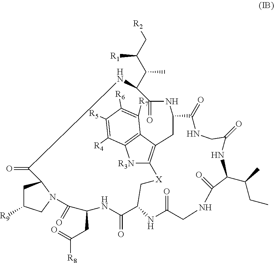
21. The method of claim 19, wherein Am-L-Z is represented by formula (IB). ##STR00104## wherein R.sub.1 is H, OH, OR.sub.A, or OR.sub.C; R.sub.2 is H, OH, OR.sub.B, or OR.sub.C; R.sub.A and R.sub.B, when present, together with the oxygen atoms to which they are bound, combine to form an optionally substituted 5-membered heterocycloalkyl group; R.sub.3 is H, R.sub.C, or R.sub.D; R.sub.4, R.sub.5, R.sub.6 and R.sub.7 are each independently H, OH, OR.sub.C, OR.sub.D, R.sub.C, or R.sub.D; R.sub.8 is OH, NH.sub.2, OR.sub.C, OR.sub.D, NHR.sub.C, or NR.sub.CR.sub.D; R.sub.9 is H, OH, OR.sub.C, or OR.sub.D; X is --S--, --S(O)--, or --SO.sub.2--; R.sub.C is -L-Z; R.sub.D is optionally substituted C.sub.1-C.sub.6 alkyl, optionally substituted C.sub.1-C.sub.6 heteroalkyl, optionally substituted C.sub.2-C.sub.6 alkenyl, optionally substituted C.sub.2-C.sub.6 heteroalkenyl, optionally substituted C.sub.2-C.sub.6 alkynyl, optionally substituted C.sub.2-C.sub.6 heteroalkynyl, optionally substituted cycloalkyl, optionally substituted heterocycloalkyl, optionally substituted aryl, or optionally substituted heteroaryl; L is optionally substituted C.sub.1-C.sub.6 alkylene, optionally substituted C.sub.1-C.sub.6 heteroalkylene, optionally substituted C.sub.2-C.sub.6 alkenylene, optionally substituted C.sub.2-C.sub.6 heteroalkenylene, optionally substituted C.sub.2-C.sub.6 alkynylene, optionally substituted C.sub.2-C.sub.6 heteroalkynylene, optionally substituted cycloalkylene, optionally substituted heterocycloalkylene, optionally substituted arylene, optionally substituted heteroarylene, a dipeptide, --C(.dbd.O)--, a peptide, or a combination thereof; and Z is a chemical moiety formed from a coupling reaction between a reactive substituent present on L and a reactive substituent present within the antibody or antigen-binding fragment thereof, wherein Am comprises exactly one R.sub.C substituent.
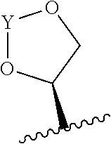
22. The method of claim 20 or 21, wherein R.sub.A and R.sub.B, together with the oxygen atoms to which they are bound, combine to form a 5 membered heterocycloalkyl group of formula: ##STR00105## wherein Y is --C(.dbd.O)--, --C(.dbd.S)--, --C(.dbd.NR.sub.E)--, or --C(R.sub.ER.sub.E')--; and R.sub.E and R.sub.E' are each independently optionally substituted C.sub.1-C.sub.6 alkylene-R.sub.C, optionally substituted C.sub.1-C.sub.6 heteroalkylene-R.sub.C, optionally substituted C.sub.2-C.sub.6 alkenylene-R.sub.C, optionally substituted C.sub.2-C.sub.6 heteroalkenylene-R.sub.C, optionally substituted C.sub.2-C.sub.6 alkynylene-R.sub.C, optionally substituted C.sub.2-C.sub.6 heteroalkynylene-R.sub.C, optionally substituted cycloalkylene-R.sub.C, optionally substituted heterocycloalkylene-R.sub.C, optionally substituted arylene-R.sub.C, or optionally substituted heteroarylene-R.sub.C.
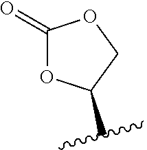
23. The method of claim 22, wherein R.sub.A and R.sub.B, together with the oxygen atoms to which they are bound, combine to form: ##STR00106##
24. The method of claim 20 or 21, wherein R.sub.1 is H, OH, or OR.sub.A; R.sub.2 is H, OH, or OR.sub.B; R.sub.A and R.sub.B, together with the oxygen atoms to which they are bound, combine to form: ##STR00107## R.sub.3, R.sub.4, R.sub.6, and R.sub.7 are each H; R.sub.5 is OR.sub.C; R.sub.8 is OH or NH.sub.2; and R.sub.9 is H or OH.
25. The method of claim 20 or 21, wherein R.sub.1 and R.sub.2 are each independently H or OH; R.sub.3 is R.sub.C; R.sub.4, R.sub.6, and R.sub.7 are each H; R.sub.5 is H, OH, or OC.sub.1-C.sub.6 alkyl; R.sub.8 is OH or NH.sub.2; and R.sub.9 is H or OH.
26. The method of claim 20 or 21, wherein R.sub.1 and R.sub.2 are each independently H or OH; R.sub.3, R.sub.6, and R.sub.7 are each H; R.sub.4 and R.sub.5 are each independently H, OH, OR.sub.C, or R.sub.C; R.sub.8 is OH or NH.sub.2; and R.sub.9 is H or OH.
27. The method of claim 20 or 21, wherein R.sub.1 and R.sub.2 are each independently H or OH; R.sub.3, R.sub.6, and R.sub.7 are each H; R.sub.4 and R.sub.5 are each independently H or OH; R.sub.8 is OR.sub.C or NHR.sub.C; and R.sub.9 is H or OH.
28. The method of any one of claims 1 to 14, wherein the antibody or antigen-binding fragment thereof conjugated to a cytotoxin is represented by the formula Ab-Z-L-Am, wherein Ab is the antibody or antigen-binding fragment thereof, Z is a chemical moiety, L is a linker, and Am is an amatoxin, and the amatoxin-linker conjugate Am-L-Z is represented by formula (II), formula (IIA), or formula (IIB) ##STR00108## wherein X is S, SO, or SO.sub.2; R.sub.1 is H or a linker covalently bound to the antibody or antigen-binding fragment thereof through a chemical moeity Z, formed from a coupling reaction between a reactive substituent present on the linker and a reactive substituent present within an antibody, or antigen-binding fragment thereof; and R.sub.2 is H or a linker covalently bound to the antibody or antigen-binding fragment thereof through a chemical moeity Z, formed from a coupling reaction between a reactive substituent present on the linker and a reactive substituent present within an antibody, or antigen-binding fragment thereof; wherein when R.sub.1 is H, R.sub.2 is the linker, and when R.sub.2 is H, R.sub.1 is the linker.
29. The method of any one of claims 1 to 14, wherein the cytotoxin is a maytansinoid selected from the group consisting of DM1 and DM4.
30. The method of any one of claims 1 to 14, wherein the cytotoxin is an auristatin selected from the group consisting of monomethyl auristatin E and monomethyl auristatin F.
31. The method of any one of claims 1 to 14, wherein the cytotoxin is an anthracycline selected from the group consisting of daunorubicin, doxorubicin, epirubicin, and idarubicin.
32. The method of any one of claims 1 to 14, wherein the cytotoxin is a pyrrolobenzodiazepine dimer derivative represented by formula (IV) ##STR00109##
33. The method of any one of claims 1-32, wherein the antibody, or antigen-binding fragment thereof, conjugated to the cytotoxin is internalized by an immune cell following administration to the patient.
34. The method of any one of claims 1-33, wherein the antibody, or antigen-binding fragment thereof, conjugated to the cytotoxin is capable of promoting necrosis of an immune cell.
35. The method of any one of claims 1-34, wherein the antibody, or antigen-binding fragment thereof, conjugated to the cytotoxin is capable of recruiting one or more complement proteins to an immune cell upon administration to the patient.
36. The method of any one of claims 33-35, wherein the immune cell is selected from the group consisting of a T cell and NK cell.
37. The method of any one of claims 3-35, wherein the transplant comprising hematopoietic stem cells is administered to the patient after the concentration of the antibody, or antigen-binding fragment thereof, conjugated to the cytotoxin has substantially cleared from the blood of the patient.
38. The method of claim 37, wherein the transplant comprising hematopoietic stem cells is administered to the patient between 1 hour and 7 days after the concentration of the antibody, or antigen-binding fragment thereof, conjugated to the cytotoxin has substantially cleared from the blood of the patient.
39. The method of claim 37, wherein the transplant comprising hematopoietic stem cells is administered to the patient between 6 hours and 3 days after the concentration of the antibody, or antigen-binding fragment thereof, conjugated to the cytotoxin has substantially cleared from the blood of the patient.
40. The method of claim 37, wherein the transplant comprising hematopoietic stem cells is administered to the patient between about 12 hours and about 36 hours after the concentration of the antibody, or antigen-binding fragment thereof, conjugated to the cytotoxin has substantially cleared from the blood of the patient.
41. The method of claim 37, wherein the transplant comprising hematopoietic stem cells is administered to the patient about 24 hours after the concentration of the antibody, or antigen-binding fragment thereof, conjugated to the cytotoxin has substantially cleared from the blood of the patient.
42. The method of any one of claims 3-35, wherein the hematopoietic stem cells or progeny thereof maintain hematopoietic stem cell functional potential after about two or more days following transplantation of the hematopoietic stem cells into the patient.
43. The method of any one of claims 3-42, wherein the hematopoietic stem cells are autologous with respect to the patient.
44. The method of any one of claims 3-42, wherein the hematopoietic stem cells are allogeneic with respect to the patient.
45. The method of claim 44, wherein the hematopoietic stem cells are HLA-matched with respect to the patient.
46. The method of claim 44, wherein the hematopoietic stem cells are HLA-mismatched with respect to the patient.
47. The method of any one of claims 1, 2, and 4-36, wherein the population of CD2+ cells comprises T cells.
48. The method of any one of claims 3-47, wherein the hematopoietic stem cells or progeny thereof are capable of localizing to hematopoietic tissue and/or reestablishing hematopoiesis following transplantation of the hematopoietic stem cells into the patient.
49. The method of any one of claims 3-48, wherein upon transplantation into the patient, the hematopoietic stem cells give rise to recovery of a population of cells selected from the group consisting of megakaryocytes, thrombocytes, platelets, erythrocytes, mast cells, myeoblasts, basophils, neutrophils, eosinophils, microglia, granulocytes, monocytes, osteoclasts, antigen-presenting cells, macrophages, dendritic cells, natural killer cells, T lymphocytes, and B lymphocytes.
50. The method of any one of claims 1-49, wherein the patient is suffering from a stem cell disorder.
51. The method of any one of claims 1-49, wherein the patient is suffering from a hemoglobinopathy disorder.
52. The method of claim 51, wherein the hemoglobinopathy disorder is selected from the group consisting of sickle cell anemia, thalassemia, Fanconi anemia, aplastic anemia, and Wiskott-Aldrich syndrome.
53. The method of claim 51, wherein the hemoglobinopathy disorder is fanconi anemia.
54. The method of claim 51, wherein the hemoglobinopathy disorder is aplastic anemia.
55. The method of claim 51, wherein the hemoglobinopathy disorder is sickle cell anemia.
56. The method of claim 51, wherein the hemoglobinopathy disorder is thalassemia.
57. The method of any one of claims 1-49, wherein the patient is suffering from a myelodysplastic disorder.
58. The method of any one of claims 1-49, wherein the patient is suffering from an immunodeficiency disorder.
59. The method of claim 58, wherein the immunodeficiency disorder is a congenital immunodeficiency.
60. The method of claim 58, wherein the immunodeficiency disorder is an acquired immunodeficiency.
61. The method of claim 60, wherein the acquired immunodeficiency is human immunodeficiency virus or acquired immune deficiency syndrome.
62. The method of any one of claims 1-49, wherein the patient is suffering from a metabolic disorder.
63. The method of claim 62, wherein the metabolic disorder is selected from the group consisting of glycogen storage diseases, mucopolysaccharidoses, Gaucher's Disease, Hurlers Disease, sphingolipidoses, and metachromatic leukodystrophy.
64. The method of any one of claims 1-63, wherein the patient is suffering from cancer.
65. The method of claim 64, wherein the cancer is selected from the group consisting of leukemia, lymphoma, multiple myeloma, and neuroblastoma.
66. The method of claim 64, wherein the cancer is a hematological cancer.
67. The method of claim 64, wherein the cancer is acute myeloid leukemia.
68. The method of claim 64, wherein the cancer is acute lymphoid leukemia.
69. The method of claim 64, wherein the cancer is chronic myeloid leukemia.
70. The method of claim 64, wherein the cancer is chronic lymphoid leukemia.
71. The method of claim 64, wherein the cancer is multiple myeloma.
72. The method of claim 64, wherein the cancer is diffuse large B-cell lymphoma.
73. The method of claim 64, wherein the cancer is non-Hodgkin's lymphoma.
74. The method of any one of claims 1-73, wherein the patient is suffering from a disorder selected from the group consisting of adenosine deaminase deficiency and severe combined immunodeficiency, hyper immunoglobulin M syndrome, Chediak-Higashi disease, hereditary lymphohistiocytosis, osteopetrosis, osteogenesis imperfecta, storage diseases, thalassemia major, systemic sclerosis, systemic lupus erythematosus, multiple sclerosis, and juvenile rheumatoid arthritis.
75. The method of any one of claims 1-74, wherein the patient is suffering from an autoimmune disorder.
76. The method of claim 75, wherein the autoimmune disorder is selected from the group consisting of multiple sclerosis, human systemic lupus, rheumatoid arthritis, inflammatory bowel disease, treating psoriasis, Type 1 diabetes mellitus, acute disseminated encephalomyelitis, Addison's disease, alopecia universalis, ankylosing spondylitisis, antiphospholipid antibody syndrome, aplastic anemia, autoimmune hemolytic anemia, autoimmune hepatitis, autoimmune inner ear disease, autoimmune lymphoproliferative syndrome, autoimmune oophoritis, Balo disease, Behcet's disease, bullous pemphigoid, cardiomyopathy, Chagas' disease, chronic fatigue immune dysfunction syndrome, chronic inflammatory demyelinating polyneuropathy, Crohn's disease, cicatrical pemphigoid, coeliac sprue-dermatitis herpetiformis, cold agglutinin disease, CREST syndrome, Degos disease, discoid lupus, dysautonomia, endometriosis, essential mixed cryoglobulinemia, fibromyalgia-fibromyositis, Goodpasture's syndrome, Grave's disease, Guillain-Barre syndrome, Hashimoto's thyroiditis, Hidradenitis suppurativa, idiopathic and/or acute thrombocytopenic purpura, idiopathic pulmonary fibrosis, IgA neuropathy, interstitial cystitis, juvenile arthritis, Kawasaki's disease, lichen planus, Lyme disease, Meniere disease, mixed connective tissue disease, myasthenia gravis, neuromyotonia, opsoclonus myoclonus syndrome, optic neuritis, Ord's thyroiditis, pemphigus vulgaris, pernicious anemia, polychondritis, polymyositis and dermatomyositis, primary biliary cirrhosis, polyarteritis nodosa, polyglandular syndromes, polymyalgia rheumatica, primary agammaglobulinemia, Raynaud phenomenon, Reiter's syndrome, rheumatic fever, sarcoidosis, scleroderma, Sjogren's syndrome, stiff person syndrome, Takayasu's arteritis, temporal arteritis, ulcerative colitis, uveitis, vasculitis, vitiligo, vulvodynia, and Wegener's granulomatosis.
77. The method of claim 75, wherein the autoimmune disorder is scleroderma.
78. The method of claim 75, wherein the autoimmune disorder is multiple sclerosis.
79. The method of claim 75, wherein the autoimmune disorder is ulcerative colitis.
80. The method of claim 75, wherein the autoimmune disorder is Crohn's disease.
81. The method of claim 75, wherein the autoimmune disorder is Type 1 diabetes.
82. The method of any one of claims 50-81, wherein the method treats the disorder or cancer.
83. A method of treating a stem cell disorder in a human patient, the method comprising administering to the patient a therapeutically effective amount of an antibody, or antigen-binding fragment thereof, that binds to CD2, wherein the antibody, or antigen-binding fragment thereof, is conjugated to a cytotoxin.
84. A method of treating a hemoglobinopathy disorder in a human patient, the method comprising administering to the patient a therapeutically effective amount of an antibody, or antigen-binding fragment thereof, that binds to CD2, wherein the antibody, or antigen-binding fragment thereof, is conjugated to a cytotoxin.
85. The method of claim 84, wherein the hemoglobinopathy disorder is selected from the group consisting of sickle cell anemia, thalassemia, Fanconi anemia, aplastic anemia, and Wiskott-Aldrich syndrome.
86. The method of claim 84, wherein the hemoglobinopathy disorder is Fanconi anemia.
87. The method of claim 84, wherein the hemoglobinopathy disorder is aplastic anemia.
88. The method of claim 84, wherein the hemoglobinopathy disorder is sickle cell anemia.
89. The method of claim 84, wherein the hemoglobinopathy disorder is thalassemia.
90. A method of treating a myelodysplastic disorder in a human patient, the method comprising administering to the patient a therapeutically effective amount of an antibody, or antigen-binding fragment thereof, that binds to CD2, wherein the antibody, or antigen-binding fragment thereof, is conjugated to a cytotoxin.
91. A method of treating an immunodeficiency disorder in a human patient, the method comprising administering to the patient a therapeutically effective amount of an antibody, or antigen-binding fragment thereof, that binds to CD2, wherein the antibody, or antigen-binding fragment thereof, is conjugated to a cytotoxin.
92. The method of claim 91, wherein the immunodeficiency disorder is a congenital immunodeficiency.
93. The method of claim 91, wherein the immunodeficiency disorder is an acquired immunodeficiency.
94. The method of claim 93, wherein the acquired immunodeficiency is human immunodeficiency virus or acquired immune deficiency syndrome.
95. A method of treating a metabolic disorder in a human patient, the method comprising administering to the patient a therapeutically effective amount of an antibody, or antigen-binding fragment thereof, that binds to CD2, wherein the antibody, or antigen-binding fragment thereof, is conjugated to a cytotoxin.
96. The method of claim 95, wherein the metabolic disorder is selected from the group consisting of glycogen storage diseases, mucopolysaccharidoses, Gaucher's Disease, Hurlers Disease, sphingolipidoses, and metachromatic leukodystrophy.
97. A method of treating cancer in a human patient, the method comprising administering to the patient a therapeutically effective amount of an antibody or antigen-binding fragment thereof that binds to CD2, wherein the antibody, or antigen-binding fragment thereof, is conjugated to a cytotoxin.
98. The method of claim 97, wherein the cancer is selected from the group consisting of leukemia, lymphoma, multiple myeloma, and neuroblastoma.
99. The method of claim 97, wherein the cancer is a hematological cancer.
100. The method of claim 97, wherein the cancer is acute myeloid leukemia.
101. The method of claim 97, wherein the cancer is acute lymphoid leukemia.
102. The method of claim 97, wherein the cancer is chronic myeloid leukemia.
103. The method of claim 97, wherein the cancer is chronic lymphoid leukemia.
104. The method of claim 97, wherein the cancer is multiple myeloma.
105. The method of claim 97, wherein the cancer is diffuse large B-cell lymphoma.
106. The method of claim 97, wherein the cancer is non-Hodgkin's lymphoma.
107. A method of treating a disorder selected from the group consisting of adenosine deaminase deficiency and severe combined immunodeficiency, hyper immunoglobulin M syndrome, Chediak-Higashi disease, hereditary lymphohistiocytosis, osteopetrosis, osteogenesis imperfecta, storage diseases, thalassemia major, systemic sclerosis, systemic lupus erythematosus, multiple sclerosis, and juvenile rheumatoid arthritis in a human patient, the method comprising administering to the patient a therapeutically effective amount of an antibody or antigen-binding fragment thereof that binds to CD2, wherein the antibody, or antigen-binding fragment thereof, is conjugated to a cytotoxin.
108. A method of treating an autoimmune disorder in a human patient, the method comprising administering to the patient a therapeutically effective amount of an antibody or antigen-binding fragment thereof that binds to CD2, wherein the antibody, or antigen-binding fragment thereof, is conjugated to a cytotoxin.
109. The method of claim 108, wherein the autoimmune disorder is selected from the group consisting of multiple sclerosis, human systemic lupus, rheumatoid arthritis, inflammatory bowel disease, treating psoriasis, Type 1 diabetes mellitus, acute disseminated encephalomyelitis, Addison's disease, alopecia universalis, ankylosing spondylitisis, antiphospholipid antibody syndrome, aplastic anemia, autoimmune hemolytic anemia, autoimmune hepatitis, autoimmune inner ear disease, autoimmune lymphoproliferative syndrome, autoimmune oophoritis, Balo disease, Behcet's disease, bullous pemphigoid, cardiomyopathy, Chagas' disease, chronic fatigue immune dysfunction syndrome, chronic inflammatory demyelinating polyneuropathy, Crohn's disease, cicatrical pemphigoid, coeliac sprue-dermatitis herpetiformis, cold agglutinin disease, CREST syndrome, Degos disease, discoid lupus, dysautonomia, endometriosis, essential mixed cryoglobulinemia, fibromyalgia-fibromyositis, Goodpasture's syndrome, Grave's disease, Guillain-Barre syndrome, Hashimoto's thyroiditis, Hidradenitis suppurativa, idiopathic and/or acute thrombocytopenic purpura, idiopathic pulmonary fibrosis, IgA neuropathy, interstitial cystitis, juvenile arthritis, Kawasaki's disease, lichen planus, Lyme disease, Meniere disease, mixed connective tissue disease, myasthenia gravis, neuromyotonia, opsoclonus myoclonus syndrome, optic neuritis, Ord's thyroiditis, pemphigus vulgaris, pernicious anemia, polychondritis, polymyositis and dermatomyositis, primary biliary cirrhosis, polyarteritis nodosa, polyglandular syndromes, polymyalgia rheumatica, primary agammaglobulinemia, Raynaud phenomenon, Reiter's syndrome, rheumatic fever, sarcoidosis, scleroderma, Sjogren's syndrome, stiff person syndrome, Takayasu's arteritis, temporal arteritis, ulcerative colitis, uveitis, vasculitis, vitiligo, vulvodynia, and Wegener's granulomatosis.
110. The method of claim 108, wherein the autoimmune disorder is scleroderma.
111. The method of claim 108, wherein the autoimmune disorder is multiple sclerosis.
112. The method of claim 108, wherein the autoimmune disorder is ulcerative colitis.
113. The method of claim 108, wherein the autoimmune disorder is Crohn's disease.
114. The method of claim 108, wherein the autoimmune disorder is Type 1 diabetes.
115. The method of any one of claims 83-114, wherein the antibody or antigen-binding fragment thereof is produced by the hybridoma cell line ATCC HB 11423.
116. The method of any one of claims 83-114, wherein the antibody, or antigen-binding fragment thereof, is i) an anti-CD2 antibody, or antigen binding portion thereof, comprising a heavy chain variable region comprising a CDR-H1 as set forth in SEQ ID NO: 1; a CDR-H2 as set forth in SEQ ID NO: 2; a CDR-H3 as set forth in SEQ ID NO: 3; and comprising a light chain variable region comprising a CDR-L1 as set forth in SEQ ID NO: 4; a CDR-L2 as set forth in SEQ ID NO: 5; and a CDR-L3 as set forth in SEQ ID NO: 6; ii) an anti-CD2 antibody, or antigen binding portion thereof, comprising a heavy chain variable region comprising a CDR-H1 as set forth in SEQ ID NO: 14; a CDR-H2 as set forth in SEQ ID NO: 15; a CDR-H3 as set forth in SEQ ID NO: 16 or 17; and comprising a light chain variable region comprising a CDR-L1 as set forth in SEQ ID NO: 18; a CDR-L2 as set forth in SEQ ID NO: 19; and a CDR-L3 as set forth in SEQ ID NO: 20; iii) an anti-CD2 antibody, or antigen binding portion thereof, comprising a heavy chain variable region as set forth in SEQ ID NO: 7 and comprising a light chain variable region as set forth in SEQ ID NO: 8; iv) an anti-CD2 antibody, or antigen binding portion thereof, comprising a heavy chain variable region as set forth in SEQ ID NO: 9 and comprising a light chain variable region as set forth in SEQ ID NO: 10; or v) an anti-CD2 antibody, or antigen binding portion thereof, comprising a heavy chain variable region as set forth in SEQ ID NO: 21 or 22 and comprising a light chain variable region as set forth in SEQ ID NO: 23.
117. The method of any one of claims 83-114, wherein the antibody or antigen-binding fragment thereof competitively inhibits the binding of CD2 to an antibody or antigen-binding fragment as set forth in claim 116.
118. The method of any one of claims 83-117, wherein the antibody or antigen-binding fragment thereof is selected from the group consisting of a monoclonal antibody, a polyclonal antibody, a humanized antibody, a bispecific antibody, a dual-variable immunoglobulin domain, a single-chain Fv molecule (scFv), a diabody, a triabody, a nanobody, an antibody-like protein scaffold, a Fv fragment, a Fab fragment, a F(ab').sub.2 molecule, and a tandem di-scFv.
119. The method of claim 83-117, wherein the antibody or antigen-binding fragment thereof is a humanized antibody, or antigen-binding fragment thereof.
120. The method of any one of claims 83-119, wherein the antibody has an isotype selected from the group consisting of IgG, IgA, IgM, IgD, and IgE.
121. The method of claim 120, wherein the IgG isotype is an IgG1 or an IgG4.
122. The method of any one of claims 83-117, wherein the cytotoxin is selected from the group consisting of pseudomonas exotoxin A, deBouganin, diphtheria toxin, an amatoxin, saporin, maytansine, a maytansinoid, an auristatin, an anthracycline, a calicheamicin, irinotecan, SN-38, a duocarmycin, a pyrrolobenzodiazepine, a pyrrolobenzodiazepine dimer, an indolinobenzodiazepine, and an indolinobenzodiazepine dimer, or a variant thereof.
123. The method of any one of claims 83-117, wherein the cytotoxin is an RNA polymerase inhibitor.
124. The method of claim 123, wherein the RNA polymerase inhibitor is an RNA polymerase II inhibitor.
125. The method of claim 124, wherein the RNA polymerase II inhibitor is amatoxin.
126. The method of any one of claims 83-117, wherein the antibody or antigen-binding fragment thereof conjugated to a cytotoxin is represented by the formula Ab-Z-L-Am, wherein Ab is the antibody or antigen-binding fragment thereof, L is a linker, Z is a chemical moiety, and Am an amatoxin represented by formula (I) ##STR00110## wherein R.sub.1 is H, OH, OR.sub.A, or OR.sub.C; R.sub.2 is H, OH, OR.sub.B, or OR.sub.C; R.sub.A and R.sub.B, together, when present, with the oxygen atoms to which they are bound, combine to form an optionally substituted 5-membered heterocyclolalkyl group; R.sub.3 is H, R.sub.C, or R.sub.D; R.sub.4, R.sub.5, R.sub.6 and R.sub.7 are each independently H, OH, OR.sub.C, OR.sub.D, R.sub.C, or R.sub.D; R.sub.8 is OH, NH.sub.2, OR.sub.C, OR.sub.D, NHR.sub.C, or NR.sub.CR.sub.D; R.sub.9 is H, OH, OR.sub.C, or OR.sub.D; X is --S--, --S(O)--, or --SO.sub.2--; R.sub.C is -L-Z; R.sub.D is optionally substituted C.sub.1-C.sub.6 alkyl, optionally substituted C.sub.1-C.sub.6 heteroalkyl, optionally substituted C.sub.2-C.sub.6 alkenyl, optionally substituted C.sub.2-C.sub.6 heteroalkenyl, optionally substituted C.sub.2-C.sub.6 alkynyl, optionally substituted C.sub.2-C.sub.6 heteroalkynyl, optionally substituted cycloalkyl, optionally substituted heterocycloalkyl, optionally substituted aryl, or optionally substituted heteroaryl; L is optionally substituted C.sub.1-C.sub.6 alkylene, optionally substituted C.sub.1-C.sub.6 heteroalkylene, optionally substituted C.sub.2-C.sub.6 alkenylene, optionally substituted C.sub.2-C.sub.6 heteroalkenylene, optionally substituted C.sub.2-C.sub.6 alkynylene, optionally substituted C.sub.2-C.sub.6 heteroalkynylene, optionally substituted cycloalkylene, optionally substituted heterocycloalkylene, optionally substituted arylene, or optionally substituted heteroarylene, a dipeptide, --C(.dbd.O)--, a peptide, or a combination thereof; and Z is a chemical moiety formed from a coupling reaction between a reactive substituent present on L and a reactive substituent present within the antibody or antigen-binding fragment thereof, wherein Am comprises exactly one R.sub.C substituent.
127. The method of claim 126, wherein Am-L-Z represented by formula (IA). ##STR00111## wherein R.sub.1 is H, OH, OR.sub.A, or OR.sub.C; R.sub.2 is H, OH, OR.sub.B, or OR.sub.C; R.sub.A and R.sub.B, when present, together with the oxygen atoms to which they are bound, combine to form an optionally substituted 5-membered heterocycloalkyl group; R.sub.3 is H, R.sub.C, or R.sub.D; R.sub.4, R.sub.5, R.sub.6 and R.sub.7 are each independently H, OH, OR.sub.C, OR.sub.D, R.sub.C, or R.sub.D; R.sub.8 is OH, NH.sub.2, OR.sub.C, OR.sub.D, NHR.sub.C, or NR.sub.CR.sub.D; R.sub.9 is H, OH, OR.sub.C, or OR.sub.D; X is --S--, --S(O)--, or --SO.sub.2--; R.sub.C is -L-Z; R.sub.D is optionally substituted C.sub.1-C.sub.6 alkyl, optionally substituted C.sub.1-C.sub.6 heteroalkyl, optionally substituted C.sub.2-C.sub.6 alkenyl, optionally substituted C.sub.2-C.sub.6 heteroalkenyl, optionally substituted C.sub.2-C.sub.6 alkynyl, optionally substituted C.sub.2-C.sub.6 heteroalkynyl, optionally substituted cycloalkyl, optionally substituted heterocycloalkyl, optionally substituted aryl, or optionally substituted heteroaryl; L is optionally substituted C.sub.1-C.sub.6 alkylene, optionally substituted C.sub.1-C.sub.6 heteroalkylene, optionally substituted C.sub.2-C.sub.6 alkenylene, optionally substituted C.sub.2-C.sub.6 heteroalkenylene, optionally substituted C.sub.2-C.sub.6 alkynylene, optionally substituted C.sub.2-C.sub.6 heteroalkynylene, optionally substituted cycloalkylene, optionally substituted heterocycloalkylene, optionally substituted arylene, optionally substituted heteroarylene, a dipeptide, --C(.dbd.O)--, a peptide, or a combination thereof; Z is a chemical moiety formed from a coupling reaction between a reactive substituent present on L and a reactive substituent present within the antibody or antigen-binding fragment thereof; and wherein Am comprises exactly one R.sub.C substituent.
128. The method of claim 126, wherein Am-L-Z is represented by formula (IB). ##STR00112## wherein R.sub.1 is H, OH, OR.sub.A, or OR.sub.C; R.sub.2 is H, OH, OR.sub.B, or OR.sub.C; R.sub.A and R.sub.B, when present, together with the oxygen atoms to which they are bound, combine to form an optionally substituted 5-membered heterocycloalkyl group; R.sub.3 is H, R.sub.C, or R.sub.D; R.sub.4, R.sub.5, R.sub.6 and R.sub.7 are each independently H, OH, OR.sub.C, OR.sub.D, R.sub.C, or R.sub.D; R.sub.8 is OH, NH.sub.2, OR.sub.C, OR.sub.D, NHR.sub.C, or NR.sub.CR.sub.D; R.sub.9 is H, OH, OR.sub.C, or OR.sub.D; X is --S--, --S(O)--, or --SO.sub.2--; R.sub.C is -L-Z; R.sub.D is optionally substituted C.sub.1-C.sub.6 alkyl, optionally substituted C.sub.1-C.sub.6 heteroalkyl, optionally substituted C.sub.2-C.sub.6 alkenyl, optionally substituted C.sub.2-C.sub.6 heteroalkenyl, optionally substituted C.sub.2-C.sub.6 alkynyl, optionally substituted C.sub.2-C.sub.6 heteroalkynyl, optionally substituted cycloalkyl, optionally substituted heterocycloalkyl, optionally substituted aryl, or optionally substituted heteroaryl; L is optionally substituted C.sub.1-C.sub.6 alkylene, optionally substituted C.sub.1-C.sub.6 heteroalkylene, optionally substituted C.sub.2-C.sub.6 alkenylene, optionally substituted C.sub.2-C.sub.6 heteroalkenylene, optionally substituted C.sub.2-C.sub.6 alkynylene, optionally substituted C.sub.2-C.sub.6 heteroalkynylene, optionally substituted cycloalkylene, optionally substituted heterocycloalkylene, optionally substituted arylene, optionally substituted heteroarylene, a dipeptide, --C(.dbd.O)--, a peptide. or a combination thereof; and Z is a chemical moiety formed from a coupling reaction between a reactive substituent present on L and a reactive substituent present within the antibody or antigen-binding fragment thereof, wherein Am comprises exactly one R.sub.C substituent.
129. The method of claim 127 or 128, wherein R.sub.A and R.sub.B, together with the oxygen atoms to which they are bound, combine to form a 5 membered heterocycloalkyl group of formula: ##STR00113## wherein Y is --C(.dbd.O)--, --C(.dbd.S)--, --C(.dbd.NR.sub.E)--, or --C(R.sub.ER.sub.E')--; and R.sub.E and R.sub.E' are each independently optionally substituted C.sub.1-C.sub.6 alkylene-R.sub.C, optionally substituted C.sub.1-C.sub.6 heteroalkylene-R.sub.C, optionally substituted C.sub.2-C.sub.6 alkenylene-R.sub.C, optionally substituted C.sub.2-C.sub.6 heteroalkenylene-R.sub.C, optionally substituted C.sub.2-C.sub.6 alkynylene-R.sub.C, optionally substituted C.sub.2-C.sub.6 heteroalkynylene-R.sub.C, optionally substituted cycloalkylene-R.sub.C, optionally substituted heterocycloalkylene-R.sub.C, optionally substituted arylene-R.sub.C, or optionally substituted heteroarylene-R.sub.C.
130. The method of claim 129, wherein R.sub.A and R.sub.B, together with the oxygen atoms to which they are bound, combine to form: ##STR00114##
131. The method of claim 127 or 128, wherein R.sub.1 is H, OH, or OR.sub.A; R.sub.2 is H, OH, or OR.sub.B; R.sub.A and R.sub.B, together with the oxygen atoms to which they are bound, combine to form: ##STR00115## R.sub.3, R.sub.4, R.sub.6, and R.sub.7 are each H; R.sub.5 is OR.sub.C; R.sub.8 is OH or NH.sub.2; and R.sub.9 is H or OH.
132. The method of claim 127 or 128, wherein R.sub.1 and R.sub.2 are each independently H or OH; R.sub.3 is R.sub.C; R.sub.4, R.sub.6, and R.sub.7 are each H; R.sub.5 is H, OH, or OC.sub.1-C.sub.6 alkyl; R.sub.8 is OH or NH.sub.2; and R.sub.9 is H or OH.
133. The method of claim 127 or 128, wherein R.sub.1 and R.sub.2 are each independently H or OH; R.sub.3, R.sub.6, and R.sub.7 are each H; R.sub.4 and R.sub.5 are each independently H, OH, OR.sub.C, or R.sub.C; R.sub.8 is OH or NH.sub.2; and R.sub.9 is H or OH.
134. The method of claim 127 or 128, wherein R.sub.1 and R.sub.2 are each independently H or OH; R.sub.3, R.sub.6, and R.sub.7 are each H; R.sub.4 and R.sub.5 are each independently H or OH; R.sub.8 is OR.sub.C or NHR.sub.C; and R.sub.9 is H or OH.
135. The method of any one of claims 83-117, wherein the antibody or antigen-binding fragment thereof conjugated to a cytotoxin is represented by the formula Ab-Z-L-Am, wherein Ab is the antibody or antigen-binding fragment thereof, Z is a chemical moiety, L is a linker, and Am is an amatoxin, and the amatoxin-linker conjugate Am-L-Z is represented by formula (II), formula (IIA), or formula (IIB) ##STR00116## wherein X is S, SO, or SO.sub.2; R.sub.1 is H or a linker covalently bound to the antibody or antigen-binding fragment thereof through a chemical moeity Z, formed from a coupling reaction between a reactive substituent present on the linker and a reactive substituent present within an antibody, or antigen-binding fragment thereof; and R.sub.2 is H or a linker covalently bound to the antibody or antigen-binding fragment thereof through a chemical moeity Z, formed from a coupling reaction between a reactive substituent present on the linker and a reactive substituent present within an antibody, or antigen-binding fragment thereof; wherein when R.sub.1 is H, R.sub.2 is the linker, and when R.sub.2 is H, R.sub.1 is the linker.
136. The method of any one of claims 83-117, wherein the cytotoxin is a maytansinoid selected from the group consisting of DM1 and DM4.
137. The method of any one of claims 83-117, wherein the cytotoxin is an auristatin monomethyl auristatin E or monomethyl auristatin F.
138. The method of any one of claims 83-117, wherein the cytotoxin is an anthracycline selected from the group consisting of daunorubicin, doxorubicin, epirubicin, and idarubicin.
139. The method of any one of claims 83-117, wherein the cytotoxin is a pyrrolobenzodiazepine dimer derivative represented by formula (IV) ##STR00117##
140. A conjugate represented by the formula Ab-Z-L-Cy, wherein Ab is an antibody or antigen-binding fragment thereof that binds CD2, Z is a chemical moiety, L is a linker, and Cy is a cytotoxin, wherein the cytotoxin is selected from the group consisting of pseudomonas exotoxin A, deBouganin, diphtheria toxin, an amatoxin, saporin, maytansine, a maytansinoid, an auristatin, an anthracycline, a calicheamicin, irinotecan, SN-38, a duocarmycin, a pyrrolobenzodiazepine, a pyrrolobenzodiazepine dimer, an indolinobenzodiazepine, and an indolinobenzodiazepine dimer, or a variant thereof.
141. The conjugate of claim 140, wherein the antibody or antigen-binding fragment thereof is produced by the hybridoma cell line ATCC HB 11423.
142. The conjugate of claim 140, wherein the antibody, or antigen-binding fragment thereof, is vi) an anti-CD2 antibody, or antigen binding portion thereof, comprising a heavy chain variable region comprising a CDR-H1 as set forth in SEQ ID NO: 1; a CDR-H2 as set forth in SEQ ID NO: 2; a CDR-H3 as set forth in SEQ ID NO: 3; and comprising a light chain variable region comprising a CDR-L1 as set forth in SEQ ID NO: 4; a CDR-L2 as set forth in SEQ ID NO: 5; and a CDR-L3 as set forth in SEQ ID NO: 6; vii) an anti-CD2 antibody, or antigen binding portion thereof, comprising a heavy chain variable region comprising a CDR-H1 as set forth in SEQ ID NO: 14; a CDR-H2 as set forth in SEQ ID NO: 15; a CDR-H3 as set forth in SEQ ID NO: 16 or 17; and comprising a light chain variable region comprising a CDR-L1 as set forth in SEQ ID NO: 18; a CDR-L2 as set forth in SEQ ID NO: 19; and a CDR-L3 as set forth in SEQ ID NO: 20; viii) an anti-CD2 antibody, or antigen binding portion thereof, comprising a heavy chain variable region as set forth in SEQ ID NO: 7 and comprising a light chain variable region as set forth in SEQ ID NO: 8; ix) an anti-CD2 antibody, or antigen binding portion thereof, comprising a heavy chain variable region as set forth in SEQ ID NO: 9 and comprising a light chain variable region as set forth in SEQ ID NO: 10; or x) an anti-CD2 antibody, or antigen binding portion thereof, comprising a heavy chain variable region as set forth in SEQ ID NO: 21 or 22 and comprising a light chain variable region as set forth in SEQ ID NO: 23.
143. The conjugate of claim 140, wherein the antibody or antigen-binding fragment thereof competitively inhibits the binding of CD2 to an antibody or antigen-binding fragment of claim 142.
144. The conjugate of any one of claims 140-143, wherein the antibody or antigen-binding fragment thereof is selected from the group consisting of a monoclonal antibody, a polyclonal antibody, a humanized antibody, a bispecific antibody, a dual-variable immunoglobulin domain, a single-chain Fv molecule (scFv), a diabody, a triabody, a nanobody, an antibody-like protein scaffold, a Fv fragment, a Fab fragment, a F(ab').sub.2 molecule, and a tandem di-scFv.
145. The conjugate of any one of claims 140-143, wherein the antibody or antigen-binding fragment thereof is a humanized antibody or antigen-binding fragment thereof.
146. The conjugate of any one of claims 140-145, wherein the antibody has an isotype selected from the group consisting of IgG, IgA, IgM, IgD, and IgE.
147. The conjugate of claim 146, wherein the IgG is IgG1 or IgG4.
148. The conjugate of any one of claims 140-146, wherein Cy is an amatoxin (Am) represented by formula (I) ##STR00118## wherein R.sub.1 is H, OH, OR.sub.A, or OR.sub.C; R.sub.2 is H, OH, OR.sub.B, or OR.sub.C; R.sub.A and R.sub.B, when present, together with the oxygen atoms to which they are bound, combine to form an optionally substituted 5-membered heterocyclolalkyl group; R.sub.3 is H, R.sub.C, or R.sub.D; R.sub.4, R.sub.5, R.sub.6 and R.sub.7 are each independently H, OH, OR.sub.C, OR.sub.D, R.sub.C, or R.sub.D; R.sub.8 is OH, NH.sub.2, OR.sub.C, OR.sub.D, NHR.sub.C, or NR.sub.CR.sub.D; R.sub.9 is H, OH, OR.sub.C, or OR.sub.D; X is --S--, --S(O)--, or --SO.sub.2--; R.sub.C is -L-Z; R.sub.D is optionally substituted C.sub.1-C.sub.6 alkyl, optionally substituted C.sub.1-C.sub.6 heteroalkyl, optionally substituted C.sub.2-C.sub.6 alkenyl, optionally substituted C.sub.2-C.sub.6 heteroalkenyl, optionally substituted C.sub.2-C.sub.6 alkynyl, optionally substituted C.sub.2-C.sub.6 heteroalkynyl, optionally substituted cycloalkyl, optionally substituted heterocycloalkyl, optionally substituted aryl, or optionally substituted heteroaryl; L is optionally substituted C.sub.1-C.sub.6 alkylene, optionally substituted C.sub.1-C.sub.6 heteroalkylene, optionally substituted C.sub.2-C.sub.6 alkenylene, optionally substituted C.sub.2-C.sub.6 heteroalkenylene, optionally substituted C.sub.2-C.sub.6 alkynylene, optionally substituted C.sub.2-C.sub.6 heteroalkynylene, optionally substituted cycloalkylene, optionally substituted heterocycloalkylene, optionally substituted arylene, or optionally substituted heteroarylene, a dipeptide, --C(.dbd.O)--, a peptide, or a combination thereof; and Z is a chemical moiety formed from a coupling reaction between a reactive substituent present on L and a reactive substituent present within the antibody or antigen-binding fragment thereof, wherein Am comprises exactly one R.sub.C substituent.
149. The conjugate of claim 148, wherein Am is an amatoxin represented by formula (IA). ##STR00119## wherein R.sub.1 is H, OH, OR.sub.A, or OR.sub.C; R.sub.2 is H, OH, OR.sub.B, or OR.sub.C; R.sub.A and R.sub.B, when present, together with the oxygen atoms to which they are bound, combine to form an optionally substituted 5-membered heterocycloalkyl group; R.sub.3 is H, R.sub.C, or R.sub.D; R.sub.4, R.sub.5, R.sub.6 and R.sub.7 are each independently H, OH, OR.sub.C, OR.sub.D, R.sub.C, or R.sub.D; R.sub.8 is OH, NH.sub.2, OR.sub.C, OR.sub.D, NHR.sub.C, or NR.sub.CR.sub.D; R.sub.9 is H, OH, OR.sub.C, or OR.sub.D; X is --S--, --S(O)--, or --SO.sub.2--; R.sub.C is -L-Z; R.sub.D is optionally substituted C.sub.1-C.sub.6 alkyl, optionally substituted C.sub.1-C.sub.6 heteroalkyl, optionally substituted C.sub.2-C.sub.6 alkenyl, optionally substituted C.sub.2-C.sub.6 heteroalkenyl, optionally substituted C.sub.2-C.sub.6 alkynyl, optionally substituted C.sub.2-C.sub.6 heteroalkynyl, optionally substituted cycloalkyl, optionally substituted heterocycloalkyl, optionally substituted aryl, or optionally substituted heteroaryl; L is optionally substituted C.sub.1-C.sub.6 alkylene, optionally substituted C.sub.1-C.sub.6 heteroalkylene, optionally substituted C.sub.2-C.sub.6 alkenylene, optionally substituted C.sub.2-C.sub.6 heteroalkenylene, optionally substituted C.sub.2-C.sub.6 alkynylene, optionally substituted C.sub.2-C.sub.6 heteroalkynylene, optionally substituted cycloalkylene, optionally substituted heterocycloalkylene, optionally substituted arylene, optionally substituted heteroarylene, a dipeptide, --(C.dbd.O)--, a peptide, or a combination thereof; Z is a chemical moiety formed from a coupling reaction between a reactive substituent present on L and a reactive substituent present within the antibody or antigen-binding fragment thereof; and wherein Am comprises exactly one R.sub.C substituent.
150. The conjugate of claim 148, wherein Am is an amatoxin represented by formula (IB). ##STR00120## wherein R.sub.1 is H, OH, OR.sub.A, or OR.sub.C; R.sub.2 is H, OH, OR.sub.B, or OR.sub.C; R.sub.A and R.sub.B, when present, together with the oxygen atoms to which they are bound, combine to form an optionally substituted 5-membered heterocycloalkyl group; R.sub.3 is H, R.sub.C, or R.sub.D; R.sub.4, R.sub.5, R.sub.6 and R.sub.7 are each independently H, OH, OR.sub.C, OR.sub.D, R.sub.C, or R.sub.D; R.sub.8 is OH, NH.sub.2, OR.sub.C, OR.sub.D, NHR.sub.C, or NR.sub.CR.sub.D; R.sub.9 is H, OH, OR.sub.C, or OR.sub.D; X is --S--, --S(O)--, or --SO.sub.2--; R.sub.C is -L-Z; R.sub.D is optionally substituted C.sub.1-C.sub.6 alkyl, optionally substituted C.sub.1-C.sub.6 heteroalkyl, optionally substituted C.sub.2-C.sub.6 alkenyl, optionally substituted C.sub.2-C.sub.6 heteroalkenyl, optionally substituted C.sub.2-C.sub.6 alkynyl, optionally substituted C.sub.2-C.sub.6 heteroalkynyl, optionally substituted cycloalkyl, optionally substituted heterocycloalkyl, optionally substituted aryl, or optionally substituted heteroaryl; L is optionally substituted C.sub.1-C.sub.6 alkylene, optionally substituted C.sub.1-C.sub.6 heteroalkylene, optionally substituted C.sub.2-C.sub.6 alkenylene, optionally substituted C.sub.2-C.sub.6 heteroalkenylene, optionally substituted C.sub.2-C.sub.6 alkynylene, optionally substituted C.sub.2-C.sub.6 heteroalkynylene, optionally substituted cycloalkylene, optionally substituted heterocycloalkylene, optionally substituted arylene, optionally substituted heteroarylene, a dipeptide, --C(.dbd.O)--, a peptide. or a combination thereof; and Z is a chemical moiety formed from a coupling reaction between a reactive substituent present on L and a reactive substituent present within the antibody or antigen-binding fragment thereof, wherein Am comprises exactly one R.sub.C substituent.
151. The conjugate of claim 149 or 150, wherein R.sub.A and R.sub.B, together with the oxygen atoms to which they are bound, combine to form a 5 membered heterocycloalkyl group of formula: ##STR00121## wherein Y is --C(.dbd.O)--, --C(.dbd.S)--, --C(.dbd.NR.sub.E)--, or --C(R.sub.ER.sub.E')--; and R.sub.E and R.sub.E' are each independently optionally substituted C.sub.1-C.sub.6 alkylene-R.sub.C, optionally substituted C.sub.1-C.sub.6 heteroalkylene-R.sub.C, optionally substituted C.sub.2-C.sub.6 alkenylene-R.sub.C, optionally substituted C.sub.2-C.sub.6 heteroalkenylene-R.sub.C, optionally substituted C.sub.2-C.sub.6 alkynylene-R.sub.C, optionally substituted C.sub.2-C.sub.6 heteroalkynylene-R.sub.C, optionally substituted cycloalkylene-R.sub.C, optionally substituted heterocycloalkylene-R.sub.C, optionally substituted arylene-R.sub.C, or optionally substituted heteroarylene-R.sub.C.
152. The conjugate of claim 151, wherein R.sub.A and R.sub.B, together with the oxygen atoms to which they are bound, combine to form: ##STR00122##
153. The conjugate of claim 149 or 150, wherein R.sub.1 is H, OH, or OR.sub.A; R.sub.2 is H, OH, or OR.sub.B; R.sub.A and R.sub.B, together with the oxygen atoms to which they are bound, combine to form: ##STR00123## R.sub.3, R.sub.4, R.sub.6, and R.sub.7 are each H; R.sub.5 is OR.sub.C; R.sub.8 is OH or NH.sub.2; and R.sub.9 is H or OH.
154. The conjugate of claim 149 or 150, wherein R.sub.1 and R.sub.2 are each independently H or OH; R.sub.3 is R.sub.C; R.sub.4, R.sub.6, and R.sub.7 are each H; R.sub.5 is H, OH, or OC.sub.1-C.sub.6 alkyl; R.sub.8 is OH or NH.sub.2; and R.sub.9 is H or OH.
155. The conjugate of claim 149 or 150, wherein R.sub.1 and R.sub.2 are each independently H or OH; R.sub.3, R.sub.6, and R.sub.7 are each H; R.sub.4 and R.sub.5 are each independently H, OH, OR.sub.C, or R.sub.C; R.sub.8 is OH or NH.sub.2; and R.sub.9 is H or OH.
156. The conjugate of claim 149 or 150, wherein R.sub.1 and R.sub.2 are each independently H or OH; R.sub.3, R.sub.6, and R.sub.7 are each H; R.sub.4 and R.sub.5 are each independently H or OH; R.sub.8 is OR.sub.C or NHR.sub.C; and R.sub.9 is H or OH.
157. The conjugate of any one of claims 140-146 the antibody or antigen-binding fragment thereof conjugated to a cytotoxin is represented by the formula Ab-Z-L-Am, wherein Ab is the antibody or antigen-binding fragment thereof, Z is a chemical moiety, L is a linker, and Am is an amatoxin, and the amatoxin-linker conjugate Am-L-Z is represented by formula (II), formula (IIA), or formula (IIB) ##STR00124## wherein X is S, SO, or SO.sub.2; R.sub.1 is H or a linker covalently bound to the antibody or antigen-binding fragment thereof through a chemical moeity Z, formed from a coupling reaction between a reactive substituent present on the linker and a reactive substituent present within an antibody, or antigen-binding fragment thereof; and R.sub.2 is H or a linker covalently bound to the antibody or antigen-binding fragment thereof through a chemical moeity Z, formed from a coupling reaction between a reactive substituent present on the linker and a reactive substituent present within an antibody, or antigen-binding fragment thereof; wherein when R.sub.1 is H, R.sub.2 is the linker, and when R.sub.2 is H, R.sub.1 is the linker.
158. The conjugate of any one of claims 140-146, wherein Cy is a maytansinoid selected from the group consisting of DM1 and DM4.
159. The conjugate of any one of claims 140-146, wherein Cy is an auristatin.
160. The conjugate of claim 159, wherein the auristatin is monomethyl auristatin E and monomethyl auristatin F.
161. The conjugate of any one of claims 140-146, wherein Cy is an anthracycline selected from the group consisting of daunorubicin, doxorubicin, epirubicin, and idarubicin.
162. The conjugate of any one of claims 140-146, wherein Cy is a pyrrolobenzodiazepine dimer derivative represented by formula (IV) ##STR00125##
163. The method of any one of claims 140-146, wherein the Cy is an RNA polymerase inhibitor.
164. The method of claim 163, wherein the RNA polymerase inhibitor is an RNA polymerase II inhibitor.
165. The method of claim 164, wherein the RNA polymerase II inhibitor is amatoxin.
166. A pharmaceutical composition comprising the conjugate of any one of claims 140-165, and a pharmaceutically acceptable excipient.
167. The pharmaceutical composition of claim 166, wherein the pharmaceutical composition is formulated for transdermal, subcutaneous, intravenous, intramuscular, intraocular, intratumoral, parenteral, intrathecal or intracerebroventricular administration to a human patient.
168. The pharmaceutical composition of claim 166, wherein the pharmaceutical composition is formulated for intravenous administration to a human patient.
169. A method of depleting a population of CD2+ cells in a human patient, the method comprising administering to the patient an effective amount of an anti-CD2 antibody, or antigen-binding fragment thereof.
170. A method of depleting a population of CD2+ cells in a human patient in need of a hematopoietic stem cell transplant, the method comprising administering to the patient an effective amount of an anti-CD2 antibody, or antigen-binding fragment thereof.
171. A method of preventing rejection of a hematopoietic stem cell graft in a human patient in need of a hematopoietic stem cell transplant, the method comprising administering to the patient an effective amount of an anti-CD2 antibody, or antigen-binding fragment thereof, prior to the human patient receiving a transplant comprising hematopoietic stem cells.
172. A method of depleting a population of CD2+ cells in a human patient in need of a hematopoietic stem cell transplant, the method comprising administering to the patient an effective amount of an anti-CD2 antibody, or antigen-binding fragment thereof, prior to the patient receiving a transplant comprising hematopoietic stem cells.
173. A method comprising administering to a human patient a transplant comprising hematopoietic stem cells, wherein the patient has been previously administered an anti-CD2 antibody or antigen-binding fragment thereof, in an amount sufficient to deplete a population of CD2+ cells in the patient.
174. A method comprising: a. administering to a human patient an antibody, or antigen-binding fragment thereof, that binds to CD2 in an amount sufficient to deplete a population of CD2+ cells in the patient; and b. subsequently administering to the patient a transplant comprising hematopoietic stem cells.
175. The method of any one of claims 169-174, wherein the antibody or antigen-binding fragment thereof is produced by the hybridoma cell line ATCC HB 11423.
176. The method of any one of claims 169-174, wherein the antibody or antigen-binding fragment thereof comprises a heavy chain variable region CDR set (CDR1, CDR2, and CDR3) and a light chain variable region CDR set (CDR1, CDR2, and CDR3) of antibody LO-CD2A produced by the hybridoma cell line having ATCC accession number HB 11423.
177. The method of any one of claims 169-174, wherein the antibody, or antigen-binding fragment thereof, is i) an anti-CD2 antibody, or antigen binding portion thereof, comprising a heavy chain variable region comprising a CDR-H1 as set forth in SEQ ID NO: 1; a CDR-H2 as set forth in SEQ ID NO: 2; a CDR-H3 as set forth in SEQ ID NO: 3; and comprising a light chain variable region comprising a CDR-L1 as set forth in SEQ ID NO: 4; a CDR-L2 as set forth in SEQ ID NO: 5; and a CDR-L3 as set forth in SEQ ID NO: 6; ii) an anti-CD2 antibody, or antigen binding portion thereof, comprising a heavy chain variable region comprising a CDR-H1 as set forth in SEQ ID NO: 14; a CDR-H2 as set forth in SEQ ID NO: 15; a CDR-H3 as set forth in SEQ ID NO: 16 or 17; and comprising a light chain variable region comprising a CDR-L1 as set forth in SEQ ID NO: 18; a CDR-L2 as set forth in SEQ ID NO: 19; and a CDR-L3 as set forth in SEQ ID NO: 20; iii) an anti-CD2 antibody, or antigen binding portion thereof, comprising a heavy chain variable region as set forth in SEQ ID NO: 7 and comprising a light chain variable region as set forth in SEQ ID NO: 8; iv) an anti-CD2 antibody, or antigen binding portion thereof, comprising a heavy chain variable region as set forth in SEQ ID NO: 9 and comprising a light chain variable region as set forth in SEQ ID NO: 10; or v) an anti-CD2 antibody, or antigen binding portion thereof, comprising a heavy chain variable region as set forth in SEQ ID NO: 21 or 22 and comprising a light chain variable region as set forth in SEQ ID NO: 23.
178. The method of any one of claims 169-174, wherein the antibody, or antigen-binding fragment thereof, competitively inhibits the binding of CD2 to an antibody or antigen-binding fragment thereof of claim 177.
179. The method of any one of claims 169-178, wherein the antibody or antigen-binding fragment thereof is selected from the group consisting of a monoclonal antibody or antigen-binding fragment thereof, a polyclonal antibody or antigen-binding fragment thereof, a humanized antibody or antigen-binding fragment thereof, a bispecific antibody or antigen-binding fragment thereof, an intact antibody, a dual-variable immunoglobulin domain, a single-chain Fv molecule (scFv), a diabody, a triabody, a nanobody, an antibody-like protein scaffold, a Fv fragment, a Fab fragment, a F(ab').sub.2 molecule, and a tandem di-scFv.
180. The method of any one of claims 169-179, wherein the antibody or antigen-binding fragment thereof is a humanized antibody, or antigen-binding fragment thereof.
181. The method of any one of claims 169-180, wherein the antibody has an isotype selected from the group consisting of IgG, IgA, IgM, IgD, and IgE.
182. The method of any one of claims 169-181, wherein the antibody or antigen-binding fragment thereof is internalized by an immune cell following administration to the patient.
183. The method of any one of claims 169-182, wherein the antibody or antigen-binding fragment thereof is capable of promoting necrosis of an immune cell.
184. The method of any one of claims 169-183, wherein the antibody or antigen-binding fragment thereof is capable of recruiting one or more complement proteins to an immune cell upon administration to the patient.
185. The method of any one of claims 182-184, wherein the immune cell is selected from the group consisting of a T cell and NK cell.
186. The method of any one of claims 171-174, wherein the transplant comprising hematopoietic stem cells is administered to the patient after the concentration of the antibody or antigen-binding fragment thereof has substantially cleared from the blood of the patient.
187. The method of claim 186, wherein the transplant comprising hematopoietic stem cells is administered to the patient between 1 hour and 7 days after the concentration of the antibody or antigen-binding fragment thereof has substantially cleared from the blood of the patient.
188. The method of claim 186, wherein the transplant comprising hematopoietic stem cells is administered to the patient between 6 hours and 3 days after the concentration of the antibody or antigen-binding fragment thereof has substantially cleared from the blood of the patient.
189. The method of claim 186, wherein the transplant comprising hematopoietic stem cells is administered to the patient between 12 hours and 36 hours after the concentration of the antibody or antigen-binding fragment thereof has substantially cleared from the blood of the patient.
190. The method of claim 186, wherein the transplant comprising hematopoietic stem cells is administered to the patient 24 hours after the concentration of the antibody or antigen-binding fragment thereof has substantially cleared from the blood of the patient.
191. The method of any one of claims 171-174, wherein the hematopoietic stem cells or progeny thereof maintain hematopoietic stem cell functional potential after two or more days following transplantation of the hematopoietic stem cells into the patient.
192. The method of any one of claims 169-174, wherein the hematopoietic stem cells are autologous with respect to the patient.
193. The method of any one of claims 169-174, wherein the hematopoietic stem cells are allogeneic with respect to the patient.
194. The method of claim 193, wherein the hematopoietic stem cells are HLA-matched with respect to the patient.
195. The method of claim 193, wherein the hematopoietic stem cells are HLA-mismatched with respect to the patient.
196. The method of any one of claims 169-174, wherein the population of CD2+ cells comprises T cells.
197. The method of any one of claims 169-174, wherein the hematopoietic stem cells or progeny thereof are capable of localizing to hematopoietic tissue and/or reestablishing hematopoiesis following transplantation of the hematopoietic stem cells into the patient.
198. The method of any one of claims 169-174, wherein upon transplantation into the patient, the hematopoietic stem cells give rise to recovery of a population of cells selected from the group consisting of megakaryocytes, thrombocytes, platelets, erythrocytes, mast cells, myeoblasts, basophils, neutrophils, eosinophils, microglia, granulocytes, monocytes, osteoclasts, antigen-presenting cells, macrophages, dendritic cells, natural killer cells, T lymphocytes, and B lymphocytes.
199. The method of any one of claims 169-198, wherein the patient is suffering from a stem cell disorder.
200. The method of any one of claims 169-199, wherein the patient is suffering from a hemoglobinopathy disorder.
201. The method of claim 200, wherein the hemoglobinopathy disorder is selected from the group consisting of sickle cell anemia, thalassemia, Fanconi anemia, aplastic anemia, and Wiskott-Aldrich syndrome.
202. The method of claim 200, wherein the hemoglobinopathy disorder is selected from the group consisting of fanconi anemia, aplastic anemia, sickle cell anemia, and thalassemia.
203. The method of any one of claims 169-174, wherein the patient is suffering from a myelodysplastic disorder or an immunodeficiency disorder.
204. The method of claim 203, wherein the immunodeficiency disorder is a congenital immunodeficiency or an acquired immunodeficiency.
205. The method of claim 204, wherein the acquired immunodeficiency is human immunodeficiency virus or acquired immune deficiency syndrome.
206. The method of any one of claims 169-205, wherein the patient is suffering from a metabolic disorder.
207. The method of claim 206, wherein the metabolic disorder is selected from the group consisting of glycogen storage diseases, mucopolysaccharidoses, Gaucher's Disease, Hurlers Disease, sphingolipidoses, and metachromatic leukodystrophy.
208. The method of any one of claims 169-207, wherein the patient is suffering from cancer.
209. The method of claim 208, wherein the cancer is selected from the group consisting of leukemia, lymphoma, multiple myeloma, and neuroblastoma.
210. The method of claim 208, wherein the cancer is a hematological cancer.
211. The method of claim 208, wherein the cancer is acute myeloid leukemia.
212. The method of claim 208, wherein the cancer is acute lymphoid leukemia.
213. The method of claim 208, wherein the cancer is chronic myeloid leukemia.
214. The method of claim 208, wherein the cancer is chronic lymphoid leukemia.
215. The method of claim 208, wherein the cancer is multiple myeloma.
216. The method of claim 208, wherein the cancer is diffuse large B-cell lymphoma.
217. The method of claim 208, wherein the cancer is non-Hodgkin's lymphoma.
218. The method of any one of claims 169-174, wherein the patient is suffering from a disorder selected from the group consisting of adenosine deaminase deficiency and severe combined immunodeficiency, hyper immunoglobulin M syndrome, Chediak-Higashi disease, hereditary lymphohistiocytosis, osteopetrosis, osteogenesis imperfecta, storage diseases, thalassemia major, systemic sclerosis, systemic lupus erythematosus, multiple sclerosis, and juvenile rheumatoid arthritis.
219. The method of any one of claims 169-174, wherein the patient is suffering from an autoimmune disorder.
220. The method of claim 219, wherein the autoimmune disorder is selected from the group consisting of multiple sclerosis, human systemic lupus, rheumatoid arthritis, inflammatory bowel disease, treating psoriasis, Type 1 diabetes mellitus, acute disseminated encephalomyelitis, Addison's disease, alopecia universalis, ankylosing spondylitisis, antiphospholipid antibody syndrome, aplastic anemia, autoimmune hemolytic anemia, autoimmune hepatitis, autoimmune inner ear disease, autoimmune lymphoproliferative syndrome, autoimmune oophoritis, Balo disease, Behcet's disease, bullous pemphigoid, cardiomyopathy, Chagas' disease, chronic fatigue immune dysfunction syndrome, chronic inflammatory demyelinating polyneuropathy, Crohn's disease, cicatrical pemphigoid, coeliac sprue-dermatitis herpetiformis, cold agglutinin disease, CREST syndrome, Degos disease, discoid lupus, dysautonomia, endometriosis, essential mixed cryoglobulinemia, fibromyalgia-fibromyositis, Goodpasture's syndrome, Grave's disease, Guillain-Barre syndrome, Hashimoto's thyroiditis, Hidradenitis suppurativa, idiopathic and/or acute thrombocytopenic purpura, idiopathic pulmonary fibrosis, IgA neuropathy, interstitial cystitis, juvenile arthritis, Kawasaki's disease, lichen planus, Lyme disease, Meniere disease, mixed connective tissue disease, myasthenia gravis, neuromyotonia, opsoclonus myoclonus syndrome, optic neuritis, Ord's thyroiditis, pemphigus vulgaris, pernicious anemia, polychondritis, polymyositis and dermatomyositis, primary biliary cirrhosis, polyarteritis nodosa, polyglandular syndromes, polymyalgia rheumatica, primary agammaglobulinemia, Raynaud phenomenon, Reiter's syndrome, rheumatic fever, sarcoidosis, scleroderma, Sjogren's syndrome, stiff person syndrome, Takayasu's arteritis, temporal arteritis, ulcerative colitis, uveitis, vasculitis, vitiligo, vulvodynia, and Wegener's granulomatosis.
221. The method of claim 219, wherein the autoimmune disorder is scleroderma.
222. The method of claim 219, wherein the autoimmune disorder is multiple sclerosis.
223. The method of claim 219, wherein the autoimmune disorder is ulcerative colitis.
224. The method of claim 219, wherein the autoimmune disorder is Crohn's disease.
225. The method of claim 219, wherein the autoimmune disorder is Type 1 diabetes.
226. The method of any one of claims 169-174, wherein the method treats the disorder or cancer.
Description
RELATED APPLICATIONS
[0001] This application claims the benefit of priority to U.S. Provisional Patent Appln. No. 62/592,169, filed on Nov. 29, 2017, the contents of which are incorporated by reference herein.
BACKGROUND OF THE INVENTION
[0002] Despite advances in the medicinal arts, there remains a demand for treating pathologies of the hematopoietic system, such as diseases of a particular blood cell, metabolic disorders, cancers, and autoimmune conditions, among others.
[0003] While hematopoietic stem cells have significant therapeutic potential, a limitation that has hindered their use in the clinic has been the difficulty associated with ensuring engraftment of hematopoietic stem cell transplants in a host. A patient's own immune system often attacks the transplanted cells and mediates rejection of the transplanted hematopoietic stem cells. In order to avoid rejection, a patient is treated with immune system destroying agents prior to hematopoietic stem cell transplantation, e.g., chemotherapeutic agents or radiation. Unfortunately efforts to induce tolerance of the hematopoietic stem cell transplantation in the patient often result in serious complications. Thus, there is a need for new compositions and methods to improve hematopoietic stem cell transplantation.
SUMMARY OF THE INVENTION
[0004] There is currently a need for compositions and methods for treating disorders of the hematopoietic system, such as autoimmune disorders, as well as compositions and methods for promoting the engraftment of exogenous hematopoietic stem cell grafts such that the multi-potency and hematopoietic functionality of these cells is preserved following transplantation.
[0005] Provided herein are compositions and methods for the direct treatment of various disorders of the hematopoietic system, metabolic disorders, cancers, and autoimmune diseases, among others. The compositions and methods disclosed herein target immune cells for conditioning a human patient for a hematopoietic stem cell transplantation for treatment of a disease such as, but not limited to, blood cancer or an autoimmune disease.
[0006] In one aspect, the invention additionally features compositions and methods for conditioning a patient, such as a human patient, prior to receiving hematopoietic stem cell transplant therapy so as to promote the engraftment of hematopoietic stem cell grafts. The patient may be one that is suffering from an autoimmune disease or one or more blood disorders, such as, cancer, hemoglobinopathy, or other hematopoietic pathology, and is thus in need of hematopoietic stem cell transplantation.
[0007] As described herein, hematopoietic stem cells are capable of differentiating into a multitude of cell types in the hematopoietic lineage, and can be administered to a patient in order to populate or re-populate a cell type that is deficient in the patient.
[0008] In certain aspects, the invention features antibodies and antibody-drug conjugates that bind CD2, as well as methods of administering the same to a patient so as to (i) directly treat a blood disorder, such as an autoimmune disease, by selectively depleting a population of immune cells that express CD2, such as an autoreactive T cell or natural killer (NK) cell, and/or to (ii) deplete a population of T cells or NK cells prior to administration of a hematopoietic stem cell transplant to the patient, thereby reducing the likelihood of hematopoietic stem cell graft rejection. The former activity enables the direct treatment of a wide range of autoimmune disorders, as CD2 may be expressed by a T cell or NK cell that cross-reacts with, and mounts an inappropriate immune response against, a self antigen. Administration of an anti-CD2 antibody or antibody-drug conjugate to a patient in this case can cause depletion of a population of CD2+ autoimmune cells, such as T cells or NK cells that cross-react with one or more self antigens, thereby treating the autoimmune pathology. The latter activity facilitates the generation of an environment that is conducive to hematopoietic stem cell engraftment, as T cells and/or NK cells that cross-react with one or more non-self antigens expressed by a hematopoietic stem cell (e.g., non-self MHC antigens) can mount an immune response against transplanted hematopoietic stem cells and thus promote graft rejection. In this latter case, patients suffering from a disorder such as cancer, an autoimmune disease, or other condition of the hematopoietic system can subsequently be administered a hematopoietic stem cell transplant in order, for instance, to repopulate one or more populations of blood cells that is defective or depleted in the patient. Also provided herein are methods of treating a variety of hematopoietic conditions, such as sickle cell anemia, thalassemia, Fanconi anemia, Wiskott-Aldrich syndrome, adenosine deaminase deficiency-severe combined immunodeficiency, metachromatic leukodystrophy, Diamond-Blackfan anemia and Schwachman-Diamond syndrome, human immunodeficiency virus infection, and acquired immune deficiency syndrome, as well as cancers and autoimmune diseases, among others.
[0009] In one aspect, the invention provides a method of depleting a population of CD2+ cells, for instance, in a human patient, such as a population of CD2+ T cells and/or CD2+NK cells in a human patient, by administering to the patient an effective amount of an antibody, or an antigen-binding fragment thereof, or an antibody-drug conjugate that binds to CD2.
[0010] In another aspect, the invention provides a method of depleting a population of CD2+ cells in a human patient in need of a hematopoietic stem cell transplant, such as a population of CD2+ T cells and/or CD2+NK cells in a human patient in need of hematopoietic stem cell transplant, by administering to the patient an effective amount of an antibody, an antigen-binding fragment thereof, or an antibody-drug conjugate that binds to CD2, for example, prior to the patient receiving a transplant including hematopoietic stem cells.
[0011] In an additional aspect, provided herein is a method of preventing or reducing the likelihood of rejection of a hematopoietic stem cell graft in a human patient in need of hematopoietic stem cell transplant therapy by administering, prior to the patient receiving a transplant including hematopoietic stem cells, an effective amount of an antibody, an antigen-binding fragment thereof, or an antibody-drug conjugate that binds to CD2.
[0012] In another aspect, the invention provides a method of depleting a population of endogenous T cells in a human patient in need of hematopoietic stem cell transplant therapy by administering, prior to the patient receiving a transplant including hematopoietic stem cells, an effective amount of an antibody, an antigen-binding fragment thereof, or an antibody-drug conjugate that binds to CD2.
[0013] In another aspect, the invention features a method, for example, of treating a human patient in need of a hematopoietic stem cell transplant, including administering to a human patient a transplant including hematopoietic stem cells, wherein the patient has been previously administered an antibody, an antigen-binding fragment thereof, or an antibody-drug conjugate that binds to CD2. The antibody, antigen-binding fragment thereof, or antibody-drug conjugate may be administered to the patient in an amount sufficient to deplete a population of CD2+ cells in the patient, such as a population of CD2+ T cells and/or CD2+NK cells in the human patient.
[0014] In an additional aspect, the invention features a method, for example, of treating a human patient in need of a hematopoietic stem cell transplant, including: administering to a human patient an antibody, an antigen-binding fragment thereof, or an antibody-drug conjugate that binds to CD2 in an amount sufficient to deplete a population of CD2+ cells in the patient, such as a population of CD2+ T cells and/or CD2+NK cells in the patient, and subsequently administering to the patient a transplant including hematopoietic stem cells.
[0015] In some embodiments of any of the foregoing aspects, the anti-CD2 antibody or antigen-binding fragment thereof, is produced by the hybridoma cell line ATCC HB 11423. In some embodiments, the anti-CD2 antibody or antigen-binding fragment thereof competitively inhibits the binding of CD2 to an anti-CD2 antibody or antigen-binding fragment thereof produced by the hybridoma cell line ATCC HB 11423.
[0016] In some embodiments, the anti-CD2 antibody, or antigen-binding fragment thereof, contains the following complementarity determining regions (CDRs):
TABLE-US-00001 a CDR-H1 having the amino acid sequence (SEQ ID NO: 1) EYYMY; a CDR-H2 having the amino acid sequence (SEQ ID NO: 2) RIDPEDGSIDYVEKFKK; a CDR-H3 having the amino acid sequence (SEQ ID NO: 3) GKFNYRFAY; a CDR-L1 having the amino acid sequence (SEQ ID NO: 4) RSSQSLLHSSGNTYLN; a CDR-L2 having the amino acid sequence (SEQ ID NO: 5) LVSKLES; and a CDR-L3 having the amino acid sequence (SEQ ID NO: 6) MQFTHYPYT.
[0017] In some embodiments, the antibody, or antigen-binding fragment thereof, competitively inhibits the binding of CD2 to an antibody, or antigen-binding fragment thereof, comprising the following CDRs:
TABLE-US-00002 a CDR-H1 having the amino acid sequence (SEQ ID NO: 1) EYYMY; a CDR-H2 having the amino acid sequence (SEQ ID NO: 2) RIDPEDGSIDYVEKFKK; a CDR-H3 having the amino acid sequence (SEQ ID NO: 3) GKFNYRFAY; a CDR-L1 having the amino acid sequence (SEQ ID NO: 4) RSSQSLLHSSGNTYLN; a CDR-L2 having the amino acid sequence (SEQ ID NO: 5) LVSKLES; and a CDR-L3 having the amino acid sequence (SEQ ID NO: 6) MQFTHYPYT.
[0018] In some embodiments, the anti-CD2 antibody, or antigen-binding fragment thereof, is i) an anti-CD2 antibody, or antigen binding portion thereof, comprising a heavy chain variable region comprising a CDR-H1 as set forth in SEQ ID NO: 1; a CDR-H2 as set forth in SEQ ID NO: 2; a CDR-H3 as set forth in SEQ ID NO: 3; and comprising a light chain variable region comprising a CDR-L1 as set forth in SEQ ID NO: 4; a CDR-L2 as set forth in SEQ ID NO: 5; and a CDR-L3 as set forth in SEQ ID NO: 6; ii) an anti-CD2 antibody, or antigen binding portion thereof, comprising a heavy chain variable region comprising a CDR-H1 as set forth in SEQ ID NO: 14; a CDR-H2 as set forth in SEQ ID NO: 15; a CDR-H3 as set forth in SEQ ID NO: 16 or 17; and comprising a light chain variable region comprising a CDR-L1 as set forth in SEQ ID NO: 18; a CDR-L2 as set forth in SEQ ID NO: 19; and a CDR-L3 as set forth in SEQ ID NO: 20; iii) an anti-CD2 antibody, or antigen binding portion thereof, comprising a heavy chain variable region as set forth in SEQ ID NO: 7 and comprising a light chain variable region as set forth in SEQ ID NO: 8; iv) an anti-CD2 antibody, or antigen binding portion thereof, comprising a heavy chain variable region as set forth in SEQ ID NO: 9 and comprising a light chain variable region as set forth in SEQ ID NO: 10; or v) an anti-CD2 antibody, or antigen binding portion thereof, comprising a heavy chain variable region as set forth in SEQ ID NO: 21 or 22 and comprising a light chain variable region as set forth in SEQ ID NO: 23.
[0019] In some embodiments, the anti-CD2 antibody, or the antigen-binding fragment thereof, is selected from the group consisting of a monoclonal antibody, a polyclonal antibody or antigen-binding fragment thereof, a humanized antibody, a bispecific antibody, a dual-variable immunoglobulin domain, a single-chain Fv molecule (scFv), a diabody, a triabody, a nanobody, an antibody-like protein scaffold, a Fv fragment, a Fab fragment, a F(ab').sub.2 molecule, and a tandem di-scFv, or antigen-binding fragments thereof. In some embodiments, the antibody has an isotype selected from the group consisting of IgG, IgA, IgM, IgD, and IgE.
[0020] In some embodiments, the anti-CD2 antibody, or antigen binding fragment, is conjugated to a cytotoxin. In some embodiments, the cytotoxin is selected from the group consisting of an amatoxin, pseudomonas exotoxin A, deBouganin, diphtheria toxin, saporin, maytansine, a maytansinoid, an auristatin, an anthracycline, a calicheamicin, irinotecan, SN-38, a duocarmycin, a pyrrolobenzodiazepine, a pyrrolobenzodiazepine dimer, an indolinobenzodiazepine, and an indolinobenzodiazepine dimer, or a variant thereof.
[0021] In another aspect, the invention provides a method of depleting a population of CD2+ cells in a human patient, such as a population of CD2+ T cells and/or CD2+NK cells in a human patient, by administering to the patient an effective amount of an antibody, an antigen binding fragment thereof, or an antibody-drug conjugate that binds CD2.
[0022] In an additional aspect, the invention provides a method of depleting a population of CD2+ cells in a human patient in need of a hematopoietic stem cell transplant, such as a population of CD2+ T cells and/or CD2+NK cells in a human patient in need of hematopoietic stem cell transplant, by administering, prior to the patient receiving a transplant including hematopoietic stem cells, an effective amount of an anti-CD2 antibody, an antigen-binding fragment thereof, or an antibody-drug conjugate.
[0023] In another aspect, the invention features a method, for example, of treating a human patient in need of a hematopoietic stem cell transplant, including administering to a human patient a transplant including hematopoietic stem cells, wherein the patient has been previously administered an antibody, fragment thereof, or an antibody-drug conjugate that binds CD2, in an amount sufficient to deplete a population of CD2+ cells in the patient, such as a population of CD2+ T cells and/or CD2+NK cells in the human patient.
[0024] In an additional aspect, the invention features a method, for example, of treating a human patient in need of a hematopoietic stem cell transplant, including: administering to a human patient an antibody, fragment thereof, or an antibody-drug conjugate that binds CD2, in an amount sufficient to deplete a population of CD2+ cells in the patient, such as a population of CD2+ T cells and/or CD2+NK cells in the patient, and subsequently administering to the patient a transplant including hematopoietic stem cells.
[0025] In some embodiments of any of the preceding four aspects, the antibody or fragment thereof that binds CD2 (e.g., on the surface of a CD2+ T cell or CD2+NK cell) is covalently bound to an Fc domain, such as a dimeric Fc domain isolated from a human antibody (for example, isolated from an IgG1, IgG2, IgG3, or IgG4 isotype human antibody). In some embodiments, the Fc domain is a monomeric Fc domain containing a single polypeptide strand. In some embodiments, the N-terminus of the antibody or fragment thereof is bound to the Fc domain. In some embodiments, the C-terminus of the antibody or fragment thereof is bound to the Fc domain. The Fc domain may be conjugated to one or more copies of the antibody or fragment thereof. For instance, conjugates that may be used in conjunction with the methods described herein include dimeric Fc domains in which each polypeptide strand of the Fc domain is conjugated to the antibody or fragment thereof. The Fc domain may in turn be conjugated to a cytotoxin, such as a cytotoxin described herein (for example, an amatoxin, such as .alpha.-amanitin, pseudomonas exotoxin A, deBouganin, diphtheria toxin, saporin, maytansine, a maytansinoid, an auristatin, an anthracycline, a calicheamicin, irinotecan, SN-38, a duocarmycin, a pyrrolobenzodiazepine, a pyrrolobenzodiazepine dimer, an indolinobenzodiazepine, and an indolinobenzodiazepine dimer, or a variant thereof).
[0026] In some embodiments, the anti-CD2 antibody or fragment thereof is covalently bound to a cytotoxin, such as a cytotoxin described herein (for example, an amatoxin, such as .alpha.-amanitin, pseudomonas exotoxin A, deBouganin, diphtheria toxin, saporin, maytansine, a maytansinoid, an auristatin, an anthracycline, a calicheamicin, irinotecan, SN-38, a duocarmycin, a pyrrolobenzodiazepine, a pyrrolobenzodiazepine dimer, an indolinobenzodiazepine, and an indolinobenzodiazepine dimer, or a variant thereof). In some embodiments, the N-terminus of the antibody or fragment thereof is bound to the cytotoxin. In some embodiments, the C-terminus of the antibody or fragment thereof is bound to the cytotoxin. The cytotoxin may in turn be conjugated to an Fc domain.
[0027] In some embodiments, the anti-CD2 antibody or fragment thereof is covalently bound to the cytotoxin at one site on the antibody or fragment thereof (for example, the N- or C-terminus of the antibody or fragment thereof) and is covalently bound to an Fc domain at another site on the antibody or fragment thereof (for example, the opposite terminus of the antibody or fragment thereof).
[0028] In some embodiments, the Fc domain is a human IgG1 isotype Fc domain. In some embodiments, the Fc domain is a human IgG2 isotype Fc domain. In some embodiments, the Fc domain is a human IgG3 isotype Fc domain. In some embodiments, the Fc domain is a human IgG4 isotype Fc domain.
[0029] In some embodiments of any of the above aspects, the cytotoxin is an amatoxin or derivative thereof, such as .alpha.-amanitin, .beta.-amanitin, .gamma.-amanitin, .epsilon.-amanitin, amanin, amaninamide, amanullin, amanullinic acid, and proamanullin. In one embodiment, the cytotoxin is an amanitin. In some embodiments of any of the above aspects, the cytotoxin is an amatoxin, and the antibody, or the antigen-binding fragment thereof, or antibody conjugated to the cytotoxin is represented by the formula Ab-Z-L-Am, wherein Ab is the antibody, antigen-binding fragment thereof, L is a linker, Z is a chemical moiety, and Am is the amatoxin. In some embodiments, the amatoxin is conjugated to a linker. In some embodiments, the amatoxin-linker conjugate Am-L-Z is represented by formula (I)
##STR00001##
[0030] wherein R.sub.1 is H, OH, OR.sub.A, or OR.sub.C;
[0031] R.sub.2 is H, OH, OR.sub.B, or OR.sub.C;
[0032] R.sub.A and R.sub.B, when present, together with the oxygen atoms to which they are bound, combine to form an optionally substituted 5-membered heterocyclolalkyl group;
[0033] R.sub.3 is H, R.sub.C, or R.sub.D;
[0034] R.sub.4 is H, OH, OR.sub.C, OR.sub.D, R.sub.C, or R.sub.D;
[0035] R.sub.5 is H, OH, OR.sub.C, OR.sub.D, R.sub.C, or R.sub.D;
[0036] R.sub.6 is H, OH, OR.sub.C, OR.sub.D, R.sub.C, or R.sub.D;
[0037] R.sub.7 is H, OH, OR.sub.C, OR.sub.D, R.sub.C, or R.sub.D;
[0038] R.sub.8 is OH, NH.sub.2, OR.sub.C, OR.sub.D, NHR.sub.C, or NR.sub.CR.sub.D;
[0039] R.sub.9 is H, OH, OR.sub.C, or OR.sub.D;
[0040] X is --S--, --S(O)--, or --SO.sub.2--;
[0041] R.sub.C is -L-Z;
[0042] R.sub.D is optionally substituted alkyl (e.g., C.sub.1-C.sub.6 alkyl), optionally substituted heteroalkyl (e.g., C.sub.1-C.sub.6 heteroalkyl), optionally substituted alkenyl (e.g., C.sub.2-C.sub.6 alkenyl), optionally substituted heteroalkenyl (e.g., C.sub.2-C.sub.6 heteroalkenyl), optionally substituted alkynyl (e.g., C.sub.2-C.sub.6 alkynyl), optionally substituted heteroalkynyl (e.g., C.sub.2-C.sub.6 heteroalkynyl), optionally substituted cycloalkyl, optionally substituted heterocycloalkyl, optionally substituted aryl, or optionally substituted heteroaryl;
[0043] L is a linker, such as optionally substituted alkylene (e.g., C.sub.1-C.sub.6 alkylene), optionally substituted heteroalkylene (C.sub.1-C.sub.6 heteroalkylene), optionally substituted alkenylene (e.g., C.sub.2-C.sub.6 alkenylene), optionally substituted heteroalkenylene (e.g., C.sub.2-C.sub.6 heteroalkenylene), optionally substituted alkynylene (e.g., C.sub.2-C.sub.6 alkynylene), optionally substituted heteroalkynylene (e.g., C.sub.2-C.sub.6 heteroalkynylene), optionally substituted cycloalkylene, optionally substituted heterocycloalkylene, optionally substituted arylene, or optionally substituted heteroarylene, a dipeptide, --C(.dbd.O)--, a peptide, or a combination thereof; and
[0044] Z is a chemical moiety formed from a coupling reaction between a reactive substituent present on L and a reactive substituent present within an antibody, or an antigen-binding fragment thereof, that binds CD2, such as on the surface of a CD2+ T cell or CD2+NK cell.
[0045] In some embodiments, Am contains exactly one R.sub.C substituent.
[0046] In some embodiments, the linker L and the chemical moiety Z, taken together as L-Z, is
##STR00002##
where S is a sulfur atom which represents the reactive substituent present within an antibody, or antigen-binding fragment thereof, that binds CD117 (e.g., from the --SH group of a cysteine residue).
[0047] In some embodiments, L-Z is
##STR00003##
[0048] In some embodiments, Am-L-Z-Ab is:
##STR00004##
[0049] In some embodiments, Am-L-Z is represented by formula (IA)
##STR00005##
[0050] wherein R.sub.1 is H, OH, OR.sub.A, or OR.sub.C;
[0051] R.sub.2 is H, OH, OR.sub.B, or OR.sub.C;
[0052] R.sub.A and R.sub.B, when present, together with the oxygen atoms to which they are bound, combine to form an optionally substituted 5-membered heterocycloalkyl group;
[0053] R.sub.3 is H, R.sub.C, or R.sub.D;
[0054] R.sub.4 is H, OH, OR.sub.C, OR.sub.D, R.sub.C, or R.sub.D;
[0055] R.sub.5 is H, OH, OR.sub.C, OR.sub.D, R.sub.C, or R.sub.D;
[0056] R.sub.6 is H, OH, OR.sub.C, OR.sub.D, R.sub.C, or R.sub.D;
[0057] R.sub.7 is H, OH, OR.sub.C, OR.sub.D, R.sub.C, or R.sub.D;
[0058] R.sub.8 is OH, NH.sub.2, OR.sub.C, OR.sub.D, NHR.sub.C, or NR.sub.CR.sub.D;
[0059] R.sub.9 is H, OH, OR.sub.C, or OR.sub.D;
[0060] X is --S--, --S(O)--, or --SO.sub.2--;
[0061] R.sub.C is -L-Z;
[0062] R.sub.D is optionally substituted alkyl (e.g., C.sub.1-C.sub.6 alkyl), optionally substituted heteroalkyl (e.g., C.sub.1-C.sub.6 heteroalkyl), optionally substituted alkenyl (e.g., C.sub.2-C.sub.6 alkenyl), optionally substituted heteroalkenyl (e.g., C.sub.2-C.sub.6 heteroalkenyl), optionally substituted alkynyl (e.g., C.sub.2-C.sub.6 alkynyl), optionally substituted heteroalkynyl (e.g., C.sub.2-C.sub.6 heteroalkynyl), optionally substituted cycloalkyl, optionally substituted heterocycloalkyl, optionally substituted aryl, or optionally substituted heteroaryl;
[0063] L is a linker, such as optionally substituted alkylene (e.g., C.sub.1-C.sub.6 alkylene), optionally substituted heteroalkylene (C.sub.1-C.sub.6 heteroalkylene), optionally substituted alkenylene (e.g., C.sub.2-C.sub.6 alkenylene), optionally substituted heteroalkenylene (e.g., C.sub.2-C.sub.6 heteroalkenylene), optionally substituted alkynylene (e.g., C.sub.2-C.sub.6 alkynylene), optionally substituted heteroalkynylene (e.g., C.sub.2-C.sub.6 heteroalkynylene), optionally substituted cycloalkylene, optionally substituted heterocycloalkylene, optionally substituted arylene, optionally substituted heteroarylene, a dipeptide, --C(.dbd.O)--, a peptide, or a combination thereof;
[0064] Z is a chemical moiety formed from a coupling reaction between a reactive substituent present on L and a reactive substituent present within an antibody, or an antigen-binding fragment thereof, that binds CD2, such as on the surface of a CD2+ T cell or CD2+NK cell; and
[0065] wherein Am contains exactly one R.sub.C substituent.
[0066] In some embodiments, the linker L and the chemical moiety Z, taken together as L-Z, is
##STR00006##
[0067] In some embodiments, L-Z is
##STR00007##
[0068] In some embodiments, Am-L-Z-Ab is
##STR00008##
[0069] In some embodiments, Am-L-Z-Ab is
##STR00009##
[0070] In some embodiments, Am-L-Z is represented by formula (IB)
##STR00010##
[0071] wherein R.sub.1 is H, OH, OR.sub.A, or OR.sub.C;
[0072] R.sub.2 is H, OH, OR.sub.B, or OR.sub.C;
[0073] R.sub.A and R.sub.B, when present, together with the oxygen atoms to which they are bound, combine to form an optionally substituted 5-membered heterocyclolalkyl group;
[0074] R.sub.3 is H, R.sub.C, or R.sub.D;
[0075] R.sub.4 is H, OH, OR.sub.C, OR.sub.D, R.sub.C, or R.sub.D;
[0076] R.sub.5 is H, OH, OR.sub.C, OR.sub.D, R.sub.C, or R.sub.D;
[0077] R.sub.6 is H, OH, OR.sub.C, OR.sub.D, R.sub.C, or R.sub.D;
[0078] R.sub.7 is H, OH, OR.sub.C, OR.sub.D, R.sub.C, or R.sub.D;
[0079] R.sub.8 is OH, NH.sub.2, OR.sub.C, OR.sub.D, NHR.sub.C, or NR.sub.CR.sub.D;
[0080] R.sub.9 is H, OH, OR.sub.C, or OR.sub.D;
[0081] X is --S--, --S(O)--, or --SO.sub.2--;
[0082] R.sub.C is -L-Z;
[0083] R.sub.D is optionally substituted alkyl (e.g., C.sub.1-C.sub.6 alkyl), optionally substituted heteroalkyl (e.g., C.sub.1-C.sub.6 heteroalkyl), optionally substituted alkenyl (e.g., C.sub.2-C.sub.6 alkenyl), optionally substituted heteroalkenyl (e.g., C.sub.2-C.sub.6 heteroalkenyl), optionally substituted alkynyl (e.g., C.sub.2-C.sub.6 alkynyl), optionally substituted heteroalkynyl (e.g., C.sub.2-C.sub.6 heteroalkynyl), optionally substituted cycloalkyl, optionally substituted heterocycloalkyl, optionally substituted aryl, or optionally substituted heteroaryl;
[0084] L is a linker, such as optionally substituted alkylene (e.g., C.sub.1-C.sub.6 alkylene), optionally substituted heteroalkylene (C.sub.1-C.sub.6 heteroalkylene), optionally substituted alkenylene (e.g., C.sub.2-C.sub.6 alkenylene), optionally substituted heteroalkenylene (e.g., C.sub.2-C.sub.6 heteroalkenylene), optionally substituted alkynylene (e.g., C.sub.2-C.sub.6 alkynylene), optionally substituted heteroalkynylene (e.g., C.sub.2-C.sub.6 heteroalkynylene), optionally substituted cycloalkylene, optionally substituted heterocycloalkylene, optionally substituted arylene, optionally substituted heteroarylene, a dipeptide, --C(.dbd.O)--, a peptide, or a combination thereof;
[0085] Z is a chemical moiety formed from a coupling reaction between a reactive substituent present on L and a reactive substituent present within an antibody, or an antigen-binding fragment thereof, that binds CD2, such as on the surface of a CD2+ T cell or CD2+NK cell; and wherein Am contains exactly one R.sub.C substituent.
[0086] In some embodiments, R.sub.A and R.sub.B, together with the oxygen atoms to which they are bound, combine to form a 5-membered heterocycloalkyl group of formula:
##STR00011##
[0087] wherein Y is --C(.dbd.O)--, --C(.dbd.S)--, --C(.dbd.NR.sub.E)--, or --C(R.sub.ER.sub.E')--; and
[0088] R.sub.E and R.sub.E' are each independently optionally substituted C.sub.1-C.sub.6 alkylene-R.sub.C, optionally substituted C.sub.1-C.sub.6 heteroalkylene-R.sub.C, optionally substituted C.sub.2-C.sub.6 alkenylene-R.sub.C, optionally substituted C.sub.2-C.sub.6 heteroalkenylene-R.sub.C, optionally substituted C.sub.2-C.sub.6 alkynylene-R.sub.C, optionally substituted C.sub.2-C.sub.6 heteroalkynylene-R.sub.C, optionally substituted cycloalkylene-R.sub.C, optionally substituted heterocycloalkylene-R.sub.C, optionally substituted arylene-R.sub.C, or optionally substituted heteroarylene-R.sub.C.
[0089] In some embodiments, Am-L-Z is represented by formula (IA) or formula (IB), wherein R.sub.1 is H, OH, OR.sub.A, or OR.sub.C;
[0090] R.sub.2 is H, OH, OR.sub.B, or OR.sub.C;
[0091] R.sub.A and R.sub.B, when present, together with the oxygen atoms to which they are bound, combine to form:
##STR00012##
[0092] R.sub.3 is H or R.sub.C;
[0093] R.sub.4 is H, OH, OR.sub.C, OR.sub.D, R.sub.C, or R.sub.D;
[0094] R.sub.5 is H, OH, OR.sub.C, OR.sub.D, R.sub.C, or R.sub.D;
[0095] R.sub.6 is H, OH, OR.sub.C, OR.sub.D, R.sub.C, or R.sub.D;
[0096] R.sub.7 is H, OH, OR.sub.C, OR.sub.D, R.sub.C, or R.sub.D;
[0097] R.sub.8 is OH, NH.sub.2, OR.sub.C, or NHR.sub.C;
[0098] R.sub.9 is H or OH; and
[0099] wherein X, R.sub.C and R.sub.D are each as defined above.
[0100] In some embodiments, Am-L-Z is represented by formula (IA) or formula (IB),
[0101] wherein R.sub.1 is H, OH, OR.sub.A, or OR.sub.C;
[0102] R.sub.2 is H, OH, OR.sub.B, or OR.sub.C;
[0103] R.sub.A and R.sub.B, together with the oxygen atoms to which they are bound, combine to form:
##STR00013##
[0104] R.sub.3 is H or R.sub.C;
[0105] R.sub.4 and R.sub.5 are each independently H, OH, OR.sub.C, R.sub.C, or OR.sub.D;
[0106] R.sub.6 and R.sub.7 are each H;
[0107] R.sub.8 is OH, NH.sub.2, OR.sub.C, or NHR.sub.C;
[0108] R.sub.9 is H or OH; and
[0109] wherein X and R.sub.C are as defined above.
[0110] In some embodiments, Am-L-Z is represented by formula (IA) or formula (IB),
[0111] wherein R.sub.1 is H, OH, or OR.sub.A;
[0112] R.sub.2 is H, OH, or OR.sub.B;
[0113] R.sub.A and R.sub.B, together with the oxygen atoms to which they are bound, combine to form:
##STR00014##
[0114] R.sub.3, R.sub.4, R.sub.6, and R.sub.7 are each H;
[0115] R.sub.5 is OR.sub.C;
[0116] R.sub.8 is OH or NH.sub.2;
[0117] R.sub.9 is H or OH; and
[0118] Wherein X and R.sub.C are as defined above.
[0119] In some embodiments, Am-L-Z is represented by formula (IA) or formula (IB),
[0120] wherein R.sub.1 and R.sub.2 are each independently H or OH;
[0121] R.sub.3 is R.sub.C;
[0122] R.sub.4, R.sub.6, and R.sub.7 are each H;
[0123] R.sub.5 is H, OH, or OC.sub.1-C.sub.6 alkyl;
[0124] R.sub.8 is OH or NH.sub.2;
[0125] R.sub.9 is H or OH; and
[0126] wherein X and R.sub.C are as defined above.
[0127] In some embodiments, Am-L-Z is represented by formula (IA) or formula (IB),
[0128] wherein R.sub.1 and R.sub.2 are each independently H or OH;
[0129] R.sub.3, R.sub.6, and R.sub.7 are each H;
[0130] R.sub.4 and R.sub.5 are each independently H, OH, OR.sub.C, or R.sub.C;
[0131] R.sub.8 is OH or NH.sub.2;
[0132] R.sub.9 is H or OH; and
[0133] wherein X and R.sub.C are as defined above.
[0134] In some embodiments, Am-L-Z is represented by formula (IA) or formula (IB),
[0135] wherein R.sub.1 and R.sub.2 are each independently H or OH;
[0136] R.sub.3, R.sub.6, and R.sub.7 are each H;
[0137] R.sub.4 and R.sub.5 are each independently H or OH;
[0138] R.sub.8 is OH, NH.sub.2, OR.sub.C, or NHR.sub.C;
[0139] R.sub.9 is H or OH; and
[0140] wherein X and R.sub.C are as defined above.
[0141] In some embodiments, the linker L and the chemical moiety Z, taken together as L-Z, is
##STR00015##
[0142] In some embodiments, L-Z is
##STR00016##
[0143] In some embodiments, Am-L-Z-Ab is
##STR00017##
[0144] In some embodiments, Am-L-Z-Ab is
##STR00018##
[0145] In some embodiments, Am-L-Z is represented by formula (II), formula (IIA), or formula (IIB)
##STR00019##
[0146] wherein X is S, SO, or SO.sub.2; R.sub.1 is H or a linker covalently bound to the antibody or antigen-binding fragment thereof through a chemical moeity Z, formed from a coupling reaction between a reactive substituent present on the linker and a reactive substituent present within an antibody, or antigen-binding fragment thereof; and R.sub.2 is H or a linker covalently bound to the antibody or antigen-binding fragment thereof through a chemical moeity Z, formed from a coupling reaction between a reactive substituent present on the linker and a reactive substituent present within an antibody, or antigen-binding fragment thereof; wherein when R.sub.1 is H, R.sub.2 is the linker, and when R.sub.2 is H, R.sub.1 is the linker.
[0147] In some embodiments, the linker comprises a --(CH).sub.2n-- unit, where n is an integer from 2-6.
[0148] In some embodiments, R.sub.1 is the linker and R.sub.2 is H, and the linker and chemical moiety, together as L-Z, is
##STR00020##
[0149] In some embodiments, Am-L-Z-Ab is
##STR00021##
[0150] In some embodiments, Am-L-Z-Ab is
##STR00022##
[0151] In some embodiments, Am-L-Z-Ab is:
##STR00023##
[0152] In some embodiments of any of the above aspects, the cytotoxin is a maytansinoid selected from the group consisting of DM1 and DM4. In some embodiments, the cytotoxin is an auristatin selected from the group consisting of monomethyl auristatin E and monomethyl auristatin F. In some embodiments, the cytotoxin is an anthracycline selected from the group consisting of daunorubicin, doxorubicin, epirubicin, and idarubicin.
[0153] In some embodiments, the cytotoxin is a pyrrolobenzodiazepine dimer represented by formula (IV):
##STR00024##
[0154] In some embodiments, the cytotoxin is conjugated to the antibody, or the antigen-binding fragment thereof, by way of a maleimidocaproyl linker.
[0155] In some embodiments, the cytotoxin is an auristatin selected from the group consisting of monomethyl auristatin E and monomethyl auristatin F.
[0156] In some embodiments, the cytotoxin is an anthracycline selected from the group consisting of daunorubicin, doxorubicin, epirubicin, and idarubicin.
[0157] In some embodiments, the antibody, or the antigen-binding fragment thereof, is internalized by an immune cell, such as a T cell or NK cell (e.g., a CD2+ T cell or CD2+NK cell) following administration to the patient. For instance, the antibody, or the antigen-binding fragment thereof, may be internalized by T cells by receptor mediated endocytosis (e.g., upon binding to cell-surface CD2). In some embodiments, a cytotoxin covalently bound to the antibody, or the antigen-binding fragment thereof, may be released intracellularly by chemical cleavage (for instance, by enzymatic or non-specific cleavage of alinker described herein). The cytotoxin may then access its intracellular target (such as RNA polymerase, the mitotic spindle apparatus, nuclear DNA, ribosomal RNA, or topoisomerases, among others) so as to promote the death of an endogenous immune cell (e.g., CD2+ T cell or CD2+NK cell) prior to hematopoietic stem cell transplantation therapy.
[0158] In some embodiments, the antibody, the antigen-binding fragment thereof, or the antibody-drug conjugate is capable of promoting necrosis of an immune cell, such as a T cell or NK cell (e.g., a CD2+ T cell or CD2+NK cell). In some embodiments, the antibody, or the antigen-binding fragment thereof, may promote the death of an endogenous immune cell (e.g., CD2+ T cell or CD2+NK cell) prior to transplantation therapy by recruiting one or more complement proteins, NK cells, macrophages, neutrophils, and/or eosinophils to the immune cell upon administration to the patient.
[0159] In some embodiments, an autologous transplant containing hematopoietic stem cells is administered to the patient. For instance, autologous hematopoietic stem cells can be removed from a patient, such as a patient in need of hematopoietic stem cell transplant therapy, and the cells can subsequently be administered to (e.g., infused into) the patient so as to re-populate one or more cell types of the hematopoietic lineage. The withdrawn hematopoietic stem cells may be freshly re-infused into the subject, for instance, following maintenance ex vivo for one or more hours, days, or weeks. For instance, the withdrawn hematopoietic stem cells may re-infused into the patient from 1 hour to about 1 week, from 1 hour to about 72 hours, from about 1 hour to about 48 hours, or from about 1 hour to about 24 hours following withdrawal from the patient. In some embodiments, the withdrawn hematopoietic stem cells are frozen for longer-term storage prior to re-infusion into the patient. For instance, the withdrawn hematopoietic stem cells may be frozen and cryopreserved for from about 1 week to about 1 year, or longer, prior to re-infusion into the patient.
[0160] In some embodiments, an allogenic transplant containing hematopoietic stem cells is administered to the patient. For instance, allogeneic hematopoietic stem cells can be removed from a donor, such as donor that is HLA-matched with respect to the patient, for instance, a closely related family member of the patient. In some embodiments, the allogenic hematopoietic stem cells are HLA-mismatched with respect to the patient. Following withdrawal of the allogeneic hematopoietic stem cells from a donor, the cells can subsequently be administered to (e.g., infused into) the patient so as to re-populate one or more cell types of the hematopoietic lineage. The withdrawn hematopoietic stem cells may be freshly infused into the subject, for instance, following maintenance ex vivo for one or more hours, days, or weeks. For instance, the withdrawn hematopoietic stem cells may infused into the patient from 1 hour to about 1 week, from 1 hour to about 72 hours, from about 1 hour to about 48 hours, or from about 1 hour to about 24 hours following withdrawal from the donor. In some embodiments, the withdrawn hematopoietic stem cells are frozen for longer-term storage prior to infusion into the patient. For instance, the withdrawn hematopoietic stem cells may be frozen and cryopreserved for from about 1 week to about 1 year, or longer, prior to infusion into the patient.
[0161] In some embodiments, a transplant containing hematopoietic stem cells is administered to the patient after the concentration of the anti-CD2 antibody, the antigen-binding fragment thereof, or the antibody-drug conjugate has substantially cleared from the blood of the patient.
[0162] In some embodiments, a transplant containing hematopoietic stem cells is administered to the patient from about 1 hour to about 7 days (e.g., from about 6 hours to about 3 days, about 12 hours to about 36 hours, or about 24 hours) after the concentration of the anti-CD2 antibody, the antigen-binding fragment, or the antibody-drug conjugate has substantially cleared from the blood of the patient.
[0163] In some embodiments, the hematopoietic stem cells or progeny thereof maintain hematopoietic stem cell functional potential after two or more days (for example, from about 2 to about 5 days, from about 2 to about 7 days, from about 2 to about 20 days, from about 2 to about 30 days, such as about 2 days, about 3 days, about 4 days, about 5 days, about 6 days, about 7 days, about 8 days, about 9 days, about 10 days, about 11 days, about 12 days, about 13 days, about 14 days, about 15 days, about 16 days, about 17 days, about 18 days, about 19 days, about 20 days, about 21 days, about 22 days, about 23 days, about 24 days, about 25 days, about 26 days, about 27 days, about 28 days, about 29 days, about 30 days, or more) following transplantation of the hematopoietic stem cells into the patient.
[0164] In some embodiments, the hematopoietic stem cells or progeny thereof are capable of localizing to hematopoietic tissue, such as the bone marrow, and/or reestablishing hematopoiesis following transplantation of the hematopoietic stem cells into the patient.
[0165] In some embodiments, upon transplantation into the patient, the hematopoietic stem cells give rise to recovery of a population of cells selected from the group consisting of megakaryocytes, thrombocytes, platelets, erythrocytes, mast cells, myeoblasts, basophils, neutrophils, eosinophils, microglia, granulocytes, monocytes, osteoclasts, antigen-presenting cells, macrophages, dendritic cells, natural killer cells, T lymphocytes, and B lymphocytes.
[0166] In some embodiments, the patient is suffering from cancer. The cancer can be a blood cancer or a type of leukemia, such as acute myeloid leukemia, acute lymphoid leukemia, chronic myeloid leukemia, or chronic lymphoid leukemia.
[0167] In some embodiments, the CD2+ cells comprise cancer cells.
[0168] In some embodiments, the anti-CD2 antibody, antigen-binding fragment thereof, or antibody-drug conjugate depletes cancer cells in a patient. For example, the antibody or antigen-binding fragment thereof may deplete about 10%, about 20%, about 30%, about 40%, about 50%, about 60%, about 70%, about 80%, about 90%, or substantially all of the cancer cells in a patient.
[0169] In some embodiments, the anti-CD2 antibody, antigen-binding fragment thereof, or the antibody-drug conjugate depletes blood cancer cells (e.g., leukemic cells) in a patient. In some embodiments, the blood cancer cells are acute myeloid leukemic cells, acute lymphoid leukemic cells, chronic myeloid leukemic cells, or chronic lymphoid leukemic cells. In some embodiments, the blood cancer cells are megakaryocytes, thrombocytes, platelets, erythrocytes, mast cells, myeoblasts, basophils, neutrophils, eosinophils, microglia, granulocytes, monocytes, osteoclasts, antigen-presenting cells, macrophages, dendritic cells, natural killer cells, T lymphocytes, or B lymphocytes.
[0170] In some embodiments, the population of CD2+ cells comprises immune cells, such as CD2+ T cells and/or CD2+NK cells.
[0171] In some embodiments of any of the above aspects, the method is used to treat one or more disorders, such as by depleting a population of immune cells in a patient, for instance, prior to hematopoietic stem cell transplant therapy so as to prevent or reduce the likelihood of rejection of the hematopoietic stem cell transplant that could otherwise be caused by a population of immune cells that cross-reacts with the hematopoietic stem cell graft, (e.g., by cross-reacting with non-self MHC antigens expressed by the hematopoietic stem cell graft). Following transplantation, the hematopoietic stem cells may establish productive hematopoiesis, so as to replenish a deficient cell type in the patient or a cell type that is being actively killed or has been killed, for instance, by chemotherapeutic methods. For instance, the patient may be one that is suffering from a stem cell disorder. In some embodiments, the patient is suffering from a hemoglobinopathy disorder, such as sickle cell anemia, thalassemia, Fanconi anemia, aplastic anemia, and Wiskott-Aldrich syndrome. The patient may be suffering from an immunodeficiency disorder, such as a congenital immunodeficiency disorder or an acquired immunodeficiency disorder (e.g., human immunodeficiency virus or acquired immune deficiency syndrome). In some embodiments, the patient is suffering from a metabolic disorder, such as glycogen storage diseases, mucopolysaccharidoses, Gaucher's Disease, Hurlers Disease, sphingolipidoses, and metachromatic leukodystrophy. In some embodiments, the patient is suffering from a disorder selected from the group consisting of adenosine deaminase deficiency and severe combined immunodeficiency, hyper immunoglobulin M syndrome, Chediak-Higashi disease, hereditary lymphohistiocytosis, osteopetrosis, osteogenesis imperfecta, storage diseases, thalassemia major, systemic sclerosis, systemic lupus erythematosus, and juvenile rheumatoid arthritis. In some embodiments, the patient is suffering from an autoimmune disease, such as scleroderma, multiple sclerosis, ulcerative colitis, Crohn's disease, ant Type 1 diabetes. In some embodiments, the patient is suffering from cancer or myeloproliferative disease, such as a hematological cancer. In some embodiments, the patient is suffering from acute myeloid leukemia, acute lymphoid leukemia, chronic myeloid leukemia, chronic lymohoid leukemia, multiple myeloma, diffuse large B-cell lymphoma, or non-Hodgkin's lymphoma. In some embodiments, the patient is suffering from a myelodysplastic disease, such as myelodysplastic syndrome.
[0172] In some embodiments of any of the above aspects, the method is used to directly treat a cancer, such as a cancer characterized by CD2+ cells (e.g., a leukemia characterized by CD2+ cells), by administration of an antibody, an antigen-binding fragment thereof, or conjugate thereof that depletes a population of CD2+ cancer cells in the patient and/or by administration of an antibody, or the antigen-binding fragment thereof, prior to hematopoietic stem cell transplant therapy so as to prevent or reduce the likelihood of rejection of the hematopoietic stem cell transplant that could otherwise be caused by a population of immune cells that cross-reacts with the hematopoietic stem cell graft (e.g., that cross-reacts with non-self MHC antigens expressed by the hematopoietic stem cell graft). In the latter case, the transplantation may in turn re-constitute, for example, a population of cells depleted during the process of eradicating cancer cells. The cancer may be a hematological cancer, such as acute myeloid leukemia, acute lymphoid leukemia, chronic myeloid leukemia, chronic lymohoid leukemia, multiple myeloma, diffuse large B-cell lymphoma, or non-Hodgkin's lymphoma.
[0173] In some embodiments of any of the above aspects, the method is used to treat an autoimmune disease, such as by administration of an anti-CD2 antibody, antigen-binding fragment thereof, or conjugate thereof so as to deplete a population of CD2+ autoimmune cells (e.g., a population of autoreactive, CD2+ T cells and/or NK cells) and/or by administration of an anti-CD2 antibody, an antigen-binding fragment thereof, or conjugate thereof prior to hematopoietic stem cell transplant therapy so as to prevent or reduce the likelihood of rejection of the hematopoietic stem cell transplant that could otherwise be caused by a population of immune cells that cross-reacts with the hematopoietic stem cell graft (e.g., that cross-reacts with non-self MHC antigens expressed by the hematopoietic stem cell graft). In the latter case, the transplantation may in turn re-constitute, for example, a population of cells depleted during the process of eradicating autoimmune cells. The autoimmune disease may be, for example, scleroderma, multiple sclerosis (MS), human systemic lupus (SLE), rheumatoid arthritis (RA), inflammatory bowel disease (IBD), treating psoriasis, Type 1 diabetes mellitus (Type 1 diabetes), acute disseminated encephalomyelitis (ADEM), Addison's disease, alopecia universalis, ankylosing spondylitisis, antiphospholipid antibody syndrome (APS), aplastic anemia, autoimmune hemolytic anemia, autoimmune hepatitis, autoimmune inner ear disease (AIED), autoimmune lymphoproliferative syndrome (ALPS), autoimmune oophoritis, Balo disease, Behcet's disease, bullous pemphigoid, cardiomyopathy, Chagas' disease, chronic fatigue immune dysfunction syndrome (CFIDS), chronic inflammatory demyelinating polyneuropathy, Crohn's disease, cicatrical pemphigoid, coeliac sprue-dermatitis herpetiformis, cold agglutinin disease, CREST syndrome, Degos disease, discoid lupus, dysautonomia, endometriosis, essential mixed cryoglobulinemia, fibromyalgia-fibromyositis, Goodpasture's syndrome, Grave's disease, Guillain-Barre syndrome (GBS), Hashimoto's thyroiditis, Hidradenitis suppurativa, idiopathic and/or acute thrombocytopenic purpura, idiopathic pulmonary fibrosis, IgA neuropathy, interstitial cystitis, juvenile arthritis, Kawasaki's disease, lichen planus, Lyme disease, Meniere disease, mixed connective tissue disease (MCTD), myasthenia gravis, neuromyotonia, opsoclonus myoclonus syndrome (OMS), optic neuritis, Ord's thyroiditis, pemphigus vulgaris, pernicious anemia, polychondritis, polymyositis and dermatomyositis, primary biliary cirrhosis, polyarteritis nodosa, polyglandular syndromes, polymyalgia rheumatica, primary agammaglobulinemia, Raynaud phenomenon, Reiter's syndrome, rheumatic fever, sarcoidosis, scleroderma, Sjogren's syndrome, stiff person syndrome, Takayasu's arteritis, temporal arteritis also known as "giant cell arteritis"), ulcerative colitis, uveitis, vasculitis, vitiligo, vulvodynia ("vulvar vestibulitis"), and Wegener's granulomatosis.
[0174] Thus, in some embodiments of any of the above aspects, the invention features a method of treating a hemoglobinopathy disorder, such as sickle cell anemia, thalassemia, Fanconi anemia, aplastic anemia, and Wiskott-Aldrich syndrome. In some embodiments, the invention features a method of treating an immunodeficiency disorder, such as a congenital immunodeficiency disorder or an acquired immunodeficiency disorder (e.g., human immunodeficiency virus or acquired immune deficiency syndrome). In some embodiments, the invention features a method of treating a metabolic disorder, such as glycogen storage diseases, mucopolysaccharidoses, Gaucher's Disease, Hurlers Disease, sphingolipidoses, and metachromatic leukodystrophy. In some embodiments, the invention features a method of treating a disorder selected from the group consisting of adenosine deaminase deficiency and severe combined immunodeficiency, hyper immunoglobulin M syndrome, Chediak-Higashi disease, hereditary lymphohistiocytosis, osteopetrosis, osteogenesis imperfecta, storage diseases, thalassemia major, systemic sclerosis, systemic lupus erythematosus, and juvenile rheumatoid arthritis In some embodiments, the invention features a method of treating an autoimmune disease, such as scleroderma, multiple sclerosis, ulcerative colitis, Crohn's disease, ant Type 1 diabetes. In some embodiments, the invention features a method of treating a cancer or myeloproliferative disease, such as a hematological cancer. In some embodiments, the invention features a method of treating acute myeloid leukemia, acute lymphoid leukemia, chronic myeloid leukemia, chronic lymohoid leukemia, multiple myeloma, diffuse large B-cell lymphoma, or non-Hodgkin's lymphoma. In some embodiments, the patient is suffering from a myelodyplastic disease, such as myelodysplastic syndrome. In these embodiments, the method may include administering to the patient an antibody, or an antigen-binding fragment thereof, or conjugate thereof that binds CD2, such as the antibody, the antigen-binding fragment thereof, or conjugate thereof of any of the aspects or embodiments of the invention. The method may additionally include administering to the patient a hematopoietic stem cell transplant, for instance, according to the method of any of the aspects or embodiments of the invention.
[0175] Similarly, in some embodiments of any of the above aspects, the invention provides a method of treating cancer directly, such as a cancer characterized by CD2+ cells (e.g., a leukemia characterized by CD2+ cells). In these embodiments, the method may include administering to the patient an antibody, an antigen-binding fragment thereof, or conjugate thereof that binds CD2, such as those described herein. The cancer may be a hematological cancer, such as acute myeloid leukemia, acute lymphoid leukemia, chronic myeloid leukemia, chronic lymohoid leukemia, multiple myeloma, diffuse large B-cell lymphoma, or non-Hodgkin's lymphoma.
[0176] Additionally, in some embodiments of any of the above aspects, the invention provides a method of treating an autoimmune disease, such as MS, SLE, RA, IBD, psoriasis, Type 1 diabetes, ADEM, Addison's disease, alopecia universalis, ankylosing spondylitisis, APS, aplastic anemia, autoimmune hemolytic anemia, autoimmune hepatitis, AIED, ALPS, autoimmune oophoritis, Balo disease, Behcet's disease, bullous pemphigoid, cardiomyopathy, Chagas' disease, CFIDS, chronic inflammatory demyelinating polyneuropathy, Crohn's disease, cicatrical pemphigoid, coeliac sprue-dermatitis herpetiformis, cold agglutinin disease, CREST syndrome, Degos disease, discoid lupus, dysautonomia, endometriosis, essential mixed cryoglobulinemia, fibromyalgia-fibromyositis, Goodpasture's syndrome, Grave's disease, GBS, Hashimoto's thyroiditis, Hidradenitis suppurativa, idiopathic and/or acute thrombocytopenic purpura, idiopathic pulmonary fibrosis, IgA neuropathy, interstitial cystitis, juvenile arthritis, Kawasaki's disease, lichen planus, Lyme disease, Meniere disease, MCTD, myasthenia gravis, neuromyotonia, OMS, optic neuritis, Ord's thyroiditis, pemphigus vulgaris, pernicious anemia, polychondritis, polymyositis and dermatomyositis, primary biliary cirrhosis, polyarteritis nodosa, polyglandular syndromes, polymyalgia rheumatica, primary agammaglobulinemia, Raynaud phenomenon, Reiter's syndrome, rheumatic fever, sarcoidosis, scleroderma, Sjogren's syndrome, stiff person syndrome, Takayasu's arteritis, temporal arteritis (also known as "giant cell arteritis"), ulcerative colitis, uveitis, vasculitis, vitiligo, vulvodynia ("vulvar vestibulitis"), and Wegener's granulomatosis. In these embodiments, the method may include administering to the patient an antibody, an antigen-binding fragment thereof, or conjugate thereof that binds CD2, such as those described herein.
[0177] In another aspect, compositions and methods disclosed herein feature an antibody, or an antigen-binding fragment thereof, that binds CD2, wherein the antibody or antigen-binding fragment thereof is conjugated to a toxin. In some embodiments, the antibody or antigen-binding fragment thereof is produced by the hybridoma cell line ATCC HB 11423. In some embodiments, the antibody or antigen-binding fragment thereof competitively inhibits the binding of CD2 to an antibody or antigen-binding fragment thereof produced by the hybridoma cell line ATCC HB 11423. In some embodiments, the antibody or antigen-binding fragment thereof comprises the following CDRs:
TABLE-US-00003 a CDR-H1 having the amino acid sequence (SEQ ID NO: 1) EYYMY; a CDR-H2 having the amino acid sequence (SEQ ID NO: 2) RIDPEDGSIDYVEKFKK; a CDR-H3 having the amino acid sequence (SEQ ID NO: 3) GKFNYRFAY; a CDR-L1 having the amino acid sequence (SEQ ID NO: 4) RSSQSLLHSSGNTYLN; a CDR-L2 having the amino acid sequence (SEQ ID NO: 5) LVSKLES; and a CDR-L3 having the amino acid sequence (SEQ ID NO: 6) MQFTHYPYT.
In some embodiments, the antibody or antigen-binding fragment thereof competitively inhibits the binding of CD2 to an antibody or antigen-binding fragment thereof that comprises the following CDRs:
TABLE-US-00004 a CDR-H1 having the amino acid sequence (SEQ ID NO: 1) EYYMY; a CDR-H2 having the amino acid sequence (SEQ ID NO: 2) RIDPEDGSIDYVEKFKK; a CDR-H3 having the amino acid sequence (SEQ ID NO: 3) GKFNYRFAY; a CDR-L1 having the amino acid sequence (SEQ ID NO: 4) RSSQSLLHSSGNTYLN; a CDR-L2 having the amino acid sequence (SEQ ID NO: 5) LVSKLES; and a CDR-L3 having the amino acid sequence (SEQ ID NO: 6) MQFTHYPYT.
[0178] In some embodiments, the anti-CD2 antibody, or antigen-binding fragment thereof, conjugated to a toxin is selected from the group consisting of a monoclonal antibody, a polyclonal antibody, a humanized antibody or antigen-binding fragment thereof, a bispecific antibody or antigen-binding fragment thereof, a dual-variable immunoglobulin domain, an scFv, a diabody, a triabody, a nanobody, an antibody-like protein scaffold, a Fv fragment, a Fab fragment, a F(ab').sub.2 molecule, and a tandem di-scFv.
[0179] In some embodiments, the anti-CD2 antibody has an isotype selected from the group consisting of IgG, IgA, IgM, IgD, and IgE.
[0180] In some embodiments, the antibody, or the antigen-binding fragment thereof, conjugated to the cytotoxin is represented by the formula Ab-Cy, wherein Ab is the anti-CD2 antibody, or antigen-binding fragment thereof, and Cy is the cytotoxin. In some embodiments, the cytotoxin is selected from the group consisting of an amatoxin, pseudomonas exotoxin A, deBouganin, diphtheria toxin, saporin, maytansine, a maytansinoid, an auristatin, an anthracycline, a calicheamicin, irinotecan, SN-38, a duocarmycin, a pyrrolobenzodiazepine, a pyrrolobenzodiazepine dimer, an indolinobenzodiazepine, and an indolinobenzodiazepine dimer, or a variant thereof.
[0181] In some embodiments, the cytotoxin is an amatoxin or derivative thereof, such as .alpha.-amanitin, .beta.-amanitin, .gamma.-amanitin, .epsilon.-amanitin, amanin, amaninamide, amanullin, amanullinic acid, and proamanullin. In some embodiments, the cytotoxin is an amatoxin, and the antibody, or the antigen-binding fragment thereof, conjugated to the cytotoxin is represented by the formula Ab-Z-L-Am, wherein Ab is the antibody, or the antigen-binding fragment thereof, Z is a chemical moiety, L is a linker, and Am is the amatoxin. In some embodiments, Am-L-Z is represented by formula (I)
##STR00025##
[0182] wherein R.sub.1 is H, OH, OR.sub.A, or OR.sub.C;
[0183] R.sub.2 is H, OH, OR.sub.B, or OR.sub.C;
[0184] R.sub.A and R.sub.B, when present, together with the oxygen atoms to which they are bound, combine to form an optionally substituted 5-membered heterocyclolalkyl group;
[0185] R.sub.3 is H, R.sub.C, or R.sub.D;
[0186] R.sub.4 is H, OH, OR.sub.C, OR.sub.D, R.sub.C, or R.sub.D;
[0187] R.sub.5 is H, OH, OR.sub.C, OR.sub.D, R.sub.C, or R.sub.D;
[0188] R.sub.6 is H, OH, OR.sub.C, OR.sub.D, R.sub.C, or R.sub.D;
[0189] R.sub.7 is H, OH, OR.sub.C, OR.sub.D, R.sub.C, or R.sub.D;
[0190] R.sub.8 is OH, NH.sub.2, OR.sub.C, OR.sub.D, NHR.sub.C, or NR.sub.CR.sub.D;
[0191] R.sub.9 is H, OH, OR.sub.C, or OR.sub.D;
[0192] X is --S--, --S(O)--, or --SO.sub.2--;
[0193] R.sub.C is -L-Z;
[0194] R.sub.D is optionally substituted alkyl (e.g., C.sub.1-C.sub.6 alkyl), optionally substituted heteroalkyl (e.g., C.sub.1-C.sub.6 heteroalkyl), optionally substituted alkenyl (e.g., C.sub.2-C.sub.6 alkenyl), optionally substituted heteroalkenyl (e.g., C.sub.2-C.sub.6 heteroalkenyl), optionally substituted alkynyl (e.g., C.sub.2-C.sub.6 alkynyl), optionally substituted heteroalkynyl (e.g., C.sub.2-C.sub.6 heteroalkynyl), optionally substituted cycloalkyl, optionally substituted heterocycloalkyl, optionally substituted aryl, or optionally substituted heteroaryl;
[0195] L is a linker, such as optionally substituted alkylene (e.g., C.sub.1-C.sub.6 alkylene), optionally substituted heteroalkylene (C.sub.1-C.sub.6 heteroalkylene), optionally substituted alkenylene (e.g., C.sub.2-C.sub.6 alkenylene), optionally substituted heteroalkenylene (e.g., C.sub.2-C.sub.6 heteroalkenylene), optionally substituted alkynylene (e.g., C.sub.2-C.sub.6 alkynylene), optionally substituted heteroalkynylene (e.g., C.sub.2-C.sub.6 heteroalkynylene), optionally substituted cycloalkylene, optionally substituted heterocycloalkylene, optionally substituted arylene, optionally substituted heteroarylene, a dipeptide, --C(.dbd.O)--, a peptide, or a combination thereof; and
[0196] Z is a chemical moiety formed from a coupling reaction between a reactive substituent present on L and a reactive substituent present within an antibody, or an antigen-binding fragment thereof, that binds CD2, such as on the surface of a CD2+ T cell or CD2+NK cell.
[0197] In some embodiments, Am contains exactly one R.sub.C substituent.
[0198] In some embodiments, Am-L-Z is represented by formula (IA)
##STR00026##
[0199] wherein R.sub.1 is H, OH, OR.sub.A, or OR.sub.C;
[0200] R.sub.2 is H, OH, OR.sub.B, or OR.sub.C;
[0201] R.sub.A and R.sub.B, when present, together with the oxygen atoms to which they are bound, combine to form an optionally substituted 5-membered heterocyclolalkyl group;
[0202] R.sub.3 is H, R.sub.C, or R.sub.D;
[0203] R.sub.4 is H, OH, OR.sub.C, OR.sub.D, R.sub.C, or R.sub.D;
[0204] R.sub.5 is H, OH, OR.sub.C, OR.sub.D, R.sub.C, or R.sub.D;
[0205] R.sub.6 is H, OH, OR.sub.C, OR.sub.D, R.sub.C, or R.sub.D;
[0206] R.sub.7 is H, OH, OR.sub.C, OR.sub.D, R.sub.C, or R.sub.D;
[0207] R.sub.8 is OH, NH.sub.2, OR.sub.C, OR.sub.D, NHR.sub.C, or NR.sub.CR.sub.D;
[0208] R.sub.9 is H, OH, OR.sub.C, or OR.sub.D;
[0209] X is --S--, --S(O)--, or --SO.sub.2--;
[0210] R.sub.C is -L-Z;
[0211] R.sub.D is optionally substituted alkyl (e.g., C.sub.1-C.sub.6 alkyl), optionally substituted heteroalkyl (e.g., C.sub.1-C.sub.6 heteroalkyl), optionally substituted alkenyl (e.g., C.sub.2-C.sub.6 alkenyl), optionally substituted heteroalkenyl (e.g., C.sub.2-C.sub.6 heteroalkenyl), optionally substituted alkynyl (e.g., C.sub.2-C.sub.6 alkynyl), optionally substituted heteroalkynyl (e.g., C.sub.2-C.sub.6 heteroalkynyl), optionally substituted cycloalkyl, optionally substituted heterocycloalkyl, optionally substituted aryl, or optionally substituted heteroaryl;
[0212] L is a linker, such as optionally substituted alkylene (e.g., C.sub.1-C.sub.6 alkylene), optionally substituted heteroalkylene (C.sub.1-C.sub.6 heteroalkylene), optionally substituted alkenylene (e.g., C.sub.2-C.sub.6 alkenylene), optionally substituted heteroalkenylene (e.g., C.sub.2-C.sub.6 heteroalkenylene), optionally substituted alkynylene (e.g., C.sub.2-C.sub.6 alkynylene), optionally substituted heteroalkynylene (e.g., C.sub.2-C.sub.6 heteroalkynylene), optionally substituted cycloalkylene, optionally substituted heterocycloalkylene, optionally substituted arylene, optionally substituted heteroarylene, a dipeptide, --C(.dbd.O)--, a peptide, or a combination thereof;
[0213] Z is a chemical moiety formed from a coupling reaction between a reactive substituent present on L and a reactive substituent present within an antibody, an antigen-binding fragment thereof, that binds CD2, such as on the surface of a CD2+ T cell or CD2+NK cell; and wherein Am contains exactly one R.sub.C substituent.
[0214] In some embodiments, the linker L and the chemical moiety Z, taken together as L-Z, is
##STR00027##
[0215] In some embodiments, L-Z is
##STR00028##
[0216] In some embodiments, Am-L-Z-Ab is
##STR00029##
[0217] In some embodiments, Am-L-Z-Ab is
##STR00030##
[0218] In some embodiments, Am-L-Z is represented by formula (IB)
##STR00031##
[0219] wherein R.sub.1 is H, OH, OR.sub.A, or OR.sub.C;
[0220] R.sub.2 is H, OH, OR.sub.B, or OR.sub.C;
[0221] R.sub.A and R.sub.B, when present, together with the oxygen atoms to which they are bound, combine to form an optionally substituted 5-membered heterocyclolalkyl group;
[0222] R.sub.3 is H, R.sub.C, or R.sub.D;
[0223] R.sub.4 is H, OH, OR.sub.C, OR.sub.D, R.sub.C, or R.sub.D;
[0224] R.sub.5 is H, OH, OR.sub.C, OR.sub.D, R.sub.C, or R.sub.D;
[0225] R.sub.6 is H, OH, OR.sub.C, OR.sub.D, R.sub.C, or R.sub.D;
[0226] R.sub.7 is H, OH, OR.sub.C, OR.sub.D, R.sub.C, or R.sub.D;
[0227] R.sub.8 is OH, NH.sub.2, OR.sub.C, OR.sub.D, NHR.sub.C, or NR.sub.CR.sub.D;
[0228] R.sub.9 is H, OH, OR.sub.C, or OR.sub.D;
[0229] X is --S--, --S(O)--, or --SO.sub.2--;
[0230] R.sub.C is -L-Z;
[0231] R.sub.D is optionally substituted alkyl (e.g., C.sub.1-C.sub.6 alkyl), optionally substituted heteroalkyl (e.g., C.sub.1-C.sub.6 heteroalkyl), optionally substituted alkenyl (e.g., C.sub.2-C.sub.6 alkenyl), optionally substituted heteroalkenyl (e.g., C.sub.2-C.sub.6 heteroalkenyl), optionally substituted alkynyl (e.g., C.sub.2-C.sub.6 alkynyl), optionally substituted heteroalkynyl (e.g., C.sub.2-C.sub.6 heteroalkynyl), optionally substituted cycloalkyl, optionally substituted heterocycloalkyl, optionally substituted aryl, or optionally substituted heteroaryl;
[0232] L is a linker, such as optionally substituted alkylene (e.g., C.sub.1-C.sub.6 alkylene), optionally substituted heteroalkylene (C.sub.1-C.sub.6 heteroalkylene), optionally substituted alkenylene (e.g., C.sub.2-C.sub.6 alkenylene), optionally substituted heteroalkenylene (e.g., C.sub.2-C.sub.6 heteroalkenylene), optionally substituted alkynylene (e.g., C.sub.2-C.sub.6 alkynylene), optionally substituted heteroalkynylene (e.g., C.sub.2-C.sub.6 heteroalkynylene), optionally substituted cycloalkylene, optionally substituted heterocycloalkylene, optionally substituted arylene, optionally substituted heteroarylene, a dipeptide, --C(.dbd.O)--, a peptide, or a combination thereof;
[0233] Z is a chemical moiety formed from a coupling reaction between a reactive substituent present on L and a reactive substituent present within an antibody, or an antigen-binding fragment thereof, that binds CD2, such as on the surface of a CD2+ T cell or CD2+NK cell; and
[0234] wherein Am contains exactly one R.sub.C substituent.
[0235] In some embodiments, the linker L and the chemical moiety Z, taken together as L-Z, is
##STR00032##
[0236] In some embodiments, L-Z is
##STR00033##
[0237] In some embodiments, Am-L-Z-Ab is
##STR00034##
[0238] In some embodiments, Am-L-Z-Ab is
##STR00035##
[0239] In some embodiments, R.sub.A and R.sub.B, together with the oxygen atoms to which they are bound, combine to form a 5-membered heterocycloalkyl group of formula:
##STR00036##
[0240] wherein Y is --C(.dbd.O)--, --C(.dbd.S)--, --C(.dbd.NR.sub.E)--, or --C(R.sub.ER.sub.E')--; and
[0241] R.sub.E and R.sub.E' are each independently optionally substituted C.sub.1-C.sub.6 alkylene-R.sub.C, optionally substituted C.sub.1-C.sub.6 heteroalkylene-R.sub.C, optionally substituted C.sub.2-C.sub.6 alkenylene-R.sub.C, optionally substituted C.sub.2-C.sub.6 heteroalkenylene-R.sub.C, optionally substituted C.sub.2-C.sub.6 alkynylene-R.sub.C, optionally substituted C.sub.2-C.sub.6 heteroalkynylene-R.sub.C, optionally substituted cycloalkylene-R.sub.C, optionally substituted heterocycloalkylene-R.sub.C, optionally substituted arylene-R.sub.C, or optionally substituted heteroarylene-R.sub.C.
[0242] In some embodiments, Am-L-Z is represented by formula (IA) or formula (IB), wherein R.sub.1 is H, OH, OR.sub.A, or OR.sub.C;
[0243] R.sub.2 is H, OH, OR.sub.B, or OR.sub.C;
[0244] R.sub.A and R.sub.B, together with the oxygen atoms to which they are bound, combine to form:
##STR00037##
[0245] R.sub.3 is H or R.sub.C;
[0246] R.sub.4 is H, OH, OR.sub.C, OR.sub.D, R.sub.C, or R.sub.D;
[0247] R.sub.5 is H, OH, OR.sub.C, OR.sub.D, R.sub.C, or R.sub.D;
[0248] R.sub.6 is H, OH, OR.sub.C, OR.sub.D, R.sub.C, or R.sub.D;
[0249] R.sub.7 is H, OH, OR.sub.C, OR.sub.D, R.sub.C, or R.sub.D;
[0250] R.sub.8 is OH, NH.sub.2, OR.sub.C, or NHR.sub.C;
[0251] R.sub.9 is H or OH; and
[0252] wherein X, R.sub.C and R.sub.D are each as defined above.
[0253] In some embodiments, Am-L-Z is represented by formula (IA) or formula (IB),
[0254] wherein R.sub.1 is H, OH, OR.sub.A, or OR.sub.C;
[0255] R.sub.2 is H, OH, OR.sub.B, or OR.sub.C;
[0256] R.sub.A and R.sub.B, together with the oxygen atoms to which they are bound, combine to form:
##STR00038##
[0257] R.sub.3 is H or R.sub.C;
[0258] R.sub.4 and R.sub.5 are each independently H, OH, OR.sub.C, R.sub.C, or OR.sub.D;
[0259] R.sub.6 and R.sub.7 are each H;
[0260] R.sub.8 is OH, NH.sub.2, OR.sub.C, or NHR.sub.C;
[0261] R.sub.9 is H or OH; and
[0262] wherein X and R.sub.C are as defined above.
[0263] In some embodiments, Am is represented by formula (IA) or formula (IB),
[0264] wherein R.sub.1 is H, OH, or OR.sub.A;
[0265] R.sub.2 is H, OH, or OR.sub.B;
[0266] R.sub.A and R.sub.B, together with the oxygen atoms to which they are bound, combine to form:
##STR00039##
[0267] R.sub.3, R.sub.4, R.sub.6, and R.sub.7 are each H;
[0268] R.sub.5 is OR.sub.C;
[0269] R.sub.8 is OH or NH.sub.2;
[0270] R.sub.9 is H or OH; and
[0271] wherein X and R.sub.C are as defined above.
[0272] In some embodiments, Am-L-Z is represented by formula (IA) or formula (IB),
[0273] wherein R.sub.1 and R.sub.2 are each independently H or OH;
[0274] R.sub.3 is R.sub.C;
[0275] R.sub.4, R.sub.6, and R.sub.7 are each H;
[0276] R.sub.5 is H, OH, or OC.sub.1-C.sub.6 alkyl;
[0277] R.sub.8 is OH or NH.sub.2;
[0278] R.sub.9 is H or OH; and
[0279] wherein X and R.sub.C are as defined above.
[0280] In some embodiments, Am-L-Z is represented by formula (IA) or formula (IB),
[0281] wherein R.sub.1 and R.sub.2 are each independently H or OH;
[0282] R.sub.3, R.sub.6, and R.sub.7 are each H;
[0283] R.sub.4 and R.sub.5 are each independently H, OH, OR.sub.C, or R.sub.C;
[0284] R.sub.8 is OH or NH.sub.2;
[0285] R.sub.9 is H or OH; and
[0286] wherein X and R.sub.C are as defined above.
[0287] In some embodiments, Am-L-Z is represented by formula (IA) or formula (IB),
[0288] wherein R.sub.1 and R.sub.2 are each independently H or OH;
[0289] R.sub.3, R.sub.6, and R.sub.7 are each H;
[0290] R.sub.4 and R.sub.5 are each independently H or OH;
[0291] R.sub.8 is OH, NH.sub.2, OR.sub.C, or NHR.sub.C;
[0292] R.sub.9 is H or OH; and
[0293] wherein X and R.sub.C are as defined above.
[0294] In some embodiments, the linker L and the chemical moiety Z, taken together as L-Z, is
##STR00040##
[0295] In some embodiments, L-Z is
##STR00041##
[0296] In some embodiments, the Am-L-Z precursor is
##STR00042##
wherein the maleimide reacts with a thiol group found on a cysteine in the antibody.
[0297] In some embodiments, the Am-L-Z precursor is
##STR00043##
wherein the maleimide reacts with a thiol group found on a cysteine in the antibody.
[0298] In some embodiments, Am-L-Z is represented by formula (II), formula (IIA), or formula (IIB)
##STR00044##
[0299] wherein X is S, SO, or SO.sub.2; R.sub.1 is H or a linker covalently bound to the antibody or antigen-binding fragment thereof through a chemical moeity Z, formed from a coupling reaction between a reactive substituent present on the linker and a reactive substituent present within an antibody, or antigen-binding fragment thereof; and R.sub.2 is H or a linker covalently bound to the antibody or antigen-binding fragment thereof through a chemical moeity Z, formed from a coupling reaction between a reactive substituent present on the linker and a reactive substituent present within an antibody, or antigen-binding fragment thereof; wherein when R.sub.1 is H, R.sub.2 is the linker, and when R.sub.2 is H, R.sub.1 is the linker.
[0300] In some embodiments, the linker comprises a --(CH).sub.2n-- unit, where n is an integer from 2-6.
[0301] In some embodiments, R.sub.1 is the linker and R.sub.2 is H, and the linker and chemical moiety, together as L-Z, is
##STR00045##
[0302] In some embodiments, Ab-Z-L-Am is
##STR00046##
[0303] In some embodiments, Ab-Z-L-Am is
##STR00047##
[0304] In some embodiments, the Am-L-Z precursor is one of:
##STR00048##
wherein the maleimide reacts with a thiol group found on a cysteine in the antibody.
[0305] In some embodiments, the cytotoxin is a maytansinoid selected from the group consisting of DM1 and DM4. In some embodiments, the cytotoxin is an auristatin selected from the group consisting of monomethyl auristatin E and monomethyl auristatin F. In some embodiments, the cytotoxin is an anthracycline selected from the group consisting of daunorubicin, doxorubicin, epirubicin, and idarubicin.
[0306] In some embodiments, the cytotoxin is a pyrrolobenzodiazepine dimer represented by formula (IV):
##STR00049##
[0307] In some embodiments, the cytotoxin is conjugated to the antibody, or the antigen-binding fragment thereof, by way of a maleimidocaproyl linker.
[0308] In some embodiments, the cytotoxin is an auristatin selected from the group consisting of monomethyl auristatin E and monomethyl auristatin F.
[0309] In some embodiments, the cytotoxin is an anthracycline selected from the group consisting of daunorubicin, doxorubicin, epirubicin, and idarubicin.
[0310] In another aspect, the invention features a pharmaceutical composition comprising the antibody, or the antigen-binding fragment thereof, of any of the above aspects or embodiments of the invention and a pharmaceutically acceptable excipient.
[0311] In some embodiments, the pharmaceutical composition is formulated for administration to a human patient transdermally, subcutaneously, intranasally, intravenously, intramuscularly, intraocularly, intratumorally, parenterally, topically, intrathecally or intracerebroventricularly.
BRIEF DESCRIPTION OF THE FIGURES
[0312] FIG. 1 graphically depicts the results of an in vitro cell line binding assay in which each of the indicated anti-CD2 antibodies or a negative control (i.e., mIgG1) was incubated with MOLT-4 cells (i.e., a human T lymphoblast cell line) followed by incubation of a fluorophore-conjugated anti-IgG antibody. Signal was detected through flow cytometry and is indicated as the geometric mean fluorescence intensity (y-axis) as a function of anti-CD2 antibody concentration (x-axis).
[0313] FIG. 2 graphically depicts the results of an in vitro primary cell binding assay in which the indicated anti-CD2 antibody (RPA-2.10) or a negative control (i.e., mIgG1) was incubated with primary human T-cells followed by incubation of a fluorophore-conjugated anti-IgG antibody. Signal was detected through flow cytometry and is indicated as the geometric mean fluorescence intensity (y-axis) as a function of anti-CD2 antibody concentration (x-axis).
[0314] FIGS. 3A and 3B graphically depict results of an in vitro T cell killing assay including an anti-CD2-amanitin ADC (i.e., RPA-2.10-AM or "CD2 AM") having an interchain conjugated amanitin with an average drug-to-antibody ratio of 6 (FIG. 3A) or a site-specific conjugated amanitin drug-to-antibody ratio of 2 (FIG. 3B). In FIG. 3A, the anti-CD2-ADC T-cell killing analysis is shown in comparison to an unconjugated anti-CD2 antibody (i.e., "CD2 Naked"). In FIG. 3B, the anti-CD2 antibody the results are shown in comparison to an anti-CD2 antibody having a H435A mutation that decreases the half-life of the antibody. The results show the number of viable T-cells (y-axis) as a function of ADC (CD2 RPA-2.10 AM, CD2 D265C.H435A AM) or unconjugated antibody (CD2 RPA-2.10) concentration (x-axis) as assessed using flow cytometry.
[0315] FIG. 4 graphically depicts results of an in vitro natural killer (NK) cell killing assay including an anti-CD2-amanitin ADC (i.e., RPA-2.10-AM or "CD2 AM") having an interchain conjugated amanitin with drug-to-antibody ratio of 6. The results show the levels of viable NK-cells (y-axis) as a function of ADC (CD2-AM) or control antibody (i.e., hIgG1, hIgG1-amanitin ("hIgG1-AM")) concentration (x-axis) as assessed using a CellTiter Glo assay.
[0316] FIGS. 5A and 5B graphically depict the results of an in vivo T-cell depletion assay showing the absolute levels of T-cells (CD3+ cells; y-axis) in the peripheral blood (FIG. 5A) and bone marrow (FIG. 5B) of humanized NSG mice 7 days after a single administration of 0.3 mg/kg, 1 mg/kg, or 3 mg/kg of an anti-CD2-amanitin ADC (i.e., RPA-2.10-AM) having an interchain drug-to-antibody ratio of 6. For comparison, FIGS. 5A and 5B also show the level of T-cell depletion following treatment of humanized NSG mice with 25 mg/kg Ab1 (an unconjugated anti-CD2 antibody) or with the indicated controls (i.e., 25 mg/kg anti-CD52 antibody (clone YTH34.5); 3 mg/kg hIgG1-amanitan ADC ("hIgG1-AM"), 25 mg/kg hIgG1, or PBS).
[0317] FIGS. 6A-6C graphically depict the results of an in vivo T-cell depletion assay showing the absolute levels of T-cells (CD3+ cells; y-axis) in the peripheral blood (FIG. 6A), bone marrow (FIG. 6B), and thymus (FIG. 6C) of humanized NSG mice 7 days after a single administration of 1 mg/kg or 3 mg/kg of an anti-CD2-amanitin ADC (i.e., RPA-2.10-AM) having a site-specific drug-to-antibody ratio of about 2. For comparison, FIGS. 6A-6C also show the level of T-cell depletion following treatment of humanized NSG mice with 3 mg/kg of an unconjugated anti-CD2 antibody or with the indicated controls (i.e., 3 mg/kg hIgG1-amanitan-ADC ("hIgG1-AMC") or PBS).
DETAILED DESCRIPTION
[0318] The present invention is based in part on the discovery that antibodies, or antigen-binding fragments thereof, that bind CD2 (also referred to as T cell surface antigen, LFA-2, and LFA-3 receptor) can be used as therapeutic agents to (i) directly treat cancers and autoimmune diseases characterized by CD2+ cells and (ii) promote the engraftment of transplanted hematopoietic stem cells in a patient in need of transplant therapy by depleting populations of immune cells that cross-react with, and mount an immune response against, hematopoietic stem cell grafts (e.g., by cross-reacting with non-self MHC antigens expressed by the hematopoietic stem cell graft). These therapeutic activities can arise, for instance, by the binding of anti-CD2 antibodies, or antigen-binding fragments thereof, to CD2 expressed on the surface of a cell, such as a cancer cell, autoimmune cell, or immune cell that cross-reacts with a non-self hematopoietic stem cell antigen (e.g., a non-self MHC antigen), thereby inducing death of the bound cell. In the case of depleting a population of cancer cells or autoimmune cells, the anti-CD2 antibody, or the antigen-binding fragment thereof, can be used to directly treat a cancer or autoimmune disease, such as a cancer autoimmune disease described herein. In the case of depleting a population of immune cells that cross-react with a non-self hematopoietic stem cell antigen, the anti-CD2 antibody, antigen-binding fragment thereof, can be used to prevent or reduce the likelihood of graft rejection in a patient that is suffering from a stem cell disorder, cancer, or autoimmune disease and that is undergoing hematopoietic stem cell transplant therapy. In such instances, the depletion of CD2+ immune cells that cross-react with one or more non-self hematopoietic stem cell antigens (e.g., one or more non-self MHC antigens) enables the successful engraftment of transplanted hematopoietic stem cells within the transplant recipient. As the transplanted cells engraft, they can home to hematopoietic tissue, where productive hematopoiesis can then ensue. The transplanted hematopoietic stem cells can subsequently give rise to a population of cells that is deficient or defective in the transplant recipient, such as megakaryocytes, thrombocytes, platelets, erythrocytes, mast cells, myeoblasts, basophils, neutrophils, eosinophils, microglia, granulocytes, monocytes, osteoclasts, antigen-presenting cells, macrophages, dendritic cells, natural killer cells, T lymphocytes, and B lymphocytes. In this way, anti-CD2 antibodies, or the fragments thereof, can be used to promote the successful engraftment of hematopoietic stem cells in a patient, such as human patient suffering from a stem cell disorder described herein.
Definitions
[0319] As used herein, the term "about" refers to a value that is within 10% above or below the value being described. For example, the term "about 5 nM" indicates a range of from 4.5 nM to 5.5 nM.
[0320] As used herein, the term "amatoxin" refers to a member of the amatoxin family of peptides produced by Amanita phalloides mushrooms, a synthetic amatoxin, a variant amatoxin, or a derivative thereof, such as a variant or derivative thereof capable of inhibiting RNA polymerase II activity. Also included are synthetic amatoxins (see, e.g., U.S. Pat. No. 9,676,702, incorporated by reference herein). As described herein, amatoxins may be conjugated to an antibody, or antigen-binding fragment thereof, for instance, by way of a linker moiety (L) (thus forming a conjugate (also referred to as an antibody drug conjugate (ADC)). Exemplary methods of amatoxin conjugation and linkers useful for such processes are described below. Exemplary linker-containing amatoxins useful for conjugation to an antibody, or antigen-binding fragment, in accordance with the compositions and methods are also described herein.
[0321] In certain embodiments, amatoxins useful in conjunction with the compositions and methods described herein include compounds according to formula (III), .alpha.-amanitin, .beta.-amanitin, .gamma.-amanitin, .epsilon.-amanitin, amanin, amaninamide, amanullin, amanullinic acid, or proamanullin. Formula (III) is as follows:
##STR00050##
[0322] wherein R.sub.1 is H, OH, or OR.sub.A;
[0323] R.sub.2 is H, OH, or OR.sub.B;
[0324] R.sub.A and R.sub.B, when present, together with the oxygen atoms to which they are bound, combine to form an optionally substituted 5-membered heterocyclolalkyl group;
[0325] R.sub.3 is H or R.sub.D;
[0326] R.sub.4 is H, OH, OR.sub.D, or R.sub.D;
[0327] R.sub.5 is H, OH, OR.sub.D, or R.sub.D;
[0328] R.sub.6 is H, OH, OR.sub.D, or R.sub.D;
[0329] R.sub.7 is H, OH, OR.sub.D, or R.sub.D;
[0330] R.sub.8 is OH, NH.sub.2, or OR.sub.D;
[0331] R.sub.9 is H, OH, or OR.sub.D;
[0332] X is --S--, --S(O)--, or --SO.sub.2--; and
[0333] R.sub.D is optionally substituted alkyl (e.g., C.sub.1-C.sub.6 alkyl), optionally substituted heteroalkyl (e.g., C.sub.1-C.sub.6 heteroalkyl), optionally substituted alkenyl (e.g., C.sub.2-C.sub.6 alkenyl), optionally substituted heteroalkenyl (e.g., C.sub.2-C.sub.6 heteroalkenyl), optionally substituted alkynyl (e.g., C.sub.2-C.sub.6 alkynyl), optionally substituted heteroalkynyl (e.g., C.sub.2-C.sub.6 heteroalkynyl), optionally substituted cycloalkyl, optionally substituted heterocycloalkyl, optionally substituted aryl, or optionally substituted heteroaryl.
[0334] For instance, in one embodiment, amatoxins useful in conjunction with the compositions and methods described herein include compounds according to formula (IIIA), below:
##STR00051##
[0335] wherein R.sub.1 is H, OH, or OR.sub.A;
[0336] R.sub.2 is H, OH, or OR.sub.B;
[0337] R.sub.A and R.sub.B, when present, together with the oxygen atoms to which they are bound, combine to form an optionally substituted 5-membered heterocyclolalkyl group;
[0338] R.sub.3 is H or R.sub.D;
[0339] R.sub.4 is H, OH, OR.sub.D, or R.sub.D;
[0340] R.sub.5 is H, OH, OR.sub.D, or R.sub.D;
[0341] R.sub.6 is H, OH, OR.sub.D, or R.sub.D;
[0342] R.sub.7 is H, OH, OR.sub.D, or R.sub.D;
[0343] R.sub.8 is OH, NH.sub.2, or OR.sub.D;
[0344] R.sub.9 is H, OH, or OR.sub.D;
[0345] X is --S--, --S(O)--, or --SO.sub.2--; and
[0346] R.sub.D is optionally substituted alkyl (e.g., C.sub.1-C.sub.6 alkyl), optionally substituted heteroalkyl (e.g., C.sub.1-C.sub.6 heteroalkyl), optionally substituted alkenyl (e.g., C.sub.2-C.sub.6 alkenyl), optionally substituted heteroalkenyl (e.g., C.sub.2-C.sub.6 heteroalkenyl), optionally substituted alkynyl (e.g., C.sub.2-C.sub.6 alkynyl), optionally substituted heteroalkynyl (e.g., C.sub.2-C.sub.6 heteroalkynyl), optionally substituted cycloalkyl, optionally substituted heterocycloalkyl, optionally substituted aryl, or optionally substituted heteroaryl.
[0347] In one embodiment, amatoxins useful in conjunction with the compositions and methods described herein also include compounds according to formula (IIB), below:
##STR00052##
[0348] wherein R.sub.1 is H, OH, or OR.sub.A;
[0349] R.sub.2 is H, OH, or OR.sub.B;
[0350] R.sub.A and R.sub.B, when present, together with the oxygen atoms to which they are bound, combine to form an optionally substituted 5-membered heterocyclolalkyl group;
[0351] R.sub.3 is H or R.sub.D;
[0352] R.sub.4 is H, OH, OR.sub.D, or R.sub.D;
[0353] R.sub.5 is H, OH, OR.sub.D, or R.sub.D;
[0354] R.sub.6 is H, OH, OR.sub.D, or R.sub.D;
[0355] R.sub.7 is H, OH, OR.sub.D, or R.sub.D;
[0356] R.sub.8 is OH, NH.sub.2, or OR.sub.D;
[0357] R.sub.9 is H, OH, or OR.sub.D;
[0358] X is --S--, --S(O)--, or --SO.sub.2--; and
[0359] R.sub.D is optionally substituted alkyl (e.g., C.sub.1-C.sub.6 alkyl), optionally substituted heteroalkyl (e.g., C.sub.1-C.sub.6 heteroalkyl), optionally substituted alkenyl (e.g., C.sub.2-C.sub.6 alkenyl), optionally substituted heteroalkenyl (e.g., C.sub.2-C.sub.6 heteroalkenyl), optionally substituted alkynyl (e.g., C.sub.2-C.sub.6 alkynyl), optionally substituted heteroalkynyl (e.g., C.sub.2-C.sub.6 heteroalkynyl), optionally substituted cycloalkyl, optionally substituted heterocycloalkyl, optionally substituted aryl, or optionally substituted heteroaryl.
[0360] As described herein, amatoxins may be conjugated to an antibody, or an antigen-binding fragment thereof, for instance, by way of a linker moiety. Exemplary methods of amatoxin conjugation and linkers useful for such processes are described in the section entitled "Linkers for chemical conjugation," as well as in Table 1, below. Exemplary linker-containing amatoxins useful for conjugation to an anti-CD2 antibody, an antigen-binding fragment, in accordance with the compositions and methods described herein are shown in structural formulas (I), (IA), (IB), (II), (IIA), and (IIB), recited herein.
[0361] As used herein, the term "antibody" refers to an immunoglobulin molecule that specifically binds to, or is immunologically reactive with, a particular antigen. Examples of antibodies include polyclonal, monoclonal, genetically engineered, and otherwise modified forms of antibodies, including but not limited to chimeric antibodies, humanized antibodies, heteroconjugate antibodies (e.g., bi- tri- and quad-specific antibodies, diabodies, triabodies, and tetrabodies), and antigen binding fragments of antibodies, including, for example, Fab', F(ab').sub.2, Fab, Fv, rIgG, and scFv fragments. As used herein, the Fab and F(ab').sub.2 fragments refer to antibody fragments that lack the Fc fragment of an intact antibody. Examples of these antibody fragments are described herein.
[0362] Generally, antibodies comprise heavy and light chains containing antigen binding regions. Each heavy chain is comprised of a heavy chain variable region (abbreviated herein as HCVR or VH) and a heavy chain constant region. The heavy chain constant region is comprised of three domains, CH1, CH2 and CH3. Each light chain is comprised of a light chain variable region (abbreviated herein as LCVR or VL) and a light chain constant region. The light chain constant region is comprised of one domain, CL. The VH, and VL regions can be further subdivided into regions of hypervariability, termed complementarity determining regions (CDR), interspersed with regions that are more conserved, termed framework regions (FR). Each VH and VL is composed of three CDRs and four FRs, arranged from amino-terminus to carboxyl-terminus in the following order: FR1, CDR1, FR2, CDR2, FR3, CDR3, FR4. The variable regions of the heavy and light chains contain a binding domain that interacts with an antigen. The constant regions of the antibodies can mediate the binding of the immunoglobulin to host tissues or factors, including various cells of the immune system (e.g., effector cells) and the first component (Clq) of the classical complement system.
[0363] The term "antigen-binding fragment," as used herein, refers to a molecule other than an intact antibody that comprises a portion of an intact antibody and that binds the antigen to which the intact antibody binds. The antigen-binding function of an antibody can be performed by fragments of a full-length antibody. The antibody fragments can be, for example, a Fv, Fab, Fab', F(ab').sub.2, scFv, diabody, a triabody, single chain antibody molecules (e.g., scFv), an affibody, a nanobody, an aptamer, or a domain antibody. Examples of binding fragments encompassed of the term "antigen-binding fragment" of an antibody include, but are not limited to: (i) a Fab fragment, a monovalent fragment consisting of the V.sub.L, V.sub.H, C.sub.L, and C.sub.H1 domains; (ii) a F(ab').sub.2 fragment, a bivalent fragment comprising two Fab fragments linked by a disulfide bridge at the hinge region; (iii) a Fd fragment consisting of the V.sub.H and C.sub.H1 domains; (iv) a Fv fragment consisting of the V.sub.L and V.sub.H domains of a single arm of an antibody, (v) a dAb including V.sub.H and V.sub.L domains; (vi) a dAb fragment that consists of a V.sub.H domain (see, e.g., Ward et al., Nature 341:544-546, 1989); (vii) a dAb which consists of a V.sub.H or a V.sub.L domain; (viii) an isolated complementarity determining region (CDR); and (ix) a combination of two or more (e.g., two, three, four, five, or six) isolated CDRs which may optionally be joined by a synthetic linker. Furthermore, although the two domains of the Fv fragment, V.sub.L and V.sub.H, are coded for by separate genes, they can be joined, using recombinant methods, by a linker that enables them to be made as a single protein chain in which the V.sub.L and V.sub.H regions pair to form monovalent molecules (known as single chain Fv (scFv); see, for example, Bird et al., Science 242:423-426, 1988 and Huston et al., Proc. Natl. Acad. Sci. USA 85:5879-5883, 1988). These antibody fragments can be obtained using conventional techniques known to those of skill in the art, and the fragments can be screened for utility in the same manner as intact antibodies. Antigen-binding fragments can be produced by recombinant DNA techniques, enzymatic or chemical cleavage of intact immunoglobulins, or, in certain cases, by chemical peptide synthesis procedures known in the art.
[0364] As used herein, the term "anti-CD2 antibody" or "an antibody that binds to CD2" refers to an antibody that specifically binds to CD2. An antibody "which binds" an antigen of interest, i.e., CD2, is one capable of binding that antigen with sufficient affinity such that the antibody is useful in targeting a cell expressing the antigen. In a preferred embodiment, the antibody specifically binds to human CD2 (hCD2). CD2 is found on the cell surface of immune cells, such as T cells. The amino acid sequence of human CD2 to which an anti-CD2 antibody (or anti-CD2 conjugate) would bind is described below in SEQ ID NO: 13.
[0365] As used herein, the term "bispecific antibody" refers to, a hybrid antibody having two different antigen binding sites. Bispecific antibodies are a species of multispecific antibody and may be produced by a variety of methods including, but not limited to, fusion of hybridomas or linking of Fab' fragments. See, e.g., Songsivilai and Lachmann, 1990, Clin. Exp. Immunol. 79:315-321; Kostelny et al., 1992, J. Immunol. 148:1547-1553. The two binding sites of a bispecific antibody will bind to two different epitopes, which may reside on the same or different protein targets. For instance, one of the binding specificities can be directed towards a T cell surface antigen, such as CD2, the other can be for a different T cell surface antigen or another cell surface protein, such as a receptor or receptor subunit involved in a signal transduction pathway that potentiates cell growth, among others.
[0366] As used herein, the term "complementarity determining region" (CDR) refers to a hypervariable region found both in the light chain and the heavy chain variable domains of an antibody. The more highly conserved portions of variable domains are referred to as framework regions (FRs). The amino acid positions that delineate a hypervariable region of an antibody can vary, depending on the context and the various definitions known in the art. Some positions within a variable domain may be viewed as hybrid hypervariable positions in that these positions can be deemed to be within a hypervariable region under one set of criteria while being deemed to be outside a hypervariable region under a different set of criteria. One or more of these positions can also be found in extended hypervariable regions. The antibodies described herein may contain modifications in these hybrid hypervariable positions. The variable domains of native heavy and light chains each comprise four framework regions that primarily adopt a .beta.-sheet configuration, connected by three CDRs, which form loops that connect, and in some cases form part of, the .beta.-sheet structure. The CDRs in each chain are held together in close proximity by the framework regions in the order FR1-CDR1-FR2-CDR2-FR3-CDR3-FR4 and, with the CDRs from the other antibody chains, contribute to the formation of the target binding site of antibodies (see Kabat et al., Sequences of Proteins of Immunological Interest, National Institute of Health, Bethesda, Md., 1987). As used herein, numbering of immunoglobulin amino acid residues is performed according to the immunoglobulin amino acid residue numbering system of Kabat et al., unless otherwise indicated.
[0367] As used herein, the terms "condition" and "conditioning" refer to processes by which a patient is prepared for receipt of a transplant containing hematopoietic stem cells. Such procedures promote the engraftment of a hematopoietic stem cell transplant (for instance, as inferred from a sustained increase in the quantity of viable hematopoietic stem cells within a blood sample isolated from a patient following a conditioning procedure and subsequent hematopoietic stem cell transplantation. According to the methods described herein, a patient may be conditioned for hematopoietic stem cell transplant therapy by administration to the patient of an antibody or antigen-binding fragment thereof capable of binding an antigen expressed by T cells, such as CD2. As described herein, the anti-CD2 antibody may be covalently conjugated to a cytotoxin so as to form an antibody-drug conjugate. Administration of an antibody, antigen-binding fragment thereof, or antibody-drug conjugate capable of binding one or more of the foregoing antigens to a patient in need of hematopoietic stem cell transplant therapy can promote the engraftment of a hematopoietic stem cell graft, for example, by selectively depleting endogenous immune cells, such as CD2+ T cells (e.g., CD4+ and/or CD8+ T cells) and/or CD2+NK cells that cross-react with one or more non-self antigens expressed by a hematopoietic stem cell (e.g., one or more non-self MHC antigens). This selective depletion of immune cells in turn prevents or reduces the likelihood of graft rejection following transplantation of an exogenous (for instance, an autologous, allogeneic, or syngeneic) hematopoietic stem cell graft.
[0368] As used herein, the term "conjugate" refers to a compound formed by the chemical bonding of a reactive functional group of one molecule, such as an antibody or antigen-binding fragment thereof, with an appropriately reactive functional group of another molecule, such as a cytotoxin described herein. Conjugates may include a linker between the two molecules (e.g., anti-CD2 antibody and a cytotoxin) bound to one another. Examples of linkers that can be used for the formation of a conjugate include peptide-containing linkers, such as those that contain naturally occurring or non-naturally occurring amino acids, such as D-amino acids. Linkers can be prepared using a variety of strategies described herein and known in the art. Depending on the reactive components therein, a linker may be cleaved, for example, by enzymatic hydrolysis, photolysis, hydrolysis under acidic conditions, hydrolysis under basic conditions, oxidation, disulfide reduction, nucleophilic cleavage, or organometallic cleavage (see, for example, Leriche et al., Bioorg. Med. Chem., 20:571-582, 2012).
[0369] As used herein, the term "coupling reaction" refers to a chemical reaction in which two or more substituents suitable for reaction with one another react so as to form a chemical moiety that joins (e.g., covalently) the molecular fragments bound to each substituent. Coupling reactions include those in which a reactive substituent bound to a fragment that is a cytotoxin, such as a cytotoxin known in the art or described herein, reacts with a suitably reactive substituent bound to a fragment that is an antibody, antigen-binding fragment thereof, or antibody, such as an antibody, antigen-binding fragment thereof, or antibody specific for CD2 known in the art or described herein. Examples of suitably reactive substituents include a nucleophile/electrophile pair (e.g., a thiol/haloalkyl pair, an amine/carbonyl pair, or a thiol/.alpha.,.beta.-unsaturated carbonyl pair, among others), a diene/dienophile pair (e.g., an azide/alkyne pair, among others), and the like. Coupling reactions include, without limitation, thiol alkylation, hydroxyl alkylation, amine alkylation, amine condensation, amidation, esterification, disulfide formation, cycloaddition (e.g., [4+2] Diels-Alder cycloaddition, [3+2] Huisgen cycloaddition, among others), nucleophilic aromatic substitution, electrophilic aromatic substitution, and other reactive modalities known in the art or described herein.
[0370] As used herein, "CRU (competitive repopulating unit)" refers to a unit of measure of long-term engrafting stem cells, which can be detected after in-vivo transplantation.
[0371] As used herein, "drug-to-antibody ratio" or "DAR" refers to the number of cytotoxins, e.g., amatoxin, attached to the antibody of an ADC. The DAR of an ADC can range from 1 to 8, although higher loads are also possible depending on the number of linkage sites on an antibody. Thus, in certain embodiments, an ADC described herein has a DAR of 1, 2, 3, 4, 5, 6, 7, or 8.
[0372] As used herein, the term "donor" refers to a human or animal from which one or more cells are isolated prior to administration of the cells, or progeny thereof, into a recipient. The one or more cells may be, for example, a population of hematopoietic stem cells.
[0373] As used herein, the term "diabody" refers to a bivalent antibody containing two polypeptide chains, in which each polypeptide chain includes V.sub.H and V.sub.L domains joined by a linker that is too short (e.g., a linker composed of five amino acids) to allow for intramolecular association of V.sub.H and V.sub.L domains on the same peptide chain. This configuration forces each domain to pair with a complementary domain on another polypeptide chain so as to form a homodimeric structure. Accordingly, the term "triabody" refers to trivalent antibodies comprising three peptide chains, each of which contains one V.sub.H domain and one V.sub.L domain joined by a linker that is exceedingly short (e.g., a linker composed of 1-2 amino acids) to permit intramolecular association of V.sub.H and V.sub.L domains within the same peptide chain. In order to fold into their native structures, peptides configured in this way typically trimerize so as to position the V.sub.H and V.sub.L domains of neighboring peptide chains spatially proximal to one another (see, for example, Holliger et al., Proc. Natl. Acad. Sci. USA 90:6444-48, 1993).
[0374] As used herein, a "dual variable domain immunoglobulin" ("DVD-Ig") refers to an antigen binding protein that combines the target-binding variable domains of two antibodies by way of linkers to create a tetravalent, dual-targeting single agent (see, for example, Gu et al., Meth. Enzymol., 502:25-41, 2012).
[0375] As used herein, the term "endogenous" describes a substance, such as a molecule, cell, tissue, or organ (e.g., a hematopoietic stem cell or a cell of hematopoietic lineage, such as a megakaryocyte, thrombocyte, platelet, erythrocyte, mast cell, myeoblast, basophil, neutrophil, eosinophil, microglial cell, granulocyte, monocyte, osteoclast, antigen-presenting cell, macrophage, dendritic cell, natural killer cell, T lymphocyte (e.g., a CD4+ or CD8+T lymphocyte), or B lymphocyte) that is found naturally in a particular organism, such as a human patient, for instance, a human patient undergoing hematopoietic stem cell transplant therapy as described herein.
[0376] As used herein, the term "engraftment potential" is used to refer to the ability of hematopoietic stem and progenitor cells to repopulate a tissue, whether such cells are naturally circulating or are provided by transplantation. The term encompasses all events surrounding or leading up to engraftment, such as tissue homing of cells and colonization of cells within the tissue of interest. The engraftment efficiency or rate of engraftment can be evaluated or quantified using any clinically acceptable parameter as known to those of skill in the art and can include, for example, assessment of competitive repopulating units (CRU); incorporation or expression of a marker in tissue(s) into which stem cells have homed, colonized, or become engrafted; or by evaluation of the progress of a subject through disease progression, survival of hematopoietic stem and progenitor cells, or survival of a recipient. Engraftment can also be determined by measuring white blood cell counts in peripheral blood during a post-transplant period. Engraftment can also be assessed by measuring recovery of marrow cells by donor cells in a bone marrow aspirate sample.
[0377] As used herein, the term "excipient" refers to a substance formulated alongside the active ingredient of a medication. They may be included, for example, for the purpose of long-term stabilization, or to confer a therapeutic enhancement on the active ingredient in the final dosage form.
[0378] As used herein, the term "exogenous" describes a substance, such as a molecule, cell, tissue, or organ (e.g., a T cell, hematopoietic stem cell, or a cell of hematopoietic lineage, such as a megakaryocyte, thrombocyte, platelet, erythrocyte, mast cell, myeoblast, basophil, neutrophil, eosinophil, microglial cell, granulocyte, monocyte, osteoclast, antigen-presenting cell, macrophage, dendritic cell, natural killer cell, T lymphocyte, or B lymphocyte) that is not found naturally in a particular organism, such as a human patient. Exogenous substances include those that are provided from an external source to an organism or to cultured matter extracted therefrom.
[0379] As used herein, the term "framework region" or "FW region" includes amino acid residues that are adjacent to the CDRs of an antibody or antigen-binding fragment thereof. FW region residues may be present in, for example, human antibodies, humanized antibodies, monoclonal antibodies, antibody fragments, Fab fragments, single chain antibody fragments, scFv fragments, antibody domains, and bispecific antibodies, among others.
[0380] The terms "full length antibody," "intact antibody," and "whole antibody" are used herein interchangeably to refer to an antibody generally comprising at least two full-length heavy chains and two full-length light chains, but in some instances may include fewer chains such as antibodies naturally occurring in camelids which may comprise only heavy chains.
[0381] As used herein, the term "hematopoietic stem cells" ("HSCs") refers to immature blood cells having the capacity to self-renew and to differentiate into mature blood cells comprising diverse lineages including but not limited to granulocytes (e.g., promyelocytes, neutrophils, eosinophils, basophils), erythrocytes (e.g., reticulocytes, erythrocytes), thrombocytes (e.g., megakaryoblasts, platelet producing megakaryocytes, platelets), monocytes (e.g., monocytes, macrophages), dendritic cells, microglia, osteoclasts, and lymphocytes (e.g., NK cells, B cells and T cells). In addition, HSCs also refer to long term repopulating HSCs (LT-HSC) and short term repopulating HSCs (ST-HSC). LT-HSCs and ST-HSCs are differentiated, based on functional potential and on cell surface marker expression. For example, human HSCs are CD34+, CD38-, CD45RA-, CD90+, CD49F+, and lin- (negative for mature lineage markers, including CD2, CD3, CD4, CD7, CD8, CD10, CD11B, CD19, CD20, CD56, and CD235A). In mice, bone marrow LT-HSCs are CD34-, SCA-1+, C-kit+, CD135-, Slamfl/CD150+, CD48-, and lin- (negative for mature lineage markers, including Ter119, CD11b, Gr1, CD3, CD4, CD8, B220, and IL7ra), whereas ST-HSCs are CD34+, SCA-1+, C-kit+, CD135-, Slamfl/CD150+, and lin- (negative for mature lineage markers, including Ter119, CD11b, Gr1, CD3, CD4, CD8, B220, and IL7ra). In addition, ST-HSCs are less quiescent and more proliferative than LT-HSCs under homeostatic conditions. However, LT-HSC have greater self-renewal potential (i.e., they survive throughout adulthood, and can be serially transplanted through successive recipients), whereas ST-HSCs have limited self-renewal (i.e., they survive for only a limited period of time, and do not possess serial transplantation potential). Any of these HSCs can be used in the methods described herein. ST-HSCs are particularly useful because they are highly proliferative and thus, can more quickly give rise to differentiated progeny.
[0382] As used herein, the term "hematopoietic stem cell functional potential" refers to the functional properties of hematopoietic stem cells which include 1) multi-potency (which refers to the ability to differentiate into multiple different blood lineages including, but not limited to, granulocytes (e.g., promyelocytes, neutrophils, eosinophils, basophils), erythrocytes (e.g., reticulocytes, erythrocytes), thrombocytes (e.g., megakaryoblasts, platelet producing megakaryocytes, platelets), monocytes (e.g., monocytes, macrophages), dendritic cells, microglia, osteoclasts, and lymphocytes (e.g., NK cells, B cells and T cells), 2) self-renewal (which refers to the ability of hematopoietic stem cells to give rise to daughter cells that have equivalent potential as the mother cell, and further that this ability can repeatedly occur throughout the lifetime of an individual without exhaustion), and 3) the ability of hematopoietic stem cells or progeny thereof to be reintroduced into a transplant recipient whereupon they home to the hematopoietic stem cell niche and re-establish productive and sustained hematopoiesis.
[0383] As used herein, the terms "Major histocompatibility complex antigens" ("MHC", also referred to as "human leukocyte antigens" ("HLA") in the context of humans) refer to proteins expressed on the cell surface that confer a unique antigenic identity to a cell. MHC/HLA antigens are target molecules that are recognized by T cells and NK cells as being derived from the same source of hematopoietic stem cells as the immune effector cells ("self") or as being derived from another source of hematopoietic reconstituting cells ("non-self"). Two main classes of HLA antigens are recognized: HLA class I and HLA class II. HLA class I antigens (A, B, and C in humans) render each cell recognizable as "self," whereas HLA class antigens (DR, DP, and DQ in humans) are involved in reactions between lymphocytes and antigen presenting cells. Both have been implicated in the rejection of transplanted organs. An important aspect of the HLA gene system is its polymorphism. Each gene, MHC class I (A, B and C) and MHC class II (DP, DQ and DR) exists in different alleles. HLA alleles are designated by numbers and subscripts. For example, two unrelated individuals may carry class I HLA-B, genes B5, and Bw41, respectively. Allelic gene products differ in one or more amino acids in the .alpha. and/or .beta. domain(s). Large panels of specific antibodies or nucleic acid reagents are used to type HLA haplotypes of individuals, using leukocytes that express class I and class molecules. The genes commonly used for HLA typing are the six MHC Class I and Class proteins, two alleles for each of HLA-A; HLA-B and HLA-DR. The HLA genes are clustered in a "super-locus" present on chromosome position 6p21, which encodes the six classical transplantation HLA genes and at least 132 protein coding genes that have important roles in the regulation of the immune system as well as some other fundamental molecular and cellular processes. The complete locus measures roughly 3.6 Mb, with at least 224 gene loci. One effect of this clustering is that "haplotypes", i.e. the set of alleles present on a single chromosome, which is inherited from one parent, tend to be inherited as a group. The set of alleles inherited from each parent forms a haplotype, in which some alleles tend to be associated together. Identifying a patient's haplotypes can help predict the probability of finding matching donors and assist in developing a search strategy, because some alleles and haplotypes are more common than others and they are distributed at different frequencies in different racial and ethnic groups.
[0384] As used herein, the term "HLA-matched" refers to a donor-recipient pair in which none of the HLA antigens are mismatched between the donor and recipient, such as a donor providing a hematopoietic stem cell graft to a recipient in need of hematopoietic stem cell transplant therapy. HLA-matched (i.e., where all of the 6 alleles are matched) donor-recipient pairs have a decreased risk of graft rejection, as endogenous T cells and NK cells are less likely to recognize the incoming graft as foreign, and are thus less likely to mount an immune response against the transplant.
[0385] As used herein, the term "HLA-mismatched" refers to a donor-recipient pair in which at least one HLA antigen, in particular with respect to HLA-A, HLA-B and HLA-DR, is mismatched between the donor and recipient, such as a donor providing a hematopoietic stem cell graft to a recipient in need of hematopoietic stem cell transplant therapy. In some embodiments, one haplotype is matched and the other is mismatched. HLA-mismatched donor-recipient pairs may have an increased risk of graft rejection relative to HLA-matched donor-recipient pairs, as endogenous T cells and NK cells are more likely to recognize the incoming graft as foreign in the case of an HLA-mismatched donor-recipient pair, and such T cells and NK cells are thus more likely to mount an immune response against the transplant.
[0386] As used herein, the term "human antibody" refers to an antibody in which substantially every part of the protein (for example, all CDRs, framework regions, C.sub.L, C.sub.H domains (e.g., C.sub.H1, C.sub.H2, C.sub.H3), hinge, and V.sub.L and V.sub.H domains) is substantially non-immunogenic in humans, with only minor sequence changes or variations. A human antibody can be produced in vitro in a human cell (for example, by recombinant expression) or by a non-human animal or a prokaryotic or eukaryotic cell that is capable of expressing functionally rearranged human immunoglobulin (such as heavy chain and/or light chain) genes. When a human antibody is a single chain antibody, it can include a linker peptide that is not found in native human antibodies. For example, an Fv can contain a linker peptide, such as two to about eight glycine or other amino acid residues, which connects the variable region of the heavy chain and the variable region of the light chain. Such linker peptides are considered to be of human origin. Human antibodies can be made by a variety of methods known in the art including phage display methods using antibody libraries derived from human immunoglobulin sequences. Human antibodies can also be produced using transgenic mice that are incapable of expressing functional endogenous immunoglobulins, but which can express human immunoglobulin genes (see, for example, PCT Publication Nos. WO1998/24893; WO1992/01047; WO1996/34096; WO1996/33735; U.S. Pat. Nos. 5,413,923; 5,625,126; 5,633,425; 5,569,825; 5,661,016; 5,545,806; 5,814,318; 5,885,793; 5,916,771; and 5,939,598). In one embodiment, a human antibody is made using recombinant methods such that the glycosylation pattern of the antibody is different than an antibody having the same sequence if it were to exist in nature.
[0387] As used herein, the term "humanized" antibody refers to a chimeric antibody generally comprising amino acid seqeunces from non-human CDRs and human framework regions. In one embodiment, a humanized antibody is a human antibody (recipient antibody) in which residues from the CDRs of the recipient are replaced by residues from the CDRs of a non-human species (donor antibody) such as mouse, rat, rabbit, or nonhuman primate having the desired specificity, affinity, and/or capacity. In general, a humanized antibody contains substantially all of at least one, and typically two, variable domains, in which all or substantially all of the CDR regions correspond to those of a non-human immunoglobulin. All or substantially all of the FW regions may also be those of a human immunoglobulin sequence. The humanized antibody can also comprise at least a portion of an immunoglobulin constant region (Fc), typically that of a human immunoglobulin consensus sequence. Methods of antibody humanization are known in the art and have been described, for example, in Riechmann et al., Nature 332:323-327, 1988; U.S. Pat. Nos. 5,530,101; 5,585,089; 5,693,761; 5,693,762; and 6,180,370.
[0388] As used herein, the term "immune cell" refers to a cell of the immune system that participates in the mounting and maintaining of an innate or adaptive immune response. Immune cells include lymphocytes that contain a receptor that specifically binds, and mounts an immune response against, an antigen of interest, such as a self antigen in the case of an autoimmune cell. Exemplary immune cells include mast cells, basophils, neutrophils, eosinophils, microglia, granulocytes, monocytes, antigen-presenting cells, macrophages, dendritic cells, natural killer cells, T lymphocytes, and B lymphocytes.
[0389] As used herein, patients that are "in need of" a hematopoietic stem cell transplant include patients that exhibit a defect or deficiency in one or more blood cell types, as well as patients having a stem cell disorder. Hematopoietic stem cells generally exhibit 1) multi-potency, and can thus differentiate into multiple different blood lineages including, but not limited to, granulocytes (e.g., promyelocytes, neutrophils, eosinophils, basophils), erythrocytes (e.g., reticulocytes, erythrocytes), thrombocytes (e.g., megakaryoblasts, platelet producing megakaryocytes, platelets), monocytes (e.g., monocytes, macrophages), dendritic cells, microglia, osteoclasts, and lymphocytes (e.g., NK cells, B cells and T cells), 2) self-renewal, and can thus give rise to daughter cells that have equivalent potential as the mother cell, and 3) the ability to be reintroduced into a transplant recipient whereupon they home to the hematopoietic stem cell niche and re-establish productive and sustained hematopoiesis. Hematopoietic stem cells can thus be administered to a patient defective or deficient in one or more cell types of the hematopoietic lineage in order to re-constitute the defective or deficient population of cells in vivo. For example, the patient may be suffering from cancer, and the deficiency may be caused by administration of a chemotherapeutic agent or other medicament that depletes, either selectively or non-specifically, the cancerous cell population. Additionally or alternatively, the patient may be suffering from a non-malignant hemoglobinopathy that may cause a defect or deficiency in one or more blood cell types, such as sickle cell anemia, thalassemia, Fanconi anemia, and Wiskott-Aldrich syndrome. The subject may be one that is suffering from adenosine deaminase severe combined immunodeficiency (ADA SCID), HIV/AIDS, metachromatic leukodystrophy, Diamond-Blackfan anemia, and Schwachman-Diamond syndrome. The subject may have or be affected by an inherited blood disorder (e.g., sickle cell anemia) or an autoimmune disorder. Additionally or alternatively, the subject may have or be affected by a malignancy, such as a malignancy selected from the group consisting of hematologic cancers (e.g., leukemia, lymphoma, multiple myeloma, or myelodysplastic syndrome) and neuroblastoma. In some embodiments, the subject has or is otherwise affected by a metabolic disorder. For example, the subject may suffer or otherwise be affected by a metabolic disorder selected from the group consisting of glycogen storage diseases, mucopolysaccharidoses, Gaucher's Disease, Hurlers Disease, sphingolipidoses, metachromatic leukodystrophy, or any other diseases or disorders which may benefit from the treatments and therapies disclosed herein and including, without limitation, severe combined immunodeficiency, Wiscott-Aldrich syndrome, hyper immunoglobulin M (IgM) syndrome, Chediak-Higashi disease, hereditary lymphohistiocytosis, osteopetrosis, osteogenesis imperfecta, storage diseases, thalassemia major, sickle cell disease, systemic sclerosis, systemic lupus erythematosus, multiple sclerosis, juvenile rheumatoid arthritis and those diseases, or disorders described in "Bone Marrow Transplantation for Non-Malignant Disease," ASH Education Book, 1:319-338 (2000), the disclosure of which is incorporated herein by reference in its entirety as it pertains to pathologies that may be treated by administration of hematopoietic stem cell transplant therapy. Additionally or alternatively, a patient "in need of" a hematopoietic stem cell transplant may be one that is or is not suffering from one of the foregoing pathologies, but nonetheless exhibits a reduced level (e.g., as compared to that of an otherwise healthy subject) of one or more endogenous cell types within the hematopoietic lineage, such as megakaryocytes, thrombocytes, platelets, erythrocytes, mast cells, myeoblasts, basophils, neutrophils, eosinophils, microglia, granulocytes, monocytes, osteoclasts, antigen-presenting cells, macrophages, dendritic cells, natural killer cells, T lymphocytes, and B lymphocytes. One of skill in the art can readily determine whether one's level of one or more of the foregoing cell types, or other blood cell type, is reduced with respect to an otherwise healthy subject, for instance, by way of flow cytometry and fluorescence activated cell sorting (FACS) methods, among other procedures, known in the art.
[0390] The term "isolated" when used in the context of a protein, e.g., an antibody, refers to a protein that by virtue of its origin or source of derivation is not associated with naturally associated components that accompany it in its native state; is substantially free of other proteins from the same species; is expressed by a cell from a different species; or does not occur in nature. Thus, a protein that is chemically synthesized or synthesized in a cellular system different from the cell from which it naturally originates will be "isolated" from its naturally associated components. A protein may also be rendered substantially free of naturally associated components by isolation, using protein purification techniques well known in the art.
[0391] The term "monoclonal antibody" or "mAb" refers to an antibody obtained from a population of substantially homogeneous antibodies, i.e., the individual antibodies comprising the population are identical and/or bind to the same epitope, except for possible variant antibodies, e.g., naturally occurring mutations or variants arising during production of a monoclonal antibody preparation, where such variants may be present in minor amounts. In contrast to polyclonal antibody preparations that typically include different antibodies directed against different determinants (epitopes), each mAb is directed against a single determinant on the antigen. The modifier "monoclonal" is not to be construed as requiring production of the antibody by any particular method.
[0392] As used herein, the term "pharmaceutically acceptable" refers to those compounds, materials, compositions and/or dosage forms, which are suitable for contact with the tissues of a subject, such as a mammal (e.g., a human) without excessive toxicity, irritation, allergic response and other problem complications commensurate with a reasonable benefit/risk ratio.
[0393] As used herein, the term "pharmaceutical composition" means a mixture containing a therapeutic compound to be administered to a subject, such as a mammal, e.g., a human, in order to prevent, treat or control a particular disease or condition affecting the mammal, such as an autoimmune disorder, cancer, or blood disorder, among others, e.g., as described herein.
[0394] As used herein, the term "recipient" refers to a patient that receives a transplant, such as a transplant containing a population of hematopoietic stem cells. The transplanted cells administered to a recipient may be, e.g., autologous, syngeneic, or allogeneic cells.
[0395] As used herein, the term "rejection" in the context of a transplant, such as a hematopoietic stem cell graft, refers to the process by which a recipient mounts an immune response against an incoming transplant, thereby reducing the ability of the transplanted matter (e.g., hematopoietic stem cells) to persist in the recipient. Rejection of a transplanted graft, such as a hematopoietic stem cell graft, can be quantified, for instance, by measuring the quantity or concentration of transplanted cells in various samples isolated from a patient at distinct time points following transplantation. A finding that the quantity or concentration of transplanted cells in samples isolated from the patient diminishes over time, for instance, by about 20%, about 25%, about 30%, about 35%, about 40%, about 56%, about 50%, about 55%, about 60%, about 65%, about 70%, about 75%, about 80%, about 85%, about 90%, about 95%, or more, indicates that the patient is suffering from graft rejection. Conversely, a finding that the quantity or concentration of transplanted cells in samples isolated from the patient remains stable over time, for instance, by being diminished by less than about 20%, about 15%, about 10%, about 5%, or fewer, indicates that the patient is not suffering from graft rejection. Alternatively, graft rejection can be quantified by measuring the quantity or concentration of immune cells, such as T cells and/or NK cells, that cross-react with MHC antigens expressed by the transplanted cells in various samples isolated from a patient at distinct time points following transplantation. A finding that the quantity or concentration of immune cells, such as T cells and/or NK cells, that cross-react with MHC antigens expressed by the transplanted cells in samples isolated from the patient increases over time, for instance, by about 10%, about 15%, about 20%, about 25%, about 30%, about 35%, about 40%, about 56%, about 50%, about 55%, about 60%, about 65%, about 70%, about 75%, about 80%, about 85%, about 90%, about 95%, about 100%, about 200%, about 300%, or more, indicates that the patient is suffering from graft rejection. Conversely, a finding that the quantity or concentration of immune cells, such as T cells and/or NK cells, that cross-react with MHC antigens expressed by the transplanted cells in samples isolated from the patient diminishes over time, for instance, by about 20%, about 25%, about 30%, about 35%, about 40%, about 56%, about 50%, about 55%, about 60%, about 65%, about 70%, about 75%, about 80%, about 85%, about 90%, about 95%, or more, indicates that the patient is not suffering from graft rejection.
[0396] As used herein, the term "sample" refers to a specimen (e.g., blood, blood component (e.g., serum or plasma), urine, saliva, amniotic fluid, cerebrospinal fluid, tissue (e.g., placental or dermal), pancreatic fluid, chorionic villus sample, and cells) taken from a subject.
[0397] As used herein, the term "scFv" refers to a single chain Fv antibody in which the variable domains of the heavy chain and the light chain from an antibody have been joined to form one chain. scFv fragments contain a single polypeptide chain that includes the variable region of an antibody light chain (VL) (e.g., CDR-L1, CDR-L2, and/or CDR-L3) and the variable region of an antibody heavy chain (V.sub.H) (e.g., CDR-H1, CDR-H2, and/or CDR-H3) separated by a linker. The linker that joins the V.sub.L and V.sub.H regions of a scFv fragment can be a peptide linker composed of proteinogenic amino acids. Alternative linkers can be used so as to increase the resistance of the scFv fragment to proteolytic degradation (for example, linkers containing D-amino acids), in order to enhance the solubility of the scFv fragment (for example, hydrophilic linkers such as polyethylene glycol-containing linkers or polypeptides containing repeating glycine and serine residues), to improve the biophysical stability of the molecule (for example, a linker containing cysteine residues that form intramolecular or intermolecular disulfide bonds), or to attenuate the immunogenicity of the scFv fragment (for example, linkers containing glycosylation sites). It will also be understood by one of ordinary skill in the art that the variable regions of the scFv molecules described herein can be modified such that they vary in amino acid sequence from the antibody molecule from which they were derived. For example, nucleotide or amino acid substitutions leading to conservative substitutions or changes at amino acid residues can be made (e.g., in CDR and/or framework residues) so as to preserve or enhance the ability of the scFv to bind to the antigen recognized by the corresponding antibody.
[0398] The terms "specific binding" or "specifically binds" in reference to the interaction of an antibody, or antibody fragment, with a second chemical species, means that the interaction is dependent upon the presence of a particular structure (e.g., an antigenic determinant or epitope) on the chemical species; for example, an antibody recognizes and binds to a specific protein structure rather than to proteins generally. If an antibody is specific for epitope "A", the presence of a molecule containing epitope A (or free, unlabeled A), in a reaction containing labeled "A" and the antibody, will reduce the amount of labeled A bound to the antibody. In one embodiment, an antibody specifically binds to a target, e.g., CD2, if the antibody has a K.sub.D for the target of at least about 10.sup.-4 M, about 10.sup.-5 M, about 10.sup.-6 M, about 10.sup.-7 M, about 10.sup.-8 M, about 10.sup.-9 M, about 10.sup.-10 M, about 10.sup.-11 M, about 10.sup.-1 M, or less (less meaning a number that is less than 10.sup.-2, e.g. 10.sup.-13). In one embodiment, the term "specific binding to CD2" or "specifically binds to CD2," as used herein, refers to an antibody or that binds to CD2 and has a dissociation constant (K.sub.D) of 1.0.times.10.sup.-7 M or less, as determined by surface plasmon resonance. In one embodiment, K.sub.D is determined according to standard bio-layer interferometery (BLI). It shall be understood, however, that the antibody may be capable of specifically binding to two or more antigens which are related in sequence. For example, in one embodiment, an antibody can specifically bind to both human and a non-human (e.g., mouse or non-human primate) orthologs of CD2.
[0399] As used herein, the terms "subject" and "patient" refer to a mammal, such as a human, that receives treatment for a particular disease or condition as described herein. For instance, a patient, such as a human patient, may be one that is suffering from an autoimmune disease described herein, and may be administered an anti-CD2 antibody or antibody-drug conjugate described herein so as to (i) deplete a population of autoimmune cells (e.g., a population of autoimmune CD2+ T cells and/or NK cells) and/or (ii) deplete a population of CD2+ immune cells (e.g., CD2+ T cells and/or NK cells that cross-react with a non-self antigen expressed by hematopoietic stem cells (e.g., a non-self MHC antigen), thereby preventing or reducing the likelihood of graft rejection prior to hematopoietic stem cell transplant therapy.
[0400] As used herein, the phrase "substantially cleared from the blood" refers to a point in time following administration of a therapeutic agent (such as an anti-CD2 antibody, or an antigen-binding fragment thereof) to a patient when the concentration of the therapeutic agent in a blood sample isolated from the patient is such that the therapeutic agent is not detectable by conventional means (for instance, such that the therapeutic agent is not detectable above the noise threshold of the device or assay used to detect the therapeutic agent). A variety of techniques known in the art can be used to detect antibodies, or antibody fragments, such as ELISA-based detection assays known in the art or described herein. Additional assays that can be used to detect antibodies, and antibody fragments, include immunoprecipitation techniques and immunoblot assays, among others known in the art.
[0401] As used herein, the phrase "stem cell disorder" broadly refers to any disease, disorder, or condition that may be treated or cured by conditioning a subject's target tissues, for instance, by ablating an endogenous T cell population in a target tissue,) and/or by engrafting or transplanting stem cells in a subject's target tissues. For example, Type I diabetes patients have been shown to be cured by hematopoietic stem cell transplant and may benefit from conditioning in accordance with the compositions and methods described herein. Additional disorders that can be treated using the compositions and methods described herein include, without limitation, sickle cell anemia, thalassemias, Fanconi anemia, Wiskott-Aldrich syndrome, ADA SCID, HIV/AIDS, metachromatic leukodystrophy, Diamond-Blackfan anemia, and Schwachman-Diamond syndrome. The subject may have or be affected by an inherited blood disorder (e.g., sickle cell anemia) or an autoimmune disorder. Additionally or alternatively, the subject may have or be affected by a malignancy, such as a malignancy selected from the group consisting of hematologic cancers (e.g., leukemia, lymphoma, multiple myeloma, or myelodysplastic syndrome) and neuroblastoma. In some embodiments, the subject has or is otherwise affected by a metabolic disorder. For example, the subject may suffer or otherwise be affected by a metabolic disorder selected from the group consisting of glycogen storage diseases, mucopolysaccharidoses, Gaucher's Disease, Hurlers Disease, sphingolipidoses, metachromatic leukodystrophy, or any other diseases or disorders which may benefit from the treatments and therapies disclosed herein and including, without limitation, severe combined immunodeficiency, Wiscott-Aldrich syndrome, hyper immunoglobulin M (IgM) syndrome, Chediak-Higashi disease, hereditary lymphohistiocytosis, osteopetrosis, osteogenesis imperfecta, storage diseases, thalassemia major, sickle cell disease, systemic sclerosis, systemic lupus erythematosus, multiple sclerosis, juvenile rheumatoid arthritis and those diseases, or disorders described in "Bone Marrow Transplantation for Non-Malignant Disease," ASH Education Book, 1:319-338 (2000), the disclosure of which is incorporated herein by reference in its entirety as it pertains to pathologies that may be treated by administration of hematopoietic stem cell transplant therapy.
[0402] As used herein, the term "transfection" refers to any of a wide variety of techniques commonly used for the introduction of exogenous DNA into a prokaryotic or eukaryotic host cell, such as electroporation, lipofection, calcium-phosphate precipitation, DEAE-dextran transfection and the like.
[0403] As used herein, the terms "treat" or "treatment" refer to therapeutic treatment, in which the object is to prevent or slow down (lessen) an undesired physiological change or disorder or to promote a beneficial phenotype in the patient being treated. Beneficial or desired clinical results include, but are not limited to, a reduction in the quantity of autoimmune cells present in a sample isolated from the patient, such as a population of CD2+ T cells and/or NK cells that cross-react with a self antigen in the case of treating an autoimmune disorder directly, or a non-self antigen expressed by hematopoietic stem cells (e.g., a non-self MHC antigen) prior to hematopoietic stem cell transplantation in the case of treating an autoimmune disease by administration an anti-CD2 antibody, antigen-binding fragment thereof, and a hematopoietic stem cell graft. Additional beneficial results include an increase in the cell count or relative concentration of hematopoietic stem cells in a patient in need of a hematopoietic stem cell transplant following conditioning therapy and subsequent administration of an exogenous hematopoietic stem cell graft to the patient. Beneficial results of therapy described herein may also include an increase in the cell count or relative concentration of one or more cells of hematopoietic lineage, such as a megakaryocyte, thrombocyte, platelet, erythrocyte, mast cell, myeoblast, basophil, neutrophil, eosinophil, microglial cell, granulocyte, monocyte, osteoclast, antigen-presenting cell, macrophage, dendritic cell, natural killer cell, T lymphocyte, or B lymphocyte, following conditioning therapy and subsequent hematopoietic stem cell transplant therapy.
[0404] As used herein, the terms "variant" and "derivative" are used interchangeably and refer to naturally-occurring, synthetic, and semi-synthetic analogues of a compound, peptide, protein, or other substance described herein. A variant or derivative of a compound, peptide, protein, or other substance described herein may retain or improve upon the biological activity of the original material.
[0405] As used herein, the term "vector" includes a nucleic acid vector, such as a plasmid, a DNA vector, a plasmid, a RNA vector, virus, or other suitable replicon. Expression vectors described herein may contain a polynucleotide sequence as well as, for example, additional sequence elements used for the expression of proteins and/or the integration of these polynucleotide sequences into the genome of a mammalian cell. Certain vectors that can be used for the expression of antibodies and antibody fragments of the invention include plasmids that contain regulatory sequences, such as promoter and enhancer regions, which direct gene transcription. Other useful vectors for expression of antibodies and antibody fragments contain polynucleotide sequences that enhance the rate of translation of these genes or improve the stability or nuclear export of the mRNA that results from gene transcription. These sequence elements may include, for example, 5' and 3' untranslated regions and a polyadenylation signal site in order to direct efficient transcription of the gene carried on the expression vector. The expression vectors described herein may also contain a polynucleotide encoding a marker for selection of cells that contain such a vector. Examples of a suitable marker include genes that encode resistance to antibiotics, such as ampicillin, chloramphenicol, kanamycin, and nourseothricin.
[0406] As used herein, the term "alkyl" refers to a straight- or branched-chain alkyl group having, for example, from 1 to 20 carbon atoms in the chain. Examples of alkyl groups include methyl, ethyl, n-propyl, isopropyl, butyl, isobutyl, sec-butyl, tert-butyl, pentyl, isopentyl, tert-pentyl, hexyl, isohexyl, and the like.
[0407] As used herein, the term "alkylene" refers to a straight- or branched-chain divalent alkyl group. The divalent positions may be on the same or different atoms within the alkyl chain. Examples of alkylene include methylene, ethylene, propylene, isopropylene, and the like.
[0408] As used herein, the term "heteroalkyl" refers to a straight or branched-chain alkyl group having, for example, from 1 to 20 carbon atoms in the chain, and further containing one or more heteroatoms (e.g., oxygen, nitrogen, or sulfur, among others) in the chain.
[0409] As used herein, the term "heteroalkylene" refers to a straight- or branched-chain divalent heteroalkyl group. The divalent positions may be on the same or different atoms within the heteroalkyl chain. The divalent positions may be one or more heteroatoms.
[0410] As used herein, the term "alkenyl" refers to a straight- or branched-chain alkenyl group having, for example, from 2 to 20 carbon atoms in the chain. Examples of alkenyl groups include vinyl, propenyl, isopropenyl, butenyl, tert-butylenyl, hexenyl, and the like.
[0411] As used herein, the term "alkenylene" refers to a straight- or branched-chain divalent alkenyl group. The divalent positions may be on the same or different atoms within the alkenyl chain. Examples of alkenylene include ethenylene, propenylene, isopropenylene, butenylene, and the like.
[0412] As used herein, the term "heteroalkenyl" refers to a straight- or branched-chain alkenyl group having, for example, from 2 to 20 carbon atoms in the chain, and further containing one or more heteroatoms (e.g., oxygen, nitrogen, or sulfur, among others) in the chain.
[0413] As used herein, the term "heteroalkenylene" refers to a straight- or branched-chain divalent heteroalkenyl group. The divalent positions may be on the same or different atoms within the heteroalkenyl chain. The divalent positions may be one or more heteroatoms.
[0414] As used herein, the term "alkynyl" refers to a straight- or branched-chain alkynyl group having, for example, from 2 to 20 carbon atoms in the chain. Examples of alkynyl groups include propargyl, butynyl, pentynyl, hexynyl, and the like.
[0415] As used herein, the term "alkynylene" refers to a straight- or branched-chain divalent alkynyl group. The divalent positions may be on the same or different atoms within the alkynyl chain.
[0416] As used herein, the term "heteroalkynyl" refers to a straight- or branched-chain alkynyl group having, for example, from 2 to 20 carbon atoms in the chain, and further containing one or more heteroatoms (e.g., oxygen, nitrogen, or sulfur, among others) in the chain.
[0417] As used herein, the term "heteroalkynylene" refers to a straight- or branched-chain divalent heteroalkynyl group. The divalent positions may be on the same or different atoms within the heteroalkynyl chain. The divalent positions may be one or more heteroatoms.
[0418] As used herein, the term "cycloalkyl" refers to a monocyclic, or fused, bridged, or spiro polycyclic ring structure that is saturated and has, for example, from 3 to 12 carbon ring atoms. Examples of cycloalkyl groups include cyclopropyl, cyclobutyl, cyclopentyl, cyclohexyl, cycloheptyl, cyclooctyl, bicyclo[3.1.0]hexane, and the like.
[0419] As used herein, the term "cycloalkylene" refers to a divalent cycloalkyl group. The divalent positions may be on the same or different atoms within the ring structure. Examples of cycloalkylene include cyclopropylene, cyclobutylene, cyclopentylene, cyclohexylene, and the like.
[0420] As used herein, the term "heterocycloalkyl" refers to a monocyclic, or fused, bridged, or spiro polycyclic ring structure that is saturated and has, for example, from 3 to 12 ring atoms per ring structure selected from carbon atoms and heteroatoms selected from, e.g., nitrogen, oxygen, and sulfur, among others. The ring structure may contain, for example, one or more oxo groups on carbon, nitrogen, or sulfur ring members. Examples of heterocycloalkyls include by way of example and not limitation dihydroypyridyl, tetrahydropyridyl (piperidyl), tetrahydrothiophenyl, piperidinyl, 4-piperidonyl, pyrrolidinyl, 2-pyrrolidonyl, tetrahydrofuranyl, tetrahydropyranyl, bis-tetrahydropyranyl, tetrahydroquinolinyl, tetrahydroisoquinolinyl, decahydroquinolinyl, octahydroisoquinolinyl, piperazinyl, quinuclidinyl, and morpholinyl.
[0421] As used herein, the term "heterocycloalkylene" refers to a divalent heterocyclolalkyl group. The divalent positions may be on the same or different atoms within the ring structure. As used herein, the term "aryl" refers to a monocyclic or multicyclic aromatic ring system containing, for example, from 6 to 19 carbon atoms. Aryl groups include, but are not limited to, phenyl, fluorenyl, naphthyl, and the like. The divalent positions may be one or more heteroatoms.
[0422] As used herein, the term "arylene" refers to a divalent aryl group. The divalent positions may be on the same or different atoms.
[0423] As used herein, the term "heteroaryl" refers to a monocyclic heteroaromatic, or a bicyclic or a tricyclic fused-ring heteroaromatic group in which one or more ring atoms is a heteroatom, e.g., nitrogen, oxygen, or sulfur. Heteroaryl groups include pyridyl, pyrrolyl, furyl, thienyl, imidazolyl, oxazolyl, isoxazolyl, thiazolyl, isothiazolyl, pyrazolyl, 1,2,3-triazolyl, 1,2,4-triazolyl, 1,2,3-oxadiazolyl, 1,2,4-oxadia-zolyl, 1,2,5-oxadiazolyl, 1,3,4-oxadiazolyl, 1,3,4-triazinyl, 1,2,3-triazinyl, benzofuryl, [2,3-dihydro]benzofuryl, isobenzofuryl, benzothienyl, benzotriazolyl, isobenzothienyl, indolyl, isoindolyl, 3H-indolyl, benzimidazolyl, imidazo[1,2-a]pyridyl, benzothiazolyl, benzoxazolyl, quinolizinyl, quinazolinyl, pthalazinyl, quinoxalinyl, cinnolinyl, napthyridinyl, pyrido[3,4-b]pyridyl, pyrido[3,2-b]pyridyl, pyrido[4,3-b]pyridyl, quinolyl, isoquinolyl, tetrazolyl, 5,6,7,8-tetrahydroquinolyl, 5,6,7,8-tetrahydroisoquinolyl, purinyl, pteridinyl, carbazolyl, xanthenyl, benzoquinolyl, and the like.
[0424] As used herein, the term "heteroarylene" refers to a divalent heteroaryl group. The divalent positions may be on the same or different atoms. The divalent positions may be one or more heteroatoms.
[0425] Unless otherwise constrained by the definition of the individual substituent, the foregoing chemical moieties, such as "alkyl", "alkylene", "heteroalkyl", "heteroalkylene", "alkenyl", "alkenylene", "heteroalkenyl", "heteroalkenylene", "alkynyl", "alkynylene", "heteroalkynyl", "heteroalkynylene", "cycloalkyl", "cycloalkylene", "heterocyclolalkyl", heterocycloalkylene", "aryl," "arylene", "heteroaryl", and "heteroarylene" groups can optionally be substituted with, for example, from 1 to 5 substituents selected from the group consisting of alkyl, alkenyl, alkynyl, cycloalkyl, heterocycloalkyl, alkyl aryl, alkyl heteroaryl, alkyl cycloalkyl, alkyl heterocycloalkyl, amino, ammonium, acyl, acyloxy, acylamino, aminocarbonyl, alkoxycarbonyl, ureido, carbamate, aryl, heteroaryl, sulfinyl, sulfonyl, alkoxy, sulfanyl, halogen, carboxy, trihalomethyl, cyano, hydroxy, mercapto, nitro, and the like. Typical substituents include, but are not limited to, --X, --R, --OH, --OR, --SH, --SR, NH.sub.2, --NHR, --N(R).sub.2, --N+(R).sub.3, --CX.sub.3, --CN, --OCN, --SCN, --NCO, --NCS, --NO, --NO.sub.2, --N.sub.3, --NC(.dbd.O)H, --NC(.dbd.O)R, --C(.dbd.O)H, --C(.dbd.O)R, --C(.dbd.O)NH.sub.2, --C(.dbd.O)N(R).sub.2, --SO.sub.3--, --SO.sub.3H, --S(.dbd.O).sub.2R, --OS(.dbd.O).sub.2OR, --S(.dbd.O).sub.2NH.sub.2--S(.dbd.O).sub.2N(R).sub.2, --S(.dbd.O)R, --OP(.dbd.O)(OH).sub.2--OP(.dbd.O)(OR).sub.2, --P(.dbd.O)(OR).sub.2, --PO.sub.3, --PO.sub.3H.sub.2, --C(.dbd.O)X, --C(.dbd.S)R, --CO.sub.2H, --CO.sub.2R, --CO.sub.2--, --C(.dbd.S)OR, --C(.dbd.O)SR, --C(.dbd.S)SR, --C(.dbd.O)NH.sub.2, --C(.dbd.O)N(R).sub.2, --C(.dbd.S)NH.sub.2, --C(.dbd.S)N(R).sub.2, --C(.dbd.NH)NH.sub.2 and --C(.dbd.NR)N(R).sub.2; wherein each X is independently selected for each occasion from F, C, Br, and I; and each R is independently selected for each occasion from alkyl, aryl, heterocycloalkyl or heteroaryl, protecting group and prodrug moiety. Wherever a group is described as "optionally substituted," that group can be substituted with one or more of the above substituents, independently for each occasion. The substitution may include situations in which neighboring substituents have undergone ring closure, such as ring closure of vicinal functional substituents, to form, for instance, lactams, lactones, cyclic anhydrides, acetals, hemiacetals, thioacetals, aminals, and hemiaminals, formed by ring closure, for example, to furnish a protecting group.
[0426] It is to be understood that certain radical naming conventions can include either a mono-radical or a di-radical, depending on the context. For example, where a substituent requires two points of attachment to the rest of the molecule, it is understood that the substituent is a di-radical.
[0427] For example, a substituent identified as alkyl that requires two points of attachment includes di-radicals such as --CH.sub.2--, --CH.sub.2CH.sub.2--, --CH.sub.2CH(CH.sub.3)CH.sub.2--, and the like. Other radical naming conventions clearly indicate that the radical is a di-radical such as "alkylene," "alkenylene," "arylene," "heterocycloalkylene," and the like.
[0428] Wherever a substituent is depicted as a di-radical (i.e., has two points of attachment to the rest of the molecule), it is to be understood that the substituent can be attached in any directional configuration unless otherwise indicated.
Anti-CD2 Antibodies
[0429] The present invention is based in part on the discovery that anti-CD2 antibodies, or antigen-binding fragments thereof, can be used to treat cancers and autoimmune diseases directly, for instance, due to the ability of such agents to kill CD2+ cancer cells (e.g., CD2+ leukemic cells) and CD2+ autoimmune cells (e.g., CD2+ autoimmune T cells and/or NK cells). In particular, an anti-CD2 antibody described herein is conjugated to a cytotoxin via a linker. Thus, where anti-CD2 antibodies are described, conjugates thereof are also contemplated unless otherwise indicated.
[0430] The invention is additionally based in part on the discovery that antibodies, or antigen-binding fragments thereof, capable of binding CD2 can be used as therapeutic agents to promote the engraftment of transplanted hematopoietic stem cells in a patient in need of transplant therapy by preventing or reducing the likelihood of immune cell-mediated graft rejection. For instance, anti-CD2 antibodies, and antigen binding fragments, can bind cell-surface CD2 expressed by immune cells such as T cells or NK cells that cross-react with, and mount an immune response against, one or more non-self hematopoietic stem cell antigens, such as one or more non-self MHC antigens expressed by the hematopoietic stem cells. The binding of such antibodies, and antigen-binding fragments, to hematopoietic stem cell-specific CD2+ immune cells can induce death of the bound immune cell, for instance, by antibody-dependent cell-mediated cytotoxicity or by the action of a cytotoxic agent that is conjugated to the antibody, or the antigen-binding fragment thereof. The depletion of a population of CD2+ immune cells that cross-react with non-self hematopoietic stem cells can thus facilitate the engraftment of hematopoietic stem cell transplants in a patient in need thereof by attenuating the ability of the recipient's immune system to mount an immune response against the incoming graft. In this way, a patient suffering from a stem cell disorder, cancer, autoimmune disease, or other blood disorder described herein can be treated, as a hematopoietic stem cell transplant can be provided to a subject in order to repopulate a lineage of cells that is defective and/or deficient in the subject. The subject may be deficient in a population of cells due to, for instance, chemotherapy that has been administered to the subject with the aim of eradicating cancerous cells but that has, in the process, depleted healthy hematopoietic cells as well.
[0431] For example, the invention thus provides compositions and methods of promoting the engraftment of transplanted hematopoietic stem cells by administration of an antibody, or an antigen-binding fragment thereof, capable of binding an antigen expressed by T cells. This administration can cause the selective depletion of a population of endogenous T cells, such as CD4+ and CD8+ T cells. This selective depletion of T cells can, in turn, prevent graft rejection following transplantation of an exogenous (for instance, an autologous, allogeneic, or syngeneic) hematopoietic stem cell graft. For instance, the selective depletion of CD4+ and/or CD8+ T cells using an anti-CD2 antibody, antigen-binding fragment, antibody-drug conjugate, or antibody-drug conjugate as described herein can attenuate a T cell-mediated immune response that may occur against a transplanted hematopoietic stem cell graft. The invention is based in part on the discovery that antibodies, and antigen-binding fragments thereof, capable of binding CD2 can be administered to a patient in need of hematopoietic stem cell transplant therapy in order to promote the survival and engraftment potential of transplanted hematopoietic stem cells.
[0432] Engraftment of hematopoietic stem cell transplants due to the administration of anti-CD2 antibodies, or antigen-binding fragments thereof, can manifest in a variety of empirical measurements. For instance, engraftment of transplanted hematopoietic stem cells can be evaluated by assessing the quantity of competitive repopulating units (CRU) present within the bone marrow of a patient following administration of an antibody or antigen-binding fragment thereof capable of binding CD2 and subsequent administration of a hematopoietic stem cell transplant. Additionally, one can observe engraftment of a hematopoietic stem cell transplant by incorporating a reporter gene, such as an enzyme that catalyzes a chemical reaction yielding a fluorescent, chromophoric, or luminescent product, into a vector with which the donor hematopoietic stem cells have been transfected and subsequently monitoring the corresponding signal in a tissue into which the hematopoietic stem cells have homed, such as the bone marrow. One can also observe hematopoietic stem cell engraftment by evaluation of the quantity and survival of hematopoietic stem and progenitor cells, for instance, as determined by fluorescence activated cell sorting (FACS) analysis methods known in the art. Engraftment can also be determined by measuring white blood cell counts in peripheral blood during a post-transplant period, and/or by measuring recovery of marrow cells by donor cells in a bone marrow aspirate sample.
[0433] The sections that follow provide a description of antibodies, or antigen-binding fragments thereof, that can be administered to a patient in need of hematopoietic stem cell transplant therapy in order to promote engraftment of hematopoietic stem cell grafts, as well as methods of administering such therapeutics to a patient prior to hematopoietic stem cell transplantation.
Exemplary Antibodies
[0434] Compositions and methods described herein include an antibody, or fragment thereof, that specifically binds to human CD2. Human CD2 is also referred to as T-cell Surface Antigen T11/Leu-5, T11, CD2 antigen (p50), and Sheep Red Blood Cell Receptor (SRBC). CD2 is expressed on T cells. Two isoforms of human CD2 have been identified. Isoform 1 contains 351 amino acids is described in Seed, B. et al. (1987) 84: 3365-69 (see also Sewell et al. (1986) 83: 8718-22) and below (NCBI Reference Sequence: NP_001758.2):
TABLE-US-00005 (SEQ ID NO: 13) msfpckfvas fllifnvssk gavskeitna letwgalgqd inldipsfqm sddiddikwe ktsdkkkiaq frkeketfke kdtyklfkng tlkikhlktd dqdiykvsiy dtkgknvlek ifdlkiqerv skpkiswtci nttltcevmn gtdpelnlyq dgkhlklsqr vithkwttsl sakfkctagn kvskessvep vscpekgldi yliigicggg sllmvfvall vfyitkrkkq rsrrndeele trahrvatee rgrkphqipa stpqnpatsq hpppppghrs qapshrpppp ghrvqhqpqk rppapsgtqv hqqkgpplpr prvqpkpphg aaenslspss n
A second isoform of CD2 is 377 amino acids and is identified herein as NCBI Reference Sequence: NP_001315538.1.
[0435] T cells and NK cells have been shown to express CD2, which is a cell adhesion molecule and specific marker for such lymphocytes. For instance, CD2 interacts with other adhesion molecules, such as lymphocyte function-associated antigen-3 (LFA-3/CD58), to potentiate T cell activation. Antibodies and antigen-binding fragments thereof capable of binding CD2 may suppress T cell activation and T cell-mediated immune responses against hematopoietic stem cell grafts, for example, by inhibiting the interaction between CD2 and LFA-3. Antibodies and antigen-binding fragments thereof that bind to this cell-surface antigen can be identified using techniques known in the art and described herein, including immunization, computational modeling techniques, and in vitro selection methods, such as the phage display and cell-based display platforms described below.
[0436] The present invention encompasses antibodies, and antigen-binding fragments thereof, that specifically bind to a CD2 polypeptide, e.g., a human CD2 polypeptide, and uses thereof. In an exemplary embodiment, the antibody, or antigen-binding fragment thereof, that specifically binds to a CD2 polypeptide comprises a heavy chain variable region and a light chain variable region.
[0437] In one embodiment, the heavy chain variable region comprises one or more complementarity determining regions (CDRs). In one embodiment, the heavy chain variable region comprises a VH CDR1 comprising the amino acid sequence of SEQ ID NO:1. In one embodiment, the heavy chain variable region comprises a VH CDR2 comprising the amino acid sequence of SEQ ID NO:2. In one embodiment, the heavy chain variable region comprises a VH CDR3 comprising the amino acid sequence of SEQ ID NO:3. In one embodiment, the heavy chain variable region comprises one or more VH CDRs selected from the group consisting of SEQ ID NO:1, SEQ ID NO:2, and SEQ ID NO:3. In one embodiment, the heavy chain variable region comprises two or more VH CDRs selected from the group consisting of SEQ ID NO:1, SEQ ID NO:2, and SEQ ID NO:3. In one embodiment, the heavy chain variable region comprises a VH CDR1 comprising SEQ ID NO:1, a VH CDR2 comprising SEQ ID NO:2, and a VH CDR3 comprising SEQ ID NO:3.
[0438] In one embodiment, the light chain variable region comprises one or more complementarity determining regions (CDRs). In one embodiment, the light chain variable region comprises a VL CDR1 comprising the amino acid sequence of SEQ ID NO:4. In one embodiment, the light chain variable region comprises a VL CDR2 comprising the amino acid sequence of SEQ ID NO:5. In one embodiment, the light chain variable region comprises a VL CDR3 comprising the amino acid sequence of SEQ ID NO:6. In one embodiment, the light chain variable region comprises one or more VL CDRs selected from the group consisting of SEQ ID NO:4, SEQ ID NO:5, and SEQ ID NO:6. In one embodiment, the light chain variable region comprises two or more VL CDRs selected from the group consisting of SEQ ID NO:4, SEQ ID NO:5, and SEQ ID NO:6. In one embodiment, the light chain variable region comprises a VL CDR1 comprising SEQ ID NO:4, a VL CDR2 comprising SEQ ID NO:5, and a VL CDR3 comprising SEQ ID NO:6.
[0439] In an exemplary embodiment, the antibody, or antigen-binding fragment thereof, comprises a heavy chain variable region that comprises a VH CDR1 comprising SEQ ID NO:1, a VH CDR2 comprising SEQ ID NO:2, and a VH CDR3 comprising SEQ ID NO:3, and a light chain variable region that comprises a VL CDR1 comprising SEQ ID NO:4, a VL CDR2 comprising SEQ ID NO:5, and a VL CDR3 comprising SEQ ID NO:6.
[0440] In certain embodiments, one or more of the CDRs (i.e., one or more heavy chain CDRs having SEQ ID NOs: 1-3, and/or one or more light chain CDRs having SEQ ID NOs: 4-6) can comprise a conservative amino acid substitution (or 2, 3, 4, or 5 amino acid substitutions) while retaining the CD2 specificity of the antibody (i.e., specificity similar to an antibody, or antigen-binding fragment thereof, comprising heavy chain CDRs of SEQ ID NOs: 1 to 3, and light chain CDRs of SEQ ID NOs:4 to 6).
[0441] In one embodiment, the antibody, or antigen-binding fragment thereof, comprises a heavy chain variable region that comprises the amino acid sequence set forth in SEQ ID NO: 7. In another embodiment, the antibody, or antigen-binding fragment thereof, comprises a heavy chain variable region that comprises an amino acid sequence having at least 95% identity to SEQ ID NO: 7, e.g., at least 95%, 96%, 97%, 98%, 99%, or 100% identity to SEQ ID NO: 7. In certain embodiments, an antibody comprises a modified heavy chain (HC) variable region comprising an HC variable domain comprising SEQ ID NO: 7, or a variant of SEQ ID NO: 7, which variant (i) differs from SEQ ID NO: 7 in 1, 2, 3, 4 or 5 amino acids substitutions, additions or deletions; (ii) differs from SEQ ID NO: 7 in at most 5, 4, 3, 2, or 1 amino acids substitutions, additions or deletions; (iii) differs from SEQ ID NO: 7 in 1-5, 1-3, 1-2, 2-5 or 3-5 amino acids substitutions, additions or deletions and/or (iv) comprises an amino acid sequence that is at least about 75%, 80%, 85%, 90%, 95%, 96%, 97%, 98% or 99% identical to SEQ ID NO: 7, wherein in any of (i)-(iv), an amino acid substitution may be a conservative amino acid substitution or a non-conservative amino acid substitution; and wherein the modified heavy chain variable region can have an enhanced biological activity relative to the heavy chain variable region of SEQ ID NO: 7, while retaining the CD2 binding specificity of the antibody, i.e. has a binding specificity similar to an antibody, or antigen-binding fragment thereof, comprising SEQ ID NO: 7. In one embodiment, the antibody, or antigen-binding fragment thereof, comprises a heavy chain variable region that differs from the amino acid sequence set forth in SEQ ID NO: 7 at one, two, three or four amino acids. For example, the antibody, or antigen-binding fragment thereof, can comprise a heavy chain variable region that differs from the amino acid sequence set forth in SEQ ID NO: 7 at one, two, three, or four of positions 12, 13, 28, and/or 48. In one embodiment, the heavy chain variable region differs from the amino acid sequence set forth in SEQ ID NO:7 at positions 12, 13, 28, and 48. In one embodiment, the heavy chain variable region comprises one, two, three, or four of the following substitutions with respsect to the sequence set forth in SEQ ID NO:7: K12Q; K13R; T28I; and M48V. In one embodiment, the heavy chain variable region comprises the substitutions K12Q; K13R; T28I; and M48V with respect to SEQ ID NO:7.
[0442] In one embodiment, the antibody, or antigen-binding fragment thereof, comprises a light chain variable region that comprises the amino acid sequence set forth in SEQ ID NO:8. In another embodiment, the antibody, or antigen-binding fragment thereof, comprises a light chain variable region that comprises an amino acid sequence having at least 95% identity to SEQ ID NO:8, e.g., at least 95%, 96%, 97%, 98%, 99%, or 100% identity to SEQ ID NO:8. In certain embodiments, an antibody comprises a modified light chain (LC) variable region comprising an LC variable domain comprising SEQ ID NO: 8, or a variant of SEQ ID NO: 8, which variant (i) differs from SEQ ID NO: 8 in 1, 2, 3, 4 or 5 amino acids substitutions, additions or deletions; (ii) differs from SEQ ID NO: 8 in at most 5, 4, 3, 2, or 1 amino acids substitutions, additions or deletions; (iii) differs from SEQ ID NO: 8 in 1-5, 1-3, 1-2, 2-5 or 3-5 amino acids substitutions, additions or deletions and/or (iv) comprises an amino acid sequence that is at least about 75%, 80%, 85%, 90%, 95%, 96%, 97%, 98% or 99% identical to SEQ ID NO: 8, wherein in any of (i)-(iv), an amino acid substitution may be a conservative amino acid substitution or a non-conservative amino acid substitution; and wherein the modified light chain variable region can have an enhanced biological activity relative to the light chain variable region of SEQ ID NO:8, while retaining the CD2 binding specificity of the antibody, i.e., has a binding specificity similar to an antibody, or antigen-binding fragment thereof, comprising SEQ ID NO:8.
[0443] In an exemplary embodiment, the antibody, or antigen-binding fragment thereof, comprises a heavy chain variable region that comprises an amino acid sequence having at least 95% identity to SEQ ID NO: 7, e.g., at least about 95%, about 96%, about 97%, about 98%, about 99%, or 100% identity to SEQ ID NO: 7, and a light chain variable region that comprises an amino acid sequence having at least about 95% identity to SEQ ID NO:8, e.g., at least about 95%, about 96%, about 97%, about 98%, about 99%, or 100% identity to SEQ ID NO:8. In one embodiment, the antibody, or antigen-binding fragment thereof, comprises a heavy chain variable region that comprises SEQ ID NO: 7, and a light chain variable region that comprises SEQ ID NO:8. In one embodiment, the antibody is an Ab1 antibody that comprises a heavy chain variable region comprising SEQ ID NO:7, and a light chain variable region comprising SEQ ID NO:8.
[0444] In one embodiment, the antibody, or antigen-binding fragment thereof, comprises a heavy chain variable region that comprises the amino acid sequence set forth in SEQ ID NO:9. In another embodiment, the antibody, or antigen-binding fragment thereof, comprises a heavy chain variable region that comprises an amino acid sequence having at least 95% identity to SEQ ID NO:9, e.g., at least about 95%, about 96%, about 97%, about 98%, about 99%, or 100% identity to SEQ ID NO:9. In an exemplary embodiment, the antibody, or antigen-binding fragment thereof, comprises a heavy chain variable region that comprises an amino acid sequence having at least 95% identity to SEQ ID NO:9, e.g., at least about 95%, about 96%, about 97%, about 98%, about 99%, or 100% identity to SEQ ID NO:9, and alight chain variable region that comprises an amino acid sequence having at least about 95% identity to SEQ ID NO:10, e.g., at least about 95%, about 96%, about 97%, about 98%, about 99%, or 100% identity to SEQ ID NO:10. In one embodiment, the antibody, or antigen-binding fragment thereof, comprises a heavy chain variable region that comprises SEQ ID NO:9, and a light chain variable region that comprises SEQ ID NO:10. In one embodiment, the antibody is an Ab1a antibody that comprises a heavy chain variable region comprising SEQ ID NO:9, and a light chain variable region comprising SEQ ID NO:10
[0445] In one embodiment, the heavy chain variable region comprises one or more complementarity determining regions (CDRs). In one embodiment, the heavy chain variable region comprises a VH CDR1 comprising the amino acid sequence of SEQ ID NO:14. In one embodiment, the heavy chain variable region comprises a VH CDR2 comprising the amino acid sequence of SEQ ID NO:15. In one embodiment, the heavy chain variable region comprises a VH CDR3 comprising the amino acid sequence of SEQ ID NO:16. In one embodiment, the heavy chain variable region comprises one or more VH CDRs selected from the group consisting of SEQ ID NO:14, SEQ ID NO:15, and SEQ ID NO:16. In one embodiment, the heavy chain variable region comprises two or more VH CDRs selected from the group consisting of SEQ ID NO:14, SEQ ID NO:15, and SEQ ID NO:16. In one embodiment, the heavy chain variable region comprises a VH CDR1 comprising SEQ ID NO:14, a VH CDR2 comprising SEQ ID NO:15, and a VH CDR3 comprising SEQ ID NO:16.
[0446] In one embodiment, the heavy chain variable region comprises one or more complementarity determining regions (CDRs). In one embodiment, the heavy chain variable region comprises a VH CDR1 comprising the amino acid sequence of SEQ ID NO:14. In one embodiment, the heavy chain variable region comprises a VH CDR2 comprising the amino acid sequence of SEQ ID NO:15. In one embodiment, the heavy chain variable region comprises a VH CDR3 comprising the amino acid sequence of SEQ ID NO:17. In one embodiment, the heavy chain variable region comprises one or more VH CDRs selected from the group consisting of SEQ ID NO:14, SEQ ID NO:15, and SEQ ID NO:17. In one embodiment, the heavy chain variable region comprises two or more VH CDRs selected from the group consisting of SEQ ID NO:14, SEQ ID NO:15, and SEQ ID NO:17. In one embodiment, the heavy chain variable region comprises a VH CDR1 comprising SEQ ID NO:14, a VH CDR2 comprising SEQ ID NO:15, and a VH CDR3 comprising SEQ ID NO:17.
[0447] In one embodiment, the light chain variable region comprises one or more complementarity determining regions (CDRs). In one embodiment, the light chain variable region comprises a VL CDR1 comprising the amino acid sequence of SEQ ID NO:18. In one embodiment, the light chain variable region comprises a VL CDR2 comprising the amino acid sequence of SEQ ID NO:19. In one embodiment, the light chain variable region comprises a VL CDR3 comprising the amino acid sequence of SEQ ID NO:20. In one embodiment, the light chain variable region comprises one or more VL CDRs selected from the group consisting of SEQ ID NO:18, SEQ ID NO:19, and SEQ ID NO:20. In one embodiment, the light chain variable region comprises two or more VL CDRs selected from the group consisting of SEQ ID NO:18, SEQ ID NO:19, and SEQ ID NO:20. In one embodiment, the light chain variable region comprises a VL CDR1 comprising SEQ ID NO:18, a VL CDR2 comprising SEQ ID NO:19, and a VL CDR3 comprising SEQ ID NO:20.
[0448] In an exemplary embodiment, the antibody, or antigen-binding fragment thereof, comprises a heavy chain variable region that comprises a VH CDR1 comprising SEQ ID NO:14, a VH CDR2 comprising SEQ ID NO:15, and a VH CDR3 comprising SEQ ID NO:16, and a light chain variable region that comprises a VL CDR1 comprising SEQ ID NO:18, a VL CDR2 comprising SEQ ID NO:19, and a VL CDR3 comprising SEQ ID NO:20.
[0449] In an exemplary embodiment, the antibody, or antigen-binding fragment thereof, comprises a heavy chain variable region that comprises a VH CDR1 comprising SEQ ID NO:14, a VH CDR2 comprising SEQ ID NO:15, and a VH CDR3 comprising SEQ ID NO:17, and a light chain variable region that comprises a VL CDR1 comprising SEQ ID NO:18, a VL CDR2 comprising SEQ ID NO:19, and a VL CDR3 comprising SEQ ID NO:20.
[0450] In certain embodiments, one or more of the CDRs (i.e., one or more heavy chain CDRs having SEQ ID NOs: 14-17, and/or one or more light chain CDRs having SEQ ID NOs: 18-19) can comprise a conservative amino acid substitution (or 2, 3, 4, or 5 amino acid substitutions) while retaining the CD2 specificity of the antibody (i.e., specificity similar to an antibody, or antigen-binding fragment thereof, comprising heavy chain CDRs of SEQ ID NOs: 14 to 16, and light chain CDRs of SEQ ID NOs:18 to 20; or comprising heavy chain CDRs of SEQ ID NOs: 14, 15, 17, and light chain CDRs of SEQ ID NOs:18 to 20).
[0451] In one embodiment, the antibody, or antigen-binding fragment thereof, comprises a heavy chain variable region that comprises the amino acid sequence set forth in SEQ ID NO: 21. In another embodiment, the antibody, or antigen-binding fragment thereof, comprises a heavy chain variable region that comprises an amino acid sequence having at least about 95% identity to SEQ ID NO: 21, e.g., at least about 95%, about 96%, about 97%, about 98%, about 99%, or 100% identity to SEQ ID NO: 21. In certain embodiments, an antibody comprises a modified heavy chain (HC) variable region comprising an HC variable domain comprising SEQ ID NO: 21, or a variant of SEQ ID NO: 21, which variant (i) differs from SEQ ID NO: 21 in 1, 2, 3, 4 or 5 amino acids substitutions, additions or deletions; (ii) differs from SEQ ID NO: 21 in at most 5, 4, 3, 2, or 1 amino acids substitutions, additions or deletions; (iii) differs from SEQ ID NO: 21 in 1-5, 1-3, 1-2, 2-5 or 3-5 amino acids substitutions, additions or deletions and/or (iv) comprises an amino acid sequence that is at least about 75%, about 80%, about 85%, about 90%, about 95%, about 96%, about 97%, about 98% or about 99% identical to SEQ ID NO: 21, wherein in any of (i)-(iv), an amino acid substitution may be a conservative amino acid substitution or a non-conservative amino acid substitution; and wherein the modified heavy chain variable region can have an enhanced biological activity relative to the heavy chain variable region of SEQ ID NO: 21, while retaining the CD2 binding specificity of the antibody, i.e. has a binding specificity similar to an antibody, or antigen-binding fragment thereof, comprising SEQ ID NO: 21.
[0452] In one embodiment, the antibody, or antigen-binding fragment thereof, comprises a heavy chain variable region that comprises the amino acid sequence set forth in SEQ ID NO: 22. In another embodiment, the antibody, or antigen-binding fragment thereof, comprises a heavy chain variable region that comprises an amino acid sequence having at least about 95% identity to SEQ ID NO: 22, e.g., at least about 95%, about 96%, about 97%, about 98%, about 99%, or 100% identity to SEQ ID NO: 22. In certain embodiments, an antibody comprises a modified heavy chain (HC) variable region comprising an HC variable domain comprising SEQ ID NO: 21, or a variant of SEQ ID NO: 22, which variant (i) differs from SEQ ID NO: 22 in 1, 2, 3, 4 or 5 amino acids substitutions, additions or deletions; (ii) differs from SEQ ID NO: 22 in at most 5, 4, 3, 2, or 1 amino acids substitutions, additions or deletions; (iii) differs from SEQ ID NO: 22 in 1-5, 1-3, 1-2, 2-5 or 3-5 amino acids substitutions, additions or deletions and/or (iv) comprises an amino acid sequence that is at least about 75%, about 80%, about 85%, about 90%, about 95%, about 96%, about 97%, about 98% or about 99% identical to SEQ ID NO: 22, wherein in any of (i)-(iv), an amino acid substitution may be a conservative amino acid substitution or a non-conservative amino acid substitution; and wherein the modified heavy chain variable region can have an enhanced biological activity relative to the heavy chain variable region of SEQ ID NO: 22, while retaining the CD2 binding specificity of the antibody, i.e. has a binding specificity similar to an antibody, or antigen-binding fragment thereof, comprising SEQ ID NO: 22.
[0453] In one embodiment, the antibody, or antigen-binding fragment thereof, comprises a light chain variable region that comprises the amino acid sequence set forth in SEQ ID NO:23. In another embodiment, the antibody, or antigen-binding fragment thereof, comprises a light chain variable region that comprises an amino acid sequence having at least about 95% identity to SEQ ID NO:23, e.g., at least about about 95%, about 96%, about 97%, about 98%, about 99%, or 100% identity to SEQ ID NO:23. In certain embodiments, an antibody comprises a modified light chain (LC) variable region comprising an LC variable domain comprising SEQ ID NO: 23, or a variant of SEQ ID NO: 23, which variant (i) differs from SEQ ID NO: 23 in 1, 2, 3, 4 or 5 amino acids substitutions, additions or deletions; (ii) differs from SEQ ID NO: 23 in at most 5, 4, 3, 2, or 1 amino acids substitutions, additions or deletions; (iii) differs from SEQ ID NO: 23 in 1-5, 1-3, 1-2, 2-5 or 3-5 amino acids substitutions, additions or deletions and/or (iv) comprises an amino acid sequence that is at least about 75%, about 80%, about 85%, about 90%, about 95%, about 96%, about 97%, about 98% or about 99% identical to SEQ ID NO: 23, wherein in any of (i)-(iv), an amino acid substitution may be a conservative amino acid substitution or a non-conservative amino acid substitution; and wherein the modified light chain variable region can have an enhanced biological activity relative to the light chain variable region of SEQ ID NO:23, while retaining the CD2 binding specificity of the antibody, i.e., has a binding specificity similar to an antibody, or antigen-binding fragment thereof, comprising SEQ ID NO:23.
[0454] In an exemplary embodiment, the antibody, or antigen-binding fragment thereof, comprises a heavy chain variable region that comprises an amino acid sequence having at least 95% identity to SEQ ID NO: 21, e.g., at least about 95%, about 96%, about 97%, about 98% or about 99%, or 100% identity to SEQ ID NO: 21, and a light chain variable region that comprises an amino acid sequence having at least about 95% identity to SEQ ID NO:23, e.g., at least about 95%, about 96%, about 97%, about 98% or about 99%, or 100% identity to SEQ ID NO:23. In one embodiment, the antibody, or antigen-binding fragment thereof, comprises a heavy chain variable region that comprises SEQ ID NO: 21, and a light chain variable region that comprises SEQ ID NO:23.
[0455] In an exemplary embodiment, the antibody, or antigen-binding fragment thereof, comprises a heavy chain variable region that comprises an amino acid sequence having at least about 95% identity to SEQ ID NO: 22, e.g., at least about 95%, about 96%, about 97%, about 98% or about 99%, or 100% identity to SEQ ID NO: 22, and alight chain variable region that comprises an amino acid sequence having at least about 95% identity to SEQ ID NO:23, e.g., at least about 95%, about 96%, about 97%, about 98% or about 99%, or 100% identity to SEQ ID NO:23. In one embodiment, the antibody, or antigen-binding fragment thereof, comprises a heavy chain variable region that comprises SEQ ID NO: 22, and a light chain variable region that comprises SEQ ID NO:23.
[0456] Anti-CD2 antibodies that can be used in conjunction with the compositions and methods described herein include those that have one or more, or all, of the following CDRs:
TABLE-US-00006 a. a CDR-H1 having the amino acid sequence (SEQ ID NO: 1) EYYMY; b. a CDR-H2 having the amino acid sequence (SEQ ID NO: 2) RIDPEDGSIDYVEKFKK; c. a CDR-H3 having the amino acid sequence (SEQ ID NO: 3) GKFNYRFAY; d. a CDR-L1 having the amino acid sequence (SEQ ID NO: 4) RSSQSLLHSSGNTYLN; e. a CDR-L2 having the amino acid sequence (SEQ ID NO: 5) LVSKLES; and f. a CDR-L3 having the amino acid sequence (SEQ ID NO: 6) MQFTHYPYT.
Antibodies and antigen-binding fragments thereof containing the foregoing CDR sequences are described, e.g., in U.S. Pat. No. 6,849,258, the disclosure of which is incorporated herein by reference as it pertains to anti-CD2 antibodies and antigen-binding fragments thereof. The antibodies and fragments thereof disclosed in U.S. Pat. Nos. 5,730,979; 5,817,311; 5,951,983; and 7,592,006; such as LO-CD2a, BTI-322, and antibodies produced by the hybridoma cell line deposited as ATCC Deposit No. HB 11423 (e.g., antibodies or antigen-binding fragments thereof containing one or more, or all, of the CDR sequences of antibody LO-CD2a isolated from the hybridoma cell line deposited as ATCC Deposit No. HB 11423) can be used in conjunction with the compositions and methods disclosed herein. Exemplary antibodies that may be used in conjunction with the compositions and methods described herein include humanized antibodies containing one or more, or all, of the CDR sequences of an antibody isolated from the hybridoma cell line deposited as ATCC Deposit No. HB 11423, such as MEDI-507. MEDI-507 is a humanized anti-CD2 monoclonal antibody that contains the CDR-H and CDR-L sequences of (a) through (f) above, and is described in Branco et al., Transplantation 68:1588-1596 (1999). MEDI-507 is additionally described in WO99/03502A1 and WO1994/020619A1; U.S. Pat. Nos. 7,592,006, 6,849,258, 5,951,983, 5,817,311, and 5,730,979; and U.S. Patent Publication Nos. US2011/0280868, US2004/0265315 and 2011/0091453, the disclosures of each of which are incorporated herein by reference as they pertain to anti-CD2 antibodies and antigen-binding fragments thereof, such as the anti-CD2 antibody MEDI-507. In one embodiment, the anti-CD2 antibody is Siplizumab, or an antigen-binding fragment thereof.
[0457] The disclosures of the foregoing scientific journal article and US patents are incorporated herein by reference as they pertain to anti-CD2 antibodies and antigen-binding fragments thereof.
[0458] Other anti-CD2 antibodies that can be used in conjunction with the compositions and methods described herein include, for instance, anti-CD2 antibodies that are described in U.S. Pat. Nos. 6,541,611 and 7,250,167, the disclosures of each of which are incorporated herein by reference as they pertain to anti-CD2 antibodies and antigen-binding fragments thereof, such as the anti-CD2 antibody LO-CD2b and antibodies produced by the hybridoma cell line deposited as ATCC Deposit No. PTA-802. Exemplary antibodies that may be used in conjunction with the compositions and methods described herein include humanized antibodies containing one or more, or all, of the CDR sequences of an antibody isolated from the hybridoma cell line deposited as ATCC Deposit No. PTA-802.
[0459] Other anti-CD2 antibodies that can be used in conjunction with the compositions and methods described herein include, for instance, anti-CD2 antibodies that are described in U.S. Pat. Nos. 5,795,572 and 5,807,734, the disclosures of each of which are incorporated herein by reference as they pertains to anti-CD2 antibodies and antigen-binding fragments thereof, such as the anti-CD2 antibody produced by hybridoma cell line deposited as ATCC Deposit No. HB 69277. For instance, anti-CD2 antibodies and antigen-binding fragments thereof that may be used in conjunction with the compositions and methods described herein include those that contain a hinge region having an amino acid sequence of EPKSSDKTHTSPPSP (SEQ ID NO: 17), such as scFv fragments containing a hinge region having the amino acid sequence of EPKSSDKTHTSPPSP (SEQ ID NO: 17). The incorporation of a hinge region having the amino acid sequence of SEQ ID NO: 17 can be beneficial, as this hinge motif has been mutated relative to wild-type hinge region sequences so as to eliminate potentially reactive cysteine residues that may promote undesirable oxidative dimerization of a single-chain antibody fragment, such as a scFv fragment.
[0460] Other anti-CD2 antibodies that can be used in conjunction with the compositions and methods described herein include, for instance, anti-CD2 antibodies that are described in U.S. Pat. No. 6,764,688, such as the anti-CD2 antibody TS2/18 and antibodies produced by hybridoma cell line deposited as ATCC Deposit No. HB-195. The disclosure of U.S. Pat. No. 6,764,688 is incorporated herein by reference as it pertains to anti-CD2 antibodies and antigen-binding fragments thereof.
[0461] Other anti-CD2 antibodies that can be used in conjunction with the compositions and methods described herein include, for instance, anti-CD2 antibodies that are described in U.S. Pat. Nos. 6,162,432, 6,558,662, 7,408,039, 7,332,157, 7,638,121, 7,939,062, and 7,115,259, US Patent Application Publication No. 2006/0084107, 2014/0369974, 2002/0051784, and 2013/0183322, and PCT Publication No. WO1992/016563, the disclosures of each of which are incorporated herein by reference as they pertain to anti-CD2 antibodies and antigen binding fragments thereof.
[0462] Antibodies and fragments thereof for use in conjunction with the methods described herein include variants of those antibodies described above, such as antibody fragments that contain or lack an Fc domain, as well as humanized variants of non-human antibodies described herein and antibody-like protein scaffolds (e.g., .sup.10Fn3 domains) containing one or more, or all, of the CDRs or equivalent regions thereof of an antibody, or an antibody fragment, described herein. Exemplary antigen-binding fragments of the foregoing antibodies include a dual-variable immunoglobulin domain, a single-chain Fv molecule (scFv), a diabody, a triabody, a nanobody, an antibody-like protein scaffold, a Fv fragment, a Fab fragment, a F(ab').sub.2 molecule, and a tandem di-scFv, among others.
[0463] In one embodiment, the anti-CD2 antibody or binding fragment thereof, comprises a modified Fc region, wherein said modified Fc region comprises at least one amino acid modification relative to a wild-type Fc region, such that said molecule has an altered affinity for or binding to an FcgammaR (Fc.gamma.R). Certain amino acid positions within the Fc region are known through crystallography studies to make a direct contact with Fc.gamma.R. Specifically amino acids 234-239 (hinge region), amino acids 265-269 (B/C loop), amino acids 297-299 (C'/E loop), and amino acids 327-332 (F/G) loop. (see Sondermann et al., 2000 Nature, 406: 267-273). The antibodies described herein may comprise variant Fc regions comprising modification of at least one residue that makes a direct contact with an Fc.gamma.R based on structural and crystallographic analysis. In one embodiment, the Fc region of the anti-CD2 antibody (or fragment thereof) comprises an amino acid substitution at amino acid 265 according to the EU index as in Kabat et al., Sequences of Proteins of Immunological Interest, 5th Ed. Public Health Service, NH1, MD (1991), expressly incorporated herein by references. The "EU index as in Kabat" refers to the numbering of the human IgG1 EU antibody. In one embodiment, the Fc region comprises a D265A mutation. In one embodiment, the Fc region comprises a D265C mutation. In some embodiments, the Fc region of the antibody (or fragment thereof) comprises an amino acid substitution at amino acid 234 according to the EU index as in Kabat. In one embodiment, the Fc region comprises a L234A mutation. In some embodiments, the Fc region of the anti-CD2 antibody (or fragment thereof) comprises an amino acid substitution at amino acid 235 according to the EU index as in Kabat. In one embodiment, the Fc region comprises a L235A mutation. In yet another embodiment, the Fc region comprises a L234A and L235A mutation. In a further embodiment, the Fc region comprises a D265C, L234A, and L235A mutation. In yet a further embodiment, the Fc region comprises a D265C, L234A, L235A, and H435A mutation. In a further embodiment, the Fc region comprises a D265C and H435A mutation.
[0464] The antibodies of the invention may be further engineered to further modulate antibody half-life by introducing additional Fc mutations, such as those described for example in (Dall'Acqua et al. (2006) J Biol Chem 281: 23514-24), (Zalevsky et al. (2010) Nat Biotechnol 28: 157-9), (Hinton et al. (2004) J Biol Chem 279: 6213-6), (Hinton et al. (2006) J Immunol 176: 346-56), (Shields et al. (2001) J Biol Chem 276: 6591-604), (Petkova et al. (2006) Int Immunol 18: 1759-69), (Datta-Mannan et al. (2007) Drug Metab Dispos 35: 86-94), (Vaccaro et al. (2005) Nat Biotechnol 23: 1283-8), (Yeung et al. (2010) Cancer Res 70: 3269-77) and (Kim et al. (1999) Eur J Immunol 29: 2819-25), and include positions 250, 252, 253, 254, 256, 257, 307, 376, 380, 428, 434 and 435. Exemplary mutations that may be made singularly or in combination are T250Q, M252Y, 1253A, S254T, T256E, P2571, T307A, D376V, E380A, M428L, H433K, N434S, N434A, N434H, N434F, H435A and H435R mutations.
[0465] In some embodiments, the anti-CD2 antibody or antigen-binding fragment thereof is conjugated to a cytotoxin (e.g., amatoxin) by way of a cysteine residue in the Fc domain of the antibody or antigen-binding fragment thereof. In some embodiments, the cysteine residue is introduced by way of a mutation in the Fc domain of the antibody or antigen-binding fragment thereof. For instance, the cysteine residue may be selected from the group consisting of Cys118, Cys239, and Cys265. In one embodiment, the Fc region of the anti-CD2 antibody (or fragment thereof) comprises an amino acid substitution at amino acid 265 according to the EU index as in Kabat. In one embodiment, the Fc region comprises a D265C mutation. In one embodiment, the Fc region comprises a D265C and a H435A mutation.
[0466] Thus, in one embodiment, the Fc region comprises a mutation resulting in a decrease in half life. An antibody having a short half life may be advantageous in certain instances where the antibody is expected to function as a short-lived therapeutic, e.g., the conditioning step described herein where the antibody is administered followed by HSCs. Ideally, the antibody would be substantially cleared prior to delivery of the HSCs, which may also generally express CD2 but are not the target of the anti-CD2 antibody, unlike the endogenous stem cells. In one embodiment, the Fc region comprises a mutation at position 435 (EU index according to Kabat). In one embodiment, the mutation is an H435A mutation.
[0467] The foregoing anti-CD2 antibodies, or antigen-binding fragments thereof, can be used in various aspects of the invention set forth herein, including, for example, in methods for depletion of CD2+ cells in a human subject. The foregoing anti-CD2 antibodies, or antigen-binding fragments thereof, can also be conjugated to an agent, e.g., a cytotoxin, for example, an amatoxin, as described herein.
Methods of Identifying Anti-CD2 Antibodies
[0468] Methods for high throughput screening of libraries of antibodies, or antibody fragments, that bind CD2 can be used to identify and affinity mature agents useful for conditioning a patient (e.g., a human patient) in need of hematopoietic stem cell therapy and/or for directly treating a cancer or autoimmune disease as described herein. Such methods include in vitro display techniques known in the art, such as phage display, bacterial display, yeast display, mammalian cell display, ribosome display, mRNA display, and cDNA display, among others. The use of phage display to isolate antibodies, or antigen-binding fragments, that bind biologically relevant molecules has been reviewed, for example, in Felici et al., Biotechnol. Annual Rev. 1:149-183, 1995; Katz, Annual Rev. Biophys. Biomol. Struct. 26:27-45, 1997; and Hoogenboom et al., Immunotechnology 4:1-20, 1998, the disclosures of each of which are incorporated herein by reference as they pertain to in vitro display techniques. Randomized combinatorial peptide libraries have been constructed to select for polypeptides that bind cell surface antigens as described in Kay, Perspect. Drug Discovery Des. 2:251-268, 1995 and Kay et al., Mol. Divers. 1:139-140, 1996, the disclosures of each of which are incorporated herein by reference as they pertain to the discovery of antigen-binding molecules. Proteins, such as multimeric proteins, have been successfully phage-displayed as functional molecules (see, for example, EP 0349578; EP 4527839; and EP 0589877, as well as Chiswell and McCafferty, Trends Biotechnol. 10:80-84 1992, the disclosures of each of which are incorporated herein by reference as they pertain to the use of in vitro display techniques for the discovery of antigen-binding molecules. In addition, functional antibody fragments, such as Fab and scFv fragments, have been expressed in in vitro display formats (see, for example, McCafferty et al., Nature 348:552-554, 1990; Barbas et al., Proc. Natl. Acad. Sci. USA 88:7978-7982, 1991; and Clackson et al., Nature 352:624-628, 1991, the disclosures of each of which are incorporated herein by reference as they pertain to in vitro display platforms for the discovery of antigen-binding molecules). These techniques, among others, can be used to identify and improve the affinity of antibodies, or antibody fragments, that bind CD2 that can in turn be used to deplete CD2+ T cells and/or NK cells in a patient (e.g., a human patient) in need of hematopoietic stem cell transplant therapy and/or suffering from cancer or an autoimmune disease described herein.
[0469] Additional techniques can be used to identify antibodies, and antigen-binding fragments thereof, that bind CD2 on the surface of a cell (e.g., a T cell or NK cell) and that are internalized by the cell, for instance, by receptor-mediated endocytosis. For example, the in vitro display techniques described above can be adapted to screen for antibodies, and antigen-binding fragments thereof, that bind CD2 on the surface of a T cell or NK cell and that are subsequently internalized. Phage display represents one such technique that can be used in conjunction with this screening paradigm. To identify anti-CD2 antibodies, and fragments thereof, that bind CD2 and are subsequently internalized by T cells and/or NK cells, one of skill in the art can use the phage display techniques described in Williams et al., Leukemia 19:1432-1438, 2005, the disclosure of which is incorporated herein by reference in its entirety. For example, using mutagenesis methods known in the art, recombinant phage libraries can be produced that encode antibodies, antibody fragments, such as scFv fragments, Fab fragments, diabodies, triabodies, and .sup.10Fn3 domains, among others, or antibodies that contain randomized amino acid cassettes (e.g., in one or more, or all, of the CDRs or equivalent regions thereof or an antibody or antibody fragment). The framework regions, hinge, Fc domain, and other regions of the antibodies or antibody fragments may be designed such that they are non-immunogenic in humans, for instance, by virtue of having human germline antibody sequences or sequences that exhibit only minor variations relative to human germline antibodies.
[0470] Using phage display techniques described herein or known in the art, phage libraries containing randomized antibodies, or antibody fragments, covalently bound to the phage particles can be incubated with CD2 antigen, for instance, by first incubating the phage library with blocking agents (such as, for instance, milk protein, bovine serum albumin, and/or IgG so as to remove phage encoding antibodies, or fragments thereof, that exhibit non-specific protein binding and phage that encode antibodies or fragments thereof that bind Fc domains, and then incubating the phage library with a population of T cells or NK cells that are CD2+. The phage library can be incubated with the T cells or NK cells for a time sufficient to allow CD2-specific antibodies, or antigen-binding fragments thereof, to bind cell-surface CD2 and to subsequently be internalized by the T cells or NK cells (e.g., from 30 minutes to 6 hours at 4.degree. C., such as 1 hour at 4.degree. C.). Phage containing antibodies, or fragments thereof, that do not exhibit sufficient affinity for CD2 so as to permit binding to, and internalization by, T cells or NK cells can subsequently be removed by washing the cells, for instance, with cold (4.degree. C.) 0.1 M glycine buffer at pH 2.8. Phage bound to antibodies, or fragments thereof, that have been internalized by the T cells and/or NK cells can be identified, for instance, by lysing the cells and recovering internalized phage from the cell culture medium. The phage can then be amplified in bacterial cells, for example, by incubating bacterial cells with recovered phage in 2xYT medium using methods known in the art. Phage recovered from this medium can then be characterized, for instance, by determining the nucleic acid sequence of the gene(s) encoding the antibodies, or fragments thereof, inserted within the phage genome. The encoded antibodies, fragments thereof, can subsequently be prepared de novo by chemical synthesis (for instance, of antibody fragments, such as scFv fragments) or by recombinant expression (for instance, of full-length antibodies).
[0471] An exemplary method for in vitro evolution of anti-CD2 antibodies for use with the compositions and methods described herein is phage display. Phage display libraries can be created by making a designed series of mutations or variations within a coding sequence for the CDRs of an antibody or the analogous regions of an antibody-like scaffold (e.g., the BC, CD, and DE loops of .sup.10Fn3 domains). The template antibody-encoding sequence into which these mutations are introduced may be, for example, a naive human germline sequence. These mutations can be performed using standard mutagenesis techniques known in the art. Each mutant sequence thus encodes an antibody corresponding to the template save for one or more amino acid variations. Retroviral and phage display vectors can be engineered using standard vector construction techniques known in the art. P3 phage display vectors along with compatible protein expression vectors can be used to generate phage display vectors for antibody diversification.
[0472] The mutated DNA provides sequence diversity, and each transformant phage displays one variant of the initial template amino acid sequence encoded by the DNA, leading to a phage population (library) displaying a vast number of different but structurally related amino acid sequences. Due to the well-defined structure of antibody hypervariable regions, the amino acid variations introduced in a phage display screen are expected to alter the binding properties of the binding peptide or domain without significantly altering its overall molecular structure.
[0473] In a typical screen, a phage library may be contacted with and allowed to bind CD2 or an epitope thereof. To facilitate separation of binders and non-binders, it is convenient to immobilize the target on a solid support. Phage bearing a CD2-binding moiety can form a complex with the target on the solid support, whereas non-binding phage remain in solution and can be washed away with excess buffer. Bound phage can then liberated from the target by changing the buffer to an extreme pH (pH 2 or pH 10), changing the ionic strength of the buffer, adding denaturants, or other known means.
[0474] The recovered phage can then be amplified through infection of bacterial cells, and the screening process can be repeated with the new pool that is now depleted in non-binding antibodies and enriched for antibodies that bind CD2. The recovery of even a few binding phage is sufficient to amplify the phage for a subsequent iteration of screening. After a few rounds of selection, the gene sequences encoding the antibodies or antigen-binding fragments thereof derived from selected phage clones in the binding pool are determined by conventional methods, thus revealing the peptide sequence that imparts binding affinity of the phage to the target. During the panning process, the sequence diversity of the population diminishes with each round of selection until desirable peptide-binding antibodies remain. The sequences may converge on a small number of related antibodies or antigen-binding fragments thereof. An increase in the number of phage recovered at each round of selection is an indication that convergence of the library has occurred in a screen.
[0475] Another method for identifying anti-CD2 antibodies includes using humanizing non-human antibodies that bind CD2, for instance, according to the following procedure. Non-human antibodies that bind CD2 can be humanized, for instance, according to the following procedure. Consensus human antibody heavy chain and light chain sequences are known in the art (see e.g., the "VBASE" human germline sequence database; Kabat et al. Sequences of Proteins of Immunological Interest, Fifth Edition, U.S. Department of Health and Human Services, NIH Publication No. 91-3242, 1991; Tomlinson et al., J. Mol. Biol. 227:776-798, 1992; and Cox et al. Eur. J. Immunol. 24:827-836, 1994, the disclosures of each of which are incorporated herein by reference as they pertain to consensus human antibody heavy chain and light chain sequences. Using established procedures, one of skill in the art can identify the variable domain framework residues and CDRs of a consensus antibody sequence (e.g., by sequence alignment). One can substitute one or more CDRs of the heavy chain and/or light chain variable domains of consensus human antibody with one or more corresponding CDRs of a non-human antibody that binds CD2 in order to produce a humanized antibody. This CDR exchange can be performed using gene editing techniques described herein or known in the art.
[0476] One example of a variable domain of a consensus human antibody contains the heavy chain variable domain EVQLVESGGGLVQPGGSLRLSCAASGFTFSDYAMSWVRQAPGKGLEWVAVISENGSDTYYADS VKGRFTISRDDSKNTLYLQMNSLRAEDTAVYYCARDRGGAVSYFDVWGQGTLVTVSS (SEQ ID NO: 18) and the light chain variable domain DIQMTQSPSSLSASVGDRVTITCRASQDVSSYLAWYQQKPGKAPKLLIYAASSLESGVPSRFSGS GSGTDFTLTISSLQPEDFATYYCQQYNSLPYTFGQGTKVEIKRT (SEQ ID NO: 19), identified in U.S. Pat. No. 6,054,297, the disclosure of which is incorporated herein by reference as it pertains to human antibody consensus sequences. The CDRs in the above sequences are shown in bold.
[0477] To produce humanized antibodies, one can recombinantly express a polynucleotide encoding the above consensus sequence in which one or more variable region CDRs have been replaced with one or more variable region CDR sequences of a non-human antibody that binds CD2. As the affinity of the antibody for CD2 is determined primarily by the CDR sequences, the resulting humanized antibody is expected to exhibit an affinity for CD2 that is about the same as that of the non-human antibody from which the humanized antibody was derived. Methods of determining the affinity of an antibody for a target antigen include, for instance, ELISA-based techniques described herein and known in the art, as well as surface plasmon resonance, fluorescence anisotropy, and isothermal titration calorimetry, among others.
[0478] The internalizing capacity of the prepared antibodies, or fragments thereof, can be assessed, for instance, using radionuclide internalization assays known in the art. For example, anti-CD2 antibodies, or fragments thereof, identified using in vitro display techniques described herein or known in the art can be functionalized by incorporation of a radioactive isotope, such as .sup.18F, .sup.75Br, .sup.77Br, .sup.122I, .sup.123I, .sup.124I, .sup.125I, .sup.129I, .sup.131I, .sup.211At, .sup.67Ga, .sup.111In, .sup.99Tc, .sup.169Yb, .sup.186Re, .sup.64Cu, .sup.67Cu, .sup.177Lu, .sup.77As, .sup.72As, .sup.86Y, .sup.90Y, .sup.89Zr, .sup.212Bi, .sup.213Bi, or .sup.225Ac. For instance, radioactive halogens, such as .sup.18F, .sup.75Br, .sup.77Br, .sup.122I, .sup.123I, .sup.124I, .sup.125I, .sup.129I, .sup.131I, .sup.211At, can be incorporated into antibodies, or fragments thereof, using beads, such as polystyrene beads, containing electrophilic halogen reagents (e.g., Iodination Beads, Thermo Fisher Scientific, Inc., Cambridge, Mass.). Radiolabeled antibodies, or fragments thereof, can be incubated with T cells and/or NK cells for a time sufficient to permit internalization (e.g., from 30 minutes to 6 hours at 4.degree. C., such as 1 hour at 4.degree. C.). The cells can then be washed to remove non-internalized antibodies, or fragments thereof, (e.g., using cold (4.degree. C.) 0.1 M glycine buffer at pH 2.8). Internalized antibodies, or fragments thereof, can be identified by detecting the emitted radiation (e.g., .gamma.-radiation) of the resulting T cells and/or NK cells in comparison with the emitted radiation (e.g., .gamma.-radiation) of the recovered wash buffer.
[0479] For recombinant production of an anti-CD2 antibody, nucleic acid encoding an antibody, e.g., as described above, is isolated and inserted into one or more vectors for further cloning and/or expression in a host cell. Such nucleic acid may be readily isolated and sequenced using conventional procedures (e.g., by using oligonucleotide probes that are capable of binding specifically to genes encoding the heavy and light chains of the antibody).
[0480] Suitable host cells for cloning or expression of antibody-encoding vectors include prokaryotic or eukaryotic cells described herein. For example, antibodies may be produced in bacteria, in particular when glycosylation and Fc effector function are not needed. For expression of antibody fragments and polypeptides in bacteria, see, e.g., U.S. Pat. Nos. 5,648,237, 5,789,199, and 5,840,523. (See also Charlton, Methods in Molecular Biology, Vol. 248 (B.K.C. Lo, ed., Humana Press, Totowa, N.J., 2003), pp. 245-254, describing expression of antibody fragments in E. coli.) After expression, the antibody may be isolated from the bacterial cell paste in a soluble fraction and can be further purified.
[0481] Vertebrate cells may also be used as hosts. For example, mammalian cell lines that are adapted to grow in suspension may be useful. Other examples of useful mammalian host cell lines are monkey kidney CV1 line transformed by SV40 (COS-7); human embryonic kidney line (293 or 293 cells as described, e.g., in Graham et al., J. Gen Virol. 36:59 (1977)); baby hamster kidney cells (BHK); mouse sertoli cells (TM4 cells as described, e.g., in Mather, Biol. Reprod. 23:243-251 (1980)); monkey kidney cells (CV1); African green monkey kidney cells (VERO-76); human cervical carcinoma cells (HELA); canine kidney cells (MDCK; buffalo rat liver cells (BRL 3A); human lung cells (W138); human liver cells (Hep G2); mouse mammary tumor (MMT 060562); TRI cells, as described, e.g., in Mather et al., Annals N.Y. Acad. Sci. 383:44-68 (1982); MRC 5 cells; and FS4 cells. Other useful mammalian host cell lines include Chinese hamster ovary (CHO) cells, including DHFR- CHO cells (Urlaub et al., Proc. Natl. Acad. Sci. USA 77:4216 (1980)); and myeloma cell lines such as Y0, NS0 and Sp2/0. For a review of certain mammalian host cell lines suitable for antibody production, see, e.g., Yazaki and Wu, Methods in Molecular Biology, Vol. 248 (B. K. C. Lo, ed., Humana Press, Totowa, N.J.), pp. 255-268 (2003). In one embodiment, the host cell is eukaryotic, e.g. a Chinese Hamster Ovary (CHO) cell or lymphoid cell (e.g., Y0, NS0, Sp20 cell).
Antibody-Drug Conjugates (ADCs)
Cytotoxins
[0482] Antibodies, and antigen-binding fragments thereof, described herein (e.g., antibodies, antigen-binding fragments, that recognize and bind CD2) can be conjugated to a cytotoxin, such as pseudomonas exotoxin A, deBouganin, diphtheria toxin, an amatoxin, such as .alpha.-amanitin, saporin, maytansine, a maytansinoid, an auristatin, an anthracycline, a calicheamicin, irinotecan, SN-38, a duocarmycin, a pyrrolobenzodiazepine, a pyrrolobenzodiazepine dimer, an indolinobenzodiazepine, and an indolinobenzodiazepine dimer, or a variant thereof, or another cytotoxic compound described herein or known in the art in order to (i) directly treat a cancer or autoimmune disease described herein or (ii) deplete endogenous immune cells so as to prevent or reduce the likelihood of rejection of hematopoietic stem cells upon transplantation into a patient (e.g., a human patient) in need of hematopoietic stem cell transplant therapy. In some embodiments, the cytotoxic molecule is conjugated to an internalizing antibody, or antigen-binding fragment thereof, such that following the cellular uptake of the antibody, or antigen-binding fragment, the cytotoxin may access its intracellular target and kill endogenous T cells and/or NK cells. Suitable cytotoxins suitable for use with the compositions and methods described herein include DNA-intercalating agents, (e.g., anthracyclines), agents capable of disrupting the mitotic spindle apparatus (e.g., vinca alkaloids, maytansine, maytansinoids, and derivatives thereof), RNA polymerase inhibitors (e.g., an amatoxin, such as .alpha.-amanitin, and derivatives thereof), agents capable of disrupting protein biosynthesis (e.g., agents that exhibit rRNA N-glycosidase activity, such as saporin and ricin A-chain), among others known in the art.
[0483] In some embodiments, the cytotoxin of the antibody-drug conjugate is an RNA polymerase inhibitor. In some embodiments, the RNA polymerase inhibitor is an amatoxin or derivative thereof.
[0484] In some embodiments, the cytotoxin is an amatoxin or a derivative thereof, such as .alpha.-amanitin, .beta.-amanitin, .gamma.-amanitin, .epsilon.-amanitin, amanin, amaninamide, amanullin, amanullinic acid, and proamanullin. Structures of the various naturally occurring amatoxins are represented by formula III, and are disclosed in, e.g., Zanotti et al., Int. J. Peptide Protein Res. 30, 1987, 450-459.
[0485] In one embodiment, the cytotoxin is an amanitin. For instance, the antibodies, or antigen-binding fragments, described herein may be bound to an amatoxin so as to form a conjugate represented by the formula Ab-Z-L-Am, wherein Ab is the antibody, or antigen-binding fragment thereof, L is a linker, Z is a chemical moiety and Am is an amatoxin. Many positions on amatoxins or derivatives thereof can serve as the position to covalently bond the linking moiety L, and, hence the antibodies or antigen-binding fragments thereof. In some embodiments, Am-L-Z is represented by formula (I)
##STR00053##
[0486] wherein R.sub.1 is H, OH, OR.sub.A, or OR.sub.C;
[0487] R.sub.2 is H, OH, OR.sub.B, or OR.sub.C;
[0488] R.sub.A and R.sub.B, when present, together with the oxygen atoms to which they are bound, combine to form an optionally substituted 5-membered heterocyclolalkyl group;
[0489] R.sub.3 is H, R.sub.C, or R.sub.D;
[0490] R.sub.4 is H, OH, OR.sub.C, OR.sub.D, R.sub.C, or R.sub.D;
[0491] R.sub.5 is H, OH, OR.sub.C, OR.sub.D, R.sub.C, or R.sub.D;
[0492] R.sub.6 is H, OH, OR.sub.C, OR.sub.D, R.sub.C, or R.sub.D;
[0493] R.sub.7 is H, OH, OR.sub.C, OR.sub.D, R.sub.C, or R.sub.D;
[0494] R.sub.8 is OH, NH.sub.2, OR.sub.C, OR.sub.D, NHR.sub.C, or NR.sub.CR.sub.D;
[0495] R.sub.9 is H, OH, OR.sub.C, or OR.sub.D;
[0496] X is --S--, --S(O)--, or --SO.sub.2--;
[0497] R.sub.C is -L-Z;
[0498] R.sub.D is optionally substituted alkyl (e.g., C.sub.1-C.sub.6 alkyl), optionally substituted heteroalkyl (e.g., C.sub.1-C.sub.6 heteroalkyl), optionally substituted alkenyl (e.g., C.sub.2-C.sub.6 alkenyl), optionally substituted heteroalkenyl (e.g., C.sub.2-C.sub.6 heteroalkenyl), optionally substituted alkynyl (e.g., C.sub.2-C.sub.6 alkynyl), optionally substituted heteroalkynyl (e.g., C.sub.2-C.sub.6 heteroalkynyl), optionally substituted cycloalkyl, optionally substituted heterocycloalkyl, optionally substituted aryl, or optionally substituted heteroaryl;
[0499] L is a linker, such as optionally substituted alkylene (e.g., C.sub.1-C.sub.6 alkylene), optionally substituted heteroalkylene (C.sub.1-C.sub.6 heteroalkylene), optionally substituted alkenylene (e.g., C.sub.2-C.sub.6 alkenylene), optionally substituted heteroalkenylene (e.g., C.sub.2-C.sub.6 heteroalkenylene), optionally substituted alkynylene (e.g., C.sub.2-C.sub.6 alkynylene), optionally substituted heteroalkynylene (e.g., C.sub.2-C.sub.6 heteroalkynylene), optionally substituted cycloalkylene, optionally substituted heterocycloalkylene, optionally substituted arylene, optionally substituted heteroarylene, a dipeptide, --C(.dbd.O)--, a peptide, or a combination thereof; and
[0500] Z is a chemical moiety formed from a coupling reaction between a reactive substituent present on L and a reactive substituent present within an antibody, or antigen-binding fragment thereof, that binds CD2.
[0501] In some embodiments, Am contains exactly one R.sub.C substituent. In some embodiments, the linker comprises a --(CH).sub.2n-- unit, where n is an integer from 2-6. In some embodiments, the linker includes --((CH.sub.2).sub.n where n is 6. In some embodiments, L-Z is
##STR00054##
[0502] where S is a sulfur atom which represents the reactive substituent present within an antibody, or antigen-binding fragment thereof, that binds CD117 (e.g., from the --SH group of a cysteine residue).
[0503] In some embodiments, L-Z is
##STR00055##
[0504] In some embodiments, Am-L-Ab is
##STR00056##
[0505] In some embodiments, Am-L-Ab is
##STR00057##
[0506] In some embodiments, Am-L-Z is represented by formula (IA)
##STR00058##
[0507] wherein R.sub.1 is H, OH, OR.sub.A, or OR.sub.C;
[0508] R.sub.2 is H, OH, OR.sub.B, or OR.sub.C;
[0509] R.sub.A and R.sub.B, when present, together with the oxygen atoms to which they are bound, combine to form an optionally substituted 5-membered heterocyclolalkyl group;
[0510] R.sub.3 is H, R.sub.C, or R.sub.D;
[0511] R.sub.4 is H, OH, OR.sub.C, OR.sub.D, R.sub.C, or R.sub.D;
[0512] R.sub.5 is H, OH, OR.sub.C, OR.sub.D, R.sub.C, or R.sub.D;
[0513] R.sub.6 is H, OH, OR.sub.C, OR.sub.D, R.sub.C, or R.sub.D;
[0514] R.sub.7 is H, OH, OR.sub.C, OR.sub.D, R.sub.C, or R.sub.D;
[0515] R.sub.8 is OH, NH.sub.2, OR.sub.C, OR.sub.D, NHR.sub.C, or NR.sub.CR.sub.D;
[0516] R.sub.9 is H, OH, OR.sub.C, or OR.sub.D;
[0517] X is --S--, --S(O)--, or --SO.sub.2--;
[0518] R.sub.C is -L-Z;
[0519] R.sub.D is optionally substituted alkyl (e.g., C.sub.1-C.sub.6 alkyl), optionally substituted heteroalkyl (e.g., C.sub.1-C.sub.6 heteroalkyl), optionally substituted alkenyl (e.g., C.sub.2-C.sub.6 alkenyl), optionally substituted heteroalkenyl (e.g., C.sub.2-C.sub.6 heteroalkenyl), optionally substituted alkynyl (e.g., C.sub.2-C.sub.6 alkynyl), optionally substituted heteroalkynyl (e.g., C.sub.2-C.sub.6 heteroalkynyl), optionally substituted cycloalkyl, optionally substituted heterocycloalkyl, optionally substituted aryl, or optionally substituted heteroaryl;
[0520] L is a linker, such as optionally substituted alkylene (e.g., C.sub.1-C.sub.6 alkylene), optionally substituted heteroalkylene (C.sub.1-C.sub.6 heteroalkylene), optionally substituted alkenylene (e.g., C.sub.2-C.sub.6 alkenylene), optionally substituted heteroalkenylene (e.g., C.sub.2-C.sub.6 heteroalkenylene), optionally substituted alkynylene (e.g., C.sub.2-C.sub.6 alkynylene), optionally substituted heteroalkynylene (e.g., C.sub.2-C.sub.6 heteroalkynylene), optionally substituted cycloalkylene, optionally substituted heterocycloalkylene, optionally substituted arylene, optionally substituted heteroarylene; a dipeptide, --C(.dbd.O)--, a peptide, or a combination thereof;
[0521] Z is a chemical moiety formed from a coupling reaction between a reactive substituent present on L and a reactive substituent present within an antibody, or antigen-binding fragment thereof, that binds CD2; and
[0522] wherein Am contains exactly one R.sub.C substituent.
[0523] In some embodiments, the linker includes --((CH.sub.2).sub.n where n is 6. In some embodiments, L-Z is
##STR00059##
In some embodiments, L-Z is
##STR00060##
In some embodiments, Am-L-Z-Ab is
##STR00061##
In some embodiments, Am-L-Z-Ab is
##STR00062##
[0524] In some embodiments, Am-L-Z is represented by formula (IB)
##STR00063##
[0525] wherein R.sub.1 is H, OH, OR.sub.A, or OR.sub.C;
[0526] R.sub.2 is H, OH, OR.sub.B, or OR.sub.C;
[0527] R.sub.A and R.sub.B, when present, together with the oxygen atoms to which they are bound, combine to form an optionally substituted 5-membered heterocyclolalkyl group;
[0528] R.sub.3 is H, R.sub.C, or R.sub.D;
[0529] R.sub.4 is H, OH, OR.sub.C, OR.sub.D, R.sub.C, or R.sub.D;
[0530] R.sub.5 is H, OH, OR.sub.C, OR.sub.D, R.sub.C, or R.sub.D;
[0531] R.sub.6 is H, OH, OR.sub.C, OR.sub.D, R.sub.C, or R.sub.D;
[0532] R.sub.7 is H, OH, OR.sub.C, OR.sub.D, R.sub.C, or R.sub.D;
[0533] R.sub.8 is OH, NH.sub.2, OR.sub.C, OR.sub.D, NHR.sub.C, or NR.sub.CR.sub.D;
[0534] R.sub.9 is H, OH, OR.sub.C, or OR.sub.D;
[0535] X is --S--, --S(O)--, or --SO.sub.2--;
[0536] R.sub.C is -L-Z;
[0537] R.sub.D is optionally substituted alkyl (e.g., C.sub.1-C.sub.6 alkyl), optionally substituted heteroalkyl (e.g., C.sub.1-C.sub.6 heteroalkyl), optionally substituted alkenyl (e.g., C.sub.2-C.sub.6 alkenyl), optionally substituted heteroalkenyl (e.g., C.sub.2-C.sub.6 heteroalkenyl), optionally substituted alkynyl (e.g., C.sub.2-C.sub.6 alkynyl), optionally substituted heteroalkynyl (e.g., C.sub.2-C.sub.6 heteroalkynyl), optionally substituted cycloalkyl, optionally substituted heterocycloalkyl, optionally substituted aryl, or optionally substituted heteroaryl;
[0538] L is a linker, such as optionally substituted alkylene (e.g., C.sub.1-C.sub.6 alkylene), optionally substituted heteroalkylene (C.sub.1-C.sub.6 heteroalkylene), optionally substituted alkenylene (e.g., C.sub.2-C.sub.6 alkenylene), optionally substituted heteroalkenylene (e.g., C.sub.2-C.sub.6 heteroalkenylene), optionally substituted alkynylene (e.g., C.sub.2-C.sub.6 alkynylene), optionally substituted heteroalkynylene (e.g., C.sub.2-C.sub.6 heteroalkynylene), optionally substituted cycloalkylene, optionally substituted heterocycloalkylene, optionally substituted arylene, optionally substituted heteroarylene, a dipeptide, --C(.dbd.O)--, a peptide, or a combination thereof;
[0539] Z is a chemical moiety formed from a coupling reaction between a reactive substituent present on L and a reactive substituent present within an antibody, or antigen-binding fragment thereof, that binds CD2; and
[0540] wherein Am contains exactly one R.sub.C substituent.
[0541] In some embodiments, the linker L and the chemical moiety Z, taken together as L-Z, is
##STR00064##
In some embodiments, L-Z is
##STR00065##
In some embodiments, Am-L-Z-Ab is
##STR00066##
In some embodiments, Am-L-Z-Ab is
##STR00067##
[0542] In some embodiments, R.sub.A and R.sub.B, together with the oxygen atoms to which they are bound, combine to form a 5-membered heterocycloalkyl group of formula:
##STR00068##
[0543] wherein Y is --C(.dbd.O)--, --C(.dbd.S)--, --C(.dbd.NR.sub.E)--, or --C(R.sub.ER.sub.E')--; and R.sub.E and R.sub.E' are each independently optionally substituted C.sub.1-C.sub.6 alkylene-R.sub.C, optionally substituted C.sub.1-C.sub.6 heteroalkylene-R.sub.C, optionally substituted C.sub.2-C.sub.6 alkenylene-R.sub.C, optionally substituted C.sub.2-C.sub.6 heteroalkenylene-R.sub.C, optionally substituted C.sub.2-C.sub.6 alkynylene-R.sub.C, optionally substituted C.sub.2-C.sub.6 heteroalkynylene-R.sub.C, optionally substituted cycloalkylene-R.sub.C, optionally substituted heterocycloalkylene-R.sub.C, optionally substituted arylene-R.sub.C, or optionally substituted heteroarylene-R.sub.C.
[0544] In some embodiments, Am-L-Z is represented by formula (IA) or formula (IB),
[0545] wherein R.sub.1 is H, OH, OR.sub.A, or OR.sub.C;
[0546] R.sub.2 is H, OH, OR.sub.B, or OR.sub.C;
[0547] R.sub.A and R.sub.B, together with the oxygen atoms to which they are bound, combine to form:
##STR00069##
[0548] R.sub.3 is H or R.sub.C;
[0549] R.sub.4 is H, OH, OR.sub.C, OR.sub.D, R.sub.C, or R.sub.D;
[0550] R.sub.5 is H, OH, OR.sub.C, OR.sub.D, R.sub.C, or R.sub.D;
[0551] R.sub.6 is H, OH, OR.sub.C, OR.sub.D, R.sub.C, or R.sub.D;
[0552] R.sub.7 is H, OH, OR.sub.C, OR.sub.D, R.sub.C, or R.sub.D;
[0553] R.sub.8 is OH, NH.sub.2, OR.sub.C, or NHR.sub.C;
[0554] R.sub.9 is H or OH; and
[0555] wherein X, R.sub.C and R.sub.D are each as defined above.
[0556] In some embodiments, Am-L-Z is represented by formula (IA) or formula (IB),
[0557] wherein R.sub.1 is H, OH, OR.sub.A, or OR.sub.C;
[0558] R.sub.2 is H, OH, OR.sub.B, or OR.sub.C;
[0559] R.sub.A and R.sub.B, together with the oxygen atoms to which they are bound, combine to form:
##STR00070##
[0560] R.sub.3 is H or R.sub.C;
[0561] R.sub.4 and R.sub.5 are each independently H, OH, OR.sub.C, R.sub.C, or OR.sub.D;
[0562] R.sub.6 and R.sub.7 are each H;
[0563] R.sub.8 is OH, NH.sub.2, OR.sub.C, or NHR.sub.C;
[0564] R.sub.9 is H or OH; and
[0565] wherein X and R.sub.C are as defined above.
[0566] In some embodiments, Am-L-Z is represented by formula (IA) or formula (IB),
[0567] wherein R.sub.1 is H, OH, or OR.sub.A;
[0568] R.sub.2 is H, OH, or OR.sub.B;
[0569] R.sub.A and R.sub.B, together with the oxygen atoms to which they are bound, combine to form:
##STR00071##
[0570] R.sub.3, R.sub.4, R.sub.6, and R.sub.7 are each H;
[0571] R.sub.5 is OR.sub.C;
[0572] R.sub.8 is OH or NH.sub.2;
[0573] R.sub.9 is H or OH; and
[0574] wherein R.sub.C is as defined above. Such amatoxin conjugates are described, for example, in US Patent Application Publication No. 2016/0002298, the disclosure of which is incorporated herein by reference in its entirety.
[0575] In some embodiments, Am-L-Z is represented by formula (IA) or formula (IB),
[0576] wherein R.sub.1 and R.sub.2 are each independently H or OH;
[0577] R.sub.3 is R.sub.C;
[0578] R.sub.4, R.sub.6, and R.sub.7 are each H;
[0579] R.sub.5 is H, OH, or OC.sub.1-C.sub.6 alkyl;
[0580] R.sub.8 is OH or NH.sub.2;
[0581] R.sub.9 is H or OH; and
[0582] wherein X and R.sub.C are as defined above. Such amatoxin conjugates are described, for example, in US Patent Application Publication No. 2014/0294865, the disclosure of which is incorporated herein by reference in its entirety.
[0583] In some embodiments, Am-L-Z is represented by formula (IA) or formula (IB),
[0584] wherein R.sub.1 and R.sub.2 are each independently H or OH;
[0585] R.sub.3, R.sub.6, and R.sub.7 are each H; R.sub.4 and R.sub.5 are each independently H, OH, OR.sub.C, or R.sub.C;
[0586] R.sub.8 is OH or NH.sub.2;
[0587] R.sub.9 is H or OH; and
[0588] wherein X and R.sub.C are as defined above. Such amatoxin conjugates are described, for example, in US Patent Application Publication No. 2015/0218220, the disclosure of which is incorporated herein by reference in its entirety.
[0589] In some embodiments, Am-L-Z is represented by formula (IA) or formula (IB),
[0590] wherein R.sub.1 and R.sub.2 are each independently H or OH;
[0591] R.sub.3, R.sub.6, and R.sub.7 are each H;
[0592] R.sub.4 and R.sub.5 are each independently H or OH;
[0593] R.sub.8 is OH, NH.sub.2, OR.sub.C, or NHR.sub.C;
[0594] R.sub.9 is H or OH; and
[0595] wherein R.sub.C is as defined above. Such amatoxin conjugates are described, for example, in U.S. Pat. Nos. 9,233,173 and 9,399,681, as well as in US 2016/0089450, the disclosures of each of which are incorporated herein by reference in their entirety.
[0596] Additional amatoxins that may be used for conjugation to an antibody, or antigen-binding fragment thereof, in accordance with the compositions and methods described herein are described, for example, in WO 2016/142049; WO 2016/071856; and WO 2017/046658, the disclosures of each of which are incorporated herein by reference in their entirety.
In some embodiments, Am-L-Z is represented by formula (II), formula (IIA), or formula (IIB)
##STR00072##
[0597] wherein X is S, SO, or SO.sub.2; R.sub.1 is H or a linker covalently bound to the antibody or antigen-binding fragment thereof through a chemical moeity Z, formed from a coupling reaction between a reactive substituent present on the linker and a reactive substituent present within an antibody, or antigen-binding fragment thereof; and R.sub.2 is H or a linker covalently bound to the antibody or antigen-binding fragment thereof through a chemical moeity Z, formed from a coupling reaction between a reactive substituent present on the linker and a reactive substituent present within an antibody, or antigen-binding fragment thereof; wherein when R.sub.1 is H, R.sub.2 is the linker, and when R.sub.2 is H, R.sub.1 is the linker.
[0598] In some embodiments, the linker includes a --(CH.sub.2).sub.n-- unit, where n is an integer from 2-6. In some embodiments, R.sub.1 is the linker and R.sub.2 is H, and the linker and chemical moiety, together as L-Z, is
##STR00073##
[0599] In some embodiments, Am-L-Z-Ab is one of:
##STR00074##
[0600] In some embodiments, the cytotoxin is an .alpha.-amanitin. In some embodiments, the .alpha.-amanitin is a compound of formula III. In some embodiments, the .alpha.-amanitin of formula III is attached to an antibody, or antigen-binding fragment thereof, that binds CD2 via linker L. The linker L may be attached to the .alpha.-amanitin of formula III at any one of several possible positions (e.g., any of R.sup.1-R.sup.9) to provide an .alpha.-amanitin-linker conjugate of formula I, IA, IB, II, IIIA, or IIIB. In some embodiments, the linker is attached at position R. In some embodiments, the linker is attached at position R.sup.2. In some embodiments, the linker is attached a position R.sup.3. In some embodiments, the linker is attached at position R.sup.4. In some embodiments, the linker is attached at position R.sup.5. In some embodiments, the linker is attached at position R.sup.6. In some embodiments, the linker is attached at position R.sup.7. In some embodiments, the linker is attached at position R.sup.8. In some embodiments, the linker is attached at position R.sup.9. In some embodiments, the linker includes a hydrazine, a disulfide, a thioether or a dipeptide. In some embodiments, the linker includes a dipeptide selected from Val-Ala and Val-Cit. In some embodiments, the linker includes a para-aminobenzyl group (PAB). In some embodiments, the linker includes the moiety PAB-Cit-Val. In some embodiments, the linker includes the moiety PAB-Ala-Val. In some embodiments, the linker includes a --((C.dbd.O)(CH.sub.2).sub.n-- unit, wherein n is an integer from 1-6
[0601] In some embodiments, the linker includes a --(CH.sub.2).sub.n-- unit, where n is an integer from 2-6. In some embodiments, the linker is -PAB-Cit-Val-((C.dbd.O)(CH.sub.2).sub.n--. In some embodiments, the linker is -PAB-Ala-Val-((C.dbd.O)(CH.sub.2).sub.n--. In some embodiments, the linker L and the chemical moiety Z, taken together as L-Z, is
##STR00075##
[0602] Antibodies, and antigen-binding fragments, for use with the compositions and methods described herein can be conjugated to an amatoxin, such as .alpha.-amanitin or a variant thereof, using conjugation techniques known in the art or described herein. For instance, antibodies, and antigen-binding fragments thereof, that recognize and bind CD2 can be conjugated to an amatoxin, such as .alpha.-amanitin or a variant thereof, as described in US 2015/0218220, the disclosure of which is incorporated herein by reference as it pertains, for example, to amatoxins, such as .alpha.-amanitin and variants thereof, as well as covalent linkers that can be used for covalent conjugation. Synthetic methods of making amatoxins are described in, for example, U.S. Pat. No. 9,676,702, which is incorporated by reference herein with respect to the synthetic methods disclosed therein.
[0603] Antibodies, or antigen-binding fragments, for use with the compositions and methods described herein can be conjugated to an amatoxin, such as .alpha.-amanitin or a variant thereof, using conjugation techniques known in the art or described herein. For instance, antibodies, or antigen-binding fragments thereof, that recognize and bind CD2 can be conjugated to an amatoxin, such as .alpha.-amanitin or a variant thereof, as described in US 2015/0218220, the disclosure of which is incorporated herein by reference as it pertains, for example, to amatoxins, such as .alpha.-amanitin and variants thereof, as well as covalent linkers that can be used for covalent conjugation.
[0604] Exemplary antibody-drug conjugates useful in conjunction with the methods described herein may be formed by the reaction of an antibody, or an antigen-binding fragment thereof, with an amatoxin that is conjugated to a linker containing a substituent suitable for reaction with a reactive residue on the antibody, or the antigen-binding fragment thereof. Amatoxins that are conjugated to alinker containing a substituent suitable for reaction with a reactive residue on the antibody, or antigen-binding fragment thereof, described herein include, without limitation, 7'C-(4-(6-(maleimido)hexanoyl)piperazin-1-yl)-amatoxin; 7'C-(4-(6-(maleimido)hexanamido)piperidin-1-yl)-amatoxin; 7'C-(4-(6-(6-(maleimido)hexanamido)hexanoyl)piperazin-1-yl)-amatoxin; 7'C-(4-(4-((maleimido)methyl)cyclohexanecarbonyl)piperazin-1-yl)-amatoxin- ; 7'C-(4-(6-(4-((maleimido)methyl)cyclohexanecarboxamido)hexanoyl)piperazi- n-1-yl)-amatoxin; 7'C-(4-(2-(6-(maleimido)hexanamido)ethyl)piperidin-1-yl)-amatoxin; 7'C-(4-(2-(6-(6-(maleimido)hexanamido)hexanamido)ethyl)piperidin-1-yl)-am- atoxin; 7'C-(4-(2-(4-((maleimido)methyl)cyclohexanecarboxamido)ethyl)piper- idin-1-yl)-amatoxin; 7'C-(4-(2-(6-(4-((maleimido)methyl)cyclohexanecarboxamido)hexanamido)ethy- l)piperidin-1-yl)-amatoxin; 7'C-(4-(2-(3-carboxypropanamido)ethyl)piperidin-1-yl)-amatoxin; 7'C-(4-(2-(2-bromoacetamido)ethyl)piperidin-1-yl)-amatoxin; 7'C-(4-(2-(3-(pyridin-2-yldisulfanyl)propanamido)ethyl)piperidin-1-yl)-am- atoxin; 7'C-(4-(2-(4-(maleimido)butanamido)ethyl)piperidin-1-yl)-amatoxin; 7'C-(4-(2-(maleimido)acetyl)piperazin-1-yl)-amatoxin; 7'C-(4-(3-(maleimido)propanoyl)piperazin-1-yl)-amatoxin; 7'C-(4-(4-(maleimido)butanoyl)piperazin-1-yl)-amatoxin; 7'C-(4-(2-(6-(4-((maleimido)methyl)cyclohexanecarboxamido)hexanamido)ethy- l)piperidin-1-yl)-amatoxin; 7'C-(3-((6-(maleimido)hexanamido)methyl)pyrrolidin-1-yl)-amatoxin; 7'C-(3-((6-(6-(maleimido)hexanamido)hexanamido)methyl)pyrrolidin-1-yl)-am- atoxin; 7'C-(3-((4-((maleimido)methyl)cyclohexanecarboxamido)methyl)pyrrol- idin-1-yl)-amatoxin; 7'C-(3-((6-((4-(maleimido)methyl)cyclohexanecarboxamido)hexanamido)methyl- )pyrrolidin-1-yl)-amatoxin; 7'C-(4-(2-(6-(2-(aminooxy)acetamido)hexanamido)ethyl)piperidin-1-yl)-amat- oxin; 7'C-(4-(2-(4-(2-(aminooxy)acetamido)butanamido)ethyl)piperidin-1-yl)- -amatoxin; 7'C-(4-(4-(2-(aminooxy)acetamido)butanoyl)piperazin-1-yl)-amato- xin; 7'C-(4-(6-(2-(aminooxy)acetamido)hexanoyl)piperazin-1-yl)-amatoxin; 7'C-((4-(6-(maleimido)hexanamido)piperidin-1-yl)methyl)-amatoxin; 7'C-((4-(2-(6-(maleimido)hexanamido)ethyl)piperidin-1-yl)methyl)-amatoxin- ; 7'C-((4-(6-(maleimido)hexanoyl)piperazin-1-yl)methyl)-amatoxin; (R)-7'C-((3-((6-(maleimido)hexanamido)methyl)pyrrolidin-1-yl)methyl)-amat- oxin; (S)-7'C-((3-((6-(maleimido)hexanamido)methyl)pyrrolidin-1-yl)methyl)- -amatoxin; 7'C-((4-(2-(6-(6-(maleimido)hexanamido)hexanamido)ethyl)piperid- in-1-yl)methyl)-amatoxin; 7'C-((4-(2-(4-((maleimido)methyl)cyclohexanecarboxamido)ethyl)piperidin-1- -yl)methyl)-amatoxin; 7'C-((4-(2-(6-(4-((maleimido)methyl)cyclohexanecarboxamido)hexanamido)eth- yl)piperidin-1-yl)methyl)-amatoxin; 7'C-((4-(2-(6-(maleimido)hexanamido)ethyl)piperazin-1-yl)methyl)-amatoxin- ; 7'C-((4-(2-(6-(6-(maleimido)hexanamido)hexanamido)ethyl)piperazin-1-yl)m- ethyl)-amatoxin; 7'C-((4-(2-(4-((maleimido)methyl)cyclohexanecarboxamido)ethyl)piperazin-1- -yl)methyl)-amatoxin; 7'C-((4-(2-(6-(4-((maleimido)methyl)cyclohexanecarboxamido)hexanamido)eth- yl)piperazin-1-yl)methyl)-amatoxin; 7'C-((3-((6-(6-(maleimido)hexanamido)hexanamido)-S-methyl)pyrrolidin-1-yl- )methyl)-amatoxin; 7'C-((3-((6-(6-(maleimido)hexanamido)hexanamido)-R-methyl)pyrrolidin-1-yl- )methyl)-amatoxin; 7'C-((3-((4-((maleimido)methyl)cyclohexanecarboxamido)-S-methyl)pyrrolidi- n-1-yl)methyl)-amatoxin; 7'C-((3-((4-((maleimido)methyl)cyclohexanecarboxamido)-R-methyl)pyrrolidi- n-1-yl)methyl)-amatoxin; 7'C-((3-((6-(4-((maleimido)methyl)cyclohexanecarboxamido)hexanamido)methy- l)pyrrolidin-1-yl)methyl)-amatoxin; 7'C-((4-(2-(3-carboxypropanamido)ethyl)piperazin-1-yl)methyl)-amatoxin; 7'C-((4-(6-(6-(maleimido)hexanamido)hexanoyl)piperazin-1-yl)methyl)-amato- xin; 7'C-((4-(6-(4-((maleimido)methyl)cyclohexanecarboxamido)hexanoyl)pipe- razin-1-yl)methyl)-amatoxin; 7'C-((4-(2-(maleimido)acetyl)piperazin-1-yl)methyl)-amatoxin; 7'C-((4-(3-(maleimido)propanoyl)piperazin-1-yl)methyl)-amatoxin; 7'C-((4-(4-(maleimido)butanoyl)piperazin-1-yl)methyl)-amatoxin; 7'C-((4-(2-(2-(maleimido)acetamido)ethyl)piperidin-1-yl)methyl)-amatoxin; 7'C-((4-(2-(4-(maleimido)butanamido)ethyl)piperidin-1-yl)methyl)-amatoxin- ; 7'C-((4-(2-(6-(4-((maleimido)methyl)cyclohexanecarboxamido)hexanamido)et- hyl)piperidin-1-yl)methyl)-amatoxin; 7'C-((3-((6-(maleimido)hexanamido)methyl)azetidin-1-yl)methyl)-amatoxin; 7'C-((3-(2-(6-(maleimido)hexanamido)ethyl)azetidin-1-yl)methyl)-amatoxin; 7'C-((3-((4-((maleimido)methyl)cyclohexanecarboxamido)methyl)azetidin-1-y- l)methyl)-amatoxin; 7'C-((3-(2-(4-((maleimido)methyl)cyclohexanecarboxamido)ethyl)azetidin-1y- l)methyl)-amatoxin; 7'C-((3-(2-(6-(4-((maleimido)methyl)cyclohexanecarboxamido)hexanamido)eth- yl)azetidin-1-yl)methyl)-amatoxin; 7'C-(((2-(6-(maleimido)-N-methylhexanamido)ethyl)(methyl)amino)methyl)-am- atoxin; 7'C-(((4-(6-(maleimido)-N-methylhexanamido)butyl(methyl)amino)meth- yl)-amatoxin; 7'C-((2-(2-(6-(maleimido)hexanamido)ethyl)aziridin-1-yl)methyl)-amatoxin; 7'C-((2-(2-(6-(4-((maleimido)methyl)cyclohexanecarboxamido)hexanamido)eth- yl)aziridin-1-yl)methyl)-amatoxin; 7'C-((4-(6-(6-(2-(aminooxy)acetamido)hexanamido)hexanoyl)piperazin-1-yl)m- ethyl)-amatoxin; 7'C-((4-(1-(aminooxy)-2-oxo-6,9,12,15-tetraoxa-3-azaheptadecan-17-oyl)pip- erazin-1-yl)methyl)-amatoxin; 7'C-((4-(2-(2-(aminooxy)acetamido)acetyl)piperazin-1-yl)methyl)-amatoxin; 7'C-((4-(3-(2-(aminooxy)acetamido)propanoyl)piperazin-1-yl)methyl)-amatox- in; 7'C-((4-(4-(2-(aminooxy)acetamido)butanoyl)piperazin-1-yl)methyl)-amat- oxin; 7'C-((4-(2-(6-(2-(aminooxy)acetamido)hexanamido)ethyl)piperidin-1-yl- )methyl)-amatoxin; 7'C-((4-(2-(2-(2-(aminooxy)acetamido)acetamido)ethyl)piperidin-1-yl)methy- l)-amatoxin; 7'C-((4-(2-(4-(2-(aminooxy)acetamido)butanamido)ethyl)piperidin-1-yl)meth- yl)-amatoxin; 7'C-((4-(20-(aminooxy)-4,19-dioxo-6,9,12,15-tetraoxa-3,18-diazaicosyl)pip- eridin-1-yl)methyl)-amatoxin; 7'C-(((2-(6-(2-(aminooxy)acetamido)-N-methylhexanamido)ethyl)(methyl)amin- o)methyl)-amatoxin; 7'C-(((4-(6-(2-(aminooxy)acetamido)-N-methylhexanamido)butyl)(methyl)amin- o)methyl)-amatoxin; 7'C-((3-((6-(4-((maleimido)methyl)cyclohexanecarboxamido)hexanamido)methy- l)pyrrolidin-1-yl)-S-methyl)-amatoxin; 7'C-((3-((6-(4-((maleimido)methyl)cyclohexanecarboxamido)hexanamido)-R-me- thyl)pyrrolidin-1-yl)methyl)-amatoxin; 7'C-((4-(2-(2-bromoacetamido)ethyl)piperazin-1-yl)methyl)-amatoxin; 7'C-((4-(2-(2-bromoacetamido)ethyl)piperidin-1-yl)methyl)-amatoxin; 7'C-((4-(2-(3-(pyridine-2-yldisulfanyl)propanamido)ethyl)piperidin-1-yl)m- ethyl)-amatoxin; 6'O-(6-(6-(maleimido)hexanamido)hexyl)-amatoxin; 6'O-(5-(4-((maleimido)methyl)cyclohexanecarboxamido)pentyl)-amatoxin; 6'O-(2-((6-(maleimido)hexyl)oxy)-2-oxoethyl)-amatoxin; 6'O-((6-(maleimido)hexyl)carbamoyl)-amatoxin; 6'O-((6-(4-((maleimido)methyl)cyclohexanecarboxamido)hexyl)carbamoyl)-ama- toxin; 6'O-(6-(2-bromoacetamido)hexyl)-amatoxin; 7'C-(4-(6-(azido)hexanamido)piperidin-1-yl)-amatoxin; 7'C-(4-(hex-5-ynoylamino)piperidin-1-yl)-amatoxin; 7'C-(4-(2-(6-(maleimido)hexanamido)ethyl)piperazin-1-yl)-amatoxin; 7'C-(4-(2-(6-(6-(maleimido)hexanamido)hexanamido)ethyl)piperazin-1-yl)-am- atoxin; 6'O-(6-(6-(11,12-didehydro-5,6-dihydro-dibenz[b,f]azocin-5-yl)-6-o- xohexanamido)hexyl)-amatoxin; 6'O-(6-(hex-5-ynoylamino)hexyl)-amatoxin; 6'O-(6-(2-(aminooxy)acetylamido)hexyl)-amatoxin; 6'O-((6-aminooxy)hexyl)-amatoxin; and 6'O-(6-(2-iodoacetamido)hexyl)-amatoxin. The foregoing linkers, among others useful in conjunction with the compositions and methods described herein, are described, for example, in US Patent Application Publication No. 2015/0218220, the disclosure of which is incorporated herein by reference in its entirety.
[0605] Additional cytotoxins that can be conjugated to antibodies, and antigen-binding fragments thereof, that recognize and bind CD2 for use in directly treating a cancer, autommine condition, or for conditioning a patient (e.g., a human patient) in preparation for hematopoietic stem cell transplant therapy include, without limitation, 5-ethynyluracil, abiraterone, acylfulvene, adecypenol, adozelesin, aldesleukin, altretamine, ambamustine, amidox, amifostine, aminolevulinic acid, amrubicin, amsacrine, anagrelide, anastrozole, andrographolide, angiogenesis inhibitors, antarelix, anti-dorsalizing morphogenetic protein-1, antiandrogen, prostatic carcinoma, antiestrogen, antineoplaston, antisense oligonucleotides, aphidicolin glycinate, apoptosis gene modulators, apoptosis regulators, apurinic acid, asulacrine, atamestane, atrimustine, axinastatin 1, axinastatin 2, axinastatin 3, azasetron, azatoxin, azatyrosine, baccatin Ill derivatives, balanol, batimastat, BCR/ABL antagonists, benzochlorins, benzoylstaurosporine, beta lactam derivatives, beta-alethine, betaclamycin B, betulinic acid, bFGF inhibitors, bicalutamide, bisantrene, bisaziridinylspermine, bisnafide, bistratene A, bizelesin, breflate, bleomycin A2, bleomycin B2, bropirimine, budotitane, buthionine sulfoximine, calcipotriol, calphostin C, camptothecin derivatives (e.g., 10-hydroxy-camptothecin), capecitabine, carboxamide-amino-triazole, carboxyamidotriazole, carzelesin, casein kinase inhibitors, castanospermine, cecropin B, cetrorelix, chlorins, chloroquinoxaline sulfonamide, cicaprost, cis-porphyrin, cladribine, clomifene and analogues thereof, clotrimazole, collismycin A, collismycin B, combretastatin A4, combretastatin analogues, conagenin, crambescidin 816, crisnatol, cryptophycin 8, cryptophycin A derivatives, curacin A, cyclopentanthraquinones, cycloplatam, cypemycin, cytarabine ocfosfate, cytolytic factor, cytostatin, dacliximab, decitabine, dehydrodidemnin B, 2'deoxycoformycin (DCF), deslorelin, dexifosfamide, dexrazoxane, dexverapamil, diaziquone, didemnin B, didox, diethylnorspermine, dihydro-5-azacytidine, dihydrotaxol, dioxamycin, diphenyl spiromustine, discodermolide, docosanol, dolasetron, doxifluridine, droloxifene, dronabinol, duocarmycin SA, ebselen, ecomustine, edelfosine, edrecolomab, eflornithine, elemene, emitefur, epothilones, epithilones, epristeride, estramustine and analogues thereof, etoposide, etoposide 4'-phosphate (also referred to as etopofos), exemestane, fadrozole, fazarabine, fenretinide, filgrastim, finasteride, flavopiridol, flezelastine, fluasterone, fludarabine, fluorodaunorunicin hydrochloride, forfenimex, formestane, fostriecin, fotemustine, gadolinium texaphyrin, gallium nitrate, galocitabine, ganirelix, gelatinase inhibitors, gemcitabine, glutathione inhibitors, hepsulfam, homoharringtonine (HHT), hypericin, ibandronic acid, idoxifene, idramantone, ilmofosine, ilomastat, imidazoacridones, imiquimod, immunostimulant peptides, iobenguane, iododoxorubicin, ipomeanol, irinotecan, iroplact, irsogladine, isobengazole, jasplakinolide, kahalalide F, lamellarin-N triacetate, lanreotide, leinamycin, lenograstim, lentinan sulfate, leptolstatin, letrozole, lipophilic platinum compounds, lissoclinamide 7, lobaplatin, lometrexol, lonidamine, losoxantrone, loxoribine, lurtotecan, lutetium texaphyrin, lysofylline, masoprocol, maspin, matrix metalloproteinase inhibitors, menogaril, rnerbarone, meterelin, methioninase, metoclopramide, MIF inhibitor, ifepristone, miltefosine, mirimostim, mithracin, mitoguazone, mitolactol, mitomycin and analogues thereof, mitonafide, mitoxantrone, mofarotene, molgramostim, mycaperoxide B, myriaporone, N-acetyldinaline, N-substituted benzamides, nafarelin, nagrestip, napavin, naphterpin, nartograstim, nedaplatin, nemorubicin, neridronic acid, nilutamide, nisamycin, nitrullyn, octreotide, okicenone, onapristone, ondansetron, oracin, ormaplatin, oxaliplatin, oxaunomycin, paclitaxel and analogues thereof, palauamine, palmitoylrhizoxin, pamidronic acid, panaxytriol, panomifene, parabactin, pazelliptine, pegaspargase, peldesine, pentosan polysulfate sodium, pentostatin, pentrozole, perflubron, perfosfamide, phenazinomycin, picibanil, pirarubicin, piritrexim, podophyllotoxin, porfiromycin, purine nucleoside phosphorylase inhibitors, raltitrexed, rhizoxin, rogletimide, rohitukine, rubiginone B1, ruboxyl, safingol, saintopin, sarcophytol A, sargramostim, sobuzoxane, sonermin, sparfosic acid, spicamycin D, spiromustine, stipiamide, sulfinosine, tallimustine, tegafur, temozolomide, teniposide, thaliblastine, thiocoraline, tirapazamine, topotecan, topsentin, triciribine, trimetrexate, veramine, vinorelbine, vinxaltine, vorozole, zeniplatin, and zilascorb, among others.
[0606] In some embodiments, the cytotoxin is a pyrrolobenzodiazepine dimer represented by formula (IV):
##STR00076##
[0607] A variety of linkers can be used to conjugate antibodies, and antigen-binding fragments, described herein that recognize and bind CD2, with a cytotoxic molecule.
[0608] The term "Linker" as used herein means a divalent chemical moiety comprising a covalent bond or a chain of atoms that covalently attaches an antibody or fragment thereof (Ab) to a drug moiety (D) to form antibody-drug conjugates of the present disclosure (ADCs; Ab-Z-L-D, where D is a cytotoxin). Suitable linkers have two reactive termini, one for conjugation to an antibody and the other for conjugation to a cytotoxin. The antibody conjugation reactive terminus of the linker (reactive moiety, Z) is typically a site that is capable of conjugation to the antibody through a cysteine thiol or lysine amine group on the antibody, and so is typically a thiol-reactive group such as a double bond (as in maleimide) or a leaving group such as a chloro, bromo, iodo, or an R-sulfanyl group, or an amine-reactive group such as a carboxyl group; while the antibody conjugation reactive terminus of the linker is typically a site that is capable of conjugation to the cytotoxin through formation of an amide bond with a basic amine or carboxyl group on the cytotoxin, and so is typically a carboxyl or basic amine group. When the term "linker" is used in describing the linker in conjugated form, one or both of the reactive termini will be absent (such as reactive moiety Z, having been converted to chemical moiety Z) or incomplete (such as being only the carbonyl of the carboxylic acid) because of the formation of the bonds between the linker and/or the cytotoxin, and between the linker and/or the antibody or antigen-binding fragment thereof. Such conjugation reactions are described further herein below.
[0609] In some embodiments, the linker is cleavable under intracellular conditions, such that cleavage of the linker releases the drug unit from the antibody in the intracellular environment. In yet other embodiments, the linker unit is not cleavable and the drug is released, for example, by antibody degradation. The linkers useful for the present ADCs are preferably stable extracellularly, prevent aggregation of ADC molecules and keep the ADC freely soluble in aqueous media and in a monomeric state. Before transport or delivery into a cell, the ADC is preferably stable and remains intact, i.e. the antibody remains linked to the drug moiety. The linkers are stable outside the target cell and may be cleaved at some efficacious rate inside the cell. An effective linker will: (i) maintain the specific binding properties of the antibody; (ii) allow intracellular delivery of the conjugate or drug moiety; (iii) remain stable and intact, i.e. not cleaved, until the conjugate has been delivered or transported to its targeted site; and (iv) maintain a cytotoxic, cell-killing effect or a cytostatic effect of the cytotoxic moiety. Stability of the ADC may be measured by standard analytical techniques such as mass spectroscopy, HPLC, and the separation/analysis technique LC/MS. Covalent attachment of the antibody and the drug moiety requires the linker to have two reactive functional groups, i.e. bivalency in a reactive sense. Bivalent linker reagents which are useful to attach two or more functional or biologically active moieties, such as peptides, nucleic acids, drugs, toxins, antibodies, haptens, and reporter groups are known, and methods have been described their resulting conjugates (Hermanson, G. T. (1996) Bioconjugate Techniques; Academic Press: New York, p. 234-242).
[0610] Linkers include those that may be cleaved, for instance, by enzymatic hydrolysis, photolysis, hydrolysis under acidic conditions, hydrolysis under basic conditions, oxidation, disulfide reduction, nucleophilic cleavage, or organometallic cleavage (see, for example, Leriche et al., Bioorg. Med. Chem., 20:571-582, 2012, the disclosure of which is incorporated herein by reference as it pertains to linkers suitable for covalent conjugation).
[0611] Linkers hydrolyzable under acidic conditions include, for example, hydrazones, semicarbazones, thiosemicarbazones, cis-aconitic amides, orthoesters, acetals, ketals, or the like. (See, e.g., U.S. Pat. Nos. 5,122,368; 5,824,805; 5,622,929; Dubowchik and Walker, 1999, Pharm. Therapeutics 83:67-123; Neville et al., 1989, Biol. Chem. 264:14653-14661, the disclosure of each of which is incorporated herein by reference in its entirety as it pertains to linkers suitable for covalent conjugation. Such linkers are relatively stable under neutral pH conditions, such as those in the blood, but are unstable at below pH 5.5 or 5.0, the approximate pH of the lysosome.
[0612] Linkers cleavable under reducing conditions include, for example, a disulfide. A variety of disulfide linkers are known in the art, including, for example, those that can be formed using SATA (N-succinimidyl-S-acetylthioacetate), SPDP (N-succinimidyl-3-(2-pyridyldithio)propionate), SPDB (N-succinimidyl-3-(2-pyridyldithio)butyrate) and SMPT (N-succinimidyl-oxycarbonyl-alpha-methyl-alpha-(2-pyridyl-dithio)toluene)- , SPDB and SMPT (See, e.g., Thorpe et al., 1987, Cancer Res. 47:5924-5931; Wawrzynczak et al., In Immunoconjugates: Antibody Conjugates in Radioimagery and Therapy of Cancer (C. W. Vogel ed., Oxford U. Press, 1987. See also U.S. Pat. No. 4,880,935, the disclosure of each of which is incorporated herein by reference in its entirety as it pertains to linkers suitable for covalent conjugation.
[0613] Examples of linkers useful for the synthesis of drug-antibody conjugates include those that contain electrophiles, such as Michael acceptors (e.g., maleimides), activated esters, electron-deficient carbonyl compounds, and aldehydes, among others, suitable for reaction with nucleophilic substituents present within antibodies or antigen-binding fragments, such as amine and thiol moieties. For instance, linkers suitable for the synthesis of drug-antibody conjugates include, without limitation, succinimidyl 4-(N-maleimidomethyl)-cyclohexane-L-carboxylate (SMCC), N-succinimidyl iodoacetate (SIA), sulfo-SMCC, m-maleimidobenzoyl-N-hydroxysuccinimidyl ester (MBS), sulfo-MBS, and succinimidyl iodoacetate, among others described, for instance, Liu et al., 18:690-697, 1979, the disclosure of which is incorporated herein by reference as it pertains to linkers for chemical conjugation. Additional linkers include the non-cleavable maleimidocaproyl linkers, which are particularly useful for the conjugation of microtubule-disrupting agents such as auristatins, are described by Doronina et al., Bioconjugate Chem. 17:14-24, 2006, the disclosure of which is incorporated herein by reference as it pertains to linkers for chemical conjugation. Additional linkers suitable for the synthesis of drug-antibody conjugates as described herein include those capable of releasing a cytotoxin by a 1,6-elimination process, (a "self-immolative" group), such as p-aminobenzyl alcohol (PABC), 6-maleimidohexanoic acid, pH-sensitive carbonates, and other reagents described in Jain et al., Pharm. Res. 32:3526-3540, 2015, the disclosure of which is incorporated herein by reference in its entirety. In some embodiments, the linker includes a self-immolative group such as the afore-mentioned PAB or PABC (para-aminobenzyloxycarbonyl), which are disclosed in, for example, Carl et al., J. Med. Chem. (1981) 24:479-480; Chakravarty et al (1983) J. Med. Chem. 26:638-644; U.S. Pat. No. 6,214,345; US20030130189; US20030096743; U.S. Pat. No. 6,759,509; US20040052793; U.S. Pat. Nos. 6,218,519; 6,835,807; 6,268,488; US20040018194; WO98/13059; US20040052793; U.S. Pat. Nos. 6,677,435; 5,621,002; US20040121940; WO2004/032828). Other such chemical moieties capable of this process ("self-immolative linkers") include methylene carbamates and heteroaryl groups such as aminothiazoles, aminoimidazoles, aminopyrimidines, and the like. Linkers containing such heterocyclic self-immolative groups are disclosed in, for example, U.S. Patent Publication Nos. 20160303254 and 20150079114, and U.S. Pat. No. 7,754,681; Hay et al. (1999) Bioorg. Med. Chem. Lett. 9:2237; US 2005/0256030; de Groot et al (2001) J. Org. Chem. 66:8815-8830; and U.S. Pat. No. 7,223,837.
[0614] Linkers susceptible to enzymatic hydrolysis can be, e.g., a peptide-containing linker that is cleaved by an intracellular peptidase or protease enzyme, including, but not limited to, a lysosomal or endosomal protease. One advantage of using intracellular proteolytic release of the therapeutic agent is that the agent is typically attenuated when conjugated and the serum stabilities of the conjugates are typically high. In some embodiments, the peptidyl linker is at least two amino acids long or at least three amino acids long. Exemplary amino acid linkers include a dipeptide, a tripeptide, a tetrapeptide or a pentapeptide. Examples of suitable peptides include those containing amino acids such as Valine, Alanine, Citrulline (Cit), Phenylalanine, Lysine, Leucine, and Glycine. Amino acid residues which comprise an amino acid linker component include those occurring naturally, as well as minor amino acids and non-naturally occurring amino acid analogs, such as citrulline. Exemplary dipeptides include valine-citrulline (vc or val-cit) and alanine-phenylalanine (af or ala-phe). Exemplary tripeptides include glycine-valine-citrulline (gly-val-cit) and glycine-glycine-glycine (gly-gly-gly). In some embodiments, the linker includes a dipeptide such as Val-Cit, Ala-Val, or Phe-Lys, Val-Lys, Ala-Lys, Phe-Cit, Leu-Cit, Ile-Cit, Phe-Arg, or Trp-Cit. Linkers containing dipeptides such as Val-Cit or Phe-Lys are disclosed in, for example, U.S. Pat. No. 6,214,345, the disclosure of which is incorporated herein by reference in its entirety as it pertains to linkers suitable for covalent conjugation. In some embodiments, the linker includes a dipeptide selected from Val-Ala and Val-Cit. In some embodiments, a dipeptide is used in combination with a self-immolative linker.
[0615] Linkers suitable for use herein further may include one or more groups selected from C.sub.1-C.sub.6 alkylene, C.sub.1-C.sub.6 heteroalkylene, C.sub.2-C.sub.6 alkenylene, C.sub.2-C.sub.6 heteroalkenylene, C.sub.2-C.sub.6 alkynylene, C.sub.2-C.sub.6 heteroalkynylene, C.sub.3--C cycloalkylene, heterocycloalkylene, arylene, heteroarylene, and combinations thereof, each of which may be optionally substituted. Non-limiting examples of such groups include (CH.sub.2).sub.n, (CH.sub.2CH.sub.2O).sub.n, and --(C.dbd.O)(CH.sub.2).sub.n-- units, wherein n is an integer from 1-6, independently selected for each occasion.
[0616] In some embodiments, the linker may include one or more of a hydrazine, a disulfide, a thioether, a dipeptide, a p-aminobenzyl (PAB) group, a heterocyclic self-immolative group, an optionally substituted C.sub.1-C.sub.6 alkyl, an optionally substituted C.sub.1-C.sub.6 heteroalkyl, an optionally substituted C.sub.2-C.sub.6 alkenyl, an optionally substituted C.sub.2-C.sub.6 heteroalkenyl, an optionally substituted C.sub.2-C.sub.6 alkynyl, an optionally substituted C.sub.2-C.sub.6 heteroalkynyl, an optionally substituted C.sub.3-C.sub.6 cycloalkyl, an optionally substituted heterocycloalkyl, an optionally substituted aryl, an optionally substituted heteroaryl, acyl, --(C.dbd.O)--, or --(CH.sub.2CH.sub.2O).sub.n-- group, wherein n is an integer from 1-6. One of skill in the art will recognize that one or more of the groups listed may be present in the form of a bivalent (diradical) species, e.g., C.sub.1-C.sub.6 alkylene and the like.
[0617] In some embodiments, the linker includes a p-aminobenzyl group (PAB). In one embodiment, the p-aminobenzyl group is disposed between the cytotoxic drug and a protease cleavage site in the linker. In one embodiment, the p-aminobenzyl group is part of a p-aminobenzyloxycarbonyl unit. In one embodiment, the p-aminobenzyl group is part of a p-aminobenzylamido unit.
[0618] In some embodiments, the linker comprises PAB, Val-Cit-PAB, Val-Ala-PAB, Val-Lys(Ac)-PAB, Phe-Lys-PAB, Phe-Lys(Ac)-PAB, D-Val-Leu-Lys, Gly-Gly-Arg, Ala-Ala-Asn-PAB, or Ala-PAB.
[0619] In some embodiments, the linker comprises a combination of one or more of a peptide, oligosaccharide, --(CH.sub.2).sub.n--, --(CH.sub.2CH.sub.2O).sub.n--, PAB, Val-Cit-PAB, Val-Ala-PAB, Val-Lys(Ac)-PAB, Phe-Lys-PAB, Phe-Lys(Ac)-PAB, D-Val-Leu-Lys, Gly-Gly-Arg, Ala-Ala-Asn-PAB, or Ala-PAB.
[0620] In some embodiments, the linker comprises a --(C.dbd.O)(CH.sub.2).sub.n-- unit, wherein n is an integer from 1-6.
[0621] In some embodiments, the linker comprises a --(CH.sub.2).sub.n-- unit, wherein n is an integer from 2 to 6.
[0622] In certain embodiments, the linker of the ADC is N-beta-maleimidopropyl-Va-Ala-para-aminobenzyl (BMP-Val-Ala-PAB).
[0623] Linkers that can be used to conjugate an antibody, or an antigen-binding fragment thereof, to a cytotoxic agent include those that are covalently bound to the cytotoxic agent on one end of the linker and, on the other end of the linker, contain a chemical moiety formed from a coupling reaction between a reactive substituent present on the linker and a reactive substituent present within the antibody, or an antigen-binding fragment thereof, that binds CD2. Reactive substituents that may be present within an antibody, or an antigen-binding fragment thereof, that binds CD2 include, without limitation, hydroxyl moieties of serine, threonine, and tyrosine residues; amino moieties of lysine residues; carboxyl moieties of aspartic acid and glutamic acid residues; and thiol moieties of cysteine residues, as well as propargyl, azido, haloaryl (e.g., fluoroaryl), haloheteroaryl (e.g., fluoroheteroaryl), haloalkyl, and haloheteroalkyl moieties of non-naturally occurring amino acids.
[0624] Examples of linkers useful for the synthesis of drug-antibody conjugates conjugates include those that contain electrophiles, such as Michael acceptors (e.g., maleimides), activated esters, electron-deficient carbonyl compounds, and aldehydes, among others, suitable for reaction with nucleophilic substituents present within antibodies or antigen-binding fragments, such as amine and thiol moieties. For instance, linkers suitable for the synthesis of drug-antibody conjugates include, without limitation, succinimidyl 4-(N-maleimidomethyl)-cyclohexane-L-carboxylate (SMCC), N-succinimidyl iodoacetate (SIA), sulfo-SMCC, m-maleimidobenzoyl-N-hydroxysuccinimidyl ester (MBS), sulfo-MBS, and succinimidyl iodoacetate, among others described, for instance, Liu et al., 18:690-697, 1979, the disclosure of which is incorporated herein by reference as it pertains to linkers for chemical conjugation. Additional linkers include the non-cleavable maleimidocaproyl linkers, which are particularly useful for the conjugation of microtubule-disrupting agents such as auristatins, are described by Doronina et al., Bioconjugate Chem. 17:14-24, 2006, the disclosure of which is incorporated herein by reference as it pertains to linkers for chemical conjugation.
[0625] It will be recognized by one of skill in the art that any one or more of the chemical groups, moieties and features disclosed herein may be combined in multiple ways to form linkers useful for conjugation of the antibodies and cytotoxins as disclosed herein. Further linkers useful in conjunction with the compositions and methods described herein, are described, for example, in U.S. Patent Application Publication No. 2015/0218220, the disclosure of which is incorporated herein by reference in its entirety.
[0626] Linkers useful in conjunction with the antibody-drug conjugates described herein include, without limitation, linkers containing chemical moieties formed by coupling reactions as depicted in Table 1, below. Curved lines designate points of attachment to the antibody, or antigen-binding fragment, and the cytotoxic molecule, respectively.
TABLE-US-00007 TABLE 1 Exemplary chemical moieties formed by coupling reactions in the formation of antibody- drug conjugates Exemplary Coupling Reactions Chemical Moiety Z Formed by Coupling Reactions [3 + 2] Cycloaddition ##STR00077## [3 + 2] Cycloaddition ##STR00078## [3 + 2] Cycloaddition, Esterification ##STR00079## [3 + 2] Cycloaddition, Esterification ##STR00080## [3 + 2] Cycloaddition, Esterification ##STR00081## [3 + 2] Cycloaddition, Esterification ##STR00082## [3 + 2] Cycloaddition, Esterification ##STR00083## [3 + 2] Cycloaddition, Esterification ##STR00084## [3 + 2] Cycloaddition, Esterification ##STR00085## [3 + 2] Cycloaddition, Esterification ##STR00086## [3 + 2] Cycloaddition, Esterification ##STR00087## [3 + 2] Cycloaddition, Esterification ##STR00088## [3 + 2] Cycloaddition, Esterification ##STR00089## [3 + 2] Cycloaddition, Etherification ##STR00090## [3 + 2] Cycloaddition ##STR00091## Michael addition ##STR00092## Michael addition ##STR00093## Imine condensation, Amidation ##STR00094## Imine condensation ##STR00095## Disulfide formation ##STR00096## Thiol alkylation ##STR00097## Condensation, Michael addition ##STR00098##
[0627] One of skill in the art will recognize that a reactive substituent Z attached to the linker and a reactive substituent on the antibody or antigen-binding fragment thereof, are engaged in the covalent coupling reaction to produce the chemical moiety Z, and will recognize the reactive substituent Z. Therefore, antibody-drug conjugates useful in conjunction with the methods described herein may be formed by the reaction of an antibody, or antigen-binding fragment thereof, with a linker or cytotoxin-linker conjugate, as described herein, the linker or cytotoxin-linker conjugate including a reactive substituent Z, suitable for reaction with a reactive substituent on the antibody, or antigen-binding fragment thereof, to form the chemical moiety Z. As depicted in Table 3, examples of suitably reactive substituents on the linker and antibody or antigen-binding fragment thereof include a nucleophile/electrophile pair (e.g., a thiol/haloalkyl pair, an amine/carbonyl pair, or a thiol/.alpha.,.beta.-unsaturated carbonyl pair, and the like), a diene/dienophile pair (e.g., an azide/alkyne pair, or a diene/.alpha.,.beta.-unsaturated carbonyl pair, among others), and the like. Coupling reactions between the reactive substitutents to form the chemical moiety Z include, without limitation, thiol alkylation, hydroxyl alkylation, amine alkylation, amine or hydroxylamine condensation, hydrazine formation, amidation, esterification, disulfide formation, cycloaddition (e.g., [4+2] Diels-Alder cycloaddition, [3+2] Huisgen cycloaddition, among others), nucleophilic aromatic substitution, electrophilic aromatic substitution, and other reactive modalities known in the art or described herein. Preferably, the linker contains an electrophilic functional group for reaction with a nucleophilic functional group on the antibody, or antigen-binding fragment thereof.
[0628] Reactive substituents that may be present within an antibody, or antigen-binding fragment thereof, as disclosed herein include, without limitation, nucleophilic groups such as (i)N-terminal amine groups, (ii) side chain amine groups, e.g. lysine, (iii) side chain thiol groups, e.g. cysteine, and (iv) sugar hydroxyl or amino groups where the antibody is glycosylated. Reactive substituents that may be present within an antibody, or antigen-binding fragment thereof, as disclosed herein include, without limitation, hydroxyl moieties of serine, threonine, and tyrosine residues; amino moieties of lysine residues; carboxyl moieties of aspartic acid and glutamic acid residues; and thiol moieties of cysteine residues, as well as propargyl, azido, haloaryl (e.g., fluoroaryl), haloheteroaryl (e.g., fluoroheteroaryl), haloalkyl, and haloheteroalkyl moieties of non-naturally occurring amino acids. In some embodiments, the reactive substituents present within an antibody, or antigen-binding fragment thereof as disclosed herein include, are amine or thiol moieties. Certain antibodies have reducible interchain disulfides, i.e. cysteine bridges. Antibodies may be made reactive for conjugation with linker reagents by treatment with a reducing agent such as DTT (dithiothreitol). Each cysteine bridge will thus form, theoretically, two reactive thiol nucleophiles. Additional nucleophilic groups can be introduced into antibodies through the reaction of lysines with 2-iminothiolane (Traut's reagent) resulting in conversion of an amine into a thiol. Reactive thiol groups may be introduced into the antibody (or fragment thereof) by introducing one, two, three, four, or more cysteine residues (e.g., preparing mutant antibodies comprising one or more non-native cysteine amino acid residues). U.S. Pat. No. 7,521,541 teaches engineering antibodies by introduction of reactive cysteine amino acids.
[0629] In some embodiments, the reactive moiety Z attached to the linker is a nucleophilic group which is reactive with an electrophilic group present on an antibody. Useful electrophilic groups on an antibody include, but are not limited to, aldehyde and ketone carbonyl groups. The heteroatom of a nucleophilic group can react with an electrophilic group on an antibody and form a covalent bond to the antibody. Useful nucleophilic groups include, but are not limited to, hydrazide, oxime, amino, hydroxyl, hydrazine, thiosemicarbazone, hydrazine carboxylate, and arylhydrazide.
In some embodiments, Z is the product of a reaction between reactive nucleophilic substituents present within the antibodies, or antigen-binding fragments thereof, such as amine and thiol moieties, and a reactive electrophilic substituent Z. For instance, Z may be a Michael acceptor (e.g., maleimide), activated ester, electron-deficient carbonyl compound, or an aldehyde, among others.
[0630] In some embodiments, the ADC comprises an anti-CD2 antibody conjugated to an amatoxin of any of formulae I, IA, IB, II, IIA, or IIB as disclosed herein via a linker and a chemical moiety Z. In some embodiments, the linker includes a a dipeptide. In some embodiments, the linker includes a dipeptide selected from Val-Ala and Val-Cit. In some embodiments, the linker includes a para-aminobenzyl group (PAB). In some embodiments, the linker includes the moiety PAB-Cit-Val. In some embodiments, the linker includes the moiety PAB-Ala-Val. In some embodiments, the linker includes a --((C.dbd.O)(CH.sub.2).sub.n-- unit, wherein n is an integer from 1-6. In some embodiments, the linker is -PAB-Cit-Val-((C.dbd.O)(CH.sub.2).sub.n--.
[0631] In some embodiments, the linker includes a --(CH.sub.2).sub.n-unit, where n is an integer from 2-6. In some embodiments, the linker is -PAB-Cit-Val-((C.dbd.O)(CH.sub.2).sub.n--. In some embodiments, the linker is -PAB-Ala-Val-((C.dbd.O)(CH.sub.2).sub.n--. In some embodiments, the linker is --(CH.sub.2).sub.n--. In some embodiments, the linker is --((CH.sub.2).sub.n--, wherein n is 6.
[0632] In some embodiments, the chemical moiety Z is selected from Table 1. In some embodiments, the chemical moiety Z is
##STR00099##
where S is a sulfur atom which represents the reactive substituent present within an antibody, or antigen-binding fragment thereof, that binds CD2 (e.g., from the --SH group of a cysteine residue).
[0633] In some embodiments, the linker L and the chemical moiety Z, taken together as L-Z, is
##STR00100##
[0634] One of skill in the art will recognize the linker-reactive substituent group structure, prior to conjugation with the antibody or antigen binding fragment thereof, includes a maleimide as the group Z. The foregoing linker moieties and amatoxin-linker conjugates, among others useful in conjunction with the compositions and methods described herein, are described, for example, in U.S. Patent Application Publication No. 2015/0218220 and Patent Application Publication No. WO2017/149077, the disclosure of each of which is incorporated herein by reference in its entirety.
[0635] In some embodiments, the linker-reactive substituent group structure, prior to conjugation with the antibody or antigen binding fragment thereof, is:
##STR00101##
Preparation of Antibody-Drug Conjugates
[0636] In the ADCs of formula I as disclosed herein, an antibody or antigen binding fragment thereof is conjugated to one or more cytotoxic drug moieties (D), e.g. about 1 to about 20 drug moieties per antibody, through alinker L and a chemical moiety Z as disclosed herein. The ADCs of the present disclosure may be prepared by several routes, employing organic chemistry reactions, conditions, and reagents known to those skilled in the art, including: (1) reaction of a reactive substituent of an antibody or antigen binding fragment thereof with a bivalent linker reagent to form Ab-Z-L as described herein above, followed by reaction with a drug moiety D; or (2) reaction of a reactive substituent of a drug moiety with a bivalent linker reagent to form D-L-Z, followed by reaction with a reactive substituent of an antibody or antigen binding fragment thereof as described herein above to form an ADC of formula D-L-Z-Ab, such as Am-Z-L-Ab. Additional methods for preparing ADC are described herein.
[0637] In another aspect, the antibody or antigen binding fragment thereof has one or more lysine residues that can be chemically modified to introduce one or more sulfhydryl groups. The ADC is then formed by conjugation through the sulfhydryl group's sulfur atom as described herein above. The reagents that can be used to modify lysine include, but are not limited to, N-succinimidyl S-acetylthioacetate (SATA) and 2-Iminothiolane hydrochloride (Traut's Reagent).
In another aspect, the antibody or antigen binding fragment thereof can have one or more carbohydrate groups that can be chemically modified to have one or more sulfhydryl groups. The ADC is then formed by conjugation through the sulfhydryl group's sulfur atom as described herein above.
[0638] In yet another aspect, the antibody can have one or more carbohydrate groups that can be oxidized to provide an aldehyde (--CHO) group (see, for e.g., Laguzza, et al., J. Med. Chem. 1989, 32(3), 548-55). The ADC is then formed by conjugation through the corresponding aldehyde as described herein above. Other protocols for the modification of proteins for the attachment or association of cytotoxins are described in Coligan et al., Current Protocols in Protein Science, vol. 2, John Wiley & Sons (2002), incorporated herein by reference.
Methods for the conjugation of linker-drug moieties to cell-targeted proteins such as antibodies, immunoglobulins or fragments thereof are found, for example, in U.S. Pat. Nos. 5,208,020; 6,441,163; WO2005037992; WO2005081711; and WO2006/034488, all of which are hereby expressly incorporated by reference in their entirety.
[0639] Alternatively, a fusion protein comprising the antibody and cytotoxic agent may be made, e.g., by recombinant techniques or peptide synthesis. The length of DNA may comprise respective regions encoding the two portions of the conjugate either adjacent one another or separated by a region encoding alinker peptide which does not destroy the desired properties of the conjugate.
Methods of Treatment
[0640] As described herein, hematopoietic stem cell transplant therapy can be administered to a subject in need of treatment so as to populate or re-populate one or more blood cell types. Hematopoietic stem cells generally exhibit multi-potency, and can thus differentiate into multiple different blood lineages including, but not limited to, granulocytes (e.g., promyelocytes, neutrophils, eosinophils, basophils), erythrocytes (e.g., reticulocytes, erythrocytes), thrombocytes (e.g., megakaryoblasts, platelet producing megakaryocytes, platelets), monocytes (e.g., monocytes, macrophages), dendritic cells, microglia, osteoclasts, and lymphocytes (e.g., NK cells, B-cells and T-cells). Hematopoietic stem cells are additionally capable of self-renewal, and can thus give rise to daughter cells that have equivalent potential as the mother cell, and also feature the capacity to be reintroduced into a transplant recipient whereupon they home to the hematopoietic stem cell niche and re-establish productive and sustained hematopoiesis.
[0641] Hematopoietic stem cells can thus be administered to a patient defective or deficient in one or more cell types of the hematopoietic lineage in order to re-constitute the defective or deficient population of cells in vivo, thereby treating the pathology associated with the defect or depletion in the endogenous blood cell population. The compositions and methods described herein can thus be used to treat a non-malignant hemoglobinopathy (e.g., a hemoglobinopathy selected from the group consisting of sickle cell anemia, thalassemia, Fanconi anemia, aplastic anemia, and Wiskott-Aldrich syndrome). Additionally or alternatively, the compositions and methods described herein can be used to treat an immunodeficiency, such as a congenital immunodeficiency. Additionally or alternatively, the compositions and methods described herein can be used to treat an acquired immunodeficiency (e.g., an acquired immunodeficiency selected from the group consisting of HIV and AIDS). The compositions and methods described herein can be used to treat a metabolic disorder (e.g., a metabolic disorder selected from the group consisting of glycogen storage diseases, mucopolysaccharidoses, Gaucher's Disease, Hurlers Disease, sphingolipidoses, and metachromatic leukodystrophy).
[0642] Additionally or alternatively, the compositions and methods described herein can be used to treat a malignancy or proliferative disorder, such as a hematologic cancer, myeloproliferative disease. In the case of cancer treatment, the compositions and methods described herein may be administered to a patient prior to hematopoietic stem cell transplantation therapy in order to deplete a population of immune cells that cross-react with, and mount an immune response against, non-self hematopoietic stem cells. This serves to prevent or reduce the likelihood of rejection of the transplanted hematopoietic stem cell grafts, allowing the transplanted hematopoietic stem cells to home to a stem cell niche and establish productive hematopoiesis. This, in turn, can re-constitute a population of cells depleted during cancer cell eradication, such as during systemic chemotherapy. Exemplary hematological cancers that can be treated using the compositions and methods described herein include, without limitation, acute myeloid leukemia, acute lymphoid leukemia, chronic myeloid leukemia, chronic lymphoid leukemia, multiple myeloma, diffuse large B-cell lymphoma, and non-Hodgkin's lymphoma, as well as other cancerous conditions, including neuroblastoma.
[0643] Additional diseases that can be treated with the compositions and methods described herein include, without limitation, adenosine deaminase deficiency and severe combined immunodeficiency, hyper immunoglobulin M syndrome, Chediak-Higashi disease, hereditary lymphohistiocytosis, osteopetrosis, osteogenesis imperfecta, storage diseases, thalassemia major, systemic sclerosis, systemic lupus erythematosus, multiple sclerosis, and juvenile rheumatoid arthritis.
[0644] The antibodies, or antigen-binding fragments thereof, and conjugates described herein may be used to induce solid organ transplant tolerance. For instance, the compositions and methods described herein may be used to deplete or ablate a population of immune cells prior to hematopoietic stem cell transplantation. Following such depletion of cells from the target tissues, a population of stem or progenitor cells from an organ donor (e.g., hematopoietic stem cells from the organ donor) may be administered to the transplant recipient, and following the engraftment of such stem or progenitor cells, a temporary or stable mixed chimerism may be achieved, thereby enabling long-term transplant organ tolerance without the need for further immunosuppressive agents. The likelihood of rejection of the transplanted graft can be reduced, or rejection may be prevented altogether, by administration of the anti-CD2 antibody, or antigen-binding fragment thereof. In this way, the compositions and methods described herein may be used to induce transplant tolerance in a solid organ transplant recipient (e.g., a kidney transplant, lung transplant, liver transplant, and heart transplant, among others). The compositions and methods described herein are well-suited for use in connection the induction of solid organ transplant tolerance, for instance, because a low percentage temporary or stable donor engraftment is sufficient to induce long-term tolerance of the transplanted organ.
[0645] In addition, the compositions and methods described herein can be used to treat cancers directly, such as cancers characterized by cells that are CD2+. For instance, the compositions and methods described herein can be used to treat leukemia, particularly in patients that exhibit CD2+ leukemic cells. By depleting CD2+ cancerous cells, such as leukemic cells, the compositions and methods described herein can be used to treat various cancers directly. Exemplary cancers that may be treated in this fashion include hematological cancers, such as acute myeloid leukemia, acute lymphoid leukemia, chronic myeloid leukemia, chronic lymphoid leukemia, multiple myeloma, diffuse large B-cell lymphoma, and non-Hodgkin's lymphoma,
[0646] In addition, the compositions and methods described herein can be used to treat autoimmune disorders. For instance, an antibody, or antigen-binding fragment thereof, can be administered to a subject, such as a human patient suffering from an autoimmune disorder, so as to kill a CD2+ immune cell. The CD2+ immune cell may be an autoreactive lymphocyte, such as a T-cell that expresses a T-cell receptor that specifically binds, and mounts an immune response against, a self antigen. By depleting self-reactive, CD2+ cells, the compositions and methods described herein can be used to treat autoimmune pathologies, such as those described below. Additionally or alternatively, the compositions and methods described herein can be used to treat an autoimmune disease by depleting a population of endogenous hematopoietic stem cells prior to hematopoietic stem cell transplantation therapy, in which case the transplanted cells can home to a niche created by the endogenous cell depletion step and establish productive hematopoiesis. This, in turn, can re-constitute a population of cells depleted during autoimmune cell eradication.
[0647] Autoimmune diseases that can be treated using the compositions and methods described herein include, without limitation, psoriasis, psoriatic arthritis, Type 1 diabetes mellitus (Type 1 diabetes), rheumatoid arthritis (RA), human systemic lupus (SLE), multiple sclerosis (MS), inflammatory bowel disease (IBD), lymphocytic colitis, acute disseminated encephalomyelitis (ADEM), Addison's disease, alopecia universalis, ankylosing spondylitisis, antiphospholipid antibody syndrome (APS), aplastic anemia, autoimmune hemolytic anemia, autoimmune hepatitis, autoimmune inner ear disease (AIED), autoimmune lymphoproliferative syndrome (ALPS), autoimmune oophoritis, Balo disease, Behcet's disease, bullous pemphigoid, cardiomyopathy, Chagas' disease, chronic fatigue immune dysfunction syndrome (CFIDS), chronic inflammatory demyelinating polyneuropathy, Crohn's disease, cicatrical pemphigoid, coeliac sprue-dermatitis herpetiformis, cold agglutinin disease, CREST syndrome, Degos disease, discoid lupus, dysautonomia, endometriosis, essential mixed cryoglobulinemia, fibromyalgia-fibromyositis, Goodpasture's syndrome, Grave's disease, Guillain-Barre syndrome (GBS), Hashimoto's thyroiditis, Hidradenitis suppurativa, idiopathic and/or acute thrombocytopenic purpura, idiopathic pulmonary fibrosis, IgA neuropathy, interstitial cystitis, juvenile arthritis, Kawasaki's disease, lichen planus, Lyme disease, Meniere disease, mixed connective tissue disease (MCTD), myasthenia gravis, neuromyotonia, opsoclonus myoclonus syndrome (OMS), optic neuritis, Ord's thyroiditis, pemphigus vulgaris, pernicious anemia, polychondritis, polymyositis and dermatomyositis, primary biliary cirrhosis, polyarteritis nodosa, polyglandular syndromes, polymyalgia rheumatica, primary agammaglobulinemia, Raynaud phenomenon, Reiter's syndrome, rheumatic fever, sarcoidosis, scleroderma, Sjogren's syndrome, stiff person syndrome, Takayasu's arteritis, temporal arteritis (also known as "giant cell arteritis"), ulcerative colitis, collagenous colitis, uveitis, vasculitis, vitiligo, vulvodynia ("vulvar vestibulitis"), and Wegener's granulomatosis.
[0648] For instance, using the compositions and methods described herein, one of skill in the art can administer to a subject suffering from an autoimmune disorder an anti-CD2 antibody, or antigen-binding fragment thereof, in a quantity sufficient to treat the autoimmune pathology. For instance, the subject may be suffering from scleroderma, multiple sclerosis, ulcerative colitis, Chrohn's disease, and/or Type 1 diabetes. To ameliorate one or more of these conditions, a physician of skill in the art can prescribe and administer to the subject an anti-CD2 antibody, or fragment thereof, such as an antibody, or fragment thereof, that is bound to a cytotoxic agent. The antibody, or fragment thereof, may be conjugated to a cytotoxic agent using conjugation techniques and linkers detailed above. A variety of cytotoxic agents can be conjugated to an anti-CD2 antibody, or antigen-binding fragment thereof, in order to deplete a population of endogenous, autoreactive CD2+ T cells or NK cells in a subject. For instance, the antibody or antigen-binding fragment thereof may be conjugated to an amatoxin or another cytotoxin moiety described herein.
[0649] In preparation for therapy, the physician may assess the quantity or concentration of autoreactive T cells and/or NK cells in a sample isolated from a subject. This may be done, for instance, using FACS analysis techniques known in the art. One of skill in the art may then administer to the subject an antibody, or fragment thereof, either alone or conjugated to a cytotoxin, so as to deplete the population of autoreactive T cells and/or NK cells. To evaluate the efficacy of the therapy, the physician may determine the quantity or concentration of autoreactive T cells and/or NK cells in a sample isolated from the patient at a time subsequent to the administration of the anti-CD2 antibody, or fragment thereof. A determination that the quantity or concentration of autoreactive T cells and/or NK cells in a sample isolated from the subject following therapy relative to the quantity or concentration of T cells or NK cells prior to therapy provides an indication that the patient is responding to the anti-CD2 antibody, or fragment thereof.
[0650] Antibody drug conjugates comprising anti-CD2 antibodies, or antigen-binding fragments thereof, can also be used in combination with CAR T therapy. Specifically, an effective amount of an anti-CD2 antibody drug conjugate can be administered to a patient in need thereof prior to CAR T treatment in order to deplete native T cells. Depletion of native T cells expressing CD2 using the methods and compositions described herein can provide for more effective transfer of enginereed T cells used in CAR T therapy.
Routes of Administration and Dosing
[0651] Antibodies, or antigen-binding fragments thereof, described herein can be administered to a patient (e.g., a human patient in need of hematopoietic stem cell transplant therapy) in a variety of dosage forms. For instance, antibodies, or antigen-binding fragments thereof, described herein can be administered to a patient in need of hematopoietic stem cell transplant therapy and/or suffering from cancer or an autoimmune disease in the form of an aqueous solution, such as an aqueous solution containing one or more pharmaceutically acceptable excipients. Exemplary pharmaceutically acceptable excipients for use with the compositions and methods described herein are viscosity-modifying agents. The aqueous solution may be sterilized using techniques known in the art.
[0652] The antibodies, and antigen-binding fragments, described herein may be administered by a variety of routes, such as orally, transdermally, subcutaneously, intranasally, intravenously, intramuscularly, intraocularly, or parenterally. The most suitable route for administration in any given case will depend on the particular antibody or antigen-binding fragment administered, the patient, pharmaceutical formulation methods, administration methods (e.g., administration time and administration route), the patient's age, body weight, sex, severity of the diseases being treated, the patient's diet, and the patient's excretion rate.
[0653] The effective dose of an antibody, or an antigen-binding fragment thereof, described herein can range, for example from about 0.001 to about 100 mg/kg of body weight per single (e.g., bolus) administration, multiple administrations, or continuous administration, or to achieve an optimal serum concentration (e.g., a serum concentration of about 0.0001 to about 5000 .mu.g/mL) of the antibody, or an antigen-binding fragment thereof. The dose may be administered one or more times (e.g., about 2-10 times) per day, week, or month to a subject (e.g., a human) undergoing conditioning therapy in preparation for receipt of a hematopoietic stem cell transplant. The antibody or antigen-binding fragment thereof can be administered to the patient at a time that optimally promotes engraftment of the exogenous hematopoietic stem cells, for instance, at a time that optimally depletes CD2+ T cells or NK cells that cross-react with a non-self hematopoietic stem cell antigen (e.g., a non-self MHC antigen expressed by the hematopoietic stem cells) prior to hematopoietic stem cell transplantation. For example, anti-CD2 antibodies, and antigen-binding fragments thereof, may be administered to a patient undergoing hematopoietic stem cell transplant therapy from about 1 hour to about 1 week (e.g., about 1 hour, about 2 hours, about 3 hours, about 4 hours, about 5 hours, about 6 hours, about 7 hours, about 8 hours, about 9 hours, about 10 hours, about 11 hours, about 12 hours, about 13 hours, about 14 hours, about 15 hours, about 16 hours, about 17 hours, about 18 hours, about 19 hours, 20 hours, about 21 hours, about 22 hours, about 23 hours, about 24 hours, about 2 days, about 3 days, about 4 days, about 5 days, about 6 days, or about 7 days; or about 1 to 3 days; about 1 to 4 days; about 12 hours to 3 days) or more prior to administration of the exogenous hematopoietic stem cell transplant. The half-life of the antibody may be between about 1 hour and about 24 hours (e.g., about 1 hour, about 2 hours, about 3 hours, about 4 hours, about 5 hours, about 6 hours, about 7 hours, about 8 hours, about 9 hours, about 10 hours, about 11, hours, about 12 hours, about 13 hours, about 14 hours, about 15 hours, about 16 hours, about 17 hours, about 18 hours, about 19 hours, about 20 hours, about 21 hours, about 22 hours, about 23 hours, or about 24 hours).
[0654] In one embodiment, an anti-CD2 antibody (or Fc containing fragment thereof) has a reduced hlaf life (compared to a wild type Fc region) where the Fc region of the antibody comprises an H435A mutation (numbering according to the EU index).
[0655] According to the methods disclosed herein, a physician of skill in the art can condition a patient, such as a human patient, so as to promote the engraftment of exogenous hematopoietic stem cell grafts prior to hematopoietic stem cell transplant therapy. To this end, a physician of skill in the art can administer to the human patient an antibody, or antigen-binding fragment thereof, capable of binding CD2, such as an anti-CD2 antibody described herein. The antibody, or fragment thereof, may be covalently conjugated to a toxin, such as a cytotoxic molecule described herein or known in the art, or an Fc domain. For instance, an anti-CD2 antibody, or antigen-binding fragment thereof, can be covalently conjugated to a cytotoxin, such as pseudomonas exotoxin A, deBouganin, diphtheria toxin, an amatoxin, such as .alpha.-amanitin, saporin, maytansine, a maytansinoid, an auristatin, an anthracycline, a calicheamicin, irinotecan, SN-38, a duocarmycin, a pyrrolobenzodiazepine, a pyrrolobenzodiazepine dimer, an indolinobenzodiazepine, an indolinobenzodiazepine dimer, or a variant thereof. This conjugation can be performed using covalent bond-forming techniques described herein or known in the art. The antibody, antigen-binding fragment thereof, or antibody-drug conjugate can subsequently be administered to the patient, for example, by intravenous administration, prior to transplantation of exogenous hematopoietic stem cells (such as autologous, syngeneic, or allogeneic hematopoietic stem cells) to the patient.
[0656] The anti-CD2 antibody, antigen-binding fragment thereof, or antibody-drug conjugate, can be administered in an amount sufficient to reduce the quantity of endogenous T cells, such as bone marrow resident T cells, for example, by about 10%, about 20%, about 30%, about 40%, about 50%, about 60%, about 70%, about 80%, about 90%, about 95%, about 10% to 90%, about 10% to 70%, about 10% to 60%, or more prior to hematopoietic stem cell transplant therapy. The reduction in T cell count can be monitored using conventional techniques known in the art, such as by FACS analysis of cells expressing characteristic T cell surface antigens in a blood sample withdrawn from the patient at varying intervals during conditioning therapy. For instance, a physician of skill in the art can withdraw a bone marrow sample from the patient at various time points during conditioning therapy and determine the extent of endogenous T cell reduction by conducting a FACS analysis to elucidate the relative concentrations of T cells in the sample using antibodies that bind to T cell marker antigens. According to some embodiments, when the concentration of T cells has reached a minimum value in response to conditioning therapy with an anti-CD2 antibody, an antigen-binding fragment thereof, or antibody-drug conjugate, the physician may conclude the conditioning therapy, and may begin preparing the patient for hematopoietic stem cell transplant therapy.
[0657] The anti-CD2 antibody, antigen-binding fragment thereof, or antibody-drug conjugate, can be administered to the patient in an aqueous solution containing one or more pharmaceutically acceptable excipients, such as a viscosity-modifying agent. The aqueous solution may be sterilized using techniques described herein or known in the art. The antibody, antigen-binding fragment thereof, or antibody-drug conjugate, can be administered to the patient at a dosage of, for example, from about 0.001 mg/kg to about 100 mg/kg prior to administration of a hematopoietic stem cell graft to the patient. The antibody, antigen-binding fragment thereof, or antibody-drug conjugate, can be administered to the patient at a time that optimally promotes engraftment of the exogenous hematopoietic stem cells, for instance, from about 1 hour to about 1 week (e.g., about 1 hour, about 2 hours, about 3 hours, about 4 hours, about 5 hours, about 6 hours, about 7 hours, about 8 hours, about 9 hours, about 10 hours, about 11 hours, about 12 hours, about 13 hours, about 14 hours, about 15 hours, about 16 hours, about 17 hours, about 18 hours, about 19 hours, about 20 hours, about 21 hours, about 22 hours, about 23 hours, about 24 hours, about 2 days, about 3 days, about 4 days, about 5 days, about 6 days, or about 7 days) or more prior to administration of the exogenous hematopoietic stem cell transplant.
[0658] Following the conclusion of conditioning therapy, the patient may then receive an infusion (e.g., an intravenous infusion) of exogenous hematopoietic stem cells, such as from the same physician that performed the conditioning therapy or from a different physician. The physician may administer the patient an infusion of autologous, syngeneic, or allogeneic hematopoietic stem cells, for instance, at a dosage of from about 1.times.10.sup.3 to about 1.times.10.sup.9 hematopoietic stem cells/kg. The physician may monitor the engraftment of the hematopoietic stem cell transplant, for example, by withdrawing a blood sample from the patient and determining the increase in concentration of hematopoietic stem cells or cells of the hematopoietic lineage (such as megakaryocytes, thrombocytes, platelets, erythrocytes, mast cells, myeoblasts, basophils, neutrophils, eosinophils, microglia, granulocytes, monocytes, osteoclasts, antigen-presenting cells, macrophages, dendritic cells, natural killer cells, T lymphocytes, and B lymphocytes) following administration of the transplant. This analysis may be conducted, for example, from 1 hour to 6 months, or more, following hematopoietic stem cell transplant therapy (e.g., about 1 hour, about 2 hours, about 3 hours, about 4 hours, about 5 hours, about 6 hours, about 7 hours, about 8 hours, about 9 hours, about 10 hours, about 11 hours, about 12 hours, about 13 hours, about 14 hours, about 15 hours, about 16 hours, about 17 hours, about 18 hours, about 19 hours, about 20 hours, about 21 hours, about 22 hours, about 23 hours about, 24 hours, 2 days, 3 days, 4 days, 5 days, 6 days, 7 days, 2 weeks, 3 weeks, 4 weeks, 5 weeks, 6 weeks, 7 weeks, 8 weeks, 9 weeks, 10 weeks, 11 weeks, 12 weeks, 13 weeks, 14 weeks, 15 weeks, 16 weeks, 17 weeks, 18 weeks, 19 weeks, 20 weeks, 21 weeks, 22 weeks, 23 weeks, 24 weeks, or more). A finding that the concentration of hematopoietic stem cells or cells of the hematopoietic lineage has increased (e.g., by about 1%, about 2%, about 3%, about 4%, about 5%, about 6%, about 7%, about 8%, about 9%, about 10%, about 20%, about 30%, about 40%, about 50%, about 60%, about 70%, about 80%, about 90%, about 100%, about 200%, about 500%, or more) following the transplant therapy relative to the concentration of the corresponding cell type prior to transplant therapy provides one indication that treatment with the anti-CD2 antibody, antigen-binding fragment thereof, antibody-drug conjugate, has successfully promoted engraftment of the transplanted hematopoietic stem cell graft.
EXAMPLES
[0659] The following examples are put forth so as to provide those of ordinary skill in the art with a description of how the compositions and methods described herein may be used, made, and evaluated, and are intended to be purely exemplary of the invention and are not intended to limit the scope of what the inventors regard as their invention.
Example 1: In Vitro Binding Analysis of Anti-CD2 Antibodies
[0660] To determine the binding characteristics of anti-CD2 antibodies RPA-2.10 mIgG1 and Ab1 hIgG1, antibody binding studies were performed at 25 degrees Celsius in 1.times.PBS supplemented with 0.1% w/v bovine serum albumin with a Pall ForteBio Octet Red96 using biolayer interferometry (BLI). The indicated purified human (Ab1-hIgG1) or murine (RPA-2.10 mIgG1) antibody was immobilized onto anti-human Fc biosensors (AHC; Pall ForteBio 18-5063) or anti-murine Fc biosensors (AMQ; Pall ForteBio 18-5090 and incubated with 50 nM of purified human CD2 ectodomain (Sigma Aldrich and Catalog #5086). The apparent monovalent affinity (K.sub.D), apparent association rate (K.sub.ON), and apparent dissociation rate (K.sub.DIS) were determined by local full fitting with a 1:1 binding model as calculated by ForteBio data analysis software version 10 of each IgG to purified human CD2 ectodomain are shown in Table 2.
[0661] Further characterization of anti-CD2 antibodies is provided in Examples 2 to 6.
TABLE-US-00008 TABLE 2 Binding kinetics of the indicated IgG to human CD2 ectodomain Anti- Conc. Response K.sub.D K.sub.ON K.sub.DIS Full body (nM) (nm) (M) (1/Ms) (1/s) R.sup.2 mRPA- 50 0.1807 2.00E-09 8.60E+04 1.72E-04 0.9952 2.10 Ab1 50 0.0615 2.12E-09 1.36E+05 2.89E-04 0.9683
Example 2: In Vitro Cell Line Binding Analysis of Anti-CD2 Antibodies
[0662] MOLT-4 cells (i.e., an immortalized human T lymphoblast cell line) were plated at 20,000 cells/well and stained with a titration of the indicated murine anti-CD2 antibodies (i.e., RPA-2.10, TS1/8, BH1, UMCD2, 1E7E8.G4, or LT2) for 2 hours at 4.degree. C. Secondary anti-mouse AF488 stain, at a constant amount, was added for 30 minutes at 4.degree. C. After washing, plates were run on a flow cytometer and binding of the indicated antibody (and the negative control, i.e., mIgG1) was determined based on geometric mean fluorescence intensity in the AF488 channel. Results from these assays are provided in FIG. 1.
[0663] As shown in FIG. 1, the murine anti-CD2 antibodies RPA-2.10, TS1/8, BH1, UMCD2, 1E7E8.G4, and LT2 bind to human T lymphoblast cells (i.e. MOLT-4 cells), with an EC.sub.50=160 pM (RPA-2.10), 125 pM (TS 1/8), 639 pM (BH1), 151 pM (UMCD2), 134 pM (1E7E8), and 60 pM (LT2).
Example 3: In Vitro Primary Cell Binding Analysis of Anti-CD2 Antibodies
[0664] Primary human T-cells were plated at 8.times.10.sup.4 cells/well and stained with a titration of the the murine anti-CD23 antibody RPA-2.10 for 2 hours at 37.degree. C. Secondary anti-mouse or anti-human AF488 stain, relative to primary antibody, at a constant amount, was added for 30 minutes at 4.degree. C. After washing, plates were run on a flow cytometer and binding of the indicated antibody (and the negative control, i.e., mIgG1 or hIgG1) was determined based on geometric mean fluorescence intensity in the AF488 channel. Results from these assays are provided in FIG. 2.
[0665] As shown in FIG. 2, the murine anti-CD2 antibody RPA-2.10 binds to primary human T-cells with an EC.sub.50=1.84 pM (RPA-2.10).
Example 4. In Vitro Analysis of an Anti-CD2-Amanitin Antibody Drug Conjugate (ADC) Using an In Vitro T-Cell Killing Assay
[0666] The anti-CD2 antibody RPA 2.10 was conjugated to amanitin with a cleavable linker to form an anti-CD2-ADC. One anti-CD2-ADC was prepared from the murine anti-CD2 antibody RPA-2.10 having an average interchain drug-to-antibody ratio (DAR) of 6. A second anti-CD2-ADC having an average DAR of 2 was prepared using a human chimeric variant of RPA-2.10 conjugated to amanitin using site-specific conjugation. Further, a fast half-life variant of anti-CD2-ADC was generated through the introduction of a H435A mutation. Each anti-CD2-ADC was assessed using an in vitro T-cell killing assay.
[0667] Cryopreserved negatively-selected primary human T cells were thawed and stimulated with anti-CD3 antibodies and IL-2. At the start of the assay, 2.times.10.sup.4 T cells were seeded per well of a 384 well plate and the indicated ADCs or non-conjugated anti-CD2 antibody were added to the wells at various concentrations between 0.003 nm and 30 nm before being placed in an incubator with 37.degree. C. and 5% CO.sub.2. Following five days of culture, cells were analyzed by flow cytometry. Cells were stained with a viability marker 7-AAD and run on a volumetric flow cytometer. Numbers of viable T-cells (FIGS. 3A and 3B) were determined by FSC vs SSC and 7-AAD staining. A non-conjugated anti-CD2 antibody (RPA 2.10) served as a comparator (FIG. 3A).
[0668] As shown in FIG. 3A, anti-CD2-ADCs having an interchain drug-to-antibody ratio of 6 exhibited potent and specific killing of T cells (IC50=5.0 pm) whereas T cells remained viable in the presence of non-conjugated ("naked") anti-CD2 antibodies. As shown in FIG. 3B, human chimeric anti-CD2-ADCs having a site-specific drug-to-antibody ratio of 2 retained a potent level of T-cell killing (IC50=1.0 pm) similar to that of the DAR 6 ADCs. Further, the fast-half life variant of the anti-CD2-ADCs (H435A) exhibited a similar level of T-cell killing (IC50=6.3 pm; FIG. 3B) as an anti-CD2-ADC with WT half-life.
Example 5. In Vitro Analysis of an Anti-CD2-Amanitin Antibody Drug Conjugate (ADC) Using an In Vitro T-Cell Killing Assay
[0669] The anti-CD2 antibody RPA 2.10 was conjugated to amanitin with a cleavable linker to form an interchain anti-CD2-ADC with an average interchain drug-to-antibody ratio (DAR) of 6. The anti-CD2-ADC was assessed using an in vitro natural killer (NK)-cell killing assay.
[0670] Primary human CD56+CD3- NK cells were cultured with recombinant IL-2 and IL-15 for four days. At the start of the assay, 30,000 freshly isolated NK cells from a healthy human donor were seeded per well of a 384 well plate and the indicated ADC or control (i.e., IgG1 or IgG1-amanitin ADC) was added to the wells at various concentrations between 0.003 nm and 30 nm before being placed in an incubator with 37.degree. C. and 5% CO.sub.2. Following 4 days of culture, NK cell viability was analyzed by a CellTiter-Glo assay (FIG. 4).
[0671] As shown in FIG. 4, anti-CD2-ADC exhibited potent killing of NK cells, with an IC50 of 5.2 pM. The lack of complete killing by the anti-CD2-ADC is consistent with the fact that CD2 is only expressed on about 75% of NK cells.
Example 6. Analysis of T-Cell Depletion Using a hNSG Mouse Model
[0672] In vivo T-cell depletion assays were conducted using humanized NSG mice (Jackson Laboratories). An anti-CD2 antibody RPA 2.10 was conjugated to amanitin with a cleavable linker to form an anti-CD2-ADC. One anti-CD2-ADC was prepared with murine RPA 2.10 having an average interchain drug-to-antibody ratio (DAR) of 6 while another anti-CD2-ADC was prepared with human chimeric RPA 2.10 having an average site-specific DAR of 2. Each anti-CD2-ADC (DAR6 and DAR2) was administered as a single intravenous injection (0.3 mg/kg, 1 mg/kg, or 3 mg/kg for DAR6 ADCs, and 1 mg/kg or 3 mg/kg for DAR2 ADCs) to the humanized mouse model. Peripheral blood cells, bone marrow, or thymic samples were collected on Day 7 and the absolute number of CD3+ T-cells was determined by flow cytometry (see FIGS. 5A and 5B for DAR2 ADCs, and 6A-6C for DAR6 ADCs).
[0673] As shown in, FIGS. 5A-5B, humanized NSG mice treated with 0.3 mg/kg, 1 mg/kg, or 3 mg/kg interchain DAR6 anti-CD2-ADC exhibited potent T-cell depletion in peripheral blood or bone morrow while thymic T-cells were depleted following treatment with 3 mg/kg of DAR6 anti-CD2-ADC. For comparison, FIGS. 5A and 5B also show the level of T-cell depletion following treatment of humanized NSG mice with 25 mg/kg Ab1 (an unconjugated anti-CD2 antibody) or with the indicated controls (i.e., 25 mg/kg anti-CD52 antibody (clone YTH34.5); 3 mg/kg hIgG1-amanitan ADC ("hIgG1-AM"), 25 mg/kg hIgG1, or PBS).
[0674] As shown in, FIGS. 6A-6C, humanized NSG mice treated with 1 mg/kg or 3 mg/kg site-specific DAR2 anti-CD2-ADC exhibited potent T-cell depletion in peripheral blood or bone morrow while thymic T-cells displayed about 59% depleted following treatment with 3 mg/kg of DAR2 anti-CD2-ADC. For comparison, FIGS. 6A-6C also show the level of T-cell depletion following treatment of humanized NSG mice with 3 mg/kg of an unconjugated anti-CD2 antibody or with the indicated controls (i.e., 3 mg/kg hIgG1-amanitan-ADC ("hIgG1-AMC") or PBS).
TABLE-US-00009 TABLE 4 Sequence Summary Sequence Identifier Description Sequence SEQ ID NO: 1 Ab1 CDR-H1 EYYMY SEQ ID NO: 2 Ab1 CDR-H2 RIDPEDGSIDYVEKFKK SEQ ID NO: 3 Ab1 CDR-H3 GKFNYRFAY SEQ ID NO: 4 Ab1 CDR-L1 RSSQSLLHSSGNTYLN SEQ ID NO: 5 Ab1 CDR-L2 LVSKLES SEQ ID NO: 6 Ab1 CDR-L3 MQFTHYPYT SEQ ID NO: 7 Ab1 Heavy chain QVQLVQSGAEVKKPGASVKVSCKASGYTFTEYY variable region MYWVRQAPGQGLELMGRIDPEDGSIDYVEKFKK KVTLTADTSSSTAYMELSSLTSDDTAVYYCARG KFNYRFAYWGQGTLVTVSS SEQ ID NO: 8 Ab1 Light chain DVVMTQSPPSLLVTLGQPASISCRSSQSLLHSS variable region GNTYLNWLLQRPGQSPQPLIYLVSKLESGVPDR FSGSGSGTDFTLKISGVEAEDVGVYYCMQFTHY PYTFGQGTKLE IK SEQ ID NO: 9 Ab1a Heavy QVQLVQSGAEVQRPGASVKVSCKASGYIFTEYY chain variable MYWVRQAPGQGLELVGRIDPEDGSIDYVEKFKK region KVTLTADTSSSTAYMELSSLTSDDTAVYYCARG KFNYRFAYWGQGTLVTVSS SEQ ID NO: 10 Ab1a Light DVVMTQSPPSLLVTLGQPASISCRSSQSLLHSS chain variable GNTYLNWLLQRPGQSPQPLIYLVSKLESGVPDR region FSGSGSGTDFTLKISGVEAEDVGVYYCMQFTHY PYTFGQGTKLEIK SEQ ID NO: 11 Consensus human EVQLVESGGGLVQPGGSLRLSCAASGFTFSDYA Ab MSWVRQAPGKGLEWVAVISENGSDTYYADSVKG Heavy chain RFTISRDDSKNTLYLQMNSLRAEDTAVYYCARD variable domain RGGAVSYFDVWGQGTLVTVSS SEQ ID NO: 12 Consensus human DIQMTQSPSSLSASVGDRVTITCRASQDVSSYL Ab AWYQQKPGKAPKLLIYAASSLESGVPSRFSGSG Light chain SGTDFTLTISSLQPEDFATYYCQQYNSLPYTFG variable domain QGTKVEIKRT SEQ ID NO: 13 Human CD2 MSFPCKFVAS FLLIFNVSSK GAVSKEITNA sequence LETWGALGQD INLDIPSFQM SDDIDDIKWE KTSDKKKIAQ FRKEKETFKE KDTYKLFKNG TLKIKHLKTD DQDIYKVSIY DTKGKNVLEK IFDLKIQERV SKPKISWTCI NTTLTCEVMN GTDPELNLYQ DGKHLKLSQR VITHKWTTSL SAKFKCTAGN KVSKESSVEP VSCPEKGLDI YLIIGICGGG SLLMVFVALL VFYITKRKKQ RSRRNDEELE TRAHRVATEE RGRKPHQIPA STPQNPATSQ HPPPPPGHRS QAPSHRPPPP GHRVQHQPQK RPPAPSGTQV HQQKGPPLPR PRVQPKPPHG AAENSLSPSS N SEQ ID NO: 14 RPA-2.10 CDR-H1 GFTFSSY SEQ ID NO: 15 RPA-2.10 CDR-H2 SGGGF SEQ ID NO: 16 RPA-2.10 CDR-H3 SSYGEIMDY Variant 1 SEQ ID NO: 17 RPA-2.10 CDR-H3 SSYGELMDY Variant 2 SEQ ID NO: 18 RPA-2.10 CDR-L1 RASQRIGTSIH SEQ ID NO: 19 RPA-2.10 CDR-L2 YASESIS SEQ ID NO: 20 RPA-2.10 CDR-L3 QQSHGWPFTF SEQ ID NO: 21 RPA-2.10 Heavy EVKLVESGGGLVKPGGSLKLSCAASGFTFSSYD chain variable MSWVRQTPEKRLEWVASISGGGFLYYLDSVKGR region FTISRDNARNILYLHMTSLRSEDTAMYYCARSS Variant 1 YGEIMDYWGQGTSVTVSS SEQ ID NO: 22 RPA-2.10 Heavy EVKLVESGGGLVKPGGSLKLSCAASGFTFSSYD chain variable MSWVRQTPEKRLEWVASISGGGFLYYLDSVKGR region FTISRDNARNILYLHMTSLRSEDTAMYYCARSS Variant 2 YGELMDYWGQGTSVTVSS SEQ ID NO: 23 RPA-2.10 Light DILLTQSPAILSVSPGERVSFSCRASQRIGTSI chain variable HWYQQRTTGSPRLLIKYASESISGIPSRFSGSG region SGTDFTLSINSVESEDVADYYCQQSHGWPFTFG GGTKLEIE SEQ ID NO: 24 RPA-2.10 Heavy AKTTPPSVYPLAPGSAAQTNSMVTLGCLVKGYF chain constant PEPVTVTWNSGSLSSGVHTFPAVLQSDLYTLSS region SVTVPSSTWPSETVTCNVAHPASSTKVDKKIVP RDCGCKPCICTVPEVSSVFIFPPKPKDVLTITL TPKVTCVVVDISKDDPEVQFSWFVDDVEVHTAQ TQPREEQFNSTFRSVSELPIMHQDWLNGKEFKC RVNSAAFPAPIEKTISKTKGRPKAPQVYTIPPP KEQMAKDKVSLTCMITDFFPEDITVEWQWNGQP AENYKNTQPIMDTDGSYFVYSKLNVQKSNWEAG NTFTCSVLHEGLHNHHTEKSLSHSPGK SEQ ID NO: 25 RPA-2.10 Light RADAAPTVSIFPPSSEQLTSGGASVVCFLNNFY chain constant PKDINVKWKIDGSERQNGVLNSWTDQDSKDSTY region SMSSTLTLTKDEYERHNSYTCEATHKTSTSPIV KSFNRNEC
Other Embodiments
[0675] All publications, patents, and patent applications mentioned in this specification are incorporated herein by reference to the same extent as if each independent publication or patent application was specifically and individually indicated to be incorporated by reference.
[0676] While the invention has been described in connection with specific embodiments thereof, it will be understood that it is capable of further modifications and this application is intended to cover any variations, uses, or adaptations of the invention following, in general, the principles of the invention and including such departures from the invention that come within known or customary practice within the art to which the invention pertains and may be applied to the essential features hereinbefore set forth, and follows in the scope of the claims.
[0677] Other embodiments are within the claims.
Sequence CWU 1
1
2515PRTArtificial SequenceDescription of Artificial Sequence
Synthetic peptide 1Glu Tyr Tyr Met Tyr1 5217PRTArtificial
SequenceDescription of Artificial Sequence Synthetic peptide 2Arg
Ile Asp Pro Glu Asp Gly Ser Ile Asp Tyr Val Glu Lys Phe Lys1 5 10
15Lys39PRTArtificial SequenceDescription of Artificial Sequence
Synthetic peptide 3Gly Lys Phe Asn Tyr Arg Phe Ala Tyr1
5416PRTArtificial SequenceDescription of Artificial Sequence
Synthetic peptide 4Arg Ser Ser Gln Ser Leu Leu His Ser Ser Gly Asn
Thr Tyr Leu Asn1 5 10 1557PRTArtificial SequenceDescription of
Artificial Sequence Synthetic peptide 5Leu Val Ser Lys Leu Glu Ser1
569PRTArtificial SequenceDescription of Artificial Sequence
Synthetic peptide 6Met Gln Phe Thr His Tyr Pro Tyr Thr1
57118PRTArtificial SequenceDescription of Artificial Sequence
Synthetic polypeptide 7Gln Val Gln Leu Val Gln Ser Gly Ala Glu Val
Lys Lys Pro Gly Ala1 5 10 15Ser Val Lys Val Ser Cys Lys Ala Ser Gly
Tyr Thr Phe Thr Glu Tyr 20 25 30Tyr Met Tyr Trp Val Arg Gln Ala Pro
Gly Gln Gly Leu Glu Leu Met 35 40 45Gly Arg Ile Asp Pro Glu Asp Gly
Ser Ile Asp Tyr Val Glu Lys Phe 50 55 60Lys Lys Lys Val Thr Leu Thr
Ala Asp Thr Ser Ser Ser Thr Ala Tyr65 70 75 80Met Glu Leu Ser Ser
Leu Thr Ser Asp Asp Thr Ala Val Tyr Tyr Cys 85 90 95Ala Arg Gly Lys
Phe Asn Tyr Arg Phe Ala Tyr Trp Gly Gln Gly Thr 100 105 110Leu Val
Thr Val Ser Ser 1158112PRTArtificial SequenceDescription of
Artificial Sequence Synthetic polypeptide 8Asp Val Val Met Thr Gln
Ser Pro Pro Ser Leu Leu Val Thr Leu Gly1 5 10 15Gln Pro Ala Ser Ile
Ser Cys Arg Ser Ser Gln Ser Leu Leu His Ser 20 25 30Ser Gly Asn Thr
Tyr Leu Asn Trp Leu Leu Gln Arg Pro Gly Gln Ser 35 40 45Pro Gln Pro
Leu Ile Tyr Leu Val Ser Lys Leu Glu Ser Gly Val Pro 50 55 60Asp Arg
Phe Ser Gly Ser Gly Ser Gly Thr Asp Phe Thr Leu Lys Ile65 70 75
80Ser Gly Val Glu Ala Glu Asp Val Gly Val Tyr Tyr Cys Met Gln Phe
85 90 95Thr His Tyr Pro Tyr Thr Phe Gly Gln Gly Thr Lys Leu Glu Ile
Lys 100 105 1109118PRTArtificial SequenceDescription of Artificial
Sequence Synthetic polypeptide 9Gln Val Gln Leu Val Gln Ser Gly Ala
Glu Val Gln Arg Pro Gly Ala1 5 10 15Ser Val Lys Val Ser Cys Lys Ala
Ser Gly Tyr Ile Phe Thr Glu Tyr 20 25 30Tyr Met Tyr Trp Val Arg Gln
Ala Pro Gly Gln Gly Leu Glu Leu Val 35 40 45Gly Arg Ile Asp Pro Glu
Asp Gly Ser Ile Asp Tyr Val Glu Lys Phe 50 55 60Lys Lys Lys Val Thr
Leu Thr Ala Asp Thr Ser Ser Ser Thr Ala Tyr65 70 75 80Met Glu Leu
Ser Ser Leu Thr Ser Asp Asp Thr Ala Val Tyr Tyr Cys 85 90 95Ala Arg
Gly Lys Phe Asn Tyr Arg Phe Ala Tyr Trp Gly Gln Gly Thr 100 105
110Leu Val Thr Val Ser Ser 11510112PRTArtificial
SequenceDescription of Artificial Sequence Synthetic polypeptide
10Asp Val Val Met Thr Gln Ser Pro Pro Ser Leu Leu Val Thr Leu Gly1
5 10 15Gln Pro Ala Ser Ile Ser Cys Arg Ser Ser Gln Ser Leu Leu His
Ser 20 25 30Ser Gly Asn Thr Tyr Leu Asn Trp Leu Leu Gln Arg Pro Gly
Gln Ser 35 40 45Pro Gln Pro Leu Ile Tyr Leu Val Ser Lys Leu Glu Ser
Gly Val Pro 50 55 60Asp Arg Phe Ser Gly Ser Gly Ser Gly Thr Asp Phe
Thr Leu Lys Ile65 70 75 80Ser Gly Val Glu Ala Glu Asp Val Gly Val
Tyr Tyr Cys Met Gln Phe 85 90 95Thr His Tyr Pro Tyr Thr Phe Gly Gln
Gly Thr Lys Leu Glu Ile Lys 100 105 11011120PRTArtificial
SequenceSynthetic peptide 11Glu Val Gln Leu Val Glu Ser Gly Gly Gly
Leu Val Gln Pro Gly Gly1 5 10 15Ser Leu Arg Leu Ser Cys Ala Ala Ser
Gly Phe Thr Phe Ser Asp Tyr 20 25 30Ala Met Ser Trp Val Arg Gln Ala
Pro Gly Lys Gly Leu Glu Trp Val 35 40 45Ala Val Ile Ser Glu Asn Gly
Ser Asp Thr Tyr Tyr Ala Asp Ser Val 50 55 60Lys Gly Arg Phe Thr Ile
Ser Arg Asp Asp Ser Lys Asn Thr Leu Tyr65 70 75 80Leu Gln Met Asn
Ser Leu Arg Ala Glu Asp Thr Ala Val Tyr Tyr Cys 85 90 95Ala Arg Asp
Arg Gly Gly Ala Val Ser Tyr Phe Asp Val Trp Gly Gln 100 105 110Gly
Thr Leu Val Thr Val Ser Ser 115 12012109PRTArtificial
SequenceSynthetic peptide 12Asp Ile Gln Met Thr Gln Ser Pro Ser Ser
Leu Ser Ala Ser Val Gly1 5 10 15Asp Arg Val Thr Ile Thr Cys Arg Ala
Ser Gln Asp Val Ser Ser Tyr 20 25 30Leu Ala Trp Tyr Gln Gln Lys Pro
Gly Lys Ala Pro Lys Leu Leu Ile 35 40 45Tyr Ala Ala Ser Ser Leu Glu
Ser Gly Val Pro Ser Arg Phe Ser Gly 50 55 60Ser Gly Ser Gly Thr Asp
Phe Thr Leu Thr Ile Ser Ser Leu Gln Pro65 70 75 80Glu Asp Phe Ala
Thr Tyr Tyr Cys Gln Gln Tyr Asn Ser Leu Pro Tyr 85 90 95Thr Phe Gly
Gln Gly Thr Lys Val Glu Ile Lys Arg Thr 100 10513351PRTHomo sapiens
13Met Ser Phe Pro Cys Lys Phe Val Ala Ser Phe Leu Leu Ile Phe Asn1
5 10 15Val Ser Ser Lys Gly Ala Val Ser Lys Glu Ile Thr Asn Ala Leu
Glu 20 25 30Thr Trp Gly Ala Leu Gly Gln Asp Ile Asn Leu Asp Ile Pro
Ser Phe 35 40 45Gln Met Ser Asp Asp Ile Asp Asp Ile Lys Trp Glu Lys
Thr Ser Asp 50 55 60Lys Lys Lys Ile Ala Gln Phe Arg Lys Glu Lys Glu
Thr Phe Lys Glu65 70 75 80Lys Asp Thr Tyr Lys Leu Phe Lys Asn Gly
Thr Leu Lys Ile Lys His 85 90 95Leu Lys Thr Asp Asp Gln Asp Ile Tyr
Lys Val Ser Ile Tyr Asp Thr 100 105 110Lys Gly Lys Asn Val Leu Glu
Lys Ile Phe Asp Leu Lys Ile Gln Glu 115 120 125Arg Val Ser Lys Pro
Lys Ile Ser Trp Thr Cys Ile Asn Thr Thr Leu 130 135 140Thr Cys Glu
Val Met Asn Gly Thr Asp Pro Glu Leu Asn Leu Tyr Gln145 150 155
160Asp Gly Lys His Leu Lys Leu Ser Gln Arg Val Ile Thr His Lys Trp
165 170 175Thr Thr Ser Leu Ser Ala Lys Phe Lys Cys Thr Ala Gly Asn
Lys Val 180 185 190Ser Lys Glu Ser Ser Val Glu Pro Val Ser Cys Pro
Glu Lys Gly Leu 195 200 205Asp Ile Tyr Leu Ile Ile Gly Ile Cys Gly
Gly Gly Ser Leu Leu Met 210 215 220Val Phe Val Ala Leu Leu Val Phe
Tyr Ile Thr Lys Arg Lys Lys Gln225 230 235 240Arg Ser Arg Arg Asn
Asp Glu Glu Leu Glu Thr Arg Ala His Arg Val 245 250 255Ala Thr Glu
Glu Arg Gly Arg Lys Pro His Gln Ile Pro Ala Ser Thr 260 265 270Pro
Gln Asn Pro Ala Thr Ser Gln His Pro Pro Pro Pro Pro Gly His 275 280
285Arg Ser Gln Ala Pro Ser His Arg Pro Pro Pro Pro Gly His Arg Val
290 295 300Gln His Gln Pro Gln Lys Arg Pro Pro Ala Pro Ser Gly Thr
Gln Val305 310 315 320His Gln Gln Lys Gly Pro Pro Leu Pro Arg Pro
Arg Val Gln Pro Lys 325 330 335Pro Pro His Gly Ala Ala Glu Asn Ser
Leu Ser Pro Ser Ser Asn 340 345 350147PRTArtificial
SequenceSynthetic peptide 14Gly Phe Thr Phe Ser Ser Tyr1
5155PRTArtificial SequenceSynthetic peptide 15Ser Gly Gly Gly Phe1
5169PRTArtificial SequenceSynthetic peptide 16Ser Ser Tyr Gly Glu
Ile Met Asp Tyr1 5179PRTArtificial SequenceSynthetic peptide 17Ser
Ser Tyr Gly Glu Leu Met Asp Tyr1 51811PRTArtificial
SequenceSynthetic peptide 18Arg Ala Ser Gln Arg Ile Gly Thr Ser Ile
His1 5 10197PRTArtificial SequenceSynthetic peptide 19Tyr Ala Ser
Glu Ser Ile Ser1 52010PRTArtificial SequenceSynthetic peptide 20Gln
Gln Ser His Gly Trp Pro Phe Thr Phe1 5 1021117PRTArtificial
SequenceSynthetic peptide 21Glu Val Lys Leu Val Glu Ser Gly Gly Gly
Leu Val Lys Pro Gly Gly1 5 10 15Ser Leu Lys Leu Ser Cys Ala Ala Ser
Gly Phe Thr Phe Ser Ser Tyr 20 25 30Asp Met Ser Trp Val Arg Gln Thr
Pro Glu Lys Arg Leu Glu Trp Val 35 40 45Ala Ser Ile Ser Gly Gly Gly
Phe Leu Tyr Tyr Leu Asp Ser Val Lys 50 55 60Gly Arg Phe Thr Ile Ser
Arg Asp Asn Ala Arg Asn Ile Leu Tyr Leu65 70 75 80His Met Thr Ser
Leu Arg Ser Glu Asp Thr Ala Met Tyr Tyr Cys Ala 85 90 95Arg Ser Ser
Tyr Gly Glu Ile Met Asp Tyr Trp Gly Gln Gly Thr Ser 100 105 110Val
Thr Val Ser Ser 11522117PRTArtificial SequenceSynthetic peptide
22Glu Val Lys Leu Val Glu Ser Gly Gly Gly Leu Val Lys Pro Gly Gly1
5 10 15Ser Leu Lys Leu Ser Cys Ala Ala Ser Gly Phe Thr Phe Ser Ser
Tyr 20 25 30Asp Met Ser Trp Val Arg Gln Thr Pro Glu Lys Arg Leu Glu
Trp Val 35 40 45Ala Ser Ile Ser Gly Gly Gly Phe Leu Tyr Tyr Leu Asp
Ser Val Lys 50 55 60Gly Arg Phe Thr Ile Ser Arg Asp Asn Ala Arg Asn
Ile Leu Tyr Leu65 70 75 80His Met Thr Ser Leu Arg Ser Glu Asp Thr
Ala Met Tyr Tyr Cys Ala 85 90 95Arg Ser Ser Tyr Gly Glu Leu Met Asp
Tyr Trp Gly Gln Gly Thr Ser 100 105 110Val Thr Val Ser Ser
11523107PRTArtificial SequenceSynthetic peptide 23Asp Ile Leu Leu
Thr Gln Ser Pro Ala Ile Leu Ser Val Ser Pro Gly1 5 10 15Glu Arg Val
Ser Phe Ser Cys Arg Ala Ser Gln Arg Ile Gly Thr Ser 20 25 30Ile His
Trp Tyr Gln Gln Arg Thr Thr Gly Ser Pro Arg Leu Leu Ile 35 40 45Lys
Tyr Ala Ser Glu Ser Ile Ser Gly Ile Pro Ser Arg Phe Ser Gly 50 55
60Ser Gly Ser Gly Thr Asp Phe Thr Leu Ser Ile Asn Ser Val Glu Ser65
70 75 80Glu Asp Val Ala Asp Tyr Tyr Cys Gln Gln Ser His Gly Trp Pro
Phe 85 90 95Thr Phe Gly Gly Gly Thr Lys Leu Glu Ile Glu 100
10524324PRTArtificial SequenceSynthetic peptide 24Ala Lys Thr Thr
Pro Pro Ser Val Tyr Pro Leu Ala Pro Gly Ser Ala1 5 10 15Ala Gln Thr
Asn Ser Met Val Thr Leu Gly Cys Leu Val Lys Gly Tyr 20 25 30Phe Pro
Glu Pro Val Thr Val Thr Trp Asn Ser Gly Ser Leu Ser Ser 35 40 45Gly
Val His Thr Phe Pro Ala Val Leu Gln Ser Asp Leu Tyr Thr Leu 50 55
60Ser Ser Ser Val Thr Val Pro Ser Ser Thr Trp Pro Ser Glu Thr Val65
70 75 80Thr Cys Asn Val Ala His Pro Ala Ser Ser Thr Lys Val Asp Lys
Lys 85 90 95Ile Val Pro Arg Asp Cys Gly Cys Lys Pro Cys Ile Cys Thr
Val Pro 100 105 110Glu Val Ser Ser Val Phe Ile Phe Pro Pro Lys Pro
Lys Asp Val Leu 115 120 125Thr Ile Thr Leu Thr Pro Lys Val Thr Cys
Val Val Val Asp Ile Ser 130 135 140Lys Asp Asp Pro Glu Val Gln Phe
Ser Trp Phe Val Asp Asp Val Glu145 150 155 160Val His Thr Ala Gln
Thr Gln Pro Arg Glu Glu Gln Phe Asn Ser Thr 165 170 175Phe Arg Ser
Val Ser Glu Leu Pro Ile Met His Gln Asp Trp Leu Asn 180 185 190Gly
Lys Glu Phe Lys Cys Arg Val Asn Ser Ala Ala Phe Pro Ala Pro 195 200
205Ile Glu Lys Thr Ile Ser Lys Thr Lys Gly Arg Pro Lys Ala Pro Gln
210 215 220Val Tyr Thr Ile Pro Pro Pro Lys Glu Gln Met Ala Lys Asp
Lys Val225 230 235 240Ser Leu Thr Cys Met Ile Thr Asp Phe Phe Pro
Glu Asp Ile Thr Val 245 250 255Glu Trp Gln Trp Asn Gly Gln Pro Ala
Glu Asn Tyr Lys Asn Thr Gln 260 265 270Pro Ile Met Asp Thr Asp Gly
Ser Tyr Phe Val Tyr Ser Lys Leu Asn 275 280 285Val Gln Lys Ser Asn
Trp Glu Ala Gly Asn Thr Phe Thr Cys Ser Val 290 295 300Leu His Glu
Gly Leu His Asn His His Thr Glu Lys Ser Leu Ser His305 310 315
320Ser Pro Gly Lys25107PRTArtificial SequenceSynthetic peptide
25Arg Ala Asp Ala Ala Pro Thr Val Ser Ile Phe Pro Pro Ser Ser Glu1
5 10 15Gln Leu Thr Ser Gly Gly Ala Ser Val Val Cys Phe Leu Asn Asn
Phe 20 25 30Tyr Pro Lys Asp Ile Asn Val Lys Trp Lys Ile Asp Gly Ser
Glu Arg 35 40 45Gln Asn Gly Val Leu Asn Ser Trp Thr Asp Gln Asp Ser
Lys Asp Ser 50 55 60Thr Tyr Ser Met Ser Ser Thr Leu Thr Leu Thr Lys
Asp Glu Tyr Glu65 70 75 80Arg His Asn Ser Tyr Thr Cys Glu Ala Thr
His Lys Thr Ser Thr Ser 85 90 95Pro Ile Val Lys Ser Phe Asn Arg Asn
Glu Cys 100 105


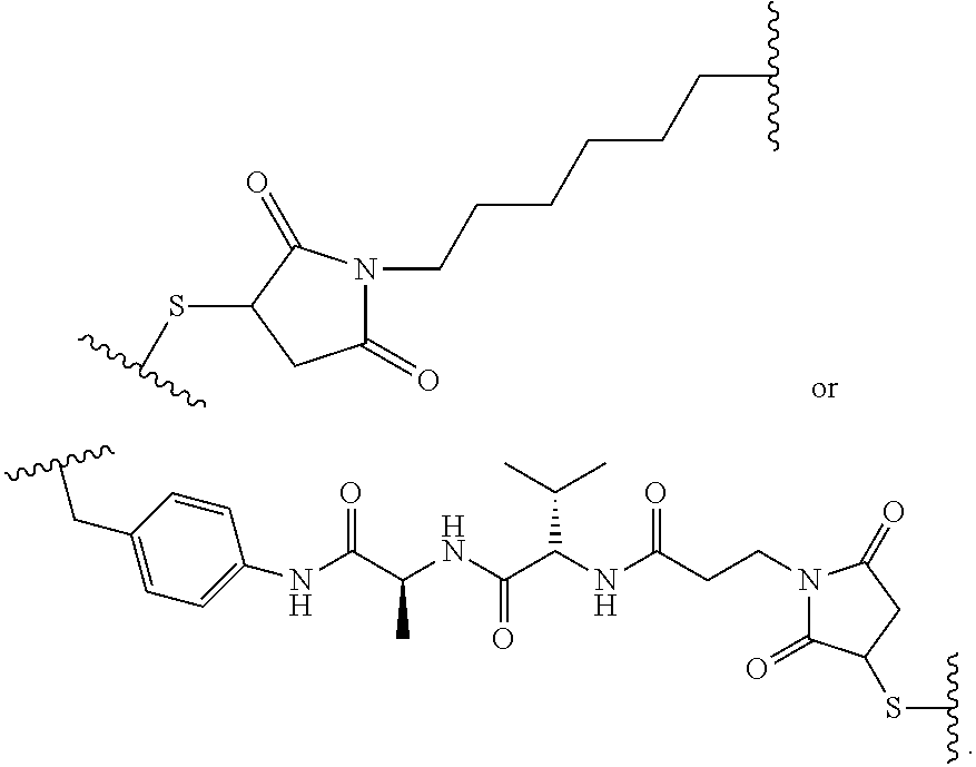
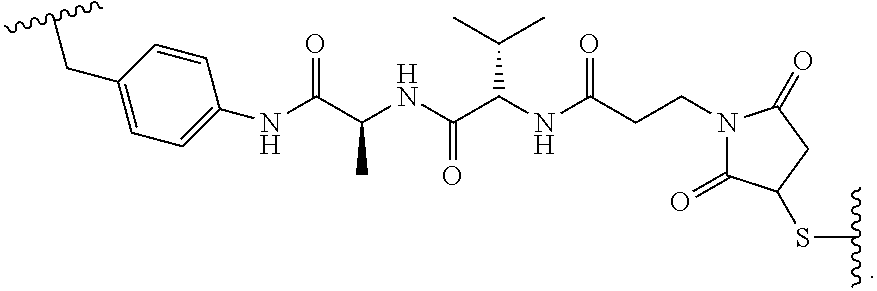
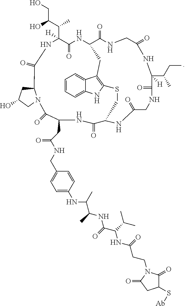

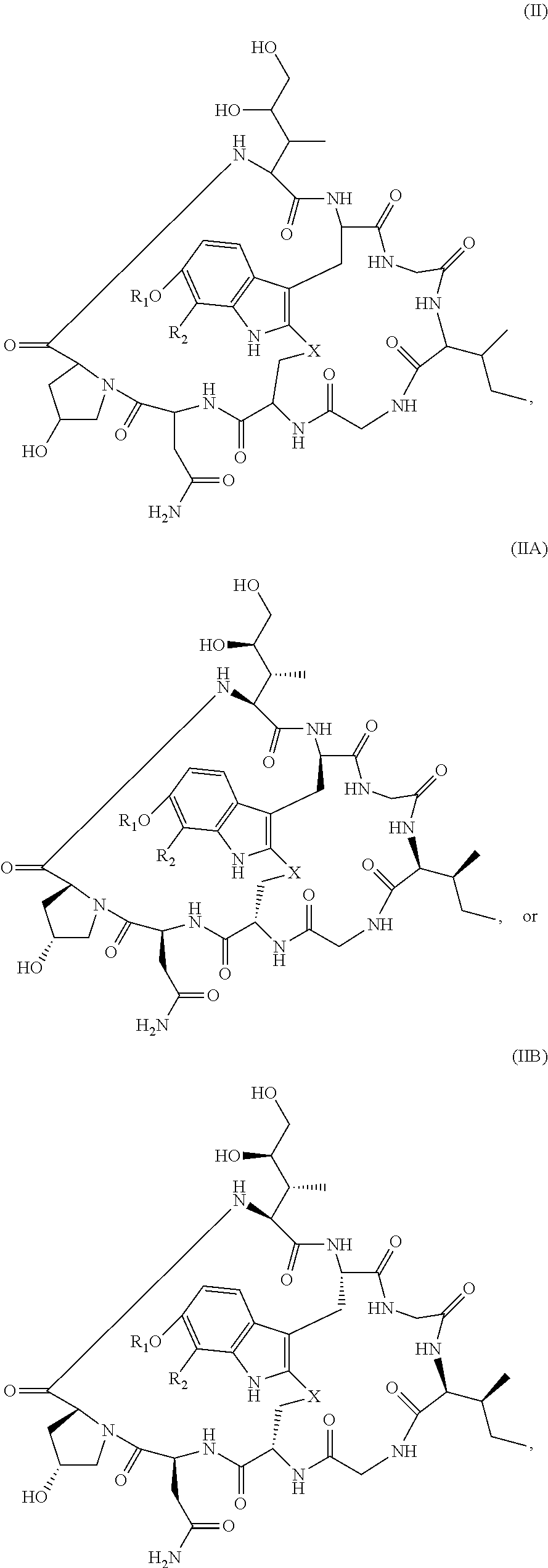


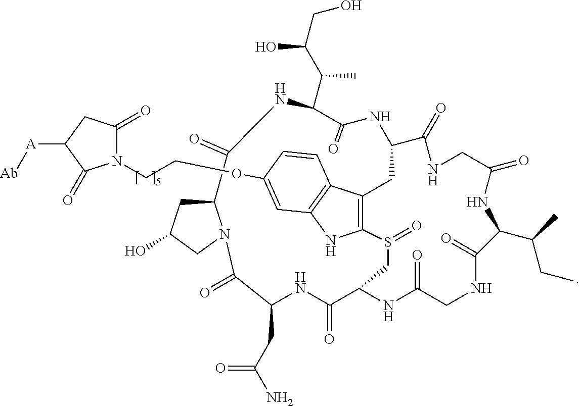


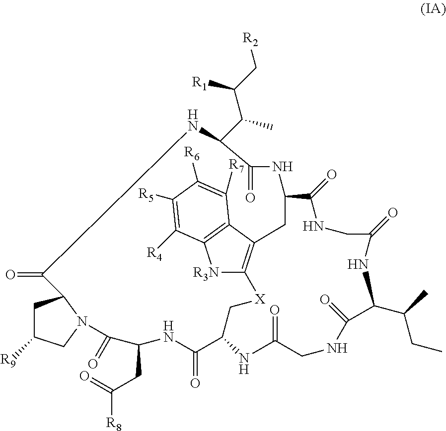

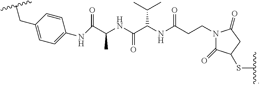
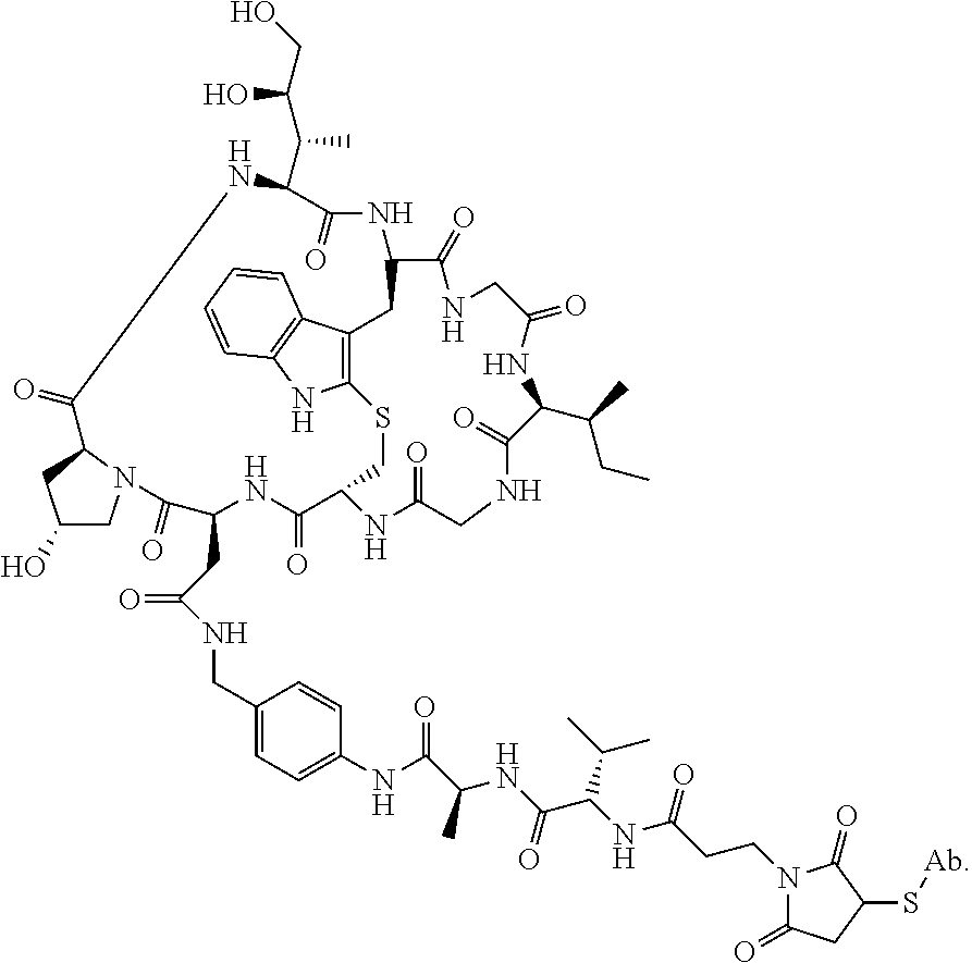
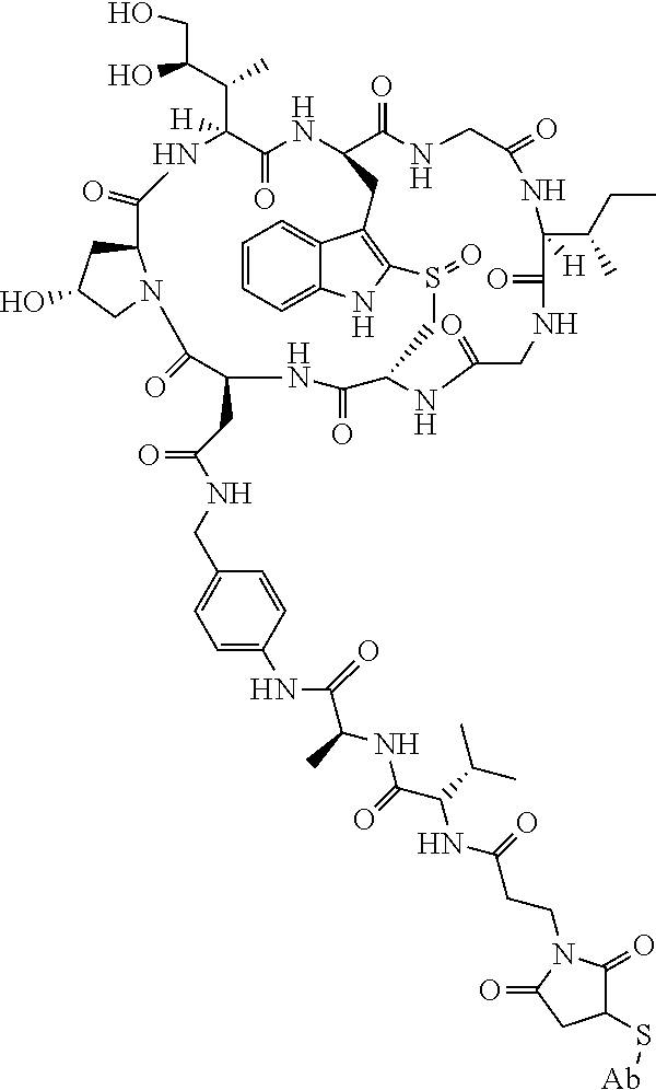


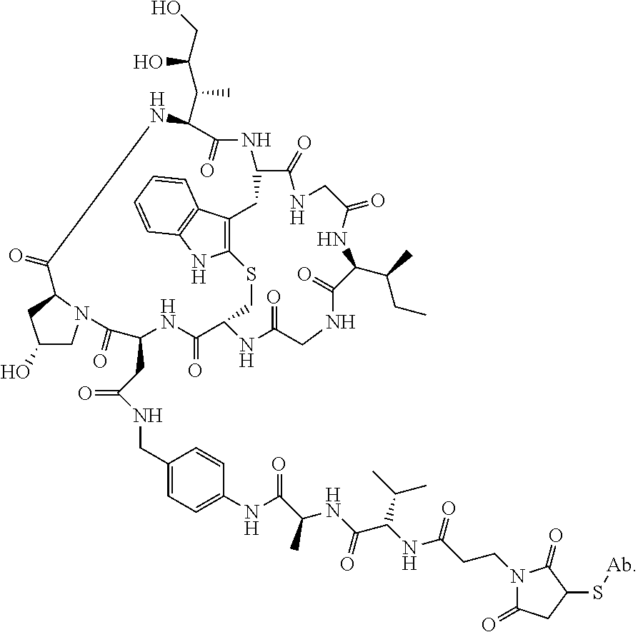
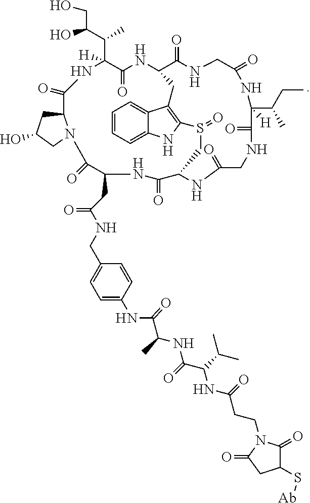
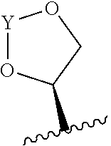

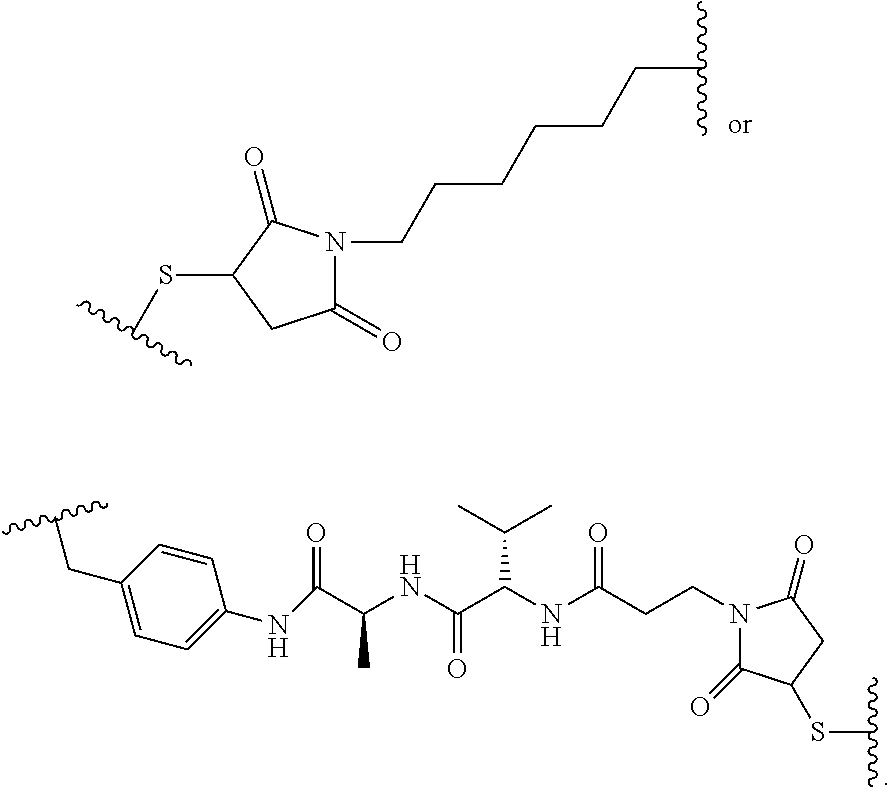

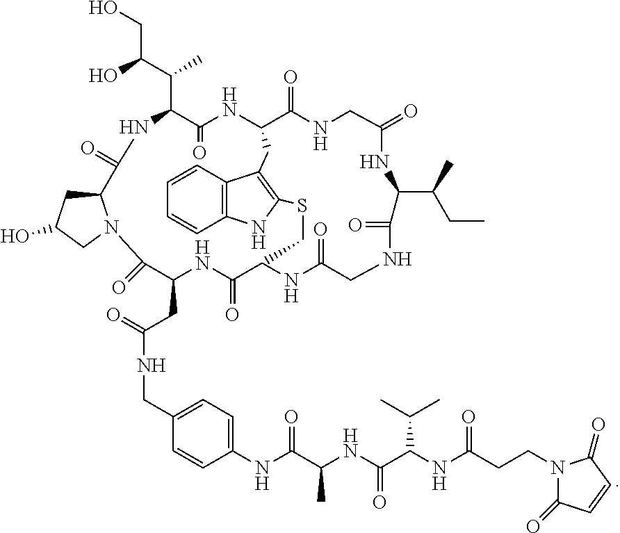
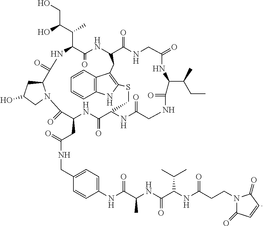
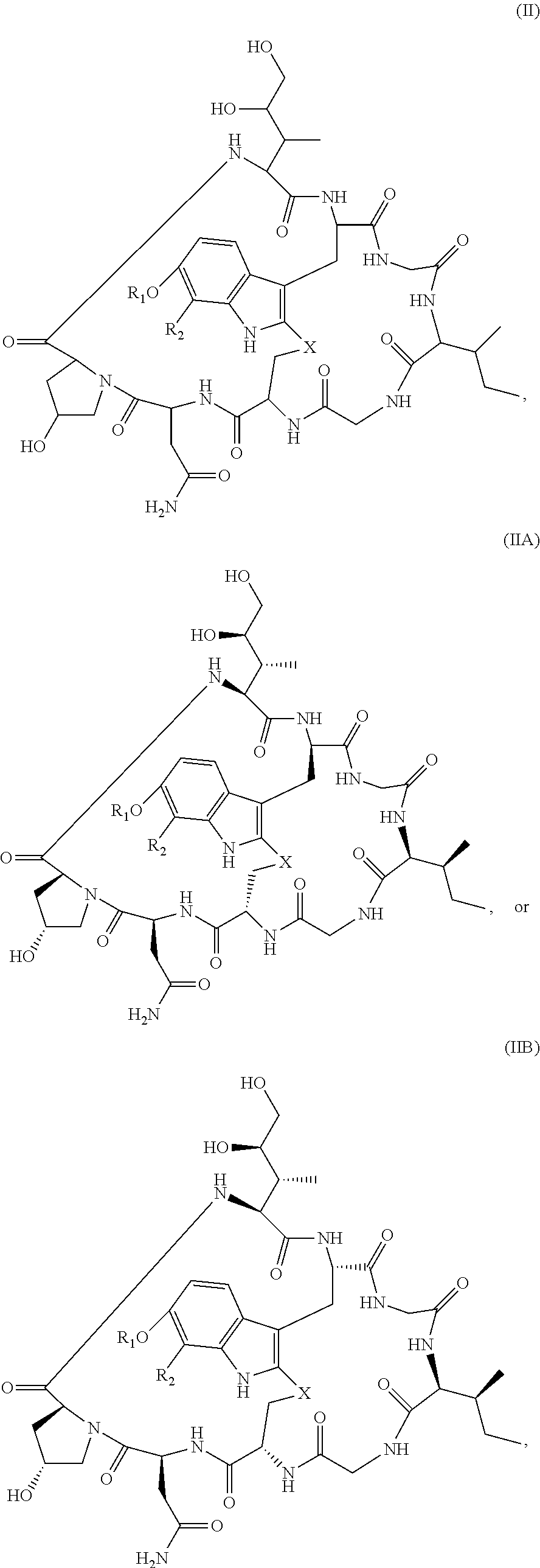
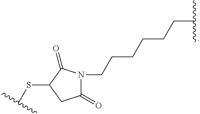
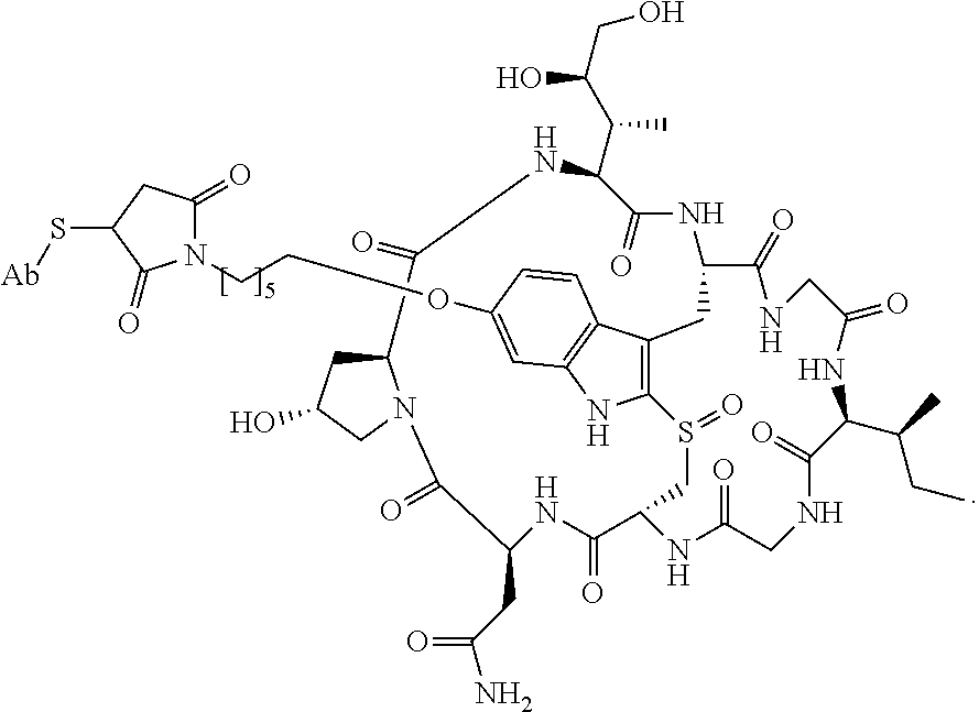
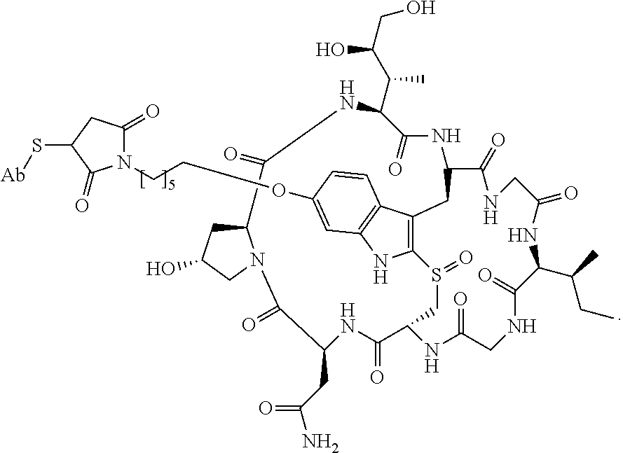

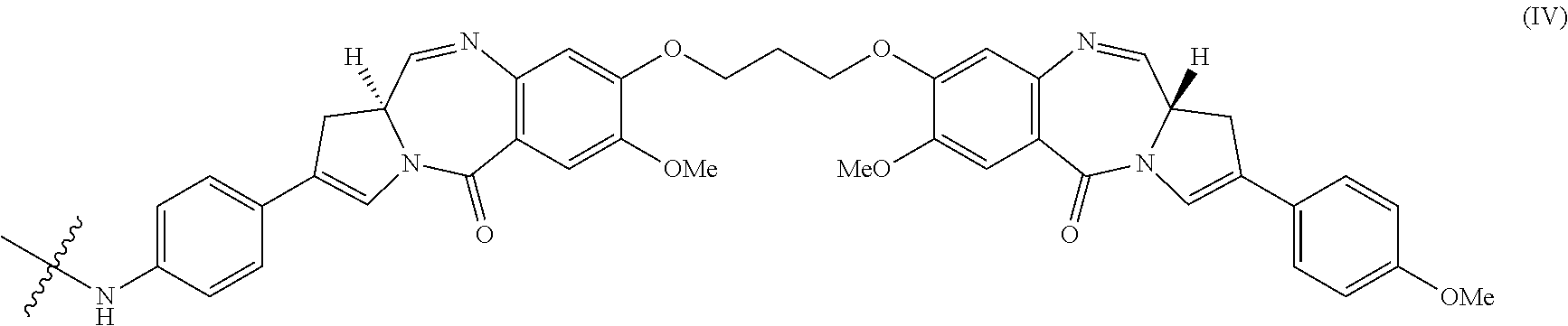
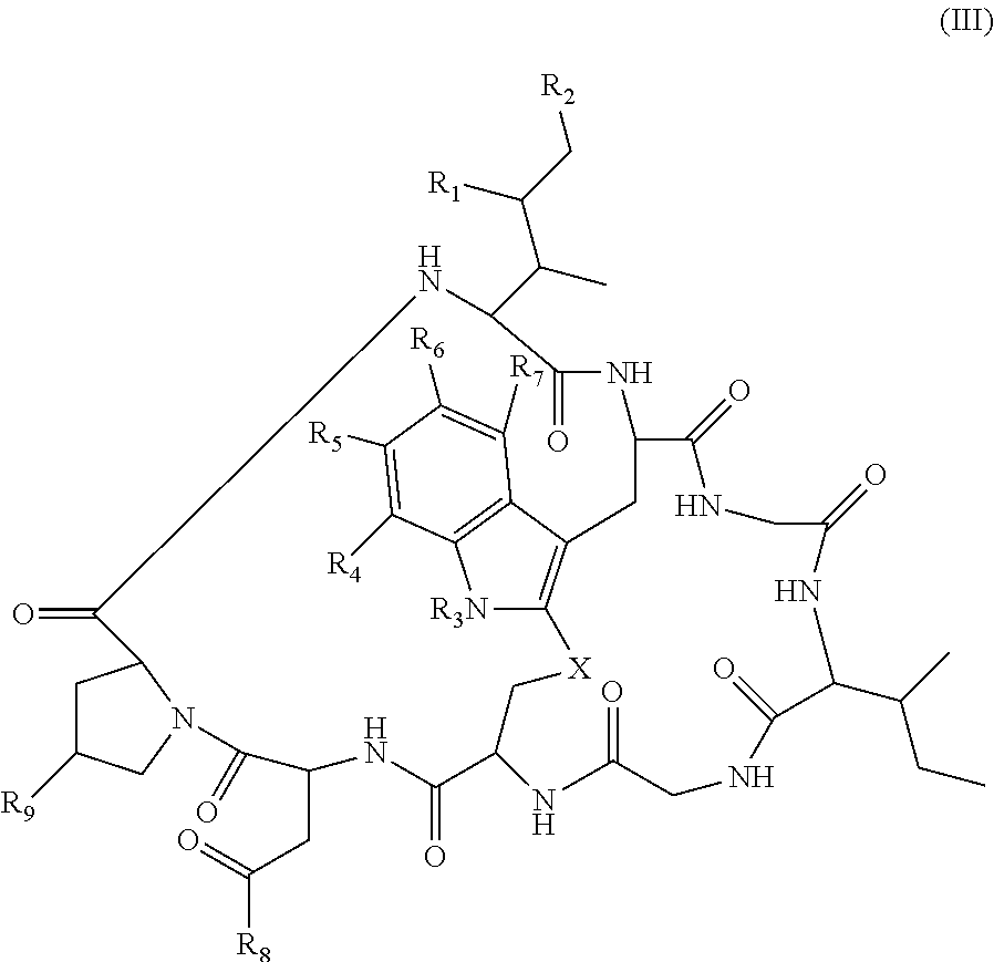
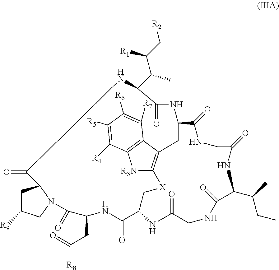

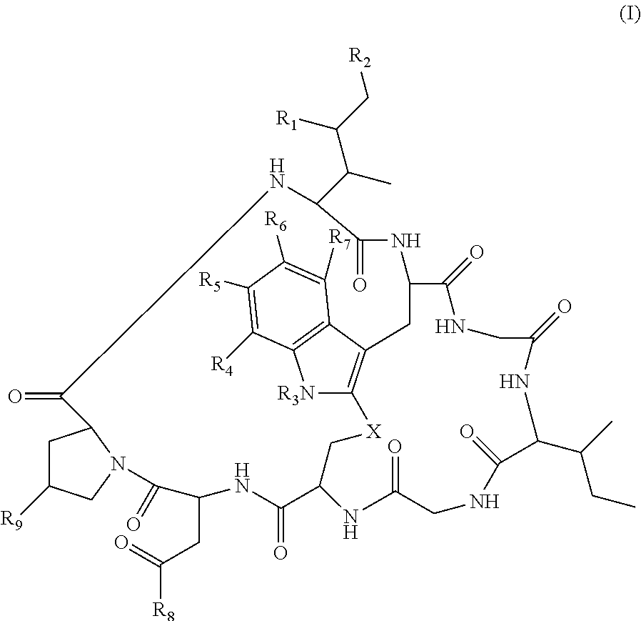
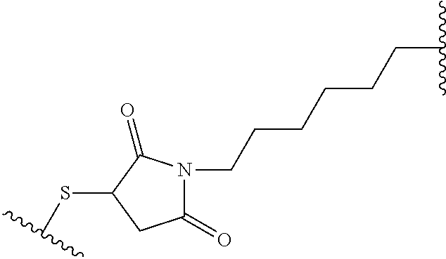

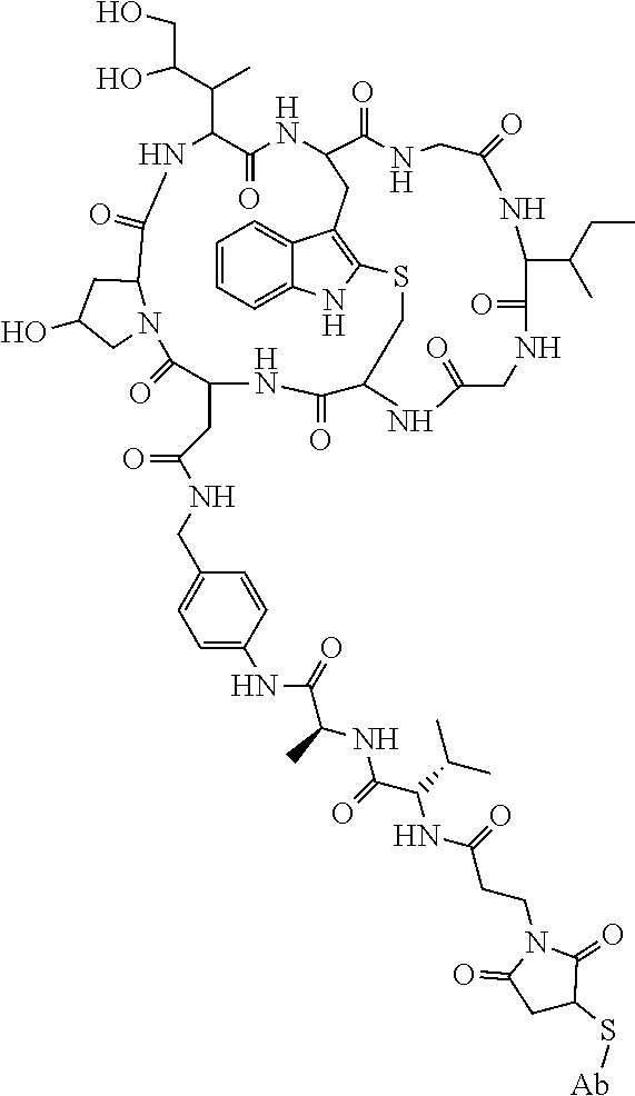
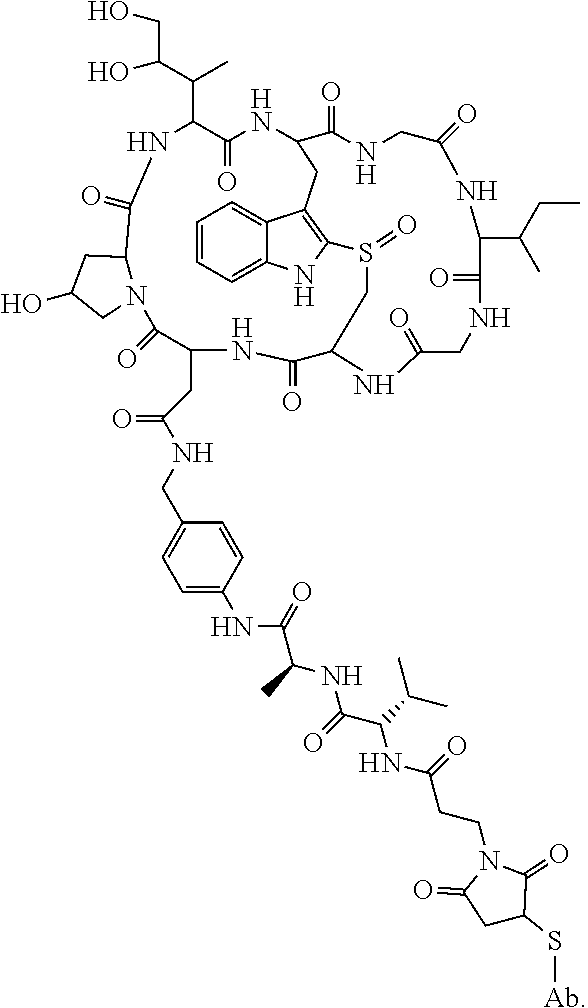
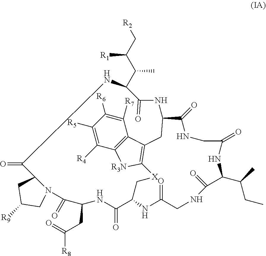
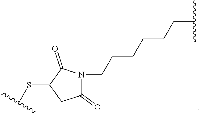

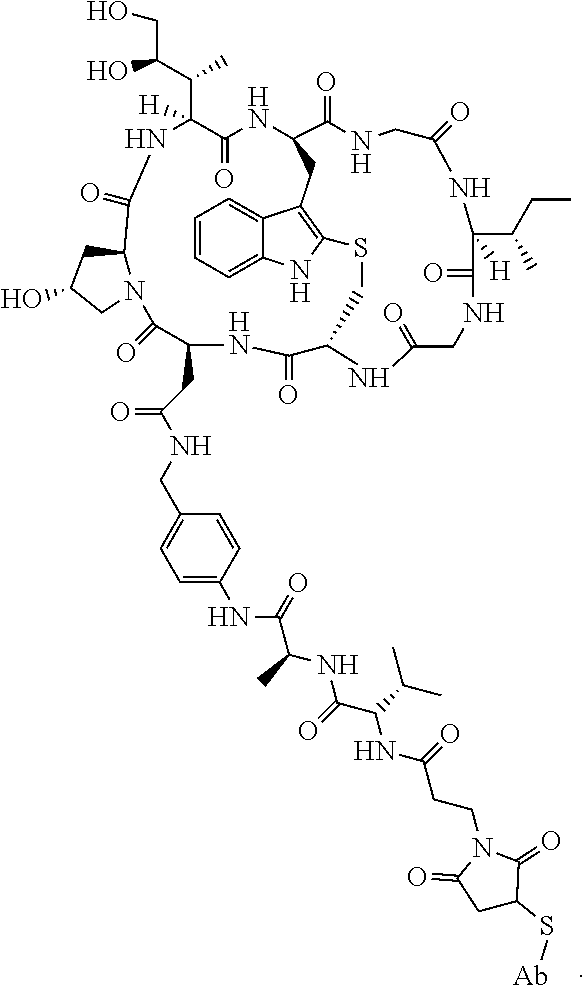
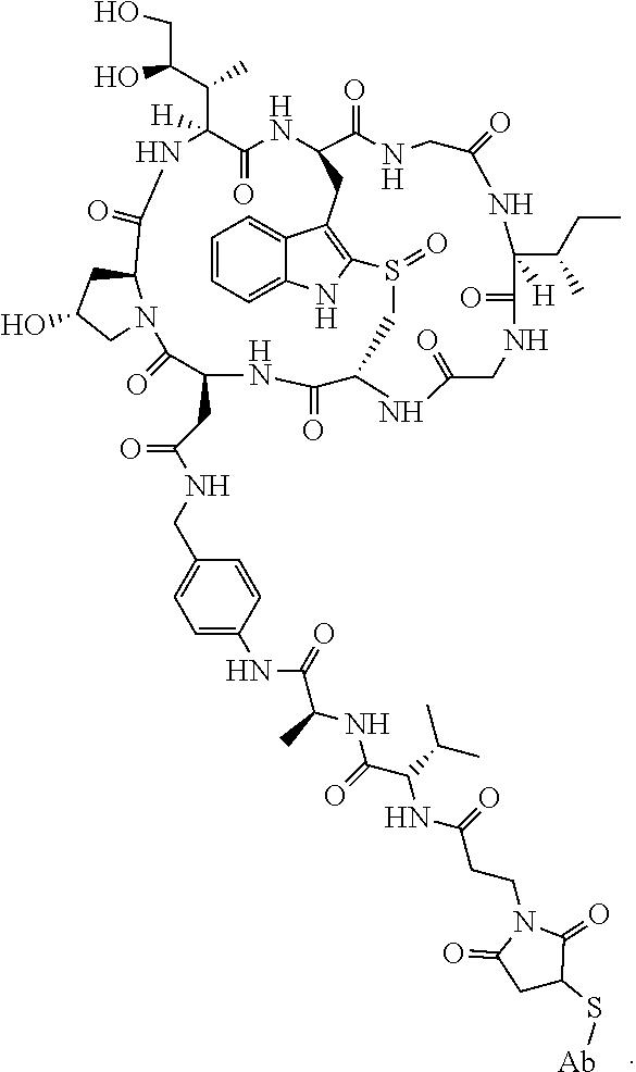
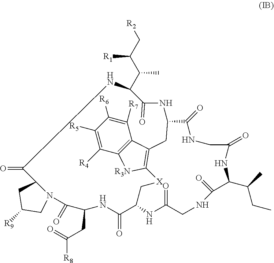
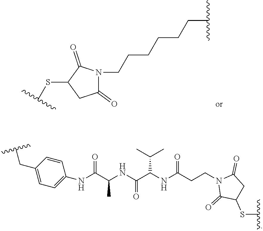
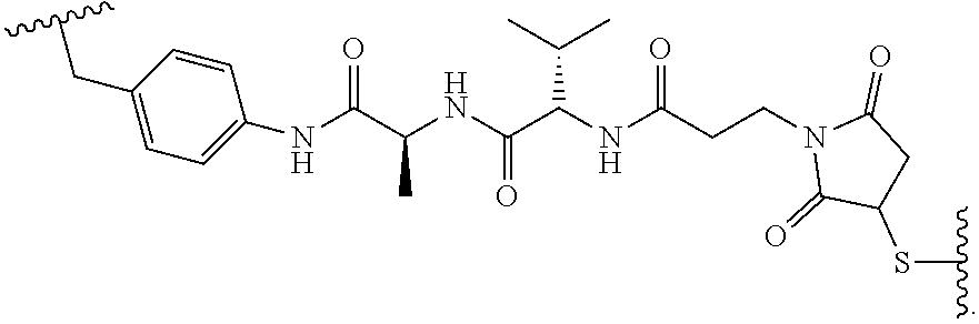
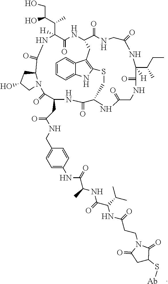


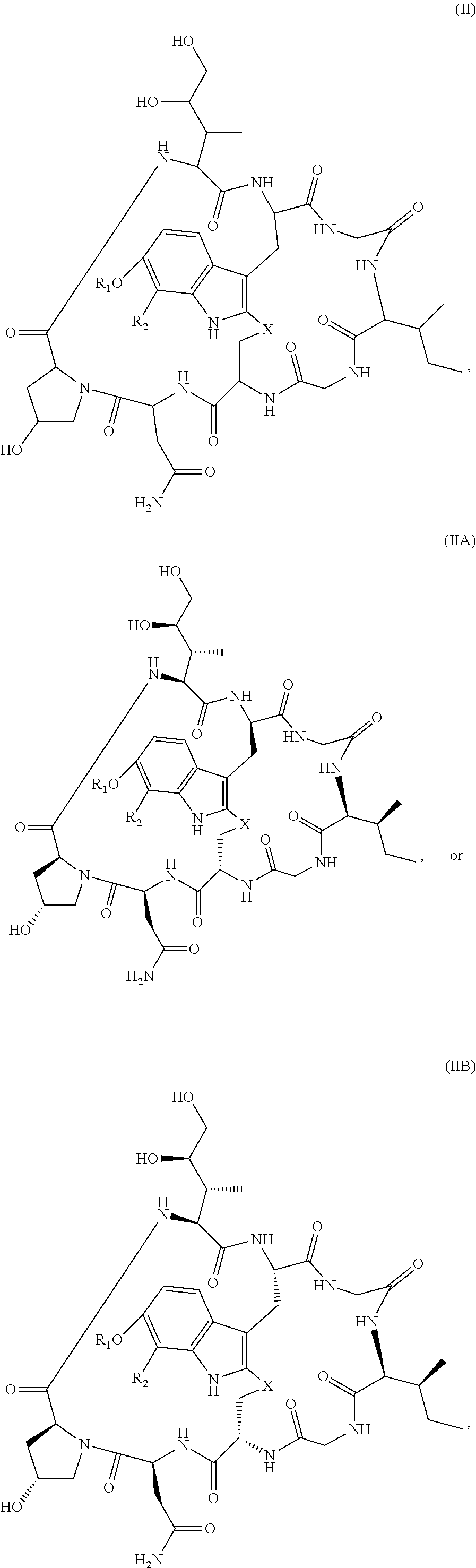

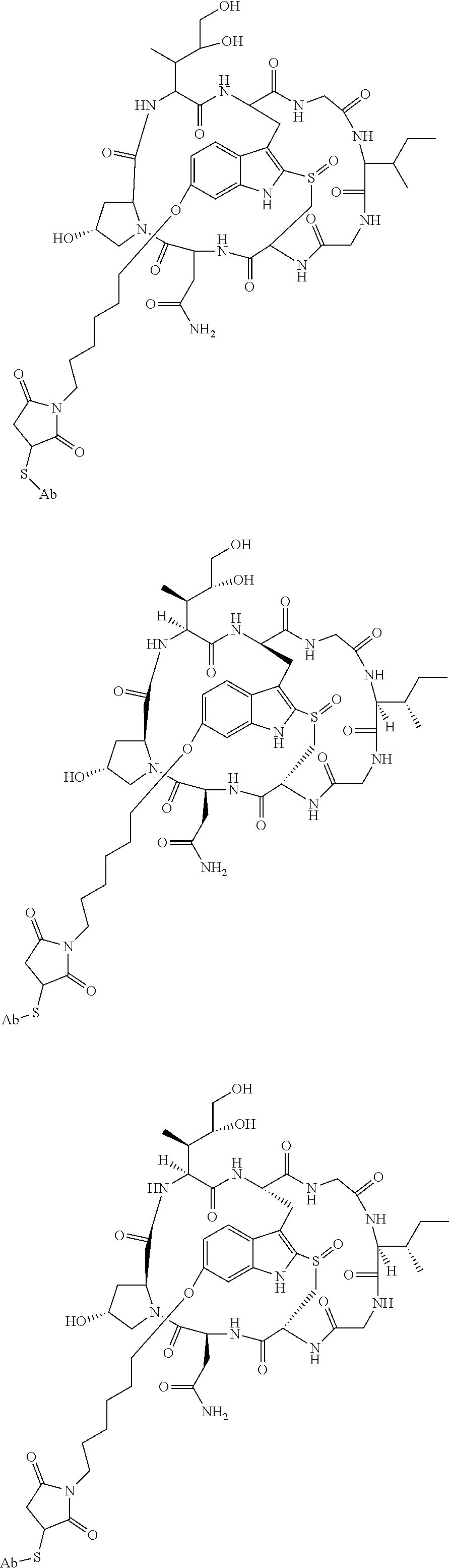

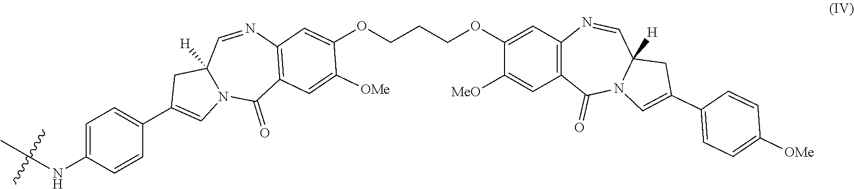
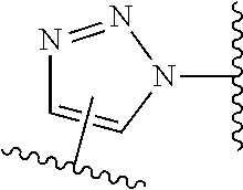


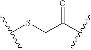
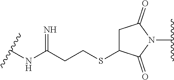


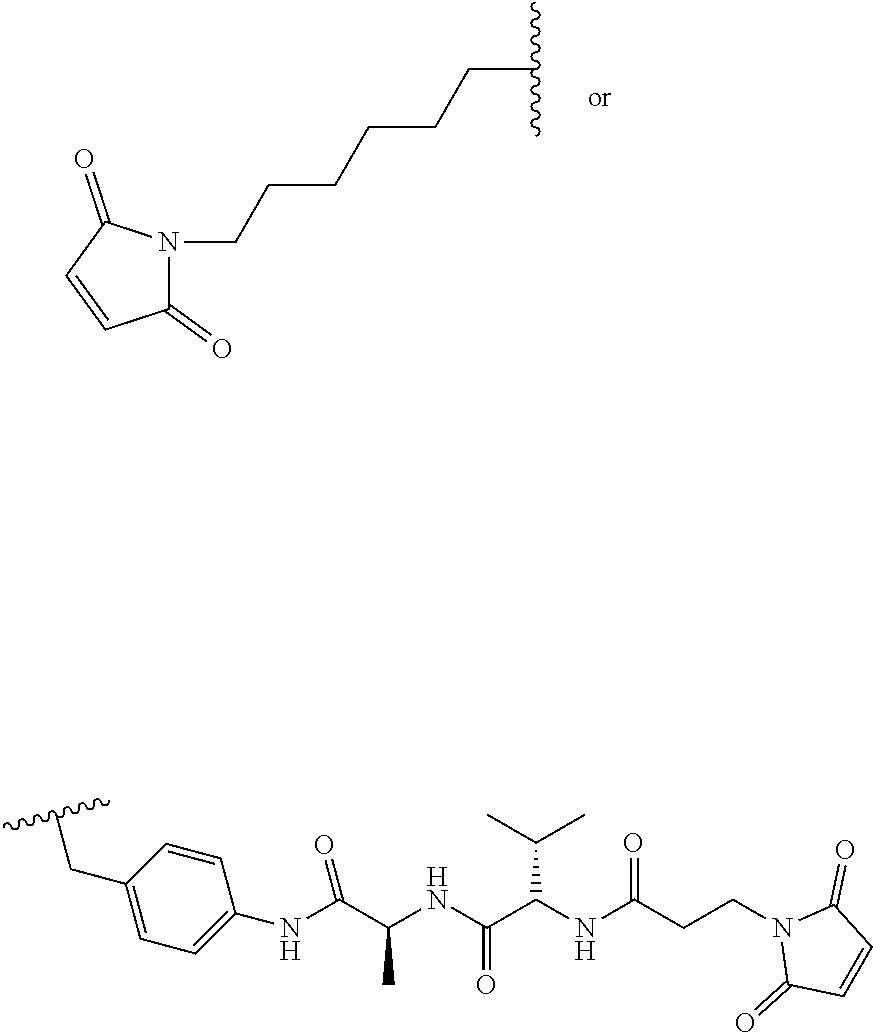
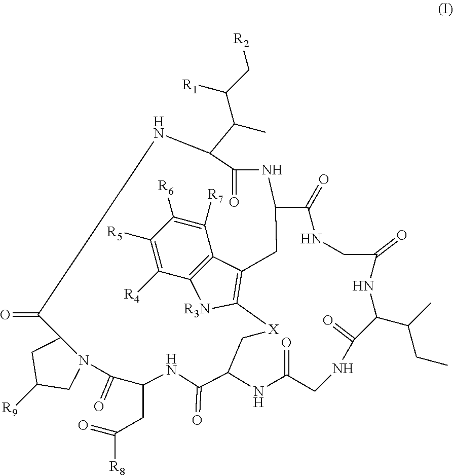

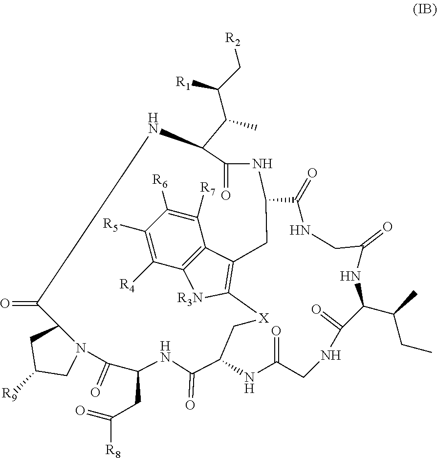
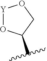

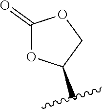


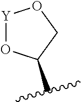


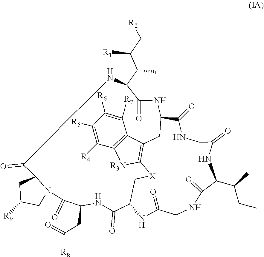
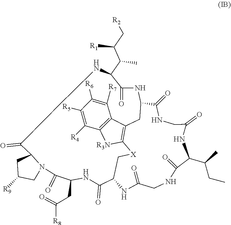
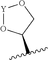


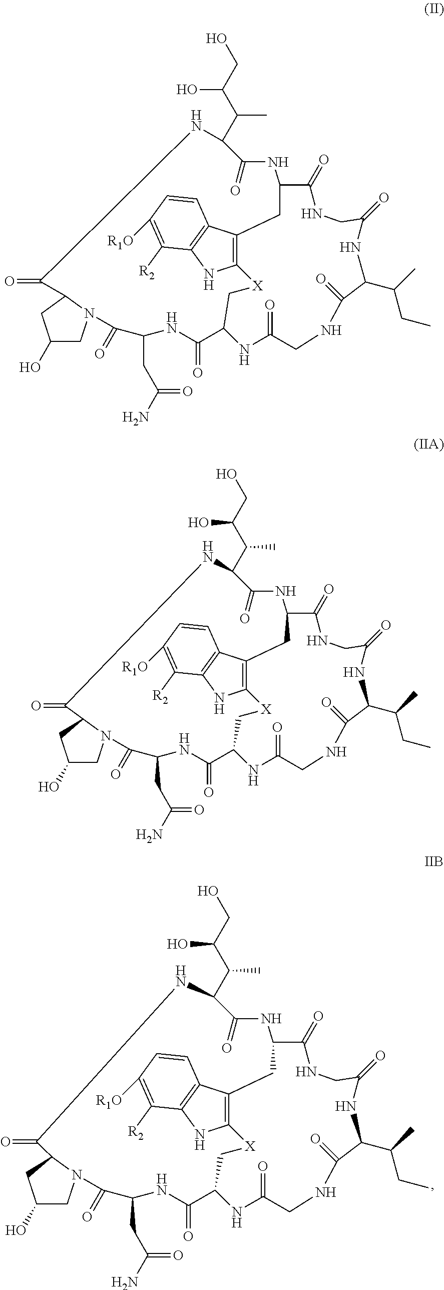
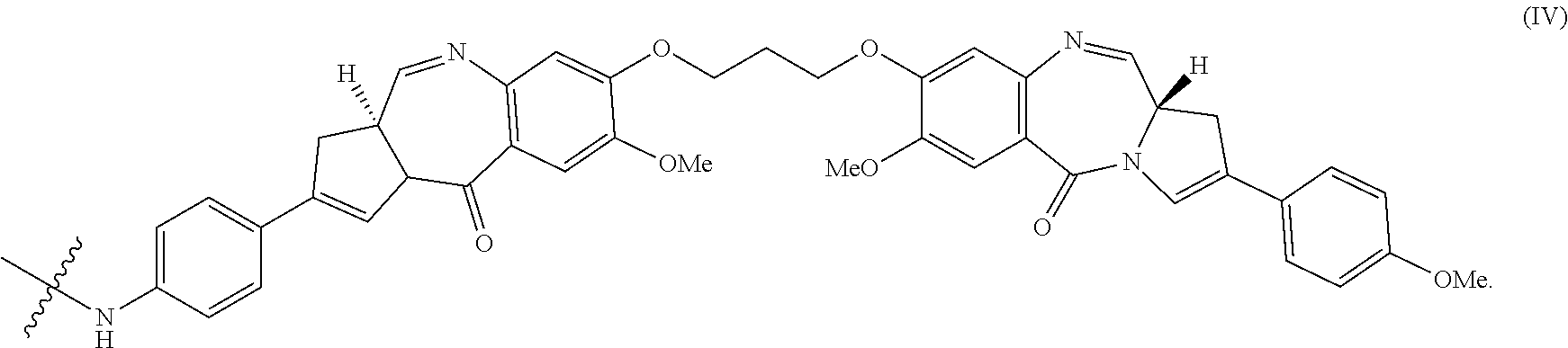
D00001
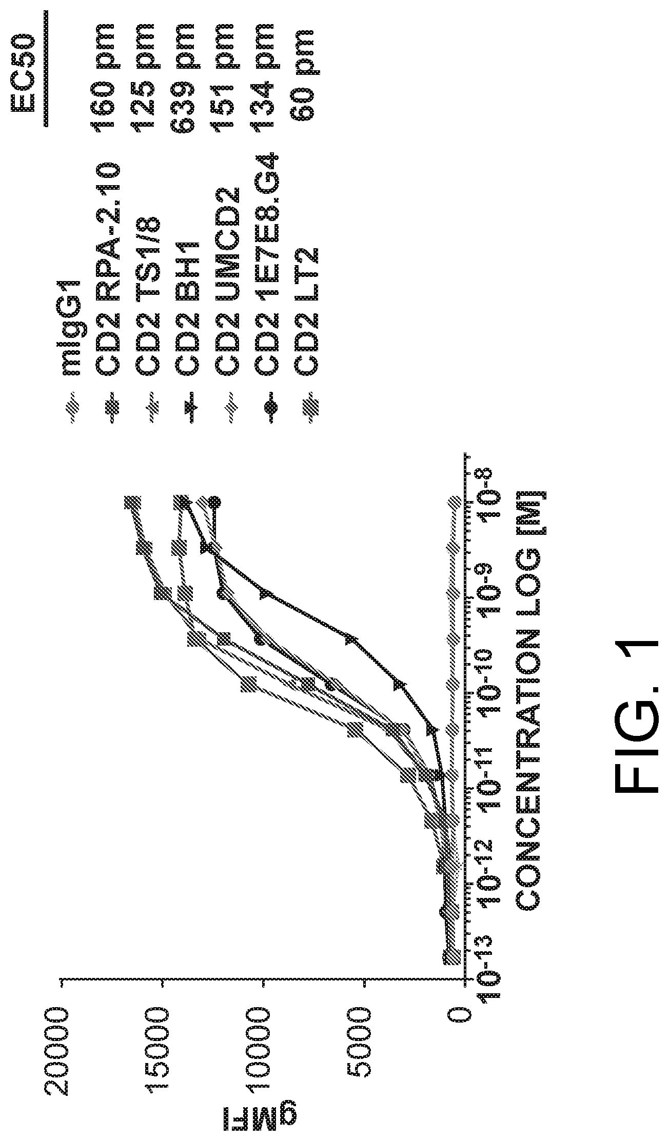
D00002
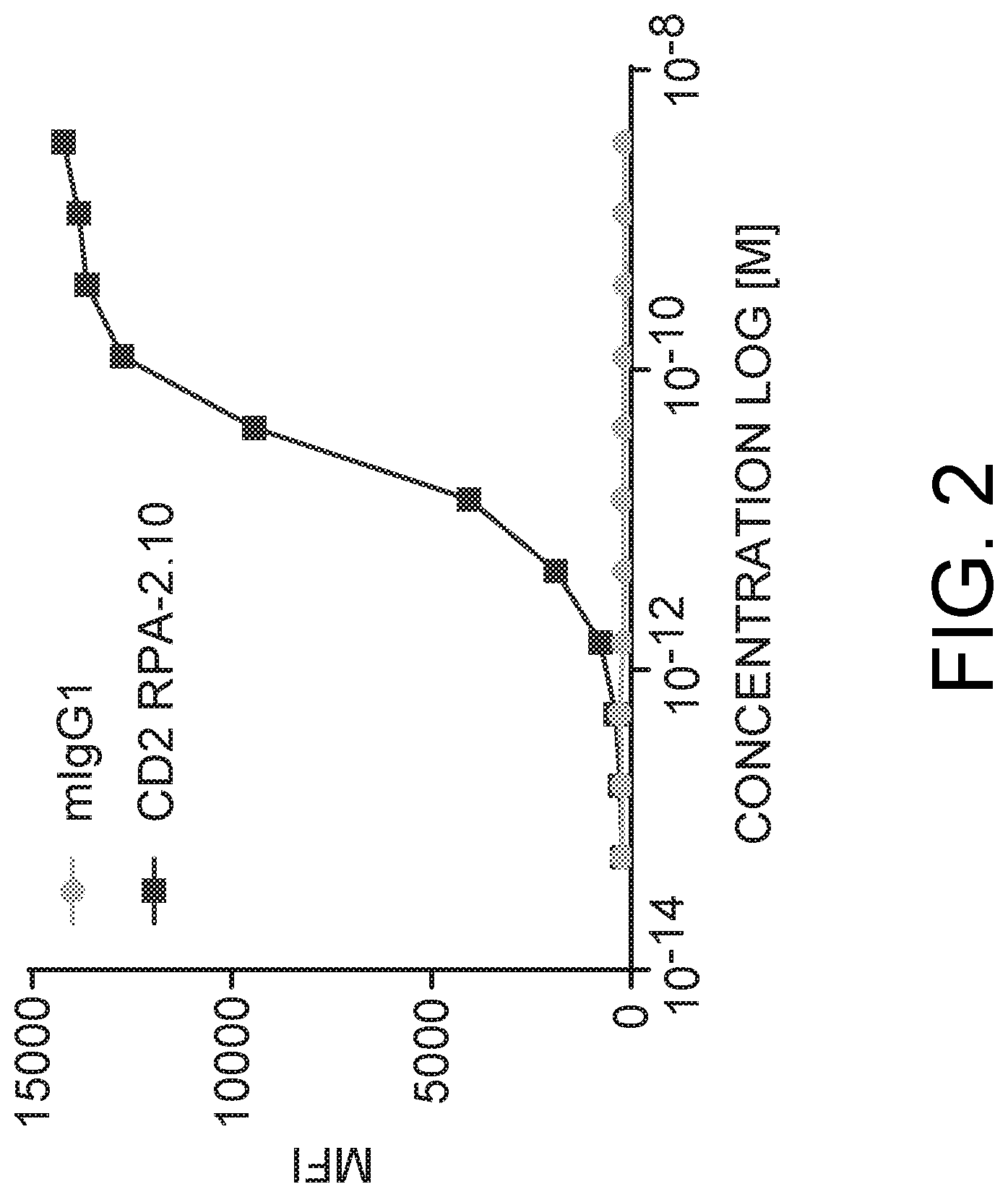
D00003
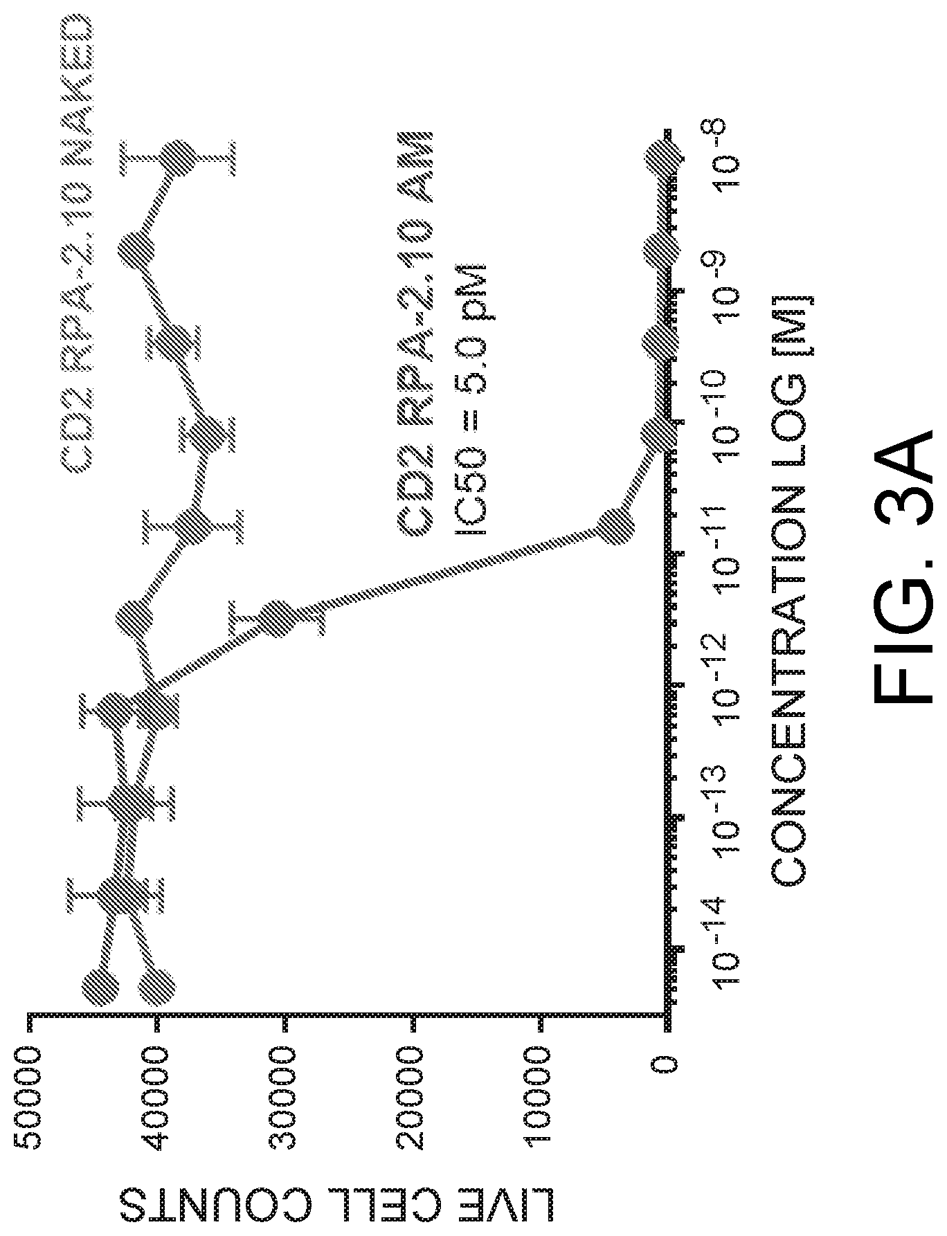
D00004
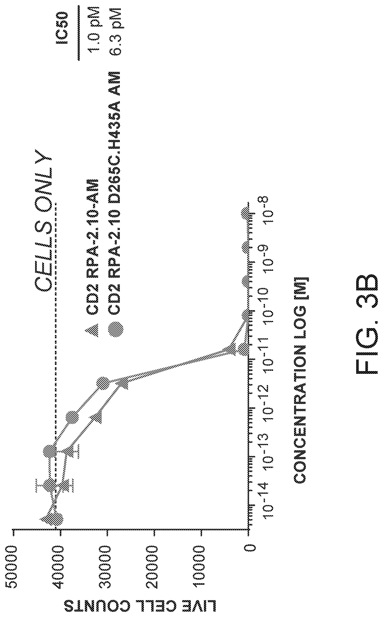
D00005
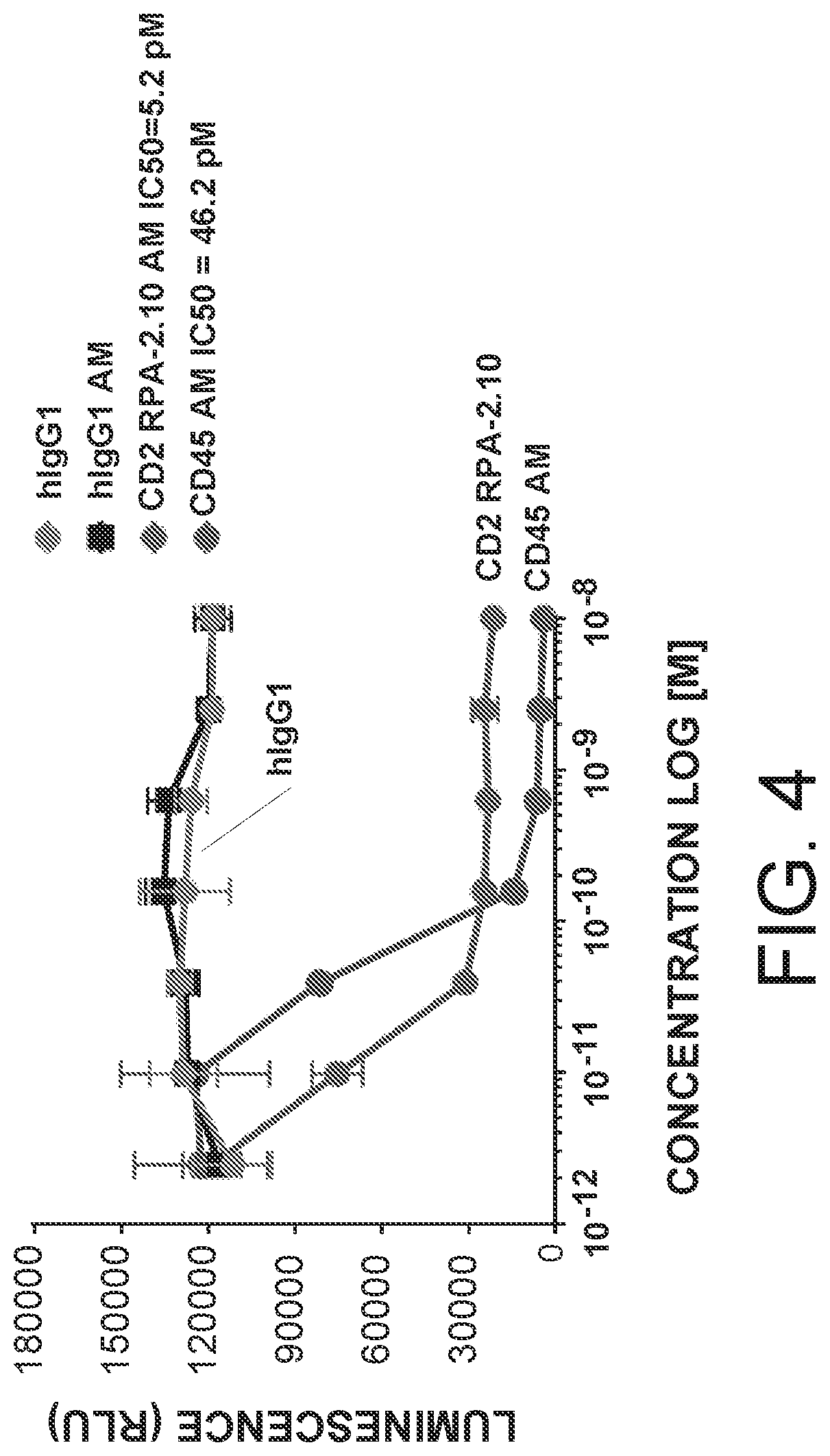
D00006
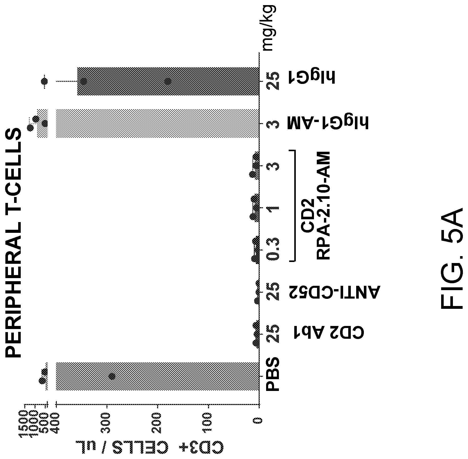
D00007
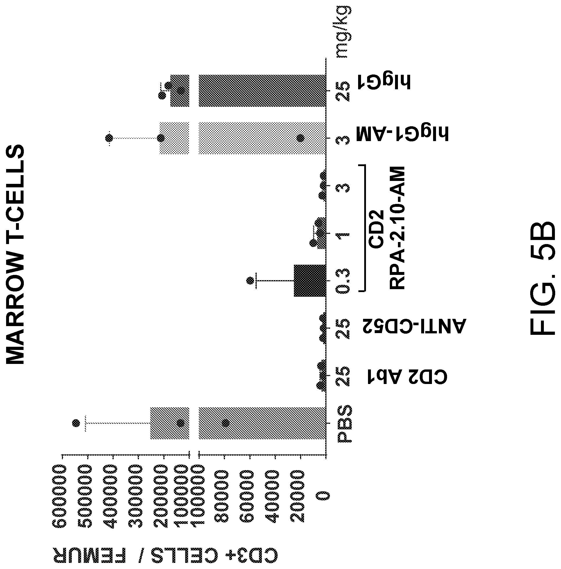
D00008

D00009
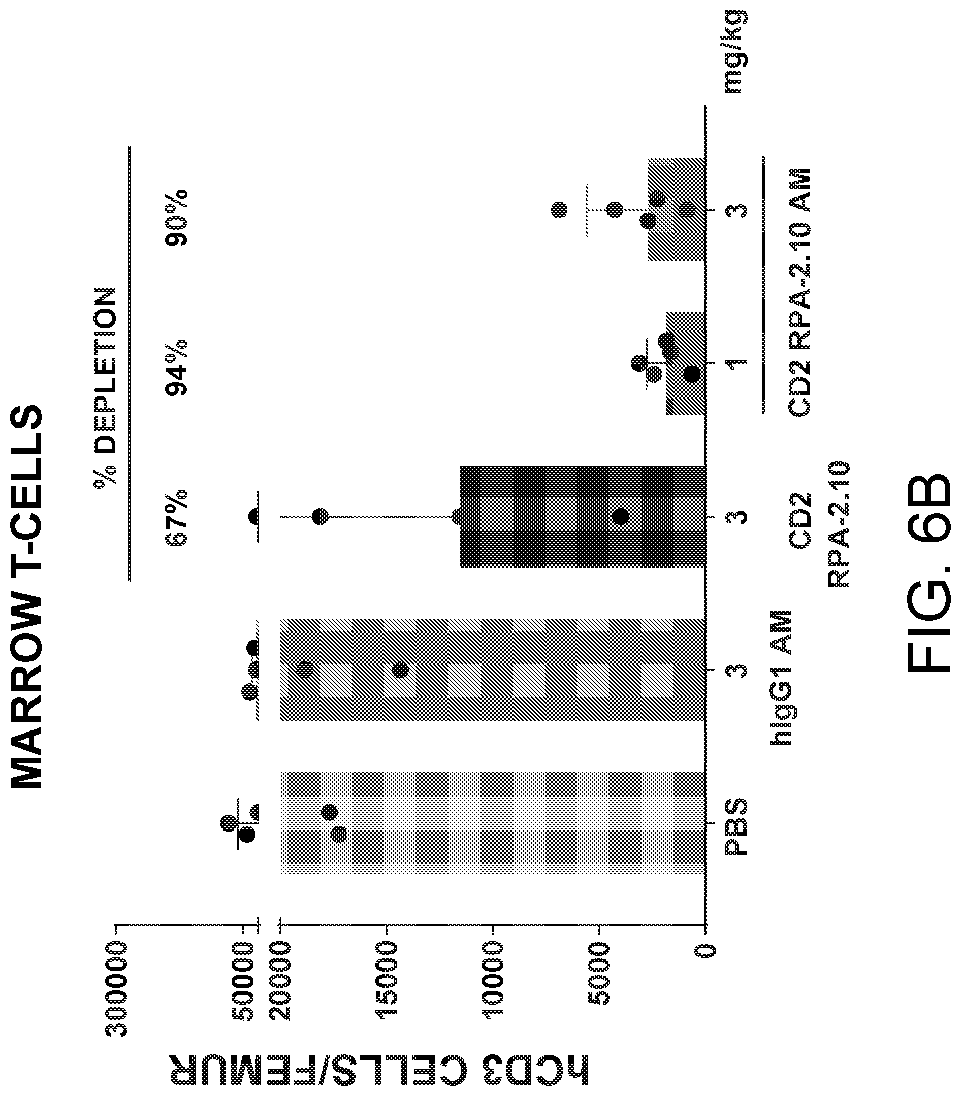
D00010
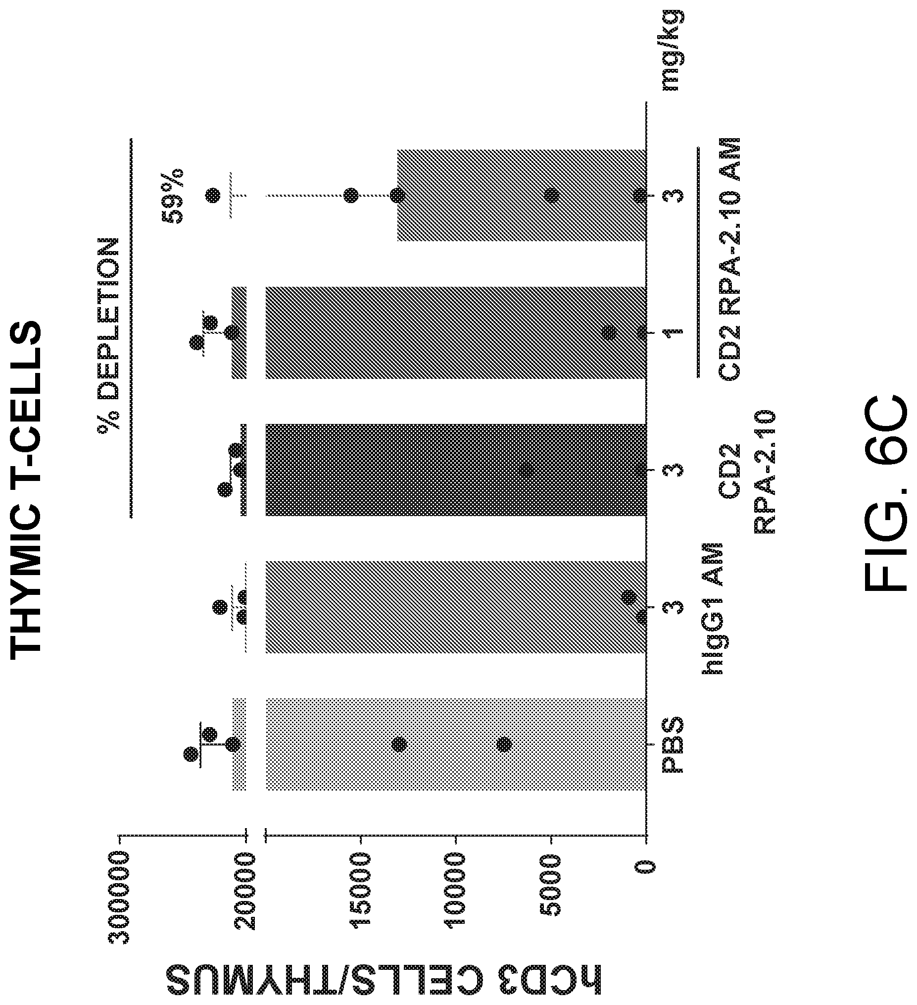
S00001
XML
uspto.report is an independent third-party trademark research tool that is not affiliated, endorsed, or sponsored by the United States Patent and Trademark Office (USPTO) or any other governmental organization. The information provided by uspto.report is based on publicly available data at the time of writing and is intended for informational purposes only.
While we strive to provide accurate and up-to-date information, we do not guarantee the accuracy, completeness, reliability, or suitability of the information displayed on this site. The use of this site is at your own risk. Any reliance you place on such information is therefore strictly at your own risk.
All official trademark data, including owner information, should be verified by visiting the official USPTO website at www.uspto.gov. This site is not intended to replace professional legal advice and should not be used as a substitute for consulting with a legal professional who is knowledgeable about trademark law.