Nkp30 Receptor Targeted Therapeutics
ZHANG; Tong ; et al.
U.S. patent application number 16/882800 was filed with the patent office on 2020-11-19 for nkp30 receptor targeted therapeutics. The applicant listed for this patent is THE TRUSTEES OF DARTMOUTH COLLEGE. Invention is credited to Charles L. SENTMAN, Tong ZHANG.
| Application Number | 20200360434 16/882800 |
| Document ID | / |
| Family ID | 1000004993360 |
| Filed Date | 2020-11-19 |


View All Diagrams
| United States Patent Application | 20200360434 |
| Kind Code | A1 |
| ZHANG; Tong ; et al. | November 19, 2020 |
NKP30 RECEPTOR TARGETED THERAPEUTICS
Abstract
The invention is directed to T cells and other cells that express chimeric NK-p30 receptors ("chimeric NKp30 T cells"), methods of making and using chimeric NKp30 T cells, and methods of using these chimeric NKp30 T cells, isolated populations thereof, and compositions comprising the same. In another aspect, said chimeric NKp30 T cells are further designed to express a functional non-TCR receptor. The disclosure also pertains to methods of making said chimeric NKp30 T cells, and methods of reducing or ameliorating, or preventing or treating, diseases and disorders using said chimeric NKp30 T cells, populations thereof, or compositions comprising the same.
| Inventors: | ZHANG; Tong; (Lebanon, NH) ; SENTMAN; Charles L.; (Grantham, NH) | ||||||||||
| Applicant: |
|
||||||||||
|---|---|---|---|---|---|---|---|---|---|---|---|
| Family ID: | 1000004993360 | ||||||||||
| Appl. No.: | 16/882800 | ||||||||||
| Filed: | May 26, 2020 |
Related U.S. Patent Documents
| Application Number | Filing Date | Patent Number | ||
|---|---|---|---|---|
| 15830605 | Dec 4, 2017 | 10682378 | ||
| 16882800 | ||||
| 14342060 | Nov 24, 2014 | 9833476 | ||
| PCT/US2012/053511 | Aug 31, 2012 | |||
| 15830605 | ||||
| 61529410 | Aug 31, 2011 | |||
| Current U.S. Class: | 1/1 |
| Current CPC Class: | C07K 14/70503 20130101; A61K 35/17 20130101; C07K 14/70578 20130101; C07K 14/70521 20130101; C12N 5/0636 20130101; C07K 14/70575 20130101; C07K 14/70596 20130101; C07K 2319/00 20130101; C12N 2800/22 20130101; C07K 2319/03 20130101; A61K 38/00 20130101 |
| International Class: | A61K 35/17 20060101 A61K035/17; C07K 14/705 20060101 C07K014/705; C12N 5/0783 20060101 C12N005/0783 |
Goverment Interests
STATEMENT REGARDING FEDERALLY SPONSORED RESEARCH OR DEVELOPMENT
[0002] This invention was made with government support under contract number CA awarded by the National Institutes of Health. The government has certain rights in the invention.
Claims
1. A nucleic acid construct for expressing a chimeric receptor comprising: a first nucleic acid sequence comprising a promoter operably linked to a second nucleic acid sequence, wherein said second nucleic acid sequence encodes a chimeric NKp30 receptor comprising: an NKp30 extracellular domain, a transmembrane domain, and at least one signaling domain.
2-394. (canceled)
Description
CROSS-REFERENCE TO RELATED APPLICATIONS
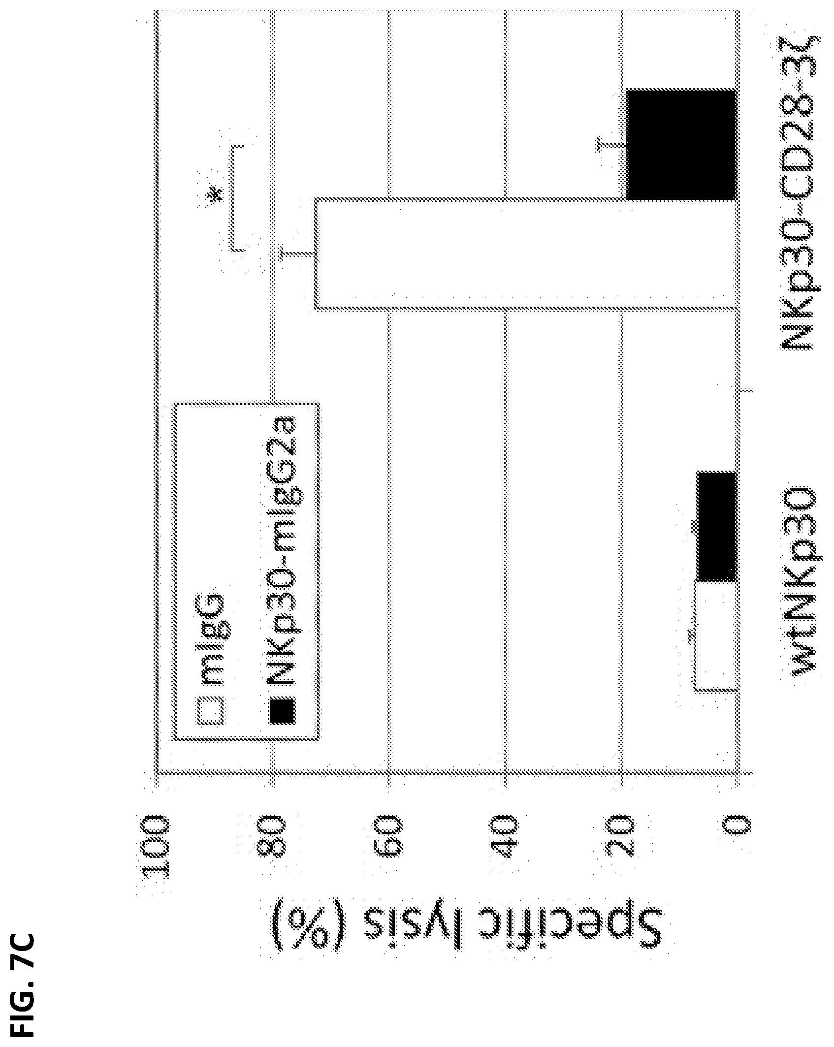
[0001] This application is a divisional of U.S. Utility application Ser. No. 15/830,605 filed Dec. 4, 2017, now U.S. Pat. No. 10,682,378, which is a divisional of U.S. application Ser. No. 14/342,060, filed Nov. 24, 2014, now U.S. Pat. No. 9,833,476, which is a 35 U.S.C. 371 United States National Phase Application of PCT Application PCT/US2012/053511, filed Aug. 31, 2012 and published as WO 2013/033626 on Mar. 7, 2013, which claims priority to U.S. Provisional Application 61/529,410 filed Aug. 31, 2011, each of which is hereby incorporated by reference in its entirety herein.
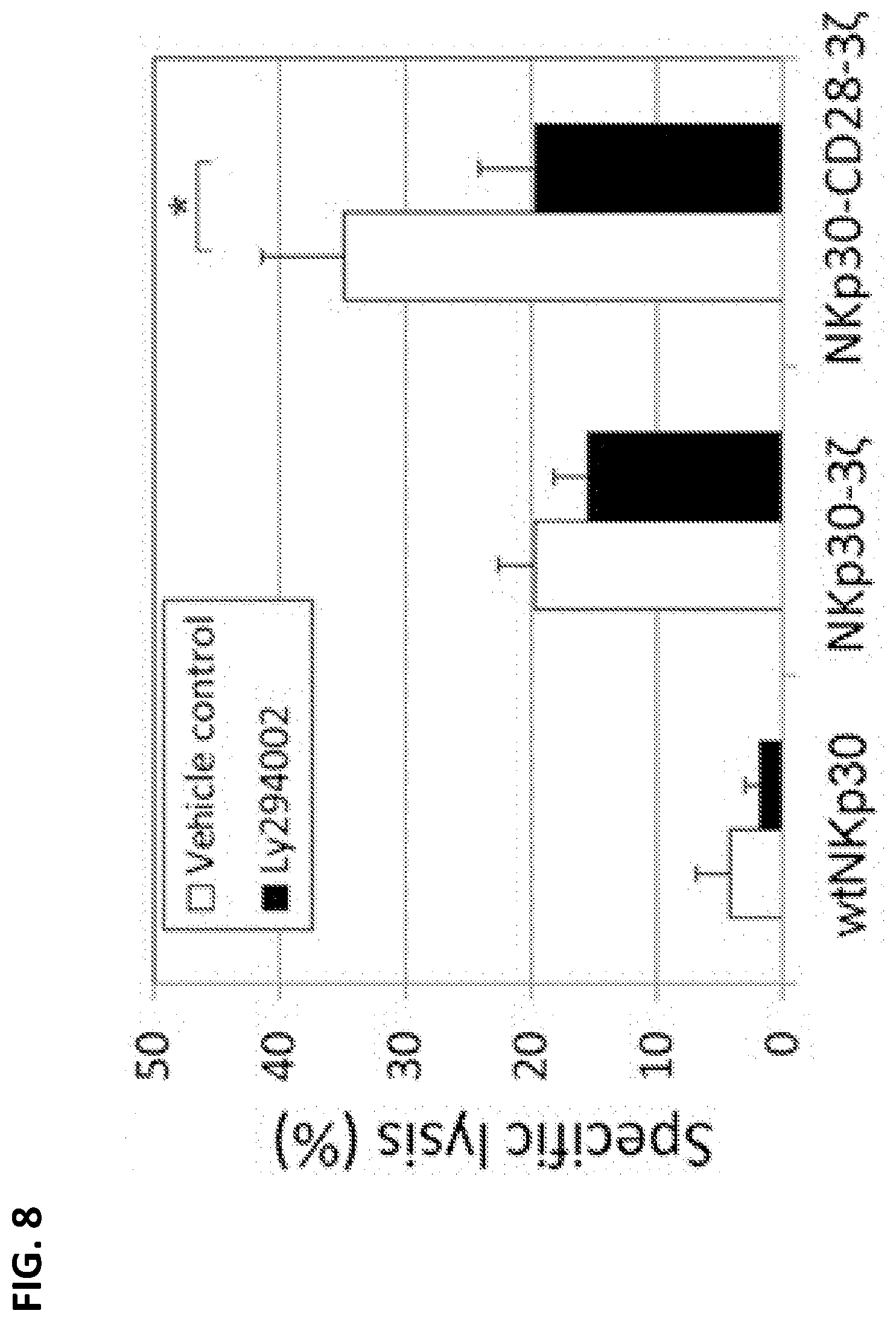
SEQUENCE LISTING
[0003] The sequence listing in the filed named "1148307o001203.txt" having a size of 84,226 bytes that was created May 26, 2020, is hereby incorporated by reference in its entirety.
BACKGROUND
Field
[0004] The present disclosure is directed to T cells that express chimeric NKp30 receptors ("chimeric NKp30 T cells"), methods of making and using chimeric NKp30 T cells, and methods of using these chimeric NKp30 T cells to address diseases and disorders. In one aspect, the disclosure broadly relates to chimeric NKp30 T cells, isolated populations thereof, and compositions comprising the same. In another aspect, said chimeric NKp30 T cells are further designed to express a functional non-TCR receptor. The disclosure also pertains to methods of making said chimeric NKp30 T cells, and methods of reducing or ameliorating, or preventing or treating, diseases and disorders, especially cancers, using said chimeric NKp30 T cells, populations thereof, or compositions comprising the same.
Description of Related Art
[0005] The global burden of cancer doubled between 1975 and 2000, and cancer is expected to become the leading cause of death worldwide by 2010. According to the American Cancer Society, it is projected to double again by 2020 and to triple by 2030. Thus, there is a need for more effective therapies to treat various forms of cancer. Ideally, any cancer therapy should be effective (at killing cancerous cells), targeted (i.e. selective, to avoid killing healthy cells), permanent (to avoid relapse and metastasis), and affordable. Today's standards of care for most cancers fall short in some or all of these criteria.
[0006] T cells, especially cytotoxic T cells, play important roles in anti-tumor immunity (Rossing and Brenner (2004) Mol. Ther. 10:5-18). Adoptive transfer of tumor-specific T cells into patients provides a means to treat cancer (Sadelain, et al. (2003) Nat. Rev. Cancer 3:35-45). However, the traditional approaches for obtaining large numbers of tumor-specific T cells are time-consuming, laborious and sometimes difficult because the average frequency of antigen-specific T cells in periphery is extremely low (Rosenberg (2001) Nature 411:380-384; Ho, et al. (2003) Cancer Cell 3:431-437; Crowley, et al. (1990) Cancer Res. 50:492-498). In addition, isolation and expansion of T cells that retain their antigen specificity and function can also be a challenging task (Sadelain, et al. (2003) supra). Genetic modification of primary T cells with tumor-specific immunoreceptors, such as full-length T cell receptors or chimeric T cell receptor molecules can be used for redirecting T cells against tumor cells (Stevens, et al. (1995) J. Immunol. 154:762-771; Oelke, et al. (2003) Nat. Med. 9:619-624; Stancovski, et al. (1993) J. Immunol. 151:6577-6582; Clay, et al. (1999) J. Immunol. 163:507-153). This strategy avoids the limitation of low frequency of antigen-specific T cells, allowing for facilitated expansion of tumor-specific T cells to therapeutic doses.
[0007] Natural killer (NK) cells are innate effector cells serving as a first line of defense against certain viral infections and tumors (Biron, at al. (1999) Annu. Rev. Immunol. 17:189-220; Trinchieri (1989) Adv. Immunol. 47:187-376). They have also been implicated in the rejection of allogeneic bone marrow transplants (Lanier (1995) Curr. Opin. Immunol. 7:626-631; Yu, et al. (1992) Annu. Rev. Immunol. 10:189-214). Innate effector cells recognize and eliminate their targets with fast kinetics, without prior sensitization. Therefore, NK cells need to sense if cells are transformed, infected, or stressed to discriminate between abnormal and healthy tissues. According to the missing self phenomenon (Karre, et al. (1986) Nature (London) 319:675-678), NK cells accomplish this by looking for and eliminating cells with aberrant major histocompatibility complex (MHC) class I expression; a concept validated by showing that NK cells are responsible for the rejection of the MHC class I-deficient lymphoma cell line RMA-S, but not its parental MHC class I-positive line RMA.
[0008] Natural killer (NK) cells can also attack tumor and virally infected cells in the absence of MHC restriction, utilizing a combination of signals from activating and inhibitory receptors. One group of activating NK receptors are natural cytotoxicity receptors (NCRs), which include NKp46 (NCR1), NKp44 (NCR2) and NKp30 (also called natural cytotoxicity receptor 3 (NCR3) or CD337). These receptors are exclusively expressed on NK cells, which play important roles in NK-mediated tumor cell-killing.
[0009] NKp30 is an activating NK receptor that is involved in the NK-mediated killing of tumor cells. NKp30 recognizes ligands on tumor cells and dendritic cells. These ligands are highly expressed on a subset of tumor cells, but not most other normal cells. There is some evidence that some subsets of dendritic cells may express these ligands in vitro. In laboratory mice, NKp30 is a pseudogene. NKp30 has been further described in the literature including Brandt et al., J Exp Med. 2009 Jul. 6; 206(7):1495-503; Byrd et al., PLoS One. 2007 Dec. 19; 2(12):e1339; and Delahaye et al., Nat Med. 2011 June; 17(6):700-7, each of which is incorporated by reference herein in its entirety.
[0010] Two cellular NKp30 receptor ligands have been identified: BAT3 and B7-H6. BAT3 is a nuclear protein, which is involved in the interaction with P53 and induction of apoptosis after stress such as DNA damage. B7-H6 is a recently identified B7 family member. Structures of an NKp30 ligand binding site and an NKp30-B7-H6 complex have been reported in the literature (Li et al., J Exp Med. 2011 Apr. 11; 208(4):703-14; Joyce et al., Proc Natl Acad Sci USA. 2011 Apr. 12; 108(15):6223-8). Unlike BAT3, B7-H6 is expressed on the surface of tumor cells, but not normal cells. Thus, the NKp30 receptor-NKp30 ligand system provides a relatively specific system for immune cells to recognize tumor cells.
[0011] NKp30 associates with CD3.zeta. and FcR.gamma. for signal transduction. A recent study shows that there exist three isoforms of NKp30 (i.e., A, B and C), which differs in signaling capacity in NK cells (Delahaye et al., Nat Med. 2011 June; 17(6):700-7). Isoforms A and B were reported to efficiently interact with CD3.zeta. and are associated with good prognosis of gastrointestinal stromal tumors, whereas isoform C poorly associate with CD3.zeta. and linked to poor prognosis. Specifically, isoform A was demonstrated to associate with CD3.zeta. upon NKp30 cross-linking, whereas isoform B was demonstrated to constitutively associate with CD3.zeta..
[0012] Inhibitory receptors specific for MHC class I molecules have been identified in mice and humans. The human killer cell Ig-like receptors (KIR) recognize HLA-A, -B, or -C; the murine Ly49 receptors recognize H-2K or H-2D; and the mouse and human CD94/NKG2 receptors are specific for Qalb or HLA-E, respectively (Long (1999) Annu. Rev. Immunol. 17:875-904; Lanier (1998) Annu. Rev. Immunol. 16:359-393; Vance, et al. (1998) J. Exp. Med. 188:1841-1848).
[0013] Activating NK cell receptors specific for classic MHC class I molecules, nonclassic MHC class I molecules or MHC class I-related molecules have been described (Bakker, et al. (2000) Hum. Immunol. 61:18-27). One such receptor is NKG2D (natural killer cell group 2D) which is a C-type lectin-like receptor expressed on NK cells, .gamma..delta.-TcR+ T cells, and CD8+ .alpha..beta.-TcR+ T cells (Bauer, et al. (1999) Science 285:727-730). NKG2D is associated with the transmembrane adapter protein DAP10 (Wu, et al. (1999) Science 285:730-732), whose cytoplasmic domain binds to the p85 subunit of the PI-3 kinase.
[0014] In humans, two families of ligands for NKG2D have been described (Bahram (2000) Adv. Immunol. 76:1-60; Cerwenka and Lanier (2001) Immunol. Rev. 181:158-169). NKG2D binds to the polymorphic MHC class I chain-related molecules (MIC)-A and MICB (Bauer, et al. (1999) supra). These are expressed on many human tumor cell lines, on several freshly isolated tumor specimens, and at low levels on gut epithelium (Groh, et al. (1999) Proc. Nat. Acad. Sci. USA 96:6879-6884). NKG2D also binds to another family of ligands designated the UL binding proteins (ULBP)-1, -2, -3, and -4 molecules (Cosman, et al. (2001) Immunity 14:123-133; Kubin, et al. (2001) Eur. J. Immunol. 31:1428-1437; Conejo-Garcia, J. R., F. Benencia, et al. (2003). "Letal, A tumor-associated NKG2D immunoreceptor ligand, induces activation and expansion of effector immune cells." Cancer Biol Ther 2(4): 446-451). Although similar to class I MHC molecules in their .alpha.1 and .alpha.2 domains, the genes encoding these proteins are not localized within the MHC. Like MIC (Groh, et al. (1996) supra), the ULBP molecules do not associate with P2-microglobulin or bind peptides. The known murine NKG2D-binding repertoire encompasses the retinoic acid early inducible-1 gene products (RAE-I) and the related H60 minor histocompatibility antigen (Cerwenka, et al. (2000) Immunity 12:721-727; Diefenbach, et al. (2000) Nat. Immunol. 1:119-126). RAE-I and H60 were identified as ligands for mouse NKG2D by expression cloning these cDNA from a mouse transformed lung cell line (Cerwenka, et al. (2000) supra). Transcripts of RAE-I are rare in adult tissues but abundant in the embryo and on many mouse tumor cell lines, indicating that these are oncofetal antigens.
[0015] Recombinant receptors containing an cytoplasmic domain for activating T cells and an extracellular antigen-binding domain, which is typically a single-chain fragment of a monoclonal antibody and is specific for a tumor-specific antigen, have been reported for targeting tumors for destruction. See, e.g., U.S. Pat. No. 6,410,319.
[0016] Baba et al. ((2000) Hum. Immunol. 61:1202-18) disclose KIR2DL1-CD3.zeta. chain chimeric proteins. Further, WO 02/068615 (which describes prior work by the present inventors) suggests fusion proteins of NKG2D containing the external domain of NKG2D with a distinct DAP10 interacting domain or with cytoplasmic domains derived from other signaling molecules, for example CD28, for use in engineering cells that respond to NKG2D ligands and potentially create a system with enhanced signaling capabilities.
[0017] Brandt et al., J Exp Med. 2009 Jul. 6; 206(7):1495-503 discloses use of IL-2-producing DO11.10 mouse T cell hybridoma expressing a chimeric receptors (in which the intracytoplasmic domain of mouse CD3.zeta. was fused either to the extracellular portion of NKp30 or NKp46) as reporter constructs in assays to evaluate recognition of ligands which were measured by detecting IL-2 secretion. However, the reference does not report introduction of these constructs into normal T cells or therapeutic use of these constructs (e.g., for treatment of cancer).
[0018] U.S. Pat. No. 5,359,046 discloses a chimeric DNA sequence encoding a membrane bound protein, wherein the chimeric DNA comprises a DNA sequence encoding a signal sequence which directs the membrane bound protein to the surface membrane; a DNA sequence encoding a non-MHC restricted extracellular binding domain of a surface membrane protein selected from the group consisting of CD4, CD8, IgG and single-chain antibody that binds specifically to at least one ligand, wherein said ligand is a protein on the surface of a cell or a viral protein; a transmembrane domain from a protein selected from the group consisting of CD4, CD8, IgG, single-chain antibody, the CD3.zeta. chain, the CD3.gamma. chain, the CD3.delta. chain and the CD3.epsilon. chain; and a cytoplasmic signal-transducing domain of a protein that activates an intracellular messenger system selected from the group consisting of the CD3.zeta. chain, the CD3.gamma. chain, the CD3.delta. chain and the CD3.epsilon. chain, wherein the extracellular domain and cytoplasmic domain are not naturally joined together and the cytoplasmic domain is not naturally joined to an extracellular ligand-binding domain, and when the chimeric DNA is expressed as a membrane bound protein in a selected host cell under conditions suitable for expression, the membrane bound protein initiates signaling in the host cell.
[0019] Cellular immunotherapy has been shown to result in specific tumor elimination and has the potential to provide specific and effective cancer therapy (Ho, W. Y. et al. 2003. Cancer Cell 3:1318-1328; Morris, E. C. et al. 2003. Clin. Exp. Immunol. 131:1-7; Rosenberg, S. A. 2001. Nature 411:380-384; Boon, T. and P. van der Bruggen. 1996. J. Exp. Med. 183:725-729). T cells have often been the effector cells of choice for cancer immunotherapy due to their selective recognition and powerful effector mechanisms. T cells recognize specific peptides derived from internal cellular proteins in the context of self-major histocompatibility complex (MHC) using their T cell receptors (TCR).
[0020] WO/2006/036445 (and its U.S. counterpart, now patented as U.S. Pat. No. 7,924,298) discloses a chimeric receptor protein comprising a C-type lectin-like natural killer cell receptor, or a protein associated therewith, fused to an immune signaling receptor having an immunoreceptor tyrosine-based activation motif for reducing or eliminating a tumor. To the N-terminus of the C-type lectin-like NK cell receptor is fused an immune signaling receptor having an immunoreceptor tyrosine-based activation motif (TTAM), (Asp/Glu)-Xaa-Xaa-Tyr*-Xaa-Xaa(Ile/Leu)-Xaa.sub.6-8-Tyr*-Xaa-Xaa-(Ile/Leu- ) which is involved in the activation of cellular responses via immune receptors. Similarly, when employing a protein associated with a C-type lectin-like NK cell receptor, an immune signaling receptor can be fused to the C-terminus of said protein. That publication additionally discloses that suitable immune signaling receptors for use in the chimeric receptor include, but are not limited to, the .zeta. chain of the T-cell receptor, the eta chain which differs from the .zeta. chain only in its most C-terminal exon as a result of alternative splicing of the .zeta. mRNA, the .delta., .gamma. and .epsilon. chains of the T-cell receptor (CD3 chains) and the .gamma. subunit of the FcR1 receptor. That publication further discloses that the immune signaling receptor may be CD3.zeta. (e.g., GENBANK accession number NM_198053), or human Fc.epsilon. receptor-.gamma. chain (e.g., GENBANK accession number M33195) or the cytoplasmic domain or a splicing variant thereof. Further exemplary chimeric receptors described in that publication include is a fusion between NKG2D and CD3.zeta. or DAP10 and CD3.zeta..
[0021] It is recognized in the art that the TCR complex associates in precise fashion by the formation of dimers and association of these dimers (TCR-.alpha./.rho., CD3-.gamma./.epsilon., CD3-.delta./.epsilon., and CD3.zeta. dimer) into one TCR complex that can be exported to the cell surface. The inability of any of these complexes to form properly can inhibit TCR assembly and expression (Call, M. E. et al., (2007) Nature Rev. Immunol., 7:841-850; Call, M. E. et al., (2005) Annu. Rev. Immunol., 23:101-125).
[0022] Particular amino acid residues in the respective TCR chains have been identified as important for proper dimer formation and TCR assembly. In particular, for TCR-.alpha., these key amino acids in the transmembrane portion are arginine (for association with CD3.zeta.) and lysine (for association with the CD3-.epsilon./.delta. dimer). For TCR-.beta., the key amino acid in the transmembrane portion is a lysine (for association with CD3-.epsilon./.gamma. dimer). For CD3-.gamma., the key amino acid in the transmembrane portion is a glutamic acid. For CD3-.delta., the key amino acid in the transmembrane portion is an aspartic acid. For CD3-.epsilon., the key amino acid in the transmembrane portion is an aspartic acid. For CD3.zeta., the key amino acid in the transmembrane portion is an aspartic acid (Call, M. E. et al., (2007) Nature Rev. Immunol., 7:841-850; Call, M. E. et al., (2005) Annu. Rev. Immunol., 23:101-125).
[0023] Peptides derived from altered or mutated proteins in tumors can be recognized by specific TCRs. Several key studies have led to the identification of antigens associated with specific tumors that have been able to induce effective cytotoxic T lymphocyte (CTL) responses in patients (Ribas, A. et al. 2003. J. Clin. Oncol. 21:2415-2432). T cell effector mechanisms include the ability to kill tumor cells directly and the production of cytokines that activate other host immune cells and change the local tumor microenvironment. Theoretically, T cells could identify and destroy a tumor cell expressing a single mutated peptide. Adoptive immunotherapy with CTL clones specific for MARTI or gp100 with low dose IL-2 has been effective in reduction or stabilization of tumor burden in some patients (Yee, C. et al. 2002. Proc. Natl. Acad. Sci. USA 99:16168-16173). Other approaches use T cells with a defined anti-tumor receptor. These approaches include genetically modifying CTLs with new antigen-specific T cell receptors that recognize tumor peptides and MHC, chimeric antigen receptors (CARs) derived from single chain antibody fragments (scFv) coupled to an appropriate signaling element, or the use of chimeric NK cell receptors (Ho, W. Y. et al. 2003. Cancer Cell 3:431-437; Eshhar, Z. et al. 1993. Proc. Nat. Acad. Sci. USA 90:720-724; Maher, J. and E. T. Davies. 2004. Br. J. Cancer 91:817-821; Zhang, T. et al. 2005. Blood 106:1544-1551).
[0024] Additional disclosures generally related to the field of cell-based therapies and chimeric NK receptors include WO/2011/05936, WO/2006/036445, U.S. patent application publication no. 2002/0039576, U.S. patent application publication no. 2006/0166314, U.S. provisional patent applications No. 61/255,980, filed Oct. 29, 2009, 60/612,836 filed Sep. 24, 2004, 60/681,782, filed May 17, 2005, Anderson et al. (2004) Blood 104:1565-1573, and Maeda et al. (2005) Blood 106:749-755, each of which is hereby incorporated by reference herein in its entirety.
BRIEF SUMMARY
[0025] Many human cancer cells naturally express NKp30 ligands. To harness the NKp30 receptor-ligand interaction for cancer therapy, we have created chimeric NKp30 (chNKp30) receptors by linking human NKp30 to a variety of other protein domains, including human CD3.zeta., CD28, Dap10, CD27, and CD8, that allow for stable protein expression and enhanced or altered signal transduction in T cells.
[0026] The examples below demonstrate that treatment with chNKp30-expressing T cells promoted survival and tumor eradication in mice bearing a tumor that expressed an NKp30 ligand. Additionally, the treatment elicited the hosts to generate memory responses against similar tumors lacking NKp30 ligand expression, such that mice that survived the first tumor were also completely resistant to the tumor re-challenge.
[0027] Without intent to be limited by theory, it is believed that these memory responses may be mediated by "epitope spreading," wherein initial targeting of NKp30 ligands by the chNKp3o-expressing T cells is thought to lead to tumor cell death, followed by efficient presentation of tumor antigens by professional APCs (such as DCs), probably as a result of the presence of proinflammatory cytokines (e.g., IFN.gamma., TNF-.alpha., and GM-CSF) and chemokines. Cross-priming of host T cells by these APCs may further lead to expansion of polyclonal tumor-specific T cells (i.e., epitope spreading). Due to these memory responses, it is predicted that tumors would be less able to evade immune surveillance through selection of tumor cells bearing mutations or deletions of targeted antigens (Swann et al., J. Clin. Invest. 117: 1137-1146; Kim et al., Immunology 121: 1-14). Thus, the methods of the present disclosure can induce polyclonal tumor-specific T cells that will minimize the chances for tumor cells to "escape," increasing the efficacy of the treatment and reducing the likelihood of recurrent tumor disease.
[0028] More specifically, the examples below demonstrate that chimeric NKp30 molecules can be expressed by viral transduction in human T cells and allow these cells to recognize tumor cells that express an NKp30 ligand, B7-H6. The chimeric NKp30 expressing T cells kill these tumor cells and secrete cytokines (e.g., IFN-.gamma.), but not when cultured with ligand-deficient tumor cells.
[0029] These chimeric receptors were also expressed in murine T cells and lead to in vitro killing and cytokine release in the presence of ligand-expressing tumor cells. In addition, these murine T cells expressing chimeric human NKp30 constructs were demonstrated to remove ligand-expressing tumors in vivo and increase survival of mice bearing a ligand-expressing lymphoma. Several of these mice become long-term survivors. These survivors were resistant to a tumor re-challenge with a similar, but ligand-deficient, lymphoma. These data show that this chimeric NKp30 receptor T cell treatment can lead to tumor eradication and suggest induction of long-term tumor immunity in the treated animals. Thus, chimeric NKp30 receptors may be a viable therapy for the treatment of tumors that express NKp30 ligands.
[0030] In summary, a chNKp30 receptor can be used to redirect T cells against NKp30 ligand-expressing tumors. Incorporation of a CD28-signaling domain into chimeric NKp30 receptors can stimulate both primary and costimulatory signals for enhanced antitumor activities. NKp30 can recognize its ligands on several different types of tumor cells, and this study demonstrates a potential broad therapeutic usefulness of this chNKp30 CAR approach for the treatment of cancer.
[0031] We additionally envision a portion of DAP10, or other proteins (such as DAP12) to enhance the function of NKp30. In addition, we have also created chimeric molecules that contain NKp30 fused to more than one of these other protein signaling domains. These combinations create new receptors with novel and unexpected signaling properties, including cellular cytotoxicity and cytokine production. The properties of the various chimeric NKp30 molecules have been empirically determined.
[0032] The present disclosure also relates to a method for reducing or eliminating tumors. The method involves introducing into an isolated T cell of a patient (or an allogeneic T cell, e.g., obtained from a compatible donor) having or suspected of having or at risk for developing a tumor a nucleic acid construct containing a first nucleic acid sequence comprising a promoter operably linked to a second nucleic acid sequence encoding a chimeric receptor protein as described in the preceding paragraphs, e.g., comprising an NKp30 extracellular domain linked to a variety of other protein domains, including domains of CD3.zeta., CD28, CD8, DAP10, and/or CD27. The T cell is subsequently introduced back into the patient so that the chimeric receptor is expressed on the surface of the T cell to activate anti-tumor immunity in the patent thereby reducing or eliminating the tumor.
BRIEF DESCRIPTION OF THE DRAWINGS
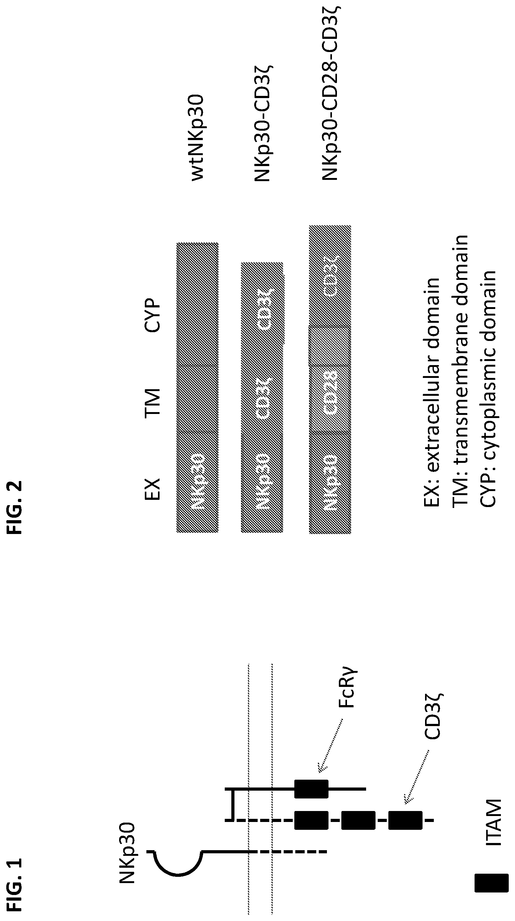
[0033] FIG. 1 provides a graphical overview of proteins involved in NKp30 signaling. Ligand-bound NKp30 can activate signaling through CD3.zeta. and FcR.gamma.. The long horizontal lines represent the cell membrane, with the extracellular space oriented toward the top of the figure. NKp30 interaction with CD3.zeta. correlates with the NKp30 splice variant isoform, with NKp30 isoform A associating with CD3.zeta. upon cross-linking, NKp30 isoform B constitutively associating with CD3.zeta., and NKp30 isoform C poorly associating with CD3.zeta.. ITAM: immunoreceptor tyrosine-based activation motif.
[0034] FIG. 2 illustrates chimeric NK receptors exemplified herein. Extracellular (EX or EC), transmembrane (TM), and cytoplasmic (CYP) (i.e., intracellular) portions are indicated. Wild-type (WT) and chimeric forms of the receptors are indicated. wtNKp30 indicates the mature wild-type NKp30 polypeptide. NKp30-CD3.zeta. indicates an NKp30 cytoplasmic domain fused to the transmembrane and cytoplasmic domains of CD3.zeta.. NKp30-CD28-CD3 indicates an NKp30 cytoplasmic domain fused to the transmembrane and cytoplasmic domains of CD28, and additionally the cytoplasmic domain of CD3.zeta.. All constructs are human.
[0035] FIGS. 3A-I shows the level of surface expression of chimeric NKp30 receptors on human T cells for the constructs illustrated in FIG. 2 and additional constructs. Surface expression was analyzed by flow cytometry using labeled anti-NKp30 and anti-CD4 mAbs (the latter identifies CD4-positive T cells). Results are shown for mock-transfected cells (FIG. 3A); wild-type (i.e., non-chimeric) NKp30 transfected cells (FIG. 3B); chimeric NKp30-CD3.zeta. transfected cells (FIG. 3C); chimeric NKp30-CD28-CD3.zeta. transfected cells (FIG. 3D); wild-type (i.e., non-chimeric) NKp30 transfected cells (FIG. 3E); chimeric NKp30-CD28-CD3.zeta. transfected cells (FIG. 3F); NKp30-CD28(TM)-DAP10-CD3.zeta. transfected cells (FIG. 3G); NKp30-CD28(TM)-CD3.zeta.-Dap10 transfected (FIG. 3H); and NKp30-CD28(TM)-CD27-CD3.zeta. (FIG. 3I). The results generally indicate robust surface expression of the chimeric constructs, except that the level of detected surface expression was somewhat lower for 30-tm28-27-z than the other constructs. In contrast, retroviral transduction of human T cells with wtNKp30 gene did not lead to significant surface expression.
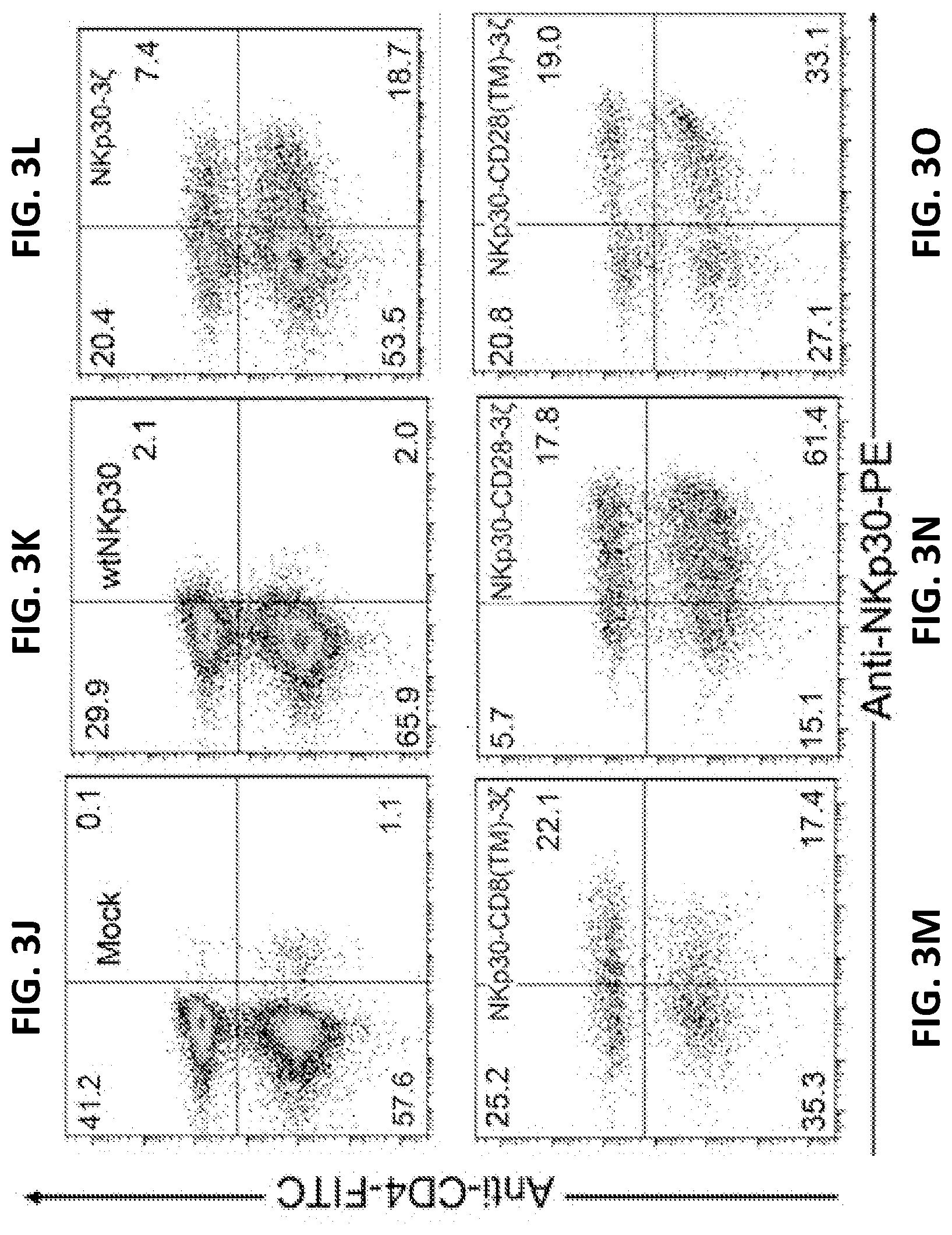
[0036] FIGS. 3J-O show NKp30 expression on human T cells 7 d after transduction with NKp30 chimric antigen receptors (CARs). NKp30 expression was measured using the PE-conjugated anti-NKp30 mAbs in combination with anti-CD4-FITC mAbs. More than 99% of cells were CD3+ T cells (data not shown). CD4- T cells are CD8+ T cells. Results are shown for mock-transfected cells (FIG. 3J); wild-type (i.e., non-chimeric) NKp30 transfected cells (FIG. 3K); chimeric NKp30-CD3.zeta. transfected cells (FIG. 3L); chimeric NKp30-CD8(TM)-CD3.zeta. transfected cells (FIG. 3M); chimeric NKp30-CD28-CD3.zeta. transfected cells (FIG. 3N) and NKp30-CD28(TM)-CD3.zeta. transfected cells (FIG. 3O). The data are representative of three experiments.
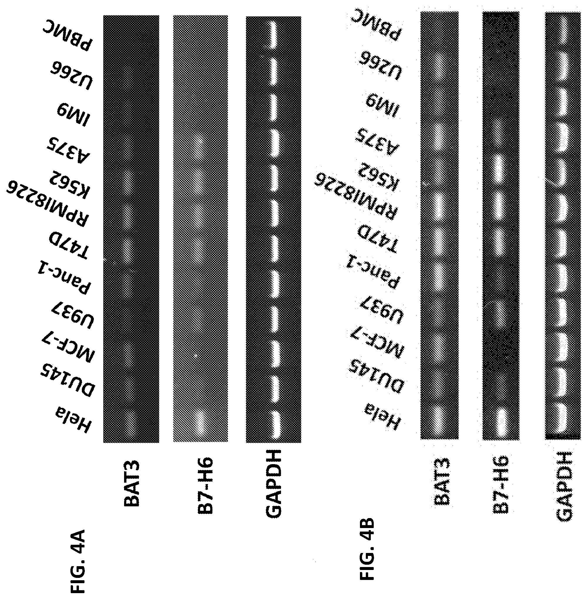
[0037] FIGS. 4A-B. Expression of NKp30 ligands on tumor cells and PBMC. Human tumor cell lines as well as human PBMCs were screened for the expression of NKp30 ligand mRNAs by RT-PCR using primers specific for the NKp30 ligands BAT3 and B7-H6, as well as a housekeeping gene (GAPDH) as an internal positive control. All tested human tumor cells had detectable amounts of BAT3 mRNA, whereas B7-H6 expression was readily detected in HeLa, U937, Panc-1, T47D, RPM18226, K562, and A375 cells, but expression was at lower levels or undetectable in MCF-7, DU145, IM9, U266 and human PBMCs.
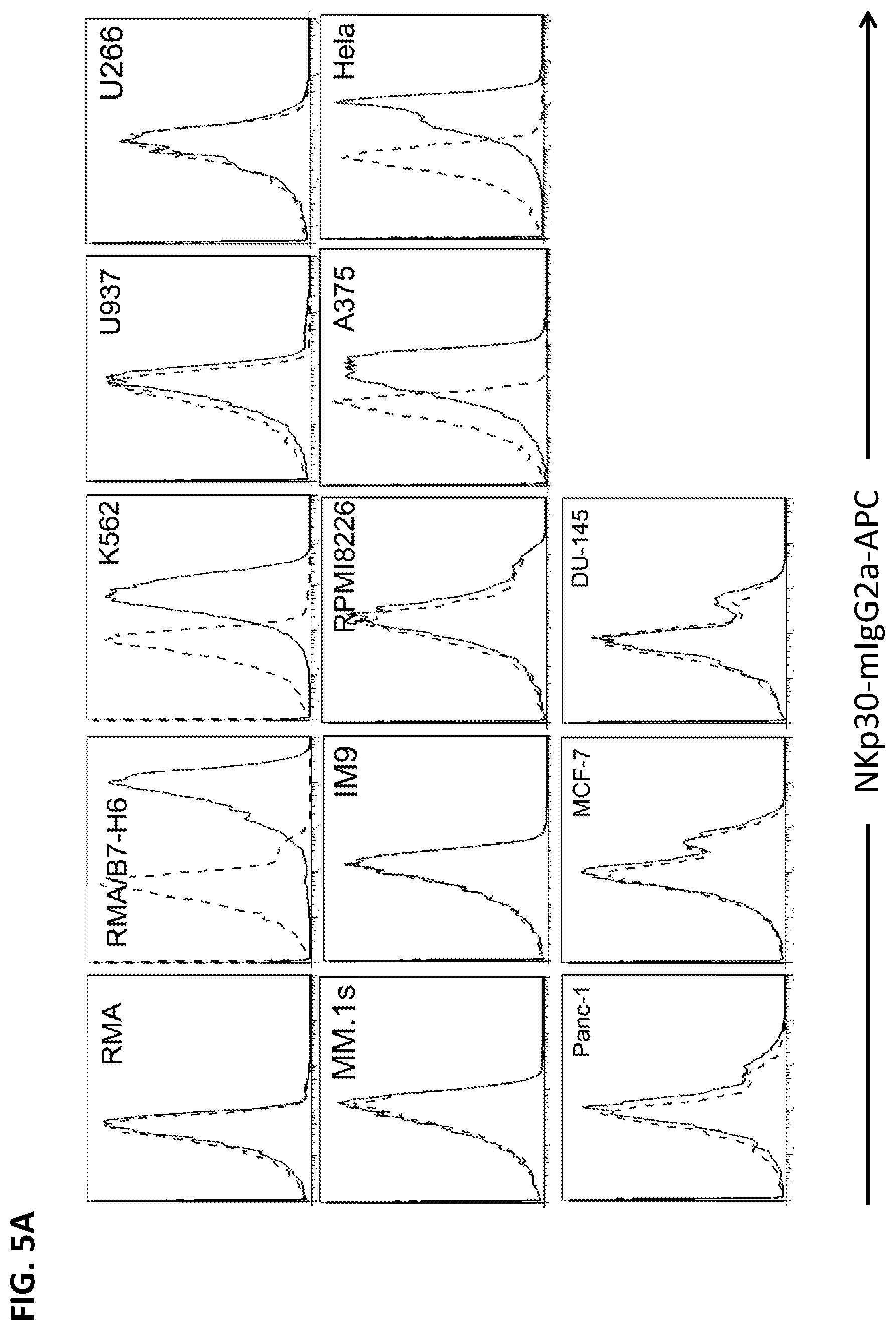
[0038] FIG. 5A. Surface expression of NKp30 ligands on tumor cells. K562, A375 and HeLa cells express high amounts of NKp30 ligands, whereas U937, RPMP8226, T47D and Panc-1 cells express marginal levels of NKp30 ligands. Some tumor cells (IM9, MM.1s, MCF-7 and DU145) as well as human PBMCs do not express NKp30 ligands. B7-H6 mRNA amounts are correlated with surface expression of NKp30 ligands, suggesting that B7-H6 is the major surface ligand of NKp30 in the tested tumors.
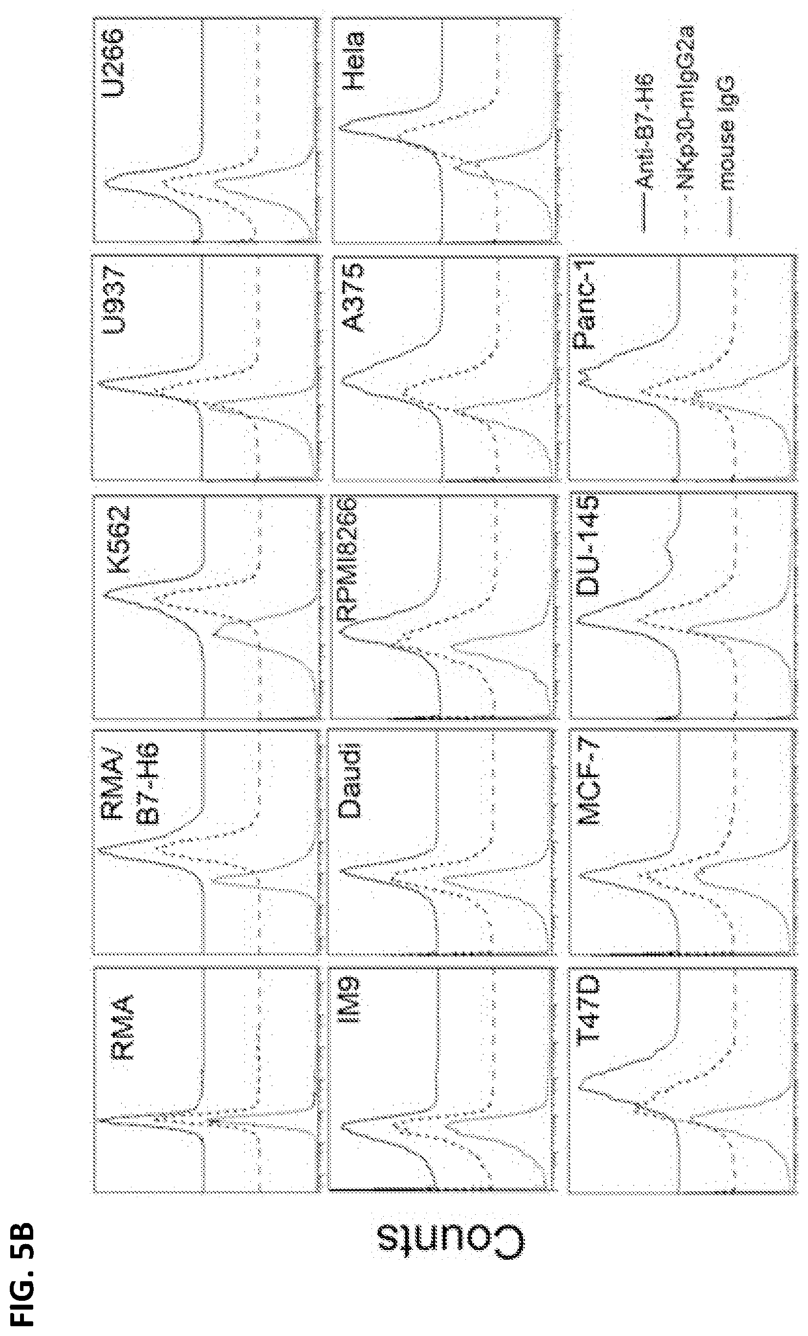
[0039] FIG. 5B. NKp30 ligand expression on the surface of human tumor cell lines was measured by flow cytometry using anti-B7-H6 mAbs (solid line) or a soluble NKp30 receptor fused to a mouse IgG2a Fc region (NKp30-mIgG2a; dashed line), followed by staining with allophycocyanin-conjugated goat anti-mouse IgG. Isotype controls are shown as a dotted line.
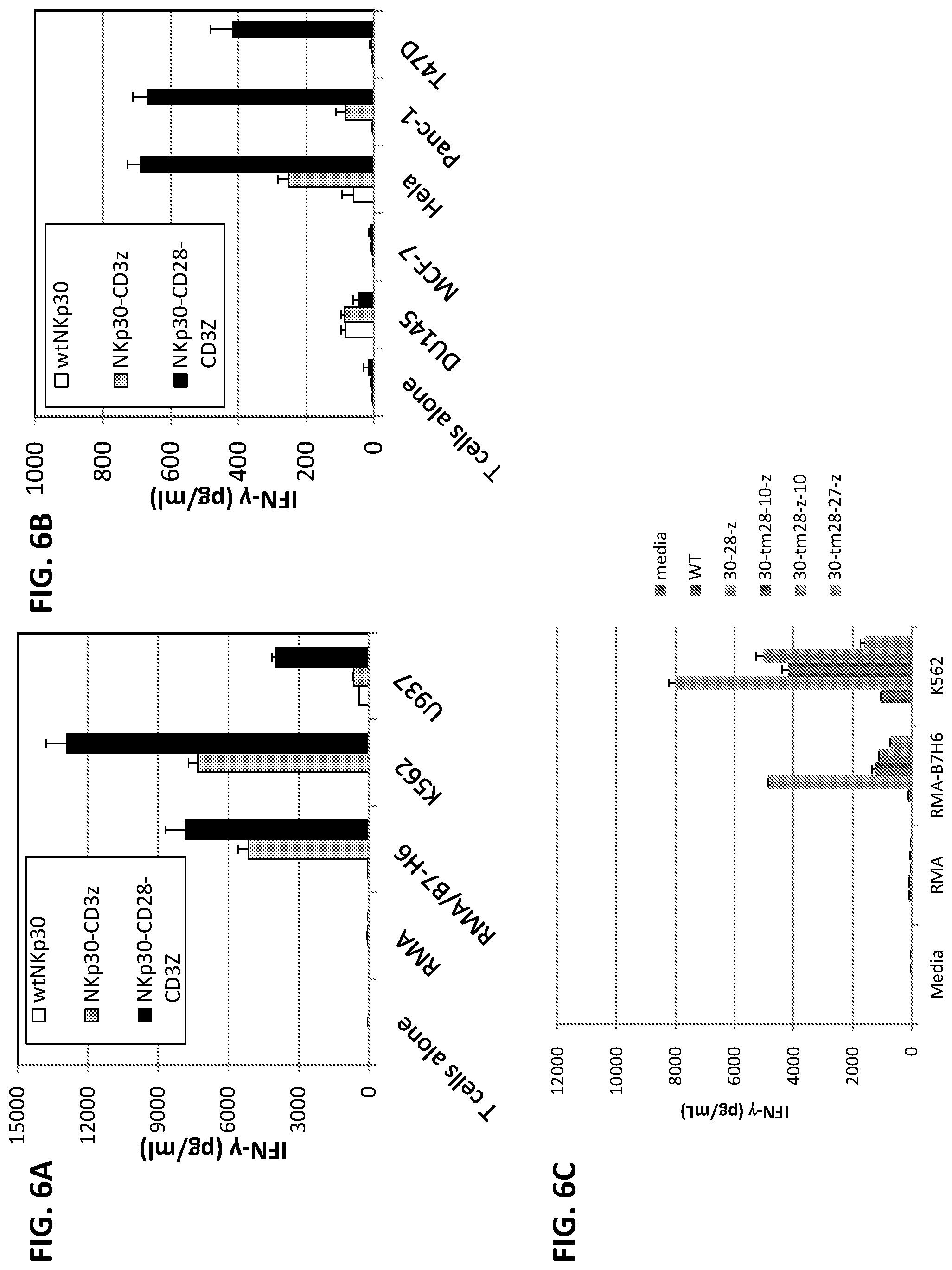
[0040] FIGS. 6A-E illustrates production of IFN-.gamma. after co-culture of transfected T cells with NKp30 ligand-positive cells but not with ligand-negative cells (or T cells alone). FIGS. 6A-B show that NKp30-CD3.zeta.+ (grey bars, middle bar in each group) and NKp30-CD28-CD3.zeta.+ T (black bars, rightmost bar in each group) cells produced significant amounts of IFN-.gamma., indicating that these chimeric NKp30-modified T cells could functionally recognize NKp30 ligand-bearing tumor cells. In contrast, wild-type NKp30-modified T cells (FIG. 6A, white bars, left bar in each group) did not show any significant response to the stimulation by NKp30-ligand positive cells. FIG. 6C illustrates IFN-.gamma. production after co-culture with T cells that were mock-transfected, transfected with wild-type NKp30, NKp30-CD28-CD3.zeta., NKp30-CD28(TM)-DAP10-CD3.zeta., NKp30-CD28(TM)-CD3.zeta.-Dap10, and NKp30-CD28(TM)-CD27-CD3.zeta. (leftmost through rightmost bars in each group, respectively). IFN-.gamma. production was highest in the cultures containing NKp30-CD28-CD3.zeta. transfected T cells. FIGS. 6D-E further illustrate that NKp30 chimeric antigen receptor-modified T cells respond to NKp30 ligand-positive cells by producing IFN-.gamma.. Five to seven days after retroviral transduction, NKp30 chimeric antigen receptor-modified T cells (100,000 cells) were cocultured with either irradiated or mitomycin C-treated tumor cells for 24 h. Suspension tumor cells (100,000 cells) (D) and adherent tumor cells (25,000 cells) (F) were used. RMA and MCF-7 cells were used as negative controls. IFN-.gamma. amounts in the supernatants were analyzed with ELISA. Results are shown as mean+/-SD. Asterisks (*) indicate p<0.05.
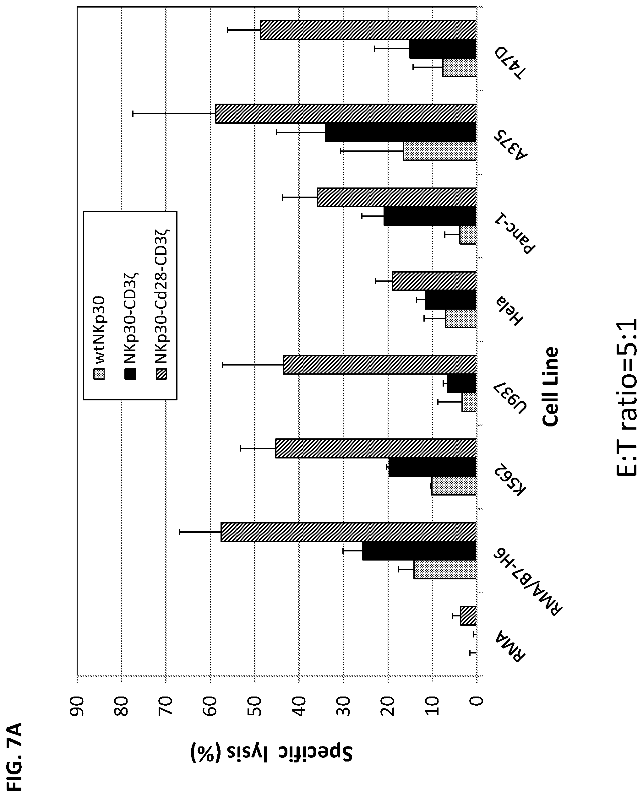
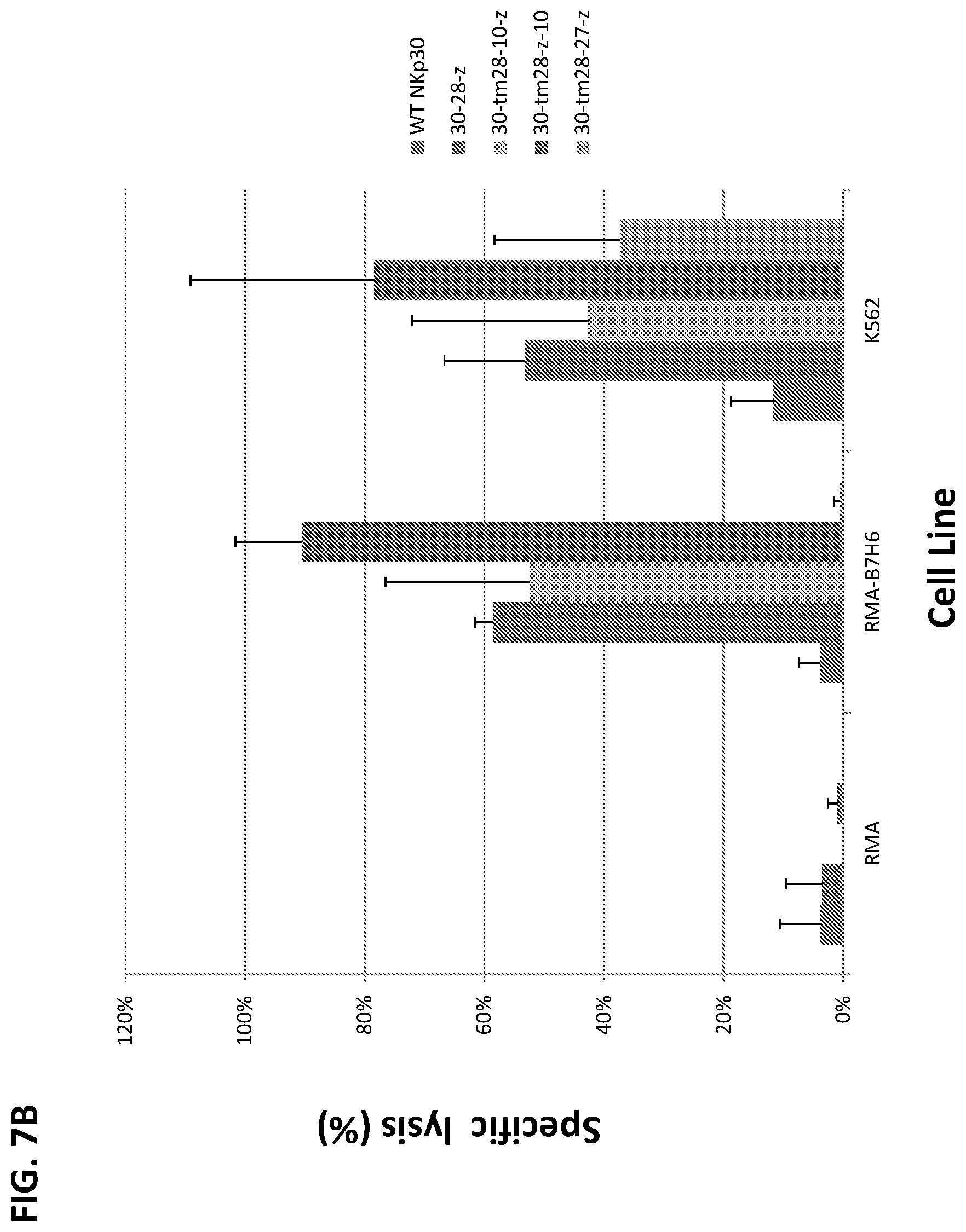
[0041] FIGS. 7A-B. Chimeric NKp30-bearing human T cells lysed NKp30-ligand positive tumor cells. Effector T cells derived from human PBMCs were modified with wtNKp30, NKp30-3.zeta., or NKp30-CD28-3.zeta. and cocultured with tumor cells at a ratio of 5:1 in 5-h LDH-release assays. In FIG. 7A, NKp30-CD3.zeta.+ (black bars, middle bar in each group) or NKp30-CD28-CD3.zeta.+ (diagonal hatched bars, rightmost bar in each group) T cells lysed NKp30 ligand-positive cells (RMA/B7-H6, K562, U937, HeLa, Panc-1, A375, and T47D) but not ligand-negative cells (cell line RMA) indicating that these chimeric NKp30-modified T cells could functionally recognize NKp30 ligand-bearing tumor cells in a specific manner. In contrast, a far lower percentage of tumor cells were lysed in the presence of wild-type NKp30-modified T cells (FIG. 7A, light gray bars, left bar in each group). NK30-CD28-CD3.zeta.+ killed a greater percentage of NKp30 ligand-positive tumor cells than NKp30-CD3.zeta.. Results are shown for triplicate experiments (mean+/-SD). Similarly, in FIG. 7B, the T cells expressing the 30-28-z, 30-tm28-10-z, 30-tm28-z-10, and 30-tm28-27-z constructs lysed NKp30 ligand-positive cells (RMA-B7H6 and K562) but not the negative control RMA cell (which lacks B7H6 expression), whereas far fewer tumor cells were lysed in the presence of T cells expressing wild-type NKp30. FIG. 7B legend: constructs in each group of bars, in order from left to right, were: NKp30, 30-28-z, 30-tm28-10-z, 30-tm28-z-10, and 30-tm28-27-z.
[0042] FIG. 7C further illustrates that lysis was mediated by the chimeric NKp30 constructs. NKp30-mIgG2a significantly reduced NKp30-28-3.zeta.-bearing T cell-mediated cytotoxicity. These results demonstrated that chimeric NKp30-bearing T cells killed ligand-positive tumor cells, and the interaction between chimeric NKp30 receptors and NKp30 ligands was essential for chimeric NKp30-mediated T cell function. Effector T cells modified with wtNKp30 or NKp30-CD28-3.zeta. were cocultured with target cells K562 in the presence of 10 .mu.g/ml NKp30-mIgG2a or mouse IgG at a ratio of 5:1; the percentage of specific lysis was determined after a 5-h LDH-release assay. The data are presented as mean+/-SD and are representative of two independent experiments.
[0043] FIG. 8. Shows data indicating that PI3 kinase is involved in NKp30-CD28-CD3.zeta.-mediated cytotoxicity. Specific lysis was significantly decreased for the chimeric NKp30-CD28-CD3.zeta. T cells incubated with the Ly294002 inhibitor, indicating that the PI3 kinase plays a role in NKp30-CD28-CD3.zeta.-mediated cytotoxicity. NKp30-modified effector T cells were incubated with a PI3K inhibitor LY294002 (10 .mu.M) at 37.degree. C. for 1 h before coculture with K562 target cells at a E:T ratio of 5:1 in 5-h LDH-release assays. Vehicle controls are 0.1% DMSO. The data shown are the mean+/-SD of triplicates and are representative of two independent experiments. Asterisk (*) indicates p<0.05.
[0044] FIGS. 9A-D. NKp30 expression on mouse T cells. Expression levels are shown for mock-transfected cells (FIG. 9A); wild-type (i.e., non-chimeric) NKp30 transfected cells (FIG. 9B); chimeric NKp30-CD3.zeta. transfected cells (FIG. 9C); and chimeric NKp30-CD28-CD3.zeta. transfected cells (FIG. 9D).
[0045] FIGS. 9E-J further exemplifies chimeric human NKp30 expression on mouse cells. Human NKp30 expression on mouse T cells 7 d after transduction. NKp30 expression was detected using the PE-conjugated anti-NKp30 mAb in combination with the anti-mouse CD4-FITC mAb. CD42 T cells are CD8+ T cells. The data are representative of three experiments. Expression levels are shown for mock-transfected cells (FIG. 9E); wild-type (i.e., non-chimeric) NKp30 transfected cells (FIG. 9F); chimeric NKp30-CD3.zeta. transfected cells (FIG. 9G); chimeric NKp30-CD8(TM)-CD3.zeta. transfected cells (FIG. 9H); chimeric NKp30-CD28(TM)-CD3.zeta. transfected cells (FIG. 9I), and chimeric NKp30-CD28-CD3.zeta. transfected cells (FIG. 9J).
[0046] FIGS. 10A-B. Human NKp30 receptors are functional in mouse T cells. Effector T cells derived from B6 (open), perforin-deficient (Pfp-/-, filled) mice that were modified with NKp30 receptors were co-cultured with RMA or RMA/B7-H6 cells at an E:T ratio of 1:1 in 5-h LDH-release assays. The T cells lysed a significantly higher percentage of NKp30 ligand-positive cells (RMA/B7-H6, FIG. 10B) than ligand-negative cells (cell line RMA, FIG. 10A). The data are presented as mean.+-.SD of triplicates and are representative results from two independent experiments (FIGS. 10A and 10B). Specific lysis was substantially decreased with the Pfp-/- cells.
[0047] FIG. 10C shows amounts of IFN-.gamma. produced by transduced murine T cells in response to NKp30 ligand-positive cells. Seven days after retroviral transduction, NKp30-modified T cells (100,000 cells) were cocultured with irradiated RMA/B7-H6 cells (100,000 cells) for 24 h. Mouse lymphoma cell line RMA was used as a negative control. IFN-.gamma. amounts in the supernatants were analyzed by ELISA. Results are shown as mean+/-SD.
[0048] FIGS. 11A-C. Integration of CD28 TM and signaling domains into chimeric NKp30 receptor leads to long-term survival in tumor-bearing mice treated with these T cells in vivo. Mouse T cells transduced with chimeric NKp30 enhanced survival of mice injected with lymphoma cells, specifically RMA/B7-H6 that express the NKp30 ligand B7-H6 (schatically illustrated in FIG. 11A). Integration of CD28 TM and signaling domains into chimeric NKp30 receptor was demonstrated to lead to better anti-tumor in vivo efficacy. RMA/B7-H6 cells (105) were administered i.v. on day 0. On day 5, 7 and 9, tumor-bearing mice were injected with T cells (5.times.10.sup.6) that were modified to express wtNKp30 (.diamond-solid.), NKp30-CD3.zeta. (.tangle-solidup.), NKp30-CD8-CD3.zeta. (.circle-solid.) and NKp30-CD28-CD3.zeta. (.quadrature.), respectively (FIG. 11B). Injection with a saline solution (HBSS) was used as negative control. Data are presented in Kaplan-Meier survival curves. Additionally, surviving mice gained ligand-independent tumor resistance. Long-term survivors were re-challenged with a similar, but ligand-deficient, lymphoma, specifically RMA cells that had not been transformed with the B7-H6 construct. Overall survival was determined, and none of the re-challenged mice had tumor growth by the end of the study period, whereas naive mice did (FIG. 11C).
[0049] FIGS. 12A-B. Human dendritic cells ("DCs") bind to NKp30 and can stimulate autologous NKp30-CD28-3.zeta.-modified T cells to produce IFN-.gamma.. (A) The cell surface phenotype and binding to NKp30 of PBMC-derived human DCs (both iDCs and mDCs) was determined by flow cytometry. Specific mAb or NKp30-Ig as indicated (solid line) or an isotype control Ab staining (dashed line) is shown. (B) Five to seven days after retroviral transduction, NKp30 chimeric antigen receptor-modified T cells (10.sup.5 cells) were cocultured with either iDCs or mDCs at a ratio of 5:1 (T/DC) for 24 h. IFN-.gamma. amounts in the supernatants were determined by ELISA. Results shown (mean+SD) are representative of two experiments. Asterisk (*) indicates p<0.05. As further discussed infra, because NKp30 ligands can be expressed by human dendritic cells, these results help confirm that the methods and compositions of the present disclosure may be useful to prevent or treat diseases where elimination of dendritic cells may be helpful, such as autoimmune diseases or rejection of transplanted organs.
[0050] FIGS. 13A-B. Engagement of NKp30-CD28-3.zeta. receptor led to increased T cell proliferation and upregulation of IL-2 and Bcl-x.sub.L. (A) NKp30 receptor (either wtNKp30 or chimeric NKp30)-modified human T cells were labeled with CFSE, as described in the examples below, and cocultured with HeLa cells (100,000 cells, NKp30 ligand-positive) in the presence of a small amount of IL-2 (25 U/ml) for 3 d. Analysis of T cell proliferation (i.e., CFSE dilution) was performed on both NKp30+ (FL4) and NKp30-cells within the same mixed T cell population by flow cytometry. (B) NKp30-modified T cells (250,000 cells) were cultured in anti-NKp30 mAb (4 .mu.g)-coated 24-well plates for 24 h. Mouse IgG was used as a negative control. IL-2 gene expression was determined by real-time PCR, as described in the examples below. Results are shown as fold increase, in which the IL-2 gene expression in the control mAb-treated T cells was normalized to 1. Data are presented as mean+/-SD from two independent experiments. (C) Twenty-four hours after cross-linking with immobilized anti-NKp30 mAbs, as described in the examples below, T cells were collected. Bcl-x.sub.L expression was determined by flow cytometry after intracellular staining with anti-Bcl-x.sub.L-FITC (solid line) or isotype control mAbs (dashed line). Asterisk (*) indicates p<0.05.
[0051] FIG. 14 schematically illustrates additional exemplary chimeric NKp30 receptor constructs. The two horizontal lines represent a cell membrane, with the portion above both lines corresponding to the extracellular domain (NKp30 extracellular domain in each Illustrated construct), the portion between the horizontal lines corresponding to the transmembrane domain (e.g., CD3.zeta. transmembrane domain or CD28 domain in the illustrated construct, though other transmembrane domains are also used in exemplary embodiments, e.g., a CD8 transmembrane domain in the construct shown in FIG. 19); and the portion below both horizontal lines including the one or more activation domain(s) (e.g., the cytoplasmic domains of CD3.zeta., CD28, DAP10, CD27, and combinations thereof. Illustrated constructs include wild-type NKp30 (Wt-NKp30); NKp30-CD3.zeta., containing the NKp30 extracellular domain and CD3.zeta. transmembrane and cytoplasmic domains; 30-tm28-3.zeta. (also referred to herein as NKp30-CD28(TM)-CD3.zeta.); 30-28-3.zeta. (also referred to herein as NKp30-CD28-CD3.zeta.); 30-tm28-10-3.zeta. (also referred to herein as NKp30-CD28(TM)-DAP10-CD3.zeta.); 30-tm28-3.zeta.-10 (also referred to herein as NKp30-CD28(TM)-CD3.zeta.-Dap10); and 30-tm28-27-3.zeta. (also referred to herein as NKp3-CD28(TM)-CD27-CD3.zeta.).
[0052] FIG. 15 provides a polypeptide sequence of a wild-type human NKp30 and illustrates domains thereof.
[0053] FIGS. 16-22 provide polypeptide sequences of chimeric NKp30 receptors, and exemplary polynucleotide sequences are shown below. In these figures, the labels "TM" or "TM domain" refer to transmembrane domain sequences; the labels "EC" or "EC domain" refer to extracellular sequences (e.g., NKp30 ligand-binding domain sequences) and "CYP" or "CYP domain" refer to cytoplasmic domain sequences (which include signaling domains of the identified polypeptides). The identified polypeptides refer to the wild-type sequences from which the respective domains were derived.
[0054] FIG. 16 provides the polypeptide sequence of an NKp30-CD3.zeta. (also referred to herein as NKp30-CD3.zeta. or NKp30-C or NKp30-3z) construct comprising a signal peptide and extracellular domain of NKp30, and a transmembrane and cytoplasmic domain of CD3.zeta.. An exemplary coding sequence of this NKp30-CD3.zeta. construct is:
TABLE-US-00001 (SEQ ID NO: 124) ATGGCCTGGATGCTGTTGCTCATCTTGATCATGGTCCATCCAGGATCCTG TGCTCTCTGGGTGTCCCAGCCCCCTGAGATTCGTACCCTGGAAGGATCCT CTGCCTTCCTGCCCTGCTCCTTCAATGCCAGCCAAGGGAGACTGGCCATT GGCTCCGTCACGTGGTTCCGAGATGAGGTGGTTCCAGGGAAGGAGGTGAG GAATGGAACCCCAGAGTTCAGGGGCCGCCTGGCCCCACTTGCTTCTTCCC GTTTCCTCCATGACCACCAGGCTGAGCTGCACATCCGGGACGTGCGAGGC CATGACGCCAGCATCTACGTGTGCAGAGTGGAGGTGCTGGGCCTTGGTGT CGGGACAGGGAATGGGACTCGGCTGGTGGTGGAGAAAGAACATCCTCAGC TAGGGGCTAGCCTCTGCTACCTGCTGGATGGAATCCTCTTCATCTATGGT GTCATTCTCACTGCCTTGTTCCTGAGAGTGAAGTTCAGCAGGAGCGCAGA CGCCCCCGCGTACCAGCAGGGCCAGAACCAGCTCTATAACGAGCTCAATC TAGGACGAAGAGAGGAGTACGATGTTTTGGACAAGAGACGTGGCCGGGAC CCTGAGATGGGGGGAAAGCCGCAGAGAAGGAAGAACCCTCAGGAAGGCCT GTACAATGAACTGCAGAAAGATAAGATGGCGGAGGCCTACAGTGAGATTG GGATGAAAGGCGAGCGCCGGAGGGGCAAGGGGCACGATGGCCTTTACCAG GGTCTCAGTACAGCCACCAAGGACACCTACGACGCCCTTCACATGCAGGC CCTGCCCCCTCGC.
[0055] FIG. 17 provides the polypeptide sequence of an NKp30-CD28-CD3.zeta. (also referred to herein as NKp30-CD28-CD3, 30-28-z, 30-28-3.zeta., NKp30-CD28-CD3z, or NKp30-CD28-.zeta.) construct comprising a signal peptide and extracellular domain of NKp30, a transmembrane and cytoplasmic domain of CD28, and a further cytoplasmic domain of CD3.zeta.. An exemplary coding sequence of this NKp30-CD28-CD3.zeta. construct is:
TABLE-US-00002 (SEQ ID NO: 125) ATGGCCTGGATGCTGTTGCTCATCTTGATCATGGTCCATCCAGGATCCTG TGCTCTCTGGGTGTCCCAGCCCCCTGAGATTCGTACCCTGGAAGGATCCT CTGCCTTCCTGCCCTGCTCCTTCAATGCCAGCCAAGGGAGACTGGCCATT GGCTCCGTCACGTGGTTCCGAGATGAGGTGGTTCCAGGGAAGGAGGTGAG GAATGGAACCCCAGAGTTCAGGGGCCGCCTGGCCCCACTTGCTTCTTCCC GTTTCCTCCATGACCACCAGGCTGAGCTGCACATCCGGGACGTGCGAGGC CATGACGCCAGCATCTACGTGTGCAGAGTGGAGGTGCTGGGCCTTGGTGT CGGGACAGGGAATGGGACTCGGCTGGTGGTGGAGAAAGAACATCCTCAGC TAGGGGCTAGCTTTTGGGTGCTGGTGGTGGTTGGTGGAGTCCTGGCTTGC TATAGCTTGCTAGTAACAGTGGCCTTTATTATTTTCTGGGTGAGGAGTAA GAGGAGCAGGCTCCTGCACAGTGACTACATGAACATGACTCCCCGCCGCC CCGGGCCCACCCGCAAGCATTACCAGCCCTATGCCCCACCACGCGACTTC GCAGCCTATCGCTCCAAGCTTAGAGTGAAGTTCAGCAGGAGCGCAGACGC CCCCGCGTACCAGCAGGGCCAGAACCAGCTCTATAACGAGCTCAATCTAG GACGAAGAGAGGAGTACGATGTTTTGGACAAGAGACGTGGCCGGGACCCT GAGATGGGGGGAAAGCCGCAGAGAAGGAAGAACCCTCAGGAAGGCCTGTA CAATGAACTGCAGAAAGATAAGATGGCGGAGGCCTACAGTGAGATTGGGA TGAAAGGCGAGCGCCGGAGGGGCAAGGGGCACGATGGCCTACCAGGGTCT CAGTACAGCCACCAAGGACACCTACGACGCCCTTCACATGCAGGCCCTGC CCCCTCGC.
[0056] FIG. 18 provides the polypeptide sequence of an NKp30-CD28(TM)-CD3.zeta. (also referred to herein as NKp30-CD28(TM)-.zeta. or 30-tm28-3) construct comprising a signal peptide and extracellular domain of NKp30, and a transmembrane and cytoplasmic domain of CD3.zeta.. An exemplary coding sequence of this NKp30-CD28(TM)-CD3.zeta. construct is:
TABLE-US-00003 (SEQ ID NO: 126) ATGGCCTGGATGCTGTTGCTCATCTTGATCATGGTCCATCCAGGATCCTG TGCTCTCTGGGTGTCCCAGCCCCCTGAGATTCGTACCCTGGAAGGATCCT CTGCCTTCCTGCCCTGCTCCTTCAATGCCAGCCAAGGGAGACTGGCCATT GGCTCCGTCACGTGGTTCCGAGATGAGGTGGTTCCAGGGAAGGAGGTGAG GAATGGAACCCCAGAGTTCAGGGGCCGCCTGGCCCCACTTGCTTCTTCCC GTTTCCTCCATGACCACCAGGCTGAGCTGCACATCCGGGACGTGCGAGGC CATGACGCCAGCATCTACGTGTGCAGAGTGGAGGTGCTGGGCCTTGGTGT CGGGACAGGGAATGGGACTCGGCTGGTGGTGGAGAAAGAACATCCTCAGC TAGGGGCTAGCTTTTGGGTGCTGGTGGTGGTTGGTGGAGTCCTGGCTTGC TATAGCTTGCTAGTAACAGTGGCCTTTATTATTTTCTGGGTGAGGAGTAA GAAGCTTAGAGTGAAGTTCAGCAGGAGCGCAGACGCCCCCGCGTACCAGC AGGGCCAGAACCAGCTCTATAACGAGCTCAATCTAGGACGAAGAGAGGAG TACGATGTTTTGGACAAGAGACGTGGCCGGGACCCTGAGATGGGGGGAAA GCCGCAGAGAAGGAAGAACCCTCAGGAAGGCCTGTACAATGAACTGCAGA AAGATAAGATGGCGGAGGCCTACAGTGAGATTGGGATGAAAGGCGAGCGC CGGAGGGGCAAGGGGCACGATGGCCTTTACCAGGGTCTCAGTACAGCCAC CAAGGACACCTACGACGCCCTTCACATGCAGGCCCTGCCCCCTCGC.
[0057] FIG. 19 provides the polypeptide sequence of an NKp30-CD8-CD3.zeta. (also referred to herein as NKp30-CD8-.zeta. or NKp30-CD8(TM)-3.zeta.) construct comprising a signal peptide and extracellular domain of NKp30, a transmembrane domain of CD8, and a cytoplasmic domain of CD3.zeta.. An exemplary coding sequence of this NKp30-CD8-CD3.zeta. construct is:
TABLE-US-00004 (SEQ ID NO: 127) ATGGCCTGGATGCTGTTGCTCATCTTGATCATGGTCCATCCAGGATCCTG TGCTCTCTGGGTGTCCCAGCCCCCTGAGATTCGTACCCTGGAAGGATCCT CTGCCTTCCTGCCCTGCTCCTTCAATGCCAGCCAAGGGAGACTGGCCATT GGCTCCGTCACGTGGTTCCGAGATGAGGTGGTTCCAGGGAAGGAGGTGAG GAATGGAACCCCAGAGTTCAGGGGCCGCCTGGCCCCACTTGCTTCTTCCC GTTTCCTCCATGACCACCAGGCTGAGCTGCACATCCGGGACGTGCGAGGC CATGACGCCAGCATCTACGTGTGCAGAGTGGAGGTGCTGGGCCTTGGTGT CGGGACAGGGAATGGGACTCGGCTGGTGGTGGAGAAAGAACATCCTCAGC TAGGGGCTAGCATCTACATCTGGGCGCCCTTGGCCGGGACTTGTGGGGTC CTTCTCCTGTCACTGGTTATCACCAAGCTTAGAGTGAAGTTCAGCAGGAG CGCAGACGCCCCCGCGTACCAGCAGGGCCAGAACCAGCTCTATAACGAGC TCAATCTAGGACGAAGAGAGGAGTACGATGTTTTGGACAAGAGACGTGGC CGGGACCCTGAGATGGGGGGAAAGCCGAGAAGGAAGAACCCTCAGGAAGG CCTGTACAATGAACTGCAGAAAGATAAGATGGCGGAGGCCTACAGTGAGA TTGGGATGAAAGGCGAGCGCCGGAGGGGCAAGGGGCACGATGGCCTTTAC CAGGGTCTCAGTACAGCCACCAAGGACACCTACGACGCCCTTCACATGCA GGCCCTGCCCCCTCGCTAA.
[0058] FIG. 20 provides the polypeptide sequence of a NKp30-CD28(TM)-DAP0-CD3.zeta. (also referred to herein as 30-tm28-10-3z, Dap10a, 30-tm28-10-7, or 30-tm2-10-3.zeta.) construct comprising a signal peptide and extracellular domain of NKp30, transmembrane domain of CD28, cytoplasmic domain of Dap10, and a cytoplasmic domain of CD3.zeta.. An exemplary coding sequence of this NKp30-CD28(TM)-DAP10-CD3.zeta. construct is:
TABLE-US-00005 (SEQ ID NO: 64) ATGGCCTGGATGCTGTTGCTCATCTTGATCATGGTCCATCCAGGATCCTG TGCTCTCTGGGTGTCCCAGCCCCCTGAGATTCGTACCCTGGAAGGATCCT CTGCCTTCCTGCCCTGCTCCTTCAATGCCAGCCAAGGGAGACTGGCCATT GGCTCCGTCACGTGGTTCCGAGATGAGGTGGTTCCAGGGAAGGAGGTGAG GAATGGAACCCCAGAGTTCAGGGGCCGCCTGGCCCCACTTGCTTCTTCCC GTTTCCTCCATGACCACCAGGCTGAGCTGCACATCCGGGACGTGCGAGGC CATGACGCCAGCATCTACGTGTGCAGAGTGGAGGTGCTGGGCCTTGGTGT CGGGACAGGGAATGGGACTCGGCTGGTGGTGGAGAAAGAACATCCTCAGC TAGGGGCTAGCTTTTGGGTGCTGGTGGTGGTTGGTGGAGTCCTGGCTTGC TATAGCTTGCTAGTAACAGTGGCCTTTATTATTTTCTGGGTGAGGAGTAA GAGGAGCCTGTGCGCACGCCCACGCCGCAGCCCCGCCCAAGAAGATGGCA AAGTCTACATCAACATGCCAGGCAGGGGCAAGCTTAGAGTGAAGTTCAGC AGGAGCGCAGACGCCCCCGCGTACCAGCAGGGCCAGAACCAGCTCTATAA CGAGCTCAATCTAGGACGAAGAGAGGAGTACGATGTTTTGGACAAGAGAC GTGGCCGGGACCCTGAGATGGGGGGAAAGCCGAGAAGGAAGAACCCTCAG GAAGGCCTGTACAATGAACTGCAGAAAGATAAGATGGCGGAGGCCTACAG TGAGATTGGGATGAAAGGCGAGCGCCGGAGGGGCAAGGGGCACGATGGCC TTTACCAGGGTCTCAGTACAGCCACCAAGGACACCTACGACGCCCTTCAC ATGCAGGCCCTGCCCCCTCGC.
[0059] FIG. 21 provides the polypeptide sequence of a NKp30-CD28(TM)-CD3.zeta.-Dap10 (also referred to herein as 30-tm28-3z-10, Dap10b, 30-tm28-z-10, or 30-tm28-3.zeta.-10) construct construct comprising a signal peptide and extracellular domain of NKp30, transmembrane domain of CD28, a cytoplasmic domain of CD3.zeta., and a cytoplasmic domain of Dap10. An exemplary coding sequence of this NKp30-CD28(TM)-CD3.zeta.-Dap10 construct is:
TABLE-US-00006 (SEQ ID NO: 66) ATGGCCTGGATGCTGTTGCTCATCTTGATCATGGTCCATCCAGGATCCTG TGCTCTCTGGGTGTCCCAGCCCCCTGAGATTCGTACCCTGGAAGGATCCT CTGCCTTCCTGCCCTGCTCCTTCAATGCCAGCCAAGGGAGACTGGCCATT GGCTCCGTCACGTGGTTCCGAGATGAGGTGGTTCCAGGGAAGGAGGTGAG GAATGGAACCCCAGAGTTCAGGGGCCGCCTGGCCCCACTTGCTTCTTCCC GTTTCCTCCATGACCACCAGGCTGAGCTGCACATCCGGGACGTGCGAGGC CATGACGCCAGCATCTACGTGTGCAGAGTGGAGGTGCTGGGCCTTGGTGT CGGGACAGGGAATGGGACTCGGCTGGTGGTGGAGAAAGAACATCCTCAGC TAGGGGCTAGCTTTTGGGTGCTGGTGGTGGTTGGTGGAGTCCTGGCTTGC TATAGCTTGCTAGTAACAGTGGCCTTTATTATTTTCTGGGTGAGGAGTAA GAGGAGCAAGCTTAGAGTGAAGTTCAGCAGGAGCGCAGACGCCCCCGCGT ACCAGCAGGGCCAGAACCAGCTCTATAACGAGCTCAATCTAGGACGAAGA GAGGAGTACGATGTTTTGGACAAGAGACGTGGCCGGGACCCTGAGATGGG GGGAAAGCCGAGAAGGAAGAACCCTCAGGAAGGCCTGTACAATGAACTGC AGAAAGATAAGATGGCGGAGGCCTACAGTGAGATTGGGATGAAAGGCGAG CGCCGGAGGGGCAAGGGGCACGATGGCCTTTACCAGGGTCTCAGTACAGC CACCAAGGACACCTACGACGCCCTTCACATGCAGGCCCTGCCCCCTCGCC TGTGCGCACGCCCACGCCGCAGCCCCGCCCAAGAAGATGGCAAAGTCTAC ATCAACATGCCAGGCAGGGGC.
[0060] FIG. 22 provides the polypeptide sequence of a NKp3-CD(TM)-CD27-CD3.zeta. (also referred to herein as 30-tm28-27-3z, 30-tm28-27-z, or 30-tm28-27-3.zeta.) construct comprising a signal peptide and extracellular domain of NKp30, transmembrane domain of CD28, a cytoplasmic domain of CD27, and a cytoplasmic domain of CD3.zeta.. An exemplary coding sequence of this NKp30-CD28(TM)-CD27-construct is:
TABLE-US-00007 [0061] (SEQ ID NO: 68) ATGGCCTGGATGCTGTTGCTCATCTTGATCATGGTCCATCCAGGATCCTG TGCTCTCTGGGTGTCCCAGCCCCCTGAGATTCGTACCCTGGAAGGATCCT CTGCCTTCCTGCCCTGCTCCTTCAATGCCAGCCAAGGGAGACTGGCCATT GGCTCCGTCACGTGGTTCCGAGATGAGGTGGTTCCAGGGAAGGAGGTGAG GAATGGAACCCCAGAGTTCAGGGGCCGCCTGGCCCCACTTGCTTCTTCCC GTTTCCTCCATGACCACCAGGCTGAGCTGCACATCCGGGACGTGCGAGGC CATGACGCCAGCATCTACGTGTGCAGAGTGGAGGTGCTGGGCCTTGGTGT CGGGACAGGGAATGGGACTCGGCTGGTGGTGGAGAAAGAACATCCTCAGC TAGGGGCTAGCTTTTGGGTGCTGGTGGTGGTTGGTGGAGTCCTGGCTTGC TATAGCTTGCTAGTAACAGTGGCCTTTATTATTTTCTGGGTGAGGAGTAA GAGGAGCCTCGAGCAACGAAGGAAATATAGATCAAACAAAGGAGAAAGTC CTGTGGAGCCTGCAGAGCCTTGTCGTTACAGCTGCCCCAGGGAGGAGGAG GGCAGCACCATCCCCATCCAGGAGGATTACCGAAAACCGGAGCCTGCCTG CTCCCCCAAGCTTAGAGTGAAGTTCAGCAGGAGCGCAGACGCCCCCGCGT ACCAGCAGGGCCAGAACCAGCTCTATAACGAGCTCAATCTAGGACGAAGA GAGGAGTACGATGTTTTGGACAAGAGACGTGGCCGGGACCCTGAGATGGG GGGAAAGCCGAGAAGGAAGAACCCTCAGGAAGGCCTGTACAATGAACTGC AGAAAGATAAGATGGCGGAGGCCTACAGTGAGATTGGGATGAAAGGCGAG CGCCGGAGGGGCAAGGGGCACGATGGCCTTTACCAGGGTCTCAGTACAGC CACCAAGGACACCTACGACGCCCTTCACATGCAGGCCCTGCCCCCTCGC.
DETAILED DESCRIPTION OF PREFERRED EMBODIMENTS
[0062] NKp30 is a natural cytotoxicity receptor that is expressed on NK cells and recognizes B7-H6, which is expressed on several types of tumors but few normal cells. To target effector T cells against B7-H6+ tumors, we developed several chimeric antigen receptors based on NKp30, including receptors that contain the CD28- and/or CD3.gamma.-signaling domains with the transmembrane domains from CD3.gamma., CD28, or CD8.alpha.. Chimeric NKp30-expressing T cells responded to B7-H6+ tumor cells. T cells expressing NKp30 chimeric antigen receptors (CARs) produced IFN-.gamma. and killed B7-H6 ligand-expressing tumor cells; this response was dependent upon ligand expression on target cells but not on MHC expression. PBMC-derived dendritic cells also express NKp30 ligands, including immature dendritic cells, and they stimulated NKp30 CAR-bearing T cells to produce IFN-.gamma., but to a lesser extent. The addition of a CD28-signaling domain significantly enhanced the activity of the NKp30 CAR in a PI3K-dependent manner.
[0063] Adoptive transfer of T cells expressing a chimeric NKp30 receptor containing a CD28-signaling domain inhibited the growth of a B7-H6-expressing murine lymphoma (RMA/B7-H6) in vivo. Moreover, mice that remained tumor-free were resistant to a subsequent challenge with the wild-type RMA tumor cells, suggesting the generation of immunity against other tumor antigens. These results demonstrates the specificity and therapeutic potential of adoptive immunotherapy with NKp30 chimeric antigen receptor-expressing T cells against B7-H6+ tumor cells in vivo.
[0064] The present disclosure relates to a chimeric receptor molecule comprising an NKp30 extracellular domain expressed on the surface of a T cell to activate killing of a tumor cell. Nucleic acid sequences encoding the chimeric receptor molecule are introduced into a patient's T-cells (or compatible T cells, e.g., an allogeneic T cell obtained from a compatible donor or a cell bank) and T-cells that express the chimeric receptor molecule are subsequently introduced into the patient. In this manner, the chimeric receptor molecules provide a means for the patient's own immune cells to recognize and destroy tumor cells, activate anti-tumor immunity, and establish long-term, specific, anti-tumor responses for treating tumors or preventing re-growth of dormant or residual tumor cells.
[0065] By way of illustration, human chimeric receptor molecules composed of an NKp30 extracellular domain in combination with transmembrane and/or cytoplasmic domains of CD3.zeta. and/or CD28 were generated and expressed in human T-cells. Specifically, a gene encoding a chimeric receptor comprising the extracellular domain of human NKp30 and transmembrane and cytoplasmic domains of human CD3.zeta. (NKp30-CD3.zeta. receptor) was generated. A gene encoding a chimeric receptor comprising the extracellular domain of human NKp30, the transmembrane domain of CD28, and cytoplasmic domain of human CD3.zeta. (NKp30-CD28-CD3.zeta. receptor) was also generated. As described further in the examples below, these chimeric NKp30 genes were introduced into human T cells ex vivo by retroviral transduction, and were shown to be efficiently surface-expressed. Moreover, T cells expressing these chimeric NKp30 receptors ("chimeric NKp30 T cells") were demonstrated to be specifically activated by and lysed NKp30 ligand-positive tumor cells. The same (human) chimeric NKp30 genes were introduced into mouse T cells ex vivo by retroviral transduction and were demonstrated to be surface-expressed by mouse cells as well, and transduced mouse cells were specifically activated by and lysed NKp30 ligand-positive tumor cells. Moreover, the transduced mouse T cells increased survival of mice injected with NKp30-ligand expressing tumor cells. Several mice became long-term survivors that were resistant to a tumor re-challenge with a similar, but NKp30 ligand-deficient, lymphoma. These results indicate that this chimeric T cell treatment can lead to tumor eradication and suggest induction of long-term tumor immunity in the animals.
Definitions
[0066] It is to be understood that this invention is not limited to the particular methodology, protocols, cell lines, animal species or genera, and reagents described, as such may vary. It is also to be understood that the terminology used herein is for the purpose of describing particular embodiments only, and is not intended to limit the scope of the present invention which will be limited only by the appended claims.
[0067] As used herein the singular forms "a", "and", and "the" include plural referents unless the context clearly dictates otherwise. Thus, for example, reference to "a cell" includes a plurality of such cells and reference to "the protein" includes reference to one or more proteins and equivalents thereof known to those skilled in the art, and so forth. All technical and scientific terms used herein have the same meaning as commonly understood to one of ordinary skill in the art to which this invention belongs unless clearly indicated otherwise.
[0068] "Allogeneic T cell" refers to a T cell from a donor having a tissue that is not genetically identical to the recipient. The T cell may have an HLA type that partially or fully matches the recipient or does not match the recipient. In some instances allogeneic transplant donors may be related (usually a closely HLA matched sibling), syngeneic (a monozygotic `identical` twin of the patient) or unrelated (donor who is not related and found to have very close degree of HLA matching). The HLA genes fall in two categories (Type I and Type I). In general, mismatches of the Type-I genes (i.e. HLA-A, HLA-B, or HLA-C) increase the risk of graft rejection. A mismatch of an HLA Type II gene (i.e. HLA-DR, or HLA-DQB1) increases the risk of graft-versus-host disease.
[0069] In the context of the present disclosure, a T cell progenitor refers to any cell that may give rise to a T cell including but not limited to adult and embryonic stem cells, induced stem cells, hematopoietic stem cells (e.g., CD34+ hematopoietic stem cells), thymocytes, and T lymphocyte-restricted progenitors typically found in the thymus. Additional exemplary T cell progenitors include thymocytes in the double negative stages (each negative for both CD4 and CD8), such as the double negative 1 stage (Lineage-CD44+CD25-CD117+), double negative 2 stage (Lineage-CD44+CD25+CD117+), double negative 3 stage (Lineage-CD44-CD25+), double negative 4 stage (Lineage-CD44-CD25-), double positive stage (CD4+CD8+) and/or single positive stage (CD4+CD8- or CD4-CD8+).
[0070] In the context of the present disclosure, a "bank of tissue matched chimeric NKp30 T cells" refers to different compositions each containing T cells of a specific HLA allotype which express a chimeric NKp30 receptor according to the present disclosure. Ideally this bank will comprise compositions containing T cells of a wide range of different HLA types that are representative of the human population. Such a bank of engineered chimeric NKp30 T cells will be useful as it will facilitate the availability of T cells suitable for use in different recipients, such as cancer patients.
[0071] As used herein. CD3.zeta. (CD3.zeta.) refers to human polypeptide or polynucleotide (or orthologs in other species if the context so indicates) having exemplary sequences as follows. Two CD3.zeta. transcript variants have been reported. One transcript variant, CD3.zeta. transcript variant 1, has Genbank accession number NM_198053 (polynucleotide), encoding the polypeptide CD3.zeta. chain isoform 1 precursor (NP_0.932170.1) having the sequence: MKWKALFTAAILQAQLPITEAQSFGLLDPKLCYLLDGILFIYGVILTALFLRVKFSRSADA PAYQQGQNQLYNELNLGRREEYDVLDKRRGRDPEMGGKPQRRKNPQEGLYNELQKDK MAEAYSEIGMKGERRRGKGHDGLYQGLSTATKDTYDALHMQALPPR (SEQ ID NO: 1) which is annotated as comprising a signal peptide (residues 1-21, i.e., MKWKALFTAAILQAQLPITEA (SEQ ID NO: 2)) and a mature peptide (residues 22-164, i.e., QSFGLLDPKLCYLLDGILFIYGVILTALFLRVKFSRSADAPAYQQGQNQLYNELNLGRRE EYDVLDKRRGRDPEMGGKPQRRKNPQEGLYNELQKDKMAEAYSEIGMKGERRRGKG HDGLYQGLSTATKDTYDALHMQALPPR (SEQ ID NO: 3)), and is further annotated as comprising a transmembrane region (residues 31-51, i.e., LCYLLDGILFIYGVILTALFL (SEQ ID NO: 4)) and additionally includes the cytoplasmic domain sequence RVKFSRSADAPAYQQGQNQLYNELNLGRREEYDVLDKRRGRDPEMGGKPQRRKNPQE GLYNELQKDKMAEAYSEIGMKGERRRGKGHDGLYQGLSTATKDTYDALHMQALPPR (SEQ ID NO: 5).
[0072] A second transcript variant, CD3.zeta. transcript variant 2, has Genbank accession number NM_000734.3 (polynucleotide), encoding the polypeptide CD3.zeta. chain isoform 2 precursor (NP_000725.1) having the sequence: MKWKALFTAAILQAQLPITEAQSFGLLDPKLCYLLDGILFIYGVILTALFLRVKFSRSADA PAYQQGQNQLYNELNLGRREEYDVLDKRRGRDPEMGGKPRRKNPQEGLYNELQKDK MAEAYSEIGMKGERRRGKGHDGLYQGLSTATKDTYDALHMQALPPR (SEQ ID NO: 6) which is annotated as comprising a signal peptide (residues 1-21, i.e., MKWKALFTAAILQAQ (SEQ ID NO: 7)) and a mature peptide (residues 22-164, i.e., LPITEAQSFGLLDPKLCYLLDGILFIYGVILTALFLRVKFSRSADAPAYQQGQNQLYNELN LGRREEYDVLDKRRGRDPEMGGKPRRKNPQEGLYNELQKDKMAEAYSEIGMKGERRR GKGHDGLYQGLSTATKDTYDALHMQALP (SEQ ID NO: 8)), and is further annotated as comprising a transmembrane region (residues 31-51, i.e., LCYLLDGILFIYGVILTALFL (SEQ ID NO: 9)) and additionally includes the cytoplasmic domain sequence RVKFSRSADAPAYQQGQNQLYNELNLGRREEYDVLDKRRGRDPEMGGKPRRKNPQEG LYNELQKDKMAEAYSEIGMKGERRRGKGHDGLYQGLSTATKDTYDALHMQALPPR (SEQ ID NO: 10).
[0073] As used herein, NKp30 refers to human polypeptide or polynucleotide (or orthologs in other species if the context so indicates) having exemplary sequences as follows. Three NKp30 isoforms have been reported. A human NKp30 isoform A gene has Genbank accession number NP_667341.1 having the sequence: MAWMLLLILIMVHPGSCALWVSQPPEIRTLEGSSAFLPCSFNASQGRLAIGSVTWFRDEV VPGKEVRNGTPEFRGRLAPLASSRFLHDHQAELHIRDVRGHDASIYVCRVEVLGLGVGT GNGTRLVVEKEHPQLGAGTVLLLRAGFYAVSFLSVAVGSTVYYQGKCLTWKGPRRQLP AVVPAPLPPPCGSSAHLLPPVPGG (SEQ ID NO: 11) which is annotated as comprising a transmembrane region (residues 136-156, i.e., AGTVLLLRAGFYAVSFLSVAV (SEQ ID NO: 12)) and includes a cytoplasmic domain having the sequence GSTVYYQGKCLTWKGPRRQLPAVVPAPLPPPCGSSAHLLPPVPGG (SEQ ID NO: 13)) and additionally includes the signal peptide sequence MAWMLLLILIMVHPGSCA (SEQ ID NO: 14) and an extracellular domain having the sequence LWVSQPPEIRTLEGSSAFLPCSFNASQGRLAIGSVTWFRDEVVPGKEVRNGTPEFRGRLA PLASSRFLHDHQAELHIRDVRGHDASIYVCRVEVLGLGVGTGNGTRLVVEKEHPQLG (SEQ ID NO: 15).
[0074] A second transcript variant, human NKp30 isoform B gene has Genbank accession number NP_001138938.1 having the sequence: MAWMLLLILIMVHPGSCALWVSQPPEIRTLEGSSAFLPCSFNASQGRLAIGSVTWFRDEV VPGKEVRNGTPEFRGRLAPLASSRFLHDHQAELHIRDVRGHDASIYVCRVEVLGLGVGT GNGTRLVVEKEHPQLGAGTVLLLRAGFYAVSFLSVAVGSTVYYQGKYAKSTLSGFPQL (SEQ ID NO: 16) which is annotated as comprising the same transmembrane region sequence as isoform A (residues 136-156, i.e., AGTVLLLRAGFYAVSFLSVAV (SEQ ID NO: 17)) and includes the signal peptide sequence MAWMLLLILIMVHPGSCA (SEQ ID NO: 18), extracellular domain sequence LWVSQPPEIRTLEGSSAFLPCSFNASQGRLAIGSVTWFRDEVVPGKEVRNGTPEFRGRLA PLASSRFLHDHQAELHIRDVRGHDASIYVCRVEVLGLGVGTGNGTRLVVEKEHPQLG (SEQ ID NO: 19), and cytoplasmic domain sequence GSTVYYQGKYAKSTLSGFPQL (SEQ ID NO: 20).
[0075] A third transcript variant, human NKp30 isoform C gene has Genbank accession number NP_001138939.1 having the sequence: MAWMLLLILIMVHPGSCALWVSQPPEIRTLEGSSAFLPCSFNASQGRLAIGSVTWFRDEV VPGKEVRNGTPEFRGRLAPLASSRFLHDHQAELHIRDVRGHDASIYVCRVEVLGLGVGT GNGTRLVVEKEHPQLGAGTVLLLRAGFYAVSFLSVAVGSTVYYQGKCHCHMGTHCHS SDGPRGVIPEPRCP (SEQ ID NO: 21) which contains the same sequence region sequence as isoforms A and B which is likewise expected to function as a transmembrane sequence (residues 136-156, i.e., AGTVLLLRAGFYAVSFLSVAV (SEQ ID NO: 22)) and includes the signal peptide sequence MAWMLLLILIMVHPGSCA (SEQ ID NO: 23), extracellular domain sequence LWVSQPPEIRTLEGSSAFLPCSFNASQGRLAIGSVTWFRDEVVPGKEVRNGTPEFRGRLA PLASSRFLHDHQAELHIRDVRGHDASIYVCRVEVLGLGVGTGNGTRLVVEKEHPQLG(S EQ ID NO: 24) and cytoplasmic domain sequence GSTVYYQGKCHCHMGTHCHSSDGPRGVIPEPRCP (SEQ ID NO: 25).
[0076] As used herein, CD28 refers to human polypeptide or polynucleotide (or orthologs in other species if the context so indicates) having exemplary sequences as follows. Three human CD28 transcript variants have been reported. One transcript variant, human CD28 isoform 1, has Genbank accession number AF222341_1 having the sequence:
TABLE-US-00008 (SEQ ID NO: 26) MLRLLLALNLFPSIQVTGNKILVKQSPMLVAYDNAVNLSWKHLCPSPLFP GPSKPFWVLVVVGGVLACYSLLVTVAFIIFWVRSKRSRLLHSDYMNMTPR RPGPTRKHYQPYAPPRDFAAYRS.
[0077] A second transcript variant, human CD28 isoform 2, has Genbank accession number AAF33793.1 having the sequence:
TABLE-US-00009 (SEQ ID NO: 27) MLRLLLALNLFPSIQVTGKHLCPSPLFPGPSKPFWVLVVVGGVLACYSLL VTVAFIIFWVRSKRSRLLHSDYMNMTPRRPGPTRKHYQPYAPPRDFAAYR S.
[0078] CD28 isoforms 2 and 3 each include the transmembrane sequence FWVLVVVGGVLACYSLLVTVAFIIFWVRSK (SEQ ID NO: 28) and cytoplasmic domain sequence RSRLLHSDYMNMTPRRPGPTRKHYQPYAPPRDFAAYRS (SEQ ID NO: 29). In exemplary embodiments, the transmembrane sequence may include adjacent sequences annotated as part of another domain, e.g., an exemplary transmembrane sequence is FWVLVVVGGVLACYSLLVTVAFIFWVRSKRS (SEQ ID NO: 30).
[0079] A third transcript variant, human CD28 isoform 3, has Genbank accession number AAF33794.1 having the sequence:
TABLE-US-00010 (SEQ ID NO: 31) MLRLLLALNLFPSIQVTGNKILVKQSPMLVAYDNAVNLSYNEKSNGTII HVKGKHLCPSPLFPGPSKPYAPPRDFAAYRS.
[0080] As used herein, CD8 refers to human polypeptide or polynucleotide (or orthologs in other species if the context so indicates) having exemplary sequences as follows. Two human CD8 transcript variants have been reported. One transcript variant, CD8.alpha. chain isoform 1 precursor, has Genbank accession number NP_001759.3 having the sequence:
TABLE-US-00011 (SEQ ID NO: 32) MALPVTALLLPLALLLHAARPSQFRVSPLDRTWNLGETVELKCQVLLSN PTSGCSWLFQPRGAAASPTFLLYLSQNKPKAAEGLDTQRFSGKRLGDTF VLTLSDFRRENEGYYFCSALSNSIMYFSHFVPVFLPAKPTTTPAPRPPT PAPTIASQPLSLRPEACRPAAGGAVHTRGLDFACDIYIWAPLAGTCGVL LLSLVITLYCNHRNRRRVCKCPRPVVKSGDKPSLSARYV
[0081] which is annotated as comprising a signal peptide (residues 1-21, i.e., MALPVTALLLPLALLLHAARP (SEQ ID NO: 33)) and a mature peptide (residues 22-235, i.e., SQFRVSPLDRTWNLGETVELKCQVLLSNPTSGCSWLFQPRGAAASPTFLLYLSQNKPKA AEGLDTQRFSGKRLGDTFVLTLSDFRRENEGYYFCSALSNSIMYFSHFVPVFLPAKFTT PAPRPPTPAPTIASQPLSLRPEACRPAAGGAVHTRGLDFACDIYIWAPLAGTCGVLLLSLV ITLYCNHRNRRRVCKCPRPVVKSGDKPSLSARYV (SEQ ID NO: 34)), and is further annotated as comprising a transmembrane region (residues 183-203, i.e., IYIWAPLAGTCGVLLLSLVIT (SEQ ID NO: 35)).
[0082] A second transcript variant, CD8.alpha. chain isoform 2 precursor, has Genbank accession number NP_741969.1 has the sequence
TABLE-US-00012 (SEQ ID NO: 36) MALPVTALLLPLALLLHAARPSQFRVSPLDRTWNLGETVELKCQVLLSN PTSGCSWLFQPRGAAASPTFLLYLSQNKPKAAEGLDTQRFSGKRLGDTF VLTLSDFRRENEGYYFCSALSNSIMYFSHFVPVFLPAKPTTTPAPRPPT PAPTIASQPLSLRPEACRPAAGGAGNRRRVCKCPRPVVKSGDKPSLSAR YV
[0083] which contains the same sequence annotated as a signal peptide in isoform 1 (residues 1-21) which is likewise expected to function as a signal peptide, with the remaining residues (i.e., residues 22-198) likewise expected to correspond to the mature polypeptide.
[0084] As used herein, DAP10 refers to human polypeptide or polynucleotide (or orthologs in other species if the context so indicates) having exemplary sequences as follows. Two human DAP10 transcript variants have been reported. One transcript variant, human DAP10 isoform 1 precursor (hematopoietic cell signal transducer isoform 1 precursor) has Genbank accession number NP_055081.1 having the sequence:
TABLE-US-00013 (SEQ ID NO: 37) MIHLGHILFLLLLPVAAAQTTPGERSSLPAFYPGTSGSCSGCGSLSLPL LAGLVAADAVASLLIVGAVFLCARPRRSPAQEDGKVYINMPGRG
[0085] which is annotated as comprising a signal peptide (residues 1-19, i.e., MIHLGHILFLLLLPVAAAQ (SEQ ID NO: 38)) and a mature peptide (residues 20-93, i.e., TTPGERSSLPAFYPGTSGSCSGCGSLSLPLLAGLVAADAVASLLIVGAVFLCARPRRSPA QEDGKVYINMPGRG (SEQ ID NO: 39)), and is further annotated as comprising a transmembrane region (residues 49-69, i.e., LLAGLVAADAVASLLIVGAVF (SEQ ID NO: 40)) and additionally includes the cytoplasmic domain sequence
TABLE-US-00014 (SEQ ID NO: 41) LCARPRRSPAQEDGKVYINMPGRG.
[0086] A second transcript variant, DAP10 isoform 2 precursor (hematopoietic cell signal transducer isoform 2 precursor) has Genbank accession number NP_001007470.1 having the sequence:
TABLE-US-00015 (SEQ ID NO: 42) MIHLGHILFLLLLPVAAAQTTPGERSSLPAFYPGTSGSCSGCGSLSLPL LAGLVAADAVASLLIVGAVFLCARPRRSPAQDGKVYINMPGRG
[0087] which is annotated as comprising a signal peptide (residues 1-19, i.e., MIHLGHILFLLLLPVAAAQ (SEQ ID NO: 43)) and a mature peptide (residues 20-92, i.e TTPGERSSLPAFYPGTSGSCSGCGSLSLPLLAGLVAADAVASLLIVGAVFLCARPRRSPA QDGKVYINMPGRG (SEQ ID NO: 44)), and is further annotated as comprising a transmembrane region (residues 49-69, i.e., LLAGLVAADAVASLLIVGAVF (SEQ ID NO: 45)).
[0088] As used herein, DAP12 refers to human polypeptide or polynucleotide (or orthologs in other species if the context so indicates) having exemplary sequences as follows. Four human DAP12 transcript variants have been reported. One transcript variant, human DAP12 isoform 1 precursor (TYRO protein tyrosine kinase-binding protein isoform 1 precursor) has Genbank accession number NP_003323.1 having the sequence:
TABLE-US-00016 (SEQ ID NO: 46) MGGLEPCSRLLLLPLLLAVSGLRPVQAQAQSDCSCSTVSPGVLAGIVMG DLVLTVLIALAVYFLGRLVPRGRGAAEAATRKQRITETESPYQELQGQR SDVYSDLNTQRPYYK
[0089] which is annotated as comprising a signal peptide (residues 1-27) and a mature peptide (residues 28-113), and is further annotated as comprising a transmembrane region (residues 41-61, i.e., GVLAGIVMGDLVLTVLIALAV (SEQ ID NO: 47)).
[0090] A second transcript variant, human DAP12 isoform 2 precursor (TYRO protein tyrosine kinase-binding protein isoform 2 precursor) has Genbank accession number NP_937758.1 having the sequence:
TABLE-US-00017 (SEQ ID NO: 48) MGGLEPCSRLLLLPLLLAVSGLRPVQAQAQSDCSCSTVSPGVLAGIVMG DLVLTVLIALAVYFLGRLVPRGRGAAEATRKQRITETESPYQELQGQRS DVYSDLNTQRPYYK
[0091] which is annotated as comprising a signal peptide (residues 1-27) and a mature peptide (residues 28-112), and is further annotated as comprising a transmembrane region (residues 41-61, i.e., GVLAGIVMGDLVLTVLIALAV (SEQ ID NO: 49)).
[0092] A third transcript variant, human DAP12 isoform 3 precursor (TYRO protein tyrosine kinase-binding protein isoform 3 precursor) has Genbank accession number NP_001166985.1 having the sequence:
TABLE-US-00018 (SEQ ID NO: 50) MGGLEPCSRLLLLPLLLAVSDCSCSTVSPGVLAGIVMGDLVLTVLIALA VYFLGRLVPRGRGAAEAATRKQRITETESPYQELQGQRSDVYSDLNTQR PYYK
[0093] which is annotated as comprising a signal peptide (residues 1-24) and a mature peptide (residues 25-102).
[0094] A fourth transcript variant, human DAP12 isoform 4 precursor (TYRO protein tyrosine kinase-binding protein isoform 4 precursor) has Genbank accession number NP_001166986.1 having the sequence:
TABLE-US-00019 (SEQ ID NO: 51) MGGLEPCSRLLLLPLLLAVSDCSCSTVSPGVLAGIVMGDLVLTVLIALA VYFLGRLVPRGRGAAEATRKQRITETESPYQELQGQRSDVYSDLNTQRP YYK
[0095] which is annotated as comprising a signal peptide (residues 1-24) and a mature peptide (residues 25-101).
[0096] As used herein, CD27 refers to human polypeptide or polynucleotide (or orthologs in other species if the context so indicates) having exemplary sequences as follows. CD27 antigen precursor (also identified in Genbank as "T-cell activation antigen" and "CD27 molecule") has Genbank accession numbers NP_001233, AAH12160, and AAA58411, each having the sequence:
TABLE-US-00020 (SEQ ID NO: 52) MARPHPWWLCVLGTLVGLSATPAPKSCPERHYWAQGKLCCQMCEPGTFL VKDCDQHRKAAQCDPCIPGVSFSPDHHTRPHCESCRHCNSGLLVRNCTI TANAECACRNGWQCRDKECTECDPLPNPSLTARSSQALSPHPQPTHLPY VSEMLEARTAGHMQTLADFRQLPARTLSTHWPPQRSLCSSDFIRILVIF SGMFLVFTLAGALFLHQRRKYRSNKGESPVEPAEPCRYSCPREEEGSTI PIQEDYRKPEPACSP
[0097] for which exemplary coding sequences include or are contained in Genbank Accession Nos. BC012160.1, M63928.1, and NM_001242.4.
[0098] CD27 (NP_001233) is annotated as comprising a signal peptide (residues 1-20) and a mature peptide (residues 21-260), and is further annotated as including a transmembrane region (residues 192-212, i.e., ILVIFSGMFLVFTLAGALFLH (SEQ ID NO: 53)) and additionally includes the cytoplasmic domain sequence
TABLE-US-00021 (SEQ ID NO: 54) QRRKYRSNKGESPVEPAEPCRYSCPREEEGSTIPIQEDYRKPEPACSP.
[0099] As used herein, 41BB (also known as tumor necrosis factor receptor superfamily member 9, TNFRSF9, 4-1BB, CD137, CDw137, Or ILA) refers to human polypeptides or polynucleotides (or orthologs in other species if the context so indicates) having exemplary sequences as follows. Tumor necrosis factor receptor superfamily member 9 precursor has Genbank accession number NP_001552.2, having the sequence:
TABLE-US-00022 (SEQ ID NO: 55) MGNSCYNIVATLLLVLNFERTRSLQDPCSNCPAGTFCDNNRNQICSPCP PNSFSSAGGQRTCDICRQCKGVFRTRKCECSSTSNAECDCTPGFHCLGA GCSMCEQDCKQGQELTKKGCKDCCFGTFNDQKRGICRPWTNCSLDGKSV LVNGTKERDVVCGPSPADLSPGASSVTPPAPAREPGHSPQIISFFLALT STALLFLLFFLTLRFSVVKRGRKKLLYIFKQPFMRPVQTTQEEDGCSCR FPEEEEGGCEL
[0100] (for which an exemplary coding sequence is contained in NM_001561.5) which polypeptide is annotated as including a signal peptide (residues 1-17) and mature peptide (residues 18-255) and is additionally annotated as including a transmembrane region (residues 187-213, i.e., HSFFLALTSTALLFLLFFLTLRFSVV (SEQ ID NO: 56)).
[0101] As used herein, OX40 (also known as tumor necrosis factor receptor superfamily member 4 precursor; ACT35; CD134; TXGP1L) refers to human polypeptides or polynucleotides (or orthologs in other species if the context so indicates) having exemplary sequences as follows. Tumor necrosis factor receptor superfamily member 4 precursor has Genbank accession number NP_003318.1, having the sequence:
TABLE-US-00023 (SEQ ID NO: 57) MCVGARRLGRGPCAALLLLGLGLSTVTGLHCVGDTYPSNDRCCHECRPG NGMVSRCSRSQNTVCRPCGPGFYNDVVSSKPCKPCTWCNLRSGSERKQL CTATQDTVCRCRAGTQPLDSYKPGVDCAPCPPGHFSPGDNQACKPWTNC TLAGKHTLQPASNSSDAICEDRDPPATQPQETQGPPARPITVQPTEAWP RTSQGPSTRPVEVPGGRAVAAILGLGLVLGLLGPLAILLALYLLRRDQR LPPDAHKPPGGGSFRTPIQEEQADAHSTLAKI
[0102] (for which an exemplary coding sequence is contained in NM_003327.3) which polypeptide is annotated as including a signal peptide (residues 1-28) and mature peptide (residues 29-277) and is additionally annotated as including a transmembrane region (residues 215-235, i.e., VAAILGLGLVLGLLGPLAILL (SEQ ID NO: 58)).
[0103] It will be appreciated by those of ordinary skill in the art that the endpoints of the above-identified domains can be altered within the scope of the present disclosure, for example by extending or truncating one or both endpoints, e.g, by up to plus or minus five amino acids and optionally substituting an omitted portion of a domain with a suitable linker (such as an engineered or artificial sequence or a functionally similar sequence drawn from the same approximate region of a homologous protein).
[0104] In the context of the present disclosure, Chimeric NKp30 refers to a polypeptide (or coding polynucleotide) comprising the ligand-binding domain of NKp30 and at least one transmembrane domain and at least one signaling domain, wherein at least one domain is a domain a polypeptide other than NKp30. Exemplary transmembrane domains comprise the transmembrane domain of the polypeptides NKp30, CD28, CD8, CD3.zeta., DAP10, CD27, and DAP12, preferably CD28, CD8, or CD3.zeta., and most preferably CD28. Exemplary signaling domains comprises the signaling domain (e.g., cytoplasmic domains) of a polypeptide selected from the group consisting: NKp30, CD28, CD8, CD3.zeta., DAP10, CD27, and DAP12, such as CD28 and/or CD3.zeta..
[0105] In the context of the present disclosure, a "therapeutically effective amount" is identified by one of skill in the art as being an amount of chimeric NKp30 T cells that, when administered to a patient, alleviates the signs and or symptoms of the disease (e.g., cancer, infection, or autoimmune diseases). The actual amount to be administered can be determined based on studies done either in vitro or in vivo where the chimeric NKp30 T cells exhibit pharmacological activity against disease. For example, the chimeric NKp30 T cells may inhibit tumor cell growth either in vitro or in vivo and the amount of chimeric NKp30 T cells that inhibits such growth is identified as a therapeutically effective amount.
[0106] In the context of the present disclosure, the words "chimeric," "chimera" and the phrases "chimeric gene" and "chimeric polypeptide" and the like refer to a gene comprising an in-frame fusion between coding sequences of different polypeptides or portions of polypeptides, or the protein produced by translation thereof. Preferred chimeras comprise complete domains of different proteins, e.g., comprising one or more of each of extracellular, transmembrane, and cytoplasmic domains, wherein each domain is independently selected.
[0107] In the context of the present disclosure, the phrase "chimeric NKp30 T cells" or similar phrases refer to a T cell, which may be an isolated T cell, which expresses a chimeric NKp30 receptor. Optionally, a chimeric NKp30 T cell may also be TCR-deficient T cell, and/or may recombinantly express one or more additional receptors to initiate signaling to T cells.
[0108] The terms "purified," "substantially purified," and "isolated" as used herein refer to the state of being free of other, dissimilar compounds with which the compound is normally associated in its natural state, so that the "purified," "substantially purified," and "isolated" subject comprises at least 0.5%, 1%, 5%, 10%, or 20%, and most preferably at least 50% or 75% of the mass, by weight, of a given sample. In one preferred embodiment, these terms refer to the compound comprising at least 95% of the mass, by weight, of a given sample. As used herein, the terms "purified," "substantially purified," and "isolated," when referring to a nucleic acid or protein, also refers to a state of purification or concentration different than that which occurs naturally in the mammalian, especially human body. Any degree of purification or concentration greater than that which occurs naturally in the mammalian, especially human, body, including (1) the purification from other associated structures or compounds or (2) the association with structures or compounds to which it is not normally associated in the mammalian, especially human, body, are within the meaning of "isolated." The nucleic acid or protein or classes of nucleic acids or proteins, described herein, may be isolated, or otherwise associated with structures or compounds to which they are not normally associated in nature, according to a variety of methods and processes known to those of skill in the art.
[0109] The term "nucleic acid" or "nucleic acid sequence" refers to a deoxyribonucleotide or ribonucleotide oligonucleotide in either single- or double-stranded form. The term encompasses nucleic acids, i.e., oligonucleotides, containing known analogs of natural nucleotides. The term also encompasses nucleic-acid-like structures with synthetic backbones (see e.g., Oligonucleotides and Analogues, a Practical Approach, ed. F. Eckstein, Oxford Univ. Press (1991); Antisense Strategies, Annals of the N.Y. Academy of Sciences, Vol. 600, Eds. Baserga et al. (NYAS 1992); Milligan J. Med. Chem. 36:1923-1937 (1993); Antisense Research and Applications (1993, CRC Press), Mata, Toxicol. Appl. Pharmacol. 144:189-197 (1997); Strauss-Soukup, Biochemistry 36:8692-8698 (1997); Samstag, Antisense Nucleic Acid Drug Dev, 6:153-156 (1996)) (47-53).
[0110] Unless otherwise indicated, a particular nucleic acid sequence also implicitly encompasses conservatively modified variants thereof (e.g., degenerate codon substitutions) and complementary sequences, as well as the sequence explicitly indicated. Specifically, degenerate codon substitutions may be achieved by generating, e.g., sequences in which the third position of one or more selected codons is substituted with mixed-base and/or deoxyinosine residues (Batzer et al, Nucleic Acid Res., 19:5081 (1991); Ohtsuka et al., J. Biol. Chem., 260:2605-2608 (1985); Rossolini et al., Mol. Cell. Probes, 8:91-98 (1994)) (54-56). The term nucleic acid is used interchangeably with gene, cDNA, mRNA, oligonucleotide, and polynucleotide.
[0111] The terms "polypeptide," "peptide" and "protein" are used interchangeably herein to refer to a polymer of amino acid residues. The terms apply to amino acid polymers in which one or more amino acid residue is an artificial chemical mimetic of a corresponding naturally occurring amino acid, as well as to naturally occurring amino acid polymers and non-naturally occurring amino acid polymer.
[0112] The term "plasma membrane translocation domain" or simply "translocation domain" means a polypeptide domain that, when incorporated into a polypeptide coding sequence, can with greater efficiency "chaperone" or"translocate" the hybrid ("fusion") protein to the cell plasma membrane than without the domain.
[0113] The term "signaling domain" refers to a portion of a receptor, costimulatory molecule, or other polypeptide that help effect the functional activities of a cell, e.g., a T cell. More specifically, a signaling domain may enhance (or inhibit) the functional activities of a T cell, such as the production of Th1 cytokines, cytotoxicity, in vivo persistence, proliferation, and IFN-g production. Exemplary signaling domains include the cytoplasmic domains of NKp30, CD28, CD8, CD3.zeta., DAP10, CD27, 41BB, OX40, or DAP12, as well as fragments, homologs, variants, analogs, conservatively modified variants, mimetics, and/or chimeras thereof.
[0114] The "translocation domain," "ligand-binding domain," and chimeric receptors compositions described herein also include "analogs," or "conservative variants" and "mimetics" (or "peptidomimetics") with structures and activity that substantially correspond to the exemplary sequences. Thus, the terms "conservative variant" or "analog" or "mimetic" refer to a polypeptide which has a modified amino acid sequence, such that the change(s) do not substantially alter the polypeptide's (the conservative variant's) structure and/or activity, as defined herein. These include conservatively modified variations of an amino acid sequence, i.e., amino acid substitutions, additions or deletions (such as deletions from one or both ends of a domain, e.g., deletion of up to five amino acids) of those residues that are not critical for protein activity, or substitution of amino acids with residues having similar properties (e.g., acidic, basic, positively or negatively charged, polar or non-polar, etc.) such that the substitutions of even critical amino acids does not substantially alter structure and/or activity.
[0115] More particularly, "conservatively modified variants" applies to both amino acid and nucleic acid sequences. With respect to particular nucleic acid sequences, conservatively modified variants refers to those nucleic acids which encode identical or essentially identical amino acid sequences, or where the nucleic acid does not encode an amino acid sequence, to essentially identical sequences. Because of the degeneracy of the genetic code, a large number of functionally identical nucleic acids encode any given protein. For instance, the codons GCA, GCC, GCG and GCU all encode the amino acid alanine. Thus, at every position where an alanine is specified by a codon, the codon can be altered to any of the corresponding codons described without altering the encoded polypeptide. Such nucleic acid variations are "silent variations," which are one species of conservatively modified variations. Every nucleic acid sequence herein, which encodes a polypeptide, also describes every possible silent variation of the nucleic acid. One of skill will recognize that each codon in a nucleic acid (except AUG, which is ordinarily the only codon for methionine, and TOO, which is ordinarily the only codon for tryptophan) can be modified to yield a functionally identical molecule. Accordingly, each silent variation of a nucleic acid, which encodes a polypeptide, is implicit in each described sequence.
[0116] Conservative substitution tables providing functionally similar amino acids are well known in the art. For example, one exemplary guideline to select conservative substitutions includes (original residue followed by exemplary substitution): ala/gly or ser; arg/lys; asn/gln or his; asp/glu; cys/ser; gin/asn; gly/asp; gly/ala or pro; his/asn or gin; ile/leu or val; leu/ile or val; lys/arg or gln or glu; met/leu or tyr or lie; phe/met or leu or tyr; ser/thr, thr/ser; trp/tyr; tyr/trp or phe; val/ile or leu. An alternative exemplary guideline uses the following six groups, each containing amino acids that are conservative substitutions for one another: 1) Alanine (A), Serine (S), Threonine (T); 2) Aspartic acid (D), Glutamic acid (E); 3) Asparagine (N), Glutamine (Q); 4) Arginine (R), Lysine (I); 5) Isoleucine (T), Leucine (L), Methionine (M), Valine (V); and 6) Phenylalanine (F), Tyrosine (Y), Tryptophan (W); (see also, e.g., Creighton, Proteins, W.H. Freeman and Company (1984); Schultz and Schimer, Principles of Protein Structure, Springer-Verlag (1979)) (57-58). One of skill in the art will appreciate that the above-identified substitutions are not the only possible conservative substitutions. For example, for some purposes, one may regard all charged amino acids as conservative substitutions for each other whether they are positive or negative. In addition, individual substitutions, deletions or additions that alter, add or delete a single amino acid or a small percentage of amino acids in an encoded sequence can also be considered "conservatively modified variations."
[0117] The term "mimetic" and "peptidomimetic" refer to a synthetic chemical compound that has substantially the same structural and/or functional characteristics of the polypeptides, e.g., translocation domains, ligand-binding domains, or chimeric receptors. The mimetic can be either entirely composed of synthetic, non-natural analogs of amino acids, or may be chimeric molecules of partly natural peptide amino acids and partly non-natural analogs of amino acids. The mimetic can also incorporate any amount of natural amino acid conservative substitutions as long as such substitutions also do not substantially alter the mimetic's structure and/or activity.
[0118] As with polypeptides which are conservative variants, routine experimentation will determine whether a mimetic's structure and/or function is not substantially altered. Polypeptide mimetic compositions can contain any combination of non-natural structural components, which are typically from three structural groups: a) residue linkage groups other than the natural amide bond ("peptide bond") linkages; b) non-natural residues in place of naturally occurring amino acid residues; or c) residues which induce secondary structural mimicry, i.e., to induce or stabilize a secondary structure, e.g., a .beta. turn, .gamma. turn, .beta. sheet, alpha helix conformation, and the like. A polypeptide can be characterized as a mimetic when all or some of its residues are joined by chemical means other than natural peptide bonds. Individual peptidomimetic residues can be joined by peptide bonds, other chemical bonds or coupling means, such as, e.g., glutaraldehyde, N-hydroxysuccinimide esters, bifunctional maleimides, N,N'dicyclohexylcarbodiimide (DCC) or N,N'-diisopropylcarbodiimide (DIC). Linking groups that can be an alternative to the traditional amide bond ("peptide bond") linkages include, e.g., ketomethylene (e.g., --C(--O)--CH2- and --C(--O)--NH--), aminomethylene (CH2-NH), ethylene, olefin (CH--CH), ether (CH2-O), thioether (CH2-S), tetrazole (CN4), thiazole, retroamide, thioamide, or ester (see, e.g., Spatola, Chemistry and Biochemistry of Amino Acids, Peptides and Proteins, Vol. 7, pp 267-357, "Peptide Backbone Modifications," Marcell Dekker, NY (1983)) (157). A polypeptide can also be characterized as a mimetic by containing all or some non-natural residues in place of naturally occurring amino acid residues; non-natural residues are well described in the scientific and patent literature.
[0119] The term "heterologous" when used with reference to portions of a nucleic acid indicates that the nucleic acid comprises two or more subsequences that are not found in the same relationship to each other in nature. For instance, the nucleic acid is typically recombinantly produced, having two or more sequences from unrelated genes arranged to make a new functional nucleic acid, e.g., a promoter from one source and a coding region from another source. Similarly, a heterologous protein indicates that the protein comprises two or more subsequences that are not found in the same relationship to each other in nature (e.g., a fusion protein). Typically, "heterologous nucleic acid" refers to a sequence that originates from a source foreign to an intended host cell or, if from the same source, is modified from its original form. A heterologous nucleic acid in a host cell can comprise a nucleic acid that is endogenous to the particular host cell but has been modified, for example by mutagenesis or by isolation from native cis-regulatory sequences. A heterologous nucleic acid also includes non-naturally occurring multiple copies of a native nucleotide sequence. A heterologous nucleic acid can also comprise a nucleic acid that is incorporated into a host cell's nucleic acids at a position wherein such nucleic acids are not ordinarily found.
[0120] The term "complementary sequences," as used herein, indicates two nucleotide sequences that comprise anti-parallel nucleotide sequences capable of pairing with one another upon formation of hydrogen bonds between base pairs. As used herein, the term "complementary sequences" means nucleotide sequences which are substantially complementary, as can be assessed by the same nucleotide comparison methods set forth below, or is defined as being capable of hybridizing to the nucleic acid segment in question under relatively stringent conditions such as those described herein. A particular example of a complementary nucleic acid segment is an antisense oligonucleotide.
[0121] The term "gene" refers broadly to any segment of DNA associated with a biological function. A gene encompasses sequences including but not limited to a coding sequence, a promoter region, a cis-regulatory sequence, a non-expressed DNA segment that is a specific recognition sequence for regulatory proteins, a non-expressed DNA segment that contributes to gene expression, a DNA segment designed to have desired parameters, or combinations thereof. A gene can be obtained by a variety of methods, including cloning from a biological sample, synthesis based on known or predicted sequence information, and recombinant derivation of an existing sequence.
[0122] The term "operatively linked," as used herein, refers to a functional combination between a promoter region and a nucleotide sequence such that the transcription of the nucleotide sequence is controlled and regulated by the promoter region. Techniques for operatively linking a promoter region to a nucleotide sequence are known in the art.
[0123] The term "vector" is used herein to refer to a nucleic acid molecule having nucleotide sequences that enable its replication in a host cell. A vector can also include nucleotide sequences to permit ligation of nucleotide sequences within the vector, wherein such nucleotide sequences are also replicated in a host cell. Representative vectors include plasmids, cosmids, and viral vectors.
[0124] The term "construct," as used herein to describe a type of construct comprising an expression construct, refers to a vector further comprising a nucleotide sequence operatively inserted with the vector, such that the nucleotide sequence is recombinantly expressed.
[0125] The terms "recombinantly expressed" or "recombinantly produced" are used interchangeably to refer generally to the process by which a polypeptide encoded by a recombinant nucleic acid is produced.
[0126] A "promoter" is defined as an array of nucleic acid sequences that direct transcription of a nucleic acid. As used herein, a promoter includes necessary nucleic acid sequences near the start site of transcription, such as, in the case of a polymerase II type promoter, a TATA element. A promoter also optionally includes distal enhancer or repressor elements, which can be located as much as several thousand base pairs from the start site of transcription. A "constitutive" promoter is a promoter that is active under most environmental and developmental conditions.
[0127] An "inducible" promoter is a promoter that is active under environmental or developmental regulation. The term "operably linked" refers to a functional linkage between a nucleic acid expression control sequence (such as a promoter, or array of transcription factor binding sites) and a second nucleic acid sequence, wherein the expression control sequence directs transcription of the nucleic acid corresponding to the second sequence.
[0128] As used herein, "recombinant" refers to a polynucleotide synthesized or otherwise manipulated in vitro (e.g., "recombinant polynucleotide"), to methods of using recombinant polynucleotides to produce gene products in cells or other biological systems, or to a polypeptide ("recombinant protein") encoded by a recombinant polynucleotide. "Recombinant" also encompass the ligation of nucleic acids having various coding regions or domains or promoter sequences from different sources into an expression cassette or vector for expression of, e.g., inducible or constitutive expression of a fusion protein comprising a translocation domain and a nucleic acid sequence amplified using a primer.
[0129] The term "autoimmunity" or "autoimmune disease or condition" herein An "autoimmune disease" herein is a disease or disorder arising from and directed against an individual's own tissues or a co-segregate or manifestation thereof or resulting condition therefrom. Examples of autoimmune diseases or disorders include, but are not limited to arthritis (rheumatoid arthritis such as acute arthritis, chronic rheumatoid arthritis, gouty arthritis, acute gouty arthritis, chronic inflammatory arthritis, degenerative arthritis, infectious arthritis, Lyme arthritis, proliferative arthritis, psoriatic arthritis, vertebral arthritis, and juvenile-onset rheumatoid arthritis, osteoarthritis, arthritis chronica progrediente, arthritis deformans, polyarthritis chronica primaria, reactive arthritis, and ankylosing spondylitis), inflammatory hyperproliferative skin diseases, psoriasis such as plaque psoriasis, gutatte psoriasis, pustular psoriasis, and psoriasis of the nails, dermatitis including contact dermatitis, chronic contact dermatitis, allergic dermatitis, allergic contact dermatitis, dermatitis herpetiformis, and atopic dermatitis, x-linked hyper IgM syndrome, urticaria such as chronic allergic urticaria and chronic idiopathic urticaria, including chronic autoimmune urticaria, polymyositis/dermatomyositis, juvenile dermatomyositis, toxic epidermal necrolysis, scleroderma (including systemic scleroderma), sclerosis such as systemic sclerosis, multiple sclerosis (MS) such as spino-optical MS, primary progressive MS (PPMS), and relapsing remitting MS (RRMS), progressive systemic sclerosis, atherosclerosis, arteriosclerosis, sclerosis disseminata, and ataxic sclerosis, inflammatory bowel disease (IBD) (for example, Crohn's disease, autoimmune-mediated gastrointestinal diseases, colitis such as ulcerative colitis, colitis ulcerosa, microscopic colitis, collagenous colitis, colitis polyposa, necrotizing enterocolitis, and transmural colitis, and autoimmune inflammatory bowel disease), pyoderma gangrenosum, erythema nodosum, primary sclerosing cholangitis, episcleritis), respiratory distress syndrome, including adult or acute respiratory distrss syndrome (ARDS), meningitis, inflammation of all or part of the uvea, iritis, choroiditis, an autoimmune hematological disorder, rheumatoid spondylitis, sudden hearing loss, IgE-mediated diseases such as anaphylaxis and allergic and atopic rhinitis, encephalitis such as Rasmussen's encephalitis and limbic and/or brainstem encephalitis, uveitis, such as anterior uveitis, acute anterior uveitis, granulomatous uveitis, nongranulomatous uveitis, phacoantigenic uveitis, posterior uveitis, or autoimmune uveitis, glomerulonephritis (GN) with and without nephrotic syndrome such as chronic or acute glomerulonephritis such as primary GN, immune-mediated GN, membranous GN (membranous nephropathy), idiopathic membranous GN or idiopathic membranous nephropathy, membrano- or membranous proliferative GN (MPGN), including Type I and Type I, and rapidly progressive GN, allergic conditions, allergic reaction, eczema including allergic or atopic eczema, asthma such as asthma bronchiale, bronchial asthma, and auto-immune asthma, conditions involving infiltration of T cells and chronic inflammatory responses, chronic pulmonary inflammatory disease, autoimmune myocarditis, leukocyte adhesion deficiency, systemic lupus erythematosus (SLE) or systemic lupus erythematodes such as cutaneous SLE, subacute cutaneous lupus erythematosus, neonatal lupus syndrome (NLE), lupus erythematosus disseminatus, lupus (including nephritis, cerebritis, pediatric, non-renal, extra-renal, discoid, alopecia), juvenile onset (Type I) diabetes mellitus, including pediatric insulin-dependent diabetes mellitus (IDDM), adult onset diabetes mellitus (Type I diabetes), autoimmune diabetes, idiopathic diabetes insipidus, immune responses associated with acute and delayed hypersensitivity mediated by cytokines and T-lymphocytes, tuberculosis, sarcoidosis, granulomatosis including lymphomatoid granulomatosis, Wegener's granulomatosis, agranulocytosis, vasculitides, including vasculitis (including large vessel vasculitis (including polymyalgia rheumatica and giant cell (Takayasu's) arteritis), medium vessel vasculitis (including Kawasaki's disease and polyarteritis nodosa), microscopic polyarteritis, CNS vasculitis, necrotizing, cutaneous, or hypersensitivity vasculitis, systemic necrotizing vasculitis, and ANCA-associated vasculitis, such as Churg-Strauss vasculitis or syndrome (CSS)), temporal arteritis, aplastic anemia, autoimmune aplastic anemia, Coombs positive anemia, Diamond Blackfan anemia, hemolytic anemia or immune hemolytic anemia including autoimmune hemolytic anemia (AIHA), pernicious anemia (anemia perniciosa), Addison's disease, pure red cell anemia or aplasia (PRCA), Factor VIII deficiency, hemophilia A, autoimmune neutropenia, pancytopenia, leukopenia, diseases involving leukocyte diapedesis, CNS inflammatory disorders, multiple organ injury syndrome such as those secondary to septicemia, trauma or hemorrhage, antigen-antibody complex-mediated diseases, anti-glomerular basement membrane disease, anti-phospholipid antibody syndrome, allergic neuritis, Bechet's or Behcet's disease, Castleman's syndrome, Goodpasture's syndrome, Reynaud's syndrome, Sjogren's syndrome, Stevens-Johnson syndrome, pemphigoid such as pemphigoid bullous and skin pemphigoid, pemphigus (including pemphigus vulgaris, pemphigus foliaceus, pemphigus mucus-membrane pemphigoid, and pemphigus erythematosus), autoimmune polyendocrinopathies, Reiter's disease or syndrome, immune complex nephritis, antibody-mediated nephritis, neuromyelitis optica, polyneuropathies, chronic neuropathy such as IgM polyneuropathies or IgM-mediated neuropathy, thrombocytopenia (as developed by myocardial infarction patients, for example), including thrombotic thrombocytopenic purpura (TTP) and autoimmune or immune-mediated thrombocytopenia such as idiopathic thrombocytopenic purpura (ITP) including chronic or acute ITP, autoimmune disease of the testis and ovary including autoimmune orchitis and oophoritis, primary hypothyroidism, hypoparathyroidism, autoimmune endocrine diseases including thyroiditis such as autoimmune thyroiditis, Hashimoto's disease, chronic thyroiditis (Hashimoto's thyroiditis); or subacute thyroiditis, autoimmune thyroid disease, idiopathic hypothyroidism, Grave's disease, polyglandular syndromes such as autoimmune polyglandular syndromes (or polyglandular endocrinopathy syndromes), paraneoplastic syndromes, including neurologic paraneoplastic syndromes such as Lambert-Eaton myasthenic syndrome or Eaton-Lambert syndrome, stiff-man or stiff-person syndrome, encephalomyelitis such as allergic encephalomyelitis or encephalomyelitis allergica and experimental allergic encephalomyelitis (EAE), myasthenia gravis such as thymoma-associated myasthenia gravis, cerebellar degeneration, neuromyotonia, opsoclonus or opsoclonus myoclonus syndrome (OMS), and sensory neuropathy, multifocal motor neuropathy, Sheehan's syndrome, autoimmune hepatitis, chronic hepatitis, lupoid hepatitis, giant cell hepatitis, chronic active hepatitis or autoimmune chronic active hepatitis, lymphoid interstitial pneumonitis, bronchiolitis obliterans (non-transplant) vs NSIP, Guillain-Barre syndrome, Berger's disease (IgA nephropathy), idiopathic IgA nephropathy, linear IgA dermatosis, primary biliary cirrhosis, pneumonocirrhosis, autoimmune enteropathy syndrome, Celiac disease, Coeliac disease, celiac sprue (gluten enteropathy), refractory sprue, idiopathic sprue, cryoglobulinemia, amylotrophic lateral sclerosis (ALS; Lou Gehrig's disease), coronary artery disease, autoimmune ear disease such as autoimmune inner ear disease (AGED), autoimmune hearing loss, opsoclonus myoclonus syndrome (OMS), polychondritis such as refractory or relapsed polychondritis, pulmonary alveolar proteinosis, amyloidosis, scleritis, a non-cancerous lymphocytosis, a primary lymphocytosis, which includes monoclonal B cell lymphocytosis (e.g., benign monoclonal gammopathy and monoclonal garnmopathy of undetermined significance, MGUS), peripheral neuropathy, paraneoplastic syndrome, channelopathies such as epilepsy, migraine, arrhythmia, muscular disorders, deafness, blindness, periodic paralysis, and channelopathies of the CNS, autism, inflammatory myopathy, focal segmental glomerulosclerosis (FSGS), endocrine opthalmopathy, uveoretinitis, chorioretinitis, autoimmune hepatological disorder, fibromyalgia, multiple endocrine failure, Schmidt's syndrome, adrenalitis, gastric atrophy, presenile dementia, demyelinating diseases such as autoimmune demyelinating diseases, diabetic nephropathy, Dressler's syndrome, alopecia greata, CREST syndrome (calcinosis, Raynaud's phenomenon, esophageal dysmotility, sclerodactyl), and telangiectasia), male and female autoimmune infertility, mixed connective tissue disease, Chagas' disease, rheumatic fever, recurrent abortion, farmer's lung, erythema multiforme, post-cardiotomy syndrome, Cushing's syndrome, bird-fancier's lung, allergic granulomatous angiitis, benign lymphocytic angiitis, Alport's syndrome, alveolitis such as allergic alveolitis and fibrosing alveolitis, interstitial lung disease, transfusion reaction, leprosy, malaria, leishmaniasis, kypanosomiasis, schistosomiasis, ascariasis, aspergillosis, Sampter's syndrome, Caplan's syndrome, dengue, endocarditis, endomyocardial fibrosis, diffuse interstitial pulmonary fibrosis, interstitial lung fibrosis, idiopathic pulmonary fibrosis, cystic fibrosis, endophthalmitis, erythema elevatum et diutinum, erythroblastosis fetalis, eosinophilic faciitis, Shulman's syndrome, Pelty's syndrome, flariasis, cyclitis such as chronic cyclitis, heterochronic cyclitis, iridocyclitis, or Fuch's cyclitis, Henoch-Schonlein purpura, human immunodeficiency virus (HIV) infection, echovirus infection, cardiomyopathy, Alzheimer's disease, parvovirus infection, rubella virus infection, post-vaccination syndromes, congenital rubella infection, Epstein-Barr virus infection, mumps, Evan's syndrome, autoimmune gonadal failure, Sydenham's chorea, post-streptococcal nephritis, thromboangitis ubiterans, thyrotoxicosis, tabes dorsalis, chorioiditis, giant cell polymyalgia, endocrine ophthamopathy, chronic hypersensitivity pneumonitis, keratoconjunctivitis sicca, epidemic keratoconjunctivitis, idiopathic nephritic syndrome, minimal change nephropathy, benign familial and ischemia-reperfusion injury, retinal autoimmunity, joint inflammation, bronchitis, chronic obstructive airway disease, silicosis, aphthae, aphthous stomatitis, arteriosclerotic disorders, aspermiogenese, autoimmune hemolysis, Boeck's disease, cryoglobulinemia, Dupuytren's contracture, endophthalmia phacoanaphylactica, enteritis allergica, erythema nodosum leprosum, idiopathic facial paralysis, chronic fatigue syndrome, febris rheumatica, Hamman-Rich's disease, sensoneural hearing loss, haemoglobinuria paroxysmatica, hypogonadism, ileitis regionalis, leucopenia, mononucleosis infectiosa, traverse myelitis, primary idiopathic myxedema, nephrosis, ophthalmia symphatica, orchitis granulomatosa, pancreatitis, polyradiculitis acuta, pyoderma gangrenosum, Quervain's thyreoiditis, acquired spenic atrophy, infertility due to antispermatozoan antobodies, non-malignant thymoma, vitiligo, SCID and Epstein-Barr virus-associated diseases, acquired immune deficiency syndrome (AIDS), parasitic diseases such as Lesihmania, toxic-shock syndrome, food poisoning, conditions involving infiltration of T cells, leukocyte-adhesion deficiency, immune responses associated with acute and delayed hypersensitivity mediated by cytokines and T-lymphocytes, diseases involving leukocyte diapedesis, multiple organ injury syndrome, antigen-antibody complex-mediated diseases, antiglomerular basement membrane disease, allergic neuritis, autoimmune polyendocrinopathies, oophoritis, primary myxedema, autoimmune atrophic gastritis, sympathetic ophthalmia, rheumatic diseases, mixed connective tissue disease, nephrotic syndrome, insulitis, polyendocrine failure, peripheral neuropathy, autoimmune polyglandular syndrome type I, adult-onset idiopathic hypoparathyroidism (AOIH), alopecia totalis, dilated cardiomyopathy, epidermolisis bullosa acquisita (EBA), hemochromatosis, myocarditis, nephrotic syndrome, primary sclerosing cholangitis, purulent or nonpurulent sinusitis, acute or chronic sinusitis, ethmoid, frontal, maxillary, or sphenoid sinusitis, an eosinophil-related disorder such as eosinophilia, pulmonary infiltration eosinophilia, eosinophilia-myalgia syndrome, Loffler's syndrome, chronic eosinophilic pneumonia, tropical pulmonary cosinophilia, bronchopneumonic aspergillosis, aspergilloma, or granulomas containing eosinophils, anaphylaxis, seronegative spondyloarthritides, polyendocrine autoimmune disease, sclerosing cholangitis, sclera, episclera, chronic mucocutaneous candidiasis, Bruton's syndrome, transient hypogammaglobulinemia of infancy, Wiskott-Aldrich syndrome, ataxia telangiectasia, autoimmune disorders associated with collagen disease, rheumatism, neurological disease, ischemic re-perfusion disorder, reduction in blood pressure response, vascular dysfunction, antgiectasis, tissue injury, cardiovascular ischemia, hyperalgesia, cerebral ischemia, and disease accompanying vascularization, allergic hypersensitivity disorders, glomerulonephritides, reperfusion injury, reperfusion injury of myocardial or other tissues, dermatoses with acute inflammatory components, acute purulent meningitis or other central nervous system inflammatory disorders, ocular and orbital inflammatory disorders, granulocyte transfusion-associated syndromes, cytokine-induced toxicity, acute serious inflammation, chronic intractable inflammation, pyelitis, pneumonocirrhosis, diabetic retinopathy, diabetic large-artery disorder, endarterial hyperplasia, peptic ulcer, valvulitis, and endometriosis.
[0130] The term "about", as used herein when referring to a measurable value such as a percentage of sequence identity (e.g., when comparing nucleotide and amino acid sequences as described herein below), a nucleotide or protein length, an amount of binding, etc. is meant to encompass variations of .+-.20% or .+-.10%, more preferably .+-.5%, even more preferably .+-.1, and still more preferably 1% from the specified amount, as such variations are appropriate to perform a disclosed method or otherwise carry out an embodiment of the present disclosure.
[0131] The term "substantially identical", is used herein to describe a degree of similarity between nucleotide sequences, and refers to two or more sequences that have at least about least 60%, preferably at least about 70%, more preferably at least about 80%, more preferably about 90% to 99%, still more preferably about 95% to about 99%, and most preferably about 99% nucleotide identify, when compared and aligned for maximum correspondence, as measured using one of the following sequence comparison algorithms or by visual inspection. Preferably, the substantial identity exists in nucleotide sequences of at least about 100 residues, more preferably in nucleotide sequences of at least about 150 residues, and most preferably in nucleotide sequences comprising a full length coding sequence. The term "full length" is used herein to refer to a complete open reading frame encoding a polypeptide, as described further herein. Methods of determining percentage identity are well known in the art. Preferably, percentage identity is determined using the Smith-Waterman alignment algorithm.
[0132] In one aspect, substantially identical sequences can be polymorphic sequences. The term "polymorphic" refers to the occurrence of two or more genetically determined alternative sequences or alleles in a population. An allelic difference can be as small as one base pair.
[0133] In another aspect, substantially identical sequences can comprise mutagenized sequences, including sequences comprising silent mutations. A mutation can comprise one or more residue changes, a deletion of residues, or an insertion of additional residues.
[0134] Another indication that two nucleotide sequences are substantially identical is that the two molecules hybridize specifically to or hybridize substantially to each other under stringent conditions. In the context of nucleic acid hybridization, two nucleic acid sequences being compared can be designated a "probe" and a "target." A "probe" is a reference nucleic acid molecule, and a "target" is a test nucleic acid molecule, often found within a heterogeneous population of nucleic acid molecules. A "target sequence" is synonymous with a "test sequence."
[0135] The phrase "hybridizing specifically to" refers to the binding, duplexing, or hybridizing of a molecule only to a particular nucleotide sequence under stringent conditions when that sequence is present in a complex nucleic acid mixture (e.g., total cellular DNA or RNA).
[0136] The phrase "selectively (or specifically) hybridizes to" refers to the binding, duplexing, or hybridizing of a molecule only to a particular nucleotide sequence under stringent hybridization conditions when that sequence is present in a complex mixture (e.g., total cellular or library DNA or RNA).
[0137] The phrase "stringent hybridization conditions" and "stringent hybridization wash conditions" refer to conditions under which a probe will hybridize to its target subsequence, typically in a complex mixture of nucleic acids but to no other sequences. Stringent conditions are sequence-dependent and will be different in different circumstances. Longer sequences hybridize specifically at higher temperatures. An extensive guide to the hybridization of nucleic acids is that in Tigssen, Techniques in Biochemistry and Molecular Biology--Hybridization With Nucleic Probes, "Overview of principles of hybridization and the strategy of nucleic acid assays." (1973) Generally, highly stringent hybridization and wash conditions are selected to be about 5-10.degree. C. lower than the thermal melting point (Tm) for the specific sequence at a defined ionic strength pH. The Tm is the temperature (under defined ionic strength, pH, and nucleic concentration) at which 50% of the probes complementary to the target hybridize to the target sequence at equilibrium (as the target sequences are present in excess, at Tm, 50% of the probes are occupied at equilibrium).
[0138] Stringent conditions will be those in which the salt concentration is less than about 1.0 M sodium ion, typically about 0.01 to 1.0M sodium ion concentration (or other salts) at pH 7.0 to 8.3 and the temperature is at least about 30.degree. C. for short probes (e.g., 10 to 50 nucleotides) and at least about 60.degree. C. for long probes (e.g., greater than 50 nucleotides). Stringent conditions may also be achieved with the additional of destabilizing agents such as formamide. For selective or specific hybridization, a positive signal is at least two times background, optionally 10 times background hybridization. Exemplary stringent hybridization conditions are: 50% formamide, 5.times.SSC, and 1% SDS, incubating at 42.degree. C. or 5.times.SSC, 1% SDS, incubating at 65.degree. C. The hybridization and wash steps effected in said exemplary stringent hybridization conditions are each effected for at least 1, 2, 5, 10, 15, 30, 60, or more minutes. Preferably, the wash and hybridization steps are each effected for at least 5 minutes, and more preferably, 10 minutes, 15 minutes, or more than 15 minutes.
[0139] The phrase "hybridizing substantially to" refers to complementary hybridization between a probe nucleic acid molecule and a target nucleic acid molecule and embraces minor mismatches that can be accommodated by reducing the stringency of the hybridization media to achieve the desired hybridization.
[0140] An example of stringent hybridization conditions for Southern or Northern Blot analysis of complementary nucleic acids having more than about 100 complementary residues is overnight hybridization in 50% formamide with 1 mg of heparin at 42.degree. C. An example of highly stringent wash conditions is 15 minutes in 0.1.times.SSC at 65.degree. C. An example of stringent wash conditions is 15 minutes in 0.2.times.SSC buffer at 65.degree. C. See Sambrook et al., eds. (1989) Molecular Cloning: A Laboratory Manual, Cold Spring Harbor Laboratory Press, Cold Spring Harbor, N.Y. for a description of SSC buffer. Often, a high stringency wash is preceded by a low stringency wash to remove background probe signal. An example of medium stringency wash conditions for a duplex of more than about 100 nucleotides, is 15 minutes in 1.times.SSC at 45.degree. C. An example of low stringency wash for a duplex of more than about 100 nucleotides, is 15 minutes in 4.times. to 6.times.SSC at 40.degree. C. For short probes (e.g., about 10 to 50 nucleotides), stringent conditions typically involve salt concentrations of less than about 1 M Na+ ion, typically about 0.01 to 1 M Na+ ion concentration (or other salts) at pH 7.0-8.3, and the temperature is typically at least about 30.degree. C. Stringent conditions can also be achieved with the addition of destabilizing agents such as formamide. In general, a signal to noise ratio of 2-fold (or higher) than that observed for an unrelated probe in the particular hybridization assay indicates detection of a specific hybridization.
[0141] Nucleic acids that do not hybridize to each other under stringent conditions are still substantially related if the polypeptides that they encode are substantially related. This occurs, for example, when a copy of a nucleic acid is created using the maximum codon degeneracy permitted by the genetic code. In such cases, the nucleic acids typically hybridize under moderately stringent hybridization conditions. Exemplary "moderately stringent hybridization conditions" include a hybridization in a buffer of 40% formamide, 1 M NaCl, 1% SDS at 37.degree. C., and a wash in 1.times.SSC at 45.degree. C. Such hybridizations and wash steps can be carried out for, e.g., 1, 2, 5, 10, 15, 30, 60, or more minutes. Preferably, the wash and hybridization steps are each effected for at least 5 minutes. A positive hybridization is at least twice background. Those of ordinary skill will readily recognize that alternative hybridization and wash conditions can be utilized to provide conditions of similar stringency.
[0142] In the context of the present disclosure, by a "TCR-deficient T cell", or a similar phrase is intended an isolated T cell(s) that lacks expression of a functional TCR, is internally capable of inhibiting its own TCR production, or produces a TCR that is defective in function so that the T cell cannot mediate effector functions triggered through TCR recognition, and further wherein progeny of said T cell(s) may also be reasonably expected to be internally capable of inhibiting their own TCR production. Internal capability is important in the context of therapy where TCR turnover timescales (.about.hours) are much faster than demonstrable efficacy timescales (days-months), i.e., internal capability is required to maintain the desired phenotype during the therapeutic period. This may e.g., be accomplished by different means (for example, as described in WO2011059836), e.g., by engineering a T cell such that it does not express any functional TCR on its cell surface, or by engineering the T cell such that it does not express one or more of the subunits that comprise a functional TCR and therefore does not produce a functional TCR or by engineering a T cell such that it produces very little functional TCR on its surface, or which expresses a substantially impaired TCR, e.g. by engineering the T cell to express mutated or truncated forms of one or more of the subunits that comprise the TCR, thereby rendering the T cell incapable of expressing a functional TCR or resulting in a cell that expresses a substantially impaired TCR. The different subunits that comprise a functional TCR are known in the art (see, e.g., WO2011059836). Whether a cell expresses a functional TCR may be determined using known assay methods such as are known in the art (Id). By a "substantially impaired TCR" it is meant that this TCR will not substantially elicit an adverse immune reaction in a host, e.g., a GVHD reaction. As further described in WO2011059836, optionally these TCR-deficient cells may be engineered to comprise other mutations or transgenes that e.g., mutations or transgines that affect T cell growth or proliferation, result in expression or absence of expression of a desired gene or gene construct. e.g., another receptor or a cytokine or other immunomodulatory or therapeutic polypeptide or a selectable marker such as a dominant selectable marker gene, e.g., DHFR or neomycin transferase. For example, a TCR-deficient T cell may express a chimeric NKp30 of the present disclosure.
[0143] A "pharmaceutical composition" refers to a chemical or biological composition suitable for administration to a mammal. Such compositions may be specifically formulated for administration via one or more of a number of routes, including but not limited to buccal, intraarterial, intracardial, intracerebroventricular, intradermal, intramuscular, intraocular, intraperitoneal, intraspinal, intrathecal, intravenous, oral, parenteral, rectally via an enema or suppository, subcutaneous, subdermal, sublingual, transdermal, and transmucosal. In addition, administration can occur by means of injection, liquid, gel, drops, or other means of administration.
[0144] As used herein, a nucleic acid construct or nucleic acid sequence is intended to mean a DNA molecule which can be transformed or introduced into a T cell and be transcribed and translated to produce a product (e.g., a chimeric receptor or a suicide protein).
[0145] Nucleic acids are "operably linked" when placed into a functional relationship with another nucleic acid sequence. For example, DNA for a signal sequence is operably linked to DNA for a polypeptide if it is expressed as a preprotein that participates in the secretion of the polypeptide; a promoter or enhancer is operably linked to a coding sequence if it affects the transcription of the sequence. Generally, "operably linked" means that the DNA sequences being linked are contiguous, and, in the case of a secretory leader, contiguous and in reading frame. However, enhancers do not have to be contiguous. Linking is accomplished by ligation at convenient restriction sites or alternatively via a PCR/recombination method familiar to those skilled in the art (Gateway.RTM. Technology; Invitrogen, Carlsbad Calif.). If such sites do not exist, the synthetic oligonucleotide adapters or linkers are used in accordance with conventional practice.
[0146] The present disclosure contemplates compositions and methods for reducing or ameliorating, or preventing or treating, diseases or conditions such as cancer and infectious disease. In a non-limiting embodiment, the compositions are based on the concept of providing an allogeneic source of isolated human T cells, namely chimeric NKp30 T cells, which can be manufactured in advance of patient need and inexpensively. The ability to create a single therapeutic product at a single site using processes that are well controlled is attractive in terms of both cost and quality considerations. The change from an autologous to an allogeneic source for T cells offers significant advantages. For example, it has been estimated that a single healthy donor could supply T cells sufficient to treat dozens of patients after transduction and expansion.
[0147] As is well known to one of skill in the art, various methods are readily available for isolating allogeneic T cells from a subject, for example, using cell surface marker expression or using commercially available kits (e.g., ISOCELL.TM. from Pierce, Rockford, Ill.).
[0148] While not necessary for most therapeutic usages of the subject chimeric NKp30 T cells, in some instances it may be desirable to remove some or all of the donor T cells from the host shortly after they have mediated their anti-tumor effect. This may be facilitated by engineering the T cells to express additional receptors or markers that facilitate their removal and/or identification in the host such as GFP, suicide genes, and the like. This is not expected to compromise efficacy as it has previously been shown that donor T cells do not need to remain long in the host for a long-term anti-tumor effect to be initiated (Zhang, T., et al. 2007. Cancer Res. 67:11029-11036; Barber, A. et al. 2008. J. Immunol. 180:72-78).
[0149] In one embodiment of the present disclosure, nucleic acid constructs introduced into engineered T cells further contains a suicide gene such as thymidine kinase (TK) of the HSV virus (herpes virus) type I (Bonini, et al. (1997) Science 276:1719-1724), a Fas-based "artificial suicide gene" (Thomis, et al. (2001) Blood 97:1249-1257), or E. coli cytosine deaminase gene which are activated by ganciclovir, AP1903, or 5-fluorocytosine, respectively. The suicide gene is advantageously included in the nucleic acid construct of the present disclosure to provide for the opportunity to ablate the transduced T cells in case of toxicity and to destroy the chimeric construct once a tumor has been reduced or eliminated. The use of suicide genes for eliminating transformed or transduced cells is well-known in the art. For example, Bonini, et al. ((1997) Science 276:1719-1724) teach that donor lymphocytes transduced with the HSV-TK suicide gene provide antitumor activity in patients for up to one year and elimination of the transduced cells is achieved using ganciclovir. Further, Gonzalez, et al. ((2004) J. Gene Med. 6:704-711) describe the targeting of neuroblastoma with cytotoxic T lymphocyte clones genetically modified to express a chimeric scFvFc:.English Pound. immunoreceptor specific for an epitope on Ll-CAM, wherein the construct further expresses the hygromycin thymidine kinase (HyTK) suicide gene to eliminate the transgenic clones.
[0150] It is contemplated that the suicide gene can be expressed from the same promoter as the chimeric NKp30, or from a different promoter. Generally, however, nucleic acid sequences encoding the suicide protein and chimeric NKp30 reside on the same construct or vector. Expression of the suicide gene from the same promoter as the chimeric NKp30 can be accomplished using any well-known internal ribosome entry site (IRES). Suitable IRES sequences which can be used in the nucleic acid construct of the present disclosure include, but are not limited to, IRES from EMCV, c-myc, FGF-2, poliovirus and HTLV-1. By way of illustration only, a nucleic acid construct for expressing a chimeric receptor can have the following structure: promoter->chimeric receptor->IRES->suicide gene. Alternatively, the suicide gene can be expressed from a different promoter than that of the chimeric receptor (e.g., promoter 1->chimeric receptor->promoter 2->suicide gene).
[0151] Because of the broad application of T cells for cell therapies, and the improved nature of the T cells of the invention, the present disclosure encompasses any method or composition wherein T cells are therapeutically desirable. Such compositions and methods include those for reducing or ameliorating, or preventing or treating cancer, infection, autoimmune diseases, or other diseases or conditions that are based on the use of T cells derived from an allogeneic source that express a chimeric NKp30.
[0152] As indicated, further embodiments of the present disclosure embrace recombinant expression of receptors in said chimeric NKp30 T cells, such as chimeric NKG2D, chimeric Fv domains, NKG2D, or any other receptor to initiate signals to T cells, thereby creating potent, specific effector T cells. One of skill in the art can select the appropriate receptor to be expressed by the T cell based on the disease to be treated. For example, receptors that can be expressed by the T cell for treatment of cancer would include any receptor to a ligand that has been identified on cancer cells. Such receptors include, but are not limited to, NKG2A, NKG2C, NKG2F, LLT1, AICL, CD26, NKRP1, NKp44, NKp46, CD244 (2B4), DNAM-1, and NKp80.
[0153] In an exemplary embodiment, the modified T cells do not express a functional T cell receptor (TCR). In this embodiment, the T cells are TCR-deficient in the expression of a functional TCR. These cells can function as a platform to allow the expression of other targeting receptors (such as chimeric NKp30 as described herein) that may be useful in specific diseases, while retaining the effector functions of T cells, albeit without a functioning TCR.
[0154] In an exemplary embodiment, the chimeric NKp30 T cells may comprise one or more additional receptors. Such additional receptors include, but not limited to, chimeric receptors comprising a ligand binding domain obtained from NKG2D, NKG2A, NKG2C, NKG2F, LLT1, AICL, CD26, NKRP1, NKp44, NKp46, CD244 (2B4), DNAM-1, and NKp80, or an anti-tumor antibody such as anti-Her2neu or anti-EGFR, and a signaling domain obtained from CD3.zeta., DAP10, CD28, 41BB, CD27, and CD40L. In one embodiment of the present disclosure, the chimeric receptor may bind B7-H6, BAT3, Her2neu, EGFR, mesothelin, CD38, CD20, CD19, PSA, MUC1, MUC2, MUC3A, MUC3B, MUC4, MUC5AC, MUC5B, MUC6, MUC7, MUC8, MUC12, MUC13, MUC15, MUC16, MUC17, MUC19, MUC20, estrogen receptor, progesterone receptor, or RON. In additional exemplary embodiments of the present disclosure, the chimeric receptor may bind MIC-A, MIC-B, or one or more members of the ULBP/RAET1 family including ULBP1, ULBP2, ULBP3, ULBP4, ULBP5, and ULBP6.
[0155] In the methods of the present disclosure a patient suffering from cancer, infection, or autoimmune disease is administered a therapeutically effective amount of a composition comprising said chimeric NKp30 T cells. In another embodiment of the present disclosure, a therapeutically effective amount of a composition comprising said chimeric NKp30 T cells is administered to prevent, treat, or reduce cancer, infection, or autoimmune diseases.
[0156] Methods of Producing TCR-Deficient T-Cells
[0157] The chimeric NKp30 T cells may further be a TCR-deficient (i.e., stably lacking expression of a functional TCR). Removal of TCR function may advantageously provide "universal" cell products, which can be stored for future therapy of any patient.
[0158] As is further described in WO/2011/05936, TCR-deficient T-cells may be produced using a variety of approaches. T cells internalize, sort, and degrade the entire T cell receptor as a complex, with a half-life of about 10 hours in resting T cells and 3 hours in stimulated T cells (von Essen, M. et al. 2004. J. Immunol. 173:384-393). Proper functioning of the TCR complex requires the proper stoichiometric ratio of the proteins that compose the TCR complex. TCR function is though to require two functioning TCR C proteins with ITAM motifs. The activation of the TCR upon engagement of its MHC-peptide ligand requires the engagement of several TCRs on the same T cell, which all must signal properly. Thus, if a TCR complex is destabilized with proteins that do not associate properly or cannot signal optimally, the T cell will not become activated sufficiently to begin a cellular response.
[0159] In one embodiment, TCR expression is eliminated using small-hairpin RNAs (shRNAs) that target nucleic acids encoding specific TCRs (e.g., TCR-.alpha. and TCR-.beta.) and/or CD3 chains in primary T cells. By blocking expression of one or more of these proteins, the T cell will no longer produce one or more of the key components of the TCR complex, thereby destabilizing the TCR complex and preventing cell surface expression of a functional TCR. Even though some TCR complexes can be recycled to the cell surface, the shRNA will prevent new production of TCR proteins resulting in degradation and removal of the entire TCR complex, resulting in the production of T cell having a stable deficiency in functional TCR expression.
[0160] Expression of shRNAs in primary T cells can be achieved using any conventional expression system, e.g., a lentiviral expression system. Although lentiviruses are useful for targeting resting primary T cells, not all T cells will express the shRNAs. Some of these T cells may not express sufficient amounts of the shRNAs to allow enough inhibition of TCR expression to alter the functional activity of the T cell. Thus, T cells that retain moderate to high TCR expression after viral transduction can be removed, e.g., by cell sorting or separation techniques, so that the remaining T cells are deficient in cell surface TCR or CD3, enabling the expansion of an isolated population of T cells deficient in expression of functional TCR or CD3.
[0161] In anon-limiting embodiment, exemplary targeting shRNAs have been designed for key components of the TCR complex as set forth below (Table 1).
TABLE-US-00024 TABLE 1 Tar- SEQ Tar- get GC ID get base shRNA Sequence % NO: TCR-.beta. 18.sup.a AGTGCGAGGAGATTCGGCAGCTTAT 52 70 21.sup.a GCGAGGAGATTCGGCAGCTTATTTC 52 71 48.sup.a CCACCATCCTCTATGAGATCTTGCT 48 72 54.sup.a TCCTCTATGAGATCTTGCTAGGGAA 44 73 TCR-.alpha. 3.sup.b TCTATGGCTTCAACTGGCTAGGGTG 52 74 76.sup.b CAGGTAGAGGCCTTGTCCACCTAAT 52 75 01.sup.b GCAGCAGACACTGCTTCTTACTTCT 48 76 07.sup.b GACACTGCTTCTTACTTCTGTGCTA 44 77 CD3-.epsilon. 89.sup.c CCTCTGCCTCTTATCAGTTGGCGTT 52 78 27.sup.c GAGCAAAGTGGTTATTATGTCTGCT 40 79 62.sup.c AAGCAAACCAGAAGATGCGAACTTT 40 80 45 GACCTGTATTCTGGCCTGAATCAGA 48 81 GGCCTCTGCCTCTTATCAGTT 52 82 GCCTCTGCCTCTTATCAGTTG 52 83 GCCTCTTATCAGTTGGCGTTT 48 84 AGGATCACCTGTCACTGAAGG 52 85 GGATCACCTGTCACTGAAGGA 52 86 GAATTGGAGCAAAGTGGTTAT 38 87 GGAGCAAAGTGGTTATTATGT 38 88 GCAAACCAGAAGATGCGAACT 48 89 ACCTGTATTCTGGCCTGAATC 48 90 GCCTGAATCAGAGACGCATCT 52 91 CTGAAATACTATGGCAACACAATGATAAA 31 92 AAACATAGGCAGTGATGAGGATCACCTGT 45 93 ATTGTCATAGTGGACATCTGCATCACTGG 45 94 CTGTATTCTGGCCTGAATCAGAGACGCAT 48 95 CD3-.delta..sup.d GATACCTATAGAGGAACTTGA 38 96 GACAGAGTGTTTGTGAATTGC 43 97 GAACACTGCTCTCAGACATTA 43 98 GGACCCACGAGGAATATATAG 48 99 GGTGTAATGGGACAGATATAT 38 100 GCAAGTTCATTATCGAATGTG 38 101 GGCTGGCATCATTGTCACTGA 52 102 GCTGGCATCATTGTCACTGAT 48 103 GCATCATTGTCACTGATGTCA 43 104 GCTTTGGGAGTCTTCTGCTTT 48 105 TGGAACATAGCACGTTTCTCTCTGGCCTG 52 106 CTGCTCTCAGACATTACAAGACTGGACCT 48 107 ACCGTGGCTGGCATCATTGTCACTGATGT 52 108 TGATGCTCAGTACAGCCACCTTGGAGGAA 52 109 CD3-.gamma..sup.e GGCTATCATTCTTCTTCAAGG 43 110 GCCCAGTCAATCAAAGGAAAC 48 111 GGTTAAGGTGTATGACTATCA 38 112 GGTTCGGTACTTCTGACTTGT 48 113 GAATGTGTCAGAACTGCATTG 43 114 GCAGCCACCATATCTGGCTTT 52 115 GGCTTTCTCTTTGCTGAAATC 43 116 GCTTTCTCTTTGCTGAAATCG 43 117 GCCACCTTCAAGGAAACCAGT 52 118 GAAACCAGTTGAGGAGGAATT 43 119 GGCTTTCTCTTTGCTGAAATCGTCAGCAT 45 120 AGGATGGAGTTCGCCAGTCGAGAGCTTCA 55 121 CCTCAAGGATCGAGAAGATGACCAGTACA 48 122 TACAGCCACCTTCAAGGAAACCAGTTGAG 48 123 .sup.aWith reference to Accession No. EU030678. .sup.bWith reference to Accession No. AY247834. .sup.cWith reference to Accession No. NM_000733. .sup.dWith reference to Accession No. NM_000732. .sup.eWith reference to Accession No. NM_000073.
[0162] TCR-.alpha., TCR-.beta., TCR-.gamma., TCR-.delta., CD3-.gamma., CD3-.zeta., CD3-.epsilon., or CD3.zeta. mRNAs can be targeted separately or together using a variety of targeting shRNAs. The TCR-.beta. and TCR-.alpha. chains are composed of variable and constant portions. Several targeting shRNAs have been designed for the constant portions of these TCR/CD3 sequences. One or a combination of shRNAs can be used for each molecular target to identify the most efficient inhibitor of TCR expression. Using established protocols, each shRNA construct is cloned into, e.g., a pLko.1 plasmid, with expression controlled by a promoter routinely used in the art, e.g., the U6p promoter. The resulting construct can be screened and confirmed for accuracy by sequencing. The shRNA expression plasmid can then be transfected into any suitable host cell (e.g., 293T), together with a packaging plasmid and an envelope plasmid for packaging. Primary human peripheral blood mononuclear cells (PBMCs) are isolated from healthy donors and activated with low dose soluble anti-CD3 and 25 U/ml rhuIL-2 for 48 hours. Although it is not required to activate T cells for lentiviral transduction, transduction works more efficiently and allows the cells to continue to expand in IL-2. The activated cells are washed and transduced, e.g., using a 1 hour spin-fection at 30.degree. C., followed by a 7 hour resting period.
[0163] In another embodiment, over-expression of a dominant-negative inhibitor protein is capable of interrupting TCR expression or function. In this embodiment, a minigene that incorporates part, or all, of a polynucleotide encoding for one of the TCR components (e.g., TCR-.alpha., TCR-.beta., CD3-.gamma., CD3-.delta., CD3-.epsilon., or CD3.zeta.) is prepared, but is modified so that: (1) it lacks key signaling motifs (e.g. an ITAM) required for protein function; (2) is modified so it does not associate properly with its other natural TCR components; or (3) can associate properly but does not bind ligands (e.g. a truncated TCR.beta. minigene).
[0164] These minigenes may also encode a portion of a protein that serves as a means to identify the over-expressed minigene. For example, polynucleotides encoding a truncated CD19 protein, which contains the binding site for anti-CD19 mAbs, can be operably linked to the minigene so that the resulting cell that expresses the minigene will express the encoded protein and can be identified with anti-CD19 mAbs. This identification enables one to determine the extent of minigene expression and isolate cells expressing this protein (and thus lack a functional TCR).
[0165] In one embodiment, over-expression of a minigene lacking a signaling motif(s) lead to a TCR complex that cannot signal properly when the TCR is engaged by its MHC-peptide ligand on an opposing cell. In a non-limiting embodiment, high expression of this minigene (and the encoded polypeptide) outcompetes the natural complete protein when the TCR components associate, resulting in a TCR complex that cannot signal. In another embodiment, the minigene comprises, or alternatively consists of, a polynucleotide encoding full or partial CD3.zeta., CD3-.gamma., CD3-.delta., or CD3-.epsilon. polypeptides lacking the ITAM motifs required for signaling. The CD3.zeta. protein contains three ITAM motifs in the cytoplasmic portion, and in one embodiment, removal of one or more of these through truncation or mutation inhibits proper TCR signaling in any complexes where this modified protein is incorporated. The construct may incorporate IM or other signaling motifs, which are known to alter cell signaling and promote inhibitory signals through the recruitment of phosphatases such as SHP1 and SHP2.
[0166] In another embodiment, over-expression of a minigene is modified so that the encoded polypeptide can associate with some, but not all, of its natural partners, creating competition with the normal protein for those associating proteins. In another non-limiting hypothesis, high level expression of the minigene (and the encoded polypeptide) outcompetes the natural partner proteins and prevents assembly of a functional TCR complex, which requires all components to associate in the proper ratios and protein-protein interactions. In another embodiment, minigenes comprise, or alternatively consist of, all or part of the polynucleotides encoding full-length proteins (e.g., TCR-.alpha., TCR-.beta., CD3-.gamma., CD3-.delta., CD3- , or CD3.zeta.), but containing selected deletions in the sequence coding for amino acids in the transmembrane portion of the protein that are known to be required for assembly with other TCR/CD3 proteins.
[0167] In a preferred embodiment, selected deletions in the sequence coding for amino acids in the transmembrane portion of the protein known to be required for assembly with other TCR/CD3 proteins include, but are not limited to: the arginine residue at position S in the TCR-.alpha. transmembrane region; the lysine residue at position 10 in the TCR-.alpha. transmembrane region; the lysine residue at position 9 in the TCR-.beta. transmembrane region; the glutamic acid residue in the transmembrane region of CD3-.gamma.; the aspartic acid residue in the transmembrane region of CD3-.delta.-.epsilon.; the aspartic acid residue in the transmembrane region of CD3-.epsilon.; and the aspartic acid residue in the transmembrane region of CD3.zeta..
[0168] Over-expression of a truncated TCR-.alpha., TCR-.beta., TCR-.gamma., or TCR-.delta. protein results in a TCR complex that cannot bind to MHC-peptide ligands, and thus will not function to activate the T cell. In another embodiment, minigenes comprise, or alternatively consist of, polynucleotides encoding the entire cytoplasmic and transmembrane portions of these proteins and portions of the extracellular region, but lacks polynucleotides encoding all or part of the first extracellular domain (i.e., the most outer domain containing the ligand binding site). In a preferred embodiment, said minigene polynucleotides do not encode V.alpha. and V.beta. polypeptides of the TCR-.alpha. and TCR-.beta. chains. In one embodiment, the minigene polynucleotides may be operably linked to polynucleotides encoding a protein epitope tag (e.g. CD19), thereby allowing mAb identification of cells expressing these genes.
[0169] In another embodiment, the chimeric NKp30 T cells may be TCR-deficient and may express an additional functional, non-TCR receptor, including those described below under the heading "additional receptors."
[0170] In another embodiment, these minigenes can be expressed using a strong viral promoter, such as the 5'LTR of a retrovirus, or a CMV or SV40 promoter. Typically, this promoter is immediately upstream of the minigene and leads to a high expression of the minigene mRNA. In another embodiment, the construct encodes a second polynucleotide sequence under the same promoter (using for example an IRES DNA sequence between) or another promoter. This second polynucleotide sequence may encode for a functional non-TCR receptor providing specificity for the T cell, such as a chimeric NKp30 of the present disclosure. Additional examples of this polynucleotide include, but are not limited to, chimeric NK02D, chimeric NKp30, chimeric NKp46, chimeric anti-CD19, or chimeric anti-Her2neu. In a further embodiment, promoter-minigenes are constructed into a retroviral or other suitable expression plasmid and transfected or transduced directly into T cells using standard methods (Zhang, T. et al., (2006) Cancer Res., 66(11) 5927-5933; Barber, A. et al., (2007) Cancer Res., 67(10):5003-5008).
[0171] After viral transduction and expansion using any of the methods discussed previously, any T cells that still express TCR/CD3 are removed using anti-CD3 mAbs and magnetic beads using Miltenyi selection columns as previously described (Barber, A. et al., (2007) Cancer Res., 67(10):5003-5008). The T cells are subsequently washed and cultured in IL-2 (50 U/ml) for 3 to 7 days to allow expansion of the effector cells in a similar manner as for use of the cells in vivo.
[0172] The expression of TCR as and CD3 can be evaluated by flow cytometry and quantitative real-time PCR (QRT-PCR). Expression of TCR-.alpha., TCR-.beta., CD3.epsilon., CD3.zeta., and GAPDH (as a control) mRNA can be analyzed by QRT-PCR using an ABI7300 real-time PCR instrument and gene-specific TAQMAN.RTM. primers using methods similar to those used in Sentman, C. L. et al ((2004) J. Immunol. 173:6760-6766). Changes in cell surface expression can be determined using antibodies specific for TCR-.alpha., TCR-.beta., CD3.epsilon., CD8, CD4, CD5, and CD45.
[0173] It is possible that a single shRNA species may not sufficiently inhibit TCR expression on the cell surface. In this case, multiple TCR shRNAs may be used simultaneously to target multiple components of the TCR complex. Each component is required for TCR complex assembly at the cell surface, so a loss of one of these proteins can result in loss of TCR expression at the cell surface. While some or even all TCR expression may remain, it is the receptor function which determines whether the receptor induces an immune response. The functional deficiency, rather than complete cell surface absence, is the critical measure. In general, the lower the TCR expression, the less likely sufficient TCR cross-linking can occur to lead to T cell activation via the TCR complex. While particular embodiments embrace the targeting of TCR-.alpha., TCR-.beta., and CD3-.epsilon., other components of the TCR complex, such as CD3-.gamma., CD3-.delta., or CD3.zeta., can also be targeted.
[0174] The primary aim of removing the TCR from the cell surface is to prevent the activation of the T cell to incompatible MHC alleles. To determine whether the reduction in TCR expression with each shRNA or minigene construct is sufficient to alter T cell function, the T cells can be tested for: (1) cell survival in vitro; (2) proliferation in the presence of mitomycin C-treated allogeneic PBMCs; and (3) cytokine production in response to allogeneic PBMCs, anti-CD3 mAbs, or anti-TCR mAbs.
[0175] To test cell survival, transduced T cells are propagated in complete RPMI medium with rhuIL-2 (25-50 U/ml). Cells are plated at similar densities at the start of culture and a sample may be removed for cell counting and viability daily for 7 or more days. To determine whether the T cells express sufficient TCR to induce a response against allogeneic cells, transduced or control T cells are cultured with mitomycin C-treated allogeneic or syngeneic PBMCs. The T cells are preloaded with CFSE, which is a cell permeable dye that divides equally between daughter cells after division. The extent of cell division can be readily determined by flow cytometry. Another hallmark of T cell activation is production of cytokines. To determine whether each shRNA construct inhibits T cell function, transduced T cells are cultured with anti-CD3 mAbs (2 ng/ml to 5000 ng/ml). After 24 hours, cell-free supernatants are collected and the amount of TL-2 and IFN-.gamma. produced is quantified by ELISA. PMA/ionomycin are used as a positive control to stimulate the T cells and T cells alone are used as a negative control.
[0176] It is possible that removal of TCR-.alpha. or TCR-.beta. components may allow the preferential expansion of TCR-.gamma./.delta.T cells. These T cells are quite rare in the blood, however the presence of these cells can be determined with anti-TCR-.gamma./.delta. antibodies. If there is an outgrowth of these cells, the targeting of CD3-.epsilon., which is required for cell surface expression of both TCR-.alpha./.beta. and TCR-.gamma./.delta. at the cell surface, can be used. Both IL-2 and IFN-.gamma. are key effector cytokines that drive T cell expansion and macrophage activation. Therefore, reduced production of these cytokines is a sign of a functional defect. It is also possible to measure changes in other cytokines, such as TNF-.alpha.. Any reduction in T cell survival upon elimination of TCR expression can be determined by culturing the T cells with PBMCs, which better reflects the in vivo environment and provides support for T cell survival.
[0177] Additional Receptors
[0178] Further embodiments embrace recombinant expression of additional receptors in said chimeric NKp30 T cells. Exemplary additional receptors include chNKG2D, chimeric Fv domains, NKG2D, or any other receptor to initiate signals to T cells, thereby creating potent, specific effector T cells. One of skill in the art can select the appropriate receptor to be expressed by the chimeric NKp30 T cells based on the disease to be treated. For example, receptors that can be expressed by the chimeric NKp30 T cells for treatment of cancer would include any receptor to a ligand that has been identified on cancer cells. Such receptors include, but are not limited to, NKG2D (GENBANK accession number BC039836), NKG2A (GENBANK accession number AF461812), NKG2C (GENBANK accession number AJ001684), NKG2F, LLT1, AICL, CD26, NKRP1, NKp30 (e.g., GENBANK accession number AB055881), NKp44 (e.g., GENBANK accession number AJ225109), NKp46 (e.g., GENBANK accession number AJ001383), CD244 (2B4), DNAM-1, and NKp80.
[0179] In another embodiment, such additional receptors include, but not limited to, chimeric receptors comprising a ligand binding domain obtained from NKG2D, NKG2A, NKG2C, NKG2F, LLT1, AICL, CD26, NKRP1, NKp30, NKp44, NKp46, CD244 (2B4), DNAM-1, and NKp80, or an anti-tumor antibody, such as anti-Her2neu and anti-EGFR, and a signaling domain obtained from CD3.zeta. (e.g., GENBANK accession number NM_198053), DAP10 (e.g., GENBANK accession number AF072845), CD28, 41BB, CD27, and/or CD40L.
[0180] In a further embodiment, the additional chimeric receptor binds MIC-A, MIC-B, Her2neu, EGFR, mesothelin, CD38, CD20, CD19, PSA, MUC1, MUC2, MUC3A, MUC3B, MUC4, MUC5AC, MUC5B, MUC6, MUC7, MUC8, MUC12, MUC13, MUC15, MUC16, MUC17, MUC19, MUC20, estrogen receptor, progesterone receptor, RON, or one or more members of the ULBP/RAET1 family including ULBP1, ULBP2, ULBP3, ULBP4, ULBP5, and ULBP6.
[0181] By way of illustration only, a chimeric NKp30 may be co-expressed with an additional receptor via one or more viral vectors. To achieve co-expression in one vector, expression of the chimeric NKp30 T cells and additional receptor may be driven by identical or different promoters. In another embodiment, if an IRES sequence is used to separate the genetic elements then only one promoter is used.
[0182] A C-type lectin-like NK cell receptor protein particularly suitable for use in exemplary additional chimeric receptors includes a receptor expressed on the surface of natural killer cells, wherein upon binding to its cognate ligand(s) it alters NK cell activation. The receptor may work alone or in concert with other molecules. Ligands for these receptors are generally expressed on the surface of one or more tumor cell types, e.g., tumors associated with cancers of the colon, lung, breast, kidney, ovary, cervix, and prostate; melanomas; myelomas; leukemias; and lymphomas (Wu, et al. (2004) J. Clin. Invest. 114:60-568; Groh, et al. (1999) Proc. Natl. Acad. Sci. USA 96:6879-6884; Pende, et al. (2001) Eur. J. Immunol. 31:1076-1086) and are not widely expressed on the surface of cells of normal tissues. Examples of such ligands include, but are not limited to, MIC-A, MIC-B, heat shock proteins, ULBP binding proteins (e.g., ULPBs 1-4), and non-classical HLA molecules such as HLA-E and HLA-G, whereas classical MHC molecules such as HLA-A, HLA-B, or HLA-C and alleles thereof are not generally considered strong ligands of the C-type lectin-like NK cell receptor protein. C-type lectin-like NK cell receptors which bind these ligands generally have a type II protein structure, wherein the N-terminal end of the protein is cytoplasmic. In addition to any NK cell receptors previously listed above, further exemplary NK cell receptors of this type include, but are not limited to, Dectin-1 (GENBANK accession number AJ312373 or AJ312372), Mast cell function-associated antigen (GENBANK accession number AF097358), HNKR-P1A (GENBANK accession number U11276), LLT1 (GENBANK accession number AF133299), CD69 (GENBANK accession number NM_001781), CD69 homolog, CD72 (GENBANK accession number NM_001782), CD94 (GENBANK accession number NM_002262 or NM_007334), KLRF1 (GENBANK accession number NM_016523), Oxidised LDL receptor (GENBANK accession number NM_002543), CLEC-1, CLEC-2 (GENBANK accession number NM_016509), NKG2D (GENBANK accession number BC039836), NKG2C (GENBANK accession number AJ001684), NKG2A (GENBANK accession number AF461812), NKG2E (GENBANK accession number AF461157), WUGSC:H_DJ0701016.2, or Myeloid DAP12-associating lectin (MDL-1; GENBANK accession number AJ271684). In an exemplary embodiment, the NK cell receptor is human NKG2D or human NKG2C.
[0183] Similar type I receptors which would be useful in the chimeric receptor include NKp46 (GENBANK accession number AJ001383), NKp30 (including GENBANK accession number AB055881), or NKp44 (GENBANK accession number AJ225109).
[0184] As an alternative to the C-type lectin-like NK cell receptor protein, a protein associated with a C-type lectin-like NK cell receptor protein can be used in the additional chimeric receptor protein. In general, proteins associated with C-type lectin-like NK cell receptor am defined as proteins that interact with the receptor and transduce signals therefrom. Suitable human proteins which function in this manner further include, but are not limited to, DAP10 (e.g., GENBANK accession number AF072845), DAP12 (e.g., GENBANK accession number AF019562) and FcR.gamma..
[0185] To the N-terminus of the C-type lectin-like NK cell receptor may be fused an immune signaling receptor having an immunoreceptor tyrosine-based activation motif (ITAM), (Asp/Glu)-Xaa-Xaa-Tyr*-Xaa-Xaa-(Ile/Leu)-Xaa.sub.6-8-Tyr*-Xaa-Xaa-(Ile/Le- u) which is involved in the activation of cellular responses via immune receptors. Similarly, when employing a protein associated with a C-type lectin-like NK cell receptor, an immune signaling receptor can be fused to the C-terminus of said protein. Suitable immune signaling receptors for use in the chimeric receptor include, but are not limited to, the .zeta. chain of the T-cell receptor, the eta chain which differs from the .zeta. chain only in its most C-terminal exon as a result of alternative splicing of the .zeta. mRNA, the .delta., .gamma. and .epsilon. chains of the T-cell receptor (CD3 chains) and the .gamma. subunit of the FcR1 receptor. In particular embodiments, in addition to immune signaling receptors identified previously, the immune signaling receptor is CD3.zeta. (e.g., GENBANK accession number NM_198053), or human Fc.epsilon. receptor-.gamma. chain (e.g., GENBANK accession number M33195) or the cytoplasmic domain or a splicing variant thereof.
[0186] In particular embodiments, said additional receptor may be a chimeric receptor fusion between NKG2D and CD3.zeta., or DAP10 and CD3.zeta..
[0187] As will be appreciated by one of skill in the art, in some instances, a few amino acids at the ends of the C-type lectin-like natural killer cell receptor (or protein associated therewith) or immune signaling receptor can be deleted, usually not more than 10, more usually not more than 5 residues. Also, it may be desirable to introduce a small number of amino acids at the borders, usually not more than 10, more usually not more than 5 residues. The deletion or insertion of amino acids will usually be as a result of the needs of the construction, providing for convenient restriction sites, ease of manipulation, improvement in levels of expression, or the like. In addition, the substitution of one or more amino acids with a different amino acid can occur for similar reasons, usually not substituting more than about five amino acids in any one domain.
[0188] Additional Exemplary Chimeric NKp30 Receptors
[0189] By way of illustration, human chimeric receptor molecules composed of an NKp30 extracellular domain in combination with transmembrane and/or cytoplasmic domains of CD3.zeta. and/or CD28 were generated and expressed in human T-cells. Specifically, a gene encoding a chimeric receptor comprising the extracellular domain of human NKp30 and transmembrane and cytoplasmic domains of human CD3.zeta. (NKp30-CD3.zeta. receptor) was generated. A gene encoding a chimeric receptor comprising the extracellular domain of human NKp30, the transmembrane domain of CD28, and cytoplasmic domain of human CD3.zeta. (NKp30-CD28-CD3.zeta. receptor) was also generated.
[0190] The CD3.zeta. chain is type I protein with the C-terminus in the cytoplasm (Weissman, et al. (1988) Proc. Natl. Acad. Sci. USA 85:9709-9713). To generate a chimeric NKG2D-CD3.zeta. fusion protein, an initiation codon ATG may be placed ahead of the coding sequence for the cytoplasmic region of the CD3.zeta. chain (without a stop codon TAA). Upon expression, the orientation of the CD3.zeta. portion is reversed inside the cells.
[0191] Expression Constructs
[0192] In the nucleic acid construct encoding the chimeric NKp30 receptor of the present disclosure, typically a promoter is operably linked to the nucleic acid sequence encoding the chimeric receptor, i.e., they are positioned so as to promote transcription of the messenger RNA from the DNA encoding the chimeric receptor. The promoter can be of genomic origin or synthetically generated. A variety of promoters for use in T cells are well-known in the art (e.g., the CD4 promoter disclosed by Marodon, et al. (2003) Blood 101(9):3416-23). The promoter can be constitutive or inducible, where induction is associated with the specific cell type or a specific level of maturation. Alternatively, a number of well-known viral promoters are also suitable. Additional exemplary promoters include the .beta.-actin promoter, SV40 early and late promoters, immunoglobulin promoter, human cytomegalovirus promoter, retrovirus promoter, and the Friend spleen focus-forming virus promoter. The promoters may or may not be associated with enhancers, wherein the enhancers may be naturally associated with the particular promoter or associated with a different promoter.
[0193] The sequence of the open reading frame encoding the chimeric NKp30 receptor of the present disclosure can be obtained from a genomic DNA source, a cDNA source, or can be synthesized (e.g., via PCR), or combinations thereof. Depending upon the size of the genomic DNA and the number of introns, it may be desirable to use cDNA or a combination thereof as it is found that introns may stabilize the mRNA or provide T cell-specific expression (Barthel and Goldfeld (2003) J. Immunol. 171(7):3612-9). Also, it may be further advantageous to use endogenous or exogenous non-coding regions to stabilize the mRNA.
[0194] For expression of the chimeric NKp30 receptor of the present disclosure, the naturally occurring or endogenous transcriptional initiation region of the nucleic acid sequence encoding N-terminal component of the chimeric receptor can be used to generate the chimeric receptor in the target host. Alternatively, an exogenous transcriptional initiation region can be used which allows for constitutive or inducible expression, wherein expression can be controlled depending upon the target host, the level of expression desired, the nature of the target host, and the like.
[0195] Likewise, the signal sequence directing the chimeric NKp30 receptor to the surface membrane can be the endogenous signal sequence of N-terminal component of the chimeric receptor. Optionally, in some instances, it may be desirable to exchange this sequence for a different signal sequence. However, the signal sequence selected typically should be compatible with the secretory pathway of T cells so that the chimeric receptor is presented on the surface of the T cell.
[0196] Similarly, a termination region can be provided by the naturally occurring or endogenous transcriptional termination region of the nucleic acid sequence encoding the C-terminal component of the chimeric NKp30 receptor. Alternatively, the termination region can be derived from a different source. For the most part, the source of the termination region is generally not considered to be critical to the expression of a recombinant protein and a wide variety of termination regions can be employed without adversely affecting expression.
[0197] The chimeric construct, which encodes the chimeric receptor can be prepared in conventional ways. Since, for the most part, natural sequences are employed, the natural genes can be isolated and manipulated so as to allow for the proper joining of the various components. For example, the nucleic acid sequences encoding the proteins of the chimeric receptor can be isolated by employing the polymerase chain reaction (PCR), using appropriate primers which result in amplification of the desired portions of the genes. Alternatively, restriction digests of cloned genes can be used to generate the chimeric construct. In either case, the sequences can be selected to facilitate assembly of the desired chimera, e.g., by provide for restriction sites which are blunt-ended, or have complementary overlaps.
[0198] The various manipulations for preparing the chimeric construct can be carried out in vitro and in particular embodiments the chimeric construct is introduced into vectors for cloning and expression in an appropriate host using standard transformation or transfection methods. Thus, after each manipulation, the resulting construct from joining of the DNA sequences can be cloned, the vector isolated, and the sequence screened to insure that the sequence encodes the desired chimeric receptor. The sequence can be screened by restriction analysis, sequencing, or the like.
[0199] It is contemplated that the chimeric construct can be introduced into T cells as naked DNA or in a suitable vector. Methods of stably transfecting T cells by electroporation using naked DNA are known in the art. See, e.g., U.S. Pat. No. 6,410,319. Naked DNA generally refers to the DNA encoding a chimeric receptor of the present disclosure contained in a plasmid expression vector in proper orientation for expression. Advantageously, the use of naked DNA can reduce the time required to produce T cells expressing the chimeric receptor of the present disclosure.
[0200] Alternatively, a viral vector (e.g., a retroviral vector, adenoviral vector, adeno-associated viral vector, or lentiviral vector) can be used to introduce the chimeric construct into T cells. Preferred vectors for use in accordance with the method of the present disclosure are non-replicating in the subject's T cells. A large number of vectors are known which are based on viruses, where the copy number of the virus maintained in the cell is low enough to maintain the viability of the cell. Illustrative vectors include the pFB-neo vectors (STRATAGENE.TM.) as well as vectors based on HIV, SV40, EBV, HSV or BPV. Once it is established that the transfected or transduced T cell is capable of expressing the chimeric receptor as a surface membrane protein with the desired regulation and at a desired level, it can be determined whether the chimeric receptor is functional in the host cell to provide for the desired signal induction (e.g., production of IFN-.gamma., TNF-.beta., GM-CSF upon stimulation with the appropriate ligand).
[0201] Primary human PBMCs can be isolated from healthy donors and activated with low-dose soluble anti-CD3 and rhuIL-2, anti-CD3/anti-CD28 beads and rhuIL-2, or irradiated antigen presenting cells and rhuIL-2. Although it is not required to activate T cells for lentiviral transduction, transduction is more efficient and the cells continue to expand in IL-2. The activated cells may be washed and transduced as described herein, followed by a resting period. The cells may be washed and cultured in IL-2 for 3 to 7 days to allow expansion of the effector cells in a similar manner as for use of the cells in vivo.
[0202] The expression of TCR.alpha..beta., CD3, and NKG2D can be evaluated by flow cytometry and quantitative real-time PCR (QRT-PCR). The number of CD4+ and CD8+ T cells can also be determined. Overall cell numbers and the percentage of chimeric NKp30 T cells can be determined by flow cytometry. Vector-only transduced cells can also be included as controls.
[0203] After viral transduction and expansion, the chimeric NKp30 T cells can be separated by mAbs with magnetic beads over Miltenyi columns and identified and isolated. For example, chimeric NKp30 T cells expression can be verified by flow cytometry using anti-NKp30 mAbs or QRT-PCR using specific primers for the chimeric NKp30 T cells receptor. Function of these chimeric NKp30 T cells can be determined by culturing the cells with tumor cells that express NKp30 ligand(s). T cell proliferation and cytokine production (IFN-.gamma. and IL-2) can be determined by flow cytometry and ELISA, respectively.
[0204] Another hallmark of T cell activation is production of cytokines. To determine whether chimeric NKp30 T cells induce T cell activation, the T cells may be co-cultured with syngeneic PBMCs or with tumor cells that do (or do not) express NKp30 ligand(s). After a period of incubation, such as 24 hours, cell-free supernatants may be collected and the amount of IL-2 and IFN-.gamma. produced is quantified by ELISA. T cells alone and culture with ligand-deficient cells can be used as a negative control.
[0205] Subsequently, the transduced T cells may be reintroduced or administered to the subject to activate anti-tumor responses in said subject. To facilitate administration, the transduced T cells according to the present disclosure can be made into a pharmaceutical composition or made implant appropriate for administration in vivo, with appropriate carriers or diluents, which further can be pharmaceutically acceptable. The means of making such a composition or an implant have been described in the art (see, for instance, Remington's Pharmaceutical Sciences, 16th Ed., Mack, ed. (1980)). Where appropriate, the transduced T cells can be formulated into a preparation in semisolid or liquid form, such as a capsule, solution, injection, inhalant, or aerosol, in the usual ways for their respective route of administration. Means known in the art can be utilized to prevent or minimize release and absorption of the composition until it reaches the target tissue or organ, or to ensure timed-release of the composition. Desirably, however, a pharmaceutically acceptable form is employed which does not render ineffectual the cells expressing the chimeric receptor. Thus, desirably the transduced T cells can be made into a pharmaceutical composition containing a balanced salt solution, preferably Hanks' balanced salt solution, or normal saline.
[0206] Methods of Ameliorating or Reducing Symptoms of, or Treating, or Preventing, Diseases and Disorders Using chimeric NKp30 T cells
[0207] The present disclosure is also directed to methods of reducing or ameliorating, or preventing or treating, diseases and disorders using the chimeric NKp30 T cells described herein, isolated populations thereof, or therapeutic compositions comprising the same. In one embodiment, the chimeric NKp30 T cells described herein, isolated populations thereof, or therapeutic compositions comprising the same are used to reduce or ameliorate, or prevent or treat cancer, infection, or autoimmune disease. Additional exemplary diseases and disorders that may potentially be reduced, ameliorated, prevented, and/or treated using the methods and compositions of the present disclosure can be identified by those of skill in the art and may include (by way of non-limiting examples) graft versus host disease (GVHD), transplantation rejection, one or more autoimmune disorders, or radiation sickness. For example, because NKp30 ligands can be expressed by human dendritic cells, it is further contemplated that the methods and compositions of the present disclosure may be useful to prevent or treat diseases where elimination of dendritic cells may be helpful, such as autoimmune diseases or rejection of transplanted organs.
[0208] Preferably, the cancer expresses at least one NKp30 ligand (e.g., B7-H6), and more preferably expresses a sufficiently high level of at least one NKp30 ligand to activate the chimeric NKp30 T cells of the present disclosure. Though some cancers may not express an NKp30 ligand, expression of NKp30 ligands have been reported in cell lines from many cancers, including leukemia, lymphomas, cervical cancer, gastric sarcoma, breast cancer, pancreatic cancer, melanoma, and prostate cancer. Optionally, the methods of the present disclosure may include a step of determining susceptibility of the cancer to a therapy disclosed herein, e.g., by detecting expression of one or more NKp30 ligands on cancer cells.
[0209] In addition to the illustrative chimeric NKp30 T cells described herein, it is contemplated that chimeric NKp30 T cells can be modified or developed to express additional functional receptors useful in treatment of diseases such as cancer, infection, or autoimmune diseases, as described previously. Briefly, the treatment methods of the present disclosure contemplate the use of chimeric NKp30 T cells expressing additional receptors, such as chNKG2D, chimeric Fv domains, NKG2D, or any other receptor to initiate signals to T cells, thereby creating potent, specific effector T cells. One of skill in the art can select the appropriate receptor to be expressed by T cell based on the disease to be treated. For example, receptors that can be expressed by the chimeric NKp30 T cells for treatment of cancer would include any receptor that binds to a ligand that has been identified on cancer cells. Such receptors include, but are not limited to, NKG2D, NKG2A, NKG2C, NKG2F, LLT1, AICL, CD26, NKRP1, NKp30, NKp44, NKp46, CD244 (2B4), DNAM-1, and NKp80.
[0210] In another embodiment, such receptors include, but not limited to, chimeric receptors comprising a ligand binding domain obtained from NKG2D, NKG2A, NKG2C, NKG2F, LLT1, AICL, CD26, NKRP1, NKp30, NKp44, NKp46, CD244 (2B4), DNAM-1, and NKp80, or an anti-tumor antibody such as anti-Her2neu and anti-EGFR, and a signaling domain obtained from CD3.zeta., DAP10, CD28, 41BB, CD27, and CD40L.
[0211] In a further embodiment, the additional receptor binds MIC-A, MIC-B, Her2neu, EGFR, mesothelin, CD38, CD20, CD19, PSA, MUC1, MUC2, MUC3A, MUC3B, MUC4, MUC5AC, MUC5B, MUC6, MUC7, MUC8, MUC12, MUC13, MUC15, MUC16, MUC17, MUC19, MUC20, estrogen receptor, progesterone receptor, RON, or one or more members of the ULBP/RAET1 family including ULBP1, ULBP2, ULBP3, ULBP4, ULBP5, and ULBP6.
[0212] Efficacy of the compositions of the present disclosure can be demonstrated in the most appropriate in vivo model system depending on the type of drug product being developed. The medical literature provides detailed disclosure on the advantages and uses of a wide variety of such models. For example, there are many different types of cancer models that are used routinely to examine the pharmacological activity of drugs against cancer such as xenograft mouse models (e.g., Mattern, J. et al. 1988. Cancer Metastasis Rev. 7:263-284; Macor, P. et al. 2008. Curr. Pharm. Des. 14:2023-2039) or even the inhibition of tumor cell growth in vitro. In the case of GVHD, there are models in mice of both acute GVHD (e.g., He, S. et al. 2008. J. Immunol. 181:7581-7592) and chronic GVHD (e.g., Xiao, Z. Y. et al. 2007. Life Sci. 81:1403-1410).
[0213] Once the compositions of the present disclosure have been shown to be effective in vivo in animals, clinical studies may be designed based on the doses shown to be safe and effective in animals. One of skill in the art can design such clinical studies using standard protocols as described in textbooks such as Spilker (2000. Guide to Clinical Trials. Lippincott Williams & Wilkins: Philadelphia).
[0214] Administration
[0215] In one embodiment of the present disclosure, the chimeric NKp30 T cells are administered to a recipient subject at an amount of between about 10.sup.6 to 10.sup.11 cells. In a preferred embodiment of the present disclosure, the chimeric NKp30 T cells are administered to a recipient subject at an amount of between 10.sup.8 to 10.sup.9 cells. In a preferred embodiment of the present disclosure, the chimeric NKp30 T cells are administered to a recipient subject with a frequency of once every twenty-six weeks or less, such as once every sixteen weeks or less, once every eight weeks or less, or once every four weeks or less.
[0216] These values provide general guidance of the range of transduced T cells to be utilized by the practitioner upon optimizing the method of the present disclosure for practice. The recitation herein of such ranges by no means precludes the use of a higher or lower amount of a component, as might be warranted in a particular application. For example, the actual dose and schedule can vary depending on whether the compositions are administered in combination with other pharmaceutical compositions, or depending on inter-individual differences in pharmacokinetics, drug disposition, and metabolism. One skilled in the art readily can make any necessary adjustments in accordance with the exigencies of the particular situation.
[0217] A person of skill in the art would be able to determine an effective dosage and frequency of administration based on teachings in the art or through routine experimentation, for example guided by the disclosure herein and the teachings in Goodman, L. S., Gilman, A., Brunton, L. L., Lazo, J. S., & Parker, K. L. (2006). Goodman & Gilman's the pharmacological basis of therapeutics. New York: McGraw-Hill; Howland, R. D., Mycek, M. J., Harvey, R. A., Champe, P. C., & Mycek, M. J. (2006). Pharmacology. Lippincott's illustrated reviews. Philadelphia: Lippincott Williams & Wilkins; and Golan, D. E. (2008). Principles of pharmacology: the pathophysiologic basis of drug therapy. Philadelphia, Pa. [etc.]: Lippincott Williams & Wilkins. The dosing schedule can be based on well-established cell-based therapies (see, e.g., Topalian and Rosenberg (1987) Acta Hacmatol. 78 Suppl 1:75-6; U.S. Pat. No. 4,690,915) or an alternate continuous infusion strategy can be employed.
[0218] In another embodiment of the present disclosure, the chimeric NKp30 T cells are administered to a subject in a pharmaceutical formulation.
[0219] In one embodiment of the present disclosure, the chimeric NKp30 T cells may be optionally administered in combination with one or more active agents. Such active agents include analgesic, antipyretic, anti-inflammatory, antibiotic, antiviral, and anti-cytokine agents. Active agents include agonists, antagonists, and modulators of TNF-.alpha., IL-2, IL-4, IL-6, IL-10, IL-12, IL-13, IL-18, IFN-.alpha., IFN-.gamma., BAFF, CXCL13, IP-10, VEGF, EPO, EGF, HRG, Hepatocyte Growth Factor (HGF), Hepcidin, including antibodies reactive against any of the foregoing, and antibodies reactive against any of their receptors. Active agents also include 2-Arylpropionic acids, Aceclofenac, Acemetacin, Acetylsalicylic acid (Aspirin), Alclofenac, Alminoprofen, Amoxiprin, Ampyrone, Arylalkanoic acids, Azapropazone, Benorylate/Benorilate, Benoxaprofen, Bromfenac, Carprofen, Celecoxib, Choline magnesium salicylate, Clofezone, COX-2 inhibitors, Dexibuprofen, Dexketoprofen, Diclofenac, Diflunisal, Droxicam, Ethenzamide, Etodolac, Etoricoxib, Faislamine, fenamic acids, Fenbufen, Fenoprofen, Flufenamic acid, Flunoxaprofen, Flurbiprofen, Ibuprofen, Ibuproxam, Indometacin, Indoprofen, Kebuzone, Ketoprofen, Ketorolac, Lornoxicam, Loxoprofen, Lumiracoxib, Magnesium salicylate, Meclofenamic acid, Mefenamic acid, Meloxicam, Metamizole, Methyl salicylate, Mofebutazone, Nabumetone, Naproxen, N-Arylanthranilic acids, Nerve Growth Factor (NGF), Oxametacin, Oxaprozin, Oxicams, Oxyphenbutazone, Parecoxib, Phenazone, Phenylbutazone, Phenylbutazone, Piroxicam, Pirprofen, profens, Proglumetacin, Pyrazolidine derivatives, Rofecoxib, Salicyl salicylate, Salicylamide, Salicylates, Sufinpyrazone, Sulindac, Suprofen, Tenoxicam, Tiaprofenic acid, Tolfenamic acid, Tolmetin, and Valdecoxib.
[0220] Antibiotics include Amikacin, Aminoglycosides, Amoxicillin, Ampicillin, Ansamycins, Arsphenamine, Azithromycin, Azlocillin, Aztreonam, Bacitracin, Carbacephem, Carbapenems, Carbenicillin, Cefaclor, Cefadroxil, Cefalexin, Cefaothin, Cefalotin, Cefamandole, Cefazolin, Cefdinir, Cefditoren, Cefepime, Cefixime, Cefoperazone, Cefotaxime, Cefoxitin, Cefpodoxime, Cefprozil, Ceftazidime, Ceftibuten, Ceftizoxime, Ceftobiprole, Ceftriaxone, Cefuroxime, Cephalosporins, Chloramphenicol, Cilastatin, Ciprofloxacin, Clarithromycin, Clindamycin, Cloxacillin, Colistin, Co-trimoxazole, Dalfopristin, Demeclocycline, Dicloxacillin, Dirithromycin, Doripenem, Doxycycline, Enoxacin, Ertapenem, Erythromycin, Ethambutol, Flucloxacillin, Fosfomycin, Furazolidone, Fusidic acid, Gatifloxacin, Geldanamycin, Gentamicin, Glycopeptides, Herbimycin, Imipenem, Isoniazid, Kanamycin, Levofloxacin, Lincomycin, Linezolid, Lomefloxacin, Loracarbef, Macrolides, Mafenide, Meropenem, Meticillin, Metronidazole, Mezlocillin, Minocycline, Monobactams, Moxifloxacin, Mupirocin, Nafcillin, Neomycin, Netilmicin, Nitrofurantoin, Norfloxacin, Ofloxacin, Oxacillin, Oxytetracycline, Paromomycin, Penicillin, Penicillins, Piperacillin, Platensimycin, Polymyxin B, Polypeptides, Prontosil, Pyrazinamide, Quinolones, Quinupristin, Rifampicin, Rifampin, Roxithromycin, Spectinomycin, Streptomycin, Sulfacetamide, Sulfamethizole, Sulfanilimide, Sulfasalazine, Sulfisoxazole, Sulfonamides, Teicoplanin, Telithromycin, Tetracycline, Tetracyclines, Ticarcillin, Tinidazole, Tobramycin, Trimethoprim, Trimethoprim-Sulfamethoxazole, Troleandomycin, Trovafloxacin, and Vancomycin.
[0221] Active agents also include Aldosterone, Beclometasone, Betamethasone, Corticosteroids, Cortisol, Cortisone acetate, Deoxycorticosterone acetate, Dexamethasone, Fludrocortisone acetate, Glucocorticoids, Hydrocortisone, Methylprednisolone, Prednisolone, Prednisone, Steroids, and Triamcinolone. Any suitable combination of these active agents is also contemplated.
[0222] A "pharmaceutical excipient" or a "pharmaceutically acceptable excipient" is a carrier, usually a liquid, in which an active therapeutic agent is formulated. In one embodiment of the present disclosure, the active therapeutic agent is a population of chimeric NKp30 T cells. In one embodiment of the present disclosure, the active therapeutic agent is a population of chimeric NKp30 T cells that are TCR-deficient. The excipient generally does not provide any pharmacological activity to the formulation, though it may provide chemical and/or biological stability. Exemplary formulations can be found, for example, in Remington's Pharmaceutical Sciences, 19.sup.th Ed., Grennaro, A., Ed., 1995 which is incorporated by reference.
[0223] As used herein "pharmaceutically acceptable carrier" or "excipient" includes any and all solvents, dispersion media, coatings, antibacterial and antifungal agents, isotonic and absorption delaying agents that are physiologically compatible. In one embodiment, the carrier is suitable for parenteral administration. Alternatively, the carrier can be suitable for intravenous, intraperitoneal, intramuscular, or sublingual administration. Pharmaceutically acceptable carriers include sterile aqueous solutions or dispersions for the extemporaneous preparation of sterile injectable solutions or dispersions. The use of such media and agents for pharmaceutically active substances is well known in the art. Except insofar as any conventional media or agent is incompatible with the active compound, use thereof in the pharmaceutical compositions of the present disclosure is contemplated. Supplementary active compounds can also be incorporated into the compositions.
[0224] In a particularly preferred embodiment of the present disclosure, appropriate carriers include, but are not limited to, Hank's Balanced Salt Solution (HBSS), Phosphate Buffered Saline (PBS), or any freezing medium having for example, 10% DMSO and 90% human serum.
[0225] Pharmaceutical compositions typically must be sterile and stable under the conditions of manufacture and storage (in this context, the term "sterile" does not preclude presence of intended living components, e.g., live T cells or viruses adapted to transduce a chimeric NKp30 receptor of the present disclosure). The composition can be formulated as a solution. The carrier can be a dispersion medium containing, for example, water, saline solution, preservatives, etc.
[0226] For each of the recited embodiments, the compounds can be administered by a variety of dosage forms. Any biologically-acceptable dosage form known to persons of ordinary skill in the art, and combinations thereof, are contemplated. Examples of such dosage forms include, without limitation, liquids, solutions, suspensions, emulsions, injectables (including subcutaneous, intramuscular, intravenous, and intradermal), infusions, and combinations thereof.
[0227] The above description of various illustrated embodiments of the invention is not intended to be exhaustive or to limit the invention to the precise form disclosed. While specific embodiments of, and examples for, the invention are described herein for illustrative purposes, various equivalent modifications are possible within the scope of the invention, as those skilled in the relevant art will recognize. The teachings provided herein of the invention can be applied to other purposes, other than the examples described above.
[0228] These and other changes can be made to the invention in light of the above detailed description. In general, in the following claims, the terms used should not be construed to limit the invention to the specific embodiments disclosed in the specification and the claims. Accordingly, the invention is not limited by the disclosure, but instead the scope of the invention is to be determined entirely by the following claims.
[0229] The invention may be practiced in ways other than those particularly described in the foregoing description and examples. Numerous modifications and variations of the invention are possible in light of the above teachings and, therefore, are within the scope of the appended claims.
[0230] Certain teachings related to T-cell receptor deficient T-cell compositions and methods of use thereof were disclosed in U.S. Provisional patent application No. 61/255,980, filed Oct. 29, 2009, the disclosure of which is herein incorporated by reference in its entirety.
[0231] Certain teachings related to the production of T cells expressing chimeric receptors and methods of use thereof were disclosed in U.S. patent application publication no. US 2010/0029749, published Feb. 4, 2010, the disclosure of which is herein incorporated by reference in its entirety.
[0232] The entire disclosure of each document cited (including patents, patent applications, journal articles, abstracts, manuals, books, or other disclosures) in the present application, including the Background, Detailed Description, and Examples, are each herein incorporated by reference in their entireties.
[0233] The following examples are put forth so as to provide those of ordinary skill in the art with a complete disclosure and description of how to make and use the subject invention, and are not intended to limit the scope of what is regarded as the invention. Efforts have been made to ensure accuracy with respect to the numbers used (e.g. amounts, temperature, concentrations, etc.) but some experimental errors and deviations should be allowed for. Unless otherwise indicated, parts are parts by weight, molecular weight is average molecular weight, temperature is in degrees centigrade; and pressure is at or near atmospheric.
EXAMPLES
Example 1: Production of Chimeric NKp30 Human T Cells
[0234] Natural killer (NK) cells can attack tumor cells, which are recognized utilizing a combination of signals from activating and inhibitory receptors including NKp30. FIG. 1 provides an graphical overview proteins involved in NKp30 signaling, which include FcR.gamma. and CD3.zeta., each of which contain immunoreceptor tyrosine-based activation motif (ITAM) which are involved in the activation of cellular responses via immune receptors.
[0235] In an effort to develop a new mechanism for T cells to attack tumor cells and induce host anti-tumor immunity, we genetically modified primary human T cells with chimeric NKp30 receptor containing the (cytoplasmic) CD3.zeta. and/or CD28 chain signaling domains. Our hypothesis was that the chimeric NKp30-modified T cells would react to NKp30 ligand-positive tumor cells and become fully activated resulting in elimination of the tumor and induction of host anti-tumor immunity. The chimeric NKp30 constructs used in this study are schematically illustrated in IG. 2. Wild-type (WT) NKp30 contains the full-length human NKp30. The NKp30-CD3.zeta. receptor comprises the extracellular domain of human NKp30 fused to the transmembrane (TM) and signaling domains of human CD3.zeta. chain as illustrated in FIG. 16. The NKp30-CD28-CD3.zeta. receptor was constructed by joining the extracellular domain of NKp30 to the TM and cytoplasmic domain of human CD28 and further to the signaling domains of human CD3.zeta. chain as illustrated in FIG. 17. Due to the nature of dimerization of CD28 molecule, it is thought to be likely that NKp30-CD28-CD3.zeta. receptor expresses as a dimer.
[0236] Surface expression of chimeric NKp30 receptors on human T cells was analyzed by flow cytometry using anti-NKp30 and anti-CD4 mAbs. CD4+ and CD4- populations were clearly discernible in each sample. As expected, mock-transfected cells had little activation (FIG. 3A). Retroviral transduction of human T cells with wild-type (i.e., non-chimeric) NKp30 gene only led to marginal surface expression (FIG. 3B). The chimeric construct NKp30-CD3.zeta. (FIG. 3C) was expressed on human T cells at efficiency around 20% (i.e., about 20% of the total cell population had staining intensity above a cutoff chosen to indicate positive cell staining, indicated in each panel by a vertical line). The chimeric NKp30-CD28-CD3.zeta. receptor (FIG. 3D) gave rise to significantly higher surface expression of NKp30 (about 70% of the total cell population).
Example 2: Chimeric NKp30 Human T Cells Specifically Respond to Tumor Cells Expressing NKp30 Ligands
[0237] This example demonstrates that the NKp30 chimeric T cells described in Example 1 were specifically activated by cells expressing NKp30 ligands.
[0238] A panel of human tumor cell lines as well as human PBMCs were screened for the expression of NKp30 ligands on mRNA level by RT-PCR (FIG. 4) using primers specific for the NKp30 ligands BAT3 and B7-H6, as well as a housekeeping gene (GAPDH) as a positive control. All tested human tumor cells had detectable amounts of BAT3 mRNA, whereas B7-H6 expression was readily detected in HeLa, U937, Panc-1, T47D, RPMI8226, K562, and A375 cells, but expression was at lower levels or undetectable in MCF-7, DU145, IM9, U266 and human PBMCs.
[0239] Surface expression of NKp30 ligands was then determined using a soluble human NKp30-mIgG2a fusion protein, which labels cells that surface-express NKp30 ligands (such as B7-H6). As shown in FIG. 5, K562, A375 and HeLa cells express high amounts of NKp30 ligands, whereas U937, RPMP8226, T47D and Panc-1 cells express marginal levels of NKp30 ligands. Some tumor cells (IM9, MM.1s, MCF-7 and DU145) as well as human PBMCs do not express NKp30 ligands. B7-H6 mRNA amounts are correlated with surface expression of NKp30 ligands, suggesting that B7-H6 is the major surface ligand of NKp30 in the tested tumors. The result is also consistent with prior reports that BAT3 is a nuclear protein, which is usually not expressed on cell surface and therefore would not be expected to trigger NKp30 and be detected in this assay.
[0240] The murine lymphoma cell line RMA was also negative for NKp30 binding (FIG. 5, "RMA), however, RMA cells transfected with a B7-H6 construct were positive for NKp30 binding (FIG. 5, "RMA/B7-H6").
[0241] Chimeric NKp30-transduced human T cells were then shown to specifically recognize these NKp30 ligand-positive tumor cells and respond by producing IFN-.gamma.. As shown in FIG. 6. NKp30-CD3.zeta.+ (grey bars, middle bar in each group) or NKp30-CD28-CD3.zeta.+ (black bars, rightmost bar in each group) T cells produced significant amounts of IFN-.gamma. after co-culture with NKp30 ligand-positive cells but not with ligand-negative cells indicating that these chimeric NKp30-modified T cells could functionally recognize NKp30 ligand-bearing tumor cells. In contrast, wild-type NKp30-modified T cells (white bars, left bar in each group) did not show any significant response to the stimulation by NKp30-ligand positive cells. NKp30-CD28-CD3.zeta.+ evoked significantly higher production of IFN-.gamma. in the presence of NKp30 ligand-positive tumor cells than NKp30-CD3.zeta., especially for cell lines for which the ligand expression was low. The reason for this may be higher amount of surface expression and/or presence of the CD28 co-stimulatory signaling domain of NKp30-CD28-CD3.zeta.+.
[0242] For the experiments shown in FIG. 6 when tumor cells were grown in suspension, co-culture with gene-modified primary human T cells (10) was performed in round-bottom 96-well plates at a ratio of 1:1, whereas adherent tumor cells (2.5.times.10.sup.4) were co-cultured with T cells in flat-bottom plates. Tumor cells were irradiated (120 Gys) before use. Cell-free supernatants were collected after 24 hr and analyzed for IFN-.gamma. by ELISA using Duoset ELISA kits (R&D systems, Minneapolis, Minn.).
[0243] Chimeric NKp30-bearing human T cells were then demonstrated to lyse NKp30-ligand positive tumor cells. The cytotoxic activity of chimeric NKp30-modified human T cells against various tumor cell lines was determined using a LDH release assay. NKp30 receptor-modified human T cells were co-cultured with NKp30 ligand-positive RMA/B7-H6 (murine lymphoma), K562, U937, Hela, Panc-1, A375, T47D cells and -negative RMA at E:T ratios of 5:1 for 5 h. FIG. 7A shows results of triplicate experiments (mean+/-SD). NKp30-CD3.zeta.+ (black bars, middle bar in each group) or NKp30-CD28-CD3.zeta.+ (dark gray bars, rightmost bar in each group) T cells lysed NKp30 ligand-positive cells (RMA/B7-H6, K562, U937, HeLa, Panc-1, A375, and T47D) but not ligand-negative cells (cell line RMA) indicating that these chimeric NKp30-modified T cells could functionally recognize NKp30 ligand-bearing tumor cells in a specific manner. In contrast, a far lower percentage of tumor cells were lysed in the presence of wild-type NKp30-modified T cells (light gray bars, left bar in each group). NKp30-CD28-CD3.zeta.+ killed a greater percentage of NKp30 ligand-positive tumor cells than NKp30-CD3.zeta..
[0244] Cross-linking of CD28 leads to activation of the PI3K pathway. Therefore, we hypothesized that significantly enhanced IFN-.gamma. production and cytotoxicity by T cells after engagement of the NKp30-CD28-3.zeta. receptor might be due to activation of PI3K. To test whether activation of PI3 kinase pathway plays a role in NKp30-CD28-CD3.zeta.-mediated cytotoxicity, a PI3K inhibitor LY294002 (10 .mu.M) was pre-incubated with T cells for 1 hr prior to co-culture with tumor cells expressing B7-H6 at an E:T ratio of 3:1. DMSO (0.02%) was used as vesicle control. The cytotoxicity was determined by a 5-hr LDH release assay. Results are shown in mean+SD of triplicates (FIG. 8). *: P<0.05. Specific lysis was significantly decreased for the chimeric NKp30-CD28-CD3.zeta. T cells incubated with the Ly294002 inhibitor, indicating that the PI3 kinase plays a role in NKp30-CD28-CD3.zeta.-mediated cytotoxicity. This was not observed for the chimeric NKp30-CD3.zeta. T cells. This result was expected because CD28 is thought to induce signals in T cells in a PI3-kinase dependent manner. These results demonstrate that that PI3K is important for enhancement of function when the cytoplasmic part of CD28 is included. Without intent to be limited by theory, it is believed that this construct uses the CD28 induced PI3K signals, such that the PI3K inhibitor decreases cytotoxicity of the NKp30-CD28-CD3.zeta. receptor to the amount similar to the NKp30-CD3.zeta. receptor, which lacks the CD28 signaling element. However, irrespective of the mechanism of action, the CD28-containing construct exhibits elevated cytotoxicity (and as shown below, tumor protection) relative to other exemplified constructs.
Example 3: Production of Chimeric NKp30 Mouse T Cells
[0245] This example demonstrates that human NKp30 receptors (both wild-type and chimeric) can express on primary mouse T cells. Because NKp30 is a pseudogene in inbred mice, we determined whether chimeric human NKp30 receptors could be expressed on mouse T cells, which allows the testing of in vivo efficacy of chimeric NKp30-modified T cells against NKp30 ligand-positive tumor cells in immunocompetent mouse models. One day after ConA (1 ug/ml) stimulation, mouse spleen cells were transduced with retroviral supernatants containing NKp30 or control viruses. After expansion and G418 selection, mouse T cells were stained with anti-CD4 FITC and anti-NKp30-PE (FIG. 9). Similar to the expression profile observed on human T cells, chimeric NKp30 receptors were expressed on mouse T cells, although the expression of NKp30 observed was slightly lower than with human T cells.
[0246] As expected, mock-transfected cells had low levels of NKp30 signaling (FIG. 9A). Retroviral transduction of mouse T cells with wild-type (i.e., non-chimeric) NKp30 gene only led to expression on about 12.8% of cells (FIG. 3B). The chimeric construct NKp30-CD3.zeta. (FIG. 3C) was expressed on mouse T cells at efficiency around 16.7% (i.e., about 16.7% of the total cell population had staining intensity above a cutoff chosen to indicate positive cell staining, indicated in each panel by a vertical line). The chimeric NKp30-CD28-CD3.zeta. receptor (FIG. 9D) gave rise to significantly higher surface expression of NKp30 (about 41.1% of the total cell population).
Example 4: Hunan NKp30 Receptors are Functional in Mouse T Cells
[0247] This example demonstrates that chimeric NKp30 mouse T cells specifically respond to tumor cells expressing NKp30 ligands. These results indicate that Human NKp30 Receptors are functional in mouse T cells.
[0248] Effector T cells derived from B6 (open), perforin-deficient (Pfp-/-, filled) mice that were modified with NKp30 receptors were co-cultured with RMA or RMA/B7-H6 cells, respectively, at a ratio of 1:1 5-hr LDH release assays. The data are presented as mean.+-.SD of triplicates and are representative results from two independent experiments. The T cells lysed a significantly higher percentage of NKp30 ligand-positive cells (RMA/B7-H6, FIG. 10B) than ligand-negative cells (cell line RMA, FIG. 10A). Specific lysis was substantially decreased with the Pfp-/-cells. These results demonstrated that specific lysis of RMA/B7-H6 by NKp30-modified murine T cells required perforin.
Example 5: Chimeric Human NKp30 Receptors Leads to Better Anti-Tumor In Vivo Efficacy and Enhanced Mouse Survival in a Murine B7-H6+ Lymphoma Model
[0249] In this example, mouse T cells transduced with chimeric NKp30 were shown to enhance survival of mice injected with lymphoma cells, specifically RMA/B7-H6 that express the NKp30 ligand B7-H6 (schematically illustrated in FIG. 11A). Integration of CD28 TM and signaling domains into chimeric NKp30 receptor was demonstrated to lead to better anti-tumor in vivo efficacy. RMA/B7-H6 cells (10C) were administered i.v. on day 0. On day 5, 7 and 9, tumor-bearing mice were injected with T cells (5.times.10.sup.6) that were modified to express wtNKp30 (.diamond-solid.), NKp30-CD3.zeta. (.tangle-solidup.), NKp30-CD8-CD3.zeta. (.circle-solid.) and NKp30-CD28-CD3.zeta. (.quadrature.), respectively (FIG. 11B). Injection with a saline solution (HBSS) was used as negative control. Data are presented in Kaplan-Meier survival curves.
[0250] Previous results had shown that i.v. injection of RMA cells leads to systemic lymphoma in B6 mice (Zhang et al., Cancer Res. 67: 11029-11036). The present results demonstrate that although B7-H6 is a human molecule, its expression did not significantly alter the growth of RMA cells. Intravenous administration of 10.sup.5 RMA/B7-H6 cells led to the development of systemic lymphoma, with a median survival of 18 d, which is the typical growth for a similar dose of RMA tumor cells in B6 mice (Zhang et al., Cancer Res. 67: 11029-11036).
[0251] Survival curves were similar for mice injected with saline or T cells transduced with wtNKp30 (with no mice surviving past about 20-22 days) indicating that wtNKp30 T cells had little or no beneficial effect. Additionally, although wtNKp30, NKp30-3.zeta., and NKp30-CD8(TM)-3.zeta. allowed T cells to respond to RMA/B7-H6 cells in vitro, the murine T cells modified with these receptors showed little effect on the survival of tumor-bearing mice in this aggressive lymphoma model.
[0252] Slight improvements in survival were obtained with NKp30-CD3.zeta. T cells, particularly for the final 25% of surviving mice, with some mice surviving for up to about 30 days. Further improvements in survival were obtained with NKp30-CD8-CD3.zeta. T cells, with some mice surviving for up to about 42 days. However, the greatest improvements in survival were observed with NKp30-CD28-CD3.zeta. T cells. Treatment with NKp30-CD28-3.zeta.+ T cells significantly improved median survival from 18 to 30 d, with approximately 20% of the population (4 out of 23 mice) surviving to the end of the experiment (60 days). Several of these mice become long-term survivors, indicating that treatment with this chimeric NKp30 T cell resulted in tumor eradication.
Example 6: Chimeric NKp30-CD28-CD3.zeta. T Cells Confer Long-Term Tumor Resistance
[0253] In the preceding example, several of the mice treated with NKp30-CD28-CD3.zeta. T cells became long-term survivors subsequent to injection with lymphoma cells. The lymphoma cells were RMA cells transformed with a B7-H6 construct (RMA/B7-H6).
[0254] To determine whether this chimeric NKp30 T cell resulted in NKp30-independent tumor immunity, long-term survivors were re-challenged with a similar, but ligand-deficient, lymphoma, specifically RMA cells that had not been transformed with the B7-H6 construct. These survivors were indeed resistant to the tumor re-challenge. Overall survival was determined, and none of the re-challenged mice had tumor growth by the end of the study period, whereas naive mice did (FIG. 11C). These results show that this chimeric T cell treatment can lead to tumor eradication and suggest induction of long-term tumor immunity can be acquired.
[0255] Because ligand-negative tumor cells could selectively grow out after CAR T cell therapy, it would be beneficial if treatment with NKp30-CD28-3.delta.+ T cells induced host immunity against other tumor antigens. These results demonstrate that adoptive transfer of NKp30-CD28-3.zeta.+ T cells may allow hosts to generate immunological memory against RMA tumor antigens. In addition, we observed that NKp30-CD28-3.zeta.+ T cells persisted longer than did either NKp30-3.zeta.+ or NKp30-CD8(TM)-3.zeta.+ T cells (data not shown), which correlated with their enhanced antitumor efficacy.
Example 7: Additional Exemplary Embodiments of Chimeric NKp30 Receptors
[0256] Additional chimeric NKp30 receptors were produced as illustrated in FIGS. 18 and 20-22. These sequences encoding these receptors were introduced into human and/or mouse T cells, and their surface expression was detected (FIGS. 3 and 9). T cells transformed with these constructs were also demonstrated to produce IFN-.gamma. production and cause specific lysis of NKp30 ligand-positive tumor cells (FIGS. 6 and 7). These cells are tested in vitro and/or in vivo for cancer cell killing activity as described above in Examples 5 and 6.
Example 8: Human DCs Express NKp30 Ligands and can Stimulate Chimeric NKp30-Expressing T Cells to Produce IFN-7
[0257] This example shows that PBMC-derived dendritic cells express NKp30 ligands, including immature dendritic cells, and they can stimulate NKp30 CAR-bearing T cells to produce IFN-.gamma., but to a lesser extent than NKp30 ligand-positive tumor cells.
[0258] It is known that NKp30 plays an important role in human NK-DC interactions (Moretta t al., Immunol. Lett. 100: 7-13; Vitale et al., Blood 106: 566-571). At low NK/DC ratios, DCs promote IFN-.gamma. production and cytotoxicity by NK cells in an NKp30-dependent manner (Vitale et al., Blood 106: 566-571; Ferlazzo et al., J. Exp. Med. 195: 343-351), which suggests that DCs express NKp30 ligands. With the use of NKp30-mIg2a, we confirmed that both iDCs and mDCs can bind to soluble NKp30, which is consistent with DCs expressing ligands for NKp30 (FIG. 12A). The level of cell surface staining on iDCs was higher than on mDCs. However, there was no significant expression of B7-H6 on DCs as determined with mAb 47.39, a specific anti-B7-H6 mAb. To determine whether the NKp30 CAR-modified T cells can respond to DCs, T cells were cocultured with either iDCs or mDCs at a 5:1 ratio for 24 h. IFN-7 production (200-800 pg/ml) was observed by NKp30-28-C-modified T cells after coculture with DCs (FIG. 12B). Compared with mDCs, iDCs induced higher amounts of IFN-.gamma., which reflected their greater binding to soluble NKp30.
Example 9: Integration of a CD28 Signal into the NKp30 Chimeric Antigen Receptor Promotes In Vitro T Cell Proliferation
[0259] It is known that CD28 signals enhance T cell survival and proliferation. To Investigate whether integration of CD28 signaling in the chNKp30 receptor resulted in similar outcomes upon engagement of the NKp30 receptor, CFSE-labeled T cells were cocultured with HeLa cells (NKp30 ligand positive) in the presence of a small amount of IL-2 (25 U/ml) for 3 d. As shown in FIG. 13A, engagement of NKp30-CD28-3.zeta.-bearing T cells led to more T cell proliferation than did engagement of NKp30-3.zeta.-bearing T cells. Two important mechanisms through which CD28 signaling promotes T cell survival are through upregulation of IL-2 and an antiapoptotic protein, Bcl-xL. To determine whether NKp30-CD28-3.zeta.+ T cells upregulated IL-2 and Bcl-xL in response to cross-linking of the chimeric receptor, T cells were cultured in anti-NKp30 mAb-coated wells for 24 h. IL-2 mRNA and Bcl-xL protein were determined using real-time PCR and intracellular staining, respectively. The results show that NKp30-CD28-3.zeta.+ T cells increased IL-2 expression by 25-fold after receptor crosslinking compared with a 10-fold induction in NKp30-3.zeta.+ T cells (FIG. 13B). No significant upregulation of IL-2 was observed in wtNKp30-modified T cells cultured under these conditions. Similarly, we observed greater expression of Bcl-xL in NKp30-CD28-3.zeta.+ T cells compared with NKp30-3.zeta.+ T cells (FIG. 13C). These data suggest that NKp30-CD28-3.zeta.+ T cells can receive a costimulatory signal through CD28 that leads to increased IL-2 and Bcl-xL expression.
Example 10
[0260] Surface expression of chimeric NKp30 receptors on human T cells was analyzed by flow cytometry using anti-NKp30 and anti-CD4 mAbs. As shown in FIG. 3K, retroviral transduction of human T cells with wtNKp30 gene did not lead to significant surface expression. Insufficient expression of wtNKp30 on the cell surface may be due to an absence of FcgR expression on T cells, which is associated with CD3.gamma. and NKp30 on human NK cells. Replacement of the NKp30 TM domain with the CD3.gamma. TM domain improved CAR expression. Our results showed that the CD8a TM domain efficiently allows surface expression of chimeric NKp30 receptor on T cell. The human CD28 TM domain also resulted in higher surface expression of chimeric NKp30, with or without the CD28 CYP domain.
[0261] Chimeric NKp30-Expressing T Cells Produce IFN-.gamma. Upon Coculture with NKp30 Ligand-Positive Tumor Cells
[0262] A panel of human tumor cell lines was screened for NKp30 ligand expression using a soluble human NKp30-mIgG2a fusion protein and an anti-B7-H6 mAb (called 47.39). As shown in FIG. 4B, K562, A375, HeLa, and T47D cells expressed high amounts of NKp30 ligands, whereas U937, RPMP8226, DU145, and Panc-1 cells expressed low amounts of NKp30 ligands. Some tumor cells (IM9, U266, and MCF-7) did not express NKp30 ligands on the cell surface. All tested human tumor cells have detectable levels of BAT3 mRNA, as detected by RT-PCR (FIG. 4B). In contrast, B7-H6 mRNA levels were correlated to surface expression of NKp30 ligands, suggesting that B7-H6 is the major surface ligand of NKp30 in tumors. The result was also consistent with the fact that BAT3 is a nuclear protein, which is usually not expressed on the cell surface. In addition, RT-PCR results showed that PBMCs lacked the mRNAs of B7-H6 and Bat3 (FIG. 4B).
[0263] To determine whether chimeric NKp30-transduced human T cells were able to recognize NKp30 ligand-positive tumor cells, the chimeric NKp30 CAR-bearing T cells were cultured with different tumor cells, and IFN-.gamma. responses measured by ELISA. As shown in FIGS. 6D-E, NKp30-3.zeta.+, NKp30-CD8(TM)-3.zeta.+, NKp30-CD28(TM)-3.zeta.+, or NKp30-CD28-3.zeta.+ T cells produced significant amounts of IFN-.gamma. after coculture with NKp30 ligand-positive cells but not when cultured with ligand-negative cells, indicating that these NKp30 CAR-modified T cells could functionally recognize NKp30 ligand-bearing tumor cells. In contrast, wtNKp30-modified T cells did not show any significant response to the stimulation by NKp30 ligand-positive cells. NKp30-CD28-3.zeta.-expressing T cells produced significantly more IFN-.gamma. than did T cells expressing NKp30-3.zeta.+, NKp30-CD8(TM)-3.zeta.+, or NKp30-28 (TM)-3.zeta.+ in the presence of NKp30 ligand-positive tumor cells, especially when the ligand expression was low. The reason was likely due to higher surface expression of this CAR and/or the presence of the CD28 costimulatory signaling domain in NKp30-CD28-3.
[0264] Chimeric NKp30-Bearing Human T Cells Kill NKp30 Ligand-Positive Tumor Cells
[0265] The cytotoxic activity of chimeric NKp30-modified human T cells against various tumor cell lines was determined. As shown in FIG. 7C, NKp30 CAR-bearing T cells were able to lyse NKp30 ligand-positive target cells (RMA/B7-H6, T47D, Panc-1, A375, K562, and RPMI8226) but not the ligand-negative cell lines RMA and MCF-7 in vitro. Similar to cytokine production, no significant killing was observed when wtNKp30-modified T cells were used. NKp30-CD28-3.zeta. receptor bestowed T cells with significantly higher lytic activity than did NKp30-3.zeta., NKp30-CD8(TM)-3.zeta., or NKp30-CD28(TM)-3.zeta.. Because RMA/B7-H6 tumor cells lack expression of human MHC class I and II molecules, these data indicate that the chimeric NKp30 receptor-modified T cell-mediated killing of these tumor cells was ligand dependent and MHC independent. To confirm that chimeric NKp30-mediated killing is dependent on the interactions between NKp30 and its ligands, soluble NKp30 (NKp30-mIgG2a) was incubated with K562 target cells prior to the coculture with T cells. As shown in FIG. 7C, NKp30-mIgG2a significantly reduced NKp30-28-3.zeta.-bearing T cell-mediated cytotoxicity. These results demonstrated that chimeric NKp30-bearing T cells killed ligand-positive tumor cells, and the interaction between chimeric NKp30 receptors and NKp30 ligands was essential for chimeric NKp30-mediated T cell function.
[0266] Adoptive Transfer of NKp30-CD28z+ T Cells Significantly Improves the Survival of RMA/B7-H6 Tumor-Bearing Mice and Induces the Generation of Immunological Memory
[0267] Because NKp30 is a pseudogene in inbred mice, we determined whether human NKp30 CARs could be expressed on mouse T cells, which allows the testing of in vivo efficacy of chimeric NKp30-modified T cells against NKp30 ligand-positive tumor cells in immunocompetent mouse tumor models. Similar to the expression profile observed on human T cells, NKp30 CARs were expressed on mouse T cells (FIG. 9E-J). All chNKp30-modified murine T cells responded to coculture with RMA/B7-H6 cells, but not with RMA cells, by producing IFN-.gamma.; NKp30-CD28-3-bearing T cells produced more IFN-7 than did NKp30-3-expressing T cells (FIG. 10C). wtNKp30-modified mouse T cells also produced low levels of IFN-.gamma. in response to B7-H6 (FIG. 1C). In addition, chNKp30-modified mouse T cells were highly cytotoxic. Even at an E:T ratio of 1:1, T cells expressing either NKp30-CD28-3.zeta. or NKp30-3.zeta. receptor killed RMA/B7-H6 cells at an efficiency of 80% (FIG. 10A-B). No significant killing of NKp30 ligand-negative RMA cells was observed. Perforin had a role in the killing process, because NKp30 CAR-modified T cells deficient in perforin showed a significantly reduced (p<0.05) ability to kill RMA/B7-H6 tumor cells.
[0268] Methods
[0269] Except where indicated otherwise, the experimental results were generally obtained using the methods that follow.
[0270] Mice
[0271] C57BU6 (B6; wild-type [wt]) mice were purchased from the National Cancer Institute (Frederick, Md.). Perforin-deficient mice C57BJ6-Prf1tm1Sdz/J (Pfp2/2) were obtained from The Jackson Laboratory (Bar Harbor, Me.). All experiments were conducted according to protocols approved by Dartmouth College's Institutional Animal Care and Use Committee.
[0272] Cell Lines and Cell Culture
[0273] Bosc23, GP+E86, PT67, K652, U937, HeLa, U266, and Jurkat cell lines were obtained from the American Type Culture Collection (Rockville, Md.). Breast cancer cell lines MCF-7 and T47D were provided by Dr. James Direnzo (The Geisel School of Medicine at Dartmouth). Pancreatic cancer cell line Panc-1 was provided by Dr. Murray Korc (School of Medicine, Indiana University, Indianapolis, Ind.). Prostate cancer cell line DU145 and melanoma cell line A375 were provided by Dr. Marc Ernstoff (The Geisel School of Medicine at Dartmouth). An RMA subline RMA/B7-H6 that expresses an NKp30 ligand, B7-H6, was generated by retroviral transduction using dualtropic retroviral vectors containing the B7-H6 gene, according to our previous protocol (Zhang et al., Blood 106: 1544-1551). Packaging cells Bosc23, GP+E86, and PT67 were grown in DMEM with a high glucose concentration (4.5 g/l), supplemented with 10% heat-inactivated FBS (Atlanta Biologicals, Lawrenceville, Ga.), 100 U/ml penicillin, 100 .mu.g/ml streptomycin, 1 mM pyruvate, 10 mM HEPES, 0.1 mM nonessential amino acids, and 50 .mu.M 2-ME. All other cell lines were cultured in RPMI 1640 plus the same supplements as in DMEM. Human dendritic cells (DCs) were generated from blood mononuclear cells that were obtained from cell cones from the Dartmouth-Hitchcock Medical Center Blood Donor Center from leukapheresis cell donations. CD14+ cells were selected using magnetic beads (Miltenyi Biotec) and were cultured in six-well plates (5.times.10.sup.5/ml) in 2 ml complete RPMI 1640 media with recombinant human IL-4 (100 ng/ml; PeproTech, Rocky Hill, N.J.) and recombinant human GM-CSF (100 ng/ml; PeproTech). On days 4 and 6, 2 ml fresh media with IL-4 and GM-CSF was added to the cultures. On day 8, the nonadherent cells were collected and used as iDCs. To generate mDCs, the media were replaced with fresh media containing LPS (1 .mu.g/ml; Sigma, St. Louis, Mo.) and CD40L (200 ng/ml; PeproTech) on day 6 for 2 d.
[0274] Construction of Chimeric NKp30 Receptors
[0275] The full-length human NKp30, CD28, and CD8a cDNAs were purchased from Open Biosystems (Huntsville, Ala.). Human CD3.gamma.-chain-signaling domain and full-length B7-H6 cDNAs were cloned by RT-PCR using RNAs from Jurkat cells as templates. The chimeric NKp30 constructs used in this study are illustrated in FIGS. 2 and 12. wt NKp30 contains the full-length human NKp30. Chimeric receptor NKp30-3.zeta. comprises the extracellular domain (aa 1-139) of human NKp30 fused to the TM domain (aa 31-51) and the signaling domains of human CD3.gamma.-chain (aa 52-164). The NKp30-CD8-3.zeta. receptor was constructed by joining the extracellular domain of NKp30 to the TM domain of human CD8a (aa 183-203) and the signaling domain of CD3.gamma.-chain. The NKp30-CD28-34 receptor contains the TM and signaling domains of CD28 (aa 153-220) and the signaling domain of the CD3.gamma.-chain. As a control receptor, NKp30-28(TM)-3.zeta. is similar to NKp30-CD28-3.zeta., except that the CD28-signaling domain was removed. All PCR reactions were performed using a high-fidelity DNA polymerase Phusion (New England BioLabs, Ipswich, Mass.). All oligonucleotides were synthesized by Sigma-Genosys (The Woodlands, Tex.). All genes were cloned into a retroviral vector pFB-neo (Stratagene, Palo Alto, Calif.).
[0276] Retroviral Transduction
[0277] Production of retroviral vectors and retroviral transduction were performed according to modified protocols, as described previously (Zhang et al., Blood 106: 1544-1551; Zhang et al., Cancer Res. 66: 5927-5933). In brief, transduction of murine primary T cells was conducted using ecotropic viruses collected from vector-transfected GP+E86 cells, whereas dualtropic retroviral viruses generated from vector-transfected PT67 cells were used to infect human primary T cells. Primary T cells from spleens of B6 mice were infected 18-24 h after Con A (1 .mu.g/ml; Sigma) stimulation. Two days postinfection, transduced primary T cells (0.5-1.times.10.sup.6/ml) were selected in RPMI media containing G418 (1 mg/ml) plus 25 U/ml recombinant human L-2 for three additional days. Viable cells were isolated using Histopaque-1083 (Sigma), washed extensively, and expanded for 2 d without G418 before functional analyses or i.v. injection. Primary human cells were stimulated with anti-CD3 mAb OKT3 (40 ng/ml; eBioscience, San Diego, Calif.) for 3 d before retroviral transduction. 0418 selection of retrovirally transduced human T cells followed the same procedures for selecting chimeric antigen receptor-transduced murine T cells.
[0278] Production of Soluble Human NKp30-mIgG2a Fusion Protein
[0279] To make a soluble human NKp30-mIgG2a fusion protein, the extracellular portion of human NKp30 (aa 1-262) was fused to the mouse IgG2a hinge-CH2-CH3 portion. NKp30-mIgG2a gene was cloned into pFB-neo. NKp30-mIgG2a fusion protein was expressed in retroviral vector stably transduced B16F10 cells. The production and purification of NKp30-mIgG2a protein were performed according to previous protocols (Zhang et al., Blood 106: 1544-1551; Zhang et al., Cancer Res. 66: 5927-5933).
[0280] Generation of Anti-B7-H6 mAbs
[0281] Eight- to twelve-week-old B6 mice were immunized (i.p.) with mitomycin C-treated RMA/B7-H6 cells (5.times.10.sup.6). Two weeks after the initial immunization, mice were boosted with 50 .mu.g recombinant B7-H6 (extracellular domain) prepared from Escherichia coli and IFA (Sigma), three times at weekly intervals. Three days after the last boosting immunization, mice splenocytes were fused to NS1 cells (provided by Dr. William Wade, The Geisel School of Medicine, Dartmouth College) using standard techniques, and hybridoma supernatants were screened for reactivity to both B7-H6-negative and -positive cell lines by flow cytometry. Two clones (47.39 and 127.4; both of the Ig2a subclass) were isolated.
[0282] Flow Cytometry
[0283] For flow cytometry analysis of NKp30 ligand expression, cells were stained with either NKp30-mIgG2a or anti-B7-H6 mAb, followed by DyLight 649-conjugated goat anti-mouse IgG (BioLegend, San Diego, Calif.). Cell surface phenotyping of transduced primary T cells was determined by staining with FITC-conjugated anti-CD4 (clone OKT4; BioLegend), PEconjugated anti-NKp30 (clone P30-15; BioLegend) mAbs. The following mAbs were used to analyze the cell surface phenotype of DCs: allophycocyanin-conjugated anti-CD86 (BioLegend), PE-conjugated anti-CD11c (BioLegend), FITC-conjugated anti-CD83 (BioLegend), and FITCconjugated anti-HLA-DR (eBioscience). Intracellular staining of Bcl-xL in T cells was performed using anti-Bcl-xL-FTC (Southern Biotech, Birmingham, Ala.), based on the protocol described previously (Eriksson et al., J. Leukoc. Biol. 76: 667-675). All samples were preincubated with either FcR block Ab (anti-mouse CD16/CD32, 2.4G2; Bio X Cell, Lebanon, N.H.) for mouse cells staining or human g globulins (Cohn's fraction, G4386; Sigma) for human cell staining. Cell fluorescence was monitored using an Accuri C6 cytometer. Flow cytometry analysis was performed using either Accuri or FlowJo software.
[0284] RT-PCR and Quantitative PCR
[0285] Extraction of total RNA and preparation of cDNAs from human tumor cell lines and PBMCs were performed as described (Zhang et al., Cancer Res. 66: 5927-5933). The resulting cDNA, corresponding to 50 ng total RNA, was subjected to PCR amplification in a total volume of 20 ml, including 0.5 mmol/I each primer, 0.2 mmol/I each deoxynucleotide triphosphate, and 1 U Taq DNA polymerase (New England BioLabs). The primers used for amplification are shown in Supplemental Table I. The PCR conditions were as follows: 95.degree. C. for 5 min, followed by 30 cycles of 95.degree. C. for 30 s (denaturation), 60.degree. C. for 30 s (annealing), and 72.degree. C. for 30 s (extension), with a 3-min incubation at 72.degree. C. at the end. The PCR products were run on agarose gels and visualized by staining with SYBR Safe (Invitrogen). For quantitative real-time PCR of human IL-2 mRNA, triplicates of cDNA samples from T cells were mixed with SYBR Green Master Mix (Applied Biosystems) and IL-2-specific primers (Supplemental Table I) in a total volume of 25 ml. The reactions were run on a Bio-Rad iCycler. The relative gene expression was calculated as described (Erikkson et al., J. Immunol. 176: 6219-6224). GAPDH gene was used as an internal control. The mean value of relative IL-2 expression in control mAb-treated T cell samples was set as 1.
[0286] Cytotoxicity Assay
[0287] Cytotoxicity of T cells against target cells was determined by an LDHrelease assay using the CytoTox 96 Non-Radioactive Cytotoxicity Assay kit (Promega, Madison, Wis.). Specific lysis was determined using the following equation: percentage of specific lysis=[(experimental-effector spontaneous-target spontaneous)/(target maximum-target spontaneous)].times.100. In the cytotoxicity-blocking experiments, K562 target cells were preincubated with a soluble NKp30 receptor, NKp30-mIgG2a (10 .mu.g/ml), for 30 min before coculture with T cells in an LDH-release assay.
[0288] Cytokine Production by T Cells
[0289] To determine whether chimeric antigen receptor T cells responded to tumor cells with production of IFN-.gamma., T cells (10.sup.5) were cocultured with suspension tumor cells at an E:T ratio of 1:1 or with adherent tumor cells at an E:T ratio of 1:0.25 (10.sup.5:2.5.times.10.sup.4) in 96-well V-bottom or flat-bottom plates, respectively, for 24 h. Cell-free supernatants were assayed for IFN-.gamma. by ELISA using DuoSet ELISA kits (R&D Systems).
[0290] Treatment of Lymphoma-Bearing Mice with Chimeric NKp30-Modified T Cells
[0291] As a systemic mouse lymphoma model, B6 mice were injected with 10.sup.5 RMA/B7-H6 cells in 400 ml HBSS via tail veins. For treatment with T cells, mice were administered 5.times.10.sup.6 wt NKp30 (wtNKp30) or chimeric NKp30-modified T cells i.v. starting on day 5 posttumor inoculations. T cell transfer was repeated on days 7 and 9 using T cells from the same T cell preparation that were expanded for additional days in vitro. Mice were monitored closely and sacrificed when they became moribund.
[0292] Tumor Rechallenge
[0293] Mice that survived for 60 d after initial tumor inoculation without signs of disease were regarded as tumor free and were inoculated s.c. with 10.sup.4 wt RMA tumor cells on the shaved right flank. Naive B6 mice were used as controls. Tumor size was monitored every 2 d, and mice were sacrificed when tumor burden became excessive.
[0294] Statistical Analysis
[0295] Differences between groups were analyzed using a Student t test or ANOVA; p values <0.05 were considered significant. Kaplan-Meier survival curves were plotted and analyzed using Prism software (GraphPad Software, San Diego, Calif.).
Sequence CWU 1
1
1271164PRTHomo sapiens 1Met Lys Trp Lys Ala Leu Phe Thr Ala Ala Ile
Leu Gln Ala Gln Leu1 5 10 15Pro Ile Thr Glu Ala Gln Ser Phe Gly Leu
Leu Asp Pro Lys Leu Cys 20 25 30Tyr Leu Leu Asp Gly Ile Leu Phe Ile
Tyr Gly Val Ile Leu Thr Ala 35 40 45Leu Phe Leu Arg Val Lys Phe Ser
Arg Ser Ala Asp Ala Pro Ala Tyr 50 55 60Gln Gln Gly Gln Asn Gln Leu
Tyr Asn Glu Leu Asn Leu Gly Arg Arg65 70 75 80Glu Glu Tyr Asp Val
Leu Asp Lys Arg Arg Gly Arg Asp Pro Glu Met 85 90 95Gly Gly Lys Pro
Gln Arg Arg Lys Asn Pro Gln Glu Gly Leu Tyr Asn 100 105 110Glu Leu
Gln Lys Asp Lys Met Ala Glu Ala Tyr Ser Glu Ile Gly Met 115 120
125Lys Gly Glu Arg Arg Arg Gly Lys Gly His Asp Gly Leu Tyr Gln Gly
130 135 140Leu Ser Thr Ala Thr Lys Asp Thr Tyr Asp Ala Leu His Met
Gln Ala145 150 155 160Leu Pro Pro Arg221PRTHomo sapiens 2Met Lys
Trp Lys Ala Leu Phe Thr Ala Ala Ile Leu Gln Ala Gln Leu1 5 10 15Pro
Ile Thr Glu Ala 203143PRTHomo sapiens 3Gln Ser Phe Gly Leu Leu Asp
Pro Lys Leu Cys Tyr Leu Leu Asp Gly1 5 10 15Ile Leu Phe Ile Tyr Gly
Val Ile Leu Thr Ala Leu Phe Leu Arg Val 20 25 30Lys Phe Ser Arg Ser
Ala Asp Ala Pro Ala Tyr Gln Gln Gly Gln Asn 35 40 45Gln Leu Tyr Asn
Glu Leu Asn Leu Gly Arg Arg Glu Glu Tyr Asp Val 50 55 60Leu Asp Lys
Arg Arg Gly Arg Asp Pro Glu Met Gly Gly Lys Pro Gln65 70 75 80Arg
Arg Lys Asn Pro Gln Glu Gly Leu Tyr Asn Glu Leu Gln Lys Asp 85 90
95Lys Met Ala Glu Ala Tyr Ser Glu Ile Gly Met Lys Gly Glu Arg Arg
100 105 110Arg Gly Lys Gly His Asp Gly Leu Tyr Gln Gly Leu Ser Thr
Ala Thr 115 120 125Lys Asp Thr Tyr Asp Ala Leu His Met Gln Ala Leu
Pro Pro Arg 130 135 140421PRTHomo sapiens 4Leu Cys Tyr Leu Leu Asp
Gly Ile Leu Phe Ile Tyr Gly Val Ile Leu1 5 10 15Thr Ala Leu Phe Leu
205113PRTHomo sapiens 5Arg Val Lys Phe Ser Arg Ser Ala Asp Ala Pro
Ala Tyr Gln Gln Gly1 5 10 15Gln Asn Gln Leu Tyr Asn Glu Leu Asn Leu
Gly Arg Arg Glu Glu Tyr 20 25 30Asp Val Leu Asp Lys Arg Arg Gly Arg
Asp Pro Glu Met Gly Gly Lys 35 40 45Pro Gln Arg Arg Lys Asn Pro Gln
Glu Gly Leu Tyr Asn Glu Leu Gln 50 55 60Lys Asp Lys Met Ala Glu Ala
Tyr Ser Glu Ile Gly Met Lys Gly Glu65 70 75 80Arg Arg Arg Gly Lys
Gly His Asp Gly Leu Tyr Gln Gly Leu Ser Thr 85 90 95Ala Thr Lys Asp
Thr Tyr Asp Ala Leu His Met Gln Ala Leu Pro Pro 100 105
110Arg6163PRTHomo sapiens 6Met Lys Trp Lys Ala Leu Phe Thr Ala Ala
Ile Leu Gln Ala Gln Leu1 5 10 15Pro Ile Thr Glu Ala Gln Ser Phe Gly
Leu Leu Asp Pro Lys Leu Cys 20 25 30Tyr Leu Leu Asp Gly Ile Leu Phe
Ile Tyr Gly Val Ile Leu Thr Ala 35 40 45Leu Phe Leu Arg Val Lys Phe
Ser Arg Ser Ala Asp Ala Pro Ala Tyr 50 55 60Gln Gln Gly Gln Asn Gln
Leu Tyr Asn Glu Leu Asn Leu Gly Arg Arg65 70 75 80Glu Glu Tyr Asp
Val Leu Asp Lys Arg Arg Gly Arg Asp Pro Glu Met 85 90 95Gly Gly Lys
Pro Arg Arg Lys Asn Pro Gln Glu Gly Leu Tyr Asn Glu 100 105 110Leu
Gln Lys Asp Lys Met Ala Glu Ala Tyr Ser Glu Ile Gly Met Lys 115 120
125Gly Glu Arg Arg Arg Gly Lys Gly His Asp Gly Leu Tyr Gln Gly Leu
130 135 140Ser Thr Ala Thr Lys Asp Thr Tyr Asp Ala Leu His Met Gln
Ala Leu145 150 155 160Pro Pro Arg715PRTHomo sapiens 7Met Lys Trp
Lys Ala Leu Phe Thr Ala Ala Ile Leu Gln Ala Gln1 5 10 158146PRTHomo
sapiens 8Leu Pro Ile Thr Glu Ala Gln Ser Phe Gly Leu Leu Asp Pro
Lys Leu1 5 10 15Cys Tyr Leu Leu Asp Gly Ile Leu Phe Ile Tyr Gly Val
Ile Leu Thr 20 25 30Ala Leu Phe Leu Arg Val Lys Phe Ser Arg Ser Ala
Asp Ala Pro Ala 35 40 45Tyr Gln Gln Gly Gln Asn Gln Leu Tyr Asn Glu
Leu Asn Leu Gly Arg 50 55 60Arg Glu Glu Tyr Asp Val Leu Asp Lys Arg
Arg Gly Arg Asp Pro Glu65 70 75 80Met Gly Gly Lys Pro Arg Arg Lys
Asn Pro Gln Glu Gly Leu Tyr Asn 85 90 95Glu Leu Gln Lys Asp Lys Met
Ala Glu Ala Tyr Ser Glu Ile Gly Met 100 105 110Lys Gly Glu Arg Arg
Arg Gly Lys Gly His Asp Gly Leu Tyr Gln Gly 115 120 125Leu Ser Thr
Ala Thr Lys Asp Thr Tyr Asp Ala Leu His Met Gln Ala 130 135 140Leu
Pro145921PRTHomo sapiens 9Leu Cys Tyr Leu Leu Asp Gly Ile Leu Phe
Ile Tyr Gly Val Ile Leu1 5 10 15Thr Ala Leu Phe Leu 2010112PRTHomo
sapiens 10Arg Val Lys Phe Ser Arg Ser Ala Asp Ala Pro Ala Tyr Gln
Gln Gly1 5 10 15Gln Asn Gln Leu Tyr Asn Glu Leu Asn Leu Gly Arg Arg
Glu Glu Tyr 20 25 30Asp Val Leu Asp Lys Arg Arg Gly Arg Asp Pro Glu
Met Gly Gly Lys 35 40 45Pro Arg Arg Lys Asn Pro Gln Glu Gly Leu Tyr
Asn Glu Leu Gln Lys 50 55 60Asp Lys Met Ala Glu Ala Tyr Ser Glu Ile
Gly Met Lys Gly Glu Arg65 70 75 80Arg Arg Gly Lys Gly His Asp Gly
Leu Tyr Gln Gly Leu Ser Thr Ala 85 90 95Thr Lys Asp Thr Tyr Asp Ala
Leu His Met Gln Ala Leu Pro Pro Arg 100 105 11011201PRTHomo sapiens
11Met Ala Trp Met Leu Leu Leu Ile Leu Ile Met Val His Pro Gly Ser1
5 10 15Cys Ala Leu Trp Val Ser Gln Pro Pro Glu Ile Arg Thr Leu Glu
Gly 20 25 30Ser Ser Ala Phe Leu Pro Cys Ser Phe Asn Ala Ser Gln Gly
Arg Leu 35 40 45Ala Ile Gly Ser Val Thr Trp Phe Arg Asp Glu Val Val
Pro Gly Lys 50 55 60Glu Val Arg Asn Gly Thr Pro Glu Phe Arg Gly Arg
Leu Ala Pro Leu65 70 75 80Ala Ser Ser Arg Phe Leu His Asp His Gln
Ala Glu Leu His Ile Arg 85 90 95Asp Val Arg Gly His Asp Ala Ser Ile
Tyr Val Cys Arg Val Glu Val 100 105 110Leu Gly Leu Gly Val Gly Thr
Gly Asn Gly Thr Arg Leu Val Val Glu 115 120 125Lys Glu His Pro Gln
Leu Gly Ala Gly Thr Val Leu Leu Leu Arg Ala 130 135 140Gly Phe Tyr
Ala Val Ser Phe Leu Ser Val Ala Val Gly Ser Thr Val145 150 155
160Tyr Tyr Gln Gly Lys Cys Leu Thr Trp Lys Gly Pro Arg Arg Gln Leu
165 170 175Pro Ala Val Val Pro Ala Pro Leu Pro Pro Pro Cys Gly Ser
Ser Ala 180 185 190His Leu Leu Pro Pro Val Pro Gly Gly 195
2001221PRTHomo sapiens 12Ala Gly Thr Val Leu Leu Leu Arg Ala Gly
Phe Tyr Ala Val Ser Phe1 5 10 15Leu Ser Val Ala Val 201345PRTHomo
sapiens 13Gly Ser Thr Val Tyr Tyr Gln Gly Lys Cys Leu Thr Trp Lys
Gly Pro1 5 10 15Arg Arg Gln Leu Pro Ala Val Val Pro Ala Pro Leu Pro
Pro Pro Cys 20 25 30Gly Ser Ser Ala His Leu Leu Pro Pro Val Pro Gly
Gly 35 40 451418PRTHomo sapiens 14Met Ala Trp Met Leu Leu Leu Ile
Leu Ile Met Val His Pro Gly Ser1 5 10 15Cys Ala15117PRTHomo sapiens
15Leu Trp Val Ser Gln Pro Pro Glu Ile Arg Thr Leu Glu Gly Ser Ser1
5 10 15Ala Phe Leu Pro Cys Ser Phe Asn Ala Ser Gln Gly Arg Leu Ala
Ile 20 25 30Gly Ser Val Thr Trp Phe Arg Asp Glu Val Val Pro Gly Lys
Glu Val 35 40 45Arg Asn Gly Thr Pro Glu Phe Arg Gly Arg Leu Ala Pro
Leu Ala Ser 50 55 60Ser Arg Phe Leu His Asp His Gln Ala Glu Leu His
Ile Arg Asp Val65 70 75 80Arg Gly His Asp Ala Ser Ile Tyr Val Cys
Arg Val Glu Val Leu Gly 85 90 95Leu Gly Val Gly Thr Gly Asn Gly Thr
Arg Leu Val Val Glu Lys Glu 100 105 110His Pro Gln Leu Gly
11516177PRTHomo sapiens 16Met Ala Trp Met Leu Leu Leu Ile Leu Ile
Met Val His Pro Gly Ser1 5 10 15Cys Ala Leu Trp Val Ser Gln Pro Pro
Glu Ile Arg Thr Leu Glu Gly 20 25 30Ser Ser Ala Phe Leu Pro Cys Ser
Phe Asn Ala Ser Gln Gly Arg Leu 35 40 45Ala Ile Gly Ser Val Thr Trp
Phe Arg Asp Glu Val Val Pro Gly Lys 50 55 60Glu Val Arg Asn Gly Thr
Pro Glu Phe Arg Gly Arg Leu Ala Pro Leu65 70 75 80Ala Ser Ser Arg
Phe Leu His Asp His Gln Ala Glu Leu His Ile Arg 85 90 95Asp Val Arg
Gly His Asp Ala Ser Ile Tyr Val Cys Arg Val Glu Val 100 105 110Leu
Gly Leu Gly Val Gly Thr Gly Asn Gly Thr Arg Leu Val Val Glu 115 120
125Lys Glu His Pro Gln Leu Gly Ala Gly Thr Val Leu Leu Leu Arg Ala
130 135 140Gly Phe Tyr Ala Val Ser Phe Leu Ser Val Ala Val Gly Ser
Thr Val145 150 155 160Tyr Tyr Gln Gly Lys Tyr Ala Lys Ser Thr Leu
Ser Gly Phe Pro Gln 165 170 175Leu1721PRTHomo sapiens 17Ala Gly Thr
Val Leu Leu Leu Arg Ala Gly Phe Tyr Ala Val Ser Phe1 5 10 15Leu Ser
Val Ala Val 201818PRTHomo sapiens 18Met Ala Trp Met Leu Leu Leu Ile
Leu Ile Met Val His Pro Gly Ser1 5 10 15Cys Ala19117PRTHomo sapiens
19Leu Trp Val Ser Gln Pro Pro Glu Ile Arg Thr Leu Glu Gly Ser Ser1
5 10 15Ala Phe Leu Pro Cys Ser Phe Asn Ala Ser Gln Gly Arg Leu Ala
Ile 20 25 30Gly Ser Val Thr Trp Phe Arg Asp Glu Val Val Pro Gly Lys
Glu Val 35 40 45Arg Asn Gly Thr Pro Glu Phe Arg Gly Arg Leu Ala Pro
Leu Ala Ser 50 55 60Ser Arg Phe Leu His Asp His Gln Ala Glu Leu His
Ile Arg Asp Val65 70 75 80Arg Gly His Asp Ala Ser Ile Tyr Val Cys
Arg Val Glu Val Leu Gly 85 90 95Leu Gly Val Gly Thr Gly Asn Gly Thr
Arg Leu Val Val Glu Lys Glu 100 105 110His Pro Gln Leu Gly
1152021PRTHomo sapiens 20Gly Ser Thr Val Tyr Tyr Gln Gly Lys Tyr
Ala Lys Ser Thr Leu Ser1 5 10 15Gly Phe Pro Gln Leu 2021190PRTHomo
sapiens 21Met Ala Trp Met Leu Leu Leu Ile Leu Ile Met Val His Pro
Gly Ser1 5 10 15Cys Ala Leu Trp Val Ser Gln Pro Pro Glu Ile Arg Thr
Leu Glu Gly 20 25 30Ser Ser Ala Phe Leu Pro Cys Ser Phe Asn Ala Ser
Gln Gly Arg Leu 35 40 45Ala Ile Gly Ser Val Thr Trp Phe Arg Asp Glu
Val Val Pro Gly Lys 50 55 60Glu Val Arg Asn Gly Thr Pro Glu Phe Arg
Gly Arg Leu Ala Pro Leu65 70 75 80Ala Ser Ser Arg Phe Leu His Asp
His Gln Ala Glu Leu His Ile Arg 85 90 95Asp Val Arg Gly His Asp Ala
Ser Ile Tyr Val Cys Arg Val Glu Val 100 105 110Leu Gly Leu Gly Val
Gly Thr Gly Asn Gly Thr Arg Leu Val Val Glu 115 120 125Lys Glu His
Pro Gln Leu Gly Ala Gly Thr Val Leu Leu Leu Arg Ala 130 135 140Gly
Phe Tyr Ala Val Ser Phe Leu Ser Val Ala Val Gly Ser Thr Val145 150
155 160Tyr Tyr Gln Gly Lys Cys His Cys His Met Gly Thr His Cys His
Ser 165 170 175Ser Asp Gly Pro Arg Gly Val Ile Pro Glu Pro Arg Cys
Pro 180 185 1902221PRTHomo sapiens 22Ala Gly Thr Val Leu Leu Leu
Arg Ala Gly Phe Tyr Ala Val Ser Phe1 5 10 15Leu Ser Val Ala Val
202318PRTHomo sapiens 23Met Ala Trp Met Leu Leu Leu Ile Leu Ile Met
Val His Pro Gly Ser1 5 10 15Cys Ala24117PRTHomo sapiens 24Leu Trp
Val Ser Gln Pro Pro Glu Ile Arg Thr Leu Glu Gly Ser Ser1 5 10 15Ala
Phe Leu Pro Cys Ser Phe Asn Ala Ser Gln Gly Arg Leu Ala Ile 20 25
30Gly Ser Val Thr Trp Phe Arg Asp Glu Val Val Pro Gly Lys Glu Val
35 40 45Arg Asn Gly Thr Pro Glu Phe Arg Gly Arg Leu Ala Pro Leu Ala
Ser 50 55 60Ser Arg Phe Leu His Asp His Gln Ala Glu Leu His Ile Arg
Asp Val65 70 75 80Arg Gly His Asp Ala Ser Ile Tyr Val Cys Arg Val
Glu Val Leu Gly 85 90 95Leu Gly Val Gly Thr Gly Asn Gly Thr Arg Leu
Val Val Glu Lys Glu 100 105 110His Pro Gln Leu Gly 1152534PRTHomo
sapiens 25Gly Ser Thr Val Tyr Tyr Gln Gly Lys Cys His Cys His Met
Gly Thr1 5 10 15His Cys His Ser Ser Asp Gly Pro Arg Gly Val Ile Pro
Glu Pro Arg 20 25 30Cys Pro26123PRTHomo sapiens 26Met Leu Arg Leu
Leu Leu Ala Leu Asn Leu Phe Pro Ser Ile Gln Val1 5 10 15Thr Gly Asn
Lys Ile Leu Val Lys Gln Ser Pro Met Leu Val Ala Tyr 20 25 30Asp Asn
Ala Val Asn Leu Ser Trp Lys His Leu Cys Pro Ser Pro Leu 35 40 45Phe
Pro Gly Pro Ser Lys Pro Phe Trp Val Leu Val Val Val Gly Gly 50 55
60Val Leu Ala Cys Tyr Ser Leu Leu Val Thr Val Ala Phe Ile Ile Phe65
70 75 80Trp Val Arg Ser Lys Arg Ser Arg Leu Leu His Ser Asp Tyr Met
Asn 85 90 95Met Thr Pro Arg Arg Pro Gly Pro Thr Arg Lys His Tyr Gln
Pro Tyr 100 105 110Ala Pro Pro Arg Asp Phe Ala Ala Tyr Arg Ser 115
12027101PRTHomo sapiens 27Met Leu Arg Leu Leu Leu Ala Leu Asn Leu
Phe Pro Ser Ile Gln Val1 5 10 15Thr Gly Lys His Leu Cys Pro Ser Pro
Leu Phe Pro Gly Pro Ser Lys 20 25 30Pro Phe Trp Val Leu Val Val Val
Gly Gly Val Leu Ala Cys Tyr Ser 35 40 45Leu Leu Val Thr Val Ala Phe
Ile Ile Phe Trp Val Arg Ser Lys Arg 50 55 60Ser Arg Leu Leu His Ser
Asp Tyr Met Asn Met Thr Pro Arg Arg Pro65 70 75 80Gly Pro Thr Arg
Lys His Tyr Gln Pro Tyr Ala Pro Pro Arg Asp Phe 85 90 95Ala Ala Tyr
Arg Ser 1002830PRTHomo sapiens 28Phe Trp Val Leu Val Val Val Gly
Gly Val Leu Ala Cys Tyr Ser Leu1 5 10 15Leu Val Thr Val Ala Phe Ile
Ile Phe Trp Val Arg Ser Lys 20 25 302938PRTHomo sapiens 29Arg Ser
Arg Leu Leu His Ser Asp Tyr Met Asn Met Thr Pro Arg Arg1 5 10 15Pro
Gly Pro Thr Arg Lys His Tyr Gln Pro Tyr Ala Pro Pro Arg Asp 20 25
30Phe Ala Ala Tyr Arg Ser 353032PRTHomo sapiens 30Phe Trp Val Leu
Val Val Val Gly Gly Val Leu Ala Cys Tyr Ser Leu1 5 10 15Leu Val Thr
Val Ala Phe Ile Ile Phe Trp Val Arg Ser Lys Arg Ser 20 25
303180PRTHomo sapiens 31Met Leu Arg Leu Leu Leu Ala Leu Asn Leu Phe
Pro Ser Ile Gln Val1 5 10 15Thr Gly Asn Lys Ile Leu Val Lys Gln Ser
Pro Met Leu Val Ala Tyr
20 25 30Asp Asn Ala Val Asn Leu Ser Tyr Asn Glu Lys Ser Asn Gly Thr
Ile 35 40 45Ile His Val Lys Gly Lys His Leu Cys Pro Ser Pro Leu Phe
Pro Gly 50 55 60Pro Ser Lys Pro Tyr Ala Pro Pro Arg Asp Phe Ala Ala
Tyr Arg Ser65 70 75 8032235PRTHomo sapiens 32Met Ala Leu Pro Val
Thr Ala Leu Leu Leu Pro Leu Ala Leu Leu Leu1 5 10 15His Ala Ala Arg
Pro Ser Gln Phe Arg Val Ser Pro Leu Asp Arg Thr 20 25 30Trp Asn Leu
Gly Glu Thr Val Glu Leu Lys Cys Gln Val Leu Leu Ser 35 40 45Asn Pro
Thr Ser Gly Cys Ser Trp Leu Phe Gln Pro Arg Gly Ala Ala 50 55 60Ala
Ser Pro Thr Phe Leu Leu Tyr Leu Ser Gln Asn Lys Pro Lys Ala65 70 75
80Ala Glu Gly Leu Asp Thr Gln Arg Phe Ser Gly Lys Arg Leu Gly Asp
85 90 95Thr Phe Val Leu Thr Leu Ser Asp Phe Arg Arg Glu Asn Glu Gly
Tyr 100 105 110Tyr Phe Cys Ser Ala Leu Ser Asn Ser Ile Met Tyr Phe
Ser His Phe 115 120 125Val Pro Val Phe Leu Pro Ala Lys Pro Thr Thr
Thr Pro Ala Pro Arg 130 135 140Pro Pro Thr Pro Ala Pro Thr Ile Ala
Ser Gln Pro Leu Ser Leu Arg145 150 155 160Pro Glu Ala Cys Arg Pro
Ala Ala Gly Gly Ala Val His Thr Arg Gly 165 170 175Leu Asp Phe Ala
Cys Asp Ile Tyr Ile Trp Ala Pro Leu Ala Gly Thr 180 185 190Cys Gly
Val Leu Leu Leu Ser Leu Val Ile Thr Leu Tyr Cys Asn His 195 200
205Arg Asn Arg Arg Arg Val Cys Lys Cys Pro Arg Pro Val Val Lys Ser
210 215 220Gly Asp Lys Pro Ser Leu Ser Ala Arg Tyr Val225 230
2353321PRTHomo sapiens 33Met Ala Leu Pro Val Thr Ala Leu Leu Leu
Pro Leu Ala Leu Leu Leu1 5 10 15His Ala Ala Arg Pro 2034214PRTHomo
sapiens 34Ser Gln Phe Arg Val Ser Pro Leu Asp Arg Thr Trp Asn Leu
Gly Glu1 5 10 15Thr Val Glu Leu Lys Cys Gln Val Leu Leu Ser Asn Pro
Thr Ser Gly 20 25 30Cys Ser Trp Leu Phe Gln Pro Arg Gly Ala Ala Ala
Ser Pro Thr Phe 35 40 45Leu Leu Tyr Leu Ser Gln Asn Lys Pro Lys Ala
Ala Glu Gly Leu Asp 50 55 60Thr Gln Arg Phe Ser Gly Lys Arg Leu Gly
Asp Thr Phe Val Leu Thr65 70 75 80Leu Ser Asp Phe Arg Arg Glu Asn
Glu Gly Tyr Tyr Phe Cys Ser Ala 85 90 95Leu Ser Asn Ser Ile Met Tyr
Phe Ser His Phe Val Pro Val Phe Leu 100 105 110Pro Ala Lys Pro Thr
Thr Thr Pro Ala Pro Arg Pro Pro Thr Pro Ala 115 120 125Pro Thr Ile
Ala Ser Gln Pro Leu Ser Leu Arg Pro Glu Ala Cys Arg 130 135 140Pro
Ala Ala Gly Gly Ala Val His Thr Arg Gly Leu Asp Phe Ala Cys145 150
155 160Asp Ile Tyr Ile Trp Ala Pro Leu Ala Gly Thr Cys Gly Val Leu
Leu 165 170 175Leu Ser Leu Val Ile Thr Leu Tyr Cys Asn His Arg Asn
Arg Arg Arg 180 185 190Val Cys Lys Cys Pro Arg Pro Val Val Lys Ser
Gly Asp Lys Pro Ser 195 200 205Leu Ser Ala Arg Tyr Val
2103521PRTHomo sapiens 35Ile Tyr Ile Trp Ala Pro Leu Ala Gly Thr
Cys Gly Val Leu Leu Leu1 5 10 15Ser Leu Val Ile Thr 2036198PRTHomo
sapiens 36Met Ala Leu Pro Val Thr Ala Leu Leu Leu Pro Leu Ala Leu
Leu Leu1 5 10 15His Ala Ala Arg Pro Ser Gln Phe Arg Val Ser Pro Leu
Asp Arg Thr 20 25 30Trp Asn Leu Gly Glu Thr Val Glu Leu Lys Cys Gln
Val Leu Leu Ser 35 40 45Asn Pro Thr Ser Gly Cys Ser Trp Leu Phe Gln
Pro Arg Gly Ala Ala 50 55 60Ala Ser Pro Thr Phe Leu Leu Tyr Leu Ser
Gln Asn Lys Pro Lys Ala65 70 75 80Ala Glu Gly Leu Asp Thr Gln Arg
Phe Ser Gly Lys Arg Leu Gly Asp 85 90 95Thr Phe Val Leu Thr Leu Ser
Asp Phe Arg Arg Glu Asn Glu Gly Tyr 100 105 110Tyr Phe Cys Ser Ala
Leu Ser Asn Ser Ile Met Tyr Phe Ser His Phe 115 120 125Val Pro Val
Phe Leu Pro Ala Lys Pro Thr Thr Thr Pro Ala Pro Arg 130 135 140Pro
Pro Thr Pro Ala Pro Thr Ile Ala Ser Gln Pro Leu Ser Leu Arg145 150
155 160Pro Glu Ala Cys Arg Pro Ala Ala Gly Gly Ala Gly Asn Arg Arg
Arg 165 170 175Val Cys Lys Cys Pro Arg Pro Val Val Lys Ser Gly Asp
Lys Pro Ser 180 185 190Leu Ser Ala Arg Tyr Val 1953793PRTHomo
sapiens 37Met Ile His Leu Gly His Ile Leu Phe Leu Leu Leu Leu Pro
Val Ala1 5 10 15Ala Ala Gln Thr Thr Pro Gly Glu Arg Ser Ser Leu Pro
Ala Phe Tyr 20 25 30Pro Gly Thr Ser Gly Ser Cys Ser Gly Cys Gly Ser
Leu Ser Leu Pro 35 40 45Leu Leu Ala Gly Leu Val Ala Ala Asp Ala Val
Ala Ser Leu Leu Ile 50 55 60Val Gly Ala Val Phe Leu Cys Ala Arg Pro
Arg Arg Ser Pro Ala Gln65 70 75 80Glu Asp Gly Lys Val Tyr Ile Asn
Met Pro Gly Arg Gly 85 903819PRTHomo sapiens 38Met Ile His Leu Gly
His Ile Leu Phe Leu Leu Leu Leu Pro Val Ala1 5 10 15Ala Ala
Gln3974PRTHomo sapiens 39Thr Thr Pro Gly Glu Arg Ser Ser Leu Pro
Ala Phe Tyr Pro Gly Thr1 5 10 15Ser Gly Ser Cys Ser Gly Cys Gly Ser
Leu Ser Leu Pro Leu Leu Ala 20 25 30Gly Leu Val Ala Ala Asp Ala Val
Ala Ser Leu Leu Ile Val Gly Ala 35 40 45Val Phe Leu Cys Ala Arg Pro
Arg Arg Ser Pro Ala Gln Glu Asp Gly 50 55 60Lys Val Tyr Ile Asn Met
Pro Gly Arg Gly65 704021PRTHomo sapiens 40Leu Leu Ala Gly Leu Val
Ala Ala Asp Ala Val Ala Ser Leu Leu Ile1 5 10 15Val Gly Ala Val Phe
204124PRTHomo sapiens 41Leu Cys Ala Arg Pro Arg Arg Ser Pro Ala Gln
Glu Asp Gly Lys Val1 5 10 15Tyr Ile Asn Met Pro Gly Arg Gly
204292PRTHomo sapiens 42Met Ile His Leu Gly His Ile Leu Phe Leu Leu
Leu Leu Pro Val Ala1 5 10 15Ala Ala Gln Thr Thr Pro Gly Glu Arg Ser
Ser Leu Pro Ala Phe Tyr 20 25 30Pro Gly Thr Ser Gly Ser Cys Ser Gly
Cys Gly Ser Leu Ser Leu Pro 35 40 45Leu Leu Ala Gly Leu Val Ala Ala
Asp Ala Val Ala Ser Leu Leu Ile 50 55 60Val Gly Ala Val Phe Leu Cys
Ala Arg Pro Arg Arg Ser Pro Ala Gln65 70 75 80Asp Gly Lys Val Tyr
Ile Asn Met Pro Gly Arg Gly 85 904319PRTHomo sapiens 43Met Ile His
Leu Gly His Ile Leu Phe Leu Leu Leu Leu Pro Val Ala1 5 10 15Ala Ala
Gln4473PRTHomo sapiens 44Thr Thr Pro Gly Glu Arg Ser Ser Leu Pro
Ala Phe Tyr Pro Gly Thr1 5 10 15Ser Gly Ser Cys Ser Gly Cys Gly Ser
Leu Ser Leu Pro Leu Leu Ala 20 25 30Gly Leu Val Ala Ala Asp Ala Val
Ala Ser Leu Leu Ile Val Gly Ala 35 40 45Val Phe Leu Cys Ala Arg Pro
Arg Arg Ser Pro Ala Gln Asp Gly Lys 50 55 60Val Tyr Ile Asn Met Pro
Gly Arg Gly65 704521PRTHomo sapiens 45Leu Leu Ala Gly Leu Val Ala
Ala Asp Ala Val Ala Ser Leu Leu Ile1 5 10 15Val Gly Ala Val Phe
2046113PRTHomo sapiens 46Met Gly Gly Leu Glu Pro Cys Ser Arg Leu
Leu Leu Leu Pro Leu Leu1 5 10 15Leu Ala Val Ser Gly Leu Arg Pro Val
Gln Ala Gln Ala Gln Ser Asp 20 25 30Cys Ser Cys Ser Thr Val Ser Pro
Gly Val Leu Ala Gly Ile Val Met 35 40 45Gly Asp Leu Val Leu Thr Val
Leu Ile Ala Leu Ala Val Tyr Phe Leu 50 55 60Gly Arg Leu Val Pro Arg
Gly Arg Gly Ala Ala Glu Ala Ala Thr Arg65 70 75 80Lys Gln Arg Ile
Thr Glu Thr Glu Ser Pro Tyr Gln Glu Leu Gln Gly 85 90 95Gln Arg Ser
Asp Val Tyr Ser Asp Leu Asn Thr Gln Arg Pro Tyr Tyr 100 105
110Lys4721PRTHomo sapiens 47Gly Val Leu Ala Gly Ile Val Met Gly Asp
Leu Val Leu Thr Val Leu1 5 10 15Ile Ala Leu Ala Val 2048112PRTHomo
sapiens 48Met Gly Gly Leu Glu Pro Cys Ser Arg Leu Leu Leu Leu Pro
Leu Leu1 5 10 15Leu Ala Val Ser Gly Leu Arg Pro Val Gln Ala Gln Ala
Gln Ser Asp 20 25 30Cys Ser Cys Ser Thr Val Ser Pro Gly Val Leu Ala
Gly Ile Val Met 35 40 45Gly Asp Leu Val Leu Thr Val Leu Ile Ala Leu
Ala Val Tyr Phe Leu 50 55 60Gly Arg Leu Val Pro Arg Gly Arg Gly Ala
Ala Glu Ala Thr Arg Lys65 70 75 80Gln Arg Ile Thr Glu Thr Glu Ser
Pro Tyr Gln Glu Leu Gln Gly Gln 85 90 95Arg Ser Asp Val Tyr Ser Asp
Leu Asn Thr Gln Arg Pro Tyr Tyr Lys 100 105 1104921PRTHomo sapiens
49Gly Val Leu Ala Gly Ile Val Met Gly Asp Leu Val Leu Thr Val Leu1
5 10 15Ile Ala Leu Ala Val 2050102PRTHomo sapiens 50Met Gly Gly Leu
Glu Pro Cys Ser Arg Leu Leu Leu Leu Pro Leu Leu1 5 10 15Leu Ala Val
Ser Asp Cys Ser Cys Ser Thr Val Ser Pro Gly Val Leu 20 25 30Ala Gly
Ile Val Met Gly Asp Leu Val Leu Thr Val Leu Ile Ala Leu 35 40 45Ala
Val Tyr Phe Leu Gly Arg Leu Val Pro Arg Gly Arg Gly Ala Ala 50 55
60Glu Ala Ala Thr Arg Lys Gln Arg Ile Thr Glu Thr Glu Ser Pro Tyr65
70 75 80Gln Glu Leu Gln Gly Gln Arg Ser Asp Val Tyr Ser Asp Leu Asn
Thr 85 90 95Gln Arg Pro Tyr Tyr Lys 10051101PRTHomo sapiens 51Met
Gly Gly Leu Glu Pro Cys Ser Arg Leu Leu Leu Leu Pro Leu Leu1 5 10
15Leu Ala Val Ser Asp Cys Ser Cys Ser Thr Val Ser Pro Gly Val Leu
20 25 30Ala Gly Ile Val Met Gly Asp Leu Val Leu Thr Val Leu Ile Ala
Leu 35 40 45Ala Val Tyr Phe Leu Gly Arg Leu Val Pro Arg Gly Arg Gly
Ala Ala 50 55 60Glu Ala Thr Arg Lys Gln Arg Ile Thr Glu Thr Glu Ser
Pro Tyr Gln65 70 75 80Glu Leu Gln Gly Gln Arg Ser Asp Val Tyr Ser
Asp Leu Asn Thr Gln 85 90 95Arg Pro Tyr Tyr Lys 10052260PRTHomo
sapiens 52Met Ala Arg Pro His Pro Trp Trp Leu Cys Val Leu Gly Thr
Leu Val1 5 10 15Gly Leu Ser Ala Thr Pro Ala Pro Lys Ser Cys Pro Glu
Arg His Tyr 20 25 30Trp Ala Gln Gly Lys Leu Cys Cys Gln Met Cys Glu
Pro Gly Thr Phe 35 40 45Leu Val Lys Asp Cys Asp Gln His Arg Lys Ala
Ala Gln Cys Asp Pro 50 55 60Cys Ile Pro Gly Val Ser Phe Ser Pro Asp
His His Thr Arg Pro His65 70 75 80Cys Glu Ser Cys Arg His Cys Asn
Ser Gly Leu Leu Val Arg Asn Cys 85 90 95Thr Ile Thr Ala Asn Ala Glu
Cys Ala Cys Arg Asn Gly Trp Gln Cys 100 105 110Arg Asp Lys Glu Cys
Thr Glu Cys Asp Pro Leu Pro Asn Pro Ser Leu 115 120 125Thr Ala Arg
Ser Ser Gln Ala Leu Ser Pro His Pro Gln Pro Thr His 130 135 140Leu
Pro Tyr Val Ser Glu Met Leu Glu Ala Arg Thr Ala Gly His Met145 150
155 160Gln Thr Leu Ala Asp Phe Arg Gln Leu Pro Ala Arg Thr Leu Ser
Thr 165 170 175His Trp Pro Pro Gln Arg Ser Leu Cys Ser Ser Asp Phe
Ile Arg Ile 180 185 190Leu Val Ile Phe Ser Gly Met Phe Leu Val Phe
Thr Leu Ala Gly Ala 195 200 205Leu Phe Leu His Gln Arg Arg Lys Tyr
Arg Ser Asn Lys Gly Glu Ser 210 215 220Pro Val Glu Pro Ala Glu Pro
Cys Arg Tyr Ser Cys Pro Arg Glu Glu225 230 235 240Glu Gly Ser Thr
Ile Pro Ile Gln Glu Asp Tyr Arg Lys Pro Glu Pro 245 250 255Ala Cys
Ser Pro 2605321PRTHomo sapiens 53Ile Leu Val Ile Phe Ser Gly Met
Phe Leu Val Phe Thr Leu Ala Gly1 5 10 15Ala Leu Phe Leu His
205448PRTHomo sapiens 54Gln Arg Arg Lys Tyr Arg Ser Asn Lys Gly Glu
Ser Pro Val Glu Pro1 5 10 15Ala Glu Pro Cys Arg Tyr Ser Cys Pro Arg
Glu Glu Glu Gly Ser Thr 20 25 30Ile Pro Ile Gln Glu Asp Tyr Arg Lys
Pro Glu Pro Ala Cys Ser Pro 35 40 4555255PRTHomo sapiens 55Met Gly
Asn Ser Cys Tyr Asn Ile Val Ala Thr Leu Leu Leu Val Leu1 5 10 15Asn
Phe Glu Arg Thr Arg Ser Leu Gln Asp Pro Cys Ser Asn Cys Pro 20 25
30Ala Gly Thr Phe Cys Asp Asn Asn Arg Asn Gln Ile Cys Ser Pro Cys
35 40 45Pro Pro Asn Ser Phe Ser Ser Ala Gly Gly Gln Arg Thr Cys Asp
Ile 50 55 60Cys Arg Gln Cys Lys Gly Val Phe Arg Thr Arg Lys Glu Cys
Ser Ser65 70 75 80Thr Ser Asn Ala Glu Cys Asp Cys Thr Pro Gly Phe
His Cys Leu Gly 85 90 95Ala Gly Cys Ser Met Cys Glu Gln Asp Cys Lys
Gln Gly Gln Glu Leu 100 105 110Thr Lys Lys Gly Cys Lys Asp Cys Cys
Phe Gly Thr Phe Asn Asp Gln 115 120 125Lys Arg Gly Ile Cys Arg Pro
Trp Thr Asn Cys Ser Leu Asp Gly Lys 130 135 140Ser Val Leu Val Asn
Gly Thr Lys Glu Arg Asp Val Val Cys Gly Pro145 150 155 160Ser Pro
Ala Asp Leu Ser Pro Gly Ala Ser Ser Val Thr Pro Pro Ala 165 170
175Pro Ala Arg Glu Pro Gly His Ser Pro Gln Ile Ile Ser Phe Phe Leu
180 185 190Ala Leu Thr Ser Thr Ala Leu Leu Phe Leu Leu Phe Phe Leu
Thr Leu 195 200 205Arg Phe Ser Val Val Lys Arg Gly Arg Lys Lys Leu
Leu Tyr Ile Phe 210 215 220Lys Gln Pro Phe Met Arg Pro Val Gln Thr
Thr Gln Glu Glu Asp Gly225 230 235 240Cys Ser Cys Arg Phe Pro Glu
Glu Glu Glu Gly Gly Cys Glu Leu 245 250 2555627PRTHomo sapiens
56Ile Ile Ser Phe Phe Leu Ala Leu Thr Ser Thr Ala Leu Leu Phe Leu1
5 10 15Leu Phe Phe Leu Thr Leu Arg Phe Ser Val Val 20
2557277PRTHomo sapiens 57Met Cys Val Gly Ala Arg Arg Leu Gly Arg
Gly Pro Cys Ala Ala Leu1 5 10 15Leu Leu Leu Gly Leu Gly Leu Ser Thr
Val Thr Gly Leu His Cys Val 20 25 30Gly Asp Thr Tyr Pro Ser Asn Asp
Arg Cys Cys His Glu Cys Arg Pro 35 40 45Gly Asn Gly Met Val Ser Arg
Cys Ser Arg Ser Gln Asn Thr Val Cys 50 55 60Arg Pro Cys Gly Pro Gly
Phe Tyr Asn Asp Val Val Ser Ser Lys Pro65 70 75 80Cys Lys Pro Cys
Thr Trp Cys Asn Leu Arg Ser Gly Ser Glu Arg Lys 85 90 95Gln Leu Cys
Thr Ala Thr Gln Asp Thr Val Cys Arg Cys Arg Ala Gly 100 105 110Thr
Gln Pro Leu Asp Ser Tyr Lys Pro Gly Val Asp Cys Ala Pro Cys 115 120
125Pro Pro Gly His Phe Ser Pro Gly Asp Asn Gln Ala Cys Lys Pro Trp
130 135 140Thr Asn Cys Thr Leu Ala Gly Lys His Thr Leu Gln Pro Ala
Ser Asn145 150 155
160Ser Ser Asp Ala Ile Cys Glu Asp Arg Asp Pro Pro Ala Thr Gln Pro
165 170 175Gln Glu Thr Gln Gly Pro Pro Ala Arg Pro Ile Thr Val Gln
Pro Thr 180 185 190Glu Ala Trp Pro Arg Thr Ser Gln Gly Pro Ser Thr
Arg Pro Val Glu 195 200 205Val Pro Gly Gly Arg Ala Val Ala Ala Ile
Leu Gly Leu Gly Leu Val 210 215 220Leu Gly Leu Leu Gly Pro Leu Ala
Ile Leu Leu Ala Leu Tyr Leu Leu225 230 235 240Arg Arg Asp Gln Arg
Leu Pro Pro Asp Ala His Lys Pro Pro Gly Gly 245 250 255Gly Ser Phe
Arg Thr Pro Ile Gln Glu Glu Gln Ala Asp Ala His Ser 260 265 270Thr
Leu Ala Lys Ile 2755821PRTHomo sapiens 58Val Ala Ala Ile Leu Gly
Leu Gly Leu Val Leu Gly Leu Leu Gly Pro1 5 10 15Leu Ala Ile Leu Leu
2059201PRTArtificialChimeric receptor 59Met Ala Trp Met Leu Leu Leu
Ile Leu Ile Met Val His Pro Gly Ser1 5 10 15Cys Ala Leu Trp Val Ser
Gln Pro Pro Glu Ile Arg Thr Leu Glu Gly 20 25 30Ser Ser Ala Phe Leu
Pro Cys Ser Phe Asn Ala Ser Gln Gly Arg Leu 35 40 45Ala Ile Gly Ser
Val Thr Trp Phe Arg Asp Glu Val Val Pro Gly Lys 50 55 60Glu Val Arg
Asn Gly Thr Pro Glu Phe Arg Gly Arg Leu Ala Pro Leu65 70 75 80Ala
Ser Ser Arg Phe Leu His Asp His Gln Ala Glu Leu His Ile Arg 85 90
95Asp Val Arg Gly His Asp Ala Ser Ile Tyr Val Cys Arg Val Glu Val
100 105 110Leu Gly Leu Gly Val Gly Thr Gly Asn Gly Thr Arg Leu Val
Val Glu 115 120 125Lys Glu His Pro Gln Leu Gly Ala Gly Thr Val Leu
Leu Leu Arg Ala 130 135 140Gly Phe Tyr Ala Val Ser Phe Leu Ser Val
Ala Val Gly Ser Thr Val145 150 155 160Tyr Tyr Gln Gly Lys Cys Leu
Thr Trp Lys Gly Pro Arg Arg Gln Leu 165 170 175Pro Ala Val Val Pro
Ala Pro Leu Pro Pro Pro Cys Gly Ser Ser Ala 180 185 190His Leu Leu
Pro Pro Val Pro Gly Gly 195 20060271PRTArtificialChimeric receptor
60Met Ala Trp Met Leu Leu Leu Ile Leu Ile Met Val His Pro Gly Ser1
5 10 15Cys Ala Leu Trp Val Ser Gln Pro Pro Glu Ile Arg Thr Leu Glu
Gly 20 25 30Ser Ser Ala Phe Leu Pro Cys Ser Phe Asn Ala Ser Gln Gly
Arg Leu 35 40 45Ala Ile Gly Ser Val Thr Trp Phe Arg Asp Glu Val Val
Pro Gly Lys 50 55 60Glu Val Arg Asn Gly Thr Pro Glu Phe Arg Gly Arg
Leu Ala Pro Leu65 70 75 80Ala Ser Ser Arg Phe Leu His Asp His Gln
Ala Glu Leu His Ile Arg 85 90 95Asp Val Arg Gly His Asp Ala Ser Ile
Tyr Val Cys Arg Val Glu Val 100 105 110Leu Gly Leu Gly Val Gly Thr
Gly Asn Gly Thr Arg Leu Val Val Glu 115 120 125Lys Glu His Pro Gln
Leu Gly Ala Ser Leu Cys Tyr Leu Leu Asp Gly 130 135 140Ile Leu Phe
Ile Tyr Gly Val Ile Leu Thr Ala Leu Phe Leu Arg Val145 150 155
160Lys Phe Ser Arg Ser Ala Asp Ala Pro Ala Tyr Gln Gln Gly Gln Asn
165 170 175Gln Leu Tyr Asn Glu Leu Asn Leu Gly Arg Arg Glu Glu Tyr
Asp Val 180 185 190Leu Asp Lys Arg Arg Gly Arg Asp Pro Glu Met Gly
Gly Lys Pro Gln 195 200 205Arg Arg Lys Asn Pro Gln Glu Gly Leu Tyr
Asn Glu Leu Gln Lys Asp 210 215 220Lys Met Ala Glu Ala Tyr Ser Glu
Ile Gly Met Lys Gly Glu Arg Arg225 230 235 240Arg Gly Lys Gly His
Asp Gly Leu Tyr Gln Gly Leu Ser Thr Ala Thr 245 250 255Lys Asp Thr
Tyr Asp Ala Leu His Met Gln Ala Leu Pro Pro Arg 260 265
27061320PRTArtificialChimeric receptor 61Met Ala Trp Met Leu Leu
Leu Ile Leu Ile Met Val His Pro Gly Ser1 5 10 15Cys Ala Leu Trp Val
Ser Gln Pro Pro Glu Ile Arg Thr Leu Glu Gly 20 25 30Ser Ser Ala Phe
Leu Pro Cys Ser Phe Asn Ala Ser Gln Gly Arg Leu 35 40 45Ala Ile Gly
Ser Val Thr Trp Phe Arg Asp Glu Val Val Pro Gly Lys 50 55 60Glu Val
Arg Asn Gly Thr Pro Glu Phe Arg Gly Arg Leu Ala Pro Leu65 70 75
80Ala Ser Ser Arg Phe Leu His Asp His Gln Ala Glu Leu His Ile Arg
85 90 95Asp Val Arg Gly His Asp Ala Ser Ile Tyr Val Cys Arg Val Glu
Val 100 105 110Leu Gly Leu Gly Val Gly Thr Gly Asn Gly Thr Arg Leu
Val Val Glu 115 120 125Lys Glu His Pro Gln Leu Gly Ala Ser Phe Trp
Val Leu Val Val Val 130 135 140Gly Gly Val Leu Ala Cys Tyr Ser Leu
Leu Val Thr Val Ala Phe Ile145 150 155 160Ile Phe Trp Val Arg Ser
Lys Arg Ser Arg Leu Leu His Ser Asp Tyr 165 170 175Met Asn Met Thr
Pro Arg Arg Pro Gly Pro Thr Arg Lys His Tyr Gln 180 185 190Pro Tyr
Ala Pro Pro Arg Asp Phe Ala Ala Tyr Arg Ser Lys Leu Arg 195 200
205Val Lys Phe Ser Arg Ser Ala Asp Ala Pro Ala Tyr Gln Gln Gly Gln
210 215 220Asn Gln Leu Tyr Asn Glu Leu Asn Leu Gly Arg Arg Glu Glu
Tyr Asp225 230 235 240Val Leu Asp Lys Arg Arg Gly Arg Asp Pro Glu
Met Gly Gly Lys Pro 245 250 255Gln Arg Arg Lys Asn Pro Gln Glu Gly
Leu Tyr Asn Glu Leu Gln Lys 260 265 270Asp Lys Met Ala Glu Ala Tyr
Ser Glu Ile Gly Met Lys Gly Glu Arg 275 280 285Arg Arg Gly Lys Gly
His Asp Gly Leu Tyr Gln Gly Leu Ser Thr Ala 290 295 300Thr Lys Asp
Thr Tyr Asp Ala Leu His Met Gln Ala Leu Pro Pro Arg305 310 315
32062282PRTArtificialChimeric receptor 62Met Ala Trp Met Leu Leu
Leu Ile Leu Ile Met Val His Pro Gly Ser1 5 10 15Cys Ala Leu Trp Val
Ser Gln Pro Pro Glu Ile Arg Thr Leu Glu Gly 20 25 30Ser Ser Ala Phe
Leu Pro Cys Ser Phe Asn Ala Ser Gln Gly Arg Leu 35 40 45Ala Ile Gly
Ser Val Thr Trp Phe Arg Asp Glu Val Val Pro Gly Lys 50 55 60Glu Val
Arg Asn Gly Thr Pro Glu Phe Arg Gly Arg Leu Ala Pro Leu65 70 75
80Ala Ser Ser Arg Phe Leu His Asp His Gln Ala Glu Leu His Ile Arg
85 90 95Asp Val Arg Gly His Asp Ala Ser Ile Tyr Val Cys Arg Val Glu
Val 100 105 110Leu Gly Leu Gly Val Gly Thr Gly Asn Gly Thr Arg Leu
Val Val Glu 115 120 125Lys Glu His Pro Gln Leu Gly Ala Ser Phe Trp
Val Leu Val Val Val 130 135 140Gly Gly Val Leu Ala Cys Tyr Ser Leu
Leu Val Thr Val Ala Phe Ile145 150 155 160Ile Phe Trp Val Arg Ser
Lys Lys Leu Arg Val Lys Phe Ser Arg Ser 165 170 175Ala Asp Ala Pro
Ala Tyr Gln Gln Gly Gln Asn Gln Leu Tyr Asn Glu 180 185 190Leu Asn
Leu Gly Arg Arg Glu Glu Tyr Asp Val Leu Asp Lys Arg Arg 195 200
205Gly Arg Asp Pro Glu Met Gly Gly Lys Pro Gln Arg Arg Lys Asn Pro
210 215 220Gln Glu Gly Leu Tyr Asn Glu Leu Gln Lys Asp Lys Met Ala
Glu Ala225 230 235 240Tyr Ser Glu Ile Gly Met Lys Gly Glu Arg Arg
Arg Gly Lys Gly His 245 250 255Asp Gly Leu Tyr Gln Gly Leu Ser Thr
Ala Thr Lys Asp Thr Tyr Asp 260 265 270Ala Leu His Met Gln Ala Leu
Pro Pro Arg 275 28063273PRTArtificialChimeric receptor 63Met Ala
Trp Met Leu Leu Leu Ile Leu Ile Met Val His Pro Gly Ser1 5 10 15Cys
Ala Leu Trp Val Ser Gln Pro Pro Glu Ile Arg Thr Leu Glu Gly 20 25
30Ser Ser Ala Phe Leu Pro Cys Ser Phe Asn Ala Ser Gln Gly Arg Leu
35 40 45Ala Ile Gly Ser Val Thr Trp Phe Arg Asp Glu Val Val Pro Gly
Lys 50 55 60Glu Val Arg Asn Gly Thr Pro Glu Phe Arg Gly Arg Leu Ala
Pro Leu65 70 75 80Ala Ser Ser Arg Phe Leu His Asp His Gln Ala Glu
Leu His Ile Arg 85 90 95Asp Val Arg Gly His Asp Ala Ser Ile Tyr Val
Cys Arg Val Glu Val 100 105 110Leu Gly Leu Gly Val Gly Thr Gly Asn
Gly Thr Arg Leu Val Val Glu 115 120 125Lys Glu His Pro Gln Leu Gly
Ala Ser Ile Tyr Ile Trp Ala Pro Leu 130 135 140Ala Gly Thr Cys Gly
Val Leu Leu Leu Ser Leu Val Ile Thr Lys Leu145 150 155 160Arg Val
Lys Phe Ser Arg Ser Ala Asp Ala Pro Ala Tyr Gln Gln Gly 165 170
175Gln Asn Gln Leu Tyr Asn Glu Leu Asn Leu Gly Arg Arg Glu Glu Tyr
180 185 190Asp Val Leu Asp Lys Arg Arg Gly Arg Asp Pro Glu Met Gly
Gly Lys 195 200 205Pro Gln Arg Arg Lys Asn Pro Gln Glu Gly Leu Tyr
Asn Glu Leu Gln 210 215 220Lys Asp Lys Met Ala Glu Ala Tyr Ser Glu
Ile Gly Met Lys Gly Glu225 230 235 240Arg Arg Arg Gly Lys Gly His
Asp Gly Leu Tyr Gln Gly Leu Ser Thr 245 250 255Ala Thr Lys Asp Thr
Tyr Asp Ala Leu His Met Gln Ala Leu Pro Pro 260 265
270Arg64924DNAArtificialChimeric receptor 64atggcctgga tgctgttgct
catcttgatc atggtccatc caggatcctg tgctctctgg 60gtgtcccagc cccctgagat
tcgtaccctg gaaggatcct ctgccttcct gccctgctcc 120ttcaatgcca
gccaagggag actggccatt ggctccgtca cgtggttccg agatgaggtg
180gttccaggga aggaggtgag gaatggaacc ccagagttca ggggccgcct
ggccccactt 240gcttcttccc gtttcctcca tgaccaccag gctgagctgc
acatccggga cgtgcgaggc 300catgacgcca gcatctacgt gtgcagagtg
gaggtgctgg gccttggtgt cgggacaggg 360aatgggactc ggctggtggt
ggagaaagaa catcctcagc taggggctag cttttgggtg 420ctggtggtgg
ttggtggagt cctggcttgc tatagcttgc tagtaacagt ggcctttatt
480attttctggg tgaggagtaa gaggagcctg tgcgcacgcc cacgccgcag
ccccgcccaa 540gaagatggca aagtctacat caacatgcca ggcaggggca
agcttagagt gaagttcagc 600aggagcgcag acgcccccgc gtaccagcag
ggccagaacc agctctataa cgagctcaat 660ctaggacgaa gagaggagta
cgatgttttg gacaagagac gtggccggga ccctgagatg 720gggggaaagc
cgagaaggaa gaaccctcag gaaggcctgt acaatgaact gcagaaagat
780aagatggcgg aggcctacag tgagattggg atgaaaggcg agcgccggag
gggcaagggg 840cacgatggcc tttaccaggg tctcagtaca gccaccaagg
acacctacga cgcccttcac 900atgcaggccc tgccccctcg ctaa
92465307PRTArtificialChimeric receptor 65Met Ala Trp Met Leu Leu
Leu Ile Leu Ile Met Val His Pro Gly Ser1 5 10 15Cys Ala Leu Trp Val
Ser Gln Pro Pro Glu Ile Arg Thr Leu Glu Gly 20 25 30Ser Ser Ala Phe
Leu Pro Cys Ser Phe Asn Ala Ser Gln Gly Arg Leu 35 40 45Ala Ile Gly
Ser Val Thr Trp Phe Arg Asp Glu Val Val Pro Gly Lys 50 55 60Glu Val
Arg Asn Gly Thr Pro Glu Phe Arg Gly Arg Leu Ala Pro Leu65 70 75
80Ala Ser Ser Arg Phe Leu His Asp His Gln Ala Glu Leu His Ile Arg
85 90 95Asp Val Arg Gly His Asp Ala Ser Ile Tyr Val Cys Arg Val Glu
Val 100 105 110Leu Gly Leu Gly Val Gly Thr Gly Asn Gly Thr Arg Leu
Val Val Glu 115 120 125Lys Glu His Pro Gln Leu Gly Ala Ser Phe Trp
Val Leu Val Val Val 130 135 140Gly Gly Val Leu Ala Cys Tyr Ser Leu
Leu Val Thr Val Ala Phe Ile145 150 155 160Ile Phe Trp Val Arg Ser
Lys Arg Ser Leu Cys Ala Arg Pro Arg Arg 165 170 175Ser Pro Ala Gln
Glu Asp Gly Lys Val Tyr Ile Asn Met Pro Gly Arg 180 185 190Gly Lys
Leu Arg Val Lys Phe Ser Arg Ser Ala Asp Ala Pro Ala Tyr 195 200
205Gln Gln Gly Gln Asn Gln Leu Tyr Asn Glu Leu Asn Leu Gly Arg Arg
210 215 220Glu Glu Tyr Asp Val Leu Asp Lys Arg Arg Gly Arg Asp Pro
Glu Met225 230 235 240Gly Gly Lys Pro Arg Arg Lys Asn Pro Gln Glu
Gly Leu Tyr Asn Glu 245 250 255Leu Gln Lys Asp Lys Met Ala Glu Ala
Tyr Ser Glu Ile Gly Met Lys 260 265 270Gly Glu Arg Arg Arg Gly Lys
Gly His Asp Gly Leu Tyr Gln Gly Leu 275 280 285Ser Thr Ala Thr Lys
Asp Thr Tyr Asp Ala Leu His Met Gln Ala Leu 290 295 300Pro Pro
Arg30566924DNAArtificialChimeric receptor 66atggcctgga tgctgttgct
catcttgatc atggtccatc caggatcctg tgctctctgg 60gtgtcccagc cccctgagat
tcgtaccctg gaaggatcct ctgccttcct gccctgctcc 120ttcaatgcca
gccaagggag actggccatt ggctccgtca cgtggttccg agatgaggtg
180gttccaggga aggaggtgag gaatggaacc ccagagttca ggggccgcct
ggccccactt 240gcttcttccc gtttcctcca tgaccaccag gctgagctgc
acatccggga cgtgcgaggc 300catgacgcca gcatctacgt gtgcagagtg
gaggtgctgg gccttggtgt cgggacaggg 360aatgggactc ggctggtggt
ggagaaagaa catcctcagc taggggctag cttttgggtg 420ctggtggtgg
ttggtggagt cctggcttgc tatagcttgc tagtaacagt ggcctttatt
480attttctggg tgaggagtaa gaggagcaag cttagagtga agttcagcag
gagcgcagac 540gcccccgcgt accagcaggg ccagaaccag ctctataacg
agctcaatct aggacgaaga 600gaggagtacg atgttttgga caagagacgt
ggccgggacc ctgagatggg gggaaagccg 660agaaggaaga accctcagga
aggcctgtac aatgaactgc agaaagataa gatggcggag 720gcctacagtg
agattgggat gaaaggcgag cgccggaggg gcaaggggca cgatggcctt
780taccagggtc tcagtacagc caccaaggac acctacgacg cccttcacat
gcaggccctg 840ccccctcgcc tgtgcgcacg cccacgccgc agccccgccc
aagaagatgg caaagtctac 900atcaacatgc caggcagggg ctga
92467307PRTArtificialChimeric receptor 67Met Ala Trp Met Leu Leu
Leu Ile Leu Ile Met Val His Pro Gly Ser1 5 10 15Cys Ala Leu Trp Val
Ser Gln Pro Pro Glu Ile Arg Thr Leu Glu Gly 20 25 30Ser Ser Ala Phe
Leu Pro Cys Ser Phe Asn Ala Ser Gln Gly Arg Leu 35 40 45Ala Ile Gly
Ser Val Thr Trp Phe Arg Asp Glu Val Val Pro Gly Lys 50 55 60Glu Val
Arg Asn Gly Thr Pro Glu Phe Arg Gly Arg Leu Ala Pro Leu65 70 75
80Ala Ser Ser Arg Phe Leu His Asp His Gln Ala Glu Leu His Ile Arg
85 90 95Asp Val Arg Gly His Asp Ala Ser Ile Tyr Val Cys Arg Val Glu
Val 100 105 110Leu Gly Leu Gly Val Gly Thr Gly Asn Gly Thr Arg Leu
Val Val Glu 115 120 125Lys Glu His Pro Gln Leu Gly Ala Ser Phe Trp
Val Leu Val Val Val 130 135 140Gly Gly Val Leu Ala Cys Tyr Ser Leu
Leu Val Thr Val Ala Phe Ile145 150 155 160Ile Phe Trp Val Arg Ser
Lys Arg Ser Lys Leu Arg Val Lys Phe Ser 165 170 175Arg Ser Ala Asp
Ala Pro Ala Tyr Gln Gln Gly Gln Asn Gln Leu Tyr 180 185 190Asn Glu
Leu Asn Leu Gly Arg Arg Glu Glu Tyr Asp Val Leu Asp Lys 195 200
205Arg Arg Gly Arg Asp Pro Glu Met Gly Gly Lys Pro Arg Arg Lys Asn
210 215 220Pro Gln Glu Gly Leu Tyr Asn Glu Leu Gln Lys Asp Lys Met
Ala Glu225 230 235 240Ala Tyr Ser Glu Ile Gly Met Lys Gly Glu Arg
Arg Arg Gly Lys Gly 245 250 255His Asp Gly Leu Tyr Gln Gly Leu Ser
Thr Ala Thr Lys Asp Thr Tyr 260 265 270Asp Ala Leu His Met Gln Ala
Leu Pro Pro Arg Leu Cys Ala Arg Pro 275 280 285Arg Arg Ser Pro Ala
Gln Glu Asp Gly Lys Val Tyr Ile Asn Met Pro 290 295 300Gly Arg
Gly305681002DNAArtificialChimeric receptor 68atggcctgga tgctgttgct
catcttgatc atggtccatc caggatcctg tgctctctgg 60gtgtcccagc
cccctgagat tcgtaccctg gaaggatcct ctgccttcct gccctgctcc
120ttcaatgcca gccaagggag actggccatt ggctccgtca cgtggttccg
agatgaggtg 180gttccaggga aggaggtgag gaatggaacc ccagagttca
ggggccgcct ggccccactt 240gcttcttccc gtttcctcca tgaccaccag
gctgagctgc acatccggga cgtgcgaggc 300catgacgcca gcatctacgt
gtgcagagtg gaggtgctgg gccttggtgt cgggacaggg 360aatgggactc
ggctggtggt ggagaaagaa catcctcagc taggggctag cttttgggtg
420ctggtggtgg ttggtggagt cctggcttgc tatagcttgc tagtaacagt
ggcctttatt 480attttctggg tgaggagtaa gaggagcctc gagcaacgaa
ggaaatatag atcaaacaaa 540ggagaaagtc ctgtggagcc tgcagagcct
tgtcgttaca gctgccccag ggaggaggag 600ggcagcacca tccccatcca
ggaggattac cgaaaaccgg agcctgcctg ctcccccaag 660cttagagtga
agttcagcag gagcgcagac gcccccgcgt accagcaggg ccagaaccag
720ctctataacg agctcaatct aggacgaaga gaggagtacg atgttttgga
caagagacgt 780ggccgggacc ctgagatggg gggaaagccg agaaggaaga
accctcagga aggcctgtac 840aatgaactgc agaaagataa gatggcggag
gcctacagtg agattgggat gaaaggcgag 900cgccggaggg gcaaggggca
cgatggcctt taccagggtc tcagtacagc caccaaggac 960acctacgacg
cccttcacat gcaggccctg ccccctcgct aa 100269333PRTArtificialChimeric
receptor 69Met Ala Trp Met Leu Leu Leu Ile Leu Ile Met Val His Pro
Gly Ser1 5 10 15Cys Ala Leu Trp Val Ser Gln Pro Pro Glu Ile Arg Thr
Leu Glu Gly 20 25 30Ser Ser Ala Phe Leu Pro Cys Ser Phe Asn Ala Ser
Gln Gly Arg Leu 35 40 45Ala Ile Gly Ser Val Thr Trp Phe Arg Asp Glu
Val Val Pro Gly Lys 50 55 60Glu Val Arg Asn Gly Thr Pro Glu Phe Arg
Gly Arg Leu Ala Pro Leu65 70 75 80Ala Ser Ser Arg Phe Leu His Asp
His Gln Ala Glu Leu His Ile Arg 85 90 95Asp Val Arg Gly His Asp Ala
Ser Ile Tyr Val Cys Arg Val Glu Val 100 105 110Leu Gly Leu Gly Val
Gly Thr Gly Asn Gly Thr Arg Leu Val Val Glu 115 120 125Lys Glu His
Pro Gln Leu Gly Ala Ser Phe Trp Val Leu Val Val Val 130 135 140Gly
Gly Val Leu Ala Cys Tyr Ser Leu Leu Val Thr Val Ala Phe Ile145 150
155 160Ile Phe Trp Val Arg Ser Lys Arg Ser Leu Glu Gln Arg Arg Lys
Tyr 165 170 175Arg Ser Asn Lys Gly Glu Ser Pro Val Glu Pro Ala Glu
Pro Cys Arg 180 185 190Tyr Ser Cys Pro Arg Glu Glu Glu Gly Ser Thr
Ile Pro Ile Gln Glu 195 200 205Asp Tyr Arg Lys Pro Glu Pro Ala Cys
Ser Pro Lys Leu Arg Val Lys 210 215 220Phe Ser Arg Ser Ala Asp Ala
Pro Ala Tyr Gln Gln Gly Gln Asn Gln225 230 235 240Leu Tyr Asn Glu
Leu Asn Leu Gly Arg Arg Glu Glu Tyr Asp Val Leu 245 250 255Asp Lys
Arg Arg Gly Arg Asp Pro Glu Met Gly Gly Lys Pro Arg Arg 260 265
270Lys Asn Pro Gln Glu Gly Leu Tyr Asn Glu Leu Gln Lys Asp Lys Met
275 280 285Ala Glu Ala Tyr Ser Glu Ile Gly Met Lys Gly Glu Arg Arg
Arg Gly 290 295 300Lys Gly His Asp Gly Leu Tyr Gln Gly Leu Ser Thr
Ala Thr Lys Asp305 310 315 320Thr Tyr Asp Ala Leu His Met Gln Ala
Leu Pro Pro Arg 325 3307025DNAHomo sapiens 70agtgcgagga gattcggcag
cttat 257125DNAHomo sapiens 71gcgaggagat tcggcagctt atttc
257225DNAHomo sapiens 72ccaccatcct ctatgagatc ttgct 257325DNAHomo
sapiens 73tcctctatga gatcttgcta gggaa 257425DNAHomo sapiens
74tctatggctt caactggcta gggtg 257525DNAHomo sapiens 75caggtagagg
ccttgtccac ctaat 257625DNAHomo sapiens 76gcagcagaca ctgcttctta
cttct 257725DNAHomo sapiens 77gacactgctt cttacttctg tgcta
257825DNAHomo sapiens 78cctctgcctc ttatcagttg gcgtt 257925DNAHomo
sapiens 79gagcaaagtg gttattatgt ctgct 258025DNAHomo sapiens
80aagcaaacca gaagatgcga acttt 258125DNAHomo sapiens 81gacctgtatt
ctggcctgaa tcaga 258221DNAHomo sapiens 82ggcctctgcc tcttatcagt t
218321DNAHomo sapiens 83gcctctgcct cttatcagtt g 218421DNAHomo
sapiens 84gcctcttatc agttggcgtt t 218521DNAHomo sapiens
85aggatcacct gtcactgaag g 218621DNAHomo sapiens 86ggatcacctg
tcactgaagg a 218721DNAHomo sapiens 87gaattggagc aaagtggtta t
218821DNAHomo sapiens 88ggagcaaagt ggttattatg t 218921DNAHomo
sapiens 89gcaaaccaga agatgcgaac t 219021DNAHomo sapiens
90acctgtattc tggcctgaat c 219121DNAHomo sapiens 91gcctgaatca
gagacgcatc t 219229DNAHomo sapiens 92ctgaaatact atggcaacac
aatgataaa 299329DNAHomo sapiens 93aaacataggc agtgatgagg atcacctgt
299429DNAHomo sapiens 94attgtcatag tggacatctg catcactgg
299529DNAHomo sapiens 95ctgtattctg gcctgaatca gagacgcat
299621DNAHomo sapiens 96gatacctata gaggaacttg a 219721DNAHomo
sapiens 97gacagagtgt ttgtgaattg c 219821DNAHomo sapiens
98gaacactgct ctcagacatt a 219921DNAHomo sapiens 99ggacccacga
ggaatatata g 2110021DNAHomo sapiens 100ggtgtaatgg gacagatata t
2110121DNAHomo sapiens 101gcaagttcat tatcgaatgt g 2110221DNAHomo
sapiens 102ggctggcatc attgtcactg a 2110321DNAHomo sapiens
103gctggcatca ttgtcactga t 2110421DNAHomo sapiens 104gcatcattgt
cactgatgtc a 2110521DNAHomo sapiens 105gctttgggag tcttctgctt t
2110629DNAHomo sapiens 106tggaacatag cacgtttctc tctggcctg
2910729DNAHomo sapiens 107ctgctctcag acattacaag actggacct
2910829DNAHomo sapiens 108accgtggctg gcatcattgt cactgatgt
2910929DNAHomo sapiens 109tgatgctcag tacagccacc ttggaggaa
2911021DNAHomo sapiens 110ggctatcatt cttcttcaag g 2111121DNAHomo
sapiens 111gcccagtcaa tcaaaggaaa c 2111221DNAHomo sapiens
112ggttaaggtg tatgactatc a 2111321DNAHomo sapiens 113ggttcggtac
ttctgacttg t 2111421DNAHomo sapiens 114gaatgtgtca gaactgcatt g
2111521DNAHomo sapiens 115gcagccacca tatctggctt t 2111621DNAHomo
sapiens 116ggctttctct ttgctgaaat c 2111721DNAHomo sapiens
117gctttctctt tgctgaaatc g 2111821DNAHomo sapiens 118gccaccttca
aggaaaccag t 2111921DNAHomo sapiens 119gaaaccagtt gaggaggaat t
2112029DNAHomo sapiens 120ggctttctct ttgctgaaat cgtcagcat
2912129DNAHomo sapiens 121aggatggagt tcgccagtcg agagcttca
2912229DNAHomo sapiens 122cctcaaggat cgagaagatg accagtaca
2912329DNAHomo sapiens 123tacagccacc ttcaaggaaa ccagttgag
29124816DNAArtificialChimeric receptor 124atggcctgga tgctgttgct
catcttgatc atggtccatc caggatcctg tgctctctgg 60gtgtcccagc cccctgagat
tcgtaccctg gaaggatcct ctgccttcct gccctgctcc 120ttcaatgcca
gccaagggag actggccatt ggctccgtca cgtggttccg agatgaggtg
180gttccaggga aggaggtgag gaatggaacc ccagagttca ggggccgcct
ggccccactt 240gcttcttccc gtttcctcca tgaccaccag gctgagctgc
acatccggga cgtgcgaggc 300catgacgcca gcatctacgt gtgcagagtg
gaggtgctgg gccttggtgt cgggacaggg 360aatgggactc ggctggtggt
ggagaaagaa catcctcagc taggggctag cctctgctac 420ctgctggatg
gaatcctctt catctatggt gtcattctca ctgccttgtt cctgagagtg
480aagttcagca ggagcgcaga cgcccccgcg taccagcagg gccagaacca
gctctataac 540gagctcaatc taggacgaag agaggagtac gatgttttgg
acaagagacg tggccgggac 600cctgagatgg ggggaaagcc gcagagaagg
aagaaccctc aggaaggcct gtacaatgaa 660ctgcagaaag ataagatggc
ggaggcctac agtgagattg ggatgaaagg cgagcgccgg 720aggggcaagg
ggcacgatgg cctttaccag ggtctcagta cagccaccaa ggacacctac
780gacgcccttc acatgcaggc cctgccccct cgctaa
816125963DNAArtificialChimeric receptor 125atggcctgga tgctgttgct
catcttgatc atggtccatc caggatcctg tgctctctgg 60gtgtcccagc cccctgagat
tcgtaccctg gaaggatcct ctgccttcct gccctgctcc 120ttcaatgcca
gccaagggag actggccatt ggctccgtca cgtggttccg agatgaggtg
180gttccaggga aggaggtgag gaatggaacc ccagagttca ggggccgcct
ggccccactt 240gcttcttccc gtttcctcca tgaccaccag gctgagctgc
acatccggga cgtgcgaggc 300catgacgcca gcatctacgt gtgcagagtg
gaggtgctgg gccttggtgt cgggacaggg 360aatgggactc ggctggtggt
ggagaaagaa catcctcagc taggggctag cttttgggtg 420ctggtggtgg
ttggtggagt cctggcttgc tatagcttgc tagtaacagt ggcctttatt
480attttctggg tgaggagtaa gaggagcagg ctcctgcaca gtgactacat
gaacatgact 540ccccgccgcc ccgggcccac ccgcaagcat taccagccct
atgccccacc acgcgacttc 600gcagcctatc gctccaagct tagagtgaag
ttcagcagga gcgcagacgc ccccgcgtac 660cagcagggcc agaaccagct
ctataacgag ctcaatctag gacgaagaga ggagtacgat 720gttttggaca
agagacgtgg ccgggaccct gagatggggg gaaagccgca gagaaggaag
780aaccctcagg aaggcctgta caatgaactg cagaaagata agatggcgga
ggcctacagt 840gagattggga tgaaaggcga gcgccggagg ggcaaggggc
acgatggcct ttaccagggt 900ctcagtacag ccaccaagga cacctacgac
gcccttcaca tgcaggccct gccccctcgc 960taa
963126849DNAArtificialChimeric receptor 126atggcctgga tgctgttgct
catcttgatc atggtccatc caggatcctg tgctctctgg 60gtgtcccagc cccctgagat
tcgtaccctg gaaggatcct ctgccttcct gccctgctcc 120ttcaatgcca
gccaagggag actggccatt ggctccgtca cgtggttccg agatgaggtg
180gttccaggga aggaggtgag gaatggaacc ccagagttca ggggccgcct
ggccccactt 240gcttcttccc gtttcctcca tgaccaccag gctgagctgc
acatccggga cgtgcgaggc 300catgacgcca gcatctacgt gtgcagagtg
gaggtgctgg gccttggtgt cgggacaggg 360aatgggactc ggctggtggt
ggagaaagaa catcctcagc taggggctag cttttgggtg 420ctggtggtgg
ttggtggagt cctggcttgc tatagcttgc tagtaacagt ggcctttatt
480attttctggg tgaggagtaa gaagcttaga gtgaagttca gcaggagcgc
agacgccccc 540gcgtaccagc agggccagaa ccagctctat aacgagctca
atctaggacg aagagaggag 600tacgatgttt tggacaagag acgtggccgg
gaccctgaga tggggggaaa gccgcagaga 660aggaagaacc ctcaggaagg
cctgtacaat gaactgcaga aagataagat ggcggaggcc 720tacagtgaga
ttgggatgaa aggcgagcgc cggaggggca aggggcacga tggcctttac
780cagggtctca gtacagccac caaggacacc tacgacgccc ttcacatgca
ggccctgccc 840cctcgctaa 849127819DNAArtificialChimeric receptor
127atggcctgga tgctgttgct catcttgatc atggtccatc caggatcctg
tgctctctgg 60gtgtcccagc cccctgagat tcgtaccctg gaaggatcct ctgccttcct
gccctgctcc 120ttcaatgcca gccaagggag actggccatt ggctccgtca
cgtggttccg agatgaggtg 180gttccaggga aggaggtgag gaatggaacc
ccagagttca ggggccgcct ggccccactt 240gcttcttccc gtttcctcca
tgaccaccag gctgagctgc acatccggga cgtgcgaggc 300catgacgcca
gcatctacgt gtgcagagtg gaggtgctgg gccttggtgt cgggacaggg
360aatgggactc ggctggtggt ggagaaagaa catcctcagc taggggctag
catctacatc 420tgggcgccct tggccgggac ttgtggggtc cttctcctgt
cactggttat caccaagctt 480agagtgaagt tcagcaggag cgcagacgcc
cccgcgtacc agcagggcca gaaccagctc 540tataacgagc tcaatctagg
acgaagagag gagtacgatg ttttggacaa gagacgtggc 600cgggaccctg
agatgggggg aaagccgaga aggaagaacc ctcaggaagg cctgtacaat
660gaactgcaga aagataagat ggcggaggcc tacagtgaga ttgggatgaa
aggcgagcgc 720cggaggggca aggggcacga tggcctttac cagggtctca
gtacagccac caaggacacc 780tacgacgccc ttcacatgca ggccctgccc cctcgctaa
819
D00001
D00002
D00003
D00004
D00005
D00006
D00007
D00008
D00009
D00010
D00011
D00012
D00013
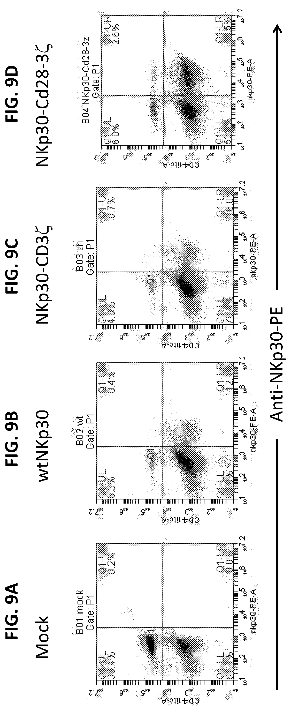
D00014
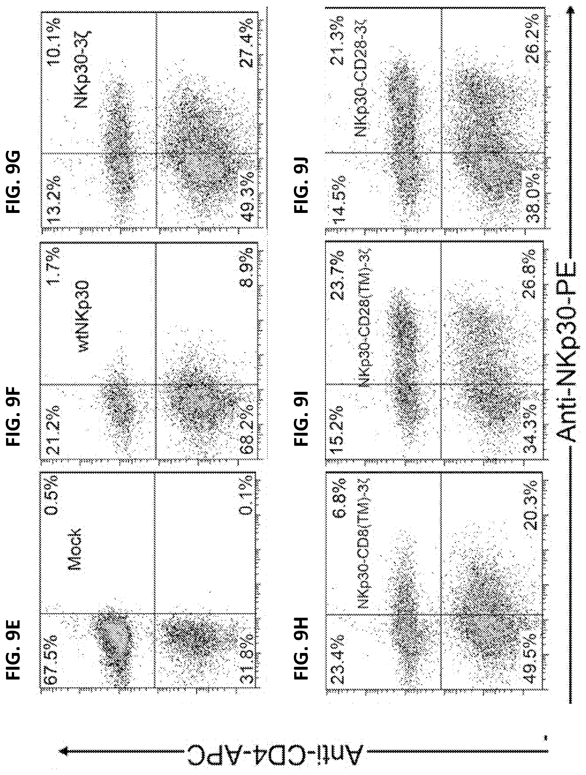
D00015
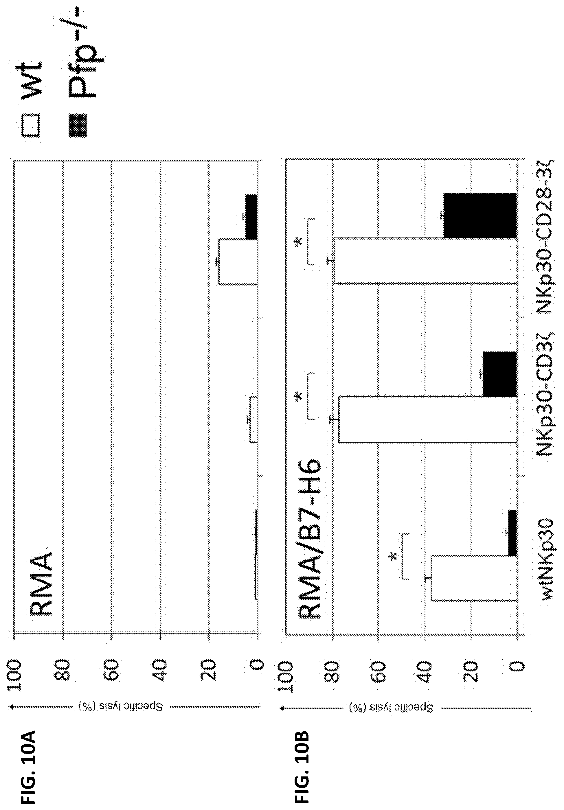
D00016
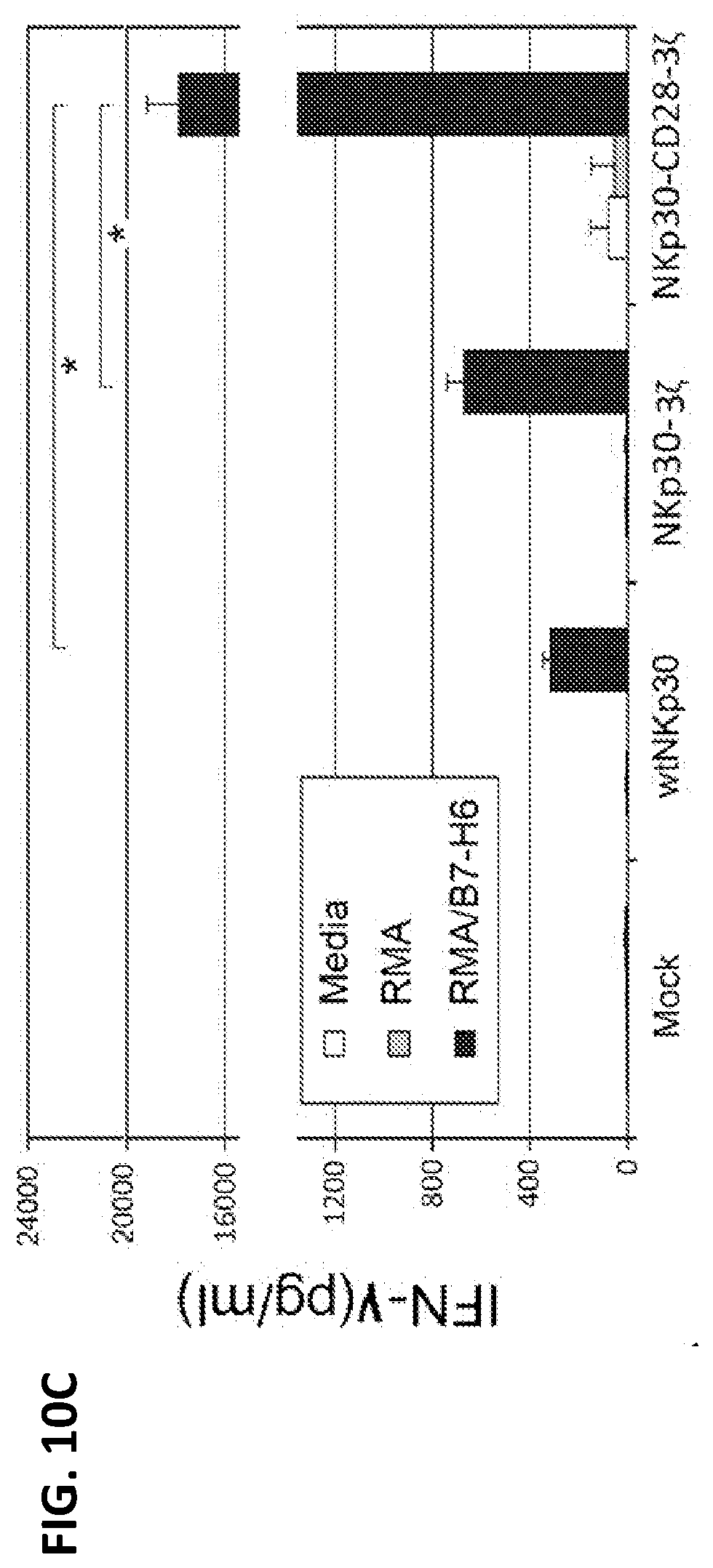
D00017
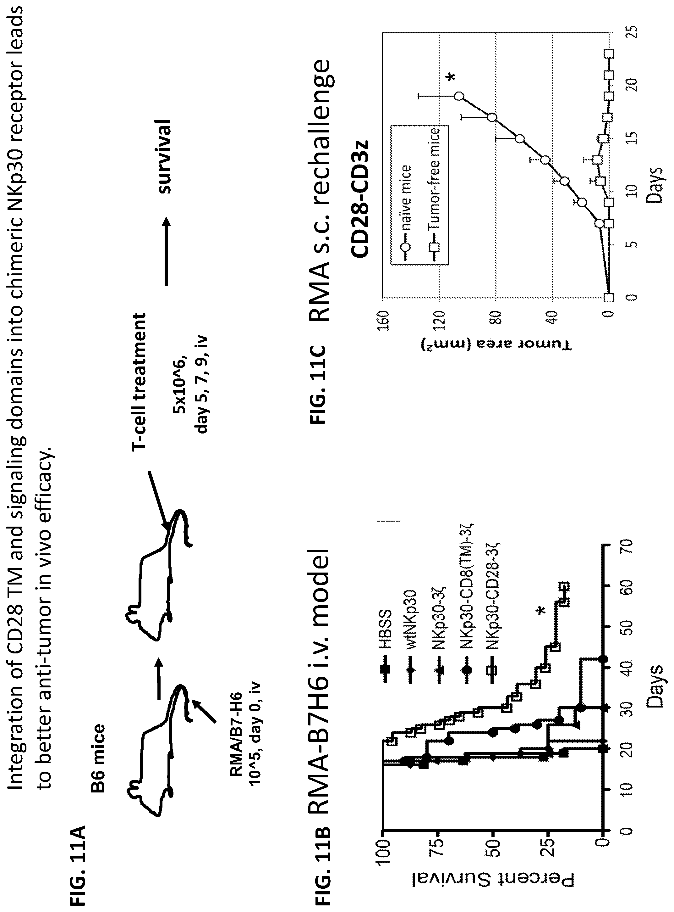
D00018
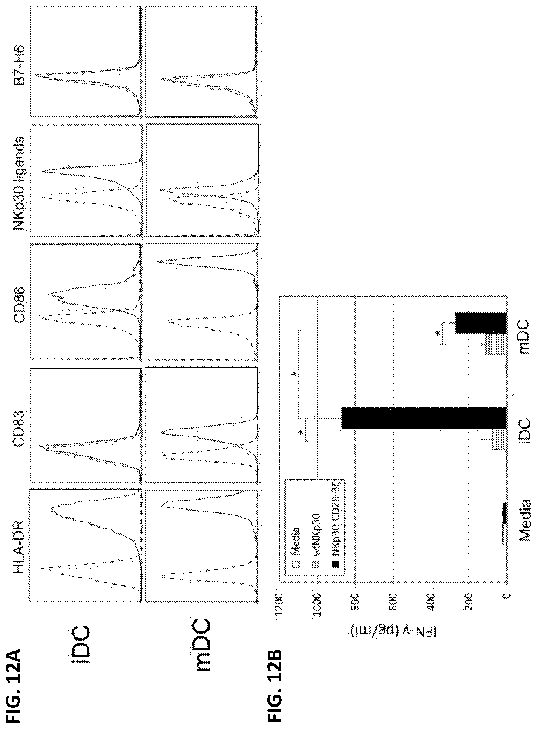
D00019
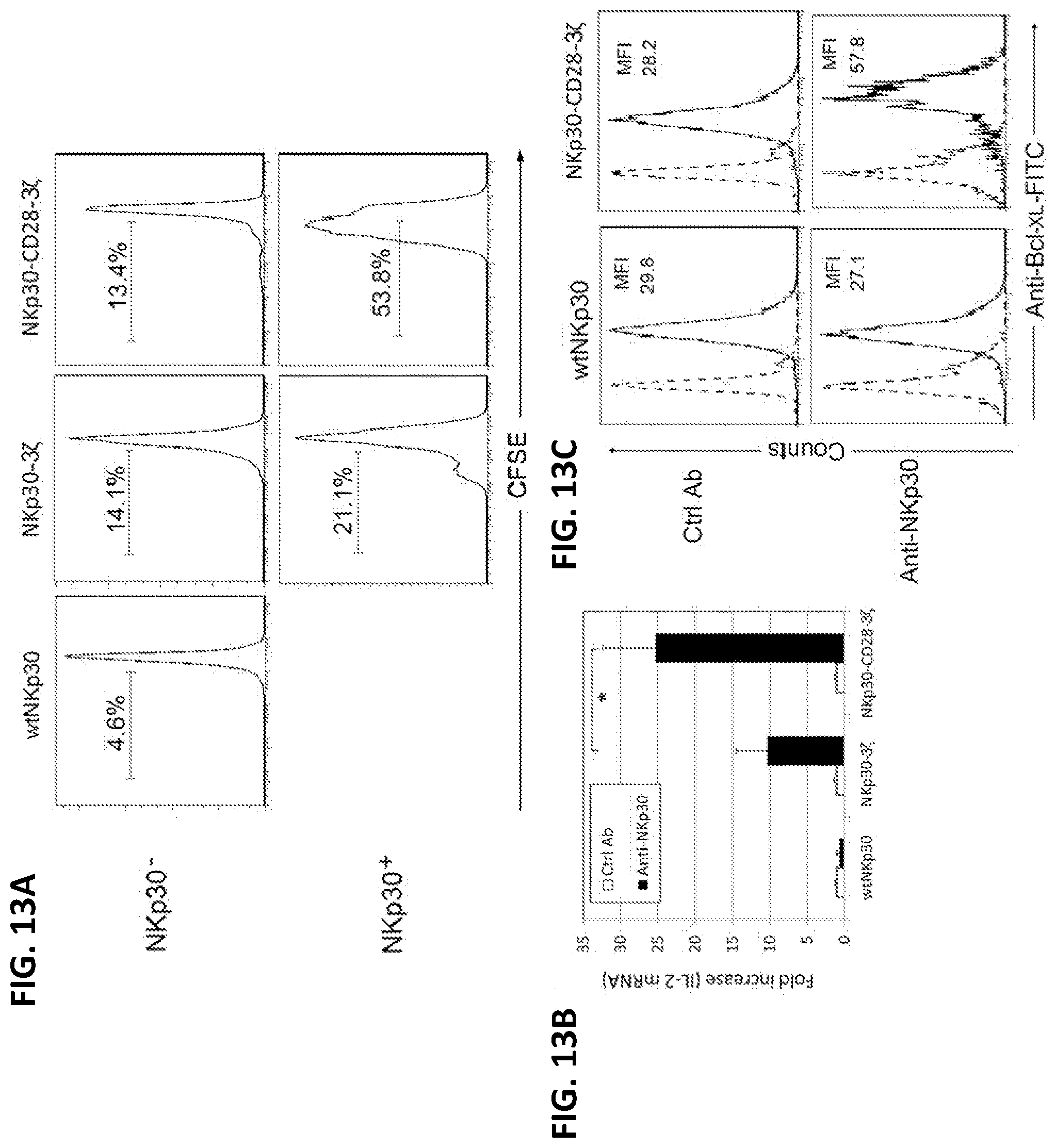
D00020
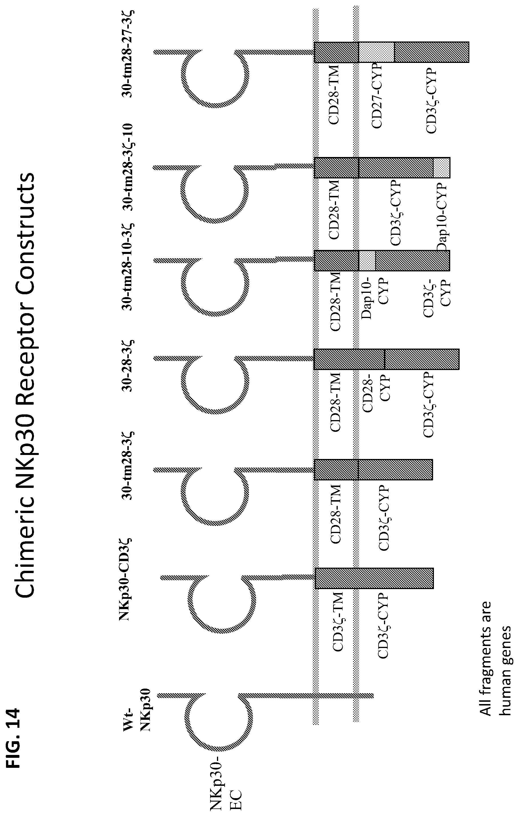
D00021
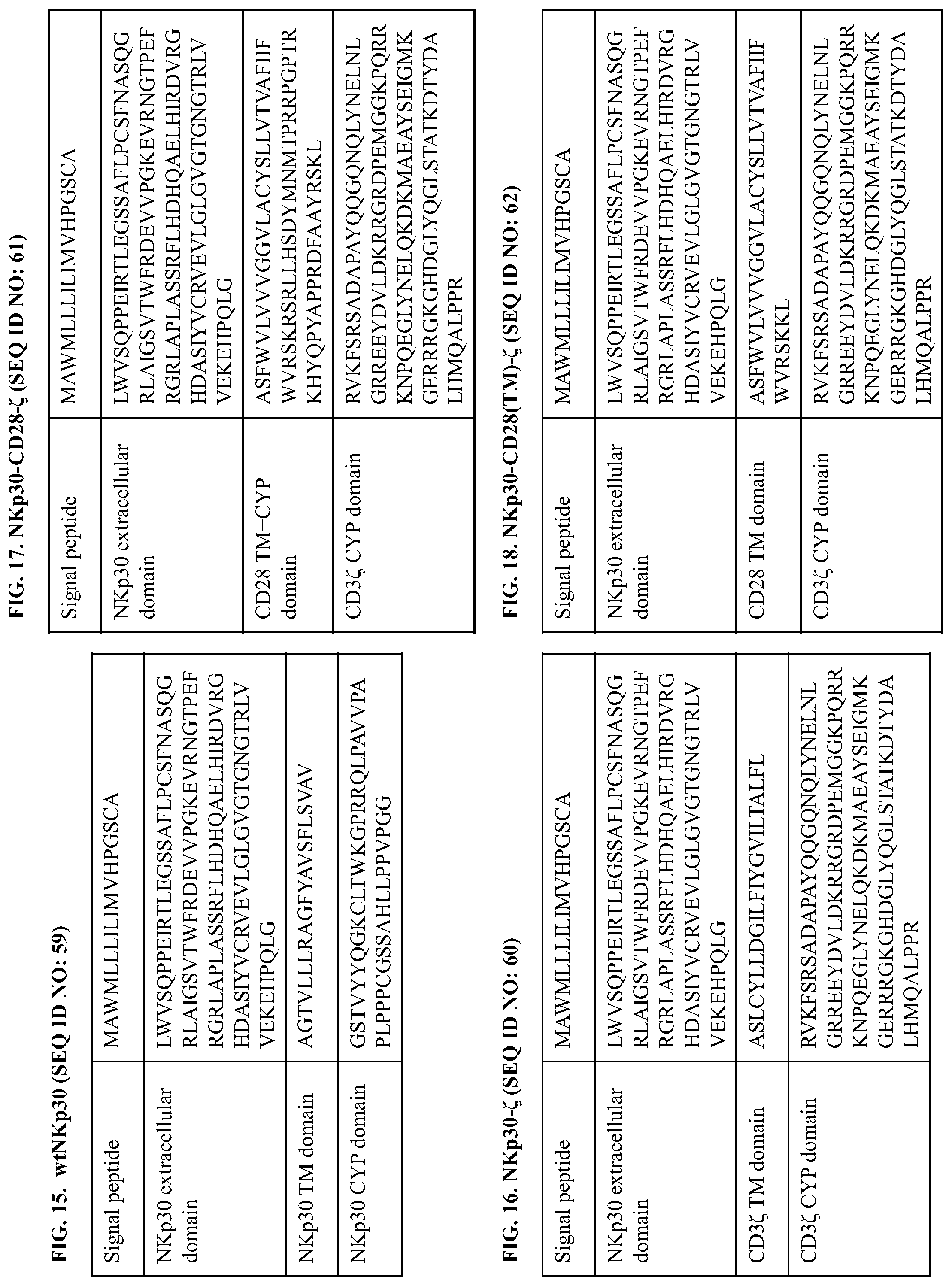
D00022
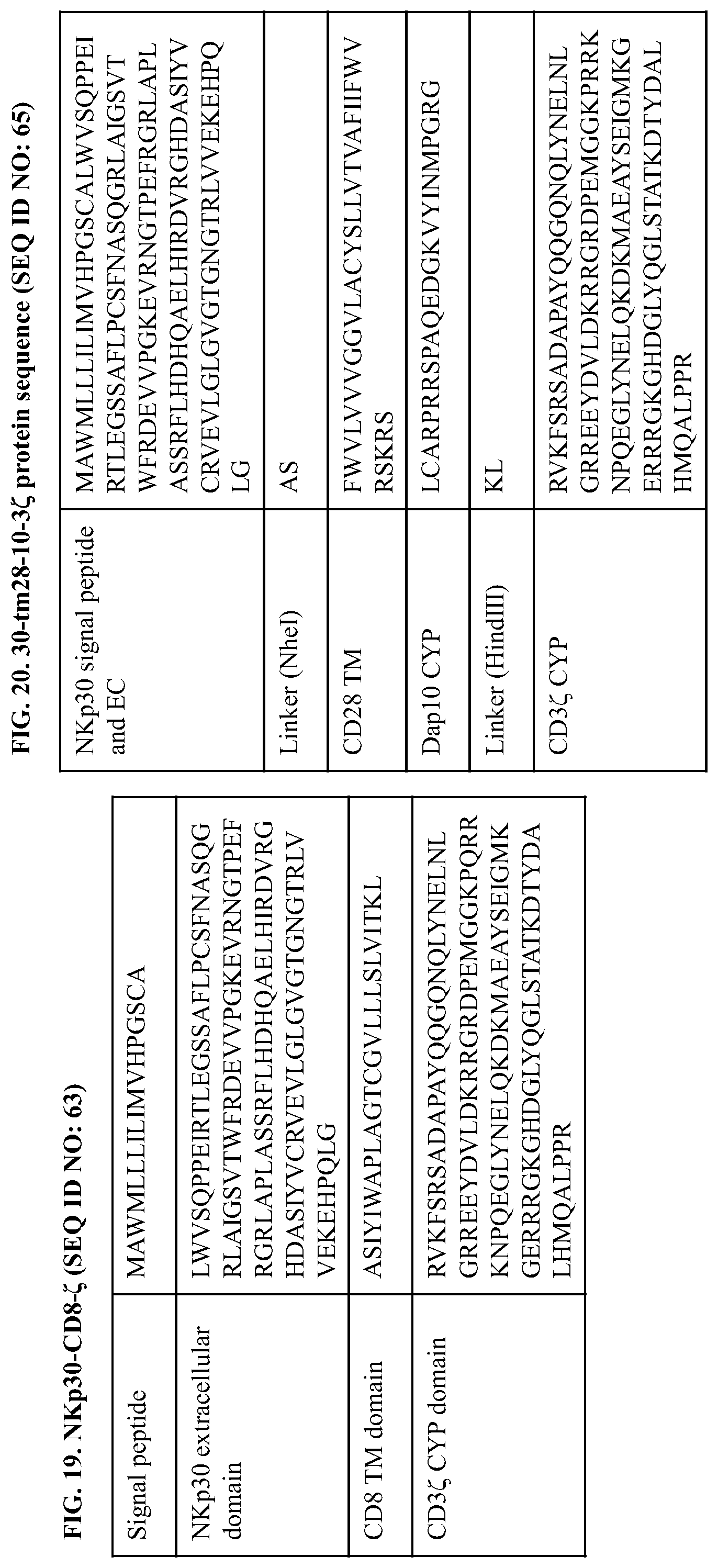
D00023
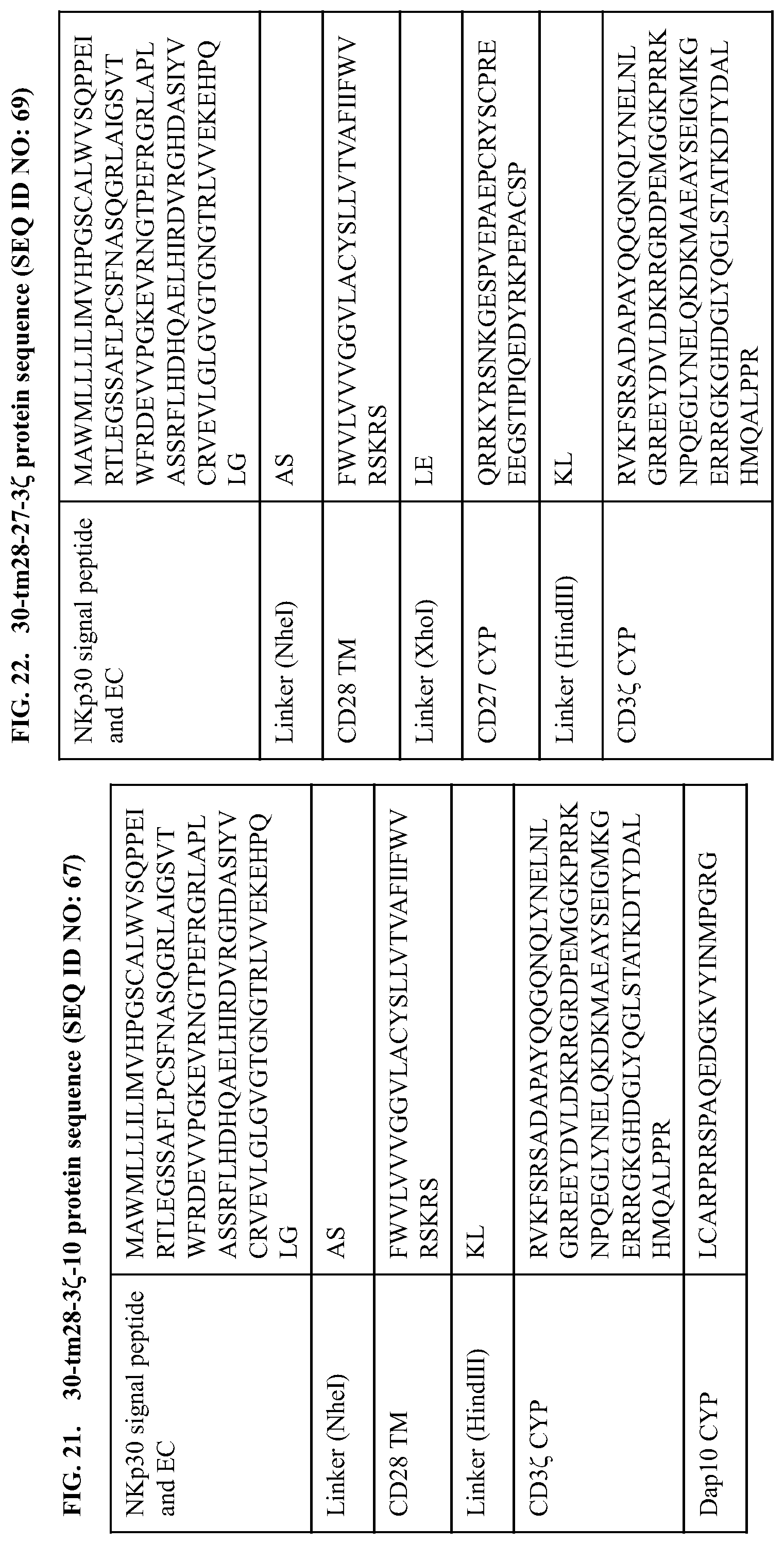
S00001
XML
uspto.report is an independent third-party trademark research tool that is not affiliated, endorsed, or sponsored by the United States Patent and Trademark Office (USPTO) or any other governmental organization. The information provided by uspto.report is based on publicly available data at the time of writing and is intended for informational purposes only.
While we strive to provide accurate and up-to-date information, we do not guarantee the accuracy, completeness, reliability, or suitability of the information displayed on this site. The use of this site is at your own risk. Any reliance you place on such information is therefore strictly at your own risk.
All official trademark data, including owner information, should be verified by visiting the official USPTO website at www.uspto.gov. This site is not intended to replace professional legal advice and should not be used as a substitute for consulting with a legal professional who is knowledgeable about trademark law.