Fusion Proteins For Improved Precision In Base Editing
Chen; Jia ; et al.
U.S. patent application number 16/640337 was filed with the patent office on 2020-11-12 for fusion proteins for improved precision in base editing. The applicant listed for this patent is ShanghaiTech University. Invention is credited to Jia Chen, Xingxu Huang, Xiaosa Li, Yajing Liu, Ying Wang, Bei Yang, Li Yang.
| Application Number | 20200354729 16/640337 |
| Document ID | / |
| Family ID | 1000005035006 |
| Filed Date | 2020-11-12 |


View All Diagrams
| United States Patent Application | 20200354729 |
| Kind Code | A1 |
| Chen; Jia ; et al. | November 12, 2020 |
FUSION PROTEINS FOR IMPROVED PRECISION IN BASE EDITING
Abstract
Provided are base editors containing a cytidine and a catalytically inactive version of Lachnospiraceae bacterium Cpf1 (LbCpf1). The new base editors have greatly improved editing efficiency and fidelity as compared to Cas9-based base editors, and have different editing windows.
| Inventors: | Chen; Jia; (Shanghai, CN) ; Huang; Xingxu; (Shanghai, CN) ; Yang; Li; (Shanghai, CN) ; Yang; Bei; (Shanghai, CN) ; Li; Xiaosa; (Shanghai, CN) ; Wang; Ying; (Shanghai, CN) ; Liu; Yajing; (Shanghai, CN) | ||||||||||
| Applicant: |
|
||||||||||
|---|---|---|---|---|---|---|---|---|---|---|---|
| Family ID: | 1000005035006 | ||||||||||
| Appl. No.: | 16/640337 | ||||||||||
| Filed: | August 28, 2018 | ||||||||||
| PCT Filed: | August 28, 2018 | ||||||||||
| PCT NO: | PCT/CN2018/102750 | ||||||||||
| 371 Date: | February 19, 2020 |
| Current U.S. Class: | 1/1 |
| Current CPC Class: | C12N 9/22 20130101; C12N 9/78 20130101; C12N 15/79 20130101; C07K 14/32 20130101; C12Y 305/04005 20130101; C07K 2319/09 20130101; C12N 15/102 20130101 |
| International Class: | C12N 15/79 20060101 C12N015/79; C12N 15/10 20060101 C12N015/10; C12N 9/22 20060101 C12N009/22; C12N 9/78 20060101 C12N009/78; C07K 14/32 20060101 C07K014/32 |
Foreign Application Data
| Date | Code | Application Number |
|---|---|---|
| Sep 1, 2017 | CN | PCT/CN2017/100131 |
Claims
1. A fusion protein comprising a first fragment comprising a cytidine deaminase and a second fragment comprising a catalytically inactive Lachnospiraceae bacterium Cpf1 (dLbCpf1).
2. The fusion protein of claim 1, wherein the cytidine deaminase is an apolipoprotein B mRNA editing enzyme, catalytic polypeptide-like (APOBEC) protein.
3. The fusion protein of claim 2, wherein the APOBEC protein is selected from the group consisting of APOBEC1, APOBEC2, APOBEC3A, APOBEC3B, APOBEC3C, APOBEC3D, APOBEC3F, APOBEC3G, APOBEC3H, APOBEC4, and activation-induced (cytidine) deaminase.
4. The fusion protein of claim 3, wherein the APOBEC protein is APOBEC1.
5. The fusion protein of claim 4, wherein the APOBEC1 protein comprises a W90Y or R126E mutation, or the combination thereof.
6. The fusion protein of claim 3, wherein the APOBEC protein is APOBEC3A.
7. The fusion protein of claim 1, further comprising one or more uracil DNA glycosylase inhibitor (UGI).
8. The fusion protein of claim 7, which comprises at least two UGIs wherein at least one of the UGIs is separated from the first fragment and the second fragment by a protease cleavage site.
9-10. (canceled)
11. The fusion protein of claim 1, further comprising one or more nuclear localization sequences (NLS).
12-15. (canceled)
16. The fusion protein of claim 1, which comprises, from the N-terminus to the C-terminus, a first NLS, the first fragment, the second fragment, a second NLS, a first UGI, a third NLS, a self-cleaving peptide, and a second UGI.
17. The fusion protein of claim 16, further comprising a fourth NLS between the second fragment and the first UGI.
18. The fusion protein of claim 17, further comprising, N-terminal to the second UGI, a second self-cleaving peptide, and a third UGI.
19. A method of editing a cytosine on a nucleic acid sequence in a sample, comprising contacting the sample with a suitable guide RNA and a fusion protein of claim 1, or a polynucleotide encoding the fusion protein.
20. The method of claim 19, wherein the cytosine is between nucleotide positions 6 and 22 3' to a protospacer adjacent motif (PAM) sequence on the nucleic acid sequence.
21. The method of claim 20, wherein the cytidine deaminase is APOBEC3.
22. The method of claim 19, wherein the cytosine is between nucleotide positions 8 and 13 3' to a protospacer adjacent motif (PAM) sequence on the nucleic acid sequence.
23. The method of claim 22, wherein the cytidine deaminase is APOBEC1 protein.
24. The method of claim 22, wherein the cytosine is between nucleotide positions 10 and 12 3' to the PAM sequence.
25. The method of claim 24, wherein the cytidine deaminase is APOBEC1 protein comprising a W90Y or R126E mutation, or the combination thereof.
26-27. (canceled)
28. A fusion protein comprising a first fragment comprising a cytidine deaminase, a second fragment comprising a Cas protein, and a uracil DNA glycosylase inhibitor (UGI) separated from the first fragment and the second fragment with a protease cleavage site.
29-32. (canceled)
Description
BACKGROUND
[0001] Genome editing that can be used to genetically manipulate the genome of cells and living organism has broad application interest in life sciences research, biotechnology, agricultural technology development and pharmaceutical and clinical development. For example, genome editing can be used to correct driver mutations underlying genetic diseases and thereby resulting in complete cure of these diseases in a living organism. CRISPR/Cas (Clustered regularly interspaced short palindromic repeats/CRISPR-associated protein) system has been the most powerful genomic editing tool since its conception for its unparalleled editing efficiency, convenience and the potential applications in living organism. Directed by guide RNA (gRNA), a Cas nuclease can generate DNA double strand breaks (DSBs) at the targeted genomic sites in various cells (both cell lines and cells from living organisms). These DSBs are then repaired by the endogenous DNA repair system, which could be utilized to perform desired genome editing.
[0002] In general, two major DNA repair pathways could be activated by DSBs, non-homologous end joining (NHEJ) and homology-directed repair (HDR). NHEJ can introduce random insertions/deletions (indels) in the genomic DNA region around the DSBs, thereby leading to open reading frame (ORF) shift and ultimately gene inactivation. In contrast, when HDR is triggered, the genomic DNA sequence at target site could be replaced by the sequence of the exogenous donor DNA template through a homologous recombination mechanism, which can result in the correction of genetic mutation.
[0003] Base editors (BE), which integrate the CRISPR/Cas system with the APOBEC (apolipoprotein B mRNA editing enzyme, catalytic polypeptide-like) cytidine deaminase family, were recently invented that greatly enhanced the efficiency of CRISPR/Cas9-meditated gene correction. Through fusion with Cas9 nickase (nCas9), the cytosine (C) deamination activity of rat APOBEC1 (rAl) can be purposely directed to the target bases in genome and to catalyze C to Thymine (T) substitutions at these bases.
[0004] However, the reliance on the Cas9 nickase as the deaminase fusion partner in the most active current base editors leads to an increased frequency of unwanted indels and non-C-to-T base substitutions, and limits editing to regions with G/C rich protospacer adjacent motif (PAM) sequences.
SUMMARY
[0005] The present disclosure, in some embodiments, provide base editors useful for genome editing that combines a catalytically inactive Lachnospiraceae bacterium Cpf1 (dLbCpf1) with a cytidine deaminase. Such base editors recognize a T-rich PAM sequence and converts C to T in human cells at high efficiency and with low levels of indels, non-C-to-T substitutions and off-target editing. These are all significant improvements over Cas9-based base editors. In addition, besides APOBEC1 (A1), when the LbCpf1 was fused to APOBEC3 (A3, or APOBEC3A), even greater editing efficiency was achieved. In addition to the greatly improved editing efficiency and precision, LbCpf1-based base editors further differ from Cas9-based base editors in terms of editing windows. Another interesting discovery in the present disclosure is that the presence of a free uracil DNA glycosylase inhibitor (UGI) domain can further improve the efficiency and fidelity in base editing.
[0006] In accordance with one embodiment of the present disclosure, therefore, provided is a fusion protein comprising a first fragment comprising a cytidine deaminase and a second fragment comprising a catalytically inactive Lachnospiraceae bacterium Cpf1 (dLbCpf1).
[0007] In some embodiments, the cytidine deaminase is an apolipoprotein B mRNA editing enzyme, catalytic polypeptide-like (APOBEC) protein. In some embodiments, the APOBEC protein is selected from the group consisting of APOBEC1, APOBEC2, APOBEC3A, APOBEC3B, APOBEC3C, APOBEC3D, APOBEC3F, APOBEC3G, APOBEC3H, APOBEC4, and activation-induced (cytidine) deaminase. In one embodiment, the APOBEC protein is APOBEC1. In one embodiment, the APOBEC1 protein comprises a W90Y or R126E mutation, or the combination thereof. In some embodiments, the APOBEC protein is APOBEC3A. In some embodiments, the APOBEC3A protein has one or more mutations selected from W104A, Y130F, D131Y, D31E, and/or Y132D mutations; examples of combinatory mutations including Y130E-D131E-Y132D, Y130E-D131Y-Y132D.
[0008] In some embodiments, the fusion protein further includes one or more uracil DNA glycosylase inhibitor (UGI). In some embodiments, the fusion protein comprises at least two UGIs.
[0009] In some embodiments, at least one of the UGIs is separated from the first fragment and the second fragment by a protease cleavage site. In some embodiments, the protease cleavage site is a self-cleaving peptide.
[0010] In some embodiments, the fusion protein further comprises one or more nuclear localization sequences (NLS). In some embodiments, at least one iNLS is located between the second fragment and the first UGI. In some embodiments, at least two iNLS are located between the second fragment and the first UGI. In some embodiments, at least one NLS is located N-terminal to the first fragment and the second fragment.
[0011] In some embodiments, at least one NLS is located C-terminal to the first fragment and the second fragment. In some embodiments, the fusion protein comprises, from the N-terminus to the C-terminus, a first NLS, the first fragment, the second fragment, a second NLS, a first UGI, a third NLS, a self-cleaving peptide, and a second UGI. In some embodiments, the fusion protein further comprises a fourth NLS between the second fragment and the first UGI. In some embodiments, the fusion protein further comprises, N-terminal to the second UGI, a second self-cleaving peptide, and a third UGI.
[0012] Also provided, in one embodiment, is a method of editing a cytosine on a nucleic acid sequence in a sample, comprising contacting the sample with a suitable guide RNA and a fusion protein of the present disclosure, or a polynucleotide encoding the fusion protein.
[0013] In some embodiments, the cytosine is between nucleotide positions 6 and 22 3' to a protospacer adjacent motif (PAM) sequence on the nucleic acid sequence. In some embodiments, the cytidine deaminase is APOBEC3A. In some embodiments, the APOBEC3A protein has one or more mutations selected from W104A, Y130F, D131Y, D31E, and/or Y132D mutations; examples of combinatory mutations including Y130E-D131E-Y132D, Y130E-D131Y-Y132D.
[0014] In some embodiments, the cytosine is between nucleotide positions 8 and 13 3' to a protospacer adjacent motif (PAM) sequence on the nucleic acid sequence. In some embodiments, the cytidine deaminase is APOBEC1 protein. In some embodiments, the cytosine is between nucleotide positions 10 and 12 3' to the PAM sequence. In some embodiments, the cytidine deaminase is APOBEC1 protein comprising a W90Y or R126E mutation, or the combination thereof.
[0015] In some embodiments, the PAM sequence is a T-rich PAM sequence. In some embodiments, the method further comprises contacting the sample with a UGI not fused to a Cas protein, or a polynucleotide encoding the UGI.
[0016] In another embodiment, provided is a fusion protein comprising a first fragment comprising a cytidine deaminase, a second fragment comprising a Cas protein, and a uracil DNA glycosylase inhibitor (UGI) separated from the first fragment and the second fragment with a protease cleavage site. In some embodiments, the protease cleavage site is a self-cleaving peptide. In some embodiments, the fusion protein further comprises a second UGI separated from the first fragment and the second fragment with a second protease cleavage site. In some embodiments, the fusion protein further comprises a third UGI separated from the second UGI with a third protease cleavage site. In some embodiments, the Cas protein is Cas9 or Cpf1.
[0017] Polynucleotides encoding the fusion proteins of the present disclosure, constructs containing the polynucleotides, cells containing the polynucleotides or the constructs, and compositions comprising any of the above are also provided, without limitation.
BRIEF DESCRIPTION OF THE DRAWINGS
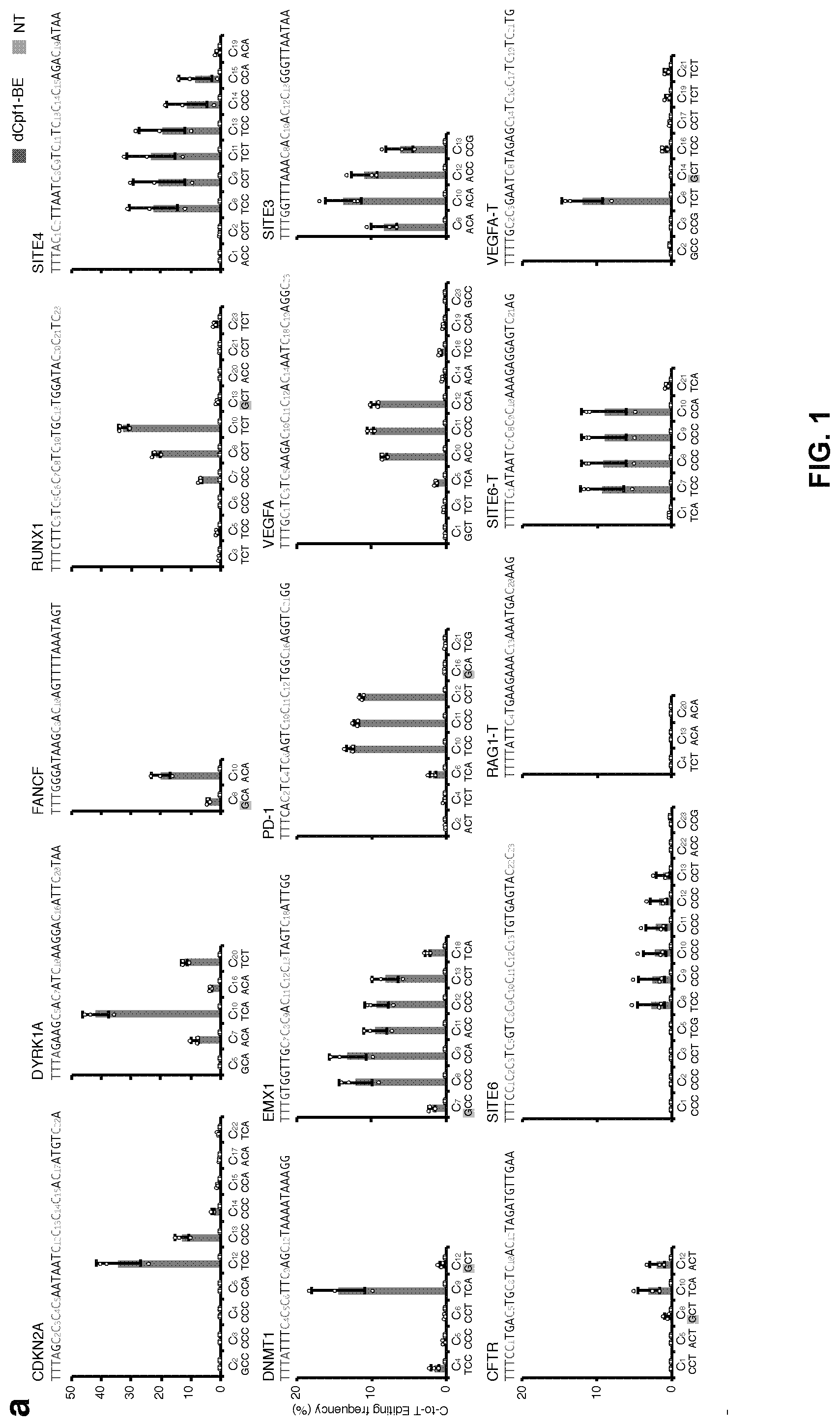
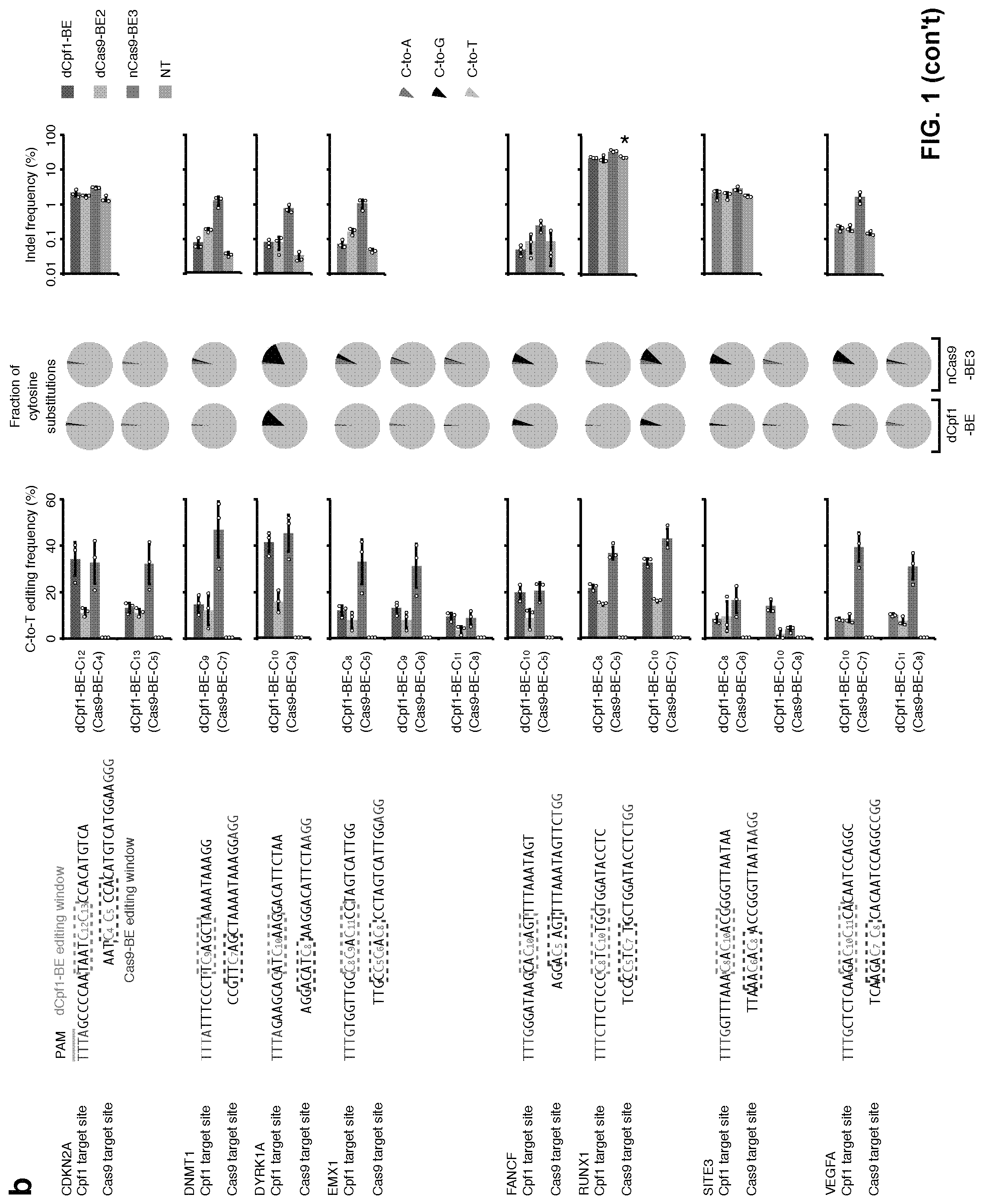
[0018] FIG. 1, with panels a-b. Base editing mediated by dCpf1-BE. (a) Determination of dCpf1-BE-induced base editing frequency at every single cytosine in the indicated spacer region. The dCpf1-BE showed inefficient C-to-T base editing at the cytosines following a G (shadowed). The cytosines were counted with the base proximal to the PAM setting as position 1. (b) The comparison of base editing mediated by dCpf1-based and Cas9-bases BEs. The C-to-T editing frequencies of the indicated cytosines, the fractions of cytosine substitutions and the indel frequencies were individually determined at the indicated genomic target sites under different conditions. The target site sequences and editing windows of dCpf1-BE and Cas9-BEs are shown. NT, non-transfected. Asterisk denotes an unusually high basal indel frequency (or amplification, sequencing, alignment artifact) at the examined RUNX1 site in the non-transfected 293FT cells. Means.+-.s.d. were from three independent experiments.
[0019] FIG. 2, with panels a-h. Improvements of dCpf1-BE. (a) Mutating APOBEC1 in dCpf1-BE to narrow down editing window. The C-to-T editing frequency at every single cytosine was individually determined in the indicated genomic target sites under different conditions. The target site sequences and the narrowed editing windows of dCpf1-BE are shown. The major editing sites (C.sub.10-C.sub.12) are in salmon and the minor editing sites (C.sub.1-C.sub.9 and C.sub.13-C.sub.23) are in green. (b) The ratios of major editing to minor editing were determined at the indicated genomic target sites. (c) Statistical analysis of the normalized ratios of major editing to minor editing, setting the ones induced by dCpf1-BE as 100%. The dCpf1-BE-YE induced significantly higher ratio of major editing to minor editing. The median, interquartile range (IQR) and 1.5.times.IQR are shown. n=15 independent samples from 3 independent experiments. (d-g) The addition of free UGI enhances the purity of editing outcomes induced by dCpf1-BEs. The fractions (d, f) and statistical analyses (e, g) of cytosine substitutions at the indicated editing positions under different conditions. (e, g) The dCpf1-eBE and dCpf1-eBE-YE induced significantly purer C-to-T editing outcomes than dCpf1-BE and dCpf1-BE-YE. The median and IQR are shown. n=9 independent samples from 3 independent experiments. (h) Summary of Cas9-based and dCpf1-based BEs. Left, schematic diagrams illustrate the complexes of Cas9-BE/sgRNA/target DNA and dCpf1-BE/crRNA/target DNA. Right, list of relevant features in Cas9-based and dCpf1-based BE systems. Comparisons are based on base editing at DYRK1A, FANCF and RUNX1 target sites. (a, b) Means.+-.s.d. were from three independent experiments. (c, e, g) P value, one-tailed Student's T test.
[0020] FIG. 3, with panels a-c. dLbCpf1-BE0 but not dAsCpf1-BE0 induced C-to-T base editing in episomal shuttle vector system. (a) Schematic diagram illustrating the procedures to determine the base editing induced by dLbCpf1-BE0 or dAsCpf1-BE0 in episomal shuttle vectors. (b) Number of E. coli colonies containing mutated shuttle vectors that were induced by dAsCpf1-BE0 or dLbCpf1-BE0. (c) C-to-T editing frequencies were determined at the indicated cytosines. The cytosines were counted with the base proximal to the PAM setting as position 1. Frequencies were calculated from data in (b). Means.+-.s.d. were from three independent experiments.
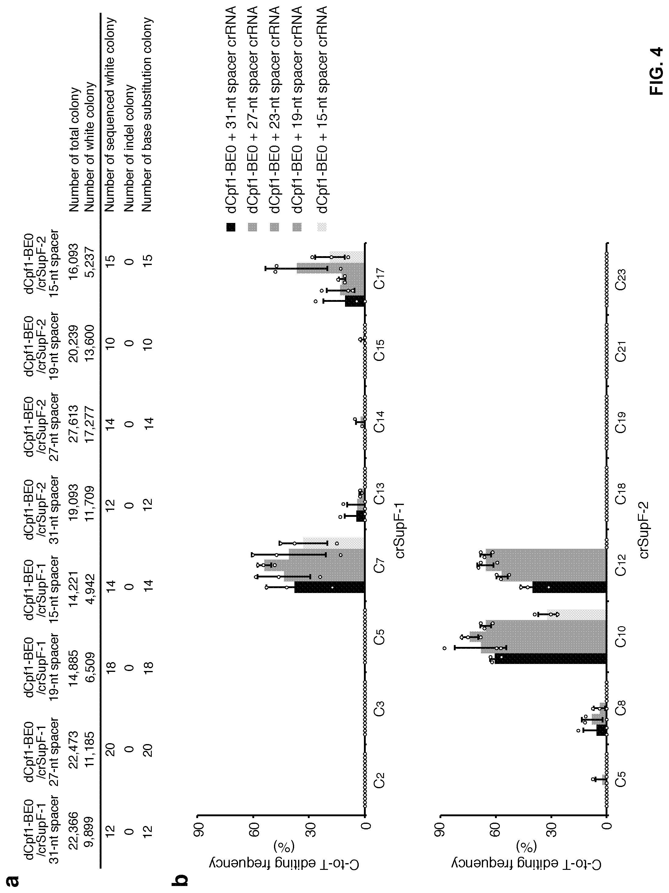
[0021] FIG. 4, with panels a-b. Effect of crRNA spacer length on editing efficiency. (a) Number of colonies containing mutated shuttle vectors that were induced by dCpf1-BE0 and crRNAs of different length. (b) C-to-T editing frequencies induced by dCpf1-BE0 and crRNAs of different length were determined at the indicated cytosines in episomal shuttle vectors. The crRNAs with the spacer length ranged from 19 to 27 nt showed similar base editing efficiencies at most of the editing positions. Frequencies were calculated from data in (a). Means.+-.s.d. were from three independent experiments.
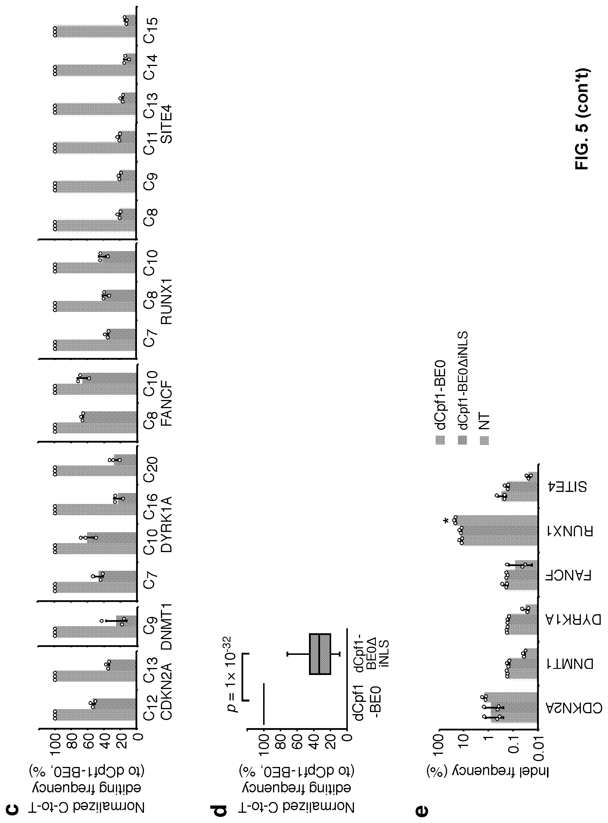
[0022] FIG. 5, with panels a-e. The internal NLS (iNLS) between dCpf1 and UGI is important for base editing induced by dCpf1-BE0. (a) Schematic diagram illustrating the design of expression vectors of dCpf1-BE0 and dCpf1-BE0.DELTA.iNLS. (b) The C-to-T editing frequencies of the indicated cytosines were individually determined at different genomic target sites under the treatment of dCpf1-BE0 (blue) or dCpf1-BE0.DELTA.iNLS (green). (c) The normalized C-to-T editing frequencies induced by dCpf1-BE0 and dCpf1-BE0.DELTA.iNLS in genomic DNA, setting the ones induced by dCpf1-BE0 as 100%. (d) Statistical analysis of the normalized C-to-T editing frequencies. The dCpf1-BE0 induced significantly higher C-to-T editing frequencies than dCpf1-BE0.DELTA.iNLS. P values, one-tailed Student's T test. The median, interquartile range (IQR) and 1.5.times.IQR are shown. n=54 independent samples from 3 independent experiments. (e) The indel frequencies were determined at indicated loci in genomic DNA. 293FT cells were either treated with dCpf1-BE0 (blue), with dCpf1-BE0.DELTA.iNLS (green) or non-transfected (gray) before deep sequencing. Asterisk denotes an unusually high basal indel frequency (or amplification, sequencing, alignment artifact) at the examined RUNX1 site in the non-transfected 293FT cells. Means.+-.s.d. were from three independent experiments.
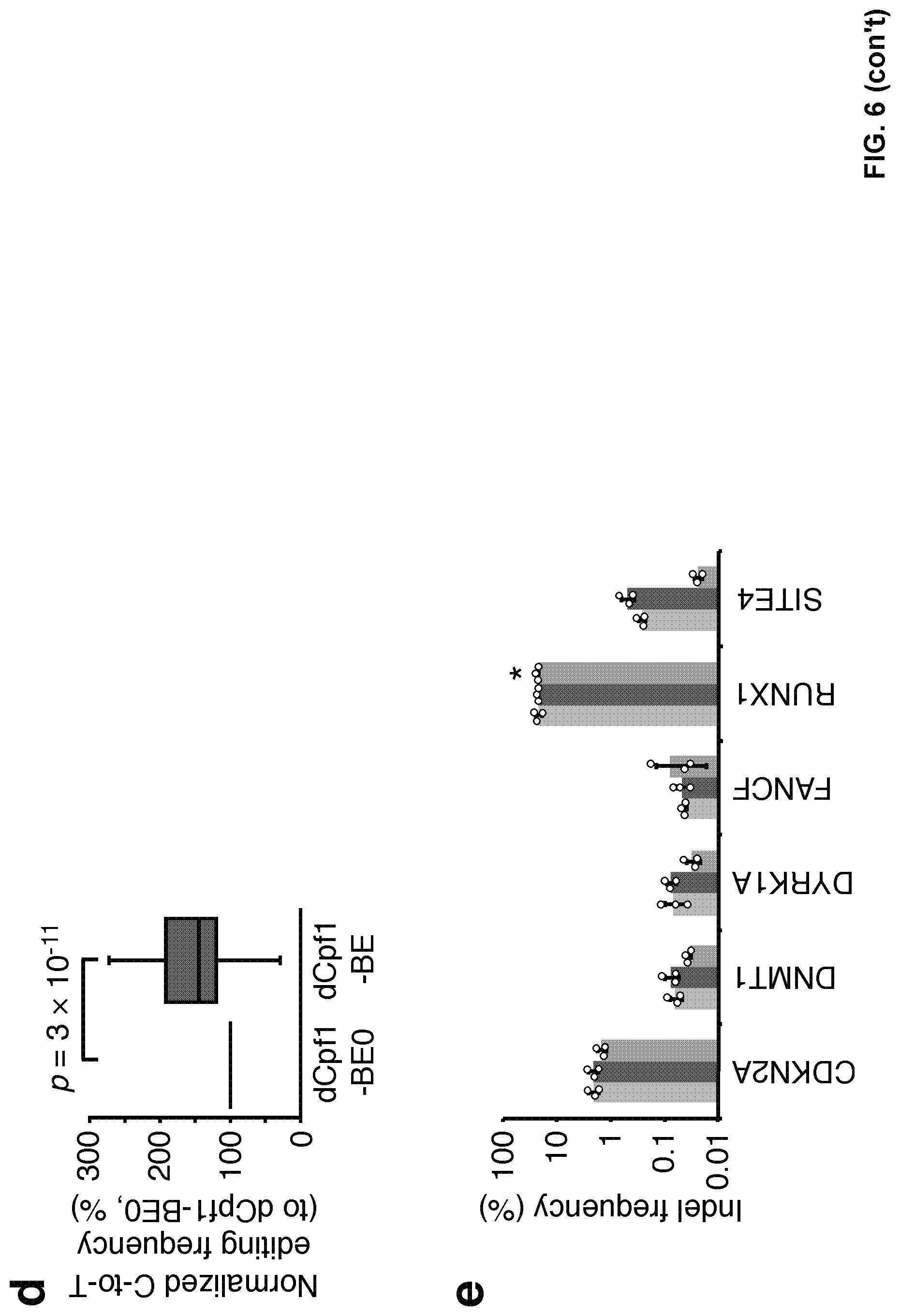
[0023] FIG. 6, with panels a-e. Additional N-terminal NLS enhanced the base editing efficiency of dCpf1-BE in genomic DNA. (a) Schematic diagram illustrating the design of expression vectors of dCpf1-BE0 and dCpf1-BE. (b) The C-to-T editing frequencies of the indicated cytosines were individually determined at different genomic target sites. 293FT cells were either treated with dCpf1-BE0 (blue), dCpf1-BE (purple) or left non-transfected (gray) before deep sequencing. (c) The normalized C-to-T editing frequencies induced by dCpf1-BE0 and dCpf1-BE in genomic DNA, setting the ones induced by dCpf1-BE0 as 100%. (d) Statistical analysis of the normalized C-to-T editing frequencies. The dCpf1-BE induced significantly higher C-to-T editing frequencies than dCpf1-BE0. P values, one-tailed Student's T test. The median, IQR and 1.5.times.IQR are shown. n=54 independent samples from 3 independent experiments. (e) The indel frequencies were determined at indicated loci in genomic DNA under different conditions. Asterisk denotes an unusually high basal indel frequency (or amplification, sequencing, alignment artifact) at the examined RUNX1 site in the non-transfected 293FT cells. Means.+-.s.d. were from three independent experiments.
[0024] FIG. 7, with panels a-d. Features of dCpf1-BE-induced base editing. (a) Summary of the base editing frequency at each cytosine in the spacer region for the indicated 14 crRNAs. These data show that the major editing window ranges from the position 8 to 13 in spacer region. (b) The indel frequencies were determined at indicated loci in genomic DNA under different conditions. 293FT cells were either treated with dCpf1-BE (purple) or non-transfected (gray) before deep sequencing. Asterisk denotes an unusually high basal indel frequency (or amplification, sequencing, alignment artifact) at the examined RUNX1 site in the non-transfected 293FT cells. Means.+-.s.d. were from three independent experiments. (c) The fractions of cytosine substitutions induced by dCpf1-BE were individually determined at the indicated cytosines. (d) Statistical analysis showed that the C-to-T fraction of base editing outcome induced by dCpf1-BE was significantly higher than that induced by nCas9-BE3. The median and IQR are shown. P values, one-tailed Student's T test. n=42 independent samples from 3 independent experiments.
[0025] FIG. 8, with panels a-c. Base editing was induced by dCpf1-BE in U2OS cells. (a) The C-to-T editing frequencies of the indicated cytosines were individually determined at the indicated genomic target sites. U2OS cells were either treated with dCpf1-BE (purple) or non-transfected (gray) before deep sequencing. (b) The indel frequencies were determined at indicated loci in genomic DNA under different conditions. Asterisk denotes an unusually high basal indel frequency (or amplification, sequencing, alignment artifact) at the examined RUNX1 site in the non-transfected U2OS cells. (c) The fractions of cytosine substitutions induced by dCpf1-BE were individually determined at the indicated cytosines. (a, b) Means.+-.s.d. were from three independent experiments.
[0026] FIG. 9, with panels a-b. Determination of base editing induced by dCpf1-BE at predicted off-target sites. (a) The sequences of on- and off-target sites for the indicated crRNAs. The cytosines were counted with the base proximal to the PAM setting as position 1. (b) The C-to-T editing frequencies of the indicated cytosines were individually determined at the indicated on- and off-target sites. 293FT cells were either treated with dCpf1-BE (purple) or non-transfected (gray) before deep sequencing. Means.+-.s.d. were from three independent experiments.
[0027] FIG. 10, with panels a-b. No substantial C-to-T editing was detected in the region outside of the spacer. (a) Schematic diagram illustrating the PAM region, the 20-nt region upstream of PAM and the 20-nt region downstream of spacer. (b) The C-to-T editing frequencies of the indicated cytosines outside of the spacer region were individually determined at the indicated sites. 293FT cells were either treated with dCpf1-BE (purple) or non-transfected (gray) before deep sequencing. Means.+-.s.d. were from three independent experiments.
[0028] FIG. 11. Multiple C-to-T editing was induced by dCpf1-BE when more than one cytosines are in the spacer region. The frequencies of single and multiple C-to-T editing induced by dCpf1-BE at the indicated cytosines were determined at different genomic target sites. The deep sequencing data are same as in FIG. 1a. Means.+-.s.d. were from three independent experiments.
[0029] FIG. 12, with panels a-e. W90Y and R126E mutations in rat APOBEC1 (rAl) narrowed the base editing window to 3 nt. (a) Schematic diagram illustrating the design of expression vectors of dCpf1-BE, dCpf1-BE-YE and dCpf1-BE-YEE. (b) The normalized ratios of major editing to minor editing induced by dCpf1-BE (purple) and dCpf1-BE-YE (magenta), setting the ones induced by dCpf1-BE as 100%. (c) The fractions of single and multiple C-to-T conversions induced by dCpf1-BE and dCpf1-BE-YE. (d) Statistical analysis showed that the fraction of single C-to-T conversion induced by dCpf1-BE-YE was significantly higher than that induced by dCpf1-BE. P values, one-tailed Student's T test. The median and IQR are shown. n=15 independent samples from 3 independent experiments. (e) The indel frequencies were determined at the indicated genomic loci from the 293FT cells transfected with dCpf1-BE (purple), dCpf1-BE-YE (magenta), dCpf1-BE-YEE (yellow) or non-transfected (gray). Asterisk denotes an unusually high basal indel frequency (or amplification, sequencing, alignment artifact) at the examined RUNX1 site in the non-transfected 293FT cells. (b, e) Means.+-.s.d. were from three independent experiments.
[0030] FIG. 13, with panels a-f. The fusion of three copies of 2A-UGI sequences did not substantially affect editing efficiency and induced no detectable indel formation. (a) Schematic diagram illustrating the design of expression vectors of dCpf1-BE and dCpf1-eBE. (b) The base editing frequencies induced by dCpf1-BE (purple) and dCpf1-eBE (green) were determined at indicated positions in genomic DNA. (c) The indel frequencies were determined at the indicated genomic loci. The 293FT cells were either treated with dCpf1-BE (purple), dCpf1-eBE (green) or left non-transfected (gray) before deep sequencing. (d) Schematic diagram illustrating the design of expression vectors of dCpf1-BE-YE and dCpf1-eBE-YE. (e) The base editing frequencies induced by dCpf1-BE-YE (magenta) and dCpf1-eBE-YE (brown) were determined at the indicated positions in genomic DNA. (f) The indel frequencies were determined at the indicated genomic. The 293FT cells were either treated with dCpf1-BE-YE (magenta), dCpf1-eBE-YE (brown) or non-transfected (gray) before deep sequencing. Asterisk denotes an unusually high basal indel frequency (or amplification, sequencing, alignment artifact) at the examined RUNX1 site in the non-transfected 293FT cells. Means.+-.s.d. were from three independent experiments.
[0031] FIG. 14, with panels A-B. (A) Schematic diagram illustrating the design of expression vectors of Cpf1-BE and Cpf1-A3-BE. (B) The base editing efficiency induced by Cpf1-A3-BE and Cpf1-BE were determined at the indicated sites in genomic DNA. The base editing efficiency of Cpf1-A3-BE is higher than that of Cpf1-BE (positions 7 and 10 at DYRK1A site and positions 7, 8 and 10 at RUNX1 site).
[0032] FIG. 15. Editing window of Cpf1-A3-BE. The editing window of Cpf1-A3-BE spans from positions 6 to 22 in the tested protospacer region.
[0033] FIG. 16, with panels A-R. Enhanced base editing system. (A-I) Enhanced base editing by co-expressing BE3 and free UGI from separate vectors in 293FT cells. Schematic diagram illustrating the design of sgRNA, BE3 and UGI expression vectors (A). The indel frequency (B), the C-to-T editing frequency at the indicated position of the sgRNA target region (D), the ratio of desired C-to-T editing to unwanted indels (F) and the fractions of C-to-T, C-to-A and C-to-G substitutions (H) were individually determined at the specified genomic sites for the indicated conditions and plotted as follows: orange represents BE3, faint blue represents BE+low UGI, blue represents BE3+medium UGI, dark blue represents BE3+high UGI and black represents dCas9. The positions of edited Cs in the sgFANCF, sgSite2 and sgRNF2 target regions were indicated with the base distal from the PAM set as position 1. Statistical analyses highlighted the significant differences between BE3 (orange) and BE3+high UGI (dark blue) in indel frequency (C), in C-to-T editing frequency at the indicated position within sgRNA target region (E), in the ratio of desired C-to-T editing to unwanted indels (G) and in the fraction of C-to-T substitution (I). (J-R) Enhanced base editing by eBE-51 and eBE-S3 in 293FT cells. Schematic diagram illustrating the design of sgRNA, BE3, eBE-51 and eBE-S3 expression vectors (J). The indel frequency (K), the C-to-T editing frequency (M), the ratio of desired C-to-T editing to unwanted indels (O) and the fractions of C-to-T, C-to-A and C-to-G substitutions (Q) were individually determined at the indicated genomic sites for BE3 (orange), eBE-S1(faint cyan) and eBE-S3 (cyan). The positions of edited Cs in the sgEMX1, sgFANCF, sgSite2, sgSite4 and sgRNF2 target regions were indicated with the base distal from the PAM set as position 1. Statistical analyses highlighted the significant differences between BE3 (orange) and eBE-S3 (cyan) in indel frequency (L), in the C-to-T editing frequency (N), in the ratio of desired C-to-T editing to unwanted indels (P) and in the fraction of C-to-T substitution (R). (B, D, F, K, M and O) Error bars (.+-.), standard deviations of 3 replicates. (C, E, G, I, L, N, P and R) P values, one-tailed Student's T-test.
DETAILED DESCRIPTION
Definitions
[0034] It is to be noted that the term "a" or "an" entity refers to one or more of that entity; for example, "an antibody," is understood to represent one or more antibodies. As such, the terms "a" (or "an"), "one or more," and "at least one" can be used interchangeably herein.
[0035] As used herein, the term "polypeptide" is intended to encompass a singular "polypeptide" as well as plural "polypeptides," and refers to a molecule composed of monomers (amino acids) linearly linked by amide bonds (also known as peptide bonds). The term "polypeptide" refers to any chain or chains of two or more amino acids, and does not refer to a specific length of the product. Thus, peptides, dipeptides, tripeptides, oligopeptides, "protein", "amino acid chain" or any other term used to refer to a chain or chains of two or more amino acids, are included within the definition of "polypeptide," and the term "polypeptide" may be used instead of, or interchangeably with any of these terms. The term "polypeptide" is also intended to refer to the products of post-expression modifications of the polypeptide, including without limitation glycosylation, acetylation, phosphorylation, amidation, derivatization by known protecting/blocking groups, proteolytic cleavage, or modification by non-naturally occurring amino acids. A polypeptide may be derived from a natural biological source or produced by recombinant technology, but is not necessarily translated from a designated nucleic acid sequence. It may be generated in any manner, including by chemical synthesis.
[0036] As used herein, the term "recombinant" as it pertains to polypeptides or polynucleotides intends a form of the polypeptide or polynucleotide that does not exist naturally, a non-limiting example of which can be created by combining polynucleotides or polypeptides that would not normally occur together.
[0037] "Homology" or "identity" or "similarity" refers to sequence similarity between two peptides or between two nucleic acid molecules. Homology can be determined by comparing a position in each sequence which may be aligned for purposes of comparison. When a position in the compared sequence is occupied by the same base or amino acid, then the molecules are homologous at that position. A degree of homology between sequences is a function of the number of matching or homologous positions shared by the sequences. An "unrelated" or "non-homologous" sequence shares less than 40% identity, though preferably less than 25% identity, with one of the sequences of the present disclosure.
[0038] A polynucleotide or polynucleotide region (or a polypeptide or polypeptide region) has a certain percentage (for example, 60%, 65%, 70%, 75%, 80%, 85%, 90%, 95%, 98% or 99%) of "sequence identity" to another sequence means that, when aligned, that percentage of bases (or amino acids) are the same in comparing the two sequences. This alignment and the percent homology or sequence identity can be determined using software programs known in the art, for example those described in Ausubel et al. eds. (2007) Current Protocols in Molecular Biology. Preferably, default parameters are used for alignment. One alignment program is BLAST, using default parameters.
[0039] The term "an equivalent nucleic acid or polynucleotide" refers to a nucleic acid having a nucleotide sequence having a certain degree of homology, or sequence identity, with the nucleotide sequence of the nucleic acid or complement thereof. A homolog of a double stranded nucleic acid is intended to include nucleic acids having a nucleotide sequence which has a certain degree of homology with or with the complement thereof. In one aspect, homologs of nucleic acids are capable of hybridizing to the nucleic acid or complement thereof. Likewise, "an equivalent polypeptide" refers to a polypeptide having a certain degree of homology, or sequence identity, with the amino acid sequence of a reference polypeptide. In some aspects, the sequence identity is at least about 70%, 75%, 80%, 85%, 90%, 95%, 98%, or 99%. In some aspects, the equivalent polypeptide or polynucleotide has one, two, three, four or five addition, deletion, substitution and their combinations thereof as compared to the reference polypeptide or polynucleotide. In some aspects, the equivalent sequence retains the activity (e.g., epitope-binding) or structure (e.g., salt-bridge) of the reference sequence.
[0040] Hybridization reactions can be performed under conditions of different "stringency". In general, a low stringency hybridization reaction is carried out at about 40.degree. C. in about 10.times.SSC or a solution of equivalent ionic strength/temperature. A moderate stringency hybridization is typically performed at about 50.degree. C. in about 6.times.SSC, and a high stringency hybridization reaction is generally performed at about 60.degree. C. in about 1.times.SSC. Hybridization reactions can also be performed under "physiological conditions" which is well known to one of skill in the art. A non-limiting example of a physiological condition is the temperature, ionic strength, pH and concentration of Mg.sup.2+ normally found in a cell.
[0041] A polynucleotide is composed of a specific sequence of four nucleotide bases: adenine (A); cytosine (C); guanine (G); thymine (T); and uracil (U) for thymine when the polynucleotide is RNA. Thus, the term "polynucleotide sequence" is the alphabetical representation of a polynucleotide molecule. This alphabetical representation can be input into databases in a computer having a central processing unit and used for bioinformatics applications such as functional genomics and homology searching. The term "polymorphism" refers to the coexistence of more than one form of a gene or portion thereof. A portion of a gene of which there are at least two different forms, i.e., two different nucleotide sequences, is referred to as a "polymorphic region of a gene". A polymorphic region can be a single nucleotide, the identity of which differs in different alleles.
[0042] The terms "polynucleotide" and "oligonucleotide" are used interchangeably and refer to a polymeric form of nucleotides of any length, either deoxyribonucleotides or ribonucleotides or analogs thereof. Polynucleotides can have any three-dimensional structure and may perform any function, known or unknown. The following are non-limiting examples of polynucleotides: a gene or gene fragment (for example, a probe, primer, EST or SAGE tag), exons, introns, messenger RNA (mRNA), transfer RNA, ribosomal RNA, ribozymes, cDNA, dsRNA, siRNA, miRNA, recombinant polynucleotides, branched polynucleotides, plasmids, vectors, isolated DNA of any sequence, isolated RNA of any sequence, nucleic acid probes and primers. A polynucleotide can comprise modified nucleotides, such as methylated nucleotides and nucleotide analogs. If present, modifications to the nucleotide structure can be imparted before or after assembly of the polynucleotide. The sequence of nucleotides can be interrupted by non-nucleotide components. A polynucleotide can be further modified after polymerization, such as by conjugation with a labeling component. The term also refers to both double- and single-stranded molecules. Unless otherwise specified or required, any embodiment of this disclosure that is a polynucleotide encompasses both the double-stranded form and each of two complementary single-stranded forms known or predicted to make up the double-stranded form.
[0043] The term "encode" as it is applied to polynucleotides refers to a polynucleotide which is said to "encode" a polypeptide if, in its native state or when manipulated by methods well known to those skilled in the art, it can be transcribed and/or translated to produce the mRNA for the polypeptide and/or a fragment thereof. The antisense strand is the complement of such a nucleic acid, and the encoding sequence can be deduced therefrom.
Fusion Proteins
[0044] As demonstrated in Example 1, a CRISPR-Cpf1-based base editor was developed by fusing the rat cytidine deaminase APOBEC1 to a catalytically inactive version of Lachnospiraceae bacterium Cpf1 (LbCpf1). The base editor recognizes a T-rich PAM sequence and converts C to T in human cells at high efficiency and with low levels of indels, non-C-to-T substitutions and off-target editing. These are all significant improvements over Cas9-based base editors. In addition, besides APOBEC1 (A1), when the LbCpf1 was fused to APOBEC3 (A3, or APOBEC3A), even greater editing efficiency was achieved.
[0045] In addition to the greatly improved editing efficiency and precision, LbCpf1-based base editors further differ from Cas9-based base editors in terms of editing windows. In general, the editing window of a Cas9-based base editor is from position 4 to position 8 and the observed editing windows for Cpf1-based base editors are from position 8 to position 13 (Cpf1-A1 base editor) and from position 6 to position 22 (Cpf1-A3 base editor). When the Cpf1 is fused to an APOBEC mutant (e.g., A1 with W90Y and R126E mutations), the editing window can be narrowed to position 10 to position 12, providing a tool for more precise position-specific editing.
[0046] Another interesting discovery in the present disclosure is that the presence of a free uracil DNA glycosylase inhibitor (UGI) domain can further improve the efficiency and fidelity in base editing. UGI has been used as a fusion portion in base editors, typically placed at the C-terminal end of the base editor. The added benefit of the addition of the free UGI, however, is surprising and unexpected. For convenience and good control, in one embodiment of the present disclosure, a UGI is fused to the based editor through a linker comprising a protease cleavage site, enabling generation of free UGI upon expression.
[0047] Yet another interesting finding of the present disclosure is that addition of more internal SV40 nuclear localization sequences (iNLS) in the base editor can further improve the editing efficiency. The iNLS, one, two, or more, can be inserted between the Cpf1 or Cas9 and the UGI. In some embodiments, the iNLS can be added to the N-terminal or C-terminal side of the cytidine deaminase and the Cpf1 or Cas9.
[0048] In accordance with one embodiment of the present disclosure, therefore, provided is a fusion protein comprising a first fragment comprising a cytidine deaminase and a second fragment comprising a catalytically inactive Lachnospiraceae bacterium Cpf1 (dLbCpf1).
[0049] "Cytidine deaminase" refers to enzymes that catalyze the irreversible hydrolytic deamination of cytidine and deoxycytidine to uridine and deoxyuridine, respectively. Cytidine deaminases maintain the cellular pyrimidine pool. A family of cytidine deaminases is APOBEC ("apolipoprotein B mRNA editing enzyme, catalytic polypeptide-like"). Members of this family are C-to-U editing enzymes. The N-terminal domain of APOBEC like proteins is the catalytic domain, while the C-terminal domain is a pseudocatalytic domain. More specifically, the catalytic domain is a zinc dependent cytidine deaminase domain and is important for cytidine deamination. RNA editing by APOBEC-1 requires homodimerisation and this complex interacts with RNA binding proteins to form the editosome.
[0050] Non-limiting examples of APOBEC proteins include APOBEC1, APOBEC2, APOBEC3A, APOBEC3B, APOBEC3C, APOBEC3D, APOBEC3F, APOBEC3G, APOBEC3H, APOBEC4, and activation-induced (cytidine) deaminase.
[0051] Various mutants of the APOBEC proteins are also known that have bring about different editing characteristics for base editors. For instance, for human APOBEC3A, certain mutants (e.g., Y130F, Y132D, W104A and D131Y) even outperform the wildtype human APOBEC3A in terms of editing efficiency. Accordingly, the term APOBEC and each of its family member also encompasses variants and mutants that have certain level (e.g., 70%, 75%, 80%, 85%, 90%, 95%, 98%, 99%) of sequence identity to the corresponding wildtype APOBEC protein and retain the cytidine deaminating activity. The variants and mutants can be derived with amino acid additions, deletions and/or substitutions. Such substitutions, in some embodiments, are conservative substitutions.
[0052] Lachnospiraceae bacterium Cpf1 (LbCpf1) is one of the many Cpf1 proteins of a large group. Cpf1 is a Cas protein. The term "Cas protein" or "clustered regularly interspaced short palindromic repeats (CRISPR)-associated (Cas) protein" refers to RNA-guided DNA endonuclease enzymes associated with the CRISPR (Clustered Regularly Interspaced Short Palindromic Repeats) adaptive immunity system in Streptococcus pyogenes, as well as other bacteria. Cas proteins include Cas9 proteins, Cas12a (Cpf1) proteins, Cas13 proteins and various engineered counterparts. Example Cas proteins are provided in the table below.
TABLE-US-00001 TABLE A Example Cas Proteins Cas protein types Cas proteins Cas9 proteins Cas9 from Staphylococcus aureus (SaCas9) Cas9 from Neisseria meningitidis (NmeCas9) Cas9 from Streptococcus thermophilus (StCas9) Cas9 from Campylobacter jejuni (CjCas9) Cas12a (Cpf1) proteins Cas12a (Cpf1) from Acidaminococcus sp BV3L6 (AsCpf1) Cas12a (Cpf1) from Francisella novicida sp BV3L6 (FnCpf1) Cas12a (Cpf1) from Smithella sp SC K08D17 (SsCpf1) Cas12a (Cpf1) from Porphyromonas crevioricanis (PcCpf1) Cas12a (Cpf1) from Butyrivibrio proteoclasticus (BpCpf1) Cas12a (Cpf1) from Candidatus Methanoplasma termitum (CmtCpf1) Cas 12a (Cpf1) from Leptospira inadai (LiCpf1) Cas 12a (Cpf1) from Porphyromonas macacae (PmCpf1) Cas12a (Cpf1) from Peregrinibacteria bacterium GW2011 WA2 33 10 (Pb3310Cpf1) Cas12a (Cpf1) from Parcubacteria bacterium GW2011 GWC2 44 17 (Pb4417Cpf1) Cas12a (Cpf1) from Butyrivibrio sp. NC3005 (BsCpf1) Cas12a (Cpf1) from Eubacterium eligens (EeCpf1) Cas13 proteins Cas13d from Ruminococcus flavefaciens XPD3002 (RfCas13d) Cas13a from Leptotrichia wadei (LwaCas13a) Cas13b from Prevotella sp. P5-125 (PspCas13b) Cas13b from Porphyromonas gulae (PguCas13b) Cas13b from Riemerella anatipestifer (RanCas13b) Engineered Cas proteins Nickases (mutation in one nuclease domain) Catalytically inactive mutant (dCas9; mutations in both of the nuclease domains) Enhanced variants with improved specificity (see, e.g., Chen et al., Nature, 550, 407-410 (2017)
[0053] In some embodiments, the fusion protein comprises a first fragment comprising an APOBEC protein and a second fragment comprising a catalytically inactive LbCpf1. In some embodiments, the fusion protein comprises a first fragment comprising an APOBEC1 protein and a second fragment comprising a catalytically inactive LbCpf1. In some embodiments, the fusion protein comprises a first fragment comprising an APOBEC3A protein and a second fragment comprising a catalytically inactive LbCpf1. In some embodiments, the fusion protein comprises a first fragment comprising an APOBEC3A protein and a second fragment comprising a catalytically inactive LbCpf1.
[0054] In some embodiments, the cytidine deaminase is a human protein. In some embodiments, the cytidine deaminase is a rat protein. In some embodiments, the cytidine deaminase is a mouse protein. In some embodiments, the cytidine deaminase includes one, two, or three amino acid substitutions while retaining the cytidine deaminase activity (such as APOBEC1 with W90Y and/or R126E mutations).
[0055] The fusion protein may include other fragments, such as uracil DNA glycosylase inhibitor (UGI) and nuclear localization sequences (NLS).
[0056] The "Uracil Glycosylase Inhibitor" (UGI), which can be prepared from Bacillus subtilis bacteriophage PBS1, is a small protein (9.5 kDa) which inhibits E. coli uracil-DNA glycosylase (UDG) as well as UDG from other species. Inhibition of UDG occurs by reversible protein binding with a 1:1 UGD:UGI stoichiometry. UGI is capable of dissociating UDG-DNA complexes. A non-limiting example of UGI is found in Bacillus phage AR9 (YP_009283008.1). In some embodiments, the UGI comprises the amino acid sequence of SEQ ID NO:8 or has at least at least 70%, 75%, 80%, 85%, 90% or 95% sequence identity to SEQ ID NO:8 and retains the uracil glycosylase inhibition activity.
[0057] In some embodiments, the UGI is placed at the C-terminal side of the cytidine deaminase-Cpf1 portion. In some embodiments, the fusion protein comprises at least two UGIs. In some embodiments, at least one of the UGIs is separated from the deaminase-Cpf1 portion by a protease cleavage site. Therefore, upon expression, the UGI may be cleaved off from the fusion protein to become a standalone protein, aside from the deaminase-Cpf1 portion. As demonstrated in Example 2, such free UGI (i.e., a UGI protein not fused to a deaminase-Cpf1 fusion protein) can further increase the efficiency and specificity of the base editor. In some embodiments, the fusion protein includes at least two such cleavage site-separated UGI units.
[0058] In some embodiments, the protease cleavage site is a self-cleaving peptide, such as the 2A peptides. "2A peptides" are 18-22 amino-acid-long viral oligopeptides that mediate "cleavage" of polypeptides during translation in eukaryotic cells. The designation "2A" refers to a specific region of the viral genome and different viral 2As have generally been named after the virus they were derived from. The first discovered 2A was F2A (foot-and-mouth disease virus), after which E2A (equine rhinitis A virus), P2A (porcine teschovirus-1 2A), and T2A (thosea asigna virus 2A) were also identified. A few non-limiting examples of 2A peptides are provided in SEQ ID NO:9-11.
[0059] The fusion protein, in some embodiments, may include one or more nuclear localization sequences (NLS).
[0060] A "nuclear localization signal or sequence" (NLS) is an amino acid sequence that tags a protein for import into the cell nucleus by nuclear transport. Typically, this signal consists of one or more short sequences of positively charged lysines or arginines exposed on the protein surface. Different nuclear localized proteins may share the same NLS. An NLS has the opposite function of a nuclear export signal (NES), which targets proteins out of the nucleus. A non-limiting example of NLS is the internal SV40 nuclear localization sequence (iNLS). In some embodiments, the NLS comprises the amino acid sequence of SEQ ID NO:7 or has at least at least 70%, 75%, 80%, 85%, 90% or 95% sequence identity to SEQ ID NO:7 and retains the nuclear localization activity.
[0061] In some embodiments, at least one NLS is located C-terminal to the first fragment and the second fragment (the cytidine deaminase-Cpf1 portion), e.g., between the second fragment (which includes the Cpf1) and an UGI. In some embodiments, at least two NLS are located between the second fragment and the UGI. In some embodiments, at least three NLS are located between the second fragment and the UGI. In some embodiments, at least one NLS is located N-terminal to the first fragment and the second fragment (the cytidine deaminase-Cpf1 portion).
[0062] Non-limiting example arrangements of the components in the fusion proteins include, from the N-terminus to the C-terminus, (a) NLS, cytidine deaminase, Cpf1, NLS, UGI, NLS, 2A, and UGI; (b) NLS, cytidine deaminase, Cpf1, NLS, NLS, UGI, NLS, 2A, and UGI; (c) NLS, cytidine deaminase, Cpf1, NLS, UGI, NLS, 2A, UGI, 2A, and UGI; (d) NLS, cytidine deaminase, Cpf1, NLS, UGI, NLS, 2A, UGI, 2A, UGI, 2A and UGI.
[0063] In some embodiments, a peptide linker is optionally provided between each of the fragments in the fusion protein. In some embodiments, the peptide linker has from 1 to 100 amino acid residues (or 3-20, 4-15, without limitation). In some embodiments, at least 10%, 20%, 30%, 40%, 50%, 60%, 70%, 80% or 90% of the amino acid residues of peptide linker are amino acid residues selected from the group consisting of alanine, glycine, cysteine, and serine.
TABLE-US-00002 TABLE 1 Example Sequences SEQ ID Name Sequence NO: APOBEC1 MSSETGPVAVDPTLRRRIEPHEFEVFFDPRELRKETCLLYEINWGGR 1 HSIWRHTSQNTNKHVEVNFIEKFTTERYFCPNTRCSITWFLSWSPCG ECSRAITEFLSRYPHVTLFIYIARLYHHADPRNRQGLRDLISSGVTI QIMTEQESGYCWRNFVNYSPSNEAHWPRYPHLWVRLYVLELYCIILG LPPCLNILRRKQPQLTFFTIALQSCHYQRLPPHILWATGLK APOBEC1-YE MSSETGPVAVDPTLRRRIEPHEFEVFFDPRELRKETCLLYEINWGGR 2 HSIWRHTSQNTNKHVEVNFIEKFTTERYFCPNTRCSITWFLSYSPCG ECSRAITEFLSRYPHVTLFIYIARLYHHADPENRQGLRDLISSGVTI QIMTEQESGYCWRNFVNYSPSNEAHWPRYPHLWVRLYVLELYCIILG LPPCLNILRRKQPQLTFFTIALQSCHYQRLPPHILWATGLK APOBEC1-YEE MSSETGPVAVDPTLRRRIEPHEFEVFFDPRELRKETCLLYEINWGGR 3 HSIWRHTSQNTNKHVEVNFIEKFTTERYFCPNTRCSITWFLSYSPCG ECSRAITEFLSRYPHVTLFIYIARLYHHADPENRQGLEDLISSGVTI QIMTEQESGYCWRNFVNYSPSNEAHWPRYPHLWVRLYVLELYCIILG LPPCLNILRRKQPQLTFFTIALQSCHYQRLPPHILWATGLK APOBEC3A MEASPASGPRHLMDPHIFTSNFNNGIGRHKTYLCYEVERLDNGTSVKM 4 DQHRGFLHNQAKNLLCGFYGRHAELRFLDLVPSLQLDPAQIYRVTWFI SWSPCFSWGCAGEVRAFLQENTHVRLRIFAARIYDYDPLYKEALQMLR DAGAQVSIMTYDEFKHCWDTFVDHQGCPFQPWDGLDEHSQALSGRLRA ILQNQGN dLbCpf1 MSKLEKFTNCYSLSKTLRFKAIPVGKTQENIDNKRLLVEDEKRAEDYK 5 GVKKLLDRYYLSFINDVLHSIKLKNLNNYISLFRKKTRTEKENKELEN LEINLRKEIAKAFKGNEGYKSLFKKDIIETILPEFLDDKDEIALVNSF NGFTTAFTGFFDNRENMFSEEAKSTSIAFRCINENLTRYISNMDIFEK VDAIFDKHEVQEIKEKILNSDYDVEDFFEGEFFNFVLTQEGIDVYNAI IGGFVTESGEKIKGLNEYINLYNQKTKQKLPKFKPLYKQVLSDRESLS FYGEGYTSDEEVLEVFRNTLNKNSEIFSSIKKLEKLFKNFDEYSSAGI FVKNGPAISTISKDIFGEWNVIRDKWNAEYDDIHLKKKAVVTEKYEDD RRKSFKKIGSFSLEQLQEYADADLSVVEKLKEIIIQKVDEIYKVYGSS EKLFDADFVLEKSLKKNDAVVAIMKDLLDSVKSFENYIKAFFGEGKET NRDESFYGDFVLAYDILLKVDHIYDAIRNYVTQKPYSKDKFKLYFQNP QFMGGWDKDKETDYRATILRYGSKYYLAIMDKKYAKCLQKIDKDDVNG NYEKINYKLLPGPNKMLPKVFFSKKWMAYYNPSEDIQKIYKNGTFKKG DMFNLNDCHKLIDFFKDSISRYPKWSNAYDFNFSETEKYKDIAGFYRE VEEQGYKVSFESASKKEVDKLVEEGKLYMFQIYNKDFSDKSHGTPNLH TMYFKLLFDENNHGQIRLSGGAELFMRRASLKKEELVVHPANSPIANK NPDNPKKTTTLSYDVYKDKRFSEDQYELHIPIAINKCPKNIFKINTEV RVLLKHDDNPYVIGIARGERNLLYIVVVDGKGNIVEQYSLNEIINNFN GIRIKTDYHSLLDKKEKERFEARQNWTSIENIKELKAGYISQVVHKIC ELVEKYDAVIALADLNSGFKNSRVKVEKQVYQKFEKMLIDKLNYMVDK KSNPCATGGALKGYQITNKFESFKSMSTQNGFIFYIPAWLTSKIDPST GFVNLLKTKYTSIADSKKFISSFDRIMYVPEEDLFEFALDYKNFSRTD ADYIKKWKLYSYGNRIRIFRNPKKNNVFDWEEVCLTSAYKELFNKYGI NYQQGDIRALLCEQSDKAFYSSFMALMSLMLQMRNSITGRTDVAFLIS PVKNSDGIFYDSRNYEAQENAILPKNADANGAYNIARKVLWAIGQFKK AEDEKLDKVKIAISNKEWLEYAQTSVKHGSPKKKRKV dAsCpf1 MTQFEGFTNLYQVSKTLRFELIPQGKTLKHIQEQGFIEEDKARNDHYKE 6 LKPIIDRIYKTYADQCLQLVQLDWENLSAAIDSYRKEKTEETRNALIEE QATYRNAIHDYFIGRTDNLTDAINKRHAEIYKGLFKAELFNGKVLKQLG TVTTTEHENALLRSFDKFTTYFSGFYENRKNVFSAEDISTAIPHRIVQD NFPKFKENCHIFTRLITAVPSLREHFENVKKAIGIFVSTSIEEVFSFPF YNQLLTQTQIDLYNQLLGGISREAGTEKIKGLNEVLNLAIQKNDETAHI IASLPHRFIPLFKQILSDRNTLSFILEEFKSDEEVIQSFCKYKTLLRNE NVLETAEALFNELNSIDLTHIFISHKKLETISSALCDHWDTLRNALYER RISELTGKITKSAKEKVQRSLKHEDINLQEIISAAGKELSEAFKQKTSE ILSHAHAALDQPLPTTLKKQEEKEILKSQLDSLLGLYHLLDWFAVDESN EVDPEFSARLTGIKLEMEPSLSFYNKARNYATKKPYSVEKFKLNFQMPT LASGWDVNKEKNNGAILFVKNGLYYLGIMPKQKGRYKALSFEPTEKTSE GFDKMYYDYFPDAAKMIPKCSTQLKAVTAHFQTHTTPILLSNNFIEPLE ITKEIYDLNNPEKEPKKFQTAYAKKTGDQKGYREALCKWIDFTRDFLSK YTKTTSIDLSSLRPSSQYKDLGEYYAELNPLLYHISFQRIAEKEIMDAV ETGKLYLFQIYNKDFAKGHHGKPNLHTLYWTGLFSPENLAKTSIKLNGQ AELFYRPKSRMKRMAHRLGEKMLNKKLKDQKTPIPDTLYQELYDYVNHR LSHDLSDEARALLPNVITKEVSHEIIKDRRFTSDKFFFHVPITLNYQAA NSPSKFNQRVNAYLKEHPETPIIGIARGERNLIYITVIDSTGKILEQRS LNTIQQFDYQKKLDNREKERVAARQAWSVVGTIKDLKQGYLSQVIHEIV DLMIHYQAVVVLANLNFGFKSKRTGIAEKAVYQQFEKMLIDKLNCLVLK DYPAEKVGGVLNPYQLTDQFTSFAKMGTQSGFLFYVPAPYTSKIDPLTG FVDPFVWKTIKNHESRKHFLEGFDFLHYDVKTGDFILHFKMNRNLSFQR GLPGFMPAWDIVFEKNETQFDAKGTPFIAGKRIVPVIENHRFTGRYRDL YPANELIALLEEKGIVFRDGSNILPKLLENDDSHAIDTMVALIRSVLQM RNSNAATGEAYINSPVRDLNGVCFDSRFQNPEWPMDADANGAYHIALKG QLLLNHLKESKDLKLQNGISNQDWLAYIQELRNGSPKKKRKV iNLS PKKKRKV 7 UGI TNLSDIIEKETGKQLVIQESILMLPEEVEEVIGNKPESDILVHTAYDES 8 TDENVMLLTSDAPEYKPWALVIQDSNGENKIKML P2A GSGATNFSLLKQAGDVEENPGP 9 T2A GSGEGRGSLLTCGDVEENPGP 10 E2A GSGQCTNYALLKLAGDVESNPGP 11 APOBEC3A Y130F MEASPASGPRHLMDPHIFTSNFNNGIGRHKTYLCYEVERLENGTSVKMDQ 12 HRGFLHNQAKNLLCGFYGRHAELRFLDLVPSLQLDPAQIYRVTWFISWSP CFSWGCAGEVRAFLQENTHVRLRIFAARIFDYDPLYKEALQMLRDAGAQV SIMTYDEFKHCWDTFVDHQGCPFQPWDGLDEHSQALSGRLRAILQNQGN APOBEC3A Y132D MEASPASGPRHLMDPHIFTSNFNNGIGRHKTYLCYEVERLDNGTSVKMDQ 13 HRGFLHNQAKNLLCGFYGRHAELRFLDLVPSLQLDPAQIYRVTWFISWSP CFSWGCAGEVRAFLQENTHVRLRIFAARIYDDDPLYKEALQMLRDAGAQV SIMTYDEFKHCWDTFVDHQGCPFQPWDGLDEHSQALSGRLRAILQNQGN APOBEC3A W104A MEASPASGPRHLMDPHIFTSNFNNGIGRHKTYLCYEVERLENGTSVKMDQ 14 HRGFLHNQAKNLLCGFYGRHAELRFLDLVPSLQLDPAQIYRVTWFISWSP CFSAGCAGEVRAFLQENTHVRLRIFAARIYDYDPLYKEALQMLRDAGAQV SIMTYDEFKHCWDTFVDHQGCPFQPWDGLDEHSQALSGRLRAILQNQGN APOBEC3A D131Y MEASPASGPRHLMDPHIFTSNFNNGIGRHKTYLCYEVERLDNGTSVKMDQ 15 HRGFLHNQAKNLLCGFYGRHAELRFLDLVPSLQLDPAQIYRVTWFISWSP CFSWGCAGEVRAFLQENTHVRLRIFAARIYYYDPLYKEALQMLRDAGAQV SIMTYDEFKHCWDTFVDHQGCPFQPWDGLDEHSQALSGRLRAILQNQGN APOBEC3A Y130F- MEASPASGPRHLMDPHIFTSNFNNGIGRHKTYLCYEVERLENGTSVKMDQ 16 D131E-Y132D HRGFLHNQAKNLLCGFYGRHAELRFLDLVPSLQLDPAQIYRVTWFISWSP CFSWGCAGEVRAFLQENTHVRLRIFAARIFEDDPLYKEALQMLRDAGAQV SIMTYDEFKHCWDTFVDHQGCPFQPWDGLDEHSQALSGRLRAILQNQGN APOBEC3A Y130F- MEASPASGPRHLMDPHIFTSNFNNGIGRHKTYLCYEVERLDNGTSVKMDQ 17 D131Y-Y132D HRGFLHNQAKNLLCGFYGRHAELRFLDLVPSLQLDPAQIYRVTWFISWSP CFSWGCAGEVRAFLQENTHVRLRIFAARIFYDDPLYKEALQMLRDAGAQV SIMTYDEFKHCWDTFVDHQGCPFQPWDGLDEHSQALSGRLRAILQNQGN
[0064] For any fusion protein of the present disclosure, biological equivalents thereof are also provided. In some embodiments, the biological equivalents have at least about 70%, 75%, 80%, 85%, 90%, 95%, 98%, or 99% sequence identity with the reference fusion protein. Preferably, the biological equivalents retained the desired activity of the reference fusion protein. In some embodiments, the biological equivalents are derived by including one, two, three, four, five or more amino acid additions, deletions, substitutions, of the combinations thereof. In some embodiments, the substitution is a conservative amino acid substitution.
[0065] A "conservative amino acid substitution" is one in which the amino acid residue is replaced with an amino acid residue having a similar side chain. Families of amino acid residues having similar side chains have been defined in the art, including basic side chains (e.g., lysine, arginine, histidine), acidic side chains (e.g., aspartic acid, glutamic acid), uncharged polar side chains (e.g., glycine, asparagine, glutamine, serine, threonine, tyrosine, cysteine), nonpolar side chains (e.g., alanine, valine, leucine, isoleucine, proline, phenylalanine, methionine, tryptophan), beta-branched side chains (e.g., threonine, valine, isoleucine) and aromatic side chains (e.g., tyrosine, phenylalanine, tryptophan, histidine). Thus, a nonessential amino acid residue in an immunoglobulin polypeptide is preferably replaced with another amino acid residue from the same side chain family. In another embodiment, a string of amino acids can be replaced with a structurally similar string that differs in order and/or composition of side chain family members.
[0066] Non-limiting examples of conservative amino acid substitutions are provided in the table below, where a similarity score of 0 or higher indicates conservative substitution between the two amino acids.
TABLE-US-00003 TABLE B Amino Acid Similarity Matrix C G P S A T D E N Q H K R V M I L F Y W W -8 -7 -6 -2 -6 -5 -7 -7 -4 -5 -3 -3 2 -6 -4 -5 -2 0 0 17 Y 0 -5 -5 -3 -3 -3 -4 -4 -2 -4 0 -4 -5 -2 -2 -1 -1 7 10 F -4 -5 -5 -3 -4 -3 -6 -5 -4 -5 -2 -5 -4 -1 0 1 2 9 L -6 -4 -3 -3 -2 -2 -4 -3 -3 -2 -2 -3 -3 2 4 2 6 I -2 -3 -2 -1 -1 0 -2 -2 -2 -2 -2 -2 -2 4 2 5 M -5 -3 -2 -2 -1 -1 -3 -2 0 -1 -2 0 0 2 6 V -2 -1 -1 -1 0 0 -2 -2 -2 -2 -2 -2 -2 4 R -4 -3 0 0 -2 -1 -1 -1 0 1 2 3 6 K -5 -2 -1 0 -1 0 0 0 1 1 0 5 H -3 -2 0 -1 -1 -1 1 1 2 3 6 Q -5 -1 0 -1 0 -1 2 2 1 4 N -4 0 -1 1 0 0 2 1 2 E -5 0 -1 0 0 0 3 4 D -5 1 -1 0 0 0 4 T -2 0 0 1 1 3 A -2 1 1 1 2 S 0 1 1 1 P -3 -1 6 G -3 5 C 12
TABLE-US-00004 TABLE C Conservative Amino Acid Substitutions For Amino Acid Substitution With Alanine D-Ala, Gly, Aib, .beta.-Ala, L-Cys, D-Cys Arginine D-Arg, Lys, D-Lys, Orn D-Orn Asparagine D-Asn, Asp, D-Asp, Glu, D-Glu Gln, D-Gln Aspartic Acid D-Asp, D-Asn, Asn, Glu, D-Glu, Gln, D-Gln Cysteine D-Cys, S-Me-Cys, Met, D-Met, Thr, D-Thr, L-Ser, D-Ser Glutamine D-Gln, Asn, D-Asn, Glu, D-Glu, Asp, D-Asp Glutamic Acid D-Glu, D-Asp, Asp, Asn, D-Asn, Gln, D-Gln Glycine Ala, D-Ala, Pro, D-Pro, Aib, .beta.-Ala Isoleucine D-Ile, Val, D-Val, Leu, D-Leu, Met, D-Met Leucine Val, D-Val, Met, D-Met, D-Ile, D-Leu, Ile Lysine D-Lys, Arg, D-Arg, Orn, D-Orn Methionine D-Met, S-Me-Cys, Ile, D-Ile, Leu, D-Leu, Val, D-Val Phenylalanine D-Phe, Tyr, D-Tyr, His, D-His, Trp, D-Trp Proline D-Pro Serine D-Ser, Thr, D-Thr, allo-Thr, L-Cys, D-Cys Threonine D-Thr, Ser, D-Ser, allo-Thr, Met, D-Met, Val, D-Val Tyrosine D-Tyr, Phe, D-Phe, His, D-His, Trp, D-Trp Valine D-Val, Leu, D-Leu, Ile, D-Ile, Met, D-Met
[0067] Use of the Fusion Proteins
[0068] As provided, the cytidine deaminase-LbCpf1 fusion protein is a highly efficient and high-fidelity base editor. Such base editors, therefore, can be used for efficient genome editing in biological samples. In addition, given that the cytidine deaminase-LbCpf1 base editor has different editing windows and employs different PAM sequences from Cas9-based systems, these new base editors supplement the Cas9 systems.
[0069] The present disclosure provides compositions and methods. Such compositions comprise an effective amount of a fusion protein, and an acceptable carrier. In some embodiments, the composition further includes a guide RNA that has a desired complementarity to a target DNA. Such a composition can be used for base editing in a sample.
[0070] The fusion proteins and the compositions can be used for base editing. In one embodiment, a method for editing a target polynucleotide is provided, comprising contacting to the target polynucleotide a fusion protein of the present disclosure and a guide RNA having at least partial sequence complementarity to the target polynucleotide, wherein the editing comprises deamination of a cytosine (C) in the target polynucleotide.
[0071] In one embodiment, provided is a method of editing a cytosine on a nucleic acid sequence in a sample. In some embodiments, the method entails contacting the sample a fusion protein of the present disclosure, or a polynucleotide encoding the fusion protein. In some embodiments, further added is a suitable guide RNA. Design of the guide RNA is readily available to the skilled artisan.
[0072] In some embodiments, the cytosine is between nucleotide positions 8 and 13 3' to a protospacer adjacent motif (PAM) sequence on the nucleic acid sequence. The cytidine deaminase for this editing window may be APOBEC1. In some embodiments, the cytosine is between nucleotide positions 10 and 12 3' to the PAM sequence. For the narrower editing window, a mutant APOBEC1 protein may be needed (e.g., the APOBEC1 protein with the W90Y and R126E mutations).
[0073] In some embodiments, the cytosine is between nucleotide positions 6 and 22 3' to a protospacer adjacent motif (PAM) sequence on the nucleic acid sequence. The cytidine deaminase for this editing window may be APOBEC3. In some embodiments, the editing window is narrower. For the narrower editing window, a mutant APOBEC3A protein may be needed (e.g., the APOBEC3A protein with the, W104A, Y130F, D131Y, D31E, and/or Y132D mutations; examples of combinatory mutations include Y130E-D131E-Y132D, Y130E-D131Y-Y132D; see SEQ ID NO:12-17).
[0074] In some embodiments, the PAM sequence is a T-rich PAM sequence. In some embodiments, further added is a free UGI not fused to a Cas protein, or a polynucleotide encoding the free UGI.
[0075] The contacting between the fusion protein (and the guide RNA) and the target polynucleotide can be in vitro, in particular in a cell culture. When the contacting is ex vivo, or in vivo, the fusion proteins can exhibit clinical/therapeutic significance. The in vivo contacting may be administration to a live subject, such as a human, an animal, a yeast, a plant, a bacterium, a virus, without limitation.
Free UGI and Uses
[0076] It is a discovery of the present disclosure that the presence of a free uracil DNA glycosylase inhibitor (UGI) domain can further improve the efficiency and fidelity in base editing.
[0077] In one embodiment, provided in a method of editing a target polynucleotide is provided, comprising contacting to the target polynucleotide a base editor and a UGI that is not fused to a Cas protein. In some embodiments, further added is a suitable guide RNA. Design of the guide RNA is readily available to the skilled artisan.
[0078] Also provided, is a fusion protein comprising a first fragment comprising a cytidine deaminase, a second fragment comprising a Cas protein, and a uracil DNA glycosylase inhibitor (UGI) separated from the first fragment and the second fragment with a protease cleavage site. In some embodiments, the protease cleavage site is a self-cleaving peptide, such as an A2 peptide. In some embodiments, the fusion protein further includes a second UGI linked through a second protease cleavage site. In some embodiments, the fusion protein further includes a third UGI linked through a third protease cleavage site.
[0079] Various kinds of cytidine deaminases and Cas proteins are described above. In some embodiments, the cytidine deaminase is selected from the group consisting of APOBEC1, APOBEC2, APOBEC3A, APOBEC3B, APOBEC3C, APOBEC3D, APOBEC3F, APOBEC3G, APOBEC3H, APOBEC4, and activation-induced (cytidine). In some embodiments, the Cas protein is Cas9 or Cpf1.
[0080] In one embodiment, provided is a method of editing a nucleic acid sequence in a sample. In some embodiments, the method entails contacting the sample a fusion protein of the present disclosure, or a polynucleotide encoding the fusion protein. In some embodiments, further added is a suitable guide RNA.
[0081] The present disclosure also provides compositions and methods. Such compositions comprise an effective amount of a fusion protein, and an acceptable carrier. In some embodiments, the composition further includes a guide RNA that has a desired complementarity to a target DNA. Such a composition can be used for base editing in a sample.
[0082] The contacting between the fusion protein (and the guide RNA) and the target polynucleotide can be in vitro, in particular in a cell culture. When the contacting is ex vivo, or in vivo, the fusion proteins can exhibit clinical/therapeutic significance. The in vivo contacting may be administration to a live subject, such as a human, an animal, a yeast, a plant, a bacterium, a virus, without limitation.
EXAMPLES
Example 1: Fusion Protein Enables Precise Editing of Single Bases in A/T-Rich Regions of the Human Genome
[0083] The targeting range of CRISPR-Cas9 base editors (BEs) is limited by their G/C-rich PAM sequences. To overcome this limitation, this example developed a CRISPR-Cpf1-based BE by fusing the rat cytidine deaminase APOBEC1 to a catalytically inactive version of Lachnospiraceae bacterium Cpf1. The base editor recognizes a T-rich PAM sequence and converts C to T in human cells with low levels of indels, non-C-to-T substitutions and off-target editing.
Methods and Materials
Plasmid Construction
[0084] pST1374-Lb-Cpf1-NLS was commercially synthesized. Two primer sets (LB_D971A_F/LB_R4635) (LB_D971A_R/LB_F2096) were used to amplify the D832A-containing fragment LbCpf1-D832A. Then two primer sets (LB E1006A_F/LB_E1006A_R) (LB_D1225A_F/LB_D1225A_R) were used to introduce the mutations E925A and D1148A. The D832A, E925A and D1148A-containing dLbCpf1 was cloned into the PstI and ApaI linearized pST1374-LbCpf1-NLS with plasmid recombination kit Clone Express.RTM. (Vazyme, C.sub.112-02) to generate the dLbCpf1 expression plasmid pST1374-dLbCpf1-NLS. Two primer sets (LB_BE3_F1/LB_BE3_R1) (LB_BE3_F2/CPF_BE3_fu_R2) were used to amplify the dLbCpf1-SV40 NLS-UGI fragment, which was cloned into the SmaI and PmeI linearized pCMV-BE3 to generate dLbCpf1-BE0 (dCpf1-BE0) expression vector pCMV-Apobec1-XTEN-dLbCpf1(D832A/E925A/D1148A)-SV40NLS-SGGS-UGI-SV40NLS.
[0085] pST1374-As-Cpf1-NLS was commercially synthesized. Two primer sets (AS_D917A_F/AS_R4871) (AS_D917A_R/AS F2155) were used to amplify the D908A-containing fragment AsCpf1-D908A. Then two primer sets (AS_E1006A_F/AS E1006A_R) (As_D1225A_F/As_D1225A_R) were used to introduce the mutations E993A and D1235A. The D908A, E993A and D1235A-containing fragment dAsCpf1 was cloned into the PstI and ApaI linearized pST1374-AsCpf1-NLS to generate the dAsCpf1 expression plasmid pST1374-dAsCpf1-NLS. Two primer sets (As_BE3_F1/As_BE3_R1) (As_BE3_F2/CPF_BE3_fu_R2) were used to amplify the dAsCpf1-SV40 NLS-UGI fragment, which was cloned into the SmaI and PmeI linearized pCMV-BE3 to generate dAsCpf1-BE0 expression vector pCMV-Apobec1-XTEN-dAsCpf1(D908A/E993A/D1235A)-SV40NLS-SGGS-UGI-SV40NLS.
[0086] Oligonucleotides (L079_LbCpf1scaffold_for/L080_LbCpf1scaffold_rev, L081_AsCpf1scaffold_for/L082 AsCpf1scaffold_rev) were annealed and ligated into BsaI and EcoRI linearized pGL3-U6-sgRNA-PGK-puromycin (addgene, 51133) to generate the Lb-crRNA and As-crRNA expression vectors pLb-Cpf1-pGL3-U6-sgRNA and pAs-Cpf1-pGL3-U6-sgRNA.
[0087] Oligonucleotides supF_Cpf1_sg1_FOR/supF_Cpf1_sg1_REV, supF_Cpf1_sg2 FOR/supF_Cpf1_sg2_REV, supF_Cpf1_sg3 FOR/supF_Cpf1_sg3_REV or other pairs of oligonucleotides with different lengths were annealed and ligated into BsaI linearized pLb-Cpf1-pGL3-U6-sgRNA or pAs-Cpf1-pGL3-U6-sgRNA to generate the expression vectors for the Lb-crRNAs or As-crRNAs targeting SupF gene in the shuttle vector pSP189.
[0088] Two primer sets (LB_BE3_F1/LB_R) (UGI_F/CPF_BE3_fu_R2) were used to amplify the dLbCpf1-SGGS-UGI fragment, which was cloned into the SmaI and PmeI linearized dLbCpf1-BE0 (dCpf1-BE0) expression vector to generate dLbCpf1-BE0.DELTA.iNLS expression vector pCMV-Apobec1-XTEN-dLbCpf1(D832A/E925A/D1148A)-SGGS-UGI-SV40NLS.
[0089] The primer set (1.times.NLS_perF/1.times.NLS_perR) was used to amplify the fragment NLS-Apobec1 from pCMV-BE3 and the gel-purified NLS-Apobec1 fragment was ligated into the SmaI and NotI linearized dCpf1-BE0 expression vector to generate the dCpf1-BE expression vector pCMV-SV40NLS-Apobec1-XTEN-dLbCpf1(D832A/E925A/D1148A)-SV40NLS-SGGS-UGI-SV- 40NLS.
[0090] Two primer sets (APOBEC W90Y F1/1.times.NLS_pcrR) (1.times.NLS_perF/APOBEC_W90Y_R1) were used to amplify the W90Y-containing fragment APOBEC-Y with the primer set. Two primer sets (APOBEC_R126E F/APOBEC_R126E_R) (APOBEC_R132E F/APOBEC_R132E_R) were used to introduce the mutations R126E and R132E. The APOBEC-YE and APOBEC-YEE fragment were respectively ligated into the NotI and SmaI linearized dCpf1-BE expression vector to generate the dCpf1-BE-YE and dCpf1-BE-YEE expression vectors pCMV-SV40NLS-Apobec1(W90Y/R126E)-XTEN-dLbCpf1(D832A/E925A/D1148A)-SV40NLS- -SGGS-UGI-SV40NLS and pCMV-SV40NLS-Apobec1(W90Y/R126E/R132E)-XTEN-dLbCpf1(D832A/E925A/D1148A)-S- V40NLS-SGGS-UGI-SV40NLS.
[0091] The primer set (LB_F2096/BE8.1_PmeI_ApaI_R) was used to introduce the ApaI site into dCpf1-BE expression vector to generate pCMV-dCpf1-BE-ApaI. The primer set (ApaI_1T2AUGI_F/PmeI_3T2AUGI_R) was used to amplify the 3.times.2A-UGI fragment from commercially synthesized DNA fragment 3.times.2A-UGI and the 3.times.2A-UGI fragment was ligated into the PmeI and ApaI linearized pCMV-dCpf1-BE-ApaI to generate the dCpf1-eBE expression vector pCMV-SV40NLS-Apobec1-XTEN-dLbCpf1(D832A/E925A/D1148A)-SV40NLS-SGGS-UGI-SV- 40NLS-T2A-UGI-SV40NLS-P2A-UGI-SV40NLS-T2A-UGI-SV40NLS. Apobec1-YE fragment was ligated into NotI and SmaI linearized dCpf1-eBE expression vector to generate the dCpf1-eBE-YE expression vector pCMV-SV40NLS-Apobec1(W90Y/R126E)-XTEN-dLbCpf1(D832A/E925A/D1148A)-SV40NLS- -SGGS-UGI-SV40NLS-T2A-UGI-SV40NLS-P2A-UGI-SV40NLS-T2A-UGI-SV40NLS.
[0092] Oligonucleotides hCDKN2A_cpf1_sg1_FOR/hCDKN2A_cpf1 sg1_REV were annealed and ligated into BsaI linearized pLb-Cpf1-pGL3-U6-sgRNA to generate crCDKN2A expression vector perCDKN2A. Oligonucleotides hCDKN2A_cpfsp_sg1_FOR/hCDKN2A_cpfsp_sg1_REV were annealed and ligated into BsaI linearized pGL3-U6-sgRNA-PGK-puromycin to generate sgCDKN2A expression vector psgCDKN2A. Other crRNA and sgRNA expression vectors were constructed by the same way.
Cell Culture and Transfection
[0093] 293FT and U2OS from ATCC were maintained in DMEM (10566, Gibco/Thermo Fisher Scientific)+10% FBS (16000-044, Gibco/Thermo Fisher Scientific) and have been tested to exclude mycoplasma contamination.
[0094] For base editing in episomal shuttle vectors, 293FT cells were seeded in a 6-well plate at a density of 5.times.10.sup.5 per well and transfected with 500 .mu.l serum-free Opti-MEM that contained 4 .mu.l LIPOFECTAMINE LTX (Life, Invitrogen), 2 .mu.l LIPOFECTAMINE plus (Life, Invitrogen), 1 .mu.g dLbCpf1-BE0 expression vector (or dAsCpf1-BE0 expression vector), 0.5 .mu.g crRNA-expressing plasmid and 0.5 .mu.g shuttle vector pSP189. After 48 hr, the plasmids were extracted from the cells with TIANprep Mini Plasmid Kit (DP103-A, TIANGEN).
[0095] For base editing in genomic DNA, 293FT and U2OS cells were seeded in a 24-well plate at a density of 2.times.10.sup.5 per well and transfected with 500 .mu.l serum-free Opti-MEM that contained 5.04 .mu.l LIPOFECTAMINE LTX (Life, Invitrogen), 1.68 .mu.l LIPOFECTAMINE plus (Life, Invitrogen), 1 .mu.g dCpf1-BE0 expression vector (dCpf1-BE0.DELTA.iNLS, dCpf1-BE, dCpf1-BE-YE, dCpf1-BE-YEE, dCpf1-eBE, dCpf1-eBE-YE expression vector, or pCMV-BE2, pCMV-BE3), and 0.68 .mu.g crRNA or sgRNA-expressing plasmid. After 72 hr, the genomic DNA was extracted from the cells with QuickExtract.TM. DNA Extraction Solution (QE09050, Epicentre).
Blue/White Colony Screening
[0096] The plasmids extracted from transfected cells were digested with DpnI (removes un-replicated input plasmid) and transformed into E. coli MBM7070 (lacZ.sup.uag_amber), which were grown on LB plates containing 50 .mu.g/ml kanamycin, 1 mM IPTG and 0.03% Bluo-gal (Invitrogen/Life Technologies, Grand Island, N.Y.) at 37.degree. C. overnight and then at room temperature for another day (for maximal color development). To determine the mutation spectrum, white colonies were random picked up for Sanger sequencing.
DNA Library Preparation and Sequencing
[0097] Target genomic sites were PCR amplified by high-fidelity DNA polymerase PrimeSTAR HS (Clonetech) with primers flanking each examined sgRNA target site. Indexed DNA libraries were prepared by using the TruSeq ChIP Sample Preparation Kit (Illumina) with some minor modifications. Briefly, the PCR products amplified from genomic DNA regions were fragmented by Covaris S220. The fragmented DNAs were then PCR amplified by using the TruSeq ChIP Sample Preparation Kit (Illumina). After being quantitated with Qubit High-Sensitivity DNA kit (Invitrogen), PCR products with different tags were pooled together for deep sequencing by using the Illumina Hiseq 2500 (2.times.150) or Hiseq X-10 (2.times.150) at CAS-MPG Partner Institute for Computational Biology Omics Core, Shanghai, China. Raw read qualities were evaluated by FastQC (www.bioinformatics.babraham.ac.uk/projects/fastqc/, v0.11.4). For paired ended sequencing, only R1 reads were used. Adaptor sequences and read sequences on both ends with Phred quality score lower than 28 were trimmed. Trimmed reads were then mapped with the BWA-MEM algorithm (BWA v0.7.9a) to target sequences. After being piled up with samtools (v0.1.18), indels and base substitutions were further calculated.
Indel Frequency Calculation
[0098] For Cpf1, indels were estimated in the aligned regions spanning from upstream 3 nucleotides to the downstream 48 nucleotides both according to PAM sites (55 bp). For Cas9, indels were estimated in the aligned regions spanning from upstream eight nucleotides to the target site to downstream 19 nucleotides to PAM sites (50 bp). Indel frequencies were subsequently calculated by dividing reads containing at least one inserted and/or deleted nucleotides by all the mapped reads at the same region.
Base Substitution Calculation
[0099] Base substitutions were selected at each position of the examined sgRNA (or crRNA) target sites that mapped with at least 1,000 independent reads, and obvious base substitutions were only observed at the targeted base editing sites. Base substitution frequencies were calculated by dividing base substitution reads by total reads.
Statistical Analysis
[0100] P values were calculated from one-tailed Student's T test in this study.
Results
[0101] Cpf1 (Cas12a) is another Cas protein that differs from Cas9 in several ways. This example tested two different Cpf1 for their ability to conduct base editing.
[0102] Rat APOBEC1 was fused to either catalytically inactive Acidaminococcus sp. Cpf1 (dAsCpf1) or catalytically inactive Lachnospiraceae bacterium Cpf1 (dLbCpf1) together with uracil DNA glycosylase inhibitor (UGI) to develop two dCpf1-based BEs, dAsCpf1-BE0 and dLbCpf1-BE0 (FIG. 3a). This example first tested their editing potential in an E. coli plasmid-derived episomal shuttle vector reporter system (FIG. 3a) that has been shown to be a sensitive tool to probe base substitutions in human cells. dLbCpf1-BE0 induced a high level of C-to-T base editing in the target regions (the editing frequency of single cytosine ranging from 44% to 74%), whereas dAsCpf1-BE0 did not show detectable base editing under similar conditions (FIG. 3b, 3c). Hence, this example used dLbCpf1-BEs in the rest of this study, and referred to them as dCpf1-BEs for simplicity. It was found that crRNAs with spacers ranged from 19 nt to 27 nt showed similar editing frequencies (FIG. 4).
[0103] Next, this example analyzed the performance of dCpf1-BE0 at endogenous genomic sites in mammalian cells. dCpf1-BE0 can also induce base editing at targeted genomic sites, resulting in 6%-37% C-to-T editing frequency (mean 20%, counting the highest editing frequency of single cytosine in each target, FIGS. 5a and 5b). Deletion of internal SV40 nuclear localization sequence (iNLS) between dCpf1 and UGI dramatically reduced the base editing efficiency (FIG. 5b-5d, comparing dCpf1-BE0.DELTA.iNLS to dCpf1-BE0). dCpf1-BE with an additional copy of N-terminal SV40NLS increased base editing efficiencies at all tested genomic loci (FIG. 6a-6d, P=3.times.10').
[0104] To evaluate its efficacy in general, this example tested dCpf1-BE at 12 target sites with the TTTV PAM sequence and 3 target sites with the TTTT PAM sequence (FIG. 1a). Among the 12 target sites with the TTTV PAM sequence, dCpf1-BE induced base editing (highest single C-to-T conversion frequency ranged from 11% to 46%, mean 22%) at ten sites and inefficient base editing (frequency less than 5%, mean 3%) at two sites. At sites with the TTTT PAM sequence, dCpf1-BE induced relatively low efficiencies (.about.10% editing at two sites and no detectable editing for the other one). Meanwhile, the main editing window of dCpf1-BE ranges from positions 8 to 13, counting the base next to the PAM as position 1 (FIG. 7a), and dCpf1-BE barely induced C-to-T base editing at the cytosines following a guanosine (FIG. 1a). Notably, as dCpf1 is used in dCpf1-BEs, unwanted indels were not generally induced and high fractions of C-to-T conversions were achieved at tested sites (FIGS. 5e, 6e, 7b and 7c). Similarly, dCpf1-BE induced base editing in another human cell line U2OS at all examined sites (highest single C-to-T conversion frequency ranged from 10% to 33%, mean 20%) with low levels of unwanted indels and non-C-to-T substitutions (FIG. 8).
[0105] Furthermore, this example examined possible OT base editing induced by dCpf1-BE at 40 predicted OT sites for eight crRNAs (five OT sites per crRNA) and found OT base editing for one crRNA at three sites (FIG. 9). Finally, assaying a 44-nt region outside the spacer sequence, this example rarely detected C-to-T base conversions (FIG. 10).
[0106] Next, this example compared the editing efficiency of dCpf1-BE with those of different Cas9-BEs at 8 target sites where the editing windows of dCpf1-BE (position 8-13) and Cas9-BEs (position 4-8) overlap. As indicated in FIG. 1b, dCpf1-BE generally induced higher editing frequencies than dCas9-BE2 at the 14 commonly editable cytosines, and reached or exceeded the editing level induced by nCas9-BE3 at five out of the 14 editable cytosines (FIG. 1b, C-to-T editing frequency). While, it cannot be ruled out that the increased number of NLS and a longer linker between dCpf1 and UGI contributed to the performance of dCpf1-BE compared to nCas9-BE3. At the other 9 commonly editable cytosines, dCpf1-BE induced lower base editing level than nCas9-based BE3 (FIG. 1b). Notably, in all cases, dCpf1-BE induced fewer indels and non-C-to-T substitutions than nCas9-BE3 (FIG. 1b and FIG. 7d, P=5.times.10.sup.-10).
[0107] To further narrow the 6-nt editing window of dCpf1-BE (positions 8 to 13, FIG. 7a) and reduce multiple C-to-T base conversion (FIG. 11), this example introduced mutations (W90Y and R126E) into the APOBEC domain (FIG. 12a). In four out of five tested genomic loci, dCpf1-BE-YE retained .about.30% to 90% of original editing efficiencies at its highly preferred editing positions (major editing sites, positions 10 to 12, FIG. 2a) but showed greatly reduced editing efficiencies elsewhere in the spacer region (minor editing sites, positions 1 to 9 and 13-23, FIG. 2a), which led to the increased ratios of major editing to minor editing (FIG. 2b). After being normalized to dCpf1-BE (FIG. 12b), the ratios of major editing to minor editing induced by dCpf1-BE-YE increased about 2 to 3 folds (FIG. 2c, P=0.0005). dCpf1-BE-YE also yielded a higher fraction of single C-to-T fraction than dCpf1-BE when two or more cytosines are in the editing window (FIG. 12c, 12d). dCpf1-BE-YEE with a third mutation (R132E) decreased base editing frequency at all editing positions to near background level (FIG. 2a). Similar to dCpf1-BE, both dCpf1-BE-YE and dCpf1-BE-YEE barely induced unwanted indels (FIG. 12e). Collectively, base editing can be specifically narrowed into a 3-nt window (positions 10 to 12) by dCpf1-BE-YE.
[0108] The non-C-to-T substitutions induced by dCpf1-BE are fewer than those induced by nCas9-BE3 (FIG. 1b), but still noticeable at some editing sites (DYRK1A-C.sub.10, FANCF-C.sub.10 and RUNX1-C.sub.10, FIG. 1b, fractions of cytosine substitutions). As shown in Example 2, co-expressing extra UGI proteins can substantially reduce these unintended non-C-to-T substitutions. This example, thus, added three copies of self-cleaving peptide 2A (2A)-UGI sequences to the 3' end of dCpf1-BE coding region to construct dCpf1-eBE (FIG. 13a). The formation of non-C-to-T substitutions was suppressed in dCpf1-eBE-mediated editing (FIG. 2d). As a result, fraction of C-to-T substitutions was further enhanced (FIG. 2e, P=0.0002) while the editing efficiencies remained largely unchanged (comparing dCpf1-eBE with dCpf1-BE, FIG. 13b). Similarly, the C-to-T fraction was also increased in dCpf1-eBE-YE-mediated base editing (FIG. 2f, 2g, P=0.007 and FIG. 13d) with little influence on the editing efficiency (FIG. 13e). Consistently, both dCpf1-eBE and dCpf1-eBE-YE induced almost undetected indels at all examined genomic loci (FIG. 13c, 13f).
[0109] In another experiment, dCpf1 was fused to APOBEC3 (A3) to generate a dCpf1-A3-BE editor (FIG. 14A). Like the Cpf1-A1-based editors, this Cpf1-A3 fusion editor had greatly improved editing frequency over Cas9 based editors (FIG. 14B). The editing window of Cpf1-A3-BE was from position 6 to 22 (FIG. 15). The instant inventors have recently discovered that mutations W104A, Y130F, D131Y, D131E, and Y132D can narrow the editing window of A3, consistent with the mutations tested in A1.
[0110] In summary, this example developed a series of CRISPR-Cpf1-based BEs, which can perform targeted base editing with very low levels of indel formation and non-C-to-T substitutions (FIG. 2h) and facilitate base editing in A/T-rich regions.
Example 2: Enhanced Base Editing by Co-Expression of Free Uracil DNA Glycosylase Inhibitor
[0111] Compared to earlier generations of BEs (BE1 and BE2), the latest BE3 achieved much higher base editing frequencies by substituting catalytically-dead Cas9 (dCas9) with Cas9 nickase (nCas9). Because BEs achieve gene corrections without introducing DNA double-strand breaks (DSBs), unwanted indels converted from DSBs through non-homologous end joining (NHEJ) were thought to be excluded in base editing. However, non-negligible levels of indels (.about.4%-12%) were still observed in BE3-mediated base editing. In addition, unwanted non-C-to-T (i.e., C-to-A or C-to-G) substitutions were observed, and the frequencies of C-to-A/C-to-G substitutions could be as high as that of C-to-T substitution in some examined cases. The existence of unwanted indels and C-to-A/C-to-G substitutions compromises the fidelity of base editing outcome.
[0112] Although UGI was fused to nCas9 in BE3, indels could still be observed in reported studies. It was contemplated that additional UGI activity may be useful to further improve the efficiency and fidelity of BE3-mediated base editing. This example then co-expressed UGI in trans with BE3. After co-transfection of UGI in trans with sgRNA/BE3 in 293FT cells (FIG. 16A), this example applied deep-sequencing to determine the indel and base substitution frequencies at three sgRNA target sites. Compared to BE3 alone, co-expressing BE3 and UGI in trans evidently reduced the indel frequencies (FIGS. 16B and 16C, P<10.sup.-6) and promoted C-to-T editing frequencies at target bases (FIGS. 16D and 16E, P<10.sup.-5). Specifically, the expression level of UGI is positively correlated with the ratio of C-to-T editing to indels (FIG. 16F). When a high level of free UGI was present, the ratio of desired base editing to unwanted indels increased by .about.6-fold (FIG. 16G, P<10.sup.-4). At the same time, the unwanted C-to-A/C-to-G substitutions were also suppressed in most tested cases by free UGI expression, resulting in a significant increase of C-to-T over C-to-A/C-to-G substitutions (FIGS. 16H and 16I, P<10.sup.-6). It was noticed that the variations among biological replicates were not trivial (FIGS. 16B, 16D and 16F, standard deviation represented by error bar), which could be explained by the different transfection efficiencies among replicates. To exclude the influence of transfection efficiency among different biological replicates, this example normalized the indel frequencies, C-to-T editing frequencies and the ratios of editing to indels induced in BE3/UGI co-expression by those induced in paired BE3 tests. Consistently better base editing effects were observed in BE3/UGI co-expression than in BE3. Moreover, the statistical analysis indicates that those improving effects conferred by high level of free UGI were highly significant (FIGS. 16C, 16E and 16G), P values were all within the range of 10.sup.-6 to 10.sup.-4). These results indicated that additional free UGI could reduce AP site formation on single-stranded NTS, thereby suppressing the generation of unwanted indels and C-to-A/C-to-G substitutions and simultaneously increasing the desired C-to-T editing.
[0113] This example next sought to set up the enhanced BE (eBE) more conveniently by using a single vector to co-express BE3 with either one (eBE-S1) or three (eBE-S3) copies of 2A-UGI sequence (FIG. 16J). After being transfected into 293FT cells together with five sgRNAs targeting different genomic loci, both eBEs showed lower indel frequencies and higher C-to-T editing frequencies than the original BE3 (FIGS. 16K and 16M); eBE-S3, with three copies of 2A-UGI and the highest level of UGI expression, displayed the most robust and highly significant effect (FIG. 16K-16N, P<10.sup.-8-10.sup.-4). Consistently, the ratios of C-to-T editing to indels were elevated when either eBE was used (FIGS. 160 and 16P, P<10.sup.-4 for eBE-S3). Moreover, the C-to-A/C-to-G substitutions were also suppressed by eBEs and eBE-S3 induced a highly significant increase of C-to-T fractions over C-to-A/C-to-G (FIGS. 16Q and 16R, P<10.sup.-9). It is worth noting that the nCas9-fused UGI domain is still important for achieving high fidelity of base editing, even when high levels of free UGI is present (data not shown).
[0114] Next, this example tested the effects of co-expressing BE3 and free UGI in another cell line, HeLa. Compared to BE3, co-expressing free UGI from a separate or the same vector both induced significantly lower indel frequencies, higher C-to-T editing frequencies, higher ratios of C-to-T editing to indels and higher C-to-T fractions over C-to-A/C-to-G. Taken together, these results indicated that our enhanced base editing system can improve the efficiency and outcome fidelity of base editing, leading to more accurate gene editing at the single-base level.
[0115] In conclusion, this example has developed an enhanced base editing system by co-expressing BE3 together with free UGI. This enhanced base editing system not only suppressed the formation of unwanted indels and substitutions but also increased the frequency of C-to-T editing, thereby improving both the fidelity and efficiency of base editing. In conditions such as therapy-related applications of BEs, the `cleanness` of editing is pursued. This finding thus provides a method to further improve BEs for cleaner editing outcomes. Since new BEs utilizing nCas9s with altered PAMs have recently been developed, this enhanced base editing strategy could also be used to improve the fidelity and efficiency of these newly emerged BEs.
[0116] The present disclosure is not to be limited in scope by the specific embodiments described which are intended as single illustrations of individual aspects of the disclosure, and any compositions or methods which are functionally equivalent are within the scope of this disclosure. It will be apparent to those skilled in the art that various modifications and variations can be made in the methods and compositions of the present disclosure without departing from the spirit or scope of the disclosure. Thus, it is intended that the present disclosure cover the modifications and variations of this disclosure provided they come within the scope of the appended claims and their equivalents.
[0117] All publications and patent applications mentioned in this specification are herein incorporated by reference to the same extent as if each individual publication or patent application was specifically and individually indicated to be incorporated by reference
Sequence CWU 1
1
171229PRTArtificial SequenceSynthetic 1Met Ser Ser Glu Thr Gly Pro
Val Ala Val Asp Pro Thr Leu Arg Arg1 5 10 15Arg Ile Glu Pro His Glu
Phe Glu Val Phe Phe Asp Pro Arg Glu Leu 20 25 30Arg Lys Glu Thr Cys
Leu Leu Tyr Glu Ile Asn Trp Gly Gly Arg His 35 40 45Ser Ile Trp Arg
His Thr Ser Gln Asn Thr Asn Lys His Val Glu Val 50 55 60Asn Phe Ile
Glu Lys Phe Thr Thr Glu Arg Tyr Phe Cys Pro Asn Thr65 70 75 80Arg
Cys Ser Ile Thr Trp Phe Leu Ser Trp Ser Pro Cys Gly Glu Cys 85 90
95Ser Arg Ala Ile Thr Glu Phe Leu Ser Arg Tyr Pro His Val Thr Leu
100 105 110Phe Ile Tyr Ile Ala Arg Leu Tyr His His Ala Asp Pro Arg
Asn Arg 115 120 125Gln Gly Leu Arg Asp Leu Ile Ser Ser Gly Val Thr
Ile Gln Ile Met 130 135 140Thr Glu Gln Glu Ser Gly Tyr Cys Trp Arg
Asn Phe Val Asn Tyr Ser145 150 155 160Pro Ser Asn Glu Ala His Trp
Pro Arg Tyr Pro His Leu Trp Val Arg 165 170 175Leu Tyr Val Leu Glu
Leu Tyr Cys Ile Ile Leu Gly Leu Pro Pro Cys 180 185 190Leu Asn Ile
Leu Arg Arg Lys Gln Pro Gln Leu Thr Phe Phe Thr Ile 195 200 205Ala
Leu Gln Ser Cys His Tyr Gln Arg Leu Pro Pro His Ile Leu Trp 210 215
220Ala Thr Gly Leu Lys2252229PRTArtificial SequenceSynthetic 2Met
Ser Ser Glu Thr Gly Pro Val Ala Val Asp Pro Thr Leu Arg Arg1 5 10
15Arg Ile Glu Pro His Glu Phe Glu Val Phe Phe Asp Pro Arg Glu Leu
20 25 30Arg Lys Glu Thr Cys Leu Leu Tyr Glu Ile Asn Trp Gly Gly Arg
His 35 40 45Ser Ile Trp Arg His Thr Ser Gln Asn Thr Asn Lys His Val
Glu Val 50 55 60Asn Phe Ile Glu Lys Phe Thr Thr Glu Arg Tyr Phe Cys
Pro Asn Thr65 70 75 80Arg Cys Ser Ile Thr Trp Phe Leu Ser Tyr Ser
Pro Cys Gly Glu Cys 85 90 95Ser Arg Ala Ile Thr Glu Phe Leu Ser Arg
Tyr Pro His Val Thr Leu 100 105 110Phe Ile Tyr Ile Ala Arg Leu Tyr
His His Ala Asp Pro Glu Asn Arg 115 120 125Gln Gly Leu Arg Asp Leu
Ile Ser Ser Gly Val Thr Ile Gln Ile Met 130 135 140Thr Glu Gln Glu
Ser Gly Tyr Cys Trp Arg Asn Phe Val Asn Tyr Ser145 150 155 160Pro
Ser Asn Glu Ala His Trp Pro Arg Tyr Pro His Leu Trp Val Arg 165 170
175Leu Tyr Val Leu Glu Leu Tyr Cys Ile Ile Leu Gly Leu Pro Pro Cys
180 185 190Leu Asn Ile Leu Arg Arg Lys Gln Pro Gln Leu Thr Phe Phe
Thr Ile 195 200 205Ala Leu Gln Ser Cys His Tyr Gln Arg Leu Pro Pro
His Ile Leu Trp 210 215 220Ala Thr Gly Leu Lys2253229PRTArtificial
SequenceSynthetic 3Met Ser Ser Glu Thr Gly Pro Val Ala Val Asp Pro
Thr Leu Arg Arg1 5 10 15Arg Ile Glu Pro His Glu Phe Glu Val Phe Phe
Asp Pro Arg Glu Leu 20 25 30Arg Lys Glu Thr Cys Leu Leu Tyr Glu Ile
Asn Trp Gly Gly Arg His 35 40 45Ser Ile Trp Arg His Thr Ser Gln Asn
Thr Asn Lys His Val Glu Val 50 55 60Asn Phe Ile Glu Lys Phe Thr Thr
Glu Arg Tyr Phe Cys Pro Asn Thr65 70 75 80Arg Cys Ser Ile Thr Trp
Phe Leu Ser Tyr Ser Pro Cys Gly Glu Cys 85 90 95Ser Arg Ala Ile Thr
Glu Phe Leu Ser Arg Tyr Pro His Val Thr Leu 100 105 110Phe Ile Tyr
Ile Ala Arg Leu Tyr His His Ala Asp Pro Glu Asn Arg 115 120 125Gln
Gly Leu Glu Asp Leu Ile Ser Ser Gly Val Thr Ile Gln Ile Met 130 135
140Thr Glu Gln Glu Ser Gly Tyr Cys Trp Arg Asn Phe Val Asn Tyr
Ser145 150 155 160Pro Ser Asn Glu Ala His Trp Pro Arg Tyr Pro His
Leu Trp Val Arg 165 170 175Leu Tyr Val Leu Glu Leu Tyr Cys Ile Ile
Leu Gly Leu Pro Pro Cys 180 185 190Leu Asn Ile Leu Arg Arg Lys Gln
Pro Gln Leu Thr Phe Phe Thr Ile 195 200 205Ala Leu Gln Ser Cys His
Tyr Gln Arg Leu Pro Pro His Ile Leu Trp 210 215 220Ala Thr Gly Leu
Lys2254199PRTArtificial SequenceSynthetic 4Met Glu Ala Ser Pro Ala
Ser Gly Pro Arg His Leu Met Asp Pro His1 5 10 15Ile Phe Thr Ser Asn
Phe Asn Asn Gly Ile Gly Arg His Lys Thr Tyr 20 25 30Leu Cys Tyr Glu
Val Glu Arg Leu Asp Asn Gly Thr Ser Val Lys Met 35 40 45Asp Gln His
Arg Gly Phe Leu His Asn Gln Ala Lys Asn Leu Leu Cys 50 55 60Gly Phe
Tyr Gly Arg His Ala Glu Leu Arg Phe Leu Asp Leu Val Pro65 70 75
80Ser Leu Gln Leu Asp Pro Ala Gln Ile Tyr Arg Val Thr Trp Phe Ile
85 90 95Ser Trp Ser Pro Cys Phe Ser Trp Gly Cys Ala Gly Glu Val Arg
Ala 100 105 110Phe Leu Gln Glu Asn Thr His Val Arg Leu Arg Ile Phe
Ala Ala Arg 115 120 125Ile Tyr Asp Tyr Asp Pro Leu Tyr Lys Glu Ala
Leu Gln Met Leu Arg 130 135 140Asp Ala Gly Ala Gln Val Ser Ile Met
Thr Tyr Asp Glu Phe Lys His145 150 155 160Cys Trp Asp Thr Phe Val
Asp His Gln Gly Cys Pro Phe Gln Pro Trp 165 170 175Asp Gly Leu Asp
Glu His Ser Gln Ala Leu Ser Gly Arg Leu Arg Ala 180 185 190Ile Leu
Gln Asn Gln Gly Asn 19551237PRTArtificial SequenceSynthetic 5Met
Ser Lys Leu Glu Lys Phe Thr Asn Cys Tyr Ser Leu Ser Lys Thr1 5 10
15Leu Arg Phe Lys Ala Ile Pro Val Gly Lys Thr Gln Glu Asn Ile Asp
20 25 30Asn Lys Arg Leu Leu Val Glu Asp Glu Lys Arg Ala Glu Asp Tyr
Lys 35 40 45Gly Val Lys Lys Leu Leu Asp Arg Tyr Tyr Leu Ser Phe Ile
Asn Asp 50 55 60Val Leu His Ser Ile Lys Leu Lys Asn Leu Asn Asn Tyr
Ile Ser Leu65 70 75 80Phe Arg Lys Lys Thr Arg Thr Glu Lys Glu Asn
Lys Glu Leu Glu Asn 85 90 95Leu Glu Ile Asn Leu Arg Lys Glu Ile Ala
Lys Ala Phe Lys Gly Asn 100 105 110Glu Gly Tyr Lys Ser Leu Phe Lys
Lys Asp Ile Ile Glu Thr Ile Leu 115 120 125Pro Glu Phe Leu Asp Asp
Lys Asp Glu Ile Ala Leu Val Asn Ser Phe 130 135 140Asn Gly Phe Thr
Thr Ala Phe Thr Gly Phe Phe Asp Asn Arg Glu Asn145 150 155 160Met
Phe Ser Glu Glu Ala Lys Ser Thr Ser Ile Ala Phe Arg Cys Ile 165 170
175Asn Glu Asn Leu Thr Arg Tyr Ile Ser Asn Met Asp Ile Phe Glu Lys
180 185 190Val Asp Ala Ile Phe Asp Lys His Glu Val Gln Glu Ile Lys
Glu Lys 195 200 205Ile Leu Asn Ser Asp Tyr Asp Val Glu Asp Phe Phe
Glu Gly Glu Phe 210 215 220Phe Asn Phe Val Leu Thr Gln Glu Gly Ile
Asp Val Tyr Asn Ala Ile225 230 235 240Ile Gly Gly Phe Val Thr Glu
Ser Gly Glu Lys Ile Lys Gly Leu Asn 245 250 255Glu Tyr Ile Asn Leu
Tyr Asn Gln Lys Thr Lys Gln Lys Leu Pro Lys 260 265 270Phe Lys Pro
Leu Tyr Lys Gln Val Leu Ser Asp Arg Glu Ser Leu Ser 275 280 285Phe
Tyr Gly Glu Gly Tyr Thr Ser Asp Glu Glu Val Leu Glu Val Phe 290 295
300Arg Asn Thr Leu Asn Lys Asn Ser Glu Ile Phe Ser Ser Ile Lys
Lys305 310 315 320Leu Glu Lys Leu Phe Lys Asn Phe Asp Glu Tyr Ser
Ser Ala Gly Ile 325 330 335Phe Val Lys Asn Gly Pro Ala Ile Ser Thr
Ile Ser Lys Asp Ile Phe 340 345 350Gly Glu Trp Asn Val Ile Arg Asp
Lys Trp Asn Ala Glu Tyr Asp Asp 355 360 365Ile His Leu Lys Lys Lys
Ala Val Val Thr Glu Lys Tyr Glu Asp Asp 370 375 380Arg Arg Lys Ser
Phe Lys Lys Ile Gly Ser Phe Ser Leu Glu Gln Leu385 390 395 400Gln
Glu Tyr Ala Asp Ala Asp Leu Ser Val Val Glu Lys Leu Lys Glu 405 410
415Ile Ile Ile Gln Lys Val Asp Glu Ile Tyr Lys Val Tyr Gly Ser Ser
420 425 430Glu Lys Leu Phe Asp Ala Asp Phe Val Leu Glu Lys Ser Leu
Lys Lys 435 440 445Asn Asp Ala Val Val Ala Ile Met Lys Asp Leu Leu
Asp Ser Val Lys 450 455 460Ser Phe Glu Asn Tyr Ile Lys Ala Phe Phe
Gly Glu Gly Lys Glu Thr465 470 475 480Asn Arg Asp Glu Ser Phe Tyr
Gly Asp Phe Val Leu Ala Tyr Asp Ile 485 490 495Leu Leu Lys Val Asp
His Ile Tyr Asp Ala Ile Arg Asn Tyr Val Thr 500 505 510Gln Lys Pro
Tyr Ser Lys Asp Lys Phe Lys Leu Tyr Phe Gln Asn Pro 515 520 525Gln
Phe Met Gly Gly Trp Asp Lys Asp Lys Glu Thr Asp Tyr Arg Ala 530 535
540Thr Ile Leu Arg Tyr Gly Ser Lys Tyr Tyr Leu Ala Ile Met Asp
Lys545 550 555 560Lys Tyr Ala Lys Cys Leu Gln Lys Ile Asp Lys Asp
Asp Val Asn Gly 565 570 575Asn Tyr Glu Lys Ile Asn Tyr Lys Leu Leu
Pro Gly Pro Asn Lys Met 580 585 590Leu Pro Lys Val Phe Phe Ser Lys
Lys Trp Met Ala Tyr Tyr Asn Pro 595 600 605Ser Glu Asp Ile Gln Lys
Ile Tyr Lys Asn Gly Thr Phe Lys Lys Gly 610 615 620Asp Met Phe Asn
Leu Asn Asp Cys His Lys Leu Ile Asp Phe Phe Lys625 630 635 640Asp
Ser Ile Ser Arg Tyr Pro Lys Trp Ser Asn Ala Tyr Asp Phe Asn 645 650
655Phe Ser Glu Thr Glu Lys Tyr Lys Asp Ile Ala Gly Phe Tyr Arg Glu
660 665 670Val Glu Glu Gln Gly Tyr Lys Val Ser Phe Glu Ser Ala Ser
Lys Lys 675 680 685Glu Val Asp Lys Leu Val Glu Glu Gly Lys Leu Tyr
Met Phe Gln Ile 690 695 700Tyr Asn Lys Asp Phe Ser Asp Lys Ser His
Gly Thr Pro Asn Leu His705 710 715 720Thr Met Tyr Phe Lys Leu Leu
Phe Asp Glu Asn Asn His Gly Gln Ile 725 730 735Arg Leu Ser Gly Gly
Ala Glu Leu Phe Met Arg Arg Ala Ser Leu Lys 740 745 750Lys Glu Glu
Leu Val Val His Pro Ala Asn Ser Pro Ile Ala Asn Lys 755 760 765Asn
Pro Asp Asn Pro Lys Lys Thr Thr Thr Leu Ser Tyr Asp Val Tyr 770 775
780Lys Asp Lys Arg Phe Ser Glu Asp Gln Tyr Glu Leu His Ile Pro
Ile785 790 795 800Ala Ile Asn Lys Cys Pro Lys Asn Ile Phe Lys Ile
Asn Thr Glu Val 805 810 815Arg Val Leu Leu Lys His Asp Asp Asn Pro
Tyr Val Ile Gly Ile Ala 820 825 830Arg Gly Glu Arg Asn Leu Leu Tyr
Ile Val Val Val Asp Gly Lys Gly 835 840 845Asn Ile Val Glu Gln Tyr
Ser Leu Asn Glu Ile Ile Asn Asn Phe Asn 850 855 860Gly Ile Arg Ile
Lys Thr Asp Tyr His Ser Leu Leu Asp Lys Lys Glu865 870 875 880Lys
Glu Arg Phe Glu Ala Arg Gln Asn Trp Thr Ser Ile Glu Asn Ile 885 890
895Lys Glu Leu Lys Ala Gly Tyr Ile Ser Gln Val Val His Lys Ile Cys
900 905 910Glu Leu Val Glu Lys Tyr Asp Ala Val Ile Ala Leu Ala Asp
Leu Asn 915 920 925Ser Gly Phe Lys Asn Ser Arg Val Lys Val Glu Lys
Gln Val Tyr Gln 930 935 940Lys Phe Glu Lys Met Leu Ile Asp Lys Leu
Asn Tyr Met Val Asp Lys945 950 955 960Lys Ser Asn Pro Cys Ala Thr
Gly Gly Ala Leu Lys Gly Tyr Gln Ile 965 970 975Thr Asn Lys Phe Glu
Ser Phe Lys Ser Met Ser Thr Gln Asn Gly Phe 980 985 990Ile Phe Tyr
Ile Pro Ala Trp Leu Thr Ser Lys Ile Asp Pro Ser Thr 995 1000
1005Gly Phe Val Asn Leu Leu Lys Thr Lys Tyr Thr Ser Ile Ala Asp
1010 1015 1020Ser Lys Lys Phe Ile Ser Ser Phe Asp Arg Ile Met Tyr
Val Pro 1025 1030 1035Glu Glu Asp Leu Phe Glu Phe Ala Leu Asp Tyr
Lys Asn Phe Ser 1040 1045 1050Arg Thr Asp Ala Asp Tyr Ile Lys Lys
Trp Lys Leu Tyr Ser Tyr 1055 1060 1065Gly Asn Arg Ile Arg Ile Phe
Arg Asn Pro Lys Lys Asn Asn Val 1070 1075 1080Phe Asp Trp Glu Glu
Val Cys Leu Thr Ser Ala Tyr Lys Glu Leu 1085 1090 1095Phe Asn Lys
Tyr Gly Ile Asn Tyr Gln Gln Gly Asp Ile Arg Ala 1100 1105 1110Leu
Leu Cys Glu Gln Ser Asp Lys Ala Phe Tyr Ser Ser Phe Met 1115 1120
1125Ala Leu Met Ser Leu Met Leu Gln Met Arg Asn Ser Ile Thr Gly
1130 1135 1140Arg Thr Asp Val Ala Phe Leu Ile Ser Pro Val Lys Asn
Ser Asp 1145 1150 1155Gly Ile Phe Tyr Asp Ser Arg Asn Tyr Glu Ala
Gln Glu Asn Ala 1160 1165 1170Ile Leu Pro Lys Asn Ala Asp Ala Asn
Gly Ala Tyr Asn Ile Ala 1175 1180 1185Arg Lys Val Leu Trp Ala Ile
Gly Gln Phe Lys Lys Ala Glu Asp 1190 1195 1200Glu Lys Leu Asp Lys
Val Lys Ile Ala Ile Ser Asn Lys Glu Trp 1205 1210 1215Leu Glu Tyr
Ala Gln Thr Ser Val Lys His Gly Ser Pro Lys Lys 1220 1225 1230Lys
Arg Lys Val 123561316PRTArtificial SequenceSynthetic 6Met Thr Gln
Phe Glu Gly Phe Thr Asn Leu Tyr Gln Val Ser Lys Thr1 5 10 15Leu Arg
Phe Glu Leu Ile Pro Gln Gly Lys Thr Leu Lys His Ile Gln 20 25 30Glu
Gln Gly Phe Ile Glu Glu Asp Lys Ala Arg Asn Asp His Tyr Lys 35 40
45Glu Leu Lys Pro Ile Ile Asp Arg Ile Tyr Lys Thr Tyr Ala Asp Gln
50 55 60Cys Leu Gln Leu Val Gln Leu Asp Trp Glu Asn Leu Ser Ala Ala
Ile65 70 75 80Asp Ser Tyr Arg Lys Glu Lys Thr Glu Glu Thr Arg Asn
Ala Leu Ile 85 90 95Glu Glu Gln Ala Thr Tyr Arg Asn Ala Ile His Asp
Tyr Phe Ile Gly 100 105 110Arg Thr Asp Asn Leu Thr Asp Ala Ile Asn
Lys Arg His Ala Glu Ile 115 120 125Tyr Lys Gly Leu Phe Lys Ala Glu
Leu Phe Asn Gly Lys Val Leu Lys 130 135 140Gln Leu Gly Thr Val Thr
Thr Thr Glu His Glu Asn Ala Leu Leu Arg145 150 155 160Ser Phe Asp
Lys Phe Thr Thr Tyr Phe Ser Gly Phe Tyr Glu Asn Arg 165 170 175Lys
Asn Val Phe Ser Ala Glu Asp Ile Ser Thr Ala Ile Pro His Arg 180 185
190Ile Val Gln Asp Asn Phe Pro Lys Phe Lys Glu Asn Cys His Ile Phe
195 200 205Thr Arg Leu Ile Thr Ala Val Pro Ser Leu Arg Glu His Phe
Glu Asn 210 215 220Val Lys Lys Ala Ile Gly Ile Phe Val Ser Thr Ser
Ile Glu Glu Val225 230 235 240Phe Ser Phe Pro Phe Tyr Asn Gln Leu
Leu Thr Gln Thr Gln Ile Asp 245 250 255Leu Tyr Asn Gln Leu Leu Gly
Gly Ile Ser Arg Glu Ala Gly Thr Glu 260 265 270Lys Ile Lys Gly Leu
Asn Glu Val Leu Asn Leu Ala Ile Gln Lys Asn 275 280 285Asp Glu Thr
Ala His Ile Ile Ala Ser Leu Pro His Arg Phe Ile Pro 290 295 300Leu
Phe Lys Gln Ile Leu Ser Asp Arg Asn Thr Leu Ser Phe Ile Leu305 310
315 320Glu Glu Phe Lys Ser Asp Glu Glu Val Ile Gln Ser Phe Cys Lys
Tyr
325 330 335Lys Thr Leu Leu Arg Asn Glu Asn Val Leu Glu Thr Ala Glu
Ala Leu 340 345 350Phe Asn Glu Leu Asn Ser Ile Asp Leu Thr His Ile
Phe Ile Ser His 355 360 365Lys Lys Leu Glu Thr Ile Ser Ser Ala Leu
Cys Asp His Trp Asp Thr 370 375 380Leu Arg Asn Ala Leu Tyr Glu Arg
Arg Ile Ser Glu Leu Thr Gly Lys385 390 395 400Ile Thr Lys Ser Ala
Lys Glu Lys Val Gln Arg Ser Leu Lys His Glu 405 410 415Asp Ile Asn
Leu Gln Glu Ile Ile Ser Ala Ala Gly Lys Glu Leu Ser 420 425 430Glu
Ala Phe Lys Gln Lys Thr Ser Glu Ile Leu Ser His Ala His Ala 435 440
445Ala Leu Asp Gln Pro Leu Pro Thr Thr Leu Lys Lys Gln Glu Glu Lys
450 455 460Glu Ile Leu Lys Ser Gln Leu Asp Ser Leu Leu Gly Leu Tyr
His Leu465 470 475 480Leu Asp Trp Phe Ala Val Asp Glu Ser Asn Glu
Val Asp Pro Glu Phe 485 490 495Ser Ala Arg Leu Thr Gly Ile Lys Leu
Glu Met Glu Pro Ser Leu Ser 500 505 510Phe Tyr Asn Lys Ala Arg Asn
Tyr Ala Thr Lys Lys Pro Tyr Ser Val 515 520 525Glu Lys Phe Lys Leu
Asn Phe Gln Met Pro Thr Leu Ala Ser Gly Trp 530 535 540Asp Val Asn
Lys Glu Lys Asn Asn Gly Ala Ile Leu Phe Val Lys Asn545 550 555
560Gly Leu Tyr Tyr Leu Gly Ile Met Pro Lys Gln Lys Gly Arg Tyr Lys
565 570 575Ala Leu Ser Phe Glu Pro Thr Glu Lys Thr Ser Glu Gly Phe
Asp Lys 580 585 590Met Tyr Tyr Asp Tyr Phe Pro Asp Ala Ala Lys Met
Ile Pro Lys Cys 595 600 605Ser Thr Gln Leu Lys Ala Val Thr Ala His
Phe Gln Thr His Thr Thr 610 615 620Pro Ile Leu Leu Ser Asn Asn Phe
Ile Glu Pro Leu Glu Ile Thr Lys625 630 635 640Glu Ile Tyr Asp Leu
Asn Asn Pro Glu Lys Glu Pro Lys Lys Phe Gln 645 650 655Thr Ala Tyr
Ala Lys Lys Thr Gly Asp Gln Lys Gly Tyr Arg Glu Ala 660 665 670Leu
Cys Lys Trp Ile Asp Phe Thr Arg Asp Phe Leu Ser Lys Tyr Thr 675 680
685Lys Thr Thr Ser Ile Asp Leu Ser Ser Leu Arg Pro Ser Ser Gln Tyr
690 695 700Lys Asp Leu Gly Glu Tyr Tyr Ala Glu Leu Asn Pro Leu Leu
Tyr His705 710 715 720Ile Ser Phe Gln Arg Ile Ala Glu Lys Glu Ile
Met Asp Ala Val Glu 725 730 735Thr Gly Lys Leu Tyr Leu Phe Gln Ile
Tyr Asn Lys Asp Phe Ala Lys 740 745 750Gly His His Gly Lys Pro Asn
Leu His Thr Leu Tyr Trp Thr Gly Leu 755 760 765Phe Ser Pro Glu Asn
Leu Ala Lys Thr Ser Ile Lys Leu Asn Gly Gln 770 775 780Ala Glu Leu
Phe Tyr Arg Pro Lys Ser Arg Met Lys Arg Met Ala His785 790 795
800Arg Leu Gly Glu Lys Met Leu Asn Lys Lys Leu Lys Asp Gln Lys Thr
805 810 815Pro Ile Pro Asp Thr Leu Tyr Gln Glu Leu Tyr Asp Tyr Val
Asn His 820 825 830Arg Leu Ser His Asp Leu Ser Asp Glu Ala Arg Ala
Leu Leu Pro Asn 835 840 845Val Ile Thr Lys Glu Val Ser His Glu Ile
Ile Lys Asp Arg Arg Phe 850 855 860Thr Ser Asp Lys Phe Phe Phe His
Val Pro Ile Thr Leu Asn Tyr Gln865 870 875 880Ala Ala Asn Ser Pro
Ser Lys Phe Asn Gln Arg Val Asn Ala Tyr Leu 885 890 895Lys Glu His
Pro Glu Thr Pro Ile Ile Gly Ile Ala Arg Gly Glu Arg 900 905 910Asn
Leu Ile Tyr Ile Thr Val Ile Asp Ser Thr Gly Lys Ile Leu Glu 915 920
925Gln Arg Ser Leu Asn Thr Ile Gln Gln Phe Asp Tyr Gln Lys Lys Leu
930 935 940Asp Asn Arg Glu Lys Glu Arg Val Ala Ala Arg Gln Ala Trp
Ser Val945 950 955 960Val Gly Thr Ile Lys Asp Leu Lys Gln Gly Tyr
Leu Ser Gln Val Ile 965 970 975His Glu Ile Val Asp Leu Met Ile His
Tyr Gln Ala Val Val Val Leu 980 985 990Ala Asn Leu Asn Phe Gly Phe
Lys Ser Lys Arg Thr Gly Ile Ala Glu 995 1000 1005Lys Ala Val Tyr
Gln Gln Phe Glu Lys Met Leu Ile Asp Lys Leu 1010 1015 1020Asn Cys
Leu Val Leu Lys Asp Tyr Pro Ala Glu Lys Val Gly Gly 1025 1030
1035Val Leu Asn Pro Tyr Gln Leu Thr Asp Gln Phe Thr Ser Phe Ala
1040 1045 1050Lys Met Gly Thr Gln Ser Gly Phe Leu Phe Tyr Val Pro
Ala Pro 1055 1060 1065Tyr Thr Ser Lys Ile Asp Pro Leu Thr Gly Phe
Val Asp Pro Phe 1070 1075 1080Val Trp Lys Thr Ile Lys Asn His Glu
Ser Arg Lys His Phe Leu 1085 1090 1095Glu Gly Phe Asp Phe Leu His
Tyr Asp Val Lys Thr Gly Asp Phe 1100 1105 1110Ile Leu His Phe Lys
Met Asn Arg Asn Leu Ser Phe Gln Arg Gly 1115 1120 1125Leu Pro Gly
Phe Met Pro Ala Trp Asp Ile Val Phe Glu Lys Asn 1130 1135 1140Glu
Thr Gln Phe Asp Ala Lys Gly Thr Pro Phe Ile Ala Gly Lys 1145 1150
1155Arg Ile Val Pro Val Ile Glu Asn His Arg Phe Thr Gly Arg Tyr
1160 1165 1170Arg Asp Leu Tyr Pro Ala Asn Glu Leu Ile Ala Leu Leu
Glu Glu 1175 1180 1185Lys Gly Ile Val Phe Arg Asp Gly Ser Asn Ile
Leu Pro Lys Leu 1190 1195 1200Leu Glu Asn Asp Asp Ser His Ala Ile
Asp Thr Met Val Ala Leu 1205 1210 1215Ile Arg Ser Val Leu Gln Met
Arg Asn Ser Asn Ala Ala Thr Gly 1220 1225 1230Glu Ala Tyr Ile Asn
Ser Pro Val Arg Asp Leu Asn Gly Val Cys 1235 1240 1245Phe Asp Ser
Arg Phe Gln Asn Pro Glu Trp Pro Met Asp Ala Asp 1250 1255 1260Ala
Asn Gly Ala Tyr His Ile Ala Leu Lys Gly Gln Leu Leu Leu 1265 1270
1275Asn His Leu Lys Glu Ser Lys Asp Leu Lys Leu Gln Asn Gly Ile
1280 1285 1290Ser Asn Gln Asp Trp Leu Ala Tyr Ile Gln Glu Leu Arg
Asn Gly 1295 1300 1305Ser Pro Lys Lys Lys Arg Lys Val 1310
131577PRTArtificial SequenceSynthetic 7Pro Lys Lys Lys Arg Lys Val1
5883PRTArtificial SequenceSynthetic 8Thr Asn Leu Ser Asp Ile Ile
Glu Lys Glu Thr Gly Lys Gln Leu Val1 5 10 15Ile Gln Glu Ser Ile Leu
Met Leu Pro Glu Glu Val Glu Glu Val Ile 20 25 30Gly Asn Lys Pro Glu
Ser Asp Ile Leu Val His Thr Ala Tyr Asp Glu 35 40 45Ser Thr Asp Glu
Asn Val Met Leu Leu Thr Ser Asp Ala Pro Glu Tyr 50 55 60Lys Pro Trp
Ala Leu Val Ile Gln Asp Ser Asn Gly Glu Asn Lys Ile65 70 75 80Lys
Met Leu922PRTArtificial SequenceSynthetic 9Gly Ser Gly Ala Thr Asn
Phe Ser Leu Leu Lys Gln Ala Gly Asp Val1 5 10 15Glu Glu Asn Pro Gly
Pro 201023PRTArtificial SequenceSynthetic 10Gly Ser Gly Gln Cys Thr
Asn Tyr Ala Leu Leu Lys Leu Ala Gly Asp1 5 10 15Val Glu Ser Asn Pro
Gly Pro 201123PRTArtificial SequenceSynthetic 11Gly Ser Gly Gln Cys
Thr Asn Tyr Ala Leu Leu Lys Leu Ala Gly Asp1 5 10 15Val Glu Ser Asn
Pro Gly Pro 2012199PRTArtificial SequenceSynthetic 12Met Glu Ala
Ser Pro Ala Ser Gly Pro Arg His Leu Met Asp Pro His1 5 10 15Ile Phe
Thr Ser Asn Phe Asn Asn Gly Ile Gly Arg His Lys Thr Tyr 20 25 30Leu
Cys Tyr Glu Val Glu Arg Leu Asp Asn Gly Thr Ser Val Lys Met 35 40
45Asp Gln His Arg Gly Phe Leu His Asn Gln Ala Lys Asn Leu Leu Cys
50 55 60Gly Phe Tyr Gly Arg His Ala Glu Leu Arg Phe Leu Asp Leu Val
Pro65 70 75 80Ser Leu Gln Leu Asp Pro Ala Gln Ile Tyr Arg Val Thr
Trp Phe Ile 85 90 95Ser Trp Ser Pro Cys Phe Ser Trp Gly Cys Ala Gly
Glu Val Arg Ala 100 105 110Phe Leu Gln Glu Asn Thr His Val Arg Leu
Arg Ile Phe Ala Ala Arg 115 120 125Ile Phe Asp Tyr Asp Pro Leu Tyr
Lys Glu Ala Leu Gln Met Leu Arg 130 135 140Asp Ala Gly Ala Gln Val
Ser Ile Met Thr Tyr Asp Glu Phe Lys His145 150 155 160Cys Trp Asp
Thr Phe Val Asp His Gln Gly Cys Pro Phe Gln Pro Trp 165 170 175Asp
Gly Leu Asp Glu His Ser Gln Ala Leu Ser Gly Arg Leu Arg Ala 180 185
190Ile Leu Gln Asn Gln Gly Asn 19513199PRTArtificial
SequenceSynthetic 13Met Glu Ala Ser Pro Ala Ser Gly Pro Arg His Leu
Met Asp Pro His1 5 10 15Ile Phe Thr Ser Asn Phe Asn Asn Gly Ile Gly
Arg His Lys Thr Tyr 20 25 30Leu Cys Tyr Glu Val Glu Arg Leu Asp Asn
Gly Thr Ser Val Lys Met 35 40 45Asp Gln His Arg Gly Phe Leu His Asn
Gln Ala Lys Asn Leu Leu Cys 50 55 60Gly Phe Tyr Gly Arg His Ala Glu
Leu Arg Phe Leu Asp Leu Val Pro65 70 75 80Ser Leu Gln Leu Asp Pro
Ala Gln Ile Tyr Arg Val Thr Trp Phe Ile 85 90 95Ser Trp Ser Pro Cys
Phe Ser Trp Gly Cys Ala Gly Glu Val Arg Ala 100 105 110Phe Leu Gln
Glu Asn Thr His Val Arg Leu Arg Ile Phe Ala Ala Arg 115 120 125Ile
Tyr Asp Asp Asp Pro Leu Tyr Lys Glu Ala Leu Gln Met Leu Arg 130 135
140Asp Ala Gly Ala Gln Val Ser Ile Met Thr Tyr Asp Glu Phe Lys
His145 150 155 160Cys Trp Asp Thr Phe Val Asp His Gln Gly Cys Pro
Phe Gln Pro Trp 165 170 175Asp Gly Leu Asp Glu His Ser Gln Ala Leu
Ser Gly Arg Leu Arg Ala 180 185 190Ile Leu Gln Asn Gln Gly Asn
19514199PRTArtificial SequenceSynthetic 14Met Glu Ala Ser Pro Ala
Ser Gly Pro Arg His Leu Met Asp Pro His1 5 10 15Ile Phe Thr Ser Asn
Phe Asn Asn Gly Ile Gly Arg His Lys Thr Tyr 20 25 30Leu Cys Tyr Glu
Val Glu Arg Leu Asp Asn Gly Thr Ser Val Lys Met 35 40 45Asp Gln His
Arg Gly Phe Leu His Asn Gln Ala Lys Asn Leu Leu Cys 50 55 60Gly Phe
Tyr Gly Arg His Ala Glu Leu Arg Phe Leu Asp Leu Val Pro65 70 75
80Ser Leu Gln Leu Asp Pro Ala Gln Ile Tyr Arg Val Thr Trp Phe Ile
85 90 95Ser Trp Ser Pro Cys Phe Ser Ala Gly Cys Ala Gly Glu Val Arg
Ala 100 105 110Phe Leu Gln Glu Asn Thr His Val Arg Leu Arg Ile Phe
Ala Ala Arg 115 120 125Ile Tyr Asp Tyr Asp Pro Leu Tyr Lys Glu Ala
Leu Gln Met Leu Arg 130 135 140Asp Ala Gly Ala Gln Val Ser Ile Met
Thr Tyr Asp Glu Phe Lys His145 150 155 160Cys Trp Asp Thr Phe Val
Asp His Gln Gly Cys Pro Phe Gln Pro Trp 165 170 175Asp Gly Leu Asp
Glu His Ser Gln Ala Leu Ser Gly Arg Leu Arg Ala 180 185 190Ile Leu
Gln Asn Gln Gly Asn 19515199PRTArtificial SequenceSynthetic 15Met
Glu Ala Ser Pro Ala Ser Gly Pro Arg His Leu Met Asp Pro His1 5 10
15Ile Phe Thr Ser Asn Phe Asn Asn Gly Ile Gly Arg His Lys Thr Tyr
20 25 30Leu Cys Tyr Glu Val Glu Arg Leu Asp Asn Gly Thr Ser Val Lys
Met 35 40 45Asp Gln His Arg Gly Phe Leu His Asn Gln Ala Lys Asn Leu
Leu Cys 50 55 60Gly Phe Tyr Gly Arg His Ala Glu Leu Arg Phe Leu Asp
Leu Val Pro65 70 75 80Ser Leu Gln Leu Asp Pro Ala Gln Ile Tyr Arg
Val Thr Trp Phe Ile 85 90 95Ser Trp Ser Pro Cys Phe Ser Trp Gly Cys
Ala Gly Glu Val Arg Ala 100 105 110Phe Leu Gln Glu Asn Thr His Val
Arg Leu Arg Ile Phe Ala Ala Arg 115 120 125Ile Tyr Tyr Tyr Asp Pro
Leu Tyr Lys Glu Ala Leu Gln Met Leu Arg 130 135 140Asp Ala Gly Ala
Gln Val Ser Ile Met Thr Tyr Asp Glu Phe Lys His145 150 155 160Cys
Trp Asp Thr Phe Val Asp His Gln Gly Cys Pro Phe Gln Pro Trp 165 170
175Asp Gly Leu Asp Glu His Ser Gln Ala Leu Ser Gly Arg Leu Arg Ala
180 185 190Ile Leu Gln Asn Gln Gly Asn 19516199PRTArtificial
SequenceSynthetic 16Met Glu Ala Ser Pro Ala Ser Gly Pro Arg His Leu
Met Asp Pro His1 5 10 15Ile Phe Thr Ser Asn Phe Asn Asn Gly Ile Gly
Arg His Lys Thr Tyr 20 25 30Leu Cys Tyr Glu Val Glu Arg Leu Asp Asn
Gly Thr Ser Val Lys Met 35 40 45Asp Gln His Arg Gly Phe Leu His Asn
Gln Ala Lys Asn Leu Leu Cys 50 55 60Gly Phe Tyr Gly Arg His Ala Glu
Leu Arg Phe Leu Asp Leu Val Pro65 70 75 80Ser Leu Gln Leu Asp Pro
Ala Gln Ile Tyr Arg Val Thr Trp Phe Ile 85 90 95Ser Trp Ser Pro Cys
Phe Ser Trp Gly Cys Ala Gly Glu Val Arg Ala 100 105 110Phe Leu Gln
Glu Asn Thr His Val Arg Leu Arg Ile Phe Ala Ala Arg 115 120 125Ile
Phe Glu Asp Asp Pro Leu Tyr Lys Glu Ala Leu Gln Met Leu Arg 130 135
140Asp Ala Gly Ala Gln Val Ser Ile Met Thr Tyr Asp Glu Phe Lys
His145 150 155 160Cys Trp Asp Thr Phe Val Asp His Gln Gly Cys Pro
Phe Gln Pro Trp 165 170 175Asp Gly Leu Asp Glu His Ser Gln Ala Leu
Ser Gly Arg Leu Arg Ala 180 185 190Ile Leu Gln Asn Gln Gly Asn
19517199PRTArtificial SequenceSynthetic 17Met Glu Ala Ser Pro Ala
Ser Gly Pro Arg His Leu Met Asp Pro His1 5 10 15Ile Phe Thr Ser Asn
Phe Asn Asn Gly Ile Gly Arg His Lys Thr Tyr 20 25 30Leu Cys Tyr Glu
Val Glu Arg Leu Asp Asn Gly Thr Ser Val Lys Met 35 40 45Asp Gln His
Arg Gly Phe Leu His Asn Gln Ala Lys Asn Leu Leu Cys 50 55 60Gly Phe
Tyr Gly Arg His Ala Glu Leu Arg Phe Leu Asp Leu Val Pro65 70 75
80Ser Leu Gln Leu Asp Pro Ala Gln Ile Tyr Arg Val Thr Trp Phe Ile
85 90 95Ser Trp Ser Pro Cys Phe Ser Trp Gly Cys Ala Gly Glu Val Arg
Ala 100 105 110Phe Leu Gln Glu Asn Thr His Val Arg Leu Arg Ile Phe
Ala Ala Arg 115 120 125Ile Phe Tyr Asp Asp Pro Leu Tyr Lys Glu Ala
Leu Gln Met Leu Arg 130 135 140Asp Ala Gly Ala Gln Val Ser Ile Met
Thr Tyr Asp Glu Phe Lys His145 150 155 160Cys Trp Asp Thr Phe Val
Asp His Gln Gly Cys Pro Phe Gln Pro Trp 165 170 175Asp Gly Leu Asp
Glu His Ser Gln Ala Leu Ser Gly Arg Leu Arg Ala 180 185 190Ile Leu
Gln Asn Gln Gly Asn 195
D00000
D00001
D00002
D00003
D00004
D00005
D00006
D00007
D00008
D00009
D00010
D00011
D00012

D00013
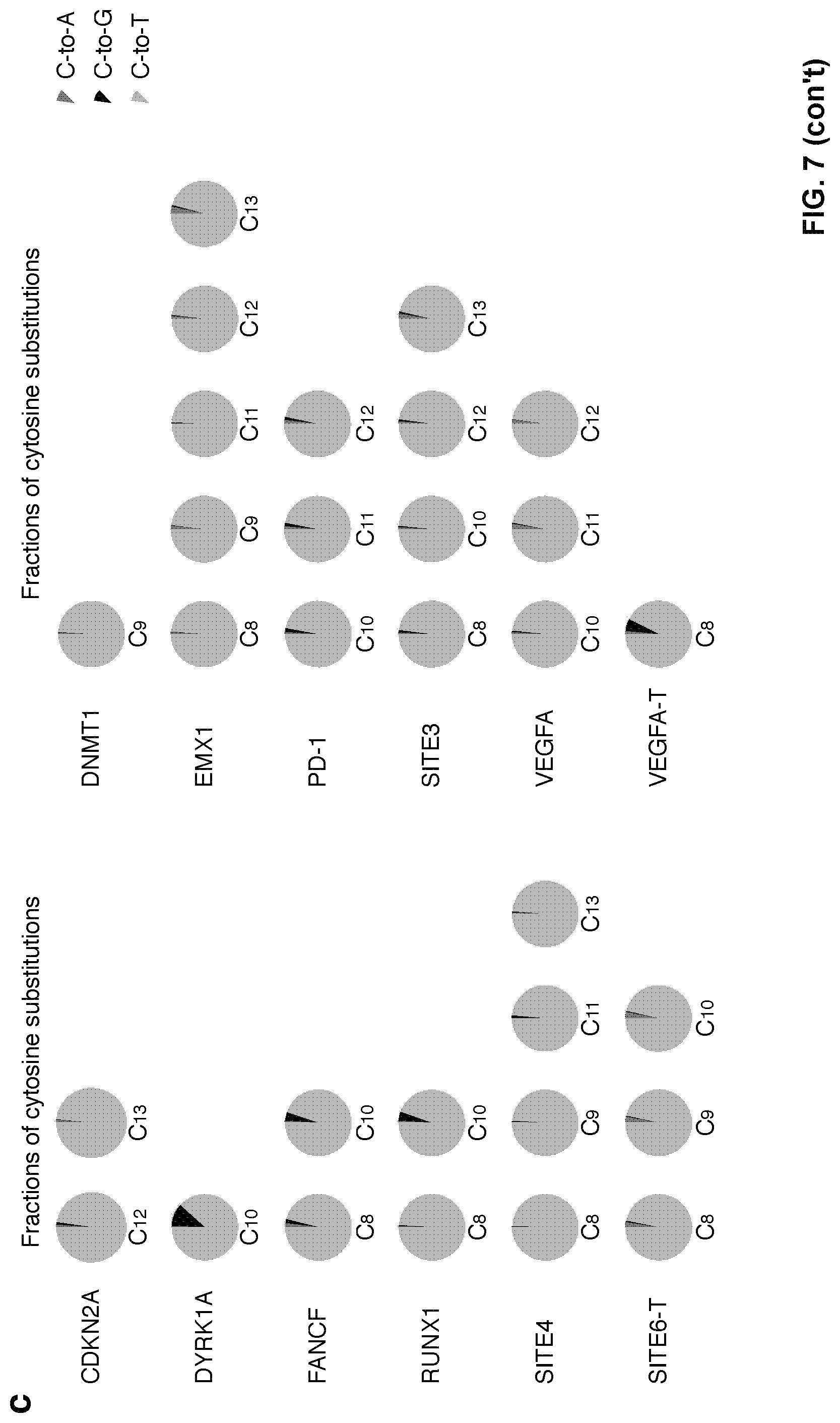
D00014

D00015

D00016
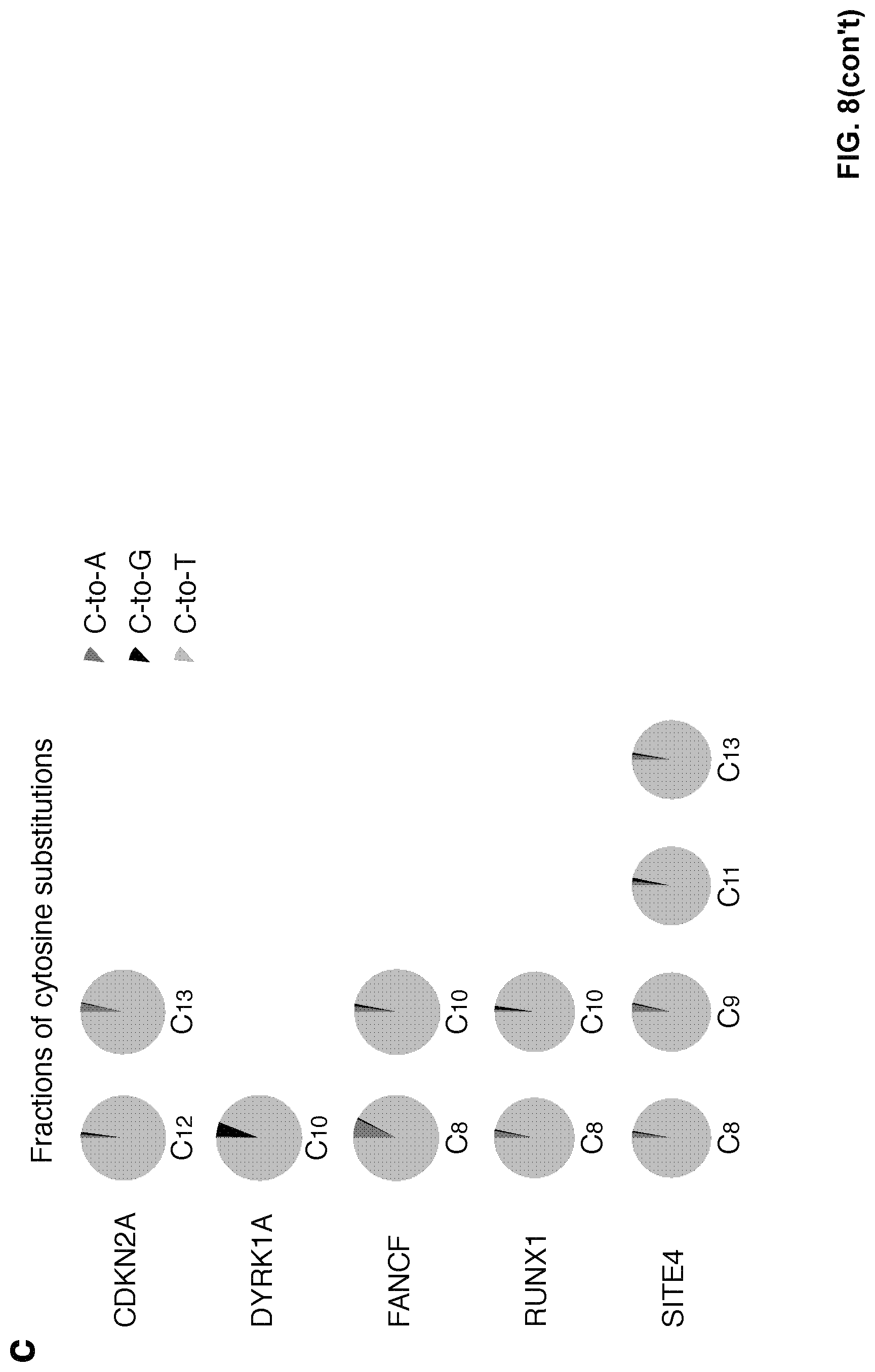
D00017

D00018

D00019
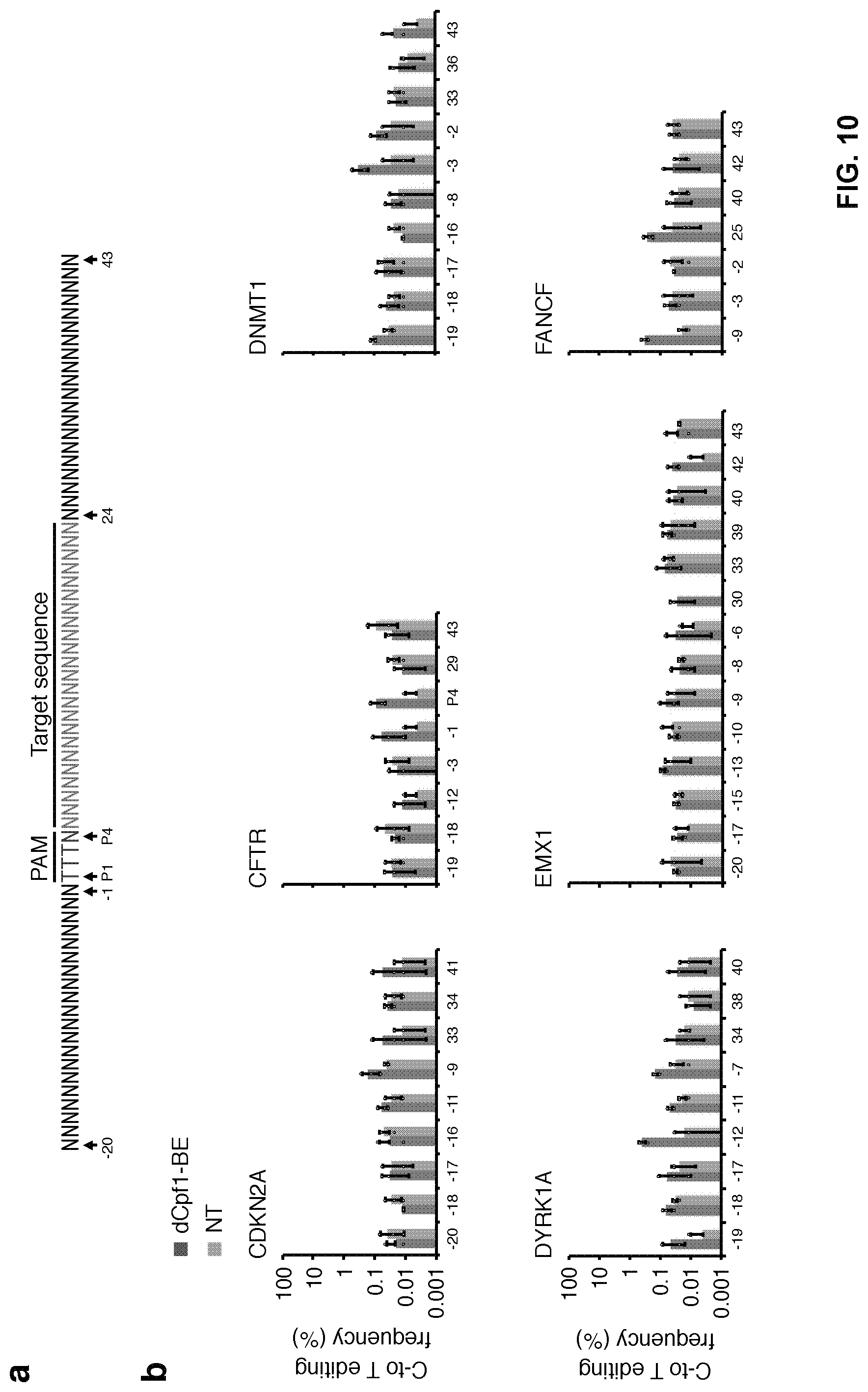
D00020

D00021

D00022

D00023

D00024
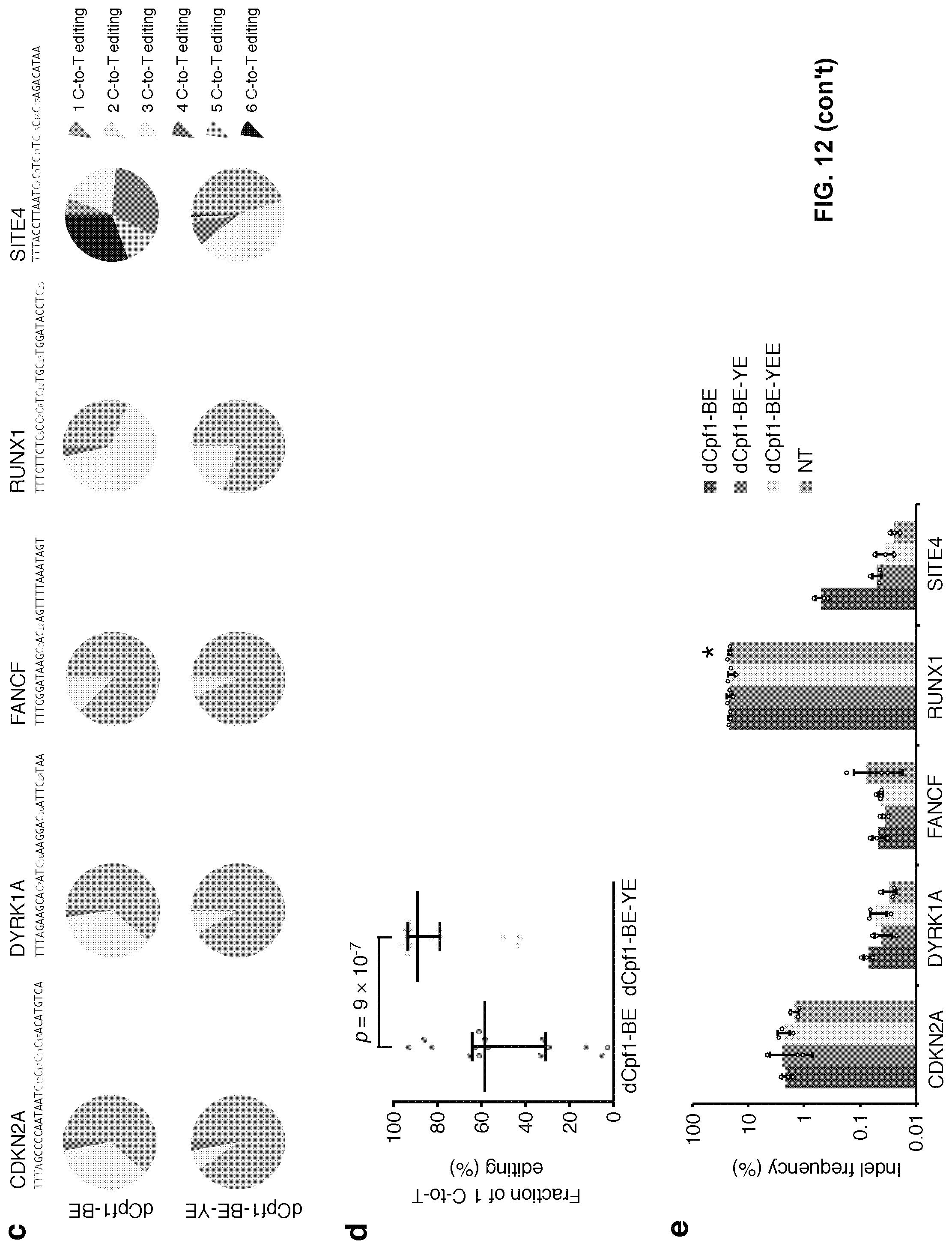
D00025
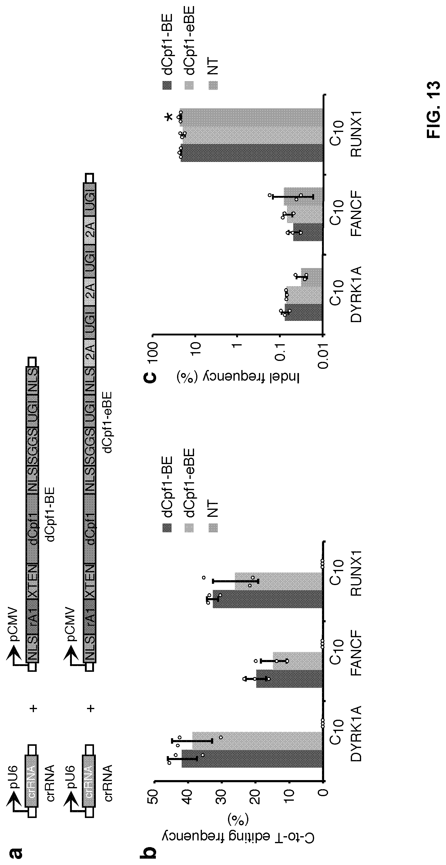
D00026
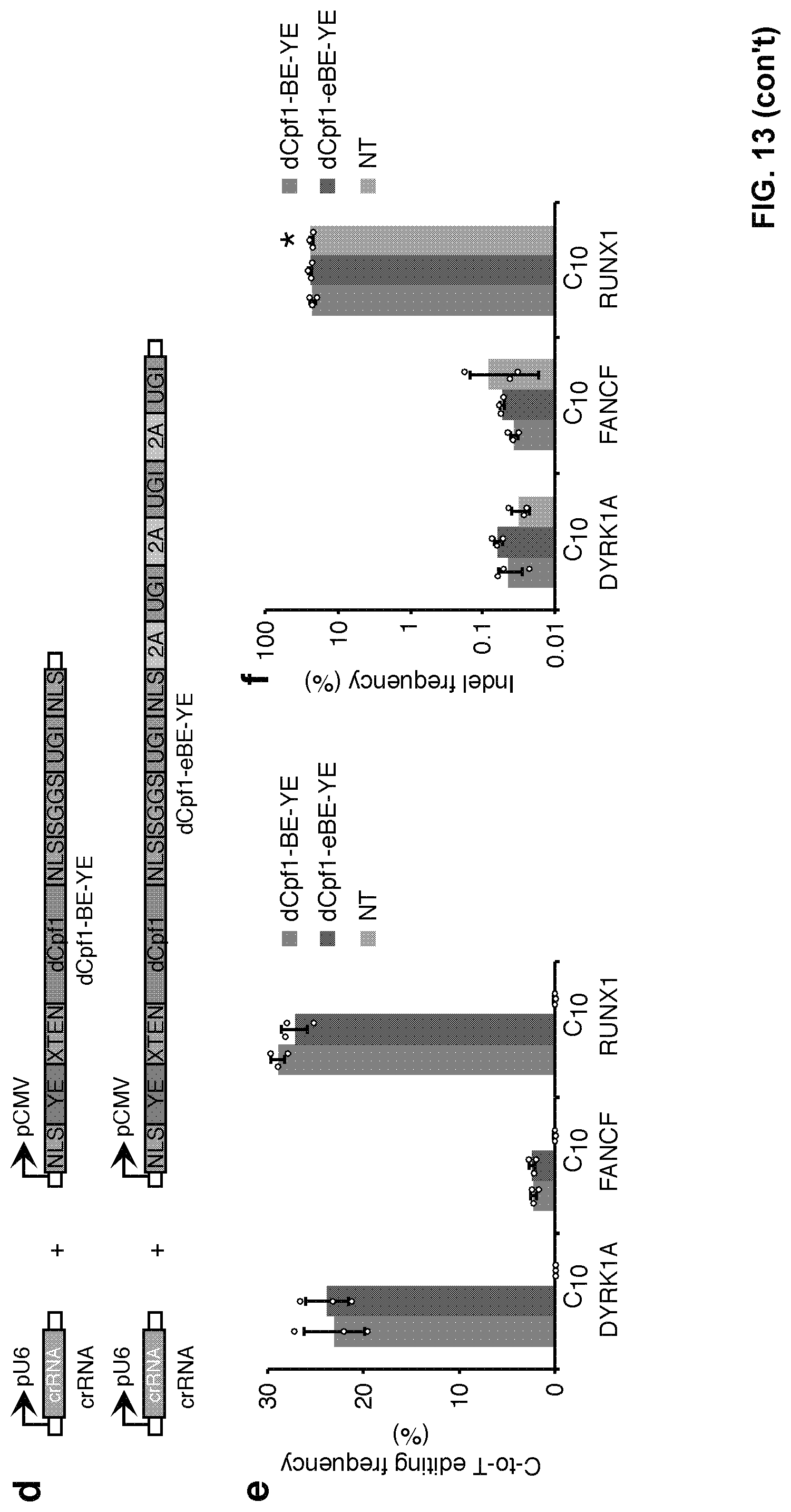
D00027

D00028

D00029
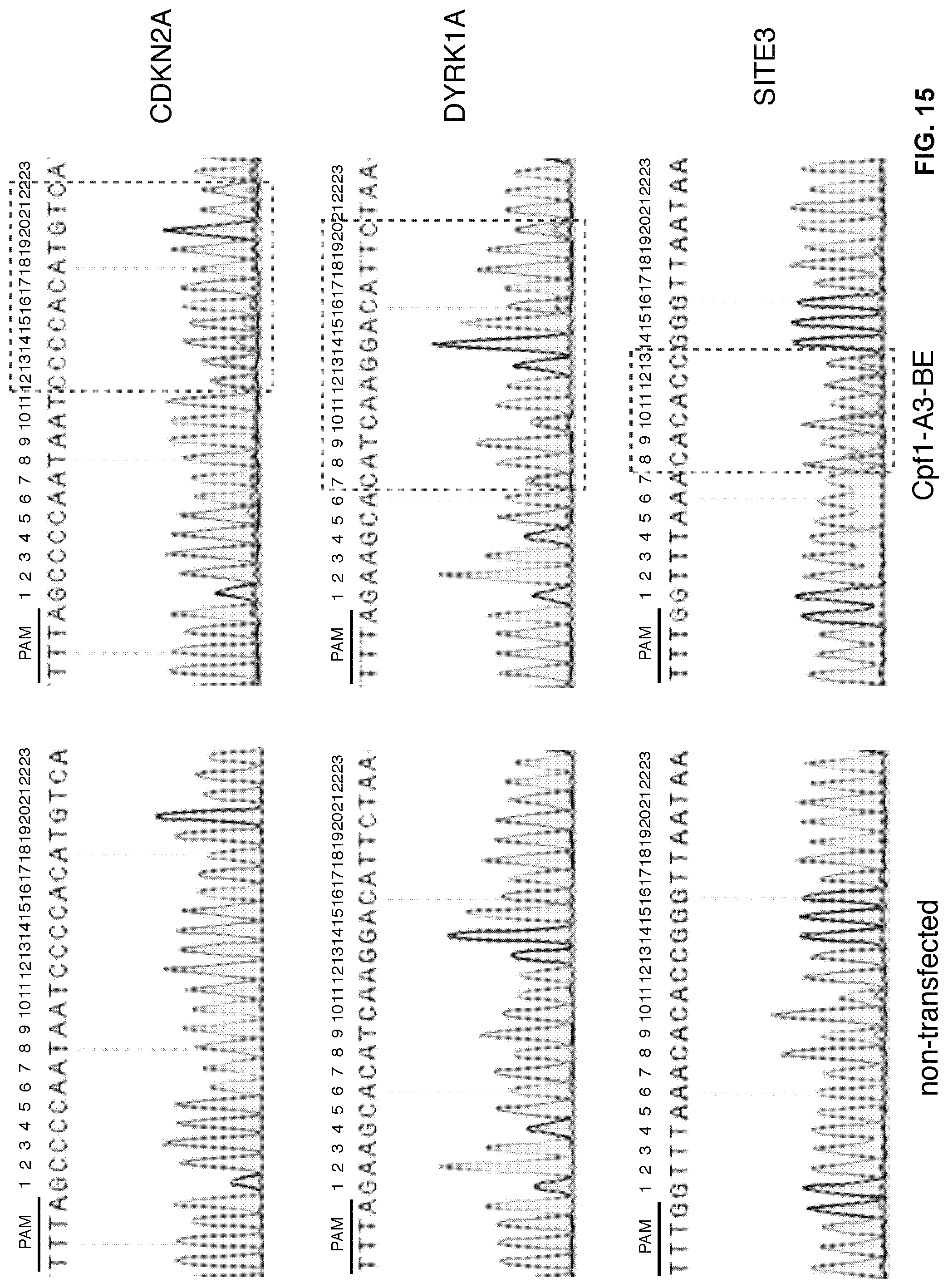
D00030
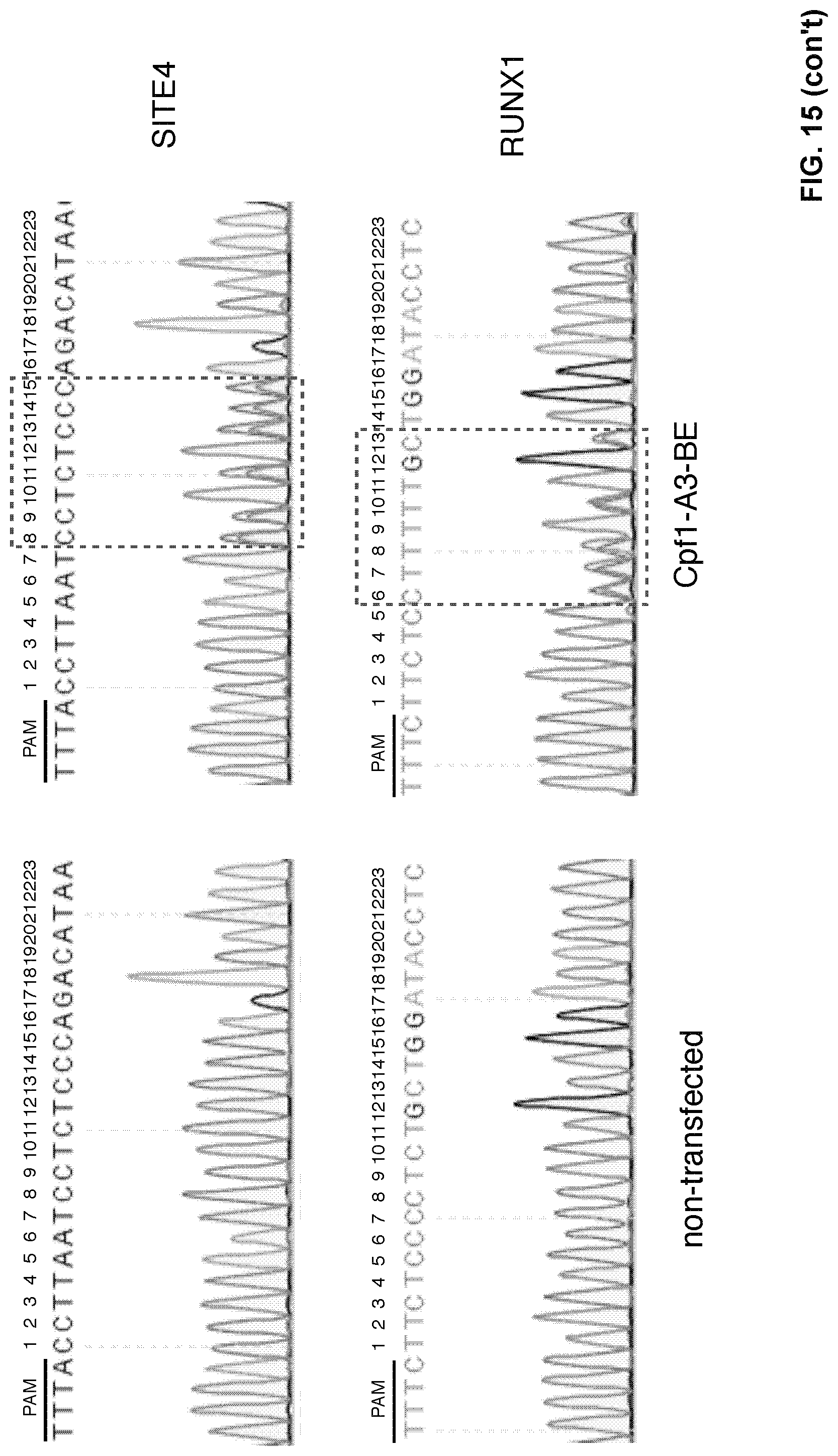
D00031

D00032
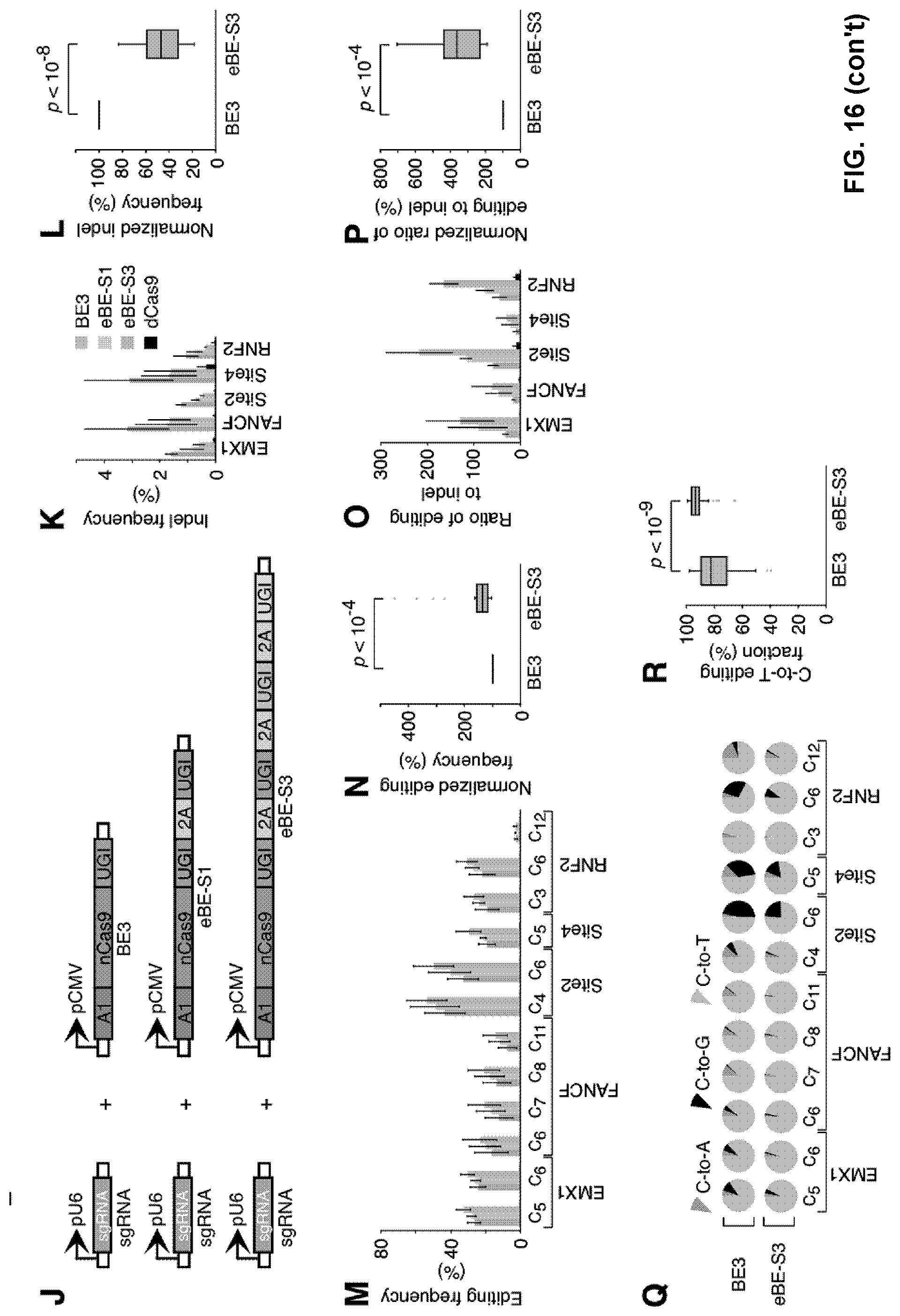
S00001
XML
uspto.report is an independent third-party trademark research tool that is not affiliated, endorsed, or sponsored by the United States Patent and Trademark Office (USPTO) or any other governmental organization. The information provided by uspto.report is based on publicly available data at the time of writing and is intended for informational purposes only.
While we strive to provide accurate and up-to-date information, we do not guarantee the accuracy, completeness, reliability, or suitability of the information displayed on this site. The use of this site is at your own risk. Any reliance you place on such information is therefore strictly at your own risk.
All official trademark data, including owner information, should be verified by visiting the official USPTO website at www.uspto.gov. This site is not intended to replace professional legal advice and should not be used as a substitute for consulting with a legal professional who is knowledgeable about trademark law.