Bispecific Molecules Comprising An Hiv-1 Envelope Targeting Arm
HAYNES; Barton F. ; et al.
U.S. patent application number 16/890380 was filed with the patent office on 2020-11-12 for bispecific molecules comprising an hiv-1 envelope targeting arm. This patent application is currently assigned to Duke University. The applicant listed for this patent is Duke University, MacroGenics, Inc., The University of North Carolina at Chapel Hill. Invention is credited to Guido FERRARI, Barton F. HAYNES, Leslie S. JOHNSON, Scott KOENIG, Chia-Ying Kao LAM, Liqin LIU, David M. MARGOLIS, Jeffrey Lee NORDSTROM, Julia A. SUNG.
| Application Number | 20200354439 16/890380 |
| Document ID | / |
| Family ID | 1000004986329 |
| Filed Date | 2020-11-12 |


View All Diagrams
| United States Patent Application | 20200354439 |
| Kind Code | A1 |
| HAYNES; Barton F. ; et al. | November 12, 2020 |
BISPECIFIC MOLECULES COMPRISING AN HIV-1 ENVELOPE TARGETING ARM
Abstract
The invention is directed to bispecific molecules comprising an HIV-1 envelope targeting arm and an arm targeting an effector cell, compositions comprising these bispecific molecule and methods of use. In certain aspects, the bispecific molecules of the present invention can bind to two different targets or epitopes on two different cells within the first epitope is expressed on a different cell type than the second epitope, such that the bispecific molecules can bring the two cells together. In certain aspects, the bispecific molecules of the present invention can bind to two different cells, wherein the bispecific molecules comprises an arm with the binding specificity of A32, 7B2, CH27, CH28 or CH44.
| Inventors: | HAYNES; Barton F.; (Durham, NC) ; FERRARI; Guido; (Durham, NC) ; KOENIG; Scott; (Rockville, MD) ; JOHNSON; Leslie S.; (Rockville, MD) ; LAM; Chia-Ying Kao; (San Jose, CA) ; SUNG; Julia A.; (Chapel Hill, NC) ; MARGOLIS; David M.; (Chapel Hill, NC) ; LIU; Liqin; (Germantown, MD) ; NORDSTROM; Jeffrey Lee; (Olney, MD) | ||||||||||
| Applicant: |
|
||||||||||
|---|---|---|---|---|---|---|---|---|---|---|---|
| Assignee: | Duke University Durham NC MacroGenics, Inc. Rockville MD The University of North Carolina at Chapel Hill Chapel Hill NC |
||||||||||
| Family ID: | 1000004986329 | ||||||||||
| Appl. No.: | 16/890380 | ||||||||||
| Filed: | June 2, 2020 |
Related U.S. Patent Documents
| Application Number | Filing Date | Patent Number | ||
|---|---|---|---|---|
| 15514420 | Mar 24, 2017 | 10717778 | ||
| PCT/US15/53027 | Sep 29, 2015 | |||
| 16890380 | ||||
| 62206586 | Aug 18, 2015 | |||
| 62056834 | Sep 29, 2014 | |||
| Current U.S. Class: | 1/1 |
| Current CPC Class: | C07K 16/2803 20130101; C07K 2317/73 20130101; C07K 2317/76 20130101; C07K 2317/31 20130101; C07K 16/2812 20130101; A61K 2039/505 20130101; C07K 2317/732 20130101; A61K 45/06 20130101; C07K 16/468 20130101; C07K 2317/52 20130101; C07K 2317/72 20130101; C07K 16/2809 20130101; C07K 16/283 20130101; A61K 39/39541 20130101; A61K 39/42 20130101; C07K 2317/626 20130101; C07K 16/44 20130101; C07K 16/1063 20130101 |
| International Class: | C07K 16/10 20060101 C07K016/10; C07K 16/28 20060101 C07K016/28; C07K 16/44 20060101 C07K016/44; A61K 39/395 20060101 A61K039/395; A61K 39/42 20060101 A61K039/42; A61K 45/06 20060101 A61K045/06; C07K 16/46 20060101 C07K016/46 |
Goverment Interests
GOVERNMENT SUPPORT
[0002] This invention was made with government support under Grant Nos. U19 AI067854 and UM1 AI100645 awarded by the National Institutes of Health. The government has certain rights in the invention.
Claims
1.-32. (canceled)
33. A bispecific molecule comprising a first polypeptide chain and a second polypeptide chain covalently bonded to one another, wherein: (I) the first polypeptide chain comprises in the N- to C-terminal direction: (i) a domain (A) comprising a binding region of the light chain variable domain of a first immunoglobulin (VL1) comprising the VL CDR3, CDR2 and CDR1 of HIV-1 antibody A32 (SEQ ID NO:78) or 7B2 (SEQ ID NO:55); (ii) a domain (B) comprising a binding region of a heavy chain variable domain of a second immunoglobulin (VH2) comprising the VH CDR3, CDR2 and CDR1 of an antibody specific for an epitope of CD3 or CD16, wherein domains (A) and (B) are separated from one another by a peptide linker 1; and (iii) a domain (C) comprising a heterodimer promoting domain including a K coil or E coil; wherein the heterodimer promoting domain (C) and domain (B) are separated by a peptide linker 2; (II) the second polypeptide chain comprises in the N- to C-terminal direction: (i) a domain (D) comprising a binding region of a light chain variable domain of the second immunoglobulin (VL2) comprising the VL CDR3, CDR2 and CDR1 of an antibody specific for the epitope of CD3 or CD16; (ii) a domain (E) comprising a binding region of a heavy chain variable domain of the first immunoglobulin (VH1) comprising the VH CDR3, CDR2, and CDR1 of HIV-1 antibody A32 (SEQ ID NO:77), or 7B2 (SEQ ID NO:56), wherein domains (D) and (E) are separated from one another by a peptide linker 1; and (iii) a domain (F) comprising a heterodimer promoting domain including a K coil or E coil; wherein the heterodimer promoting domain (F) and domain (E) are separated by a peptide linker 2, and wherein: the domains (A) and (B) do not associate with one another to form an epitope binding site; the domains (D) and (E) do not associate with one another to form an epitope binding site; the domains (A) and (E) associate to form a binding site that binds the HIV-1 envelope like the A32 or 7B2 antibody (1); the domains (B) and (D) associate to form a binding site that binds the epitope of CD3 or CD16; the K coil comprises residues 240-267 of SEQ ID NO: 19, and the E coil comprises residues 249-276 of SEQ ID NO: 17; and peptide linker 2 comprises residues 244-248 of SEQ ID NO: 17.
34. The bispecific molecule of claim 33, wherein (i) the VL1 comprises the VL CDR3, CDR2, and CDR1 of HIV-1 antibody A32 (SEQ ID NO: 78); (ii) the VH1 comprises the VH CDR3, CDR2, and CDR1 of HIV-1 antibody A32 (SEQ ID NO: 77); and (iii) the domains (B) and (D) associate to form a binding site that binds the epitope of CD3.
35. The bispecific molecule of claim 33, wherein (i) the VL1 comprises the VL CDR3, CDR2, and CDR1 of HIV-1 antibody A32 (SEQ ID NO: 78); (ii) the VH1 comprises the VH CDR3, CDR2, and CDR1 of HIV-1 antibody A32 (SEQ ID NO: 77); and (iii) the domains (B) and (D) associate to form a binding site that binds the epitope of CD16.
36. The bispecific molecule of claim 33, wherein (i) the VL1 comprises the VL CDR3, CDR2, and CDR1 of HIV-1 antibody 7B2 (SEQ ID NO: 55); (ii) the VH1 comprises the VH CDR3, CDR2, and CDR1 of HIV-1 antibody 7B2 (SEQ ID NO: 56); and (iii) the domains (B) and (D) associate to form a binding site that binds the epitope of CD3.
37. The bispecific molecule of claim 33, wherein (i) the VL1 comprises the VL CDR3, CDR2, and CDR1 of HIV-1 antibody 7B2 (SEQ ID NO: 55); (ii) the VH1 comprises the VH CDR3, CDR2, and CDR1 of HIV-1 antibody 7B2 (SEQ ID NO: 56); and (iii) the domains (B) and (D) associate to form a binding site that binds the epitope of CD16.
38. The bispecific molecule of claim 33, wherein (i) the VL1 comprises the VL CDR3, CDR2, and CDR1 of HIV-1 antibody A32 (SEQ ID NO: 78); (ii) the VH1 comprises the VH CDR3, CDR2, and CDR1 of HIV-1 antibody A32 (SEQ ID NO: 77); (iii) the VL2 comprises the VL CDR3, CDR2, and CDR1 of anti-CD3 antibody (SEQ ID NO: 52); and (iv) the VH2 comprises the VH CDR3, CDR2, and CDR1 of anti-CD3 antibody (SEQ ID NO: 51).
39. The bispecific molecule of claim 33, wherein (i) the VL1 comprises the VL CDR3, CDR2, and CDR1 of HIV-1 antibody A32 (SEQ ID NO: 78); (ii) the VH1 comprises the VH CDR3, CDR2, and CDR1 of HIV-1 antibody A32 (SEQ ID NO: 77); (iii) the VL2 comprises the VL CDR3, CDR2, and CDR1 of anti-CD16 antibody (SEQ ID NO: 54); and (iv) the VH2 comprises the VH CDR3, CDR2, and CDR1 of anti-CD16 antibody (SEQ ID NO: 53).
40. The bispecific molecule of claim 33, wherein (i) the VL1 comprises the VL CDR3, CDR2, and CDR1 of HIV-1 antibody 7B2 (SEQ ID NO: 55); (ii) the VH1 comprises the VH CDR3, CDR2, and CDR1 of HIV-1 antibody 7B2 (SEQ ID NO: 56); (iii) the VL2 comprises the VL CDR3, CDR2, and CDR1 of anti-CD3 antibody (SEQ ID NO: 52); and (iv) the VH2 comprises the VH CDR3, CDR2, and CDR1 of anti-CD3 antibody (SEQ ID NO: 51).
41. The bispecific molecule of claim 33, wherein (i) the VL1 comprises the VL CDR3, CDR2, and CDR1 of HIV-1 antibody 7B2 (SEQ ID NO: 55); (ii) the VH1 comprises the VH CDR3, CDR2, and CDR1 of HIV-1 antibody 7B2 (SEQ ID NO: 56); (iii) the VL2 comprises the VL CDR3, CDR2, and CDR1 of anti-CD16 antibody (SEQ ID NO: 54); and (iv) the VH2 comprises the VH CDR3, CDR2, and CDR1 of anti-CD16 antibody (SEQ ID NO: 53).
42. The bispecific molecule of claim 33, wherein the domain (A) comprises VL of HIV-1 antibody A32 (SEQ ID NO: 78); the domain (E) comprises VH of HIV-1 antibody A32 (SEQ ID NO: 77); and the domains (B) and (D) associate to form a binding site that binds the epitope of CD3.
43. The bispecific molecule of claim 33, wherein the domain (A) comprises VL of HIV-1 antibody A32 (SEQ ID NO: 78); the domain (E) comprises VH of HIV-1 antibody A32 (SEQ ID NO: 77); the domain (B) comprises VH of anti-CD3 antibody (SEQ ID NO: 51); and the domain (D) comprises VL of anti-CD3 antibody (SEQ ID NO: 52).
44. The bispecific molecule of claim 33, wherein the domain (A) comprises VL of HIV-1 antibody A32 (SEQ ID NO: 78); the domain (E) comprises VH of HIV-1 antibody A32 (residues 119-241 of SEQ ID NO: 11); and the domains (B) and (D) associate to form a binding site that binds the epitope of CD3.
45. The bispecific molecule of claim 33, wherein the domain (A) comprises VL of HIV-1 antibody A32 (SEQ ID NO: 78); the domain (E) comprises VH of HIV-1 antibody A32 (residues 119-241 of SEQ ID NO: 11); the domain (B) comprises VH of anti-CD3 antibody (SEQ ID NO: 51); and the domain (D) comprises VL of anti-CD3 antibody (SEQ ID NO: 52).
46. The bispecific molecule of claim 33, wherein the domain (A) comprises VL of HIV-1 antibody 7B2 (SEQ ID NO: 55); the domain (E) comprises VH of HIV-1 antibody 7B2 (SEQ ID NO: 56), and the domains (B) and (D) associate to form a binding site that binds the epitope of CD3.
47. The bispecific molecule of claim 33, wherein the domain (A) comprises VL of HIV-1 antibody 7B2 (SEQ ID NO: 55); the domain (E) comprises VH of HIV-1 antibody 7B2 (SEQ ID NO: 56), and the domain (B) comprises VH of anti-CD3 antibody (SEQ ID NO: 51); and the domain (D) comprises VL of anti-CD3 antibody (SEQ ID NO: 52).
48. The bispecific molecule of claim 33, wherein the domain (A) comprises VL of HIV-1 antibody A32 (SEQ ID NO: 78); the domain (E) comprises VH of HIV-1 antibody A32 (SEQ ID NO: 77), and the domains (B) and (D) associate to form a binding site that binds the epitope of CD16.
49. The bispecific molecule of claim 33, wherein the domain (A) comprises VL of HIV-1 antibody A32 (SEQ ID NO: 78); the domain (E) comprises VH of HIV-1 antibody A32 (SEQ ID NO: 77); the domain (B) comprises VH of anti-CD16 antibody (SEQ ID NO: 53); and the domain (D) comprises VL of anti-CD16 antibody (SEQ ID NO: 54).
50. The bispecific molecule of claim 33, wherein the domain (A) comprises VL of HIV-1 antibody A32 (SEQ ID NO: 78); the domain (E) comprises VH of HIV-1 antibody A32 (residues 119-241 of SEQ ID NO: 11), and the domains (B) and (D) associate to form a binding site that binds the epitope of CD16.
51. The bispecific molecule of claim 33, wherein the domain (A) comprises VL of HIV-1 antibody A32 (SEQ ID NO: 78); the domain (E) comprises VH of HIV-1 antibody A32 (residues 119-241 of SEQ ID NO: 11); the domain (B) comprises VH of anti-CD16 antibody (SEQ ID NO: 53); and the domain (D) comprises VL of anti-CD16 antibody (SEQ ID NO: 54).
52. The bispecific molecule of claim 33, wherein the domain (A) comprises VL of HIV-1 antibody 7B2 (SEQ ID NO: 55); the domain (E) comprises VH of HIV-1 antibody 7B2 SEQ (ID NO: 56), and the domains (B) and (D) associate to form a binding site that binds the epitope of CD16.
53. The bispecific molecule of claim 33, wherein the domain (A) comprises VL of HIV-1 antibody 7B2 (SEQ ID NO: 55); the domain (E) comprises VH of HIV-1 antibody 7B2 SEQ (ID NO: 56); the domain (B) comprises VH of anti-CD16 antibody (SEQ ID NO: 53); and the domain (D) comprises VL of anti-CD16 antibody (SEQ ID NO: 54).
54. The bispecific molecule of claim 38, 39, 40, or 41, wherein the bispecific molecule further comprises an Fc-domain.
55. The bispecific molecule of claim 38 or 40, wherein: the first polypeptide chain further comprises a CH2-CH3 domain, wherein the CH2-CH3 domain and domain (C) are separated by a peptide linker 3 or a spacer-linker 3; (ii) the bispecific molecule further comprises a third polypeptide chain comprising in the N- to C-terminal direction a peptide linker 3 and a CH2-CH3 domain; and (iii) the CH2-CH3 domains of the first and third polypeptide form the Fc domain.
56. The bispecific molecule of claim 55, wherein: (i) the CH2-CH3 domain of the first polypeptide chain comprises SEQ ID NO: 42 and the CH2-CH3 domain of the third polypeptide chain comprises SEQ ID NO: 43; or (ii) the CH2-CH3 domain of the first polypeptide chain comprises SEQ ID NO: 43 and the CH2-CH3 domain of the third polypeptide chain comprises SEQ ID NO: 42.
57. A composition comprising the bispecific molecule of claim 38 or 40.
58. The composition of claim 57, further comprising a second bispecific molecule comprising a first arm with the binding specificity of HIV-1 antibody A32, HIV-1 antibody 7B2, HIV-1 antibody CH28, or HIV-1 antibody CH44 and a second arm targeting CD3 or CD16, wherein the first and second bispecific molecules are different.
59. A composition comprising the bispecific molecule of claim 54.
60. The composition of claim 59, further comprising a second bispecific molecule comprising a first arm with the binding specificity of HIV-1 antibody A32, HIV-1 antibody 7B2, HIV-1 antibody CH28, or HIV-1 antibody CH44 and a second arm targeting CD3 or CD16, wherein the first and second bispecific molecules are different.
61. A composition comprising the bispecific molecule of claim 55.
62. The composition of claim 61, further comprising a second bispecific molecule comprising a first arm with the binding specificity of HIV-1 antibody A32, HIV-1 antibody 7B2, HIV-1 antibody CH28, or HIV-1 antibody CH44 and a second arm targeting CD3 or CD16, wherein the first and second bispecific molecules are different.
63. A method to treat HIV-1 infection in a subject in need thereof comprising administering to the subject a composition comprising the bispecific molecule of claim 38 or 40 in a therapeutically effective amount.
64. The method of claim 63, further comprising administering a latency activating agent.
65. The method of claim 64, wherein the latency activating agent is vorinostat, romidepsin, panobinostat, disulfiram, JQ1, bryostatin, PMA, inonomycin, or any combination thereof.
66. A method to treat HIV-1 infection in a subject in need thereof comprising administering to the subject a composition comprising the bispecific molecule of claim 54 in a therapeutically effective amount.
67. The method of claim 66, further comprising administering a latency activating agent.
68. The method of claim 67, wherein the latency activating agent is vorinostat, romidepsin, panobinostat, disulfiram, JQ1, bryostatin, PMA, inonomycin, or any combination thereof.
69. A vector comprising nucleic acid comprising nucleotides encoding the bispecific molecule of claim 33.
70. A composition comprising a vector comprising a nucleic acid encoding the bispecific molecule of claim 33.
Description
[0001] This application is a continuation of U.S. application Ser. No. 15/514,420 filed Mar. 24, 2017 which is a U.S. National Stage application of PCT/US2015/053027, filed Sep. 29, 2015, which claims the benefit of and priority to U.S. Ser. No. 62/056,834 filed Sep. 29, 2014, and U.S. Ser. No. 62/206,586 filed Aug. 18, 2015, the contents of each of which are hereby incorporated by reference in their entireties.
[0003] This patent disclosure contains material that is subject to copyright protection. The copyright owner has no objection to the facsimile reproduction by anyone of the patent document or the patent disclosure as it appears in the U.S. Patent and Trademark Office patent file or records, but otherwise reserves any and all copyright rights.
[0004] All patents, patent applications and publications cited herein are hereby incorporated by reference in their entirety. The disclosure of these publications in their entireties are hereby incorporated by reference into this application in order to more fully describe the state of the art as known to those skilled therein as of the date of the invention described herein.
SEQUENCE LISTING
[0005] The instant application contains a Sequence Listing which has been submitted electronically in ASCII format and is hereby incorporated by reference in its entirety. Said ASCII copy, created on Jun. 29, 2020, is named 1234300_00344 US2_SL.txt and is 117,071 bytes in size.
TECHNICAL FIELD
[0006] The invention is directed to HIV-1 antibodies and bispecific molecules comprising an HIV-1 binding domain and an effector cell binding domain, and their uses.
BACKGROUND
[0007] Highly Active Antiretroviral Therapy (HAART) has been effective in reducing the viral burden and ameliorating the effects of HIV-1 infection in infected individuals. However, despite this therapy the virus persists in the individual due to latent reservoir of HIV infected cells which evade this treatment. Thus, there is a need for therapeutic agents for treatment of HIV-1 infected individuals, as well as agents that target virus infected cells and have to potential to reduce the latent reservoir of HIV-1 infected cells.
SUMMARY OF THE INVENTION
[0008] The present invention is directed to bispecific molecules, e.g. covalently linked polypeptide chains to form antibodies, covalent diabodies and/or covalent diabody molecules and their use in the treatment of HIV-1. In certain aspects, the bispecific molecules of the present invention can bind to two different targets or epitopes on two different cells wherein the first epitope is expressed on a different cell type than the second epitope, such that the bispecific molecules can bring the two cells together. In certain aspects, the bispecific molecules of the present invention can bind to two different cells, wherein the bispecific molecules comprises an arm with the binding specificity of A32, 7B2, CH27, CH28 or CH44, which arm binds to the HIV-1 envelope expressed on a first cell, e.g. HIV infected cell, and a second arm with the binding specificity for an epitope expressed on a different cell type than the first cell, such that the bispecific molecules can bring the two cells together. In certain embodiment, the second cell is in effector cell which expresses CD3 or CD16.
[0009] In certain embodiments an antibody binds specifically to a particular target, even where the specific epitope may not be know, peptide or polysaccharide (such as an antigen present on the surface of a pathogen, for example gp120, gp41, or CD3) and do not bind in a significant amount to other proteins or polysaccharides present in the sample or subject. Specific binding can be determined by methods known in the art. Various competitive binding assays are known in the art. With reference to an antibody antigen complex, in certain embodiments specific binding of the antigen and antibody has a KD of less than about 10.sup.6 Molar, such as less than about 10.sup.6 Molar, 10.sup.7 Molar, 10.sup.8 Molar, 10.sup.9, or even less than about 10.sup.10 Molar.
[0010] In certain aspects the invention provides bispecific molecules comprising a first polypeptide chain and a second polypeptide chain, covalently bonded to one another, wherein: [0011] (I) the first polypeptide chain comprises in the N- to C-terminal direction: [0012] (i) a domain (A) comprising a binding region of the light chain variable domain of a first immunoglobulin (VL1) having the binding specificity of the A32, 7B2, CH28, or envelope antibody; [0013] (ii) a domain (B) comprising a binding region of a heavy chain variable domain of a second immunoglobulin (VH2) specific for an epitope (2), wherein domains (A) and (B) are separated from one another by a peptide linker 1; and [0014] (iii) a domain (C) comprising a heterodimer promoting domain including a K coil or E coil; wherein the heterodimer promoting domain (C) and domain B are separated by a peptide linker 2; [0015] (II) the second polypeptide chain comprises in the N- to C-terminal direction: [0016] (i) a domain (D) comprising a binding region of a light chain variable domain of the second immunoglobulin (VL2) specific for the epitope (2); [0017] (ii) a domain (E) comprising a binding region of a heavy chain variable domain of the first immunoglobulin (VH1) having the binding specificity of the A32, 7B2, CH28, or CH44 HIV-1 antibody, wherein domains (D) and (E) are separated from one another by a peptide linker 1; and [0018] (iii) a domain (F) comprising a heterodimer promoting domain including a K coil or E coil; wherein the heterodimer promoting domain (F) and domain (E) are separated by a peptide linker 2; and wherein: the domains (A) and (B) do not associate with one another to form an epitope binding site; the domains (D) and (E) do not associate with one another to form an epitope binding site; and the domains (A) and (E) associate to form a binding site that binds the HIV-1 envelope like A32, 7B2, CH28, or CH44 antibody (1); and the domains (B) and (D) associate to form a binding site that binds the epitope (2).
[0019] In certain aspects the invention provides bispecific molecules comprising a first polypeptide chain, a second polypeptide chain, and a third polypeptide chain, wherein some of the polypeptides are covalently bonded (See FIG. 8), and wherein:
(I) the first polypeptide chain comprises in the N- to C-terminal direction: [0020] (i) a domain (A) comprising a binding region of the light chain variable domain of a first immunoglobulin (VL1) having the binding specificity of the A32, 7B2, CH28, or CH44 HIV-1 antibody; [0021] (ii) a domain (B) comprising a binding region of a heavy chain variable domain of a second immunoglobulin (VH2) specific for an epitope (2), wherein domains (A) and (B) are separated from one another by a peptide linker 1; [0022] (iii) a domain (C) comprising a heterodimer promoting domain including a K coil or E coil; wherein the heterodimer promoting domain (C) and domain B are separated by a peptide linker 2; [0023] (iv) a CH2-CH3 domain, wherein the CH2-CH3 domain and domain (C) are separated by a peptide linker 3 or a spacer-linker 3; (II) the second polypeptide chain comprises in the N- to C-terminal direction: [0024] (i) a domain (D) comprising a binding region of a light chain variable domain of the second immunoglobulin (VL2) specific for the epitope (2); [0025] (ii) a domain (F) comprising a binding region of a heavy chain variable domain of the first immunoglobulin (VH1) having the binding specificity of the A32, 7B2, CH28, or CH44 HIV-1 antibody, wherein domains (D) and (E) are separated from one another by a peptide linker 1; [0026] (iii) a domain (F) comprising a heterodimer promoting domain including a K coil or E coil; wherein the heterodimer promoting domain (F) and domain (E) are separated by a peptide linker 2; (III) the third polypeptide chain comprises in the N- to C-terminal direction: [0027] (i) a peptide linker 3, [0028] (ii) a CH2-CH3 domain, and wherein: the domains (A) and (B) do not associate with one another to form an epitope binding site; the domains (D) and (E) do not associate with one another to form an epitope binding site; the domains (A) and (E) associate to form a binding site that binds the HIV-1 envelope lik A32, 7B2, CH28, or CH44 antibody (I); the domains (B) and (D) associate to form a binding site that binds the epitope (2); and the CH2-CH3 domains of the first and third polypeptide form an Fc chain.
[0029] A bispecific molecule comprising a first polypeptide chain, a second polypeptide chain, and a third polypeptide chain, wherein some of the polypeptides are covalently bonded (See FIG. 8), and wherein:
(I) the first polypeptide chain comprises in the N- to C-terminal direction: (i) a peptide linker 3 followed by a CH2-CH3 domain; (ii) a domain (A) comprising a binding region of the light chain variable domain of a first immunoglobulin (VL1) having the binding specificity of the A32, 7B2, CH28, or CH44 HIV-1 antibody, wherein the CH2-CH3 domain and domain (A) are separated by a peptide linker 4; (iii) a domain (B) comprising a binding region of a heavy chain variable domain of a second immunoglobulin (VH2) specific for an epitope (2), wherein domains (A) and (B) are separated from one another by a peptide linker 1; (iv) a domain (C) comprising a heterodimer promoting domain including a K coil or F coil; wherein the heterodimer promoting domain (C) and domain B are separated by a peptide linker 2; (II) the second polypeptide chain comprises in the N- to C-terminal direction: (i) a domain (D) comprising a binding region of a light chain variable domain of the second immunoglobulin (VL2) specific for the epitope (2); (ii) a domain (E) comprising a binding region of a heavy chain variable domain of the first immunoglobulin (VH1) having the binding specificity of the A32, 7B2, CH28, or CH44 HIV-1 antibody, wherein domains (D) and (E) are separated from one another by a peptide linker 1; (iii) a domain (F) comprising a heterodimer promoting domain including a K coil or E coil; wherein the heterodimer promoting domain (F) and domain (F) are separated by a peptide linker 2; (III) the third polypeptide chain comprises in the N- to C-terminal direction: [0030] (i) a peptide linker 3, [0031] (ii) a CH2-CH3 domain, and wherein: the domains (A) and (B) do not associate with one another to form an epitope binding site; the domains (D) and (E) do not associate with one another to form an epitope binding site; the domains (A) and (F) associate to form a binding site that binds the HIV-1 envelope like A32, 7B2, CH28, or CH44 antibody (1); the domains (B) and (D) associate to form a binding site that binds the epitope (2); and the CH2-CH3 domains of the first and third polypeptide form an Fc chain.
[0032] In certain embodiments, the CH2-CH3 domain of polypeptide chain 1 is the of the "knob" design and the CH2-CH3 domain of the third polypeptide chain is of the "hole" design, or vice versa.
[0033] In certain embodiments, the epitope (2) is a CD3 epitope or a CD16 epitope, In certain embodiments, the bispecific molecule binds HIV envelope with the specificity of A32 antibody and also binds CD3. In certain embodiments, the bispecific molecule binds HIV envelope with the specificity of 7B2 antibody and also binds CD3. In certain embodiments, the bispecific molecule binds HIV envelope with the specificity of CH28 antibody and also binds CD3. In certain embodiments, the bispecific molecule binds HIV envelope with the specificity of CH44antibody and also binds CD3. In certain embodiments, the bispecific molecule binds HIV envelope with the specificity of A32 antibody and also binds CD16. In certain embodiments, the bispecific molecule binds HIV envelope with the specificity of 7B2 antibody and also binds CD16. In certain embodiments, the bispecific molecule binds HIV envelope with the specificity of CH28 antibody and also binds CD16. In certain embodiments, the bispecific molecule binds HIV envelope with the specificity of CH44antibody and also binds CD16.
[0034] In certain embodiments, the domains (A) and (E) associate to form a binding site that binds the HIV-1 envelope with the binding specificity of the A32, 7B2, CH28, or CH44 antibody. In certain embodiments, the domains (A) and (E) associate to form a binding site that binds the A32, 7B2, CH27, CH28, or CH44 HIV-1 antibody epitope.
[0035] In certain embodiments, the domain (A) binding region of the A32 immunoglobulin (VL1) comprises the VL-A32 CDR3, CDR2, and CDR1. In certain embodiments, wherein the domain (E) binding region of the A32 immunoglobulin (VH1) comprises the VH-A32 CDR3, CDR2, and CDR1. In certain embodiments, the domain (A) binding region of the 7B2 immunoglobulin (VL1) comprises the VL-7B2 CDR3, CDR2, and CDR1. In certain embodiments, the domain (E) binding region of the 7B2 immunoglobulin (VH1) comprises the VH-7B2 CDR3, CDR2, and CDR1. In certain embodiments, the domain (A) binding region of the CH28 immunoglobulin (VL1) comprises the VL-CH28 CDR3, CDR2, and CDR1. In certain embodiments, the domain (E) binding region of the CH28 immunoglobulin (VH1) comprises the VH-CH28 CDR3, CDR2, and CDR1. In certain embodiments, the domain (A) binding region of the CH44 immunoglobulin (VL1) comprises the VL-CH44 CDR3, CDR2, and CDR1. In certain embodiments, the domain (E) binding region of the CH44 immunoglobulin (VH1) comprises the VH-CH44 CDR3, CDR2, and CDR1.
[0036] In certain embodiments, the domain (A) comprises VL-A32, VL-7B2, VL-CH28, or VL-CH44. In certain embodiments, the domain (E) comprises VH-A32, VH-7B2, VH-CH28, VH-CH44.
[0037] In certain embodiments, the first polypeptide comprises SEQ ID NO: 9, SEQ ID NO: 13, SEQ ID NO: 17, SEQ ID NO: 21. SEQ ID NO: 25, or SEQ ID NO: 44. In certain embodiments, the second polypeptide comprises SEQ ID NO: 11, SEQ ID NO: 15, SEQ ID NO: 19, SEQ ID NO: 23, SEQ ID NO: 27, or SEQ NO: 45. In certain embodiments, the bispecific molecule comprises the complementary second polypeptide, and wherein the second polypeptide comprises SEQ ID NO: 11, SEQ ID NO: 15, SEQ ID NO: 19, SEQ) NO: 23, SEQ ID NO: 27 or SEQ ID NO: 45.
[0038] In certain embodiments, the bispecific molecule comprises the first polypeptide of SEQ ID NO: 9, SEQ ID NO: 13, SEQ ID NO: 17, SEQ ID NO: 21, SEQ ID NO: 25, or SEQ ID NO: 44 and the second polypeptide of SEQ ID NO: 11, SEQ ID NO: 15, SEQ ID NO: 19, SEQ ID NO: 23, SEQ ID NO: 27, or SEQ ID NO: 45.
[0039] In certain embodiments, the bispecific molecule comprises the first polypeptide of SEQ ID NO: 9, and the complementary second polypeptide of SEQ ID NO: 11. In certain embodiments, the bispecific molecule comprises the first polypeptide of SEQ ID NO: 13, and the complementary second polypeptide of SEQ ID NO: 15. In certain embodiments, the bispecific molecule comprises the first polypeptide of SEQ ID NO: 17, and the complementary second polypeptide of SEQ ID NO: 19. In certain embodiments, the bispecific molecule comprises the first polypeptide of SEQ ID NO: 21, and the complementary second polypeptide of SEQ ID NO: 23. In certain embodiments, the bispecific molecule comprises the first polypeptide of SEQ ID NO: 25, and the complementary second polypeptide of SEQ ID NO: 27.
[0040] In certain embodiments, the bispecific molecule comprises consisting essentially of the first polypeptide of SEQ NO: 9. SEQ ID NO: 13, SEQ ID NO: 17, SEQ ID NO: 21, SEQ ID NO: 25, or SEQ ID NO: 44 and the second polypeptide of SEQ ID NO: 11, SEQ ID NO: 15, SEQ ID NO: 19, SEQ ID NO: 23, SEQ ID NO: 27, or SEQ ID NO: 45.
[0041] In certain embodiments, the bispecific molecule comprises consisting of the first polypeptide of SEQ ID NO: 9, SEQ ID NO: 13, SEQ ID NO: 17, SEQ ID NO: 21, SEQ NO: 25, or SEQ ID NO: 44 and the second polypeptide of SEQ ID NO: 11, SEQ ID NO: 15, SEQ ID NO: 19, SEQ ID NO: 23, SEQ ID NO: 27, or SEQ ID NO: 45.
[0042] In certain embodiments, the bispecific molecule comprises SEQ ID NO: 46, 47 and 48. In certain embodiments, the bispecific molecule consists essentially of SEQ ID NO: 46, 47 and 48. In certain embodiments, the bispecific molecule consists of SEQ ID NO: 46, 47 and 48. In certain embodiments, the first polypeptide of the bispecific molecule comprises SEQ ID NO: 46, the second polypeptide of the bispecific molecule comprises SEQ ID NO: 47, and the third polypeptide of the bispecific molecule comprises SEQ ID NO: 48.
[0043] In certain aspects, the invention provides a composition comprising any one of the bispecific molecules or any combination thereof. In certain embodiments, the composition comprises a composition comprising a bispecific molecule comprising a first arm with the binding specificity of HIV-1 antibody A32, HIV-1 antibody 7B2, HIV-1 antibody CH28, HIV-1 antibody CH44 and a second arm targeting CD3 or CD16. In certain embodiment, the bispecific molecule comprises an Fc portion or any other modification which extends its serum half-life. In certain embodiments, the composition further comprises a second bispecific molecule comprising a first arm with the binding specificity of the HIV-1 antibody A32, HIV-1 antibody 7B2, HIV-1 antibody CH28, HIV-1 antibody CH44 and a second arm targeting CD3 or CD16, wherein the first and second bispecific molecules are different.
[0044] In certain aspects, the invention provides a method to treat or prevent HIV-1 infection in a subject in need thereof comprising administering to the subject a composition comprising any one of the bispecific molecules of the invention or a combination of any one of the bispecific molecules in a therapeutically effective amount. In certain embodiments, the methods of claim further comprise administering a latency activating agent. In some embodiments, the latency activating agent is vorinostat, romidepsin, panobinostat, disulfiram, JQ1, bryostatin, PMA, inonomycin, or any combination thereof.
[0045] In certain aspects, the invention provides nucleic acids comprising nucleotides encoding the bispecific molecules of the invention. In certain aspects, the invention provides a vector comprising nucleic acids comprising nucleotides encoding the bispecific molecules of the invention. Provided are also compositions comprising a vector comprising a nucleic acid encoding the bispecific molecules. In certain aspects the invention provide a cell line comprising vectors or nucleic acids encoding the bispecific molecules of the invention, wherein the vectors encode polypeptide chains for expression of the bispecific molecules of the invention, e.g., polypeptide chain 1 and polypeptide chain 2, or polypeptide chain 1, polypeptide chain 2 and polypeptide chain 3. In certain embodiments, the vector is suitable for gene delivery and expression. In certain embodiment, the vector is an adenoviral vector, an adeno associated virus based vector, or a combination thereof.
[0046] In certain aspects, the invention provides a bispecific molecule comprising a polypeptide with a dual affinity retargeting reagent (DART), wherein the DART comprises a diabody molecule comprising a first polypeptide chain and a second polypeptide chain, covalently bonded to one another, wherein: [0047] (A) the first polypeptide chain comprises: [0048] (i) a domain (A) comprising a binding region of the light chain variable domain of a first immunoglobulin (VL1) specific for the first epitope (I); wherein the first VIA comprises, consists essentially of, consists of the VL or VLCDR1, VLCDR2, and VLCDR3 from A32, 7B2, CH27, CH28, or CH44 HIV-1 antibody, [0049] (ii) a domain (B) comprising a binding region of a heavy chain variable domain of a second immunoglobulin (VH2) specific for a second target, e.g an epitope (2), wherein domains (A) and (B) are separated from one another by a peptide linker; and [0050] (iii) a domain (C) comprising a heterodimer promoting domain; [0051] (B) the second polypeptide chain comprises: [0052] (i) a domain (D) comprising a binding region of a light chain variable domain of the second immunoglobulin (VL2) specific for the epitope (2); [0053] (ii) a domain (E) comprising a binding region of a heavy chain variable domain of the first immunoglobulin. (VH1) specific for the first epitope (I); wherein the first VH1 comprises, consists essentially of, consists of the VH or VHCDR1, VHCDR2, and VHCDR3 from A32, 7B2, CH27, CH28, or CH44 HIV-1 antibody, wherein domains (D) and (E) are separated from one another by a peptide linker, and [0054] (iii) a domain (F) comprising a heterodimer promoting domain, and [0055] wherein: [0056] the domains (A) and (B) do not associate with one another to form an epitope binding site; [0057] the domains (D) and (E) do not associate with one another to form an epitope binding site; [0058] the domains (A) and (E) associate to form a binding site that binds the A32, 7B2, CH27, CH28, or CH44 HIV-1 antibody epitope (1); the domains (B) and (D) associate to form a binding site that binds the second target, e.g., epitope (2).
[0059] In certain embodiments, the invention provides bispecific molecules, wherein the HIV antibodies VH and VL domains, and the CD3 and CD16 VH and VL domains are in a different orientation. For example, in a non-limiting embodiment, the VL1 domain in polypeptide chain 1 is from CD3, and VH2 domain is from an HIV envelope binding antibody. In this embodiment, the VH1 domain of polypeptide 2 is from CD3, and VL2 domain is from is from an HIV envelope binding antibody.
[0060] In certain aspects, the invention provides a bispecific molecule capable of specific binding to HIV-1 envelope and to an epitope of CD3, wherein the bispecific molecule comprises a first polypeptide chain and a second polypeptide chain, covalently bonded to one another, wherein: [0061] A. the first polypeptide chain comprises, in the N-terminal to C-terminal direction: [0062] i. a Domain 1, comprising [0063] (1) a sub-Domain (1A), which comprises a VL Domain of a monoclonal antibody capable of binding to CD3 (VLCD3); and [0064] (2) a sub-Domain (1B), which comprises a VH Domain of a monoclonal antibody capable of binding to HIV-1 (VHHIV-1), wherein the sub-Domains 1A and 1B are separated from one another by a peptide linker (e.g. SEQ ID NO:1); [0065] ii. a Domain 2, wherein the Domain 2 is an E-coil Domain (e.g. SEQ ID NO:7) or a K-coil Domain (e.g. SEQ ID NO:8), wherein the Domain 2 is separated from the Domain 1 by a peptide linker (SEQ ID NO:2); and [0066] B. the second polypeptide chain comprises, in the N-terminal to C-terminal direction: [0067] i. a Domain 1, comprising [0068] (1) a sub-Domain (1A), which comprises a VL Domain of a monoclonal antibody capable of binding to HIV-1 (VLHIV-1); and [0069] (2) a sub-Domain (1B), which comprises a VH Domain of a monoclonal antibody capable of binding to CD3 (VHCD3), wherein the sub-Domains 1A and 1B are separated from one another by a peptide linker (e.g. SEQ ID NO:1); and [0070] ii. a Domain 2, wherein the Domain 2 is a K-coil Domain (e.g. SEQ ID NO:8) or an E-coil Domain (SEQ ID NO:7), wherein the Domain 2 is separated from the Domain 1 by a peptide linker (SEQ ID NO:2); and wherein the Domain 2 of the first and the second polypeptide chains are not both E-coil Domains or both K-coil Domains and wherein: (a) the VL Domain of the first polypeptide chain and the VH Domain of the second polypeptide chain form an Antigen Binding Domain capable of specifically binding to an epitope of CD3; and (b) the VL Domain of the second polypeptide chain and the VH Domain of the first polypeptide chain form an Antigen Binding Domain capable of specifically binding to HIV-1 envelope.
[0071] A bispecific molecule capable of specific binding to HIV-lenvelope and to an epitope of CD16, wherein the bispecific molecule comprises a first polypeptide chain and a second polypeptide chain, covalently bonded to one another, wherein: [0072] A. the first polypeptide chain comprises, in the N-terminal to C-terminal direction: [0073] i. a Domain 1, comprising [0074] (1) a sub-Domain (1A), which comprises a VL Domain of a monoclonal antibody capable of binding to CD16 (VLCD16); and [0075] (2) a sub-Domain (1B), which comprises a VH Domain of a monoclonal antibody capable of binding to HIV-1 (VHHIV-1), wherein the sub-Domains 1A and 1B are separated from one another by a peptide linker (e.g. SEQ ID NO:1); [0076] ii. a Domain 2, wherein the Domain 2 is an E-coil Domain (SEQ ID NO:7) or a K-coil Domain (e.g. SEQ ID NO:8), wherein the Domain 2 is separated from the Domain 1 by a peptide linker (SEQ ID NO:2); and [0077] B. the second polypeptide chain comprises, in the N-terminal to C-terminal direction: [0078] i. a Domain 1, comprising [0079] (1) a sub-Domain (1A), which comprises a VL Domain of a monoclonal antibody capable of binding to HIV-1 (VLHIV-1); and [0080] (2) a sub-Domain (1B), which comprises a VH Domain of a monoclonal antibody capable of binding to CD16 (VHCD16), wherein the sub-Domains 1A and 1B are separated from one another by a peptide linker (e.g. SEQ ID NO:1); and [0081] ii. a Domain 2, wherein the Domain 2 is a K-coil Domain e.g. SEQ ID NO:8) or an E-coil Domain (e.g. SEQ ID NO:7), wherein the Domain 2 is separated from the Domain 1 by a peptide linker (SEQ ID NO:2); and wherein the Domain 2 of the first and the second polypeptide chains are not both E-coil Domains or both K-coil Domains and wherein: (a) the VL Domain of the first polypeptide chain and the VH Domain of the second polypeptide chain form an Antigen Binding Domain capable of specifically binding to an epitope of CD16; and (b) the VL Domain of the second polypeptide chain and the VH Domain of the first polypeptide chain form an Antigen Binding Domain capable of specifically binding to HIV-1 envelope.
[0082] In certain embodiments, the bispecific molecule binds to the HIV-1 envelope like the HIV antibody from which it is derived. In certain embodiments, the bispecific molecule binds to the A32-HIV-1 envelope epitope, i.e. the bispecific molecule binds to the HIV-1 envelope like the A32 antibody, and CD3, or CD16. In certain embodiments, the bispecific molecule binds to the 7B2-HIV1 envelope epitope and CD3, or CD16. In certain embodiments, the bispecific molecule binds to the CH27-HIV-1 envelope epitope and CD3, or CD16. In certain embodiments, the bispecific molecule binds to the CH28-HIV-1 envelope epitope and CD3, or CD16. In certain embodiments, the bispecific molecule binds to the CH44-HIV-1 envelope epitope and CD3, or CD16.
[0083] In certain embodiments, the bispecific molecule has the binding specificity of the A32 HIV-1-envelope antibody. In certain embodiments, the bispecific molecule has the binding specificity of the 7B2 HIV-1-envelope antibody. The bispecific molecule has the binding specificity of the CH27 HIV-1-envelope antibody. The bispecific molecule has the binding specificity of the CH28 HIV-1-envelope antibody. In certain embodiments, the bispecific molecule has the binding specificity of the CH44 HIV-1-envelope antibody.
[0084] In certain embodiments a bispecific molecule of the invention comprises, consists essentially of or consists of sequences as described herein, e.g. Table 2 and Table 3)
[0085] In certain embodiments a bispecific molecule of the invention comprises, consists essentially of or consists of SEQ ID NO: 9 and 11; SEQ ID NO: 13 and 15, SEQ ID NO: 17 and 19; SEQ ID NO; 21 and 23; SEQ ID NO: 25 and 27; SEQ ID NO; 44 and 45 (See Table 2 and Table 3).
[0086] In certain aspects the invention provides compositions comprising any of the bispecific molecule described herein, or a combination thereof. In certain embodiments, these compositions are formulated as pharmaceutical composition for therapeutic use.
[0087] In certain aspects the invention is directed to nucleic acids which encode the bispecific molecules of the invention. In certain embodiments, these nucleic acids are comprised in a vector, and are operably linked to a promoter. In certain aspects the invention provides cell lines, or isolated cells, which comprise nucleic acids for the expression of the bispecific molecules of the invention.
[0088] In certain aspects, the invention provides compositions comprising the bispecific molecules of the invention or nucleic acids encoding the same for use in methods of treating or preventing HIV infection. In some embodiments, these methods further comprise administering a Latency Activating Reagent. Non-limiting examples of these include HDAC inhibitors, e,g, vorinostat, romidepsin, panobinostat, disulfiram, JQ1, bryostatin, PMA, inonomycin, or any combination thereof. In some embodiments, this combination therapy targets the pool of latently infected HIV cells.
[0089] In certain aspects, the invention provides methods treating or preventing an HIV infection in a subject, the method comprising administering to the subject a composition comprising any one of the bispecific molecules of the invention, or a combination thereof in a therapeutically sufficient amount. In certain embodiments, the methods further comprise administering a latency activating agent.
BRIEF DESCRIPTION OF THE DRAWINGS
[0090] FIG. 1 shows potency of ADCC-mediating mAbs. The ADCC activity of the 5 CHAVI mAbs against the 22 HIV-1 IMC is reported as maximum percentage of specific killing. Each dot represent the average activity of all the positive results for each group of mAbs against the individual IMCs. The lines represent the mean.+-.standard deviation. The black line represent the cut-off for positive response.
[0091] FIG. 2 shows anti-HIV-1-DARTs-mediated cytotoxic activity. Activated CD4+ T cells from a HIV-1 seronegative donor were infected with HIV-1 subtype B BaL, AE CM235, and C 1086.c IMC (top to bottom). The cells were incubated with autologous resting CD8 T cells in the presence of six concentrations of the anti-HIV-1 (A32.times.CD3 .diamond-solid. and 7B2.times.CD3.box-solid.) and control (4420.times.CD3 ) DARTs for 6, 24, and 48 hours at an effector to target cell ratio of 33:1. The results are reported as maximum percentage of specific killing observed at each time point.
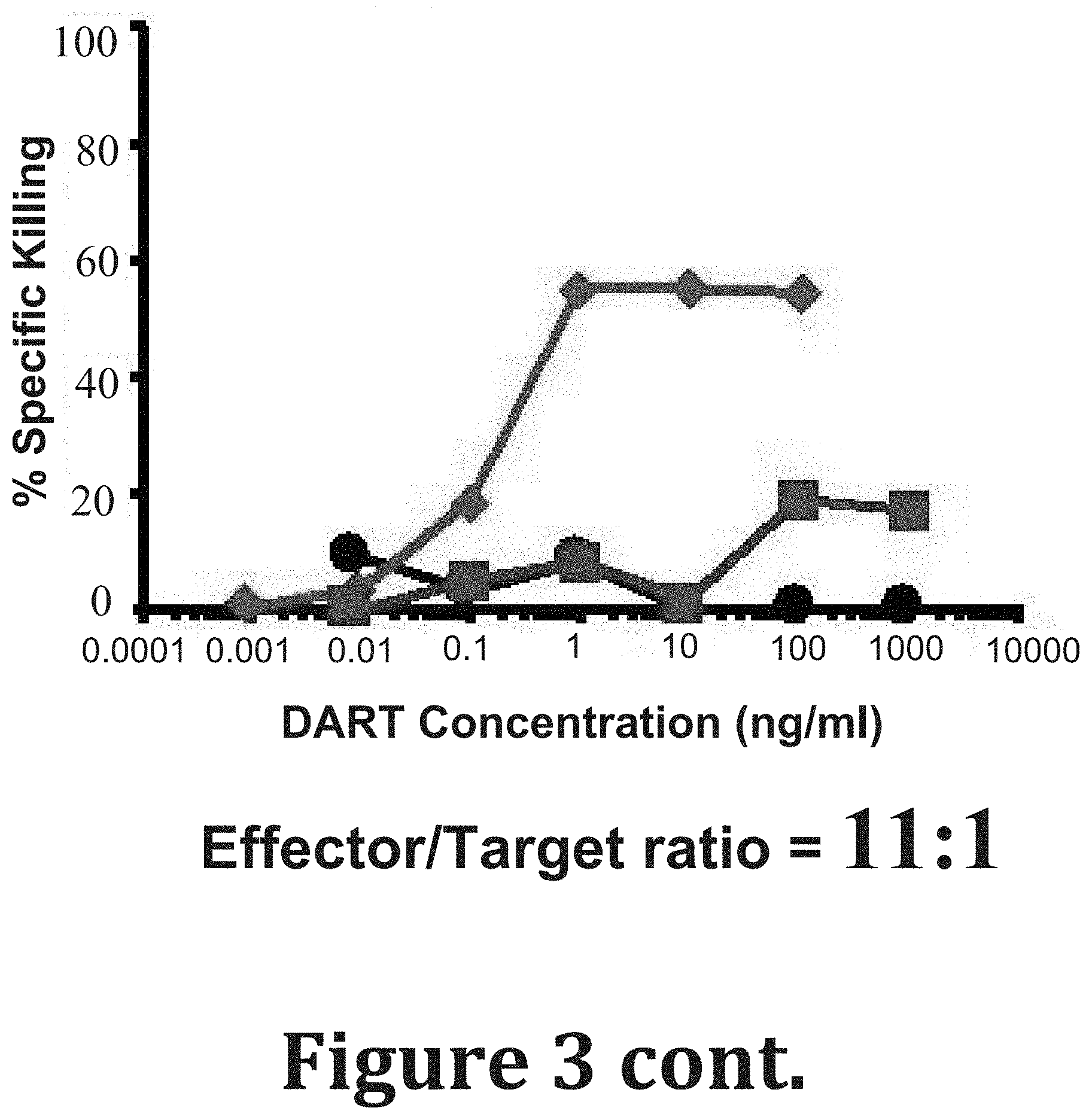
[0092] FIG. 3 shows dose dependence of anti-HIV-1 BaL DARTs-mediated cytotoxic activity. Activated CD4+ T cells from a HIV-1 seronegative donor were infected with HIV-1 subtype B BaL. The cells were incubated with autologous resting CD8 T cells in the presence of six concentrations of the anti-HIV-1 (A32.times.CD3 .diamond-solid. and 7B2.times.CD3.box-solid.) and control (4420.times.CD3 ) DARTs for 48 hours at an effector to target cell ratio of 33, 11, and 3:1 (top to bottom). The results are reported as percentage of specific killing.
[0093] FIG. 4. shows DART concentration to reach 50% Specific Killing. Activated CD4+ T cells from a HIV-1 seronegative donor were infected with HIV-1 subtype B BaL, AE CM235, and C 1086.c IMC. The cells were incubated with autologous resting CD8 T cells in the presence of six concentrations of the anti-HIV (A32.times.CD3, red; 7B2.times.CD3, blue) and control (4420.times.CD3, black) DARTs for 48 hours at an effector to target cell ratio of 33:1. Each bar represent the concentration required to detect 50% specific killing against each infected target population.
[0094] FIG. 5 shows the sequences of CH27, CH28 and CH44 HIV-1 antibodies. CDRs are indicated in the sequences (SEQ ID Nos: 57-74).
[0095] FIG. 6 shows the nucleotide sequences encoding VH and VL chains of A32 antibody and amino acid sequences of VH and VL chains of A32 (SEQ ID Nos 75-78 in order of appearance).
[0096] FIG. 7 shows nucleotide sequences encoding VH and VK chains of 7B2 antibody and amino acid sequences of VH and VK chains of 7B2 (SEQ ID NO: 79-82 in order of appearance).
[0097] FIG. 8A-C show the structures and domains of the bispecific molecules of the present invention. FIG. 8A illustrates the structure of a bispecific molecule composed of two polypeptide chains. FIGS. 8B and 8C illustrate the structures of two versions of the first, second and third polypeptide chains of a three chain bispecific molecule with an Fc domain (Version 1, FIG. 8B; Version 2, FIG. 8C).
[0098] FIG. 9 shows various sequences: Linker 1 (SEQ ID NO; 1); Linker 2 (SEQ ID NO: 2); Heterodimer promoting domain and K-coil and E coil sequences (SEQ ID Nos: 3-6, land 8); Linker 3 (DKTHTCPPCP (SEQ ID No: 49); Linker 4--SEQ ID NOS: 39, 40; CH2-CH3 fragments--SEQ ID Nos; 41-43; CH3 VH chain--SEQ ID NO: 51; CD3VL chain--SEQ ID NO: 52, CD16VH chain--SEQ ID NO 53, CH16 VL chain--SEQ ID NO: 54; 7B2 VL--SEQ IDNO 55; 7b2 VH-SEQ ID NO 56. SEQ ID Nos: 9-38, 44-48 show various bispecific antibodies (See Table 2).
[0099] FIGS. 10A-10C show HIV.times.CD3 DART structure. (FIGS. 10A-10B) These DART molecules contain an anti-HIV-1 binding arm (A32 or 7B2) combined with an anti-CD3 binding arm (hXR32). They are composed of two polypeptide chains: one with the VL of anti-CD3 linked to the VH of anti-HIV; the second with the VL of anti-HIV linked to the VH of anti-CD3. The carboxy termini of the chains have an interchain disulfide bond and paired oppositely charged E-coil/K-coil dimerization domains. Control DARTs have one of the arms replaced by an irrelevant one derived from an anti-FITC antibody (4420) or from an anti-RSV antibody, palivizumab (RSV) sequence. (FIG. 10C) Schematic representation of HIV.times.CD3 DART binding to two distinct antigens simultaneously and redirecting the cytotoxic T cells (effectors) to lyse the Env-expressing, HIV-1 infected cells (targets).
[0100] FIGS. 11A-11F show HIV.times.CD3 DART binding properties. FIGS. 11A-11C show antigen binding by ELISA. DART binding to human CD3 protein (FIG. 11A), to JR-FL gp140 protein (FIG. 11B) or simultaneously to both JR-FL gp140 and human CD3 proteins (FIG. 11C). FIGS. 11D-11F show cell surface binding by FACS. DART binding to primary human T cells expressing CD3 (FIG. 11D), to HEK293-D371 cells expressing HIV-1 Env, CM244, subtype AE (FIG. 11E) or to Jurkat 522-F/Y cells expressing CD3 and HIV-1 Env, HXBC2, subtype B (FIG. 11F). Data are reported as mean fluorescence intensity (MFI). CD3 and Env expression characteristics of the cells are reported in parenthesis. A32 and 7B2 are targeting arms that recognize HIV-1 gp120 and gp41, respectively; CD3 is the effector arm that recognizes CD3c; 4420 is an irrelevant, negative control arm.
[0101] FIGS. 12A-12H show HIV.times.CD3 DART redirected T-cell killing of Env.sup.+ target cells. FIG. 12A shows DART concentration dependent killing of Env.sup.+ Jurkat 522-F/Y cells in the presence of human T-cells at an E:T ratio of 10:1 for 48 hours with cytolysis measured by LDH release assay; EC.sub.50 values were 230 and 160 pg/mL for A32.times.CD3 and 7B2.times.CD3, respectively. The control DARTs (A32.times.4420, 7B2.times.4420, 4420.times.CD3) were inactive. FIG. 12B shows lack of DART mediated killing of Env.sup.+ Jurkat 522-F/Y cells in the absence of effector T-cells with cytolysis measured by LDH release assay. FIG. 12C shows lack of DART redirected T-cell killing of Env.sup.- Jurkat .DELTA.KS cells at an E:T ratio of 10:1 for 48 hours with cytolysis measured by LDH release assay. FIG. 12D shows DART concentration dependent killing of Env.sup.+ Jurkat 522-F/Y GF cells in the presence of human T-cells at an E:T ratio of 10:1 for 48 hours with cytolysis measured by LUM assay; EC.sub.50 values were 172 and 147 pg/mL for A32.times.CD3 and 7B2.times.CD3, respectively. FIGS. 12E-12G show 7B2.times.CD3 DART concentration dependent redirected T cell killing of Env.sup.+ Jurkat 522-F/Y GF cells at different E:T ratios (10:1, 5:1, 1:1) and incubation times (24, 48, 72 hours) with cytolysis measured by LUM assay. FIG. 12H shows time course of maximal cytolytic activity with 7B2.times.CD3 at different E:T ratios (data from FIGS. 12E-12G).
[0102] FIGS. 13A-13F show HIV.times.CD3 DARTs redirect T-cell cytotoxicity against CD4+ cells infected with HIV-1 IMCs of different subtypes. FIGS. 13A-13C show DART concentration dependence. Activated CD4+ T cells from a HIV-1 seronegative donor were infected with HIV-1 subtype B BaL (FIG. 13A), subtype AE CM235 (FIG. 13B) or subtype C 1086.0 (FIG. 13C) IMC and incubated for 48 hours with A32.times.CD3 (red circles), 7B2.times.CD3 (blue squares) or 4420.times.CD3 (black diamonds) in the presence of autologous resting CD8.sup.+ T cells at an E:T ratio of 33:1 (filled symbols) or in the absence of effector cells (E:T ratio of 0:1) (open symbols). The data are reported as percentage of specific lysis (% SL). DART concentrations ranged from 0.001 to 1000 ng/mL. FIGS. 13D-13F show time course. The data represent the maximal % SL observed at 6, 24, and 48 hours for each DART against CD4+ T cells infected with HIV-1 subtype B BaL (FIG. 13D), subtype AE CM235 (FIG. 13E) or subtype C 1086.0 (FIG. 13F) IMC and incubated with autologous resting CD8.sup.+ T cells at an E:T ratio of 33:1.
[0103] FIGS. 14A-14H show HIV.times.CD3 DARTs induce specific degranulation of CD8+ T-cell. FIGS. 14A-14D show schematic of gating strategy to identify Live/CD3+CD8+CD107+ T cells after their incubation with HIV-1 BaL infected target cells in presence of DARTs for 6 hours. (FIGS. 14E-14G) Dot plots represent the percentage of Live/CD3.sup.+ CD8.sup.+ CD107.sup.+ cells observed in presence of 1 ng/mL of 4420.times.CD3 (FIG. 14E), 7B2.times.CD3 (FIG. 14F) or A32.times.CD3 (FIG. 14G). FIG. 14H shows frequency of the CD3.sup.+ CD4.sup.- CD8.sup.+ CD107.sup.+ T cells observed in each of the five HIV-1 seronegative healthy donors after 6 hours of incubation with the autologous infected CD4.sup.+ T cells using the E:T ratio of 33:1. Each symbol represents the average of duplicate stimulations performed for each donor. The lines represent the mean.+-.standard deviation. * indicates p<0.05 after Dunnett's test for multiple comparisons.
[0104] FIGS. 15A-15C show viral clearance assay to assess HIV.times.CD3 DART redirected CD8+ T cell killing of autologous JR-CSF-infected CD4+ T cells from healthy HIV seronegative donors. Activated CD4.sup.+ T cells from HIV seronegative donors were infected with HIV-1 clone JR-CSF and then incubated with autologous resting CD8.sup.+ T effector cells at an E:T ratio of 1:1 in the absence (No DART) or presence of HIV.times.CD3 or control DARTs at a concentration of 100 ng/mL for 7 days. Results are shown for two healthy donors (FIGS. 15A-15B), as well as for healthy donor 2 in the presence of integrase and non-nucleoside reverse transcriptase inhibitors during the co-culture period to inhibit virus replication (FIG. 15C). Each bar represents the absolute p24 concentration detected in culture supernatants. Error bars represent standard error mean (SEM) of n=3. * indicates p<0.05 with Dunnett's test for multiple comparisons.
[0105] FIGS. 16A-16H show viral clearance assay detects HIV.times.CD3 DART redirected CD8+ T-cell clearance of JR-CSF or autologous reservoir (AR) virus-infected CD4+ cells using lymphocytes from HIV-infected ART suppressed patients. CD4.sup.+ depleted T cells from HIV-infected ART suppressed patients were activated with PHA and infected with HIV-1 subtype B clone JR-CSF (FIGS. 16A-16C) or autologous reservoir (AR) virus isolates (FIGS. 16D-16F) and then incubated without (FIGS. 16A, 16D) or with autologous CD8.sup.+ T effector cells at E:T ratios of 1:10 (FIGS. 16B, 16E) or 1:1 (FIGS. 16C, 16F) in the absence (No DART) or presence of HIV.times.CD3 (A32.times.CD3, 7B2.times.CD3) or control (7B2.times.4420, 4420.times.CD3) DARTs at a concentration of 100 ng/mL for 7 days. `Combo` indicates a 1:1 cocktail of 7B2.times.CD3 and A32.times.CD3 at a total concentration of 100 ng/mL. Each bar represents the log fold reduction of p24 detected in culture supernatants, calculated as the log (p24 of infected target cells only control divided by p24 of the test condition). FIG. 16G shows schematic of gating strategy to identify Live/CD3+CD4+CD107+ Effector (TFL4-) T cells after their incubation with HIV-1 JR-CSF infected target cells in presence of DARTs for 6 hours. FIG. 16H shows the % of live/effector cells (TFL4 negative)/CD3+/CD4+/107a+ cells following a 6 hour incubation with the indicated DARTs and JR-CSF infected targets in n=4 patients. Error bars represent SEM of n=8 (FIGS. 16A-16C, except for combo n=5 and 7B2.times.4420 n=6), n=5 (FIGS. 16D-16F), and n=4 (FIGS. 16G-16H). * indicates p<0.05 with Dunnett's test for multiple comparisons.
[0106] FIGS. 17A-17B show latency clearance assay to assess HIV.times.CD3 DART redirected CD8.sup.+ T-cell activity. Resting CD4.sup.+ T cells from HIV-infected, ART suppressed patients were incubated with PHA (FIG. 17A) or vorinostat (FIG. 17B), plated in 12-36 replicate wells depending on the size of the patient's latent reservoir, and co-cultured with autologous CD8.sup.+ T cells at an E:T ratio of 1:10 in the absence or presence of HIV.times.CD3 or control DARTs at 100 ng/mL for 24 hours (or up to 96 hours where indicated), after which DARTs were washed off and CD8-depleted PBMCs from a seronegative donor were added to amplify residual virus. Wells were assessed for the presence or absence of p24 by ELISA at day 15. `Combo` indicates a 1:1 cocktail of 7B2.times.CD3 and A32.times.CD3 at a total concentration of 100 ng/mL. Results are shown as % viral recovery (# of positive wells/total number plated), normalized to a control in which no CD8.sup.+ T cells are added. Dashed lines indicate undetectable viral recovery. NT indicated the conditions that were not tested due to low cell availability according to the table shown in FIG. 21.
[0107] FIG. 18 shows a list of IMC by HIV-1 Subtypes and Neutralization Tier.
[0108] FIG. 19 shows Equilibrium Dissociation Constants (K.sub.D) for Binding of A32.times.CD3 and 7B2.times.CD3 to Recombinant Env and CD3 Protein.
[0109] FIG. 20 shows Clinical Characteristics.
[0110] FIG. 21 shows that DARTs redirect patient T cells against JR-CSF infected autologous target cells and absolute p24 concentration.
[0111] FIG. 22 shows Absolute # of Positive Wells in Latency Clearance Assay with DARTs.
[0112] FIG. 23 shows potency and breadth of ADCC-mediating mAbs. The ADCC activities of the A32 (anti-gp120 C1/C2) mAb (.diamond-solid.) and 7B2 (anti-gp41 cluster I) mAb (.box-solid.) are reported as maximum percentage of specific lysis (% SL) against each of the 22 HIV-1 IMC. Each dot represents one HIV-1 IMC. The results obtained with plasma from one HIV-1 seropositive (positive control; pos ctrl) and one seronegative (negative control; neg ctrl) donor are also reported. The lines represent the mean.+-.standard deviation. The black line represents the cut-off for positive response.
[0113] FIG. 24 shows conservation of HIV-1 Env residues known to influence the binding of 7B2 and A32 mAbs. A linear 7-residue sequence in gp41 (gp160 positions 598-604; immunodominant cluster I) is reported to contain the binding site for 7B2 mAb (28, 29). Discontinuous residues in gp120 C1-C4 known to influence A32 mAb binding (based on point mutagenesis studies) occur at positions 52, 53, 66, 69, 83, 86, 96, 100, 103, 107, 112, 215, 217, 252, 256, 262, 427 and 479 (37, 39, 68). The conservation of these residues in the Los Alamos National Laboratory (LANL) HIV1 Env Amino acid Filtered web alignment, a database consisting of 4556 HIV-1 Env sequences with representation of all subtypes, was assessed by QuickAlign analysis (http://www.hiv.lanl.gov/content/sequence/QUICK_ALIGNv2/QuickAlign.html). The height of the residue at each position of Env is proportional to its frequency of distribution among the HIV-1 isolates. Residues are colored according to hydrophobicity: black, hydrophilic; green, neutral; blue, hydrophobic. Based on a crystal structure of a CD4-stabilized gp120 core complexed with a Fab fragment of N5-i5 (an A32-like mAb), residues at 52, 53, 69, 103, 107 and 217 (located in C1-C2) may be direct epitope contacts (27).
[0114] FIG. 25 shows cell surface Env binding of A32.times.4420 and 7B2.times.4420 control DARTs. DART binding to HEK293-D371 cells expressing HIV-1 Env, CM244, subtype AE was measured and data are reported as mean fluorescence intensity (MFI). A32 and 7B2 are targeting arms that recognize HIV-1 gp120 and gp41, respectively; 4420 is an irrelevant, negative control arm.
[0115] FIGS. 26A-26D show HIV.times.CD3 DART-mediated T-cell activation depends on co-engagement with target cells. Unstimulated CD4.sup.+ or CD8.sup.+ T-cells from healthy seronegative donors were incubated with (FIGS. 26A, 26C) and without (FIGS. 26B, 26D) Env expressing Jurkat-522 F/Y cell line in the absence or presence of control (RSV.times.CD3) or HIV.times.CD3 (A32.times.CD3, 7B2.times.CD3) DARTs at 40, 0.32, and 0 ng/mL for 48 hours. CD8.sup.+(FIGS. 26A-26B) and CD4.sup.+ (FIGS. 26C-26D) T cell activation was assessed by staining with CD25 Ab cells. The data are reported as frequency (%) of activated (CD25.sup.+) T cells. Each bar represent the average of results obtained from 2 different donors.
[0116] FIG. 27 shows HIV DARTs bind specifically to HIV-1 IMC infected CD4.sup.+ T cells. Activated CD4.sup.+ T cells obtained from healthy HIV-1 seronegative donors were infected for 48 hours with HIV-1 IMCs representing the HIV-1 subtype B BaL, AE CM235, and C 1086.0 as reported in the methods section. Non-infected CD4.sup.+ T cells (mock) were utilized as negative control. The cells were stained using the 7B2.times.4420 and A32.times.4420 DART where the CD3 arm was substituted with the irrelevant 4420 protein to avoid binding to the CD3 receptor. After incubation with the DART, the cells were stained with the secondary anti-EK-IgG2a-biotinylated complex to reveal binding of the DARTs. The staining with 7B2 and A32 mAbs, utilizing an indirect staining technique with the secondary mouse anti-human-IgG mAb, was performed as control. The secondary fluoresceinated anti-human IgG Abs and the Palivizumab mAb were utilized as negative controls. The frequency of infected cells was determined by intracellular staining using the anti-p24 mAb as reported in the method section. Each bar represents CD4.sup.+ T cells infected with the IMCs and controls as indicated above the graph. The results are reported as frequency (%) of viable infected (p24.sup.+) CD4.sup.+ T cells that were stained by each of the DARTs, mAbs, and controls as listed on the x-axis.
[0117] FIGS. 28A-28D show lack of HIV.times.CD3 DART effects on T cell viability or activation status in the absence of added target cells using PBMC from HIV-1 infected donors. Unstimulated CD4.sup.+ or CD8.sup.+ T-cells from HIV-infected, ART suppressed were incubated in the absence or presence of control (4420.times.CD3, 7B2.times.4420, A32.times.4420) or active (A32.times.CD3, 7B2.times.CD3) DARTs at 100 ng/mL for 7 days. (FIGS. 28A-28B) T cell viability was assessed by staining cells for Annexin V/7-AAD. Viable cells were identified as those that were Annein V and 7-AAD negative. (FIGS. 28C-28D) T cell activation was assessed by staining cells for HLA-DR and CD25 expression. Data points for both analyses are from n=3 patients performed on 3 independent occasions. Error bars represent standard error mean.
DETAILED DESCRIPTION
[0118] Highly active anti-retroviral therapy (HAART) alone or in combination with latency reversing agents fails to reduce the pool of latently infected cells. This is due to limited ability of the CD8+ T cells to eliminate HIV-1 latently infected cells. Dual Affinity Re-Targeting proteins (DARTs) are bispecific, antibody-based molecules that can bind two distinct antigens simultaneously. HIV-1 DARTs contain an HIV-1 binding arm combined with an effector cell binding arm, and are designed to redirect cytotoxic CD3+ T cells to engage and kill HIV-infected cells. A panel of monoclonal antibodies (mAbs) was studied to determine their magnitude and breadth of mediating ADCC against 22 different isolates. The goals were to: 1) identify mAbs that could be used as the HIV-1 binding arms of DARTs; 2) test the resulting DARTs for their ability to mediate killing of HIV-1 infected cells. Provided herein are data related to the potency of the different groups of ADCC-mediating mAbs and the resulting DARTs against HIV-1 Infectious Molecular Clones (IMC)-infected target cells.
[0119] Antibodies and Other Binding Molecules
[0120] Antibodies
[0121] The invention provides polyclonal or monoclonal antibodies, variants, fusion proteins comprising an antibody portion with an antigen recognition site of the required specificity, humanized antibodies, and chimeric antibodies, and any other modified configuration of the immunoglobulin molecule that comprises an antigen recognition site of the required specificity. Throughout this application, the numbering of amino acid residues of the light and heavy chains of antibodies is according to the EU index as in Kabat et al. (1992) SEQUENCES OF PROTEINS OF IMMUNOLOGICAL INTEREST, National Institutes of Health Publication No. 91-3242. In some embodiments, antigen-binding fragment of an antibody is a portion of an antibody that possesses an at least one antigen recognition site. Fragments include for example but not limited to Fab, Fab', F(ab').sub.2 Fv), and single chain (scFv).
[0122] Monoclonal antibodies are known in the art. In certain embodiments, monoclonal antibody encompasses not only intact monoclonal antibodies and full-length monoclonal antibodies, but also fragments thereof (such as Fab, Fab', F(ab').sub.2 Fv), single chain (scFv), mutants thereof, fusion proteins comprising an antibody portion, humanized monoclonal antibodies, chimeric monoclonal antibodies, and any other modified configuration of the immunoglobulin molecule that comprises an antigen recognition site of the required specificity and the ability to bind to an antigen. Monoclonal antibodies are not limited as regards to the source of the antibody or the manner in which it is made (e.g., by hybridoma, phage selection, recombinant expression, transgenic animals, etc.).
[0123] Methods of making monoclonal antibodies are known in the art. In certain embodiments, the antibodies are produced recombinantly by any means known in the art. In one embodiment, such an antibody is sequenced and the polynucleotide sequence is then cloned into a vector for expression or propagation. The sequence encoding the antibody of interest may be maintained in a vector in a host cell and the host cell can then be expanded and frozen for future use. The polynucleotide sequence of such antibodies may be used for genetic manipulation to generate the bi-specific molecules of the invention as well as a chimeric antibody, a humanized antibody, or a caninized antibody, to improve the affinity, or other characteristics of the antibody. The general principle in humanizing an antibody involves retaining the basic sequence of the antigen-binding portion of the antibody, while swapping the non-human remainder of the antibody with human antibody sequences. There are four general steps to humanize a monoclonal antibody. These are: (1) determining the nucleotide and predicted amino acid sequence of the starting antibody light and heavy variable Domains (2) designing the humanized antibody or caninized antibody, i.e., deciding which antibody framework region to use during the humanizing or canonizing process (3) the actual humanizing or caninizing methodologies/techniques and (4) the transfection and expression of the humanized antibody. See, for example, U.S. Pat. Nos. 4,816,567; 5,807,715; 5,866,692; and 6,331,415.
[0124] Bi-Specific Antibodies, Multi-Specific Diabodies and DART.TM. Diabodies
[0125] The provision of non-mono-specific "diabodies" provides a significant advantage over antibodies: the capacity to co-ligate and co-localize cells that express different epitopes. Bivalent diabodies thus have wide-ranging applications including therapy and immunodiagnosis. Bi-valency allows for great flexibility in the design and engineering of the diabody in various applications, providing enhanced avidity to multimeric antigens, the cross-linking of differing antigens, and directed targeting to specific cell types relying on the presence of both target antigens. Due to their increased valency, low dissociation rates and rapid clearance from the circulation (for diabodies of small size, at or below 50 kDa), diabody molecules known in the art have also shown particular use in the field of tumor imaging (Fitzgerald et al. (1997) "Improved Tumour Targeting By Disulphide Stabilized Diabodies Expressed In Pichia pastoris," Protein Eng. 10:1221). Of particular importance is the co-ligating of differing cells, for example, the cross-linking of cytotoxic T cells to tumor cells (Staerz et al. (1985) "Hybrid Antibodies Can Target Sites For Attack By T Cells," Nature 314:628-631, and Holliger et al. (1996) "Specific Killing Of Lymphoma Cells By Cytotoxic T-Cells Mediated By A Bispecific Diabody," Protein Eng. 9:299-305).
[0126] Diabody epitope binding domains may also be directed to a surface determinant of a B cell, such as CD19, CD20, CD22, CD30, CD37, CD40, and CD74 (Moore, P. A. et al. (2011) "Application Of Dual Affinity Retargeting Molecules To Achieve Optimal Redirected T-Cell Killing Of B-Cell Lymphoma," Blood 117(17):4542-4551; Cheson, B. D. et al. (2008) "Monoclonal Antibody Therapy For B-Cell Non Hodgkin's Lymphoma," N. Engl. J. Med. 359(6):613-626; Castillo, J. et al. (2008) "Newer monoclonal antibodies for hematological malignancies," Exp. Hematol. 36(7):755-768. In many studies, diabody binding to effector cell determinants, e.g., Fc.gamma. receptors (Fc.gamma.R), was also found to activate the effector cell (Holliger et al. (1996) "Specific Killing Of Lymphoma Cells By Cytotoxic T-Cells Mediated By A Bispecific Diabody," Protein Eng. 9:299-305; Holliger et al. (1999) "Carcinoembryonic Antigen (CEA)-Specific T-Cell Activation In Colon Carcinoma Induced By Anti-CD3.times.Anti-CEA Bispecific Diabodies And B7.times.Anti-CEA Bispecific Fusion Proteins," Cancer Res. 59:2909-2916; WO 2006/113665; WO 2008/157379; WO 2010/080538; WO 2012/018687; WO 2012/162068). Normally, effector cell activation is triggered by the binding of an antigen bound antibody to an effector cell via Fc-Fc.gamma.R interaction; thus, in this regard, diabody molecules may exhibit Ig-like functionality independent of whether they comprise an Fc Domain (e.g., as assayed in any effector function assay known in the art or exemplified herein (e.g., ADCC assay)). By cross-linking tumor and effector cells, the diabody not only brings the effector cell within the proximity of the tumor cells but leads to effective tumor killing (see e.g., Cao et al. (2003) "Bispecific Antibody Conjugates In Therapeutics," Adv. Drug. Deliv. Rev. 55:171-197).
[0127] The formation of such non-mono-specific diabodies requires the successful assembly of two or more distinct and different polypeptides (i.e., such formation requires that the diabodies be formed through the heterodimerization of different polypeptide chain species). This fact is in contrast to mono-specific diabodies, which are formed through the homodimerization of identical polypeptide chains. Because at least two dissimilar polypeptides (i.e., two polypeptide species) must be provided in order to form a non-mono-specific diabody, and because homodimerization of such polypeptides leads to inactive molecules (Takemura, S. et al. (2000) "Construction Of A Diabody (Small Recombinant Bispecific Antibody) Using A Refolding System," Protein Eng. 13(8):583-588), the production of such polypeptides must be accomplished in such a way as to prevent covalent bonding between polypeptides of the same species (i.e., so as to prevent homodimerization) (Takemura, S. et al. (2000) "Construction Of A Diabody (Small Recombinant Bispecific Antibody) Using A Refolding System," Protein Eng. 13(8):583-588). The art has therefore taught the non-covalent association of such polypeptides (see, e.g., Olafsen et al. (2004) "Covalent Disulfide-Linked Anti-CEA Diabody Allows Site-Specific Conjugation And Radiolabeling For Tumor Targeting Applications," Prot. Engr. Des. Sel. 17:21-27; Asano et al. (2004) "A Diabody For Cancer Immunotherapy And Its Functional Enhancement By Fusion Of Human Fc Domain," Abstract 3P-683, J. Biochem. 76(8):992; Takemura, S. et al. (2000) "Construction Of A Diabody (Small Recombinant Bispecific Antibody) Using A Refolding System," Protein Eng. 13(8):583-588; Lu, D. et al. (2005) "A Fully Human Recombinant IgG-Like Bispecific Antibody To Both The Epidermal Growth Factor Receptor And The Insulin-Like Growth Factor Receptor For Enhanced Antitumor Activity," J. Biol. Chem. 280(20):19665-19672).
[0128] The art has recognized that bi-specific diabodies composed of non-covalently associated polypeptides are unstable and readily dissociate into non-functional monomers (see, e.g., Lu, D. et al. (2005) "A Fully Human Recombinant IgG-Like Bispecific Antibody To Both The Epidermal Growth Factor Receptor And The Insulin-Like Growth Factor Receptor For Enhanced Antitumor Activity," J. Biol. Chem. 280(20):19665-19672).
[0129] In the face of this challenge, the invention provides stable, covalently bonded heterodimeric non-mono-specific diabodies, termed DARTs.TM. (see, e.g., United States Patent Publications No. 2014-0099318; 2013-0295121; 2010-0174053 and 2009-0060910; European Patent Publication No. EP 2714079; EP 2601216; EP 2376109; EP 2158221 and PCT Publications No. WO 2015/026894; WO2015/026892; WO 2015/021089; WO 2014/159940; WO 2012/162068; WO 2012/018687; WO 2010/080538; Moore, P. A. et al. (2011) "Application Of Dual Affinity Retargeting Molecules To Achieve Optimal Redirected T-Cell Killing Of B-Cell Lymphoma," Blood 117(17):4542-4551; Veri, M. C. et al. (2010) "Therapeutic Control Of B Cell Activation Via Recruitment Of Fcgamma Receptor IIb (CD32B) Inhibitory Function With A Novel Bispecific Antibody Scaffold," Arthritis Rheum. 62(7):1933-1943; Johnson, S. et al. (2010) "Effector Cell Recruitment With Novel Fv-Based Dual-Affinity Re-Targeting Protein Leads To Potent Tumor Cytolysis And in vivo B-Cell Depletion," J. Mol. Biol. 399(3):436-449), the contents of which publications are herein incorporated by reference in their entirety. Such diabodies comprise two or more covalently complexed polypeptides and involve engineering one or more cysteine residues into each of the employed polypeptide species. For example, the addition of a cysteine residue to the c-terminus of such constructs has been shown to allow disulfide bonding between the polypeptide chains, stabilizing the resulting heterodimer without interfering with the binding characteristics of the bivalent molecule.
[0130] In some embodiments, each of the two polypeptides of the DART.TM. comprises three Domains (FIG. 8A). The first polypeptide comprises: (i) a Domain that comprises a binding region of a light chain variable Domain of the a first immunoglobulin (VL1), (ii) a second Domain that comprises a binding region of a heavy chain variable Domain of a second immunoglobulin (VH2), and (iii) a third Domain that serves to promote heterodimerization with the second polypeptide and to covalently bond the first polypeptide to the second polypeptide of the diabody. The second polypeptide contains a complementary first Domain (a VL2 Domain), a complementary second Domain (a VH1 Domain) and a third Domain that complexes with the third Domain of the first polypeptide chain in order to promote heterodimerization and covalent bonding with the first polypeptide chain. Such molecules are stable, potent and have the ability to simultaneously bind two or more antigens. They are able to promote redirected T cell (CD3) or NK (CD16) cell mediated killing of cells expressing target antigens.
[0131] In certain aspects, the present invention is directed to HIV-1.times.CD3 and HIV-1.times.CD16 bi-specific monovalent diabodies that are capable of simultaneous binding to HIV-1 and CD3 or HIV-1 and CD16, and to the uses of such molecules in the treatment of HIV-1 infection.
[0132] In certain embodiments, the HIV-1.times.CD3 and HIV-1.times.CD16 bi-specific monovalent diabodies of the present invention are composed of two polypeptide chains which associate with one another to form one binding site specific for an epitope of HIV-1 and one binding site specific for an epitope of CD3 or CD16 (see, FIG. 8), so as to be capable of simultaneously binding to HIV-1 and to CD3 or CD16. Thus, such diabodies bind to a "first antigen," which may be either CD3 or HIV-1, and a "second antigen," which is HIV-1 when the first epitope is CD3, and is CD3 when the first epitope is HIV-1. Alternatively, such diabodies bind to a "first antigen," which may be either CD16 or HIV-1, and a "second antigen," which is HIV-1 when the first epitope is CD16, and is CD16 when the first epitope is HIV-1.
[0133] In certain embodiments as shown in FIG. 8, the first of such two polypeptide chains will contain, in the N-terminal to C-terminal direction, an N-terminus, the Antigen-Binding Domain of a Light Chain Variable Domain (VL) of a "first" antigen (either CD3 or HIV-1 envelope), the Antigen-Binding Domain of a Heavy Chain Variable Domain (VH) of a second antigen (HIV-1, if the first antigen was CD3; CD3, if the first antigen was HIV-1), a Heterodimerization-Promoting Domain, and a C-terminus. An intervening linker peptide (Linker 1) separates the Antigen-Binding Domain of the Light Chain Variable Domain from the Antigen-Binding Domain of the Heavy Chain Variable Domain. In certain embodiments the Antigen-Binding Domain of the Heavy Chain Variable Domain is linked to the Heterodimerization-Promoting Domain by an intervening linker peptide (Linker 2). In certain embodiments the first of the two polypeptide chains will thus contain, in the N-terminal to C-terminal direction: VL.sub.First Antigen-Linker 1-VH.sub.Second Antigen-Linker 2-Heterodimerization-Promoting Domain.
[0134] In certain embodiments, the second of such two polypeptide chains will contain, in the N-terminal to C-terminal direction, an N-terminus, the Antigen-Binding Domain of a Light Chain Variable Domain (VL) of the second antigen, the Antigen-Binding Domain of a Heavy Chain Variable Domain (VH) of the first antigen, a Heterodimerization-Promoting Domain and a C-terminus. An intervening linker peptide (Linker 1) separates the Antigen-Binding Domain of the Light Chain Variable Domain from the Antigen-Binding Domain of the Heavy Chain Variable Domain. In certain embodiments, the Antigen-Binding Domain of the Heavy Chain Variable Domain is linked to the Heterodimerization-Promoting Domain by an intervening linker peptide (Linker 2). In certain embodiments the second of the two polypeptide chains will thus contain, in the N-terminal to C-terminal direction: VL.sub.Second Antigen-Linker 1-VH.sub.First Antigen-Linker 2-Heterodimerization-Promoting Domain.
[0135] The Antigen-Binding Domain of the Light Chain Variable Domain of the first polypeptide chain interacts with the Antigen-Binding Domain of the Heavy Chain Variable Domain of the second polypeptide chain in order to form a functional antigen-binding site that is specific for the first antigen (i.e., either HIV-1 envelope or CD3/CD16). Likewise, the Antigen-Binding Domain of the Light Chain Variable Domain of the second polypeptide chain interacts with the Antigen-Binding Domain of the Heavy Chain Variable Domain of the first polypeptide chain in order to form a second functional antigen-binding site that is specific for the second antigen (i.e., either CD3/CD16 or HIV-1 envelope, depending upon the identity of the first antigen). Thus, the selection of the Antigen-Binding Domain of the Light Chain Variable Domain and the Antigen-Binding Domain of the Heavy Chain Variable Domain of the first and second polypeptide chains are coordinated, such that the two polypeptide chains collectively comprise Antigen-Binding Domains of light and Heavy Chain Variable Domains capable of binding to the intended targets, in certain embodiments e.g. HIV-1 envelope and CD3, or CD16.
[0136] In certain embodiments the length of Linker 1, which separates such VL and VH domains of a polypeptide chain is selected to substantially or completely prevent such VL and VH domains from binding to one another. Thus the VL and VH domains of the first polypeptide chain are substantially or completely incapable of binding to one another. Likewise, the VL and VH domains of the second polypeptide chain are substantially or completely incapable of binding to one another. In certain embodiments this is due to the linker which separates the VH and VL domains. In certain embodiments, the linker is 5, 6, 7, 8, 9, 10, 11, 12, 13, 14, but no more than 15 amino acids. In certain embodiments an intervening spacer peptide (Linker 1) has the sequence (SEQ ID NO:1): GGGSGGGG.
[0137] Linker 2 separates the VH Domain of a polypeptide chain from the Heterodimer-Promoting Domain of that polypeptide chain. Any of a variety of linkers can be used for the purpose of Linker 2. In certain embodiments a sequence for such Linker 2 has the amino acid sequence: GGCGGG (SEQ ID NO:2), which has a cysteine residue that may be used to covalently bond the first and second polypeptide chains to one another via a disulfide bond.
[0138] The formation of heterodimers of the first and second polypeptide chains can be driven by the inclusion of Heterodimerization-Promoting Domains. Such domains include GVEPKSC (SEQ ID NO:3) or VEPKSC (SEQ ID NO:4) on one polypeptide chain and GFNRGEC (SEQ ID NO:5) or FNRGEC (SEQ ID NO:6) on the other polypeptide chain (See US2007/0004909 herein incorporated by reference in its entirety).
[0139] In certain embodiments, the Heterodimerization-Promoting Domains of the present invention are formed from one, two, three or four tandemly repeated coil domains of opposing charge that comprise a sequence of at least six, at least seven or at least eight charged amino acid residues (Apostolovic, B. et al. (2008) "pH-Sensitivity of the E3/K3 Heterodimeric Coiled Coil," Biomacromolecules 9:3173-3180; Arndt, K. M. et al. (2001) "Helix-stabilized Fv (hsFv) Antibody Fragments: Substituting the Constant Domains of a Fab Fragment for a Heterodimeric Coiled-coil Domain," J. Molec. Biol. 312:221-228; Arndt, K. M. et al. (2002) "Comparison of In Vivo Selection and Rational Design of Heterodimeric Coiled Coils," Structure 10:1235-1248; Boucher, C. et al. (2010) "Protein Detection By Western Blot Via Coiled--Coil Interactions," Analytical Biochemistry 399:138-140; Cachia, P. J. et al. (2004) "Synthetic Peptide Vaccine Development: Measurement Of Polyclonal Antibody Affinity And Cross-Reactivity Using A New Peptide Capture And Release System For Surface Plasmon Resonance Spectroscopy," J. Mol. Recognit. 17:540-557; De Crescenzo, G. D. et al. (2003) "Real-Time Monitoring of the Interactions of Two-Stranded de novo Designed Coiled-Coils: Effect of Chain Length on the Kinetic and Thermodynamic Constants of Binding," Biochemistry 42:1754-1763; Fernandez-Rodriquez, J. et al. (2012) "Induced Heterodimerization And Purification Of Two Target Proteins By A Synthetic Coiled-Coil Tag," Protein Science 21:511-519; Ghosh, T. S. et al. (2009) "End-To-End And End-To-Middle Interhelical Interactions: New Classes Of Interacting Helix Pairs In Protein Structures," Acta Crystallographica D65:1032-1041; Grigoryan, G. et al. (2008) "Structural Specificity In Coiled-Coil Interactions," Curr. Opin. Struc. Biol. 18:477-483; Litowski, J. R. et al. (2002) "Designing Heterodimeric Two-Stranded a-Helical Coiled-Coils: The Effects Of Hydrophobicity And .alpha.-Helical Propensity On Protein Folding, Stability, And Specificity," J. Biol. Chem. 277:37272-37279; Steinkruger, J. D. et al. (2012) "The d'-d-d' Vertical Triad is Less Discriminating Than the a'-a-a' Vertical Triad in the Antiparallel Coiled-coil Dimer Motif," J. Amer. Chem. Soc. 134(5):2626-2633; Straussman, R. et al. (2007) "Kinking the Coiled Coil--Negatively Charged Residues at the Coiled-coil Interface," J. Molec. Biol. 366:1232-1242; Tripet, B. et al. (2002) "Kinetic Analysis of the Interactions between Troponin C and the C-terminal Troponin I Regulatory Region and Validation of a New Peptide Delivery/Capture System used for Surface Plasmon Resonance," J. Molec. Biol. 323:345-362; Woolfson, D. N. (2005) "The Design Of Coiled--Coil Structures And Assemblies," Adv. Prot. Chem. 70:79-112; Zeng, Y. et al. (2008) "A Ligand-Pseudoreceptor System Based On de novo Designed Peptides For The Generation Of Adenoviral Vectors With Altered Tropism," J. Gene Med. 10:355-367).
[0140] Such repeated coil domains may be exact repeats or may have substitutions. For example, the Heterodimerization-Promoting Domain of the first polypeptide chain may comprise a sequence of eight negatively charged amino acid residues and the Heterodimerization-Promoting Domain of the second polypeptide chain may comprise a sequence of eight negatively charged amino acid residues. It is immaterial which coil is provided to the first or second polypeptide chains, provided that a coil of opposite charge is used for the other polypeptide chain.
[0141] In certain embodiments an HIV-1.times.CD3 bi-specific monovalent diabody of the present invention has a first polypeptide chain having a negatively charged coil. The positively charged amino acid may be lysine, arginine, histidine, etc. and/or the negatively charged amino acid may be glutamic acid, aspartic acid, etc. In certain embodiments the positively charged amino acid is lysine and/or the negatively charged amino acid is glutamic acid. It is possible for only a single Heterodimerization-Promoting Domain to be employed (since such domain will inhibit homodimerization and thereby promote heterodimerization). In certain embodiments both the first and second polypeptide chains of the diabodies of the present invention to contain Heterodimerization-Promoting Domains.
[0142] In certain embodiments, one of the Heterodimerization-Promoting Domains will comprise four tandem "E-coil" helical domains (SEQ ID NO:7: EVAALEK-EVAALEK-EVAALEK-EVAALEK), whose glutamate residues will form a negative charge at pH 7, while the other of the Heterodimerization-Promoting Domains will comprise four tandem "K-coil" domains (SEQ ID NO:8: KVAALKE-KVAALKE-KVAALKE-KVAALKE), whose lysine residues will form a positive charge at pH 7. The presence of such charged domains promotes association between the first and second polypeptides, and thus fosters heterodimerization. In some embodiments, the number of K coil and E coil domains can vary and a skilled artisan can readily determine whether a different number of K-coil or E-coil domain lead to heterodimerization.
[0143] In certain embodiments, the HIV-1.times.CD3 or HIV-1.times.CD16 bi-specific monovalent diabodies of the present invention are engineered so that their first and second polypeptide chains covalently bond to one another via one or more cysteine residues positioned along their length. Such cysteine residues may be introduced into the intervening linker that separates the VL and VH domains of the polypeptides. Alternatively, Linker 2 may contain a cysteine residue.
[0144] The invention also includes variants of the antibodies (and fragments) disclosed herein, including variants that retain the ability to bind to recombinant Env protein, the ability to bind to the surface of virus-infected cells and/or ADCC-mediating properties of the antibodies specifically disclosed, and methods of using same to, for example, reduce HIV-1 infection risk. Combinations of the antibodies, or fragments thereof, disclosed herein can also be used in the methods of the invention.
[0145] In certain embodiments the invention provides a bispecific antibody. A bispecific or bifunctional antibody is an artificial hybrid antibody having two different heavy/light chain pairs and two different binding sites (see, e.g., Romain Rouet & Daniel Christ "Bispecific antibodies with native chain structure" Nature Biotechnology 32, 136-137 (2014); Byrne et al. "A tale of two specificities: bispecific antibodies for therapeutic and diagnostic applications" Trends in Biotechnology, Volume 31, Issue 11, November 2013, Pages 621-632 Songsivilai and Lachmann, Clin. Exp. Immunol., 79:315-321 (1990); Kostelny et al., J. Immunol. 148:1547-53 (1992) (and references therein)). In certain embodiments the bispecific antibody is a whole antibody of any isotype. In other embodiments a bispecific fragment, for example but not limited to F(ab).sub.2 fragment. In some embodiments, the bispecific antibodies do not include Fc portion, which makes these diabodies relatively small in size and easy to penetrate tissues.
[0146] In certain embodiments, the bispecific antibodies could include Fc region. Fc bearing diabodies, for example but not limited to Fc bearing DARTs are heavier, and could bind neonatal Fc receptor, increasing their circulating half-life. See Garber "Bispecific antibodies rise again" Nature Reviews Drug Discovery 13, 799-801 (2014), FIG. 1a; See US Pub 20130295121, US Pub 20140099318 incorporated by reference in their entirety. In certain embodiments, the invention encompasses diabody molecules comprising an Fc domain or portion thereof (e.g. a CH2 domain, or CH3 domain). The Fc domain or portion thereof may be derived from any immunoglobulin isotype or allotype including, but not limited to, IgA, IgD, IgG, IgE and IgM. In some embodiments, the Fc domain (or portion thereof) is derived from IgG. In some embodiments, the IgG isotype is IgG1, IgG2, IgG3 or IgG4 or an allotype thereof. In some embodiments, the diabody molecule comprises an Fc domain, which Fc domain comprises a CH2 domain and CH3 domain independently selected from any immunoglobulin isotype (i.e. an Fc domain comprising the CH2 domain derived from IgG and the CH3 domain derived from IgE, or the CH2 domain derived from IgG1 and the CH3 domain derived from IgG2, etc.). In some embodiments, the Fc domain may be engineered into a polypeptide chain comprising the diabody molecule of the invention in any position relative to other domains or portions of the polypeptide chain (e.g., the Fc domain, or portion thereof, may be c-terminal to both the VL and VH domains of the polypeptide of the chain; may be n-terminal to both the VL and VH domains; or may be N-terminal to one domain and c-terminal to another (i.e., between two domains of the polypeptide chain)).
[0147] Other modification s of the bispecific molecules are contemplated to increase the half-life of the bispecific molecules. In some embodiments, these modifications include addition of a polypeptide portion of a serum binding protein. See US20100174053 A1, incorporated by reference.
[0148] In some embodiments, the Fc variants of the bispecific molecules of the invention are expected to have increased serum half-life compared to the non-Fc variants. Skilled artisan can readily carry out various assays, including pharmacokinetic studies, to determine the half-life of these molecules.
[0149] In some embodiments, the invention encompasses polypeptide chains, each of which polypeptide chains comprise a VH and VL domain, comprising CDRs as described herein. In certain embodiments, the VL and VH domains comprising each polypeptide chain have the same specificity, and the multimer molecule is bivalent and monospecific. In other embodiments, the VL and VH domains comprising each polypeptide chain have differing specificity and the multimer is bivalent and bispecific.
[0150] In some embodiments, the polypeptide chains in multimers further comprise an Fc domain. Dimerization of the Fc domains leads to formation of a diabody molecule that exhibits immunoglobulin-like functionality, i.e., Fc mediated function (e.g., Fc-Fc.gamma.R interaction, complement binding, etc.).
[0151] Formation of bispecific molecule as described supra requires the interaction of differing polypeptide chains. Such interactions are difficult to achieve with efficiency within a single cell recombinant production system, due to the many variants of potential chain mispairings. One solution to increase the probability of mispairings, is to engineer "knobs-into-holes" type mutations into the desired polypeptide chain pairs. Such mutations favor heterodimerization over homodimerization. For example, with respect to Fc-Fc-interactions, an amino acid substitution (preferably a substitution with an amino acid comprising a bulky side group forming a `knob`, e.g., tryptophan) can be introduced into the CH2 or CH3 domain such that steric interference will prevent interaction with a similarly mutated domain and will obligate the mutated domain to pair with a domain into which a complementary, or accommodating mutation has been engineered, i.e., `the hole` (e.g., a substitution with glycine). Such sets of mutations can be engineered into any pair of polypeptides comprising the diabody molecule, and further, engineered into any portion of the polypeptides chains of the pair. Methods of protein engineering to favor heterodimerization over homodimerization are well known in the art, in particular with respect to the engineering of immunoglobulin-like molecules, and are encompassed herein (see e.g., Ridgway et al. (1996) "`Knobs-Into-Holes` Engineering Of Antibody CH3 Domains For Heavy Chain Heterodimerization," Protein Engr. 9:617-621, Atwell et al. (1997) "Stable Heterodimers From Remodeling The Domain Interface Of A Homodimer Using A Phage Display Library," J. Mol. Biol. 270: 26-35, and Xie et al. (2005) "A New Format Of Bispecific Antibody: Highly Efficient Heterodimerization, Expression And Tumor Cell Lysis," J. Immunol. Methods 296:95-101; each of which is hereby incorporated herein by reference in its entirety).
[0152] The invention also encompasses diabody molecules comprising variant Fc or portion thereof), which variant Fc domain comprises at least one amino acid modification (e.g. substitution, insertion deletion) relative to a comparable wild-type Fc domain or hinge-Fc domain (or portion thereof). Molecules comprising variant Fc domains or hinge-Fc domains (or portion thereof) (e.g., antibodies) normally have altered phenotypes relative to molecules comprising wild-type Fc domains or hinge-Fc domains or portions thereof. The variant phenotype may be expressed as altered serum half-life, altered stability, altered susceptibility to cellular enzymes or altered effector function as assayed in an NK dependent or macrophage dependent assay. Fc domain variants identified as altering effector function are known in the art. For example International Application WO04/063351, U.S. Patent Application Publications 2005/0037000 and 2005/0064514.
[0153] The bispecific diabodies of the invention can simultaneously bind two separate and distinct epitopes. In certain embodiments the epitopes are from the same antigen. In other embodiments, the epitopes are from different antigens. In non-limiting embodiments a at least one epitope binding site is specific for a determinant expressed on an immune effector cell (e.g. CD3, CD16, CD32, CD64, etc.) which are expressed on T lymphocytes, natural killer (NK) cells or other mononuclear cells. In one embodiment, the diabody molecule binds to the effector cell determinant and also activates the effector cell. In this regard, diabody molecules of the invention may exhibit Ig-like functionality independent of whether they further comprise an Fc domain (e.g., as assayed in any effector function assay known in the art or exemplified herein).
[0154] In certain embodiments, the bispecific antibody comprises an HIV envelope binding fragment, for example but not limited to an HIV envelope binding fragment from any of the antibodies described herein. In other embodiments, the bispecific antibody further comprises a second antigen-interaction-site/fragment. In other embodiments, the bispecific antibody further comprises at least one effector domain.
[0155] In certain embodiments the bispecific antibodies engage cells for Antibody-Dependent Cell-mediated Cytotoxicity (ADCC). In certain embodiments the bispecific antibodies engage natural killer cells, neutrophil polymorphonuclear leukocytes, monocytes and macrophages. In certain embodiments the bispecific antibodies are T-cell engagers. In certain embodiments, the bispecific antibody comprises an HIV envelope binding fragment and CD3 binding fragment. Various CD3 antibodies are known in the art. See for example U.S. Pat. No. 8,784,821, and United States Patent Publications No. 2014-0099318 providing various disclosure on various CD3 antibodies, which disclosure in incorporated by reference in its entirety. In certain embodiments, the bispecific antibody comprises an HIV envelope binding fragment and CD16 binding fragment.
[0156] In certain embodiments the invention provides antibodies with dual targeting specificity. In certain aspects the invention provides bi-specific molecules that are capable of localizing an immune effector cell to an HIV-1 envelope expressing cell, so as facilitate the killing of the HIV-1 envelope expressing cell. In this regard, bispecific antibodies bind with one "arm" to a surface antigen on target cells, and with the second "arm" to an activating, invariant component of the T cell receptor (TCR) complex. The simultaneous binding of such an antibody to both of its targets will force a temporary interaction between target cell and T cell, causing activation of any cytotoxic T cell and subsequent lysis of the target cell. Hence, the immune response is re-directed to the target cells and is independent of peptide antigen presentation by the target cell or the specificity of the T cell as would be relevant for normal MHC-restricted activation of CTLs. In this context it is crucial that CTLs are only activated when a target cell is presenting the bispecific antibody to them, i.e. the immunological synapse is mimicked. Particularly desirable are bispecific antibodies that do not require lymphocyte preconditioning or co-stimulation in order to elicit efficient lysis of target cells.
[0157] In certain embodiments, the invention provides antibodies or fragments comprising a CDR(s) of the VH and/or VL chains, or VH and/or VL chains of the inventive antibodies, as the HIV-1 binding arm(s) of a bispecific molecules, e.g. but not limited to DARTS, or toxin labeled HIV-1 binding molecules.
[0158] In certain embodiments, such bispecific molecules comprise one portion which targets HIV-1 envelope and a second portion which binds a second target. In certain embodiments, the first portion comprises VH and VL sequences, or CDRs from the antibodies described herein. In certain embodiments, the second target could be, for example but not limited to an effector cell. In certain embodiments the second portion is a T-cell engager. In certain embodiments, the second portion comprises a sequence/paratope which targets CD3. In certain embodiments, the second portion is an antigen-binding region derived from a CD3 antibody, optionally a known CD3 antibody. In certain embodiments, the anti-CD antibody induce T cell-mediated killing. In certain embodiments, the bispecific antibodies are whole antibodies. In other embodiments, the dual targeting antibodies consist essentially of Fab fragments. In other embodiments, the dual targeting antibodies comprise a heavy chain constant region (CH1). In certain embodiments, the bispecific antibody does not comprise Fc region. In certain embodiments, the bispecific antibodies have improved effector function. In certain embodiments, the bispecific antibodies have improved cell killing activity. Various methods and platforms for design of bispecific antibodies are known in the art. See for example US Pub. 20140206846, US Pub. 20140170149, 20100174053, US Pub. 20090060910, US Pub 20130295121, US Pub. 20140099318, US Pub. 20140088295 which contents are herein incorporated by reference in their entirety.
[0159] In certain embodiments the invention provides human, humanized and/or chimeric antibodies. Methods to construct such antibodies are well known in the art.
[0160] In certain aspects the invention provides use of the antibodies of the invention, including bispecific antibodies, in methods of treating and preventing HIV-1 infection in an individual, comprising administering to the individual a therapeutically effective amount of a composition comprising the antibodies of the invention in a pharmaceutically acceptable form. In certain embodiment, the methods include a composition which includes more than one HIV-1 targeting antibody. In certain embodiments, the HIV-1 targeting antibodies in such combination bind different epitopes on the HIV-1 envelope. In certain embodiments, such combinations of bispecific antibodies targeting more than one HIV-1 epitope provide increased killing of HIV-1 infected cells. In other embodiments, such combinations of bispecific antibodies targeting more than one HIV-1 epitope provide increased breadth in recognition of different HIV-1 subtypes.
[0161] The invention also includes variants of the antibodies (and fragments) disclosed herein, including variants that retain the ability to bind to recombinant Env protein, the ability to bind to the surface of virus-infected cells and/or ADCC-mediating properties of the antibodies specifically disclosed, and methods of using same to, for example, reduce HIV-1 infection risk. Combinations of the antibodies, or fragments thereof, disclosed herein can also be used in the methods of the invention.
[0162] Homologs and variants of a VL or a VH of an antibody that specifically binds a polypeptide are typically characterized by possession of at least about 75%, for example at least about 80%, 85%, 90%, 91%, 92%, 93%, 94%, 95%, 96%, 97%, 98% or 99% sequence identity counted over the full length alignment with the amino acid sequence of interest. Proteins with even greater similarity to the reference sequences will show increasing percentage identities when assessed by this method, such as at least 80%, at least 85%, at least 90%, at least 95%, at least 98%, or at least 99% sequence identity. When less than the entire sequence is being compared for sequence identity, homologs and variants will typically possess at least 80% sequence identity over short windows of 10-20 amino acids, and may possess sequence identities of at least 85% or at least 90% or 95% depending on their similarity to the reference sequence. Methods for determining sequence identity over such short windows are available at the NCBI website on the internet. One of skill in the art will appreciate that these sequence identity ranges are provided for guidance only; it is entirely possible that strongly significant homologs could be obtained that fall outside of the ranges provided.
[0163] In certain embodiments, the invention provides antibodies which are 99%, 98%, 97%, 96%, 95%, 94%, 93%, 92%, 91%, 90%, 89%, 88%, 87%, 86%, 85%, 84%, 83%, 82%, 81%, 80% identical to the VH and VL amino acid sequences of the antibodies described herein and still maintain their epitope binding breadth and/or potency. In certain embodiments, the invention provides antibodies which are 99%, 98%, 97%, 96%, 95%, 94%, 93%, 92%, 91%, 90%, 89%, 88%, 87%, 86%, 85%, 84%, 83%, 82%, 81%, 80% identical to the CDR1, 2, and/or 3 of VH and CDR1, 2, and/or 3 VL amino acid sequences of the antibodies described herein and still maintain their epitope binding breadth and/or potency.
[0164] In another aspect, the invention provides Fc bearing bispecific molecules. In some embodiments, the third Domain of one or both of the polypeptides may additionally comprises the sequence of a CH2-CH3 Domain, such that complexing of the diabody polypeptides forms an Fc Domain that is capable of binding to the Fc receptor of cells (such as B lymphocytes, dendritic cells, natural killer cells, macrophages, neutrophils, eosinophils, basophils and mast cells) (FIGS. 8B-8C). Many variations of such molecules have been described (see, e.g., United States Patent Publications No. 2014-0099318; 2013-0295121; 2010-0174053 and 2009-0060910; European Patent Publication No. EP 2714079; EP 2601216; EP 2376109; EP 2158221 and PCT Publications No. WO 2015/026894; WO 2015/026892; WO 2015/021089; WO 2014/159940; WO 2012/162068; WO 2012/018687; WO 2010/080538), the content of each of these publications in herein incorporated by reference in its entirety.
[0165] In some embodiments, these Fc-bearing DARTs may comprise three polypeptide chains. The first polypeptide of such a diabody contains three Domains: (i) a VL1-containing Domain, (ii) a VH2-containing Domain, (iii) a Domain that promotes heterodimerization and covalent bonding with the diabody's first polypeptide chain and (iv) a Domain containing a CH2-CH3 sequence. The second polypeptide of such DART.TM. contains: (i) a VL2-containing Domain, (ii) a VH1-containing Domain and (iii) a Domain that promotes heterodimerization and covalent bonding with the diabody's first polypeptide chain. The third polypeptide of such DART.TM. comprises a CH2-CH3 sequence. Thus, the first and second polypeptide chains of such DART.TM. associate together to form a VL1/VH1 binding site that is capable of binding to the epitope, as well as a VL2/VH2 binding site that is capable of binding to the second epitope. The first and second polypeptides are bonded to one another through a disulfide bond involving cysteine residues in their respective third Domains. Notably, the first and third polypeptide chains complex with one another to form an Fc Domain that is stabilized via a disulfide bond. Such diabodies have enhanced potency. Such Fc-bearing DARTs.TM. may have either of two orientations (Table 1):
TABLE-US-00001 TABLE 1 First 3.sup.rd Chain NH.sub.2--CH2--CH3--COOH Orientation 1.sup.st Chain NH.sub.2-VL1-VH2-Heterodimer Promoting Domain-CH2--CH3--COOH 2.sup.nd Chain NH.sub.2-VL2-VH1-Heterodimer Promoting Domain-COOH 3.sup.rd Chain NH.sub.2--CH2--CH3--COOH Second 1.sup.st Chain NH.sub.2--CH2--CH3- VL1-VH2-Heterodimer Orientation Promoting Domain-COOH 2.sup.nd Chain NH.sub.2-VL2-VH1-Heterodimer Promoting Domain-COOH
[0166] HIV.times.CD3 bi-specific monovalent Fc diabodies that are composed of three polypeptide chains which associate with one another to form one binding site specific for an epitope of HIV and one binding site specific for an epitope of CD3 (see, FIG. 8B-8C), so as to be capable of simultaneously binding to HIV and to CD3. Thus, such diabodies bind to a "first antigen," which may be either CD3 or HIV, and a "second antigen," which is HIV when the first epitope is CD3, and is CD3 when the first epitope is HIV.
[0167] As shown in FIG. 8B, the first of such three polypeptide chains will contain, in the N-terminal to C-terminal direction, an N-terminus, the Antigen-Binding Domain of a Light Chain Variable Domain (VL) of a "first" antigen (either CD3 or HIV), the Antigen-Binding Domain of a Heavy Chain Variable Domain (VH) of a second antigen (HIV, if the first antigen was CD3; CD3, if the first antigen was HIV), a Heterodimerization-Promoting Domain, and a C-terminus. An intervening linker peptide (Linker 1) separates the Antigen-Binding Domain of the Light Chain Variable Domain from the Antigen-Binding Domain of the Heavy Chain Variable Domain. In non-limiting embodiments, the Antigen-Binding Domain of the Heavy Chain Variable Domain is linked to the Heterodimerization-Promoting Domain by an intervening linker peptide (Linker 2). In the case of an HIV.times.CD3 bi-specific monovalent Fc diabody, the C-terminus of the Heterodimerization-Promoting Domain is linked to the CH2-CH3 domains of an Fc region ("Fc Domain") by an intervening linker peptide (Linker 3) or by an intervening spacer-linker peptide (Spacer-Linker 3). In non-limiting embodiments, the first of the three polypeptide chains will thus contain, in the N-terminal to C-terminal direction: VL.sub.First Antigen-Linker 1-VH.sub.Second Antigen-Linker 2 Heterodimerization-Promoting Domain-Spacer-Linker 3-Fc Domain.
[0168] Alternatively, as shown in FIG. 8C, the first of such three polypeptide chains will contain, in the N-terminal to C-terminal direction, an N-terminus, Linker 3, the CH2-CH3 domains of an Fc region ("Fc Domain"), an intervening spacer peptide (Linker 4), having, for example the amino acid sequence: APSSS (SEQ ID NO:39) or the amino acid sequence APSSSPME (SEQ ID NO:40), the Antigen-Binding Domain of a Light Chain Variable Domain (VL) of the first antigen (either CD3 or HIV), the Antigen-Binding Domain of a Heavy Chain Variable Domain (VII) of the second antigen (HIV, if the first antigen was CD3; CD3, if the first antigen was HIV), a Heterodimerization-Promoting Domain, and a C-terminus. An intervening linker peptide (Linker 1) separates the Antigen-Binding Domain of the Light Chain Variable Domain from the Antigen-Binding Domain of the Heavy Chain Variable Domain. In non-limiting embodiments, the Antigen-Binding Domain of the Heavy Chain Variable Domain is linked to the Heterodimerization-Promoting Domain by an intervening linker peptide (Linker 2). In non-limiting embodiments, the first of the three polypeptide chains will thus contain, in the N-terminal to C-terminal direction: Linker 3-Fc Domain-Linker 4-VL.sub.First Antigen-Linker 1-VH.sub.Second Antigen-Linker 2-Heterodimerization-Promoting Domain.
[0169] In non-limiting embodiments, the second of such three polypeptide chains will contain, in the N-terminal to C-terminal direction, an N-terminus, the Antigen-Binding Domain of a Light Chain Variable Domain (VL) of the second antigen, the Antigen-Binding Domain of a Heavy Chain Variable Domain (VH) of the first antigen, a Heterodimerization-Promoting Domain and a C-terminus. An intervening linker peptide (Linker 1) separates the Antigen-Binding Domain of the Light Chain Variable Domain from the Antigen-Binding Domain of the Heavy Chain Variable Domain. In non-limiting embodiments, the Antigen-Binding Domain of the Heavy Chain Variable Domain is linked to the Heterodimerization-Promoting Domain by an intervening linker peptide (Linker 2). In non-limiting embodiments, the second of the three polypeptide chains will thus contain, in the N-terminal to C-terminal direction: VL.sub.Second Antigen-Linker 1-VH.sub.First Antigen-Linker 2-Heterodimerization-Promoting Domain.
[0170] In non-limiting embodiments, the third of such three polypeptide chains will contain the linker peptide (Linker 3) and the CH2-CH3 domains of an Fc region ("Fc Domain").
[0171] The bispecific molecules of the invention contemplate designs with various linkers to separate the different domain comprised in the polypeptide chains. Specific non-limiting embodiments of the linkers are disclosed herein. Other linkers can be readily determined. Some additional examples of linkers are disclosed in US Pub 20100174053, incorporated by reference in its entirety.
[0172] The Antigen-Binding Domain of the Light Chain Variable Domain of the first polypeptide chain interacts with the Antigen-Binding Domain of the Heavy Chain Variable Domain of the second polypeptide chain in order to form a functional antigen-binding site that is specific for the first antigen (i.e., either HIV or CD3). Likewise, the Antigen-Binding Domain of the Light Chain Variable Domain of the second polypeptide chain interacts with the Antigen-Binding Domain of the Heavy Chain Variable Domain of the first polypeptide chain in order to form a second functional antigen-binding site that is specific for the second antigen (i.e., either CD3 or HIV, depending upon the identity of the first antigen). Thus, the selection of the Antigen-Binding Domain of the Light Chain Variable Domain and the Antigen-Binding Domain of the Heavy Chain Variable Domain of the first and second polypeptide chains are coordinated, such that the two polypeptide chains collectively comprise Antigen-Binding Domains of light and Heavy Chain Variable Domains capable of binding to HIV and CD3.
[0173] The Fc Domain of the HIV.times.CD3 bi-specific monovalent Fc diabodies of the present invention may be either a complete Fc region (e.g., a complete IgG Fc region) or only a fragment of a complete Fc region. Although the Fc Domain of the bi-specific monovalent Fc diabodies of the present invention may possess the ability to bind to one or more Fc receptors (e.g., Fc.gamma.R(s)), In non-limiting embodiments such Fc Domain will cause reduced binding to Fc.gamma.RIA (CD64), Fc.gamma.RIIA (CD32A), Fc.gamma.RIIB (CD32B), Fc.gamma.RIIIA (CD16a) or Fc.gamma.RIIIB (CD16b) (relative to the binding exhibited by a wild-type Fc region) or will substantially eliminate the ability of such Fc Domain to bind to such receptor(s). The Fc Domain of the bi-specific monovalent Fc diabodies of the present invention may include some or all of the CH2 Domain and/or some or all of the CH3 Domain of a complete Fc region, or may comprise a variant CH2 and/or a variant CH3 sequence (that may include, for example, one or more insertions and/or one or more deletions with respect to the CH2 or CH3 domains of a complete Fc region). The Fc Domain of the bi-specific monovalent Fc diabodies of the present invention may comprise non-Fc polypeptide portions, or may comprise portions of non-naturally complete Fc regions, or may comprise non-naturally occurring orientations of CH2 and/or CH3 domains (such as, for example, two CH2 domains or two CH3 domains, or in the N-terminal to C-terminal direction, a CH3 Domain linked to a CH2 Domain, etc.).
[0174] In non-limiting embodiments the first and third polypeptide chains of the HIV.times.CD3 bi-specific monovalent Fc diabodies of the present invention each comprise CH2-CH3 domains that complex together to form an immunoglobulin (IgG) Fc Domain. The amino acid sequence of the CH2-CH3 domain of human IgG1 is (SEQ ID NO:41):
TABLE-US-00002 APELLGGPSV FLFPPKPKDT LMISRTPEVT CVVVDVSHED PEVKFNWYVD GVEVHNAKTK PREEQYNSTY RVVSVLTVLH QDWLNGKEYK CKVSNKALPA PIEKTISKAK GQPREPQVYT LPPSREEMTK NQVSLTCLVK GFYPSDIAVE WESNGQPENN YKTTPPVLDS DGSFFLYSKL TVDKSRWQQG NVFSCSVMHE ALHNHYTQKS LSLSPGK
[0175] Thus the CH2 and/or CH3 Domains of the first and third polypeptide chains may both be composed of SEQ ID NO:41, or a variant thereof.
[0176] In non-limiting embodiments the CH2-CH3 domains of the first and third polypeptide chains of the HIV.times.CD3 bi-specific monovalent Fc diabodies of the present invention to exhibit decreased (or substantially no) binding to Fc.gamma.RIA (CD64), Fc.gamma.RIIA (CD32A), Fc.gamma.RIIB (CD32B), Fc.gamma.RIIIA (CD16a) or Fc.gamma.RIIIB (CD16b) (relative to the binding exhibited by the wild-type Fc region (SEQ ID NO:41). Fc variants and mutant forms capable of mediating such altered binding are well known in the art and include amino acid substitutions at positions 234 and 235, a substitution at position 265 or a substitution at position 297 (see, for example, U.S. Pat. No. 5,624,821, herein incorporated by reference). In non-limiting embodiments the CH2-CH3 Domain of the first and/or third polypeptide chains of the HIVx CD3 bi-specific monovalent Fc diabodies of the present invention include a substitution at position 234 with alanine and 235 with alanine.
[0177] The CH2 and/or CH3 Domains of the first and third polypeptide chains need not be identical in sequence, and advantageously are modified to foster complexing between the two polypeptide chains. For example, an amino acid substitution (for example a substitution with an amino acid comprising a bulky side group forming a `knob`, e.g., tryptophan) can be introduced into the CH2 or CH3 Domain such that steric interference will prevent interaction with a similarly mutated domain and will obligate the mutated domain to pair with a domain into which a complementary, or accommodating mutation has been engineered, i.e., `the hole` (e.g., a substitution with glycine). Such sets of mutations can be engineered into any pair of polypeptides comprising the bi-specific monovalent Fc diabody molecule, and further, engineered into any portion of the polypeptides chains of the pair. Methods of protein engineering to favor heterodimerization over homodimerization are well known in the art, in particular with respect to the engineering of immunoglobulin-like molecules, and are encompassed herein (see e.g., Ridgway et al. (1996) "Knobs-Into-Holes' Engineering Of Antibody CH3 Domains For Heavy Chain Heterodimerization," Protein Engr. 9:617-621, Atwell et al. (1997) "Stable Heterodimers From Remodeling The Domain Interface Of A Homodimer Using A Phage Display Library," J. Mol. Biol. 270: 26-35, and Xie et al. (2005) "A New Format Of Bispecific Antibody: Highly Efficient Heterodimerization, Expression And Tumor Cell Lysis," J. Immunol. Methods 296:95-101; each of which is hereby incorporated herein by reference in its entirety). In non-limiting embodiments the `knob` is engineered into the CH2-CH3 Domains of the first polypeptide chain and the `hole` is engineered into the CH2-CH3 Domains of the third polypeptide chain. Thus, the `knob` will help in preventing the first polypeptide chain from homodimerizing via its CH2 and/or CH3 Domains. In non-limiting embodiments, as the third polypeptide chain contains the `hole` substitution it will heterodimerize with the first polypeptide chain as well as homodimerize with itself. In non-limiting embodiments a knob is created by modifying a native IgG Fc Domain to contain the modification T366W. In non-limiting embodiments a hole is created by modifying a native IgG Fc Domain to contain the modification T366S, L368A and Y407V. To aid in purifying the third polypeptide chain homodimer from the final bi-specific monovalent Fc diabody comprising the first, second and third polypeptide chains, the protein A binding site of the CH2 and CH3 Domains of the third polypeptide chain is mutated by amino acid substitution at position 435 (H435R). Thus, the third polypeptide chain homodimer will not bind to protein A, whereas the bi-specific monovalent Fc diabody will retain its ability to bind protein A via the protein A binding site on the first polypeptide chain.
[0178] In non-limiting embodiments a sequence for the CH2 and CH3 Domains of the first polypeptide chain of the HIV.times.effector (e.g.CD3) bi-specific monovalent Fc diabodies of the present invention will have the "knob-bearing" sequence (SEQ ID NO:42):
TABLE-US-00003 APEAAGGPSV FLFPPKPKDT LMISRTPEVT CVVVDVSHED PEVKFNWYVD GVEVHNAKTK PREEQYNSTY RVVSVLTVLH QDWLNGKEYK CKVSNKALPA PIEKTISKAK GQPREPQVYT LPPSREEMTK NQVSLWCLVK GFYPSDIAVE WESNGQPENN YKTTPPVLDS DGSFFLYSKL TVDKSRWQQG NVFSCSVMHE ALHNHYTQKS LSLSPGK
[0179] In non-limiting embodiments a sequence for the CH2 and CH3 Domains of the third polypeptide chain of the HIV.times.effector (e.g. CD3) bi-specific monovalent Fc diabodies of the present invention will have the "hole-bearing" sequence (SEQ ID NO:43):
TABLE-US-00004 APEAAGGPSV FLFPPKPKDT LMISRTPEVT CVVVDVSHED PEVKFNWYVD GVEVHNAKTK PREEQYNSTY RVVSVLTVLH QDWLNGKEYK CKVSNKALPA PIEKTISKAK GQPREPQVYT LPPSREEMTK NQVSLSCAVK GFYPSDIAVE WESNGQPENN YKTTPPVLDS DGSFFLVSKL TVDKSRWQQG NVFSCSVMHE ALHNRYTQKS LSLSPGK
[0180] As will be noted, the CH2-CH3 Domains of SEQ ID NO:42 and SEQ ID NO:43 include a substitution at position 234 with alanine and 235 with alanine, and thus form an Fc Domain exhibit decreased (or substantially no) binding to Fc.gamma.RIA (CD64), Fc.gamma.RIIA (CD32A), Fc.gamma.RIIB (CD32B), Fc.gamma.RIIIA (CD16a) or Fc.gamma.RIIIB (CD16b) (relative to the binding exhibited by the wild-type Fc region (SEQ ID NO:41).
[0181] In non-limiting embodiments a the first polypeptide chain will have a "knob-bearing" CH2-CH3 sequence, such as that of SEQ ID NO:42. However, as will be recognized, a "hole-bearing" CH2-CH3 Domain (e.g., SEQ ID NO:43) could be employed in the first polypeptide chain, in which case, a "knob-bearing" CH2-CH3 Domain (e.g., SEQ ID NO:42) would be employed in the third polypeptide chain.
[0182] In non-limiting embodiments, the Fc domain can be modified by amino acid substitution to increase binding to the neonatal Fc receptor and therefore the half-life of the antibody when administered to a subject. The Fc domain can be an IgA, IgM, IgD, IgE or IgG Fc domain. The Fc domain can be an optimized Fc domain, as described in U.S. Published Patent Application No. 20100093979, incorporated herein by reference. In certain embodiments the antibodies comprise amino acid alterations, or combinations thereof, for example in the Fc region outside of epitope binding, which alterations can improve their properties. Various Fc modifications are known in the art. Amino acid numbering is according to the EU Index in Kabat. In some embodiments, the invention contemplates antibodies comprising mutations that affect neonatal Fc receptor (FcRn) binding, antibody half-life, and localization and persistence of antibodies at mucosal sites. See e.g. Ko S Y et al., Nature 514: 642-45, 2014, at FIG. 1a and citations therein; Kuo, T. and and Averson, V., mAbs 3(5): 422-430, 2011, at Table 1, US Pub 20110081347 (an aspartic acid at Kabat residue 288 and/or a lysine at Kabat residue 435), US Pub 20150152183 for various Fc region mutation, incorporated by reference in their entirety.
[0183] In certain embodiments, the antibodies comprise AAAA substitution in and around the Fc region of the antibody that has been reported to enhance ADCC via NK cells (AAA mutations) containing the Fc region aa of S298A as well as E333A and K334A (Shields R I et al JBC, 276: 6591-6604, 2001) and the 4.sup.th A (N434A) is to enhance FcR neonatal mediated transport of the IgG to mucosal sites (Shields R I et al. ibid). Other antibody mutations have been reported to improve antibody half-life or function or both and can be incorporated in sequences of the antibodies. These include the DLE set of mutations (Romain G, et al. Blood 124: 3241, 2014), the LS mutations M428L/N434S, alone or in a combination with other Fc region mutations, (Ko S Y et al. Nature 514: 642-45, 2014, at FIG. 1a and citations therein; Zlevsky et al., Nature Biotechnology, 28(2): 157-159, 2010; US Pub 20150152183); the YTE Fc mutations (Robbie G et al Antimicrobial Agents and Chemotherapy 12: 6147-53, 2013) as well as other engineered mutations to the antibody such as QL mutations, IHH mutations (Ko S Y et al. Nature 514: 642-45, 2014, at FIG. 1a and relevant citations; See also Rudicell R et al. J. Virol 88: 12669-82, 201). In some embodiments, modifications, such as but not limited to antibody fucosylation, may affect interaction with Fc receptors (See e.g. Moldt, et al. JVI 86(11): 66189-6196, 2012). In some embodiments, the antibodies can comprise modifications, for example but not limited to glycosylation, which reduce or eliminate polyreactivity of an antibody. See e.g. Chuang, et al. Protein Science 24: 1019-1030, 2015. In some embodiments the antibodies can comprise modifications in the Fc domain such that the Fc domain exhibits, as compared to an unmodified Fc domain enhanced antibody dependent cell mediated cytotoxicity (ADCC); increased binding to Fc.gamma.RIIA or to Fc.gamma.RIIIA; decreased binding to Fc.gamma.RIIB; or increased binding to Fc.gamma.RIIB. See e.g. US Pub 20140328836.
[0184] The antibodies, and fragments thereof, described above can be formulated as a composition (e.g., a pharmaceutical composition). Suitable compositions can comprise the ADCC-mediating antibody (or antibody fragment) dissolved or dispersed in a pharmaceutically acceptable carrier (e.g., an aqueous medium). The compositions can be sterile and can be in an injectable form (e.g. but not limited to a form suitable for intravenous injection, or intramascular injection). The antibodies (and fragments thereof) can also be formulated as a composition appropriate for topical administration to the skin or mucosa. Such compositions can take the form of liquids, ointments, creams, gels and pastes. The antibodies (and fragments thereof) can also be formulated as a composition appropriate for intranasal administration. The antibodies (and fragments thereof) can be formulated so as to be administered as a post-coital douche or with a condom. Standard formulation techniques can be used in preparing suitable compositions.
[0185] The antibody (and fragments thereof), for example the ADCC-mediating antibodies, described herein have utility, for example, in settings including but not limited to the following:
[0186] i) in the setting of anticipated known exposure to HIV-1 infection, the antibodies described herein (or fragments thereof) and be administered prophylactically (e.g., IV, topically or intranasally) as a microbiocide,
[0187] ii) in the setting of known or suspected exposure, such as occurs in the setting of rape victims, or commercial sex workers, or in any homosexual or heterosexual transmission without condom protection, the antibodies described herein (or fragments thereof) can be administered as post-exposure prophylaxis, e.g., IV or topically, and
[0188] iii) in the setting of Acute HIV-1 infection (AHI), the antibodies described herein (or fragments thereof) can be administered as a treatment for AHI to control the initial viral load, or for the elimination of virus-infected CD4 T cells.
[0189] In accordance with the invention, the ADCC-mediating antibody (or antibody fragments) described herein can be administered prior to contact of the subject or the subject's immune system/cells with HIV-1 or within about 48 hours of such contact. Administration within this time frame can maximize inhibition of infection of vulnerable cells of the subject with HIV-1.
In addition, various forms of the antibodies described herein can be administered to chronically or acutely infected HIV-1 patients and used to kill remaining virus infected cells by virtue of these antibodies binding to the surface of virus infected cells and being able to deliver a toxin to these reservoir cells. In certain embodiments, the antibodies of the invention can be administered in combination with latency activating agents, so as to activate latent reservoir of HIV-infected cells. The expectation is that by activating latent proviral HIV DNA in resting cells, once inactive cells will start producing new virus and they will be recognized and eliminated by the immune system. Non-limiting examples of latency activating agents are HDAC inhibitors, e,g, vorinostat, romidepsin, panobinostat, disulfiram, JQ1, bryostatin, PMA, inonomycin, or any combination thereof. See Bullen et al. Nature Medicine 20, 425-429 (2014).
[0190] Suitable dose ranges can depend on the antibody (or fragment) and on the nature of the formulation and route of administration. Optimum doses can be determined by one skilled in the art without undue experimentation. For example, doses of antibodies in the range of 1-50 mg/kg of unlabeled or labeled antibody (with toxins or radioactive moieties) can be used. If antibody fragments, with or without toxins are used or antibodies are used that can be targeted to specific CD4 infected T cells, then less antibody can be used (e.g., from 5 mg/kg to 0.01 mg/kg).
[0191] Antibodies of the invention and fragments thereof can be produced recombinantly using nucleic acids comprising nucleotide sequences encoding VH and VL sequences selected from those shown in the figures and examples.
[0192] In certain embodiments the invention provides intact/whole antibodies. In certain embodiments the invention provides antigen binding fragments thereof. Typically, fragments compete with the intact antibody from which they were derived for specific binding to the target including separate heavy chains, light chains Fab, Fab', F(ab').sub.2, F(ab)c, diabodies, Dabs, nanobodies, and Fv. Fragments can be produced by recombinant DNA techniques, or by enzymatic or chemical separation of intact immunoglobulins.
[0193] Nucleic acid sequences encoding polypeptides for the production of bispecific antibodies with specificities as described herein can be used to produce plasmids for stable expression of recombinant antibodies. Methods for recombinant expression and purification are known in the art. In certain embodiments of Fc, the plasmids also comprise any of the changes to the Fc portion described herein. In some embodiments, these are AAAA substitution in and around the Fc region of the antibody that has been reported to enhance ADCC via NK cells (AAA mutations) containing the Fc region aa of S298A as well as E333A and K334A (Shields R I et al JBC, 276: 6591-6604, 2001) and the 4.sup.th A (N434A) is to enhance FcR neonatal mediated transport of the IgG to mucosal sites (Shields R I et al. ibid).
[0194] In certain embodiments, the nucleic acids are optimized for recombinant expression in a suitable host cell. In certain embodiments, the vector is suitable for gene delivery and expression. There are numerous expression systems available for expression of proteins including E. coli, other bacterial hosts, yeast, and various higher eukaryotic cells such as the COS, CHO, HeLa and myeloma cell lines.
[0195] Any suitable cell line can be used for expression of the polypeptides of the invention, including but not limited to CHO cells, 293T cells. In some aspects, the invention provides nucleic acids encoding these antibodies, expression cassettes and vectors including these nucleic acids, and isolated cells that express the nucleic acids which encode the antibodies of the invention are also provided. The polypeptides of the invention can be purified by any suitable method for purification of polypeptides and/or antibodies.
[0196] The contents of the various publications cited throughout the specification are incorporated by reference in their entirety.
EXAMPLES
Example 1A: Construction of an HIV-1.times.CD3 or HIV-1.times.CD16 Bispecific Molecules and Control Bispecific Molecules
[0197] Table 2 contains a list of bi-specific diabodies that were designed, expressed and purified. The bi-specific diabodies are heterodimers, or heterotrimer of the recited amino acid sequences. Methods for forming bi-specific diabodies are provided in WO 2006/113665, WO 2008/157379, WO 2010/080538, WO 2012/018687, WO 2012/162068, WO 2012/162067, WO 2014/159940, WO 2015/021089, WO 2015/026892 and WO 2015/026894.
TABLE-US-00005 TABLE 2 Polypeptide Chain Amino Nucleic Acid Encoding Bi-Specific Molecule Acid Sequences Sequences HIV-1 .times. CD3 Bi-Specific Diabody SEQ ID NO: 9 SEQ ID NO: 10 (A32 .times. CD3) SEQ ID NO: 11 SEQ ID NO: 12 (Variable domain from A32, binds to HIV-1 gp120) HIV-1 .times. CD3 Bi-Specific Diabody SEQ ID NO: 13 SEQ ID NO: 14 (7B2 .times. CD3) SEQ ID NO: 15 SEQ ID NO: 16 (Variable domain from 7B2, binds to HIV-1 gp41) HIV-1 .times. CD3 Bi-Specific Diabody SEQ ID NO: 17 SEQ ID NO: 18 (CH28 .times. CD3) SEQ ID NO: 19 SEQ ID NO: 20 (Variable domain from CH28, binds to HIV-1 gp) HIV-1 .times. CD3 Bi-Specific Diabody SEQ ID NO: 21 SEQ ID NO: 22 (CH44 .times. CD3) SEQ ID NO: 23 SEQ ID NO: 24 (Variable domain from CH44, binds to HIV-1 gp) HIV-1 .times. CD16 Bi-Specific Diabody SEQ ID NO: 25 SEQ ID NO: 26 (7B2 .times. CD16) SEQ ID NO: 27 SEQ ID NO: 28 (Variable domain from 7B2, binds to HIV-1 gp41) Fluorescein .times. CD3 Bi-Specific Diabody SEQ ID NO: 29 (4420 .times. CD3) SEQ ID NO: 30 Fluorescein .times. CD16 Bi-Specific SEQ ID NO: 31 Diabody SEQ ID NO: 32 (4420 .times. CD16) HIV-1 .times. Fluorescein Bi-Specific SEQ ID NO: 33 Diabody SEQ ID NO: 34 (7B2 .times. 4420) (Variable domain from 7B2, binds to HIV-1 gp41) HIV-1 .times. Fluorescein Bi-Specific SEQ ID NO: 35 Diabody SEQ ID NO: 36 (A32 .times. 4420) (Variable domain from A32, binds to HIV-1 gp120) Palivizumab .times. CD3 Bi-Specific Diabody SEQ ID NO: 37 (Palivizumab .times. CD3) SEQ ID NO: 38 HIV-1 .times. CD16 Bi-Specific Diabody SEQ ID NO: 44 (A32 .times. CD16) SEQ ID NO: 45 (Variable domain from A32, binds to HIV-1 gp120) HIV-1 .times. CD3 Bi-Specific Diabody with SEQ ID NO: 46 Fc Domain V1 (7B2 .times. CD3) SEQ ID NO: 47 (Variable domain from 7B2, binds to SEQ ID NO: 48 HIV-1 gp41)
[0198] HIV-1.times.CD3 bi-specific diabodies are capable of simultaneously binding to HIV-1 and CD3. HIV-1.times.CD16 bi-specific diabodies are capable of simultaneously binding to HIV-1 and CD16. The control bi-specific diabody (4420.times.CD3) is capable of simultaneously binding to FITC and CD3. The control bi-specific diabody (4420.times.CD16) is capable of simultaneously binding to FITC and CD16. The control bi-specific diabody (7B2.times.4420) is capable of simultaneously binding to HIV-1 and FITC. The control bi-specific diabody (A32x 4420) is capable of simultaneously binding to HIV-1 and FITC. The control bi-specific diabody (Palivizumab.times.CD3) is capable of simultaneously binding to RSV and CD3.
[0199] Table 3 shows a summary of some embodiments of bispecific molecules. The information in the specification can be readily used for alternative designs of the listed bispecific molecules, and for design of other bispecific molecules, for example 7B2, CH27, Ch28, CH44 using CDRs, or VH and VL chains from these antibodies.
TABLE-US-00006 HIV .times. CD3 A32 .times. CD3 One embodiment HIV .times. CD3 Fc V1 HIV .times. CD3 FcV2 One SEQ ID Nos: 9 One One A32 .times. CD3 Fc embodiment and 11 Together embodiment A32 .times. CD3 Fc V1 embodiment V2 Polypeptide SEQ ID NO: 9 is Polypeptide Polypeptide Chain 1 one embodiment Chain 1 Chain 1 NH2- SEQ ID NH2- SEQ ID NO: 78 NH2-Linker3 SEQ ID NO: 49 VL(HIV) NO: 78(FIG. 6) - VL(HIV) Linker 1 SEQ ID NO: 1 Linker 1 SEQ ID NO: 1 CH2--CH3 SEQ ID NO: 42 (knob bearing) or SEQ ID NO: 43 (hole bearing) VH(CD3) SEQ ID NO: 51 VH(CD3) SEQ ID NO: 51 Linker 4 SEQ ID NO: 39, 40 Linker 2 SEQ ID NO: 2 Linker 2 SEQ ID NO: 2 VL(HIV) SEQ ID NO: 78 Heterodimer SEQ ID Nos: 3-6 Heterodimer SEQ ID Nos: 3-6 Linker 1 SEQ ID NO: 1 promoting SEQ ID Nos: 7 or promoting SEQ ID Nos: 7 domain 8 domain or 8 K coil or E K coil or E coil coil Linker 3 or SEQ ID NO: 49 VH (CD3) SEQ ID NO: 51 Spacer SEQ ID NO: 50 Linker 3 CH2--CH3 SEQ ID NO: 42 Linker 2 SEQ ID NO: 2 (knob bearing) or SEQ ID NO: 43 (hole bearing) Heterodimer SEQ ID Nos: 3- promoting 6 domain SEQ ID Nos: 7 K coil or E or 8 coil Polypeptide SEQ ID NO: 11 Polypeptide Polypeptide Chain 2 is one Chain 2 Chain 2 embodiment NH2- SEQ ID NO: 52 NH2- SEQ ID NO: 52 NH2- SEQ ID NO: VL(CD3) VL(CD3) VL(CD3) 52 Linker 1 SEQ ID NO: 1 Linker 1 SEQ ID NO: 1 Linker 1 SEQ ID NO: 1 VH(HIV1) SEQ ID NO: 77 VH(HIV1) SEQ ID NO: 77 VH(HIV) SEQ ID NO: 77 (FIG. 6) Linker 2 SEQ ID NO: 2 Linker 2 SEQ ID NO: 2 Linker 2 SEQ ID NO: 2 Heterodimer SEQ ID Nos: 3-6 Heterodimer SEQ ID Nos: 3-6 Heterodimer SEQ ID Nos: 3- promoting SEQ ID Nos: 7 or promoting SEQ ID Nos: 7 promoting 6 domain 8 domain or 8 domain SEQ ID Nos: 7 K coil or E K coil or E K coil or E or 8 coil coil coil Polypeptide NONE Polypeptide Polypeptide Chain 3 Chain 3 Chain 3 NH2- Linker SEQ ID NO: 49 NH2- Linker 3 SEQ ID NO: 3 49 CH2--CH3 SEQ ID NO: 42 CH2--CH3 SEQ ID NO: 42 (knob bearing) (knob bearing) or or SEQ ID NO: 43 SEQ ID NO: 43 (hole bearing) (hole bearing)
Example 1C: HIV-1 Antibodies with ADCC Activity
[0200] Monoclonal Antibodies.
[0201] Five mAbs representing those directed against the HIV-1 env gp120 constant region 1 (C1; n=1), CD4 binding site (CD4bs; n=3), and the gp41 Cluster 1 [Pollara J. Curr. HIV Res. 2013; 11(8):378-3870]. All the mAbs are listed in Table 1. All but mAbs were generated with a sequence for the Fc region that included amino acid substitutions according to Shields et al to optimize the binding to the Fc.gamma.-Receptor (Fc.gamma.-R) IIIa [Shields RL J Biol Chem 2001; 276(9): 6591-6604].
[0202] A32 mAb recognizes a conformational epitope in the C1 region of HIV-1 Env gp120 (Wyatt et al, J. Virol. 69:5723-5733 (1995)), could mediate potent ADCC activity and could block a significant proportion of ADCC-mediating Ab activity detectable in HIV-1 infected individuals (Ferrari et al, J. Virol. 85:7029-7036 (2011)).
[0203] CH28 or CH44 are HIV-1 CD4 bs neutralizing antibodies.
TABLE-US-00007 TABLE 4 List of mAbs tested for ADCC gp120 gp41 C1 CD4bs Cluster I A32 CH27 7B2 CH28 CH44 All mAbs were produced in the 3A version to optimize the binding to the Fc.gamma.-Receptor IIIa, but those identified by the symbol (*).
[0204] Infectious Molecular Clones (IMC).
[0205] The HIV-1 IMCs represented 22 isolates to represent those with various degree of susceptibility to neutralization based on testing with the A3R5 cell line. The list of the IMCs is reported in Table 5.
TABLE-US-00008 TABLE 5 List of IMC by HIV-1 subtype used to generate infected target cells. A AE Bc C Q23.17 C1080.C03 SF162 MW96.5 427299 BaL CAP45 92TH023 CH058 245-F3-C10 CM235 CH040 TV-1 CM244 SUMA CH0505 816763 WITO DU151 YU2 DU422 1086.c
[0206] All IMCs were generated on backbone derived from NHL4-3 isolate as previously described [Edmonds TG. Virology. 2010; 408(1):1-13; Adachi A. J Virol. 1986; 59(2):284-291] but for the subtype AE 92TH023 that was generated utilizing the backbone from the 40021 AE HIV-1 Isolate. All IMCs expressed the Renato luciferase reporter gene and preserved all nine viral open reading frames. The Renato luciferase reporter gene was expressed under the control of the HIV-1 Tat gene. Upon HIV-1 infection of the CD4+ T cells, expression of Tat during HIV-1 replication will induce expression of the luciferase and infected cells can be easily quantified by measure of Relative Luminescence Units.
[0207] Antibody Dependent Cellular Cytotoxicity (ADCC) Assay.
[0208] The assay was performed according to our previously published methods using a luciferase based platform as read-out for the cytotoxicity mediated by the mAbs [Pollara J. J Virol. 2014; 88(14):7715-7726]. The effector cells populations were all derived from a single donor with the characterized heterozygous phenotype FN for the amino acid in position 158 of the Fc.gamma.-R IIIa. The effector to target ratio was 30:1 in each assay. The plasma from a HIV-1 infected individuals (A300) and the Palivizumab (anti-RSV) mAb were used as positive and negative control in each assay. All the mAbs were tested together against each IMC. The percentage of specific killing (% SK) was calculated as previously reported. The results were considered positive if the % SK was >20%.
[0209] Potency and Breadth of ADCC-Mediating mAbs.
[0210] Each mAbs listed in Table 1 was tested individually against each of the 22 IMCs listed in Table 2. The results have been evaluated to identify the maximum ADCC activity as % SK independently from the concentration at which the activity was observed. The mAbs were grouped based on the env gp120 and gp41 regions recognized. The average of the positive responses for each mAb are reported in FIG. 1. The magnitude and breadth of the mAbs is summarized in Table 3. The non-neutralizing Abs directed against gp120 C1 and gp41 cluster 1 provided the broadest spectrum of ADCC by recognizing 21 (95%) and 20 (91%) HIV-1 isolates, respectively. The average % of specific killing (% SK) was 37% for the C1 mAbs and 34% for the gp41 cluster I mAbs. The averages of the maximum % SK of A32 and 7B2 were 45 and 42, respectively. Cumulatively, the CH44 mAb recognized<60% of the isolates tested with a range of activity between 21 and 60% SK.
TABLE-US-00009 TABLE 6 Magnitude and breadth of ADCC-mediating mAbs A32 7B2 CH27 CH28 CH44 Average 45 42 31 36 13 Max % SK Range 23-86 21-74 21-53 23-48 21-60 # IMCs 21 20 8 7 14 recognized (%) (95%) (91%) (36%) (31%) (59%)
Example 2: Cell Killing by Dual Affinity Re-Targeting (DART) Molecules A32/CD3 and 7B2/CD3
[0211] Dual affinity Re-Targeting molecules A32/CD3 (SEQ ID NOs: 9 and 11) and 7B2/CD3 (SEQ ID NOs: 13 and 15) were designed and expressed. These molecules include an HIV-1 binding arm generated based on the Fab of anti-HIV-1 monoclonal antibodies (mAbs) (mAbs that have the property of binding to the surface of tier 2 transmitted/founder (T/F) virus infected CD4 T cells (i.e. A32 or 7B2) [Ferrari G, J Virol. 2011; 85(14):7029-7036; Pollara J. Curr. HIV Res. 2013; 11(8):378-3870], and an effector cell binding arm that can bind the CD3 (.alpha.CD3.epsilon. arm) or CD16 (.alpha.CD16 h3G8 arm) receptors. Appropriate negative controls with an irrelevant binding arm [.alpha.fluorescein (4420) or .alpha.RSV] instead of the HIV-1 or effector arm have also been developed. The results presented in this example are from experiments with the CD3-DARTs.
[0212] Luciferase-based Cytotoxicity Assay.
[0213] We optimized a method to quantify the elimination of HIV-1-infected cells by cytotoxic CD8 T cells recruited by the DARTs that is based on the detection of luciferase activity as final readout as previously reported [Pollara J. J Virol. 2014; 88(14):7715-7726]. Cryopreserved resting PBMC from normal healthy HIV-1 seronegative donors were activated for 72 hours with anti-human CD3 (clone OKT3; eBioscience)/anti-human CD28(clone CD28.2; BD Pharmingen). Subsequently, a CD4+ enriched cell population was obtained using magnetic beads, spinoculated in presence of the IMC representing the HIV-1 subtype AE (CM235), B (BaL), and C (1086.c) and cultured, for 72 hours. CD4+ infected target cells were then plated along with resting CD8+ effector cells at 33:1, 11:1, 3:1, and 0:1 effector to target ratios. DARTs (4420.times.CD3, 7B2.times.CD3, or A32.times.CD3) were added to combined cells at concentrations ranging from 0.001 to 1000 ng/ml and incubated for 6, 24, and 48-hour time points. Combined effector and target cells without DARTs, uninfected cells, and target cells alone were included on each plate for control conditions. At the end of each incubation time, Viviren substrate was added to each well and cells were analyzed on a luminometer to measure RLU values through luciferase readout. In presence of the cytotoxic cells of interest the elimination of infected target cells was evaluated using the appropriate already published formula [Pollara J. J Virol. 2014; 88(14):7715-7726]. The results are reported as % SK as described for the ADCC assay.
[0214] Anti-HIV-1 DARTs-Mediated Cytotoxic Activity.
[0215] Based on the results described above, two DARTs were generated whose anti-HIV-1 arm was the A32 and 7B2 Fab region and the effector cell binding arm the .alpha.CD3c arm. We studied these two DART molecules for their ability to recognize and mediate the killing of infected CD4+ T cells. Leukapheresis samples obtained from HIV-1 seronegative donor were infected in vitro to generate the target cells as described in the material and method section using our previously described ADCC Luciferase-based assay to detect the cytotoxic effects of the DARTs. We tested the two CD3-DART molecules (7B2.times.CD3 and A32.times.CD3) for their ability to redirect the cytotoxicity of resting CD8+ T cells against subtype B BaL, AE CM235, and C 1086.c HIV-1 IMC infected autologous CD4+ T cells. We evaluated DART-mediated cytotoxicity at 6, 24, and 48 hours after incubation of effector and target cells at the effector-to-target ratios of 33, 11, and 3 to 1. Although cytotoxic activity was already observed after 6 hours incubation, the peak cytotoxic activity (>70% SK) was detected at 48 hours using the 33:1 E:T ratio against each HIV-1 IMC (FIG. 2). The activity of the two HIV-1 DARTs was always greater than the background maximum killing observed with the 4420.times.CD3 control DART. We also observed a dose dependent potency of the two DARTs against each HIV-1 IMC-infected target cell population that is also reflected at the level of each E:T ratio, as illustrated for the BaL IMC (FIG. 3).
[0216] The difference in the potency of the two DARTs was also analyzed as the DART concentration at which 50% of specific killing (Killing Concentrations.RTM. or KC.sub.50) was detected at 48 hours with E:T of 33:1. The A32.times.CD3 DART KC.sub.50 was always approximately one log lower than the 7B2.times.CD3 DART KC.sub.50 (FIG. 4) against each HIV-1 IMC.
[0217] These results indicated that DARTs can effectively recruit CD8+ T cells and direct their cytotoxic activity against HIV-1 infected cells.
Example 3: A32/CD16 and 7B2/CD16 DARTs
[0218] Dual affinity Re-Targeting molecules A32/CD16 (SEQ ID NOs: 44 and 45) and 7B2/CD16 (SEQ ID NOs: 25 and 27 see Table 2) will be analyzed as described in Example and 6 using Luciferase-based cytotoxicity assay and CD4+ infected target cells along with resting effector cells. For the CD16-DART assay, the effector cells are CD16+ cells, which could be purified by removing CD3+CD20+ cells from whole PBMCs.
[0219] The Luciferase-based killing assay, described in Example 2 will be used to examine and compare the potency and kinetics of CD16-DART-enhanced clearance of productive infection as previously proposed for the CD3-DARTs. The procedure will be the same but the negative selection of the effector cells will provide an enriched population of CD16+ cells.
Example 4: CH28 and CH44 DARTs
[0220] DART molecules with a HIV-1 arm having the binding specificity of CH28 or CH44, and an effector cell arm targeting CD3 or CD16 will be made and tested in the Luciferase-based killing assay essentially as described in Examples 2 and 3. CH28 or CH44 are HIV-1 CD4 bs neutralizing antibodies. See U.S. Provisional Appl. No. 61/883,220 filed Sep. 27, 2013 and corresponding PCT application. CH28/CD3 comprises SEQ ID NOs: 19 and 19. CH44/CD3 comprises SEQ ID NOs: 21 and 23.
Example 5: Combinations of CD13- and CD16-DARTs
[0221] The Luciferase-based killing assay will be used to test whether CD13- and CD16-DARTs in a combination formulation provide enhanced benefits. For each DART combination, we will utilize cells expressing the 3 different Fc.gamma.-R IIIa (CD16) phenotypes and the panel of established IMCs to test the ability of DARTs to recruit simultaneously CD3+ and CD16+ effector cells. These assessments will be conducted using leukapheresis samples collected from HIV-1 seronegative donors.
[0222] All documents and other information sources cited herein are hereby incorporated in their entirety by reference.
Example 6: Dual-Affinity-Re-Targeting (DART) Proteins Direct T-Cells-Mediated Cytolysis of Latently HIV-Infected Cells
[0223] Enhancement of HIV-specific immunity is likely required to eliminate latent HIV infection. To this aim, a novel immunotherapeutic modality has been developed, Dual Affinity Re-Targeting (DART) proteins that are bispecific antibody-based molecules that can bind two distinct cell surface molecules simultaneously. Described herein are HIV.times.CD3 DARTs designed with a monovalent HIV-1 envelope (Env) binding arm, derived from broadly binding, ADCC-mediating antibodies known to bind to HIV-infected target cells, that is coupled to a monovalent CD3 binding arm designed to engage cytolytic effector T-cells. Thus, DARTs redirect polyclonal T-cells to specifically engage with, and kill Env-expressing cells, including CD4.sup.+ T cells infected with different HIV-1 subtypes, thereby obviating the requirement for HIV-specific immunity. Using lymphocytes from patients on suppressive anti-retroviral therapy (ART), DARTs mediated CD8.sup.+ T-cell clearance in vitro of CD4.sup.+ T-cells superinfected with the HIV-1 strain JR-CSF or infected with autologous reservoir viruses isolated from HIV-infected patient resting CD4.sup.+ T-cells. Importantly, DARTs also mediated CD8.sup.+ T-cell clearance of HIV from resting CD4.sup.+ T cell cultures following induction of latent virus expression. Combined with HIV latency reversing agents, HIV.times.CD3 DARTs have the potential to be effective immunotherapeutic agents to clear latent HIV-1 reservoirs in HIV-infected individuals.
[0224] The inability of antiretroviral therapy (ART) to eradicate HIV was first suggested by the demonstration of latent infection of resting CD4.sup.+ T cells (1), and then by the recovery of rare, integrated, replication-competent HIV from the resting CD4.sup.+ memory T cells of patients receiving potent ART (2-4). Current ART cannot eradicate HIV infection, because these long-lived CD4.sup.+ T cells remain persistently infected and unrecognized by the immune system, with minimal expression of HIV genes or proteins (1, 5, 6). The persistence of quiescent HIV infection, primarily within central memory T cells, is a major obstacle to eradication of HIV infection (2-4, 7-9).
[0225] Viral persistence is also manifest in a substantial proportion of treated patients by very low levels of detectable viral RNA (10, 11) that represents expression of viral particles without effective rounds of new replication and does not appear to lead to drug resistance or failure of therapy (12, 13). However, persistent viremia demonstrates an inability of the immune response to recognize and clear HIV-1 infected cells.
[0226] Chronically infected individuals generally have rapid viral rebound when ART is withdrawn (14-16). This observation has suggested that the immune system in patients cannot control viremia, unless bolstered by a further intervention. Therapeutic immunization, even in individuals who initiated ART when CD4.sup.+ and CD8.sup.+ cellular immune responses remain relatively preserved, has thus far been unsuccessful in inducing enhanced anti-HIV immunity that can restrict viremia in the absence of ART (17). Therefore, eliminating the latent pool of HIV-infected cells that persist despite ART, and as well, the unknown cells that are the source of low-level viremia found in most patients despite ART, requires new and innovative strategies. One initial step, the disruption of latency and the induction of viral antigen expression in cells that are latently infected, is under intensive investigation (18, 19). However, as early progress is made in the development of latency reversing agents (LRAs), improvements in the ability to clear persistent infection must be sought as well.
[0227] Latently infected cells are very rare, and even if the latent reservoir is as much as 60-times larger than the typical estimates of about 1 infected cell per 10.sup.6 resting central memory CD4+ cells (20), current LRAs might induce proviral transcription in only a fraction of this population, and the quantity of viral antigen presented might be low (21, 22). Therefore, a novel and robust immune response may be necessary to detect and clear both cells producing low-level viremia, and in quiescently infected cells induced to leave the latent state.
[0228] Following the reactivation of latent HIV, viral antigens are presented on the surface of the cell and thus could be targeted by antibodies or antibody-derived molecules. Proof of concept for this approach has been provided by immunotoxins--bifunctional chimeric proteins consisting of a targeting domain, such as an antibody or a ligand, joined to a toxin effector domain (23). Although initial clinical trials using immunotoxins in HIV-infected individuals failed to have sustained impact on immunological or clinical markers (24), immunotoxin 3B3-PE38 (25) has been reported to reduce levels of HIV-infected cells that persist despite ART in the BLT humanized mouse model (26).
[0229] Several monoclonal antibodies (mAbs) have been reported as capable of recognizing HIV-1 infected cells and engaging Fc-gamma receptor-bearing cells to mediate antibody dependent cellular cytotoxicity (ADCC) (27), such as A32 and 7B2, non-neutralizing mAbs that bind to conserved residues in gp120 (28) and gp41(29, 30), respectively. Based on these properties, two Dual Affinity Re-Targeting (DART) proteins (31, 32) were generated in which HIV envelope targeting arms derived from the A32 and 7B2 mAbs were combined with a CD3 effector arm derived from hXR32, a humanized anti-CD3E mAb, to generate two HIV.times.CD3 DARTs, A32.times.CD3 and 7B2.times.CD3 (FIG. 10).
[0230] Bispecific molecules that co-engage T cells with antigen-expressing target cells, such as DARTs and Bi-specific T-cell Engager proteins (BiTEs), have been characterized and developed largely for use in oncology (31-34). They are dependent on the engagement of both of the binding arms to activate and redirect the cytolytic activity of polyclonal T-cells, in a major histocompatibility complex (MHC) independent manner, against the antigen expressing target cells (31-34). This class of bispecific molecules is effective in vivo at doses many-fold lower than those typically employed for mAbs (33, 34), and has been shown to be clinically potent and efficacious with acceptable safety, as evidenced by the approval of blinatumumab, a CD19.times.CD3 BiTE, for the treatment of relapsed or refractory B-precursor acute lymphoblastic leukemia (ALL) (35, 36). DARTs, which have inter-chain disulfide bonds at their C-termini and are structurally compact, making them well suited for forming stable cell-to-cell contacts between target and effector cells, exhibit greater potency than BiTEs in side-by-side comparisons (32, 37).
[0231] Disclosed herein is the ability of HIV.times.CD3 DARTs to redirect CD8.sup.+ T cells against CD4.sup.+ cells infected by HIV-1, including ones infected with authentic latent virus isolates emerging from HIV-infected patients' cells in model systems designed to mimic potential clinical HIV eradication strategies. The ability of HIV.times.CD3 DARTs to recognize conserved HIV-1 antigens on infected cells and simultaneously engage receptors on the membrane of polyclonal effector T-cells, will overcome the need to activate pre-existing HIV-specific cytotoxic effector cells (38), thus surmounting a significant hurdle that impedes the effective elimination of the reservoir of infected CD4.sup.+ T cells.
[0232] HIV Arm Selection for DARTs.
[0233] A32 mAb binds to a conformational, CD4-inducible epitope in gp120 C1/C2 (within epitope cluster A) (28, 39-41) and 7B2 mAb binds to a linear epitope in gp41 cluster I (29, 30, 42). The two mAbs were tested for their ability to mediate antibody dependent cell-mediated cytotoxicity (ADCC) against a panel of 22 representative HIV-1 infectious molecular clones (IMCs) of subtypes A, AE, B and C (FIG. 18). The A32 mAb recognized 21 (95%) of the HIV-1 isolates with an average percent specific lysis (% SL) of 43.69% (range 12-86%; FIG. 23). The 7B2 mAb recognized 20 (91%) of the HIV-1 isolates with an average % SL of 39.58% (range 15-74%; FIG. 23). In addition to possessing breadth and efficiency in mediating ADCC--indicating epitope accessibility at the surface of HIV-infected cells, a necessary property for HIV.times.CD3 DARTs--the A32 and 7B2 mAbs are attractive sources for Env binding domains for DARTs as the residues in Env that influence binding by these mAbs are highly conserved among all HIV-1 subtypes (FIG. 24). Based on these properties, two HIV.times.CD3 DARTs were generated in which HIV targeting arms derived from the A32 and 7B2 mAbs were combined with a CD3 effector arm derived from hXR32, a humanized anti-CD3E mAb (FIGS. 10A-10C). These HIV.times.CD3 DARTs are named A32.times.CD3 and 7B2.times.CD3. Control DARTs with an irrelevant arm derived from an anti-FITC antibody (4420) or from palivizumab, an antibody to the respiratory syncytial virus (RSV) fusion protein antibody instead of the HIV arm (4420.times.CD3, RSV.times.CD3) or CD3 arm (A32.times.4420, 7B2.times.4420) were also generated. Control DARTs with an irrelevant arm derived from an anti-FITC antibody (4420) or palivizumab, an antibody to the respiratory syncytial virus (RSV) fusion protein antibody, instead of the HIV (4420.times.CD3 and RSV.times.CD3) or CD3 arm (A32.times.4420 and 7B2.times.4420) were also generated.
[0234] HIV DART Binding Properties.
[0235] A32.times.CD3 and 7B2.times.CD3 each exhibited binding to recombinant human CD3 and HIV-1 Env proteins, individually and simultaneously, as shown by ELISA (FIGS. 11A-11C). While the binding to CD3 protein was similar for both DARTs, the magnitude of binding to JR-FL gp140 CF was greater for 7B2.times.CD3 than for A32.times.CD3, likely due to the fact that the conformational A32 epitope is highly CD4-dependent (41-44). Based on surface plasmon resonance (SPR), the equilibrium dissociation constants (K.sub.D) for CD3 arm binding were 3.6 and 6.1 nM for A32.times.CD3 and 7B2.times.CD3, respectively, and K.sub.D for HIV arm binding was 47.7 nM for A32.times.CD3 using M.ConS gp140 CFI, and 15.1 nM for 7B2.times.CD3 using JR-FL gp140 CF, respectively (FIG. 19). Different Env proteins were utilized for these two DARTs in the SPR studies, because A32.times.CD3 binding to JR-FL gp140 CF was inefficient and 7B2.times.CD3 binding to M.ConS gp140 CFI, due to its lack of the gp41 cluster I sequence, was precluded.
[0236] HIV.times.CD3 DARTs bind to their cell surface antigens with specificity. DARTs with CD3 effector arms (A32.times.CD3, 7B2.times.CD3, 4420.times.CD3) bind to human CD3.sup.+ T cells with similar efficiencies, whereas DARTs with the CD3 arm replaced by an irrelevant arm (A32.times.4420, 7B2.times.4420) or with two irrelevant arms (4420.times.4420) do not bind (FIG. 11D). HIV.times.CD3 DARTs (A32.times.CD3, 7B2.times.CD3) bind efficiently to HEK293-D371 cells that express subtype AE CM244 Env (FIG. 11E), and similar binding activity is observed with the A32.times.4420 and 7B2.times.4420 control DARTs (FIG. 25). As expected, the 4420.times.CD3 control DART does not bind to these cells (FIG. 11E). A32.times.CD3 and 7B2.times.CD3 bind to Jurkat-522 F/Y cells, which express both CD3 and subtype B HXBc2 Env (45) and binding via the CD3 arm predominates as shown by the equivalence of 4420.times.CD3, A32.times.CD3 and 7B2.times.CD3 binding. When the CD3 arm is replaced by the irrelevant 4420 arm to ablate CD3 binding, low level binding to the cell surface Env is detected with A32.times.4420, but not with 7B2.times.4420 (FIG. 11F).
[0237] HIV.times.CD3 DART Redirected T-Cell Killing of Env-Expressing Cell Lines and Concomitant T-Cell Activation.
[0238] Jurkat 522-F/Y is a human CD4.sup.+ cell line that expresses Env and serves as a model for HIV-infected CD4.sup.+ T cells and Jurkat-.DELTA.KS is a control cell line that is identical, except for a deletion/frameshift mutation in the Env gene that precludes its expression (45). These cell lines were utilized to evaluate the ability of HIV.times.CD3 DARTs to mediate redirected T-cell killing of Env.sup.+ target cells. Target cell cytolysis was determined by measuring lactate dehydrogenase (LDH) release with the standard assay and the results were confirmed by luminescence (LUM) assay. As measured by LDH release assays, A32.times.CD3 and 7B2.times.CD3 redirected human T cells derived from healthy donors to kill the Jurkat-522 F/Y cells in a concentration dependent manner at an E:T ratio of 10:1, and these two HIV.times.DARTs exhibited similar potencies after 48 h of incubation with fifty percent effective concentrations (EC.sub.50) of 160-230 pg/mL (FIG. 12A). No DART-mediated redirected T-cell killing of Jurkat-522 F/Y cells occurred with control DARTs (4420.times.CD3, A32.times.4420, 7B2.times.4420) in which the HIV arm or CD3 arm was replaced by an irrelevant one (FIG. 12A). The A32.times.CD3 and 7B2.times.CD3 DARTs did not mediate target cell killing when the effector T-cells were omitted (FIG. 12B) or when the target cells lacked Env expression (FIG. 12C). These data demonstrate a strict requirement for Env-expressing target cells and their coengagement with CD3-expressing effector cells for HIV.times.CD3 DART mediated cytolytic activity.
[0239] As measured by LUM assays, A32.times.CD3 and 7B2.times.CD3 exhibited similar potencies for redirected T-cell killing of Jurkat 522-F/Y GF cells with ECso values of 140-170 pg/mL (FIG. 12D), which were close to those measured with the LDH release assay, indicating consistency across the two different assay modalities. Moreover, with the sensitivity and specificity of the LUM assay, DART-dependent elimination of the Env.sup.+ target cells was nearly complete (>98%), while the 4420.times.CD3 control DART mediated no cytotoxicity (FIG. 12D). HIV.times.CD3 DART redirected T cell killing activity was time and E:T ratio dependent. Near complete cytolysis with 7B2.times.CD3 was reached at 48 hours at E:T ratios of 10:1 and 5:1, whereas high level cytolysis (>80%) at an E:T ratio of 1:1 was delayed until 72 hours (FIGS. 12E-12H), suggesting that time is the limiting factor for the efficient elimination of target cells at lower E:T cell ratios.
[0240] Concomitant with redirecting T-cell killing activity, the HIV.times.CD3 DARTs induced T-cell activation (measured by upregulation of the activation marker, CD25) in the presence of the Env.sup.+ target cells with CD25 upregulated in CD8.sup.+ T-cells to a greater extent than in CD4.sup.+ T-cells (FIGS. 26A-26D). The overall data demonstrate that A32.times.CD3 and 7B2.times.CD3 potently activate and redirect T cells, especially CD8.sup.+ T-cells, to specifically kill Env-expressing target cells. Moreover, the killing data confirm that both DARTs were capable of recognizing and binding to Env antigens on the surface of a CD4.sup.+ cell line even though the detection of Env binding by FACS analysis was negligible (FIG. 13F).
[0241] HIV.times.CD3 DARTs Bind to the Surface of HIV-Infected CD4.sup.+ T Cells and Redirect CD8.sup.+ T-Cells to Kill HIV-1 Infected CD4.sup.+ Cells Using Lymphocytes from HIV-1 Seronegative Donors.
[0242] The A32.times.4420 and 7B2.times.4420 DARTs were evaluated for their ability to bind and redirect the killing of CD4.sup.+ T cell infected with HIV-1 Infectious molecular clones representing the subtype AE CM235, subtype B BaL, and subtype C 1086.0 HIV-1 isolates. Each IMC was engineered with a luciferase reporter gene to quantitatively measure the cytolysis of infected target cells. To assess binding to infected cell surface Env, A32.times.4420 and 7B2.times.4420 DARTs (which lack CD3 effector arms) were compared to the parental A32 and 7B2 mAbs. Similar staining of the p24.sup.+ (infected) CD4.sup.+ T cells by both HIV.times.CD3 DARTs independently from the HIV-1 IMC used for the infection (FIG. 27) was observed. Interestingly, staining with the A32.times.4420 DART recapitulated closely the staining with the A32 mAbs; in contrast, the 7B2.times.4420 DART recognized>66% of the HIV-1 infected cells (range 66-78%) compared to the >24% recognized by the 7B2 mAb (range 24-38), suggesting that the DART has a better accessibility to the cluster I gp41 epitope compared to the mAb (FIG. 27). The secondary conjugated Abs and the Palivizumab mAb utilized as controls recognized less than <5% HIV-1 infected CD4.sup.+ T cells.
[0243] The ability of A32.times.CD3 and 7B2.times.CD3 to redirect CD8.sup.+ T cells from HIV-1 seronegative donors against autologous CD4.sup.+ T cells infected with the three HIV-1 IMCs was subsequently investigated. The two HIV.times.CD3 DARTs redirected autologous CD8.sup.+ T effector cells to kill subtype B BaL (FIG. 13A), subtype AE CM235 (FIG. 13B), and subtype C 1086.0 (FIG. 13C) IMC-infected CD4.sup.+ target cells in a concentration dependent manner, whereas the control DART (4420.times.CD3) was inactive. The greater potency exhibited by A32.times.CD3 (EC.sub.50.ltoreq.1 ng/mL) compared to 7B2.times.CD3 (EC.sub.50.about.10 ng/mL) in these studies with IMC-infected CD4.sup.+ cells contrasts with the similar potencies observed in the studies with Envy cell lines (FIGS. 12A-12C). DART mediated killing of the IMC-infected CD4.sup.+ T cells was dependent on the presence of CD8.sup.+ effector cells, and no cytolytic activity was observed in their absence (FIGS. 13A-13C). In time course studies, DART-dependent cytolytic activity was evident at 6 hours with maximal activity (>70% cytolysis) at 48 hours (FIGS. 13D-13F).
[0244] To gain insight into the frequency of effector T cells recruited by the DARTs to kill HIV-1 infected target cells, the ability of DARTs to induce degranulation of the CD8.sup.+ T cells obtained from 5 HIV-1 seronegative donors when co-incubated with autologous HIV-1 BaL IMC-infected CD4.sup.+ cells under the same conditions used to detect cytolytic activity was assessed. The example of the gating strategy adopted for data analysis is illustrated in FIGS. 14A-14G. The mean frequency of Live/CD3.sup.+/CD8.sup.+/CD107.sup.+ cells (FIG. 14H) under control conditions (absence of HIV.times.CD3 DART or presence of control DART) was 0.38% (standard deviation 0.10%; range 0.24-0.51), which increased to an average 3.53% (range 1.5-6.9%) or 18.23% (range 12.30-23.35%) in the presence of 1 ng/mL 7B2.times.CD3 or A32.times.CD3, respectively. The data demonstrate that HIV.times.CD3 DARTs can specifically induce degranulation of resting CD8.sup.+ T cells in the presence of Env-expressing target cells (autologous HIV-1-infected CD4.sup.+ T cells).
[0245] HIV.times.CD3 DART Redirected CD8.sup.+ T Cell Killing Activity Against JR-CSF Infected Cells from Seronegative Donors.
[0246] A viral clearance assay measuring HIV gag p24 antigen production was utilized as an alternative method to assess DART redirected T cell killing activity. CD4.sup.+ cells from healthy donors were superinfected with the HIV-1 Glade B clone JR-CSF and incubated with autologous CD8.sup.+ T cells at an E:T ratio of 1:1 in the absence or presence of 100 ng/mL DARTs for 7 days. In experiments with two different donors, addition of the control DART (4420.times.CD3) did not significantly reduce p24 production compared to incubations performed in the absence of DARTs, whereas addition of A32.times.CD3 or 7B2.times.CD3 significantly reduced p24 production to a similar extent (by 72-96% or 87-99% respectively; p<0.01 Student T test; FIGS. 15A-15B). The viral clearance assay was also conducted in the presence of integrase and non-nucleoside reverse transcriptase inhibitors once infection was established, at the time of addition of effector cells and DARTs, to block further rounds of infection. When antiretrovirals (ARVs) were included in the assay, A32.times.CD3 and 7B2.times.CD3 still mediated a trend towards reduction in p24 production, although this did not reach statistical significance likely due to low levels of baseline p24 production with the antiretrovirals (FIG. 15C), suggesting that the DARTs are not acting by inhibition of virus spread but rather through clearance of infected cells.
[0247] HIV.times.CD3 DARTs Redirect CD8.sup.+ T-Cells to Clear JR-CSF-Superinfected CD4.sup.+ Cells Using Lymphocytes from Patients on Suppressive ART.
[0248] Chronic ART is characterized by dysfunctional and exhausted T cell responses (46, 47) and thus confirmation of robust DART mediated T-cell redirected clearance activity in patient samples ex vivo is critical. The activity of HIV.times.CD3 DARTs in viral clearance assays with lymphocytes from 8 HIV-infected individuals on suppressive ART was evaluated. All participants were on ART for at least 6 months at the time of study with virus load<50 copies/mL, but otherwise exhibited diverse clinical backgrounds (FIG. 20).
[0249] Because T cells from HIV-1 seropositive subjects could be more susceptible to apoptosis than those from seronegative subjects (48), whether HIV.times.CD3 DARTs, in the absence of target cells, might impact T-cell viability, which could confound the analysis of DART activity with patients' cells, was evaluated. Following 7 days of culture of either CD4.sup.+ or CD8.sup.+ T cells from HIV-infected, ART-suppressed patients in the presence of 100 ng/mL DART, which mimics the viral clearance assay conditions, no decreases in T cell viability based on Annexin V/7 AAD staining (FIGS. 28A-28B) was observed. Moreover, no changes in activation markers (HLA-DR, CD25) on unstimulated CD4.sup.+ or CD8.sup.+ T cells were observed after culture with HIV.times.CD3 or control DARTs (FIGS. 28C-28D), suggesting that engagement of the CD3 arm alone does not activate patients' CD8.sup.+ or CD4.sup.+ T-cells ex vivo.
[0250] Using the lymphocytes from 8 HIV patients on suppressive ART, viral clearance assays were conducted in which CD4.sup.+ cells were superinfected with HIV-1 JR-CSF (target cells) and incubated with autologous CD8.sup.+ cells (effectors) at E:T ratios of 0:1, 1:10 or 1:1 in the absence or presence of 100 ng/mL DARTs for 7 days. HIV.times.CD3 DART activity occurred even in the absence of added CD8.sup.+ T cells, indicating, that under these experimental conditions, CD4.sup.+ T cells may be recruited as effector cells; compared to control, p24 production was reduced by 0.89 log with 7B2.times.CD3 (p<0.05), by 0.32 log with A32.times.CD3 (p=NS), and by 0.81 with a 1:1 cocktail of both DARTs (p<0.05) (FIG. 16A). Indeed, the addition of fully active DARTs led to significantly increased degranulation of CD4+ T cells when in the presence of infected target cells (FIGS. 16G, 16H). The addition of CD8.sup.+ T cells as effectors resulted in further reductions in p24 levels; compared to the 0.13 log reduction seen with CD8.sup.+ T cells alone at an E:T of 1:10, p24 production was reduced by 1.2 log with 7B2.times.CD3 (p<0.05), by 0.6 log with A32.times.CD3 (p=NS), and by 1.8 log with a cocktail of the two DARTs (p<0.05) (FIG. 16B). Even more marked reductions were found with the higher E:T ratio of 1:1, where CD8s alone accounted for a 0.7 log reduction, but p24 production was reduced by 2.8 log with 7B2.times.CD3 (p<0.05), by 1.6 log with A32.times.CD3 (p=NS), and by 2.8 log with a cocktail of the two DARTs (p<0.05) (FIG. 16C). Significant reductions were seen even in the absence of any detectable baseline CD8 T cell antiviral activity, and in three cases no virus was able to be recovered following incubation with DARTs (patient 749 with both fully active DARTs, and patients 720 and 725 with 7B2.times.CD3). The absolute HIV gag p24 antigen values are provided in FIG. 21.
[0251] HIV.times.CD3 DARTs redirect CD8.sup.+ T-cells to clear autologous reservoir virus (AR)-superinfected CD4.sup.+ cells using lymphocytes from patients on suppressive ART. The ability of the DARTs to redirect T-cells against target cells expressing Env sequences arising from the latent reservoir through the use of viral clearance assays employing autologous reservoir virus (AR)-infected CD4.sup.+ target cells from 5 patients (FIGS. 16D-16F) was evaluated. Patient AR virus isolates were generated from pooled supernatants of limiting dilution cultures of mitogen stimulated resting CD4.sup.+ T cells to reflect the diversity of virus that may be encountered in vivo following reactivation of latent virus. Despite the diversity of the AR virus isolates, DART activity mirrored that seen with JR-CSF-infected target cells. Modest activity was observed with AR-infected target cells in the absence of CD8.sup.+ effectors (thus attributed to CD4.sup.+ T cells; FIG. 16D), with p24 production reduced by 0.32 log with 7B2.times.CD3 and by 0.20 log with A32.times.CD3 (p=N.S. due to higher variance in response to 7B2.times.CD3) and by 0.51 log with a 1:1 cocktail of both DARTs (p<0.05), whereas no activity was observed with the control DARTs (FIG. 16D). The addition of HIV.times.CD3 DARTs to a mixture of AR virus-infected CD4+ target cells and autologous CD8+ effector cells led to significantly enhanced reductions in p24 production. At an E:T ratio of 1:10, p24 production was reduced by 0.51 log with 7B2.times.CD3 (p<0.05), by 0.37 log with A32.times.CD3 and by 0.79 log with a 1:1 cocktail of the two (p<0.05), compared to a reduction of only 0.02 log with CD8.sup.+ cells alone (FIG. 16E). A trend towards decreased p24 production in the presence of HIV.times.CD3 DARTs was also seen at the higher E:T ratio of 1:1, although the magnitude of the effect was reduced by the variable baseline CD8.sup.+ activity seen in the absence of DARTs (FIG. 16F). Notably, ex vivo DART activity was observed with lymphocytes from all 5 patients evaluated with at least one of the two HIV.times.CD3 DARTs, and in all cases with the 1:1 DART cocktail.
[0252] HIV.times.CD3 DARTs Redirect T Cells from HIV-Infected Individuals on Suppressive ART to Clear Virus from Resting CD4' T Cells Following Induction of Latent Virus Expression.
[0253] Ultimately, a reagent used in the "shock and kill" HIV eradication strategy must recognize and clear rare infected cells that are likely to express low levels of antigen as they emerge from latency. A latency clearance assay as previously described (49) was employed. This assay seeks to measure the ability of DARTs to redirect autologous CD8.sup.+ T cells to reduce viral recovery following induction of resting CD4.sup.+ T cells of HIV-infected individuals on suppressive ART. Addition of fully active DARTs or a 1:1 cocktail of A32.times.CD3 and 7B2.times.CD3 to a co-culture of CD8.sup.+ T cells with PHA-stimulated resting CD4.sup.+ T cells at an E:T ratio of 1:10 reduced viral recovery in all 6 out of 6 patients, although the magnitude of reduction varied amongst patients. (FIGS. 17A, 22).
[0254] Reversal of HIV latency using maximal mitogen stimulation in vivo is not clinically practical (50). However, the presentation of viral antigen following the reversal of latency with agents that do not result in global T cell activation, such as vorinostat (VOR), may be less robust than that following maximal mitogen stimulation. To evaluate the HIV.times.CD3 DARTs in a clinically relevant context, a physiologically relevant exposure to VOR that models that obtained following a single 400 mg in vivo dose (18) to induce latent viral envelope expression was used. In this setting, addition of CD8.sup.+ cells at an E:T ratio of 1:10 plus fully active DARTs led to a reduction in viral recovery following a 24 hour co-culture period when compared to CD8.sup.+ cells without or with control DARTs in 4 of 5 patients tested. In the single patient who did not respond to DARTs after a 24 hour co-culture period (patient 795), extending the co-culture period from 24 hours to 96 hours led to complete ablation of viral recovery (FIGS. 17B, 22).
Discussion
[0255] Significant hurdles in the elimination of the latent HIV-1 reservoir include: 1) the limited ability of the immune system to recognize rare HIV-1 infected cells presenting modest levels of HIV antigen prior to or following induction with latency reversing agents (LRA) (38, 51); 2) the presence of CD8.sup.+ cytotoxic T lymphocyte escape mutants in the HIV-1 latent reservoir (52); and 3) the low frequency of circulating HIV-specific CD8.sup.+ T cells in patients on ART and the necessity to activate them due to inadequate stimuli provided by infected cells (38). Described herein are data that HIV.times.CD3 DARTs could overcome each of these major obstacles.
[0256] HIV.times.CD3 DARTs with HIV arms derived from the non-neutralizing mAbs A32 and 7B2 were able to recognize HIV-1 Env-expressing cell lines and to elicit redirected T-cell killing activity, even when cell surface Env expression appeared low. In addition, HIV.times.CD3 DARTs were effective ex vivo in redirecting CD8.sup.+ T cells to clear resting CD4.sup.+ T cells obtained from aviremic, ART-treated patients following exposure to VOR.
[0257] HIV-1 isolates represented in the latent reservoir are reported to include escape mutants generated by the CD8.sup.+ T cell responses (52), which may limit the ability of the MHC class I-restricted CD8.sup.+ CTL responses induced by natural infection to clear HIV-1 infected cells. The A32 and 7B2 arms of the HIV.times.CD3 DARTs are based on broadly reactive non-neutralizing anti-HIV mAbs that interact with highly conserved residues in gp120 and gp41, respectively, and efficiently mediate ADCC activity against cells infected with HIV-1 isolates of various subtypes. Of note, the A32 mAb epitope is the earliest one known to be expressed on the surface of infected cells during the syncytia-formation process (53) or following tier 2 virus infection (54) and the 7B2 mAb epitope is accessible on gp41 stumps, which are expressed on the surface of infected cells during budding and retained at the membrane surface when gp120 subunits dissociate (29, 55). These properties are indicative of the accessibility of the A32 and 7B2 epitopes on the surface of infected cells. Importantly, the existence of CTL escape mutants is not a limitation, because CTL epitopes are irrelevant to DART-mediated redirected killing activity. Further, effector T-cells recruited by bispecific molecules like DARTs are polyclonal and not MHC-restricted (33). Consistent with these assertions, A32.times.CD3 and 7B2.times.CD3 were effective at redirecting CD8.sup.+ T cells from patients to clear CD4.sup.+ cells infected by their own autologous reservoir (AR) virus, regardless of the presence of any escape mutations that may have accumulated before initiation of therapy (52). Interestingly, upon in vitro activation of the CD4.sup.+ T cells used as target cells, a specific reduction in virus recovery in absence of CD8.sup.+ T cells was observed, suggesting that DARTs could also recruit cytotoxic CD4.sup.+ T cells under these particular experimental conditions. In line with these, it was found that DARTs induced activation of CD4.sup.+ T cells in the presence of Env expressing Jurkat-522 F/Y cells, and were capable of increasing degranulation of CD4.sup.+ T cells when co-cultured with infected autologous target cells from HIV positive individuals. Cytotoxic CD4.sup.+ T cells have been previously reported in the context of responses to HIV-1 (56) and Cytomegalovirus (57). Further studies will be necessary to determine whether effective DART recruitment and redirection of cytotoxic CD4.sup.+ T cells occurs under in vivo settings.
[0258] The relative potencies of the A32.times.CD3 and 7B2.times.CD3 DARTs varied among the different test systems employed in our studies, most likely due to variations in the characteristics of the Env-expressing target cells and/or effector T-cells. However, whenever one of the DARTs exhibited greater activity than the other, activity similar to that of the more potent DART when combinations of the two DARTs were utilized in the studies with infected patients' cells (FIGS. 16A-16H and 17A-17B) was consistently observed. Thus, combinations of DARTs targeting different HIV epitopes may be an advantageous strategy to maximize both level and breadth of activity, similar to what has been described for combinations of ADCC-mediating (58) or broadly neutralizing anti-HIV-1 mAbs (59, 60).
[0259] Eliminating the pool of latently infected cells by HIV-1-specific CD8.sup.+ T cell responses is limited by the low frequency of these cells in infected individuals and the need to activate them from the resting state (38). With resting CD8.sup.+ T cells from HIV-1 seronegative individuals lacking any previous exposure to HIV-1 antigens, HIV.times.CD3 DARTs induced degranulation of up to 23% of these resting CD8.sup.+ T cells when incubated with the autologous HIV-1 infected target cells destined to be killed. DARTs were also capable of redirecting CD8.sup.+ T cells from HIV-1 seropositive individuals who received antiretroviral therapy in viral clearance assays. Therefore, HIV.times.CD3 DART proteins can effectively recruit and redirect CD8.sup.+ T cytotoxic cells independent of previous exposure to HIV antigens, and regardless of any functional impairment that may remain in chronic HIV-1 infection (46, 47, 61).
[0260] DART redirected T cell activity against HIV-1 Env-expressing targets was dependent on HIV.times.CD3 DART concentration, effector:target (E:T) cell ratio and incubation time. The monovalent nature of each of the binding arms of the HIV.times.CD3 DART molecule ensures that target cell killing depends exclusively upon effector/target cell co-engagement, as has been observed with CD19.times.CD3 and other DARTs (31, 32, 34). No HIV.times.CD3 DART-mediated T-cell activation or redirected killing activity was observed in the absence of Env expression on target cells. Similarly, with T-cells from HIV-infected patients on suppressive ART, no T-cell activation was observed in the absence of virus-infected target cells. Because they should elicit cytotoxic activity from circulating T cells only in the proximity of HIV-1 infected Env-expressing target cells, HIV.times.CD3 DARTs are not expected to elicit widespread systemic effects, such as inflammatory cytokine release, in HIV-infected patients on ART due to the scarcity of the Env-expressing target cells. The specificity of T-cell redirected responses elicited by HIV.times.CD3 DARTs will be of critical importance clinically, considering that HIV infection induces nonspecific activation of the immune system in both the acute and chronic phases of the disease, in HIV-1 specific T-cell subsets as well as in general CD8+ T cell populations (62-64).
[0261] HIV-infected CD4.sup.+ T cells expressing cell surface Env are the primary in vivo targets for HIV.times.CD3 DART-redirected T cell killing activity. Because these target cells also express CD3, the DART molecules could mediate synapses between infected and uninfected CD4.sup.+ T cells that, rather than or in addition to redirecting the killing of infected cells, conceivably could facilitate the spread of virus to uninfected cells. However, no evidence to suggest that DARTs enhanced the spread of virus was observed, as DARTs reduced p24 production even in the absence of CD8+ T cells (FIGS. 16A and 16D).
[0262] In summary, the experiments described herein demonstrate that HIV.times.CD3 DARTs, with HIV arms derived from the non-neutralizing A32 and 7B2 mAbs, are specific and potent agents to redirect cytolytic T-cells against target cells consisting of 1) HIV-1 Env-expressing CD4.sup.+ cell lines, 2) activated CD4.sup.+ cells from seronegative individuals infected with HIV-1 IMCs of different subtypes, 3) activated CD4.sup.+ cells from seropositive patients on suppressive ART infected with JR-CSF or autologous reservoir virus, or 4) resting CD4.sup.+ cells from HIV-infected patients exposed ex vivo to a T-cell mitogen (phytohemagglutinin, PHA) or latency reversing agent (vorinostat, VOR). Importantly, the studies demonstrated that autologous CD8.sup.+ T cells from HIV-infected patients on suppressive ART were efficacious as effector cells in the presence of DARTs. The demonstration of HIV.times.CD3 DART-mediated T cell killing activity in the presence of vorinostat is particularly notable because it provides evidence of activity against authentic latent virus isolates expressed from HIV-infected patients' cells in a model system designed to mimic potential clinical HIV eradication strategies, similar to earlier findings using ex-vivo expanded CTLs (49). Thus, the disclosed data indicate that HIV.times.CD3 DARTs are suitable agents for testing in vivo in combination with LRAs in "shock and kill" HIV eradication strategies.
[0263] Methods
[0264] We have reanalyzed the data using the Dunnett's test for multiple comparisons deemed appropriate due of the relative limited number of samples in our studies. The calculated p values are now indicated in the main text (page 14) and in the legends for FIGS. 5-7. The Methods section for the statistical analyses has also been revised.
[0265] Patient Population.
[0266] Leukapheresis samples were obtained from HIV seronegative donors or HIV-infected donors with undetectable plasma viremia (<50 copies/mL) on stable ART for at least 6 months, as indicated. Written informed consent was obtained from each patient and the study was approved by the Duke and UNC Biomedical Institutional Review Boards.
[0267] Infectious Molecular Clones (IMCs).
[0268] HIV-1 IMCs for subtype B BaL, subtype AE CM235 and subtype C 1086.0 were generated with the backbone derived from NHL4-3 isolate as previously described (65, 66). All IMCs expressed the Renilla luciferase reporter gene and preserved all nine viral open reading frames. The Renilla luciferase reporter gene was expressed under the control of the HIV-1 Tat gene. Upon HIV-1 infection of CD4+ T cells, expression of Tat during HIV-1 replication will induce luciferase expression, which allows quantitation of infected cells by measuring relative luminescence units (RLU).
[0269] Construction, Expression, and Purification of HIV.times.CD3 DARTs.
[0270] The DARTs were produced from plasmids that coexpressed two polypeptide chains: one with VL of anti-CD3 linked to VH of anti-HIV; the second with VL of anti-HIV linked to VH of anti-CD3. The carboxy termini of the two polypeptide chains consist of paired oppositely charged E-coil/K-coil dimerization domains, which include an interchain disulfide bond (FIGS. 10A-10C). The HIV arm sequences were derived from the non-neutralizing mAbs, A32 [Genbank accession numbers 3TNM_H and 3TNM_L] and 7B2 [Genbank accession numbers AFQ31502 and AFQ31503], and the CD3 arm sequence was derived from hXR32, a humanized mouse anti-human CD36 mAb (L. Huang, L. S. Johnson, CD3-binding molecules capable of binding to human and nonhuman CD3, U.S. Patent. 20140099318 (2014)). Control DARTs were similarly constructed by replacing either the HIV or CD3 specificity with an irrelevant specificity from an anti-fluorescein mAb (4420) (67) or anti-RSV mAb (palivizumab) (68). DART-encoding sequences were cloned into CET1019AD UCOE vectors (EMD Millipore), transfected into CHO cells and proteins purified as described previously (31). Purified proteins were analyzed by SDS-PAGE (NuPAGE Bis-Tris gel system, Invitrogen) and analytical SEC (TSK GS3000SW.times.L SE-HPLC, Tosoh Bioscience).
[0271] ELISA.
[0272] For monospecific binding assays, a MaxiSorp microtiter plate (Nunc) coated with recombinant proteins (human CD36/8 heterodimer, JR-FL gp140.DELTA.CF; (69)) in bicarbonate buffer was blocked with 3% BSA and 0.1% Tween-20. DART proteins were applied, followed by sequential addition of biotinylated anti-EK coil antibody and streptavidin-HRP (BD Biosciences). For bispecific binding assays, the plate was coated with JRFL gp140.DELTA.CF and DART application was followed by sequential addition of biotinylated CD3.epsilon./.delta. and streptavidin-HRP. HRP activity was detected with SuperSignal ELISA Pico chemiluminescent substrate (Thermo Scientific).
[0273] SPR Analysis.
[0274] HIV.times.CD3 DART binding to antigens was analyzed by BIAcore 3000 biosensor (GE, Healthcare) as previously described (31, 32). Human CD3.epsilon./.delta. was immobilized on the CMS sensor chip according to the manufacturer's procedure. DART binding to immobilized CD3 was analyzed to assess the properties of the CD3 arm and HIV-1 Env protein binding to HIV DART captured on immobilized CD3 was analyzed to assess the properties of the HIV arm. JRFL gp140.DELTA.CF was used to assess 7B2.times.CD3 binding and M.ConS gp140.DELTA.CFI (69) was used to assess A32.times.CD3 binding. The different Env proteins were utilized because A32.times.CD3 did not bind efficiently to JR-FL gp140.DELTA.CF and M.ConS gp140.DELTA.CFI lacks the gp41 binding site for 7B2.times.CD3. Binding experiments were performed in 10 mM HEPES, pH 7.4, 150 mM NaCl, 3 mM EDTA and 0.005% P20 surfactant. Regeneration of immobilized receptor surfaces was performed by pulse injection of 10 mM glycine, pH 1.5. K.sub.D values were determined by a global fit of binding curves to the Langmuir 1:1 binding model (BIA evaluation software v4.1).
[0275] Cell Lines.
[0276] Jurkat-522 F/Y GF cells, which constitutively express a fusion protein of Copepod Green Fluorescent Protein (copGFP) and Firefly Luciferase (System Biosciences), were generated at Macrogenics from Jurkat-522 F/Y cells by transduction and clone selection. HEK293-D371 cells, which have doxycycline-inducible expression of HIV-1 CM244 (subtype AE) gp140, were obtained from Dr. John Kappes (University of Alabama at Birmingham).
[0277] Flow Cytometric Analysis of DART or mAb Binding to Cells.
[0278] DARTs at 4 .mu.g/mL were incubated with 10.sup.5 cells in 200 .mu.L FACS buffer containing 10% human AB serum for 30 minutes at room temperature. After washing, cells were resuspended in 100 .mu.L of 1 .mu.g/mL biotin-conjugated mouse anti-EK antibody (recognizes the E/K heterodimerization region of DART proteins), mixed with 1:500 diluted streptavidin-PE and incubated in the dark for 45 minutes at 2-8.degree. C. Cells were washed, resuspended with FACS buffer, and analyzed with a BD Calibur flow cytometer and FlowJo software (TreeStar, Ashland Oreg.). Binding to IMC-infected CD4.sup.+ T cells from normal human donors was conducted as previously described (54) for the A32 and 7B2 mAbs, and with biotin-conjugated mouse anti-EK antibody and 1:500 diluted streptavidin-PE for the HIV.times.4420 DARTs.
[0279] Redirected T-Cell Cytotoxicity Assay Against HIV-1 Env-Expressing Cell Lines and Assessment of T-Cell Activation.
[0280] Pan T cells were isolated from healthy human PBMCs with the Dynabeads.RTM. Untouched.TM. Human T Cells Kit (Invitrogen). HIV-1 Env expressing cell lines (1-4.times.10.sup.5 cells/mL) were treated with serial dilutions of DARTs, together with human T cells at an effector:target (E:T) ratio=10:1, or otherwise at varying E:T ratios as indicated, and incubated at 37.degree. C., 5% CO.sub.2 overnight. Cytotoxicity was measured by lactate dehydrogenase (LDH) release (CytoTox 96.RTM. Non-Radioactive Cytotoxicity Assay, Promega) as described previously (32). With the Jurkat-522 F/Y GF cell line, cytotoxicity was also measured by luminescence using Luciferase-Glo substrate (Promega). Specific lysis was calculated from luminiscence counts (RLU): cytotoxicity (%)=100.times.(1-(RLU of Sample/RLU of Control)), where Control=average RLU of target cells incubated with effector cells in the absence of DART. Data were fit to a sigmoidal dose-response function to obtain 50% effective concentration (EC.sub.50) and percent maximum specific lysis values. T-cell activation was measured by FACS analysis after cells in the assay plate were labeled with CD8-FITC, CD4-APC, and CD25-PE antibodies (BD Biosciences), followed by cell collection by FACS Calibur flow cytometer equipped with acquisition software CellQuest Pro Version 5.2.1 (BD Biosciences). Data analysis was performed using FlowJo software (Treestar, Inc).
[0281] Redirected T-Cell Cytotoxicity Assay Against HIV-1 IMC-Infected CD4.sup.+ Cells.
[0282] Cryopreserved resting PBMC from normal healthy HIV-1 seronegative donors were activated for 72 hours with anti-human CD3 (clone OKT3; eBioscience) and anti-human CD28 (clone CD28.2; BD Pharmingen). Subsequently, a CD4.sup.+ enriched cell population (purity>92.3%; average.+-.standard deviation 95.73.+-.2.6%) was obtained by depletion of CD8.sup.+ T cells using magnetic beads (Miltenyi Biosciences), spinoculated in presence of the luciferase-expressing IMC representing HIV-1 subtype AE (CM235), B (BaL) or C (1086.C) and cultured for 72 hours. CD4.sup.+ infected target cells were incubated with resting CD8.sup.+ effector cells (isolated by negative selection from autologous PBMC, CD8.sup.+ T cell Isolation Kit, Miltenyi Biosciences) at 33:1, 11:1, 3:1, and 0:1 E:T ratios in the absence or presence of DARTs for 6-48 hours at concentration ranging from 1,000 to 0.0001 ng/mL. Uninfected and infected target cells alone were included as additional controls. Each condition was tested in duplicate. After incubation, ViviRen.TM. Live Cell Substrate (Promega) was added and RLU measured on a luminometer; percentage specific lysis (% SL) of target cells was determined as described previously (58).
[0283] T-Cell Degranulation (CD107) Assay.
[0284] As described for the cytotoxicity assay with HIV-1 IMC-infected cells as targets, activated CD4.sup.+ cells infected with HIV-1 BaL IMC were plated with resting CD8.sup.+ effector cells at a 33:1 E:T ratio in the absence or presence of 1 ng/mL DARTs and incubated for 6 hour. For the CD4 T cell degranulation, activated CD4.sup.+ T cells were either infected with JR-CSF and labeled with the viability (NFL1) and target specific (TFL4) markers routinely utilized in our ADCC assay (70) or added to targets as effectors at a 10:1 ratio prior to addition of DARTs. Each condition was tested in duplicate. CD107 PE-Cy5 (clone H4A3; eBioscience) was titered and added during the last six hours of the incubation along with Monensin solution (BD GolgiStop) (71). A panel of antibodies consisting of LIVE/DEAD Aqua stain, anti-CD3 APC-H7 (clone SK7; BD Pharmingen), anti-CD4 BV605 (clone OKT4; Biolegend), anti-CD8 BV650 (clone RPA-T8; Biolegend) was used to detect CD107.sup.+ CD8.sup.+ T cells. After washing and fixation, samples were acquired on a custom made LSRII (BD Bioscience, San Jose, Calif.) within the next 24 hours. A minimum of 300,000 total viable events was acquired for each test. The analysis of the data was performed using the Flow-Jo software (Treestar, Ashland, Oreg.).
[0285] T-Cell Viability and Activation Assays.
[0286] CD8.sup.+ T cells and CD8 depleted PBMCs obtained from HIV infected ART suppressed patients were plated at 5.times.10.sup.4 cells per well in 96 well plates with 100 ng/mL of the indicated DART. Cells were cultured in 0.2 mL of cIMDM media supplemented with 10% FBS, 1% Penicillin/Streptomycin and 5U/mL IL-2 for 7 days, and then stained with the following antibodies: HLA-DR-PerCP (clone L243), CD25-PE (clone M-A251), CD8-FITC (clone HIT8a), CD8-PE (clone HIT8a), CD4-FITC (clone RPA-T4), and Annexin V-PE and 7-AAD (all BD biosciences, San Jose, Calif.).
[0287] Redirected T-Cell Viral Clearance Assay.
[0288] CD8.sup.+ T-cells were isolated from PBMCs by positive selection (EasySep human CD8.sup.+ Selection Kit, Stem Cell). CD8-depleted PBMCs were first activated with 2 .mu.g/mL of PHA (Remel, Lenexa, Kans.) and 60U/mL of IL-2, and then infected by spinoculation at 1200.times.g for 90 minutes with either JR-CSF or autologous reservoir virus (AR) at an MOI of 0.01 as previously described (47). AR virus was obtained from pooled supernatants of replicate wells from outgrowth assays of resting CD4+ T-cells for each patient performed as previously described (72). Fifty-thousand (5.times.10.sup.4) targets/well were co-cultured with CD8.sup.+ T cells in triplicate at the indicated E:T ratio in the absence or presence of 100 ng/mL of DART in 0.2m of cIMDM media supplemented with 10% FBS, 1% Penicillin/Streptomycin and 5 U/mL IL-2. For experiments performed in the presence of antiretrovirals (ARVs), 24 hours after spinoculation cells were washed and 104 of raltegravir and 404 of abacavir were added, and then DARTs and CD8.sup.+ T-cells were added to cultures. Supernatant was assayed on day 7 by p24 ELISA (ABL, Rockville, Md.). Results are calculated as the log (p24 of infected target cells only control divided by p24 of the test condition).
[0289] Latency Clearance Assay (LCA).
[0290] The reduction of virus recovery from CD4.sup.+ infected cells was assessed by a standard quantitative viral outgrowth assay using the resting CD4.sup.+ T cells of aviremic, ART-treated patients, following the addition of antiviral effector cells and/or molecules, as previously described (49). In this case the LCA was used to model the ability of DARTs to clear virus emerging from the latent reservoir under clinically and pharmacologically relevant conditions. Resting CD4.sup.+ T-cells were isolated from a leukapheresis product as previously described (72) and exposed to PHA (4 .mu.g/mL) and IL-2 (60U/mL) for 24 hours or vorinostat (VOR) (335 nM, 6 hours) (Merck Research Laboratories), and plated at 0.5 to 1.times.10.sup.6 cells/well in 12 to 36 replicate wells depending on the size of the reservoir. The VOR was then washed off and CD8s added at an E:T of 1:10 as well as 100 ng/mL of the indicated DART. Cells were co-cultured for 24 hours (unless specified otherwise) following which the DART proteins were washed off and allogeneic CD8-depleted PBMCs from an HIV negative donor were added to amplify residual virus. Supernatant was assayed for the presence of p24 antigen on day 15 for each well. Results are calculated as % viral recovery [(# of positive wells/total number plated).times.100], normalized to a control in which no CD8.sup.+ T cells are added.
[0291] Statistical Analysis.
[0292] Statistical comparisons between groups were analyzed using the Dunnett's test for multiple comparisons using GraphPad Prism Softward (La Jolla, Calif.); p values<0.05, calculated with Dunnett correction for multiple comparisons, were considered significant. Dunnett's test for multiple comparisons was deemed appropriate due to the relative limited number of samples in the studies.
REFERENCES FOR EXAMPLE 6
[0293] 1. Chun T-W et al. In vivo fate of HIV-1-infected T cells: Quantitative analysis of the transition to stable latency. Nat Med. 1995; 1(12):1284-1290. [0294] 2. Chun T-W et al. Quantification of latent tissue reservoirs and total body viral load in HIV-1 infection. Nature. 1997; 387(6629):183-188. [0295] 3. Finzi D et al. Identification of a reservoir for HIV-1 in patients on highly active antiretroviral therapy. Science. 1997; 278(5341):1295-1300. [0296] 4. Wong J K. Recovery of Replication-Competent HIV Despite Prolonged Suppression of Plasma Viremia. Science. 1997; 278(5341):1291-1295. [0297] 5. Pierson T C et al. Molecular Characterization of Preintegration Latency in Human Immunodeficiency Virus Type 1 Infection. J Virol. 2002; 76(17):8518-8531. [0298] 6. Pomerantz R J. Reservoirs of Human Immunodeficiency Virus Type 1: The Main Obstacles to Viral Eradication. Clin Infect Dis. 2002; 34(1):91-97. [0299] 7. Chomont N et al. HIV reservoir size and persistence are driven by T cell survival and homeostatic proliferation. Nat Med. 2009; 15(8):893-900. [0300] 8. Bosque A, Famiglietti M, Weyrich A S, Goulston C, Planelles V. Homeostatic Proliferation Fails to Efficiently Reactivate HIV-1 Latently Infected Central Memory CD4+ T Cells. PLoS Pathog. 2011; 7(10): e 1002288. [0301] 9. Soriano-Sarabia N et al. Quantitation of Replication-Competent HIV-1 in Populations of Resting CD4+ T Cells. J Virol. 2014; 88(24):14070-14077. [0302] 10. Palmer S et al. New Real-Time Reverse Transcriptase-Initiated PCR Assay with Single-Copy Sensitivity for Human Immunodeficiency Virus Type 1 RNA in Plasma. J Clin Microbiol. 2003; 41(10):4531-4536. [0303] 11. Palmer S et al. Low-level viremia persists for at least 7 years in patients on suppressive antiretroviral therapy. Proc Natl Acad Sci USA. 2008; 105(10):3879-3884. [0304] 12. Dinoso J B et al. Treatment intensification does not reduce residual HIV-1 viremia in patients on highly active antiretroviral therapy. Proc Natl Acad Sci USA. 2009; 106(23):9403-9408. [0305] 13. Gandhi R T et al. No Evidence for Decay of the Latent Reservoir in HIV-1-Infected Patients Receiving Intensive Enfuvirtide-Containing Antiretroviral Therapy. J Infect Dis. [0306] 2010; 201:293-296. [0307] 14. Davey R T et al. HIV-1 and T cell dynamics after interruption of highly active antiretroviral therapy (HAART) in patients with a history of sustained viral suppression. Proc Natl Acad Sci USA. 1999; 96(26):15109-15114. [0308] 15. Carcelain G et al. Transient Mobilization of Human Immunodeficiency Virus (HIV)-Specific CD4 T-Helper Cells Fails To Control Virus Rebounds during Intermittent Antiretroviral Therapy in Chronic HIV Type 1 Infection. J Virol. 2001; 75(1):234-241. [0309] 16. Rothenberger M K et al. Large number of rebounding/founder HIV variants emerge from multifocal infection in lymphatic tissues after treatment interruption. Proc Natl Acad Sci USA. [0310] 2015; pii:201414926 [Epub ahead of print]. [0311] 17. Robb M L, Kim J H. Shot in the HAART: vaccine therapy for HIV. The Lancet infectious diseases. 2014; 14(4):259-260. [0312] 18. Archin N M et al. Administration of vorinostat disrupts HIV-1 latency in patients on antiretroviral therapy. Nature. 2012; 487(7408):482-485. [0313] 19. Denton P W et al. Targeted cytotoxic therapy kills persisting HIV infected cells during ART. PLoS Pathog. 2014; 10(1):e1003872. [0314] 20. Ho Y-C et al. Replication-Competent Noninduced Proviruses in the Latent Reservoir Increase Barrier to HIV-1 Cure. Cell. 2013; 155(3):540-551. [0315] 21. Bullen C K, Laird G M, Durand C M, Siliciano J D, Siliciano R F. New ex vivo approaches distinguish effective and ineffective single agents for reversing HIV-1 latency in vivo. Nat Med. 2014; 20(4):425-429. [0316] 22. Cillo A R et al. Quantification of HIV-1 latency reversal in resting CD4+ T cells from patients on suppressive antiretroviral therapy. Proc Natl Acad Sci USA. 2014; 111(19):7078-7083. [0317] 23. Pincus S H. Therapeutic potential of anti-HIV immunotoxins. Antiviral research. 1996; 33(1):1-9. [0318] 24. Davey R T et al. Use Of Recombinant Soluble Cd4 Pseudomonas Exotoxin, A Novel Immunotoxin, For Treatment Of Persons Infected With Human Immunodeficiency Virus. J Infect Dis. 1994; ! 170 (November): 1180-1188. [0319] 25. Bera T K, Kennedy P E, Berger E A, Barbas CFI, Pastan I. Specific killing of HIV-infected lymphocytes by a recombinant immunotoxin directed against the HIV-1 envelope glycoprotein. Molecular Medicine. 1998; 4(6):384. [0320] 26. Denton P W et al. Generation of HIV latency in humanized BLT mice. J Virol. 2012; 86(1):630-634. [0321] 27. Pollara J et al. Epitope Specificity of Human Immunodeficiency Virus-1 Antibody Dependent Cellular Cytotoxicity [ADCC] Responses. Curr HIV Res. 2013; 11(8):378-387. [0322] 28. Acharya P et al. Structural Definition of an Antibody-Dependent Cellular Cytotoxicity Response Implicated in Reduced Risk for HIV-1 Infection. J Virol. 2014; 88(21):12895-12906. [0323] 29. Pincus S H et al. In Vivo Efficacy of Anti-Glycoprotein 41, But Not Anti-Glycoprotein 120, Immunotoxins in a Mouse Model of HIV Infection. J Immunol. 2003; 170:2236-2241. [0324] 30. Craig R B, Summa C M, Corti M, Pincus S H. Anti-HIV Double Variable Domain Immunoglobulins Binding Both gp41 and gp120 for Targeted Delivery of Immunoconjugates. PLoS ONE. 2012; 7(10):e46778. [0325] 31. Johnson S et al. Effector Cell Recruitment with Novel Fv-based Dual-affinity Re-targeting Protein Leads to Potent Tumor Cytolysis and in Vivo B-cell Depletion. J Mol Biol. 2010; 399(3):436-449. [0326] 32. Moore P A et al. Application of dual affinity retargeting molecules to achieve optimal redirected T-cell killing of B-cell lymphoma. Blood. 2011; 117(17):4542-4551. [0327] 33. Nagorsen D, Baeuerle P A. Immunomodulatory therapy of cancer with T cell-engaging BiTE antibody blinatumomab. Experimental Cell Research. 2011; 317(9):1255-1260. [0328] 34. Chichili G R et al. A CD3.times.CD123 bispecific DART for redirecting host T cells to myelogenous leukemia: Preclinical activity and safety in nonhuman primates. Sci Transl Med. 2015; 7(289):289ra82. [0329] 35. Topp M S et al. Phase I I Trial of the Anti-CD19 Bispecific T Cell-Engager Blinatumomab Shows Hematologic and Molecular Remissions in Patients With Relapsed or Refractory B-Precursor Acute Lymphoblastic Leukemia. J Clin Oncol. 2014; 32(36):4134-4140. [0330] 36. Topp M S et al. Safety and activity of blinatumomab for adult patients with relapsed or refractory B-precursor acute lymphoblastic leukaemia: a multicentre, single-arm, phase 2 study. Lancet Oncol. 2014; 16:57-66. [0331] 37. Rader C. DARTs take aim at BiTEs. Blood. 2011; 117(17):4403-4404. [0332] 38. Shan L et al. Stimulation of HIV-1-Specific Cytolytic T Lymphocytes Facilitates Elimination of Latent Viral Reservoir after Virus Reactivation. Immunity. 2012; 36(3):491-501. [0333] 39. Moore J P et al. Exploration of antigenic variation in gp120 from clades A through F of human immunodeficiency virus type 1 by using monoclonal antibodies. J Virol. 1994; 68(12):8350-8364. [0334] 40. Guan Y et al. Diverse specificity and effector function among human antibodies to HIV-1 envelope glycoprotein epitopes exposed by CD4 binding. Proc Natl Acad Sci USA. 2013; 110(1):E69-78. [0335] 41. Veillette M et al. Interaction with Cellular CD4 Exposes HIV-1 Envelope Epitopes Targeted by Antibody-Dependent Cell-Mediated Cytotoxicity. J Virol. 2014; 88(5):2633-2644. [0336] 42. Zhang M-Y et al. Identification and characterization of a broadly cross-reactive HIV-1 human monoclonal antibody that binds to both gp120 and gp41. PLoS ONE. 2012; 7(9):e44241. [0337] 43. Wyatt R et al. Involvement of the V1N2 variable loop structure in the exposure of human immunodeficiency virus type 1 gp120 epitopes induced by receptor binding. J Virol. 1995; 69(9):5723-5733. [0338] 44. Sanders R W et al. A next-generation cleaved, soluble HIV-1 Env Trimer, BG505 SOSIP.664 gp140, expresses multiple epitopes for broadly neutralizing but not non-neutralizing antibodies. PLoS Pathog. 2013; 9(9): e 1003618. [0339] 45. Cao J, Park I W, Cooper A, Sodroski J. Molecular determinants of acute single-cell lysis by human immunodeficiency virus type 1. J Virol. 1996; 70(3):1340-1354. [0340] 46. Trautmann L, Janbazian L, Chomont N, The E. Upregulation of PD-1 expression on HIV-specific CD8 T cells leads to reversible immune dysfunction. Nat Med. 2006; 12(10):1198-1202. [0341] 47. Blackburn S D et al. Coregulation of CD8+ T cell exhaustion by multiple inhibitory receptors during chronic viral infection. Nat Immunol. 2008; 10(1):29-37. [0342] 48. Groux H. Activation-induced death by apoptosis in CD4+ T cells from human immunodeficiency virus-infected asymptomatic individuals. J Exp Med. 1992; 175(2):331-340. [0343] 49. Sung J A et al. Expanded Cytotoxic T-Cell Lymphocytes Target the Latent Hiv Reservoir. J Infect Dis. 2015; 1-15. [0344] 50. Van Praag RME et al. OKT3 and IL-2 Treatment for Purging of the Latent HIV-1 Reservoir in Vivo Results in Selective Long-Lasting CD4+ T Cell Depletion--Springer. J Clin Immunol. 2001; 21(3):218-226. [0345] 51. Durand C M, Blankson J N, Siliciano R F. Developing strategies for HIV-1 eradication. Trends Immunol. 2012; 33(11):554-562. [0346] 52. Deng K et al. Broad CTL response is required to clear latent HIV-1 due to dominance of escape mutations. Nature. 2015; 517(7534):381-385. [0347] 53. Finnegan C M, Berg W, Lewis G K, DeVico A L. Antigenic properties of the human immunodeficiency virus envelope during cell-cell fusion. J Virol. 2001; 75(22):11096-11105. [0348] 54. Ferrari G et al. An HIV-1 gp120 envelope human monoclonal antibody that recognizes a C1 conformational epitope mediates potent antibody-dependent cellular cytotoxicity (ADCC) activity and defines a common ADCC epitope in human HIV-1 serum. J Virol. 2011; 85(14):7029-7036. [0349] 55. Moore P L et al. Nature of nonfunctional envelope proteins on the surface of human immunodeficiency virus type 1. J Virol. 2006; 80(5):2515-2528. [0350] 56. Johnson S et al. Cooperativity of HIV-Specific Cytolytic CD4 T Cells and CD8 T Cells in Control of HIV Viremia. J Virol. 2015; 89(15):7494-7505. [0351] 57. Casazza J P et al. Acquisition of direct antiviral effector functions by CMV-specific CD4+T lymphocytes with cellular maturation. J Exp Med. 2006; 203(13):2865-2877. [0352] 58. Pollara J et al. HIV-1 Vaccine-Induced C1 and V2 Env-Specific Antibodies Synergize for Increased Antiviral Activities. J Virol. 2014; 88(14):7715-7726. [0353] 59. Shingai M et al. Antibody-mediated immunotherapy of macaques chronically infected with SHIV suppresses viraemia. Nature. 2013; 503(7475):277-280. [0354] 60. Barouch D H et al. Therapeutic efficacy of potent neutralizingHlV-1-specific monoclonal antibodies inSHIV-infected rhesus monkeys. Nature. 2013; 503(7475):224-228. [0355] 61. Yamamoto T et al. Surface expression patterns of negative regulatory molecules identify determinants of virus-specific CD8+ T-cell exhaustion in HIV infection. Blood. 2011; 117(18):4805-4815. [0356] 62. Giorgi J V et al. Elevated levels of CD38+CD8+ T cells in HIV infection add to the prognostic value of low CD4+ T cell levels: results of 6 years of follow-up. The Los Angeles Center, Multicenter AIDS Cohort Study. JAcquir Immune Defic Syndr. 1993; 6(8):904-912. [0357] 63. Perfetto S P et al. CD38 expression on cryopreserved CD8+ T cells predicts HIV disease progression. Cytometry. 1998; 33(2):133-137. [0358] 64. Vinikoor M J et al. Antiretroviral Therapy Initiated During Acute HIV Infection Fails to Prevent Persistent T-Cell Activation. JAcquir Immune Defic Syndr. 2013; 62(5):505-508. [0359] 65. Edmonds T G et al. Replication competent molecular clones of HIV-1 expressing Renilla luciferase facilitate the analysis of antibody inhibition in PBMC. Virology. 2010; 408(1):1-13. [0360] 66. Adachi A et al. Production of acquired immunodeficiency syndrome-associated retrovirus in human and nonhuman cells transfected with an infectious molecular clone. J Virol. 1986; 59(2):284-291. [0361] 67. Kranz D M, Voss E W. Partial elucidation of an anti-hapten repertoire in BALB/c mice: comparative characterization of several monoclonal anti-fluorescyl antibodies. Mol Immunol. 1981; 18(10):889-898. [0362] 68. Johnson S et al. Development of a Humanized Monoclonal Antibody (MEDI-493) with Potent In Vitro and In Vivo Activity against Respiratory Syncytial Virus. J Infect Dis. 1997; 176 (November): 1215-1224. [0363] 69. Liao H-X et al. A group M consensus envelope glycoprotein induces antibodies that neutralize subsets of subtype B and C HIV-1 primary viruses. Virology. 2006; 353:268-282. [0364] 70. Pollara J et al. High-throughput quantitative analysis of HIV-1 and SIV-specific ADCC-mediating antibody responses. Cytometry A. 2011; 79A(8):603-612. [0365] 71. Betts M R et al. Sensitive and viable identification of antigen-specific CD8+ T cells by a flow cytometric assay for degranulation. J Immunol Methods. 2003; 281(1-2):65-78. [0366] 72. Archin N M et al. Valproic acid without intensified antiviral therapy has limited impact on persistent HIV infection of resting CD4+ T cells. AIDS. 2008; 22(10):1131-1135.
Sequence CWU 1
1
8218PRTArtificial SequenceDescription of Artificial Sequence
Synthetic peptide 1Gly Gly Gly Ser Gly Gly Gly Gly1
526PRTArtificial SequenceDescription of Artificial Sequence
Synthetic peptide 2Gly Gly Cys Gly Gly Gly1 537PRTArtificial
SequenceDescription of Artificial Sequence Synthetic peptide 3Gly
Val Glu Pro Lys Ser Cys1 546PRTArtificial SequenceDescription of
Artificial Sequence Synthetic peptide 4Val Glu Pro Lys Ser Cys1
557PRTArtificial SequenceDescription of Artificial Sequence
Synthetic peptide 5Gly Phe Asn Arg Gly Glu Cys1 566PRTArtificial
SequenceDescription of Artificial Sequence Synthetic peptide 6Phe
Asn Arg Gly Glu Cys1 5728PRTArtificial SequenceDescription of
Artificial Sequence Synthetic peptide 7Glu Val Ala Ala Leu Glu Lys
Glu Val Ala Ala Leu Glu Lys Glu Val1 5 10 15Ala Ala Leu Glu Lys Glu
Val Ala Ala Leu Glu Lys 20 25828PRTArtificial SequenceDescription
of Artificial Sequence Synthetic peptide 8Lys Val Ala Ala Leu Lys
Glu Lys Val Ala Ala Leu Lys Glu Lys Val1 5 10 15Ala Ala Leu Lys Glu
Lys Val Ala Ala Leu Lys Glu 20 259277PRTArtificial
SequenceDescription of Artificial Sequence Synthetic polypeptide
9Gln Ser Ala Leu Thr Gln Pro Pro Ser Ala Ser Gly Ser Pro Gly Gln1 5
10 15Ser Val Thr Ile Ser Cys Thr Gly Thr Ser Ser Asp Val Gly Gly
Tyr 20 25 30Asn Tyr Val Ser Trp Tyr Gln His His Pro Gly Lys Ala Pro
Lys Leu 35 40 45Ile Ile Ser Glu Val Asn Asn Arg Pro Ser Gly Val Pro
Asp Arg Phe 50 55 60Ser Gly Ser Lys Ser Gly Asn Thr Ala Ser Leu Thr
Val Ser Gly Leu65 70 75 80Gln Ala Glu Asp Glu Ala Glu Tyr Tyr Cys
Ser Ser Tyr Thr Asp Ile 85 90 95His Asn Phe Val Phe Gly Gly Gly Thr
Lys Leu Thr Val Leu Gly Gly 100 105 110Gly Ser Gly Gly Gly Gly Glu
Val Gln Leu Val Glu Ser Gly Gly Gly 115 120 125Leu Val Gln Pro Gly
Gly Ser Leu Arg Leu Ser Cys Ala Ala Ser Gly 130 135 140Phe Thr Phe
Ser Thr Tyr Ala Met Asn Trp Val Arg Gln Ala Pro Gly145 150 155
160Lys Gly Leu Glu Trp Val Gly Arg Ile Arg Ser Lys Tyr Asn Asn Tyr
165 170 175Ala Thr Tyr Tyr Ala Asp Ser Val Lys Gly Arg Phe Thr Ile
Ser Arg 180 185 190Asp Asp Ser Lys Asn Ser Leu Tyr Leu Gln Met Asn
Ser Leu Lys Thr 195 200 205Glu Asp Thr Ala Val Tyr Tyr Cys Val Arg
His Gly Asn Phe Gly Asn 210 215 220Ser Tyr Val Ser Trp Phe Ala Tyr
Trp Gly Gln Gly Thr Leu Val Thr225 230 235 240Val Ser Ser Gly Gly
Cys Gly Gly Gly Glu Val Ala Ala Leu Glu Lys 245 250 255Glu Val Ala
Ala Leu Glu Lys Glu Val Ala Ala Leu Glu Lys Glu Val 260 265 270Ala
Ala Leu Glu Lys 27510831DNAArtificial SequenceDescription of
Artificial Sequence Synthetic polynucleotide 10cagagcgcac
tgactcagcc cccttccgcc tccgggtctc ctggacagag cgtgacaatc 60tcatgcactg
ggacttcaag cgatgtgggc gggtacaact atgtgagttg gtaccagcac
120catcccggga aggcacctaa actgatcatt agcgaagtga acaatcgacc
aagcggcgtc 180cccgaccggt tcagcggcag caagtctggc aataccgcca
gtctgacagt ctcaggcctg 240caggccgagg atgaagctga gtactattgc
tcatcataca ctgacatcca taacttcgtc 300ttcggcggcg gaactaaact
gaccgtgctg ggtggcggat ccggcggcgg aggcgaggtg 360cagctggtgg
agtctggggg aggcttggtc cagcctggag ggtccctgag actctcctgt
420gcagcctctg gattcacctt cagcacatac gctatgaatt gggtccgcca
ggctccaggg 480aaggggctgg agtgggttgg aaggatcagg tccaagtaca
acaattatgc aacctactat 540gccgactctg tgaagggtag attcaccatc
tcaagagatg attcaaagaa ctcactgtat 600ctgcaaatga acagcctgaa
aaccgaggac acggccgtgt attactgtgt gagacacggt 660aacttcggca
attcttacgt gtcttggttt gcttattggg gacaggggac actggtgact
720gtgtcttccg gaggatgtgg cggtggagaa gtggccgcac tggagaaaga
ggttgctgct 780ttggagaagg aggtcgctgc acttgaaaag gaggtcgcag
ccctggagaa a 83111275PRTArtificial SequenceDescription of
Artificial Sequence Synthetic polypeptide 11Gln Ala Val Val Thr Gln
Glu Pro Ser Leu Thr Val Ser Pro Gly Gly1 5 10 15Thr Val Thr Leu Thr
Cys Arg Ser Ser Thr Gly Ala Val Thr Thr Ser 20 25 30Asn Tyr Ala Asn
Trp Val Gln Gln Lys Pro Gly Gln Ala Pro Arg Gly 35 40 45Leu Ile Gly
Gly Thr Asn Lys Arg Ala Pro Trp Thr Pro Ala Arg Phe 50 55 60Ser Gly
Ser Leu Leu Gly Gly Lys Ala Ala Leu Thr Ile Thr Gly Ala65 70 75
80Gln Ala Glu Asp Glu Ala Asp Tyr Tyr Cys Ala Leu Trp Tyr Ser Asn
85 90 95Leu Trp Val Phe Gly Gly Gly Thr Lys Leu Thr Val Leu Gly Gly
Gly 100 105 110Gly Ser Gly Gly Gly Gly Gln Val Gln Leu Gln Glu Ser
Gly Pro Gly 115 120 125Leu Val Lys Pro Ser Gln Thr Leu Ser Leu Ser
Cys Thr Val Ser Gly 130 135 140Gly Ser Ser Ser Ser Gly Ala His Tyr
Trp Ser Trp Ile Arg Gln Tyr145 150 155 160Pro Gly Lys Gly Leu Glu
Trp Ile Gly Tyr Ile His Tyr Ser Gly Asn 165 170 175Thr Tyr Tyr Asn
Pro Ser Leu Lys Ser Arg Ile Thr Ile Ser Gln His 180 185 190Thr Ser
Glu Asn Gln Phe Ser Leu Lys Leu Asn Ser Val Thr Val Ala 195 200
205Asp Thr Ala Val Tyr Tyr Cys Ala Arg Gly Thr Arg Leu Arg Thr Leu
210 215 220Arg Asn Ala Phe Asp Ile Trp Gly Gln Gly Thr Leu Val Thr
Val Ser225 230 235 240Ser Gly Gly Cys Gly Gly Gly Lys Val Ala Ala
Leu Lys Glu Lys Val 245 250 255Ala Ala Leu Lys Glu Lys Val Ala Ala
Leu Lys Glu Lys Val Ala Ala 260 265 270Leu Lys Glu
27512825DNAArtificial SequenceDescription of Artificial Sequence
Synthetic polynucleotide 12caggctgtgg tgactcagga gccttcactg
accgtgtccc caggcggaac tgtgaccctg 60acatgcagat ccagcacagg cgcagtgacc
acatctaact acgccaattg ggtgcagcag 120aagccaggac aggcaccaag
gggcctgatc gggggtacaa acaaaagggc tccctggacc 180cctgcacggt
tttctggaag tctgctgggc ggaaaggccg ctctgactat taccggggca
240caggccgagg acgaagccga ttactattgt gctctgtggt atagcaatct
gtgggtgttc 300gggggtggca caaaactgac tgtgctggga gggggtggat
ccggcggcgg aggccaggtg 360cagctgcagg agtccggccc cggactggtc
aaaccctctc agactctgtc tctgtcatgt 420accgtgtcag gcggctcttc
cagctccggg gcacactact ggagctggat caggcagtat 480cccggcaagg
ggctggagtg gatcggatac attcattata gcggcaacac atactataat
540ccttctctga agagtcggat cactatttca cagcacacca gcgaaaacca
gttcagcctg 600aagctgaaca gcgtgaccgt cgccgacaca gccgtgtact
attgcgcccg gggcaccaga 660ctgagaactc tgagaaacgc atttgacatc
tggggacagg ggacactggt gacagtgagc 720tccggaggat gtggcggtgg
aaaagtggcc gcactgaagg agaaagttgc tgctttgaaa 780gagaaggtcg
ccgcacttaa ggaaaaggtc gcagccctga aagag 82513280PRTArtificial
SequenceDescription of Artificial Sequence Synthetic polypeptide
13Asp Ile Val Met Thr Gln Ser Pro Asp Ser Leu Ala Val Ser Pro Gly1
5 10 15Glu Arg Ala Thr Ile His Cys Lys Ser Ser Gln Thr Leu Leu Tyr
Ser 20 25 30Ser Asn Asn Arg His Ser Ile Ala Trp Tyr Gln Gln Arg Pro
Gly Gln 35 40 45Pro Pro Lys Leu Leu Leu Tyr Trp Ala Ser Met Arg Leu
Ser Gly Val 50 55 60Pro Asp Arg Phe Ser Gly Ser Gly Ser Gly Thr Asp
Phe Thr Leu Thr65 70 75 80Ile Asn Asn Leu Gln Ala Glu Asp Val Ala
Ile Tyr Tyr Cys His Gln 85 90 95Tyr Ser Ser His Pro Pro Thr Phe Gly
His Gly Thr Arg Val Glu Ile 100 105 110Lys Gly Gly Gly Ser Gly Gly
Gly Gly Glu Val Gln Leu Val Glu Ser 115 120 125Gly Gly Gly Leu Val
Gln Pro Gly Gly Ser Leu Arg Leu Ser Cys Ala 130 135 140Ala Ser Gly
Phe Thr Phe Ser Thr Tyr Ala Met Asn Trp Val Arg Gln145 150 155
160Ala Pro Gly Lys Gly Leu Glu Trp Val Gly Arg Ile Arg Ser Lys Tyr
165 170 175Asn Asn Tyr Ala Thr Tyr Tyr Ala Asp Ser Val Lys Gly Arg
Phe Thr 180 185 190Ile Ser Arg Asp Asp Ser Lys Asn Ser Leu Tyr Leu
Gln Met Asn Ser 195 200 205Leu Lys Thr Glu Asp Thr Ala Val Tyr Tyr
Cys Val Arg His Gly Asn 210 215 220Phe Gly Asn Ser Tyr Val Ser Trp
Phe Ala Tyr Trp Gly Gln Gly Thr225 230 235 240Leu Val Thr Val Ser
Ser Gly Gly Cys Gly Gly Gly Glu Val Ala Ala 245 250 255Leu Glu Lys
Glu Val Ala Ala Leu Glu Lys Glu Val Ala Ala Leu Glu 260 265 270Lys
Glu Val Ala Ala Leu Glu Lys 275 28014840DNAArtificial
SequenceDescription of Artificial Sequence Synthetic polynucleotide
14gacatcgtga tgacccagtc tccagactcc ctggctgtgt ctccgggcga gagggccacc
60atccactgca agtccagcca gactcttttg tacagctcca acaatagaca ctccattgct
120tggtaccaac agagaccagg acagcctcct aaattactcc tttattgggc
atctatgcgg 180ctttccgggg tccctgaccg attcagtggc agcgggtctg
ggacagattt cactctcacc 240atcaacaacc tgcaggctga ggatgtggcc
atctattact gtcaccaata ttccagtcat 300cccccgacgt tcggccacgg
gaccagggtg gagatcaaag gtggaggatc cggcggcgga 360ggcgaggtgc
agctggtgga gtctggggga ggcttggtcc agcctggagg gtccctgaga
420ctctcctgtg cagcctctgg attcaccttc agcacatacg ctatgaattg
ggtccgccag 480gctccaggga aggggctgga gtgggttgga aggatcaggt
ccaagtacaa caattatgca 540acctactatg ccgactctgt gaagggtaga
ttcaccatct caagagatga ttcaaagaac 600tcactgtatc tgcaaatgaa
cagcctgaaa accgaggaca cggccgtgta ttactgtgtg 660agacacggta
acttcggcaa ttcttacgtg tcttggtttg cttattgggg acaggggaca
720ctggtgactg tgtcttccgg aggatgtggc ggtggagaag tggccgcact
ggagaaagag 780gttgctgctt tggagaagga ggtcgctgca cttgaaaagg
aggtcgcagc cctggagaaa 84015278PRTArtificial SequenceDescription of
Artificial Sequence Synthetic polypeptide 15Gln Ala Val Val Thr Gln
Glu Pro Ser Leu Thr Val Ser Pro Gly Gly1 5 10 15Thr Val Thr Leu Thr
Cys Arg Ser Ser Thr Gly Ala Val Thr Thr Ser 20 25 30Asn Tyr Ala Asn
Trp Val Gln Gln Lys Pro Gly Gln Ala Pro Arg Gly 35 40 45Leu Ile Gly
Gly Thr Asn Lys Arg Ala Pro Trp Thr Pro Ala Arg Phe 50 55 60Ser Gly
Ser Leu Leu Gly Gly Lys Ala Ala Leu Thr Ile Thr Gly Ala65 70 75
80Gln Ala Glu Asp Glu Ala Asp Tyr Tyr Cys Ala Leu Trp Tyr Ser Asn
85 90 95Leu Trp Val Phe Gly Gly Gly Thr Lys Leu Thr Val Leu Gly Gly
Gly 100 105 110Gly Ser Gly Gly Gly Gly Gln Val Gln Leu Val Gln Ser
Gly Gly Gly 115 120 125Val Phe Lys Pro Gly Gly Ser Leu Arg Leu Ser
Cys Glu Ala Ser Gly 130 135 140Phe Thr Phe Thr Glu Tyr Tyr Met Thr
Trp Val Arg Gln Ala Pro Gly145 150 155 160Lys Gly Leu Glu Trp Leu
Ala Tyr Ile Ser Lys Asn Gly Glu Tyr Ser 165 170 175Lys Tyr Ser Pro
Ser Ser Asn Gly Arg Phe Thr Ile Ser Arg Asp Asn 180 185 190Ala Lys
Asn Ser Val Phe Leu Gln Leu Asp Arg Leu Ser Ala Asp Asp 195 200
205Thr Ala Val Tyr Tyr Cys Ala Arg Ala Asp Gly Leu Thr Tyr Phe Ser
210 215 220Glu Leu Leu Gln Tyr Ile Phe Asp Leu Trp Gly Gln Gly Ala
Arg Val225 230 235 240Thr Val Ser Ser Gly Gly Cys Gly Gly Gly Lys
Val Ala Ala Leu Lys 245 250 255Glu Lys Val Ala Ala Leu Lys Glu Lys
Val Ala Ala Leu Lys Glu Lys 260 265 270Val Ala Ala Leu Lys Glu
27516834DNAArtificial SequenceDescription of Artificial Sequence
Synthetic polynucleotide 16caggctgtgg tgactcagga gccttcactg
accgtgtccc caggcggaac tgtgaccctg 60acatgcagat ccagcacagg cgcagtgacc
acatctaact acgccaattg ggtgcagcag 120aagccaggac aggcaccaag
gggcctgatc gggggtacaa acaaaagggc tccctggacc 180cctgcacggt
tttctggaag tctgctgggc ggaaaggccg ctctgactat taccggggca
240caggccgagg acgaagccga ttactattgt gctctgtggt atagcaatct
gtgggtgttc 300gggggtggca caaaactgac tgtgctggga gggggtggat
ccggcggagg tggacaggtg 360cagctggtgc agtctggggg aggcgttttc
aagcctggag ggtccctgag actctcctgt 420gaagcctctg gattcacatt
tactgaatat tacatgactt gggtccgcca ggctcctggg 480aaggggctgg
agtggcttgc gtatattagt aagaatggtg aatattcaaa atattcaccg
540tcctcaaacg gccggttcac catctccaga gacaacgcca agaactcagt
gtttctgcaa 600ttggacagac tgagcgccga cgacacggcc gtctattact
gtgcgagagc ggacggatta 660acatacttct ctgaattact ccaatacatt
tttgacctct ggggccaggg agcccgggtc 720accgtctcct ccggaggatg
tggcggtgga aaagtggccg cactgaagga gaaagttgct 780gctttgaaag
agaaggtcgc cgcacttaag gaaaaggtcg cagccctgaa agag
83417276PRTArtificial SequenceDescription of Artificial Sequence
Synthetic polypeptide 17Glu Ile Val Leu Thr Gln Ser Pro Gly Thr Leu
Ser Leu Ser Pro Gly1 5 10 15Glu Arg Ala Thr Leu Ser Cys Arg Ala Asn
Arg Arg Ile Asp Met Asn 20 25 30Ala Leu Ala Trp Tyr Gln His Arg Ser
Gly Gln Ala Pro Arg Leu Leu 35 40 45Thr His Gly Val Tyr Asn Arg Ala
Thr Gly Ile Pro Asp Arg Phe Ser 50 55 60Gly Tyr Trp Ser Gly Pro Glu
Phe Thr Leu Val Ile Ser Arg Leu Glu65 70 75 80Pro Glu Asp Phe Ala
Val Tyr Tyr Cys Val His Phe Leu Tyr Glu Asn 85 90 95Pro Ala Trp Ala
Phe Gly Gln Gly Thr Lys Leu Glu Ile Lys Gly Gly 100 105 110Gly Ser
Gly Gly Gly Gly Glu Val Gln Leu Val Glu Ser Gly Gly Gly 115 120
125Leu Val Gln Pro Gly Gly Ser Leu Arg Leu Ser Cys Ala Ala Ser Gly
130 135 140Phe Thr Phe Ser Thr Tyr Ala Met Asn Trp Val Arg Gln Ala
Pro Gly145 150 155 160Lys Gly Leu Glu Trp Val Gly Arg Ile Arg Ser
Lys Tyr Asn Asn Tyr 165 170 175Ala Thr Tyr Tyr Ala Asp Ser Val Lys
Gly Arg Phe Thr Ile Ser Arg 180 185 190Asp Asp Ser Lys Asn Ser Leu
Tyr Leu Gln Met Asn Ser Leu Lys Thr 195 200 205Glu Asp Thr Ala Val
Tyr Tyr Cys Val Arg His Gly Asn Phe Gly Asn 210 215 220Ser Tyr Val
Ser Trp Phe Ala Tyr Trp Gly Gln Gly Thr Leu Val Thr225 230 235
240Val Ser Ser Ala Ser Thr Lys Gly Glu Val Ala Ala Cys Glu Lys Glu
245 250 255Val Ala Ala Leu Glu Lys Glu Val Ala Ala Leu Glu Lys Glu
Val Ala 260 265 270Ala Leu Glu Lys 27518828DNAArtificial
SequenceDescription of Artificial Sequence Synthetic polynucleotide
18gaaattgtgt tgacgcagtc tccaggcacc ctgtccttgt ctccagggga gagagccacc
60ctctcctgca gggccaatcg gcggattgac atgaacgcct tggcctggta ccagcaccga
120tctggccagg ctcccaggct cctcacccat ggtgtctata acagggccac
tggcatccca 180gacaggttca gtggctattg gtctgggcca gagttcaccc
tcgtcatcag cagactggag 240cctgaagatt ttgcagtcta ttactgtgta
cactttctct acgaaaatcc agcgtgggcg 300ttcggccagg ggaccaagct
ggagatcaag ggtggaggat ccggcggcgg aggcgaggtg 360cagctggtgg
agtctggggg aggcttggtc cagcctggag ggtccctgag actctcctgt
420gcagcctctg gattcacctt cagcacatac gctatgaatt gggtccgcca
ggctccaggg 480aaggggctgg agtgggttgg aaggatcagg tccaagtaca
acaattatgc aacctactat 540gccgactctg tgaagggtag attcaccatc
tcaagagatg attcaaagaa ctcactgtat 600ctgcaaatga acagcctgaa
aaccgaggac acggccgtgt attactgtgt gagacacggt 660aacttcggca
attcttacgt gtcttggttt gcttattggg gacaggggac actggtgact
720gtgtcttccg cctccaccaa gggcgaagtg gccgcatgtg agaaagaggt
tgctgctttg 780gagaaggagg tcgctgcact tgaaaaggag gtcgcagccc tggagaaa
82819267PRTArtificial SequenceDescription of Artificial Sequence
Synthetic polypeptide 19Gln Ala Val Val Thr Gln Glu Pro Ser Leu Thr
Val Ser Pro Gly Gly1 5 10 15Thr Val Thr Leu Thr Cys Arg Ser Ser Thr
Gly Ala Val Thr Thr Ser 20 25 30Asn Tyr Ala Asn Trp Val Gln Gln Lys
Pro Gly Gln Ala Pro Arg Gly 35 40 45Leu Ile Gly Gly Thr Asn Lys Arg
Ala Pro Trp Thr Pro Ala Arg Phe 50 55 60Ser Gly Ser Leu Leu Gly Gly
Lys Ala Ala Leu Thr
Ile Thr Gly Ala65 70 75 80Gln Ala Glu Asp Glu Ala Asp Tyr Tyr Cys
Ala Leu Trp Tyr Ser Asn 85 90 95Leu Trp Val Phe Gly Gly Gly Thr Lys
Leu Thr Val Leu Gly Gly Gly 100 105 110Gly Ser Gly Gly Gly Gly Glu
Val Gln Leu Val Glu Ser Gly Gly Gly 115 120 125Val Val His Pro Gly
Gly Ser Leu Arg Leu Ser Cys Ala Ala Ser Gly 130 135 140Phe Ser Val
Arg Asn Asp Gln Met Ala Trp Val Arg Gln Ala Pro Gly145 150 155
160Lys Arg Leu Glu Trp Val Ser Ile Ile Asn Asp Gly Ala Ser Pro Tyr
165 170 175Tyr Ala Asp Ser Val Lys Gly Arg Phe Ala Met Ser Arg Asp
Thr Ser 180 185 190Lys Asn Thr Val Phe Leu Gln Met Asn Ser Leu Arg
Arg Asp Asp Thr 195 200 205Ala Val Tyr Phe Cys Ala Arg Gly Ile Ala
Ser Phe Phe Asp Val Trp 210 215 220Gly Arg Gly Thr Leu Val Thr Val
Ser Ser Ala Ser Thr Lys Gly Lys225 230 235 240Val Ala Ala Cys Lys
Glu Lys Val Ala Ala Leu Lys Glu Lys Val Ala 245 250 255Ala Leu Lys
Glu Lys Val Ala Ala Leu Lys Glu 260 26520801DNAArtificial
SequenceDescription of Artificial Sequence Synthetic polynucleotide
20caggctgtgg tgactcagga gccttcactg accgtgtccc caggcggaac tgtgaccctg
60acatgcagat ccagcacagg cgcagtgacc acatctaact acgccaattg ggtgcagcag
120aagccaggac aggcaccaag gggcctgatc gggggtacaa acaaaagggc
tccctggacc 180cctgcacggt tttctggaag tctgctgggc ggaaaggccg
ctctgactat taccggggca 240caggccgagg acgaagccga ttactattgt
gctctgtggt atagcaatct gtgggtgttc 300gggggtggca caaaactgac
tgtgctggga gggggtggat ccggcggagg tggagaggtg 360cagctggtgg
agtctggagg tggcgtggtc caccctggag gaagcctgag actctcctgt
420gcagcctctg gattcagcgt caggaacgac cagatggcct gggtccgcca
ggctccaggg 480aagcgactgg agtgggtctc tattattaac gatggtgcta
gtccatacta cgcagactct 540gtgaagggcc gcttcgccat gtccagagac
acctccaaga atacagtgtt tcttcagatg 600aacagcctga gacgtgacga
cacagctgtt tatttctgtg cgagggggat cgcctcattc 660ttcgatgtct
ggggccgtgg cacgctggtc actgtctcgt cagcctccac caagggcaaa
720gtggccgcat gtaaggagaa agttgctgct ttgaaagaga aggtcgccgc
acttaaggaa 780aaggtcgcag ccctgaaaga g 80121276PRTArtificial
SequenceDescription of Artificial Sequence Synthetic polypeptide
21Glu Ile Val Leu Thr Gln Ser Pro Ala Ile Leu Ser Val Ser Pro Gly1
5 10 15Glu Thr Ala Thr Leu Ser Cys Arg Ala Ser Arg Arg Val Asp Met
Asn 20 25 30Gly Leu Ala Trp Tyr Gln His Arg Pro Gly Gln Ala Pro Arg
Leu Leu 35 40 45Met His Gly Val Tyr Asn Arg Ala Ala Gly Ile Ser Gly
Arg Phe Thr 50 55 60Gly Ser Trp Ser Gly Pro Val Phe Thr Leu Thr Ile
Ser Arg Leu Glu65 70 75 80Pro Glu Asp Phe Gly Val Tyr Tyr Cys Gln
His Phe Tyr Tyr Glu Thr 85 90 95Ser Ala Trp Ala Phe Gly Gln Gly Thr
Arg Leu Glu Ile Lys Gly Gly 100 105 110Gly Ser Gly Gly Gly Gly Glu
Val Gln Leu Val Glu Ser Gly Gly Gly 115 120 125Leu Val Gln Pro Gly
Gly Ser Leu Arg Leu Ser Cys Ala Ala Ser Gly 130 135 140Phe Thr Phe
Ser Thr Tyr Ala Met Asn Trp Val Arg Gln Ala Pro Gly145 150 155
160Lys Gly Leu Glu Trp Val Gly Arg Ile Arg Ser Lys Tyr Asn Asn Tyr
165 170 175Ala Thr Tyr Tyr Ala Asp Ser Val Lys Gly Arg Phe Thr Ile
Ser Arg 180 185 190Asp Asp Ser Lys Asn Ser Leu Tyr Leu Gln Met Asn
Ser Leu Lys Thr 195 200 205Glu Asp Thr Ala Val Tyr Tyr Cys Val Arg
His Gly Asn Phe Gly Asn 210 215 220Ser Tyr Val Ser Trp Phe Ala Tyr
Trp Gly Gln Gly Thr Leu Val Thr225 230 235 240Val Ser Ser Ala Ser
Thr Lys Gly Glu Val Ala Ala Cys Glu Lys Glu 245 250 255Val Ala Ala
Leu Glu Lys Glu Val Ala Ala Leu Glu Lys Glu Val Ala 260 265 270Ala
Leu Glu Lys 27522828DNAArtificial SequenceDescription of Artificial
Sequence Synthetic polynucleotide 22gaaattgtgt tgacgcagtc
tccagccatc ctgtctgtgt ctccagggga aaccgccacc 60ctctcctgca gggccagtcg
ccgtgttgac atgaacggcc tcgcctggta ccaacacagg 120cctggccagg
ctcccaggct cctcatgcat ggtgtttata atagggccgc cggcatctca
180ggcaggttca ctggcagttg gtctgggcca gtcttcactc tcaccatcag
cagactggag 240cctgaagatt ttggagtcta ttactgtcaa cacttttact
atgagacttc agcgtgggcg 300ttcggccagg ggaccaggct ggagatcaaa
ggtggaggat ccggcggcgg aggcgaggtg 360cagctggtgg agtctggggg
aggcttggtc cagcctggag ggtccctgag actctcctgt 420gcagcctctg
gattcacctt cagcacatac gctatgaatt gggtccgcca ggctccaggg
480aaggggctgg agtgggttgg aaggatcagg tccaagtaca acaattatgc
aacctactat 540gccgactctg tgaagggtag attcaccatc tcaagagatg
attcaaagaa ctcactgtat 600ctgcaaatga acagcctgaa aaccgaggac
acggccgtgt attactgtgt gagacacggt 660aacttcggca attcttacgt
gtcttggttt gcttattggg gacaggggac actggtgact 720gtgtcttccg
cctccaccaa gggcgaagtg gccgcatgtg agaaagaggt tgctgctttg
780gagaaggagg tcgctgcact tgaaaaggag gtcgcagccc tggagaaa
82823268PRTArtificial SequenceDescription of Artificial Sequence
Synthetic polypeptide 23Gln Ala Val Val Thr Gln Glu Pro Ser Leu Thr
Val Ser Pro Gly Gly1 5 10 15Thr Val Thr Leu Thr Cys Arg Ser Ser Thr
Gly Ala Val Thr Thr Ser 20 25 30Asn Tyr Ala Asn Trp Val Gln Gln Lys
Pro Gly Gln Ala Pro Arg Gly 35 40 45Leu Ile Gly Gly Thr Asn Lys Arg
Ala Pro Trp Thr Pro Ala Arg Phe 50 55 60Ser Gly Ser Leu Leu Gly Gly
Lys Ala Ala Leu Thr Ile Thr Gly Ala65 70 75 80Gln Ala Glu Asp Glu
Ala Asp Tyr Tyr Cys Ala Leu Trp Tyr Ser Asn 85 90 95Leu Trp Val Phe
Gly Gly Gly Thr Lys Leu Thr Val Leu Gly Gly Gly 100 105 110Gly Ser
Gly Gly Gly Gly Glu Val Gln Leu Val Glu Ser Gly Gly Gly 115 120
125Val Val His Pro Gly Gly Ser Leu Arg Leu Ser Cys Ala Ala Ser Gly
130 135 140Phe Ser Val Ser His Asp Phe Met Ala Trp Ile Arg Gln Ala
Pro Gly145 150 155 160Lys Gly Leu Glu Trp Ile Ser Ile Ile Tyr Asn
Thr Gly Ser Arg Tyr 165 170 175Tyr Tyr Ala Asp Ser Val Lys Gly Arg
Phe Ala Leu Ser Arg Asp Thr 180 185 190Ser Asn Asn Thr Leu Ile Leu
His Met Ser Gly Leu Arg Arg Asp Asp 195 200 205Thr Ala Ile Tyr Phe
Cys Ala Arg Gly Val Ala Ser Phe Phe Glu Leu 210 215 220Trp Gly Arg
Gly Thr Leu Val Thr Val Ser Ser Ala Ser Thr Lys Gly225 230 235
240Lys Val Ala Ala Cys Lys Glu Lys Val Ala Ala Leu Lys Glu Lys Val
245 250 255Ala Ala Leu Lys Glu Lys Val Ala Ala Leu Lys Glu 260
26524804DNAArtificial SequenceDescription of Artificial Sequence
Synthetic polynucleotide 24caggctgtgg tgactcagga gccttcactg
accgtgtccc caggcggaac tgtgaccctg 60acatgcagat ccagcacagg cgcagtgacc
acatctaact acgccaattg ggtgcagcag 120aagccaggac aggcaccaag
gggcctgatc gggggtacaa acaaaagggc tccctggacc 180cctgcacggt
tttctggaag tctgctgggc ggaaaggccg ctctgactat taccggggca
240caggccgagg acgaagccga ttactattgt gctctgtggt atagcaatct
gtgggtgttc 300gggggtggca caaaactgac tgtgctggga gggggtggat
ccggcggagg tggagaggtg 360cagctggtgg agtctggagg tggcgtggtc
caccctggag gaagcctgag actctcctgt 420gcagcctctg gattcagcgt
cagtcacgac ttcatggcct ggatcaggca ggctccagga 480aagggactgg
agtggatctc tatcatatat aacactggtt ctcgatacta ctacgcagac
540tctgtgaagg gccgcttcgc cctctccaga gatacgtcca acaacacact
gattcttcac 600atgagcggcc tgagacgtga cgacacggct atttatttct
gtgccagggg agtcgcctcg 660ttctttgaat tgtggggccg tggcaccctg
gtcactgtct cgtcagcctc caccaagggc 720aaagtggccg catgtaagga
gaaagttgct gctttgaaag agaaggtcgc cgcacttaag 780gaaaaggtcg
cagccctgaa agag 80425273PRTArtificial SequenceDescription of
Artificial Sequence Synthetic polypeptide 25Asp Ile Val Met Thr Gln
Ser Pro Asp Ser Leu Ala Val Ser Pro Gly1 5 10 15Glu Arg Ala Thr Ile
His Cys Lys Ser Ser Gln Thr Leu Leu Tyr Ser 20 25 30Ser Asn Asn Arg
His Ser Ile Ala Trp Tyr Gln Gln Arg Pro Gly Gln 35 40 45Pro Pro Lys
Leu Leu Leu Tyr Trp Ala Ser Met Arg Leu Ser Gly Val 50 55 60Pro Asp
Arg Phe Ser Gly Ser Gly Ser Gly Thr Asp Phe Thr Leu Thr65 70 75
80Ile Asn Asn Leu Gln Ala Glu Asp Val Ala Ile Tyr Tyr Cys His Gln
85 90 95Tyr Ser Ser His Pro Pro Thr Phe Gly His Gly Thr Arg Val Glu
Ile 100 105 110Lys Gly Gly Gly Ser Gly Gly Gly Gly Gln Val Thr Leu
Arg Glu Ser 115 120 125Gly Pro Ala Leu Val Lys Pro Thr Gln Thr Leu
Thr Leu Thr Cys Thr 130 135 140Phe Ser Gly Phe Ser Leu Ser Thr Ser
Gly Met Gly Val Gly Trp Ile145 150 155 160Arg Gln Pro Pro Gly Lys
Ala Leu Glu Trp Leu Ala His Ile Trp Trp 165 170 175Asp Asp Asp Lys
Arg Tyr Asn Pro Ala Leu Lys Ser Arg Leu Thr Ile 180 185 190Ser Lys
Asp Thr Ser Lys Asn Gln Val Val Leu Thr Met Thr Asn Met 195 200
205Asp Pro Val Asp Thr Ala Thr Tyr Tyr Cys Ala Gln Ile Asn Pro Ala
210 215 220Trp Phe Ala Tyr Trp Gly Gln Gly Thr Leu Val Thr Val Ser
Ser Gly225 230 235 240Gly Cys Gly Gly Gly Glu Val Ala Ala Leu Glu
Lys Glu Val Ala Ala 245 250 255Leu Glu Lys Glu Val Ala Ala Leu Glu
Lys Glu Val Ala Ala Leu Glu 260 265 270Lys26819DNAArtificial
SequenceDescription of Artificial Sequence Synthetic polynucleotide
26gacatcgtga tgacccagtc tccagactcc ctggctgtgt ctccgggcga gagggccacc
60atccactgca agtccagcca gactcttttg tacagctcca acaatagaca ctccattgct
120tggtaccaac agagaccagg acagcctcct aaattactcc tttattgggc
atctatgcgg 180ctttccgggg tccctgaccg attcagtggc agcgggtctg
ggacagattt cactctcacc 240atcaacaacc tgcaggctga ggatgtggcc
atctattact gtcaccaata ttccagtcat 300cccccgacgt tcggccacgg
gaccagggtg gagatcaaag gtggaggatc cggcggcgga 360ggccaggtta
ccctgagaga gtctggccct gcgctggtga agcccacaca gaccctcaca
420ctgacttgta ccttctctgg gttttcactg agcacttctg gtatgggtgt
aggctggatt 480cgtcagcctc ccgggaaggc tctagagtgg ctggcacaca
tttggtggga tgatgacaag 540cgctataatc cagccctgaa gagccgactg
acaatctcca aggatacctc caaaaaccag 600gtagtcctca caatgaccaa
catggaccct gtggatactg ccacatacta ctgtgctcaa 660ataaaccccg
cctggtttgc ttactggggc caagggactc tggtcactgt gagctccgga
720ggatgtggcg gtggagaagt ggccgcactg gagaaagagg ttgctgcttt
ggagaaggag 780gtcgctgcac ttgaaaagga ggtcgcagcc ctggagaaa
81927279PRTArtificial SequenceDescription of Artificial Sequence
Synthetic polypeptide 27Asp Ile Val Met Thr Gln Ser Pro Asp Ser Leu
Ala Val Ser Leu Gly1 5 10 15Glu Arg Ala Thr Ile Asn Cys Lys Ala Ser
Gln Ser Val Asp Phe Asp 20 25 30Gly Asp Ser Phe Met Asn Trp Tyr Gln
Gln Lys Pro Gly Gln Pro Pro 35 40 45Lys Leu Leu Ile Tyr Thr Thr Ser
Asn Leu Glu Ser Gly Val Pro Asp 50 55 60Arg Phe Ser Gly Ser Gly Ser
Gly Thr Asp Phe Thr Leu Thr Ile Ser65 70 75 80Ser Leu Gln Ala Glu
Asp Val Ala Val Tyr Tyr Cys Gln Gln Ser Asn 85 90 95Glu Asp Pro Tyr
Thr Phe Gly Gln Gly Thr Lys Leu Glu Ile Lys Gly 100 105 110Gly Gly
Ser Gly Gly Gly Gly Gln Val Gln Leu Val Gln Ser Gly Gly 115 120
125Gly Val Phe Lys Pro Gly Gly Ser Leu Arg Leu Ser Cys Glu Ala Ser
130 135 140Gly Phe Thr Phe Thr Glu Tyr Tyr Met Thr Trp Val Arg Gln
Ala Pro145 150 155 160Gly Lys Gly Leu Glu Trp Leu Ala Tyr Ile Ser
Lys Asn Gly Glu Tyr 165 170 175Ser Lys Tyr Ser Pro Ser Ser Asn Gly
Arg Phe Thr Ile Ser Arg Asp 180 185 190Asn Ala Lys Asn Ser Val Phe
Leu Gln Leu Asp Arg Leu Ser Ala Asp 195 200 205Asp Thr Ala Val Tyr
Tyr Cys Ala Arg Ala Asp Gly Leu Thr Tyr Phe 210 215 220Ser Glu Leu
Leu Gln Tyr Ile Phe Asp Leu Trp Gly Gln Gly Ala Arg225 230 235
240Val Thr Val Ser Ser Gly Gly Cys Gly Gly Gly Lys Val Ala Ala Leu
245 250 255Lys Glu Lys Val Ala Ala Leu Lys Glu Lys Val Ala Ala Leu
Lys Glu 260 265 270Lys Val Ala Ala Leu Lys Glu
27528837DNAArtificial SequenceDescription of Artificial Sequence
Synthetic polynucleotide 28gacatcgtga tgacccaatc tccagactct
ttggctgtgt ctctagggga gagggccacc 60atcaactgca aggccagcca aagtgttgat
tttgatggtg atagttttat gaactggtac 120caacagaaac caggacagcc
acccaaactc ctcatctata ctacatccaa tctagaatct 180ggggtcccag
acaggtttag tggcagtggg tctgggacag acttcaccct caccatcagc
240agcctgcagg ctgaggatgt ggcagtttat tactgtcagc aaagtaatga
agatccgtac 300acgttcggac aggggaccaa gcttgagatc aaaggaggcg
gatccggcgg aggtggacag 360gtgcagctgg tgcagtctgg gggaggcgtt
ttcaagcctg gagggtccct gagactctcc 420tgtgaagcct ctggattcac
atttactgaa tattacatga cttgggtccg ccaggctcct 480gggaaggggc
tggagtggct tgcgtatatt agtaagaatg gtgaatattc aaaatattca
540ccgtcctcaa acggccggtt caccatctcc agagacaacg ccaagaactc
agtgtttctg 600caattggaca gactgagcgc cgacgacacg gccgtctatt
actgtgcgag agcggacgga 660ttaacatact tctctgaatt actccaatac
atttttgacc tctggggcca gggagcccgg 720gtcaccgtct cctccggagg
atgtggcggt ggaaaagtgg ccgcactgaa ggagaaagtt 780gctgctttga
aagagaaggt cgccgcactt aaggaaaagg tcgcagccct gaaagag
83729270PRTArtificial SequenceDescription of Artificial Sequence
Synthetic polypeptide 29Gln Ala Val Val Thr Gln Glu Pro Ser Leu Thr
Val Ser Pro Gly Gly1 5 10 15Thr Val Thr Leu Thr Cys Arg Ser Ser Thr
Gly Ala Val Thr Thr Ser 20 25 30Asn Tyr Ala Asn Trp Val Gln Gln Lys
Pro Gly Gln Ala Pro Arg Gly 35 40 45Leu Ile Gly Gly Thr Asn Lys Arg
Ala Pro Trp Thr Pro Ala Arg Phe 50 55 60Ser Gly Ser Leu Leu Gly Gly
Lys Ala Ala Leu Thr Ile Thr Gly Ala65 70 75 80Gln Ala Glu Asp Glu
Ala Asp Tyr Tyr Cys Ala Leu Trp Tyr Ser Asn 85 90 95Leu Trp Val Phe
Gly Gly Gly Thr Lys Leu Thr Val Leu Gly Gly Gly 100 105 110Gly Ser
Gly Gly Gly Gly Glu Val Lys Leu Asp Glu Thr Gly Gly Gly 115 120
125Leu Val Gln Pro Gly Arg Pro Met Lys Leu Ser Cys Val Ala Ser Gly
130 135 140Phe Thr Phe Ser Asp Tyr Trp Met Asn Trp Val Arg Gln Ser
Pro Glu145 150 155 160Lys Gly Leu Glu Trp Val Ala Gln Ile Arg Asn
Lys Pro Tyr Asn Tyr 165 170 175Glu Thr Tyr Tyr Ser Asp Ser Val Lys
Gly Arg Phe Thr Ile Ser Arg 180 185 190Asp Asp Ser Lys Ser Ser Val
Tyr Leu Gln Met Asn Asn Leu Arg Val 195 200 205Glu Asp Met Gly Ile
Tyr Tyr Cys Thr Gly Ser Tyr Tyr Gly Met Asp 210 215 220Tyr Trp Gly
Gln Gly Thr Ser Val Thr Val Ser Ser Gly Gly Cys Gly225 230 235
240Gly Gly Glu Val Ala Ala Leu Glu Lys Glu Val Ala Ala Leu Glu Lys
245 250 255Glu Val Ala Ala Leu Glu Lys Glu Val Ala Ala Leu Glu Lys
260 265 27030277PRTArtificial SequenceDescription of Artificial
Sequence Synthetic polypeptide 30Asp Val Val Met Thr Gln Thr Pro
Phe Ser Leu Pro Val Ser Leu Gly1 5 10 15Asp Gln Ala Ser Ile Ser Cys
Arg Ser Ser Gln Ser Leu Val His Ser 20 25 30Asn Gly Asn Thr Tyr Leu
Arg Trp Tyr Leu Gln Lys Pro Gly Gln Ser 35 40 45Pro Lys Val Leu Ile
Tyr Lys Val Ser Asn Arg Phe Ser Gly Val Pro 50 55 60Asp Arg Phe Ser
Gly Ser
Gly Ser Gly Thr Asp Phe Thr Leu Lys Ile65 70 75 80Ser Arg Val Glu
Ala Glu Asp Leu Gly Val Tyr Phe Cys Ser Gln Ser 85 90 95Thr His Val
Pro Trp Thr Phe Gly Gly Gly Thr Lys Leu Glu Ile Lys 100 105 110Gly
Gly Gly Ser Gly Gly Gly Gly Gln Val Gln Leu Gln Glu Ser Gly 115 120
125Pro Gly Leu Val Lys Pro Ser Gln Thr Leu Ser Leu Ser Cys Thr Val
130 135 140Ser Gly Gly Ser Ser Ser Ser Gly Ala His Tyr Trp Ser Trp
Ile Arg145 150 155 160Gln Tyr Pro Gly Lys Gly Leu Glu Trp Ile Gly
Tyr Ile His Tyr Ser 165 170 175Gly Asn Thr Tyr Tyr Asn Pro Ser Leu
Lys Ser Arg Ile Thr Ile Ser 180 185 190Gln His Thr Ser Glu Asn Gln
Phe Ser Leu Lys Leu Asn Ser Val Thr 195 200 205Val Ala Asp Thr Ala
Val Tyr Tyr Cys Ala Arg Gly Thr Arg Leu Arg 210 215 220Thr Leu Arg
Asn Ala Phe Asp Ile Trp Gly Gln Gly Thr Leu Val Thr225 230 235
240Val Ser Ser Gly Gly Cys Gly Gly Gly Lys Val Ala Ala Leu Lys Glu
245 250 255Lys Val Ala Ala Leu Lys Glu Lys Val Ala Ala Leu Lys Glu
Lys Val 260 265 270Ala Ala Leu Lys Glu 27531272PRTArtificial
SequenceDescription of Artificial Sequence Synthetic polypeptide
31Asp Val Val Met Thr Gln Thr Pro Phe Ser Leu Pro Val Ser Leu Gly1
5 10 15Asp Gln Ala Ser Ile Ser Cys Arg Ser Ser Gln Ser Leu Val His
Ser 20 25 30Asn Gly Asn Thr Tyr Leu Arg Trp Tyr Leu Gln Lys Pro Gly
Gln Ser 35 40 45Pro Lys Val Leu Ile Tyr Lys Val Ser Asn Arg Phe Ser
Gly Val Pro 50 55 60Asp Arg Phe Ser Gly Ser Gly Ser Gly Thr Asp Phe
Thr Leu Lys Ile65 70 75 80Ser Arg Val Glu Ala Glu Asp Leu Gly Val
Tyr Phe Cys Ser Gln Ser 85 90 95Thr His Val Pro Trp Thr Phe Gly Gly
Gly Thr Lys Leu Glu Ile Lys 100 105 110Gly Gly Gly Ser Gly Gly Gly
Gly Gln Val Thr Leu Arg Glu Ser Gly 115 120 125Pro Ala Leu Val Lys
Pro Thr Gln Thr Leu Thr Leu Thr Cys Thr Phe 130 135 140Ser Gly Phe
Ser Leu Ser Thr Ser Gly Met Gly Val Gly Trp Ile Arg145 150 155
160Gln Pro Pro Gly Lys Ala Leu Glu Trp Leu Ala His Ile Trp Trp Asp
165 170 175Asp Asp Lys Arg Tyr Asn Pro Ala Leu Lys Ser Arg Leu Thr
Ile Ser 180 185 190Lys Asp Thr Ser Lys Asn Gln Val Val Leu Thr Met
Thr Asn Met Asp 195 200 205Pro Val Asp Thr Ala Thr Tyr Tyr Cys Ala
Gln Ile Asn Pro Ala Trp 210 215 220Phe Ala Tyr Trp Gly Gln Gly Thr
Leu Val Thr Val Ser Ser Gly Gly225 230 235 240Cys Gly Gly Gly Glu
Val Ala Ala Leu Glu Lys Glu Val Ala Ala Leu 245 250 255Glu Lys Glu
Val Ala Ala Leu Glu Lys Glu Val Ala Ala Leu Glu Lys 260 265
27032271PRTArtificial SequenceDescription of Artificial Sequence
Synthetic polypeptide 32Asp Ile Val Met Thr Gln Ser Pro Asp Ser Leu
Ala Val Ser Leu Gly1 5 10 15Glu Arg Ala Thr Ile Asn Cys Lys Ala Ser
Gln Ser Val Asp Phe Asp 20 25 30Gly Asp Ser Phe Met Asn Trp Tyr Gln
Gln Lys Pro Gly Gln Pro Pro 35 40 45Lys Leu Leu Ile Tyr Thr Thr Ser
Asn Leu Glu Ser Gly Val Pro Asp 50 55 60Arg Phe Ser Gly Ser Gly Ser
Gly Thr Asp Phe Thr Leu Thr Ile Ser65 70 75 80Ser Leu Gln Ala Glu
Asp Val Ala Val Tyr Tyr Cys Gln Gln Ser Asn 85 90 95Glu Asp Pro Tyr
Thr Phe Gly Gln Gly Thr Lys Leu Glu Ile Lys Gly 100 105 110Gly Gly
Ser Gly Gly Gly Gly Glu Val Lys Leu Asp Glu Thr Gly Gly 115 120
125Gly Leu Val Gln Pro Gly Arg Pro Met Lys Leu Ser Cys Val Ala Ser
130 135 140Gly Phe Thr Phe Ser Asp Tyr Trp Met Asn Trp Val Arg Gln
Ser Pro145 150 155 160Glu Lys Gly Leu Glu Trp Val Ala Gln Ile Arg
Asn Lys Pro Tyr Asn 165 170 175Tyr Glu Thr Tyr Tyr Ser Asp Ser Val
Lys Gly Arg Phe Thr Ile Ser 180 185 190Arg Asp Asp Ser Lys Ser Ser
Val Tyr Leu Gln Met Asn Asn Leu Arg 195 200 205Val Glu Asp Met Gly
Ile Tyr Tyr Cys Thr Gly Ser Tyr Tyr Gly Met 210 215 220Asp Tyr Trp
Gly Gln Gly Thr Ser Val Thr Val Ser Ser Gly Gly Cys225 230 235
240Gly Gly Gly Lys Val Ala Ala Leu Lys Glu Lys Val Ala Ala Leu Lys
245 250 255Glu Lys Val Ala Ala Leu Lys Glu Lys Val Ala Ala Leu Lys
Glu 260 265 27033273PRTArtificial SequenceDescription of Artificial
Sequence Synthetic polypeptide 33Asp Ile Val Met Thr Gln Ser Pro
Asp Ser Leu Ala Val Ser Pro Gly1 5 10 15Glu Arg Ala Thr Ile His Cys
Lys Ser Ser Gln Thr Leu Leu Tyr Ser 20 25 30Ser Asn Asn Arg His Ser
Ile Ala Trp Tyr Gln Gln Arg Pro Gly Gln 35 40 45Pro Pro Lys Leu Leu
Leu Tyr Trp Ala Ser Met Arg Leu Ser Gly Val 50 55 60Pro Asp Arg Phe
Ser Gly Ser Gly Ser Gly Thr Asp Phe Thr Leu Thr65 70 75 80Ile Asn
Asn Leu Gln Ala Glu Asp Val Ala Ile Tyr Tyr Cys His Gln 85 90 95Tyr
Ser Ser His Pro Pro Thr Phe Gly His Gly Thr Arg Val Glu Ile 100 105
110Lys Gly Gly Gly Ser Gly Gly Gly Gly Glu Val Lys Leu Asp Glu Thr
115 120 125Gly Gly Gly Leu Val Gln Pro Gly Arg Pro Met Lys Leu Ser
Cys Val 130 135 140Ala Ser Gly Phe Thr Phe Ser Asp Tyr Trp Met Asn
Trp Val Arg Gln145 150 155 160Ser Pro Glu Lys Gly Leu Glu Trp Val
Ala Gln Ile Arg Asn Lys Pro 165 170 175Tyr Asn Tyr Glu Thr Tyr Tyr
Ser Asp Ser Val Lys Gly Arg Phe Thr 180 185 190Ile Ser Arg Asp Asp
Ser Lys Ser Ser Val Tyr Leu Gln Met Asn Asn 195 200 205Leu Arg Val
Glu Asp Met Gly Ile Tyr Tyr Cys Thr Gly Ser Tyr Tyr 210 215 220Gly
Met Asp Tyr Trp Gly Gln Gly Thr Ser Val Thr Val Ser Ser Gly225 230
235 240Gly Cys Gly Gly Gly Glu Val Ala Ala Leu Glu Lys Glu Val Ala
Ala 245 250 255Leu Glu Lys Glu Val Ala Ala Leu Glu Lys Glu Val Ala
Ala Leu Glu 260 265 270Lys34280PRTArtificial SequenceDescription of
Artificial Sequence Synthetic polypeptide 34Asp Val Val Met Thr Gln
Thr Pro Phe Ser Leu Pro Val Ser Leu Gly1 5 10 15Asp Gln Ala Ser Ile
Ser Cys Arg Ser Ser Gln Ser Leu Val His Ser 20 25 30Asn Gly Asn Thr
Tyr Leu Arg Trp Tyr Leu Gln Lys Pro Gly Gln Ser 35 40 45Pro Lys Val
Leu Ile Tyr Lys Val Ser Asn Arg Phe Ser Gly Val Pro 50 55 60Asp Arg
Phe Ser Gly Ser Gly Ser Gly Thr Asp Phe Thr Leu Lys Ile65 70 75
80Ser Arg Val Glu Ala Glu Asp Leu Gly Val Tyr Phe Cys Ser Gln Ser
85 90 95Thr His Val Pro Trp Thr Phe Gly Gly Gly Thr Lys Leu Glu Ile
Lys 100 105 110Gly Gly Gly Ser Gly Gly Gly Gly Gln Val Gln Leu Val
Gln Ser Gly 115 120 125Gly Gly Val Phe Lys Pro Gly Gly Ser Leu Arg
Leu Ser Cys Glu Ala 130 135 140Ser Gly Phe Thr Phe Thr Glu Tyr Tyr
Met Thr Trp Val Arg Gln Ala145 150 155 160Pro Gly Lys Gly Leu Glu
Trp Leu Ala Tyr Ile Ser Lys Asn Gly Glu 165 170 175Tyr Ser Lys Tyr
Ser Pro Ser Ser Asn Gly Arg Phe Thr Ile Ser Arg 180 185 190Asp Asn
Ala Lys Asn Ser Val Phe Leu Gln Leu Asp Arg Leu Ser Ala 195 200
205Asp Asp Thr Ala Val Tyr Tyr Cys Ala Arg Ala Asp Gly Leu Thr Tyr
210 215 220Phe Ser Glu Leu Leu Gln Tyr Ile Phe Asp Leu Trp Gly Gln
Gly Ala225 230 235 240Arg Val Thr Val Ser Ser Gly Gly Cys Gly Gly
Gly Lys Val Ala Ala 245 250 255Leu Lys Glu Lys Val Ala Ala Leu Lys
Glu Lys Val Ala Ala Leu Lys 260 265 270Glu Lys Val Ala Ala Leu Lys
Glu 275 28035270PRTArtificial SequenceDescription of Artificial
Sequence Synthetic polypeptide 35Gln Ser Ala Leu Thr Gln Pro Pro
Ser Ala Ser Gly Ser Pro Gly Gln1 5 10 15Ser Val Thr Ile Ser Cys Thr
Gly Thr Ser Ser Asp Val Gly Gly Tyr 20 25 30Asn Tyr Val Ser Trp Tyr
Gln His His Pro Gly Lys Ala Pro Lys Leu 35 40 45Ile Ile Ser Glu Val
Asn Asn Arg Pro Ser Gly Val Pro Asp Arg Phe 50 55 60Ser Gly Ser Lys
Ser Gly Asn Thr Ala Ser Leu Thr Val Ser Gly Leu65 70 75 80Gln Ala
Glu Asp Glu Ala Glu Tyr Tyr Cys Ser Ser Tyr Thr Asp Ile 85 90 95His
Asn Phe Val Phe Gly Gly Gly Thr Lys Leu Thr Val Leu Gly Gly 100 105
110Gly Ser Gly Gly Gly Gly Glu Val Lys Leu Asp Glu Thr Gly Gly Gly
115 120 125Leu Val Gln Pro Gly Arg Pro Met Lys Leu Ser Cys Val Ala
Ser Gly 130 135 140Phe Thr Phe Ser Asp Tyr Trp Met Asn Trp Val Arg
Gln Ser Pro Glu145 150 155 160Lys Gly Leu Glu Trp Val Ala Gln Ile
Arg Asn Lys Pro Tyr Asn Tyr 165 170 175Glu Thr Tyr Tyr Ser Asp Ser
Val Lys Gly Arg Phe Thr Ile Ser Arg 180 185 190Asp Asp Ser Lys Ser
Ser Val Tyr Leu Gln Met Asn Asn Leu Arg Val 195 200 205Glu Asp Met
Gly Ile Tyr Tyr Cys Thr Gly Ser Tyr Tyr Gly Met Asp 210 215 220Tyr
Trp Gly Gln Gly Thr Ser Val Thr Val Ser Ser Gly Gly Cys Gly225 230
235 240Gly Gly Glu Val Ala Ala Leu Glu Lys Glu Val Ala Ala Leu Glu
Lys 245 250 255Glu Val Ala Ala Leu Glu Lys Glu Val Ala Ala Leu Glu
Lys 260 265 27036277PRTArtificial SequenceDescription of Artificial
Sequence Synthetic polypeptide 36Asp Val Val Met Thr Gln Thr Pro
Phe Ser Leu Pro Val Ser Leu Gly1 5 10 15Asp Gln Ala Ser Ile Ser Cys
Arg Ser Ser Gln Ser Leu Val His Ser 20 25 30Asn Gly Asn Thr Tyr Leu
Arg Trp Tyr Leu Gln Lys Pro Gly Gln Ser 35 40 45Pro Lys Val Leu Ile
Tyr Lys Val Ser Asn Arg Phe Ser Gly Val Pro 50 55 60Asp Arg Phe Ser
Gly Ser Gly Ser Gly Thr Asp Phe Thr Leu Lys Ile65 70 75 80Ser Arg
Val Glu Ala Glu Asp Leu Gly Val Tyr Phe Cys Ser Gln Ser 85 90 95Thr
His Val Pro Trp Thr Phe Gly Gly Gly Thr Lys Leu Glu Ile Lys 100 105
110Gly Gly Gly Ser Gly Gly Gly Gly Gln Val Gln Leu Gln Glu Ser Gly
115 120 125Pro Gly Leu Val Lys Pro Ser Gln Thr Leu Ser Leu Ser Cys
Thr Val 130 135 140Ser Gly Gly Ser Ser Ser Ser Gly Ala His Tyr Trp
Ser Trp Ile Arg145 150 155 160Gln Tyr Pro Gly Lys Gly Leu Glu Trp
Ile Gly Tyr Ile His Tyr Ser 165 170 175Gly Asn Thr Tyr Tyr Asn Pro
Ser Leu Lys Ser Arg Ile Thr Ile Ser 180 185 190Gln His Thr Ser Glu
Asn Gln Phe Ser Leu Lys Leu Asn Ser Val Thr 195 200 205Val Ala Asp
Thr Ala Val Tyr Tyr Cys Ala Arg Gly Thr Arg Leu Arg 210 215 220Thr
Leu Arg Asn Ala Phe Asp Ile Trp Gly Gln Gly Thr Leu Val Thr225 230
235 240Val Ser Ser Gly Gly Cys Gly Gly Gly Lys Val Ala Ala Leu Lys
Glu 245 250 255Lys Val Ala Ala Leu Lys Glu Lys Val Ala Ala Leu Lys
Glu Lys Val 260 265 270Ala Ala Leu Lys Glu 27537272PRTArtificial
SequenceDescription of Artificial Sequence Synthetic polypeptide
37Gln Ala Val Val Thr Gln Glu Pro Ser Leu Thr Val Ser Pro Gly Gly1
5 10 15Thr Val Thr Leu Thr Cys Arg Ser Ser Thr Gly Ala Val Thr Thr
Ser 20 25 30Asn Tyr Ala Asn Trp Val Gln Gln Lys Pro Gly Gln Ala Pro
Arg Gly 35 40 45Leu Ile Gly Gly Thr Asn Lys Arg Ala Pro Trp Thr Pro
Ala Arg Phe 50 55 60Ser Gly Ser Leu Leu Gly Gly Lys Ala Ala Leu Thr
Ile Thr Gly Ala65 70 75 80Gln Ala Glu Asp Glu Ala Asp Tyr Tyr Cys
Ala Leu Trp Tyr Ser Asn 85 90 95Leu Trp Val Phe Gly Gly Gly Thr Lys
Leu Thr Val Leu Gly Gly Gly 100 105 110Gly Ser Gly Gly Gly Gly Gln
Val Thr Leu Arg Glu Ser Gly Pro Ala 115 120 125Leu Val Lys Pro Thr
Gln Thr Leu Thr Leu Thr Cys Thr Phe Ser Gly 130 135 140Phe Ser Leu
Ser Thr Ser Gly Met Ser Val Gly Trp Ile Arg Gln Pro145 150 155
160Pro Gly Lys Ala Leu Glu Trp Leu Ala Asp Ile Trp Trp Asp Asp Lys
165 170 175Lys Asp Tyr Asn Pro Ser Leu Lys Ser Arg Leu Thr Ile Ser
Lys Asp 180 185 190Thr Ser Lys Asn Gln Val Val Leu Lys Val Thr Asn
Met Asp Pro Ala 195 200 205Asp Thr Ala Thr Tyr Tyr Cys Ala Arg Ser
Met Ile Thr Asn Trp Tyr 210 215 220Phe Asp Val Trp Gly Ala Gly Thr
Thr Val Thr Val Ser Ser Gly Gly225 230 235 240Cys Gly Gly Gly Glu
Val Ala Ala Leu Glu Lys Glu Val Ala Ala Leu 245 250 255Glu Lys Glu
Val Ala Ala Leu Glu Lys Glu Val Ala Ala Leu Glu Lys 260 265
27038273PRTArtificial SequenceDescription of Artificial Sequence
Synthetic polypeptide 38Asp Ile Gln Met Thr Gln Ser Pro Ser Thr Leu
Ser Ala Ser Val Gly1 5 10 15Asp Arg Val Thr Ile Thr Cys Arg Ala Ser
Gln Ser Val Gly Tyr Met 20 25 30His Trp Tyr Gln Gln Lys Pro Gly Lys
Ala Pro Lys Leu Leu Ile Tyr 35 40 45Asp Thr Ser Lys Leu Ala Ser Gly
Val Pro Ser Arg Phe Ser Gly Ser 50 55 60Gly Ser Gly Thr Glu Phe Thr
Leu Thr Ile Ser Ser Leu Gln Pro Asp65 70 75 80Asp Phe Ala Thr Tyr
Tyr Cys Phe Gln Gly Ser Gly Tyr Pro Phe Thr 85 90 95Phe Gly Gly Gly
Thr Lys Leu Glu Ile Lys Gly Gly Gly Ser Gly Gly 100 105 110Gly Gly
Glu Val Gln Leu Val Glu Ser Gly Gly Gly Leu Val Gln Pro 115 120
125Gly Gly Ser Leu Arg Leu Ser Cys Ala Ala Ser Gly Phe Thr Phe Ser
130 135 140Thr Tyr Ala Met Asn Trp Val Arg Gln Ala Pro Gly Lys Gly
Leu Glu145 150 155 160Trp Val Gly Arg Ile Arg Ser Lys Tyr Asn Asn
Tyr Ala Thr Tyr Tyr 165 170 175Ala Asp Ser Val Lys Gly Arg Phe Thr
Ile Ser Arg Asp Asp Ser Lys 180 185 190Asn Ser Leu Tyr Leu Gln Met
Asn Ser Leu Lys Thr Glu Asp Thr Ala 195 200 205Val Tyr Tyr Cys Val
Arg His Gly Asn Phe Gly Asn Ser Tyr Val Ser 210 215 220Trp Phe Ala
Tyr Trp Gly Gln Gly Thr Leu Val Thr Val Ser Ser Gly225 230 235
240Gly Cys Gly Gly Gly Lys Val Ala Ala Leu Lys Glu Lys Val Ala Ala
245 250 255Leu Lys Glu Lys Val Ala Ala Leu Lys Glu Lys Val Ala Ala
Leu Lys 260 265 270Glu395PRTArtificial SequenceDescription of
Artificial Sequence Synthetic peptide 39Ala Pro Ser Ser Ser1
5408PRTArtificial SequenceDescription of Artificial Sequence
Synthetic peptide 40Ala Pro Ser Ser Ser Pro Met Glu1 541217PRTHomo
sapiens 41Ala Pro Glu Leu Leu Gly Gly Pro Ser Val Phe Leu Phe Pro
Pro Lys1 5 10 15Pro Lys Asp Thr Leu Met Ile Ser Arg Thr Pro Glu Val
Thr Cys Val 20 25 30Val Val Asp Val Ser His Glu Asp Pro Glu Val Lys
Phe Asn Trp Tyr 35 40 45Val Asp Gly Val Glu Val His Asn Ala Lys Thr
Lys Pro Arg Glu Glu 50 55 60Gln Tyr Asn Ser Thr Tyr Arg Val Val Ser
Val Leu Thr Val Leu His65 70 75 80Gln Asp Trp Leu Asn Gly Lys Glu
Tyr Lys Cys Lys Val Ser Asn Lys 85 90 95Ala Leu Pro Ala Pro Ile Glu
Lys Thr Ile Ser Lys Ala Lys Gly Gln 100 105 110Pro Arg Glu Pro Gln
Val Tyr Thr Leu Pro Pro Ser Arg Glu Glu Met 115 120 125Thr Lys Asn
Gln Val Ser Leu Thr Cys Leu Val Lys Gly Phe Tyr Pro 130 135 140Ser
Asp Ile Ala Val Glu Trp Glu Ser Asn Gly Gln Pro Glu Asn Asn145 150
155 160Tyr Lys Thr Thr Pro Pro Val Leu Asp Ser Asp Gly Ser Phe Phe
Leu 165 170 175Tyr Ser Lys Leu Thr Val Asp Lys Ser Arg Trp Gln Gln
Gly Asn Val 180 185 190Phe Ser Cys Ser Val Met His Glu Ala Leu His
Asn His Tyr Thr Gln 195 200 205Lys Ser Leu Ser Leu Ser Pro Gly Lys
210 21542217PRTArtificial SequenceDescription of Artificial
Sequence Synthetic polypeptide 42Ala Pro Glu Ala Ala Gly Gly Pro
Ser Val Phe Leu Phe Pro Pro Lys1 5 10 15Pro Lys Asp Thr Leu Met Ile
Ser Arg Thr Pro Glu Val Thr Cys Val 20 25 30Val Val Asp Val Ser His
Glu Asp Pro Glu Val Lys Phe Asn Trp Tyr 35 40 45Val Asp Gly Val Glu
Val His Asn Ala Lys Thr Lys Pro Arg Glu Glu 50 55 60Gln Tyr Asn Ser
Thr Tyr Arg Val Val Ser Val Leu Thr Val Leu His65 70 75 80Gln Asp
Trp Leu Asn Gly Lys Glu Tyr Lys Cys Lys Val Ser Asn Lys 85 90 95Ala
Leu Pro Ala Pro Ile Glu Lys Thr Ile Ser Lys Ala Lys Gly Gln 100 105
110Pro Arg Glu Pro Gln Val Tyr Thr Leu Pro Pro Ser Arg Glu Glu Met
115 120 125Thr Lys Asn Gln Val Ser Leu Trp Cys Leu Val Lys Gly Phe
Tyr Pro 130 135 140Ser Asp Ile Ala Val Glu Trp Glu Ser Asn Gly Gln
Pro Glu Asn Asn145 150 155 160Tyr Lys Thr Thr Pro Pro Val Leu Asp
Ser Asp Gly Ser Phe Phe Leu 165 170 175Tyr Ser Lys Leu Thr Val Asp
Lys Ser Arg Trp Gln Gln Gly Asn Val 180 185 190Phe Ser Cys Ser Val
Met His Glu Ala Leu His Asn His Tyr Thr Gln 195 200 205Lys Ser Leu
Ser Leu Ser Pro Gly Lys 210 21543217PRTArtificial
SequenceDescription of Artificial Sequence Synthetic polypeptide
43Ala Pro Glu Ala Ala Gly Gly Pro Ser Val Phe Leu Phe Pro Pro Lys1
5 10 15Pro Lys Asp Thr Leu Met Ile Ser Arg Thr Pro Glu Val Thr Cys
Val 20 25 30Val Val Asp Val Ser His Glu Asp Pro Glu Val Lys Phe Asn
Trp Tyr 35 40 45Val Asp Gly Val Glu Val His Asn Ala Lys Thr Lys Pro
Arg Glu Glu 50 55 60Gln Tyr Asn Ser Thr Tyr Arg Val Val Ser Val Leu
Thr Val Leu His65 70 75 80Gln Asp Trp Leu Asn Gly Lys Glu Tyr Lys
Cys Lys Val Ser Asn Lys 85 90 95Ala Leu Pro Ala Pro Ile Glu Lys Thr
Ile Ser Lys Ala Lys Gly Gln 100 105 110Pro Arg Glu Pro Gln Val Tyr
Thr Leu Pro Pro Ser Arg Glu Glu Met 115 120 125Thr Lys Asn Gln Val
Ser Leu Ser Cys Ala Val Lys Gly Phe Tyr Pro 130 135 140Ser Asp Ile
Ala Val Glu Trp Glu Ser Asn Gly Gln Pro Glu Asn Asn145 150 155
160Tyr Lys Thr Thr Pro Pro Val Leu Asp Ser Asp Gly Ser Phe Phe Leu
165 170 175Val Ser Lys Leu Thr Val Asp Lys Ser Arg Trp Gln Gln Gly
Asn Val 180 185 190Phe Ser Cys Ser Val Met His Glu Ala Leu His Asn
Arg Tyr Thr Gln 195 200 205Lys Ser Leu Ser Leu Ser Pro Gly Lys 210
21544271PRTArtificial SequenceDescription of Artificial Sequence
Synthetic polypeptide 44Ser Gln Ser Ala Leu Thr Gln Pro Pro Ser Ala
Ser Gly Ser Pro Gly1 5 10 15Gln Ser Val Thr Ile Ser Cys Thr Gly Thr
Ser Ser Asp Val Gly Gly 20 25 30Tyr Asn Tyr Val Ser Trp Tyr Gln His
His Pro Gly Lys Ala Pro Lys 35 40 45Leu Ile Ile Ser Glu Val Asn Asn
Arg Pro Ser Gly Val Pro Asp Arg 50 55 60Phe Ser Gly Ser Lys Ser Gly
Asn Thr Ala Ser Leu Thr Val Ser Gly65 70 75 80Leu Gln Ala Glu Asp
Glu Ala Glu Tyr Tyr Cys Ser Ser Tyr Thr Asp 85 90 95Ile His Asn Phe
Val Phe Gly Gly Gly Thr Lys Leu Thr Val Leu Gly 100 105 110Gly Gly
Ser Gly Gly Gly Gly Gln Val Thr Leu Arg Glu Ser Gly Pro 115 120
125Ala Leu Val Lys Pro Thr Gln Thr Leu Thr Leu Thr Cys Thr Phe Ser
130 135 140Gly Phe Ser Leu Ser Thr Ser Gly Met Gly Val Gly Trp Ile
Arg Gln145 150 155 160Pro Pro Gly Lys Ala Leu Glu Trp Leu Ala His
Ile Trp Trp Asp Asp 165 170 175Asp Lys Arg Tyr Asn Pro Ala Leu Lys
Ser Arg Leu Thr Ile Ser Lys 180 185 190Asp Thr Ser Lys Asn Gln Val
Val Leu Thr Met Thr Asn Met Asp Pro 195 200 205Val Asp Thr Ala Thr
Tyr Tyr Cys Ala Gln Ile Asn Pro Ala Trp Phe 210 215 220Ala Tyr Trp
Gly Gln Gly Thr Leu Val Thr Val Ser Ser Gly Gly Cys225 230 235
240Gly Gly Gly Glu Val Ala Ala Leu Glu Lys Glu Val Ala Ala Leu Glu
245 250 255Lys Glu Val Ala Ala Leu Glu Lys Glu Val Ala Ala Leu Glu
Lys 260 265 27045276PRTArtificial SequenceDescription of Artificial
Sequence Synthetic polypeptide 45Asp Ile Val Met Thr Gln Ser Pro
Asp Ser Leu Ala Val Ser Leu Gly1 5 10 15Glu Arg Ala Thr Ile Asn Cys
Lys Ala Ser Gln Ser Val Asp Phe Asp 20 25 30Gly Asp Ser Phe Met Asn
Trp Tyr Gln Gln Lys Pro Gly Gln Pro Pro 35 40 45Lys Leu Leu Ile Tyr
Thr Thr Ser Asn Leu Glu Ser Gly Val Pro Asp 50 55 60Arg Phe Ser Gly
Ser Gly Ser Gly Thr Asp Phe Thr Leu Thr Ile Ser65 70 75 80Ser Leu
Gln Ala Glu Asp Val Ala Val Tyr Tyr Cys Gln Gln Ser Asn 85 90 95Glu
Asp Pro Tyr Thr Phe Gly Gln Gly Thr Lys Leu Glu Ile Lys Gly 100 105
110Gly Gly Ser Gly Gly Gly Gly Gln Val Gln Leu Gln Glu Ser Gly Pro
115 120 125Gly Leu Val Lys Pro Ser Gln Thr Leu Ser Leu Ser Cys Thr
Val Ser 130 135 140Gly Gly Ser Ser Ser Ser Gly Ala His Tyr Trp Ser
Trp Ile Arg Gln145 150 155 160Tyr Pro Gly Lys Gly Leu Glu Trp Ile
Gly Tyr Ile His Tyr Ser Gly 165 170 175Asn Thr Tyr Tyr Asn Pro Ser
Leu Lys Ser Arg Ile Thr Ile Ser Gln 180 185 190His Thr Ser Glu Asn
Gln Phe Ser Leu Lys Leu Asn Ser Val Thr Val 195 200 205Ala Asp Thr
Ala Val Tyr Tyr Cys Ala Arg Gly Thr Arg Leu Arg Thr 210 215 220Leu
Arg Asn Ala Phe Asp Ile Trp Gly Gln Gly Thr Leu Val Thr Val225 230
235 240Ser Ser Gly Gly Cys Gly Gly Gly Lys Val Ala Ala Leu Lys Glu
Lys 245 250 255Val Ala Ala Leu Lys Glu Lys Val Ala Ala Leu Lys Glu
Lys Val Ala 260 265 270Ala Leu Lys Glu 27546510PRTArtificial
SequenceDescription of Artificial Sequence Synthetic polypeptide
46Asp Ile Val Met Thr Gln Ser Pro Asp Ser Leu Ala Val Ser Pro Gly1
5 10 15Glu Arg Ala Thr Ile His Cys Lys Ser Ser Gln Thr Leu Leu Tyr
Ser 20 25 30Ser Asn Asn Arg His Ser Ile Ala Trp Tyr Gln Gln Arg Pro
Gly Gln 35 40 45Pro Pro Lys Leu Leu Leu Tyr Trp Ala Ser Met Arg Leu
Ser Gly Val 50 55 60Pro Asp Arg Phe Ser Gly Ser Gly Ser Gly Thr Asp
Phe Thr Leu Thr65 70 75 80Ile Asn Asn Leu Gln Ala Glu Asp Val Ala
Ile Tyr Tyr Cys His Gln 85 90 95Tyr Ser Ser His Pro Pro Thr Phe Gly
His Gly Thr Arg Val Glu Ile 100 105 110Lys Gly Gly Gly Ser Gly Gly
Gly Gly Glu Val Gln Leu Val Glu Ser 115 120 125Gly Gly Gly Leu Val
Gln Pro Gly Gly Ser Leu Arg Leu Ser Cys Ala 130 135 140Ala Ser Gly
Phe Thr Phe Ser Thr Tyr Ala Met Asn Trp Val Arg Gln145 150 155
160Ala Pro Gly Lys Gly Leu Glu Trp Val Gly Arg Ile Arg Ser Lys Tyr
165 170 175Asn Asn Tyr Ala Thr Tyr Tyr Ala Asp Ser Val Lys Gly Arg
Phe Thr 180 185 190Ile Ser Arg Asp Asp Ser Lys Asn Ser Leu Tyr Leu
Gln Met Asn Ser 195 200 205Leu Lys Thr Glu Asp Thr Ala Val Tyr Tyr
Cys Val Arg His Gly Asn 210 215 220Phe Gly Asn Ser Tyr Val Ser Trp
Phe Ala Tyr Trp Gly Gln Gly Thr225 230 235 240Leu Val Thr Val Ser
Ser Gly Gly Cys Gly Gly Gly Glu Val Ala Ala 245 250 255Leu Glu Lys
Glu Val Ala Ala Leu Glu Lys Glu Val Ala Ala Leu Glu 260 265 270Lys
Glu Val Ala Ala Leu Glu Lys Gly Gly Gly Asp Lys Thr His Thr 275 280
285Cys Pro Pro Cys Pro Ala Pro Glu Ala Ala Gly Gly Pro Ser Val Phe
290 295 300Leu Phe Pro Pro Lys Pro Lys Asp Thr Leu Met Ile Ser Arg
Thr Pro305 310 315 320Glu Val Thr Cys Val Val Val Asp Val Ser His
Glu Asp Pro Glu Val 325 330 335Lys Phe Asn Trp Tyr Val Asp Gly Val
Glu Val His Asn Ala Lys Thr 340 345 350Lys Pro Arg Glu Glu Gln Tyr
Asn Ser Thr Tyr Arg Val Val Ser Val 355 360 365Leu Thr Val Leu His
Gln Asp Trp Leu Asn Gly Lys Glu Tyr Lys Cys 370 375 380Lys Val Ser
Asn Lys Ala Leu Pro Ala Pro Ile Glu Lys Thr Ile Ser385 390 395
400Lys Ala Lys Gly Gln Pro Arg Glu Pro Gln Val Tyr Thr Leu Pro Pro
405 410 415Ser Arg Glu Glu Met Thr Lys Asn Gln Val Ser Leu Trp Cys
Leu Val 420 425 430Lys Gly Phe Tyr Pro Ser Asp Ile Ala Val Glu Trp
Glu Ser Asn Gly 435 440 445Gln Pro Glu Asn Asn Tyr Lys Thr Thr Pro
Pro Val Leu Asp Ser Asp 450 455 460Gly Ser Phe Phe Leu Tyr Ser Lys
Leu Thr Val Asp Lys Ser Arg Trp465 470 475 480Gln Gln Gly Asn Val
Phe Ser Cys Ser Val Met His Glu Ala Leu His 485 490 495Asn His Tyr
Thr Gln Lys Ser Leu Ser Leu Ser Pro Gly Lys 500 505
51047278PRTArtificial SequenceDescription of Artificial Sequence
Synthetic polypeptide 47Gln Ala Val Val Thr Gln Glu Pro Ser Leu Thr
Val Ser Pro Gly Gly1 5 10 15Thr Val Thr Leu Thr Cys Arg Ser Ser Thr
Gly Ala Val Thr Thr Ser 20 25 30Asn Tyr Ala Asn Trp Val Gln Gln Lys
Pro Gly Gln Ala Pro Arg Gly 35 40 45Leu Ile Gly Gly Thr Asn Lys Arg
Ala Pro Trp Thr Pro Ala Arg Phe 50 55 60Ser Gly Ser Leu Leu Gly Gly
Lys Ala Ala Leu Thr Ile Thr Gly Ala65 70 75 80Gln Ala Glu Asp Glu
Ala Asp Tyr Tyr Cys Ala Leu Trp Tyr Ser Asn 85 90 95Leu Trp Val Phe
Gly Gly Gly Thr Lys Leu Thr Val Leu Gly Gly Gly 100 105 110Gly Ser
Gly Gly Gly Gly Gln Val Gln Leu Val Gln Ser Gly Gly Gly 115 120
125Val Phe Lys Pro Gly Gly Ser Leu Arg Leu Ser Cys Glu Ala Ser Gly
130 135 140Phe Thr Phe Thr Glu Tyr Tyr Met Thr Trp Val Arg Gln Ala
Pro Gly145 150 155 160Lys Gly Leu Glu Trp Leu Ala Tyr Ile Ser Lys
Asn Gly Glu Tyr Ser 165 170 175Lys Tyr Ser Pro Ser Ser Asn Gly Arg
Phe Thr Ile Ser Arg Asp Asn 180 185 190Ala Lys Asn Ser Val Phe Leu
Gln Leu Asp Arg Leu Ser Ala Asp Asp 195 200 205Thr Ala Val Tyr Tyr
Cys Ala Arg Ala Asp Gly Leu Thr Tyr Phe Ser 210 215 220Glu Leu Leu
Gln Tyr Ile Phe Asp Leu Trp Gly Gln Gly Ala Arg Val225 230 235
240Thr Val Ser Ser Gly Gly Cys Gly Gly Gly Lys Val Ala Ala Leu Lys
245 250 255Glu Lys Val Ala Ala Leu Lys Glu Lys Val Ala Ala Leu Lys
Glu Lys 260 265 270Val Ala Ala Leu Lys Glu 27548217PRTArtificial
SequenceDescription of Artificial Sequence Synthetic polypeptide
48Ala Pro Glu Ala Ala Gly Gly Pro Ser Val Phe Leu Phe Pro Pro Lys1
5 10 15Pro Lys Asp Thr Leu Met Ile Ser Arg Thr Pro Glu Val Thr Cys
Val 20 25 30Val Val Asp Val Ser His Glu Asp Pro Glu Val Lys Phe Asn
Trp Tyr 35 40 45Val Asp Gly Val Glu Val His Asn Ala Lys Thr Lys Pro
Arg Glu Glu 50 55 60Gln Tyr Asn Ser Thr Tyr Arg Val Val Ser Val Leu
Thr Val Leu His65 70 75 80Gln Asp Trp Leu Asn Gly Lys Glu Tyr Lys
Cys Lys Val Ser Asn Lys 85 90 95Ala Leu Pro Ala Pro Ile Glu Lys Thr
Ile Ser Lys Ala Lys Gly Gln 100 105 110Pro Arg Glu Pro Gln Val Tyr
Thr Leu Pro Pro Ser Arg Glu Glu Met 115 120 125Thr Lys Asn Gln Val
Ser Leu Ser Cys Ala Val Lys Gly Phe Tyr Pro 130 135 140Ser Asp Ile
Ala Val Glu Trp Glu Ser Asn Gly Gln Pro Glu Asn Asn145 150 155
160Tyr Lys Thr Thr Pro Pro Val Leu Asp Ser Asp Gly Ser Phe Phe Leu
165 170 175Val Ser Lys Leu Thr Val Asp Lys Ser Arg Trp Gln Gln Gly
Asn Val 180 185 190Phe Ser Cys Ser Val Met His Glu Ala Leu His Asn
Arg Tyr Thr Gln 195 200 205Lys Ser Leu Ser Leu Ser Pro Gly Lys 210
2154910PRTArtificial SequenceDescription of Artificial Sequence
Synthetic peptide 49Asp Lys Thr His Thr Cys Pro Pro Cys Pro1 5
105013PRTArtificial SequenceDescription of Artificial Sequence
Synthetic peptide 50Gly Gly Gly Asp Lys Thr His Thr Cys Pro Pro Cys
Pro1 5 1051125PRTArtificial SequenceDescription of Artificial
Sequence Synthetic polypeptide 51Glu Val Gln Leu Val Glu Ser Gly
Gly Gly Leu Val Gln Pro Gly Gly1 5 10 15Ser Leu Arg Leu Ser Cys Ala
Ala Ser Gly Phe Thr Phe Ser Thr Tyr 20 25 30Ala Met Asn Trp Val Arg
Gln Ala Pro Gly Lys Gly Leu Glu Trp Val 35 40 45Gly Arg Ile Arg Ser
Lys Tyr Asn Asn Tyr Ala Thr Tyr Tyr Ala Asp
50 55 60Ser Val Lys Gly Arg Phe Thr Ile Ser Arg Asp Asp Ser Lys Asn
Ser65 70 75 80Leu Tyr Leu Gln Met Asn Ser Leu Lys Thr Glu Asp Thr
Ala Val Tyr 85 90 95Tyr Cys Val Arg His Gly Asn Phe Gly Asn Ser Tyr
Val Ser Trp Phe 100 105 110Ala Tyr Trp Gly Gln Gly Thr Leu Val Thr
Val Ser Ser 115 120 12552110PRTArtificial SequenceDescription of
Artificial Sequence Synthetic polypeptide 52Gln Ala Val Val Thr Gln
Glu Pro Ser Leu Thr Val Ser Pro Gly Gly1 5 10 15Thr Val Thr Leu Thr
Cys Arg Ser Ser Thr Gly Ala Val Thr Thr Ser 20 25 30Asn Tyr Ala Asn
Trp Val Gln Gln Lys Pro Gly Gln Ala Pro Arg Gly 35 40 45Leu Ile Gly
Gly Thr Asn Lys Arg Ala Pro Trp Thr Pro Ala Arg Phe 50 55 60Ser Gly
Ser Leu Leu Gly Gly Lys Ala Ala Leu Thr Ile Thr Gly Ala65 70 75
80Gln Ala Glu Asp Glu Ala Asp Tyr Tyr Cys Ala Leu Trp Tyr Ser Asn
85 90 95Leu Trp Val Phe Gly Gly Gly Thr Lys Leu Thr Val Leu Gly 100
105 11053118PRTArtificial SequenceDescription of Artificial
Sequence Synthetic polypeptide 53Gln Val Thr Leu Arg Glu Ser Gly
Pro Ala Leu Val Lys Pro Thr Gln1 5 10 15Thr Leu Thr Leu Thr Cys Thr
Phe Ser Gly Phe Ser Leu Ser Thr Ser 20 25 30Gly Met Gly Val Gly Trp
Ile Arg Gln Pro Pro Gly Lys Ala Leu Glu 35 40 45Trp Leu Ala His Ile
Trp Trp Asp Asp Asp Lys Arg Tyr Asn Pro Ala 50 55 60Leu Lys Ser Arg
Leu Thr Ile Ser Lys Asp Thr Ser Lys Asn Gln Val65 70 75 80Val Leu
Thr Met Thr Asn Met Asp Pro Val Asp Thr Ala Thr Tyr Tyr 85 90 95Cys
Ala Gln Ile Asn Pro Ala Trp Phe Ala Tyr Trp Gly Gln Gly Thr 100 105
110Leu Val Thr Val Ser Ser 11554111PRTArtificial
SequenceDescription of Artificial Sequence Synthetic polypeptide
54Asp Ile Val Met Thr Gln Ser Pro Asp Ser Leu Ala Val Ser Leu Gly1
5 10 15Glu Arg Ala Thr Ile Asn Cys Lys Ala Ser Gln Ser Val Asp Phe
Asp 20 25 30Gly Asp Ser Phe Met Asn Trp Tyr Gln Gln Lys Pro Gly Gln
Pro Pro 35 40 45Lys Leu Leu Ile Tyr Thr Thr Ser Asn Leu Glu Ser Gly
Val Pro Asp 50 55 60Arg Phe Ser Gly Ser Gly Ser Gly Thr Asp Phe Thr
Leu Thr Ile Ser65 70 75 80Ser Leu Gln Ala Glu Asp Val Ala Val Tyr
Tyr Cys Gln Gln Ser Asn 85 90 95Glu Asp Pro Tyr Thr Phe Gly Gln Gly
Thr Lys Leu Glu Ile Lys 100 105 11055110PRTHomo sapiens 55Asp Ile
Val Met Thr Gln Ser Pro Asp Ser Leu Ala Val Ser Pro Gly1 5 10 15Glu
Arg Ala Thr Ile His Cys Lys Ser Ser Gln Thr Leu Leu Tyr Ser 20 25
30Ser Asn Asn Arg His Ser Ile Ala Trp Tyr Gln Gln Arg Pro Gly Gln
35 40 45Pro Pro Lys Leu Leu Leu Tyr Trp Ala Ser Met Arg Leu Ser Gly
Val 50 55 60Pro Asp Arg Phe Ser Gly Ser Gly Ser Gly Thr Asp Phe Thr
Leu Thr65 70 75 80Ile Asn Asn Leu Gln Ala Glu Asp Val Ala Ile Tyr
Tyr Cys His Gln 85 90 95Tyr Ser Ser His Pro Pro Thr Phe Gly His Gly
Thr Arg Val 100 105 11056126PRTHomo sapiens 56Gln Val Gln Leu Val
Gln Ser Gly Gly Gly Val Phe Lys Pro Gly Gly1 5 10 15Ser Leu Arg Leu
Ser Cys Glu Ala Ser Gly Phe Thr Phe Thr Glu Tyr 20 25 30Tyr Met Thr
Trp Val Arg Gln Ala Pro Gly Lys Gly Leu Glu Trp Leu 35 40 45Ala Tyr
Ile Ser Lys Asn Gly Glu Tyr Ser Lys Tyr Ser Pro Ser Ser 50 55 60Asn
Gly Arg Phe Thr Ile Ser Arg Asp Asn Ala Lys Asn Ser Val Phe65 70 75
80Leu Gln Leu Asp Arg Leu Ser Ala Asp Asp Thr Ala Val Tyr Tyr Cys
85 90 95Ala Arg Ala Asp Gly Leu Thr Tyr Phe Ser Glu Leu Leu Gln Tyr
Ile 100 105 110Phe Asp Leu Trp Gly Gln Gly Ala Arg Val Thr Val Ser
Ser 115 120 12557399DNAArtificial SequenceDescription of Artificial
Sequence Synthetic polynucleotide 57gaggtkcagc tggtggagtc
tgggggtggc ttggtccagc cgggggggtc cctgagactc 60tcttgtgcag cctctggatt
cagcgtcagc tacgactata tggcctgggt ccgccaggct 120ccagggaagg
gactggagtg ggtctctatt atttatggtg gtggtagtcc atattacgca
180gactctgtga agggccgatt cgccatctcc agagacacct ccaggaatac
actggatctt 240caaatgagca gcctgagacg tgacgacagc ggtgtttact
tctgtgcgag gggactcgcc 300tcgctcttcg atctctgggg ccgaggcacc
ctggtcactg tctcgtcagc atccccgacc 360agccccaagg tcttcccgct
gagcctcgac agcaccagc 39958846DNAArtificial SequenceDescription of
Artificial Sequence Synthetic polynucleotide 58gaaattgtgt
tgacrcagtc tccaggcacc ctgtctttgt ctccaggaga aacagccacc 60ctctcctgca
gggccagtcg gcgtgttaac gtcaactacc tagcctggta tcaacacaga
120cctggccaga gtcccaggct cctcatgtac ggtccttaca acaggcccac
tggcatcccg 180ggcaggttct ggggcgagtg gtctgggcca ctcttcactc
tcaacatcga cagactggag 240cctgttgatc gagcagtcta ttactgtcta
cactttgact ctgatacttc ttcgtgggcg 300ttcggccgag ggaccaaggt
ggaggtcaaa cgaactgtgg ctgcaccatc ttcttcactc 360ttccaaaaac
atctgaagca gttttaatct caacttctct catcaaaccc ggggggggag
420atcaagaccg atgggccagc cacggttggt tggaccggca cgggggccgg
cccacagcga 480aaaaaagggg gagacccaga gtgtgagggc actagagggg
tggggacaga cccttctggg 540gacttgaagg gggagagtcg cccccacatg
cccaaccggg ggcccaccac cgggcggttt 600cacggacgtt attaacgggc
cggaattttt cccccgtgtt tcatacaagc ccccccctgg 660aggggggaaa
caaaccccgc caaaaagggg cctttattct caaaccaaca acgcgcgccc
720ccaaaccctc caaaattttt ccccccaaac caaacacaaa cccccccccc
cccccccccc 780cccccccccc cccccccccc cccccccccc cccccccccc
cccccccccc cccccccccc 840cccccc 84659351DNAArtificial
SequenceDescription of Artificial Sequence Synthetic polynucleotide
59gaggtkcagc tggtggagtc tgggggtggc ttggtccagc cgggggggtc cctgagactc
60tcttgtgcag cctctggatt cagcgtcagc tacgactata tggcctgggt ccgccaggct
120ccagggaagg gactggagtg ggtctctatt atttatggtg gtggtagtcc
atattacgca 180gactctgtga agggccgatt cgccatctcc agagacacct
ccaggaatac actggatctt 240caaatgagca gcctgagacg tgacgacagc
ggtgtttact tctgtgcgag gggactcgcc 300tcgctcttcg atctctgggg
ccgaggcacc ctggtcactg tctcgtcagc a 35160333DNAArtificial
SequenceDescription of Artificial Sequence Synthetic polynucleotide
60gaaattgtgt tgacrcagtc tccaggcacc ctgtctttgt ctccaggaga aacagccacc
60ctctcctgca gggccagtcg gcgtgttaac gtcaactacc tagcctggta tcaacacaga
120cctggccaga gtcccaggct cctcatgtac ggtccttaca acaggcccac
tggcatcccg 180ggcaggttct ggggcgagtg gtctgggcca ctcttcactc
tcaacatcga cagactggag 240cctgttgatc gagcagtcta ttactgtcta
cactttgact ctgatacttc ttcgtgggcg 300ttcggccgag ggaccaaggt
ggaggtcaaa cga 33361117PRTArtificial SequenceDescription of
Artificial Sequence Synthetic polypeptide 61Glu Val Gln Leu Val Glu
Ser Gly Gly Gly Leu Val Gln Pro Gly Gly1 5 10 15Ser Leu Arg Leu Ser
Cys Ala Ala Ser Gly Phe Ser Val Ser Tyr Asp 20 25 30Tyr Met Ala Trp
Val Arg Gln Ala Pro Gly Lys Gly Leu Glu Trp Val 35 40 45Ser Ile Ile
Tyr Gly Gly Gly Ser Pro Tyr Tyr Ala Asp Ser Val Lys 50 55 60Gly Arg
Phe Ala Ile Ser Arg Asp Thr Ser Arg Asn Thr Leu Asp Leu65 70 75
80Gln Met Ser Ser Leu Arg Arg Asp Asp Ser Gly Val Tyr Phe Cys Ala
85 90 95Arg Gly Leu Ala Ser Leu Phe Asp Leu Trp Gly Arg Gly Thr Leu
Val 100 105 110Thr Val Ser Ser Ala 11562111PRTArtificial
SequenceDescription of Artificial Sequence Synthetic polypeptide
62Glu Ile Val Leu Thr Gln Ser Pro Gly Thr Leu Ser Leu Ser Pro Gly1
5 10 15Glu Thr Ala Thr Leu Ser Cys Arg Ala Ser Arg Arg Val Asn Val
Asn 20 25 30Tyr Leu Ala Trp Tyr Gln His Arg Pro Gly Gln Ser Pro Arg
Leu Leu 35 40 45Met Tyr Gly Pro Tyr Asn Arg Pro Thr Gly Ile Pro Gly
Arg Phe Trp 50 55 60Gly Glu Trp Ser Gly Pro Leu Phe Thr Leu Asn Ile
Asp Arg Leu Glu65 70 75 80Pro Val Asp Arg Ala Val Tyr Tyr Cys Leu
His Phe Asp Ser Asp Thr 85 90 95Ser Ser Trp Ala Phe Gly Arg Gly Thr
Lys Val Glu Val Lys Arg 100 105 110631000DNAArtificial
SequenceDescription of Artificial Sequence Synthetic polynucleotide
63caggtgcagc tggtrcagtc tgggacttaa tcctctacac acacccatat ctcctacgct
60ccataacgga cctactttgc actaatcctt aggacacgcc gactcctcct cggggtaaac
120tctataaaga tgacaaaaga tagtaaccta ggaatagaac aatgcaatat
actaaagaac 180tgatccttga cactgctacc gcctaggtat tcgcaatata
acaaatatca tcatccgacc 240caagtatatg ctggatttgt taaaatagtc
agagaactat atctatctgc aatggcttac 300agctaagcca gaatctacaa
taaaaacaac aagcgggaac gcttccaaag aaaaaaacca 360tatactcagg
tctgttcacc atctcaatca ccgactttca cacatttttt cccaggctta
420caaagatatt cagggttttt ttttcctttt ccattccaga ggaaccacag
tgcagcgtgt 480cagtaggggg gtaaggaaaa taagcctggt tcctcagtgt
gtctctctgg caagtcttgg 540ggcgcctcct ttagaaataa tctcacccca
gccccccgcc acctccaggg aatggacgag 600tagtcgagac aaatcttccc
ccttatgtta gaagaaacaa ccaacccaga taaaatgccg 660cggaagcgtc
acatttctcc caaactcaac ctatatcgca tctaacaggg gataaagcag
720ctcaaaaaaa gacacggcgg cctgtatgtg ggagagatcg gtttctctcc
ttacagaacg 780gagacccccg cgaattaaaa atggggccaa gagaaaagag
ttaatttttc ttctgggtat 840gtgatggggg gggagaataa tctgcgcacc
agaccacagg gtagctgcgt acgtctcggg 900gggtccctga gactctcctg
tgcagcctct ggattcagcg tcaggaacga ccagatggcc 960tgggtccgcc
aggctccagg gaagcgactg gagtgggtct 100064822DNAArtificial
SequenceDescription of Artificial Sequence Synthetic polynucleotide
64gaaattgtgt tgacrcagtc tccaggcacc ctgtccttgt ctccagggga gagagccacc
60ctctcctgca gggccaatcg gcggattgac atgaacgcct tggcctggta ccagcaccga
120tctggccagg ctcccaggct cctcacccat ggtgtctata acagggccac
tggcatccca 180gacaggttca gtggctattg gtctgggcca gagttcaccc
tcgtcatcag cagactggag 240cctgaagatt ttgcagtcta ttactgtgta
cactttctct acgaaaatcc agcgtgggcg 300ttcggccgag ggaccaagat
agaggtcaag cgaactgtgg ctgcaccatc tgctcacatc 360ttccaaccat
atgatgagca cttgaaatct gaaactgcct catcacccct ggcggggaaa
420aacaagaacg gtgggggcgc cacaggtgcc aaaaacttta tgtcgctccg
ggcaaaagcc 480cccgtggaat ttcatgaaaa aggggtaccc attagtagac
cgcaaaatgc cgggccaaat 540ctcctaaata acagaggtag aattaaccaa
acagatgtcc aaagaacttt gctggaacac 600cgtaagcggt atcaaagagg
aggggggaga gaaccggggg gcccctgccg gatatatatc 660tgtaaacagc
cgggccaatg gatttcctcc cccctgtggg ccctaaaaaa agggggtgtt
720cttaaaccca caaaaaaagg gaaaacggtg tcccgggaaa cacacctttc
tcccaatatt 780atttcgcgcg gggaaaaaaa gaaaaacaaa aaaaaaaacc cc
82265351DNAArtificial SequenceDescription of Artificial Sequence
Synthetic polynucleotide 65gaggtgcagc tggtggagtc tgggggtagc
tgcgtacgtc tcggggggtc cctgagactc 60tcctgtgcag cctctggatt cagcgtcagg
aacgaccaga tggcctgggt ccgccaggct 120ccagggaagc gactggagtg
ggtctctatt attaacgatg gtgctagtcc atactacgca 180gactctgtga
agggccgctt cgccatgtcc agagacacct ccaagaatac agtgtttctt
240cagatgaaca gcctgagacg tgacgacaca gctgtttatt tctgtgcgag
ggggatcgcc 300tcattcttcg atgtctgggg ccgtggcacg ctggtcgctg
tctcgtcagc a 35166333DNAArtificial SequenceDescription of
Artificial Sequence Synthetic polynucleotide 66gaaattgtgt
tgacrcagtc tccaggcacc ctgtccttgt ctccagggga gagagccacc 60ctctcctgca
gggccaatcg gcggattgac atgaacgcct tggcctggta ccagcaccga
120tctggccagg ctcccaggct cctcacccat ggtgtctata acagggccac
tggcatccca 180gacaggttca gtggctattg gtctgggcca gagttcaccc
tcgtcatcag cagactggag 240cctgaagatt ttgcagtcta ttactgtgta
cactttctct acgaaaatcc agcgtgggcg 300ttcggccgag ggaccaagat
agaggtcaag cga 33367117PRTArtificial SequenceDescription of
Artificial Sequence Synthetic polypeptide 67Glu Val Gln Leu Val Glu
Ser Gly Gly Ser Cys Val Arg Leu Gly Gly1 5 10 15Ser Leu Arg Leu Ser
Cys Ala Ala Ser Gly Phe Ser Val Arg Asn Asp 20 25 30Gln Met Ala Trp
Val Arg Gln Ala Pro Gly Lys Arg Leu Glu Trp Val 35 40 45Ser Ile Ile
Asn Asp Gly Ala Ser Pro Tyr Tyr Ala Asp Ser Val Lys 50 55 60Gly Arg
Phe Ala Met Ser Arg Asp Thr Ser Lys Asn Thr Val Phe Leu65 70 75
80Gln Met Asn Ser Leu Arg Arg Asp Asp Thr Ala Val Tyr Phe Cys Ala
85 90 95Arg Gly Ile Ala Ser Phe Phe Asp Val Trp Gly Arg Gly Thr Leu
Val 100 105 110Ala Val Ser Ser Ala 11568111PRTArtificial
SequenceDescription of Artificial Sequence Synthetic polypeptide
68Glu Ile Val Leu Thr Gln Ser Pro Gly Thr Leu Ser Leu Ser Pro Gly1
5 10 15Glu Arg Ala Thr Leu Ser Cys Arg Ala Asn Arg Arg Ile Asp Met
Asn 20 25 30Ala Leu Ala Trp Tyr Gln His Arg Ser Gly Gln Ala Pro Arg
Leu Leu 35 40 45Thr His Gly Val Tyr Asn Arg Ala Thr Gly Ile Pro Asp
Arg Phe Ser 50 55 60Gly Tyr Trp Ser Gly Pro Glu Phe Thr Leu Val Ile
Ser Arg Leu Glu65 70 75 80Pro Glu Asp Phe Ala Val Tyr Tyr Cys Val
His Phe Leu Tyr Glu Asn 85 90 95Pro Ala Trp Ala Phe Gly Arg Gly Thr
Lys Ile Glu Val Lys Arg 100 105 11069402DNAArtificial
SequenceDescription of Artificial Sequence Synthetic polynucleotide
69gaggtkcagc tggtggagtc tgggggtggc gtggtccacc ctggggggtc cctgagactc
60tcctgtgcag cctctggatt cagcgtcagt cacgacttca tggcctggat ccgccaggct
120ccaggaaagg gactggagtg gatctctatc atatataaca ctggttctcg
atactactac 180gcagactctg tgaagggccg cttcgccctc tccagagata
cgtccaacaa cacactgatt 240cttcacatga gcggcctgag acgtgacgac
acggctattt atttctgtgc caggggagtc 300gcctcgttct tcgaattgtg
gggccgtggc accctggtca ctgtctcgtc agcctccacc 360aagggcccat
cggtcttccc cctggcaccc tcctccaaga gc 40270836DNAArtificial
SequenceDescription of Artificial Sequence Synthetic polynucleotide
70gaaacgacac tcacgcagtc tccagccatc ctgtctgtgt ctccagggga aaccgccacc
60ctctcctgca gggccagtcg ccgtgttgac atgaacggcc tcgcctggta ccaacacagg
120cctggccagg ctcccaggct cctcatgcat ggtgtttata atagggccgc
cggcatctca 180ggcaggttca ctggcagttg gtctgggcca gtcttcactc
tcaccatcag cagactggag 240cctgaagatt ttggagtcta ttactgtcaa
cacttttact atgagacttc agcgtgggcg 300ttcggccgag ggaccagggt
ggagggcaaa cgaactgtgg ctgcaccatc ttctcctatc 360ttccaaaaaa
acaacctccc aatgacagct gtaaatgtaa cataactgtg gcacaatcca
420tcagcatccg ccgcggaagg gcgagcaaag ggccccccgc cgcccgcccg
gggggggggg 480ggggggggtg gggggggccc ccctgggcgg ggggccccac
cccccccggc ccccccccgg 540gcccggcccc cccccccccc cggccccccc
ccccgggcgg ggcccccaca gggccccggg 600ggggccccct cccccccccc
cccaaacccc cccccccccc cccccccccc ccgccgcccc 660cccccccccc
cccccccccc cccccccccc aaaccccccc cccccccccc cccccccccc
720cccccccccc cccccccccc cccccccccc cccccccccc cccccccccc
cccccccccc 780cccccccccc cccccccccc cccccccccc cccccccccc
cccccccccc cccccc 83671354DNAArtificial SequenceDescription of
Artificial Sequence Synthetic polynucleotide 71gaggtkcagc
tggtggagtc tgggggtggc gtggtccacc ctggggggtc cctgagactc 60tcctgtgcag
cctctggatt cagcgtcagt cacgacttca tggcctggat ccgccaggct
120ccaggaaagg gactggagtg gatctctatc atatataaca ctggttctcg
atactactac 180gcagactctg tgaagggccg cttcgccctc tccagagata
cgtccaacaa cacactgatt 240cttcacatga gcggcctgag acgtgacgac
acggctattt atttctgtgc caggggagtc 300gcctcgttct tcgaattgtg
gggccgtggc accctggtca ctgtctcgtc agcc 35472333DNAArtificial
SequenceDescription of Artificial Sequence Synthetic polynucleotide
72gaaacgacac tcacgcagtc tccagccatc ctgtctgtgt ctccagggga aaccgccacc
60ctctcctgca gggccagtcg ccgtgttgac atgaacggcc tcgcctggta ccaacacagg
120cctggccagg ctcccaggct cctcatgcat ggtgtttata atagggccgc
cggcatctca 180ggcaggttca ctggcagttg gtctgggcca gtcttcactc
tcaccatcag cagactggag 240cctgaagatt ttggagtcta ttactgtcaa
cacttttact atgagacttc agcgtgggcg 300ttcggccgag ggaccagggt
ggagggcaaa cga 33373118PRTArtificial SequenceDescription of
Artificial Sequence Synthetic polypeptide 73Glu Val Gln Leu Val Glu
Ser Gly Gly Gly Val
Val His Pro Gly Gly1 5 10 15Ser Leu Arg Leu Ser Cys Ala Ala Ser Gly
Phe Ser Val Ser His Asp 20 25 30Phe Met Ala Trp Ile Arg Gln Ala Pro
Gly Lys Gly Leu Glu Trp Ile 35 40 45Ser Ile Ile Tyr Asn Thr Gly Ser
Arg Tyr Tyr Tyr Ala Asp Ser Val 50 55 60Lys Gly Arg Phe Ala Leu Ser
Arg Asp Thr Ser Asn Asn Thr Leu Ile65 70 75 80Leu His Met Ser Gly
Leu Arg Arg Asp Asp Thr Ala Ile Tyr Phe Cys 85 90 95Ala Arg Gly Val
Ala Ser Phe Phe Glu Leu Trp Gly Arg Gly Thr Leu 100 105 110Val Thr
Val Ser Ser Ala 11574111PRTArtificial SequenceDescription of
Artificial Sequence Synthetic polypeptide 74Glu Thr Thr Leu Thr Gln
Ser Pro Ala Ile Leu Ser Val Ser Pro Gly1 5 10 15Glu Thr Ala Thr Leu
Ser Cys Arg Ala Ser Arg Arg Val Asp Met Asn 20 25 30Gly Leu Ala Trp
Tyr Gln His Arg Pro Gly Gln Ala Pro Arg Leu Leu 35 40 45Met His Gly
Val Tyr Asn Arg Ala Ala Gly Ile Ser Gly Arg Phe Thr 50 55 60Gly Ser
Trp Ser Gly Pro Val Phe Thr Leu Thr Ile Ser Arg Leu Glu65 70 75
80Pro Glu Asp Phe Gly Val Tyr Tyr Cys Gln His Phe Tyr Tyr Glu Thr
85 90 95Ser Ala Trp Ala Phe Gly Arg Gly Thr Arg Val Glu Gly Lys Arg
100 105 11075369DNAArtificial SequenceDescription of Artificial
Sequence Synthetic polynucleotide 75caggtgcagc tgtgcggagt
cgggccagga ctggtgaagc cttcacagac cttgtccctc 60agttgcactg tctctggtgg
ctccagcagt agtggtgctc actactggag ttggatccgc 120cagtacccag
ggaagggcct ggagtggatt ggttacatcc attacagtgg gaacacttac
180tacaacccgt ccctcaagag tcgaattacc atatcacaac acacgtctga
gaaccagttc 240tccctgaagc tcaactctgt gactgttgca gacacggccg
tctattactg tgcgagaggg 300acccgtctcc ggacactacg gaatgctttt
gatatttggg gccaggggac aagggtcacc 360gtctcttca 36976330DNAArtificial
SequenceDescription of Artificial Sequence Synthetic polynucleotide
76cagtctgccc tgactcagcc tccctccgcg tccgggtctc ctggacagtc agtcaccatc
60tcctgcactg gaaccagcag tgacgttggt ggttataact atgtttcctg gtaccaacac
120cacccaggca aagcccccaa actcataatt tctgaggtca ataaccggcc
ctcaggggtc 180cctgatcgtt tctctggctc caagtctggc aacacggcct
ccctgaccgt ctctgggctc 240caggctgagg atgaggctga atattactgc
agctcataca cagacatcca caatttcgtc 300ttcggcggag ggaccaagct
gaccgtccta 33077123PRTArtificial SequenceDescription of Artificial
Sequence Synthetic polypeptide 77Gln Val Gln Leu Cys Gly Val Gly
Pro Gly Leu Val Lys Pro Ser Gln1 5 10 15Thr Leu Ser Leu Ser Cys Thr
Val Ser Gly Gly Ser Ser Ser Ser Gly 20 25 30Ala His Tyr Trp Ser Trp
Ile Arg Gln Tyr Pro Gly Lys Gly Leu Glu 35 40 45Trp Ile Gly Tyr Ile
His Tyr Ser Gly Asn Thr Tyr Tyr Asn Pro Ser 50 55 60Leu Lys Ser Arg
Ile Thr Ile Ser Gln His Thr Ser Glu Asn Gln Phe65 70 75 80Ser Leu
Lys Leu Asn Ser Val Thr Val Ala Asp Thr Ala Val Tyr Tyr 85 90 95Cys
Ala Arg Gly Thr Arg Leu Arg Thr Leu Arg Asn Ala Phe Asp Ile 100 105
110Trp Gly Gln Gly Thr Arg Val Thr Val Ser Ser 115
12078110PRTArtificial SequenceDescription of Artificial Sequence
Synthetic polypeptide 78Gln Ser Ala Leu Thr Gln Pro Pro Ser Ala Ser
Gly Ser Pro Gly Gln1 5 10 15Ser Val Thr Ile Ser Cys Thr Gly Thr Ser
Ser Asp Val Gly Gly Tyr 20 25 30Asn Tyr Val Ser Trp Tyr Gln His His
Pro Gly Lys Ala Pro Lys Leu 35 40 45Ile Ile Ser Glu Val Asn Asn Arg
Pro Ser Gly Val Pro Asp Arg Phe 50 55 60Ser Gly Ser Lys Ser Gly Asn
Thr Ala Ser Leu Thr Val Ser Gly Leu65 70 75 80Gln Ala Glu Asp Glu
Ala Glu Tyr Tyr Cys Ser Ser Tyr Thr Asp Ile 85 90 95His Asn Phe Val
Phe Gly Gly Gly Thr Lys Leu Thr Val Leu 100 105
11079378DNAArtificial SequenceDescription of Artificial Sequence
Synthetic polynucleotide 79caggtgcagc tggtgcagtc tgggggaggc
gttttcaagc ctggagggtc cctgagactc 60tcctgtgaag cctctggatt cacatttact
gaatattaca tgacttgggt ccgccaggct 120cctgggaagg ggctggagtg
gcttgcgtat attagtaaga atggtgaata ttcaaaatat 180tcaccgtcct
caaacggccg gttcaccatc tccagagaca acgccaagaa ctcagtgttt
240ctgcaattgg acagactgag cgccgacgac acggccgtct attactgtgc
gagagcggac 300ggattaacat acttctctga attactccaa tacatttttg
acctctgggg ccagggagcc 360cgggtcaccg tctcctcg 37880147PRTArtificial
SequenceDescription of Artificial Sequence Synthetic polypeptide
80Met Glu Thr Asp Thr Leu Leu Leu Trp Val Leu Leu Leu Trp Val Pro1
5 10 15Gly Ser Thr Gly Asp Gln Val Gln Leu Val Gln Ser Gly Gly Gly
Val 20 25 30Phe Lys Pro Gly Gly Ser Leu Arg Leu Ser Cys Glu Ala Ser
Gly Phe 35 40 45Thr Phe Thr Glu Tyr Tyr Met Thr Trp Val Arg Gln Ala
Pro Gly Lys 50 55 60Gly Leu Glu Trp Leu Ala Tyr Ile Ser Lys Asn Gly
Glu Tyr Ser Lys65 70 75 80Tyr Ser Pro Ser Ser Asn Gly Arg Phe Thr
Ile Ser Arg Asp Asn Ala 85 90 95Lys Asn Ser Val Phe Leu Gln Leu Asp
Arg Leu Ser Ala Asp Asp Thr 100 105 110Ala Val Tyr Tyr Cys Ala Arg
Ala Asp Gly Leu Thr Tyr Phe Ser Glu 115 120 125Leu Leu Gln Tyr Ile
Phe Asp Leu Trp Gly Gln Gly Ala Arg Val Thr 130 135 140Val Ser
Ser14581134PRTArtificial SequenceDescription of Artificial Sequence
Synthetic polypeptide 81Met Glu Thr Asp Thr Leu Leu Leu Trp Val Leu
Leu Leu Trp Val Pro1 5 10 15Gly Ser Thr Gly Asp Glu Thr Thr Leu Thr
Gln Ser Pro Asp Ser Leu 20 25 30Ala Val Ser Pro Gly Glu Arg Ala Thr
Ile His Cys Lys Ser Ser Gln 35 40 45Thr Leu Leu Tyr Ser Ser Asn Asn
Arg His Ser Ile Ala Trp Tyr Gln 50 55 60Gln Arg Pro Gly Gln Pro Pro
Lys Leu Leu Leu Tyr Trp Ala Ser Met65 70 75 80Arg Leu Ser Gly Val
Pro Asp Arg Phe Ser Gly Ser Gly Ser Gly Thr 85 90 95Asp Phe Thr Leu
Thr Ile Asn Asn Leu Gln Ala Glu Asp Val Ala Ile 100 105 110Tyr Tyr
Cys His Gln Tyr Ser Ser His Pro Pro Thr Phe Gly His Gly 115 120
125Thr Arg Val Glu Leu Arg 13082339DNAArtificial
SequenceDescription of Artificial Sequence Synthetic polynucleotide
82gaaacgacac tcacgcagtc tccagactcc ctggctgtgt ctccgggcga gagggccacc
60atccactgca agtccagcca gactcttttg tacagctcca acaatagaca ctccattgct
120tggtaccaac agagaccagg acagcctcct aaattactcc tttattgggc
atctatgcgg 180ctttccgggg tccctgaccg attcagtggc agcgggtctg
ggacagattt cactctcacc 240atcaacaacc tgcaggctga ggatgtggcc
atctattact gtcaccaata ttccagtcat 300cccccgacgt tcggccacgg
gaccagggtg gagctcaga 339
References
D00001
D00002
D00003
D00004
D00005
D00006
D00007
D00008
D00009
D00010
D00011
D00012
D00013

D00014

D00015

D00016

D00017

D00018

D00019

D00020

D00021

D00022

D00023

D00024

D00025

D00026

D00027

D00028

D00029

D00030

D00031

D00032

D00033

D00034

D00035

D00036
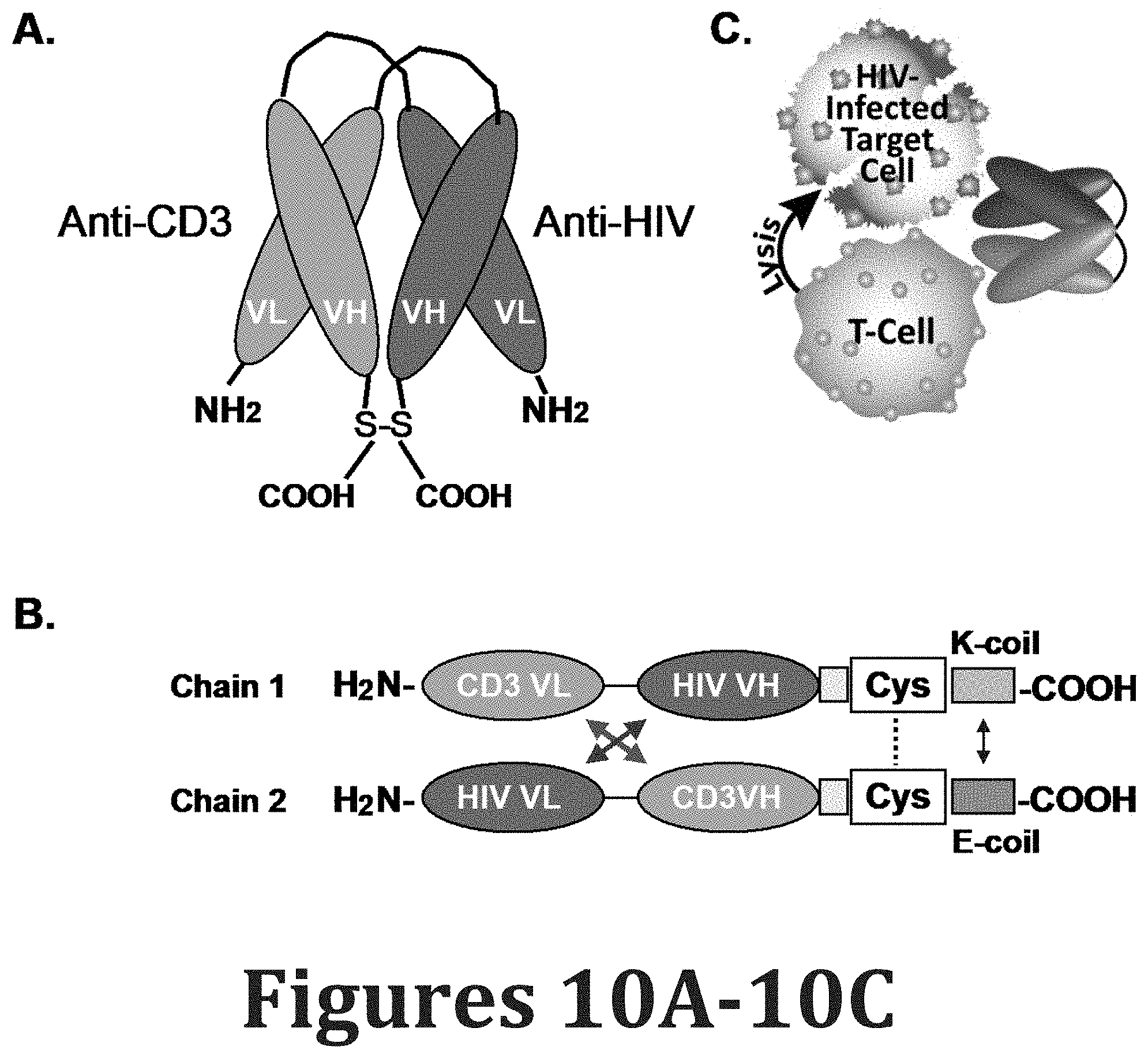
D00037

D00038

D00039

D00040

D00041

D00042

D00043

D00044

D00045

D00046

D00047

D00048

D00049

D00050

D00051

D00052

D00053

D00054

D00055

D00056

D00057

D00058

D00059

D00060

D00061

D00062

D00063

D00064

D00065

D00066

D00067

D00068

D00069

D00070

D00071

D00072

D00073
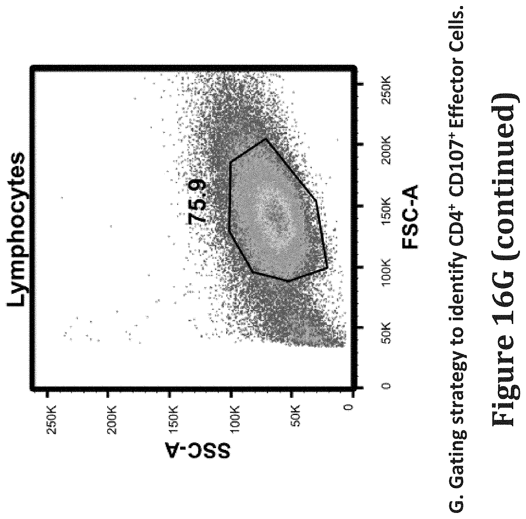
D00074

D00075

D00076

D00077

D00078

D00079

D00080

D00081

D00082

D00083

D00084

D00085

D00086

D00087

D00088

D00089

D00090

D00091

D00092

S00001
XML
uspto.report is an independent third-party trademark research tool that is not affiliated, endorsed, or sponsored by the United States Patent and Trademark Office (USPTO) or any other governmental organization. The information provided by uspto.report is based on publicly available data at the time of writing and is intended for informational purposes only.
While we strive to provide accurate and up-to-date information, we do not guarantee the accuracy, completeness, reliability, or suitability of the information displayed on this site. The use of this site is at your own risk. Any reliance you place on such information is therefore strictly at your own risk.
All official trademark data, including owner information, should be verified by visiting the official USPTO website at www.uspto.gov. This site is not intended to replace professional legal advice and should not be used as a substitute for consulting with a legal professional who is knowledgeable about trademark law.