Monoclonal Antibodies And Cocktails For Treatment Of Ebola Infections
Bornholdt; Zachary A. ; et al.
U.S. patent application number 16/896980 was filed with the patent office on 2020-11-12 for monoclonal antibodies and cocktails for treatment of ebola infections. The applicant listed for this patent is Adimab, LLC, Albert Einstein College of medicine, Inc., MAPP BIOPHARMACEUTICAL, INC.. Invention is credited to Zachary A. Bornholdt, Kartik Chandran, Laura Walker, Anna Wec, Larry Zeitlin.
| Application Number | 20200354437 16/896980 |
| Document ID | / |
| Family ID | 1000004976524 |
| Filed Date | 2020-11-12 |


View All Diagrams
| United States Patent Application | 20200354437 |
| Kind Code | A1 |
| Bornholdt; Zachary A. ; et al. | November 12, 2020 |
MONOCLONAL ANTIBODIES AND COCKTAILS FOR TREATMENT OF EBOLA INFECTIONS
Abstract
Described herein are compositions and methods for the prevention and treatment of ebolavirus infection. In certain embodiments of the present invention, monoclonal antibodies substantially, similar to those described herein, as well as affinity matured variants thereof, alone or in combination, provide therapeutic efficacy in a patient against multiple species of ebolavirus.
| Inventors: | Bornholdt; Zachary A.; (Encinitas, CA) ; Zeitlin; Larry; (San Diego, CA) ; Chandran; Kartik; (Brooklyn, NY) ; Wec; Anna; (Lebanon, NH) ; Walker; Laura; (Norwich, VT) | ||||||||||
| Applicant: |
|
||||||||||
|---|---|---|---|---|---|---|---|---|---|---|---|
| Family ID: | 1000004976524 | ||||||||||
| Appl. No.: | 16/896980 | ||||||||||
| Filed: | June 9, 2020 |
Related U.S. Patent Documents
| Application Number | Filing Date | Patent Number | ||
|---|---|---|---|---|
| 15898524 | Feb 17, 2018 | |||
| 16896980 | ||||
| 62460200 | Feb 17, 2017 | |||
| Current U.S. Class: | 1/1 |
| Current CPC Class: | C07K 2317/76 20130101; A61K 2039/505 20130101; A61K 2039/62 20130101; A61K 2039/55 20130101; A61K 2039/545 20130101; A61K 39/42 20130101; C07K 16/10 20130101; A61K 2039/6075 20130101; A61K 2039/57 20130101; A61P 31/14 20180101 |
| International Class: | C07K 16/10 20060101 C07K016/10; A61P 31/14 20060101 A61P031/14 |
Goverment Interests
STATEMENT REGARDING FEDERALLY FUNDED RESEARCH
[0002] This invention was made with government support under U19 AI109762 awarded by NIH and HDTRA-13-C-0018 awared by DTRA. The government has certain rights in the invention.
Claims
1. A composition for the treatment of Ebola, the composition comprising: a therapeutically effective combination of i. a first monoclonal antibody or antigen binding fragment thereof comprising a heavy chain variable region comprising an amino acid sequence at least 90% identical to SEQ ID NO: 15, and affinity matured variants thereof; and a light chain variable region comprising an amino acid sequence at least 90% identical to SEQ ID NO: 18, and affinity, matured variants thereof, wherein said first monoclonal antibody or antigen binding fragment thereof has a heavy chain CDR1 comprising SEQ ID NO: 41, a heavy chain CDR2 comprising SEQ ID NO: 42, a heavy chain CDR3 comprising SEQ ID NO: 43, a light chain CDR1 comprising SEQ ID NO: 44, a light chain CDR2 comprising SEQ ID NO: 45, and a light chain CDR3 comprising SEQ ID NO: 46 and amino acid sequences 90% identical thereto, and wherein the antigen to which the antigen binding fragment binds comprises Ebola glycoprotein; and ii. a pharmaceutically acceptable excipient or carrier.
2. The composition of claim 1, wherein said first monoclonal antibody or antigen binding fragment thereof binds at least two species of Filovirus glycoprotein.
3. The composition of claim 1, wherein the first monoclonal antibody or antigen binding fragment that binds to the Ebola glycoprotein antigen thereof comprises predominantly a single glycoform.
4. The composition of claim 3, wherein the predominantly single glycoform is one of GnGn, G1/G2, and NaNa.
5. The composition of claim 3, wherein the predominantly single glycoform substantially lacks at least one of fucose and xylose.
6. The composition of claim 2 further comprising a second monoclonal antibody or antigen binding fragment thereof, wherein said second monoclonal antibody or antigen binding fragment thereof binds Ebola glycoprotein.
7. A composition for the treatment of Ebola, the composition comprising: a therapeutically effective combination of i. a first monoclonal antibody or antigen binding fragment thereof comprising a heavy chain variable region comprising an amino acid sequence at least 90% identical to SEQ ID NO: 15, and affinity matured variants thereof; and a light chain variable region comprising an amino acid sequence at least 90% identical to SEQ ID NO: 18, and affinity matured variants thereof, wherein said first monoclonal antibody or antigen binding fragment thereof binds at least two species of Filovirus, and wherein the antigen to which the antigen binding fragment binds comprises Ebola glycoprotein; and ii. a pharmaceutically acceptable excipient or carrier.
8. The composition of claim 7, wherein said first monoclonal antibody or antigen binding fragment thereof has a heavy chain CDR1 comprising SEQ ID NO: 41, a heavy chain CDR2 comprising SEQ ID NO: 42, a heavy chain CDR3 comprising SEQ ID NO: 43, a light chain CDR1 comprising SEQ ID NO: 44, a light chain CDR2 comprising SEQ ID NO: 45, and a light chain CDR3 comprising SEQ ID NO: 46 and amino acid sequences 90% identical thereto.
9. The composition of claim 7, wherein the first monoclonal antibody or antigen binding fragment that binds to the Ebola glycoprotein antigen thereof comprises predominantly a single glycoform.
10. The composition of claim 9, wherein the predominantly single glycoform is one of GnGn, G1/G2, and NaNa.
11. The composition of claim 9, wherein the predominantly single glycoform substantially lacks at least one of fucose and xylose.
12. The composition of claim 7 further comprising a second monoclonal antibody or antigen binding fragment thereof, wherein said second monoclonal antibody or antigen binding fragment thereof binds the Ebola glycoprotein.
13. A monoclonal antibody or antigen binding fragment thereof effective to treat Ebola comprising a heavy chain variable region comprising an amino acid sequence at least 90% identical to SEQ ID NO: 15, and affinity matured variants thereof; and a light chain variable region comprising an amino acid sequence at least 90% identical to SEQ ID NO: 18, and affinity matured variants thereof, wherein said first monoclonal antibody or antigen binding fragment thereof comprises predominantly a single glycoform that binds at least two species of Filovirus, and wherein the antigen to which the antigen binding fragment binds comprises Ebola glycoprotein.
14. The monoclonal antibody or antigen binding fragment thereof of claim 13, wherein said first monoclonal antibody or antigen binding fragment thereof has a heavy chain CDR1 comprising SEQ ID NO: 41, a heavy chain CDR2 comprising SEQ ID NO: 42, a heavy chain CDR3 comprising SEQ ID NO: 43, a light chain CDR1 comprising SEQ ID NO: 44, a light chain CDR2 comprising SEQ ID NO: 45, and a light chain CDR3 comprising SEQ ID NO: 46 and amino acid sequences 90% identical thereto.
15. The monoclonal antibody or antigen binding fragment thereof of claim 13, wherein the predominantly single glycoform is one of GnGn, G1/G2, and NaNa.
16. The monoclonal antibody or antigen binding fragment thereof of claim 13, wherein the predominantly single glycoform substantially lacks at least one of fucose and xylose.
Description
CROSS-REFERENCE TO RELATED APPLICATIONS
[0001] This application is a continuation of U.S. patent application Ser. No. 15/898,524, filed Feb. 17, 2018, which claims the benefit of U.S. Provisional Patent Application No. 62/460,200, filed Feb. 17, 2017, each of which is incorporated by reference herein in their entirety.
SEQUENCE LISTING
[0003] The instant application contains a Sequence Listing which has been submitted electronically in ASCII format and is hereby incorporated by reference. Said ASCII copy, created on May 20, 2020, is named 1123-2007-ST25 and is 34,674 bytes in size.
BACKGROUND
[0004] Ebolaviruses are members of the family Filoviridae which infect humans and non-human primates (NHPs) causing hemorrhagic fever with mortality rates up to 90%. Ebolaviruses include Ebola virus (EBOV), Sudan virus (SUDV), Bundibugyo virus (BDBV), Reston virus (RESTV), and Tai Forest virus (TAFV), which are causative agents of the hemorrhagic fever [1, 2]. A summary of the ebolaviruses can be found in Burk, et. al., Neglected Filoviruses. FEMS Microbiology Reviews, 40, 494-519 (May, 2016), and the differences between the viruses have been well characterized and well known in the art. Between 1967 and 2013, 31 filovirus disease outbreaks have occurred, mainly in central Africa with around 2,000 confirmed cases. Of these 31 outbreaks, 16 were caused by EBOV. The unprecedented 2013-2016 Ebola virus disease epidemic led to more than 27,000 cases and 11,100 deaths in the first 14 months. There are currently no approved treatments or vaccines for filoviruses, and most advanced experimental treatments focus only on EBOV. Given that other filoviruses have caused sizeable outbreaks broadly protective treatment options are urgently needed.
[0005] Several studies have shown that filovirus glycoprotein (GP)-specific neutralizing antibodies (nAbs) can reduce mortality following experimental inoculation of animals with a lethal dose of EBOV [3-9]. The primary target of these neutralizing antibodies, the filovirus surface GP, is a trimer composed of three heavily glycosylated GP1-GP2 heterodimers. The GP1 subunit can be divided further into base, head, glycan cap and mucin-like domains [10]. During viral entry, the mucin-like domain and glycan cap mediate binding to multiple host attachment factors present on the cell membrane. After the virus enters the host cell by macropinocytosis [11, 12] the GP is cleaved by host proteases that remove approximately 80% of the mass of the GP1 subunit, including the mucin-like domain and glycan cap [13, 14]. After cleavage of GP in the endosome, the receptor binding sites on GP become exposed, and the GP1 head then is able to bind its receptor, the Niemann-Pick C1 (NPC1) protein [13, 15, 16]. Subsequent conformational changes in GP facilitate fusion between viral and endosomal membranes. Recognition of NPC1 by a cleaved GP species (hereafter, GP.sub.CL), together with one or more unknown host signals, is proposed to trigger GP refolding and the membrane fusion reaction that is coupled to it. Endosomal GP.fwdarw.GP.sub.CL cleavage is a prerequisite for GP-NPC1 binding and therefore essential for filovirus entry.
[0006] The dense clustering of glycans on the glycan cap and mucin-like domain likely shield much of the surface of EBOV GP from humoral immune surveillance, leaving only a few sites on the EBOV GP protein where nAbs could bind without interference by glycans [17]. Most of our knowledge about humoral response against filovirus infections has come from studies of murine Abs that recognize EBOV GP. From those studies, it has become clear that mouse neutralizing Abs preferentially target peptides exposed in upper, heavily glycosylated domains or lower areas (the GP1 base) where rearrangements occur that drive fusion of viral and host membranes [18]. Abs have not been identified that target protein features of the membrane proximal external region (MPER) subdomain, which likely rearranges during fusion. KZ52, the first reported human EBOV GP-specific monoclonal antibody (mAb), was obtained from a phage display library that was constructed from bone marrow RNA obtained from a survivor [19]. KZ52 binds a site at the base of the GP and neutralizes EBOV, most likely by blocking GP.fwdarw.GP.sub.CL cleavage and/or inhibiting the conformational changes required for fusion of viral and endosomal membranes [10]. Some murine Abs also have been reported to bind to the base region of Ebola virus GPs [20, 21].
[0007] The most divergent ebolavirus species (EBOV and SUDV) exhibit 56% GP sequence identity. The sequence identity between filovirus GPs is highest within the receptor binding region (RBR) [23] and GP2, suggesting that shared epitopes may exist within these domains. Several mAbs against EBOV GP with protective efficacy in rodents and non-human primates (NHPs) have been reported [3, 5-9, 24, 25]. Neutralizing antibodies have also been described for SUDV with efficacy in a recently developed rodent model [20, 26]. However, these antibodies bind the same epitope as KZ52, and like KZ52 are viral species-specific and lack cross-neutralizing or cross-protective properties.
SUMMARY OF THE INVENTION
[0008] Described herein are a number of mAbs that are capable of neutralizing Ebola viruses both in vitro and in vivo. Surprisingly, the disclosed human antibodies possess pan-ebolavirus cross-reactivity and cross-neutralizing activity, and are thus capable of binding and neutralizing all known species of the Ebola virus.
[0009] According to a first aspect of the present invention, there are provided novel monoclonal antibodies capable of binding to and neutralizing an Ebola virus in a patient. In certain embodiments of the present invention, said monoclonal antibodies bind to GP proteins from ebolaviruses belonging to at least two different species, thereby neutralizing the infectivity of viral particles or targeting infected cells for destruction.
[0010] According to a second aspect of the invention, there is provided monoclonal antibodies comprising the following heavy and light chain CDR3 amino acid sequences:
[0011] mAb PE-87-heavy CDR3: SEQ ID No. 1; mAb PE-87-light CDR3: SEQ ID No. 2
[0012] mAb PE-24-heavy CDR3: SEQ ID No. 3; mAb PE-24-light CDR3: SEQ ID No. 4
[0013] mAb PE-47-heavy CDR3: SEQ ID No. 5; mAb PE-47 light CDR3: SEQ ID No. 6
[0014] mAb PE-16-heavy CDR3: SEQ ID No. 7; mAb PE-16-light CDR3: SEQ ID No. 8
[0015] mAb PE-05-heavy CDR3: SEQ ID No. 9; mAb PE-05-light CDR3: SEQ ID No. 10
[0016] In one embodiment, the critical residues in PE-87 and PE-24 heavy chain CDR3 are D95, W99, and Y100C (Kabat numbering).
[0017] In another embodiment of the invention, an antibody isolated as described in Methods (below) from the peripheral B cells of a survivor of a filovirus infection, is modified so that the VH and VL region nucleotide sequences encode modified V region amino acids that confer enhanced binding capabilities to the mAb. There is provided a method of preparing a recombinant antibody comprising: providing a nucleotide sequence selected from the group consisting of
[0018] PE-24, PE-87, PE-47, PE-16, PE-64 and PE-05 VH and VL nucleotides;
[0019] modifying said nucleic acid sequence such that at least one but fewer than about 30 of the amino acid residues encoded by said nucleic acid sequence has been changed or deleted without disrupting antigen binding of said peptide; and expressing and recovering said modified nucleotide sequence.
[0020] In yet other embodiments, immunoreactive fragments of any of the herein described monoclonal antibodies are prepared using means known in the art, for example, by preparing nested deletions using enzymatic degradation or convenient restriction enzymes.
[0021] It is another aspect of the present invention to provide modified variants of the disclosed mAb sequences, wherein the sequences have been affinity matured or otherwise mutated to increase the therapeutic effectiveness of the mAb.
[0022] Thus, it is one embodiment of the present invention to provide a composition for the treatment of Ebola, the composition comprising: a therapeutically effective combination of a first monoclonal antibody or antigen binding fragment comprising a heavy chain variable region comprising an amino acid sequence at least 90% identical to SEQ. ID NO: 12, and affinity matured variants thereof and a light chain variable region comprising an amino acid sequence at least 90% identical to SEQ ID NO: 14, and affinity matured variants thereof and a pharmaceutically acceptable excipient or carrier.
[0023] It is another embodiment of the present invention to provide such a composition, wherein said first monoclonal antibody is binds at least two species of the Flivovirus glycoprotein.
[0024] It is yet another embodiment of the present invention to provide such a composition, therein the first monoclonal antibody or antigen binding fragment comprises predominantly a single glycoform.
[0025] It is still another embodiment of the present invention to provide such a composition, wherein the predominantly single glycoform is one of GnGn G1/G2, and NaNa.
[0026] It is yet another embodiment of the present invention to provide such a composition, wherein the predominantly single glycoform substantially lacks at least one of fucose and xylose.
[0027] It is second embodiment of the present invention to provide a composition for the treatment of Ebola, the composition comprising: a therapeutically effective combination of a first monoclonal antibody or antigen binding fragment selected from a list consisting of: [0028] a. a monoclonal antibody or antigen binding fragment comprising a heavy chain variable region comprising an amino acid sequence at least 90% identical to SEQ. ID NO: 15, and affinity matured variants thereof; and a light chain variable region comprising an amino acid sequence at least 90% identical to SEQ ID NO: 18, and affinity matured variants thereof; [0029] b. a monoclonal antibody or antigen binding fragment comprising a heavy chain variable region comprising an amino acid sequence at least 90% identical to SEQ. ID NO: 21, and affinity matured variants thereof; and a light chain variable region comprising an amino acid sequence at least 90% identical to SEQ ID NO: 23, and affinity matured variants thereof; [0030] c. a monoclonal antibody or antigen binding fragment comprising a heavy chain variable region comprising an amino acid sequence at least 90% identical to SEQ. ID NO: 29, and affinity matured variants thereof; and a light chain variable region comprising an amino acid sequence at least 90% identical to SEQ ID NO: 31, and affinity matured variants thereof; [0031] d. a monoclonal antibody or antigen binding fragment comprising a heavy chain variable region comprising an amino acid sequence at least 90% identical to SEQ. ID NO: 33, and affinity matured variants thereof, and a light chain variable region comprising an amino acid sequence at least 90% identical to SEQ ID NO: 35, and affinity matured variants thereof; [0032] e. a monoclonal antibody or antigen binding fragment comprising a heavy chain variable region comprising an amino acid sequence at least 90% identical to SEQ. ID NO: 11, and affinity matured variants thereof; and a light chain variable region comprising an amino acid sequence at least 90% identical to SEQ ID NO: 13, and affinity matured variants thereof, and a pharmaceutically acceptable excipient or carrier; wherein said first monoclonal antibody or antigen binding fragment binds at least two species of the Flivovirus glycoprotein.
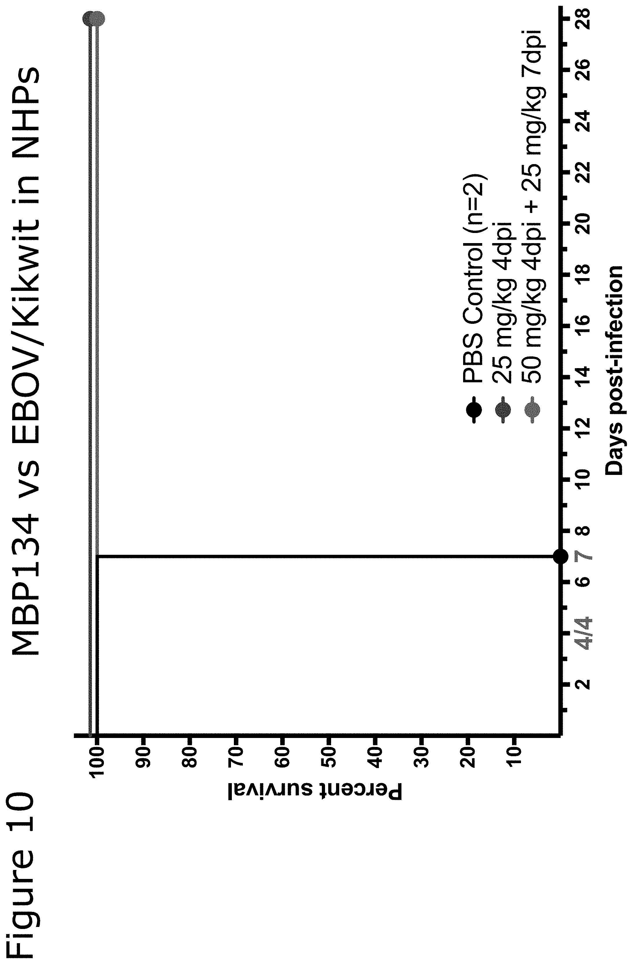
[0033] It is another embodiment of the present invention to provide such a composition, wherein the first monoclonal antibody or antigen binding fragment comprises predominantly a single glycoform.
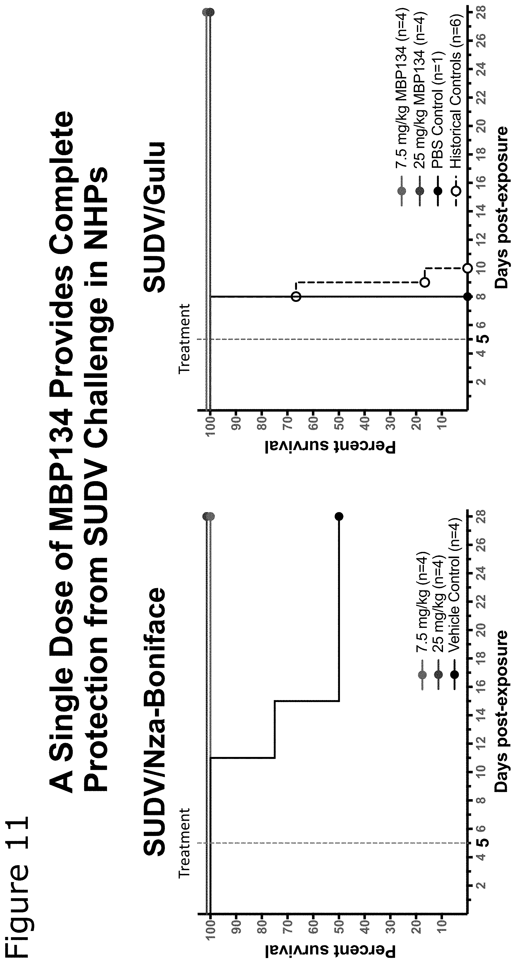
[0034] It is yet another embodiment of the present invention to provide such a composition, wherein the predominantly single glycoform is one of GnGn, G1/G2, and NaNa.
[0035] It is still another embodiment of the present invention to provide such a composition, wherein the predominantly single glycoform substantially lacks at least one of fucose and xylose.
[0036] It is third embodiment of the present invention to provide a composition for the treatment of Ebola, the composition comprising: a therapeutically effective combination of a first monoclonal antibody or antigen binding fragment is selected from a list consisting of: [0037] a. a monoclonal antibody or antigen binding fragment comprising a heavy chain variable region comprising an amino acid sequence at least 90% identical to SEQ. ID NO: 12, and affinity matured variants thereof; and a light chain variable region comprising an amino acid sequence at least 90% identical to SEQ ID NO: 14, and affinity matured variants thereof; [0038] b. a monoclonal antibody or antigen binding fragment comprising a heavy chain variable region comprising an amino acid sequence at least 90% identical to SEQ. ID NO: 15, and affinity matured variants thereof; and a light chain variable region comprising an amino acid sequence at least 90% identical to SEQ ID NO: 18, and affinity matured variants thereof; [0039] c. a monoclonal antibody or antigen binding fragment comprising a heavy chain variable region comprising an amino acid sequence at least 90% identical to SEQ. ID NO: 21, and affinity matured variants thereof; and a light chain variable region comprising an amino acid sequence at least 90% identical to SEQ ID NO: 23, and affinity matured variants thereof; [0040] d. a monoclonal antibody or antigen binding fragment comprising a heavy chain variable region comprising an amino acid sequence at least 90% identical to SEQ. ID NO: 29, and affinity matured variants thereof; and a light chain variable region comprising an amino acid sequence at least 90% identical to SEQ ID NO: 31, and affinity matured variants thereof; [0041] e. a monoclonal antibody or antigen binding fragment comprising a heavy chain variable region comprising an amino acid sequence at least 90% identical to SEQ. ID NO: 33, and affinity matured variants thereof; and a light chain variable region comprising an amino acid sequence at least 90% identical to SEQ II) NO: 35, and affinity matured variants thereof; [0042] f. a monoclonal antibody or antigen binding fragment comprising a heavy chain variable region comprising an amino acid sequence at least 90% identical to SEQ. ID NO: 11, and affinity matured variants thereof; and a light chain variable region comprising an amino acid sequence at least 90% identical to SEQ ID NO: 13, and affinity matured variants thereof, a second monoclonal antibody or antigen binding fragment, wherein said second monoclonal antibody or antigen binding fragment binds the Ebola glycoprotein; and a pharmaceutically acceptable excipient or carrier.
[0043] It is another embodiment of the present invention to provide such a composition, wherein at least one of the first monoclonal antibody or antigen binding fragment and the second antibody or antigen binding fragment comprises predominantly a single glycoform.
[0044] It is yet another embodiment of the present invention to provide such a composition, wherein the predominantly single glycoform is one of GnGn, G1/G2, and NaNa.
[0045] It is still another embodiment of the present invention to provide such a composition, wherein the predominantly single glycoform substantially lacks at least one of fucose and xylose.
[0046] It is yet another embodiment of the present invention to provide such a composition, wherein said therapeutically effective combination further comprises a third monoclonal antibody or antigen binding fragment that binds to the Ebola glycoprotein.
[0047] It is still another embodiment of the present invention to provide such a composition, wherein said first monoclonal antibody or antigen binding fragment comprises a heavy chain variable region comprising an amino acid sequence at least 90% identical to SEQ. ID NO: 12, and affinity matured variants thereof; and a light chain variable region comprising an amino acid sequence at least 90% identical to SEQ ID NO: 14, and affinity matured variants thereof; and wherein said second monoclonal antibody or antigen binding fragment comprises a heavy chain variable region comprising an amino acid sequence at least 90% identical to SEQ. ID NO: 15, and affinity matured variants thereof; and a light chain variable region comprising an amino acid sequence at least 90% identical to SEQ ID NO: 18, and affinity matured variants thereof.
[0048] It is yet another embodiment of the present invention to provide such a composition wherein said therapeutically effective combination further comprises a third monoclonal antibody or antigen binding fragment, wherein said third antibody or antigen binding fragment comprises a heavy chain variable region comprising an amino acid sequence at least 90% identical to SEQ. ID NO: 21, and affinity matured variants thereof; and a light chain variable region comprising an amino acid sequence at least 90% identical to SEQ ID NO: 23, and affinity matured variants thereof.
[0049] It is fourth embodiment of the present invention to provide a method for treating at least one species of flivovirus infection in a patient, the method comprising: identifying a patient in need of treatment; and administering to the patient a therapeutically effective amount of a composition comprising a combination of: a first monoclonal antibody or antigen binding fragment, wherein said first monoclonal antibody or antigen binding fragment is selected from a list consisting of: [0050] i. a monoclonal antibody or antigen binding fragment comprising a heavy chain variable region comprising an amino acid sequence at least 90% identical to SEQ. ID NO: 12, and affinity matured variants thereof; and a light chain variable region comprising an amino acid sequence at least 90% identical to SEQ ID NO: 14, and affinity matured variants thereof; [0051] ii. a monoclonal antibody or antigen binding fragment comprising a heavy chain variable region comprising an amino acid sequence at least 90% identical to SEQ. ID NO: 15, and affinity matured variants thereof; and a light chain variable region comprising an amino acid sequence at least 90% identical to SEQ ID NO: 18, and affinity matured variants thereof; [0052] iii. a monoclonal antibody or antigen binding fragment comprising a heavy chain variable region comprising an amino acid sequence at least 90% identical to SEQ. II) NO: 21, and affinity matured variants thereof; and a light chain variable region comprising an amino acid sequence at least 90% identical to SEQ ID NO: 23, and affinity matured variants thereof; [0053] iv. a monoclonal antibody or antigen binding fragment comprising a heavy chain variable region comprising an amino acid sequence at least 90% identical to SEQ. ID NO: 29, and affinity matured variants thereof; and a light chain variable region comprising an amino acid sequence at least 90% identical to SEQ ID NO: 31, and affinity matured variants thereof; [0054] v. a monoclonal antibody or antigen binding fragment comprising a heavy chain variable region comprising an amino acid sequence at least 90% identical to SEQ ID NO: 33, and affinity matured variants thereof; and a light chain variable region comprising an amino acid sequence at least 90% identical to SEQ ID NO: 35, and affinity matured variants thereof; [0055] vi. a monoclonal antibody or antigen binding fragment comprising a heavy chain variable region comprising an amino acid sequence at least 90% identical to SEQ ID NO: 11, and affinity matured variants thereof; and a light chain variable region comprising an amino acid sequence at least 90% identical to SEQ ID NO: 13, and affinity matured variants thereof; and a pharmaceutically acceptable excipient or carrier.
[0056] It is another embodiment of the present invention to provide such a method, wherein the patient is a mammal.
[0057] It is yet another embodiment of the present invention to provide such a method, wherein the first monoclonal antibody or antigen binding fragment comprises predominantly a single glycoform.
[0058] It is still another embodiment of the present invention to provide such a method, wherein the predominantly single glycoform substantially lacks at least one of fucose and xylose.
[0059] It is fifth embodiment of the present invention to provide a composition for the treatment of Ebola, the composition comprising: a therapeutically effective combination of a first monoclonal antibody or antigen binding fragment is selected from a list consisting of: [0060] a. a monoclonal antibody or antigen binding fragment comprising a heavy chain variable region at least 90% identical to a heavy chain variable region comprising a CDR1 comprising the amino acid sequence as set forth in SEQ ID NO: 53, a CDR2 comprising the amino acid sequence as set forth in SEQ. ID NO: 54, and a CDR3 comprising the amino acid sequence as set forth in SEQ. ID NO: 55, and affinity matured variants thereof, and a light chain variable region at least 90% identical to a light chain variable region comprising a CDR1 comprising the amino acid sequence as set forth in SEQ. ID NO: 56, a CDR2 comprising the amino acid sequence as set forth in SEQ. II) NO: 57, and a CDR3 comprising the amino acid sequence as set forth in SEQ. ID NO: 58, and affinity matured variants thereof; [0061] b. a monoclonal antibody or antigen binding fragment comprising a heavy chain variable region at least 90% identical to a heavy chain variable region comprising a CDR1 comprising the amino acid sequence as set forth in SEQ. ID NO: 41, a CDR2 comprising the amino acid sequence as set forth in SEQ. ID NO: 42, and a CDR3 comprising the amino acid sequence as set forth in SEQ. II) NO: 43, and affinity matured variants thereof; and a light chain variable region at least 90% identical to a light chain variable region comprising a CDR1 comprising the amino acid sequence as set forth in SEQ. ID NO: 44, a CDR2 comprising the amino acid sequence as set forth in SEQ. ID NO: 45, and a CDR3 comprising the amino acid sequence as set forth in SEQ. ID NO: 46, and affinity matured variants thereof; [0062] c. a monoclonal antibody or antigen binding fragment comprising a heavy chain variable region at least 90% identical to a heavy chain variable region comprising a CDR1 comprising the amino acid sequence as set forth in SEQ ID NO: 47, a CDR2 comprising the amino acid sequence as set forth in SEQ. ID NO: 48, and a CDR3 comprising the amino acid sequence as set forth in SEQ. ID NO: 49, and affinity matured variants thereof; and a light chain variable region at least 90% identical to a light chain variable region comprising a CDR1 comprising the amino acid sequence as set forth in SEQ. ID NO: 50, a CDR2 comprising the amino acid sequence as set forth in SEQ. ID NO: 51, and a CDR3 comprising the amino acid sequence as set forth in SEQ. ID NO: 52, and affinity matured variants thereof; [0063] d. a monoclonal antibody or antigen binding fragment comprising a heavy chain variable region at least 90% identical to a heavy chain variable region comprising a CDR1 comprising the amino acid sequence as set forth in SEQ. ID NO: 59, a CDR2 comprising the amino acid sequence as set forth in SEQ. ID NO: 60, and a CDR3 comprising the amino acid sequence as set forth in SEQ. ID NO: 61, and affinity matured variants thereof; and a light chain variable region at least 90% identical to a light chain variable region comprising a CDR1 comprising the amino acid sequence as set forth in SEQ. ID NO: 62, a CDR2 comprising the amino acid sequence as set forth in SEQ. ID NO: 63, and a CDR3 comprising the amino acid sequence as set forth in SEQ. ID NO: 64, and affinity matured variants thereof; [0064] e. a monoclonal antibody or antigen binding fragment comprising a heavy chain variable region at least 90% identical to a heavy chain variable region comprising a CDR1 comprising the amino acid sequence as set forth in SEQ. ID NO: 65, a CDR2 comprising the amino acid sequence as set forth in SEQ. ID NO: 66, and a CDR3 comprising the amino acid sequence as set forth in SEQ. ID NO: 67, and affinity matured variants thereof, and a light chain variable region at least 90% identical to a light chain variable region comprising a CDR1 comprising the amino acid sequence as set forth in SEQ. ID NO: 68, a CDR2 comprising the amino acid sequence as set forth in SEQ. ID NO: 69, and a CDR3 comprising the amino acid sequence as set forth in SEQ. ID NO: 70, and affinity matured variants thereof; [0065] f. a monoclonal antibody or antigen binding fragment comprising a heavy chain variable region at least 90% identical to a heavy chain variable region comprising a CDR1 comprising the amino acid sequence as set forth in SEQ. ID NO: 71, a CDR2 comprising the amino acid sequence as set forth in SEQ. ID NO: 72, and a. CDR3 comprising the amino acid sequence as set forth in SEQ. ID NO: 73, and affinity matured variants thereof; and a light chain variable region at least 90% identical to a light chain variable region comprising a CDR1 comprising the amino acid sequence as set forth in SEQ. ID NO: 74, a CDR2 comprising the amino acid sequence as set forth in SEQ. ID NO: 75, and a CDR3 comprising the amino acid sequence as set forth in SEQ. ID NO: 76, and affinity matured variants thereof and a pharmaceutically acceptable excipient or carrier.
[0066] It is another embodiment of the present invention to provide such a composition, further comprising a second monoclonal antibody or antigen binding fragment, wherein said second monoclonal antibody or antigen binding fragment binds the Ebola glycoprotein.
[0067] It is yet another embodiment of the present invention to provide such a composition, wherein said first monoclonal antibody is binds at least two species of the Flivovirus glycoprotein.
[0068] It is still another embodiment of the present invention to provide such a composition, wherein the first monoclonal antibody or antigen binding fragment comprises predominantly a single glycoform.
[0069] It is yet another embodiment of the present invention to provide such a composition, wherein the predominantly single glycoform is one of GnGn, G1/G2, and NaNa.
[0070] It is still another embodiment of the present invention to provide such a composition, wherein the predominantly single glycoform substantially lacks at least one of fucose and xylose.
[0071] These, and other, embodiments of the invention will be better appreciated and understood when considered in conjunction with the following description and the accompanying drawings. It should be understood, however, that the following description, while indicating various embodiments of the invention and numerous specific details thereof, is given by way of illustration and not of limitation. Many substitutions, modifications, additions and/or rearrangements may be made within the scope of the invention without departing from the spirit thereof, and the invention includes all such substitutions, modifications, additions and/or rearrangements.
DESCRIPTION OF THE FIGURES AND TABLES
[0072] Table 1. Amino acid residues comprising CDRs of anti-Ebola mAbs.
[0073] FIG. 1 shows a neutralization curve for affinity matured variants of one embodiment of the present invention.
[0074] FIG. 2 shows the binding sites on the EBOV-GP of various embodiments of the monoclonal antibodies of the present invention.
[0075] FIG. 3 shows the location of the mutations that result in escape mutant resistance to two monoclonal antibodies of the present invention.
[0076] FIG. 4 shows neutralization assays preformed against the escape mutants.
[0077] FIG. 5 shows survival data for ebolavirus infected guinea pigs treated with certain embodiments of the present invention.
[0078] FIG. 6 shows immune system response data from ebolavirus infected guinea pigs treated with certain embodiments of the present invention.
[0079] FIG. 7 shows survival data for ebolavirus infected guinea pigs treated with certain embodiments of the present invention.
[0080] FIG. 8 shows survival data for ebolavirus infected guinea pigs treated with certain embodiments of the present invention.
[0081] FIG. 9 survival data for ebolavirus infected guinea pigs treated with certain embodiments of the present invention.
[0082] FIG. 10 survival data for ebolavirus infected non-human primates treated with certain embodiments of the present invention.
[0083] FIG. 11 survival data for ebolavirus infected non-human primates treated with certain embodiments of the present invention.
[0084] FIG. 12 survival data for ebolavirus infected non-human primates treated with certain embodiments of the present invention.
[0085] FIG. 13 shows a neutralization curves for certain embodiments of the present invention created using differing production methods.
[0086] Table 2 shows the efficiency of anti-GP antibody isolation from peripheral B cells.
[0087] Table 3 shows the cross-reactivity of candidate pan-ebolavirus mAbs against different ebolavirus species. Reactivity was measured by ELISA.
[0088] Table 4 shows the in vitro neutralization activity and affinities of candidate pan-ebolavirus mAbs.
[0089] Table 5 shows that mice infected with EBOV and subsequently treated with the monoclonal antibodies described above showed increased survival compared to mice treated with PBS.
[0090] Table 6 is a summary of rVSV-GP neutralization by cross-neutralizing human mAb s.
[0091] Table 7 is a summary of authentic ebolavirus neutralization by cross-neutralizing human mAbs.
[0092] Table 8 shows K.sub.D values for recognition of EBOV GP.DELTA.TM by mature PE-87 bearing the indicated mutations in the CDR-H3 loop were determined by BLI. 95% confidence intervals are reported for each binding constant. IC.sub.50 values for neutralization of rVSVs bearing ebolavirus GPs by mature PE-87 bearing the indicated mutations in the CDR-H3 loop.
[0093] Table 9 shows the mAb protection of mice after challenge with EBOV or SUDV.
DESCRIPTION OF THE PREFERRED EMBODIMENTS
[0094] Unless defined otherwise, all technical and scientific terms used herein have the same meaning as commonly understood by one of ordinary skill in the art to which the invention belongs. Although any methods and materials similar or equivalent to those described herein can be used in the practice or testing of the present invention, the preferred methods and materials are now described. All publications mentioned above and hereunder are incorporated herein by reference.
Definitions
[0095] As used herein, "neutralizing antibody" (NAb) refers to an antibody, for example, a monoclonal antibody, capable of disrupting a formed viral particle or inhibiting formation of a viral particle or prevention of binding to or infection of mammalian cells by a viral particle. As used herein, "diagnostic antibody" or "detection antibody" or "detecting antibody" refers to an antibody, for example, a monoclonal antibody, capable of detecting the presence of an antigenic target within a sample. As will be appreciated by one of skill in the art, such diagnostic antibodies preferably have high specificity for their antigenic target. As used herein, "human antibodies" refer to antibodies that were isolated from the B cells of a human or directly from the sequence of serum antibodies.
[0096] A "therapeutically effective" treatment refers a treatment that is capable of producing a desired effect. Such effects include, but are not limited to, enhanced survival, reduction in presence or severity of symptoms, reduced time to recovery, and prevention of initial infection. "Therapeutically effective" permutations of a mAb may enhance any of the above characteristics in a manner that is detectable by routine analysis of patient data. In certain embodiments, such therapeutically effective mutations include mutations that improve the stability, solubility, or production of the mAb, including mutations to the framework regions of the mAb sequence.
[0097] As used herein, `immunoreactive fragment` refers in this context to an antibody fragment reduced in length compared to the wild-type or parent antibody which retains an acceptable degree or percentage of binding activity to the target antigen. As will be appreciated by one of skill in the art, what is an acceptable degree will depend on the intended use.
[0098] As used herein, a mAb has "pan-Ebola" binding characteristics if it is capable of binding to at least 2, but preferable more, ebolavirus species.
[0099] The basic antibody structural unit is known to comprise a tetramer. Each tetramer is composed of two identical pairs of polypeptide chains, each pair having one "light" (about 25 kDa) and one "heavy" chain (about 50-70 kDa). The amino-terminal portion of each chain includes a variable region of about 100 to 110 or more amino acids primarily responsible for antigen recognition. The carboxy-terminal portion of each chain defines a constant region primarily responsible for effector function.
[0100] Light chains are classified as kappa and lambda. Heavy chains are classified as gamma, mu, alpha, delta, or epsilon, and define the antibody's isotype as IgG, IgM, IgA, IgD and IgE, respectively. Within each isotype, there may be subtypes, such as IgG.sub.1, IgG.sub.2, IgG.sub.3, IgG.sub.4, etc. Within light and heavy chains, the variable and constant regions are joined by a "J" region of about 12 or more amino acids, with the heavy chain also including a "D" region of about 3 or more amino acids. The particular identity of constant region, the isotype, or subtype does not impact the present invention. The variable regions of each light/heavy chain pair form the antibody binding site.
[0101] Thus, an intact antibody has two binding sites. The chains all exhibit the same general structure of relatively conserved framework regions (FR) joined by three hypervariable regions, also called complementarity determining regions or CDRs. The CDRs from the two chains of each pair are aligned by the framework regions, enabling binding to a specific epitope. From N-terminal to C-terminal, both light and heavy chains comprise the domains FR1, CDR1, FR2, CDR2, FR3, CDR3 and FR4. The assignment of amino acids to each domain is in accordance with well known conventions [Kabat "Sequences of Proteins of Immunological Interest" National Institutes of Health, Bethesda, Md. 1987 and 1991; Chothia, et al., J. Mol. Biol. 196:901-917 (1987); Chothia, et al., Nature 342:878-883 (1989)].
[0102] In another embodiment of the invention, there are provided glycoengineered variants of the monoclonal antibodies that contain predominantly a single glycoform. These glycans can be GnGn (GlcNAc.sub.2-Man.sub.3-GlcNAc.sub.2), mono- or di-galactosylated (Gal.sub.(1/2)-GlcNAc.sub.2-Man.sub.3-GlcNAc.sub.2) (hereinafter mono-galactosylated="G1", di-galactosylated="G2", and a combination of the two, in any proportion="G1/G2"), mono- or di-sialylated (NaNa.sub.(1,2)-Gal.sub.(1/2)-GlcNAc.sub.2-Man.sub.3-GlcNAc.sub.2) containing little or no fucose or xylose. A predominantly single glycoform is any glycoform that represents more than half (e.g. greater than 50%, 60%, 70%, 80%, 90%, 95%, 96%, 97%, 98%, 99%) of all glycoforms present in the antibody solution.
[0103] The RAMP system has been used for glycoengineering of antibodies, antibody fragments, idiotype vaccines, enzymes, and cytokines. Dozens of antibodies have been produced in the RAMP system by Mapp (5, 6) and others (7, 8). These have predominantly been IgGs but other isotypes, including IgM (9, 10), have been glycoengineered. Glycoengineering has also been extended to human enzymes in the RAMP system (11, 12). Since the RAMP system has a rapid turn-around time from Agrobacterium infection to harvest and purification (13) patient specific idiotype vaccines have been used in clinical trials for non-Hodgkins lymphoma (7).
[0104] For glycoengineering, recombinant Agrobacterium containing a mAb cDNA is used for infection of N. benthamiana in combination with the appropriate glycosylation Agrobacteria to produce the desired glycan profile. For wild-type glycans (i.e. native, plant-produced glycosylation) wild-type N. benthamiana is inoculated with only the Agrobacterium containing the anti-M2e cDNA. For the GnGn glycan, the same Agrobacterium is used to inoculate plants that contain little or no fucosyl or xylosyl transfrases (.DELTA.XF plants). For galactosylated glycans, .DELTA.XF plants are inoculated with the Agrobacterium containing the mAb cDNA as well as an Agrobacterium containing the cDNA for .beta.-1,4-galactosyltransferase expression contained on a binary Agrobacterium vector to avoid recombination with the TMV and PVX vectors (14). For sialylated glycans, six additional genes are introduced in binary vectors to reconstitute the mammalian sialic acid biosynthetic pathway. The genes are UDP-N-acetylglucosamine 2-epimerase/N-acetylmannosamine kinase, N-acetylneuraminic acid phosphate synthase, CMP-N-acetylneuraminic acid synthetase, CMP-NeuAc transporter, .beta.-1,4-galactosylatransferase, and .alpha.2,6-sialyltransferase (14).
[0105] Glycanalysis of glycoengineered mAbs involved release of N-linked glycans by digestion with N-glycosidase F (PNGase F), and subsequent derivatization of the free glycan is achieved with anthranilic acid (2-AA). The 2-AA-derivatized oligosaccharide is separated from any excess reagent via normal-phase HPLC. The column is calibrated with 2-AA-labeled glucose homopolymers and glycan standards. The test samples and 2-AA-labeled glycan standards are detected fluorometrically. Glycoforms are assigned either by comparing their glucose unit (GU) values with those of the 2-AA-labeled glycan standards or by comparing with the theoretical GU values (15). Confirmation of glycan structure was accomplished with LC/MS.
[0106] While the RAMP system is an effective method of producing various glycoengineered and wild-type mABs, it will be recognized that other expression systems may be used to accomplish the same result. For example, mammalian cell lines (such as CHO or NSO cells [Davies, J., Jiang, L., Pan, L. Z., LaBarre, M. J., Anderson, D., and Reff, M. 2001, Expression of GnTIII in a recombinant anti-CD20 CHO production cell line: Expression of antibodies with altered glycoforms leads to an increase in ADCC through higher affinity for FCyRIII. Biotechnol Bioeng 74:288-294]), yeast cells (such as Pichia pastoris [Gerngross T. Production of complex human glycoproteins in yeast. Adv Exp Med Biol. 2005; 564]) and bacterial cells (such as E. coli) have been used produce such mABs.
[0107] Described herein are mAbs, designated PE-24, PE-87, PE-47, PE-16, PE-64 and PE-05, which have surprisingly exhibited pan-Ebola neutralizing characteristics. The preferred antibodies of the present invention comprise mAbs with amino acid sequences sufficiently identical to referenced amino acid sequences. By "sufficiently identical" is intended an amino acid sequence that has at least about 60% or 65% sequence identity, about 70% or 75% sequence identity, about 80% or 85% sequence identity, about 90%, 91%, 92%, 93%, 94%, 95%, 96%, 97%, 98%, 99% or greater sequence identity compared to a reference sequence using one of the alignment programs known in the art.
[0108] The sequences below show the amino acid modifications to mAb PE-64 VH and VL amino acids to yield mAb PE-47 (Modifications are shown in Bold, CDR sequences are Underlined).
TABLE-US-00001 mAb PE-64 VH amino acids: SEQ ID No. 11 EVQLVESGGGLVKPGGSLRLSCAASGFTFSNAWMSWVRQAPGK GLEWVGRIKSKTDGGTIDYAAPVKGRFTISRDDSKNTVYLQMT SLKTEDTAVYYCTTYTEDMRYFDWLLRGGETFDYWGQGTLVTV SS mAb PE-47 VH amino acids: SEQ ID No. 12 EVQLVESGGGLVKPGGSLRLSCAASGFTFSNAWMSWVRQAPGE GLEWVGRIKSKTDGGTIDYAAPVKGRFTISRDDSKNTVYLQMT SLKTEDTAVYYCTTYTEDMQYFDWLLRGGETFDYWGQGTLVTV SS mAb PE-64 VL amino acids: SEQ ID No. 13 DIRLTQSPSSLSASVGDRVTITCRASHYISTYLNWYQQKPGKA PKLLIYAASNLQSGVPSRFSGSGFGTDFSLTISSLQPEDFATY HCQQSYSTPGRYTFGQGTKVEIK mAb PE-47 VL amino acids: SEQ ID No. 14 DIQMTQSPSSLSASVGDRVTITCRASQYISTYLNWYQQKPGKA PKLLIYAAYNLQSGVPSRFSGSGSGTDFTLTISSLQPEDFATY YCQQSYSTPGRYTFGQGTKVEIK
[0109] The antibodies displayed below were isolated from the peripheral B cells of a survivor of the 2014 Ebola virus outbreak in West Africa (CDR amino acids are disclosed in Table 1).
[0110] PE-87 VH amino acids: SEQ ID No. 15
[0111] PE-87 VH nucleotides: SEQ ID No. 16
[0112] An alternative PE-87 VH amino acid sequence is: SEQ ID No. 17 (alterations shown in Bold and Underlined)
TABLE-US-00002 EVQLVESGGGLVQPGGSLRVSCAASGFTFSSYAMSWVRQ APGKGLEWVSAISGLGGSTYYADSVKGRFTISRDNSKNT LYLQMNSLRAEDTAVYYCAKDHRVWAAGYHFDYWGQGTL VTVSS
[0113] PE-87 VL amino acids: SEQ ID No. 18
[0114] PE-87 VL nucleotides: SEQ ID No. 19
[0115] An alternative PE-87 VL amino acid sequence is: SEQ ID No. 20 (alterations shown in Bold and Underlined)
TABLE-US-00003 DIQMTQSPSTLSASVGDRVTITCRASQSISSWLAWYQQK PGEAPKLLISDASSLESGVPSRFSGSGSGTEFTLTISSL QPDDFATYYCQQYYSSPTFGGGTKVEIK
[0116] PE-24 VH amino acids: SEQ ID No. 21
[0117] PE-24 VH nucleotides: SEQ ID No. 22
[0118] PE-24 VL amino acids: SEQ ID No. 23
[0119] PE-24 VL nucleotides: SEQ ID No. 24
[0120] PE-47 VH amino acids: SEQ ID No. 25
[0121] PE-47 VH nucleotides: SEQ ID No. 26
[0122] PE-47 VL amino acids: SEQ ID No. 27
[0123] PE-47 VL nucleotides: SEQ ID No. 28
[0124] PE-16 VH amino acids: SEQ ID No. 29
[0125] PE-16 VH nucleotides: SEQ ID No. 30
[0126] PE-16 VL amino acids: SEQ ID No. 31
[0127] PE-16 VL nucleotides: SEQ ID No. 32
[0128] PE-05 VH amino acids: SEQ ID No. 33
[0129] PE-05 VH nucleotides: SEQ ID No. 34
[0130] PE-05 VL amino acids: SEQ ID No. 35
[0131] PE-05 VL nucleotides: SEQ ID No. 36
[0132] PE-64 VH amino acids: SEQ ID No. 37
[0133] PE-64 VH nucleotides: SEQ ID No. 38
[0134] PE-64 VL amino acids: SEQ ID No. 39
[0135] PE-64 VL nucleotides: SEQ ID No. 40
TABLE-US-00004 TABLE 1 Amino acid residues comprising CDRs of anti-Ebola mAbs (SEQ ID Nos. indicated in parenthesis) Mab V region CDR 1 CDR 2 CDR 3 PE-87 VH GFTFSSYAMS (41) AISGLGGSTYYADSV (42) DHRVWAAGYHFDY (43) PE-87 VL RASQSISSWLA (44) DASSLES (45) QQYYSSPT (46) PE-24VH GFTFSSYAMS (47) EISGLGGSTYYADSAK (48) DHRVWAPGYYFDH (49) PE-24 VL RASQSISSWLA (50) DASSLES (51) QQYNRSPT (52) PE-47 VH GFTFSNAWMS (53) RIKSKTDGGTIDYAAPVK (54) YTEDMQYFDWLLRGGETFDY (55) PE-47 VL RASQYISTYLN (56) AAYNLQS (57) QQSYSTPGRYT (58) PE-16 VH GYTFTTYYMH (59) IINPSGGITRYAQKFQ (60) DRYPVLFATDYGMDV (61) PE-16 VL RASQSVSGYLA (62) DASNRAT (63) QQRSIWPPGVT (64) PE-05 VH GFTFGDYAMS (65) FLRSKAYGGTAEYAASVK (66) DGFRGSSWGYSYYGMDV (67) PE-05 VL SGSSSNIGGNTVS (68) TNDQRPS (69) WDDSLNGPVFGGGT (70) PE-64 VH GFTFSNAWMS (71) RIKSKTDGGTIDYAAPVK (72) YTEDMRYFDWLLRGGETFDY (73) PE-64 VL RASHYISTYLN (74) AASNLQS (75) QQSYSTPGRYT (76)
[0136] In certain embodiments of the present invention, the above mAb sequences are affinity matured to enhance binding or otherwise improve the therapeutic efficacy of the antibody. In one embodiment, optimization of antibodies was performed via a light chain diversification protocol, and then by introducing diversities into the heavy chain and light chain variable regions as described below:
[0137] CDRL1 and CDRL2 selection: The CDRL3 of a single antibody was recombined into a premade library with CDRL1 and CDRL2 variants of a diversity of 1.times.10.sup.8 and selections were performed with one round of MACS and four rounds of FACS. For each FACS round the libraries were affinity pressured using titrating amounts of an ebolavirus GP (for example, SUDV GP) and sorting was performed in order to obtain a population with the desired characteristics.
[0138] VH Mut selection: The heavy chain variable region (VH) was mutagenized via error prone PCR. The library was then created by transforming this mutagenized VH and the heavy chain expression vector into yeast already containing the light chain plasmid of the parent. Selections were performed similar to previous cycles using FACS sorting for two rounds. For each FACS round the libraries were affinity pressured using titrating amounts of Sudan GP and sorting was performed in order to obtain a population with the desired characteristics.
[0139] ADI-23774 (PE-47) was generated by combining the most improved HC (from the VH mut selection) with the most improved LC (from the L1/L2 selection).
[0140] FIG. 1 illustrates the enhanced neutralization potential of the parent (PE-64), best VH mutant, best VL mutant, and best VH/VL mutant (PE-47).
[0141] It will be apparent to those having skill in the art that these or alternate methods of affinity maturation may be used to rapidly and efficiently improve upon the desired characteristics of the mAb sequences described herein, and that routine analytical tools may be used to identify if any potential variant developed using these techniques possess the desired characteristic.
[0142] These antibodies have high affinity and avidity for Ebola glycoproteins, which means that in certain embodiments they can be used as therapeutic reagents administered to an individual with an ebolavirus infection or as prophylactic reagents to prevent an ebolavirus infection or as highly sensitive diagnostic tools. In particular, we have found that PE-87 and PE-47 act primarily at a step that follows GP.fwdarw.GP.sub.CL cleavage and receptor engagement. Endosomally generated GP.sub.CL species (either alone or in complex with NPC1) is the presumptive final target of these mAbs. Strikingly, GP cleavage to GP.sub.CL enhanced the antiviral potencies of PE-64, PE-87, and PE-47 by 50-200 fold. Together, these results suggest that the broadly neutralizing mAbs PE-87 and PE-47 differ from previously described monospecific mAbs (KZ52, c2G4, and 4G7), in their ability to target and neutralize a cleaved GP species that is generated deep in the endocytic pathway. Conversely, the latter mAbs appear to act principally at and/or prior to the GP.fwdarw.GP.sub.CL cleavage step. PE-64 displayed a dual behavior, and may act both upstream, to block GP cleavage, and downstream, to target one or more GP.sub.CL-like species at or near the membrane fusion step. We assessed the protective efficacy of these broadly neutralizing human mAbs in three small-animal models of lethal ebolavirus challenge. First, wild type (WT) BALB/c mice were exposed to mouse-adapted EBOV (EBOV-MA), and then administered a single dose of each mAb at 2 days post-infection (300 .mu.g/animal). Cross-neutralizing mAbs were highly (.gtoreq.80%) protective against EBOV in this stringent post-exposure setting, with little or no weight loss apparent in mAb-treated animals.
[0143] FIG. 2 illustrates negative stain EM reconstructions of broadly neutralizing ebolavirus mAbs. A structure of ebolavirus GP (based on PDB IDs: 5JQ3) displaying the antigenic surfaces and corresponding structural regions of interest. The disordered mucin domain (dashed lines), GP1, GP2, fusion loop, glycan cap in, CHR2 region and the N563-linked glycan. Top and side views are shown for negative stain EM 3D reconstructions of Fab models of PE-87, PE-47, PE-24 and PE-16 (shown in dark gray) in complex with EBOV GP.
[0144] We next evaluated the NAbs in the Type I interferon .alpha./.beta. receptor-deficient mouse model for SUDV challenge. Mice were exposed to WT SUDV, and then dosed with each NAb on days 1 and 3 post-infection (300 .mu.g/animal/dose). The pan-ebolavirus mAbs PE-87 and PE-47 afforded .gtoreq.95% survival and greatly reduced weight loss, relative to the PBS control group. By contrast, PE-16 and PE-64, both weak SUDV neutralizers, provided little or no protection against SUDV.
[0145] Finally, we tested the anti-BDBV efficacy of the two pan-ebolavirus human mAbs, PE-87 and PE-47, in the domestic ferret, which is the only described non-NHP model for BDBV challenge. Animals received two doses of each NAb (15 mg and 10 mg per animal on days 3 and 6 post-challenge, respectively). As observed previously, BDBV infection was uniformly lethal, with PBS-treated animals succumbing between days 8-10 following challenge. By contrast, both mAbs afforded highly significant levels of survival (3 of 4 animals for PE-87; 2 of 4 for PE-24). Furthermore, peak viremia levels correlated with mAb treatment and survival outcome, with lower viral titers observed in the surviving animals relative to those that succumbed to infection (p<0.001), and in mAb-treated animals relative to PBS-treated controls (p<0.001). Viremia also trended lower in animals receiving PE-87 relative to those receiving PE-47, but this difference did not reach statistical significance. In sum, our findings demonstrate that the pan-ebolavirus mAbs PE-87 and PE-47 can afford post-exposure protection against challenge by the three divergent ebolaviruses currently associated with lethal disease outbreaks in humans.
[0146] In another embodiment of the present invention, the mAbs of the present invention have been shown to provide complete protection to a non-human primate model of Ebola virus challenge. Four days after exposure to a lethal challenge of EBOV virus, a group of rhesus macaque monkeys were treated with either one dose of an NAb cocktail (comprising 25 mg/kg each of PE-87 and PE-47) or two doses of the same NAb cocktail (one at 4 days post infection, comprising 50 mg/kg of the NAbs, and another at 7 days post infection, comprising 25 mg/kg of the NAbs). As previously observed, EBOV infection was uniformly lethal, with the all PBS-treated animals succumbing by the 7.sup.th day post infection. By contrast, every animal from the NAb treatment groups survived, with no detectable viral RNA present in the blood of the treatment groups 10 days following the initial treatment, as assayed via qRT-PCR.
[0147] The NAb cocktail of PE-87 and PE-47 (also referred to herein as MBP134) was further tested as follows. First, escape mutants that were resistant to the individual components of MBP134 were generated. Escape mutant selections were performed by serial passage of rVSV-GP particles in the presence of test antibody. Briefly, serial 3-fold dilutions of virus were preincubated for one hour with a concentration of antibody corresponding to the IC.sub.90 value derived from neutralization assays, and then added to confluent monolayers of Vero cells in 12-well plates, in duplicate. Infection was allowed to proceed to completion (>90% cell death by eye), and supernatants were harvested from the infected wells that received the highest dilution (i.e., the least amount) of viral inoculum. Following three subsequent passages under antibody selection with virus-containing supernatants as above, supernatants from passage 4 were tested for viral neutralization escape. If resistance was evident, individual viral clones were plaque-purified on Vero cells, and their GP gene sequences were determined as described previously (Wong et al., 2010).
[0148] FIG. 3 illustrates the mutations to the rVSV-GP and their relative locations within the three-dimensional structure of the viral glycoprotein for the two escape mutants that were most resistant to PE-47 (MBP047) and PE-87 (MBP087) respectively. Namely, the PE-87 escape mutant contained a G528E substitution, while the PE-47 escape mutant contained a N514D substitution.
[0149] FIG. 4 illustrates the dose response curves of the above-mentioned escape mutants and the wild-type SUDV virus to concentrations PE-47 and PE-87. Importantly with regard to the efficacy of a multi-mAb cocktail, the escape mutations which provided resistance to one mAb resulted in significantly enhanced neurtralization by the other. As such, in certain embodiments of the present invention, a combination of multiple antibodies is provided which significantly reduce the risk of viral resistance development.
[0150] As noted above, antibodies comprising a substantially single glycan and lacking fucose show enhanced efficacy in patients. To determine if afucosylated MBP134 has increased efficacy in mammals, fucosylated and afucosylated versions of the cocktail were used to treat guinea pigs challenged with a lethal dose of EBOV. All guinea pigs were healthy and immune competent as per vendor's representation. All guinea pigs were drug and test naive. Animals were monitored daily for food and water consumption and given environmental enrichment according to the guidelines for the species. Cleaning of the animals was completed three times per week which included a complete cage and bedding material change. Animals were kept two or three per cage in the large shoe box cages from IVC Alternative Design. Each unit is ventilated with a HEPA blower system. 4-6 week old female Hartley guinea pigs (250-300 g) were randomly assigned to experimental groups and challenged via IP with a 1000.times.LD50 of guinea pig adapted EBOV/Mayinga in 1 mL of DMEM. Either MBP134 or the afucosylated MBP134-N was given IP at indicated time points and doses, with 6 guinea pigs/group (n=6). Control guinea pigs with 4 animals/group (n=4), were given PBS treatment. Animals were observed for clinical signs of disease, survival and weight change for 15-16 days, while survival was monitored for an additional 12 days.
[0151] FIG. 5 illustrates the survival curves of the afucosylated vs. fucosylated MBP134 at various doses. The afucosylated cocktail showed dramatically improved survival, even at the lowest dosage tested. Furthermore, blood drawn from the animals showed significantly increased immune reactions in response to treatment with afucosylated PE-47 and PE-87, as compared to their fucosylated counterparts and other anti-EBOV mAbs c13C6 (also afucosylated) and 2G12, as illustrated in FIG. 6. Thus, in certain embodiments of the present invention, there is provided a monoclonal antibody that substantially lacks fucose.
[0152] To determine the ability of the afucoslyated MBP134 to neutralize multiple strains of the ebolavirus, a dose down study of guinea pigs infected with a lethal dose of SUDV was conducted. As illustrated in FIG. 7, animals treated at three and four days post infection had 100% survival, while even treatment at 5 dpi resulted in a dramatic increase in survival. To determine if a lower dose of MBP134 would be effective at 4 dpi, and if a higher dose would lead to increased survival if administered at 5 dpi, further tests were conducted. As illustrated in FIG. 8, reduced doses of MBP134 administered at 4 dpi resulted in excellent, though not perfect, survival rates among the treated animals. Furthermore, doubling the dose administered at 5 dpi resulted in all of the infected animals surviving. In certain embodiments of the present invention, the increase dosage of the monoclonal antibodies at later dates post infection allows the host animals to overcome the increased viral load associated with the infection.
[0153] Thus, in certain embodiments of the present invention, a patient is treated with an effective dose of a monoclonal antibody or combination of monoclonal antibodies. An effective dose includes, but is not limited to, 0.01 mg/kg, 0.05 mg/kg, 0.1 mg/kg, 0.25 mg/kg, 0.5 mg/kg, 0.75 mg/kg, 1 mg/kg, 2 mg/kg, 5 mg/kg, 10 mg/kg, 25 mg/kg, 50 mg/kg, and 100 mg/kg
[0154] To further explore the ability of the monoclonal antibodies disclosed herein to protect against multiple strains of the ebolavirus in mammals, female ferrets were infected with various strains of ebolavirus and treated with different dosages of MBP134. Female ferrets weighing 0.75-1 kg were housed 2-3 per cage per study. Ferrets were anesthetized by intramuscular injection with a ketamine-acepromazine-xylazine cocktail prior to all procedures. Prior to challenge, transponder chips (Bio-Medic Data Systems) were subcutaneously implanted for identification and temperature monitoring. Subjects were challenged intranasally with a lethal dose of 1000 plaque-forming units (PFU) of ZEBOV strain Kikwit, SEBOV strain Gulu, or BDBV and treated with MBP134-N at the times and dosing shown in FIG. 9. As shown in FIG. 9, two doses of 15 mg at two or three dpi and five or six dpi were sufficient to offer full survival to the infected mammals. Furthermore, the results illustrated here, combined with those discussed above, indicate that the MBP134 cocktail provides protection against many different stains of ebolavirus in mammals.
[0155] To determine if this protection extends to primates, rhesus macaques were infected with a lethal dose of EBOV/Kikwit and treated with the monoclonal antibodies of the present invention. Rhesus macaques at UTMB were challenged by intramuscular injection (IM) with 1,000 PFU of EBOV/Kikwit. Two treatment groups (n=4/group) were treated either with a single 25 mg/kg dose of MBP134-N on day 4 or two doses of MBP134-N day 4 (50 mg/kg) and day 7 (25 mg/kg) post infection. Control animals (n=2) were treated with PBS. All the macaques were given physical examinations and blood was collected at the time of viral challenge; and on days 4, 7, 10, 14, 21, and 28 after challenge. The macaques were monitored daily and scored for disease progression with an internal Filovirus scoring protocol approved by the UTMB Institutional Animal Care and Use Committee (IACUC) in accordance with state and federal statutes and regulations relating to experiments involving animals and by the UTMB Institutional Biosafety Committee. The scoring changes measured from baseline included posture/activity level; attitude/behavior; food and water intake; weight; respiration; and disease manifestations, such as visible rash, hemorrhage, ecchymosis, or flushed skin, with increased scores resulting in euthanasia. As illustrated in FIG. 10, all of the treated primates survived the lethal challenge of ebolavirus.
[0156] As illustrated in FIG. 11, the protection offered to primates by the antibodies of the present invention extends to multiple strains of ebolavirus. Even a single dose of MBP134 is sufficient to protect from a lethal challenge of both SUDV/Nza-Boniface and SUDV/Gulu in rhesus macaques.
[0157] Furthermore, the monoclonal antibodies of the present invention provide protection from ebolavirus challenge in different species of primate. Cynomolgus monkeys at UTMB were challenged by intramuscular injection (IM) with 1,000 PFU of BDBV (200706291 Uganda isolate, Vero E6 passage 2). One treatment group (n=6) was treated with a single 25 mg/kg dose of MBP134 (from CHOK1-AF) on day 7 post infection via IV infusion. Control animals (n=3) were untreated. All the animals were given physical examinations and blood was collected at the time of viral challenge; and on days 4, 7, 10, 14, 21, and 28 after challenge (or at time of euthanasia). All animals were monitored daily and scored for disease progression with an internal filovirus scoring protocol approved by the UTMB Institutional Animal Care and Use Committee. The scoring changes measured from baseline included posture/activity level, attitude/behavior, food intake, respiration, and disease manifestations such as visible rash, hemorrhage, ecchymosis, or flushed skin. A score of .gtoreq.9 indicated that an animal met criteria for euthanasia. As illustrated in FIG. 12, a single dose of MBP134 as late as one-week post infection is sufficient to offer excellent protection.
[0158] In order to optimize the production methodology of the monoclonal antibodies disclosed herein, the ability of PE-87 and PE-47 produced in plants or CHO cells to neutralize numerous strains of ebolavirus were tested. As illustrated in FIG. 13, monoclonal antibodies produced in both plant and CHO based systems possess similar neurtralization characteristics. As such, these, or other systems known in the art, may be used to produce the monoclonal antibodies of the present invention.
[0159] It is of note that as discussed herein, any of the above described antibodies may be formulated into a pharmaceutical treatment for providing passive immunity for individuals suspected of or at risk of developing hemorrhagic fever comprising a therapeutically effective amount of said antibody. The pharmaceutical preparation may include a suitable excipient or carrier. See, for example, Remington: The Science and Practice of Pharmacy, 1995, Gennaro ed. As will be apparent to one knowledgeable in the art, the total dosage will vary according to the weight, health and circumstances of the individual as well as the efficacy of the antibody. While the preferred embodiments of the invention have been described above, it will be recognized and understood that various modifications may be made therein, and the appended claims are intended to cover all such modifications which may fall within the spirit and scope of the invention.
Materials and Methods 1
Human Subjects
[0160] Human blood samples were collected after Institutional Review Board (IRB) approval of a protocol to isolate B cells from healthy adult volunteers to identify antibodies elicited from prior immunization or infections. Eligible subjects were determined based on immunization and infection history recorded on a self-reported questionnaire completed prior to sample collection. Peripheral blood mononuclear cells were obtained from a survivor of the 2014 EBOV outbreak three months after the patient had been diagnosed with EBOV infection.
B Cell and Plasma Isolation
[0161] Approximately 85 ml of whole blood was collected in 8.5 ml ACD Solution A Vacutainer.RTM. venous blood collection tubes (Becton Dickinson) per the manufacturer's protocol. Blood was transported at room temperature and distributed into 50 ml conical tubes before addition of 300 .mu.l of RosetteSep.TM. human B cell enrichment cocktail (StemCell Technologies) per 21 ml of blood, mixed by inversion and incubated for 20 minutes at room temperature. The total volume was brought to 50 ml with Hank's Balanced Salt Solution (HBSS), layered over Ficoll-Paque Plus (GE Healthcare) and centrifuged following the manufacturer's protocol. The B cell layer was removed from the density gradient by pipette, washed twice in HBSS by centrifugation at 400.times.g, frozen at 6.5.times.106 cells/ml in a 1:1 mixture of FBS (Life Technologies) and cryoprotective medium (Lonza) and stored under liquid nitrogen. Plasma was collected from the top layer of the density gradient and stored at -80.degree. C. until use.
TABLE-US-00005 TABLE 2 Efficiency of anti-GP mAb isolation from peripheral B cells. Total number of IgG + B cells sorted: 600 Number of antibodies cloned: 420 (70%) Number of clones expressing IgG: 378 (63%) Number of EBOV GP binders: 349 (58%)
Anti-EBOV GP Plasma ELISA
[0162] A high-binding ELISA plate was coated with 1 .mu.g/ml of EBOV rGP.DELTA.TM (IBT BioSciences) diluted in PBS overnight at 4.degree. C. After washing, wells were blocked with 1% BSA in PBS and 0.05% Tween-20 for 2 hours at room temperature. Wells were washed and serial dilutions of human plasma (diluted in blocking buffer) were added and incubated for 1.5 hours at room temperature. As positive and negative controls, serial dilutions of mAb KZ52 (IBT BioSciences) or an irrelevant human mAb, respectively, were added to appropriate wells. After washing, HRP-conjugated donkey anti-human IgG (Jackson ImmunoResearch) or HRP-conjugated goat anti-human IgA (Southern Biotech) secondary antibody was incubated in appropriate wells for 1.25 hours at room temperature. Wells were washed twice and developed with SureBlue TMB substrate (KPL). The reaction was stopped with 1M HCl and wells were read on an EMax Microplate Reader (Molecular Devices) at 450 nm wavelength. Plasma endpoint titers were determined by calculating the highest serum dilution that gives a reading above the blank including three standard deviations.
Single B Cell Sorting
[0163] Purified B cells were stained using anti-human IgM (BV605), IgD (BV605), IgG (BV421), CD8 (APC-Cy7), CD14 (AF700), CD19 (PerCP-Cy5.5), CD20 (PerCP-Cy5.5) and biotinylated EBOV GP.DELTA.TM. Biotinylated GP.DELTA.TM was used at a concentration of 50 nM and detected using streptavidin-APC (Life Technologies) at a dilution of 1:500. Single cells were sorted on a MoFlo cytometer (Beckman-Coulter) into 96-well PCR plates (BioRad) containing 20 .mu.l/well of lysis buffer [5 .mu.l of 5.times. first strand cDNA buffer (Invitrogen), 0.5 .mu.l RNaseOUT (Invitrogen), 1.25 .mu.l dithiothreitol (Invitrogen), 0.625 .mu.l NP-40 (New England Biolabs), and 12.6 .mu.l dH2O]. Plates were immediately frozen on dry ice before storage at -80.degree. C.
Amplification and Cloning of Antibody Variable Genes
[0164] Single B cell PCR was performed essentially as previously described [27]. Briefly, IgH, Ig.lamda. and Ig.kappa. variable gene transcripts were amplified by RT-PCR and nested PCR reactions using cocktails of primers specific for IgG [27]. The primers used in the second round of PCR contained 40 base pairs of 5' and 3' homology to the cut expression vectors to allow for cloning by homologous recombination into Saccharomyces cerevisiae [28]. PCR products were cloned into S. cerevisiae using the lithium acetate method for chemical transformation [29]. Each transformation reaction contained 20 .mu.l of unpurified heavy chain and light chain PCR product and 200 ng of cut heavy and light chain plasmids. Individual yeast colonies were picked for sequencing and down-stream characterization.
Expression and Purification of Antibodies and Fab Fragments
[0165] Antibodies used for binding experiments, competition assays, neutralization assays, and structural studies were expressed in Saccharomyces cerevisiae cultures grown in 24 well plates. After 6 days of growth, the yeast cell culture supernatant was harvested by centrifugation and subject to purification. IgGs used in protection experiments were expressed by transient co-transfection of heavy and light chain plasmids into HEK293 cells. One day prior to transfection, HEK293 cells were passaged at 2.0-2.5.times.106 cells/ml. On the day of transfection, cells were pelleted by centrifuging at 400 g for 5 min, and cell pellets were resuspended in fresh FreeStyle F17 medium at a density of 4.times.106 cells/ml and returned to the incubator. A transfection mixture was prepared by first diluting the plasmid DNA preparations in FreeStyle F17 medium (1.33 .mu.g total plasmid DNA per ml of culture). Transfection agent, PEIpro.TM. (Polyplus Transfection, Illkirch, France), was then added to the diluted DNA at a DNA-to-PEI ratio of 1:2, and the mixture was incubated at room temperature for 10 min. The transfection mixture was then added to the culture. Cultures were harvested six days post transfection by two rounds of centrifugation, each at 2000.times.g for 5 min, and the clarified conditioned medium subject to antibody purification. Cell supernatents were purified by passing over Protein A agarose (Mab Select SuRe.TM. from GE Healthcare Life Sciences). The bound antibodies were washed with PBS, eluted with 200 mM acetic acid/50 mM NaCl pH 3.5 into 1/8.sup.th volume 2M Hepes pH 8.0, and buffer-exchanged into PBS pH 7.0. Fabs were generated by digesting the IgGs with papain for 2 h at 30.degree. C. The digestion was terminated by the addition of iodoacetamide, and the Fab and Fc mixtures were passed over Protein A agarose to remove Fc fragments and undigested IgG. The flowthrough of the Protein A resin was then passed over CaptureSelect.TM. IgG-CH1 affinity resin (ThermoFischer Scientific), and eluted with 200 mM acetic acid/50 mM NaCl pH 3.5 into 1/8th volume 2M Hepes pH 8.0. Fab fragments then were buffer-exchanged into PBS pH 7.0.
Expression and Purification of EBOV GPs
[0166] Recombinant EBOV GP ectodomains containing the mucin-like domain (EBOV GP.DELTA.TM) or lacking residues 312-463 of the mucin-like domain (EBOV GPAmuc) were produced as described previously [10, 30].
EBOV GP.DELTA.TM Biotinylation
[0167] EBOV GP.DELTA.TM was biotinylated using EZ-Link.TM. Sulfo-NHS-LC-Biotin (Life Technologies) followed by a desalting step by a Zeba.TM. Spin Desalting Column (Life Technologies).
Biolayer Interferometry Binding Analysis
[0168] IgG binding to the different GP antigens was determined by BLI measurements using a ForteBio Octet HTX instrument (Pall Life Sciences). For high-throughput KD screening, IgGs were immobilized on AHQ sensors (Pall Life Sciences) and exposed to 100 nM antigen in PBS containing 0.1% BSA (PBSF) for an association step, followed by a dissociation step in PBSF buffer. Data was analyzed using the ForteBio Data Analysis Software 7. The data was fit to a 1:1 binding model to calculate an association and dissociation rate, and KD was calculated using the ratio kd/ka.
Anti-GP mAb ELISAs
[0169] ELISA plates were coated with 50 .mu.l PBS containing 4 .mu.g/mL EBOV GP antigens for 1 h at room temperature. After washing, wells were blocked with 3% BSA for 1 h at room temperature. After removal of the blocking solution, mAbs were applied to the plates at a concentration of 0.2 .mu.g/ml and incubated at room temperature for 1 h. After washing, binding was detected with an anti-human HRP-conjugated secondary antibody and TMB substrate. Optical density was read at 450 nm.
TABLE-US-00006 TABLE 3 Cross-reactivity of pan ebolavirus mAbs (elisa) mAb EBOV SUDV BDBV RESTV TAFV MARV sGP GPcl PE-24 YES YES YES YES YES NO NO YES PE-05 YES YES YES YES YES NO YES NO PE-87 YES YES YES YES YES NO NO YES PE-16 YES WEAK YES NO YES NO NO YES PE-47 YES YES YES NP YES NO NO YES
Antibody Competition Assays
[0170] Antibody competition assays were performed essentially as previously described [31]. Antibody competition was measured by the ability of a control anti-EBOV GP Fab to inhibit binding of yeast surface-expressed anti-GP IgGs to GPAmuc. 50 nM biotinylated GPAmuc was pre-incubated with 1 .mu.M competitor Fab for 30 min at RT and then added to a suspension of yeast-expressed anti-GP IgG. Unbound antigen was removed by washing with PBSF. After washing, bound antigen was detected using Streptavidin Alexa Fluor 633 at a 1:500 dilution (Life Technologies) and analyzed by flow cytometry using a BD FACS Canto II. Results are expressed as the fold reduction in antigen binding in the presence of competitor Fab relative to an antigen-only control.
Neutralization Assays
[0171] Virus-specific neutralizing antibody responses were titrated essentially as previously described [32]. Briefly, plasma or antibodies were diluted serially in Minimal Essential Medium (Corning Cellgro, Manassas, Va.) containing 5% heat-inactivated fetal bovine serum (Gibco-Invitrogen, Gaithersburg, Md.), 1.times. Anti-Anti (Gibco-Invitrogen, Gaithersburg, Md.) (MEM complete) and incubated 1 hour at 37.degree. C. with virus. After incubation, the antibody-virus or plasma-virus mixture was added in duplicate to 6-well plates containing 90-95% confluent monolayers of Vero E6 cells. Plates were incubated for 1 hour at 37.degree. C. with gentle rocking every 15 minutes. Following the incubation, wells were overlaid with 0.5% agarose in supplemented EBME media, 10% heat-inactivated fetal bovine serum (Gibco-Invitrogen, Gaithersburg, Md.), 2.times. Anti-Anti (Gibco-Invitrogen, Gaithersburg, Md.), and plates were incubated at 37.degree. C., 5% CO2 for 7 days. On day 7, cells were stained by the addition of a second overlay prepared as above containing 4-5% neutral red. Plates were incubated for 18-24 hours at 37.degree. C., 5% CO2. The endpoint titer was determined to be the highest dilution with a 50% or greater or 80% or greater reduction (PRNT50, PRNT80) in the number of plaques observed in control wells. The assay limit of detection was calculated to be 5 plaque forming units (p.f.u.)/ml by this method.
TABLE-US-00007 TABLE 4 Candidate pan-Ebolavirus mAbs in vitro activity Neutralization Neut. (VSV-GP IC.sub.50, nM) WT EBOV Microneut. WT Affinity EBOV PRNT.sub.50 IC.sub.50 (nM) mAb Epitope (KD, nM) EBOV SUDV BDBV RESTV TAFV GP.sub.CL (nM) EBOV SUDV BDBV PE-05 GC 44 8.8 34 3.7 22 0.8 NR 4.0 7.7 NR NR PE-87 IFL <0.01 0.5 0.3 0.5 0.2 1.0 0.2 <0.05 0.3 0.3 0.4 PE-16 Stalk 0.16 0.2 NR 0.6 NR 4.8 5.7 <0.02 0.05 NR 0.2 PE-47 Other 3.5 6.6 5.1 0.4 NR 6.1 0.08 NT 0.7 <0.1 0.5 PE-24 IFL 1 1.8 0.5 0.8 0.2 1.5 0.6 0.4 1 0.4 0.3 Epitope analyses and affinity measurements were performed by both Mapp and Integrated BioTherapeutics. VSV-GP assays were performed in Dr. K. Chandran's laboratory (Albert Einstein); neutralization assays with wildtype virus were performed in Dr. J. Dye's lab (USAMRIID); IFL = internal fusion loop; GC = glycan cap; RBS = receptor binding site; GP.sub.CL = cleaved GP, the form of GP exposed in the endosome when virus is internalized by the cell in preparation for fusion with the host cell receptor; P = in progress; NR = non-reactive; NT = not tested; WT = wildtype.
Single-Particle Electron Microscopy
[0172] For all EM studies the EBOV GP.DELTA.TM construct described above was used. Fabs were generated as described above and incubated with the EBOV GP.DELTA.TM trimer at a ratio of 1:10 for overnight at 4.degree. C. Complexes were then deposited onto a carbon coated copper mesh grid and stained with 1% uranyl formate. Samples were imaged on a Tecnai F12 microscope using the automated image acquisition software Leginon [33]. Images were collected at 52,000.times. magnification resulting in a final pixel size at the specimen level of 2.05 A using a Tietz 4K CMOS detector. Images were automatically uploaded to and processed within our Appion database [34]. Individual complexes were extracted from raw images using DogPicker [35] binned by 2 and placed into a stack. The stack was then subjected to reference free 2 dimensional classification using MRA/MSA [PMID 14572474]. Class averages that did not respond to Fab-EBOVA.TM. complexes were removed from all subsequent analyses. A subset of 2D class averages was used to create an initial model using common lines within EMAN2 [36]. The raw particle stack was then refined against the initial model using EMAN2 to yield the final 3D volumes. UCSF Chimera was used for modeling and figure generation [37].
EBOV Challenge Studies in Mice
[0173] The lethal mouse-adapted EBOV mouse model was developed at the U.S. Army Medical Research Institute of Infectious Diseases (USAMRIID) by serial passages of EBOV (Zaire) in progressively older suckling mice [38]. Female BALB/c mice, aged 6 to 8 weeks, were purchased from Charles River Laboratory. Upon arrival, mice were housed in microisolator cages in an animal biosafety level 4 containment area and provided chow and water ad libitum. On day 0, mice were infected intraperitoneally (i.p.) with 100 p.f.u. of mouse-adapted EBOV. Two days post-infection, groups of mice (10 mice per group) were treated i.p. with a single dose (100 .mu.g) of antibody. Negative control mice received PBS. Mice were monitored daily (twice daily if there were clinical signs of disease) for 28 days post-infection. Group weights were taken on days 0-14, and on days 21 and 28 post-infection. Survival was compared using the log-rank test in GraphPad PRISM 5. Differences in survival were considered significant when the P value was less than 0.05. Research was conducted under an IACUC approved protocol in compliance with the Animal Welfare Act, PHS Policy, and other Federal statutes and regulations relating to animals and experiments involving animals. The facility where this research was conducted is accredited by the Association for Assessment and Accreditation of Laboratory Animal Care, International and adheres to principles stated in the Guide for the Care and Use of Laboratory Animals, National Research Council, 2011.
TABLE-US-00008 TABLE 5 Therapeutic efficacy of mAbs in a mouse model of EBOV infection. No. Average mAb Treatment survivors/ weight competition group.sup.# total loss (%)* P value group PBS 2/10 8.8 N/A N/A 2G4 4/10 7.2 0.095 KZ52 competitor ADI-15956 7/10 8.6 0.008 HR2 PE-16 7/10 9.1 0.008 HR2 ADI-15974 8/10 8.0 0.002 HR2 ADI-15758 8/10 9.8 0.009 HR2 ADI-15999 10/10 8.1 <0.0001 HR2 ADI-15820 6/10 9.9 0.026 HR2 ADI-15848 7/10 8.2 0.008 HR2 ADI-15960 5/10 15.4 0.073 Undefined ADI-15903 4/10 8.4 0.079 Undefined ADI-15959 5/10 8.4 0.040 Undefined ADI-15765 4/10 5.9 0.102 Undefined ADI-15818 3/10 13.8 0.164 KZ52 competitor PE-87 8/10 10.4 0.005 KZ52 competitor ADI-15734 6/10 7.3 0.026 KZ52 competitor ADI-15784 8/10 9.1 0.002 KZ52 competitor ADI-15772 7/10 11.8 0.011 KZ52 competitor PE-24 10/10 10.5 0.0003 KZ52 competitor ADI-15731 4/10 9.2 0.059 13C6 competitor ADI-15932 4/10 12.1 0.139 13C6 competitor ADI-15940 4/10 11.0 0.059 13C6 competitor ADI-15744 4/10 12.3 0.095 13C6 competitor ADI-16037 8/10 7.0 0.005 13C6 competitor ADI-16044 2/10 9.9 0.263 13C6 competitor ADI-15817 4/10 9.9 0.095 13C6 competitor PE-47 9/10 10.1 0.006 Undefined PE-05 9/10 8.2 0.008 Undefined .sup.#Mice were given 100 ug of the indicated antibody, or PBS, two days post infection. *Average weight change from the pre-injection baseline to the peak of clinical disease. Mice were weighed as groups.
SUDV Challenge Studies in Mice
[0174] 4-5 week old, IFNa/bR KO mice will be inoculated I.P. with SUDV (1000 pfu). Experimental group will be treated with mAbs (0.3 ml volume) at indicated dose on days 1 and 4 post-infection. Control mice will vehicle control I.P. (0.3 ml volume) on the same schedule as experimental mice. Mice will be observed daily for 21 days for moribund condition. Moribund mice will be promptly euthanized (IAW SOP AC-11-07) when they meet euthanasia criteria (score sheet).
Reagents and Animals Required:
TABLE-US-00009 [0175] Number # of Chal- Chal- Dose of Animals to lenge lenge Group Treatment (ug) Animals Challenge Dose Virus Grp 1 PE-87 300 10 10 1000 pfu SUDV Grp 2 PE-24 300 10 10 1000 pfu SUDV Grp 3 PE-16 300 10 10 1000 pfu SUDV Grp 4 PBS n/a 10 10 1000 pfu SUDV Total 40 40
Species: IFNa/bR KO; Number of pans: 4; Days Required: 21; mAb: 300 ug/dose (20 mg/kg): 300 ul of stock mAb per mouse
Time Line
TABLE-US-00010 [0176] Day Date Task 0 Feb. 4, 2016 Challenge I.P. 1 Feb. 5, 2016 Treat I.P. with 300 ul per mouse 4 Feb. 8, 2016 Treat I.P. with 300 ul per mouse 21 Feb. 25, 2016 Terminate Study
Materials and Methods 2
Cells
[0177] Vero African grivet monkey cells and 293T human embryonic kidney fibroblast cells were maintained in high-glucose Dulbecco's modified Eagle medium (DMEM; Thermo Fisher) supplemented with 10% fetal bovine serum (Atlanta Biologicals), 1% GlutaMAX (Thermo Fisher), and 1% penicillin-streptomycin (Thermo Fisher). Cells were maintained in a humidified 37.degree. C., 5% CO2 incubator.
Vesicular Stomatitis Virus (VSV) Recombinants and Pseudotypes
[0178] Recombinant vesicular stomatitis Indiana viruses (rVSV) expressing eGFP in the first position, and encoding representative GP proteins from EBOV/Mayinga (EBOV/H.sap-tc/COD/76/Yambuku-Mayinga), EBOV/Makona (EBOV/H.sap-rec/LBR/14/Makona-L2014), BDBV (BDBV/H.sap/UGA/07/But-811250), SUDV/B oneface (SUDV/C.por-lab/SSD/76/Boneface), RESTV (RESTV/Mfas-tc/USA/89/Phi89-AZ-1435), and LLOV (LLOV/M.sch-wt/ESP/03/Asturias-Bat86), in place of VSV G have been described previously [1-3]. VSV pseudotypes bearing eGFP and GP proteins from TAFV (TAFV/H.sap-tc/CIV/94/CDC807212) and MARV (MARV/H.sap-tc/KEN/80/Mt. Elgon-Musoke) were generated as described [4].
Generation of Cleaved VSV-GP Particles and GP.DELTA.TM Ectodomain Proteins
[0179] In some experiments, cleaved viral particles bearing GP.sub.CL were first generated by incubation with thermolysin (200 .mu.g/mL, pH 7.5, 37.degree. C. for 1 h; Sigma-Aldrich) or recombinant human cathepsin L (CatL, 2 ng/.mu.L, pH 5.5, 37.degree. C. for 1 h; R&D Systems), as described previously [1]. Reactions were stopped by removal onto ice and addition of phosphoramidon (1 mM) or E-64 (10 .mu.M), respectively, and viral particles were used immediately for infectivity assays. A recombinant, soluble GP.DELTA.TM protein [5] was also essentially as described above.
VSV Infectivity Measurements and Neutralization Assays
[0180] Viral infectivities were measured by automated counting of eGFP+ cells (infectious units; IU) using a CellInsight CX5 imager (Thermo Fisher) at 12-14 h post-infection. For mAb neutralization experiments, pre-titrated amounts of VSV-GP particles (MOI.apprxeq.1 IU per cell) were incubated with increasing concentrations of test mAb at room temp for 1 h, and then added to confluent cell monolayers in 96-well plates. Viral neutralization data were subjected to nonlinear regression analysis to derive EC.sub.50 values (4-parameter, variable slope sigmoidal dose-response equation; GraphPad Prism).
TABLE-US-00011 TABLE 6 rVSV-GP neutralization IC50 (nM).sup.1 mAb EBOV BDBV TAF SUDV RESTV PE-16 0.2 0.6 4.8 --.sup.2 -- PE-05 9.0 4.0 0.8 34 22 PE-24 2.0 1.0 1.5 0.5 0.2 PE-87 0.5 0.5 1.0 0.3 0.2 PE-64 2.5 0.4 8 40 -- .sup.1IC.sub.50 (nM), mAb concentration that affords half-maximal neutralization of viral infectivity. .sup.2No detectable neutralizing activity.
Authentic Filoviruses and Microneutralization Assays
[0181] The authentic filoviruses EBOV/"Zaire 1995" (EBOV/H.sap-tc/COD/95/Kik-9510621) [6], mouse-adapted EBOV/Mayinga (EBOV-MA) [7], SUDV/Boneface-USAMRIID111808, and BDBV/200706291 [8] were used in this study. Antibodies were diluted to indicated concentrations in culture media and incubated with virus for 1 h. Vero E6 cells were exposed to antibody/virus inoculum at an MOI of 0.2 (EBOV, BDBV) or 0.5 (SUDV) plaque-forming unit (PFU)/cell for 1 h. Antibody/virus inoculum was then removed and fresh culture media was added. At 48 h post-infection, cells were fixed, and infected cells were immunostained and quantitated by automated fluorescence microscopy, as described [3].
TABLE-US-00012 TABLE 7 Authentic virus neutralization IC50 (nM).sup.1 mAb EBOV BDBV SUDV PE-16 0.1 0.3 300 PE-05 5.2 --.sup.2 -- PE-24 0.7 0.6 0.2 PE-87 0.2 0.6 0.2 PE-64 0.6 1.5 120 .sup.1IC.sub.50 (nM), mAb concentration that affords half-maximal neutralization of viral infectivity. .sup.2No detectable neutralizing activity.
Generation of mAbs
[0182] Recombinant mAbs from the human EBOV disease survivor, as well as germline-reverted (IGL) mAb constructs and WT:IGL chimeras of PE-87 were expressed in Saccharomyces cerevisiae and purified from cell supernatants by protein A affinity chromatography, as described previously [5]. Other recombinant mAbs were produced in 293F cells by transient transfection, and purified by protein A affinity chromatography, as described previously [3].
ELISAs for GP:mAb Binding
[0183] To identify GP cross-reactive mAbs, normalized amounts of rVSVs bearing EBOV, BDBV, and SUDV GP were coated onto plates at 4.degree. C. Plates were then blocked with PBS containing 3% bovine serum albumin (PBSA), and incubated with dilutions of test antibody (5, 50 nM). Bound Abs were detected with anti-human IgG conjugated to horseradish peroxidase (Santa Cruz Biotechnology) and Ultra-TMB colorimetric substrate (Thermo Fisher). All incubations were performed for 1 h at 37.degree. C.
Competition ELISAs for GP/mAb Binding to NPC1
[0184] The viral lipid envelopes of rVSV-EBOV GP particles were labeled with biotin using a function-spacer-lipid construct (FSL-biotin) (Sigma-Aldrich) for 1 h at pH 7.5 and 37.degree. C., as described [2]. Biotinylated viral particles bearing GP.sub.CL were generated by incubation with thermolysin, and then captured onto high-binding 96-well ELISA plates precoated with recombinant streptavidin (0.65 .mu.g/mL; Sigma-Aldrich). Plates were then blocked with PB SA, and incubated with serial dilutions of test mAbs. Washed plates were then incubated with a pre-titrated concentration of soluble, FLAG epitope-tagged, NPC1 domain C (NPC1-C) protein [9], and bound NPC1-C was detected with an anti-FLAG antibody conjugated to horseradish peroxidase (Sigma-Aldrich). All incubations were performed for 1 h at 37.degree. C.
ELISAs and Immunoblots to Detect mAb Inhibition of GP Cleavage
[0185] We used exposure of the NPC1-binding site in EBOV GP.sub.CL as a proxy for successful GP.fwdarw.GP.sub.CL cleavage by CatL. rVSV-EBOV GP particles, biotinylated as above, were preincubated with mixtures of test mAb and irrelevant human IgG (test mAb at 50, 250, or 1000 nM; 1000 nM total IgG per reaction) for 1 h at pH 5.5 and 37.degree. C. Reactions were then incubated with CatL (4 ng/.mu.L and 37.degree. C. for 30 min). Reactions were then stopped with E-64, readjusted to neutral pH with PBS, and captured onto streptavidin-coated ELISA plates. NPC1-C binding was measured as above.
[0186] Samples treated with the highest concentration of test mAb were also subjected to western blotting. Cleaved GP1 species were detected by immunoblotting with h21D10 mAb (a gift from Dr. Javad Aman) directly conjugated to horseradish peroxidase.
Selection of Viral Neutralization Escape Mutants
[0187] Escape mutant selections were performed by serial passage of rVSV-GP particles in the presence of test mAb. Briefly, serial 3-fold dilutions of virus were preincubated with a concentration of mAb corresponding to the IC.sub.90 value derived from neutralization assays, and then added to confluent monolayers of Vero cells in 12-well plates, in duplicate. Infection was allowed to proceed to completion (>90% cell death by eye), and supernatants were harvested from the infected wells that received the highest dilution (i.e., the least amount) of viral inoculum. Following three subsequent passages under mAb selection with virus-containing supernatants as above, supernatants from passage 4 were tested for viral neutralization escape. If resistance was evident, individual viral clones were plaque-purified on Vero cells, and their GP gene sequences were determined as described previously [1]. The following escape mutant selections were performed: PE-16 with rVSV-EBOV GP/Makona, PE-24 with rVSV-SUDV GP/Boneface, PE-05 with rVSV-EBOV/Mayinga, and PE-64 with rVSV-BDBVAMuc.
Single-Particle Electron Microscopy
[0188] Antibody Fabs and a EBOV GP.DELTA.TM ectodomain protein were prepared as described previously [5], and incubated at a ratio of 10:1 (Fab:GP) overnight at 4.degree. C. Complexes were then deposited onto a carbon-coated copper mesh grid, and stained with 1% uranyl formate. Samples were imaged on a Tecnai F12 microscope using the automated image acquisition software Leginon [10]. Images were collected with a Tietz 4K CMOS detector at 52,000.times. magnification, resulting in a final pixel size of 2.05 .ANG. at the specimen level. Images were automatically uploaded to and processed within our Appion database [11]. Individual complexes were extracted from raw images using DoG Picker [12], binned by 2, and placed into a stack. The stack was then subjected to reference-free 2D classification using MRA/MSA [13]. Class averages that did not respond to Fab:EBOV GP.DELTA.TM complexes were removed from all subsequent analyses. A subset of 2D class averages was used to create an initial model using common lines within EMAN2 [14]. The raw particle stack was then refined against the initial model using EMAN2 to yield the final 3D volumes. UCSF Chimera was used for modeling and figure generation [15].
GP:mAb Kinetic Binding Analysis by Biolayer Interferometry (BLI)
[0189] The OctetRed.TM. system (ForteBio, Pall LLC) was used to determine the binding properties of different IgGs to various forms of EBOV GP. Anti-human Fc (AHC) capture sensors (ForteBio) were used for initial mAb loading at 25 mg/mL in 1.times. kinetics buffer (PBS supplemented with 0.002% Tween-20 and 1 mg/mL of BSA). Binding to GP was performed across two-fold serial dilutions of EBOV GP.DELTA.TM or GP.sub.CL. The baseline and dissociation steps were carried out in the 1.times.kinetics buffer as per the instrument manufacturer's recommendations. For analysis of binding at pH 5.5, a 1.times.pH 5.5 kinetics buffer (50 mM sodium citrate dihydrate[pH 5.5], 150 mM sodium chloride, 0.002% Tween-20 and 1 mg/mL BSA) was used in place of the PBS-based 1.times. kinetic buffer for all steps. For all of the kinetics experiments, a global data fitting to a 1:1 binding model was used to estimate values for the k.sub.on (association rate constant), k.sub.off (dissociation rate constant), and K.sub.D (equilibrium dissociation constant).
TABLE-US-00013 TABLE 8 K.sub.D and IC.sub.50 values for PE-87 and PE-87 CDR-H3 mutations Ligand Analyte K.sub.D(nM) EBOV.sup.1 BDBV.sup.1 TAFV.sup.1 SUDV.sup.1 RESTV.sup.1 PE-87 GP/GP.sub.CL <0.001 1.0 0.4 0.9 0.8 0.1 D99A.sup.2 GP 74 .+-. 1 >500 nn >350 nn nn H100A.sup.2 GP 4.5 .+-. 0.2 1.0 0.4 0.8 1.0 0.2 R101A.sup.2 GP 9.4 .+-. 0.3 3.2 0.4 1.9 1.5 0.2 V102A.sup.2 GP 4.0 .+-. 0.1 0.6 0.2 1.0 1.1 0.2 W103A.sup.2 GP 1.3 .+-. 0.1 nn >200 >150 nn nn G106A.sup.2 GP .03 .+-. .01 0.8 0.4 0.8 0.9 0.2 Y107A.sup.2 GP 30 .+-. 1 3.0 0.8 0.8 >350 1.3 H108A.sup.2 GP 18 .+-. 0.4 3.2 0.6 2.2 2.1 0.5 F109A.sup.2 GP 37 .+-. 1 4.2 0.3 2.8 2.1 0.4 D110A.sup.2 GP 1.3 .+-. 0.3 2.0 0.9 2.1 1.5 0.4 Y111A.sup.2 GP 5.9 .+-. 0.3 0.8 0.4 1.1 0.8 0.1 .sup.1IC.sub.50 (nM), mAb concentration that affords half-maximal neutralization of vira infectivity. .sup.2Mutation in CDR3 of PE87.
EBOV and SUDV Challenge Studies in Mice
[0190] 10-12 week old female BALB/c mice (Jackson Labs) were challenged via the intraperitoneal (i.p.) route with EBOV-MA (100 PFU; .about.3,000 LD50). Mice were treated i.p. 2 days post-challenge with PBS vehicle or 300 .mu.g of each mAb (0.3 mL volume, 15 mg mAb/kg). Animals were observed daily for clinical signs of disease and lethality. Daily observations were increased to a minimum of twice daily while mice were exhibiting signs of disease. Moribund mice were humanely euthanized on the basis of IACUC-approved criteria.
[0191] 6-8 week old male and female Type 1 IFN .alpha./.beta. receptor knockout mice (Type 1 IFN.alpha./.beta. R-/-) (Jackson Labs) were challenged with WT SUDV (1000 PFU i.p.). Animals were treated i.p. 1 and 4 days post-challenge with PBS vehicle or 300 .mu.g (.apprxeq.15 mg mAb/kg) per dose, and monitored and euthanized as above.
TABLE-US-00014 TABLE 9 Activity in mouse models Mouse efficacy (% survival) mAb EBOV SUDV PE-05 90 -- PE-87 80 100 PE-16 70 20 PE-47 90 100 PE-24 100 90-100 EBOV mouse studies (n = 10-30) were performed by Dr. J. Dye or P. Glass (USAMRIID) with mAb dosing (5-20 mg/kg) two days post-infection; SUDV mouse studies (n = 10) were performed by Dr. Dye with 10-20 mg/kg of mAb dosed one and four days post-infection.
BDBV Challenge Studies in Ferrets
[0192] Six-month-old female ferrets (Mustela putorius furo) were challenged via the intramuscular (i.m.) route with WT BDBV (BDBV/H.sap-tc/UGA/07/Butalya-811250; 1000 TCID.sub.50 in 0.5 mL volume), as described previously [16]. Animals were treated i.p. 3 and 6 days post-challenge with either PBS vehicle or 15 mg (day 3) and 10 mg (day 6) of each mAb (2 mL volume/dose). Additionally, 1 mL blood was taken from each animal on days 0, 3, 6, 10, 14, 21, 28 days post-infection to determine viral load, measure complete blood counts, and evaluate biochemical markers. Animals were monitored twice daily for signs of disease during the course of the experiment.
Animal Welfare Statement
[0193] Murine challenge studies were conducted under IACUC-approved protocols in compliance with the Animal Welfare Act, PHS Policy, and other applicable federal statutes and regulations relating to animals and experiments involving animals. The facility where these studies was conducted (USAMRIID) is accredited by the Association for Assessment and Accreditation of Laboratory Animal Care, International (AAALAC) and adhere to principles stated in the Guide for the Care and Use of Laboratory Animals, National Research Council, 2011.
[0194] Ferret challenge studies were approved by the Animal Care Committee (ACC) of the Canadian Science Centre for Human and Animal Health (CSCHAH) in Winnipeg, Canada, in accordance with guidelines from the Canadian Council on Animal Care (CCAC).
Statistical Analysis
[0195] Dose-response neutralization curves were fit to a logistic equation by nonlinear regression analysis. 95% confidence intervals (95% CI) for the extracted IC.sub.50 parameter were estimated under the assumption of normality. Analysis of survival curves was performed with the Mantel-Cox (log-rank) test. Statistical comparisons of viral titers were carried out with an unpaired t-test. Testing level (alpha) was 0.05 for all statistical tests. All analyses were carried out in GraphPad Prism.
REFERENCES FOR MATERIALS AND METHODS 2
[0196] 1. Wong, A. C., et al., A forward genetic strategy reveals destabilizing mutations in the Ebolavirus glycoprotein that alter its protease dependence during cell entry. J Virol, 2010. 84(1): p. 163-75. [0197] 2. Ng, M., et al., Cell entry by a novel European filovirus requires host endosomal cysteine proteases and Niemann-Pick C1. Virology, 2014. 468-470: p. 637-46. [0198] 3. Wec, A. Z., et al., A "Trojan horse" bispecific antibody strategy for broad protection against ebolaviruses. Science, 2016. [0199] 4. Takada, A., et al., A system for functional analysis of Ebola virus glycoprotein. Proc Natl Acad Sci USA, 1997. 94(26): p. 14764-9. [0200] 5. Bornholdt, Z. A., et al., Isolation of potent neutralizing antibodies from a survivor of the 2014 Ebola virus outbreak. Science, 2016. 351(6277): p. 1078-83. [0201] 6. Jahrling, P. B., et al., Evaluation of immune globulin and recombinant interferon-alpha2b for treatment of experimental Ebola virus infections. J Infect Dis, 1999. 179 Suppl 1: p. S224-34. [0202] 7. Bray, M., et al., A mouse model for evaluation of prophylaxis and therapy of Ebola hemorrhagic fever. J Infect Dis, 1998. 178(3): p. 651-61. [0203] 8. Towner, J. S., et al., Newly discovered ebola virus associated with hemorrhagic fever outbreak in Uganda. PLoS Pathog, 2008. 4(11): p. e1000212. [0204] 9. Bornholdt, Z. A., et al., Host-Primed Ebola Virus GP Exposes a Hydrophobic NPC1 Receptor-Binding Pocket, Revealing a Target for Broadly Neutralizing Antibodies. M Bio, 2015. 7(1). [0205] 10. Suloway, C., et al., Automated molecular microscopy: the new Leginon system. J Struct Biol, 2005. 151(1): p. 41-60. [0206] 11. Lander, G. C., et al., Appion: an integrated, database-driven pipeline to facilitate EM image processing. J Struct Biol, 2009. 166(1): p. 95-102. [0207] 12. Voss, N. R., et al., DoG Picker and TiltPicker: software tools to facilitate particle selection in single particle electron microscopy. J Struct Biol, 2009. 166(2): p. 205-13. [0208] 13. Ogura, T., K. Iwasaki, and C. Sato, Topology representing network enables highly accurate classification of protein images taken by cryo electron-microscope without masking. J Struct Biol, 2003. 143(3): p. 185-200. [0209] 14. Tang, G., et al., EMAN2: an extensible image processing suite for electron microscopy. J Struct Biol, 2007. 157(1): p. 38-46. [0210] 15. Goddard, T. D., C. C. Huang, and T. E. Ferrin, Visualizing density maps with UCSF Chimera. J Struct Biol, 2007. 157(1): p. 281-7. [0211] 16. Kozak, R., et al., Ferrets Infected with Bundibugyo Virus or Ebola Virus Recapitulate Important Aspects of Human Filovirus Disease. J Virol, 2016. 90(20): p. 9209-23.
REFERENCES FOR BACKGROUND, SUMMARY, MATERIALS AND METHODS 1
[0211] [0212] 1. Kuhn, J. H., et al., Nomenclature- and database-compatible names for the two Ebola virus variants that emerged in Guinea and the Democratic Republic of the Congo in 2014. Viruses, 2014. 6(11): p. 4760-99. [0213] 2. Feldmann, H. and M. P. Kiley, Classification, structure, and replication of filoviruses. Curr Top Microbiol Immunol, 1999. 235: p. 1-21. [0214] 3. Dye, J. M., et al., Postexposure antibody prophylaxis protects nonhuman primates from filovirus disease. Proc Natl Acad Sci USA, 2012. 109(13): p. 5034-9. [0215] 4. Garbutt, M., et al., Properties of replication-competent vesicular stomatitis virus vectors expressing glycoproteins of filoviruses and arenaviruses. J Virol, 2004. 78(10): p. 5458-65. [0216] 5. Marzi, A., et al., Protective efficacy of neutralizing monoclonal antibodies in a nonhuman primate model of Ebola hemorrhagic fever. PLoS One, 2012. 7(4): p. e36192. [0217] 6. Olinger, G. G., Jr., et al., Delayed treatment of Ebola virus infection with plant-derived monoclonal antibodies provides protection in rhesus macaques. Proc Natl Acad Sci USA, 2012. 109(44): p. 18030-5. [0218] 7. Qiu, X., et al., Successful treatment of ebola virus-infected cynomolgus macaques with monoclonal antibodies. Sci Transl Med, 2012. 4(138): p. 138ra81. [0219] 8. Pettitt, J., et al., Therapeutic intervention of Ebola virus infection in rhesus macaques with the MB-003 monoclonal antibody cocktail. Sci Transl Med, 2013. 5(199): p. 199ra113. [0220] 9. Qiu, X., et al., Reversion of advanced Ebola virus disease in nonhuman primates with ZMapp. Nature, 2014. 514(7520): p. 47-53. [0221] 10. Lee, J. E., et al., Structure of the Ebola virus glycoprotein bound to an antibody from a human survivor. Nature, 2008. 454(7201): p. 177-82. [0222] 11. Nanbo, A., et al., Ebolavirus is internalized into host cells via macropinocytosis in a viral glycoprotein-dependent manner. PLoS Pathog, 2010. 6(9): p. e1001121. [0223] 12. Saeed, M. F., et al., Cellular entry of ebola virus involves uptake by a macropinocytosis-like mechanism and subsequent trafficking through early and late endosomes. PLoS Pathog, 2010. 6(9): p. e1001110. [0224] 13. Chandran, K., et al., Endosomal proteolysis of the Ebola virus glycoprotein is necessary for infection. Science, 2005. 308(5728): p. 1643-5. [0225] 14. Dube, D., et al., The primed ebolavirus glycoprotein (19-kilodalton GP1,2): sequence and residues critical for host cell binding. J Virol, 2009. 83(7): p. 2883-91. [0226] 15. Carette, J. E., et al., Ebola virus entry requires the cholesterol transporter Niemann-Pick C1. Nature, 2011. 477(7364): p. 340-3. [0227] 16. Cote, M., et al., Small molecule inhibitors reveal Niemann-Pick C1 is essential for Ebola virus infection. Nature, 2011. 477(7364): p. 344-8. [0228] 17. Cook, J. D. and J. E. Lee, The secret life of viral entry glycoproteins: moonlighting in immune evasion. PLoS Pathog, 2013. 9(5): p. e1003258. [0229] 18. Saphire, E. O., An update on the use of antibodies against the filoviruses. Immunotherapy, 2013. 5(11): p. 1221-33. [0230] 19. Maruyama, T., et al., Ebola virus can be effectively neutralized by antibody produced in natural human infection. J Virol, 1999. 73(7): p. 6024-30. [0231] 20. Dias, J. M., et al., A shared structural solution for neutralizing ebolaviruses. Nat Struct Mol Biol, 2011. 18(12): p. 1424-7. [0232] 21. Murin, C. D., et al., Structures of protective antibodies reveal sites of vulnerability on Ebola virus. Proc Natl Acad Sci USA, 2014. 111(48): p. 17182-7. [0233] 22. Volchkov, V. E., et al., Processing of the Ebola virus glycoprotein by the proprotein convertase furin. Proc Natl Acad Sci USA, 1998. 95(10): p. 5762-7. [0234] 23. Kuhn, J. H., et al., Conserved receptor-binding domains of Lake Victoria marburgvirus and Zaire ebolavirus bind a common receptor. J Biol Chem, 2006. 281(23): p. 15951-8. [0235] 24. Qiu, X., et al., Characterization of Zaire ebolavirus glycoprotein-specific monoclonal antibodies. Clin Immunol, 2011. 141(2): p. 218-27. [0236] 25. Qiu, X., et al., Ebola GP-specific monoclonal antibodies protect mice and guinea pigs from lethal Ebola virus infection. PLoS Negl Trop Dis, 2012. 6(3): p. e1575. [0237] 26. Chen, G., et al., Synthetic antibodies with a human framework that protect mice from lethal Sudan ebolavirus challenge. ACS Chem Biol, 2014. 9(10): p. 2263-73. [0238] 27. Tiller, T., et al., Efficient generation of monoclonal antibodies from single human B cells by single cell RT-PCR and expression vector cloning. J Immunol Methods, 2008. 329(1-2): p. 112-24. [0239] 28. Swers, J. S., B. A. Kellogg, and K. D. Wittrup, Shuffled antibody libraries created by in vivo homologous recombination and yeast surface display. Nucleic Acids Res, 2004. 32(3): p. e36. [0240] 29. Gietz, R. D. and R. H. Schiestl, High-efficiency yeast transformation using the LiAc/SS carrier DNA/PEG method. Nat Protoc, 2007. 2(1): p. 31-4. [0241] 30. Hashiguchi, T., et al., Structural basis for Marburg virus neutralization by a cross-reactive human antibody. Cell, 2015. 160(5): p. 904-12. [0242] 31. Bowley, D. R., et al., Antigen selection from an HIV-1 immune antibody library displayed on yeast yields many novel antibodies compared to selection from the same library displayed on phage. Protein Eng Des Sel, 2007. 20(2): p. 81-90. [0243] 32. Honnold, S. P., et al., Second generation inactivated eastern equine encephalitis virus vaccine candidates protect mice against a lethal aerosol challenge. PLoS One, 2014. 9(8): p. e104708. [0244] 33. Suloway, C., et al., Automated molecular microscopy: the new Leginon system. J Struct Biol, 2005. 151(1): p. 41-60. [0245] 34. Lander, G. C., et al., Appion: an integrated, database-driven pipeline to facilitate EM image processing. J Struct Biol, 2009. 166(1): p. 95-102. [0246] 35. Voss, N. R., et al., DoG Picker and TiltPicker: software tools to facilitate particle selection in single particle electron microscopy. J Struct Biol, 2009. 166(2): p. 205-13. [0247] 36. Tang, G., et al., EMAN2: an extensible image processing suite for electron microscopy. J Struct Biol, 2007. 157(1): p. 38-46. [0248] 37. Goddard, T. D., C. C. Huang, and T. E. Ferrin, Visualizing density maps with UCSF Chimera. J Struct Biol, 2007. 157(1): p. 281-7. [0249] 38. Bray, M., et al., A mouse model for evaluation of prophylaxis and therapy of Ebola hemorrhagic fever. J Infect Dis, 1999. 179 Suppl 1: p. S248-58.
Sequence CWU 1
1
76115PRTHomo sapiens 1Ala Lys Asp His Arg Val Trp Ala Ala Gly Tyr
His Phe Asp Tyr1 5 10 1528PRTHomo sapiens 2Gln Gln Tyr Tyr Ser Ser
Pro Thr1 5315PRTHomo sapiens 3Ala Lys Asp His Arg Val Trp Ala Pro
Gly Tyr Tyr Phe Asp His1 5 10 1548PRTHomo sapiens 4Gln Gln Tyr Asn
Arg Ser Pro Thr1 5522PRTHomo sapiens 5Thr Thr Tyr Thr Glu Asp Met
Gln Tyr Phe Asp Trp Leu Leu Arg Gly1 5 10 15Gly Glu Thr Phe Asp Tyr
20611PRTHomo sapiens 6Gln Gln Ser Tyr Ser Thr Pro Gly Arg Tyr Thr1
5 10717PRTHomo sapiens 7Ala Arg Asp Arg Tyr Pro Val Leu Phe Ala Thr
Asp Tyr Gly Met Asp1 5 10 15Val811PRTHomo sapiens 8Gln Gln Arg Ser
Ile Trp Pro Pro Gly Val Thr1 5 10919PRTHomo sapiens 9Thr Arg Asp
Gly Phe Arg Gly Ser Ser Trp Gly Tyr Ser Tyr Tyr Gly1 5 10 15Met Asp
Val1011PRTHomo sapiens 10Ala Ala Trp Asp Asp Ser Leu Asn Gly Pro
Val1 5 1011131PRTHomo sapiens 11Glu Val Gln Leu Val Glu Ser Gly Gly
Gly Leu Val Lys Pro Gly Gly1 5 10 15Ser Leu Arg Leu Ser Cys Ala Ala
Ser Gly Phe Thr Phe Ser Asn Ala 20 25 30Trp Met Ser Trp Val Arg Gln
Ala Pro Gly Lys Gly Leu Glu Trp Val 35 40 45Gly Arg Ile Lys Ser Lys
Thr Asp Gly Gly Thr Ile Asp Tyr Ala Ala 50 55 60Pro Val Lys Gly Arg
Phe Thr Ile Ser Arg Asp Asp Ser Lys Asn Thr65 70 75 80Val Tyr Leu
Gln Met Thr Ser Leu Lys Thr Glu Asp Thr Ala Val Tyr 85 90 95Tyr Cys
Thr Thr Tyr Thr Glu Asp Met Arg Tyr Phe Asp Trp Leu Leu 100 105
110Arg Gly Gly Glu Thr Phe Asp Tyr Trp Gly Gln Gly Thr Leu Val Thr
115 120 125Val Ser Ser 13012131PRTHomo sapiens 12Glu Val Gln Leu
Val Glu Ser Gly Gly Gly Leu Val Lys Pro Gly Gly1 5 10 15Ser Leu Arg
Leu Ser Cys Ala Ala Ser Gly Phe Thr Phe Ser Asn Ala 20 25 30Trp Met
Ser Trp Val Arg Gln Ala Pro Gly Glu Gly Leu Glu Trp Val 35 40 45Gly
Arg Ile Lys Ser Lys Thr Asp Gly Gly Thr Ile Asp Tyr Ala Ala 50 55
60Pro Val Lys Gly Arg Phe Thr Ile Ser Arg Asp Asp Ser Lys Asn Thr65
70 75 80Val Tyr Leu Gln Met Thr Ser Leu Lys Thr Glu Asp Thr Ala Val
Tyr 85 90 95Tyr Cys Thr Thr Tyr Thr Glu Asp Met Gln Tyr Phe Asp Trp
Leu Leu 100 105 110Arg Gly Gly Glu Thr Phe Asp Tyr Trp Gly Gln Gly
Thr Leu Val Thr 115 120 125Val Ser Ser 13013109PRTHomo sapiens
13Asp Ile Arg Leu Thr Gln Ser Pro Ser Ser Leu Ser Ala Ser Val Gly1
5 10 15Asp Arg Val Thr Ile Thr Cys Arg Ala Ser His Tyr Ile Ser Thr
Tyr 20 25 30Leu Asn Trp Tyr Gln Gln Lys Pro Gly Lys Ala Pro Lys Leu
Leu Ile 35 40 45Tyr Ala Ala Ser Asn Leu Gln Ser Gly Val Pro Ser Arg
Phe Ser Gly 50 55 60Ser Gly Phe Gly Thr Asp Phe Ser Leu Thr Ile Ser
Ser Leu Gln Pro65 70 75 80Glu Asp Phe Ala Thr Tyr His Cys Gln Gln
Ser Tyr Ser Thr Pro Gly 85 90 95Arg Tyr Thr Phe Gly Gln Gly Thr Lys
Val Glu Ile Lys 100 10514109PRTHomo sapiens 14Asp Ile Gln Met Thr
Gln Ser Pro Ser Ser Leu Ser Ala Ser Val Gly1 5 10 15Asp Arg Val Thr
Ile Thr Cys Arg Ala Ser Gln Tyr Ile Ser Thr Tyr 20 25 30Leu Asn Trp
Tyr Gln Gln Lys Pro Gly Lys Ala Pro Lys Leu Leu Ile 35 40 45Tyr Ala
Ala Tyr Asn Leu Gln Ser Gly Val Pro Ser Arg Phe Ser Gly 50 55 60Ser
Gly Ser Gly Thr Asp Phe Thr Leu Thr Ile Ser Ser Leu Gln Pro65 70 75
80Glu Asp Phe Ala Thr Tyr Tyr Cys Gln Gln Ser Tyr Ser Thr Pro Gly
85 90 95Arg Tyr Thr Phe Gly Gln Gly Thr Lys Val Glu Ile Lys 100
10515122PRTHomo sapiens 15Gln Val Gln Leu Val Gln Ser Gly Val Thr
Leu Val Gln Pro Gly Gly1 5 10 15Ser Leu Arg Val Ser Cys Ala Ala Ser
Gly Phe Thr Phe Ser Ser Tyr 20 25 30Ala Met Ser Trp Val Arg Gln Ala
Pro Gly Lys Gly Leu Glu Trp Val 35 40 45Ser Ala Ile Ser Gly Leu Gly
Gly Ser Thr Tyr Tyr Ala Asp Ser Val 50 55 60Lys Gly Arg Phe Thr Ile
Ser Arg Asp Asn Ser Lys Asn Thr Leu Tyr65 70 75 80Leu Gln Met Asn
Ser Leu Arg Ala Glu Asp Thr Ala Val Tyr Tyr Cys 85 90 95Ala Lys Asp
His Arg Val Trp Ala Ala Gly Tyr His Phe Asp Tyr Trp 100 105 110Gly
Gln Gly Ala Leu Val Thr Val Ser Ser 115 12016366DNAHomo sapiens
16caggtccagc tggtgcagtc tggggtaacc ttggtacagc ctggggggtc cctgagagtc
60tcctgtgcag cctctggatt cacctttagc agctatgcca tgagctgggt ccgccaggct
120ccagggaagg ggctggagtg ggtctcagct attagtggtc ttggcggaag
cacatactac 180gcagactccg tgaagggccg gttcaccatc tccagagaca
attccaagaa cacgctgtat 240ctgcaaatga acagcctgag agccgaggac
acggccgtat attactgtgc gaaagatcat 300cgggtttggg cagctggata
ccactttgac tactggggcc agggagccct ggtcaccgtc 360tcctca
36617122PRTHomo sapiens 17Glu Val Gln Leu Val Glu Ser Gly Gly Gly
Leu Val Gln Pro Gly Gly1 5 10 15Ser Leu Arg Val Ser Cys Ala Ala Ser
Gly Phe Thr Phe Ser Ser Tyr 20 25 30Ala Met Ser Trp Val Arg Gln Ala
Pro Gly Lys Gly Leu Glu Trp Val 35 40 45Ser Ala Ile Ser Gly Leu Gly
Gly Ser Thr Tyr Tyr Ala Asp Ser Val 50 55 60Lys Gly Arg Phe Thr Ile
Ser Arg Asp Asn Ser Lys Asn Thr Leu Tyr65 70 75 80Leu Gln Met Asn
Ser Leu Arg Ala Glu Asp Thr Ala Val Tyr Tyr Cys 85 90 95Ala Lys Asp
His Arg Val Trp Ala Ala Gly Tyr His Phe Asp Tyr Trp 100 105 110Gly
Gln Gly Thr Leu Val Thr Val Ser Ser 115 12018106PRTHomo sapiens
18Asp Ile Val Leu Thr Gln Ser Pro Ser Thr Leu Ser Ala Ser Val Gly1
5 10 15Asp Arg Val Thr Ile Thr Cys Arg Ala Ser Gln Ser Ile Ser Ser
Trp 20 25 30Leu Ala Trp Tyr Gln Gln Lys Pro Gly Glu Ala Pro Lys Leu
Leu Ile 35 40 45Ser Asp Ala Ser Ser Leu Glu Ser Gly Val Pro Ser Arg
Phe Ser Gly 50 55 60Ser Gly Ser Gly Thr Glu Phe Thr Leu Thr Ile Ser
Ser Leu Gln Pro65 70 75 80Asp Asp Phe Ala Thr Tyr Tyr Cys Gln Gln
Tyr Tyr Ser Ser Pro Thr 85 90 95Phe Gly Gly Gly Thr Lys Val Glu Ile
Lys 100 10519318DNAHomo sapiens 19gatattgtgc tgacccagtc tccttccacc
ctgtctgcat ctgtaggaga cagagtcacc 60atcacttgcc gggccagtca gagtattagt
agctggttgg cctggtatca gcagaaacca 120ggggaagccc ctaaactcct
gatctctgat gcctccagtt tggaaagtgg ggtcccatca 180aggttcagcg
gcagtggatc tgggacagaa ttcactctca ccatcagcag cctgcagcct
240gatgattttg caacttatta ctgccaacag tattatagtt cccccacttt
cggcggaggg 300accaaggtgg aaatcaaa 31820106PRTHomo sapiens 20Asp Ile
Gln Met Thr Gln Ser Pro Ser Thr Leu Ser Ala Ser Val Gly1 5 10 15Asp
Arg Val Thr Ile Thr Cys Arg Ala Ser Gln Ser Ile Ser Ser Trp 20 25
30Leu Ala Trp Tyr Gln Gln Lys Pro Gly Glu Ala Pro Lys Leu Leu Ile
35 40 45Ser Asp Ala Ser Ser Leu Glu Ser Gly Val Pro Ser Arg Phe Ser
Gly 50 55 60Ser Gly Ser Gly Thr Glu Phe Thr Leu Thr Ile Ser Ser Leu
Gln Pro65 70 75 80Asp Asp Phe Ala Thr Tyr Tyr Cys Gln Gln Tyr Tyr
Ser Ser Pro Thr 85 90 95Phe Gly Gly Gly Thr Lys Val Glu Ile Lys 100
10521122PRTHomo sapiens 21Glu Val Gln Leu Val Glu Ser Gly Gly Gly
Leu Val Gln Pro Gly Gly1 5 10 15Ser Leu Arg Leu Ser Cys Ala Ala Ser
Gly Phe Thr Phe Ser Ser Tyr 20 25 30Ala Met Ser Trp Val Arg Gln Ala
Pro Gly Lys Gly Leu Glu Trp Val 35 40 45Ser Glu Ile Ser Gly Leu Gly
Gly Ser Thr Tyr Tyr Ala Asp Ser Ala 50 55 60Lys Gly Arg Phe Thr Ile
Ser Arg Asp Asn Ser Lys Ser Thr Leu Tyr65 70 75 80Leu Gln Met Asn
Ser Leu Arg Ala Glu Asp Thr Ala Val Tyr Tyr Cys 85 90 95Ala Lys Asp
His Arg Val Trp Ala Pro Gly Tyr Tyr Phe Asp His Trp 100 105 110Gly
Gln Gly Thr Leu Val Thr Val Ser Ser 115 12022366DNAHomo sapiens
22gaggtgcagc tggtggagtc ggggggaggc ttggtacagc cgggggggtc cctgagactc
60tcctgtgcag cctctggatt cacctttagc agctatgcca tgagctgggt ccgccaggct
120ccagggaagg ggctggagtg ggtctcggaa attagcggtc ttggtggtag
cacatactac 180gcagactccg cgaagggccg gttcaccatc tccagagaca
attccaagag caccctgtat 240ctgcaaatga acagcctgag agccgaagac
acggccgtat attactgtgc gaaagatcat 300cgcgtttggg cacctggata
ttactttgac cactggggcc agggaaccct ggtcactgtc 360tcctca
36623106PRTHomo sapiens 23Asp Ile Val Leu Thr Gln Ser Pro Ser Thr
Leu Ser Ala Ser Val Gly1 5 10 15Asp Arg Val Thr Ile Thr Cys Arg Ala
Ser Gln Ser Ile Ser Ser Trp 20 25 30Leu Ala Trp Tyr Gln Gln Lys Pro
Gly Lys Ala Pro Lys Leu Leu Ile 35 40 45Tyr Asp Ala Ser Ser Leu Glu
Ser Gly Val Pro Ser Arg Phe Ser Gly 50 55 60Ser Gly Ser Gly Thr Glu
Phe Thr Leu Thr Ile Ser Ser Leu Gln Pro65 70 75 80Asp Asp Phe Ala
Thr Tyr Phe Cys Gln Gln Tyr Asn Arg Ser Pro Thr 85 90 95Phe Gly Gly
Gly Thr Lys Val Glu Ile Lys 100 10524318DNAHomo sapiens
24gatattgtgc tgacgcagtc tccttccacc ctgtctgcat ctgtaggaga cagagtcacc
60atcacttgcc gggccagtca gagtattagt agctggttgg cctggtatca gcagaaacca
120gggaaagccc ctaaactcct gatctatgat gcctccagtt tggaaagtgg
ggtcccatca 180aggttcagcg gcagtggatc tgggacagaa ttcactctca
ccatcagcag cctgcagcct 240gatgattttg caacttattt ctgccaacag
tataataggt cccccacttt cggcggaggg 300accaaggtgg aaatcaaa
31825131PRTHomo sapiens 25Glu Val Gln Leu Val Glu Ser Gly Gly Gly
Leu Val Lys Pro Gly Gly1 5 10 15Ser Leu Arg Leu Ser Cys Ala Ala Ser
Gly Phe Thr Phe Ser Asn Ala 20 25 30Trp Met Ser Trp Val Arg Gln Ala
Pro Gly Glu Gly Leu Glu Trp Val 35 40 45Gly Arg Ile Lys Ser Lys Thr
Asp Gly Gly Thr Ile Asp Tyr Ala Ala 50 55 60Pro Val Lys Gly Arg Phe
Thr Ile Ser Arg Asp Asp Ser Lys Asn Thr65 70 75 80Val Tyr Leu Gln
Met Thr Ser Leu Lys Thr Glu Asp Thr Ala Val Tyr 85 90 95Tyr Cys Thr
Thr Tyr Thr Glu Asp Met Gln Tyr Phe Asp Trp Leu Leu 100 105 110Arg
Gly Gly Glu Thr Phe Asp Tyr Trp Gly Gln Gly Thr Leu Val Thr 115 120
125Val Ser Ser 13026393DNAHomo sapiens 26gaggttcagc ttgttgaatc
tggtggtggt cttgtgaagc ctggtggttc tcttagactt 60agctgtgctg ctagcggttt
caccttctct aacgcttgga tgtcttgggt tagacaggct 120cctggtgaag
gtcttgaatg ggtgggaagg atcaagagca agaccgatgg tggtactatc
180gattacgctg ctcctgttaa gggaaggttc accatcagca gggatgatag
caagaacacc 240gtgtacctgc agatgacctc tcttaagact gaggataccg
ctgtgtacta ctgcactacc 300tacaccgagg atatgcagta cttcgattgg
cttcttaggg gtggtgagac tttcgattat 360tggggtcagg gtactctggt
gaccgtgtca tct 39327109PRTHomo sapiens 27Asp Ile Gln Met Thr Gln
Ser Pro Ser Ser Leu Ser Ala Ser Val Gly1 5 10 15Asp Arg Val Thr Ile
Thr Cys Arg Ala Ser Gln Tyr Ile Ser Thr Tyr 20 25 30Leu Asn Trp Tyr
Gln Gln Lys Pro Gly Lys Ala Pro Lys Leu Leu Ile 35 40 45Tyr Ala Ala
Tyr Asn Leu Gln Ser Gly Val Pro Ser Arg Phe Ser Gly 50 55 60Ser Gly
Ser Gly Thr Asp Phe Thr Leu Thr Ile Ser Ser Leu Gln Pro65 70 75
80Glu Asp Phe Ala Thr Tyr Tyr Cys Gln Gln Ser Tyr Ser Thr Pro Gly
85 90 95Arg Tyr Thr Phe Gly Gln Gly Thr Lys Val Glu Ile Lys 100
10528327DNAHomo sapiens 28gatattcaga tgacccagtc acctagcagc
ctgtctgctt ctgttggtga tagggtgacc 60attacctgca gggcttctca gtacatcagc
acctacctga attggtacca gcagaagcct 120ggtaaggctc ctaagcttct
tatctacgct gcttacaacc tgcagagcgg tgttccttct 180aggttctctg
gttctggaag cggaaccgat ttcaccctga ccatttcttc actgcagcct
240gaggatttcg ctacctatta ctgccagcag agctactcta ctcctggtag
gtacactttc 300ggtcagggta ctaaggttga gatcaag 32729124PRTHomo sapiens
29Gln Val Gln Leu Val Gln Ser Gly Ala Glu Val Thr Lys Pro Gly Ala1
5 10 15Ser Val Lys Val Ser Cys Lys Ala Ser Gly Tyr Thr Phe Thr Thr
Tyr 20 25 30Tyr Met His Trp Val Arg Gln Ala Pro Gly Gln Gly Leu Glu
Trp Val 35 40 45Gly Ile Ile Asn Pro Ser Gly Gly Ile Thr Arg Tyr Ala
Gln Lys Phe 50 55 60Gln Gly Arg Val Thr Leu Thr Arg Asp Thr Ser Thr
Thr Thr Val Tyr65 70 75 80Met Glu Leu Ser Ser Leu Arg Ser Glu Asp
Thr Ala Val Tyr Tyr Cys 85 90 95Ala Arg Asp Arg Tyr Pro Val Leu Phe
Ala Thr Asp Tyr Gly Met Asp 100 105 110Val Trp Gly Gln Gly Thr Thr
Val Thr Val Ser Ser 115 12030372DNAHomo sapiens 30caggtccagc
ttgtacagtc tggggctgag gtgacgaagc ctggggcctc agtgaaggtt 60tcctgcaagg
catctggata caccttcacc acctactata tgcactgggt gcgccaggcc
120cctggacaag ggcttgagtg ggtgggaata atcaacccta gtggtggtat
cacacggtac 180gcacagaagt tccagggcag agtcaccttg accagggaca
cgtccacgac cacagtctac 240atggagctga gcagcctgag atctgaggac
acggccgtgt attactgtgc gagagatcga 300taccccgtcc tttttgcgac
cgactacggt atggacgtct ggggccaagg gaccacggtc 360accgtctcct ca
37231109PRTHomo sapiens 31Asp Ile Val Met Thr Gln Ser Pro Ala Thr
Leu Ser Leu Ser Pro Gly1 5 10 15Glu Arg Ala Thr Leu Ser Cys Arg Ala
Ser Gln Ser Val Ser Gly Tyr 20 25 30Leu Ala Trp Tyr Gln Gln Lys Pro
Gly Gln Ala Pro Arg Leu Leu Ile 35 40 45Tyr Asp Ala Ser Asn Arg Ala
Thr Gly Ile Pro Ala Arg Phe Ser Gly 50 55 60Ser Gly Ser Gly Thr Asp
Phe Thr Leu Thr Ile Ser Ser Leu Glu Pro65 70 75 80Glu Asp Phe Ala
Val Tyr Tyr Cys Gln Gln Arg Ser Ile Trp Pro Pro 85 90 95Gly Val Thr
Phe Gly Gly Gly Thr Lys Val Glu Ile Lys 100 10532326DNAHomo sapiens
32gatattgtga tgacacagtc tccagccacc ctgtctttgt ctccagggga aagagccacc
60ctctcctgca gggccagtca gagtgttagc ggctacttag cctggtacca acagaaacct
120ggccaggctc ccagactcct catctatgat gcatccaaca gggccactgg
catcccagcc 180aggttcagcg gcagtgggtc tgggacagac ttcactctca
ccatcagcag cctagagcct 240aagattttgc agtttattac tgtcagcagc
gaagcatctg gcctccgggg gtcactttcg 300gcggagggac caaggtggaa atcaaa
32633128PRTHomo sapiens 33Gln Val Gln Leu Gln Glu Ser Gly Gly Gly
Leu Val Lys Pro Gly Arg1 5 10 15Ser Leu Arg Leu Ser Cys Thr Ala Ser
Gly Phe Thr Phe Gly Asp Tyr 20 25 30Ala Met Ser Trp Phe Arg Gln Ala
Pro Gly Lys Gly Leu Glu Trp Val 35 40 45Gly Phe Leu Arg Ser Lys Ala
Tyr Gly Gly Thr Ala Glu Tyr Ala Ala 50 55 60Ser Val Lys Gly Arg Phe
Thr Met Ser Arg Asp Asp Ser Lys Ser Ile65 70 75 80Ala Tyr Leu Gln
Met Asn Ser Leu Lys Thr Glu Asp Thr Ala Val Tyr 85 90 95Phe Cys Thr
Arg Asp Gly Phe Arg Gly Ser Ser
Trp Gly Tyr Ser Tyr 100 105 110Tyr Gly Met Asp Val Trp Gly Gln Gly
Thr Thr Val Thr Val Ser Ser 115 120 12534384DNAHomo sapiens
34caggtgcagc tgcaggagtc ggggggaggc ttggtaaagc cagggcggtc cctgagactc
60tcctgtacag cttctggatt cacctttggt gattatgcta tgagctggtt ccgccaggct
120ccagggaagg ggctggagtg ggtaggtttc cttagaagca aagcttatgg
tgggacagca 180gaatacgccg cgtctgtgaa aggcagattc accatgtcaa
gagatgattc caaaagcatc 240gcctatctgc aaatgaacag cctgaaaacc
gaggacacag ccgtgtattt ctgtactaga 300gatggatttc ggggcagcag
ctgggggtac tcctactacg gtatggacgt ctggggccaa 360gggaccacgg
tcaccgtctc ctca 38435110PRTHomo sapiens 35Ser Tyr Glu Leu Thr Gln
Pro Pro Ser Ala Ser Gly Thr Pro Gly Gln1 5 10 15Arg Val Thr Ile Ser
Cys Ser Gly Ser Ser Ser Asn Ile Gly Gly Asn 20 25 30Thr Val Ser Trp
Tyr Gln Gln Leu Pro Gly Thr Ala Pro Lys Leu Leu 35 40 45Ile Tyr Thr
Asn Asp Gln Arg Pro Ser Gly Val Pro Asp Arg Phe Ser 50 55 60Gly Ser
Lys Ser Gly Thr Ser Ala Ser Leu Ala Ile Ser Gly Leu Gln65 70 75
80Ser Glu Asp Glu Ala Asp Tyr Tyr Cys Ala Ala Trp Asp Asp Ser Leu
85 90 95Asn Gly Pro Val Phe Gly Gly Gly Thr Lys Leu Thr Val Leu 100
105 11036330DNAHomo sapiens 36tcctatgagc tgactcagcc accctcagcg
tctgggaccc ccgggcagag ggtcaccatc 60tcttgttctg gaagcagctc caatatcgga
ggtaatactg taagctggta ccagcagctc 120ccaggaacgg cccccaaact
cctcatctat actaatgatc agcggccctc aggggtccct 180gaccgattct
ctggctccaa gtctggcacc tcagcctccc tggccatcag tgggctccag
240tctgaggatg aggctgatta ttattgtgca gcatgggatg acagcctgaa
tggtccggtg 300ttcggcggag ggaccaagct caccgtccta 33037131PRTHomo
sapiens 37Glu Val Gln Leu Val Glu Ser Gly Gly Gly Leu Val Lys Pro
Gly Gly1 5 10 15Ser Leu Arg Leu Ser Cys Ala Ala Ser Gly Phe Thr Phe
Ser Asn Ala 20 25 30Trp Met Ser Trp Val Arg Gln Ala Pro Gly Lys Gly
Leu Glu Trp Val 35 40 45Gly Arg Ile Lys Ser Lys Thr Asp Gly Gly Thr
Ile Asp Tyr Ala Ala 50 55 60Pro Val Lys Gly Arg Phe Thr Ile Ser Arg
Asp Asp Ser Lys Asn Thr65 70 75 80Val Tyr Leu Gln Met Thr Ser Leu
Lys Thr Glu Asp Thr Ala Val Tyr 85 90 95Tyr Cys Thr Thr Tyr Thr Glu
Asp Met Arg Tyr Phe Asp Trp Leu Leu 100 105 110Arg Gly Gly Glu Thr
Phe Asp Tyr Trp Gly Gln Gly Thr Leu Val Thr 115 120 125Val Ser Ser
13038393DNAHomo sapiens 38gaggtgcagc tggtggagtc tgggggaggc
ttggtaaagc ctggggggtc ccttagactc 60tcctgtgcag cctctggatt cactttcagt
aacgcctgga tgagctgggt ccgccaggct 120ccagggaagg ggctggagtg
ggttggccgg attaaaagca aaactgatgg tgggacaata 180gactacgctg
cacccgtgaa aggcagattc accatctcaa gagatgattc aaaaaacacg
240gtgtatctgc aaatgaccag cctgaaaacc gaggacacag ccgtgtatta
ctgtaccaca 300tacacggaag atatgcgata ttttgactgg ttattgcggg
gtggggaaac ctttgactac 360tggggccagg gaaccctggt caccgtctcc tca
39339109PRTHomo sapiens 39Asp Ile Arg Leu Thr Gln Ser Pro Ser Ser
Leu Ser Ala Ser Val Gly1 5 10 15Asp Arg Val Thr Ile Thr Cys Arg Ala
Ser His Tyr Ile Ser Thr Tyr 20 25 30Leu Asn Trp Tyr Gln Gln Lys Pro
Gly Lys Ala Pro Lys Leu Leu Ile 35 40 45Tyr Ala Ala Ser Asn Leu Gln
Ser Gly Val Pro Ser Arg Phe Ser Gly 50 55 60Ser Gly Phe Gly Thr Asp
Phe Ser Leu Thr Ile Ser Ser Leu Gln Pro65 70 75 80Glu Asp Phe Ala
Thr Tyr His Cys Gln Gln Ser Tyr Ser Thr Pro Gly 85 90 95Arg Tyr Thr
Phe Gly Gln Gly Thr Lys Val Glu Ile Lys 100 10540327DNAHomo sapiens
40gacatccggt tgacccagtc tccatcctcc ctgtctgcat ctgtaggaga cagagtcacc
60atcacttgcc gggcaagtca ctacattagc acctatttaa attggtatca gcagaaacca
120gggaaagccc ctaagctcct gatctatgct gcatccaatt tacaaagtgg
ggtcccatca 180aggttcagtg gcagtggatt tgggacagat ttctctctca
ccatcagcag tctgcaacct 240gaagatttcg caacttacca ctgtcaacag
agttacagta ccccagggag gtacactttt 300ggccagggga ccaaggtgga aatcaaa
3274110PRTHomo sapiens 41Gly Phe Thr Phe Ser Ser Tyr Ala Met Ser1 5
104215PRTHomo sapiens 42Ala Ile Ser Gly Leu Gly Gly Ser Thr Tyr Tyr
Ala Asp Ser Val1 5 10 154313PRTHomo sapiens 43Asp His Arg Val Trp
Ala Ala Gly Tyr His Phe Asp Tyr1 5 104411PRTHomo sapiens 44Arg Ala
Ser Gln Ser Ile Ser Ser Trp Leu Ala1 5 10457PRTHomo sapiens 45Asp
Ala Ser Ser Leu Glu Ser1 5468PRTHomo sapiens 46Gln Gln Tyr Tyr Ser
Ser Pro Thr1 54710PRTHomo sapiens 47Gly Phe Thr Phe Ser Ser Tyr Ala
Met Ser1 5 104816PRTHomo sapiens 48Glu Ile Ser Gly Leu Gly Gly Ser
Thr Tyr Tyr Ala Asp Ser Ala Lys1 5 10 154913PRTHomo sapiens 49Asp
His Arg Val Trp Ala Pro Gly Tyr Tyr Phe Asp His1 5 105011PRTHomo
sapiens 50Arg Ala Ser Gln Ser Ile Ser Ser Trp Leu Ala1 5
10517PRTHomo sapiens 51Asp Ala Ser Ser Leu Glu Ser1 5528PRTHomo
sapiens 52Gln Gln Tyr Asn Arg Ser Pro Thr1 55310PRTHomo sapiens
53Gly Phe Thr Phe Ser Asn Ala Trp Met Ser1 5 105418PRTHomo sapiens
54Arg Ile Lys Ser Lys Thr Asp Gly Gly Thr Ile Asp Tyr Ala Ala Pro1
5 10 15Val Lys5520PRTHomo sapiens 55Tyr Thr Glu Asp Met Gln Tyr Phe
Asp Trp Leu Leu Arg Gly Gly Glu1 5 10 15Thr Phe Asp Tyr
205611PRTHomo sapiens 56Arg Ala Ser Gln Tyr Ile Ser Thr Tyr Leu
Asn1 5 10577PRTHomo sapiens 57Ala Ala Tyr Asn Leu Gln Ser1
55811PRTHomo sapiens 58Gln Gln Ser Tyr Ser Thr Pro Gly Arg Tyr Thr1
5 105910PRTHomo sapiens 59Gly Tyr Thr Phe Thr Thr Tyr Tyr Met His1
5 106016PRTHomo sapiens 60Ile Ile Asn Pro Ser Gly Gly Ile Thr Arg
Tyr Ala Gln Lys Phe Gln1 5 10 156115PRTHomo sapiens 61Asp Arg Tyr
Pro Val Leu Phe Ala Thr Asp Tyr Gly Met Asp Val1 5 10 156211PRTHomo
sapiens 62Arg Ala Ser Gln Ser Val Ser Gly Tyr Leu Ala1 5
10637PRTHomo sapiens 63Asp Ala Ser Asn Arg Ala Thr1 56411PRTHomo
sapiens 64Gln Gln Arg Ser Ile Trp Pro Pro Gly Val Thr1 5
106510PRTHomo sapiens 65Gly Phe Thr Phe Gly Asp Tyr Ala Met Ser1 5
106618PRTHomo sapiens 66Phe Leu Arg Ser Lys Ala Tyr Gly Gly Thr Ala
Glu Tyr Ala Ala Ser1 5 10 15Val Lys6717PRTHomo sapiens 67Asp Gly
Phe Arg Gly Ser Ser Trp Gly Tyr Ser Tyr Tyr Gly Met Asp1 5 10
15Val6813PRTHomo sapiens 68Ser Gly Ser Ser Ser Asn Ile Gly Gly Asn
Thr Val Ser1 5 10697PRTHomo sapiens 69Thr Asn Asp Gln Arg Pro Ser1
57014PRTHomo sapiens 70Trp Asp Asp Ser Leu Asn Gly Pro Val Phe Gly
Gly Gly Thr1 5 107110PRTHomo sapiens 71Gly Phe Thr Phe Ser Asn Ala
Trp Met Ser1 5 107218PRTHomo sapiens 72Arg Ile Lys Ser Lys Thr Asp
Gly Gly Thr Ile Asp Tyr Ala Ala Pro1 5 10 15Val Lys7320PRTHomo
sapiens 73Tyr Thr Glu Asp Met Arg Tyr Phe Asp Trp Leu Leu Arg Gly
Gly Glu1 5 10 15Thr Phe Asp Tyr 207411PRTHomo sapiens 74Arg Ala Ser
His Tyr Ile Ser Thr Tyr Leu Asn1 5 10757PRTHomo sapiens 75Ala Ala
Ser Asn Leu Gln Ser1 57611PRTHomo sapiens 76Gln Gln Ser Tyr Ser Thr
Pro Gly Arg Tyr Thr1 5 10
D00000
D00001
D00002
D00003
D00004
D00005
D00006
D00007
D00008
D00009
D00010
D00011
D00012
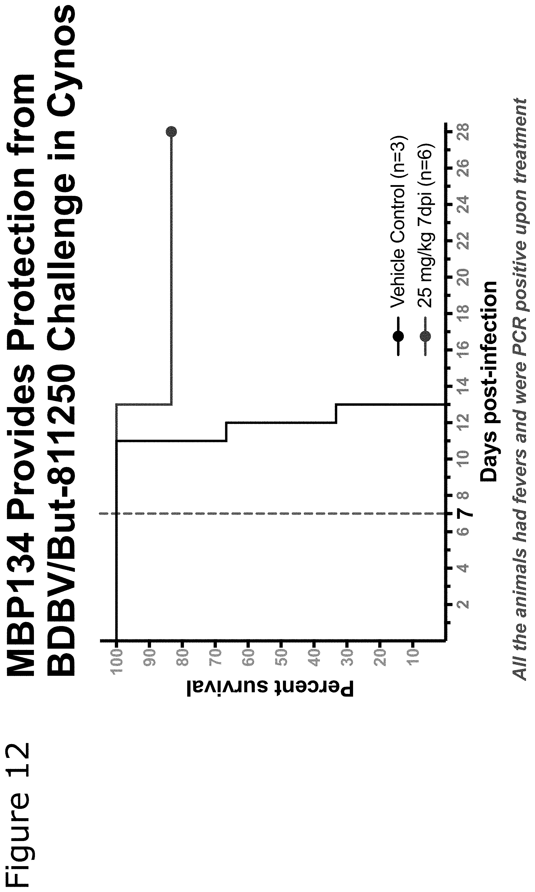
D00013

S00001
XML
uspto.report is an independent third-party trademark research tool that is not affiliated, endorsed, or sponsored by the United States Patent and Trademark Office (USPTO) or any other governmental organization. The information provided by uspto.report is based on publicly available data at the time of writing and is intended for informational purposes only.
While we strive to provide accurate and up-to-date information, we do not guarantee the accuracy, completeness, reliability, or suitability of the information displayed on this site. The use of this site is at your own risk. Any reliance you place on such information is therefore strictly at your own risk.
All official trademark data, including owner information, should be verified by visiting the official USPTO website at www.uspto.gov. This site is not intended to replace professional legal advice and should not be used as a substitute for consulting with a legal professional who is knowledgeable about trademark law.