Methods For Detection Of Flavivirus Antibodies
Khurana; Surender
U.S. patent application number 16/831525 was filed with the patent office on 2020-11-05 for methods for detection of flavivirus antibodies. This patent application is currently assigned to The U.S.A., as represented by the Secretary, Department of Health and Human Services. The applicant listed for this patent is The U.S.A., as represented by the Secretary, Department of Health and Human Services, The U.S.A., as represented by the Secretary, Department of Health and Human Services. Invention is credited to Surender Khurana.
| Application Number | 20200348299 16/831525 |
| Document ID | / |
| Family ID | 1000005003827 |
| Filed Date | 2020-11-05 |


View All Diagrams
| United States Patent Application | 20200348299 |
| Kind Code | A1 |
| Khurana; Surender | November 5, 2020 |
METHODS FOR DETECTION OF FLAVIVIRUS ANTIBODIES
Abstract
Isolated peptides that include one or more antigenic sites of Zika virus (ZIKV) and methods of their use and production are disclosed. The peptides can be used, for example, to detect exposure of a subject to a flavivirus infection, such as a ZIKV infection.
| Inventors: | Khurana; Surender; (Clarksburg, MD) | ||||||||||
| Applicant: |
|
||||||||||
|---|---|---|---|---|---|---|---|---|---|---|---|
| Assignee: | The U.S.A., as represented by the
Secretary, Department of Health and Human Services Silver Spring MD |
||||||||||
| Family ID: | 1000005003827 | ||||||||||
| Appl. No.: | 16/831525 | ||||||||||
| Filed: | March 26, 2020 |
Related U.S. Patent Documents
| Application Number | Filing Date | Patent Number | ||
|---|---|---|---|---|
| 62823834 | Mar 26, 2019 | |||
| Current U.S. Class: | 1/1 |
| Current CPC Class: | C07K 14/005 20130101; C12N 7/00 20130101; G01N 33/56983 20130101; G01N 33/566 20130101; C12N 2770/24121 20130101 |
| International Class: | G01N 33/566 20060101 G01N033/566; C07K 14/005 20060101 C07K014/005; C12N 7/00 20060101 C12N007/00; G01N 33/569 20060101 G01N033/569 |
Goverment Interests
ACKNOWLEDGMENT OF GOVERNMENT SUPPORT
[0002] This invention was made with government support under project number OCET 2018-698 awarded by the FDA Medical Countermeasures Initiative (MCMi). The government has certain rights in the invention.
Claims
1. An isolated peptide comprising an amino acid sequence set forth as SEQ ID NO: 1, SEQ ID NO: 2, SEQ ID NO: 3, SEQ ID NO: 4, SEQ ID NO: 5, SEQ ID NO: 6, SEQ ID NO: 7, SEQ ID NO: 8 or SEQ ID NO: 10, wherein the peptide is no more than 100 amino acids in length, and wherein: the peptide is linked to a solid support; the peptide is fused to a heterologous protein; or the peptide is conjugated to a heterologous carrier.
2. The isolated peptide of claim 1, wherein the peptide is no more than 80 amino acids in length.
3. The isolated peptide of claim 1, wherein the peptide is no more than 60 amino acids in length.
4. The isolated peptide of claim 1, wherein the amino acid sequence of the peptide comprises SEQ ID NO: 2, SEQ ID NO: 5, SEQ ID NO: 7 or SEQ ID NO: 8.
5. The isolated peptide of claim 1, wherein the amino acid sequence of the peptide consists or consists essentially of SEQ ID NO: 2, SEQ ID NO: 5, SEQ ID NO: 7 or SEQ ID NO: 8.
6. The isolated peptide of claim 1, wherein the peptide is linked to the solid support by a linker.
7. The isolated peptide of claim 6, wherein the linker comprises biotin, streptavidin, maleimide, polyethylene glycol (PEG), a peptide, or combinations of thereof.
8. The isolated peptide of claim 1, wherein the solid support comprises a bead, a membrane, a reaction tray, a multi-well plate, or a test tube.
9. The isolated peptide of claim 1, wherein the heterologous protein comprises a tag or linker.
10. The isolated peptide of claim 1, wherein the heterologous carrier comprises a protein from bacteria, a protein from a virus, keyhole limpet hemocyanin (KLH), ovalbumin (OVA), or bovine serum albumin (BSA).
11. A solid support linked to one or more peptides comprising amino acid sequences selected from SEQ ID NO: 1, SEQ ID NO: 2, SEQ ID NO: 3, SEQ ID NO: 4, SEQ ID NO: 5, SEQ ID NO: 6, SEQ ID NO: 7, SEQ ID NO: 8 and SEQ ID NO: 10, wherein the one or more peptides are no more than 100 amino acids in length.
12. The solid support of claim 11, wherein the one or more peptides are no more than 80, or no more than 60 amino acids in length.
13. The solid support of claim 11, wherein the one or more peptides comprise amino acid sequences selected from SEQ ID NO: 2, SEQ ID NO: 5, SEQ ID NO: 7 and SEQ ID NO: 8
14. The solid support of claim 11, wherein the one or more peptides consist or consist essentially of the amino acid sequences set forth as SEQ ID NO: 2, SEQ ID NO: 5, SEQ ID NO: 7 and SEQ ID NO: 8.
15. The solid support of claim 11, wherein the solid support comprises a bead, a membrane, a reaction tray, a multi-well plate, or a test tube.
16. A method for detecting anti-flavivirus antibodies in a biological sample containing antibodies, comprising: contacting the biological sample with one or more peptides comprising amino acid sequences selected from SEQ ID NO: 1, SEQ ID NO: 2, SEQ ID NO: 3, SEQ ID NO: 4, SEQ ID NO: 5, SEQ ID NO: 6, SEQ ID NO: 7, SEQ ID NO: 8 and SEQ ID NO: 10 under conditions sufficient to form an immune complex between the one or more peptides and the antibodies present in the biological sample; and detecting the presence or absence of the immune complex, wherein the presence of the immune complex indicates anti-flavivirus antibodies are present in the sample and the absence of the immune complex indicates anti-flavivirus antibodies are not present in the sample.
17. The method of claim 16, wherein the flavivirus is Zika virus (ZIKV).
18. The method of claim 16, wherein the biological sample is from a human subject.
19. The method of claim 18, wherein the subject is one who is at risk of or is suspected of having a flavivirus infection.
20. The method of claim 16, wherein the biological sample comprises blood, serum, plasma, urine, saliva, tears, feces, semen, mucous, tissue, tissue homogenate, cellular extract, spinal fluid, or any combination of two or more thereof.
21. The method of claim 16, wherein the one or more peptides comprise amino acid sequences selected from SEQ ID NO: 2, SEQ ID NO: 5, SEQ ID NO: 7 and SEQ ID NO: 8.
Description
CROSS REFERENCE TO RELATED APPLICATIONS
[0001] This application claims the benefit of U.S. Provisional Application No. 62/823,834, filed Mar. 26, 2019, which is herein incorporated by reference in its entirety.
FIELD
[0003] The present disclosure concerns Zika virus (ZIKV) peptides and their use for detecting a subject's exposure ZIKV or other flaviviruses.
BACKGROUND
[0004] Since its discovery in 1947 (Haddow et al., 1952, Trans. R. Soc. Trop. Med. Hyg., 46: 509-520; de Silva et al., 2016, JAMA., 315: 1945-1946), Zika virus (ZIKV) has primarily been associated with sporadic infections in humans and mild symptoms. However, during the recent 2015-2016 outbreak in Latin America, ZIKV infections were associated with developmental and neurological complications including microcephaly in newborns and Guillain-Barre syndrome in adults (Diamond et al., 2016, J. Virol., 90: 4864-4875; Sardi et al., 2015, Emerg. Infect. Dis. 21: 1885-1886; Mlakar et al., 2016, N. Engl. J. Med., 374: 951-958; Cao-Lormeau et al., 2016, Lancet., 387: 1531-1539; Faria et al., 2016, Science, 352: 345-349). This has prompted an emphasis on vaccine development (Michael et al., 2017, Immunity, 46: 176-182; Gaudinski et al., 2017, Lancet.; Fauci et al., 2016, N. Engl. J. Med., 375: 1209-1212; Graham et al., 2017, J. Infect. Dis., 216: S957-S963; Shan et al., 2017, Nat. Commun., 8:676; Thomas et al., 2017, N. Engl. J. Med., 376:1883-1886), and isolation/characterization of ZIKV-specific monoclonal antibodies (MAbs) with a low risk of antibody-dependent enhancement (ADE). Additionally, accurate diagnostics for ZIKV infection are hampered by pre-existing cross-reactive antibodies against other flaviviruses circulating in the same geographical areas (Lanciotti et al., 2008, Emerg. Infect. Dis., 14: 1232-1239; Gubler et al., 2016, Clin. Microbiol. Rev. 29:487-524). A need exists for identifying new targets in the ZIKV polyprotein that are recognized by antibodies at early stages post-exposure to aid in the development of an improved diagnostic test for ZIKV infection and/or infection by other flaviviruses. Furthermore, a need exists to improve the accuracy and speed of serologic diagnosis for flaviviruses, including ZIKV.
SUMMARY
[0005] Disclosed herein are peptide fragments of Zika virus (ZIKV) that can be used, for example, to detect exposure of a subject to a ZIKV infection. Also disclosed are ZIKV peptides for detecting exposure of a subject to any one of a multiple of flaviviruses. For use in diagnostic and/or detection assays, the peptides can be linked or conjugated to a solid support.
[0006] Provided herein are isolated peptides that encompass antigenic sites of a ZIKV polyprotein. In some embodiments, the peptides are less than 100 amino acids in length. In some embodiments, the peptides are linked to a solid support, fused to a heterologous protein, or conjugated to a heterologous carrier.
[0007] Also provided are solid supports linked to one or more the ZIKV peptides disclosed herein. In some examples, the solid support includes a bead, membrane, multi-well plate, or any solid support suitable for use in an immunoassay.
[0008] Further provided is a method for detecting anti-flavivirus antibodies in a biological sample. In some embodiments, the method includes contacting a biological sample with a ZIKV peptide disclosed herein under conditions sufficient to form an immune complex between the one or more peptides and the antibodies present in the biological sample; and detecting the presence or absence of the immune complex. The presence of the immune complex indicates anti-flavivirus antibodies are present in the sample and the absence of the immune complex indicates anti-flavivirus antibodies are not present in the sample. In some examples, the flavivirus is ZIKV. In other examples, the flavivirus is a flavivirus other than ZIKV.
[0009] Methods of identifying a subject with a flavivirus infection are also provided. In some embodiments, the method includes contacting a biological sample containing antibodies from the subject with one or more ZIKV peptides disclosed herein under conditions sufficient to form an immune complex between the one or more peptides and the antibodies present in the biological sample; and detecting the presence or absence of the immune complex. The presence of the immune complex identifies the subject as having a flavivirus infection and the absence of the immune complex identifies the subject as not having a flavivirus infection.
[0010] Methods for detecting exposure of a subject to a flavivirus infection, such as a ZIKV infection, are disclosed. In such methods, a biological sample from a subject is contacted with an effective amount of an isolated peptide immobilized on a solid support under conditions sufficient to form an immune complex between the isolated peptide and antibodies present in the biological sample. The presence of the immune complex indicates that the biological sample is from a subject with the ZIKV infection or from a subject previously infected with ZIKV or other flavivirus.
[0011] Also provided is a process for linking a peptide to a solid support; and test kits containing the peptide linked to a solid support and optionally further comprising buffer, positive or negative control materials, labelling reagents, reagents for detecting a label and other conventional components.
[0012] The foregoing and other objects and features of the disclosure will become more apparent from the following detailed description, which proceeds with reference to the accompanying figures.
BRIEF DESCRIPTION OF THE DRAWINGS
[0013] FIGS. 1A-1C show IgG and IgM antibody repertoires across the whole ZIKV proteome elicited following ZIKV infection. (FIG. 1A) Number of bound phage clones isolated using ZIKV GFPDL affinity selection against flavivirus naive negative control serum, pooled serum samples from five acutely ZIKV-infected patients on Day 0 and Day 7 of a hospital visit, and urine samples on Day 7 of the hospital visit (day 0 of the hospital visit corresponds to 0-3 days from the onset of symptoms for these 5 patients).
[0014] (FIGS. 1B-1C) Schematic alignment of the peptides recognized by IgM (FIG. 1B) and IgG (FIG. 1C) antibodies in ZIKV-infected human sera (day 0 and day 7) and urine (day 7), and flavivirus naive negative control serum, identified by panning with ZIKV-GFPDL. The amino acid designation is based on the ZIKV polyprotein sequence encoded by the complete ZIKV-ICD genome (FIG. 8; SEQ ID NO: 9). Bars indicate identified inserts in the different structural (C, prM, E) and non-structural (NS) genes on the ZIKV polyprotein sequence. Graphical distribution of representative clones with a frequency of >2, obtained after affinity selection, are shown. The horizontal position and the length of the bars indicate the peptide sequence displayed on the selected phage clone to its homologous sequence in the ZIKV sequence on alignment. The thickness of each bar represents the frequency of repetitively isolated phages.
[0015] FIGS. 2A-2C show the frequency of ZIKV inserts bound by IgG and IgM antibodies in serum and urine following ZIKV infection in humans. (FIG. 2A) Antigenic sites within the ZIKV proteins recognized by serum (Day 0, Day 7) and urine (Day 7) IgM and IgG antibodies following ZIKV infection (based on data presented in FIG. 1). The amino acid designation is based on the ZIKV polyprotein sequence encoded by the complete ZIKV genome (FIG. 8; SEQ ID NO: 9). The amino acid designations on the ZIKV polyprotein are as follows: C, capsid; pr, pre; M, membrane; E, envelope; NS, non-structural. Inset shows expanded version of E protein schematic with domains (D) I, II and III and fusion loop (FL) shown along with their antigenic sites. Previously described epitopes using MAbs are shown above the ZIKV-E schematic. Critical residues for binding of the following MAbs are depicted: 1, ZIKV-117; 2, 2A10G6; 3, ZIKV-12; 4, ZIKV-15; and 5, ZIKV-116. (FIGS. 2B-2C) Distribution and frequency of phage clones expressing each of the ZIKV antigenic sites recognized by IgM (FIG. 2B) and IgG (FIG. 2C) antibodies in post-ZIKV infection serum (Day 0 and Day 7) and urine (Day 7) are shown. The number of phage clones that expressed each antigenic site was divided by the total number of ZIKV-GFPDL selected clones for each pooled sera or urine samples and represented as a percentage.
[0016] FIGS. 3A-3B show structural representations of antigenic sites identified in ZIKV E protein using GFDPL. (FIG. 3A) (left panel) Heat map showing sequence conservation on one monomer chain of mature ZIKV-E protein structure (PDB #5JHM) based on comparison with several ZIKV isolates (Paraiba, Uganda-1947, Nigeria-1968, Senegal-2001, Micronesia-2007 and Brazil-2016 strains) and (right panel) ZIKV vs. other flaviviruses (DENV 1 to 4, West Nile virus (WNV), yellow fever virus (YFV) and ZIKV). (FIG. 3B) Antigenic sites within ZIKV-prM/E identified by the GFPDL analysis are depicted on the structures of both immature (PDB 5U4W) and mature ZIKV E (PDB 5JHM). PDB Structure #5U4W encompasses residues 288-794 and PDB Structure #5JHM encompasses residues 313-699 based on ZIKV_ICD polyprotein sequence (SEQ ID NO: 9; FIG. 8).
[0017] FIGS. 4A-4B show seroreactivity of acute ZIKV-infected samples, convalescent ZIKV-infected samples, and DENV seropositive samples with ZIKV antigenic site peptides. (FIG. 4A) End-point titers of acute ZIKV (Acute-V0 and Acute-V28), convalescent ZIKV (ZIKV-Conv) and convalescent DENV human serum samples tested for binding to various ZIKV peptides in ELISA are depicted. ELISA was performed with 5-fold serially diluted (starting at 1:100) samples. One-way ANOVA was performed with a Bonferroni post-hoc analysis. ****p<0.0001, **p<0.005 and *p<0.05. (FIG. 4B) Table showing frequency and percentage of samples with end-point titers of >100 for each of the sample groups; ZIKV Acute-V0 and Acute-V28, ZIKV-Conv and DENV for each ZIKV peptide determined by peptide ELISA. Frequency and percent seropositivity were calculated for 19 samples each of ZIKV-Acute-V0 and V28, 13 samples for ZIKV-Conv and 27 samples for convalescent DENV infected samples.
[0018] FIGS. 5A-5F are graphs showing the binding of polyclonal antibodies in human sera and urine from post-ZIKV infected individuals to NS1, ZIKV-E, and ZIKV-E domain III purified proteins using surface plasmon resonance (SPR). Serum (FIGS. 5A, 5C, 5D) and urine (FIGS. 5B, 5D, 5F) samples collected at different time points from adults post-ZIKV infection (Days 0, 3, 7 and 28) were analyzed for total binding to purified NS1 (FIGS. 5A-5B), ZIKV-E (FIGS. 5C-5D) and ZIKV-E-domain III (FIGS. 5E-5F) proteins. Total antibody binding is represented in SPR resonance units (RU). Maximum resonance unit (Max RU) values for protein binding by serum or urine antibodies obtained from each individual at different time points post-ZIKV exposure are linked by connecting lines.
[0019] FIGS. 6A-6F show the antibody affinity maturation of polyclonal human sera and urine antibodies to NS1, ZIKV-E, and ZIKV-E domain III proteins following ZIKV infection. SPR analysis of individual sera (FIGS. 6A, 6C, 6E) or urine (FIGS. 6B, 6D, 6F) samples post-ZIKV infection was performed with purified recombinant NS1 (FIGS. 6A-6B), ZIKV-E (FIGS. 6C-6D) and ZIKV-E domain III (FIGS. 6E-6F) proteins to determine the dissociation kinetics (off-rates) at different time points post-infection. Antibody off-rate constants that describe the fraction of antibody-antigen complexes decaying per second were determined directly from the serum/urine sample interaction with recombinant NS1, ZIKV-E, and ZIKV-E domain III proteins using SPR in the dissociation phase. Average values are indicated by the horizontal bar for each group. The statistical significances between each time-point (visit days) were determined using two-tailed paired t-test in GraphPad software. p-values less than 0.05 were considered significant with a 95% confidence interval. Statistically significant with p-values of <0.05 (*), <0.005 (**), or <0.001 (***) are shown.
[0020] FIGS. 7A-7B show the relationship between antibody affinity of polyclonal human serum antibodies to ZIKV-E and NS1 proteins with clinical symptoms following ZIKV infection in patients. Antibody off-rate constants of the polyclonal serum sample interaction with recombinant ZIKV-E (FIG. 7A) and NS1 (FIG. 7B) proteins on day 7 as measured by SPR was correlated with total number of symptoms on day 28 for the corresponding patient. Inverse Spearman correlations were observed between anti-ZIKV E antibody affinity on day 7 measured by SPR vs. number of symptoms on day 28 (r=0.592; p=0.01569).
[0021] FIG. 8 shows the complete ZIKV ICD Paraiba strain whole genome translated sequence (SEQ ID NO: 9) used for construction of ZIKV GFPD library and described in FIGS. 1-3.
[0022] FIG. 9 shows the random distribution of size and sequence of the ZIKV-GFPDL. Sequencing of ZIKV whole genome fragments expressed by the phages of the ZIKV GFPD libraries were aligned to the ZIKV_ICD translated sequence (FIG. 8; SEQ ID NO: 9).
[0023] FIGS. 10A-10D show GFPDL based epitope mapping of neutralizing MAb ZV54. (FIG. 10A) GFPDL-based epitope mapping of neutralizing ZIKV mouse Mab ZV54 to prME. (FIG. 10B) The ELISA reactivity of selected GFPDL identified phage clones to MAb ZV54 was confirmed by phage ELISA. (FIG. 10C) Structure of GFPDL-identified epitope on PDB #5JHM for mature E. (FIG. 10D) The highest ELISA reactive sequence for MAb ZV54 identified using GFPDL mapping (residues 598-694 of SEQ ID NO: 9) is shown in the table compared to the sequence previously identified as `known site` (residues 589-697 of SEQ ID NO: 9).
[0024] FIGS. 11A-11D show GFPDL based epitope mapping of neutralizing and protective mouse MAb ZV67. (FIG. 11A) GFPDL-based epitope mapping of neutralizing ZIKV mouse MAb ZV67 to prME. (FIG. 11B) The ELISA reactivity of selected GFPDL identified phage clones to MAb ZV67 was confirmed by phage ELISA. (FIG. 11C) Structure of GFPDL-identified epitope on PDB #5JHM for mature E. (FIG. 11D) The highest ELISA reactive sequence for MAb ZV67 identified using GFPDL mapping (595-708; SEQ ID NO: 9) is shown in the table compared to the sequence previously identified as `known site` (residues 589-697 of SEQ ID NO: 9).
[0025] FIGS. 12A-12D show GFPDL based epitope mapping of a conformational tertiary epitope dependent, DENV-negative, ZIKV-specific neutralizing and protective human MAb Z23. (FIG. 12A) GFPDL-based epitope mapping of neutralizing ZIKV MAb Z23 to prME. (FIG. 12B) The ELISA reactivity of selected GFPDL identified phage clones to MAb Z23 was confirmed by phage ELISA and the ELISA positive clones are shown. (FIG. 12C) Structure of GFPDL-identified epitope on PDB #5JHM for mature E. (FIG. 12D) The minimal reactive sequence for MAb Z23 identified using GFPDL mapping (residues 594-694 of SEQ ID NO: 9) is shown in the table.
[0026] FIGS. 13A-13D show GFPDL based epitope mapping of neutralizing and protective human MAb ZKA64. (FIG. 13A) GFPDL-based epitope mapping of neutralizing ZIKV MAb ZKA64 to prME. (FIG. 13B) The ELISA reactivity of selected GFPDL identified phage clones to MAb ZKA64 was confirmed by phage ELISA and the ELISA positive clones are shown. (FIG. 13C) Structure of GFPDL-identified epitope on PDB #5JHM for mature E. (FIG. 13D) The minimal reactive sequence for MAb ZKA64 identified using GFPDL mapping (residues 595-694 of SEQ ID NO: 9) is shown in the table.
[0027] FIG. 14 shows anti-E reactivity of post-infection sera or urine in SPR before and after ZIKV-GFPDL adsorption. Post infection sera or urine at day 7 from individuals was adsorbed on ZIKV-GFPDL coated petri dishes. Binding to recombinant ZIKV-E is shown before and after GFPDL-adsorption in SPR.
[0028] FIG. 15 shows adsorption of anti-ZIKV antibodies in post-infection sera using ZIKV-GFPDL. Post ZIKV infection sera at day 7 (post-onset of symptoms) was adsorbed on ZIKV-GFPDL coated petri dishes. Antibody reactivity to Zika virus coated on polystyrene Immulon 2HB plates is shown before and after GFPDL-adsorption in ELISA was revealed using HRP-conjugated goat anti-human IgA+IgG+IgM specific antibody. All data was normalized to `virus only` background signal.
[0029] FIGS. 16A-16C show individual antibody repertoires elicited following acute ZIKV infection using IgG and IgM specific capture beads by ZIKV GFPDL. (FIG. 16A) Table of phage titers. (FIGS. 16B and 16C) Alignment of bound phage clones to ZIKV genome. Schematic alignment of the peptide sequences recognized by IgM (FIG. 16B) and IgG (FIG. 16C) antibodies in ZIKV infected human sera (42-001-F at day 7 post-onset of illness), identified using ZIKV-GFPDL. The amino acid designation is based on the ZIKV polyprotein sequence encoded by the complete ZIKV-ICD genome (FIG. 8; SEQ ID NO: 9). Bars indicate identified inserts in the different structural (C, prM, E) and non-structural (NS) genes on the ZIKV polyprotein sequence. Graphical distribution of representative clones with a frequency of >2, obtained after affinity selection, are shown. The horizontal position and the length of the bars indicate the peptide sequence displayed on the selected phage clone to its homologous sequence in the ZIKV sequence on alignment. The thickness of each bar represents the frequency of repetitively isolated phages.
[0030] FIGS. 17A-17B show structural representations of antigenic sites identified in ZIKV NS1 protein using GFDPL. (FIG. 17A) Heat map on one monomer chain showing sequence conservation of various ZIKV (left panel; Paraiba, Uganda 1947, Nigeria 1968, Senegal 2001, Micronesia 2007 and Brazil 2016 strains) and flaviviruses (right panel; Dengue 1-4, West Nile, Yellow fever and ZIKV) conservation on ZIKV NS1 protein structure (PDB 5K6K). (FIG. 17B) Antigenic sites have been depicted on surface structures of ZIKV NS1 protein (PDB 5K6K). C- and N-termini have been depicted on the first structure showing antigenic sites. Structure 5K6K encompasses residues 795-1146 of ZIKV_ICD whole genome-encoded polyprotein sequence (SEQ ID NO: 9).
[0031] FIGS. 18A-18B show structural representations of antigenic sites identified in ZIKV NS2B protein using GFDPL. (FIG. 18A) Heat map on one monomer chain showing various ZIKV (left panel; Paraiba/2015, MR766/Uganda/1947, Nigeria/IbH30656_SM21V1-V3/1968, ArD157995/Senegal/2001, Micronesia/2007 and Brazil/2015 strains) and flaviviruses (center panel; Dengue_1-4, West Nile, Yellow fever and ZIKV) conservation on ZIKV NS2B-NS3 protein structure (PDB # SGXJ). C- and N-termini have been depicted on the ribbon structure (right panel). (FIG. 18B) Antigenic sites have been depicted on surface structures of ZIKV NS2B protein (PDB # SGXJ). Structure PDB # SGXJ encompasses residues 1421-1673 on ZIKV ICD whole genome-encoded polyprotein sequence (SEQ ID NO: 9).
[0032] FIGS. 19A-19B show structural representations of antigenic sites identified in ZIKV NS3 protein using GFDPL. (FIG. 19A) Heat map showing various ZIKV (left panel; Paraiba, Uganda 1947, Nigeria 1968, Senegal 2001, Micronesia 2007 and Brazil 2016 strains) and flaviviruses (center panel; Dengue 1-4, West Nile virus, Yellow fever and ZIKV) conservation on ZIKV NS3 protein structure (PDB SJRZ). C- and N-termini have been depicted on the ribbon structure of SJRZ (right panel). (FIG. 19B) Antigenic sites have been depicted on surface structures of ZIKV NS3 protein (PDB SJRZ). Structure SJRZ encompasses residues 1677-2119 on ZIKV ICD whole genome-encoded polyprotein sequence (SEQ ID NO: 9).
[0033] FIGS. 20A-20B show structural representations of antigenic sites identified in ZIKV NS5 protein using GFDPL. (FIG. 20A) Heat map showing various ZIKV (left panel; Paraiba, Uganda 1947, Nigeria 1968, Senegal 2001, Micronesia 2007 and Brazil 2016 strains) and flaviviruses (center panel; Dengue 1-4, West Nile virus, Yellow fever and ZIKV) conservation on ZIKV NS5 protein structure (PDB 5TFR). C- and N-termini have been depicted on the ribbon structure of 5TFR (right panel). (FIG. 20B) Antigenic sites have been depicted on surface structures of ZIKV NS5 protein (PDB 5TFR). Structure 5TFR encompasses residues 2525-3423 on ZIKV ICD whole genome-encoded polyprotein sequence (SEQ ID NO: 9).
[0034] FIGS. 21A-21B show percent similarity between different ZIKV strains and flaviviruses. (FIG. 21A) Similarity between various ZIKV strains plotted as a percentage of ZIKV Paraiba strain (considered at 100%) whose genome structure with antigenic sites has been depicted. (FIG. 21B) Similarity between various flaviviruses plotted as a percentage of ZIKV Paraiba strain (considered at 100%) whose polyprotein structure with antigenic sites have been depicted. In both cases, ZIKV Paraiba strain was used as a query sequence and was used to generate a plot that shows the percent similarity of the reference sequences (other flaviviruses FIG. 21A, ZIKV strains FIG. 21B) to the query sequence. A sliding window of size 200 bp or 20 bp was used, which passes through the alignment in steps of 1 bp to generate the plot showing different flaviviruses and all ZIKV strains respectively.
[0035] FIG. 22 shows a similarity plot of envelope (E) protein sequences of different flaviviruses. For generating the plot, ZIKV_Paraiba strain was used as the query sequence to obtain the percent similarity of various domains of E protein of different flaviviruses to the ZIKV E. Highest conservation was seen near the fusion loop region while the lowest was seen around domain I (Antigenic sites 5, 6 and 7) showing potential specificity to ZIKV E protein. A sliding window size of 80 bp and a step size of 1 bp was used to generate the plot.
[0036] FIGS. 23A-23B show steady-state equilibrium analysis of different dilutions of post-infection sera to ZIKV-E and NS1 protein by SPR. Serial dilutions of post-infection sera were injected simultaneously onto either ZIKV-E (FIG. 23A) or NS1 (FIG. 23B) protein immobilized on a GLC sensor chip and on a surface free of protein (used as a blank). Binding was recorded using BioRad Proteon surface plasmon resonance biosensor instrument. Responses from the protein surface were corrected for the response from the mock surface and for responses from a separate, buffer only injection. Antibody off-rate constants, which describe the fraction of antigen-antibody complexes that decay per second, were determined directly from the serum sample interaction with ZIKV-E using SPR in the dissociation phase only for the sensorgrams with Max RU in the range of 10-100 RU (shown here for 10.times. and 40.times. fold dilution of sera) and calculated using the BioRad ProteOn manager software for the heterogeneous sample model.
[0037] FIGS. 24A-24J are graphs showing seroreactivity of human samples with ZIKV antigenic site peptides. Acute, convalescent (Cony) and late stage ZIKV-infected, DENV seropositive and influenza virus seropositive human sera and plasma samples were tested for binding to individual ZIKV peptides or combinations of three ZIKV peptides by ELISA. (FIG. 24A) ZIKV NS1 1033-1067. (FIG. 24B) ZIKV NS2B 1421-1469. (FIG. 24C) ZIKV NS2B 1424-1457. (FIG. 24D) ZIKV NS3 1805-1873. (FIG. 24E) ZIKV NS4B 2312-2363. (FIG. 24F) ZIKV NS5 2943-2977. (FIG. 24G) ZIKV NS5 3136-3179. (FIG. 24H) ZIKV NS1 1033-1067+NS2B 1421-1469+NS5 2943-2977. (FIG. 24I) ZIKV NS1 1033-1067+NS2B 1421-1469+NS5 3136-3179. (FIG. 24J) ZIKV NS2B 1424-1457+NS5 2943-2977+NS5 3136-3179.
[0038] FIGS. 25A-25D show IgM/IgG/IgA antibody repertoires across the whole ZIKV proteome elicited in serum and amniotic fluid samples from A ZIKV-infected woman the first trimester. (FIG. 25A) Number of bound phage clones bound by IgM, IgG and IgA antibodies using ZIKV-GFPDL affinity selection of maternal serum and AF samples from first trimester pregnancy (ID #38 in Table 5) at 10 days post onset of symptoms. (FIGS. 25B-25D) The amino acid designation is based on the ZIKV polyprotein sequence encoded by the complete ZIKV-ICD genome (FIG. 8). Antigenic epitope profiles with bars indicating identified inserts in the ZIKV genome are shown for (FIG. 25B) IgM, (FIG. 25C) IgG and (FIG. 25D) IgA antibodies. Graphical distribution of representative clones with a frequency of >2, obtained after affinity selection, are shown. The horizontal position and the length of each of the bars in FIGS. 25B-25D indicate the genomic location of the bound displayed peptides on the ZIKV genome (C, capsid; pr peptide; M, membrane; E, envelope; NS, non-structural 1, 2B, 3, 4A, 4B and 5). The thickness of each bar represents the frequency of repetitively isolated phages.
[0039] FIGS. 26A-26E show whole ZIKV proteome IgM/IgG/IgA antibody repertoires in serum and amniotic fluid samples in pregnancy following Zika virus infection in the third trimester. GFPDL analysis conducted with serum and AF from a ZIKV infected pregnant woman in the third trimester (ID #19 in Table 5) at 42 days post onset of symptoms. (FIG. 26A) Number of bound phage clones isolated using ZIKV-GFPDL affinity selection by IgM, IgG and IgA antibodies of maternal serum and AF samples from the third trimester. Antigenic epitope profiles with bars indicating identified inserts in the ZIKV genome are shown for (FIG. 26B) IgM, (FIG. 26C) IgG and (FIG. 26D) IgA antibodies. Graphical distribution of representative clones with a frequency of >2, obtained after affinity selection, are shown. The horizontal position and the length of each of the bars in FIGS. 26B-26D indicate the genomic location of the bound displayed peptides on the ZIKV genome (C, capsid; pr peptide; M, membrane; E, envelope; NS, non-structural 1, 2B, 3, 4A, 4B and 5). The thickness of each bar represents the frequency of repetitively isolated phages. (FIG. 26E) Immunodominant antigenic sites within the ZIKV genome recognized by IgM, IgG and IgA antibodies in maternal serum and AF samples from first and third trimester. Epitopes numbered in black represent sites identified in the acute infection study in non-pregnant adults described in Example 1. Epitopes in blue, red and green numbering represent the epitopes specifically recognized by IgM, IgG and IgA antibodies, respectively. Those highlighted in bold black, blue, red or green represent newly identified antigenic sites in this study.
[0040] FIGS. 27A-27C show antibody binding, isotype distribution, and affinity of serum and AF paired samples to E, E-DIII, NS1, and prM using SPR. Serum and AF samples, collected from pregnant females post-ZIKV infection, were analyzed for antibody binding to purified ZIKV E, E-domain III, NS1 and prM proteins in SPR. (FIG. 27A) Total antibody binding is represented in SPR resonance units. Maximum resonance unit (Max RU) values for protein binding by serum vs. AF antibodies obtained from all subjects are linked by connecting lines. (FIG. 27B) Antibody isotype of ZIKV protein binding antibodies in serum and AF following ZIKV infection as measured in SPR. The resonance units for each anti-ZIKV antibody isotype (IgM, IgE, IgG subclasses, and IgA) was divided by the total resonance units for all antibody isotypes combined to calculate the percentage of each antibody isotype for individual serum and AF sample. (FIG. 27C) Antibody affinity of serum vs. AF antibodies to E, E-DIII, NS1, and prM. SPR analysis of sera or AF post-ZIKV infection from each pregnant female was performed with purified recombinant ZIKV E, E-DIII, NS1 and -prM proteins to determine the dissociation kinetics (off-rates) points. Antibody off-rate constants that describe the fraction of antibody-antigen complexes decaying per second were determined directly from the serum or AF sample interaction with recombinant proteins using SPR in the dissociation phase as described in Example 3. Antibody off-rates were calculated for samples with total antibody binding (Max RU shown in FIG. 27A) between 10-100 RU. The statistical significances between each time-point (visit days) were determined using two-tailed paired t-test in GraphPad software. p-values less than 0.05 were considered significant with a 95% confidence interval. Statistically significant with p-values of <0.05 (*), <0.01 (**), <0.001 (***) or <0.0001 (****) are shown.
[0041] FIGS. 28A-28D show seroreactivity of serum and AF samples with selected ZIKV prM and E antigenic site peptides and impact of serum and AF antibodies on ZIKV infection in vitro. (FIG. 28A) Antigenic sites within the ZIKV prM-E protein schematic with domains (D) I, II and III and fusion loop (FL) shown along with their immunodominant antigenic sites. (FIG. 28B) End-point titers of serum/AF paired samples from ZIKV infected pregnant women were tested for binding to ZIKV peptides in ELISA. ELISA was performed with 5-fold serially diluted (starting at 1:100) samples for combination of IgM +IgG +IgA isotypes. The statistical significances between each time-point (visit days) were determined using two-tailed paired t-test in GraphPad software. p-values less than 0.05 were considered significant with a 95% confidence interval. Statistically significant with p-values of <0.05 (*), <0.01 (**), <0.001 (***) or <0.0001 (****) are shown. (FIGS. 28C-28D) Serum and amniotic fluid samples from different patients were tested for impact on ZIKV infection in vitro with Vero cells with both Asian-lineage virus PRVABC59 (FIG. 28C) and African-lineage virus MR766 (FIG. 28D). The percent virus control from RT-qPCR is plotted against log serum dilution. Mean.+-.standard deviation (SD) data from triplicates for serum (line, filled circle markers) and amniotic fluid (dotted line, empty circle markers) is shown for each matched serum and amniotic fluid pair. The positive control ZKA185 (at starting concentration of 1 .mu.g/ml) is shown in filled black symbols in both panels. The percent of virus control (determined by RT-qPCR) is plotted against log serum dilution.
[0042] FIG. 29 is a graph showing anti-E reactivity of post-infection sera in SPR before and after ZIKV-GFPDL adsorption. Post infection 19-S sera was adsorbed on ZIKV-GFPDL coated petri dishes. Binding to recombinant ZIKV-E is shown before (blue line) and after (black line) GFPDL-adsorption in SPR.
[0043] FIGS. 30A-30D show immunodominant antigenic sites within ZIKV genome by IgM, IgG and IgA antibodies. (FIG. 30A) a combination of IgM, IgG and IgA antibodies recognized by antibodies in maternal serum and AF samples from the first and third trimesters are shown (black numbering). (FIG. 30B) IgM antigenic sites (blue numbering), (FIG. 30C) IgG antigenic sites (red numbering), and (FIG. 30D) IgA antigenic sites (green numbering). Epitopes in bold black, blue, red and green represent newly identified antigenic sites in this study and the others were described in Example 1.
[0044] FIGS. 31A-31C show distribution of phage clones and frequency of phage clones binding in serum and AF antibodies following ZIKV infection in first trimester (Subject ID #38) at 10 days post onset of symptoms. (FIG. 31A) IgM, (FIG. 31B) IgG and (FIG. 31C) IgA antibodies recognizing each antigenic site on the ZIKV genome isolated using GFPDL against the maternal serum (black) and AF (red) samples from the first trimester. The number of clones encoding each antigenic site was divided by the total number of ZIKV GFPDL-selected clones for each sample to calculate clonal frequency. Those epitopes newly identified in the current study have been highlighted in bold black letters on X-axis.
[0045] FIGS. 32A-32C show distribution of phage clones and frequency of phage clones binding in serum and AF antibodies following ZIKV infection in third trimester (Subject ID #19) at 42 days post onset of symptoms. Distribution of phage clones and frequency of phage clones binding (FIG. 32A) IgM, (FIG. 32B) IgG and (FIG. 32C) IgA antibodies expressing each antigenic site on the ZIKV genome isolated using GFPDL against the maternal serum (black) and AF (red) samples from the first trimester. The number of clones encoding each antigenic site was divided by the total number of ZIKV GFPDL-selected clones for each sample to calculate frequency. Those epitopes newly identified in the current study have been highlighted in bold black labeling on X-axis.
[0046] FIGS. 33A-33C are structural representations of antigenic sites differentially identified in serum vs AF in ZIKV E, NS1 and NS5 proteins using GFPDL. Frequency of differentially recognized antigenic sites by serum vs AF following ZIKV infection are shown in Table 7. (FIG. 33A) Heat map on one monomer chain showing various ZIKV (Paraiba/2015, MR-766/Uganda/1947, Nigeria/IbH-30656_SM21V1-V3/1968, ArD157995/Senegal/2001, Micronesia/2007 and Brazil/2015 strains) and flaviviruses (DENV1-4, WNV, YFV and ZIKV_Paraiba) conservation on mature ZIKV E protein structure (PDB #5JHM). The heat maps have been color coded from red (0) to green (1), where green signifies complete conservation. Antigenic sites Z-5 and Z-8 respectively have been depicted in blue on surface structures of both mature ZIKV E (PDB #5JHM) and immature (PDB #5U4W), with Domain I of E protein shaded in light grey (PDB #5JHM), pr domain in yellow and M in pink (PDB #5U4W). Structure PDB #5U4W encompasses residues 288-794 and structure PDB #5JHM encompasses residues 313-699 on ZIKV ICD whole genome sequence. (FIG. 33B) NS1 structure. Heat map on one monomer chain showing various ZIKV (Paraiba/2015, MR-766/Uganda/1947, Nigeria/IbH-30656_5 M21V1-V3/1968, ArD157995/Senegal/2001, Micronesia/2007 and Brazil/2015 strains) and flaviviruses (DENV1-4, WNV, YFV (and ZIKV) conservation on ZIKV NS1 protein structure (PDB #5K6K). The heat maps have been color coded from red (0) to green (1), where green signifies complete conservation. Antigenic sites Z-42, Z-12, Z-43, Z-14 respectively have been depicted in blue on surface structures of ZIKV NS1 protein (PDB #5K6K). Structure PDB #5K6K encompasses residues 795-1146 on ZIKV ICD whole genome sequence. (FIG. 33C) NS5 structure. Heat map showing various ZIKV (Paraiba/2015, MR-766/Uganda/1947, Nigeria/IbH-30656_SM21V1-V3/1968, ArD157995/Senegal/2001, Micronesia/2007 and Brazil/2015 strains) and flaviviruses (DENV1-4, WNV, YFV and ZIKV) conservation on ZIKV NS5 protein structure (PDB #5TFR). The heat maps have been color coded from red (0) to green (1), where green signifies complete conservation. Antigenic sites Z-25, Z-28 and Z-29 respectively have been depicted in blue on surface structures of ZIKV NS5 protein (PDB #5TFR). Structure PDB #5TFR encompasses residues 2525-3423 on ZIKV ICD whole genome sequence.
[0047] FIG. 34 shows sequence identity (%) of immunodominant prM-E antigenic site within African and Asian lineage ZIKV strains. The sequence shown in the figure corresponds to residues 123-795 of SEQ ID NO: 9.
[0048] FIG. 35 depicts antigenic sites within ZIKV-prM/E identified by the GFPDL analysis in blue on the structures of both immature (PDB 5U4W) and mature ZIKV E (PDB 5JHM). Domain I is shaded in light grey (PDB 5JHM), pr domain is shaded in yellow and M in pink (PDB 5U4W). PDB Structure #5U4W encompasses residues 288-794 and PDB Structure #5JHM encompasses residues 313-699 based on ZIKV_ICD polyprotein sequence (FIG. 8).
TABLES
[0049] Table 1: Demographic, epidemiological and diagnostic information of samples used in Example 1.
[0050] Table 2: Clinical symptoms for the acute ZIKV infected individuals in Example 1.
[0051] Table 3: Frequency of antigenic sites for IgM and IgG antibodies in serum on day 0 and day 7 and in urine on day 7 post-ZIKV exposure (Example 1).
[0052] Table 4: Sequence conservation of antigenic regions/sites among different flavivirus strains (Example 1).
[0053] Table 5: Demographic, epidemiological and diagnostic information of samples used in Example 3.
[0054] Table 6: RT-PCR and serodiagnostic information of samples used in Example 3.
[0055] Table 7: Frequency of antigenic sites for IgM, IgG and IgA antibodies in serum and amniotic fluid (AF) during ZIKV infection in the first and third trimester of pregnancy (Example 3).
[0056] Table 8: Sequence conservation of antigenic sites among different flavivirus strains (Example 3).
[0057] Table 9: Frequency of differentially recognized antigenic sites by serum vs amniotic fluid (AF) following ZIKV infection in first trimester (1st) or 3rd trimester (3rd). Sites Z-5, Z-8, Z-42, Z-12, Z-43, Z-14, Z-25, Z-28, Z-29 have been depicted on structures of the respective proteins in FIG. 32.
SEQUENCE LISTING
[0058] The amino acid sequences listed in the accompanying sequence listing are shown using standard three letter code for amino acids, as defined in 37 C.F.R. 1.822. The Sequence Listing is submitted as an ASCII text file, created on Mar. 25, 2020, 33.8 KB, which is incorporated by reference herein. In the accompanying sequence listing:
[0059] SEQ ID NO: 1 is the amino acid sequence of the NS1 1033-1067 antigenic site: SDLIIPKSLAGPLSHHNTREGYRTQMKGPWHSEEL
[0060] SEQ ID NO: 2 is the amino acid sequence of the NS2B 1421-1469 antigenic site: VDMYIERAGDITWEKDAEVTGNSPRLDVALDESGDFSLVEDDGPPMREI
[0061] SEQ ID NO: 3 is the amino acid sequence of the NS3 1805-1873 antigenic site: TRVEMGEAAAIFMTATPPGTRDAFPDSNSPIMDTEVEVPERAWSSGFDWVTDHSGKTVWFVPSV RNGNE
[0062] SEQ ID NO: 4 is the amino acid sequence of the NS4 2422-2465 antigenic site: VVTDIDTMTIDPQVEKKMGQVLLIAVAVSSAILSRTAWGWGEAG
[0063] SEQ ID NO: 5 is the amino acid sequence of the NS4B 2312-2363 antigenic site: YAALTTFITPAVQHAVTTSYNNYSLMAMATQAGVLFGMGKGMPFYAWDFGVP SEQ ID NO: 6 is the amino acid sequence of the NS5 2860-2901 antigenic site: TGIAMTDTTPYGQQRVFKEKVDTRVPDPQEGT
[0064] SEQ ID NO: 7 is the amino acid sequence of the NS5 2943-2977 antigenic site: AVEAVNDPRFWALVDKEREHHLRGECQSCVYNMMG
[0065] SEQ ID NO: 8 is the amino acid sequence of the NS5 3163-3179 antigenic site: NLVVQLIRNMEAEEVLEMQDLWLLRRSEKVTNWLQSNGWDRLKR
[0066] SEQ ID NO: 9 is the amino acid sequence of ZIKV Paraiba strain polyprotein (see FIG. 8).
[0067] SEQ ID NO: 10 is the amino sequence of the NS2B 1424-1457 antigenic site: YIERAGDITWEKDAEVTGNSPRLDVALDESGDFS
DETAILED DESCRIPTION
[0068] Described herein are methods of detecting exposure of a subject to a flavivirus infection, such as a ZIKV infection. The disclosed methods utilize isolated peptides and fragments thereof from ZIKV. Also described are ZIKV peptides and fragments linked to a solid support or conjugated to a heterologous molecule, such as a heterologous protein, linker and/or detectable label.
I. Abbreviations
[0069] ADE antibody dependent enhancement [0070] AF amniotic fluid [0071] C capsid [0072] CHICKV Chikungunya virus [0073] DENV dengue virus [0074] E envelope [0075] ELISA enzyme-linked immunosorbent assay [0076] FL fusion loop [0077] WGFPDL Whole-Genome-Fragment-Phage-Display Libraries [0078] HRP horseradish peroxidase [0079] JEV Japanese encephalitis virus [0080] LFA lateral flow assay [0081] Mab monoclonal antibody [0082] M membrane [0083] Max RU maximum resonance unit [0084] NS non-structural [0085] POC point-of-care [0086] prM premembrane [0087] PRNT plaque reduction neutralization test [0088] SPR surface plasmon resonance [0089] TBEV tick-borne encephalitis virus [0090] WNV West Nile virus [0091] YFV yellow fever [0092] ZIKV Zika virus
II. Terms
[0093] Unless otherwise noted, technical terms are used according to conventional usage. Definitions of common terms in molecular biology may be found in Benjamin Lewin, Genes X, published by Jones & Bartlett Publishers, 2009; and Meyers et al. (eds.), The Encyclopedia of Cell Biology and Molecular Medicine, published by Wiley-VCH in 16 volumes, 2008; and other similar references.
[0094] As used herein, the singular forms "a," "an," and "the," refer to both the singular as well as plural, unless the context clearly indicates otherwise. For example, the term "an antigen" includes single or plural antigens and can be considered equivalent to the phrase "at least one antigen." As used herein, the term "comprises" means "includes." It is further to be understood that any and all base sizes or amino acid sizes, and all molecular weight or molecular mass values, given for nucleic acids or polypeptides are approximate, and are provided for descriptive purposes, unless otherwise indicated. Although many methods and materials similar or equivalent to those described herein can be used, particular suitable methods and materials are described herein. In case of conflict, the present specification, including explanations of terms, will control. In addition, the materials, methods, and examples are illustrative only and not intended to be limiting. To facilitate review of the various embodiments, the following explanations of terms are provided:
[0095] Administration: The introduction of an active compound or composition into a subject by a chosen route. Administration can be local or systemic. Examples of local administration include, but are not limited to, topical administration, subcutaneous administration, intramuscular administration, intrathecal administration, intrapericardial administration, intra-ocular administration, topical ophthalmic administration, or administration to the nasal mucosa or lungs by inhalational administration. In addition, local administration includes routes of administration typically used for systemic administration, for example by directing intravascular administration to the arterial supply for a particular organ. Thus, in particular embodiments, local administration includes intra-arterial administration and intravenous administration when such administration is targeted to the vasculature supplying a particular organ. Local administration also includes the incorporation of active compounds and agents into implantable devices or constructs, such as vascular stents or other reservoirs, which release the active agents and compounds over extended time intervals for sustained treatment effects.
[0096] Systemic administration includes any route of administration designed to distribute an active compound or composition widely throughout the body via the circulatory system. Thus, systemic administration includes, but is not limited to intra-arterial and intravenous administration. Systemic administration also includes, but is not limited to, oral administration, topical administration, subcutaneous administration, intramuscular administration, transdermal administration, or administration by inhalation, when such administration is directed at absorption and distribution throughout the body by the circulatory system.
[0097] Antibody or antibodies: A protein (or protein complex) that includes one or more polypeptides substantially encoded by immunoglobulin genes or fragments of immunoglobulin genes. The recognized immunoglobulin genes include the kappa, lambda, alpha, gamma, delta, epsilon and mu constant region genes, as well as the myriad immunoglobulin variable region genes. Light chains are classified as either kappa or lambda. Heavy chains are classified as gamma, mu, alpha, delta, or epsilon, which in turn define the immunoglobulin classes, IgG, IgM, IgA, IgD and IgE, respectively.
[0098] The basic immunoglobulin (antibody) structural unit is generally a tetramer. Each tetramer is composed of two identical pairs of polypeptide chains, each pair having one light (about 25 kD) and one heavy chain (about 50-70 kD). The N-terminus of each chain defines a variable region of about 100 to 110 or more amino acids primarily responsible for antigen recognition. The terms variable light chain (V.sub.L) and variable heavy chain (V.sub.H) refer, respectively, to these light and heavy chains.
[0099] As used herein, the term antibody includes intact immunoglobulins as well as a number of well-characterized fragments produced by digestion with various peptidases, or genetically engineered artificial antibodies. Thus, for example, pepsin digests an antibody below the disulfide linkages in the hinge region to produce F(ab)'.sub.2, a dimer of Fab which itself is a light chain joined to V.sub.H-C.sub.H by a disulfide bond. The F(ab)'.sub.2 may be reduced under mild conditions to break the disulfide linkage in the hinge region thereby converting the F(ab)'.sub.2 dimer into an Fab' monomer. The Fab' monomer is essentially a Fab with part of the hinge region (see, Fundamental Immunology, W. E. Paul, ed., Raven Press, N. Y., 1993).
[0100] While various antibody fragments are defined in terms of the digestion of an intact antibody, it will be appreciated that Fab' fragments may be synthesized de novo either chemically or by utilizing recombinant DNA methodology. Thus, the term antibody as used herein also includes antibody fragments either produced by the modification of whole antibodies or synthesized de novo using recombinant DNA methodologies.
[0101] Antibodies for use in the methods and compositions of this disclosure can be monoclonal or polyclonal. Merely by way of example, monoclonal antibodies can be prepared from murine hybridomas according to the classical method of Kohler and Milstein (Nature 256:495-497, 1975) or derivative methods thereof. Detailed procedures for monoclonal antibody production are described in Harlow and Lane (Using Antibodies, A Laboratory Manual, CSHL, New York, 1998).
[0102] The terms "bind specifically" and "specific binding" refer to the ability of a specific binding agent (such as, an antibody) to bind to a target molecular species in preference to binding to other molecular species with which the specific binding agent and target molecular species are admixed. A specific binding agent is said specifically to recognize a target molecular species when it can bind specifically to that target.
[0103] A single-chain antibody (scFv) is a genetically engineered molecule containing the V.sub.H and V.sub.L domains of one or more antibody(ies) linked by a suitable polypeptide linker as a genetically fused single chain molecule (see, for example, Bird et al., Science, 242:423-426, 1988; Huston et al., Proc. Natl. Acad. Sci., 85:5879-5883, 1988). Diabodies are bivalent, bispecific antibodies in which V.sub.H and V.sub.L domains are expressed on a single polypeptide chain, but using a linker that is too short to allow for pairing between the two domains on the same chain, thereby forcing the domains to pair with complementary domains of another chain and creating two antigen binding sites (see, for example, Holliger et al., Proc. Natl. Acad. Sci., 90:6444-6448, 1993; Poljak et al., Structure, 2:1121-1123, 1994).
[0104] One or more complementarity determining regions (CDRs) may be incorporated into a molecule either covalently or noncovalently to make the resultant molecule an immunoadhesin. An immunoadhesin may incorporate the CDR(s) as part of a larger polypeptide chain, may covalently link the CDR(s) to another polypeptide chain, or may incorporate the CDR(s) noncovalently. The CDRs permit the immunoadhesin to specifically bind to a particular antigen of interest. A chimeric antibody is an antibody that contains one or more regions from one antibody and one or more regions from one or more other antibodies.
[0105] An antibody may have one or more binding sites. If there is more than one binding site, the binding sites may be identical to one another or may be different. For instance, a naturally-occurring immunoglobulin has two identical binding sites, a single-chain antibody or Fab fragment has one binding site, while a bispecific or bifunctional antibody has two different binding sites.
[0106] A neutralizing antibody or an inhibitory antibody is an antibody that inhibits at least one activity of a target, usually a polypeptide, such as by blocking the binding of the polypeptide to a ligand to which it normally binds, or by disrupting or otherwise interfering with a protein-protein interaction of the polypeptide with a second polypeptide. An activating antibody is an antibody that increases an activity of a polypeptide. Antibodies may function as mimics of a target protein activity, or as blockers of the target protein activity, with therapeutic effect derived therein.
[0107] Anti-idiotypic antibody: An antibody that binds to the specific antigen binding site of another antibody generated in response to exposure to an antigen, such as an antigen derived from a member of the flavivirus genus or immunological relative thereof.
[0108] Biological sample: A sample obtained from a subject. Biological samples include all clinical samples useful for detection of disease or infection (for example, ZIKV infection) in subjects, including, but not limited to, cells, tissues, and bodily fluids, such as blood, derivatives and fractions of blood (such as serum or plasma), cerebrospinal fluid, urine, eye tissue, saliva, semen, breast milk, synovial fluid, amniotic fluid, cord blood; as well as biopsied or surgically removed tissue, for example tissues that are unfixed, frozen, or fixed in formalin or paraffin. In a particular example, a biological sample is obtained from a subject having or suspected of having a ZIKV or other flavivirus infection.
[0109] Carrier: An immunogenic molecule to which a peptide can be linked to enhance an immune response to the peptide. Carriers are chosen to increase the immunogenicity of the antigen and/or to elicit antibodies against the carrier which are diagnostically, analytically, and/or therapeutically beneficial.
[0110] Useful carriers include polymeric carriers, which can be natural (for example, nucleic acid or proteins from bacteria or viruses), aptamers, dyes, semi-synthetic or synthetic materials containing one or more functional groups to which a reactant moiety can be attached.
[0111] Conditions sufficient to form an immune complex: Conditions which allow an antibody or antigen binding fragment to bind to its cognate epitope to a detectably greater degree than, and/or to the substantial exclusion of, binding to substantially all other epitopes. Conditions sufficient to form an immune complex are dependent upon the format of the binding reaction and typically are those utilized in immunoassay protocols or those conditions encountered in vivo. See Harlow & Lane, Antibodies, A Laboratory Manual, 2.sup.nd ed. Cold Spring Harbor Publications, New York (2013) for a description of immunoassay formats and conditions. The conditions employed in the methods are "physiological conditions" which include reference to conditions (such as temperature, osmolarity, pH) that are typical inside a living mammal or a mammalian cell. While it is recognized that some organs are subject to extreme conditions, the intra-organismal and intracellular environment normally lies around pH 7 (for example, from pH 6.0 to pH 8.0, more typically pH 6.5 to 7.5), contains water as the predominant solvent, and exists at a temperature above 0.degree. C. and below 50.degree. C. Osmolarity is within the range that is supportive of cell viability and proliferation.
[0112] In several embodiments, the formation of an immune complex can be detected through conventional methods, for instance immunohistochemistry, immunoprecipitation, flow cytometry, immunofluorescence microscopy, ELISA, immunoblotting (for example, Western blot), point-of-care test, rapid assay, biosensors, fluorophores or dyes, flow assay, bead based assay, nitrocellulose/PVDF membrane based assay, magnetic resonance imaging, CT scans, X-ray and affinity chromatography.
[0113] Conjugate: A complex of at least two heterologous molecules linked together. In a non-limiting example, an ZIKV peptide as disclosed herein is conjugated to a solid support, such as via a linker.
[0114] Consists essentially of and Consists of: A polypeptide comprising an amino sequence that consists essentially of a specified amino acid sequence does not include any additional amino acid residues. However, the residues in the polypeptide can be modified to include non-peptide components, such as labels (for example, fluorescent, radioactive, color, biosensors, or solid particle labels), sugars or lipids, and the N- or C-terminus of the polypeptide can be joined (for example, by peptide bond) to heterologous amino acids, such as a cysteine (or other) residue in the context of a linker for conjugation chemistry. A polypeptide that consists of a specified amino acid sequence does not include any additional amino acid residues, nor does it include additional biological components, such as nucleic acid lipids, sugars, nor does it include labels. However, the N- and C-terminus of the polypeptide can be joined (for example, by peptide bond) to heterologous amino acids, such as a peptide tag, or a cysteine (or other) residue in the context of a linker for conjugation chemistry.
[0115] A polypeptide that consists or consists essentially of a specific amino acid sequence can be glycosylated or have an amide modification. A polypeptide that consists or consists essentially of a particular amino acid sequence can be linked via its N- or C-terminus to a heterologous polypeptide, such as in the case of a fusion protein containing a first polypeptide consisting or a first sequence that is linked (via peptide bond) to a heterologous polypeptide consisting of a second sequence. In another example, the N- or C-terminus of a polypeptide that consists of or consists essentially of a particular amino acid sequence can be linked to a peptide linker (via peptide bonds) that is further linked to one or more additional heterologous polypeptides. In a further example, the N- or C-terminus of a polypeptide that consists of or consists essentially of a particular amino acid sequence can be linked to one or more amino acid residues that facilitate further modification or manipulation of the polypeptide.
[0116] Control: A reference standard. In some embodiments, the control is a negative control sample obtained from a healthy patient not infected with ZIKV. In other embodiments, the control is a positive control, such as a biological sample obtained from a patient diagnosed with ZIKV infection. In still other embodiments, the control is a historical control or standard reference value or range of values (such as a previously tested control sample, such as a group of ZIKV patients with known prognosis or outcome, or group of samples that represent baseline or normal values).
[0117] A difference between a test sample and a control can be an increase or conversely a decrease. The difference can be a qualitative difference or a quantitative difference, for example a statistically significant difference. In some examples, a difference is an increase or decrease, relative to a control, of at least about 5%, such as at least about 10%, at least about 20%, at least about 30%, at least about 40%, at least about 50%, at least about 60%, at least about 70%, at least about 80%, at least about 90%, at least about 100%, at least about 150%, at least about 200%, at least about 250%, at least about 300%, at least about 350%, at least about 400%, at least about 500%, or greater than 500%.
[0118] Diagnosis: The process of identifying a disease by its signs, symptoms and/or results of various tests. The conclusion reached through that process is also called "a diagnosis." Forms of testing commonly performed include blood tests, medical imaging, genetic analysis, urinalysis, biopsy and analysis of biological samples obtained from a subject. In one example, diagnosis of a subject as having a flavivirus infection comprises determining whether the subject has antibodies that specifically bind to one or more peptides disclosed herein (see, e.g., Tables 3 and 4).
[0119] Effective amount: An amount of agent, such as an anti-viral agent, that is sufficient to generate a desired response, such as an inhibition of viral infection in a subject or detection of a particular viral infection in a subject. For instance, this can be the amount necessary to inhibit an infection with one or more flaviviruses or to measurably alter outward symptoms of the infection. In some embodiments, an effective amount is an amount of a peptide that is sufficient for detection of antibodies to the peptide in a biological sample from a subject.
[0120] In one example, a desired response is to induce an immune response that elicits an immune response to flavivirus in a subject and/or inhibits or prevents flavivirus infection in a subject. For example, administration of an effective amount of a disclosed flavivirus peptide can induce an immune response in a subject that inhibits subsequent infection of the subject by the flavivirus.
[0121] Epitope: An antigenic determinant. These are particular chemical groups or peptide sequences on a molecule that are antigenic, such that they elicit a specific immune response, for example, an epitope is the region of an antigen to which anti-ZIKV antibodies bind. An antibody can bind to a particular antigenic epitope. Epitopes can be formed both from contiguous amino acids or noncontiguous amino acids juxtaposed by tertiary folding of a protein.
[0122] Expression: Transcription or translation of a nucleic acid sequence. For example, a gene is expressed when its DNA is transcribed into an RNA or RNA fragment, which in some examples is processed to become mRNA. A gene may also be expressed when its RNA is translated into an amino acid sequence, such as a protein or protein fragment. In a particular example, a heterologous gene is expressed when its RNA is translated into an amino acid sequence. The term "expression" is used herein to denote either transcription or translation. Regulation of expression can include controls on transcription, translation, RNA transport and processing, degradation of intermediary molecules such as mRNA, or through activation, inactivation, compartmentalization or degradation of specific protein molecules after they are produced.
[0123] Flavivirus non-structural protein: There are seven non-structural (NS) proteins of a flavivirus, NS1, NS2A, NS2B, NS3, NS4A, NS4B, and NS5, which are encoded by the portion of the flavivirus genome that is 3' to the structural proteins. NS1 has been implicated in RNA replication and has been shown to be secreted from infected mammalian cells (Post et al., Virus Res. 18:291-302, 1991; Mackenzie et al., Virology 220:232-240, 1996; Muylaert et al., Virology 222:159-168, 1996). NS1 can elicit strong humoral immune responses and is a potential vaccine candidate (Shlesinger et al., J. Virol. 60:1153-1155, 1986; Qu et al., J. Gen. Virol. 74:89-97, 1993). NS2 is cleaved into NS2A and NS2B. NS2A is involved in RNA replication and virus particle assembly and secretion and NS2B forms a complex with NS3 and functions as a cofactor for the NS3 protease, which cleaves portions of the virus polyprotein. NS3 also functions as an RNA helicase and is used to unwind viral RNA during replication (Li et al., J. Virol. 73:3108-3116, 1999). While the exact functions of NS4A and NS4B remain to be elucidated, they are thought to be involved in RNA replication and RNA trafficking (Lindenbach and Rice, In: Fields Virology, Knipe and Howley, eds., Lippincott, Williams, and Wilkins, 991-1041, 2001). Finally, the NS5 protein is an RNA-dependent RNA polymerase involved in genome replication (Rice et al., Science 229:726-733, 1985). NS5 also shows methyltransferase activity commonly found in RNA capping enzymes (Koonin, J. Gen. Virol. 74:733-740, 1993).
[0124] Flavivirus structural protein: The capsid (C), premembrane (prM), and envelope (E) proteins of a flavivirus are the viral structural proteins. Flavivirus genomes consist of positive-sense RNAs that are roughly 11 kb in length. The genome has a 5' cap, but lacks a 3' polyadenylated tail (Wengler et al., Virology 89:423-437, 1978) and is translated into one polyprotein. The structural proteins (C, prM, and E) are at the amino-terminal end of the polyprotein followed by the non-structural proteins (NS1-5). The polyprotein is cleaved by virus and host derived proteases into individual proteins. The C protein forms the viral capsid while the prM and E proteins are embedded in the surrounding envelope (Russell et al., The Togaviruses: Biology, Structure, and Replication, Schlesinger, ed., Academic Press, 1980). The E protein functions in binding to host cell receptors resulting in receptor-mediated endocytosis. In the low pH of the endosome, the E protein undergoes a conformational change causing fusion between the viral envelope and the endosomal membranes. The prM protein is believed to stabilize the E protein until the virus exits the infected cell, at which time prM is cleaved to the mature M protein (Reviewed in Lindenbach and Rice, In: Fields Virology, Knipe and Howley, eds., Lippincott, Williams, and Wilkins, 991-1041, 2001).
[0125] Heterologous: Originating from a different genetic source. A nucleic acid molecule that is heterologous to a cell originated from a genetic source other than the cell in which it is expressed. Methods for introducing a heterologous nucleic acid molecule in a cell or organism include, for example, transformation with a nucleic acid, including electroporation, lipofection, particle gun acceleration, and homologous recombination.
[0126] Immune response: A response of a cell of the immune system, such as a B cell, T cell, or monocyte, to a stimulus. In one embodiment, the response is specific for a particular antigen (an "antigen-specific response"). In one embodiment, an immune response is a T cell response, such as a CD4+ response or a CD8+ response. In another embodiment, the response is a B cell response, and results in the production of specific antibodies. "Priming an immune response" refers to treatment of a subject with a "prime" immunogen to induce an immune response that is subsequently "boosted" with a boost immunogen. Together, the prime and boost immunizations produce the desired immune response in the subject. "Enhancing an immune response" refers to co-administration of an adjuvant and an immunogenic agent, wherein the adjuvant increases the desired immune response to the immunogenic agent compared to administration of the immunogenic agent to the subject in the absence of the adjuvant.
[0127] Immune complex: The binding of antibody to an antigen forms an immune complex. In some embodiments, the formation of an immune complex can be detected through conventional methods, for instance immunodetection, immunohistochemistry, immunoprecipitation, flow cytometry, immunofluorescence microscopy, ELISA, immunoblotting (for example, Western blot), biosensors, magnetic resonance imaging, CT scans, X-ray and affinity chromatography.
[0128] Immunogen: A protein or a portion thereof that is capable of inducing an immune response in a mammal, such as a mammal infected or at risk of infection with a pathogen.
[0129] Inhibiting a disease or condition: Reducing the full development of a disease or condition in a subject, for example, reducing the full development of ZIKV disease in a subject who has an ZIKV infection, and/or reducing ZIKV infection in a subject or population of subjects at risk thereof. This includes neutralizing, antagonizing, prohibiting, preventing, restraining, slowing, disrupting, stopping, or reversing progression or severity of the disease or condition.
[0130] Inhibiting a disease or condition refers to a prophylactic intervention administered before the disease or condition has begun to develop (for example, by vaccinating a subject at risk of a flavivirus infection, but not infected by a flavivirus, with an flavivirus peptide as disclosed herein) that reduces subsequent development of the disease or condition, and also to amelioration of one or more signs or symptoms of the disease or condition following development. The term "ameliorating," with reference to inhibiting a disease or condition refers to any observable beneficial effect of the intervention intended to inhibit the disease or condition. The beneficial effect can be evidenced, for example, by a delayed onset of clinical symptoms of the disease or condition in a susceptible subject, a reduction in severity of some or all clinical symptoms of the disease or condition, a slower progression of the disease or condition, an improvement in the overall health or well-being of the subject, a reduction in infection, or by other parameters well known in the art that are specific to the particular disease or condition.
[0131] In some embodiments, an immune response elicited by administering an effective amount of an flavivirus peptide as disclosed herein inhibits infection of a human subject by the flavivirus, for example, by at least 50% (such as at least 60%, at least 70%, at least 80%, at least 90%, or more) compared to a suitable control.
[0132] Isolated: A biological component (such as a nucleic acid, peptide, protein or protein complex, for example an antibody) that has been substantially separated, produced apart from, or purified away from other biological components in the cell of the organism in which the component naturally occurs, that is, other chromosomal and extra-chromosomal DNA and RNA, and proteins. Thus, isolated nucleic acids, peptides and proteins include nucleic acids and proteins purified by standard purification methods. The term also embraces nucleic acids, peptides and proteins prepared by recombinant expression in a host cell, as well as, chemically synthesized nucleic acids. An isolated nucleic acid, peptide or protein, for example an antibody, can be at least 50%, at least 60%, at least 70%, at least 80%, at least 90%, at least 95%, at least 96%, at least 97%, at least 98%, or at least 99% pure.
[0133] Label: A detectable compound that is conjugated directly or indirectly to another molecule, such as a peptide or antibody, to facilitate detection of that molecule. For example, the label can be capable of detection by any method including ELISA, spectrophotometry, flow cytometry, or microscopy. Specific, non-limiting examples of labels include fluorophores, chemiluminescent agents, enzymatic linkages, biosensors and radioactive isotopes. Non-limiting methods for labeling and guidance in the choice of labels appropriate for various purposes are discussed for example in Green and Sambrook (Molecular Cloning: A Laboratory Manual, 4.sup.th ed., New York: Cold Spring Harbor Laboratory Press, 2012) and Ausubel et al. (Eds.) (Current Protocols in Molecular Biology, New York: John Wiley and Sons, 2017).
[0134] Lateral flow device: A device that absorbs or adsorbs a liquid sample (such as a serum, plasma, urine or amniotic fluid sample), routes that liquid sample to a detection zone, and uses detection methods (such as peptide- or antibody-based detection methods) to generate a visible signal in response to the presence or absence of a specific molecule (such as an antibody specific for a peptide of interest). The device can be a test strip used in lateral flow chromatography, in which a test sample fluid, suspected of containing an analyte (such as an antibody that specifically binds to one or more of the peptides listed in Tables 3 and 4), flows (for example by capillary action) through the strip (which is frequently made of bibulous materials such as paper, nitrocellulose, and cellulose). The test fluid and any suspended analyte can flow along the strip to a detection zone in which the analyte (if present) interacts with a detection agent to indicate a presence, absence and/or quantity of the analyte.
[0135] Many lateral flow devices are one-step lateral flow assays in which a biological fluid is placed in a sample area on a bibulous strip (though, non-bibulous materials can be used, and rendered bibulous by applying a surfactant to the material), and allowed to migrate along the strip until the liquid comes into contact with a specific binding partner (such as one or more of the peptides listed in Tables 3 and 4) that interacts with an analyte (such as an antibody that binds to one or more of the peptides listed in Tables 3 and 4) in the liquid. Once the analyte interacts with the binding partner, a signal (such as a fluorescent or otherwise visible dye, or biosensor or enzyme) indicates that the interaction has occurred. Multiple discrete binding partners can be placed on the strip (for example in parallel lines) to detect multiple analytes in the liquid. The test strips can also incorporate control indicators, which provide a signal that the test has adequately been performed, even if a positive signal indicating the presence (or absence) of an analyte is not seen on the strip.
[0136] Linked: The term "linked" means joined together, either directly or indirectly. For example, a first moiety may be covalently or noncovalently (e.g., electrostatically) linked to a second moiety. This includes, but is not limited to, covalently bonding one molecule to another molecule, noncovalently bonding one molecule to another (e.g. electrostatically bonding), non-covalently bonding one molecule to another molecule by hydrogen bonding, non-covalently bonding one molecule to another molecule by van der Waals forces, and any and all combinations of such couplings. Indirect attachment is possible, such as by using a "linker". In several embodiments, linked components are associated in a chemical or physical manner so that the components are not freely dispersible from one another, at least until contacting or entering a cell, such as an immune cell.
[0137] Linker: One or more molecules or groups of atoms positioned between two moieties. Typically, linkers are bifunctional, i.e., the linker includes a functional group at each end, wherein the functional groups are used to couple the linker to the two moieties. The two functional groups may be the same, i.e., a homobifunctional linker, or different, i.e., a heterobifunctional linker. In several embodiments, a peptide linker can be used to link the C-terminus of a first protein to the N-terminus of a second protein. Non-limiting examples of peptide linkers include glycine-serine peptide linkers, which are typically not more than 10 amino acids in length. Typically, such linkage is accomplished using molecular biology techniques to genetically manipulate DNA encoding the first polypeptide linked to the second polypeptide by the peptide linker.
[0138] Peptide: A polymer in which the monomers are amino acid residues that are linked together through amide bonds. Peptides are typically less than 150 amino acids in length, such as less than 100 amino acids in length. The amino acids included in a peptide may be subject to post-translational modification (e.g., glycosylation or phosphorylation). A "residue" refers to an amino acid or amino acid mimetic incorporated in a peptide by an amide bond or amide bond mimetic. A peptide has an amino terminal (N-terminal) end and a carboxy terminal (C-terminal) end. In some embodiments herein, a ZIKV peptide is no more than 100 amino acids, no more than 90 amino acids, no more than 80 amino acids, no more than 70 amino acids, no more than 60 amino acids, no more than 50 amino acids, or no more than 40 amino acids in length.
[0139] Peptide Modifications: Synthetic embodiments of the peptides described herein are also provided. For example, peptides can be modified by a variety of chemical techniques to produce derivatives having essentially the same activity as the unmodified peptides, and optionally having other desirable properties. For example, carboxylic acid groups of the peptide, whether carboxyl-terminal or side chain, can be provided in the form of a salt of a pharmaceutically-acceptable cation or esterified to form a C.sub.1-C.sub.16 ester, or converted to an amide of formula NR.sub.1R.sub.2 wherein R.sub.1 and R.sub.2 are each independently H or C.sub.1-C.sub.16 alkyl, or combined to form a heterocyclic ring, such as a 5- or 6-membered ring Amino groups of the peptide, whether amino-terminal or side chain, can be in the form of a pharmaceutically-acceptable acid addition salt, such as the HCl, HBr, acetic, benzoic, toluene sulfonic, maleic, tartaric and other organic salts, or can be modified to C.sub.1-C.sub.16 alkyl or dialkyl amino or further converted to an amide.
[0140] Hydroxyl groups of the peptide side chains may be converted to C.sub.1-C.sub.16 alkoxy or to a C.sub.1-C.sub.16 ester using well-recognized techniques. Phenyl and phenolic rings of the peptide side chains may be substituted with one or more halogen atoms, such as fluorine, chlorine, bromine or iodine, or with C.sub.1-C.sub.16 alkyl, C.sub.1-C.sub.16 alkoxy, carboxylic acids and esters thereof, or amides of such carboxylic acids. Methylene groups of the peptide side chains can be extended to homologous C.sub.2-C.sub.4 alkylenes. Thiols can be protected with any one of a number of well-recognized protecting groups, such as acetamide groups. Any suitable method may be used to introduce cyclic structures into the disclosed peptides to select and provide conformational constraints to the structure that result in enhanced stability.
[0141] Each peptide of this disclosure is comprised of a sequence of amino acids, which may be either L- and/or D-amino acids, naturally occurring and otherwise.
[0142] Pharmaceutically acceptable carriers: The pharmaceutically acceptable carriers useful in this disclosure are conventional. Remington: The Science and Practice of Pharmacy, The University of the Sciences in Philadelphia, Editor, Lippincott, Williams, & Wilkins, Philadelphia, Pa., 21.sup.st Edition (2005), describes compositions and formulations suitable for pharmaceutical delivery of the compounds herein disclosed.
[0143] In general, the nature of the carrier will depend on the particular mode of administration being employed. For instance, parenteral formulations usually comprise injectable fluids that include pharmaceutically and physiologically acceptable fluids such as water, physiological saline, balanced salt solutions, aqueous dextrose, glycerol or the like as a vehicle. For solid compositions (for example, powder, pill, tablet, or capsule forms), conventional non-toxic solid carriers can include, for example, pharmaceutical grades of mannitol, lactose, starch, or magnesium stearate. In addition to biologically-neutral carriers, pharmaceutical compositions to be administered can contain minor amounts of non-toxic auxiliary substances, such as wetting or emulsifying agents, preservatives, and pH buffering agents and the like, for example sodium acetate or sorbitan monolaurate.
[0144] Prime-boost immunization: An immunotherapy including administration of multiple immunogens over a period of time to elicit the desired immune response.
[0145] Solid support: Any material which is insoluble or can be made insoluble by a subsequent reaction. Numerous and varied solid supports are known and include, without limitation, nitrocellulose, the walls of wells of a reaction tray, multi-well plates, test tubes, polystyrene (e.g., polystyrene beads), polyvinyl (e.g., polyvinyl beads), magnetic beads, membranes, hydrogel, and microparticles (such as latex particles). Any suitable porous material with sufficient porosity to allow access by detector reagents and a suitable surface affinity to immobilize capture reagents (e.g., peptides or antibodies) is contemplated by this term. For example, the porous structure of nitrocellulose has excellent absorption and adsorption qualities for a wide variety of reagents, for instance, capture reagents. Nylon possesses similar characteristics and is also suitable. Microporous structures are useful, as are materials with gel structure in the hydrated state.
[0146] Further examples of useful solid supports include: natural polymeric carbohydrates and their synthetically modified, cross-linked or substituted derivatives, such as agar, agarose, cross-linked alginic acid, substituted and cross-linked guar gums, cellulose esters, especially with nitric acid and carboxylic acids, mixed cellulose esters, and cellulose ethers; natural polymers containing nitrogen, such as proteins and derivatives, including cross-linked or modified gelatins; natural hydrocarbon polymers, such as latex and rubber; synthetic polymers which may be prepared with suitably porous structures, such as vinyl polymers, including polyethylene, polypropylene, polystyrene, polyvinylchloride, polyvinylacetate and its partially hydrolyzed derivatives, polyacrylamides, polymethacrylates, copolymers and terpolymers of the above polycondensates, such as polyesters, polyamides, and other polymers, such as polyurethanes or polyepoxides; porous inorganic materials such as sulfates or carbonates of alkaline earth metals and magnesium, including barium sulfate, calcium sulfate, calcium carbonate, silicates of alkali and alkaline earth metals, aluminum and magnesium; and aluminum or silicon oxides or hydrates, such as clays, alumina, talc, kaolin, zeolite, silica gel, or glass (these materials may be used as filters with the above polymeric materials); and mixtures or copolymers of the above classes, such as graft copolymers obtained by initializing polymerization of synthetic polymers on a pre-existing natural polymer.
[0147] It is contemplated that porous solid supports, such as nitrocellulose, described herein can be in the form of sheets or strips.
[0148] The surface of a solid support may be activated by chemical processes that cause covalent linkage of an agent (e.g., a peptide or antibody) to the support. However, any other suitable method may be used for immobilizing an agent (e.g., a peptide or antibody) to a solid support including, without limitation, ionic interactions, hydrophobic interactions, covalent interactions and the like. The particular forces that result in immobilization of an agent on a solid phase are not important for the methods and devices described herein.
[0149] A solid phase can be chosen for its intrinsic ability to attract and immobilize an agent, such as a capture reagent (such as an antibody or peptide). Alternatively, the solid phase can possess a factor that has the ability to attract and immobilize an agent, such as a capture reagent. The factor can include a charged substance that is oppositely charged with respect to, for example, the capture reagent itself or to a charged substance conjugated to the capture reagent. In another embodiment, a specific binding member may be immobilized upon the solid phase to immobilize its binding partner (e.g., a capture reagent). In this example, therefore, the specific binding member enables the indirect binding of the capture reagent to a solid phase material.
[0150] Except as otherwise physically constrained, a solid support may be used in any suitable shapes, such as films, sheets, strips, or plates, or it may be coated onto or bonded or laminated to appropriate inert carriers, such as paper, glass, plastic films, or fabrics.
[0151] A "lateral flow support" is a solid support that is useful in a lateral flow device.
[0152] Specifically bind: When referring to the formation of an antibody:antigen protein complex, or a protein:protein complex, refers to a binding reaction which determines the presence of a target protein, peptide, or polysaccharide, in the presence of a heterogeneous population of proteins and other biologics.
[0153] Thus, under designated conditions, a particular antibody or protein binds preferentially to a particular target protein, peptide or polysaccharide and does not bind in a significant amount to other proteins or polysaccharides present in the sample or subject. Specific binding can be determined by standard methods. A first protein or antibody specifically binds to a target protein when the interaction has a K.sub.D of less than 10.sup.-6 Molar, such as less than 10.sup.-7 Molar, less than 10.sup.-8 Molar, less than 10.sup.-9, or even less than 10.sup.-10 Molar.
[0154] Subject: Living multi-cellular organisms, including vertebrate organisms, a category that includes both human and non-human mammals. In some examples, the subject is human. In specific non-limiting examples, the subject is one who is at risk of or is suspected of having a flavivirus infection, such as a ZIKV infection.
[0155] Treating, preventing or ameliorating a disease: "Preventing" a disease refers to inhibiting the full development of a disease. "Treating" refers to a therapeutic intervention that ameliorates a sign or symptom of a disease or pathological condition, such as a viral infection, after it has begun to develop. "Ameliorating" refers to the reduction in the number or severity of signs or symptoms of a disease, such as a viral infection.
[0156] Under conditions sufficient for: A phrase that is used to describe any environment that permits the desired activity. In one example, this includes administering an effective amount of a composition that includes, for example, a peptide or antibody sufficient to enable the desired activity.
[0157] Vector: A vector is a nucleic acid molecule allowing insertion of foreign nucleic acid without disrupting the ability of the vector to replicate and/or integrate in a host cell. A vector can include nucleic acid sequences that permit it to replicate in a host cell, such as an origin of replication. An insertional vector is capable of inserting itself into a host nucleic acid. A vector can also include one or more selectable marker genes and other genetic elements. An expression vector is a vector that contains the necessary regulatory sequences to allow transcription and translation of inserted gene or genes.
[0158] Whole-Genome-Fragment-Phage-Display Libraries (GFPDL) have been previously used for an unbiased comprehensive analysis of the antibody repertoires in individuals infected with viruses, either early or during recovery (Khurana et al., PLoS Med 6:e1000049, 2009; Fuentes et al., PLoS Pathog 12:e1005554, 2016). They can also help to determine the diversity of epitopes bound by post-vaccination sera and decipher the impact of novel adjuvants (Khurana et al., Sci Transl Med 3:85ra48, 2011; Verma et al., J Virol 86:5515-5522, 2012). Such information could help in the development of improved vaccines, therapeutics, and diagnostics. For example, in HIV, panning of virus specific GFPDLs with sera from acute infections identified several antigenic peptides that could be used for serological diagnosis of HIV-1 (Khurana et al., J Virol 85:12455-12463, 2011; Khurana et al., J Acquir Immune Defic Syndr 43:304-312, 2006), and in avian H5N1, peptides were identified that differentiated infection (across multiple clades) from vaccination (Khurana et al., J Virol 85:12455-12463, 2011; Khurana et al., J Virol 80:2092-2099, 2006).
[0159] In the present disclosure, GFPDL spanning the entire genome of ZIKV was constructed and used for in-depth immune profiling of IgG and IgM antibody repertoires in both serum and urine from individuals acutely infected with ZIKV. Total binding and affinity maturation of antibodies against ZIKV NS1 and E proteins and their evolution during the first month post ZIKV infection was also evaluated using surface plasmon resonance (SPR). The results demonstrated unlinked evolution of antibody responses in terms of antibody epitope repertoire and affinity maturation against structural and non-structural proteins following ZIKV infection in humans, describing differential recognition of various ZIKV proteins by the human immune system.
[0160] Zika virus (ZIKV): A member of the virus family Flaviviridae and the genus Flavivirus. Other members of this genus include dengue virus, yellow fever virus, Japanese encephalitis virus, West Nile virus and Spondweni virus. ZIKV is spread by the daytime-active mosquitoes Aedes aegypti and A. albopictus. This virus was first isolated from a Rhesus macaque from the Zika Forest of Uganda in 1947. Since the 1950s, ZIKV has been known to occur within a narrow equatorial belt from Africa to Asia. The virus spread eastward across the Pacific Ocean in 2013-2014, resulting in ZIKV outbreaks in Oceania to French Polynesia, New Caledonia, the Cook Islands, and Easter Island. In 2015, ZIKV spread to Mexico, Central America, the Caribbean and South America, where ZIKV has reached pandemic levels. Infection by ZIKV generally causes either no symptoms or mild symptoms, including mild headache, maculopapular rash, fever, malaise, conjunctivitis and joint pain. ZIKV causes symptoms in about 20% of infected individuals, and no deaths from the virus have yet been reported. However, ZIKV infection has been linked to the birth of microcephalic infants following maternal infection, as well an increase in cases of GBS. Reports have also indicated that ZIKV has the potential for human blood-borne and sexual transmission. ZIKV has also been found in human saliva and breastmilk.
III. Zika Virus
[0161] Zika virus (ZIKV) belongs to the flavivirus genus within the Flaviviridae family Many flaviviruses are significant human pathogens, including ZIKV, yellow fever (YFV), dengue virus (DENV serotypes 1 to 4), Japanese encephalitis virus (JEV), West Nile virus (WNV), and tick-borne encephalitis virus (TBEV). The present disclosure describes methods for detecting flavivirus exposure (such as by detecting anti-flavivirus antibodies), in particular ZIKV, in subjects including humans, non-human animals, and laboratory animals. The disclosure includes any subject that can respond to an infection by a flavivirus, in particular ZIKV, or an equivalent thereof.
[0162] The flavivirus genome is a single-strand, positive-sense RNA of approximately 11,000 nucleotides. It contains a 5' untranslated region (UTR), an open-reading frame (ORF), and a 3' UTR. The single ORF encodes a long polyprotein which is processed into ten viral proteins, including three structural proteins--capsid (C), precursor membrane (prM), and envelope (E)--and seven non-structural proteins (NS1, NS2A, NS2B, NS3, NS4A, NS4B, and NS5).
[0163] ZIKV is predominantly transmitted to humans by infected Aedes spp. mosquitoes, which also transmit DENV and YFV, as well as chikungunya virus (an emerging alphavirus). ZIKV can also be transmitted through the maternofetal route, sex, blood transfusion, and organ transplantation.
[0164] ZIKV causes multiple illnesses in humans depending on the infecting virus, the host age and immunological conditions. Approximately 80% of ZIKV infections are asymptomatic. Disease symptoms associated with ZIKV infection include headaches, fever, lethargy, rash, conjunctivitis, myalgia, and arthralgia. Severe diseases resulting from ZIKV infection include neurotropic Guillain-Barre syndrome and congenital microcephaly.
[0165] ZIKV infection can be diagnosed by detection of viral components (e.g., viral RNA, viral proteins, or virus isolation) or detection of host immune responses (e.g., antibodies against viral proteins). For viral component-based diagnosis, RT-PCR, immunoassay, and virus isolation can be used to detect ZIKV RNA, viral proteins, and live virus, respectively. Among them, RT-PCR is the most commonly used because of its sensitivity and specificity. The viremic phase of ZIKV infection usually lasts from one to two weeks. The diagnostic window for detection of viral components is narrow because of the short duration of the viremic phase. Host immune response-based assays are also effective techniques, among which enzyme-linked immunosorbent assays (ELISAs), such as IgM-capture ELISA (MAC-ELISA), and plaque reduction neutralization test (PRNT) are the two commonly used serologic assays in ZIKV diagnosis. Serologic diagnosis of ZIKV infection relies upon IgM-capture ELISA which can be flawed due to cross-reactivity with different flaviviruses. The interpretation of conventional IgM-capture ELISA assays for ZIKV and other flaviviruses is challenging due to the cross-reactive nature of anti-flaviviral antibodies conventionally used in such tests, leading to misleading diagnostic results. This challenge confounds ZIKV diagnosis because (i) many flaviviruses (e.g., ZIKV and DENV) produce similar disease symptoms and (ii) antibodies from patients infected with ZIKV cross-react with other flaviviruses. Consequently, ZIKV IgM-capture ELISA results typically require neutralization tests for confirmation. Furthermore, PRNT is time-consuming, labor-intensive, slow, low-throughput, and cost-ineffective, impairing attempts at rapid diagnosis to halt or slow spread of infection. Moreover, PRNT still relies upon both virus-specific and cross-reactive epitopes of viral E protein such that the results may be inconclusive with respect to flavivirus infections. There is therefore a need to improve the accuracy and speed of serologic diagnosis for flaviviruses, ZIKV in particular.
IV. Zika Virus Peptides
[0166] Isolated peptides containing fragments of ZIKV proteins are disclosed herein. The ZIKV peptides can be used to detect exposure of a subject to a flavivirus infection, for example a ZIKV infection, such as by detecting anti-flavivirus antibodies. As discussed in the Examples below, the isolated peptides contain antigenic sites of the ZIKV that are targeted by antibodies following ZIKV infection.
[0167] In several embodiments, the isolated peptides contain fragments of a ZIKV NS protein, such as from the Paraiba strain of ZIKV. Exemplary ZIKV NS protein sequences are set forth herein as SEQ ID NOs: 1-8.
[0168] In some embodiments, the isolated peptide comprises, consists essentially of, or consists of 10-100 consecutive amino acids or longer (such as 10-15, 10-20, 10-30, 10-40, 20-30, 20-40, 20-50, 30-50, 40-50, 10-75, 20-75, 30-75, 40-75, or 75-100 consecutive amino acids) from a native Zika virus sequence.
[0169] The isolated peptides include the amino acid sequence of an antigenic site of the ZIKV. For example, the isolated peptide comprises, consists essentially of, or consists of the amino acid sequence of any one of the antigenic sites of Zika Paraiba as set forth in FIG. 8 (SEQ ID NO: 9), such as antigenic site ZIKV-NS1 1033-1067 (SEQ ID NO: 1), ZIKV-NS2B 1421-1469 (SEQ ID NO: 2), ZIKV-NS2B 1424-1457 (SEQ ID NO: 10), ZIKV-NS3 1805-1873 (SEQ ID NO: 3), ZIKV-NS4B 2422-2465 (SEQ ID NO: 4), ZIKV-NS4B 2312-2363 (SEQ ID NO: 5), ZIKV-NS5 2860-2901 (SEQ ID NO: 6), ZIKV-NS5 2943-2977 (SEQ ID NO: 7), or ZIKV-NS5 3136-3179 (SEQ ID NO: 8).
[0170] In some embodiments, the isolated peptide comprises, consists essentially of, or consists of the amino acid sequence of ZIKV-NS1 (1033-1067) from ZIKV, such as Zika Paraiba. In some embodiments, the isolated peptide comprises, consists essentially of, or consists of the amino acid sequence set forth as SEQ ID NO: 1 wherein the peptide is no more than 100 (such as no more than 95, 90, 85, 80, 75, 70, 65, 60, 55, 50, 45, 40, 35, 30, 25, 20, 15 or 10) amino acids in length.
[0171] In some embodiments, the isolated peptide comprises, consists essentially of, or consists of the amino acid sequence of ZIKV-NS2B (1421-1469) from ZIKV, such as Zika Paraiba. In some embodiments, the isolated peptide comprises, consists essentially of, or consists of the amino acid sequence set forth as SEQ ID NO: 2 wherein the peptide is no more than 100 (such as no more than 95, 90, 85, 80, 75, 70, 65, 60, 55, 50, 45, 40, 35, 30, 25, 20, 15 or 10) amino acids in length.
[0172] In some embodiments, the isolated peptide comprises, consists essentially of, or consists of the amino acid sequence of ZIKV-NS2B (1424-1457) from ZIKV, such as Zika Paraiba. In some embodiments, the isolated peptide comprises, consists essentially of, or consists of the amino acid sequence set forth as SEQ ID NO: 10 wherein the peptide is no more than 100 (such as no more than 95, 90, 85, 80, 75, 70, 65, 60, 55, 50, 45, 40, 35, 30, 25, 20, 15 or 10) amino acids in length.
[0173] In some embodiments, the isolated peptide comprises, consists essentially of, or consists of the amino acid sequence of ZIKV-NS3 (1805-1873 within Z-19) from ZIKV, such as Zika Paraiba. In some embodiments, the isolated peptide comprises, consists essentially of, or consists of the amino acid sequence set forth as SEQ ID NO: 3 wherein the peptide is no more than 100 (such as no more than 95, 90, 85, 80, 75, 70, 65, 60, 55, 50, 45, 40, 35, 30, 25, 20, 15 or 10) amino acids in length.
[0174] In some embodiments, the isolated peptide comprises, consists essentially of, or consists of the amino acid sequence of ZIKV-NS4B (2422-2465 within Z-24) from ZIKV, such as Zika Paraiba. In some embodiments, the isolated peptide comprises, consists essentially of, or consists of the amino acid sequence set forth as SEQ ID NO: 4 wherein the peptide is no more than 100 (such as no more than 95, 90, 85, 80, 75, 70, 65, 60, 55, 50, 45, 40, 35, 30, 25, 20, 15 or 10) amino acids in length.
[0175] In some embodiments, the isolated peptide comprises, consists essentially of, or consists of the amino acid sequence of ZIKV-NS4B (2312-2363 overlapping Z-23.1) from Zika, such as Zika Paraiba. In some embodiments, the isolated peptide comprises, consists essentially of, or consists of the amino acid sequence set forth as SEQ ID NO: 5 wherein the peptide is no more than 100 (such as no more than 95, 90, 85, 80, 75, 70, 65, 60, 55, 50, 45, 40, 35, 30, 25, 20, 15 or 10) amino acids in length.
[0176] In some embodiments, the isolated peptide comprises, consists essentially of, or consists of the amino acid sequence of ZIKV-NS5 (2860-2901 representing Z-29) from Zika, such as Zika Paraiba. In some embodiments, the isolated peptide comprises, consists essentially of, or consists of the amino acid sequence set forth as SEQ ID NO: 6 wherein the peptide is no more than 100 (such as no more than 95, 90, 85, 80, 75, 70, 65, 60, 55, 50, 45, 40, 35, 30, 25, 20, 15 or 10) amino acids in length.
[0177] In some embodiments, the isolated peptide comprises, consists essentially of, or consists of the amino acid sequence of ZIKV-NS5 (2943-2977, within Z-30) from Zika, such as Zika Paraiba. In some embodiments, the isolated peptide comprises, consists essentially of, or consists of the amino acid sequence set forth as SEQ ID NO: 7 wherein the peptide is no more than 100 (such as no more than 95, 90, 85, 80, 75, 70, 65, 60, 55, 50, 45, 40, 35, 30, 25, 20, 15 or 10) amino acids in length.
[0178] In some embodiments, the isolated peptide comprises, consists essentially of, or consists of the amino acid sequence of ZIKV-NS5 (3136-3179, overlapping Z-32.2) from Zika, such as Zika Paraiba. In some embodiments, the isolated peptide comprises, consists essentially of, or consists of the amino acid sequence set forth as SEQ ID NO: 8 wherein the peptide is no more than 100 (such as no more than 95, 90, 85, 80, 75, 70, 65, 60, 55, 50, 45, 40, 35, 30, 25, 20, 15 or 10) amino acids in length.
[0179] Any of the isolated peptides disclosed herein can be linked or conjugated to a solid support, such as via a linker. Suitable linkers include, but are not limited to, straight or branched chain carbon linkers, heterocyclic carbon linkers, nucleic acid linkers, dyes or peptide linkers. For an immunogenic conjugate from two or more constituents, each of the constituents will contain the necessary reactive groups. Representative combinations of such groups are amino with carboxyl to form amide linkages or carboxy with hydroxyl to form ester linkages or amino with alkyl halides to form alkylamino linkages or thiols with thiols to form disulfides or thiols with maleimides or alkylhalides to form thioethers. Hydroxyl, carboxyl, amino and other functionalities, where not present may be introduced by known methods. A wide variety of linking groups can be used. In some instances, the linking group can be either hydrophilic or hydrophobic in order to enhance the desired binding characteristics of the peptide. The covalent linkages should be stable relative to the solution conditions under which the conjugate is subjected.
[0180] In some embodiments, the linkers may be joined to the constituent amino acids through their side chains (such as through a disulfide linkage to cysteine) or to the alpha carbon, amino, and/or carboxyl groups of the terminal amino acids. In some embodiments, the linker and the peptide can be encoded as a single peptide such that the peptide and the linker are connected by peptide bonds. Peptides typically contain a variety of functional groups; for example, carboxylic acid (COOH), free amine (-NH.sub.2) or sulfhydryl (--SH) groups, which are available for reaction with a suitable functional group on a peptide. Alternatively, the peptide is derivatized to expose or attach additional reactive functional groups. The derivitization may involve attachment of any of a number of linker molecules, such as streptavidin.
[0181] Although non-limiting examples discussed herein can include using full-length recombinant ZIKV peptides as antigens, full-length peptides would not necessarily be required for use in all embodiments of the method and compositions disclosed herein. For example, specific or portions less than full-length viral proteins may be used rather than full-length protein. A portion less than the full-length protein to which antibodies produced in response to a subject's exposure to a given ZIKV will bind may be used rather than full-length protein. In some embodiments, a portion less than full-length ZIKV to which antibodies produced in response to a subject's exposure to a given ZIKV, but not another flavivirus, will bind may be used. In other examples, portions less than full length protein that bind to antibodies produced in response to either of two or more different flaviviruses may be used.
[0182] In one embodiment, an antigen with less than complete homology to a ZIKV protein, such as ZIKV non-structural proteins NS1, NS2B, NS3, NS4B and NS5B, may be used, as may a combination of any two or more of the foregoing. A recombinant ZIKV protein with an amino acid sequence that corresponds to some but not all of the amino acids in the ZIKV protein to which it corresponds may be used. In some embodiments, ZIKV antigen with between 75%-80% homology, 80%-90% homology, or 95%-100% homology may be used. ZIKV protein sequences homologous to known strains may be used, within homology ranges as disclosed herein. A recombinant ZIKV antigen with less than 100% homology to a corresponding ZIKV peptide to which antibodies produced in response to a subject's exposure to a ZIKV will bind may be used rather than a recombinant protein with 100% homology. In some embodiments, a recombinant ZIKV peptide of less than 100% homology to a ZIKV peptide to which antibodies produced in response to a subject's exposure to a given ZIKV, but not another flavivirus, will bind may be used. In other examples, a recombinant flaviviral protein of less than 100% homology to a flaviviral protein to which antibodies produced in response to a subject's exposure to two or more given flaviviruses may be used.
[0183] In other embodiments, an antigen with an amino acid sequence that corresponds to only a portion of a full-length ZIKV protein, such as NS1, NS2B, NS3, NS4B, or NS5 may be used, as may a combnation of any two or more of the foregoing. For example, a truncated portion of NS1, NS2B, NS3, NS4B, or NS5, which is recognized by antibodies generated in response to a ZIKV infection, may be used. In some embodiments, an antigen may include amino acids not present in naturally occurring ZIKV proteins. For purposes of antigen synthesis and purification, an antigen may contain an amino acid tag, such as on its C-terminus or N-terminus, without interfering with the binding thereto of an antibody produced by an individual as a result of ZIKV infection. Other possible tags include, a poly-histidine tag, a myc-tag, a FLAG-tag, an HA-tag, or others, all of which can be included as part of an antigen and be within the scope of methods and kits disclosed herein.
[0184] Also provided herein are isolated peptides containing an antigenic site of a ZIKV polyprotein, such as the ZIKV Paraiba strain, wherein the peptides are linked to a solid support, fused to a heterologous protein, or conjugated to a heterologous carrier. In some embodiments, the peptide is no more than 100, no more than 90, no more than 80, no more than 70, no more than 60, no more than 50 or no more than 40 amino acids in length. In some examples, the peptide comprises an amino acid sequence set forth as SEQ ID NO: 1, SEQ ID NO: 2, SEQ ID NO: 3, SEQ ID NO: 4, SEQ ID NO: 5, SEQ ID NO: 6, SEQ ID NO: 7, SEQ ID NO: 8 or SEQ ID NO: 10. In specific non-limiting examples, the peptide consists, or consists essentially of, SEQ ID NO: 1, SEQ ID NO: 2, SEQ ID NO: 3, SEQ ID NO: 4, SEQ ID NO: 5, SEQ ID NO: 6, SEQ ID NO: 7, SEQ ID NO: 8 or SEQ ID NO: 10. In particular examples, the amino acid sequence of the peptide comprises, consists, or consists essentially of SEQ ID NO: 2, SEQ ID NO: 5, SEQ ID NO: 7 or SEQ ID NO: 8.
[0185] In some embodiments, the peptide is linked to a solid support. In some examples, the peptide is linked to the solid support via a linker, such as, but not limited to, biotin, streptavidin, maleimide, polyethylene glycol (PEG), a peptide, or combinations of thereof. In some examples, the solid support comprises a bead, a membrane, a reaction tray, a multi-well plate, or a test tube.
[0186] In other embodiments, the peptide is fused to a heterologous protein. In some examples, the heterologous protein comprises a tag or linker. In specific examples, the tag is a poly-histidine tag, a myc tag, a FLAG tag, an HA tag. In some examples, the linker is a peptide linker.
[0187] In other embodiments, the peptide is conjugated to a heterologous carrier. In some examples, the heterologous carrier includes a protein from bacteria, a protein from a virus, keyhole limpet hemocyanin (KLH), ovalbumin (OVA), or bovine serum albumin (BSA).
[0188] Further provided herein are solid supports linked to one or more ZIKV peptides disclosed herein. In some embodiments, the one or more peptides include one or more of the peptides listed in Table 3, Table 4, Table 7 or Table 8. In specific embodiments, the one or more peptides comprise amino acid sequences selected from SEQ ID NO: 1, SEQ ID NO: 2, SEQ ID NO: 3, SEQ ID NO: 4, SEQ ID NO: 5, SEQ ID NO: 6, SEQ ID NO: 7, SEQ ID NO: 8 and SEQ ID NO: 10. In some examples, the one or more peptides comprise amino acid sequences selected from SEQ ID NO: 2, SEQ ID NO: 5, SEQ ID NO: 7 and SEQ ID NO: 8. In specific non-limiting examples, the one or more peptides consist or consist essentially of the amino acid sequences set forth as SEQ ID NO: 2, SEQ ID NO: 5, SEQ ID NO: 7 and SEQ ID NO: 8. In some embodiments, the one or more peptides are no more than 100, no more than 90, no more than 80, no more than 70, no more than 60, no more than 50 or no more than 40 amino acids in length. In some embodiments, the solid support comprises a bead, a membrane, a reaction tray, a multi-well plate, or a test tube.
V. Methods for Detection and Diagnosis
[0189] Methods are also provided for the detection of the presence of antibodies to flaviviruses, in particular ZIKV, in a biological sample. The method can be used to identify a biological sample from a subject with a ZIKV infection, or from a subject that had a prior infection with a ZIKV. In one example, the presence of ZIKV is detected in a biological sample from a subject, and can be used to identify a subject with ZIKV infection. The sample can be any sample from a subject that contains antibodies induced by ZIKV infection, including, but not limited to biological fluids. In some examples, the biological sample is from a subject, such as a human subject, who is at risk of or is suspected of having a flavivirus infection, for example, a ZIKV infection. The method of detection can include contacting the sample with an isolated ZIKV peptide as disclosed herein and under conditions sufficient to form an immune complex between the peptide and antibodies in the sample, and detecting the immune complex.
[0190] In some embodiments, the peptides disclosed herein are used to test vaccines. For example, to test if a vaccine elicits an immune response that targets a particular antigenic site on the ZIKV. Such methods involve immunizing a subject with a vaccine, and then screening a sample from the subject that contains antibodies induced by the immunization for antibody binding to the appropriate peptide.
[0191] Thus, the peptides disclosed herein can be used for serodiagnosis as well as the development of assays, including point-of-care (POC) assays.
[0192] In some embodiments, the method further includes determining the isotype of the anti-flavivirus antibodies present in a biological sample. Such a method is useful in order to determine the stage of infection; IgM antibodies indicate an early/acute stage of infection, while IgG antibodies indicate a later/convalescent stage of flavivirus infection.
[0193] The disclosed methods can also be used for distinguishing between a subject with a natural flavivirus infection and a subject who has been vaccinated against a flavivirus. A subject with a natural infection will have antibodies that will bind the peptides disclosed herein derived from flavivirus non-structural proteins (e.g. NS1, NS2B, NS3, NS4B, NS5), while a vaccinated subject will have antibodies specific for flavivirus structural proteins (e.g. C, prM and/or E).
[0194] Heterogeneous immunoassay techniques usually involve the use of a solid phase material that the reaction material becomes bound to, but can be adapted to bind antigens and antibodies. The reaction product is separated from excess sample, assay reagents, and other substances by separating the solid phase from the reaction mixture (e.g. by washing). Sandwich immunoassays are one type of solid phase immunoassay that can be used in the present disclosure. In the sandwich assay, the more analyte present in the sample, the greater the amount of label present on the solid phase. This type of assay is generally preferred for visualization of low analyte concentration, because the appearance of label on the solid phase is more readily detected. In some embodiments, the detection or diagnostic assay is designed for POC. In some examples, the POC assay is a lateral flow assay (LFA).
[0195] Provided herein are methods for detecting anti-flavivirus antibodies in a biological sample containing antibodies. In some embodiments, the method includes contacting the biological sample with one or more peptides listed in Table 3, Table 4, Table 7 or Table 8, such as peptides comprising amino acid sequences selected from SEQ ID NO: 1, SEQ ID NO: 2, SEQ ID NO: 3, SEQ ID NO: 4, SEQ ID NO: 5, SEQ ID NO: 6, SEQ ID NO: 7, SEQ ID NO: 8 and SEQ ID NO: 10, under conditions sufficient to form an immune complex between the one or more peptides and the antibodies present in the biological sample; and detecting the presence or absence of the immune complex. The presence of the immune complex indicates anti-flavivirus antibodies are present in the sample and the absence of the immune complex indicates anti-flavivirus antibodies are not present in the sample. In some examples, the one or more peptides comprise amino acid sequences selected from SEQ ID NO: 2, SEQ ID NO: 5, SEQ ID NO: 7 and SEQ ID NO: 8. In specific non-limiting examples, the one or more peptides consist or consist essentially of the amino acid sequences set forth as SEQ ID NO: 2, SEQ ID NO: 5, SEQ ID NO: 7 and
[0196] SEQ ID NO: 8. In specific non-limiting examples, the one or more peptides consists of SEQ ID NO: 1, SEQ ID NO: 2 and SEQ ID NO: 7; SEQ ID NO: 1, SEQ ID NO: 2 and SEQ ID NO: 7; or SEQ ID NO: 10, SEQ ID NO: 7, SEQ ID NO: 8 and SEQ ID NO: 10.
[0197] In some embodiments of the detection method, the flavivirus is ZIKV. In other embodiments, the flavivirus is a non-ZIKV flavivirus, such as, but not limited to JEV, WNV, YFV, DENY-1, DENY-1, DENV-2, DENV-3, DENV-4 or TBEV. In other embodiments, the virus is closely related to flaviviruses, such as Chikungunya virus (CHIKV), an alphavirus that elicits antibodies that can be cross-reactive with flaviviruses.
[0198] In some embodiments, the biological sample is from a human subject. In other embodiments, the biological sample is from a non-human animal. In some examples, the biological sample is from a subject who is at risk of or is suspected of having a flavivirus infection, such as a ZIKV infection.
[0199] In some embodiments, the biological sample comprises blood, serum, plasma, urine, saliva, tears, feces, semen, mucous, tissue, tissue homogenate, cellular extract, spinal fluid, or any combination of two or more thereof.
[0200] In some embodiments, the detection assay is ELISA, SPR, LFA, biosensor, array or any POC assay.
[0201] Also provided herein is a method of identifying a subject with a flavivirus infection. In some embodiments, the method includes contacting a biological sample containing antibodies from the subject with one or more peptides listed in Table 3, Table 4, Table 7 or Table 8, such as peptides comprising amino acid sequences selected from SEQ ID NO: 1, SEQ ID NO: 2, SEQ ID NO: 3, SEQ ID NO: 4, SEQ ID NO: 5, SEQ ID NO: 6, SEQ ID NO: 7, SEQ ID NO: 8 and SEQ ID NO: 10, under conditions sufficient to form an immune complex between the one or more peptides and the antibodies present in the biological sample; and detecting the presence or absence of the immune complex. The presence of the immune complex identifies the subject as having a flavivirus infection and the absence of the immune complex identifies the subject as not having a flavivirus infection. In some examples, the one or more peptides comprise amino acid sequences selected from SEQ ID NO: 2, SEQ ID NO: 5, SEQ ID NO: 7 and SEQ ID NO: 8. In specific non-limiting examples, the one or more peptides consist or consist essentially of the amino acid sequences set forth as SEQ ID NO: 2, SEQ ID NO: 5, SEQ ID NO: 7 and SEQ ID NO: 8.
[0202] In some embodiments of the diagnostic method, the flavivirus is ZIKV. In other embodiments, the flavivirus is a non-ZIKV flavivirus, such as, but not limited to JEV, WNV, YFV, DENY-1, DENY-1, DENV-2, DENV-3, DENV-4 or TBEV. In other embodiments, the virus is closely related to flaviviruses, such as CHIKV.
[0203] In some embodiments, the subject is human. In other embodiments, the subject is a non-human animal. In some examples, the subject is one who is at risk of or is suspected of having a flavivirus infection, such as a ZIKV infection.
[0204] In some embodiments, the biological sample comprises blood, serum, plasma, urine, saliva, tears, feces, semen, mucous, tissue, tissue homogenate, cellular extract, spinal fluid, or any combination of two or more thereof.
[0205] In some embodiments, the diagnostic assay is ELISA, SPR or LFA.
[0206] In some embodiments, the method further includes determining the isotype of anti-flavivirus antibodies present in the biological sample from the subject diagnosed with a flavivirus infection, such as a ZIKV infection. In some examples, the method includes contact the anti-flavivirus antibodies with an isotype-specific secondary antibody, such as an IgM-specific or IgG-specific secondary antibody. In specific non-limiting examples, the secondary antibody is directly labelled for detection.
[0207] The majority of flavivirus vaccines include prM/E proteins but do not include any flavivirus non-structural proteins. Accordingly, biological samples from flavivirus (such as ZIKV) vaccinated subjects would not have any antibodies specific for the peptides disclosed herein (i.e. SEQ ID NOs: 1-8 and 10). Thus, the diagnostic method disclosed herein can be used for differential diagnosis of a subject with a natural infection versus a vaccinated subject.
[0208] A. Binding Partner
[0209] In a one embodiment, the binding partner is any molecule produced in the subject against the ZIKV or related virus components. Preferably, it is an immunointeractive molecule such as an antibody or immunologically active fragment thereof, or a cytotoxic cell. The binding partner includes an immunointeractive molecule that can interact with a ZIKV antigen or equivalent and compete with ZIKV specific immunological agents such as ZIKV specific monoclonal antibodies. The preferred binding partner is an immunointeractive molecule, which preferably refers to any molecule comprising an antigen binding portion or a derivative thereof. Preferably, the immunointeractive molecule is an antibody against any portion of a ZIKV protein produced during a humoral response in the subject of a ZIKV infection or exposure.
[0210] In one embodiment, the binding partner is an antibody produced in the subject to a ZIKV or a related virus component. However, a binding partner of the targeted antibody can also be used. An example of such a binding partner is an anti-idiotypic antibody or an antibody specific for a subject antibody specific for the member of the ZIKV or related virus components.
[0211] In the early convalescent stages of ZIKV infection, IgG antibody is one of the indications of either secondary or primary ZIKV infection. The antibody can be detected by the formation of an immune complex between the antibody and a component of the ZIKV. The formation of this immune complex with the antibody of the ZIKV specific IgG and antigen at the ZIKV specific IgG or antibody specific epitope is indicated by the absence of the attachment of competing flavivirus or member specific immunological agents.
[0212] In some embodiments, the binding partner is an antibody. In some examples, the binding partner is a flavivirus IgG molecule or an immunointeractive portion thereof. In specific non-limiting examples, the flavivirus is a ZIKV virus.
[0213] B. Immune Complex Formation
[0214] In one embodiment of the present disclosure, a peptide or protein specifically reactive with an anti-flavivirus antibody is immobilized on a solid support and incubated with the biological sample being screened for the presence of an anti-flavivirus antibody. In another embodiment, a peptide or protein specifically reactive with an anti-ZIKV antibody is immobilized on a solid support and incubated with the biological sample being screened for the presence of an anti-ZIKV antibody. A blocking agent may be added to reduce non-specific binding. The peptide may be incubated with the biological sample in an unbound state and then subsequently bound to the solid support (i.e. immobilizable). The supports are washed to remove non-ZIKV antibodies that may be present but failed to bind to the bound peptide. An immune complex forms between the peptide and anti-ZIKV antibody.
[0215] A detectably labeled secondary antibody (capable of binding to the initial antibody, e.g., an anti-human IgG antibody) can be added and the support is incubated under conditions sufficient to permit the secondary antibody to bind to any anti-ZIKV antibody present. The support is then extensively treated (e.g. by washing) to remove unbound secondary antibody. If anti-ZIKV antibody is present in the test sample, then the two antibodies form an immune complex with the immobilized peptide (i.e. a secondary antibody/anti-ZIKV antibody/immobilized peptide complex). In such an assay, the secondary antibody bound to the support is indicative of the presence of an anti-ZIKV antibody in the sample being tested. The secondary antibody may be a natural immunoglobulin isolated from a nonhuman species (e.g., a murine anti-human IgG antibody, a goat anti-human IgG antibody, a goat anti-human IgM antibody, etc.), or it can be produced recombinantly or synthetically. It may be an intact immunoglobulin, or an immunoglobulin fragment. Other binding molecules may be used along with or in lieu of secondary antibodies. For example, the anti-ZIKV antibodies can be biotinylated and the secondary antibody can be replaced with labeled avidin or streptavidin. The secondary antibody can comprise HRP-conjugated goat anti-Human IgG-A-M antibody.
[0216] To eliminate the bound-free separation step and reduce the time and equipment needed for a chemical binding assay, a homogenous assay format may alternatively be employed. In such assays, one component of the binding pair may still be immobilized; however, the presence of the second component of the binding pair is detected without a bound-free separation.
[0217] In all such assay formats, at least one component of the assay reagents will preferably be labeled or otherwise detectable by the evolution or quenching of light. The component may be a secondary antibody, anti-ZIKV antibody, or the peptide that binds to the anti-ZIKV antibody, depending on the immunoassay format employed. Radioisotopic binding assay formats (e.g. a radioimmunoassay, etc.)
[0218] employ a radioisotope as such label; the signal is detectable by the evolution of light in the presence of a fluorescent or fluorogenic moiety). Enzymatic-binding assay formats (e.g., an ELISA, etc.) employ an enzyme as a label; the signal is detectable by the emission of color or light in the presence of a chromogenic or fluorogenic moiety. Other labels, such a paramagnetic labels, materials used as colored particles, latex particles, colloidal metals such as selenium and gold, and dye particles may also be employed. In some examples, enzymes (e.g., alkaline phosphatase, (3-galactosidase, horseradish peroxidase, or urease) are used as the detectable label (i.e., an enzyme immunoassay or EM). In other examples, the label is a nucleic acid or an aptamer.
[0219] A wide variety of solid supports can be employed in the immunoassays of the present disclosure. Suitable materials for the solid support are synthetics such as polystyrene, polyvinyl chloride, polyamide, or other synthetic polymers, natural polymers such as cellulose, as well as derivatized natural polymers such as cellulose acetate or nitrocellulose, glass and glass fibers. The support can take the form of spheres, rods, tubes, and microassay or microtiter plates. Sheet-like structures such as paper strips, small plates, and membranes are also suitable. The surface of the carriers can be permeable and impermeable for aqueous solutions.
[0220] The solid support may be a polystyrene 96-well plate, a nitrocellulose membrane, a microtiter plate, a glass slide or a biological microchip wherein the peptides are immobilized. These solid supports in contact with peptide can be used as an immobilized anti-ZIKV antibody capture component subjected to a biological sample to detect ZIKV exposure in a subject.
[0221] Although the foregoing description pertains to assays for the presence of anti-ZIKV antibodies in biological sample that are fluids (e.g., sera, blood, urine, saliva, pancreatic juice, cerebrospinal fluid, semen, etc.), it will be appreciated that any fluidic biological sample (e.g., tissue or biopsy extracts, extracts of feces, sputum, etc.) may likewise be employed in the assays of the present invention. In one embodiment, the biological sample being assayed is serum or plasma.
[0222] A scrambled peptide can be used as a negative control to detect non-specific reactivity with anti-ZIKV antibodies. A scrambled peptide, wherein the scrambled peptide comprises the amino acid residues of any one of SEQ ID NOs: 1-8 in a scrambled mode for use as a control, or can be another peptide from similar region of DENV, WNV or YF, or any peptide that is non-reactive to ZIKV sera, such as a peptide from the NS2A region.
[0223] C. Exposure
[0224] The subject may have been exposed to ZIKV but need not show visual symptoms of the infection. The present method detects exposure that may lead to infection (clinical or sub-clinical or non-clinical) or may indicate prior exposure with no symptoms manifested.
[0225] The present disclosure is applicable to detecting exposure to ZIKV. Exposure may be current or prior infection with ZIKV or an equivalent thereof. The exposure is sufficient to elicit an immune reaction or response in the body so as to induce a binding partner in response to the ZIKV. Once the subject is exposed, the method of the present invention may be applied at any stage of exposure. The method is used to detect exposure where there are no obvious signs or symptoms of ZIKV infection. In some examples, the method detects exposure of the subject at any phase of ZIKV infection, for example at an early acute phase for secondary infection, or late convalescence stage of exposure to ZIKV or equivalent thereof for the primary infection or vaccination. The exposure may not always manifest in a ZIKV infection or notable signs or symptoms but can cause a response that induces a binding partner. Preferably, the response is an immunological response. The presence of different antibody types following a subject's exposure to ZIKV or other flaviviruses corresponds to different time frames, with IgM antibodies to specific antigens being produced sooner after infection than IgG antibodies to the antigens. In some embodiments, detection of one or both of IgM and IgG, and/or other types of antibodies to ZIKV-derived antigens may indicate exposure to ZIKV relative to when a sample was taken from a subject. For example, presence of IgM anti-ZIKV antibodies may indicate recent exposure while presence of IgG anti-ZIKV antibodies may signify less recent exposure.
[0226] D. Immune or Immunological Response
[0227] An "immune response" or "immunological response" is understood to be a selective response by the immune system of vertebrates in which specific antibodies or fragments of antibodies and/or cytotoxic cells are produced against invading pathogens and antigens which are recognized as foreign in the body.
[0228] E. Biological Sample
[0229] The method of the present disclosure detects current or prior infection with ZIKV or equivalent thereof through the use of a biological sample obtained from a subject potentially exposed to ZIKV. The biological sample may be any sample from the body that may contain a binding partner. Such biological samples may be selected from the group including blood, saliva, cerebral spinal fluid, B cells, T cells, plasma, urine, tears, feces, semen, mucous, cellular extract, umbilical cord, and amniotic fluid. In specific examples, the biological sample is serum or urine.
[0230] In particular examples, the biological sample is obtained from subjects with possible exposure to ZIKV. A biological sample may also be modified prior to use, such as by dilution, purification of various fractions, and centrifugation. Accordingly, a biological sample may refer to a homogenate, lysate or extract prepared from a whole organism or a subset of tissues, cells or component parts, or a fraction or portion thereof.
[0231] A biological sample may also be devoid of a binding partner that can interact with ZIKV or an equivalent thereof. This occurs when the subject has not been exposed to ZIKV or an equivalent thereof. The only complex that may form in this instance would comprise the competing ZIKV specific immunological agent such as a monoclonal antibody designed to compete with the binding partners in the biological sample. Reference to a biological sample being placed in contact with a component of a lysate, preferably an immunogenic component of ZIKV or its immunological relative thereof should be understood as a reference to any method of facilitating the interaction of one or more immunointeractive molecules of the biological sample with a component of ZIKV or its immunological relative thereof. The interaction should be such that coupling or binding or association between the immunointeractive molecule and a specific immunogenic component of the ZIKV or its immunological relative thereof can occur. A subject may be any vertebrate, such as a horse, pig, cow, dog, cat, bat, primate (including human), goat, sheep, deer, rabbit, mouse, rat, chicken or other avian species, or other animal.
[0232] F. Kits
[0233] The present disclosure provides a kit for detecting ZIKV exposure and/or screening for anti-ZIKV antibodies in a subject comprising any one or more of the following: a solid support in contact with at least one peptide specifically reactive with an anti-ZIKV antibody; a detection reagent according to the present disclosure; at least one positive control, such a reference serum from an infected individual and; at least one negative control, such as a reference serum from a non-infected individual.
[0234] In one embodiment, the kit comprises a solid support in contact with at least one peptide specifically reactive with an anti-ZIKV antibody for use in the detection of ZIKV exposure in a subject.
[0235] In some embodiments, the kit is designed for use with ELISA. In some embodiments, the kit is designed for use with a POC assay, such as LFA.
[0236] The solid support may be a polystyrene 96-well plate, a nitrocellulose membrane, a microtiter plate, a glass slide or a biological microchip, wherein the peptides are immobilized. These solid supports in contact with peptide can be used as an immobilized anti-ZIKV antibody capture component subjected to a biological sample to detect ZIKV exposure in a subject.
[0237] In another embodiment, the peptide is selected from the group consisting of ZIKV structural and non-structural proteins, ZIKV particles and fragments thereof, glycoproteins, lipids, carbohydrates derived from ZIKV or any mixture thereof. The ZIKV non-structural proteins can also be selected from a group consisting of NS1, NS2A, NS2B, NS3, NS4A, NS4B, and NS5, or any combination of two or more of the foregoing.
[0238] In another embodiment, the kit also comprises detection reagents, reporter molecules capable of providing detectable signals. In a particular embodiment, the detection reagent comprises horseradish peroxidase (HRP) enzyme.
[0239] Optionally, the kit will also include additional parts, such as washing buffers, incubation containers, blocking buffers, and instructions for practicing the method.
[0240] The kit can also comprise a container with a premeasured amount of a sample suspected of containing a measurable unknown amount of anti-ZIKV antibody, a premeasured amount of support bound ZIKV non-structural peptides present in a first container, and a premeasured amount of the detection reagent in a second container. After an appropriate time for incubation, an immune complex is formed (if the sample contained anti-ZIKV antibody) and is separated from the supernatant fluid, and the immune complex or the supernatant fluid are detected, as by radioactive counting, addition of an enzyme substrate, and color development, or by inclusion of a chemical label (e.g., colloidal gold, latex beads, etc.).
V. Immunogenic Compositions
[0241] Immunogenic compositions comprising a disclosed peptide (for example, linked to a carrier) or a nucleic acid molecule or vector encoding the peptide and a pharmaceutically acceptable carrier are also provided. Such compositions can be administered to subjects by a variety of administration modes, for example, intramuscular, subcutaneous, intravenous, intra-arterial, intra-articular, intraperitoneal, or parenteral routes. Actual methods for preparing administrable compositions are described in more detail in such publications as Remingtons Pharmaceutical Sciences, 19.sup.th Ed., Mack Publishing Company, Easton, Pa., 1995.
[0242] The peptide (for example, linked to a carrier) or a nucleic acid molecule or vector encoding the peptide can be formulated with pharmaceutically acceptable carriers to help retain biological activity while also promoting increased stability during storage within an acceptable temperature range. Potential carriers include, but are not limited to, physiologically balanced culture medium, phosphate buffer saline solution, water, emulsions (e.g., oil/water or water/oil emulsions), various types of wetting agents, cryoprotective additives or stabilizers such as proteins, peptides or hydrolysates (e.g., albumin, gelatin), sugars (e.g., sucrose, lactose, sorbitol), amino acids (e.g., sodium glutamate), or other protective agents. The resulting aqueous solutions may be packaged for use as is or lyophilized. Lyophilized preparations are combined with a sterile solution prior to administration for either single or multiple dosing.
[0243] Formulated compositions, especially liquid formulations, may contain a bacteriostat to prevent or minimize degradation during storage, including but not limited to effective concentrations (usually 1% w/v) of benzyl alcohol, phenol, m-cresol, chlorobutanol, methylparaben, and/or propylparaben. A bacteriostat may be contraindicated for some patients; therefore, a lyophilized formulation may be reconstituted in a solution either containing or not containing such a component.
[0244] The immunogenic compositions of the disclosure can contain as pharmaceutically acceptable vehicles substances as required to approximate physiological conditions, such as pH adjusting and buffering agents, tonicity adjusting agents, wetting agents and the like, for example, sodium acetate, sodium lactate, sodium chloride, potassium chloride, calcium chloride, sorbitan monolaurate, and triethanolamine oleate.
[0245] The immunogenic composition may optionally include an adjuvant to enhance an immune response of the host. Suitable adjuvants are, for example, toll-like receptor agonists, alum, AlPO4, alhydrogel, Lipid-A and derivatives or variants thereof, oil-emulsions, saponins, neutral liposomes, liposomes containing the vaccine and cytokines, non-ionic block copolymers, and chemokines. Non-ionic block polymers containing polyoxyethylene (POE) and polyxylpropylene (POP), such as POE-POP-POE block copolymers, MPL.TM. (3-O-deacylated monophosphoryl lipid A; Corixa, Hamilton, Ind.) and IL-12 (Genetics Institute, Cambridge, Mass.) may also be used as an adjuvant (Newman et al., 1998, Critical Reviews in Therapeutic Drug Carrier Systems 15:89-142). These adjuvants have the advantage in that they help to stimulate the immune system in a non-specific way, thus enhancing the immune response to a pharmaceutical product.
[0246] In some embodiments, the immunogenic composition can be provided as a sterile composition. The immunogenic composition typically contains an effective amount of a disclosed peptide (for example, linked to a carrier) or a nucleic acid molecule or vector encoding the peptide, and can be prepared by conventional techniques. Typically, the amount of a disclosed peptide (for example, linked to a carrier) or a nucleic acid molecule or vector encoding the peptide in each dose of the immunogenic composition is selected as an amount which elicits an immune response without significant, adverse side effects. In some embodiments, the immunogenic composition can be provided in unit dosage form for use to elicit an immune response in a subject, for example, to prevent ZIKV infection in the subject. A unit dosage form contains a suitable single preselected dosage for administration to a subject, or suitable marked or measured multiples of two or more preselected unit dosages, and/or a metering mechanism for administering the unit dose or multiples thereof. In other embodiments, the composition further includes an adjuvant.
VI. Methods of Inducing an Immune Response
[0247] An immunogenic composition comprising a disclosed ZIKV peptide, a nucleic acid molecule (such as an RNA molecule) encoding a disclosed ZIKV peptide, vector including the nucleic acid molecule, or immunogenic composition, can be administered to a subject to induce an immune response to ZIKV and/or other flaviviruses in the subject. In a particular example, the subject is a human. The immune response can be a protective immune response, for example a response that inhibits subsequent infection with a flavivirus (such as ZIKV). Elicitation of the immune response can also be used to treat or inhibit infection and illnesses associated with a flavivirus (such as a ZIKV).
[0248] A subject can be selected for immunization that has, or is at risk for developing infection or illness associated with a flavivirus (such as a ZIKV), for example because of exposure or the possibility of exposure to a flavivirus (such as a ZIKV).
[0249] Typical subjects intended for administration of the immunogenic compositions include humans, as well as non-human primates and other animals. To identify relevant subjects, accepted screening methods are employed to determine risk factors associated with a targeted or suspected disease or condition, or to determine the status of an existing disease or condition in a subject. These screening methods include, for example, conventional work-ups to determine environmental, familial, occupational, and other such risk factors that may be associated with the targeted or suspected disease or condition, as well as diagnostic methods, such as various ELISA and other immunoassay methods to detect and/or characterize a flavivirus (such as a ZIKV) infection. These and other routine methods allow the clinician to select patients in need of therapy. In accordance with these methods and principles, the immunogenic composition can be administered according to the teachings herein, or other conventional methods, as an independent prophylaxis or treatment program, or as a follow-up, adjunct or coordinate treatment regimen to other treatments.
[0250] The administration of the immunogenic composition can be for prophylactic or therapeutic purpose. When provided prophylactically, the immunogenic composition can be provided in advance of any symptom, for example, in advance of infection. The prophylactic administration serves to prevent or ameliorate any subsequent infection. In some embodiments, the methods can involve selecting a subject at risk for contracting a flavivirus infection (e.g., a ZIKV infection), and administering an effective amount of the immunogenic composition to the subject. The immunogenic composition can be provided prior to the anticipated exposure to the flavivirus infection (e.g., ZIKV infection) so as to attenuate the anticipated severity, duration or extent of an infection and/or associated disease symptoms, after exposure or suspected exposure to the virus, or after the actual initiation of an infection.
[0251] The immunogenic composition is provided to the subject in an amount effective to induce or to enhance an immune response against flavivirus (e.g., ZIKV) in the subject, preferably a human. The actual dosage of the immunogenic composition will vary according to factors such as the disease indication and particular status of the subject (for example, the subject's age, size, fitness, extent of symptoms, susceptibility factors, and the like), time and route of administration, other drugs or treatments being administered concurrently, as well as the specific pharmacology of the composition for eliciting the desired activity or biological response in the subject. Dosage regimens can be adjusted to provide an optimum prophylactic or therapeutic response.
[0252] An immunogenic composition including one or more of the disclosed immunogens can be used in coordinate (or prime-boost) vaccination protocols or combinatorial formulations. In certain embodiments, novel combinatorial immunogenic compositions and coordinate immunization protocols employ separate immunogens or formulations, each directed toward eliciting an anti-viral immune response, such as an immune response to flavivirus (e.g., ZIKV). Separate immunogenic compositions that elicit the anti-viral immune response can be combined in a polyvalent immunogenic composition administered to a subject in a single immunization step, or they can be administered separately (in monovalent immunogenic compositions) in a coordinate (or prime-boost) immunization protocol.
[0253] There can be several boosts, and each boost can be a different disclosed immunogen. In some examples, the boost may be the same immunogen as another boost, or the prime. The prime and boost can be administered as a single dose or multiple doses, for example two doses, three doses, four doses, five doses, six doses or more can be administered to a subject over days, weeks or months. Multiple boosts can also be given, such one to five (e.g., 1, 2, 3, 4 or 5 boosts), or more. Different dosages can be used in a series of sequential immunizations. For example, a relatively large dose in a primary immunization and then a boost with relatively smaller doses.
[0254] In some embodiments, the boost can be administered about two, about three to eight, or about four, weeks following the prime, or about several months after the prime. In some embodiments, the boost can be administered about 5, about 6, about 7, about 8, about 10, about 12, about 18, about 24, months after the prime, or more or less time after the prime. Periodic additional boosts can also be used at appropriate time points to enhance the subject's "immune memory." The adequacy of the vaccination parameters chosen, e.g., formulation, dose, regimen and the like, can be determined by taking aliquots of serum from the subject and assaying antibody titers during the course of the immunization program. In addition, the clinical condition of the subject can be monitored for the desired effect, e.g., inhibition of flavivirus infection (e.g., ZIKV infection) or improvement in disease state (e.g., reduction in viral load). If such monitoring indicates that vaccination is sub-optimal, the subject can be boosted with an additional dose of immunogenic composition, and the vaccination parameters can be modified in a manner expected to potentiate the immune response.
[0255] In some embodiments, the prime-boost method can include DNA-prime and protein-boost vaccination protocol to a subject. The method can include two or more administrations of the nucleic acid molecule or the protein.
[0256] For peptide therapeutics, typically, each human dose will comprise 1-1000 .mu.g of protein, such as from about 1 .mu.g to about 100 .mu.g, for example, from about 1 .mu.g to about 50 .mu.g, such as about 1 .mu.g, about 2 .mu.g, about 5 .mu.g, about 10 .mu.g, about 15 .mu.g, about 20 .mu.g, about 25 .mu.g, about 30 .mu.g, about 40 .mu.g, or about 50 .mu.g.
[0257] The amount utilized in an immunogenic composition is selected based on the subject population (e.g., infant or elderly). An optimal amount for a particular composition can be ascertained by standard studies involving observation of antibody titers and other responses in subjects. It is understood that an effective amount of a disclosed immunogenic composition can include an amount that is ineffective at eliciting an immune response by administration of a single dose, but that is effective upon administration of multiple dosages, for example in a prime-boost administration protocol.
[0258] Upon administration of the immunogenic composition, the immune system of the subject typically responds to the immunogenic composition by producing antibodies specific for viral protein. Such a response signifies that an immunologically effective dose was delivered to the subject.
[0259] In some embodiments, the antibody response of a subject will be determined in the context of evaluating effective dosages/immunization protocols. In most instances it will be sufficient to assess the antibody titer in serum or plasma obtained from the subject. Decisions as to whether to administer booster inoculations and/or to change the amount of the therapeutic agent administered to the individual can be at least partially based on the antibody titer level. The antibody titer level can be based on, for example, an immunobinding assay which measures the concentration of antibodies in the serum which bind to an antigen including, for example, (e.g., ZIKV).
[0260] Flavivirus infection (e.g., ZIKV infection) does not need to be completely eliminated or reduced or prevented for the methods to be effective. For example, elicitation of the immune response can reduce or inhibit infection with the flavivirus (e.g., ZIKV) by a desired amount, for example, by at least 10%, at least 20%, at least 50%, at least 60%, at least 70%, at least 80%, at least 90%, at least 95%, at least 98%, or even at least 100% (elimination or prevention of detectable infected cells), as compared to infection with the flavivirus (e.g., ZIKV) in the absence of the immunization.
[0261] One approach to administration of nucleic acids is direct immunization with plasmid DNA, such as with a mammalian expression plasmid Immunization by nucleic acid constructs is well known in the art and taught, for example, in U.S. Pat. No. 5,643,578 (which describes methods of immunizing vertebrates by introducing DNA encoding a desired antigen to elicit a cell-mediated or a humoral response), and U.S. Pat. Nos. 5,593,972 and 5,817,637 (which describe operably linking a nucleic acid sequence encoding an antigen to regulatory sequences enabling expression). U.S. Pat. No. 5,880,103 describes several methods of delivery of nucleic acids encoding immunogenic peptides or other antigens to an organism. The methods include liposomal delivery of the nucleic acids (or of the synthetic peptides themselves), and immune-stimulating constructs, or ISCOMS.TM., negatively charged cage-like structures of 30-40 nm in size formed spontaneously on mixing cholesterol and Quil A.TM. (saponin) or adjuvants. Protective immunity has been generated in a variety of experimental models of infection, including toxoplasmosis and Epstein-Barr virus-induced tumors, using ISCOMS.TM. as the delivery vehicle for antigens (Mowat and Donachie, Immunol. Today 12:383, 1991). Doses of antigen as low as 1 .mu.g encapsulated in ISCOMS.TM. have been found to produce Class I mediated CTL responses (Takahashi et al., Nature 344:873, 1990).
[0262] In some embodiments, a plasmid DNA vaccine is used to express a disclosed flavivirus peptide (e.g., ZIKV peptide) in a subject. For example, a nucleic acid molecule encoding a disclosed flavivirus peptide (e.g., ZIKV peptide) can be administered to a subject to induce an immune response to flavivirus (e.g., ZIKV).
[0263] In another approach, a disclosed flavivirus peptide (e.g., ZIKV peptide) can be expressed by attenuated viral hosts or vectors or bacterial vectors. Recombinant vaccinia virus, adeno-associated virus (AAV), herpes virus, retrovirus, cytomegalovirus or other viral vectors can be used to express the peptide or protein, thereby eliciting a CTL response. For example, vaccinia vectors and methods useful in immunization protocols are described in U.S. Pat. No. 4,722,848. BCG (Bacillus Calmette Guerin) provides another vector for expression of the peptides (see Stover, Nature 351:456-460, 1991). These peptides can also be used in combination or with vaccines against other pathogens.
[0264] In one embodiment, a nucleic acid encoding a disclosed flavivirus peptide (e.g., ZIKV peptide) is introduced directly into cells to induce the immune response. For example, the nucleic acid can be loaded onto gold microspheres by standard methods and introduced into the skin by a device such as Bio-Rad's HELIOS.TM. Gene Gun. The nucleic acids can be "naked," consisting of plasmids under control of a strong promoter. Typically, the DNA is injected into muscle, although it can also be injected directly into other sites. Dosages for injection are usually around 0.5 .mu.g/kg to about 50 mg/kg, and typically are about 0.005 mg/kg to about 5 mg/kg (see, e.g., U.S. Pat. No. 5,589,466).
[0265] In another embodiment, an mRNA-based immunization protocol can be used to deliver a nucleic acid encoding a disclosed flavivirus peptide (e.g., ZIKV peptide) directly into cells. In some embodiments, nucleic acid-based vaccines based on mRNA may provide a potent alternative to the previously mentioned approaches. mRNA vaccines preclude safety concerns about DNA integration into the host genome and can be directly translated in the host cell cytoplasm. Moreover, the simple cell-free, in vitro synthesis of RNA avoids the manufacturing complications associated with viral vectors. Two exemplary forms of RNA-based vaccination that can be used to deliver a nucleic acid encoding a disclosed flavivirus peptide (e.g., ZIKV peptide) include conventional non-amplifying mRNA immunization (see, e.g., Petsch et al., "Protective efficacy of in vitro synthesized, specific mRNA vaccines against influenza A virus infection," Nature biotechnology, 30(12):1210-6, 2012) and self-amplifying mRNA immunization (see, e.g., Geall et al., "Nonviral delivery of self-amplifying RNA vaccines," PNAS, 109(36): 14604-14609, 2012; Magini et al., "Self-Amplifying mRNA Vaccines Expressing Multiple Conserved Influenza Antigens Confer Protection against Homologous and Heterosubtypic Viral Challenge," PLoS One, 11(8):e0161193, 2016; and Brito et al., "Self-amplifying mRNA vaccines," Adv Genet., 89:179-233, 2015).
[0266] In some embodiments, administration of an effective amount of one or more of the disclosed immunogens to a subject induces a neutralizing or protective immune response in the subject. To assess neutralization activity, following immunization of a subject, serum can be collected from the subject at appropriate time points, frozen, and stored for neutralization testing. Methods to assay for binding or neutralization activity are known and are further described herein, and include, but are not limited to, ELISA, plaque reduction neutralization (PRNT) assays, microneutralization assays, flow cytometry based assays, single-cycle infection assays. In some embodiments, the serum neutralization activity can be assayed using a panel of flavivirus (e.g., ZIKV) pseudoviruses.
EXAMPLES
[0267] The following examples are provided to illustrate certain particular features and/or embodiments. These examples should not be construed to limit the disclosure to the particular features or embodiments described.
Example 1
[0268] Blood and urine samples from 19 patients (10 females and 9 males; 18-51 years old) with confirmed acute ZIKV infection in Mexico (Table 1) were analyzed. Only 1/19 individuals reported a known prior exposure to dengue virus. Both serum and urine samples from all individuals at all time-points of collection were PCR-negative for dengue virus (DENV) infection. All these serum samples (and corresponding urine samples) were from acutely ZIKV infected adults collected within 0-5 days of onset of symptoms that were PCR positive for ZIKV RNA in serum/urine (Table 1). Of the 19 individuals, 11 were PCR positive for ZIKV RNA in both serum and urine, 2 were ZIKV positive only in serum, while 6 were only ZIKV-positive in urine within the first 7 days of visit (day 0-day 12 since onset of symptoms). The number of clinical symptoms following ZIKV infection in these adults were highest at day 0 visit, and declined by day 28, in most patients (Table 2). For simplicity, samples are referred by the visit day throughout rather than days post onset of symptoms. For most individuals the first visit ranged between 0-5 days from the day of symptom onset.
Affinity Selection of ZIKV-GFPDL with Serum and Urine Samples from ZIKV Confirmed Infections
[0269] Whole genome ZIKV-GFPDL was constructed from the entire genome of Zika virus strain Paraiba_01/2015 (FIG. 8). Sequencing of the ZIKV-GFPDL clones showed unbiased random distribution of peptides with diversity in size including large inserts (>500 bp) that spanned the sequence of the entire ZIKV-genome (FIG. 9).
[0270] To ascertain that the GFPDL represents both linear and conformational epitopes, two independent experiments were performed. First, the ZIKV-GFPDL was used to map epitopes of a panel of linear and conformation dependent MAbs. GFPDL mapping data is shown for four representative previously described ZIKV-protective MAbs. ZV54 is a ZIKV-specific neutralizing MAb against African, Asian, and American strains to varying degrees. Structurally, it binds the lateral ridge in DIII of the envelope protein similar to MAb ZV67 (Zhao et al., 2016, Cell, 166: 1016-1027) (FIG. 10). MAb ZV67 is a cross-reactive neutralizing and protective mouse MAb recognizing a conformational epitope in the lateral ridge of E domain III (Zhao et al., 2016, Cell, 166: 1016-1027) (FIG. 11). MAb Z23 is a DENV-negative, ZIKV-specific neutralizing and protective human MAb that recognizes a conformation-dependent tertiary epitope in E domain III; it mainly binds to one envelope protein monomer and can interact with two envelope protein dimers on the virion surface (Wang et al., 2016, Sci. Transl. Med., 18:369ra179) (FIG. 12). MAb ZKA64 is a neutralizing and protective human MAb that recognizes a conformational epitope in E domain III (Stettler et al., 2016, Science., 353: 823-826) (FIG. 13). These MAbs were derived from immune B cells following mouse immunization, or from ZIKV infected survivors (Wang et al., 2016, Sci. Transl. Med., I8:369ra179; Stettler et al., 2016, Science., 353: 823-826).
[0271] For all four MAbs, strong binding to the ZIKV-GFPDL was observed (FIGS. 10-13). The consensus epitope sequences obtained through GFPDL analysis were very similar to the footprints previously identified for these MAbs (Wang et al., 2016, Sci. Transl. Med., I8:369ra179; Stettler et al., 2016, Science., 353: 823-826; Abbink et al., 2016, Science., 353:1129-1132). The initial binding was confirmed by phage-ELISA using three phages expressing overlapping sequences for each of the MAbs (FIGS. 10B, 11B, 12B and 13B). These results provided proof of concept that the ZIKV-GFPDL approach can identify conformational epitopes recognized by previously described protective ZIKV-E specific MAbs.
[0272] Second, the capacity of a phage display library to adsorb ZIKV-E specific antibodies in the post-ZIKV infected polyclonal human sera and urine was determined. After two rounds of adsorption with the ZIKV-GFPDL, pooled day 7 serum or urine samples demonstrated >90% removal of total anti-ZIKV-E binding antibodies as measured by surface plasmon resonance (FIG. 14). Third, reactivity of the GFPDL-adsorbed sera was evaluated against the ZIKV particles in ELISA, which showed >90% of serum antibodies were adsorbed by the ZIKV-GFPDL (FIG. 15). Together, these studies demonstrated that GFPDL could be used for epitope mapping of post-ZIKV infection polyclonal sera/urine antibody repertoire.
[0273] The ZIKV-GFPDL was used for comprehensive analyses of antibody repertoires of blood and urine samples from 5/19 individuals with confirmed acute Zika virus infections in Mexico. For GFPDL analysis, serum (and corresponding urine samples) were pooled from 5 acutely ZIKV infected patients (Table 1). Subjects 41-010-F, 41-017-F, 42-001-F, 42-003-F, and 42-018-F) gave PCR positive results for ZIKV RNA in serum and/or urine on visit 1. They were 1-3 days post onset of symptoms. In ELISA, these patients had low anti-ZIKV reactivity of IgM (O.D. <0.1) and IgG (O.D. <0.4) antibodies that increased in titers on subsequent visits. In all cases, low to moderate reactivity was also observed in dengue virus ELISA, confirming the known cross-reactivity of antibodies between these closely related flaviviruses (Table 1). While none of these 5 subjects reported prior dengue virus infection, the possibility of prior dengue virus exposure cannot be excluded based on the observed ELISA binding results and the high dengue seroprevalence previously reported in the region (Amaya-Larios et al., 2014, Am. J. Trop. Med. Hyg., 91: 1057-1065).
[0274] Serum from a flavivirus naive individual was used as a negative control. This serum bound very few phages of the ZIKV-GFPDL (412 and 103 phages bound by IgM and IgG antibodies, respectively) (FIG. 1A). Sequencing of these bound phage clones showed random distribution across the entire ZIKV genome both for IgM and IgG antibody profile (FIGS. 1B, 1C marked `Naive serum`).
[0275] The pooled sera (day 0 vs. day 7) and urine samples (day 7) from the 5 acutely infected individuals were subjected to panning with the ZIKV-GFPDL (FIG. 1). Both the IgM and IgG antibody epitope repertoires following human ZIKV infection were evaluated. The 2-3 log higher total numbers of bound phages by serum IgM than IgG antibodies observed on days 0 (day of first visit) and 7, and the 1.5-2 log increase in the numbers of bound phages observed on Day 7 compared with Day 0 for both IgM and IgG, confirmed the acute infection status of the study participants (FIG. 1A). Urine samples at day 7 contained IgM that bound large numbers of ZIKV-GFPDL phages. However, very few IgG-bound phages were isolated from the same pooled day 7 urine samples, suggesting predominantly ZIKV-specific IgM antibodies in the urine (FIG. 1A).
[0276] The inserts of the bound phages were sequenced and mapped against the ZIKV genome (FIG. 1B). Overall, 36 antigenic sites were recognized by the IgM antibodies in the serum and urine of the acutely infected individuals (FIG. 2A). At study day 0, serum IgM antibodies recognized a very diverse array of inserts spanning the entire ZIKV genome except for NS2A. The capsid region was only minimally recognized (multiple phages bound but with frequency of 1 for each unique peptide). The relative frequencies of bound phages expressing different inserts are presented in Table 3. By day 7, a significant immune focusing was observed for IgM antibodies with increased binding to antigenic sites in the E (Site Z-6; aa 484-535; 8%), NS3 (Site Z-19; aa 1792-1877; 14%), and NS5 (Site Z-33; aa 3194-3168; 9% & Site Z-35; aa 3308-3368; 6%) (FIG. 1B, FIG. 2B). The distribution of inserts recognized by urine IgM antibodies (from day 7 visit) was similar to the binding pattern of the serum IgM antibodies with predominant binding to the antigenic sites in NS3, but not similar in NS5 (FIG. 1B, FIG. 2B and Table 3).
[0277] The IgG response in sera on day 0 post-onset was much more limited compared with the IgM response (2 logs fewer bound phages). The IgG antibodies showed binding to diverse antigenic sites mostly in the structural proteins (prM/E) and non-structural proteins NS3 and NS5, but no immunodominant (clonal frequency >10%) IgG antigenic site (FIG. 1C and FIG. 2C). However, by day 7, the IgG antibodies in the serum from the same individuals demonstrated pronounced immune focusing to several antigenic sites in E (Z-8; aa 595-729; 16%), NS1 (Z-14; aa 1046-1127; 7%) and NS2B (Z-15; aa 1417-1474; 49%) (FIG. 1C, FIG. 2C and Table 3). These data suggested early expansion of IgM and isotype-switched IgG B cells recognizing different ZIKV proteins following acute Zika virus infection. Minimal ZIKV-specific IgG antibodies were observed in the urine samples from the same individuals at day 7.
[0278] Subsequently, additional IgM and IgG antibody epitope repertoire analysis was performed with a serum sample of an acutely ZIKV-infected individual (Patient #42-001-F) at day 7 visit (day 7 since onset of symptoms) to define the fine epitope specificity in an individual. This individual was part of the 5 pooled samples used for GFPDL analysis in FIGS. 1 and 2. The epitopes recognized by IgM and IgG antibodies identified similar pattern to the pooled samples (FIG. 16). Again, the IgG bound epitopes were more focused than the IgM repertoire, with immunodominance of NS2B and E antibody specificities.
Antigenic Sites Identified by GFPDL Following ZIKV Infection are Largely Exposed on the Surface of Zika Virus Proteins.
[0279] It was important to determine whether the epitopes recognized by the ZIKV post-infection serum and urine antibodies, as identified by the GFPDL panning, are likely to be exposed on the surface of the viral proteins expressed on virions or in infected cells. This could be done for proteins for which the structures are available in the ZIKV database: mature and immature E (FIG. 3), NS1 (FIG. 17), NS2B (FIG. 18), NS3 (FIG. 19) and NS5 (FIG. 20). In addition to surface mapping of the antigenic sites on the protein structures, heat maps showing the protein sequence conservation for each antigenic site, either among Zika virus strains or between Zika virus and other flaviviruses are presented (FIGS. 3A, 17A, 18A, 19A and 20A). The majority of antigenic sites identified by GFPDL were found to be expressed on the surface of the individual proteins, while some antigenic sites were only partially exposed (FIGS. 3B, 17B, 18B, 19B and 20B). The GFPDL analyses identified many antigenic sites spanning prM-E (FIGS. 1 and 2 and Table 3). These epitopes were mapped on the crystal structures of ZIKV immature or mature forms of the prM and E protein (FIG. 3B).
Conservation of Antigenic Sites with ZIKV Strains and Other Flaviviruses
[0280] Prior exposure to other flaviviruses may play a role in the observed antibody responses to Zika virus infection. The % similarity among all sequences of old and new Zika virus strains (FIG. 18A), and % similarity between flaviviruses (DENV 1-4, YFV, and WNV) (FIG. 18B) was plotted. In both cases, the sequence of ZIKV_Paraiba strain was used as a reference comparator sequence (considered as 100%).
[0281] In general, the Zika virus sequences from strains isolated between 1947 and 2015 in diverse geographical areas are highly conserved (FIG. 21A), but conservation between ZIKV and other flaviviruses (DENV 1-4, WNV, and YFV) for these sites is highly variable (FIG. 3A, FIG. 21B and Table 4). Within the ZIKV-E protein, the GFPDL analysis identified IgM and IgG antibody binding to multiple domains including a large antigenic site mapping to Domain III (Z-8/Z-8.1 in FIG. 2A, FIG. 3B). Domain III of ZIKV is divergent compared with other flavivirus E proteins (FIG. 19). Among ZIKV strains, sites of modest variability were identified in the prM, and domains I and II of the E protein covered by antigenic sites Z-3 to Z-6 in the GFPDL analysis, while E-Domain III antigenic sites (Z-8, Z-8.1 and Z-9) and NS1 antigenic sites (Z-10 to Z-14) are highly conserved (FIG. 21A). Most of the antigenic sites recognized by the IgM and IgG antibodies in the serum or urine post-ZIKV infection that mapped to the ZIKV non-structural proteins were highly conserved within the Zika virus strains (FIG. 21A), however, different levels of sequence conservation were observed with other flaviviruses (Z-10 to Z-36) (Table 4). The NS3 contains a single site of variability, which was not covered by any of the antigenic sites identified by either the IgM or IgG antibodies in the GFPDL analysis (FIG. 2A and FIG. 14A). When comparing ZIKV sequence with all the other flaviviruses, much larger sequence diversity scores were found in regions spanning E-domain I (Z-5), E-domain III (Z-8, Z-9), NS1 (Z-10 and Z-11), NS2B (Z-15), NS4A (Z-22), NS4B (Z-23-24) and NS5 (Z-28) (Table 4, FIGS. 21B and 22). The yellow fever viral genome was the most divergent of all the flaviviruses. Several sequences in the non-structural proteins (NS1, NS2B, NS3, NS4B, and NS5) recognized at high frequencies by the IgM and IgG antibodies in the serum or urine post-ZIKV infection and were highly conserved among different ZIKV strains could potentially be used for serodiagnosis of ZIKV infection (FIG. 21 and Table 4). Therefore, the antigenic sites identified by post-infection serum (IgM/IgG) and urine (IgM) spanned both sites of high conservation and several sites of divergence compared with other flaviviruses.
[0282] The GFPDL analysis revealed highly diverse antibody repertoires in serum and urine samples from ZIKV-infected individuals that bound to multiple non-structural genes (except NS2A) in addition to prM/E. Differential antibody binding profiles were observed for IgM and IgG antibodies following ZIKV infection and immune focusing of the antibody repertoire was observed on day 7 compared with day 0.
Serodiagnostic Potential of GFPDL Selected Antigenic Site Peptides
[0283] For further evaluation of diagnostic potential of the newly identified ZIKV antigenic sites, sequences in the non-structural proteins (NS1, NS2B, NS3, NS4B, and NS5) recognized at high frequencies by the IgM and IgG antibodies in the serum or urine post-ZIKV infection and were highly conserved among different ZIKV strains were selected (FIG. 18 and Table 4). Representative peptides spanning these selected antigenic sites were synthesized and tested with individual acute-ZIKV infected, convalescent ZIKV, and convalescent DENV infected serum samples in ELISA (FIG. 4). Most acute ZIKV infected serum reacted with NS2B peptide (Z-15) at least once, either at day 0 or day 28 (17/19 were seropositive at least once on either day; 6/19 and 15/19 with end-point titers >1:00, respectively). NS2B peptide also showed high reactivity (13/13) with all ZIKV convalescent sera in ELISA (FIGS. 4A-4B). Only 1/27 convalescent sera from DENV infected individuals reacted weakly (end-point tier of 100) with the NS2B peptide, demonstrating ZIKV antibody specificity for this peptide sequence. Peptides from NS1 (1033-1067 within Z-13), NS3 (1805-1873 within Z-19), ZIKV-NS4B (2422-2465 within Z-24), ZIKV-NS5 (2860-2901 representing Z-29), showed variable reactivity (50-76%) with the acutely ZIKV infected samples, moderate reactivity to ZIKV convalescent samples, and minimal or no reactivity to DENV infected samples (FIGS. 4A-4B). The ZIKV-NS5-3263-3330 peptide showed weak reactivity to a few (7-38%) convalescent ZIKV and DENV infected samples. The peptides ZIKV-NS4B-2312-2363 (overlapping Z-23.1), ZIKV-NS5-2943-2977 (within Z-30) and ZIKV-NS5-3136-3179 (overlapping Z-32.2) all showed weak/moderate reactivity with acute ZIKV infected samples (68-76%), but similar reactivity to convalescent sera from ZIKV and DENV infected individuals (60-92%), reflecting the higher degree of antigenic conservation in these peptide sequences among these flaviviruses (FIGS. 4A-4B). Some of these cross-reactive peptides can be used for serodiagnosis of multiple flaviviruses.
Measurements of Real-Time Antibody Kinetics of Polyclonal Antibody Binding to ZIKV-E Ectodomain, Domain III, and NS1 in Individual Post-Infection Serum and Urine Samples Using Surface Plasmon Resonance (SPR)
[0284] The evolution of antibody binding kinetics in serum and urine samples of each ZIKV-infected individual against the structural and non-structural proteins of ZIKV was performed using SPR-based real-time kinetics assay. The ZIKV Envelope (E) protein is an important target for vaccine development, while NS1 is important for ZIKV diagnosis (Larocca et al., 2016, Nature., 536: 474-476). Therefore, ZIKV E-ectodomain, E-domain III and NS1 proteins were used for the SPR analysis of serum and urine samples from day 0, day 3, day 7 and day 28 visits from ZIKV-infected individuals (FIG. 5). Serum antibody binding to NS1 was variable. For the majority of patients, the total binding (Max RU) started to increase on day 3, peaked on day 7, and either plateaued or started to drop by day 28 visit (FIG. 5A). However, 11% (2/19) of ZIKV-infected individuals did not show significant serum antibody binding (Max RU<20) to NS1 protein following ZIKV exposure. Urine samples from all subjects showed very low NS1 antibody binding on all days, with only 4 post-infection samples binding (Max RU >20) on day 3 or 7 (FIG. 5B).
[0285] In contrast to NS1, serum antibody binding to the E-ectodomain and to E-domain III was more robust following ZIKV infection, peaking either on day 3 or day 7 visits (day 3 to day 12 post-onset of symptoms) and were largely maintained until day 28 with a moderate decline in few individuals (FIGS. 5C, 5E). The total serum antibody binding to either E-ectodomain or Domain III was about 3-fold higher compared with the antibody binding to NS1 However, 16% (3/19) of ZIKV-infected individuals did not show significant serum antibody binding (Max RU<20) to either the E or domain III proteins following ZIKV exposure. Two of the three ZIKV-E non-responsive individuals were the same individuals that did not show reactivity to NS1 protein (samples 41-2 and 42-2), and the third individual had very low NS1 binding (sample 41-3). These three individuals gave positive reactivity in the commercial ZIKV-ELISA at the first visit but did not show increase in either IgG or IgM binding titers on subsequent visits (Table 1). Only one of these individuals (42-2) acknowledged prior dengue virus infection (Table 1). However, the possibility that these individuals developed some antibodies to quaternary epitopes in ZIKV-E, E-domain III, or NS1 that were not captured by the SPR binding assay cannot be excluded.
[0286] Antibody binding of urine samples to E-proteins was generally much lower compared with the serum antibody binding, and there was no direct correlation between reactivity of serum and urine antibodies in SPR. These data demonstrates that not all ZIKV infections lead to seroconversion, which could lead to underestimation of exposure rates in antibody-based serodiagnostic tests.
[0287] To further evaluate antibody affinity maturation following ZIKV infection against structural ZIKV-E and non-structural NS1 protein, the antibody-antigen complex dissociation rates (off-rate constants) were determined as a surrogate for affinity maturation using SPR. Antibody dissociation kinetics of antigen-antibody complexes are independent of antibody concentration and provide a measure of overall average affinity of polyclonal antibody binding. To that end, serially diluted serum/urine at 10-, 40-, and 160-fold dilutions were injected at a flow rate of 50 .mu.L/min (120-sec contact time) for association, and dissociation was performed over a 600 second interval (at a flow rate of 50 .mu.L/min) on ZIKV proteins (FIGS. 23A and 23B, respectively). Antibody off-rate constants, which describe the fraction of antigen-antibody complexes that decay per second, were determined directly from the plasma antibody interaction with GP in the dissociation phase only for the sensorgrams with maximum RU in range of 10-100 RU using BioRad Proteon SPR machine (FIG. 23) (Khurana et al., 2011, Sci. Transl. Med., 3: 85ra48). Antibody binding to NS1 showed low affinity with fast dissociation rate kinetics (off-rates of 10.sup.-1 to 10.sup.-2/sec). Furthermore, only minimal affinity maturation against NS1 was observed between day 0 and day 28 post infection (FIG. 6A). In contrast, antibody binding to the E-ectodomain and E-Domain III, demonstrated a significant (p<0.05) affinity maturation of serum antibodies between days 0 and 28 (FIGS. 6C, 6E), with off rates ranging between 10.sup.-2 to 10.sup.-3/sec. The antibody affinity against the E ectodomain and domain III was .about.10-fold higher compared with the NS1 protein. No antibody affinity maturation was observed for urine antibodies (FIGS. 6B, 6D, 6F), which is in line with the fact that only IgM antibodies were found in the urine samples (FIG. 1).
[0288] To determine the relevance of antibody affinity maturation with the clinical disease following ZIKV infection, spearman correlations were determined for the polyclonal sera antibody off-rates to ZIKV-E protein and NS1 proteins on the day of peak antibody binding titer (day 7) vs. number of clinical symptoms on day 28 visit. The first visit date ranged from 0-5 days post onset of symptoms, and onset of symptoms can be 3-12 days after infection. While this is still an acute infection response with predominant IgM responses there was evidence of class switching and affinity maturation between visit day 0 and visit day 7. Therefore, it was reasonable to probe the correlation between early antibody affinity maturation and the reduction in clinical symptoms by day 28. Statistically significant inverse correlations were observed between polyclonal serum antibody off-rates to ZIKV-E and the number of clinical symptoms on day 28 (p=0.01569; FIG. 7 A). In contrast, no significant correlation was found between binding serum antibody affinities to the NS1 protein and number of symptoms on day 28 (FIG. 7B).
[0289] Together, the data demonstrate differential evolution of antibody diversity, class-switching and affinity maturation within structural and non-structural proteins in different body fluids following ZIKV exposure. Some of the new antigenic sites identified in the study can be used as serodiagnostic targets and as countermeasures against ZIKV disease. Antibody affinity maturation against ZIKV-E protein may play an important role in resolution of clinical symptoms following ZIKV infection.
Discussion
[0290] While the ZIKV outbreaks of 2015-2016 subsided in 2017, there is still a need to understand the immune responses to the virus in endemic areas where the Aedes aegypti mosquito continues to be a source of multiple flavivirus transmissions. Development of an effective vaccine against ZIKV is a high priority, both for pre-epidemic preparedness and for rapid vaccination to control future outbreaks. Protection against ZIKV disease is at least partially attributed to the humoral immune response, since strong correlation was demonstrated between ZIKV-specific antibody responses and protective efficacy after vaccination of mice and non-human primates (NHPs). Furthermore, passive transfer of antibodies to naive NHPs can protect the recipients against ZIKV challenge (Abbink et al., 2017, Sci. Transl. Med., 9; Dupont-Rouzeyrol et al., 2015, Emerg. Infect. Dis., 21: 84-86; Lamb et al., 2018, Sci. Rep., 8: 3803).
[0291] However, there is limited knowledge of the specificities of the antibodies generated following ZIKV infection in humans and their evolution over time. A better understanding of immune profiles in different body fluids following ZIKV infection is important to identify and understand immune markers that can facilitate development of better serodiagnostic tests and vaccines. To address this need, the study disclosed herein used unbiased technologies including ZIKV-GFPDL and SPR to perform a comprehensive analysis of the evolution of antibody repertoires across the whole viral proteome in a group of patients with confirmed exposure to the Zika virus.
[0292] The ZIKV-GFPDL panning identified very broad antibody reactivities as early as day 0 (first visit: 0-3 days post symptoms onset) that were predominantly IgM antibodies. The only region not recognized by serum antibodies was the NS2A protein, even though it was well represented in the ZIKV-GFPDL (FIGS. 8 and 9). One of the possible limitations of GFPDL-based assessments is that while the phage display is likely to detect both conformational and linear epitopes on ZIKV, they are unlikely to detect paratopic interactions that require post-translational modifications and rare quaternary epitopes formed by ZIKV proteins. Binding to NS4A was also very low (only one insert bound). The ZIKV genome is organized as a single open reading frame transcribed as one viral transcript that is translated as a single polyprotein, which undergoes proteolytic cleavage by host and viral proteases to generate three structural C, prM/M, and envelope E, and seven nonstructural proteins in cells post-viral infection. It is possible that different viral proteins are processed with different efficiency in vivo following ZIKV infection. It is also unknown whether the individual proteins are being released by infected cells at the same frequency. By day 7, there was an increase in the frequency of IgM-bound phages with inserts mapping to E-domain II (aa 484-535), NS3 (aa 1792-1877) and several sites in NS5. These are regions with significant homology to other flaviviruses (DENV, WNV) (FIG. 21). In Tapachula, Chiapas, Mexico, where the study was conducted, DENV is common (Amaya-Larios et al., 2014, Am. J. Trop. Med. Hyg., 91: 1057-1065), yet only one confirmed prior dengue virus exposure was reported in the current study (Table 1). This individual was not included in the samples for the GFPDL analysis. Therefore, the possibility of unconfirmed prior exposure to other flaviviruses in the study subjects resulting in recall antibody responses shortly after acute Zika virus infection cannot be excluded. The urine day 7 IgM antibodies had similar, but not identical, repertoires to the serum IgM antibodies. The presence of IgM in urine can be explained by a urogenital ZIKV replication, in agreement with previous reports (Dupont-Rouzeyrol et al., 2015, Emerg. Infect. Dis., 21: 84-86; Lamb et al., 2018, Sci. Rep. 8: 3803). Recent studies suggest that plasma and urine ZIKV PCR positivity are not linked, supporting local infections in different organs resulting in localized immune responses, in agreement with our findings (Paz-Bailey et al., 2018, N. Engl. J. Med., 379: 1234-1243). These studies supported the use of urine samples, which is used for diagnosis of ZIKV infections.
[0293] The numbers of GFPDL phages bound by IgG antibodies were significantly lower (.about.2 logs) compared with the IgM panning. This was expected in acutely ZIKV-infected patients. The repertoire of the IgG antibodies was not identical to the IgM repertoire on day 7. There were very few IgG antibodies targeting the NS3-NS5 proteins. In contrast, very strong antibody binding was observed to antigenic sites in the E protein, NS1 and NS2B. This is surprising, because all the structural and non-structural proteins are derived from a single ZIKV polyprotein and should be in equivalent amounts following proteolytic cleavage. Unlinked evolution of antibody binding to subdomains in the influenza hemagglutinin (HA1 vs. HA2) and RSV membrane proteins (F vs. G) over time have been reported (Khurana et al., 2009, PLoS Med., 6: e1000049; Khurana et al., 2016, PLoS Pathog., 12: e1005554; Khurana et al., 2011, Sci. Transl. Med., 3: 85ra48; Verma et al., 2012, J. Virol., 86: 5515-5522). The unlinked antibody evolution against these viral proteins in influenza and RSV is most likely due to multiple mechanisms including differential viral protein expression following virus infection, protein secretion/release from infected cells, antigen presentation, immune-dominance, pre-existing immunity and immune selection over time. In contrast, for flaviviruses, a single polyprotein is being cleaved to generate the structural and non-structural proteins at equimolar amounts. In the study disclosed herein, immune responses were measured against more conserved and less conserved proteins shortly after primary acute ZIKV infection. During that time frame, it is unlikely that prior immunity played a key role in the observed differential antibody responses.
[0294] These findings of class switching of IgM antibodies to IgG antibodies and affinity maturation for different matured ZIKV proteins suggest that immune dominance following infection is determined by multiple factors including protein concentration, BCR affinity, antigen pick up and processing by B cells, presentation to TFH cells at the T cell-B cell follicle border and germinal center entry, which is required for class switching and affinity maturation (Klein et al., 2015, Nat. Rev. Immunol., 15: 137-148; Victora et al., 2016, Immunity., 45: 471-482; Nussenzweig et al., 2012, Annu. Rev. Immunol., 30: 429-457; Cyster et al., 2017, Curr. Opin. Immunol. 45: 21-30). The E protein is expressed on both virions and infected cells and was shown to be highly immunogenic in all flaviviruses. NS1 is expressed in multiple oligomeric forms and is present in different cellular locations including on intracellular membranes, on the cell surface, and extracellularly as soluble secreted lipoparticles. Both secreted and cell-surface associated NS1 proteins are immunogenic, were shown to contribute to virus pathogenesis in the host, and to enhance viral acquisition by mosquitoes (Young et al., 2013, Antiviral Res., 98: 192-208; Akey et al., 2014, Science., 343: 881-885; Liu et al., 2016, Nat. Microbiol., 1: 16087). On the other hand, the strong binding of antibodies to NS2B was not described before. Little is known about the function of NS2B during viral replication and whether it is also secreted from cells. The NS2B antigenic site identified herein (Z-15; FIG. 3) is largely able to discriminate between ZIKV and DENV. Furthermore, this site is 100% conserved among several Zika viruses, but only 30-55% conserved between ZIKV and other flaviviruses (FIG. 21 and Table 4). In a peptide-based ELISA assay, the reactivity of both acute samples in the current study and convalescent sera from DENV and ZIKV infections was evaluated using multiple peptides from the non-structural proteins that were identified in the GFPDL analysis (FIG. 4). These analyses demonstrated that peptides derived from NS1, NS2B, NS3 and one site in NS4B are significantly reactive with both acute and convalescent sera from Zika virus infected individuals (47-79% and 69%-100%) with very low reactivity with dengue virus convalescent sera (0% to 11% cross-reactivity). Therefore, an optimized combination of NS1, NS2B, NS3 and/or NS4B peptide sequence based serodiagnostic assay is contemplated herein.
[0295] In order to better understand the interplay between the virus and the immune system and to identify the immune correlates of protection, it is important to follow the kinetics of antibody binding and affinity maturation against different viral gene products in individual patients. These analyses were conducted with currently available recombinant viral proteins, NS1, E-ectodomain, and E-domain III using SPR. While most patients had antibodies against NS1, E, and E-domain III during the 28-day observation period (peak responses on day 7), the antibody binding to NS1 (MAX RU) was at least 3-fold lower than the binding of antibodies targeting the E-proteins (FIG. 5). Furthermore, the affinity of anti-NS1 antibodies (as measured by antigen-antibody dissociation kinetics) was 10-fold lower than anti-E antibodies, and very minimal affinity maturation for anti-NS1 antibodies was observed between day 0 and day 28 (FIG. 6). It is highly likely that antibody affinity maturation continues beyond the 28-day period. The difference in IgM and IgG antibody repertoires and affinity maturation shows that not all the early IgM.sup.+ B cells underwent class switching and engaged in germinal center (GC) formation (Cyster et al., 2017, Curr. Opin Immunol., 45: 21-30). Alternatively, the possibility cannot be excluded that the serum antibodies may reflect both long term cross-reactive memory B cells and newly activated naive B cells. It is not possible to obtain pre-infection serum samples that could help to decipher these possibilities. In urine, no evidence of class switching or affinity maturation was found, showing that the local infections induced primary IgM responses only. The lack of IgG confirmed preservation of kidney function in these patients. It's possible that SPR based assay may not identify all antibodies that recognize quaternary epitopes in ZIKV proteins. The findings in the current study could be extended to other infected populations with additional studies including pregnant mothers that could help to identify the key immune targets that prevent trans-placental viral transfer leading to infections of the developing fetus.
[0296] Domain III in the E-ectodomain is the least conserved between ZIKV and other arboviruses including DENV (FIG. 3 and FIG. 18). Several human monoclonal antibodies targeting the ZIKV domain III have been shown to have potent virus neutralizing activity in vitro and in vivo in mouse models, with minimal Antibody dependent enhancement (ADE) (Sapparapu et al., 2016, Nature., 540: 443-447; Abbink et al., 2016, Science., 353: 1129-1132; Dai et al., 2016, Cell Host Microbe., 19: 696-704). Low level cross-reactivity with DENV 1-4 E-domain III was reported for several Zika virus domain III antibodies (Wang et al., 2016, Sci. Transl. Med., 8: 369ra179; Sapparapu et al., 2016, Nature., 540: 443-447; Robbiani et al., 2017, Cell., 169: 597-609 e511; Wu et al., 2017, Emerg. Microbes. Infect., 6: e89). Several potent Domain III specific human monoclonal antibodies with germline (unmutated) Ig were isolated (Wu et al., 2017, Emerg. Microbes. Infect., 6: e89). The data confirm that polyclonal antibodies from acutely infected patients indeed demonstrate strong binding and affinity maturation against E-Domain III supporting the use of Domain III as a targeted vaccine in naive populations.
[0297] Antibody epitope sites, total antibody binding, and antibody affinity maturation may all contribute to epidemiology and disease outcomes. In the current study, there is a statistically significant but modest inverse correlation between the day 7 antibody binding affinity against the E-ectodomain and the number of clinical symptoms on day 28 post-ZIKV infection. The present study provides a rationale for measurements of antibody affinity in future vaccine trials as well as post-exposure studies.
[0298] Three of 19 of ZIKV PCR-positive individuals in the current study did not show significant serum antibody binding to the E, E-domain III as well as NS1 proteins in SPR. These data demonstrate that not all ZIKV infections lead to seroconversion, even though they gave positive reactivity in the commercial ZIKV-ELISA on their first visit (with no increase titers in subsequent visits). This could lead to underestimation of exposure rates. A search for additional targets for serodiagnostic and surveillance tests is warranted. The high reactivity with NS2B inserts found in the GFPDL analyses on days 7 and day 28 post-onset, and in 100% of convalescent sera supports further exploration of this peptide along with NS1, NS3 and NS4B peptides in future sero-diagnostic assays.
[0299] The study disclosed herein demonstrates independent evolution of antibody binding patterns to structural and non-structural proteins following acute Zika virus infection in terms of antibody epitope repertoire diversity, antibody affinity maturation, and antibody isotype class switch, including importance of predominant anti-ZIKV IgM response in different body fluids. These findings could have significant implications for further development and evaluation of ZIKV serodiagnostics, therapeutics and vaccines.
Materials and Methods
Study Design
[0300] Samples were analyzed from patients with confirmed ZIKV infection enrolled in a prospective, longitudinal observational study conducted at 4 hospitals in Tapachula, Chiapas, Mexico: Instituto de Seguridad y Servicios Sociales de los Trabajadores del Estado-Clinica Hospital Dr. Roberto Nettel Flores, Hospital Regional de Alta Especialidad Ciudad Salud, Hospital General de Tapachula, Instituto Mexicano del Seguro Social-Unidad de Medicina Familiar No. 11 to study the natural history of Zika. The study was sponsored by the Mexican Emerging Infectious Disease Clinical Research Network, Mexico (La Red), and conducted in accordance with the applicable regulatory and International Conference on Harmonization--Good Clinical Practice requirements. The study protocol was approved by an institutional review board for each study site as well as by all local and/or country governing bodies as applicable (ClinicalTrials.gov Identifier: NCT02831699).
[0301] Individuals of all ages and any gender that met the 12 Feb. 2016 WHO/PAHO case definition (World Health Organization. Zika virus disease: Interim case definitions; of suspected acute Zika (fever and/or rash, and one or more other symptoms including arthralgia, myalgia, non-purulent conjunctivitis or conjunctival hyperemia, headache, and malaise) were eligible for the study if symptoms started in the 6 previous days before first visit. Informed consent was obtained from every participant in this study. After consent, subjects were evaluated on study Days 0, 3, 7, and 28 with a series of clinical assessments, and serum and urine samples were obtained at each visit. For this analysis, only subjects with confirmed Zika (PCR positive) (Lanciotti et al., 2008, Emerg. Infect. Dis., 14: 1232-1239) in serum or urine, on any study day were included, in adherence with the CDC guidance for ZIKV infection. The clinical assessments comprised past medical history, symptoms assessment, complete physical including neurological exam, disability assessment, assessment of complications including hospitalization, and assessment of presence of Guillain-Barre syndrome, with dedicated testing, if present (Zhao et al., 2016, Cell., 166: 1016-1027). Serum and urine were tested on Days 0, 3, and 7 by PCR for ZIKV, Dengue (DENV), and pan-Flavivirus (Lanciotti et al., 2008, Emerg. Infect. Dis., 14: 1232-1239). The age, gender, days since onset of illness, prior Dengue virus exposure, PCR testing for Zika, Dengue and pan-Flavivirus (on both serum and urine samples) and ELISA testing (IgG and IgM antibodies) for Zika virus and Dengue virus of these subjects (all Hispanic ethnicity) are detailed in Table 1. The clinical symptoms experienced by each patient are documented in Table 2. Convalescent Dengue samples were collected in 2012 from a separate cohort study conducted in Cuernavaca, Mexico (prior to emergence of ZIKV in Mexico). Convalescent serum samples from Zika virus infected individuals were obtained from BEI Resources, NIAID, NIH. Researchers performing antibody assays were blinded to the identity of samples.
Clinical Laboratory Assays
[0302] Serologic assays were performed for ZIKA IgG and IgM antibodies by ELISA (Euroimmun), DENV IgG and IgM antibodies by ELISA (Panbio) using commercial diagnostic kits. PCR assays for Zika.sup.20 (recommended by WHO), Dengue (Amaya-Larios et al., 2014, Am. J. Trop. Med. Hyg. 91: 1057-1065) and Pan-flavivirus (Bosch et al., 2017, Sci. Transl. Med. 9) were performed as described in the respective publications.
ZIKV Whole Genome Fragment Phage Display Library (ZIKV-GFPDL) Construction
[0303] Plasmid (ZIKV-ICD) containing cDNAs complementary to the whole genome of Paraiba_01/2015 strain of ZIKV was used. This strain is closely related to circulating ZIKV strains in Mexico in the current study. A gIII display-based phage vector, fSK-9-3, where the desired polypeptide can be displayed on the surface of the phage as a gIII-fusion protein, was used for construction of the ZIKV whole genome fragment phage display library (ZIKV-GFPDL). Purified PCR amplified DNA of whole genome of ZIKV (FIG. 8) amplified using ZIKV-ICD was digested with DNaseI to obtain gene fragments ranging in size from 200-1000 bp, and used for GFPDL construction as described previously (Khurana et al., 2009, PLoS Med., 6: e1000049). PCR-based sequencing of individual clones was performed to ascertain the random distribution of both size and sequence of peptide displayed on the phage surface (FIG. 9).
Adsorption of Polyclonal Human Sera on ZIKV-GFPDL Phages and Residual Reactivity to ZIKV-E
[0304] To demonstrate the capacity of the ZIKV-GFPDL to remove anti-ZIKV antibodies, 500 .mu.L of 10-fold diluted serum antibodies from five post-infection human sera were adsorbed by incubation with ZIKV-GFPDL phage-coated Petri dishes. To ascertain the residual antibodies binding capacity, an SPR was performed with GLC chips coated with 500 RU of recombinant ZIKV-E. Human serum (with or without ZIKV-GFPDL adsorption) in BSA-PBST buffer (PBS pH 7.4 buffer with Tween-20 and BSA) were injected at a flow rate of 50 .mu.L/min (240 sec contact duration) to determine the total anti-ZIKV-E binding antibodies before and after adsorption on ZIKV-E GFPDL.
Adsorption of Polyclonal Post-Infection Human Sample on EBOV GFPDL Phages and Residual Reactivity to ZIKV Particles
[0305] Prior to panning of GFPDL, 500 .mu.l of 10-fold diluted serum antibodies from pooled post-infection serum samples collected at day 7 visit was adsorbed by incubation with ZIKV-GFPDL phage-coated Petri dishes. To ascertain the residual antibodies specificity, an ELISA was performed with wells coated with heat inactivated 10.sup.4TCID.sub.50/100 .mu.l of ZIKV, PRVABC59 virus particles. After blocking with PBST containing 2% milk, serial dilutions of human serum sample (with or without adsorption) in blocking solution were added to each well, incubated for 1 hr at RT, followed by addition of 5000-fold diluted HRP-conjugated goat anti-human IgA+IgG+IgM specific antibody and developed by 100 .mu.l of OPD substrate solution. Absorbance was measured at 490 nm. The absorbance values for test samples were subtracted from "virus only" negative control absorbance values.
Affinity Selection of ZIKV-GFPDL with Serum and Urine Samples
[0306] For GFPDL analysis, serum (and corresponding urine samples) were pooled from 5 acutely ZIKV infected patients (Table 1). Prior to panning, serum or urine components that could nonspecifically interact with phage proteins were removed by incubation in UV-killed M13K07 phage-coated petri dishes. Equal volumes of pooled polyclonal human sera (day 0 or day7) or urine (day7) were used for each round of GFPDL panning GFPDL affinity selection was carried out in solution with protein A/G beads (for IgG) or IgM-specific capture beads to define the fine epitope specificity of these polyclonal IgG and IgM isotype antibodies as previously described (Khurana et al., 2009, PLoS Med., 6: e1000049; Khurana et al., 2016, PLoS Pathog., 12: e1005554). The purity of IgG and IgM antibodies in serum was confirmed after bead incubation by SPR using specific anti-human isotyping antibodies. After panning, antibody-bound phage clones were amplified, the inserts were sequenced, and the sequences were aligned to the ZIKV genome. Subsequently, additional IgM and IgG antibody epitope repertoire analysis was performed with serum sample of an acutely ZIKV-infected individual (Patient #42-001-F) at day 7 visit (day 7 since onset of symptoms) to define the fine epitope specificity in an individual. This individual was part of the 5 pooled samples used for GFPDL analysis in FIGS. 1 and 2. GFPDL affinity selection experiments were performed in duplicate (two independent experiments by research fellow, who was blinded to sample identity) and showed similar numbers of phage clones and epitope repertoires.
Protein Alignment of ZIKV and Other Flaviviruses
[0307] An alignment of various ZIKV strains [Paraiba/2015 (GenBank # ANT96596.1 MR-766/Uganda/1947 (GenBank # ANK57895.1), Nigeria/IbH-30656_SM21V1-V3/1968 (GenBank # AMR68906.1), ArD157995/Senegal/2001 (GenBank # AHL43503.1), Micronesia/2007 (GenBank # ACD75819.1) and Brazil/2015 (GenBank # AMD16557.1)] and Flaviviruses MENV-1 (NCBI Reference # NP_059433.1), DENV-2 (NCBI Reference # NP_056776.2), DENV-3 (NCBI Reference # YP_001621843.1), DENV-4 (NCBI Reference # NP_073286.1), WNV (UniProt Accession # P06935), and YFV/17D, (UniProt Accession # P03314)] was performed using MUSCLE program prior to generating heat maps showing conservation and similarity plots (SimPlot). Using Simplot, a query sequence (ZIKV_Paraiba strain) was used to generate a plot that will show the percent similarity of the reference sequences to other flaviviruses or to other ZIKV strains. A sliding window of size 200 bp or 20 bp was used, with the alignment in steps of 1 bp to generate the SimPlot showing different flaviviruses and all ZIKV strains, respectively.
Surface Representation of Antigenic Sites of Various ZIKV Proteins
[0308] The crystal structures of various proteins that are a part of ZIKV genome E [PDB #5U4W (immature E), 5JHM (mature E)], NS1 (PDB #5K6K), NS2B (PDB # SGXJ), NS3 (PDB # SJRZ) and NS5(PDB #5TFR) were used to depict surface representation of sequence conservation and antigenic sites pertaining to each of these ZIKV proteins.
ELISA
[0309] Biotinylated peptides (200 ng/well) were captured onto wells coated with 200 ng of streptavidin. Following three washes with phosphate buffered saline containing Tween-20 (PBST) (20 mM PBS, 0.1% Tween-20) plates were blocked with PBST containing 5% BSA (BSA-PBST). For testing, all specimens were 5-fold serially diluted (starting at 1:100) in BSA-PBST and added to peptide-coated wells for 1 h at room temperature (RT) in duplicate. After three washes with PBST, the wells were reacted with HRP-conjugated goat anti-Human IgG-A-M antibody (diluted 1:2,000) (Jackson ImmunoResearch, West Grove, Pa.) at RT for 1 h, followed by addition of 0-Phenylenediamine (OPD) substrate.
[0310] The cut-off values used are twice the average absorbance of negative control sera (at similar dilutions) for each peptide.
Real-Time Antibody Binding Kinetics of Post-ZIKV Infected Human Sera or Urine Samples to Recombinant ZIKV-E, Domain III and NS1 Proteins by Surface Plasmon Resonance (SPR)
[0311] Steady-state equilibrium binding of longitudinal samples post-ZIKV infected human polyclonal sera or urine from every individual in the clinical study was monitored at 25.degree. C. using a ProteOn surface plasmon resonance (Bio Rad). The purified recombinant ZIKV proteins (ZIKV-E and ZIKV-E Domain III from Sino Biologicals and ZIKV-NS1 from Meridian Life Sciences) were coupled to a GLC sensor chip via amine coupling with either 100 or 500 resonance units (RU) in the test flow channels. The protein density on the chip was optimized such as to measure only monovalent interactions independent of the antibody isotype. The kinetics of disassociation was identical irrespective of the serum dilution and MAX RU (FIG. 23). Samples of 300 .mu.l freshly prepared sera at 10-fold and 100-fold dilution in BSA-PBST buffer (PBS pH 7.4 buffer with Tween-20 and BSA) were injected at a flow rate of 50 .mu.L/min (240 sec contact duration) for association, and disassociation was performed over a 1200-second interval. Responses from the protein surface were corrected for the response from a mock surface and for responses from a buffer-only injection. SPR was performed with serially diluted serum (10-fold and 100-fold dilution) or 10-fold dilution of urine of each individual participant in this study such that the SPR signal of the samples between 10 to 100 RU was used for further quantitative analysis. The maximum resonance units (Max RU) for each sera and urine sample in the manuscript figures was calculated by multiplying the observed RU signal with the dilution factor to provide the data for undiluted serum/urine sample. Total antibody binding and antibody isotype analysis were calculated with BioRad ProteOn manager software (version 3.1.0). All SPR experiments were performed twice, and the researchers performing the assay were blinded to sample identity. In these optimized SPR conditions, the variation for each sample in duplicate SPR runs was <5%.
[0312] Antibody off-rate constants, which describe the stability of the complex, i.e., the fraction of complexes that decays per second, were determined directly from the post-ZIKV infected human polyclonal serum or urine sample interaction with ZIKV proteins using SPR (as described above) and calculated using the BioRad ProteOn manager software for the heterogeneous sample model (Khurana et al., 2011, Sci. Trans. Med., 3: 85ra48).
Example 2
[0313] This example describes a study to evaluate seroreactivity of human samples with ZIKV antigenic site peptides. Acute, convalescent and late stage ZIKV-infected, DENV seropositive and influenza virus seropositive human sera and plasma samples were tested for binding to individual ZIKV peptides or combinations of three ZIKV peptides by ELISA.
[0314] A large panel of ZIKV positive and negative samples were tested in an ELISA assay for binding to ZIKV-specific IgG antibodies in serum samples. The 7 individual biotinylated peptides and a panel of 3 combinatorial mixtures of peptides were used. The chosen peptides and combinations are as follows: ZIKV NS1 1033-1067; ZIKV NS2B 1421-1469; ZIKV NS2B 1424-1457; ZIKV NS3 1805-1873; ZIKV
[0315] NS4B 2312-2363; ZIKV NS5 2943-2977; ZIKV NS5 3136-3179; ZIKV NS1 1033-1067+NS2B 1421-1469+NS5 2943-2977; ZIKV NS1 1033-1067+NS2B 1421-1469+NS5 3136-3179; and ZIKV NS2B 1424-1457+NS5 2943-2977+NS5 3136-3179. ELISA plates (96-well; Immulon 2HB) were coated overnight at 4.degree. C. with 200 ng Streptavidin (NEB) in 100 .mu.L PBS in each well. After washing the plates three times with PBST (0.05% Tween-20) each of the biotinylated peptides/peptide mixtures were coated in 100 .mu.L PBS and allowed to bind the streptavidin-coated wells for 1 hour at RT. The plates were then washed three times with PBST and blocked for 2 hours at RT with 5% BSA-PBST. Serially diluted serum samples in 2% BSA-PBST were added to the plates and incubated for 1 hour at RT. After washing the plates, 100 .mu.L of anti-human IgG-Fc antibodies conjugated with HRP were added and incubated for 1 hour at RT. The plates were washed again, and the bound antibodies were developed with 0-phenylenediamine substrate solution. After stopping the reaction with 3.3M H.sub.2SO.sub.4, the plates were read at 492 nm. Absorbance values were considered positive if they exceeded twice the mock serum control values for each peptide and were above a cut-off absorbance value of 0.05. The OD values to a mock antigen was subtracted from those of the positive antigen to normalize the values for each sample. The results are shown in FIGS. 24A-24J.
[0316] Based on this data, the peptides ZIKV NS1 1033-1067 (SEQ ID NO: 1) and ZIKV NS5 3136-3179 (SEQ ID NO: 8) preferentially recognize early acute ZIKV infection samples. ZIKV NS2B 1421-1469 (SEQ ID NO: 2); ZIKV NS2B 1424-1457 (SEQ ID NO: 10); ZIKV NS4B 2312-2363 (SEQ ID NO: 5); and ZIKV NS5 2943-2977 (SEQ ID NO: 7) peptides recognize ZIKV infected samples across various time points, both during acute and convalescent phase following ZIKV exposure. Therefore, the three peptides combinations: ZIKV NS1 1033-1067+NS2B 1421-1469+NS5 2943-2977 (SEQ ID NOs: 1, 2 and 7); ZIKV NS1 1033-1067+NS2B 1421-1469+NS5 3136-3179 (SEQ ID NOs: 1, 2 and 8); and ZIKV NS2B 1424-1457+NS5 2943-2977+NS5 3136-3179 (SEQ ID NOs: 10, 7 and 8) demonstrate very high sensitivity for all ZIKV infected samples from various time-points post-infection. These peptide mixes also show high specificity with minimal or no reactivity to influenza virus-positive and DENV-positive samples.
Example 3
[0317] Zika virus (ZIKV) infection acquired during pregnancy, especially the first and early second trimester, poses a great risk for congenital abnormalities. The contribution of amniotic fluid (AF) antibodies to ZIKV infection of fetuses may depend on the specificity and function of AF antibodies. This Example describes a comprehensive antibody repertoire analyses on IgM, IgG, and IgA antibodies using paired serum and AF samples from pregnant women with confirmed ZIKV infection. In addition, SPR and ELISA-based assays were used to compare total antibody binding, isotype distribution and antibody affinity maturation in serum versus AF from ZIKV-infected pregnant women at different gestational stages. The data indicate that antibodies in the AF contain multiple isotypes and specificities that are not identical to antibodies in the mother's blood. In general, AF contained lower binding to epitopes in E, E-DII, E-DIII, NS1, NS2B and NS5, and lower affinity antibodies against E, E-DIII and NS1. Furthermore, an in vitro ZIKV infection assay provided evidence of both ZIKV infection control (low sample dilutions) and infection enhancement (high sample dilutions) that was more apparent in the AF than sera samples from these ZIKV-infected pregnant women. These findings indicate that antibodies in the amniotic fluid may be derived from B cells in the local ZIKV-infected tissues, and/or the infected fetuses, in addition to transplacental transfer of antibodies from the mother. This is the first study that demonstrates independent antibody response to structural and non-structural proteins in serum and amniotic fluid following Zika virus infection during pregnancy in terms of antibody epitope repertoire diversity, antibody affinity maturation, and antibody isotype class switch in utero.
Introduction
[0318] Zika virus (ZIKV) infection acquired during all stages of pregnancy, especially the first and early second trimester, poses a great risk for congenital abnormalities (Melo et al., JAMA Neurol 73, 1407-1416, 2016; Xavier-Neto et al., PLoS Negl Trop Dis 11, e0005363, 2017). Sampling of amniotic fluids (AFs) provides a powerful tool to detect the presence of ZIKV in utero. Indeed, there have been several reports of detection and sequencing of ZIKV in the AF of pregnant women while their serum and urine samples were negative for the virus (Calvet et al., Lancet Infect Dis 16, 653-660, 2016; Benjamin et al., Fertil Steril 107, 1319-1322, 2017). Furthermore, transfer of virus to the AF and fetal demise has been reported in a non-human primate (NHP) model of ZIKV-infection (Magnani et al., Nat Commun 9, 1624, 2018).
[0319] Antibodies play an important role in protecting fetuses from TORCH pathogens (toxoplasma, HIV, syphilis, measles, rubella, cytomegalovirus, and herpes simplex) (Jaspan et al., J Clin Virol 31, 190-197, 2004; Fouda et al., Immunohorizons 2, 14-25, 2018). Transplacental transfer of IgG in ZIKV-infected pregnant women has been reported (Singh et al., PLoS Negl Trop Dis 13, e0007648, 2019). However, in addition to transfer through the cord blood, the AF surrounding the fetus may be another source of anti-pathogen antibodies. Therefore, it is important to explore the repertoire of antibodies present in the AF versus serum in ZIKV-infected pregnant women at different gestational stages to inform the fetal immune responses to congenital ZIKV infection. This is particularly relevant because ZIKV vaccine trials and studies of passive antibody transfer to animals have demonstrated that both antibody specificity and antibody titers may lead to protection and/or enhancement of ZIKV infection (Maciejewski and Pierson, Cell Host Microbe 24, 622-624, 2018; Shim et al., mBio 10, 2019; Robbiani et al., J Exp Med 216, 2302-2315, 2019). However, the role of antibodies during pregnancy is more complex, since there is limited knowledge of quantity and quality of in utero immune responses to virus infection. Fetal immune responses to foreign antigens may develop in utero with memory that lasts into childhood (Odorizzi et al., Sci Transl Med 10, 2018; Zhang et al., Nat Rev Immunol 17, 495-507, 2017; Zhivaki and Lo-Man, Semin Immunopathol 39, 585-592, 2017; Wilcox and Jones, Front Immunol 9, 1548, 2018).
[0320] Whole-genome-fragment-phage-display-libraries (GFPDL) analysis was conducted on IgM, IgG, and IgA antibodies using paired serum and AF samples from two pregnant DENV-naive women with confirmed ZIKV infection, one in the first trimester and the second in the third trimester. In addition to GFPDL analysis, SPR and ELISA-based assays were used to compare total antibody binding, isotype distribution and antibody affinity maturation in serum versus AF from five ZIKV-infected pregnant women at different gestational stages. Antibody kinetics measured under optimized SPR conditions represent primarily the monovalent interactions between the antibody-antigen complex. To determine the antibody affinity against different ZIKV proteins following virus infection, the dissociation kinetics (off-rate constants) of antigen-antibody complexes that are independent of antibody concentration were used as a surrogate for overall average affinity of polyclonal antibody against ZIKV proteins using SPR (Khurana et al., Sci Transl Med 3, 85ra48, 2011; Khurana et al., Nat Med 22, 1439-1447, 2016 Khurana et al., Nat Commun 10, 3338, 2019). Furthermore, the impact of antibodies in serum and AF on ZIKV infection was evaluated in vitro.
Materials and Methods
Sample Demographics
[0321] Serum and amniotic fluid (AF) pairs following ZIKV infection were obtained from 5 pregnant females at different gestational age in Colombia (Antibody Systems Inc.) after symptom onset (Table 5).
[0322] All pregnant women presented with clinical symptoms and were confirmed ZIKV PCR positive. All five women were confirmed positive for ZIKV infection but tested negative for DENV and CHIKV by RT-PCR (Table 6). These pregnant females self-reported to be Flavivirus naive. For GFPDL analysis, two paired serum and amniotic fluid samples were used; first trimester serum/AF samples were collected from an 18-year-old pregnant Colombian female (ID #38 in Table 5) 10 days after diagnosis of ZIKV infection at week 3 gestational stage. For the third trimester samples, paired serum and AF were collected from a 32-year-old pregnant female (ID #19, Table 5) at 42 days post onset of symptoms at 32 weeks of gestation. Both subjects were positive for ZIKV IgM and IgG antibodies and negative for DENV antigen (Table 5). Additionally, all 5 patients' samples showed high antibody binding titers in a ZIKV-specific differential peptide-based serodiagnostic IgG-ELISA against ZIKV-NS2B and ZIKV-NS5 peptides (Table 6), which are divergent between ZIKV and other flaviviruses (Table 8).
Affinity Selection of ZIKV-GFPDL with Serum and AF Samples
[0323] ZIKV whole genome (Paraiba_01/2015 strain) phage library with gene fragments ranging in size from 200-1000 bp was used for affinity selection of serum and AF samples (see Example 1). The phage library potentially displays all possible known or unknown viral protein segments as fusion proteins with the gilt protein of a filamentous bacteriophage. Prior to panning with this library, the serum and AF samples were first allowed to incubate with UV-killed M13K07 phage-coated petri dishes to remove any components reacting non-specifically to the phage proteins. The serum and AF samples were then allowed to interact with the phage library and affinity selection was carried out in solution using protein A/G beads (for IgG), anti-human IgM and anti-human IgA beads (Thermofisher) to capture these antibodies respectively, as previously described (Khurana et al., Nat Med 22, 1439-1447, 2016). After thorough washing, the phage clones that bind each of these antibodies were eluted in a low pH, their inserts sequenced using PCR and aligned to the ZIKV whole genome. GFPDL affinity selection experiments were performed in duplicate (two independent experiments by a person who was blinded to sample identity) and showed similar numbers of phage clones and epitope repertoires.
Peptide ELISA
[0324] Immulon 2 HB 96-well microtiter plates were coated with 100 .mu.l of streptavidin in PBS (100 ng/well) at 4.degree. C. overnight followed by capturing of biotinylated ZIKV-peptide. After blocking with PBST containing 2% BSA, five-fold serial dilutions of post-infection human serum in blocking solution were added to each well, incubated for 1h at RT, followed by addition of 2,000-fold dilution of HRP-conjugated goat anti-human IgM+IgG+IgA specific antibody, and developed by 100 .mu.l of OPD substrate solution. Absorbance was measured at 490 nm.
[0325] For ZIKV-specific peptide serodiagnostic IgG-ELISA, biotinylated ZIKV-NS2B-1424-1457 and ZIKV-NS5-2943-2977 peptide was captured on Sv-coated plates, followed by blocking with PBST containing 2% BSA. Five-fold serial dilutions of post-infection human serum in blocking solution were added to each well, incubated for 1h at RT, followed by addition of 2,000-fold dilution of HRP-conjugated goat anti-human IgG-Fc specific antibody, and developed by 100 .mu.l of OPD substrate solution. Absorbance was measured at 490 nm.
Surface Representation of Antigenic Sites of Various ZIKV Proteins
[0326] The crystal structures of various proteins that are encoded by the ZIKV genome; Envelope (E) [PDB 5U4W (immature E), 5JHM (mature E)]; Non-structural protein (NS), NS1 (5K6K), and NS5(5TFR) were used to depict surface representation of sequence conservation and specific antigenic sites pertaining to each of these ZIKV proteins using Chimera. The selected antigenic sites (Z-5, 8 (ZIKV-E); Z-42,12,43,14 (ZIKV-NS1); and Z-25, 28, 29 (ZIKV-NS5) were depicted based on their high percent clonal frequency (immunodominant) of IgG or IgA antibodies in the serum or AF samples in the first or third trimester.
Real-Time Antibody Binding Kinetics of Post-ZIKV Infected Human Sera or Amniotic Fluid Samples to Recombinant ZIKV-E, Domain III, NS1 and prM Proteins by Surface Plasmon Resonance (SPR)
[0327] Steady-state equilibrium binding of longitudinal samples post-ZIKV infected human polyclonal sera or amniotic fluids from all individuals in the study was monitored at 25.degree. C. using a ProteOn surface plasmon resonance (Bio-Rad). The purified recombinant ZIKV proteins (ZIKV-E and ZIKV-E Domain III (Sino Biologicals), ZIKV-NS1 (Meridian Life Sciences), and ZIKV-prM (Creative Diagnostics) were coupled to a sensor chip via anti-His interaction with either 100 or 500 resonance units (RU) in the test-flow channels to ensure native conformation of the proteins. The protein density on the chip was optimized such as to measure only monovalent interactions independent of the antibody isotype. Importantly, the kinetics of disassociation was identical irrespective of the serum dilution and Max RU as demonstrated in Example 1. Samples of 300 .mu.L freshly prepared dilution were injected at a flow rate of 50 .mu.L/min (120 sec contact duration) for association, and disassociation was performed over a 600-second interval. Responses from the protein surface were corrected for the response from a mock surface and for responses from a buffer-only injection. SPR was performed with serially diluted serum (10-fold, 50-fold) or 2- and 10-fold dilution of amniotic fluid of each individual participant in this study such that the SPR signal of the samples between 10 to 100 RU was used for further quantitative analysis. The maximum resonance units (Max RU) for each serum and AF sample was calculated by multiplying the observed RU signal with the dilution factor to provide the data for undiluted serum/AF sample. Total antibody binding and antibody isotype analysis were calculated with Bio-Rad ProteOn manager software (version 3.1.0). All SPR experiments were performed twice, and were performed blinded to sample identity. In these optimized SPR conditions, the variation for each sample in duplicate SPR runs was <5%.
[0328] Antibody off-rate constants, which describe the stability of the complex, i.e., the fraction of complexes that decays per second, were determined directly from the post-ZIKV infected human polyclonal serum or AF sample interaction with ZIKV proteins using SPR (as described above) and calculated using the Bio-Rad ProteOn manager software for the heterogeneous sample model.
ZIKV Infection and Microneutralization Assay
[0329] The infection and microneutralization assay were done by an approach described for VSV.EBOV.GP by Lee et al. (Vaccine 35, 5481-5486, 2017). Briefly, Vero cells were plated in 96 well-plates at 2.times.10.sup.4 cells/well the day before the experiment. Human sera and amniotic fluid were collected and diluted three-fold starting at 1:10 for the first five dilutions and five-fold for the last two dilutions. Each sample was tested in triplicate runs. Monoclonal antibody ZKA185 was used at 1 .mu.g/mL for the first dilution. The sera were incubated with either the Asian-lineage PRVABC59 or the African-lineage MR766 strain at 2,000 TCID.sub.50/well for 1 hour at 37.degree. C. The antibody-virus mixture was then added at 100 .mu.L/well to the Vero cells and incubated at 37.degree. C. for 24 hours. The infected cells were then harvested after washing the cells with 1.times.PBS by adding 100 .mu.L of sample preparing reagent (SPR) buffer (BioRad) to each well and transferred to U-bottom plates which were then stored at -80.degree. C. until PCR analysis. For RT-qPCR analysis, the RNA standard was prepared using 2.times.iQ SYBR Green Supermix (Biorad) and iScript reverse transcriptase. The mastermix was added to PCR plates and 1 .mu.L of cell lysate sample was added to each well of the PCR plate containing 9 .mu.L mastermix. RT-qPCR analysis was performed and % virus control was calculated.
[0330] A standard curve using RNA from ZIKV-infected Vero cells, purified using RNeasy Extraction Kit (Qiagen), was included for quantification; negative controls (lysates from uninfected cells) and no-reverse transcriptase were included. The amount of neutralization was provided by comparing the quantity of ZIKV RNA against a no-antibody control. The positive control was the monoclonal antibody ZKA185 (Absolute Antibody),
Statistical Analyses
[0331] The statistical significances between each time-point (visit days) were determined using two-tailed paired t-test in GraphPad software. One-way ANOVA was performed with a Bonferroni post-hoc analysis for ELISA data. p-values less than 0.05 were considered significant with a 95% confidence interval.
Results
Antibody Repertoire in Maternal Serum and Amniotic Fluid Following ZIKV Infection
[0332] Whole genome ZIKV-GFPDL was constructed from the complete genome of ZIKV strain Paraiba_01/2015 (FIG. 8). The ZIKV strains that circulated during 2016 in Colombia when these samples were obtained were of Asian lineage and sequence homology analysis showed that circulating strains matched closely with the Paraiba strain used in this study. Sequencing of this library showed that a random distribution of peptides in both size and sequence across the entire ZIKV genome that recognized epitopes of conformational dependent neutralized MAbs, as described in Example 1. This ZIKV-GFPDL was used to analyze paired maternal serum and amniotic fluid (AF) samples collected from an 18-year-old pregnant Colombian female (3 weeks gestation) at 10 days post-symptom onset (ID #38 in Table 5), and from a 32-year old pregnant female in her third trimester (ID #19 in Table 5). Symptoms included fever, rash and conjunctivitis. These pregnant females self-reported to be Flavivirus naive. All five women were positive for ZIKV but negative for DENV and CHIKV infection (Table 6) by RT-PCR. Additionally, these five patient's samples showed high antibody binding titers in a ZIKV-specific differential peptide-based serodiagnostic IgG-ELISA against ZIKV-NS2B and ZIKV-NS5 peptides (Table 6), which are divergent between ZIKV and other flaviviruses (Ravichandran et al., Nat Commun 10, 1943, 2019; Mishra et al., mBio 9, 2018) (Table 8). Samples from two patients that were used for antibody repertoire analysis by GFPDL, were negative for DENV antigen and showed positive ZIKV-IgM and IgG response in a commercial diagnostic test (Table 5).
[0333] First, the capacity of the phage display library to adsorb ZIKV-E specific antibodies in the post-ZIKV infected polyclonal human sera was determined. After two rounds of adsorption with the ZIKV-GFPDL, the serum sample demonstrated >90% removal of total anti-ZIKV-E binding antibodies as measured by SPR (FIG. 29). Together with the study described in Example 1, this adsorption analysis provided a rationale for using this GFPDL for epitope mapping of post-ZIKV infection polyclonal sera/AF antibody repertoire. The serum and AF samples were evaluated individually for ZIKV-specific IgM, IgG and IgA antibody repertoires by GFPDL analysis (FIGS. 25 and 26). For the first trimester pregnant woman, the number of bound phages by serum IgM antibodies (7.7.times.10.sup.7/mL) was .about.100-fold higher than those bound by IgG (6.2.times.10.sup.5/mL) and 1000-fold higher than IgA antibodies (4.4.times.10.sup.4/mL), respectively (FIG. 25A). The amniotic fluid also contained predominantly IgM at similar levels as serum, while, the number of IgG-bound phages by AF antibodies was 1000-fold lower than for the serum antibodies; IgA-bound phages were similar in serum and AF samples.
[0334] The inserts of bound phages were sequenced and mapped against the entire ZIKV genome to gain insight into the epitope repertoire of the antibodies in serum and amniotic fluid (FIG. 25B). The epitope distribution of the serum IgM antibodies was much broader than that for the serum IgG and IgA and spanned the entire ZIKV genome. The serum IgG bound predominantly to an epitope in the E protein (spanning aa 595-729 in domain III, FIG. 30C) followed by much lower frequencies of bound-epitopes in NS1, NS2B, NS3, and NS5 (Table 7, FIG. 30C). The IgA antibodies were more focused on epitopes in NS5 (aa 2826-2898; FIG. 25D, Table 7, and FIG. 30D). These isotype-specific epitope profiles suggested that not all the IgM B cells underwent class switching at this early stage of infection. For this individual, the epitope repertoires bound by IgM and IgG antibodies in AF were similar to those of the serum antibodies (FIGS. 25B-25D and FIGS. 31A-31C, red vs. black bars), while for IgA, there was higher frequency of antibody clones binding to Domain II of ZIKV E protein in the AF compared with serum.
[0335] In the case of the third-trimester pregnancy, serum and AF samples were collected later in post-ZIKV infection (42 days). The number of serum IgM-bound phages (3.3.times.10.sup.7/mL) was still .about.100-fold higher than the number of IgG-bound phages (1.8.times.10.sup.5/mL) followed by IgA-bound phages (1.7.times.10.sup.3/mL) (FIG. 26A). The AF IgM bound similar number of phages to the serum (1.1.times.10.sup.7/mL), while the AF IgG and IgA bound 10-fold fewer phages compared with the serum antibodies (FIG. 26A). In terms of epitope diversity, again broad repertoires were observed for both serum and AF IgM with no clear difference in distribution along the ZIKV genome (FIG. 26B, Table 7 and FIG. 32A). However, for the IgG antibodies, a very high frequency of an NS2B-bound epitope was observed in the serum (aa 1417-1474, FIG. 32B), while for the AF IgG there was a predominance of binding to an NS1 epitope (aa 1046-1127; FIG. 26C, Table 7, FIG. 32B). The serum and AF IgA antibodies gave similar epitope profiles with a high frequency of phages expressing an NS1 epitope (aa 925-965) and few epitopes in E, NS3, and NS5, with serum displaying a predominant selection of clones displaying NS5-7 (aa 2826-2898) (FIG. 26D, Table 7, FIG. 32C).
[0336] The combined GFPDL analysis, including serum and AF from the two ZIKV-infected pregnant women, identified .about.40 antigenic sites (FIG. 26E). Many of these sites were previously identified using serum and urine samples from acute ZIKV infected individuals (see Example 1) (black-numbered antigenic sites, FIG. 26E). Relative frequencies of the inserts in each region is shown in Table 7 and in FIGS. 31 and 32 for ID #38 and #19, respectively. Those epitopes newly identified in the current study have been highlighted in bold black letters on the X-axis in FIG. 26E. For serum IgM, sites in E, NS1, NS3, NS4B and NS5 regions appeared relatively dominant, particularly, Site Z-10 (NS1, aa 795-835), and Site Z-23.2 (NS4B, aa 2375-2462). For the AF samples, a similar epitope-recognition pattern was observed spanning the entire ZIKV genome, with NS3 (Sites Z-18, 19 and 21) and NS5 (Sites Z-26, 35) regions appearing relatively dominant. Sites uniquely recognized by IgM but not IgG or IgA in the maternal serum and AF samples have been highlighted with blue numbering in FIG. 26E.
[0337] IgG in maternal serum showed an epitope-recognition pattern different from IgM, where it was mostly confined to E (FIG. 25C), followed by NS2B and NS1 proteins. Binding to the E protein was mainly in the Domain III region of the protein, which is the most divergent between ZIKV and other flaviviruses, thereby indicating a potentially primary immune response to ZIKV infection in both serum and AF compartments. The strong binding of serum IgG to NS2B, especially for subject ID #19 was also seen with serum samples from acute ZIKV infections (see Example 1). Site Z-15 in the NS2B protein (aa 1417-1474) is only 30-55% conserved between ZIKV and other flaviviruses while being 100% conserved among different ZIKV strains indicating a potential serodiagnostic marker of the ZIKV-specific IgG response. We also observed IgG binding to an NS1 epitope (Site Z-14, aa 1046-1127) in the serum and AF from subject ID #38, but only in AF from subject ID #19. (FIGS. 31B and 32B). Epitopes unique or predominantly bound by IgG are highlighted in red numbering in FIG. 26E.
[0338] In the case of IgA, similar epitope diversity was observed for serum and AF in both subjects. The predominant epitope for subject ID #38 was mapped to NS5 (FIGS. 25D and 26D; site Z-28; aa 2736 and Z-29; 2826-2898 in FIG. 30C). But IgA from subject ID #19 was more focused on NS1 (FIG. 26D and sites Z-12; AA 958-1023, in FIG. 32C). However, serum recognized antigenic site NS5-7 (aa 2826-2898) preferentially compared with AF in this patient. Sites uniquely or predominantly identified by IgA are highlighted with green numbering in FIG. 26E.
[0339] In the current study, several new epitopes were identified in E, NS1 and NS5 proteins that were preferentially recognized by IgG or IgA (Table 7). Structural representation of the new antigenic sites is shown in FIG. 33. The E sites are shown on both immature and mature forms of the E-protein. The heat maps delineate the conservation of these sites among ZIKV strains and among flaviviruses. All the sites are predicted to be expressed on the surface of both immature and mature E proteins. Similarly, the NS1 and NS5 sites are predicted to be surface exposed. These sites are highly conserved among ZIKV strains, but poorly conserved between ZIKV and other flaviviruses (Table 8). Therefore, they likely represent ZIKV-specific early immune responses to infection.
Measurements of Binding, Isotype Profiles, and Affinity of Serum and AF Antibodies from ZIKV Infected Women to E, E-DIII, NS1, and prM Using SPR
[0340] The GFPDL analyses identified multiple epitopes recognized by antibodies in serum and AF from recent ZIKV-infected pregnant women. Some were common and some showed differences between subjects and between the compartments (FIGS. 25 and 26). Several of these antigenic sites that were differentially recognized by IgG and IgA in serum versus AF following ZIKV infection in first trimester or 3rd trimester are shown in Table 9. To further compare the quantity and quality of antibodies found in the serum vs amniotic fluids from pregnant women, serum/AF paired samples from five women who were infected at different gestational stages with ZIKV were evaluated (Table 5, Table 6). SPR was used to measure total antibody binding to surface ZIKV proteins [prM, E, and E-domain III (E-DIII)] and to NS1 non-structural protein (FIG. 27A). The maximum resonance units (Max RU) for each serum and AF sample was calculated by multiplying the observed RU signal with the dilution factor to provide the data for undiluted serum/AF sample. Irrespective of the gestational stage and days post onset of symptoms (ranging between 10 and 42 days, Table 5), similar total binding was observed for the serum samples of the five women against the E and NS1 proteins. Total binding (Max RU) of serum antibodies was higher for ZIKV E, E-DIII, and NS1 compared with prM. Sample #38 (infected at -3 weeks of gestational age; 10-days post onset of symptoms) displayed lower antibody binding to the E-domain III and prM proteins compared with samples from the other 4 women who were infected later in pregnancy and were sampled at later time-points post-onset of symptoms. In all cases, the total binding of AF antibodies to the 4 proteins was significantly lower (1-2 logs) compared with the serum antibodies (FIG. 27A).
[0341] Next, the relative contributions of different isotypes (IgM, IgG, IgA, IgE) and IgG subclasses (IgG1-4) to the binding of serum and AF antibodies to the four ZIKV proteins was determined. High IgM contribution was found primarily in the binding to E-DIII and prM with the serum from the first trimester woman (ID #38) (10 days post-onset of symptoms). All isotypes and IgG subclasses contributed to the serum binding to all four proteins with highest relative contribution of IgG1 to the binding to E and NS1 proteins. The relative isotype contributions for ZIKV-specific antibodies for paired serum and AF were not identical. There was a discordance between the bound antibody isotype distribution of paired serum and AF samples, wherein AF samples showed more equal distribution of each antibody isotype (especially IgG3, IgG4 and IgE) for antibodies binding to E-DIII, NS1 and prM, but bound serum antibodies were predominantly either IgG1 (E-DIII and NS1) or IgM (for prM) (FIG. 27B).
[0342] Finally, the antibody off-rate constants that describe the fraction of antibody-antigen complexes decaying per second were determined directly from the serum or AF sample interaction with recombinant ZIKV-E, E-DIII, NS1, and prM proteins using SPR in the dissociation phase, as described above in the Materials and Methods section. Since the SPR chip surface is coated with low antigen density to assure monovalent interactions, the calculated off-rates can be used as a surrogate of antibody affinity as previously described (Ravichandran et al., Nat Commun 10, 1943, 2019; Khurana et al., Sci Transl Med 3, 85ra48, 2011; Khurana et al., Nat Commun 10, 3338, 2019). As can be seen in FIG. 27C, the highest antibody affinities (i.e., slowest off rates) were measured against the ZIKV E protein (<10.sup.-3/sec) followed by E-DIII (.about.10.sup.-3/sec) and NS1 (<10.sup.2/sec). Subject ID #38 (10 days post onset of symptoms in first trimester) exhibited lower binding affinities against the three proteins compared with the other four subjects (24-42 days post onset of symptoms). The affinity of binding to the prM was very low (.about.10.sup.-1/sec) across the board. The disassociation off rates of the amniotic fluid antibody binding to E, E-DIII, and NS1 proteins were faster (>1 log) than the serum antibodies, reflecting lower binding affinity (FIG. 27C). These findings of discordance in antibody isotyping and antibody affinity between AF and serum suggest that the antibodies in the amniotic fluid not only consist of maternal antibodies following direct transfer from the mother's blood but also contributed by local immune response in utero.
Antibody Binding of Serum and Amniotic Fluid to Immunodominant IgG/IgA Selected Antigenic Site Peptides
[0343] To further evaluate the specificity of post-ZIKV infection antibodies in serum and AF to several antigenic sites in prM, E, NS1, NS2B, and NS5 that were identified at high frequency in IgG/IgA GFPDL analysis, peptides were chemically synthesized and analyzed in ELISA (FIG. 28A). These immunodominant antigenic sites are surface exposed on the prM and E protein structures (FIG. 35). Peptide ELISA using the patient sera and AF showed that the strongest serum antibody binding (combined binding of IgM+IgG+IgA antibody isotypes) was to the ZIKV peptide E 365-411 that spans domain II containing the fusion peptide (site 4.2), followed by peptide E 649-719 at the C-terminus of domain III (site 8.1), and to peptide 170-216 (site 2) in the C-terminus of prM (FIG. 28B). Among the NS peptides, the strongest serum-antibody binding was to NS2B, which was also identified as a dominant target of early post ZIKV infection antibodies in the study described in Example 1. The binding of AF antibodies to all the peptides was significantly lower compared with serum, in agreement with the binding to the whole proteins in SPR (FIG. 27A), but still showed positive reactivity with the E-domain II peptide 365-411(FIG. 28B).
Impact of Serum/AF Antibodies on ZIKV Infection in Vero Cells
[0344] In addition to binding profiles, the relative capacity of the paired serum and AF samples on infection of two ZIKV strains was evaluated in vitro.
[0345] Sera and AF were serially diluted and mixed with ZIKV strains PRVABC59 (Asian lineage) or MR766 (African lineage) and added to Vero cells. The percent of virus control (determined by RT-qPCR) was plotted against log serum dilution (FIGS. 28C and 28D). The ZIKV-neutralizing monoclonal antibody ZKA-185 (Mishra et al., mBio 9, 2018) was used as a positive control (at starting concentration of 1 .mu.g/ml). The ID.sub.50 of this MAb was .about.1:10,000 and .about.1:5,000 against PRVABC59 and MR766, respectively (FIGS. 28C and 28D; black symbols). The viral infectivity curves for sera demonstrated potent inhibition of both ZIKV strains at low serum dilutions (high antibody concentration) that was lost rapidly upon further dilution of sera. One serum sample from subject ID #70 (12 weeks gestation) demonstrated low ID.sub.50 (<1:100), moreover upon serial dilutions it showed an increase in ZIKV PRVABC59 infection of Vero cells and to lesser degree of ZIKV MR766 infection (FIGS. 28C and 28D, red closed symbols). Compared with the serum samples from the same subjects, the AF samples (open symbols in FIGS. 28C and 28D) demonstrated lower neutralization titers (presented as different open-symbol colors in FIGS. 28C and 28D). Modest enhancement of ZIKV PRVABC59 virus infection was observed with 4/5 AF samples at higher dilutions.
Discussion
[0346] In the current study, GFPDL analyses was conducted on IgM, IgG, and IgA antibodies using paired serum and AF samples from pregnant women at the first trimester and third trimester with confirmed ZIKV infections (both women were seronegative for DENV antibodies). These pregnant females self-reported to be flavivirus naive. The ZIKV infection in vivo was confirmed for all pregnant women samples by the RT-PCR test (Table 6). Samples from all women at time-points of collection were PCR-negative for DENV and CHIKV infection (Table 6). Furthermore, the two pregnant female samples used for GFPDL based antibody repertoire analysis were negative for the DENV antigen but showed seropositivity for both IgM and IgG antibodies to ZIKV (Table 5). Moreover, all these samples used in the study reacted strongly in a research grade ZIKV-specific peptide serodiagnostic IgG-ELISA based on ZIKV-NS2B and ZIKV-NS5 peptides (Table 6), which are divergent between ZIKV and other flaviviruses (Table 8). The NS2B peptide was also demonstrated by Mishra et al. to be suitable for ZIKV serodiagnosis based on its sensitivity (96%) and specificity (95.9%) (Mishra et al., mBio 9, 2018).
[0347] In the two GFPDL analyses conducted in this study, the largest number of phages were bound by IgM antibodies followed by IgG and IgA. Furthermore, the epitope repertoires of IgM antibodies in both serum and AF were very broad, spanning the entire ZIKV genome. In contrast, the IgG and IgA antibodies were more focused on sites in E, NS1, NS2B and NS5. Between the two cases, several new epitopes were identified that were not described in Example 1. Structural modeling demonstrated that these epitopes are located on the exposed surfaces of the E (mature and immature), NS1, and NS5 proteins (FIGS. 33 and 35). The GFPDL was performed on each individual antibody IgM, IgG and IgA isotype and showed a diverse antibody response across ZIKV-E protein (FIGS. 25, 26, 31 and 32). The ELISA data shown in FIG. 28B is a sum combination of all IgM/IgG/IgA antibody binding to these peptides and therefore a strong response to domain II (including the FL) is possibly due to contribution of all antibody isotypes in the patient plasma/AF samples.
[0348] Analysis of paired serum and AF samples from ZIKV-infected pregnant women (including the two sentinel subjects), at different gestational stages (Table 5) by SPR with E, E-DIII, NS1 and prM proteins revealed several discordances between sera and AF antibodies: a) ZIKV-specific different antibody isotype distribution; b) difference in total binding (serum Max RU >>AF) and c) difference in antibody affinity to E, E-DIII, and NS1 (AF antibodies showed 1-2 log lower affinity than serum antibodies), but not prM (low affinity binding among both serum and AF). These findings strongly suggest that the AF antibodies may not be only comprised of maternally derived antibodies, but instead were produced locally by ZIKV infection of cells in utero. The transfer of maternal IgG to fetuses via the placenta has been well documented, especially in the second and third trimesters (Jennewein et al., Semin Immunopathol 39, 605-613, 2017). However, in agreement with the present findings, Calvet et al. reported the presence of anti-ZIKV IgM in amniotic fluid of two pregnant women who were seronegative (no IgM in serum or urine) (Calvet et al., Lancet Infect Dis 16, 653-660, 2016). These findings suggest that mothers were infected with Zika virus early in pregnancy and cleared the infection, but the virus entered the fetus where it persisted for a longer time.
[0349] Recent studies on the immune cell make up of human decidua from in term and preterm parturition described the presence of a variety of lymphocytes including NK cells, T cells, B cells, and iNKT cells (Rinaldi et al., Mol Hum Reprod 23, 708-724, 2017). In another study, the frequency of B cells was reported to be higher in basalis compared with parietalis, but parietalis contained a higher proportion of mature/naive B cells at the expense of transitional B cells (Solders et al., Stem Cells Int 2017, 8010961, 2017). Thus, it is highly likely that the presence of ZIKV in the placenta and fetus results in activation of local tissue resident B cells that tend to be more immature and less likely to undergo affinity maturation.
[0350] Flavivirus infections and vaccination may result in the generation of antibodies that can provide protection or antibody-dependent enhancement of infection or disease leading to fetal developmental abnormalities (including microcephaly and other CNS pathology) (Maciejewski and Pierson, Cell Host Microbe 24, 622-624, 2018; Shim et al., mBio 10, 2019; Robbiani et al., J Exp Med 216, 2302-2315, 2019). In the current study, 4 of 5 AF samples and 1 of 5 serum samples showed dilution dependent ZIKV neutralization or enhancement. The Vero cells used in the assay are FcR.sup.-, which are different from the commonly used FcR.sup.+ K-562 cells in which enhancement is entirely dependent on FcR binding of antibody. The mechanism of enhancement in Vero cells requires further investigation and may not correlate directly with the in vivo scenario. Nevertheless, the impact of antibodies in serum and AF (or other body compartments) on virus infection of target cells may be correlated with antibody specificity and affinity (apart from FcR binding), since many target cells in vivo are FcR.sup.-.
[0351] Together, the data demonstrate the importance of applying unbiased comprehensive analyses to evaluate the specificity, quantity, and quality of antibodies in different body compartments (blood vs. amniotic fluid) following infection with the Zika virus. The findings indicate that the antibodies found in the amniotic fluid may derive from resident immune cells in utero (in the placenta as well as in the developing fetuses), especially at early gestational stages before active transfer of IgG antibodies via the FcRn in placenta. The AF antibodies tend to be of lower affinity compared with the serum antibodies. Moreover, in addition to protective antibodies, some antibodies with enhanced ZIKV infection potential were observed in AF (and one serum) from infected pregnant women posing additional risks for the developing fetuses and indicating careful evaluation of antibody response in ZIKV infected pregnant women.
TABLE-US-00001 TABLE 1 Demographic, epidemiological, serology and diagnostic information of samples in Example 1 Sample Characteristic Days since Prior Onset PCR* DENV of ELISA (Absorbance){circumflex over ( )} Pan- Pan- Visit Age expo- Symp- ZIKV- ZIKV DENV- DENV- ZIKV- ZIKV- DENV- DENV- Flavi- Flavi- ID (Day) Gender (Yr) sure toms IgG IgM IgG IgM Serum Urine Serum Urine Serum Urine 41- 0 F 41 NO 2 1.696 0.19 1.904 0.157 1 0 0 0 0 1 001-F 3 5 0.019 0.536 0.019 0.127 0 1 0 0 0 1 7 9 0.185 1.572 0.014 0.127 0 1 0 0 0 1 28 30 0.755 0.995 0.044 0.139 41- 0 M 37 NO 2 1.222 0.022 0.797 0.116 0 0 0 0 0 0 002-F 3 5 1.168 0.024 0.832 0.12 0 0 0 0 0 0 7 9 1.137 0.029 0.774 0.117 1 0 0 0 1 0 28 30 1.036 0.03 0.681 0.112 41- 0 M 51 NO 3 1.603 0.183 1.93 0.106 0 0 0 0 0 0 003-F 3 6 1.519 0.14 1.788 0.09 1 0 0 0 1 0 28 31 1.787 0.18 1.321 0.082 41- 0 M 49 NO 3 0.979 0.048 0.863 0.311 1 1 0 0 1 1 006-F 3 6 2.569 0.292 2.185 0.373 1 1 0 0 1 1 7 10 2.544 0.332 9.999 0.531 0 1 0 0 0 1 28 31 2.494 0.245 9.999 0.457 41- 0 F 32 NO 3 0.171 0.079 0.602 0.082 0 1 0 0 0 1 010-F 3 6 1.032 0.176 1.38 0.101 0 0 0 0 0 0 7 10 1.943 0.635 9.999 0.246 0 0 0 0 0 0 28 31 1.692 0.972 2.935 0.24 41- 0 F 34 NO 3 0.229 0.085 0.815 0.317 1 1 0 0 1 1 017-F 3 6 1.162 0.271 1.861 0.363 0 1 0 0 0 1 7 10 1.591 0.626 9.999 0.806 0 1 0 0 0 1 28 31 2.282 0.231 9.999 0.635 41- 0 F 33 NO 0 0.83 0.152 1.358 0.211 1 1 0 0 0 0 023-F 3 3 2.629 0.251 9.999 0.478 0 1 0 0 0 1 7 7 2.684 0.348 9.999 0.685 0 1 0 0 0 0 28 28 2.613 0.266 9.999 0.524 41- 0 M 38 NO 5 1.257 0.04 0.484 9.999 1 0 0 0 0 0 028-F 3 8 2.445 0.168 9.999 9.999 0 1 0 0 0 0 7 12 2.445 0.382 9.999 9.999 0 0 0 0 0 0 28 33 2.024 0.122 2.851 9.999 41- 0 M 19 NO 2 1.858 0.037 0.374 0.04 0 1 031-F 3 5 2.576 0.062 0.593 0.059 0 1 0 0 0 0 7 9 2.867 0.217 1.159 0.085 0 1 0 0 0 0 28 30 2.678 0.045 1.022 0.075 41- 0 M 50 NO 3 1.32 0.566 1.583 0.135 0 1 0 0 0 1 036-F 3 6 2.599 1.146 9.999 0.307 0 1 0 0 0 0 7 10 2.486 0.734 9.999 0.295 0 1 0 0 0 0 28 31 2.455 0.163 9.999 0.276 42- 0 F 35 NO 0 0.063 0.023 0.075 0.185 1 1 0 0 1 1 001-F 3 3 1.417 0.142 1.396 0.578 0 1 0 0 0 1 7 7 2.233 0.218 9.999 0.99 0 1 0 0 0 1 28 28 1.218 0.137 2.187 0.401 42- 0 M 18 YES 0 0.893 0.028 1.591 0.178 0 0 0 0 0 0 002-F 3 3 0.938 0.031 1.502 0.171 0 1 0 0 0 1 7 7 0.915 0.025 1.437 0.179 0 0 0 0 0 0 28 28 0.968 0.038 1.344 0.238 42- 0 M 37 NO 1 0.397 0.017 0.205 0.114 1 1 0 0 1 1 003-F 3 4 1.8 0.025 0.45 0.194 0 1 0 0 0 1 7 8 2.386 0.2 9.999 0.802 0 1 0 0 0 1 28 29 2.172 0.135 9.999 1.049 42- 0 F 27 NO 5 1.663 0.057 1.556 0.079 0 1 0 0 0 0 004-F 3 8 1.516 0.065 1.673 0.083 0 0 0 0 0 0 7 12 1.452 0.039 1.486 0.072 0 0 0 0 0 0 28 33 1.414 0.089 1.504 0.075 42- 0 F 24 NO 5 0.291 0.088 1.112 0.18 1 1 0 0 1 1 007-F 3 8 0.956 0.111 1.505 0.222 0 1 0 0 0 1 7 12 2.426 0.235 9.999 0.61 0 0 0 28 33 2.908 0.151 9.999 0.46 42- 0 M 35 NO 4 0.837 0.025 1.464 0.134 0 1 0 1 008-F 3 7 2.32 0.041 9.999 0.395 0 1 0 0 0 0 7 11 2.516 0.314 9.999 0.511 0 0 0 0 0 0 28 32 9.999 0.154 9.999 0.39 42- 0 F 32 NO 5 0.091 0.337 1.762 0.661 1 1 0 0 0 0 014-F 3 8 0.397 0.88 9.999 0.787 0 1 0 0 0 1 7 12 0.954 0.553 9.999 0.795 0 1 0 0 0 0 28 33 0.938 0.372 9.999 0.742 42- 0 F 33 NO 0 0.269 0.043 0.496 0.111 1 1 0 0 1 0 018-F 3 3 1.425 0.086 0.812 0.128 1 1 0 0 0 1 7 7 2.711 0.405 9.999 0.577 0 1 0 0 1 0 28 28 2.289 0.125 9.999 0.346 43- 0 F 46 NO 3 1.768 0.109 0.669 0.235 1 1 0 0 0 1 026-F 3 6 2.716 0.169 9.999 0.517 0 1 0 0 0 1 7 10 2.79 0.156 9.999 0.696 0 1 0 0 0 1 28 31 2.682 0.182 9.999 0.365 {circumflex over ( )}Serologic assays were performed for ZIKA IgG and IgM antibodies by ELISA (Euroimmun), DENV IgG and IgM antibodies by ELISA (Panbio) using commercial diagnostic kits. *PCR assays for Zika (recommended by WHO), Dengue and Pan-flavivirus were performed as described in Example 1. PCR results are shown as: 1; positive, and 0; negative.
TABLE-US-00002 TABLE 2 Clinical Symptoms, collection dates and days since onset of symptoms for the acutely ZIKV infected patients Sample Characteristic Symptoms Visit Age Days since Onset Number of ID (Day) Gender (Yr) Date of Sample of Symptoms symptoms.sup.a 41-001-F 0 F 41 6/21/2016 2 13 3 6/23/2016 5 10 7 6/28/2016 9 6 28 7/22/2016 30 41-002-F 0 M 37 6/22/2016 2 8 3 6/24/2016 5 8 7 6/29/2016 9 4 28 7/20/2016 30 7 41-003-F 0 M 51 6/27/2016 3 11 3 6/29/2016 6 14 28 7/25/2016 31 8 41-006-F 0 M 49 7/5/2016 3 5 3 7/8/2016 6 9 7 7/13/2016 10 2 28 8/1/2016 31 2 41-010-F 0 F 32 7/13/2016 3 13 3 7/15/2016 6 5 7 7/20/2016 10 4 28 8/12/2016 31 2 41-017-F 0 F 34 8/8/2016 3 13 3 8/10/2016 6 6 7 8/15/2016 10 6 28 9/5/2016 31 3 41-023-F 0 F 33 8/22/2016 0 16 3 8/25/2016 3 10 7 8/29/2016 7 2 28 9/20/2016 28 6 41-028-F 0 M 38 9/6/2016 5 11 3 9/9/2016 8 3 7 9/14/2016 12 1 28 10/5/2016 33 1 41-031-F 0 M 19 9/12/2016 2 12 3 9/14/2016 5 13 7 9/19/2016 9 5 28 10/10/2016 30 11 41-036-F 0 M 50 10/10/2016 3 19 3 10/13/2016 6 7 7 10/17/2016 10 11 28 11/9/2016 31 14 42-001-F 0 F 35 6/24/2016 0 17 3 6/27/2016 3 15 7 7/1/2016 7 12 28 7/22/2016 28 7 42-002-F 0 M 18 6/29/2016 0 7 3 7/1/2016 3 9 7 7/6/2016 7 4 28 7/29/2016 28 3 42-003-F 0 M 37 7/8/2016 1 5 3 7/11/2016 4 8 7 7/15/2016 8 8 28 8/5/2016 29 1 42-004-F 0 F 27 7/11/2016 5 6 3 7/15/2016 8 9 7 7/18/2016 12 5 28 8/8/2016 33 7 42-007-F 0 F 24 7/26/2016 5 11 3 7/29/2016 8 12 7 8/2/2016 12 11 28 8/23/2016 33 5 42-008-F 0 M 35 7/26/2016 4 6 3 7/29/2016 7 6 7 8/2/2016 11 6 28 8/23/2016 32 42-014-F 0 F 32 8/17/2016 5 13 3 8/19/2016 8 10 7 8/24/2016 12 10 28 9/14/2016 33 7 42-018-F 0 F 33 9/21/2016 0 11 3 9/23/2016 3 10 7 9/28/2016 7 10 28 10/20/2016 28 9 43-026-F 0 F 46 10/28/2016 3 9 3 10/31/2016 6 8 7 11/4/2016 10 3 28 11/25/2016 31 2 .sup.aSymptom checklist included the following-Back pain, Bleeding, Confusion/disorientation, Conjunctivitis, Difficult standing upright/hunched, Difficulty walking, Fatigue, Headache, Itchiness, Joint ache (severe arthralgia), Malaise, Mouth ulcers, Muscle ache, Muscular weakness, Paresthesias, Periorbital pain, Photophobia, Rash, Sore throat
TABLE-US-00003 TABLE 3 Frequency of antigenic sites for IgM and IgG antibodies in serum on day 0 and 7 and urine on day 7 post-ZIKV exposure AA of IgM IgG SEQ ID Serum Serum Urine Serum Serum Urine Antigenic Site NO: 9 Sequence D0 D7 D7 D0 D7 D7 Z-1 (pr) 122-170 AAEVTRRGSAYYMYLDRNDAGEAISFPTTLGM 0% 1% 1% 0% 0% 0% 122-170 NKCYIQIMDLGHMCDAT Z-2 (pr/M-1) 170-216 TMSYECPMLDEGVEPDDVDCWCNTTSTWVVYG 1% 1% 2% 1% 0% 16% 170-216 TCHHKKGEARRSRRA Z-3 (pr/M-2) 188-293 DCWCNTTSTWVVYGTCHHKKGEARRSRRAVTL 0% 0% 0% 0% 1% 0% 188-293 PSHSTRKLQTRSQTWLESREYTKHLIRVENWI FRNPGFALAAAAIAWLLGSSTSQKVIYLVMIL LIAPAYSIRC Z-4 (M/E) 213-374 SRRAVTLPSHSTRKLQTRSQTWLESREYTKHL 0% 0% 0% 1% 1% 0% 213-374 IRVENWIFRNPGFALAAAAIAWLLGSSTSQKV IYLVMILLIAPAYSIRCIGVSNRDFVEGMSGG TWVDVVLEHGGCVTVMAQDKPTVDIELVTTTV SNMAEVRSYCYEASISDMASDSRCPTQGEAYL DK Z-4.1 (E-1) 310-372 WVDVVLEHGGCVTVMAQDKPTVDIELVTTTVS 0% 1% 0% 4% 1% 0% 310-372 NMAEVRSYCYEASISDMASDSRCPTQGEAYL Z-4.2 (E-2) 365-411 PTQGEAYLDKQSDTQYVCKRTLVDRGWGNGCG 2% 1% 0% 3% 2% 0% 365-411 LFGKGSLVTCAKFAC Z-5 (E-3) 417-526 GKSIQPENLEYRIMLSVHGSQHSGMIVNDTGH 1% 0% 0% 3% 0% 0% 417-526 ETDENRAKVEITPNSPRAEATLGGFGSLGLDC EPRTGLDFSDLYYLTMNNKHWLVHKEWFHDIP LPWHAGADTGTPHW Z-6 (E-4) 484-535 TGLDFSDLYYLTMNNKHWLVHKEWFHDIPLPW 3% 8% 3% 4% 1% 0% 484-535 HAGADTGTPHWNNKEALVEF Z-7 (E-5) 558-579 ALAGALEAEMDGAKGRLSSGHL 0% 0% 1% 0% 0% 0% 558-579 Z-8 (E-6) 595-729 YSLCTAAFTFTKIPAETLHGTVTVEVQYAGTD 1% 0% 0% 1% 16% 17% 595-729 GPCKVPAQMAVDMQTLTPVGRLITANPVITES TENSKMMLELDPPFGDSYIVIGVGEKKITHHW HRSGSTIGKAFEATVRGAKRMAVLGDTAWDFG SVGGALN Z-8.1 (E-7) 657-719 ESTENSKMMLELDPPFGDSYIVIGVGEKKITH 2% 1% 0% 7% 1% 0% 657-719 HWHRSGSTIGKAFEATVRGAKRMAVLGDTAW Z-9 (E-8) 679-806 IGVGEKKITHHWHRSGSTIGKAFEATVRGAKR 0% 0% 2% 0% 1% 0% 679-794 MAVLGDTAWDFGSVGGALNSLGKGIHQIFGAA FKSLFGGMSWFSQILIGTLLMWLGLNTKNGSI SLMCLALGGVLIFLSTAVSADVGCSVDFSKKE Z-10 (NS1-1) 789-835 STAVSADVGCSVDFSKKETRCGTGVFVYNDVE 1% 1% 1% 5% 0% 0% 795-835 AWRDRYKYHPDSPRR Z-11 (NS1-2) 873-913 LEENGVQLTVVVGSVKNPMWRGPQRLPVPVNE 0% 1% 1% 0% 1% 0% 873-913 LPHGWKAWG Z-12 (NS1-3) 958-1023 HTSVWLKVREDYSLECDPAVIGTAVKGKEAVH 2% 1% 0% 0% 1% 0% 958-1023 SDLGYWIESEKNDTWRLKRAHLIEMKTCEWPK SH Z-13 (NS1-4) 1031-1090 EESDLIIPKSLAGPLSHHNTREGYRTQMKGPW 1% 1% 1% 0% 0% 0% 1031-1090 HSEELEIRFEECPGTKVHVEETCGTRGP Z-14 (NS1-5) 1046-1127 SHHNTREGYRTQMKGPWHSEELEIRFEECPGT 0% 1% 2% 5% 7% 0% 1046-1127 KVHVEETCGTRGPSLRSTTASGRVIEEWCCRE CTMPPLSFRAKDGCWYGM Z-15 (NS2B) 1417-1474 SGKSVDMYIERAGDITWEKDAEVTGNSPRLDV 0% 1% 0% 1% 49% 17% 1417-1474 ALDESGDFSLVEDDGPPMREIILKVV Z-16 (NS3-1) 1477-1624 TICGMNPIAIPFAAGAWYVYVKTGKRSGALWD 1% 0% 0% 1% 0% 0% 1503-1624 VPAPKEVKKGETTDGVYRVMTRRLLGSTQVGV GVMQEGVFHTMWHVTKGSALRSGEGRLDPYWG DVKQDLVSYCGPWKLDAAWDGHSEVQLLAVPP GERARNIQTLPGIFKTKDGD Z-17 (NS3-2) 1536-1672 TQVGVGVMQEGVFHTMWHVTKGSALRSGEGRL 3% 0% 1% 1% 0% 0% 1536-1672 DPYWGDVKQDLVSYCGPWKLDAAWDGHSEVQL LAVPPGERARNIQTLPGIFKTKDGDIGAVALD YPAGTSGSPILDKCGRVIGLYGNGVVIKNGSY VSAITQGRR Z-18 (NS3-3) 1600-1694 LAVPPGERARNIQTLPGIFKTKDGDIGAVALD 1% 0% 0% 1% 0% 0% 1600-1694 YPAGTSGSPILDKCGRVIGLYGNGVVIKNGSY VSAITQGRREEETPVECFEPSMLKKKQLTVL Z-19 (NS3-4) 1792-1877 TDPSSIAARGYISTRVEMGEAAAIFMTATPPG 7% 14% 11% 7% 0% 17% 1792-1877 TRDAFPDSNSPIMDTEVEVPERAWSSGFDWVT DHSGKTVWFVPSVRNGNEIAAC Z-20 (NS3-5) 1910-2027 TTDISEMGANFKADRVIDSRRCLKPVILDGER 2% 3% 1% 1% 0% 0% 1910-2027 VILAGPMPVTHASAAQRRGRIGRNPNKPGDEY LYGGGCAETDEDHAHWLEARMLLDNIYLQDGL IASLYRPEADKVAAIEGEFKLR Z-21 (NS3-6) 2021-2136 EGEFKLRTEQRKTFVELMKRGDLPVWLAYQVA 1% 0% 0% 1% 1% 0% 2021-2119 SAGITYTDRRWCFDGTTNNTIMEDSVPAEVWT RHGEKRVLKPRWMDARVCSDHAALKSFKEFAA GKRGAAFGVMEALGTLPGHM Z-21.1 (NS3-7) 2028-2111 TEQRKTFVELMKRGDLPVWLAYQVASAGITYT 1% 2% 1% 1% 1% 0% 2028-2111 DRRWCFDGTTNNTIMEDSVPAEVWTRHGEKRV LKPRWMDARVCSDHAALKSF Z-22 (NS4A) 2135-2171 HMTERFQEAIDNLAVLMRAETGSRPYKAAAAQ 1% 0% 0% 0% 0% 0% 2135-2171 LPETL Z-23 (NS4B-1) 2316-2468 TTFITPAVQHAVTTSYNNYSLMAMATQAGVLF 2% 0% 0% 0% 1% 0% 2316-2468 GMGKGMPFYAWDFGVPLLMIGCYSQLTPLTLI VAIILLVAHYMYLIPGLQAAAARAAQKRTAAG IMKNPVVDGIVVTDIDTMTIDPQVEKKMGQVL LIAVAVSSAILSRTAWGWGEAGALI Z-23.1 (NS4B-2) 2320-2365 TPAVQHAVTTSYNNYSLMAMATQAGVLFGMGK 0% 0% 2% 0% 0% 0% 2320-2365 GMPFYAWDFGVPLL Z-23.2 (NS4B-3) 2375-2462 PLTLIVAIILLVAHYMYLIPGLQAAAARAAQK 1% 0% 1% 0% 1% 0% 2375-2462 RTAAGIMKNPVVDGIVVTDIDTMTIDPQVEKK MGQVLLIAVAVSSAILSRTAWGWG Z-24 (NS5-1) 2413-2484 MKNPVVDGIVVTDIDTMTIDPQVEKKMGQVLL 1% 2% 3% 0% 0% 17% 2413-2484 IAVAVSSAILSRTAWGWGEAGALITAATSTLW EGSPNKYW Z-25 (NS5-2) 2525-2608 GETLGEKWKARLNQMSALEFYSYKKSGITEVC 1% 1% 0% 0% 0% 0% 2525-2608 REEARRALKDGVATGGHAVSRGSAKLRWLVER GYLQPYGKVIDLGCGRGGWS Z-26 (NS5-3) 2609-2665 YYAATIRKVQEVKGYTKGGPGHEEPVLVQSYG 1% 0% 3% 0% 0% 0% 2609-2665 WNIVRLKSGVDVFHMAAEPCDTLLC Z-27 (NS5-4) 2671-2753 SSSPEVEEARTLRVLSMVGDWLEKRPGAFCIK 2% 0% 1% 0% 1% 0% 2671-2753 VLCPYTSTMMETLERLQRRYGGGLVRVPLSRN STHEMYWVSGAKSNTIKSV Z-28 (NS-5) 2736-2829 THEMYWVSGAKSNTIKSVSTTSQLLLGRMDGP 2% 1% 2% 1% 0% 0% 2736-2829 RRPVKYEEDVNLGSGTRAVVSCAEAPNMKIIG NRIERIRSEHAETWFFDENHPYRTWAYHGS Z-28.1 (NS5-6) 2798-2831 IGNRIERIRSEHAETWFFDENHPYRTWAYHGS 5% 3% 2% 4% 1% 0% 2798-2831 YE Z-29 (NS5-7) 2826-2898 YHGSYEAPTQGSASSLINGVVRLLSKPWDVVT 2% 3% 6% 1% 0% 0% 2826-2898 GVTGIAMTDTTPYGQQRVFKEKVDTRVPDPQE GTRQVMSMV Z-30 (NS5-8) 2917-2979 KEEFINKVRSNAALGAIFEEEKEWKTAVEAVN 3% 1% 2% 4% 1% 0% 2917-2979 DPRFWALVDKEREHHLRGECQSCVYNMMGKR Z-31 (NS5-9) 2997-3050 YMWLGARFLEFEALGFLNEDHWMGRENSGGGV 2% 0% 2% 0% 0% 0% 2997-3050 EGLGLQRLGYVLEEMSRIPGGR Z-32 (NS5-10) 3029-3174 EGLGLQRLGYVLEEMSRIPGGRMYADDTAGWD 1% 0% 1% 1% 0% 0% 3029-3174 TRISRFDLENEALITNQMEKGHRALALAIIKY TYQNKVVKVLRPAEKGKTVMDIISRQDQRGSG QVVTYALNTFTNLVVQLIRNMEAEEVLEMQDL WLLRRSEKVTNWLQSNGW Z-32.1 (NS5-11) 3039-3068 VLEEMSRIPGGRMYADDTAGWDTRISRFDL 2% 1% 1% 4% 0% 0% 3039-3068 Z-32.2 (NS5-12) 3104-3168 PAEKGKTVMDIISRQDQRGSGQVVTYALNTFT 1% 9% 2% 0% 0% 16% 3104-3168 NLVVQLIRNMEAEEVLEMQDLWLLRRSEKVTN W Z-33 (NS5-13) 3162-3241 SEKVTNWLQSNGWDRLKRMAVSGDDCVVKPID 2% 3% 0% 0% 0% 0% 3162-3241 DRFAHALRFLNDMGKVRKDTQEWKPSTGWDNW EEVPFCSHHFNKLHLK Z-33.1 (NS5-14) 3181-3239 AVSGDDCVVKPIDDRFAHALRFLNDMGKVRKD 2% 2% 2% 4% 1% 0% 3181-3239 TQEWKPSTGWDNWEEVPFCSHHFNKLH Z-34 (NS5-15) 3241-3275 KDGRSIVVPCRHQDELIGRARVSPGAGWSIRE 0% 0% 2% 0% 0% 0% 3241-3275 TAC Z-35 (NS5-16) 3308-3368 DWVPTGRTTWSIHGKGEWMTTEDMLVVWNRVW 2% 6% 6% 3% 0% 0% 3308-3368 IEENDHMEDKTPVTKWTDIPYLGKREDLW Z-36 (NS5-17) 3357-3417 DIPYLGKREDLWCGSLIGHRPRTTWAENIKNT 0% 1% 2% 3% 1% 0% 3357-3417 VNMVRRIIGDEEKYMDYLSTQVRYLGEEG Total* 63% 71% 68% 78% 91% 100% *Total percentage of clones are comprised of clones represented in these antigenic sites for the analyzed sample. The remaining clones are not represented by any unique antigenic site as clonal frequency is less than 2 for all analyzed samples.
TABLE-US-00004 TABLE 4 Sequence conservation of Antigenic regions/ sites among different Flavivirus strains Similarity of ZIKV AA of antigenic Sites to other Antigenic SEQ ID flaviviruses (%) Site NO: 9 Sequence DENV-1 DENV-2 DENV-3 DENV-4 YFV WNV Z-1 (pr) 122-170 AAEVTRRGSAYYMYLDRNDAGEAISF 35% 35% 39% 37% 14% 29% 122-170 PTTLGMNKCYIQIMDLGHMCDAT Z-2 (pr/M-1) 170-216 TMSYECPMLDEGVEPDDVDCWCNTTS 62% 57% 60% 57% 50% 51% 170-216 TWVVYGTCHHKKGEARRSRRA Z-3 (pr/M-2) 188-293 DCWCNTTSTWVVYGTCHHKKGEARRS 43% 43% 43% 51% 42% 47% 188-293 RRAVTLPSHSTRKLQTRSQTWLESRE YTKHLIRVENWIFRNPGFALAAAAIA WLLGSSTSQKVIYLVMILLIAPAYSI RC Z-4 (M/E) 213-374 SRRAVTLPSHSTRKLQTRSQTWLESR 51% 47% 49% 55% 46% 51% 213-374 EYTKHLIRVENWIFRNPGFALAAAAI AWLLGSSTSQKVIYLVMILLIAPAYS IRCIGVSNRDFVEGMSGGTWVDVVLE HGGCVTVMAQDKPTVDIELVTTTVSN MAEVRSYCYEASISDMASDSRCPTQG EAYLDK Z-4.1 (E-1) 310-372 WVDVVLEHGGCVTVMAQDKPTVDIEL 68% 55% 62% 65% 48% 52% 310-372 VTTTVSNMAEVRSYCYEASISDMASD SRCPTQGEAYL Z-4.2 (E-2) 365-411 PTQGEAYLDKQSDTQYVCKRTLVDRG 77% 66% 79% 72% 64% 70% 365-411 WGNGCGLFGKGSLVTCAKFAC Z-5 (E-3) 417-526 GKSIQPENLEYRIMLSVHGSQHSGMI 47% 47% 48% 46% 22% 35% 417-526 VNDTGHETDENRAKVEITPNSPRAEA TLGGFGSLGLDCEPRTGLDFSDLYYL TMNNKHWLVHKEWFHDIPLPWHAGAD TGTPHW Z-6 (E-4) 484-535 TGLDFSDLYYLTMNNKHWLVHKEWFH 56% 58% 62% 56% 35% 42% 484-535 DIPLPWHAGADTGTPHWNNKEALVEF Z-7 (E-5) 558-579 ALAGALEAEMDGAKGRLSSGHL 41% 45% 41% 41% 32% 50% 558-579 Z-8 (E-6) 595-729 YSLCTAAFTFTKIPAETLHGTVTVEV 55% 52% 53% 54% 42% 62% 595-729 QYAGTDGPCKVPAQMAVDMQTLTPVG RLITANPVITESTENSKMMLELDPPF GDSYIVIGVGEKKITHHWHRSGSTIG KAFEATVRGAKRMAVLGDTAWDFGSV GGALN Z-8.1 (E-7) 657-719 ESTENSKMMLELDPPFGDSYIVIGVG 56% 56% 52% 65% 48% 68% 657-719 EKKITHHWHRSGSTIGKAFEATVRGA KRMAVLGDTAW Z-9 (E-8) 679-806 IGVGEKKITHHWHRSGSTIGKAFEAT 55% 48% 57% 56% 48% 62% 679-794 VRGAKRMAVEGDTAWDFGSVGGALNS EGKGIHQIFGAAFKSLFGGMSWFSQI LIGTLLMWLGLNTKNGSISLMCLALG GVLIFLSTAVSADVGCSVDFSKKE Z-10 (NS1-1) 789-835 STAVSADVGCSVDFSKKETRCGTGVF 43% 40% 45% 45% 40% 55% 795-835 VYNDVEAWRDRYKYHPDSPRR Z-11 (NS1-2) 873-913 LEENGVQLTVVVGSVKNPMWRGPQRL 37% 41% 41% 46% 41% 46% 873-913 PVPVNELPHGWKAWG Z-12 (NS1-3) 958-1023 HTSVWLKVREDYSLECDPAVIGTAVK 58% 50% 58% 53% 34% 58% 958-1023 GKEAVHSDLGYWIESEKNDTWRLKRA HLIEMKTCEWPKSH Z-13 (NS1-4) 1031-1090 EESDLIIPKSLAGPLSHHNTREGYRT 57% 60% 63% 60% 45% 57% 1031-1090 QMKGPWHSEELEIRFEECPGTKVHVE ETCGTRGP Z-14 (NS1-5) 1046-1127 SHHNTREGYRTQMKGPWHSEELEIRF 62% 65% 62% 65% 57% 55% 1046-1127 EECPGTKVHVEETCGTRGPSLRSTTA SGRVIEEWCCRECTMPPLSFRAKDGC WYGM Z-15 (NS2B) 1417-1474 SGKSVDMYIERAGDITWEKDAEVTGN 29% 33% 34% 34% 31% 55% 1417-1474 SPRLDVALDESGDFSLVEDDGPPMRE IILKVV Z-16 (NS3-1) 1477-1624 TICGMNPIAIPFAAGAWYVYVKTGKR 51% 49% 49% 51% 44% 60% 1503-1624 SGALWDVPAPKEVKKGETTDGVYRVM TRRLLGSTQVGVGVMQEGVFHTMWHV TKGSALRSGEGRLDPYWGDVKQDLVS YCGPWKLDAAWDGHSEVQLLAVPPGE RARNIQTLPGIFKTKDGD Z-17 (NS3-2) 1536-1672 TQVGVGVMQEGVFHTMWHVTKGSALR 56% 58% 59% 60% 57% 58% 1536-1672 SGEGRLDPYWGDVKQDLVSYCGPWKL DAAWDGHSEVQLLAVPPGERARNIQT LPGIFKTKDGDIGAVALDYPAGTSGS PILDKCGRVIGLYGNGVVIKNGSYVS AITQGRR Z-18 (NS3-3) 1600-1694 LAVPPGERARNIQTLPGIFKTKDGDI 49% 53% 54% 55% 55% 65% 1600-1694 GAVALDYPAGTSGSPILDKCGRVIGL YGNGVVIKNGSYVSAITQGRREEETP VECFEPSMLKKKQLTVL Z-19 (NS3-4) 1792-1877 TDPSSIAARGYISTRVEMGEAAAIFM 72% 77% 74% 77% 48% 71% 1792-1877 TATPPGTRDAFPDSNSPIMDTEVEVP ERAWSSGFDWVTDHSGKTVWFVPSVR NGNEIAAC Z-20 (NS3-5) 1910-2027 TTDISEMGANFKADRVIDSRRCLKPV 73% 71% 71% 70% 49% 63% 1910-2027 ILDGERVILAGPMPVTHASAAQRRGR IGRNPNKPGDEYLYGGGCAETDEDHA HWLEARMLLDNIYLQDGLIASLYRPE ADKVAAIEGEFKLR Z-21 (NS3-6) 2021-2136 EGEFKLRTEQRKTFVELMKRGDLPVW 57% 60% 58% 59% 47% 59% 2021-2119 LAYQVASAGITYTDRRWCFDGTTNNT IMEDSVPAEVWTRHGEKRVLKPRWMD ARVCSDHAALKSFKEFAAGKRGAAFG VMEALGTLPGHM Z-21.1 (NS3-7) 2028-2111 TEQRKTFVELMKRGDLPVWLAYQVAS 43% 54% 43% 38% 32% 35% 2028-2111 AGITYTDRRWCFDGTTNNTIMEDSVP AEVWTRHGEKRVLKPRWMDARVCSDH AALKSF Z-22 (NS4A) 2135-2171 HMTERFQEAIDNLAVLMRAETGSRPY 52% 55% 52% 50% 31% 42% 2135-2171 KAAAAQLPETL Z-23 (NS4B-1) 2316-2468 TTFITPAVQHAVTTSYNNYSLMAMAT 47% 49% 47% 44% 34% 42% 2316-2468 QAGVLFGMGKGMPFYAWDFGVPLLMI GCYSQLTPLTLIVAIILLVAHYMYLI PGLQAAAARAAQKRTAAGIMKNPVVD GIVVTDIDTMTIDPQVEKKMGQVLLI AVAVSSAILSRTAWGWGEAGALI Z-23.1 (NS4B-2) 2320-2365 TPAVQHAVTTSYNNYSLMAMATQAGV 46% 52% 50% 50% 37% 37% 2320-2365 LFGMGKGMPFYAWDFGVPLL Z-23.2 (NS4B-3) 2375-2462 PLTLIVAIILLVAHYMYLIPGLQAAA 55% 55% 52% 50% 30% 44% 2375-2462 ARAAQKRTAAGIMKNPVVDGIVVTDI DTMTIDPQVEKKMGQVLLIAVAVSSA ILSRTAWGWG Z-24 (NS5-1) 2413-2484 MKNPVVDGIVVTDIDTMTIDPQVEKK 47% 49% 47% 44% 34% 42% 2413-2484 MGQVLLIAVAVSSAILSRTAWGWGEA GALITAATSTLWEGSPNKYW Z-25 (NS5-2) 2525-2608 GETLGEKWKARLNQMSALEFYSYKKS 69% 67% 70% 67% 57% 64% 2525-2608 GITEVCREEARRALKDGVATGGHAVS RGSAKLRWLVERGYLQPYGKVIDLGC GRGGWS Z-26 (NS5-3) 2609-2665 YYAATIRKVQEVKGYTKGGPGHEEPV 67% 65% 69% 67% 51% 77% 2609-2665 LVQSYGWNIVRLKSGVDVFHMAAEPC DTLLC Z-27 (NS5-4) 2671-2753 SSSPEVEEARTLRVLSMVGDWLEKRP 58% 60% 63% 66% 57% 69% 2671-2753 GAFCIKVLCPYTSTMMETLERLQRRY GGGLVRVPLSRNSTHEMYWVSGAKSN TIKSV Z-28 (NS-5) 2736-2829 THEMYWVSGAKSNTIKSVSTTSQLLL 53% 52% 52% 57% 49% 57% 2736-2829 GRMDGPRRPVKYEEDVNLGSGTRAVV SCAEAPNMKIIGNRIERIRSEHAETW FFDENHPYRTWAYHGS Z-28.1 (NS5-6) 2798-2831 IGNRIERIRSEHAETWFFDENHPYRT 68% 65% 65% 65% 56% 68% 2798-2831 WAYHGSYE Z-29 (NS5-7) 2826-2898 YHGSYEAPTQGSASSLINGVVRLLSK 70% 70% 71% 73% 62% 70% 2826-2898 PWDVVTGVTGIAMTDTTPYGQQRVFK EKVDTRVPDPQEGTRQVMSMV Z-30 (NS5-8) 2917-2979 KEEFINKVRSNAALGAIFEEEKEWKT 63% 71% 63% 71% 68% 71% 2917-2979 AVEAVNDPRFWALVDKEREHHLRGEC QSCVYNMMGKR Z-31 (NS5-9) 2997-3050 YMWLGARFLEFEALGFLNEDHWMGRE 74% 72% 74% 76% 74% 80% 2997-3050 NSGGGVEGLGLQRLGYVLEEMSRIPG GR Z-32 (NS5-10) 3029-3174 EGLGLQRLGYVLEEMSRIPGGRMYAD 60% 60% 62% 62% 57% 62% 3029-3174 DTAGWDTRISRFDLENEALITNQMEK GHRALALAIIKYTYQNKVVKVLRPAE KGKTVMDIISRQDQRGSGQVVTYALN TFTNLVVQLIRNMEAEEVLEMQDLWL LRRSEKVTNWLQSNGW Z-32.1 (NS5-11) 3039-3068 VLEEMSRIPGGRMYADDTAGWDTRIS 70% 63% 70% 63% 57% 63% 3039-3068 RFDL Z-32.2 (NS5-12) 3104-3168 PAEKGKTVMDIISRQDQRGSGQVVTY 52% 58% 54% 55% 58% 64% 3104-3168 ALNTFTNLVVQLIRNMEAEEVLEMQD LWLERRSEKVTNW Z-33 (NS5-13) 3162-3241 SEKVTNWLQSNGWDREKRMAVSGDDC 68% 65% 68% 65% 66% 67% 3162-3241 VVKPIDDRFAHALRFLNDMGKVRKDT QEWKPSTGWDNWEEVPFCSHHFNKLH LK Z-33.1 (NS5-14) 3181-3239 AVSGDDCVVKPIDDRFAHALRFLNDM 73% 71% 73% 71% 71% 75% 3181-3239 GKVRKDTQEWKPSTGWDNWEEVPFCS HHFNKLH Z-34 (NS5-15) 3241-3275 KDGRSIVVPCRHQDELIGRARVSPGA 86% 83% 83% 86% 83% 80% 3241-3275 GWSIRETAC Z-35 (NS5-16) 3308-3368 DWVPTGRTTWSIHGKGEWMTTEDMLV 77% 79% 77% 75% 75% 81% 3308-3368 VWNRVWIEENDHMEDKTPVTKWTDIP YLGKREDLW Z-36 (NS5-17) 3357-3417 DIPYLGKREDLWCGSLIGHRPRTTWA 54% 57% 52% 57% 46% 57% 3357-3417 ENIKNTVNMVRRIIGDEEKYMDYLST QVRYLGEEG
TABLE-US-00005 TABLE 5 Demographic, epidemiological and diagnostic information of samples used in Example 3 Sample Characteristic Immunoassay for ZIKV and DENV Days using CTKbiotech since EIA kit* Onset Gesta- Zika Zika Dengue of ZIKA SYMPTOMS Age tional IgM IgG Ag Symp- Skin Joint Ocular ID (Yr) Week S/CO S/CO S/CO toms Fever Rash Pain Myalgia Pain Cephalgia Conjuntivitis Diarrhea 38 18 3 7.6 4.0 0.1 10 YES YES NO NO YES YES YES NO 70 30 12 ND ND ND 34 YES YES YES YES YES YES NO NO 73 23 19 ND ND ND 33 YES YES YES YES NO NO YES NO 71 31 24.5 ND ND ND 24 YES YES YES YES NO YES NO NO 19 32 32 1.7 10.0 0.3 42 NO YES YES YES YES YES YES NO *Interpretation Criteria: Pos > 1.00. ND - Not done
TABLE-US-00006 TABLE 6 RT-PCR and serodiagnostic information of samples used in Example 3 ZINK/DENV/CHIKV Real-Time PCR ZIKV-specific peptide serodiagnostic using CTKbiotech kit IgG ELISA (End-Point titer) Age Gestational ZIKV RT- DENV RT- CHIKV RT- ZIKV-NS2B- ZIKV-NS5- ID (Yr) Week PCR PCR PCR 1424-1457 2943-2977 38 18 3 Pos Neg Neg 2500 2500 70 30 12 Pos Neg Neg 12500 2500 73 23 19 Pos Neg Neg 2500 500 71 31 24.5 Pos Neg Neg 12500 500 19 32 32 Pos Neg Neg 500 100
TABLE-US-00007 TABLE 7 Frequency of antigenic sites for IgM, IgG and IgA antibodies in serum and amniotic fluid (AF) during ZIKV infection in the first and third trimester of pregnancy AA of IgM IgG IgA IgM IgG IgA Antigenic SEQ ID First Trimester Third Trimester Site NO: 9 Sequence Serum AF Serum AF Serum AF Serum AF Serum AF Serum AF Z-I (pr) 122-170 AAEVTRRGSAYYMY 0% 0% 0% 0% 0% 0% 1% 0% 0% 1% 0% 0% 122-170 LDRNDAGEAISFPT TLGMNKCYIQIMDL GHMCDAT Z-1.1 (pr) 159-198 QIMDLGHMCDATMS 0% 4% 0% 0% 0% 0% 0% 1% 0% 0% 0% 0% 159-198 YECPMLDEGVEPDD VDCWCNTTSTWV Z-2 (pr/M-1) 170-216 TMSYECPMLDEGVE 1% 1% 0% 0% 0% 0% 1% 0% 0% 0% 0% 0% 170-216 PDDVDCWCNTTSTW VVYGTCHHKKGEAR RSRRA Z-3 (pr/M-2) 188-293 DCWCNTTSTWVVYG 0% 0% 0% 0% 0% 0% 1% 2% 0% 0% 0% 0% 188-293 TCHHKKGEARRSRR AVTLPSHSTRKLQT RSQTWLESREYTKH LIRVENWIFRNPGF ALAAAAIAWLLGSS TSQKVIYLVMILLI APAYSIRC Z-4 (M/E) 213-374 SRRAVTLPSHSTRK 2% 1% 0% 0% 0% 0% 0% 1% 1% 0% 1% 0% 213-374 LQTRSQTWLESREY TKHLIRVENWIFRN PGFALAAAAIAWLL GSSTSQKVIYLVMI LLIAPAYSIRCIGV SNRDFVEGMSGGTW VDVVLEHGGCVTVM AQDKPTVDIELVTT TVSNMAEVRSYCYE ASISDMASDSRCPT QGEAYLDK Z-4.1 (E-1) 310-372 WVDVVLEHGGCVTV 1% 2% 0% 0% 0% 0% 1% 1% 1% 1% 3% 0% 310-372 MAQDKPTVDIELVT TTVSNMAEVRSYCY EASISDMASDSRCP TQGEAYL Z-37 (E-9) 339-450 TVSNMAEVRSYCYE 0% 0% 0% 0% 0% 6% 0% 2% 0% 0% 0% 0% 339-450 ASISDMASDSRCPT QGEAYLDKQSDTQY VCKRTLVDRGWGNG CGLFGKGSLVTCAK FACSKKMTGKSIQP ENLEYRIMLSVHGS QHSGMIVNDTGHET Z-4.2(E-2) 365-411 PTQGEAYLDKQSDT 0% 0% 0% 0% 2% 0% 0% 2% 0% 3% 0% 0% 365-411 QYVCKRTLVDRGWG NGCGLFGKGSLVTC AKFAC Z-5 (E-3) 417-526 GKSIQPENLEYRIM 4% 1% 0% 0% 2% 22% 2% 4% 1% 0% 5% 0% 417-526 LSVHGSQHSGMIVN DTGHETDENRAKVE ITPNSPRAEATLGG FGSLGLDCEPRTGL DFSDLYYLTMNNKH WLVHKEWFHDIPLP WHAGADTGTPHW Z-6 (E-4) 484-535 TGLDFSDLYYLTMN 2% 4% 0% 0% 4% 1% 3% 2% 0% 3% 2% 0% 484-535 NKHWLVHKEWFHDI PLPWHAGADTGTPH WNNKEALVEF Z-38 (E-10) 515-573 WHAGADTGTPHWNN 4% 1% 0% 0% 0% 6% 0% 0% 0% 0% 0% 0% 515-573 KEALVEFKDAHAKR QTVVVLGSQEGAVH TALAGALEAEMDGA KGR Z-7 (E-5) 558-579 ALAGALEAEMDGAK 1% 0% 0% 0% 0% 0% 0% 1% 0% 0% 0% 0% 558-579 GRLSSGHL Z-8 (E-6) 595-729 YSLCTAAFTFTKIP 3% 1% 79% 59% 0% 0% 1% 3% 12% 10% 0% 1% 595-729 AETLHGTVTVEVQY AGTDGPCKVPAQMA VDMQTLTPVGRLIT ANPVITESTENSKM MLELDPPFGDSYIV IGVGEKKITHHWHR SGSTIGKAFEATVR GAKRMAVLGDTAWD FGSVGGALN Z-8.1 (E-7) 657-719 ESTENSKMMLELDP 2% 0% 0% 1% 0% 0% 2% 3% 0% 0% 0% 0% 657-719 PFGDSYIVIGVGEK KITHHWHRSGSTIG KAFEATVRGAKRMA VLGDTAW Z-9 (E-8) 679-806 IGVGEKKITHHWHR 2% 2% 0% 0% 0% 0% 2% 5% 0% 3% 0% 0% 679-794 SGSTIGKAFEATVR GAKRMAVLGDTAWD FGSVGGALNSLGKG IHQIFGAAFKSLFG GMSWFSQILIGTLL MWLGLNTKNGSISL MCLALGGVLIFLST AVSADVGCSVDFSK KE Z-10 (NS1-1) 789-835 STAVSADVGCSVDF 8% 1% 0% 0% 0% 0% 4% 3% 0% 0% 0% 0% 795-835 SKKETRCGTGVFVY NDVEAWRDRYKYHP DSPRR Z-11 (NS1-2) 873-913 LEENGVQLTVVVGS 0% 1% 0% 0% 0% 0% 0% 1% 0% 0% 0% 0% 873-913 VKNPMWRGPQRLPV PVNELPHGWKAWG Z-39 (NS1-6) 880-962 LTVVVGSVKNPMWR 1% 0% 0% 0% 0% 0% 0% 0% 0% 0% 12% 0% 880-962 GPQRLPVPVNELPH GWKAWGKSYFVRAA KTNNSFVVDGDTLK ECPLKHRAWNSFLV EDHGFGVFHTSVW Z-40 (NS1-7) 895-1054 PQRLPVPVNELPHG 1% 1% 0% 1% 0% 0% 0% 0% 0% 0% 0% 4% 895-1054 WKAWGKSYFVRAAK TNNSFVVDGDTLKE CPLKHRAWNSFLVE DHGFGVFHTSVWLK VREDYSLECDPAVI GTAVKGKEAVHSDL GYWIESEKNDTWRL KRAHLIEMKTCEWP KSHTLWTDGIEESD LIIPKSLAGPLSHH NTREGY Z-41 (NS1-8) 922-984 KTNNSFVVDGDTLK 0% 0% 0% 1% 5% 6% 0% 0% 0% 0% 0% 0% 922-984 ECPLKHRAWNSFLV EDHGFGVFHTSVWL KVREDYSLECDPAV IGTAVKG Z-42 (NS1-9) 925-965 NSFVVDGDTLKECP 0% 0% 0% 0% 0% 0% 0% 1% 0% 0% 0% 36% 925-965 LKHRAWNSFLVEDH GFGVFHTSVWLKV Z-12 (NS1-3) 958-1023 HTSVWLKVREDYSL 2% 1% 0% 0% 5% 1% 2% 2% 0% 1% 38% 0% 958-1023 ECDPAVIGTAVKGK EAVHSDLGYWIESE KNDTWRLKRAHLIE MKTCEWPKSH Z-43 (NS1-10) 966-1081 REDYSLECDPAVIG 0% 0% 0% 0% 0% 0% 1% 1% 0% 0% 0% 9% 966-1081 TAVKGKEAVHSDLG YWIESEKNDTWRLK RAHLIEMKTCEWPK SHTLWTDGIEESDL IIPKSLAGPLSHHN TREGYRTQMKGPWH SEELEIRFEECPGT KVHV Z-13 (NS1-4) 1031-1090 EESDLIIPKSLAGP 4% 3% 0% 0% 0% 3% 1% 1% 2% 0% 0% 0% 1031-1090 LSHHNTREGYRTQM KGPWHSEELEIRFE ECPGTKVHVEETCG TRGP Z-14 (NS1-5) 1046-1127 SHHNTREGYRTQMK 2% 0% 5% 4% 0% 0% 2% 1% 0% 47% 0% 0% 1046-1127 GPWHSEELEIRFEE CPGTKVHVEETCGT RGPSLRSTTASGRV IEEWCCRECTMPPL SFRAKDGCWYGM Z-44 (NS2A-1) 1165-1251 MVQEGLKKRMTTKI 1% 0% 0% 0% 0% 0% 0% 0% 0% 0% 0% 0% 1165-1251 IISTSMAVLVAMIL GGFSMSDLAKLAIL MGATFAEMNTGGDV AHLALIAAFKVRPA LLVSFIFRANWTPR ESM Z-45 (NS2A-2) 1313-1398 LVAWRAGLATCGGF 0% 0% 0% 0% 0% 0% 0% 1% 0% 0% 0% 0% 1313-1398 MLLSLKGKGSVKKN LPFVMALGLTAVRL VDPINVVGLLLLTR SGKRSWPPSEVLTA VGLICALAGGFAKA DI Z-15 (NS2B) 1417-1474 SGKSVDMYIERAGD 2% 2% 6% 24% 4% 1% 1% 1% 76% 1% 0% 0% 1417-1474 ITWEKDAEVTGNSP RLDVALDESGDFSL VEDDGPPMREIILK VV Z-16 (NS3-1) 1477-1624 TICGMNPIAIPFAA 0% 0% 0% 0% 0% 0% 2% 2% 0% 0% 0% 0% 1503-1624 GAWYVYVKTGKRSG ALWDVPAPKEVKKG ETTDGVYRVMTRRL LGSTQVGVGVMQEG VFHTMWHVTKGSAL RSGEGRLDPYWGDV KQDLVSYCGPWKLD AAWDGHSEVQLLAV PPGERARNIQTLPG IFKTKDGD Z-17 (NS3-2) 1536-1672 TQVGVGVMQEGVFH 1% 0% 0% 0% 4% 1% 3% 2% 0% 6% 2% 0% 1536-1672 TMWHVTKGSALRSG
EGRLDPYWGDVKQD LVSYCGPWKLDAAW DGHSEVQLLAVPPG ERARNIQTLPGIFK TKDGDIGAVALDYP AGTSGSPILDKCGR VIGLYGNGVVIKNG SYVSAITQGRR Z-17.1 (NS3-8) 1533-1568 LGSTQVGVGVMQEG 0% 0% 0% 0% 0% 0% 1% 1% 0% 0% 0% 0% 1533-1568 VFHTMWHVTKGSAL RSGEGRLD Z-18 (NS3-3) 1600-1694 LAVPPGERARNIQT 1% 6% 0% 0% 0% 0% 1% 1% 0% 4% 1% 0% 1600-1694 LPGIFKTKDGDIGA VALDYPAGTSGSPI LDKCGRVIGLYGNG VVIKNGSYVSAITQ GRREEETPVECFEP SMLKKKQLTVL Z-19 (NS3-4) 1792-1877 TDPSSIAARGYIST 6% 6% 2% 2% 0% 4% 2% 6% 1% 3% 1% 4% 1792-1877 RVEMGEAAAIFMTA TPPGTRDAFPDSNS PIMDTEVEVPERAW SSGFDWVTDHSGKT VWFVPSVRNGNEIA AC Z-19.1 (NS3-9) 1800-1853 RGYISTRVEMGEAA 0% 0% 0% 0% 0% 0% 2% 1% 0% 0% 0% 0% 1800-1853 AIFMTATPPGTRDA FPDSNSPIMDTEVE VPERAWSSGFDW Z-19.2 (NS3-10) 1813-1885 AAIFMTATPPGTRD 0% 0% 0% 0% 0% 0% 1% 1% 1% 7% 0% 0% 1813-1885 AFPDSNSPIMDTEV EVPERAWSSGFDWV TDHSGKTVWFVPSV RNGNEIAACLTKAG KRV Z-46 (NS3-11) 1861-1934 TVWFVPSVRNGNEI 0% 0% 0% 0% 0% 0% 1% 1% 2% 0% 0% 0% 1861-1934 AACLTKAGKRVIQL SRKTFETEFQKTKH QEWDFVVTTDISEM GANFKADRVIDSRR CLKP Z-47 (NS3-12) 1891-1996 KTFETEFQKTKHQE 1% 1% 0% 1% 0% 0% 0% 1% 0% 0% 0% 0% 1891-1996 WDFVVTTDISEMGA NFKADRVIDSRRCL KPVILDGERVILAG PMPVTHASAAQRRG RIGRNPNKPGDEYL YGGGCAETDEDHAH WLEARMLL Z-20 (NS3-5) 1910-2027 TTDISEMGANFKAD 0% 2% 0% 0% 0% 0% 4% 3% 1% 1% 1% 0% 1910-2027 RVIDSRRCLKPVIL DGERVILAGPMPVT HASAAQRRGRIGRN PNKPGDEYLYGGGC AETDEDHAHWLEAR MLLDNIYLQDGLIA SLYRPEADKVAAIE GEFKLR Z-21 (NS3-6) 2021-2136 EGEFKLRTEQRKTF 3% 6% 0% 0% 0% 1% 1% 2% 0% 0% 0% 10% 2021-2119 VELMKRGDLPVWLA YQVASAGITYTDRR WCFDGTTNNTIMED SVPAEVWTRHGEKR VLKPRWMDARVCSD HAALKSFKEFAAGK RGAAFGVMEALGTL PGHM Z-21.1 (NS3-7) 2028-2111 TEQRKTFVELMKRG 0% 0% 0% 0% 0% 0% 3% 3% 0% 0% 2% 6% 2028-2111 DLPVWLAYQVASAG ITYTDRRWCFDGTT NNTIMEDSVPAEVW TRHGEKRVLKPRWM DARVCSDHAALKSF Z-22 (NS4A) 2135-2171 HMTERFQEAIDNLA 2% 2% 0% 0% 0% 0% 2% 1% 0% 0% 1% 0% 2135-2171 VLMRAETGSRPYKA AAAQLPETL Z-48 (NS4B-4) 2294-2349 TIGFSMDIDLRPAS 0% 0% 0% 0% 0% 0% 2% 0% 0% 0% 1% 1% 2294-2349 AWAIYAALTTFITP AVQHAVTTSYNNYS LMAMATQAGVLFGM Z-23 (NS4B-1) 2316-2468 TTFITPAVQHAVTT 0% 0% 0% 0% 0% 0% 0% 1% 0% 0% 0% 0% 2316-2468 SYNNYSLMAMATQA GVLFGMGKGMPFYA WDFGVPLLMIGCYS QLTPLTLIVAIILL VAHYMYLIPGLQAA AARAAQKRTAAGIM KNPVVDGIVVTDID TMTIDPQVEKKMGQ VLLIAVAVSSAILS RTAWGWGEAGALI Z-23.1 (NS4B-2) 2320-2365 TPAVQHAVTTSYNN 0% 0% 0% 0% 4% 3% 0% 2% 0% 0% 0% 0% 2320-2365 YSLMAMATQAGVLF GMGKGMPFYAWDFG VPLL Z-23.2 (NS4B-3) 2375-2462 PLTLIVAIILLVAH 9% 0% 0% 0% 0% 0% 2% 0% 0% 0% 0% 0% 2375-2462 YMYLIPGLQAAAAR AAQKRTAAGIMKNP VVDGIVVTDIDTMT IDPQVEKKMGQVLL IAVAVSSALLSRTA WGWG Z-24 (NS5-1) 2413-2484 MKNPVVDGIVVTDI 4% 3% 1% 0% 0% 0% 3% 2% 0% 0% 0% 0% 2413-2484 DTMTIDPQVEKKMG QVLLIAVAVSSAIL SRTAWGWGEAGALI TAATSTLWEGSPNK YW Z-25 (NS5-2) 2525-2608 GETLGEKWKARLNQ 2% 0% 0% 0% 0% 0% 1% 1% 0% 0% 16% 0% 2525-2608 MSALEFYSYKKSGI TEVCREEARRALKD GVATGGHAVSRGSA KLRWLVERGYLQPY GKVIDLGCGRGGWS Z-26 (NS5-3) 2609-2665 YYAATIRKVQEVKG 2% 9% 0% 0% 2% 0% 1% 3% 0% 3% 0% 0% 2609-2665 YTKGGPGHEEPVLV QSYGWNIVRLKSGV DVFHMAAEPCDTLL C Z-27 (NS5-4) 2671-2753 SSSPEVEEARTLRV 0% 0% 0% 0% 0% 0% 2% 3% 0% 0% 0% 3% 2671-2753 LSMVGDWLEKRPGA FCIKVLCPYTSTMM ETLERLQRRYGGGL VRVPLSRNSTHEMY WVSGAKSNTIKSV Z-28 (NS5-5) 2736-2829 THEMYWVSGAKSNT 1% 0% 0% 0% 7% 21% 4% 1% 0% 1% 6% 0% 2736-2829 IKSVSTTSQLLLGR MDGPRRPVKYEEDV NLGSGTRAVVSCAE APNMKIIGNRIERI RSEHAETWFFDENH PYRTWAYHGS Z-28.1 (NS5-6) 2798-2831 IGNRIERIRSEHAE 0% 2% 0% 0% 4% 0% 1% 2% 0% 0% 0% 0% 2798-2831 TWFFDENHPYRTWA YHGSYE Z-29 (NS5-7) 2826-2898 YHGSYEAPTQGSAS 4% 1% 0% 0% 47% 16% 5% 7% 0% 0% 0% 17% 2826-2898 SLINGVVRLLSKPW DVVTGVTGIAMTDT TPYGQQRVFKEKVD TRVPDPQEGTRQVM SMV Z-30 (NS5-8) 2917-2979 KEEFINKVRSNAAL 4% 4% 0% 1% 0% 0% 1% 1% 0% 0% 0% 0% 2917-2979 GAIFEEEKEWKTAV EAVNDPRFWALVDK EREHHLRGECQSCV YNMMGKR Z-31 (NS5-9) 2997-3050 YMWLGARFLEFEAL 1% 0% 0% 0% 0% 0% 4% 3% 0% 0% 0% 0% 2997-3050 GFLNEDHWMGRENS GGGVEGLGLQRLGY VLEEMSRIPGGR Z-32 (NS5-10) 3029-3174 EGLGLQRLGYVLEE 0% 0% 0% 0% 0% 0% 5% 2% 0% 0% 0% 0% 3029-3174 MSRIPGGRMYADDT AGWDTRISRFDLEN EALITNQMEKGHRA LALAIIKYTYQNKV VKVLRPAEKGKTVM DIISRQDQRGSGQV VTYALNTFTNLVVQ LIRNMEAEEVLEMQ DLWLLRRSEKVTNW LQSNGW Z-32.1 (NS5-11) 3039-3068 VLEEMSRIPGGRMY 3% 2% 0% 0% 0% 0% 0% 1% 0% 0% 0% 0% 3039-3068 ADDTAGWDTRISRF DL Z-32.2 (NS5-12) 3104-3168 PAEKGKTVMDIISR 1% 1% 0% 1% 0% 0% 1% 4% 0% 0% 0% 0% 3104-3168 QDQRGSGQVVTYAL NTFTNLVVQLIRNM EAEEVLEMQDLWLL RRSEKVTNW Z-33 (NS5-13) 3162-3241 SEKVTNWLQSNGWD 1% 2% 0% 0% 2% 1% 3% 3% 1% 1% 2% 6% 3162-3241 RLKRMAVSGDDCVV KPIDDRFAHALRFL NDMGKVRKDTQEWK PSTGWDNWEEVPFC SHHFNKLHLK Z-33.1 (NS5-14) 3181-3239 AVSGDDCVVKPIDD 1% 1% 0% 0% 0% 1% 2% 1% 1% 0% 0% 0% 3181-3239 RFAHALRFLNDMGK VRKDTQEWKPSTGW DNWEEVPFCSHHFN KLH Z-34 (NS5-15) 3241-3275 KDGRSIVVPCRHQD 1% 2% 0% 0% 0% 0% 0% 2% 0% 0% 1% 0% 3241-3275 ELIGRARVSPGAGW SIRETAC Z-35 (NS5-16) 3308-3368 DWVPTGRTTWSIHG 7% 7% 0% 0% 2% 0% 2% 4% 1% 1% 0% 1% 3308-3368 KGEWMTTEDMLVVW NRVWIEENDHMEDK TPVTKWTDIPYLGK REDLW
Z-36 (NS5-17) 3357-3417 DIPYLGKREDLWCG 2% 1% 0% 0% 0% 0% 1% 1% 0% 0% 1% 0% 3357-3417 SLIGHRPRTTWAEN IKNTVNMVRRIIGD EEKYMDYLSTQVRY LGEEG Total* 96% 89% 95% 98% 91% 99% 96% 98% 99% 100% 95% 100% Sites in bold represent the new sited identified in the present study compared to those identified in Example 1. *Total percentage of clones are comprised of clones represented in these antigenic sites for the analyzed sample.
TABLE-US-00008 TABLE 8 Sequence conservation of antigenic sites among different flavivirus strains AA of Antigenic SEQ ID Dengue Dengue Dengue Dengue Yellow West Site NO: 9 Sequence 1 2 3 4 Fever Nile MR766 Z-I (pr) 122-170 AAEVTRRGSAYYMYLDRNDA 35% 35% 39% 37% 14% 29% 85% 122-170 GEAISFPTTLGMNKCYIQIM DLGHMCDAT Z-1.1 (pr) 159-198 QIMDLGHMCDATMSYECPML 63% 58% 65% 60% 39% 48% 100% 159-198 DEGVEPDDVDCWCNTTSTWV Z-2 (pr/M-1) 170-216 TMSYECPMLDEGVEPDDVDC 62% 57% 60% 57% 50% 51% 100% 170-216 WCNTTSTWVVYGTCHHKKGE ARRSRRA Z-3 (pr/M-2) 188-293 DCWCNTTSTWVVYGTCHHKK 43% 43% 43% 51% 42% 47% 97% 188-293 GEARRSRRAVTLPSHSTRKL QTRSQTWLESREYTKHLIRV ENWIFRNPGFALAAAAIAWL LGSSTSQKVIYLVMILLIAP AYSIRC Z-4 (M/E) 213-374 SRRAVTLPSHSTRKLQTRSQ 51% 47% 49% 55% 46% 51% 98% 213-374 TWLESREYTKHLIRVENWIF RNPGFALAAAAIAWLLGSST SQKVIYLVMILLIAPAYSIR CIGVSNRDFVEGMSGGTWVD VVLEHGGCVTVMAQDKPTVD IELVTTTVSNMAEVRSYCYE ASISDMASDSRCPTQGEAYL DK Z-4.1 (E-1) 310-372 WVDVVLEHGGCVTVMAQDKP 68% 55% 62% 65% 48% 52% 100% 310-372 TVDIELVTTTVSNMAEVRSY CYEASISDMASDSRCPTQGE AYL Z-37 (E-9) 339-450 TVSNMAEVRSYCYEASISDM 55% 49% 50% 54% 38% 53% 97% 339-450 ASDSRCPTQGEAYLDKQSDT QYVCKRTLVDRGWGNGCGLF GKGSLVTCAKFACSKKMTGK SIQPENLEYRIMLSVHGSQH SGMIVNDTGHET Z-4.2 (E-2) 365-411 PTQGEAYLDKQSDTQYVCKR 77% 66% 79% 72% 64% 70% 98% 365-411 TLVDRGWGNGCGLFGKGSLV TCAKFAC Z-5 (E-3) 417-526 GKSIQPENLEYRIMLSVHGS 47% 47% 48% 46% 22% 35% 97% 417-526 QHSGMIVNDTGHETDENRAK VEITPNSPRAEATEGGFGSL GLDCEPRTGLDFSDLYYLTM NNKHWLVHKEWFHDIPLPWH AGADTGTPHW Z-6 (E-4) 484-535 TGLDFSDLYYLTMNNKHWLV 56% 58% 62% 56% 35% 42% 100% 484-535 HKEWFHDIPLPWHAGADTGT PHWNNKEALVEF Z-38 (E-10) 515-573 WHAGADTGTPHWNNKEALVE 56% 58% 63% 59% 34% 46% 98% 515-573 FKDAHAKRQTVVVLGSQEGA VHTALAGALEAEMDGAKGR Z-7 (E-5) 558-579 ALAGALEAEMDGAKGRLSSG 41% 45% 41% 41% 32% 50% 90% 558-579 HL Z-8 (E-6) 595-729 YSLCTAAFTFTKIPAETLHG 55% 52% 53% 54% 42% 62% 96% 595-729 TVTVEVQYAGTDGPCKVPAQ MAVDMQTLTPVGRLITANPV ITESTENSKMMLELDPPFGD SYIVIGVGEKKITHHWHRSG STIGKAFEATVRGAKRMAVL GDTAWDFGSVGGALN Z-8.1 (E-7) 657-719 ESTENSKMMLELDPPFGDSY 56% 56% 52% 65% 48% 68% 98% 657-719 IVIGVGEKKITHHWHRSGST IGKAFEATVRGAKRMAVLGD TAW Z-9 (E-8) 679-806 IGVGEKKITHHWHRSGSTIG 55% 48% 57% 56% 48% 62% 95% 679-794 KAFEATVRGAKRMAVLGDTA WDFGSVGGALNSLGKGIHQI FGAAFKSLFGGMSWFSQILI GTLLMWLGLNTKNGSISLMC LALGGVLIFLSTAVSADVGC SVDFSKKE Z-10 (NS1-1) 789-835 STAVSADVGCSVDFSKKETR 43% 40% 45% 45% 40% 55% 98% 795-835 CGTGVFVYNDVEAWRDRYKY HPDSPRR Z-11 (NS1-2) 873-913 LEENGVQLTVVVGSVKNPMW 37% 41% 41% 46% 41% 46% 100% 873-913 RGPQRLPVPVNELPHGWKAW G Z-39 (NS1-6) 880-962 LTVVVGSVKNPMWRGPQRLP 41% 43% 47% 46% 42% 45% 99% 880-962 VPVNELPHGWKAWGKSYFVR AAKTNNSFVVDGDTLKECPL KHRAWNSFLVEDHGFGVFHT SVW Z-40 (NS1-7) 895-1054 PQRLPVPVNELPHGWKAWGK 51% 51% 57% 51% 39% 58% 97% 895-1054 SYFVRAAKTNNSFVVDGDTL KECPLKHRAWNSFLVEDHGF GVFHTSVWLKVREDYSLECD PAVIGTAVKGKEAVHSDLGY WIESEKNDTWRLKRAHLIEM KTCEWPKSHTLWTDGIEESD LIIPKSLAGPLSHHNTREGY Z-41 (NS1-8) 922-984 KTNNSFVVDGDTLKECPLKH 44% 46% 51% 46% 44% 52% 98% 922-984 RAWNSFLVEDHGFGVFHTSV WLKVREDYSLECDPAVIGTA VKG Z-42 (NS1-9) 925-965 NSFVVDGDTLKECPLKHRAW 57% 67% 62% 57% 48% 48% 98% 925-965 NSFLVEDHGFGVFHTSVWLK V Z-12 (NS1-3) 958-1023 HTSVWLKVREDYSLECDPAV 58% 50% 58% 53% 34% 58% 97% 958-1023 IGTAVKGKEAVHSDLGYWIE SEKNDTWRLKRAHLIEMKTC EWPKSH Z-43 (NS1-10) 966-1081 REDYSLECDPAVIGTAVKGK 57% 54% 60% 58% 40% 61% 96% 966-1081 EAVHSDLGYWIESEKNDTWR LKRAHLIEMKTCEWPKSHTL WTDGIEESDLIIPKSLAGPL SHHNTREGYRTQMKGPWHSE ELEIRFEECPGTKVHV Z-13 (NS1-4) 1031-1090 EESDLIIPKSLAGPLSHHNT 57% 60% 63% 60% 45% 57% 97% 1031-1090 REGYRTQMKGPWHSEELEIR FEECPGTKVHVEETCGTRGP Z-14 (NS1-5) 1046-1127 SHHNTREGYRTQMKGPWHSE 62% 65% 62% 65% 57% 55% 98% 1046-1127 ELEIRFEECPGTKVHVEETC GTRGPSLRSTTASGRVIEEW CCRECTMPPLSFRAKDGCWY GM Z-44 (NS2A-1) 1165-1251 MVQEGLKKRMTTKIIISTSM 31% 36% 30% 34% 41% 47% 95% 1165-1251 AVLVAMILGGFSMSDLAKLA ILMGATFAEMNTGGDVAHLA LIAAFKVRPALLVSFIFRAN WTPRESM Z-45 (NS2A-2) 1313-1398 LVAWRAGLATCGGFMLLSLK 21% 25% 29% 28% 26% 33% 98% 1313-1398 GKGSVKKNLPFVMALGLTAV RLVDPINVVGLLLLTRSGKR SWPPSEVLTAVGLICALAGG FAKADI Z-15 (NS2B) 1417-1474 SGKSVDMYIERAGDITWEKD 29% 33% 34% 34% 31% 55% 98% 1417-1474 AEVTGNSPRLDVALDESGDF SLVEDDGPPMREIILKVV Z-16 (NS3-1) 1477-1624 TICGMNPIAIPFAAGAWYVY 51% 49% 49% 51% 44% 60% 98% 1503-1624 VKTGKRSGALWDVPAPKEVK KGETTDGVYRVMTRRLLGST QVGVGVMQEGVFHTMWHVTK GSALRSGEGRLDPYWGDVKQ DLVSYCGPWKLDAAWDGHSE VQLLAVPPGERARNIQTLPG IFKTKDGD Z-17 (NS3-2) 1536-1672 TQVGVGVMQEGVFHTMWHVT 56% 58% 59% 60% 57% 58% 98% 1536-1672 KGSALRSGEGRLDPYWGDVK QDLVSYCGPWKLDAAWDGHS EVQLLAVPPGERARNIQTLP GIFKTKDGDIGAVALDYPAG TSGSPILDKCGRVIGLYGNG VVIKNGSYVSAITQGRR Z-17.1 (NS3-8) 1533-1568 LGSTQVGVGVMQEGVFHTMW 67% 53% 64% 64% 58% 78% 97% 1533-1568 HVTKGSALRSGEGRLD Z-18 (NS3-3) 1600-1694 LAVPPGERARNIQTLPGIFK 49% 53% 54% 55% 55% 65% 99% 1600-1694 TKDGDIGAVALDYPAGTSGS PILDKCGRVIGLYGNGVVIK NGSYVSAITQGRREEETPVE CFEPSMLKKKQLTVL Z-19 (NS3-4) 1792-1877 TDPSSIAARGYISTRVEMGE 72% 77% 74% 77% 48% 71% 100% 1792-1877 AAAIFMTATPPGTRDAFPDS NSPIMDTEVEVPERAWSSGF DWVTDHSGKTVWFVPSVRNG NEIAAC Z-19.1 (NS3-9) 1800-1853 RGYISTRVEMGEAAAIFMTA 72% 76% 76% 80% 48% 69% 100% 1800-1853 TPPGTRDAFPDSNSPIMDTE VEVPERAWSSGFDW Z-19.2 (NS3-10) 1813-1885 AAIFMTATPPGTRDAFPDSN 67% 70% 68% 70% 51% 67% 100% 1813-1885 SPIMDTEVEVPERAWSSGFD WVTDHSGKTVWFVPSVRNGN EIAACLTKAGKRV Z-46 (NS3-11) 1861-1934 TVWFVPSVRNGNEIAACLTK 78% 76% 81% 77% 53% 73% 97% 1861-1934 AGKRVIQLSRKTFETEFQKT KHQEWDFVVTTDISEMGANF KADRVIDSRRCLKP Z-47 (NS3-12) 1891-1996 KTFETEFQKTKHQEWDFVVT 77% 72% 76% 72% 48% 63% 96% 1891-1996 TDISEMGANFKADRVIDSRR CLKPVILDGERVILAGPMPV THASAAQRRGRIGRNPNKPG DEYLYGGGCAETDEDHAHWL EARMLL Z-20 (NS3-5) 1910-2027 TTDISEMGANFKADRVIDSR 73% 71% 71% 70% 49% 63% 98% 1910-2027 RCLKPVILDGERVILAGPMP VTHASAAQRRGRIGRNPNKP GDEYLYGGGCAETDEDHAHW LEARMLLDNIYLQDGLIASL YRPEADKVAAIEGEFKLR Z-21 (NS3-6) 2021-2136 EGEFKLRTEQRKTFVELMKR 57% 60% 58% 59% 47% 59% 97% 2021-2119 GDLPVWLAYQVASAGITYTD RRWCFDGTTNNTIMEDSVPA EVWTRHGEKRVLKPRWMDAR VCSDHAALKSFKEFAAGKRG AAFGVMEALGTLPGHM Z-21.1 (NS3-7) 2028-2111 TEQRKTFVELMKRGDLPVWL 43% 54% 43% 38% 32% 35% 98% 2028-2111 AYQVASAGITYTDRRWCFDG TTNNTIMEDSVPAEVWTRHG EKRVLKPRWMDARVCSDHAA LKSF Z-22 (NS4A) 2135-2171 HMTERFQEAIDNLAVLMRAE 52% 55% 52% 50% 31% 42% 100% 2135-2171 TGSRPYKAAAAQLPETL Z-48 (NS4B-4) 2294-2349 TIGFSMDIDLRPASAWAIYA 44% 51% 49% 46% 35% 42% 96% 2294-2349 ALTTFITPAVQHAVTTSYNN YSLMAMATQAGVLFGM Z-23 (NS4B-1) 2316-2468 TTFITPAVQHAVTTSYNNYS 47% 49% 47% 44% 34% 42% 96% 2316-2468 LMAMATQAGVLFGMGKGMPF YAWDFGVPLLMIGCYSQLTP LTLIVAIILLVAHYMYLIPG LQAAAARAAQKRTAAGIMKN
PVVDGIVVTDIDTMTIDPQV EKKMGQVLLIAVAVSSAILS RTAWGWGEAGALI Z-23.1 (NS4B-2) 2320-2365 TPAVQHAVTTSYNNYSLMAM 46% 52% 50% 50% 37% 37% 98% 2320-2365 ATQAGVLFGMGKGMPFYAWD FGVPLL Z-23.2 (NS4B-3) 2375-2462 PLTLIVAIILLVAHYMYLIP 55% 55% 52% 50% 30% 44% 97% 2375-2462 GLQAAAARAAQKRTAAGIMK NPVVDGIVVTDIDTMTIDPQ VEKKMGQVLLIAVAVSSAIL SRTAWGWG Z-24 (NS5-1) 2413-2484 MKNPVVDGIVVTDIDTMTID 47% 49% 47% 44% 34% 42% 96% 2413-2484 PQVEKKMGQVLLIAVAVSSA ILSRTAWGWGEAGALITAAT STLWEGSPNKYW Z-25 (NS5-2) 2525-2608 GETLGEKWKARLNQMSALEF 69% 67% 70% 67% 57% 64% 99% 2525-2608 YSYKKSGITEVCREEARRAL KDGVATGGHAVSRGSAKLRW LVERGYLQPYGKVIDLGCGR GGWS Z-26 (NS5-3) 2609-2665 YYAATIRKVQEVKGYTKGGP 67% 65% 69% 67% 51% 77% 96% 2609-2665 GHEEPVLVQSYGWNIVRLKS GVDVFHMAAEPCDTLLC Z-27 (NS5-4) 2671-2753 SSSPEVEEARTERVLSMVGD 58% 60% 63% 66% 57% 69% 95% 2671-2753 WLEKRPGAFCIKVLCPYTST MMETLERLQRRYGGGLVRVP LSRNSTHEMYWVSGAKSNTI KSV Z-28 (NS-5) 2736-2829 THEMYWVSGAKSNTIKSVST 53% 52% 52% 57% 49% 57% 95% 2736-2829 TSQLLLGRMDGPRRPVKYEE DVNLGSGTRAVVSCAEAPNM KIIGNRIERIRSEHAETWFF DENHPYRTWAYHGS Z-28.1 (NS5-6) 2798-2831 IGNRIERIRSEHAETWFFDE 68% 65% 65% 65% 56% 68% 91% 2798-2831 NHPYRTWAYHGSYE Z-29 (NS5-7) 2826-2898 YHGSYEAPTQGSASSLINGV 70% 70% 71% 73% 62% 70% 96% 2826-2898 VRLLSKPWDVVTGVTGIAMT DTTPYGQQRVFKEKVDTRVP DPQEGTRQVMSMV Z-30 (NS5-8) 2917-2979 KEEFINKVRSNAALGAIFEE 63% 71% 63% 71% 68% 71% 97% 2917-2979 EKEWKTAVEAVNDPRFWALV DKEREHHLRGECQSCVYNMM GKR Z-31 (NS5-9) 2997-3050 YMWLGARFLEFEALGFLNED 74% 72% 74% 76% 74% 80% 92% 2997-3050 HWMGRENSGGGVEGLGLQRL GYVLEEMSRIPGGR Z-32 (NS5-10) 3029-3174 EGLGLQRLGYVLEEMSRIPG 60% 60% 62% 62% 57% 62% 92% 3029-3174 GRMYADDTAGWDTRISRFDL ENEALITNQMEKGHRALALA IIKYTYQNKVVKVLRPAEKG KTVMDIISRQDQRGSGQVVT YALNTFTNLVVQLIRNMEAL EVLEMQDLWLLRRSEKVTNW LQSNGW Z-32.1 (NS5-11) 3039-3068 VLEEMSRIPGGRMYADDTAG 70% 63% 70% 63% 57% 63% 83% 3039-3068 WDTRISRFDL Z-32.2 (NS5-12) 3104-3168 PAEKGKTVMDIISRQDQRGS 52% 58% 54% 55% 58% 64% 94% 3104-3168 GQVVTYALNTFTNLVVQLIR NMEALEVLEMQDLWLLRRSE KVTNW Z-33 (NS5-13) 3162-3241 SEKVTNWLQSNGWDRLKRMA 68% 65% 68% 65% 66% 67% 95% 3162-3241 VSGDDCVVKPIDDRFAHALR FLNDMGKVRKDTQEWKPSTG WDNWEEVPFCSHHFNKLHLK Z-33.1 (NS5-14) 3181-3239 AVSGDDCVVKPIDDRFAHAL 73% 71% 73% 71% 71% 75% 97% 3181-3239 RFLNDMGKVRKDTQEWKPST GWDNWEEVPFCSHHFNKLH Z-34 (NS5-15) 3241-3275 KDGRSIVVPCRHQDELIGRA 86% 83% 83% 86% 83% 80% 100% 3241-3275 RVSPGAGWSIRETAC Z-35 (NS5-16) 3308-3368 DWVPTGRTTWSIHGKGEWMT 77% 79% 77% 75% 75% 81% 98% 3308-3368 TEDMLVVWNRVWIEENDHME DKTPVTKWTDIPYLGKREDL W Z-36 (NS5-17) 3357-3417 DIPYLGKREDLWCGSLIGHR 54% 57% 52% 57% 46% 57% 98% 3357-3417 PRTTWAENIKNTVNMVRRII GDEEKYMDYLSTQVRYLGEE G Sites in bold represent the new sites identified in this study compared with those identified in Example 1
TABLE-US-00009 TABLE 9 Frequency of differentially recognized antigenic sites by serum vs AF following ZIKV infection in first trimester (1.sup.st) or 3.sup.rd trimester (3.sup.rd) Antigenic Site Serum AF Z-5 (E-3) 417-526 IgA 1.sup.st- 2% IgA 1.sup.st- 22% Z-8 (E-6) 595-729 IgG 1.sup.st- 79% IgG 1.sup.st- 59% IgG 3.sup.rd- 12% IgG 3.sup.rd- 10% Z-42 (NS1-9) 925-965 None IgA 3.sup.rd- 36% Z-12 (NS1-3) 958-1023 IgA 3.sup.rd- 38% None Z-43 (NS1-10) 966-1081 None IgA 3.sup.rd- 9% Z-14 (NS1-5) 1046-1127 None IgG 3.sup.rd- 47% Z-25 (NS5-2) 2525-2608 IgA 3.sup.rd- 16% None Z-28 (NS-5) 2736-2829 IgA 1.sup.st- 7% IgA 1.sup.st- 21% Z-29 (NS5-7) 2826-2898 IgA 1.sup.st-47% IgA 1.sup.st- 16% IgA 3.sup.rd- 17%
[0352] In view of the many possible embodiments to which the principles of the disclosed subject matter may be applied, it should be recognized that the illustrated embodiments are only preferred examples of the disclosure and should not be taken as limiting the scope of the disclosure. Rather, the scope of the disclosure is defined by the following claims. We therefore claim all that comes within the scope and spirit of these claims.
Sequence CWU 1
1
10135PRTZika virus Paraiba strain 1Ser Asp Leu Ile Ile Pro Lys Ser
Leu Ala Gly Pro Leu Ser His His1 5 10 15Asn Thr Arg Glu Gly Tyr Arg
Thr Gln Met Lys Gly Pro Trp His Ser 20 25 30Glu Glu Leu
35249PRTZika virus Paraiba strain 2Val Asp Met Tyr Ile Glu Arg Ala
Gly Asp Ile Thr Trp Glu Lys Asp1 5 10 15Ala Glu Val Thr Gly Asn Ser
Pro Arg Leu Asp Val Ala Leu Asp Glu 20 25 30Ser Gly Asp Phe Ser Leu
Val Glu Asp Asp Gly Pro Pro Met Arg Glu 35 40 45Ile369PRTZika virus
Paraiba strain 3Thr Arg Val Glu Met Gly Glu Ala Ala Ala Ile Phe Met
Thr Ala Thr1 5 10 15Pro Pro Gly Thr Arg Asp Ala Phe Pro Asp Ser Asn
Ser Pro Ile Met 20 25 30Asp Thr Glu Val Glu Val Pro Glu Arg Ala Trp
Ser Ser Gly Phe Asp 35 40 45Trp Val Thr Asp His Ser Gly Lys Thr Val
Trp Phe Val Pro Ser Val 50 55 60Arg Asn Gly Asn Glu65444PRTZika
virus Paraiba strain 4Val Val Thr Asp Ile Asp Thr Met Thr Ile Asp
Pro Gln Val Glu Lys1 5 10 15Lys Met Gly Gln Val Leu Leu Ile Ala Val
Ala Val Ser Ser Ala Ile 20 25 30Leu Ser Arg Thr Ala Trp Gly Trp Gly
Glu Ala Gly 35 40552PRTZika virus Paraiba strain 5Tyr Ala Ala Leu
Thr Thr Phe Ile Thr Pro Ala Val Gln His Ala Val1 5 10 15Thr Thr Ser
Tyr Asn Asn Tyr Ser Leu Met Ala Met Ala Thr Gln Ala 20 25 30Gly Val
Leu Phe Gly Met Gly Lys Gly Met Pro Phe Tyr Ala Trp Asp 35 40 45Phe
Gly Val Pro 50632PRTZika virus Paraiba strain 6Thr Gly Ile Ala Met
Thr Asp Thr Thr Pro Tyr Gly Gln Gln Arg Val1 5 10 15Phe Lys Glu Lys
Val Asp Thr Arg Val Pro Asp Pro Gln Glu Gly Thr 20 25 30735PRTZika
virus Paraiba strain 7Ala Val Glu Ala Val Asn Asp Pro Arg Phe Trp
Ala Leu Val Asp Lys1 5 10 15Glu Arg Glu His His Leu Arg Gly Glu Cys
Gln Ser Cys Val Tyr Asn 20 25 30Met Met Gly 35844PRTZika virus
Paraiba strain 8Asn Leu Val Val Gln Leu Ile Arg Asn Met Glu Ala Glu
Glu Val Leu1 5 10 15Glu Met Gln Asp Leu Trp Leu Leu Arg Arg Ser Glu
Lys Val Thr Asn 20 25 30Trp Leu Gln Ser Asn Gly Trp Asp Arg Leu Lys
Arg 35 4093423PRTZika virus Paraiba strain 9Met Lys Asn Pro Lys Lys
Lys Ser Gly Gly Phe Arg Ile Val Asn Met1 5 10 15Leu Lys Arg Gly Val
Ala Arg Val Ser Pro Phe Gly Gly Leu Lys Arg 20 25 30Leu Pro Ala Gly
Leu Leu Leu Gly His Gly Pro Ile Arg Met Val Leu 35 40 45Ala Ile Leu
Ala Phe Leu Arg Phe Thr Ala Ile Lys Pro Ser Leu Gly 50 55 60Leu Ile
Asn Arg Trp Gly Ser Val Gly Lys Lys Glu Ala Met Glu Ile65 70 75
80Ile Lys Lys Phe Lys Lys Asp Leu Ala Ala Met Leu Arg Ile Ile Asn
85 90 95Ala Arg Lys Glu Lys Lys Arg Arg Gly Ala Asp Thr Ser Val Gly
Ile 100 105 110Val Gly Leu Leu Leu Thr Thr Ala Met Ala Ala Glu Val
Thr Arg Arg 115 120 125Gly Ser Ala Tyr Tyr Met Tyr Leu Asp Arg Asn
Asp Ala Gly Glu Ala 130 135 140Ile Ser Phe Pro Thr Thr Leu Gly Met
Asn Lys Cys Tyr Ile Gln Ile145 150 155 160Met Asp Leu Gly His Met
Cys Asp Ala Thr Met Ser Tyr Glu Cys Pro 165 170 175Met Leu Asp Glu
Gly Val Glu Pro Asp Asp Val Asp Cys Trp Cys Asn 180 185 190Thr Thr
Ser Thr Trp Val Val Tyr Gly Thr Cys His His Lys Lys Gly 195 200
205Glu Ala Arg Arg Ser Arg Arg Ala Val Thr Leu Pro Ser His Ser Thr
210 215 220Arg Lys Leu Gln Thr Arg Ser Gln Thr Trp Leu Glu Ser Arg
Glu Tyr225 230 235 240Thr Lys His Leu Ile Arg Val Glu Asn Trp Ile
Phe Arg Asn Pro Gly 245 250 255Phe Ala Leu Ala Ala Ala Ala Ile Ala
Trp Leu Leu Gly Ser Ser Thr 260 265 270Ser Gln Lys Val Ile Tyr Leu
Val Met Ile Leu Leu Ile Ala Pro Ala 275 280 285Tyr Ser Ile Arg Cys
Ile Gly Val Ser Asn Arg Asp Phe Val Glu Gly 290 295 300Met Ser Gly
Gly Thr Trp Val Asp Val Val Leu Glu His Gly Gly Cys305 310 315
320Val Thr Val Met Ala Gln Asp Lys Pro Thr Val Asp Ile Glu Leu Val
325 330 335Thr Thr Thr Val Ser Asn Met Ala Glu Val Arg Ser Tyr Cys
Tyr Glu 340 345 350Ala Ser Ile Ser Asp Met Ala Ser Asp Ser Arg Cys
Pro Thr Gln Gly 355 360 365Glu Ala Tyr Leu Asp Lys Gln Ser Asp Thr
Gln Tyr Val Cys Lys Arg 370 375 380Thr Leu Val Asp Arg Gly Trp Gly
Asn Gly Cys Gly Leu Phe Gly Lys385 390 395 400Gly Ser Leu Val Thr
Cys Ala Lys Phe Ala Cys Ser Lys Lys Met Thr 405 410 415Gly Lys Ser
Ile Gln Pro Glu Asn Leu Glu Tyr Arg Ile Met Leu Ser 420 425 430Val
His Gly Ser Gln His Ser Gly Met Ile Val Asn Asp Thr Gly His 435 440
445Glu Thr Asp Glu Asn Arg Ala Lys Val Glu Ile Thr Pro Asn Ser Pro
450 455 460Arg Ala Glu Ala Thr Leu Gly Gly Phe Gly Ser Leu Gly Leu
Asp Cys465 470 475 480Glu Pro Arg Thr Gly Leu Asp Phe Ser Asp Leu
Tyr Tyr Leu Thr Met 485 490 495Asn Asn Lys His Trp Leu Val His Lys
Glu Trp Phe His Asp Ile Pro 500 505 510Leu Pro Trp His Ala Gly Ala
Asp Thr Gly Thr Pro His Trp Asn Asn 515 520 525Lys Glu Ala Leu Val
Glu Phe Lys Asp Ala His Ala Lys Arg Gln Thr 530 535 540Val Val Val
Leu Gly Ser Gln Glu Gly Ala Val His Thr Ala Leu Ala545 550 555
560Gly Ala Leu Glu Ala Glu Met Asp Gly Ala Lys Gly Arg Leu Ser Ser
565 570 575Gly His Leu Lys Cys Arg Leu Lys Met Asp Lys Leu Arg Leu
Lys Gly 580 585 590Val Ser Tyr Ser Leu Cys Thr Ala Ala Phe Thr Phe
Thr Lys Ile Pro 595 600 605Ala Glu Thr Leu His Gly Thr Val Thr Val
Glu Val Gln Tyr Ala Gly 610 615 620Thr Asp Gly Pro Cys Lys Val Pro
Ala Gln Met Ala Val Asp Met Gln625 630 635 640Thr Leu Thr Pro Val
Gly Arg Leu Ile Thr Ala Asn Pro Val Ile Thr 645 650 655Glu Ser Thr
Glu Asn Ser Lys Met Met Leu Glu Leu Asp Pro Pro Phe 660 665 670Gly
Asp Ser Tyr Ile Val Ile Gly Val Gly Glu Lys Lys Ile Thr His 675 680
685His Trp His Arg Ser Gly Ser Thr Ile Gly Lys Ala Phe Glu Ala Thr
690 695 700Val Arg Gly Ala Lys Arg Met Ala Val Leu Gly Asp Thr Ala
Trp Asp705 710 715 720Phe Gly Ser Val Gly Gly Ala Leu Asn Ser Leu
Gly Lys Gly Ile His 725 730 735Gln Ile Phe Gly Ala Ala Phe Lys Ser
Leu Phe Gly Gly Met Ser Trp 740 745 750Phe Ser Gln Ile Leu Ile Gly
Thr Leu Leu Met Trp Leu Gly Leu Asn 755 760 765Thr Lys Asn Gly Ser
Ile Ser Leu Met Cys Leu Ala Leu Gly Gly Val 770 775 780Leu Ile Phe
Leu Ser Thr Ala Val Ser Ala Asp Val Gly Cys Ser Val785 790 795
800Asp Phe Ser Lys Lys Glu Thr Arg Cys Gly Thr Gly Val Phe Val Tyr
805 810 815Asn Asp Val Glu Ala Trp Arg Asp Arg Tyr Lys Tyr His Pro
Asp Ser 820 825 830Pro Arg Arg Leu Ala Ala Ala Val Lys Gln Ala Trp
Glu Asp Gly Ile 835 840 845Cys Gly Ile Ser Ser Val Ser Arg Met Glu
Asn Ile Met Trp Arg Ser 850 855 860Val Glu Gly Glu Leu Asn Ala Ile
Leu Glu Glu Asn Gly Val Gln Leu865 870 875 880Thr Val Val Val Gly
Ser Val Lys Asn Pro Met Trp Arg Gly Pro Gln 885 890 895Arg Leu Pro
Val Pro Val Asn Glu Leu Pro His Gly Trp Lys Ala Trp 900 905 910Gly
Lys Ser Tyr Phe Val Arg Ala Ala Lys Thr Asn Asn Ser Phe Val 915 920
925Val Asp Gly Asp Thr Leu Lys Glu Cys Pro Leu Lys His Arg Ala Trp
930 935 940Asn Ser Phe Leu Val Glu Asp His Gly Phe Gly Val Phe His
Thr Ser945 950 955 960Val Trp Leu Lys Val Arg Glu Asp Tyr Ser Leu
Glu Cys Asp Pro Ala 965 970 975Val Ile Gly Thr Ala Val Lys Gly Lys
Glu Ala Val His Ser Asp Leu 980 985 990Gly Tyr Trp Ile Glu Ser Glu
Lys Asn Asp Thr Trp Arg Leu Lys Arg 995 1000 1005Ala His Leu Ile
Glu Met Lys Thr Cys Glu Trp Pro Lys Ser His 1010 1015 1020Thr Leu
Trp Thr Asp Gly Ile Glu Glu Ser Asp Leu Ile Ile Pro 1025 1030
1035Lys Ser Leu Ala Gly Pro Leu Ser His His Asn Thr Arg Glu Gly
1040 1045 1050Tyr Arg Thr Gln Met Lys Gly Pro Trp His Ser Glu Glu
Leu Glu 1055 1060 1065Ile Arg Phe Glu Glu Cys Pro Gly Thr Lys Val
His Val Glu Glu 1070 1075 1080Thr Cys Gly Thr Arg Gly Pro Ser Leu
Arg Ser Thr Thr Ala Ser 1085 1090 1095Gly Arg Val Ile Glu Glu Trp
Cys Cys Arg Glu Cys Thr Met Pro 1100 1105 1110Pro Leu Ser Phe Arg
Ala Lys Asp Gly Cys Trp Tyr Gly Met Glu 1115 1120 1125Ile Arg Pro
Arg Lys Glu Pro Glu Ser Asn Leu Val Arg Ser Val 1130 1135 1140Val
Thr Ala Gly Ser Thr Asp His Met Asp His Phe Ser Leu Gly 1145 1150
1155Val Leu Val Ile Leu Leu Met Val Gln Glu Gly Leu Lys Lys Arg
1160 1165 1170Met Thr Thr Lys Ile Ile Ile Ser Thr Ser Met Ala Val
Leu Val 1175 1180 1185Ala Met Ile Leu Gly Gly Phe Ser Met Ser Asp
Leu Ala Lys Leu 1190 1195 1200Ala Ile Leu Met Gly Ala Thr Phe Ala
Glu Met Asn Thr Gly Gly 1205 1210 1215Asp Val Ala His Leu Ala Leu
Ile Ala Ala Phe Lys Val Arg Pro 1220 1225 1230Ala Leu Leu Val Ser
Phe Ile Phe Arg Ala Asn Trp Thr Pro Arg 1235 1240 1245Glu Ser Met
Leu Leu Ala Leu Ala Ser Cys Leu Leu Gln Thr Ala 1250 1255 1260Ile
Ser Ala Leu Glu Gly Asp Leu Met Val Leu Ile Asn Gly Phe 1265 1270
1275Ala Leu Ala Trp Leu Ala Ile Arg Ala Met Val Val Pro Arg Thr
1280 1285 1290Asp Asn Ile Thr Leu Ala Ile Leu Ala Ala Leu Thr Pro
Leu Ala 1295 1300 1305Arg Gly Thr Leu Leu Val Ala Trp Arg Ala Gly
Leu Ala Thr Cys 1310 1315 1320Gly Gly Phe Met Leu Leu Ser Leu Lys
Gly Lys Gly Ser Val Lys 1325 1330 1335Lys Asn Leu Pro Phe Val Met
Ala Leu Gly Leu Thr Ala Val Arg 1340 1345 1350Leu Val Asp Pro Ile
Asn Val Val Gly Leu Leu Leu Leu Thr Arg 1355 1360 1365Ser Gly Lys
Arg Ser Trp Pro Pro Ser Glu Val Leu Thr Ala Val 1370 1375 1380Gly
Leu Ile Cys Ala Leu Ala Gly Gly Phe Ala Lys Ala Asp Ile 1385 1390
1395Glu Met Ala Gly Pro Met Ala Ala Val Gly Leu Leu Ile Val Ser
1400 1405 1410Tyr Val Val Ser Gly Lys Ser Val Asp Met Tyr Ile Glu
Arg Ala 1415 1420 1425Gly Asp Ile Thr Trp Glu Lys Asp Ala Glu Val
Thr Gly Asn Ser 1430 1435 1440Pro Arg Leu Asp Val Ala Leu Asp Glu
Ser Gly Asp Phe Ser Leu 1445 1450 1455Val Glu Asp Asp Gly Pro Pro
Met Arg Glu Ile Ile Leu Lys Val 1460 1465 1470Val Leu Met Thr Ile
Cys Gly Met Asn Pro Ile Ala Ile Pro Phe 1475 1480 1485Ala Ala Gly
Ala Trp Tyr Val Tyr Val Lys Thr Gly Lys Arg Ser 1490 1495 1500Gly
Ala Leu Trp Asp Val Pro Ala Pro Lys Glu Val Lys Lys Gly 1505 1510
1515Glu Thr Thr Asp Gly Val Tyr Arg Val Met Thr Arg Arg Leu Leu
1520 1525 1530Gly Ser Thr Gln Val Gly Val Gly Val Met Gln Glu Gly
Val Phe 1535 1540 1545His Thr Met Trp His Val Thr Lys Gly Ser Ala
Leu Arg Ser Gly 1550 1555 1560Glu Gly Arg Leu Asp Pro Tyr Trp Gly
Asp Val Lys Gln Asp Leu 1565 1570 1575Val Ser Tyr Cys Gly Pro Trp
Lys Leu Asp Ala Ala Trp Asp Gly 1580 1585 1590His Ser Glu Val Gln
Leu Leu Ala Val Pro Pro Gly Glu Arg Ala 1595 1600 1605Arg Asn Ile
Gln Thr Leu Pro Gly Ile Phe Lys Thr Lys Asp Gly 1610 1615 1620Asp
Ile Gly Ala Val Ala Leu Asp Tyr Pro Ala Gly Thr Ser Gly 1625 1630
1635Ser Pro Ile Leu Asp Lys Cys Gly Arg Val Ile Gly Leu Tyr Gly
1640 1645 1650Asn Gly Val Val Ile Lys Asn Gly Ser Tyr Val Ser Ala
Ile Thr 1655 1660 1665Gln Gly Arg Arg Glu Glu Glu Thr Pro Val Glu
Cys Phe Glu Pro 1670 1675 1680Ser Met Leu Lys Lys Lys Gln Leu Thr
Val Leu Asp Leu His Pro 1685 1690 1695Gly Ala Gly Lys Thr Arg Arg
Val Leu Pro Glu Ile Val Arg Glu 1700 1705 1710Ala Ile Lys Thr Arg
Leu Arg Thr Val Ile Leu Ala Pro Thr Arg 1715 1720 1725Val Val Ala
Ala Glu Met Glu Glu Ala Leu Arg Gly Leu Pro Val 1730 1735 1740Arg
Tyr Met Thr Thr Ala Val Asn Val Thr His Ser Gly Thr Glu 1745 1750
1755Ile Val Asp Leu Met Cys His Ala Thr Phe Thr Ser Arg Leu Leu
1760 1765 1770Gln Pro Ile Arg Val Pro Asn Tyr Asn Leu Tyr Ile Met
Asp Glu 1775 1780 1785Ala His Phe Thr Asp Pro Ser Ser Ile Ala Ala
Arg Gly Tyr Ile 1790 1795 1800Ser Thr Arg Val Glu Met Gly Glu Ala
Ala Ala Ile Phe Met Thr 1805 1810 1815Ala Thr Pro Pro Gly Thr Arg
Asp Ala Phe Pro Asp Ser Asn Ser 1820 1825 1830Pro Ile Met Asp Thr
Glu Val Glu Val Pro Glu Arg Ala Trp Ser 1835 1840 1845Ser Gly Phe
Asp Trp Val Thr Asp His Ser Gly Lys Thr Val Trp 1850 1855 1860Phe
Val Pro Ser Val Arg Asn Gly Asn Glu Ile Ala Ala Cys Leu 1865 1870
1875Thr Lys Ala Gly Lys Arg Val Ile Gln Leu Ser Arg Lys Thr Phe
1880 1885 1890Glu Thr Glu Phe Gln Lys Thr Lys His Gln Glu Trp Asp
Phe Val 1895 1900 1905Val Thr Thr Asp Ile Ser Glu Met Gly Ala Asn
Phe Lys Ala Asp 1910 1915 1920Arg Val Ile Asp Ser Arg Arg Cys Leu
Lys Pro Val Ile Leu Asp 1925 1930 1935Gly Glu Arg Val Ile Leu Ala
Gly Pro Met Pro Val Thr His Ala 1940 1945 1950Ser Ala Ala Gln Arg
Arg Gly Arg Ile Gly Arg Asn Pro Asn Lys 1955 1960 1965Pro Gly Asp
Glu Tyr Leu Tyr Gly Gly Gly Cys Ala Glu Thr Asp 1970 1975 1980Glu
Asp His Ala His Trp Leu Glu Ala Arg Met Leu Leu Asp Asn 1985 1990
1995Ile Tyr Leu Gln Asp Gly Leu Ile Ala Ser Leu Tyr Arg Pro Glu
2000 2005 2010Ala Asp Lys Val Ala Ala Ile Glu Gly Glu Phe Lys Leu
Arg Thr 2015 2020 2025Glu Gln Arg Lys Thr Phe Val Glu Leu Met Lys
Arg Gly Asp Leu 2030 2035 2040Pro Val Trp Leu Ala Tyr Gln Val Ala
Ser Ala Gly Ile Thr Tyr 2045 2050 2055Thr Asp Arg Arg Trp Cys Phe
Asp Gly Thr Thr Asn Asn
Thr Ile 2060 2065 2070Met Glu Asp Ser Val Pro Ala Glu Val Trp Thr
Arg His Gly Glu 2075 2080 2085Lys Arg Val Leu Lys Pro Arg Trp Met
Asp Ala Arg Val Cys Ser 2090 2095 2100Asp His Ala Ala Leu Lys Ser
Phe Lys Glu Phe Ala Ala Gly Lys 2105 2110 2115Arg Gly Ala Ala Phe
Gly Val Met Glu Ala Leu Gly Thr Leu Pro 2120 2125 2130Gly His Met
Thr Glu Arg Phe Gln Glu Ala Ile Asp Asn Leu Ala 2135 2140 2145Val
Leu Met Arg Ala Glu Thr Gly Ser Arg Pro Tyr Lys Ala Ala 2150 2155
2160Ala Ala Gln Leu Pro Glu Thr Leu Glu Thr Ile Met Leu Leu Gly
2165 2170 2175Leu Leu Gly Thr Val Ser Leu Gly Ile Phe Phe Val Leu
Met Arg 2180 2185 2190Asn Lys Gly Ile Gly Lys Met Gly Phe Gly Met
Val Thr Leu Gly 2195 2200 2205Ala Ser Ala Trp Leu Met Trp Leu Ser
Glu Ile Glu Pro Ala Arg 2210 2215 2220Ile Ala Cys Val Leu Ile Val
Val Phe Leu Leu Leu Val Val Leu 2225 2230 2235Ile Pro Glu Pro Glu
Lys Gln Arg Ser Pro Gln Asp Asn Gln Met 2240 2245 2250Ala Ile Ile
Ile Met Val Ala Val Gly Leu Leu Gly Leu Ile Thr 2255 2260 2265Ala
Asn Glu Leu Gly Trp Leu Glu Arg Thr Lys Ser Asp Leu Ser 2270 2275
2280His Leu Met Gly Arg Arg Glu Glu Gly Ala Thr Ile Gly Phe Ser
2285 2290 2295Met Asp Ile Asp Leu Arg Pro Ala Ser Ala Trp Ala Ile
Tyr Ala 2300 2305 2310Ala Leu Thr Thr Phe Ile Thr Pro Ala Val Gln
His Ala Val Thr 2315 2320 2325Thr Ser Tyr Asn Asn Tyr Ser Leu Met
Ala Met Ala Thr Gln Ala 2330 2335 2340Gly Val Leu Phe Gly Met Gly
Lys Gly Met Pro Phe Tyr Ala Trp 2345 2350 2355Asp Phe Gly Val Pro
Leu Leu Met Ile Gly Cys Tyr Ser Gln Leu 2360 2365 2370Thr Pro Leu
Thr Leu Ile Val Ala Ile Ile Leu Leu Val Ala His 2375 2380 2385Tyr
Met Tyr Leu Ile Pro Gly Leu Gln Ala Ala Ala Ala Arg Ala 2390 2395
2400Ala Gln Lys Arg Thr Ala Ala Gly Ile Met Lys Asn Pro Val Val
2405 2410 2415Asp Gly Ile Val Val Thr Asp Ile Asp Thr Met Thr Ile
Asp Pro 2420 2425 2430Gln Val Glu Lys Lys Met Gly Gln Val Leu Leu
Ile Ala Val Ala 2435 2440 2445Val Ser Ser Ala Ile Leu Ser Arg Thr
Ala Trp Gly Trp Gly Glu 2450 2455 2460Ala Gly Ala Leu Ile Thr Ala
Ala Thr Ser Thr Leu Trp Glu Gly 2465 2470 2475Ser Pro Asn Lys Tyr
Trp Asn Ser Ser Thr Ala Thr Ser Leu Cys 2480 2485 2490Asn Ile Phe
Arg Gly Ser Tyr Leu Ala Gly Ala Ser Leu Ile Tyr 2495 2500 2505Thr
Val Thr Arg Asn Ala Gly Leu Val Lys Arg Arg Gly Gly Gly 2510 2515
2520Thr Gly Glu Thr Leu Gly Glu Lys Trp Lys Ala Arg Leu Asn Gln
2525 2530 2535Met Ser Ala Leu Glu Phe Tyr Ser Tyr Lys Lys Ser Gly
Ile Thr 2540 2545 2550Glu Val Cys Arg Glu Glu Ala Arg Arg Ala Leu
Lys Asp Gly Val 2555 2560 2565Ala Thr Gly Gly His Ala Val Ser Arg
Gly Ser Ala Lys Leu Arg 2570 2575 2580Trp Leu Val Glu Arg Gly Tyr
Leu Gln Pro Tyr Gly Lys Val Ile 2585 2590 2595Asp Leu Gly Cys Gly
Arg Gly Gly Trp Ser Tyr Tyr Ala Ala Thr 2600 2605 2610Ile Arg Lys
Val Gln Glu Val Lys Gly Tyr Thr Lys Gly Gly Pro 2615 2620 2625Gly
His Glu Glu Pro Val Leu Val Gln Ser Tyr Gly Trp Asn Ile 2630 2635
2640Val Arg Leu Lys Ser Gly Val Asp Val Phe His Met Ala Ala Glu
2645 2650 2655Pro Cys Asp Thr Leu Leu Cys Asp Ile Gly Glu Ser Ser
Ser Ser 2660 2665 2670Pro Glu Val Glu Glu Ala Arg Thr Leu Arg Val
Leu Ser Met Val 2675 2680 2685Gly Asp Trp Leu Glu Lys Arg Pro Gly
Ala Phe Cys Ile Lys Val 2690 2695 2700Leu Cys Pro Tyr Thr Ser Thr
Met Met Glu Thr Leu Glu Arg Leu 2705 2710 2715Gln Arg Arg Tyr Gly
Gly Gly Leu Val Arg Val Pro Leu Ser Arg 2720 2725 2730Asn Ser Thr
His Glu Met Tyr Trp Val Ser Gly Ala Lys Ser Asn 2735 2740 2745Thr
Ile Lys Ser Val Ser Thr Thr Ser Gln Leu Leu Leu Gly Arg 2750 2755
2760Met Asp Gly Pro Arg Arg Pro Val Lys Tyr Glu Glu Asp Val Asn
2765 2770 2775Leu Gly Ser Gly Thr Arg Ala Val Val Ser Cys Ala Glu
Ala Pro 2780 2785 2790Asn Met Lys Ile Ile Gly Asn Arg Ile Glu Arg
Ile Arg Ser Glu 2795 2800 2805His Ala Glu Thr Trp Phe Phe Asp Glu
Asn His Pro Tyr Arg Thr 2810 2815 2820Trp Ala Tyr His Gly Ser Tyr
Glu Ala Pro Thr Gln Gly Ser Ala 2825 2830 2835Ser Ser Leu Ile Asn
Gly Val Val Arg Leu Leu Ser Lys Pro Trp 2840 2845 2850Asp Val Val
Thr Gly Val Thr Gly Ile Ala Met Thr Asp Thr Thr 2855 2860 2865Pro
Tyr Gly Gln Gln Arg Val Phe Lys Glu Lys Val Asp Thr Arg 2870 2875
2880Val Pro Asp Pro Gln Glu Gly Thr Arg Gln Val Met Ser Met Val
2885 2890 2895Ser Ser Trp Leu Trp Lys Glu Leu Gly Lys His Lys Arg
Pro Arg 2900 2905 2910Val Cys Thr Lys Glu Glu Phe Ile Asn Lys Val
Arg Ser Asn Ala 2915 2920 2925Ala Leu Gly Ala Ile Phe Glu Glu Glu
Lys Glu Trp Lys Thr Ala 2930 2935 2940Val Glu Ala Val Asn Asp Pro
Arg Phe Trp Ala Leu Val Asp Lys 2945 2950 2955Glu Arg Glu His His
Leu Arg Gly Glu Cys Gln Ser Cys Val Tyr 2960 2965 2970Asn Met Met
Gly Lys Arg Glu Lys Lys Gln Gly Glu Phe Gly Lys 2975 2980 2985Ala
Lys Gly Ser Arg Ala Ile Trp Tyr Met Trp Leu Gly Ala Arg 2990 2995
3000Phe Leu Glu Phe Glu Ala Leu Gly Phe Leu Asn Glu Asp His Trp
3005 3010 3015Met Gly Arg Glu Asn Ser Gly Gly Gly Val Glu Gly Leu
Gly Leu 3020 3025 3030Gln Arg Leu Gly Tyr Val Leu Glu Glu Met Ser
Arg Ile Pro Gly 3035 3040 3045Gly Arg Met Tyr Ala Asp Asp Thr Ala
Gly Trp Asp Thr Arg Ile 3050 3055 3060Ser Arg Phe Asp Leu Glu Asn
Glu Ala Leu Ile Thr Asn Gln Met 3065 3070 3075Glu Lys Gly His Arg
Ala Leu Ala Leu Ala Ile Ile Lys Tyr Thr 3080 3085 3090Tyr Gln Asn
Lys Val Val Lys Val Leu Arg Pro Ala Glu Lys Gly 3095 3100 3105Lys
Thr Val Met Asp Ile Ile Ser Arg Gln Asp Gln Arg Gly Ser 3110 3115
3120Gly Gln Val Val Thr Tyr Ala Leu Asn Thr Phe Thr Asn Leu Val
3125 3130 3135Val Gln Leu Ile Arg Asn Met Glu Ala Glu Glu Val Leu
Glu Met 3140 3145 3150Gln Asp Leu Trp Leu Leu Arg Arg Ser Glu Lys
Val Thr Asn Trp 3155 3160 3165Leu Gln Ser Asn Gly Trp Asp Arg Leu
Lys Arg Met Ala Val Ser 3170 3175 3180Gly Asp Asp Cys Val Val Lys
Pro Ile Asp Asp Arg Phe Ala His 3185 3190 3195Ala Leu Arg Phe Leu
Asn Asp Met Gly Lys Val Arg Lys Asp Thr 3200 3205 3210Gln Glu Trp
Lys Pro Ser Thr Gly Trp Asp Asn Trp Glu Glu Val 3215 3220 3225Pro
Phe Cys Ser His His Phe Asn Lys Leu His Leu Lys Asp Gly 3230 3235
3240Arg Ser Ile Val Val Pro Cys Arg His Gln Asp Glu Leu Ile Gly
3245 3250 3255Arg Ala Arg Val Ser Pro Gly Ala Gly Trp Ser Ile Arg
Glu Thr 3260 3265 3270Ala Cys Leu Ala Lys Ser Tyr Ala Gln Met Trp
Gln Leu Leu Tyr 3275 3280 3285Phe His Arg Arg Asp Leu Arg Leu Met
Ala Asn Ala Ile Cys Ser 3290 3295 3300Ser Val Pro Val Asp Trp Val
Pro Thr Gly Arg Thr Thr Trp Ser 3305 3310 3315Ile His Gly Lys Gly
Glu Trp Met Thr Thr Glu Asp Met Leu Val 3320 3325 3330Val Trp Asn
Arg Val Trp Ile Glu Glu Asn Asp His Met Glu Asp 3335 3340 3345Lys
Thr Pro Val Thr Lys Trp Thr Asp Ile Pro Tyr Leu Gly Lys 3350 3355
3360Arg Glu Asp Leu Trp Cys Gly Ser Leu Ile Gly His Arg Pro Arg
3365 3370 3375Thr Thr Trp Ala Glu Asn Ile Lys Asn Thr Val Asn Met
Val Arg 3380 3385 3390Arg Ile Ile Gly Asp Glu Glu Lys Tyr Met Asp
Tyr Leu Ser Thr 3395 3400 3405Gln Val Arg Tyr Leu Gly Glu Glu Gly
Ser Thr Pro Gly Val Leu 3410 3415 34201034PRTZika virus Paraiba
strain 10Tyr Ile Glu Arg Ala Gly Asp Ile Thr Trp Glu Lys Asp Ala
Glu Val1 5 10 15Thr Gly Asn Ser Pro Arg Leu Asp Val Ala Leu Asp Glu
Ser Gly Asp 20 25 30Phe Ser
D00001
D00002
D00003
D00004
D00005
D00006
D00007
D00008
D00009
D00010
D00011
D00012
D00013
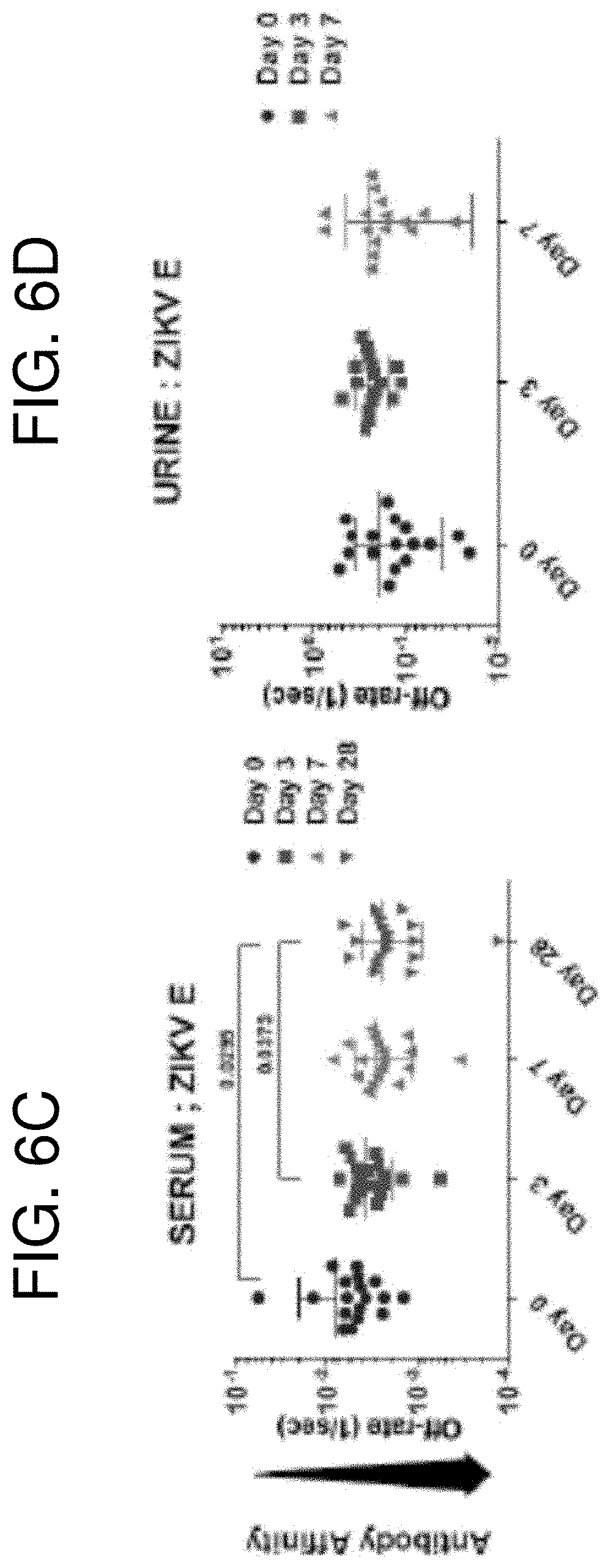
D00014

D00015

D00016

D00017

D00018

D00019

D00020

D00021

D00022

D00023

D00024

D00025

D00026

D00027
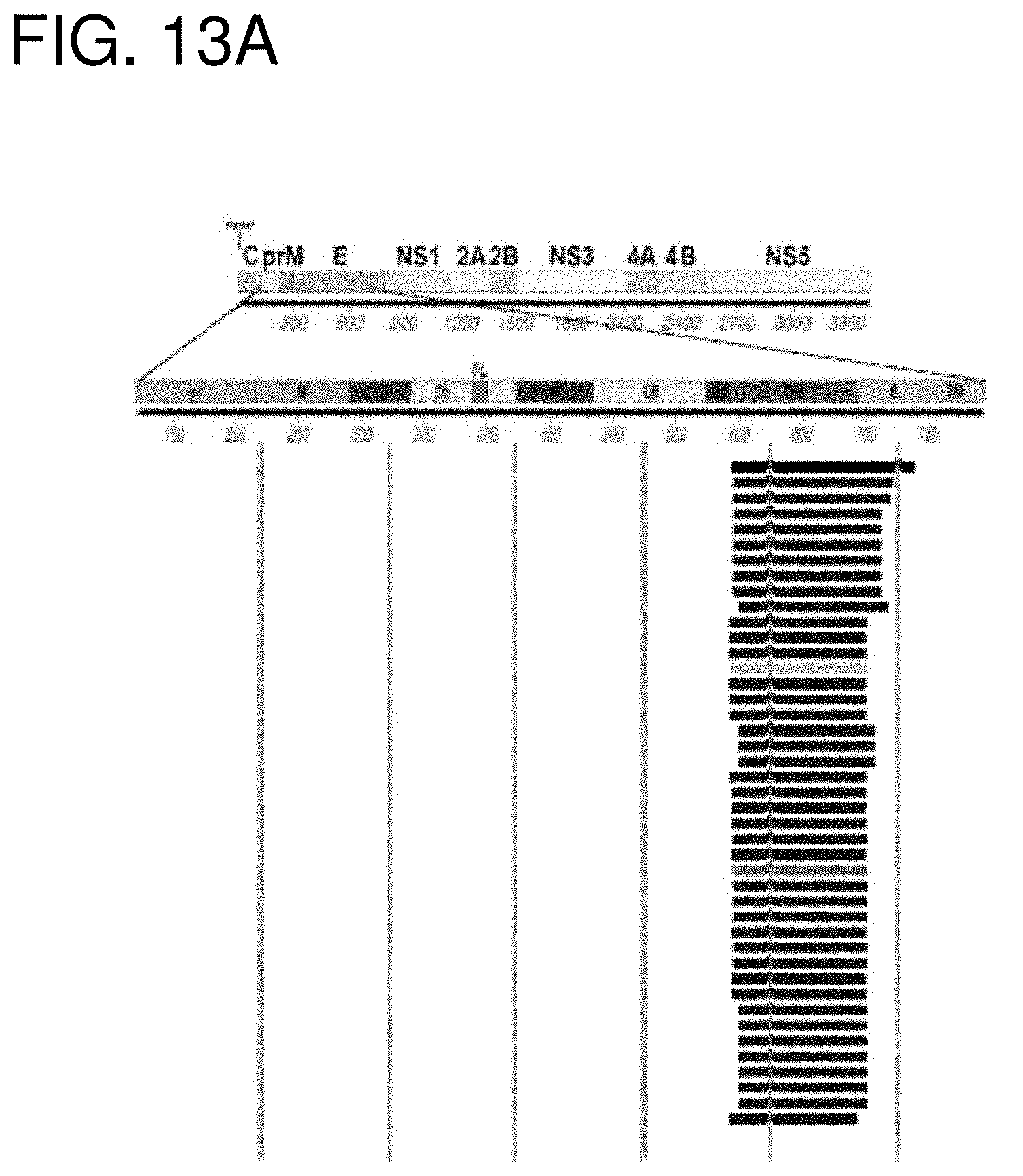
D00028

D00029

D00030

D00031

D00032

D00033

D00034

D00035

D00036

D00037

D00038

D00039
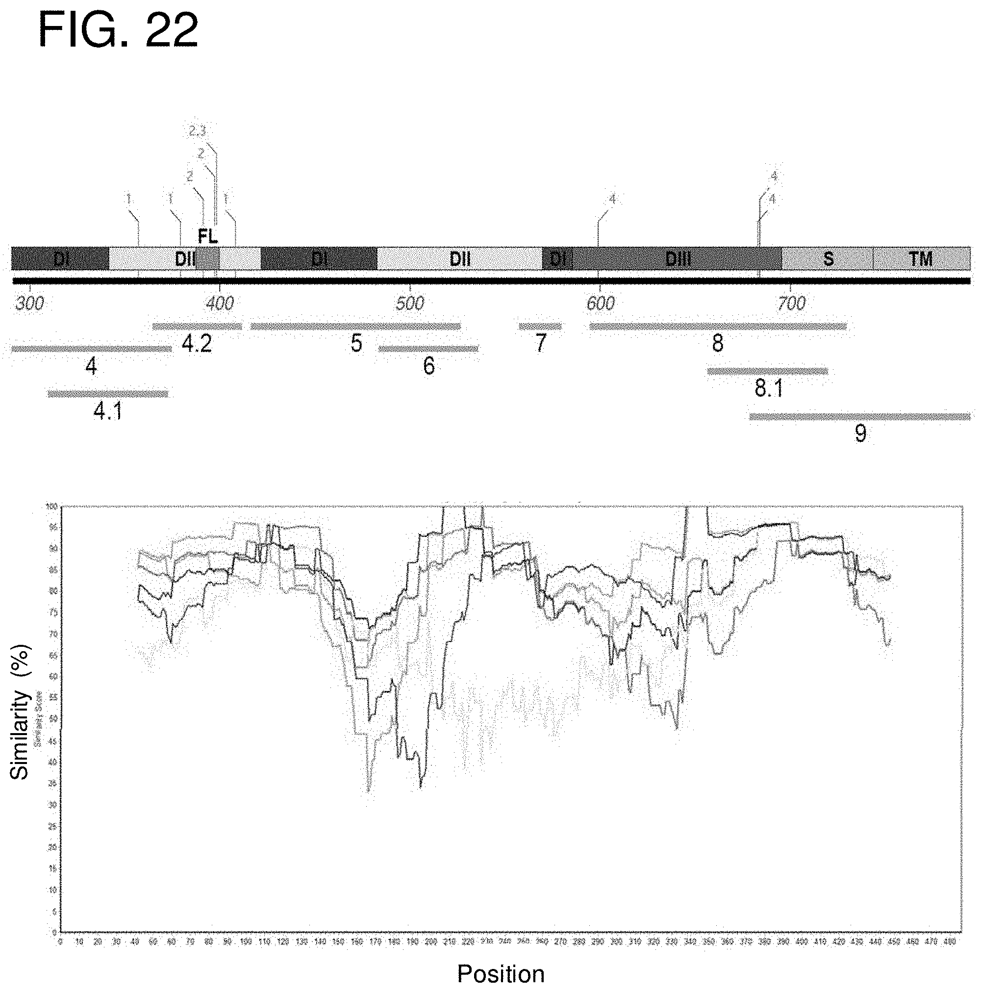
D00040

D00041
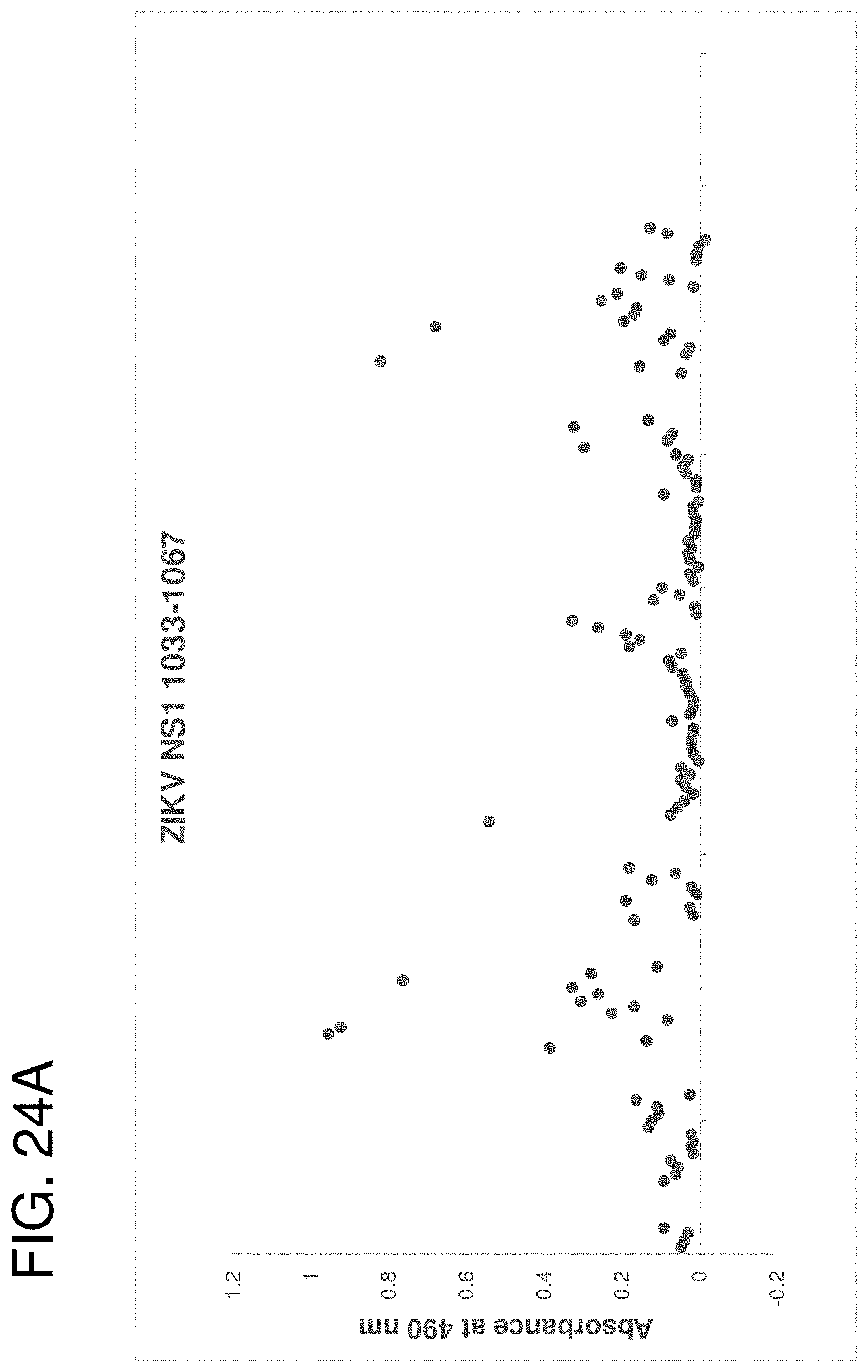
D00042

D00043

D00044

D00045

D00046

D00047

D00048

D00049
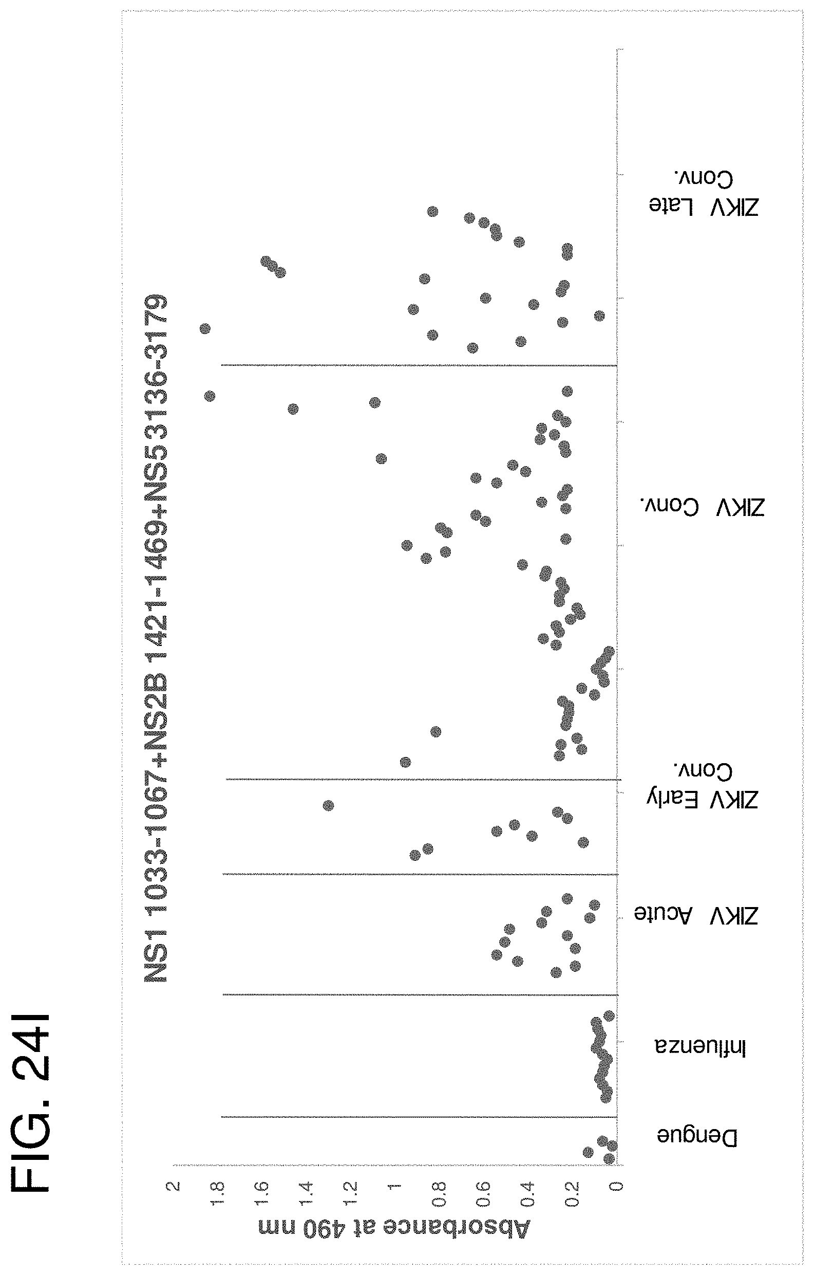
D00050

D00051

D00052

D00053

D00054

D00055

D00056

D00057

D00058

D00059

D00060

D00061

D00062

D00063
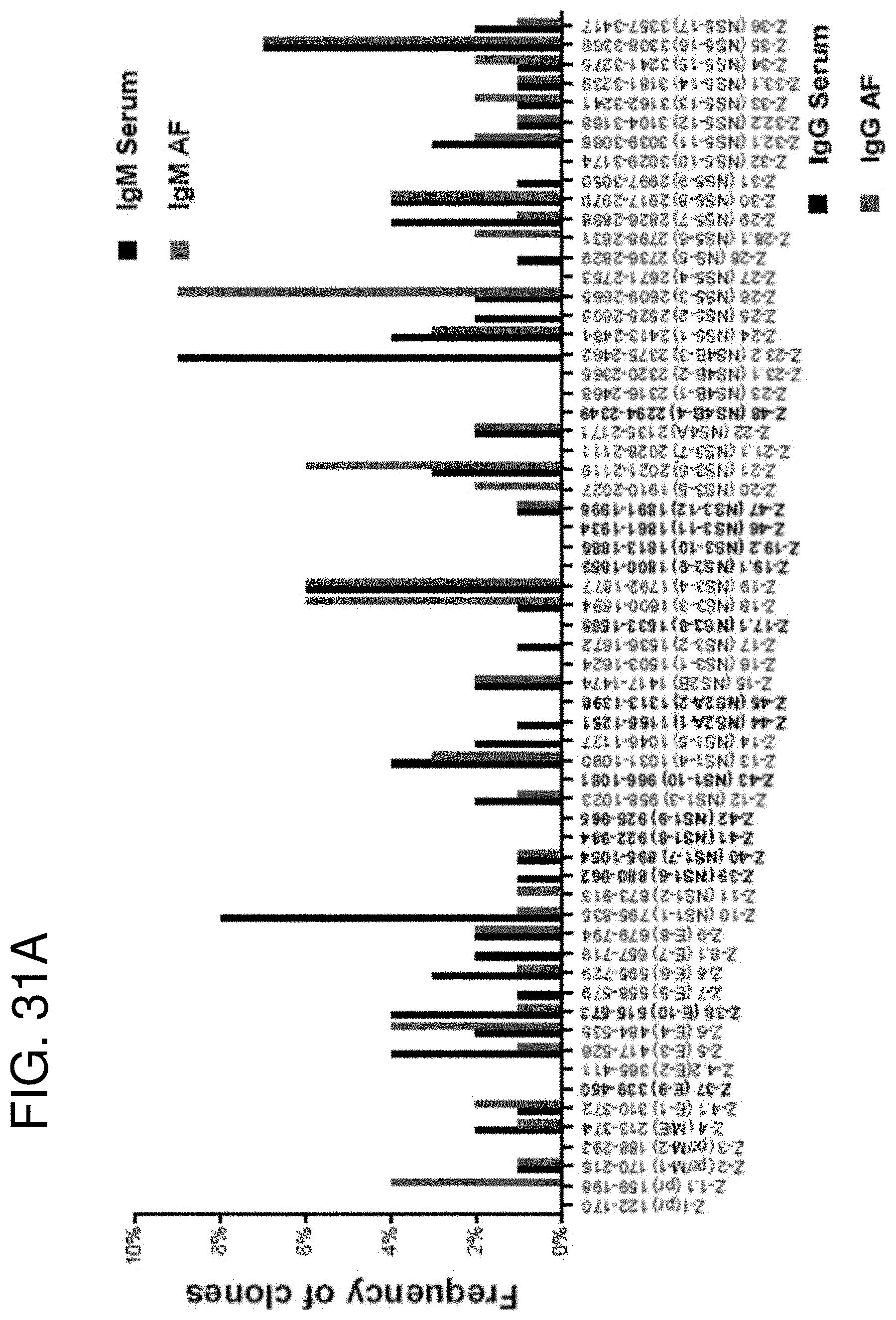
D00064

D00065

D00066

D00067

D00068

D00069

D00070

D00071
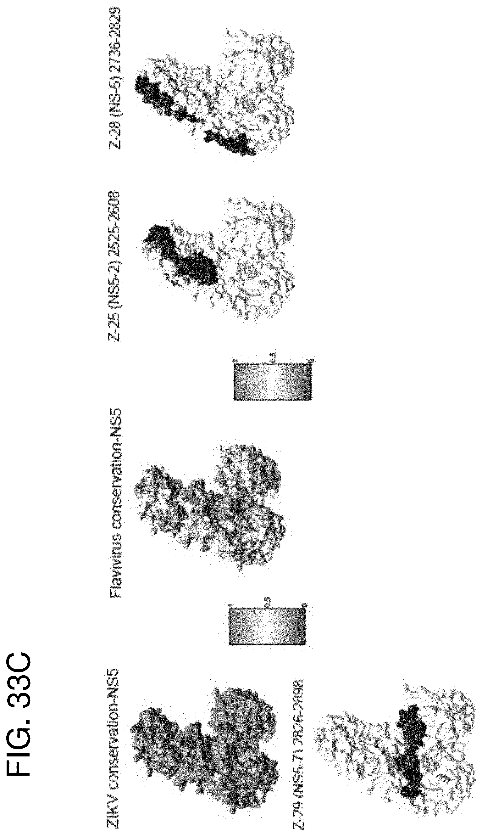
D00072

D00073

S00001
XML
uspto.report is an independent third-party trademark research tool that is not affiliated, endorsed, or sponsored by the United States Patent and Trademark Office (USPTO) or any other governmental organization. The information provided by uspto.report is based on publicly available data at the time of writing and is intended for informational purposes only.
While we strive to provide accurate and up-to-date information, we do not guarantee the accuracy, completeness, reliability, or suitability of the information displayed on this site. The use of this site is at your own risk. Any reliance you place on such information is therefore strictly at your own risk.
All official trademark data, including owner information, should be verified by visiting the official USPTO website at www.uspto.gov. This site is not intended to replace professional legal advice and should not be used as a substitute for consulting with a legal professional who is knowledgeable about trademark law.