Body Composition Assessment Using Two-dimensional Digital Image Analysis
Fedewa; Michael Vernon ; et al.
U.S. patent application number 16/841944 was filed with the patent office on 2020-11-05 for body composition assessment using two-dimensional digital image analysis. The applicant listed for this patent is THE BOARD OF TRUSTEES OF THE UNIVERSITY OF ALABAMA. Invention is credited to Michael Randall Esco, Michael Vernon Fedewa.
| Application Number | 20200345314 16/841944 |
| Document ID | / |
| Family ID | 1000004782023 |
| Filed Date | 2020-11-05 |


| United States Patent Application | 20200345314 |
| Kind Code | A1 |
| Fedewa; Michael Vernon ; et al. | November 5, 2020 |
BODY COMPOSITION ASSESSMENT USING TWO-DIMENSIONAL DIGITAL IMAGE ANALYSIS
Abstract
Methods and systems are provided for measuring anatomical dimensions from a single two-dimensional (2D) digital image. The digital image is taken from the front/anterior view using a mobile, handheld communication device. The linear measurements are used to estimate the body volume of the individual. Total body density is calculated from estimated body volume and body weight. Body composition (fat mass and fat-free mass) of the individual is derived from density using known mathematical conversion formulas. A method for estimating body composition analysis is provided.
| Inventors: | Fedewa; Michael Vernon; (Northport, AL) ; Esco; Michael Randall; (Northport, AL) | ||||||||||
| Applicant: |
|
||||||||||
|---|---|---|---|---|---|---|---|---|---|---|---|
| Family ID: | 1000004782023 | ||||||||||
| Appl. No.: | 16/841944 | ||||||||||
| Filed: | April 7, 2020 |
Related U.S. Patent Documents
| Application Number | Filing Date | Patent Number | ||
|---|---|---|---|---|
| 62842826 | May 3, 2019 | |||
| 62984016 | Mar 2, 2020 | |||
| Current U.S. Class: | 1/1 |
| Current CPC Class: | A61B 5/6898 20130101; A61B 2576/00 20130101; A61B 5/0537 20130101; A61B 5/7278 20130101; A61B 5/4875 20130101; A61B 5/1075 20130101; A61B 5/1079 20130101; A61B 5/0077 20130101; A61B 5/4872 20130101; A61B 5/1073 20130101 |
| International Class: | A61B 5/00 20060101 A61B005/00; A61B 5/107 20060101 A61B005/107; A61B 5/053 20060101 A61B005/053 |
Claims
1. A method of deriving body composition of an individual, the method comprising: obtaining an image of the individual; measuring a cross-sectional diameter (linear distance) at a plurality of anatomical landmarks on the image of the individual; estimating a body volume of the individual; and estimating a body composition of the individual using the estimated body volume.
2. The method of claim 1, wherein obtaining the image comprises at least one of: capturing a digital image of the individual, capturing a profile image from the front/anterior angle or the rear/posterior angle. capturing the image of the individual using a mobile, handheld communication device, or receiving the image from a storage device, an image capture device, or a camera.
3. The method of claim 1, wherein the plurality of anatomical landmarks comprises at least one of the hips, the waist, or the shoulders, and wherein measuring the cross-sectional diameter (linear distance) at the plurality of anatomical landmarks comprises at least one of using an automated image analysis program, or adjusting for standing height.
4. The method of claim 1, wherein estimating the body volume of the individual comprises at least one of using a mathematical equation, or using known anatomical ratio relationships.
5. The method of claim 1, wherein the image is a two-dimensional (2D) digital image, and wherein estimating the body volume of the individual comprises measuring the cross-sectional diameter (linear distance) at the series of anatomical landmarks and standing height from the 2D digital image, and accounting for age, sex, race/ethnicity, weight, and height.
6. The method of claim 1, wherein estimating the body composition comprises at least one of: estimating fat mass and fat-free mass, estimating the body density of the individual from the estimated body volume and a weight of the individual, estimating relative adiposity (percentage fat mass, percentage fat) using a relation that directly relates percentage fat to body density, or estimating the body composition of the individual comprises estimating body volume from the age, sex, race/ethnicity, height, weight, and digital image measurements, and then calculating body composition, wherein the digital image measurements comprise adjusted hips, waist, and shoulders diameters.
7. The method of claim 1, wherein obtaining the image comprises capturing a two-dimensional image of the individual from a front view or from a rear view.
8. The method of claim 1, further comprising: obtaining a body mass of the individual; and determining a body density of the individual.
9. The method of claim 8, further comprising converting the body density to a percentage of body fat.
10. The method of claim 1, further comprising: determining a body weight of the individual; determining total body water of the individual using bioelectrical impedance analysis (BIA); and estimating the body fat of the individual using the estimated body volume, the body weight, and the total body water.
11. A system of deriving body composition of an individual, the system comprising: an image capture device; and a computing device configured to receive images of the individual, analyze the linear distance related to anatomical landmarks from the digital images to obtain linear measurements, estimate the body volume of the individual using the linear measurements, and estimate body composition of the individual based at least in part upon the estimated volume.
12. The system of claim 11, wherein the linear distance is in units of pixels.
13. The system of claim 11, wherein the image capture device is comprised within a mobile, handheld communication device, wherein the mobile, handheld communication device on one side has a screen capable of displaying a front/anterior image of the individual being viewed with a camera or the image capture device on an opposite side.
14. The system of claim 11, wherein the system is embodied by the image capture device that further compromises image capturing apparatus, or wherein the system is embodied by a mobile, handheld communication device.
15. The system of claim 11, wherein the computing device comprises a measurement module configured to estimate the body volume of the individual by measuring the cross-sectional diameter (linear distance) at a plurality of anatomical landmarks and standing height from the 2D digital image, and accounting for age, sex, race/ethnicity, weight, and height, wherein the plurality of anatomical landmarks comprises the hip, the waist, and the shoulder.
16. The system of claim 15, wherein the computing device further comprises a body composition assessment module configured to estimate the body composition of the individual by estimating the density of the individual from the estimated volume and a body weight of the individual.
17. The system of claim 16, wherein the body composition assessment module is further configured to estimate the body composition of the individual by calculating the body fat percentage of the individual using a relation that directly relates body fat percentage to body density.
18. A system comprising: an image capture apparatus to receive an image of an individual; a landmark identifier module; a measurement module to estimate the body volume of the individual by measuring the cross-sectional diameter (linear distance) at a plurality of anatomical landmarks; and a body composition assessment module to estimate the body composition of the individual based in part upon the estimated volume.
19. The system of claim 18, wherein the image capture apparatus comprises a mobile, handheld communication device, which on one side has a screen capable of displaying a front/anterior image of the individual being viewed with a camera or image capture device on an opposite side.
20. The system of claim 18, further comprising: a memory that stores the landmark identifier module, the measurement module, and the body composition assessment module; and an output device that outputs the estimated body composition of the individual.
Description
CROSS-REFERENCE TO RELATED APPLICATIONS
[0001] This application claims the benefit of priority to U.S. Provisional Patent Application No. 62/842,826, filed on May 3, 2019, entitled "BODY COMPOSITION ASSESSMENT USING TWO-DIMENSIONAL DIGITAL IMAGE ANALYSIS," and to U.S. Provisional Patent Application No. 62/984,016, filed on Mar. 2, 2020, entitled "BODY COMPOSITION ASSESSMENT USING TWO-DIMENSIONAL DIGITAL IMAGE ANALYSIS," the contents of which are hereby incorporated by reference in their entireties.
BACKGROUND
[0002] Measurement of body composition (fat mass and fat-free mass) can be obtained using highly accurate techniques such as dual energy x-ray absorptiometry (DXA), air displacement plethysmography (ADP), or underwater weighing (UWW). However, these methods are not widely used in field settings due, in part, to their inability to be transported, as well as the cost and size of the equipment used. In addition, many of these methods cannot accommodate the dimensions of taller and heavier individuals that may exceed the scanning area, are too large for the measurement chamber, or cannot completely submerge underwater. Furthermore, individuals who may become claustrophobic or are unable to safely access the equipment because of mobility limitations may also experience difficulties when using the previously mentioned techniques. Moreover, DXA is often used in clinical settings, but exposes the technician and participants to radiation, which may limit the use of DXA to licensed or certified technicians.
[0003] When more technologically advanced methods are unavailable in field settings, anthropometric measurements of body size can be performed, such as body mass index (BMI), waist circumference (WC) or hip circumference (HC), and skinfold thickness. Although calculation of BMI is a simple method commonly used in large-scale epidemiological research, BMI is limited in value because it is an assessment of body weight relative to height and not of body composition per se. Furthermore, the accuracy of these methods (i.e., WC, HC, and skinfold thickness measurements) may depend heavily upon repeated training of the research staff in order to obtain accurate and reliable assessments. Further limiting these anthropometric indices, the relationship between each of these measures and body composition varies tremendously by age, sex, and race/ethnicity. Additionally, these anthropometric measures (BMI, WC, and HC) are only moderately correlated with adiposity, and are weakly correlated with changes in body composition over time. As such, improved methods that allow for accurate and portable measurement of body composition could be of tremendous value for practitioners in sport performance, commercial fitness, or allied health care fields.
[0004] Most recently, three-dimensional body scanning systems have been developed and provide accurate estimates of body volume; however, the cost of these devices and their inability to be transported also limit their use for many practical settings and field assessments. There is currently no smartphone/tablet application that can estimate body composition from a single two-dimensional image.
SUMMARY
[0005] Body composition is estimated from a single two-dimensional (2D) image. In an embodiment, anatomical dimensions are measured, in the form of linear measurements, from a single 2D image that is taken from the front/anterior view or from the rear/posterior view. The linear measurements are used to estimate the body volume of the individual. Total body density is calculated from estimated body volume and body weight (more precisely, body mass). Body composition (fat mass and fat-free mass) of the individual is derived from the body density using known mathematical conversion formulas. According to some implementations, the image is a digital image and the digital image is obtained using a mobile, handheld communication device.
[0006] An implementation comprises a method of deriving body composition of an individual, the method comprising: obtaining an image of the individual; measuring a cross-sectional diameter (linear distance) at a plurality of anatomical landmarks on the image of the individual; estimating a body volume of the individual; and estimating a body composition of the individual using the estimated body volume.
[0007] An implementation comprises a system of deriving body composition of an individual, the system comprising: an image capture device; and a computing device configured to receive images of the individual, analyze the linear distance related to anatomical landmarks from the digital images to obtain linear measurements, estimate the body volume of the individual using the linear measurements, and estimate body composition of the individual based at least in part upon the estimated volume.
[0008] An implementation comprises a system that comprises: an image capture apparatus to receive an image of an individual; a landmark identifier module; a measurement module to estimate the body volume of the individual by measuring the cross-sectional diameter (linear distance) at a plurality of anatomical landmarks; and a body composition assessment module to estimate the body composition of the individual based in part upon the estimated volume.
[0009] This summary is provided to introduce a selection of concepts in a simplified form that are further described below in the detailed description. This summary is not intended to identify key features or essential features of the claimed subject matter, nor is it intended to be used to limit the scope of the claimed subject matter.
BRIEF DESCRIPTION OF THE DRAWINGS
[0010] The foregoing summary, as well as the following detailed description of illustrative embodiments, is better understood when read in conjunction with the appended drawings. For the purpose of illustrating the embodiments, there is shown in the drawings example constructions of the embodiments; however, the embodiments are not limited to the specific methods and instrumentalities disclosed. In the drawings:
[0011] FIG. 1 is an illustration of an exemplary environment for body composition assessment using two-dimensional (2D) digital image analysis;
[0012] FIG. 2 is an illustration of example image of a subject obtained using dual energy x-ray absorptiometry (DXA) and an associated 2D digital image for body composition assessment using a camera according to an implementation of the invention;
[0013] FIG. 3 is an illustration of example images obtained using a DXA machine;
[0014] FIG. 4 is an operational flow of an implementation of a method for body composition assessment using 2D digital analysis; and
[0015] FIG. 5 shows an exemplary computing environment in which example embodiments and aspects may be implemented.
DETAILED DESCRIPTION
[0016] The description is not to be taken in a limiting sense, but is made merely for the purpose of illustrating the general principles of the invention, since the scope of the invention is best defined by the appended claims.
[0017] Various inventive features are described herein that can each be used independently of one another or in combination with other features.
[0018] FIG. 1 is an illustration of an exemplary environment 100 for body composition assessment using two-dimensional (2D) digital image analysis. As described further herein, a single digital two-dimensional image of an individual 102 is used. In some implementations, the 2D image is taken from the anterior (front) view. In other implementations, the 2D image may be taken from the posterior (rear) view. The individual 102 may take the 2D image using a camera 105, or another user or administrator may take the 2D image of the individual 102. The body volume of the individual 102 is estimated by measuring the diameter of one or more anatomical landmarks from the single 2D image. In some implementations, the body volume of the individual 102 is estimated by measuring the diameter of a plurality (e.g., two, three, etc.) anatomical landmarks from the single 2D image, such as the shoulders, waist, and/or hips, although the invention is not limited thereto.
[0019] The environment 100 may include the camera 105, a computing device 110, and an output device 175 in communication with each other through a network. The network may be a variety of network types including the public switched telephone network (PSTN), a cellular telephone network, and a packet switched network (e.g., the Internet). Alternately, the camera 105 and/or the output device 175 may be comprised within the computing device 110. Although only one camera 105, one computing device 110, and one output device 175 are shown in FIG. 1, there is no limit to the number of cameras 105, computing devices 110, and output devices 175 that may be supported.
[0020] The camera 105 may comprise, or be embodied in, any image capturing device, system, or apparatus. The output device 175 may comprise any device, system, or apparatus for providing a result (e.g., a body composition assessment described further herein) received from the computing device 110 to a user, a storage device, a display, etc.
[0021] The computing device 110 may be implemented using a variety of computing devices such as handheld communication devices, smart phones, desktop computers, laptop computers, tablets, and video game consoles. Other types of computing devices may be supported. A suitable computing device is illustrated in FIG. 5 as the computing device 500.
[0022] The computing device 110 may include a landmark identifier 120, a measurement module 130, and a body composition assessment module 140.
[0023] The landmark identifier 120 receives a two-dimensional (2D) image of an individual 102 (e.g., taken and/or provided by the camera 105) and identifies one or a plurality of anatomical landmarks of the image. The anatomical landmarks may include hips, waist, overarms, shoulders, neck, knees, etc. Although the embodiments described herein are directed to identifying and using a plurality of landmarks, such as three anatomical landmarks (e.g., hips, waist, shoulder), the invention is not limited thereto, and any number of anatomical landmarks, as well as any anatomical landmarks, are contemplated. As the number of anatomical landmarks used is increased, the assessments herein may become more accurate. It is contemplated that the hips, waist, and shoulders are straightforward to accurately identify and measure in a 2D image.
[0024] The measurement module 130 receives the identified anatomical landmark information from the landmark identifier 120 and measures the linear distance of the diameter of the individual at these anatomical landmarks. The linear distance may be in inches, centimeters, or pixels, depending on the implementation, although any unit(s) of linear distance measurement may be used. The linear distances determined by the measurement module 130 are then used to estimate the body volume of the individual 102, as described further herein.
[0025] The estimated body volume from the measurement module 130 is provided to the body composition assessment module 140. The body composition assessment module 140 uses the estimated body volume (as well as the body mass in some embodiments) to determine a body composition assessment (e.g., using body volume in some embodiments) as described further herein. The body composition assessment is then provided to the output device 175 for output to a user, to storage, to a display, etc.
[0026] In an implementation, the computing device 110 is provided for measuring the anatomical dimensions of the human body. The computing device 110 may be a mobile device, and/or configured to provide a digital anthropometer on a mobile device, and to digitize anatomical landmarks on a displayed image such as a photograph (such as a single 2D digital image) of the human body displayed on the computing device 110 (or the camera 105) with established calibration methods for measuring dimensions of the human body.
[0027] The measurement module 130 may be configured to measure the dimensions of the human body and may comprise a programmed device including a digital display, an automated image analysis program to measure an array of pixels and a camera (such as the camera 105) for acquiring an image of a person (e.g., the individual 102) on the digital display, and means for digitizing anatomical landmarks on an image of the person on the display for measuring dimensions of the human body.
[0028] The measurement module 130 enables the ability to derive the anatomical measurement such as the linear measurement from the front/anterior aspect of the body (or the rear/posterior aspect of the body), and to then calculate an estimate of total body volume using mathematical equations. The calculated body volume is converted by the body composition assessment module 140, to body composition (fat mass and fat-free mass) using known mathematical conversion equations.
[0029] The body composition assessment module 140 uses a prediction model which can estimate total body volume with near perfect precision across a wide range of body sizes, which is then converted to body fat percentage, fat mass, and fat-free mass after accounting for the total mass/body weight of the individual. The methodology is strongly correlated with the criterion measure of total body volume as measured by UWW (r=0.999), as well as three known other methods of estimating total body volume using DXA (e.g., r=0.999, 0.998, and 0.997), for example.
[0030] Once an image is obtained and digitized by the camera 105 and/or the computing device 110, digitization points on anatomical landmarks for purpose of body composition measurements can be performed by the landmark identifier 120. The invention may be utilized in commercial fitness, athletic performance, and allied health care settings to measure body dimensions, shape, and body composition based on anatomical ratio relationship, and to track progress of these measurements over time. The anthropometric measurements can be acquired from a single digital image of the individual 102.
[0031] FIG. 2 is an illustration 200 of an example image 210 of a subject obtained using dual energy x-ray absorptiometry (DXA) and an associated 2D digital image 250 for body composition assessment using a camera according to an implementation of the invention. Thus, while DXA techniques use a specialized image 210 of the subject, the techniques of the invention described herein can use a photo of the subject taken from an anterior (front) view or a posterior (rear) view, such as a 2D digital image 250 taken with a camera such as a camera integrated with a handheld computing device like a phone or a tablet.
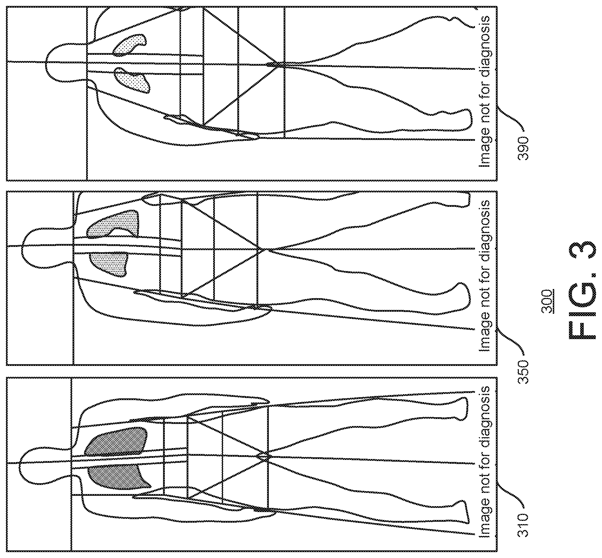
[0032] FIG. 3 is an illustration 300 of example images 310, 350, 390 obtained using a DXA machine. The images 310, 350, 390 correspond to subjects whose body composition assessment cannot be performed by a DXA technique because of the body size. For example, the image 310 corresponds to a subject whose body is too tall for DXA techniques, and image 350 corresponds to a subject whose body is too muscular and wide for DXA techniques, and the image 390 corresponds to a subject whose body is too large for DXA techniques. The techniques of the invention described herein, however, can be used for all body sizes and shapes, including those of the subjects having the images 310, 350, 390.
[0033] In some implementations, patient information (age, sex, race/ethnicity) as well as body weight (i.e., mass) and height are obtained and used for normalizing the individual's height in the digital image, and determining a pixel to distance ratio using the acquired patient information and the normalized patient height.
[0034] Thus, in some implementations, a front/anterior perspective image of the individual is obtained using a mobile, handheld communication device, which on one side has a screen capable of displaying a front/anterior image of the individual 102 being viewed with a camera or image capture device on an opposite side. Mobile, handheld communication devices capable of running a program in accordance with the invention could be used, such as iPhone.RTM., iPod Touch.RTM., iPad.RTM. and Android.RTM. devices including tablets and Windows.RTM. based tablets.
[0035] A method of estimating body composition from a single 2-dimensional digital image is provided using a series of anatomical landmarks with near perfect accuracy (r=0.999) when compared to conventional methods such as underwater weighing (UWW). FIG. 4 is an operational flow 400 of an implementation of a method for body composition assessment using 2D digital analysis. The method 400 may be implemented using the computing device 110, alone or in conjunction with the camera 105 and/or the output device 175.
[0036] At 410, a digital two-dimensional image of an individual, such as the individual 102, is obtained, e.g., by the camera 105 and/or the computing device 110. In some implementations, the image is taken from the anterior (front) view, while in other implementations, the image is taken from the posterior (rear) view. In some implementations, the digital image used is taken from the front/anterior view or the rear/posterior view using a mobile, handheld communication device.
[0037] At 420, anatomical landmarks are identified in the 2D digital image, e.g., by the landmark identifier 120. The anatomical landmarks may comprise shoulders, waist, and/or hips in some implementations, although any anatomical landmarks may be used and although any number of anatomical landmarks may be used. In some implementations, a digital anthropometer on a mobile device is used to identify anatomical landmarks on the digital image.
[0038] At 430, the diameter of each of the anatomical landmarks is measured and the body volume is estimated, e.g., by the measurement module 130. Linear measurements may be obtained in some implementations, and then used to estimate the body volume of the individual. In some implementations, the diameters (i.e., the linear measurement across the individual's body in the image at the landmarks) are converted to ratios by dividing those diameters by the individual's height. These ratios may then be used in an equation to obtain the body volume estimate. An example equation using shoulders, waist, and hips is given by Equation (1):
Body Volume (Liters)=Hip Diameter.sub.Adjusted+Waist Diameter.sub.Adjusted+Shoulder Diameter.sub.Adjusted+Body Mass.sub.kg+Height.sub.cm. (1)
It is noted that one or more of the values of this equation, as well as the equations below, may be adjusted with a "slope" or variable, depending on the implementation, which may weight or otherwise adjust the particular value. In this manner, the parameters of the equation (e.g., hip diameter, waist diameter, shoulder diameter, body mass, and height) may be accounted for and their units standardized or normalized.
[0039] Another example equation is given by Equation (2):
Body Volume ( Liters ) = ( H e i g h t c m ( Image Hip Diameter Image Height ) ) + ( H e i g h t c m ( Image Waist Diameter Image Height ) ) + ( H e i g h t c m ( Image Shoulder Diameter Image Height ) ) + Body Mass k g + Hei g h t c m ( 2 ) ##EQU00001##
[0040] Another example equation is given by Equation (3):
Body Volume ( Liters ) = ( A .times. ( H e i g h t c m .times. ( Image Hip Diameter Image Height ) ) ) + ( B .times. ( H e i g h t c m .times. ( Image Waist Diameter Image Height ) ) ) + ( C .times. ( H e i g h t c m .times. ( Image Shoulder Diameter Image Height ) ) ) + ( D .times. ( Body Mass k g ) ) + ( E .times. ( H e i g h t c m ) ) + F . ( 3 ) ##EQU00002##
In Equation (3), variables A, B, C, D, and E, are included as "slopes," and F is included as an "intercept," which may be determined and adjusted as desired to tune or create a final outcome of the body volume. The values for variables A, B, C, D, E, and F may be determined experimentally and/or using numerical and/or mathematical techniques such as multivariate linear regression.
[0041] At 440, the body composition assessment is performed using the estimated body volume. The body composition assessment may be performed by the body composition assessment module 140. In some embodiments, total body density is calculated from estimated body volume and body weight. Body composition (fat mass and fat-free mass) of the individual is then derived from density using known mathematical conversion formulas, such as the Sin equation which converts body density (BD) in percent body fat (% BF). Other conversion formulas and techniques can be used. In some embodiments, measurements are taken from the image concerning the torso and hips, as well as the body weight, to assess body composition.
[0042] At 450, the body composition assessment is outputted. The computing device 110 may output the body composition to an associated output device, such as the output device 175.
[0043] In this manner, the systems, methods, and techniques described herein are cost effective, can accommodate dimensions of taller and/or heavier individual, and do not rely on trained personnel or staff.
[0044] In some implementations, a programmed device is provided that includes an automated analysis of arrays of pixels, a camera for acquiring a digital image of a person, and means for automatically identifying a series of anatomical landmarks on an image of an individual. The number of pixels is determined using an automated program to measure the linear distance at a series of anatomical landmarks, and the prediction equation includes measurements of the hip, waist, and overarm diameter, standardized to the height of the individual.
[0045] In some implementations, the present invention includes a means of acquiring an image of a person, a method of deriving body volume and body composition estimates from a programmed smartphone application which uses an automatic image analysis program, digitizing points on a plurality of anatomical landmarks on the displayed image, determining linear anatomical dimensions of the person's body using the digitized points and a scale factor for the displayed image, and making an anatomical prediction using the determined linear anatomical dimensions.
[0046] Some implementations comprise acquiring a single image of the person obtained from the front/anterior view, digitizing points on anatomical landmarks on each displayed image and determining linear anatomical dimensions of the person's body (in pixels) using the digitized points and a scale factor (adjusted to the person's height in pixels) for each displayed image for making the anatomical prediction.
[0047] In some implementations, a body volume and body composition assessment method comprises acquiring an image of an individual on a display screen having an array of pixels, determining a pixel to distance ratio for the displayed image, and calculating the body composition of the individual using the cross-sectional diameter of the individual at a series of anatomical landmarks (hips, waist, and shoulders). A known linear distance in the displayed image and the number of display screen pixels spanning the distance are used in determining pixel to distance ratio. The known linear distance used in the estimation equation is the height of the individual.
[0048] The body composition assessment method in example images further includes scaling the size of the image relative to the display screen to normalize the known linear distance in the image to a display screen reference distance corresponding to a known number of pixels for determining the pixel to distance ratio.
[0049] The person performing the screening can operate an image capture device of a camera for acquiring the image of the patient. A single two-dimensional camera providing a 2D image can be used. The method preferably includes leveling the image capture device before capturing the image from which the pixel to distance ratio is to be determined for eliminating distortion. The image capture device and display screen are part of a mobile, handheld communication device.
[0050] The development of a smartphone/tablet application that can accurately measure body composition from a single 2D image would have tremendous appeal for practitioners in the allied health field, in research settings, and in recreationally active individuals to provide a single estimate for diagnostic purposes, and to monitor changes in body composition over time. In addition, the utility of a portable body composition assessment method would provide tremendous value for practitioners or clinicians in rural health community health outreach programs when access to more technologically advanced body composition assessment methods are unavailable.
[0051] Embodiments contemplated herein, which provide an accurate method of assessing body composition, may be implemented via a smartphone/tablet application. Various embodiments and implementations provide portable, accurate, non-invasive body composition measurement available for use in allied health and clinical settings, the commercial fitness industry, and fitness enthusiasts, for example, though it is not limited thereto. There is potential for the embodiments and implementations in the health and fitness industry. Commercial fitness centers need accurate methods of body composition assessment of their patrons. In addition, the embodiments and implementations would be of interest to fitness enthusiasts interested in measuring their own body composition and tracking changes due to their exercise training program. Embodiments and implementations accurately measure changes in weight and body composition through the least obtrusive means possible. Monitoring changes in body composition through an application would be a desired addition by users that may already use a health and fitness application.
[0052] The methodology may be limited, as are the UWW and BodPod techniques, because these methods do not account for total body water or bone mineral content. As a result, body composition can only be separated in to two components, fat mass and fat-free mass. Because bone mass does not change acutely as a result of exercise training, and because this method will not be used as a diagnostic or screening tool for osteoporosis or low bone mass, accounting for bone mass would be of little value for the embodiments and implementations contemplated herein. In addition, although this methodology (as well as UWW and BodPod) does not account for total body water, this is also of little value to for the embodiments and implementations contemplated herein. An additional limitation of the methodology is that the image is taken of the front of the body with hips and shoulders parallel to the camera, with the camera held directly at hip, eye, or chest level to allow for identification of all of the anatomical points of interest (as shown in FIGS. 2 and 3 for example). An image taken with an individual wearing loose-fitting clothing, or taken at an angle may distort the perspective of the image and add potential error to the estimate of body composition.
[0053] An accurate method of assessing body fat is with a system called a multi-compartment model. Basically, the more "compartments" (or tissues) of the body that are measured, the better the estimate of fat will be. A common multi-compartment approach is to measure the following compartments: body volume, total body water, and body weight. Once each of these compartments are measured, the values are entered into an existing regression/prediction equation that provides the outcome measure of fat mass.
[0054] Body weight is easily measured with a standard scale. In the lab, body volume is measured with sophisticated methods of either underwater weighing or x-ray. However, the smartphone applications (apps) described or otherwise contemplated or provided herein estimate body volume, and provide nearly perfect agreement compared to the other laboratory methods.
[0055] Total body water is not measured with the app(s), but instead can be done with a process called bioelectrical impedance analysis (BIA) or bioelectrical impedance spectroscopy (BIS). Scales in retail stores that supposedly measure body fat percentage are not very accurate, but use the BIA method. It is a process of sending an unnoticeable electrical current(s) through the body from one "pole" to another. In the case of the scales, the feet serve as the poles. With more advanced methods, the poles are surface sticky electrodes that are placed over the hand(s) and foot (feet). The measures are performed while the person lays on their back, face up.
[0056] In some implementations, the smartphone application(s) can be used with bioelectrical impedance (BIA or BIS) and a weighing scale to provide a multi-compartment model for measuring body fat. This process is more user friendly than putting someone in a tank of water to measure body volume, and then performing the other measures of bioimpedance and body weighing.
[0057] In some implementations, the process uses the smartphone app(s) plus (any) bioelectrical impedance analyzer and (any) weighing scale for a multi-compartment modeling method of fat determination.
[0058] In some implementations, a system for measuring total body water can be coupled with a process of body volume from an image.
[0059] FIG. 5 shows an exemplary computing environment in which example embodiments and aspects may be implemented. The computing device environment is only one example of a suitable computing environment and is not intended to suggest any limitation as to the scope of use or functionality.
[0060] Numerous other general purpose or special purpose computing devices environments or configurations may be used. Examples of well-known computing devices, environments, and/or configurations that may be suitable for use include, but are not limited to, personal computers, server computers, handheld or laptop devices, multiprocessor systems, microprocessor-based systems, network personal computers (PCs), minicomputers, mainframe computers, embedded systems, distributed computing environments that include any of the above systems or devices, and the like.
[0061] Computer-executable instructions, such as program modules, being executed by a computer may be used. Generally, program modules include routines, programs, objects, components, data structures, etc. that perform particular tasks or implement particular abstract data types. Distributed computing environments may be used where tasks are performed by remote processing devices that are linked through a communications network or other data transmission medium. In a distributed computing environment, program modules and other data may be located in both local and remote computer storage media including memory storage devices.
[0062] With reference to FIG. 5, an exemplary system for implementing aspects described herein includes a computing device, such as computing device 500. In its most basic configuration, computing device 500 typically includes at least one processing unit 502 and memory 504. Depending on the exact configuration and type of computing device, memory 504 may be volatile (such as random access memory (RAM)), non-volatile (such as read-only memory (ROM), flash memory, etc.), or some combination of the two. This most basic configuration is illustrated in FIG. 5 by dashed line 506.
[0063] Computing device 500 may have additional features/functionality. For example, computing device 500 may include additional storage (removable and/or non-removable) including, but not limited to, magnetic or optical disks or tape. Such additional storage is illustrated in FIG. 5 by removable storage 508 and non-removable storage 510.
[0064] Computing device 500 typically includes a variety of computer readable media. Computer readable media can be any available media that can be accessed by the device 500 and includes both volatile and non-volatile media, removable and non-removable media.
[0065] Computer storage media include volatile and non-volatile, and removable and non-removable media implemented in any method or technology for storage of information such as computer readable instructions, data structures, program modules or other data. Memory 504, removable storage 508, and non-removable storage 510 are all examples of computer storage media. Computer storage media include, but are not limited to, RAM, ROM, electrically erasable program read-only memory (EEPROM), flash memory or other memory technology, CD-ROM, digital versatile disks (DVD) or other optical storage, magnetic cassettes, magnetic tape, magnetic disk storage or other magnetic storage devices, or any other medium which can be used to store the desired information and which can be accessed by computing device 500. Any such computer storage media may be part of computing device 500.
[0066] Computing device 500 may contain communication connection(s) 512 that allow the device to communicate with other devices. Computing device 500 may also have input device(s) 514 such as a keyboard, mouse, pen, voice input device, touch input device, etc. Output device(s) 516 such as a display, speakers, printer, etc. may also be included. All these devices are well known in the art and need not be discussed at length here.
[0067] In an implementation, a method of deriving body composition of an individual comprises: obtaining an image of the individual; measuring a cross-sectional diameter (linear distance) at a plurality of anatomical landmarks on the image of the individual; estimating a body volume of the individual; and estimating a body composition of the individual using the estimated body volume.
[0068] Implementations may include some or all of the following features. Obtaining the image comprises at least one of: capturing a digital image of the individual, capturing a profile image from the front/anterior angle or the rear/posterior angle, capturing the image of the individual using a mobile, handheld communication device, or receiving the image from a storage device, an image capture device, or a camera. The plurality of anatomical landmarks comprises at least one of the hips, the waist, or the shoulders, and wherein measuring the cross-sectional diameter (linear distance) at the plurality of anatomical landmarks comprises at least one of using an automated image analysis program, or adjusting for standing height. Estimating the body volume of the individual comprises at least one of using a mathematical equation, or using known anatomical ratio relationships. The image is a two-dimensional (2D) digital image, and wherein estimating the body volume of the individual comprises measuring the cross-sectional diameter (linear distance) at the series of anatomical landmarks and standing height from the 2D digital image, and accounting for age, sex, race/ethnicity, weight, and height. Estimating the body composition comprises at least one of: estimating fat mass and fat-free mass, estimating the body density of the individual from the estimated body volume and a weight of the individual, estimating relative adiposity (percentage fat mass, percentage fat) using a relation that directly relates percentage fat to body density, or estimating the body composition of the individual comprises estimating body volume from the age, sex, race/ethnicity, height, weight, and digital image measurements, and then calculating body composition, wherein the digital image measurements comprise adjusted hips, waist, and shoulders diameters. Obtaining the image comprises capturing a two-dimensional image of the individual from a front view or from a rear view. The method further comprises obtaining a body mass of the individual and determining a body density of the individual. The method further comprises converting the body density to a percentage of body fat. The method further comprises determining a body weight of the individual; determining total body water of the individual using bioelectrical impedance analysis (BIA); and estimating the body fat of the individual using the estimated body volume, the body weight, and the total body water.
[0069] In an implementation, a system of deriving body composition of an individual comprises: an image capture device; and a computing device configured to receive images of the individual, analyze the linear distance related to anatomical landmarks from the digital images to obtain linear measurements, estimate the body volume of the individual using the linear measurements, and estimate body composition of the individual based at least in part upon the estimated volume.
[0070] Implementations may include some or all of the following features. The linear distance is in units of pixels. The image capture device is comprised within a mobile, handheld communication device, wherein the mobile, handheld communication device on one side has a screen capable of displaying a front/anterior image of the individual being viewed with a camera or the image capture device on an opposite side. The system is embodied by the image capture device that further compromises image capturing apparatus, or wherein the system is embodied by a mobile, handheld communication device. The computing device comprises a measurement module configured to estimate the body volume of the individual by measuring the cross-sectional diameter (linear distance) at a plurality of anatomical landmarks and standing height from the 2D digital image, and accounting for age, sex, race/ethnicity, weight, and height, wherein the plurality of anatomical landmarks comprises the hip, the waist, and the shoulder. The computing device further comprises a body composition assessment module configured to estimate the body composition of the individual by estimating the density of the individual from the estimated volume and a body weight of the individual. The body composition assessment module is further configured to estimate the body composition of the individual by calculating the body fat percentage of the individual using a relation that directly relates body fat percentage to body density.
[0071] In an implementation, a system comprises: an image capture apparatus to receive an image of an individual; a landmark identifier module; a measurement module to estimate the body volume of the individual by measuring the cross-sectional diameter (linear distance) at a plurality of anatomical landmarks; and a body composition assessment module to estimate the body composition of the individual based in part upon the estimated volume.
[0072] Implementations may include some or all of the following features. The image capture apparatus comprises a mobile, handheld communication device, which on one side has a screen capable of displaying a front/anterior image of the individual being viewed with a camera or image capture device on an opposite side. The system further comprises a memory that stores the landmark identifier module, the measurement module, and the body composition assessment module; and an output device that outputs the estimated body composition of the individual.
[0073] It should be understood that the various techniques described herein may be implemented in connection with hardware components or software components or, where appropriate, with a combination of both. Illustrative types of hardware components that can be used include Field-programmable Gate Arrays (FPGAs), Application-specific Integrated Circuits (ASICs), Application-specific Standard Products (ASSPs), System-on-a-chip systems (SOCs), Complex Programmable Logic Devices (CPLDs), etc. The methods and apparatus of the presently disclosed subject matter, or certain aspects or portions thereof, may take the form of program code (i.e., instructions) embodied in tangible media, such as floppy diskettes, CD-ROMs, hard drives, or any other machine-readable storage medium where, when the program code is loaded into and executed by a machine, such as a computer, the machine becomes an apparatus for practicing the presently disclosed subject matter.
[0074] Although exemplary implementations may refer to utilizing aspects of the presently disclosed subject matter in the context of one or more stand-alone computer systems, the subject matter is not so limited, but rather may be implemented in connection with any computing environment, such as a network or distributed computing environment. Still further, aspects of the presently disclosed subject matter may be implemented in or across a plurality of processing chips or devices, and storage may similarly be effected across a plurality of devices. Such devices might include personal computers, network servers, and handheld devices, for example.
[0075] Although the subject matter has been described in language specific to structural features and/or methodological acts, it is to be understood that the subject matter defined in the appended claims is not necessarily limited to the specific features or acts described above. Rather, the specific features and acts described above are disclosed as example forms of implementing the claims.
* * * * *
D00000
D00001
D00002
D00003
D00004
D00005
XML
uspto.report is an independent third-party trademark research tool that is not affiliated, endorsed, or sponsored by the United States Patent and Trademark Office (USPTO) or any other governmental organization. The information provided by uspto.report is based on publicly available data at the time of writing and is intended for informational purposes only.
While we strive to provide accurate and up-to-date information, we do not guarantee the accuracy, completeness, reliability, or suitability of the information displayed on this site. The use of this site is at your own risk. Any reliance you place on such information is therefore strictly at your own risk.
All official trademark data, including owner information, should be verified by visiting the official USPTO website at www.uspto.gov. This site is not intended to replace professional legal advice and should not be used as a substitute for consulting with a legal professional who is knowledgeable about trademark law.