In Vitro Selection For Nucleic Acid Aptamers
Li; Yingfu ; et al.
U.S. patent application number 16/857885 was filed with the patent office on 2020-10-29 for in vitro selection for nucleic acid aptamers. The applicant listed for this patent is McMaster University. Invention is credited to John Brennan, Yingfu Li, Meng Liu.
| Application Number | 20200340042 16/857885 |
| Document ID | / |
| Family ID | 1000004916979 |
| Filed Date | 2020-10-29 |


View All Diagrams
| United States Patent Application | 20200340042 |
| Kind Code | A1 |
| Li; Yingfu ; et al. | October 29, 2020 |
IN VITRO SELECTION FOR NUCLEIC ACID APTAMERS
Abstract
Provided herein are methods for selection of circular aptamers using a circular nucleic acid library. Also provided are circular aptamers, circular aptamer probes, biosensor systems, and the methods for their use in detecting a microorganism target, or a target molecule present on or generated from a microorganism or a virus in a test sample, including C. difficile glutamate dehydrogenase and methods for determining whether a subject has a C. difficile infection.
| Inventors: | Li; Yingfu; (Dundas, CA) ; Brennan; John; (Dundas, CA) ; Liu; Meng; (Dalian City, CN) | ||||||||||
| Applicant: |
|
||||||||||
|---|---|---|---|---|---|---|---|---|---|---|---|
| Family ID: | 1000004916979 | ||||||||||
| Appl. No.: | 16/857885 | ||||||||||
| Filed: | April 24, 2020 |
Related U.S. Patent Documents
| Application Number | Filing Date | Patent Number | ||
|---|---|---|---|---|
| 62837837 | Apr 24, 2019 | |||
| Current U.S. Class: | 1/1 |
| Current CPC Class: | C12Q 1/6844 20130101; C12Y 104/01002 20130101; C12Q 1/32 20130101; C12N 2310/16 20130101; C12Q 1/689 20130101; C12N 15/1013 20130101; C12N 15/113 20130101 |
| International Class: | C12Q 1/689 20060101 C12Q001/689; C12Q 1/6844 20060101 C12Q001/6844; C12N 15/113 20060101 C12N015/113; C12Q 1/32 20060101 C12Q001/32; C12N 15/10 20060101 C12N015/10 |
Claims
1. A method of identifying or producing an aptamer capable of binding to a target molecule, wherein the method comprises: a) providing a plurality of circular nucleic acid molecules, wherein the plurality of circular nucleic acid molecules comprises oligonucleotides having at least one random nucleotide domain flanked by a 5'-end primer region and a 3'-end primer region; b) contacting the plurality of circular nucleic acid molecules with a bare solid support; c) collecting unbound circular nucleic acid molecules; d) contacting the unbound circular nucleic acid molecules with solid support coated with the target molecule to form a complex comprising bound circular nucleic acid molecules; e) optionally washing the complex; f) eluting the bound circular nucleic acid molecules from the complex; and g) amplifying the eluted circular nucleic acid molecules by polymerase chain reaction (PCR).
2. The method of claim 1, further comprising repeating steps d) to g) for one to nineteen times, optionally for six to eleven times, with the amplified eluted circular nucleic acid molecules of g) as the unbound circular nucleic acid molecules in step d).
3. The method of claim 1, wherein providing a plurality of circular nucleic acid molecules comprises preparing a plurality of circular nucleic acid molecules by circularizing a plurality of linear nucleic acid molecules.
4. The method of claim 1, wherein the solid support comprises nitrocellulose filter disc, optionally with a pore size 0.45 or 0.22 um, or magnetic beads coated with a metal, optionally Nickel Nitrilotriacetic acid magnetic beads (NiMBs).
5. The method of claim 1, wherein step f) eluting comprises denaturing the circular nucleic acid molecules, optionally wherein the denaturing comprises heating or urea treatment, optionally 10M urea.
6. The method of claim 1, wherein the target molecule is a microorganism target, or a target molecule present on or generated from a microorganism or a virus.
7. The method of claim 6, wherein the microorganism is selected from the group consisting of a bacteria, fungi, archaea, protists, algae, plankton and planarian.
8. The method of claim 6, wherein the microorganism is a pathogenic bacterium.
9. The method of claim 8, wherein the pathogenic bacterium is selected from the group consisting of Escherichia coli O157:H7, Listeria monocytogenes, Salmonella typhimurium or Clostridium difficile.
10. The method of claim 1, wherein the target molecule is selected from the group consisting of small inorganic molecule, small organic molecule, metal ion, biomolecule, toxin, biopolymer, optionally nucleic acid, carbohydrate, lipid, peptide, protein, optionally glutamate dehydrogenase.
11. The method of claim 1, wherein the oligonucleotides include a primer region to allow for amplification and a random single stranded DNA sequence domain of about 20 to about 80 nucleotides, optionally about 40 nucleotides, and wherein the primer region comprises sequences of SEQ ID NOs: 2 or 63, and 64.
12. A circular DNA aptamer that binds to C. difficile glutamate dehydrogenase (GDH), wherein the circular DNA aptamer comprises a sequence of SEQ ID NO: 6, 7, 14, or 16, or a functional fragment or modified derivative thereof.
13. An aptamer probe comprising the circular DNA aptamer of claim 12 and a detectable label, optionally the detectable label is a fluorescent moiety, optionally the fluorescent moiety is a fluorophore.
14. A biosensor system comprising: the circular DNA aptamer of claim 12 attached directly or indirectly to a solid support.
15. The biosensor system of claim 14, further comprising components for detecting a signal through rolling circle amplification (RCA) of the circular DNA aptamer in a test sample.
16. A method for detecting the presence of a target C. difficile glutamate dehydrogenase (GDH) in a test sample, comprising: a) contacting the test sample with a circular DNA aptamer of claim 12 to form a mixture, wherein the circular DNA aptamer is capable of binding to the target C. difficile glutamate dehydrogenase to form a complex; b) separating the complex from the mixture; and c) detecting a signal from the complex through rolling circle amplification (RCA) of the circular DNA aptamer, wherein detection of a signal indicates the presence of the target molecule in the test sample and the lack of signal indicates the absence of the target molecule.
17. A method of detecting C. difficile infection in a subject comprising: a) testing a sample from the subject for the presence of C. difficile GDH by the method of claim 16; and b) if GDH is present, further comprising testing the sample for the presence of C difficile toxins A and B, wherein the presence of GDH and the presence of toxins A and B indicate that the subject has a C. difficile infection.
18. The method of claim 17, wherein the testing for the presence of C. difficile toxins A and B is selected from the group consisting of cell cytotoxicity neutralization assay, toxin enzyme immunoassay or detection of toxin genes using PCR.
19. The method of claim 17, further comprising treating the subject for C. difficile infection if GDH and toxins are present.
20. A kit for detecting C. difficile GDH, wherein the kit comprises the circular DNA aptamer of claim 12 and instructions for use of the kit.
Description
CROSS REFERENCE TO RELATED APPLICATIONS
[0001] The present disclosure claims priority from U.S. provisional application No. 62/837837 filed on Apr. 24, 2019, which is hereby incorporated by reference in its entirety.
INCORPORATION OF SEQUENCE LISTING
[0002] A computer readable form of the Sequence Listing "3244-P58930US01_SequenceListing.txt" (60,940 bytes), submitted via EFS-WEB and created on Apr. 23, 2019, is herein incorporated by reference.
FIELD
[0003] The present disclosure relates to the field of nucleic acid aptamers, and in particular, for methods of in vitro selection of circular aptamers and circular aptamers capable of binding to a target molecule, such as glutamate dehydrogenase produced by Clostridium difficile.
BACKGROUND
[0004] Aptamers are single-stranded nucleic acids that contain sequence-dependent binding sites for defined molecular targets [1]. They are typically isolated from random-sequence libraries of nucleic acids via "Systematic Evolution of Ligands by Exponential Enrichment (SELEX)" [1,2]. One major advantage of SELEX is its adaptability with experimental conditions, as SELEX can be performed with diverse targets, assorted libraries and wide-ranging binding conditions. Aptamers also offer programmability based on predictable Watson-Crick base-pairing interactions and structure-switching capability, enabling them to be used as smart materials for bioanalytical and biomedical applications [3].
SUMMARY
[0005] The present disclosure describes the selection of DNA aptamers from a circular DNA library. Use of a circular DNA library resulted in the discovery of two high-affinity circular DNA aptamers that recognize the glutamate dehydrogenase (GDH) from Clostridium difficile, an established antigen for diagnosing Clostridium difficile infection (CDI). One aptamer binds effectively in both the circular and linear forms, the other is functional only in the circular configuration. These two aptamers recognize different epitopes on GDH, demonstrating the advantage of selecting aptamers from circular DNA libraries. A sensitive diagnostic test was developed to take advantage of the high stability of circular DNA aptamers in biological samples and their compatibility with rolling circle amplification. This test was capable of identifying patients with active CDI using stool samples.
[0006] In accordance with a broad aspect of the present disclosure, there is provided a method comprising:
[0007] a) providing a plurality of circular nucleic acid molecules, wherein the plurality of circular nucleic acid molecules comprises oligonucleotides having at least one random nucleotide domain flanked by a 5'-end primer region and a 3'-end primer region;
[0008] b) contacting the plurality of circular nucleic acid molecules with a bare solid support;
[0009] c) collecting unbound circular nucleic acid molecules;
[0010] d) contacting the unbound circular nucleic acid molecules with solid support coated with the target molecule to form a complex comprising bound circular nucleic acid molecules;
[0011] e) optionally washing the complex;
[0012] f) eluting the bound circular nucleic acid molecules from the complex; and
[0013] g) amplifying the eluted circular nucleic acid molecules by polymerase chain reaction (PCR).
[0014] In an embodiment, the method further comprises repeating steps d) to g) for one to nineteen times, optionally for six to eleven times, with the amplified eluted circular nucleic acid molecules of g) as the unbound circular nucleic acid molecules in step d). In an embodiment, step a) providing a plurality of circular nucleic acid molecules comprises preparing a plurality of circular nucleic acid molecules by circularizing a plurality of linear nucleic acid molecules. In an embodiment, the solid support comprises nitrocellulose filter disc, optionally with a pore size 0.45 or 0.22 um, or magnetic beads coated with a metal, optionally Nickel Nitrilotriacetic acid magnetic beads (NiMBs). In an embodiment, step f) eluting comprises denaturing the circular nucleic acid molecules, optionally wherein the denaturing comprises heating or urea treatment, optionally 10M urea.
[0015] In an embodiment, the target molecule is a microorganism target, or a target molecule present on or generated from a microorganism or a virus. In an embodiment, the microorganism is selected from the group consisting of a bacteria, fungi, archaea, protists, algae, plankton and planarian. In an embodiment, the microorganism is a pathogenic bacterium. In an embodiment, the pathogenic bacterium is selected from the group consisting of Escherichia coli O157:H7, Listeria monocytogenes, Salmonella typhimurium or Clostridium difficile. In an embodiment, the target molecule is selected from the group consisting of small inorganic molecule, small organic molecule, metal ion, biomolecule, toxin, biopolymer, optionally nucleic acid, carbohydrate, lipid, peptide, protein, optionally glutamate dehydrogenase.
[0016] In an embodiment, the oligonucleotides include a primer region to allow for amplification and a random single stranded DNA sequence domain of about 20 to about 80 nucleotides, optionally about 40 nucleotides, and wherein the primer region comprises sequences of SEQ ID NOs: 2 or 63, and 64.
[0017] In another aspect, there is also provided is a circular DNA aptamer that binds to C. difficile glutamate dehydrogenase (GDH), wherein the circular DNA aptamer comprises a sequence of SEQ ID NO: 6, 7, 14, or 16, or a functional fragment or modified derivative thereof.
[0018] In another aspect, there is also provided is an aptamer probe comprising the circular DNA aptamer of that binds to C. difficile glutamate dehydrogenase (GDH), wherein the circular DNA aptamer comprises a sequence of SEQ ID NO: 6, 7, 14, or 16, or a functional fragment or modified derivative thereof, and a detectable label, optionally the detectable label is a fluorescent moiety, optionally the fluorescent moiety is a fluorophore.
[0019] In another aspect, there is also provided a biosensor system comprising: [0020] a) a circular DNA aptamer attached directly or indirectly to a solid support, wherein the circular DNA aptamer comprises a circular DNA aptamer comprising a sequence of SEQ ID NO: 6, 7, 14, or 16, or a functional fragment or modified derivative thereof.
[0021] In an embodiment, the circular DNA aptamer indirectly via GDH attached to the solid support.
[0022] In an embodiment, the biosensor system further comprises components for detecting a signal through rolling circle amplification (RCA) of the circular DNA aptamer in a test sample.
[0023] In another aspect, there is also provided a method for detecting the presence of a target C. difficile glutamate dehydrogenase (GDH) in a test sample, comprising: [0024] a) contacting the test sample with a circular DNA aptamer to form a mixture, wherein the circular DNA aptamer is capable of binding to the target C. difficile glutamate dehydrogenase to form a complex; [0025] b) separating the complex from the mixture; and [0026] c) detecting a signal from the complex through rolling circle amplification (RCA) of the circular DNA aptamer, [0027] wherein detection of a signal indicates the presence of the target molecule in the test sample and the lack of signal indicates the absence of the target molecule, and [0028] wherein the circular aptamer comprises a circular DNA aptamer comprising the sequence of SEQ ID NO: 6, 7, 14, or 16, or a functional fragment or modified derivative thereof.
[0029] In another aspect, there is also provided a method of detecting C. difficile infection in a subject comprising: [0030] a) testing a sample from the subject for the presence of C. difficile GDH by a method of detecting the presence of a C. difficile glutamate dehydrogenase (GDH) in a test sample described herein; and [0031] b) if GDH is present, further comprising testing the sample for the presence of C difficile toxins A and B, [0032] wherein the presence of GDH and the presence of toxins A and B indicate that the subject has a C. difficile infection.
[0033] In an embodiment, the testing for the presence of C. difficile toxins A and B is selected from the group consisting of cell cytotoxicity neutralization assay, toxin enzyme immunoassay or detection of toxin genes using PCR. In an embodiment, the method further comprises treating the subject for C. difficile infection if GDH and toxins are present.
[0034] In another aspect, there is also provided a kit for detecting C. difficile GDH, wherein the kit comprises a circular DNA aptamer described herein and instructions for use of the kit.
[0035] In another aspect, there is also provided a kit for detecting C. difficile GDH, wherein the kit comprises the components required for a method of detecting C. difficile described herein and instructions for use of the kit.
[0036] Other features and advantages of the present disclosure will become apparent from the following detailed description. It should be understood, however, that the detailed description and the specific examples, while indicating embodiments of the disclosure, are given by way of illustration only and the scope of the claims should not be limited by these embodiments, but should be given the broadest interpretation consistent with the description as a whole.
DRAWINGS
[0037] The embodiments of the disclosure will now be described in greater detail with reference to the attached drawings in which:
[0038] FIG. 1a shows in vitro selection of circular aptamers (captamers) for GDH. There are 5 steps: library circularization, counter selection with bare magnetic beads, positive selection with rGDH-coated magnetic beads, elution of circular DNAs bound to rGDH-coated magnetic beads, and DNA amplification.
[0039] FIG. 1b shows the sequences of the original DNA library and the two featured captamers isolated from the library. PBS: primer-binding site.
[0040] FIG. 2a is a schematic of the pulldown assay that determines the dissociation constants (K.sub.d) of the radioactively labeled linear and circular aptamers 2 and 4 for rGDH.
[0041] FIG. 2b shows the data obtained when it was applied to determine the dissociation constants (K.sub.d) of the radioactively labeled linear and circular aptamers 2 and 4 for rGDH. The relative radioactivity in the DNA band is calculated by taking the highest radioactivity reading in each series as 1. The binding data was fitted with the Origin software to a saturation 1:1 binding curve using nonlinear regression.
[0042] FIG. 2c is a schematic of the dot blotting assay for binding of rGDH by the radioactive linear and circular aptamers 2 and 4.
[0043] FIG. 2d shows the results obtained with binding of rGDH by the radioactive linear and circular aptamers 2 and 4.
[0044] FIG. 3a shows competitive binding assays using .sup.32P-labeled Capt-2 and nonradioactive Lapt-2.
[0045] FIG. 3b shows competitive binding assays using .sup.32P-labeled Lapt-2 and Capt-4.
[0046] FIG. 3c shows sandwich dot blotting assays using immobilized Lapt-2 as a capture agent and .sup.32P-labeled Capt-4 as a detection agent. Four rows represent four repeats.
[0047] FIG. 4a is a schematic of the reaction for filter binding assays using .sup.32P-labeled Capt-4 and different amounts of immobilized nGDH.
[0048] FIG. 4b shows radioimaging data for filter binding assays using .sup.32P-labeled Capt-4 and different amounts of immobilized nGDH.
[0049] FIG. 4c shows binding curve measured by filter binding assay for Capt-4 to nGDH.
[0050] FIG. 5a is a schematic representation of the Captamer-RCA biosensing
[0051] FIG. 5b shows time-dependent fluorescence upon incubation of SYBR Gold with RCA products obtained with varying nGDH concentrations.
[0052] FIG. 5c shows fluorescence intensities of a proposed biosensor from four healthy persons (N) and four CDI patients (P) using a Genie II instrument for fluorescence reading.
[0053] FIG. 6a is a schematic illustration of the preparation and circularization of the DNA library DL1 by T4 DNA ligase-mediated ligation (p: 5'-phosphate).
[0054] FIG. 6b shows circular DNA amplification by PCR. PCR1 uses FP1 and RP1 as primers, and PCR2 uses FP1 and RP2 as primers. RP2 contains an internal C3 spacer and All tail at the 5' end. The spacer prevents the poly-A tail from being amplified, making the non-aptamer-coding strand 12-nt longer than the aptamer-coding strand. The aptamer-coding strand was then purified by 10% dPAGE.
[0055] FIG. 7 shows SELEX progress. The percent DNA bound to rGDH-coated magnetic beads, measured by RT-PCR, was calculated as the amount of bound DNA divided by the total input DNA. The time of incubation between the DNA pool and the beads was 30 min for rounds 1-7, and was then reduced to 15 min for rounds 8-12.
[0056] FIG. 8 shows results of a binding assay that identifies potential captamer sequences. The signal-to-background ratio (S/B) was defined as S/B=I.sub.1/I.sub.2, where I.sub.1 and I.sub.0 were the measured radioactive signal on beads coated with and without rGDH. The scrambled sequences of Capt-2 and Capt-4 were generated using the Sequence Manipulation Suite: Shuffle DNA web based program (http://www.bioinformatics.org/sms2/shuffle_dna.html).
[0057] FIG. 9 shows predicted secondary structures of Capt-2 and Lapt-2 using the m-fold program. Folding conditions: 25.degree. C., 150 mM NaCl, 15 mM MgCl.sub.2. Letters in grey are nucleotides in the original random-sequence domain, those in black are nucleotides in the primer domains. Common elements in circular and linear structures are annotated L1 (L: loop), L2, B1 (bulge) and B2.
[0058] FIG. 10 shows predicted secondary structures of Capt-4 and Lapt-4 using the m-fold program. Folding conditions: 25.degree. C., 150 mM NaCl, 15 mM MgCl.sub.2. Letters in grey are nucleotides in the original random-sequence domain, those in black are nucleotides in the primer domains. Common structural elements are annotated L1 and L2. The distinct structural element within the random-sequence domain in Capt-4 is labeled as X1.
[0059] FIG. 11 shows results of competitive binding assays using .sup.32P-labeled Anti-G1T1, Anti-G1, Anti-G3 and Anti-G7. Anti-G1, Anti-G3 and Anti-G7 were linear aptamers previously selected from the linear library. Anti-G1 was the linear form of Capt-2 discovered and provided in this disclosure (SEQ ID NO: 6). Anti-G1T1 is a shortened version of Anti-G1 with full activity. These aptamers were set up to compete with each other for binding to rGDH using the pull-down assay. Lanes B, D, G and J show that the amount of radioactive Anti-G1T1, Anti-G1, Anti-G3 and Anti-G7 pulled down by rGDH-coated NiMBs when each radioactive aptamer was individually incubated with the beads, while Lanes E, H, K depict the amount of Anti-G1T1/Anti-G1, Anti-G1T1/Anti-G3 and Anti-G1T1/Anti-G7 pulled down when the named pair were combined and incubated with the beads. The significant reduction of both Anti-G1T1 and its paired aptamer in these lanes is consistent with the expected competitive binding between Anti-G1T1 and each of the paired aptamer. The results clearly show that all three linear aptamers recognize the same site on rGDH. Experimental details: The protocol is similar to the one described in Example 1G for the use of different aptamers named above.
[0060] FIG. 12 shows dot blotting analysis using [.sup.32P]-labeled Capt-4 and different protein targets. Experimental details: The protocol is similar to the one described in Example 1E except for: A total of 2 .mu.L of BSA (1.5 .mu.g/.mu.L), 2 .mu.L of TcdA (1.5 .mu.g/.mu.L), 1.8 .mu.L of TcdB (1.7 .mu.g/.mu.L) and 4.6 .mu.L of nGDH (0.65 .mu.g/uL) were spotted onto a nitrocellulose membrane.
[0061] FIG. 13 shows chemical stability of Lapt-4 and Capt-4 in biological faeces. Experimental details: A total of 50 nM .gamma.-[.sup.32P]-labeled Lapt-4 or Capt-4 was incubated in 10 .mu.L of unformed faecal specimens at RT for periods up to 3 h. Afterward, the mixtures were heated to 90.degree. C. for 10 min and analyzed by 10% dPAGE.
[0062] FIG. 14 shows functional stability of Lapt-4 and Capt-4 in biological faeces. Experimental details: A total of 50 nM .gamma.-[P]-labeled Lapt-4 or Capt-4 was incubated in 20 .mu.L of 1.times. binding buffer containing 10 .mu.L of unformed faecal specimens at RT for periods up to 5 h. Following incubation, the mixtures were added to the nGDH-coated nitrocellulose membrane and incubated for 20 min. After washing with 1.times. binding buffer three times, these paper samples were dried at RT prior to imaging.
[0063] FIG. 15 shows schematics of nGDH-induced inhibition effect on the RCA reaction, and analysis of the RCA products (RP) in the presence of various nGDH concentrations. Experimental details: The protocol is similar to the one described in Experiment Section 9 above except for: no heating step was included.
[0064] FIG. 16a shows analysis of RCA products (RP) by 0.6% agarose gel electrophoresis.
[0065] FIG. 16b shows specificity test of a bioassay of this disclosure.
[0066] FIG. 17 shows time-dependent fluorescence upon incubation of SYBR Gold with RCA products obtained with varying nGDH levels using Genie II portable fluorimeter platform.
[0067] FIG. 18 shows fluorescent results of the RCA assay system obtained using varying nGDH levels in pure buffer and spiked in faeces.
[0068] FIG. 19 shows real-time fluorescence responses of the biosensor from four healthy persons (N) and CDI patients (P) using Genie II portable fluorimeter platform.
DETAILED DESCRIPTION
[0069] Although many SELEX studies have been described for diverse targets, to the inventors' knowledge, all these experiments used linear nucleic acid libraries. A circular DNA library was used for the selection of a DNA-cleaving DNAzyme [2d], however, aptamer selection with nucleic acid libraries of circular topology has never been attempted. This disclosure herein describes the isolation of circular aptamers (simplified as captamers) directly from a circular DNA library.
[0070] There are several advantages associated with selecting captamers. First, previous studies have shown that circular DNA aptamers, produced via circularization of linear DNA aptamers, offer enhanced biological stability (as they are resistant to exonuclease degradation), making them highly desirable for applications that involve real biological samples [4]. Second, placing aptamers in a circular form creates opportunities for the design of biosensors that incorporate "rolling circle amplification (RCA)" as a signal amplification mechanism, as this technique involves copying a circular template by a DNA polymerase [4,5]. Although a linear aptamer can be redesigned into a captamer, additional nucleotides will have to be carefully incorporated into new constructs to minimize the impact on the activity of the aptamer imposed by the circularization [6]. Selection of DNA aptamers directly from a circular DNA library should provide optimal captamers for such applications. Importantly, selecting aptamers using circular DNA libraries offers a unique opportunity to search for aptamers with novel properties that can only be provided with the circular topology.
[0071] As shown in this disclosure, selecting aptamers from a circular DNA library can generate circular DNA aptamers with properties considerably different from the aptamers selected from a linear library.
[0072] I. Definitions
[0073] Unless otherwise indicated, the definitions and embodiments described in this and other sections are intended to be applicable to all embodiments and aspects of the present disclosure herein described for which they are suitable as would be understood by a person skilled in the art.
[0074] The term "aptamer" as used herein refers to a short, chemically synthesized nucleic acid molecule or oligonucleotide which fold into specific three-dimensional (3D) structures that interact with a target molecule with dissociation constants in the pico- to nano-molar range. In general, aptamers may be single-stranded DNA or RNA, and may include modified nucleotides, modified backbones, for example in a peptide nucleic acid (PNA), and/or nucleotide derivatives.
[0075] The term "nucleic acid aptamer" and its derivatives, as used herein, are intended to refer to an aptamer comprising a nucleic acid molecule described herein, such as an unmodified DNA or RNA or modified DNA or RNA, or modified backbone nucleic acids, such as PNA, that are derived from the DNA base sequence.
[0076] The term "DNA aptamer" as used herein refers to an aptamer comprising DNA or comprising modified backbone nucleic acids, such as PNA, that are derived from the DNA base sequence.
[0077] Aptamers can be in linear or circular form. Circular aptamer is also known as captamer, which has circular topology. Circular nucleic acid aptamers aptamer can be produced via circularization (i.e. end-to-end ligation) of linear nucleic acid aptamers. As well, a plurality of circular nucleic acid aptamers (i.e. circular nucleic acid aptamer library or pool), for example a plurality of circular DNA aptamers, can be created from a plurality of linear DNA molecules (i.e. linear DNA library or pool) as described in Example 1C, the first step of which is circularization of linear DNA molecules. Circularization of linear nucleic acid molecules such as DNA molecules may involve, for example, a ligation template which is a nucleic acid oligonucleotide such as DNA oligonucleotide that binds the 5'-end and 3'-end of a linear molecule to create a duplex structure for a ligase such as T4 DNA ligase mediated ligation. The resulting pool of circular nucleic acid molecules such as DNA molecules can then be used in selection, counter-selection, and/or amplification for generating circular nucleic acid aptamers such as DNA aptamers. Circular nucleic acid aptamers such as DNA aptamers are useful in biosensors or biosensor systems that incorporate rolling circle amplification (RCA) as a signal amplification mechanism, as this technique involves copying a circular template by a polymerase such as a DNA polymerase. In an embodiment, circular DNA aptamer comprising a sequence of SEQ ID NO: 6, 7, 14, or 16, or a functional fragment or modified derivative thereof is a RCA template. In an embodiment, circular DNA aptamer comprising a sequence of SEQ ID NO: 6, or a functional fragment or modified derivative thereof is a RCA template. In an embodiment, circular DNA aptamer comprising a sequence of SEQ ID NO: 7, or a functional fragment or modified derivative thereof is a RCA template. In an embodiment, circular DNA aptamer comprising a sequence of SEQ ID NO: 14, or a functional fragment or modified derivative thereof is a RCA template. In an embodiment, circular DNA aptamer comprising a sequence of SEQ ID NO: 16, or a functional fragment or modified derivative thereof is a RCA template.
[0078] The term "rolling circle amplification" or "RCA" as used herein refers to a unidirectional nucleic acid replication that can rapidly synthesize multiple copies of circular nucleic acid molecules. In an embodiment, rolling circle amplification is an isothermal enzymatic process where a short DNA or RNA primer is amplified to form a long single stranded DNA or RNA using a circular nucleic acid template and an appropriate DNA or RNA polymerase. The product of this process is a concatemer containing ten to thousands of tandem repeats that are complementary to the circular template. A method of RCA comprises: annealing a primer to a circular template where the circular template comprises a region complementary to the primer and an AC rich nucleotide region; amplifying the circular template under conditions that allow rolling circle amplification.
[0079] Rolling circle amplification conditions are known in the art. For example, rolling circle amplification occurs in the presence of a polymerase that possesses both strand displacement ability and high processivity in the presence of template, primer and nucleotides. In an embodiment, rolling circle amplification conditions comprise temperatures of from about 20.degree. C. to about 35.degree. C., or about 30.degree. C., a reaction time sufficient for the generation of detectable amounts of amplicon and performing the reaction in a buffer. In an embodiment, the rolling circle amplification conditions comprise the presence of phi29-, Bst-, or Vent exo-DNA polymerase. In an embodiment, the rolling circle amplification conditions comprise the presence of phi29-DNA polymerase.
[0080] A person skilled in the art would understand that there are numerous ways to detect the presence of single stranded DNA or RNA molecules in a test sample after rolling circle amplification and includes, without limitation, radioactive, electrochemical, spectroscopic and colorimetric detection and/or quantification. For example, the generated DNA or RNA molecules can be labeled radioactively or detected by hybridizing with a complementary nucleic acid molecule, optionally coupled to a detectable labeled.
[0081] The term "nucleic acid molecule" and its derivatives, as used herein, are intended to include unmodified DNA or RNA or modified DNA or RNA. The nucleic acid molecules of the disclosure may contain one or more modified bases or DNA or RNA backbones modified for stability or for other reasons. "Modified" bases include, for example, tritiated bases and unusual bases such as inosine. A variety of modifications can be made to DNA and RNA; thus "nucleic acid molecule", "DNA molecule", and "RNA molecule" embrace chemically, enzymatically, or metabolically modified forms. The term "polynucleotide" shall have a corresponding meaning. The term "oligonucleotide" refers to a nucleic acid molecule having a sequence of at least about 13 nucleotides residues and up to about 220 nucleotide residues. Examples of modified nucleotides which can be used to generate the nucleic acids disclosed herein include xanthine, hypoxanthine, 2-aminoadenine, 6-methyl, 2-propyl and other alkyl adenines, 5-halo uracil, 5-halo cytosine, 6-aza uracil, 6-aza cytosine and 6-aza thymine, pseudo uracil, 4-thiouracil, 8-halo adenine, 8-aminoadenine, 8-thiol adenine, 8-thiolalkyl adenines, 8-hydroxyl adenine and other 8-substituted adenines, 8-halo guanines, 8 amino guanine, 8-thiol guanine, 8-thiolalkyl guanines, 8-hydroxyl guanine and other 8-substituted guanines, other aza and deaza uracils, thymidines, cytosines, adenines, or guanines, 5-trifluoromethyl uracil and 5-trifluoro cytosine. Alternatively, the nucleic acid molecules can be produced biologically using an expression vector.
[0082] Another example of a modification is to include modified phosphorous or oxygen heteroatoms in the phosphate backbone, short chain alkyl or cycloalkyl intersugar linkages or short chain heteroatomic or heterocyclic intersugar linkages in the nucleic acid molecules. For example, the nucleic acid sequences may contain phosphorothioates, phosphotriesters, methyl phosphonates, and phosphorodithioates.
[0083] A further example of an analog of a nucleic acid molecule of the disclosure is a peptide nucleic acid (PNA) wherein the deoxyribose (or ribose) phosphate backbone in the DNA (or RNA), is replaced with a polyamide backbone which is similar to that found in peptides (P. E. Nielsen, et al Science 1991, 254, 1497). PNA analogs have been shown to be resistant to degradation by enzymes and to have extended lives in vivo and in vitro. PNAs also bind stronger to a complementary DNA sequence due to the lack of charge repulsion between the PNA strand and the DNA strand. Other nucleic acid analogs may contain nucleotides containing polymer backbones, cyclic backbones, or acyclic backbones. For example, the nucleotides may have morpholino backbone structures (U.S. Pat. No. 5,034,506).
[0084] The term "target" or "target molecule" as used herein may refer to any agent, including, but not limited to, a small inorganic molecule, small organic molecule, metal ion, biomolecule, toxin, biopolymer (such as a nucleic acid, carbohydrate, lipid, peptide, protein), cell, tissue, microorganism, virus and pathogen, for which one would like to sense or detect. In an embodiment, the analyte is either isolated from a natural source or is synthetic. The analyte may be a single compound or a class of compounds, such as a class of compounds that share structural or functional features. The term analyte also includes combinations (e.g. mixtures) of compounds or agents such as, but not limited to, combinatorial libraries and samples from an organism or a natural environment. In an embodiment, the analyte comprises a microorganism target.
[0085] The term a "microorganism target" as used herein may be a molecule, compound or substance that is present in or on a microorganism or is generated, excreted, secreted or metabolized by a microorganism. In an embodiment, the microorganism target is present in the extracellular matrix of a microorganism. In another embodiment, the microorganism target is present in the intracellular matrix of a microorganism. In another embodiment, the microorganism target comprises a protein, a nucleic acid, a small molecule, extracellular matrix, intracellular matrix, a cell of the microorganism, or any combination thereof. In an embodiment, the microorganism target is a crude or purified extracellular matrix or a crude or purified intracellular matrix. In another embodiment, the microorganism target is specific to a particular species or strain of microorganism.
[0086] The term "microorganism" as used herein may refer to a microscopic organism that comprises either a single cell or a cluster of single cells including, but not limited to, bacteria, fungi, archaea, protists, algae, plankton and planarian. In an embodiment, the microorganism is a bacterium. In an embodiment, the microorganism is a pathogenic bacterium (for example, a bacterium that causes bacterial infection), such as Escherichia coli O157:H7, Listeria monocytogenes, Salmonella typhimurium or Clostridium difficile.
[0087] As used herein, "test sample" refers to a sample in which the presence or amount of a target or target molecule is unknown and to be determined in an assay, preferably a diagnostic test. The test sample may be a "biological sample" comprising cellular and non-cellular material, including, but not limited to, tissue samples, urine, blood, serum, other bodily fluids, and excrement, such as a stool (i.e. faeces) sample from a subject, or an "environmental sample" obtained from water, soil or air. In an embodiment, the test sample target is GDH of C. difficile.
[0088] The term "treatment or treating" as used herein means an approach for obtaining beneficial or desired results, including clinical results. Beneficial or desired clinical results can include, but are not limited to, alleviation or amelioration of one or more symptoms or conditions, diminishment of extent of disease, stabilized (i.e. not worsening) state of disease, preventing spread of disease, delay or slowing of disease progression, amelioration or palliation of the disease state, and remission (whether partial or total), whether detectable or undetectable.
[0089] As used herein, the term "random nucleotide domain" refers to a random sequence domain of, for example, about 20 to about 80 nucleotides, which is a part of a nucleic acid molecule in a random-sequence library of nucleic acid molecules for aptamer selection. In some instance, the random sequence domain is about 40 nucleotides.
[0090] In understanding the scope of the present disclosure, the term "comprising" and its derivatives, as used herein, are intended to be open ended terms that specify the presence of the stated features, elements, components, groups, integers, and/or steps, but do not exclude the presence of other unstated features, elements, components, groups, integers and/or steps. The foregoing also applies to words having similar meanings such as the terms, "including", "having" and their derivatives. The term "consisting" and its derivatives, as used herein, are intended to be closed terms that specify the presence of the stated features, elements, components, groups, integers, and/or steps, but exclude the presence of other unstated features, elements, components, groups, integers and/or steps. The term "consisting essentially of", as used herein, is intended to specify the presence of the stated features, elements, components, groups, integers, and/or steps as well as those that do not materially affect the basic and novel characteristic(s) of features, elements, components, groups, integers, and/or steps.
[0091] Terms of degree such as "substantially", "about" and "approximately" as used herein mean a reasonable amount of deviation of the modified term such that the end result is not significantly changed. These terms of degree should be construed as including a deviation of at least .+-.5% of the modified term if this deviation would not negate the meaning of the word it modifies.
[0092] As used in this disclosure, the singular forms "a", "an" and "the" include plural references unless the content clearly dictates otherwise.
[0093] In embodiments comprising an "additional" or "second" component, such as an additional or second component, the second component as used herein is chemically different from the other components or first component. A "third" component is different from the other, first, and second components, and further enumerated or "additional" components are similarly different.
[0094] The term "and/or" as used herein means that the listed items are present, or used, individually or in combination. In effect, this term means that "at least one of" or "one or more" of the listed items is used or present.
[0095] The term "subject" as used herein includes all members of the animal kingdom including mammals such as a mouse, a rat, a dog, and a human.
[0096] II. Methods of Identifying or Producing A Circular Aptamer
[0097] In a broad aspect, herein provided is a method of identifying or producing a circular nucleic acid aptamer capable of binding to a target molecule, wherein said method comprises: [0098] a) incubating or contacting a plurality of circular nucleic acid molecules in the presence of the target molecule; [0099] b) collecting the plurality of circular nucleic acid molecules that bind to the target molecule; and [0100] c) amplifying the plurality of circular nucleic acid molecules of b) to yield a mixture of a plurality of circular nucleic acid aptamers enriched in nucleic acid sequences that are capable of binding to the target molecule.
[0101] In an embodiment, the circular nucleic acid aptamer is a circular DNA aptamer. In an embodiment, the target is immobilized. In an embodiment, the method further comprises testing for binding to the target molecule. In an embodiment, the oligonucleotides include 5'-end primer region and a 3'-end primer region to allow for amplification and a random single stranded DNA sequence domain of about 20 to about 80 nucleotides, optionally about 40 nucleotides. A person skilled in the art can readily recognize the appropriate sequence for the primer regions for use in Systematic Evolution of Ligands by Exponential Enrichment (SELEX). In an embodiment, the sequence for the primer regions are compatible with SELEX.
[0102] In a further aspect of the present disclosure, the inventors provide a method for identifying or producing an aptamer capable of binding to a target molecule using circular DNA molecules. Accordingly, herein provided is a method of identifying or producing an aptamer capable of binding to a target molecule, wherein the method comprises: [0103] a) providing a plurality of circular nucleic acid molecules, wherein the plurality of circular nucleic acid molecules comprises oligonucleotides having at least one random nucleotide domain flanked by a 5'-end primer region and a 3'-end primer region; [0104] b) contacting the plurality of circular nucleic acid molecules with a bare solid support; [0105] c) collecting unbound circular nucleic acid molecules; [0106] d) contacting the unbound circular nucleic acid molecules with a solid support coated with the target molecule to form a complex comprising bound circular nucleic acid molecules; [0107] e) optionally washing the complex; [0108] f) eluting the bound circular nucleic acid molecules from the complex; and [0109] g) amplifying the eluted circular nucleic acid molecules by polymerase chain reaction (PCR).
[0110] In an embodiment, the contacting comprises contacting in a binding buffer. In an embodiment, the method further comprises repeating steps b) to g) or d) to g) for one to nineteen times, optionally for six to eleven times, with the amplified eluted circular nucleic acid molecules of g) as the plurality of circular nucleic acid molecules in step b) or the unbound circular nucleic acid molecules of d). In an embodiment, the method further comprises repeating steps b) to g) for one to nineteen times, optionally for six to eleven times, with the amplified eluted circular nucleic acid molecules of g) as the plurality of circular nucleic acid molecules in step b). In an embodiment, the method further comprises repeating steps d) to g) for one to nineteen times, optionally for six to eleven times, with the amplified eluted circular nucleic acid molecules of g) as the unbound circular nucleic acid molecules of d). In an embodiment, step b) comprises contacting the plurality of nucleic acid molecules with a solid support coated with an off-target molecule. In an embodiment, the method further comprises repeating steps b) to g) for eleven times. In an embodiment, the oligonucleotides include a primer region to allow for amplification and a random single stranded nucleic acid sequence domain of about 20 to about 80 nucleotides, optionally about 40 nucleotides. In an embodiment, the primer region comprises sequence of SEQ ID NOs: 2 or 63, and 64. In an embodiment, the primer region comprises sequence of SEQ ID NOs: 2 and 64. In an embodiment, the primer region comprises sequence of SEQ ID NOs: 63 and 64. In an embodiment, the circular nucleic acid molecules are circular DNA molecules.
[0111] In an embodiment, the method further comprises repeating e) washing the complex for one or two times. In an embodiment, the washing comprises washing with a binding buffer. In an embodiment, the binding buffer comprises a physiological buffer. In an embodiment, the binding buffer comprises HEPES, NaCl, MgCl.sub.2, KCl and Tween-20. In a specific embodiment, the binding buffer comprises 50 mM HEPES, 150 mM NaCl, 10 mM MgCl.sub.2, 5 mM KCl and 0.02% Tween-20. In an embodiment, step f) eluting comprises denaturing the circular nucleic acid molecules, optionally wherein the denaturing comprises heating or urea treatment, optionally 10M urea.
[0112] In an embodiment, providing a plurality of circular nucleic acid molecules comprises preparing a plurality of circular nucleic acid molecules by circularizing a plurality of linear nucleic molecules. In an embodiment, the circularizing a plurality of linear nucleic acid molecules comprises template-assisted ligation. In an embodiment, the circular nucleic acid molecules are circular DNA molecules.
[0113] In an embodiment, the solid support comprises nitrocellulose filter disc or magnetic beads. In an embodiment, the nitrocellulose filter disc comprises a pore size 0.45 or 0.22 um. In an embodiment, the solid support comprises magnetic beads. In an embodiment, the solid support comprises magnetic beads coated with a metal. In an embodiment, the solid support comprises magnetic beads comprising silica, polystyrene, or agarose, coated with nickel, cobalt, copper, iron, zinc, or aluminum. In an embodiment, the magnetic beads comprises silica, polystyrene, or agarose. In an embodiment, the metal comprises nickel, cobalt, copper, iron, zinc, or aluminum. In an embodiment, the solid support comprises Nickel Nitrilotriacetic acid magnetic beads (NiMBs).
[0114] In an embodiment, the target molecule is a cell, tissue, microorganism, virus and pathogen. In an embodiment, the target molecule is a microorganism target or a target molecule present on or generated from a microorganism or a virus. In an embodiment, target molecule is a virus. In an embodiment, the target molecule is a pathogen. In an embodiment, the target molecule is a microorganism. In an embodiment, the microorganism is selected from the group consisting of a bacteria, fungi, archaea, protists, algae, plankton and planarian. In an embodiment, the microorganism is a pathogenic bacterium. In an embodiment, the pathogenic bacterium is selected from the group consisting of Escherichia coli O157:H7, Listeria monocytogenes, Salmonella typhimurium or Clostridium difficile.
[0115] In an embodiment, the target molecule is selected from the group consisting of small inorganic molecule, small organic molecule, metal ion, biomolecule, toxin, biopolymer, optionally nucleic acid, carbohydrate, lipid, peptide, protein, optionally glutamate dehydrogenase.
[0116] III. Aptamers, Probes and Biosensor Systems of the Disclosure
[0117] The term "GDH" or "glutamate dehydrogenase" as used herein refers to GDH from any source or organism. In an embodiment, the GDH is C. difficile GDH having a protein sequence as set out in Genbank Accession No. AAA62756.1 (SEQ ID NO: 18).
[0118] Accordingly, the disclosure provides a circular DNA aptamer that interacts with and binds to GDH through structural recognition. In an embodiment, the development of an aptamer that binds GDH is produced through Systematic Evolution of Ligands by EXponential enrichment (SELEX) technology.
[0119] The inventors identified in the screening circular DNA aptamers that bind to GDH, namely capt-2 (SEQ ID: 6) and capt-4 (SEQ ID NO: 7), which have the random sequence domain of SEQ ID NO: 14 and SEQ ID NO: 16, respectively. In an embodiment, the circular aptamer that binds to C. difficile GDH comprises or consists of a sequence of SEQ ID NOS: 6, 7, 14, or 16, or a functional fragment or modified derivative thereof. In an embodiment, the circular aptamer that binds to C. difficile GDH comprises or consists of a sequence of SEQ ID NO: 6 or 14, or a functional fragment or modified derivative thereof. In an embodiment, the circular aptamer that binds to C. difficile GDH comprises or consists of a sequence of SEQ ID NO: 6, or a functional fragment or modified derivative thereof. In an embodiment, the circular aptamer that binds to C. difficile GDH comprises or consists of a sequence of SEQ ID NO: 14, or a functional fragment or modified derivative thereof. In an embodiment, the circular aptamer that binds to C. difficile GDH comprises or consists of a sequence of SEQ ID NO: 7 or 16, or a functional fragment or modified derivative thereof. In an embodiment, the aptamer that binds to C. difficile GDH comprises or consists of a sequence of SEQ ID NO: 7, or a functional fragment or modified derivative thereof. In an embodiment, the aptamer that binds to C. difficile GDH comprises or consists of a sequence of SEQ ID NO: 16, or a functional fragment or modified derivative thereof. The term "functional fragment" as used herein refers to the ability of the fragment to act as an aptamer to bind to GDH and change conformation upon the binding. The circular DNA aptamer sequence of SEQ ID NO: 16 can have additional surrounding sequence at the 5' and 3' ends (see Table 4). In an embodiment, the aptamer that binds to C. difficile GDH comprises a sequence of SEQ ID NO: 16, further comprises a sequence of any one of SEQ ID NO: 21-41 at the 5' end, and a sequence of any one of SEQ ID NO: 42-62 at the 3' end.
[0120] The term "aptamer probe" as used herein refers to an aptamer coupled to a detectable label.
[0121] In another aspect, herein provided is an aptamer probe that comprises an aptamer disclosed herein and a detectable label. In one embodiment, the detectable label comprises a fluorescent, a colorimetric or other optical probe or electrochemical moiety. In a particular embodiment, the detectable label is a fluorescent moiety, optionally a fluorophore. The fluorophore may be any fluorophore, such as a chemical fluorophore, for example, one selected from fluorescein, rhodamine, coumarin, cyanine or derivatives thereof. In one embodiment, the detectable label is FAM or Cy5. The selection of the fluorophore is based upon one or more parameters including, but not limited to, (i) maximum excitation and emission wavelength, (ii) extinction coefficient, (iii) quantum yield, (iv) lifetime, (v) stokes shift, (vi) polarity of the fluorophore and (vii) size.
[0122] In an embodiment, the circular DNA aptamer probe comprises or consists of the sequence of SEQ ID NOS: 6, 7, 14, or 16, or a functional fragment or modified derivative thereof. In an embodiment, the circular DNA aptamer probe comprises or consists of the sequence of SEQ ID NO: 6, or a functional fragment or modified derivative thereof. In an embodiment, the circular DNA aptamer probe comprises or consists of the sequence of SEQ ID NO: 7, or a functional fragment or modified derivative thereof. In an embodiment, the circular DNA aptamer probe comprises or consists of the sequence of SEQ ID NO: 14, or a functional fragment or modified derivative thereof. In an embodiment, the circular DNA aptamer probe comprises or consists of the sequence of SEQ ID NO: 16, or a functional fragment or modified derivative thereof.
[0123] In another aspect, herein provided is a biosensor system comprising: [0124] a) a circular DNA aptamer attached directly or indirectly to a solid support, [0125] wherein the circular DNA aptamer comprises a circular DNA aptamer comprising a sequence of SEQ ID NO: 6, 7, 14, or 16, or a functional fragment or modified derivative thereof.
[0126] In an embodiment, the circular DNA aptamer indirectly via GDH attached to the solid support. In an embodiment, the circular DNA aptamer comprises a circular DNA aptamer comprising a sequence of SEQ ID NO: 6, or a functional fragment or modified derivative thereof. In an embodiment, the circular DNA aptamer comprises a circular DNA aptamer comprising a sequence of SEQ ID NO: 7, or a functional fragment or modified derivative thereof. In an embodiment, the circular DNA aptamer comprises a circular DNA aptamer comprising a sequence of SEQ ID NO: 14, or a functional fragment or modified derivative thereof. In an embodiment, the circular DNA aptamer comprises a circular DNA aptamer comprising a sequence of SEQ ID NO: 16, or a functional fragment or modified derivative thereof.
[0127] In an embodiment, the biosensor system further comprises components for detecting a signal through rolling circle amplification (RCA) of the circular DNA aptamer in a test sample. In an embodiment, the components for RCA comprise a DNA polymerase. In an embodiment, the components for RCA comprise phi29-, Bst-, or Vent exo-DNA polymerase. In an embodiment, the components for RCA comprise phi29-DNA polymerase. In an embodiment, detection of a signal through RCA indicates the presence of the target molecule. In an embodiment, the lack of detection of a signal through RCA indicates the absence of the target molecule. In an embodiment, circular DNA aptamer further comprises a detectable label. In an embodiment, the circular DNA aptamer is capable of binding to the target molecule to form an aptamer-target molecule complex.
[0128] In an embodiment, the solid support comprises nitrocellulose filter disc or magnetic beads. In an embodiment, the nitrocellulose filter disc comprises a pore size 0.45 or 0.22 um. In an embodiment, the solid support comprises magnetic beads. In an embodiment, the solid support comprises magnetic beads coated with a metal. In an embodiment, the solid support comprises magnetic beads comprising silica, polystyrene, or agarose, coated with nickel, cobalt, copper, iron, zinc, or aluminum. In an embodiment, the magnetic beads comprises silica, polystyrene, or agarose. In an embodiment, the metal comprises nickel, cobalt, copper, iron, zinc, or aluminum. In an embodiment, the solid support comprises Nickel Nitrilotriacetic acid magnetic beads (NiMBs).
[0129] In an embodiment, the circular DNA aptamer is an aptamer probe. The aptamer probes disclosed herein are also useful as part of a typical signaling structure-switching nucleic acid aptamer (FQDNA) biosensor system.
[0130] The term "structure-switching nucleic acid aptamers" or "reporter nucleic acid aptamers" as used herein refers to aptamer-based reporters that function by switching structures from a DNA/DNA or RNA/RNA complex to a DNA/target or RNA/target complex.
[0131] This general assay design is based on the change in conformation from a DNA/DNA duplex to a DNA/target complex. In this assay, an oligonucleotide is generated that contains an aptamer flanked by a primer region. A fluorophore-labeled oligonucleotide (FDNA) hybridizes to the primer region, while the quencher-labeled oligonucleotide (QDNA) hybridizes to the aptamer region. In the absence of target, this DNA/DNA duplex will be weakly fluorescent as a result of the close proximity of the quencher and fluorophore. Upon introduction of target, the aptamer forms a DNA/target complex, displacing the QDNA and producing a large increase in fluorescence intensity. The magnitude of signal generation is dependent upon the concentration of target added.
[0132] Accordingly, there is provided in the disclosure a biosensor system comprising an aptamer probe disclosed herein in association with a quencher-oligonucleotide that quenches the detectable label; wherein the aptamer changes conformation upon binding GDH and results in release from the quencher such that the label is able to be detected. In some embodiments, the quencher-oligonucleotide is a DNA that hybridizes with the aptamer probe in the absence of GDH.
[0133] In an embodiment, the quencher molecule is selected from dimethylaminoazobenzenesulfonic acid (dabcyl) and fluorescence resonance energy transfer (FRET or blackhole) quenchers and derivatives thereof.
[0134] In yet another aspect of the disclosure, a structure-switching signaling aptamer-based biosensor system for real-time, sensitive and selective detection of GDH is disclosed. Accordingly, the present disclosure provides a biosensor system comprising an aptamer probe disclosed herein associated with a quencher molecule; wherein the aptamer changes conformation upon binding to GDH and results in displacement of the quencher from the aptamer probe.
[0135] A quencher molecule is a substance with no native fluorescence and that absorbs the excitation energy from a fluorophore and dissipates the energy as heat, with no emission of fluorescence. Thus, when the fluorophore and quencher are close in proximity, the fluorophore's emission is suppressed.
[0136] Also provided herein is another biosensor system comprising: [0137] a) a circular DNA aptamer probe; and [0138] b) a nanomaterial; [0139] wherein the circular DNA aptamer probe is adsorbed on the nanomaterial; [0140] wherein the circular DNA aptamer changes conformation upon binding GDH and results in desorption from the nanomaterial; and [0141] wherein the circular DNA aptamer probe comprises a circular DNA aptamer comprising a sequence of SEQ ID NO: 6, 7, 14, or 16, or a functional fragment or modified derivative thereof.
[0142] In an embodiment, the circular DNA aptamer comprises a circular DNA aptamer comprising a sequence of SEQ ID NO: 6, or a functional fragment or modified derivative thereof. In an embodiment, the circular DNA aptamer comprises a circular DNA aptamer comprising a sequence of SEQ ID NO: 7, or a functional fragment or modified derivative thereof. In an embodiment, the circular DNA aptamer comprises a circular DNA aptamer comprising a sequence of SEQ ID NO: 14, or a functional fragment or modified derivative thereof. In an embodiment, the circular DNA aptamer comprises a circular DNA aptamer comprising a sequence of SEQ ID NO: 16, or a functional fragment or modified derivative thereof.
[0143] In an embodiment, the nanomaterial acts as a quencher molecule. In an embodiment, the nanomaterial may be any nanomaterial that is able to have nonspecific DNA binding affinity and allow target-induced binding, such as reduced graphene oxide (RGO) or metal particles, such as gold or platinum particles.
[0144] In an embodiment, the nanomaterial is reduced graphene oxide (RGO). In some embodiments, reduced graphene oxide is produced by reducing an aqueous solution of graphene oxide, prepared, for example as described in M. Liu, et al. ACS Nano 2012, 6, 3142-3151, with a reducing agent, such as ascorbic acid and ammonia, followed by heating, for example to about 80.degree. C. to about 100.degree. C. for about 3 to about 10 minutes. Cooling this solution to room temperature provides a stably dispersed RGO solution.
[0145] In an embodiment, the biosensor system is comprised of a fluorometric circular DNA aptamer probe adsorbed to the surface of reduced graphene oxide (RGO), wherein conformational changes in the circular DNA aptamer probe induced by binding to GDH result in desorption of the circular DNA aptamer probe from the RGO, to provide RGO and a GDH-aptamer probe complex that is detectable fluorometrically. In an embodiment, conformational changes in the circular DNA aptamer or circular DNA aptamer probe induced by binding to GDH result in a RCA reaction. In an embodiment, the RCA reaction produces an amplified signal. In an embodiment, the biosensor system further comprises components for detecting a signal through rolling circle amplification (RCA) of the circular DNA aptamer or circular DNA aptamer probe in a test sample. In an embodiment, the components for RCA comprise a DNA polymerase. In an embodiment, the components for RCA comprise phi29-, Bst-, or Vent exo-DNA polymerase. In an embodiment, the components for RCA comprise phi29-DNA polymerase.
[0146] In an embodiment, the nanomaterial, such as RGO is blocked with a blocking agent to avoid non-specific binding. In an embodiment, the blocking is bovine serum albumin, milk or milk proteins. In an embodiment, the blocking agent is bovine serum albumin (BSA), optionally at a concentration of 0.05 to 1%.
[0147] IV. Methods of Detection and Kits of the Disclosure
[0148] Also provided herein is a method for detecting the presence of C. difficile glutamate dehydrogenase in a test sample, comprising contacting said sample with an aptamer disclosed herein, an aptamer probe disclosed herein or a biosensor system disclosed herein under conditions for a binding-induced conformational change in the aptamer to occur, and detecting a signal, wherein the aptamer, the aptamer probe or the biosensor system comprises a circular DNA aptamer, wherein the circular DNA aptamer comprises a sequence of SEQ ID NO: 6, 7, 14, or 16, or a functional fragment or modified derivative thereof, and wherein detection of a signal indicates the presence of C. difficile GDH in the test sample and lack of signal indicates that C. difficile GDH is not present. In an embodiment, the circular DNA aptamer comprises a sequence of SEQ ID NO: 6 or 14, or a functional fragment or modified derivative thereof. In an embodiment, the circular DNA aptamer comprises a sequence of SEQ ID NO: 7 or 16, or a functional fragment or modified derivative thereof. In an embodiment, the circular DNA aptamer comprises a circular DNA aptamer comprising a sequence of SEQ ID NO: 6, or a functional fragment or modified derivative thereof. In an embodiment, the circular DNA aptamer comprises a circular DNA aptamer comprising a sequence of SEQ ID NO: 7, or a functional fragment or modified derivative thereof. In an embodiment, the circular DNA aptamer comprises a circular DNA aptamer comprising a sequence of SEQ ID NO: 14, or a functional fragment or modified derivative thereof. In an embodiment, the circular DNA aptamer comprises a circular DNA aptamer comprising a sequence of SEQ ID NO: 16, or a functional fragment or modified derivative thereof.
[0149] In an embodiment, the test sample is a biological sample from a subject suspected of having a C. difficile infection. In one embodiment, the biological sample is a sample of excrement, for example, stool, from the subject.
[0150] In another aspect, the disclosure provides a method for detecting the presence of a target C. difficile glutamate dehydrogenase in a test sample, comprising: [0151] a) contacting the test sample with a circular DNA aptamer to form a mixture, wherein the circular DNA aptamer is capable of binding to the target C. difficile glutamate dehydrogenase to form a complex; [0152] b) separating the complex from the mixture; and c) detecting a signal from the complex through rolling circle amplification (RCA) of the circular DNA aptamer, [0153] wherein detection of a signal indicates the presence of the target molecule in the test sample and the lack of signal indicates the absence of the target molecule, and [0154] wherein the circular aptamer comprises a circular DNA aptamer comprising the sequence of SEQ ID NO: 6, 7, 14, or 16, or a functional fragment or modified derivative thereof.
[0155] In an embodiment, the circular DNA aptamer comprises a circular DNA aptamer comprising a sequence of SEQ ID NO: 6, or a functional fragment or modified derivative thereof. In an embodiment, the circular DNA aptamer comprises a circular DNA aptamer comprising a sequence of SEQ ID NO: 7, or a functional fragment or modified derivative thereof. In an embodiment, the circular DNA aptamer comprises a circular DNA aptamer comprising a sequence of SEQ ID NO: 14, or a functional fragment or modified derivative thereof. In an embodiment, the circular DNA aptamer comprises a circular DNA aptamer comprising a sequence of SEQ ID NO: 16, or a functional fragment or modified derivative thereof.
[0156] In an embodiment, the circular DNA aptamer is a circular DNA aptamer probe. In an embodiment, the circular DNA aptamer is attached directly or indirectly to a solid support. In an embodiment, the circular DNA aptamer indirectly via GDH attached to the solid support. In an embodiment, the method comprises separating the complex from the solid support. In an embodiment, the solid support comprises nitrocellulose filter disc or magnetic beads. In an embodiment, the solid support comprises magnetic beads. In an embodiment, the solid support comprises magnetic beads coated with a metal. In an embodiment, the nitrocellulose filter disc comprises a pore size 0.45 or 0.22 um. In an embodiment, the solid support comprises magnetic beads coated with a metal. In an embodiment, the solid support comprises magnetic beads comprising silica, polystyrene, or agarose, coated with nickel, cobalt, copper, iron, zinc, or aluminum. In an embodiment, the magnetic beads comprises silica, polystyrene, or agarose. In an embodiment, the metal comprises nickel, cobalt, copper, iron, zinc, or aluminum. In an embodiment, the solid support comprises Nickel
[0157] Nitrilotriacetic acid magnetic beads (NiMBs). In an embodiment, the denaturing comprises heating or urea treatment, optionally 10M urea. In an embodiment, the aptamer probe comprises an aptamer and a detectable label. In an embodiment, the test sample is a blood sample, a urine sample, a saliva sample, or a stool sample. In an embodiment, the test sample is a stool sample.
[0158] The disclosure provides herein a method of RCA comprising: [0159] annealing a primer to a circular template; [0160] wherein the circular template comprises a region complementary to the primer; and [0161] amplifying the circular template under conditions that allow rolling circle amplification, [0162] wherein the circular template comprises a circular DNA aptamer comprising a sequence of SEQ ID NO: 6, 7, 14, or 16, or a functional fragment or modified derivative thereof.
[0163] In an embodiment, the circular DNA aptamer comprises a circular DNA aptamer comprising a sequence of SEQ ID NO: 6, or a functional fragment or modified derivative thereof. In an embodiment, the circular DNA aptamer comprises a circular DNA aptamer comprising a sequence of SEQ ID NO: 7, or a functional fragment or modified derivative thereof. In an embodiment, the circular DNA aptamer comprises a circular DNA aptamer comprising a sequence of SEQ ID NO: 14, or a functional fragment or modified derivative thereof. In an embodiment, the circular DNA aptamer comprises a circular DNA aptamer comprising a sequence of SEQ ID NO: 16, or a functional fragment or modified derivative thereof.
[0164] In an embodiment, the method of RCA further comprises subjecting the product of the amplification to restriction digestion. In an embodiment, upon restriction enzyme digestion of the product, the concatemer is separated into individual monomer amplicons, which can then optionally be detected or quantified. A person skilled in the art would understand that any restriction recognition sites would be compatible.
[0165] In another embodiment, the method of RCA further comprises c) detecting the product of the RCA. Methods for detection of products of RCA are known in the art. For example, a nucleotide probe that is complementary to a portion of the concatemer can be labeled and incubated with the product under stringent conditions to allow for hybridization and subsequent detection of the label. Detection includes qualitative and quantitative detection.
[0166] In another aspect, the disclosure provides a method for detecting the presence of a target C. difficile glutamate dehydrogenase in a test sample, comprising: [0167] contacting the test sample with a synthetic target C. difficile glutamate dehydrogenase-coated solid support coupled with a circular DNA aptamer to form a complex, [0168] wherein the circular DNA aptamer is capable of binding to native form of the target C. difficile glutamate dehydrogenase; [0169] wherein the circular DNA aptamer comprises a circular DNA aptamer comprising a sequence of SEQ ID NO: 6, 7, 14, or 16, or a functional fragment or modified derivative thereof; and [0170] wherein the circular DNA aptamer has lower or equivalent affinity for the synthetic target C. difficile glutamate dehydrogenase compared to the native form of the target C. difficile glutamate dehydrogenase present in a test sample.
[0171] In an embodiment, the circular DNA aptamer comprises a circular DNA aptamer comprising a sequence of SEQ ID NO: 6, or a functional fragment or modified derivative thereof. In an embodiment, the circular DNA aptamer comprises a circular DNA aptamer comprising a sequence of SEQ ID NO: 7, or a functional fragment or modified derivative thereof. In an embodiment, the circular DNA aptamer comprises a circular DNA aptamer comprising a sequence of SEQ ID NO: 14, or a functional fragment or modified derivative thereof. In an embodiment, the circular DNA aptamer comprises a circular DNA aptamer comprising a sequence of SEQ ID NO: 16, or a functional fragment or modified derivative thereof.
[0172] The phrase "contacting the sample" or "contacting the test sample", or a derivative thereof refers to incubating the sample, which has been processed, with the aptamer or aptamer probe, which allows any GDH in the sample to bind to the aptamer and induce a conformational change in the aptamer or aptamer probe. In the biosensor system comprising an aptamer or an aptamer probe disclosed herein, the aptamer-GDH or aptamer probe-GDH is then desorbed from the nanomaterial and the fluorescent signal can be detected.
[0173] Detection of the signal can be performed using any available method, including, for example, colorimetric, electrochemical and/or spectroscopic methods, depending on the label on the aptamer. The detection can simply be detection of the direct product formed, for example, by reaction or interaction of the aptamer with GDH, if the product being formed possesses a color (or any signal, such as a fluorescent signal) that is intense enough to be detected and that is distinct from the color (or signal) of any of the starting reagents. In some embodiments, detection of the signal comprises rolling circle amplification (RCA) of a circular aptamer. In some embodiments, the detection means is not a separate component of the biosensor system, but is instead formed during the assay and therefore is an inherent part of the biosensor system. In a further embodiment, the detection means comprises a separate entity that reacts or interacts with the direct product formed by reaction of, for example, the aptamer and the GDH, the reaction with the separate entity resulting in a distinct detectable signal.
[0174] The initial GDH screening test can be used in clinical diagnosis of CDI. Accordingly, further provided herein is a method of detecting C. difficile infection in a subject comprising testing a sample from the subject for the presence of C. difficile GDH by the method disclosed herein; and if GDH is present, further comprising testing the sample for the presence of C. difficile toxins A and B (with a protein sequence as set out in Genbank Accession No. CAA63564.1 (SEQ ID NO: 19) and CAA63562.1 (SEQ ID NO: 20), respectively); wherein the presence of GDH and the presence of toxins indicates that the subject has a C. difficile infection. In an embodiment, testing for the presence of C. difficile toxins comprises a cell cytotoxicity neutralization assay, a toxin enzyme immunoassay or detection of toxin genes using PCR.
[0175] In another embodiment, the method further comprises treating the subject for C. difficile infection if GDH and toxins are present. Also provided is use of a medicament to treat a subject that has been identified as having a C. difficile infection by the method disclosed herein. In an embodiment, treatment of C. difficile or a medicament for treatment of C. difficile comprises antibiotics such as metronidazole, vancomycin or fidaxomicin.
[0176] Even further provided herein is a kit for detecting C. difficile glutamate dehydrogenase, wherein the kit comprises an aptamer disclosed herein, an aptamer probe disclosed herein and/or a biosensor system disclosed herein and instructions for use of the kit. In an embodiment, the kit further comprises components of RCA described herein. In an embodiment, the kit further comprise a synthetic GDH. In an embodiment, the kit further comprises a blocking agent for non-specific binding to a solid support, such as bovine serum albumin (BSA), milk or milk proteins. In an embodiment, the blocking agent is BSA, optionally at a concentration of 0.05 to 1%.
EXAMPLES
[0177] The following non-limiting examples are illustrative of the present disclosure:
Example 1
Selection of Captamer for Glutamate Dehydrogenase (GDH) From Clostridium difficile.
Experiment Section
Example 1A
Oligonucleotides and Other Materials
[0178] All DNA oligonucleotides (Table 2) were obtained from Integrated DNA Technologies (IDT), and purified by standard 10% denaturing (8 M urea) polyacrylamide gel electrophoresis (dPAGE). T4 DNA ligase, phi29 DNA polymerase, T4 polynucleotide kinase (PNK) and deoxyribonucleoside 5'-triphosphates (dNTPs) were purchased from Thermo Scientific (Ottawa, ON, Canada). Thermus thermophilus DNA polymerase was obtained from Biotools. Recombinant (his-tagged) glutamate dehydrogenase (rGDH, isoelectric point of 5.60 and molecular weight of 46,000 Da; expressed and purified from E. coli cells), native GDH (nGDH), TcdA and TcdB were obtained from Pro-Lab Diagnostics (Toronto, ON, Canada). Ni-NTA magnetic agarose beads were obtained from QIAGEN. [.alpha.-.sup.32P]deoxy-GTP and .gamma.[.sup.32P]-ATP were acquired from Perkin Elmer (Woodbridge, ON, Canada). All other chemicals were purchased from Sigma-Aldrich (Oakville, ON, Canada) and used without further purification.
Example 1B
Instruments
[0179] The autoradiogram and fluorescent images of gels were obtained using a Typhoon 9200 variable mode imager (GE Healthcare) and analyzed using Image Quant software (Molecular Dynamics). Time-dependent fluorescence emission measurements were performed using a BioRad CFX96 qPCR system and Genie II portable fluorimeter platform.
Example 1C
In Vitro Selection of Captamers
[0180] Library preparation. The DNA library DL1 (SEQ ID NO: 1) was chemically synthesized and purified by 10% dPAGE. Purified DL1 was replicated using a previously described protocol [10]. Briefly, 1 nM of purified DL1 was amplified by PCR for 5 cycles with a biotinylated primer (.about.32-fold amplification). Following the amplification, single-stranded DNA was prepared using streptavidin modified agarose beads and elution with an alkaline solution. 1/3.sup.rd of the thus prepared DNA pool (.about.15 nmoles) was used for preparing the required circular DNA library (cirDNA) through template-assisted ligation as previously described [14]. Briefly, DL1 was first phosphorylated as follows: a reaction mixture (100 .mu.L) was made to contain 3 .mu.M linear oligonucleotide, 10 U PNK (U: unit), 1.times. PNK buffer A (50 mM Tris-HCl, pH 7.6 at 25.degree. C., 10 mM MgCl.sub.2, 5 mM DTT, 0.1 mM spermidine), and 1 mM ATP. The mixture was incubated at 37.degree. C. for 1 h, followed by heating at 90.degree. C. for 10 min. Equimolar DNA ligation template (LT1 (SEQ ID NO: 5)) was then added, heated at 90.degree. C. for 60 s and cooled to room temperature (RT) for 20 min. Then, 15 .mu.L of 10.times.T4 DNA ligase buffer (400 mM Tris-HCl, 100 mM MgCl.sub.12, 100 mM DTT, 5 mM ATP, pH 7.8 at 25.degree. C.) and 20 U of T4 DNA ligase were added (total volume 150 .mu.L). This mixture was incubated at RT for 2 h before heating at 90.degree. C. for 10 min to deactivate the ligase. The ligated cDNAs were concentrated by standard ethanol precipitation and purified by 10% dPAGE.
[0181] The SELEX procedure was performed according to reported method with modifications [9a]. In the first round, the cirDNA library (10 nmol), which was denatured at 90.degree. C. for 10 min and then cooled on ice for 15 min, was incubated with bead-bound rGDH (138 pmol) with rotation for 30 min at room temperature in 500 .mu.L of 1.times. binding buffer (pH 7.2) containing 50 mM HEPES, 150 mM NaCl, 10 mM MgCl.sub.2, 5 mM KCl and 0.02% Tween-20. The tube was then applied to a magnet (Qiagen), the supernatant was removed, and the beads were washed three times with 1.times. binding buffer (500 .mu.L). rGDH and bound aptamers were eluted with 300 .mu.L of elution buffer (1.times. binding buffer containing 500 mM imidazole) at 90.degree. C. for 15 min with gentle shaking. After recovery by ethanol precipitation, the cirDNA was amplified by standard PCR with FP1 (SEQ ID NO: 2) and RP1 (SEQ ID NO: 3). Thermal cycles were typically performed as follows: 94.degree. C. for 30 s; 16 cycles of 94.degree. C. for 30 s, 50.degree. C. for 45 s and 72.degree. C. for 40 s; 72.degree. C. for 5 min. A small aliquote of the PCR product (1%) was taken as an input in PCR2 with the use of FP1 (SEQ ID NO: 2) and RP2 (SEQ ID NO: 4; which contained internal C3 spacer) under the same reaction conditions. The purpose of the second PCR was to make the nonaptamer-coding DNA strand 12 nucleotides longer than the coding strand. The amplified DNA was then separated and purified by 10% dPAGE. The concentration of rGDH was kept at 138 pmol for rounds 1-6 and then reduced to 46 pmol for rounds 7-12. The DNA pool from round 12 was used for deep sequencing using an Illumina Miseq system at the Farncombe Metagenomics Facility, McMaster University (Hamilton, ON, Canada).
Example 1D
Pull-Down Assay
[0182] 100 .mu.L of 1.times. binding buffer containing 100 nM .gamma.-[.sup.32P]-labeled captamer candidates was first denatured at 90.degree. C. for 10 min, followed by incubation at RT for 20 min. The pull-down assays were conducted in a total volume of 300 .mu.L of 1.times. binding buffer containing 50 .mu.L of bead-bound rGDH (final concentration .about.50 nM) and 100 .mu.L of 100 nM captamer candidates for 30 min with gentle rotation. In parallel, negative controls consisting of the captamers incubated with uncoated beads were included. Following three washing steps, proteins and bound aptamers were eluted with 30 .mu.L of elution buffer (20 mM Tris-HCl, 500 mM imidazole, pH 7.5) by shaking at 90.degree. C. for 10 min. The resultant mixtures were then analyzed by 10% dPAGE, and quantified by Image Quant software. For determination of the dissociation constants (K.sub.d), 100 .mu.L of 2 .mu.M .gamma.-[.sup.32P]-labeled aptamers was first denatured in 1.times. binding buffer at 90.degree. C. for 10 min, followed by incubation at RT for 20 min. Pull-down assays were conducted in a total volume of 300 .mu.L of 1.times. binding buffer containing 10 .mu.L of bead-bound rGDH (final concentration .about.9.2 nM) and 100 .mu.L of aptamers with serial dilutions for 30 min with gentle rotation. Following washing steps, the bound DNA was then analyzed by 10% dPAGE. The apparent dissociation constants (K.sub.d) of each aptamer candidate were obtained by fitting the fractional bound DNA to the total aptamer concentration using a one-site binding model.
Example 1E
Dot Blotting Assay
[0183] A total of 2 .mu.L of rGDH (2.3 .mu.g/uL) was spotted onto a nitrocellulose membrane (Millipore HF180) and was allowed to dry at RT for 10 min. Then, the membrane was blocked with 10 .mu.L of 1% BSA in 1.times. binding buffer for 20 min. After washed once with 20 .mu.L of 1.times. binding buffer, 10 .mu.L of 100 nM .gamma.-[.sup.32p]-labeled Apt-2 or Apt-4 were added and incubated for 20 min. The paper plate was placed in a trough containing 2 mL of washing buffer for 5 min, nitrogen-dried, and exposed to a phosphor-imaging screen.
Example 1F
Filter Binding Assay
[0184] Serially diluted nGDH were first incubated with 10 nM .gamma.[.sup.32P]-labeled Capt-4 in 20 .mu.L of 1.times. binding buffer containing 1% BSA for 30 min. The mixtures were then spotted onto wax-printed 96-microzone paper plates (Whatman.RTM. Grade 1 chromatography paper), which will pass through the filter by capillary force. After washing with 1.times. binding buffer three times (20 .mu.L each time), the paper was dried at RT and exposed to a phosphor-imaging screen before scanning.
Example 1G
Competitive Binding Assay
[0185] 300 .mu.L of 1.times. binding buffer containing 200 nM .gamma.-[.sup.32P]-labeled Lapt-2 and Capt-2 or Capt-4, which had been denatured at 90.degree. C. for 5 min and then cooled at RT, was incubated with 10 .mu.L of bead-bound rGDH (final concentration .about.9.2 nM) with rotation for 30 min. The tube was then applied to a magnet (Qiagen), the supernatant was removed, and the beads were washed three times with 500 .mu.L of 1.times. binding buffer. The bound DNA was then analyzed by 10% dPAGE. For the controls, a random DNA sequence (RDS) was used instead of Lapt-2.
Example 1H
Sandwich Assay
[0186] To facilitate the immobilization of Lapt-2 on paper, the inventors first prepared the streptavidin-biotinylated Lapt-2 conjugate according to reported method [5f]. A desired volume (.about.5 .mu.L) of the above solution was then printed onto the nitrocellulose membrane (Millipore HF180) with a Scienion SciFlex Arrayer 10 Non-Contact Microarray Printer and allowed to dry at RT. The plates were then blocked with 20 .mu.L of 1.times. binding buffer containing 1% BSA. After incubating at RT for 10 min and washing once by 20 .mu.L of 1.times. binding buffer, 10 .mu.L of 100 nM rGDH was added and allowed to bind for 20 min. Then 10 .mu.L of 200 nM .gamma.[.sup.32P]-labeled Capt-4 was added and incubated for another 20 min. The paper plate was placed in a trough containing 2 mL of washing buffer for 5 min, nitrogen-dried, and exposed to a phosphor-imaging screen.
Example 1I
Target Detection
[0187] 300 .mu.L of 1.times. binding buffer containing 300 nM Capt-4, which had been denatured at 90.degree. C. for 2 min and then cooled at RT, was incubated with 10 .mu.L of bead-bound rGDH (final concentration .about.20 nM) with rotation for 15 min. 10 .mu.L of 1% BSA was added and incubated for 10 min. The supernatant was removed, and the beads were washed three times before being re-dispersed in 40 .mu.L of 1.times. binding buffer. Then 10 .mu.L of nGDH with different dilutions in pure buffer or unformed faecal specimens was added. Following 15 min incubation with gentle shaking, the supernatant was transferred to a new tube and heated at 90.degree. C. for 2 min. To this mixture were added 10 .mu.L of 10.times. RCA reaction buffer (330 mM Tris acetate, 100 mM magnesium acetate, 660 mM potassium acetate, 1% (v/v) Tween-20, 10 mM DTT, pH 7.9), 5 U of phi29 DNA polymerase, 2 .mu.L of dNTPs (10 mM) and 1 .mu.L of DPI (100 .mu.M), and the resultant mixture (final volume: 100 .mu.L) was incubated at 30.degree. C. for 30 min before being analyzed by 0.6% agarose gel electrophoresis. A similar experiment was carried out using non-target proteins (BSA, TcdA and TcdB from C. difficile) to evaluate the specificity. For the real-time monitoring of RCA at various nGDH concentrations, these reactions were monitored using a BioRad CFX96 qPCR system or Genie II portable fluorimeter platform set to a constant temperature of 30.degree. C., and the fluorescence intensity was recorded.
Example 1J
Faecal Sample Analysis
[0188] 50 .mu.L of unformed faeces were first transferred to 100 .mu.L of Sample Diluent (Pro-Lab Diagnostics). The sample was vortexed for 30 s to thoroughly emulsify the specimen. This was followed by the addition of the above prepared bead-bound rGDH/Capt-4 mixture in 200 .mu.L of 1.times. binding buffer. Following 15 min incubation with gentle shaking, the supernatant was transferred to a new tube and heated at 90.degree. C. for 5 min. To 50 .mu.L of this mixture were added 10 .mu.L of 10.times.RCA reaction buffer (330 mM Tris acetate, 100 mM magnesium acetate, 660 mM potassium acetate, 1% (v/v) Tween-20, 10 mM DTT, pH 7.9), 5 U of phi29 DNA polymerase, 2 .mu.L of dNTPs (10 mM) and 1 .mu.L of LT1 (SEQ ID NO: 5; 100 .mu.M). These reactions (final volume: 100 .mu.L) were monitored using a Genie II portable fluorimeter platform set to a constant temperature of 30.degree. C., and the fluorescence intensity was recorded.
Example 2
Advantages of Performing Aptamer Selection With a Circular DNA Library
[0189] To demonstrate the aforementioned advantages, the inventors carried out captamer selection targeting glutamate dehydrogenase (GDH) from Clostridium difficile (C. difficile), a proven antigen for diagnosing C. difficile infections (CDI) that cause many annual global outbreaks [7]. GDH is an excellent biomarker for diagnosing CDI because it is at fairly high levels (.about.0.02-20 nM) in the feces of CDI patients but undetectable in the stool of healthy people [8]. In addition, because the inventors have shown a linear DNA aptamer for the same GDH [9], head-to-head comparisons of the binding to the same target by linear and circular aptamers can be conducted. For this consideration, the inventors conducted the aptamer selection with the same library used for the linear aptamer selection, which was replicated using a published protocol (see Example 1C and ref [10]).
[0190] The selection strategy included 5 key steps, as illustrated in FIG. la. The first step was the preparation of the DNA library by the end-to-end ligation (circularization) of the linear DNA pool, DL1 (SEQ ID NO: 1; which contained a 40-nucleotide random domain; see FIG. 1b for its sequence). This step used a DNA oligonucleotide, named LT1 (SEQ ID NO: 5; see Table 2 for the sequences of all the DNA oligonucleotides used in this work), that binds the 5'-end and 3'-end of DL1 to create a duplex structure for T4 DNA ligase-mediated ligation (FIG. 6a). The circular DNA pool was then applied to the bare Ni-NTA magnetic beads (NiMBs) in a counter-selection step to remove bead-binding DNA molecules. The unbound DNA sequences were incubated with the NiMBs coated with histidine-tagged recombinant GDH (rGDH). The bound DNA molecules were eluted by heating, and then amplified by PCR. This step used a regular forward DNA primer and a modified reverse primer containing a non-amplifiable A12 extension at its 5'-end, which made the aptamer-containing sense DNA strand shorter than the antisense strand (see FIG. 6b for complete information regarding this step). After purification using denaturing (7 M urea) gel electrophoresis (dPAGE) [9a] and circularization, the amplified sense DNA strand was used for the next round of selection.
[0191] .about.10.sup.15 distinct circular DNA molecules were used as the initial DNA library. 12 rounds of selection were carried out, and the fraction of the DNA bound to rGDH-NiMBs increased in each round (FIG. 7). The enriched sequences from round-12 were subjected to high-throughput DNA sequencing and the results are provided in Table 3. 321,489 sequence reads were obtained; however, the top 5 sequences, named Capt-1 to Capt-5, account for .about.46% of them, indicating the selection method was effective in sequence enrichment.
[0192] Interestingly, the sequences of Capt-1, 2, 3 and 5 were also observed in the linear GDH-binding aptamer selection (which were named Anti-G6, Anti-G1, Anti-G5, Anti-G4 respectively) [9a], an unsurprising finding given that the same replicated DNA library was used for both. However, Capt-4 was a new sequence that was not observed in the previous selection. A pull-down assay was then carried out for radiolabeled versions of the top 5 sequences. In this assay, each captamer candidate was incubated with bare NiMBs or rGDH-coated NiMBs, followed by elution of the bound captamer and radioactivity analysis by dPAGE (FIG. 2a). While Capt-1, Capt-3 and Capt-5 exhibited strong binding to bare NiMBs and minimal binding to rGDH (similar to inventors' previous observations), Capt-2 (SEQ ID NO: 6) and Capt-4 (SEQ ID NO: 7) showed strong binding to rGDH-coated NiMBs and minimal binding to beads alone (FIG. 8). Scrambled Capt-2 (SEQ ID NO: 11) and Capt-4 (SEQ ID NO: 12) sequences showed no affinity to rGDH (FIG. 8), indicating that the recognition by the two aptamers is sequence-specific.
[0193] The inventors measured the dissociation constant (K.sub.d) for Capt-2 and Capt-4 using the same pull-down assay, along with their linear counterparts, Lapt-2 and Lapt-4 (FIG. 2b). Capt-2 and Lapt-2 exhibited similar K.sub.d values (5.6.+-.2.1 nM and 4.4.+-.2.5 nM, respectively), consistent with the observation that the same aptamer was seen in both selections. The structural prediction with the m-fold program (http: //unafold.rna.albany.edu/?q=mfold) also provides a good rationale for the functionality of aptamer 2 in both circular and linear configurations, as the random-sequence nucleotides are predicted to form the same structural elements in both forms (FIG. 9). However, while Capt-4 showed excellent affinity for rGDH with a K.sub.d of 12.3.+-.4.6 nM, Lapt-4 showed much reduced affinity, with a K.sub.d of 293.+-.15 nM, representing .about.25-fold affinity reduction (Table 1). A comparison of the predicted secondary structures for Lapt-4 and Capt-4 reveals significant disparities (FIG. 10). Their difference in binding affinity is also consistent with the lack of observation of aptamer 4 in the linear library-based selection, as it is clear that this particular aptamer requires the circular configuration to achieve a high level of binding to rGDH.
[0194] The striking difference seen with aptamers 2 and 4 was also evaluated via dot blotting assays with rGDH being immobilized onto nitrocellulose membranes (FIG. 2c). Equivalent levels of binding to rGDH were observed with both Lapt-2 and Capt-2, while only the circular form of aptamer 4 (Capt-4) produced detectable binding to rGDH (FIG. 2d).
[0195] Taken together, the results above highlight the added advantage of performing aptamer selection with a circular DNA library, as this can result in the discovery of aptamers that cannot be obtained from a linear library.
[0196] Interestingly, Capt-2 and Capt-4 recognize distinct epitopes on rGDH, a conclusion drawn from both a competitive binding assay (FIG. 3a and FIG. 3b) and a sandwich dot blotting assay (FIG. 3c). FIG. 3a was designed to show that Capt-2 and Lapt-2 competed with each other for binding to rGDH using the pull-down assay. Lanes B and D show that the amount of radioactive Lapt-2 and radioactive Capt-2 pulled down by rGDH-coated NiMBs when each radioactive aptamer was individually incubated with the beads, while Lane E depicts the amount of Lapt-2 and Capt-2 pulled down when they were combined and incubated with the beads. The significant reduction of both Lapt-2 and Capt-2 in Lane E is consistent with the expected competitive binding between Lapt-2 and Capt-2. FIG. 3b was set up identically except that radioactive Capt-4 was used to substitute Capt-2. However, the outcome was drastically different: the amount of Lapt-2 and Capt-4 pulled down from their mixtures was equivalent to the amount pulled down from the single aptamer samples, without wishing to be bound by theory, suggesting that these aptamers bound different epitopes on rGDH. As a control, a random DNA sequence (RDS) with no binding activity (Lane F) was used to replace Lapt-2 for each competition assay. It had no impact on the amount of Capt-2 or Capt-4 pulled down when it was combined with each circular aptamer (Lane G).
[0197] That aptamers 2 and 4 recognize different epitopes was further confirmed using a dot blotting sandwich assay in which biotinylated Lapt-2 was immobilized on streptavidin-coated paper plates to capture rGDH and .sup.32P-labeled Capt-4 was used to report captured rGDH on the paper. As shown in FIG. 3c, a strong signal was observed in the presence of rGDH, but not in the presence of C. difficile TcdA and TcdB (controls).
[0198] Inventors' selection with the linear DNA library resulted in 3 linear aptamers: Anti-G1 (which is Capt-2), Anti-G3 and Anti-G7 [9a] (see Table 2 for their sequences). The competition assay illustrated in FIG. 11 clearly indicates that all 3 linear aptamers recognize the same epitope. This finding together with the observation that Capt-4 binds rGDH at a different site points to an additional advantage of performing selection with a circular DNA library: it generates aptamers with unique binding properties owing to their circular topology.
[0199] The inventors next applied circular aptamers for the diagnosis of CDI.
[0200] Achieving this objective requires that the circular aptamers recognize native GDH (nGDH) from C. difficile. The inventors confirmed the binding of Capt-4 to nGDH using the filter binding assay (FIG. 4a and FIG. 4b). The inventors also measured the K.sub.d of Capt-4 for nGDH using the same assay (FIG. 4c), which was 2.8.+-.1.8 nM, .about.4-fold higher than its affinity for rGDH, presumably due to the hexameric structure of nGDH [11]. Three non-target proteins, BSA and C. difficile toxins TcdA and TcdB [12], were also tested. No binding was observed to the non-target proteins, indicating that Capt-4 is highly specific for GDH (FIG. 12).
[0201] Human faeces are commonly used specimens in diagnosing CDI [13], and therefore, it was next examined the molecular integrity of Capt-4 and Lapt-4 in such samples. The half-life of Capt-4 was >3 h, compared to 0.4 h observed for Lapt-4 (FIG. 13). The measured functional half-lives of Lapt-4 and Capt-4 were 0.35 h and >5 h respectively (FIG. 14), in good agreement with their nucleolytic stability. These results indicated that captamers were more desirable than linear aptamers for diagnostic applications using faecal samples.
[0202] The inventors next designed a biosensing assay for the detection of nGDH from human faeces that takes advantage of the observation that Capt-4 shows significantly better affinity to nGDH (K.sub.d=2.8 nM) over rGDH (K.sub.d=12.3 nM). The strategy features rGDH-coated NiMBs pre-bound with Capt-4; the presence of nGDH from a test sample causes the release of Capt-4, which is then used for RCA to produce long-chain DNA molecules with repetitive sequences (FIG. 5a) [5]. The use of RCA converts the detection of the target to the detection of DNA amplicons, offering significantly better detection sensitivity [5d-g]. Importantly, the captamer acts both as the molecular recognition element and as the circular DNA template for RCA. A heat-denaturation step (90.degree. C. for 2 min) was included following magnetic separation, to liberate Capt-4 from the nGDH-Capt-4 complex for RCA, as the inventors found that nGDH binding reduced Capt-4's RCA efficiency, presumably due to its strong affinity to nGDH (FIG. 15).
[0203] The inventors analyzed the production of RCA products in response to increasing concentrations of nGDH. The RCA products were observed by agarose gel analysis when the nGDH concentration was at 0.5 nM or higher (FIG. 16a). No signal was observed with BSA, TcdA and TcdB (FIG. 16b). The RCA reactions were also monitored in real time with SYBR Gold that binds RCA products to generate fluorescence. This approach was able to detect 10 .mu.M nGDH at 90 min when using a conventional plate reader (FIG. 5b). A limit of detection of 100 .mu.M in 60 min was also achieved by the portable Genie II fluorimeter (FIG. 17).
[0204] The inventors also performed the analysis of nGDH spiked into human faecal samples and found that the signal responses were comparable to the results obtained with pure buffer (FIG. 18). To investigate the applicability of the assay in clinical diagnosis of CDI, the inventors performed the assay on patient faecal samples. As shown in FIG. 5c and FIG. 19, signal intensities from CDI-positive patients were significantly higher than those of healthy controls.
Example 3
High-Affinity Circular DNA Aptamers That Specifically Recognize Glutamate Dehydrogenase (GDH) From C. difficile
[0205] The inventors undertook selection of protein-binding DNA aptamers from a circular DNA library, resulting in the isolation of two high-affinity circular DNA aptamers that specifically recognize glutamate dehydrogenase (GDH) from C. difficile, an established antigen for diagnosing C. difficile infections (CDI). One aptamer, Capt-2, has a sequence identical to an aptamer previously isolated from the same DNA library in a linear configuration and this aptamer works in both circular and linear forms. The other aptamer, Capt-4, which linear form sequence was not selected from the linear library and exhibits high affinity for GDH only in the circular form. Capt-4 and Capt-2 recognize different epitopes on GDH because they can simultaneously bind this protein.
[0206] Isolation of Capt-4 highlights the advantage of selecting aptamers from circular DNA libraries, without wishing to be bound by theory, because this approach not only can discover distinctive aptamers that do not exist in linear DNA libraries but can also produce high-affinity recognition elements with improved chemical and functional stabilities in biological media. Finally, the inventors show a sensitive diagnostic test for CDI, which takes advantage of three outstanding properties of Capt-4: high stability and functionality in human faeces, compatibility with rolling circle amplification, and higher binding affinity for native GDH from C. difficile relative to the recombinant version of GDH. This test is capable of detecting 10 .mu.M native GDH in human faeces and can correctly identify patients with active CDI.
[0207] While the present disclosure has been described with reference to examples, it is to be understood that the scope of the claims should not be limited by the embodiments set forth in the examples, but should be given the broadest interpretation consistent with the description as a whole.
[0208] All publications, patents and patent applications are herein incorporated by reference in their entirety to the same extent as if each individual publication, patent or patent application was specifically and individually indicated to be incorporated by reference in its entirety. Where a term in the present disclosure is found to be defined differently in a document incorporated herein by reference, the definition provided herein is to serve as the definition for the term.
TABLE-US-00001 TABLE 1 A comparison of K.sub.d for different aptamer constructs. Capt-2 Lapt-2 Capt-4 Lapt-4 K.sub.d (nM).sup.a 4.4 .+-. 2.5 5.6 .+-. 2.1 12.3 .+-. 4.6 293 .+-. 15 .sup.aPull-down assay was performed in 1.times. binding buffer (50 mM HEPES, 150 mM NaCl, 10 mM MgCl.sub.2, 5 mM KCl and 0.02% Tween-20, pH 7.2) at 25.degree. C.
TABLE-US-00002 TABLE 2 Sequences of DNA oligonucleotides used in this disclosure. Identifier Name of DNA oligonucleotide Sequence (5'-3') SEQ ID NO: 1 DNA library (DL1) CAGGT CCATC GAGTG GTAGG AN.sub.40TCGC ACTGC TCCTG AACGT AC Primers for PCR SEQ ID NO: 2 Forward primer (FP1) CAGGT CCATC GAGTG GTAGG A SEQ ID NO: 3 Reverse primer (RP1) GTACG TTCAG GAGCA GTGCG A SEQ ID NO: 4 Reverse primer (RP2) AAAAA AAAAA AAXGT ACGTT CAGGA GCAGT GCGA; X = internal C3 space SEQ ID NO: 5 Ligation template (LT1) TCGAT GGACC TGGTA CGTTC AGGA SEQ ID NO: 6 Capt-2 CAGGT CCATC GAGTG GTAGG AGGAG GTAT ( = Anti-G1 in Ref. 3).sup.a TAGTG CCAAG CCATC TCAAA CGACG TCTGA GTCGC ACTGC TCCTG AACGT AC SEQ ID NO: 7 Capt-4 CAGGT CCATC GAGTG GTAGG ACGAC CCGAA TAGTG GCATA TCAAT GAGTG CTTGT CATCT TTCGC ACTGC TCCTG AACGT AC SEQ ID NO: 8 Anti-G3.sup.a CAGGT CCATC GAGTG GTAGG ATGCA GCGGA CAGTG TGGGA CCATC GCTGC GGATG TATGA ATCGC ACTGC TCCTG AACGT AC SEQ ID NO: 9 Anti-G7.sup.a CAGGT CCATC GAGTG GTAGG ACCCA ACCCA CGATG CGCAA GAGGA ATGCA GCCTA CCAGC ATCGC ACTGC TCCTG AACGT AC SEQ ID NO: 10 Anti-G1T1.sup.b TAGGA GGAGG TATTT AGTGC CAAGC CATCT CAAAC GACGT CTGAG TCGCA CTGCT CCTG Scrambled Sequences.sup.c SEQ ID NO: 11 Scrambled Capt-2 CGGGA GTACG CATGT ATGAG ACACC CACGC TATGT CCCTA CTATG AGGTG GAGAC TTGCT ACGTG CATCG CAGAT CGGTC AA SEQ ID NO: 12 Scrambled Capt-4 GAAGG ATTCG GACTA GGATC CGGTC CAACT ACCGG TGCCC TAATG GCATC AAAAC TGCGT ACCGA GCGTG TTTAT CTTTC TG .sup.aAnti-G1, Anti-G3 and Anti-G7 were near aptamers previously selected from the linear library (Ref 3). Anti-G1 was the linear form of Capt-2 in this disclosure, i.e. Anti-G1 is Lapt-2. .sup.bAnti-G1T1 is a shortened version of Anti-G1 with full activity (Ref 3). .sup.cThe scrambled sequences were generated using the following web based program: http://www.bioinformatics.org/sms2/shuffle_dna.html
TABLE-US-00003 TABLE 3 High-throughput sequencing results from selection round 12 pools Random Multi- % of Identifier Region ID Sequence of random region(5'.fwdarw.3') plicity Total SEQ ID NO: 13 RR-Capt- CAGCT GTCGA CGCGT TACCG TGAAC 99288 30.9 1.sup.a GGAAC ACCGA TGACG SEQ ID NO: 14 RR-Capt- GGAGG TATTT AGTGC CAAGC CATCT 14151 4.4 2.sup.a CAAAC GACGT CTGAG SEQ ID NO: 15 RR-Capt- GTGTA GGGAC GCAAG ATGAA TGCAG 13965 4.3 3.sup.a CATAC CAGTC CCTAG SEQ ID NO: 16 RR-Capt- CGACC CGAAT AGTGG CATAT CAATG 11503 3.6 4 AGTGC TTGTC ATCTT SEQ ID NO: 17 RR-Capt- TCAGT ACCAG TTCCC CAGGA GAATG 10255 3.2 5.sup.a CAGAT CCCCA GGTAC .sup.aCapt-1,2,3,5 were also selected from the linear library (Ref. 3 above), which were given diffrent names: Capt-1 = AntiG6; Capt-2 = Anti-G1; Capt-3 = Anti-G5; Capt-5 = Anti-G4; RR: Random Region; Each captamer sequence also contains primer regions CAGGTCCATC GAGTGGTAGGA (SEQ ID NO: 2) or CAGGTCCATC GAGTGGTA (SEQ ID NO: 63) at the 5' end and TCGCACTGCT CCTGAACGTA C (SEQ ID NO:64) at 3' end flanking the random region.
TABLE-US-00004 TABLE 4 Sequence surrounding SEQ ID NO: 16 Ter- Sequence surrounding Identifier minus random region SEQ ID NO: 21 5' CAGGT CCATC GAGTG GTAGG A SEQ ID NO: 22 5' AGGTC CATCG AGTGG TAGGA SEQ ID NO: 23 5' GGTCC ATCGA GTGGT AGGA SEQ ID NO: 24 5' GTCCA TCGAG TGGTA GGA SEQ ID NO: 25 5' TCCAT CGAGT GGTAG GA SEQ ID NO: 26 5' CCATC GAGTG GTAGG A SEQ ID NO: 27 5' CATCG AGTGG TAGGA SEQ ID NO: 28 5' ATCGA GTGGT AGGA SEQ ID NO: 29 5' TCGAG TGGTA GGA SEQ ID NO: 30 5' CGAGT GGTAG GA SEQ ID NO: 31 5' GAGTG GTAGG A SEQ ID NO: 32 5' AGTGG TAGGA SEQ ID NO: 33 5' GTGGT AGGA SEQ ID NO: 34 5' TGGTA GGA SEQ ID NO: 35 5' GGTAG GA SEQ ID NO: 36 5' GTAGG A SEQ ID NO: 37 5' TAGGA SEQ ID NO: 38 5' AGGA SEQ ID NO: 39 5' GGA SEQ ID NO: 40 5' GA SEQ ID NO: 41 5' A SEQ ID NO: 42 3' TCGC ACTGC TCCTG AACGT AC SEQ ID NO: 43 3' TCGC ACTGC TCCTG AACGT A SEQ ID NO: 44 3' TCGC ACTGC TCCTG AACGT SEQ ID NO: 45 3' TCGC ACTGC TCCTG AACG SEQ ID NO: 46 3' TCGC ACTGC TCCTG AAC SEQ ID NO: 47 3' TCGC ACTGC TCCTG AA SEQ ID NO: 48 3' TCGC ACTGC TCCTG A SEQ ID NO: 49 3' TCGC ACTGC TCCTG SEQ ID NO: 50 3' TCGC ACTGC TCCT SEQ ID NO: 51 3' TCGC ACTGC TCC SEQ ID NO: 52 3' TCGC ACTGC TC SEQ ID NO: 53 3' TCGC ACTGC T SEQ ID NO: 54 3' TCGC ACTGC SEQ ID NO: 55 3' TCGC ACTG SEQ ID NO: 56 3' TCGC ACT SEQ ID NO: 57 3' TCGC AC SEQ ID NO: 58 3' TCGC A SEQ ID NO: 59 3' TCGC SEQ ID NO: 60 3' TCG SEQ ID NO: 61 3' TC SEQ ID NO: 62 3' T
FULL CITATIONS FOR DOCUMENTS REFERRED TO IN THE DISCLOSURE
[0209] [1] a) A. D. Ellington, J. W. Szostak, Nature 1990, 346, 818-822; b) C. Tuerk, L. Gold Science 1990, 249, 505-510; c) D. L. Robertson, G. F. Joyce, Nature 1990, 344, 467-468. [0210] [2] a) D. S. Wilson, J. W. Szostak, Annu. Rev. Biochem. 1999, 68, 611-647; b) S. M. Shamah, J. M. Healy, S. T. Cload, Acc. Chem. Res. 2008, 41, 130-138; c) M. R. Gotrik, T. A. Feagin, A. T. Csordas, M. A. Nakamoto, H. Tom Soh, Acc. Chem. Res. 2016, 49, 1903-1910; d) H. Gu, K. Furukawa, Z. Weinberg, D. F. Berenson, R. R. Breaker, J. Am. Chem. Soc. 2013, 135, 9121-9129. [0211] [3] a) J. Liu, Z. Cao, Y. Lu, Chem. Rev. 2009, 109, 1948-1998; b) W. Tan, M. J. Donovan, J. Jiang, Chem. Rev. 2013, 113, 2842-2862; c) M. Famulok, J. S. Hartig, G. Mayer, Chem. Rev. 2007, 107, 3715-3743. [0212] [4] a) P. R. Bohjanen, R. A. Colvin, M. Puttaraju, M. D. Been, M. A. Garcia-Blanco, Nucleic Acids Res. 1996, 24, 3733-3738; b) D. A. Di Giusto, G. C. King, J. Biol. Chem. 2004, 279, 46483-46489; c) X. Tang, M. Su, L. Yu, C. Lv, J. Wang, Z. Li, Nucleic Acids Res. 2010, 38, 3848-3855; d) D. A. Di Giusto, S. M. Knox, Y. Lai, G. D. Tyrelle, M. T. Aung, G. C. King, ChemBioChem 2006, 7, 535-544; e) H. Kuai, Z. Zhao, L. Mo, H. Liu, X. Hu, T. Fu, X. Zhang, W. Tan, J. Am. Chem. Soc. 2017, 139, 9128-9131; f) H. Dong, L. Han, Z. Wu, T. Zhang, J. Xie, J. Ma, J. Wang, T. Li, Y. Gao, J. Shao, P. J. Sinko, L. Jia, Chem. Mater. 2017, 29, 10312-10325. [0213] [5] a) A. Fire, S. Q. Xu, Proc. Natl. Acad. Sci. USA 1995, 92, 4641-4645; b) D. Liu, S. L. Daubendiek, M. A. Zillman, K. Ryan, E. T. Kool, J. Am. Chem. Soc. 1996, 118, 1587-1594; c) W. Zhao, M. M. Ali, M. A. Brook, Y. Li, Angew. Chem. Int. Ed. 2008, 47, 6330-6337; Angew. Chem. 2008, 120, 6428-6436; d) M. Liu, W. Zhang, Q. Zhang, J. D. Brennan, Y. Li, Angew. Chem. Int. Ed. 2015, 54, 9637-9641; Angew. Chem.2015, 127, 9773-9777; e) M. Liu, Q. Zhang, Z. Li, J. Gu, J. D. Brennan, Y. Li, Nat. Commun. 2016, 7, 12704; f) M. Liu, C. Y. Hui, Q. Zhang, J. Gu, B. Kannan, S. Jahanshahi-Anbuhi, C. D. M. Filipe, J. D. Brennan, Y. Li, Angew. Chem. Int. Ed. 2016, 55, 2709-2713; Angew. Chem. 2016, 128, 2759-2763; g) M. Liu, Q. Zhang, D. Chang, J. Gu, J. D. Brennan, Y. Li, Angew. Chem. Int. Ed. 2017, 56, 6142-6146; Angew. Chem. 2017, 22, 6238-6242. [0214] [6] a) D. A. Di Giusto, W. A. Wlassoff, J. J. Gooding, B. A. Messerle, G. C. King, Nucleic Acids Res. 2005, 33, e64; b) L. Wang, K. Tram, M. M. Ali, B. J. Salena, J. Li, Y. Li, Chem. Eur. J. 2014, 20, 2420-2424. [0215] [7] a) B. Stoddart, M. H. Wilcox, Curr. Opin. Infect. Dis. 2002, 15, 513-518; b) N. Shetty, M. W. D. Wren, P. G. Coen, J. Hosp. Infect. 2011, 77, 1-6; c) S. D. Goldenberg, M. Gumban, A. Hall, A. Patel, G. L. French, Diagn. Microbiol. Infect. Dis. 2011, 70, 417-419. [0216] [8] a) T. D. Wilkins, D. M. Lyerly, J. Clin. Microbiol. 2003, 41, 531-534; b) L. Fenner, A. F. Widmer, G. Goy, S. Rudin, R. Frei, J. Clin. Microbiol. 2008, 46, 328-330; c) J. R. Ticehurst, D. Z. Aird, L. M. Dam, A. P. Borek, J. T. Hargrove, K. C. Carroll, J. Clin. Microbiol. 2006, 44, 1145-1149; d) F. C. Tenover, E. J. Baron, L. R. Peterson, D. H. Persing, J. Mol. Diagn 2011, 13, 573-582. [0217] [9] a) M. Liu, Q. Yin, J. D. Brennan, Y. Li, Biochimie 2018, 145, 151-157; b) C. Y. Hui, M. Liu, Y. Li, J. D. Brennan, Angew. Chem. Int. Ed. 2018, 57, 4549-4553; Angew. Chem. 2018, 130, 4639-4643. [0218] [10] Y. Li, C. R. Geyer, D. Sen, Biochemistry 1996, 35, 6911-6922. [0219] [11] B. M. Anderson, C. D. Anderson, R. L. Vantassell, D. M. Lyerly, T. D. Wilkins, Arch. Biochem. Biophys. 1993, 300, 483-488. [0220] [12] a) T. Planche, A. Aghaizu, R. Holliman, P. Riley, J. Poloniecki, A. Breathnach, S. Krishna, Lancet Infect. Dis. 2008, 8, 777-784; b) D. L. McCollum, J. M. Rodriguez, Clin. Gastroenterol. Hepatol.2012, 10, 581-592. [0221] [13] M. Kawada, M. Annaka, H. Kato, S. Shibasaki, K. Hikosaka, H. Mizuno,Y. Masuda, T. Inamatsu, J. Infect. Chemother. 2011, 17, 807-811. [0222] [14] M. Liu, Q. Zhang, Z. Li, J. Gu, J. D. Brennan, Y. Li, Nat. Commun. 2016, 7, 12074.
Sequence CWU 1
1
64182DNAArtificial SequenceSynthetic
Constructmisc_feature(22)..(61)n is a, c, g, or t 1caggtccatc
gagtggtagg annnnnnnnn nnnnnnnnnn nnnnnnnnnn nnnnnnnnnn 60ntcgcactgc
tcctgaacgt ac 82221DNAArtificial SequenceSynthetic Construct
2caggtccatc gagtggtagg a 21321DNAArtificial SequenceSynthetic
Construct 3gtacgttcag gagcagtgcg a 21434DNAArtificial
SequenceSynthetic Constructmisc_feature(13)..(13)n =
phosphoramidite 4aaaaaaaaaa aangtacgtt caggagcagt gcga
34524DNAArtificial sequenceSynthetic Construct 5tcgatggacc
tggtacgttc agga 24682DNAArtificial SequenceSynthetic Construct
6caggtccatc gagtggtagg aggaggtatt tagtgccaag ccatctcaaa cgacgtctga
60gtcgcactgc tcctgaacgt ac 82782DNAArtificial SequenceSynthetic
Construct 7caggtccatc gagtggtagg acgacccgaa tagtggcata tcaatgagtg
cttgtcatct 60ttcgcactgc tcctgaacgt ac 82882DNAArtificial
SequenceSynthetic Construct 8caggtccatc gagtggtagg atgcagcgga
cagtgtggga ccatcgctgc ggatgtatga 60atcgcactgc tcctgaacgt ac
82982DNAArtificial SequenceSynthetic Construct 9caggtccatc
gagtggtagg acccaaccca cgatgcgcaa gaggaatgca gcctaccagc 60atcgcactgc
tcctgaacgt ac 821059DNAArtificial SequenceSynthetic Construct
10taggaggagg tatttagtgc caagccatct caaacgacgt ctgagtcgca ctgctcctg
591182DNAArtificial SequenceSynthetic Construct 11cgggagtacg
catgtatgag acacccacgc tatgtcccta ctatgaggtg gagacttgct 60acgtgcatcg
cagatcggtc aa 821282DNAArtificial SequenceSynthetic Construct
12gaaggattcg gactaggatc cggtccaact accggtgccc taatggcatc aaaactgcgt
60accgagcgtg tttatctttc tg 821340DNAArtificial SequenceSynthetic
Construct 13cagctgtcga cgcgttaccg tgaacggaac accgatgacg
401440DNAArtificial SequenceSynthetic Construct 14ggaggtattt
agtgccaagc catctcaaac gacgtctgag 401540DNAArtificial
SequenceSynthetic Construct 15gtgtagggac gcaagatgaa tgcagcatac
cagtccctag 401640DNAArtificial SequenceSynthetic Construct
16cgacccgaat agtggcatat caatgagtgc ttgtcatctt 401740DNAArtificial
SequenceSynthetic Construct 17tcagtaccag ttccccagga gaatgcagat
ccccaggtac 4018421PRTClostridium difficile 18Met Ser Gly Lys Asp
Val Asn Val Phe Glu Met Ala Gln Ser Gln Val1 5 10 15Lys Asn Ala Cys
Asp Lys Leu Gly Met Glu Pro Ala Val Tyr Glu Leu 20 25 30Leu Lys Glu
Pro Met Arg Val Ile Glu Val Ser Ile Pro Val Lys Met 35 40 45Asp Asp
Gly Ser Ile Lys Thr Phe Lys Gly Phe Arg Ser Gln His Asn 50 55 60Asp
Ala Val Gly Pro Thr Lys Gly Gly Ile Arg Phe His Gln Asn Val65 70 75
80Ser Arg Asp Glu Val Lys Ala Leu Ser Ile Trp Met Thr Phe Lys Cys
85 90 95Ser Val Thr Gly Ile Pro Tyr Gly Gly Gly Lys Gly Gly Ile Ile
Val 100 105 110Asp Pro Ser Thr Leu Ser Gln Gly Glu Leu Glu Arg Leu
Ser Arg Gly 115 120 125Tyr Ile Asp Gly Ile Tyr Lys Leu Ile Gly Glu
Lys Val Asp Val Pro 130 135 140Ala Pro Asp Val Asn Thr Asn Gly Gln
Ile Met Ser Trp Met Val Asp145 150 155 160Glu Tyr Asn Lys Leu Thr
Gly Gln Ser Ser Ile Gly Val Ile Thr Gly 165 170 175Lys Pro Val Glu
Phe Gly Gly Ser Leu Gly Arg Thr Ala Ala Thr Gly 180 185 190Phe Gly
Val Ala Val Thr Ala Arg Glu Ala Ala Ala Lys Leu Gly Ile 195 200
205Asp Met Lys Lys Ala Lys Ile Ala Val Gln Gly Ile Gly Asn Val Gly
210 215 220Ser Tyr Thr Val Leu Asn Cys Glu Lys Leu Gly Gly Thr Val
Val Ala225 230 235 240Met Ala Glu Trp Cys Lys Ser Glu Gly Ser Tyr
Ala Ile Tyr Asn Glu 245 250 255Asn Gly Leu Asp Gly Gln Ala Met Leu
Asp Tyr Met Lys Glu His Gly 260 265 270Asn Leu Leu Asn Phe Pro Gly
Ala Lys Arg Ile Ser Leu Glu Glu Phe 275 280 285Trp Ala Ser Asp Val
Asp Ile Val Ile Pro Ala Ala Leu Glu Asn Ser 290 295 300Ile Thr Lys
Glu Val Ala Glu Ser Ile Lys Ala Lys Leu Val Cys Glu305 310 315
320Ala Ala Asn Gly Pro Thr Thr Pro Glu Ala Asp Glu Val Phe Ala Glu
325 330 335Arg Gly Ile Val Leu Thr Pro Asp Ile Leu Thr Asn Ala Gly
Gly Val 340 345 350Thr Val Ser Tyr Phe Glu Trp Val Gln Asn Leu Tyr
Gly Tyr Tyr Trp 355 360 365Ser Glu Glu Glu Val Glu Gln Lys Glu Glu
Ile Ala Met Val Lys Ala 370 375 380Phe Glu Ser Ile Trp Lys Ile Lys
Glu Glu Tyr Asn Val Thr Met Arg385 390 395 400Glu Ala Ala Tyr Met
His Ser Ile Lys Lys Val Ala Glu Ala Met Lys 405 410 415Leu Arg Gly
Trp Tyr 420192710PRTClostridium difficile 19Met Ser Leu Ile Ser Lys
Glu Glu Leu Ile Lys Leu Ala Tyr Ser Ile1 5 10 15Arg Pro Arg Glu Asn
Glu Tyr Lys Thr Ile Leu Thr Asn Leu Asp Glu 20 25 30Tyr Asn Lys Leu
Thr Thr Asn Asn Asn Glu Asn Lys Tyr Leu Gln Leu 35 40 45Lys Lys Leu
Asn Glu Ser Ile Asp Val Phe Met Asn Lys Tyr Lys Thr 50 55 60Ser Ser
Arg Asn Arg Ala Leu Ser Asn Leu Lys Lys Asp Ile Leu Lys65 70 75
80Glu Val Ile Leu Ile Lys Asn Ser Asn Thr Ser Pro Val Glu Lys Asn
85 90 95Leu His Phe Val Trp Ile Gly Gly Glu Val Ser Asp Ile Ala Leu
Glu 100 105 110Tyr Ile Lys Gln Trp Ala Asp Ile Asn Ala Glu Tyr Asn
Ile Lys Leu 115 120 125Trp Tyr Asp Ser Glu Ala Phe Leu Val Asn Thr
Leu Lys Lys Ala Ile 130 135 140Val Glu Ser Ser Thr Thr Glu Ala Leu
Gln Leu Leu Glu Glu Glu Ile145 150 155 160Gln Asn Pro Gln Phe Asp
Asn Met Lys Phe Tyr Lys Lys Arg Met Glu 165 170 175Phe Ile Tyr Asp
Arg Gln Lys Arg Phe Ile Asn Tyr Tyr Lys Ser Gln 180 185 190Ile Asn
Lys Pro Thr Val Pro Thr Ile Asp Asp Ile Ile Lys Ser His 195 200
205Leu Val Ser Glu Tyr Asn Arg Asp Glu Thr Val Leu Glu Ser Tyr Arg
210 215 220Thr Asn Ser Leu Arg Lys Ile Asn Ser Asn His Gly Ile Asp
Ile Arg225 230 235 240Ala Asn Ser Leu Phe Thr Glu Gln Glu Leu Leu
Asn Ile Tyr Ser Gln 245 250 255Glu Leu Leu Asn Arg Gly Asn Leu Ala
Ala Ala Ser Asp Ile Val Arg 260 265 270Leu Leu Ala Leu Lys Asn Phe
Gly Gly Val Tyr Leu Asp Val Asp Met 275 280 285Leu Pro Gly Ile His
Ser Asp Leu Phe Lys Thr Ile Ser Arg Pro Ser 290 295 300Ser Ile Gly
Leu Asp Arg Trp Glu Met Ile Lys Leu Glu Ala Ile Met305 310 315
320Lys Tyr Lys Lys Tyr Ile Asn Asn Tyr Thr Ser Glu Asn Phe Asp Lys
325 330 335Leu Asp Gln Gln Leu Lys Asp Asn Phe Lys Leu Ile Ile Glu
Ser Lys 340 345 350Ser Glu Lys Ser Glu Ile Phe Ser Lys Leu Glu Asn
Leu Asn Val Ser 355 360 365Asp Leu Glu Ile Lys Ile Ala Phe Ala Leu
Gly Ser Val Ile Asn Gln 370 375 380Ala Leu Ile Ser Lys Gln Gly Ser
Tyr Leu Thr Asn Leu Val Ile Glu385 390 395 400Gln Val Lys Asn Arg
Tyr Gln Phe Leu Asn Gln His Leu Asn Pro Ala 405 410 415Ile Glu Ser
Asp Asn Asn Phe Thr Asp Thr Thr Lys Ile Phe His Asp 420 425 430Ser
Leu Phe Asn Ser Ala Thr Ala Glu Asn Ser Met Phe Leu Thr Lys 435 440
445Ile Ala Pro Tyr Leu Gln Val Gly Phe Met Pro Glu Ala Arg Ser Thr
450 455 460Ile Ser Leu Ser Gly Pro Gly Ala Tyr Ala Ser Ala Tyr Tyr
Asp Phe465 470 475 480Ile Asn Leu Gln Glu Asn Thr Ile Glu Lys Thr
Leu Lys Ala Ser Asp 485 490 495Leu Ile Glu Phe Lys Phe Pro Glu Asn
Asn Leu Ser Gln Leu Thr Glu 500 505 510Gln Glu Ile Asn Ser Leu Trp
Ser Phe Asp Gln Ala Ser Ala Lys Tyr 515 520 525Gln Phe Glu Lys Tyr
Val Arg Asp Tyr Thr Gly Gly Ser Leu Ser Glu 530 535 540Asp Asn Gly
Val Asp Phe Asn Lys Asn Thr Ala Leu Asp Lys Asn Tyr545 550 555
560Leu Leu Asn Asn Lys Ile Pro Ser Asn Asn Val Glu Glu Ala Gly Ser
565 570 575Lys Asn Tyr Val His Tyr Ile Ile Gln Leu Gln Gly Asp Asp
Ile Ser 580 585 590Tyr Glu Ala Thr Cys Asn Leu Phe Ser Lys Asn Pro
Lys Asn Ser Ile 595 600 605Ile Ile Gln Arg Asn Met Asn Glu Ser Ala
Lys Ser Tyr Phe Leu Ser 610 615 620Asp Asp Gly Glu Ser Ile Leu Glu
Leu Asn Lys Tyr Arg Ile Pro Glu625 630 635 640Arg Leu Lys Asn Lys
Glu Lys Val Lys Val Thr Phe Ile Gly His Gly 645 650 655Lys Asp Glu
Phe Asn Thr Ser Glu Phe Ala Arg Leu Ser Val Asp Ser 660 665 670Leu
Ser Asn Glu Ile Ser Ser Phe Leu Asp Thr Ile Lys Leu Asp Ile 675 680
685Ser Pro Lys Asn Val Glu Val Asn Leu Leu Gly Cys Asn Met Phe Ser
690 695 700Tyr Asp Phe Asn Val Glu Glu Thr Tyr Pro Gly Lys Leu Leu
Leu Ser705 710 715 720Ile Met Asp Lys Ile Thr Ser Thr Leu Pro Asp
Val Asn Lys Asn Ser 725 730 735Ile Thr Ile Gly Ala Asn Gln Tyr Glu
Val Arg Ile Asn Ser Glu Gly 740 745 750Arg Lys Glu Leu Leu Ala His
Ser Gly Lys Trp Ile Asn Lys Glu Glu 755 760 765Ala Ile Met Ser Asp
Leu Ser Ser Lys Glu Tyr Ile Phe Phe Asp Ser 770 775 780Ile Asp Asn
Lys Leu Lys Ala Lys Ser Lys Asn Ile Pro Gly Leu Ala785 790 795
800Ser Ile Ser Glu Asp Ile Lys Thr Leu Leu Leu Asp Ala Ser Val Ser
805 810 815Pro Asp Thr Lys Phe Ile Leu Asn Asn Leu Lys Leu Asn Ile
Glu Ser 820 825 830Ser Ile Gly Asp Tyr Ile Tyr Tyr Glu Lys Leu Glu
Pro Val Lys Asn 835 840 845Ile Ile His Asn Ser Ile Asp Asp Leu Ile
Asp Glu Phe Asn Leu Leu 850 855 860Glu Asn Val Ser Asp Glu Leu Tyr
Glu Leu Lys Lys Leu Asn Asn Leu865 870 875 880Asp Glu Lys Tyr Leu
Ile Ser Phe Glu Asp Ile Ser Lys Asn Asn Ser 885 890 895Thr Tyr Ser
Val Arg Phe Ile Asn Lys Ser Asn Gly Glu Ser Val Tyr 900 905 910Val
Glu Thr Glu Lys Glu Ile Phe Ser Lys Tyr Ser Glu His Ile Thr 915 920
925Lys Glu Ile Ser Thr Ile Lys Asn Ser Ile Ile Thr Asp Val Asn Gly
930 935 940Asn Leu Leu Asp Asn Ile Gln Leu Asp His Thr Ser Gln Val
Asn Thr945 950 955 960Leu Asn Ala Ala Phe Phe Ile Gln Ser Leu Ile
Asp Tyr Ser Ser Asn 965 970 975Lys Asp Val Leu Asn Asp Leu Ser Thr
Ser Val Lys Val Gln Leu Tyr 980 985 990Ala Gln Leu Phe Ser Thr Gly
Leu Asn Thr Ile Tyr Asp Ser Ile Gln 995 1000 1005Leu Val Asn Leu
Ile Ser Asn Ala Val Asn Asp Thr Ile Asn Val 1010 1015 1020Leu Pro
Thr Ile Thr Glu Gly Ile Pro Ile Val Ser Thr Ile Leu 1025 1030
1035Asp Gly Ile Asn Leu Gly Ala Ala Ile Lys Glu Leu Leu Asp Glu
1040 1045 1050His Asp Pro Leu Leu Lys Lys Glu Leu Glu Ala Lys Val
Gly Val 1055 1060 1065Leu Ala Ile Asn Met Ser Leu Ser Ile Ala Ala
Thr Val Ala Ser 1070 1075 1080Ile Val Gly Ile Gly Ala Glu Val Thr
Ile Phe Leu Leu Pro Ile 1085 1090 1095Ala Gly Ile Ser Ala Gly Ile
Pro Ser Leu Val Asn Asn Glu Leu 1100 1105 1110Ile Leu His Asp Lys
Ala Thr Ser Val Val Asn Tyr Phe Asn His 1115 1120 1125Leu Ser Glu
Ser Lys Lys Tyr Gly Pro Leu Lys Thr Glu Asp Asp 1130 1135 1140Lys
Ile Leu Val Pro Ile Asp Asp Leu Val Ile Ser Glu Ile Asp 1145 1150
1155Phe Asn Asn Asn Ser Ile Lys Leu Gly Thr Cys Asn Ile Leu Ala
1160 1165 1170Met Glu Gly Gly Ser Gly His Thr Val Thr Gly Asn Ile
Asp His 1175 1180 1185Phe Phe Ser Ser Pro Ser Ile Ser Ser His Ile
Pro Ser Leu Ser 1190 1195 1200Ile Tyr Ser Ala Ile Gly Ile Glu Thr
Glu Asn Leu Asp Phe Ser 1205 1210 1215Lys Lys Ile Met Met Leu Pro
Asn Ala Pro Ser Arg Val Phe Trp 1220 1225 1230Trp Glu Thr Gly Ala
Val Pro Gly Leu Arg Ser Leu Glu Asn Asp 1235 1240 1245Gly Thr Arg
Leu Leu Asp Ser Ile Arg Asp Leu Tyr Pro Gly Lys 1250 1255 1260Phe
Tyr Trp Arg Phe Tyr Ala Phe Phe Asp Tyr Ala Ile Thr Thr 1265 1270
1275Leu Lys Pro Val Tyr Glu Asp Thr Asn Ile Lys Ile Lys Leu Asp
1280 1285 1290Lys Asp Thr Arg Asn Phe Ile Met Pro Thr Ile Thr Thr
Asn Glu 1295 1300 1305Ile Arg Asn Lys Leu Ser Tyr Ser Phe Asp Gly
Ala Gly Gly Thr 1310 1315 1320Tyr Ser Leu Leu Leu Ser Ser Tyr Pro
Ile Ser Thr Asn Ile Asn 1325 1330 1335Leu Ser Lys Asp Asp Leu Trp
Ile Phe Asn Ile Asp Asn Glu Val 1340 1345 1350Arg Glu Ile Ser Ile
Glu Asn Gly Thr Ile Lys Lys Gly Lys Leu 1355 1360 1365Ile Lys Asp
Val Leu Ser Lys Ile Asp Ile Asn Lys Asn Lys Leu 1370 1375 1380Ile
Ile Gly Asn Gln Thr Ile Asp Phe Ser Gly Asp Ile Asp Asn 1385 1390
1395Lys Asp Arg Tyr Ile Phe Leu Thr Cys Glu Leu Asp Asp Lys Ile
1400 1405 1410Ser Leu Ile Ile Glu Ile Asn Leu Val Ala Lys Ser Tyr
Ser Leu 1415 1420 1425Leu Leu Ser Gly Asp Lys Asn Tyr Leu Ile Ser
Asn Leu Ser Asn 1430 1435 1440Thr Ile Glu Lys Ile Asn Thr Leu Gly
Leu Asp Ser Lys Asn Ile 1445 1450 1455Ala Tyr Asn Tyr Thr Asp Glu
Ser Asn Asn Lys Tyr Phe Gly Ala 1460 1465 1470Ile Ser Lys Thr Ser
Gln Lys Ser Ile Ile His Tyr Lys Lys Asp 1475 1480 1485Ser Lys Asn
Ile Leu Glu Phe Tyr Asn Asp Ser Thr Leu Glu Phe 1490 1495 1500Asn
Ser Lys Asp Phe Ile Ala Glu Asp Ile Asn Val Phe Met Lys 1505 1510
1515Asp Asp Ile Asn Thr Ile Thr Gly Lys Tyr Tyr Val Asp Asn Asn
1520 1525 1530Thr Asp Lys Ser Ile Asp Phe Ser Ile Ser Leu Val Ser
Lys Asn 1535 1540 1545Gln Val Lys Val Asn Gly Leu Tyr Leu Asn Glu
Ser Val Tyr Ser 1550 1555 1560Ser Tyr Leu Asp Phe Val Lys Asn Ser
Asp Gly His His Asn Thr 1565 1570 1575Ser Asn Phe Met Asn Leu Phe
Leu Asp Asn Ile Ser Phe Trp Lys 1580 1585 1590Leu Phe Gly Phe Glu
Asn Ile Asn Phe Val Ile Asp Lys Tyr Phe 1595 1600 1605Thr Leu Val
Gly Lys Thr Asn Leu Gly Tyr Val Glu Phe Ile Cys 1610 1615 1620Asp
Asn Asn Lys Asn Ile Asp Ile Tyr Phe Gly Glu Trp Lys Thr 1625 1630
1635Ser Ser Ser Lys Ser Thr Ile Phe Ser Gly Asn Gly Arg Asn Val
1640 1645 1650Val Val Glu Pro Ile Tyr Asn Pro Asp Thr Gly Glu Asp
Ile Ser 1655 1660 1665Thr Ser Leu Asp
Phe Ser Tyr Glu Pro Leu Tyr Gly Ile Asp Arg 1670 1675 1680Tyr Ile
Asn Lys Val Leu Ile Ala Pro Asp Leu Tyr Thr Ser Leu 1685 1690
1695Ile Asn Ile Asn Thr Asn Tyr Tyr Ser Asn Glu Tyr Tyr Pro Glu
1700 1705 1710Ile Ile Val Leu Asn Pro Asn Thr Phe His Lys Lys Val
Asn Ile 1715 1720 1725Asn Leu Asp Ser Ser Ser Phe Glu Tyr Lys Trp
Ser Thr Glu Gly 1730 1735 1740Ser Asp Phe Ile Leu Val Arg Tyr Leu
Glu Glu Ser Asn Lys Lys 1745 1750 1755Ile Leu Gln Lys Ile Arg Ile
Lys Gly Ile Leu Ser Asn Thr Gln 1760 1765 1770Ser Phe Asn Lys Met
Ser Ile Asp Phe Lys Asp Ile Lys Lys Leu 1775 1780 1785Ser Leu Gly
Tyr Ile Met Ser Asn Phe Lys Ser Phe Asn Ser Glu 1790 1795 1800Asn
Glu Leu Asp Arg Asp His Leu Gly Phe Lys Ile Ile Asp Asn 1805 1810
1815Lys Thr Tyr Tyr Tyr Asp Glu Asp Ser Lys Leu Val Lys Gly Leu
1820 1825 1830Ile Asn Ile Asn Asn Ser Leu Phe Tyr Phe Asp Pro Ile
Glu Phe 1835 1840 1845Asn Leu Val Thr Gly Trp Gln Thr Ile Asn Gly
Lys Lys Tyr Tyr 1850 1855 1860Phe Asp Ile Asn Thr Gly Ala Ala Leu
Thr Ser Tyr Lys Ile Ile 1865 1870 1875Asn Gly Lys His Phe Tyr Phe
Asn Asn Asp Gly Val Met Gln Leu 1880 1885 1890Gly Val Phe Lys Gly
Pro Asp Gly Phe Glu Tyr Phe Ala Pro Ala 1895 1900 1905Asn Thr Gln
Asn Asn Asn Ile Glu Gly Gln Ala Ile Val Tyr Gln 1910 1915 1920Ser
Lys Phe Leu Thr Leu Asn Gly Lys Lys Tyr Tyr Phe Asp Asn 1925 1930
1935Asn Ser Lys Ala Val Thr Gly Trp Arg Ile Ile Asn Asn Glu Lys
1940 1945 1950Tyr Tyr Phe Asn Pro Asn Asn Ala Ile Ala Ala Val Gly
Leu Gln 1955 1960 1965Val Ile Asp Asn Asn Lys Tyr Tyr Phe Asn Pro
Asp Thr Ala Ile 1970 1975 1980Ile Ser Lys Gly Trp Gln Thr Val Asn
Gly Ser Arg Tyr Tyr Phe 1985 1990 1995Asp Thr Asp Thr Ala Ile Ala
Phe Asn Gly Tyr Lys Thr Ile Asp 2000 2005 2010Gly Lys His Phe Tyr
Phe Asp Ser Asp Cys Val Val Lys Ile Gly 2015 2020 2025Val Phe Ser
Thr Ser Asn Gly Phe Glu Tyr Phe Ala Pro Ala Asn 2030 2035 2040Thr
Tyr Asn Asn Asn Ile Glu Gly Gln Ala Ile Val Tyr Gln Ser 2045 2050
2055Lys Phe Leu Thr Leu Asn Gly Lys Lys Tyr Tyr Phe Asp Asn Asn
2060 2065 2070Ser Lys Ala Val Thr Gly Leu Gln Thr Ile Asp Ser Lys
Lys Tyr 2075 2080 2085Tyr Phe Asn Thr Asn Thr Ala Glu Ala Ala Thr
Gly Trp Gln Thr 2090 2095 2100Ile Asp Gly Lys Lys Tyr Tyr Phe Asn
Thr Asn Thr Ala Glu Ala 2105 2110 2115Ala Thr Gly Trp Gln Thr Ile
Asp Gly Lys Lys Tyr Tyr Phe Asn 2120 2125 2130Thr Asn Thr Ala Ile
Ala Ser Thr Gly Tyr Thr Ile Ile Asn Gly 2135 2140 2145Lys His Phe
Tyr Phe Asn Thr Asp Gly Ile Met Gln Ile Gly Val 2150 2155 2160Phe
Lys Gly Pro Asn Gly Phe Glu Tyr Phe Ala Pro Ala Asn Thr 2165 2170
2175Asp Ala Asn Asn Ile Glu Gly Gln Ala Ile Leu Tyr Gln Asn Glu
2180 2185 2190Phe Leu Thr Leu Asn Gly Lys Lys Tyr Tyr Phe Gly Ser
Asp Ser 2195 2200 2205Lys Ala Val Thr Gly Trp Arg Ile Ile Asn Asn
Lys Lys Tyr Tyr 2210 2215 2220Phe Asn Pro Asn Asn Ala Ile Ala Ala
Ile His Leu Cys Thr Ile 2225 2230 2235Asn Asn Asp Lys Tyr Tyr Phe
Ser Tyr Asp Gly Ile Leu Gln Asn 2240 2245 2250Gly Tyr Ile Thr Ile
Glu Arg Asn Asn Phe Tyr Phe Asp Ala Asn 2255 2260 2265Asn Glu Ser
Lys Met Val Thr Gly Val Phe Lys Gly Pro Asn Gly 2270 2275 2280Phe
Glu Tyr Phe Ala Pro Ala Asn Thr His Asn Asn Asn Ile Glu 2285 2290
2295Gly Gln Ala Ile Val Tyr Gln Asn Lys Phe Leu Thr Leu Asn Gly
2300 2305 2310Lys Lys Tyr Tyr Phe Asp Asn Asp Ser Lys Ala Val Thr
Gly Trp 2315 2320 2325Gln Thr Ile Asp Gly Lys Lys Tyr Tyr Phe Asn
Leu Asn Thr Ala 2330 2335 2340Glu Ala Ala Thr Gly Trp Gln Thr Ile
Asp Gly Lys Lys Tyr Tyr 2345 2350 2355Phe Asn Leu Asn Thr Ala Glu
Ala Ala Thr Gly Trp Gln Thr Ile 2360 2365 2370Asp Gly Lys Lys Tyr
Tyr Phe Asn Thr Asn Thr Phe Ile Ala Ser 2375 2380 2385Thr Gly Tyr
Thr Ser Ile Asn Gly Lys His Phe Tyr Phe Asn Thr 2390 2395 2400Asp
Gly Ile Met Gln Ile Gly Val Phe Lys Gly Pro Asn Gly Phe 2405 2410
2415Glu Tyr Phe Ala Pro Ala Asn Thr Asp Ala Asn Asn Ile Glu Gly
2420 2425 2430Gln Ala Ile Leu Tyr Gln Asn Lys Phe Leu Thr Leu Asn
Gly Lys 2435 2440 2445Lys Tyr Tyr Phe Gly Ser Asp Ser Lys Ala Val
Thr Gly Leu Arg 2450 2455 2460Thr Ile Asp Gly Lys Lys Tyr Tyr Phe
Asn Thr Asn Thr Ala Val 2465 2470 2475Ala Val Thr Gly Trp Gln Thr
Ile Asn Gly Lys Lys Tyr Tyr Phe 2480 2485 2490Asn Thr Asn Thr Ser
Ile Ala Ser Thr Gly Tyr Thr Ile Ile Ser 2495 2500 2505Gly Lys His
Phe Tyr Phe Asn Thr Asp Gly Ile Met Gln Ile Gly 2510 2515 2520Val
Phe Lys Gly Pro Asp Gly Phe Glu Tyr Phe Ala Pro Ala Asn 2525 2530
2535Thr Asp Ala Asn Asn Ile Glu Gly Gln Ala Ile Arg Tyr Gln Asn
2540 2545 2550Arg Phe Leu Tyr Leu His Asp Asn Ile Tyr Tyr Phe Gly
Asn Asn 2555 2560 2565Ser Lys Ala Ala Thr Gly Trp Val Thr Ile Asp
Gly Asn Arg Tyr 2570 2575 2580Tyr Phe Glu Pro Asn Thr Ala Met Gly
Ala Asn Gly Tyr Lys Thr 2585 2590 2595Ile Asp Asn Lys Asn Phe Tyr
Phe Arg Asn Gly Leu Pro Gln Ile 2600 2605 2610Gly Val Phe Lys Gly
Ser Asn Gly Phe Glu Tyr Phe Ala Pro Ala 2615 2620 2625Asn Thr Asp
Ala Asn Asn Ile Glu Gly Gln Ala Ile Arg Tyr Gln 2630 2635 2640Asn
Arg Phe Leu His Leu Leu Gly Lys Ile Tyr Tyr Phe Gly Asn 2645 2650
2655Asn Ser Lys Ala Val Thr Gly Trp Gln Thr Ile Asn Gly Lys Val
2660 2665 2670Tyr Tyr Phe Met Pro Asp Thr Ala Met Ala Ala Ala Gly
Gly Leu 2675 2680 2685Phe Glu Ile Asp Gly Val Ile Tyr Phe Phe Gly
Val Asp Gly Val 2690 2695 2700Lys Ala Pro Gly Ile Tyr Gly 2705
2710202366PRTClostridium difficile 20Met Ser Leu Val Asn Arg Lys
Gln Leu Glu Lys Met Ala Asn Val Arg1 5 10 15Phe Arg Thr Gln Glu Asp
Glu Tyr Val Ala Ile Leu Asp Ala Leu Glu 20 25 30Glu Tyr His Asn Met
Ser Glu Asn Thr Val Val Glu Lys Tyr Leu Lys 35 40 45Leu Lys Asp Ile
Asn Ser Leu Thr Asp Ile Tyr Ile Asp Thr Tyr Lys 50 55 60Lys Ser Gly
Arg Asn Lys Ala Leu Lys Lys Phe Lys Glu Tyr Leu Val65 70 75 80Thr
Glu Val Leu Glu Leu Lys Asn Asn Asn Leu Thr Pro Val Glu Lys 85 90
95Asn Leu His Phe Val Trp Ile Gly Gly Gln Ile Asn Asp Thr Ala Ile
100 105 110Asn Tyr Ile Asn Gln Trp Lys Asp Val Asn Ser Asp Tyr Asn
Val Asn 115 120 125Val Phe Tyr Asp Ser Asn Ala Phe Leu Ile Asn Thr
Leu Lys Lys Thr 130 135 140Val Val Glu Ser Ala Ile Asn Asp Thr Leu
Glu Ser Phe Arg Glu Asn145 150 155 160Leu Asn Asp Pro Arg Phe Asp
Tyr Asn Lys Phe Phe Arg Lys Arg Met 165 170 175Glu Ile Ile Tyr Asp
Lys Gln Lys Asn Phe Ile Asn Tyr Tyr Lys Ala 180 185 190Gln Arg Glu
Glu Asn Pro Glu Leu Ile Ile Asp Asp Ile Val Lys Thr 195 200 205Tyr
Leu Ser Asn Glu Tyr Ser Lys Glu Ile Asp Glu Leu Asn Thr Tyr 210 215
220Ile Glu Glu Ser Leu Asn Lys Ile Thr Gln Asn Ser Gly Asn Asp
Val225 230 235 240Arg Asn Phe Glu Glu Phe Lys Asn Gly Glu Ser Phe
Asn Leu Tyr Glu 245 250 255Gln Glu Leu Val Glu Arg Trp Asn Leu Ala
Ala Ala Ser Asp Ile Leu 260 265 270Arg Ile Ser Ala Leu Lys Glu Ile
Gly Gly Met Tyr Leu Asp Val Asp 275 280 285Met Leu Pro Gly Ile Gln
Pro Asp Leu Phe Glu Ser Ile Glu Lys Pro 290 295 300Ser Ser Val Thr
Val Asp Phe Trp Glu Met Thr Lys Leu Glu Ala Ile305 310 315 320Met
Lys Tyr Lys Glu Tyr Ile Pro Glu Tyr Thr Ser Glu His Phe Asp 325 330
335Met Leu Asp Glu Glu Val Gln Ser Ser Phe Glu Ser Val Leu Ala Ser
340 345 350Lys Ser Asp Lys Ser Glu Ile Phe Ser Ser Leu Gly Asp Met
Glu Ala 355 360 365Ser Pro Leu Glu Val Lys Ile Ala Phe Asn Ser Lys
Gly Ile Ile Asn 370 375 380Gln Gly Leu Ile Ser Val Lys Asp Ser Tyr
Cys Ser Asn Leu Ile Val385 390 395 400Lys Gln Ile Glu Asn Arg Tyr
Lys Ile Leu Asn Asn Ser Leu Asn Pro 405 410 415Ala Ile Ser Glu Asp
Asn Asp Phe Asn Thr Thr Thr Asn Thr Phe Ile 420 425 430Asp Ser Ile
Met Ala Glu Ala Asn Ala Asp Asn Gly Arg Phe Met Met 435 440 445Glu
Leu Gly Lys Tyr Leu Arg Val Gly Phe Phe Pro Asp Val Lys Thr 450 455
460Thr Ile Asn Leu Ser Gly Pro Glu Ala Tyr Ala Ala Ala Tyr Gln
Asp465 470 475 480Leu Leu Met Phe Lys Glu Gly Ser Met Asn Ile His
Leu Ile Glu Ala 485 490 495Asp Leu Arg Asn Phe Glu Ile Ser Lys Thr
Asn Ile Ser Gln Ser Thr 500 505 510Glu Gln Glu Met Ala Ser Leu Trp
Ser Phe Asp Asp Ala Arg Ala Lys 515 520 525Ala Gln Phe Glu Glu Tyr
Lys Arg Asn Tyr Phe Glu Gly Ser Leu Gly 530 535 540Glu Asp Asp Asn
Leu Asp Phe Ser Gln Asn Ile Val Val Asp Lys Glu545 550 555 560Tyr
Leu Leu Glu Lys Ile Ser Ser Leu Ala Arg Ser Ser Glu Arg Gly 565 570
575Tyr Ile His Tyr Ile Val Gln Leu Gln Gly Asp Lys Ile Ser Tyr Glu
580 585 590Ala Ala Cys Asn Leu Phe Ala Lys Thr Pro Tyr Asp Ser Val
Leu Phe 595 600 605Gln Lys Asn Ile Glu Asp Ser Glu Ile Ala Tyr Tyr
Tyr Asn Pro Gly 610 615 620Asp Gly Glu Ile Gln Glu Ile Asp Lys Tyr
Lys Ile Pro Ser Ile Ile625 630 635 640Ser Asp Arg Pro Lys Ile Lys
Leu Thr Phe Ile Gly His Gly Lys Asp 645 650 655Glu Phe Asn Thr Asp
Ile Phe Ala Gly Phe Asp Val Asp Ser Leu Ser 660 665 670Thr Glu Ile
Glu Ala Ala Ile Asp Leu Ala Lys Glu Asp Ile Ser Pro 675 680 685Lys
Ser Ile Glu Ile Asn Leu Leu Gly Cys Asn Met Phe Ser Tyr Ser 690 695
700Ile Asn Val Glu Glu Thr Tyr Pro Gly Lys Leu Leu Leu Lys Val
Lys705 710 715 720Asp Lys Ile Ser Glu Leu Met Pro Ser Ile Ser Gln
Asp Ser Ile Ile 725 730 735Val Ser Ala Asn Gln Tyr Glu Val Arg Ile
Asn Ser Glu Gly Arg Arg 740 745 750Glu Leu Leu Asp His Ser Gly Glu
Trp Ile Asn Lys Glu Glu Ser Ile 755 760 765Ile Lys Asp Ile Ser Ser
Lys Glu Tyr Ile Ser Phe Asn Pro Lys Glu 770 775 780Asn Lys Ile Thr
Val Lys Ser Lys Asn Leu Pro Glu Leu Ser Thr Leu785 790 795 800Leu
Gln Glu Ile Arg Asn Asn Ser Asn Ser Ser Asp Ile Glu Leu Glu 805 810
815Glu Lys Val Met Leu Thr Glu Cys Glu Ile Asn Val Ile Ser Asn Ile
820 825 830Asp Thr Gln Ile Val Glu Glu Arg Ile Glu Glu Ala Lys Asn
Leu Thr 835 840 845Ser Asp Ser Ile Asn Tyr Ile Lys Asp Glu Phe Lys
Leu Ile Glu Ser 850 855 860Ile Ser Asp Ala Leu Cys Asp Leu Lys Gln
Gln Asn Glu Leu Glu Asp865 870 875 880Ser His Phe Ile Ser Phe Glu
Asp Ile Ser Glu Thr Asp Glu Gly Phe 885 890 895Ser Ile Arg Phe Ile
Asn Lys Glu Thr Gly Glu Ser Ile Phe Val Glu 900 905 910Thr Glu Lys
Thr Ile Phe Ser Glu Tyr Ala Asn His Ile Thr Glu Glu 915 920 925Ile
Ser Lys Ile Lys Gly Thr Ile Phe Asp Thr Val Asn Gly Lys Leu 930 935
940Val Lys Lys Val Asn Leu Asp Thr Thr His Glu Val Asn Thr Leu
Asn945 950 955 960Ala Ala Phe Phe Ile Gln Ser Leu Ile Glu Tyr Asn
Ser Ser Lys Glu 965 970 975Ser Leu Ser Asn Leu Ser Val Ala Met Lys
Val Gln Val Tyr Ala Gln 980 985 990Leu Phe Ser Thr Gly Leu Asn Thr
Ile Thr Asp Ala Ala Lys Val Val 995 1000 1005Glu Leu Val Ser Thr
Ala Leu Asp Glu Thr Ile Asp Leu Leu Pro 1010 1015 1020Thr Leu Ser
Glu Gly Leu Pro Ile Ile Ala Thr Ile Ile Asp Gly 1025 1030 1035Val
Ser Leu Gly Ala Ala Ile Lys Glu Leu Ser Glu Thr Ser Asp 1040 1045
1050Pro Leu Leu Arg Gln Glu Ile Glu Ala Lys Ile Gly Ile Met Ala
1055 1060 1065Val Asn Leu Thr Thr Ala Thr Thr Ala Ile Ile Thr Ser
Ser Leu 1070 1075 1080Gly Ile Ala Ser Gly Phe Ser Ile Leu Leu Val
Pro Leu Ala Gly 1085 1090 1095Ile Ser Ala Gly Ile Pro Ser Leu Val
Asn Asn Glu Leu Val Leu 1100 1105 1110Arg Asp Lys Ala Thr Lys Val
Val Asp Tyr Phe Lys His Val Ser 1115 1120 1125Leu Val Glu Thr Glu
Gly Val Phe Thr Leu Leu Asp Asp Lys Ile 1130 1135 1140Met Met Pro
Gln Asp Asp Leu Val Ile Ser Glu Ile Asp Phe Asn 1145 1150 1155Asn
Asn Ser Ile Val Leu Gly Lys Cys Glu Ile Trp Arg Met Glu 1160 1165
1170Gly Gly Ser Gly His Thr Val Thr Asp Asp Ile Asp His Phe Phe
1175 1180 1185Ser Ala Pro Ser Ile Thr Tyr Arg Glu Pro His Leu Ser
Ile Tyr 1190 1195 1200Asp Val Leu Glu Val Gln Lys Glu Glu Leu Asp
Leu Ser Lys Asp 1205 1210 1215Leu Met Val Leu Pro Asn Ala Pro Asn
Arg Val Phe Ala Trp Glu 1220 1225 1230Thr Gly Trp Thr Pro Gly Leu
Arg Ser Leu Glu Asn Asp Gly Thr 1235 1240 1245Lys Leu Leu Asp Arg
Ile Arg Asp Asn Tyr Glu Gly Glu Phe Tyr 1250 1255 1260Trp Arg Tyr
Phe Ala Phe Ile Ala Asp Ala Leu Ile Thr Thr Leu 1265 1270 1275Lys
Pro Arg Tyr Glu Asp Thr Asn Ile Arg Ile Asn Leu Asp Ser 1280 1285
1290Asn Thr Arg Ser Phe Ile Val Pro Ile Ile Thr Thr Glu Tyr Ile
1295 1300 1305Arg Glu Lys Leu Ser Tyr Ser Phe Tyr Gly Ser Gly Gly
Thr Tyr 1310 1315 1320Ala Leu Ser Leu Ser Gln Tyr Asn Met Gly Ile
Asn Ile Glu Leu 1325 1330 1335Ser Glu Ser Asp Val Trp Ile Ile Asp
Val Asp Asn Val Val Arg 1340 1345 1350Asp Val Thr Ile Glu Ser Asp
Lys Ile Lys Lys Gly Asp Leu Ile 1355 1360 1365Glu Gly Ile Leu Ser
Thr Leu Ser Ile Glu Glu Asn Lys Ile Ile 1370 1375 1380Leu Asn Ser
His Glu Ile Asn Phe Ser Gly Glu Val Asn Gly Ser 1385 1390 1395Asn
Gly Phe Val Ser Leu Thr Phe Ser Ile Leu Glu Gly Ile
Asn 1400 1405 1410Ala Ile Ile Glu Val Asp Leu Leu Ser Lys Ser Tyr
Lys Leu Leu 1415 1420 1425Ile Ser Gly Glu Leu Lys Ile Leu Met Leu
Asn Ser Asn His Ile 1430 1435 1440Gln Gln Lys Ile Asp Tyr Ile Gly
Phe Asn Ser Glu Leu Gln Lys 1445 1450 1455Asn Ile Pro Tyr Ser Phe
Val Asp Ser Glu Gly Lys Glu Asn Gly 1460 1465 1470Phe Ile Asn Gly
Ser Thr Lys Glu Gly Leu Phe Val Ser Glu Leu 1475 1480 1485Pro Asp
Val Val Leu Ile Ser Lys Val Tyr Met Asp Asp Ser Lys 1490 1495
1500Pro Ser Phe Gly Tyr Tyr Ser Asn Asn Leu Lys Asp Val Lys Val
1505 1510 1515Ile Thr Lys Asp Asn Val Asn Ile Leu Thr Gly Tyr Tyr
Leu Lys 1520 1525 1530Asp Asp Ile Lys Ile Ser Leu Ser Leu Thr Leu
Gln Asp Glu Lys 1535 1540 1545Thr Ile Lys Leu Asn Ser Val His Leu
Asp Glu Ser Gly Val Ala 1550 1555 1560Glu Ile Leu Lys Phe Met Asn
Arg Lys Gly Asn Thr Asn Thr Ser 1565 1570 1575Asp Ser Leu Met Ser
Phe Leu Glu Ser Met Asn Ile Lys Ser Ile 1580 1585 1590Phe Val Asn
Phe Leu Gln Ser Asn Ile Lys Phe Ile Leu Asp Ala 1595 1600 1605Asn
Phe Ile Ile Ser Gly Thr Thr Ser Ile Gly Gln Phe Glu Phe 1610 1615
1620Ile Cys Asp Glu Asn Asp Asn Ile Gln Pro Tyr Phe Ile Lys Phe
1625 1630 1635Asn Thr Leu Glu Thr Asn Tyr Thr Leu Tyr Val Gly Asn
Arg Gln 1640 1645 1650Asn Met Ile Val Glu Pro Asn Tyr Asp Leu Asp
Asp Ser Gly Asp 1655 1660 1665Ile Ser Ser Thr Val Ile Asn Phe Ser
Gln Lys Tyr Leu Tyr Gly 1670 1675 1680Ile Asp Ser Cys Val Asn Lys
Val Val Ile Ser Pro Asn Ile Tyr 1685 1690 1695Thr Asp Glu Ile Asn
Ile Thr Pro Val Tyr Glu Thr Asn Asn Thr 1700 1705 1710Tyr Pro Glu
Val Ile Val Leu Asp Ala Asn Tyr Ile Asn Glu Lys 1715 1720 1725Ile
Asn Val Asn Ile Asn Asp Leu Ser Ile Arg Tyr Val Trp Ser 1730 1735
1740Asn Asp Gly Asn Asp Phe Ile Leu Met Ser Thr Ser Glu Glu Asn
1745 1750 1755Lys Val Ser Gln Val Lys Ile Arg Phe Val Asn Val Phe
Lys Asp 1760 1765 1770Lys Thr Leu Ala Asn Lys Leu Ser Phe Asn Phe
Ser Asp Lys Gln 1775 1780 1785Asp Val Pro Val Ser Glu Ile Ile Leu
Ser Phe Thr Pro Ser Tyr 1790 1795 1800Tyr Glu Asp Gly Leu Ile Gly
Tyr Asp Leu Gly Leu Val Ser Leu 1805 1810 1815Tyr Asn Glu Lys Phe
Tyr Ile Asn Asn Phe Gly Met Met Val Ser 1820 1825 1830Gly Leu Ile
Tyr Ile Asn Asp Ser Leu Tyr Tyr Phe Lys Pro Pro 1835 1840 1845Val
Asn Asn Leu Ile Thr Gly Phe Val Thr Val Gly Asp Asp Lys 1850 1855
1860Tyr Tyr Phe Asn Pro Ile Asn Gly Gly Ala Ala Ser Ile Gly Glu
1865 1870 1875Thr Ile Ile Asp Asp Lys Asn Tyr Tyr Phe Asn Gln Ser
Gly Val 1880 1885 1890Leu Gln Thr Gly Val Phe Ser Thr Glu Asp Gly
Phe Lys Tyr Phe 1895 1900 1905Ala Pro Ala Asn Thr Leu Asp Glu Asn
Leu Glu Gly Glu Ala Ile 1910 1915 1920Asp Phe Thr Gly Lys Leu Ile
Ile Asp Glu Asn Ile Tyr Tyr Phe 1925 1930 1935Asp Asp Asn Tyr Arg
Gly Ala Val Glu Trp Lys Glu Leu Asp Gly 1940 1945 1950Glu Met His
Tyr Phe Ser Pro Glu Thr Gly Lys Ala Phe Lys Gly 1955 1960 1965Leu
Asn Gln Ile Gly Asp Tyr Lys Tyr Tyr Phe Asn Ser Asp Gly 1970 1975
1980Val Met Gln Lys Gly Phe Val Ser Ile Asn Asp Asn Lys His Tyr
1985 1990 1995Phe Asp Asp Ser Gly Val Met Lys Val Gly Tyr Thr Glu
Ile Asp 2000 2005 2010Gly Lys His Phe Tyr Phe Ala Glu Asn Gly Glu
Met Gln Ile Gly 2015 2020 2025Val Phe Asn Thr Glu Asp Gly Phe Lys
Tyr Phe Ala His His Asn 2030 2035 2040Glu Asp Leu Gly Asn Glu Glu
Gly Glu Glu Ile Ser Tyr Ser Gly 2045 2050 2055Ile Leu Asn Phe Asn
Asn Lys Ile Tyr Tyr Phe Asp Asp Ser Phe 2060 2065 2070Thr Ala Val
Val Gly Trp Lys Asp Leu Glu Asp Gly Ser Lys Tyr 2075 2080 2085Tyr
Phe Asp Glu Asp Thr Ala Glu Ala Tyr Ile Gly Leu Ser Leu 2090 2095
2100Ile Asn Asp Gly Gln Tyr Tyr Phe Asn Asp Asp Gly Ile Met Gln
2105 2110 2115Val Gly Phe Val Thr Ile Asn Asp Lys Val Phe Tyr Phe
Ser Asp 2120 2125 2130Ser Gly Ile Ile Glu Ser Gly Val Gln Asn Ile
Asp Asp Asn Tyr 2135 2140 2145Phe Tyr Ile Asp Asp Asn Gly Ile Val
Gln Ile Gly Val Phe Asp 2150 2155 2160Thr Ser Asp Gly Tyr Lys Tyr
Phe Ala Pro Ala Asn Thr Val Asn 2165 2170 2175Asp Asn Ile Tyr Gly
Gln Ala Val Glu Tyr Ser Gly Leu Val Arg 2180 2185 2190Val Gly Glu
Asp Val Tyr Tyr Phe Gly Glu Thr Tyr Thr Ile Glu 2195 2200 2205Thr
Gly Trp Ile Tyr Asp Met Glu Asn Glu Ser Asp Lys Tyr Tyr 2210 2215
2220Phe Asn Pro Glu Thr Lys Lys Ala Cys Lys Gly Ile Asn Leu Ile
2225 2230 2235Asp Asp Ile Lys Tyr Tyr Phe Asp Glu Lys Gly Ile Met
Arg Thr 2240 2245 2250Gly Leu Ile Ser Phe Glu Asn Asn Asn Tyr Tyr
Phe Asn Glu Asn 2255 2260 2265Gly Glu Met Gln Phe Gly Tyr Ile Asn
Ile Glu Asp Lys Met Phe 2270 2275 2280Tyr Phe Gly Glu Asp Gly Val
Met Gln Ile Gly Val Phe Asn Thr 2285 2290 2295Pro Asp Gly Phe Lys
Tyr Phe Ala His Gln Asn Thr Leu Asp Glu 2300 2305 2310Asn Phe Glu
Gly Glu Ser Ile Asn Tyr Thr Gly Trp Leu Asp Leu 2315 2320 2325Asp
Glu Lys Arg Tyr Tyr Phe Thr Asp Glu Tyr Ile Ala Ala Thr 2330 2335
2340Gly Ser Val Ile Ile Asp Gly Glu Glu Tyr Tyr Phe Asp Pro Asp
2345 2350 2355Thr Ala Gln Leu Val Ile Ser Glu 2360
23652121DNAArtificial SequenceSynthetic Construct 21caggtccatc
gagtggtagg a 212220DNAArtificial SequenceSynthetic Construct
22aggtccatcg agtggtagga 202319DNAArtificial SequenceSynthetic
Construct 23ggtccatcga gtggtagga 192418DNAArtificial
SequenceSynthetic Construct 24gtccatcgag tggtagga
182517DNAArtificial SequenceSynthetic Construct 25tccatcgagt
ggtagga 172616DNAArtificial SequenceSynthetic Construct
26ccatcgagtg gtagga 162715DNAArtificial SequenceSynthetic Construct
27catcgagtgg tagga 152814DNAArtificial SequenceSynthetic Construct
28atcgagtggt agga 142913DNAArtificial SequenceSynthetic Construct
29tcgagtggta gga 133012DNAArtificial SequenceSynthetic Construct
30cgagtggtag ga 123111DNAArtificial SequenceSynthetic Construct
31gagtggtagg a 113210DNAArtificial SequenceSynthetic Construct
32agtggtagga 10339DNAArtificial SequenceSynthetic Construct
33gtggtagga 9348DNAArtificial SequenceSynthetic Construct
34tggtagga 8357DNAArtificial SequenceSynthetic Construct 35ggtagga
7366DNAArtificial SequenceSynthetic Construct 36gtagga
6375DNAArtificial SequenceSynthetic Construct 37tagga
5384DNAArtificial SequenceSynthetic Construct 38agga
4393DNAArtificial SequenceSynthetic Construct 39gga
3402DNAArtificial SequenceSynthetic Construct 40ga
2411DNAArtificial SequenceSynthetic Construct 41a
14221DNAArtificial SequenceSynthetic Construct 42tcgcactgct
cctgaacgta c 214320DNAArtificial SequenceSynthetic Construct
43tcgcactgct cctgaacgta 204419DNAArtificial SequenceSynthetic
Construct 44tcgcactgct cctgaacgt 194518DNAArtificial
SequenceSynthetic Construct 45tcgcactgct cctgaacg
184617DNAArtificial SequenceSynthetic Construct 46tcgcactgct
cctgaac 174716DNAArtificial SequenceSynthetic Construct
47tcgcactgct cctgaa 164815DNAArtificial SequenceSynthetic Construct
48tcgcactgct cctga 154914DNAArtificial SequenceSynthetic Construct
49tcgcactgct cctg 145013DNAArtificial SequenceSynthetic Construct
50tcgcactgct cct 135112DNAArtificial SequenceSynthetic Construct
51tcgcactgct cc 125211DNAArtificial SequenceSynthetic Construct
52tcgcactgct c 115310DNAArtificial SequenceSynthetic Construct
53tcgcactgct 10549DNAArtificial SequenceSynthetic Construct
54tcgcactgc 9558DNAArtificial SequenceSynthetic Construct
55tcgcactg 8567DNAArtificial SequenceSynthetic Construct 56tcgcact
7576DNAArtificial SequenceSynthetic Construct 57tcgcac
6585DNAArtificial SequenceSynthetic Construct 58tcgca
5594DNAArtificial SequenceSynthetic Construct 59tcgc
4603DNAArtificial SequenceSynthetic Construct 60tcg
3612DNAArtificial SequenceSynthetic Construct 61tc
2621DNAArtificial SequenceSynthetic Construct 62t
16318DNAArtificial SequenceSynthetic Construct 63caggtccatc
gagtggta 186421DNAArtificial SequenceSynthetic Construct
64tcgcactgct cctgaacgta c 21
References
D00000
D00001
D00002
D00003
D00004
D00005
D00006
D00007
D00008
D00009
D00010
D00011
D00012

D00013

D00014
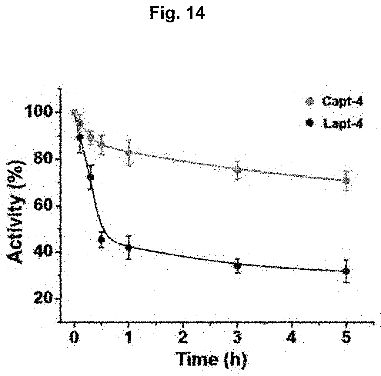
D00015

D00016

D00017

D00018

D00019

S00001
XML
uspto.report is an independent third-party trademark research tool that is not affiliated, endorsed, or sponsored by the United States Patent and Trademark Office (USPTO) or any other governmental organization. The information provided by uspto.report is based on publicly available data at the time of writing and is intended for informational purposes only.
While we strive to provide accurate and up-to-date information, we do not guarantee the accuracy, completeness, reliability, or suitability of the information displayed on this site. The use of this site is at your own risk. Any reliance you place on such information is therefore strictly at your own risk.
All official trademark data, including owner information, should be verified by visiting the official USPTO website at www.uspto.gov. This site is not intended to replace professional legal advice and should not be used as a substitute for consulting with a legal professional who is knowledgeable about trademark law.