A Pair Of Amino Acid Sequences For Monitoring Formation Of Tdp-43 Oligomer In Living Cells Via Bimolecular Fluorescence Complementation
LIM; Sung Su ; et al.
U.S. patent application number 16/857089 was filed with the patent office on 2020-10-29 for a pair of amino acid sequences for monitoring formation of tdp-43 oligomer in living cells via bimolecular fluorescence complementation. This patent application is currently assigned to KOREA INSTITUTE OF SCIENCE AND TECHNOLOGY. The applicant listed for this patent is KOREA INSTITUTE OF SCIENCE AND TECHNOLOGY. Invention is credited to Yun Kyung KIM, Sung Su LIM, Seul Gi SHIN.
| Application Number | 20200340001 16/857089 |
| Document ID | / |
| Family ID | 1000004916978 |
| Filed Date | 2020-10-29 |

| United States Patent Application | 20200340001 |
| Kind Code | A1 |
| LIM; Sung Su ; et al. | October 29, 2020 |
A PAIR OF AMINO ACID SEQUENCES FOR MONITORING FORMATION OF TDP-43 OLIGOMER IN LIVING CELLS VIA BIMOLECULAR FLUORESCENCE COMPLEMENTATION
Abstract
The present disclosure relates to a vector pair for screening TDP-43 (TAR DNA-binding protein 43 or TransActive Response DNA-binding protein 43) oligomer formation, a cell line transfected with the vector pair, and a method of monitoring TDP-43 oligomer formation using the cell line. More specifically, the vector pair includes: a first vector including a first TDP-43 gene and a first fluorescence protein gene; and a second vector including a second TDP-43 gene and a second fluorescence protein gene, wherein a protein expressed from the first fluorescence protein gene and a protein expressed from the second fluorescence protein gene bind to each other to display fluorescence, by association between a protein expressed from the first TDP-43 gene and a protein expressed from the second TDP-43 gene. The vector pair is effective in that it makes it possible to monitor TDP-43 oligomer formation in living cells.
| Inventors: | LIM; Sung Su; (Seoul, KR) ; SHIN; Seul Gi; (Seoul, KR) ; KIM; Yun Kyung; (Seoul, KR) | ||||||||||
| Applicant: |
|
||||||||||
|---|---|---|---|---|---|---|---|---|---|---|---|
| Assignee: | KOREA INSTITUTE OF SCIENCE AND
TECHNOLOGY Seoul KR |
||||||||||
| Family ID: | 1000004916978 | ||||||||||
| Appl. No.: | 16/857089 | ||||||||||
| Filed: | April 23, 2020 |
| Current U.S. Class: | 1/1 |
| Current CPC Class: | C12N 15/64 20130101; C07K 14/47 20130101 |
| International Class: | C12N 15/64 20060101 C12N015/64; C07K 14/47 20060101 C07K014/47 |
Foreign Application Data
| Date | Code | Application Number |
|---|---|---|
| Apr 26, 2019 | KR | 10-2019-0048923 |
Claims
1. A vector pair for screening TDP-43 oligomer formation, comprising: a first vector comprising a first TDP-43 (TAR DNA-binding protein 43 or TransActive Response DNA-binding protein 43) gene and a first fluorescence protein gene; and a second vector comprising a second TDP-43 gene and a second fluorescence protein gene, wherein a protein expressed from the first fluorescence protein gene and a protein expressed from the second fluorescence protein gene bind to each other to display fluorescence, by association between a protein expressed from the first TDP-43 gene and a protein expressed from the second TDP-43 gene.
2. The vector pair of claim 1, wherein each of the first TDP-43 gene and the second TDP-43 gene is a full-length TDP-43, a fragment of the full-length TDP-4, or a variant of the full-length TDP-43.
3. The vector pair of claim 1, wherein each of the first TDP-43 gene and the second TDP-43 gene is composed of the nucleotide sequence of SEQ ID NO: 1 or a fragment of the nucleotide sequence.
4. The vector pair of claim 1, wherein the first TDP-43 gene and the first fluorescence protein gene are operably linked to each other, and the second TDP-43 gene and the second fluorescence protein gene are operably linked to each other.
5. The vector pair of claim 4, wherein the first TDP-43 gene and the first fluorescence protein gene are linked to each other by a first linker, and the second TDP-43 gene and the second fluorescence protein gene are linked to each other by a second linker
6. The vector pair of claim 1, wherein each of the first vector and the second vector comprises a neuron-specific promoter.
7. The vector pair of claim 6, wherein the neuron-specific promoter is a CMV promoter.
8. The vector pair of claim 1, wherein the first fluorescence protein gene is linked to the C-terminus of the first TDP-43 gene, and the second fluorescence protein gene is linked to the C-terminus of the second TDP-43 gene.
9. The vector pair of claim 1, wherein the first fluorescence protein gene is composed of the nucleotide sequence of SEQ ID NO: 2 or a fragment thereof, and the second fluorescence protein gene is composed of the nucleotide sequence of SEQ ID NO: 3 or a fragment thereof.
10. The vector pair of claim 5, wherein the first linker is composed of the nucleotide sequence of SEQ ID NO: 4 or a fragment thereof, and the second linker is composed of the nucleotide sequence of SEQ ID NO: 5 or a fragment thereof.
11. The vector pair of claim 1, wherein the first vector comprises the nucleotide sequence of SEQ ID NO: 6, and the second vector comprises the nucleotide sequence of SEQ ID NO: 7.
12. A recombinant vector comprising the vector pair of claim 1.
13. A cell line transfected with the recombinant vector of claim 12.
14. The cell line of claim 13, which is deposited under accession number KCTC13819BP.
15. The cell line of claim 13, wherein fluorescence is displayed by binding between the first fluorescence protein and the second fluorescence protein, when a TDP-43 expressed from the first vector and a TDP-43 expressed from the second vector form an oligomer.
16. A composition for monitoring TDP-43 oligomer formation, comprising proteins expressed by the vector pair of claim 1.
17. A method for monitoring TDP-43 oligomer formation, comprising the steps of: treating the cell line of claim 13 with a TDP-43 oligomer formation inducer and culturing the treated cell line; and performing immunoblot analysis on the cell line.
18. The method of claim 17, wherein the TDP-43 oligomer formation inducer is one or more selected from the group consisting of forskolin, scriptaid, thapsigargin and ionomycin.
Description
BACKGROUND
Technical Field
[0001] The present disclosure relates to an amino acid sequence pair for monitoring interaction of TDP-43 in cells via bimolecular fluorescence complementation, a protein pair for screening TDP-43 oligomer formation, a nucleotide sequence pair encoding the same, a vector pair including the same, a cell line transfected with the vector pair, and a method of monitoring TDP-43 oligomer formation in living cells using the cell line.
Description of the Related Art
[0002] TDP-43 (TAR DNA-binding protein 43 or transactive response DNA-binding protein 43), a protein present mainly in the nucleus, inhibits gene transcription by binding to DNA or RNA, and is involved in gene expression by regulating RNA splicing and translation processes.
[0003] Initial studies have revealed that ubiquitinated TDP-43 protein is the major component of insoluble protein aggregates found in amyotrophic lateral sclerosis (ALS) and frontotemporal lobar degeneration (FTLD). Since then, cytoplasmic aggregates of TDP-43 protein have been found in various neurodegenerative brain diseases, including Alzheimer's disease (AD), Parkinson's disease (PD), dementia with lewy bodies (DLB), hippocampal sclerosis (HS), Alexander disease, Perry syndrome, argyrophilic grain disease (AG), corticobasal degeneration (CBD), progressive supranuclear palsy (PSP), and Huntington's disease (HD), and muscle degenerative diseases, including IBMPFD (inclusion body myopathy with Paget's disease of bone and/or frontotemporal dementia), sporadic inclusion body myositis (sporadic IBM), and myofibrillar myopathy. In addition, an abnormal increase of TDP-43 protein in the cytoplasm has been found in traumatic brain injury (TBI) animal models.
[0004] Accumulation of insoluble aggregates of this TDP-43 is a major symptom of various neurodegenerative diseases, but some studies conducted using TDP-43 expressing cell models or animal models have reported that TDP-43 overexpression alone without the formation of insoluble aggregates of TDP-43 also exhibits neurotoxicity. This led to the hypothesis that either the loss of normal function of TDP-43 by post-translational modifications (PTMs), such as phosphorylation, acetylation, fragmentation, ubiquitination, oxidation of cysteine residues, and SUMOylation, in intracellular stress cascades, or formation of toxic TDP-43 oligomers, causes neurodegeneration. Accordingly, studies on TDP-43 oligomers have been actively conducted.
[0005] However, conventional oligomer formation studies using TDP-43 recombinant protein have a problem in that PTMs cannot be regulated. In addition, they have a problem in that formation of the original oligomer cannot be accurately identified, since TDP-43 has a structural characteristic of spontaneously forming oligomers in vitro.
[0006] Therefore, there is an urgent need for a cell-based model that makes it possible to regulate the PTMs of TDP-43 in living cells, making it possible to observe the correct oligomer formation.
SUMMARY
[0007] The present disclosure has been made in order to solve the above-described problems, and an object of the present disclosure is to provide a reliable system for monitoring TDP-oligomer formation under the physiological conditions of living cells.
[0008] Another object of the present disclosure is to provide a cell model based on bimolecular fluorescence complementation (BiFC), which makes it possible to observe interaction between TDP-43 proteins in living cells.
[0009] However, the technical problems to be solved by the present disclosure are not limited to the above-mentioned problems, and other problems which are not mentioned will be clearly understood by those skilled in the art from the following description.
[0010] Hereinafter, various embodiments described herein will be described with reference to the accompanying drawings.
[0011] The present disclosure may undergo various modifications and have various embodiments, and thus specific embodiments are illustrated in the drawings and will be described in detail in the following detailed description. However, this description is not intended to limit the present disclosure to specific embodiments, and it should be understood that the present disclosure includes all modifications, equivalents or replacements that fall within the spirit and technical scope of the present disclosure. In the following description, when the detailed description of relevant known technology is determined to unnecessarily obscure the subject matter of the present disclosure, it may be omitted.
[0012] Terms used in the present specification are only to describe specific embodiments and are not intended to limit the scope of the present disclosure. Singular expressions include plural expressions unless otherwise specified in the context thereof. In the present specification, the terms "include", "comprise", "have", etc., are intended to denote the existence of mentioned characteristics, numbers, steps, operations, components, parts, or combinations thereof, but do not exclude the probability of existence or addition of one or more other characteristics, numbers, steps, operations, components, parts, or combinations thereof.
[0013] The terms "first", "second", etc., may be used to describe various components, but the components are not limited by the terms. The terms are used only for the purpose of distinguishing a component from other components.
[0014] Unless otherwise stated in the specification, all the scientific and technical terms used in the specification have the same meanings as commonly understood by those skilled in the technical field to which the present disclosure pertains.
[0015] A first aspect of the present disclosure is directed to a vector pair for screening TDP-43 oligomer formation, including: a first vector including a first TDP-43 (TAR DNA-binding protein 43 or TransActive Response DNA-binding protein 43) gene and a first fluorescence protein gene; and a second vector including a second TDP-43 gene and a second fluorescence protein gene, wherein a protein expressed from the first fluorescence protein gene and a protein expressed from the second fluorescence protein gene bind to each other to display fluorescence, by association between a protein expressed from the first TDP-43 gene and a protein expressed from the second TDP-43 gene.
[0016] Each of the first TDP-43 gene and the second TDP-43 gene may be a full-length TDP-43, a fragment of the full-length TDP-43, or a variant of the full-length TDP-43. For example, the TDP-43 gene may be a human full-length TDP-43 gene, a fragment thereof, or a variant having mutation in at least one nucleotide in the sequence of the human full-length TDP-43 gene. In addition, the TDP-43 gene may also be composed of the nucleotide sequence of SEQ ID NO: 1 or a fragment thereof. In one Example of the present disclosure, the TDP-43 gene set forth in SEQ ID NO: 1 was used.
[0017] It has been reported that ubiquitination of TDP-43, abnormal aggregation of TDP-43, and the like appear in various neurodegenerative brain diseases, muscle degenerative diseases, traumatic brain injury, etc. Thus, studies on TDP-43 oligomer formation have been actively conducted. Nevertheless, in the past, oligomer formation was studied using TDP-43 recombinant protein. However, these studies using TDP-43 recombinant protein have a problem in that PTMs cannot be regulated. In addition, these studies have a problem in that formation of the original oligomer cannot be accurately identified, since TDP-43 has a structural characteristic of spontaneously forming oligomers in vitro. For this reason, cellular models that can induce and monitor TDP-43 binding in living cells can be useful tools for studying TDP-43 pathology and developing methods to prevent and reverse the process. For this reason, cellular models that can induce and monitor binding between TDP-43 proteins in living cells can be useful tools for studying TDP-43 pathology and developing methods capable of preventing and reversing the process of the pathology.
[0018] The first fluorescence protein gene and/or the second fluorescence protein gene may be configured such that a protein expressed from the first fluorescence protein gene and a protein expressed from the second fluorescence protein gene bind to each other to display fluorescence, by association between a protein expressed from the first TDP-43 gene and the second TDP-43 gene. That is, when association between a protein expressed from the first TDP-43 gene and a protein expressed from the second TDP-43 gene occurs, a protein expressed from the first fluorescence protein gene and a protein expressed from the second fluorescence protein gene preferably bind to each other simultaneously with or subsequently to the association, thereby displaying fluorescence. For example, each of the fluorescence proteins may be a Venus protein or a fragment thereof, and the first fluorescence protein gene may be represented by the nucleotide sequence of SEQ ID NO: 2 or a fragment thereof, and the second fluorescence protein gene may be represented by the nucleotide sequence of SEQ ID NO: 3 or a fragment thereof. In addition, the first fluorescence protein gene may be VN173, and the second fluorescence protein gene may be VC155.
[0019] The present disclosure is an application of a method for visualizing protein-protein interactions, which is based on a bimolecular fluorescence complementation (BiFC) technique of forming a fluorescence protein complex from non-fluorescent constituents attached to the proteins of interest (Annu. Rev. Biophys. 37: 465-487). The BiFC technique based on the Venus protein, a kind of yellow fluorescence protein (YFP), is based on the principle according to which, when two different target proteins approach each other for interaction, fluorescence protein fragments linked to the target proteins also approach each other, and as a result, reconstruction between the fluorescence protein fragments occurs to display fluorescence. The use of this technique makes it possible to visually observe that an interaction between two target proteins occurred. Thus, this technique has the advantage of enabling protein-protein interactions to be visually observed in an optimal physical/chemical environment in which protein-protein interactions in cells or tissue may occur and be maintained. This technique makes it possible to determine not only a position where protein-protein interactions in cells or tissue occur, but also information about movement of these proteins.
[0020] The protein that is used in the present disclosure may be the Venus protein. The Venus protein can be effectively used for analysis of proteins such as TDP-43 protein, which are difficult to analyze spatially and temporally, because (1) it has fast and efficient maturation, (2) its self-assembly rate is low compared to that of other BiFC pairs, and (3) the fluorescence intensity of Venus-based BiFC is 10 times higher than that of EYFP-based BiFC (Biotechniques 40: 61-66; Biotechniques 49: 793-805).
[0021] According to one embodiment of the present disclosure, the first TDP-43 gene and the first fluorescence protein gene may be operably linked to each other, and the second TDP-43 and the second fluorescence protein gene may be operably linked to each other. Namely, in each of the first vector and the second vector, the TDP-43 gene and the fluorescence protein gene are preferably sequentially expressed by a single promoter. For example, the first TDP-43 gene and the first fluorescence protein gene may be linked to each other by a first linker, and the second TDP-43 gene and the second fluorescence protein gene may be linked to each other by a second linker. In an example of the present disclosure, the first linker represented by the nucleotide sequence of SEQ ID NO: 4 or a fragment thereof was used, and the second linker represented by the nucleotide sequence of SEQ ID NO: 5 or a fragment thereof was used.
[0022] The first vector and the second vector may each include a neuron-specific promoter. The first vector may include a first neuron-specific promoter, and the second vector may include a second neuron-specific promoter. The first neuron-specific promoter and/or the second neuron-specific promoter serves to express the vector pair of the present disclosure in neuron-related tissue, is not particularly limited, and may include any promoter known in the art. For example, the first neuron-specific promoter and/or the second neuron-specific promoter may be a CMV promoter and/or a Thy1 promoter, preferably CMV promoter. Since the Thy1 promoter is expressed specifically in neurons, it can induce expression of the genes inserted in the vector in neurons or neural tissue, particularly brain tissue.
[0023] The present disclosure is characterized in that, when the vector pair including the first vector and the second vector is expressed, a protein expressed from the first fluorescence protein gene and a protein expressed from the second fluorescence protein gene bind to each other to display fluorescence, by association between a protein expressed from the first TDP-43 gene and a protein expressed from the second TDP-43 gene. In particular, the present disclosure is characterized in that the genes inserted in the vector can be expressed by the neuron-specific promoter in neurons or neural tissue, particularly brain tissue. Accordingly, TDP-43 oligomer formation occurring in living cells can be visualized directly by fluorescence, thereby monitoring and quantifying the TDP-43 oligomerization process.
[0024] According to one embodiment of the present disclosure, in the case of the first vector, the first fluorescence protein gene may be linked to the N-terminus or C-terminus of the TDP-43 gene, and particularly, the first fluorescence protein gene is preferably linked to the C-terminus of the TDP-43 gene. In addition, in the case of the second vector according to the present disclosure, the second fluorescence protein gene may be linked to the N-terminus or C-terminus of the TDP-43 gene, and particularly, the second fluorescence protein gene is preferably linked to the C-terminus of the TDP-43 gene. This is believed to be because structurally positioning the TDP-43 gene ahead of the fluorescence protein gene in both the first and second vectors can further increase the luminous effect of the fluorescence proteins due to the interaction (or binding) of the TDP-43 proteins expressed from the genes.
[0025] According to one embodiment of the present disclosure, each of the first vector and second vector including the CMV promoter may be a pCMV6 including the CMV promoter, but is not limited thereto.
[0026] In addition, the TDP-43 gene and the first fluorescence protein gene may be inserted in the XhoI site of the pCMV6 vector, but may not be limited thereto. Furthermore, the TDP-43 gene, the second fluorescence protein gene and the second linker may be inserted in the XhoI site of the pCMV6 vector, but may not be limited thereto.
[0027] In addition, the first vector may include the nucleotide sequence of SEQ ID NO: 6, and the second vector may include the nucleotide sequence of SEQ ID NO: 7, but the scope of the present disclosure may not be limited thereto.
[0028] Moreover, the present disclosure may be directed to a vector pair for screening TDP-43 oligomer formation including: a first vector in which the TDP-43 gene represented by SEQ ID NO: 1, the first linker represented by SEQ ID NO: 4 and the first fluorescence protein gene represented by SEQ ID NO: 2 are operably linked to one another and which includes a CMV6 promoter; and a second vector in which the TDP-43 gene represented by SEQ ID NO: 1, the second linker represented by SEQ ID NO: 5 and the second fluorescence protein gene represented by SEQ ID NO: 3 are operably linked to one another and which includes a CMV6 promoter.
[0029] The second aspect of the present disclosure is directed to a recombinant vector including the above-described vector pair. The recombinant vector may be a single vector including the above-described vector pair, or may also be a pair of a first recombinant vector and a second recombinant vector, which include the first vector and the second vector, respectively.
[0030] A third aspect of the present disclosure is directed to a cell line transfected with the above-described recombinant vector. The cell line may be a cell line transiently transfected with the recombinant vector, or may be a cell line transfected with the recombinant vector for a long time or continuously.
[0031] As shown in the examples described below, the present inventors transfected HEK cells with the vector pair according to the present disclosure. Among these cells, cell lines having an excellent luminous effect were subcultured to obtain a stable cell line (KIST HT43B1). The obtained cell line was deposited with the Korean Collection for Type Cultures, the Korea Research Institute of Bioscience and Biotechnology (KRIBB) under accession number KCTC13819BP. Thus, the cell line according to the present disclosure may be the cell line deposited under accession number KCTC13819BP. This cell line has a remarkably excellent effect of displaying fluorescence by binding between the first fluorescence protein and the second fluorescence protein when TDP-43 expressed from the first vector and TDP-43 expressed from the second vector form an oligomer.
[0032] A fourth aspect of the present disclosure is directed to a composition for monitoring TDP-43 oligomer formation, the composition including proteins expressed by the vector pair. In addition, the present disclosure may also be directed to a composition for detecting TDP-43 binding or for screening a TDP-43 binding inhibitor, the composition including proteins expressed by the vector pair.
[0033] A fifth aspect of the present disclosure is directed to a method of monitoring TDP-43 oligomer formation in cells, the method including the steps of: treating the above-described cell line with a TDP-43 oligomer formation inducer and culturing the treated cell line; and performing immunoblot analysis on the cell line. The TDP-43 oligomer formation inducer may be one or more selected from the group consisting of forskolin, scriptaid, thapsigargin and ionomycin.
[0034] When the TDP-43 cell model according to the present disclosure was treated with forskolin, scriptaid, thapsigargin and ionomycin, the BiFC fluorescence in the treated cells increased greatly (7 times or more) compared to that in an untreated group. In particular, treatment with scriptaid greatly increased the fluorescence not only in the nucleus but also in the cytoplasm.
BRIEF DESCRIPTION OF THE DRAWINGS
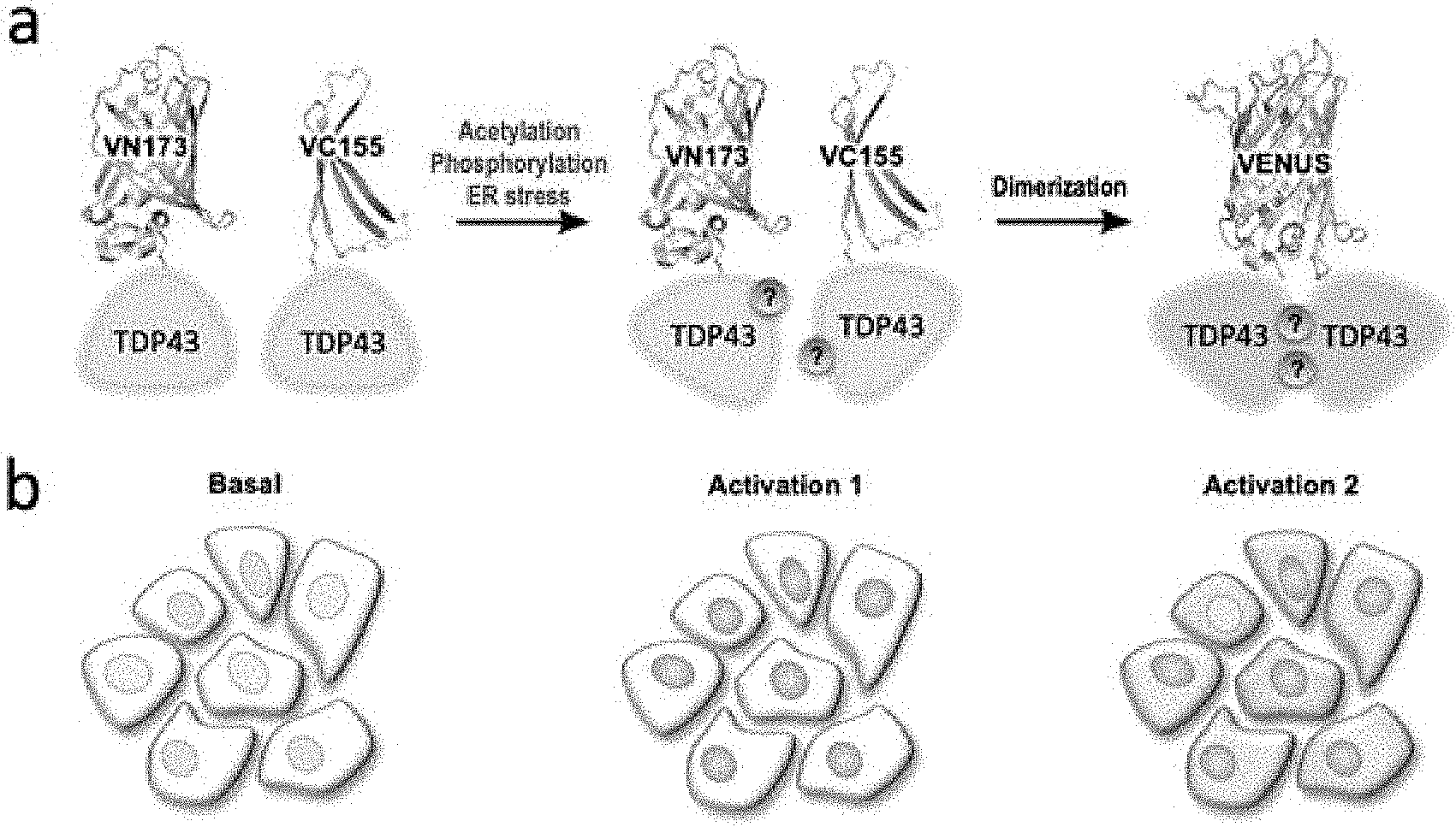
[0035] FIG. 1 is a schematic view showing the principle according to which TDP-43 fused to the Venus protein BiFC used in one example of the present disclosure acts as a Venus fluorescence turn-on sensor in cells in a normal state and a TDP-43 aggregation-induced state (FIG. 1a), and an increase in BiFC fluorescence and a change in the distribution of BiFC fluorescence (FIG. 1b).
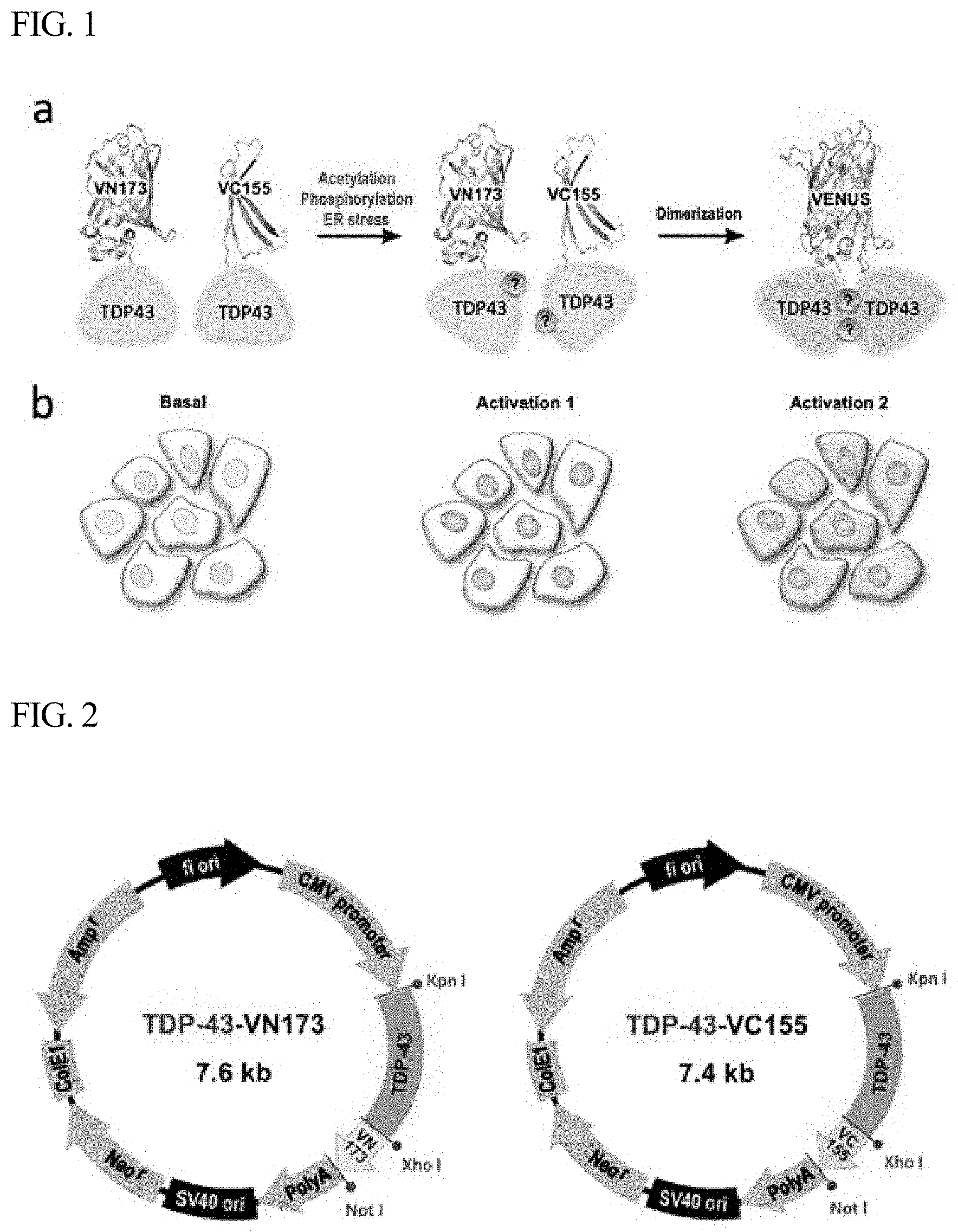
[0036] FIG. 2 is a vector map showing a structure in which two plasmids having BiFC labeled at the C-terminus of TDP-43, used in one example of the present disclosure, are inserted in a pCMV vector.
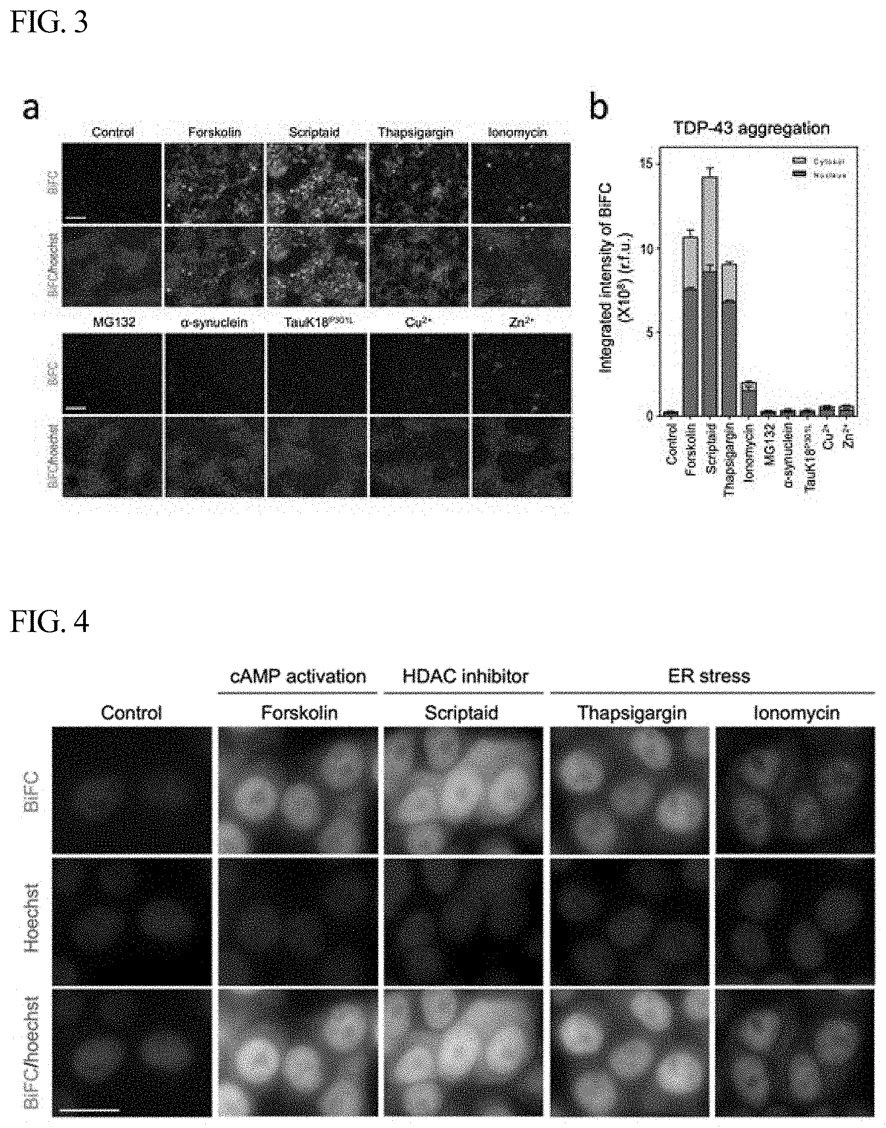
[0037] FIG. 3 shows the results of observing changes in BiFC fluorescence after treating TDP-43-BiFC cells with a compound that induces PTMs of protein, a compound that ER stress in cells, amyloid protein, metal ions, or the like, according to one example of the present disclosure.
[0038] FIG. 4 shows high-magnification images obtained after treating TDP-43-BiFC cells with each of forskolin, scriptaid, thapsigargin and ionomycin according to one example of the present disclosure.
[0039] FIG. 5 shows the results of immunoblot analysis performed according to one example of the present disclosure by preparing lysates of TDP-43-BiFC cells treated with each of four drugs (forskolin, scriptaid, thapsigargin, and ionomycin) and a lysate of untreated cells and isolating TDP-43 oligomers from the lysates.
DETAILED DESCRIPTION
[0040] The present disclosure may be better understood with the following examples. However, the following examples are for the purpose of illustrating the present disclosure and are not intended to limit the scope of the present disclosure as defined by the appended claims.
[0041] In the following examples, in order to visualize interaction between TDP-43 proteins at the oligomer level, a TDP-43-BiFC cell model was constructed. In order to visualize TDP-43 oligomer formation in living cells, BiFC (bimolecular fluorescence complementation) was attached to a human full-length TDP-43 and expressed in cells, and the cells were treated with various drugs to regulate PTMs of the protein, thereby developing a cell model that makes it possible to observe TDP-43 oligomer formation.
[0042] The cell model according to the present disclosure was constructed using a Venus protein-based BiFC method in which the N-terminal and C-terminal non-fluorescent fragments of the Venus protein were fused to TDP-43.
[0043] In the model according to the present disclosure as a fluorescence "turn-on" approach, no fluorescence is observed when TDP-43 proteins are present as monomers, but Venus fluorescence is observed when TDP-43 proteins are associated with each other. Thus, it is possible to TDP-43 oligomerization in living cells without staining with a foreign molecule.
Examples
[0044] 1. Construction of TDP-43-BiFC Sensor
[0045] FIG. 1a shows the principle according to which TDP-43 fused to the Venus protein BiFC acts as a Venus fluorescence turn-on sensor that shows TDP-43 interaction induced by PTM or intracellular stress. In a normal state, TDP-43 is present mainly in the nucleus, but when abnormal aggregation of TDP-43 is induced by various factors, including phosphorylation, fragmentation, acetylation, ubiquitination, cysteine residue oxidation, and SUMOylation, TDP-43 forms insoluble aggregates in the cytoplasm. FIG. 1b is a schematic view showing an increase in BiFC fluorescence and a change in the distribution of BiFC fluorescence in TDP-43-BiFC cells in a normal state and an aggregation-induced state.
[0046] FIG. 2 is a vector map of a pCMV vector, which shows two plasmids having BiFC labeled at the C-terminus of TDP-43. In order to develop a TDP-43-BiFC cell line, the TDP-43-VN173 and TDP-43-VC155 constructs shown in FIG. 2 were co-transfected into HEK293 BiFC cells. For establishment of a stable cell line, the transfected cells were selected by culture with growth medium containing 100 .mu.g/mL geneticin (G418; Sigma). In addition, the selected cells were sorted using FACSAria (BD Bioscience) into cells showing fluorescence. The sorted TDP-43-BiFC cells were kept in DMEM medium containing 10% FBS, 100 units/mL penicillin, 100 .mu.g/mL streptomycin and 100 .mu.g/mL G418 in a humidified atmosphere at 37.degree. C. under 5% CO.sub.2.
[0047] The nucleotide sequences of the TDP-43-BiFC genes inserted in the pCMV6 vector of FIG. 2, used for the transfection, are shown in Table 1 below.
TABLE-US-00001 TABLE 1 Nucleotide sequence TDP43- ATG TCT GAA TAT ATT CGG GTA ACC GAA GAT GAG AAC GAT VC173 GAG CCC ATT GAA ATA CCA TCG GAA GAC GAT GGG ACG GTG (SEQ ID CTG CTC TCC ACG GTT ACA GCC CAG TTT CCA GGG GCG TGT NO: 6) GGG CTT CGC TAC AGG AAT CCA GTG TCT CAG TGT ATG AGA GGT GTC CGG CTG GTA GAA GGA ATT CTG CAT GCC CCA GAT GCT GGC TGG GGA AAT CTG GTG TAT GTT GTC AAC TAT CCA AAA GAT AAC AAA AGA AAA ATG GAT GAG ACA GAT GCT TCA TCA GCA GTG AAA GTG AAA AGA GCA GTC CAG AAA ACA TCC GAT TTA ATA GTG TTG GGT CTC CCA TGG AAA ACA ACC GAA CAG GAC CTG AAA GAG TAT TTT AGT ACC TTT GGA GAA GTT CTT ATG GTG CAG GTC AAG AAA GAT CTT AAG ACT GGT CAT TCA AAG GGG TTT GGC TTT GTT CGT TTT ACG GAA TAT GAA ACA CAA GTG AAA GTA ATG TCA CAG CGA CAT ATG ATA GAT GGA CGA TGG TGT GAC TGC AAA CTT CCT AAT TCT AAG CAA AGC CAA GAT GAG CCT TTG AGA AGC AGA AAA GTG TTT GTG GGG CGC TGT ACA GAG GAC ATG ACT GAG GAT GAG CTG CGG GAG TTC TTC TCT CAG TAC GGG GAT GTG ATG GAT GTC TTC ATC CCC AAG CCA TTC AGG GCC TTT GCC TTT GTT ACA TTT GCA GAT GAT CAG ATT GCG CAG TCT CTT TGT GGA GAG GAC TTG ATC ATT AAA GGA ATC AGC GTT CAT ATA TCC AAT GCC GAA CCT AAG CAC AAT AGC AAT AGA CAG TTA GAA AGA AGT GGA AGA TTT GGT GGT AAT CCA GGT GGC TTT GGG AAT CAG GGT GGA TTT GGT AAT AGC AGA GGG GGT GGA GCT GGT TTG GGA AAC AAT CAA GGT AGT AAT ATG GGT GGT GGG ATG AAC TTT GGT GCG TTC AGC ATT AAT CCA GCC ATG ATG GCT GCC GCC CAG GCA GCA CTA CAG AGC AGT TGG GGT ATG ATG GGC ATG TTA GCC AGC CAG CAG AAC CAG TCA GGC CCA TCG GGT AAT AAC CAA AAC CAA GGC AAC ATG CAG AGG GAG CCA AAC CAG GCC TTC GGT TCT GGA AAT AAC TCT TAT AGT GGC TCT AAT TCT GGT GCA GCA ATT GGT TGG GGA TCA GCA TCC AAT GCA GGG TCG GGC AGT GGT TTT AAT GGA GGC TTT GGC TCA AGC ATG GAT TCT AAG TCT TCT GGC TGG GGA ATG ACG CGT ACG CGG CCG CTC GAG TCT AGA AGA TCC ATC GCC ACC ATG GTG AGC AAG GGC GAG GAG CTG TTC ACC GGG GTG GTG CCC ATC CTG GTC GAG CTG GAC GGC GAC GTA AAC GGC CAC AAG TTC AGC GTG TCC GGC GAG GGC GAG GGC GAT GCC ACC TAC GGC AAG CTG ACC CTG AAG CTG ATC TGC ACC ACC GGC AAG CTG CCC GTG CCC TGG CCC ACC CTC GTG AEC AEC CTG GGC TAC GGC CTG CAG TGC TTC GCC CGC TAC CCC GAC CAC ATG AAG CAG CAC GAC TTC TTC AAG TCC GCC ATG CCC GAA GGC TAC GTC CAG GAG CGC AEC ATC TTC TTC AAG GAC GAC GGC AAC TAC AAG AEC CGC GCC GAG GTG AAG TTC GAG GGC GAC AEC CTG GTG AAC CGC ATC GAG CTG AAG GGC ATC GAC TTC AAG GAG GAC GGC AAC ATC CTG GGG CAC AAG CTG GAG TAC AAC TAC AAC AGC CAC AAC GTC TAT ATC AEC GCC GAC AAG CAG AAG AAC GGC ATC AAG GCC AAC TTC AAG ATC CGC CAC AAC ATC GAG TAG
TABLE-US-00002 TABLE 2 Nucleotide sequence TDP43- ATG TCT GAA TAT ATT CGG GTA ACC GAA GAT GAG AAC GAT VC155 GAG CCC ATT GAA ATA CCA TCG GAA GAC GAT GGG ACG GTG (SEQ ID CTG CTC TCC ACG GTT ACA GCC CAG TTT CCA GGG GCG TGT NO: 7) GGG CTT CGC TAC AGG AAT CCA GTG TCT CAG TGT ATG AGA GGT GTC CGG CTG GTA GAA GGA ATT CTG CAT GCC CCA GAT GCT GGC TGG GGA AAT CTG GTG TAT GTT GTC AAC TAT CCA AAA GAT AAC AAA AGA AAA ATG GAT GAG ACA GAT GCT TCA TCA GCA GTG AAA GTG AAA AGA GCA GTC CAG AAA ACA TCC GAT TTA ATA GTG TTG GGT CTC CCA TGG AAA ACA ACC GAA CAG GAC CTG AAA GAG TAT TTT AGT ACC TTT GGA GAA GTT CTT ATG GTG CAG GTC AAG AAA GAT CTT AAG ACT GGT CAT TCA AAG GGG TTT GGC TTT GTT CGT TTT ACG GAA TAT GAA ACA CAA GTG AAA GTA ATG TCA CAG CGA CAT ATG ATA GAT GGA CGA TGG TGT GAC TGC AAA CTT CCT AAT TCT AAG CAA AGC CAA GAT GAG CCT TTG AGA AGC AGA AAA GTG TTT GTG GGG CGC TGT ACA GAG GAC ATG ACT GAG GAT GAG CTG CGG GAG TTC TTC TCT CAG TAC GGG GAT GTG ATG GAT GTC TTC ATC CCC AAG CCA TTC AGG GCC TTT GCC TTT GTT ACA TTT GCA GAT GAT CAG ATT GCG CAG TCT CTT TGT GGA GAG GAC TTG ATC ATT AAA GGA ATC AGC GTT CAT ATA TCC AAT GCC GAA CCT AAG CAC AAT AGC AAT AGA CAG TTA GAA AGA AGT GGA AGA TTT GGT GGT AAT CCA GGT GGC TTT GGG AAT CAG GGT GGA TTT GGT AAT AGC AGA GGG GGT GGA GCT GGT TTG GGA AAC AAT CAA GGT AGT AAT ATG GGT GGT GGG ATG AAC TTT GGT GCG TTC AGC ATT AAT CCA GCC ATG ATG GCT GCC GCC CAG GCA GCA CTA CAG AGC AGT TGG GGT ATG ATG GGC ATG TTA GCC AGC CAG CAG AAC CAG TCA GGC CCA TCG GGT AAT AAC CAA AAC CAA GGC AAC ATG CAG AGG GAG CCA AAC CAG GCC TTC GGT TCT GGA AAT AAC TCT TAT AGT GGC TCT AAT TCT GGT GCA GCA ATT GGT TGG GGA TCA GCA TCC AAT GCA GGG TCG GGC AGT GGT TTT AAT GGA GGC TTT GGC TCA AGC ATG GAT TCT AAG TCT TCT GGC TGG GGA ATG ACG CGT ACG CGG CCG CTC GAG AAG CAG AAG AAC GGC ATC AAG GCC AAC TTC AAG ATC CGC CAC AAC ATC GAG GAC GGC GGC GTG CAG CTC GCC GAC CAC TAC CAG CAG AAC AEC CCC ATC GGC GAC GGC CCC GTG CTG CTG CCC GAC AAC CAC TAC CTG AGC TAC CAG TCC AAA CTG AGC AAA GAC CCC AAC GAG AAG CGC GAT CAC ATG GTC CTG CTG GAG TTC GTG AEC GCC GCC GGG ATC ACT CTC GGC ATG GAC GAG CTG TAC AAG TAA
[0048] 2. Induction of TDP-43 Oligomer Formation
[0049] The TDP-43-BiFC cells prepared in Example 1 above were treated with a compound that induces PTMs of protein, a compound that ER stress in cells, amyloid protein, metal ions, or the like, and changes in BiFC fluorescence in the cells were examined. Specifically, for fluorescence analysis, the TDP-43-BiFC cells were grown in a 384-well plate and treated with each of forskolin (30 .mu.M), scriptaid (3 .mu.M), thapsigargin (1 .mu.M), ionomycin (1 .mu.M), MG132 (5 .mu.M), .alpha.-synuclein (5 .mu.g/ml), tauK18.sup.P301L (5 .mu.g/ml), Cu.sup.2+ (1 .mu.M) and Zn.sup.2+ (30 .mu.M), followed by culture at 37.degree. C. for 48 hours. Fluorescence images were obtained using Operetta.RTM. (PerkinElmer), and the fluorescence intensity in the nucleus and the cytoplasmic fluorescence intensity were analyzed using Harmony3.1 software (PerkinElmer).
[0050] FIG. 3 shows the results of observing changes in BiFC fluorescence after treating TDP-43-BiFC cells with a compound that induces PTMs of protein, a compound that ER stress in cells, amyloid protein, metal ions, or the like. As shown in FIG. 3, when the cells were treated with each of the compound scriptaid that induces phosphorylation, scriptaid that increases acetylation, thapsigargin, and ionomycin, the BiFC fluorescence in the cells increased 36-fold, 48-fold, 31-fold and 7-fold, respectively, compared to that in the untreated cells. However, when the cells were treated with proteasome inhibitor MG132, no change in the fluorescence was observed. When the cells were treated with each of .alpha.-synuclein and tauK18.sup.P301L, which are amyloid proteins, the BiFC fluorescence of TDP-43 did not increase. When the cells were treated with each of Cu.sup.2+ and Zn.sup.2+, the BiFC fluorescence increased about 2-fold. From these results, it can be seen that the phosphorylation or acetylation of TDP-43 strongly induces TDP-43 oligomer formation, and even when intracellular ER stress is induced, TDP-43 oligomer formation greatly increases.
[0051] 3. Change in Intracellular Distribution by TDP-43 Oligomer Formation
[0052] Next, the pattern of intracellular distribution by an increase in TDP-43 oligomer formation was examined. Specifically, TDP-43 formation was induced in the same manner as Example 2 above, and then the whole plate was automatically imaged using Operetta.RTM., and high-resolution images were obtained using a Nikon Eclipse inverted microscope (Ti, Nikon) at 1000.times. magnification.
[0053] FIG. 4 shows high-magnification images obtained after treating the TDP-43-BiFC cells with each of forskolin, scriptaid, thapsigargin and ionomycin as described above. It can be seen that when the cells were treated with scriptaid, the BiFC fluorescence intensity in the cytoplasm increased. In addition, the control showed the BiFC fluorescence in the nucleus portion and showed very weak or almost no fluorescence in other portions, but when the cells were treated with scriptaid, the BiFC fluorescence significantly increased not only in the nucleus but also in the cytoplasm.
[0054] 4. Isolation and Detection of TDP-43 Oligomer
[0055] Lysates of the TDP-43-BiFC cells treated with each of the four drugs in Example 3 and a lysate of untreated cells were prepared and TDP-43 oligomers were isolated therefrom. Specifically, TDP-43-BiFC cells were grown in a 6-well plate, treated with each of forskolin (30 .mu.M), scriptaid (1 .mu.M), thapsigargin (0.5 .mu.M) and ionomycin (1 .mu.M), and cultured at 37.degree. C. for 24 hours, and then lysates of the cells were prepared. GFP-trap beads shown in FIG. 5a were added to the lysates which were then incubated at 4.degree. C. for 16 hours, and the oligomerized TDP-43 protein was isolated. The isolated protein was separated by SDS-PAGE, and immunoblot analysis was performed using anti-TDP-43 antibody.
[0056] As can be expected from the results of measuring the BiFC fluorescence intensity in Example 2 (FIG. 3), when the cells were treated with each of the drugs, the amount of the detected oligomerized protein significantly increased compared to that in the untreated cells, and the largest amount of the oligomerized protein was detected in the cells treated with scriptaid, which showed the greatest increase in the BiFC fluorescence among the drugs. For the remaining drugs, the amount of the oligomer appeared in the same pattern as the change in the BiFC fluorescence intensity (left of FIG. 5b). From these results, it was confirmed that the degree of the BiFC fluorescence shown in the TDP-43-BiFC cells represents the amount of the TDP-43 oligomer. As can be seen in FIG. 5 showing the results of the immunoblot analysis in which disulfide bonds were not reduced, oligomers having a size of 100 kDa or more were detected together with monomers. These results suggest that the TDP-43 oligomers include oligomers formed through disulfide bonds together with oligomers formed without disulfide bonds.
[0057] As described above, according to the present disclosure, TDP-43 oligomer formation in living cells may be observed using a cell model based on bimolecular fluorescence complementation (BiFC).
[0058] In addition, according to the present disclosure, non-fluorescent constituents attached to TDP-43 display fluorescence when the physical distance therebetween becomes closer due to association between TDP-43 proteins, thus making it possible to observe interaction between two TDP-43 proteins.
[0059] According to the present disclosure, TDP-43 oligomer formation in living cells may be directly visualized, and thus the process of TDP-43 oligomer formation may be monitored and quantified. Therefore, the cell model according to the present disclosure may be used as a useful tool for studying the development of diseases related to TDP-43 and developing a method for preventing and reversing TDP-43 oligomer formation.
[0060] The scope of the present disclosure is defined not by the detailed description, but by the appended claims, and all variations and modifications derived from the meaning and scope of the claims and their equivalents are to be construed as being included in the scope of the present disclosure.
Sequence CWU 1
1
711242DNAHomo sapiensgene(1)..(1242)TDP-43(TAR DNA-binding protein
43, transactive response DNA binding protein 43) 1atgtctgaat
atattcgggt aaccgaagat gagaacgatg agcccattga aataccatcg 60gaagacgatg
ggacggtgct gctctccacg gttacagccc agtttccagg ggcgtgtggg
120cttcgctaca ggaatccagt gtctcagtgt atgagaggtg tccggctggt
agaaggaatt 180ctgcatgccc cagatgctgg ctggggaaat ctggtgtatg
ttgtcaacta tccaaaagat 240aacaaaagaa aaatggatga gacagatgct
tcatcagcag tgaaagtgaa aagagcagtc 300cagaaaacat ccgatttaat
agtgttgggt ctcccatgga aaacaaccga acaggacctg 360aaagagtatt
ttagtacctt tggagaagtt cttatggtgc aggtcaagaa agatcttaag
420actggtcatt caaaggggtt tggctttgtt cgttttacgg aatatgaaac
acaagtgaaa 480gtaatgtcac agcgacatat gatagatgga cgatggtgtg
actgcaaact tcctaattct 540aagcaaagcc aagatgagcc tttgagaagc
agaaaagtgt ttgtggggcg ctgtacagag 600gacatgactg aggatgagct
gcgggagttc ttctctcagt acggggatgt gatggatgtc 660ttcatcccca
agccattcag ggcctttgcc tttgttacat ttgcagatga tcagattgcg
720cagtctcttt gtggagagga cttgatcatt aaaggaatca gcgttcatat
atccaatgcc 780gaacctaagc acaatagcaa tagacagtta gaaagaagtg
gaagatttgg tggtaatcca 840ggtggctttg ggaatcaggg tggatttggt
aatagcagag ggggtggagc tggtttggga 900aacaatcaag gtagtaatat
gggtggtggg atgaactttg gtgcgttcag cattaatcca 960gccatgatgg
ctgccgccca ggcagcacta cagagcagtt ggggtatgat gggcatgtta
1020gccagccagc agaaccagtc aggcccatcg ggtaataacc aaaaccaagg
caacatgcag 1080agggagccaa accaggcctt cggttctgga aataactctt
atagtggctc taattctggt 1140gcagcaattg gttggggatc agcatccaat
gcagggtcgg gcagtggttt taatggaggc 1200tttggctcaa gcatggattc
taagtcttct ggctggggaa tg 12422522DNAArtificial SequenceVC173
2atggtgagca agggcgagga gctgttcacc ggggtggtgc ccatcctggt cgagctggac
60ggcgacgtaa acggccacaa gttcagcgtg tccggcgagg gcgagggcga tgccacctac
120ggcaagctga ccctgaagct gatctgcacc accggcaagc tgcccgtgcc
ctggcccacc 180ctcgtgacca ccctgggcta cggcctgcag tgcttcgccc
gctaccccga ccacatgaag 240cagcacgact tcttcaagtc cgccatgccc
gaaggctacg tccaggagcg caccatcttc 300ttcaaggacg acggcaacta
caagacccgc gccgaggtga agttcgaggg cgacaccctg 360gtgaaccgca
tcgagctgaa gggcatcgac ttcaaggagg acggcaacat cctggggcac
420aagctggagt acaactacaa cagccacaac gtctatatca ccgccgacaa
gcagaagaac 480ggcatcaagg ccaacttcaa gatccgccac aacatcgagt ag
5223249DNAArtificial SequenceVC155 3cagaagaacg gcatcaaggc
caacttcaag atccgccaca acatcgagga cggcggcgtg 60cagctcgccg accactacca
gcagaacacc cccatcggcg acggccccgt gctgctgccc 120gacaaccact
acctgagcta ccagtccaaa ctgagcaaag accccaacga gaagcgcgat
180cacatggtcc tgctggagtt cgtgaccgcc gccgggatca ctctcggcat
ggacgagctg 240tacaagtaa 249442DNAArtificial SequenceVC173 linker
4acgcgtacgc ggccgctcga gtctagaaga tccatcgcca cc 42524DNAArtificial
SequenceVC155 linker 5acgcgtacgc ggccgctcga gaag
2461806DNAArtificial SequenceTDP43-VC173 linker-VC173 6atgtctgaat
atattcgggt aaccgaagat gagaacgatg agcccattga aataccatcg 60gaagacgatg
ggacggtgct gctctccacg gttacagccc agtttccagg ggcgtgtggg
120cttcgctaca ggaatccagt gtctcagtgt atgagaggtg tccggctggt
agaaggaatt 180ctgcatgccc cagatgctgg ctggggaaat ctggtgtatg
ttgtcaacta tccaaaagat 240aacaaaagaa aaatggatga gacagatgct
tcatcagcag tgaaagtgaa aagagcagtc 300cagaaaacat ccgatttaat
agtgttgggt ctcccatgga aaacaaccga acaggacctg 360aaagagtatt
ttagtacctt tggagaagtt cttatggtgc aggtcaagaa agatcttaag
420actggtcatt caaaggggtt tggctttgtt cgttttacgg aatatgaaac
acaagtgaaa 480gtaatgtcac agcgacatat gatagatgga cgatggtgtg
actgcaaact tcctaattct 540aagcaaagcc aagatgagcc tttgagaagc
agaaaagtgt ttgtggggcg ctgtacagag 600gacatgactg aggatgagct
gcgggagttc ttctctcagt acggggatgt gatggatgtc 660ttcatcccca
agccattcag ggcctttgcc tttgttacat ttgcagatga tcagattgcg
720cagtctcttt gtggagagga cttgatcatt aaaggaatca gcgttcatat
atccaatgcc 780gaacctaagc acaatagcaa tagacagtta gaaagaagtg
gaagatttgg tggtaatcca 840ggtggctttg ggaatcaggg tggatttggt
aatagcagag ggggtggagc tggtttggga 900aacaatcaag gtagtaatat
gggtggtggg atgaactttg gtgcgttcag cattaatcca 960gccatgatgg
ctgccgccca ggcagcacta cagagcagtt ggggtatgat gggcatgtta
1020gccagccagc agaaccagtc aggcccatcg ggtaataacc aaaaccaagg
caacatgcag 1080agggagccaa accaggcctt cggttctgga aataactctt
atagtggctc taattctggt 1140gcagcaattg gttggggatc agcatccaat
gcagggtcgg gcagtggttt taatggaggc 1200tttggctcaa gcatggattc
taagtcttct ggctggggaa tgacgcgtac gcggccgctc 1260gagtctagaa
gatccatcgc caccatggtg agcaagggcg aggagctgtt caccggggtg
1320gtgcccatcc tggtcgagct ggacggcgac gtaaacggcc acaagttcag
cgtgtccggc 1380gagggcgagg gcgatgccac ctacggcaag ctgaccctga
agctgatctg caccaccggc 1440aagctgcccg tgccctggcc caccctcgtg
accaccctgg gctacggcct gcagtgcttc 1500gcccgctacc ccgaccacat
gaagcagcac gacttcttca agtccgccat gcccgaaggc 1560tacgtccagg
agcgcaccat cttcttcaag gacgacggca actacaagac ccgcgccgag
1620gtgaagttcg agggcgacac cctggtgaac cgcatcgagc tgaagggcat
cgacttcaag 1680gaggacggca acatcctggg gcacaagctg gagtacaact
acaacagcca caacgtctat 1740atcaccgccg acaagcagaa gaacggcatc
aaggccaact tcaagatccg ccacaacatc 1800gagtag 180671515DNAArtificial
SequenceTDP43-VC155 linker-VC155 7atgtctgaat atattcgggt aaccgaagat
gagaacgatg agcccattga aataccatcg 60gaagacgatg ggacggtgct gctctccacg
gttacagccc agtttccagg ggcgtgtggg 120cttcgctaca ggaatccagt
gtctcagtgt atgagaggtg tccggctggt agaaggaatt 180ctgcatgccc
cagatgctgg ctggggaaat ctggtgtatg ttgtcaacta tccaaaagat
240aacaaaagaa aaatggatga gacagatgct tcatcagcag tgaaagtgaa
aagagcagtc 300cagaaaacat ccgatttaat agtgttgggt ctcccatgga
aaacaaccga acaggacctg 360aaagagtatt ttagtacctt tggagaagtt
cttatggtgc aggtcaagaa agatcttaag 420actggtcatt caaaggggtt
tggctttgtt cgttttacgg aatatgaaac acaagtgaaa 480gtaatgtcac
agcgacatat gatagatgga cgatggtgtg actgcaaact tcctaattct
540aagcaaagcc aagatgagcc tttgagaagc agaaaagtgt ttgtggggcg
ctgtacagag 600gacatgactg aggatgagct gcgggagttc ttctctcagt
acggggatgt gatggatgtc 660ttcatcccca agccattcag ggcctttgcc
tttgttacat ttgcagatga tcagattgcg 720cagtctcttt gtggagagga
cttgatcatt aaaggaatca gcgttcatat atccaatgcc 780gaacctaagc
acaatagcaa tagacagtta gaaagaagtg gaagatttgg tggtaatcca
840ggtggctttg ggaatcaggg tggatttggt aatagcagag ggggtggagc
tggtttggga 900aacaatcaag gtagtaatat gggtggtggg atgaactttg
gtgcgttcag cattaatcca 960gccatgatgg ctgccgccca ggcagcacta
cagagcagtt ggggtatgat gggcatgtta 1020gccagccagc agaaccagtc
aggcccatcg ggtaataacc aaaaccaagg caacatgcag 1080agggagccaa
accaggcctt cggttctgga aataactctt atagtggctc taattctggt
1140gcagcaattg gttggggatc agcatccaat gcagggtcgg gcagtggttt
taatggaggc 1200tttggctcaa gcatggattc taagtcttct ggctggggaa
tgacgcgtac gcggccgctc 1260gagaagcaga agaacggcat caaggccaac
ttcaagatcc gccacaacat cgaggacggc 1320ggcgtgcagc tcgccgacca
ctaccagcag aacaccccca tcggcgacgg ccccgtgctg 1380ctgcccgaca
accactacct gagctaccag tccaaactga gcaaagaccc caacgagaag
1440cgcgatcaca tggtcctgct ggagttcgtg accgccgccg ggatcactct
cggcatggac 1500gagctgtaca agtaa 1515
D00000
D00001
D00002
D00003
S00001
XML
uspto.report is an independent third-party trademark research tool that is not affiliated, endorsed, or sponsored by the United States Patent and Trademark Office (USPTO) or any other governmental organization. The information provided by uspto.report is based on publicly available data at the time of writing and is intended for informational purposes only.
While we strive to provide accurate and up-to-date information, we do not guarantee the accuracy, completeness, reliability, or suitability of the information displayed on this site. The use of this site is at your own risk. Any reliance you place on such information is therefore strictly at your own risk.
All official trademark data, including owner information, should be verified by visiting the official USPTO website at www.uspto.gov. This site is not intended to replace professional legal advice and should not be used as a substitute for consulting with a legal professional who is knowledgeable about trademark law.