Method For Re-using Test Probe And Reagents In An Immunoassay
Zuk; Robert F. ; et al.
U.S. patent application number 16/918911 was filed with the patent office on 2020-10-22 for method for re-using test probe and reagents in an immunoassay. The applicant listed for this patent is Access Medical Systems, LTD.. Invention is credited to Qing Xia, Robert F. Zuk.
| Application Number | 20200333337 16/918911 |
| Document ID | / |
| Family ID | 1000004932562 |
| Filed Date | 2020-10-22 |


View All Diagrams
| United States Patent Application | 20200333337 |
| Kind Code | A1 |
| Zuk; Robert F. ; et al. | October 22, 2020 |
METHOD FOR RE-USING TEST PROBE AND REAGENTS IN AN IMMUNOASSAY
Abstract
The present invention is directed an immunoassay method, which re-uses an antibody-immobilized test probe and reagents for quantitating an analyte in different samples, anywhere from about 3 to 20 times, while maintaining acceptable clinical assay performance. The method regenerates the test probe by dipping the test probe in an acidic solution having pH about 1-4, after the completion of each cycle of reaction. The present invention is also directed to a unitized cartridge (a strip) for an immunoassay test. Each unitized cartridge contains all necessary reagents can be used for 3-20 cycles to measure 3-20 different samples.
| Inventors: | Zuk; Robert F.; (Menlo Park, CA) ; Xia; Qing; (Sunnyvale, CA) | ||||||||||
| Applicant: |
|
||||||||||
|---|---|---|---|---|---|---|---|---|---|---|---|
| Family ID: | 1000004932562 | ||||||||||
| Appl. No.: | 16/918911 | ||||||||||
| Filed: | July 1, 2020 |
Related U.S. Patent Documents
| Application Number | Filing Date | Patent Number | ||
|---|---|---|---|---|
| 15802075 | Nov 2, 2017 | |||
| 16918911 | ||||
| PCT/US2016/031661 | May 10, 2016 | |||
| 15802075 | ||||
| 62159919 | May 11, 2015 | |||
| Current U.S. Class: | 1/1 |
| Current CPC Class: | G01N 2333/4737 20130101; G01N 33/54393 20130101; G01N 33/54386 20130101; G01N 33/536 20130101 |
| International Class: | G01N 33/543 20060101 G01N033/543; G01N 33/536 20060101 G01N033/536 |
Claims
1. A method of detecting an analyte in multiple liquid samples, comprising the steps of: (a) obtaining a probe having a first antibody immobilized on the tip of the probe, wherein the diameter of the tip surface is .ltoreq.5 mm; (b) dipping the probe in a pre-read vessel comprising an aqueous solution having pH of 6.0-8.5 to pre-read the fluorescent signal of the probe tip; (c) dipping the probe tip into a sample vessel containing a liquid sample having an analyte; (d) dipping the probe tip into a reagent vessel containing a reagent solution comprising a second antibody conjugated with one or more fluorescent labels to form an immunocomplex among the analyte, the first antibody, and the second antibody on the probe tip, wherein the first antibody and the second antibody are antibodies against the analyte; (e) dipping the probe tip into a washing vessel containing a wash solution; (f) determining the analyte concentration in the first sample by measuring the fluorescent signal of the immunocomplex at the probe tip, subtracting the pre-read fluorescent signal of (b), and quantitating against a calibration curve; (g) dipping the probe tip in an acidic solution having pH about 1.0-4.0 to elute the immunocomplex from the probe tip; and (h) repeating steps (b)-(g) with a next liquid sample in a next sample vessel in a next cycle for 1-20 times, whereby the analyte in multiple liquid samples is detected.
2. The method of claim 1, wherein the calibration curves in step (f) are the same for all cycles of quantitation.
3. The method of claim 1, wherein the acidic solution in step (g) has a pH of 1.0-4.0.
4. The method of claim 1, wherein the acidic solution in step (g) has a pH of 1.0-3.0.
5. The method of claim 1, wherein the acidic solution in step (g) has a pH of 1.5-2.5.
6. The method of claim 1, where in step (g), the probe tip is exposed to the acidic solution one time for 10 second to 2 minutes.
7. The method of claim 1, where in step (g), the probe tip is exposed to a pulse treatment of 2-5 cycles of the acidic solution treatment followed by neutralization in the read vessel for 10-20 seconds.
8. The method of claim 1, wherein the first antibody is labeled with biotin and is indirectly immobilized on the sensing surface coated with streptavidin.
9. The method of claim 1, wherein in step (h), the steps (b)-(g) are repeated 1-10 times with the next liquid sample.
10. The method of claim 1, wherein the first antibody is mouse monoclonal anti-human C-reactive protein antibody CRP30.
Description
[0001] This application is a divisional of U.S. application Ser. No. 15/802,075, filed Nov. 2, 2017, which is a continuation-in-part of PCT/US2016/031661, filed May 10, 2016; which claims the benefit of U.S. Provisional Application No. 62/159,919, filed May 11, 2015. The contents of the above-identified applications are incorporated herein by reference in their entirety.
FIELD OF THE INVENTION
[0002] The present invention related to an immunoassay method, which re-uses an antibody-immobilized test probe for quantitating an analyte in different samples, from about 3 to 20 times. The method regenerates the test probe by dipping the test probe in an acidic solution having pH about 1-4, after the completion of each cycle of reaction.
BACKGROUND OF THE INVENTION
[0003] Cost containment is a major goal for healthcare providers worldwide. In vitro diagnostics (IVD) is no exception, where the clinical utility of biomarkers in the diagnosis and prognosis has become standard in-patient management. Immunoassay technology is large portion of the IVD industry and is steadily growing, about 3%/year in the U.S. and 15-20%/year in developing countries. In some cases, such as serial measurements for cardiac markers in diagnosing myocardial infarction, cost can limit the appropriate amount of testing.
[0004] Typical approaches to reducing the cost of immunoassays entail minimizing manufacturing expenses for materials, labor, and facilities overhead.
[0005] There is a need for reducing the cost of immunoassays, while maintaining the clinical performance at the same time.
BRIEF DESCRIPTION OF THE DRAWINGS
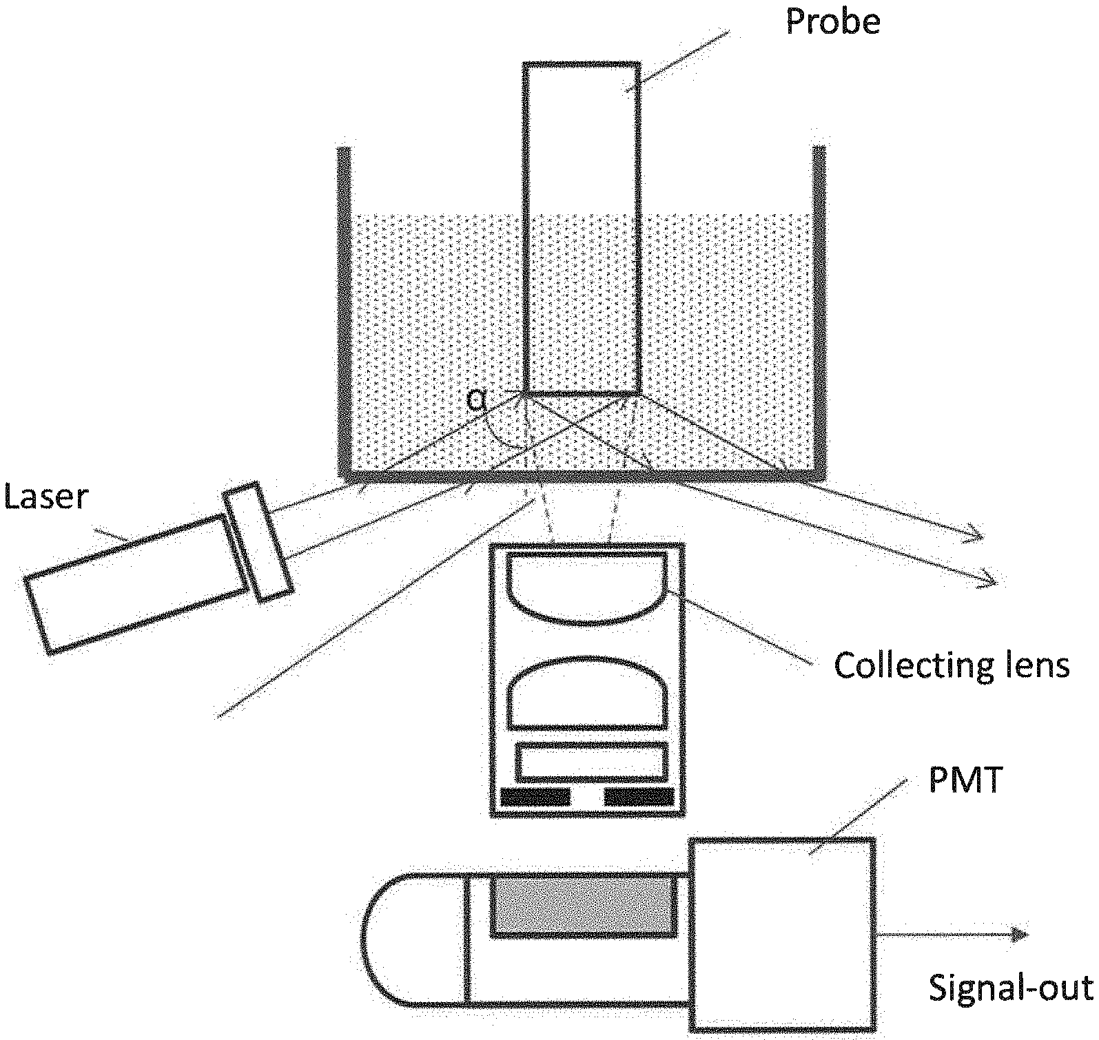
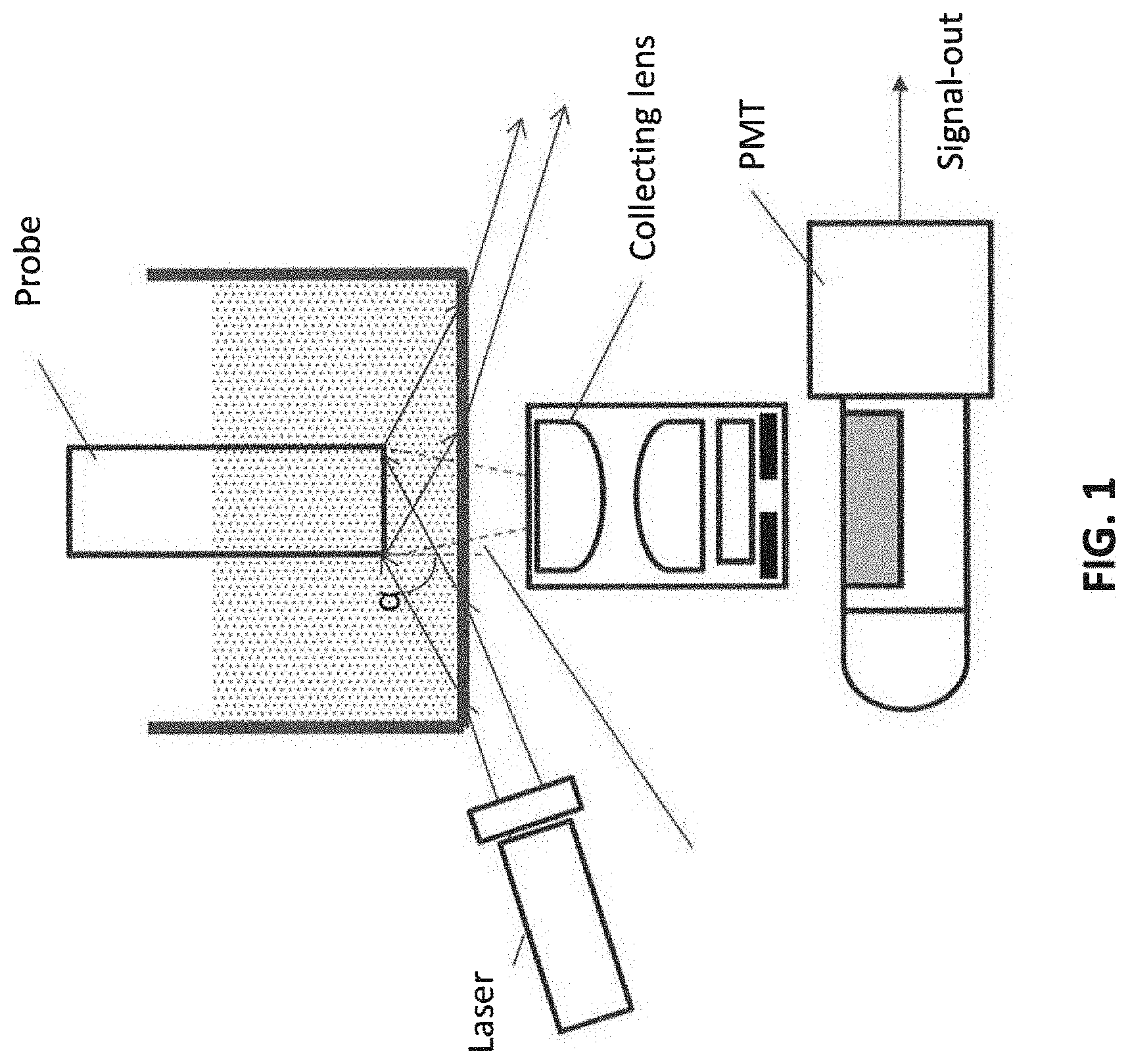
[0006] FIG. 1 illustrates an embodiment of the fluorescent detection system.
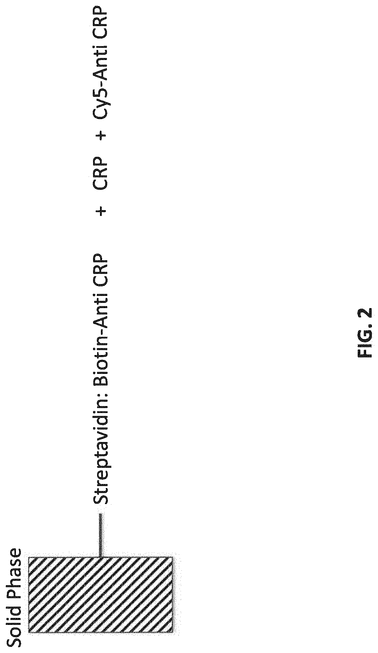
[0007] FIG. 2 illustrates one embodiment of the assay format, in which C-reactive protein (CRP) is an example of an analyte to be measured. The solid phase (probe tip) is immobilized with streptavidin:biotin-anti-CRP antibody.
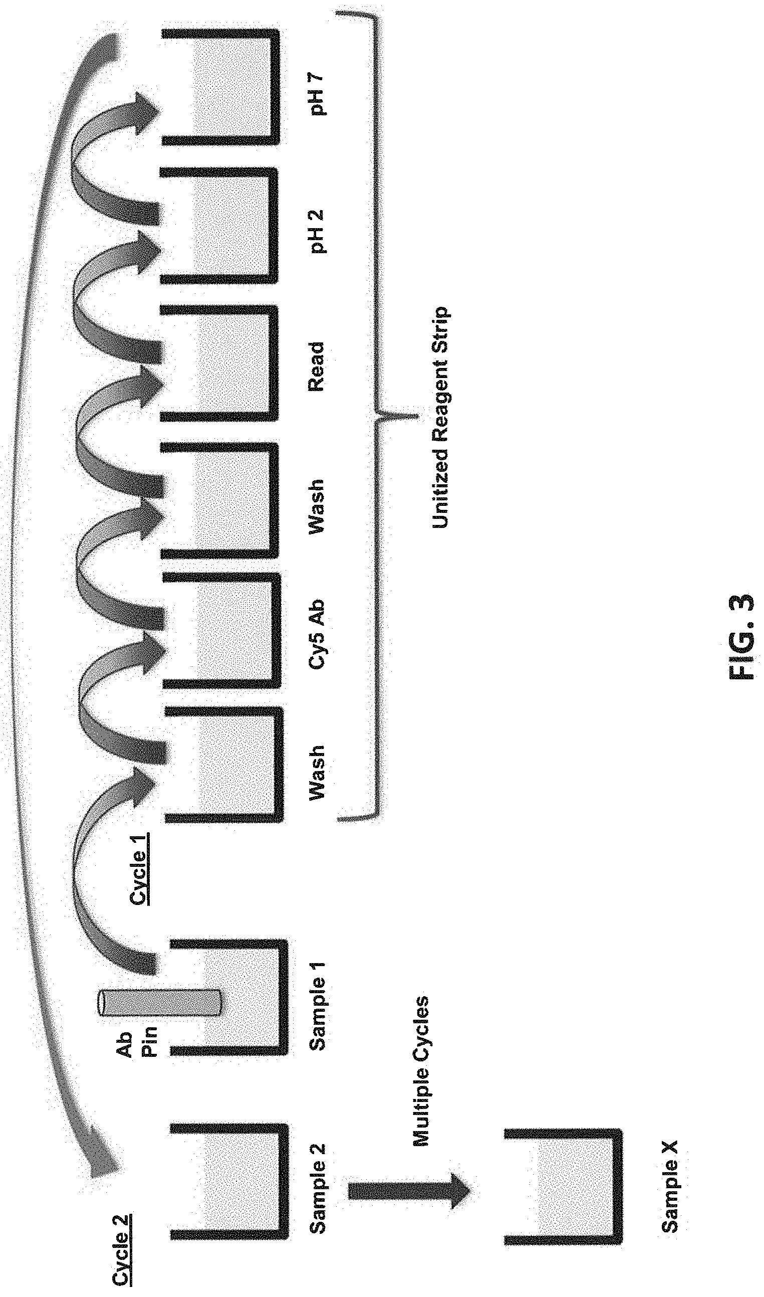
[0008] FIG. 3 illustrates an assay protocol of transferring probe over multiple cycles.
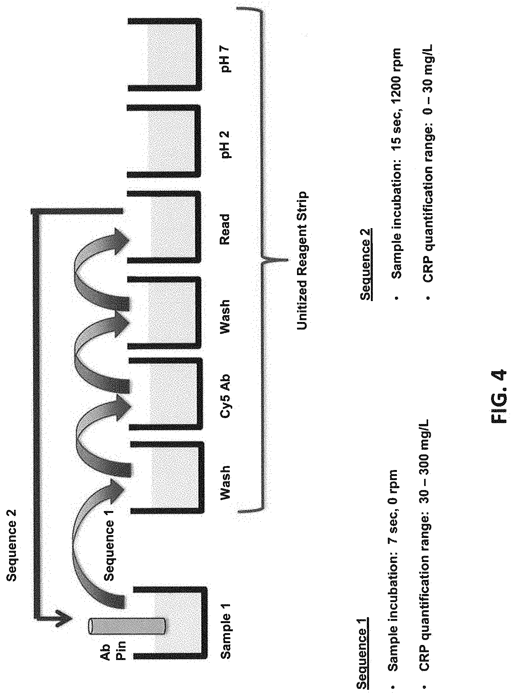
[0009] FIG. 4 illustrates an assay protocol of transferring probe in two sequences of events in a wide-range protocol for detecting analytes that may have a wide range of concentration.
[0010] FIG. 5 illustrates the fluorescent signals of CRP samples at 30, 100, and 300 mg/L over 20 cycles of measurements (Sequence 1) by re-using the same test tube, with CRP30 antibody as a capture antibody and C5 antibody as a signal antibody. The results show consistent fluorescent signals from Cycle 1 to Cycle 20.
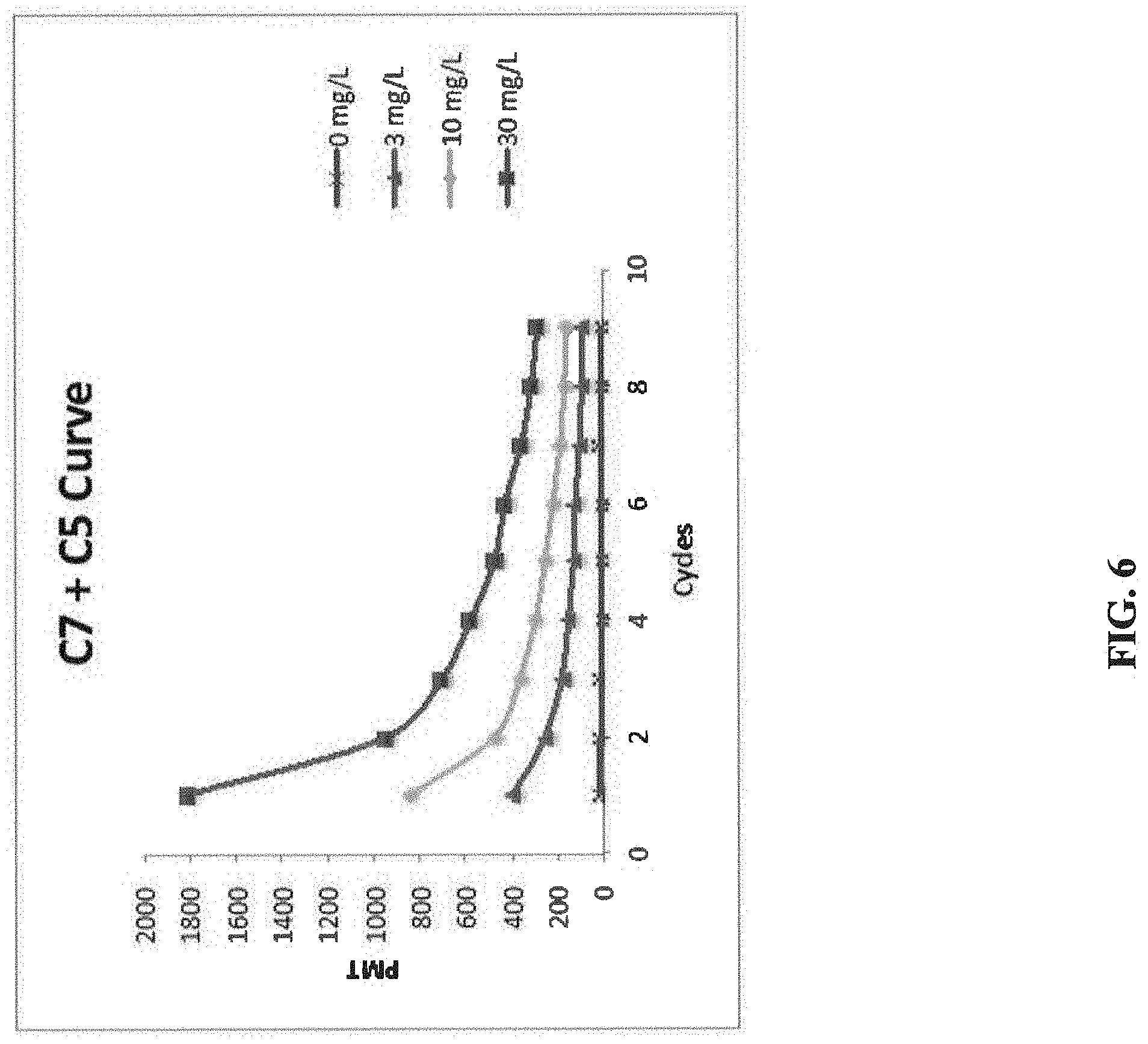
[0011] FIG. 6 illustrates the fluorescent signals of CRP samples at 30, 100, and 300 mg/L over 9 cycles of measurements (Sequence 1) by re-using the same test tube, with C7 antibody as a capture antibody and C5 antibody as a signal antibody. The results show that fluorescent signals dropped significantly from Cycle 1 to Cycle 9.
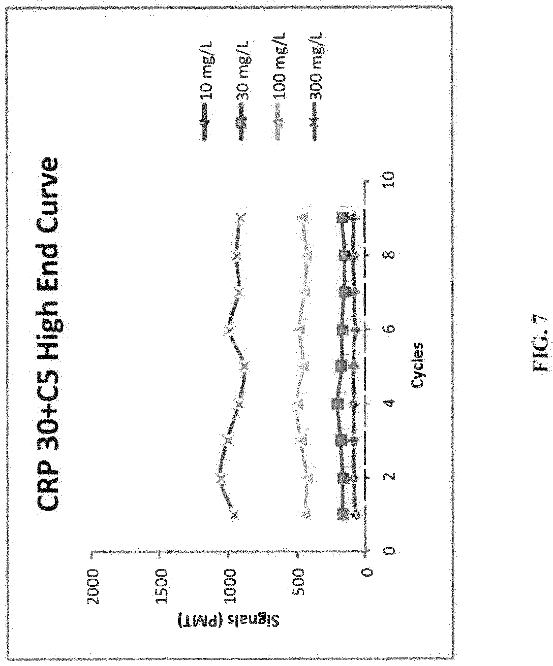
[0012] FIG. 7 illustrates the fluorescent signals of CRP samples at 10, 30, 100, and 300 mg/L over 9 cycles of measurements (Sequence 1) by re-using the same test tube, with CRP30 antibody as a capture antibody and C5 antibody as a signal antibody.
[0013] FIG. 8 illustrates the fluorescent signals of CRP samples at 0, 3, and 10 mg/L over 9 cycles of measurements (Sequence 2) by re-using the same test tube, with CRP30 antibody as a capture antibody and C5 antibody as a signal antibody.
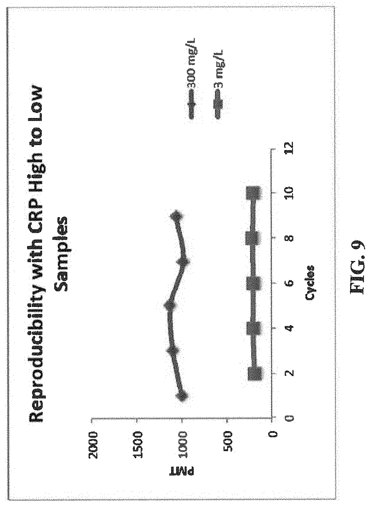
[0014] FIG. 9 illustrates the reproducibility of fluorescent signals of CRP high to low samples at 3 and 300 mg/L, with CRP30 antibody as a capture antibody and C5 antibody as a signal antibody.
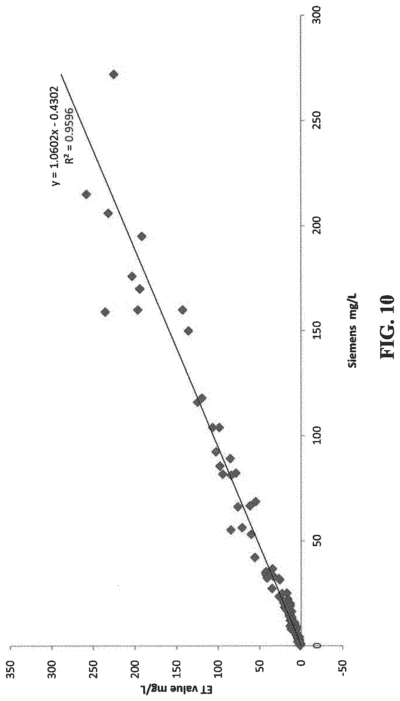
[0015] FIG. 10 show the correlation of the CRP results of 100 clinical samples measured by the present protocol and by an established clinical instrument, the Siemens BN II.
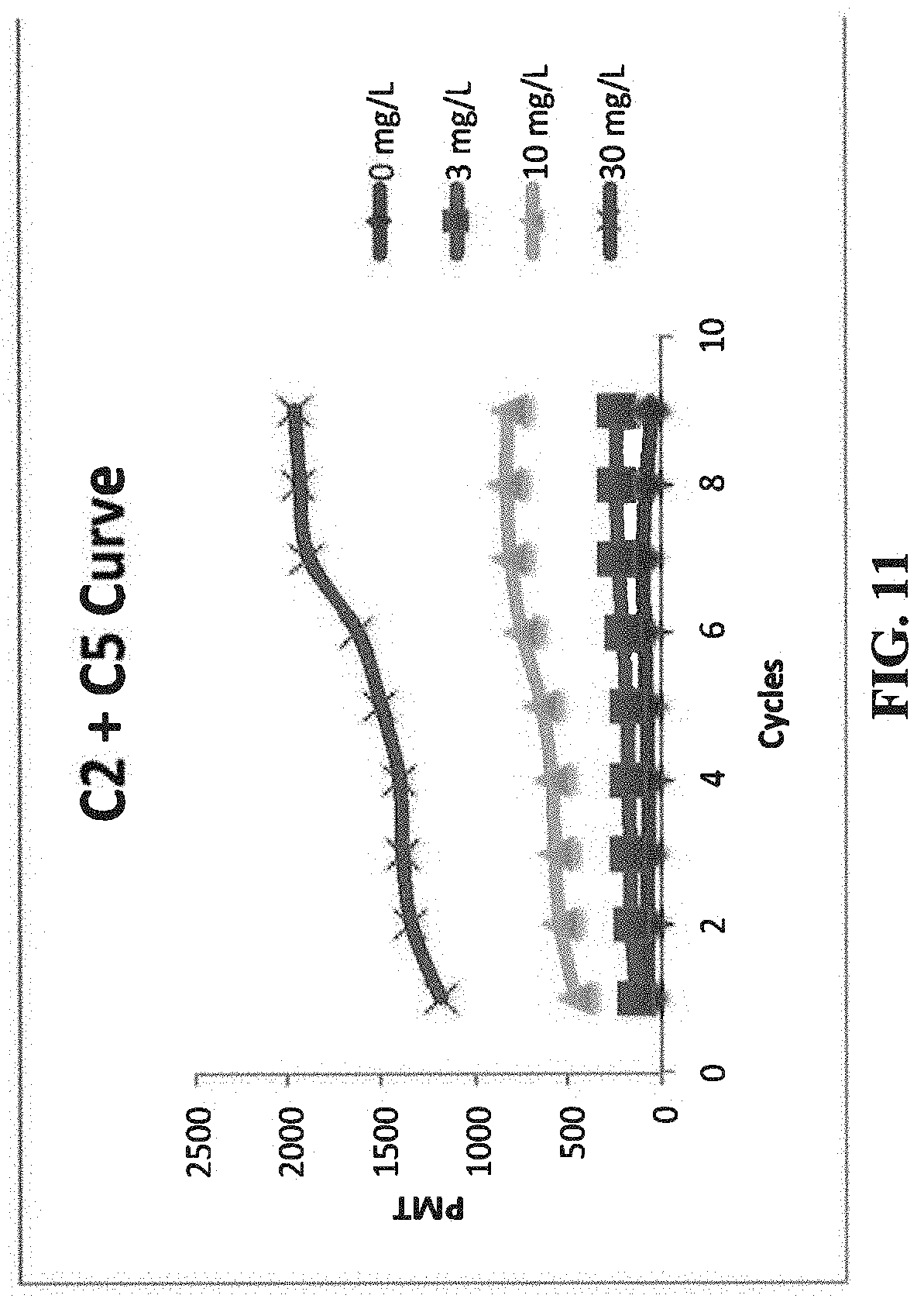
[0016] FIG. 11 illustrates the fluorescent signals of CRP samples at 0, 3, 10, and 30 mg/L over 9 cycles of measurements, with C2 antibody as a capture antibody and C5 antibody as a signal antibody.
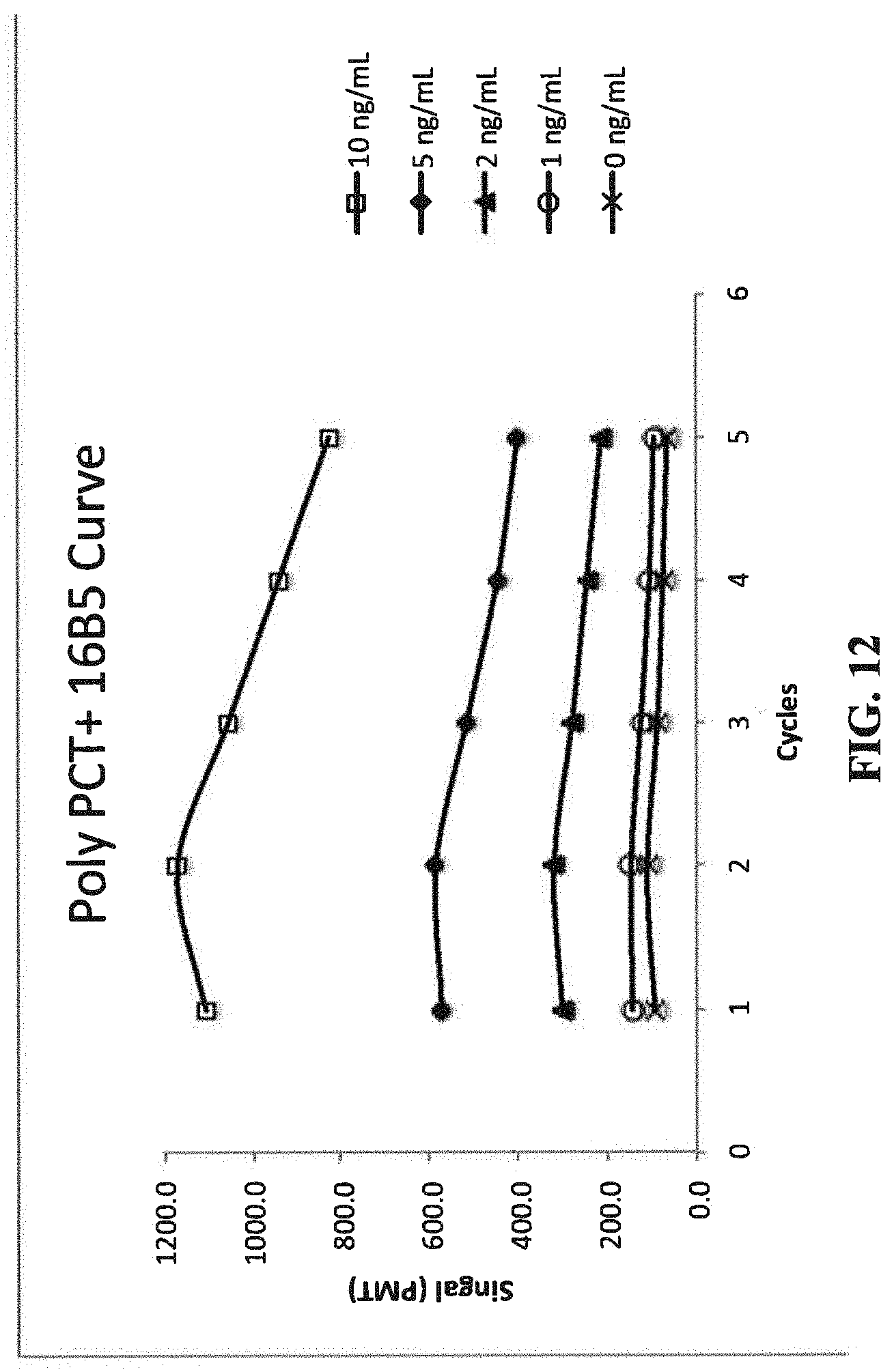
[0017] FIG. 12 illustrates the fluorescent signals of PCT samples at 0, 1, 2, 5, and 10 ng/mL over 5 cycles of measurements, with a polyclonal antibody as a capture antibody and C16B5 monoclonal antibody as a signal antibody.
DETAILED DESCRIPTION OF THE INVENTION
Definitions
[0018] Terms used in the claims and specification are to be construed in accordance with their usual meaning as understood by one skilled in the art except and as defined as set forth below.
[0019] "About," as used herein, refers to within .+-.15% or within .+-.10% of the recited value.
[0020] An "analyte-binding" molecule, as used herein, refers to any molecule capable of participating in a specific binding reaction with an analyte molecule. Examples include but are not limited to, (i) antigen molecules, for use in detecting the presence of antibodies specific against that antigen; (ii) antibody molecules, for use in detecting the presence of antigens; (iii) protein molecules, for use in detecting the presence of a binding partner for that protein; (iv) ligands, for use in detecting the presence of a binding partner; or (v) single stranded nucleic acid molecules, for detecting the presence of nucleic acid binding molecules.
[0021] An "aspect ratio" of a shape refers to the ratio of its longer dimension to its shorter dimension.
[0022] A "binding molecular," refers to a molecule that is capable to bind another molecule of interest.
[0023] "A binding pair," as used herein, refers to two molecules that are attracted to each other and specifically bind to each other. Examples of binding pairs include, but not limited to, an antigen and an antibody against the antigen, a ligand and its receptor, complementary strands of nucleic acids, biotin and avidin, biotin and streptavidin, lectin and carbohydrates. Preferred binding pairs are biotin and streptavidin, biotin and avidin, fluorescein and anti-fluorescein, digioxigenin/anti-digioxigenin. Biotin and avidin, including biotin derivatives and avidin derivatives such as streptavidin, may be used as intermediate binding substances in assay protocols employing complex binding sequences. For example, antibodies may be labeled with biotin ("biotinylated") and used to bind to a target substance previously immobilized on a solid phase surface. Fluorescent compositions according to the present invention employing an avidin or streptavidin may then be used to introduce the fluorescent label.
[0024] "Immobilized," as used herein, refers to reagents being fixed to a solid surface. When a reagent is immobilized to a solid surface, it is either be non-covalently bound or covalently bound to the surface.
[0025] "A monolithic substrate," as used herein, refers to a single piece of a solid material such as glass, quartz, or plastic that has one refractive index.
[0026] A "probe," as used herein, refers to a substrate coated with a thin-film layer of analyte-binding molecules at the sensing side. A probe has a distal end and a proximal end. The proximal end (also refers to probe tip in the application) has a sensing surface coated with a thin layer of analyte-binding molecules.
[0027] A "wide range concentration", as used herein, refers to a concentration range over at least 50-fold, 100 fold, or 500-fold.
[0028] The present invention discloses a method to re-use an immunoassay test probe and reagents, from about 3 to 20 times, while maintaining acceptable clinical assay performance. The immunoassay test probe and reagents may be contained in one test strip, or one cartridge. The present invention re-uses test probe and reagents the and saves the cost on a per test basis.
[0029] There are several key elements to practice the invention. First, the invention regenerates the test probe by employing a denaturing reagent that disassociates the immune complexes bound to the antibodies immobilized on a solid phase, but does not denature or disassociate the antibodies bound to the solid phase to a degree that affects the assay performance. The denaturation step conditions the solid phase antibody for subsequent binding steps to other antigen containing samples. Second, the probe tip has a small dimension (.ltoreq.5 mm in diameter) so that there is negligible consumption of the reagents, and no replenish of the reagents is necessary during the assay cycles. Third, the assay utilizes the same test probe and the same reagents necessary to perform a complete assay, which facilitates multiple assay cycles without additional reagents.
Fluorescent Detection System
[0030] The present invention uses a fluorescent detection system as described in U.S. Pat. No. 8,492,139, which is incorporated herein by reference, for measuring a fluorescent signal on a probe tip. The system comprises: (a) a probe having an aspect ratio of length to width at least 5 to 1, the probe having a first end and a second end, the second end having a sensing surface bound with a fluorescent label; (b) a light source for emitting excitation light directly to the probe's sensing surface; (c) a collecting lens pointed toward the sensing surface; and (d) an optical detector for detecting the emission fluorescent light; where the collecting lens collects and directs the emission fluorescent light to the optical detector.
[0031] The probe can be a monolithic substrate or an optical fiber. The probe can be any shape such as rod, cylindrical, round, square, triangle, etc., with an aspect ratio of length to width of at least 5 to 1, preferably 10 to 1. Because the probe is dipped in a sample solution and one or more assay solutions during an immunoassay, it is desirable to have a long probe with an aspect ratio of at least 5 to 1 to enable the probe tip's immersion into the solutions. Heterogeneous assays can be performed where the long probe is transferred to different reaction chambers. Dispensing and aspirating reagents and sample during the assay are avoided. The sensing surface of the probe is coated with analyte-binding molecules and bound with fluorescent labels.
[0032] Any light source that can emit proper excitation light for the fluorescent label is suitable for the present invention. A prefer light source is a laser that can emit light with wavelengths suitable for fluorescent labels. For example, the laser center wavelength is preferred to be 649 nm for Cy5 fluorescent dye. A suitable optical detector for detecting emission light is a photomultiplier tube (PMT), a charge coupled device (CCD), or a photodiode.
[0033] The light source and the optical detector including the collecting lens are mounted on the same side of the probe tip surface (the sensing surface). If the sensing surface faces down, they are both mounted below the tip surface. If the sensing surface faces up, they are both mounted above the tip surface. They are closer to the sensing surface than the other end of the probe. The sensing surface is always within the numeric aperture of the collecting lens. The probe can be, but does not have to be centrally aligned with the collecting lens.
[0034] FIG. 1 illustrate an embodiment of the fluorescent detection system.
Detecting an Analyte by a Recycling Protocol
[0035] The present invention is directed to a method of detecting an analyte in multiple liquid samples by a fluorescent immunoassay, using the same test probe and test reagents for different sample. The method comprises the steps of: (a) obtaining a probe having a first antibody immobilized on the tip of the probe, wherein the diameter of the tip surface is .ltoreq.5 mm, preferably .ltoreq.2 mm; (b) dipping the probe in a pre-read vessel comprising an aqueous solution having pH of 6.0-8.5 to pre-read the fluorescent signal of the probe tip; (c) dipping the probe tip into a sample vessel containing a liquid sample having an analyte; (d) dipping the probe tip into a reagent vessel containing a reagent solution comprising a second antibody conjugated with one or more fluorescent labels to form an immunocomplex among the analyte, the first antibody, and the second antibody on the probe tip, wherein the first antibody and the second antibody are antibodies against the analyte; (e) dipping the probe tip into a washing vessel containing a wash solution; (f) determining the analyte concentration in the first sample by measuring the fluorescent signal of the immunocomplex at the probe tip, subtracting the pre-read fluorescent signal of (b), and quantitating against a calibration curve; (g) dipping the probe tip in an acidic solution having pH about 1.0-4.0 to elute the immunocomplex from the probe tip, and (h) repeat steps (b)-(f) 1-20 times, preferably 1-10 times, with a next liquid sample in a next sample vessel in a next cycle, whereby the analyte in multiple liquid samples is detected. The method uses the same probe and the same washing solution in all cycles of reaction. Preferably, the method uses the same reagent solution in all cycles of reaction. However, a fresh reagent solution can also be used in different cycles.
[0036] In step (a) of the present method, a probe that has a small tip for binding an analyte is obtained. The tip has a smaller surface area with a diameter .ltoreq.5 mm, preferably .ltoreq.2 mm or .ltoreq.1 mm. The small surface of the probe tip endows it with several advantages. In a solid phase immunoassays, having a small surface area is advantageous because it has less non-specific binding and thus produces a lower background signal. Further, the reagent or sample carry over on the probe tip is extremely small due to the small surface area of the tip. This feature makes the probe tip easy to wash, and causes negligible contamination in the wash solution since the wash solution has a larger volume. Another aspect of the small surface area of the probe tip is that it has small binding capacity. Consequently, when the probe tip is immersed in a reagent solution, the binding of the reagent does not consume a significant amount of the reagent. The reagent concentration is effectively unchanged. Negligible contamination of the wash solution and small consumption of the reagents enable the reagents and the wash solution to be re-used many times, for example, 3-20 times.
[0037] Methods to immobilize a first antibody to the solid phase (the sensing surface of the probe tip) are common in immunochemistry and involve formation of covalent, hydrophobic or electrostatic bonds between the solid phase and antibody. The first antibody, also called capture antibody for its ability to capture the analyte, can be directly immobilized on the sensing surface. For example, a first antibody can be first immobilized either by adsorption to the solid surface or by covalently binding to aminopropylsilane coated on the solid surface. Alternatively, the first antibody can be indirectly immobilized on the sensing surface through a binding pair. For example, the first antibody can be labeled with biotin by known techniques (see Wilchek and Bayer, (1988) Anal. Biochem. 171:1-32), and then be indirectly immobilized on the sensing surface coated with streptavidin. Biotin and streptavidin are a preferred binding pair due to their strong binding affinity, which does not dissociate during the low pH (pH 1-4) regeneration steps of the present method. The capture antibody immobilized on the sensing surface must be able to survive the denaturation condition when the probe sensing surface is regenerated to remove the immunocomplex bound to the sensing surface after the immunoreaction. The capture antibody immobilized on the sensing surface must not lose a significant amount of activity or significantly disassociate from the solid phase so that the immunoassay performance is compromised.
[0038] In step (b), the fluorescent signal of the probe tip is pre-read by a fluorescent detection system in a read vessel (or a read chamber, or a read well). The read vessel contains an aqueous solution such as water or a buffer having pH between 6.0 to 8.5. Preferably, the aqueous solution contains 1-10 mM or 1-100 mM of phosphate buffer, tris buffer, citrate buffer or other buffer suitable for pH between 6.0-8.5, to neutralize the probe after low pH regeneration. Pre-read is necessary before the first sample binding to establish a baseline of any potential background fluorescence for the first cycle reaction. Pre-read is also necessary after the regeneration of the probe tip and before the next sample binding to establish a baseline for subsequent cycles. After each cycle, the pre-read signal can be the same, or higher, or lower than the pre-read signal of the previous cycle, due to the change of the binding property of the immobilized capture antibody caused by the denaturing condition. The inventor has discovered that for certain capture antibodies, the fluorescent signal at the completion of each cycle of reaction after subtracting the pre-read signal, remains constant for 20 cycles of reaction using the same probe and the same reagents. The inventor has also discovered that for other capture antibodies, the fluorescent signal continuously goes up or down slightly after each cycle of reaction, even after subtracting the pre-read signal. The acid treatment could alter the protein on the surface of the probe so that either the capture antibody binding capacity changes or the fluorescence signal is altered. Fluorescence is known to be very sensitive to environmental effects. In spite of the increase or decrease in fluorescence at each cycle, consistent quantification is obtained in such case with a cycle specific calibration; i.e., the fluorescent signal at the completion of each cycle of reaction, after subtracting the pre-read signal, is quantitated against a cycle-specific calibration curve included in the system.
[0039] In step (c) of the method, the probe tip is dipped into a sample vessel (or a sample chamber or a sample well), and incubated for 5 seconds to 5 minutes, 10 seconds to 2 minutes, or 30 seconds to 1 minute, to bind the analyte to the first antibody on the probe tip.
[0040] After step (c), the probe is optionally washed 1-5 times, preferably 1-3 times in a wash vessel (or a wash chamber or a wash well) containing a wash solution. This extra washing step may not be required because the amount of the carried-over solution is minimal due to a small binding surface area. The wash solution typically contains buffer and a surfactant such as Tween 20.
[0041] In step (d) of the method, the probe tip is dipped into a reagent vessel (or a reagent chamber or a reagent sell) for 5 seconds to 5 minutes, 10 seconds to 2 minutes, or 30 seconds to 1 minute, to bind the reagent to the analyte on the probe tip. The reagent solution comprises a fluorescent labelled second antibody (a signal antibody). Any suitable fluorescent label can be used in this method. An example of a fluorescent label is an arylsulfonate cyanine fluorescent dye as described in Mujumdar et al. (1993) Bioconjugate Chemistry, 4:105-111; Southwick et al. (1990) Cytometry, 11:418-430; and U.S. Pat. No. 5,268,486. Cy5 is a preferred arylsulfonate cyanine fluorescent dye, because it has a high extinction coefficient and good quantum yield; it also has fluorescent emission spectra in a range (500 nm to 750 nm) outside of the auto-fluorescence wavelengths of most biological materials and plastics. In addition, Cy5 has a good solubility in water, and has low non-specific binding characteristics.
[0042] A fluorescent label can covalently bind to a second antibody through a variety of moieties, including disulfide, hydroxyphenyl, amino, carboxyl, indole, or other functional groups, using conventional conjugation chemistry as described in the scientific and patent literature. Exemplary techniques for binding arylsulfonate cyanine fluorescent dye labels to antibodies and other proteins are described in U.S. Pat. Nos. 5,268,486; 5,650,334; the contents of which are in incorporated herein by reference. Techniques for linking a preferred Cy5 fluorescent label to antibodies are described in a technical bulletin identified as Cat. No. A25000, published by Biological Detection Systems, Inc., Pittsburgh, Pa.
[0043] In Step (e), the probe is washed 1-5 times, preferably 1-3 times in a wash vessel containing a wash solution. The wash solution typically contains a buffer and a surfactant such as Tween 20.
[0044] In step (f), the probe stays in the wash vessel or is moved to a measurement vessel and the fluorescent signal of the bound immunocomplex is detected by the fluorescent detection system as described above, where the light source and the detector are mounted at the same side (the proximal side) of the sensing surface of the probe. The measurement vessel can be a separate well or can be the same pre-read vessel.
[0045] Alternatively, the methods of the present invention can be detected by other suitable fluorescent detection systems.
[0046] The analyte concentration in the sample is determined by measuring the fluorescent signal of the immunocomplex at the probe tip, subtracting the pre-read fluorescent signal of (b), and then quantitating against a calibration curve (a standard curve).
[0047] The calibration curve is typically pre-established before assaying the samples according to the methods known to a person skilled in the art. In a preferred embodiment, the fluorescent signals (after subtracting the pre-read signal) of the same sample remain constant at each cycle, and the calibration curves are the same for each cycle. In another embodiment, the fluorescent signals (after subtracting the pre-read signal) of the same sample increase or decrease at each cycle, and a cycle-specific calibration curve needs to be established for each cycle. In these instances with changes in fluorescent signals, samples are quantitated against a cycle-specific calibration curve, and the quantitated results can still be consistent in spite of the increase or decrease of the fluorescent signals at different cycles.
[0048] In step (g), the probe is regenerated by employing a denaturing condition that dissociates the immune complexes bound to the capture antibody on a solid phase, but does not denature or dissociate the capture antibody from the solid phase to a degree that affects the assay performance. In general, an acid or an acidic buffer having pH about 1 to about 4 is effective to regenerate the antibody probe of the present invention. For example, hydrochloric acid, sulfuric acid, nitric acid, acetic acid can be used to regenerate the probe. The probe is first treated with an acidic condition, and then neutralized by a neutral aqueous solution such as a buffer having pH between 6.0-8.5. In one embodiment, the low pH treated probe is conveniently neutralized in the read vessel of step (b) before pre-read. Alternatively, the low pH treated probe can be neutralized in a separate vessel having a buffer with a pH of 6.0-8.5. The regeneration procedures can be one single acidic treatment, followed by neutralization. For example, a single pH 1-3, or pH 1.5-2.5 (e.g., pH 2) exposure ranging from 10 seconds to 2 minutes is effective. The regeneration procedures can also be a "pulse" regeneration step, where the probe is exposed to 2-5 cycles (e.g. 3 cycles) of a short pH treatment (e.g., 10-20 seconds), followed by neutralization at pH 6.5-8.0 (e.g., 10-20 seconds).
[0049] After regeneration of the probe, steps of (b)-(g) are repeated with a different sample in a subsequent cycle, for 1-10, 1-20, 1-25, 3-20, 5-20, 5-25, or 5-30 times, with the same probe and the same reagents.
[0050] In one embodiment, the reaction is accelerated by agitating or mixing the solution in the vessel. For example, a flow such as a lateral flow or an orbital flow of the solution across the probe tip can be induced in one or more reaction vessels, including sample vessel, reagent vessel, wash vessels, and regeneration vessel, to accelerates the binding reactions, dissociation. For example, the reaction vessels can be mounted on an orbital shaker and the orbital shaker is rotated at a speed at least 50 rpm, preferably at least 200 rpm or at least 500 rpm, such as 50-200 or 500-1,500 rpm. Additionally, the probe tip can be moved up and down and perpendicular to the plane of the orbital flow, at a speed of 0.01 to 10 mm/second, in order to induce additional mixing of the solution above and below the probe tip.
Detecting an Analyte having a Wide Concentration Range by a Regeneration Protocol
[0051] In one embodiment, the present recycle method as described above is modified to add a second sequence of binding events for quantitating an analyte that has a wide range concentration in a single assay without having to dilute the sample and repeating the assay. In this embodiment, each cycle of the immunoassay has two sequences of events each including sample binding to probe, binding reactions, and detection. In general, the assay conditions of the first sequence are optimized for samples at the high concentration end of the relevant clinical range, and the assay conditions of the second sequence are optimized for low concentration end of the relevant clinical range. After the first sequence of binding and detecting, the probe is re-dipped into the same sample vessel to bind additional analyte in the sample vessel to the probe in a more favorable binding condition (e.g., longer reaction time and/or agitation) than the binding condition in the first cycle (see FIG. 4). The analyte concentration is detected in both cycles, and the combined results provide the ability of quantitating an analyte that has a wide range concentration in a single assay without having to dilute the sample and re-do the assay.
[0052] The combined recycling and wide-range protocol comprises the steps of: (i) obtaining a probe having a first antibody immobilized on the tip of the probe, wherein the diameter of the tip surface is .ltoreq.5 mm, preferably .ltoreq.2 mm; (ii) dipping the probe in a pre-read vessel comprising an aqueous solution to pre-read the fluorescent signal of the probe tip, (iii) dipping the probe tip into a first sample vessel containing a first sample solution having an analyte (for example, for 10 seconds to 2 minutes and flowing the sample solution in the sample vessel at 0-500 rpm) to bind the analyte to the first antibody on the probe tip; (iv) dipping the probe tip into a reagent vessel containing a reagent solution comprising a second antibody conjugated with fluorescent labels, to form an immunocomplex of the analyte, the first antibody, and the second antibody, wherein the first antibody and the second antibody are antibodies against the analyte; (v) dipping the probe tip into a washing vessel containing a wash solution to wash the probe tip; (vi) measuring a first fluorescent signal of the first immunocomplex formed on the probe tip; (vii) dipping the probe tip into the same sample vessel for a time period longer than that in step (iii) (for example, 1-30 minutes), and flowing the sample solution in the first sample vessel (at 0-1200 rpm, preferably 200-1200 rpm or 200-1000 rpm), to bind additional analyte in the first sample to the first antibody on the probe tip; (viii) repeating step (iv) with a longer incubation time and repeating step (v); (ix) measuring a second fluorescent signal of the second immunocomplex formed on the probe tip; and (x) determining the analyte concentration in the first sample by first subtracting the pre-read fluorescent signal of (b) from the first and second fluorescent signals, and then quantitating the analyte concentration against a high-end calibration curve or a low-end calibration curve; (xi) dipping the probe tip in an acidic solution having pH about 1.0-4.0 to elute the immunocomplex from the probe tip, and (xii) repeating steps (ii)-(xi) with a next liquid sample in a next sample vessel in a next cycle, whereby the analyte in multiple liquid samples is detected. The method uses the same probe and the same washing solution in all cycles of reaction. Preferably, the method uses the same reagent solution in all cycles of reaction. However, a fresh reagent solution can also be used in different cycles.
[0053] In the above method, steps (iii)-(vi) are the first sequence of binding events for binding an analyte having a high concentration. Steps (vii) and (viii) are the second sequence of binding events for binding an analyte having a high concentration. After the two sequences of events and measurements, the probe tip is then regenerated by steps (xi) and (ii), and then steps (iii)-(x) are repeated for the next cycle for quantitate a next sample. Unless otherwise specified, the reagents and wash solutions and procedures are the same or similar to those described in the recycling/regeneration protocol above.
Unitized Immunoassay Strips
[0054] The present invention is further directed to a cartridge (a strip) for an immunoassay test. This unitized cartridge can be used for 2-20, or 3-20 cycles to measure 2-20, or 3-20 different samples. The cartridge comprises (a) a probe well comprising a probe and a cap, the cap being in a closed position to enclose the probe in the probe well, wherein the probe has a bottom tip coated with a first antibody; (b) a sample well to receive a sample; (c) a reagent well; (d) one or more wash wells each containing a wash solution; (e) a low pH well to provide pH of 1-4, (f) a neutralization well to provide a buffer having pH 6.0-8.5; and (g) a measurement well (a read well) having a light transmissive bottom, the measurement well containing an aqueous solution; wherein the openings of the sample well, reagent well, measurement well and wash wells are sealed. In one embodiment, the neutralization well and a measurement well (a read well) are the same well. In another embodiment, the neutralization well and a measurement well are two separate wells.
[0055] The cartridge is similar to that described in U.S. Pat. No. 8,753,574, which is incorporated herein by reference in its entirety; except that the cartridge of this invention contains additional low pH well and neutralization well.
[0056] A sample well is a well that receives a sample containing an analyte. A sample well can be a blank well, or it can contain detergents, blocking agents and various additives for the immunoassay, either in a dry format or in a wet (liquid) format.
[0057] A reagent well contains reagents such as a fluorescent labelled antibody that reacts with the analyte to form an immunocomplex and generate a signal for detection. The reagents can be in a wet format or in a dry format. The wet format contains a reagent in an assay buffer. The wet format is typically in a small liquid volume (<10 .mu.L, e.g., 5 .mu.L). An assay buffer typically includes a buffer (e.g., phosphate, tris), a carrier protein (e.g., bovine serum albumin, porcine serum albumin, and human serum albumin, 0.1-50 mg/mL), a salt (e.g., saline), and a detergent (e.g., Tween, Triton). An example of an assay buffer is phosphate buffered saline, pH 7.4, 5 mg/ml bovine serum albumin, 0.05% Tween 20. The assay buffer optionally contains a blocking agent in an amount of 1-500 .mu.g/mL. The final formulation will vary depending on the requirements of each analyte assay. The dry format is the dry form of the reagent in an assay buffer. The dry format includes lyophilization cake, powder, tablet or other formats typical in diagnostic kits. The dry format is to be reconstituted to a wet format by a reconstitution buffer or a wash buffer.
[0058] The cartridge comprises one or more washing wells each containing an aqueous solution. The wash wells contain a wash buffer to wash the probe after binding steps in the sample well and reagent well. One to four wash wells (e.g., 1, 2, 3, or 4 wells) are dedicated for washing after each binding step. Wash buffers contain detergents. Any detergent typically used in immunoassays (e.g., Tween, Triton) can be used in this invention.
[0059] The cartridge comprises a measurement well having an optically clear bottom that enables the detection of the labeled-immunocomplex bound to the bottom tip of the probe. The measurement is through the bottom of the well.
[0060] In one embodiment, the cartridge further comprises one or more reconstitution wells that contain reconstitution buffer to be dispensed into the sample well and reagent well to reconstitute the dry forms in the sample well and reagent well. The reconstitution buffer can be simply a buffer such as phosphate-buffer saline. The reconstitution buffer can additionally include other additives (carrier protein, blockers, detergents, etc.) contained in the assay buffer.
[0061] The openings of the reagent well and wash well(s) are sealed with a foil or a film. The seal is penetrable. The wells may be opened by piercing the seal by a manual or automated device. In one embodiment, when the cap of the probe is in a closed position, the cap is folded over the probe well to enclose the probe in the probe well, but the cap does not cover the sample well, the wash wells or the measurement well.
Probe Comprising an Immobilized Antibody
[0062] The inventor has discovered that for certain antibodies such as mouse anti-human CRP monoclonal antibody CRP30 (an IgG1 isotype) from Hytest (Turku, Finland), when used as a capture antibody in the present method, the fluorescent signals after each cycle of reaction and regeneration remains constant for at least 10 cycles using the same probe and the same reagents. Because the capture antibody anti-CRP antibody CRP30 provides consistent fluorescent signals through multiple regeneration cycles, such effect enables sample quantification with a single calibration curve, and thus provides convenience and high precision.
[0063] The inventor has discovered that for some antibodies, such as mouse monoclonal anti-human CRP antibody C7 from HyTest, mouse monoclonal anti-human CRP antibody C2 from HyTest, and goat polyclonal anti-procalcitonin antibody PPC3 from Hytest, when used as capture antibodies in the present method, the fluorescent signals after each cycle of reaction and regeneration changes.
[0064] The acid treatment could alter the protein on the surface of the probe to cause the change of the capture antibody binding capacity. In spite of the change of fluorescent signal at each cycle, consistent quantification of an analyte concentration may be obtained in such case with a cycle specific calibration; i.e., the fluorescent signal at the completion of each cycle of reaction, after adjusted by the pre-read fluorescent signal, is quantitated against a cycle-specific calibration curve included in the system.
[0065] Although cycle-specific calibration curve could resolve the change of fluorescent signals of some capture antibodies after regeneration of the probe by low pH, it is advantageous to use a capture antibody that does not change the fluorescent signal after regeneration of the probe by low pH. CRP, like most quantitative immunoassays employed in clinical laboratories, has a defined set of performance parameters that must be met to have clinical utility. Minimum detection limit, analytical range, and precision are examples of such performance parameters. With CRP30 antibody, the assay conditions can be established and remain unchanged during multiple recycles using a single calibration while maintaining its assay performance parameters. Capture antibodies that produce variable fluorescent signals after regeneration by low pH require cycle-specific calibration; in addition, assay parameters are difficult to maintain. Since cycle specific calibration introduces an additional variable, imprecision between cycles is greater. This is a drawback since clinical assays require high precision with coefficient of variation (CV) <10%. Antibodies that lose activity and generate declining fluorescent signal after low pH treatment typically have a difficulty to maintain precision, minimum detection limit, and analytical range due to decreasing signals.
[0066] The present invention provides a probe comprising an antibody immobilized on the tip of the probe, wherein the probe has an aspect ratio of length to width of at least 5 to 1, the diameter of the probe tip surface is .ltoreq.5 mm, and the antibody does not substantially denature or dissociate from the probe after an acidic treatment; i.e., no more than 15%, preferably no more than 10% or 5% of the antibody is denatured or dissociated from the probe after 1-20 cycles of the acid treatment. The acid treatment is typically performed by dipping the probe in a low pH buffer (pH 1-4, or 1-3, or 1.5-2.5) for 10 seconds to 2 minutes. In one embodiment, the antibody is labelled with biotin and is indirectly immobilized on the probe tip by streptavidin coated on the probe tip.
[0067] In one embodiment, the present invention is directed to a probe comprising a monolithic substrate coated with an antibody at the probe tip, wherein the antibody is mouse monoclonal anti-human C-reactive protein antibody CRP30, the probe has an aspect ratio of length to width of at least 5 to 1, and the diameter of the probe tip surface is .ltoreq.5 mm. In one embodiment, the CRP30 antibody is biotin-labeled, and is bound to streptavidin directly immobilized onto the substrate. The probe comprising CRP30 as a captured antibody is useful for an immunoassay because the probe survives an acid regeneration and yields consistent dose response curves for at least 20 regeneration cycles.
[0068] The invention is illustrated further by the following examples that are not to be construed as limiting the invention in scope to the specific procedures described in them.
EXAMPLES
Example 1. Preparing Antibody-Coated Probe
[0069] Quartz probes, 1 mm diameter and 2 cm in length, were coated with aminopropylsilane using a chemical vapor deposition process (Yield Engineering Systems, 1224P) following manufacturer's protocol. The probe tip was then immersed in a solution of streptavidin (Sigma-Aldrich), 10 .mu.g/ml in phosphate buffered saline pH 7.4 (PBS). After allowing the protein to adsorb to the probe for 5 minutes, the probe tip was washed in PBS. The probe tip was then immersed in a solution containing a biotin labeled antibody at 10 .mu.g/ml in PBS. After 10 minutes the probe tip was washed in PBS. The antibodies were biotinylated by a standard method. The biotinylated antibodies were designated as "capture antibody".
Example 2. Preparing Cy5 Labeled Antibody
[0070] Antibody at 3.2 mg/ml in 1 ml 0.1 M sodium carbonate pH 9.5 was mixed with 10.6 .mu.l Cy5-NHS (GE Healthcare) at 10 mg/ml DMF and allowed to react for 1/2 hour at 30 C. The mixture was then purified on a PD 10 column (GE Healthcare). The Cy5 labeled antibodies were designated as "signal antibody".
Example 3. C-Reactive Protein Immunoassay Protocol
[0071] FIG. 2 shows the basic assay format consisting of a biotinylated anti C-reactive protein (CRP) antibody bound to streptavidin immobilized to the probe tip. The streptavidin in this case serves a spacer preventing the antibody from interacting directly with the probe surfaces, potentially interfering with its binding activity.
[0072] FIG. 3 is a schematic of an assay protocol. After incubation in sample, the antibody (Ab)-coated probe is transferred to a wash well and followed by incubation with the Cy5 labeled second antibody. After the incubation with the second Ab, a wash cycle is carried out, then the fluorescence is measured at the probe tip to complete the assay. Immersion of the probe in a low pH buffer, pH 2, dissociates the CRP immune complex and immersion in a pH 7 buffer conditions the probe for a subsequent sample analysis. The streptavidin:biotin-Ab complex remains intact on the probe tip during the low pH exposure. Typically, much harsher denaturation conditions, such as 8M urea or 6M guanidine, are required to disassociate biotin from streptavidin.
[0073] In a subsequent sample analysis, the probe and all the reagents to perform the assay are re-used.
Example 4. C-Reactive Protein Immunoassay (Wide Concentration Range)
[0074] FIG. 4 illustrates the probe transfer sequence in a "wide concentration range" protocol, which can quantitate a wide range of analyte concentration (e.g., 30-300 mg/L CRP) in a single assay. Sequence 1 quantitates analyte having high concentration (30-300 mg/L) and sequence 2 quantitates samples with low concentration (0-30 mg/L). The combined quantitation provides the quantitation over a wide range of over 100-fold concentration.
[0075] Three sets of samples were measured for fluorescent signals by the following protocol. Set 1 has 20 different samples each having a CRP concentration of 30 mg/L in assay buffer (0.5 mg/mL bovine serum albumin (BSA), phosphate-buffered saline, 0.05% Tween 20, pH 7.4). Set 2 has 20 different samples each having a CRP concentration of 100 mg/L in assay buffer. Set 3 has 20 different samples each having a CRP concentration of 300 mg/L in assay buffer. The same reagents were used for 20 cycles of measurements of each set of 20 different samples.
Sequence 1 (High Concentration Detection)
[0076] 1. Pre-read [0077] 2. First sample (CRP) incubation: 7 seconds 0 RPM [0078] 3. Three wash: 7 seconds 1200 RPM [0079] 4. Cy5_C5 incubation: 7 seconds 1200 RPM [0080] 5. Three wash: 7 seconds 1200 RPM [0081] 6. 1.sup.st read
Sequence 2 (Low Concentration Detection)
[0081] [0082] 7. Same first sample (CRP) incubation: 15 seconds 1200 RPM [0083] 8. Three wash: 7 seconds 1200 RPM [0084] 9. Cy5_C5 incubation: 15 seconds 1200 RPM [0085] 10. Three wash: 15 seconds 1200 RPM [0086] 11. 2.sup.nd read
Pulse Regeneration
[0086] [0087] 12. Regeneration buffer (pH 2.0): 10 sec 500 RPM [0088] 13. PBS (pH 7.4): 10 sec 500 RPM [0089] 14. Repeat 12 [0090] 15. Repeat 13 [0091] 16. Repeat 12 [0092] 17. Repeat 13 [0093] 18. Return to 1, for subsequent cycles for different samples
Example 5. C-Reactive Protein Immunoassay Using CRP30 or C7 as Capture Antibody
[0094] This example follows the same protocols as described in Example 4.
[0095] In the first experiment, Hytest mouse anti-human CRP monoclonal antibody CRP30 was used as a capture antibody and Hytest mouse anti-human CRP monoclonal antibody C5 was used as a signal antibody. FIG. 5 illustrates the fluorescent signals of CRP samples at 30, 100, and 300 mg/L over 20 cycles of measurements (Sequence 1) by re-using the same test tube, with CRP30 antibody as a capture antibody and C5 antibody as a signal antibody. The results show that the fluorescent signals are consistent after each cycle, which indicates that the capture antibody CRP30 retained its activity after each low pH regeneration treatment. The average fluorescent signals and coefficients of variation (CV) are summarized in Table 1.
TABLE-US-00001 TABLE 1 CRP (mg/L) Signal Average Signal SD CV (%) 30 155 16 10 100 424 22 5 300 937 54 6
[0096] In the second experiment, a different Hytest mouse anti-human CRP monoclonal antibody C7 was used as a capture antibody, and the same Hytest mouse anti-human CRP monoclonal antibody C5 was used as a signal antibody. FIG. 6 illustrates the fluorescent signals of CRP samples at 30, 100, and 300 mg/L over 9 cycles of measurements (Sequence 1) by re-using the same test tube, with C7 antibody as a capture antibody and C5 antibody as a signal antibody. The results show that when C7 antibody was used as a capture antibody, the fluorescent signals decreased after each cycle, which indicates that the capture antibody C7 lost its activity after each low pH regeneration treatment.
Example 6. C-Reactive Protein Immunoassay (Wide Concentration Range Protocol)
[0097] Six sets of samples were measured for fluorescent signals by the same protocol in Example 4. Each set have PBS samples with CRP concentration of 0, 3, 10, 30, 100, or 300 mg/L. Each sample was measured 9 times in 9 cycles. The same reagents were used for 9 cycles of measurements of each set of samples.
[0098] The first step in this protocol is a "pre-read" to measure the background fluorescence associated with the probe. Table 2 shows the "pre-read: signals taken before each cycle from the CRP samples. Pre-read data indicate that the acid elution is not complete with residual fluorescence on the probe after each cycle and before next cycle. The results show that the fluorescent signal in the "pre-reads" increases with each cycle, however, subtraction of the "pre-reads" yield the consistent results at each cycle as shown in FIGS. 7 and 8.
TABLE-US-00002 TABLE 2 (Pre-Read Signals) CRP Cycles (mg/L) 1 2 3 4 5 6 7 8 9 10 0 97 121 156 175 195 212 229 244 259 282 1 153 196 237 265 288 310 333 351 369 388 3 95 115 150 173 195 215 234 246 259 277 10 95 122 160 190 217 240 261 281 300 321 30 91 138 195 238 238 277 310 339 366 417 100 84 147 210 257 306 351 393 430 470 508 300 93 147 209 262 308 352 393 434 474 513
[0099] The fluorescent signals of high-end curve (Sequence 1, n=9) are shown in FIG. 7 and Table 3.
TABLE-US-00003 TABLE 3 CRP (mg/L) Signal Average Signal SD CV (%) 10 80 4 5 30 168 15 9 100 453 25 5 300 954 55 6
[0100] The fluorescent signals of low-end curve (Sequence 2, n=20) are shown in FIG. 8 and Table 4.
TABLE-US-00004 TABLE 4 CRP (mg/L) Signal Average Signal SD CV (%) 0 30 5 3 231 14 6 10 617 8 1
[0101] FIG. 7 depicts the assay signals of high CRP samples from 10 to 300 mg/L, and FIG. 8 shows second measurements of assay signals of low CRP samples from 0 to 10mg/L. Assay signals were consistent out for 9 cycles for all CRP levels measured.
Example 7. Measuring CRP in High Concentration to Low Concentration Sample
[0102] In clinical practice, CRP samples are assayed in a random sequence. To evaluate whether the assay of a very high sample could bias results of a subsequent low sample, we assayed 10 cycles of a 300 mg/L sample followed by a 3 mg/L sample. The 300 mg/L is at the top of the quantification range representing a CRP level associated with extreme inflammation, while 3 mg/L is within the normal range. FIG. 9 shows the results of CRP assays of the two sets of samples; the results show that the fluorescent signals (after subtracting pre-read signal) are consistent for both 300 mg/L and 3 mg/L, with negligible bias alternating between the high and low samples. The fluorescent signals are summarized in Table 5.
TABLE-US-00005 TABLE 5 CRP (mg/L) Average (n = 5) SD CV (%) 3 202 8 4 300 1051 69 7
Example 8. Comparison Study
[0103] This experiment compares the CRP results of 100 clinical samples measured by the present protocol as shown in Example 4, and by an established clinical instrument, the Siemens BN II.
[0104] 100 samples were quantified by the present method using 10 test strips. Each strip assayed 10 randomly selected samples in 10 cycles of re-using the same test probe. The Siemens results were obtained following the standard protocol from the manufacture. The result comparison of the present method vs. Siemens method is shown in FIG. 10, which shows that the results generated by the present method are highly correlated to those generated by the Siemens method with R.sup.2 being 0.9596 (R=correlation coefficient).
Example 9. CRP Assay with Capture Antibody C2
[0105] Another capture antibody (Hytest mouse monoclonal anti-human CRP C2) was evaluated by the same protocol as described in Example 4. FIG. 11 shows that CRP signals increased with each cycle, even after subtraction of pre-reads. The acid treatment could alter the protein on the surface of the probe so that either the antibody binding capacity increases or the fluorescence signal is altered. Fluorescence is known to be very sensitive to environmental effects.
[0106] In order to address the increase in fluorescence signal at each cycle, a standard curve is established in each cycle, and the quantitation of analyte in each cycle is calculated against the cycle-specific standard curve. A CRP sample of 6.0 mg/mL in assay buffer was tested over 9 cycles and quantitated against cycle-specific standard curve to determine reproducibility. The results show an average of 6.1 mg/mL, with standard deviation of 0.5 mg/mL, and CV of 9%.
[0107] In spite of the increase in fluorescence signal at each cycle, consistent quantification was achieved by running a calibration curve for each cycle.
Example 10. Procalcitonin Assay
[0108] The following protocol lists the steps for the assays of procalcitonin (PCT).
Sequence 1 (High Concentration Detection)
[0109] 1. Pre-read [0110] 2. First sample (PCT) incubation: 15 seconds 0 RPM [0111] 3. Three wash: 7 seconds 1200 RPM [0112] 4. Cy5_16B5 incubation: 15 seconds 1200 RPM [0113] 5. Three wash: 7 seconds 1200 RPM [0114] 6. 1.sup.st read
Sequence 2 (Low Concentration Detection)
[0114] [0115] 7. First sample (PCT) incubation: 360 seconds 1200 RPM [0116] 8. Three wash: 7 seconds 1200 RPM [0117] 9. Cy5_16B5 incubation: 60 seconds 1200 RPM [0118] 10. Three wash: 15 seconds 1200 RPM [0119] 11. 2.sup.nd read
Regeneration
[0119] [0120] 12. Regeneration buffer (pH 2.0): 10 sec 500 RPM [0121] 13. PBS (pH 7.4): 10 sec 500 RPM [0122] 14. Return to 1, for a subsequent cycle on a different sample
[0123] The PCT protocol is similar to that of CRP (see Example 4), except that steps 2 and 7 have longer incubations to account for the much lower concentrations of PCT. Also, the PCT protocol only uses a single 10 second, pH 2.0 regeneration. The capture antibody is a goat polyclonal anti-PCT (Hytest, PPC3) and the signal antibody is a monoclonal anti-PCT (Hytest, 16B5).
[0124] The fluorescent signals of 5 cycles are depicted in FIG. 12; by 5 cycles, the fluorescent signals show a decline.
[0125] In order to address the decrease in fluorescence signal at each cycle, a standard curve is established for each cycle, and the quantitation of analyte in each cycle is calculated against the cycle-specific standard curve. A PCT sample of 5.0 ng/mL in assay buffer was tested over 5 cycles and quantitated against cycle-specific standard curve to determine reproducibility. The results show an average of 4.8 ng/mL, with standard deviation of 0.4 ng/mL, and CV of 9%.
[0126] In spite of the decrease in fluorescence signal at each cycle, consistent quantification was achieved by running a calibration curve for each cycle.
[0127] The invention, and the manner and process of making and using it, are now described in such full, clear, concise and exact terms as to enable any person skilled in the art to which it pertains, to make and use the same. It is to be understood that the foregoing describes preferred embodiments of the present invention and that modifications may be made therein without departing from the scope of the present invention as set forth in the claims. To particularly point out and distinctly claim the subject matter regarded as invention, the following claims conclude this specification.
* * * * *
D00000
D00001
D00002
D00003
D00004
D00005
D00006
D00007
D00008
D00009
D00010
D00011
D00012
XML
uspto.report is an independent third-party trademark research tool that is not affiliated, endorsed, or sponsored by the United States Patent and Trademark Office (USPTO) or any other governmental organization. The information provided by uspto.report is based on publicly available data at the time of writing and is intended for informational purposes only.
While we strive to provide accurate and up-to-date information, we do not guarantee the accuracy, completeness, reliability, or suitability of the information displayed on this site. The use of this site is at your own risk. Any reliance you place on such information is therefore strictly at your own risk.
All official trademark data, including owner information, should be verified by visiting the official USPTO website at www.uspto.gov. This site is not intended to replace professional legal advice and should not be used as a substitute for consulting with a legal professional who is knowledgeable about trademark law.