Electrochemical Detection Of Bacterial And/or Fungal Infections
Freeman-Cook; Lisa Lynn ; et al.
U.S. patent application number 16/858471 was filed with the patent office on 2020-10-22 for electrochemical detection of bacterial and/or fungal infections. This patent application is currently assigned to Clinical Micro Sensors, Inc. (dba GenMark Diagnostics, Inc.). The applicant listed for this patent is Clinical Micro Sensors, Inc. (dba GenMark Diagnostics, Inc.). Invention is credited to Anna Maria Al-Khouri, Bradley Adam Brown, Lisa Lynn Freeman-Cook, John Jay Harvey, Milena Iacobelli Martinez, Christine J. Shaw.
| Application Number | 20200332346 16/858471 |
| Document ID | / |
| Family ID | 1000004939346 |
| Filed Date | 2020-10-22 |


View All Diagrams
| United States Patent Application | 20200332346 |
| Kind Code | A1 |
| Freeman-Cook; Lisa Lynn ; et al. | October 22, 2020 |
ELECTROCHEMICAL DETECTION OF BACTERIAL AND/OR FUNGAL INFECTIONS
Abstract
The present disclosure relates to methods and devices for amplifying a plurality of targets in a single PCR run while distinguishing between clinically relevant amplification and amplification from other sources such as from background contamination. The methods and devices further enable discrimination between gram-positive, gram-negative and fungal infections as wells as identify antimicrobial resistance genes. When applying the methods and devices of the invention, the species or genus of an infection(s), and genus of a fungal co-infection(s) or category of bacterial (gram-positive or negative) co-infection(s) are identified. Species identification of co-infections can also be achieved. Further, when applying the methods and devices of the invention, organisms which are likely to be contaminating organisms from a blood draw are identified.
| Inventors: | Freeman-Cook; Lisa Lynn; (Carlsbad, CA) ; Shaw; Christine J.; (San Diego, CA) ; Martinez; Milena Iacobelli; (Vista, CA) ; Al-Khouri; Anna Maria; (San Diego, CA) ; Brown; Bradley Adam; (San Marcos, CA) ; Harvey; John Jay; (San Marcos, CA) | ||||||||||
| Applicant: |
|
||||||||||
|---|---|---|---|---|---|---|---|---|---|---|---|
| Assignee: | Clinical Micro Sensors, Inc. (dba
GenMark Diagnostics, Inc.) Carlsbad CA |
||||||||||
| Family ID: | 1000004939346 | ||||||||||
| Appl. No.: | 16/858471 | ||||||||||
| Filed: | April 24, 2020 |
Related U.S. Patent Documents
| Application Number | Filing Date | Patent Number | ||
|---|---|---|---|---|
| 16286476 | Feb 26, 2019 | 10669592 | ||
| 16858471 | ||||
| 16054960 | Aug 3, 2018 | 10273535 | ||
| 16286476 | ||||
| 15828074 | Nov 30, 2017 | 10106847 | ||
| 16054960 | ||||
| 15686001 | Aug 24, 2017 | |||
| 15828074 | ||||
| Current U.S. Class: | 1/1 |
| Current CPC Class: | C12Q 2600/16 20130101; B01L 2300/0819 20130101; C12Q 1/689 20130101; B01L 2300/0627 20130101; C12Q 1/686 20130101; B01L 3/5027 20130101; G16H 15/00 20180101; B01L 3/5023 20130101; C12Q 1/6853 20130101; C12Q 1/6895 20130101; G01N 27/3277 20130101; G01N 27/48 20130101 |
| International Class: | C12Q 1/689 20060101 C12Q001/689; C12Q 1/686 20060101 C12Q001/686; C12Q 1/6853 20060101 C12Q001/6853; B01L 3/00 20060101 B01L003/00; C12Q 1/6895 20060101 C12Q001/6895; G01N 27/327 20060101 G01N027/327; G01N 27/48 20060101 G01N027/48 |
Claims
1. A method for processing at least a first patient sample comprising: a) receiving a first patient sample; b) receiving a test order from a hospital laboratory information system (LIS) for the first patient sample; c) processing the first patient sample in a target analyte detection instrument wherein the target analyte detection instrument is directly linked to a LIS interchange; d) generating a first result identifying at least one pathogen in the first patient sample processed by the target analyte detection instrument; and e) automatically reporting the first result to the LIS interchange.
2. The method of claim 1, wherein the LIS interchange automatically reports the first result to a hospital LIS.
3. The method of claim 1, wherein the test order is in an HL7 or ASTM format and the LIS interchange converts the test order from the HL7 or ASTM format to a CSV format.
4. The method of claim 1, wherein the LIS interchange connects to more than one hospital LIS.
5. The method of claim 1, wherein the at least one pathogen in the first patient sample processed is a viable gram-positive bacteria and the first patient sample comprises viable gram-positive bacteria and non-viable gram-positive bacteria.
6. The method of claim 5, wherein the target analyte detection instrument performs a single detuned multiplex end-point polymerase chain reaction (PCR) on the first patient sample, the PCR comprising about 30 to about 35 cycles.
7. The method of claim 1, wherein the target analyte detection instrument sends an alert when preventative maintenance for the target analyte detection instrument is needed.
8. The method of claim 1, wherein the target analyte detection instrument sends an alert notifying a target analyte detection instrument operator that the first patient sample has an accession number that is the same as the accession number for a second patient sample.
9. A method for processing at least a first and second patient sample comprising: a) receiving a first and second patient sample; b) receiving a test order from a hospital LIS; c) processing the first and second patient sample, according to the test order, in a target analyte detection instrument wherein the target analyte detection instrument is directly linked to a LIS interchange; d) generating a first result by the target analyte detection instrument that identifies at least one pathogen in the first patient sample and generating a second result from the target analyte detection instrument that identifies at least one value for the second patient sample; and e) automatically reporting the first result and second result to the LIS interchange.
10. The method of claim 9, wherein the LIS interchange automatically reports the first result to a hospital LIS.
11. The method of claim 9, wherein automatically reporting the first result to the LIS interchange comprises generating a first detection report, and wherein the method further comprises manually applying comments to the first detection report.
12. The method of claim 9, wherein the test order is in a flat file format.
13. A method for processing at least a first patient sample comprising: a) receiving a first patient sample; b) receiving a first test order from a hospital LIS; c) loading the first patient sample into a cartridge, the cartridge comprising a machine readable information tag; d) loading the cartridge into a sample processing instrument comprising a first device that scans machine readable identification tags; e) scanning the machine readable identification tag by the first device; f) processing the first patient sample in the sample processing instrument wherein the sample processing instrument is directly linked to a LIS interchange; g) generating a first result identifying at least one pathogen in the first patient sample processed by the sample processing instrument; and h) automatically sending the first result to the LIS interchange.
14. The method of claim 13, wherein the at least one pathogen in the first patient sample is a blood stream pathogen that causes or is suspected of causing sepsis.
15. The method of claim 13, wherein the cartridge comprises a second machine readable identification tag and the second machine readable identification tag is scanned by a second device that scans machine readable identification tags prior to loading the cartridge into the sample processing instrument.
16. The method of claim 15, wherein the sample processing instrument discontinues processing the cartridge if the first machine readable identification tag does not match the second machine readable identification tag.
17. The method of claim 13, wherein the first result further identifies the presence of a resistance gene present in the at least one pathogen in the first patient sample.
18. The method of claim 13, wherein the at least one pathogen is a human pathogen, and wherein the first patient sample further comprises a contaminating pathogen.
19. The method of claim 13, wherein the sample processing instrument sends an alert if a threshold for stability is violated for the first patient sample.
20. The method of claim 13, wherein the sample processing instrument performs a single detuned multiplex end-point polymerase chain reaction (PCR) on the first patient sample, the PCR comprising about 30 to about 35 cycles.
Description
CROSS REFERENCE TO RELATED APPLICATIONS
[0001] This is a continuation of U.S. patent application Ser. No. 16/286,476, filed on Feb. 26, 2019, which is a continuation of U.S. patent application Ser. No. 16/054,960, filed Aug. 3, 2018, issued as U.S. Pat. No. 10,273,535, which is a continuation of U.S. patent application Ser. No. 15/828,074, filed on Nov. 30, 2017, now issued as U.S. Pat. No. 10,106,847, which is a continuation of U.S. patent application Ser. No. 15/686,001, filed on Aug. 24, 2017, abandoned. The prior applications are incorporated herein by reference their entirety.
[0002] The invention relates to the field of molecular diagnostic methods, in particular, microfluidic devices for the detection of target analytes.
INCORPORATION BY REFERENCE
[0003] This application is related to U.S. Pat. Nos. 7,820,391, 7,560,237, 6,013,459, 6,740,518, 6,063,573, 6,600,026, 6,264,825, 6,541,617, 6,942,771, 6,432,723, 6,833,267, 7,090,804, 7,935,481, 7,172,897, 6,753,143, 6,518,024, 6,642,046, 6,361,958, 6,602,400, 6,824,669, 6,596,483, 6,875,619, 7,863,035, 9,598,722 and U.S. patent application Ser. Nos. 12/914,257, 14/206,871, 14/206,932, 15/026,314, 14/538,533, 14/206,817, 14/538,602, 14/206,867, 14/206,903, 14/062,860, and 14/538,506, the respective disclosures of which are hereby incorporated by reference.
BACKGROUND OF THE INVENTION
[0004] In North America, the most common causes of a sepsis are bacteria such as Escherichia coli or Staphylococcus aureus. In addition to bacterial infection, fungal infections have in recent times become a significant cause of the disease. Fungal infections tend to be associated with higher rates of death. Only approximately 5% of fungal caused cases of sepsis are identified during the disease due to the poor diagnostic methods available. Recent studies have shown that patients with severe sepsis or septic shock showed an increased likelihood of death of 7.6% for every hour in which antibiotic therapy is not applied. Survival rates are also significantly reduced when antibiotics are not applied within the first 6 hours of identifying hypotension. Survival rates will be significantly increased if diagnosis times are reduced.
[0005] Culturing microorganisms from blood samples (Gram staining) remains the gold standard in detection of the microbiological cause of sepsis. This method is however subject to significant disadvantages, in particular, due to the large time difference between taking a blood sample and providing the results. It is not uncommon that 24 to 72 hours pass between taken a sample and providing diagnostic information. Within this time, broad band, untargeted antibiotic therapies are often introduced. This may lead to some success in treating the disease but is related to significant disadvantages with respect to the development of antibiotic resistant microorganisms.
[0006] Microarray and multiplex PCR approaches have been disclosed in the art, which are typically defined by extremely large numbers of probes or primers required for application of such methods (leading to significant cost and effort), a limited pool of target pathogens capable of being detected (such as only a limited sub-group of bacterial pathogens, or the absence of fungal pathogens), or a lack of discrimination between gram-negative and gram-positive bacterial pathogens, which provides sub-standard information for appropriate antibiotic therapies (US 2009286691 and US 201 1 151453). Methods for discriminating gram-positive and gram-negative bacteria have been disclosed in the art (US 20080118923 A1), in addition to the combined analysis of 16S and 18S sequences of bacteria and fungi (US 20090061446). Such methods, although potentially useful in clinical diagnostics, have never been applied in sepsis analytics and employ large numbers of primers in either microarray or very complex multiplex reactions, representing a significant technical and financial challenge for clinical diagnostic laboratories.
[0007] Multiplex RT-PCR approaches have been described in which a number of commonly occurring human pathogens are potentially detected. One example of such a multiplex PCR method is described in Gosiewski et al (BMC Microbiology 2014, 14:144) and U.S. Publication No. 2015/0232916, which discloses a nested PCR approach for detecting gram-positive and gram-negative bacteria, yeast, fungi and filamentous fungi from blood samples. A nested polymerase chain reaction involves two sets of primers, used in two successive runs of polymerase chain reaction, the second set intended to amplify a secondary target within the first run product. Nested PCR is applied in order to reduce nonspecific binding in products due to the amplification of unexpected primer binding sites, as it is unlikely that any of the unwanted PCR products contain binding sites for both the new primers in the second PCR run, ensuring the product from the second PCR has little contamination from unwanted products of primer dimers, hairpins, and alternative primer target sequences. Despite potentially reducing background signal, the PCR method described in Gosiewski and U.S. Publication No. 2015/0232916 are relatively complex and require two cycling reactions, essentially doubling the time, effort and reagents required for the analysis.
[0008] Other methods have employed the amplification of a number of PCR products from bacterial and fungal pathogens using sequence-specific oligonucleotides together with sequence-unspecific dyes, and subsequently, a melting curve analysis to differentiate between the various products (Horvath et al, BMC Microbiology 2013, 13:300). The method disclosed therein is however limited by a number of disadvantages known to occur with melting curve analyses.
[0009] Other methods have employed the amplification of a number of PCR products from bacterial and fungal pathogens using non-sequence-specific oligonucleotides together with sequence-specific probes to differentiate between the various products as described in EP 3172337. In such cases, only a broadband antibiotic therapeutic approach is possible, which may, in fact, be poorly suited for the particular pathogen.
[0010] Electrochemical detection techniques have higher detection sensitivity than conventional luminescence techniques (e.g., fluorescence and phosphorescence) due to higher signal-to-noise ratios. Because of their sensitivity and ability to accurately measures low-concentrations of nucleic acids, electrochemical detection techniques are able to differentiate between pathogenic species representing a significant technological improvement over the prior art. But, because of their sensitivity, false positive detection rates are high. Indeed, where organisms are cultured, the growth media often contains non-viable organisms or DNA/nucleic acids, which would not affect culture, but could produce false positives in PCR. If a system is designed uniformly for increased sensitivity to detect low titers pathogens, frequent false positive results may occur from background organisms or DNA/nucleic acids. Alternatively, if system sensitivity is reduced to avoid background organism detection, low titer organisms may be missed, resulting in false negative detection.
[0011] Further, when blood or other bodily fluids are obtained from a subject they may be contaminated by skin cells, bacteria, fungi, viruses, phages, their respective nucleic acids (including RNA and DNA) and/or other undesirable molecules, or disinfectants. Antiseptics are crucial for the practice of medicine; however, currently used antiseptics have a significant failure rate which results in substantial additional medical costs. Antiseptics are commonly used prior to routine phlebotomy, in preparation for minor and major invasive procedures, and as part of routine infection control hand-washing practices. The failure of antiseptics often result in erroneous diagnostic tests. For example, it has been estimated that a single false positive blood culture (i.e., where the culture indicates that the blood has been infected with bacteria, although the blood was contaminated during the blood draw) done on blood drawn from a patient at a hospital costs the patient an additional S2000 to S4,200 in unnecessary medication, additional follow up testing, and increased length of stay. (Bates, 1991).
[0012] Thus, there is a need in the art to provide methods which can selectively detect pathogenic organisms of interest. In particular, there is a need in the art for a method which enables the discrimination between a systemic infection and a false positive signal due to blood matrix bottle contamination. There is also a need in the art to identify when a blood culture is contaminated during blood draw.
BRIEF SUMMARY OF THE INVENTION
[0013] The ability to detect infection is hampered by background contamination present in blood culture bottles, such as are used in gram staining, a common first step in any clinical pathogen diagnosis. The invention disclosed herein can not only differentiate between background contamination (from any blood culture matrix bottle) and clinically relevant infection but can also differentiate between gram-positive bacterial infection, gram-negative bacterial infection, fungal infection and can identify antibiotic resistance genes. Even more importantly, the invention can identify the contaminating pathogen and contaminating co-infection (if present) by its species. Because prior art methods failed to recognize background contamination as an issue or cannot discriminate by species the infecting pathogen and co-infecting pathogen, the invention allows for better antimicrobial stewardship and improved patient care outcomes.
[0014] Disclosed herein are in vitro methods (or systems or devices) for the detection and/or identification of a human pathogen and/or genetic material thereof comprising subjecting a sample suspected of comprising a human pathogen and/or genetic material thereof to a single multiplex polymerase chain reaction (PCR), wherein said method (or system) comprises amplification of PCR products that enable discrimination between contaminating pathogen and/or genetic material present in the sample and infectious pathogen and/or genetic material present in the sample. In particular, the inventive method allows for a single amplification step and single detection step for the detection of an actual pathogenic infection and not a putative contamination. The inventive method does not require a purification step prior to amplification or detection. The inventive method does not require determining whether amplification has occurred prior to detection. When purification is needed or a determination as to whether amplification has occurred prior to detection requires human action and such systems cannot be fully automated like the invention disclosure herein. The inventive method does not require dilution of the PCR sample. The inventive method does not require additional testing or analysis to differentiate signaling due to contamination versus real pathogenic infections.
[0015] The methods (or system or devices) can further discriminate between gram-positive bacterial, gram-negative bacterial and fungal pathogens if present in said sample as well as identify antimicrobial resistance genes. An infection can be identified by its species, and a fungal co-infection can be identified by its genus whereas a bacterial co-infection of a different type than the infection (i.e. infection is GP and co infection is GN or vice versa) can be identified by its category (gram-positive or negative). If the infection and co-infection are of the same type (i.e., both gram-positive or both gram-negative), the systems and methods can identify the species of the co-infection via a single PCR run. If the infection and co-infection are of different types (i.e., the infection is gram-positive and the co-infection is gram-negative or fungal) the systems and methods can identify the species of the co-infection via a second single PCR run.
[0016] The methods (or system or devices) can further discriminate between background contamination and de-escalation targets. Background contamination from blood culture bottles is not detected by the methods (or system or devices) but organisms associated with possible contamination by blood draw such as Propionibacterium acnes, Staphylococcus epidermidis, Micrococcus, Lactobacillus or Corynebacterium (so called de-escalation targets) are identified. The methods (or system or devices) can further discriminate between (1) background contamination, (2) de-escalation targets and (3) clinically relevant gram-positive bacterial, gram-negative bacterial or fungal pathogens if present in the sample as well as identify antimicrobial resistance genes.
BRIEF DESCRIPTION OF THE DRAWINGS
[0017] The patent or application file contains at least one drawing executed in color. Copies of this patent or patent application publication with color drawing(s) will be provided by the Office upon request and payment of the necessary fee.
[0018] FIG. 1: Shows a schematic of a hybridization complex.
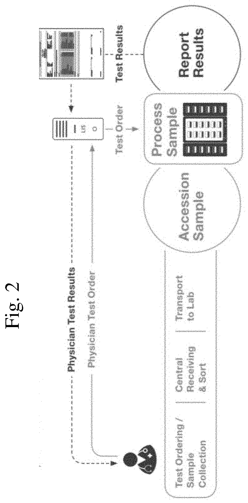
[0019] FIG. 2: Shows a schematic of the bi-directional LIS to automate and accelerate order entry and results reporting.
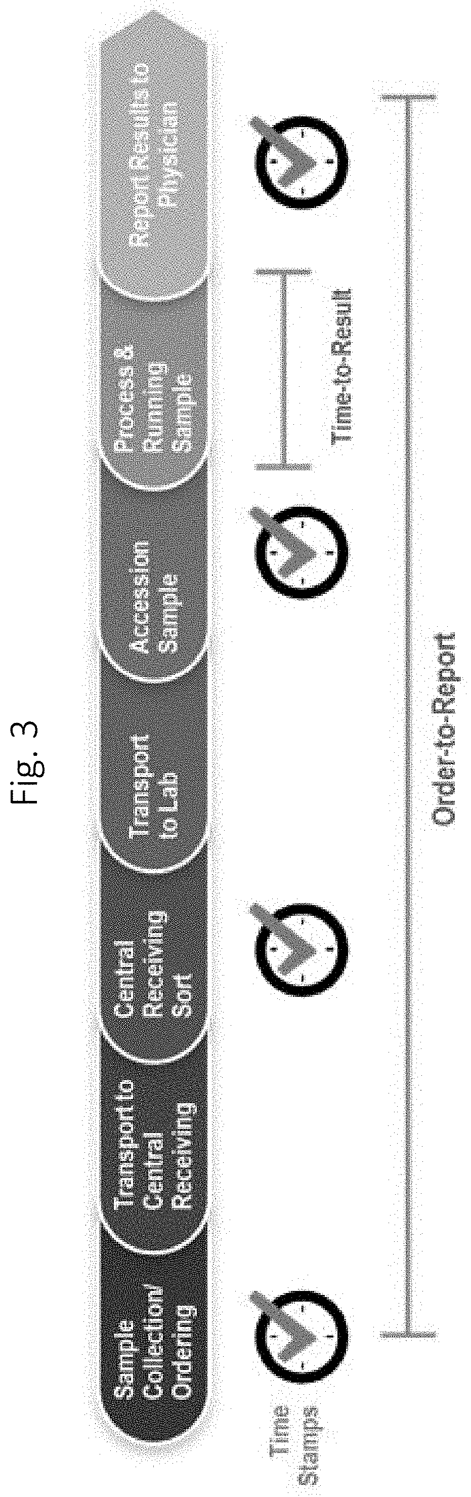
[0020] FIG. 3: Shows a schematic of "Order-to-report." The clock time stamps are commonly documented in hospital and laboratory information systems.
[0021] FIG. 4: BCID-GP, High false positive signals for Enterococcus faecalis, Pan-candida, and Pan-GN in bottle matrix with no blood or bacterial targets.
[0022] FIGS. 5A-5D: False positives detected for Enterococcus faecalis (5A), Staph 16s (5B); Enterococcus (genus) (5C); and Pan-Candida (5D), were detected when negative blood culture matrices (no blood or bacterial targets) were tested on a BCID-GP cartridge.
[0023] FIGS. 6A-6B: FIG. 6A shows Enterococcus faecalis contamination signals are reduced but not eliminated with 35 cycles and FIG. 6B shows that at 35 cycles the S. pombe internal control is still detected.
[0024] FIGS. 7A-7D: False positive signal from blood culture bottles is eliminated using a 30-cycle PCR. FIGS. 7A and 7B show that only the positive controls were detected. FIGS. 7C and 7D show that detection is possible (although weak) at 1.times.LOD (1.times.05 CFU/mL) Enterococcus faecalis run on sLRMs using a 30-cycle PCR.
[0025] FIG. 8: BCID-GP, Background P. acnes signals with 30, 35, and 40-cycle PCR. False Positives are detected with 40 and 35 cycles when BDIC-GP cartridges are spotted with P. acnes primers but eliminated with 30 cycle PCR.
[0026] FIGS. 9A-9B: BCID-GP, P. acnes detection at 1.times.LOD with 30-cycle PCR. When 30 PCR cycles are used, P. acnes is detected at 1.times.LOD (1.times.06 CFU/mL) (9A for P. acnes strain ATCC11827 and 9B for P. acnes strain ATCC6919).
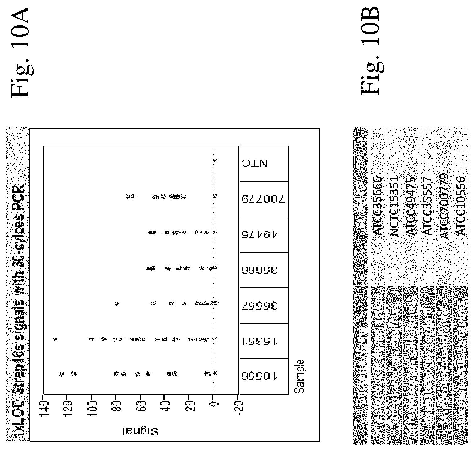
[0027] FIGS. 10A-10B: BCID-GP, Strep spp. assay performance with 30-cycle PCR. FIG. 10A shows that when 30 PCR cycles are used, six representative Streptococcus species identified by their identification number are detected at 1.times.LOD (1.times.06 CFU/mL). FIG. 10B corrolates the bacterial species name and identification number.
[0028] FIG. 11: BCID-GP cartridge with 1.times.LOD Streptococcus spp. When primer concentration is increased, three P. acnes strains and two Streptococcus species are detected at 30 PCR cycles and 1.times.LOD (1.times.106 CFU/mL).
[0029] FIG. 12: Contamination of NTC sLRMs with Streptococcus spp. and P. acnes primers and 30 cycles of PCR. No Streptococcus spp or P. acnes signals were detected with increased primer concentrations and 30 PCR cycles in negative blood culture matrices.
[0030] FIG. 13: BCID-GP, PCR cycles are reduced from 40 to 37. When PCR cycles are reduced from 40 to 37, most blood matrix contamination is eliminated.
[0031] FIG. 14: BCID-GP Multiplex primer pool and PCR cycles.
[0032] FIG. 15: PCB with GP Reagent & 8 PCR Drop Locations: a schematic of the BCID-GP cartridge sub-assembly layout. Reservoirs, R1, R2, R3, R4, and R5 are part of the top plate. R1 typically includes PCR enzyme (Taq) and buffer; R2 typically includes exonuclease; R3 typically includes reconstitution buffer used to wet reagents and rehydrate PCR reagents; R4 typically includes is a waste manipulation zone or is empty for drop manipulation; and R5 is typically where the sample comes out of the LRM after extraction. "Sample" designates where the sample is loaded from the LRM. "PCR enzyme" means Taq and "PCR buffer" is a buffer; the buffer and PCR enzyme are shown where they are spotted on the top plate. "Exo" is exonuclease and is shown where it is spotted on the top plate. 1, 2, 3, 4, 5, 6, 7, 8 displayed vertically next to heater 3 are the multiplex primer pools; once the drop has the primers they go into PCR lane 1, 2, 3 or 4 as shown and cycled (35 or 30) as shown. 1, 2, 3, 4, 5, 6, 7, 8 displayed vertically on Heater 1 or 3 depict where each PCR drop is moved. SPCA means signal probe cocktail which is where the amplicon is mixed with the signal probe. Detection zones A, B, C and D are where detection takes place. The drops 1, 2, 3, 4, 5, 6, 7, 8 are moved into the detection zone as shown. The gold plated electrode is depicted as small round circles in the detection zone. The RTD temperature set points are shown in degrees Celsius.
[0033] FIGS. 16A-16B: Shows false positive signal for Proteus mirabilis (FIG. 16A) and Proteus spp (FIG. 16B) in negative blood culture matrices cycled 40 times (a variety of blood culture bottles are shown).
[0034] FIG. 17: BCID-GN, Negative bottles run with reduced cycling showed no false positives. When negative blood culture matrices were cycled 35 or 30 times (cycling as indicated in FIG. 17) no false positives were detected.
[0035] FIG. 18: BCID-GN Multiplex primer pool and PCR cycles. The bolded organisms are the genus calls in the detection report and the non-bolded organisms are species calls on the detection report.
[0036] FIG. 19: A schematic of the BCID-GN cartridge sub-assembly layout. Reservoirs, R1, R2, R3, R4, and R5 are part of the top plate. R1 typically includes PCR enzyme (Taq) and buffer; R2 typically includes exonuclease; R3 typically includes reconstitution buffer used to wet reagents and rehydrate PCR reagents; R4 typically includes is a waste manipulation zone or is empty for drop manipulation; and R5 is typically where the sample comes out of the LRM after extraction. "Sample" designates where the sample is loaded from the LRM. "PCR buffer" is a buffer; the buffer and taq are shown where they are spotted on the top plate. "Exo" is exonuclease and is shown where it is spotted on the top plate. MP1, MP2, MP3, MP4, MP5, MP6, MP7, MP8 displayed vertically next to heater 3 are the multiplex primer pools; 1, 2, 3, 4, 5, 6, 7, 8 displayed vertically on Heater 1 or 3 depict where each PCR drop is moved. Once the drop has the primers they go into PCR lane 1, 2, 3 or 4 and cycled (35 or 30) as shown in the key below. SPC means signal probe cocktail which is where the amplicon is mixed with the signal probe. Detection zones A, B, C and D is where detection takes place. The drops 1, 2, 3, 4, 5, 6, 7, 8 are moved into the detection zone as shown. The gold plated electrode is depicted as small round circles in the detection zone. The RTD temperature set points are shown in degrees Celsius.
[0037] FIGS. 20A-20B: BCID-FP, Detuning Eliminates False Positives. The signals obtained before and after detuning, for Rhodotorula (FIG. 20A) and Trichosporon (FIG. 20B) are shown.
[0038] FIG. 21: The BCID-FP Multiplex primer pool and PCR cycles.
[0039] FIG. 22: BCID-FP, PCB with Fungal Reagent & PCR Drop Locations. A schematic of the BCID-FP cartridge sub-assembly layout is shown. Reservoirs, R1, R2, R3, R4, and R5 are part of the top plate. R1 typically includes PCR enzyme (Taq) and buffer; R2 typically includes exonuclease; R3 typically includes reconstitution buffer used to wet reagents and rehydrate PCR reagents; R4 typically includes is a waste manipulation zone or is empty for drop manipulation; and R5 is typically where the sample comes out of the LRM after extraction. "Sample" designates where the sample is loaded from the LRM. "PCR buffer" is a buffer; the buffer and taq are shown where they are spotted on the top plate. "Exo" is exonuclease and is shown where it is spotted on the top plate. PM1, PM 2, PM3 and PM4 displayed vertically next to heater 3 are the multiplex primer pools 1-4; MP1, MP2, MP3 and MP4 means PCR primer mixes; 1, 2, 3, 4 displayed vertically on Heater 3 depict where each PCR drop is moved. Once the drop has the primers they go into PCR lane 1, 2, 3 or 4 and cycled 40 times. SPC means signal probe cocktail which is where the amplicon is mixed with the signal probe. Detection zones A, B, C and D is where detection takes place. The drops 1, 2, 3, and 4 are moved into the detection zone as shown. The gold plated electrode is depicted as small round circles in the detection zone. cDT-8 and cDT-9 are the controls used. cDT-8 uses a ferrocene derivative QW56. cDT-9 uses a ferrocene derivative QW56. The RTD temperature set points are shown in degrees Celsius.
[0040] FIG. 23: A notice showing that if the BCID Panel is used to test BacT/ALERT SN bottles, positives for Pseudomonas aeruginosa and Enterococcus should be reconfirmed by another method prior to reporting the test results.
SEQUENCE LISTING
[0041] The Sequence Listing submitted as an ASCII text file [9823-100100-07_Sequence_Listing.txt, Apr. 24, 2020, 2.27 KB] is incorporated by reference herein.
Definitions
[0042] "Target nucleic acid," or "analyte of interest", or "target molecule" or "human pathogen nucleic acid", include genes, portions of genes, regulatory sequences of genes, mRNAs, rRNAs, tRNAs, siRNAs, cDNA and may be single stranded, double stranded or triple stranded. As discussed herein, target nucleic acids are DNA from human pathogens, and are naturally occurring nucleic acids, as contrasted to the nucleic acids of capture probes and signal probes, which may include non-naturally occurring components. Some nucleic acid targets have polymorphisms, single nucleotide polymorphisms, deletions and alternate splice sequences, such as allelic variants. Multiple target domains may exist in a single molecule, for example, a target nucleic acid may have a first target domain that binds the capture probe and a second target domain that binds a signal probe, and/or distinct primer binding sequences. Target nucleic acids are not generally provided with the cartridge as manufactured, but are contained in the liquid sample to be assayed; in contrast, "control analytes" or "control nucleic acids" are typically provided with the cartridge or are routinely present in a sample of a particular type and are assayed in order to ensure proper performance of the assay. Spiked samples may be used in certain quality control testing and for calibration, as is well known in the art. The target analyte is also referred to as "clinically relevant amplification" or "systemic infection" or "pathogen of interest" and is distinguished from, for example, contamination.
[0043] As used herein, the term "sample" is used in its broadest sense. In one sense, it is meant to include a specimen or culture obtained from any source, as well as biological and environmental samples. Biological samples may be obtained from animals (including humans) and encompass fluids, solids, tissues, and gases. Biological samples include blood products, such as plasma, serum and the like. In contrast with some commercial systems that require some off chip handling of the sample, generally including sample extraction (cell lysis, for example), and sample preparation prior to detection. Thus, in accordance with aspects of the current system, a sample is loaded onto a BCID cartridge and the target analyte is extracted, amplified as necessary (for example, when the target analyte is a nucleic acid using polymerase chain reaction (PCR) techniques, although isothermal amplification methods can be utilized as well), and then detected using electrochemical detection, all on a microfluidic platform, generally referred to herein as a "multiplex cartridge" or a "fluid sample processing cartridge." The BCID cartridge utilizes a sample preparation module as further described and shown in FIG. 15 of U.S. Pat. No. 9,598,722 (which is herein incorporated by reference in its entirety). In many embodiments, e.g. for the detection of human pathogens, the sample is a blood sample that is treated as outlined herein. Environmental samples include environmental material such as surface matter, soil, water, crystals and industrial samples. Such examples are not, however, to be construed as limiting the sample types applicable to the present technology.
[0044] By "nucleic acid" or "oligonucleotide" or grammatical equivalents herein means at least two nucleotides covalently linked together. A nucleic acid of the present invention will generally contain phosphodiester bonds, although in some cases, for example in the creation of signal probes and sometimes capture probes, nucleic acid analogs are included that may have alternate backbones, comprising, for example, phosphoramides, phosphorothioates, phosphorodithioates, 0methylphophoroamidite linkages and peptide nucleic acid backbones and linkages, as well as those with positive backbones, non-ionic backbones nonribose backbones, including those containing one or more carbocyclic sugars are also included within the definition of nucleic acids. These modifications of the ribosephosphate backbone may be done to facilitate the addition of electron transfer moieties, or to increase the stability and half-life of such molecules in physiological environments.
[0045] The term "detection system" as used herein refers to a method that enables visualization of PCR-amplified nucleic acid products. Examples of suitable detection systems include systems that depend on detection of color, radioactivity, fluorescence, chemiluminescence or electrochemical signals, with the latter finding particular use in the present invention.
[0046] The term "contamination" or "contaminant" or "background contamination" or "contaminating pathogen and/or genetic material" or "unwanted contamination" as used herein refers to nucleic acids in the sample which are not a part of the nucleic acid population that is being targeted for amplification. For example, nucleic acids found in the blood culture matrix.
[0047] The term "de-escalation targets" means Propionibacterium acnes, Staphylococcus epidermidis, Micrococcus, Lactobacillus or Corynebacterium. An object of the invention is to distinguish between unwanted contamination (from blood culture bottles) and de-escalation targets (which may be present as a contamination from blood draw) but which may be a clinically relevant infection. An object of the invention is to distinguish between unwanted contamination, de-escalation targets, clinically relevant pathogens and determinants of antimicrobial resistance.
[0048] The term "infection" means the invasion of a host organism's body by another organism or entity (pathogen), for example, a fungi or bacteria. The meaning of the term "co-infection" as used herein means "double infection," "multiple infection," or "serial infection" and is used to denote simultaneous infection with two or more infections/pathogens.
[0049] The term "determinants of antimicrobial resistance" relates to a gene responsible for the development of resistance in the bacteria which actively counteracts the effect of an antibiotic. Particularly, genetic determinants of resistance to methicillin (mecA and mecC) and vancomycin (vanA and vanB) are envisaged. Genes associated with genetic determinants of resistance such as CTX-M, NDM, IMP, OXA, KPC, VIM are envisaged.
[0050] For some nucleic acid detection systems, the target sequence is generally amplified, and during amplification, a label is added. The compositions of the invention may additionally contain one or more labels at any position. By "label" herein is meant an element (e.g. an isotope) or chemical compound that is attached to enable the detection of the compound. Preferred labels are radioactive isotopic labels, and colored or fluorescent dyes. The labels may be incorporated into the compound at any position. In addition, the compositions of the invention may also contain other moieties such as cross-linking agents to facilitate cross-linking of the target-probe complex. See for example, Lukhtanov et al., Nucl. Acids. Res. 24(4):683 (1996) and Tabone et al., Biochem. 33:375 (1994), both of which are expressly incorporated by reference.
[0051] The electrochemical detection system used herein uses a separate singal probe or label probe having an electron transfer moiety (ETM). That is, one portion of the label probe directly or indirectly binds to the target analyte, and one portion comprises a recruitment linker comprising covalently attached ETMs. In some systems, these may be the same. In an embodiment, the ETM is responsive to an input waveform. In an embodiment, the ETM is a metallocene. In an embodiment, the metallocene is a ferrocene. In an embodiment, the ferrocene is a ferrocene derivative. Preferred ferrocene derivatives can be N6 (FIG. 1D as shown in U.S. application Ser. No. 14/218,615), QW56 (FIG. 2A as shown in U.S. application Ser. No. 14/218,615), and QW80 (FIG. 2B as shown in U.S. application Ser. No. 14/218,615).
[0052] The expression "electrochemical system" or "electrochemical detection system" or "automated nucleic acid testing system" refers to a system that determines the presence and/or quantity of a redox analyte through measurements of electrical signal in a solution between a working electrode and a counter electrode, such as induced by a redox reaction or electrical potential from the release or absorption of ions. The redox reaction refers to the loss of electrons (oxidation) or gain of electrons (reduction) that a material undergoes during electrical stimulation such as applying a potential. Redox reactions take place at the working electrode, and which, for chemical detection, is typically constructed from an inert material such as platinum or carbon. The potential of the working electrode is measured against a reference electrode, which is typically a stable, well-behaved electrochemical half-cell such as silver/silver chloride. The electrochemical system can be used to support many different techniques for determining the presence and concentration of the target biomolecules including, but not limited to, various types of voltammetry, amperometry, potentiometry, coulometry, conductometry, and conductimetry such as AC voltammetry, differential pulse voltammetry, square wave voltammetry, electrochemical impedance spectroscopy, anodic stripping voltammetry, cyclic voltammetry, and fast scan cyclic voltammetry. The electrochemical system may further include one or more negative control electrode and a positive control electrode. In the context of the invention, a single electrochemical system may be used to detect and quantify more than one type of target analyte. The use of electrochemical systems is described in more detail in U.S. Pat. Nos. 9,557,295, 8,501,921, 6,600,026, 6,740,518 and U.S. application Ser. No. 14/538,506 which are herein incorporated by reference in their entirety.
[0053] The term "pathogen" or "human pathogen" as used herein refers to an organism (bacteria or fungi) that may affect the health status of the host, if that host is infected by that organism. A large number of human pathogens are outlined in the Tables, Examples and Lists herein. Included within the definition of human pathogen is the genetic material, usually DNA, that is contained within the pathogenic organism. In addition, as will be appreciated by those in the art, included within the definition of the genetic material of a pathogen are amplicons that result from amplification reactions such as the PCR reactions described herein.
[0054] The term "analyzing the presence of a pathogen" is used to describe a method to determine the presence or absence of a pathogen. The systems and methods disclosed herein do not require additional analysis to discriminate between background signaling due to contamination effects and real pathogenic infections and thus enable a decision on whether to apply a selective antibiotic therapy.
[0055] The term "thresholding" or "threshold signal" or the like refers to a set signal level below which the reported call is "not detected," above which the reported call is "detected."
[0056] The term "PCR" means "polymerase chain reaction." PCR is a technique used in molecular biology to amplify a single copy or a few copies of a segment of DNA across several orders of magnitude, generating thousands to millions of copies of a particular DNA sequence. PCR reagents generally include pairs of primers, dNTPs and a DNA polyermase.
[0057] The term "single reaction" or "single run" or "single multiplex PCR" or "single PCR" or "single nucleic acid amplification reaction" or "single amplification" or the like in this context refers to a standard PCR operating program. A single PCR run encompasses non-uniform PCR cycling (also referred to as heterogeneous PCR cycling, non-harmonized, uneven, unsymmetrical, mismatched PCR cycling and the like) in a single cartridge, i.e., some samples being cycled 30 times while others are cycled 35 times but not two sequential PCR runs such as with nested PCR. If heterogeneous single run PCR cycling were not utilized, there would be either a risk of false positives for the organisms that tend to have high contamination concentrations (such as Bacillus) or a risk of false negatives for organisms that tend to have slower growth in culture and therefore fewer copies of target sequence in the sample (such as E. Coli), or both if a compromise cycle were chosen. Using heterogeneous single run PCR cycles for different organisms improves the overall accuracy of the assay. Herein, the standard PCR operating program comprises a series of repeated temperature changes, called cycles, with each cycle consisting of 2 discrete temperature steps, referred to as denaturation and annealing/extension steps. The cycling is preceded by a single temperature step (hot start) at a high temperature (>90.degree. C.) for enzyme activation.
[0058] "Nucleotide" means a building block of DNA or RNA, consisting of one nitrogenous base, one phosphate molecule, and one sugar molecule (deoxyribose in DNA, ribose in RNA).
[0059] "Oligonucleotide" means a short string of nucleotides. Oligonucleotides are often used as probes to find a matching sequence of DNA or RNA and can be labeled with a variety of labels, such as radioisotopes and fluorescent and chemiluminescent moieties and ferrocene labels.
[0060] "Primer" means a short strand of oligonucleotides complementary to a specific target sequence of DNA, which is used to prime DNA synthesis. Some primer pools contain species-specific primers. Such species-specific primer pairs hybridize in the assay to a target nucleic acid sequence of only one of said target species (gram-positive bacterial, gram-negative bacterial or fungal). Some primer pools contain genus-specific primers. Each double stranded amplicon contains a blocking moiety (phosphorylation on one strand) so that exonuclease activity is blocked, thereby inhibiting digestion of the blocked strand and promoting digestion of the unblocked strand. Exonucleases are enzymes that work by cleaving nucleotides one at a time from the end (exo) of a polynucleotide chain. A hydrolyzing reaction that breaks phosphodiester bonds at either the 3' or the 5' end occurs.
[0061] "Uniplex" means a PCR-based assay utilizing a single set of primers in each reaction that amplifies a single pathogen specific nucleic acid sequence
[0062] "Multiplex" means a PCR-based assay utilizing multiple primer sets in a single reaction, where each primer can amplify a single pathogen specific nucleic acid sequence.
[0063] "End point PCR" means one multiplexed PCR method for amplification and end point detection (i.e. after the log phase).
[0064] "Real-time PCR" or "Q-PCT" refers to a homogenous PCR assay that permits continuous fluorescent monitoring of the kinetic progress of the amplification reaction. Methods of conducting real-time PCR are well known in the art and a number of systems are available commercially (see e.g. Higucho et al., "Kinetic PCR Analysis: Real-time Monitoring of DNA Amplification Reactions," Bio/Technology 11:1026-1030 (1993))
[0065] The term "capture probe" refers to the nucleic acid sequence, specific to the individual pathogen that is immobilized on an inert matrix. When a capture probe is combined with other capture probes for simultaneous detection of multiple pathogens, the specificity of the capture probe should not be substantially affected by the presence of other capture probes, i.e., it still hybridizes to the target pathogens nucleic acid. Preferably, a capture probe selected for one pathogen does not hybridize to a nucleic acid from another pathogen. Capture probes generally hybridize to a first target domain of an amplicon of a human pathogen as outlined herein.
[0066] The term "signal probe" refers to the nucleic acid sequence, specific to the individual pathogen that is not immobilized on an inert matrix. Signal probes generally hybridize to a second target domain of an amplicon of a human pathogen as outlined herein, and they are generally labeled. Signaling probes in some embodiments are labeled with different labels that enable simultaneous use and differentiation between each of the labels. However, in the BCID-GP and GN panels disclosed the signaling probes are not labelled with different labels such that when the signaling probe binds, "pan-candida detection" is reported not the specific candida species detected (Candida albicans, Candida glabrata, Candida krusei, Candida parapsilosis).
[0067] By "pan-assay" or "pan-target" in the context of the invention herein is meant an assay that detects if a marker such as a gene is present in the sample and is reflective of the presence of a pathogen category such as a gram-positive bacteria, gram-negative bacteria or fungi. Pan-assays are characterized by the fact that they reflect the possibility of the presence of more than one pathogen in the sample. Thus, pan-assays are not specific for a single pathogen being present in the sample, but are specific for a pathogen type such as gram-positive, gram-negative or fungi in the sample.
[0068] "Hybridization" refers to the process of joining two complementary strands of nucleic acid to form a double-stranded molecule; more specifically mentioned here is hybridization between the `probe (capture or signal)` and the `target` nucleic acid sequences. In many embodiments, a "hybridization complex" comprises three nucleic acids: a target nucleic acid, a signal probe hybridized to a first target domain of the target nucleic acid and a capture probe hybridized to a second target domain of the target nucleic acid.
[0069] As used herein, the term "cartridge" or "consumable" is a Self-contained cartridge/consumable that includes the necessary components to perform a single BCID Panel test. A "cartridge" or "consumable" is a cartridge for performing assays in a closed sample preparation and reaction system as described in U.S. Pat. No. 9,598,722 which is herein incorporated by reference in its entirety. The invention provides cartridges comprising several components, including a biochip cartridge, a top plate, a liquid reagent module (LRM), and a housing that keeps the components together. The biochip cartage comprises a bottom substrate, a sample preparation zone, reagent zone, Sample Manipulation Zone, Amplification Zone, Detection Zones as further described in U.S. Patent Publication No. 2015/0323555 and U.S. Pat. No. 9,598,722 which are herein incorporated by reference in their entireties. Specifically, in the embodiments for detecting nucleic acid targets, the substrate comprises one or more amplification pathways/zones. The top plate is spotted with reagents and primers. During the spotting process, phenol red is added to the reagents and primers so that spotting can be visualized. The LRM includes fluid filled blisters, as generally depicted in FIG. 1 from U.S. Patent application publication no. 2014/0194305 which is herein incorporated by reference in its entirety. For example, lysis buffer (which in some cases can be water for hypotonic lysis, or can be a commercially available lysis buffer, such as those containing chiatropic salts such as guanidinium salts, and or high/low pH, and/or surfactants such as sodium dodecyl sulfate (SDS), Polysorbate 20, Triton-X, etc. is contained within a blister that is activated to add lysis buffer to the sample. These buffers and in particular Polysorbate 20 (such as Tween.RTM. 20) can be washed or they can remain in the sample upon amplification. The top plate may include a PDOT (or PEDOT) coating. PEDOT:PSS or poly(3,4-ethylenedioxythiophene) polystyrene sulfonate is a polymer mixture of two ionomers. One component in this mixture is made up of sodium polystyrene sulfonate which is a sulfonated polystyrene. Part of the sulfonyl groups are deprotonated and carry a negative charge. The other component poly(3,4-ethylenedioxythiophene) or PEDOT is a conjugated polymer and carries positive charges and is based on polythiophene. Together the charged macromolecules form a macromolecular salt. The top plate may be coated with Teflon.RTM., Cytop.RTM., or Fluoropel.RTM., preferably Cytop.RTM.. Cytop.RTM. is an amorphous fluoropolymer with high optical transparency and excellent chemical, thermal, electrical and surface properties. As used herein, the term "cartridge sub-assembly" means the bottom plate and top plate together.
[0070] As used herein, the term BCID-GP means Blood Culture Identification--Gram-Positive Panel. The BCID-GP panel includes all of the oligonucleotides and reagents for carrying out a nucleic acid amplification reaction for the targets listed in FIG. 14 as well as the capture and signal probes to form the hybridization complex necessary to detect the targets listed in FIG. 14. Specifically, phenol red is included in the reagents and primer mix pools as a visual tool to ensure the top plates are properly spotted.
[0071] As used herein, the term BCID-GN means Blood Culture Identification--Gram-Negative Panel. The BCID-GN panel includes all of the oligonucleotides and reagents for carrying out a nucleic acid amplification reaction for the targets listed in FIG. 18 as well as the capture and signal probes to form the hybridization complex necessary to detect the targets listed in FIG. 18. Specifically, phenol red is included in the reagents and primer mix pools as a visual tool to ensure the top plates are properly spotted.
[0072] As used herein, the term BCID-FP means Blood Culture Identification--Fungal Panel. The BCID-FP panel includes all of the oligonucleotides and reagents for carrying out a nucleic acid amplification reaction for the targets listed in FIG. 21 as well as the capture and signal probes to form the hybridization complex necessary to detect the targets listed in FIG. 21. Specifically, phenol red is included in the reagents and primer mix pools as a visual tool to ensure the top plates are properly spotted.
[0073] As used herein, the term "BCID-GP cartridge" or "BCID-GN cartridge" or "BCID-FP cartridge" means a cartridge for performing gram-positive, gram-negative, or fungal assays respectively in a closed sample preparation and reaction system as described in U.S. Pat. No. 9,598,722 which is herein incorporated by reference in its entirety.
[0074] As used herein, the term "about" means encompassing plus or minus 10%. For example, about 90% refers to a range encompassing between 81% and 99% nucleotides. As used herein, the term "about" is synonymous with the term approximately.
[0075] Unless otherwise indicated or the context suggests otherwise, as used herein, "a" or "an" means "at least one" or "one or more."
[0076] The word "or" as used herein means any one member of a particular list and also includes any combination of members of that list.
[0077] The term "amplifying" or "amplification" in the context of nucleic acids refers to the production of multiple copies of a polynucleotide (generally referred to herein as "amplicons"), or a portion of the polynucleotide, typically starting from a small amount of the polynucleotide or a single polynucleotide molecule, where the amplification products or amplicons are generally detectable. Detection in the system ranges, for example, on the low end C. Kefyr is 200 CFU/mL without false positives due to contaminants. For fungal the upper detection limit for organisms is 1.times.10.sup.5. For gram-negative bacteria the detection limit for organisms ranges from 1.times.10.sup.5 to 1.times.10.sup.7 without false positives due to contaminants. For gram-positive bacteria the detection limit for organisms ranges from 1.times.10.sup.5 to 1.times.10.sup.8 without false positives due to contaminants.
[0078] Amplification of polynucleotides encompasses a variety of chemical and enzymatic processes. The generation of multiple nucleic acid copies from one or a few copies of a target or template nucleic acid molecule during a polymerase chain reaction (PCR) or a ligase chain reaction (LCR) are forms of amplification.
[0079] The term "detect", "detecting" or "detection" refers to an act of determining the existence or presence of one or more targets (e.g., microorganism nucleic acids, amplicons, etc.) in a sample. As used herein, target detection occurs when the amplicon forms a hybridization complex with the complimentary signal and capture probe.
[0080] Amplicon--double-stranded nucleic acid product of PCR. Generally, the amplicon comprises a length that is compatible with electrochemical detection which is typically less than 300 base pairs although many amplicons used herein are less than 150; indeed some amplicons used in the system are less than 100 base pairs. Preferably the amplicon is less than 300 base pairs, 200 base pairs, 150 base pairs, 100 base pairs, or 75 base pairs. Generally, the goal is to make a short amplicon because it is more efficient for exonuclease to make it single strand and also requires shorter amplification times.
[0081] "Bay" or "instrument bay" or "cartridge bay"--Stand-alone processing unit which runs a consumable. Bays as used herein are further described in U.S. patent application Ser. No. 14/062,860, U.S. Patent Publication No. 2015/0323555 and U.S. Pat. No. 9,598,722 which are herein incorporated by reference in their entireties.
[0082] Exonuclease digestion--enzyme-driven process digesting double-stranded nucleic acid to single-stranded nucleic acid fragments. Exonuclease activity is blocked by phosphorylating one strand, thereby inhibiting digestion of the blocked strand and promoting digestion of the unblocked strand.
[0083] RTD--Temperature set point that is controlled by a feedback loop from the resistance temperature detectors (RTDs) to the Thermistor on the bay.
[0084] sLRM--"simulated liquid reagent module", a blood culture sample that is manually prepared on the bench to mimic processing on an automated instrument.
[0085] "Open bay" means an bay lacking the top plate bay component so only cartridge-related functions can be performed
[0086] NTC sLRM=No Template Control sLRM is a sLRM prepared without positive blood culture or bacterial targets
[0087] NTC--No template control
DETAILED DESCRIPTION OF THE INVENTION
[0088] While aspects of the subject matter of the present disclosure may be embodied in a variety of forms, the following description and accompanying drawings are merely intended to disclose some of these forms as specific examples of the subject matter. Accordingly, the subject matter of this disclosure is not intended to be limited to the forms or embodiments so described and illustrated.
[0089] Unless defined otherwise, all terms of art, notations and other technical terms or terminology used herein have the same meaning as is commonly understood by one of ordinary skill in the art to which this disclosure belongs. All patents, applications, published applications and other publications referred to herein are incorporated by reference in their entirety. If a definition set forth in this section is contrary to or otherwise inconsistent with a definition set forth in the patents, applications, published applications, and other publications that are herein incorporated by reference, the definition set forth in this section prevails over the definition that is incorporated herein by reference.
[0090] I. Introduction
[0091] The present disclosure relates to methods and systems for distinguishing between background contamination and clinically relevant infection. As noted above, the ability to detect pathogen infections in humans is hampered by background contamination present in the blood culture bottles used during gram-staining, the first step in many diagnoses. The present invention can distinguish between background contamination and the pathogen, including situations where the patient has more than one infection (e.g. a primary infection and a co-infection). The present invention can further identify the presence of de-escalation targets in a sample wherein the de-escalation target is Propionibacterium acnes, Staphylococcus epidermidis, Micrococcus, Lactobacillus or Corynebacterium.
[0092] The methods and systems can further distinguish between gram-positive, gram-negative and fungal infection(s). The methods and systems can further detect and identify antimicrobial resistance genes. If the infection is gram-positive or gram-negative the species of the infection can be identified. If a co-infection is present, and is of the same category as the infection (both gram positive or both gram negative), then the species of the co-infection can be identified. If a co-infection is present and is of a different category (infection is GP and co-infection is GN or fungal for example), the genus of the fungal co-infection can be identified and the category (Gram-negative) of the co-infection can be identified. If the co-infection is of a different category than the infection, the species of the co-infection can be identified by applying a two-step detection method. Further, the methods and systems identify the genus of an organism which is likely to be a contaminating organisms from a blood draw. Further, the methods and systems identify the presence of Propionibacterium acnes, Staphylococcus epidermidis, Micrococcus, Lactobacillus or Corynebacterium which are de-escalation targets.
[0093] This technical solution solves the problem stated above, namely, it enables the detection and/or identification of a human pathogen (i.e., the amplified human pathogen hybridized to a signal and capture probe is detected) with elimination and/or reduction of false positives due to contamination, thereby enabling informed decisions to be made regarding antibiotic stewardship.
[0094] In an embodiment, the application of PCR using a multiplex PCR method enables a substantial reduction in electrochemical detection of contaminating pathogen and/or genetic material present in the sample while allowing infectious bacteria and/or fungi to be detected. Detection occurs when the infectious bacteria and/or fungi are amplified and the amplicon hybridizes with a signal and/or capture probe.
[0095] One aspect of the invention discloses methods and devices for identifying which of a plurality of target nucleic acids is in a sample. The disclosed methods comprise providing a sample to the cartridge, providing PCR regents (including, but not limited to primers, dNTPs, DNA polymerase, exonucleases, etc.) for amplifying a locus from a different one of a plurality of target nucleic acid sequence to the sample, subjecting the sample to amplification conditions through a number of amplification cycles, detecting whether amplification has occurred, and identifying the target nucleic acid present in the sample wherein identifying comprises determining if the target nucleic acid is hybridized to signal and capture probes. In one embodiment, non-uniform PCR cycling is used in a single cartridge, i.e., a single cartridge may cycle a sample and a first set of primers 30 times and cycle the sample and a second different set of primers 35 times (based on using different locations on the cartridge; reference is made to FIG. 15).
[0096] The overall method of the invention is preferably substantially specific regarding the identification of the pathogen. An infectious pathogen can be identified by its species. A co-infectious pathogen which is of the same type as the infection (both are gram-positive, both are gram-negative or both are fungal) can be identified by its species. A co-infectious pathogen which is not the same type as the infection (the infection is gram-positive and co-infection is gram-negative or fungal; the infection is gram-negative and the co-infection is gram-positive or fungal), can be identified by its species.
[0097] Co-infectious pathogens not being a member of a predetermined group (pan-fungal or pan-gram-negative for the BCID-GP panel; or pan-fungal or pan gram-positive for the BCID-GN panel) are not identified because the steps performed with the reagents are adjusted to not detect pathogens not belonging to that group. In a preferred embodiment, 20-30 infectious pathogens can be identified on a single cartridge by its species or genus using a single PCR run while simultaneously being able to distinguish between systemic infection and punitive contamination. In a preferred embodiment, 30-40 or 40-50 infectious pathogens can be identified on a single cartridge by its species or genus using a single PCR run while simultaneously being able to distinguish between systemic infection and punitive contamination. In a preferred embodiment, at least 20, 20-60; 30-40 or 40-50 infectious pathogens can be identified on a single cartridge by its species or genus using a single PCR run while simultaneously being able to distinguish between systemic infection and punitive contamination and while simultaneously identifying fungal and bacteria co-infections by genus (fungal) or category (gram positive or gram negative).
[0098] Purification
[0099] Purification, partial purification or isolation of nucleic acids (e.g. DNA) from the clinical sample after gram staining is not needed to achieve sufficient sensitivity for detecting an infection while not detecting contaminants in the sample. Particularly, the nucleic acids need not be separated from proteins, sugars, and salts present in the original clinical sample. It is not necessary to partially or even completely isolate nucleic acid from the clinical sample after gram staining.
[0100] Alternatively, the nucleic acid target (genome, gene or gene fragment (e.g., a restriction fragment) of the pathogen) may be in a purified, or in an isolated form.
[0101] Alternatively, the sample may be treated with a compound which hydrolyzes nucleic acids aka a nuclease before amplification. Specifically, the sample may be treated with DNase I, Benzonase, or S1 nuclease before amplification, preferably before cell lysis.
[0102] Primer Amplification
[0103] In general, the design of amplification primers is performed on the basis of available sequence information with regard to the pre-selected target nucleic acid sequence regions of the specific pathogenic gram-positive bacteria to be amplified as well as with regard to the homologous sequences of those gram-positive and gram-negative bacteria, which shall not be amplified. More precisely, the set or sets of amplification primers are selected in such a way that there is a maximum sequence complementarity with respect to all target nucleic acid sequences of the selected predetermined pathogenic gram-positive bacteria species or genus, and, on the other hand, a minimum sequence complementarity with respect to nucleic acid sequences of all other non-selected gram-positive bacteria, gram-negative bacteria, i.e. those not belonging to the predetermined group or not being pathogenic, as well as fungi. The same method is applied to the BCID-GN cartridge and BCID-FN cartridge.
[0104] The invention surprisingly shows that the analysis of fungi is possible in a single PCR reaction, without a nested PCR approach, in such a manner that a highly sensitive and very specific method is provided. This is surprising as generally, due to the slower growth of fungal infections, the fungal pathogens are present in lower amounts in the sample, and, thus, signal from contaminants can compete with the actual signal from the fungal pathogen. Previous attempts at PCR followed by detection have been bothered by high levels of false positives caused by contaminating pathogen and/or genetic material present in the sample and/or media bottle. See U.S. Application no. 2015/0232916. The invention is, therefore, the first described single-run multiplex PCR method for discrimination between contaminating pathogen and/or genetic material present in the sample and infectious pathogen combined with discrimination between gram-positive pathogens, gram-negative bacterial pathogens, and fungal pathogens in said sample as well as antimicrobial resistance genes. The complexity of the present method is significantly reduced compared to alternative amplification schemes described previously, thereby increasing the user friendliness and reproducibility compared to those methods of the prior art.
[0105] In an embodiment of the invention, the method is characterized in that the PCR reaction comprises oligonucleotides that bind a DNA/nucleic acid sequence of a bacterial pathogen. In another embodiment, the method of the invention is characterized in that the oligonucleotides capable of binding a sequence of a bacterial pathogen enable discrimination between gram-positive and gram-negative bacteria. In one embodiment the method of the invention is characterized in that the oligonucleotides capable of binding a DNA/nucleic acid sequence of a bacterial pathogen which, once amplified, attach to probes labeled so as to be distinguished from each other.
[0106] In an embodiment, the oligonucleotides designed for DNA amplification are able to amplify genetic material from a single pathogenic or potentially pathogenic bacteria (i.e. specific for sequence variation of a particular species or genus of a gram-positive bacteria) allowing detection of a specific species or genus of gram-positive bacterial infection and are run with oligonucleotides that detect the fungal genus or gram-negative genus and, as a result, is a broad-band, gram-negative bacterial and fungal detection method. Likewise, in an embodiment, the oligonucleotides designed for DNA amplification allow detection of a specific gram-negative bacterial infection (i.e. specific for sequence variation of a particular species or genus of a gram-negative bacteria) and are run with oligonucleotides that detect the fungal genus or the gram-positive bacteria genus or species but do not identify gram-positive or fungal infections by genus or species and, as a result, is a broad-band, gram-positive bacterial or fungal and detection method.
[0107] In an embodiment of the invention, the method is characterized in that the PCR reaction comprises oligonucleotides that bind a DNA sequence of a fungal pathogen. In one embodiment the method of the invention is characterized in that the oligonucleotides capable of binding a DNA sequence of a fungal pathogen which, once amplified, attach to probes labeled so as to be distinguished from each other.
[0108] In a surprising manner, the oligonucleotides designed for fungal DNA amplification are able to amplify genetic material from a single pathogenic or potentially pathogenic fungi (i.e. specific for sequence variation of a particular species or genus of fungi) allowing detection of a specific fungal infection and do not detect contaminating pathogen and/or genetic material present in the sample.
[0109] Probes
[0110] In one embodiment the method of the invention is characterized in that the signal probes comprise electrochemical labels, wherein multiple probes may be identified and differentiated from one another on the basis of distinct labels that emit electrical signals at different voltages from each other; see for example, U.S. Pat. No. 7,935,481 and U.S. patent application Ser. No. 10/137,710 (which are hereby incorporated by reference in their entirety) which disclose a plurality of probes each with at least one ETM with a unique redox potential. This is analogous to the "two color" or "four color" idea of competitive hybridization, and is also analogous to sequencing by hybridization. Probes and labels may be selected as required depending on the device used for analysis and the sample to be assessed as known by those skilled in the art. Preferred labels for signal probes include ferrocene and ferrocene derivatives. Ferrocene undergoes many reactions characteristic of aromatic compounds, enabling the preparation of substituted derivatives. Ferrocene derivatives (such as N6, QW56, and QW80) and are generally covalently attached to the signal probes.
[0111] Single PCR Run
[0112] In an embodiment the method of the invention is carried out in a single multiplex, end point (PCR) reaction, otherwise known as a single PCR run (to be distinguished from nested PCR).
[0113] The invention is therefore characterized by the reduced number of PCR runs (single run) employed in the method compared to the prior art. The invention is therefore characterized by the reduced number of primers employed in the method compared to the prior art. The invention is therefore characterized by the reduced number of PCR runs (single run), PCR cycles (40, 35 or 30) and primers employed in the method compared to the prior art.
[0114] The invention is characterized in that some targets are detected with a 35 PCR cycle while other targets are detected with reduced PCR cycling (30) but there is a single PCR run. As such, the invention is characterized in that there is a single PCR run of the sample in a single cartridge.
[0115] Detuning
[0116] In recent years, there has been a growing demand for quick and highly sensitive systems for detecting infectious diseases. New systems use a reverse transcription-PCR and nested PCT to increase assay sensitivity. But with increased sensitivity, false positive results may occur. Indeed, there is a risk of false positives for Pseudomonas aeruginosa and Enterococcus results using bioMerieux BacT/ALERT SN Standard Anaerobic Blood Culture Bottles (Catalog Number 259790). See FIG. 23.
[0117] The art teaches that "tuning" the number of PCR cycles when using nested PCT can minimize false positive calls from background contamination, cross-reactivity (which can be problematic in a highly multiplexed reaction), and other extraneous amplification. See U.S. Patent application no. US20150232916 which is herein incorporated by reference in its entirety. However, such approaches require nested PCR to achieve the necessary sensitivity. Indeed, a single run PCR with reduced cycling (less than 40 cycles) may be insufficient to detect some organisms because they amplify much later, because of slower growth in culture, less efficient PCR, or because there are fewer copies of the target sequence in a positive blood culture. Indeed, the BCID-FP panel cycles 40 times because fungi is known to grow slower in culture and the assay is detuned by having primer mismatches or having dual zone detection. Additionally, a single run PCR with reduced cycling (less than 40 cycles) could result in false negatives because a single PCR run is insufficient to amplify and detect the organism. It was surprising and unexpected that the balance of sensitivity (detection of low titer infectious organisms) and non-detection of contaminates could be achieved in a single PCR run using end-point PCR not nested PCR.
[0118] Prior to Applicant's discovery, the vast number of organisms' nucleic acid in blood culture bottles was not recognized in the field. Table 5 below shows that over 20 contaminating organisms' DNA is found in blood culture bottles. It was further surprising that the assays could be detuned in such a way that only clinically relevant detection was achieved given the vast number of organisms' DNA detected in blood culture bottles. It was further surprising that the assays could be detuned in such a way that only clinically relevant detection was achieved regardless of the blood culture bottle used (sensitivity is not limited to a particular blood culture bottle type).
[0119] Indeed, the ability to de-tune the assay is hampered by the system's four-track PCR configuration. With such a system only two PCR cycles can be run at a time because when lanes 1-4 are being denatured, lanes 5-8 are being amplified (See FIG. 15). It was surprising that clinically relevant infection could be distinguished from background contamination for the vast number of contaminating organisms detected in empty bottles using only 2 PCR cycling conditions (a.k.a., a single PCR run with two mismatched PCR cycles) in the cartridge.
[0120] The invention is characterized in that the electrochemical detection system employed, needed to be made less sensitive, "detuned," to eliminate or reduce detection of contaminants while remaining sensitive enough to detect clinically relevant infection. Detuning was achieved by reducing the PCR cycles in each single PCR run, increasing or decreasing the primer concentration, thresholding, primer mismatch and/or requiring one pathogen be detected in two detection zones. While the molecular biology techniques used to detune were known, no one had applied them in the context of electrochemical detection in a single run PCR to detect pathogens but not background contamination. In this way, applicants were able to reduce false positives from bottle contaminates to less than 5%, preferably less than 4%, preferably less than 3%, preferably less than 2%, preferably less than 1%, preferably less than 0.5%, preferably less than 0.1%, preferably less than 0.0.5%, preferably between 0.05%-5%, preferably between 0.5%-1%, preferably between 0.5%-3%, preferably between 0.01%-1%. Further, prior to Applicant, no one had applied a single run end-point PCR utilizing two mismatched PCR cycles to detect clinically relevant pathogens but not background contamination. Prior to Applicant, no one had applied a single run end-point PCR utilizing two mismatched PCR cycles to detect about 23 clinically relevant pathogens by genus and identify about 15 by their species but not background contamination. Prior to Applicant, no one had applied a single run end-point PCR utilizing two mismatched PCR cycles to detect 15-30 clinically relevant pathogens by genus and identify about 10-30 by their species but not background contamination. Another problem associated with nested PCR is that because there are two amplifications the number of reagents and primers needed is high compared to a single PCR run. As such, nested PCT systems cannot detect as many organisms as a system utilizing a single PCR run. As such, nested PCT systems tend to be focused on genus calls as opposed to species calls, like the invention. As such, only a broadband antibiotic therapeutic approach is possible when a nested PCT system identifying only genus calls is used, which may, in fact, be poorly suited for the particular pathogen.
[0121] The invention can be further understood by the following numbered paragraphs:
[0122] Paragraph 1. A method for identifying which of a plurality of organisms is in a sample, comprising: (a) providing a plurality of sample wells, each sample well provided with a portion of the sample and primers for amplifying a target nucleic acid sequence from a different one of the plurality of organisms, subjecting the plurality of sample wells to a single amplification having a number of predetermined amplification cycles, detecting whether amplification has occurred in each of a second set of the plurality of sample wells, identifying at least one organism present in the sample.
[0123] Paragraph 2: The method of paragraph 1, further comprising subjecting the plurality of sample wells to a single amplification condition wherein the number of predetermined amplification cycles can be mismatched.
[0124] Paragraph 3: The method of paragraph 1, further comprising subjecting the plurality of sample wells to a single amplification condition wherein the number of predetermined amplification cycles is 30 or 35.
[0125] Paragraph 4. A method for identifying which of a plurality of organisms is in a sample, comprising: (a) providing a plurality of sample wells, each sample well provided with a portion of the sample and primers for amplifying a target nucleic acid sequence from a different one of the plurality of organisms, subjecting the plurality of sample wells to a single amplification having a heterogeneous number of amplification cycles, detecting whether amplification has occurred in each of a second set of the plurality of sample wells, identifying at least one organism present in the sample.
[0126] Antibiotic Stewardship
[0127] In an embodiment, the method of the invention is characterized in that the sample is obtained from a subject exhibiting one or more symptoms of systemic inflammatory response syndrome (SIRS), sepsis, severe sepsis and/or septic shock. A significant benefit of this approach is the ability to subsequently prescribe an appropriate medicament during treatment. In light of the knowledge regarding bacterial (gram-positive, gram-negative) or fungal pathogen presence (as well as its resistance profile), an appropriate antibiotic or an appropriate anti-fungal can be selected for treatment, thereby avoiding potentially useless antibiotic treatments and associated financial, health and environmental disadvantages.
[0128] Patient care and antibiotic stewardship would be advanced by development and application of rapid diagnostics that provide accurate and timely information as to the nature of the infecting pathogen, including whether it is gram-positive bacterial, gram-negative bacterial, fungal and its resistance profile.
[0129] The BCID-GP, GN and FP Panels also includes several targets for organisms known to be common blood culture contaminants to aid in rapidly ruling out blood culture contamination. Other molecular panels include only coagulase negative Staphylococcus (CoNS) while the BCID panel includes Bacillus subtilis group, Corynebacterium, Lactobacillus group, Micrococcus and Propionibacterium acnes in addition to CoNS.
[0130] This is important because studies have shown that up to 15-30% of positive blood cultures may be contaminants depending on the lab. So being able to quickly determine a contaminant from a true infection means clinicians can more rapidly de-escalate unnecessary antibiotics and get patients out of the hospital quickly, instead of waiting 2-3 days for identification and antimicrobial susceptibility testing (AST). That also limits the adverse outcomes from unnecessary antibiotics.
[0131] Propionibacterium acnes, Staphylococcus epidermidis, Micrococcus, Lactobacillus or Corynebacterium can be true pathogens but in order for clinicians to determine a true pathogen from a contaminant they will look at several factors including: whether or not the patient is immunocompromised, the number of blood culture bottles that rang positive (if more than one, it is considered a true pathogen), time to bottle positivity compared to other bottles (If a bottle rings positive later than others it is often considered a contaminant because the bacterial load is generally lower) and other clinical symptoms.
[0132] Specifically, when contamination occurs at blood draw there is a positive gram stain and the physician can begin treatment. Propionibacterium acnes, Staphylococcus epidermidis, Micrococcus, Lactobacillus and Corynebacterium on the BCID-GP GN and FP panels are referred to as "de-escalation targets." When these targets are positive on the BCID panels and/or gram stain, the physician can evaluate whether the organism detected is likely the result of a blood infection or sample contamination. Sample contamination is especially likely when organisms such as Propionibacterium acnes, Staphylococcus epidermidis, Micrococcus, Lactobacillus and Corynebacterium are identified in one patient sample but not the other. When these are identified the physician or laboratory can verify infection by a second method. Although, sometimes the detection of these targets are technically a contamination, the identification of these on the BCID-GP GN and FP panels leads to clinically actionable data because the physician can evaluate whether the organism detected is likely the result of a blood infection or sample contamination.
[0133] In one embodiment the method of the invention is characterized in that patient treatment is altered or started based on the results from a BCID-GP, BCID-GN, or BCID-FP assay.
[0134] In one embodiment, an initial analysis is performed on a first sample to determine whether or not one or more common contaminants (such as Propionibacterium acnes, Staphylococcus epidermidis, Micrococcus, Lactobacillus or Corynebacterium) are present in a sample. If this initial analysis indicates that the common contaminant is present, a second analysis is performed to determine if the common contaminant is present in a second sample. If the common contaminant is not present in a second analysis then it is presumed the first sample was contaminated.
[0135] The invention can be further understood by the below numbered paragraphs:
[0136] Paragraph 1: An in vitro method for the detection and/or identification of a hybridization complex comprising a human pathogen and/or genetic material thereof hybridized to a signal probe and a capture probe comprising: subjecting a sample comprising or suspected of comprising a human pathogen and/or genetic material thereof to a single multiplex polymerase chain reaction (PCR), wherein said method comprises amplification of PCR products under conditions appropriate for the substantial reduction in detection of contaminating pathogen and/or genetic material present in the sample from blood culture bottles, and detecting the binding between the human pathogen and/or genetic material thereof and the signal probe and a capture probe and detecting the binding between Propionibacterium acnes, Staphylococcus epidermidis, Micrococcus, Lactobacillus or Corynebacterium and the signal probe and a capture probe.
[0137] Paragraph 2: The in vitro method of paragraph 1, wherein the detection of Propionibacterium acnes, Staphylococcus epidermidis, Micrococcus, Lactobacillus or Corynebacterium is compared to a second detection method and/or factor such as whether or not the patient is immunocompromised, the number of blood culture bottles that rang positive (if more than one, it is considered a true pathogen), time to bottle positivity compared to other bottles (If a bottle rings positive later than others it is often considered a contaminant because the bacterial load is generally lower) and other clinical symptoms.
[0138] Paragraph 3: The in vitro method of paragraph 2, wherein if the second detection method does not detect Propionibacterium acnes, Staphylococcus epidermidis, Micrococcus, Lactobacillus or Corynebacterium then the detection of Propionibacterium acnes, Staphylococcus epidermidis, Micrococcus, Lactobacillus or Corynebacterium rules out infection by Propionibacterium acnes, Staphylococcus epidermidis, Micrococcus, Lactobacillus or Corynebacterium.
[0139] Paragraph 4: An in vitro method for ruling out infection in a patient comprising: subjecting a sample comprising or suspected of comprising a human pathogen and/or genetic material thereof and a contaminate to a single multiplex polymerase chain reaction (PCR), and detecting the binding between the human pathogen and/or genetic material thereof and the signal probe and a capture probe and detecting the binding between Propionibacterium acnes, Staphylococcus epidermidis, Micrococcus, Lactobacillus or Corynebacterium and a signal probe and a capture probe wherein the detection of Propionibacterium acnes, Staphylococcus epidermidis, Micrococcus, Lactobacillus or Corynebacterium rules out infection by Propionibacterium acnes, Staphylococcus epidermidis, Micrococcus, Lactobacillus or Corynebacterium.
[0140] Paragraph 5: An in vitro method for determining if a sample is contaminated comprising: subjecting a sample comprising or suspected of comprising a human pathogen and/or genetic material thereof to a single multiplex polymerase chain reaction (PCR), and detecting the binding between the human pathogen and/or genetic material thereof and the signal probe and a capture probe and detecting the binding between Propionibacterium acnes, Staphylococcus epidermidis, Micrococcus, Lactobacillus or Corynebacterium and a signal probe and a capture probe wherein the detection of Propionibacterium acnes, Staphylococcus epidermidis, Micrococcus, Lactobacillus or Corynebacterium indicates the sample was likely contaminated. In embodiments, the method can further differentiate between gram-positive bacteria, gram-negative bacteria, fungi and determinates for antimicrobial resistance. In embodiments, the method can further identify the gram-positive bacteria species, gram-negative bacteria species, or fungi species. In embodiments, the method can further identify a co-infection if present in the sample by genus or type (gram-positive bacteria, gram-negative bacteria).
[0141] Paragraph 6: A method for testing a blood sample for the presence of a possible contaminant and a human pathogen and/or genetic material thereof, that reduces the risk of a false positive indication for contaminations frequently present in blood samples, the method comprising the steps of: obtaining a sample; subjecting the sample comprising or suspected of comprising a contamination or human pathogen and/or genetic material thereof to a single multiplex polymerase chain reaction (PCR), and detecting the binding between the possible contaminant and the signal probe and a capture probe; and detecting binding between the human pathogen and/or genetic material thereof and the signal probe and a capture probe wherein when binding between Propionibacterium acnes, Staphylococcus epidermidis, Micrococcus, Lactobacillus or Corynebacterium and a signal probe and a capture probe is detected, there is a possible contamination. In embodiments, the method can further differentiate between gram-positive bacteria, gram-negative bacteria, fungi and determinates for antimicrobial resistance. In embodiments, the method can further identify the gram-positive bacteria species, gram-negative bacteria species, or fungi species. In embodiments, the method can further identify a co-infection if present in the sample by genus or type (gram-positive bacteria, gram-negative bacteria).
[0142] Paragraph 7: A method for testing a blood sample for the presence of a possible contaminant and a human pathogen and/or genetic material thereof, that reduces the risk of a false positive indication for contaminations frequently present in blood samples, the method comprising the steps of: obtaining a sample; subjecting the sample comprising or suspected of comprising a contamination or human pathogen and/or genetic material thereof to a single multiplex polymerase chain reaction (PCR) wherein said method comprises amplification of PCR products under conditions appropriate for the substantial reduction in detection of contaminating pathogen and/or genetic material present in the sample as a result of blood culture bottle contamination, and detecting the binding between the possible contamination and the signal probe and a capture probe; and detecting binding between the human pathogen and/or genetic material thereof and the signal probe and a capture probe wherein when binding between the possible contamination (Propionibacterium acnes, Staphylococcus epidermidis, Micrococcus, Lactobacillus or Corynebacterium) and a signal probe and a capture probe is detected, there is a possible contamination.
[0143] Paragraph 8: A method for identifying the presence of a possible contaminant in a sample comprising: obtaining a sample; subjecting the sample comprising or suspected of comprising a contamination and/or human pathogen and/or genetic material thereof to a single multiplex polymerase chain reaction (PCR) wherein said method comprises amplification of PCR products under conditions appropriate for the substantial reduction in detection of the contaminating pathogen and/or genetic material present in the sample as a result of blood culture bottle contamination, and detecting the binding between the possible contaminant and the signal probe and a capture probe; and detecting binding between the human pathogen and/or genetic material thereof and the signal probe and a capture probe wherein when binding between the possible contaminant (Propionibacterium acnes, Staphylococcus epidermidis, Micrococcus, Lactobacillus or Corynebacterium) and a signal probe and a capture probe is detected, indicates the presence of a possible contaminant is identified. In embodiments, the method can further differentiate between gram-positive bacteria, gram-negative bacteria, fungi and determinates for antimicrobial resistance. In embodiments, the method can further identify the gram-positive bacteria species, gram-negative bacteria species, or fungi species. In embodiments, the method can further identify a co-infection if present in the sample by genus or type (gram-positive bacteria, gram-negative bacteria).
[0144] Paragraph 9: A method for identifying the presence of a de-escalation target in a sample comprising: obtaining a sample; subjecting the sample comprising or suspected of comprising a human pathogen and/or genetic material thereof to a single multiplex polymerase chain reaction (PCR) wherein said method comprises amplification of PCR products under conditions appropriate for the substantial reduction in detection of the contaminating pathogen and/or genetic material present in the sample as a result of blood culture bottle contamination, and detecting the binding between the de-escalation target and the signal probe and a capture probe; and detecting binding between the human pathogen and/or genetic material thereof and the signal probe and a capture probe. In embodiments, the method can further differentiate between gram-positive bacteria, gram-negative bacteria, fungi and determinates for antimicrobial resistance. In embodiments, the method can further identify the gram-positive bacteria species, gram-negative bacteria species, or fungi species. In embodiments, the method can further identify a co-infection if present in the sample by genus or type (gram-positive bacteria, gram-negative bacteria).
[0145] Paragraph 10: A microfluidic device for the detection and/or identification of a human pathogen and/or genetic material thereof comprising: a mixture of oligonucleotides and reagents for carrying out a single nucleic acid amplification reaction capable of distinguishing between clinically relevant amplification and amplification from blood culture bottle contamination and further capable of identifying the presence of a de-escalation target in a sample wherein the de-escalation target is Propionibacterium acnes, Staphylococcus epidermidis, Micrococcus, Lactobacillus or Corynebacterium. In embodiments, the device can further differentiate between gram-positive bacteria, gram-negative bacteria, fungi and determinates for antimicrobial resistance. In embodiments, the device can further identify the gram-positive bacteria species, gram-negative bacteria species, or fungi species. In embodiments, the device can further identify a co-infection if present in the sample by genus or type (gram-positive bacteria, gram-negative bacteria).
[0146] Paragraph 11: A detection report identifying the presence of a possible contaminant in a sample wherein the possible contaminant is Propionibacterium acnes, Staphylococcus epidermidis, Micrococcus, Lactobacillus or Corynebacterium.
[0147] Paragraph 12: A detection report identifying the presence of a possible contaminant in a sample wherein the possible contaminant is Propionibacterium acnes, Staphylococcus epidermidis, Micrococcus, Lactobacillus or Corynebacterium and further identifying of a human pathogen and/or genetic material thereof comprising gram-positive bacteria, gram-negative bacteria or fungi. In some embodiments the gram-positive bacteria or gram-negative bacteria or fungi is identified by its species and a co-infection if present is identified by its genus or type (gram-positive or negative).
[0148] Paragraph 13: A detection report identifying the presence of a de-escalation target wherein the de-escalation target is Propionibacterium acnes, Staphylococcus epidermidis, Micrococcus, Lactobacillus or Corynebacterium and further identifying of a human pathogen and/or genetic material thereof comprising gram-positive bacteria, gram-negative bacteria or fungi. In some embodiments the gram-positive bacteria or gram-negative bacteria or fungi is identified by its species and a co-infection if present is identified by its genus or type (gram-positive or negative).
[0149] Paragraph 14: A method for distinguishing between unwanted contamination (from blood culture bottles) and possible contaminant (from blood draw) but which may be a clinically relevant infection the method comprising obtaining a sample; subjecting a sample comprising or suspected of comprising a human pathogen and/or genetic material thereof to a single multiplex polymerase chain reaction (PCR), wherein said method comprises amplification of PCR products under conditions appropriate for the substantial reduction in detection of unwanted contaminating pathogen and/or genetic material present in the sample and appropriate for the detection of possible contaminant in the sample wherein the possible contaminant is Propionibacterium acnes, Staphylococcus epidermidis, Micrococcus, Lactobacillus or Corynebacterium. In embodiments, the method can further differentiate between gram-positive bacteria, gram-negative bacteria, fungi and determinates for antimicrobial resistance. In embodiments, the device can further identify the gram-positive bacteria species, gram-negative bacteria species, or fungi species. In embodiments, the device can further identify a co-infection if present in the sample by genus or type (gram-positive bacteria, gram-negative bacteria).
[0150] Paragraph 15: A device for distinguishing between unwanted contamination (from blood culture bottles) and possible contaminant (from blood draw) but which may be a clinically relevant infection in a sample the device comprising: a mixture of oligonucleotides and reagents for carrying out a single nucleic acid amplification reaction capable of distinguishing between a possible contaminant and unwanted contamination. The device can further include a mixture of oligonucleotides and reagents for carrying out a single nucleic acid amplification reaction capable of distinguishing between clinically relevant pathogen and unwanted contamination and possible contamination. In embodiments, the device can further differentiate between gram-positive bacteria, gram-negative bacteria, fungi and determinates for antimicrobial resistance. In embodiments, the device can further identify the gram-positive bacteria species, gram-negative bacteria species, or fungi species. In embodiments, the device can further identify a co-infection if present in the sample by genus or type (gram-positive bacteria, gram-negative bacteria).
[0151] Method(s) of the Invention(s)
[0152] In one embodiment the method of the invention comprises or consists of the following steps: a) providing a sample, preferably a blood culture, blood, serum or plasma sample, b) after nucleic acid extraction, bringing said sample into contact with a mixture of oligonucleotides and reagents (as well as phenol red) for carrying out a nucleic acid amplification reaction, c) carrying out a single nucleic acid amplification reaction, and d) detecting and evaluating the amplification products generated as a result of said single nucleic acid amplification reaction.
[0153] In general, the method is suitable for detection of a bacteria or fungi from a sample. In general, the method is suitable for identification of a gram-positive bacteria, gram-negative bacteria or fungi from a sample. The identification of a pathogen may occur such that the detection report provides "fungal", "gram-positive" or "gram-negative" as an appropriate result. The identification of a pathogen may occur such that the detection report provides the fungal species name, gram-positive bacteria species name or gram-negative bacteria species name as an appropriate result. The identification of a pathogen may occur such that the detection report provides the fungal species or genus name, gram-positive bacteria species or genus name or gram-negative bacteria species or genus name as an appropriate result.
[0154] The identification of a pathogen may occur such that the detection report provides the gram-positive bacteria species or genus name, the fungal genus name and/or identifies gram-negative bacteria detection as an appropriate result. The identification of a pathogen may occur such that the detection report provides the gram-negative bacteria species or genus name, the fungal genus name and/or identifies gram-positive bacteria detection as an appropriate result.
[0155] Below summarizes the types of calls/reports for each BCID panel.
[0156] A method for reducing or eliminating false positives comprising the steps of: a) providing a sample b) after nucleic acid extraction contacting the sample with a mixture of oligonucleotides and reagents for carrying out a single nucleic acid amplification reaction capable of distinguishing between clinically relevant pathogen and endogenous or contaminating DNA thereby reducing or eliminating false positives. In some embodiments, after using the methods of the invention, false positives range from 0.001-5%, 0.001-3%, 0.001-1%, 0.05-1%, 0.1-1%.
[0157] A method for detecting the presence of a pathogen of interest in a sample, comprising the steps of: a) providing sample comprising a pathogen; b) after nucleic acid extraction contacting the sample with a mixture of oligonucleotides and reagents (including phenol red) for carrying out a single nucleic acid amplification reaction c) amplify under conditions appropriate for pathogen replication and the substantial reduction of endogenous or contaminating DNA replication; and d) detecting the presence of amplified pathogen in the sample.
[0158] The methods of detection may be carried out by amplification of the genetic material, by hybridization of the genetic material with oligonucleotides or by a combination of amplification and hybridization. A significant advantage of the invention is that the amplification step may be performed under similar or uniform amplification conditions for each pathogen species or genus. As such, amplification of each pathogen species or genus may be performed simultaneously. Detection of the genetic material may also advantageously be performed under uniform conditions.
[0159] It is an object of the invention to provide a method of detecting a nucleic acid sequence which reduces the number of false positives resulting from nucleic acid contamination in the sample (i.e., organisms or nucleic acid found in the blood culture bottle media). The present method increases the accuracy of the procedure without sacrificing clinically relevant sensitivity.
[0160] It is another object of the invention to provide a method of detecting a nucleic acid sequence which obviates the necessity to select a signaling threshold.
[0161] It is another object of the invention to provide methods and systems to detect nucleic acid sequences which identify blood culture draw contamination.
[0162] Device(s) of the Invention(s)
[0163] A microfluidic device for detecting a genetic material, comprising: a mixture of oligonucleotides and reagents (including phenol red) for carrying out a single nucleic acid amplification reaction capable of distinguishing between clinically relevant amplification and amplification from other sources such as from contamination. Wherein the mixture of oligonucleotides and reagents for carrying out a single nucleic acid amplification reaction is further capable of distinguishing between gram-positive, gram-negative, or fungal infection. Wherein the mixture of oligonucleotides and reagents for carrying out a single nucleic acid amplification reaction is further capable of identifying antimicrobial resistance. Wherein the mixture of oligonucleotides and reagents for carrying out a single nucleic acid amplification reaction is further capable of identifying the species of the infection and spices of a co-infection. Wherein the mixture of oligonucleotides and reagents for carrying out a single nucleic acid amplification reaction is further capable of identifying the species or genus of the infection and spices or genus of a co-infection. Wherein the mixture of oligonucleotides and reagents for carrying out a single nucleic acid amplification reaction is further capable of identifying the species or genus of the infection and spices or type (gram-positive or gram negative) of co-infection.
[0164] A cartridge comprising: a mixture of oligonucleotides and reagents for carrying out a single nucleic acid amplification reaction capable of distinguishing between clinically relevant amplification and amplification from other sources such as from background contamination. Wherein the mixture of oligonucleotides and reagents for carrying out a single nucleic acid amplification reaction is further capable of distinguishing between gram-positive, gram-negative, or fungal infection. Wherein the mixture of oligonucleotides and reagents for carrying out a single nucleic acid amplification reaction is further capable of identifying antimicrobial resistance. Wherein the mixture of oligonucleotides and reagents for carrying out a single nucleic acid amplification reaction is further capable of identifying the species of the infection and genus of a co-infection. Wherein the mixture of oligonucleotides and reagents for carrying out a single nucleic acid amplification reaction is further capable of identifying the species or genus of the infection and spices or genus of a co-infection. Wherein the mixture of oligonucleotides and reagents for carrying out a single nucleic acid amplification reaction is further capable of identifying the species or genus of the infection and spices or type (gram-positive or gram negative) of co-infection.
[0165] In some embodiments, gram-positive and gram-negative primers are in the same multiplex primer pool. In some embodiments, gram-positive and fungal primers are in the same multiplex primer pool. In some embodiments, gram-negative and fungal primers are in the same multiplex primer pool.
[0166] Gram-Positive
[0167] The Gram-Positive (BCID-GP) Panel is a fully automated, qualitative, nucleic acid, multiplex in vitro diagnostic test for the simultaneous qualitative detection and identification of multiple potentially pathogenic gram-positive bacterial organisms and select determinants of antimicrobial resistance in positive blood culture. In addition, the BCID-GP Panel also detects but does not differentiate Gram-Negative bacteria (Pan Gram-Negative assay giving a gram-negative call) and several Candida species (Pan Candida assay giving a Candida call) present in co-infections. The BCID-GP Panel is performed directly on blood culture samples identified as positive by a continuously monitoring blood culture system that demonstrate the presence of organisms as determined by Gram stain.
[0168] The BCID-GP Panel contains assays for the detection of genetic determinants of resistance to methicillin (mecA and mecC) and vancomycin (vanA and vanB) to aid in the identification of potentially antimicrobial resistant organisms in positive blood culture samples. The antimicrobial resistance gene detected may or may not be associated with the agent responsible for the disease.
[0169] The BCID-GP Panel also contains targets designed to detect a broad range of organisms with a potentially misleading Gram stain result or organisms that may be missed by Gram staining altogether for example in the case of co-infections. These include a broad Pan Gram-Negative assay as well as a Pan Candida assay, both of which may provide data to facilitate the correct testing algorithm. As such, the present disclosure relates to methods and systems for a) distinguishing between contamination and gram-positive bacterial infection, b) distinguishing between gram-positive bacterial species infection; c) distinguishing between some gram-positive bacterial species and some gram-positive genus infection(s); d) identifying but not differentiating gram-negative bacterial infection and fungal infection. The present disclosure further relates to methods and systems for identifying a pathogen that is likely a contamination from the blood draw.
[0170] The following bacterial organisms and resistance marker genes are identified using the BCID-GP Panel: Bacillus cereus group, Staphylococcus epidermidis, Bacillus subtilis group, Staphylococcus lugdunensis, Corynebacterium spp., Streptococcus, Enterococcus, Streptococcus agalactiae, Enterococcus faecalis, Streptococcus anginosus group, Enterococcus faecium, Streptococcus pneumonia, Lactobacillus, Streptococcus pyogenes, Listeria, Pan Gram-negative target (at least Enterobacteriaceae, Acinetobacter, Pseudomonas, Bacteroides, Stenotrophomonas), Listeria monocytogenes, Pan Candida target (Candida albicans, Candida glabrata, Candida krusei, Candida parapsilosis), Micrococcus, Propionibacterium acnes, Staphylococcus, Staphylococcus aureus, mecA, mecC, vanA, and vanB. Table 1 below shows that reported target call and the target species detected. Stated another way, some species are detected by the BCID-GP panel ("Targets detected" in Table 1 below) but not identified by the species in the call (report); instead, the call/report identifies the genus. Some species are detected by the BCID-GP panel ("Targets detected" in Table 1 below) and are identified by the species in the call (report). Some organisms can generate both the genus and species call.
TABLE-US-00001 TABLE 1 Analytes Detected by the BCID-GP Panel Calls Reported Target Targets Detected 1 Streptococcus agalactiae Streptococcus agalactiae 2 Streptococcus anginosus Streptococcus constellatus group Streptococcus intermedius Streptococcus anginosus 3 Streptococcus pneumoniae Streptococcus pneumoniae 4 Streptococcus pyogenes Streptococcus pyogenes 5 Staphylococcus aureus Staphylococcus aureus 6 Staphylococcus epidermidis Staphylococcus epidermidis 7 Staphylococcus lugdunensis Staphylococcus lugdunensis 8 Enterococcus faecalis Enterococcus faecalis 9 Enterococcus faecium Enterococcus faecium 10 Bacillus subtilis group Bacillus amyloliquefaciens Bacillus atrophaeus Bacillus licheniformis Bacillus subtilis 11 Bacillus cereus group Bacillus anthracis Bacillus cereus Bacillus thuringiensis 12 Micrococcus M. yunnanensis M. alkanovora M. aquilus M. endophyticus M. flavus M. indicus M. leuteus M. thailandius 13 Corynebacterium Corynebacterium jeikeium Corynebacterium urealyticum Corynebacterium diphtheriae Corynebacterium ulcerans Corynebacterium striatum And many more 14 Listeria Listeria innocua Listeria ivanovii Listeria seeligeri Listeria welshimeri 15 Listeria monocytogenes L. monocytogenes 16 Lactobacillus Lactobacillus casei Lactobacillus paracasei Lactobacillus rhamnosus 17 Propionibacterium acnes P. acnes 18 Enterococcus Enterococcus avium Enterococcus casseliflavus Enterococcus faecalis Enterococcus faecium Enterococcus gallinarum Enterococcus hirae Enterococcus raffinosus Enterococcus saccharolyticus 19 Streptococcus Streptococcus agalactiae{circumflex over ( )} Streptococcus constellatus{circumflex over ( )} Streptococcus intermedius{circumflex over ( )} Streptococcus anginosus{circumflex over ( )} Streptococcus bovis Streptococcus criceti Streptococcus dysgalactiae Streptococcus dysgalactiae subsp dysgalactiae Streptococcus dysgalactiae subsp equisindlis Streptococcus equi Streptococcus equinus Streptococcus gallolyricus Streptococcus gallolyricus pasteurianus Streptococcus gordonii Streptococcus infantarius Streptococcus infantis Streptococcus mitis Streptococcus mutans Streptococcus oralis Streptococcus parasanguinis Streptococcus peroris Streptococcus pneumoniae{circumflex over ( )} Streptococcus pyogenes{circumflex over ( )} Streptococcus salivarius Streptococcus sanguinis Streptococcus thoraltensis 20 Staphylococcus Staphylococcus arlettae Staphylococcus aureus{circumflex over ( )} Staphylococcus auriculari Staphylococcus capitis Staphylococcus caprae Staphylococcus camosus Staphylococcus chromogenes Staphylococcus cohnii Staphylococcus epidermidis{circumflex over ( )} Staphylococcus gallinarum Staphylococcus haemolyticus Staphylococcus hominis Staphylococcus hominis subsp. novobiosepticus Staphylococcus hyicus Staphylococcus intermedius Staphylococcus lentus Staphylococcus lugdunensis Staphylococcus muscae Staphylococcus pasteuri Staphylococcus pettenkoferi Staphylococcus pseudintermedius Staphylococcus saccharolyticus Staphylococcus sap rophyticus Staphylococcus schleiferi Staphylococcus sciuri Staphylococcus simulans Staphylococcus vitulinus Staphylococcus wameri Staphylococcus xylosus 21 mecA Staphylococcus aureus (mecA) Staphylococcus epidermidis(mecA) 22 mecC Staphylococcus aureus (mecC) Staphylococcus epidermidis(mecC) 23 vanA Enterococcus faecalis (vanA) Enterococcus faecium (vanA) 24 vanB Enterococcus faecalis(vanB) Enterococcus faecium (vanB) 25 Pan Candida Candida albicans Candida glabrata Candida krusei Candida parapsilosis 26 Pan Gram-Negative Enterobacteriaceae Acinetobacter Pseudomonas Bacteroides Stenotrophomonas {circumflex over ( )}identified in the species or group call
[0171] In a preferred embodiment the Pan Gram-negative target in the BCID-GP panel can identify about 10 species of gram-negative bacteria. In a preferred embodiment the Pan Gram-negative target in the BCID-GP panel can identify at least 5, at least 10, at least 15, at least 20, at least 30, at least 40 at least 50 at least 60, at least 70, at least 80, at least 90, at least 100 or more species of gram-negative bacteria. In a preferred embodiment the Pan Gram-negative target in the BCID-GP panel can identify 30-100 species of gram-negative bacteria. In a preferred embodiment the Pan Gram-negative target in the BCID-GP panel can identify about 10%, about 20%, about 30%, about 40%, about 50%, about 60%, about 70%, about 80% or more species of gram-negative bacteria.
[0172] The BCID-GP oligonucleotides capable of binding a sequence of a bacterial pathogen which enable discrimination between gram-positive species or genus were not designed to avoid or reduce detection of background contamination. It was a surprising and unexpected result that reducing cycling from 40 to 37, and in some cases from 40 to 35, and in some cases from 40 to 30 was sufficient to distinguish between background contamination, gram-positive bacteria species or genus infection, non-species gram-negative bacteria and non-species fungal infection.
[0173] The BCID-GP assay can be further understood by the following numbered paragraphs:
[0174] Paragraph 1. An in vitro method for the detection and/or identification of a first human pathogen and/or genetic material thereof comprising subjecting a sample to a single multiplex polymerase chain reaction (PCR), wherein said method comprises amplification of PCR products under conditions appropriate for the substantial reduction or elimination in electrochemical detection of contaminating pathogen and/or genetic material present in the sample.
[0175] Paragraph 2. The method of Paragraph 1, wherein the first human pathogen comprises a gram-positive bacteria or a plurality of gram-positive bacteria.
[0176] Paragraph 3. The method of Paragraph 2, wherein the gram-positive bacteria is selected from the group consisting of Bacillus cereus group, Staphylococcus epidermidis, Bacillus subtilis group, Staphylococcus lugdunensis, Corynebacterium spp., Streptococcus, Enterococcus, Streptococcus agalactiae, Enterococcus faecalis, Streptococcus anginosus group, Enterococcus faecium, Streptococcus pneumonia, Lactobacillus, Streptococcus pyogenes, Listeria and combinations thereof.
[0177] Paragraph 4. The method of Paragraph 2, wherein the gram-positive bacteria is Streptococcus, Staphylococcus or Enterococcus faecalis.
[0178] Paragraph 5. The method of any preceding paragraph, wherein the method can further detect a second human pathogen if present in the sample.
[0179] Paragraph 6. The method of Paragraph 5, wherein the second human pathogen is gram-positive bacteria, gram-negative bacteria, fungi, a plurality of gram-positive bacteria, a plurality of gram-negative bacteria, a plurality of fungi, or combinations thereof.
[0180] Paragraph 7. The method of Paragraph 6, wherein the gram-negative bacteria is selected from the group comprising Escherichia coli, Pseudomonas aeruginosa, Proteus mirabilis and combinations thereof.
[0181] Paragraph 8. The method of Paragraph 6, wherein the fungi are selected from the group comprising Candida albicans, Candida glabrata, Candida krusei, Candida parapsilosis and combinations thereof.
[0182] Paragraph 9. The method of any preceding paragraph, wherein the method can further detect an antimicrobial resistance gene.
[0183] Paragraph 10. The method of Paragraph 9, wherein the antimicrobial resistance gene is selected from the group consisting of mecA, mecC, vanA, and vanB.
[0184] Paragraph 11. The method of Paragraph 6, wherein the species of the gram-negative bacterial pathogen can be identified by subjecting the sample to a second single multiplex PCR, wherein said method comprises amplification of PCR products under conditions appropriate for the substantial reduction or elimination in electrochemical detection of contaminating pathogen and/or genetic material present in the sample and wherein the gram-negative bacteria is selected from the group consisting of Acinetobacter baumannii, Klebsiella pneumoniae, Bacteroides fragilis, Morganella morganii, Citrobacter, Neisseria meningitides, Cronobacter sakazakii, Proteus, Enterobacter cloacae complex, Proteus mirabilis, Enterobacter (non-cloacae complex), Pseudomonas aeruginosa, Escherichia coli, Salmonella, Fusobacterium necrophorum, Serratia, Fusobacterium nucleatum, Serratia marcescens, Haemophilus influenza, Stenotrophomonas maltophilia, Klebsiella oxytoca and combinations thereof.
[0185] Paragraph 12. The method of Paragraph 6, wherein the species of fungi pathogen can be identified by subjecting the sample to a second single multiplex PCR, wherein said method comprises amplification of PCR products under conditions appropriate for the substantial reduction or elimination in electrochemical detection of contaminating pathogen and/or genetic material present in the sample and wherein the fungi are selected from the group consisting of Candida albicans, Candida dubliniensis, Candida famata, Candida glabrata, Candida guilliermondii, Candida kefyr, Candida lusitaniae, Candida krusei, Candida parapsilosis, Candida tropicalis, Cryptococcus gattii, Cryptococcus neoformans, Fusarium, Malassezia furfur, Rhodotorula, Trichosporon and combinations thereof.
[0186] Paragraph 13. The method of any preceding paragraph, wherein the detection method is electrochemical detection.
[0187] Paragraph 14. The method of any preceding paragraph, wherein contaminating organisms from a blood draw are identified.
[0188] Paragraph 15. The method of paragraph 14, wherein the contaminating organisms are selected from the group comprising Propionibacterium acnes, Staphylococcus epidermidis, Micrococcus, Lactobacillus or Corynebacterium.
[0189] Paragraph 16. An in vitro method for the detection and/or identification of a human pathogen and/or genetic material thereof comprising subjecting a sample to a single multiplex polymerase chain reaction (PCR), wherein said method comprises amplification of PCR products under conditions appropriate for the substantial reduction or elimination in electrochemical detection of contaminating pathogen and/or genetic material present in the sample wherein the human pathogen is gram-positive bacteria, gram-negative bacteria, fungi or combinations thereof.
[0190] Paragraph 17. A microfluidic device for the detection and/or identification of a human pathogen and/or genetic material thereof comprising: a mixture of oligonucleotides and reagents for carrying out a single nucleic acid amplification reaction capable of distinguishing between clinically relevant amplification and amplification from other sources such as from background contamination.
[0191] Paragraph 18. The microfluidic device of paragraph 17, wherein the clinically relevant amplification is a first human pathogen.
[0192] Paragraph 19. The microfluidic device of paragraph 18, wherein the first human pathogen comprises a gram-positive bacteria or a plurality of gram-positive bacteria.
[0193] Paragraph 20. The microfluidic device of Paragraph 19, wherein the gram-positive bacteria is selected from the group consisting of Bacillus cereus group, Staphylococcus epidermidis, Bacillus subtilis group, Staphylococcus lugdunensis, Corynebacterium spp., Streptococcus, Enterococcus, Streptococcus agalactiae, Enterococcus faecalis, Streptococcus anginosus group, Enterococcus faecium, Streptococcus pneumonia, Lactobacillus, Streptococcus pyogenes, Listeria and combinations thereof.
[0194] Paragraph 21. The microfluidic device of any preceding Paragraph, wherein the method can further detect a second human pathogen if present in the sample.
[0195] Paragraph 22. The microfluidic device of Paragraph 21, wherein the second human pathogen is gram-positive bacteria, gram-negative bacteria, fungi, a plurality of gram-negative bacteria, a plurality of fungi, or combinations thereof.
[0196] Paragraph 23. The microfluidic device of Paragraph 22, wherein the gram-negative bacteria is selected from the group comprising Escherichia coli, Pseudomonas aeruginosa, Proteus mirabilis and combinations thereof.
[0197] Paragraph 24. The microfluidic device of Paragraph 22, wherein the fungi are selected from the group comprising Candida albicans, Candida glabrata, Candida krusei, Candida parapsilosis and combinations thereof.
[0198] Paragraph 25. The microfluidic device of any preceding Paragraph, wherein the method can further detect an antimicrobial resistance gene.
[0199] Paragraph 24. The microfluidic device of Paragraph 23, wherein the antimicrobial resistance gene is selected from the group consisting of mecA, mecC, vanA, and vanB.
[0200] Paragraph 25. The microfluidic device of any preceding paragraph, wherein contaminating organisms from a blood draw are identified.
[0201] Paragraph 26. The microfluidic device of paragraph 25, wherein the contaminating organisms are selected from the group comprising Propionibacterium acnes, Staphylococcus epidermidis, Micrococcus, Lactobacillus or Corynebacterium.
[0202] Paragraph 27. The microfluidic device of Paragraph 22, wherein the species of the gram-negative bacterial pathogen can be identified by subjecting the sample to a single multiplex PCR, wherein said method comprises amplification of PCR products under conditions appropriate for the substantial reduction or elimination in electrochemical detection of contaminating pathogen and/or genetic material present in the sample and wherein the gram-negative bacteria is selected from the group consisting of Acinetobacter baumannii, Klebsiella pneumoniae, Bacteroides fragilis, Morganella morganii, Citrobacter, Neisseria meningitides, Cronobacter sakazakii, Proteus, Enterobacter cloacae complex, Proteus mirabilis, Enterobacter (non-cloacae complex), Pseudomonas aeruginosa, Escherichia coli, Salmonella, Fusobacterium necrophorum, Serratia, Fusobacterium nucleatum, Serratia marcescens, Haemophilus influenza, Stenotrophomonas maltophilia, Klebsiella oxytoca and combinations thereof.
[0203] Paragraph 28. The microfluidic device of Paragraph 22, wherein the species of fungi pathogen can be identified by subjecting the sample to a single multiplex PCR, wherein said method comprises amplification of PCR products under conditions appropriate for the substantial reduction or elimination in electrochemical detection of contaminating pathogen and/or genetic material present in the sample and wherein the fungi are selected from the group consisting of Candida albicans, Candida dubliniensis, Candida famata, Candida glabrata, Candida guilliermondii, Candida kefyr, Candida lusitaniae, Candida krusei, Candida parapsilosis, Candida tropicalis, Cryptococcus gattii, Cryptococcus neoformans, Fusarium, Malassezia furfur, Rhodotorula, Trichosporon and combinations thereof.
[0204] Paragraph 29. The microfluidic device of any preceding Paragraph, wherein the detection method is electrochemical detection.
[0205] Paragraph 30. A microfluidic device for the detection and/or identification of a human pathogen and/or genetic material thereof comprising: a mixture of oligonucleotides and reagents for carrying out a single nucleic acid amplification reaction capable of distinguishing between clinically relevant amplification and amplification from other sources such as from background contamination wherein the clinically relevant amplification is from a gram-positive bacteria, gram-negative bacteria, fungi or combinations thereof.
[0206] Gram-Negative
[0207] The BCID-GN Panel is a fully automated, qualitative, nucleic acid, multiplex in vitro diagnostic test for simultaneous detection and identification of multiple potentially pathogenic gram-negative bacterial organisms and select determinants of antimicrobial resistance in positive blood culture. The test also detects but does not differentiate gram-positive bacteria and several pathogenic Candida species. The test is able to detect 21 bacterial targets and 6 resistance genes, as well as multiple Candida species from a single cartridge (single PCR run) and most major gram-positive organisms, also as on a single cartridge (single PCR run).
[0208] The following bacterial organisms are identified using the BCID-GN Panel: Acinetobacter baumannii, Klebsiella pneumoniae, Bacteroides fragilis, Morganella morganii, Citrobacter, Neisseria meningitides, Cronobacter sakazakii, Proteus, Enterobacter cloacae complex, Proteus mirabilis, Enterobacter (non-cloacae complex), Pseudomonas aeruginosa, Escherichia coli, Salmonella, Fusobacterium necrophorum, Serratia, Fusobacterium nucleatum, Serratia marcescens, Haemophilus influenza, Stenotrophomonas maltophilia, Klebsiella oxytoca. The following Antimicrobial Resistance Markers are identified using the BCID-GN Panel: CTX-M, NDM, IMP, OXA, KPC, VIM. The following Pan Targets are identified using the BCID-GN Panel: Pan Candida (Candida albicans, Candida glabrata, Candida krusei, Candida parapsilosis) See FIG. 18; Pan Gram-Positive (S. anginosus group, Enterococcus, Staphylococcus, Streptococcus, Bacillus subtilis group, Bacillus cereus group, Enterococcus faecalis) See FIG. 18. So for the Pan-Gram-positive call in the BCID-GN panel, a co-infection is detected by its species but identified by the type, gram-positive.
[0209] Table 2 below shows that reported target call and the target species detected. Stated another way, some species are detected by the BCID-GN panel ("Targets detected" in Table 2 below) but not identified by the species in the call (report); instead, the call/report identifies the genus. Some species are detected by the BCID-GN panel ("Targets detected" in Table 2 below) and identified by the species in the call (report). Some organisms can generate both the genus and species call.
TABLE-US-00002 TABLE 2 Gram-Negative Analytes Detected by the BCID-GN Panel Reported Target Targets Detected Acinetobacter baumannii Acinetobacter baumannii Bacteroides fragilis Bacteroides fragilis Citrobacter Citrobacter brakii Citrobacter fruendii Citrobacter koseri Critrobacter youngae Citrobacter freandii/brakii Citrobacter freundii Citrobacter brakii Cronobacter sakazakii Cronobacter sakazakii Enterobacter (not cloacae Enterobacter aerogenes complex) Enterobacter amnigenus Enterobacter gergoviae(detect with amnigenus assay) Enterobacter cloacae complex Enterobacter asburiae Enterobacter cloacae Enterobacter hormaechei Escherichia coli Escherichia coli Fusobacterium (not Fusobacterium nucleatum necrophorum) Fusobacterium russii Fusobacterium varium Fusobacterium periodonticum Fusobacterium necrophorum Fusobacterium necrophoum Haemophilus influenzae Haemophilus influenza Klebsiella oxytoca Klebsiella oxytoca Klebsiella pneumoniae Klebsiella pneumoniae Klebsiella variicola Morganella morganii Morganella morganii Neisseria meningitidis Neisseria meningitidis Pantoea agglomerans Pantoea agglomerans Prevotella Prevotella bivia Prevotella buccae Prevotella buccalis Prevotella corporis Prevotella dentalis Prevotella denticola Prevotella disiens Prevotella intermedia Prevotella oralis Prevotella oris Prevotella veroralis Proteus Proteus hauseri Proteus mirabilis Proteus penneri Proteus vulgaris Proteus mirabilis Proteus mirabilis Pseudomonas aeruginosa Pseudomonas aeruginosa Pseudomonas Pseudomonas aeruginosa Pseudomonas oryzihabitans Pseudomonas alcaligenes Pseudomonas fluorescens Pseudomonas mendocina Pseudomonas pseudoalcaligenes Pseudomonas putida Pseudomonas stutzeri Salmonella Salmonella bongori Salmonella bongori Salmonella enterica subsp arizonae Salmonella enterica subsp diarizonae Salmonella enterica subsp enterica serovar Abaetetuba Salmonella enterica subsp enterica serovar Abony Salmonella enterica subsp enterica serovar Typhimurium Serratia marcescens Serratia marcescens Serratia Serratia ficaria Serratia fonticola Serratia grimesii Serratia liquefaciens Serratia plymuthica Serratia rubidaea Serretia odorifera Stenotrophomonas maltophilia Stenotrophomonas maltophilia CTX-M CTX-1 CTX-2 CTX-8 CTX-9 CTX-25 IMP IMP-1 IMP-18 IMP-33 IMP-5 KPC KPC NDM NDM OXA OXA-23 OXA-48 VIM VIM
[0210] In a preferred embodiment the Pan Gram-positive target in the BCID-GN panel can identify about 15 species of gram-positive bacteria. In a preferred embodiment the Pan Gram-positive target in the BCID-GN panel can identify at least 10, at least 15, at least 20, at least 30, at least 40 at least 50 at least 60, at least 70, at least 80, at least 90, at least 100 or more species of gram-positive bacteria. In a preferred embodiment the Pan Gram-positive target in the BCID-GN panel can identify 30-100 species of gram-positive bacteria. In a preferred embodiment the Pan Gram-positive target in the BCID-GN panel can identify about 10%, about 20%, about 30%, about 40%, about 50%, about 60%, about 70%, about 80% or more species of gram-positive bacteria.
[0211] The BCID-GN oligonucleotides capable of binding a sequence of a bacterial pathogen which enable discrimination between gram-negative species or genus were not designed to avoid or reduce detection of background contamination. It was a surprising and unexpected result that detuning the assay i.e., by merely reducing the PCR cycling, was sufficient to distinguish between background contamination, gram-negative bacteria species or genus infection, non-species gram-positive bacteria and non-species fungal infection.
[0212] The BCID-GN Panel contains targets designed to detect a broad range of organisms with a potentially misleading Gram stain result or organisms that may be missed by Gram staining altogether for example in the case of co-infections. These include a Pan Gram-Positive assay as well as a Pan Candida assay, both of which may provide data to facilitate the correct testing algorithm. As such, the present disclosure relates to methods and systems for a) distinguishing between background contamination and gram-negative bacterial infection; b) distinguishing between gram-negative bacterial species infection; c) distinguishing between some gram-negative bacterial species and some gram-negative genus infection(s); and d) detecting but not identifying gram-positive bacterial species or genus infection and fungal species infection. The present disclosure further relates to methods and systems for identifying a pathogen that is likely a contamination from the blood draw.
[0213] Gram-negative bacteria are a common cause of bacteremia, being isolated from over 60% of positive blood cultures throughout the world. Antimicrobial resistance is common among gram-negative organisms, and multi-drug resistance is increasingly common in many species. When involved in bacteremia, the species belonging to this group have mortality rates ranging from 20% to over 90% in some populations.
[0214] The BCID-GN assay can be further understood by the following numbered paragraphs:
[0215] Paragraph 1. An in vitro method for the detection and/or identification of a first human gram-negative bacteria pathogen and/or genetic material thereof comprising subjecting a sample to a single multiplex polymerase chain reaction (PCR), wherein said method comprises amplification of PCR products under conditions appropriate for the substantial reduction or elimination in electrochemical detection of contaminating pathogen and/or genetic material present in the sample.
[0216] Paragraph 2. The method of Paragraph 1, wherein the first human pathogen comprises a gram-negative bacteria or a plurality of gram-negative bacteria.
[0217] Paragraph 3. The method of Paragraph 2, wherein the gram-negative bacteria is selected from the group consisting of Acinetobacter baumannii, Klebsiella pneumoniae, Bacteroides fragilis, Morganella morganii, Citrobacter, Neisseria meningitides, Cronobacter sakazakii, Proteus, Enterobacter cloacae complex, Proteus mirabilis, Enterobacter (non-cloacae complex), Pseudomonas aeruginosa, Escherichia coli, Salmonella, Fusobacterium necrophorum, Serratia, Fusobacterium nucleatum, Serratia marcescens, Haemophilus influenza, Stenotrophomonas maltophilia, Klebsiella oxytoca and combinations thereof.
[0218] Paragraph 4. The method of any preceding paragraph, wherein the method can further detect a second human pathogen if present in the sample.
[0219] Paragraph 5. The method of Paragraph 4, wherein the second human pathogen is gram-negative bacteria, gram-positive bacteria, fungi, a plurality of gram-negative bacteria, a plurality of gram-positive bacteria, a plurality of fungi, or combinations thereof.
[0220] Paragraph 6. The method of Paragraph 5, wherein the gram-positive bacteria is selected from the group comprising Staphylococcus, Streptococcus, Bacillus subtilis group, Bacillus cereus group Enterococcus, Proteus mirabilis, Acinetobater baumannii, Serratia, Citrobacter, Enterococcus faecalis, Neisseria meningitides, Morganella morganii, Klebsiella penumoniae, Haemophilus influenza or combinations thereof.
[0221] Paragraph 7. The method of Paragraph 5, wherein the fungi are selected from the group comprising Candida albicans, Candida glabrata, Candida krusei, Candida parapsilosi or combinations thereof.
[0222] Paragraph 8. The method of any preceding paragraph, wherein the method can further detect an antimicrobial resistance gene.
[0223] Paragraph 9. The method of Paragraph 8, wherein the antimicrobial resistance gene is selected from the group consisting of CTX-M, NDM, IMPDXA, KPC, VIM or combinations thereof.
[0224] Paragraph 10. The method of Paragraph 5, wherein the species of the gram-positive bacterial pathogen can be identified by subjecting the sample to a single multiplex PCR, wherein said method comprises amplification of PCR products under conditions appropriate for the substantial reduction or elimination in electrochemical detection of contaminating pathogen and/or genetic material present in the sample and wherein the gram-positive bacteria is selected from the group consisting of Bacillus cereus group, Staphylococcus epidermidis, Bacillus subtilis group, Staphylococcus lugdunensis, Corynebacterium spp., Streptococcus, Enterococcus, Streptococcus agalactiae, Enterococcus faecalis, Streptococcus anginosus group, Enterococcus faecium, Streptococcus pneumonia, Lactobacillus, Streptococcus pyogenes, Listeria, and combinations thereof.
[0225] Paragraph 11. The method of Paragraph 5, wherein the species of fungi pathogen can be identified by subjecting the sample to a single multiplex PCR, wherein said method comprises amplification of PCR products under conditions appropriate for the substantial reduction or elimination in electrochemical detection of contaminating pathogen and/or genetic material present in the sample and wherein the fungi are selected from the group consisting of Candida albicans, Candida dubliniensis, Candida famata, Candida glabrata, Candida guilliermondii, Candida kefyr, Candida lusitaniae, Candida krusei, Candida parapsilosis, Candida tropicalis, Cryptococcus gattii, Cryptococcus neoformans, Fusarium, Malassezia furfur, Rhodotorula, Trichosporon and combinations thereof.
[0226] Paragraph 12. The method of any preceding paragraph, wherein the detection method is electrochemical detection.
[0227] Paragraph 13. The method of any preceding paragraph, wherein if four or more human pathogens are detected, the sample is subject to a new single PCR run.
[0228] Paragraph 14. A microfluidic device for the detection and/or identification of a human pathogen and/or genetic material thereof comprising: a mixture of oligonucleotides and reagents for carrying out a single nucleic acid amplification reaction capable of distinguishing between clinically relevant amplification and amplification from other sources such as from background contamination.
[0229] Paragraph 15. The microfluidic device of paragraph 14, wherein the clinically relevant amplification is a first human pathogen.
[0230] Paragraph 16. The microfluidic device of paragraph 15, wherein the first human pathogen comprises a gram-negative bacteria or a plurality of gram-negative bacteria.
[0231] Paragraph 17. The microfluidic device of Paragraph 16, wherein the gram-negative bacteria is selected from the group consisting of Acinetobacter baumannii, Klebsiella pneumoniae, Bacteroides fragilis, Morganella morganii, Citrobacter, Neisseria meningitides, Cronobacter sakazakii, Proteus, Enterobacter cloacae complex, Proteus mirabilis, Enterobacter (non-cloacae complex), Pseudomonas aeruginosa, Escherichia coli, Salmonella, Fusobacterium necrophorum, Serratia, Fusobacterium nucleatum, Serratia marcescens, Haemophilus influenza, Stenotrophomonas maltophilia, Klebsiella oxytoca and combinations thereof.
[0232] Paragraph 18. The microfluidic device of Paragraph 16, wherein the gram-negative bacteria is selected from the group consisting of Escherichia coli, Pseudomonas aeruginosa, Proteus mirabilis, Proteus, and combinations thereof.
[0233] Paragraph 19. The microfluidic device of any preceding paragraph, wherein the device can further detect a second human pathogen if present in the sample.
[0234] Paragraph 20. The microfluidic device of paragraph 18, wherein the second human pathogen is gram-negative bacteria, gram-positive bacteria, fungi, a plurality of gram-negative bacteria, a plurality of gram-positive bacteria, a plurality of fungi, or combinations thereof.
[0235] Paragraph 21. The microfluidic device of Paragraph 19, wherein the gram-positive bacteria is selected from the group comprising Bacillus cereus group, Staphylococcus epidermidis, Bacillus subtilis group, Staphylococcus lugdunensis, Corynebacterium spp., Streptococcus, Enterococcus, Streptococcus agalactiae, Enterococcus faecalis, Streptococcus anginosus group, Enterococcus faecium, Streptococcus pneumonia, Lactobacillus, Streptococcus pyogenes, Listeria, and combinations thereof.
[0236] Paragraph 22. The microfluidic device of Paragraph 19, wherein the fungi is selected from the group comprising Candida albicans, Candida dubliniensis, Candida famata, Candida glabrata, Candida guilliermondii, Candida kefyr, Candida lusitaniae, Candida krusei, Candida parapsilosis, Candida tropicalis, Cryptococcus gattii, Cryptococcus neoformans, Fusarium, Malassezia furfur, Rhodotorula, Trichosporon and combinations thereof.
[0237] Paragraph 23. The microfluidic device of any preceding paragraph, wherein the method can further detect an antimicrobial resistance gene.
[0238] Paragraph 24. The microfluidic device of paragraph 23, wherein the antimicrobial resistance gene is selected from the group consisting of CTX-M, NDM, IMPDXA, KPC, VIM or combinations thereof.
[0239] Paragraph 25. The microfluidic device of any preceding paragraph, wherein the detection method is electrochemical detection.
[0240] Fungal
[0241] The Blood Culture Identification Fungal Pathogen Panel (BCID-FP Panel) is a fully automated, qualitative, nucleic acid, multiplex in vitro diagnostic test for simultaneous detection and identification of multiple potentially pathogenic fungal organisms in positive blood culture. The BCID-FP Panel is performed directly on blood culture samples identified as positive by a continuously monitoring blood culture system that demonstrates the presence of organisms as confirmed by Gram stain.
[0242] The following fungal organisms are identified using the BCID-FP Panel: Candida auris, Candida albicans, Candida dubliniensis, Candida famata, Candida glabrata, Candida guilliermondii, Candida kefyr, Candida lusitaniae, Candida krusei, Candida parapsilosis, Candida tropicalis, Cryptococcus gattii, Cryptococcus neoformans, Fusarium, Malassezia furfur, Rhodotorula, and Trichosporon. The fungal species detected by the BCID-FP Panel are in FIG. 21. Specifically, Schizosaccharomyces pombe, Malaessezia furfur, Candida albicans, and Candida auris get a species call. For the Fusarium call, the BCID-FP panel detects but does not identify the following species in the call (report): solani set, dimerum, proliferatum, moniliforme, verticillioides, oxysporum, and sacchari. For the Rhodotorula call, the BCID-FP panel detects but does not identify the following species in the call (report): mucilaginosa, and glutinis. For the Trichosporon call, the BCID-FP panel detects but does not identify the following species in the call (report): asteroid, coremiiforme and dermatis.
[0243] The BCID-FP oligonucleotides capable of binding a sequence of a fungal pathogen which enable discrimination between fungal species or genus were not designed to avoid or reduce detection of background contamination. It was a surprising and unexpected result that merely creating primer mismatches and in some cases using dual zone detection was sufficient to distinguish between background contamination and fungal species or genus infection. It was non-obvious to intentionally decrease the sensitivity of the assay by intentionally introducing primer miss-matches.
[0244] The present disclosure relates to methods and systems for a) distinguishing between background contamination and fungal infection. The present disclosure relates to methods and systems for a) distinguishing between background contamination, and detecting and identifying the species or genus of fungal infection.
[0245] Invasive fungal infections are an increasingly common cause of sepsis in critically ill patients and are the source of significant morbidity and mortality. Of the fungi with the ability to cause severe sepsis, Candida species are by far the most prevalent, accounting for between 8-10% of all bloodstream infections in the US and 2-3% in Europe. Sepsis caused by invasive fungi is associated with mortality rates ranging from 15% to nearly 100% depending on the organism and underlying factors involved.
[0246] With increasing numbers of immunocompromised persons and increased use of implanted medical devices such as central venous catheters, the opportunity for infection with opportunistic pathogens is steadily increasing. This in combination with the fact that many fungi are part of the normal human skin, vaginal, and gastrointestinal flora and are commonly found in the environment has resulted in a significant increase in fungal involvement in bloodstream infections.
[0247] The fungal primers, signal and capture probe sequences are below. Nucleotide sequences should have at least 80% sequence identity preferably more than 85%, preferably more than 90%, preferably more than 95% sequence identity, to the sequences provided herein.
TABLE-US-00003 TABLE 3 Fungal Forward and Reverse Primers and Sequences ID Nos. Species Forward Sequence Reverse Sequence Rhodotorula 1 SEQ ID NO: 1. SEQ ID NO: 5. XACTAGCACTACACGA GGTAGTTCGGAGCGTG GCACGGAAG GAATACCA Rhodotorula 2 SEQ ID NO: 2. SEQ ID NO: 6. XAGCACGGAAGTAGTA GGTCGTTTGGTACGTA ACCCATTAG GAATACCA Trichosporon 1 SEQ ID NO: 3. SEQ ID NO: 7. ACTCTACACCGATTCT XATGTAATATGGATGC TCTAACTTCA ATTGGAACTCG Trichosporon 2 SEQ ID NO: 4. SEQ ID NO: 8. ACACTTCACCGATTCT XATGTAATATGGATGC TCTAACTTCA ATTGGCACTCG Trichosporon 3 SEQ ID NO: 9. XATATAATAAGGATGC ATTGGAATTCG
[0248] The BCID-FP assay can be further understood by the following numbered paragraphs:
[0249] Paragraph 1. An in vitro method for the detection and/or identification of a first human fungal pathogen and/or genetic material thereof comprising subjecting a sample to a single multiplex polymerase chain reaction (PCR), wherein said method comprises amplification of PCR products under conditions appropriate for the substantial reduction or elimination in electrochemical detection of contaminating pathogen and/or genetic material present in the sample.
[0250] Paragraph 2. The method of Paragraph 1, wherein the fungal pathogen is selected from the group consisting of Candida auris, Candida albicans, Candida dubliniensis, Candida famata, Candida glabrata, Candida guilliermondii, Candida kefyr, Candida lusitaniae, Candida krusei, Candida parapsilosis, Candida tropicalis, Cryptococcus gattii, Cryptococcus neoformans, Fusarium, Malassezia furfur, Rhodotorula, Trichosporon and combinations thereof.
[0251] Paragraph 4. A microfluidic device for the detection and/or identification of a human fungal pathogen and/or genetic material thereof comprising: a mixture of oligonucleotides and reagents for carrying out a single nucleic acid amplification reaction capable of distinguishing between clinically relevant fungal amplification and amplification from other sources such as from background contamination.
[0252] Paragraph 5. The microfluidic device of Paragraph 4, wherein the fugal pathogen is selected from the group consisting of Candida auris, Candida albicans, Candida dubliniensis, Candida famata, Candida glabrata, Candida guilliermondii, Candida kefyr, Candida lusitaniae, Candida krusei, Candida parapsilosis, Candida tropicalis, Cryptococcus gattii, Cryptococcus neoformans, Fusarium, Malassezia furfur, Rhodotorula, Trichosporon and combinations thereof.
[0253] Paragraph 6. The microfluidic device of Paragraph 4, wherein the fugal pathogen is selected from the group consisting of Candida auris, C. parapsilosis, C. tropicalis, Rhodotorula, Trichosporon and combinations thereof.
[0254] Paragraph 7. The microfluidic device of any preceding Paragraph, wherein the detection method is electrochemical detection.
[0255] Two-Step Species Detection for Co-Infections
[0256] If a co-infection is of the same type as the infection (i.e., both gram-positive, both gram-negative or both fungal), the method and system can identify the species of the co-infection based on the first single PCR run. If a co-infection is of a different type as the infection (i.e., the infection is gram-positive but a pan gram-negative and/or pan fungal co-infection is detected or the infection is gram-negative but a pan gram-positive and/or pan fungal co-infection is detected), the method and system can identify the genus of the fungal infection and the type of the co-infection (gram-positive or negative) based on a second single PCR run.
[0257] Gram staining is typically accurate, however, some organisms are known to be gram variable, potentially producing misleading Gram stain results. Additionally, inaccurate Gram stains have also been noted in the instance of polymicrobial infections. The BCID-GP Panel includes two pan targets (Pan gram-negative and Pan Candida) designed to detect but not identify organisms that may be missed by Gram stain. Likewise, the BCID-GN Panel includes two pan targets (Pan gram-positive and Pan Candida) designed to detect but not identify organisms that may be missed by Gram stain. If a pan target is identified in the BCID-GP Panel, then the BCID-GN and/or FN Panel can be run to identify the specific species or genus of the infection. Likewise, if a pan target is identified in the BCID-GN Panel, then the BCID-GP and/or FN Panel can be run to identify the specific species or genus of the infection.
[0258] In one embodiment, the method of the invention comprises the following steps: a) identify a first species or genus infection and a co-infection; b) identifying the species of the co-infection. In one embodiment, the first infection is a gram-positive infection and the co-infection is a gram-negative infection or fungal infection. In one embodiment, the first infection is a gram-negative infection and the co-infection is a gram-positive or fungal infection.
[0259] In one embodiment, the method of the invention comprises the following steps: a) providing a sample, b) bringing said sample into contact with a mixture of oligonucleotides and reagents for carrying out a nucleic acid amplification reaction, c) after DNA/neucleic acid extraction carrying out a first single nucleic acid amplification reaction, d) obtaining a first result e) if four or more infections are present, obtaining a second result wherein obtaining a second result comprises f) providing the sample, g) bringing said sample into contact with a mixture of oligonucleotides the same as the oligonucleotides used to obtained the first result and reagents for carrying out a nucleic acid amplification reaction, h) after DNA/neucleic acid extraction carrying out a second single nucleic acid amplification reaction, and i) obtaining a second result.
[0260] In one embodiment, the method of the invention comprises the following steps: a) obtaining a first result b) analyzing the first result for a secondary infection c) if a secondary infection is present obtaining a second result wherein obtaining a second result comprises a) providing a sample, b) bringing said sample into contact with a mixture of oligonucleotides different from oligonucleotides used to obtained the first result and reagents for carrying out a nucleic acid amplification reaction, c) carrying out a single nucleic acid amplification reaction, and d) obtaining a second result.
[0261] In one embodiment, the method of the invention comprises the following steps: a) obtaining a gram-stain result (b) selecting a panel based on the gram stain result c) carry out a first single nucleic acid amplification reaction d) detecting the amplification products generated as a result of said first single nucleic acid amplification reaction d) if the amplification products do not match the gram stain result, select a second panel based on the results from the first single nucleic acid amplification reaction e) carry out a second single nucleic acid amplification reaction d) detect the amplification products generated as a result of said second single nucleic acid amplification reaction.
[0262] In embodiments, the second single nucleic acid amplification reaction provides more specific species or genus identification than the first single nucleic acid amplification reaction for the co-infection.
[0263] A method for identifying a fungal infection not identified by a gram stain comprising loading a sample suspected of having a bacterial infection based on a gram stain into a first cartridge, amplifying the sample using a single nucleic acid amplification reaction, identifying a pan-candida organism, and loading the sample into a second cartridge amplifying the sample using a second single nucleic acid amplification reaction and detecting the amplification products generated as a result of said second single nucleic acid amplification reaction.
[0264] A method for screening a patient suspected of having a bacterial infection comprising a) performing a first test for the presence of a gram-positive bacterial infection; b) if the first test indicates a gram-negative bacterial infection is present performing a second test for the presence of a gram-negative bacterial infection wherein the first test and second test comprise amplifying the sample using a single nucleic acid amplification reaction.
[0265] A method for screening a patient suspected of having a fungal infection comprising a) performing a first test for the presence of a bacterial infection; b) if the first test indicates a fungal infection is present performing a second test for the presence of a fungal infection wherein the first test and second test comprise amplifying the sample using a single nucleic acid amplification reaction.
[0266] A method for identifying a plurality of organisms in a sample, comprising providing a first portion of a sample from a patient to a first cartridge, bringing said first sample into contact with a mixture of oligonucleotides and reagents for carrying out a single nucleic acid amplification reaction, determining if a second pathogen is present in said first sample, providing a second portion of a sample from the patient to a second cartridge, b) bringing said sample into contact with a mixture of oligonucleotides and reagents for carrying out a second single nucleic acid amplification reaction, determining if a second pathogen is present in said second sample.
[0267] The method of determining the existence of and identifying any one of up to a plurality of human pathogens in a sample, comprising the steps of: a) after DNA/neucleic acid extraction, performing a first detection process comprising carrying out a single nucleic acid amplification reaction, b) detecting and evaluating the amplification products generated as a result of said first single nucleic acid amplification reaction, c) after DNA/neucleic acid extraction, performing a second detection process comprising carrying out a single nucleic acid amplification reaction, and d) detecting and evaluating the amplification products generated as a result of said second detection process, thereby identifying any one of up to a plurality of human pathogens in the sample.
[0268] Amplification of Target Analytes
[0269] The BCID-GP, GN and FP panels can be run in a cartridge comprising a bottom substrate. The bottom substrate (or printed circuit board) can contain 1, 2, 3 or more amplification pathways or pads (called the Amplification Zone). These can be used for individual PCR reactions (e.g. one droplet is moved up one path and down another, etc.) or for multiplexing (e.g. eight different droplets can be moved up and down four pathways).
[0270] As will be appreciated by those in the art, each PCR reaction can additionally be multiplexed. That is, for target specific amplification, the use of multiple primer sets in a single PCR reaction can be unwieldy, and thus the invention allows multiple reactions to achieve higher levels of multiplexing. For example, for the evaluation of 21 different target sequences (for example, in screening for fungal infections), it may be desirable to run 3 different reactions of seven primer sets; e.g. a first PCR sample droplet (e.g. the bottom pathway) picks up the first set of 7 primer pairs (e.g. "Primer Mix A"), a second droplet picks up the second set of 7 primer pairs ("Primer Mix B"), and a third droplet picks up a third set ("Primer Mix C"). In some embodiments, the primers will be completely different in each set; in others, redundancy and/or internal controls are built into the system by adding the same primer sets to different tracks. The multiplexing flexibility represents one of the key advantageous and distinguishing features of the invention. The number of multiplexes can vary easily through software without the need to modify any physical components of the system.
[0271] In general, the amplification reactions (as more fully described below) for use in the present systems use sets of primers wherein one primer of each set has a blocked end that is impervious to standard exonucleases. That is, one strand of the double stranded amplicons that are generated in the PCR reaction is removed so that the resulting single stranded DNA amplicon can hybridize to the single stranded capture probe. Thus, by running a first PCR reaction and then adding exonuclease, one strand of the double stranded amplicon is digested, leaving only the detection strand.
[0272] The use of heating zones perpendicular to the amplification pathway, as generally depicted in FIG. 15, allows the droplets to travel through the appropriate thermal zones. As shown in FIG. 15, four amplification pathways are shown with three perpendicular thermal zones (in this case, the thermal elements are off chip Peltier heaters and show desired temperatures of about 95.5 C for denaturation (typically greater than 90.degree. C.) and about 65.degree. C. (typically 60-70.degree. C.) for annealing and extension. In this configuration, two-step amplification cycles can be performed with more than one droplet in each PCR track, sometimes referred to herein as "tandem amplification" or "bussing amplification". For example, two droplets may be positioned in each PCR track and spaced in such a way that when one droplet is in the denaturation zone, the other is in one of combined annealing and extension zones, and vice versa. By shuttling the droplets in tandem back and forth between the denaturation and annealing/extension zones, one can amplify both of them in the same amount of time it would normally take to amplify a single droplet. In a four-track PCR configuration, this means that eight droplets can be amplified simultaneously instead of four.
[0273] Detection of Amplification Products
[0274] The BCID-GP, GN and FP panels can be run on an automated nucleic acid testing system including extraction, amplification, and detection, combining electrowetting and electrochemical detection. Electrochemical detection technology is based on the principles of competitive DNA hybridization and electrochemical detection, which is highly specific and is not based on fluorescent or optical detection.
[0275] Electrowetting, or digital microfluidics, uses electrical fields to directly manipulate discrete droplets on the surface of a hydrophobically coated printed circuit board (PCB). Sample and reagents are moved in a programmable fashion in the cartridge to complete all portions of the sample processing from nucleic acid extraction to detection.
[0276] A sample is loaded into the cartridge and the cartridge is placed into the instrument. Nucleic acids are extracted and purified from the specimen via magnetic solid phase extraction (i.e. the use of magnetic beads to pre-concentrate analytes or targets, then move (elute) the beads containing the targets to a different location, where the targets are released for post-elution events. PCR is used to created double-stranded cDNA which is treated with exonuclease to create single-stranded DNA in preparation for electrochemical detection.
[0277] The target amplicons are mixed with ferrocene-labeled signal probes that are complementary to the specific targets on the panel. Target sequences hybridize to the complementary signal probe and capture probes, which are bound to gold-plated electrodes, as shown in FIG. 1. The presence of each target is determined by voltammetry which generates specific electrical signals from the ferrocene-labeled signal probe. Specifically, FIG. 1 shows the hybridization complex. Target-specific capture probes are bound to the gold electrodes in the microarray on the cartridge. The amplified target DNA hybridizes to the capture probe and to a complementary ferrocene-labeled signal probe. The electrochemical analysis determines the presence or absence of targets using voltammetry. The use of microfluidic systems in the electrochemical detection of target analytes is described in more detail in U.S. Pat. Nos. 9,557,295, 8,501,921, 6,600,026, 6,740,518 and U.S. application Ser. No. 14/538,506 which are herein incorporated by reference in their entirety.
[0278] Initial sample processing begins with blood draw, removal of blood from the tube at the lab, centrifugation, gram stain. Following gram stain, sample processing is summarized here: Step-1. Obtain sample after gram stain; Step 0. Load Sample; step 1. Combine Lysis Buffer with Sample (LRM), beads and Dispense Oil (Cartridge sub-assembly); step 2. Combine Binding Buffer with Sample (LRM) and Dispense Reconstitution Buffer (cartridge sub-assembly); step 3. Separate beads from sample bead mixture (LRM) and Rehydrate PCR reagent (cartridge sub-assembly); step 4. Wash beads with Wash buffer (LRM) and Rehydrate PCR reagent (cartridge sub-assembly); step 5. Flush beads from LRM into cartridge; step 6. Final bead wash in cartridge sub-assembly and Quick Rinses (cartridge sub-assembly); step 7. Elute target analyte from beads; step 8. Combine PCR reagent with elute target (analyte); step 9. Dispense analyte drops mix into PCR staging area; step 10. Rehydrate PCR primers cocktail with each analyte drop; step 11. Transfer eluted analyte to thermal-cycling PCR area in the cartridge; step 12. Convert RNA into DNA with Reverse Transcriptase (optional step); step 13. Perform PCR cycling; step 14. Rehydrate exonuclease reagent; step 15. Combine PCR products with exonuclease reagent (ssDNA conversion); step 16. Exonuclease incubation and combine with Signal Probe cocktail (detection); step 17. Deliver PCR products and signal probe into Detection area; step 18. Incubate in eSensor area with capture probe bound to gold electrode; step 19. Scan and detect target analyte; step 20. Eject cartridge.
[0279] The basic microfluidic platform used herein is based on systems developed by Advanced Liquid Logic (ALL, currently a subsidiary of Illumina, Inc.), as more fully described in U.S. Patent app. no. 20140194305 (which is incorporated by reference in its entirety). In general, these technologies rely on the formation of microdroplets and the ability to independently transport, merge, mix and/or process the droplets, using electrical control of surface tension (i.e., electrowetting). In general, liquid samples are contained within a microfluidic device between two parallel plates. One plate contains etched drive electrodes on its surface while the other plate contains either etched electrodes or a single, continuous plane electrode that is grounded or set to a reference potential ("biplanar electrowetting"). Hydrophobic insulation covers the electrodes and an electric field is generated between electrodes on opposing plates. This electric field creates a surface-tension gradient that causes a droplet overlapping the energized electrode to move towards that electrode. In some embodiments, the active electrowetting electrodes may be adjacent and on the same plane as the neighboring ground reference electrode, which is referred to as "coplanar electrowetting"). Through proper arrangement and control of the electrodes, a droplet can be transported by successively transferring it between adjacent electrodes. The patterned electrodes can be arranged in a two dimensional array so as to allow transport of a droplet to any location covered by that array. The space surrounding the droplets may be filled with a gas such as air or an immiscible fluid such as oil, with immiscible oils being preferred embodiments of the present invention. Indeed, the immiscible fluid may be a synthetic silicone oil. This silicone oil is present throughout the system, i.e., during amplification and detection.
[0280] Signal probes are used in the electrochemical detection of target analytes on the surface of a monolayer. QW56 or QW80 are ferrocene labeled signal probes that can be prepared using routine DNA synthesis techniques essentially as described in commonly owned application PCT/US08/82666 (published as WO/2009/061941A2 and U.S. Pat. No. 7,820,391 which are herein incorporated by reference in its entirety). In U.S. application Ser. No. 14/218,61 (which is herein incorporated by reference in its entirety), FIG. 3A depicts QW 56 and FIG. 3B depicts QW80. N6 (a ferrocene labeled signal probe) is another label that can be used; its synthesis is described in commonly owned U.S. Pat. No. 7,393,645 which is herein incorporated by reference in its entirety.
[0281] Capture probes are used in the electrochemical detection of target analytes on the surface of a monolayer. Specifically, capture binding ligands (called capture probes when the target analyte is a nucleic acid) anchor target analytes to the electrode surface and form an assay complex. The assay complex further comprises an electron transfer moiety (ETM), that is directly or indirectly attached to the target analyte. That is, the presence of the ETM near the electrode surface is dependent on the presence of the target analyte. Electron transfer between the ETM and the electrode is initiated using a variety of techniques as known by those of skill in the art, and the output signals received and optionally processed as further known by those of skill in the art. Thus, by detecting electron transfer, the presence or absence of the target analyte is determined.
[0282] In general, there are two basic detection mechanisms that may be used. In a preferred embodiment, detection of an ETM is based on electron transfer through the stacked i-orbitals of double stranded nucleic acid. This basic mechanism is described in U.S. Pat. Nos. 5,591,578, 5,770,369, 5,705,348, and PCT US97/20014 and is termed "mechanism-1" herein. Briefly, previous work has shown that electron transfer can proceed rapidly through the stacked i-orbitals of double stranded nucleic acid, and significantly more slowly through single-stranded nucleic acid. Accordingly, this can serve as the basis of an assay. Thus, by adding ETMs (either covalently to one of the strands or non-covalently to the hybridization complex through the use of hybridization indicators, described below) to a nucleic acid that is attached to a detection electrode via a conductive oligomer, electron transfer between the ETM and the electrode, through the nucleic acid and conductive oligomer, may be detected.
[0283] Alternatively, the presence or absence of ETMs can be directly detected on a surface of a monolayer. That is, the electrons from the ETMs need not travel through the stacked .pi. orbitals in order to generate a signal. As above, in this embodiment, the detection electrode preferably comprises a self-assembled monolayer (SAM) that serves to shield the electrode from redox-active species in the sample. In this embodiment, the presence of ETMs on the surface of a SAM, that has been formulated to comprise slight "defects" (sometimes referred to herein as "microconduits", "nanoconduits" or "electroconduits") can be directly detected. This basic idea is termed "mechanism-2" herein. Essentially, the electroconduits allow particular ETMs access to the surface. Without being bound by theory, it should be noted that the configuration of the electroconduit depends in part on the ETM chosen. For example, the use of relatively hydrophobic ETMs allows the use of hydrophobic electroconduit forming species, which effectively exclude hydrophilic or charged ETMs. Similarly, the use of more hydrophilic or charged species in the SAM may serve to exclude hydrophobic ETMs. Thus, in either embodiment, an assay complex is formed that contains an ETM, which is then detected using the detection electrode and the signal processing techniques outlined herein.
[0284] Moreover, as specifically described in U.S. Pat. No. 6,740,518 which is herein incorporated by reference in its entirety, monitoring of the output signal at higher harmonic frequencies can be used to achieve higher signal to noise ratios, to increase the detection limits of target analytes. For example, the ferrocene response reacts non-linearly, producing a harmonic response in the signal above that in the background; this harmonic signal from AC voltametry is most likely the result of a harmonic distortion due to the nonlinear response of the electrochemical cell; see Yap, J. of Electroanalytical Chem. 454:33 (1998); hereby incorporated by reference. Thus, any techniques that increase this non-linearity are desirable. In a preferred embodiment, techniques are used to increase the higher harmonic signals; thus, frequency and phase-sensitive lock-in detection is performed at both the fundamental frequency of the applied waveform and also at multiples of the fundamental frequency (i.e. the higher harmonics). Since the background capacitance responds relatively linearly to AC signals (a sine wave input AC voltage results in a relatively nondistorted sine wave output), very little upper harmonic current is produced in the background. This gives a dramatic increase in the signal to noise ratio. Thus, detection at the higher harmonic frequencies, particularly the third, fourth and fifth harmonics (although the harmonics from second to tenth or greater can also be used) is shown to result in dramatic suppression of the background currents associated with non-Faradaic processes (like double layer charging) that can overwhelm the signal from the target molecules. In this way, the evaluation of the system at higher harmonic frequencies and phases can lead to significant improvements in the detection limits and clarity of signal. Thus, in a preferred embodiment, one method of increasing the non-linear harmonic response is to increase or vary the amplitude of the AC perturbation, although this may also be used in monitoring the fundamental frequency as well. Thus, the amplitude may be increased at high frequencies to increase the rate of electron transfer through the system, resulting in greater sensitivity. In addition, this may be used, for example, to induce responses in slower systems such as those that do not possess optimal spacing configurations
[0285] Electrode initialization is another signal processing method to achieve higher signal to noise ratios, and to increase the detection limits of target analytes. In general, in any system, the observed signal is a combination of signal from the target analyte (sample signal) and signal from the background, or noise. Electrode initialization provides variations in initiation signals (e.g. varying the "input") that can be used to increase the signal, decrease the noise, or make the signal more obvious or detectable in a background of noise. In an embodiment, the input signal is AC/DC offset. In an embodiment, the AC frequency ranges from 90-1000 Hz. In an embodiment, the AC voltage ranges from -150 to 880 mV rms. In an embodiment, electrode initialization is performed for 0.5-5 seconds and then stopped as described in U.S. patent application Ser. No. 14/218,615 which is herein infprorated by reference in its entirety.
[0286] These techniques are generally described in U.S. application Ser. No. 14/062,860 and U.S. Pat. Nos. 4,887,455; 5,591,578; 5,705,348; 5,770,365; 5,807,701; 5,824,473; 5,882,497; 6,013,170; 6,013,459; 6,033,601; 6,063,573; 6,090,933; 6,096,273; 6,180,064; 6,190,858; 6,192,351; 6,221,583; 6,232,062; 6,236,951; 6,248,229; 6,264,825; 6,265,155; 6,290,839; 6,361,958; 6,376,232; 6,431,016; 6,432,723; 6,479,240; 6,495,323; 6,518,024; 6,541,617; 6,596,483; 6,600,026; 6,602,400; 6,627,412; 6,642,046; 6,655,010; 6,686,150; 6,740,518; 6,753,143; 6,761,816; 6,824,669; 6,833,267; 6,875,619; 6,942,771; 6,951,759; 6,960,467; 6,977,151; 7,014,992; 7,018,523; 7,045,285; 7,056,669; 7,087,148; 7,090,804; 7,125,668; 7,160,678; 7,172,897; 7,267,939; 7,312,087; 7,381,525; 7,381,533; 7,384,749; 7,393,645; 7,514,228; 7,534,331; 7,560,237; 7,566,534; 7,579,145; 7,582,419; 7,595,153; 7,601,507; 7,655,129; 7,713,711; 7,759,073; 7,820,391; 7,863,035; 7,935,481; 8,012,743; 8,114,661, 9,598,722, all of which are incorporated by reference in their entirety.
[0287] The automated nucleic acid testing system aka electrochemical detection system described above includes a) an instrument bank comprising a plurality of biochip cartridge bays for insertion and analysis of a biochip cartridge, wherein each bay comprises: i) a top bay comprising actuators for a liquid reagent module (LRM); and ii) a bottom bay comprising electrical connections for an electrowetting electrode grid and detection electrodes; and b) a base station comprising: i) a central processing unit; and ii) a user interface comprising a touch screen display having a plurality of bay icons, each icon uniquely corresponding to one of said plurality of bays. The sample-to-answer system is generally described in U.S. patent application Ser. No. 14/062,865, U.S. Pat. No. 9,598,722 and Provisional U.S. Patent Application 62/396,449 all of which are incorporated by reference in their entirety.
[0288] Identification, detection or reporting results occurs when amplified target DNA hybridizes to its complementary signal probe and capture probes. Identification, detection or reporting results occurs when amplified target DNA hybridizes to its complementary signal probe and capture probes wherein the capture probe is bound to gold-plated electrodes. Identification, detection or reporting results occurs when a hybridization complex forms between the target DNA and signal and capture probes.
[0289] Detection of a target analyte can be further understood by the following numbered paragraphs:
[0290] Paragraph 1: A method for detecting the presence of a target analyte in a sample, the method comprising: a) providing an electrode comprising a monolayer and a capture binding ligand; b) initializing the electrode; c) hybridizing a probe to said target analyte to form an assay complex; and d) detecting the presence or absence of said target analyte wherein said detection comprises the substantial reduction in detection of contaminating pathogen and/or genetic material present in the sample.
[0291] Paragraph 2: A method of determining the presence of target analytes in a sample comprising: a) applying said sample to an array comprising a plurality of electrodes, wherein at least one electrode comprises an assay complex comprising: i) a capture binding ligand covalently attached to said electrode; ii) a target analyte; and iii) an electron transfer moiety; b) applying an input waveform to said electrode to generate an output waveform comprising at least one harmonic component, having a harmonic number greater than or equal to two; c) detecting said output waveform at said electrode; d) analyzing said harmonic component with harmonic number greater than or equal to two to determine the presence of said target analytes wherein said method comprises the substantial reduction in detection of contaminating pathogen and/or genetic material present in the sample.
[0292] Paragraph 3: An immobilized capture probe carrier comprising: a first capture probe for detecting a species of pathogenic bacterium, the first capture probe being arranged on a solid phase carrier; and a second capture probe for detecting a genus of pathogenic fungi or type of pathogenic gram-positive or pathogenic gram-negative bacterium immobilized at a position spaced from the first capture probe wherein there is substantial reduction in detection of contaminating pathogen and/or genetic material present in the sample.
[0293] Paragraph 4: An signal probe carrier comprising: a first signal probe bound to a ferrocene label for detecting a species of pathogenic bacterium; and a second signal probe for detecting a genus of pathogenic fungi or type of pathogenic gram-positive or pathogenic gram-negative bacterium wherein there is substantial reduction in detection of contaminating pathogen and/or genetic material present in the sample.
[0294] Paragraph 5: An in vitro method for the detection and/or identification of a hybridization complex immobilized on a gold substrate comprising a human pathogen and/or genetic material thereof hybridized to a signal probe with a ferrocene label and a capture probe comprising: subjecting a sample comprising or suspected of comprising a human pathogen and/or genetic material thereof to a single multiplex polymerase chain reaction (PCR), wherein said method comprises amplification of PCR products under conditions appropriate for the substantial reduction in detection of contaminating pathogen and/or genetic material present in the sample.
[0295] Paragraph 6: A method for detecting the presence of a target analyte in a sample, the method comprising: a) subjecting the sample to a single multiplex polymerase chain reaction (PCR), wherein said method comprises amplification of PCR products under conditions appropriate for the substantial reduction or elimination in electrochemical detection of contaminating pathogen and/or genetic material present in the sample and amplification of a species specific sequence of a gram-positive bacterial pathogen and not gram-negative bacteria or fungi; b) providing an electrode comprising a monolayer and a capture binding ligand; c) hybridizing said amplified target analyte to said capture binding ligand to form an assay complex; and d) detecting the presence or absence of said target analyte wherein said detection comprises the substantial reduction in detection of contaminating pathogen and/or genetic material present in the sample. In some embodiments, step a can further detect a second human pathogen if present in the sample wherein the second human pathogen is gram-positive bacteria, gram-negative bacteria, or fungi. In some embodiment's species of the gram-negative bacterial pathogen or fungal pathogen can be identified by subjecting the sample to a second single multiplex PCR, wherein said method comprises amplification of PCR products under conditions appropriate for the substantial reduction or elimination in electrochemical detection of contaminating pathogen and/or genetic material present in the sample.
[0296] Paragraph 7: A method for detecting the presence of a target analyte in a sample, the method comprising: a) subjecting the sample to a single multiplex polymerase chain reaction (PCR), wherein said method comprises amplification of PCR products under conditions appropriate for the substantial reduction or elimination in electrochemical detection of contaminating pathogen and/or genetic material present in the sample and amplification of a species specific sequence of a gram-negative bacterial pathogen and not gram-positive bacteria or fungi; b) providing an electrode comprising a monolayer and a capture binding ligand; c) hybridizing said amplified target analyte to said capture binding ligand to form an assay complex; and d) detecting the presence or absence of said target analyte wherein said detection comprises the substantial reduction in detection of contaminating pathogen and/or genetic material present in the sample. In some embodiments, step a can further detect a second human pathogen if present in the sample wherein the second human pathogen is gram-positive bacteria, gram-negative bacteria, or fungi. In some embodiments, step a can further detect a second human pathogen if present in the sample by its genus or gram type. In some embodiment's species of the gram-positive bacterial pathogen or fungal pathogen can be identified by subjecting the sample to a second single multiplex PCR, wherein said method comprises amplification of PCR products under conditions appropriate for the substantial reduction or elimination in electrochemical detection of contaminating pathogen and/or genetic material present in the sample.
[0297] Paragraph 8: A microfluidic device for detecting a human pathogen and/or genetic material thereof comprising: a housing, a liquid reagent modual, a top plate and a bottom substrate the bottom substrate comprising a mixture of oligonucleotides and reagents for carrying out a single nucleic acid amplification reaction capable of distinguishing between contaminating pathogen and/or genetic material present in the sample and infectious pathogen and/or genetic material present in the sample and capable of distinguishing between gram-negative bacterial species and identifying by its genus a fungal co-infection or identifying by its type a gram-positive co-infection.
[0298] Paragraph 9: An immobilized capture probe capable of capable of distinguishing between contaminating pathogen and/or genetic material present in the sample and infectious pathogen and/or genetic material present in the sample and capable of distinguishing between gram-negative bacterial species and identifying by its genus a fungal co-infection or identifying by its type a gram-positive co-infection.
[0299] Paragraph 10: A signal probe capable of capable of distinguishing between contaminating pathogen and/or genetic material present in the sample and infectious pathogen and/or genetic material present in the sample and capable of distinguishing between gram-negative bacterial species and identifying by its genus a fungal co-infection or identifying by its type a gram-positive co-infection.
[0300] Paragraph 11: An in vitro method for the detection and/or identification of a human pathogen and/or genetic material thereof comprising subjecting a sample to a single multiplex polymerase chain reaction (PCR), wherein said method comprises amplification of PCR products under conditions appropriate for the substantial reduction or elimination in detection of contaminating pathogen and/or genetic material present in the sample wherein the human pathogen is gram-positive bacteria, gram-negative bacteria, fungi or combinations thereof wherein the PCR products are cycled 30-35 times and are moved between heaters using electrowetting.
[0301] Paragraph 12: A method of treating a patient having or suspected of having a bacterial infection comprising: obtaining a blood sample; subjecting the sample to a single multiplex polymerase chain reaction (PCR), wherein said method comprises amplification of PCR products under conditions appropriate for the substantial reduction or elimination in electrochemical detection of contaminating pathogen and/or genetic material present in the sample wherein the human pathogen is gram-positive bacteria, gram-negative bacteria, fungi or combinations thereof; detecting the presence of a clinically relevant pathogen and treating the patient based on detection.
[0302] Paragraph 13: A method of detecting a human pathogen in a sample, the method comprising: obtaining a sample; detecting whether a human pathogen is present by subjecting the sample to a single multiplex polymerase chain reaction (PCR), wherein said method comprises amplification of PCR products under conditions appropriate for the substantial reduction or elimination in electrochemical detection of contaminating pathogen and/or genetic material present in the sample wherein the human pathogen is gram-positive bacteria, gram-negative bacteria, fungi or combinations thereof.
[0303] Paragraph 14: A method of diagnosing a gram-positive bacterial infection or gram-negative bacterial infection or fingal infection in a patient, said method comprising: obtaining a blood sample; subjecting the sample to a single multiplex polymerase chain reaction (PCR), wherein said method comprises amplification of PCR products under conditions appropriate for the substantial reduction or elimination in electrochemical detection of contaminating pathogen and/or genetic material present in the sample; detecting whether a gram-positive bacteria or gram-negative bacterial infection or fingal infection is present by contacting the the PCR products with a signal and capture probe and detecting binding between the PCR products and the signal and capute probe; and diagnosing the patient with a gram-positive infection when the presence of a gram-positive bacterial identified by its species or genus is detected or diagnosing the patient with a gram-negative infection when the presence of a gram-negative bacterial identified by its species or genus is detected or diagnosing the patient with a fungal infection when the presence of a fungi is identified by its species or genus is detected.
[0304] Paragraph 14: A method of diagnosing and treating a gram-positive bacterial infection or gram-negative bacterial infection or fingal infection in a patient, said method comprising: obtaining a blood sample; subjecting the sample to a single multiplex polymerase chain reaction (PCR), wherein said method comprises amplification of PCR products under conditions appropriate for the substantial reduction or elimination in electrochemical detection of contaminating pathogen and/or genetic material present in the sample; detecting whether a gram-positive bacteria or gram-negative bacterial infection or fingal infection is present by contacting the the PCR products with a signal and capture probe and detecting binding between the PCR products and the signal and capute probe; and administering an effective amount of antibiotic or anti-fungal to the diagnosed patient.
[0305] Sample-To-Answer System
[0306] The sample-to-answer system combines an automated nucleic acid testing system with communication capabilities to streamline the diagnostic workflow from physician order entry to the release of the final report with accurate, actionable test results.
[0307] The sample-to-answer system is designed to reduce avoidable medical errors. Preventable medical errors are now the third leading cause of death in the United States at more than 250,000 per year. See Martin A Makary, Michael Daniel. Medical error--the third leading cause of death in the US. BMJ, 2016. Automating information transfer has been shown to be effective in reducing many common errors, including patient identity checking and result transcription. The sample-to-answer system is uniquely designed with patient safety features to help address this challenge in the lab.
[0308] Bi-Directional LIS
[0309] The sample-to-answer system includes a bi-directional LIS (also referred to as a communication system, an LIS communication system, bi-directional LIS communication system and the like) to automate and accelerate order entry and results reporting. See FIG. 2 for a schematic of the bi-directional LIS reporting. Specifically, when a sample is collected, the physician creates a test order called a physician test order. The physician test order allows the physician to specify a sample stability time (the time the sample is stable before results could be affected). The physician test order will further allow the physician to specify the time that a physician test order should remain on the physician test order list before processing.
[0310] Physician.fwdarw.Hospital LIS
[0311] After the physician test order is generated, the physician test order is sent to the hospital's laboratory information system (LIS), a computer software that processes, stores and manages data from all stages of medical processes and tests. The physician test order is accepted by the Hospital LIS and a pending test order (PTO) is created once the patient sample is received and accessioned by the lab into the hospital LIS.
[0312] Hospital LIS.fwdarw.LIS Interchange
[0313] The hospital's LIS sends the PTO to an LIS interchange which converts the PTO request from an HL7 or ASTM format to a CSV format and the PTO is now referred to as a test order or an interchange order or formatted test order and the like. HL7 and ASTM are a set of standards used in the transfer of information between clinical instruments and Laboratory Information Systems. In this way, the sample-to-answer system is able to communicate with any hospital LIS because it is driven by multiple standard messaging protocols such as HL7 and ASTM. In this way, if the hospital's LIS system is updated the LIS interchange can be remotely updated (an update on the clinical instrument is not required).
[0314] The sample-to-answer system further supports a "flat file format" i.e. non-standard file support for laboratories without automated interfaces (HL7 or ASTM). As such, tests can be imported and/or exported manually in a text format, CSV, TXT or XML formats. Automatic results can be released in XML format to a shared network location.
[0315] When the LIS interchange receives the PTO and reformats it to a test order, the test order is auto published with information associated with the PTO/sample such as patient identification, accession number, test ordered (BCID-GP, BCID-GN, BCID-FP etc), patient type (e.g. pediatric, intensive care, maternity), patient location (e.g. pediatrics, ER, maternity), and/or time stamps such as sample collection, sample ordering, time received at central receiving, central receiving sort, transport to lab and/or accession of sample. These time stamps provide real-time monitoring by the instrument software of pending test order turn-around time.
[0316] LIS interchange.fwdarw.Clinical Instrument's CPU
[0317] The automated nucleic acid testing system with communication capabilities is referred to as a "Clinical instrument" of sample-to-answer system. After the LIS interchange receives the test order, it sends it to the sample-to-answer system's (clinical instrument's) CPU in the base station.
[0318] The sample-to-answer system supports both serial and ethernet/RJ45 input/output connections to one or more hospital LIS.
[0319] The Sample Stability feature or Sample Stability time allows the user to specify the stability time on a per assay basis. The software tracks PTO orders and sends an alert notification when an order has violated the threshold for sample stability. The sample-to-answer system includes "cleanup rules" to automatically delete outstanding pending test orders e.g. delete a PTO if it is in the Pending Test Order queue for a predetermined time (called max PTO time), preferably for more than one week, preferably for more than two weeks.
[0320] These bi-directional LIS capabilities improve PTO to detection report turnaround time, reduce labor costs, and eliminate potential transcription errors.
[0321] The communication from the LIS interchange to the detection device's CPU can be referred to as the clinical instrument test order.
[0322] Reporting Results
[0323] Detection Reports
[0324] After the sample is run in a detection system, a result is generated. A result is generated if the amplified target DNA/neucleic acid hybridizes to its complementary signal probe and capture probes. The CPU in the base station then sends (either automatically or manually) a detection report (also referred to as a result report or test results) to the LIS interchange which converts the detection report into a physician test result report and sends the physician test result report to the hospital's LIS which then sends the physician result report to the physician or directly to the physician. The detection report/physician test result sent to the hospital's LIS or to the physician can include detected targets, non-detected targets, invalid results and/or control data. The sample-to-answer system can either auto release all information or hold all information for manual release. Alternatively, the sample-to-answer system can auto release some detection reports and hold some detection reports for manual release. For example, detected and non-detected targets can be auto-released while invalids can be manually released (i.e., released only after a lab supervisor approves for release). If the detection report shows 3 (triple infection) or fewer targets were identified/detected the detection report will automatically release to the hospital's LIS/physician. If the detection report shows greater than 3 (i.e. 4 or more) targets were identified/detected the report will be flagged, a multiple infection error alert (also called an alert notification) can be sent to the operator or physician and the sample can be automatically re-run. The detection report includes the assay ordered. If a cartridge is inserted that does not match the assay ordered (e.g. a gram-negative assay is ordered but a respiratory assay is inserted) a "mismatch alert" is sent to the operator and/or physician and/or the additional target is noted in the detection report. Anomalous results that are not auto-released can require a manager signature before manual release. Such reporting minimizes the risk of reporting errors.
[0325] The detection report can include time stamps such as sample collection time, sample ordering time, transport to central receiving time, central receiving sort time, transport to lab time, accession of sample time, time to process, and time to detection. FIG. 3 includes an "order-to-report" timeline. The clock time stamps are commonly documented in hospital and laboratory information systems.
[0326] The automated result reporting (at order entry and results reporting) eliminates transcription errors and ensures actionable results are returned to physicians as soon as possible. Sample results are reported in about 60-90 minutes after the start of running the sample, this is referred to as time to result (See FIG. 2) or sample to result. Preferably, sample results are reported in about 60 minutes after the start of running the sample. Preferably, sample results are reported in under 90 minutes after the start of running the sample. Preferably, sample results are reported upon test completion. A detection report is sent immediately after the pathogen is identified by the detection system.
[0327] The sample-to-answer system allows the operator to include comments in the detection report called detection report comments, e.g., to specify if the assay ordered matched the target detected, if the assay ordered does not match the target detected, if an additional target was detected in addition to the target for the assay ordered, if a second assay is recommended, if a resistance gene was identified, suggest a course of treatment such as antibiotic.
[0328] Control Reports
[0329] Control reports or Control summary reports are generated based on the assay, test frequency and lot of cartridges from the supplier. Control reports provide information about the number of samples run, and when control runs are needed. When a control run is processed, the report shows the expected and actual result, if the control passed or failed. Control runs are typically run every 30 days or every lot change. The sample-to-answer system alerts to the operator 48 and/or 24 hours before a control run is needed.
[0330] System Usage Report
[0331] The system usage report provides analytics around system usage data and performance based on a specified date range. For example, the system usage report will show if higher or lower than average samples were run, if higher or lower than expected samples were run, if a bay has not been utilized, etc. System Usage Reports can be printed from the Clinical Instrument or remotely by the clinical instrument's provider.
[0332] Service Notification Report
[0333] A service notification report is a report sent to the clinical instrument's provider to request remote access to the clinical instrument to trouble shoot errors such as when a device has exceeded downtime for a month, exceeded invalid runs, mean time to failure is too high, no LIS connectivity etc.
[0334] Alerts
[0335] The sample-to-answer system includes a number of automatic alerts.
[0336] A Remote Practitioner Alert is an alert sent to practitioners to notify them that test results are available.
[0337] A Non-Operator Alert is an alert sent to non-operators such as lab-managers, directors of labs etc. regarding test results.
[0338] A Reportable Organism Alert is an alert sent based on a user-defined reportable organisms. For example, if a patient is diagnosed with an infectious disease, then an alert can be sent to the Department of Health.
[0339] A Turnaround Time Violation Alert is an alert sent to the physician, operator or lab manager when the predetermined turnaround time is violated.
[0340] A Sample Stability Time Violation Alert is an alert sent to the physician, operator or lab manager that the sample stability time was violated.
[0341] A Duplicate Accession ID Alert is an alert notifying the operator that a sample with the same accession number was already run. Since each sample should have its own accession number, the operator should review for a possible error.
[0342] A Multiple Infection Error Alert is an alert to notify the operator that there are 4 or more co-infections detected and the sample should be re-run.
[0343] A Mismatch Alert is an alert sent to the operator or physician that a target is detected which does not match the assay ordered (e.g. a gram-negative assay is ordered but a fungal infection is identified). The mismatch can be the only target detected or can be in addition to a target expected to be detected by the assay ordered. When a mismatch alert is sent the sample can be automatically re-run on the assay ordered or on another assay which matches the mismatch. For example, if the assay ordered was a BCID-GP assay but a fungal target was identified, the BCID-GP assay can be re-run and/or a BCID-FP assay is run.
[0344] User Interface
[0345] The detection system includes a user interface comprising a touch screen display having a plurality of bay icons, each icon uniquely corresponding to one of said plurality of bays. The user interface further includes hour, minute and second countdown timer on the bay icon to show the time left until a result will be reported.
[0346] Additionally, the user interface will display the bay status (whether the bay is empty, the presence or absence of a cartridge, whether the cartridge assay is underway, assay complete, and a process error) even while the user is logged out.
[0347] The user interface audible clicks by default on a virtual keyboard.
[0348] The user interface allows batch printing of reports.
[0349] QC Results
[0350] Monitoring and reporting quality control is both a requirement and a best practice to ensure the accuracy of patient testing results and compliance with lab standards. With on-board QC tracking capabilities, the sample-to-answer system provides safeguards to ensure labs not only run controls when required but can easily track and report compliance. Indeed, the base station itself retains onboard QC test records to help ensure the lab runs controls when required. As discussed above, control reports are sent if an external control is due in 48 hours and/or 24 hours.
[0351] The sample-to-answer system can prevent new runs if the detection system has not been qualified. This means that if a new lot is provided and a control should be run on the clinical instrument before running a patient sample, the instrument will prevent a patient sample test until the control is run.
[0352] The Sample-to-answer system further supports the release of QC results to the hospital LIS either automatically or manually.
[0353] Further, patient data is automatically removed in all exported run data (troubleshooting logs and raw data calculations such as nA signal from targets, non-detected targets, controls etc) for HIPPA compliance.
[0354] The sample-to-answer system tracks and reports required preventative maintenance. Such systems maximize lab efficiency by reducing administrative overhead.
[0355] Compliance And Data Management
[0356] The sample-to-answer system provides the following compliance and data management tools: Integrated data analytics to easily monitor lab performance, on-demand epidemiology reports for export and simplified analysis in Excel (including disease prevalence in a geographic area); and fully configurable, auto-release of test results (detected targets as well as non-detected targets). All of these unique capabilities of the sample-to-answer system allow Lab Directors to reduce their time spent on routine administrative tasks and focus their limited resources on high-value activities that impact patient care and the bottom line.
[0357] Specifically, on demand Epidemiology reports can be run from each base station individually or collectively from all of the base stations run in the laboratory via the LIS.
[0358] Remote Service Capability
[0359] The sample-to-answer system includes remote service capability to minimize system downtime and ensure patients and physicians have access to rapid test results. Remote service may be needed when the clinical instrument has exceeded downtime for a month, exceeded invalid runs, mean time to failure is too high, no LIS connectivity etc.
[0360] Positive Patient ID
[0361] The sample-to-answer system's positive patient ID feature reduces the potential for patient sample mix-up. Positive patient ID is described in more detail in U.S. Pat. No. 9,500,663 which is herein incorporated in its entirety by reference. Specifically, two machine-readable information tags (or patient identification tags) are arranged on the cartridge and encoded with cartridge-identifying information, where the information encoded in the second tag corresponds to the information encoded in the first tag and is read by a device within the sample processing instrument.
[0362] After a first machine-readable information tag on the outside of the cartridge is scanned, the cartridge can be loaded into any bay at any time, this is referred to as "random access" or "random and continuous bay access" or "unassigned" or un-delegated" or un-allocated" or unspecified" and the like. Stated another way, the cartridge need not be loaded into a specified bay. In this way, loading errors are avoided. Once the cartridge is loaded, the bay's CPU reads a second machine-readable information tag and confirms it matches the first machine-readable information tag.
[0363] With the sample-to-answer system, labs and physicians can have confidence that they have the right patient, with the right test, and the right result every time.
[0364] The sample-to-answer system can be further understood by the following numbered paragraphs:
[0365] Paragraph 1. An in vitro method for reporting test results to a hospital LIS comprising: obtaining a test order from a hospital's laboratory information system (LIS); conveying the test order to a sample-to-answer system; receiving and processing a sample from the hospital associated with the test order; generating a detection report identifying 1 or 2 or 3 human pathogens in the sample; and automatically sending the detection report to the hospital LIS.
[0366] Paragraph 2. An in vitro method for reporting test results to a hospital LIS comprising: obtaining a physician test order; generating a pending test order (PTO); conveying the PTO to a hospital's laboratory information system (LIS); generating a test order; conveying the test order to an LIS interchange; generating a clinical instrument test order; conveying the clinical instrument test order to a detection device; receiving and processing a sample from the hospital associated with the physician test order; generating a detection report identifying 1 or 2 or 3 human pathogens in the sample; automatically sending the detection report to the LIS interchange; converting the detection report to a physician test result; automatically sending the physician test result to the hospital LIS.
[0367] Paragraph 2: The method of any preceding paragraph, wherein the detection report is automatically sent to the hospital within 90 minutes of when the sample processing began.
[0368] Paragraph 3: The method of any preceding paragraph, wherein the detection report includes one or more time stamps selected from the group comprising time stamps such as sample collection, sample ordering, transport to central receiving, central receiving sort, transport to lab, accession of sample, time to process, or time to detection report.
[0369] Paragraph 4. The method of any preceding Paragraph, wherein when the detection report identifies four or more pathogens, a multiple infection error alert is sent to the physician.
[0370] Paragraph 5. The method of any preceding Paragraph, wherein an alert is sent to practitioners that the detection report is available.
[0371] Paragraph 6. The method of any preceding Paragraph, wherein an epidemiology report is generated by the clinical instrument.
[0372] Paragraph 7. The method of any preceding Paragraph, wherein the sample is associated with a patient identification tag such as an electronically-readable tag, a wirelessly-readable tag, a radio frequency identification (RFID) tag or an electrically EPROM (EEPROM) tag.
[0373] Paragraph 8. An in vitro method for reporting the detection of a human pathogen and/or genetic material thereof comprising: obtaining a sample; loading the sample in a detection system; subjecting a sample to a single multiplex polymerase chain reaction (PCR) thereby detecting a human pathogen; generating a report containing the identification of the pathogen; automatically delivering the report from the detection system to an LIS interchange; delivering the report from the LIS interchange to a hospital LIS.
EXAMPLES
[0374] The invention is demonstrated in practical embodiments by the following examples. The embodiments disclosed therein relate to potentially preferred embodiments of the invention and are not intended to limit the scope of the invention.
Example 1: Gram-Positive Panel, Blood Culture Contamination
[0375] False positive Enterococcus faecalis, Pan-GN and Pan-candida signals were observed when Applicants ran the sLRM GP assay. Applicants investigated whether the blood culture matrix was the source of the contamination.
[0376] Desired blood culture bottles were collected. The rubber sealer of each blood culture bottle was cleaned with ethanol before puncturing it with a needle. 75 uL from each bottle was aspirated. sLRM was performed (Bead beater sample- to lyse cell, add 300 uL lysis buffer, 500 uL binding buffer-wait 2 min, and wash with 150 uL wash buffer). 100% of the washed magnetic beads were loaded onto the cartridge. H1 and H3 (annealing heaters) were run at 61.5.degree. C.
[0377] A preliminary test of NTC sLRMs (bottle matrix with no blood or bacterial targets) showed high false positive signals for Enterococcus faecalis, Pan-candida, and Pan-GN but buffer alone runs did not (see FIG. 4). As a result it was determined that contamination is coming from the bottle matrix.
[0378] To follow up, 13 negative (no blood or bacterial targets) blood matrices were screened for contaminants Desired blood culture bottles were collected. 1000 uL of Bottle matrix was collected and the sample bead beated. 1 S. pombe lyo pellet (5e5 Colony Forming Unit (CFU)/bead) (control), 75 uL of Bead beaten sample and 300 uL Lysis Buffer was added to an Eppendorf tube. N=8 per bottle type. Waited 5 minutes then added 500 uL of Binding buffer, rotate tubes. Centrifuge briefly and put the tubes onto magnetic racks. Waited for 1-2 minutes and aspirated the liquid using 1000 uL pipettes. Wash the beads with 150 uL of wash buffer, remove residual wash buffer after briefly centrifuging the tubes. Resuspended beads with 150 uL of wash buffer. Loaded 100% of beads onto open bay runs or store them @4 C. Table 4 below summarizes the Pan-GN and Pan-candida contaminates identified in the negative blood matrices.
TABLE-US-00004 TABLE 4 Blood Culture Bottle Contaminants Blood Culture Bottle Pan-GP and Pan-candida Brand Types Contaminants identified 1 BACTEC Plus Anaerobic/F Pan-candida 2 BACTEC Standard/10 Aerobic/F Pan-candida 3 BACTEC Standard Anaerobic/F Pan-candida 4 BACTEC Plus Aerobic/F Enterococcus Faecalis, Enterococcus spp. Pan candida 5 BACTEC Pediatric Plus Pan-candida 6 BACTEC Lytic/10 Anaerobic/F Enterococcus Faecalis- Pan- candida 7 BacT/ALERT SA Standard Aerobic None 8 BacT/ALERT SN Standard Anaerobic Pan-Candida, Staphylococcus 9 BacT/ALERT FA Plus Pan-candida 10 VersaTREK REDOX 1 Aerobic Pan-candida 11 VersaTREK REDOX 2 Anaerobic Pan-candida 12 BacT/ALERT FN Plus Pan-candida 13 BacT/ALERT PF Plus Pan-candida
[0379] Blood culture matrices that were known to have contaminants were then evaluated on the BCID-GP cartridge. FIG. 5A shows Enterococcus faecalis, FIG. 5B shows Staphylococcus, FIG. 5C shows Enterococcus (genus) and FIG. 5D shows Pan-Candida were detected when negative blood culture matrices (no blood or bacterial targets) were tested on a BCID-GP cartridge.
[0380] Blood culture bottles were sent out for DNA testing by sequencing to confirm DNA was coming from the bottle and not another source. Table 5 below details the percentage of DNA in the empty bottle attributable to the contaminate.
TABLE-US-00005 TABLE 5 Percentage of DNA In The Empty Bottle Attributable To The Contaminate BD BD Genus Anerobic Aerobic Bacillus 17.838% 16.61% Streptococcus 4.838% 5.51% Enterococcus 0.568% 0.90% Micrococcus 0.199% 0.74% Listeria 0.521% 0.73% Acinetobacter 0.337% 0.62% Proteus 0.655% 0.78% Stenotrophomonas 0.024% 0.30% Propionibacterium 0.046% 0.27% Morganella 0.124% 0.24% Staphylococcus 0.170% 0.18% Corynebacterium 0.114% 0.17% Serratia 0.062% 0.14% Pseudomonas 0.082% 0.13% Pantoea 0.052% 0.11% Lactobacillus 0.020% 0.08% Bacteroides 0.006% 0.07% Cronobacter 0.026% 0.04% Citrobacter 0.001% 0.00% Klebsiella 0.002% 0.00% Salmonella 0.001% 0.00% Enterobacter 0.000% 0.0004% Neisseria 0.000% 0.0004% Prevotella 0.019% 0.00%
Example 2: Gram-Positive Enterococcus faecalis DNA Contamination
[0381] In order to eliminate Enterococcus faecalis signals coming from the blood culture matrices, reduced PCR cycling was evaluated. Two strains of Enterococcus faecalis (Enterococcus faecalis ATCC19433 and Enterococcus faecalis ATCC49532) at 1.times. Limit of Detection (LoD) (1.times.05 CFU/mL) were PCR cycled 40 and 35 times. Specifically, 10 sLRM were made for each strain type and dilute to 50% beads. FIG. 6A shows that Enterococcus faecalis contamination signals are reduced but not eliminated with 35 cycles and the S. pombe internal control is still detected (See FIG. 6B).
[0382] Next, 30-cycle PCR was evaluated. FIGS. 7a and 7b show 2 types of negative blood culture bottles (Bactec plus aerobic and Peds plus/f), using 75 uL direct input tested on open bay runs. False positive signal from blood culture bottles is eliminated using a 30-cycle PCR. Only the positive internal controls (S. pombe and DT9) were detected. FIGS. 7c and 7d show that detection is possible (although weak) at 1.times.LOD (1.times.05 CFU/mL) Enterococcus faecalis run on sLRMs using a 30-cycle PCR.
[0383] Because Enterococcus faecalis will run with a 30-cycle PCR, it was combined/pooled with the Pan-GN primers which also cycle 30 times.
Example 3: Gram-Positive, Streptococcus Spp. and P. acnes Contamination
[0384] It was observed that the BCID-GP panel also detects common gram-positive organisms or nucleic acid found in the blood culture bottles. Detection of these organisms from the environment leads to false positives. Streptococcus spp. and P. acnes lead to the most number of false positives. To mitigate the risk of false positives, Streptococcus and P. acnes were amplified with decreased cycling (30 or 35 cycles from 40). Amplification conditions are as follows:
TABLE-US-00006 TABLE 6 Bench PCR conditions for primer optimization PCR Component Working Concentrations PCR Buffer 1X + MgCl2 1.25X and 3 mM, respectively dNTPs 0.8 mM AptaTaq LDX 4U/rxn Enhancer with 0.1% 1X Tween Multiplex Primer Mix 1X Total PCR Reaction 2 .mu.L
TABLE-US-00007 TABLE 7 PCR Cycling conditions Cycles Temp. Time Stage 1 1X 95.degree. C. 20'' Stage 2 40X 95.degree. C. 3'' 35X 60.degree. C. 18'' 30X
[0385] Tests using a BDIC-GP cartridge spotted with P. acnes primers show that P. acnes false positives are detected with 40 and 35 cycles but eliminated with 30 cycle PCR (FIG. 8). Thus, decreasing cycles eliminates contamination detection of P. acnes coming from negative blood culture matrices.
[0386] Next, Applicants analyzed whether 30 cycles allows sufficient amplification to detect the targets at 1.times.LOD. Negative blood culture matrixes (10 mL of blood in a blood culture bottle) were spiked with P. acnes. The P. acnes was then bead beaten. 100 uL of bead beaten sample and 1 S. pombe lyo pellet was then added to 300 uL of lysis buffer and incubated for 1 minute. Following that 500 uL of binding buffer was added and incubated for 2 minutes. The magnetic beads were then collected and washed once with 150 uL of wash, they were then resuspended in 150 uL of wash and transferred to open bays.
[0387] FIG. 9 shows that when 30 PCR cycles are used, the sLRM assay is still capable of detecting P. acnes at 1.times.LOD (1.times.06 CFU/mL). (FIGS. 9a and 9b).
[0388] Six representative Streptococcus species were also tested and shown not to produce false positive signals using 30 PCR cycles on BCID-GP cartridges (data not shown) yet capable of 1.times.LOD (1.times.06 CFU/mL) detection. FIG. 10.
[0389] Primer optimization: Because Streptococcus spp. and P. acnes have reduced PCR cycling to avoid false positives while maintaining target sensitivity, they were placed in their own primer pool on the PCB board. The primers were combined with Internal Control 1 (IC1) template and IC1 primers. IC1 is a synthetic ssDNA sequence with zero mismatches in the primer binding regions. Streptococcus spp. and P. acnes primers were initially evaluated at their original working concentrations of 250 nM but were increased to 500 nM for improved performance Three P. acnes strains and two Streptococcus species were tested on open bays. All targets were detected at 1.times.LOD (1.times.06 CFU/mL) (FIG. 11). While the P. acnes signal is less robust than the Streptococcus spp. signal, it is above the signal threshold (10 namps).
TABLE-US-00008 TABLE 8 Multiplex Pool formulation Organisms For Rev Working Amplicon and Drug Primer Primer [Primer] size Pool Resistance Target ID ID nM bp MP5 propioni- rpoB- D12933 D12936 500 183 bacterium Prop Streptococcus 16 s D12193 D12945 500 110 spp. RNA- Strep IC1 IC1 D19507- D19506- 250 99 H3 H3 Internal D19505 1000 Control 1 copies
[0390] In order to finalize the multiplex primer pool, it was necessary to confirm whether contamination signals were eliminated. NTC sLRMs were run with 100% bead loading to evaluate contamination levels. The results demonstrated that no Streptococcus spp or P. acnes signals were detected while the S. pombe and IC1 control signals were detected. FIG. 12.
Example 4: Gram--Positive, Contamination Mitigation
[0391] Next Applicants evaluated 37-cycle PCR for all targets to reduce or eliminate contamination from blood matrix bottles. Three types of bottles were tested (Bactec Pediatric Plus/F, Bactec Aerobic Plus/F, Bactec Anaerobic Lytic/10) with and without blood.
[0392] Surprisingly, when PCR cycles are reduced from 40 to 37, most blood matrix contamination is eliminated. FIG. 13.
Example 5: Gram-Positive, Detuning to Eliminate Blood Culture Contamination
[0393] In light of the above experiments, Applicants reduced all cycling to 35 or 30 cycles. Even with the reduction in cycles, false positives were still detected. For example, Corynebacterium was reduced from 40 to 35 and then to 30 cycles but false positives persisted. Applicants then dropped the primer concentration by 50% to 250 nM and the false positives were eliminated. Enterococccus false positives were eliminated when PCR cycles were dropped from 40 to 35 cycles and primer concentration was reduced by 50% to 250 nM. S anginosus false positives were eliminated when PCR cycles were dropped from 40 to 35 cycles and primer concentration was reduced by 75% to 250 nM. The primer concentration for the other targets ranges from 125 to 1000 nM. As summarized in the table below, Applicants were surprisingly able to make their BCID-GP assay less sensitive, to eliminate or reduce detection of contaminants in the sample by "detuning" which in some cases involved only the reduction in the number of cycles and in other cases involved the combined reduction in cycling and reduction in primer concentration and thresholding.
TABLE-US-00009 TABLE 9 Assay thresholds Target Before detuning After detuning (nA) B cereus not detected not detected 20 B subtilis detected not detected 20 Corynebacterium detected not detected 10 Enterococccus detected not detected 20 E faecalis detected not detected 20 E faecium detected not detected 20 and requires Entero call Lactobacillus not detected not detected 20 Listeria not detected not detected 20 L monocytogenes not detected not detected 20 Micrococcus detected not detected 30 P acnes detected not detected 50 Staphylococcus detected not detected 50 S aureus detected not detected 20 S epidermidis detected not detected 20 S lugdunensis not detected not detected 20 Streptococcus detected not detected 70 S agalactiae detected not detected 100 S anginosus detected not detected 20 S pneumoniae detected not detected 20 S pyogenes detected not detected 50 S maltophilia detected not detected 30 Pan-GN detected not detected 100 Pan-Candida not detected not detected 10 mecA detected not detected 20 mecC not detected not detected 20 vanA not detected not detected 20 vanB not detected not detected 20
Example 6, Gram-Positive, Use of Benzonase to Remove Bottle Culture Contaminates
[0394] In the above Examples, the goal was to amplify PCR products in a single PCR run under conditions appropriate for the substantial reduction or elimination of electrochemical detection of contaminating pathogen and/or genetic material present in the sample. Next Applicants sought to reduce or eliminate the concentration of contaminating nucleic acids in the sample. The invention employs nucleases to remove contaminating nucleic acids. Exemplary nucleases include BENZONASE.RTM., PULMOZYME.RTM.; or any other DNase or RNase commonly used within the art.
[0395] Enzymes such as BENZONASE.RTM. degrade nucleic acid and have no proteolytic activity. As with all endonucleases, BENZONASE.RTM. hydrolyzes internal phosphodiester bonds between specific nucleotides. Upon complete digestion, all free nucleic acids present in solution are reduced to oligonucleotides 2 to 4 bases in length. It is well known that nucleic acids may adhere to cell derived particles such as viruses. BENZONASE.RTM. has been used to purify samples with viral targets. See e.g. U.S. Pat. No. 9,428,768 which is herein incorporated in its entirety by reference. What is not known is whether BENZONASE.RTM. would be useful in removing non-viable organisms (bacteria or fungi) found in growth media without interfering with detection of a bacterial or fungal target in the sample.
[0396] 15 clinical samples were tested on the BCID-GP Panel that had previously given Enterococcus faecium false positives. Specifically, samples were treated with Benzonase+1 mM MgCl2 for 5 min. at 37 C or room temperature. Benzonase eliminated all false positives. Room temperature incubation was as effective as 37 C incubation
[0397] Next, the blood bottle, BacT/Alert FA Plus blood matrix, which has Bacillus subtilis DNA contamination, was treated with Benzonase as described above. Without Benzonase treatment, the blood culture matrix gave background signals of 1.9-10.9 nA but 9 replicates of Benzonase-treated matrix did not give any signal greater than 0.2 nA, thus eliminating false positive signals.
[0398] Next, whether Benzonase impacts target detection was tested. 5 clinical samples were tested on the BCID-GN panel that had previously given false positives. Samples were treated with Benzonase at room temperature for 5 min and 2 hr. All false positives signals were eliminated and all true targets were detected with 5 and 2 hr treatment, but several target signals decreased with the 2 hr. treatment.
[0399] Next, 3.times.LOD (3.times.106 CFU/mL) Multiplex Primer Pool (MP) mix #3 (comprising Escherichia coli, Lactobacillus paracasei, Streptococcus pyogenes, Listeria innocua, Candida albicans) and blood/BC matrix with 0, 5' or 2 hrs Benzonase treatment was tested and run on BCID-GP cartridge with 35, 30 and 40 PCR cycles. All LOD targets were detected with 5' and 2 hr benzonase treatment with both 30 and 35 PCR cycling parameters. Not all contamination is eliminated with 40-cycle PCR.
[0400] BENZONASE.RTM. is well suited for reducing nucleic acid and/or non-viable bacterial and fungal organisms without adversely impacting the detection of clinically relevant bacteria (gram-positive/gram-negative) or fungal targets.
Example 7: Gram-Positive Panel, Limit of Detection (Analytical Sensitivity)
[0401] The BCID-GP Multiplex primer pool and PCR cycles are shown in FIG. 14. Each of the 8 PCR drops contains an internal control S. pombe is the control target in PCR drops 1-4 (30-cycle PCR) Synthetic Control 1 (SC1) is the control target in PCR drops 5-8 (35-cycle PCR).
[0402] The PCR cycling conditions are as follows:
TABLE-US-00010 TABLE 10 BCID-GP PCR cycling Denature 95.5.degree. C. Anneal/Extend 65.0.degree. C. Cycle No. Hot Start 30 sec. Step 1 3 sec. 30 sec. 1-30 or 1-35
[0403] This primer pool and PCR cycling are used for Examples 7-10. The BCID-GP cartridge layout is shown in FIG. 15 and was also used in Examples 7-10.
[0404] The limit of detection (LoD), or analytical sensitivity, was identified and verified for each assay on the BCID-GP Panel using quantified reference strains. To facilitate testing, five organism mixes were made at varying concentrations and serial dilutions were prepared in simulated blood culture sample matrix which is defined as the matrix from a negative blood culture bottle mixed with whole blood and EDTA in the same ratio as the manufacturer recommends for blood culture. At least 20 replicates per target were tested for each condition. The limit of detection was defined as the lowest concentration of each target that is detected in >95% of tested replicates. The confirmed LoD for each BCID-GP Panel organism is shown in Table 11.
TABLE-US-00011 TABLE 11 LoD Results Summary LoD Target Organism Strain Concentration Bacillus cereus Bacillus cereus ATCC 21769 1 .times. 10.sup.5 CFU/mL Group Bacillus subtilis Bacillus subtilis ATCC 55614 1 .times. 10.sup.5 CFU/mL Group Corynebacterium Corynebacterium ATCC 43735 1 .times. 10.sup.6 CFU/mL striatum Enterococcus Enterococcus ATCC 25788 1 .times. 10.sup.5 CFU/mL casseliflavus Enterococcus Enterococcus ATCC 51575 1 .times. 10.sup.6 CFU/mL faecalis faecalis (vanB+) Enterococcus Enterococcus ATCC BAA- 1 .times. 10.sup.6 CFU/mL faecium faecium (vanA+) 2317 Lactobacillus Lactobacillus ATCC 25598 1 .times. 10.sup.5 CFU/mL paracasei Listeria Listeria ATCC 10890 1 .times. 10.sup.5 CFU/mL monocytogenes Listeria Listeria ATCC 10890 1 .times. 10.sup.5 CFU/mL monocytogenes monocytogenes Micrococcus Micrococcus luteus ATCC 19212 1 .times. 10.sup.6 CFU/mL Propionibacterium Propionibacterium ATCC 6919 1 .times. 10.sup.8 CFU/mL acnes acnes Staphylococcus Staphylococcus NRS 879 1 .times. 10.sup.5 CFU/mL lugdunensis Staphylococcus Staphylococcus ATCC BAA- 1 .times. 10.sup.5 CFU/mL aureus aureus 2313 (mecC+) Staphylococcus Staphylococcus ATCC 35983 1 .times. 10.sup.5 CFU/mL epidermidis epidermidis (mecA+) Staphylococcus Staphylococcus NRS 879 1 .times. 10.sup.5 CFU/mL lugdunensis lugdunensis Streptococcus Streptococcus ATCC BAA- 1 .times. 10.sup.5 CFU/mL pneumoniae 475 Streptococcus Streptococcus ATCC 12401 1 .times. 10.sup.6 CFU/mL agalactiae agalactiae Streptococcus Streptococcus ATCC 9895 1 .times. 10.sup.5 CFU/mL anginosus group anginosus Streptococcus Streptococcus ATCC BAA- 1 .times. 10.sup.5 CFU/mL pneumoniae pneumoniae 475 Streptococcus Streptococcus ATCC 12384 1 .times. 10.sup.5 CFU/mL pyogenes pyogenes mecA Staphylococcus ATCC 35983 1 .times. 10.sup.4 CFU/mL epidermidis (mecA+) mecC Staphylococcus ATCC BAA- 1 .times. 10.sup.4 CFU/mL aureus (mecC+) 2313 vanA Enterococcus ATCC BAA- 1 .times. 10.sup.4 CFU/mL faecium (vanA+) 2317 vanB Enterococcus ATCC 51575 1 .times. 10.sup.4 CFU/mL faecalis (vanB+) Pan Candida Candida albicans ATCC 24433 1 .times. 10.sup.6 CFU/mL Candida glabrata ATCC 66032 1 .times. 10.sup.6 CFU/mL Pan Gram- Escherichia coli ATCC 4157 1 .times. 10.sup.6 CFU/mL Negative Stenotrophomonas ATCC 13636 1 .times. 10.sup.6 CFU/mL maltophilia
Example 8, Gram-Positive, Analytical Reactivity (Inclusivity and Exclusivity)
[0405] A panel of 158 strains/isolates representing the genetic, temporal and geographic diversity of each target on the BCID-GP Panel was evaluated to demonstrate analytical reactivity. Each strain was tested in triplicate at 1.times.108 CFU/mL while each fungus was tested at 1.times.106 CFU/mL in simulated sample matrix.
[0406] All of the 158 strains/isolates tested for inclusivity were detected by the BCID-GP Panel. Results of analytical reactivity are shown in Table 12.
[0407] Analytical Specificity (Cross-Reactivity and Exclusivity)
[0408] Cross-reactivity of on-panel analytes was evaluated using data generated from the Analytical Reactivity study. Cross-reactivity of off-panel organisms was evaluated by testing a 30 member panel, containing clinically-relevant bacteria and fungi. Bacterial targets were tested at a concentration of .gtoreq.1.times.09 CFU/mL while fungi were tested at a concentration of .gtoreq.1.times.07 CFU/mL. In three cases where .gtoreq.1.times.09 CFU/mL could not be achieved in culture for bacteria, a two-fold dilution of the stock material was used as reflected in Table 12. Table 12 summarizes the results of the on-panel organism strains tested. Each on-panel strain was tested in triplicate. Table 13 summarizes the results of the off-panel fungal and bacterial strains tested. No cross-reactivity was observed for any of the off nor on-panel organisms with any of the assays.
TABLE-US-00012 TABLE 12 Analytical Reactivity (Inclusivity, Cross-Reactivity, and Exclusivity) Results Percent Percent Organism Strain Detected Positivity Cross-Reactivity Result Bacillus cereus ATCC 10876 100% 100% Not Observed Bacillus thuringiensis ATCC 10792 100% 100% Not Observed ATCC 35646 100% 100% Not Observed Bacillus ATCC 23350 100% 100% Not Observed amyloliquefaciens ATCC 23845 100% 100% Not Observed Bacillus atrophaeus ATCC 6455 100% 100% Not Observed ATCC 6537 100% 100% Not Observed Bacillus licheniformis ATCC 21039 100% 100% Not Observed ATCC 21667 100% 100% Not Observed Bacillus subtilis ATCC 15561 100% 100% Not Observed Corynebacterium ATCC 39255 100% 100% Not Observed diphtheriae ATCC 53281 100% 100% Not Observed Corynebacterium ATCC 51799 100% 100% Not Observed ulcerans Corynebacterium ATCC BAA- 100% 100% Not Observed jeikeium 949 ATCC BAA- 100% 100% Not Observed 950 ATCC 43734 100% 100% Not Observed Corynebacterium ATCC 43044 100% 100% Not Observed urealyticum Corynebacterium ATCC 7094 100% 100% Not Observed striatum Enterococcus avium ATCC 14025 100% 100% Not Observed Enterococcus gallinarum ATCC 49608 100% 100% Not Observed Enterococcus hirae ATCC 49479 100% 100% Not Observed Enterococcus ATCC 100% 100% Not Observed casseliflavus 700327 Enterococcus raffinosus ATCC 49464 100% 100% Not Observed Enterococcus ATCC 43076 100% 100% Not Observed saccharolyticus Enterococcus faecalis ATCC 14506 100% 100% Not Observed ATCC 19433 100% 100% Not Observed ATCC 29200 100% 100% Not Observed ATCC 49149 100% 100% Not Observed ATCC 49332 100% 100% Not Observed ATCC 49452 100% 100% Not Observed ATCC 49474 100% 100% Not Observed ATCC 49532 100% 100% Not Observed Enterococcus faecalis ATCC BAA- 100% 100% Not Observed (vanB+) 2365 Enterococcus faecium ATCC 19953 100% 100% Not Observed ATCC 23828 100% 100% Not Observed ATCC 27270 100% 100% Not Observed ATCC 27273 100% 100% Not Observed ATCC 35667 100% 100% Not Observed ATCC 49224 100% 100% Not Observed ATCC 49624 100% 100% Not Observed Enterococcus faecium ATCC 51559 100% 100% Not Observed (vanA+) ATCC 100% 100% Not Observed 700221 ATCC BAA- 100% 100% Not Observed 2316 ATCC BAA- 100% 100% Not Observed 2318 ATCC BAA- 100% 100% Not Observed 2319 ATCC BAA- 100% 100% Not Observed 2320 Enterococcus faecium ATCC 51858 100% 100% Not Observed (vanB+) Listeria monocytogenes ATCC 13932 100% 100% Not Observed ATCC 19111 100% 100% Not Observed ATCC 19112 100% 100% Not Observed Listeria innocua NCTC 11288 100% 100% Not Observed Listeria ivanovii ATCC 19119 100% 100% Not Observed ATCC BAA- 100% 100% Not Observed 139 Listeria seeligeri ATCC 35967 100% 100% Not Observed Listeria welshimeri ATCC 35897 100% 100% Not Observed Lactobacillus casei ATCC 334 100% 100% Not Observed ATCC 39392 100% 100% Not Observed Lactobacillus paracasei ATCC 27092 100% 100% Not Observed Lactobacillus rhamnosus ATCC 39595 100% 100% Not Observed ATCC 53103 100% 100% Not Observed Micrococcus luteus ATCC 400 100% 100% Not Observed ATCC 4698 100% 100% Not Observed Micrococcus ATCC 7468 100% 100% Not Observed yunnanensis Propionibacterium acnes ATCC 11827 100% 100% Not Observed Staphylococcus ATCC 100% 100% Not Observed gallinarum 700401 Staphylococcus ATCC 29970 100% 100% Not Observed haemolyticus ATCC 31874 100% 100% Not Observed Staphylococcus hominis ATCC 27844 100% 100% Not Observed ATCC 100% 100% Not Observed 700236 NRS 871 100% 100% Not Observed Staphylococcus hyicus ATCC 11249 100% 100% Not Observed Staphylococcus lentus ATCC 100% 100% Not Observed 700403 Staphylococcus capitis ATCC 35661 100% 100% Not Observed NRS 866 100% 100% Not Observed Staphylococcus ATCC 43764 100% 100% Not Observed chromogenes Staphylococcus cohnii ATCC 29974 100% 100% Not Observed Staphylococcus vitulinus ATCC 51161 100% 100% Not Observed Staphylococcus pasteuri ATCC 51129 100% 100% Not Observed Staphylococcus simulans ATCC 27850 100% 100% Not Observed ATCC 27851 100% 100% Not Observed Staphylococcus aureus ATCC 11632 100% 100% Not Observed ATCC 14775 100% 100% Not Observed ATCC 29213 100% 100% Not Observed ATCC 29247 100% 100% Not Observed ATCC 6538P 100% 100% Not Observed ATCC 25923 100% 100% Not Observed Staphylococcus aureus NRS 383 100% 100% Not Observed (mecA+) NRS 384 100% 100% Not Observed NRS 385 100% 100% Not Observed NRS 387 100% 100% Not Observed NRS 483 100% 100% Not Observed NRS 484 100% 100% Not Observed NRS 643 100% 100% Not Observed NRS 645 100% 100% Not Observed NRS 653 100% 100% Not Observed ATCC 100% 100% Not Observed 700698 ATCC 100% 100% Not Observed 700699 ATCC BAA- 100% 100% Not Observed 1707 ATCC BAA- 100% 100% Not Observed 40 ATCC BAA- 100% 100% Not Observed 42 ATCC BAA- 100% 100% Not Observed 43 NRS 382 100% 100% Not Observed NRS 386 100% 100% Not Observed NRS 647 100% 100% Not Observed NRS 654 100% 100% Not Observed NRS 655 100% 100% Not Observed NRS 657 100% 100% Not Observed NRS 659 100% 100% Not Observed NRS 648 100% 100% Not Observed NRS 651 100% 100% Not Observed Staphylococcus ATCC 49134 100% 100% Not Observed epidermidis ATCC 100% 100% Not Observed 700583 NCIMB 8853 100% 100% Not Observed Staphylococcus ATCC 49461 100% 100% Not Observed epidermidis (mecA+) Staphylococcus ATCC 49576 100% 100% Not Observed lugdunensis Streptococcus mitis ATCC 15914 100% 100% Not Observed ATCC 49456 100% 100% Not Observed Streptococcus ATCC 43078 100% 100% Not Observed dysgalactiae ATCC 35666 100% 100% Not Observed Streptococcus equi ATCC 9528 100% 100% Not Observed Streptococcus ATCC 49475 100% 100% Not Observed gallolyticus ATCC 9809 100% 100% Not Observed Streptococcus infantis ATCC 100% 100% Not Observed 700779 Streptococcus oralis ATCC 35037 100% 100% *Not Observed Streptococcus ATCC 15909 100% 100% Not Observed parasanguinis Streptococcus salivarius ATCC 25975 100% 100% Not Observed ATCC 7073 100% 100% Not Observed Streptococcus ATCC 100% 100% Not Observed thoraltensis 700865 Streptococcus gordonii ATCC 10558 100% 100% Not Observed Streptococcus agalactiae ATCC 12403 100% 100% Not Observed ATCC 12973 100% 100% Not Observed ATCC 13813 100% 100% Not Observed Streptococcus ATCC 6315 100% 100% Not Observed pneumoniae ATCC 6321 100% 100% Not Observed ATCC 100% 100% *Not Observed 700673 ATCC 100% 100% Not Observed 700674 ATCC BAA- 100% 100% Not Observed 659 ATCC BAA- 100% 100% Not Observed 1656 ATCC BAA- 100% 100% Not Observed 1667 Streptococcus pyogenes ATCC 14289 100% 100% Not Observed ATCC 19615 100% 100% Not Observed Streptococcus anginosus NCTC 10713 100% 100% Not Observed Streptococcus ATCC 27513 100% 100% Not Observed constellatus Streptococcus ATCC 27335 100% 100% Not Observed intermedius Candida krusei ATCC 32196 100% 100% Not Observed Candida parapsilosis ATCC 58895 100% 100% Not Observed Acinetobacter baumanii NCTC 13420 100% 100% Not Observed Bacteroides fragilis NCTC 9343 100% 100% Not Observed Citrobacter freundii NCTC 9750 100% 100% Not Observed Enterobacter cloacae ATCC 13047 100% 100% Not Observed Fusobacterium ATCC 25286 100% 100% *Not Observed necrophorum Haemophilus influenzae ATCC 4560 100% 100% Not Observed Klebsiella pneumoniae ATCC 51503 100% 100% Not Observed Neisseria meningitides ATCC 13113 100% 100% Not Observed (serogroup B) Proteus mirabilis ATCC 43071 100% 100% Not Observed Pseudomonas ATCC 15442 100% 100% Not Observed aeruginosa Salmonella enterica ATCC 51957 100% 100% Not Observed subsp. enterica Serratia marcescens ATCC 8100 100% 100% Not Observed *One replicate had a low-level signal for Pan-Candida. Repeat testing of three additional replicates showed no false positive signals.
TABLE-US-00013 TABLE 13 Cross-reactivity with Targets Not Detected by the BCID-GP Panel (Exclusivity) Highest Concentration Cross-Reactivity Organism Strain Tested Result Aspergillus fumigatus ATCC 204305 1 .times. 10.sup.7 CFU/mL Not observed Candida orthopsilosis ATCC 96139 1 .times. 10.sup.7 CFU/mL Not observed Cryptococcus ATCC 14116 1 .times. 10.sup.7 CFU/mL Not observed neoformans Rhodotorula minuta ATCC 36236 1 .times. 10.sup.7 CFU/mL Not observed Saccharomyces ATCC 18824 1 .times. 10.sup.7 CFU/mL Not observed cerevisiae Trichosporon asahii ATCC 201110 1 .times. 10.sup.7 CFU/mL Not observed Abiotrophia defectiva ATCC 49176 1 .times. 10.sup.9 CFU/mL Not observed Actinomyces ATCC 17929 1 .times. 10.sup.9 CFU/mL Not observed odontolyticus Aerococcus urinae ATCC 700306 1 .times. 10.sup.9 CFU/mL Not observed Aerococcus viridans ATCC 10400 1 .times. 10.sup.9 CFU/mL Not observed Anaerococcus prevotii ATCC 9321 1 .times. 10.sup.9 CFU/mL Not observed Arcanobacterium ATCC BAA- 4.1 .times. 10.sup.8 CFU/mL Not observed haemolyticum 1784 Arthrobacter ATCC 700733 1 .times. 10.sup.9 CFU/mL Not observed psychrolactophilus Carnobacterium ATCC 27865 3.6 .times. 10.sup.8 CFU/mL Not observed maltaromaticum Cellulomonas turbata ATCC 25835 1 .times. 10.sup.9 CFU/mL Not observed Clostridium ATCC 25537 1 .times. 10.sup.9 CFU/mL Not observed clostridioforme Granulicatella adiacens ATCC 43205 1 .times. 10.sup.9 CFU/mL Not observed Granulicatella elegans ATCC 700633 3.6 .times. 10.sup.8 CFU/mL Not observed Kocuria kristinae ATCC BAA- 1 .times. 10.sup.9 CFU/mL Not observed 752 Leuconostoc carnosum ATCC 49367 1 .times. 10.sup.9 CFU/mL Not observed Leuconostoc citreum ATCC 13146 1 .times. 10.sup.9 CFU/mL Not observed Leuconostoc ATCC 8293 1 .times. 10.sup.9 CFU/mL Not observed mesenteroides Macrococcus ATCC 29750 1 .times. 10.sup.9 CFU/mL Not observed caseolyticus Pediococcus acidilactici ATCC 8042 1 .times. 10.sup.9 CFU/mL Not observed Peptostreptococcus ATCC 27337 1 .times. 10.sup.9 CFU/mL Not observed anaerobius Propionibacterium ATCC 11829 1 .times. 10.sup.9 CFU/mL Not observed granulosum Propionibacterium ATCC 14157 1 .times. 10.sup.9 CFU/mL Not observed propionicum Rhodococcus equi ATCC 6939 1 .times. 10.sup.9 CFU/mL Not observed Rothia dentocariosa ATCC 31918 1 .times. 10.sup.9 CFU/mL Not observed Rothia mucilaginosa ATCC 25296 1 .times. 10.sup.9 CFU/mL Not observed
Example 9: Gram-Positive Panel, Competitive Inhibition
[0409] Detection of more than one clinically relevant on-panel organism in a sample was evaluated with the BCID-GP Panel using eight selected organisms which were grouped into mixes of two or three organisms per mix in a blood culture matrix. Test case scenarios paired mixes with one mix at approximately ten times the analytically determined limit of detection (10.times.LoD) and a second at high titer (1.times.10.sup.8 CFU/mL for bacterial targets and 1.times.10.sup.7 CFU/mL for Candida albicans) and vice versa. The organism mixes and combined mixes are summarized in Table 14 and Table 15. All targets were detected in the combinations specified in Table 15 with the exception of mecA in combined mix 2. mecA (carried by S. aureus) was not detected and therefore further tested at 10-fold higher levels in order to achieve 100% detection. The results of co-detection testing demonstrate the ability of the BCID-GP to detect two on-panel organisms in a sample at both high and low concentrations.
TABLE-US-00014 TABLE 14 Detection of Co-Infections: Organism Mixes Organism Mix 1 Organism Mix 2 Organism Mix 3 Enterococcus faecium Klebsiella Enterococcus faecalis (vanA+) pneumoniae (vanB+) Escherichia coli Lactobacillus casei Streptococcus pneumoniae Staphylococcus aureus Candida albicans (mecA+)
TABLE-US-00015 TABLE 15 Detection of Co-Infections: Organism Mix Pairings Combined Mix Concentration ID 10X LoD 1 .times. 10.sup.8 CFU/mL* 1 Mix 1 Mix 2 2 Mix 1 Mix 3 3 Mix 2 Mix 1 4 Mix 2 Mix 3 5 Mix 3 Mix 1 6 Mix 3 Mix 2 *Candida albicans was tested at 1 .times. 10.sup.7 CFU/mL
Example 10: Gram-Positive Panel, Interfering Substances
[0410] Substances
[0411] Fifteen substances commonly found in blood culture specimens or as medications commonly used to treat the skin or blood infections which could potentially interfere with the BCID-GP Panel were individually evaluated. Each potentially interfering substance was spiked into negative sample matrix at a medically relevant concentration. Eight organisms representing 13 targets over a broad range of pathogens on the BCID-GP panel were combined in two mixes to achieve a final concentration of 10.times.LoD each and run in triplicate. No substances tested were found to inhibit the BCID-GP Panel at the concentrations listed in Table 16. The organisms in the test panel and the interfering substances are summarized in Tables 16 and 17, respectively.
TABLE-US-00016 TABLE 16 Potentially Interfering Substances: Gram-Positive Organism List Mix Target(s) Organism Strain Concentration 1 Enterococcus faecium/ Enterococcus faecium ATCC BAA- 1 .times. 10.sup.7 vanA (vanA+) 2317 CFU/mL Klebsiella pneumoniae Klebsiella pneumoniae ATCC 51503 1 .times. 10.sup.7 CFU/mL Candida albicans Candida albicans ATCC 24433 1 .times. 10.sup.7 CFU/mL Staphylococcus aureus/ Staphylococcus aureus NRS 70 1 .times. 10.sup.6 mecA (mecA+) CFU/mL 2 Enterococcus faecalis/ Enterococcus faecalis ATCC 51575 1 .times. 10.sup.7 vanB (vanB+) CFU/mL Streptococcus Streptococcus pneumoniae ATCC BAA- 1 .times. 10.sup.6 pneumoniae 475 CFU/mL Lactobacillus Lactobacillus casei ATCC 334 1 .times. 10.sup.6 CFU/mL Staphylococcus Staphylococcus ATCC 49134 1 .times. 10.sup.6 epidermidis epidermidis CFU/mL
TABLE-US-00017 TABLE 17 Potentially Interfering Substances: Substance List Endogenous Substances Testing Concentration Bilirubin 20 mg/dL Hemoglobin 14 g/L Human Genomic DNA 6.0 .times. 10.sup.4 copies/mL Triglycerides 3000 mg/dL .gamma.-globulin 0.75 g/dL Exogenous Substances Testing Concentration Heparin 0.4 U/mL Amoxicillin/Clavulanate 7.5 ug/mL Amphotericin B 2.0 mg/L Ceftriaxone 0.152 mg/mL Ciprofloxacin 7.27 mg/L Fluconazole 15.6 mg/L Gentamicin sulfate 0.01 mg/mL Imipenem 0.083 mg/mL Tetracycline 5 mg/L Vancomycin 15 mg/L
[0412] Bottle Types
[0413] The potential inhibitory effect of various blood culture bottles were evaluated as part of the interfering substance study. A diverse mix of four of organisms that represent eight targets on the BCID-GP panel, was spiked into sample matrix at a concentration of 10.times.LoD each based on the analytically determined limit of detection of the species. Thirteen types of blood culture bottles were tested in duplicate for each bottle type. One replicate of each bottle type was inoculated with negative blood only as a negative control. The organisms and bottle types tested are summarized in Table 18 and Table 19, respectively.
[0414] All bottle types tested were shown to be compatible with the BCID-GP Panel. None of the bottle types tested were found to inhibit the BCID-GP Panel.
TABLE-US-00018 TABLE 18 Potentially Interfering Substances: Bottle Type Gram-Positive Organism List Target(s) Evaluated Organism Strain Concentration Enterococcus Enterococcus faecium ATCC BAA-2317 1 .times. 10.sup.7 Enterococcus faecium (vanA+) CFU/mL vanA Staphylococcus aureus Staphylococcus aureus NRS 70 1 .times. 10.sup.6 mecA (mecA+) CFU/mL Klebsiella pneumoniae Klebsiella pneumoniae ATCC 51503 1 .times. 10.sup.7 CFU/mL Candida albicans Candida albicans ATCC 24433 1 .times. 10.sup.7 CFU/mL
TABLE-US-00019 TABLE 19 Potentially Interfering Substances: Bottle Types Bottle Brand Bottle Type BACTEC Plus Aerobic/F BACTEC Standard/10 Aerobic/F BACTEC Standard Anaerobic/F BACTEC Plus Anaerobic/F BACTEC Pediatric Plus BACTEC Lytic/10 Anaerobic/F BacT/ALERT SA Standard Aerobic BacT/ALERT SN Standard Anaerobic BacT/ALERT FA Plus BacT/ALERT FN Plus BacT/ALERT PF Plus VersaTREK REDOX 1 EZ Draw Aerobic VersaTREK REDOX 2 EZ Draw Anaerobic
Example 11, Gram-Negative Blood Culture Contamination
[0415] False positives were also observed in the gram-negative panel. As in Example, 1 above, negative blood matrices (no sample, no blood) listed in Table 20 were screened for contaminants. The rubber sealer of each blood culture bottle was cleaned with ethanol before puncturing it with a needle. 75 uL from each bottle was aspirated. sLRM was performed (BB sample, add 300 uL lysis buffer, 500 uL binding buffer-wait 2 min, and wash with 150 uL wash buffer). Take the washed beads and perform S2A run using 100% beads.
TABLE-US-00020 TABLE 20 Blood Culture Brand Bottle Types BACTEC Plus Aerobic/F BACTEC Standard/10 Aerobic/F BACTEC Standard Anaerobic/F BACTEC Plus Anaerobic/F BACTEC Pediatric Plus BACTEC Lytic/10 Anaerobic/F BacT/ALERT SN Standard Anaerobic BacT/ALERT FA Plus BacT/ALERT FN Plus BacT/ALERT PF Plus VersaTREK REDOX 1 EZ Draw Aerobic VersaTREK REDOX 2 EZ Draw Anaerobic
[0416] The following organisms were detected as false positives: Stenotrophomonas maltophilia, Klebsiella oxytoca, OXA (OXA-23 and OXA-48), Pseudomonas aeruginosa, Pan Gram-Positive, Enterobacter cloacae/hormaechei, Pan Candida, Fusobacterium nucleatum, Escherichia coli, Serratia, Neisseria meningitides, Citrobacter, Morganella morganii, Klebsiella penumoniae, Proteus mirabilis, Proteus, Haemophilus influenza, Acinetobater baumannii. FIG. 16 shows representative data for the false positives, Proteus mirabilis (FIG. 16a), Proteus (FIG. 16b).
[0417] When PCR cycling was reduced from 40 to 30 or 35, no false positives were detected.
TABLE-US-00021 TABLE 21 BCID-GN reduction in PCR cycle eliminates false positives 40 Cycles Reduced Cycles Stenotrophomonas Detected Not detected at 35 cycles.sup.1 maltophilia OXA-23 Detected Not detected at 35 cycles.sup.2 OXA-48 Detected Not detected at 30 cycles.sup.3 Pan Gram-Positive (7 Detected Not detected at 30 or 35 cycles, assays) some threshold.sup.4 Pan Candida Detected Not detected at 35 cycles Escherichia coli Detected Not detected at 30 cycles.sup.3 Neisseria meningitides Detected Not detected at 30 cycles.sup.7 Morganella morganii Detected Not detected at 30 cycles.sup.3 Klebsiella penumoniae Detected Not detected at 30 cycles.sup.3 Haemophilus influenza Detected Not detected at 30 cycles.sup.3 Klebsiella oxytoca Detected Not detected at 30 cycles.sup.3 Pseudomonas aeruginosa Detected Not detected at 30 cycles.sup.3 Enterobacter Detected Not detected at 30 cycles.sup.3 cloacae/hormaechei Fusobacterium nucleatum Detected Not detected at 30 cycles.sup.3 Serratia Detected Not detected at 30 cycles.sup.3 Citrobacter Detected Not detected at 30 cycles.sup.3 Proteus Detected Not detected at 30 cycles.sup.3 Proteus mirabilis Detected Not detected at 30 cycles.sup.5 Acinetobater baumannii Detected Not detected at 30 cycles.sup.6 .sup.130 nA boundary set point for target .sup.250 nA boundary set point for target .sup.320 nA boundary set point for target .sup.4Pan Gram-Positive, Enterococcus faecalis (10 nA); Pan Gram-Positive Bacillus (40 nA); Pan Gram-Positive Streptococcus anginosus (70 nA); Pan Gram-Positive Enterococcus (15 nA); Pan Gram-PositiveStrep_Staph (70 nA) boundary set point for target .sup.525 nA boundary set point for target .sup.610 nA boundary set point for target .sup.770 nA boundary set point for target
[0418] FIG. 17 shows representative data for the BCID-GN assay with reduced PCR cycling (cycling as indicated in FIG. 18) showing no false positives were detected. Specifically, negative (no blood or bacterial targets) BacT/ALERT bottles were tested (.about.30 replicates) and the graph in FIG. 17 shows only control signals; no contamination.
Example 12: Gram-Negative Panel, Limit of Detection (Analytical Sensitivity)
[0419] The BCID-GN Multiplex primer pool and PCR cycles are shown in FIG. 18. Each of the 8 PCR drops contains an internal control S. pombe is the control target in PCR drops 1-4 (35-cycle PCR) Synthetic Control 1 (SC1) is the control target in PCR drops 5-8 (30-cycle PCR).
[0420] The PCR cycling conditions are as follows:
TABLE-US-00022 TABLE 22 BCID-GN PCR Cycling Denature Anneal/Extend Cycle No. Hot Start 30 sec. Step 1 3 sec. 27 sec. 1-10 Step 2 3 sec. 42 sec. 11-30 or 11-35
[0421] This primer pool and PCR cycling are used for Examples 12-15. The BCID-GN cartridge layout is shown in FIG. 19 and was also used in Examples 12-15.
[0422] The limit of detection (LoD), or analytical sensitivity, was identified and verified for each assay on the BCID-GN Panel using quantified reference strains. Serial dilutions were prepared in simulated blood culture sample matrix which is defined as the matrix from a negative blood culture bottle mixed with whole blood and EDTA in the same ratio as the manufacturer recommends for blood culture. One or more organisms per target were tested, with at least 20 replicates per organism tested. The limit of detection was defined as the lowest concentration of each target that is detected >95% of the time. The confirmed LoD for each BCID-GN Panel organism is shown in Table 23.
TABLE-US-00023 TABLE 23 LoD Results Summary Target Organism Strain LoD Concentration Acinetobacter Acinetobacter baumannii NCTC 13421 1 .times. 10.sup.6 CFU/mL baumannii (OXA-23+) Bacteroides fragilis Bacteroides fragilis ATCC 43860 1 .times. 10.sup.4 CFU/mL Citrobacter koseri Citrobacter koseri ATCC 27156 1 .times. 10.sup.6 CFU/mL Cronobacter Cronobacter sakazakii ATCC 29004 1 .times. 10.sup.6 CFU/mL sakazakii Enterobacter non- Enterobacter aerogenes CDC #0074 1 .times. 10.sup.5 CFU/mL cloacae complex (OXA-48+) Enterobacter amnigenus ATCC 33072 1 .times. 10.sup.6 CFU/mL Enterobacter cloacae Enterobacter asburiae ATCC 35957 1 .times. 10.sup.6 CFU/mL complex Enterobacter cloacae CDC #0154 1 .times. 10.sup.6 CFU/mL (VIM+) Escherichia coli Escherichia coli (CTX- NCTC 13441 1 .times. 10.sup.6 CFU/mL M+) Fusobacterium Fusobacterium ATCC 51357 1 .times. 10.sup.7 CFU/mL necrophorum necrophorum Fusobacterium Fusobacterium ATCC 25586 1 .times. 10.sup.6 CFU/mL nucleatum nucleatum Haemophilus Haemophilus Influenzae ATCC 19418 1 .times. 10.sup.6 CFU/mL Influenzae Klebsiella oxytoca Klebsiella oxytoca ATCC 8724 1 .times. 10.sup.7 CFU/mL Klebsiella Klebsiella pneumoniae ATCC 9436 1 .times. 10.sup.6 CFU/mL pneumoniae Morganella morganii Morganella morganii CDC #0133 1 .times. 10.sup.6 CFU/mL (KPC+) Neisseria Neisseria meningitidis ATCC 13102 1 .times. 10.sup.7 CFU/mL meningitidis Proteus mirabilis Proteus mirabilis CDC #0159 1 .times. 10.sup.6 CFU/mL (NDM+) Proteus Proteus vulgaris ATCC 6896 1 .times. 10.sup.6 CFU/mL Pseudomonas Pseudomonas aeruginosa CDC #0103 1 .times. 10.sup.7 CFU/mL aeruginosa (IMP+) Salmonella Salmonella bongori ATCC 43975 1 .times. 10.sup.5 CFU/mL Serratia marcescens Serratia marcescens ATCC 14041 1 .times. 10.sup.6 CFU/mL Serratia Serratia plymuthica ATCC 53858 1 .times. 10.sup.7 CFU/mL Stenotrophomonas Stenotrophomonas ATCC 17666 1 .times. 10.sup.6 CFU/mL maltophilia maltophilia CTX-M Escherichia coli (CTX- NCTC 13441 1 .times. 10.sup.4 CFU/mL M+) IMP Pseudomonas aeruginosa CDC #0103 1 .times. 10.sup.5 CFU/mL (IMP+) KPC Morganella morganii CDC #0133 1 .times. 10.sup.5 CFU/mL (KPC+) NDM Proteus mirabilis CDC #0159 1 .times. 10.sup.5 CFU/mL (NDM+) OXA Acinetobacter baumannii NCTC 13421 1 .times. 10.sup.5 CFU/mL (OXA-23+) OXA Enterobacter aerogenes CDC #0074 1 .times. 10.sup.6 CFU/mL (OXA-48+) VIM Enterobacter cloacae CDC #0154 1 .times. 10.sup.5 CFU/mL (VIM+) Pan Candida Candida albicans ATCC 10231 1 .times. 10.sup.5 CFU/mL Candida glabrata ATCC 15126 1 .times. 10.sup.5 CFU/mL Pan Gram-Positive Bacillus subtilis ATCC 21008 1 .times. 10.sup.5 CFU/mL Enterococcus faecium ATCC 31282 1 .times. 10.sup.7 CFU/mL Staphylococcus aureus ATCC BAA- 1 .times. 10.sup.5 CFU/mL 2313 Streptococcus agalactiae ATCC 13813 1 .times. 10.sup.7 CFU/mL
Example 13: Gram-Negative Panel, Analytical Reactivity (Inclusivity and Exclusivity)
[0423] Analytical Reactivity (Inclusivity)
[0424] A panel of 178 strains/isolates representing the genetic, temporal and geographic diversity of each target on the BCID-GN Panel was evaluated to demonstrate analytical reactivity. Each bacterial strain was tested in triplicate at 1.times.10.sup.8 CFU/mL while each fungus was tested at 1.times.10.sup.6 CFU/mL in simulated sample matrix.
[0425] All of the 178 strains/isolates tested for inclusivity were detected by the BCID-GN Panel. Results of analytical reactivity are shown in Table 24.
[0426] Analytical Reactivity (Exclusivity)
[0427] Cross-reactivity of on-panel analytes was evaluated using data generated from the Analytical Reactivity study. Cross-reactivity of off-panel organisms was evaluated by testing a 44 member panel including three antibiotic resistance markers. Bacterial targets were tested at a concentration of .gtoreq.1.times.10.sup.9 CFU/mL while fungi were tested at a concentration of .gtoreq.1.times.10.sup.7 CFU/mL. If the desired final concentration could not be achieved a 2 fold-dilution of the stock organism was used. Table 24 summarizes the results of the on-panel organism strains tested. Each on-panel strain was tested in triplicate. Table 25 summarizes the results of the off-panel fungal and bacterial strains tested. No cross-reactivity was observed for any of the off nor on-panel organisms with any of the assays with a few exceptions. Shigella cross-reacts with Escherichia coli due to complete sequence homology as was expected based on the bioinformatic analysis. Escherichia hermanii may cross-react with at Enterobacter non-cloacae complex at >1.times.10.sup.5 CFU/mL and with Serratia at >1.times.10.sup.6 CFU/mL. Acinetobacter anitratus may cross-react with Acinetobacter baumannii at >1.times.10.sup.4 CFU/mL.
TABLE-US-00024 TABLE 24 Analytical Reactivity (Inclusivity and exclusivity) Results Highest Cross- Percent Concentration Reactivity Target Organism Strain Detection Tested Results Acinetobacter Acinetobacter ATCC BAA- 100% 1 .times. 10.sup.8 Not baumannii baumannii 1605 CFU/mL observed CDC #0033 100% 1 .times. 10.sup.8 Not CFU/mL observed NCTC 100% 1 .times. 10.sup.8 Not 13421 CFU/mL observed NCTC 100% 1 .times. 10.sup.8 Not 13424 CFU/mL observed NCTC 100% 1 .times. 10.sup.8 Not 13304 CFU/mL observed NCTC 100% 1 .times. 10.sup.8 Not 13301 CFU/mL observed ATCC BAA- 100% 1 .times. 10.sup.8 Not 2093 CFU/mL observed NCIMB 100% 1 .times. 10.sup.8 Not 12457 CFU/mL observed Bacteroides Bacteroides ATCC 9343 100% 1 .times. 10.sup.8 Not fragilis fragilis CFU/mL observed ATCC 100% 1 .times. 10.sup.8 Not 25285 CFU/mL observed ATCC 100% 1 .times. 10.sup.8 Not 700786 CFU/mL observed Citrobacter Citrobacter ATCC 100% 1 .times. 10.sup.8 Not braakii 43162 CFU/mL observed Citrobacter ATCC 8090 100% 1 .times. 10.sup.8 Not freundii CFU/mL observed CDC #0116 100% l .times. 10.sup.8 Not CFU/mL observed NCTC 8581 100% l .times. 10.sup.8 Not CFU/mL observed JMI 2047 100% 1 .times. 10.sup.8 Not CFU/mL observed Citrobacter ATCC 100% 1 .times. 10.sup.8 Not koseri 29936 CFU/mL observed ATCC 100% 1 .times. 10.sup.8 Not 25409 CFU/mL observed ATCC 100% 1 .times. 10.sup.8 Not 29225 CFU/mL observed Citrobacter ATCC 100% 1 .times. 10.sup.8 Not youngae 29935 CFU/mL observed Citrobacter CDC #0157 100% 1 .times. 10.sup.8 Not species CFU/mL observed Cronobacter Cronobacter ATCC 100% 1 .times. 10.sup.8 Not sakazakii sakazakii 29544 CFU/mL observed Enterobacter Enterobacter ATCC 100% 1 .times. 10.sup.8 Not cloacae asburiae 35957 CFU/mL observed complex ATCC 100% 1 .times. 10.sup.8 Not 35954 CFU/mL observed Enterobacter NCTC 100% 1 .times. 10.sup.8 Not cloacae 13464 CFU/mL observed CDC #0163 100% l .times. 10.sup.8 Not CFU/mL observed ATCC 100% 1 .times. 10.sup.8 Not 35030 CFU/mL observed Enterobacter ATCC 100% 1 .times. 10.sup.8 Not hormaechei 49163 CFU/mL observed ATCC 100% 1 .times. 10.sup.8 Not 700323 CFU/mL observed ATCC BAA- 100% 1 .times. 10.sup.8 Not 2082 CFU/mL observed Enterobacter Enterobacter ATCC 100% 1 .times. 10.sup.8 Not non-cloacae aerogenes 13048 CFU/mL observed complex ATCC 100% 1 .times. 10.sup.8 Not 51697 CFU/mL observed ATCC 100% 1 .times. 10.sup.8 Not 29010 CFU/mL observed Enterobacter ATCC 100% 1 .times. 10.sup.8 Not amnigenus 51816 CFU/mL observed ATCC 100% 1 .times. 10.sup.8 Not 33731 CFU/mL observed Enterobacter ATCC 100% 1 .times. 10.sup.8 Not gergoviae 33426 CFU/mL observed Escherichia Escherichia NCTC 100% 1 .times. 10.sup.8 Not coli coli 13353 CFU/mL observed NCTC 100% 1 .times. 10.sup.8 Not 13400 CFU/mL observed NCTC 100% 1 .times. 10.sup.8 Not 13452 CFU/mL observed CDC #0118 100% l .times. 10.sup.8 Not CFU/mL observed CDC #0137 100% l .times. 10.sup.8 Not CFU/mL observed CDC #0150 100% l .times. 10.sup.8 Not CFU/mL observed ATCC BAA- 100% 1 .times. 10.sup.8 Not 2340 CFU/mL observed LMC_ 100% 1 .times. 10.sup.8 Not DR00012 CFU/mL observed ATCC 4157 100% l .times. 10.sup.8 Not CFU/mL observed ATCC 100% 1 .times. 10.sup.8 Not 51446 CFU/mL observed ATCC 100% 1 .times. 10.sup.8 Not 53498 CFU/mL observed ATCC 100% 1 .times. 10.sup.8 Not 700728 CFU/mL observed ATCC 8545 100% 1 .times. 10.sup.8 Not CFU/mL observed ATCC 8620 100% 1 .times. 10.sup.8 Not CFU/mL observed ATCC 9637 100% 1 .times. 10.sup.8 Not CFU/mL observed ATCC BAA- 100% 1 .times. 10.sup.8 Not 196 CFU/mL observed ATCC BAA- 100% 1 .times. 10.sup.8 Not 197 CFU/mL observed ATCC BAA- 100% 1 .times. 10.sup.8 Not 198 CFU/mL observed ATCC BAA- 100% 1 .times. 10.sup.8 Not 199 CFU/mL observed ATCC BAA- 100% 1 .times. 10.sup.8 Not 202 CFU/mL observed ATCC BAA- 100% 1 .times. 10.sup.8 Not 203 CFU/mL observed ATCC BAA- 100% 1 .times. 10.sup.8 Not 204 CFU/mL observed ATCC BAA- 100% 1 .times. 10.sup.8 Not 201 CFU/mL observed NCTC 100% 1 .times. 10.sup.8 Not 13462 CFU/mL observed NCTC 100% 1 .times. 10.sup.8 Not 13463 CFU/mL observed CDC #0086 100% 1 .times. 10.sup.8 Not CFU/mL observed NCTC 100% 1 .times. 10.sup.8 Not 13450 CFU/mL observed NCTC 100% 1 .times. 10.sup.8 Not 13476 CFU/mL observed ATCC 100% 1 .times. 10.sup.8 Not 13353 CFU/mL observed LMC_24309 100% 1 .times. 10.sup.8 Not 4647 CFU/mL observed Fusobacterium Fusobacterium ATCC 100% 1 .times. 10.sup.8 Not necrophorum necrophorum 27852 CFU/mL observed NCTC 100% 1 .times. 10.sup.8 Not 10575 CFU/mL observed Fusobacterium Fusobacterium ATCC 100% 1 .times. 10.sup.8 Not nucleatum nucleatum 31647 CFU/mL observed ATCC 100% 1 .times. 10.sup.8 Not 23726 CFU/mL observed Haemophilus Haemophilus ATCC 9332 100% 1 .times. 10.sup.8 Not influenzae influenzae CFU/mL observed NCTC 8472 100% 1 .times. 10.sup.8 Not CFU/mL observed ATCC 9833 100% l .times. 10.sup.8 Not CFU/mL observed Klebsiella Klebsiella ATCC 100% 1 .times. 10.sup.8 Not oxytoca oxytoca 13182 CFU/mL observed ATCC 100% 1 .times. 10.sup.8 Not 43165 CFU/mL observed ATCC 100% 1 .times. 10.sup.8 Not 43863 CFU/mL observed ATCC 100% 1 .times. 10.sup.8 Not 43086 CFU/mL observed Klebsiella Klebsiella CDC #0112 100% 1 .times. 10.sup.8 Not pneumoniae pneumoniae CFU/mL observed CDC #0113 100% l .times. 10.sup.8 Not CFU/mL observed CDC #0125 100% l .times. 10.sup.8 Not CFU/mL observed ATCC BAA- 100% 1 .times. 10.sup.8 Not 1705 CFU/mL observed NCTC 100% 1 .times. 10.sup.8 Not 13443 CFU/mL observed CDC #0140 100% l .times. 10.sup.8 Not CFU/mL observed CDC #0141 100% l .times. 10.sup.8 Not CFU/mL observed NCTC 100% 1 .times. 10.sup.8 Not 13440 CFU/mL observed NCTC 100% 1 .times. 10.sup.8 Not 13439 CFU/mL observed IMH- 100% 1 .times. 10.sup.8 Not C4171868 CFU/mL observed IMH- 100% 1 .times. 10.sup.8 Not C2261309 CFU/mL observed IMH- 100% 1 .times. 10.sup.8 Not C3020782 CFU/mL observed IMH- 100% 1 .times. 10.sup.8 Not C2260742 CFU/mL observed IMH- 100% 1 .times. 10.sup.8 Not C4151728 CFU/mL observed ATCC BAA- 100% 1 .times. 10.sup.8 Not 1706 CFU/mL observed CDC #0075 100% 1 .times. 10.sup.8 Not CFU/mL observed CDC #0142 100% l .times. 10.sup.8 Not CFU/mL observed CDC #0135 100% l .times. 10.sup.8 Not CFU/mL observed CDC #0153 100% l .times. 10.sup.8 Not CFU/mL observed CDC #0160 100% l .times. 10.sup.8 Not CFU/mL observed Morganella Morganella CDC #0057 100% 1 .times. 10.sup.8 Not morganii morganii CFU/mL observed GM148-209 100% 1 .times. 10.sup.8 Not CFU/mL observed ATCC 100% 1 .times. 10.sup.8 Not 25829 CFU/mL observed Neisseria Neisseria NCTC 100% 1 .times. 10.sup.8 Not meningitidis meningitidis 10026 CFU/mL observed ATCC 100% 1 .times. 10.sup.8 Not 13077 CFU/mL observed ATCC 100% 1 .times. 10.sup.8 Not 35561 CFU/mL observed Proteus/ Proteus CDC #0155 100% 1 .times. 10.sup.8 Not Proteus mirabilis CFU/mL observed mirabilis ATCC 100% 1 .times. 10.sup.8 Not 12453 CFU/mL observed ATCC 100% 1 .times. 10.sup.8 Not 43071 CFU/mL observed Proteus Proteus ATCC 8427 100% 1 .times. 10.sup.8 Not vulgaris CFU/mL observed NCTC 4636 100% 1 .times. 10.sup.8 Not CFU/mL observed ATCC 100% 1 .times. 10.sup.8 Not 49132 CFU/mL observed Pseudomonas Pseudomonas CDC #0090 100% 1 .times. 10.sup.8 Not aeruginosa aeruginosa CFU/mL observed CDC #0100 100% l .times. 10.sup.8 Not CFU/mL observed CDC #0054 100% 1 .times. 10.sup.8 Not CFU/mL observed CDC #0092 100% 1 .times. 10.sup.8 Not CFU/mL observed CDC #0103 100% l .times. 10.sup.8 Not CFU/mL observed NCTC 100% 1 .times. 10.sup.8 Not 13437 CFU/mL observed Salmonella Salmonella ATCC 100% 1 .times. 10.sup.8 Not enterica 29834 CFU/mL observed serovar Houtenae Salmonella ATCC BAA- 100% 1 .times. 10.sup.8 Not enterica 1578 CFU/mL observed serovar Indica Salmonella ATCC 100% 1 .times. 10.sup.8 Not enterica 10721 CFU/mL observed serovar Javiana Salmonella ATCC 9239 100% 1 .times. 10.sup.8 Not enterica CFU/mL observed serovar
Oranienburg Salmonella ATCC 9712 100% 1 .times. 10.sup.8 Not enterica CFU/mL observed serovar Saintpaul Salmonella ATCC 100% 1 .times. 10.sup.8 Not enterica 700136 CFU/mL observed serovar Braenderup Salmonella ATCC BAA- 100% 1 .times. 10.sup.8 Not enterica 708 CFU/mL observed serovar Enteritidis Salmonella ATCC 8391 100% 1 .times. 10.sup.8 Not enterica CFU/mL observed serovar Thompson Salmonella ATCC 9115 100% 1 .times. 10.sup.8 Not enterica CFU/mL observed serovar Bareilly Salmonella ATCC 8326 100% 1 .times. 10.sup.8 Not enterica CFU/mL observed serovar Heidelberg Salmonella ATCC 6962 100% 1 .times. 10.sup.8 Not enterica CFU/mL observed serovar Newport Salmonella FSL A4- 100% 1 .times. 10.sup.8 Not enterica 0633 CFU/mL observed serovar Mississippi Serratia/ Serratia ATCC 100% 1 .times. 10.sup.8 Not Serratia marcescens 43861 CFU/mL observed marcescens ATCC 100% 1 .times. 10.sup.8 Not 43862 CFU/mL observed ATCC 100% 1 .times. 10.sup.8 Not 13880 CFU/mL observed Serratia Serratia ATCC 100% 1 .times. 10.sup.8 Not plymuthica 53858 CFU/mL observed Stenotrophomonas Stenotrophomonas ATCC 100% 1 .times. 10.sup.8 Not maltophilia maltophilia 13636 CFU/mL observed ATCC 100% 1 .times. 10.sup.8 Not 13637 CFU/mL observed ATCC 100% 1 .times. 10.sup.8 Not 17666 CFU/mL observed Pan Candida Candida ATCC 100% 1 .times. 10.sup.6 Not albicans 24433 CFU/mL observed Candida ATCC 100% 1 .times. 10.sup.6 Not parapsilosis 22019 CFU/mL observed Candida ATCC 100% 1 .times. 10.sup.6 Not glabrata 66032 CFU/mL observed Candida krusei ATCC 100% 1 .times. 10.sup.6 Not 32196 CFU/mL observed Pan Gram- Bacillus cereus ATCC 100% 1 .times. 10.sup.8 Not Positive 10876 CFU/mL observed Bacillus ATCC 100% 1 .times. 10.sup.8 Not atrophaeus 49337 CFU/mL observed Bacillus badius ATCC 100% 1 .times. 10.sup.8 Not 14574 CFU/mL observed Bacillus ATCC 100% 1 .times. 10.sup.8 Not thuringiensis 35646 CFU/mL observed Bacillus ATCC 100% 1 .times. 10.sup.8 Not subtilis 55614 CFU/mL observed Enterococcus ATCC 100% 1 .times. 10.sup.8 Not faecalis 10100 CFU/mL observed Enterococcus ATCC 100% 1 .times. 10.sup.8 Not raffinosus 49464 CFU/mL observed Enterococcus ATCC 100% 1 .times. 10.sup.8 Not saccharolyticus 43076 CFU/mL observed Enterococcus ATCC BAA- 100% 1 .times. 10.sup.8 Not faecium 2317 CFU/mL observed Enterococcus ATCC 100% 1 .times. 10.sup.8 Not casseliflavus 700327 CFU/mL observed Enterococcus ATCC 100% 1 .times. 10.sup.8 Not gallinarum 49573 CFU/mL observed Enterococcus ATCC 100% 1 .times. 10.sup.8 Not faecalis 49533 CFU/mL observed Enterococcus ATCC 100% 1 .times. 10.sup.8 Not faecalis 51299 CFU/mL observed Staphylococcus NR-46244 100% 1 .times. 10.sup.8 Not aureus CFU/mL observed Staphylococcus ATCC 100% 1 .times. 10.sup.8 Not caprae 51548 CFU/mL observed Staphylococcus ATCC 100% 1 .times. 10.sup.8 Not epidermidis 35984 CFU/mL observed Staphylococcus ATCC 100% 1 .times. 10.sup.8 Not haemolyticus 29970 CFU/mL observed Staphylococcus ATCC 100% 1 .times. 10.sup.8 Not lentus 700403 CFU/mL observed Staphylococcus ATCC 100% 1 .times. 10.sup.8 Not muscae 49910 CFU/mL observed Staphylococcus ATCC 100% 1 .times. 10.sup.8 Not warneri 27836 CFU/mL observed Staphylococcus ATCC 100% 1 .times. 10.sup.8 Not arlettae 43957 CFU/mL observed Staphylococcus ATCC 100% 1 .times. 10.sup.8 Not camosus 51365 CFU/mL observed Staphylococcus ATCC 100% 1 .times. 10.sup.8 Not chromogenes 43764 CFU/mL observed Staphylococcus ATCC 100% 1 .times. 10.sup.8 Not vitulinus 51699 CFU/mL observed Staphylococcus ATCC 100% 1 .times. 10.sup.8 Not hominis 27844 CFU/mL observed Staphylococcus ATCC 100% 1 .times. 10.sup.8 Not pseudintermedius 49444 CFU/mL observed Staphylococcus ATCC 100% 1 .times. 10.sup.8 Not hyicus 11249 CFU/mL observed Staphylococcus ATCC 100% 1 .times. 10.sup.8 Not saccharolyticus 14953 CFU/mL observed Streptococcus ATCC 100% 1 .times. 10.sup.8 Not infantis 700779 CFU/mL observed Streptococcus ATCC 100% 1 .times. 10.sup.8 Not parasanguinis 15909 CFU/mL observed Streptococcus ATCC 100% 1 .times. 10.sup.8 Not gordonii 35557 CFU/mL observed Streptococcus ATCC 100% 1 .times. 10.sup.8 Not peroris 700780 CFU/mL observed Streptococcus ATCC 100% 1 .times. 10.sup.8 Not criceti 19642 CFU/mL observed Streptococcus ATCC 9528 100% 1 .times. 10.sup.8 Not equi CFU/mL observed Streptococcus ATCC 100% 1 .times. 10.sup.8 Not anginosus 33397 CFU/mL observed Streptococcus ATCC 100% 1 .times. 10.sup.8 Not agalactiae 13813 CFU/mL observed Streptococcus ATCC 100% 1 .times. 10.sup.8 Not bovis 33317 CFU/mL observed Streptococcus ATCC 100% 1 .times. 10.sup.8 Not dysgalactiae 35666 CFU/mL observed Streptococcus ATCC 100% 1 .times. 10.sup.8 Not equinus 15351 CFU/mL observed Streptococcus ATCC BAA- 100% 1 .times. 10.sup.8 Not infantarius 102 CFU/mL observed CTX-M Citrobacter JMI 2047 100% 1 .times. 10.sup.8 Not freundii (CTX- CFU/mL observed M+) Enterobacter NCTC 100% 1 .times. 10.sup.8 Not cloacae (CTX- 13464 CFU/mL observed M+) CDC #0163 100% l .times. 10.sup.8 Not CFU/mL observed Escherichia NCTC 100% 1 .times. 10.sup.8 Not coli (CTX-M+) 13353 CFU/mL observed NCTC 100% 1 .times. 10.sup.8 Not 13400 CFU/mL observed NCTC 100% 1 .times. 10.sup.8 Not 13452 CFU/mL observed NCTC 100% 1 .times. 10.sup.8 Not 13462 CFU/mL observed NCTC 100% 1 .times. 10.sup.8 Not 13463 CFU/mL observed CDC #0086 100% 1 .times. 10.sup.8 Not CFU/mL observed NCTC 100% 1 .times. 10.sup.8 Not 13450 CFU/mL observed ATCC 100% 1 .times. 10.sup.8 Not 13353 CFU/mL observed Klebsiella NCTC 100% 1 .times. 10.sup.8 Not pneumoniae 13443 CFU/mL observed (CTX-M+) IMP Escherichia NCTC 100% 1 .times. 10.sup.8 Not coli (IMP+) 13476 CFU/mL observed Pseudomonas CDC #0092 100% 1 .times. 10.sup.8 Not aeruginosa CFU/mL observed (IMP+) CDC #0103 100% 1 .times. 10.sup.8 Not CFU/mL observed KPC Citrobacter CDC #0116 100% 1 .times. 10.sup.8 Not freundii CFU/mL observed (KPC+) Enterobacter CDC #0163 100% l .times. 10.sup.8 Not cloacae CFU/mL observed (KPC+) Enterobacter ATCC BAA- 100% 1 .times. 10.sup.8 Not hormaechei 2082 CFU/mL observed (KPC+) Escherichia ATCC BAA- 100% 1 .times. 10.sup.8 Not coli (KPC+) 2340 CFU/mL observed Klebsiella CDC #0112 100% 1 .times. 10.sup.8 Not pneumoniae CFU/mL observed (KPC+) CDC #0113 100% 1 .times. 10.sup.8 Not CFU/mL observed CDC #0125 100% l .times. 10.sup.8 Not CFU/mL observed ATCC BAA- 100% 1 .times. 10.sup.8 Not 1705 CFU/mL observed Proteus CDC #0155 100% l .times. 10.sup.8 Not mirabilis CFU/mL observed (KPC+) Pseudomonas CDC #0090 100% 1 .times. 10.sup.8 Not aeruginosa CFU/mL observed (KPC+) NDM Acinetobacter CDC #0033 100% 1 .times. 10.sup.8 Not baumannii CFU/mL observed (NDM+) Citrobacter CDC #0157 100% 1 .times. 10.sup.8 Not species CFU/mL observed (NDM+) Escherichia CDC #0118 100% 1 .times. 10.sup.8 Not coli (NDM+) CFU/mL observed CDC #0137 100% l .times. 10.sup.8 Not CFU/mL observed CDC #0150 100% l .times. 10.sup.8 Not CFU/mL observed Klebsiella NCTC 100% 1 .times. 10.sup.8 Not pneumoniae 13443 CFU/mL observed (NDM+) Morganella CDC #0057 100% 1 .times. 10.sup.8 Not morganii CFU/mL observed (NDM+) Klebsiella CDC #0153 100% 1 .times. 10.sup.8 Not pneumoniae CFU/mL observed (NDM+) OXA Acinetobacter ATCC BAA- 100% 1 .times. 10.sup.8 Not baumannii 1605 CFU/mL observed (OXA+) NCTC 100% 1 .times. 10.sup.8 Not 13421 CFU/mL observed NCTC 100% 1 .times. 10.sup.8 Not 13424 CFU/mL observed NCTC 100% 1 .times. 10.sup.8 Not 13304 CFU/mL observed NCTC 100% 1 .times. 10.sup.8 Not 13301 CFU/mL observed Escherichia LMC_ 100% 1 .times. 10.sup.8 Not coli (OXA+) DR00012 CFU/mL observed Klebsiella CDC #0140 100% 1 .times. 10.sup.8 Not pneumoniae CFU/mL observed (OXA+) CDC #0141 100% l .times. 10.sup.8 Not CFU/mL observed CDC #0075 100% 1 .times. 10.sup.8 Not CFU/mL observed CDC #0142 100% l .times. 10.sup.8 Not CFU/mL observed CDC #0153 100% l .times. 10.sup.8 Not CFU/mL observed CDC #0160 100% l .times. 10.sup.8 Not CFU/mL observed VIM Klebsiella NCTC 100% 1 .times. 10.sup.8 Not pneumoniae 13440 CFU/mL observed (VIM+) NCTC 100% 1 .times. 10.sup.8 Not 13439 CFU/mL observed Pseudomonas CDC #0100 100% l .times. 10.sup.8 Not aeruginosa CFU/mL observed (VIM+) CDC #0054 100% 1 .times. 10.sup.8 Not CFU/mL observed NCTC 100% 1 .times. 10.sup.8 Not 13437 CFU/mL observed Klebsiella CDC #0135 100% 1 .times. 10.sup.8 Not pneumoniae CFU/mL observed (VIM+)
TABLE-US-00025 TABLE 25 Cross-reactivity with Organisms Not Detected by the BCID-GN Panel (Exclusivity) Highest Concentration Cross-Reactivity Organism Strain Tested Results Acinetobacter ATCC 19002 1 .times. 10.sup.9 CFU/mL Not observed haemolyticus Prevotella oralis ATCC 33269 1 .times. 10.sup.9 CFU/mL Not observed Shigella boydii ATCC 9207 1 .times. 10.sup.9 CFU/mL Escherichia coli detected Shigella flexneri ATCC 9199 1 .times. 10.sup.9 CFU/mL Escherichia coli detected Neisseria sicca ATCC 29193 1 .times. 10.sup.9 CFU/mL Not observed Neisseria gonorrhoeae ATCC 19424 1 .times. 10.sup.9 CFU/mL Not observed Yersinia enterocolitica ATCC 9610 1 .times. 10.sup.9 CFU/mL Not observed subsp. enterocolitica Yersinia kristensenii ATCC 33639 1 .times. 10.sup.9 CFU/mL Not observed Bacteroides ovatus ATCC BAA-1296 1 .times. 10.sup.9 CFU/mL Not observed Haemophilus ATCC 33390 1 .times. 10.sup.9 CFU/mL Not observed haemolyticus Ralstonia insidiosa ATCC 49129 1 .times. 10.sup.9 CFU/mL Not observed Vibrio alginolyticus ATCC 17749 1 .times. 10.sup.9 CFU/mL Not observed Vibrio fumissii NCTC 11218 1 .times. 10.sup.9 CFU/mL Not observed Prevotella intermedia ATCC 15032 1 .times. 10.sup.9 CFU/mL Not observed Prevotella corporis ATCC 33547 1 .times. 10.sup.9 CFU/mL Not observed Pantoea agglomerans ATCC 14537 1 .times. 10.sup.9 CFU/mL Not observed Escherichia hermanii ATCC 700368 1 .times. 10.sup.9 CFU/mL Enterobacter amnigenus and Serratia detected.sup.A Acinetobacter anitratus ATCC 49139 1 .times. 10.sup.9 CFU/mL Acinetobacter baumanii detected.sup.B Escherichia fergusonii ATCC 35469 1 .times. 10.sup.9 CFU/mL Not observed Bacteroides merdae ATCC 43184 1 .times. 10.sup.9 CFU/mL Not observed Bacteroides distasonis ATCC 8503 1 .times. 10.sup.9 CFU/mL Not observed (Parabacteroides) Bacteroides eggerthii ATCC 27754 1 .times. 10.sup.9 CFU/mL Not observed Prevotella nigrescens ATCC 33563 1 .times. 10.sup.9 CFU/mL Not observed Bordetella pertussis ATCC 9797 1 .times. 10.sup.9 CFU/mL Not observed Pseudomonas mosselii ATCC 49838 1 .times. 10.sup.9 CFU/mL Not observed Pseudomonas fluorescens ATCC 13525 1 .times. 10.sup.9 CFU/mL Not observed Neisseria flavescens ATCC 13115 1 .times. 10.sup.9 CFU/mL Not observed Pasteurella aerogenes ATCC 27883 1 .times. 10.sup.9 CFU/mL Not observed Providencia alcalifaciens ATCC 9886 1 .times. 10.sup.9 CFU/mL Not observed Escherichia coli (TEM) CTC 13351 1 .times. 10.sup.9 CFU/mL Not observed.sup.C Klebsiella pneumoniae (SHV) CDC# 0087 1 .times. 10.sup.9 CFU/mL Not observed.sup.C Serratia marcescens (SME) CDC# 0091 1 .times. 10.sup.9 CFU/mL Not observed.sup.C Lactococcus lactis ATCC 49032 1 .times. 10.sup.9 CFU/mL Not observed Lactobacillus acidophilus ATCC 314 1 .times. 10.sup.9 CFU/mL Not observed Corynebacterium renale ATCC 19412 1 .times. 10.sup.9 CFU/mL Not observed Corynebacterium ATCCBAA-949 1 .times. 10.sup.9 CFU/mL Not observed jeikeium Corynebacterium ATCC 13812 1 .times. 10.sup.9 CFU/mL Not observed diphtheriae Listeria innocua ATCC 33090 1 .times. 10.sup.9 CFU/mL Not observed Lactobacillus casei ATCC 39392 1 .times. 10.sup.9 CFU/mL Not observed Micrococcus luteus ATCC 10240 1 .times. 10.sup.9 CFU/mL Not observed Corynebacterium ATCC 51799 1 .times. 10.sup.9 CFU/mL Not observed ulcerans Candida tropicalis ATCC 1369 1 .times. 10.sup.7 CFU/mL Not observed Candida orthopsilosis ATCC 96139 1 .times. 10.sup.7 CFU/mL Not observed Trichosporon asahii ATCC 201110 1 .times. 10.sup.7 CFU/mL Not observed Prevotella intermedia ATCC 15032 9 .times. 10.sup.8 CFU/mL Not observed Prevotella corporis ATCC 33547 6 .times. 10.sup.8 CFU/mL Not observed Pseudomonas mosselii ATCC 49838 1 .times. 10.sup.9 CFU/mL Not observed Prevotella oralis ATCC 33269 5 .times. 10.sup.8 CFU/mL Not observed Bacteroides ovatus ATCC BAA-1296 6 .times. 10.sup.8 CFU/mL Not observed Haemophilus ATCC 33390 4 .times. 10.sup.8 CFU/mL Not observed haemolyticus Prevotella nigrescens ATCC 33563 4 .times. 10.sup.8 CFU/mL Not observed Lactobacillus acidophilus ATCC 314 3 .times. 10.sup.8 CFU/mL Not observed Corynebacterium ATCC 13812 5 .times. 10.sup.8 CFU/mL Not observed diphtheriae AEnterobacter amnigenus detected at >1 .times. 10.sup.5 CFU/mL, Serratia detected at >1 .times. 10.sup.6 CFU/mL .sup.BAcinetobacter baumanii detected at >1 .times. 10.sup.4 CFU/mL .sup.CCross-reactivity was not observed for the resistance marker. The on-panel organism was detected as expected.
Example 14: Gram-Negative Panel Competitive Inhibition
[0428] Detection of more than one clinically relevant on-panel organism in a sample was evaluated with the BCID-GN Panel using nine selected organisms which were grouped into mixes of three organisms per mix in a blood culture matrix. Test case scenarios paired mixes with one mix at approximately ten times the analytically determined limit of detection for the species (10.times.LoD) and a second at high titer (1.times.10.sup.8 CFU/mL for bacterial targets and 1.times.10.sup.7 CFU/mL for Candida albicans) and vice versa. The organism mixes and combined mixes are summarized in Table 26 and Table 27. All targets were detected in the combinations specified in Table 27. The results of co-detection testing demonstrate the ability of the BCID-GN to detect two on-panel organisms in a sample at both high and low concentrations.
TABLE-US-00026 TABLE 26 Detection of Co-Infections: Organism Mixes Organism Mix Target(s) Organism Strain 1 Pan Candida Candida albicans ATCC 10231 Escherichia coli/CTX-M Escherichia coli (CTX-M+) NCTC 13441 Pan Gram-Positive Staphylococcus aureus ATCC BAA-2313 2 Enterobacter cloacae Enterobacter cloacae CDC #0154 complex/VIM (VIM+) Klebsiella pneumoniae Klebsiella pneumoniae ATCC 9436 Serratia/Serratia Serratia marcescens ATCC 14041 marcescens 3 Klebsiella oxytoca Klebsiella oxytoca ATCC 8724 Proteus/Proteus mirabilis/NDM Proteus mirabilis (NDM+) CDC #0159 Pseudomonas aeruginosa Pseudomonas aeruginosa CDC #0103
TABLE-US-00027 TABLE 27 Detection of Co-Infections: Organism Mix Pairings Combined Mix Concentration ID 10X LoD 1 .times. 10.sup.8 CFU/mL* 1 Mix 1 Mix 2 2 Mix 1 Mix 3 3 Mix 2 Mix 1 4 Mix 2 Mix 3 5 Mix 3 Mix 1 6 Mix 3 Mix 2 *Candida albicans was tested at 1 .times. 10.sup.7 CFU/mL.
Example 15: Interfering Substances
[0429] Substances
[0430] Fifteen substances commonly found in blood culture specimens or as medications commonly used to treat the skin or blood infections which could potentially interfere with the BCID-GN Panel were individually evaluated. Each potentially interfering substance was spiked into negative sample matrix at a medically relevant concentration. Six organisms representing 9 targets covering a broad range of pathogens on the BCID-GN Panel were spiked into negative blood matrix to achieve a final concentration of 10.times.LoD for each species and run in triplicate. No substances tested were found to inhibit the BCID-GN Panel at the organism concentrations listed in Table 28 and the substance concentrations listed in Table 29.
TABLE-US-00028 TABLE 28 Potentially Interfering Substances: Gram-Negative Organism List Target(s) Organism Strain Concentration Enterobacter Enterobacter CDC #0154 1 .times. 10.sup.7 CFU/mL cloacae/VIM cloacae (VIM+) Escherichia coli/ Escherichia coli NCTC 13441 1 .times. 10.sup.7 CFU/mL CTX-M (CTX-M+) Klebsiella Klebsiella ATCC 9436 1 .times. 10.sup.7 CFU/mL pneumoniae pneumoniae Serratia/Serratia Serratia ATCC 14041 1 .times. 10.sup.7 CFU/mL marcescens marcescens Pan Candida Candida glabrata ATCC 15126 1 .times. 10.sup.6 CFU/mL Pan Gram- Staphylococcus ATCC BAA- 1 .times. 10.sup.6 CFU/mL Positive aureus 2313
TABLE-US-00029 TABLE 29 Potentially Interfering Substances: Substance List Endogenous Substances Testing Concentration Bilirubin 20 mg/dL Hemoglobin 14 g/L Human Genomic DNA 6.0 .times. 10.sup.4 copies/mL Triglycerides 3000 mg/dL .gamma.-globulin 0.75 g/dL Exogenous Substances Testing Concentration Heparin 0.4 U/mL Amoxicillin/Clavulanate 7.5 ug/mL Amphotericin B 2.0 mg/L Ceftriaxone 0.152 mg/mL Ciprofloxacin 7.27 mg/L Fluconazole 15.6 mg/L Gentamicin sulfate 0.01 mg/mL Imipenem 0.083 mg/mL Tetracycline 5 mg/L Vancomycin 15 mg/L
[0431] Bottle Types
[0432] The potential inhibitory effect of various blood culture bottles were evaluated as part of the interfering substance study. Six organisms representing 9 targets covering a broad range of pathogens on the BCID-GN Panel, were spiked into sample matrix at a concentration of 10.times.LoD each based on the analytically determined limit of detection of the species. Thirteen types of blood culture bottles were tested in duplicate for each bottle type. One replicate of each bottle type was inoculated with negative blood only as a negative control. The organisms and bottle types tested are summarized in Table 30 and Table 31, respectively.
[0433] All bottle types tested were shown to be compatible with the BCID-GN Panel. None of the bottle types tested were found to inhibit the BCID-GN Panel.
TABLE-US-00030 TABLE 30 Potentially Interfering Substances: Bottle Type Gram-Negative Organism List Target(s) Organism Strain Concentration Escherichia coli/CTX-M Escherichia coli (CTX- NCTC 13441 1 .times. 10.sup.7 M+) CFU/mL Enterobacter cloacae Enterobacter cloacae CDC #0154 1 .times. 10.sup.7 complex/VIM (VIM+) CFU/mL Klebsiella pneumoniae Klebsiella pneumoniae ATCC 9436 1 .times. 10.sup.7 CFU/mL Serratia/Serratia Serratia marcescens ATCC 14041 1 .times. 10.sup.7 marcescens CFU/mL Pan Candida Candida glabrata ATCC 15126 1 .times. 10.sup.6 CFU/mL Pan Gram-Positive Staphylococcus aureus ATCC BAA- 1 .times. 10.sup.6 2313 CFU/mL
TABLE-US-00031 TABLE 31 Potentially Interfering Substances: Bottle Types Bottle Brand Bottle Type BACTEC Plus Aerobic/F BACTEC Standard/10 Aerobic/F BACTEC Standard Anaerobic/F BACTEC Plus Anaerobic/F BACTEC Pediatric Plus BACTEC Lytic/10 Anaerobic/F BacT/ALERT SA Standard Aerobic BacT/ALERT SN Standard Anaerobic BacT/ALERT FA Plus BacT/ALERT FN Plus BacT/ALERT PF Plus VersaTREK REDOX 1 EZ Draw Aerobic VersaTREK REDOX 2 EZ Draw Anaerobic
Example 16, Fungal Blood Culture Contamination
[0434] False positives were also observed in the BCID-FP panel.
[0435] Unlike for the BCID-GP and GN panels, false positives were reduced/eliminated in the BCID-FP panel by introducing mismatched primers.
[0436] Table 32 shows the percentage of false positives obtained before mismatching and thresholding and the percentage of false positives after mismatching and thresholding. FIG. 20 shows the signals obtained before and after detuning, i.e., before and after mismatching and thresholding for two targets Rhodotorula (FIG. 20a) and Trichosporon (FIG. 20b).
TABLE-US-00032 TABLE 32 BCID-FP, Percentage false positives before detuning and after detuning False positives False positives Assay before mismatch, after mismatch, (threshold) threshold thresholding Rhodotorula 30/782 (3.8%) 2/1254 (0.16%) (.gtoreq.50 nA) Trichosporon 11/2042 (0.54%) 1/1371 (0.07%) (.gtoreq.10 nA)
Example 17, Fungal Panel, Two Zone, One Target
[0437] An additional approach to overcome false positives was applied to two targets of the BCID-FP assay. Rather than changing the PCR cycle number or primer concentrations, primers to amplify a single target were placed in two multiplex primer pools. The target is amplified in two PCR lanes and is detected in two different detection zones. A positive detection call is only made when the target is detected in both detection zones. If the target is detected in one zone but not the other, a non-detection call is made. Without dual zone detection, low level contamination may trigger amplification and detection at low frequencies. With dual zone detection, which requires two amplifications and detections, the frequency of background contamination detection is reduced.
[0438] Table 33 shows the percentage of false positive before dual zone detection and after dual zone detection.
TABLE-US-00033 TABLE 33 BCID-FP Dual Zone detection % False % False Positives Before Positives After Target Dual Zone Dual Zone C. parapsilosis 0.4% (4/926) 0.1% (2/1826) C. tropicalis 0.6% (8/1280) 0.1% (2/1877)
[0439] Table 34 shows which targets were falsely detected before detuning (primer mismatch or dual zone detection and thresholding) and after as well as the thresholding for each target.
TABLE-US-00034 TABLE 34 BCID-FP, Detection of false positives before detuning and after detuning Threshold Target Name Before Detuning After Detuning (nA) Candida auris Not Detected Not Detected 5 Candida albicans Not Detected Not Detected 5 Candida dubliniensis Not Detected Not Detected 25 Candida famata Not Detected Not Detected 100 Candida glabrata Not Detected Not Detected 25 Candida guilliermondii Not Detected Not Detected 25 Candida kefyr Not Detected Not Detected 25 Candida krusei Not Detected Not Detected 25 Candida lusitaniae Not Detected Not Detected 25 Candida parapsilosis.sup.2 Detected Not Detected 25 Candida tropicalis.sup.2 Detected Not Detected 25 Cryptococcus gattii Not Detected Not Detected 25 Cryptococcus neoformans var. grubii Not Detected Not Detected 25 Cryptococcus neoformans var. neoformans Not Detected Not Detected 25 Fusarium Not Detected Not Detected 25 Malassezia furfur Not Detected Not Detected 400 Rhodotorula.sup.1 Detected Not Detected 50 Trichosporon.sup.1 Detected Not Detected 10 .sup.1.With primer mismatch. .sup.2.With dual zone detection
Example 18, Fungal Panel, Limit of Detection (Analytical Sensitivity)
[0440] The BCID-FP Multiplex primer pool and PCR cycles are shown in FIG. 21. S. pombe is the control target in PCR drops 1-4 (40-cycle PCR).
[0441] The PCR cycling conditions are as follows:
TABLE-US-00035 TABLE 35 BCID-FP PCR cycling Denature Anneal/Extend Cycle 96.3.degree. C. 62.5.degree. C. No. Hot Start 30 Step 1 3 sec 27 sec 1-15 Step 2 3 sec 40 sec 16-40
[0442] This primer pool and PCR cycling are used for Examples 18-21. The BCID-FP cartridge layout is shown in FIG. 22 and was also used in Examples 18-21 below.
[0443] The limit of detection (LoD) or analytical sensitivity was identified and verified for each fungal target on the BCID-FP Panel using quantified reference strains. Serial dilutions were prepared in simulated blood culture sample matrix (sample matrix), which is defined as the matrix from a negative blood culture bottle mixed with whole blood and EDTA in the same ratio as the manufacturer recommends for blood culture. Organisms were tested with at least 20 replicates split between two cartridge lots. The limit of detection was defined as the lowest concentration of each target that is detected .gtoreq.95% of the time. The confirmed LoD for each BCID-FP Panel organism is shown in Table 36.
TABLE-US-00036 TABLE 36 LoD Results Summary Target Organism Strain LoD Concentration Candida albicans Candida albicans ATCC 14053 1.0 .times. 10.sup.4 CFU/mL Candida dubliniensis Candida dubliniensis ATCC MYA-577 3.0 .times. 10.sup.4 CFU/mL Candida famata Candida famata CBS 767 3.0 .times. 10.sup.3 CFU/mL Candida glabrata Candida glabrata ATCC 2001 1.0 .times. 10.sup.4 CFU/mL Candida guilliermondii Candida guilliermondii ATCC 22017 1.0 .times. 10.sup.4 CFU/mL Candida kefyr Candida kefyr ATCC 4135 2.0 .times. 10.sup.2 CFU/mL Candida lusitaniae Candida lusitaniae ATCC 34449 1.0 .times. 10.sup.4 CFU/mL Candid krusei Candid krusei ATCC 22985 2.0 .times. 10.sup.4 CFU/mL Candida parapsilosis Candida parapsilosis ATCC 28475 3.0 .times. 10.sup.4 CFU/mL Candida tropicalis Candida tropicalis ATCC 13803 5.0 .times. 10.sup.4 CFU/mL Cryptococcus Cryptococcus ATCC 208821 3.0 .times. 10.sup.3 CFU/mL neoformans neoformans var grubii Cryptococcus gattii Cryptococcus gattii ATCC MYA-4877 1.0 .times. 10.sup.3 CFU/mL Fusarium Fusarium verticillioides CBS 100312 3.0 .times. 10.sup.7 CFU/mL Malassezia furfur Malassezia furfur CBS 7710 3.0 .times. 10.sup.4 CFU/mL Rhodotorula Rhodotorula ATCC 4058 3.0 .times. 10.sup.3 CFU/mL mucilaginosa Trichosporon Trichosporon dermatis ATCC 204094 1.0 .times. 10.sup.5 CFU/mL
Example 19, Fungal Panel, Analytical Reactivity (Inclusivity, Cross-Reactivity and Exclusivity)
[0444] Analytical reactivity (inclusivity) for the BCID-FP Panel was evaluated using a collection of 48 fungal isolates covering the genetic diversity of the organisms detected on the BCID-FP Panel. Negative sample matrix was spiked with the organism at a concentration of 10.times.LoD, with a total of 3 replicates tested for each isolate. The results of the BCID-FP Panel Analytical Reactivity (Inclusivity) Study is shown in Table 37. Cross-Reactivity and Exclusivity
[0445] Cross-reactivity of on-panel analytes was evaluated using data generated from the Analytical Reactivity study. Cross-reactivity of off-panel organisms was evaluated by testing a 36 member panel, containing clinically-relevant bacteria and fungi. Bacterial targets were tested at a concentration of .gtoreq.1.times.09 CFU/mL while fungi were tested at a concentration of .gtoreq.1.times.07 CFU/mL whenever possible. Table 38 summarizes the results of the on-panel fungal strains tested. Each on-panel strain was tested in triplicate. Table 39 summarizes the results of the off-panel fungal and bacterial strains tested. No cross-reactivity was observed for any of the off- or on-panel organisms.
TABLE-US-00037 TABLE 37 Analytical Reactivity (Inclusivity, Cross-Reactivity, and Exclusivity) Results Multiple of Organism Strain Concentration LoD Detected Candida albicans ATCC MYA-4441 1.0 .times. 10.sup.5 10x CFU/mL Candida albicans ATCC 90028 1.0 .times. 10.sup.5 10x CFU/mL Candida NCPF 3949 3.0 .times. 10.sup.5 10x dubliniensis CFU/mL Candida ATCC MYA-582 3.0 .times. 10.sup.5 10x dubliniensis CFU/mL Candida famata CBS 1961 3.0 .times. 10.sup.4 10x CFU/mL Candida famata CBS 766 3.0 .times. 10.sup.4 10x CFU/mL Candida glabrata ATCC MYA-2950 1.0 .times. 10.sup.5 10x CFU/mL Candida glabrata ATCC 15545 1.0 .times. 10.sup.5 10x CFU/mL Candida ATCC 6260 1.0 .times. 10.sup.5 10x guilliermondii CFU/mL Candida ATCC 90197 1.0x 10.sup.5 10x guilliermondii CFU/mL Candida kefyr ATCC 204093 2.0 .times. 10.sup.3 10x CFU/mL Candida kefyr ATCC 8553 2.0 .times. 10.sup.3 10x CFU/mL Candida krusei ATCC 28870 2.0 .times. 10.sup.5 10x CFU/mL Candida krusei ATCC 14243 2.0 .times. 10.sup.5 10x CFU/mL Candid lusitaniae ATCC 42720 1.0 .times. 10.sup.5 10x CFU/mL Candid lusitaniae ATCC 66035 1.0 .times. 10.sup.5 10x CFU/mL Candida ATCC 28474 3.0 .times. 10.sup.5 10x parapsilosis CFU/mL Candida ATCC 22019 3.0x 10.sup.5 10x parapsilosis CFU/mL Candida tropicalis ATCC 201381 5.0 .times. 10.sup.5 10x CFU/mL Candida tropicalis ATCC 1369 5.0 .times. 10.sup.5 10x CFU/mL Cryptococcus gattii ATCC MYA-4138 1.0 .times. 10.sup.4 10x CFU/mL Cryptococcus gattii ATCC 4560 1.0 .times. 10.sup.4 10x CFU/mL Cryptococcus ATCC MYA-565 3.0 .times. 10.sup.4 10x neoformans CFU/mL Cryptococcus ATCC 14116 3.0x 10.sup.4 10x neoformans CFU/mL Fusarium CBS 116611 3.0 .times. 10.sup.5 10x oxysporum CFU/mL Fusarium sacchari CBS 119828 3.0 .times. 10.sup.5 10x CFU/mL Malassezia furfur ATCC 14521 3.0 .times. 10.sup.5 10x CFU/mL Malassezia furfur ATCC 44345 3.0 .times. 10.sup.5 10x CFU/mL Rhodotorula ATCC 4058 3.0 .times. 10.sup.4 10x mucilaginosa CFU/mL Rhodotorula ATCC 96365 3.0 .times. 10.sup.4 10x glutinis CFU/mL Trichosporon ATCC 201110 1.0 .times. 10.sup.6 10x asahii CFU/mL Trichosporon ATCC 90043 1.0 .times. 10.sup.6 10x asteroides CFU/mL
TABLE-US-00038 TABLE 38 Cross-reactivity with BCID-FP Panel On-Panel Organisms Highest Concentration Cross-Reactivity Organism Strain Tested Results Candida albicans ATCC 14053 1.0 .times. 10.sup.7 CFU/mL Not observed Candida dubliniensis ATCC MYA-577 1.0 .times. 10.sup.7 CFU/mL Not observed Candida dubliniensis ATCC MYA-578 1.0 .times. 10.sup.7 CFU/mL Not observed Candida dubliniensis ATCC MYA-582 1.0 .times. 10.sup.7 CFU/mL Not observed Candida famata CBS 767 1.0 .times. 10.sup.7 CFU/mL Not observed Candida glabrata ATCC 2001 1.0 .times. 10.sup.7 CFU/mL Not observed Candida guilliermondii ATCC 22017 1.0 .times. 10.sup.7 CFU/mL Not observed Candida kefyr ATCC 4135 1.0 .times. 10.sup.7 CFU/mL Not observed Candida lusitaniae ATCC 34449 1.0 .times. 10.sup.7 CFU/mL Not observed Candid krusei ATCC 22985 1.0 .times. 10.sup.7 CFU/mL Not observed Candida parapsilosis ATCC 28475 1.0 .times. 10.sup.7 CFU/mL Not observed Candida tropicalis ATCC 13803 1.0 .times. 10.sup.7 CFU/mL Not observed Cryptococcus gattii ATCC MYA-4877 1.0 .times. 10.sup.7 CFU/mL Not observed Cryptococcus neoformans ATCC 208821 1.0 .times. 10.sup.7 CFU/mL Not observed Fusarium verticillioides ATCC 100312 1.0 .times. 10.sup.7 CFU/mL Not observed Malassezia furfur CBS 7710 1.0 .times. 10.sup.7 CFU/mL Not observed Rhodotorula mucilaginosa ATCC 4058 1.0 .times. 10.sup.7 CFU/mL Not observed Trichosporon dermatis ATCC 204094 1.0 .times. 10.sup.7 CFU/mL Not observed
TABLE-US-00039 TABLE 39 Cross-reactivity with Organisms Not Detected by the BCID-FP Panel (Exclusivity) Highest Concentration Cross-Reactivity Organism Classification Strain Tested Results Aspergillus fumigatus Fungus ATCC 204305 2.6 .times. 10.sup.6 CFU/mL Not observed Candida bracarensis Fungus CBS 10154 1.0 .times. 10.sup.7 CFU/mL Not observed Candida metapsdosis Fungus ATCC 96144 1.0 .times. 10.sup.7 CFU/mL Not observed Candida orthopsilosis Fungus ATCC 96139 1.0 .times. 10.sup.7 CFU/mL Not observed Candida rugosa Fungus CBS 96275 1.0 .times. 10.sup.7 CFU/mL Not observed Filobasidium elegans Fungus CBS 7637 1.0 .times. 10.sup.7 CFU/mL Not observed Filobasidium Fungus CBS 7642 1.0 .times. 10.sup.7 CFU/mL Not observed globisporum Kluyveromyces lactis Fungus ATCC 10689 1.0 .times. 10.sup.7 CFU/mL Not observed Malassezia globosa Fungus ATCC 1.0 .times. 10.sup.7 CFU/mL Not observed MYA-4612 Malassezia restricta Fungus ATCC 1.0 .times. 10.sup.7 CFU/mL Not observed MYA-4611 Malassezia sympodialis Fungus ATCC 44031 1.0 .times. 10.sup.7 CFU/mL Not observed Saccharomyces Fungus ATCC 18824 1.0 .times. 10.sup.7 CFU/mL Not observed cerevisiae Schizosaccharomyces Fungus LPY 02387 4.9 .times. 10.sup.6 CFU/mL Not observed pombe Sporidiobolus Fungus ATCC 24217 1.0 .times. 10.sup.7 CFU/mL Not observed salmonicolor Acinetobacter lwoffii Gram-negative ATCC 15309 1.0 .times. 10.sup.9 CFU/mL Not observed Bacteroides fragilis Gram-negative ATCC 25285 1.0 .times. 10.sup.9 CFU/mL Not observed Bordetella pertussis Gram-negative ATCC 9340 1.0 .times. 10.sup.9 CFU/mL Not observed Citrobacter freundii Gram-negative ATCC 6879 1.0 .times. 10.sup.9 CFU/mL Not observed Enterobacter aerogenes Gram-negative ATCC 29751 3.5 .times. 10.sup.8 CFU/mL Not observed Enterobacter cloacae Gram-negative ATCC 23373 1.0 .times. 10.sup.9 CFU/mL Not observed Klebsiella oxytoca Gram-negative ATCC 43165 1.0 .times. 10.sup.9 CFU/mL Not observed Morganella morganii Gram-negative ATCC 25830 1.0 .times. 10.sup.9 CFU/mL Not observed Proteus mirabilis Gram-negative ATCC 35659 1.0 .times. 10.sup.9 CFU/mL Not observed Salmonella enterica Gram-negative ATCC 19430 1.0 .times. 10.sup.9 CFU/mL Not observed Typhi Serratia marcescens Gram-negative ATCC 43861 1.0 .times. 10.sup.9 CFU/mL Not observed Clostridium perfringens Gram-positive ATCC 13124 1.0 .times. 10.sup.9 CFU/mL Not observed Corynebacterium Gram-positive ATCC 7094 1.0 .times. 10.sup.9 CFU/mL Not observed striatum Enterococcus faecium Gram-positive ATCC 31282 1.0 .times. 10.sup.9 CFU/mL Not observed Lactobacillus rhamnosus Gram-positive ATCC 53103 1.0 .times. 10.sup.9 CFU/mL Not observed Micrococcus luteus Gram-positive ATCC 19212 1.0 .times. 10.sup.9 CFU/mL Not observed Staphylococcus hominis Gram-positive ATCC 27844 1.0 .times. 10.sup.9 CFU/mL Not observed (CoNS) Staphylococcus Gram-positive ATCC 29663 1.0 .times. 10.sup.9 CFU/mL Not observed intermedius (CoPS) Staphylococcus Gram-positive ATCC 15305 1.0 .times. 10.sup.9 CFU/mL Not observed saprophyticus (CoNS) Streptococcus Gram-positive ATCC 12401 1.0 .times. 10.sup.9 CFU/mL Not observed agalactiae (Group B) Streptococcus anginosus Gram-positive ATCC 9895 1.0 .times. 10.sup.9 CFU/mL Not observed (Group F) Streptococcus pyogenes Gram-positive ATCC 12384 1.0 .times. 10.sup.9 CFU/mL Not observed (Group A)
Example 20, Fungal Panel, Competitive Inhibition
[0446] Detection of more than one clinically relevant fungal organism in a sample was evaluated with the BCID-FP Panel using sample matrix spiked with Candida albicans paired with Candida glabrata or Candida parapsilosis. Candida albicans was tested at 1.times.10.sup.7 CFU/mL in conjunction with the other two Candida species at 10.times.LoD, and both Candida glabrata and Candida parapsilosis were tested at 1.times.10.sup.7 CFU/mL in combination with Candida albicans at 10.times.LoD. Additionally, Candida albicans was tested at 10.times.LoD in combination with nine off-panel bacteria each at >1.times.10.sup.9 CFU/mL. If Candida albicans was not detected in triplicate at 10.times.LoD in the presence of any off-panel bacteria, testing was repeated at 30.times.LoD. These results, summarized in Table 40, demonstrate the ability of the BCID-FP Panel to detect two organisms in a sample at both high and low concentrations as well as the ability to detect low concentrations of clinically relevant fungi in the presence of a high concentration of off-panel organism.
TABLE-US-00040 TABLE 40 Detection of Co-Infections Results Organism 1 Organism 2 Organism 1/ Organism 1 Concentration Organism 2 Concentration Organism 2 Candida 10x LoD Candida glabrata.sup.1 1.0 .times. 10.sup.7 Positive/Positive albicans.sup.1 CFU/mL Candida 10x LOD Candida parapsilosis.sup.1 1.0 .times. 10.sup.7 Positive/Positive albicans.sup.1 CFU/mL Candida 10x LOD Candida albicans.sup.1 1.0 .times. 10.sup.7 Positive/Positive glabrata.sup.1 CFU/mL Candida 10x LOD Candida albicans.sup.1 1.0 .times. 10.sup.7 Positive/Positive parapsilosis.sup.1 CFU/mL Candida 30x LoD Acinetobacter 1.0 .times. 10.sup.9 Positive/N/A albicans.sup.1 baumannii CFU/mL Candida 10x LoD Enterococcus faecalis 1.0 .times. 10.sup.9 Positive/N/A albicans.sup.1 CFU/mL Candida 10x LoD Escherichia coli 1.0 .times. 10.sup.9 Positive/N/A albicans.sup.1 CFU/mL Candida 10x LoD Klebsiella pneumoniae 1.0 .times. 10.sup.9 Positive/N/A albicans.sup.1 CFU/mL Candida 10x LoD Pseudomonas 1.0 .times. 10.sup.9 Positive/N/A albicans.sup.1 aeruginosa CFU/mL Candida 10x LoD Staphylococcus aureus 1.0 .times. 10.sup.9 Positive/N/A albicans.sup.1 CFU/mL Candida 10x LoD Staphylococcus 1.0 .times. 10.sup.9 Positive/N/A albicans.sup.1 epidermidis CFU/mL Candida 10x LoD Streptococcus 1.0 .times. 10.sup.9 Positive/N/A albicans.sup.1 pneumoniae CFU/mL Candida 10x LoD Propionibacterium 1.0 .times. 10.sup.9 Positive/N/A albicans.sup.1 acnes CFU/mL .sup.1On-panel organism
Example 21, Fungal Panel, Interfering Substances
[0447] Substances
[0448] Fifteen substances commonly found in blood culture specimens or as medications commonly used to treat the skin or bloodstream infections which could potentially interfere with the BCID-FP Panel were individually evaluated. Each potentially interfering substance was spiked into negative sample matrix at a medically relevant concentration. Five organisms representing a broad range of fungal pathogens were combined to achieve a final concentration of 10.times. each and run in triplicate. The organisms on the test panel and the interfering substances are summarized in Tables 42 and 43, respectively.
[0449] Blood Culture Bottles
[0450] The potential inhibitory effect of various blood culture bottle types were evaluated as part of the interfering substance study. A diverse sub-panel containing 5 fungi, including the most prevalent fungal organisms identified in positive blood culture, was spiked into sample matrix at a concentration of 10.times.LoD each, in fifteen types of blood culture bottles. Two replicates were tested per bottle type. One replicate of each bottle type was inoculated with negative blood only as a negative control. The study is summarized in Table 41.
[0451] All substances and organisms tested for interference were shown to be compatible with the BCID-FP Panel. No potentially interfering substances or bottle types were found to inhibit the BCID-FP Panel at the concentrations tested.
TABLE-US-00041 TABLE 41 Potentially Interfering Substances: Fungal Organism List Assay Evaluated Organism Strain Concentration Candida kefyr Candida kefyr ATCC 10x LoD 4135 Cryptococcus Cryptococcus ATCC 10x LoD neoformans neoformans grubii 208821 Fusarium Fusarium CBS 10x LoD verticillioides 100312 Rhodotorula Rhodotorula ATCC 10x LoD mucilaginosa 4058 Candida albicans Candida albicans ATCC 10x LoD 14053
TABLE-US-00042 TABLE 42 Potentially Interfering Substances: Substance List Endogenous Substances Testing Concentration Bilirubin 20 mg/dL Hemoglobin 14 g/L Human Genomic DNA 6.0 .times. 10.sup.4 copies/mL Triglycerides 3000 mg/dL .gamma.-globulin 5.4 g/dL Exogenous Substances Testing Concentration Heparin 0.4 U/mL Amoxicillin/Clavulanate 7.5 ug/mL Amphotericin B 2.0 mg/L Ceftriaxone 0.152 mg/mL Ciprofloxacin 7.27 mg/L Fluconazole 15.6 mg/L Gentamicin sulfate 0.01 mg/mL Imipenem 0.083 mg/mL Tetracycline 5 mg/L Vancomycin 15 mg/L
TABLE-US-00043 TABLE 43 Potentially Interfering Substances: Bottle Types Bottle Brand Bottle Type BACTEC Plus Aerobic/F BACTEC Standard/10 Aerobic/F BACTEC Standard Anaerobic/F BACTEC Plus Anaerobic/F BACTEC Pediatric Plus BACTEC Lytic/10 Anaerobic/F BACTEC MYCO/F Lytic BacT/ALERT SA Standard Aerobic BacT/ALERT SN Standard Anaerobic BacT/ALERT FA Aerobic FAN BacT/ALERT FN Anaerobic FAN BacT/ALERT PF Pediatric FAN BacT/ALERT MP Mycobacteria for yeast/fungi VersaTREK REDOX 1 EZ Draw Aerobic VersaTREK REDOX 2 EZ Draw Anaerobic
Sequence CWU 1
1
9125DNARhodotorulamisc_feature(1)..(1)n is a, c, g, or t
1nactagcact acacgagcac ggaag
25225DNArhodotorulamisc_feature(1)..(1)n is a, c, g, or t
2nagcacggaa gtagtaaccc attag 25326DNAtrichosporon 3actctacacc
gattcttcta acttca 26426DNAtrichosporon 4acacttcacc gattcttcta
acttca 26524DNArhodotorula 5ggtagttcgg agcgtggaat acca
24624DNArhodotorula 6ggtcgtttgg tacgtagaat acca
24727DNAtrichosporonmisc_feature(1)..(1)n is a, c, g, or t
7natgtaatat ggatgcattg gaactcg
27827DNAtrichosporonmisc_feature(1)..(1)n is a, c, g, or t
8natgtaatat ggatgcattg gcactcg
27927DNAtrichosporonmisc_feature(1)..(1)n is a, c, g, or t
9natataataa ggatgcattg gaattcg 27
D00000
D00001
D00002
D00003
D00004
D00005
D00006
D00007
D00008
D00009
D00010
D00011
D00012

D00013
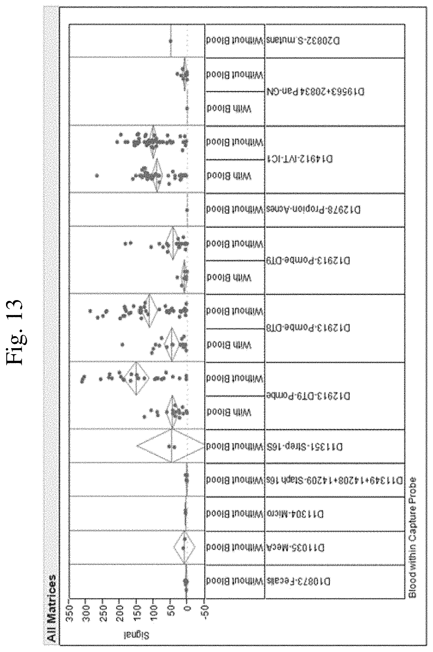
D00014
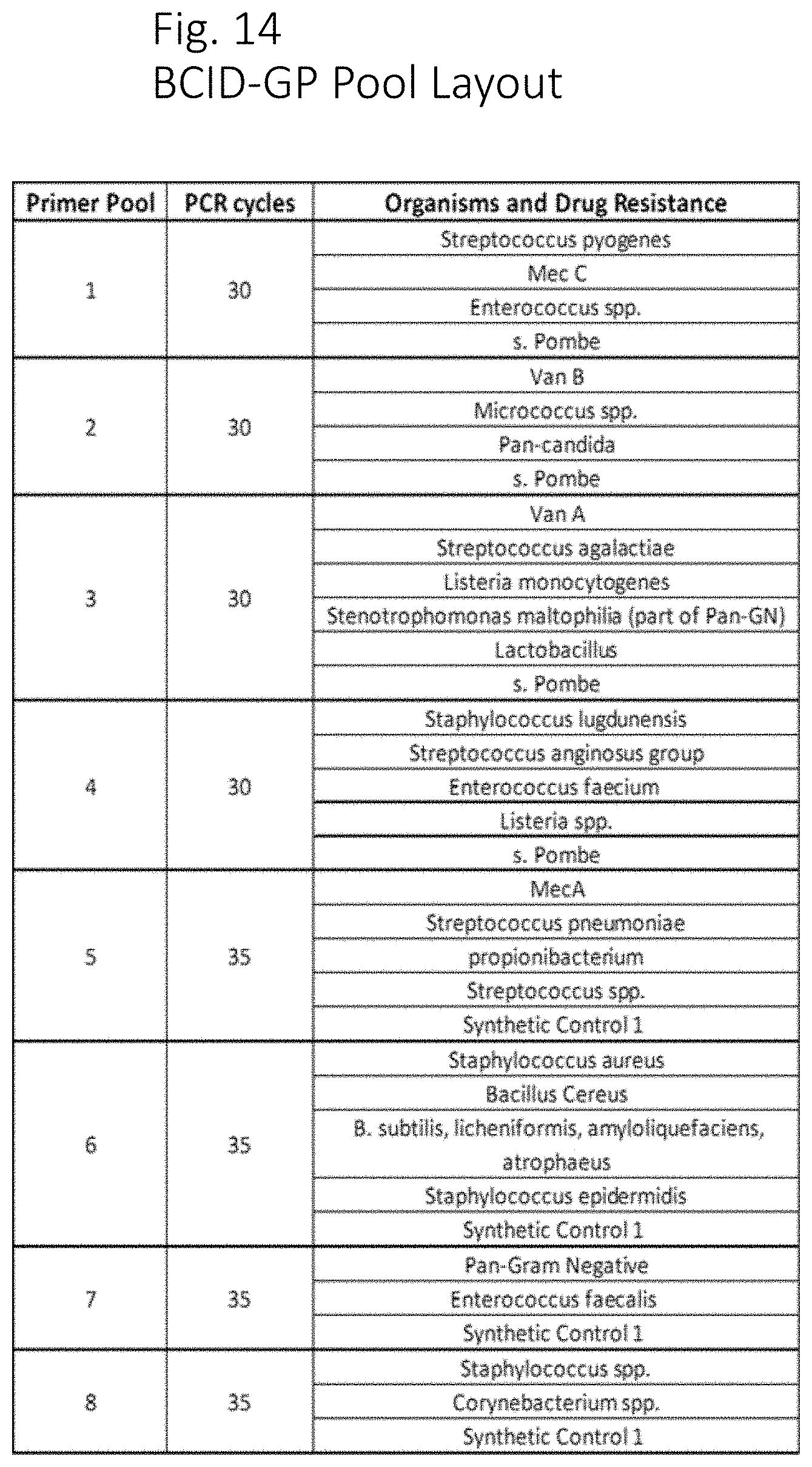
D00015
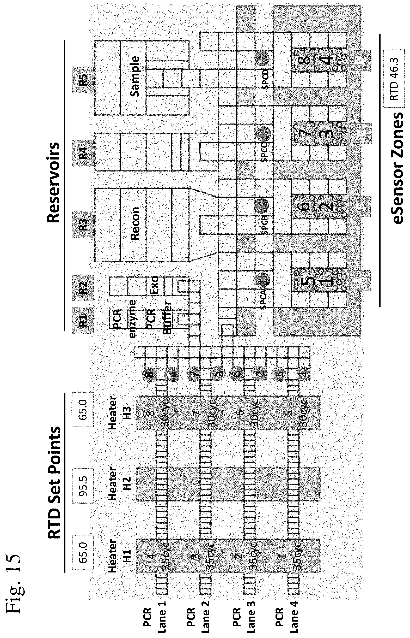
D00016

D00017

D00018

D00019
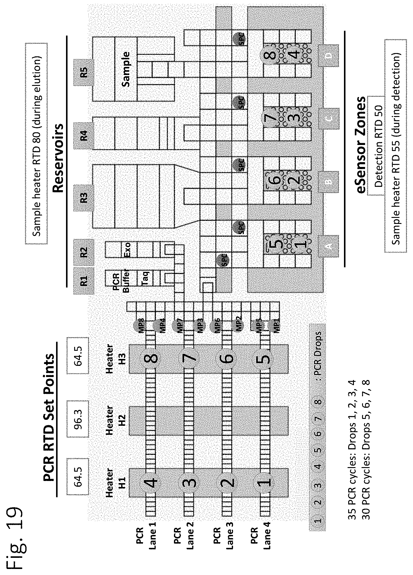
D00020
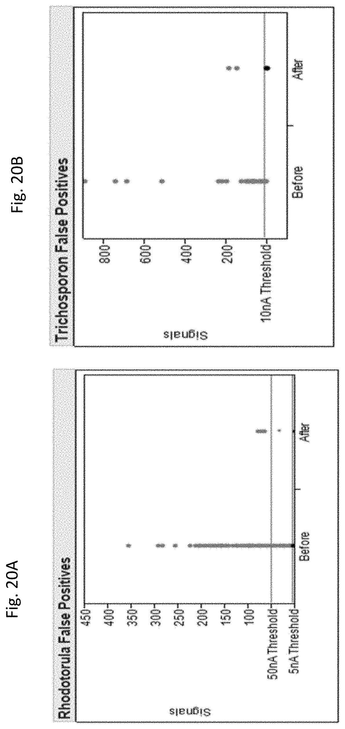
D00021
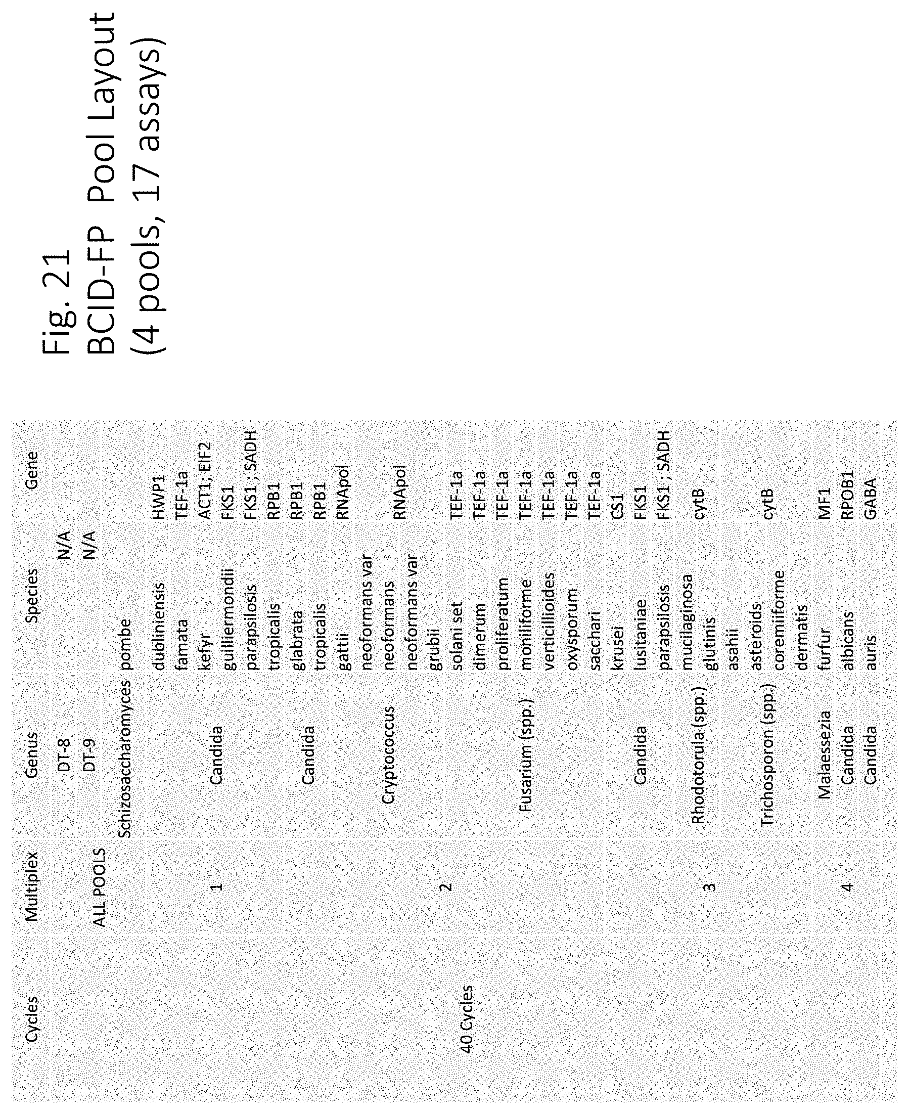
D00022

D00023

S00001
XML
uspto.report is an independent third-party trademark research tool that is not affiliated, endorsed, or sponsored by the United States Patent and Trademark Office (USPTO) or any other governmental organization. The information provided by uspto.report is based on publicly available data at the time of writing and is intended for informational purposes only.
While we strive to provide accurate and up-to-date information, we do not guarantee the accuracy, completeness, reliability, or suitability of the information displayed on this site. The use of this site is at your own risk. Any reliance you place on such information is therefore strictly at your own risk.
All official trademark data, including owner information, should be verified by visiting the official USPTO website at www.uspto.gov. This site is not intended to replace professional legal advice and should not be used as a substitute for consulting with a legal professional who is knowledgeable about trademark law.