Hemagglutinin-specific Antibodies And Uses Thereof
Manceur; Aziza ; et al.
U.S. patent application number 16/480727 was filed with the patent office on 2020-10-22 for hemagglutinin-specific antibodies and uses thereof. This patent application is currently assigned to National Research Council of Canada. The applicant listed for this patent is National Research Council of Canada. Invention is credited to Christine Gadoury, Amine Kamen, Aziza Manceur, Anne Marcil, Wei Zou.
| Application Number | 20200331988 16/480727 |
| Document ID | / |
| Family ID | 1000005132197 |
| Filed Date | 2020-10-22 |


View All Diagrams
| United States Patent Application | 20200331988 |
| Kind Code | A9 |
| Manceur; Aziza ; et al. | October 22, 2020 |
HEMAGGLUTININ-SPECIFIC ANTIBODIES AND USES THEREOF
Abstract
The present invention relates to hemagglutinin-specific antibodies, fragments thereof, and uses thereof. More specifically, these antibodies and fragments thereof are able to recognize antigen from multiple influenza strains.
| Inventors: | Manceur; Aziza; (Montreal, CA) ; Marcil; Anne; (Pierrefonds, CA) ; Zou; Wei; (Ottawa, CA) ; Kamen; Amine; (Montreal, CA) ; Gadoury; Christine; (Montreal, CA) | ||||||||||
| Applicant: |
|
||||||||||
|---|---|---|---|---|---|---|---|---|---|---|---|
| Assignee: | National Research Council of
Canada Ottawa ON |
||||||||||
| Prior Publication: |
|
||||||||||
| Family ID: | 1000005132197 | ||||||||||
| Appl. No.: | 16/480727 | ||||||||||
| Filed: | January 26, 2018 | ||||||||||
| PCT Filed: | January 26, 2018 | ||||||||||
| PCT NO: | PCT/IB2018/050493 PCKC 00 | ||||||||||
| 371 Date: | July 25, 2019 |
Related U.S. Patent Documents
| Application Number | Filing Date | Patent Number | ||
|---|---|---|---|---|
| 62451230 | Jan 27, 2017 | |||
| Current U.S. Class: | 1/1 |
| Current CPC Class: | C07K 2317/92 20130101; G01N 33/56983 20130101; C07K 16/1018 20130101; A61P 31/16 20180101; A61K 2039/505 20130101; G01N 2333/11 20130101; C07K 2317/76 20130101 |
| International Class: | C07K 16/10 20060101 C07K016/10; G01N 33/569 20060101 G01N033/569; A61P 31/16 20060101 A61P031/16 |
Claims
1. An isolated or purified antibody or fragment thereof, comprising: a) a light chain comprising a complementarity determining region (CDR) L1 sequence of QSLLNSX.sub.1X.sub.2QKNX.sub.3 (SEQ ID NO:1) where X.sub.1=R or D, X.sub.2=N or T, X.sub.3=H or F; a CDR L2 sequence of X.sub.1AS (SEQ ID NO:35) where X.sub.1=W or F; and a CDR L3 sequence of QQYYX.sub.1X.sub.2X.sub.3X.sub.4T (SEQ ID NO:2) where X.sub.1=T or S, X.sub.2=Y or I, X.sub.3=P or no amino acid, X.sub.4=R or L; and b) a heavy chain comprising a complementarity determining region (CDR) H1 sequence of GYX.sub.1X.sub.2TX.sub.3DYY (SEQ ID NO:3) where X.sub.1=S or T, X.sub.2=I or F, X.sub.3=S or no amino acid; a CDR H2 sequence selected from the group consisting of IGYDGX.sub.1K (SEQ ID NO:4) where X.sub.1=S or T, and IYPGNGHT (SEQ ID NO:5); and a CDR H3 sequence selected from the group consisting of TRDRANWDDYFDY (SEQ ID NO:6) and AYDLFNY (SEQ ID NO:7).
2. The isolated or purified antibody or fragment thereof of claim 1, wherein CDR L1 is selected from the group consisting of QSLLNSRNQKNH (SEQ ID NO:8) and QSLLNSDTQKNF (SEQ ID NO:9).
3. The isolated or purified antibody or fragment thereof of claim 1 or 2, wherein CDR L2 is selected from the group consisting of WAS (SEQ ID NO:36) and FAS (SEQ ID NO:37).
4. The isolated or purified antibody or fragment thereof of any one of claims 1 to 3, wherein CDR L3 is selected from the group consisting of QQYYTYRT (SEQ ID NO:10) wherein X is P or no amino acid; and QQYYSIPLT (SEQ ID NO:11).
5. The isolated or purified antibody or fragment thereof of any one of claims 1 to 4, wherein CDR H1 is selected from the group consisting of GYSITSDYY (SEQ ID NO:12) and GYTFTDYY (SEQ ID NO:13).
6. The isolated or purified antibody or fragment thereof of any one of claims 1 to 5, wherein the antibody or fragment thereof is selected from the group consisting of: a) a light chain comprising CDR L1 of sequence QSLLNSRNQKNH (SEQ ID NO:8), CDR L2 of sequence WAS (SEQ ID NO:36), and CDR L3 of sequence QQYYTYRT (SEQ ID NO:14); and a heavy chain comprising CDR H1 of sequence GYSITSDYY (SEQ ID NO:12), CDR H2 of sequence IGYDGSK (SEQ ID NO:15), and CDR H3 of sequence TRDRANWDDYFDY (SEQ ID NO:6); b) a light chain comprising CDR L1 of sequence QSLLNSRNQKNH (SEQ ID NO:8), CDR L2 of sequence WAS (SEQ ID NO:36), and CDR L3 of sequence QQYYTYRT (SEQ ID NO:14); and a heavy chain comprising CDR H1 of sequence GYSITSDYY (SEQ ID NO:12), CDR H2 of sequence IGYDGTK (SEQ ID NO:16), and CDR H3 of sequence TRDRANWDDYFDY (SEQ ID NO:6); and c) a light chain comprising CDR L1 of sequence QSLLNSDTQKNF (SEQ ID NO:9), CDR L2 of sequence FAS (SEQ ID NO:37), CDRL3 of sequence QQYYSIPLT (SEQ ID NO:11); and a heavy chain comprising CDR H1 of sequence GYTFTDYY (SEQ ID NO:13), CDR H2 of sequence IYPGNGHT (SEQ ID NO:5), and CDR H3 of sequence AYDLFNY (SEQ ID NO:7).
7. The isolated or purified antibody or fragment thereof of any one of claims 1 to 6, wherein the variable light (VL) domain comprises the sequence: DIVMX.sub.1QSPSSLAX.sub.2SVGX.sub.3KVTMSCKSSQSLLNSX.sub.4X.sub.- 5QKNX.sub.6LAWYQQKPGQS PKX.sub.7LX.sub.8YX.sub.9ASTX.sub.10ESGVPDRFX.sub.11GX.sub.12GSGTDFILTIX.- sub.13SVX.sub.14AEDLAX.sub.15YX.sub.16C QQYYX.sub.17X.sub.18X.sub.19X.sub.20TFGX.sub.21GTKLEIK (SEQ ID NO:17) where X.sub.1=S or T, X.sub.2=V or M, X.sub.3=E or O, X.sub.4=R or D, X.sub.5=N or T, X.sub.6=H or F, X.sub.7=L or I, X.sub.8=I or V, X.sub.9=W or F, X.sub.10=R or K, X.sub.11=S or I, X.sub.12=D or S, X.sub.13=S or T, X.sub.14=K or Q, X.sub.15=V or D, X.sub.16=Y or F, X.sub.17=T or S, X.sub.18=Y or I, X.sub.19=P or no amino acid, X.sub.20=R or L, X.sub.21=G or A.
8. The isolated or purified antibody or fragment thereof of claim 7, wherein the variable light (VL) domain comprises a sequence selected from the group consisting of: DIVMSQSPSSLAVSVGEKVTMSCKSSQSLLNSRNQKNHLAWYQQKPGQSPKL LIYWASTRESGVPDRFX.sub.1GDGSGTDFTLTISSVKAEDLAVYYCQQYYTYRTFGG GTKLEIK (SEQ ID NO:18) where X.sub.1=S or T; and DIVMTQSPSSLAMSVGQKVTMSCKSSQSLLNSDTQKNFLAWYQQKPGQSPKIL VYFASTKESGVPDRFIGSGSGTDFTLTITSVQAEDLADYFCQQYYSIPLTFGAGT KLELK (SEQ ID NO:19).
9. The isolated or purified antibody or fragment thereof of any one of claims 1 to 8, wherein the variable heavy (VH) domain comprises a sequence selected from the group consisting of: DVQLQESGPGLVKPSQSLSLTCSVTGYSITSDYYWNWIRQFPGNKLEWMAYIG YDGX.sub.1KNYNPSLKNRISITRDTSKNQFFLKLNSVTTDDTATYYCTRDRANWDDY FDYWGQGTTLTVSS (SEQ ID NO:20) X.sub.1=S or T; and QIQLQQSGPELVKPGAPVKISCKASGYTFTDYYIHWVNQRPGQGLEWIGYIYPG NGHTVYNQKFKVRATLTADNPSSTAYLQLNSLTSEDSGVYFCAYDLFNYWGQ GTLVTVSA (SEQ ID NO:21).
10. The isolated or purified antibody or fragment thereof any one of claims 1 to 9, wherein the isolated or purified antibody or fragment thereof comprises a sequence selected from the group consisting of a) a variable light (V.sub.L) domain of sequence: TABLE-US-00024 (SEQ ID NO: 22) DIVMSQSPSSLAVSVGEKVTMSCKSSQSLLNSRNQKNHLAWYQQKPGQSP KLLIYWASTRESGVPDRFSGDGSGTDFTLTISSVKAEDLAVYYCQQYYTY RTFGGGTKLEIK;
and variable heavy (V.sub.H) domain of sequence: TABLE-US-00025 (SEQ ID NO: 24) DVQLQESGPGLVKPSQSLSLTCSVTGYSITSDYYWNWIRQFPGNKLEWMA YIGYDGSKNYNPSLKNRISITRDTSKNQFFLKLNSVTTDDTATYYCTRDR ANWDDYFDYWGQGTTLTVSS;
b) a variable light (V.sub.L) domain of sequence: TABLE-US-00026 (SEQ ID NO: 23) DIVMSQSPSSLAVSVGEKVTMSCKSSQSLLNSRNQKNHLAWYQQKPGQSP KLLIYWASTRESGVPDRFTGDGSGTDFTLTISSVKAEDLAVYYCQQYYTY XRTFGGGTKLEIK;
and variable heavy (V.sub.H) domain of sequence: TABLE-US-00027 (SEQ ID NO: 25) DVQLQESGPGLVKPSQSLSLTCSVTGYSITSDYYWNWIRQFPGNKLEWMA YIGYDGTKNYNPSLKNRISITRDTSKNQFFLKLNSVTTDDTATYYCTRDR ANWDDYFDYWGQGTTLTVSS;
c) a variable light (V.sub.L) domain of sequence: TABLE-US-00028 (SEQ ID NO: 19) DIVMTQSPSSLAMSVGQKVTMSCKSSQSLLNSDTQKNFLAWYQQKPGQSP KILVYFASTKESGVPDRFIGSGSGTDFTLTITSVQAEDLADYFCQQYYSI PLTFGAGTKLELK;
and variable heavy (V.sub.H) domain of sequence: TABLE-US-00029 (SEQ ID NO: 21) QIQLQQSGPELVKPGAPVKISCKASGYTFTDYYIHWVNQRPGQGLEWIGY IYPGNGHTVYNQKFKVRATLTADNPSSTAYLQLNSLTSEDSGVYFCAYDL FNYWGQGTLVTVSA;
and d) a sequence substantially identical thereto.
11. The isolated or purified antibody or fragment thereof of any one of claims 1 to 10, wherein the antibody or fragment thereof specifically binds to the peptide: GLFGAIAGFIEGGW (SEQ ID NO:26).
12. The isolated or purified antibody or fragment thereof any one of claims 1 to 11, wherein the antibody or fragment thereof is a full-length IgG, Fv, scFv, Fab, or F(ab')2.
13. The isolated or purified antibody or fragment thereof of any one of claims 1 to 12, wherein the antibody or fragment thereof comprises framework regions from IgA, IgD, IgE, IgG, or IgM.
14. The isolated or purified antibody or fragment thereof of any one of claims 1 to 13, wherein the antibody or fragment thereof is chimeric.
15. The isolated or purified antibody or fragment thereof of claim 14, wherein the chimeric antibody or fragment thereof constant domain is from human IgG1.
16. The isolated or purified antibody or fragment thereof of claim 14, wherein the chimeric antibody or fragment thereof comprises human kappa 1 light chain and human IgG1 heavy chain constant domains.
17. A nucleic acid molecule encoding the isolated or purified antibody or fragment thereof of any one of claims 1 to 16.
18. A vector comprising the nucleic acid molecule of claim 17.
19. The isolated or purified antibody or fragment thereof of any one of claims 1 to 16, wherein the antibody or fragment thereof is immobilized onto a surface.
20. The isolated or purified antibody or fragment thereof of claim 19, wherein the surface is nitrocellulose.
21. The isolated or purified antibody or fragment thereof of any one of claims 1 to 16, wherein the antibody or fragment thereof is linked to a cargo molecule.
22. The isolated or purified antibody or fragment thereof of claim 21, wherein the cargo molecule is a detectable agent, a therapeutic agent, a drug, a peptide, an enzyme, a growth factor, a cytokine, a receptor trap, an antibody or fragment thereof, a chemical compound, a carbohydrate moiety, DNA-based molecules, a neutralizing agent, viral vector, one or more liposomes or nanocarriers loaded with any of the previously recited types of cargo molecules, or one or more nanoparticle, nanowire, nanotube, or quantum dots.
23. The isolated or purified antibody or fragment thereof of claim 21, wherein the cargo molecule is a neutralizing agent.
24. A composition comprising one or more than one isolated or purified antibody or fragment thereof of any one of claims 1 to 16 and 21 to 23 and a pharmaceutically-acceptable carrier, diluent, or excipient.
25. An in vitro method of detecting influenza hemagglutinin (HA), comprising: a) contacting a biological sample or a viral suspension, with an isolated or purified antibody or fragment thereof according to any one of claims 1 to 16 linked to a detectable agent; and b) detecting the detectable agent linked to the antibody or fragment thereof bound to hemagglutinin in the biological sample or a viral suspension.
26. The method of claim 25, wherein the biological sample is from: blood, serum, nasal wash, nasal swab, saliva or sputum.
27. The method of claim 25 or 26, wherein the step of detecting step b) is performed using: optical imaging, immunohistochemistry, molecular diagnostic imaging, ELISA, or other suitable method.
28. A method of preventing or treating influenza in a subject, comprising administering a pharmaceutically acceptable dose of an isolated or purified antibody or fragment thereof of any one of claims 1 to 16 and 21 to 23 to the subject.
29. A kit for detecting influenza HA comprising a support and an isolated or purified labelled-antibody or -fragment thereof according to any one of claims 1 to 16.
30. The kit of claim 29, wherein the support is nitrocellulose.
31. The kit of claim 30, wherein the isolated or purified labelled-antibody or -fragment thereof is immobilized onto the nitrocellulose.
32. An in vitro method for quantifying influenza HA, comprising: a) contacting a biological sample comprising an isolated or purified antibody or fragment thereof according to any one of claims 1 to 16 linked to a detectable agent; and b) quantifying the detectable agent linked to the antibody or fragment thereof.
33. The method of claim 32, wherein the step of detecting step b) is performed using: optical imaging, immunohistochemistry (dot blot, slot blot, quantitative Western blot), molecular diagnostic imaging, ELISA (direct, indirect or competitive), or other suitable method.
34. The method of claim 33, wherein the immunohistochemistry is: dot blot, slot blot or quantitative Western blot.
35. The method of claim 33, wherein the ELISA is direct, indirect or competitive.
36. The method of claim 33. 34 or 35, wherein said biological sample is selected from: a purified vaccine or in-process supernatant, produced with a platform selected from: eggs, mammalian cells, and plant.
37. A kit for measuring influenza HA comprising: one or more than one antibody as defined in any one of claims 1 to 16; and a detection reagent for detecting the antibody bound to said influenza HA in a biological sample; a measuring reagent for measuring a level of the detection agent.
38. An isolated or purified antibody or fragment thereof, for the manufacture of a composition for the treatment or prevention of influenza in a subject.
39. An isolated or purified antibody or fragment thereof, for use in the treatment or prevention of influenza in a subject.
40. The method of claim 28, wherein the subject is a human or an animal.
41. The isolated or purified antibody or fragment thereof of claim 38 or 39, wherein the subject is a human or an animal.
42. The isolated or purified antibody or fragment thereof, wherein the antibody is selected from: mAb 9D1 comprising sequences SEQ ID NO. 29 and 30; mAb 10A9 comprising sequences SEQ ID NO. 31 and 32; and mAb 11 H12 comprising sequences SEQ ID NO 33 and 34.
43. The isolated or purified antibody of claim 42, referred to as mAb 10A9 and defined by SEQ ID NO. 35.
44. The isolated or purified antibody of claim 42, referred to as mAb 11H12 and defined by SEQ ID NO. 36.
45. A cocktail comprising the antibodies 10A9 and 11H12, as defined in any one of claim 42, 43 or 44.
Description
FIELD OF THE INVENTION
[0001] The present invention relates to hemagglutinin-specific antibodies, fragments thereof, and uses thereof. More specifically, the present invention relates to hemagglutinin-specific antibodies and fragments thereof able to recognize antigen from multiple influenza strains.
BACKGROUND OF THE INVENTION
[0002] Influenza is an infectious disease caused by the influenza virus which belongs to the Orthomyxoviridae family. Based on their core proteins, influenza viruses are classified into types A, B, and C. The two main types of influenza virus responsible for seasonal flu epidemics are types A and B. Influenza A virus can be further characterized by serotype based on the hemagglutinin (HA) and neuraminidase (NA) proteins on the viral surface. Currently, there are 18 known subtypes of HA and 11 subtypes of NA. Based on HA subtypes, influenza A viruses are further divided into two phylogenetic groups: group 1 (H1, H2, H5, H6, H8, H9, H11, H12, H13, H16, H17 and H18) and group 2 (H3, H4, H7, H10, H14 and H15). Point mutations in the viral genome RNA of a given HA subtype already in circulation, or a new subtype of HA that arises through antigenic shift (Kang et al., 2011) can result in an influenza pandemic.
[0003] According to the World Health Organization (WHO), the influenza virus is responsible for up to 500,000 deaths per year worldwide. In order to combat the potential fatal effect of influenza virus, vaccines are produced yearly and administered to global populations. However, there are thousands of influenza virus strains. Presently, each strain requires a specific antibody for detection and quantification of HA, the most abundant protein expressed at the surface of the virus. Regulatory agencies such as the WHO are responsible for producing and distributing the antibodies used to quantify new vaccine lots throughout the world. Antibody production can take from 3 up to 16 weeks, which causes significant delays for the vaccine industry.
[0004] Generally, quantification of new vaccine lots is the bottleneck in vaccine distribution. Quantification of HA is currently performed using an assay called the Single Radial Immunodiffusion (SRID) assay. However, this assay is lengthy, laborious, and highly variable depending on the operator. In addition, standardised reagents necessary for SRID (polyclonal sera and antigen), need to be updated every year, which takes 12-16 weeks and is reliant on obtaining purified HA antigen. While the SRID assay is the only quantification method that is officially accepted by regulatory agencies, alternative quantification methods such as Enzyme-Linked Immunosorbent Assays (ELISA) could be more efficient.
[0005] Currently, specific antibodies against each strain are generated by injecting animals with isolates from each strain. However, the use of strain-specific antibodies can cause delays in releasing new vaccine lots. Using antibodies with broad specificity can speed up the detection and quantification of new influenza strains. To date, only two pan-HA antibodies are able to recognize all 3 groups of influenza commonly circulating in humans (type A group 1, type A group 2, and type B).
[0006] CR9114 is a human monoclonal antibody that was isolated using combinatorial display library derived from human B cells of subjects exposed to influenza (Dreyfus et al, 2012). CR9114 was shown to detect 10 influenza B viruses, as well as five HA belonging to influenza A (three from group 1 and two from group 2), but was not tested against all subtypes. However, its ability to recognize all HA subtypes remains untested.
[0007] Uni-1 was raised at Health Canada against a peptide sequence that is known to be highly conserved among influenza strains: GLFGAIAGFIEGGW (SEQ ID NO:29). The peptide sequence was derived from the HA fusion peptide, which was selected using bioinformatics analyses. Notably, Uni-1 was able to detect 13 different HA subtypes (H1 to H13) as well as B/Malaysia/2506/2004 by western blot (Chun et al, 2008).
[0008] Unfortunately, the main constraint with Uni-1 is that the antibodies are polyclonal rabbit antibodies, which result in high lot to lot variations. Additionally, the peptide used to raise Uni-1 was unable to elicit an immune response in mice, which has prevented production of monoclonal antibodies.
[0009] Thus, while some success has been achieved in the influenza field to generate antibodies with broad specificity to influenza HA, it is limited and not without drawbacks. The influenza community continues to seek faster and more accurate quantification methods to improve or replace the SRID assay, which could speed up the vaccine production and delivery system.
SUMMARY OF THE INVENTION
[0010] The present invention relates to hemagglutinin-specific antibodies, fragments thereof, and uses thereof. More specifically, the present invention relates to hemagglutinin-specific antibodies and fragments thereof able to recognize antigen from multiple influenza strains.
[0011] The present invention provides an isolated or purified antibody or fragment thereof, comprising: [0012] a) a light chain comprising a complementarity determining region (CDR) L1 sequence of QSLLNSX.sub.1X.sub.2QKNX.sub.3 (SEQ ID NO:1) where X.sub.1=R or D, X.sub.2=N or T, X.sub.3=H or F; a CDR L2 sequence of X.sub.1AS (SEQ ID NO:35) where X.sub.1=W or F; and a CDR L3 sequence of QQYYX.sub.1X.sub.2X.sub.3X.sub.4T (SEQ ID NO:2) where X.sub.1=T or S, X.sub.2=Y or I, X.sub.3=P or no amino acid, X.sub.4=R or L, and [0013] b) a heavy chain comprising a complementarity determining region (CDR) H1 sequence of GYX.sub.IX.sub.2TX.sub.3DYY (SEQ ID NO:3) where X.sub.1=S or T, X.sub.2=I or F, X.sub.3=S or no amino acid; a CDR H2 sequence selected from the group consisting of IGYDGX.sub.1K (SEQ ID NO:4) where X.sub.1=S or T, and IYPGNGHT (SEQ ID NO:5), and a CDR H3 sequence selected from the group consisting of TRDRANWDDYFDY (SEQ ID NO:6) and AYDLFNY (SEQ ID NO:7).
[0014] In one non-limiting example, the isolated or purified antibody or fragment described above may comprise a CDR L1 that is selected from the group consisting of QSLLNSRNQKNH (SEQ ID NO:8) and QSLLNSDTQKNF (SEQ ID NO:9).
[0015] In another non-limiting example, the isolated or purified antibody or fragment thereof as previously described may comprise a CDR L2 that is selected from the group consisting of WAS (SEQ ID NO:36) and FAS (SEQ ID NO:37).
[0016] In another non-limiting example, the isolated or purified antibody or fragment thereof as described above may comprise a CDR L3 that is selected from the group consisting of QQYYTYXRT (SEQ ID NO:10) where X is P or no amino acid and QQYYSIPLT (SEQ ID NO:11).
[0017] In yet another non-limiting example, the isolated or purified antibody or fragment thereof as described above may comprise a CDR H1 selected from the group consisting of GYSITSDYY (SEQ ID NO:12) and GYTFTDYY (SEQ ID NO:13).
[0018] In a more specific example, the isolated or purified antibody or fragment thereof may comprise a sequence that may be selected from the group consisting of: [0019] a) a light chain comprising CDR L1 of sequence QSLLNSRNQKNH (SEQ ID NO:8), CDR L2 of sequence WAS (SEQ ID NO:36), and CDR L3 of sequence QQYYTYRT (SEQ ID NO:14); and a heavy chain comprising CDR H1 of sequence GYSITSDYY (SEQ ID NO:12), CDR H2 of sequence IGYDGSK (SEQ ID NO:15), and CDR H3 of sequence TRDRANWDDYFDY (SEQ ID NO:6); [0020] b) a light chain comprising CDR L1 of sequence QSLLNSRNQKNH (SEQ ID NO:8), CDR L2 of sequence WAS (SEQ ID NO:36), and CDR L3 of sequence QQYYTYRT (SEQ ID NO:14); and a heavy chain comprising CDR H1 of sequence GYSITSDYY (SEQ ID NO:12), CDR H2 of sequence IGYDGTK (SEQ ID NO:16), and CDR H3 of sequence TRDRANWDDYFDY (SEQ ID NO:6); and [0021] c) a light chain comprising CDR L1 of sequence QSLLNSDTQKNF (SEQ ID NO:9), CDR L2 of sequence FAS (SEQ ID NO:37), CDRL3 of sequence QQYYSIPLT (SEQ ID NO:11); and a heavy chain comprising CDR H1 of sequence GYTFTDYY (SEQ ID NO:13), CDR H2 of sequence IYPGNGHT (SEQ ID NO:5), and CDR H3 of sequence AYDLFNY (SEQ ID NO:7).
[0022] More specifically, the isolated or purified antibody or fragment thereof may comprise a variable light (VL) domain having a sequence of: [0023] DIVMX.sub.1QSPSSLAX.sub.2SVGX.sub.3KVTMSCKSSQSLLNSX.sub.4X.sub.5QK- NX6LAWYQQKPGQS PKX.sub.7LX.sub.8YX.sub.9ASTX.sub.10ESGVPDRFX.sub.11GX.sub.12GSGTDFTLTIX.- sub.13SVX.sub.14AEDLAX.sub.15YX.sub.16C QQYYX.sub.17X.sub.18X.sub.19X.sub.20TFGX.sub.21GTKLEIK (SEQ ID NO:17), where X.sub.1=S or T, X.sub.2=V or M, X.sub.3=E or O, X.sub.4=R or D, X.sub.5=N or T, X.sub.6=H or F, X.sub.7=L or I, X.sub.8=I or V, X.sub.9=W or F, X.sub.10=R or K, X.sub.11=S or I, X.sub.12=D or S, X.sub.13=S or T, X.sub.14=K or Q, X.sub.15=V or D, X.sub.16=Y or F, X.sub.17=T or S, X.sub.18=Y or I, X.sub.19=P or no amino acid, X.sub.20=R or L, X.sub.21=G or A.
[0024] In an even more specific example, the variable light (VL) domain may comprise a sequence selected from the group consisting of: [0025] DIVMSQSPSSLAVSVGEKVTMSCKSSQSLLNSRNQKNHLAWYQQKPGQSPKL LIYWASTRESGVPDRFX.sub.1GDGSGTDFTLTISSVKAEDLAVYYCQQYYTYRTFGG GTKLEIK (SEQ ID NO:18) where X.sub.1=S or T; DIVMTQSPSSLAMSVGQKVTMSCKSSQSLLNSDTQKNFLAWYQQKPGQSPKIL VYFASTKESGVPDRFIGSGSGTDFTLTITSVQAEDLADYFCQQYYSIPLTFGAGT KLELK (SEQ ID NO:19); and [0026] a sequence substantially identical thereto.
[0027] In the isolated or purified antibody or fragment as described above, the variable heavy (VH) domain may comprise a sequence selected from the group consisting of: [0028] DVQLQESGPGLVKPSQSLSLTCSVTGYSITSDYYWNWIRQFPGNKLEWMAYIG YDGX.sub.1KNYNPSLKNRISITRDTSKNQFFLKLNSVTTDDTATYYCTRDRANWDDY FDYWGQGTTLTVSS (SEQ ID NO:20) where X.sub.1=S or T; QIQLQQSGPELVKPGAPVKISCKASGYTFTDYYIHWVNQRPGQGLEWIGYIYPG NGHTVYNQKFKVRATLTADNPSSTAYLQLNSLTSEDSGVYFCAYDLFNYWGQ GTLVTVSA (SEQ ID NO:21); and [0029] a sequence substantially identical thereto.
[0030] In specific, non-limiting examples, the isolated or purified antibody or fragment thereof may comprise: [0031] a) a variable light (V.sub.L) domain of sequence:
TABLE-US-00001 [0031] (SEQ ID NO: 22) DIVMSQSPSSLAVSVGEKVTMSCKSSQSLLNSRNQKNHLAWYQQKPGQS PKLLIYWASTRESGVPDRFSGDGSGTDFTLTISSVKAEDLAVYYCQQYY TYRTFGGGTKLEIK;
and variable heavy (V.sub.H) domain of sequence:
TABLE-US-00002 (SEQ ID NO: 24) DVQLQESGPGLVKPSQSLSLTCSVTGYSITSDYYWNWIRQFPGNKLEWM AYIGYDGSKNYNPSLKNRISITRDTSKNQFFLKLNSVTTDDTATYYCTR DRANWDDYFDYWGQGTTLTVSS;
or [0032] b) a variable light (V.sub.L) domain of sequence:
TABLE-US-00003 [0032] (SEQ ID NO: 23) DIVMSQSPSSLAVSVGEKVTMSCKSSQSLLNSRNQKNHLAWYQQKPGQS PKLLIYWASTRESGVPDRFTGDGSGTDFTLTISSVKAEDLAVYYCQQYY TYXRTFGGGTKLEIK;
and variable heavy (V.sub.H) domain of sequence:
TABLE-US-00004 (SEQ ID NO: 25) DVQLQESGPGLVKPSQSLSLTCSVTGYSITSDYYWNWIRQFPGNKLEWM AYIGYDGTKNYNPSLKNRISITRDTSKNQFFLKLNSVTTDDTATYYCTR DRANWDDYFDYWGQGTTLTVSS;
or [0033] c) a variable light (V.sub.L) domain of sequence:
TABLE-US-00005 [0033] (SEQ ID NO: 19) DIVMTQSPSSLAMSVGQKVTMSCKSSQSLLNSDTQKNFLAWYQQKPGQS PKILVYFASTKESGVPDRFIGSGSGTDFTLTITSVQAEDLADYFCQQYY SIPLTFGAGTKLELK;
and variable heavy (V.sub.H) domain of sequence:
TABLE-US-00006 (SEQ ID NO: 21) QIQLQQSGPELVKPGAPVKISCKASGYTFTDYYIHWVNQRPGQGLEWIGY IYPGNGHTVYNQKFKVRATLTADNPSSTAYLQLNSLTSEDSGVYFCAYDL FNYWGQGTLVTVSA;
or [0034] d) a sequence substantially identical thereto.
[0035] The antibody or fragment thereof of the present invention, as defined above, may specifically bind hemagglutinin.
[0036] The isolated or purified antibody or fragment thereof as described herein may be a full-length IgG, Fv, scFv, Fab, or F(ab')2 fragments; the antibody or fragment thereof may also comprise framework regions from IgA, IgD, IgE, IgG, or IgM. The isolated or purified antibody or fragment thereof of the present invention may be chimeric; or example, and without wishing to be limiting, such a chimeric antibody or fragment thereof may comprise the V.sub.L and V.sub.H domains from mouse and framework regions (constant domains) from human IgG1, more specifically human kappa 1 light chain and human IgG1 heavy chain.
[0037] The present invention also provides a nucleic acid molecule encoding the isolated or purified antibody or fragment thereof as described herein. A vector comprising the nucleic acid molecule as herein described is also provided.
[0038] The isolated or purified antibody or fragment thereof as described herein may be immobilized onto a surface, or may be linked to a cargo molecule. The cargo molecule may be a detectable agent, a therapeutic agent, a drug, a peptide, an enzyme, a growth factor, a cytokine, a receptor trap, an antibody or fragment thereof (e.g., IgG, scFv, Fab, V.sub.HH, etc) a chemical compound, a carbohydrate moiety, DNA-based molecules (anti-sense oligonucleotide, microRNA, siRNA, plasmid), a neutralizing agent, viral vector (adeno-, lenti-, retro-), one or more liposomes or nanocarriers loaded with any of the previously recited types of cargo molecules, or one or more nanoparticle, nanowire, nanotube, or quantum dots. In a specific, non-limiting example, the cargo molecule is a neutralizing agent.
[0039] Additionally, the present invention provides a composition comprising one or more than one isolated or purified antibody or fragment thereof as described herein and a pharmaceutically-acceptable carrier, diluent, or excipient.
[0040] An in vitro method of detecting hemagglutinin is also provided, the method comprising the steps of: [0041] a) contacting a tissue sample with an (one or more than one) isolated or purified antibody or fragment thereof as described herein linked to a detectable agent; and [0042] b) detecting the detectable agent linked to the antibody or fragment thereof bound to hemagglutinin in the tissue sample.
[0043] In the method described above, the method may detect hemagglutinin in circulating cells and the sample may be a serum sample, lung tissue sample, neuroepithelium tissue sample, or other tissue from the respiratory system. In the method as described, the step of detecting (step b)) may be performed using optical imaging, immunohistochemistry, molecular diagnostic imaging, immunoassays such as ELISA, Western blot, dot blot, slot blot, or other suitable method.
[0044] Hemagglutinin is a protein expressed at the surface of the influenza virus. Due to antigenic drift and shift, each new strain of influenza generally requires a specific antibody for its recognition. Presently, three novel antibodies [11H12 (SEQ ID NO. 36), 10A9 (SEQ ID NO. 35) and 9D1] have been identified that specifically bind hemagglutinin. The novel antibodies described herein are able to detect and bind multiple strains encompassing 13 HA subtypes. They can be used for detection, quantification, and neutralization. Furthermore, these antibodies are monoclonal, and thus can be produced in a reproducible and scalable fashion.
[0045] The antibodies of the present invention may be use to speed up or improve the accuracy of quantification methods, which would ease the bottleneck in vaccine distribution. The antibodies could also be used with in-process and crude samples in order to optimise the bioprocess used to generate vaccines.
[0046] The present invention may also provide a method for the prevention or treatment of influenza in a subject, comprising administering an (one or more than one) isolated or purified antibody or fragment thereof as described herein to the subject
[0047] Alternatively, there is also provided an isolated or purified antibody or fragment thereof as described herein for use in preventing or treating influenza in a subject.
[0048] Additional aspects and advantages of the present invention will be apparent in view of the following description. The detailed descriptions and examples, while indicating preferred embodiments of the invention, are given by way of illustration only, as various changes and modifications within the scope of the invention will become apparent to those skilled in the art in light of the teachings of this invention.
BRIEF DESCRIPTION OF THE DRAWINGS
[0049] These and other features of the invention will now be described by way of example, with reference to the appended drawings, wherein:
[0050] FIG. 1A is a schematic diagram of the conjugate structure used for mouse immunizations. The universal peptide epitope is shown in a box; Lac-Ser is O-beta-lactosyl-serine; --S-- is thio-Cys; KLH is Keyhole Limpet Hemocyanin.
[0051] FIG. 1B is a scheme representing the addition of non-immunogenic hydrophilic saccharide at one end of a linear peptide-conjugate for producing the antibodies of the invention.
[0052] FIG. 2 are sequence alignments of the variable domains of the antibodies of the present application. A) light chains: 11H12 (SEQ ID NO:23); 10A9 (SEQ ID NO:19); and 9D1 (SEQ ID NO:22). B) heavy chains: 11H12 (SEQ ID NO:25); 10A9 (SEQ ID NO:21); and 9D1 (SEQ ID NO:24).
[0053] FIG. 3 shows thermograms obtained by Differential Scanning calorimetry (DSC) of the three mAb. Each mAb was scanned from 20 to 100.degree. C.
[0054] FIGS. 4A-C shows results from ELISA analysis following one to three freeze-thaw cycles. The binding affinity of 9D1 (FIG. 4A), 10A9 (FIG. 4B) and 11H12 (FIG. 4C) was measured using the peptide-conjugate shown in FIG. 1.
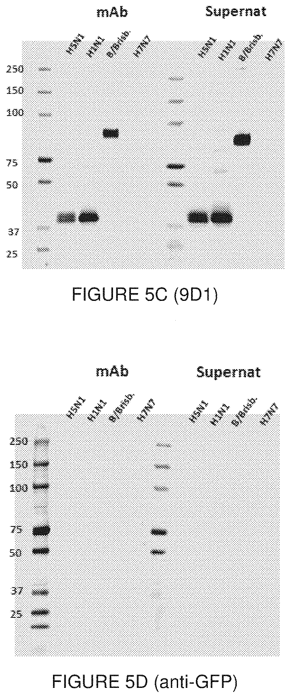
[0055] FIG. 5 show Western blot results of the reactivity of the mAb 11H12 (FIG. 5A), 10A9 (FIG. 5B), and 9D1 (FIG. 5C), as well as an anti-GFP negative control (FIG. 5D), for recombinant HA proteins. For each set of data, the purified mAb (mAb) or the unpurified hybridoma supernatant (Supernat) was used. The molecular weight of the rHA is 75KDa when uncleaved (HA.sub.D). When cleaved, the fragments have a molecular weight of 45KDa (HA.sub.1) and 25KDa (HA.sub.2), as indicated in FIG. 5A. Each mAb was assayed against H5N1/A/Indonesia/05/2005 (H5N1); H1N1 A/Puerto Rico/08/1934 (H1N1); B/Brisbane/60/2008 (B/Brisb.); and H7N7 A/Netherlands/219/2003 (H7N7).
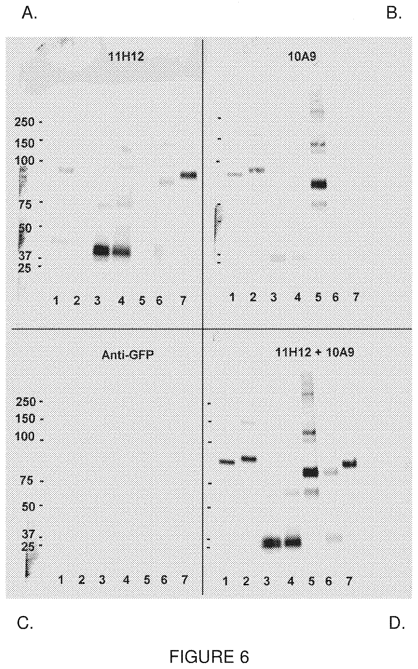
[0056] FIG. 6 show results of a second Western blot analysis of the reactivity of the 11H12 mAb (FIG. 6A), the 10A9 mAb (FIG. 6B), a mixture of the two antibodies (FIG. 6D), and an anti-GFP negative control (FIG. 6C). Lane 1, H1N1/A/California/06/2008; Lane 2, H3N2/NBrisbane/10/2007; Lane 3, H5N1/A/Indonesia/05/2005; Lane 4, H5N1/A/Vietnam/1203/2004; Lane 5, H7N7/A/Netherlands/219/2003; Lane 6, H9N2/A/Hong Kong/1073/1999; Lane 7, B/Brisbane/60/2008.
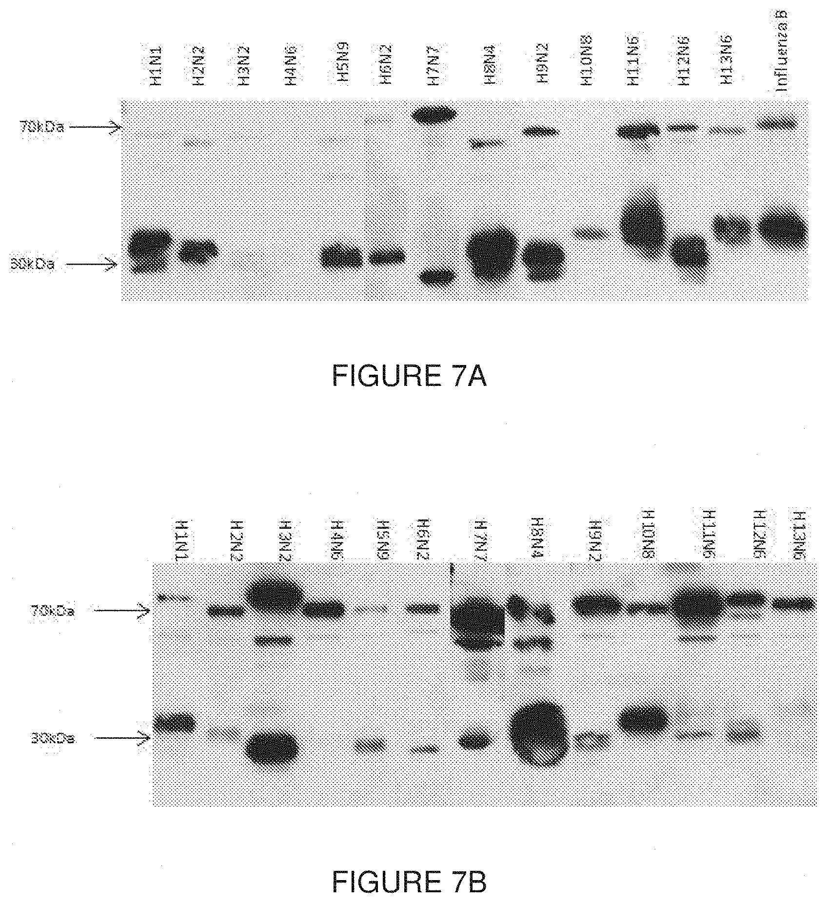
[0057] FIG. 7 show Western blot results of the reactivity of the 11H12 (FIG. 7A) and 10A9 (FIG. 7B) mAb with viruses produced in eggs. Full names of the virus strains are shown in Table 6.
[0058] FIGS. 8 show dot blot analysis of the 3mAbs: 9D1 (FIG. 8A), 10A9 (FIG. 8B) and 11H12 (FIG. 8C), tested against HA from different strains as summarized in Table 7. Values corresponding to the optical densities of each dot are also reported in Table 7.
[0059] FIG. 9 show the results of quantitative dot blot analysis with 11H12. A representative dot blot is shown in FIG. 9A. H1N1 A/Puerto Rico/8/34 virus produced in HEK293 cells were quantified by generating a standard curve (FIG. 9B). Samples were serially diluted and 4 concentrations were assayed. Sample 6 was obtained from non-infected cells and was used as a negative control (FIG. 9C).
[0060] FIG. 10 shows the results of ELISA analysis of 10A9, 11H12, and 9D1. A direct ELISA was performed with the antibodies against the rHA from H5N1 A/Indonesia/05/2005. A non-related antibody (anti-GFP) was used as a negative control.
[0061] FIG. 11 show in vivo results of challenges with lethal concentrations of influenza viruses, along with several doses of antibodies to evaluate their protective/neutralizing capacity. In FIG. 11A, mice were infected intra-nasally with 750 PFU of H1N1 A/Puerto Rico/8/34; antibodies were injected at three different time points: 2 hours before infection, 4 hours post-infection and 24 hours post-infection. In FIG. 11B, mice were infected intra-nasally with 10.sup.4 PFU of H3N2 A/Hong Kong/8/68; antibodies were injected at 4 different time points: 24 hours before infection, 2 hours before infection, 24 hours post-infection and 72 hours post-infection.
[0062] FIG. 12. Dot blot results with a pan-HA cocktail obtained from mouse hybridomas (FIG. 12A) and CHO pools (FIG. 12B). Each dot represents a different strain as indicated in Table 9. Recombinant proteins and plant VLPs were loaded at a final concentration of 2.5 .mu.g while the viruses and standard antigens were loaded at a concentration of 5 .mu.g.
[0063] FIG. 13. Primary amino acid sequence comparison analysis of IgGs: 11H12 (FIG. 13A); 10A9 (FIG. 13B); and 9D1 (FIG. 13C). Amino acid sequences identified with a Mascot score>30 are highlighted in bold.
[0064] FIG. 14. Full protein sequences of recombinant monoclonal antibodies: 10A9 (FIG. 14A); and 11H12 (FIG. 14B).
DETAILED DESCRIPTION OF THE INVENTION
Abbreviations
[0065] DT: diphteria toxin; HA: hemagluttinin; KLH: Keyhole limpet hemocyanin; NA: neuraminidase; TT: tetanus toxoid.
Definitions
[0066] As used herein the singular forms "a", "and", and "the" include plural referents unless the context clearly dictates otherwise. Thus, for example, reference to "an antibody" includes "one or more than one" antibodies and reference to "the antibody" includes reference to one or more than one antibodies and equivalents thereof known to those skilled in the art, and so forth. All technical and scientific terms used herein have the same meaning as commonly understood to one of ordinary skill in the art to which this invention belongs unless clearly indicated otherwise.
[0067] The term "about" as used herein refers to a margin of + or -10% of the number indicated. For sake of precision, the term about when used in conjunction with, for example: 90% means 90% +/-9% i.e. from 81% to 99%. More precisely, the term about refer to + or -5% of the number indicated, where for example: 90% means 90% +/-4.5% i.e. from 86.5% to 94.5%. When used in the context of a pH, the term "about" means +/-0.5 pH unit.
[0068] As used in this specification and claim(s), the words "comprising" (and any form of comprising, such as "comprise" and "comprises"), "having" (and any form of having, such as "have" and "has"), "including" (and any form of including, such as "includes" and "include") or "containing" (and any form of containing, such as "contains" and "contain") are inclusive or open-ended and do not exclude additional, un-recited elements or method steps.
[0069] As used herein, the terms "disease" may be used interchangeably or may be different in that the particular disorder, infection or condition may not have a known causative agent (so that etiology has not yet been worked out) and it is therefore not yet recognized as a disease but only as an undesirable condition or syndrome, wherein a more or less specific set of symptoms have been identified by clinicians.
[0070] The term "subject" as used herein refers to an animal, preferably a mammal or a bird, who is the object of administration, treatment, observation or experiment. "Mammal" includes humans and both domestic animals such as laboratory animals and household pets, (e.g. cats, dogs, swine, cattle, sheep, goats, horses, rabbits), and non-domestic animals such as wildlife, fowl, birds and the like. More particularly, the mammal is a rodent. Still, most particularly, the mammal is a human.
[0071] The antibody(ies) described herein can be formulated as pharmaceutical compositions by formulation with additives such as pharmaceutically acceptable excipients, pharmaceutically acceptable carriers, and pharmaceutically acceptable vehicles.
[0072] As used herein, the term "pharmaceutically acceptable" refers to molecular entities and compositions that are physiologically tolerable and do not typically produce an allergic or similar unwanted reaction, such as gastric upset, dizziness and the like, when administered to human. Preferably, as used herein, the term "pharmaceutically acceptable" means approved by regulatory agency of the federal or state government or listed in the U.S. Pharmacopeia or other generally recognized pharmacopeia for use in animals, and more particularly in humans.
[0073] The terms "carrier," "diluent" or "excipient" each refers to a vehicle with which the antibodies of the present invention may be administered. Sterile water or aqueous saline solutions and aqueous dextrose and glycerol solutions may be employed as carrier, particularly for injectable solutions. Suitable pharmaceutical carriers are described in "Remington's Pharmaceutical Sciences" by E. W. Martin.
[0074] If administered as a medicinal preparation, the antibodies can be administered, either as a prophylaxis or treatment, to a patient by a number of methods. The present compositions may be administered alone or in combination with other pharmaceutical antibodies and can be combined with a physiologically acceptable carrier thereof. The effective amount and method of administration and aim of the present formulation can vary based on the individual subject, the stage of the disease or condition, and other factors apparent to one skilled in the art. In the case of a pharmaceutical formulation, during the course of the treatment, the concentration of the present compositions may be monitored (for example, blood antibody levels may be monitored) to ensure that the desired response is obtained.
DETAILED DESCRIPTION OF PARTICULAR EMBODIMENTS
[0075] The present invention relates to hemagglutinin-specific antibodies, fragments thereof, and uses thereof. More specifically, the present invention relates to hemagglutinin-specific antibodies and fragments thereof able to recognize antigen from multiple influenza strains.
[0076] The present invention provides an isolated or purified antibody or fragment thereof, comprising: [0077] a) a light chain comprising a complementarity determining region (CDR) L1 sequence of QSLLNSX.sub.1X.sub.2QKNX.sub.3 (SEQ ID NO:1) where X.sub.1=R or D, X.sub.2=N or T, X.sub.3=H or F; a CDR L2 sequence of X.sub.1AS (SEQ ID NO:35) where X.sub.1=W or F; and a CDR L3 sequence of QQYYX.sub.1X.sub.2X.sub.3X.sub.4T (SEQ ID NO:2) where X.sub.1=T or S, X.sub.2=Y or I, X.sub.3=P or no amino acid, X.sub.4=R or L, and [0078] b) a heavy chain comprising a complementarity determining region (CDR) H1 sequence of GYX.sub.1X.sub.2TX.sub.3DYY (SEQ ID NO:3) where X.sub.1=S or T, X.sub.2=I or F, X.sub.3=S or no amino acid; a CDR H2 sequence selected from the group consisting of IGYDGX.sub.1K (SEQ ID NO:4) where X.sub.1=S or T and IYPGNGHT (SEQ ID NO:5); and a CDR H3 sequence selected from the group consisting of TRDRANWDDYFDY (SEQ ID NO:6) and AYDLFNY (SEQ ID NO:7).
[0079] The term "antibody", also referred to in the art as "immunoglobulin" (Ig), as used herein refers to a protein constructed from paired heavy and light polypeptide chains; various Ig isotypes exist, including IgA, IgD, IgE, IgG, and IgM. When an antibody is correctly folded, each chain folds into a number of distinct globular domains joined by more linear polypeptide sequences. For example, the immunoglobulin light chain folds into a variable (V.sub.L) and a constant (CO domain, while the heavy chain folds into a variable (V.sub.H) and three constant (C.sub.H, C.sub.H2, C.sub.H3) domains. Interaction of the heavy and light chain variable domains (V.sub.H and V.sub.L) results in the formation of an antigen binding region (Fv). Each domain has a well-established structure familiar to those of skill in the art.
[0080] The light and heavy chain variable regions are responsible for binding the target antigen and can therefore show significant sequence diversity between antibodies. The constant regions show less sequence diversity, and are responsible for binding a number of natural proteins to elicit important biochemical events. The variable region of an antibody contains the antigen-binding determinants of the molecule, and thus determines the specificity of an antibody for its target antigen. The majority of sequence variability occurs in six hypervariable regions, three each per variable heavy (V.sub.H) and light (V.sub.L) chain; the hypervariable regions combine to form the antigen-binding site, and contribute to binding and recognition of an antigenic determinant. The specificity and affinity of an antibody for its antigen is determined by the structure of the hypervariable regions, as well as their size, shape, and chemistry of the surface they present to the antigen. Various schemes exist for identification of the regions of hypervariability, the two most common being those of Kabat and of Chothia and Lesk. Kabat et al (1991) define the "complementarity-determining regions" (CDR) based on sequence variability at the antigen-binding regions of the V.sub.H and V.sub.L domains. Chothia and Lesk (1987) define the "hypervariable loops" (H or L) based on the location of the structural loop regions in the V.sub.H and V.sub.L domains. As these individual schemes define CDR and hypervariable loop regions that are adjacent or overlapping, those of skill in the antibody art often utilize the terms "CDR" and "hypervariable loop" interchangeably, and they may be so used herein. A more recent scheme is the IMGT numbering system (Lefranc et al., 2003), which was developed to facilitate comparison of variable domains. In this system, conserved amino acids (such as Cys23, Trp41, Cys104, Phe/Trp118, and a hydrophobic residue at position 89) always have the same position. Additionally, a standardized delimitation of the framework regions (FR1: positions 1 to 26; FR2: 39 to 55; FR3: 66 to 104; and FR4: 118 to 129) and of the CDR (CDR1: 27 to 38, CDR2: 56 to 65; and CDR3: 105 to 117) is provided.
[0081] The CDR/loops are referred to herein according to the IMGT numbering system for all CDR. The CDR of the antibodies of the present invention are referred to herein as CDR L1, L2, L3 for CDR in the light chain, and CDR H1, H2, H3 for CDR in the heavy chain.
[0082] An "antibody fragment" as referred to herein may include any suitable antigen-binding antibody fragment known in the art. The antibody fragment may be a naturally-occurring antibody fragment, or may be obtained by manipulation of a naturally-occurring antibody or by using recombinant methods. For example, an antibody fragment may be selected from the group consisting of, but is not limited to a
[0083] Fv, single-chain Fv (scFv; a molecule consisting of V.sub.L and V.sub.H connected with a peptide linker), Fab, F(ab').sub.2, and multivalent presentations of any of these. Antibody fragments such as those herein described may require linker sequences, disulfide bonds, or other type of covalent bond to link different portions of the fragments; those of skill in the art will be familiar with various approaches.
[0084] The terms "antibody" and "antibody fragment" ("fragment thereof") as defined above may be from any selected from the group consisting of source, human, mouse, or other; may be any isotype selected from the group consisting of IgA, IgD, IgE, IgG, and IgM; and may be any type of fragment, including but not limited to Fv, scFv, Fab, and F(ab').sub.2.
[0085] In one non-limiting example, the isolated or purified antibody or fragment described above may comprise a CDR L1 that is selected from the group consisting of QSLLNSRNQKNH (SEQ ID NO:8) and QSLLNSDTQKNF (SEQ ID NO:9).
[0086] In another non-limiting example, the isolated or purified antibody or fragment thereof as previously described may comprise a CDR L2 that is selected from the group consisting of WAS (SEQ ID NO:36) and FAS (SEQ ID NO:37).
[0087] In another non-limiting example, the isolated or purified antibody or fragment thereof as described above may comprise a CDR L3 that is selected from the group consisting of QQYYTYXRT (SEQ ID NO:10) where X is P or no amino acid, and QQYYSIPLT (SEQ ID NO:11).
[0088] In yet another non-limiting example, the isolated or purified antibody or fragment thereof as described above may comprise a CDR H1 is selected from the group consisting of GYSITSDYY (SEQ ID NO:12) and GYTFTDYY (SEQ ID NO:13).
[0089] In a more specific example, the isolated or purified antibody or fragment thereof may be selected from the group consisting of: [0090] a) a light chain comprising CDR L1 of sequence QSLLNSRNQKNH (SEQ ID NO:8), CDR L2 of sequence WAS (SEQ ID NO:36), and CDR L3 of sequence QQYYTYRT (SEQ ID NO:14); and a heavy chain comprising CDR H1 of sequence GYSITSDYY (SEQ ID NO:12), CDR H2 of sequence IGYDGSK (SEQ ID NO:15), and CDR H3 of sequence TRDRANWDDYFDY (SEQ ID NO:6); [0091] b) a light chain comprising CDR L1 of sequence QSLLNSRNQKNH (SEQ ID NO:8), CDR L2 of sequence WAS (SEQ ID NO:36), and CDR L3 of sequence QQYYTYRT (SEQ ID NO:17); and a heavy chain comprising CDR H1 of sequence GYSITSDYY (SEQ ID NO:12), CDR H2 of sequence IGYDGTK (SEQ ID NO:16), and CDR H3 of sequence TRDRANWDDYFDY (SEQ ID NO:6); and [0092] c) a light chain comprising CDR L1 of sequence QSLLNSDTQKNF (SEQ ID NO:9), CDR L2 of sequence FAS (SEQ ID NO:37), CDRL3 of sequence QQYYSIPLT (SEQ ID NO:11); and a heavy chain comprising CDR H1 of sequence GYTFTDYY (SEQ ID NO:13), CDR H2 of sequence IYPGNGHT (SEQ ID NO:5), and CDR H3 of sequence AYDLFNY (SEQ ID NO:7).
[0093] More specifically, the isolated or purified antibody or fragment thereof may comprise a variable light (VL) domain having a sequence of: [0094] DIVMX.sub.1QSPSSLAX.sub.2SVGX.sub.3KVTMSCKSSQSLLNSX.sub.4X.sub.5QK- NX.sub.6LAWYQQKPGQS PKX.sub.7LX.sub.8YX.sub.9ASTX.sub.10ESGVPDRFX.sub.11GX.sub.12GSGTDFTLTIX.- sub.13SVX.sub.14AEDLAX.sub.15YX.sub.16C QQYYX.sub.17X.sub.18X.sub.19X.sub.20TFGX.sub.21GTKLEIK (SEQ ID NO:17) where X.sub.1=S or T, X.sub.2=V or M, X.sub.3=E or O, X.sub.4=R or D, X.sub.5=N or T, X.sub.6=H or F, X.sub.7=L or I, X.sub.8=l or V, X.sub.9=W or F, X.sub.10=R or K, X.sub.11=S or I, X.sub.12=D or S, X.sub.13=S or T, X.sub.14=K or Q, X.sub.15=V or D, X.sub.16=Y or F, X.sub.17=T or S, X.sub.18=Y or I, X.sub.19=P or no amino acid, X.sub.20=R or L, X.sub.21=G or A.
[0095] In a more specific example, the variable light (VL) domain may comprise a sequence selected from the group consisting of: [0096] a) DIVMSQSPSSLAVSVGEKVTMSCKSSQSLLNSRNQKNHLAWYQQKPGQS PKWYWASTRESGVPDRFX.sub.1GDGSGTDFTLTISSVKAEDLAVYYCQQYYTY RTFGGGTKLEIK (SEQ ID NO:18) where X.sub.1=S or T; [0097] b) DIVMTQSPSSLAMSVGQKVTMSCKSSQSLLNSDTQKNFLAWYQQKPGQS PKILVYFASTKESGVPDRFIGSGSGTDFTLTITSVQAEDLADYFCQQYYSIPL TFGAGTKLELK (SEQ ID NO:19); and [0098] c) a sequence substantially identical thereto.
[0099] In the isolated or purified antibody or fragment as described above, the variable heavy (VH) domain may comprise a sequence selected from the group consisting of: [0100] a) DVQLQESGPGLVKPSQSLSLTCSVTGYSITSDYYWNWIRQFPGNKLEWMA YIGYDGX.sub.1KNYNPSLKNRISITRDTSKNQFFLKLNSVTTDDTATYYCTRDRAN WDDYFDYWGQGTTLTVSS (SEQ ID NO:20) where X.sub.1=S or T; [0101] b) QIQLQQSGPELVKPGAPVKISCKASGYTFTDYYIHWVNQRPGQGLEWIGYI YPGNGHTVYNQKFKVRATLTADNPSSTAYLQLNSLTSEDSGVYFCAYDLFN YWGQGTLVTVSA (SEQ ID NO:21); and [0102] c) a sequence substantially identical thereto.
[0103] In specific, non-limiting examples, the isolated or purified antibody or fragment thereof may comprise: [0104] a) a variable light (V.sub.L) domain of sequence:
TABLE-US-00007 [0104] (SEQ ID NO: 22) DIVMSQSPSSLAVSVGEKVTMSCKSSQSLLNSRNQKNHLAWYQQKPGQSP KLLIYWASTRESGVPDRFSGDGSGTDFTLTISSVKAEDLAVYYCQQYYTY RTFGGGTKLEIK
and variable heavy (V.sub.H) domain of sequence:
TABLE-US-00008 (SEQ ID NO: 24) DVQLQESGPGLVKPSQSLSLTCSVTGYSITSDYYWNWIRQFPGNKLEWMA YIGYDGSKNYNPSLKNRISITRDTSKNQFFLKLNSVTTDDTATYYCTRDR ANWDDYFDYWGQGTTLTVSS;
or [0105] b) a variable light (V.sub.L) domain of sequence:
TABLE-US-00009 [0105] (SEQ ID NO: 23) DIVMSQSPSSLAVSVGEKVTMSCKSSQSLLNSRNQKNHLAWYQQKPGQSP KLLIYWASTRESGVPDRFTGDGSGTDFTLTISSVKAEDLAVYYCQQYYTY XRTFGGGTKLEIK;
and variable heavy (V.sub.H) domain of sequence:
TABLE-US-00010 (SEQ ID NO: 25) DVQLQESGPGLVKPSQSLSLTCSVTGYSITSDYYWNWIRQFPGNKLEWMA YIGYDGTKNYNPSLKNRISITRDTSKNQFFLKLNSVTTDDTATYYCTRDR ANWDDYFDYWGQGTTLTVSS;
or [0106] c) a variable light (V.sub.L) domain of sequence:
TABLE-US-00011 [0106] (SEQ ID NO: 19) DIVMTQSPSSLAMSVGQKVTMSCKSSQSLLNSDTQKNFLAWYQQKPGQSP KILVYFASTKESGVPDRFIGSGSGTDFTLTITSVQAEDLADYFCQQYYSI PLTFGAGTKLELK;
and variable heavy (V.sub.H) domain of sequence:
TABLE-US-00012 (SEQ ID NO: 21) QIQLQQSGPELVKPGAPVKISCKASGYTFTDYYIHWVNQRPGQGLEWIGY IYPGNGHTVYNQKFKVRATLTADNPSSTAYLQLNSLTSEDSGVYFCAYDL FNYWGQGTLVTVSA;
or [0107] d) a sequence substantially identical thereto.
[0108] A substantially identical sequence may comprise one or more conservative amino acid mutations. It is known in the art that one or more conservative amino acid mutations to a reference sequence may yield a mutant peptide with no substantial change in physiological, chemical, physico-chemical or functional properties compared to the reference sequence; in such a case, the reference and mutant sequences would be considered "substantially identical" polypeptides. A conservative amino acid substitution is defined herein as the substitution of an amino acid residue for another amino acid residue with similar chemical properties (e.g. size, charge, or polarity). These conservative amino acid mutations may be made to the framework regions of the antibody or fragment thereof while maintaining the CDR sequences listed above and the overall structure of the antibody or fragment; thus, the specificity and binding of the antibody are maintained.
[0109] In a non-limiting example, a conservative mutation may be an amino acid substitution. Such a conservative amino acid substitution may substitute a basic, neutral, hydrophobic, or acidic amino acid for another of the same group. By the term "basic amino acid" it is meant hydrophilic amino acids having a side chain pK value of greater than 7, which are typically positively charged at physiological pH. Basic amino acids include histidine (His or H), arginine (Arg or R), and lysine (Lys or K). By the term "neutral amino acid" (also "polar amino acid"), it is meant hydrophilic amino acids having a side chain that is uncharged at physiological pH, but which has at least one bond in which the pair of electrons shared in common by two atoms is held more closely by one of the atoms. Polar amino acids include serine (Ser or S), threonine (Thr or T), cysteine (Cys or C), tyrosine (Tyr or Y), asparagine (Asn or N), and glutamine (Gln or Q). The term "hydrophobic amino acid" (also "non-polar amino acid") is meant to include amino acids exhibiting a hydrophobicity of greater than zero according to the normalized consensus hydrophobicity scale of Eisenberg (1984). Hydrophobic amino acids include proline (Pro or P), isoleucine (Ile or I), phenylalanine (Phe or F), valine (Val or V), leucine (Leu or L), tryptophan (Trp or W), methionine (Met or M), alanine (Ala or A), and glycine (Gly or G). "Acidic amino acid" refers to hydrophilic amino acids having a side chain pK value of less than 7, which are typically negatively charged at physiological pH. Acidic amino acids include glutamate (Glu or E), and aspartate (Asp or D).
[0110] Sequence identity is used to evaluate the similarity of two sequences; it is determined by calculating the percent of residues that are the same when the two sequences are aligned for maximum correspondence between residue positions. Any known method may be used to calculate sequence identity; for example, computer software is available to calculate sequence identity. Without wishing to be limiting, sequence identity can be calculated by software such as NCBI BLAST2 service maintained by the Swiss Institute of Bioinformatics (and as found at ca.expasy.org/tools/blast/), or any other appropriate software that is known in the art.
[0111] The substantially identical sequences of the present invention may be at least 90% identical; in another example, the substantially identical sequences may be at least 90, 91, 92, 93, 94, 95, 96, 97, 98, 99, or 100% identical, or any percentage therebetween, at the amino acid level to sequences described herein. Importantly, the substantially identical sequences retain the activity and specificity of the reference sequence. In a non-limiting embodiment, the difference in sequence identity may be due to conservative amino acid mutation(s). In a non-limiting example, the present invention may be directed to an antibody or fragment thereof comprising a sequence at least 95%, 98% or 99% identical to that of the antibodies described herein.
[0112] The antibodies as described herein may comprise the V.sub.L and V.sub.H domains as described above and one or more than one constant regions from mouse IgG2a.
[0113] The present invention further encompasses an antibody or fragment thereof that is chimeric (or chimerized), veneered, or humanized. The antibody or fragment thereof as described herein may be chimeric, in that the antibody or fragment thereof is a combination of protein sequences originating from more than one species.
[0114] As is known to those of skill in the art, a chimeric antibody is produced by combining genetic material from a nonhuman source (for example but not limited to a mouse) with genetic material from a human. For example, and without wishing to be limiting, human constant domains can be fused to mouse V.sub.H and V.sub.L sequences (see Gonzales et al 2005). Veneering, also referred to in the art as "variable region resurfacing", of antibodies involves replacing solvent-exposed residues in the framework region of the native antibody or fragment thereof with the amino acid residues in their human counterpart (Padlan, 1991; Gonzales et al 2005); thus, buried non-humanized residues, which may be important for CDR conformation, are preserved while the potential for immunological reaction against solvent-exposed regions is minimized. Humanization of an antibody or antibody fragment comprises replacing an amino acid in the sequence with its human counterpart, as found in the human consensus sequence, without loss of antigen-binding ability or specificity; this approach reduces immunogenicity of the antibody or fragment thereof when introduced into human subjects. In this process, one or more than one of the CDR defined herein may be fused or grafted to a human variable region (V.sub.H, or V.sub.L), to other human antibody (IgA, IgD, IgE, IgG, and IgM), to human antibody fragment framework regions (Fv, scFv, Fab), or to human proteins of similar size and nature onto which CDR can be grafted (Nicaise et al, 2004). In such a case, the conformation of said one or more than one hypervariable loop is likely preserved, and the affinity and specificity of the sdAb for its target (i.e., C. difficile LTA) is likely minimally affected. As is known by those of skill in the art, it may be necessary to incorporate certain native amino acid residues into the human framework in order to retain binding and specificity. Humanization by CDR grafting is known in the art (for example, see Tsurushita et al, 2005; Jones et al, 1986; Tempest et al, 1991; Riechmann et al, 1988; Queen et al, 1989; all reviewed in Gonzales et al, 2005--see also references cited therein), and thus persons of skill would be amply familiar with methods of preparing such humanized antibody or fragments thereof.
[0115] The isolated or purified antibody or fragment thereof of the present invention may be a chimeric antibody or fragment thereof may comprise the V.sub.L and V.sub.H domains from mouse and framework regions (constant domains) from human IgG1, more specifically human kappa 1 light chain and human IgG1 heavy chain.
[0116] The antibody or fragment thereof of the present invention specifically binds to the influenza hemagglutinin (HA) protein. The hemagglutinin protein binds sialic acid present on the surface of target host cells and plays a key role in entry of the viral genome into the target cell. Currently, 18 types HA protein are known. The antibody or fragment thereof of the present invention may bind one subtype or multiple subtypes of HA; when binding multiple subtypes of HA, the antibody or fragment thereof may bind different subtypes with varying affinity.
[0117] The present application further provides a novel influenza HA antigen. The HA antigen comprises a peptide originating from the N-terminus of HA2, GLFGAIAGFIEGGW (SEQ ID NO:26) functionalized with O-beta-lactosyl-serine and its N-terminus and thio-Cys at its C-terminus; the C-terminus of the functionalized antigen is conjugated to Keyhole Limpet Hemocyanin (KLH), which is one of a plurality of carrier molecules that can be used for conjugating to antigenic epitope to provoke or increase the immune response thereof (such as, for example the tetanus toxin (TT)). The carrier protein, such as KLH in this case, may also comprises multiple epitopes conjugated to its surface. A schematic of the antigen is shown in FIG. 1A and in the following formula (I):
##STR00001##
[0118] The antibody or fragment thereof of the present invention may bind to the novel antigen as described above.
[0119] The antibody or fragment thereof of the present invention may also comprise additional sequences to aid in expression, detection or purification of a recombinant antibody or fragment thereof. Any such sequences or tags known to those of skill in the art may be used. For example, and without wishing to be limiting, the antibody or fragment thereof may comprise a targeting or signal sequence, a detection/purification tag (for example, but not limited to c-Myc, His.sub.5, His.sub.6, or His.sub.8G), or a combination thereof. In another example, the signal peptide may be MVLQTQVFISLLLWISGAYG (SEQ ID NO.27) or MDWTWRILFLVAAATGTHA (SEQ ID NO.28). In a further example, the additional sequence may be a biotin recognition site such as that described by Cronan et al in WO 95/04069 or Voges et al in WO/2004/076670. As is also known to those of skill in the art, linker sequences may be used in conjunction with the additional sequences or tags, or may serve as a detection/purification tag.
[0120] The antibody or fragment thereof of the present invention may also be in a multivalent display format, also referred to herein as multivalent presentation. Multimerization may be achieved by any suitable method of known in the art. For example, and without wishing to be limiting in any manner, multimerization may be achieved using self-assembly molecules such as those described in WO2003/046560, where pentabodies are produced by expressing a fusion protein comprising the antibody or fragment thereof of the present invention and the pentamerization domain of the B-subunit of an AB.sub.5 toxin family (Merritt & Hol, 1995). A multimer may also be formed using the multimerization domains described by Zhu et al. (2010); this form, referred to herein as a "combody" form, is a fusion of the antibody or fragment of the present invention with a coiled-coil peptide resulting in a multimeric molecule. Other forms of multivalent display are also encompassed by the present invention. For example, and without wishing to be limiting, the antibody or fragment thereof may be presented as a dimer, a trimer, or any other suitable oligomer. This may be achieved by methods known in the art, for example direct linking connection (Nielsen et al, 2000), c-jun/Fos interaction (de Kruif & Logtenberg, 1996), "Knob into holes" interaction (Ridgway et al, 1996).
[0121] Each subunit of the multimers described above may comprise the same or different antibodies or fragments thereof of the present invention, which may have the same or different specificity. Additionally, the multimerization domains may be linked to the antibody or antibody fragment using a linker, as required; such a linker should be of sufficient length and appropriate composition to provide flexible attachment of the two molecules, but should not hamper the antigen-binding properties of the antibody. For example, and without wishing to be limiting in any manner, the antibody or fragments thereof may be presented in a bi-specific antibody.
[0122] The invention also encompasses the antibody or fragment thereof as described above linked to a cargo molecule. The cargo molecule may be any suitable molecule. For example, and without wishing to be limiting in any manner, the cargo molecule may be a detectable agent, a therapeutic agent, a drug, a peptide, an enzyme, a growth factor, a cytokine, a receptor trap, an antibody or fragment thereof (e.g., IgG, scFv, Fab, V.sub.HH, V.sub.H, V.sub.L, etc) a chemical compound, a carbohydrate moiety, DNA-based molecules (anti-sense oligonucleotide, microRNA, siRNA, plasmid), a neutralizing agent, viral vector (adeno-, lenti-, retro-), one or more liposomes or nanocarriers loaded with any of the previously recited types of cargo molecules, or one or more nanoparticle, nanowire, nanotube, or quantum dots. The antibody or fragment thereof may be linked to the cargo molecule using any method known in the art (recombinant technology, chemical conjugation, etc.).
[0123] In one non-limiting example, the cargo molecule may be a detectable label, a radioisotope, a paramagnetic label such as gadolinium or iron oxide, a fluorophore, a fluorescent agent, Near Infra-Red (NIR) fluorochrome or dye (such as Cy5.5), an echogenic microbubble, an affinity label (for example biotin, avidin, etc), a detectable protein-based molecule, nucleotide, quantum dot, nanoparticle, nanowire, or nanotube or any other suitable agent that may be detected by imaging methods. In a specific, non-limiting example, the anti-hemagglutinin or fragment thereof may be linked to a near infrared fluorescence (NIRF) imaging dye, for example and not wishing to be limiting Cy5.5, Alexa680, Dylight680, or Dylight800.
[0124] In another specific, non-limiting embodiment, the antibody or fragment thereof as described herein is linked to a drug, thus providing an antibody-drug conjugate (ADC). The drug may be any type of drug, for example but not limited to a neutralizing agent. The neutralizing agent may include, but is not limited to anti-microtubule agents (such as taxanes, maytansines and auristatins), DNA damaging agents (such as calicheamicin and duocarmydin), RNA polymerase inhibitors (such as alpha-amantin), and other potent neutralizing drugs (such as anthracyclines). As is known to those of skill in the art, the antibody-drug conjugate allows for targeted delivery of a drug, thus limiting systemic exposure. In this construct, the antibody or fragment thereof as described herein binds to the extracellular domain of hemagglutinin; the drug linked to the antibody or fragment thereof is thus internalized. Upon internalization DM1 is released within the target cells upon degradation of the human hemagglutininantibody-DM1 complex in lysosomes. Depending on the intracellular concentration of DM1 accumulated in cancer cells, rapid apoptosis occurs.
[0125] The cargo molecule as described herein may be linked, also referred to herein as "conjugated", to the antibody or fragment thereof by any suitable method known in the art. For example, and without wishing to be limiting, the cargo molecule may be linked to the peptide by a covalent bond or ionic interaction. The linkage may be achieved through a chemical cross-linking reaction, or through fusion using recombinant DNA methodology combined with any peptide expression system, such as bacteria, yeast or mammalian cell-based systems. When conjugating the cargo molecule to the antibody or fragment thereof, a suitable linker may be used. Methods for linking an antibody or fragment thereof to a cargo molecule such as a therapeutic or detectable agent would be well-known to a person of skill in the art.
[0126] The present invention also encompasses nucleic acid sequences encoding the molecules as described herein. Given the degeneracy of the genetic code, a number of nucleotide sequences would have the effect of encoding the desired polypeptide, as would be readily understood by a skilled artisan. The nucleic acid sequence may be codon-optimized for expression in various micro-organisms. The present invention also encompasses vectors comprising the nucleic acids as just described. Furthermore, the invention encompasses cells comprising the nucleic acid and/or vector as described.
[0127] The present invention further encompasses the isolated or purified antibody or fragments thereof immobilized onto a surface using various methodologies; for example, and without wishing to be limiting, the antibody or fragment may be linked or coupled to the surface via His-tag coupling, biotin binding, covalent binding, adsorption, and the like. Immobilization of the antibody or fragment thereof of the present invention may be useful in various applications for capturing, purifying or isolating proteins. The solid surface may be any suitable surface, for example, but not limited to the well surface of a microtiter plate, channels of surface plasmon resonance (SPR) sensorchips, membranes, beads (such as magnetic-based or sepharose-based beads or other chromatography resin), glass, plastic, stainless steel, a film, biosensors (such as those used in Biolayer Interferometry), or any other useful surface such as nanoparticles, nanowires and cantilever surfaces. A purified antibody or fragment thereof immobilized onto a surface may be used in a variety of methods, including diagnostic methods.
[0128] Thus, the present invention further provides an in vitro method of detecting influenza HA, comprising contacting a tissue sample with one or more than one isolated or purified antibody or fragment thereof of the present invention linked to a detectable agent. The HA-antibody complex can then be detected using detection and/or imaging technologies known in the art. The tissue sample in the method as just described may be any suitable tissue sample, for example but not limited to a serum sample, a vascular tissue sample such as lung tissue sample, neuroepithelium tissue sample, nasal aspirates, nasopharyngeal aspirates or swabs, nasal washes or swabs, throat swabs, endotracheal aspirates, bronchoalveolar lavage, or other tissue from the respiratory system; the tissue sample may be from a human or animal subject. The step of contacting is done under suitable conditions, known to those skilled in the art, for formation of a complex between the antibody or fragment thereof and HA. The step of detecting may be accomplished by any suitable method known in the art, for example, but not limited to optical imaging, immunohistochemistry, molecular diagnostic imaging, ELISA, or other suitable method. For example, and without wishing to be limiting in any manner, the isolated or purified antibody or fragment thereof linked to a detectable agent may be used in immunoassays (IA) including, but not limited to enzyme IA (EIA), ELISA, "rapid antigen capture", "rapid chromatographic IA", and "rapid EIA". (For example, see Planche et al, 2008; Sloan et al, 2008; Russmann et al, 2007; Musher et al, 2007; Turgeon et al, 2003; Fenner et al, 2008). Other immunoassay techniques in which the antibodies of the present invention may be used include Western blot, dot blot, and slot blot analysis. In a specific, non-limiting embodiment, the in vitro method is for detection of HA in nasal wash or swab. The one or more than one isolated or purified antibody or fragment thereof of the present invention could be used for detection in a Rapid Influenza Diagnosis Test (RIDT), also known as Point-of-Care Test (POCT) or dipsticks; these assays are used in the clinic to provide rapid diagnosis (less than 15 minutes) of patients with flu-like symptoms. The presence of viral antigens (HA) in a specimen (nasal wash or swab) is analysed in a lateral flow immunoassay and result in a colorimetric change when detected by the anti-HA antibodies.
[0129] The present invention also provides a method of preventing or treating influenza in a subject. The method comprises administering one or more than one isolated or purified antibody or fragment thereof as described herein to the subject. The one or more than one isolated or purified antibody or fragment thereof may be linked to one or more than one cargo molecule, as described herein. The subject may be a human or animal subject. The administration may be by any suitable method, for example parenteral administration, including but not limited to intravenous (iv), subcutaneous (sc), and intramuscular (im) administration.
[0130] The present invention also encompasses a composition comprising one or more than one isolated or purified antibody or fragment thereof as described herein.
[0131] The composition may comprise a single antibody or fragment as described above, or may be a mixture of antibodies or fragments. Furthermore, in a composition comprising a mixture of antibodies or fragments of the present invention, the antibodies may have the same specificity, or may differ in their specificities; for example, and without wishing to be limiting in any manner, the composition may comprise antibodies or fragments thereof specific to hemagglutinin (same or different epitope). The composition may also comprise one or more than one antibody or fragments of the present invention linked to one or more than one cargo molecule.
[0132] The composition may also comprise a pharmaceutically acceptable diluent, excipient, or carrier. The diluent, excipient, or carrier may be any suitable diluent, excipient, or carrier known in the art, and must be compatible with other ingredients in the composition, with the method of delivery of the composition, and is not deleterious to the recipient of the composition. The composition may be in any suitable form; for example, the composition may be provided in suspension form, powder form (for example, but limited to lyophilised or encapsulated), capsule or tablet form. For example, and without wishing to be limiting, when the composition is provided in suspension form, the carrier may comprise water, saline, a suitable buffer, or additives to improve solubility and/or stability; reconstitution to produce the suspension is effected in a buffer at a suitable pH to ensure the viability of the antibody or fragment thereof. Dry powders may also include additives to improve stability and/or carriers to increase bulk/volume; for example, and without wishing to be limiting, the dry powder composition may comprise sucrose or trehalose. In a specific, non-limiting example, the composition may be so formulated as to deliver the antibody or fragment thereof to the gastrointestinal tract of the subject. Thus, the composition may comprise encapsulation, time-release, or other suitable technologies for delivery of the antibody or fragment thereof. It would be within the competency of a person of skill in the art to prepare suitable compositions comprising the present antibodies or fragments thereof.
[0133] The present invention further provides an isolated or purified antibody or fragment thereof as described herein for use in preventing or treating influenza in a subject.
[0134] The present invention also encompasses a cocktail comprising both mAbs 10A9 (SEQ ID NO. 35) and 11H12 (SEQ ID NO.36) for the detection, prevention or treatment of influenza.
[0135] The present invention further provides a kit for the identification of influenza in a sample, the kit comprising a support (such as nitrocellulose) and one or more than one isolated or purified antibody or fragment thereof as described herein. The one or more than one isolated or purified antibody or fragment thereof may be immobilized onto the nitrocellulose. The nitrocellulose support may be placed inside a plastic housing (also referred to herein as cassette), or may be adhered to a paper support (also referred to herein as card). The kit may also contain a swab for collecting the sample.
[0136] Particularly, the sample is a biological sample such as for example, blood, serum, nasal wash, nasal swab, saliva or sputum.
[0137] The present invention also provides a kit for the prevention or treatment of influenza in a subject, the kit comprising a container; and an isolated or purified antibody or fragment thereof contained therein. The kit may also contain a syringe for injecting the antibodies or fragments thereof to the subject.
[0138] The present invention will be further illustrated in the following examples. However, it is to be understood that these examples are for illustrative purposes only and should not be used to limit the scope of the present invention in any manner.
EXAMPLE 1
Production and Purification of Antigen
[0139] A fusion peptide was prepared to use for immunization of mice. The fusion protein comprised a conserved peptide sequence from the N-terminal region of HA2 (GLFGAIAGFIEGGW; SEQ ID NO:26), functional groups on the peptide sequence, and keyhole limpet hemocyanin (KLH).
[0140] Conjugate structure. The peptide conjugate shown in FIG. 1A, was designed by using a conserved peptide sequence at the N-terminus of HA2 that was previously identified: GLFGAIAGFIEGGW (SEQ ID NO:26). The peptide epitope was functionalized with lactose and conjugated to Keyhole Limpet Hemocyanin (KLH) to obtain a conjugate of formula (I):
##STR00002##
[0141] The KLH portion of the conjugate also comprised multiple epitopes conjugated to its surface.
[0142] Peptide conjugate synthesis. The peptide conjugate was prepared according to FIG. 1B via a thio-ether bond between terminal Cys of the (glyco)peptide antigen and bromoacetyl KLH.
[0143] Bromoacetylation of KLH. Typically, 20 mg of KLH (Sigma-Aldrich H7017) was solubilized in 2 mL of deionized water at room temperature, and buffer-exchanged to 10.times.PBS by an Amicon Ultra centrifugal filter (MWC 30K). To the above solution of KLH in 10.times.PBS (2 mL) was added 9 mg of bromoacetic acid N-hydroxysuccinimide ester in DMSO (0.18 mL) and incubated overnight at 4.degree. C. The product was purified by a G-25 column (50.times.1.6 cm) with PBS as eluent and bromoacetyl KLH obtained was stored in PBS buffer. Similarly, bromoacetyl BSA was also made in parallel and MALDI indicated 9-10 bromoacetyl groups per BSA, we obtained similar ratio of bromoacetyl group present in KLH.
[0144] Conjugation: (glyco)peptide antigen with terminal Cys and bromoacetyl KLH are dissolved under the conditions of 0.1M phosphate buffer with 5 mM EDTA-0.01% sodium azide at pH 8.0-8.5 overnight at room temperature. As a reference to estimate the peptide antigen on KLH, bromoacetyl BSA was also coupled with the peptide to give a conjugate with a ratio of peptide:BSA 6-7:1. The peptide antigen coupled to KLH carrier protein was found to have a similar w/w ratio as BSA.
[0145] Purification. Typically, a (glyco)peptide antigen with terminal Cys was mixed with equivalent amount of biotin-maleimide (B1267, Sigma), in DMSO at room temperature and the solution was kept for 5 hours, which was diluted with water and lyophilized to give product. The product was characterized by MALDI and no further purification was needed for ELISA.
EXAMPLE 2
Generation of Anti-Influenza Antibodies
[0146] To produce antibodies that target the influenza virus, mice were immunized with the peptide conjugate obtained in Example 1. Hybridomas (monoclonal antibodies) were also prepared and evaluated by ELISA.
[0147] Immunizations. 6-week old A/J mice were bled (pre-immune serum) and immunized i.p. and s.c. with 100 .mu.g of antigen (Example 1) in Titermax adjuvant. Three weeks later, a second injection of 100 .mu.g of antigen in Titermax adjuvant was performed and mice were bled 7-10 days later. The serum titer was measured by ELISA. Two months later, a final i.p. booster injection using 100 .mu.g of antigen was performed 4 days prior to fusion experiment.
[0148] Fusion of the harvested spleen cells. All manipulations were carried out under sterile conditions. Spleen cells were harvested from immunized mice in IMDM (Hy-Clone) and fused to NSO myeloma cell line using PEG fusion protocol. To this end, spleen cells and myeloma cells were washed in IMDM, counted in RBC lysing buffer (Sigma) and mixed together at a 5:1 ratio. Pelleted cells were fused together by adding 1 ml of a 50% solution of PEG 4000 (EMD-Millipore) in PBS preheated at 37.degree. C. drop-wise over one minute, and incubated at 37.degree. C. for an additional 90 sec. The reaction was stopped by addition of 30 ml of IMDM at 22.degree. C. over 2 min. After a 10 min incubation, freshly fused cells were spun at 233.times.g for 10 min. Cells were washed once in IMDM supplemented with 10% heat inactivated FBS (Sigma), and suspended at a concentration of 2.times.10.sup.5 input myeloma cells per ml in HAT selection medium (IMDM containing 20% heat inactivated FBS, penicillin-streptomycin (Sigma), 1 ng/ml mouse IL-6 (Biosource), HAT media supplement (Sigma) and L-glutamine) and incubated at 37.degree. C., 5% CO.sub.2. The next day, hybridoma cells were washed and suspended at a concentration of 2.times.10.sup.5 input myeloma cells per ml in semi-solid medium D (StemCell) supplemented with 5% heat inactivated FBS, 1 ng/ml mouse IL-6 and 10 .mu.g/ml FITC- Fab'2 Goat anti-mouse IgG (H+L) (Jackson). The cell mixture was plated in OmniTrays (Nunc) and further incubated for 6-7 days at 37.degree. C., 5% CO.sub.2. Secretor clones were then transferred using a mammalian cell clone picker (ClonepixFL, Molecular Devices) into sterile 96-well plates (Costar) containing 200 .mu.l of IMDM supplemented with 20% heat inactivated FBS, penicillin-streptomycin (Sigma), 1 ng/ml mouse IL-6 (Biosource), HT media supplement (Sigma) and L-glutamine and incubated for 2-3 days at 37.degree. C., 5% CO.sub.2.
[0149] Hybridoma selection. Hybridoma supernatant were screened by ELISA to detect specific binders. To this end, 96-well half-area plates (Costar) were coated with 25 .mu.l of neutravidinat 10 .mu.g/ml in 50mM carbonate buffer at pH9.8 and incubated 2 hours at room temperature. Microplates were washed 3 times with PBS, blocked with PBS-BSA 1%, washed once with PBS and 25 .mu.l of biotinylated peptide at 5 .mu.g/ml was added and incubated overnight at 4.degree. C. Microplates were washed 4 times with PBS-Tween 20 0.05% and 25 .mu.l of hybridoma supernatant were added and incubated at 37.degree. C., 5% CO.sub.2 for 2 hours. Plates were washed 4 times with PBS-Tween 20 0.05% and incubated for 1h at 37.degree. C., 5% CO.sub.2 with 25 .mu.l of secondary antibody alkaline phosphatase conjugated F(ab)'2 goat anti-mouse. IgG (Fcy fragment specific) (Jackson Immunoresearch) diluted 1/000 in blocking buffer. After 4 washes with PBS-Tween 20 0.05%, 25 .mu.l of a 1 mg/ml pNPP substrate solution was added and further incubated for one hour at 37.degree. C. OD.sub.405nm measurements were taken using a microplate reader (Spectramax 340 PC, Molecular Devices).
[0150] From fusions of mouse spleen cells, anti-influenza mAb-producing hybridomas were identified, from which conditioned medium (CM) was collected and evaluated for binding to the peptide (Example 1) by ELISA. Results for the selected clones are shown in Table 1.
TABLE-US-00013 TABLE 1 Evaluation of the CM collected from mAb-producing hybridomas by ELISA. Clone Species Isotype ELISA on peptide F211-11H12 mouse IgG2A, .kappa. +++ F211-9D1 mouse IgG2A, .kappa. +++ F211-10A9 mouse IgG2A, .kappa. +++
[0151] All mAbs were then purified via Protein A spin column, dialyzed twice against PBS and concentrated using an Amicon filter (cut-off MW 30,000). The final concentration of the antibody solutions was determined by nano-drop (280 nm). Clones 11H12, 9D1 and 10A9 were subsequently sequenced using art-known methods.
[0152] Sequences are as follows:
TABLE-US-00014 11H12 light chain: (SEQ ID NO: 23) DIVMSQSPSSLAVSVGEKVTMSCKSSQSLLNSRNQKNHLAWYQQKPGQSP KLLIYWASTRESGVPDRFTGDGSGTDFTLTISSVKAEDLAVYYCQQYYTY RTFGGGTKLEIK; 11H12 heavy chain: (SEQ ID NO: 25) DVQLQESGPGLVKPSQSLSLTCSVTGYSITSDYYWNWIRQFPGNKLEWMA YIGYDGTKNYNPSLKNRISITRDTSKNQFFLKLNSVTTDDTATYYCTRDR ANWDDYFDYWGQGTTLTVSS. 10A9 light chain: (SEQ ID NO: 19) DIVMTQSPSSLAMSVGQKVTMSCKSSQSLLNSDTQKNFLAWYQQKPGQSP KILVYFASTKESGVPDRFIGSGSGTDFTLTITSVQAEDLADYFCQQYYSI PLTFGAGTKLELK; 10A9 heavy chain: (SEQ ID NO: 21) QIQLQQSGPELVKPGAPVKISCKASGYTFTDYYIHWVNQRPGQGLEWIGY IYPGNGHTVYNQKFKVRATLTADNPSSTAYLQLNSLTSEDSGVYFCAYDL FNYWGQGTLVTVSA. 9D1 light chain: (SEQ ID NO: 22) DIVMSQSPSSLAVSVGEKVTMSCKSSQSLLNSRNQKNHLAWYQQKPGQSP KLLIYWASTRESGVPDRFSGDGSGTDFTLTISSVKAEDLAVYYCQQYYTY RTFGGGTKLEIK; 9D1 heavy chain: (SEQ ID NO: 24) DVQLQESGPGLVKPSQSLSLTCSVTGYSITSDYYWNWIRQFPGNKLEWMA YIGYDGSKNYNPSLKNRISITRDTSKNQFFLKLNSVTTDDTATYYCTRDR ANWDDYFDYWGQGTTLTVSS.
[0153] FIGS. 2 show the sequence alignments of the variable domains of the 11H12, 9D1 and 10A9 antibodies.
EXAMPLE 3
Biophysical Characterization of the Anti-HA mAb
[0154] The anti-HA mAb of Example 2 were characterized using Differential Scanning calorimetry (DSC) and surface plasmon resonance (SPR). MAb resistance to freeze-thaw cycles were also evaluated.
[0155] Thermal stability of the mAb measured by DSC: The mAb were analysed by DSC to determine at which temperature unfolding of the mAb is induced. Each purified mAb was diluted to 0.4 mg/mL in PBS, and a total of 400 uL was used for DSC analysis with a VP-Capillary DSC (Malvern Inc.). At the start of each DSC run, 5 buffer blank injections are performed to stabilize the baseline and a buffer injection precede each mAb injection for referencing. Each sample was scanned from 20 to 100.degree. C. at a 60.degree. C./hr rate, with low feedback, 8 sec filter, 5 min preTstat, and 70 psi nitrogen pressure. The mAb thermograms were referenced and analyzed using Origin 7 software. The melting points of the mAb were found to be between 71.28 and 72.90.degree. C. as reported in Table 2. Thermograms are also shown in FIG. 3.
TABLE-US-00015 TABLE 2 Melting points (TM) measured by DSC for each of the 3 mAb Sample Tm (melting point in .degree. C.) F211-11H12 72.68 F211-10A9 71.28 F211-9D1 72.90
[0156] Surface Plasmon Resonance (SPR). The binding of the peptide of Example 1 to captured antibody (9D1, 10A9 or 11H12) was determined by SPR using a BIACORE T200 (GE Healthcare) using PBS containing 0.05% Tween 20 (Teknova Inc.) and 3.4 mM EDTA as a running buffer. An anti-mouse-Fc surface was immobilized to approximately 2200 RUs using the Immobilization Wizard within the T200 control software set to a 2000 RU target. Standard amine coupling of 20 ug/mL anti-mouse Fc solution in 10 mM NaOAc pH 4.5 was used. For the binding assay, approximately 350 RUs of antibody to be tested (9D1, 10A9 or 11H12) was captured onto a sheep anti-mouse Fc antibody surface by injecting 20 ug/mL solution for 300 seconds. This was followed by single cycle kinetics injection of peptide using a 2-fold dilution series with a top-nominal concentration of 250 nM for antibody 9D1 and 11H2, and 15 nM for antibody 10A9, or PBST running buffer only for referencing. 180 second injections of each peptide or buffer blank were used at a flow rate of 100 uL/min and with a 2700 second dissociation. Surfaces were regenerated with 10 mM glycine pH 1.5 with a contact time of 120 seconds. Sensorgrams were double referenced and data were analyzed within Biacore T200 evaluation software v3.0 (GE Healthcare). Results of binding affinity for each mAb are shown in Table 3.
TABLE-US-00016 TABLE 3 Binding affinity measured by surface plasmon resonance for each of the mAb. mAb KD (M) F211-9D1-2 3.93 .times. 10.sup.-10 F211-10A9-2 3.38 .times. 10.sup.-10 F211-11H12-3 4.65 .times. 10.sup.-10
[0157] Resistance to freeze-thaw cycles measured by ELISA. The binding affinity of the three mAb following 1, 2 and 3 freeze-thaw cycles was assessed by ELISA. The freeze-thaw cycles were performed by freezing the mAb at -80.degree. C. and thawing at room temperature 1 to 3 times; the mAb were kept at 4.degree. C. after the last thaw.
[0158] Plates were coated with 1 .mu.g/ml peptide-conjugate (Example 1) overnight at 4.degree. C. After washing, the mAb were serially diluted and added to the plate in concentrations ranging from 2 to 480ng/ml. AP-conjugated secondary antibodies and p-nitrophenyl phosphate tablets were used for detection. Results shown in FIG. 4 indicate that 1 to 3 freeze-thaw cycles have no detrimental effect on the binding affinity to the peptide-conjugate. KD values are summarized in Table 4.
TABLE-US-00017 TABLE 4 KD values obtained after 1 to 3 freeze-thaw cycles with the 4 mAb Cycle 1 Cycle 2 Cycle 3 F211-8C4-2 3.25 .times. 10.sup.-8 2.28 .times. 10.sup.-8 2.50 .times. 10.sup.-8 F211-9D1-2 3.57 .times. 10.sup.-8 3.28 .times. 10.sup.-8 2.81 .times. 10.sup.-8 F211-10A9-2 5.62 .times. 10.sup.-8 3.79 .times. 10.sup.-8 3.38 .times. 10.sup.-8 F211-11H12-3 2.98 .times. 10.sup.-8 2.67 .times. 10.sup.-8 2.22 .times. 10.sup.-8
EXAMPLE 4
Immunoreactivity of Anti-Influenza mAb
[0159] The immunoreactivity of the anti-influenza monoclonal antibodies obtained in Example 2 was assessed by Western blot using recombinant HA proteins and viruses. Dot blot assays to determine reactivity of the mAb with multiple strains were also performed.
[0160] Western blot using recombinant protein. Four recombinant HA (rHA) proteins from influenza strains were tested: [0161] H5N1/A/Indonesia/05/2005 (Protein Sciences) [0162] H1N1 A/Puerto Rico/08/1934 (Protein Sciences) [0163] B/Brisbane/60/2008 (Protein Sciences) [0164] H7N7/A/Netherlands/219/2003 (Protein Sciences)
[0165] The rHA were used to assess the specificity of the monoclonal antibodies of Example 2. The rHA were heat-denatured at 95.degree. C. for 10min, resolved on a 4-15% gel and transferred to a nitrocellulose membrane. The membranes were blocked in 5% milk and probed overnight at 4.degree. C. with purified primary antibodies (11H12; 10A9, 9D1; anti-GFP negative control) at 2 .mu.g/mL or hybridoma supernatant diluted 1 in 5. Membranes were washed and incubated with infrared-conjugated secondary antibodies and scanned using an Odyssey scanner (LICOR Biosciences, Lincoln, Neb.). Results of the Western blot analysis are shown in FIGS. 5. 11H12 and 9D1 recognized H5N1/A/Indonesia/05/200, H1N1 A/Puerto Rico/08/1934, and B/Brisbane/60/2008. Only the 10A9 mAb recognized the rHA from H7N7/A/Netherlands/219/2003; however, it did not recognize any other rHA. Identical results were obtained from the hybridoma supernatant and purified mAb for each clone, indicating that the purification process did not affect the integrity of the antibodies.
[0166] The Western blot analysis was repeated and expanded using the following rHA proteins: [0167] H1N1/A/California/06/2008 (Immune Technology); [0168] H3N2/A/Brisbane/10/2007 (BEI Resources); [0169] H5N1/A/Indonesia/05/2005 (Protein Sciences); [0170] H5N1/A/Vietnam/1203/2004 (Protein Sciences); [0171] H7N7/A/Netherlands/219/2003 (Protein Sciences); [0172] H9N2/A/HongKong/1073/1999 (Protein Sciences); or [0173] B/Brisbane/6012008 (Protein Sciences).
[0174] In this assay, 300 ng rHA were heat-denatured at 95.degree. C. for 10 min in sample buffer (20% SDS), loaded on a 4-15% gel, resolved by SDS-PAGE, and transferred on a nitrocellulose membrane. The membranes were blocked in 5% non-fat milk and further incubated with 2 .mu.g/ml primary antibodies overnight at 4.degree. C. The membranes were probed with either the purified mAb (11H12; 10A9; anti-GFP) or a mixture of 11H12 and 10A9 antibodies. Membranes were washed and incubated with infrared-conjugated secondary antibodies at a dilution of 1:15000, and scanned using an Odyssey scanner (LICOR Biosciences, Lincoln, Neb.). Results are shown in FIG. 6 and Table 5.
TABLE-US-00018 TABLE 5 Reactivity of antibodies with rHA proteins. Both Anti- Lane rHA 11H12 10A9 mAb GFP 1 H1N1 A/California/06/2008 + ++ +++ - 2 H3N2/A/Brisbane/10/2007 - ++ +++ - 3 H5N1 A/Indonesia/05/2005 +++ + +++ - 4 H5N1/A/Vietnam/1203/2004 +++ + +++ - 5 H7N7/A/Netherlands/219/2003 - +++ +++ - 6 H9N2/A/Hong Kong/1073/1999 + - ++ - 7 B/Brisbane/60/2008 +++ - +++ - "Lane" refers to the lane on the Western blot shown in FIG. 6. "+++" indicates that the strain was highly detected by the specified antibody, "++" indicates that the strain was well detected by the specified antibody, "+" indicates that the strain was weakly detected by the specified antibody, while "-" indicates that the strain was not detected by the antibody.
[0175] In agreement with the previous Western blot analysis, only the 10A9 mAb recognized the H7N7/A/Netherlands and H3N2/A/Brisbane. 11H12 consistently recognized all other rHA. Use of a cocktail containing both mAb resulted in the detection of all 7 rHA demonstrating that there is no negative interactions (competition) when the mAb are combined. Thus, the antibodies can be used together in a cocktail.
[0176] Western blot using viruses. Thirty strains of influenza virus (see Table 6) were produced in eggs or mammalian cells using art-known methods (Chun et al., 2008). Samples were heat-denatured at 95.degree. C. for 10 min, loaded and electrophoresed on a 4-15% gel then transferred to a nitrocellulose membrane. The membranes were blocked in 5% milk and probed with primary antibodies overnight at 4.degree. C. at a concentration of 2.mu.g/ml. Membranes were next incubated with HRP-conjugated secondary antibodies and signal was revealed by chemiluminescence. Representative results with strains belonging to 13 different HA subtypes are shown in FIG. 7. Results obtained from 40 strains produced using different platforms tested are summarized in Table 6. All of the influenza virus HA subtypes were recognized by at least one of mAb 11H12 or 10A9 demonstrating the universality of the antibodies. In addition, the mAb are able to detect HA from viruses produced in eggs as well as mammalian cells (Table 6).
TABLE-US-00019 TABLE 6 Reactivity of antibodies with 40 different influenza strains. Strain Type of Ag Production platform 11H12 10A9 H1N1 A/Puerto Rico/8/1934 rHA Insect cells Yes No Virus Eggs Yes Yes Virus MDCK cells Yes Yes Virus HEK293 cells Yes Yes VLP HEK293 cells Yes Yes H1N1 A/California/07/2009 rHA Insect cells Yes Yes Virus Egg Yes Yes (low signal) Virus MDCK Yes Yes (low signal) Virus HEK293 Yes Yes (low signal) Virus Avian Yes No VLP Plant Yes Yes H1N1 A/Wilson Smith/33 Virus HEK293 cells Yes No H1N1 Avian like Swine Virus MDCK cells Yes Yes H1N2 Reassortant human like A/Scotland/410440/94 Virus MDCK cells Yes Yes H2N2 A/Singapore/1/57 Virus Eggs Yes Yes H3N1 A/Victoria/361/2011 VLP Plant No Yes H3N2 A/Wisconsin/67/2005 .sup.2 rHA Insect cells Yes Yes H3N2 A/Aichi/2/1968 .sup.2 Virus HEK293 cells Yes Yes H3N2 A/Hong Kong/8/1968 .sup.2 Virus HEK293 cells Yes Yes H3N2 A/Brisbane/10/2007 rHA Insect cells No Yes H3N2 A/New York/55/01 Virus Eggs Yes Yes H3N2 A/Texas/50/2012 Virus HEK293 Yes Yes (low signal) H3N2 A/Panama/2007/99 Virus Egg Yes Yes H4N6 A/Duck/Czechoslovakia/56 Virus Eggs No Yes H5N1 A/Vietnam/1203/2004 rHA Insect cells Yes No H5N1 A/Indonesia/5/2005 rHA Insect cells Yes No VLP Plant Yes Yes H5N2 A/Finch/England Virus MDCK cells Yes Yes H5N9 A/Turkey/Wisconsin/68 Virus Eggs Yes Yes H6N2 A/Turkey/Massachusetts/3740/65 Virus Eggs Yes Yes H7N7 A/Netherlands/219/2003 rHA Insect cells No Yes H7N7 A/Equine/Prague/1/56 Virus Eggs Yes Yes H7N9 A/Anhui/1/2013 rHA Human cells No Yes H7N9 A/Shanghai/2/2013 rHA Human cells No Yes H7N9 A/Pigeon/Shanghai/S1069/2013 rHA Human cells No Yes H7N1 A/Turkey/Italy/977/1999 Virus MDCK cells Yes Yes H7N3/A/Turkey/Oregon/71 Virus Eggs No Yes H8N4 A/Turkey/Ontario/6118/68 Virus Eggs Yes Yes H9N2 A/Hong Kong/1073/1999 rHA Insect cells Yes No H9N2 A/Turkey/Wisconsin/1/66 Virus Eggs Yes Yes H10N8 A/Quail/Italy/1117/65 Virus Eggs Yes Yes H11N6 A/Duck/England/56 Virus Eggs Yes Yes H12N6 A/Duck/Wisconsin/480/79 Virus Eggs Yes Yes H12N5 A/Duck/Alberta/60/76 Virus MDCK cells Yes Yes H13N6 A/Gull/Maryland/704/77 Virus Eggs Yes Yes B/Malaysia/2506/2004 Virus Eggs Yes No B/Brisbane/60/2008 rHA Insect cells Yes No B/Lee/40 Virus HEK293 cells Yes No B/Massachusetts/2/2012 Virus MDCK Yes Yes (low signal) Virus HEK293 Yes Yes (low signal) B/Florida/04/2006 Virus MDCK cells Yes Yes Virus Egg Yes Yes "Yes" indicates that the strain was detected by the specified antibody, while "No" indicates that it was not.
[0177] Dot blot using rHA and viruses. Detection of 19 strains belonging to 6 different HA subtypes was also assessed by dot blot. Representative results are shown in FIGS. 8A-C. Labels at the bottom right of each dot correspond to the strain listed in Table 7. As opposed to Western blot analysis where samples are fully denatured with SDS, samples detected by dot blot are mildly denatured with 4M urea for 30 min according to Li et al, 2010. The samples are then placed in a dot blot apparatus and fixed onto a nitrocellulose membrane by applying vacuum. The membranes were blocked in 5% milk and probed overnight at 4.degree. C. with purified primary antibodies (11H12; 10A9, 9D1) at 6 .mu.g/mL. Membranes were washed and incubated with infrared-conjugated secondary antibodies and scanned using an Odyssey scanner (LICOR Biosciences, Lincoln, Neb.).
TABLE-US-00020 TABLE 7 List of the strains tested by dot blot (FIG. 8) with mAb 9D1, 10A9 and 11H12, and signal intensity obtained with each mAb (arbitrary fluorescent units) Label Production (FIG. 8) Type of Antigen platform/Company Strain full name 9D1 10A9 11H12 1 rHA Prot. Sc..sup.1 H1N1 A/puerto rico/8/34 21363 2522 20304 2 rHA Imm. Techn..sup.2 H1N1 A/California/06/2008 5778 2782 5119 3 rHA Sino Biol..sup.3 H3N2 A/Aichi/2/1968 842 18095 995 4 rHA Prot. Sc..sup.1 H3N2 A/Wisconsin67/05 70 791 176 5 rHA Imm. Techn..sup.2 H3N2 A/Hong Kong/8/68 651 5668 464 6 rHA BEI Res..sup.4 H3N2/A/Brisbane/10/2007 362 11515 280 7 rHA Prot. Sc..sup.1 H5N1 A/Indonesia/05/2005 10080 165 22168 8 rHA Prot. Sc..sup.1 H5N1/A/Vietnam/1203/2004 46752 8262 45928 9 rHA Prot. Sc..sup.1 H7N7/A/Netherlands/219/2003 425 42049 488 11 rHA Prot. Sc..sup.1 B/Brisbane/60/2008 41083 25825 38483 10 rHA Prot. Sc..sup.1 H9N2/A/HongKong/1073/1999 3759 2969 3573 12 Inactivated virus Egg/NIBSC.sup.5 H1N1 A/California/06/2008 17370 227 14413 13 Inactivated virus Egg/NIBSC.sup.5 H3N2/A/Texas 238 412 201 14 Inactivated virus Egg/NIBSC.sup.5 B/Massachusetts 10499 240 9927 15 Negative ctrl NRC.sup.6 Buffer only 93 43 91 16 Negative ctrl NRC.sup.6 Non-related protein 47 34 38 17 virus HEK293/NRC.sup.6 H1N1 A/puerto rico/8/34 7808 7451 6537 18 virus HEK293/NRC.sup.6 H1N1 A/Wilson Smith/33 6006 343 5057 19 virus HEK293/NRC.sup.6 H3N2 A/Hong Kong/8/68 108 319 201 20 virus HEK293/NRC.sup.6 H3N2 A/Aichi/2/1968 231 833 341 21 virus HEK293/NRC.sup.6 B/Lee 7195 1054 6423 22 VLP Plant/MedG.sup.7 H3N1/A/Victoria/361/2011 4292 34000 3695 23 VLP Plant/MedG.sup.7 H5N1 A/Indonesia/05/2005 46669 28633 43052 24 VLP Plant/MedG.sup.7 H1N1 A/California/06/2008 55423 55141 54122 .sup.1Prot. Sc: Protein Sciences; .sup.2Imm. Techn: Immune Technology; .sup.3Sino Biol.: Sino Biologicals; .sup.4BEI Ress.: BEI Resources; .sup.5NIBSC: The National Institute for Biological Standards and Control. .sup.6NRC: National Research Council Canada; .sup.7MedG: Medicago Inc.
[0178] The specificity of the antibodies is illustrated by the lack of signal with the negative controls (dots number 15 and 16). Overall, mAb 10A9 has a higher binding affinity for strains of subtype H3 (dots 3-6) and H7 (dot 9) whereas mAb 11H12 and 9D1 have a higher binding affinity for strains of subtype H1 (dots 1-2, 12 and 16) and H5 (dots 7-8).
EXAMPLE 5
Quantification of HA by Dot-Blot and ELISA
[0179] The anti-HA monoclonal antibodies obtained in Example 2 were used to quantify viruses by dot blot, and rHA by ELISA.
[0180] Dot blot for quantification. Following the same denaturation and probing procedure described in Example 4, the dot blot technique was used to quantify H1N1 A/Puerto Rico/8/34 viruses produced in HEK293 cells (FIG. 9A). A standard (virus previously quantified by SRID) was used to establish a standard curve ranging from 160 ng/mL to 20 .mu.g/mL HA. The linear portion of the curve was used to generate an equation of the type y=ax+b (FIG. 9B). Using this equation, six samples were quantified. Samples were run on the same blot at four different dilutions (FIG. 9C) to ensure that they fall within the dynamic range of the standard curve. Samples 1 to 5 were produced by infecting HEK293 cells with H1N1 A/Puerto Rico/8/34 at an MOI of 0.01 and collecting the supernatant. Sample 6 was collected from non-infected cells and was used as a negative control. This example shows that the mAb can be used in conjunction with a dot blot apparatus to quantify viruses given that the appropriate standard is used.
[0181] Direct ELISA. Recombinant HA from H5N1 A/Indonesia/05/2005 (Protein Science) was denatured in 8M urea supplemented with 10mM DTT for 30 minutes with shaking. Plates (96-wells flat-bottom from Costar) were coated with 0.25 to 16 .mu.g rHA overnight at 4.degree. C. Antibodies 11H12, 10A9, 9D1, and anti-GFP (negative control) were added at a concentration of 100 ng/mL and incubated at 37.degree. C. for 1 hour. HRP-conjugated secondary antibodies and TMB One component (Bethyl Laboratories) were used to reveal the signal. Results are shown in FIG. 10 and show that 11H12 and 9D1 have the same binding affinity for H5N1/A/Indonesia/05/2005, while 10A9 has a negligible binding affinity to this strain. This example demonstrates that a standard curve can be generated with the mAb in an ELISA assay, indicating applicability for quantification purposes.
EXAMPLE 6
In Vivo Neutralization by Anti-HA Antibodies
[0182] The ability of the anti-influenza mAb obtained in Example 2 to neutralize influenza in vivo was evaluated.
[0183] BALB/C mice (n=5 per group) were infected intra-nasally with lethal concentrations of influenza viruses, and mAb were injected at different time points (15 .mu.g mAb per gram of body weight in 100 .mu.l). Mice treated with an anti-HA antibody named Uni-1 produced in rabbits (gift from Xuguang (Sean) Li from Health Canada) was used as a positive control. In the case where the mice were infected with 750 Plaque Forming Units (PFU) of H1N1 A/Puerto Rico/8/34, the mAb were injected 2 hours before infection, 4 hours post-infection and 24 hours post-infection (FIG. 11A). By day 8 post-infection, none of the untreated mice had survived whereas 80% of the mice treated with mAb 11H12 survived. The antibody 10A9 was the least protective against H1N1 A/Puerto Rico/8/34 with only 11% of the mice still alive by day 9 post-infection. In the second case, mice were infected with 10.sup.4 PFU of H3N2 A/Hong Kong/8/68, and mAb were injected 24 hours before infection, 2 hours before infection, 24 hours post-infection and 72 hours post-infection (FIG. 11B). As a negative control, an isotype control was also included. Untreated mice and mice treated with the isotype control were all dead by day 9 post-infection. On the other hand, mice treated with mAb 10A9 or Uni-1 survived the viral challenge for all the duration of the experiment (14 days). This example suggests that the mAb have a neutralizing capacity in vivo, and could be used as a therapeutic.
EXAMPLE 7
Production of MAbs in Chinese Hamster Ovary (CHO) Cells
[0184] The antibodies from Example 2 were also expressed in CHO cells to facilitate large-scale production using bioreactor technologies that might be required to meet industrial needs for quantification reagents.
[0185] After selecting two pan-HA antibodies (11H12 and 10A9), these were sequenced and then plasmids were generated and transfected in CHO cells. The binding affinity and specificity of antibodies produced in CHO pools (FIG. 12B) is similar to the ones of antibodies produced from mouse hybridomas (FIG. 12A), strongly indicating possibility of large scale production.
[0186] Antibody production in Chinese Hamster Ovary (CHO) pools. F211-11H12 and F211-10A9 were sequenced by qRT-PCR and plasmids were synthesized (BIO BASIC Inc.). A CHO cell line (CHOBRI55E1-JN, proprietary to Applicant), which is inducible with cumate, was transfected with linear polyethylenimine (PEIMax, Polysciences) at a DNA/PEI ratio of 1 in 5. The resulting stable transfected pools produced antibodies named CHO-11H12 and CHO-10A9. Cells were maintained in PowerCHO media (Lonza) supplemented with 50 .mu.M MSX, and antibody production was performed in BalanCD CHO growth media (Irvine Scientific) supplemented with 50 .mu.M MSX at 32C.
[0187] Dot blot. In brief, samples were denatured in 4M urea for 30 min with shaking at room temperature. After serial dilutions, 100 ul was loaded into the dot-blot wells in duplicate, along with a standard of known concentration previously quantified by SRID. Samples were then filtered through a nitrocellulose membrane using vacuum on a bio-dot apparatus (BioRad). The membrane was blocked in 5% non-fat dry milk in PBS for 1 hour at room temperature with shaking, followed by overnight incubation with 6 .mu.g/ml antibody diluted in Odyssey blocking buffer at 4C. Infrared-conjugated secondary antibodies were used for detection, along with the Odyssey scanner (LI-COR Biosciences).
[0188] Surface Plasmon Resonance (SPR). A BIACORE T200 (GE Healthcare) was used to determine the binding affinity (KD) of antibodies F211-10A9, CHO-10A9, F211-11H12 and CHO-11 H12 to the peptide immunogen. All SPR experiments were run in PBS containing 0.05% Tween 20 (Teknova Inc.) and 3.4 mM EDTA as a running buffer. An anti-mouse-Fc surface was immobilized to approximately 2200 RUs using the Immobilization Wizard within the T200 control software set to a 2000 RU target.
[0189] Standard amine coupling of 20 .mu.g/mL anti-mouse Fc solution in 10 mM NaOAc pH 4.5 was used. For the binding assay, approximately 350 RUs of antibody to be tested (-10A9 or -11H12) was captured onto a sheep anti-mouse Fc antibody surface by injecting 20 .mu.g/mL solution for 300 seconds. This was followed by a single cycle kinetics injection of peptide using a 2-fold dilution series with a top-nominal concentration of 250 nM for antibodies -11H2, and 15 nM for antibodies -10A9, or PBST running buffer only for referencing. Using a flow rate of 100 .mu.L/min with a 2700 second dissociation, 180 second injections of each peptide or buffer blank were used. The SPR surfaces were regenerated with 10mM glycine pH 1.5 with a contact time of 120 seconds. Sensorgrams were double referenced and data were analyzed within Biacore T200 evaluation software v3.0 (GE Healthcare).
[0190] Hybridoma versus CHO-produced mAb. In order to scale up the production of pan-HA mAb, a different antibody production platform was explored. The variable regions of heavy and light chains of F211-11H12 and F211-10A9 hybridomas were sequenced by qRT-PCR and confirmed by mass spectrometry analysis of the purified mAbs (results not shown). DNA corresponding to the sequences were synthesized, cloned into vectors for full-length mouse IgG expression and transfected in a cumate-inducible CHO cell line using PEI in order to produce stable CHO pools. The two antibodies generated were named CHO-11H12 and CHO-10A9. Their binding affinity was tested by SPR and compared to their hybridomas counterpart; similar KD values were measured for the 4 mAbs, ranging from 3.38E-10 to 6.39E-10 M (Table 8).
TABLE-US-00021 TABLE 8 KD values (M) measured by SPR for pan-HA antibodies produced in mouse hybridomas and CHO pools. mAb Average KD (M) SD F211-10A9 3.38E.sup.-10 7.85E.sup.-11 CHO-10A9 3.65E.sup.-10 4.21E.sup.-11 F211-11H12 4.65E.sup.-10 7.17E.sup.-11 CHO-11H12 6.39E.sup.-10 1.16E.sup.-10
[0191] The reactivity panel of the CHO-produced mAb cocktail is similar to the one generated by hybridoma-mAbs as demonstrated by dot blot against 18 strains listed in Table 9 (FIG. 12). The intensity of the signal correlates with the binding affinity of the antibody with a given strain. All the strains tested were recognized, except for a recombinant protein from H3N2 ANVisconsin/67/05 (spot 4) for unknown reason; all other H3N2 strains were efficiently detected. Again, a variation due to the production platform was observed. For instance, the detection differs for the strain expressed as a recombinant protein (spot 5) compared to a virus (spot 19).
[0192] The first generation of antibodies used in this study were produced from mouse hybridomas with a yield of about 100 mg/L of culture, but higher levels of production are achievable using mammalian expression systems such as CHO cells. The pan-HA antibodies have therefore been sequenced and expressed in CHO cells.
[0193] Similar binding affinities and specificities were observed between both types of antibodies. As a result, production can easily be scaled up using CHO cells in large bioreactors if needed.
TABLE-US-00022 TABLE 9 List of different strains used in dot blot of FIG. 12. Dot # Subtype Strain rHA 1 H1N1 A/Puerto Rico/8/34 2 H1N1 A/California/06/2008 3 H3N2 A/Aichi/2/1968 4 H3N2 A/Wisconsin/67/05 5 H3N2 A/Hong Kong/8/68 6 H3N2 A/Brisbane/10/2007 7 H5N1 A/Indonesia/05/2005 8 H5N1 A/Vietnam/1203/2004 9 H7N7 A/Netherlands/219/2003 10 H7N9 A/Anhul/1/2013 11 H7N9 A/Shanghai/2/2013 12 H7N9 A/Pigeon/Shanghai/S1069/2013 13 H9N2 A/Hong Kong/1073/1999 14 B Brisbane/60/2008 Sd 15 H1N1 A/California/06/2008 16 B Massachusetts/02/2016 Virus 17 H1N1 A/Puerto Rico/8/34 18 H1N1 A/Wilson Smith/33 VLP 19 H3N2 A/Hong Kong/8/68 20 B Lee/1940 21 H3N1 A/Victoria/361/2011 22 H5N1 A/Indonesia/05/2005 23 H1N1 A/California/06/2008 24 Negative control (non-related protein) Legend: rHA = recombinant hemagglutinin, Sd = cell-produced calibrating standards (NIBSC), Virus = viruses produced in-house in HEK293 cells, VLP = viral like particles produced in plants (Medicago, Quebec, QC).
EXAMPLE 8
Primary Sequence Confirmation of Influenza mAbs
[0194] We confirmed the amino acid sequence of the following 3 mAbs (produced from mouse hybridomas): F211-11H12-2; F211-10A9-2; and F211-9D1-2.
[0195] IgGs were reduced, alkylated, and digested with trypsin. The resulting peptides were analyzed by nano-C18-LC and data-dependent MS-MS/MS on an LTQ-Orbitrap XL mass spectrometer. Peptides were identified by Mascot search on a database containing all 4 mAb sequences. Amino acid sequences identified with a Mascot score >30 are highlighted in bold red on the sequences of FIGS. 13.
[0196] The data suggest that the predicted sequences are likely to be correct for all 4 IgGs, especially when combined with the supportive intact mass data. We obtained sequence coverage averaging 75% which is standard in this type of analysis. Most areas that were not covered are areas that produce tryptic peptides that are either too large or too small to be identified under standard MS conditions. There are no obvious differences between 11H12 and 8C4.
EXAMPLE 9
Delivery of Antibodies Through Recombinant Adeno-Associated Virus (rAAV)
[0197] Multiple repeated administrations of antibodies in at-risk population would be impractical and too expensive. Therefore, recombinant Adeno-Associated-Virus (rAAV) is being considered as a delivery system. Using rAAV would allow delivering the genes encoding for the mAb and ensuring their long-term expression. The heavy chain and light chain of the antibodies can be expressed on the same cassette and transfected into cells, along with two other plasmids (Rep/Cap and helper plasmids) to form the rAAV particles. After purification, the AAV can be injected for long term production of the antibodies. This would represent a cost-effective delivery route, especially in immunocompromised or elderly individuals.
Conclusion
[0198] The approach explored here is to use anti-stem antibodies to address the enduring quantification issue. The pan-HA antibodies have been tested against a large panel of strains including 13 HA influenza A subtypes as well as B subtypes. In addition, the pan-HA cocktail can detect egg-produced viruses, but also new-generation vaccines such as VLPs. Indeed, the host can also have an impact on detection due to different glycosylation patterns.
[0199] The monoclonal pan-HA antibodies were generated in mice against a highly conserved sequence found in the fusion peptide. Issues related to heterogeneity such as the ones observed with polyclonal antibodies are thus avoided.
[0200] Alternatively, large quantities can be produced in largescale bioreactors from CHO-producing cells to respond to industrial demand. This could be critical in case of a pandemic but could also considerably speed up process development.
[0201] The embodiments and examples described herein are illustrative and are not meant to limit the scope of the invention as claimed. Variations of the foregoing embodiments, including alternatives, modifications and equivalents, are intended by the inventors to be encompassed by the claims. Furthermore, the discussed combination of features might not be necessary for the inventive solution.
TABLE-US-00023 LISTING OF SEQUENCES SEQ ID NO: Sequence Description 1 QSLLNSX.sub.1X.sub.2QKNX.sub.3 CDR L1 consensus where X.sub.1 = R or D, X.sub.2 = N or T, X.sub.3 = H or F; 35 X.sub.1AS CDR L2 where X.sub.1 = W or F; 2 QQYYX.sub.1X.sub.2X.sub.3X.sub.4T CDR L3 where X.sub.1 = T or S, X.sub.2 = Y or I, X.sub.3 = P or no amino acid, X.sub.4 = R or L, 3 GYX.sub.1X.sub.2TX.sub.3DYY CDR H1 consensus where X.sub.1 = S or T, X.sub.2 = I or F, X.sub.3 = S or no amino acid; 4 IGYDGX.sub.1K 9D1 and 11H12 CDR Where X.sub.1 = S or T, H2 5 IYPGNGHT 10A9 CDR H2 6 TRDRANWDDYFDY 9D1 and 11H12 CDR H3 7 AYDLFNY 10A9 CDR H3 8 QSLLNSRNQKNH 9D1 and 11H12 CDR L1 9 QSLLNSDTQKNF 10A9 CDR L1 36 WAS 9D1 and 11H12 CDR L2 37 FAS 10A9 CDR L2 10 QQYYTYXRT 9D1 and 11H12 CDR where X is P or no amino acid L3 11 QQYYSIPLT 10A9 CDR L3 12 GYSITSDYY 9D1 and 11H12 CDR H1 13 GYTFTDYY 10A9 CDR H1 14 QQYYTYRT 9D1 and 11H12 CDR L3 15 IGYDGSK 9D1 CDR H2 16 IGYDGTK 11H12 CDR H2 17 DIVMX.sub.1QSPSSLAX.sub.2SVGX.sub.3KVTMSCKSSQSLLNSX.sub.4X.sub.5QK Light chain consensus NX.sub.6LAWYQQKPGQSPKX.sub.7LX.sub.8YX.sub.9ASTX.sub.10ESGVPDRFX.sub.11G sequence X.sub.12GSGTDFTLTIX.sub.13SVX.sub.14AEDLAX.sub.15YX.sub.16CQQYYX.sub.17X.- sub.18X.sub.19 X.sub.20TFGX.sub.21GTKLEIK where X.sub.1 = S or T, X.sub.2 = V or M, X.sub.3 = E or O, X.sub.4 = R or D, X.sub.5 = N or T, X.sub.6 = H or F, X.sub.7 = L or I, X.sub.8 = I or V, X.sub.9 = W or F, X.sub.10 = R or K, X.sub.11 = S or I, X.sub.12 = D or S, X.sub.13 = S or T, X.sub.14 = K or Q, X.sub.15 = V or D, X.sub.16 = Y or F, X.sub.17 = T or S, X.sub.18 = Y or I, X.sub.19 = P or no amino acid, X.sub.20 = R or L, X.sub.21 = G or A. 18 DIVMSQSPSSLAVSVGEKVTMSCKSSQSLLNSRNQKNHL 9D1 and 11H12 light AWYQQKPGQSPKLLIYWASTRESGVPDRFX.sub.1GDGSGTDF chain TLTISSVKAEDLAVYYCQQYYTYRTFGGGTKLEIK where X.sub.1 = S or T 19 DIVMTQSPSSLAMSVGQKVTMSCKSSQSLLNSDTQKNFL 10A9 light chain AWYQQKPGQSPKILVYFASTKESGVPDRFIGSGSGTDFT LTITSVQAEDLADYFCQQYYSIPLTFGAGTKLELK 20 DVQLQESGPGLVKPSQSLSLTCSVTGYSITSDYYWNWIR 9D1 and 11H12 heavy QFPGNKLEWMAYIGYDGX.sub.1KNYNPSLKNRISITRDTSKNQ chain FFLKLNSVTTDDTATYYCTRDRANWDDYFDYWGQGTTLT VSS where X.sub.1 = S or T 21 QIQLQQSGPELVKPGAPVKISCKASGYTFTDYYIHWVNQR 10A9 heavy chain PGQGLEWIGYIYPGNGHTVYNQKFKVRATLTADNPSSTA YLQLNSLTSEDSGVYFCAYDLFNYWGQGTLVTVSA 22 DIVMSQSPSSLAVSVGEKVTMSCKSSQSLLNSRNQKNHL 9D1 light chain AWYQQKPGQSPKLLIYWASTRESGVPDRFSGDGSGTDF TLTISSVKAEDLAVYYCQQYYTYRTFGGGTKLEIK 23 DIVMSQSPSSLAVSVGEKVTMSCKSSQSLLNSRNQKNHL 11H12 light chain AWYQQKPGQSPKLLIYWASTRESGVPDRFTGDGSGTDF TLTISSVKAEDLAVYYCQQYYTYRTFGGGTKLEIK 24 DVQLQESGPGLVKPSQSLSLTCSVTGYSITSDYYWNWIR 9D1 heavy chain QFPGNKLEWMAYIGYDGSKNYNPSLKNRISITRDTSKNQF FLKLNSVTTDDTATYYCTRDRANWDDYFDYWGQGTTLTV SS 25 DVQLQESGPGLVKPSQSLSLTCSVTGYSITSDYYWNWIR 11H12 heavy chain QFPGNKLEWMAYIGYDGTKNYNPSLKNRISITRDTSKNQF FLKLNSVTTDDTATYYCTRDRANWDDYFDYWGQGTTLTV SS 26 GLFGAIAGFIEGGW Conserved peptide sequence 27 MVLQTQVFISLLLWISGAYG Signal peptide 28 MDWTWRILFLVAAATGTHA Signal peptide 29 FIG. 13A UPPER F211-11H12-2 V.sub.L 30 FIG. 13A LOWER F211-11H12-2 V.sub.H 31 FIG. 13B UPPER F211-10A9-2 V.sub.L 32 FIG. 13B LOWER F211-10A9-2 V.sub.H 33 FIG. 13C UPPER F211-9D1-2 V.sub.L 34 FIG. 13C LOWER F211-9D1-2 V.sub.H 35 FIG. 14A 10A9 full sequence 36 FIG. 14B 11H12 full sequence
REFERENCES
[0202] All patents, patent applications and publications referred to throughout the application are listed below.
[0203] Chothia C, Lesk A M. Canonical structures for the hypervariable regions of immunoglobulins. J Mol Biol. 1987 Aug. 20;196(4):901-17.
[0204] Chun S, Li C, Van Domselaru G, Wang J, Farnsworth A, Cui X, Rode H, Cyr TD, He R, and Li X. (2008) Universal antibodies and their applications to the quantitative determination of virtually all subtypes of the influenza A viral hemagglutinins. Vaccine 26(48): 6068-76.
[0205] de Kruif J, Logtenberg T. Leucine zipper dimerized bivalent and bispecific scFv antibodies from a semi-synthetic antibody phage display library. J Biol Chem. 1996 Mar. 29;271(13):7630-4.
[0206] Dreyfus C, Laursen N S, Kwaks T, Zuijdgeest D, K.hayat R, Ekiert D C, Lee J H, Metlagel Z, Bujny M V, Jongeneelen M, van der Vlugt R, Lamrani M, Korse H J, Geelen E, Sabin O, Sieuwerts M, Brakenhoff J P, Vogels R, Li O T, Poon L L, Peiris M, Koudstaal W, Ward A B, Wilson I A, Ooudsmit J, and Friesen R H. (2012) Highly conserved protective epitopes on influenza B viruses. Science 337(6100): 1343-8.
[0207] Eisenberg, D., Schwarz, E., Komaromy, M., and Wall, R. Analysis of membrane and surface protein sequences with the hydrophobic moment plot. (1984) J. Mol. Biol. 179, 125-142
[0208] Fenner, L., Widmer, A. F., Goy, G., Rudin, S., and Frei, R. Rapid and reliable diagnostic algorithm for detection of Clostridium difficile. (2008) J. Clin. Microbiol. 46, 328-330.
[0209] Gonzales N R, DePascalis R, Schlom J, Kashmiri S V S (2005) Tumor Biol 26, 31-43.
[0210] Jones P T, Dear P H, Foote J, Neuberger M S, Winter G (1986) Nature 321, 522-525.
[0211] Kabat E A, Wu T T. Identical V region amino acid sequences and segments of sequences in antibodies of different specificities. Relative contributions of VH and VL genes, minigenes, and complementarity-determining regions to binding of antibody-combining sites. J Immunol. 1991;147:1709-19
[0212] Kang S M, Song J M, Compans R W. Novel vaccines against influenza viruses. Virus Res. 2011 December;162(1-2):31-8.
[0213] Lefranc M P, Pommie C, Ruiz M, Giudicelli. V, Foulquier E, Truong L, Thouvenin-Contet V, Lefranc G. IMGT unique numbering for immunoglobulin and T cell receptor variable domains and Ig superfamily V-like domains. Dev Comp Immunol. 2003 January;27(1):55-77. Review.
[0214] Li C, Jaentchke B, Song Y, Wang J, Cyr T D, Van Domselaar G, et al. (2010) A simple slot blot for the detection of virtually all subtypes of the influenza A viral hemagglutinins using universal antibodies targeting the fusion peptide. Nat Protoc Jan;5(1):14-9.
[0215] Merritt, E. A., and Hol, W. G. (1995) Curr. Opin. Struct. Biol. 5, 165-171.
[0216] Musher, D. M., Manhas, A., Jain, P., Nuila, F., Waqar, A., Logan, N., Marino, B., Graviss, E. A. Detection of Clostridium difficile toxin: comparison of enzyme immunoassay results with results obtained by cytotoxicity assay. (2007) J. Clin. Microbiol. 45, 2737-2739.
[0217] Nicaise M, Valeio-Lepiniec M, Minard P, Desmadril M. (2004) Affinity transfer by CDR grafting on a nonimmunoglobulin scaffold. Protein Sci. 13(7): 1882-1891.
[0218] Nielsen U B, Adams G P, Weiner L M, Marks J D. Targeting of bivalent anti-ErbB2 diabody antibody fragments to tumor cells is independent of the intrinsic antibody affinity. Cancer Res. 2000 Nov. 15;60(22):6434-40.
[0219] Padlan E A (1991) A possible procedure for reducing the immunogenicity of antibody variable domains while preserving their ligand-binding properties. Mol Immunol 28, 489-498.
[0220] Planche, T. Aghaizu, A., Holliman, R., Riley, P., Poloniecki, J., Breathnach, A., and Krishna, S. (2008) Diagnosis of Clostridium difficile infection by toxin detection kits: a systematic review. Lancet Infect. Dis. 8, 777-784.
[0221] Ridgway, J. B., Presta, L. G., and Carter, P. `Knobs-into-holes` engineering of antibody CH3 domains for heavy chain heterodimerization. (1996) Protein Eng. 9, 617-621.
[0222] Queen C, Schneider W P, Selick H E, Payne P W, Landolfi N F, Duncan J F, Avdalovic N M, Levitt M, Junghans R P, Waldmann T A (1989) Proc Nati Acad Sci USA 86, 10029-10033.
[0223] Ridgway, J. B., Presta, L. G., and Carter, P. (1996) Protein Eng. 9, 617-621.
[0224] Riechmann L, Clark M, Waldmann H, Winter G (1988) Nature 332, 323-327.
[0225] Russmann, H., Panthel, K., Bader, R. C., Schmitt, C., and Schaumann, R. Evaluation of three rapid assays for detection of Clostridium difficile toxin A and toxin B in stool specimens. (2007) Eur. J. Clin. Microbiol. Infect. Dis. 26, 115-119.
[0226] Sloan, L. M., Duresko, B. J., Gustafson, D. R., and Rosenblatt, J. E. Comparison of real-time PCR for detection of the tcdC gene with four toxin immunoassays and culture in diagnosis of Clostridium difficile infection. (2008) J. Clin, Microbiol. 46, 1996-2001.
[0227] Tempest P R, Bremmer P, Lambert M, Taylor G, Furze J M, Carr F J, Harris W J (1991) Biotechnology 9, 266-271.
[0228] Tsurushita N, Hinton, R P, Kumar S (2005) Design of humanized antibodies: From anti-Tac to Zenapax. Methods 36, 69-83.
[0229] Turgeon, D. K., Novicki, T. J., Quick, J., Carlson, L., Miller, P., Ulness, B., Cent, A., Ashley, R., Larson, A., Coyle, M., Limaye, A. P., Cookson, B. T., and Fritsche, T. R. Six rapid tests for direct detection of Clostridium difficile and its toxins in fecal samples compared with the fibroblast cytotoxicity assay. (2003) J. Clin. Microbiol, 41, 667-670.
[0230] Zhu X, Wang L, Liu R, Flutter B, Li S, Ding J, Tao H, Liu C, Sun M, Gao B. COMBODY: one-domain antibody multimer with improved avidity. Immunol Cell Biol. 2010 August;88(6):667-75. doi: 10.1038/icb.2010.21. Epub 2010 Mar. 9.
[0231] WO 95/04069; WO/2004/076670; WO2003/046560.
Sequence CWU 1
1
36112PRTArtificial SequenceComplementarity determining region (CDR)
L1 sequenceMISC_FEATURE(7)..(7)Wherein X is R or
DMISC_FEATURE(8)..(8)Wherein X is N or
TMISC_FEATURE(12)..(12)Wherein X is H or F 1Gln Ser Leu Leu Asn Ser
Xaa Xaa Gln Lys Asn Xaa1 5 1029PRTArtificial
SequenceComplementarity determining region (CDR) L3
sequenceMISC_FEATURE(5)..(5)Wherein X is T or
SMISC_FEATURE(6)..(6)Wherein X is Y or IMISC_FEATURE(7)..(7)Wherein
X is P or no amino acidMISC_FEATURE(8)..(8)Wherein X is R or L 2Gln
Gln Tyr Tyr Xaa Xaa Xaa Xaa Thr1 539PRTArtificial
SequenceComplementarity determining region (CDR) H1
sequenceMISC_FEATURE(3)..(3)Wherein X is S or
TMISC_FEATURE(4)..(4)Wherein X is I or FMISC_FEATURE(6)..(6)Wherein
X is S or no amino acid 3Gly Tyr Xaa Xaa Thr Xaa Asp Tyr Tyr1
547PRTArtificial SequenceComplementarity determining region (CDR)
H2 sequenceMISC_FEATURE(6)..(6)Wherein X is S or T 4Ile Gly Tyr Asp
Gly Xaa Lys1 558PRTArtificial SequenceComplementarity determining
region (CDR) H2 sequence 5Ile Tyr Pro Gly Asn Gly His Thr1
5613PRTArtificial SequenceComplementarity determining region (CDR)
H3 sequence 6Thr Arg Asp Arg Ala Asn Trp Asp Asp Tyr Phe Asp Tyr1 5
1077PRTArtificial SequenceComplementarity determining region (CDR)
H3 sequence 7Ala Tyr Asp Leu Phe Asn Tyr1 5812PRTArtificial
SequenceComplementarity determining region (CDR) L1 sequence 8Gln
Ser Leu Leu Asn Ser Arg Asn Gln Lys Asn His1 5 10912PRTArtificial
SequenceComplementarity determining region (CDR) L1 sequence 9Gln
Ser Leu Leu Asn Ser Asp Thr Gln Lys Asn Phe1 5 10109PRTArtificial
SequenceComplementarity determining region (CDR) L3
sequenceMISC_FEATURE(7)..(7)Wherein X is P or no amino acid 10Gln
Gln Tyr Tyr Thr Tyr Xaa Arg Thr1 5119PRTArtificial
SequenceComplementarity determining region (CDR) L3 sequence 11Gln
Gln Tyr Tyr Ser Ile Pro Leu Thr1 5129PRTArtificial
SequenceComplementarity determining region (CDR) H1 sequence 12Gly
Tyr Ser Ile Thr Ser Asp Tyr Tyr1 5138PRTArtificial
SequenceComplementarity determining region (CDR) H1 sequence 13Gly
Tyr Thr Phe Thr Asp Tyr Tyr1 5148PRTArtificial
SequenceComplementarity determining region (CDR) L3 sequence 14Gln
Gln Tyr Tyr Thr Tyr Arg Thr1 5157PRTArtificial
SequenceComplementarity determining region (CDR) H2 sequence 15Ile
Gly Tyr Asp Gly Ser Lys1 5167PRTArtificial SequenceComplementarity
determining region (CDR) H2 sequence 16Ile Gly Tyr Asp Gly Thr Lys1
517113PRTArtificial SequenceSequence from variable light domain
(VL)MISC_FEATURE(5)..(5)Wherein X is S or
TMISC_FEATURE(13)..(13)Wherein X is V or
MMISC_FEATURE(17)..(17)Wherein X is E or
OMISC_FEATURE(33)..(33)Wherein X is R or
DMISC_FEATURE(34)..(34)Wherein X is N or
TMISC_FEATURE(38)..(38)Wherein X is H or
FMISC_FEATURE(52)..(52)Wherein X is L or
IMISC_FEATURE(54)..(54)Wherein X is I or
VMISC_FEATURE(56)..(56)Wherein X is W or
FMISC_FEATURE(60)..(60)Wherein X is R or
KMISC_FEATURE(69)..(69)Wherein X is S or
IMISC_FEATURE(71)..(71)Wherein X is D or
SMISC_FEATURE(82)..(82)Wherein X is S or
TMISC_FEATURE(85)..(85)Wherein X is K or
QMISC_FEATURE(91)..(91)Wherein X is V or
DMISC_FEATURE(93)..(93)Wherein X is Y or
FMISC_FEATURE(99)..(99)Wherein X is T or
SMISC_FEATURE(100)..(100)Wherein X is Y or
IMISC_FEATURE(101)..(101)Wherein X is P or no amino
acidMISC_FEATURE(102)..(102)Wherein X is R or
LMISC_FEATURE(106)..(106)Wherein X is G or A 17Asp Ile Val Met Xaa
Gln Ser Pro Ser Ser Leu Ala Xaa Ser Val Gly1 5 10 15Xaa Lys Val Thr
Met Ser Cys Lys Ser Ser Gln Ser Leu Leu Asn Ser 20 25 30Xaa Xaa Gln
Lys Asn Xaa Leu Ala Trp Tyr Gln Gln Lys Pro Gly Gln 35 40 45Ser Pro
Lys Xaa Leu Xaa Tyr Xaa Ala Ser Thr Xaa Glu Ser Gly Val 50 55 60Pro
Asp Arg Phe Xaa Gly Xaa Gly Ser Gly Thr Asp Phe Thr Leu Thr65 70 75
80Ile Xaa Ser Val Xaa Ala Glu Asp Leu Ala Xaa Tyr Xaa Cys Gln Gln
85 90 95Tyr Tyr Xaa Xaa Xaa Xaa Thr Phe Gly Xaa Gly Thr Lys Leu Glu
Ile 100 105 110Lys18112PRTArtificial SequenceSequence from variable
light domain (VL)MISC_FEATURE(69)..(69)Wherein X is S or T 18Asp
Ile Val Met Ser Gln Ser Pro Ser Ser Leu Ala Val Ser Val Gly1 5 10
15Glu Lys Val Thr Met Ser Cys Lys Ser Ser Gln Ser Leu Leu Asn Ser
20 25 30Arg Asn Gln Lys Asn His Leu Ala Trp Tyr Gln Gln Lys Pro Gly
Gln 35 40 45Ser Pro Lys Leu Leu Ile Tyr Trp Ala Ser Thr Arg Glu Ser
Gly Val 50 55 60Pro Asp Arg Phe Xaa Gly Asp Gly Ser Gly Thr Asp Phe
Thr Leu Thr65 70 75 80Ile Ser Ser Val Lys Ala Glu Asp Leu Ala Val
Tyr Tyr Cys Gln Gln 85 90 95Tyr Tyr Thr Tyr Arg Thr Phe Gly Gly Gly
Thr Lys Leu Glu Ile Lys 100 105 11019113PRTArtificial
SequenceSequence from variable light domain (VL) 19Asp Ile Val Met
Thr Gln Ser Pro Ser Ser Leu Ala Met Ser Val Gly1 5 10 15Gln Lys Val
Thr Met Ser Cys Lys Ser Ser Gln Ser Leu Leu Asn Ser 20 25 30Asp Thr
Gln Lys Asn Phe Leu Ala Trp Tyr Gln Gln Lys Pro Gly Gln 35 40 45Ser
Pro Lys Ile Leu Val Tyr Phe Ala Ser Thr Lys Glu Ser Gly Val 50 55
60Pro Asp Arg Phe Ile Gly Ser Gly Ser Gly Thr Asp Phe Thr Leu Thr65
70 75 80Ile Thr Ser Val Gln Ala Glu Asp Leu Ala Asp Tyr Phe Cys Gln
Gln 85 90 95Tyr Tyr Ser Ile Pro Leu Thr Phe Gly Ala Gly Thr Lys Leu
Glu Leu 100 105 110Lys20120PRTArtificial SequenceSequence from
variable heavy domain (VH)MISC_FEATURE(57)..(57)Wherein X is S or T
20Asp Val Gln Leu Gln Glu Ser Gly Pro Gly Leu Val Lys Pro Ser Gln1
5 10 15Ser Leu Ser Leu Thr Cys Ser Val Thr Gly Tyr Ser Ile Thr Ser
Asp 20 25 30Tyr Tyr Trp Asn Trp Ile Arg Gln Phe Pro Gly Asn Lys Leu
Glu Trp 35 40 45Met Ala Tyr Ile Gly Tyr Asp Gly Xaa Lys Asn Tyr Asn
Pro Ser Leu 50 55 60Lys Asn Arg Ile Ser Ile Thr Arg Asp Thr Ser Lys
Asn Gln Phe Phe65 70 75 80Leu Lys Leu Asn Ser Val Thr Thr Asp Asp
Thr Ala Thr Tyr Tyr Cys 85 90 95Thr Arg Asp Arg Ala Asn Trp Asp Asp
Tyr Phe Asp Tyr Trp Gly Gln 100 105 110Gly Thr Thr Leu Thr Val Ser
Ser 115 12021114PRTArtificial SequenceSequence from variable heavy
domain (VH) 21Gln Ile Gln Leu Gln Gln Ser Gly Pro Glu Leu Val Lys
Pro Gly Ala1 5 10 15Pro Val Lys Ile Ser Cys Lys Ala Ser Gly Tyr Thr
Phe Thr Asp Tyr 20 25 30Tyr Ile His Trp Val Asn Gln Arg Pro Gly Gln
Gly Leu Glu Trp Ile 35 40 45Gly Tyr Ile Tyr Pro Gly Asn Gly His Thr
Val Tyr Asn Gln Lys Phe 50 55 60Lys Val Arg Ala Thr Leu Thr Ala Asp
Asn Pro Ser Ser Thr Ala Tyr65 70 75 80Leu Gln Leu Asn Ser Leu Thr
Ser Glu Asp Ser Gly Val Tyr Phe Cys 85 90 95Ala Tyr Asp Leu Phe Asn
Tyr Trp Gly Gln Gly Thr Leu Val Thr Val 100 105 110Ser
Ala22112PRTArtificial SequenceSequence of variable light domain
(VL) 22Asp Ile Val Met Ser Gln Ser Pro Ser Ser Leu Ala Val Ser Val
Gly1 5 10 15Glu Lys Val Thr Met Ser Cys Lys Ser Ser Gln Ser Leu Leu
Asn Ser 20 25 30Arg Asn Gln Lys Asn His Leu Ala Trp Tyr Gln Gln Lys
Pro Gly Gln 35 40 45Ser Pro Lys Leu Leu Ile Tyr Trp Ala Ser Thr Arg
Glu Ser Gly Val 50 55 60Pro Asp Arg Phe Ser Gly Asp Gly Ser Gly Thr
Asp Phe Thr Leu Thr65 70 75 80Ile Ser Ser Val Lys Ala Glu Asp Leu
Ala Val Tyr Tyr Cys Gln Gln 85 90 95Tyr Tyr Thr Tyr Arg Thr Phe Gly
Gly Gly Thr Lys Leu Glu Ile Lys 100 105 11023112PRTArtificial
SequenceSequence of variable light domain (VL) 23Asp Ile Val Met
Ser Gln Ser Pro Ser Ser Leu Ala Val Ser Val Gly1 5 10 15Glu Lys Val
Thr Met Ser Cys Lys Ser Ser Gln Ser Leu Leu Asn Ser 20 25 30Arg Asn
Gln Lys Asn His Leu Ala Trp Tyr Gln Gln Lys Pro Gly Gln 35 40 45Ser
Pro Lys Leu Leu Ile Tyr Trp Ala Ser Thr Arg Glu Ser Gly Val 50 55
60Pro Asp Arg Phe Thr Gly Asp Gly Ser Gly Thr Asp Phe Thr Leu Thr65
70 75 80Ile Ser Ser Val Lys Ala Glu Asp Leu Ala Val Tyr Tyr Cys Gln
Gln 85 90 95Tyr Tyr Thr Tyr Arg Thr Phe Gly Gly Gly Thr Lys Leu Glu
Ile Lys 100 105 11024120PRTArtificial SequenceSequence of variable
heavy domain (VH) 24Asp Val Gln Leu Gln Glu Ser Gly Pro Gly Leu Val
Lys Pro Ser Gln1 5 10 15Ser Leu Ser Leu Thr Cys Ser Val Thr Gly Tyr
Ser Ile Thr Ser Asp 20 25 30Tyr Tyr Trp Asn Trp Ile Arg Gln Phe Pro
Gly Asn Lys Leu Glu Trp 35 40 45Met Ala Tyr Ile Gly Tyr Asp Gly Ser
Lys Asn Tyr Asn Pro Ser Leu 50 55 60Lys Asn Arg Ile Ser Ile Thr Arg
Asp Thr Ser Lys Asn Gln Phe Phe65 70 75 80Leu Lys Leu Asn Ser Val
Thr Thr Asp Asp Thr Ala Thr Tyr Tyr Cys 85 90 95Thr Arg Asp Arg Ala
Asn Trp Asp Asp Tyr Phe Asp Tyr Trp Gly Gln 100 105 110Gly Thr Thr
Leu Thr Val Ser Ser 115 12025120PRTArtificial SequenceSequence of
variable heavy domain (VH) 25Asp Val Gln Leu Gln Glu Ser Gly Pro
Gly Leu Val Lys Pro Ser Gln1 5 10 15Ser Leu Ser Leu Thr Cys Ser Val
Thr Gly Tyr Ser Ile Thr Ser Asp 20 25 30Tyr Tyr Trp Asn Trp Ile Arg
Gln Phe Pro Gly Asn Lys Leu Glu Trp 35 40 45Met Ala Tyr Ile Gly Tyr
Asp Gly Thr Lys Asn Tyr Asn Pro Ser Leu 50 55 60Lys Asn Arg Ile Ser
Ile Thr Arg Asp Thr Ser Lys Asn Gln Phe Phe65 70 75 80Leu Lys Leu
Asn Ser Val Thr Thr Asp Asp Thr Ala Thr Tyr Tyr Cys 85 90 95Thr Arg
Asp Arg Ala Asn Trp Asp Asp Tyr Phe Asp Tyr Trp Gly Gln 100 105
110Gly Thr Thr Leu Thr Val Ser Ser 115 1202614PRTArtificial
SequenceAntibody, or fragment thereof, binding peptide 26Gly Leu
Phe Gly Ala Ile Ala Gly Phe Ile Glu Gly Gly Trp1 5
102720PRTArtificial SequenceSignal peptide 27Met Val Leu Gln Thr
Gln Val Phe Ile Ser Leu Leu Leu Trp Ile Ser1 5 10 15Gly Ala Tyr Gly
202819PRTArtificial SequenceSignal peptide 28Met Asp Trp Thr Trp
Arg Ile Leu Phe Leu Val Ala Ala Ala Thr Gly1 5 10 15Thr His
Ala29469PRTArtificial SequenceSequence from mAbs 29Arg Met Lys Val
Leu Ser Leu Leu Tyr Leu Leu Thr Ala Ile Pro Gly1 5 10 15Phe Leu Ser
Asp Val Gln Leu Gln Glu Ser Gly Pro Gly Leu Val Lys 20 25 30Pro Ser
Gln Ser Leu Ser Leu Thr Cys Ser Val Thr Gly Tyr Ser Ile 35 40 45Thr
Ser Asp Tyr Tyr Trp Asn Trp Ile Arg Gln Phe Pro Gly Asn Lys 50 55
60Leu Glu Trp Met Ala Tyr Ile Gly Tyr Asp Gly Thr Lys Asn Tyr Asn65
70 75 80Pro Ser Leu Lys Asn Arg Ile Ser Ile Thr Arg Asp Thr Ser Lys
Asn 85 90 95Gln Phe Phe Leu Lys Leu Asn Ser Val Thr Thr Asp Asp Thr
Ala Thr 100 105 110Tyr Tyr Cys Thr Arg Asp Arg Ala Asn Trp Asp Asp
Tyr Phe Asp Tyr 115 120 125Trp Gly Gln Gly Thr Thr Leu Thr Val Ser
Ser Ala Lys Thr Thr Ala 130 135 140Pro Ser Val Tyr Pro Leu Ala Pro
Val Cys Gly Asp Thr Thr Gly Ser145 150 155 160Ser Val Thr Leu Gly
Cys Leu Val Lys Gly Tyr Phe Pro Glu Pro Val 165 170 175Thr Leu Thr
Trp Asn Ser Gly Ser Leu Ser Ser Gly Val His Thr Phe 180 185 190Pro
Ala Val Leu Gln Ser Asp Leu Tyr Thr Leu Ser Ser Ser Val Thr 195 200
205Val Thr Ser Ser Thr Trp Pro Ser Gln Ser Ile Thr Cys Asn Val Ala
210 215 220His Pro Ala Ser Ser Thr Lys Val Asp Lys Lys Ile Glu Pro
Arg Gly225 230 235 240Pro Thr Ile Lys Pro Cys Pro Pro Cys Lys Cys
Pro Ala Pro Asn Leu 245 250 255Leu Gly Gly Pro Ser Val Phe Ile Phe
Pro Pro Lys Ile Lys Asp Val 260 265 270Leu Met Ile Ser Leu Ser Pro
Ile Val Thr Cys Val Val Val Asp Val 275 280 285Ser Glu Asp Asp Pro
Asp Val Gln Ile Ser Trp Phe Val Asn Asn Val 290 295 300Glu Val His
Thr Ala Gln Thr Gln Thr His Arg Glu Asp Tyr Asn Ser305 310 315
320Thr Leu Arg Val Val Ser Ala Leu Pro Ile Gln His Gln Asp Trp Met
325 330 335Ser Gly Lys Glu Phe Lys Cys Lys Val Asn Asn Lys Asp Leu
Pro Ala 340 345 350Pro Ile Glu Arg Thr Ile Ser Lys Pro Lys Gly Ser
Val Arg Ala Pro 355 360 365Gln Val Tyr Val Leu Pro Pro Pro Glu Glu
Glu Met Thr Lys Lys Gln 370 375 380Val Thr Leu Thr Cys Met Val Thr
Asp Phe Met Pro Glu Asp Ile Tyr385 390 395 400Val Glu Trp Thr Asn
Asn Gly Lys Thr Glu Leu Asn Tyr Lys Asn Thr 405 410 415Glu Pro Val
Leu Asp Ser Asp Gly Ser Tyr Phe Met Tyr Ser Lys Leu 420 425 430Arg
Val Glu Lys Lys Asn Trp Val Glu Arg Asn Ser Tyr Ser Cys Ser 435 440
445Val Val His Glu Gly Leu His Asn His His Thr Thr Lys Ser Phe Ser
450 455 460Arg Thr Pro Gly Lys46530233PRTArtificial
SequenceSequence from mAbs 30Val Leu Met Leu Leu Leu Leu Trp Val
Ser Gly Thr Cys Gly Asp Ile1 5 10 15Val Met Ser Gln Ser Pro Ser Ser
Leu Ala Val Ser Val Gly Glu Lys 20 25 30Val Thr Met Ser Cys Lys Ser
Ser Gln Ser Leu Leu Asn Ser Arg Asn 35 40 45Gln Lys Asn His Leu Ala
Trp Tyr Gln Gln Lys Pro Gly Gln Ser Pro 50 55 60Lys Leu Leu Ile Tyr
Trp Ala Ser Thr Arg Glu Ser Gly Val Pro Asp65 70 75 80Arg Phe Thr
Gly Asp Gly Ser Gly Thr Asp Phe Thr Leu Thr Ile Ser 85 90 95Ser Val
Lys Ala Glu Asp Leu Ala Val Tyr Tyr Cys Gln Gln Tyr Tyr 100 105
110Thr Tyr Arg Thr Phe Gly Gly Gly Thr Lys Leu Glu Ile Lys Arg Ala
115 120 125Asp Ala Ala Pro Thr Val Ser Ile Phe Pro Pro Ser Ser Glu
Gln Leu 130 135 140Thr Ser Gly Gly Ala Ser Val Val Cys Phe Leu Asn
Asn Phe Tyr Pro145 150 155 160Lys Asp Ile Asn Val Lys Trp Lys Ile
Asp Gly Ser Glu Arg Gln Asn 165 170 175Gly Val Leu Asn Ser Trp Thr
Asp Gln Asp Ser Lys Asp Ser Thr Tyr 180 185 190Ser Met Ser Ser Thr
Leu Thr Leu Thr Lys Asp Glu Tyr Glu Arg His 195 200 205Asn Ser Tyr
Thr Cys Glu Ala Thr His Lys Thr Ser Thr Ser Pro Ile 210 215 220Val
Lys Ser Phe Asn Arg Asn Glu Cys225 23031449PRTArtificial
SequenceSequence from mAbs 31Ala Gly Val His Cys Gln Ile Gln Leu
Gln Gln
Ser Gly Pro Glu Leu1 5 10 15Val Lys Pro Gly Ala Pro Val Lys Ile Ser
Cys Lys Ala Ser Gly Tyr 20 25 30Thr Phe Thr Asp Tyr Tyr Ile His Trp
Val Asn Gln Arg Pro Gly Gln 35 40 45Gly Leu Glu Trp Ile Gly Tyr Ile
Tyr Pro Gly Asn Gly His Thr Val 50 55 60Tyr Asn Gln Lys Phe Lys Val
Arg Ala Thr Leu Thr Ala Asp Asn Pro65 70 75 80Ser Ser Thr Ala Tyr
Leu Gln Leu Asn Ser Leu Thr Ser Glu Asp Ser 85 90 95Gly Val Tyr Phe
Cys Ala Tyr Asp Leu Phe Asn Tyr Trp Gly Gln Gly 100 105 110Thr Leu
Val Thr Val Ser Ala Ala Lys Thr Thr Ala Pro Ser Val Tyr 115 120
125Pro Leu Ala Pro Val Cys Gly Asp Thr Thr Gly Ser Ser Val Thr Leu
130 135 140Gly Cys Leu Val Lys Gly Tyr Phe Pro Glu Pro Val Thr Leu
Thr Trp145 150 155 160Asn Ser Gly Ser Leu Ser Ser Gly Val His Thr
Phe Pro Ala Val Leu 165 170 175Gln Ser Asp Leu Tyr Thr Leu Ser Ser
Ser Val Thr Val Thr Ser Ser 180 185 190Thr Trp Pro Ser Gln Ser Ile
Thr Cys Asn Val Ala His Pro Ala Ser 195 200 205Ser Thr Lys Val Asp
Lys Lys Ile Glu Pro Arg Gly Pro Thr Ile Lys 210 215 220Pro Cys Pro
Pro Cys Lys Cys Pro Ala Pro Asn Leu Leu Gly Gly Pro225 230 235
240Ser Val Phe Ile Phe Pro Pro Lys Ile Lys Asp Val Leu Met Ile Ser
245 250 255Leu Ser Pro Ile Val Thr Cys Val Val Val Asp Val Ser Glu
Asp Asp 260 265 270Pro Asp Val Gln Ile Ser Trp Phe Val Asn Asn Val
Glu Val His Thr 275 280 285Ala Gln Thr Gln Thr His Arg Glu Asp Tyr
Asn Ser Thr Leu Arg Val 290 295 300Val Ser Ala Leu Pro Ile Gln His
Gln Asp Trp Met Ser Gly Lys Glu305 310 315 320Phe Lys Cys Lys Val
Asn Asn Lys Asp Leu Pro Ala Pro Ile Glu Arg 325 330 335Thr Ile Ser
Lys Pro Lys Gly Ser Val Arg Ala Pro Gln Val Tyr Val 340 345 350Leu
Pro Pro Pro Glu Glu Glu Met Thr Lys Lys Gln Val Thr Leu Thr 355 360
365Cys Met Val Thr Asp Phe Met Pro Glu Asp Ile Tyr Val Glu Trp Thr
370 375 380Asn Asn Gly Lys Thr Glu Leu Asn Tyr Lys Asn Thr Glu Pro
Val Leu385 390 395 400Asp Ser Asp Gly Ser Tyr Phe Met Tyr Ser Lys
Leu Arg Val Glu Lys 405 410 415Lys Asn Trp Val Glu Arg Asn Ser Tyr
Ser Cys Ser Val Val His Glu 420 425 430Gly Leu His Asn His His Thr
Thr Lys Ser Phe Ser Arg Thr Pro Gly 435 440
445Lys32226PRTArtificial SequenceSequence from mAbs 32Val Ser Gly
Ala Cys Ala Asp Ile Val Met Thr Gln Ser Pro Ser Ser1 5 10 15Leu Ala
Met Ser Val Gly Gln Lys Val Thr Met Ser Cys Lys Ser Ser 20 25 30Gln
Ser Leu Leu Asn Ser Asp Thr Gln Lys Asn Phe Leu Ala Trp Tyr 35 40
45Gln Gln Lys Pro Gly Gln Ser Pro Lys Ile Leu Val Tyr Phe Ala Ser
50 55 60Thr Lys Glu Ser Gly Val Pro Asp Arg Phe Ile Gly Ser Gly Ser
Gly65 70 75 80Thr Asp Phe Thr Leu Thr Ile Thr Ser Val Gln Ala Glu
Asp Leu Ala 85 90 95Asp Tyr Phe Cys Gln Gln Tyr Tyr Ser Ile Pro Leu
Thr Phe Gly Ala 100 105 110Gly Thr Lys Leu Glu Leu Lys Arg Ala Asp
Ala Ala Pro Thr Val Ser 115 120 125Ile Phe Pro Pro Ser Ser Glu Gln
Leu Thr Ser Gly Gly Ala Ser Val 130 135 140Val Cys Phe Leu Asn Asn
Phe Tyr Pro Lys Asp Ile Asn Val Lys Trp145 150 155 160Lys Ile Asp
Gly Ser Glu Arg Gln Asn Gly Val Leu Asn Ser Trp Thr 165 170 175Asp
Gln Asp Ser Lys Asp Ser Thr Tyr Ser Met Ser Ser Thr Leu Thr 180 185
190Leu Thr Lys Asp Glu Tyr Glu Arg His Asn Ser Tyr Thr Cys Glu Ala
195 200 205Thr His Lys Thr Ser Thr Ser Pro Ile Val Lys Ser Phe Asn
Arg Asn 210 215 220Glu Cys22533469PRTArtificial SequenceSequence
from mAbs 33Arg Met Lys Val Leu Ser Leu Leu Tyr Leu Leu Thr Ala Ile
Pro Gly1 5 10 15Phe Leu Ser Asp Val Gln Leu Gln Glu Ser Gly Pro Gly
Leu Val Lys 20 25 30Pro Ser Gln Ser Leu Ser Leu Thr Cys Ser Val Thr
Gly Tyr Ser Ile 35 40 45Thr Ser Asp Tyr Tyr Trp Asn Trp Ile Arg Gln
Phe Pro Gly Asn Lys 50 55 60Leu Glu Trp Met Ala Tyr Ile Gly Tyr Asp
Gly Ser Lys Asn Tyr Asn65 70 75 80Pro Ser Leu Lys Asn Arg Ile Ser
Ile Thr Arg Asp Thr Ser Lys Asn 85 90 95Gln Phe Phe Leu Lys Leu Asn
Ser Val Thr Thr Asp Asp Thr Ala Thr 100 105 110Tyr Tyr Cys Thr Arg
Asp Arg Ala Asn Trp Asp Asp Tyr Phe Asp Tyr 115 120 125Trp Gly Gln
Gly Thr Thr Leu Thr Val Ser Ser Ala Lys Thr Thr Ala 130 135 140Pro
Ser Val Tyr Pro Leu Ala Pro Val Cys Gly Asp Thr Thr Gly Ser145 150
155 160Ser Val Thr Leu Gly Cys Leu Val Lys Gly Tyr Phe Pro Glu Pro
Val 165 170 175Thr Leu Thr Trp Asn Ser Gly Ser Leu Ser Ser Gly Val
His Thr Phe 180 185 190Pro Ala Val Leu Gln Ser Asp Leu Tyr Thr Leu
Ser Ser Ser Val Thr 195 200 205Val Thr Ser Ser Thr Trp Pro Ser Gln
Ser Ile Thr Cys Asn Val Ala 210 215 220His Pro Ala Ser Ser Thr Lys
Val Asp Lys Lys Ile Glu Pro Arg Gly225 230 235 240Pro Thr Ile Lys
Pro Cys Pro Pro Cys Lys Cys Pro Ala Pro Asn Leu 245 250 255Leu Gly
Gly Pro Ser Val Phe Ile Phe Pro Pro Lys Ile Lys Asp Val 260 265
270Leu Met Ile Ser Leu Ser Pro Ile Val Thr Cys Val Val Val Asp Val
275 280 285Ser Glu Asp Asp Pro Asp Val Gln Ile Ser Trp Phe Val Asn
Asn Val 290 295 300Glu Val His Thr Ala Gln Thr Gln Thr His Arg Glu
Asp Tyr Asn Ser305 310 315 320Thr Leu Arg Val Val Ser Ala Leu Pro
Ile Gln His Gln Asp Trp Met 325 330 335Ser Gly Lys Glu Phe Lys Cys
Lys Val Asn Asn Lys Asp Leu Pro Ala 340 345 350Pro Ile Glu Arg Thr
Ile Ser Lys Pro Lys Gly Ser Val Arg Ala Pro 355 360 365Gln Val Tyr
Val Leu Pro Pro Pro Glu Glu Glu Met Thr Lys Lys Gln 370 375 380Val
Thr Leu Thr Cys Met Val Thr Asp Phe Met Pro Glu Asp Ile Tyr385 390
395 400Val Glu Trp Thr Asn Asn Gly Lys Thr Glu Leu Asn Tyr Lys Asn
Thr 405 410 415Glu Pro Val Leu Asp Ser Asp Gly Ser Tyr Phe Met Tyr
Ser Lys Leu 420 425 430Arg Val Glu Lys Lys Asn Trp Val Glu Arg Asn
Ser Tyr Ser Cys Ser 435 440 445Val Val His Glu Gly Leu His Asn His
His Thr Thr Lys Ser Phe Ser 450 455 460Arg Thr Pro Gly
Lys46534233PRTArtificial SequenceSequence from mAbs 34Val Leu Met
Leu Leu Leu Leu Trp Val Ser Gly Thr Cys Gly Asp Ile1 5 10 15Val Met
Ser Gln Ser Pro Ser Ser Leu Ala Val Ser Val Gly Glu Lys 20 25 30Val
Thr Met Ser Cys Lys Ser Ser Gln Ser Leu Leu Asn Ser Arg Asn 35 40
45Gln Lys Asn His Leu Ala Trp Tyr Gln Gln Lys Pro Gly Gln Ser Pro
50 55 60Lys Leu Leu Ile Tyr Trp Ala Ser Thr Arg Glu Ser Gly Val Pro
Asp65 70 75 80Arg Phe Ser Gly Asp Gly Ser Gly Thr Asp Phe Thr Leu
Thr Ile Ser 85 90 95Ser Val Lys Ala Glu Asp Leu Ala Val Tyr Tyr Cys
Gln Gln Tyr Tyr 100 105 110Thr Tyr Arg Thr Phe Gly Gly Gly Thr Lys
Leu Glu Ile Lys Arg Ala 115 120 125Asp Ala Ala Pro Thr Val Ser Ile
Phe Pro Pro Ser Ser Glu Gln Leu 130 135 140Thr Ser Gly Gly Ala Ser
Val Val Cys Phe Leu Asn Asn Phe Tyr Pro145 150 155 160Lys Asp Ile
Asn Val Lys Trp Lys Ile Asp Gly Ser Glu Arg Gln Asn 165 170 175Gly
Val Leu Asn Ser Trp Thr Asp Gln Asp Ser Lys Asp Ser Thr Tyr 180 185
190Ser Met Ser Ser Thr Leu Thr Leu Thr Lys Asp Glu Tyr Glu Arg His
195 200 205Asn Ser Tyr Thr Cys Glu Ala Thr His Lys Thr Ser Thr Ser
Pro Ile 210 215 220Val Lys Ser Phe Asn Arg Asn Glu Cys225
23035731PRTArtificial SequencemAb 10A9 sequence 35Met Glu Thr Asp
Thr Leu Leu Leu Trp Val Leu Leu Leu Trp Val Pro1 5 10 15Gly Ser Thr
Gly Gln Ile Gln Leu Gln Gln Ser Gly Pro Glu Leu Val 20 25 30Lys Pro
Gly Ala Pro Val Lys Ile Ser Cys Lys Ala Ser Gly Tyr Thr 35 40 45Phe
Thr Asp Tyr Tyr Ile His Trp Val Asn Gln Arg Pro Gly Gln Gly 50 55
60Leu Glu Trp Ile Gly Tyr Ile Tyr Pro Gly Asn Gly His Thr Val Tyr65
70 75 80Asn Gln Lys Phe Lys Val Arg Ala Thr Leu Thr Ala Asp Asn Pro
Ser 85 90 95Ser Thr Ala Tyr Leu Gln Leu Asn Ser Leu Thr Ser Glu Asp
Ser Gly 100 105 110Val Tyr Phe Cys Ala Tyr Asp Leu Phe Asn Tyr Trp
Gly Gln Gly Thr 115 120 125Leu Val Thr Val Ser Ala Ala Lys Thr Thr
Ala Pro Ser Val Tyr Pro 130 135 140Leu Ala Pro Val Cys Gly Asp Thr
Thr Gly Ser Ser Val Thr Leu Gly145 150 155 160Cys Leu Val Lys Gly
Tyr Phe Pro Glu Pro Val Thr Leu Thr Trp Asn 165 170 175Ser Gly Ser
Leu Ser Ser Gly Val His Thr Phe Pro Ala Val Leu Gln 180 185 190Ser
Asp Leu Tyr Thr Leu Ser Ser Ser Val Thr Val Thr Ser Ser Thr 195 200
205Trp Pro Ser Gln Ser Ile Thr Cys Asn Val Ala His Pro Ala Ser Ser
210 215 220Thr Lys Val Asp Lys Lys Ile Glu Pro Arg Gly Pro Thr Ile
Lys Pro225 230 235 240Cys Pro Pro Cys Lys Cys Pro Ala Pro Asn Leu
Leu Gly Gly Pro Ser 245 250 255Val Phe Ile Phe Pro Pro Lys Ile Lys
Asp Val Leu Met Ile Ser Leu 260 265 270Ser Pro Ile Val Thr Cys Val
Val Val Asp Val Ser Glu Asp Asp Pro 275 280 285Asp Val Gln Ile Ser
Trp Phe Val Asn Asn Val Glu Val His Thr Ala 290 295 300Gln Thr Gln
Thr His Arg Glu Asp Tyr Asn Ser Thr Leu Arg Val Val305 310 315
320Ser Ala Leu Pro Ile Gln His Gln Asp Trp Met Ser Gly Lys Glu Phe
325 330 335Lys Cys Lys Val Asn Asn Lys Asp Leu Pro Ala Pro Ile Glu
Arg Thr 340 345 350Ile Ser Lys Pro Lys Gly Ser Val Arg Ala Pro Gln
Val Tyr Val Leu 355 360 365Pro Pro Pro Glu Glu Glu Met Thr Lys Lys
Gln Val Thr Leu Thr Cys 370 375 380Met Val Thr Asp Phe Met Pro Glu
Asp Ile Tyr Val Glu Trp Thr Asn385 390 395 400Asn Gly Lys Thr Glu
Leu Asn Tyr Lys Asn Thr Glu Pro Val Leu Asp 405 410 415Ser Asp Gly
Ser Tyr Phe Met Tyr Ser Lys Leu Arg Val Glu Lys Lys 420 425 430Asn
Trp Val Glu Arg Asn Ser Tyr Ser Cys Ser Val Val His Glu Gly 435 440
445Leu His Asn His His Thr Thr Lys Ser Phe Ser Arg Thr Pro Gly Arg
450 455 460Lys Arg Arg Ala Pro Val Lys Gln Thr Leu Asn Phe Asp Leu
Leu Lys465 470 475 480Leu Ala Gly Asp Val Glu Ser Asn Pro Gly Pro
Met Glu Thr Asp Thr 485 490 495Leu Leu Leu Trp Val Leu Leu Leu Trp
Val Pro Gly Ser Thr Gly Asp 500 505 510Ile Val Met Thr Gln Ser Pro
Ser Ser Leu Ala Met Ser Val Gly Gln 515 520 525Lys Val Thr Met Ser
Cys Lys Ser Ser Gln Ser Leu Leu Asn Ser Asp 530 535 540Thr Gln Lys
Asn Phe Leu Ala Trp Tyr Gln Gln Lys Pro Gly Gln Ser545 550 555
560Pro Lys Ile Leu Val Tyr Phe Ala Ser Thr Lys Glu Ser Gly Val Pro
565 570 575Asp Arg Phe Ile Gly Ser Gly Ser Gly Thr Asp Phe Thr Leu
Thr Ile 580 585 590Thr Ser Val Gln Ala Glu Asp Leu Ala Asp Tyr Phe
Cys Gln Gln Tyr 595 600 605Tyr Ser Ile Pro Leu Thr Phe Gly Ala Gly
Thr Lys Leu Glu Leu Lys 610 615 620Arg Ala Asp Ala Ala Pro Thr Val
Ser Ile Phe Pro Pro Ser Ser Glu625 630 635 640Gln Leu Thr Ser Gly
Gly Ala Ser Val Val Cys Phe Leu Asn Asn Phe 645 650 655Tyr Pro Lys
Asp Ile Asn Val Lys Trp Lys Ile Asp Gly Ser Glu Arg 660 665 670Gln
Asn Gly Val Leu Asn Ser Trp Thr Asp Gln Asp Ser Lys Asp Ser 675 680
685Thr Tyr Ser Met Ser Ser Thr Leu Thr Leu Thr Lys Asp Glu Tyr Glu
690 695 700Arg His Asn Ser Tyr Thr Cys Glu Ala Thr His Lys Thr Ser
Thr Ser705 710 715 720Pro Ile Val Lys Ser Phe Asn Arg Asn Glu Cys
725 73036736PRTArtificial SequencemAb 11H12 sequence 36Met Glu Thr
Asp Thr Leu Leu Leu Trp Val Leu Leu Leu Trp Val Pro1 5 10 15Gly Ser
Thr Gly Asp Val Gln Leu Gln Glu Ser Gly Pro Gly Leu Val 20 25 30Lys
Pro Ser Gln Ser Leu Ser Leu Thr Cys Ser Val Thr Gly Tyr Ser 35 40
45Ile Thr Ser Asp Tyr Tyr Trp Asn Trp Ile Arg Gln Phe Pro Gly Asn
50 55 60Lys Leu Glu Trp Met Ala Tyr Ile Gly Tyr Asp Gly Thr Lys Asn
Tyr65 70 75 80Asn Pro Ser Leu Lys Asn Arg Ile Ser Ile Thr Arg Asp
Thr Ser Lys 85 90 95Asn Gln Phe Phe Leu Lys Leu Asn Ser Val Thr Thr
Asp Asp Thr Ala 100 105 110Thr Tyr Tyr Cys Thr Arg Asp Arg Ala Asn
Trp Asp Asp Tyr Phe Asp 115 120 125Tyr Trp Gly Gln Gly Thr Thr Leu
Thr Val Ser Ser Ala Lys Thr Thr 130 135 140Ala Pro Ser Val Tyr Pro
Leu Ala Pro Val Cys Gly Asp Thr Thr Gly145 150 155 160Ser Ser Val
Thr Leu Gly Cys Leu Val Lys Gly Tyr Phe Pro Glu Pro 165 170 175Val
Thr Leu Thr Trp Asn Ser Gly Ser Leu Ser Ser Gly Val His Thr 180 185
190Phe Pro Ala Val Leu Gln Ser Asp Leu Tyr Thr Leu Ser Ser Ser Val
195 200 205Thr Val Thr Ser Ser Thr Trp Pro Ser Gln Ser Ile Thr Cys
Asn Val 210 215 220Ala His Pro Ala Ser Ser Thr Lys Val Asp Lys Lys
Ile Glu Pro Arg225 230 235 240Gly Pro Thr Ile Lys Pro Cys Pro Pro
Cys Lys Cys Pro Ala Pro Asn 245 250 255Leu Leu Gly Gly Pro Ser Val
Phe Ile Phe Pro Pro Lys Ile Lys Asp 260 265 270Val Leu Met Ile Ser
Leu Ser Pro Ile Val Thr Cys Val Val Val Asp 275 280 285Val Ser Glu
Asp Asp Pro Asp Val Gln Ile Ser Trp Phe Val Asn Asn 290 295 300Val
Glu Val His Thr Ala Gln Thr Gln Thr His Arg Glu Asp Tyr Asn305 310
315 320Ser Thr Leu Arg Val Val Ser Ala Leu Pro Ile Gln His Gln Asp
Trp 325 330 335Met Ser Gly Lys Glu Phe Lys Cys Lys Val Asn Asn Lys
Asp Leu Pro 340
345 350Ala Pro Ile Glu Arg Thr Ile Ser Lys Pro Lys Gly Ser Val Arg
Ala 355 360 365Pro Gln Val Tyr Val Leu Pro Pro Pro Glu Glu Glu Met
Thr Lys Lys 370 375 380Gln Val Thr Leu Thr Cys Met Val Thr Asp Phe
Met Pro Glu Asp Ile385 390 395 400Tyr Val Glu Trp Thr Asn Asn Gly
Lys Thr Glu Leu Asn Tyr Lys Asn 405 410 415Thr Glu Pro Val Leu Asp
Ser Asp Gly Ser Tyr Phe Met Tyr Ser Lys 420 425 430Leu Arg Val Glu
Lys Lys Asn Trp Val Glu Arg Asn Ser Tyr Ser Cys 435 440 445Ser Val
Val His Glu Gly Leu His Asn His His Thr Thr Lys Ser Phe 450 455
460Ser Arg Thr Pro Gly Arg Lys Arg Arg Ala Pro Val Lys Gln Thr
Leu465 470 475 480Asn Phe Asp Leu Leu Lys Leu Ala Gly Asp Val Glu
Ser Asn Pro Gly 485 490 495Pro Met Glu Thr Asp Thr Leu Leu Leu Trp
Val Leu Leu Leu Trp Val 500 505 510Pro Gly Ser Thr Gly Asp Ile Val
Met Ser Gln Ser Pro Ser Ser Leu 515 520 525Ala Val Ser Val Gly Glu
Lys Val Thr Met Ser Cys Lys Ser Ser Gln 530 535 540Ser Leu Leu Asn
Ser Arg Asn Gln Lys Asn His Leu Ala Trp Tyr Gln545 550 555 560Gln
Lys Pro Gly Gln Ser Pro Lys Leu Leu Ile Tyr Trp Ala Ser Thr 565 570
575Arg Glu Ser Gly Val Pro Asp Arg Phe Thr Gly Asp Gly Ser Gly Thr
580 585 590Asp Phe Thr Leu Thr Ile Ser Ser Val Lys Ala Glu Asp Leu
Ala Val 595 600 605Tyr Tyr Cys Gln Gln Tyr Tyr Thr Tyr Arg Thr Phe
Gly Gly Gly Thr 610 615 620Lys Leu Glu Ile Lys Arg Ala Asp Ala Ala
Pro Thr Val Ser Ile Phe625 630 635 640Pro Pro Ser Ser Glu Gln Leu
Thr Ser Gly Gly Ala Ser Val Val Cys 645 650 655Phe Leu Asn Asn Phe
Tyr Pro Lys Asp Ile Asn Val Lys Trp Lys Ile 660 665 670Asp Gly Ser
Glu Arg Gln Asn Gly Val Leu Asn Ser Trp Thr Asp Gln 675 680 685Asp
Ser Lys Asp Ser Thr Tyr Ser Met Ser Ser Thr Leu Thr Leu Thr 690 695
700Lys Asp Glu Tyr Glu Arg His Asn Ser Tyr Thr Cys Glu Ala Thr
His705 710 715 720Lys Thr Ser Thr Ser Pro Ile Val Lys Ser Phe Asn
Arg Asn Glu Cys 725 730 735
D00000
D00001
D00002
D00003
D00004
D00005
D00006
D00007
D00008
D00009
D00010
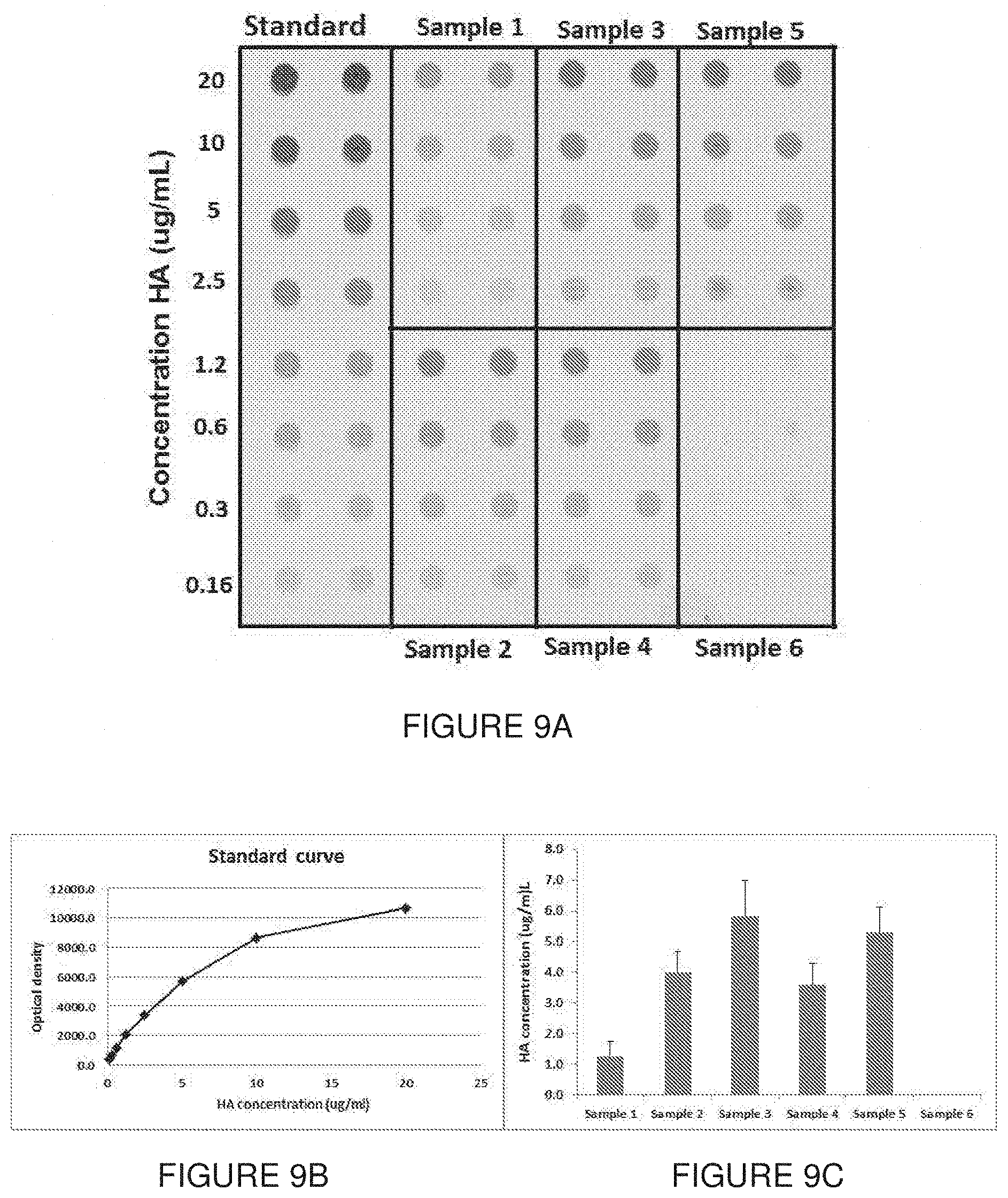
D00011
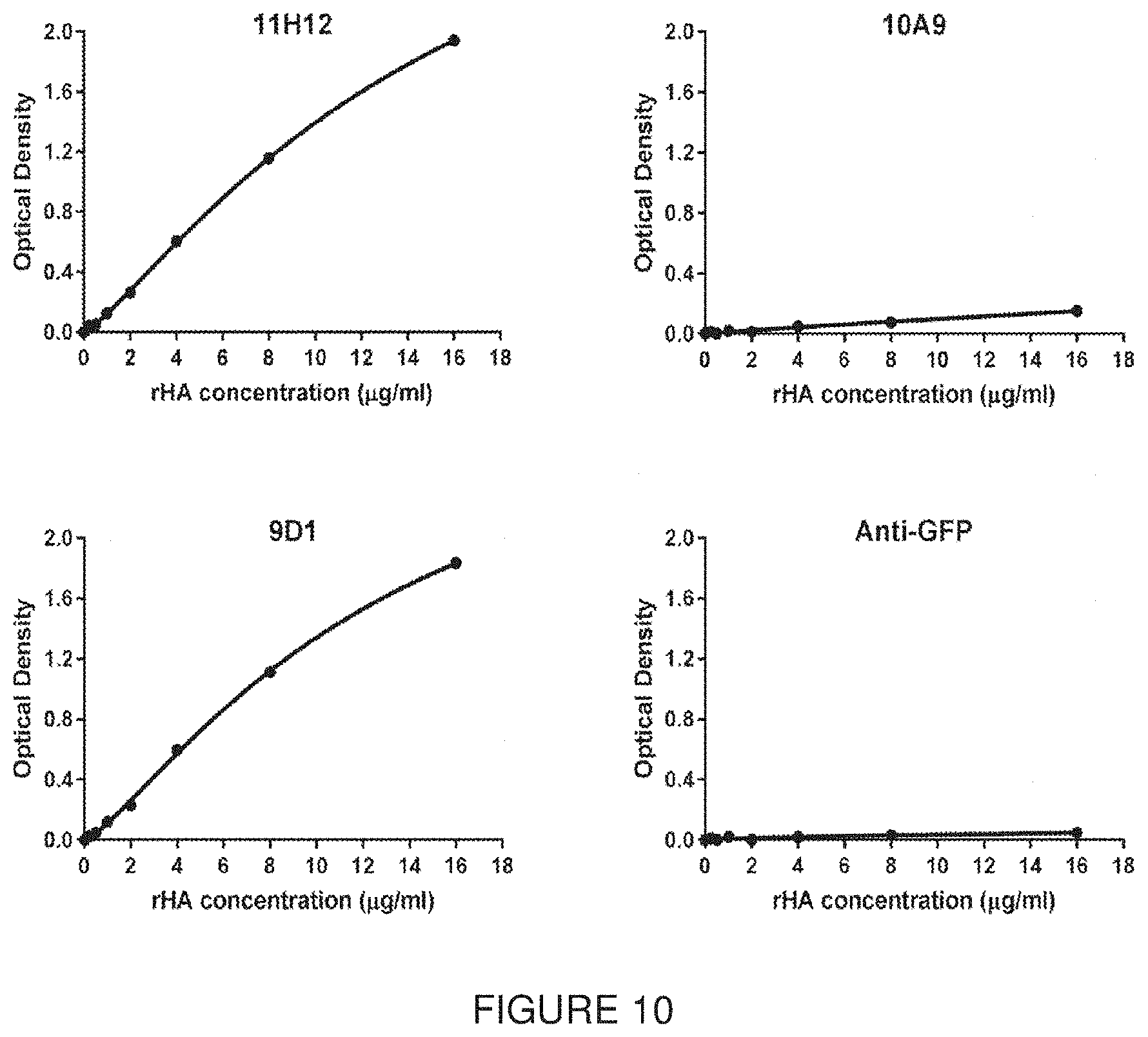
D00012

D00013

D00014

D00015

D00016
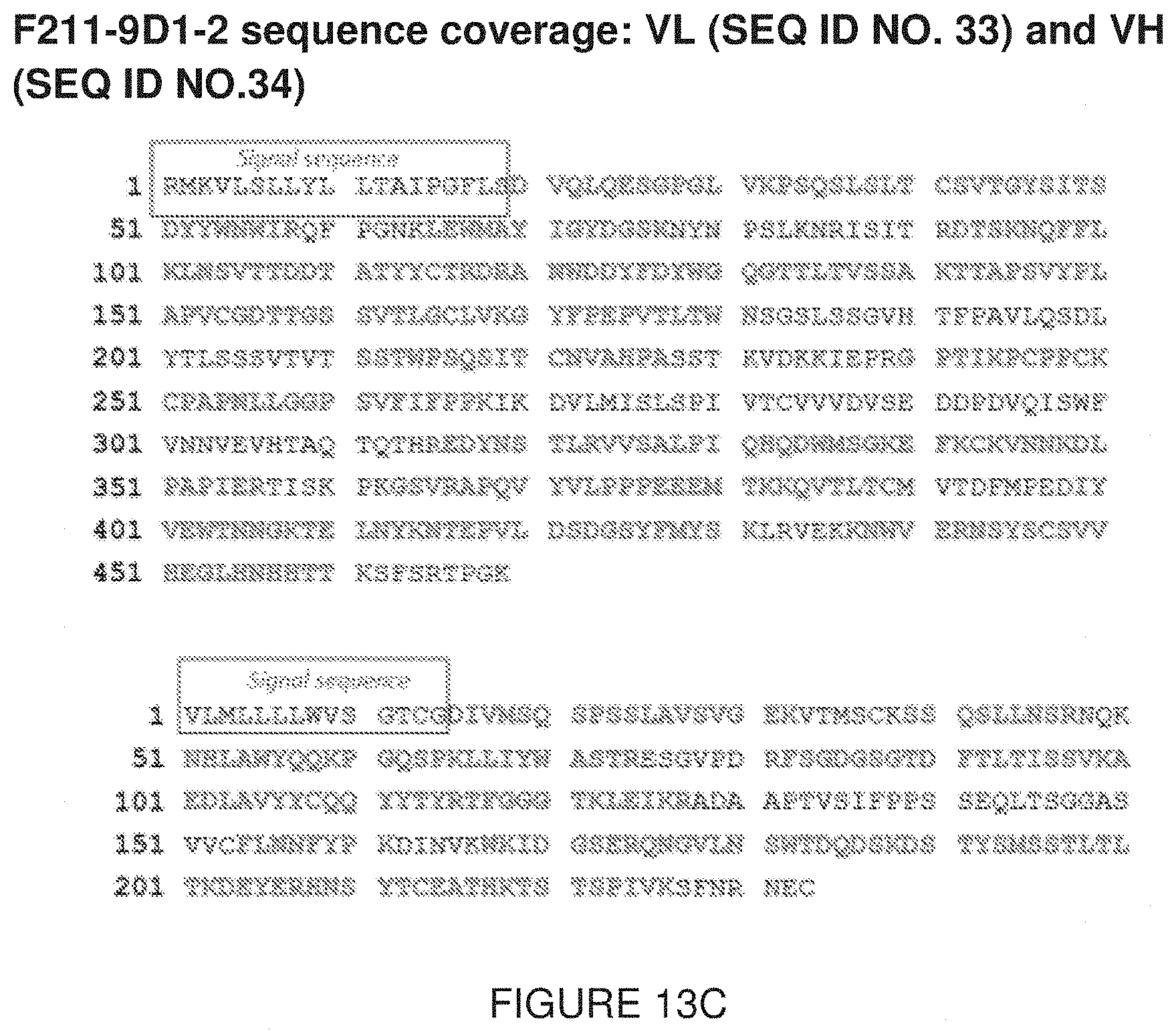
D00017
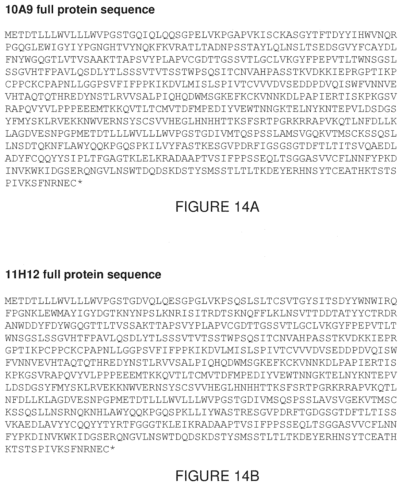
S00001
XML
uspto.report is an independent third-party trademark research tool that is not affiliated, endorsed, or sponsored by the United States Patent and Trademark Office (USPTO) or any other governmental organization. The information provided by uspto.report is based on publicly available data at the time of writing and is intended for informational purposes only.
While we strive to provide accurate and up-to-date information, we do not guarantee the accuracy, completeness, reliability, or suitability of the information displayed on this site. The use of this site is at your own risk. Any reliance you place on such information is therefore strictly at your own risk.
All official trademark data, including owner information, should be verified by visiting the official USPTO website at www.uspto.gov. This site is not intended to replace professional legal advice and should not be used as a substitute for consulting with a legal professional who is knowledgeable about trademark law.