Cd133 Related To Anticancer Agent Resistance In Colon Cancer And Use Thereof
KO; Je Sang ; et al.
U.S. patent application number 16/848273 was filed with the patent office on 2020-10-15 for cd133 related to anticancer agent resistance in colon cancer and use thereof. This patent application is currently assigned to KOREA UNIVERSITY RESEARCH AND BUSINESS FOUNDATION. The applicant listed for this patent is KOREA UNIVERSITY RESEARCH AND BUSINESS FOUNDATION. Invention is credited to Min-Soo KANG, Je Sang KO.
| Application Number | 20200325545 16/848273 |
| Document ID | / |
| Family ID | 1000004828428 |
| Filed Date | 2020-10-15 |


View All Diagrams
| United States Patent Application | 20200325545 |
| Kind Code | A1 |
| KO; Je Sang ; et al. | October 15, 2020 |
CD133 RELATED TO ANTICANCER AGENT RESISTANCE IN COLON CANCER AND USE THEREOF
Abstract
The present invention relates to a use of CD133 involved in resistance to an EGFR-targeting agent in colon cancer. The CD133 protein may be used as a novel target protein for diagnosing and treating resistant cancer as well as general cancer.
| Inventors: | KO; Je Sang; (Seoul, KR) ; KANG; Min-Soo; (Gyeonggido, KR) | ||||||||||
| Applicant: |
|
||||||||||
|---|---|---|---|---|---|---|---|---|---|---|---|
| Assignee: | KOREA UNIVERSITY RESEARCH AND
BUSINESS FOUNDATION Seoul KR |
||||||||||
| Family ID: | 1000004828428 | ||||||||||
| Appl. No.: | 16/848273 | ||||||||||
| Filed: | April 14, 2020 |
| Current U.S. Class: | 1/1 |
| Current CPC Class: | A61K 45/06 20130101; C12Q 2600/158 20130101; C12Q 1/6886 20130101 |
| International Class: | C12Q 1/6886 20060101 C12Q001/6886 |
Foreign Application Data
| Date | Code | Application Number |
|---|---|---|
| Apr 15, 2019 | KR | 10-2019-0043765 |
Claims
1. A biomarker composition for diagnosing resistance to an EGFR-targeting agent in colon cancer, comprising CD133 protein or a gene encoding the same.
2. A composition for diagnosing resistance to an EGFR-targeting agent in colon cancer, comprising an agent for measuring the expression level or activity of CD133 protein or a gene encoding the same.
3. The composition of claim 2, wherein the agent for measuring the expression level or activity of the protein is one or more of an antibody, a peptide and a nucleotide, which specifically bind to the protein.
4. The composition of claim 2, wherein the agent for measuring the expression level or activity of the gene is one or more of antisense oligonucleotide, primer pair and probe, which specifically bind to mRNA of the gene.
5. A kit for diagnosing resistance to an EGFR-targeting agent in colon cancer, comprising the composition of claim 1.
6. A method of providing information required for diagnosis of resistance to an EGFR-targeting agent in colon cancer, comprising: measuring the expression level or activity of CD133 protein or a gene encoding the same in a biological sample isolated from a colon cancer patient.
7. The method of claim 6, wherein, when the expression level or activity of the protein or gene is higher than normal ranges, the colon cancer patient is diagnosed with resistance to an EGFR-targeting agent.
8. A method of screening a drug for preventing or treating resistance to an EGFR-targeting agentin colon cancer, comprising: bringing the CD133 geneor CD133 proteininto contact with a candidate ex vivo, and determining whether the candidate improves or inhibits the function or activity of the gen or the protein.
9. The method of claim 8, wherein, when the candidate downgrades the expression of the CD133 gene or CD133 protein, the candidate is determined as a drug for the prevention or treatment of resistance to an EGFR-targeting agentin colon cancer.
10. A method of preventing or treating resistance to an EGFR-targeting agentin colon cancer, the method comprising administering an inhibitor against a CD133 gene to a subject, wherein the inhibitor comprises an antisense oligonucleotide, siRNA, shRNA or miRNA against the CD133 gene, or a vector comprising the same.
11. A method of preventing or treating resistance to an EGFR-targeting agentin colon cancer, the method comprising administering an inhibitor against a CD133 protein to a subject, wherein the inhibitor is a peptide, a compound, or an antibody against the CD133 protein.
Description
TECHNICAL FIELD
[0001] The present invention relates to a use of CD133 involved in anticancer agent resistance in colon cancer.
BACKGROUND ART
[0002] Although many anticancer agents against various types of cancer have been developed to date, only a few types of cancer can be completely cured with an anticancer agent alone, this is because cancer cells do not respond to an anticancer agent in cancer treatment with the anticancer agent, or tumors are effectively reduced in an early stage, but develop anticancer agent resistance during or after treatment. Therefore, for effective anticancer treatment, resistance to an anticancer agent, for example, anticancer agent resistance of cancer cells, has to be overcome.
[0003] Generally, a mutation in the KRAS, NRAS or BRAF gene results in production of a protein having a modified signaling characteristic in tumor cells, and such a mutation has been known to be associated with unsuccessful results in cancer treatment using a therapeutic antibody targeting an epithelial growth factor receptor (EGFR), for example, gefitinib, cetuximab or panitumumab (Amado, Wolf et al. 2008; Karapetis, Khambata-Ford et al. 2008; Di Nicolantonio, Martini et al. 2008; Loupakis, Ruzzo et al. 2009; Lievre, Bachet et al. 2006). For example, KRAS G13D known as a colon cancer mutant cell line was reported to have resistance (tolerance) to an anticancer agent targeting EGFR.
[0004] For this reason, to improve a therapeutic effect of an anticancer agent targeting EGFR for cancer treatment, it is urgent to solve such a problem of anticancer agent resistance. Research on the identification of a specific cause or mechanism of the anticancer agent resistance has been persistently conducted, but it is a reality that little is known about it.
[0005] Nevertheless, the inventors had identified that, through a resistance study for colon cancer mutant cell lines resistant to an anticancer agent targeting EGFR, CD133 is related to resistance of colon cancer cells against an anticancer agent targeting EGFR, and thus the present invention was completed.
Technical Problem
[0006] The present invention is directed to providing a biomarker composition for diagnosing resistance to an EGFR-targeting agent in colon cancer, which includes CD133 protein or a gene encoding the same.
[0007] The present invention is also directed to providing a kit for diagnosing resistance to an EGFR-targeting agent in colon cancer.
[0008] The present invention is also directed to providing a method of providing information required for diagnosis of resistance to an EGFR-targeting agent in colon cancer.
Technical Solution
[0009] The inventors had confirmed, through a study on a KRAS G13D mutant colon cancer cell line having resistance to an EGFR-targeting agent, that the cell migration to and invasiveness of adjacent normal cells are highly improved by transferring the mutant to adjacent cells via microvesicles, the migration of KRAS G13D via microvesicles imparts anticancer agent resistance to adjacent colon cancer cells, and the CD133 protein controlling the release of microvesicles is related to resistance to an EGFR-targeting agent, and thus the present invention was completed.
[0010] The present invention is characterized by confirming whether colon cancer cells have resistance to an EGFR-targeting agent using the activity of the CD133 gene and an active protein, which is a product thereof, as a biomarker.
[0011] To this end, the present invention provides a biomarker composition for diagnosing resistance to an EGFR-targeting agent in colon cancer, which includes the CD133 protein or a gene encoding the same.
[0012] The biomarker of the present invention may be a marker with anticancer agent resistance, which is an EGFR-targeting agent, and since it has excellent accuracy and reliability as a marker for diagnosing anticancer agent resistance, the biomarker may be used to diagnose the occurrence, development and/or metastasis of resistant cancer, and treat resistant cancer.
[0013] The term "resistance" or "tolerance" used herein means that an organism does not sensitively respond to a drug, and thus withstands the drug's effects.
[0014] CD133 used as a biomarker for resistance diagnosis in the present invention is one of the representative CD-type proteins expressed on the surface of cancer stem cells, which are reported to be mainly present in cancer stem cells of colon cancer, liver cancer, pancreatic cancer and lung cancer.
[0015] The term "colon cancer" used herein is a collective term referring to rectal cancer, colorectal cancer and anal cancer.
[0016] In addition, the EGFR-targeting agent of the present invention refers to an anticancer agent, which may be any EGFR-targeting agent exhibiting an anticancer effect, and is used interchangeably with an EGFR-targeting drug. Preferably, the EGFR-targeting agent is one or more selected from the group consisting of cetuximab, gefitinib, erlotinib, panitumumab, PKI-166, EKB-569, HKI-272 (WAY-177820), icotinib, brigatinib, afatinib, lapatinib, canertinib, AEE788, XL647, and Zactima. More preferably, the EGFR-targeting agent is cetuximab, gefitinib, erlotinib or panitumumab, and most preferably, gefitinib.
[0017] The term "diagnosis" used herein refers to the detection of a pathological condition, which means, in terms of the purpose of the present invention, the determination of the presence or absence of resistance, and the development or alleviation of symptoms of a disease by examining the presence or absence of the expression of a biomarker for diagnosing resistance to an EGFR-targeting agent.
[0018] The "diagnosis biomarker" used herein refers to a material that can be used in diagnosis by distinguishing the presence or absence of resistance to an EGFR-targeting agent, and includes organic biomolecules such as polypeptides or nucleic acids (e.g., mRNA, etc.), lipids, glycolipids, glycoproteins, saccharides (a monosaccharide, a disaccharide, an oligosaccharide, etc.), which increase or decrease in cancer cells resistant to an anticancer agent, compared to cancer cells. The biomarker for resistance diagnosis according to the present invention may be a protein expressed from the CD133 gene whose expression level is increased in resistant cancer cells with respect to an EGFR-targeting agent, compared to general cancer cells.
[0019] The composition for diagnosing resistance to an EGFR-targeting agent may include an agent that measures an mRNA expression level of the CD133 gene or an amount of a protein expressed from the gene, and such an agent may be an oligonucleotide having a complementary sequence to CD133 mRNA, for example, a primer or nucleic acid probe specifically binding to CD133 mRNA, or an antibody specific for CD133 protein.
[0020] The primer refers to a single-stranded oligonucleotide that is able to serve as a starting point of template-directed DNA synthesis under suitable conditions (that is, four different types of nucleoside triphosphates and a polymerase) in a suitable buffer at a suitable temperature. A suitable length of the primer may vary according to various factors, for example, a temperature and the usage of the primer. In addition, the primer sequence is not required to be perfectly complementary to a partial sequence of the template, and it is sufficient that the primer sequence has sufficient complementarity within a range in which the primer can do an intrinsic action when hybridized with the template. Therefore, the primer of the present invention does not need to have a perfectly complementary sequence to the nucleotide sequence of a gene, which is the template, and it is reasonable that the primer has a sufficient complementarity within a range in which the primer can do an intrinsic action when hybridized with the gene sequence. In addition, the primer according to the present invention is preferably used in a gene amplification reaction. The amplification reaction refers to a reaction of amplifying a nucleic acid molecule, and amplification reactions of such a gene are well known in the art, and may include, for example, polymerase chain reaction (PCR), reverse transcriptase chain reaction (RT-PCR), ligase chain reaction (LCR), transcription-mediated amplification (TMA), and nucleic acid sequence-based amplification (NASBA).
[0021] The nucleic acid probe refers to a natural or modified monomer or linear oligomer consisting of linkages of such monomers, and includes a deoxyribonucleotide and a ribonucleotide, may be specifically hybridized with a target nucleotide sequence, and naturally occurs or is artificially synthesized. The probe according to the present invention may be a single chain, and preferably, an oligodeoxyribonucleotide. The probe of the present invention may include natural dNMPs (i.e., dAMP, dGMP, dCMP and dTMP), or nucleotide analogs or derivatives. In addition, the probe of the present invention may include a ribonucleotide. For example, the probe of the present invention may include backbone-modified nucleotides, such as a peptide nucleic acid (PNA), phosphorothioate DNA, phosphodithioate DNA, phosphoroamidate DNA, amide-linked DNA, MMI-linked DNA, 2'-O-methyl RNA, a-DNA and methyl phosphonate DNA; sugar-modified nucleotides, such as 2'-O-methyl RNA, 2'-fluoro RNA, 2'-amino RNA, 2'-O-alkyl
[0022] DNA, 2'-O-allyl DNA, 2'-O-alkynyl DNA, hexose DNA, pyranosyl RNA and anhydrohexitol DNA; base-modified nucleotides, such as pyrimidines with a C-5 substituent (the substituent may be fluoro-, bromo-, chloro-, iodo-, methyl-, ethyl-, vinyl-, formyl-, ethynyl-, propynyl-, alkynyl-, thiazolyl-, imidazolyl-, or pyridyl-), 7-deazapurines with a C-7 substituent (the substituent may be fluoro-, bromo-, chloro-, iodo-, methyl-, ethyl-, vinyl-, formyl-, alkynyl-, alkenyl-, thiazolyl-, imidazolyl-, or pyridyl-); inosine; and diaminopurine.
[0023] The CD133-specific antibody may be a polyclonal antibody, a monoclonal antibody, a human antibody or a humanized antibody.
[0024] Examples of the antibody fragments include Fab, Fab', F(ab').sub.2 and Fv fragments; a diabody; a linear antibody (Zapata et al., Protein Eng. 8(10):1057-1062(1995)); a single-chain antibody molecule; and a multi-specific antibody formed from an antibody fragment.
[0025] When an antibody is digested with papain, two identical antigen-binding fragments, that is, "Fab" fragments having a single antigen-binding region, and the remainder, "Fc" fragment, are obtained. When treated with pepsin, a F(ab').sub.2 fragment which has two antigen-binding regions and is still able to be crosslinked to an antigen is produced. Fv is a minimal antibody fragment including a complete antigen-recognizing and binding region. This region consists of a dimer of one heavy chain variable region and one light chain variable region, which are firmly bound to each other by a non-covalent bond.
[0026] A method of preparing a polyclonal antibody is known to those of ordinary skill in the art. The polyclonal antibody may be prepared by one or more injections of an immunizing agent in combination with an immune adjuvant, if necessary, into a mammal. Generally, an immunizing agent and/or an immune adjuvant is/are subcutaneously or intraperitoneally injected into a mammal several times. The immunizing agent may be a protein of the present invention or a fusion protein thereof. It may be effective that an immunizing agent, as well as a protein known to be immunogenic, is injected into an immunized mammal.
[0027] The monoclonal antibody according to the present invention may be prepared by the hybridoma method disclosed in the literature (Kohler et al., Nature, 256:495 (1975)), or a recombinant DNA method (refer to U.S. Pat. No. 4,816,576). The monoclonal antibody may also be isolated from a phage antibody library using the technology disclosed in the literature (Clackson et al., Nature, 352:624-628 (1991) and Marks et al., J. Mol. Biol., 222:581-597 (1991)).
[0028] In the monoclonal antibody of the present invention, specifically, when exhibiting desired activity, a part of a heavy chain and/or a light chain is identical to or has homology with a corresponding sequence of an antibody derived from a specific species or an antibody belonging to a specific antibody class or subclass, whereas the remainder of the chain(s) includes an antibody derived from a different species, an antibody belonging to a different antibody class or subclass or a "chimeric" antibody which is identical or homologous to a fragment of such an antibody (Morrison et al., Proc. Natl. Acad. Sci. USA, 81:6851-6855 (1984)).
[0029] A "humanized" form of a non-human (e.g., rodents) antibody is a chimeric immunoglobulin including a minimal sequence derived from a non-human immunoglobulin, an immunoglobulin chain or a fragment thereof (e.g., Fv, Fab, Fab', F(ab').sub.2 or a different antigen-binding sequence of an antibody). In most cases, the humanized antibody includes a human immunoglobulin (recipient antibody) in which a complementarity-determining region (CDR) residue of a recipient is substituted with a CDR residue of a non-human species (donor antibody) such as a mouse, rat or rabbit, having desired specificity, affinity and ability. In some cases, aFv framework residue of the human immunoglobulin is substituted with a corresponding non-human residue. In addition, the humanized antibody may include a recipient antibody, or a residue which is not found in an introduced CDR or framework sequence. Generally, the humanized antibody substantially includes one or more, generally two or more variable domains, and here, all or substantially all CDR regions correspond to a region of a non-human immunoglobulin, and all or substantially all FR regions correspond to a region of a human immunoglobulin sequence. In addition, the humanized antibody includes at least a part of a variable region (Fc) of an immunoglobulin, generally, a part of a human immunoglobulin region (Presta, Curr. Op. Struct. Biol. 2:593-596 (1992)).
[0030] The composition for diagnosing resistance to an EGFR-targeting agent according to the present invention may be included in the form of a kit.
[0031] The kit may include a primer, probe or antibody which is able to measure the expression level of the CD133 gene or an amount of the CD133 protein, and the definition thereof is as described above.
[0032] When the kit is employed in PCR amplification, optionally, reagents required for PCR amplification, for example, a buffer, a DNA polymerase (e.g., a heat-stable DNA polymerase obtained from Thermusaquaticus (Taq), Thermusthermophilus (Tth), Thermusfiliformis, Thermisflavus, Thermococcusliteralis or Pyrococcusfuriosus (Pfu)), a DNA polymerase cofactor and dNTPs may be included, and when the kit is used in an immunoassay, the kit of the present invention may optionally include a secondary antibody and a marker substrate. Further, the kit according to the present invention may be manufactured in multiple individual packages or compartments containing the above-mentioned reagent components.
[0033] In addition, the composition for diagnosing resistance to an EGFR-targeting agent according to the present invention may be included in the form of a microarray.
[0034] In the microarray of the present invention, a primer, probe or antibody that is able to measure the expression level of the CD133 protein or gene encoding the same is used as a hybridizable array element, and fixed on a substrate. A preferable substrate may be a suitable rigid or semi-rigid support, for example, a membrane, a filter, a chip, a slide, a wafer, a fiber, a magnetic or non-magnetic bead, a gel, a tubing, a plate, a polymer, a microparticle and a capillary. The hybridizable array element may be arranged and immobilized on the substrate, and such immobilization may be performed by a chemical binding method or a covalent binding method such as UV. For example, the hybridizable array element may be bound to a glass surface which is modified to include an epoxy compound or an aldehyde group, or bound to a polylysine-coated surface using UV. In addition, the hybridizable array element may bind to a substrate by a linker (e.g., an ethylene glycol oligomer or a diamine).
[0035] Meanwhile, when a sample applied to the microarray of the present invention is a nucleic acid, it may be labeled and hybridized with an array element fixed on the microarray. Hybridization conditions may vary, and detection and analysis of a hybridization degree may be performed in various ways according to a labeling material.
[0036] In addition, the present invention provides a method of providing information for diagnosing resistance to an EGFR-targeting agent, which includes measuring the expression level of the CD133 gene or the expressed protein in a biological sample isolated from a patient, and more specifically, the method may include (a) measuring the expression level of the CD133 gene or an amount of the expressed protein in a biological sample of a patient; and (b) measuring the expression level of the gene and an amount of the expressed protein from a sample of a normal control and comparing it with the measurement result obtained in Step (a).
[0037] The method of measuring the expression level of a gene or an amount of a protein encoded by the gene may be performed by a known process of isolating mRNA or a protein from a biological sample using a known technique.
[0038] The biological sample refers to a sample obtained from the living body, which is different from a normal control in terms of the expression level of the gene or a protein level according to the occurrence or progression of resistance to an EGFR-targeting agent, and the sample may be, but is not limited to, tissue, a cell, blood, serum, plasma, saliva or urine.
[0039] The measurement of the expression level of a gene is preferably to measure an mRNA level, and methods of measuring an mRNA level include, but are not limited to, RT-PCR, real time reverse transcription polymerase chain reaction, RNase protection assay, Northern blotting and DNA chip assay.
[0040] The measurement of a protein level may use an antibody, and in this case, the CD133 protein in a biological sample and an antibody specific for the protein forms a binding product, that is, an antigen-antibody complex, and an amount of antigen-antibody complex formation may be quantitatively measured from the size of a signal of a detection label. The detection level may be selected from the group consisting of an enzyme, a fluorescent material, a ligand, a light-emitting material, a microparticle, a redox molecule and a radioisotope, but the present invention is not limited thereto. Analysis methods for measuring a protein level may include, but not limited to, Western blotting, ELISA, radioimmunoassay, radioimmunodiffusion, Ouchterlonyimmunodiffusion, rocket immunoelectrophoresis, immunohistochemical staining, immunoprecipitation assay, complement fixation assay, FACS, and protein chip assay.
[0041] Accordingly, the present invention may confirm mRNA expression levels or protein amounts in the control, a patient resistant to an EGFR-targeting agent, or a patient suspected of having resistance to an EGFR-targeting agent through the detection methods described above, and the expression levels are compared to be able to diagnose the occurrence and progression of the resistance to an EGFR-targeting agent.
[0042] In addition, according to the method of providing information for the diagnosis of the resistance to an EGFR-targeting agent according to the present invention, when the expression level of the CD133 gene according to the present invention or an amount of the expressed protein is higher than that of the normal control sample, it can be determined that a patient has resistance to an EGFR-targeting agent.
[0043] The present invention also relates to a composition for preventing or treating resistance to an EGFR-targeting agent, which includes a CD133 inhibitor.
[0044] The present invention also provides a use of a CD133 inhibitor for preparing a pharmaceutical composition for preventing or treating resistance to an EGFR-targeting agent.
[0045] According to the present invention, CD133 is highly expressed in cancer cells with resistance to an EGFR-targeting agent, as described above, and the expression of CD133 promotes budding of microvesicles, increases cell migration and invasiveness by delivering the oncogenic protein KRAS G13D to adjacent cells using microvesicles, and thus is involved in the occurrence of anticancer agent resistance.
[0046] Therefore, the CD133 inhibitor may be used in treatment of resistant cancer.
[0047] To this end, the composition for preventing or treating resistance to an EGFR-targeting agent of the present invention may include an agent for reducing mRNA expression of the CD133 gene or protein expression thereof, or degrading function or activity.
[0048] The CD133 protein inhibitor may be a peptide or compound which is bound with the CD133 protein to regulate a signal of a nerve differentiation pathway. The inhibitor may be selected by a screening method exemplified below, such as protein structure analysis, and may be designed using a method known in the art.
[0049] Specifically, the CD133 protein inhibitor may be a material which is bound with the CD133 protein consisting of an amino acid sequence represented by SEQ ID NO: 2 to inhibit activity.
[0050] In addition, as the protein inhibitor, a polyclonal antibody, monoclonal antibody, human antibody or humanized antibody against the CD133 protein may be used, and the definition of the antibody is as described above.
[0051] Resistant cancer may be prevented or treated by inhibiting the function of CD133 in cells using the antibody.
[0052] The functional or activity inhibitor of the CD133 protein according to the present invention may be delivered using a liposome, a virus, a gene gun, a polymer, ultrasonication or an electric shock, but the present invention is not particularly limited thereto.
[0053] The CD133 gene may be DNA encoding the same or mRNA transcribed therefrom. Accordingly, the inhibitor of the gene may be an inhibitor which is bound to the gene itself to interfere with transcription or bound to mRNA transcribed from the gene to interfere with the translation of mRNA.
[0054] Therefore, the inhibitor of the CD133 gene includes all types of inhibitors that inhibit the expression of the CD133 gene. For example, such an inhibitor may be a peptide, nucleic acid or compound binding to the gene. The inhibitor may be selected by a screening method to be described below, such as cell-based screening, and may be designed by a method known in the art.
[0055] In one embodiment, the inhibitor may be an antisense-oligonucleotide, siRNA, shRNA or miRNA against the CD133 gene or a vector including the same. Such an antisense-oligonucleotide, siRNA, shRNA, miRNA or a vector including the same may be manufactured using a method known in the art. Specifically, the inhibitor may be a nucleic acid molecule prepared to inhibit the expression of the CD133 gene consisting of SEQ ID NO: 1.
[0056] The term "siRNA" used herein refers to double-stranded RNA inducing RNA interference through the cleavage of mRNA of a target gene, and consists of an RNA strand of a sense sequence having the sequence like mRNA of the target gene and an RNA strand of an antisense sequence having a sequence complementary thereto.
[0057] The siRNA may include siRNA synthesized in vitro, or a form expressed by inserting a base sequence encoding siRNA into an expression vector.
[0058] The "vector" used herein refers to a gene construct including foreign DNA inserted into a genome encoding a polypeptide.
[0059] The vector related to the present invention is a vector in which a nucleic acid sequence inhibiting the gene is inserted into a genome, and such a vector may be a DNA vector, a plasmid vector, a cosmid vector, a bacteriophage vector, an enzyme vector, or a viral vector.
[0060] In addition, the antisense may have a sequence complementary to an entire or partial mRNA sequence transcribed from the CD133 gene or a fragment thereof, and bind to the mRNA to inhibit the expression of the CD133 gene or a fragment thereof.
[0061] In addition, the short hairpin RNAi (shRNAi) may be manufactured by a conventional method using a human or mouse shRNAi common base sequence region as a target.
[0062] In addition, the pharmaceutical composition of the present invention may further include a pharmaceutically acceptable carrier.
[0063] The pharmaceutically acceptable carrier includes a carrier and a vehicle, which are generally used in the pharmaceutical field, and specifically, includes an ion exchange resin, alumina, aluminum stearate, lecithin, a serum protein (e.g., human serum albumin), a buffer material (e.g., various types of phosphates, glycine, sorbic acid, potassium sorbate, or a partial glyceride compound of a saturated vegetable fatty acid), water, a salt or electrolyte (e.g., protamine sulfate, disodium hydrogen phosphate, potassium hydrogen phosphate, sodium chloride or a zinc salt), colloidal silica, magnesium trisilicate, polyvinylpyrrolidone, a cellulose-based substrate, polyethylene glycol, sodium carboxymethylcellulose, polyarylate, wax, polyethylene glycol or lanolin, but the present invention is not limited thereto.
[0064] In addition, the composition of the present invention may further include a lubricant, a wetting agent, an emulsifier, a suspending agent, or a preservative, other than the above-described components.
[0065] In one aspect, the composition according to the present invention may be prepared as an aqueous solution for parenteral administration, and preferably, a Hank's solution, a Ringer's solution or a buffer solution such as physically buffered saline is used. An aqueous injectable suspension may include a substrate that can increase the viscosity of the suspension such as sodium carboxymethylcellulose, sorbitol or dextran.
[0066] The composition of the present invention may be systemically or locally administered, and formulated in a suitable form by known technology for such administration. For example, for oral administration, the composition may be administered by mixing the active compound with an inactive diluent or edible carrier, encapsulating all components with a hard or soft gelatin capsule or compressing all components in the form of a tablet. For oral administration, the active compound may be mixed with an excipient, and then formed as an edible tablet, a buccal tablet, a troche, a capsule, an elixir, a suspension, a syrup or a wafer.
[0067] Various forms for injection or parenteral administration may be prepared according to a technique known or commonly used in the art. Since CD133 is well dissolved in saline or a buffer solution, after storage in a freeze-dried state, an effective amount of CD133 may be mixed in saline or a buffer solution to prepare a solution suitable for intravenous injection, subcutaneous injection, muscle injection, intraperitoneal injection or transdermal injection, right before the administration.
[0068] The effective amount of the active ingredient of the pharmaceutical composition of the present invention refers to an amount required for prevention, inhibition or alleviation of a disease.
[0069] Accordingly, the effective amount may be adjusted by various factors such as the type and severity of a disease, the type and content of the active ingredient and other components, contained in the composition, the type of a dosage form, and the age, body weight, general health condition, gender and diet of a patient, administration time, administration route, a release rate of the composition, a treatment period, and a co-used drug. While not limited thereto, for example, in the case of an adult, the inhibitor of the present invention may be administered once or several times a day, when the type of the inhibitor is a compound, it may be administered at a dose of 0.1 ng/kg to 10 g/kg, when the type of the inhibitor is a polypeptide, protein or antibody, it may be administered at a dose of 0.1 ng/kg to 10 g/kg, or when the type of the inhibitor is an antisense-oligonucleotide, siRNA, shRNAi or miRNA, it may be administered at a dose of 0.01 ng/kg to 10 g/kg.
[0070] The present invention also provides a method of treating an animal with resistance to an EGFR-targeting agent, which includes administering a composition including a pharmaceutically effective amount of a CD133 inhibitor for preventing or treating resistance to an EGFR-targeting agent to a subject.
[0071] A pharmaceutical composition and an administration method, which are used in the method of treating resistance to an EGFR-targeting agent, are as described above, and the common description between them will be omitted to avoid excessive complexity of the specification.
[0072] Meanwhile, subjects to which the pharmaceutical composition for preventing or treating resistance to an EGFR-targeting agent may be administered include all kinds of animals. For example, the subjects may be non-human animals such as dogs, cats, and mice.
[0073] The present invention also provides a method of screening a drug for preventing or treating resistance to an EGFR-targeting agent, which includes bringing the CD133 gene into contact with a candidate ex vivo, and determining whether the candidate promotes or inhibits the expression of the gene.
[0074] In addition, the present invention provides a method of screening a drug for preventing or treating resistance to an EGFR-targeting agent, which includes bringing the CD133 protein into contact with a candidate ex vivo, and determining whether the candidate improves or inhibits the function or activity of the protein.
[0075] According to the screening method of the present invention, first, a candidate to be analyzed may be brought into contact with resistant cancer cells having the gene or protein.
[0076] The candidate may be an individual nucleic acid, protein or peptide, another extract or natural substance, or a compound, which is expected or randomly selected to have a potential as a material promoting or inhibiting the mRNA transcription or mRNA translation into a protein in the base sequence of the CD133 gene or a drug improving or inhibiting the function or activity of the CD133 protein according to a common selection method.
[0077] Afterward, in cells treated with a candidate, the expression level of the gene or the amount or activity of the protein may be measured, and as a result of the measurement, when the expression level of the gene or the amount or activity of the protein is measured as increased or decreased, the candidate may be determined as a material for treating or preventing resistant cancer.
[0078] The method of measuring the expression level of the gene, or the amount or activity of the protein may be performed by various methods known in the art, and may be, for example, RT-PCR, real time-polymerase chain reaction, Western blotting, Northern blotting, ELISA, radioimmunoassay (RIA), radioimmunodiffusion and immunoprecipitation assay, but the present invention is not limited thereto.
[0079] The candidate exhibiting an activity of inhibiting gene expression or a protein function, obtained by the screening method of the present invention, may be a candidate for a resistant cancer therapeutic agent.
[0080] The candidate for a therapeutic agent against cancer resistant to an EGFR-targeting agent serves as a leading compound in the development of a therapeutic agent against resistance to an EGFR-targeting agent, and modifies and optimizes the structure of the leading compound to exhibit an effect of inhibiting the CD133 gene or the function of a protein expressed therefrom, thereby developing a novel therapeutic agent against cancer resistant to an EGFR-targeting agent.
[0081] Details related to genetic engineering technology in the present invention will become more apparent from the content disclosed in Sambrook, et al. Molecular Cloning, A Laboratory Manual, Cold Spring Harbor laboratory Press, Cold Spring Harbor, N.Y. (2001); and Frederick M. Ausubel et al., Current protocols in molecular biology volume 1, 2, 3, John Wiley & Sons, Inc. (1994).
Advantageous Effects
[0082] The present invention identifies that the CD133 protein expressed in colon cancer increases resistance to an EGFR-targeting agent, and thus the CD133 protein can be used as a novel target protein for diagnosing and treating resistant cancer as well as general cancer.
DESCRIPTION OF DRAWINGS
[0083] The patent or application file contains at least one drawing executed in color. Copies of this patent or patent application publication with color drawing(s) will be provided by the Office upon request and payment of the necessary fee.
[0084] FIGS. 1A-1E show the result of identifying a CD133 expression regulatory mechanism in liver cancer cells: (A) the change in CD133 expression according to EGF expression; (B-C) the change in CD133 expression according to an EGF downstream signaling molecule, NF-KB; and (D-E) the relationship between NF-.kappa.B and CD133, confirmed by promoter activity.
[0085] FIGS. 2A-2E show the result of confirming that CD133 is involved in promotion of microvesicle budding in liver cancer cells: (A) confirmation of the relationship between an EGF signaling system and microvesicle and TNT formation; (B) confirmation of microvesicle and TNT formation in a CD133-overexpressing cell line; and (C-E) confirmation of the change in activation of Rac1 and RhoA associated with microvesicle budding and a downstream signaling factor, Erk1/2, of RhoA according to CD133 expression
[0086] FIGS. 3A-3B show the result of analyzing microvesicles budded from cancer cells: (A) the result of confirming whether CD133 is included in microvesicles budded from various cancer cells; and (B) the result of observing the migration of microvesicles containing CD133 to adjacent cells.
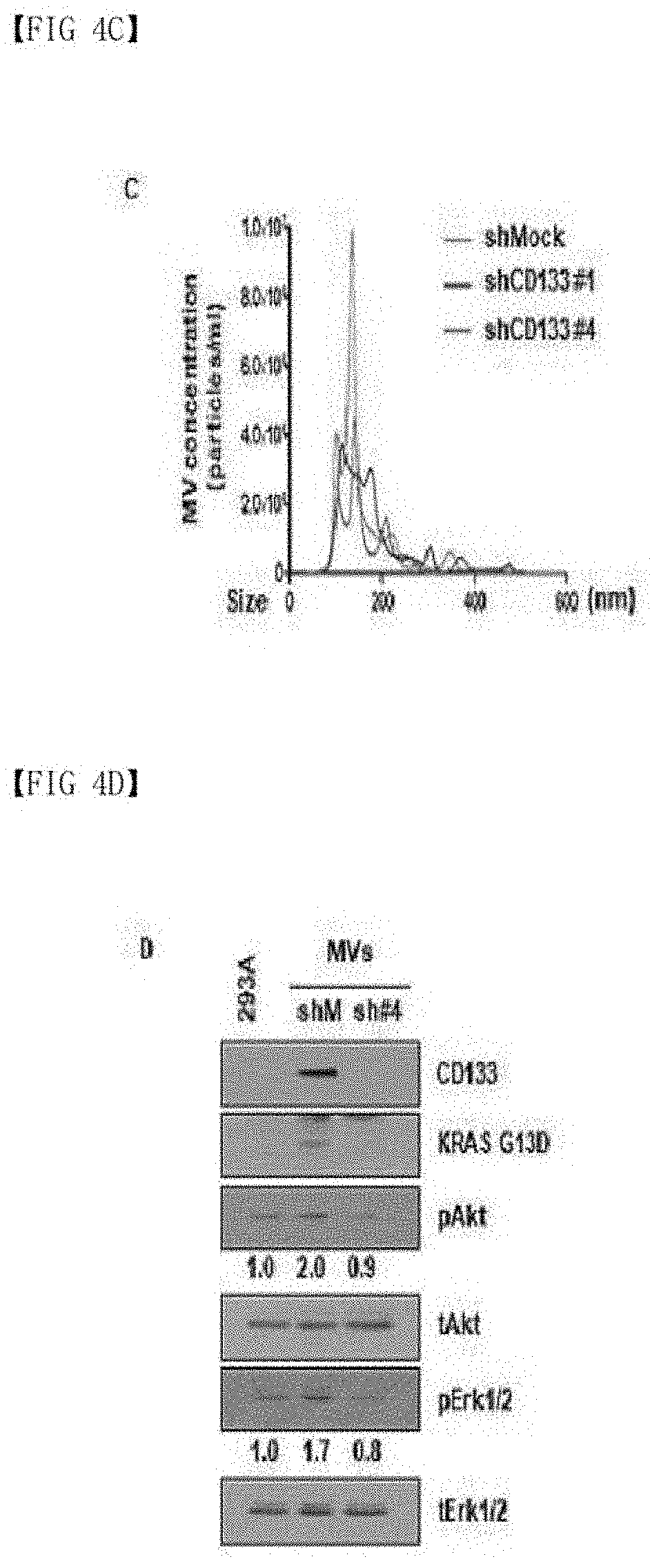
[0087] FIGS. 4A-4F show the result of confirming that CD133 is involved in oncogenic protein transport and microvesicle budding in colon cancer: (A) confirmation whether an oncogenic protein is contained in microvesicles according to CD133 expression; (B-C) confirmation that CD133 expression contributes to microvesicle budding and a size change; (D) confirmation that CD133 expression contributes to the transport of an oncogenic protein via microvesicles and cell proliferation; and (E-F) confirmation that the transport of an oncogenic protein via microvesicles contributes to the change in cell migration to and invasiveness of adjacent normal cells.
[0088] FIGS. 5A-5F show the result of confirming that CD133-containing microvesicles induce anticancer agent resistance (gefitinib) in colon cancer: (A-B) confirmation of the capability of CD133-containing microvesicles to induce anticancer agent resistance (gefitinib) by confirmation of the proliferation of cancer cells; (C-D) the result of confirming that cancer cell proliferation is caused by an oncogenic protein transported by microvesicles through the change in KRAS downstream signaling molecule and target gene expression; and (E-F) confirmation that cell migration to and invasiveness of adjacent normal cells are changed by the oncogenic protein transported by CD133-containing microvesicles in the presence of gefitinib.
MODES OF THE INVENTION
[0089] Hereinafter, the advantages and features of the present invention and the methods of accomplishing the same may be clearly understood with reference to the detailed description of exemplary embodiments and the accompanying drawings. However, the present invention is not limited to the exemplary embodiments disclosed below, and may be embodied in many different forms. These exemplary embodiments are merely provided to complete the disclosure of the present invention and fully convey the scope of the present invention to those of ordinary skill in the art, and the present invention should be defined by only the accompanying claims.
EXAMPLE 1
Confirmation of CD133 Expression Regulatory Mechanism in Cancer Cells
[0090] An epithelial growth factor (EGF) signaling system has been reported to be significant in the occurrence and development of cancer. CD133 (NCBI Gene ID: 8842) is known as a marker of a cancer stem cell, and particularly, to play a crucial role in formation of liver cancer stem cells. Therefore, the correlation between the EGF signaling system and CD133 expression in a liver cancer cell line was investigated.
[0091] After treatment with an inhibitor against an EGF downstream signaling molecule, CD133 expression was investigated. Specifically, for transfection and Western blotting, a specific gene (plasmid vector or siRNA) was expressed in cells using a transfection reagent. For general plasmid vector transfection, 24 hours after transfection, cells were harvested, and for siRNA transfection, 48 hours after transfection, cells were harvested. For Western blotting, the harvested cells were lysed in RIPA buffer, and centrifuged at 12,000 g for 20 minutes at 4.degree. C., followed by collecting a supernatant. The collected supernatant was subjected to SDS-PAGE gel electrophoresis, and transferred onto a nitrocellulose membrane, followed by detecting the expression of a desired protein using suitable antibodies. As a result, it was observed that NF-.kappa.B is involved in EGF-induced CD133 expression (FIG. 1B). It was confirmed that, when the expression of NF-.kappa.B subunits, such as p50 (NCBI Gene ID: 4790) and p65 (NCBI Gene ID: 5970), was inhibited, CD133 expression decreases (FIG. 1C). Promoter activity was observed in a promoter near CD133 ORF. Specifically, to measure the promoter activity, a CD133 ORF promoter region was cloned in a special vector expressing luciferase. Cells were simultaneously transfected with the luciferase vector and a Renilla plasmid vector for quantification. Twenty-four hours after transfection, the cells were treated with EGF for 14 hours, lysed with lysis buffer and centrifuged to obtain a supernatant, and then luciferase activity was measured using the Luminometer 20/20 (FIG. 1D). In a promoter which had been subjected to treatment with an NF-.kappa.B inhibitor and an NF-.kappa.B subunit-binding site, promoter activity was not observed (FIG. 1D). Taken together, it was confirmed that EGF activates NF-.kappa.B and regulates CD133 expression at the transcription level (FIG. 1A).
EXAMPLE 2
Identification of CD133 Role in Cancer Cells
[0092] The relationship between EGF and microvesicle and TNT formation was investigated by microscopic analysis. Specifically, after 14 hours of EGF treatment, cells were fixed with 4% paraformaldeyde and treated with WGA-488 and DAPI for 10 minutes to stain the cell membrane and nucleus. Fluorescence intensity was measured using an LSM700 Meta confocal microscope.
[0093] The results showed that, when a liver cancer cell line was treated with EGF, compared to a control, microvesicle and TNT formation increases between cells, and this phenomenon is decreased by treatment with the NF-.kappa.B inhibitor (WGA, cell membrane staining, FIG. 2A). In addition, in a CD133 expression-stable cell line, it was confirmed that microvesicles and TNT formation rates highly increase (FIG. 2B). Therefore, it was expected that microvesicle budding is closely related to the change in CD133 expression.
[0094] In addition, to bud microvesicles from the cell membrane, the regulation of activities of small GTPases such as ARF6, RhoA and Rac1 is known to be important, and thus an expression pattern of the gene according to the CD133 expression pattern was observed by small GTPase pull-down assay. Specifically, to measure Rac1 activity, after transfected with CD133, cells were harvested, and activity was measured using a Rac1 activation Assay kit. To measure RhoA activity, after CD133 transfection, cells were harvested, and activity was measured using a RhoA activation Assay kit. As a result, it was seen that, in the CD133 expression-stable cell line, Rac1 (NCBI Gene ID: 5879) activity decreases, RhoA (NCBI Gene ID: 387) activity increases, and thus the activity of the downstream signaling molecule Erk1/2 (NCBI Gene ID: 5595, 5594) increases (FIG. 2E). This result showed that a certain level of CD133 expression is essential for microvesicle formation, and particularly, small GTPase activity is regulated to promote microvesicle budding.
EXAMPLE 3
Analysis of Physiological Properties of Microvesicles Released From Cancer Cells
[0095] Microvesicles were observed by microvesicle microscopic analysis. Specifically, microvesicles were isolated from the culture solution of CD133-transfected cells and stained with WGA-488, general cells were treated with the stained microvesicles, and after 12 hours, fluorescence intensity was measured using a LSM700 Meta confocal microscope.
[0096] It was observed that the microvesicles were released from various types of cancer cells, and contained CD133 (FIG. 3A). In addition, the CD133 (red)-containing microvesicles were isolated by centrifugation, and treated with WGA to stain the cell membrane. It was observed that, when the cell line was treated with the microvesicles, the microvesicles were transported to adjacent cells along with CD133 (FIG. 3B).
EXAMPLE 4
Identification of CD133 role in colon cancer
[0097] Forty-eight hours after a HCT116 cell line was transfected with a shCD133 vector into which the shRNA sequence (5':GAGUCGGAAACUGGCAGAUAGCAAU-3': SEQ ID NO: 3) knocking down the CD133 expression was inserted to prepare a CD133 expression-inhibited cell line, selection was performed with a selection marker Zeocin. Afterward, single colonies were selected and subcultured, and subjected to Western blotting with wild-type HCT116 to verify CD133 knockdown. The used vector is a vector prepared by substituting a neomycin antibiotic region with Zeomycin in a pSilencer 2.1-U6 neo vector (https://www.thermofisher.com/kr/ko/home/life-science/dna-rnapurification- -analysis/napamisc/vector-maps/psilencer-2-1-u6-hygro-vectormap.html).
[0098] To isolate microvesicles, the culture solutions of a CD133 normal expression cell line and a CD133 expression-inhibited cell line were collected, and then each cell culture solution was centrifuged at 20,000 g for 1 hour, thereby obtaining a supernatant. After discarding the supernatant, microvesicles were isolated.
[0099] From the isolated microvesicles, the amount and sizes of microvesicle budding were measured by a Malvern Nanosight Nanoparticle Tracking Analysis system.
[0100] KRAS (NCBI Gene ID: 3845) G13D is a well-known oncogenic protein, and usually found as a KRAS mutant in colon cancer. It was observed that, when CD133 expression was inhibited in a colon cancer cell line, KRAS G13D was not contained in the microvesicles (FIG. 4A). In addition, it was confirmed that CD133 expression is involved in determination of the amount and sizes (100 to 200 nm) of microvesicle budding (FIGS. 4B and 4C).
[0101] To observe the transport of KRAS G13D to adjacent cells, microvesicles were isolated from the CD133 expression-inhibited cell line and the general cell line and then normal cells were treated with the microvesicles. Specifically, for cell migration assay, microvesicle-recipient cells were cultured in an 8-.mu.m pore insert, treated with microvesicles, and then after 48 hours, migrating cells counted. For invasion assay, an 8-.mu.m pore insert was coated with Matrigel, and then recipient cells were incubated. Forty-eight hours after microvesicle treatment, cells migrating through the Matrigel were counted.
[0102] As a result, it was confirmed that, in normal cells treated with the microvesicles isolated from the general cell line, KRAS G13D is transported to adjacent cells via the microvesicles along with CD133, and the activation of KRAS downstream signaling molecules Akt (NCBI Gene ID: 207) and Erk1/2 increases (FIG. 4D). The microvesicle-mediated KRAS G13D transport induced increases in cell migration to (FIG. 4E) and invasiveness (FIG. 4F) of normal cells. Based on this result, it was able to be confirmed that CD133 regulates a microvesicle transport material, and plays a crucial role in microvesicle size and budding.
EXAMPLE 5
Confirmation of Induction of Anticancer Agent Resistance to Adjacent Cells by CD133-Containing Microvesicles in Colon Cancer
[0103] While gefitinib is an anticancer material that inhibits EGFR activity and thus controls cancer cells, it was reported that, in certain types of cancer in which EGFR downstream signaling molecules including KRAS are activated, gefitinib efficacy was insignificant. It was inferred that the transport of KRAS G13D to adjacent cells by CD133-containing microvesicles is involved in resistance to such an EGFR-targeting agent.
[0104] Cells being cultured to confirm the above inference were incubated with gefitinib and the microvesicles for a suggested time, cell viability was analyzed using an EZ-cytox kit at specific time, and the absorbance was measured at 570 nm using a microplate reader, followed by calculating a cell growth rate. Twenty-four hours after treatment with the microvesicles and gefitinib, the cells were harvested, and then fixed with 95% cold ethanol. The fixed cells were treated with RNase and propidium iodide to stain DNA, and then the stained DNA was analyzed by FACS.
[0105] As a result, it was confirmed that, when treated along with gefitinib, microvesicles extracted from CD133 expression-inhibited colon cancer cells do not contribute to cell proliferation of adjacent recipient cells, but microvesicles extracted from CD133-expressing cells restore cell proliferation even when gefitinib is treated (FIGS. 5A and 5B). To demonstrate that such a change is caused by KRAS G13D transport by microvesicles, the change in KRAS downstream signaling molecules and target genes in microvesicle-recipient cells was observed. It was confirmed that the CD133-mediated KRAS G13D transport increases the activities of KRAS downstream signaling molecules FAK (NCBI Gene ID: 5747), Akt and Erk1/2 in the recipient cells (FIG. 5C). In addition, it was observed that mRNA expressions of KRAS target genes SERVIVIN (NCBI Gene ID: 332) and CCNB1 (NCBI Gene ID: 891) increase, anti-apoptotic mRNA (BCLXL, BCL2L2 (NCBI Gene ID: 598, 599)) increases, and pro-apoptotic mRNA (BIM (NCBI Gene ID: 10018)) decreases (FIG. 5D). This shows that the cell migration to and invasiveness of recipient cells receiving the oncogenic protein (KRAS G13D)-containing microvesicles increase, and thus the development of cancer is maintained after treatment with an anticancer agent. In conclusion, in colon cancer, CD133-containing microvesicles are involved in transport of the KRAS oncogenic protein to induce anticancer agent resistance to adjacent cells, thereby accelerating cancer development.
Sequence CWU 1
1
312598DNAHomo sapiens 1atggccctcg tactcggctc cctgttgctg ctggggctgt
gcgggaactc cttttcagga 60gggcagcctt catccacaga tgctcctaag gcttggaatt
atgaattgcc tgcaacaaat 120tatgagaccc aagactccca taaagctgga
cccattggca ttctctttga actagtgcat 180atctttctct atgtggtaca
gccgcgtgat ttcccagaag atactttgag aaaattctta 240cagaaggcat
atgaatccaa aattgattat gacaagccag aaactgtaat cttaggtcta
300aagattgtct actatgaagc agggattatt ctatgctgtg tcctggggct
gctgtttatt 360attctgatgc ctctggtggg gtatttcttt tgtatgtgtc
gttgctgtaa caaatgtggt 420ggagaaatgc accagcgaca gaaggaaaat
gggcccttcc tgaggaaatg ctttgcaatc 480tccctgttgg tgatttgtat
aataataagc attggcatct tctatggttt tgtggcaaat 540caccaggtaa
gaacccggat caaaaggagt cggaaactgg cagatagcaa tttcaaggac
600ttgcgaactc tcttgaatga aactccagag caaatcaaat atatattggc
ccagtacaac 660actaccaagg acaaggcgtt cacagatctg aacagtatca
attcagtgct aggaggcgga 720attcttgacc gactgagacc caacatcatc
cctgttcttg atgagattaa gtccatggca 780acagcgatca aggagaccaa
agaggcgttg gagaacatga acagcacctt gaagagcttg 840caccaacaaa
gtacacagct tagcagcagt ctgaccagcg tgaaaactag cctgcggtca
900tctctcaatg accctctgtg cttggtgcat ccatcaagtg aaacctgcaa
cagcatcaga 960ttgtctctaa gccagctgaa tagcaaccct gaactgaggc
agcttccacc cgtggatgca 1020gaacttgaca acgttaataa cgttcttagg
acagatttgg atggcctggt ccaacagggc 1080tatcaatccc ttaatgatat
acctgacaga gtacaacgcc aaaccacgac tgtcgtagca 1140ggtatcaaaa
gggtcttgaa ttccattggt tcagatatcg acaatgtaac tcagcgtctt
1200cctattcagg atatactctc agcattctct gtttatgtta ataacactga
aagttacatc 1260cacagaaatt tacctacatt ggaagagtat gattcatact
ggtggctggg tggcctggtc 1320atctgctctc tgctgaccct catcgtgatt
ttttactacc tgggcttact gtgtggcgtg 1380tgcggctatg acaggcatgc
caccccgacc acccgaggct gtgtctccaa caccggaggc 1440gtcttcctca
tggttggagt tggattaagt ttcctctttt gctggatatt gatgatcatt
1500gtggttctta cctttgtctt tggtgcaaat gtggaaaaac tgatctgtga
accttacacg 1560agcaaggaat tattccgggt tttggataca ccctacttac
taaatgaaga ctgggaatac 1620tatctctctg ggaagctatt taataaatca
aaaatgaagc tcacttttga acaagtttac 1680agtgactgca aaaaaaatag
aggcacttac ggcactcttc acctgcagaa cagcttcaat 1740atcagtgaac
atctcaacat taatgagcat actggaagca taagcagtga attggaaagt
1800ctgaaggtaa atcttaatat ctttctgttg ggtgcagcag gaagaaaaaa
ccttcaggat 1860tttgctgctt gtggaataga cagaatgaat tatgacagct
acttggctca gactggtaaa 1920tcccccgcag gagtgaatct tttatcattt
gcatatgatc tagaagcaaa agcaaacagt 1980ttgcccccag gaaatttgag
gaactccctg aaaagagatg cacaaactat taaaacaatt 2040caccagcaac
gagtccttcc tatagaacaa tcactgagca ctctatacca aagcgtcaag
2100atacttcaac gcacagggaa tggattgttg gagagagtaa ctaggattct
agcttctctg 2160gattttgctc agaacttcat cacaaacaat acttcctctg
ttattattga ggaaactaag 2220aagtatggga gaacaataat aggatatttt
gaacattatc tgcagtggat cgagttctct 2280atcagtgaga aagtggcatc
gtgcaaacct gtggccaccg ctctagatac tgctgttgat 2340gtctttctgt
gtagctacat tatcgacccc ttgaatttgt tttggtttgg cataggaaaa
2400gctactgtat ttttacttcc ggctctaatt tttgcggtaa aactggctaa
gtactatcgt 2460cgaatggatt cggaggacgt gtacgatgat gttgaaacta
tacccatgaa aaatatggaa 2520aatggtaata atggttatca taaagatcat
gtatatggta ttcacaatcc tgttatgaca 2580agcccatcac aacattga
25982865PRTHomo sapiens 2Met Ala Leu Val Leu Gly Ser Leu Leu Leu
Leu Gly Leu Cys Gly Asn1 5 10 15Ser Phe Ser Gly Gly Gln Pro Ser Ser
Thr Asp Ala Pro Lys Ala Trp 20 25 30Asn Tyr Glu Leu Pro Ala Thr Asn
Tyr Glu Thr Gln Asp Ser His Lys 35 40 45Ala Gly Pro Ile Gly Ile Leu
Phe Glu Leu Val His Ile Phe Leu Tyr 50 55 60Val Val Gln Pro Arg Asp
Phe Pro Glu Asp Thr Leu Arg Lys Phe Leu65 70 75 80Gln Lys Ala Tyr
Glu Ser Lys Ile Asp Tyr Asp Lys Pro Glu Thr Val 85 90 95Ile Leu Gly
Leu Lys Ile Val Tyr Tyr Glu Ala Gly Ile Ile Leu Cys 100 105 110Cys
Val Leu Gly Leu Leu Phe Ile Ile Leu Met Pro Leu Val Gly Tyr 115 120
125Phe Phe Cys Met Cys Arg Cys Cys Asn Lys Cys Gly Gly Glu Met His
130 135 140Gln Arg Gln Lys Glu Asn Gly Pro Phe Leu Arg Lys Cys Phe
Ala Ile145 150 155 160Ser Leu Leu Val Ile Cys Ile Ile Ile Ser Ile
Gly Ile Phe Tyr Gly 165 170 175Phe Val Ala Asn His Gln Val Arg Thr
Arg Ile Lys Arg Ser Arg Lys 180 185 190Leu Ala Asp Ser Asn Phe Lys
Asp Leu Arg Thr Leu Leu Asn Glu Thr 195 200 205Pro Glu Gln Ile Lys
Tyr Ile Leu Ala Gln Tyr Asn Thr Thr Lys Asp 210 215 220Lys Ala Phe
Thr Asp Leu Asn Ser Ile Asn Ser Val Leu Gly Gly Gly225 230 235
240Ile Leu Asp Arg Leu Arg Pro Asn Ile Ile Pro Val Leu Asp Glu Ile
245 250 255Lys Ser Met Ala Thr Ala Ile Lys Glu Thr Lys Glu Ala Leu
Glu Asn 260 265 270Met Asn Ser Thr Leu Lys Ser Leu His Gln Gln Ser
Thr Gln Leu Ser 275 280 285Ser Ser Leu Thr Ser Val Lys Thr Ser Leu
Arg Ser Ser Leu Asn Asp 290 295 300Pro Leu Cys Leu Val His Pro Ser
Ser Glu Thr Cys Asn Ser Ile Arg305 310 315 320Leu Ser Leu Ser Gln
Leu Asn Ser Asn Pro Glu Leu Arg Gln Leu Pro 325 330 335Pro Val Asp
Ala Glu Leu Asp Asn Val Asn Asn Val Leu Arg Thr Asp 340 345 350Leu
Asp Gly Leu Val Gln Gln Gly Tyr Gln Ser Leu Asn Asp Ile Pro 355 360
365Asp Arg Val Gln Arg Gln Thr Thr Thr Val Val Ala Gly Ile Lys Arg
370 375 380Val Leu Asn Ser Ile Gly Ser Asp Ile Asp Asn Val Thr Gln
Arg Leu385 390 395 400Pro Ile Gln Asp Ile Leu Ser Ala Phe Ser Val
Tyr Val Asn Asn Thr 405 410 415Glu Ser Tyr Ile His Arg Asn Leu Pro
Thr Leu Glu Glu Tyr Asp Ser 420 425 430Tyr Trp Trp Leu Gly Gly Leu
Val Ile Cys Ser Leu Leu Thr Leu Ile 435 440 445Val Ile Phe Tyr Tyr
Leu Gly Leu Leu Cys Gly Val Cys Gly Tyr Asp 450 455 460Arg His Ala
Thr Pro Thr Thr Arg Gly Cys Val Ser Asn Thr Gly Gly465 470 475
480Val Phe Leu Met Val Gly Val Gly Leu Ser Phe Leu Phe Cys Trp Ile
485 490 495Leu Met Ile Ile Val Val Leu Thr Phe Val Phe Gly Ala Asn
Val Glu 500 505 510Lys Leu Ile Cys Glu Pro Tyr Thr Ser Lys Glu Leu
Phe Arg Val Leu 515 520 525Asp Thr Pro Tyr Leu Leu Asn Glu Asp Trp
Glu Tyr Tyr Leu Ser Gly 530 535 540Lys Leu Phe Asn Lys Ser Lys Met
Lys Leu Thr Phe Glu Gln Val Tyr545 550 555 560Ser Asp Cys Lys Lys
Asn Arg Gly Thr Tyr Gly Thr Leu His Leu Gln 565 570 575Asn Ser Phe
Asn Ile Ser Glu His Leu Asn Ile Asn Glu His Thr Gly 580 585 590Ser
Ile Ser Ser Glu Leu Glu Ser Leu Lys Val Asn Leu Asn Ile Phe 595 600
605Leu Leu Gly Ala Ala Gly Arg Lys Asn Leu Gln Asp Phe Ala Ala Cys
610 615 620Gly Ile Asp Arg Met Asn Tyr Asp Ser Tyr Leu Ala Gln Thr
Gly Lys625 630 635 640Ser Pro Ala Gly Val Asn Leu Leu Ser Phe Ala
Tyr Asp Leu Glu Ala 645 650 655Lys Ala Asn Ser Leu Pro Pro Gly Asn
Leu Arg Asn Ser Leu Lys Arg 660 665 670Asp Ala Gln Thr Ile Lys Thr
Ile His Gln Gln Arg Val Leu Pro Ile 675 680 685Glu Gln Ser Leu Ser
Thr Leu Tyr Gln Ser Val Lys Ile Leu Gln Arg 690 695 700Thr Gly Asn
Gly Leu Leu Glu Arg Val Thr Arg Ile Leu Ala Ser Leu705 710 715
720Asp Phe Ala Gln Asn Phe Ile Thr Asn Asn Thr Ser Ser Val Ile Ile
725 730 735Glu Glu Thr Lys Lys Tyr Gly Arg Thr Ile Ile Gly Tyr Phe
Glu His 740 745 750Tyr Leu Gln Trp Ile Glu Phe Ser Ile Ser Glu Lys
Val Ala Ser Cys 755 760 765Lys Pro Val Ala Thr Ala Leu Asp Thr Ala
Val Asp Val Phe Leu Cys 770 775 780Ser Tyr Ile Ile Asp Pro Leu Asn
Leu Phe Trp Phe Gly Ile Gly Lys785 790 795 800Ala Thr Val Phe Leu
Leu Pro Ala Leu Ile Phe Ala Val Lys Leu Ala 805 810 815Lys Tyr Tyr
Arg Arg Met Asp Ser Glu Asp Val Tyr Asp Asp Val Glu 820 825 830Thr
Ile Pro Met Lys Asn Met Glu Asn Gly Asn Asn Gly Tyr His Lys 835 840
845Asp His Val Tyr Gly Ile His Asn Pro Val Met Thr Ser Pro Ser Gln
850 855 860His865325RNAArtificial SequenceCD133 inhibitor shRNA
3gagucggaaa cuggcagaua gcaau 25
References
D00000
D00001
D00002
D00003
D00004
D00005
D00006
D00007
D00008
D00009
D00010
D00011
D00012

D00013

D00014

D00015

S00001
XML
uspto.report is an independent third-party trademark research tool that is not affiliated, endorsed, or sponsored by the United States Patent and Trademark Office (USPTO) or any other governmental organization. The information provided by uspto.report is based on publicly available data at the time of writing and is intended for informational purposes only.
While we strive to provide accurate and up-to-date information, we do not guarantee the accuracy, completeness, reliability, or suitability of the information displayed on this site. The use of this site is at your own risk. Any reliance you place on such information is therefore strictly at your own risk.
All official trademark data, including owner information, should be verified by visiting the official USPTO website at www.uspto.gov. This site is not intended to replace professional legal advice and should not be used as a substitute for consulting with a legal professional who is knowledgeable about trademark law.