ESCULENTIN 1a DERIVATIVES AND USES THEREOF
McDermott; Alison ; et al.
U.S. patent application number 16/912944 was filed with the patent office on 2020-10-15 for esculentin 1a derivatives and uses thereof. This patent application is currently assigned to University of Houston System. The applicant listed for this patent is Universita' degli Studi di Roma La Sapienza, University of Houston System. Invention is credited to Maria Luisa Mangoni, Alison McDermott.
| Application Number | 20200324014 16/912944 |
| Document ID | / |
| Family ID | 1000004926110 |
| Filed Date | 2020-10-15 |


View All Diagrams
| United States Patent Application | 20200324014 |
| Kind Code | A1 |
| McDermott; Alison ; et al. | October 15, 2020 |
ESCULENTIN 1a DERIVATIVES AND USES THEREOF
Abstract
Provided herein are synthetic antibacterial peptides that have a sequence at least 80% identical to a sequence shown in SEQ ID NO: 2 or the diastereomer thereof with a sequence shown in SEQ ID NO: 3 or pharmaceutical compositions thereof. Also provided are methods for stimulating wound healing via the synthetic antibacterial peptides and for forming an antimicrobial coating on contact lenses. In addition a device is provided that has a surface with a coating of the synthetic antibacterial peptides.
| Inventors: | McDermott; Alison; (Bellaire, TX) ; Mangoni; Maria Luisa; (Rome, IT) | ||||||||||
| Applicant: |
|
||||||||||
|---|---|---|---|---|---|---|---|---|---|---|---|
| Assignee: | University of Houston
System Houston TX Universita' degli Studi di Roma La Sapienza Roma |
||||||||||
| Family ID: | 1000004926110 | ||||||||||
| Appl. No.: | 16/912944 | ||||||||||
| Filed: | June 26, 2020 |
Related U.S. Patent Documents
| Application Number | Filing Date | Patent Number | ||
|---|---|---|---|---|
| 16112741 | Aug 26, 2018 | 10751440 | ||
| 16912944 | ||||
| 14506383 | Oct 3, 2014 | 10059752 | ||
| 16112741 | ||||
| 61890521 | Oct 14, 2013 | |||
| Current U.S. Class: | 1/1 |
| Current CPC Class: | B29D 11/00096 20130101; A61K 9/0048 20130101; A61K 38/04 20130101; A61L 12/14 20130101; B29D 11/00865 20130101; A61P 27/02 20180101; G02B 1/043 20130101 |
| International Class: | A61L 12/14 20060101 A61L012/14; A61P 27/02 20060101 A61P027/02; A61K 38/04 20060101 A61K038/04; B29D 11/00 20060101 B29D011/00 |
Claims
1. A method for stimulating wound healing, comprising the step of: contacting a wound with an antimicrobial component comprising an antibacterial peptide with a sequence at least 80% identical to the sequence of SEQ ID NO: 2 or a diastereomer thereof with a sequence at least 80% identical to the sequence of SEQ ID NO: 3.
2. The method of claim 1, wherein the step of contacting the wound with the antimicrobial component induces closure of the wound.
3. The method of claim 1, wherein the step of contacting the wound with the antimicrobial component comprises complete coverage of the wound with the antimicrobial component.
4. The method of claim 1, wherein the antimicrobial component is in the form of a solid, an ointment, a gel, a liquid, an aerosol, a mist, a polymer, a film, an emulsion, or a suspension.
5. The method of claim 1, wherein the antimicrobial component further comprises a sustained-release carrier.
6. The method of claim 5, wherein the sustained-release carrier is a sustained release polymer, a nanoparticle, a nanosuspension, a liposome or a microcapsule.
7. The method of claim 1, wherein the antimicrobial component comprises the antibacterial peptide in a concentration of 0.005 .mu.M to 256 .mu.M.
8. The method of claim 1, wherein the wound is contacted with the antimicrobial component for a period of time between three hours and twenty-four hours.
Description
CROSS-REFERENCE TO RELATED APPLICATIONS
[0001] This application is a divisional patent application of U.S. Ser. No. 16/112,741, filed Aug. 26, 2018, which is a continuation-in-part patent application of pending non-provisional patent application U.S. Ser. No. 14/506,383, filed Oct. 3, 2014, which issued on Aug. 28, 2018 as U.S. Pat. No. 10,059,752, which claims benefit of priority under 35 U.S.C. .sctn. 1.119(e) of provisional application U.S. Ser. No. 61/890,521, filed Oct. 14, 2013, now abandoned, the entirety of all of which are hereby incorporated by reference.
BACKGROUND OF THE INVENTION
Field of the Invention
[0002] The present invention is in the field of biology and medicine of microbial infections, for example, of the eye. More specifically, the present invention is directed to the novel use of esculentin 1a and derivatives thereof for the treatment of microbial keratitis.
Description of the Related Art
[0003] Microbial keratitis is a vision threatening infection of the cornea. It may be caused by bacterial, fungal, acanthamoeba or viral infection. Contact lens wear is a major factor that increases the risk of bacterial, fungal and acanthamoeba keratitis and millions of users around the world are at risk. As with all infections, causative organisms now commonly show resistance to traditional antimicrobial agents limiting the treatment options. Antimicrobial peptides are small peptides with potent activity against bacteria, fungi and some protozoa and viruses and are recognized to have a low risk for selecting for resistant organisms.
[0004] Nowadays, contact lens-associated microbial keratitis is mainly treated by topical administration of fluoroquinolones (1). However, there is alarming evidence that P. aeruginosa strains are developing resistance to antimicrobial components of contact lens care solutions (2, 3) increasing the chance for development of keratitis and thus complicating the treatment options (4-7).
[0005] The prior art is deficient in use of Esculentin 1a and derivatives thereof for the treatment and prevention of microbial keratitis. The present invention fulfills this longstanding need and desire in the art.
SUMMARY OF THE INVENTION
[0006] The present invention is directed to a synthetic antibacterial peptide, for example, Esc-1a(1-21)NH.sub.2 or the diastereoisomer thereof. The synthetic antibacterial peptide is at least 80%, 90% or 95% identical to Esc-1a(1-21)NH.sub.2 or the diastereoisomer thereof.
[0007] The present invention also is directed to a pharmaceutical composition comprising the synthetic antibacterial peptides described herein and a pharmaceutically acceptable carrier.
[0008] The present invention is further directed to a related pharmaceutical composition further comprising an antibacterial compound, an antiparasitic compound, an anti-acanthamoebal compound, an antifungal compound or an antiviral compound or a combination thereof.
[0009] The present invention is further still directed to a method to stimulate wound healing. The method comprises contacting a wound with an amount of one or more of synthetic antibacterial peptides or pharmaceutical compositions described herein.
[0010] The present invention is directed further still to a method for forming an antimicrobial coating on contact lenses. The method comprises treating contact lenses with 1-ethyl-3-(3-dimethylaminopropyl) carbodiimide hydrochloride (EDC) and exposing them to a solution containing an antimicrobial component under conditions sufficient to incorporate an effective amount of an antimicrobial component onto the lens or into the lens or a combination thereof, followed by immersing the lens in sodium chloride to obtain antimicrobial layer coated contact lenses.
[0011] The present invention is directed further still to an antimicrobial lens formed by the method described herein comprising a contact lens with an antimicrobial component, incorporated onto the lens or into the lens or a combination thereof.
[0012] The present invention is further still directed to a device having at least one surface comprising a coating of an effective amount of the synthetic antibacterial peptides described herein for the prevention of adhesion of pathogens to the device.
[0013] Other and further aspects, features and advantages of the present invention will be apparent from the following description of the presently preferred embodiments of the invention. These embodiments are given for the purpose of disclosure.
BRIEF DESCRIPTION OF THE DRAWINGS
[0014] FIGS. 1A-1B show the effects of salt on the bactericidal activity of Esc-1a(1-21)NH.sub.2 against P. aeruginosa ATCC 27853. FIG. 1A: Bacterial cells were incubated with different concentrations of Esc-1a(1-21)NH.sub.2 in the presence of 150 mM NaCl for 20 minutes at 37.degree. C. Mean data from three experiments, expressed as the percentage of bacteria killed by the peptide with respect to the control sample (bacteria not treated with the peptide) are reported. FIG. 1B: Bacterial cells were incubated with the peptide (1 .mu.M) in the absence or in the presence of varying concentrations of NaCl at 37.degree. C. for 20 minutes. Mean data from three experiments, expressed as the percentage of bacteria killed by the peptide with respect to the control sample (bacteria not treated with the peptide) are reported.
[0015] FIGS. 2A-2B show the effects of human basal tears on the bactericidal activity of Esc-1a(1-21)NH.sub.2 against P. aeruginosa ATCC 27853 (FIG. 2A) and ATCC 19660 (FIG. 2B). Bacterial cells were incubated with the peptide in the presence of different concentrations of tears collected from eighteen consenting adult volunteers with normal ocular surface and pooled. Aliquots were withdrawn for cell counting at different time intervals. Mean data (n=3), expressed as the percentage of bacteria killed by the peptide are shown. *Indicates significant difference, p<0.05.
[0016] FIGS. 3A-3B shows the effects of human reflex tears on the bactericidal activity of Esc-1a(1-21)NH.sub.2 against P. aeruginosa ATCC 27853. FIG. 3A: Bacterial cells were incubated with 10 .mu.M or 20 .mu.M peptide in the presence of 50% reflex tears induced by onion vapors and pooled from three consenting adult volunteers with a normal ocular surface. FIG. 3B: Bacteria were incubated with 20 .mu.M peptide in the presence of 70% reflex tears. In both cases, aliquots were withdrawn for cell counting at different time intervals. Mean data, expressed as the percentage of bacteria killed by the peptide are shown.
[0017] FIG. 4 shows the cytotoxicity of Esc-1a(1-21)NH.sub.2 towards human corneal epithelial cells. Human corneal epithelial cells were incubated with varying concentrations of Esc-1a(1-21)NH.sub.2 for 24 hrs then cytotoxicity determined by MTT assay. Data are the mean of 3 independent experiments. Esc-1a(1-21)NH.sub.2 was significantly cytotoxic to corneal epithelial cells at concentrations above 50 .mu.M. The positive control, benzalkonium chloride (BAC) caused almost 100% cytotoxicity. *Indicates p<0.0002 using ANOVA with Tukey's HSD for post hoc analysis.
[0018] FIG. 5 shows that Esc-1a(1-21)NH.sub.2 reduces severity of Pseudomonas keratitis in mice. C57BL/6 mice were infected in one eye with P. aeruginosa ATCC 19660 and treated with 40 .mu.M Esc-1a(1-21)NH.sub.2 or vehicle control (PBS). In some animals (Pre-trt group) treatment with Esc-1a(1-21)NH.sub.2 was initiated 24 hrs prior to infection. Esc-1a(1-21)NH.sub.2 treated animals had a significantly lower clinical score than PBS treated controls at each time point (data are average from 4 independent experiments with 4-6 mice/experiment). *Indicates significant difference, p<0.05, among control and peptide treated groups. There was no significant difference between the Esc-1a(1-21)NH.sub.2 pretreatment group and when treatment was begun after infection.
[0019] FIG. 6 shows reduced neutrophil infiltration in Esc-1a(1-21)NH.sub.2treated corneas. Corneas were harvested 1,3 and 5 days after infection and neutrophil infiltration determined by MPO assay. There was significantly greater neutrophil infiltration in PBS vehicle treated animals compared to Esc-1a(1-21)NH.sub.2 treated animals on days 3 and 5 post infection (n=3). PI=post infection, Pre-trt=pretreatment with Esc-1a(1-21)NH.sub.2. *Indicates significant difference, p<0.05, among PBS control and peptide treated groups.
[0020] FIG. 7 shows reduced recovery of viable P. aeruginosa ATCC 19660 in Esc-1a(1-21)NH.sub.2 treated corneas. Corneas were harvested 1, 3 and 5 days after infection and the number of surviving bacteria determined. At each day there were significantly greater numbers of bacteria in PBS vehicle treated animals compared to Esc-1a(1-21)NH.sub.2treated animals (n=3). PI=post infection; Pre-trt=pretreatment with Esc-1a(1-21)NH.sub.2. *Indicates significant difference, p<0.05, among PBS control and peptide treated groups.
[0021] FIGS. 8A-8D shows the effect of Esc-1a(1-21)NH.sub.2 on the viability of Acanthamoeba castellani. The amoeba were seeded in to chamber slides, then a fluorescent viability dye (ethidium homodimer-1) and 500 .mu.g/ml Esc-1a(1-21)NH.sub.2 added. The amoebas were viewed by time lapse microscopy in an environmental chamber for 6 hours. Images are from before peptide addition at time zero in healthy trophozoites (FIG. 8A), then 10 minutes (FIG. 8B), 2 hours (FIG. 8C) and 6 hours (FIG. 8D) post peptide addition of Esc-1a(1-21)NH.sub.2. The amoeba rapidly rounded up in response to the peptide and exhibited orange/red fluorescence indicating dead/dying cells. Significant amounts of debris accumulated over time indicating rupture of the cells.
[0022] FIG. 9 shows the effect of Esc-1a(1-21)NH.sub.2 on the closure of a wound field produced in a monolayer of corneal epithelial cells. The percentage of cell-covered area at each time point was determined and is reported on the y-axis. Control (Ctrl) represents cells not treated with the peptide. All data are the mean of at least three independent experiments .+-.SE. The levels of statistical significance between control and treated samples are indicated as follows: **p<0.01, ***p<0.001.
[0023] FIG. 10 shows the effect of different concentrations of Esc-1a(1-21)NH.sub.2 in inhibiting the secretion of the pro-inflammatory cytokine TNF-.alpha. from LPS-stimulated murine macrophages. RAW264.7 cells were stimulated with LPS (10 ng/ml) derived from P. aeruginosa 10 in the presence of 1, 5, 10 and 20 .mu.M Esc-1a(1-21)NH.sub.2 for 4 hours at 37.degree. C. and 5% CO.sub.2. The percentage of inhibition of TNF-.alpha. release was normalized to that of macrophages stimulated with LPS without peptide (0% inhibition). The results are the average of three independent experiments .+-.S.D. A clear concentration-dependent effect on the inhibition of TNF-.alpha. extracellular release was observed with .about.80% and 90% inhibition of cytokine secretion, at 10 .mu.M and 20 .mu.M, respectively.
[0024] FIGS. 11A-11B shows the peptides' effect on the viability of A549 cells and Raw 264.7 macrophages. Cell viability is expressed as percentage with respect to the control (cells not treated with the peptides). Data points represent the mean of triplicate samples .+-.SEM. When the peptides were tested for their effect on the viability of A549 cells (FIG. 11A), they did not cause a significant reduction in the amount of living cells at a concentration of 64 .mu.M and below. However, a significant difference was found between the two isomers at higher concentrations, with an LD.sub.50 of 150 .mu.M or >256 .mu.M for the wild type Esc-1a(1-21)NH.sub.2 or the diastereomer Esc-1a(1-21)-1cNH.sub.2, respectively. This discrepancy was even more pronounced against macrophages: Esc-1a(1-21)NH.sub.2 had an LD50 of 64 .mu.M and was highly toxic (100% killing) at 128 .mu.M after 24 h incubation, while Esc-1a(1-21)-1cNH.sub.2 caused only .about.20% decrease in the percentage of metabolically-active cells at the highest concentration of 256 .mu.M (FIG. 11B). The levels of statistical significance between the two peptides are indicated as follows: **p<0.01, ***p<0.001.
[0025] FIGS. 12A-12B show the peptides' effect on the viability of HaCaT keratinocytes. Cell viability is expressed as percentage with respect to the control (cells not treated with the peptide). Data points represent the mean of triplicate samples .+-.SEM. Both peptides did not show any remarkable reduction in the number of metabolically-active keratinocytes, after 2 h treatment at concentrations in the range of 1-64 .mu.M, and the difference between them was not statistically significant (FIG. 12A). In contrast, 24 h after peptide addition, only the all-L peptide was slightly toxic at 32 .mu.M and 64 .mu.M (FIG. 12B), causing approximately 20% reduction in the percentage of metabolically-active cells compared to the all D Esc-1a(1-21)NH.sub.2 (p<0.001). The levels of statistical significance between the two peptides are indicated as follows: ***p<0.001.
[0026] FIGS. 13A-13B shows the peptides' effect on the closure of a wound field produced in a monolayer of A549 lung epithelial cells. The percentage of cell-covered area at each time point is reported on the y-axis. Control (Ctrl) represents cells not treated with the peptides. All data are the mean of at least three independent experiments .+-.SEM. The levels of statistical significance between Ctrl and treated samples are indicated as follows: *, p<0.05, **, p<0.01. Both peptides were able to stimulate the closure of the gap produced in the monolayer of A549 cells, with the optimal concentration allowing the almost complete coverage of the wound field equal to 10 .mu.M (for the wild type peptide, FIG. 13A) or 4 .mu.M (for the diastereomer, FIG. 13B) within approximately 24 hours or 20 hours, respectively. This suggests a higher effectiveness of the diastereomer in promoting migration of lung epithelial cells.
[0027] FIGS. 14A-14B shows the peptides' effect on the closure of a "wound field" produced in a monolayer of HaCaT cells. The percentage of cell-covered area at each time point is reported on the y-axis. Control (Ctrl) represents cells not treated with the peptide. All data are the mean of at least three independent experiments .+-.SEM. The levels of statistical significance between Ctrl and treated samples are indicated as follows: *p<0.05; **p<0.01; ***p<0.001. The all-L peptide was found to induce coverage of the "wound-field" in about 9-12 h with a bell-shaped dose-response curve (FIG. 14A). The optimal concentration allowing gap closure was 0.25 .mu.M. On the contrary, no statistically significant difference in the cell-covered area was measured between the all-D Esc-1a(1-21)NH.sub.2-treated samples and the untreated control cells (FIG. 14 B).
[0028] FIGS. 15A-15C shows killing activity of Esc-1a(1-21)NH.sub.2 and Esc-1a(1-21)-1cNH.sub.2 against different P. aeruginosa strains (ATCC 27853 (FIG. 15 A); R1 (FIG. 15 B); 1 Rm (FIG. 15 C)) grown as biofilms after 2 hours treatment. Data points are the mean.+-.SEM of three independent experiments. The levels of statistically significant differences between the two peptides are indicated as follows *P<0.05, **P <0.01, ***P<0.001
[0029] FIGS. 16A-16B show SEM of P. aeruginosa ATCC 27853 biofilm formed on contact lens (CL) (left side) and after 2 hours treatment with Esc-1a(1-21)-1cNH.sub.2 at 16 .mu.M (right side) (FIG. 16A) and higher magnification view to show the effect of Esc-1a(1-21)-1cNH.sub.2 on the morphology of biofilm cells formed on CL (right side) with respect to the untreated biofilm (left side) (FIG. 16B). Images are representative of triplicate samples.
[0030] FIGS. 17A-17B shows antipseudomonal activity of Esc-1a(1-21)NH.sub.2 and Esc-1a(1-21)-1cNH.sub.2 modified CLs. FIG. 17A shows bactericidal activity of peptide-coated CLs against the planktonic form of P. aeruginosa ATCC 27853 in comparison to the process control during 2 hours incubation in PBS at 37.degree. C. (P<0.001). FIG. 17B shows effect of peptide-coated CLs on bacterial growth (left side) or bacterial adhesion on the CL surface (right side) with respect to the process control, 24 hours after incubation in LB medium at 37.degree. C. The percent reduction was normalized to that of the process control (0% reduction). Data are the mean.+-.SEM of three independent experiments. The level of statistical significance between the two peptides are indicated as follows *P<0.05, **P<0.01, ***P<0.001
[0031] FIGS. 18A-18B shows a representative photograph of a randomly selected control, process control, and Esc-1a(1-21)-1cNH.sub.2 and Esc-1a(1-21)NH.sub.2 with a Nikon profile projector (n55) (FIG. 18A) and contact angle (degrees) measured on control, process control, Esc-1a(1-21)-1cNH.sub.2 and Esc-1a(1-21)NH.sub.2 CLs by captive bubble technique (n55). Data displayed at the base of the bars indicate advancing and receding contact angles in degrees (FIG. 18B).
[0032] FIG. 19 shows representative images of the effect of Esc-coated CLs on the viability of murine L929 cells after 24 hours incubation. They were assessed by means of bright field and phase contrast microscopy after Trypan Blue staining. Positive and negative controls were surgical latex gloves and silastic medical grade tubing, respectively. Images are representative of quadruplicate samples.
DETAILED DESCRIPTION OF THE PRESENT INVENTION
I. Definitions
[0033] Unless otherwise noted, technical terms are used according to conventional usage. Definitions of common scientific technical terms may be found, for example, in McGraw-Hill Dictionary of Scientific & Technical Terms published by McGraw-Hill Healthcare Management Group; Benjamin Lewin, Genes VIII, published by Oxford University Press; Kendrew et al. (eds.), The Encyclopedia of Molecular Biology, published by Blackwell Publishers; and Robert A. Meyers (ed.), Molecular Biology and Biotechnology: a Comprehensive Desk Reference, published by Wiley, John & Sons, Inc; and other similar technical references.
[0034] As used herein the specification, "a" or "an" may mean one or more. As used herein in the claim(s), when used in conjunction with the word "comprising", the words "a" or "an" may mean one or more than one. As used herein "another" may mean at least a second or more. Furthermore, unless otherwise required by context, singular terms shall include pluralities and plural terms shall include the singular.
[0035] As used herein, "about" refers to a numeric value, including, for example, whole numbers, fractions, and percentages, whether or not explicitly indicated. The term "about" generally refers to a range of numerical values (e.g., +/-5-10% of the recited value) that one of ordinary skill in the art would consider equivalent to the recited value (e.g., having the same function or result). In some instances, the term "about" may include numerical values that are rounded to the nearest significant figure.
[0036] As used herein, the term "pharmacologically effective dose" (or a derivative or variation thereof) is an amount of Esculentin 1a and derivatives thereof or composition containing the same that alleviates, totally or partially, the pathophysiological effects of a treatment indication of the invention (including, for example, treatment of an infection or a subject at risk of developing an infection). Unless otherwise indicated when referring to the administration of Esculentin 1a and derivatives thereof or composition containing the same, the Esculentin 1a and derivatives thereof or composition containing the same is administered at a concentration that is a pharmacologically effective dose. A pharmacologically effective dose will depend upon, for example, subject size, gender, magnitude of the associated disease, condition, or injury, and genetic or non-genetic factors associated individual pharmacokinetic or pharmacodynamic properties of the administered Esculentin 1a and derivatives thereof or composition containing the same. For a given subject in need thereof, a pharmacologically effective dose can be determined by one of ordinary skill in the art and by methods known to one of ordinary skill in the art.
II. The Present Invention
[0037] Microbial keratitis is a vision threatening infection. In particular, millions of individuals who wear contact lenses are at increased risk for microbial keratitis. Provided herein is esculentin 1 a and derivatives thereof as a novel treatment/preventative for vision threatening infections, for example, but not limited to, microbial keratitis. There have been no new classes of antimicrobial drugs for microbial keratitis for many years. Moreover, Esculentin 1a and its derivatives are useful to coat a contact lens with the advantage that the peptide will kill an organism on contact and thereby prevent it from being transferred to the eye and causing infection.
[0038] As with all infections, pathogens that cause microbial keratitis are rapidly developing resistance to traditional antibiotics. Because of their mechanism of action, antimicrobial peptides such as Esculentin 1a have a low risk of inducing microbial resistance. Many antimicrobial peptides are also effective against bacterial biofilms. Further, antimicrobial peptides have a broad spectrum of antimicrobial activity. Thus a single peptide may be able to treat bacterial, fungal and acanthamoeba (and possibly viral) infections whereas currently available agents are only effective against one type of pathogen. This is an advantage as in the early stages of infection it may be difficult for a clinician to diagnose the causative pathogen type and if they make the wrong decision and for example use an antibacterial drug when the causative agent is actually a fungus, valuable treatment time is lost and may lead to poor clinical outcome. Antimicrobial peptides such as Esculentin 1a also can modulate host immune responses and enhance wound healing so there is the added benefit of tissue repair along side the ability to directly kill pathogens.
[0039] In one embodiment of the present invention there is provided a synthetic antibacterial peptide comprising a sequence at least 80% identical to the sequence of SEQ ID NO: 2 or the diastereomer thereof with the sequence shown in SEQ ID NO: 3. In one aspect of this embodiment the peptide may be at least 90% identical to the sequence of SEQ ID NO:2 or the diastereomer thereof with the sequence shown in SEQ ID NO: 3. In another aspect the peptide may be at least 95% identical to the sequence SEQ ID NO: 2 or the diastereomer thereof with the sequence shown in SEQ ID NO: 3.
[0040] In another embodiment of the present invention there is provided a pharmaceutical composition comprising the synthetic antibacterial peptides described supra and a pharmaceutically acceptable carrier. Further to this embodiment the pharmaceutical composition may comprise an antibacterial compound, an antiparasitic compound, an anti-acanthamoebal compound, an antifungal compound or an antiviral compound or a combination thereof.
[0041] In yet another embodiment of the present invention there is provided a method of reducing the severity of microbe-induced inflammation, comprising the step of contacting the microbe with an amount of one or more of the synthetic peptides described herein that is effective to reduce the inflammation caused by the microbe or to inhibit the growth of the microbe. The peptides reduce the microbe-induced inflammation by inhibiting the growth of the microbe, killing the microbe, reducing inflammatory cytokine production induced by the microbe or increasing anti-inflammatory cytokine production from host immune cells, or a combination thereof. In one embodiment, the bacteria are gram negative bacteria. Representative gram negative bacteria include but are not limited to Escherichia coli, Salmonella, Shigella, Pseudomonas, Moraxella, Helicobacter, Stenotrophomonas, Bdellovibrio, acetic acid bacteria, Legionella, Wolbachia, Neisseria gonorrhoeae, Neisseria meningitidis, Moraxella catarrhalis, Hemophilus influenzae, Klebsiella pneumoniae, Legionella pneumophila, Pseudomonas aeruginosa, Helicobacter pylori, Salmonella enteritidis, Salmonella typhi and Acinetobacter baumannii. In another embodiment, the bacterium is a gram positive bacteria. Representative gram positive bacteria include but are not limited to Streptococcus, Staphylococcus, Corynebacterium, Listeria, Bacillus and Clostridium.
[0042] In other aspects of this method, the microbe is a fungus, an acanthamoeba, a parasite or a virus. In one embodiment of this method of the present invention, the method further comprises the step of contacting the microbe with one or more of an antibacterial compound, an antiparasitic compound, an antifungal compound, an anti-acanthamoebal compound and an antiviral compound. Representative antiparasitic compounds include but are not limited to benzazole, an azole, a macrocycle, pyrantel pamoate, diethylcarbamazine, niclosamide, praziquantel, melarsopro, and eflornithine. Representative anti-acanthamoeba compounds include but are not limited to chlorohexidine and polyhexamethylene biguanide. Representative antiviral compounds include but are not limited to a nucleoside analog reverse transcriptase inhibitor, an uncoating inhibitor, a protease inhibitor, zanamivir, oseltamivir, and rifampin. Representative antibacterial compounds include but are not limited to an aminoglycoside, a beta-lactam, a cephalosporin, a quinolone, a macrolide, an oxazolidinone, an ansamycin, a sulphonamide, a tetracycline, a glycopeptide, a parahydroxy benzoic acid ester, sulfisoxazole, trimethoprim, novobiocin, daptomycin and linezolid. Representative antifungal compounds include but are not limited to an azole, a macrocycle, an allyl amine, an echinocandin, polygodial, ciclopirox, tolnaftate, benzoic acid, undecylenic acid, flucytosine and griseofulvin.
[0043] In this method, the peptide may be in the form of a solid, an ointment, a gel, a liquid, an aerosol, a mist, a polymer, a contact lens, a film, an emulsion, or a suspension. In one preferred embodiment of this method, the composition is administered topically. In another preferred embodiment of this method, the peptide is incorporated into a sustained-release carrier. Representative sustained-release carriers include but are not limited to a sustained release polymer, a nanoparticle, a nanosuspension, a liposome and a microcapsule.
[0044] In yet another embodiment of the present invention there is provided a method for stimulating wound healing, comprising the step of contacting a wound with an amount of one or more of the synthetic antibacterial peptides described supra. In this embodiment, the wound healing may comprise a process of cell migration. Representative examples of cell migration may be, but are not limited to, corneal epithelial cell migration, lung epithelial cell migration or HaCaT cell migration.
[0045] In yet another embodiment of the present invention there is provided a method for forming an antimicrobial coating on a contact lens comprising treating contact lenses with 1-ethyl-3-(3-dimethylaminopropyl) carbodiimide hydrochloride (EDC); exposing the treated contact lens to a solution containing an antimicrobial component under conditions sufficient to incorporate an effective amount of an antimicrobial component onto the lens or into the lens or a combination thereof; and immersing the lens in a solution of sodium chloride to obtain an antimicrobial layer coated contact lens.
[0046] In this embodiment the sodium chloride may be about 10% wt/vol in the solution. Also in this embodiment the antimicrobial component may be a synthetic antibacterial peptide comprising a sequence at least 80% identical to a sequence shown in SEQ ID NO: 2 or a diastereomer thereof with a sequence shown in SEQ ID NO: 3. In addition the solution may contain the antimicrobial component in a concentration of about 1 mg/ml. Furthermore, the conditions may comprise activation of the lens with EDC at a temperature of about 25.degree. C. for a time of about 15 minutes and peptide conjugation at about 37.degree. C. for a time of about 2 hours.
[0047] In yet another embodiment of the present invention there is provided an antimicrobial lens comprising a contact lens with an antimicrobial component formed by the method described supra, incorporated onto the lens or into the lens or a combination thereof. In this embodiment, the antimicrobial component is effective to prevent or treat an ocular surface infection caused by Gram-positive bacteria or Gram-negative bacteria. Also in this embodiment the ocular surface infection is microbial keratitis.
[0048] In yet another embodiment of the present invention there is provided a device having at least one surface which comprises a coating containing an effective amount of a synthetic antibacterial peptide comprising a sequence at least 80% identical to the sequence of SEQ ID NO: 2 or the diastereomer thereof with the sequence shown in SEQ ID NO: 3. Further to this embodiment the device has a surface comprising a polymer hydrogel, a silicone hydrogel, 2-hydroxyethylmethacrylate polymer or a copolymer or mixtures thereof. In both embodiments representative examples of the device may be, but are not limited to, a catheter, an implant, a stent, a fluid collection bags, a sensor, a hydrogel bandage, a tubing, a carrier for antibiotic, a diagnostic and therapeutic agent or an ophthalmic device. In an aspect of these embodiments, the ophthalmic device may be a contact lens.
[0049] As would be well known to those having ordinary skill in this art, the antimicrobial peptide of the present invention may be manipulated to enhance activity. For example, it is well known that if the positive charge of a peptide is increased, activity can be enhanced. In other embodiments, the compounds of the invention comprise one or more conservative amino acid substitutions. Conservative substitutions, in which an amino acid is exchanged for another having similar properties, can be made in a compound of the invention by techniques well known by one of ordinary skill in the art. Conservative amino acid substitutions typically fall in the range of about 1 to 2 amino acid residues. Guidance in determining which amino acid residues can be substituted without activity or immunological properties can be found using computer programs well known in the art, such as DNASTAR software, or in Dayhoff et al. (1978) in Atlas of Protein Sequence and Structure (Natl. Biomed. Res. Found., Washington, D.C.). Amino acid substitutions conservative in nature are when, for example, the substituted amino acid has similar structural and/or chemical properties (including, for example, molecular weight, polarity, isoelectric point, hydrophilicity, hydrophobicity, charge, etc.) (see, for example, U.S. Pat. No: 7,098,015). Examples of conservative replacements are substitution of a leucine with an isoleucine or valine, an aspartate with a glutamate, or a threonine with a serine. Specifically, amino acids are generally divided into families: (1) acidic--aspartate and glutamate; (2) basic--lysine, arginine, histidine; (3) non-polar--alanine, valine, leucine, isoleucine, proline, phenylalanine, methionine, tryptophan; (4) uncharged polar--glycine, asparagine, glutamine, cysteine, serine threonine, and tyrosine; (5) aromatic amino acids--phenylalanine, tryptophan, and tyrosine.
Esculentin 1a and Esculentin 1a Derived Peptides
[0050] Esculentin 1a is a naturally occurring peptide found in the skin of the amphibian Pelophylax lessonae/ridibundus (formerly known as Rana esculenta). It is 46 amino acids in length and has an intramolecular disulphide bridge at the C-terminal end. The specific peptide used herein was Esc-1a(1-21)NH.sub.2 which consists of the first 20 amino acids of the mature Esculentin 1a sequence with a glycinamide residue at the C-terminus. The single letter amino acid sequences are listed below:
TABLE-US-00001 Esculentin 1a: (SEQ ID NO: 1) GIFSKLAGKKIKNLLISGLKNVGKEVGMDVVRTGIDIAGCKIKGEC; and Esculentin-1a(1-21)NH.sub.2: (SEQ ID NO: 2) GIFSKLAGKKIKNLLISGLKG-NH.sub.2.
Novel Aspects and Unique Features
[0051] Esculentin 1a is one of a very large peptide family referred to as antimicrobial peptides (AMPs). Antimicrobial peptides are produced by most living species including bacteria, invertebrates, vertebrates and plants and are a component of the natural defense systems of these species, being active against bacteria, fungi and some protozoa and viruses. In addition to a direct killing ability in humans, the peptides also have other actions including modulation of immune responses and wound healing. The key features that make an antimicrobial peptide useful as an antimicrobial agent are broad-spectrum activity, effective against planktonic and sessile states and reduced risk of pathogen resistance.
[0052] Broad-spectrum activity means that the peptides have antimicrobial activity against multiple types of pathogens. Thus, a given antimicrobial peptide may have activity against Gram positive and Gram-negative bacteria, fungi, acanthamoeba and even some viruses. This is generally not the case with traditional antimicrobial agents. The latter (e.g. penicillin, fluoroquinolones) may have activity against Gram negative and/or positive bacteria but they do not have anti-fungal activity. Prescribing pharmaceuticals for most infections is performed empirically, i.e., the doctor uses their knowledge and experience of the pathogens known to typically cause a particular infection to decide what antimicrobial agent to prescribe. In other words, the prescribing physician does not know for certain what the causative organism is. In the vast majority of cases this is not problem. However it is not uncommon for a physician to empirically prescribe a treatment and then find that it does not work indicating that either the expected causative organism has become resistant (see below) or that the infection is caused by some other type of pathogen. In the case of microbial keratitis caused by contact lens wear, Gram negative bacteria are the most common cause but certain fungi and acanthamoeba may also be responsible. Thus, in the typical scenario an eye doctor would prescribe an antibiotic but if it does not help resolve the infection this indicates that the probable cause is not bacterial but may be fungal so an antifungal agent would then be prescribed. Unfortunately, this practice results in time lost treating the patient with an ineffective medication, which may have adverse effects on the final outcome of treatment. Use of an agent, such as an AMP, with activity against bacteria and fungi, would mean there would be no delay in getting an effective treatment and improve the probability of a favorable outcome.
[0053] Importantly, several bacterial species e.g., Pseudomonas aeruginosa have the tendency to adhere to biological or inert surfaces (e.g. contact lens) and form sessile communities, named biofilms, which are very difficult to eradicate using traditional antibiotics. Some antimicrobial peptides, including Esc-1a(1-21)NH.sub.2, have been found to be active against both free-living (planktonic) and biofilm forms of this pathogen and thus its activity is improved over many traditional antibiotics.
[0054] It is well recognized that use of antimicrobial agents leads to the emergence of resistant pathogens. This is a particular problem with bacteria. Although there are a small number of pathogens that are naturally resistant to antimicrobial peptides, the vast majority of pathogens are susceptible. Antimicrobial peptides are ancient components of the innate immune system, and they have retained their effectiveness over that time despite numerous interactions with pathogens. The reason for this, i.e., why antimicrobial peptides are generally not associated with microbial resistance lies in their mechanism of action. Antimicrobial peptides primarily exert their antimicrobial activity by interacting with and disrupting microbial cell membranes. They do this by virtue of their overall positive charge, which allows them to interact electrostatically with the negatively charged microbial membrane without involving the recognition of chiral targets (e.g. membrane proteins). The microbial membrane is an essential component of the organism and to modify it in such a way as to prevent an antimicrobial peptide from interacting with it would severely compromise the organism. This contrasts with the mechanism of action of most traditional antimicrobial agents, which act by inhibiting enzymes, which are usually highly sensitive to mutation.
[0055] Another feature of antimicrobial peptides that may be of value is that many modulate immune responses, neutralize the toxic effect of the bacterial lipopolysaccharide (LPS), preventing the induction of septic shock from LPS-activated immune cells by reducing the level of pro-inflammatory cytokines such as TNF.alpha. and enhance wound healing. Thus, while one can envisage broad-spectrum activity and lack of resistance as the primary benefit of antimicrobial peptides they may also facilitate favorable resolution of microbial keratitis through additional effects on the ocular immune response and wound healing processes.
[0056] Also provided is the first time evidence of the antimicrobial activity of the frog skin derived antimicrobial peptide Esc-1a(1-21)NH.sub.2 and its diastereomer Esc-1a(1-21)1cNH.sub.2 on P. aeruginosa biofilm formed on a medical device that is a narafilcon soft contact lens. These peptides can be used as new ophthalmic pharmaceuticals with the ability to eradicate Pseudomonas biofilm built on soft CLs by one reference strain (P. aeruginosa ATCC 27853) and two drug-resistant clinical isolates from ocular infections (i.e. P. aeruginosa R1 and P. aeruginosa 1 Rm).
Uses
[0057] The present invention teaches the use of Esculentin 1a and derivatives thereof for the treatment and/or prevention of microbial keratitis (infection of the cornea of the eye). For treatment, a representative example includes but is not limited to a topical formulation applied drop-wise to the eye. The topical formulation may simply be the Esculentin 1a and derivatives thereof in a suitable solution compatible with use on the ocular surface. The topical formulation may also be more sophisticated such as delivery via nanoparticles such as dendrimers or liposomes, or a gel like solution. The rationale for the alternative delivery is to enhance the effectiveness of the peptide and improve retention time on the eye. Delivery via a specialized therapeutic contact lens, which delivers drug to the surface of the eye over time, would be readily recognized by a person having ordinary skill in this art.
[0058] For prevention of microbial keratitis, one could envisage the peptide covalently tethered to a contact lens. Contact lens wear is a major risk factor for microbial (particularly bacterial) keratitis. In the disease process, the contact lens becomes colonized with pathogens, which are then transferred to the eye when the lens is inserted. Wear of a contact lens is associated with compromise of multiple actions normally used by the surface of the eye to protect itself from infection. Thus, in contact lens wear, the defenses of the eye are reduced allowing the pathogen transferred from the contact lens to take hold. The tethering of peptide to create a so-called "antimicrobial contact lens" would mean that any pathogen trying to adhere to the lens is immediately killed by the peptide. Thus colonization of the contact lens is prevented and it does not/cannot harbor any pathogens that could be transferred to the eye.
[0059] While these indications are in the specific area of ocular surface infection, the broad-spectrum activity of Esculentin 1a lends itself to the treatment of a multitude of infections, including lung infections. As described above, treatment of other infections may be via traditional means or by a coating on other medical devices such as catheters. In addition, while the studies described herein were focused on the anti-infective properties of Esculentin 1a, it may also be possible to use the peptide to improve wound healing.
[0060] The following examples are given for the purpose of illustrating various embodiments of the invention and are not meant to limit the present invention in any fashion. One skilled in the art will appreciate readily that the present invention is well adapted to carry out the objects and obtain the ends and advantages mentioned, as well as those objects, ends and advantages inherent herein. Changes therein and other uses which are encompassed within the spirit of the invention as defined by the scope of the claims will occur to those skilled in the art.
EXAMPLE 1
Minimum Inhibitory Concentration Assays
[0061] To determine the minimum inhibitory concentration (MIC) of Esc-1a(1-21)NH.sub.2 that causes total inhibition of microbial growth against Pseudomonas aeruginosa and Staphylococcal strains, the following experiment (using P. aeruginosa ATCC 19660 as an example) was performed. MIC was determined using the 96 well micro-plate dilution assay. The test was performed in triplicate wells and Esc-1 a(1-21)NH.sub.2 was serially diluted to give concentrations of 0.125 .mu.M to 64 .mu.M.
[0062] Day 1: Picked a colony from a P. aeruginosa ATCC19660 streak plate, inoculated in to nutrient broth and incubated overnight at 37.degree. C. with shaking (250 rpm)
[0063] Day 2 Steps: Removed 100 .mu.l of the bacterial suspension and inoculated into 50 ml of fresh broth. Incubated for 2.5 hours with vigorous shaking (250 rpm) at 37.degree. C. to achieve mid-log phase growth. Centrifuged the 50 ml culture at 3100 g for 10 minutes; discarded supernatant and resuspended pellet in 2 ml Muller-Hinton broth (MHB). Adjusted the optical density (OD) of the bacterial culture to 0.2 at 620 nm (corresponds to .about.10.sup.7 CFU/ml) and diluted to 2.times.10.sup.6CFU/ml using MHB media. Thawed an aliquot of the peptide on ice. Pipetted 90 .mu.l of MHB into wells A1, B1 and C1 and 50 .mu.l of MHB media into A2-C2 through A12-C12. Added 10 .mu.l of Esc-1a(1-21)NH.sub.2 into wells A1, B1 and C1 and performed serial twofold dilutions of peptide in a volume of 50 .mu.l from column 1 to column 10 (i.e. transferred 50 .mu.l from A1, B1 and C1 to A2, B2 and C2 and so on till A10-C10). Columns A11-C11 had bacteria only (positive control) and A12-C12 had MHB media only (negative control). Pipetted 50 .mu.l of the diluted P. aeruginosa ATCC 19660 in to each well using a multichannel pipette (final concentration was 1.times.10.sup.5 bacteria in 100 .mu.l volume). Wrapped the plate using parafilm to prevent evaporation and incubated in a shaker incubator (150 rpm) at 37.degree. C. for 18-24 h.
[0064] Day 3 Steps: Read the plate using the spectrophotometer at 590 nm after examining the plates visually for turbidity. Generated spot plates to check for bacterial growth by plating out the contents of the first 3 wells showing no visible growth of bacteria onto MHA plates. Plated dilutions in duplicate and incubated the plates at 37.degree. C. for 18 hr.
[0065] Day 4: Counted the number of colonies on the agar plates and plotted the data.
[0066] Data obtained by visual and spectrophotometric observation were comparable and showed clear/non-turbid wells indicating no growth of P .aeruginosa ATCC19660 with 64, 32 and 16 .mu.M peptide. However, wells containing Esc-1a(1-21)NH.sub.2 at a concentration of 8, 4, 2, 1, 0.5 and 0 .mu.M exhibited considerable turbidity indicating significant bacterial growth and therefore the MIC was 16 .mu.M. As reported in Table 1, a lower MIC (4 .mu.M) was displayed by this peptide against P. aeruginosa ATCC 27853. Furthermore, four P. aeruginosa clinical isolates from human ocular surface infections (keratitis and conjunctivitis) and with varying degrees of resistance to commonly used antibiotics were included for comparison, as well as three other bacterial strains belonging to Staphylococcus genus (i.e. S. aureus, S. epidermidis, S. hominis) which are relevant not only for cornea, but also for conjunctiva infections, and that may be encountered in the eye clinic. Importantly, Esc-1a(1-21)NH.sub.2 was found to be active on the selected clinical isolates with MIC values measured in the range of 2-8 .mu.M for P. aeruginosa strains compared with a MIC of 1 .mu.M or 8 .mu.M for S. hominis or S. epidermidis, respectively. An exception was given by S. aureus toward which a higher MIC (64 .mu.M) was detected (Table 1).
TABLE-US-00002 TABLE 1 Antimicrobial activity of Esc-1a(1-21)NH.sub.2 against reference and clinical isolates from human ocular surface infections, with varying degrees of antibiotic resistance MIC Species and strains Relevant features (.mu.M) Reference strains Pseudomonas aeruginosa ATCC 27853.sup.a Reference strain, 4 wild type Pseudomonas aeruginosa ATCC 19660 Reference strain, 16 wild type Clinical ocular isolates.sup.b Pseudomonas aeruginosa R1 CAZ, GEN, IPM, 2 TOB Pseudomonas aeruginosa1 Rm CAZ, CIP, CTX, 4 FEP, GEN, PIP, SXT, TOB Pseudomonas aeruginosa n. 2 ME CAZ, IPM 8 Pseudomonas aeruginosa n. 3 IPM 8 Staphylococcus epidermidis n. 21 ERY, GEN, OXA, 8 (326) ME TET, TOB, VAN Staphylococcus hominis n. 1 ME AMP, ERY, GEN, 1 RIF, TET, TOB Staphylococcus aureus n. 6 ME TET, TOB, 64 .sup.aData were taken from Luca et al. Cell Mol Life Sci 2013, 70: 2773-2786. .sup.bRelevant resistance traits are indicated as follows: AMP, ampicillin; CAZ, ceftazidime; CIP, ciprofloxacin; CTX, cefotaxime; ERY, erythromycin; FEP, cefepime; GEN, gentamicin; IPM, imipenem; OXA, oxacillin; PIP, piperacillin; RIF, rifampin SXT, trimethoprim-sulfamethoxazole; TET, tetracycline; TOB, tobramycin; VAN, vancomycin
EXAMPLE 2
Tear and Salt Effects on Esc-1a(1-21)NH.sub.2 activity
[0067] Owing to their mode of action, the antimicrobial activity of antimicrobial peptides may be reduced in the presence of salt. To demonstrate anti-Pseudomonal activity of Esc-1a(1-21)NH.sub.2 in the presence of salt (NaCl), the following experiment was performed. In FIG. 1A 2.times.10.sup.6 CFU/ml P. aeruginosa (ATCC 27853) were incubated with varying concentrations of Esc-1a(1-21)NH.sub.2 in the presence of 150 mM NaCl at 37.degree. C. for 20 min then aliquots plated and counted (n=3). In FIG. 1B, 2.times.10.sup.6 CFU/ml P. aeruginosa (ATCC 27853) were incubated with 1 .mu.M Esc-1a(1-21)NH.sub.2 in the presence of increasing amounts of NaCl at 37.degree. C. for 20 minutes then aliquots plated and counted (n=3). Concentrations of Esc-1a(1-21)NH.sub.2 at 0.68 .mu.M and above fully retained activity in 150 mM NaCl, the physiological salt concentration at the ocular surface.
Anti-Pseudomonal Activity of Esc-1a(1-21)NH.sub.2 in the Presence of Basal Human Tears
[0068] In addition to salt, mucins in tears have been shown to compromise the antimicrobial activity of some AMPs. 2.times.10.sup.6 CFU/ml P. aeruginosa (ATCC 27853 or ATCC 19660) were incubated with 1-20 .mu.M Esc-1a(1-21)NH.sub.2 in the presence of 50% or 70% v/v tears and aliquots withdrawn, plated and counted after 30, 90 and 120 minutes (n=3).
[0069] As can be seen in FIGS. 2A-2B, the majority of Esc-1a(1-21)NH.sub.2 activity was retained in the presence of basal tears. There was 70% or 100% killing of strain ATCC 27853 after 90 minutes incubation with 20 .mu.M Esc-1a(1-21)NH.sub.2 (FIG. 2A) in 70% or 50% v/v tears. Also, although the MIC for Esc-1a(1-21)NH.sub.2 was higher against ATCC19660 than ATCC 27853, 10 .mu.M Esc-1a(1-21)NH.sub.2 was sufficient to induce complete killing of P. aeruginosa ATCC19960 within 30 minutes in the presence of 70% (v/v) basal tears. As salt does not affect Esc-1a(1-21)NH.sub.2 activity, the small reduction in activity in basal tears is presumed to be due to interaction with mucins.
Anti-Pseudomonal Activity of Esc-1a(1-21)NH.sub.2 in the Presence of Reflex Human Tears
[0070] 2.times.10.sup.6 CFU/ml P. aeruginosa (ATCC 27853) were incubated with 10 .mu.M or 20 .mu.M Esc-1a(1-21)NH.sub.2 in the presence of 50% (FIG. 3A) or 70% (FIG. 3B) human reflex tears and aliquots withdrawn, plated and counted after 30, 90 and 120 minutes (n=3). Esc-1a(1-21)NH.sub.2 activity was retained in the presence of reflex tears.
EXAMPLE 3
MTT Cytotoxicity Assays
[0071] To determine the toxicity of Esc-1a(1-21)NH.sub.2 to human telomerase immortalized corneal epithelial cells, the following experiment was performed. The MTT (3-(4,5-Dimethylthiazole-2-yl)-2,5-diphenyltetrazolium bromide) assay measures the activity of enzymes that reduce the tetrazolium blue dye to a formazan salt. Mitochondrial reductase enzymes will reduce MTT to formazan, which can be visualized by colorimetric reaction the absorbance of which can be quantified.
[0072] MTT assay was performed on human telomerase immortalized corneal epithelial cells treated with different concentrations of the peptide for 24 hrs. The test was performed in triplicate or quadruplicate wells with concentrations of the peptide of up to 100 .mu.M using the following protocol. On day 1, plated the telomerase immortalized epithelial cells at 10,000 cells/well in a 96 well plate. Incubated the plate at 37.degree. C. for 48 hrs to allow the cells to attach to the plate and spread. On day 2, incubated the cells in serum free media for at least 6 hrs prior to peptide treatment. On day 3, stimulated the cells with 100, 50, 25, 10, 5, 1, 0.5 and 0.1 .mu.M Esc-1a(1-21)NH.sub.2 in quadruplicate for 24 hrs. On day 4, added 50 .mu.l of 0.02% Benzalkonium chloride into 4 wells as a positive control for 15 minutes. Added 50 .mu.l of serum free media into all the other wells (total volume 100 .mu.l). Added 10 .mu.l of MTT stock solution into each of the well. Incubated at 37.degree. C. for 3 hours. At the end of 3 hours purple crystals were visible under the microscope. Added 100 .mu.l of stop solution into each well using a multichannel pipette. Pipetted up and down to dissolve all the crystals. Gently popped the bubbles using a 10 .mu.l tip. Read plate using plate reader at 590 nm abs and 635 nm reference. Subtracted reference OD value (635) from 590 OD value and plotted graph.
[0073] Data from 3 experiments demonstrated that 100 .mu.M and 50 .mu.M of Esc-1a(1-21)NH.sub.2 (mean of 9-12 wells from 3 different experiments) showed significant levels of toxicity compared to the lower concentrations of the peptide (FIG. 4). Concentrations lower than 50 .mu.M did not show any significant levels of toxicity. The positive control BAC also showed a significant level of toxicity (6.2% viability) compared to untreated cells (p<0.0013). Data from 3 separate experiments indicated that Esc-1a(1-21)NH.sub.2 at a concentration below 50 .mu.M was not toxic to the cells. However, Esc-1a(1-21)NH.sub.2 at 100 .mu.M is definitely very cytotoxic (17.4% viability) to the telomerase immortalized corneal epithelial cells where as 50 .mu.M peptide was slightly toxic (75.7% viability).
EXAMPLE 4
Clinical Grading, Neutrophil Infiltration and Viable Bacterial Counts following Esc-1a(1-21)NH.sub.2 Treatment in Pseudomonas Aeruginosa ATCC 19660 induced Keratitis in C57BL/6 Mice
[0074] To determine a clinical score, neutrophil infiltration by myeloperoxidase assay (MPO assay) and viable bacterial cell counts in infected and control corneas of C57BL/6 mice with Pseudomonas aeruginosa ATCC 19660 keratitis following pre-treatment and post-infection treatment with Esc-1a(1-21)NH.sub.2 up to 5 days post-infection (PI) the following experiment was performed. In these experiments pre-treatment refers to a group of animals where treatment was initiated 24hrs before infection then continued after infection.
[0075] Images of the uninfected control and infected eye were taken on days 1, 3 and 5 PI using a camera equipped slit lamp biomicroscope. Clinical grading of infection on days 1, 3 and 5 PI was by visualizing through a slit lamp and using an established grading scale (Table 2). These data are presented in FIG. 5. A myeloperoxidase assay was used to quantitate the polymorphonuclear cell numbers in the infected and control corneas (data presented in FIG. 6). Viable counts for bacteria were recovered from corneas at day 1, 3 and 5 PI (FIG. 7).
TABLE-US-00003 TABLE 2 Grading of slit lamp observations of ocular disease in P. aeruginosa infected mice Clinical Score/grade Slit lamp observation 0 Clear or slight opacity, partially covering pupil. 1 Slight opacity fully covering cornea. 2 Dense opacity, partially or fully covering pupil. 3 Dense opacity, covering entire cornea. 4 Corneal perforation or phthisis.
Mice Infection and Data Collection
[0076] 1. IP injections of ketamine and xylazine mixture were given (final dose 100 and 10 mg/kg respectively) to anesthetize the mice.
[0077] 2. Pre-treatment group--5 .mu.l of peptide (40 .mu.M) was instilled topically onto intact corneas--3 times in 24 h.
[0078] 3. Day 0--Day of infection: [0079] a. 3.times.1 mm parallel scratches were made at the center of the right cornea of the mice using a 271/2 gauge needle and 5 .mu.l of P. aeruginosa ATCC19660 bacterial solution (1.times.10.sup.6CFU) was pipetted on the wounded cornea. [0080] b. Topically pipetted 5 .mu.l of 40 .mu.M Esc-1a(1-21)NH.sub.2 or vehicle (PBS) on the cornea 2-times on Day 0 starting 5 hours after bacterial infection.
[0081] 4. Day 1 post-infection: [0082] a. Captured images and graded the infection using a slit lamp biomicroscope. [0083] b. Harvested corneas from 2 mice per group, pooled the corneas and processed them to perform the MPO assay. [0084] c. Instilled peptide or vehicle three times/24 hrs (6.30 am, 1.30 pm, 8.30 pm).
[0085] 5. Repeated the peptide or vehicle treatment 3 times a day for the next 4 days.
[0086] 6. In addition, captured images, graded the infection and repeated the MPO assay on Day 3 and 5 post-infection.
Myeloperoxidase Assay Protocol
[0087] An MPO assay was used to quantitate polymorphonuclear cell numbers in the cornea from both infected corneas (n=2/group/time point) of Esc-1a(1-21)NH.sub.2 treated and control (PBS) treated animals. Briefly, corneas were harvested at days 1, 3 and 5 PI. The change in absorbance at 450 nm was monitored for one hour at 15-30 minute intervals. The results were expressed as units of MPO per cornea. One unit of MPO activity is equivalent to 2*10.sup.5 polymorphonuclear cells.
[0088] Harvested and pooled corneas (2 mice/group) in 200 .mu.l of sterile PBS/cornea. Placed the harvested corneas on ice until further processing. Homogenized the corneas for 30-45 seconds and briefly sonicated them for 10-20 seconds.
[0089] A. Made up 50 mM KH.sub.2PO.sub.4 at pH 6.
[0090] B. Weighed out 0.00167 g of O-dianisidine dihydrochloride (O-d-d) (for 10 ml)
[0091] C. Added 10 .mu.l of 0.5% HTAB (hexadecyltrimethylammonium bromide in potassium phosphate buffer 50 mM pH 6) to 90 .mu.l homogenate (*stored the remaining homogenate on ice to obtain recoverable P. aeruginosa counts for viability assay).
[0092] D. Sonicated samples 2.times.5 seconds then freeze thawed three times on liquid nitrogen.
[0093] E. Centrifuged at 14000*g at 4.degree. C. for 20 minutes.
[0094] F. To 10 ml of phosphate buffer added 0.00167 g O-dianisidine dihydrochloride and 1.67 .mu.l (0.05%) H.sub.2O.sub.2. 90 .mu.l of this was added to each well to produce the color reaction (protected the solution from light).
[0095] G. Made up standards for MPO using phosphate buffer based on the initial concentration of the MPO. Double diluted 25 .mu.l MPO standard & carry 25 .mu.l over since 10 .mu.l was needed per well (run in duplicate).
[0096] H. Added 10 .mu.l of each standard to 96 well plate and 10 .mu.l sample supernatant (in triplicate).
[0097] I. Added 90 .mu.l phosphate buffer containing O-d-d and H.sub.2O.sub.2 to the plate. Popped bubbles and read on spectrophotometer at 450 nm at 3 minutes, 5 minutes, 10 minutes, 15 minutes, 30 minutes, 45 minutes and 60 minutes.
[0098] To quantitate viable bacteria, a 100 .mu.l aliquot from the corneal homogenates was serially diluted 1:10 in sterile PBS. Duplicate aliquots (20 .mu.l) of each dilution, including the original homogenate, were plated onto nutrient agar. Plates were incubated for 14-16 h at 37.degree. C.
Clinical Grading of Infection
[0099] The mean clinical scores for the Esc-1a(1-21)NH.sub.2 treated and PBS vehicle control group at day 1, 3 and 5 PI obtained from 4 separate experiments where the size of inoculum was 1*10.sup.6 CFU/5 .mu.l were plotted (FIG. 5).
[0100] Data are results from 4 independent experiments with 4-6 animals/treatment group. Esc-1a(1-21)NH.sub.2 pre-treated mice had a mean clinical score of 1.37.+-.0.07 at day 1 PI and 2.29.+-.0.17, 2.86.+-.0.20 at days 3 and 5 PI. The Esc-1a(1-21)NH.sub.2 treated animals demonstrated a mean clinical score similar to the pre-treated animals of 1.38.+-.0.22, 2.36.+-.0.24 and 2.89.+-.0.26 at day 1, 3 and 5 PI. The mean scores for the control PBS treated animals at days 1, 3 and 5 PI were 2.43.+-.0.17, 3.63.+-.0.17 and 3.92.+-.0.08. Infection was significantly less severe at all time points (p<0.009, 0.005 and 0.009 at days 1, 3 and 5 PI respectively) for mice treated (or pretreated) with Esc-1a(1-21)NH.sub.2. Data from the pre-treated and Esc-1a(1-21)NH.sub.2 treated animals were not significantly different at day 1, 3 or 5 PI with p<0.95, 0.83 and 0.92 respectively. These data were consistent with the severity of ocular disease observed by comparing the captured images.
PMN Infiltration into the Cornea
[0101] The MPO units per cornea were determined and the mean from 3 independent experiments was plotted (FIG. 6).
[0102] Normalized MPO data obtained indicate that Esc-1a(1-21)NH.sub.2 pre-treated mice MPO activity was 1.55.+-.0.4 at day 1 PI and 1.66.+-.0.13, 1.55.+-.0.28 at days 3 and 5 PI. The peptide treated animals had MPO units of 1.69.+-.0.40, 1.84.+-.0.16 and 1.82.+-.0.42 at day 1, 3 and 5 PI. The normalized data for the PBS treated control animals at days 1, 3 and 5 PI were 2.5.+-.0.36, 4.53.+-.0.22 and 4.2.+-.0.12. The normalized MPO units obtained for the uninfected left eye (of animals with infected right eye) in all treatment groups at all time points was not significantly different from uninfected control animal (p<0.15). Data from the Esc-1a(1-21)NH.sub.2 pre-treated and Esc-1a(1-21)NH.sub.2 treated animals were not significantly different at day 1, 3 or 5 PI with p<0.83, 0.42 and 0.62 respectively. The pre-treated and Esc-1a(1-21)NH.sub.2 treated animals showed no significant difference in MPO values compared to the PBS treated controls at day 1 PI (p<0.15, 0.20). However, the MPO values and hence the number of polymorphonuclear cells recruited into the cornea were significantly lower in the pre-treated and Esc-1a(1-21)NH.sub.2 treated animals compared to PBS control animals at days 3 and 5 PI (p<0.0004 and 0.0012 for the pretreated group and 0.0006 and 0.005 for the Esc-1a(1-21)NH.sub.2 group). Additionally, the Esc-1a(1-21)NH.sub.2 pre-treated and Esc-1a(1-21)NH.sub.2 treated infected corneas showed no significant difference between each time point at day 1, 3 and 5 PI. However, the PBS treated control corneas demonstrated a significantly higher recruitment of inflammatory cells and hence a higher polymorphonuclear cell count at day 3 and day 5 PI compared to day 1 (p<0.008 and 0.008). There was no significant difference seen in the PBS treated corneas at day 3 and day 5 Pl.
Viable Bacterial Count
[0103] The harvested corneas were homogenized, plated on growth media and the bacterial colonies recovered counted 14-18 hours post plating. Data plotted (FIG. 7) show the viable bacterial counts obtained from an average of 3 independent experiments.
[0104] Viable bacterial counts obtained for the PBS control group were significantly greater than for the Esc-1a(1-21)NH.sub.2 pre-treated or treated groups at days 1, 3 and 5 PI (p<0.001, 0.0001, 0.0001 respectively) with a 2-3 log.sub.10 CFU difference at days 1 and 3 and 4log.sub.10 at day 5 PI. There was no significant difference between the pretreated and Esc-1a(1-21)NH.sub.2 treated infected corneas (p<0.41, 0.15 and 0.15) at days 1, 3 and 5 PI respectively. The pre-treated corneas showed a significantly higher CFU at day 3 PI (3.7 log.sub.10 CFU) compared to day 1 (3.35 log.sub.10 CFU) and day 5 PI (3.47 log.sub.10 CFU) (p<0.01 and 0.04 respectively). The peptide treated corneas demonstrated a similar pattern with statistically significant differences (p<0.008, 0.04 at day 1 and 5 PI respectively). However in PBS treated control animals there was a much greater increase in the viable bacteria recovered at day 3 PI (6.76 log.sub.10 CFU) compared to day I PI (5.8 log.sub.10 CFU) and for day 5 PI (7.7 log.sub.10 CFU) as compare to day 3 PI (p<0.02).
[0105] Overall the clinical score data show that Esc-1a(1-21)NH.sub.2 significantly reduced the severity of Pseudomonas aeruginosa keratitis. There was no difference among the beneficial effects of Esc-1a(1-21)NH.sub.2 if treatment was started before infection. Esc-1a(1-21)NH.sub.2treated animals showed a significantly lower number of inflammatory cells and reduced recoverable viable bacterial cells which contributed to the beneficial effects of the treatment.
EXAMPLE 5
Efficacy of Esc-1a(1-21)NH.sub.2 against Acanthamoeba
[0106] 1.times.10.sup.4 A. castellani (ATCC 50370) trophozoites in PYG 712 growth medium were transferred to coverglass chamber slides and allowed to attach overnight. The cells were then incubated with PYG 712 medium containing a 1:100 dilution of ethidium homodimer-1 (EthD-1) for 15 minutes at room temperature. EthD-1 is a cell-impermeant viability indicator dye that is taken up by cells with a compromised plasma membrane and fluoresces red/orange when bound to DNA. Serial brightfield and fluorescence images were taken using a DeltaVision Spectris Core fluorescence microscope every 3 minutes for 30 minutes to visualize cellular health prior to addition of Esc-1a(1-21)NH.sub.2. The medium was then replaced with 200 .mu.l growth media containing 125, 250 or 500 .mu.g/ml Esc-1a(1-21)NH.sub.2 and 1:100 diluted ethidium homodimer-1. Brightfield and fluorescence images were taken every 3 minutes for another 6 hours. Serial images were compiled together using Softworx software (Applied Precision) to generate time-lapse videos.
[0107] Exposure to the peptide at 500 .mu.g/ml (FIGS. 8A-8D) rapidly caused the trophozoites to round up and orange/red fluorescence could be detected within 10 minutes indicating the presence of dead/dying cells. Significant accumulation of debris due to cell rupture followed quickly. Similar effects were seen with the lower concentrations of Esc-1a(1-21)NH.sub.2although the time course for killing was longer. With 250 .mu.g/ml Esc-1a(1-21)NH.sub.2 approximately 82% and 100% of the trophozoites stained positively with EthD-1 at 10 minutes and 6 hours respectively. For 125 .mu.g/ml Esc-1a(1-21)NH.sub.2approximately 67% of the trophozoites stained positively with EthD-1 at 6 hours. These results show that Esc-1a(1-21)NH.sub.2 has potent and rapid killing activity against A. castellani and can be a therapeutic agent against this pathogen.
EXAMPLE 6
Effect of Esc-1a(1-21)NH.sub.2 on Wound Healing
[0108] To determine cell migration after treatment with Esc-1a(1-21)NH.sub.2, a wound healing assay was performed using human telomerase immortalized corneal epithelial cells (hTCEpi) (FIG. 9). Cells were cultured in Keratinocyte growth Medium-2 supplemented with growth factors and Normocin 50 mg/ml (KGM-2g). Cell migration was studied as follows: hTCEpi cells (70,000) suspended in KGM-2g were seeded on each side of ibidi culture inserts for live cell analysis (Ibidi, Munich, Germany). Inserts were placed into 35 mm dishes and incubated at 37.degree. C. and 5% CO.sub.2 to allow cells grow to confluence. Afterwards, inserts were removed with sterile tweezers to create a cell-free area ("wound") of approximately 500 .mu.m; after a wash with 1 ml of PBS, 1 ml of Dulbecco's modified Eagle's medium (DMEM) supplemented with 4 mM L-glutamine, 5% fetal bovine serum (FBS) and the peptide at different concentrations was added. The dishes with inserts were placed in an appropriate incubator and the cells were allowed to migrate. At 0, 3, 6, 9 and 12 hours, fields of the injury area were visualized microscopically under an inverted microscope (Olympus CKX41) at .times.4 magnification and photographed with a Color View II digital camera. The percentage of cell-covered area at each time was determined by WIMASIS Image Analysis program. Esc-1a(1-21)NH.sub.2 was diluted in H.sub.2O to 2 mM stock concentration and aliquots were stored at -20.degree. C.
[0109] Esc-1a(1-21)NH.sub.2 significantly stimulated cell migration within 6, 9 and 12 h, at a concentration range from 0.005 .mu.M to 0.1 .mu.M, with a bell-shaped dose-response curve. Maximum cell-covered area was observed after 9-12 h after peptide addition. The optimal concentration allowing the complete coverage of the wound field was 0.005 .mu.M. These data indicate that a low concentration of Esc-1a(1-21)NH.sub.2 promotes the closure of a wound field produced in a hTCEpi monolayer. This finding suggests that, Esc-1a(1-21)NH.sub.2 may facilitate healing of corneal epithelial injuries in vivo.
EXAMPLE 7
In vitro Anti-Endotoxin Activity of Esc-1a(1-21)NH.sub.2
[0110] Macrophages (Raw 264.7) were cultured overnight in 96-well plates (1.times.10.sup.5 cells/well) in DMEM supplemented with 2 mM L-glutamine, 1 mM sodium pyruvate, non-essential amino acids (NEAA), and 10% FBS. The medium was then removed and replaced with fresh medium containing LPS (10 ng/ml final concentration) and the peptide at different concentrations. Cells were incubated at 37.degree. C. for 4 h, after which the medium was collected and TNF-.alpha. concentration in the samples was evaluated using a mouse TNF-.alpha. enzyme-linked immunosorbant assay kit according to the manufacturer's protocol (ELISA, Biosource). Cells that were stimulated with LPS alone, and untreated cells served as controls. All experiments were done in triplicate.
[0111] These data (FIG. 10) show that Esc-1a(1-21)NH.sub.2 is able to dampen down inflammatory cytokine production induced by bacterial products. Thus, not only may Esc-1a(1-21)NH.sub.2 reduce severity of bacterial keratitis by directly killing invading organisms (FIGS. 1-6), it may also help reduce corneal damage caused by damaging cytokines produced by host cells as part of the natural inflammatory response
EXAMPLE 8
In Vitro Viability Assay of Esc-1a(1-21)NH.sub.2 and its Diastereomer Esc-1a(1-21)1cNH.sub.2 on Other Mammalian Cells (Human Type II Alveolar Epithelial Cell Line A549 and Murine Macrophages Raw 264.7)
[0112] Cells were plated in wells of a microtiter plate, at 4.times.10.sup.4 cells/well in DMEM supplemented with 2 mM glutamine and 2% FBS (for A549 cells) or DMEM containing 2 mM glutamine, non-essential amino acids (NEAA), sodium pyruvate and 2% FBS (for Raw 264.7 macrophages). After overnight incubation at 37.degree. C. in a 5% CO.sub.2 atmosphere, the medium was replaced with 100 .mu.l fresh serum-free medium supplemented with the peptides at different concentrations. After 24 h of peptide treatment, cell viability was determined by a MTT assay. The experimental procedure was similar to that described in Example 3.
[0113] The data in FIG. 11A-11B show that low concentrations of the peptides are not toxic to lung cells or murine macrophages, higher concentrations of Esc-1a(1-21)NH.sub.2 are however toxic. This is comparable to the findings for human corneal epithelial cells (FIG. 4). Notably diastereomer Esc-1a(1-21)-1cNH.sub.2, obtained by replacing L-Leu 14 and L-Ser 17 with the corresponding D enantiomers, did not exhibit significant toxicity even at high concentrations. The reduced toxicity of this diastereomer means it may have greater potential as a therapeutic than the wild type peptide.
EXAMPLE 9
In Vitro Viability Assay of Esc-1a(1-21)NH.sub.2 and its Enantiomer Composed of all D Amino Acids on Human Immortalized Keratinocytes (HaCaT Cell Line)
[0114] The experimental procedure was similar to that described in Example 8, for A549 cells, with the exception of incubation time with the peptide (2 hours and 24 hours). As with other investigations in to cytotoxicity, the peptides became toxic only at the higher concentrations tested (FIG. 12A-12B). The all D enantiomer did not show toxicity at the higher concentration indicating a potential benefit over the wild type peptide.
EXAMPLE 10
[0115] Effect of Esc-1a(1-21)NH.sub.2 and its Diastereomer Esc-1a(1-21)-1cNH.sub.2 on Wound Healing of A549 Cells
[0116] The experimental procedure was similar to that described in Example 6 with the following three differences: (i) number of cells seeded on each side of the ibidi culture inserts (40,000 cells suspended in DMEM supplemented with 2 mM glutamine and 10% FBS instead of 70,000 cells in KGM-2g as for corneal epithelial cells); (ii) FBS percentage in the medium used for the wound healing assay (2% instead of 5% as for corneal epithelial cells); (iii) time intervals at which wound fields were visualized (15, 20 and 24 h instead of 3, 6, 9 and 12 h as for corneal epithelial cells). As shown in FIGS. 13A-13B, Esc-1a(1-21)NH.sub.2 and its diastereomer both stimulated wound closure although at concentrations much higher than were required to induce a similar effect in human corneal epithelial cells. This may reflect cell specific differences.
EXAMPLE 11
Effect of Esc-1a(1-21)NH.sub.2 and its Enantiomer on Wound Healing of HaCaT Cells
[0117] The experimental procedure is similar to that described in Example 6 with the following two differences: (i) 40,000 cells suspended in DMEM supplemented with 4 mM glutamine and 10% FBS were seeded on each side of the ibidi culture inserts instead of 70,000 cells in KGM-2g as for corneal epithelial cells; (ii) serum-free medium was used in the wound healing assay instead of medium supplemented with 5% FBS as for corneal epithelial cells. The results (FIGS. 14A-14B) show that Esc-1a(1-21)NH.sub.2 can stimulate wound closure in HaCaT cell monolayers however this is not the case for its enantiomer. Overall, the experiments indicate that Esc-1a(1-21)NH.sub.2 can stimulate wound closure in different types of mammalian cells but this is with a different kinetic or optimal concentration and thus a stereospecific mechanism involving a direct/indirect activation of different signalling transduction cascades (depending on the selected cell type) likely subtends such events.
EXAMPLE 12
Effect of Serum on the Stability of Esc-1a(1-21)NH.sub.2 and its Diastereomer Esc-1a(1-21)-1cNH.sub.2
[0118] A total of 125 .mu.l of a 0.92 mM solution of each peptide was incubated at 37.degree. C. with 20 .mu.l and 60 .mu.l human serum. Samples were collected after 5 h and 24 h of incubation, precipitated with 200 .mu.l methanol, and centrifuged for 2 min at 10,000 g. The crude solution was then analyzed by high-performance liquid chromatography (HPLC) and mass spectrometry. HPLC was performed with a Vydac C18 column, and the crude solution was diluted 5 times with 0.1% trifluoroacetic acid before injection and monitored at 280 nm.
[0119] Table 3 shows that there is less degradation of the D-amino acids containing diastereomer, especially after 24 h incubation. Indeed, the amount of this peptide was reduced to 45.61% or 25.46% from the initial amount, in 10% or 30% serum respectively, while the estimated percentage remaining of the wild-type Esc-1a(1-21)NH.sub.2 was approximately 22.19% or 11.5%, respectively.
[0120] Compared to other naturally-occurring antimicrobial peptides with a simple structure and whose half life is approximately 1-2 hours, Esc-1a(1-21)NH.sub.2 is revealed to be a peptide with good stability, even when containing all L-amino acids. This suggests that in addition to topical ocular surface application Esc-1a(1-21)NH.sub.2 has potential for use as a systemic therapeutic.
TABLE-US-00004 TABLE 3 Peptide amount in 10% & 30% human fresh serum after 5 h & 24 h incubation at 37.degree. C. Peptide Amount (%) 5 h 24 h 10% 30% 10% 30% Peptide designation Peptide Sequence Serum Serum Serum Serum Esc-1a(1-21)NH2 GIFSKLAGKKIKNLLISGLKG-NH.sub.2 44.40 20.95 22.19 11.5 (SEQ ID NO: 2) Esc-1a(1-21)-1cNH2 GIFSKLAGKKIKNLLISGLKG-NH.sub.2 63.34 30.12 45.61 25.46 (SEQ ID NO: 3) .sup.aD amino acids are in italics and underlined .sup.bPeptide amounts were determined by the peak areas of the RP-HPLC relative to those of the control peptide (dissolved in PBS) at 0 min (set as 100%).
EXAMPLE 13
Bacteria
[0121] The following bacterial strains were used for the antimicrobial assays: the reference strain P. aeruginosa ATCC 27853 and human clinical isolates from keratoconjunctivitis (kindly provided by Dr. Anna Rita Blanco, at SIFI, Catania, Italy) P. aeruginosa R1 and P. aeruginosa 1 Rm.
Mammalian Cells
[0122] L929 mouse fibroblast cells (NCTC clone 929 ATCC CCL-1) were maintained in Dulbecco's modified Eagle's medium (DMEM) (Sigma, Irvine, UK) in tissue culture treated T25 and T75 flasks. The media was supplemented with 10% fetal bovine serum (Sigma, Irvine, UK) and the cells were maintained at 37.degree. C. and 5% CO2 in a humidified incubator.
Antipseudomonal Activity Against Preformed Biofilm on Soft Contact Lens (CL)
[0123] The reference strain and the clinical isolates were grown at 37.degree.C. in Luria-Bertani broth (LB) to mid-log phase (optical density of 0.8 at 590 nm). With the help of tweezers, silicone hydrogel narafilcon A soft CL (1-Day ACUVUE TruEye) were removed from the manufacturer's containers, and washed thrice in phosphate buffered saline (PBS). The lenses were then immersed into wells of a 24-multiwell plate, each well containing 1 mL of bacterial inoculum in 1/10 LB at a concentration of 5.times.10.sup.4 colony-forming units (CFU)/mL. Noteworthy, the number of bacteria isolated from CL storage cases has been reported to range from 1 to 6.times.10.sup.4 CFU/case (2, 10).
[0124] Therefore, in order to reproduce a more realistic level of bacteria to which CLs can be exposed, an inoculum size of 5.times.10.sup.4 CFU/mL was used for biofilm formation. In this case, the highest level of biofilm biomass on CL was obtained when bacterial growth was performed in 1/10 LB medium compared to 1/2 or undiluted LB (data not shown) after 20 hours incubation with mild agitation.
[0125] The plate was then incubated in a humidified incubator at 37.degree.C. at 125 rpm to allow biofilm formation on the surface of both lens sides. After 20 hours of incubation, each lens was washed thrice with PBS to remove any non-adherent planktonic cells.
[0126] The lenses were then transferred into wells of a new 24-multiwell plate, each well containing serial dilutions of Esc-1a(1-21)NH.sub.2 and Esc-1a(1-21)-1cNH.sub.2 in PBS. For positive controls, the lenses were immersed in sterile PBS. The new plate was then incubated at 37.degree.C for 2 hours with gentle shaking (125 rpm). After peptide treatment, each lens was washed thrice in PBS and transferred into wells of another 24-multiwell plate, each well containing 350 .mu.L of 0.5 mg/mL of 3-(4,5-dimethylthiazol-2-yl)-2,5-diphenyltetrazolium bromide (MTT, from Sigma Aldrich, Milan, Italy) in Hank's solution (136 mM NaCl; 0.34 mM Na2HPO4; 0.44mMKH2PO4; 5.4 mM KCl; 4.1 mM NaHCO3 pH 7.2, supplemented with 5.5 mM D-glucose) as previously reported (8). The killing of biofilms was determined as a function of the inhibition of MTT reduction to insoluble formazan crystals by bacterial reductases. The plate was incubated again for 2 hours (125 rpm) at 37.degree.C. Formazan crystals were solubilized by the addition of acidified isopropanol. During overnight incubation at room temperature the resulting purple color was absorbed by the lens. Afterward, each lens was soaked in absolute ethanol (500 .mu.L in each well) to decolor it. Aliquots of 100 .mu.L were finally transferred to the wells of a 96-multiwell plate and absorbance was measured at 590 nm using a microplate reader (Infine M 200, Tecan). Untreated bacterial cells in PBS were the control. Percentage of killing was calculated according to the formula: [1--(Abs of treated samples--Abs of blank/Abs of control samples-Abs of blank)]/100, where the blank was the solvent PBS.
EXAMPLE 14
Scanning Electron Microscopy
[0127] Contact lenses on which biofilm was formed, as described above, were either used directly or treated with 16 .mu.M of Esc-1a(1-21)NH.sub.2 and Esc-1a(1-21)-1cNH.sub.2 dissolved in PBS for 2 hours and prepared for electron microscopy analysis, as follows: each lens was washed thrice with PBS and fixed with 1% glutaraldehyde in PBS for 1 hour at room temperature. Afterward, each lens was cut into four pieces and dehydrated through a graded series of ethanol (30%, 50%, 75%, and 95% vol/vol in water). Finally, all samples were air-dried (overnight) at room temperature and after gold coating, they were observed with a Philips XL 30 CP instrument.
EXAMPLE 15
Covalent immobilization of esculentin-1a derivatives to contact lenses
[0128] Briefly, both peptides Esc-1a(1-21)NH.sub.2 and Esc-1a(1-21)-1cNH.sub.2 were diluted in sterile phosphate buffered saline (PBS) at a concentration of 1 mg/mL. With the help of sterile tweezers, etafilcon A soft contact lenses (CLs), Acuvue 2, which contain methacrylic acid (MAA) in the lens matrix, were removed from the blister packs, washed thrice with PBS and twice with 0.1 M sodium acetate buffer, pH 5.0 (SAB). Afterwards, they were soaked in 2 mL SAB containing 2 mg/mL of 1-ethyl-3-(3-dimethylaminopropyl) carbodiimide hydrochloride (EDC) for 15 min at 25.degree. C. to activate the carboxylic group of MAA present in the etafilcon A lens. After EDC treatment, the lenses were washed thrice with PBS and soaked in 350 .mu.L of 1 mg/mL Esc-1a(1-21)NH.sub.2 and Esc-1a(1-21)-1cNH.sub.2 in PBS for peptide immobilization (2 h at 37.degree. C. with gentle shaking). After peptide immobilization, the lenses were washed thrice with PBS and immersed in 10% wt/vol sodium chloride overnight. Afterwards, they were left in PBS for 2 h to extract any non-covalently attached peptide remaining within the lens matrix. Contact lenses treated or not with EDC without peptide immobilization were used as process control or control, respectively.
Amino Acid Analysis to Determine Amount of Immobilized Antimicrobial Peptide
[0129] The amount of antimicrobial peptide present on the immobilized lens was quantified by amino acid analysis (AAA) which is detailed in Dutta et al. (9) Briefly, lenses were washed in Milli-Q.RTM. (Millipore Corp., Billerica) water, and then underwent 24 hours gas phase hydrolysis in 6 M HCl (Ajax Finechem Pty Ltd, New South Wales, Australia) at 110.degree. C. Lenses were then dried and amino acids extracted in 20% acetonitrile in trifluoroacetic acid (Themo Fisher Scientific, Rockford). The amount of amino acids in the hydrolysates was analyzed using the AccQ-Tag Ultra chemistry kit (Waters Corp., Milford). As the amino acid asparagine is hydrolyzed to aspartic acid, the amount of this was regarded as the respective original type of amino acid. The sum of all the amino acids derived from each lens was regarded as the total amount of antimicrobial peptide attachment. None of the peptides contain cysteine and tryptophan amino acids which are not analyzed by this method. Triplicates were used for each type of peptide immobilized lenses and uncoated PBS soaked lens was used as control.
EXAMPLE 16
Antimicrobial Activity of Esc-Coated Contact Lenses
[0130] The ability of peptides-coated contact lenses (CLs) to kill P. aeruginosa cells was assessed against the reference strain P. aeruginosa ATCC 27853. In detail, P. aeruginosa was grown at 37.degree. C. until mid-log phase. Bacterial cells were then diluted to 5.times.10.sup.4 CFU/mL in PBS. One ml of the bacterial suspension was transferred into wells of a 24-multiwell plate, each well containing peptide-coated CLs or the EDC-activated CLs (process control) or untreated non-coated CLs (control). Aliquots at 20, 60 and 120 minutes were withdrawn and plated on Luria Bertani (LB)-agar plates for counting of colony-forming units (CFU).
[0131] In parallel, to assess the ability of peptide-coated CLs to inhibit bacterial adhesion to the lens surface, the peptide-coated CLs were incubated in 1 mL of LB broth containing 5.times.10.sup.4 bacterial cells for 24 h at 37.degree. C. Afterwards, with the help of sterile tweezers, the lenses were taken out from the wells and washed thrice with PBS to remove non-attached bacterial cells. They were then transferred into eppendorf tubes containing 500 .mu.L of PBS and then sonicated to remove bacterial cells from the lens surface. Aliquots from the eppendorf tube were withdrawn and plated on LB-agar plates for CFU counting.
Contact Lens Parameters Measurements
[0132] Esc immobilized CLs were tested for any change in physical parameters. Five CLs (Acuvue 2; base curve 8.3, diameter 14.0 and power 24.00 Ds) for each type of lens coating were randomly selected for this evaluation. CLs soaked overnight in PBS were used as controls and lenses treated with EDC without peptide immobilization were used as process controls. Lenses were immersed in PBS at ambient temperature (20.degree. C..+-.2.degree. C.) for 24 hours prior to testing. Centre thicknesses of lenses were measured by a Heidenhain soft CL thickness gage according to ISO:18369-3, 9339-2 and American National Standard ANSI Z80.20-1998 protocols. Sagittal depth was calculated by profile projector following ISO:18369-3 and ANSI Z80.20-1998 protocols. The diameters of lenses were measured following the ISO: 18369-3 and 9338 protocols in a wet cell by means of a Nikon profile projector with horizontal x-y table and digital position readout. Base curve equivalents were calculated from the measured lens diameters, center thickness, and sagittal depth measurements. All procedures were repeated in order to obtain five independent measurements for each lens and the values were then averaged. CL hydrophobicity was determined through dynamic water contact angle measurement using a captive bubble method (9, 11) and a contact angle goniometer (Rame-Hart, Inc NRL USA, Model no. 200-F1). The angle between bubble and lens surface was measured with a 50 mm Cosmicar Television Lens (Japan). Advancing and receding contact angle was calculated by Image J software. A minimum of eight measurements were made on five samples of each CL and then averaged.
Cytotoxicity
[0133] In vitro cytotoxicity of the CLs was determined using a direct contact method as outlined in ISO 10993--biological evaluation of medical devices. The ISO standard in vitro cytotoxicity results are important for approval and validation with regulatory authorities of CL care products.
[0134] As per the guideline, murine L929 cells were grown in 24-well cell culture plates (GreinerBio One, Frickenhauser, Germany) to 80% confluence, and peptide-coated CLs, process controls or non-coated controls were placed directly on the cell monolayer and incubated for 24 hours with fresh medium. A minimum of three samples was used for each type of CLs. Afterward, cytotoxicity was evaluated using bright field and phase contrast microscopy after Trypan blue staining. Cytotoxic responses, that is zone of extent of cell damage, were graded on a scale of 0-4 as per ISO guidelines. Silastic medical grade tubing (Dow Corning Corporation, Michigan) and samples of surgical latex gloves (Ansell Medical, Victoria, Australia) were used as a negative and positive control, respectively. Grades of above 1 are suggestive of cytotoxic responses under the conditions specified.
Statistical Analysis
[0135] The statistical analyses were performed using Student's t test with PRISM software (GraphPad, San Diego, Calif.) and the differences were considered to be statistically significant for P<0.05. The levels of statistical significance are indicated in the legend to the figures. Differences between CL types in terms of metrological parameters and AAA were determined using Wilcoxon Signed-Ranked test. Statistical significance was set at 5%.
EXAMPLE 17
Killing Activity Against P. aeruginosa Biofilm formed on Soft Contact Lens
[0136] These peptides can be used as new ophthalmic pharmaceuticals. The ability of these peptides to eradicate Pseudomonas biofilm built on soft CLs by one reference strain (P. aeruginosa ATCC 27853) and two drug-resistant clinical isolates from ocular infections (i.e. P. aeruginosa R1 and P. aeruginosa 1 Rm) was evaluated.
[0137] As shown in FIG. 15A-15C, Esc-1a(1-21)NH.sub.2 displayed a dose-dependent antibiofilm activity causing more than 50% killing of P. aeruginosa ATCC 27853 biofilm, 2 hours after treatment at a concentration of 32 .mu.M, while this activity was lost at 4 .mu.M. In comparison, the diastereomer was more active causing .about.90% and 35% biofilm eradication at 32 .mu.M and 4 .mu.M, respectively. When tested against the two clinical isolates, Esc-1a(1-21)NH.sub.2 increased its killing activity at all concentrations used, with the exception of 4 .mu.M against P. aeruginosa R1 (0% killing). However, the antibiofilm activity of Esc-1a(1-21)NH.sub.2 was weaker than that of Esc-1a(1-21)-1cNH.sub.2 against all the strains, and this difference was more pronounced at the lowest concentration of 4 .mu.m.
[0138] A greater antibiofilm activity was displayed by the diastereomer Esc-1a(1-21)1cNH.sub.2 in line with what was found against the biofilm form of P. aeruginosa strains from cystic fibrosis patients (12). As detailed by SEM analysis and in agreement with previous studies on Esc-1a(1-21)NH.sub.2, bacterial cells in the sessile community appeared like "ghosts" emptied of their intracellular content 2 hours after peptide treatment.
Scanning Electron Microscopy
[0139] The activity of the peptides on Pseudomonas biofilm formed on both sides of soft CLs was next explored by SEM analysis. FIG. 16A highlights the effect of the diastereomer on the reference strain of P. aeruginosa ATCC 27853, 2 hours after treatment at 16 .mu.M (a peptide concentration causing almost 80% biofilm death). As pointed out by the left panels, a clear biofilm community developed on the concave and the convex side of the lens, 20 hours after incubation with the bacteria. In comparison, when this biofilm was treated with the diastereomer, it appeared disaggregated with a lower cell density on both lens sides, presumably due to the killing action of the peptide and detachment of dead bacteria from the lens surface (FIG. 16A). The diastereomer was able to provoke a remarkable change in the morphology of bacterial cells, most of which appeared emptied of their content and with a wrinkled surface. The damage induced by this peptide on the microbial cell surface was better visualized at 10-fold higher magnification (.times.100), as reported in FIG. 16B. Overall, this effect was similar to that previously described for the all-L Esc-1a(1-21)NH.sub.2 on Pseudomonas biofilm formed by the same bacterial strain on the peg surface of a Calgary Biofilm Device (8).
Peptides Immobilized on Contact Lenses and their Quantification
[0140] Esc-1a(1-21)NH.sub.2 or Esc-1a(1-21)-1cNH.sub.2 were immobilized to hydrogel soft CLs and their amount bound was 2.13.+-.0.29 .mu.g and 2.09.+-.0.20 .mu.g total amino acids, respectively. Process control and control lenses had <0.10 .mu.g of peptide associated with them. There was no significant difference between the amount of peptide recovered from Esc-1a(1-21)NH.sub.2 and Esc-1a(1-21)-1cNH.sub.2 immobilized CL (P>0.05).
[0141] Covalent attachment of esculentin-1 a derivatives to biomaterials such as hydrogel CLs was effective at achieving an antimicrobial surface which was active against P. aeruginosa causing more than a four log reduction in the number of bacterial cells. Importantly, the killing activity displayed by the antimicrobial CLs was higher than that displayed by the same concentration of the peptides in their soluble free form. During the immobilization process, the MAA of the lens matrix was activated with EDC and the peptides immobilized via amide bonding. This likely resulted in random orientation of the Esc peptides on the lens surface.
[0142] Interestingly, such non-directional coupling of the selected antimicrobial peptides to the lens surface does not affect their antipseudomonal activity, in line with what was previously found when Esc-1a(1-21)NH.sub.2 or LL-37 was conjugated to gold nanoparticles (13, 14) or melimine tethered to CLs (15, 9). This is presumably due to the membrane-perturbing mechanism of bactericidal activity of the two Esc isoforms, which would not require peptide detachment from the lens surface. In addition, this mechanism would not depend on the peptide's mobility, but rather on the peptide's cationicity and amphipathicity. One could speculate that this latter characteristic is likely preserved upon immobilization, thus allowing the peptide to penetrate and destabilize the bacterial membrane, as well. Indeed, according to the literature, the amphipathic character of a polypeptide, generally resulting from the adoption of an a-helix structure is a crucial prerequisite for membrane interactions and perturbation of the hydrophobic core of phospholipid bilayers (16, 17). In this study, the amount of surface tethered peptide (i.e. 2 .mu.g) was enough to reach adequate concentration to (i) rapidly kill Pseudomonas that can be found as a contaminant of CLs storage solutions, as well as to (ii) inhibit Pseudomonas growth and its adhesion to CLs after a long term incubation (e.g. 24 hours) under conditions favoring bacterial replication.
EXAMPLE 18
Bactericidal Activity of Esc-Immobilized Contact Lens
[0143] The antimicrobial activity of the two Esc peptides after immobilization to hydrogel soft CLs was initially studied by measuring the bactericidal activity of Esc-coated CLs against P. aeruginosa ATCC 27853 in 1 mL of physiologic solution.
[0144] As indicated in FIG. 17A, the antipseudomonal activity of both peptides was preserved upon covalent binding to CLs causing a >4 log reduction in the number of viable bacterial cells (>99.99% killing) within 20 minutes of treatment in PBS with the diastereomer and an even higher bacterial mortality when the all-L peptide-coated CLs were used. This behavior is in accordance with the slightly lower activity of the diastereomer against the planktonic form of P. aeruginosa (4). Taking into account the amount of peptide conjugated to the CL (.about.2.1 .mu.g), and the volume used for the experiment (1 mL), the "effective" concentration of both peptides was .about.1 .mu.M. Importantly, when the bactericidal activity of the peptides in their soluble free form was tested at 1 .mu.M against the same inoculum of bacterial cells in PBS, a lower efficacy was detected with .about.99.9% and 90% killing for Esc-1a(1-21)NH.sub.2 and Esc-1a(1-21)-1cNH.sub.2, respectively, within 20 minutes (data not shown).
Effect of Esc-Immobilized Contact Lenses on the Inhibition of Bacterial Growth and Adhesion to the Lens Surface
[0145] The ability of Esc-coated CLs to inhibit growth and adhesion of P. aeruginosa ATCC 27853 to the lens surface under conditions favoring bacterial replication (e.g. undiluted LB culture medium) was analyzed up to 24 hours. Both types of immobilized CLs were found to inhibit growth of P. aeruginosa with the all-L Esc-1a(1-21)NH.sub.2coated CL having higher efficacy. Indeed, 86% and 52% reduction in the number of bacterial cells was observed after 24 hours incubation of Pseudomonas culture with Esc-1a(1-21)NH.sub.2 and Esc-1a(1-21)-1cNH.sub.2 coated CLs, respectively, compared to the number found in the medium containing process controls (FIG. 17A, left side).
[0146] In parallel, the number of Pseudomonas cells attached to the lens surface was evaluated. As indicated in FIG. 17B (right side), immobilization of both peptides to hydrogel soft CLs significantly impaired bacterial adhesion to their surface and presumably biofilm formation, after 24 hours incubation with the bacterial culture in LB. Interestingly, in this case, the diastereomer was observed to be more efficient causing approximately 1.56 log (i.e. 97%) reduction in the number of adherent bacterial cells with respect to the process control. In comparison, a lower potency was manifested by the all-L Esc-1a(1-21)NH.sub.2 CLs with an average of 77% of reduction in bacterial adhesion compared to the process control. The untreated control CLs gave similar results as those found for the process control and therefore are not shown.
Effect of Peptide Immobilization on Contact Lens Parameters
[0147] The commercially available Etafilcon-A CL had a mean diameter of 13.89.+-.0.01 mm, a central thickness of 73.4.+-.4.2 .mu.m and calculated base curve of 8.1.+-.0.0 mm following soaking in PBS (Table 1). After coating with either peptide, there was no statistically significant difference in any of the lens parameters (P>0.05). While examining with a Nikon profile projector, no detectable optical abnormality was found in the peptide immobilized lenses and each appeared identical to untreated CLs (FIG. 18A). Advancing and receding contact angle measurement by captive bubble technique revealed that there was no statistically significant difference (P>0.05) between the control, and Esc-1a(1-21)NH.sub.2 and Esc-1a(1-21)-1cNH.sub.2 CL (FIG. 18B).
[0148] The amount of Esc peptide associated with lenses was less than that was found in a previous study with melimine and Mel4 immobilized lenses (9). However, Esc immobilized CL showed excellent results, likely partly due to its higher amphipathicity compared to melimine and Mel4; which are essentially a chain of highly cationic amino acids (18).
[0149] This study also revealed that peptide immobilization did not alter CL surface contact angles measured by captive bubble technique signifying that the surface hydrophobicity has not changed following surface treatment with the Esc peptides. This is particularly important since P. aeruginosa is known to attach on hydrophobic CL surfaces more readily than hydrophilic ones. In comparison, earlier results with melamine immobilization showed decrease in hydrophobicity. This difference was probably due to the fact that the total amount of Esc peptides recovered from CL in this study was much less than that of melimine, and the Esc peptides have an overall lower cationicity compared to melimine.
[0150] The immobilization of the two antimicrobial peptides to CLs did not change the lens parameters and did not make the lenses harmful to mammalian cells. One of the advantages of covalent attachment of antimicrobial peptides to medical devices is related to the stable peptide association to them, limiting antimicrobial peptide elution and interaction with the surrounding tissue followed by potential cytotoxicity. Previous studies proved that binding/inclusion of antibiotics or metals (i.e. silver) on the surface of CLs or lens cases was effective in reducing bacterial colonization (19-22). Small molecule mimics of antimicrobial peptides, named ceragenins were able to prevent bacterial colonization of CLs, upon covalent binding to them (20). Further, lenses coated with the designed antimicrobial peptide melimine have been shown (i) to be safely worn by rabbits for 22 days and by humans for 1 day; (ii) to inhibit bacterial colonization in vitro and in vivo, (9, 18) and (iii) to reduce microbial keratitis in a rabbit model of CL wear (23). It should be emphasized that besides displaying antimicrobial activities, antimicrobial peptides are also endowed with immunomodulatory properties, for example wound-healing activity, which may speed up the recovery process from an infectious agent by restoring the integrity of the damaged tissue (24). This makes antimicrobial peptides even more attractive molecules over classical antibiotics (25-27).
Cytotoxicity of Peptide Immobilized Lenses
[0151] The in vitro cytotoxicity results were assessed based on reactivity grades for direct contact test. This is based on a standard key, which quantifies the zonal extent of cell damage (0-4 maximum). Negative controls showed mostly healthy cells with no morphological abnormality, whereas positive controls showed that all cells were dead (stained with trypan blue). Both the Esc-1a(1-21)NH.sub.2 and Esc-1a(1-21)-1cNH.sub.2 immobilized lenses and commercially available etafilcon A lenses showed a minimal response of grade 1. This indicated no cytotoxicity, with only few degenerated cells under CL physical contact zone (FIG. 19). Thus, the peptide-immobilized lenses are considered to be nontoxic.
[0152] Only a few studies have reported beneficial effects on the antibacterial/antibiofilm activity of antimicrobial peptides once applied to the ocular surface, mainly because of their toxicity to the corneal epithelium. Among them, Esc-1a(1-21)NH.sub.2 was found to display a potent efficacy in reducing the level of infection in a mouse model of Pseudomonas-induced keratitis after dropwise administration only three times daily, for 5 days post-infection (28). Furthermore, it retained its antimicrobial effectiveness in the presence of human tears (28), making it unlikely that the Esc immobilized CLs lose antimicrobial efficacy once put into the human eye.
[0153] Here, it is demonstrated that a derivative of the frog skin antimicrobial peptide esculentin-1a, that is Esc-1a(1-21)NH.sub.2 as well as its diastereomer represent encouraging candidates to be developed as ophthalmic formulations and/or for the manufacture of antimicrobial CLs to prevent and/or treat P. aeruginosa associated ocular surface infections, with the diastereomer being more efficacious against the more resistant sessile form of this pathogen. This may be due to a prolonged residence time of the diastereomer compared to the all-L peptide, because of its higher resistance to bacterial proteases which are mainly produced by bacterial cells in biofilm communities (29). Therefore, this would prolong the exposure time of the diastereomer to the biofilm cells, resulting in a higher antimicrobial efficacy in comparison with the all-L peptide, which could be rapidly degraded.
[0154] The following references are cited herein. [0155] 1. Gangopadhyay, N., et al. Br. J. Ophthalmol. 2000, 84, 378. [0156] 2. Szczotka-Flynn, L., B. et al. Cornea 2009, 28, 918. [0157] 3. European Center for Disease Prevention and Control. Annual Report of the European Antimicrobial Resistance Surveillance Network (EARS-Net). ISSN 2363-2666, ISBN 978-92-9498-029-8, doi 10.2900/6928, Stockholm, 2015. [0158] 4. Biswal, I., et al. J. Clin. Diagn. Res. 2014, 8, DC26. [0159] 5. Lakkis, C., et al. J. Clin. Microbiol. 2001, 39, 1477. [0160] 6. Bruinsma, G., M., et al. J. Antimicrob. Chemother. 2006, 57, 764. [0161] 7. Rajkumari, N., et al. J. Glob. Infect. Dis. 2014, 6, 182. [0162] 8. Luca, V., et al. Cell Mol. Life Sci. 2013, 70, 2773. [0163] 9. Dutta, D., et al. Invest. Ophthalmol. Vis. Sci. 2013, 54, 175. [0164] 10. Ustunturk, M., et al. Wochenschr. 2012, 124, 17. [0165] 11. Read, M. L., et al. J. Biomater. Appl. 2011, 26, 85. [0166] 12. Di Grazia, A., et al. Amino Acids 2015, 47, 2505. [0167] 13. Casciaro, B., Acta Biomater. 2017, 47, 170. [0168] 14. Qin, G. T., Biomater. Sci. 2015, 3, 771. [0169] 15. Dutta, D., et al. Biofouling 2016, 32, 429. [0170] 16. Gazit, E., et al. Biochemistry 1994, 33, 10681. [0171] 17. Pouny, Y., et al. Biochemistry 1992, 31, 12416. [0172] 18. Willcox, M. D., et al. J. Appl. Microbiol. 2008, 105, 1817. [0173] 19. Shayani Rad, M. Curr. Eye Res. 2016, 41, 1286. [0174] 20. Gu, X., et al. Invest. Ophthalmol. Vis. Sci. 2013, 54, 6217. [0175] 21. Malakooti, N. J., et al. Pharm. Sci. 2015, 104, 3386. [0176] 22. Fazly Bazzaz B. S., et al. Cont. Lens Anterior Eye 2014, 37, 149. [0177] 23. Dutta, D., et al. Ophthalmol. Vis. Sci. 2016, 57, 5616. [0178] 24. Mangoni, M. L., et al. Exp. Dermatol. 2016, 25, 167. [0179] 25. Casciaro, B., et al. Front Chem. 2017, 5, 26. [0180] 26. Piotrowska, U., et al. Chem. Biol. Drug Des. 2017 [0181] 27. Mishra, B., et al. Curr. Opin. Chem. Biol. 2017, 38, 87. [0182] 28. Kolar, S. S., et al. Cell Mol. Life Sci. 2015, 72, 617. [0183] 29. Singh, G., et al. Microb. Pathog. 2010, 49, 196.
[0184] The present invention is well adapted to attain the ends and advantages mentioned as well as those that are inherent therein. The particular embodiments disclosed above are illustrative only, as the present invention may be modified and practiced in different but equivalent manners apparent to those skilled in the art having the benefit of the teachings herein. Furthermore, no limitations are intended to the details of construction or design herein shown, other than as described in the claims below. It is therefore evident that the particular illustrative embodiments disclosed above may be altered or modified and all such variations are considered within the scope and spirit of the present invention. The terms in the claims have their plain, ordinary meaning unless otherwise explicitly and clearly defined by the patentee.
* * * * *
D00000
D00001
D00002
D00003
D00004
D00005
D00006
D00007
D00008
D00009
D00010
D00011
D00012

D00013
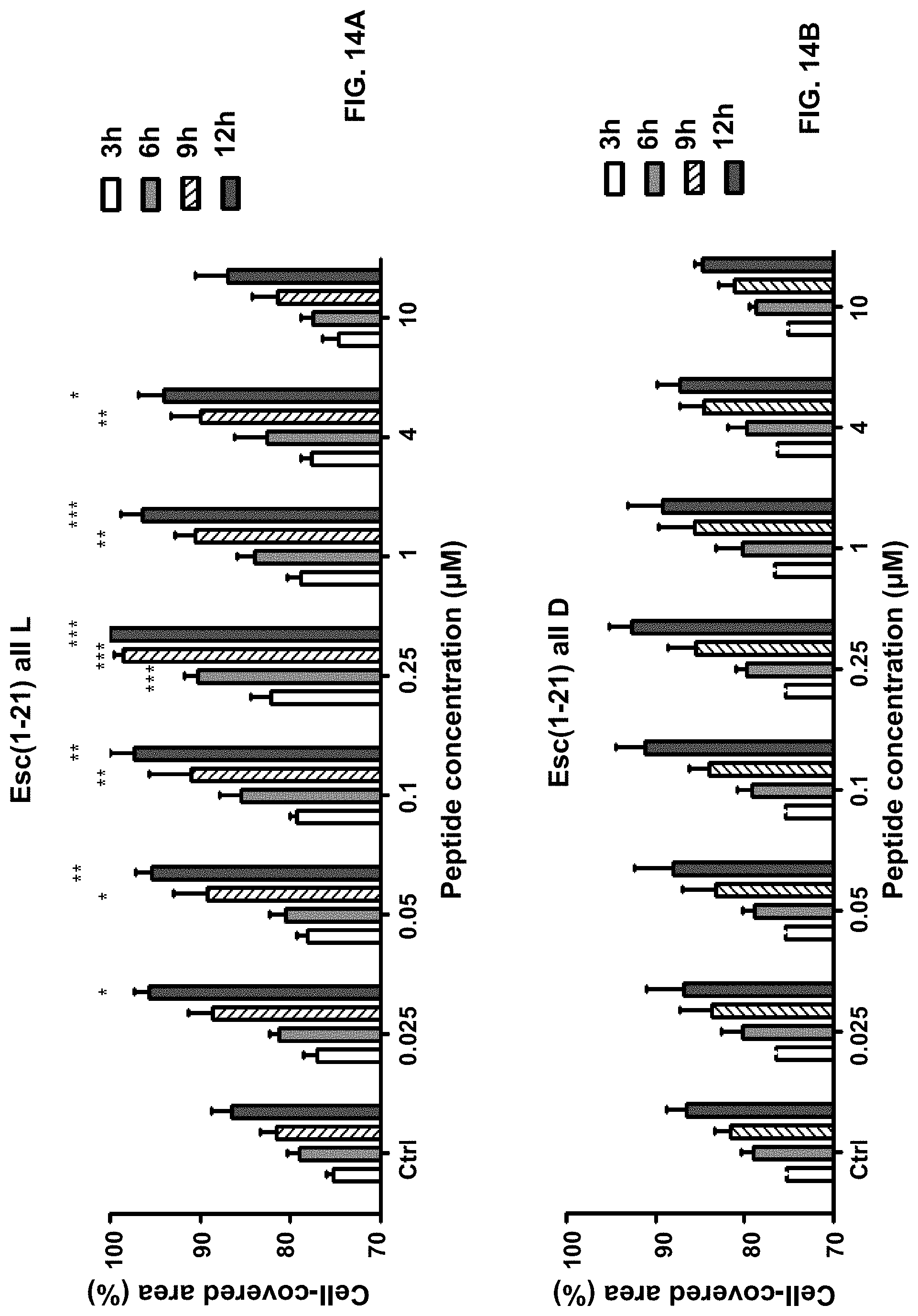
D00014

D00015

D00016

D00017

D00018

XML
uspto.report is an independent third-party trademark research tool that is not affiliated, endorsed, or sponsored by the United States Patent and Trademark Office (USPTO) or any other governmental organization. The information provided by uspto.report is based on publicly available data at the time of writing and is intended for informational purposes only.
While we strive to provide accurate and up-to-date information, we do not guarantee the accuracy, completeness, reliability, or suitability of the information displayed on this site. The use of this site is at your own risk. Any reliance you place on such information is therefore strictly at your own risk.
All official trademark data, including owner information, should be verified by visiting the official USPTO website at www.uspto.gov. This site is not intended to replace professional legal advice and should not be used as a substitute for consulting with a legal professional who is knowledgeable about trademark law.