Treatment of Cancer with TG02
ESTOK; Thomas M. ; et al.
U.S. patent application number 16/087966 was filed with the patent office on 2020-10-15 for treatment of cancer with tg02. The applicant listed for this patent is Tragara Pharmaceuticals, Inc.. Invention is credited to Thomas M. ESTOK, Tracy PARROTT, Eckard WEBER.
| Application Number | 20200323862 16/087966 |
| Document ID | / |
| Family ID | 1000004959127 |
| Filed Date | 2020-10-15 |


View All Diagrams
| United States Patent Application | 20200323862 |
| Kind Code | A1 |
| ESTOK; Thomas M. ; et al. | October 15, 2020 |
Treatment of Cancer with TG02
Abstract
The present disclosure provides therapeutic methods of treating a cancer patient with TG02 and a second therapeutic agent, e.g., TG02 and an immune checkpoint inhibitor, TG02 and a COX-2 inhibitor, or TG02 and an immune checkpoint inhibitor and a COX-2 inhibitor.
| Inventors: | ESTOK; Thomas M.; (Williamsburg, VA) ; WEBER; Eckard; (San Diego, CA) ; PARROTT; Tracy; (Encinitas, CA) | ||||||||||
| Applicant: |
|
||||||||||
|---|---|---|---|---|---|---|---|---|---|---|---|
| Family ID: | 1000004959127 | ||||||||||
| Appl. No.: | 16/087966 | ||||||||||
| Filed: | March 24, 2017 | ||||||||||
| PCT Filed: | March 24, 2017 | ||||||||||
| PCT NO: | PCT/US17/23965 | ||||||||||
| 371 Date: | September 24, 2018 |
Related U.S. Patent Documents
| Application Number | Filing Date | Patent Number | ||
|---|---|---|---|---|
| 62312712 | Mar 24, 2016 | |||
| 62423468 | Nov 17, 2016 | |||
| Current U.S. Class: | 1/1 |
| Current CPC Class: | A61K 31/704 20130101; A61K 31/44 20130101; A61P 35/00 20180101; A61K 33/243 20190101; A61K 31/529 20130101; A61K 31/495 20130101; A61K 38/07 20130101; A61K 31/454 20130101; A61K 31/69 20130101; A61K 39/3955 20130101 |
| International Class: | A61K 31/529 20060101 A61K031/529; A61K 39/395 20060101 A61K039/395; A61K 31/495 20060101 A61K031/495; A61K 31/69 20060101 A61K031/69; A61K 31/44 20060101 A61K031/44; A61K 38/07 20060101 A61K038/07; A61K 33/243 20060101 A61K033/243; A61K 31/704 20060101 A61K031/704; A61K 31/454 20060101 A61K031/454; A61P 35/00 20060101 A61P035/00 |
Claims
1. A method of treating a patient having cancer, the method comprising administering to the patient a therapeutically effective amount of TG02, wherein one or more of the genes listed in Table 1 is differentially present in a biological sample taken from the patient as compared with a biological sample taken from a subject of another phenotypic status.
2. The method of claim 1, wherein MYC overexpression is differentially present in a sample taken from the patient.
3. The method of claim 1 or 2, wherein MCL1 overexpression is differentially present in a sample taken from the patient.
4. The method of any one of claims 1-3 further comprising administering to the patient a therapeutically effective amount of an immune checkpoint inhibitor.
5. The method of claim 4, wherein TG02 is administered to the patient before an immune checkpoint inhibitor.
6. The method of claim 4, wherein TG02 is administered to the patient after an immune checkpoint inhibitor.
7. The method of claim 4, wherein TG02 is administered to the patient at the same time as an immune checkpoint inhibitor.
8. The method of any one of claims 4-7, wherein the immune checkpoint inhibitor is selected from the group consisting of a PD-1 inhibitor, a PD-L1 inhibitor, a CTLA-4 inhibitor, a LAG3 inhibitor, a TIM3 inhibitor, and a cd47 inhibitor.
9. The method of claim 8, wherein the immune checkpoint inhibitor is a PD-1 inhibitor.
10. The method of claim 9, wherein the PD-1 inhibitor is an anti-PD-1 antibody.
11. The method of claim 10, wherein the anti-PD-1 antibody is selected from the group consisting of nivolumab, pembrolizumab, pidilizumab and STI-1110.
12. The method of claim 8, wherein the immune checkpoint inhibitor is a PD-L1 inhibitor.
13. The method of claim 12, wherein the PD-L1 inhibitor is an anti-PD-L1 antibody.
14. The method of claim 12, wherein the anti-PD-L1 antibody is selected from the group consisting of avelumab, atezolizumab, durvalumab, and STI-1014
15. The method of claim 8, wherein the immune checkpoint inhibitor is an anti-CTLA-4 inhibitor.
16. The method of claim 15, wherein the CTLA-4 inhibitor is an anti-CTLA-4 antibody.
17. The method of claim 16, wherein the anti-CTLA-4 antibody is selected from the group consisting of ipilimumab and tremelimumab.
18. The method of claim 8, wherein immune checkpoint inhibitor is a LAG3 inhibitor.
19. The method of claim 18, wherein the LAG3 inhibitor is an anti-LAG3 antibody.
20. The method of claim 19, wherein the anti-LAG3 antibody is GSK2831781.
21. The method of claim 20, wherein the immune checkpoint inhibitor is a TIM3 inhibitor.
22. The method of claim 21, wherein the TIM3 inhibitor is an anti-TIM3 antibody.
23. The method of any one of claims 1-3 further comprising administering to the patient a therapeutically effective amount of an alkylating agent.
24. The method of claim 23, wherein TG02 is administered to the patient before the alkylating agent.
25. The method of claim 23, wherein TG02 is administered to the patient after the alkylating agent.
26. The method of claim 23, wherein a therapeutically effective amount of TG02 is administered to the patient at the same time as the alkylating agent.
27. The method of any one of claims 23-26, wherein the alkylating agent is temozolimide.
28. The method of any one of claims 1-3 further comprising administering to the patient a therapeutically effective amount of a protein kinase inhibitor.
29. The method of claim 28, wherein TG02 is administered to the patient before the protein kinase inhibitor.
30. The method of claim 28, wherein TG02 is administered to the patient after the protein kinase inhibitor.
31. The method of claim 28, wherein a therapeutically effective amount of TG02 is administered to the patient at the same time as the protein kinase inhibitor.
32. The method of any one of claims 28-31, wherein the protein kinase inhibitor is sorafenib.
33. The method of any one of claims 1-3 further comprising administering to the patient a therapeutically effective amount of a proteasome inhibitor.
34. The method of claim 33, wherein TG02 is administered to the patient before the proteasome inhibitor.
35. The method of claim 33, wherein TG02 is administered to the patient after the proteasome inhibitor.
36. The method of claim 33, wherein a therapeutically effective amount of TG02 is administered to the patient at the same time as the proteasome inhibitor.
37. The method of any one of claims 33-36, wherein the proteasome inhibitor is bortezomib.
38. The method of any one of claims 33-36, wherein the proteasome inhibitor is carfilizomib.
39. The method of any one of claims 1-3 further comprising administering to the patient a therapeutically effective amount of a topoisomerase II inhibitor.
40. The method of claim 39, wherein TG02 is administered to the patient before the topoisomerase II inhibitor.
41. The method of claim 39, wherein TG02 is administered to the patient after the topoisomerase II inhibitor.
42. The method of claim 39, wherein a therapeutically effective amount of TG02 is administered to the patient at the same time as the topoisomerase II inhibitor.
43. The method of any one of claims 39-42, wherein the topoisomerase II inhibitor is doxorubicin.
44. The method of any one of claims 1-3 further comprising administering to the patient a therapeutically effective amount of a platinum coordinating complex.
45. The method of claim 44, wherein TG02 is administered to the patient before the platinum coordinating complex.
46. The method of claim 44, wherein TG02 is administered to the patient after the platinum coordinating complex.
47. The method of claim 44, wherein a therapeutically effective amount of TG02 is administered to the patient at the same time as the platinum coordinating complex.
48. The method of any one of claims 44-47, wherein the platinum coordinating complex is cisplatin.
49. The method of any one of claims 1-3 further comprising administering to the patient a therapeutically effective amount of lenalidomide.
50. The method of claim 49, wherein TG02 is administered to the patient before lenalidomide.
51. The method of claim 49, wherein TG02 is administered to the patient after lenalidomide.
52. The method of claim 49, wherein a therapeutically effective amount of TG02 is administered to the patient at the same time as lenalidomide.
53. The method of any one of claims 1-3 further comprising administering to the patient a therapeutically effective amount of radiotherapy.
54. The method of claim 53, wherein TG02 is administered to the patient before radiotherapy.
55. The method of claim 53, wherein TG02 is administered to the patient after radiotherapy.
56. The method of claim 53, wherein a therapeutically effective amount of TG02 is administered to the patient at the same time as radiotherapy.
57. A method of treating a patient having cancer, the method comprising administering to the patient therapeutically effective amounts of TG02 and an immune checkpoint inhibitor.
58. The method of claim 57, wherein TG02 is administered to the patient before the immune checkpoint inhibitor.
59. The method of claim 57, wherein TG02 is administered to the patient after the immune checkpoint inhibitor.
60. The method of claim 57, wherein TG02 is administered to the patient at the same time as the immune checkpoint inhibitor.
61. The method of any one of claims 57-60, wherein immune checkpoint inhibitor is selected from the group consisting of a PD-1 inhibitor, a PD-L1 inhibitor, a CTLA-4 inhibitor, a LAG3 inhibitor, and a TIM3 inhibitor.
62. The method of 61, wherein the immune checkpoint inhibitor is a PD-1 inhibitor.
63. The method of claim 62, wherein the PD-1 inhibitor is an anti-PD-1 antibody.
64. The method of claim 63, wherein the anti-PD-1 antibody is selected from the group consisting of nivolumab, pembrolizumab, pidilizumab and STI-1110.
65. The method of claim 61, wherein the immune checkpoint inhibitor is a PD-L1 inhibitor.
66. The method of claim 65, wherein the PD-L1 inhibitor is an anti-PD-L1 antibody.
67. The method of claim 66, wherein the anti-PD-L1 antibody is selected from the group consisting of avelumab, atezolizumab, durvalumab, and STI-1014
68. The method of claim 61, wherein the immune checkpoint inhibitor is an anti-CTLA-4 inhibitor.
69. The method of claim 68, wherein the CTLA-4 inhibitor is an anti-CTLA-4 antibody.
70. The method of claim 69, wherein the anti-CTLA-4 antibody is selected from the group consisting of ipilimumab and tremelimumab.
71. The method of claim 61, wherein the immune checkpoint inhibitor is a LAG3 inhibitor.
72. The method of claim 71, wherein the LAG3 inhibitor is an anti-LAG3 antibody.
73. The method of claim 72, wherein the anti-LAG3 antibody is GSK2831781.
74. The method of claim 61, wherein the immune checkpoint inhibitor is a TIM3 inhibitor.
75. The method of claim 74, wherein the TIM3 inhibitor is an anti-TIM3 antibody.
76. A method of treating a patient having cancer, the method comprising administering to the patient therapeutically effective amounts of TG02 and an alkylating agent.
77. The method of claim 76, wherein TG02 is administered to the patient before the alkylating agent.
78. The method of claim 76, wherein TG02 is administered to the patient after the alkylating agent.
79. The method of claim 76, wherein TG02 is administered to the patient at the same time as the alkylating agent.
80. The method of any one of claims 76-79, wherein the alkylating agent is temozolimide.
81. A method of treating a patient having cancer, the method comprising administering to the patient therapeutically effective amounts of TG02 and a protein kinase inhibitor.
82. The method of claim 81, wherein TG02 is administered to the patient before the protein kinase inhibitor.
83. The method of claim 81, wherein TG02 is administered to the patient after the protein kinase inhibitor.
84. The method of claim 81, wherein a therapeutically effective amount of TG02 is administered to the patient at the same time as the protein kinase inhibitor.
85. The method of any one of claims 77-80, wherein the protein kinase inhibitor is sorafenib.
86. A method of treating a patient having cancer, the method comprising administering to the patient therapeutically effective amounts of TG02 and a proteasome inhibitor.
87. The method of claim 86, wherein TG02 is administered to the patient before the proteasome inhibitor.
88. The method of claim 86, wherein TG02 is administered to the patient after the proteasome inhibitor.
89. The method of claim 86, wherein a therapeutically effective amount of TG02 is administered to the patient at the same time as the proteasome inhibitor.
90. The method of any one of claims 86-89, wherein the proteasome inhibitor is bortezomib.
91. The method of any one of claims 86-89, wherein the proteasome inhibitor is carfilizomib.
92. A method of treating a patient having cancer, the method comprising administering to the patient therapeutically effective amounts of TG02 and a topoisomerase II inhibitor.
93. The method of claim 92, wherein TG02 is administered to the patient before the topoisomerase II inhibitor.
94. The method of claim 92, wherein TG02 is administered to the patient after the topoisomerase II inhibitor.
95. The method of claim 92, wherein a therapeutically effective amount of TG02 is administered to the patient at the same time as the topoisomerase II inhibitor.
96. The method of any one of claims 92-95, wherein the topoisomerase II inhibitor is doxorubicin.
97. A method of treating a patient having cancer, the method comprising administering to the patient therapeutically effective amounts of TG02 and a platinum coordinating complex.
98. The method of claim 97, wherein TG02 is administered to the patient before the platinum coordinating complex.
99. The method of claim 97, wherein TG02 is administered to the patient after the platinum coordinating complex.
100. The method of claim 97, wherein a therapeutically effective amount of TG02 is administered to the patient at the same time as the platinum coordinating complex.
101. The method of any one of claims 97-100, wherein the platinum coordinating complex is cisplatin.
102. A method of treating a patient having cancer, the method comprising administering to the patient therapeutically effective amounts of TG02 and lenalidomide.
103. The method of claim 102, wherein TG02 is administered to the patient before lenalidomide.
104. The method of claim 102, wherein TG02 is administered to the patient after lenalidomide.
105. The method of claim 102, wherein a therapeutically effective amount of TG02 is administered to the patient at the same time as lenalidomide.
106. A method of treating a patient having cancer, the method comprising administering to the patient therapeutically effective amounts of TG02 and radiotherapy.
107. The method of claim 106, wherein TG02 is administered to the patient before radiotherapy.
108. The method of claim 106, wherein TG02 is administered to the patient after radiotherapy.
109. The method of claim 106, wherein a therapeutically effective amount of TG02 is administered to the patient at the same time as radiotherapy
110. The method of any one of claims 1-109, wherein the cancer is a solid tumor.
111. The method of any one of claims 1-109, wherein the cancer is a hematological malignancy.
112. The method of any one of claims 1-109, wherein the cancer selected from the group consisting of adrenal cancer, acinic cell carcinoma, acoustic neuroma, acral lentigious melanoma, acrospiroma, acute eosinophilic leukemia, acute erythroid leukemia, acute lymphoblastic leukemia, acute megakaryoblastic leukemia, acute monocytic leukemia, acute promyelocytic leukemia, adenocarcinoma, adenoid cystic carcinoma, adenoma, adenomatoid odontogenic tumor, adenosquamous carcinoma, adipose tissue neoplasm, adrenocortical carcinoma, adult T-cell leukemia/lymphoma, aggressive NK-cell leukemia, AIDS-related lymphoma, alveolar rhabdomyosarcoma, alveolar soft part sarcoma, ameloblastic fibroma, anaplastic large cell lymphoma, anaplastic thyroid cancer, angioimmunoblastic T-cell lymphoma, angiomyolipoma, angiosarcoma, astrocytoma, atypical teratoid rhabdoid tumor, B-cell chronic lymphocytic leukemia, B-cell prolymphocytic leukemia, B-cell lymphoma, basal cell carcinoma, biliary tract cancer, bladder cancer, blastoma, bone cancer, Brenner tumor, Brown tumor, Burkitt's lymphoma, breast cancer, brain cancer, carcinoma, carcinoma in situ, carcinosarcoma, cartilage tumor, cementoma, myeloid sarcoma, chondroma, chordoma, choriocarcinoma, choroid plexus papilloma, clear-cell sarcoma of the kidney, craniopharyngioma, cutaneous T-cell lymphoma, cervical cancer, colorectal cancer, Degos disease, desmoplastic small round cell tumor, diffuse large B-cell lymphoma, dysembryoplastic neuroepithelial tumor, dysgerminoma, embryonal carcinoma, endocrine gland neoplasm, endodermal sinus tumor, enteropathy-associated T-cell lymphoma, esophageal cancer, fetus in fetu, fibroma, fibrosarcoma, follicular lymphoma, follicular thyroid cancer, ganglioneuroma, gastrointestinal cancer, germ cell tumor, gestational choriocarcinoma, giant cell fibroblastoma, giant cell tumor of the bone, glial tumor, glioblastoma, glioma, gliomatosis cerebri, glucagonoma, gonadoblastoma, granulosa cell tumor, gynandroblastoma, gallbladder cancer, gastric cancer, hairy cell leukemia, hemangioblastoma, head and neck cancer, hemangiopericytoma, hematological malignancy, hepatoblastoma, hepatocellular carcinoma, hepatosplenic T-cell lymphoma, Hodgkin's lymphoma, non-Hodgkin's lymphoma, invasive lobular carcinoma, intestinal cancer, kidney cancer, laryngeal cancer, lentigo maligna, lethal midline carcinoma, leukemia, leydig cell tumor, liposarcoma, lung cancer, lymphangioma, lymphangiosarcoma, lymphoepithelioma, lymphoma, acute lymphocytic leukemia, acute myelogeous leukemia, chronic lymphocytic leukemia, liver cancer, small cell lung cancer, non-small cell lung cancer, MALT lymphoma, malignant fibrous histiocytoma, malignant peripheral nerve sheath tumor, malignant triton tumor, mantle cell lymphoma, marginal zone B-cell lymphoma, mast cell leukemia, mediastinal germ cell tumor, medullary carcinoma of the breast, medullary thyroid cancer, medulloblastoma, melanoma, meningioma, merkel cell cancer, mesothelioma, metastatic urothelial carcinoma, mixed Mullerian tumor, mucinous tumor, multiple myeloma, muscle tissue neoplasm, mycosis fungoides, myxoid liposarcoma, myxoma, myxosarcoma, nasopharyngeal carcinoma, neurinoma, neuroblastoma, neurofibroma, neuroma, nodular melanoma, ocular cancer, oligoastrocytoma, oligodendroglioma, oncocytoma, optic nerve sheath meningioma, optic nerve tumor, oral cancer, osteosarcoma, ovarian cancer, Pancoast tumor, papillary thyroid cancer, paraganglioma, pinealoblastoma, pineocytoma, pituicytoma, pituitary adenoma, pituitary tumor, plasmacytoma, polyembryoma, precursor T-lymphoblastic lymphoma, primary central nervous system lymphoma, primary effusion lymphoma, preimary peritoneal cancer, prostate cancer, pancreatic cancer, pharyngeal cancer, pseudomyxoma periotonei, renal cell carcinoma, renal medullary carcinoma, retinoblastoma, rhabdomyoma, rhabdomyosarcoma, Richter's transformation, rectal cancer, sarcoma, Schwannomatosis, seminoma, Sertoli cell tumor, sex cord-gonadal stromal tumor, signet ring cell carcinoma, skin cancer, small blue round cell tumors, small cell carcinoma, soft tissue sarcoma, somatostatinoma, soot wart, spinal tumor, splenic marginal zone lymphoma, squamous cell carcinoma, synovial sarcoma, Sezary's disease, small intestine cancer, squamous carcinoma, stomach cancer, T-cell lymphoma, testicular cancer, thecoma, thyroid cancer, transitional cell carcinoma, throat cancer, urachal cancer, urogenital cancer, urothelial carcinoma, uveal melanoma, uterine cancer, verrucous carcinoma, visual pathway glioma, vulvar cancer, vaginal cancer, Waldenstrom's macroglobulinemia, Warthin's tumor, and Wilms' tumor.
113. The method of claim 112, wherein the cancer is selected from the group consisting of hepatocellular carcinoma, glioblastoma, lung cancer, breast cancer, head and neck cancer, prostate cancer, melanoma, and colorectal cancer.
114. The method of claim 112, wherein the cancer is multiple myeloma.
115. The method of any one of claims 1-114, wherein the cancer has become resistant to conventional treatments.
116. The method of any one of claims 1-115, wherein TG02 is the citrate salt of (16E)-14-methyl-20-oxa-5,7,14,26-tetraazatetracyclo[19.3.1.1(2,6)- .1(8,12)]heptacosa-1(25),2(26),3,5,8(27),9,11,16,21,23-decaene.
117. A kit comprising TG02 and an immune checkpoint inhibitor, an alkylating agent, a protein kinase inhibitor, a proteasome inhibitor, a topoisomerase II inhibitor, a platinum coordinating complex, or lenalidomide, and instructions for administering TG02 and the immune checkpoint inhibitor, alkylating agent, protein kinase inhibitor, proteasome inhibitor, topoisomerase II inhibitor, platinum coordinating complex, or lenalidomide, to a patient having cancer.
Description
BACKGROUND OF THE INVENTION
Field of the Invention
[0001] The present disclosure provides therapeutic methods of treating a cancer patient with TG02 and a second therapeutic agent, e.g., TG02 and an immune checkpoint inhibitor, TG02 and a COX-2 inhibitor, and TG02 and an immune checkpoint inhibitor and a COX-2 inhibitor.
Background
[0002] TG02 is a pyrimidine-based multi-kinase inhibitor that inhibits CDKs 1, 2, 5, 7 and 9 together with JAK2 and FLT3. It dose-dependently inhibits signaling pathways downstream of CDKs, JAK2 and FLT3 in cancer cells with the main targets being CDKs. TG02 is anti-proliferative in a broad range of tumor cell lines, inducing G1 cell cycle arrest and apoptosis. Primary cultures of progenitor cells derived from acute myeloid leukemia (AML) and polycythemia vera patients are very sensitive to TG02. Comparison with reference inhibitors that block only one of the main targets of TG02 demonstrate the benefit of combined CDK and JAK2/FLT3 inhibition in cell lines as well as primary cells. See Goh et al., Leukemia 26:236-43 (2012). TG02 is also known as SB1317 and by its chemical name: (16E)-14-methyl-20-oxa-5,7,14,26-tetraazatetracyclo[19.3.1.1(2,6).1(8,12)- ]heptacosa-1(25),2(26),3,5,8(27),9,11,16,21,23-decaene. TG02 is disclosed as Compound 1 in U.S. Pat. No. 8,143,255. U.S. Pat. No. 9,120,815 discloses various salt, e.g., TG02 citrate, and crystalline forms of TG02. The chemical structure of TG02 is:
##STR00001##
BRIEF SUMMARY OF THE INVENTION
[0003] In one aspect, the present disclosure provides therapeutic methods of treating a cancer patient, the methods comprising administering to the patient a therapeutically effective amount of TG02. In another aspect, the patient's cancer is characterized as overexpressing of MYC, MCL1, or both.
[0004] In another aspect, the present disclosure provides therapeutic methods of treating a cancer patient, the methods comprising administering to the patient therapeutically effective amounts of TG02 and an immune checkpoint inhibitor, e.g., a PD-1 inhibitor, a PD-L1 inhibitor, a CTLA-4 inhibitor, a LAG3 inhibitor, a TIM3 inhibitor, or a cd47 inhibitor.
[0005] In another aspect, the present disclosure provides therapeutic methods of treating a cancer patient, the methods comprising administering to the patient therapeutically effective amounts of TG02 and a COX inhibitor, e.g., apricoxib or 6-bromo-8-(methyl-D3)-2-(trifluoromethyl)-2H-chromene-3-carboxylic acid.
[0006] In another aspect, the present disclosure provides therapeutic methods of treating a cancer patient, the methods comprising administering to the patient therapeutically effective amounts of TG02, an immune checkpoint inhibitor, and a COX-2 inhibitor.
[0007] In another aspect, present disclosure provides therapeutic methods of treating a cancer patient who has tumors that overexpress MYC, MCL1, or both.
[0008] In another aspect, the present disclosure provides kits comprising TG02, TG02 and an immune checkpoint inhibitor, TG02 and a COX-2 inhibitor, and TG02 and an immune checkpoint inhibitor and a COX-2 inhibitor.
[0009] In another aspect, the present disclosure provides a pharmaceutical composition comprising TG02, a COX-2 inhibitor, e.g., apricoxib or 6-bromo-8-(methyl-D3)-2-(trifluoromethyl)-2H-chromene-3-carboxylic acid and a pharmaceutically acceptable excipient.
BRIEF DESCRIPTION OF DRAWINGS
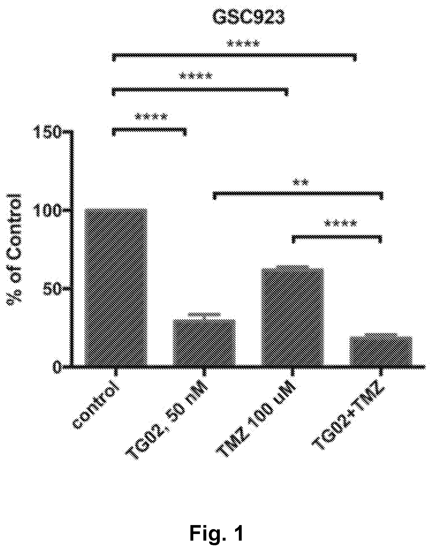
[0010] FIG. 1 is a bar graph showing the in vitro activity of TG02, TMZ (temozolomide), and TG02+TMZ in GSC923 cells.
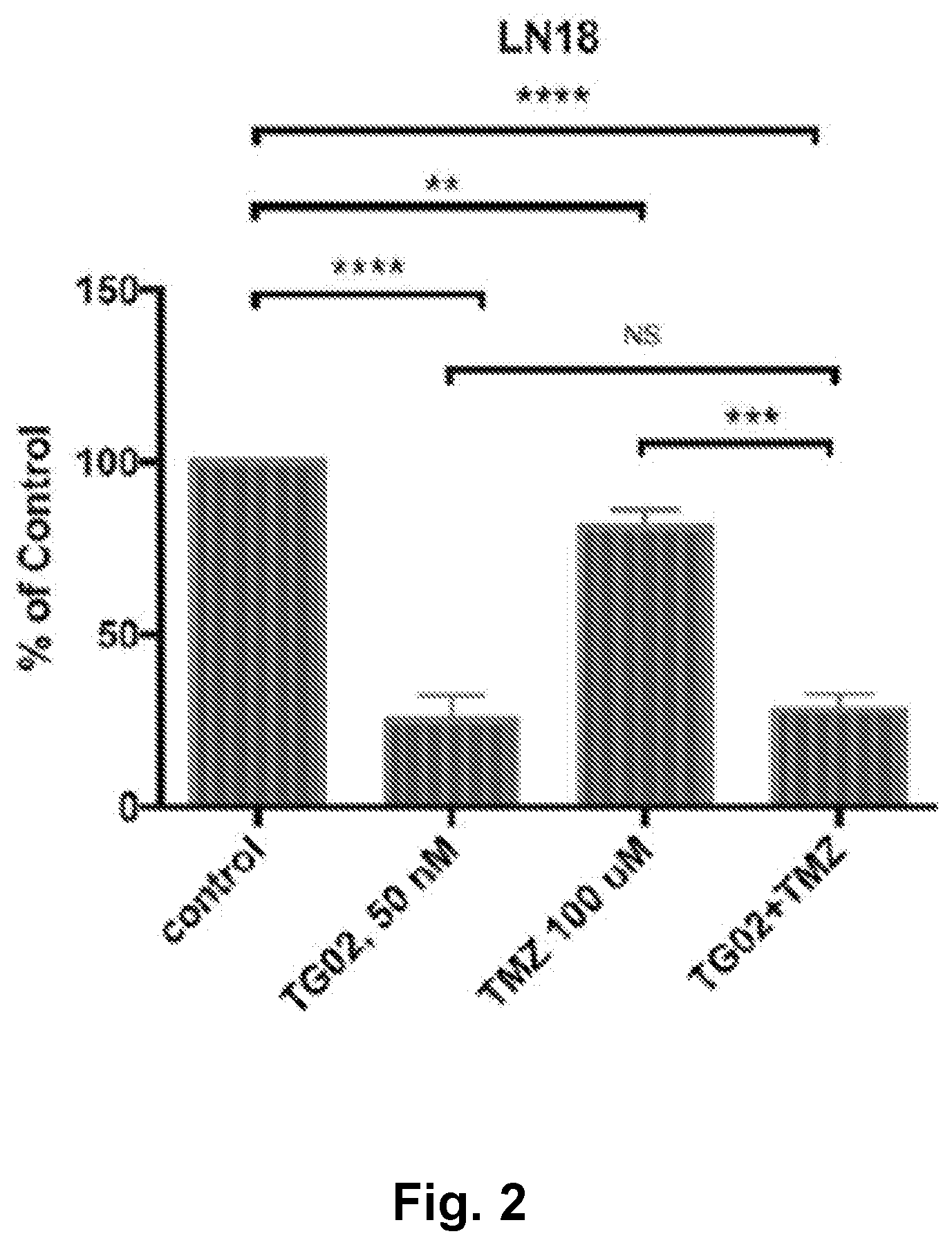
[0011] FIG. 2 is a bar graph showing the in vitro activity of TG02, TMZ, and TG02+TMZ in LN18 cells.
[0012] FIG. 3 is a bar graph showing the in vitro activity of TG02, TMZ, and TG02+TMZ in T98G cells.
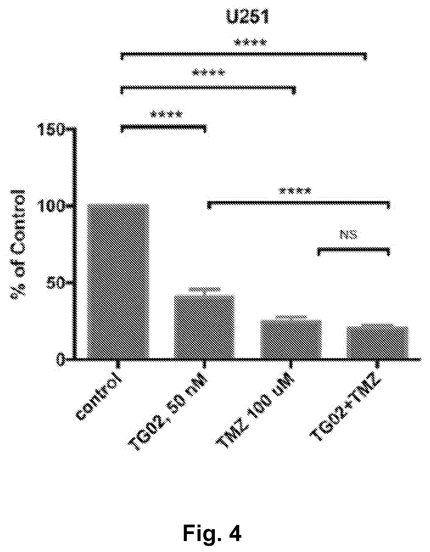
[0013] FIG. 4 is a bar graph showing the in vitro activity of TG02, TMZ, and TG02+TMZ in U251 cells.
[0014] FIG. 5 is a bar graph showing the in vitro activity of TG02, TMZ, and TG02+TMZ in U87 cells.
[0015] FIG. 6 is a bar graph showing the in vitro activity of TG02, TMZ, and TG02+TMZ in LN299 cells.
[0016] FIG. 7 is a bar graph showing the in vitro activity of TG02, TMZ, and TG02+TMZ in GSC827 cells
[0017] FIG. 8 is a bar graph showing the in vitro cytotoxicity of TG02, TMZ, and TG02+TMZ (T+T) in GSC923 cells.
[0018] FIG. 9 is a bar graph showing the in vitro cytotoxicity of TG02, TMZ, and TG02+TMZ (T+T) in U251 cells.
[0019] FIG. 10 is a bar graph showing a lack of in vitro activity of TG02, TMZ, and TG02+TMZ (T+T) in human pulmonary arterial endothelial cells.
[0020] FIG. 11 is a bar graph showing a lack of in vitro activity of TG02, TMZ, and TG02+TMZ (T+T) in human astrocytes.
[0021] FIG. 12 is a dose response curve showing the in vitro activity of TG02 and TG02+TMZ in GSC923 cells.
[0022] FIG. 13 is a dose response curve showing the in vitro activity of TMZ and TG02+TMZ in GSC923 cells.
[0023] FIG. 14 is a dose response curve showing the in vitro activity of TG02 and TG02+TMZ in U251 cells.
[0024] FIG. 15 is a dose response curve showing the in vitro activity of TMZ and TG02+TMZ in U251 cells.
[0025] FIG. 16 is a schematic illustration of TG02 and TMZ administration in a mouse glioma GL261 cell allograft model.
[0026] FIG. 17 is a line graph showing percent survival following TG02, TMZ, and TG02+TMZ administration in a mouse glioma GL261 cell allograft model.
[0027] FIG. 18 is a line graph showing the tumor burden following TG02, TMZ, and TG02+TMZ administration in a mouse glioma GL261 cell allograft model.
[0028] FIG. 19 is an illustration showing the effect of TG02 on MYC protein levels in hepatocellular carcinoma (HCC) cells.
[0029] FIG. 20 is a dose response curve showing the effect of TG02 on MYC protein levels in HCC cells.
[0030] FIG. 21 is is an illustration showing showing the effect of TG02 on MYC protein levels in HCC tumor cells.
[0031] FIG. 22 is a line graph showing the in vivo activity of TG02 and TG02+sorafenib in an orthotopic model of HepG2 HCC xenografts.
[0032] FIG. 23 is a bar graph showing PD-L1 expression following treatment with TG02 in a transgenic mouse model of MYC-induced T cell acute lymphoblastic leukemia.
[0033] FIG. 24 is a bar graph showing CD47 expression following treatment with TG02 in a transgenic mouse model of MYC-induced T cell acute lymphoblastic leukemia.
[0034] FIG. 25 is a bar graph showing BCL-xL expression following treatment with TG02 in a transgenic mouse model of MYC-induced T cell acute lymphoblastic leukemia.
[0035] FIG. 26 is a bar graph showing MYC expression following treatment with TG02 in a transgenic mouse model of MYC-induced T cell acute lymphoblastic leukemia.
[0036] FIG. 27 is a line graph showing the efficacy of TG02 in combination with anti-PD-1 in a mouse syngeneic GL261 orthotopic glioblastoma model.
[0037] FIG. 28 is an illustration showing that BT245 tumor cells exposed to TG02 show inhibition of MYC and MCL-1 expression.
[0038] FIG. 29 is a bar graph showing the area under the curve (AUC) for TG02 induced inhibition in glioblastoma (GBM) cells.
[0039] FIG. 30 is a scatter graph showing that high MYC expression correlates with low AUC in GBM cells.
[0040] FIG. 31 is a series of six line graphs showing the activity of TG02 in combination with radiation in glioblastoma cell lines.
[0041] FIG. 32 is a bar graph showing the activity of TG02 on 26 patient-derived GBM stem cell lines.
[0042] FIG. 33 is an illustration showing the expression level of CDK9, MYC, and Mcl-1 in patient-derived GBM stem cell lines following treatment with TG02.
DETAILED DESCRIPTION OF THE INVENTION
[0043] In one embodiment, the present disclosure provides therapeutic methods of treating a patient having cancer, the method comprising administering to the patient a therapeutically effective amount of TG02, wherein one or more of the genes listed in Table 1, see below, is differentially present in a biological sample taken from the patient as compared with a biological sample taken from a subject of another phenotypic status. In another embodiment, MYC overexpression is differentially present in a sample taken from the patient. In another embodiment, MCL1 overexpression is differentially present in a sample taken from the patient.
[0044] In another embodiment, the present disclosure provides therapeutic methods of treating a patient having cancer, the method comprising administering to the patient a therapeutically effective amounts of TG02 and an immune checkpoint inhibitor, wherein one or more of the genes listed in Table 1, see below, is differentially present in a biological sample taken from the patient as compared with a biological sample taken from a subject of another phenotypic status. In another embodiment, MYC overexpression is differentially present in a sample taken from the patient. In another embodiment, MCL1 overexpression is differentially present in a sample taken from the patient. In another embodiment, TG02 is administered to the patient before the immune checkpoint inhibitor. In another embodiment, TG02 is administered to the patient after the immune checkpoint inhibitor. In another embodiment, TG02 is administered to the patient at the same time as an immune checkpoint inhibitor.
[0045] In another embodiment, the present disclosure provides therapeutic methods of treating a patient having cancer, the method comprising administering to the patient a therapeutically effective amounts of TG02, an immune checkpoint inhibitor, and a COX-2 inhibitor wherein one or more of the genes listed in Table 1, see below, is differentially present in a biological sample taken from the patient as compared with a biological sample taken from a subject of another phenotypic status. In another embodiment, MYC overexpression is differentially present in a sample taken from the patient. In another embodiment, MCL1 overexpression is differentially present in a sample taken from the patient. In another embodiment, TG02 is administered to the patient before the COX-2 inhibitor. In another embodiment, TG02 is administered to the patient after the COX-2 inhibitor. In another embodiment, TG02 is administered to the patient at the same time as the COX-2 inhibitor.
[0046] In another embodiment, the present disclosure provides therapeutic methods of treating a patient having cancer, the method comprising administering to the patient therapeutically effective amounts of TG02 and an immune checkpoint inhibitor. In another embodiment, TG02 is administered to the patient before the immune checkpoint inhibitor. In another embodiment, TG02 is administered to the patient after the immune checkpoint inhibitor. In another embodiment, TG02 is administered to the patient at the same time as an immune checkpoint inhibitor.
[0047] In another embodiment, the present disclosure provides therapeutic methods of treating a patient having cancer, the method comprising administering to the patient therapeutically effective amounts of TG02, an immune checkpoint inhibitor, and a COX-2 inhibitor. In another embodiment, TG02 is administered to the patient before the COX-2 inhibitor. In another embodiment, TG02 is administered to the patient after the COX-2 inhibitor. In another embodiment, TG02 is administered to the patient at the same time as the COX-2 inhibitor.
[0048] In another embodiment, the present disclosure provides kits comprising TG02 and an immune checkpoint inhibitor, and instructions for administering TG02 and the immune checkpoint inhibitor to a patient having cancer. In another embodiment, the kit further comprises a COX-2 inhibitor.
[0049] In another embodiment, the present disclosure provides kits comprising TG02 and a COX-2 inhibitor, and instructions for administering TG02 and the COX-2 inhibitor to a patient having cancer.
[0050] In another embodiment, the kit is packaged in a manner that facilitates its use to practice methods of the present disclosure.
[0051] In another embodiment, the kit includes TG02 (or a composition comprising TG02) packaged in a container, such as a sealed bottle or vessel, with a label affixed to the container or included in the kit that describes use of TG02 or composition to practice the method of the disclosure. In one embodiment, TG02 is packaged in a unit dosage form. The kit further can include a device suitable for administering the composition according to the intended route of administration.
[0052] The disclosure provides various therapeutic methods, kits, and compositions relating to the treatment of cancer. In one embodiment, the cancer is a solid tumor. In another embodiment, the cancer is a hematological malignancy. In another embodiment, the cancer selected from the group consisting of adrenal cancer, acinic cell carcinoma, acoustic neuroma, acral lentigious melanoma, acrospiroma, acute eosinophilic leukemia, acute erythroid leukemia, acute lymphoblastic leukemia, acute megakaryoblastic leukemia, acute monocytic leukemia, acute promyelocytic leukemia, adenocarcinoma, adenoid cystic carcinoma, adenoma, adenomatoid odontogenic tumor, adenosquamous carcinoma, adipose tissue neoplasm, adrenocortical carcinoma, adult T-cell leukemia/lymphoma, aggressive NK-cell leukemia, AIDS-related lymphoma, alveolar rhabdomyosarcoma, alveolar soft part sarcoma, ameloblastic fibroma, anaplastic large cell lymphoma, anaplastic thyroid cancer, angioimmunoblastic T-cell lymphoma, angiomyolipoma, angiosarcoma, astrocytoma, atypical teratoid rhabdoid tumor, B-cell chronic lymphocytic leukemia, B-cell prolymphocytic leukemia, B-cell lymphoma, basal cell carcinoma, biliary tract cancer, bladder cancer, blastoma, bone cancer, Brenner tumor, Brown tumor, Burkitt's lymphoma, breast cancer, brain cancer, carcinoma, carcinoma in situ, carcinosarcoma, cartilage tumor, cementoma, myeloid sarcoma, chondroma, chordoma, choriocarcinoma, choroid plexus papilloma, clear-cell sarcoma of the kidney, craniopharyngioma, cutaneous T-cell lymphoma, cervical cancer, colorectal cancer, Degos disease, desmoplastic small round cell tumor, diffuse large B-cell lymphoma, dysembryoplastic neuroepithelial tumor, dysgerminoma, embryonal carcinoma, endocrine gland neoplasm, endodermal sinus tumor, enteropathy-associated T-cell lymphoma, esophageal cancer, fetus in fetu, fibroma, fibrosarcoma, follicular lymphoma, follicular thyroid cancer, ganglioneuroma, gastrointestinal cancer, germ cell tumor, gestational choriocarcinoma, giant cell fibroblastoma, giant cell tumor of the bone, glial tumor, glioblastoma, glioma, gliomatosis cerebri, glucagonoma, gonadoblastoma, granulosa cell tumor, gynandroblastoma, gallbladder cancer, gastric cancer, hairy cell leukemia, hemangioblastoma, head and neck cancer, hemangiopericytoma, hematological malignancy, hepatoblastoma, hepatocellular carcinoma, hepatosplenic T-cell lymphoma, Hodgkin's lymphoma, non-Hodgkin's lymphoma, invasive lobular carcinoma, intestinal cancer, kidney cancer, laryngeal cancer, lentigo maligna, lethal midline carcinoma, leukemia, leydig cell tumor, liposarcoma, lung cancer, lymphangioma, lymphangiosarcoma, lymphoepithelioma, lymphoma, acute lymphocytic leukemia, acute myelogeous leukemia, chronic lymphocytic leukemia, liver cancer, small cell lung cancer, non-small cell lung cancer, MALT lymphoma, malignant fibrous histiocytoma, malignant peripheral nerve sheath tumor, malignant triton tumor, mantle cell lymphoma, marginal zone B-cell lymphoma, mast cell leukemia, mediastinal germ cell tumor, medullary carcinoma of the breast, medullary thyroid cancer, medulloblastoma, melanoma, meningioma, merkel cell cancer, mesothelioma, metastatic urothelial carcinoma, mixed Mullerian tumor, mucinous tumor, multiple myeloma, muscle tissue neoplasm, mycosis fungoides, myxoid liposarcoma, myxoma, myxosarcoma, nasopharyngeal carcinoma, neurinoma, neuroblastoma, neurofibroma, neuroma, nodular melanoma, ocular cancer, oligoastrocytoma, oligodendroglioma, oncocytoma, optic nerve sheath meningioma, optic nerve tumor, oral cancer, osteosarcoma, ovarian cancer, Pancoast tumor, papillary thyroid cancer, paraganglioma, pinealoblastoma, pineocytoma, pituicytoma, pituitary adenoma, pituitary tumor, plasmacytoma, polyembryoma, precursor T-lymphoblastic lymphoma, primary central nervous system lymphoma, primary effusion lymphoma, preimary peritoneal cancer, prostate cancer, pancreatic cancer, pharyngeal cancer, pseudomyxoma periotonei, renal cell carcinoma, renal medullary carcinoma, retinoblastoma, rhabdomyoma, rhabdomyosarcoma, Richter's transformation, rectal cancer, sarcoma, Schwannomatosis, seminoma, Sertoli cell tumor, sex cord-gonadal stromal tumor, signet ring cell carcinoma, skin cancer, small blue round cell tumors, small cell carcinoma, soft tissue sarcoma, somatostatinoma, soot wart, spinal tumor, splenic marginal zone lymphoma, squamous cell carcinoma, synovial sarcoma, Sezary's disease, small intestine cancer, squamous carcinoma, stomach cancer, T-cell lymphoma, testicular cancer, thecoma, thyroid cancer, transitional cell carcinoma, throat cancer, urachal cancer, urogenital cancer, urothelial carcinoma, uveal melanoma, uterine cancer, verrucous carcinoma, visual pathway glioma, vulvar cancer, vaginal cancer, Waldenstrom's macroglobulinemia, Warthin's tumor, and Wilms' tumor.
[0053] In another embodiment, the cancer is selected from the group consisting of squamous cell carcinoma of the head and neck, adenocarcinoma squamous cell carcinoma of the esophagus, adenocarcinoma of the stomach, adenocarcinoma of the colon, hepatocellular carcinoma, cholangiocarcinoma of the biliary system, adenocarcinoma of gall bladder, adenocarcinoma of the pancreas, ductal carcinoma in situ of the breast, adenocarcinoma of the breast, adenocarcinoma of the lungs, squamous cell carcinoma of the lungs, transitional cell carcinoma of the bladder, squamous cell carcinoma of the bladder, squamous cell carcinoma of the cervix, adenocarcinoma of the cervix, endometrial carcinoma, penile squamous cell carcinoma, and squamous cell carcinoma of the skin.
[0054] In another embodiment, a precancerous tumor is selected from the group consisting of leukoplakia of the head and neck, Barrett's esophagus, metaplasia of the stomach, adenoma of the colon, chronic hepatitis, bile duct hyperplasia, pancreatic intraepithelial neoplasia, atypical adenomatous hyperplasia of the lungs, dysplasia of the bladder, cervical initraepithelial neoplasia, penile intraepithelial neoplasia, and actinic keratosis of the skin.
[0055] In another embodiment, the patient has tumors that overexpress MYC, MCL1, or both. The tumors may be determined to overexpress MYC, MCL1, or both, by methods known in the art.
[0056] In another embodiment, the cancer is selected from the group consisting of hepatocellular carcinoma, glioblastoma, lung cancer, breast cancer, head and neck cancer, prostate cancer, melanoma, and colorectal cancer.
[0057] In another embodiment, the cancer is selected from the group consisting of glioblastoma, hepatocellular carcinoma, non-small cell and small-cell lung cancer, head and neck cancer, colorectal carcinoma, and triple-negative breast cancer.
[0058] In another embodiment, the cancer has become resistant to conventional cancer treatments. The term "conventional cancer treatments" as used herein refers to any cancer drugs or biologics, or combination of cancer drugs and/or biologics that have been tested and/or approved for therapeutic use in humans by the U.S. Food and Drug Administration, European Medicines Agency, or similar regulatory agency.
[0059] In another embodiment, the patient has been treated previously with an immune checkpoint inhibitor without TG02. For example, the previous immune checkpoint therapy may be an anti-PD-1 therapy.
[0060] In another embodiment, the patient has been treated previously with a COX-2 inhibitor without TG02.
[0061] In another embodiment, the present disclosure provides a pharmaceutical composition comprising TG02, a COX-2 inhibitor, and a pharmaceutically acceptable excipient.
[0062] In another embodiment, the present disclosure provides therapeutic methods of treating a patient having cancer, the method comprising administering to the patient a therapeutically effective amount of TG02, wherein the phenotypic status of the patient is overexpression of MYC, overexpression of MCL1, or overexpression of MYC and MCL1. In another embodiment, the cancer is selected from the group consisting of hepatocellular carcinoma, glioblastoma, lung cancer, breast cancer, head and neck cancer, prostate cancer, melanoma, and colorectal cancer.
[0063] In another embodiment, the present disclosure provides therapeutic methods of treating a patient having cancer, the method comprising administering to the patient therapeutically effective amounts of TG02 and a second therapeutic agent, wherein the second therapeutic agent is neither an immune checkpoint inhibitor nor a COX-2 inhibitor.
[0064] In another embodiment, the present disclosure provides therapeutic methods of treating a patient having cancer, comprising administering to the patient therapeutically effective amounts of TG02, an immune checkpoint inhibitor, and a third therapeutic agent, wherein the third therapeutic agent is not a COX-2 inhibitor.
[0065] In another embodiment, the present disclosure provides therapeutic methods of treating a patient having cancer, comprising administering to the patient therapeutically effective amounts of TG02, a COX-2 inhibitor, and a third therapeutic agent, wherein the third therapeutic agent is not an immune checkpoint inhibitor.
[0066] In another embodiment, the present disclosure provides therapeutic methods of treating a patient having cancer, comprising administering to the patient therapeutically effective amounts of TG02, an immune checkpoint inhibitor, a COX-2 inhibitor, and a fourth therapeutic agent, wherein the fourth therapeutic agent is neither an immune checkpoint inhibitor nor a COX-2 inhibitor.
[0067] In another embodiment, the present disclosure provides personalized medicine for cancer patients, and encompasses the selection of treatment options with the highest likelihood of successful outcome for individual cancer patients. In another aspect, the disclosure relates to the use of an assay(s) to predict the treatment outcome, e.g., the likelihood of favorable responses or treatment success, in patients having cancer.
[0068] In another embodiment, the present disclosure provides methods of selecting a patient, e.g., a human subject for treatment of cancer with TG02 and, optionally, an immune checkpoint inhibitor and/or a COX-2 inhibitor, comprising obtaining a biological sample, e.g., blood cells, from the patient, testing a biological sample from the patient for the presence of a biomarker, e.g., overexpression of MYC, overexpression of MCL1, or both, and selecting the patient for treatment if the biological sample contains that biomarker. In another embodiment, the methods further comprise administering a therapeutically effective amount of TG02 and, optionally, an immune checkpoint inhibitor and/or a COX-2 inhibitor, to the patient if the biological sample contains the biomarker. Examples of cancer biomarkers are provided in Table 1. In another embodiment, the cancer is a solid tumor. In another embodiment, the cancer is a hematological malignancy. In another embodiment, the cancer is selected from the group consisting of hepatocellular carcinoma, glioblastoma, lung cancer, breast cancer, head and neck cancer, prostate cancer, melanoma, and colorectal cancer.
[0069] In another embodiment, the present disclosure provides methods of predicting treatment outcomes in a patient having cancer, comprising obtaining a biological sample, from the patient, testing the biological sample from the patient for the presence of a biomarker, e.g., overexpression of MYC, overexpression of MCL1, or both, wherein the detection of the biomarker indicates the patient will respond favorably to administration of a therapeutically effective amount of TG02 and, optionally, an immune checkpoint inhibitor and/or a COX-2 inhibitor. Favorable responses include, but are not limited to, a decrease in tumor size and an increase in progression-free or overall survival.
[0070] In another embodiment, the present disclosure provides methods of treating cancer, comprising administering a therapeutically effective amount of TG02 and, optionally, an immune checkpoint inhibitor and/or a COX-2 inhibitor, to a patient, e.g., a human subject, with cancer in whom the patient's cells contain a biomarker. In another embodiment, the patient is selected for treatment with TG02 and, optionally, an immune checkpoint inhibitor and/or a COX-2 inhibitor, after the patient's cells have been determined to contain an overexpression of MYC. In another embodiment, the patient is selected for treatment with TG02 and, optionally, an immune checkpoint inhibitor and/or a COX-2 inhibitor, after the patient's cells have been determined to contain an overexpression of MCL1. In another embodiment, the patient is selected for treatment with TG02 and, optionally, an immune checkpoint inhibitor and/or a COX-2 inhibitor, after the patient's cells have been determined to contain an overexpression of MYC and an overexpression of MCL1.
[0071] In another embodiment, the method of treating a patient having cancer comprises obtaining a biological sample from the patient, determining whether the biological sample contains a biomarker, e.g., overexpression of MYC, overexpression of MCL1, or both, and administering to the patient a therapeutically effective amount of TG02 and, optionally, an immune checkpoint inhibitor and/or a COX-2 inhibitor, if the biological sample contains the biomarker. In another embodiment, the methods provided herein comprise determining whether the patient's cells contain an overexpression of MYC. In another embodiment, the methods provided herein comprise determining whether the patient's cells contain an overexpression of MCL1. In another embodiment, the methods provided herein comprise determining whether the patient's cells contain an overexpression of MYC and MCL1.
[0072] In another embodiment, the disclosure provides a method of treating a subject having cancer, the method comprising obtaining a biological sample from the subject, determining the expression level of MYC, MCL1, or both in the biological sample; and administering a therapeutically effective amount of TG02 and a second therapeutic agent, e.g., temozolomide, carfilzomib, sorafenib, bortezomib, doxorubicin, cisplatin, lenalidomide, dexamethasone, or Ara-C, to the subject if the biological sample shows overexpression of MYC, MCL1, or both.
[0073] In another embodiment, the patient has been treated previously with immune checkpoint inhibitor alone. For example, the previous immune checkpoint therapy may be an anti-PD-1 therapy.
[0074] In another embodiment, the patient has been treated previously with COX-2 inhibitor alone.
I. Immune checkpoint inhibitors
[0075] Immune checkpoint inhibitors are therapies that blockade immune system inhibitor checkpoints. Immune checkpoints can be stimulatory or inhibitory. Blockade of inhibitory immune checkpoint activates immune system function and can be used for cancer immunotherapy. Pardoll, Nature Reviews. Cancer 12:252-64 (2012). Tumor cells turn off activated T cells when they attach to specific T-cell receptors. Immune checkpoint inhibitors prevent tumor cells from attaching to T cells, which results in T cells remaining activated. In effect, the coordinated action by cellular and soluble components combats pathogens and injuries by cancers. The modulation of immune system pathways may involve changing the expression or the functional activity of at least one component of the pathway to then modulate the response by the immune system. U.S. 2015/0250853. Examples of immune checkpoint inhibitors include PD-1 inhibitors, PD-L1 inhibitors, CTLA-4 inhibitors, LAG3 inhibitors, TIM3 inhibitors, cd47 inhibitors, and B7-H1 inhibitors. Thus, in one embodiment, the immune checkpoint inhibitor is selected from the group consisting of a PD-1 inhibitor, a PD-L1 inhibitor, a CTLA-4 inhibitor, a LAG3 inhibitor, a TIM3 inhibitor, and a cd47 inhibitor.
[0076] In another embodiment, the immune checkpoint inhibitor is a programmed cell death (PD-1) inhibitor. PD-1 is a T-cell coinhibitory receptor that plays a pivotal role in the ability of tumor cells to evade the host's immune system. Blockage of interactions between PD-1 and PD-L1, a ligand of PD-1, enhances immune function and mediates antitumor activity. Examples of PD-1 inhibitors include antibodies that specifically bind to PD-1. Particular anti-PD-1 antibodies include, but are not limited to nivolumab, pembrolizumab, STI-1014, and pidilzumab. For a general discussion of the availability, methods of production, mechanism of action, and clinical studies of anti-PD-1 antibodies, see U.S. 2013/0309250, U.S. Pat. Nos. 6,808,710, 7,595,048, 8,008,449, 8,728,474, 8,779,105, 8,952,136, 8,900,587, 9,073,994, 9,084,776, and Naido et al., British Journal of Cancer 111:2214-19 (2014).
[0077] In another embodiment, the immune checkpoint inhibitor is a PD-L1 (also known as B7-H1 or CD274) inhibitor. Examples of PD-L1 inhibitors include antibodies that specifically bind to PD-L1. Particular anti-PD-L1 antibodies include, but are not limited to, avelumab, atezolizumab, durvalumab, and BMS-936559. For a general discussion of the availability, methods of production, mechanism of action, and clinical studies, see U.S. Pat. No. 8,217,149, U.S. 2014/0341917, U.S. 2013/0071403, WO 2015036499, and Naido et al., British Journal of Cancer 111:2214-19 (2014).
[0078] In another embodiment, the immune checkpoint inhibitor is a CTLA-4 inhibitor. CTLA-4, also known as cytotoxic T-lymphocyte antigen 4, is a protein receptor that downregulates the immune system. CTLA-4 is characterized as a "brake" that binds costimulatory molecules on antigen-presenting cells, which prevents interaction with CD28 on T cells and also generates an overtly inhibitory signal that constrains T cell activation. Examples of CTLA-4 inhibitors include antibodies that specifically bind to CTLA-4. Particular anti-CTLA-4 antibodies include, but are not limited to, ipilimumab and tremelimumab. For a general discussion of the availability, methods of production, mechanism of action, and clinical studies, see U.S. Pat. Nos. 6,984,720, 6,207,156, and Naido et al., British Journal of Cancer 111:2214-19 (2014).
[0079] In another embodiment, the immune checkpoint inhibitor is a LAG3 inhibitor. LAG3, Lymphocyte Activation Gene 3, is a negative co-simulatory receptor that modulates T cell homeostatis, proliferation, and activation. In addition, LAG3 has been reported to participate in regulatory T cells (Tregs) suppressive function. A large proportion of LAG3 molecules are retained in the cell close to the microtubule-organizing center, and only induced following antigen specific T cell activation. U.S. 2014/0286935. Examples of LAG3 inhibitors include antibodies that specifically bind to LAG3. Particular anti-LAG3 antibodies include, but are not limited to, GSK2831781. For a general discussion of the availability, methods of production, mechanism of action, and studies, see, U.S. 2011/0150892, U.S. 2014/0093511, U.S. 20150259420, and Huang et al., Immunity 21:503-13 (2004).
[0080] In another embodiment, the immune checkpoint inhibitor is a TIM3 inhibitor. TIM3, T-cell immunoglobulin and mucin domain 3, is an immune checkpoint receptor that functions to limit the duration and magnitude of T.sub.H1 and T.sub.C1 T-cell responses. The TIM3 pathway is considered a target for anticancer immunotherapy due to its expression on dysfunctional CD8.sup.+ T cells and Tregs, which are two reported immune cell populations that constitute immunosuppression in tumor tissue. Anderson, Cancer Immunology Research 2:393-98 (2014). Examples of TIM3 inhibitors include antibodies that specifically bind to TIM3. For a general discussion of the availability, methods of production, mechanism of action, and studies of TIM3 inhibitors, see U.S. 20150225457, U.S. 20130022623, U.S. Pat. No. 8,522,156, Ngiow et al., Cancer Res 71: 6567-71 (2011), Ngiow, et al., Cancer Res 71:3540-51 (2011), and Anderson, Cancer Immunology Res 2:393-98 (2014).
[0081] In another embodiment, the immune checkpoint inhibitor is a cd47 inhibitor. See Unanue, E. R., PNAS 110:10886-87 (2013).
[0082] The term "antibody" is meant to include intact monoclonal antibodies, polyclonal antibodies, multispecific antibodies formed from at least two intact antibodies, and antibody fragments, so long as they exhibit the desired biological activity. In another embodiment, "antibody" is meant to include soluble receptors that do not possess the Fc portion of the antibody. In one embodiment, the antibodies are humanized monoclonal antibodies and fragments thereof made by means of recombinant genetic engineering.
[0083] Another class of immune checkpoint inhibitors include polypeptides that bind to and block PD-1 receptors on T-cells without triggering inhibitor signal transduction. Such peptides include B7-DC polypeptides, B7-H1 polypeptides, B7-1 polypeptides and B7-2 polypeptides, and soluble fragments thereof, as disclosed in U.S. Pat. No. 8,114,845.
[0084] Another class of immune checkpoint inhibitors include compounds with peptide moieties that inhibit PD-1 signaling. Examples of such compounds are disclosed in U.S. Pat. No. 8,907,053 and have the structure:
##STR00002##
or a pharmaceutically acceptable salt thereof, wherein the compound comprises at least 5 amino acids useful as therapeutic agents capable of inhibiting the PD-1 signaling pathway.
[0085] Another class of immune checkpoint inhibitors include inhibitors of certain metabolic enzymes, such as indoleamine 2,3 dioxygenase (IDO), which is expressed by infiltrating myeloid cells and tumor cells. The IDO enzyme inhibits immune responses by depleting amino acids that are necessary for anabolic functions in T cells or through the synthesis of particular natural ligands for cytosolic receptors that are able to alter lymphocyte functions. Pardoll, Nature Reviews. Cancer 12:252-64 (2012); Lob, Cancer Immunol Immunother 58:153-57 (2009). Particular IDO blocking agents include, but are not limited to levo-1-methyl typtophan (L-1MT) and 1-methyl-tryptophan (1MT). Qian et al., Cancer Res 69:5498-504 (2009); and Lob et al., Cancer Immunol Immunother 58:153-7 (2009).
[0086] In one embodiment, the immune checkpoint inhibitor is nivolumab, pembrolizumab, pidilizumab, STI-1110, avelumab, atezolizumab, durvalumab, STI-1014, ipilimumab, tremelimumab, GSK2831781, BMS-936559 or MED14736.
II. COX-2 Inhibitors
[0087] Cyclooxygenase-2 (COX-2) is an enzyme that promotes inflammation and plays a role in tumor progression. COX-2 inhibitors include non-selective inhibitors such as aspirin, ibuprofen, sulindac sulphone, sulindac sulphide, diclofenac, nabumetone, naproxen, indomethacine, and piroxicam, selective inhibitors such as celecoxib, rofecoxib, valdecoxib, ANS-398, Cay10404, SC-236, and DUP697, and preferential inhibitors such as meloxicam and nimesulide. Other COX-2 inhibitors include apricoxib, tilmacoxib, and cimicoxib. Any COX-2 inhibitor is contemplated for use in the therapeutic methods of this disclosure. See Sobolewski et al., "The Role of Cyclooxygenase-2 in Cell Proliferation and Cell Death in Human Malignancies," International Journal of Cell Biology, vol. 2010, Article ID 215158, 21 pages, 2010. doi:10.1155/2010/215158.
[0088] In another embodiment, the COX-2 inhibitor is apricoxib. See Kirane et al., Clin. Cancer Res. 18:5031-5042 (2012).
[0089] In another embodiment, the COX-2 inhibitor is selected from the group consisting of: [0090] 8-(ethyl-D5)-6-(trifluoromethoxy)-2-(trifluoromethyl)-2H-chromene-3-carbo- xylic acid; [0091] 6-chloro-8-(methyl-D3)-2-(trifluoromethyl)-2H-chromene-3-carb oxylic acid; [0092] 6-bromo-8-(methyl-D3)-2-(trifluoromethyl)-2H-chromene-3-carb oxylic acid; [0093] 8-chloro-6-(methyl-D3)-2-(trifluoromethyl)-2H-chromene-3-carb oxylic acid; [0094] 6,8-dibromo-5,7-(dimethyl-D6)-2-(trifluoromethyl)-2H-chromene-3-carboxyli- c acid; [0095] 8-(1-methylhexyl-D15)-2-(trifluoromethyl)-2H-chromene-3-carboxylic acid; [0096] 6-chloro-8-(1-methylhexyl-D15)-2-(trifluoromethyl)-2H-chromene-3-c- arboxylic acid; [0097] 8-(hexyl-D13)-2-(trifluoromethyl)-2H-chromene-3-carboxylic acid; [0098] 7,8-(dimethyl-D6)-2-(trifluoromethyl)-2H-chromene-3-carboxylic acid; and [0099] 6-chloro-8-(hexyl-D13)-2-(trifluoromethyl)-2H-chromene-3-carboxyli- c acid.
See US 2015/0133538.
[0100] In another embodiment, the COX-2 inhibitor is 6-bromo-8-(methyl-D3)-2-(trifluoromethyl)-2H-chromene-3-carboxylic acid.
III. Optional Therapeutic Agents
[0101] In certain therapeutic methods of the disclosure, a second therapeutic agent is administered to a cancer patient in combination with TG02, a third therapeutic agent is administered to a cancer patient in combination with TG02 and an immune checkpoint inhibitor or in combination with TG02 and a COX-2 inhibitor, or a fourth therapeutic agent is administered to a cancer patient in combination with TG02, an immune checkpoint inhibitor, and a COX-2 inhibitor. The second, third and fourth therapeutic agents used in the therapeutic methods of the present disclosure are referred to as "optional therapeutic agents." Such optional therapeutic agents useful in the treatment of cancer patients are known in the art. In one embodiment, the optional therapeutic agent combined with TG02 is an anticancer agent that is neither an immune checkpoint inhibitor nor a COX-2 inhibitor.
[0102] Optional therapeutic agents are administered in an amount to provide their desired therapeutic effect. The effective dosage range for each optional therapeutic agent is known in the art, and the optional therapeutic agent is administered to an individual in need thereof within such established ranges.
[0103] TG02, the immune checkpoint inhibitor, the COX-2 inhibitor, and/or the optional therapeutic agent can be administered together as a single-unit dose or separately as multi-unit doses, and in any order, e.g., wherein TG02 is administered before the immune checkpoint inhibitor, COX-2 inhibitor, and/or the optional therapeutic agent, or vice versa. One or more doses of TG02, the immune checkpoint inhibitor, the COX-2 inhibitor and/or the optional therapeutic agent can be administered to the patient.
[0104] In one embodiment, the optional therapeutic agent is an epigenetic drug. As used herein, the term "epigenetic drug" refers to a therapeutic agent that targets an epigenetic regulator. Examples of epigenetic regulators include the histone lysine methyltransferases, histone arginine methyl transferases, histone demethylases, histone deacetylases, histone acetylases, and DNA methyltransferases. Histone deacetylase inhibitors include, but are not limited to, vorinostat.
[0105] In another embodiment, the optional therapeutic agent is a chemotherapeutic agent or other anti-proliferative agent that can be administered in combination with TG02, or a pharmaceutically acceptable salt thereof, to treat cancer. Examples of therapies and anticancer agents that can be used in combination with TG02, or a pharmaceutically acceptable salt thereof, include surgery, radiotherapy (e.g., gamma-radiation, neutron beam radiotherapy, electron beam radiotherapy, proton therapy, brachytherapy, and systemic radioactive isotopes), endocrine therapy, a biologic response modifier (e.g., an interferon, an interleukin, tumor necrosis factor (TNF), hyperthermia and cryotherapy, an agent to attenuate any adverse effect (e.g., an antiemetic), and any other approved chemotherapeutic drug.
[0106] Nonlimiting exemplary antiproliferative compounds include an aromatase inhibitor; an anti-estrogen; an anti-androgen; a gonadorelin agonist; a topoisomerase I inhibitor; a topoisomerase II inhibitor; a microtubule active agent; an alkylating agent, e.g., temozolomide; a retinoid, a carontenoid, or a tocopherol; a cyclooxygenase inhibitor; an MMP inhibitor; an mTOR inhibitor; an antimetabolite; a platin compound; a methionine aminopeptidase inhibitor; a bisphosphonate; an antiproliferative antibody; a heparanase inhibitor; an inhibitor of Ras oncogenic isoforms; a telomerase inhibitor; a proteasome inhibitor; a compound used in the treatment of hematologic malignancies; a Flt-3 inhibitor; an Hsp90 inhibitor; a kinesin spindle protein inhibitor; a MEK inhibitor; an antitumor antibiotic; a nitrosourea; a compound targeting/decreasing protein or lipid kinase activity, a compound targeting/decreasing protein or lipid phosphatase activity, or any further anti-angiogenic compound.
[0107] Nonlimiting exemplary aromatase inhibitors include steroids, such as atamestane, exemestane, and formestane, and non-steroids, such as aminoglutethimide, roglethimide, pyridoglutethimide, trilostane, testolactone, ketokonazole, vorozole, fadrozole, anastrozole, and letrozole.
[0108] Nonlimiting anti-estrogens include tamoxifen, fulvestrant, raloxifene, and raloxifene hydrochloride. Anti-androgens include, but are not limited to, bicalutamide. Gonadorelin agonists include, but are not limited to, abarelix, goserelin, and goserelin acetate.
[0109] Nonlimiting exemplary topoisomerase I inhibitors include topotecan, gimatecan, irinotecan, camptothecin and its analogues, 9-nitrocamptothecin, and the macromolecular camptothecin conjugate PNU-166148. Topoisomerase II inhibitors include, but are not limited to, anthracyclines, such as doxorubicin, daunorubicin, epirubicin, idarubicin, and nemorubicin; anthraquinones, such as mitoxantrone and losoxantrone; and podophillotoxines, such as etoposide and teniposide.
[0110] Microtubule active agents include microtubule stabilizing, microtubule destabilizing compounds, and microtubulin polymerization inhibitors including, but not limited to, taxanes, such as paclitaxel and docetaxel; vinca alkaloids, such as vinblastine, vinblastine sulfate, vincristine, and vincristine sulfate, and vinorelbine; discodermolides; cochicine and epothilones and derivatives thereof.
[0111] Nonlimiting exemplary alkylating agents include cyclophosphamide, ifosfamide, melphalan, and nitrosoureas, such as carmustine and lomustine.
[0112] Nonlimiting exemplary matrix metalloproteinase inhibitors ("MMP inhibitors") include collagen peptidomimetic and nonpeptidomimetic inhibitors, tetracycline derivatives, batimastat, marimastat, prinomastat, metastat, BMS-279251, BAY 12-9566, TAA211, MMI270B, and AAJ996.
[0113] Nonlimiting exemplary mTOR inhibitors include compounds that inhibit the mammalian target of rapamycin (mTOR) and possess antiproliferative activity such as sirolimus, everolimus, CCI-779, and ABT578.
[0114] Nonlimiting exemplary antimetabolites include 5-fluorouracil (5-FU), capecitabine, gemcitabine, DNA demethylating compounds, such as 5-azacytidine and decitabine, methotrexate and edatrexate, and folic acid antagonists, such as pemetrexed.
[0115] Nonlimiting exemplary platin compounds include carboplatin, cis-platin, cisplatinum, and oxaliplatin.
[0116] Nonlimiting exemplary methionine aminopeptidase inhibitors include bengamide or a derivative thereof and PPI-2458.
[0117] Nonlimiting exemplary bisphosphonates include etridonic acid, clodronic acid, tiludronic acid, pamidronic acid, alendronic acid, ibandronic acid, risedronic acid, and zoledronic acid.
[0118] Nonlimiting exemplary heparanase inhibitors include compounds that target, decrease, or inhibit heparin sulfate degradation, such as PI-88 and OGT2115.
[0119] Nonlimiting exemplary compounds which target, decrease, or inhibit the oncogenic activity of Ras include farnesyl transferase inhibitors, such as L-744832, DK8G557, tipifarnib, and lonafarnib.
[0120] Nonlimiting exemplary telomerase inhibitors include compounds that target, decrease, or inhibit the activity of telomerase, such as compounds that inhibit the telomerase receptor, such as telomestatin.
[0121] Nonlimiting exemplary proteasome inhibitors include compounds that target, decrease, or inhibit the activity of the proteasome including, but not limited to, bortezomib. In some embodiments, the proteasome inhibitor is carfilzomib.
[0122] Nonlimiting exemplary FMS-like tyrosine kinase inhibitors, which are compounds targeting, decreasing or inhibiting the activity of FMS-like tyrosine kinase receptors (Flt-3R) include interferon, I-.beta.-D-arabinofuransylcytosine (ara-c), and bisulfan; and ALK inhibitors, which are compounds which target, decrease, or inhibit anaplastic lymphoma kinase.
[0123] Nonlimiting exemplary Flt-3 inhibitors include PKC412, midostaurin, a staurosporine derivative, SU11248, and MLN518.
[0124] Nonlimiting exemplary HSP90 inhibitors include compounds targeting, decreasing, or inhibiting the intrinsic ATPase activity of HSP90; or degrading, targeting, decreasing or inhibiting the HSP90 client proteins via the ubiquitin proteosome pathway. Compounds targeting, decreasing or inhibiting the intrinsic ATPase activity of HSP90 are especially compounds, proteins, or antibodies that inhibit the ATPase activity of HSP90, such as 17-allylamino,17-demethoxygeldanamycin (17AAG), a geldanamycin derivative; other geldanamycin related compounds; radicicol and HDAC inhibitors.
[0125] Nonlimiting exemplary protein tyrosine kinase and/or serine and/or threonine kinase inhibitors or lipid kinase inhibitors, include a) a compound targeting, decreasing, or inhibiting the activity of the platelet-derived growth factor-receptors (PDGFR), such as a compound that targets, decreases, or inhibits the activity of PDGFR, such as an N-phenyl-2-pyrimidine-amine derivatives, such as imatinib, SU101, SU6668, and GFB-111; b) a compound targeting, decreasing, or inhibiting the activity of the fibroblast growth factor-receptors (FGFR); c) a compound targeting, decreasing, or inhibiting the activity of the insulin-like growth factor receptor I (IGF-IR), such as a compound that targets, decreases, or inhibits the activity of IGF-IR; d) a compound targeting, decreasing, or inhibiting the activity of the Trk receptor tyrosine kinase family, or ephrin B4 inhibitors; e) a compound targeting, decreasing, or inhibiting the activity of the Axl receptor tyrosine kinase family; f) a compound targeting, decreasing, or inhibiting the activity of the Ret receptor tyrosine kinase; g) a compound targeting, decreasing, or inhibiting the activity of the Kit/SCFR receptor tyrosine kinase, such as imatinib; h) a compound targeting, decreasing, or inhibiting the activity of the c-Kit receptor tyrosine kinases, such as imatinib; i) a compound targeting, decreasing, or inhibiting the activity of members of the c-Abl family, their gene-fusion products (e.g. Bcr-Abl kinase) and mutants, such as an N-phenyl-2-pyrimidine-amine derivative, such as imatinib or nilotinib; PD180970; AG957; NSC 680410; PD173955; or dasatinib; j) a compound targeting, decreasing, or inhibiting the activity of members of the protein kinase C (PKC) and Raf family of serine/threonine kinases, members of the MEK, SRC, JAK, FAK, PDK1, PKB/Akt, and Ras/MAPK family members, and/or members of the cyclin-dependent kinase family (CDK), such as a staurosporine derivative disclosed in U.S. Pat. No. 5,093,330, such as midostaurin; examples of further compounds include UCN-01, safingol, BAY 43-9006, bryostatin 1, perifosine; ilmofosine; RO 318220 and RO 320432; GO 6976; Isis 3521; LY333531/LY379196; a isochinoline compound; a farnesyl transferase inhibitor; PD184352 or QAN697, or AT7519; k) a compound targeting, decreasing or inhibiting the activity of a protein-tyrosine kinase, such as imatinib mesylate or a tyrphostin, such as Tyrphostin A23/RG-50810; AG 99; Tyrphostin AG 213; Tyrphostin AG 1748; Tyrphostin AG 490; Tyrphostin B44; Tyrphostin B44 (+) enantiomer; Tyrphostin AG 555; AG 494; Tyrphostin AG 556, AG957 and adaphostin (4-{[(2,5-dihydroxyphenyl)methyl]amino}-benzoic acid adamantyl ester; NSC 680410, adaphostin); 1) a compound targeting, decreasing, or inhibiting the activity of the epidermal growth factor family of receptor tyrosine kinases (EGFR, ErbB2, ErbB3, ErbB4 as homo- or heterodimers) and their mutants, such as CP 358774, ZD 1839, ZM 105180; trastuzumab, cetuximab, gefitinib, erlotinib, OSI-774, C1-1033, EKB-569, GW-2016, antibodies E1.1, E2.4, E2.5, E6.2, E6.4, E2.11, E6.3 and E7.6.3, and 7H-pyrrolo-[2,3-d]pyrimidine derivatives; and m) a compound targeting, decreasing, or inhibiting the activity of the c-Met receptor.
[0126] Nonlimiting exemplary compounds that target, decrease, or inhibit the activity of a protein or lipid phosphatase include inhibitors of phosphatase 1, phosphatase 2A, or CDC25, such as okadaic acid or a derivative thereof.
[0127] Further anti-angiogenic compounds include compounds having another mechanism for their activity unrelated to protein or lipid kinase inhibition, e.g., thalidomide and TNP-470.
[0128] Additional, nonlimiting, exemplary chemotherapeutic compounds, one or more of which may be used in combination with TG02, or a pharmaceutically acceptable salt thereof, include: avastin, daunorubicin, adriamycin, Ara-C, VP-16, teniposide, mitoxantrone, idarubicin, carboplatinum, PKC412, 6-mercaptopurine (6-MP), fludarabine phosphate, octreotide, SOM230, FTY720, 6-thioguanine, cladribine, 6-mercaptopurine, pentostatin, hydroxyurea, 2-hydroxy-1H-isoindole-1,3-dione derivatives, 1-(4-chloroanilino)-4-(4-pyridylmethyl)phthalazine or a pharmaceutically acceptable salt thereof, 1-(4-chloroanilino)-4-(4-pyridylmethyl)phthalazine succinate, angiostatin, endostatin, anthranilic acid amides, ZD4190, ZD6474, SU5416, SU6668, bevacizumab, rhuMAb, rhuFab, macugon; FLT-4 inhibitors, FLT-3 inhibitors, VEGFR-2 IgGI antibody, RPI 4610, bevacizumab, porfimer sodium, anecortave, triamcinolone, hydrocortisone, 11-a-epihydrocotisol, cortex olone, 17a-hydroxyprogesterone, corticosterone, desoxycorticosterone, testosterone, estrone, dexamethasone, fluocinolone, a plant alkaloid, a hormonal compound and/or antagonist, a biological response modifier, such as a lymphokine or interferon, an antisense oligonucleotide or oligonucleotide derivative, shRNA, and siRNA.
[0129] A number of suitable optional therapeutic, e.g., anticancer, agents are contemplated for use in the therapeutic methods provided herein. Indeed, the methods provided herein can include, but are not limited to, administration of numerous optional therapeutic agents such as: agents that induce apoptosis; polynucleotides (e.g., anti-sense, ribozymes, siRNA); polypeptides (e.g., enzymes and antibodies); biological mimetics (e.g., gossypol or BH3 mimetics); agents that bind (e.g., oligomerize or complex) with a Bcl-2 family protein such as Bax; alkaloids; alkylating agents; antitumor antibiotics; antimetabolites; hormones; platinum compounds; monoclonal or polyclonal antibodies (e.g., antibodies conjugated with anticancer drugs, toxins, defensins), toxins; radionuclides; biological response modifiers (e.g., interferons (e.g., IFN-.alpha.) and interleukins (e.g., IL-2)); adoptive immunotherapy agents; hematopoietic growth factors; agents that induce tumor cell differentiation (e.g., all-trans-retinoic acid); gene therapy reagents (e.g., antisense therapy reagents and nucleotides); tumor vaccines; angiogenesis inhibitors; proteosome inhibitors: NF-.kappa.B modulators; anti-CDK compounds; HDAC inhibitors; and the like. Numerous other examples of optional therapeutic agents such as chemotherapeutic compounds and anticancer therapies suitable for co-administration with the disclosed compounds are known to those skilled in the art.
[0130] In certain embodiments, anticancer agents comprise agents that induce or stimulate apoptosis. Agents that induce or stimulate apoptosis include, for example, agents that interact with or modify DNA, such as by intercalating, cross-linking, alkylating, or otherwise damaging or chemically modifying DNA. Agents that induce apoptosis include, but are not limited to, radiation (e.g., X-rays, gamma rays, UV); tumor necrosis factor (TNF)-related factors (e.g., TNF family receptor proteins, TNF family ligands, TRAIL, antibodies to TRAIL-R1 or TRAIL-R2); kinase inhibitors (e.g., epidermal growth factor receptor (EGFR) kinase inhibitor. Additional anticancer agents include: vascular growth factor receptor (VGFR) kinase inhibitor, fibroblast growth factor receptor (FGFR) kinase inhibitor, platelet-derived growth factor receptor (PDGFR) kinase inhibitor, and Bcr-Abl kinase inhibitors (such as GLEEVEC)); antisense molecules; antibodies (e.g., HERCEPTIN, RITUXAN, ZEVALIN, and AVASTIN); anti-estrogens (e.g., raloxifene and tamoxifen); anti-androgens (e.g., flutamide, bicalutamide, finasteride, aminoglutethamide, ketoconazole, and corticosteroids); cyclooxygenase 2 (COX-2) inhibitors (e.g., celecoxib, meloxicam, NS-398, and non-steroidal anti-inflammatory drugs (NSAIDs)); anti-inflammatory drugs (e.g., butazolidin, DECADRON, DELTASONE, dexamethasone, dexamethasone intensol, DEXONE, HEXADROL, hydroxychloroquine, METICORTEN, ORADEXON, ORASONE, oxyphenbutazone, PEDIAPRED, phenylbutazone, PLAQUENIL, prednisolone, prednisone, PRELONE, and TANDEARIL); and cancer chemotherapeutic drugs (e.g., irinotecan (CAMPTOSAR), CPT-11, fludarabine (FLUDARA), dacarbazine (DTIC), dexamethasone, mitoxantrone, MYLOTARG, VP-16, cisplatin, carboplatin, oxaliplatin, 5-FU, doxorubicin, gemcitabine, bortezomib, gefitinib, bevacizumab, TAXOTERE or TAXOL); cellular signaling molecules; ceramides and cytokines; staurosporine, and the like.
[0131] In still other embodiments, the therapeutic methods provided herein include administering to a cancer patient a therapeutically effective amount of TG02 and at least one additional anti-hyperproliferative or antineoplastic agent selected from alkylating agents, antimetabolites, and natural products (e.g., herbs and other plant and/or animal derived compounds).
[0132] Alkylating agents suitable for use in the present methods include, but are not limited to: 1) nitrogen mustards (e.g., mechlorethamine, cyclophosphamide, ifosfamide, melphalan (L-sarcolysin); and chlorambucil); 2) ethylenimines and methylmelamines (e.g., hexamethylmelamine and thiotepa); 3) alkyl sulfonates (e.g., busulfan); 4) nitrosoureas (e.g., carmustine (BCNU); lomustine (CCNU); semustine (methyl-CCNU); and streptozocin (streptozotocin)); and 5) triazenes (e.g., dacarbazine (DTIC; dimethyltriazenoimid-azolecarboxami de).
[0133] In some embodiments, antimetabolites suitable for use in the present methods include, but are not limited to: 1) folic acid analogs (e.g., methotrexate (amethopterin)); 2) pyrimidine analogs (e.g., fluorouracil (5-fluorouracil; 5-FU), floxuridine (fluorode-oxyuridine; FudR), and cytarabine (cytosine arabinoside)); and 3) purine analogs (e.g., mercaptopurine (6-mercaptopurine; 6-MP), thioguanine (6-thioguanine; TG), and pentostatin (2'-deoxycoformycin)).
[0134] In still further embodiments, chemotherapeutic agents suitable for use in the methods of the present disclosure include, but are not limited to: 1) vinca alkaloids (e.g., vinblastine (VLB), vincristine); 2) epipodophyllotoxins (e.g., etoposide and teniposide); 3) antibiotics (e.g., dactinomycin (actinomycin D), daunorubicin (daunomycin; rubidomycin), doxorubicin, bleomycin, plicamycin (mithramycin), and mitomycin (mitomycin C)); 4) enzymes (e.g., L-asparaginase); 5) biological response modifiers (e.g., interferon-alfa); 6) platinum coordinating complexes (e.g., cisplatin (cis-DDP) and carboplatin); 7) anthracenediones (e.g., mitoxantrone); 8) substituted ureas (e.g., hydroxyurea); 9) methylhydrazine derivatives (e.g., procarbazine (N-methylhydrazine; MIH)); 10) adrenocortical suppressants (e.g., mitotane (o,p'-DDD) and aminoglutethimide); 11) adrenocorticosteroids (e.g., prednisone); 12) progestins (e.g., hydroxyprogesterone caproate, medroxyprogesterone acetate, and megestrol acetate); 13) estrogens (e.g., diethylstilbestrol and ethinyl estradiol); 14) antiestrogens (e.g., tamoxifen); 15) androgens (e.g., testosterone propionate and fluoxymesterone); 16) antiandrogens (e.g., flutamide): and 17) gonadotropin-releasing hormone analogs (e.g., leuprolide).
[0135] Any oncolytic agent that is routinely used in a cancer therapy context finds use in the therapeutic methods of the present disclosure. For example, the U.S. Food and Drug Administration (FDA) maintains a formulary of oncolytic agents approved for use in the United States. International counterpart agencies to the FDA maintain similar formularies. Those skilled in the art will appreciate that the "product labels" required on all U.S. approved chemotherapeutics describe approved indications, dosing information, toxicity data, and the like, for the exemplary agents.
[0136] Anticancer agents further include compounds which have been identified to have anticancer activity. Examples include, but are not limited to, 3-AP, 12-O-tetradecanoylphorbol-13-acetate, 17AAG, 852A, ABI-007, ABR-217620, ABT-751, ADI-PEG 20, AE-941, AG-013736, AGRO100, alanosine, AMG 706, antibody G250, antineoplastons, AP23573, apaziquone, APC8015, atiprimod, ATN-161, atrasenten, azacitidine, BB-10901, BCX-1777, bevacizumab, BG00001, bicalutamide, BMS 247550, bortezomib, bryostatin-1, buserelin, calcitriol, CCI-779, CDB-2914, cefixime, cetuximab, CG0070, cilengitide, clofarabine, combretastatin A4 phosphate, CP-675,206, CP-724,714, CpG 7909, curcumin, decitabine, DENSPM, doxercalciferol, E7070, E7389, ecteinascidin 743, efaproxiral, eflornithine, EKB-569, enzastaurin, erlotinib, exisulind, fenretinide, flavopiridol, fludarabine, flutamide, fotemustine, FR901228, G17DT, galiximab, gefitinib, genistein, glufosfamide, GTI-2040, histrelin, HKI-272, homoharringtonine, HSPPC-96, hu14.18-interleukin-2 fusion protein, HuMax-CD4, iloprost, imiquimod, infliximab, interleukin-12, IPI-504, irofulven, ixabepilone, lapatinib, lenalidomide, lestaurtinib, leuprolide, LMB-9 immunotoxin, lonafarnib, luniliximab, mafosfamide, MB07133, MDX-010, MLN2704, monoclonal antibody 3F8, monoclonal antibody J591, motexafin, MS-275, MVA-MUC1-IL2, nilutamide, nitrocamptothecin, nolatrexed dihydrochloride, nolvadex, NS-9, 06-benzylguanine, oblimersen sodium, ONYX-015, oregovomab, OSI-774, panitumumab, paraplatin, PD-0325901, pemetrexed, PHY906, pioglitazone, pirfenidone, pixantrone, PS-341, PSC 833, PXD101, pyrazoloacridine, R115777, RAD001, ranpirnase, rebeccamycin analogue, rhuAngiostatin protein, rhuMab 2C4, rosiglitazone, rubitecan, S-1, S-8184, satraplatin, SB-, 15992, SGN-0010, SGN-40, sorafenib, SR31747A, ST1571, SU011248, suberoylanilide hydroxamic acid, suramin, talabostat, talampanel, tariquidar, temsirolimus, TGFa-PE38 immunotoxin, thalidomide, thymalfasin, tipifarnib, tirapazamine, TLK286, trabectedin, trimetrexate glucuronate, TroVax, UCN-1, valproic acid, vinflunine, VNP40101M, volociximab, vorinostat, VX-680, ZD1839, ZD6474, zileuton, and zosuquidar trihydrochloride.
[0137] For a more detailed description of anticancer agents and other optional therapeutic agents, those skilled in the art are referred to any number of instructive manuals including, but not limited to, the Physician's Desk Reference and to Goodman and Gilman's "Pharmaceutical Basis of Therapeutics" tenth edition, Eds. Hardman et al., 2002.
[0138] In some embodiments, methods provided herein comprise administering TG02 to a cancer patient in combination with radiation therapy. The methods provided herein are not limited by the types, amounts, or delivery and administration systems used to deliver the therapeutic dose of radiation to a patient. For example, the patient may receive photon radiotherapy, particle beam radiation therapy, other types of radiotherapies, and combinations thereof. In some embodiments, the radiation is delivered to the patient using a linear accelerator. In still other embodiments, the radiation is delivered using a gamma knife.
[0139] The source of radiation can be external or internal to the patient. External radiation therapy is most common and involves directing a beam of high-energy radiation to a tumor site through the skin using, for instance, a linear accelerator. While the beam of radiation is localized to the tumor site, it is nearly impossible to avoid exposure of normal, healthy tissue. However, external radiation is usually well tolerated by patients. Internal radiation therapy involves implanting a radiation-emitting source, such as beads, wires, pellets, capsules, particles, and the like, inside the body at or near the tumor site including the use of delivery systems that specifically target cancer cells (e.g., using particles attached to cancer cell binding ligands). Such implants can be removed following treatment, or left in the body inactive. Types of internal radiation therapy include, but are not limited to, brachytherapy, interstitial irradiation, intracavity irradiation, radioimmunotherapy, and the like.
[0140] The patient may optionally receive radiosensitizers (e.g., metronidazole, misonidazole, intra-arterial Budr, intravenous iododeoxyuridine (IudR), nitroimidazole, 5-substituted-4-nitroimidazoles, 2H-isoindolediones, [[(2-bromoethyl)-amino]methyl]-nitro-1H-imidazole-1-ethanol, nitroaniline derivatives, DNA-affinic hypoxia selective cytotoxins, halogenated DNA ligand, 1,2,4 benzotriazine oxides, 2-nitroimidazole derivatives, fluorine-containing nitroazole derivatives, benzamide, nicotinamide, acridine-intercalator, 5-thiotretrazole derivative, 3-nitro-1,2,4-triazole, 4,5-dinitroimidazole derivative, hydroxylated texaphrins, cisplatin, mitomycin, tiripazamine, nitrosourea, mercaptopurine, methotrexate, fluorouracil, bleomycin, vincristine, carboplatin, epirubicin, doxorubicin, cyclophosphamide, vindesine, etoposide, paclitaxel, heat (hyperthermia), and the like), radioprotectors (e.g., cysteamine, aminoalkyl dihydrogen phosphorothioates, amifostine (WR 2721), IL-1, IL-6, and the like). Radiosensitizers enhance the killing of tumor cells. Radioprotectors protect healthy tissue from the harmful effects of radiation.
[0141] Any type of radiation can be administered to an patient, so long as the dose of radiation is tolerated by the patient without unacceptable negative side-effects. Suitable types of radiotherapy include, for example, ionizing (electromagnetic) radiotherapy (e.g., X-rays or gamma rays) or particle beam radiation therapy (e.g., high linear energy radiation). Ionizing radiation is defined as radiation comprising particles or photons that have sufficient energy to produce ionization, i.e., gain or loss of electrons (as described in, for example, U.S. Pat. No. 5,770,581 incorporated herein by reference in its entirety). The effects of radiation can be at least partially controlled by the clinician. In one embodiment, the dose of radiation is fractionated for maximal target cell exposure and reduced toxicity.
[0142] In one embodiment, the total dose of radiation administered to a patient is about 0.01 Gray (Gy) to about 100 Gy. In another embodiment, about 10 Gy to about 65 Gy (e.g., about 15 Gy, 20 Gy, 25 Gy, 30 Gy, 35 Gy, 40 Gy, 45 Gy, 50 Gy, 55 Gy, or 60 Gy) are administered over the course of treatment. While in some embodiments a complete dose of radiation can be administered over the course of one day, the total dose is ideally fractionated and administered over several days. Desirably, radiotherapy is administered over the course of at least about 3 days, e.g., at least 5, 7, 10, 14, 17, 21, 25, 28, 32, 35, 38, 42, 46, 52, or 56 days (about 1-8 weeks). Accordingly, a daily dose of radiation will comprise approximately 1-5 Gy (e.g., about 1 Gy, 1.5 Gy, 1.8 Gy, 2 Gy, 2.5 Gy, 2.8 Gy, 3 Gy, 3.2 Gy, 3.5 Gy, 3.8 Gy, 4 Gy, 4.2 Gy, or 4.5 Gy), or 1-2 Gy (e.g., 1.5-2 Gy). The daily dose of radiation should be sufficient to induce destruction of the targeted cells. If stretched over a period, in one embodiment, radiation is not administered every day, thereby allowing the animal to rest and the effects of the therapy to be realized. For example, radiation desirably is administered on 5 consecutive days, and not administered on 2 days, for each week of treatment, thereby allowing 2 days of rest per week. However, radiation can be administered 1 day/week, 2 days/week, 3 days/week, 4 days/week, 5 days/week, 6 days/week, or all 7 days/week, depending on the animal's responsiveness and any potential side effects. Radiation therapy can be initiated at any time in the therapeutic period. In one embodiment, radiation is initiated in week 1 or week 2, and is administered for the remaining duration of the therapeutic period. For example, radiation is administered in weeks 1-6 or in weeks 2-6 of a therapeutic period comprising 6 weeks for treating, for instance, a solid tumor. Alternatively, radiation is administered in weeks 1-5 or weeks 2-5 of a therapeutic period comprising 5 weeks. These exemplary radiotherapy administration schedules are not intended, however, to limit the methods provided herein.
IV. Therapeutic Methods
[0143] In the therapeutic methods provided herein, TG02, the immune checkpoint inhibitor, the COX-2 inhibitor, and/or the optional therapeutic, e.g., anticancer, agent may be administered to a cancer patient under one or more of the following conditions: at different periodicities, at different durations, at different concentrations, by different administration routes, etc.
[0144] In some embodiments, TG02 is administered prior to the immune checkpoint inhibitor, the COX-2 inhibitor, and/or the optional therapeutic agent, e.g., 0.5, 1, 2, 3, 4, 5, 10, 12, or 18 hours, 1, 2, 3, 4, 5, or 6 days, or 1, 2, 3, or 4 weeks prior to the administration of the immune checkpoint inhibitor, the COX-2 inhibitor, and/or the optional therapeutic agent.
[0145] In some embodiments, TG02 is administered after the immune checkpoint inhibitor, the COX-2 inhibitor, and/or the optional therapeutic agent, e.g., 0.5, 1, 2, 3, 4, 5, 10, 12, or 18 hours, 1, 2, 3, 4, 5, or 6 days, or 1, 2, 3, or 4 weeks after the administration of the immune checkpoint inhibitor, the COX-2 inhibitor, and/or the optional therapeutic agent.
[0146] In some embodiments, TG02, the immune checkpoint inhibitor, the COX-2 inhibitor, and/or the optional therapeutic agent are administered concurrently but on different schedules, e.g., TG02 is administered daily while the immune checkpoint inhibitor is administered once a week, once every two weeks, once every three weeks, or once every four weeks. In other embodiments, TG02 is administered once a day while the immune checkpoint inhibitor, the COX-2 inhibitor, and/or the optional therapeutic agent is administered once a week, once every two weeks, once every three weeks, or once every four weeks.
[0147] The therapeutic methods provided herein comprise administering TG02 to a cancer patient in an amount which is effective to achieve its intended purpose. While individual needs vary, determination of optimal ranges of effective amounts of each component is within the skill of the art. Typically, TG02 may be administered in an amount from about 1 mg/kg to about 500 mg/kg, about 1 mg/kg to about 100 mg/kg, or about 1 mg/kg to about 50 mg/kg. The dosage of a composition can be at any dosage including, but not limited to, 30-600 mg/day. Particular doses include 50, 100, 200, 250, 300, 400, 500, and 600 mg/day. In one embodiment, TG02 is administered once a day on 3-7 consecutive days prior to the administration of the immune checkpoint inhibitor. In another embodiment, 250 mg/day of TG02 is administered. In another embodiment, 250 mg/day of TG02 is administered twice weekly. In another embodiment, TG02 administration continues on the day of the immune checkpoint inhibitor and continues for additional days until disease progression or until TG02 administration is no longer beneficial. These dosages are exemplary of the average case, but there can be individual instances in which higher or lower dosages are merited, and such are within the scope of this disclosure. In practice, the physician determines the actual dosing regimen that is most suitable for an individual patient, which can vary with the age, weight, and response of the particular patient.
[0148] The unit oral dose of TG02 may comprise from about 0.01 to about 1000 mg, e.g., about 10 to about 500 mg of TG02. In one embodiment, the unit oral dose of TG02 is 10 mg, 20 mg, 30 mg, 40 mg, 50 mg, 60 mg, 70 mg 80 mg, 90 mg, 100 mg, 110 mg, 120 mg, 130 mg, 140 mg, 150 mg, 160 mg, 170 mg, 180 mg, 190 mg, 200 mg, 210 mg, 220 mg, 230 mg, 240 mg, 250 mg, 260 mg, 270 mg, 280 mg, 290 mg, or 300 mg. The unit dose may be administered one or more times daily, e.g., as one or more tablets or capsules.
[0149] In addition to administering TG02 as a raw chemical, it may be administered as part of a pharmaceutical preparation or composition. In some embodiments, the pharmaceutical preparation or composition can include one or more pharmaceutically acceptable carriers, excipients, and/or auxiliaries. In some embodiments, the one or more carriers, excipients, and auxiliaries facilitate processing of TG02 into a preparation or composition which can be used pharmaceutically. The preparations, particularly those preparations which can be administered orally or topically and which can be used for one type of administration, such as tablets, dragees, slow release lozenges and capsules, mouth rinses and mouth washes, gels, liquid suspensions, hair rinses, hair gels, shampoos and also preparations which can be administered rectally, such as suppositories, as well as suitable solutions for administration by intravenous infusion, injection, topically or orally, contain from about 0.01 to 99 percent, in one embodiment from about 0.25 to 75 percent of active compound(s), together with the one or more carriers, excipients, and/or auxiliaries.
[0150] The pharmaceutical compositions of provided herein may be administered to any patient which may experience the beneficial effects of TG02. Foremost among such patients are mammals, e.g., humans, although the methods and compositions provided herein are not intended to be so limited. Other patients include veterinary animals (cows, sheep, pigs, horses, dogs, cats and the like).
[0151] The pharmaceutical preparations provided herein are manufactured by means of conventional mixing, granulating, dragee-making, dissolving, or lyophilizing processes. Thus, pharmaceutical preparations for oral use can be obtained by combining the active compounds with solid excipients, optionally grinding the resulting mixture and processing the mixture of granules, after adding suitable auxiliaries, if desired or necessary, to obtain tablets or dragee cores.
[0152] Suitable excipients are, in particular, fillers such as saccharides, for example lactose or sucrose, mannitol or sorbitol, cellulose preparations and/or calcium phosphates, for example tricalcium phosphate or calcium hydrogen phosphate, as well as binders such as starch paste, using, for example, maize starch, wheat starch, rice starch, potato starch, gelatin, tragacanth, methyl cellulose, hydroxypropylmethylcellulose, sodium carboxymethylcellulose, and/or polyvinyl pyrrolidone. If desired, disintegrating agents may be added such as the above-mentioned starches and also carboxymethyl-starch, cross-linked polyvinyl pyrrolidone, agar, or alginic acid or a salt thereof, such as sodium alginate. Auxiliaries can be suitable flow-regulating agents and lubricants. Suitable auxiliaries include, for example, silica, talc, stearic acid or salts thereof, such as magnesium stearate or calcium stearate, and/or polyethylene glycol. Dragee cores are provided with suitable coatings which, if desired, are resistant to gastric juices. For this purpose, concentrated saccharide solutions may be used, which may optionally contain gum arabic, talc, polyvinyl pyrrolidone, polyethylene glycol and/or titanium dioxide, lacquer solutions and suitable organic solvents or solvent mixtures. In order to produce coatings resistant to gastric juices, solutions of suitable cellulose preparations such as acetylcellulose phthalate or hydroxypropylmethyl-cellulose phthalate, are used. Dye stuffs or pigments may be added to the tablets or dragee coatings, for example, for identification or in order to characterize combinations of active compound doses.
[0153] Other pharmaceutical preparations which can be used orally include push-fit capsules made of gelatin, as well as soft, sealed capsules made of gelatin and a plasticizer such as glycerol or sorbitol. The push-fit capsules can contain the active compounds in the form of granules which may be mixed with fillers such as lactose, binders such as starches, and/or lubricants such as talc or magnesium stearate and, optionally, stabilizers. In soft capsules, the active compounds are in one embodiment dissolved or suspended in suitable liquids, such as fatty oils, or liquid paraffin. In addition, stabilizers may be added.
[0154] Possible pharmaceutical preparations which can be used rectally include, for example, suppositories, which consist of a combination of one or more of the active compounds with a suppository base. Suitable suppository bases are, for example, natural or synthetic triglycerides, or paraffin hydrocarbons. In addition, it is also possible to use gelatin rectal capsules which consist of a combination of the active compounds with a base. Possible base materials include, for example, liquid triglycerides, polyethylene glycols, or paraffin hydrocarbons.
[0155] Suitable formulations for parenteral administration include aqueous solutions of the active compounds in water-soluble form, for example, water-soluble salts and alkaline solutions. In addition, suspensions of the active compounds as appropriate oily injection suspensions may be administered. Suitable lipophilic solvents or vehicles include fatty oils, for example, sesame oil, or synthetic fatty acid esters, for example, ethyl oleate or triglycerides or polyethylene glycol-400. Aqueous injection suspensions may contain substances which increase the viscosity of the suspension including, for example, sodium carboxymethyl cellulose, sorbitol, and/or dextran. Optionally, the suspension may also contain stabilizers.
[0156] The present disclosure encompasses the use of solvates of TG02. Solvates typically do not significantly alter the physiological activity or toxicity of a compound, and as such may function as pharmacological equivalents. The term "solvate" as used herein is a combination, physical association and/or solvation of TG02 with a solvent molecule such as, e.g. a disolvate, monosolvate or hemisolvate, where the ratio of solvent molecule to TG02 is about 2:1, about 1:1 or about 1:2, respectively. This physical association involves varying degrees of ionic and covalent bonding, including hydrogen bonding. In certain instances, the solvate can be isolated, such as when one or more solvent molecules are incorporated into the crystal lattice of a crystalline solid. Thus, "solvate" encompasses both solution-phase and isolatable solvates. TG02 can be present as solvated forms with a pharmaceutically acceptable solvent, such as water, methanol, ethanol, and the like, and it is intended that the disclosure includes both solvated and unsolvated forms of TG02. One type of solvate is a hydrate. A "hydrate" relates to a particular subgroup of solvates where the solvent molecule is water. Solvates typically can function as pharmacological equivalents. Preparation of solvates is known in the art. See, for example, M. Caira et al, J. Pharmaceut. Sci., 93(3):601-611 (2004), which describes the preparation of solvates of fluconazole with ethyl acetate and with water. Similar preparation of solvates, hemisolvates, hydrates, and the like are described by E. C. van Tonder et al., AAPS Pharm. Sci. Tech., 5(1):Article 12 (2004), and A. L. Bingham et al., Chem. Commun. 603-604 (2001). A typical, non-limiting, process of preparing a solvate involves dissolving TG02 in a desired solvent (organic, water, or a mixture thereof) at temperatures above 20.degree. C. to about 25.degree. C., then cooling the solution at a rate sufficient to form crystals, and isolating the crystals by known methods, e.g., filtration. Analytical techniques such as infrared spectroscopy can be used to confirm the presence of the solvent in a crystal of the solvate.
[0157] Therapeutically effective amounts of TG02 and/or the immune checkpoint inhibitor, and/or the COX-2 inhibitor, and/or the optional therapeutic agent formulated in accordance with standard pharmaceutical practices, are administered to a human patient in need thereof. Whether such a treatment is indicated depends on the individual case and is subject to medical assessment (diagnosis) that takes into consideration signs, symptoms, and/or malfunctions that are present, the risks of developing particular signs, symptoms and/or malfunctions, and other factors.
[0158] TG02, the immune checkpoint inhibitor, the COX-2 inhibitor, and/or the optional therapeutic agent can be administered by any suitable route, for example by oral, buccal, inhalation, sublingual, rectal, vaginal, intracisternal or intrathecal through lumbar puncture, transurethral, nasal, percutaneous, i.e., transdermal, or parenteral (including intravenous, intramuscular, subcutaneous, intracoronary, intradermal, intramammary, intraperitoneal, intraarticular, intrathecal, retrobulbar, intrapulmonary injection and/or surgical implantation at a particular site) administration. Parenteral administration can be accomplished using a needle and syringe or using a high pressure technique.
[0159] Pharmaceutical compositions include those wherein TG02, the immune checkpoint inhibitor, the COX-2 inhibitor, and/or the optional therapeutic agent are administered in an effective amount to achieve its intended purpose. The exact formulation, route of administration, and dosage is determined by an individual physician in view of the diagnosed condition or disease. Dosage amount and interval can be adjusted individually to provide levels of TG02, immune checkpoint inhibitor, COX-2 inhibitor, and/or optional therapeutic agent that is sufficient to maintain therapeutic effects.
[0160] Toxicity and therapeutic efficacy of TG02, the immune checkpoint inhibitor, the COX-2 inhibitor, and/or the optional therapeutic agent can be determined by standard pharmaceutical procedures in cell cultures or experimental animals, e.g., for determining the maximum tolerated dose (MTD) of a compound, which defines as the highest dose that causes no toxicity in a patient. The dose ratio between the maximum tolerated dose and therapeutic effects (e.g. inhibiting of tumor growth) is the therapeutic index. The dosage can vary within this range depending upon the dosage form employed, and the route of administration utilized. Determination of a therapeutically effective amount is well within the capability of those skilled in the art, especially in light of the detailed disclosure provided herein.
[0161] A therapeutically effective amount of TG02, immune checkpoint inhibitor, COX-2 inhibitor, and/or optional therapeutic agent required for use in therapy varies with the nature of the condition being treated, the length of time that activity is desired, and the age and the condition of the patient, and ultimately is determined by the attendant physician. For example, dosage amounts and intervals can be adjusted individually to provide plasma levels of TG02 and immune checkpoint inhibitor that are sufficient to maintain the desired therapeutic effects. The desired dose conveniently can be administered in a single dose, or as multiple doses administered at appropriate intervals, for example as one, two, three, four or more subdoses per day. Multiple doses often are desired, or required. For example, TG02 and immune checkpoint inhibitor can be administered at a frequency of: one dose per day; four doses delivered as one dose per day at four-day intervals (q4d.times.4); four doses delivered as one dose per day at three-day intervals (q3d.times.4); one dose delivered per day at five-day intervals (qd.times.5); one dose per week for three weeks (qwk3); five daily doses, with two days rest, and another five daily doses (5/2/5); or, any dose regimen determined to be appropriate for the circumstance.
[0162] The immune checkpoint inhibitor is administered in therapeutically effective amounts. When the immune checkpoint inhibitor is a monoclonal antibody, 1-20 mg/kg is administered as an intravenous infusion every 2-4 weeks. For example, 50 mg, 60 mg, 70 mg, 80 mg, 90 mg, 100 mg, 200 mg, 300 mg, 400 mg, 500 mg, 600 mg, 700 mg, 800 mg, 900 mg, 1000 mg, 1100 mg, 1200 mg, 1300 mg, 1400 mg, 1500 mg, 1600 mg, 1700 mg, 1800 mg, 1900 mg and 2000 mg of the antibody may be administered.
[0163] For example, when the immune checkpoint inhibitor is the anti-PD-1 antibody nivolumab, 3 mg/kg may be administered by intravenous infusion over 60 minutes every two weeks. When the immune checkpoint inhibitor is the anti-PD-1 antibody pembrolizumab, 2 mg/kg may be administered by intravenous infusion over 30 minutes every two or three weeks. When the immune checkpoint inhibitor is the anti-PD-L1 antibody avelumab, 10 mg/kg may be administered by intravenous infusion as frequently as every 2 weeks. Disis et al., J. Clin Oncol. 33 (2015) (suppl; abstr 5509). When the immune checkpoint inhibitor is the anti-PD-L1 antibody MPDL3280A, 20 mg/kg may be administered by intravenous infusion every 3 weeks. Herbst et al., Nature 515:563-80 (2014). When the immune checkpoint inhibitor is the anti-CTLA-4 antibody ipilumumab, 3 mg/kg may be administered by intravenous infusion over 90 minutes every 3 weeks. When the immune checkpoint inhibitor is the anti-CTLA-4 antibody tremelimumab, 15 mg/kg may be administered by intravenous infusion every 12 weeks. Naido et al., British Journal of Cancer 111:2214-19 (2014); Drugs R D, 10:123-32 (2010). When the immune checkpoint inhibitor is the anti-LAG3 antibody GSK2831781, 1.5 to 5 mg/kg may be administered by intravenous infusion over 120 minutes every 2-4 weeks. When the immune checkpoint inhibitor is an anti-TIM3 antibody, 1-5 mg/kg may be administered by intravenous infusion over 30-90 minutes every 2-4 weeks. When an inhibitor of indoleamine 2,3-dioxygenase (IDO) pathway is inhibitor indoximod in combination with temozolomide, 18.5 mg/kg/dose BID with an escalation to 27.7 mg/kg/dose BID of indoximod with 200 mg/m.sup.2 every 5 days of temozolomide.
[0164] The COX-2 inhibitor is also administered in therapeutically effective amounts, e.g., from about 1 mg/kg to about 500 mg/kg, about 1 mg/kg to about 100 mg/kg, or about 1 mg/kg to about 50 mg/kg. The unit oral dose of the COX-2 inhibitor may comprise from about 0.01 to about 1000 mg, e.g., about 1 to about 250 mg of the COX-2 inhibitor. In one embodiment, the unit oral dose of the COX-2 inhibitor is 5 mg, 10 mg, 20 mg, 30 mg, 40 mg, 50 mg, 60 mg, 70 mg 80 mg, 90 mg, 100 mg, 110 mg, 120 mg, 130 mg, 140 mg, 150 mg, 160 mg, 170 mg, 180 mg, 190 mg, 200 mg, 210 mg, 220 mg, 230 mg, 240 mg, or 250 mg. The unit dose may be administered one or more times daily, e.g., as one or more tablets or capsules.
[0165] In one embodiment, the immune checkpoint inhibitor is an antibody and 1-20 mg/kg is administered by intravenous infusion every 2-4 weeks. In another embodiment, 50-2000 mg of the antibody is administered by intravenous infusion every 2-4 weeks. In another embodiment, TG02 is administered prior to administration of the antibody. In another embodiment, TG02 is administered 3-7 days prior to the day of administration of the antibody. In another embodiment, TG02 is also administered the day the antibody is administered and on consecutive days thereafter until disease progression or until TG02 administration is no longer beneficial.
[0166] In one embodiment, the cancer patient has tumors with a biomarker, e.g., overexpression of MYC and/or MCL1, and receives 2 mg/kg pembrolizumab administered by intravenous infusion every three weeks and 30-600 mg of TG02 administered for 3-7 days prior to pembrolizumab administration, on the day of pembrolizumab administration, and thereafter until disease progression or until there is no therapeutic benefit.
[0167] In another embodiment, the cancer patient has tumors with a biomarker, e.g., overexpression of MYC and/or MCL1, and receives 3 mg/kg nivolumab administered by intravenous infusion every 2 weeks and 30-600 mg TG02 administered orally for 3-7 days prior to nivolumab administration, on the day of nivolumab administration, and thereafter until disease progression or until there is no therapeutic benefit.
[0168] In another embodiment, the cancer patient has tumors with a biomarker, e.g., overexpression of MYC and/or MCL1, and receives 3 mg/kg nivolumab administered by intravenous infusion every 2 weeks and 30-600 mg TG02 administered orally twice weekly prior to nivolumab administration, on the day of nivolumab administration, and thereafter until disease progression or until there is no therapeutic benefit.
[0169] In another embodiment, the treatment of the cancer patient with an immune checkpoint inhibitor and TG02 induces anti-proliferative response faster than when the immune checkpoint inhibitor is administered alone.
[0170] In another embodiment, the treatment of the cancer patient with a COX-2 inhibitor and TG02 induces anti-proliferative response faster than when the COX-2 inhibitor is administered alone.
V. Biomarkers
[0171] The term "biomarker" as used herein refers to any biological compound, such as a gene, a protein, a fragment of a protein, a peptide, a polypeptide, a nucleic acid, etc., that can be detected and/or quantified in a cancer patient in vivo or in a biological sample obtained from a cancer patient. A biomarker can be the entire intact molecule, or it can be a portion or fragment thereof. In one embodiment, the expression level of the biomarker is measured. The expression level of the biomarker can be measured, for example, by detecting the protein or RNA, e.g., mRNA, level of the biomarker. In some embodiments, portions or fragments of biomarkers can be detected or measured, for example, by an antibody or other specific binding agent. In some embodiments, a measurable aspect of the biomarker is associated with a given state of the patient, such as a particular stage of cancer. For biomarkers that are detected at the protein or RNA level, such measurable aspects may include, for example, the presence, absence, or concentration, i.e., expression level, of the biomarker in a cancer patient, or biological sample obtained from the cancer patient. For biomarkers that are detected at the nucleic acid level, such measurable aspects may include, for example, allelic versions of the biomarker or type, rate, and/or degree of mutation of the biomarker, also referred to herein as mutation status.
[0172] For biomarkers that are detected based on expression level of protein or RNA, expression level measured between different phenotypic statuses can be considered different, for example, if the mean or median expression level of the biomarker in the different groups is calculated to be statistically significant. Common tests for statistical significance include, among others, t-test, ANOVA, Kruskal-Wallis, Wilcoxon, Mann-Whitney, Significance Analysis of Microarrays, odds ratio, etc. Biomarkers, alone or in combination, provide measures of relative likelihood that a subject belongs to one phenotypic status or another. Therefore, they are useful, inter alia, as markers for disease and as indicators that particular therapeutic treatment regimens will likely result in beneficial patient outcomes.
[0173] Biomarkers include, but are not limited, the genes listed in Table 1. In one embodiment, the measurable aspect of the biomarker is its expression status. In one embodiment, the measurable aspect of the biomarker is its mutation status.
TABLE-US-00001 TABLE 1 Gene Gene synonym Gene description A2M CPAMD5, FWP007, S863-7 Alpha-2-macroglobulin ABCB1 ABC20, CD243, CLCS, ATP-binding cassette, sub-family B GP170, MDR1, P-gp, PGY1 (MDR/TAP), member 1 ABCC1 GS-X, MRP, MRP1 ATP-binding cassette, sub-family C (CFTR/MRP), member 1 ABCC2 CMOAT, cMRP, DJS, ATP-binding cassette, sub-family C MRP2 (CFTR/MRP), member 2 ABCC3 cMOAT2, EST90757, ATP-binding cassette, sub-family C MLP2, MOAT-D, MRP3 (CFTR/MRP), member 3 ABCC5 EST277145, MOAT-C, ATP-binding cassette, sub-family C MRP5, SMRP (CFTR/MRP), member 5 ABCC6 ARA, EST349056, MLP1, ATP-binding cassette, sub-family C MRP6, PXE, URG7 (CFTR/MRP), member 6 ABCG2 ABCP, BCRP, CD338, ATP-binding cassette, sub-family G EST157481, MXR (WHITE), member 2 (Junior blood group) ABL1 ABL, c-ABL, JTK7, p150 ABL proto-oncogene 1, non-receptor tyrosine kinase ABL2 ABLL, ARG ABL proto-oncogene 2, non-receptor tyrosine kinase ACAP1 CENTB1, KIAA0050 ArfGAP with coiled-coil, ankyrin repeat and PH domains 1 ACLY ACL, ATPCL, CLATP ATP citrate lyase ACPP ACP-3, ACP3 Acid phosphatase, prostate ACVR1B ActRIB, ACVRLK4, ALK4, Activin A receptor, type IB SKR2 ACVR2A ACTRII, ACVR2 Activin A receptor, type IIA ACVR2B ActR-IIB Activin A receptor, type IIB ADAM9 CORD9, KIAA0021, ADAM metallopeptidase domain 9 MCMP, MDC9, Mltng ADAMTS1 C3-C5, KIAA1346, METH1 ADAM metallopeptidase with thrombospondin type 1 motif, 1 ADAMTS14 ADAM metallopeptidase with thrombospondin type 1 motif, 14 ADAMTS18 ADAMTS21 ADAM metallopeptidase with thrombospondin type 1 motif, 18 ADAMTS20 GON-1 ADAM metallopeptidase with thrombospondin type 1 motif, 20 ADAMTS3 ADAMTS-4, KIAA0366 ADAM metallopeptidase with thrombospondin type 1 motif, 3 ADAMTS4 ADAMTS-2, ADMP-1, ADAM metallopeptidase with KIAA0688 thrombospondin type 1 motif, 4 ADAMTS5 ADAMTS11, ADMP-2 ADAM metallopeptidase with thrombospondin type 1 motif, 5 ADAMTS6 ADAM-TS6 ADAM metallopeptidase with thrombospondin type 1 motif, 6 ADAMTS8 ADAM-TS8, FLJ41712, ADAM metallopeptidase with METH2 thrombospondin type 1 motif, 8 ADAMTS9 KIAA1312 ADAM metallopeptidase with thrombospondin type 1 motif, 9 ADM AM Adrenomedullin ADRA1B Adrenoceptor alpha 1B AFP FETA, HPAFP Alpha-fetoprotein AGER RAGE Advanced glycosylation end product- specific receptor AHR bHLHe76 Aryl hydrocarbon receptor AHSG A2HS, FETUA, HSGA Alpha-2-HS-glycoprotein AKAP12 AKAP250, SSeCKS A kinase (PRKA) anchor protein 12 AKR1B1 ALDR1, AR Aldo-keto reductase family 1, member B1 (aldose reductase) AKT1 AKT, PKB, PRKBA, RAC V-akt murine thymoma viral oncogene homolog 1 AKT2 V-akt murine thymoma viral oncogene homolog 2 AKT3 PKBG, PRKBG, RAC- V-akt murine thymoma viral oncogene gamma homolog 3 ALB Albumin ALCAM CD166, MEMD Activated leukocyte cell adhesion molecule ALDOA Aldolase A, fructose-bisphosphate ALDOB Aldolase B, fructose-bisphosphate ALDOC Aldolase C, fructose-bisphosphate ALPL HOPS, TNSALP Alkaline phosphatase, liver/bone/kidney ALPP Alkaline phosphatase, placental ANG RNASE5 Angiogenin, ribonuclease, RNase A family, 5 ANGPT1 Ang1, KIAA0003 Angiopoietin 1 ANGPT2 Ang2 Angiopoietin 2 ANXA1 ANX1, LPC1 Annexin A1 ANXA11 ANX11 Annexin A11 ANXA2 ANX2, ANX2L4, CAL1H, Annexin A2 LIP2, LPC2D ANXA4 ANX4 Annexin A4 ANXA7 ANX7 Annexin A7 AOC3 HPAO, VAP-1, VAP1 Amine oxidase, copper containing 3 AP2B1 ADTB2, CLAPB1 Adaptor-related protein complex 2, beta 1 subunit APAF1 APAF-1, CED4 Apoptotic peptidase activating factor 1 APEX1 APE, APE-1, APEN, APEX, APEX nuclease (multifunctional DNA APX, HAP1, REF-1, REF1 repair enzyme) 1 APOA1 Apolipoprotein A-I APOA2 Apolipoprotein A-II APOC1 Apolipoprotein C-I APOC3 Apolipoprotein C-III APOD Apolipoprotein D APOE AD2 Apolipoprotein E APPBP2 Hs.84084, KIAA0228, Amyloid beta precursor protein PAT1 (cytoplasmic tail) binding protein 2 AR AIS, DHTR, HUMARA, Androgen receptor NR3C4, SBMA, SMAX1 AREG AREGB, SDGF Amphiregulin ARG2 Arginase 2 ARNT bHLHe2, HIF-1beta Aryl hydrocarbon receptor nuclear translocator ASPH BAH, CASQ2BP1, HAAH, Aspartate beta-hydroxylase JCTN ATM ATA, ATC, ATD, ATDC, ATM serine/threonine kinase TEL1, TELO1 ATOH1 bHLHa14, HATH1, MATH- Atonal homolog 1 (Drosophila) 1, Math1 ATP7B WND ATPase, Cu++ transporting, beta polypeptide AURKA AIK, ARK1, AurA, BTAK, Aurora kinase A PPP1R47, STK15, STK6, STK7 AURKB Aik2, AIM-1, ARK2, AurB, Aurora kinase B IPL1, PPP1R48, STK12, STK5 AZGP1 ZA2G, ZAG Alpha-2-glycoprotein 1, zinc-binding B2M Beta-2-microglobulin BAD BBC2, BCL2L8 BCL2-associated agonist of cell death BAG1 BCL2-associated athanogene BAI1 Brain-specific angiogenesis inhibitor 1 BAX BCL2L4 BCL2-associated X protein BCL11A BCL11A-L, BCL11A-S, B-cell CLL/lymphoma 11A (zinc BCL11A-XL, CTIP1, EVI9, finger protein) HBFQTL5, ZNF856 BCL2 Bcl-2, PPP1R50 B-cell CLL/lymphoma 2 BCL2A1 ACC-1, ACC-2, BCL2L5, BCL2-related protein A1 BFL1, GRS, HBPA1 BCL2L1 Bcl-X, bcl-xL, bcl-xS, BCL2-like 1 BCL2L, BCLX, PPP1R52 BCL2L2 BCL-W, KIAA0271, BCL2-like 2 PPP1R51 BCL2L2- BCL2L2-PABPN1 readthrough PABPN1 BCL3 BCL4, D19S37 B-cell CLL/lymphoma 3 BCL6 BCL5, BCL6A, LAZ3, B-cell CLL/lymphoma 6 ZBTB27, ZNF51 BDNF Brain-derived neurotrophic factor BIRC2 API1, c-IAP1, cIAP1, hiap- Baculoviral IAP repeat containing 2 2, MIHB, RNF48 BIRC3 API2, c-IAP2, cIAP2, hiap- Baculoviral IAP repeat containing 3 1, MALT2, MIHC, RNF49 BIRC5 API4, EPR-1, survivin Baculoviral IAP repeat containing 5 BIRC6 BRUCE Baculoviral IAP repeat containing 6 BLK MGC10442 BLK proto-oncogene, Src family tyrosine kinase BLMH BH Bleomycin hydrolase BMI1 PCGF4, RNF51 BMI1 proto-oncogene, polycomb ring finger BMP2 BMP2A Bone morphogenetic protein 2 BMP4 BMP2B Bone morphogenetic protein 4 BNIP3 Nip3 BCL2/adenovirus E1B 19 kDa interacting protein 3 BNIP3L BNIP3a, Nix BCL2/adenovirus E1B 19 kDa interacting protein 3-like BRCA1 BRCC1, PPP1R53, RNF53 Breast cancer 1, early onset BRCA2 BRCC2, FACD, FAD, Breast cancer 2, early onset FAD1, FANCD, FANCD1 BRMS1 DKFZP564A063 Breast cancer metastasis suppressor 1 BTG2 MGC126063, MGC126064, BTG family, member 2 PC3, TIS21 C18orf8 HsT2591, MIC-1, MIC1 Chromosome 18 open reading frame 8 C1QBP gC1Q-R, gC1qR, HABP1, Complement component 1, q p32, SF2p32 subcomponent binding protein C6 Complement component 6 C7 Complement component 7 CA8 CALS, CARP Carbonic anhydrase VIII CALCA CALC1 Calcitonin-related polypeptide alpha CALM1 CALML2, CAMI, DD132, Calmodulin 1 (phosphorylase kinase, PHKD delta) CALM2 CAMII, PHKD Calmodulin 2 (phosphorylase kinase, delta) CALM3 PHKD Calmodulin 3 (phosphorylase kinase, delta) CALR cC1qR, CRT, FLJ26680, Calreticulin RO, SSA CANX CNX, IP90, P90 Calnexin CAPN6 CalpM, CANPX, CAPNX Calpain 6 CASC3 BTZ, MLN51 Cancer susceptibility candidate 3 CASP1 ICE, IL1BC Caspase 1, apoptosis-related cysteine peptidase CASP10 MCH4 Caspase 10, apoptosis-related cysteine peptidase CASP2 ICH1, MGC2181, NEDD2, Caspase 2, apoptosis-related cysteine PPP1R57 peptidase CASP3 apopain, CPP32, CPP32B, Caspase 3, apoptosis-related cysteine Yama peptidase CASP4 ICE(rel)II, ICH-2, TX Caspase 4, apoptosis-related cysteine peptidase CASP5 ICE(rel)III Caspase 5, apoptosis-related cysteine peptidase CASP6 MCH2 Caspase 6, apoptosis-related cysteine peptidase CASP7 CMH-1, ICE-LAP3, MCH3 Caspase 7, apoptosis-related cysteine peptidase CASP8 Casp-8, FLICE, MACH, Caspase 8, apoptosis-related cysteine MCH5 peptidase CASP9 APAF-3, ICE-LAP6, Caspase 9, apoptosis-related cysteine MCH6, PPP1R56 peptidase CAT Catalase CAV1 CAV Caveolin 1, caveolae protein, 22 kDa CBL c-Cbl, CBL2, RNF55 Cbl proto-oncogene, E3 ubiquitin protein ligase CCKBR Cholecystokinin B receptor CCL11 eotaxin, MGC22554, Chemokine (C-C motif) ligand 11 SCYA11 CCL13 CKb10, MCP-4, Chemokine (C-C motif) ligand 13 MGC17134, NCC-1, SCYA13, SCYL1 CCL14 CKb1, HCC-1, HCC-3, Chemokine (C-C motif) ligand 14 MCIF, NCC-2, SCYA14, SCYL2 CCL16 CKb12, HCC-4, LCC-1, Chemokine (C-C motif) ligand 16 LEC, LMC, Mtn-1, NCC-4, SCYA16, SCYL4 CCL18 AMAC-1, CKb7, DC-CK1, Chemokine (C-C motif) ligand 18 DCCK1, MIP-4, PARC, (pulmonary and activation-regulated) SCYA18 CCL19 CKb11, ELC, exodus-3, Chemokine (C-C motif) ligand 19 MIP-3b, SCYA19 CCL2 GDCF-2, HC11, MCAF, Chemokine (C-C motif) ligand 2 MCP-1, MCP1, MGC9434, SCYA2, SMC-CF CCL21 6Ckine, CKb9, ECL, Chemokine (C-C motif) ligand 21 exodus-2, SCYA21, SLC, TCA4 CCL23 Ckb-8, CKb8, MIP-3, Chemokine (C-C motif) ligand 23 MPIF-1, SCYA23 CCL3 G0S19-1, LD78ALPHA, Chemokine (C-C motif) ligand 3 MIP-1-alpha, SCYA3 CCL4 Act-2, AT744.1, LAG1, Chemokine (C-C motif) ligand 4 MIP-1-beta, SCYA4 CCL5 D17S136E, MGC17164, Chemokine (C-C motif) ligand 5 RANTES, SCYA5, SISd, TCP228 CCL7 FIC, MARC, MCP-3, Chemokine (C-C motif) ligand 7 MCP3, NC28, SCYA6, SCYA7 CCL8 HC14, MCP-2, SCYA8 Chemokine (C-C motif) ligand 8 CCNA1 CT146 Cyclin A1 CCNA2 CCN1, CCNA Cyclin A2 CCNB1 CCNB Cyclin B1 CCNB2 HsT17299 Cyclin B2
CCND1 BCL1, D11S287E, PRAD1, Cyclin D1 U21B31 CCND2 Cyclin D2 CCNE1 CCNE Cyclin E1 CCNE2 CYCE2 Cyclin E2 CCNG1 CCNG Cyclin G1 CCNG2 Cyclin G2 CCNH CycH, p34, p37 Cyclin H CCR10 GPR2 Chemokine (C-C motif) receptor 10 CCR7 BLR2, CD197, CDw197, Chemokine (C-C motif) receptor 7 CMKBR7, EBI1 CD14 CD14 molecule CD27 S152, TNFRSF7, Tp55 CD27 molecule CD36 FAT, GP3B, GP4, GPIV, CD36 molecule (thrombospondin SCARB3 receptor) CD38 CD38 molecule CD40 Bp50, p50, TNFRSF5 CD40 molecule, TNF receptor superfamily member 5 CD40LG CD154, CD40L, gp39, CD40 ligand hCD40L, HIGM1, IMD3, TNFSF5, TRAP CD44 CD44R, CSPG8, HCELL, CD44 molecule (Indian blood group) IN, MC56, MDU2, MDU3, MIC4, Pgp1 CD46 MCP, MGC26544, MIC10, CD46 molecule, complement TLX, TRA2.10 regulatory protein CD52 CDW52 CD52 molecule CD59 16.3A5, EJ16, EJ30, EL32, CD59 molecule, complement G344, MIC11, MIN1, regulatory protein MIN2, MIN3, MSK21, p18- 20 CD70 CD27L, CD27LG, TNFSF7 CD70 molecule CD74 DHLAG CD74 molecule, major histocompatibility complex, class II invariant chain CD82 IA4, KAI1, R2, ST6, CD82 molecule TSPAN27 CD9 BA2, MIC3, MRP-1, P24, CD9 molecule TSPAN29 CDC16 ANAPC6, APC6, CUT9 Cell division cycle 16 CDC20 CDC20A, p55CDC Cell division cycle 20 CDC25A Cell division cycle 25A CDC25B Cell division cycle 25B CDC25C CDC25, PPP1R60 Cell division cycle 25C CDC34 E2-CDC34, UBC3, Cell division cycle 34 UBE2R1 CDC37 P50CDC37 Cell division cycle 37 CDC6 CDC18L Cell division cycle 6 CDH1 CD324, UVO, uvomorulin Cadherin 1, type 1, E-cadherin (epithelial) CDH17 cadherin, HPT-1 Cadherin 17, LI cadherin (liver-intestine) CDH5 7B4, CD144 Cadherin 5, type 2 (vascular endothelium) CDK1 CDC2, CDC28A Cyclin-dependent kinase 1 CDK2 Cyclin-dependent kinase 2 CDK4 PSK-J3 Cyclin-dependent kinase 4 CDK6 PLSTIRE Cyclin-dependent kinase 6 CDK7 CAR, CAK1, CDKN7, Cyclin-dependent kinase 7 MO15, STK1 CDKN1A CAP20, CDKN1, CIP1, Cyclin-dependent kinase inhibitor 1A P21, p21CIP1, (p21, Cip1) p21Cip1/Waf1, SDI1, WAF1 CDKN1C BWCR, BWS, KIP2, P57 Cyclin-dependent kinase inhibitor 1C (p57, Kip2) CDKN2A ARF, CDK4I, CDKN2, Cyclin-dependent kinase inhibitor 2A CMM2, INK4, INK4a, MLM, MTS1, p14, p14ARF, p16, p16INK4a, p19, p19Arf CEACAM5 CD66e, CEA Carcinoembryonic antigen-related cell adhesion molecule 5 CEACAM6 CD66c, NCA Carcinoembryonic antigen-related cell adhesion molecule 6 (non-specific cross reacting antigen) CENPF hcp-1 Centromere protein F, 350/400 kDa CFHR1 CFHL, CFHL1, CFHL1P, Complement factor H-related 1 CFHR1P, FHR1, H36-1, H36-2, HFL1, HFL2 CFLAR c-FLIP, CASH, CASP8AP1, CASP8 and FADD-like apoptosis Casper, CLARP, FLAME, regulator FLIP, I-FLICE, MRIT CFTR ABC35, ABCC7, CF, Cystic fibrosis transmembrane CFTR/MRP, dJ760C5.1, conductance regulator (ATP-binding MRP7, TNR-CFTR cassette sub-family C, member 7) CGA FSHA, GPHa, GPHA1, Glycoprotein hormones, alpha HCG, LHA, TSHA polypeptide CGB CGB3 Chorionic gonadotropin, beta polypeptide CGB5 HCG Chorionic gonadotropin, beta polypeptide 5 CGB7 CG-beta-a Chorionic gonadotropin, beta polypeptide 7 CGB8 Chorionic gonadotropin, beta polypeptide 8 CHD7 CRG, FLJ20357, FLJ20361, Chromodomain helicase DNA binding KIAA1416 protein 7 CHEK1 CHK1 Checkpoint kinase 1 CHEK2 bA444G7, CDS1, CHK2, Checkpoint kinase 2 HuCds1, PP1425, RAD53 CHFR FLJ10796, RNF196 Checkpoint with forkhead and ring finger domains, E3 ubiquitin protein ligase CHGA Chromogranin A (parathyroid secretory protein 1) CHI3L1 GP39, YKL40 Chitinase 3-like 1 (cartilage glycoprotein-39) CHP2 Calcineurin-like EF-hand protein 2 CIB2 DFNB48, KIP2, USH1J Calcium and integrin binding family member 2 CKB CKBB Creatine kinase, brain CKS1B CKS1, ckshs1 CDC28 protein kinase regulatory subunit 1B CKS2 CDC28 protein kinase regulatory subunit 2 CLDN3 C7orf1, CPE-R2, CPETR2, Claudin 3 HRVP1, RVP1 CLDN4 CPE-R, CPETR, CPETR1, Claudin 4 hCPE-R, WBSCR8 CLDN7 CEPTRL2, CPETRL2, Claudin 7 Hs.84359 CLEC3B TN, TNA C-type lectin domain family 3, member B CLIC1 NCC27, p64CLCP Chloride intracellular channel 1 CLIP1 CLIP, CLIP-170, CLIP170, CAP-GLY domain containing linker CYLN1, RSN protein 1 CLSTN1 CDHR12, CSTN1, Calsyntenin 1 KIAA0911 CLU APOJ, CLI, CLU1, CLU2, Clusterin KUB1, SGP-2, SP-40, TRPM-2 CNN1 Sm-Calp, SMCC Calponin 1, basic, smooth muscle CNTF HCNTF Ciliary neurotrophic factor COL11A1 CO11A1, COLL6, STL2 Collagen, type XI, alpha 1 COL17A1 BP180, BPAG2 Collagen, type XVII, alpha 1 COL18A1 KNO, KNO1, KS Collagen, type XVIII, alpha 1 COL1A1 OI4 Collagen, type I, alpha 1 COL1A2 OI4 Collagen, type I, alpha 2 COL4A2 DKFZp686I14213, Collagen, type IV, alpha 2 FLJ22259 COL4A3 Collagen, type IV, alpha 3 (Goodpasture antigen) COL4A4 CA44 Collagen, type IV, alpha 4 COL4A5 ASLN, ATS Collagen, type IV, alpha 5 COL6A1 Collagen, type VI, alpha 1 COX17 COX17 cytochrome c oxidase copper chaperone CP Ceruloplasmin (ferroxidase) CRABP1 CRABP, CRABP-I, Cellular retinoic acid binding protein 1 CRABPI, RBP5 CRADD RAIDD CASP2 and RIPK1 domain containing adaptor with death domain CREBBP CBP, KAT3A, RSTS, RTS CREB binding protein CRP PTX1 C-reactive protein, pentraxin-related CRYAB CRYA2, HSPB5 Crystallin, alpha B CSE1L CAS, CSE1, XPO2 CSE1 chromosome segregation 1-like (yeast) CSF1 M-CSF, MCSF, MGC31930 Colony stimulating factor 1 (macrophage) CSF1R C-FMS, CD115, CSFR, Colony stimulating factor 1 receptor FMS CSF2 GM-CSF, GMCSF Colony stimulating factor 2 (granulocyte-macrophage) CSF2RA CD116, CSF2R Colony stimulating factor 2 receptor, alpha, low-affinity (granulocyte- macrophage) CSF3 C17orf33, G-CSF, GCSF, Colony stimulating factor 3 MGC45931 (granulocyte) CSN1S1 CASA, CSN1 Casein alpha s1 CSNK1E CKIE, CKIepsilon, HCKIE Casein kinase 1, epsilon CSNK2A1 Casein kinase 2, alpha 1 polypeptide CSNK2A2 CSNK2A1 Casein kinase 2, alpha prime polypeptide CSNK2B Casein kinase 2, beta polypeptide CST3 Cystatin C CST6 Cystatin E/M CSTA STF1, STFA Cystatin A (stefin A) CSTB CST6, EPM1, PME, STFB Cystatin B (stefin B) CTAG1A ESO1, LAGE2A Cancer/testis antigen 1A CTAG1B CT6.1, CTAG, CTAG1, Cancer/testis antigen 1B ESO1, LAGE2A, LAGE2B, NY-ESO-1 CTAG2 CAMEL, CT6.2a, CT6.2b, Cancer/testis antigen 2 ESO2, LAGE-1, LAGE-1a, LAGE-1b, LAGE1, MGC138724, MGC3803 CTGF CCN2, IGFBP8 Connective tissue growth factor CTNNB1 armadillo, beta-catenin, Catenin (cadherin-associated protein), CTNNB beta 1, 88 kDa CTNNBL1 C20orf33, FLJ21108, NAP, Catenin, beta like 1 NYD-SP19, P14, P14L CTSB Cathepsin B CTSD CLN10, CPSD Cathepsin D CTSH ACC-4, ACC-5, CPSB Cathepsin H CTSL CTSL1, FLJ31037 Cathepsin L CUL2 Cullin 2 CUL5 VACM-1 Cullin 5 CXCL1 FSP, GRO1, GROa, MGSA, Chemokine (C-X-C motif) ligand 1 MGSA-a, NAP-3, SCYB1 (melanoma growth stimulating activity, alpha) CXCL10 C7, crg-2, gIP-10, IFI10, Chemokine (C-X-C motif) ligand 10 INP10, IP-10, mob-1, SCYB10 CXCL13 ANGIE, ANGIE2, BCA-1, Chemokine (C-X-C motif) ligand 13 BLC, BLR1L, SCYB13 CXCL2 CINC-2a, GRO2, GROb, Chemokine (C-X-C motif) ligand 2 MGSA-b, MIP-2a, SCYB2 CXCL5 ENA-78, SCYB5 Chemokine (C-X-C motif) ligand 5 CXCL8 3-10C, AMCF-I, b-ENAP, Chemokine (C-X-C motif) ligand 8 GCP-1, GCP1, IL-8, IL8, K60, LECT, LUCT, LYNAP, MDNCF, MONAP, NAF, NAP-1, NAP1, SCYB8, TSG-1 CXCL9 CMK, crg-10, Humig, MIG, Chemokine (C-X-C motif) ligand 9 SCYB9 CXCR1 CD181, CDw128a, CKR-1, Chemokine (C-X-C motif) receptor 1 CMKAR1, IL8RA CXCR2 CD182, CMKAR2, IL8RB Chemokine (C-X-C motif) receptor 2 CXCR4 CD184, D2S201E, fusin, Chemokine (C-X-C motif) receptor 4 HM89, HSY3RR, LESTR, NPY3R, NPYR, NPYY3R CYB5R3 DIA1 Cytochrome b5 reductase 3 CYP19A1 ARO, ARO1, aromatase, Cytochrome P450, family 19, CPV1, CYAR, CYP19, P- subfamily A, polypeptide 1 450AROM CYP1A2 CP12, P3-450 Cytochrome P450, family 1, subfamily A, polypeptide 2 CYP2C19 CPCJ, CYP2C, P450IIC19 Cytochrome P450, family 2, subfamily C, polypeptide 19 CYP2E1 CYP2E Cytochrome P450, family 2, subfamily E, polypeptide 1 CYP3A4 CYP3A3 Cytochrome P450, family 3, subfamily A, polypeptide 4 CYP3A5 CP35, P450PCN3, PCN3 Cytochrome P450, family 3, subfamily A, polypeptide 5 DAD1 OST2 Defender against cell death 1 DAPK1 DAPK Death-associated protein kinase 1 DAXX DAP6 Death-domain associated protein DBI ACBD1, ACBP Diazepam binding inhibitor (GABA receptor modulator, acyl-CoA binding protein) DCC IGDCC1, NTN1R1 DCC netrin 1 receptor DCDC1 Doublecortin domain containing 1 DCN DSPG2, SLRR1B Decorin DDB2 DDBB, FLJ34321, UV- Damage-specific DNA binding protein DDB2 2, 48 kDa DDIT3 CHOP, CHOP10, DNA-damage-inducible transcript 3 GADD153 DEFA1 DEF1, DEFA2, HNP-1, Defensin, alpha 1 MRS
DEFA1B Defensin, alpha 1B DEFA3 DEF3, HNP-3 Defensin, alpha 3, neutrophil-specific DEK D6S231E DEK proto-oncogene DES CMD1I, CSM1, CSM2 Desmin DHFR Dihydrofolate reductase DIAPH3 AN, AUNA1, DRF3, Diaphanous-related formin 3 FLJ34705, NSDAN DLC1 ARHGAP7, DLC-1, HP, DLC1 Rho GTPase activating protein p122-RhoGAP, STARD12 DNAJC2 MPHOSPH11, MPP11, DnaJ (Hsp40) homolog, subfamily C, ZRF1, ZUO1, zuotin member 2 DST BP240, BPA, BPAG1, Dystonin CATX-15, FLJ13425, FLJ21489, FLJ30627, FLJ32235, KIAA0728, MACF2 DUSP1 CL100, HVH1, MKP-1, Dual specificity phosphatase 1 PTPN10 DUSP14 MKP-L, MKP6 Dual specificity phosphatase 14 DUSP4 HVH2, MKP-2, TYP Dual specificity phosphatase 4 DVL3 KIAA0208 Dishevelled segment polarity protein 3 DYNLL1 DLC1, DLC8, DNCL1, Dynein, light chain, LC8-type 1 hdlc1, LC8, PIN DYRK2 Dual-specificity tyrosine-(Y)- phosphorylation regulated kinase 2 E2F1 RBBP3, RBP3 E2F transcription factor 1 E2F3 E2F transcription factor 3 E2F5 E2F transcription factor 5, p130- binding EBAG9 EB9, RCAS1 Estrogen receptor binding site associated, antigen, 9 EDN1 ET1 Endothelin 1 EEF2 EEF-2, EF2 Eukaryotic translation elongation factor 2 EFNA1 ECKLG, EPLG1, LERK1, Ephrin-A1 TNFAIP4 EFNA2 ELF-1, EPLG6, LERK6 Ephrin-A2 EFNA5 AF1, EPLG7, LERK7 Ephrin-A5 EFNB1 CFNS, Elk-L, EPLG2, Ephrin-B1 LERK2 EFNB2 EPLG5, Htk-L, HTKL, Ephrin-B2 LERK5, MGC126226, MGC126227, MGC126228 EFNB3 EPLG8, LERK-8 Ephrin-B3 EGF Epidermal growth factor EGFR ERBB, ERBB1 Epidermal growth factor receptor EGR1 AT225, G0S30, KROX-24, Early growth response 1 NGFI-A, TIS8, ZIF-268, ZNF225 EI24 EPG4, PIG8, TP53I8 Etoposide induced 2.4 EIF3H eIF3-gamma, eIF3-p40, Eukaryotic translation initiation factor eIF3h, EIF3S3 3, subunit H EIF4E EIF4E1, EIF4EL1, EIF4F Eukaryotic translation initiation factor 4E EIF4EBP1 4E-BP1, PHAS-I Eukaryotic translation initiation factor 4E binding protein 1 EIF4G1 EIF4F, EIF4G, p220, Eukaryotic translation initiation factor PARK18 4 gamma, 1 EIF4H KIAA0038, WBSCR1, Eukaryotic translation initiation factor WSCR1 4H EIF5A EIF-5A, EIF5A1, Eukaryotic translation initiation factor MGC104255, MGC99547 5A ELANE ELA2, HLE, HNE, NE Elastase, neutrophil expressed ELK3 ERP, NET, SAP2 ELK3, ETS-domain protein (SRF accessory protein 2) ENC1 ENC-1, KLHL37, NRPB, Ectodermal-neural cortex 1 (with BTB PIG10, TP53I10 domain) ENG CD105, END, HHT1, ORW, Endoglin ORW1 ENO1 ENO1L1, MBP-1, MPB1, Enolase 1, (alpha) PPH ENO2 Enolase 2 (gamma, neuronal) ENPP2 ATX, PD-IALPHA, PDNP2 Ectonucleotide pyrophosphatase/phosphodiesterase 2 EPAS1 bHLHe73, HIF2A, HLF, Endothelial PAS domain protein 1 MOP2, PASD2 EPCAM 17-1A, 323/A3, CD326, Epithelial cell adhesion molecule CO-17A, EGP-2, EGP34, EGP40, Ep-CAM, ESA, GA733-2, HEA125, KS1/4, KSA, Ly74, M4S1, MH99, MIC18, MK-1, MOC31, TACST-1, TACSTD1, TROP1 EPHA1 EPH, EPHT, EPHT1 EPH receptor A1 EPHA2 ECK EPH receptor A2 EPHA3 ETK, ETK1, HEK, HEK4, EPH receptor A3 TYRO4 EPHA4 Hek8, TYRO1 EPH receptor A4 EPHA7 Hek11 EPH receptor A7 EPHA8 EEK, Hek3 EPH receptor A8 EPHB2 DRT, EPHT3, ERK, Hek5, EPH receptor B2 Tyro5 EPHB3 ETK2, Hek2, Tyro6 EPH receptor B3 EPHB4 HTK, Tyro11 EPH receptor B4 EPHX1 EPHX Epoxide hydrolase 1, microsomal (xenobiotic) EPO EP Erythropoietin EPOR Erythropoietin receptor ERBB2 CD340, HER-2, HER2, V-erb-b2 avian erythroblastic leukemia NEU, NGL viral oncogene homolog 2 ERBB3 HER3, LCCS2 V-erb-b2 avian erythroblastic leukemia viral oncogene homolog 3 ERBB4 ALS19 V-erb-b2 avian erythroblastic leukemia viral oncogene homolog 4 ERCC1 RAD10 Excision repair cross-complementation group 1 ERCC2 EM9, MAG, MGC102762, Excision repair cross-complementation MGC126218, MGC126219, group 2 TFIIH, XPD ERCC3 BTF2, GTF2H, RAD25, Excision repair cross-complementation TFIIH, XPB group 3 ERCC4 FANCQ, RAD1, XPF Excision repair cross-complementation group 4 ERCC5 ERCM2, XPGC Excision repair cross-complementation group 5 ERCC6 ARMD5, CKN2, CSB, Excision repair cross-complementation RAD26 group 6 ESR1 Era, ESR, NR3A1 Estrogen receptor 1 ESR2 Erb, NR3A2 Estrogen receptor 2 (ER beta) ETHE1 HSCO, YF13H12 Ethylmalonic encephalopathy 1 ETV4 E1A-F, E1AF, PEA3 Ets variant 4 ETV5 ERM Ets variant 5 EXT1 LGCR, LGS, ttv Exostosin glycosyltransferase 1 EZH2 ENX-1, EZH1, KMT6, Enhancer of zeste 2 polycomb KMT6A repressive complex 2 subunit EZR VIL2 Ezrin F13A1 F13A Coagulation factor XIII, A1 polypeptide F13B FXIIIB Coagulation factor XIII, B polypeptide F2 Coagulation factor II (thrombin) F3 CD142 Coagulation factor III (thromboplastin, tissue factor) FABP1 L-FABP Fatty acid binding protein 1, liver FABP2 I-FABP Fatty acid binding protein 2, intestinal FABP4 A-FABP, aP2 Fatty acid binding protein 4, adipocyte FABP5 E-FABP, KFABP, PA- Fatty acid binding protein 5 (psoriasis- FABP associated) FADD GIG3, MORT1 Fas (TNFRSF6)-associated via death domain FAF1 CGI-03, hFAF1, HFAF1s, Fas (TNFRSF6) associated factor 1 UBXD12, UBXN3A FAM129A C1orf24, GIG39, NIBAN Family with sequence similarity 129, member A FAP DPPIV Fibroblast activation protein, alpha FAS APO-1, APT1, CD95, Fas cell surface death receptor FAS1, TNFRSF6 FASLG APT1LG1, CD178, FasL, Fas ligand (TNF superfamily, member TNFSF6 6) FASN FAS, SDR27X1 Fatty acid synthase FBXO6 FBG2, FBS2, FBX6, Fbx6b F-box protein 6 FCER2 CD23, CD23A, CLEC4J, Fc fragment of IgE, low affinity II, FCE2 receptor for (CD23) FEN1 FEN-1, MF1, RAD2 Flap structure-specific endonuclease 1 FES FPS FES proto-oncogene, tyrosine kinase FGA Fibrinogen alpha chain FGB Fibrinogen beta chain FGF1 AFGF, ECGF, ECGF-beta, Fibroblast growth factor 1 (acidic) ECGFA, ECGFB, FGF- alpha, FGFA, GLIO703, HBGF1 FGF17 FGF-13 Fibroblast growth factor 17 FGF18 FGF-18, ZFGF5 Fibroblast growth factor 18 FGF19 Fibroblast growth factor 19 FGF2 FGFB Fibroblast growth factor 2 (basic) FGF23 Fibroblast growth factor 23 FGF3 HBGF-3, INT2 Fibroblast growth factor 3 FGF4 HBGF-4, HST, HST-1, Fibroblast growth factor 4 HSTF1, K-FGF, KFGF FGF6 Fibroblast growth factor 6 FGF7 KGF Fibroblast growth factor 7 FGF8 AIGF Fibroblast growth factor 8 (androgen- induced) FGF9 Fibroblast growth factor 9 FGFR1 BFGFR, CD331, CEK, Fibroblast growth factor receptor 1 FLG, FLT2, H2, H3, H4, H5, KAL2, N-SAM FGFR2 BEK, CD332, CEK3, CFD1, Fibroblast growth factor receptor 2 ECT1, JWS, K-SAM, KGFR, TK14, TK25 FGFR3 ACH, CD333, CEK2, JTK4 Fibroblast growth factor receptor 3 FGFR4 CD334, JTK2 Fibroblast growth factor receptor 4 FGG Fibrinogen gamma chain FHIT AP3Aase, FRA3B Fragile histidine triad FIGF VEGF-D, VEGFD C-fos induced growth factor (vascular endothelial growth factor D) FKBP5 FKBP51, FKBP54, P54, FK506 binding protein 5 PPIase, Ptg-10 FKBP8 FKBP38, FKBPr38 FK506 binding protein 8, 38 kDa FLT1 FLT, VEGFR1 Fms-related tyrosine kinase 1 FLT4 PCL, VEGFR3 Fms-related tyrosine kinase 4 FMO5 Flavin containing monooxygenase 5 FN1 CIG, FINC, GFND2, LETS, Fibronectin 1 MSF FOLH1 FOLH, GCP2, GCPII, Folate hydrolase (prostate-specific NAALAD1, NAALAdase, membrane antigen) 1 PSM, PSMA FOS AP-1, c-fos FBJ murine osteosarcoma viral oncogene homolog FOSL1 fra-1 FOS-like antigen 1 FOXJ1 FKHL13, HFH-4, HFH4 Forkhead box J1 FOXM1 FKHL16, HFH-11, HNF-3, Forkhead box M1 INS-1, MPHOSPH2, MPP2, TGT3, trident FOXO1 FKH1, FKHR, FOXO1A Forkhead box O1 FOXO3 AF6q21, FKHRL1, FOXO2, Forkhead box O3 FOXO3A FOXQ1 HFH1 Forkhead box Q1 FSCN1 FLJ38511, p55, SNL Fascin actin-bundling protein 1 FSHB Follicle stimulating hormone, beta polypeptide FST FS Follistatin FTH1 FHC, FTH, FTHL6, PIG15, Ferritin, heavy polypeptide 1 PLIF FTL MGC71996, NBIA3 Ferritin, light polypeptide FZD1 DKFZp564G072 Frizzled class receptor 1 FZD2 Frizzled class receptor 2 G6PD G6PD1 Glucose-6-phosphate dehydrogenase GADD45A DDIT1, GADD45 Growth arrest and DNA-damage- inducible, alpha GADD45G CR6, DDIT2, Growth arrest and DNA-damage- GADD45gamma, GRP17 inducible, gamma GAS1 Growth arrest-specific 1 GAST GAS Gastrin GATA3 HDR GATA binding protein 3 GCLM GLCLR Glutamate-cysteine ligase, modifier subunit GDF15 MIC-1, MIC1, NAG-1, Growth differentiation factor 15 PDF, PLAB, PTGFB GDNF ATF1, ATF2, HFB1-GDNF Glial cell derived neurotrophic factor GH1 GH, GH-N, GHN, hGH-N Growth hormone 1 GH2 GH-V, GH2, GHL, GHV, Growth hormone 2 hGH-V GJA1 CX43, GJAL, ODD, Gap junction protein, alpha 1, 43 kDa ODDD, ODOD, SDTY3 GJB5 CX31.1 Gap junction protein, beta 5, 31.1 kDa GLO1 GLOD1 Glyoxalase I GMNN Gem Geminin, DNA replication inhibitor GNAS GNAS1, GNASXL, GPSA, GNAS complex locus NESP, NESP55, SCG6 GPA33 A33 Glycoprotein A33 (transmembrane) GPC3 DGSX, OCI-5, SDYS, SGB, Glypican 3 SGBS, SGBS1 GPI AMF, NLK Glucose-6-phosphate isomerase GPX1 Glutathione peroxidase 1 GPX2 GSHPX-GI Glutathione peroxidase 2 (gastrointestinal) GRB10 Growth factor receptor-bound protein 10 GRB2 NCKAP2 Growth factor receptor-bound protein 2 GRB7 Growth factor receptor-bound protein 7
GSK3A Glycogen synthase kinase 3 alpha GSN DKFZp313L0718 Gelsolin GSR Glutathione reductase GSTM1 GST1, H-B, MU Glutathione S-transferase mu 1 GSTM3 GST5 Glutathione S-transferase mu 3 (brain) GSTP1 FAEES3, GST3, GSTP Glutathione S-transferase pi 1 HDAC10 DKFZP761B039 Histone deacetylase 10 HDAC2 RPD3, YAF1 Histone deacetylase 2 HDAC5 FLJ90614, KIAA0600, NY- Histone deacetylase 5 CO-9 HGF DFNB39, F-TCF, HGFB, Hepatocyte growth factor (hepapoietin HPTA, SF A; scatter factor) HGFAC HGFA, HGFAP HGF activator HIF1A bHLHe78, HIF-1alpha, Hypoxia inducible factor 1, alpha HIF1, MOP1, PASD8 subunit (basic helix-loop-helix transcription factor) HIP1R FLJ14000, HIP12, HIP3, Huntingtin interacting protein 1 related ILWEQ, KIAA0655 HIST1H2AC H2AFL Histone cluster 1, H2ac HK1 Hexokinase 1 HK2 Hexokinase 2 HLA-G Major histocompatibility complex, class I, G HMGA1 HMGIY High mobility group AT-hook 1 HMGA2 BABL, HMGIC, LIPO High mobility group AT-hook 2 HMOX1 bK286B10, HO-1 Heme oxygenase (decycling) 1 HOXA5 HOX1, HOX1C Homeobox A5 HOXA9 HOX1, HOX1G Homeobox A9 HP Haptoglobin HPGD SDR36C1 Hydroxyprostaglandin dehydrogenase 15-(NAD) HPN TMPRSS1 Hepsin HRAS HRAS1 Harvey rat sarcoma viral oncogene homolog HSF1 HSTF1 Heat shock transcription factor 1 HSP90AA1 FLJ31884, Hsp89, Hsp90, Heat shock protein 90 kDa alpha HSP90N, HSPC1, HSPCA (cytosolic), class A member 1 HSP90AB1 HSPC2, HSPCB Heat shock protein 90 kDa alpha (cytosolic), class B member 1 HSP90B1 GP96, GRP94, TRA1 Heat shock protein 90 kDa beta (Grp94), member 1 HSPA1A HSP70-1, HSPA1 Heat shock 70 kDa protein 1A HSPA1B HSP70-2 Heat shock 70 kDa protein 1B HSPA1L HSP70-HOM, hum70t Heat shock 70 kDa protein 1-like HSPA2 Heat shock 70 kDa protein 2 HSPA4 HS24/P52, HSPH2 Heat shock 70 kDa protein 4 HSPA8 HSC70, HSC71, HSP73, Heat shock 70 kDa protein 8 HSPA10 HSPB1 Hs.76067, Hsp25, HSP27, Heat shock 27 kDa protein 1 HSP28 HSPD1 GROEL, HSP60, SPG13 Heat shock 60 kDa protein 1 (chaperonin) HSPE1 CPN10, GROES Heat shock 10 kDa protein 1 HSPH1 HSP105A, HSP105B, Heat shock 105 kDa/110 kDa protein 1 KIAA0201, NY-CO-25 IBSP BSP, BSP-II, SP-II Integrin-binding sialoprotein ICAM1 BB2, CD54 Intercellular adhesion molecule 1 ID1 bHLHb24, dJ857M17.1.2 Inhibitor of DNA binding 1, dominant negative helix-loop-helix protein ID2 bHLHb26, GIG8 Inhibitor of DNA binding 2, dominant negative helix-loop-helix protein ID3 bHLHb25, HEIR-1 Inhibitor of DNA binding 3, dominant negative helix-loop-helix protein IDO1 IDO, INDO Indoleamine 2, 3-dioxygenase 1 IFNA1 IFL, IFN, IFN-ALPHA, Interferon, alpha 1 IFN-alphaD, IFNA13, IFNA@ IFNA13 Interferon, alpha 13 IFNAR1 IFNAR, IFRC Interferon (alpha, beta and omega) receptor 1 IFNAR2 IFNABR Interferon (alpha, beta and omega) receptor 2 IFNB1 IFB, IFF, IFNB Interferon, beta 1, fibroblast IFNG Interferon, gamma IGF1 IGF-I, IGF1A, IGFI Insulin-like growth factor 1 (somatomedin C) IGF1R CD221, IGFIR, IGFR, Insulin-like growth factor 1 receptor JTK13, MGC18216 IGF2 C11orf43, FLJ44734, IGF-II Insulin-like growth factor 2 IGF2R CD222, CIMPR, M6P-R, Insulin-like growth factor 2 receptor MPR1, MPRI IGFBP2 IBP2 Insulin-like growth factor binding protein 2, 36 kDa IGFBP3 BP-53, IBP3 Insulin-like growth factor binding protein 3 IL10 CSIF, IL-10, IL10A, TGIF Interleukin 10 IL11 AGIF, IL-11 Interleukin 11 IL12A CLMF, IL-12A, NFSK, Interleukin 12A NKSF1, p35 IL13 ALRH, BHR1, IL-13, Interleukin 13 MGC116786, MGC116788, MGC116789, P600 IL13RA2 CD213a2, CT19, IL-13R, Interleukin 13 receptor, alpha 2 IL13BP IL15 IL-15, MGC9721 Interleukin 15 IL16 FLJ16806, FLJ42735, Interleukin 16 HsT19289, IL-16, LCF, prIL-16 IL17A CTLA8, IL-17, IL-17A, Interleukin 17A IL17 IL17B IL-17B, IL-20, Interleukin 17B MGC138900, MGC138901, NIRF, ZCYTO7 IL18 IGIF, IL-18, IL-1g, IL1F4 Interleukin 18 IL1A IL-1A, IL1, IL1-ALPHA, Interleukin 1, alpha IL1F1 IL1B IL-1B, IL1-BETA, IL1F2 Interleukin 1, beta IL1R1 CD121A, D2S1473, IL1R, Interleukin 1 receptor, type I IL1RA IL1R2 CD121b, IL1RB Interleukin 1 receptor, type II IL1RN ICIL-1RA, IL-1RN, IL1F3, Interleukin 1 receptor antagonist IL1RA, IRAP, MGC10430 IL2 IL-2, TCGF Interleukin 2 IL24 C49A, FISP, IL-24, IL10B, Interleukin 24 mda-7, Mob-5, ST16 IL2RA CD25, IDDM10, IL2R Interleukin 2 receptor, alpha IL2RB CD122, IL15RB Interleukin 2 receptor, beta IL2RG CD132, CIDX, IMD4, Interleukin 2 receptor, gamma SCIDX1 IL4 BCGF-1, BCGF1, BSF1, IL-4, Interleukin 4 MGC79402 IL4R CD124 Interleukin 4 receptor IL5 EDF, IL-5, TRF Interleukin 5 IL6 BSF2, HGF, HSF, IFNB2, Interleukin 6 IL-6 IL6R CD126 Interleukin 6 receptor IL6ST CD130, GP130 Interleukin 6 signal transducer IL7 IL-7 Interleukin 7 IL9 HP40, IL-9, P40 Interleukin 9 ILF3 DRBP76, MPHOSPH4, Interleukin enhancer binding factor 3, MPP4, NF90, NFAR-1 90 kDa ILK Integrin-linked kinase INHBA Inhibin, beta A INHBB Inhibin, beta B INS IDDM1, IDDM2 Insulin IRF1 MAR Interferon regulatory factor 1 IRF4 LSIRF, MUM1 Interferon regulatory factor 4 ITGA1 CD49a, VLA1 Integrin, alpha 1 ITGA2 CD49B Integrin, alpha 2 (CD49B, alpha 2 subunit of VLA-2 receptor) ITGA2B CD41, CD41B, GP2B, Integrin, alpha 2b (platelet glycoprotein PPP1R93 IIb of IIb/IIIa complex, antigen CD41) ITGA3 CD49c, GAP-B3, MSK18, Integrin, alpha 3 (antigen CD49C, VCA-2, VLA3a alpha 3 subunit of VLA-3 receptor) ITGA4 CD49D Integrin, alpha 4 (antigen CD49D, alpha 4 subunit of VLA-4 receptor) ITGA5 CD49e, FNRA Integrin, alpha 5 (fibronectin receptor, alpha polypeptide) ITGA6 CD49f Integrin, alpha 6 ITGAM CD11B, CR3A, MAC-1 Integrin, alpha M (complement component 3 receptor 3 subunit) ITGAV CD51, MSK8, VNRA, Integrin, alpha V VTNR ITGB1 CD29, FNRB, GPIIA, Integrin, beta 1 (fibronectin receptor, MDF2, MSK12 beta polypeptide, antigen CD29 includes MDF2, MSK12) ITGB3 CD61, GP3A, GPIIIa Integrin, beta 3 (platelet glycoprotein IIIa, antigen CD61) ITGB4 CD104 Integrin, beta 4 ITGB5 Integrin, beta 5 ITGB6 Integrin, beta 6 ITGB8 Integrin, beta 8 ITIH4 H4P, IHRP, ITIHL1 Inter-alpha-trypsin inhibitor heavy chain family, member 4 JKAMP C14orf100, CDA06, JNK1/MAPK8-associated membrane HSPC213, HSPC327, JAMP protein JTB hJT Jumping translocation breakpoint JUN AP-1, c-Jun Jun proto-oncogene JUND AP-1 Jun D proto-oncogene JUP CTNNG, DP3, DPIII, Junction plakoglobin PDGB, PKGB KAT2B GCN5, GCN5L, P/CAF, K(lysine) acetyltransferase 2B PCAF KDR CD309, FLK1, VEGFR, Kinase insert domain receptor (a type VEGFR2 III receptor tyrosine kinase) KIF2A HK2, KIF2 Kinesin heavy chain member 2A KIF2C CT139, KNSL6, MCAK Kinesin family member 2C KISS1 KiSS-1 metastasis-suppressor KIT C-Kit, CD117, PBT, SCFR V-kit Hardy-Zuckerman 4 feline sarcoma viral oncogene homolog KITLG FPH2, Kitl, KL-1, MGF, KIT ligand SCF, SF KLF4 EZF, GKLF Kruppel-like factor 4 (gut) KLF5 BTEB2, CKLF, IKLF Kruppel-like factor 5 (intestinal) KLK10 NES1, PRSSL1 Kallikrein-related peptidase 10 KLK11 PRSS20, TLSP Kallikrein-related peptidase 11 KLK13 KLK-L4 Kallikrein-related peptidase 13 KLK14 KLK-L6 Kallikrein-related peptidase 14 KLK15 ACO, HSRNASPH, Kallikrein-related peptidase 15 prostinogen KLK2 Kallikrein-related peptidase 2 KLK3 APS, PSA Kallikrein-related peptidase 3 KLK4 EMSP, EMSP1, KLK-L1, Kallikrein-related peptidase 4 PRSS17, PSTS KLK5 KLK-L2, SCTE Kallikrein-related peptidase 5 KLK6 Bssp, Klk7, neurosin, Kallikrein-related peptidase 6 PRSS18, PRSS9 KLK7 PRSS6, SCCE Kallikrein-related peptidase 7 KLK8 HNP, neuropsin, ovasin, Kallikrein-related peptidase 8 PRSS19, TADG14 KLRK1 CD314, D12S2489E, KLR, Killer cell lectin-like receptor NKG2-D, NKG2D subfamily K, member 1 KRAS KRAS1, KRAS2 Kirsten rat sarcoma viral oncogene homolog KRT13 CK13, K13, MGC161462, Keratin 13 MGC3781 KRT14 EBS3, EBS4 Keratin 14 KRT15 CK15, K15, K1CO Keratin 15 KRT17 PCHC1 Keratin 17 KRT18 Keratin 18 KRT19 CK19, K19, K1CS, Keratin 19 MGC15366 KRT4 CK4, CYK4, K4 Keratin 4 KRT8 CARD2, CK8, CYK8, Keratin 8 K2C8, K8, KO LALBA LYZL7 Lactalbumin, alpha- LAMB1 CLM Laminin, beta 1 LAMC1 LAMB2 Laminin, gamma 1 (formerly LAMB2) LCN1 MGC71975, PMFA, TLC, Lipocalin 1 TP, VEGP LDHA Lactate dehydrogenase A LEP OB, OBS Leptin LGALS3 GALIG, LGALS2, MAC-2 Lectin, galactoside-binding, soluble, 3 LGALS3BP 90K, BTBD17B, CyCAP, Lectin, galactoside-binding, soluble, 3 gp90, M2BP, MAC-2-BP, binding protein TANGO10B LGALS4 GAL4 Lectin, galactoside-binding, soluble, 4 LGI1 EPITEMPIN, EPT, ETL1, Leucine-rich, glioma inactivated 1 IB1099 LGMN LGMN1, PRSC1 Legumain LHB CGB4, hLHB, LSH-B Luteinizing hormone beta polypeptide LHX1 LIM-1, LIM1 LIM homeobox 1 LIF CDF, DIA, HILDA Leukemia inhibitory factor LIG4 Ligase IV, DNA, ATP-dependent LIMK1 LIMK LIM domain kinase 1 LMNA CMD1A, HGPS, LGMD1B, Lamin A/C LMN1, LMNL1, PRO1 LRP1B LRP-DIT, LRPDIT Low density lipoprotein receptor- related protein 1B LRP6 ADCAD2 Low density lipoprotein receptor- related protein 6 LTA LT, TNFB, TNFSF1 Lymphotoxin alpha LTA4H Leukotriene A4 hydrolase LTB p33, TNFC, TNFSF3 Lymphotoxin beta (TNF superfamily, member 3) LTBR D12S370, TNF-R-III, Lymphotoxin beta receptor (TNFR TNFCR, TNFR-RP, superfamily, member 3) TNFR2-RP, TNFRSF3 LTF HLF2 Lactotransferrin MAD2L1 HSMAD2, MAD2 MAD2 mitotic arrest deficient-like 1 (yeast)
MAD2L2 MAD2B, POLZ2, REV7 MAD2 mitotic arrest deficient-like 2 (yeast) MAGEA3 CT1.3, HIP8, HYPD, Melanoma antigen family A, 3 MAGE3, MGC14613 MAGEA4 CT1.4, MAGE-41, MAGE- Melanoma antigen family A, 4 X2, MAGE4, MAGE4A, MAGE4B, MGC21336 MAGEA6 CT1.6, MAGE6 Melanoma antigen family A, 6 MAGEB5 CT3.3, MAGE-B5 Melanoma antigen family B, 5 MAGEB6 CT3.4, FLJ40242, MAGE- Melanoma antigen family B, 6 B6, MAGEB6A MAGEC1 CT7, CT7.1, MAGE-C1, Melanoma antigen family C, 1 MGC39366 MAGEC2 CT10, MAGE-C2, Melanoma antigen family C, 2 MAGEE1 MAGEC3 CT7.2, HCA2, MAGE-C3 Melanoma antigen family C, 3 MAGED1 DLXIN-1, NRAGE Melanoma antigen family D, 1 MAGED2 11B6, BCG1, HCA10, JCL- Melanoma antigen family D, 2 1, MAGE-D2, MAGED, MGC8386 MAGI1 AIP3, BAIAP1, BAP1, Membrane associated guanylate kinase, MAGI-1, TNRC19, WWP3 WW and PDZ domain containing 1 MAP2K1 MAPKK1, MEK1, Mitogen-activated protein kinase PRKMK1 kinase 1 MAP2K2 MEK2, PRKMK2 Mitogen-activated protein kinase kinase 2 MAP2K4 JNKK1, MEK4, MKK4, Mitogen-activated protein kinase PRKMK4, SERK1 kinase 4 MAPK1 ERK, ERK2, MAPK2, Mitogen-activated protein kinase 1 p41mapk, PRKM1, PRKM2 MAPK14 CSBP1, CSBP2, CSPB1, Mitogen-activated protein kinase 14 Mxi2, p38, PRKM14, PRKM15 MAPK3 ERK1, p44erk1, p44mapk, Mitogen-activated protein kinase 3 PRKM3 MAPK7 BMK1, ERK5, PRKM7 Mitogen-activated protein kinase 7 MAPK8 JNK, JNK1, PRKM8, Mitogen-activated protein kinase 8 SAPK1 MAPKAPK2 Mitogen-activated protein kinase- activated protein kinase 2 MBD1 CXXC3, PCM1 Methyl-CpG binding domain protein 1 MBD2 Methyl-CpG binding domain protein 2 MBD4 MED1 Methyl-CpG binding domain protein 4 MCL1 BCL2L3, Mcl-1 Myeloid cell leukemia 1 MCM2 BM28, CCNL1, cdc19, Minichromosome maintenance CDCL1, D3S3194, complex component 2 KIAA0030 MCM3 Minichromosome maintenance complex component 3 MCM5 CDC46 Minichromosome maintenance complex component 5 MCM7 CDC47, MCM2, PPP1R104 Minichromosome maintenance complex component 7 MDH1 Malate dehydrogenase 1, NAD (soluble) MDK FLJ27379, MK, NEGF2 Midkine (neurite growth-promoting factor 2) MDM2 HDM2, MGC5370 MDM2 proto-oncogene, E3 ubiquitin protein ligase MECP2 MRX16, MRX79, RTT Methyl CpG binding protein 2 MED1 CRSP1, CRSP200, Mediator complex subunit 1 DRIP230, PBP, PPARBP, PPARGBP, RB18A, TRAP220, TRIP2 MET HGFR, RCCP2 MET proto-oncogene, receptor tyrosine kinase MFGE8 BA46, EDIL1, hP47, Milk fat globule-EGF factor 8 protein HsT19888, MFG-E8, OAcGD3S, SED1, SPAG10 MGMT O-6-methylguanine-DNA methyltransferase MIA CD-RAP Melanoma inhibitory activity MIF GIF, GLIF Macrophage migration inhibitory factor (glycosylation-inhibiting factor) MKI67 MIB-, PPP1R105 Marker of proliferation Ki-67 MLH1 COCA2, FCC2, HNPCC, MutL homolog 1 HNPCC2 MLLT11 AF1Q Myeloid/lymphoid or mixed-lineage leukemia (trithorax homolog, Drosophila); translocated to, 11 MME CALLA, CD10, NEP Membrane metallo-endopeptidase MMP1 CLG Matrix metallopeptidase 1 (interstitial collagenase) MMP10 STMY2 Matrix metallopeptidase 10 (stromelysin 2) MMP11 STMY3 Matrix metallopeptidase 11 (stromelysin 3) MMP12 HME Matrix metallopeptidase 12 (macrophage elastase) MMP13 CLG3 Matrix metallopeptidase 13 (collagenase 3) MMP14 MT1-MMP Matrix metallopeptidase 14 (membrane-inserted) MMP15 MT2-MMP, MTMMP2, Matrix metallopeptidase 15 SMCP-2 (membrane-inserted) MMP16 C8orf57, DKFZp761D112, Matrix metallopeptidase 16 MT3-MMP (membrane-inserted) MMP2 CLG4, CLG4A, TBE-1 Matrix metallopeptidase 2 (gelatinase A, 72 kDa gelatinase, 72 kDa type IV collagenase) MMP3 STMY, STMY1 Matrix metallopeptidase 3 (stromelysin 1, progelatinase) MMP7 MPSL1, PUMP-1 Matrix metallopeptidase 7 (matrilysin, uterine) MMP8 CLG1 Matrix metallopeptidase 8 (neutrophil collagenase) MMP9 CLG4B Matrix metallopeptidase 9 (gelatinase B, 92 kDa gelatinase, 92 kDa type IV collagenase) MPO Myeloperoxidase MRE11A ATLD, MRE11 MRE11 meiotic recombination 11 homolog A (S. cerevisiae) MSH6 GTBP MutS homolog 6 MSLN CAK1, MPF Mesothelin MSMB IGBF, MSP, MSPB, PN44, Microseminoprotein, beta- PRPS, PSP, PSP-94, PSP57, PSP94 MSR1 CD204, SCARA1 Macrophage scavenger receptor 1 MT1A MT1, MT1S Metallothionein 1A MT1G MT1, MT1K Metallothionein 1G MTA1 Metastasis associated 1 MUC1 ADMCKD, ADMCKD1, Mucin 1, cell surface associated CD227, MCD, MCKD, MCKD1, PEM, PUM MUTYH MYH MutY homolog MVP LRP, VAULT1 Major vault protein MXI1 bHLHc11, MAD2, MXD2, MAX interactor 1, dimerization protein MXI MYBL2 B-MYB, BMYB V-myb avian myeloblastosis viral oncogene homolog-like 2 MYC bHLHe39, c-Myc, MYCC V-myc avian myelocytomatosis viral oncogene homolog MYOCD MYCD Myocardin MYOD1 bHLHc1, MYF3, MYOD, Myogenic differentiation 1 PUM MYOG bHLHc3, MYF4 Myogenin (myogenic factor 4) NAGA D22S674 N-acetylgalactosaminidase, alpha- NAIP BIRC1, NLRB1 NLR family, apoptosis inhibitory protein NAMPT PBEF, PBEF1 Nicotinamide phosphoribosyltransferase NAT2 AAC2 N-acetyltransferase 2 (arylamine N- acetyltransferase) NCAM1 CD56, NCAM Neural cell adhesion molecule 1 NCOA3 ACTR, AIB1, bHLHe42, Nuclear receptor coactivator 3 CAGH16, KAT13B, p/CIP, RAC3, SRC-3, SRC3, TNRC16, TRAM-1 NDRG1 CAP43, DRG1, NDR1, N-myc downstream regulated 1 RTP, TDD5 NEDD8 Nedd-8 Neural precursor cell expressed, developmentally down-regulated 8 NEO1 HsT17534, IGDCC2, NGN, Neogenin 1 NTN1R2 NFKB1 KBF1, NF-kappaB, NF- Nuclear factor of kappa light kB1, NFkappaB, NFKB- polypeptide gene enhancer in B-cells 1 p50, p105, p50 NFKB2 LYT-10, NF-kB2, p105, p52 Nuclear factor of kappa light polypeptide gene enhancer in B-cells 2 (p49/p100) NFKBIA IkappaBalpha, IKBA, Nuclear factor of kappa light MAD-3, NFKBI polypeptide gene enhancer in B-cells inhibitor, alpha NFKBIE IKBE Nuclear factor of kappa light polypeptide gene enhancer in B-cells inhibitor, epsilon NGF NGFB Nerve growth factor (beta polypeptide) NGFR CD271, p75NTR, Nerve growth factor receptor TNFRSF16 NKX3-1 BAPX2, NKX3.1, NKX3A NK3 homeobox 1 NME1 NDPKA, NM23, NM23-H1 NME/NM23 nucleoside diphosphate kinase 1 NME2 NDPKB, NM23-H2 NME/NM23 nucleoside diphosphate kinase 2 NOS1 nNOS, NOS Nitric oxide synthase 1 (neuronal) NOS2 HEP-NOS, iNOS, NOS, Nitric oxide synthase 2, inducible NOS2A NOS3 ECNOS, eNOS Nitric oxide synthase 3 (endothelial cell) NOTCH1 TAN1 Notch 1 NOTCH2 Notch 2 NOTCH3 CADASIL, CASIL Notch 3 NQO1 DHQU, DIA4, DTD, NAD(P)H dehydrogenase, quinone 1 NMOR1, QR1 NR0B1 AHC, AHCH, DAX1, DSS, Nuclear receptor subfamily 0, group B, NR0B1 member 1 NRG1 GGF, HGL, HRG, NDF, Neuregulin 1 NRG1-IT2 NRG2 Don-1, HRG2, NTAK Neuregulin 2 NRG3 Neuregulin 3 NRP1 CD304, NRP, VEGF165R Neuropilin 1 NRP2 VEGF165R2 Neuropilin 2 NTF3 NGF2 Neurotrophin 3 NTF4 GLC1O, NT-4/5, NTF5 Neurotrophin 4 NTHL1 NTH1, OCTS3 Nth endonuclease III-like 1 (E. coli) NTN1 NTN1L Netrin 1 NTRK1 MTC, TRK, TRKA Neurotrophic tyrosine kinase, receptor, type 1 NTRK2 TRKB Neurotrophic tyrosine kinase, receptor, type 2 NTRK3 TRKC Neurotrophic tyrosine kinase, receptor, type 3 NUDT1 MTH1 Nudix (nucleoside diphosphate linked moiety X)-type motif 1 NUMB C14orf41 Numb homolog (Drosophila) OGG1 HMMH, HOGG1, MUTM, 8-oxoguanine DNA glycosylase OGH1 OR51E2 PSGR Olfactory receptor, family 51, subfamily E, member 2 ORM1 Orosomucoid 1 OSM MGC20461 Oncostatin M PAGE4 CT16.7, GAGEC1, PAGE-4 P antigen family, member 4 (prostate associated) PAPPA ASBABP2, DIPLA1, Pregnancy-associated plasma protein IGFBP-4ase, PAPA, PAPP- A, pappalysin 1 A, PAPPA1 PARP1 ADPRT, PARP, PPOL Poly (ADP-ribose) polymerase 1 PARVB CGI-56 Parvin, beta PAX5 BSAP Paired box 5 PAX8 Paired box 8 PCNA Proliferating cell nuclear antigen PDGFA PDGF-A, PDGF1 Platelet-derived growth factor alpha polypeptide PDGFB SIS, SSV Platelet-derived growth factor beta polypeptide PDGFRA CD140a, PDGFR2 Platelet-derived growth factor receptor, alpha polypeptide PDGFRB CD140b, JTK12, PDGFR, Platelet-derived growth factor receptor, PDGFR1 beta polypeptide PDZD4 FLJ34125, KIAA1444, PDZ domain containing 4 LU1, PDZK4, PDZRN4L PF4 CXCL4, SCYB4 Platelet factor 4 PGC Progastricsin (pepsinogen C) PGF D12S1900, PGFL, PLGF, Placental growth factor PIGF-2, SHGC-10760 PGR NR3C3, PR Progesterone receptor PHF20 C20orf104, dJ1121G12.1, PHD finger protein 20 TDRD20A PIGR Polymeric immunoglobulin receptor PIK3CA PI3K Phosphatidylinositol-4,5-bisphosphate 3-kinase, catalytic subunit alpha PIK3R1 GRB1, p85, p85-ALPHA Phosphoinositide-3-kinase, regulatory subunit 1 (alpha) PIK3R2 p85, P85B Phosphoinositide-3-kinase, regulatory subunit 2 (beta) PIK3R3 p55 Phosphoinositide-3-kinase, regulatory subunit 3 (gamma) PIM1 PIM Pim-1 proto-oncogene, serine/threonine kinase PIM2 Pim-2 proto-oncogene, serine/threonine kinase
PIM3 Pim-3 proto-oncogene, serine/threonine kinase PIN1 dod Peptidylprolyl cis/trans isomerase, NIMA-interacting 1 PIP4K2B PIP5K2B, PIP5KIIB, Phosphatidylinositol-5-phosphate 4- PIP5KIIbeta kinase, type II, beta PKM OIP3, PK3, PKM2, THBP1 Pyruvate kinase, muscle PLAT Plasminogen activator, tissue PLAU UPA, URK Plasminogen activator, urokinase PLAUR CD87, UPAR, URKR Plasminogen activator, urokinase receptor PLG Plasminogen PLK1 PLK Polo-like kinase 1 PLP1 GPM6C, PLP, SPG2 Proteolipid protein 1 PMEPA1 STAG1, TMEPAI Prostate transmembrane protein, androgen induced 1 PML MYL, RNF71, TRIM19 Promyelocytic leukemia PMP22 GAS-3, HNPP, Sp110 Peripheral myelin protein 22 PNMT PENT Phenylethanolamine N- methyltransferase POMC ACTH, CLIP, LPH, MSH, Proopiomelanocortin NPP, POC PON1 ESA, PON Paraoxonase 1 POSTN OSF-2, periostin, PN Periostin, osteoblast specific factor POU2F2 OCT2, OTF2 POU class 2 homeobox 2 PPA2 FLJ20459 Pyrophosphatase (inorganic) 2 PPARG NR1C3, PPARG1, Peroxisome proliferator-activated PPARG2, PPARgamma receptor gamma PPARGC1A PGC1, PGC1A, PPARGC1 Peroxisome proliferator-activated receptor gamma, coactivator 1 alpha PPM1D PP2C-DELTA, Wip1 Protein phosphatase, Mg2+/Mn2+ dependent, 1D PPP1R15A GADD34 Protein phosphatase 1, regulatory subunit 15A PPY PNP Pancreatic polypeptide PRDM13 PR domain containing 13 PRDM16 KIAA1675, MEL1, PR domain containing 16 MGC166915, PFM13 PRDX2 MGC4104, NKEFB, PRP, Peroxiredoxin 2 PRX2, PRXII, TDPX1, TSA PRDX4 AOE37-2 Peroxiredoxin 4 PRKCA PKCA Protein kinase C, alpha PRKCB PKCB, PRKCB1, PRKCB2 Protein kinase C, beta PRKCE Protein kinase C, epsilon PRKCH PKC-L, PKCL, PRKCL Protein kinase C, eta PRKCI DXS1179E, PKCI Protein kinase C, iota PRKCQ Protein kinase C, theta PRKDC DNA-PKcs, DNAPK, Protein kinase, DNA-activated, DNPK1, HYRC, HYRC1, catalytic polypeptide p350, XRCC7 PRL Prolactin PROC Protein C (inactivator of coagulation factors Va and VIIIa) PRSS1 TRY1 Protease, serine, 1 (trypsin 1) PSCA Prostate stem cell antigen PSMD4 AF, AF-1, Rpn10, S5A Proteasome (prosome, macropain) 26S subunit, non-ATPase, 4 PTCH1 BCNS, NBCCS, PTCH Patched 1 PTCH2 Patched 2 PTGS1 COX1, PGHS-1, PTGHS Prostaglandin-endoperoxide synthase 1 (prostaglandin G/H synthase and cyclooxygenase) PTGS2 COX2 Prostaglandin-endoperoxide synthase 2 (prostaglandin G/H synthase and cyclooxygenase) PTH PTH1 Parathyroid hormone PTHLH HHM, PLP, PTHR, PTHRP Parathyroid hormone-like hormone PTK2 FADK, FAK, FAK1, Protein tyrosine kinase 2 PPP1R71 PTN HBGF8, HBNF, NEGF1 Pleiotrophin PTPRO GLEPP1, NPHS6, PTP-oc, Protein tyrosine phosphatase, receptor PTP-U2, PTPU2 type, O PTTG1 EAP1, HPTTG, PTTG, Pituitary tumor-transforming 1 securin, TUTR1 PURA PUR-ALPHA, PUR1, Purine-rich element binding protein A PURALPHA PZP CPAMD6 Pregnancy-zone protein RAB11FIP3 eferin, KIAA0665, Rab11- RAB11 family interacting protein 3 FIP3 (class II) RAB18 RAB18, member RAS oncogene family RAB25 CATX-8 RAB25, member RAS oncogene family RAC1 p21-Rac1, Rac-1, TC-25 Ras-related C3 botulinum toxin substrate 1 (rho family, small GTP binding protein Rac1) RAD23A HHR23A, MGC111083 RAD23 homolog A (S. cerevisiae) RAD23B HHR23B, HR23B, P58 RAD23 homolog B (S. cerevisiae) RAD51 BRCC5, HsRad51, RAD51 recombinase HsT16930, RAD51A, RECA RAD51D HsTRAD, R51H3, RAD51 paralog D RAD51L3, Trad RAD52 RAD52 homolog (S. cerevisiae) RAD54B RDH54 RAD54 homolog B (S. cerevisiae) RAF1 c-Raf, CRAF, Raf-1 Raf-1 proto-oncogene, serine/threonine kinase RARA NR1B1, RAR Retinoic acid receptor, alpha RARB HAP, NR1B2, RRB2 Retinoic acid receptor, beta RARG NR1B3, RARC Retinoic acid receptor, gamma RASA1 CM-AVM, GAP, p120GAP, RAS p21 protein activator (GTPase p120RASGAP, RASA activating protein) 1 RB1 OSRC, PPP1R130, RB Retinoblastoma 1 RBBP4 lin-53, NURF55, RbAp48 Retinoblastoma binding protein 4 RBL1 cp107, p107, PRB1 Retinoblastoma-like 1 RBL2 p130, Rb2 Retinoblastoma-like 2 RBM6 3G2, DEF-3, DEF3, g16, RNA binding motif protein 6 NY-LU-12 RBP4 Retinol binding protein 4, plasma REL c-Rel, I-Rel V-rel avian reticuloendotheliosis viral oncogene homolog RELA NFKB3, p65 V-rel avian reticuloendotheliosis viral oncogene homolog A RELB REL-B V-rel avian reticuloendotheliosis viral oncogene homolog B RET CDHF12, CDHR16, Ret proto-oncogene HSCR1, MEN2A, MEN2B, MTC1, PTC, RET51 RHOA ARH12, ARHA, Rho12, Ras homolog family member A RhoA, RHOH12 RHOB ARH6, ARHB, MST081, Ras homolog family member B RhoB, RHOH6 RHOC ARH9, ARHC, RhoC Ras homolog family member C RPA2 Replication protein A2, 32 kDa RPL27 L27 Ribosomal protein L27 RPS3 FLJ26283, FLJ27450, Ribosomal protein S3 MGC87870, S3 RPS6KA1 HU-1, RSK, RSK1 Ribosomal protein S6 kinase, 90 kDa, polypeptide 1 RPS6KA3 CLS, HU-3, MRX19, RSK, Ribosomal protein S6 kinase, 90 kDa, RSK2 polypeptide 3 RXRA NR2B1 Retinoid X receptor, alpha RXRB H-2RIIBP, NR2B2, RCoR-1 Retinoid X receptor, beta RXRG NR2B3 Retinoid X receptor, gamma S100A1 S100-alpha, S100A S100 calcium binding protein A1 S100A2 CAN19, S100L S100 calcium binding protein A2 S100A4 18A2, 42A, CAPL, FSP1, S100 calcium binding protein A4 MTS1, P9KA, PEL98 S100A6 2A9, CABP, CACY, PRA S100 calcium binding protein A6 S100A7 PSOR1, S100A7c S100 calcium binding protein A7 S100A8 60B8AG, CAGA, CFAG, S100 calcium binding protein A8 CGLA, MRP8, P8 S100A9 60B8AG, CAGB, CFAG, S100 calcium binding protein A9 CGLB, LIAG, MAC387, MIF, MRP14, NIF, P14 S100B S100beta S100 calcium binding protein B S1PR1 CD363, D1S3362, edg-1, Sphingosine-1-phosphate receptor 1 EDG1 SAA1 PIG4, SAA, TP53I4 Serum amyloid A1 SAA2 Serum amyloid A2 SART1 Ara1, SNRNP110, Snu66 Squamous cell carcinoma antigen recognized by T cells SCGB1A1 CC10, CC16, CCSP, UGB Secretoglobin, family 1A, member 1 (uteroglobin) SCGB1D2 LIPB, LPHB Secretoglobin, family 1D, member 2 SCGB2A1 LPHC, MGB2, MGC71973, Secretoglobin, family 2A, member 1 UGB3 SCGB2A2 MGB1, MGC71974, UGB2 Secretoglobin, family 2A, member 2 SDC1 CD138, SDC, SYND1, Syndecan 1 syndecan SELE CD62E, ELAM, ELAM1, Selectin E ESEL SELL CD62L, hLHRc, LAM-1, Selectin L LAM1, Leu-8, LNHR, LSEL, Lyam-1, LYAM1, PLNHR SELP CD62, CD62P, GMP140, Selectin P (granule membrane protein GRMP, PADGEM, PSEL 140 kDa, antigen CD62) SEMA3B LUCA-1, SemA, sema5, Sema domain, immunoglobulin domain SEMAA, semaV (Ig), short basic domain, secreted, (semaphorin) 3B 2-Sep DIFF6, hNedd5, KIAA0158, Septin 2 NEDD5, Pnutl3 SERPINA1 A1A, A1AT, AAT, alpha-1- Serpin peptidase inhibitor, clade A antitrypsin, alpha1AT, PI, (alpha-1 antiproteinase, antitrypsin), PI1 member 1 SERPINA3 AACT, ACT, alpha-1- Serpin peptidase inhibitor, clade A antichymotrypsin (alpha-1 antiproteinase, antitrypsin), member 3 SERPINA5 PAI3, PCI, PLANH3, Serpin peptidase inhibitor, clade A PROCI (alpha-1 antiproteinase, antitrypsin), member 5 SERPINB2 HsT1201, PAI2, PLANH2 Serpin peptidase inhibitor, clade B (ovalbumin), member 2 SERPINB3 HsT1196, SCC, SCCA1, Serpin peptidase inhibitor, clade B T4-A (ovalbumin), member 3 SERPINB4 LEUPIN, PI11, SCCA-2, Serpin peptidase inhibitor, clade B SCCA1, SCCA2 (ovalbumin), member 4 SERPINE1 PAI, PAI1, PLANH1 Serpin peptidase inhibitor, clade E (nexin, plasminogen activator inhibitor type 1), member 1 SERPINF1 EPC-1, PEDF, PIG35 Serpin peptidase inhibitor, clade F (alpha-2 antiplasmin, pigment epithelium derived factor), member 1 SFN YWHAS Stratifin SHBG ABP, MGC126834, Sex hormone-binding globulin MGC138391, TEBG SIRT2 SIR2L Sirtuin 2 SKP2 FBL1, FBXL1, p45 S-phase kinase-associated protein 2, E3 ubiquitin protein ligase SLC19A1 FOLT Solute carrier family 19 (folate transporter), member 1 SLC2A1 DYT18, GLUT, GLUT1, Solute carrier family 2 (facilitated HTLVR glucose transporter), member 1 SLC3A2 4F2, 4F2HC, 4T2HC, Solute carrier family 3 (amino acid CD98, CD98HC, MDU1, transporter heavy chain), member 2 NACAE SLPI ALK1, ALP, BLPI, HUSI, Secretory leukocyte peptidase inhibitor HUSI-I, WAP4, WFDC4 SMAD1 JV4-1, MADH1, MADR1 SMAD family member 1 SMAD2 JV18-1, MADH2, MADR2 SMAD family member 2 SMAD3 HsT17436, JV15-2, SMAD family member 3 MADH3 SMAD4 DPC4, MADH4 SMAD family member 4 SMYD3 KMT3E, ZMYND1, SET and MYND domain containing 3 ZNFN3A1 SOD1 ALS, ALS1, IPOA Superoxide dismutase 1, soluble SOD2 Superoxide dismutase 2, mitochondrial SOX1 SRY (sex determining region Y)-box 1 SOX9 CMD1, CMPD1, SRA1 SRY (sex determining region Y)-box 9 SP1 Sp1 transcription factor SPARC ON Secreted protein, acidic, cysteine-rich (osteonectin) SPARCL1 MAST9 SPARC-like 1 (hevin) SPINK1 PCTT, PSTI, Spink3, TATI Serine peptidase inhibitor, Kazal type 1 SPINT1 HAI, MANSC2 Serine peptidase inhibitor, Kunitz type 1 SPINT2 HAI-2, Kop Serine peptidase inhibitor, Kunitz type, 2 SPP1 BNSP, BSPI, ETA-1, OPN Secreted phosphoprotein 1 SPRR1B GADD33, SPRR1 Small proline-rich protein 1B SPRR3 Small proline-rich protein 3 SPRY1 hSPRY1 Sprouty homolog 1, antagonist of FGF signaling (Drosophila) SRC ASV, c-src, SRC1 SRC proto-oncogene, non-receptor tyrosine kinase SRD5A1 Steroid-5-alpha-reductase, alpha polypeptide 1 (3-oxo-5 alpha-steroid delta 4-dehydrogenase alpha 1) SRD5A2 Steroid-5-alpha-reductase, alpha polypeptide 2 (3-oxo-5 alpha-steroid delta 4-dehydrogenase alpha 2) SST SMST Somatostatin SSX2 CT5.2a, HD21, HOM-MEL- Synovial sarcoma, X breakpoint 2 40, MGC119055, MGC15364, MGC3884, SSX SSX2B CT5.2b Synovial sarcoma, X breakpoint 2B ST14 HAI, MT-SP1, PRSS14, Suppression of tumorigenicity 14 SNC19, TMPRSS14 (colon carcinoma) STARD3 es64, MLN64 StAR-related lipid transfer (START)
domain containing 3 STAT4 Signal transducer and activator of transcription 4 STAT5A MGF, STAT5 Signal transducer and activator of transcription 5A STEAP1 PRSS24, STEAP Six transmembrane epithelial antigen of the prostate 1 STMN1 C1orf215, FLJ32206, Lag, Stathmin 1 LAP18, OP18, PP17, PP19, PR22, SMN STRAP MAWD, pt-wd, UNRIP Serine/threonine kinase receptor associated protein STT3A ITM1, MGC9042, STT3-A, STT3A, subunit of the TMC oligosaccharyltransferase complex (catalytic) SULT1E1 EST, STE Sulfotransferase family 1E, estrogen- preferring, member 1 TAGLN DKFZp686P11128, SM22, Transgelin SMCC, TAGLN1, WS3-10 TDRD6 bA446F17.4, CT41.2, NY- Tudor domain containing 6 CO-45, SPATA36 TEK CD202b, TIE-2, TIE2, TEK tyrosine kinase, endothelial VMCM, VMCM1 TERT EST2, hEST2, TCS1, TP2, Telomerase reverse transcriptase TRT TF PRO1557, PRO2086 Transferrin TFAP2B AP2-B Transcription factor AP-2 beta (activating enhancer binding protein 2 beta) TFDP1 Dp-1, DP1, DRTF1 Transcription factor Dp-1 TFDP2 Dp-2 Transcription factor Dp-2 (E2F dimerization partner 2) TFF1 BCEI, D21S21, HP1.A, Trefoil factor 1 HPS2, pNR-2, pS2 TFF2 SML1 Trefoil factor 2 TFF3 HITF, ITF Trefoil factor 3 (intestinal) TFRC CD71, p90, TFR1 Transferrin receptor TG AITD3, TGN Thyroglobulin TGFA Transforming growth factor, alpha TGFB1 CED, DPD1, TGFB, Transforming growth factor, beta 1 TGFbeta TGFB2 Transforming growth factor, beta 2 TGFB3 ARVD, ARVD1 Transforming growth factor, beta 3 TGFBR3 betaglycan, BGCAN Transforming growth factor, beta receptor III TGM4 TGP Transglutaminase 4 TGM7 TGMZ Transglutaminase 7 THBS1 THBS, THBS-1, TSP, TSP- Thrombospondin 1 1, TSP1 THBS2 TSP2 Thrombospondin 2 THBS4 Thrombospondin 4 THPO MGDF, MPLLG, TPO Thrombopoietin THRA AR7, EAR-7.1/EAR-7.2, Thyroid hormone receptor, alpha ERBA, ERBA1, NR1A1, THRA1, THRA2, THRA3 THRB ERBA-BETA, ERBA2, Thyroid hormone receptor, beta GRTH, NR1A2, PRTH, THR1, THRB1, THRB2 TIE1 JTK14, TIE Tyrosine kinase with immunoglobulin- like and EGF-like domains 1 TIMP1 CLGI, EPO, TIMP TIMP metallopeptidase inhibitor 1 TIMP2 CSC-21K TIMP metallopeptidase inhibitor 2 TIMP3 SFD TIMP metallopeptidase inhibitor 3 TK1 Thymidine kinase 1, soluble TMF1 ARA160, TMF TATA element modulatory factor 1 TMPRSS2 PRSS10 Transmembrane protease, serine 2 TMPRSS3 DFNB10, DFNB8 Transmembrane protease, serine 3 TNC DFNA56, HXB, Tenascin C MGC167029, TN TNF DIF, TNF-alpha, TNFA, Tumor necrosis factor TNFSF2 TNFAIP2 B94, EXOC3L3 Tumor necrosis factor, alpha-induced protein 2 TNFAIP3 A20, OTUD7C Tumor necrosis factor, alpha-induced protein 3 TNFRSF10A Apo2, CD261, DR4, Tumor necrosis factor receptor TRAILR-1 superfamily, member 10a TNFRSF10B CD262, DR5, KILLER, Tumor necrosis factor receptor TRAIL-R2, TRICK2A, superfamily, member 10b TRICKB TNFRSF10C CD263, DcR1, LIT, Tumor necrosis factor receptor TRAILR3, TRID superfamily, member 10c, decoy without an intracellular domain TNFRSF10D CD264, DcR2, TRAILR4, Tumor necrosis factor receptor TRUNDD superfamily, member 10d, decoy with truncated death domain TNFRSF11B OCIF, OPG, TR1 Tumor necrosis factor receptor superfamily, member 11b TNFRSF12A CD266, FN14, TweakR Tumor necrosis factor receptor superfamily, member 12A TNFRSF14 ATAR, CD270, HVEA, Tumor necrosis factor receptor HVEM, LIGHTR, TR2 superfamily, member 14 TNFRSF1A CD120a, TNF-R, TNF-R-I, Tumor necrosis factor receptor TNF-R55, TNFAR, TNFR1, superfamily, member 1A TNFR60 TNFRSF1B CD120b, p75, TNF-R-II, Tumor necrosis factor receptor TNF-R75, TNFBR, TNFR2, superfamily, member 1B TNFR80 TNFRSF4 ACT35, CD134, OX40, Tumor necrosis factor receptor TXGP1L superfamily, member 4 TNFRSF8 CD30, D1S166E, KI-1 Tumor necrosis factor receptor superfamily, member 8 TNFRSF9 4-1BB, CD137, ILA Tumor necrosis factor receptor superfamily, member 9 TNFSF10 Apo-2L, CD253, TL2, Tumor necrosis factor (ligand) TRAIL superfamily, member 10 TNFSF11 CD254, ODF, OPGL, Tumor necrosis factor (ligand) RANKL, TRANCE superfamily, member 11 TNFSF13 APRIL, CD256 Tumor necrosis factor (ligand) superfamily, member 13 TNFSF13B BAFF, BLYS, CD257, Tumor necrosis factor (ligand) TALL-1, TALL1, THANK, superfamily, member 13b TNFSF20 TNFSF4 CD252, gp34, OX-40L, Tumor necrosis factor (ligand) TXGP1 superfamily, member 4 TNFSF8 CD153, CD30LG Tumor necrosis factor (ligand) superfamily, member 8 TNK2 ACK, ACK1, p21cdc42Hs Tyrosine kinase, non-receptor, 2 TOP2A TOP2 Topoisomerase (DNA) II alpha 170 kDa TP53 LFS1, p53 Tumor protein p53 TP53BP2 53BP2, ASPP2, PPP1R13A Tumor protein p53 binding protein 2 TPD52 D52, hD52, N8L Tumor protein D52 TPI1 Triosephosphate isomerase 1 TPM1 C15orf13, CMH3 Tropomyosin 1 (alpha) TPM2 AMCD1, DA1, NEM4 Tropomyosin 2 (beta) TPX2 C20orf1, C20orf2, DIL-2, TPX2, microtubule-associated p100 TRAF1 EBI6 TNF receptor-associated factor 1 TRAF2 TRAP3 TNF receptor-associated factor 2 TRAF4 CART1, MLN62, RNF83 TNF receptor-associated factor 4 TRIM25 EFP, RNF147, ZNF147 Tripartite motif containing 25 TRIP4 HsT17391, ZC2HC5 Thyroid hormone receptor interactor 4 TRO KIAA1114, MAGE-D3, Trophinin MAGED3 TSG101 TSG10, VPS23 Tumor susceptibility 101 TSPAN8 CO-029, TM4SF3 Tetraspanin 8 TSPO BZRP, DBI, IBP, MBR, Translocator protein (18 kDa) mDRC, PBR, pk18, PKBS TTR CTS, CTS1, HsT2651, Transthyretin PALB TUSC2 C3orf11, FUS1, PAP, Tumor suppressor candidate 2 PDAP2 TWIST1 ACS3, bHLHa38, BPES2, Twist family bHLH transcription factor BPES3, CRS, CRS1, H- 1 twist, SCS, TWIST TXLNA DKFZp451J0118 Taxilin alpha TYMP ECGF1, MNGIE Thymidine phosphorylase TYMS HsT422, TMS, TS, Tsase Thymidylate synthetase TYRO3 Brt, Dtk, RSE, Sky, Tif TYRO3 protein tyrosine kinase UBA1 A1S9T, CFAP124, GXP1, Ubiquitin-like modifier activating POC20, UBE1, UBE1X enzyme 1 UBE2C UBCH10 Ubiquitin-conjugating enzyme E2C UBE2I UBC9 Ubiquitin-conjugating enzyme E2I UBE2N MGC8489, UBC13, UbcH- Ubiquitin-conjugating enzyme E2N ben UGT1A10 UGT1J UDP glucuronosyltransferase 1 family, polypeptide A10 UGT1A3 UGT1C UDP glucuronosyltransferase 1 family, polypeptide A3 UGT1A4 HUG-BR2, UGT1D UDP glucuronosyltransferase 1 family, polypeptide A4 UGT1A8 UGT1H UDP glucuronosyltransferase 1 family, polypeptide A8 UGT1A9 HLUGP4, LUGP4, UGT1AI UDP glucuronosyltransferase 1 family, polypeptide A9 USH1C AIE-75, DFNB18, Usher syndrome 1C (autosomal harmonin, NY-CO-37, NY- recessive, severe) CO-38, PDZ-73, PDZ73, PDZD7C VAMP3 CEB Vesicle-associated membrane protein 3 VCAM1 CD106 Vascular cell adhesion molecule 1 VEGFA VEGF, VEGF-A, VPF Vascular endothelial growth factor A VEGFB VEGFL, VRF Vascular endothelial growth factor B VEGFC VRP Vascular endothelial growth factor C VHL VHL1 Von Hippel-Lindau tumor suppressor, E3 ubiquitin protein ligase VIL1 D2S1471, VIL Villin 1 VIP Vasoactive intestinal peptide VTN VN Vitronectin VWF F8VWF Von Willebrand factor WEE1 WEE1 G2 checkpoint kinase WFDC2 dJ461P17.6, EDDM4, HE4, WAP four-disulfide core domain 2 WAP5 WISP1 CCN4 WNT1 inducible signaling pathway protein 1 WNT1 INTI Wingless-type MMTV integration site family, member 1 WNT2 INT1L1, IRP Wingless-type MMTV integration site family member 2 WRN RECQ3, RECQL2 Werner syndrome, RecQ helicase-like WT1 AWT1, GUD, WAGR, Wilms tumor 1 WIT-2 XBP1 XBP2 X-box binding protein 1 XIAP API3, BIRC4, hILP X-linked inhibitor of apoptosis XPA XP1, XPAC Xeroderma pigmentosum, complementation group A XPC RAD4, XPCC Xeroderma pigmentosum, complementation group C XRCC2 X-ray repair complementing defective repair in Chinese hamster cells 2 XRCC3 X-ray repair complementing defective repair in Chinese hamster cells 3 XRCC4 X-ray repair complementing defective repair in Chinese hamster cells 4 XRCC5 KARP-1, KU80, Ku86, X-ray repair complementing defective KUB2 repair in Chinese hamster cells 5 (double-strand-break rejoining) XRCC6 D22S671, D22S731, G22P1, X-ray repair complementing defective KU70, ML8 repair in Chinese hamster cells 6 YBX1 BP-8, CSDA2, CSDB, Y box binding protein 1 DBPB, MDR-NF1, NSEP-1, NSEP1, YB-1, YB1 YWHAB YWHAA Tyrosine 3-monooxygenase/tryptophan 5-monooxygenase activation protein, beta YWHAE FLJ45465 Tyrosine 3-monooxygenase/tryptophan 5-monooxygenase activation protein, epsilon YWHAH YWHA1 Tyrosine 3-monooxygenase/tryptophan 5-monooxygenase activation protein, eta ZBTB16 PLZF, ZNF145 Zinc finger and BTB domain containing 16 ZMAT3 FLJ12296, MGC10613, Zinc finger, matrin-type 3 PAG608, WIG-1, WIG1
[0174] In one embodiment, the biomarker is MYC. In one embodiment, the measurable aspect of MYC is its expression status. In one embodiment, the biomarker is overexpression of MYC.
[0175] Thus, in certain aspects of the disclosure, the biomarker is MYC which is differentially present in a subject of one phenotypic status, e.g., a patient having cancer, e.g., hepatocellular carcinoma (HCC), glioblastomas (GBM), lung cancer, breast cancer, head and neck cancer, prostate cancer, melanoma, or colorectal cancer, as compared with another phenotypic status, e.g., a normal undiseased subject or a patient having cancer without overexpression MYC.
[0176] Biomarker standards can be predetermined, determined concurrently, or determined after a biological sample is obtained from the subject. Biomarker standards for use with the methods described herein can, for example, include data from samples from subjects without cancer; data from samples from subjects with cancer, e.g., GBM, that is not a progressive, recurrent, and/or metastatic cancer; and data from samples from subjects with cancer, e.g., GBM, that is a progressive, recurrent, and/or metastatic cancer. Comparisons can be made to establish predetermined threshold biomarker standards for differenct classes of subjects, e.g., diseased vs. non-diseased subjects. The standards can be run in the same assay or can be known standards from a previous assay.
[0177] In one embodiment, the biomarker is MCL1. In one embodiment, the measurable aspect of MCL1 is its expression status. In one embodiment, the biomarker is overexpression of MCL1.
[0178] A biomarker is differentially present between different phenotypic status groups if the mean or median expression or mutation levels of the biomarker is calculated to be different, i.e., higher or lower, between the groups. Thus, biomarkers provide an indication that a subject, e.g., a cancer patient, belongs to one phenotypic status or another.
[0179] Thus, in certain aspects of the disclosure, the biomarker is MCL1 which is differentially present, i.e., overexpressed, in a subject of one phenotypic status, e.g., a patient having cancer, e.g., hepatocellular carcinoma (HCC), glioblastomas (GBM), lung cancer, breast cancer, head and neck cancer, prostate cancer, melanoma, colorectal cancer, medulloblastoma, or general brain tumors, as compared with another phenotypic status, e.g., an undiseased patient or a cancer patient without overexpression MCL1.
[0180] In addition to individual biological compounds, e.g., MYC or MCL1, the term "biomarker" as used herein is meant to include groups, sets, or arrays of multiple biological compounds. For example, the combination of MYC and MCL1 may comprise a biomarker. The term "biomarker" may comprise one, two, three, four, five, six, seven, eight, nine, ten, fifteen, twenty, twenty five, thirty, or more, biological compounds.
[0181] The determination of the expression level or mutation status of a biomarker in a patient can be performed using any of the many methods known in the art. Any method known in the art for quantitating specific proteins and/or detecting MYC and/or MCL1 expression, or the expression or mutation levels of any other biomarker in a patient or a biological sample may be used in the methods of the disclosure. Examples include, but are not limited to, PCR (polymerase chain reaction), or RT-PCR, Northern blot, Western blot, ELISA (enzyme linked immunosorbent assay), RIA (radioimmunoassay), gene chip analysis of RNA expression, immunohistochemistry or immunofluorescence. See, e.g., Slagle et al. Cancer 83:1401 (1998). Certain embodiments of the disclosure include methods wherein biomarker RNA expression (transcription) is determined. Other embodiments of the disclosure include methods wherein protein expression in the biological sample is determined. See, for example, Harlow et al., Antibodies: A Laboratory Manual, Cold Spring Harbor Laboratory, Cold Spring Harbor, N.Y., (1988) and Ausubel et al., Current Protocols in Molecular Biology, John Wiley & Sons, New York 3rd Edition, (1995). For northern blot or RT-PCR analysis, RNA is isolated from the tumor tissue sample using RNAse free techniques. Such techniques are commonly known in the art.
[0182] In one embodiment of the disclosure, a biological sample is obtained from the patient and cells in the biopsy are assayed for determination of biomarker expression or mutation status.
[0183] In one embodiment of the disclosure, PET imaging is used to determine biomarker expression.
[0184] In another embodiment of the disclosure, Northern blot analysis of biomarker transcription in a tumor cell sample is performed. Northern analysis is a standard method for detection and/or quantitation of mRNA levels in a sample. Initially, RNA is isolated from a sample to be assayed using Northern blot analysis. In the analysis, the RNA samples are first separated by size via electrophoresis in an agarose gel under denaturing conditions. The RNA is then transferred to a membrane, crosslinked and hybridized with a labeled probe. Typically, Northern hybridization involves polymerizing radiolabeled or nonisotopically labeled DNA, in vitro, or generation of oligonucleotides as hybridization probes. Typically, the membrane holding the RNA sample is prehybridized or blocked prior to probe hybridization to prevent the probe from coating the membrane and, thus, to reduce non-specific background signal. After hybridization, typically, unhybridized probe is removed by washing in several changes of buffer. Stringency of the wash and hybridization conditions can be designed, selected and implemented by any practitioner of ordinary skill in the art. Detection is accomplished using detectably labeled probes and a suitable detection method. Radiolabeled and non-radiolabled probes and their use are well known in the art. The presence and or relative levels of expression of the biomarker being assayed can be quantified using, for example, densitometry.
[0185] In another embodiment of the disclosure, biomarker expression and/or mutation status is determined using RT-PCR. RT-PCR allows detection of the progress of a PCR amplification of a target gene in real time. Design of the primers and probes required to detect expression and/or mutation status of a biomarker of the disclosure is within the skill of a practitioner of ordinary skill in the art. RT-PCR can be used to determine the level of RNA encoding a biomarker of the disclosure in a tumor tissue sample. In an embodiment of the disclosure, RNA from the biological sample is isolated, under RNAse free conditions, than converted to DNA by treatment with reverse transcriptase. Methods for reverse transcriptase conversion of RNA to DNA are well known in the art. A description of PCR is provided in the following references: Mullis et al., Cold Spring Harbor Symp. Quant. Biol. 51:263 (1986); EP 50,424; EP 84,796; EP 258,017; EP 237,362; EP 201,184; U.S. Pat. Nos. 4,683,202; 4,582,788; 4,683,194.
[0186] RT-PCR probes depend on the 5'-3' nuclease activity of the DNA polymerase used for PCR to hydrolyze an oligonucleotide that is hybridized to the target amplicon (biomarker gene). RT-PCR probes are oligonucleotides that have a fluorescent reporter dye attached to the 5, end and a quencher moiety coupled to the 3' end (or vice versa). These probes are designed to hybridize to an internal region of a PCR product. In the unhybridized state, the proximity of the fluor and the quench molecules prevents the detection of fluorescent signal from the probe. During PCR amplification, when the polymerase replicates a template on which an RT-PCR probe is bound, the 5'-3' nuclease activity of the polymerase cleaves the probe. This decouples the fluorescent and quenching dyes and FRET no longer occurs. Thus, fluorescence increases in each cycle, in a manner proportional to the amount of probe cleavage. Fluorescence signal emitted from the reaction can be measured or followed over time using equipment which is commercially available using routine and conventional techniques.
[0187] In another embodiment of the disclosure, expression of proteins encoded by biomarkers are detected by western blot analysis. A western blot (also known as an immunoblot) is a method for protein detection in a given sample of tissue homogenate or extract. It uses gel electrophoresis to separate denatured proteins by mass. The proteins are then transferred out of the gel and onto a membrane (e.g., nitrocellulose or polyvinylidene fluoride (PVDF)), where they are detected using a primary antibodythat specifically bind to the protein. The bound antibody can then detected by a secondary antibody that is conjugated with a detectable label (e.g., biotin, horseradish peroxidase or alkaline phosphatase). Detection of the secondary label signal indicates the presence of the protein.
[0188] In another embodiment of the disclosure, the expression of a protein encoded by a biomarker is detected by enzyme-linked immunosorbent assay (ELISA). In one embodiment of the disclosure, "sandwich ELISA" comprises coating a plate with a capture antibody; adding sample wherein any antigen present binds to the capture antibody; adding a detecting antibody which also binds the antigen; adding an enzyme-linked secondary antibody which binds to detecting antibody; and adding substrate which is converted by an enzyme on the secondary antibody to a detectable form. Detection of the signal from the secondary antibody indicates presence of the biomarker antigen protein.
[0189] In another embodiment of the disclosure, the expression of a biomarker is evaluated by use of a gene chip or microarray. Such techniques are within ordinary skill held in the art.
VI. Definitions
[0190] The disclosure provides various therapeutic methods, kits, and pharmaceutical compositions comprising TG02. The term "TG02" as used herein refers to (16E)-14-methyl-20-oxa-5,7,14,26-tetraazatetracyclo[19.3.1.1(2,6).1(8,12)- ]heptacosa-1(25),2(26),3,5,8(27),9,11,16,21,23-decaene in any crystalline or amorphous form as a free base or as a pharmaceutically acceptable salt or solvate. In one embodiment, TG02 refers to the free base of (16E)-14-methyl-20-oxa-5,7,14,26-tetraazatetracyclo[19.3.1.1(2,6).1(8,12)- ]heptacosa-1(25),2(26),3,5,8(27),9,11,16,21,23-decaene. In another embodiment, TG02 refers to a pharmaceutically acceptable salt of (16E)-14-methyl-20-oxa-5,7,14,26-tetraazatetracyclo[19.3.1.1(2,6).1(8,12)- ]heptacosa-1(25),2(26),3,5,8(27),9,11,16,21,23-decaene. A pharmaceutically acceptable salt of TG02 can be prepared during the final isolation and purification of TG02 or separately by reacting TG02 with pharmaceutically acceptable acids. Examples of acids which can be employed to form pharmaceutically acceptable salts include inorganic acids such as nitric, boric, hydrochloric, hydrobromic, sulfuric, and phosphoric, and organic acids such as oxalic, maleic, succinic, and citric. Nonlimiting examples of salts of TG02 include, but are not limited to, the hydrochloride, hydrobromide, hydroiodide, sulfate, bisulfate, 2-hydroxyethansulfonate, phosphate, hydrogen phosphate, acetate, adipate, alginate, aspartate, benzoate, bisulfate, butyrate, camphorate, camphorsulfonate, digluconate, glycerolphsphate, hemisulfate, heptanoate, hexanoate, formate, succinate, fumarate, maleate, ascorbate, isethionate, salicylate, methanesulfonate, mesitylenesulfonate, naphthylenesulfonate, nicotinate, 2-naphthalenesulfonate, oxalate, pamoate, pectinate, persulfate, 3-phenylproprionate, picrate, pivalate, propionate, trichloroacetate, trifluoroacetate, phosphate, glutamate, bicarbonate, paratoluenesulfonate, undecanoate, lactate, citrate, tartrate, gluconate, methanesulfonate, ethanedisulfonate, benzene sulfonate, and p-toluenesulfonate salts.
[0191] In another embodiment, TG02 refers to the citrate salt of (16E)-14-methyl-20-oxa-5,7,14,26-tetraazatetracyclo[19.3.1.1(2,6).1(8,12)- ]heptacosa-1(25),2(26),3,5,8(27),9,11,16,21,23-decaene. This is referred to as TG02 citrate or (16E)-14-methyl-20-oxa-5,7,14,26-tetraazatetracyclo[19.3.1.1(2,6). 1(8,12)]heptacosa-1(25),2(26),3,5,8(27),9,11,16,21,23-decaene--citric acid.
[0192] The term "biological sample" as used herein refers any tissue or fluid from a patient that is suitable for detecting a biomarker, such as MYC and/or MCL1 expression status. Examples of useful biological samples include, but are not limited to, biopsied tissues and/or cells, e.g., solid tumor, lymph gland, inflamed tissue, tissue and/or cells involved in a condition or disease, blood, plasma, serous fluid, cerebrospinal fluid, saliva, urine, lymph, cerebral spinal fluid, and the like. Other suitable biological samples will be familiar to those of ordinary skill in the relevant arts. A biological sample can be analyzed for biomarker expression and/or mutation using any technique known in the art and can be obtained using techniques that are well within the scope of ordinary knowledge of a clinical practioner. In one embodiment of the disclosure, the biological sample comprises blood cells.
[0193] The terms "a", "an", "the", and similar referents in the context of describing the disclosure (especially in the context of the claims) are to be construed to cover both the singular and the plural, unless otherwise indicated. Recitation of ranges of values herein merely are intended to serve as a shorthand method of referring individually to each separate value falling within the range, unless otherwise indicated herein, and each separate value is incorporated into the specification as if it were individually recited herein. The use of any and all examples, or exemplary language, e.g., "such as," provided herein, is intended to better illustrate the disclosure and is not a limitation on the scope of the disclosure unless otherwise claimed. No language in the specification should be construed as indicating any non-claimed element as essential to the practice of the disclosure.
[0194] The term "about," as used herein, includes the recited number .+-.10%. Thus, "about 10" means 9 to 11.
[0195] As used herein, the terms "treat," "treating," "treatment," and the like refer to eliminating, reducing, or ameliorating a disease or condition, and/or symptoms associated therewith. Although not precluded, treating a disease or condition does not require that the disease, condition, or symptoms associated therewith be completely eliminated. However, in one embodiment, administration of TG02 and/or an immune checkpoint inhibitor and/or COX-2 inhibitor leads to complete remission of the cancer.
[0196] The term "therapeutically effective amount," as used herein, refers to that amount of the therapeutic agent sufficient to result in amelioration of one or more symptoms of a disorder, or prevent advancement of a disorder, or cause regression of the disorder. For example, with respect to the treatment of cancer, in one embodiment, a therapeutically effective amount will refer to the amount of a therapeutic agent that causes a therapeutic response, e.g., normalization of blood counts, decrease in the rate of tumor growth, decrease in tumor mass, decrease in the number of metastases, increase in time to tumor progression, and/or increase patient survival time by at least about 2%, at least about 5%, at least about 10%, at least about 15%, at least about 20%, at least about 25%, at least about 30%, at least about 35%, at least about 40%, at least about 45%, at least about 50%, at least about 55%, at least about 60%, at least about 65%, at least about 70%, at least about 75%, at least about 80%, at least about 85%, at least about 90%, at least about 95%, or at least about 100%, or more.
[0197] The term "pharmaceutically acceptable carrier" or "pharmaceutically acceptable vehicle" encompasses any of the standard pharmaceutical carriers, solvents, surfactants, or vehicles. Suitable pharmaceutically acceptable vehicles include aqueous vehicles and nonaqueous vehicles. Standard pharmaceutical carriers and their formulations are described in Remington's Pharmaceutical Sciences, Mack Publishing Co., Easton, Pa., 19th ed. 1995.
[0198] The term "container" means any receptacle and closure therefore suitable for storing, shipping, dispensing, and/or handling a pharmaceutical product.
[0199] The term "insert" means information accompanying a pharmaceutical product that provides a description of how to administer the product, along with the safety and efficacy data required to allow the physician, pharmacist, and patient to make an informed decision regarding use of the product. The package insert generally is regarded as the "label" for a pharmaceutical product.
[0200] "Concurrent administration," "administered in combination," "simultaneous administration," and similar phrases mean that two or more agents are administered concurrently to the subject being treated. By "concurrently," it is meant that each agent is administered either simultaneously or sequentially in any order at different points in time. However, if not administered simultaneously, it is meant that they are administered to an individual in a sequence and sufficiently close in time so as to provide the desired therapeutic effect and can act in concert. For example, TG02 can be administered at the same time or sequentially in any order at different points in time as the immune checkpoint inhibitor and/or the COX-2 inhibitor and/or the optional therapeutic agent. TG02 and the immune checkpoint inhibitor and/or the COX-2 inhibitor and/or the optional therapeutic agent can be administered separately, in any appropriate form and by any suitable route. When TG02 and the immune checkpoint inhibitor and/or the COX-2 inhibitor and/or the optional therapeutic agent are not administered concurrently, it is understood that they can be administered in any order to a patient in need thereof. For example, TG02 can be administered prior to (e.g., 5 minutes, 15 minutes, 30 minutes, 45 minutes, 1 hour, 2 hours, 4 hours, 6 hours, 12 hours, 24 hours, 48 hours, 72 hours, 96 hours, 1 week, 2 weeks, 3 weeks, 4 weeks, 5 weeks, 6 weeks, 8 weeks, or 12 weeks before), concomitantly with, or subsequent to (e.g., 5 minutes, 15 minutes, 30 minutes, 45 minutes, 1 hour, 2 hours, 4 hours, 6 hours, 12 hours, 24 hours, 48 hours, 72 hours, 96 hours, 1 week, 2 weeks, 3 weeks, 4 weeks, 5 weeks, 6 weeks, 8 weeks, or 12 weeks after) the immune checkpoint inhibitor and/or COX-2 inhibitor, to an individual in need thereof. In various embodiments, TG02 and the immune checkpoint inhibitor and/or COX-2 inhibitor are administered 1 minute apart, 10 minutes apart, 30 minutes apart, less than 1 hour apart, 1 hour apart, 1 hour to 2 hours apart, 2 hours to 3 hours apart, 3 hours to 4 hours apart, 4 hours to 5 hours apart, 5 hours to 6 hours apart, 6 hours to 7 hours apart, 7 hours to 8 hours apart, 8 hours to 9 hours apart, 9 hours to 10 hours apart, 10 hours to 11 hours apart, 11 hours to 12 hours apart, no more than 24 hours apart or no more than 48 hours apart. In one embodiment, the components of the combination therapies are administered at about 1 minute to about 24 hours apart. In one embodiument, TG02 is administered 3-7 days prior to the day the immune checkpoint inhibitor is administered. In another embodiment, TG02 is also administered on the day the immune checkpoint inhibitor is administered and continues to be administered until disease progression or TG02 therapy is no longer beneficial.
EXAMPLES
Example 1
[0201] This study is being done to compare progression-free or overall survival using pembrolizumab (p) or nivolumab (n) to p or n in combination with TG02 for participants with cancer who are untreated or have progressed after prior therapy and who have been selected for overexpressed MYC and/or MCL1 status. Participants will be randomized to receive either standard anti-PD-1 therapy plus placebo or standard anti-PD-1 therapy plus TG02.
[0202] Primary Outcome Measures: Progression-free-survival (PFS) and/or Overall survival (OS)
[0203] Secondary Outcome Measures: Overall response rate (ORR) and/or Response Duration
Eligibility
[0204] Ages Eligible for Study: Generally--18 Years and older [0205] For medulloblastoma patients--6 months or older [0206] Genders Eligible for Study: Both
Inclusion Criteria:
[0207] Histologically or cytologically confirmed diagnosis of cancer not amenable to local therapy
[0208] Must consent to allow correlative studies; must provide a newly obtained tissue/biopsy specimen (or specimen obtained within 60 days of consenting)
[0209] Radiographically measurable disease
[0210] Eastern Cooperative Oncology Group Performance Status of 0 or 1
[0211] Patient has disease with overexpressed MYC and/or MCL1
Exclusion Criteria:
[0212] Chemotherapy, radiation therapy, or biological therapy within four weeks prior to the first dose of study drug, or not recovered from the AEs due to cancer therapies administered more than four weeks earlier
[0213] Participating or has participated in a study of an investigational agent or using an investigational device within 30 days of the first dose of study drug
[0214] Expected to require any other form of systemic or localized antineoplastic therapy while on study
[0215] Chronic systemic steroid therapy within two weeks before the planned date for first dose randomized treatment or on any other form of immunosuppressive medication
[0216] Known history of any other than the current malignancy excepting adequately treated basal or squamous cell carcinoma of the skin, superficial bladder cancer, in situ cervical cancer, breast cancer, or other in situ cancers
[0217] Known active central nervous system (CNS) metastases and/or carcinomatous meningitis
[0218] Active autoimmune disease or a documented history of autoimmune disease or syndrome that requires systemic steroids or immunosuppressive agents
[0219] Prior treatment with any other anti-programmed cell death (PD) agent
[0220] Active infection requiring systemic therapy
[0221] Known history of Human Immunodeficiency Virus (HIV)
[0222] Active Hepatitis B or Hepatitis C
[0223] Regular user (including recreational use of) illicit drugs or had a recent history (within the last year) of substance abuse (including alcohol)
[0224] Pregnant or breastfeeding, or expecting to conceive or father children within the projected duration of the study.
Protocols:
[0225] A first group of patients receive 2-10 mg/kg pembrolizumab (or flat dose equivalent) administered by intravenous infusion every three weeks and TG02 administered orally at 100, 200 or 300 mg once daily until disease progression or is no longer beneficial. TG02 administration is started 3-7 days prior to initiating pembrolizumab therapy, continues on the day of pembrolizumab administration, and continues until disease progression or until TG02 therapy is no longer beneficial. The control patients receive 2-10 mg/kg pembrolizumab (or flat dose equivalent) administered by intravenous infusion every three weeks.
[0226] A second group of patients receive 3 mg/kg nivolumab administered over 60 minutes by intravenous infusion every 2 weeks and TG02 administered orally at 100, 200, or 300 mg once daily. TG02 administration is started 3-7 days prior to initiating nivolumab therapy, continues on the day of nivolumab administration, and continues until disease progression or until TG02 therapy is no longer beneficial. The control patients receive 3 mg/kg nivolumab administered over 60 minutes by intravenous infusion every 2 weeks.
Results:
[0227] TG02 in combination with pembrolizumab or nivolumab results in better antitumor clinical activity than the immune checkpoint inhibitors alone in patients whose tumors overexpress MYC and/or MCL1. Unexpected objective responses are obtained associated with lack of tumor progression and extension of long term survival compared to historical controls using (the antibody) alone. In one embodiment, patients receiving TG02 and the immune checkpoint inhibitor achieve an extension of time to progression (or progression-free survival) of at least 2 months, at least 4 months, at least 6 months, at least 8 months, at least 10 months or at least 12 months. In another embodiment, at least some of the patients receiving TG02 and the immune checkpoint inhibitor achieve an extension of duration of response of at least 2 months, at least 4 months, at least 6 months, at least 8 months, at least 10 months or at least 12 months.
Example 2
[0228] Open label Phase 2 study assessing the combination of checkpoint blockade immunotherapy and TG02 in patients relapsing from or refractory to standard anti-PD-1 therapy (p) or (n). [0229] Primary endpoint: ORR [0230] Secondary endpoints: PFS, OS, Duration of Response, Safety
Inclusion Criteria:
[0231] Histologically confirmed diagnosis of cancer not amenable to local therapy
[0232] Eastern Cooperative Oncology Group (ECOG) performance status of 0 or 1
[0233] At least one measurable lesion
[0234] Adequate organ function
[0235] Prior therapy with an anti-PD-1 or anti-PD-L1 antibody
[0236] Patient has disease with overexpressed MYC and/or MCL1
Exclusion Criteria:
[0237] Chemotherapy, targeted small molecule therapy, radiotherapy, or biological cancer therapy (including monoclonal antibodies) within 4 weeks prior to the first dose of trial treatment, or not recovered (<=Grade 1 or baseline) from adverse events due to a previously administered agent.
[0238] Expected to require any other form of systemic or localized antineoplastic therapy while in study.
[0239] Known active central nervous system (CNS) metastases and/or carcinomatous meningitis.
[0240] Documented history of clinically severe autoimmune disease, or a syndrome that requires systemic steroids or immunosuppressive agents.
[0241] Receiving systemic steroid therapy or any other form of immunosuppressive therapy within 1 week prior to the first dose of study treatment.
[0242] Received a live vaccine within 4 weeks prior to the first dose of trial treatment.
[0243] History or evidence of active pneumonitis.
[0244] Human immunodeficiency virus (HIV)-positive.
[0245] Active Hepatitis B or C.
[0246] Pregnant, breastfeeding, or expecting to conceive or father children within the projected duration of the trial treatment through 120 days after the last dose of study medication.
Dosing Protocol:
TABLE-US-00002 [0247] TABLE 1 TG02 + Checkpoint Inhibitor Combination Dosing & Schedules Every Every Every 2 weeks 3 weeks 4 weeks Pembrolizumab 2 mg/kg X X Pembrolizumab 10 mg/kg X X Pembrolizumab 200 mg X X Pembrolizumab 300 mg X X Nivolumab 3 mg/kg X X X Nivolumab 1 mg/kg X X X Pidilizumab 3 mg/kg X X X Pidilizumab 1.5 mg/kg X X X STI-110 2 mg/kg X X X STI-110 2 mg/kg X X X Durvalumab 10 mg/kg X X Durvalumab 2 mg/kg X X Durvalumab 15, g/kg X X Avelumab 1200 mg X X X Avelumab 10 mg/kg X X X Avelumab 5 mg/kg X X X Atezolizumab 1200 mg X STI-1014 10 mg/kg X X X STI-1014 15 mg/kg X X X *TG02 is dosed weekly (50-400 mg) starting at least 5 days prior to initiating checkpoint inhibitor therapy and continuing until disease progression or investigator decision
Results
[0248] Combining TG02 with at least one checkpoint inhibitor in patients with overexpressed MYC and/or MCL1 tumors reverses immune evasion and induces clinically relevant responses in patients previously nonresponding to or failing checkpoint inhibitor therapy or de novo cancer patients. Unexpected objective responses are obtained associated with lack of tumor progression and extension of long term survival compared to historical controls using (the antibody) alone. In one embodiment, patients receiving TG02 and at least one immune checkpoint inhibitor achieve an extension of time to progression (or progression-free survival) of at least 2 months, at least 4 months, at least 6 months, at least 8 months, at least 10 months or at least 12 months. In another embodiment, at least some of the patients receiving TG02 and at least one immune checkpoint inhibitor achieve an extension of duration of response of at least 2 months, at least 4 months, at least 6 months, at least 8 months, at least 10 months or at least 12 months.
Example 3
Placebo-Controlled, Randomized Phase 2 Study of Pembrolizumab+TG02 vs. Pembrolizumab+Placebo in Participants with Previously-Treated Locally Advanced Unresectable or Metastatic Colorectal Cancer Exhibiting Overexpressed MYC and/or MCL1 Status
[0249] Primary Endpoint: PFS [0250] Secondary Endpoint: ORR, Duration of Response
Inclusion Criteria:
[0251] Histologically-proven locally advanced unresectable or metastatic high colorectal carcinoma
[0252] Previously treated with at least two lines of approved standard therapies, which must include fluoropyrimidine, oxaliplatin, irinotecan, bevacizumab, and cetuximab or panitumumab
[0253] Eastern Cooperative Oncology Group performance status of 0 or 1
[0254] Patient has disease with overexpressed MYC and/or MCL1
[0255] Life expectancy of greater than 3 months
[0256] At least one measureable lesion
[0257] Female participants of childbearing potential should be willing to use 2 methods of birth control or be surgically sterile, or abstain from heterosexual activity for the course of the study through 120 days after the last dose of study medication
[0258] Male participants should agree to use an adequate method of contraception starting with the first dose of study therapy through 120 days after the last dose of study medication
[0259] Adequate organ function
Exclusion Criteria:
[0260] Currently participating in another study and receiving trial treatment, participated in a study of an investigational agent and received trial treatment within 4 weeks of the first dose of medication in this study, or used an investigational device within 4 weeks of the first dose of medication in this study
[0261] Active autoimmune disease that has required systemic treatment in past 2 years
[0262] Diagnosis of immunodeficiency or receiving systemic steroid therapy or any other form of immunosuppressive therapy within 7 days prior to the first dose of study medication
[0263] Known active central nervous system (CNS) metastases and/or carcinomatous meningitis
[0264] Prior monoclonal antibody (mAb), chemotherapy, targeted small molecule therapy, or radiation therapy within 2 weeks prior to study Day 1 or not recovered (i.e., Grade 1 or at baseline) from adverse events due to a previously administered agent
[0265] Prior therapy with an anti-programmed cell death (PD)-1, anti-PD-L1, or anti-PD-L2 agent, or participant has previously participated in Merck pembrolizumab (MK-3475) clinical trial
[0266] Known additional malignancy that is progressing or requires active treatment with the exception of basal cell carcinoma of the skin or squamous cell carcinoma of the skin that has undergone potentially curative therapy, or in situ cervical cancer
[0267] Received a live vaccine within 30 days of planned start of study medication
[0268] Known history of human immunodeficiency virus (HIV)
[0269] Known active Hepatitis B or C
[0270] Known history or any evidence of interstitial lung disease or active, non-infectious pneumonitis
[0271] Active infection requiring systemic therapy
[0272] Known psychiatric or substance abuse disorders that would interfere with cooperation with the requirements of the trial
[0273] Pregnant or breastfeeding, or expecting to conceive or father children within the projected duration of the trial, starting with the screening visit through 120 days after the last dose of trial medication
Dosing Protocol:
[0274] Patients receive 2-10 mg/kg pembrolizumab administered by intravenous infusion every three weeks and TG02 administered orally at 1, 2 or 3 mg/kg 3-7 days prior to pembrolizumab administration, on the day of pembrolizumab administration, and continuously thereafter until disease progression or until it is no longer beneficial. The control patients receive 2 mg/kg pembrolizumab administered by intravenous infusion every three weeks.
Results:
[0275] When used in patients with tumors overexpressing MYC and/or MCL1, TG02 combined with pembrolizumab provides superior clinical activity than pembrolizumab alone in the same patients. Unexpected objective responses are obtained in patients associated with lack of tumor progression and extension of long term survival compared to historical controls using (the antibody) alone. In one embodiment, patients receiving TG02 and pembrolizumab achieve an extension of time to progression (or progression-free survival) of at least 2 months, at least 4 months, at least 6 months, at least 8 months, at least 10 months or at least 12 months. In another embodiment, at least some of the patients receiving TG02 and pembrolizumab achieve an extension of duration of response of at least 2 months, at least 4 months, at least 6 months, at least 8 months, at least 10 months or at least 12 months.
Example 4
TG02 in Combination with Carfilzomib in Carfilzomib (CFZ) Refractory Multiple Myeloma (MM) Patients
Methods
[0276] An open-label Phase 1b study enrolled MM patients that previously received .gtoreq.2 lines of therapy. The primary objective was to determine the maximum tolerated dose (MTD) of TG02 in combination with carfilzomib (TG02/CFZ). Secondary objectives included anti-tumor activity and safety. TG02 was administered once daily on days 1, 4, 8, 11, 15, 18 of a 28-day schedule (BIW). The TG02 starting dose was 150 mg. TG02 dosing was escalated in 50 mg increments up to 300 mg. CFZ was dosed according to the Prescribing Information. Responses were assessed using standard criteria.
Results
[0277] Fourteen patients were enrolled for dose escalation and 10 patients were enrolled for the MTD cohort expansion. Patients were heavily pretreated: median 6 previous treatments [min 3; max 15] and 92% patients received CFZ in a prior regimen. The best response to previous therapy was progressive disease in 46% patients. The MTD was 250 mg TG02 combined with CFZ. Two dose-limiting toxicities were observed (including Grade (Gr) 4 sepsis and Gr 4 neutropenia), both on the 300 mg cohort. The most common drug-related adverse events (AEs) were diarrhea (Gr 1-2: 71% Gr 3: 17%), nausea (Gr 1-2: 79%), vomiting (Gr 1-2: 50%), fatigue (Gr 1-2: 38%, Gr 3: 4%), anorexia (Gr 1: 21%), anemia (Gr 1-2: 4%, Gr 3: 17%) and thrombocytopenia (Gr 3: 8%, Gr 4: 13%). Six patients (25%) discontinued treatment due to an AE. Serious AEs occurred in 50% patients; only acute renal failure and febrile neutropenia occurred in >1 pt (8% each). The severity of AEs was similar to single agent TG02. The incidence of diarrhea was increased in the TG02/CFZ administration (88% vs 67%) but the incidence of other AEs was similar to single agent TG02. Fourteen patients administered TG02 at the MTD were evaluable for response. The overall response rate (.gtoreq.PR) was 27%; the clinical benefit rate (.gtoreq.MR) was 45% (1 very good partial response, 2 partial response and 2 minimal response). All responders (MR or better) were CFZ-refractory in a previous treatment regimen. Durable stable disease was observed in 27% patients.
Conclusion
[0278] The safety profile of TG02 BIW/CFZ was similar to that of TG02 alone. The most common drug-related AEs were diarrhea, nausea, and vomiting; grade 4 AEs were infrequent. Objective responses were observed in CFZ-refractory patients.
Example 5
TG02 Activity in Glioma Cells and Allograft Model
[0279] Several standard glioblastoma multiforme (GBM) cell lines and one stem cell line expressing O.sup.6-methylguanine DNA methyltransferase (MGMT) were treated with TG02, temozolomiode (TMZ), or the combination of TG02 and TMZ in a 72-hour cell proliferation assay. See FIGS. 1-3. TG02, TMZ, and the TG02+TMZ combination was also tested in cell lines without MGMT expression. See FIGS. 4-7. The cells were seeded onto 12-well plates and treated with 50 nM of TG02, 100 .mu.M of TMZ or TG02+TMZ for 72 hr. Cell viability was determined by cell counting.
[0280] The cytotoxic effect TG02, TMZ, and the combination of TG02 and TMZ was examined via the colony formation assay in GSC923 and U251 cells. See FIGS. 8 and 9.
[0281] Pulmonary arterial endothelial cells and human astrocytes were tested with 50 nM of TG02, 100 .mu.M of TMZ, or TG02+TMZ for 72 hr. The cells were then changed to normal medium and cultured for another 7 days. Cell viability was determined by cell counting. See FIGS. 10 and 11.
[0282] GSC923 (FIGS. 12 and 13) and U251 (FIGS. 14 and 15) cells were exposed to various concentrations of TG02, TMZ, and TG02+TMZ for 72 hr, and cell viability was examined via cell counting. The synergistic effect of TG02+TMZ was determined by Combination Index (CI). CI values were calculated by COMPUSYN software and shown Table 2 for GSC923 cells and Table 3 for U251 cells. CI<1 is a synergistic, CI=1 is an additive, and CI>1 is an antagonistic effect of the two compounds combined.
TABLE-US-00003 TABLE 2 Fraction Affected (Fa) CI value 0.25 0.077 0.5 0.136 0.75 0.242 0.90 0.430
TABLE-US-00004 TABLE 3 Fraction Affected (Fa) CI value 0.25 0.029 0.5 0.104 0.75 0.376 0.90 1.333
[0283] FIG. 16 shows a schematic illustration of drug administration in an mouse glioma GL261 cell allograft model. Mouse glioma GL261 cells were injected stereotactically into the striatum of female C57BL/6 albino mice (n=5-7 per cohort) followed by vehicle, TG02, TMZ, and TG02+TMZ combination treatment. A median overall survival of 24, 24.5, 27.5, and 32 days, respectively was observed. See FIG. 17. The result was analyzed using Logrank test for trend in GraphPad Prism software (Chi square=9.063, df=1, P value=0.0026**). The turmor burden was determined by bioluminescence imaging (BLI) taken using the PerkinElmer IVIS.RTM. Spectrum. The intensity of BLI was calculated and normalized to the initial intensity at day 5. See FIG. 18.
Example 6
TG02 Activity in Hepatocellular Carcinoma (HCC) Cells and Xenograft Model
[0284] The effect of TG02 on MYC expression in HCC cell lines was investigated. Five HCC cell lines with varying levels of MYC expression were treated with 0.5 TG02 for 24 hours and MYC expression was evaluated by western blot. See FIG. 19. MYC expression levels were decreased by TG02 treatment in HepG2, SNU398 and HUH-1 cell lines but not in the JHH-5 line. Hep3B cells did not express MYC.
[0285] Eight HCC cell lines with either high or low MYC expression were then treated with TG02 in vitro. TG02 treatment resulted in inhibition of cell proliferation in all HCC cell lines tested. TG02 is selectively more potent in the cells lines with high levels of MYC expression as compared to those cells with low levels of MYC expression, with mean IC.sub.50 values of 84 nM and 524 nM, respectively. See FIG. 20.
[0286] Inhibition of MYC expression was also measured in vivo. HepG2 hepatocellular carcinoma xenografts was grown orthotopically in Balb/c nude mice. TG02 or vehicle was given orally at 50 mg/kg to 5 mice each; tumors were collected 8 hours post-treatment and MYC protein expression levels were measured by western blot. MYC expression was observed in each of the control tumors. In the TG02 treatment group, MYC expression levels were decreased in 4 of 5 animals, with substantial MYC depletion in two animals and partial reduction in two other animals. See FIG. 21.
[0287] The therapeutic efficacy of TG02 as a single agent or in combination with sorafenib in the treatment of orthotopic HepG2 human liver cancer xenograft model was evaluated in BALB/c nude mice. On Day 19 post-inoculation, mice were randomized into treatment groups based on baseline serum AFP levels which trace tumor volume in the liver. TG02 was given orally twice weekly at 50 mg/kg and reduced to 40 mg/kg. Sorafenib was given orally daily at 15 mg/kg. TG02 as a single agent had a modest effect on tumor volume. TG02 combined with sorafenib led to significant anti-tumor activity. See FIG. 22.
Example 7
TG02-Mediated CDK9 Inhibition
[0288] A Tet-off transgenic mouse model of MYC-induced T cell acute lymphoblastic leukemia (MYC T-ALL) which over-expresses and is dependent on MYC, was treated with TG02 at 100 or 500 .mu.M.
[0289] MYC is a transcription factor that regulates the expression of a multitude of gene products involved in cell proliferation, growth, differentiation and apoptosis. The MYC gene is genetically activated and over-expressed in many human cancers and this over-expression has been causally linked to tumorigenesis, driving malignant growth and immune evasion. See, e.g., Alvarez-Fernandez et al., Clin. Cancer Res. Off. J. Am. Assoc. Cancer Res. 19:2677-2687 (2013); Carter et al., Blood 105:4043-4050 (2005); Casey et al., Science 352:227-231 (2016); Hannah, A. L., Curr. Mol. Med. 5:625-642 (2005); and Parcells et al., Stem Cells Day. Ohio 24:1174-1184 (2006).
[0290] As shown in FIGS. 23-26, when MYC was turned "on," PD-L1 was expressed as detected by RT-PCR (t=0 hr). But when MYC was turned "off", both CD47 and PD-L1 expression was significantly reduced in a time dependent manner. TG02 led to a time- and dose-dependent down-regulation of both PD-L1 and CD47 mRNA expression in MYC T-ALL cells (FIGS. 23 and 24, respectively) and both BCL-xL and MYC expression were down-regulated (FIGS. 25 and 26, respectively). Reduced expression of both CD47 and PD-L1 on tumor cells may result in reduced immune evasion and increased tumor cell death.
Example 8
TG02 in Combination with Anti-PD-1
[0291] The combination of TG02 and PD-1 mAb (anti-PD-1) was tested in an orthotopic GL261 glioma model. GL261 allografts were established for 3 days and the mice were then randomized into 6 treatment cohorts (n=8) based on the bioluminescent tumor volumes.
[0292] Mice were treated with vehicle, TG02 alone (20 or 40 mg/kg), PD-1 mAb alone (500 ug), and the combination of TG02 and anti-PD-1. Median survival times of the mice were 27.5, 26.5, 33, 32, 78 and >95 days, respectively (FIG. 27).
[0293] There was a significant survival benefit in TG02 alone at 40 mg/kg (0.009), PD-1 mAb alone (0.003), TG02 20 mg/kg+PD-1 mAb (0.0001), and TG02 40 mg/kg+PD-1 mAb (0.0001) treated-mice compared with the vehicle group.
Example 9
TG02 Induces Cell Death and Synergizes with Radiation in MYC-Driven Glioblastoma
[0294] The relationship between TG02 antitumor activity and MYC expression was tested in a panel of patient-derived GBM cell lines (PDCLs). TG02 inhibited six out of twelve PDCLs at an IC.sub.50 of less than 0.2 .mu.M. See Table 4.
TABLE-US-00005 TABLE 4 Patient-derived GBM lines TG02 IC.sub.50 (.mu.M) BT245 0.066 BT360 0.073 BT145 0.052 BT224 0.109 BT187 0.176 BT164 0.178 BT228 0.267 BT286 0.371 BT182 0.416 BT359 0.624 BT139 57.88 BT181 ~4828
[0295] Downregulation of MYC and Mcl-1 was observed in MYC-amplified BT245 cell line as early as 6 hours, while complete downregulation was seen at 24 hours, which coincided with a significant increase in apoptosis (FIG. 28). The AUC of TG02 induced inhibition of cell viability was calculated in this cell line panel to correlate with MYC expression levels (FIG. 29). TG02 was a more potent inhibitor of PDCLs exhibiting high MYC expression (FIG. 30). In vitro sensitivity (AUC) negatively correlated with MYC expression in GBM cells (P value=0.02)
Example 10
[0296] Radiation is an effective treatment for glioblastoma. But tumor resistance and recurrence develops in all patients.
[0297] A panel of GBM PDCLs, see Example 9, were chosen for evaluation of the combination of TG02 and radiation therapy for the treatment of glioblastoma (FIG. 31). Cells were treated first with TG02 at increasing concentrations. Within 30 minutes, cells were treated with increasing doses of radiation and cell proliferation was measured 72 hours post-treatment. TG02 alone had anti-proliferative activity in these cell lines. The addition of TG02 augmented the effects radiation in a synergistic manner. The combination of TG02 and radiation exceeds the Bliss predicted model (greater than a 10% change from the Bliss predicted model), demonstrating synergy between TG02 and radiation in multiple PDCLs.
Example 11
TG02 Activity Correlates with MYC Expression in Glioblastoma Cell Lines
[0298] In a panel of 26 patient-derived GBM stem cell lines, the activity of TG02 on GBM stem cell proliferation was evaluated (FIG. 32). TG02 was potent in this panel with sixteen cell lines achieving an IC.sub.50 of less than 250 nM.
[0299] The expression level of CDK9 and downstream markers, including MYC and Mcl-1, were measured to explore whether a correlation exists between protein expression and IC.sub.50 values in this GBM panel. High MYC expression was found to correlate with greater sensitivity to TG02 treatment (FIG. 33).
[0300] Having now fully described the methods, compounds, and compositions herein, it will be understood by those of skill in the art that the same can be performed within a wide and equivalent range of conditions, formulations, and other parameters without affecting the scope of the methods, compounds, and compositions provided herein or any embodiment thereof. All patents, patent applications and publications cited herein are fully incorporated by reference herein in their entirety.
* * * * *
D00000
D00001
D00002
D00003
D00004
D00005
D00006
D00007
D00008
D00009
D00010
D00011
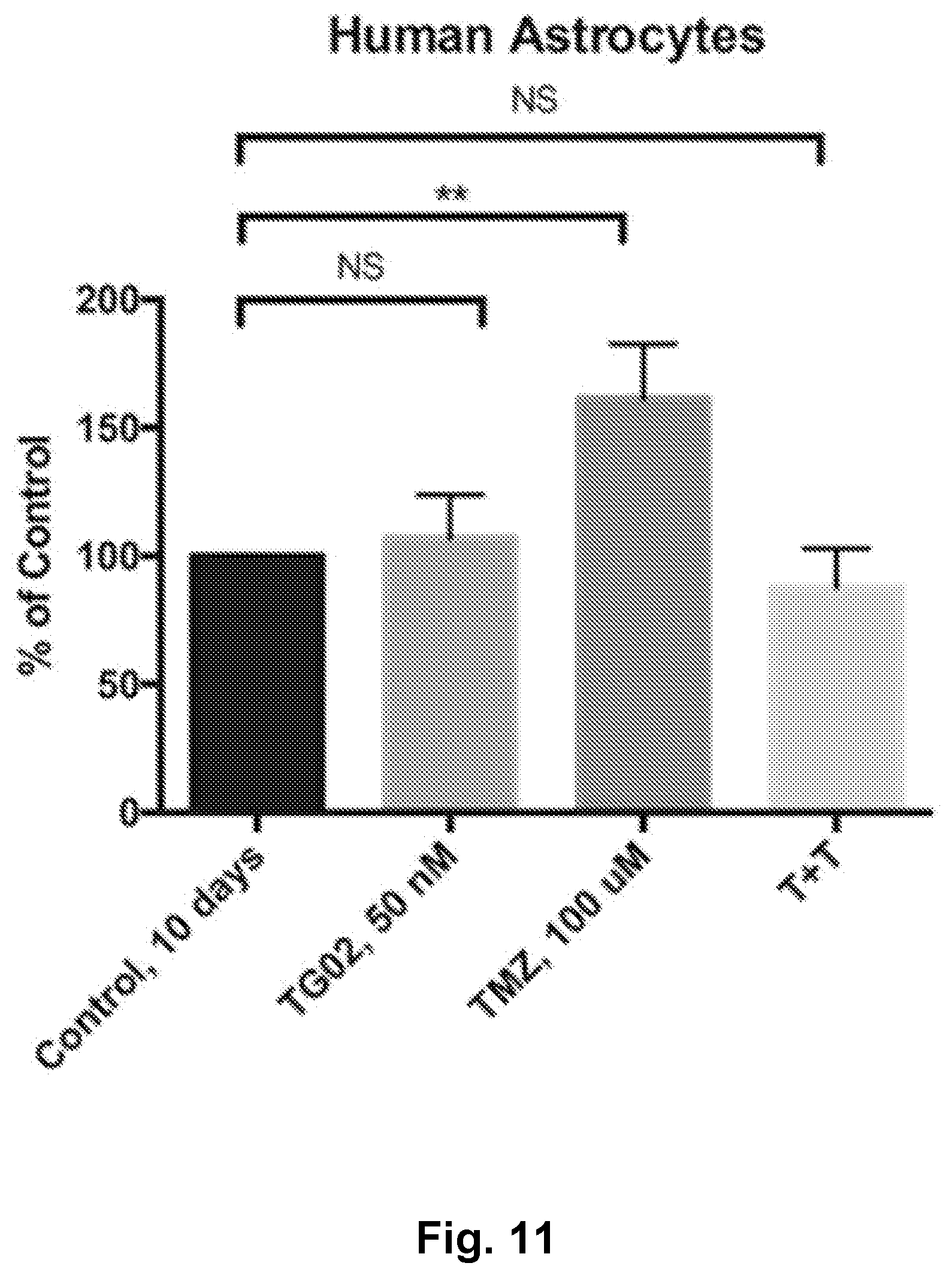
D00012
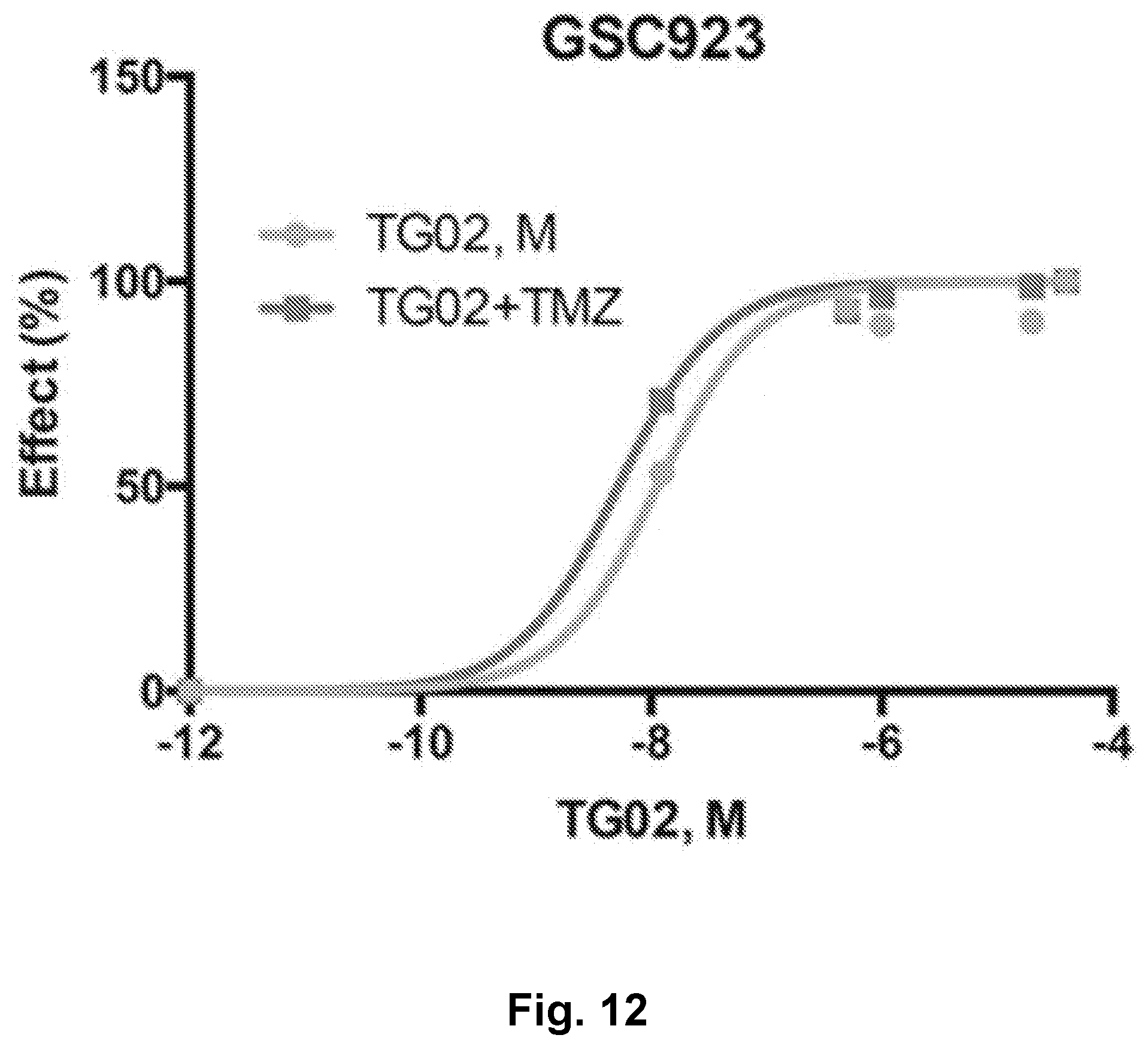
D00013

D00014
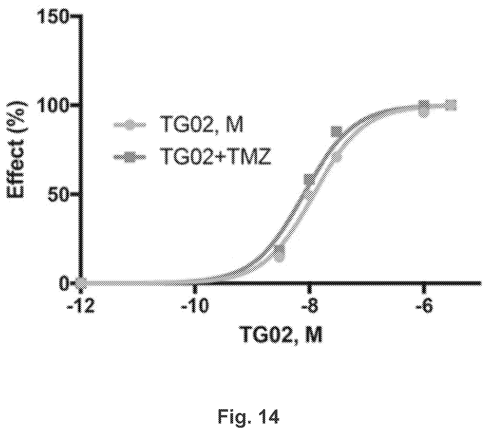
D00015

D00016
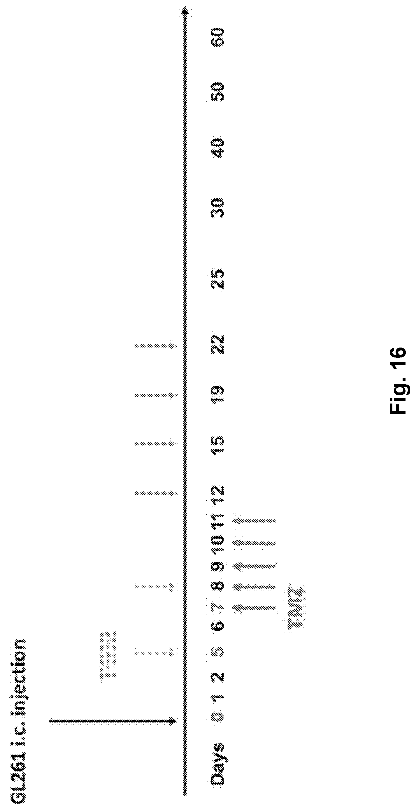
D00017
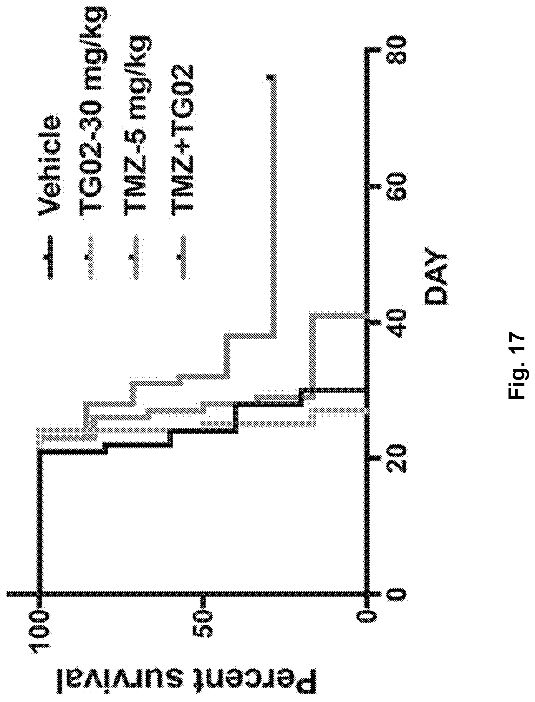
D00018
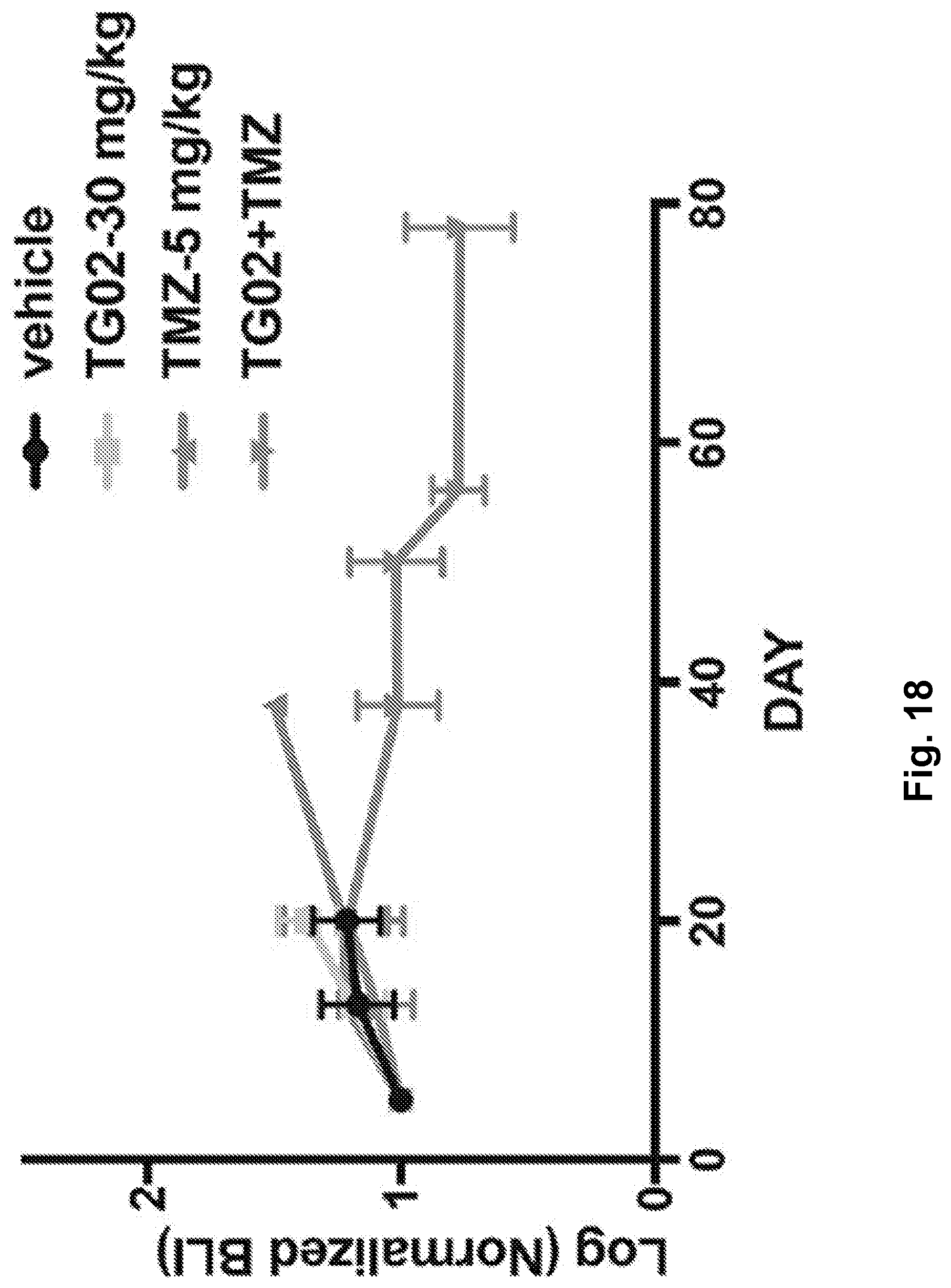
D00019
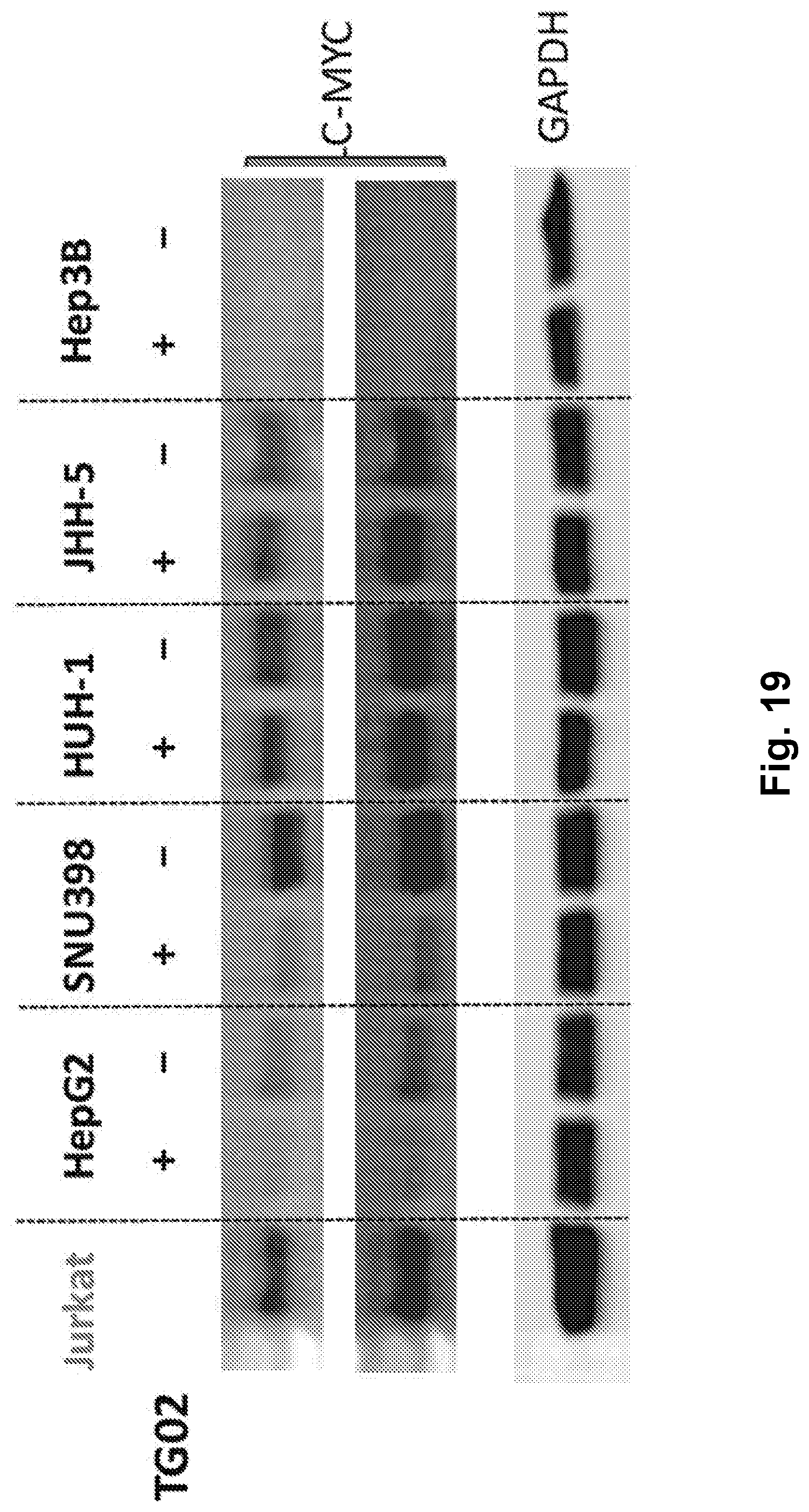
D00020
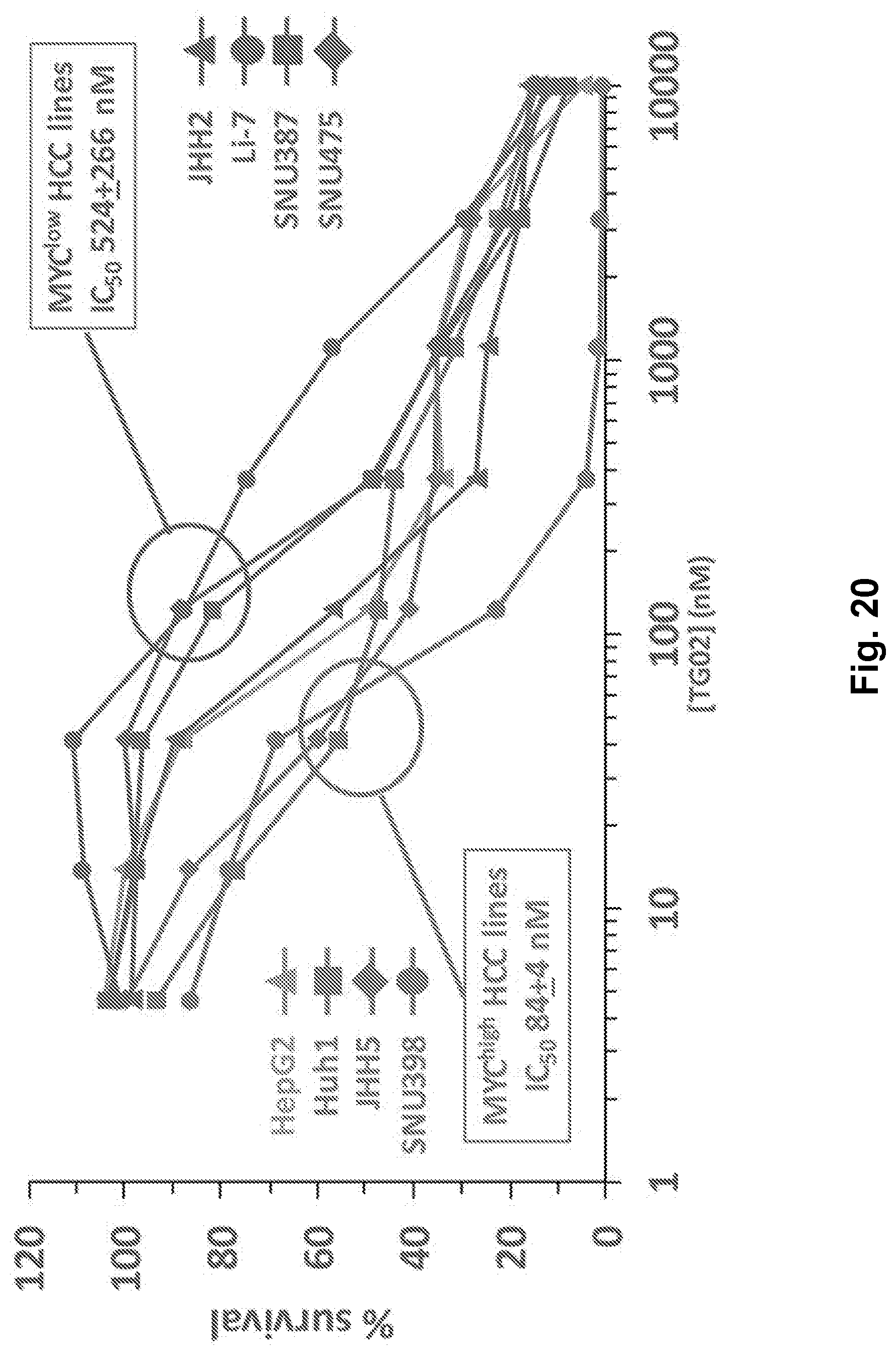
D00021

D00022

D00023
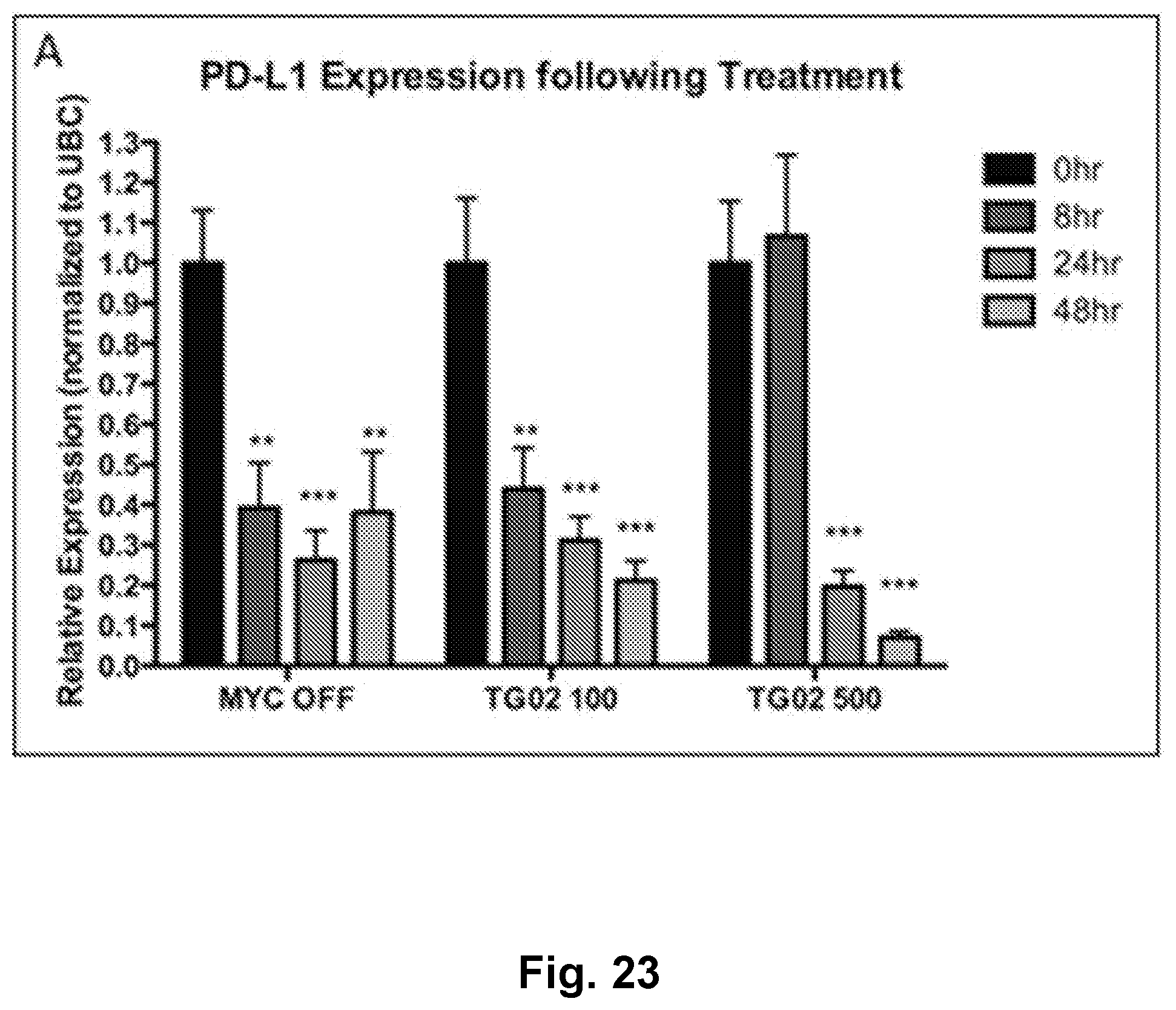
D00024

D00025
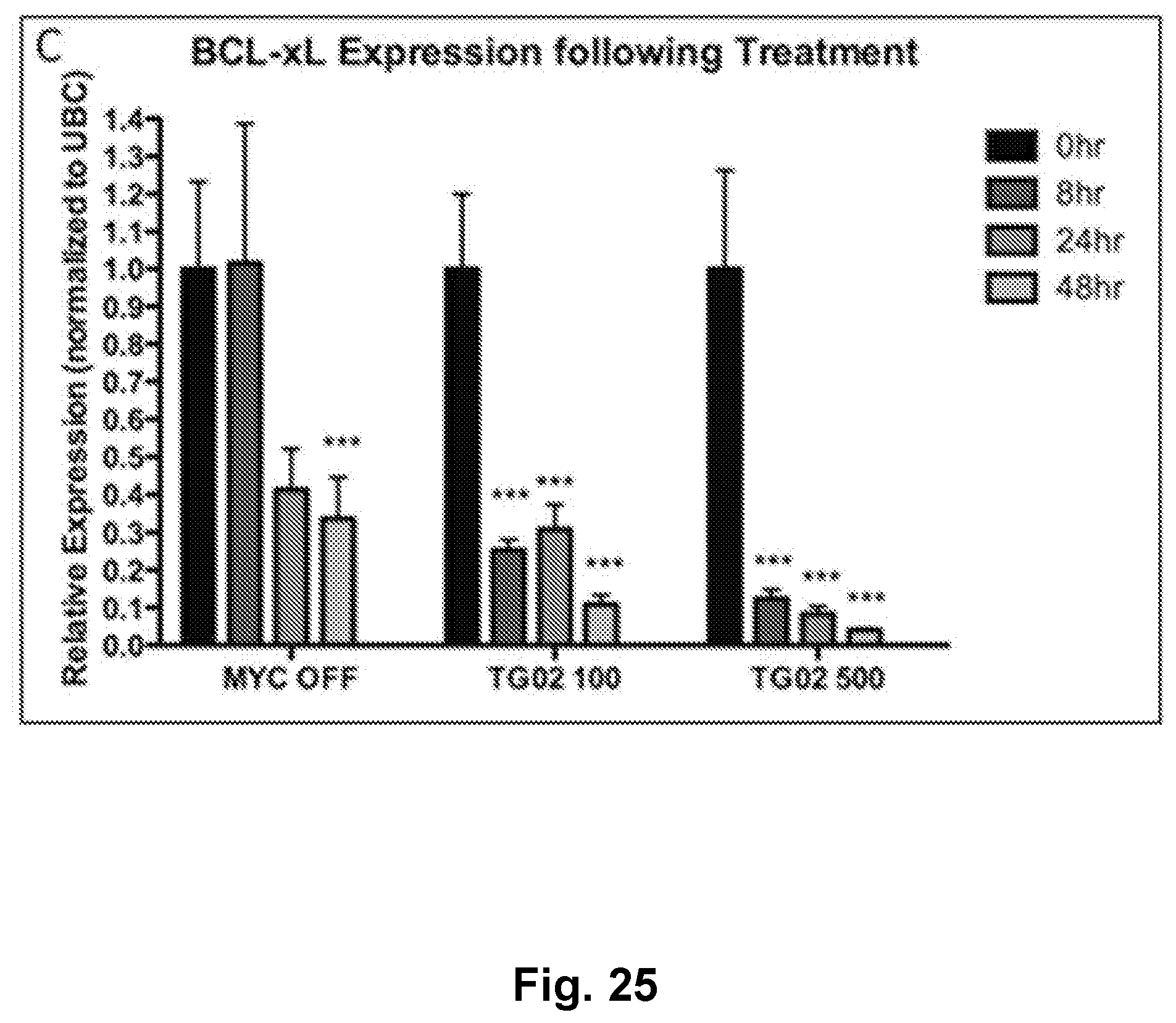
D00026

D00027

D00028
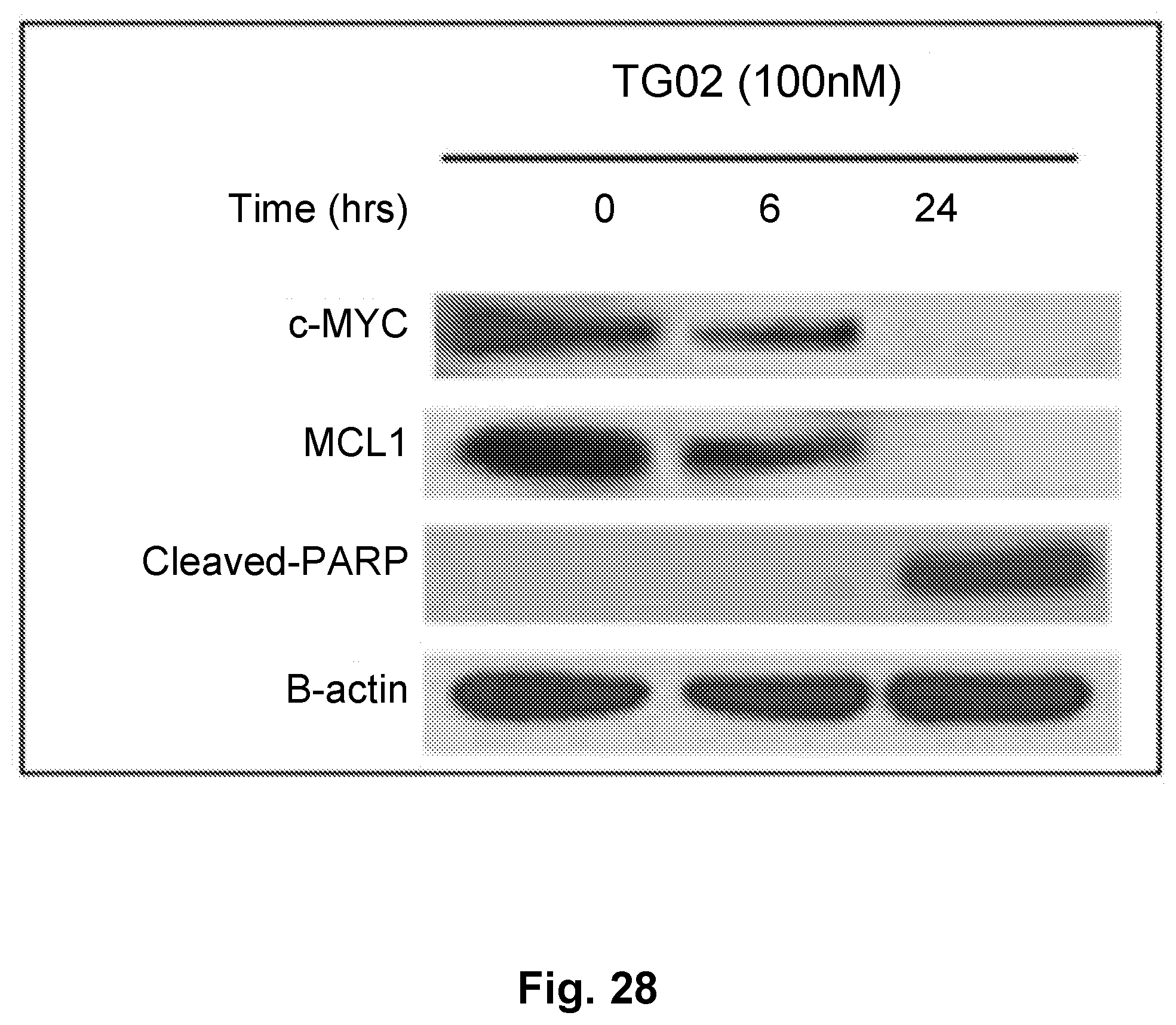
D00029
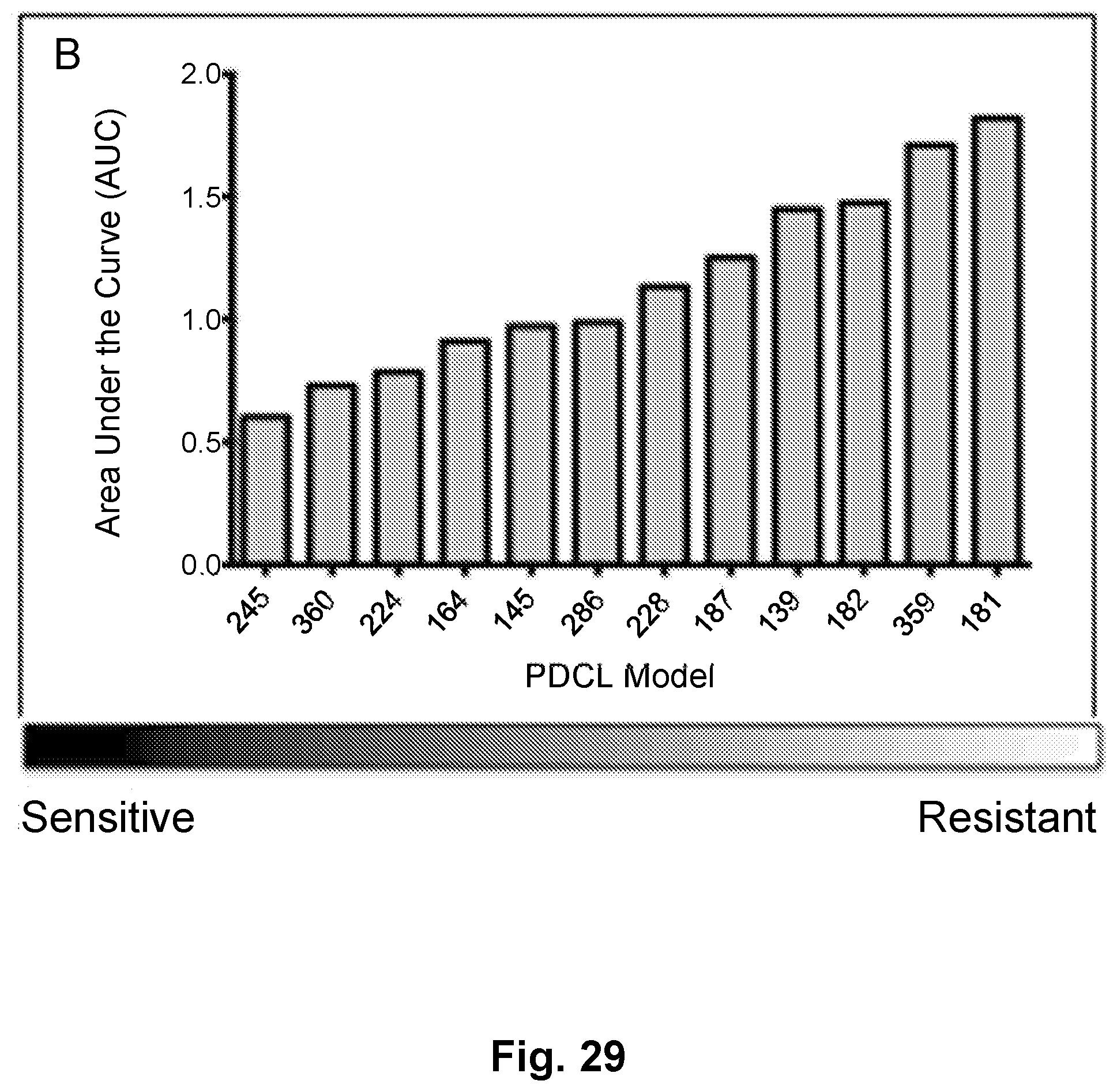
D00030

D00031
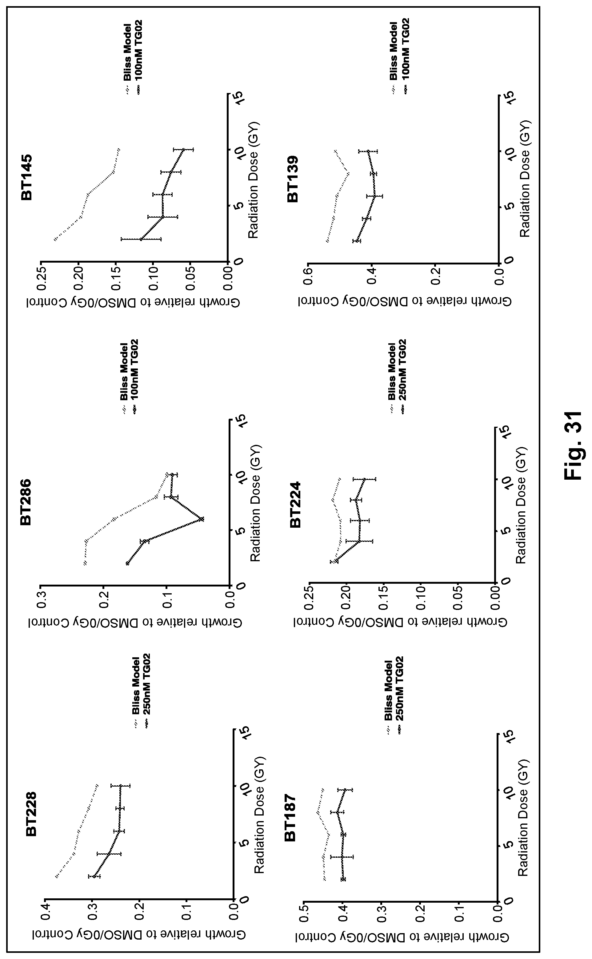
D00032
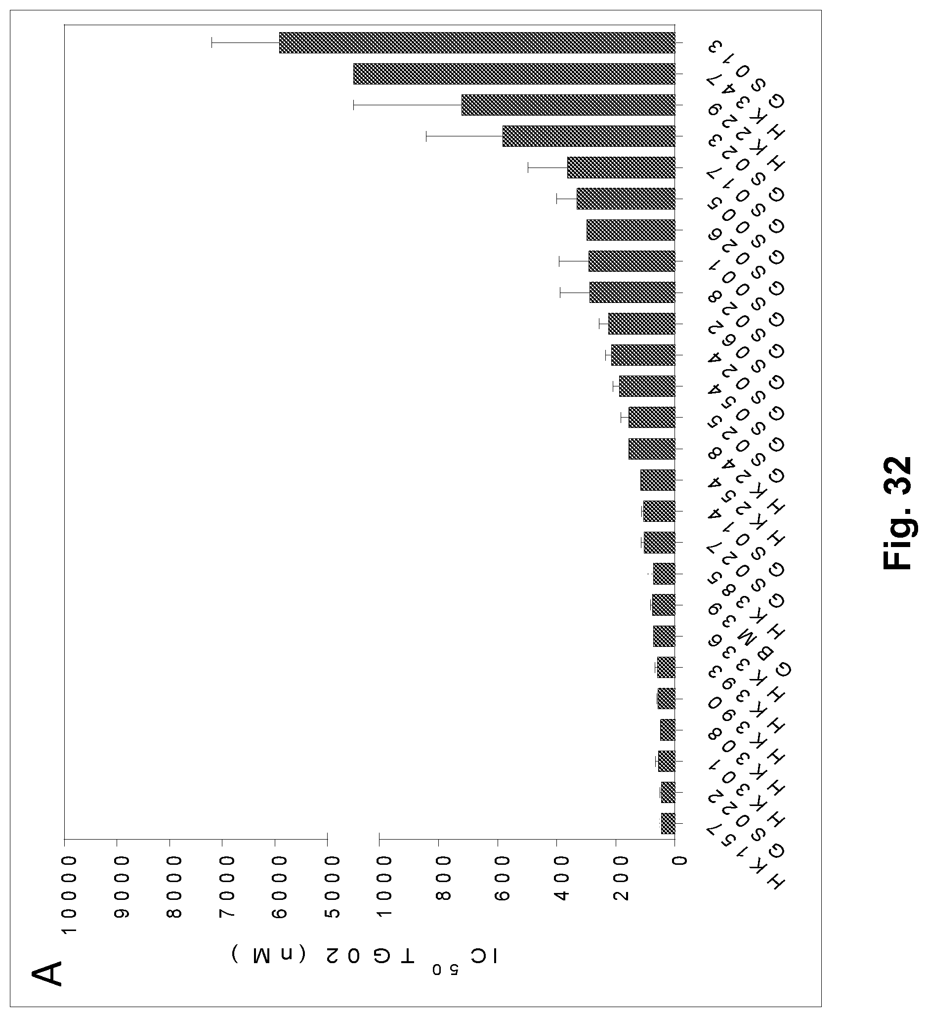
D00033

XML
uspto.report is an independent third-party trademark research tool that is not affiliated, endorsed, or sponsored by the United States Patent and Trademark Office (USPTO) or any other governmental organization. The information provided by uspto.report is based on publicly available data at the time of writing and is intended for informational purposes only.
While we strive to provide accurate and up-to-date information, we do not guarantee the accuracy, completeness, reliability, or suitability of the information displayed on this site. The use of this site is at your own risk. Any reliance you place on such information is therefore strictly at your own risk.
All official trademark data, including owner information, should be verified by visiting the official USPTO website at www.uspto.gov. This site is not intended to replace professional legal advice and should not be used as a substitute for consulting with a legal professional who is knowledgeable about trademark law.