Detecting Gastrointestinal Neoplasms
Ahlquist; David A. ; et al.
U.S. patent application number 16/908056 was filed with the patent office on 2020-10-08 for detecting gastrointestinal neoplasms. The applicant listed for this patent is Mayo Foundation for Medical Education and Research. Invention is credited to David A. Ahlquist, John B. Kisiel, Douglas W. Mahoney, William R. Taylor, Tracy C. Yab.
| Application Number | 20200318202 16/908056 |
| Document ID | / |
| Family ID | 1000004901919 |
| Filed Date | 2020-10-08 |
View All Diagrams
| United States Patent Application | 20200318202 |
| Kind Code | A1 |
| Ahlquist; David A. ; et al. | October 8, 2020 |
DETECTING GASTROINTESTINAL NEOPLASMS
Abstract
Provided herein is technology for gastrointestinal neoplasia screening and particularly, but not exclusively, to methods, compositions, and related uses for detecting the presence of gastrointestinal neoplasm, and classifying the site location of such a gastrointestinal neoplasm (e.g., a colorectal region, a pancreaticobiliary region, a gastroesophageal region).
| Inventors: | Ahlquist; David A.; (Rochester, MN) ; Taylor; William R.; (Lake City, MN) ; Kisiel; John B.; (Rochester, MN) ; Mahoney; Douglas W.; (Elgin, MN) ; Yab; Tracy C.; (Rochester, MN) | ||||||||||
| Applicant: |
|
||||||||||
|---|---|---|---|---|---|---|---|---|---|---|---|
| Family ID: | 1000004901919 | ||||||||||
| Appl. No.: | 16/908056 | ||||||||||
| Filed: | June 22, 2020 |
Related U.S. Patent Documents
| Application Number | Filing Date | Patent Number | ||
|---|---|---|---|---|
| 16030313 | Jul 9, 2018 | 10704107 | ||
| 16908056 | ||||
| 15054860 | Feb 26, 2016 | 10030272 | ||
| 16030313 | ||||
| 62126061 | Feb 27, 2015 | |||
| Current U.S. Class: | 1/1 |
| Current CPC Class: | C12Q 1/6886 20130101; C12Q 2600/112 20130101; C12Q 2600/154 20130101 |
| International Class: | C12Q 1/6886 20060101 C12Q001/6886 |
Claims
1. A method of classifying lower gastrointestinal neoplasm and upper gastrointestinal neoplasm in a sample obtained from a subject having gastrointestinal neoplasm, the method comprising: 1) assaying a methylation state of a marker in a sample obtained from the subject; and 2) classifying the subject as having lower gastrointestinal neoplasm when the methylation state of the marker is similar to a methylation state of the marker assayed in a subject that has a lower gastrointestinal neoplasm, 3) classifying the subject as having upper gastrointestinal neoplasm when the methylation state of the marker is similar to a methylation state of the marker assayed in a subject that has an upper gastrointestinal neoplasm, wherein the marker comprises a base in a differentially methylated region (DMR) selected from a group consisting of DMR No. 1 and 5 as provided in Table 1.
Description
CROSS-REFERENCE TO RELATED APPLICATIONS
[0001] This application is a continuation of U.S. patent application Ser. No. 16/030,313 filed Jul. 9, 2018, now allowed as U.S. Pat. No. 10,704,107, which is a continuation of U.S. patent application Ser. No. 15/054,860, filed Feb. 26, 2016, now allowed as U.S. Pat. No. 10,030,272, which claims priority to U.S. Provisional Patent Application No. 62/126,061, filed Feb. 27, 2015, the contents of which are incorporated by reference in their entireties.
FIELD OF INVENTION
[0002] Provided herein is technology for gastrointestinal neoplasia screening and particularly, but not exclusively, to methods, compositions, and related uses for detecting the presence of gastrointestinal neoplasm, and classifying the site location of such a gastrointestinal neoplasm (e.g., a colorectal region, a pancreaticobiliary region, a gastroesophageal region).
BACKGROUND
[0003] When upper gastrointestinal cancers (UGC) are combined with colorectal cancer (CRC), mortality from GI cancers is greater than for any other organ system. In 2012, it was estimated that 120,000 men and women were diagnosed with UGC, which caused nearly 90,000 deaths (compared to <50,000 by CRC in the same time period) (see, e.g., Siegel R, et al., 2012. CA Cancer J Clin 2012; 62:10-29). Patients who present with symptoms typically have advanced stage disease; and only a minority, usually early stage patients, is cured (see, e.g., Cleary S P, et al., J Am Coll Surg 2004; 198:722-31; Talsma K, et al., Ann Surg Oncol 2012; 19:2142-8). Remarkably, although the aggregate death toll from UGC exceeds that of CRC, general population screening for UGC does not exist in this country. Tools considered to screen asymptomatic individuals for UGC have been dismissed as too invasive, insensitive, or cost-prohibitive for application at the population-wide level (see, e.g., Gudlaugsdottir S, et al., Eur J Gastroenterol Hepatol 2001; 13:639-45; Inadomi J M, Keio J Med 2009; 58:12-8; Yeh J M, et al., Gastrointest Endosc 2010; 72:33-43; Rulyak S J, et al., Gastrointest Endosc 2003; 57:23-9). A noninvasive, accurate, and affordable screening tool capable of the screen-detection of all GI cancers is needed.
SUMMARY
[0004] Methylated DNA has been studied as a potential class of biomarkers in the tissues of most tumor types. In many instances, DNA methyltransferases add a methyl group to DNA at cytosine-phosphate-guanine (CpG) island sites as an epigenetic control of gene expression. In a biologically attractive mechanism, acquired methylation events in promoter regions of tumor suppressor genes are thought to silence expression, thus contributing to oncogenesis. DNA methylation may be a more chemically and biologically stable diagnostic tool than RNA or protein expression (Laird (2010) Nat Rev Genet 11: 191-203). Furthermore, in other cancers like sporadic colon cancer, methylation markers offer excellent specificity and are more broadly informative and sensitive than are individual DNA mutations (Zou et al (2007) Cancer Epidemiol Biomarkers Prev 16: 2686-96).
[0005] Analysis of CpG islands has yielded important findings when applied to animal models and human cell lines. For example, Zhang and colleagues found that amplicons from different parts of the same CpG island may have different levels of methylation (Zhang et al. (2009) PLoS Genet 5: e1000438). Further, methylation levels were distributed bi-modally between highly methylated and unmethylated sequences, further supporting the binary switch-like pattern of DNA methyltransferase activity (Zhang et al. (2009) PLoS Genet 5: e1000438). Analysis of murine tissues in vivo and cell lines in vitro demonstrated that only about 0.3% of high CpG density promoters (HCP, defined as having >7% CpG sequence within a 300 base pair region) were methylated, whereas areas of low CpG density (LCP, defined as having <5% CpG sequence within a 300 base pair region) tended to be frequently methylated in a dynamic tissue-specific pattern (Meissner et al. (2008) Nature 454: 766-70). HCPs include promoters for ubiquitous housekeeping genes and highly regulated developmental genes. Among the HCP sites methylated at >50% were several established markers such as Wnt 2, NDRG2, SFRP2, and BMP3 (Meissner et al. (2008) Nature 454: 766-70).
[0006] Accordingly, provided herein is technology for gastrointestinal neoplasia screening and particularly, but not exclusively, to methods, compositions, and related uses for detecting the presence of gastrointestinal neoplasm, and classifying the site location of such a gastrointestinal neoplasm (e.g., a colorectal region, a pancreaticobiliary region, a gastroesophageal region).
[0007] Indeed, experiments conducted during the course of developing this technology compared the methylation state of DNA markers from esophageal tissue, stomach tissue, pancreatic tissue, bile duct/liver tissue, and colorectal tissue of subjects having neoplasm related to the respective tissue type to the methylation state of the same DNA markers from control subjects (e.g., normal tissue for the respective tissue type) (see, Example 1).
[0008] Markers and/or panels of markers were identified (e.g., a chromosomal region having an annotation selected from chr12.133484978-133485739, BMP3, and chr11.123301058-123301255) capable of classifying gastrointestinal (GI) neoplasm versus control (e.g., normal tissue for the respective tissue type) across GI tissue types (e.g., esophageal tissue, stomach tissue, pancreatic tissue, bile duct/liver tissue, and colorectal tissue) (see, Example 1).
[0009] Markers and/or panels of markers were identified (e.g., a chromosomal region having an annotation selected from chr7.25896389-25896501, and QKI) capable of classifying lower gastrointestinal (LGI) neoplasm versus upper gastrointestinal neoplasm (UGI) across GI tissue types (e.g., esophageal tissue, stomach tissue, pancreatic tissue, bile duct/liver tissue, and colorectal tissue) (see, Example 1).
[0010] Markers and/or panels of markers were identified (e.g., a chromosomal region having an annotation selected from PDGFD, ELOVL2, PCBP3) capable of classifying pancreaticobiliary neoplasm versus gastroesophageal neoplasm across GI tissue types (e.g., esophageal tissue, stomach tissue, pancreatic tissue, bile duct/liver tissue, and colorectal tissue) (see, Example 1).
[0011] Markers and/or panels of markers were identified (e.g., a chromosomal region having an annotation selected from QKI, PDGFD, ELOVL2, chr12.133484978-133485739, chr7.25896389-25896501, PCBP3, chr11.123301058-123301255, and BMP3) capable of predicting control versus neoplasm location across GI tissue types (e.g., esophageal tissue, stomach tissue, pancreatic tissue, bile duct/liver tissue, and colorectal tissue) (see, Example 1).
[0012] Markers and/or panels of markers were identified (e.g., a chromosomal region having an annotation selected from BMP3 and QKI) capable of predicting control versus neoplasm location in blood plasma samples (see, Example 1).
[0013] Markers and/or panels of markers were identified (e.g., a chromosomal region having an annotation selected from BMP3 and QKI) capable of predicting colorectal cancer versus pancreaticobiliary neoplasm in blood plasma samples (see, Example 1).
[0014] Additional statistical analysis of the results demonstrated that the technology described herein based on these markers specifically and sensitively predicts a tumor site.
[0015] As described herein, the technology provides a number of methylated DNA markers and subsets thereof (e.g., sets of 2, 3, 4, 5, 6, 7, or 8 markers) with high discrimination for GI neoplasms overall and/or at individual tumor sites. Experiments applied a selection filter to candidate markers to identify markers that provide a high signal to noise ratio and a low background level to provide high specificity, e.g., when assaying distant media (e.g., stool, blood, urine, metastatic tissue, etc.) for purposes of cancer screening or diagnosis. Further, experiments were performed to demonstrate that the identified methylated DNA markers predict tumor site. As such, the technology provides for specific markers, marker combinations, and algorithms to predict tumor site.
[0016] In some embodiments, the technology is related to assessing the presence of and methylation state of one or more of the markers identified herein in a biological sample. These markers comprise one or more differentially methylated regions (DMR) as discussed herein, e.g., as provided in Table 1. Methylation state is assessed in embodiments of the technology. As such, the technology provided herein is not restricted in the method by which a gene's methylation state is measured. For example, in some embodiments the methylation state is measured by a genome scanning method. For example, one method involves restriction landmark genomic scanning (Kawai et al. (1994) Mol. Cell. Biol. 14: 7421-7427) and another example involves methylation-sensitive arbitrarily primed PCR (Gonzalgo et al. (1997) Cancer Res. 57: 594-599). In some embodiments, changes in methylation patterns at specific CpG sites are monitored by digestion of genomic DNA with methylation-sensitive restriction enzymes followed by Southern analysis of the regions of interest (digestion-Southern method). In some embodiments, analyzing changes in methylation patterns involves a PCR-based process that involves digestion of genomic DNA with methylation-sensitive restriction enzymes prior to PCR amplification (Singer-Sam et al. (1990) Nucl. Acids Res. 18: 687). In addition, other techniques have been reported that utilize bisulfate treatment of DNA as a starting point for methylation analysis. These include methylation-specific PCR (MSP) (Herman et al. (1992) Proc. Natl. Acad. Sci. USA 93: 9821-9826) and restriction enzyme digestion of PCR products amplified from bisulfate-converted DNA (Sadri and Hornsby (1996) Nucl. Acids Res. 24: 5058-5059; and Xiong and Laird (1997) Nucl. Acids Res. 25: 2532-2534). PCR techniques have been developed for detection of gene mutations (Kuppuswamy et al. (1991) Proc. Natl. Acad. Sci. USA 88: 1143-1147) and quantification of allelic-specific expression (Szabo and Mann (1995) Genes Dev. 9: 3097-3108; and Singer-Sam et al. (1992) PCR Methods Appl. 1: 160-163). Such techniques use internal primers, which anneal to a PCR-generated template and terminate immediately 5' of the single nucleotide to be assayed. Methods using a "quantitative MS-SNUPE assay" as described in U.S. Pat. No. 7,037,650 are used in some embodiments.
[0017] Upon evaluating a methylation state, the methylation state is often expressed as the fraction or percentage of individual strands of DNA that is methylated at a particular site (e.g., at a single nucleotide, at a particular region or locus, at a longer sequence of interest, e.g., up to a .about.100-bp, 200-bp, 500-bp, 1000-bp subsequence of a DNA or longer) relative to the total population of DNA in the sample comprising that particular site. Traditionally, the amount of the unmethylated nucleic acid is determined by PCR using calibrators. Then, a known amount of DNA is bisulfite treated and the resulting methylation-specific sequence is determined using either a real-time PCR or other exponential amplification, e.g., a QuARTS assay (e.g., as provided by U.S. Pat. No. 8,361,720; and U.S. Pat. Appl. Pub. Nos. 2012/0122088 and 2012/0122106).
[0018] For example, in some embodiments methods comprise generating a standard curve for the unmethylated target by using external standards. The standard curve is constructed from at least two points and relates the real-time Ct value for unmethylated DNA to known quantitative standards. Then, a second standard curve for the methylated target is constructed from at least two points and external standards. This second standard curve relates the Ct for methylated DNA to known quantitative standards. Next, the test sample Ct values are determined for the methylated and unmethylated populations and the genomic equivalents of DNA are calculated from the standard curves produced by the first two steps. The percentage of methylation at the site of interest is calculated from the amount of methylated DNAs relative to the total amount of DNAs in the population, e.g., (number of methylated DNAs)/(the number of methylated DNAs+number of unmethylated DNAs).times.100.
[0019] Also provided herein are compositions and kits for practicing the methods. For example, in some embodiments, reagents (e.g., primers, probes) specific for one or more markers are provided alone or in sets (e.g., sets of primers pairs for amplifying a plurality of markers). Additional reagents for conducting a detection assay may also be provided (e.g., enzymes, buffers, positive and negative controls for conducting QuARTS, PCR, sequencing, bisulfite, or other assays). In some embodiments, the kits containing one or more reagent necessary, sufficient, or useful for conducting a method are provided. Also provided are reactions mixtures containing the reagents. Further provided are master mix reagent sets containing a plurality of reagents that may be added to each other and/or to a test sample to complete a reaction mixture.
[0020] In some embodiments, the technology described herein is associated with a programmable machine designed to perform a sequence of arithmetic or logical operations as provided by the methods described herein. For example, some embodiments of the technology are associated with (e.g., implemented in) computer software and/or computer hardware. In one aspect, the technology relates to a computer comprising a form of memory, an element for performing arithmetic and logical operations, and a processing element (e.g., a microprocessor) for executing a series of instructions (e.g., a method as provided herein) to read, manipulate, and store data. In some embodiments, a microprocessor is part of a system for determining a methylation state (e.g., of one or more DMR, e.g., DMR 1-8 as provided in Table 1); comparing methylation states (e.g., of one or more DMR, e.g., DMR 1-8 as provided in Table 1); generating standard curves; determining a Ct value; calculating a fraction, frequency, or percentage of methylation (e.g., of one or more DMR, e.g., DMR 1-8 as provided in Table 1); identifying a CpG island; determining a specificity and/or sensitivity of an assay or marker; calculating an ROC curve and an associated AUC; sequence analysis; all as described herein or is known in the art.
[0021] In some embodiments, a microprocessor or computer uses methylation state data in an algorithm to predict a site of a cancer.
[0022] In some embodiments, a software or hardware component receives the results of multiple assays and determines a single value result to report to a user that indicates a cancer risk based on the results of the multiple assays (e.g., determining the methylation state of multiple DMR, e.g., as provided in Table 1). Related embodiments calculate a risk factor based on a mathematical combination (e.g., a weighted combination, a linear combination) of the results from multiple assays, e.g., determining the methylation states of multiple markers (such as multiple DMR, e.g., as provided in Table 1). In some embodiments, the methylation state of a DMR defines a dimension and may have values in a multidimensional space and the coordinate defined by the methylation states of multiple DMR is a result, e.g., to report to a user, e.g., related to a cancer risk.
[0023] Some embodiments comprise a storage medium and memory components. Memory components (e.g., volatile and/or nonvolatile memory) find use in storing instructions (e.g., an embodiment of a process as provided herein) and/or data (e.g., a work piece such as methylation measurements, sequences, and statistical descriptions associated therewith). Some embodiments relate to systems also comprising one or more of a CPU, a graphics card, and a user interface (e.g., comprising an output device such as display and an input device such as a keyboard).
[0024] Programmable machines associated with the technology comprise conventional extant technologies and technologies in development or yet to be developed (e.g., a quantum computer, a chemical computer, a DNA computer, an optical computer, a spintronics based computer, etc.).
[0025] In some embodiments, the technology comprises a wired (e.g., metallic cable, fiber optic) or wireless transmission medium for transmitting data. For example, some embodiments relate to data transmission over a network (e.g., a local area network (LAN), a wide area network (WAN), an ad-hoc network, the internet, etc.). In some embodiments, programmable machines are present on such a network as peers and in some embodiments the programmable machines have a client/server relationship.
[0026] In some embodiments, data are stored on a computer-readable storage medium such as a hard disk, flash memory, optical media, a floppy disk, etc.
[0027] In some embodiments, the technology provided herein is associated with a plurality of programmable devices that operate in concert to perform a method as described herein. For example, in some embodiments, a plurality of computers (e.g., connected by a network) may work in parallel to collect and process data, e.g., in an implementation of cluster computing or grid computing or some other distributed computer architecture that relies on complete computers (with onboard CPUs, storage, power supplies, network interfaces, etc.) connected to a network (private, public, or the internet) by a conventional network interface, such as Ethernet, fiber optic, or by a wireless network technology.
[0028] For example, some embodiments provide a computer that includes a computer-readable medium. The embodiment includes a random access memory (RAM) coupled to a processor. The processor executes computer-executable program instructions stored in memory. Such processors may include a microprocessor, an ASIC, a state machine, or other processor, and can be any of a number of computer processors, such as processors from Intel Corporation of Santa Clara, Calif. and Motorola Corporation of Schaumburg, Ill. Such processors include, or may be in communication with, media, for example computer-readable media, which stores instructions that, when executed by the processor, cause the processor to perform the steps described herein.
[0029] Embodiments of computer-readable media include, but are not limited to, an electronic, optical, magnetic, or other storage or transmission device capable of providing a processor with computer-readable instructions. Other examples of suitable media include, but are not limited to, a floppy disk, CD-ROM, DVD, magnetic disk, memory chip, ROM, RAM, an ASIC, a configured processor, all optical media, all magnetic tape or other magnetic media, or any other medium from which a computer processor can read instructions. Also, various other forms of computer-readable media may transmit or carry instructions to a computer, including a router, private or public network, or other transmission device or channel, both wired and wireless. The instructions may comprise code from any suitable computer-programming language, including, for example, C, C++, C#, Visual Basic, Java, Python, Perl, and JavaScript.
[0030] Computers are connected in some embodiments to a network. Computers may also include a number of external or internal devices such as a mouse, a CD-ROM, DVD, a keyboard, a display, or other input or output devices. Examples of computers are personal computers, digital assistants, personal digital assistants, cellular phones, mobile phones, smart phones, pagers, digital tablets, laptop computers, internet appliances, and other processor-based devices. In general, the computers related to aspects of the technology provided herein may be any type of processor-based platform that operates on any operating system, such as Microsoft Windows, Linux, UNIX, Mac OS X, etc., capable of supporting one or more programs comprising the technology provided herein. Some embodiments comprise a personal computer executing other application programs (e.g., applications). The applications can be contained in memory and can include, for example, a word processing application, a spreadsheet application, an email application, an instant messenger application, a presentation application, an Internet browser application, a calendar/organizer application, and any other application capable of being executed by a client device.
[0031] All such components, computers, and systems described herein as associated with the technology may be logical or virtual.
[0032] Accordingly, provided herein is technology related to a method of screening for a gastrointestinal neoplasm in a sample obtained from a subject, the method comprising assaying a methylation state of a marker in a sample obtained from a subject; and identifying the subject as having a neoplasm when the methylation state of the marker is different than a methylation state of the marker assayed in a subject that does not have a neoplasm, wherein the marker comprises a base in a differentially methylated region (DMR) selected from a group consisting of DMR 1-8 as provided in Table 1.
[0033] In some embodiments, the method further comprises locating the neoplasm site within the subject, wherein the methylation state of the marker indicates the neoplasm site within the subject. The technology is related to identifying and discriminating gastrointestinal neoplasm. In some embodiments, the methylation state of markers (e.g., a chromosomal region having an annotation selected from chr7.25896389-25896501, and QKI) are used to classify lower gastrointestinal (LGI) neoplasm versus upper gastrointestinal neoplasm (UGI). In some embodiments, the methylation state of markers (e.g., a chromosomal region having an annotation selected from PDGFD, ELOVL2, PCBP3) are used to classify pancreaticobiliary neoplasm versus gastroesophageal neoplasm. In some embodiments, the methylation state of markers (e.g., a chromosomal region having an annotation selected from BMP3 and QKI) are used to classify colorectal cancer versus pancreaticobiliary neoplasm. The technology also encompasses determining the state or stage of a cancer, e.g., in some embodiments the neoplasm is pre-cancerous. Some embodiments provide methods comprising assaying a plurality of markers, e.g., comprising assaying 2 to 8 markers.
[0034] The technology is not limited in the methylation state assessed. In some embodiments assessing the methylation state of the marker in the sample comprises determining the methylation state of one base. In some embodiments, assaying the methylation state of the marker in the sample comprises determining the extent of methylation at a plurality of bases. Moreover, in some embodiments the methylation state of the marker comprises an increased methylation of the marker relative to a normal methylation state of the marker. In some embodiments, the methylation state of the marker comprises a decreased methylation of the marker relative to a normal methylation state of the marker. In some embodiments the methylation state of the marker comprises a different pattern of methylation of the marker relative to a normal methylation state of the marker.
[0035] Furthermore, in some embodiments the marker is a region of 100 or fewer bases, the marker is a region of 500 or fewer bases, the marker is a region of 1000 or fewer bases, the marker is a region of 5000 or fewer bases, or, in some embodiments, the marker is one base. In some embodiments the marker is in a high CpG density promoter.
[0036] The technology is not limited by sample type. For example, in some embodiments the sample is a stool sample, a tissue sample (e.g., esophageal tissue, stomach tissue, pancreatic tissue, bile duct/liver tissue, and colorectal tissue), a blood sample (e.g., plasma, serum, whole blood), an excretion, or a urine sample.
[0037] Furthermore, the technology is not limited in the method used to determine methylation state. In some embodiments the assaying comprises using methylation specific polymerase chain reaction, nucleic acid sequencing, mass spectrometry, methylation specific nuclease, mass-based separation, or target capture. In some embodiments, the assaying comprises use of a methylation specific oligonucleotide. In some embodiments, the technology uses massively parallel sequencing (e.g., next-generation sequencing) to determine methylation state, e.g., sequencing-by-synthesis, real-time (e.g., single-molecule) sequencing, bead emulsion sequencing, nanopore sequencing, etc.
[0038] The technology provides reagents for detecting a DMR, e.g., in some embodiments are provided a set of oligonucleotides comprising the sequences provided by SEQ ID NO: 1-16. In some embodiments are provided an oligonucleotide comprising a sequence complementary to a chromosomal region having a base in a DMR, e.g., an oligonucleotide sensitive to methylation state of a DMR.
[0039] The technology provides various panels of markers, e.g., in some embodiments the marker comprises a chromosomal region having an annotation that is QKI, PDGFD, ELOVL2, chr12.133484978-133485739, chr7.25896389-25896501, PCBP3, chr11.123301058-123301255, and BMP3, and that comprises the marker (see, Table 1). In addition, embodiments provide a method of analyzing a DMR from Table 1 that is DMR No. 1-8.
[0040] Kit embodiments are provided, e.g., a kit comprising a bisulfite reagent; and a control nucleic acid comprising a sequence from a DMR selected from a group consisting of DMR 1-8 (from Table 1) and having a methylation state associated with a subject who does not have a cancer. In some embodiments, kits comprise a bisulfite reagent and an oligonucleotide as described herein. In some embodiments, kits comprise a bisulfite reagent; and a control nucleic acid comprising a sequence from a DMR selected from a group consisting of DMR 1-8 (from Table 1) and having a methylation state associated with a subject who has a cancer. Some kit embodiments comprise a sample collector for obtaining a sample from a subject (e.g., a stool sample); reagents for isolating a nucleic acid from the sample; a bisulfite reagent; and an oligonucleotide as described herein.
[0041] The technology is related to embodiments of compositions (e.g., reaction mixtures). In some embodiments are provided a composition comprising a nucleic acid comprising a DMR and a bisulfite reagent. Some embodiments provide a composition comprising a nucleic acid comprising a DMR and an oligonucleotide as described herein. Some embodiments provide a composition comprising a nucleic acid comprising a DMR and a methylation-sensitive restriction enzyme. Some embodiments provide a composition comprising a nucleic acid comprising a DMR and a polymerase.
[0042] Additional related method embodiments are provided for screening for a neoplasm in a sample obtained from a subject, e.g., a method comprising determining a methylation state of a marker in the sample comprising a base in a DMR that is one or more of DMR 1-8 (from Table 1); comparing the methylation state of the marker from the subject sample to a methylation state of the marker from a normal control sample from a subject who does not have a cancer; and determining a confidence interval and/or a p value of the difference in the methylation state of the subject sample and the normal control sample. In some embodiments, the confidence interval is 90%, 95%, 97.5%, 98%, 99%, 99.5%, 99.9% or 99.99% and the p value is 0.1, 0.05, 0.025, 0.02, 0.01, 0.005, 0.001, or 0.0001. Some embodiments of methods provide steps of reacting a nucleic acid comprising a DMR with a bisulfate reagent to produce a bisulfate-reacted nucleic acid; sequencing the bisulfate-reacted nucleic acid to provide a nucleotide sequence of the bisulfate-reacted nucleic acid; comparing the nucleotide sequence of the bisulfate-reacted nucleic acid with a nucleotide sequence of a nucleic acid comprising the DMR from a subject who does not have a cancer to identify differences in the two sequences; and identifying the subject as having a neoplasm when a difference is present.
[0043] Systems for screening for a gastrointestinal neoplasm in a sample obtained from a subject are provided by the technology. Exemplary embodiments of systems include, e.g., a system for screening for a gastrointestinal neoplasm in a sample obtained from a subject, the system comprising an analysis component configured to determine the methylation state of a sample, a software component configured to compare the methylation state of the sample with a control sample or a reference sample methylation state recorded in a database, and an alert component configured to alert a user of a cancer-associated methylation state. An alert is determined in some embodiments by a software component that receives the results from multiple assays (e.g., determining the methylation states of multiple markers, e.g., DMR, e.g., as provided in Table 1) and calculating a value or result to report based on the multiple results. Some embodiments provide a database of weighted parameters associated with each DMR provided herein for use in calculating a value or result and/or an alert to report to a user (e.g., such as a physician, nurse, clinician, etc.). In some embodiments all results from multiple assays are reported and in some embodiments one or more results are used to provide a score, value, or result based on a composite of one or more results from multiple assays that is indicative of a cancer risk in a subject.
[0044] In some embodiments of systems, a sample comprises a nucleic acid comprising a DMR. In some embodiments the system further comprises a component for isolating a nucleic acid, a component for collecting a sample such as a component for collecting a stool sample. In some embodiments, the system comprises nucleic acid sequences comprising a DMR. In some embodiments the database comprises nucleic acid sequences from subjects who do not have a cancer. Also provided are nucleic acids, e.g., a set of nucleic acids, each nucleic acid having a sequence comprising a DMR. In some embodiments the set of nucleic acids wherein each nucleic acid has a sequence from a subject who does not have a cancer. Related system embodiments comprise a set of nucleic acids as described and a database of nucleic acid sequences associated with the set of nucleic acids. Some embodiments further comprise a bisulfite reagent. And, some embodiments further comprise a nucleic acid sequencer.
[0045] Additional embodiments will be apparent to persons skilled in the relevant art based on the teachings contained herein.
BRIEF DESCRIPTION OF THE DRAWINGS
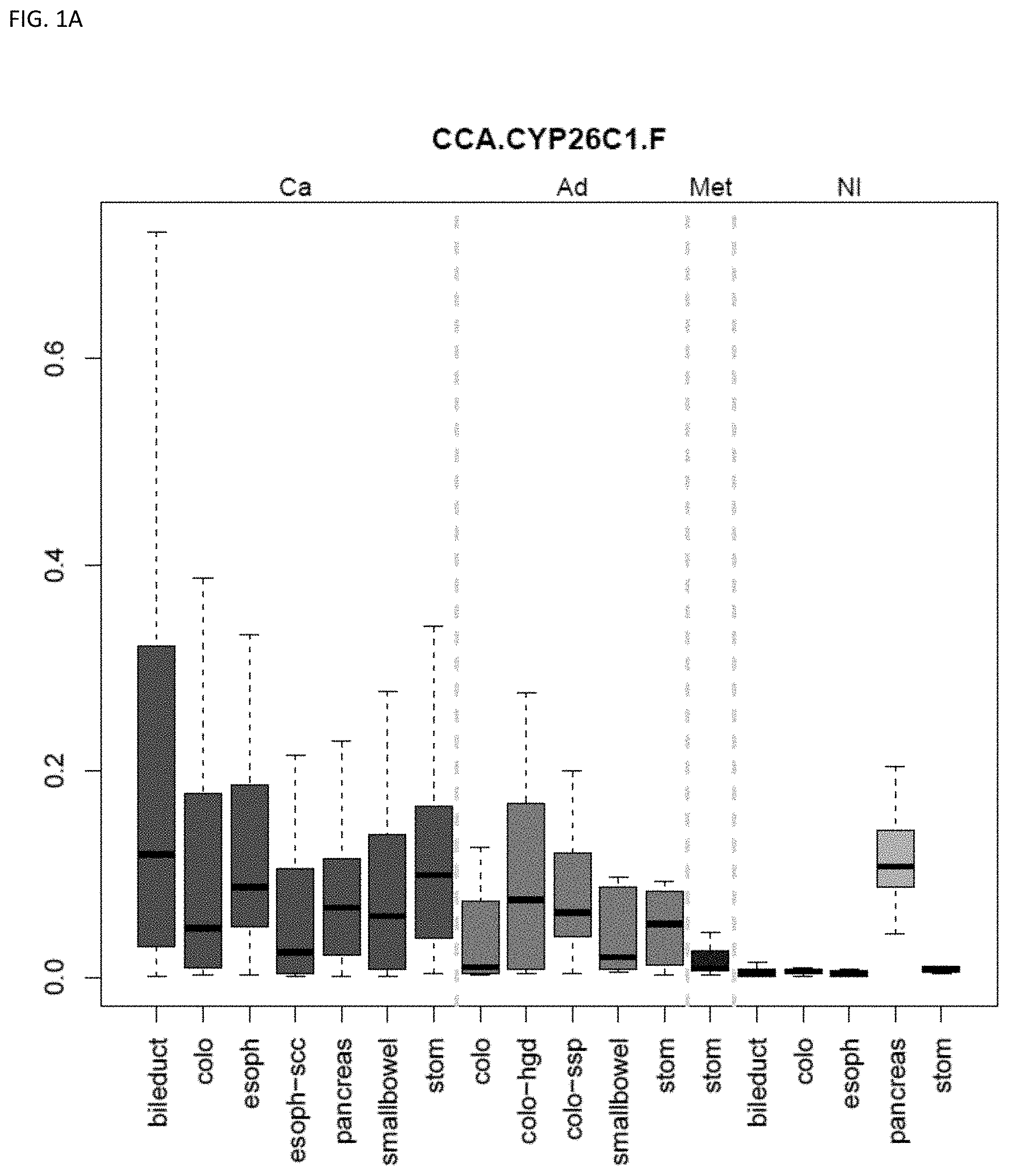
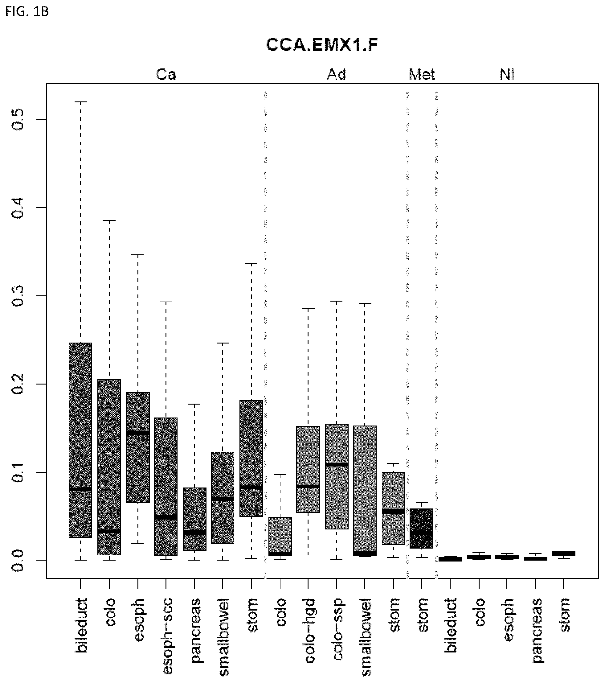
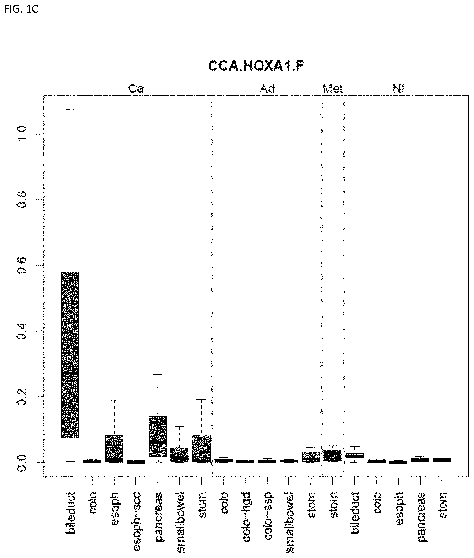
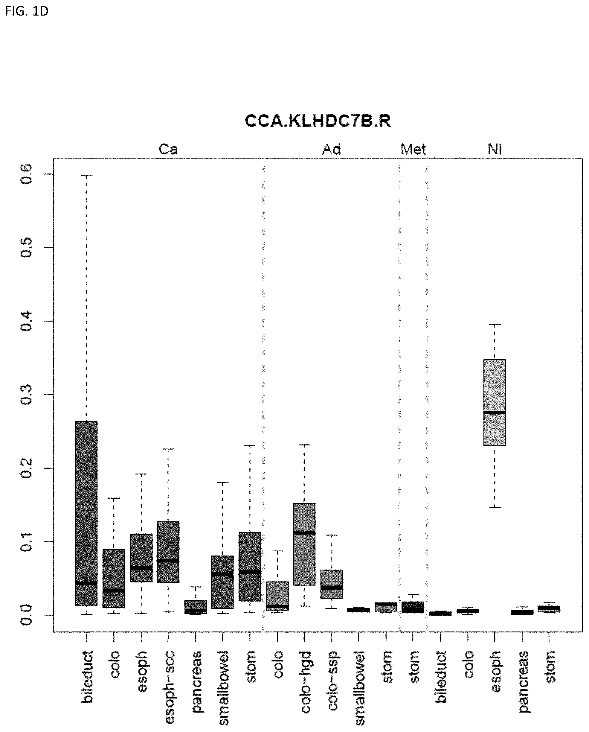
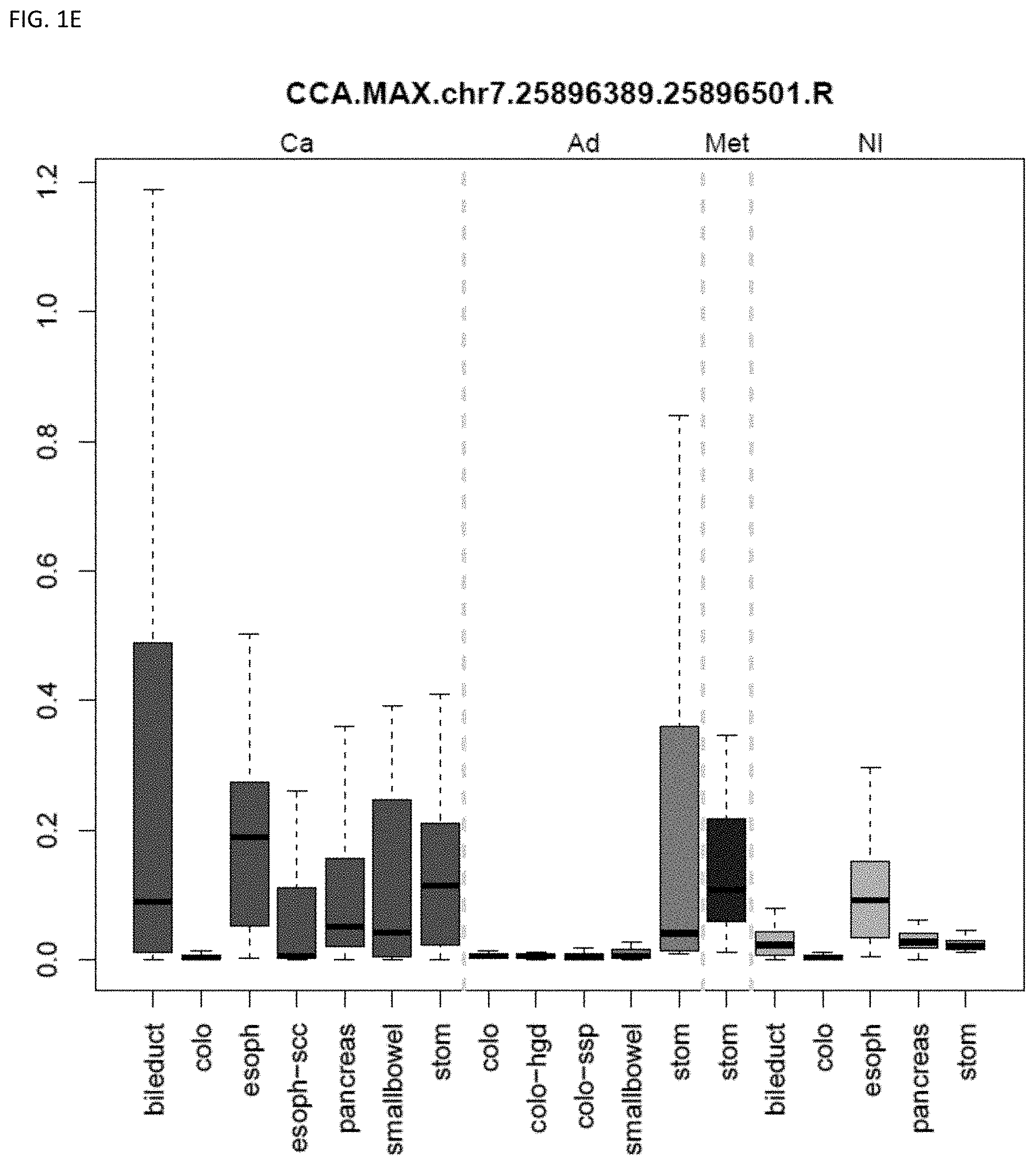
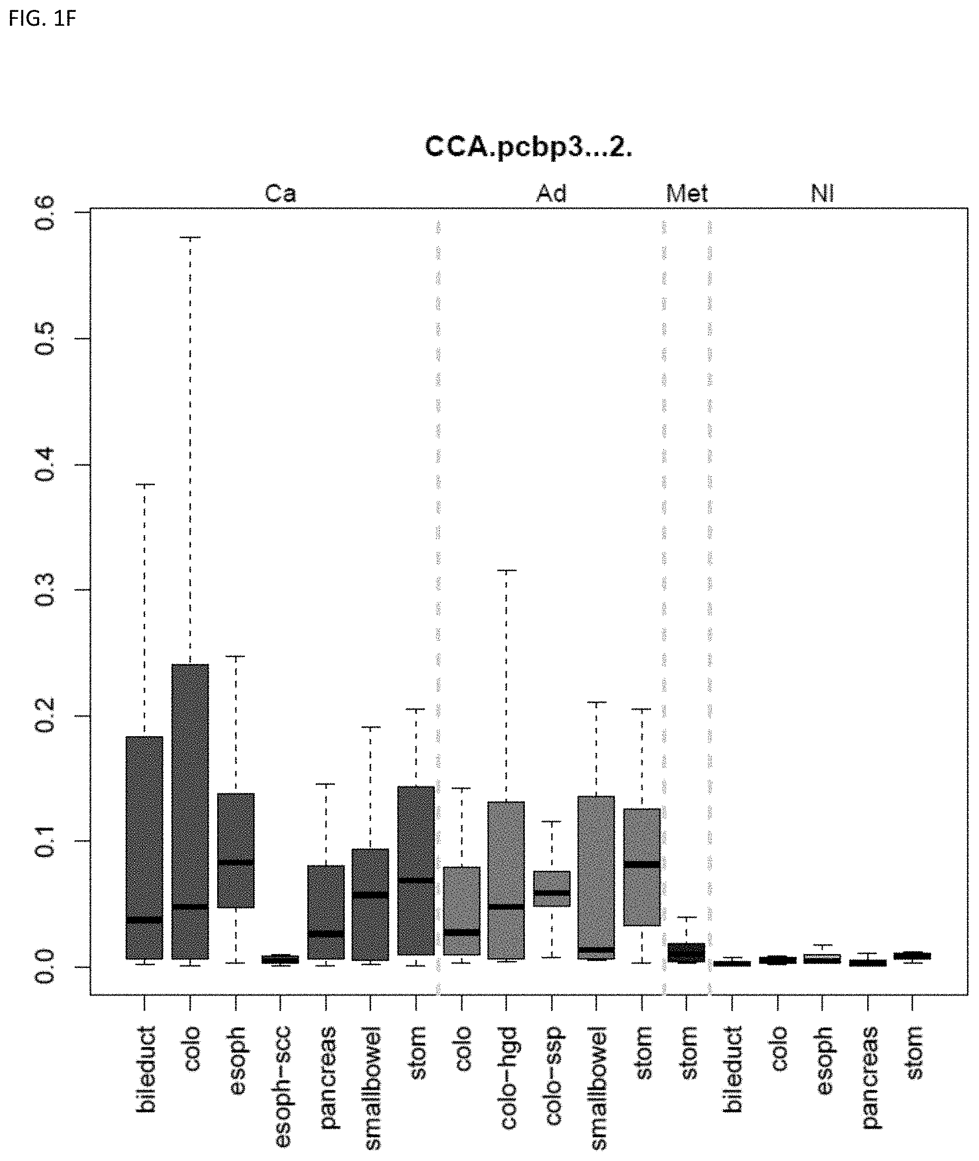
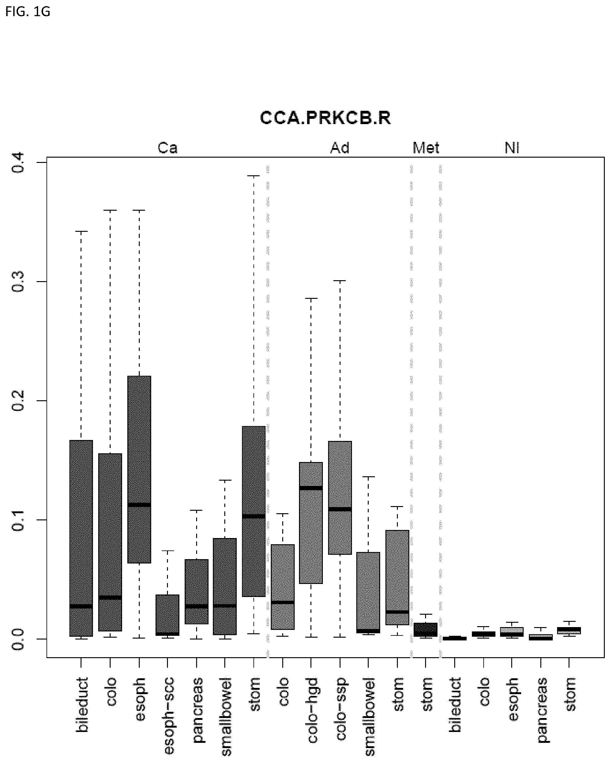
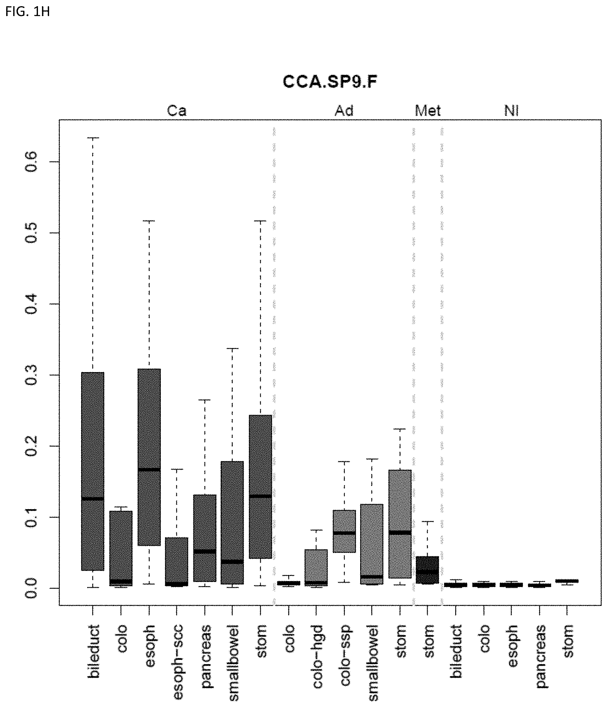
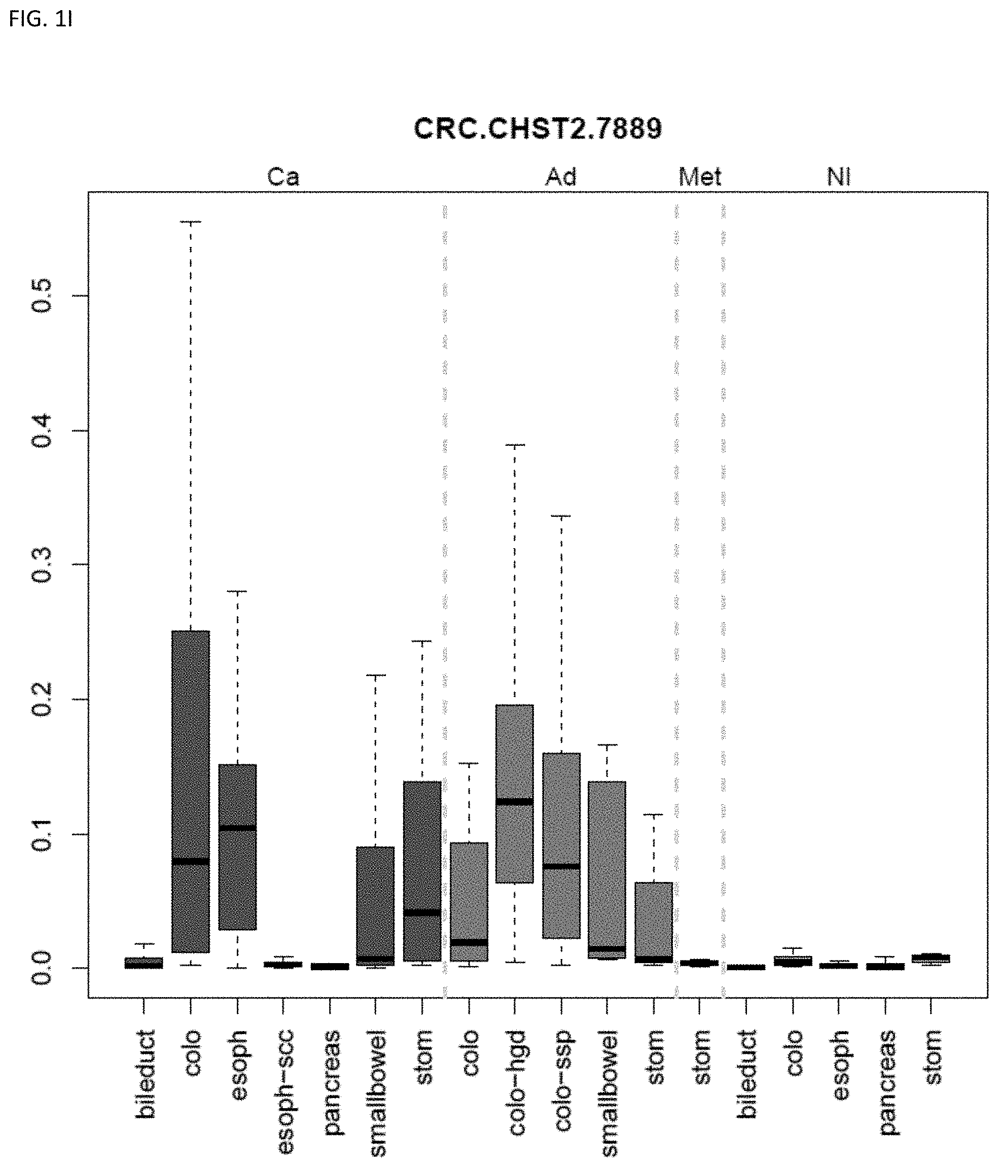
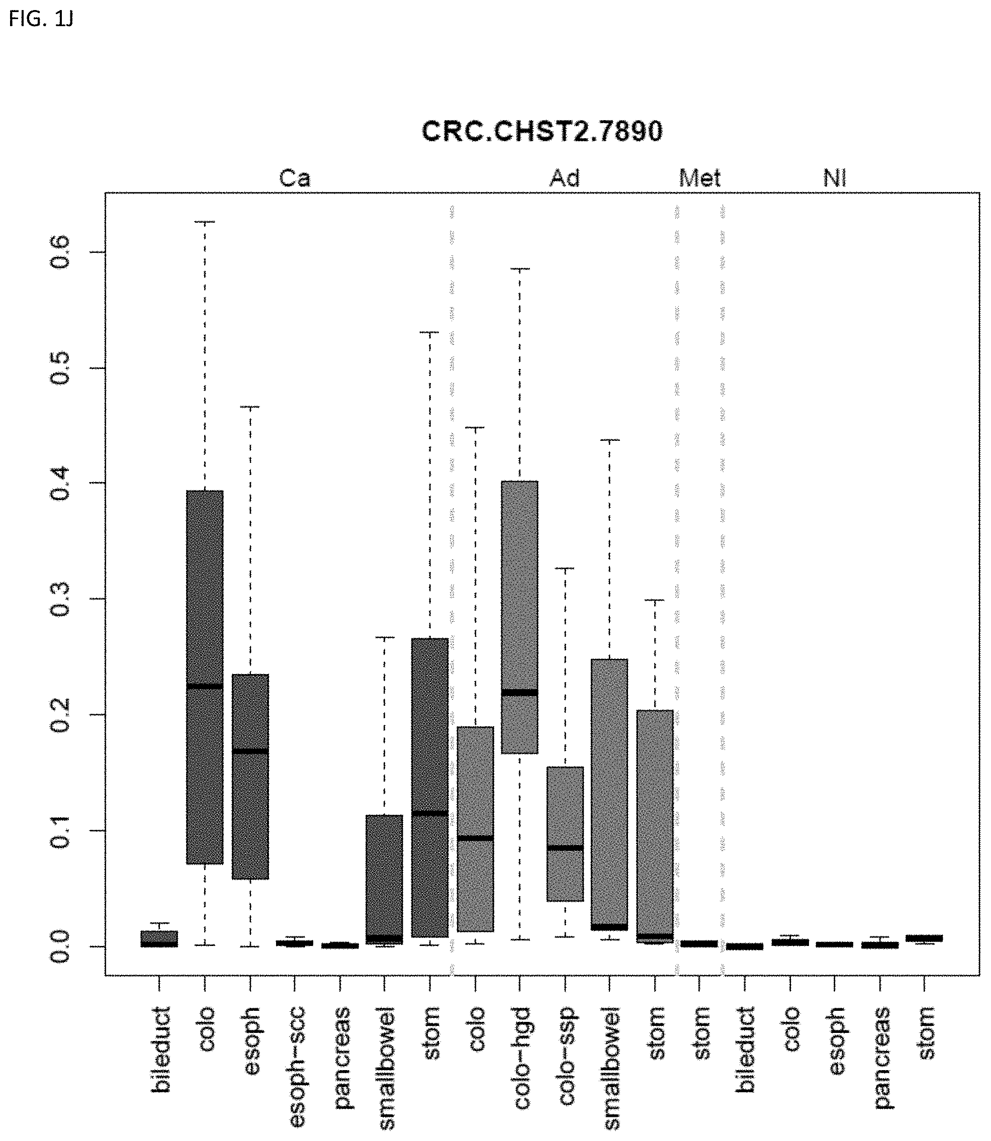
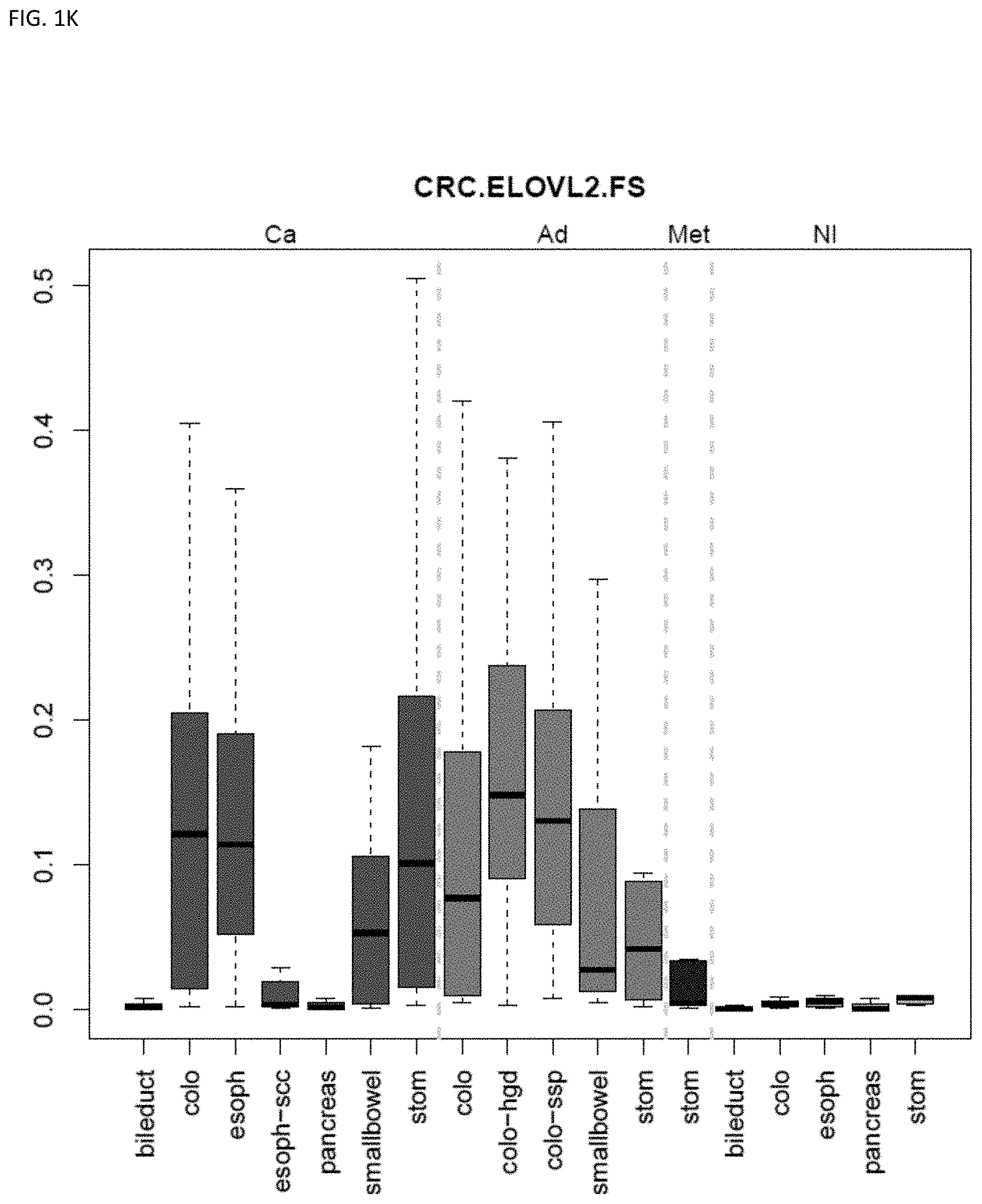
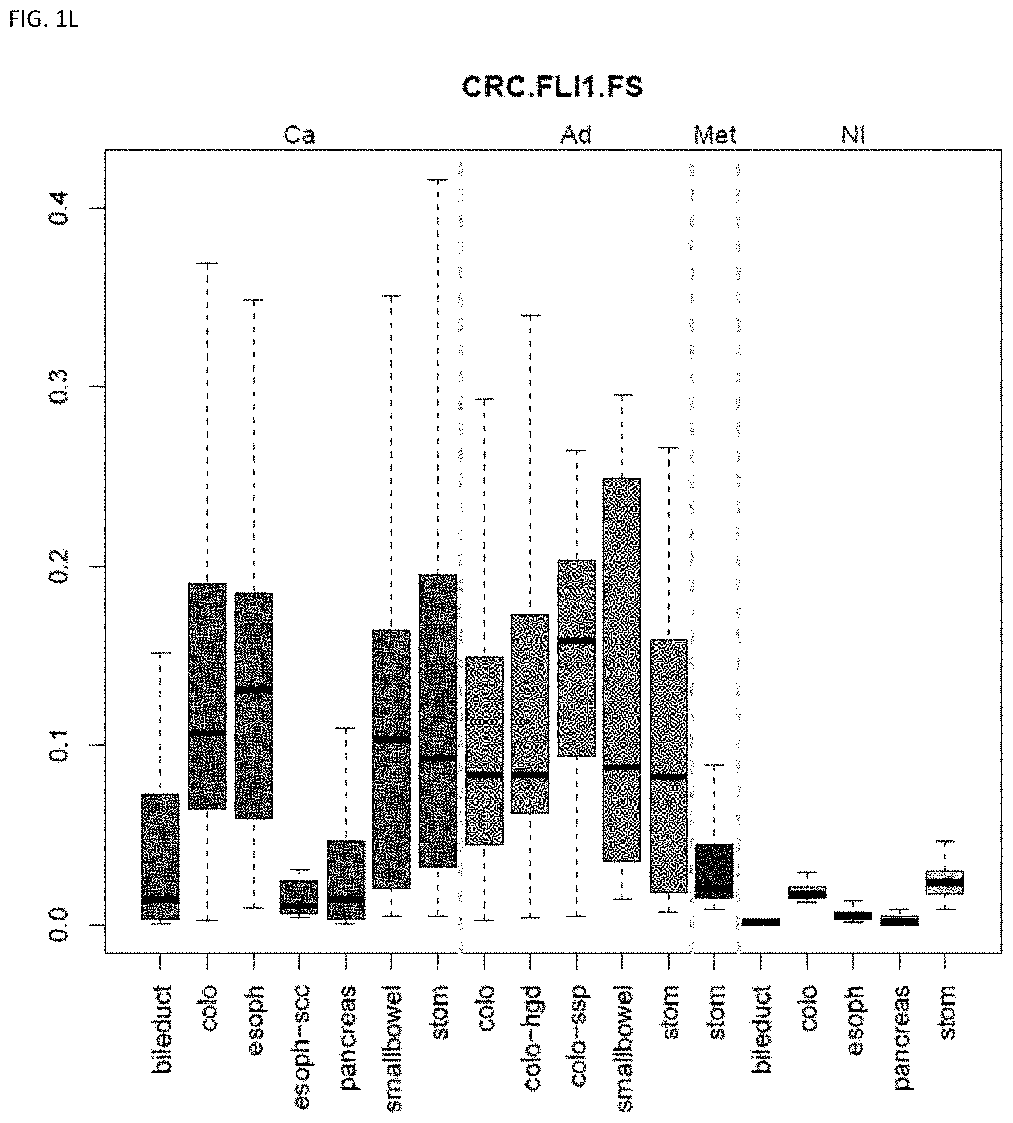
[0046] FIG. 1A-OOOO shows the distribution of individual markers within specific disease sites and neoplasm. The segregated region furthest to the left represents cancer tissue (Ca), the region second most left represents adenoma or precancer (Ad), the region third from the left represents metaplasia (Met), and region fourth from left represents normal tissue (NI).
[0047] FIG. 2A-G shows the top markers which resulted from the MSP biological validation.
[0048] FIG. 3A and FIG. 3B shows BMP3 and QKI in combination, were able to predict the cancer site (83% accuracy) using plasma-derived DNA (see Example 1).
DETAILED DESCRIPTION
[0049] Provided herein is technology for gastrointestinal neoplasia screening and particularly, but not exclusively, to methods, compositions, and related uses for detecting the presence of gastrointestinal neoplasm, and classifying the site location of such a gastrointestinal neoplasm (e.g., a colorectal region, a pancreaticobiliary region, a gastroesophageal region).
[0050] As the technology is described herein, the section headings used are for organizational purposes only and are not to be construed as limiting the subject matter in any way.
[0051] In this detailed description of the various embodiments, for purposes of explanation, numerous specific details are set forth to provide a thorough understanding of the embodiments disclosed. One skilled in the art will appreciate, however, that these various embodiments may be practiced with or without these specific details. In other instances, structures and devices are shown in block diagram form. Furthermore, one skilled in the art can readily appreciate that the specific sequences in which methods are presented and performed are illustrative and it is contemplated that the sequences can be varied and still remain within the spirit and scope of the various embodiments disclosed herein.
[0052] All literature and similar materials cited in this application, including but not limited to, patents, patent applications, articles, books, treatises, and internet web pages are expressly incorporated by reference in their entirety for any purpose. Unless defined otherwise, all technical and scientific terms used herein have the same meaning as is commonly understood by one of ordinary skill in the art to which the various embodiments described herein belongs. When definitions of terms in incorporated references appear to differ from the definitions provided in the present teachings, the definition provided in the present teachings shall control.
Definitions
[0053] To facilitate an understanding of the present technology, a number of terms and phrases are defined below. Additional definitions are set forth throughout the detailed description.
[0054] Throughout the specification and claims, the following terms take the meanings explicitly associated herein, unless the context clearly dictates otherwise. The phrase "in one embodiment" as used herein does not necessarily refer to the same embodiment, though it may. Furthermore, the phrase "in another embodiment" as used herein does not necessarily refer to a different embodiment, although it may. Thus, as described below, various embodiments of the invention may be readily combined, without departing from the scope or spirit of the invention.
[0055] In addition, as used herein, the term "or" is an inclusive "or" operator and is equivalent to the term "and/or" unless the context clearly dictates otherwise. The term "based on" is not exclusive and allows for being based on additional factors not described, unless the context clearly dictates otherwise. In addition, throughout the specification, the meaning of "a", "an", and "the" include plural references. The meaning of "in" includes "in" and "on."
[0056] As used herein, a "nucleic acid" or "nucleic acid molecule" generally refers to any ribonucleic acid or deoxyribonucleic acid, which may be unmodified or modified DNA or RNA. "Nucleic acids" include, without limitation, single- and double-stranded nucleic acids.
[0057] As used herein, the term "nucleic acid" also includes DNA as described above that contains one or more modified bases. Thus, DNA with a backbone modified for stability or for other reasons is a "nucleic acid". The term "nucleic acid" as it is used herein embraces such chemically, enzymatically, or metabolically modified forms of nucleic acids, as well as the chemical forms of DNA characteristic of viruses and cells, including for example, simple and complex cells.
[0058] The terms "oligonucleotide" or "polynucleotide" or "nucleotide" or "nucleic acid" refer to a molecule having two or more deoxyribonucleotides or ribonucleotides, preferably more than three, and usually more than ten. The exact size will depend on many factors, which in turn depends on the ultimate function or use of the oligonucleotide. The oligonucleotide may be generated in any manner, including chemical synthesis, DNA replication, reverse transcription, or a combination thereof. Typical deoxyribonucleotides for DNA are thymine, adenine, cytosine, and guanine. Typical ribonucleotides for RNA are uracil, adenine, cytosine, and guanine.
[0059] As used herein, the terms "locus" or "region" of a nucleic acid refer to a subregion of a nucleic acid, e.g., a gene on a chromosome, a single nucleotide, a CpG island, etc.
[0060] The terms "complementary" and "complementarity" refer to nucleotides (e.g., 1 nucleotide) or polynucleotides (e.g., a sequence of nucleotides) related by the base-pairing rules. For example, the sequence 5'-A-G-T-3' is complementary to the sequence 3'-T-C-A-S'. Complementarity may be "partial," in which only some of the nucleic acids' bases are matched according to the base pairing rules. Or, there may be "complete" or "total" complementarity between the nucleic acids. The degree of complementarity between nucleic acid strands effects the efficiency and strength of hybridization between nucleic acid strands. This is of particular importance in amplification reactions and in detection methods that depend upon binding between nucleic acids.
[0061] The term "gene" refers to a nucleic acid (e.g., DNA or RNA) sequence that comprises coding sequences necessary for the production of an RNA, or of a polypeptide or its precursor. A functional polypeptide can be encoded by a full length coding sequence or by any portion of the coding sequence as long as the desired activity or functional properties (e.g., enzymatic activity, ligand binding, signal transduction, etc.) of the polypeptide are retained. The term "portion" when used in reference to a gene refers to fragments of that gene. The fragments may range in size from a few nucleotides to the entire gene sequence minus one nucleotide. Thus, "a nucleotide comprising at least a portion of a gene" may comprise fragments of the gene or the entire gene.
[0062] The term "gene" also encompasses the coding regions of a structural gene and includes sequences located adjacent to the coding region on both the 5' and 3' ends, e.g., for a distance of about 1 kb on either end, such that the gene corresponds to the length of the full-length mRNA (e.g., comprising coding, regulatory, structural and other sequences). The sequences that are located 5' of the coding region and that are present on the mRNA are referred to as 5' non-translated or untranslated sequences. The sequences that are located 3' or downstream of the coding region and that are present on the mRNA are referred to as 3' non-translated or 3' untranslated sequences. The term "gene" encompasses both cDNA and genomic forms of a gene. In some organisms (e.g., eukaryotes), a genomic form or clone of a gene contains the coding region interrupted with non-coding sequences termed "introns" or "intervening regions" or "intervening sequences." Introns are segments of a gene that are transcribed into nuclear RNA (hnRNA); introns may contain regulatory elements such as enhancers. Introns are removed or "spliced out" from the nuclear or primary transcript; introns therefore are absent in the messenger RNA (mRNA) transcript. The mRNA functions during translation to specify the sequence or order of amino acids in a nascent polypeptide.
[0063] In addition to containing introns, genomic forms of a gene may also include sequences located on both the 5' and 3' ends of the sequences that are present on the RNA transcript. These sequences are referred to as "flanking" sequences or regions (these flanking sequences are located 5' or 3' to the non-translated sequences present on the mRNA transcript). The 5' flanking region may contain regulatory sequences such as promoters and enhancers that control or influence the transcription of the gene. The 3' flanking region may contain sequences that direct the termination of transcription, posttranscriptional cleavage, and polyadenylation.
[0064] The term "wild-type" when made in reference to a gene refers to a gene that has the characteristics of a gene isolated from a naturally occurring source. The term "wild-type" when made in reference to a gene product refers to a gene product that has the characteristics of a gene product isolated from a naturally occurring source. The term "naturally-occurring" as applied to an object refers to the fact that an object can be found in nature. For example, a polypeptide or polynucleotide sequence that is present in an organism (including viruses) that can be isolated from a source in nature and which has not been intentionally modified by the hand of a person in the laboratory is naturally-occurring. A wild-type gene is often that gene or allele that is most frequently observed in a population and is thus arbitrarily designated the "normal" or "wild-type" form of the gene. In contrast, the term "modified" or "mutant" when made in reference to a gene or to a gene product refers, respectively, to a gene or to a gene product that displays modifications in sequence and/or functional properties (e.g., altered characteristics) when compared to the wild-type gene or gene product. It is noted that naturally-occurring mutants can be isolated; these are identified by the fact that they have altered characteristics when compared to the wild-type gene or gene product.
[0065] The term "allele" refers to a variation of a gene; the variations include but are not limited to variants and mutants, polymorphic loci, and single nucleotide polymorphic loci, frameshift, and splice mutations. An allele may occur naturally in a population or it might arise during the lifetime of any particular individual of the population.
[0066] Thus, the terms "variant" and "mutant" when used in reference to a nucleotide sequence refer to a nucleic acid sequence that differs by one or more nucleotides from another, usually related, nucleotide acid sequence. A "variation" is a difference between two different nucleotide sequences; typically, one sequence is a reference sequence.
[0067] "Amplification" is a special case of nucleic acid replication involving template specificity. It is to be contrasted with non-specific template replication (e.g., replication that is template-dependent but not dependent on a specific template). Template specificity is here distinguished from fidelity of replication (e.g., synthesis of the proper polynucleotide sequence) and nucleotide (ribo- or deoxyribo-) specificity. Template specificity is frequently described in terms of "target" specificity. Target sequences are "targets" in the sense that they are sought to be sorted out from other nucleic acid. Amplification techniques have been designed primarily for this sorting out.
[0068] Amplification of nucleic acids generally refers to the production of multiple copies of a polynucleotide, or a portion of the polynucleotide, typically starting from a small amount of the polynucleotide (e.g., a single polynucleotide molecule, 10 to 100 copies of a polynucleotide molecule, which may or may not be exactly the same), where the amplification products or amplicons are generally detectable. Amplification of polynucleotides encompasses a variety of chemical and enzymatic processes. The generation of multiple DNA copies from one or a few copies of a target or template DNA molecule during a polymerase chain reaction (PCR) or a ligase chain reaction (LCR; see, e.g., U.S. Pat. No. 5,494,810) are forms of amplification. Additional types of amplification include, but are not limited to, allele-specific PCR (see, e.g., U.S. Pat. No. 5,639,611), assembly PCR (see, e.g., U.S. Pat. No. 5,965,408), helicase-dependent amplification (see, e.g., U.S. Pat. No. 7,662,594), Hot-start PCR (see, e.g., U.S. Pat. Nos. 5,773,258 and 5,338,671), intersequence-specfic PCR, inverse PCR (see, e.g., Triglia, et alet al. (1988) Nucleic Acids Res., 16:8186), ligation-mediated PCR (see, e.g., Guilfoyle, R. et al., Nucleic Acids Research, 25:1854-1858 (1997); U.S. Pat. No. 5,508,169), methylation-specific PCR (see, e.g., Herman, et al., (1996) PNAS 93(13) 9821-9826), miniprimer PCR, multiplex ligation-dependent probe amplification (see, e.g., Schouten, et al., (2002) Nucleic Acids Research 30(12): e57), multiplex PCR (see, e.g., Chamberlain, et al., (1988) Nucleic Acids Research 16(23) 11141-11156; Ballabio, et al., (1990) Human Genetics 84(6) 571-573; Hayden, et al., (2008) BMC Genetics 9:80), nested PCR, overlap-extension PCR (see, e.g., Higuchi, et al., (1988) Nucleic Acids Research 16(15) 7351-7367), real time PCR (see, e.g., Higuchi, et alet al., (1992) Biotechnology 10:413-417; Higuchi, et al., (1993) Biotechnology 11:1026-1030), reverse transcription PCR (see, e.g., Bustin, S. A. (2000) J. Molecular Endocrinology 25:169-193), solid phase PCR, thermal asymmetric interlaced PCR, and Touchdown PCR (see, e.g., Don, et al., Nucleic Acids Research (1991) 19(14) 4008; Roux, K. (1994) Biotechniques 16(5) 812-814; Hecker, et al., (1996) Biotechniques 20(3) 478-485). Polynucleotide amplification also can be accomplished using digital PCR (see, e.g., Kalinina, et al., Nucleic Acids Research. 25; 1999-2004, (1997); Vogelstein and Kinzler, Proc Natl Acad Sci USA. 96; 9236-41, (1999); International Patent Publication No. WO05023091A2; US Patent Application Publication No. 20070202525).
[0069] The term "polymerase chain reaction" ("PCR") refers to the method of K. B. Mullis U.S. Pat. Nos. 4,683,195, 4,683,202, and 4,965,188, that describe a method for increasing the concentration of a segment of a target sequence in a mixture of genomic DNA without cloning or purification. This process for amplifying the target sequence consists of introducing a large excess of two oligonucleotide primers to the DNA mixture containing the desired target sequence, followed by a precise sequence of thermal cycling in the presence of a DNA polymerase. The two primers are complementary to their respective strands of the double stranded target sequence. To effect amplification, the mixture is denatured and the primers then annealed to their complementary sequences within the target molecule. Following annealing, the primers are extended with a polymerase so as to form a new pair of complementary strands. The steps of denaturation, primer annealing, and polymerase extension can be repeated many times (i.e., denaturation, annealing and extension constitute one "cycle"; there can be numerous "cycles") to obtain a high concentration of an amplified segment of the desired target sequence. The length of the amplified segment of the desired target sequence is determined by the relative positions of the primers with respect to each other, and therefore, this length is a controllable parameter. By virtue of the repeating aspect of the process, the method is referred to as the "polymerase chain reaction" ("PCR"). Because the desired amplified segments of the target sequence become the predominant sequences (in terms of concentration) in the mixture, they are said to be "PCR amplified" and are "PCR products" or "amplicons."
[0070] Template specificity is achieved in most amplification techniques by the choice of enzyme. Amplification enzymes are enzymes that, under conditions they are used, will process only specific sequences of nucleic acid in a heterogeneous mixture of nucleic acid. For example, in the case of Q-beta replicase, MDV-1 RNA is the specific template for the replicase (Kacian et al., Proc. Natl. Acad. Sci. USA, 69:3038 [1972]). Other nucleic acid will not be replicated by this amplification enzyme. Similarly, in the case of T7 RNA polymerase, this amplification enzyme has a stringent specificity for its own promoters (Chamberlin et al, Nature, 228:227 [1970]). In the case of T4 DNA ligase, the enzyme will not ligate the two oligonucleotides or polynucleotides, where there is a mismatch between the oligonucleotide or polynucleotide substrate and the template at the ligation junction (Wu and Wallace (1989) Genomics 4:560). Finally, thermostable template-dependant DNA polymerases (e.g., Taq and Pfu DNA polymerases), by virtue of their ability to function at high temperature, are found to display high specificity for the sequences bounded and thus defined by the primers; the high temperature results in thermodynamic conditions that favor primer hybridization with the target sequences and not hybridization with non-target sequences (H. A. Erlich (ed.), PCR Technology, Stockton Press [1989]).
[0071] As used herein, the term "nucleic acid detection assay" refers to any method of determining the nucleotide composition of a nucleic acid of interest. Nucleic acid detection assay include but are not limited to, DNA sequencing methods, probe hybridization methods, structure specific cleavage assays (e.g., the INVADER assay, Hologic, Inc.) and are described, e.g., in U.S. Pat. Nos. 5,846,717, 5,985,557, 5,994,069, 6,001,567, 6,090,543, and 6,872,816; Lyamichev et al., Nat. Biotech., 17:292 (1999), Hall et al., PNAS, USA, 97:8272 (2000), and US 2009/0253142); enzyme mismatch cleavage methods (e.g., Variagenics, U.S. Pat. Nos. 6,110,684, 5,958,692, 5,851,770); polymerase chain reaction; branched hybridization methods (e.g., Chiron, U.S. Pat. Nos. 5,849,481, 5,710,264, 5,124,246, and 5,624,802); rolling circle replication (e.g., U.S. Pat. Nos. 6,210,884, 6,183,960 and 6,235,502); NASBA (e.g., U.S. Pat. No. 5,409,818); molecular beacon technology (e.g., U.S. Pat. No. 6,150,097); E-sensor technology (Motorola, U.S. Pat. Nos. 6,248,229, 6,221,583, 6,013,170, and 6,063,573); cycling probe technology (e.g., U.S. Pat. Nos. 5,403,711, 5,011,769, and 5,660,988); Dade Behring signal amplification methods (e.g., U.S. Pat. Nos. 6,121,001, 6,110,677, 5,914,230, 5,882,867, and 5,792,614); ligase chain reaction (e.g., Barnay Proc. Natl. Acad. Sci USA 88, 189-93 (1991)); and sandwich hybridization methods (e.g., U.S. Pat. No. 5,288,609).
[0072] The term "amplifiable nucleic acid" refers to a nucleic acid that may be amplified by any amplification method. It is contemplated that "amplifiable nucleic acid" will usually comprise "sample template."
[0073] The term "sample template" refers to nucleic acid originating from a sample that is analyzed for the presence of "target" (defined below). In contrast, "background template" is used in reference to nucleic acid other than sample template that may or may not be present in a sample. Background template is most often inadvertent. It may be the result of carryover or it may be due to the presence of nucleic acid contaminants sought to be purified away from the sample. For example, nucleic acids from organisms other than those to be detected may be present as background in a test sample.
[0074] The term "primer" refers to an oligonucleotide, whether occurring naturally as in a purified restriction digest or produced synthetically, that is capable of acting as a point of initiation of synthesis when placed under conditions in which synthesis of a primer extension product that is complementary to a nucleic acid strand is induced, (e.g., in the presence of nucleotides and an inducing agent such as a DNA polymerase and at a suitable temperature and pH). The primer is preferably single stranded for maximum efficiency in amplification, but may alternatively be double stranded. If double stranded, the primer is first treated to separate its strands before being used to prepare extension products. Preferably, the primer is an oligodeoxyribonucleotide. The primer must be sufficiently long to prime the synthesis of extension products in the presence of the inducing agent. The exact lengths of the primers will depend on many factors, including temperature, source of primer, and the use of the method.
[0075] The term "probe" refers to an oligonucleotide (e.g., a sequence of nucleotides), whether occurring naturally as in a purified restriction digest or produced synthetically, recombinantly, or by PCR amplification, that is capable of hybridizing to another oligonucleotide of interest. A probe may be single-stranded or double-stranded. Probes are useful in the detection, identification, and isolation of particular gene sequences (e.g., a "capture probe"). It is contemplated that any probe used in the present invention may, in some embodiments, be labeled with any "reporter molecule," so that is detectable in any detection system, including, but not limited to enzyme (e.g., ELISA, as well as enzyme-based histochemical assays), fluorescent, radioactive, and luminescent systems. It is not intended that the present invention be limited to any particular detection system or label.
[0076] As used herein, "methylation" refers to cytosine methylation at positions C5 or N4 of cytosine, the N6 position of adenine, or other types of nucleic acid methylation. In vitro amplified DNA is usually unmethylated because typical in vitro DNA amplification methods do not retain the methylation pattern of the amplification template. However, "unmethylated DNA" or "methylated DNA" can also refer to amplified DNA whose original template was unmethylated or methylated, respectively.
[0077] Accordingly, as used herein a "methylated nucleotide" or a "methylated nucleotide base" refers to the presence of a methyl moiety on a nucleotide base, where the methyl moiety is not present in a recognized typical nucleotide base. For example, cytosine does not contain a methyl moiety on its pyrimidine ring, but 5-methylcytosine contains a methyl moiety at position 5 of its pyrimidine ring. Therefore, cytosine is not a methylated nucleotide and 5-methylcytosine is a methylated nucleotide. In another example, thymine contains a methyl moiety at position 5 of its pyrimidine ring; however, for purposes herein, thymine is not considered a methylated nucleotide when present in DNA since thymine is a typical nucleotide base of DNA.
[0078] As used herein, a "methylated nucleic acid molecule" refers to a nucleic acid molecule that contains one or more methylated nucleotides.
[0079] As used herein, a "methylation state", "methylation profile", and "methylation status" of a nucleic acid molecule refers to the presence of absence of one or more methylated nucleotide bases in the nucleic acid molecule. For example, a nucleic acid molecule containing a methylated cytosine is considered methylated (e.g., the methylation state of the nucleic acid molecule is methylated). A nucleic acid molecule that does not contain any methylated nucleotides is considered unmethylated.
[0080] The methylation state of a particular nucleic acid sequence (e.g., a gene marker or DNA region as described herein) can indicate the methylation state of every base in the sequence or can indicate the methylation state of a subset of the bases (e.g., of one or more cytosines) within the sequence, or can indicate information regarding regional methylation density within the sequence with or without providing precise information of the locations within the sequence the methylation occurs.
[0081] The methylation state of a nucleotide locus in a nucleic acid molecule refers to the presence or absence of a methylated nucleotide at a particular locus in the nucleic acid molecule. For example, the methylation state of a cytosine at the 7th nucleotide in a nucleic acid molecule is methylated when the nucleotide present at the 7th nucleotide in the nucleic acid molecule is 5-methylcytosine. Similarly, the methylation state of a cytosine at the 7th nucleotide in a nucleic acid molecule is unmethylated when the nucleotide present at the 7th nucleotide in the nucleic acid molecule is cytosine (and not 5-methylcytosine).
[0082] The methylation status can optionally be represented or indicated by a "methylation value" (e.g., representing a methylation frequency, fraction, ratio, percent, etc.) A methylation value can be generated, for example, by quantifying the amount of intact nucleic acid present following restriction digestion with a methylation dependent restriction enzyme or by comparing amplification profiles after bisulfate reaction or by comparing sequences of bisulfate-treated and untreated nucleic acids. Accordingly, a value, e.g., a methylation value, represents the methylation status and can thus be used as a quantitative indicator of methylation status across multiple copies of a locus. This is of particular use when it is desirable to compare the methylation status of a sequence in a sample to a threshold or reference value.
[0083] As used herein, "methylation frequency" or "methylation percent (%)" refer to the number of instances in which a molecule or locus is methylated relative to the number of instances the molecule or locus is unmethylated.
[0084] As such, the methylation state describes the state of methylation of a nucleic acid (e.g., a genomic sequence). In addition, the methylation state refers to the characteristics of a nucleic acid segment at a particular genomic locus relevant to methylation. Such characteristics include, but are not limited to, whether any of the cytosine (C) residues within this DNA sequence are methylated, the location of methylated C residue(s), the frequency or percentage of methylated C throughout any particular region of a nucleic acid, and allelic differences in methylation due to, e.g., difference in the origin of the alleles. The terms "methylation state", "methylation profile", and "methylation status" also refer to the relative concentration, absolute concentration, or pattern of methylated C or unmethylated C throughout any particular region of a nucleic acid in a biological sample. For example, if the cytosine (C) residue(s) within a nucleic acid sequence are methylated it may be referred to as "hypermethylated" or having "increased methylation", whereas if the cytosine (C) residue(s) within a DNA sequence are not methylated it may be referred to as "hypomethylated" or having "decreased methylation". Likewise, if the cytosine (C) residue(s) within a nucleic acid sequence are methylated as compared to another nucleic acid sequence (e.g., from a different region or from a different individual, etc.) that sequence is considered hypermethylated or having increased methylation compared to the other nucleic acid sequence. Alternatively, if the cytosine (C) residue(s) within a DNA sequence are not methylated as compared to another nucleic acid sequence (e.g., from a different region or from a different individual, etc.) that sequence is considered hypomethylated or having decreased methylation compared to the other nucleic acid sequence. Additionally, the term "methylation pattern" as used herein refers to the collective sites of methylated and unmethylated nucleotides over a region of a nucleic acid. Two nucleic acids may have the same or similar methylation frequency or methylation percent but have different methylation patterns when the number of methylated and unmethylated nucleotides are the same or similar throughout the region but the locations of methylated and unmethylated nucleotides are different. Sequences are said to be "differentially methylated" or as having a "difference in methylation" or having a "different methylation state" when they differ in the extent (e.g., one has increased or decreased methylation relative to the other), frequency, or pattern of methylation. The term "differential methylation" refers to a difference in the level or pattern of nucleic acid methylation in a cancer positive sample as compared with the level or pattern of nucleic acid methylation in a cancer negative sample. It may also refer to the difference in levels or patterns between patients that have recurrence of cancer after surgery versus patients who not have recurrence. Differential methylation and specific levels or patterns of DNA methylation are prognostic and predictive biomarkers, e.g., once the correct cut-off or predictive characteristics have been defined.
[0085] Methylation state frequency can be used to describe a population of individuals or a sample from a single individual. For example, a nucleotide locus having a methylation state frequency of 50% is methylated in 50% of instances and unmethylated in 50% of instances. Such a frequency can be used, for example, to describe the degree to which a nucleotide locus or nucleic acid region is methylated in a population of individuals or a collection of nucleic acids. Thus, when methylation in a first population or pool of nucleic acid molecules is different from methylation in a second population or pool of nucleic acid molecules, the methylation state frequency of the first population or pool will be different from the methylation state frequency of the second population or pool. Such a frequency also can be used, for example, to describe the degree to which a nucleotide locus or nucleic acid region is methylated in a single individual. For example, such a frequency can be used to describe the degree to which a group of cells from a tissue sample are methylated or unmethylated at a nucleotide locus or nucleic acid region.
[0086] As used herein a "nucleotide locus" refers to the location of a nucleotide in a nucleic acid molecule. A nucleotide locus of a methylated nucleotide refers to the location of a methylated nucleotide in a nucleic acid molecule.
[0087] Typically, methylation of human DNA occurs on a dinucleotide sequence including an adjacent guanine and cytosine where the cytosine is located 5' of the guanine (also termed CpG dinucleotide sequences). Most cytosines within the CpG dinucleotides are methylated in the human genome, however some remain unmethylated in specific CpG dinucleotide rich genomic regions, known as CpG islands (see, e.g, Antequera et al. (1990) Cell 62: 503-514).
[0088] As used herein, a "CpG island" refers to a G:C-rich region of genomic DNA containing an increased number of CpG dinucleotides relative to total genomic DNA. A CpG island can be at least 100, 200, or more base pairs in length, where the G:C content of the region is at least 50% and the ratio of observed CpG frequency over expected frequency is 0.6; in some instances, a CpG island can be at least 500 base pairs in length, where the G:C content of the region is at least 55%) and the ratio of observed CpG frequency over expected frequency is 0.65. The observed CpG frequency over expected frequency can be calculated according to the method provided in Gardiner-Garden et al (1987) J. Mol. Biol. 196: 261-281. For example, the observed CpG frequency over expected frequency can be calculated according to the formula R=(A.times.B)/(C.times.D), where R is the ratio of observed CpG frequency over expected frequency, A is the number of CpG dinucleotides in an analyzed sequence, B is the total number of nucleotides in the analyzed sequence, C is the total number of C nucleotides in the analyzed sequence, and D is the total number of G nucleotides in the analyzed sequence. Methylation state is typically determined in CpG islands, e.g., at promoter regions. It will be appreciated though that other sequences in the human genome are prone to DNA methylation such as CpA and CpT (see, e.g., Ramsahoye (2000) Proc. Natl. Acad. Sci. USA 97: 5237-5242; Salmon and Kaye (1970) Biochim. Biophys. Acta. 204: 340-351; Grafstrom (1985) Nucleic Acids Res. 13: 2827-2842; Nyce (1986) Nucleic Acids Res. 14: 4353-4367; Woodcock (1987) Biochem. Biophys. Res. Commun. 145: 888-894).
[0089] As used herein, a reagent that modifies a nucleotide of the nucleic acid molecule as a function of the methylation state of the nucleic acid molecule, or a methylation-specific reagent, refers to a compound or composition or other agent that can change the nucleotide sequence of a nucleic acid molecule in a manner that reflects the methylation state of the nucleic acid molecule. Methods of treating a nucleic acid molecule with such a reagent can include contacting the nucleic acid molecule with the reagent, coupled with additional steps, if desired, to accomplish the desired change of nucleotide sequence. Such a change in the nucleic acid molecule's nucleotide sequence can result in a nucleic acid molecule in which each methylated nucleotide is modified to a different nucleotide. Such a change in the nucleic acid nucleotide sequence can result in a nucleic acid molecule in which each unmethylated nucleotide is modified to a different nucleotide. Such a change in the nucleic acid nucleotide sequence can result in a nucleic acid molecule in which each of a selected nucleotide which is unmethylated (e.g., each unmethylated cytosine) is modified to a different nucleotide. Use of such a reagent to change the nucleic acid nucleotide sequence can result in a nucleic acid molecule in which each nucleotide that is a methylated nucleotide (e.g., each methylated cytosine) is modified to a different nucleotide. As used herein, use of a reagent that modifies a selected nucleotide refers to a reagent that modifies one nucleotide of the four typically occurring nucleotides in a nucleic acid molecule (C, G, T, and A for DNA and C, G, U, and A for RNA), such that the reagent modifies the one nucleotide without modifying the other three nucleotides. In one exemplary embodiment, such a reagent modifies an unmethylated selected nucleotide to produce a different nucleotide. In another exemplary embodiment, such a reagent can deaminate unmethylated cytosine nucleotides. An exemplary reagent is bisulfite.
[0090] As used herein, the term "bisulfite reagent" refers to a reagent comprising in some embodiments bisulfite, disulfite, hydrogen sulfite, or combinations thereof to distinguish between methylated and unmethylated cytidines, e.g., in CpG dinucleotide sequences.
[0091] The term "methylation assay" refers to any assay for determining the methylation state of one or more CpG dinucleotide sequences within a sequence of a nucleic acid.
[0092] The term "MS AP-PCR" (Methylation-Sensitive Arbitrarily-Primed Polymerase Chain Reaction) refers to the art-recognized technology that allows for a global scan of the genome using CG-rich primers to focus on the regions most likely to contain CpG dinucleotides, and described by Gonzalgo et al. (1997) Cancer Research 57: 594-599.
[0093] The term "METHYLIGHT.TM." refers to the art-recognized fluorescence-based real-time PCR technique described by Eads et al. (1999) Cancer Res. 59: 2302-2306.
[0094] The term "HEAVYMETHYL.TM." refers to an assay wherein methylation specific blocking probes (also referred to herein as blockers) covering CpG positions between, or covered by, the amplification primers enable methylation-specific selective amplification of a nucleic acid sample.
[0095] The term "HEAVYMETHYL.TM. METHYLIGHT.TM." assay refers to a HEAVYMETHYL.TM. METHYLIGHT.TM. assay, which is a variation of the METHYLIGHT.TM. assay, wherein the METHYLIGHT.TM. assay is combined with methylation specific blocking probes covering CpG positions between the amplification primers.
[0096] The term "MS-SNUPE" (Methylation-sensitive Single Nucleotide Primer Extension) refers to the art-recognized assay described by Gonzalgo & Jones (1997) Nucleic Acids Res. 25: 2529-2531.
[0097] The term "MSP" (Methylation-specific PCR) refers to the art-recognized methylation assay described by Herman et al. (1996) Proc. Natl. Acad. Sci. USA 93: 9821-9826, and by U.S. Pat. No. 5,786,146.
[0098] The term "COBRA" (Combined Bisulfite Restriction Analysis) refers to the art-recognized methylation assay described by Xiong & Laird (1997) Nucleic Acids Res. 25: 2532-2534.
[0099] The term "MCA" (Methylated CpG Island Amplification) refers to the methylation assay described by Toyota et al. (1999) Cancer Res. 59: 2307-12, and in WO 00/26401A1.
[0100] As used herein, a "selected nucleotide" refers to one nucleotide of the four typically occurring nucleotides in a nucleic acid molecule (C, G, T, and A for DNA and C, G, U, and A for RNA), and can include methylated derivatives of the typically occurring nucleotides (e.g., when C is the selected nucleotide, both methylated and unmethylated C are included within the meaning of a selected nucleotide), whereas a methylated selected nucleotide refers specifically to a methylated typically occurring nucleotide and an unmethylated selected nucleotides refers specifically to an unmethylated typically occurring nucleotide.
[0101] The terms "methylation-specific restriction enzyme" or "methylation-sensitive restriction enzyme" refers to an enzyme that selectively digests a nucleic acid dependent on the methylation state of its recognition site. In the case of a restriction enzyme that specifically cuts if the recognition site is not methylated or is hemimethylated, the cut will not take place or will take place with a significantly reduced efficiency if the recognition site is methylated. In the case of a restriction enzyme that specifically cuts if the recognition site is methylated, the cut will not take place or will take place with a significantly reduced efficiency if the recognition site is not methylated. Preferred are methylation-specific restriction enzymes, the recognition sequence of which contains a CG dinucleotide (for instance a recognition sequence such as CGCG or CCCGGG). Further preferred for some embodiments are restriction enzymes that do not cut if the cytosine in this dinucleotide is methylated at the carbon atom C5.
[0102] As used herein, a "different nucleotide" refers to a nucleotide that is chemically different from a selected nucleotide, typically such that the different nucleotide has Watson-Crick base-pairing properties that differ from the selected nucleotide, whereby the typically occurring nucleotide that is complementary to the selected nucleotide is not the same as the typically occurring nucleotide that is complementary to the different nucleotide. For example, when C is the selected nucleotide, U or T can be the different nucleotide, which is exemplified by the complementarity of C to G and the complementarity of U or T to A. As used herein, a nucleotide that is complementary to the selected nucleotide or that is complementary to the different nucleotide refers to a nucleotide that base-pairs, under high stringency conditions, with the selected nucleotide or different nucleotide with higher affinity than the complementary nucleotide's base-paring with three of the four typically occurring nucleotides. An example of complementarity is Watson-Crick base pairing in DNA (e.g., A-T and C-G) and RNA (e.g., A-U and C-G). Thus, for example, G base-pairs, under high stringency conditions, with higher affinity to C than G base-pairs to G, A, or T and, therefore, when C is the selected nucleotide, G is a nucleotide complementary to the selected nucleotide.
[0103] As used herein, the "sensitivity" of a given marker refers to the percentage of samples that report a DNA methylation value above a threshold value that distinguishes between neoplastic and non-neoplastic samples. In some embodiments, a positive is defined as a histology-confirmed neoplasia that reports a DNA methylation value above a threshold value (e.g., the range associated with disease), and a false negative is defined as a histology-confirmed neoplasia that reports a DNA methylation value below the threshold value (e.g., the range associated with no disease). The value of sensitivity, therefore, reflects the probability that a DNA methylation measurement for a given marker obtained from a known diseased sample will be in the range of disease-associated measurements. As defined here, the clinical relevance of the calculated sensitivity value represents an estimation of the probability that a given marker would detect the presence of a clinical condition when applied to a subject with that condition.
[0104] As used herein, the "specificity" of a given marker refers to the percentage of non-neoplastic samples that report a DNA methylation value below a threshold value that distinguishes between neoplastic and non-neoplastic samples. In some embodiments, a negative is defined as a histology-confirmed non-neoplastic sample that reports a DNA methylation value below the threshold value (e.g., the range associated with no disease) and a false positive is defined as a histology-confirmed non-neoplastic sample that reports a DNA methylation value above the threshold value (e.g., the range associated with disease). The value of specificity, therefore, reflects the probability that a DNA methylation measurement for a given marker obtained from a known non-neoplastic sample will be in the range of non-disease associated measurements. As defined here, the clinical relevance of the calculated specificity value represents an estimation of the probability that a given marker would detect the absence of a clinical condition when applied to a patient without that condition.
[0105] The term "AUC" as used herein is an abbreviation for the "area under a curve". In particular it refers to the area under a Receiver Operating Characteristic (ROC) curve. The ROC curve is a plot of the true positive rate against the false positive rate for the different possible cut points of a diagnostic test. It shows the trade-off between sensitivity and specificity depending on the selected cut point (any increase in sensitivity will be accompanied by a decrease in specificity). The area under an ROC curve (AUC) is a measure for the accuracy of a diagnostic test (the larger the area the better; the optimum is 1; a random test would have a ROC curve lying on the diagonal with an area of 0.5; for reference: J. P. Egan. (1975) Signal Detection Theory and ROC Analysis, Academic Press, New York).
[0106] As used herein, the term "neoplasm" refers to "an abnormal mass of tissue, the growth of which exceeds and is uncoordinated with that of the normal tissues" See, e.g., Willis R A, "The Spread of Tumors in the Human Body", London, Butterworth & Co, 1952.
[0107] As used herein, the term "adenoma" refers to a benign tumor of glandular origin. Although these growths are benign, over time they may progress to become malignant.
[0108] The term "pre-cancerous" or "pre-neoplastic" and equivalents thereof refer to any cellular proliferative disorder that is undergoing malignant transformation.
[0109] A "site" or "region" of a neoplasm, adenoma, cancer, etc. is the tissue, organ, cell type, anatomical area, body part, etc. in a subject's body where the neoplasm, adenoma, cancer, etc. is located.
[0110] As used herein, the term "upper gastrointestinal cancer" refers to types of cancer within the gastroesophageal and pancreaticobiliary regions.
[0111] As used herein, the term "lower gastrointestinal cancer" refers to types of cancer within the colorectal regions.
[0112] As used herein, the term "pancreaticobiliary" refers to tissues and organs including, but not limited to, the pancreas gland, pancreas duct, pancreatic ampulla, bile ducts, gallbladder, liver parenchyma, and duodenum. Examples of cancers associated with pancreaticobiliary tissues and organs include, but are not limited to, pancreatic ductal adenocarcinoma, pancreatic intra-epithelial neoplasia (PanIN) lesions, intraductal papillary mucinous neoplasms (and possibly mucinous cystadenoma), cholangiocarcinoma (bile duct cancer) (with or without underlying chronic bile duct disease, specifically primary sclerosing cholangitis), hepatocellular cancer (primary liver cancer) (with or without underlying chronic liver disease, specifically cirrhosis), ampullary cancer, duodenal small bowel cancer, and gallbladder cancer.
[0113] As used herein, the term "gastroesophageal" refers to tissues and organs including, but not limited to, the pharynx, larynx, glottis, epiglottis, upper esophageal sphincter, esophagus, gastroesophageal junction (with or without metaplasia), and stomach. Examples of cancers associated with gastroesophageal tissues and organs include, but are not limited to, Barrett's esophagus, Barrett's esophagus with dysplasia, adenocarcinoma of the esophagus, squamous cell cancer of the esophagus, head/neck squamous cell cancers, adenocarcinoma of the gastroesophageal junction, and adenocarcinoma of the stomach.
[0114] As used herein, the term "colorectal" refers to tissues and organs including, but not limited to, the entire colon (including ileocecal valve and appendix), rectum, anal canal, terminal ileum, ileum, and jejunum. Cancers associated with colorectal tissues and organs include, but are not limited to, adenocarcinoma of the colon, adenocarcinoma of the rectum, adenomatous polyps, and sessile serrated polyps
[0115] As used herein, a "diagnostic" test application includes the detection or identification of a disease state or condition of a subject, determining the likelihood that a subject will contract a given disease or condition, determining the likelihood that a subject with a disease or condition will respond to therapy, determining the prognosis of a subject with a disease or condition (or its likely progression or regression), and determining the effect of a treatment on a subject with a disease or condition. For example, a diagnostic can be used for detecting the presence or likelihood of a subject contracting a neoplasm or the likelihood that such a subject will respond favorably to a compound (e.g., a pharmaceutical, e.g., a drug) or other treatment.
[0116] The term "marker", as used herein, refers to a substance (e.g., a nucleic acid or a region of a nucleic acid) that is able to diagnose a cancer by distinguishing cancerous cells from normal cells, e.g., based its methylation state.
[0117] The term "isolated" when used in relation to a nucleic acid, as in "an isolated oligonucleotide" refers to a nucleic acid sequence that is identified and separated from at least one contaminant nucleic acid with which it is ordinarily associated in its natural source. Isolated nucleic acid is present in a form or setting that is different from that in which it is found in nature. In contrast, non-isolated nucleic acids, such as DNA and RNA, are found in the state they exist in nature. Examples of non-isolated nucleic acids include: a given DNA sequence (e.g., a gene) found on the host cell chromosome in proximity to neighboring genes; RNA sequences, such as a specific mRNA sequence encoding a specific protein, found in the cell as a mixture with numerous other mRNAs which encode a multitude of proteins. However, isolated nucleic acid encoding a particular protein includes, by way of example, such nucleic acid in cells ordinarily expressing the protein, where the nucleic acid is in a chromosomal location different from that of natural cells, or is otherwise flanked by a different nucleic acid sequence than that found in nature. The isolated nucleic acid or oligonucleotide may be present in single-stranded or double-stranded form. When an isolated nucleic acid or oligonucleotide is to be utilized to express a protein, the oligonucleotide will contain at a minimum the sense or coding strand (i.e., the oligonucleotide may be single-stranded), but may contain both the sense and anti-sense strands (i.e., the oligonucleotide may be double-stranded). An isolated nucleic acid may, after isolation from its natural or typical environment, by be combined with other nucleic acids or molecules. For example, an isolated nucleic acid may be present in a host cell in which into which it has been placed, e.g., for heterologous expression.
[0118] The term "purified" refers to molecules, either nucleic acid or amino acid sequences that are removed from their natural environment, isolated, or separated. An "isolated nucleic acid sequence" may therefore be a purified nucleic acid sequence. "Substantially purified" molecules are at least 60% free, preferably at least 75% free, and more preferably at least 90% free from other components with which they are naturally associated. As used herein, the terms "purified" or "to purify" also refer to the removal of contaminants from a sample. The removal of contaminating proteins results in an increase in the percent of polypeptide or nucleic acid of interest in the sample. In another example, recombinant polypeptides are expressed in plant, bacterial, yeast, or mammalian host cells and the polypeptides are purified by the removal of host cell proteins; the percent of recombinant polypeptides is thereby increased in the sample.
[0119] The term "composition comprising" a given polynucleotide sequence or polypeptide refers broadly to any composition containing the given polynucleotide sequence or polypeptide. The composition may comprise an aqueous solution containing salts (e.g., NaCl), detergents (e.g., SDS), and other components (e.g., Denhardt's solution, dry milk, salmon sperm DNA, etc.).
[0120] The term "sample" is used in its broadest sense. In one sense it can refer to an animal cell or tissue. In another sense, it is meant to include a specimen or culture obtained from any source, as well as biological and environmental samples. Biological samples may be obtained from plants or animals (including humans) and encompass fluids, solids, tissues, and gases. Environmental samples include environmental material such as surface matter, soil, water, and industrial samples. These examples are not to be construed as limiting the sample types applicable to the present invention.
[0121] As used herein, a "remote sample" as used in some contexts relates to a sample indirectly collected from a site that is not the cell, tissue, or organ source of the sample. For instance, when sample material originating from the pancreas is assessed in a stool sample (e.g., not from a sample taken directly from a pancreas), the sample is a remote sample.
[0122] As used herein, the terms "patient" or "subject" refer to organisms to be subject to various tests provided by the technology. The term "subject" includes animals, preferably mammals, including humans. In a preferred embodiment, the subject is a primate. In an even more preferred embodiment, the subject is a human.
[0123] As used herein, the term "kit" refers to any delivery system for delivering materials. In the context of reaction assays, such delivery systems include systems that allow for the storage, transport, or delivery of reaction reagents (e.g., oligonucleotides, enzymes, etc. in the appropriate containers) and/or supporting materials (e.g., buffers, written instructions for performing the assay etc.) from one location to another. For example, kits include one or more enclosures (e.g., boxes) containing the relevant reaction reagents and/or supporting materials. As used herein, the term "fragmented kit" refers to delivery systems comprising two or more separate containers that each contain a subportion of the total kit components.
[0124] The containers may be delivered to the intended recipient together or separately. For example, a first container may contain an enzyme for use in an assay, while a second container contains oligonucleotides. The term "fragmented kit" is intended to encompass kits containing Analyte specific reagents (ASR's) regulated under section 520(e) of the Federal Food, Drug, and Cosmetic Act, but are not limited thereto. Indeed, any delivery system comprising two or more separate containers that each contains a subportion of the total kit components are included in the term "fragmented kit." In contrast, a "combined kit" refers to a delivery system containing all of the components of a reaction assay in a single container (e.g., in a single box housing each of the desired components). The term "kit" includes both fragmented and combined kits.
Embodiments of the Technology
[0125] A biological phenomenon of importance to detection of both upper gastrointestinal cancer (UGC) and colorectal cancer (CRC) relates to luminal cell shedding (or exfoliation), as cancers and their pre-cursor lesions exfoliate cells and DNA into the local effluent (see, e.g., Yamaguchi K, et al., Pancreatology 2005; 5:416-21) which are ultimately excreted in stool. Exploiting this biology, the use of stool DNA testing for population screening of CRC has been implemented (see, e.g., Ahlquist D A, et al., Ann Intern Med 2008; 149:441-50, W81; Ahlquist D A, et al., Gastroenterology 2012; 142:248-256). Highly sensitive assays have been used to detect mutant DNA in matched stools of UGC patients whose excised tumor was known to contain the same sequences (see, e.g., Zou H, et al., Gastroenterology 2009; 136:A-625). While this approach demonstrated feasibility of stool testing as an approach to UGC detection, use of mutation markers is limited. The limitation with mutation markers relates to the unwieldy process of their detection; typically, each mutational site across multiple genes must be assayed separately to achieve sufficiently high sensitivity.
[0126] Epigenetic molecular changes offer an attractive class of candidate markers for the potential stool detection of UGC. Epigenetic methylation of DNA at cytosine-phosphate-guanine (CpG) island sites by DNA methyltransferases has been studied using DNA from tissues of most tumor types. Acquired methylation events in promotor regions of tumor suppressor genes are thought to silence expression, contributing oncogenesis. Furthermore, DNA methylation may be a more chemically and biologically stable diagnostic tool than RNA or protein expression (see, e.g., Laird P W, Nat Rev Genet 2010; 11:191-203). In other cancers, like sporadic colon cancer, it has been found that aberrant methylation markers are more broadly informative and sensitive than are individual DNA mutations and offer excellent specificity (see, e.g., Zou H, et al., Cancer Epidemiol Biomarkers Prev 2007; 16:2686-96).
[0127] Methylation markers have been studied at the tissue level for UGC and their precursor lesions (see, e.g., Omura N, et al., Cancer Biol Ther 2008; 7:1146-56; Sato N, et al., Mod Pathol 2008; 21:238-44; Hong S M, et al., Mod Pathol 2008; 21:1499-507; Hibi K, et al., Anticancer Res 2010; 30:4131-3; Tsunoda S, et al., Oncol Rep 2009; 21:1067-73; Uhm K O, et al., Oncol Res 2008; 17:151-7), but selection of methylation markers has been non-comprehensive in essentially all such studies. DNA from pancreas cancer (PanC) tumor cases have been compared to DNA from colonic epithelia using MSP for markers previously reported in PanC (MDFI, SFRP2, UCHL1, CNTNAP2 and TFPI2) as well as discriminant colonic neoplasm markers (BMP3, EYA4, Vimentin and NDRG4). In a multi-marker regression model, EYA4, UCHL1 and MDFI were highly discriminant, with an area under the receiver operating characteristics curve (AUC) of 0.85; while BMP3 alone achieved an AUC of 0.90. It was found subsequently that stool assay of the combination of mutant KRAS and methylated BMP3 was more sensitive for detection of pancreatic cancer than assay of either marker individually (see, e.g., Kisiel J B, et al., Cancer 2012; 118:2623-31). Methylation differences in UCHL1, EYA4 and MDFI at the level of the pancreas were obscured by background colonic methylation in the stool-based applications. Thus, indicates that a stool marker (or marker panel) for UGC is feasible only if it is broadly informative and exhibits virtually 100% specificity at the tissue level. Better methylation markers are needed to achieve the ideal of high sensitivity without yielding false-positives.
[0128] Several methods are available to search for novel methylation markers. While micro-array based interrogation of CpG methylation is a reasonable, high-throughput approach, this strategy is biased towards known regions of interest, mainly established tumor suppressor promotors. Alternative methods for genome-wide analysis of DNA methylation have been developed in the last decade (see, e.g., Laird P W, et al., Nat Rev Genet 2010; 11:191-203). There are three basic approaches. The first employs digestion of DNA by restriction enzymes which recognize specific methylated sites, followed by several possible analytic techniques which provide methylation data limited to the enzyme recognition site or the primers used to amplify the DNA in quantification steps (such as MSP). A second approach enriches methylated fractions of genomic DNA using anti-bodies directed to methyl-cytosine or other methylation-specific binding domains followed by microarray analysis or sequencing to map the fragment to a reference genome. This approach does not provide single nucleotide resolution of all methylated sites within the fragment. A third approach begins with bisulfate treatment of the DNA to convert all unmethylated cytosines to uracil, followed by restriction enzyme digestion and complete sequencing of all fragments after coupling to an adapter ligand. The choice of restriction enzymes can enrich the fragments for CpG dense regions, reducing the number of redundant sequences which may map to multiple gene positions during analysis. This latter approach is termed Reduced Representation Bisulfite Sequencing (RRBS) (see, e.g., Gu H, et al., Nat Methods 2010; 7:133-6).
[0129] Single nucleotide resolution analysis of CpG islands has yielded important findings when applied to animal models and human cell lines. Zhang and colleagues found that amplicons from different parts of the same CpG island may have different levels of methylation (see, e.g., Zhang Y, et al., PLoS Genet 2009; 5:e1000438). Further, methylation levels were distributed bi-modally, between highly methylated and unmethylated sequences, further supporting the switch-like pattern to DNA methyltransferase activity (see, e.g., Zhang Y, et al., PLoS Genet 2009; 5:e1000438). When looking at in vivo murine tissues as well as cell lines, Meissner and colleagues found that only about 0.3% of high CpG density promotors, or HCPs (defined as >7% of CpGs within a 300 base pair region) were methylated, whereas areas of low CpG density (LCP, <5%) tended to be frequently methylated in a dynamic tissue-specific pattern (see, e.g., Meissner A, et al., Nature 2008; 454:766-70). HCPs include promotors for ubiquitous housekeeping genes and highly regulated developmental genes; among the HCP sites methylated at >50% were several established candidates, including Wnt 2, NDRG2, SFRP2 and BMP 3 (see, e.g., Meissner A, et al., Nature 2008; 454:766-70).
[0130] Provided herein is technology for gastrointestinal neoplasia screening and particularly, but not exclusively, to methods, compositions, and related uses for detecting the presence of gastrointestinal neoplasm, and classifying the site location of such a gastrointestinal neoplasm (e.g., a colorectal region, a pancreaticobiliary region, a gastroesophageal region).
[0131] Indeed, experiments conducted during the course of developing this technology compared the methylation state of DNA markers from esophageal tissue, stomach tissue, pancreatic tissue, bile duct/liver tissue, and colorectal tissue of subjects having neoplasm related to the respective tissue type to the methylation state of the same DNA markers from control subjects (e.g., normal tissue for the respective tissue type) (see, Example 1).
[0132] Markers and/or panels of markers were identified (e.g., a chromosomal region having an annotation selected from chr12.133484978-133485739, BMP3, and chr11.123301058-123301255) capable of classifying gastrointestinal (GI) neoplasm versus control (e.g., normal tissue for the respective tissue type) across GI tissue types (e.g., esophageal tissue, stomach tissue, pancreatic tissue, bile duct/liver tissue, and colorectal tissue) (see, Example 1).
[0133] Markers and/or panels of markers were identified (e.g., a chromosomal region having an annotation selected from chr7.25896389-25896501, and QKI) capable of classifying lower gastrointestinal (LGI) neoplasm versus upper gastrointestinal neoplasm (UGI) across GI tissue types (e.g., esophageal tissue, stomach tissue, pancreatic tissue, bile duct/liver tissue, and colorectal tissue) (see, Example 1).
[0134] Markers and/or panels of markers were identified (e.g., a chromosomal region having an annotation selected from PDGFD, ELOVL2, PCBP3) capable of classifying pancreaticobiliary neoplasm versus gastroesophageal neoplasm across GI tissue types (e.g., esophageal tissue, stomach tissue, pancreatic tissue, bile duct/liver tissue, and colorectal tissue) (see, Example 1).
[0135] Markers and/or panels of markers were identified (e.g., a chromosomal region having an annotation selected from QKI, PDGFD, ELOVL2, chr12.133484978-133485739, chr7.25896389-25896501, PCBP3, chr11.123301058-123301255, and BMP3) capable of predicting control versus neoplasm location across GI tissue types (e.g., esophageal tissue, stomach tissue, pancreatic tissue, bile duct/liver tissue, and colorectal tissue) (see, Example 1).
[0136] Markers and/or panels of markers were identified (e.g., a chromosomal region having an annotation selected from BMP3 and QKI) capable of predicting control versus neoplasm location in blood plasma samples (see, Example 1).
[0137] Markers and/or panels of markers were identified (e.g., a chromosomal region having an annotation selected from BMP3 and QKI) capable of predicting colorectal cancer versus pancreaticobiliary neoplasm in blood plasma samples (see, Example 1).
[0138] Although the disclosure herein refers to certain illustrated embodiments, it is to be understood that these embodiments are presented by way of example and not by way of limitation.
[0139] In particular aspects, the present technology provides methods for locating the neoplasm site within the subject, wherein the methylation state of the marker indicates the neoplasm site within the subject. For example, in some embodiments, the methylation state of markers (e.g., a chromosomal region having an annotation selected from chr7.25896389-25896501, and QKI) are used to classify lower gastrointestinal (LGI) neoplasm (e.g., adenoma, colorectal cancer) versus upper gastrointestinal cancer (UGI) (e.g., cancer of the esophagus, pancreas, stomach). In some embodiments, the methylation state of markers (e.g., a chromosomal region having an annotation selected from PDGFD, ELOVL2, PCBP3) are used to classify pancreaticobiliary neoplasm versus gastroesophageal neoplasm. In some embodiments, the methylation state of markers (e.g., a chromosomal region having an annotation selected from BMP3 and QKI) are used to classify colorectal cancer versus pancreaticobiliary neoplasm. The technology also encompasses determining the state or stage of a cancer, e.g., in some embodiments the neoplasm is pre-cancerous. Some embodiments provide methods comprising assaying a plurality of markers, e.g., comprising assaying 2 to 8 markers.
[0140] The methods comprise determining the methylation status of at least one methylation marker in a biological sample isolated from a subject, wherein a change in the methylation state of the marker is indicative of the presence, class, or site of a cancer. Particular embodiments relate to markers comprising a differentially methylated region (DMR, e.g., DMR 1-8, see Table 1) that are used for diagnosis (e.g., screening) of neoplastic cellular proliferative disorders (e.g., cancer), including early detection during the pre-cancerous stages of disease and prediction of a neoplasm site (e.g., by discriminating among cancer types, e.g., upper gastrointestinal cancers and lower gastrointestinal cancers).
[0141] The markers of the present technology are particularly efficient in detecting or distinguishing between gastrointestinal proliferative disorders, thereby providing improved means for the early detection, classification, and treatment of said disorders.
[0142] In addition to embodiments wherein the methylation analysis of at least one marker, a region of a marker, or a base of a marker comprising a DMR (e.g., DMR 1-8 from Table 1) provided herein and listed in Table 1 is analyzed, the technology also provides panels of markers comprising at least one marker, region of a marker, or base of a marker comprising a DMR with utility for the detection of cancers, in neoplasm associated with an UGI region, a LGI region, a pacreaticobiliary region, a gastroesophageal region, and/or a colorectal region.
[0143] Some embodiments of the technology are based upon the analysis of the CpG methylation status of at least one marker, region of a marker, or base of a marker comprising a DMR.
[0144] In some embodiments, the present technology provides for the use of the bisulfite technique in combination with one or more methylation assays to determine the methylation status of CpG dinucleotide sequences within at least one marker comprising a DMR (e.g., as provided in Table 1 (e.g., DMR 1-8)). Genomic CpG dinucleotides can be methylated or unmethylated (alternatively known as up- and down-methylated respectively). However the methods of the present invention are suitable for the analysis of biological samples of a heterogeneous nature, e.g., a low concentration of tumor cells, or biological materials therefrom, within a background of a remote sample (e.g., blood, organ effluent, or stool). Accordingly, when analyzing the methylation status of a CpG position within such a sample one may use a quantitative assay for determining the level (e.g., percent, fraction, ratio, proportion, or degree) of methylation at a particular CpG position.
[0145] According to the present technology, determination of the methylation status of CpG dinucleotide sequences in markers comprising a DMR has utility both in the diagnosis and characterization of cancers such as upper gastrointestinal cancer (e.g., cancer of the esophagus, pancreas, stomach) or lower gastrointestinal cancer (e.g., adenoma, colorectal cancer).
[0146] Combinations of Markers
[0147] In some embodiments, the technology relates to assessing the methylation state of combinations of markers comprising a DMR from Table 1 (e.g., 2, 3, 4, 5, 6, 7, 8), or more markers comprising a DMR. In some embodiments, assessing the methylation state of more than one marker increases the specificity and/or sensitivity of a screen or diagnostic for identifying a neoplasm in a subject, e.g., an upper gastrointestinal cancer (e.g., esophagus, pancreas, stomach) or a lower gastrointestinal cancer (e.g., adenoma, colorectal). In some embodiments, a marker or a combination of markers discriminates between types and/or locations of a neoplasm. For example, combinations of markers discriminate pancreaticobiliary, gastroesophageal, and colorectal regions from each other, from other neoplasia, and/or from normal (e.g., non-cancerous, non-precancerous) tissue.
[0148] Various cancers are predicted by various combinations of markers, e.g., as identified by statistical techniques related to specificity and sensitivity of prediction. The technology provides methods for identifying predictive combinations and validated predictive combinations for some cancers.
[0149] In some embodiments, combinations of markers (e.g., comprising a DMR) predict the site of a neoplasm.
[0150] For example, markers and/or panels of markers were identified (e.g., a chromosomal region having an annotation selected from chr12.133484978-133485739, BMP3, and chr11.123301058-123301255) capable of classifying gastrointestinal (GI) neoplasm versus control (e.g., normal tissue for the respective tissue type) across GI tissue types (e.g., esophageal tissue, stomach tissue, pancreas tissue, bile duct/liver tissue, and colorectal tissue) (see, Example 1).
[0151] Markers and/or panels of markers were identified (e.g., a chromosomal region having an annotation selected from chr7.25896389-25896501, and QKI) capable of classifying lower gastrointestinal (LGI) neoplasm versus upper gastrointestinal neoplasm (UGI) across GI tissue types (e.g., esophageal tissue, stomach tissue, pancreas tissue, bile duct/liver tissue, and colorectal tissue) (see, Example 1).
[0152] Markers and/or panels of markers were identified (e.g., a chromosomal region having an annotation selected from PDGFD, ELOVL2, PCBP3) capable of classifying pancreaticobiliary neoplasm versus gastroesophageal neoplasm across GI tissue types (e.g., esophageal tissue, stomach tissue, pancreas tissue, bile duct/liver tissue, and colorectal tissue) (see, Example 1).
[0153] Markers and/or panels of markers were identified (e.g., a chromosomal region having an annotation selected from QKI, PDGFD, ELOVL2, chr12.133484978-133485739, chr7.25896389-25896501, PCBP3, chr11.123301058-123301255, and BMP3) capable of predicting control versus neoplasm location across GI tissue types (e.g., esophageal tissue, stomach tissue, pancreas tissue, bile duct/liver tissue, and colorectal tissue) (see, Example 1).
[0154] Markers and/or panels of markers were identified (e.g., a chromosomal region having an annotation selected from BMP3 and QKI) capable of predicting control versus neoplasm location in blood plasma samples (see, Example 1).
[0155] Markers and/or panels of markers were identified (e.g., a chromosomal region having an annotation selected from BMP3 and QKI) capable of predicting colorectal cancer versus pancreaticobiliary neoplasm in blood plasma samples (see, Example 1).
[0156] Methods for Assaying Methylation State
[0157] The most frequently used method for analyzing a nucleic acid for the presence of 5-methylcytosine is based upon the bisulfite method described by Frommer, et al. for the detection of 5-methylcytosines in DNA (Frommer et al. (1992) Proc. Natl. Acad. Sci. USA 89: 1827-31) or variations thereof. The bisulfite method of mapping 5-methylcytosines is based on the observation that cytosine, but not 5-methylcytosine, reacts with hydrogen sulfite ion (also known as bisulfite). The reaction is usually performed according to the following steps: first, cytosine reacts with hydrogen sulfite to form a sulfonated cytosine. Next, spontaneous deamination of the sulfonated reaction intermediate results in a sulfonated uracil. Finally, the sulfonated uricil is desulfonated under alkaline conditions to form uracil. Detection is possible because uracil forms base pairs with adenine (thus behaving like thymine), whereas 5-methylcytosine base pairs with guanine (thus behaving like cytosine). This makes the discrimination of methylated cytosines from non-methylated cytosines possible by, e.g., bisulfite genomic sequencing (Grigg G, & Clark S, Bioessays (1994) 16: 431-36; Grigg G, DNA Seq. (1996) 6: 189-98) or methylation-specific PCR (MSP) as is disclosed, e.g., in U.S. Pat. No. 5,786,146.
[0158] Some conventional technologies are related to methods comprising enclosing the DNA to be analyzed in an agarose matrix, thereby preventing the diffusion and renaturation of the DNA (bisulfite only reacts with single-stranded DNA), and replacing precipitation and purification steps with a fast dialysis (Olek A, et al. (1996) "A modified and improved method for bisulfite based cytosine methylation analysis" Nucleic Acids Res. 24: 5064-6). It is thus possible to analyze individual cells for methylation status, illustrating the utility and sensitivity of the method. An overview of conventional methods for detecting 5-methylcytosine is provided by Rein, T., et al. (1998) Nucleic Acids Res. 26: 2255.
[0159] The bisulfite technique typically involves amplifying short, specific fragments of a known nucleic acid subsequent to a bisulfite treatment, then either assaying the product by sequencing (Olek & Walter (1997) Nat. Genet. 17: 275-6) or a primer extension reaction (Gonzalgo & Jones (1997) Nucleic Acids Res. 25: 2529-31; WO 95/00669; U.S. Pat. No. 6,251,594) to analyze individual cytosine positions. Some methods use enzymatic digestion (Xiong & Laird (1997) Nucleic Acids Res. 25: 2532-4). Detection by hybridization has also been described in the art (Olek et al., WO 99/28498). Additionally, use of the bisulfite technique for methylation detection with respect to individual genes has been described (Grigg & Clark (1994) Bioessays 16: 431-6,; Zeschnigk et al. (1997) Hum Mol Genet. 6: 387-95; Feil et al. (1994) Nucleic Acids Res. 22: 695; Martin et al. (1995) Gene 157: 261-4; WO 9746705; WO 9515373).
[0160] Various methylation assay procedures are known in the art and can be used in conjunction with bisulfite treatment according to the present technology. These assays allow for determination of the methylation state of one or a plurality of CpG dinucleotides (e.g., CpG islands) within a nucleic acid sequence. Such assays involve, among other techniques, sequencing of bisulfite-treated nucleic acid, PCR (for sequence-specific amplification), Southern blot analysis, and use of methylation-sensitive restriction enzymes.
[0161] For example, genomic sequencing has been simplified for analysis of methylation patterns and 5-methylcytosine distributions by using bisulfite treatment (Frommer et al. (1992) Proc. Natl. Acad. Sci. USA 89: 1827-1831). Additionally, restriction enzyme digestion of PCR products amplified from bisulfite-converted DNA finds use in assessing methylation state, e.g., as described by Sadri & Hornsby (1997) Nucl. Acids Res. 24: 5058-5059 or as embodied in the method known as COBRA (Combined Bisulfite Restriction Analysis) (Xiong & Laird (1997) Nucleic Acids Res. 25: 2532-2534).
[0162] COBRA.TM. analysis is a quantitative methylation assay useful for determining DNA methylation levels at specific loci in small amounts of genomic DNA (Xiong & Laird, Nucleic Acids Res. 25:2532-2534, 1997). Briefly, restriction enzyme digestion is used to reveal methylation-dependent sequence differences in PCR products of sodium bisulfite-treated DNA. Methylation-dependent sequence differences are first introduced into the genomic DNA by standard bisulfite treatment according to the procedure described by Frommer et al. (Proc. Natl. Acad. Sci. USA 89:1827-1831, 1992). PCR amplification of the bisulfite converted DNA is then performed using primers specific for the CpG islands of interest, followed by restriction endonuclease digestion, gel electrophoresis, and detection using specific, labeled hybridization probes. Methylation levels in the original DNA sample are represented by the relative amounts of digested and undigested PCR product in a linearly quantitative fashion across a wide spectrum of DNA methylation levels. In addition, this technique can be reliably applied to DNA obtained from microdissected paraffin-embedded tissue samples.
[0163] Typical reagents (e.g., as might be found in a typical COBRA.TM.-based kit) for COBRA.TM. analysis may include, but are not limited to: PCR primers for specific loci (e.g., specific genes, markers, DMR, regions of genes, regions of markers, bisulfite treated DNA sequence, CpG island, etc.); restriction enzyme and appropriate buffer; gene-hybridization oligonucleotide; control hybridization oligonucleotide; kinase labeling kit for oligonucleotide probe; and labeled nucleotides. Additionally, bisulfite conversion reagents may include: DNA denaturation buffer; sulfonation buffer; DNA recovery reagents or kits (e.g., precipitation, ultrafiltration, affinity column); desulfonation buffer; and DNA recovery components.
[0164] Preferably, assays such as "METHYLIGHT.TM." (a fluorescence-based real-time PCR technique) (Eads et al., Cancer Res. 59:2302-2306, 1999), MS-SNUPE.TM. (Methylation-sensitive Single Nucleotide Primer Extension) reactions (Gonzalgo & Jones, Nucleic Acids Res. 25:2529-2531, 1997), methylation-specific PCR ("MSP"; Herman et al., Proc. Natl. Acad. Sci. USA 93:9821-9826, 1996; U.S. Pat. No. 5,786,146), and methylated CpG island amplification ("MCA"; Toyota et al., Cancer Res. 59:2307-12, 1999) are used alone or in combination with one or more of these methods.
[0165] The "HEAVYMETHYL.TM." assay, technique is a quantitative method for assessing methylation differences based on methylation-specific amplification of bisulfite-treated DNA. Methylation-specific blocking probes ("blockers") covering CpG positions between, or covered by, the amplification primers enable methylation-specific selective amplification of a nucleic acid sample.
[0166] The term "HEAVYMETHYL.TM. METHYLIGHT.TM." assay refers to a HEAVYMETHYL.TM. METHYLIGHT.TM. assay, which is a variation of the METHYLIGHT.TM. assay, wherein the METHYLIGHT.TM. assay is combined with methylation specific blocking probes covering CpG positions between the amplification primers. The HEAVYMETHYL.TM. assay may also be used in combination with methylation specific amplification primers.
[0167] Typical reagents (e.g., as might be found in a typical METHYLIGHT.TM.-based kit) for HEAVYMETHYL.TM. analysis may include, but are not limited to: PCR primers for specific loci (e.g., specific genes, markers, DMR, regions of genes, regions of markers, bisulfite treated DNA sequence, CpG island, or bisulfite treated DNA sequence or CpG island, etc.); blocking oligonucleotides; optimized PCR buffers and deoxynucleotides; and Taq polymerase.
[0168] MSP (methylation-specific PCR) allows for assessing the methylation status of virtually any group of CpG sites within a CpG island, independent of the use of methylation-sensitive restriction enzymes (Herman et al. Proc. Natl. Acad. Sci. USA 93:9821-9826, 1996; U.S. Pat. No. 5,786,146). Briefly, DNA is modified by sodium bisulfite, which converts unmethylated, but not methylated cytosines, to uracil, and the products are subsequently amplified with primers specific for methylated versus unmethylated DNA. MSP requires only small quantities of DNA, is sensitive to 0.1% methylated alleles of a given CpG island locus, and can be performed on DNA extracted from paraffin-embedded samples. Typical reagents (e.g., as might be found in a typical MSP-based kit) for MSP analysis may include, but are not limited to: methylated and unmethylated PCR primers for specific loci (e.g., specific genes, markers, DMR, regions of genes, regions of markers, bisulfite treated DNA sequence, CpG island, etc.); optimized PCR buffers and deoxynucleotides, and specific probes.
[0169] The METHYLIGHT.TM. assay is a high-throughput quantitative methylation assay that utilizes fluorescence-based real-time PCR (e.g., TAQMAN.RTM.) that requires no further manipulations after the PCR step (Eads et al., Cancer Res. 59:2302-2306, 1999). Briefly, the METHYLIGHT.TM. process begins with a mixed sample of genomic DNA that is converted, in a sodium bisulfite reaction, to a mixed pool of methylation-dependent sequence differences according to standard procedures (the bisulfite process converts unmethylated cytosine residues to uracil). Fluorescence-based PCR is then performed in a "biased" reaction, e.g., with PCR primers that overlap known CpG dinucleotides. Sequence discrimination occurs both at the level of the amplification process and at the level of the fluorescence detection process.
[0170] The METHYLIGHT.TM. assay is used as a quantitative test for methylation patterns in a nucleic acid, e.g., a genomic DNA sample, wherein sequence discrimination occurs at the level of probe hybridization. In a quantitative version, the PCR reaction provides for a methylation specific amplification in the presence of a fluorescent probe that overlaps a particular putative methylation site. An unbiased control for the amount of input DNA is provided by a reaction in which neither the primers, nor the probe, overlie any CpG dinucleotides. Alternatively, a qualitative test for genomic methylation is achieved by probing the biased PCR pool with either control oligonucleotides that do not cover known methylation sites (e.g., a fluorescence-based version of the HEAVYMETHYL.TM. and MSP techniques) or with oligonucleotides covering potential methylation sites.
[0171] The METHYLIGHT.TM. process is used with any suitable probe (e.g. a "TAQMAN.RTM." probe, a LIGHTCYCLER.RTM. probe, etc.) For example, in some applications double-stranded genomic DNA is treated with sodium bisulfite and subjected to one of two sets of PCR reactions using TAQMAN.RTM. probes, e.g., with MSP primers and/or HEAVYMETHYL blocker oligonucleotides and a TAQMAN.RTM. probe. The TAQMAN.RTM. probe is dual-labeled with fluorescent "reporter" and "quencher" molecules and is designed to be specific for a relatively high GC content region so that it melts at about a 10.degree. C. higher temperature in the PCR cycle than the forward or reverse primers. This allows the TAQMAN.RTM. probe to remain fully hybridized during the PCR annealing/extension step. As the Taq polymerase enzymatically synthesizes a new strand during PCR, it will eventually reach the annealed TAQMAN.RTM. probe. The Taq polymerase 5' to 3' endonuclease activity will then displace the TAQMAN.RTM. probe by digesting it to release the fluorescent reporter molecule for quantitative detection of its now unquenched signal using a real-time fluorescent detection system.
[0172] Typical reagents (e.g., as might be found in a typical METHYLIGHT.TM.-based kit) for METHYLIGHT.TM. analysis may include, but are not limited to: PCR primers for specific loci (e.g., specific genes, markers, DMR, regions of genes, regions of markers, bisulfite treated DNA sequence, CpG island, etc.); TAQMAN.RTM. or LIGHTCYCLER.RTM. probes; optimized PCR buffers and deoxynucleotides; and Taq polymerase.
[0173] The QM.TM. (quantitative methylation) assay is an alternative quantitative test for methylation patterns in genomic DNA samples, wherein sequence discrimination occurs at the level of probe hybridization. In this quantitative version, the PCR reaction provides for unbiased amplification in the presence of a fluorescent probe that overlaps a particular putative methylation site. An unbiased control for the amount of input DNA is provided by a reaction in which neither the primers, nor the probe, overlie any CpG dinucleotides. Alternatively, a qualitative test for genomic methylation is achieved by probing the biased PCR pool with either control oligonucleotides that do not cover known methylation sites (a fluorescence-based version of the HEAVYMETHYL.TM. and MSP techniques) or with oligonucleotides covering potential methylation sites.
[0174] The QM.TM. process can by used with any suitable probe, e.g., "TAQMAN.RTM." probes, LIGHTCYCLER.RTM. probes, in the amplification process. For example, double-stranded genomic DNA is treated with sodium bisulfite and subjected to unbiased primers and the TAQMAN.RTM. probe. The TAQMAN.RTM. probe is dual-labeled with fluorescent "reporter" and "quencher" molecules, and is designed to be specific for a relatively high GC content region so that it melts out at about a 10.degree. C. higher temperature in the PCR cycle than the forward or reverse primers. This allows the TAQMAN.RTM. probe to remain fully hybridized during the PCR annealing/extension step. As the Taq polymerase enzymatically synthesizes a new strand during PCR, it will eventually reach the annealed TAQMAN.RTM. probe. The Taq polymerase 5' to 3' endonuclease activity will then displace the TAQMAN.RTM. probe by digesting it to release the fluorescent reporter molecule for quantitative detection of its now unquenched signal using a real-time fluorescent detection system. Typical reagents (e.g., as might be found in a typical QM.TM.-based kit) for QM.TM. analysis may include, but are not limited to: PCR primers for specific loci (e.g., specific genes, markers, DMR, regions of genes, regions of markers, bisulfite treated DNA sequence, CpG island, etc.); TAQMAN.RTM. or LIGHTCYCLER.RTM. probes; optimized PCR buffers and deoxynucleotides; and Taq polymerase.
[0175] The MS-SNUPE.TM. technique is a quantitative method for assessing methylation differences at specific CpG sites based on bisulfite treatment of DNA, followed by single-nucleotide primer extension (Gonzalgo & Jones, Nucleic Acids Res. 25:2529-2531, 1997). Briefly, genomic DNA is reacted with sodium bisulfite to convert unmethylated cytosine to uracil while leaving 5-methylcytosine unchanged. Amplification of the desired target sequence is then performed using PCR primers specific for bisulfite-converted DNA, and the resulting product is isolated and used as a template for methylation analysis at the CpG site of interest. Small amounts of DNA can be analyzed (e.g., microdissected pathology sections) and it avoids utilization of restriction enzymes for determining the methylation status at CpG sites.
[0176] Typical reagents (e.g., as might be found in a typical MS-SNUPE.TM.-based kit) for MS-SNUPE.TM. analysis may include, but are not limited to: PCR primers for specific loci (e.g., specific genes, markers, DMR, regions of genes, regions of markers, bisulfite treated DNA sequence, CpG island, etc.); optimized PCR buffers and deoxynucleotides; gel extraction kit; positive control primers; MS-SNUPE.TM. primers for specific loci; reaction buffer (for the MS-SNUPE reaction); and labeled nucleotides. Additionally, bisulfite conversion reagents may include: DNA denaturation buffer; sulfonation buffer; DNA recovery reagents or kit (e.g., precipitation, ultrafiltration, affinity column); desulfonation buffer; and DNA recovery components.
[0177] Reduced Representation Bisulfite Sequencing (RRBS) begins with bisulfite treatment of nucleic acid to convert all unmethylated cytosines to uracil, followed by restriction enzyme digestion (e.g., by an enzyme that recognizes a site including a CG sequence such as MspI) and complete sequencing of fragments after coupling to an adapter ligand. The choice of restriction enzyme enriches the fragments for CpG dense regions, reducing the number of redundant sequences that may map to multiple gene positions during analysis. As such, RRBS reduces the complexity of the nucleic acid sample by selecting a subset (e.g., by size selection using preparative gel electrophoresis) of restriction fragments for sequencing. As opposed to whole-genome bisulfite sequencing, every fragment produced by the restriction enzyme digestion contains DNA methylation information for at least one CpG dinucleotide. As such, RRBS enriches the sample for promoters, CpG islands, and other genomic features with a high frequency of restriction enzyme cut sites in these regions and thus provides an assay to assess the methylation state of one or more genomic loci.
[0178] A typical protocol for RRBS comprises the steps of digesting a nucleic acid sample with a restriction enzyme such as MspI, filling in overhangs and A-tailing, ligating adaptors, bisulfite conversion, and PCR. See, e.g., et al. (2005) "Genome-scale DNA methylation mapping of clinical samples at single-nucleotide resolution" Nat Methods 7: 133-6; Meissner et al. (2005) "Reduced representation bisulfite sequencing for comparative high-resolution DNA methylation analysis" Nucleic Acids Res. 33: 5868-77.
[0179] In some embodiments, a quantitative allele-specific real-time target and signal amplification (QUARTS) assay is used to evaluate methylation state. Three reactions sequentially occur in each QUARTS assay, including amplification (reaction 1) and target probe cleavage (reaction 2) in the primary reaction; and FRET cleavage and fluorescent signal generation (reaction 3) in the secondary reaction. When target nucleic acid is amplified with specific primers, a specific detection probe with a flap sequence loosely binds to the amplicon. The presence of the specific invasive oligonucleotide at the target binding site causes cleavase to release the flap sequence by cutting between the detection probe and the flap sequence. The flap sequence is complementary to a nonhairpin portion of a corresponding FRET cassette. Accordingly, the flap sequence functions as an invasive oligonucleotide on the FRET cassette and effects a cleavage between the FRET cassette fluorophore and a quencher, which produces a fluorescent signal. The cleavage reaction can cut multiple probes per target and thus release multiple fluorophore per flap, providing exponential signal amplification. QuARTS can detect multiple targets in a single reaction well by using FRET cassettes with different dyes. See, e.g., in Zou et al. (2010) "Sensitive quantification of methylated markers with a novel methylation specific technology" Clin Chem 56: A199; U.S. patent application Ser. Nos. 12/946,737, 12/946,745, 12/946,752, and 61/548,639.
[0180] The term "bisulfite reagent" refers to a reagent comprising bisulfite, disulfite, hydrogen sulfite, or combinations thereof, useful as disclosed herein to distinguish between methylated and unmethylated CpG dinucleotide sequences. Methods of said treatment are known in the art (e.g., PCT/EP2004/011715). It is preferred that the bisulfite treatment is conducted in the presence of denaturing solvents such as but not limited to n-alkylenglycol or diethylene glycol dimethyl ether (DME), or in the presence of dioxane or dioxane derivatives. In some embodiments the denaturing solvents are used in concentrations between 1% and 35% (v/v). In some embodiments, the bisulfite reaction is carried out in the presence of scavengers such as but not limited to chromane derivatives, e.g., 6-hydroxy-2,5,7,8,-tetramethylchromane 2-carboxylic acid or trihydroxybenzone acid and derivates thereof, e.g., Gallic acid (see: PCT/EP2004/011715). The bisulfite conversion is preferably carried out at a reaction temperature between 30.degree. C. and 70.degree. C., whereby the temperature is increased to over 85.degree. C. for short times during the reaction (see: PCT/EP2004/011715). The bisulfite treated DNA is preferably purified prior to the quantification. This may be conducted by any means known in the art, such as but not limited to ultrafiltration, e.g., by means of Microcon.TM. columns (manufactured by Millipore.TM.). The purification is carried out according to a modified manufacturer's protocol (see, e.g., PCT/EP2004/011715).
[0181] In some embodiments, fragments of the treated DNA are amplified using sets of primer oligonucleotides according to the present invention (e.g., see Table 2) and an amplification enzyme. The amplification of several DNA segments can be carried out simultaneously in one and the same reaction vessel. Typically, the amplification is carried out using a polymerase chain reaction (PCR). Amplicons are typically 100 to 2000 base pairs in length.
[0182] In another embodiment of the method, the methylation status of CpG positions within or near a marker comprising a DMR (e.g., DMR 1-8 as provided in Table 1) may be detected by use of methylation-specific primer oligonucleotides. This technique (MSP) has been described in U.S. Pat. No. 6,265,171 to Herman. The use of methylation status specific primers for the amplification of bisulfite treated DNA allows the differentiation between methylated and unmethylated nucleic acids. MSP primer pairs contain at least one primer that hybridizes to a bisulfite treated CpG dinucleotide. Therefore, the sequence of said primers comprises at least one CpG dinucleotide. MSP primers specific for non-methylated DNA contain a "T" at the position of the C position in the CpG.
[0183] The fragments obtained by means of the amplification can carry a directly or indirectly detectable label. In some embodiments, the labels are fluorescent labels, radionuclides, or detachable molecule fragments having a typical mass that can be detected in a mass spectrometer. Where said labels are mass labels, some embodiments provide that the labeled amplicons have a single positive or negative net charge, allowing for better delectability in the mass spectrometer. The detection may be carried out and visualized by means of, e.g., matrix assisted laser desorption/ionization mass spectrometry (MALDI) or using electron spray mass spectrometry (ESI).
[0184] Methods for isolating DNA suitable for these assay technologies are known in the art. In particular, some embodiments comprise isolation of nucleic acids as described in U.S. patent application Ser. No. 13/470,251 ("Isolation of Nucleic Acids").
[0185] Methods
[0186] In some embodiments the technology, methods are provided that comprise the following steps: [0187] 1) contacting a nucleic acid (e.g., genomic DNA, e.g., isolated from a body fluids such as a stool sample, a blood sample, or a tissue sample (e.g., gastrointestinal tissue)) obtained from a subject with at least one reagent or series of reagents that distinguishes between methylated and non-methylated CpG dinucleotides within at least one marker comprising a DMR (e.g., DMR 1-8, e.g., as provided in Table 1) and [0188] 2) detecting a lack of cancer, colorectal neoplasia, pancreaticobiliary cancer, or gastroesophageal cancer (e.g., afforded with a sensitivity of greater than or equal to 80% and a specificity of greater than or equal to 80%).
[0189] In some embodiments the technology, methods are provided that comprise the following steps: [0190] 1) contacting a nucleic acid (e.g., genomic DNA, e.g., isolated from a body fluids such as a stool sample, a blood sample, or a tissue sample (e.g., gastrointestinal tissue)) obtained from a subject with at least one reagent or series of reagents that distinguishes between methylated and non-methylated CpG dinucleotides within at least one marker selected from a chromosomal region having an annotation selected from the group consisting of QKI, PDGFD, ELOVL2, chr12.133484978-133485739, chr7.25896389-25896501, PCBP3, chr11.123301058-123301255, and BMP3, and [0191] 2) detecting a lack of cancer, colorectal neoplasia, pancreaticobiliary cancer, or gastroesophageal cancer (e.g., afforded with a sensitivity of greater than or equal to 80% and a specificity of greater than or equal to 80%).
[0192] In some embodiments the technology, methods are provided that comprise the following steps: [0193] 1) contacting a nucleic acid (e.g., genomic DNA, e.g., isolated from a body fluids such as a stool sample, a blood sample, or a tissue sample (e.g., gastrointestinal tissue)) obtained from a subject with at least one reagent or series of reagents that distinguishes between methylated and non-methylated CpG dinucleotides within at least one marker selected from a chromosomal region having an annotation selected from the group consisting of chr12.133484978-133485739, BMP3, and chr11.123301058-123301255, and [0194] 2) detecting a lack of cancer or gastrointestinal cancer (e.g., afforded with a sensitivity of greater than or equal to 80% and a specificity of greater than or equal to 80%).
[0195] In some embodiments the technology, methods are provided that comprise the following steps:
[0196] 1) contacting a nucleic acid (e.g., genomic DNA, e.g., isolated from a body fluids such as a stool sample, a blood sample, or a tissue sample (e.g., gastrointestinal tissue)) obtained from a subject with at least one reagent or series of reagents that distinguishes between methylated and non-methylated CpG dinucleotides within at least one marker selected from a chromosomal region having an annotation selected from the group consisting of BMP3 and QKI, and [0197] 2) detecting a lack of cancer or gastrointestinal cancer (e.g., afforded with a sensitivity of greater than or equal to 80% and a specificity of greater than or equal to 80%).
[0198] In some embodiments the technology, methods are provided that comprise the following steps:
[0199] 1) contacting a nucleic acid (e.g., genomic DNA, e.g., isolated from a body fluids such as a stool sample, a blood sample, or a tissue sample (e.g., gastrointestinal tissue)) obtained from a subject having gastrointestinal cancer with at least one reagent or series of reagents that distinguishes between methylated and non-methylated CpG dinucleotides within at least one marker selected from a chromosomal region having an annotation selected from the group consisting of chr7.25896389-25896501, and QKI, and [0200] 2) classifying the gastrointestinal cancer as a lower gastrointestinal (LGI) neoplasm or upper gastrointestinal neoplasm (UGI) (e.g., afforded with a sensitivity of greater than or equal to 80% and a specificity of greater than or equal to 80%).
[0201] In some embodiments the technology, methods are provided that comprise the following steps:
[0202] 1) contacting a nucleic acid (e.g., genomic DNA, e.g., isolated from a body fluids such as a stool sample, a blood sample, or a tissue sample (e.g., gastrointestinal tissue)) obtained from a subject having gastrointestinal cancer with at least one reagent or series of reagents that distinguishes between methylated and non-methylated CpG dinucleotides within at least one marker selected from a chromosomal region having an annotation selected from the group consisting of PDGFD, ELOVL2, PCBP3, and [0203] 2) classifying the gastrointestinal cancer as a pancreaticobiliary neoplasm or a gastroesophageal neoplasm (e.g., afforded with a sensitivity of greater than or equal to 80% and a specificity of greater than or equal to 80%).
[0204] In some embodiments the technology, methods are provided that comprise the following steps:
[0205] 1) contacting a nucleic acid (e.g., genomic DNA, e.g., isolated from a body fluids such as a stool sample, a blood sample, or a tissue sample (e.g., gastrointestinal tissue)) obtained from a subject having gastrointestinal cancer with at least one reagent or series of reagents that distinguishes between methylated and non-methylated CpG dinucleotides within at least one marker selected from a chromosomal region having an annotation selected from the group consisting of BMP3 and QKI, and [0206] 2) classifying the gastrointestinal cancer as a pancreaticobiliary neoplasm or a colorectal neoplasm (e.g., afforded with a sensitivity of greater than or equal to 80% and a specificity of greater than or equal to 80%).
[0207] Preferably, the sensitivity is from about 70% to about 100%, or from about 80% to about 90%, or from about 80% to about 85%. Preferably, the specificity is from about 70% to about 100%, or from about 80% to about 90%, or from about 80% to about 85%.
[0208] Genomic DNA may be isolated by any means, including the use of commercially available kits. Briefly, wherein the DNA of interest is encapsulated in by a cellular membrane the biological sample must be disrupted and lysed by enzymatic, chemical or mechanical means. The DNA solution may then be cleared of proteins and other contaminants, e.g., by digestion with proteinase K. The genomic DNA is then recovered from the solution. This may be carried out by means of a variety of methods including salting out, organic extraction, or binding of the DNA to a solid phase support. The choice of method will be affected by several factors including time, expense, and required quantity of DNA. All clinical sample types comprising neoplastic matter or pre-neoplastic matter are suitable for use in the present method, e.g., cell lines, histological slides, biopsies, paraffin-embedded tissue, body fluids, stool, colonic effluent, urine, blood plasma, blood serum, whole blood, isolated blood cells, cells isolated from the blood, and combinations thereof.
[0209] The technology is not limited in the methods used to prepare the samples and provide a nucleic acid for testing. For example, in some embodiments, a DNA is isolated from a stool sample or from blood or from a plasma sample using direct gene capture, e.g., as detailed in U.S. Pat. Appl. Ser. No. 61/485,386 or by a related method.
[0210] The genomic DNA sample is then treated with at least one reagent, or series of reagents, that distinguishes between methylated and non-methylated CpG dinucleotides within at least one marker comprising a DMR (e.g., DMR 1-8, e.g., as provided by Table 1).
[0211] In some embodiments, the reagent converts cytosine bases which are unmethylated at the 5'-position to uracil, thymine, or another base which is dissimilar to cytosine in terms of hybridization behavior. However in some embodiments, the reagent may be a methylation sensitive restriction enzyme.
[0212] In some embodiments, the genomic DNA sample is treated in such a manner that cytosine bases that are unmethylated at the 5' position are converted to uracil, thymine, or another base that is dissimilar to cytosine in terms of hybridization behavior. In some embodiments, this treatment is carried out with bisulfate (hydrogen sulfite, disulfite) followed byt alkaline hydrolysis.
[0213] The treated nucleic acid is then analyzed to determine the methylation state of the target gene sequences (at least one gene, genomic sequence, or nucleotide from a marker comprising a DMR, e.g., at least one DMR chosen from DMR 1-8, e.g., as provided in Table 1). The method of analysis may be selected from those known in the art, including those listed herein, e.g., QUARTS and MSP as described herein.
[0214] Aberrant methylation, more specifically hypermethylation of a marker comprising a DMR (e.g., DMR 1-8, e.g., as provided by Table 1) is associated with a cancer and, in some embodiments, predicts tumor site.
[0215] The technology relates to the analysis of any sample associated with a cancer of the gastrointestinal system. For example, in some embodiments the sample comprises a tissue and/or biological fluid obtained from a patient. In some embodiments, the sample comprises a secretion. In some embodiments, the sample comprises blood, serum, plasma, gastric secretions, pancreatic juice, a gastrointestinal biopsy sample, microdissected cells from a gastrointestinal biopsy, gastrointestinal cells sloughed into the gastrointestinal lumen, and/or gastrointestinal cells recovered from stool. In some embodiments, the subject is human. These samples may originate from the upper gastrointestinal tract, the lower gastrointestinal tract, or comprise cells, tissues, and/or secretions from both the upper gastrointestinal tract and the lower gastrointestinal tract. The sample may include cells, secretions, or tissues from the liver, bile ducts, pancreas, stomach, colon, rectum, esophagus, small intestine, appendix, duodenum, polyps, gall bladder, anus, and/or peritoneum. In some embodiments, the sample comprises cellular fluid, ascites, urine, feces, pancreatic fluid, fluid obtained during endoscopy, blood, mucus, or saliva. In some embodiments, the sample is a stool sample.
[0216] Such samples can be obtained by any number of means known in the art, such as will be apparent to the skilled person. For instance, urine and fecal samples are easily attainable, while blood, ascites, serum, or pancreatic fluid samples can be obtained parenterally by using a needle and syringe, for instance. Cell free or substantially cell free samples can be obtained by subjecting the sample to various techniques known to those of skill in the art which include, but are not limited to, centrifugation and filtration. Although it is generally preferred that no invasive techniques are used to obtain the sample, it still may be preferable to obtain samples such as tissue homogenates, tissue sections, and biopsy specimens
[0217] In some embodiments, the technology relates to a method for treating a patient (e.g., a patient with gastrointestinal cancer, with early stage gastrointestinal cancer, or who may develop gastrointestinal cancer), the method comprising determining the methylation state of one or more DMR as provided herein and administering a treatment to the patient based on the results of determining the methylation state. The treatment may be administration of a pharmaceutical compound, a vaccine, performing a surgery, imaging the patient, performing another test. Preferably, said use is in a method of clinical screening, a method of prognosis assessment, a method of monitoring the results of therapy, a method to identify patients most likely to respond to a particular therapeutic treatment, a method of imaging a patient or subject, and a method for drug screening and development.
[0218] In some embodiments of the technology, a method for diagnosing a gastrointestinal cancer in a subject is provided. The terms "diagnosing" and "diagnosis" as used herein refer to methods by which the skilled artisan can estimate and even determine whether or not a subject is suffering from a given disease or condition or may develop a given disease or condition in the future. The skilled artisan often makes a diagnosis on the basis of one or more diagnostic indicators, such as for example a biomarker (e.g., a DMR as disclosed herein), the methylation state of which is indicative of the presence, severity, or absence of the condition.
[0219] Along with diagnosis, clinical cancer prognosis relates to determining the aggressiveness of the cancer and the likelihood of tumor recurrence to plan the most effective therapy. If a more accurate prognosis can be made or even a potential risk for developing the cancer can be assessed, appropriate therapy, and in some instances less severe therapy for the patient can be chosen. Assessment (e.g., determining methylation state) of cancer biomarkers is useful to separate subjects with good prognosis and/or low risk of developing cancer who will need no therapy or limited therapy from those more likely to develop cancer or suffer a recurrence of cancer who might benefit from more intensive treatments.
[0220] As such, "making a diagnosis" or "diagnosing", as used herein, is further inclusive of making determining a risk of developing cancer or determining a prognosis, which can provide for predicting a clinical outcome (with or without medical treatment), selecting an appropriate treatment (or whether treatment would be effective), or monitoring a current treatment and potentially changing the treatment, based on the measure of the diagnostic biomarkers (e.g., DMR) disclosed herein. Further, in some embodiments of the presently disclosed subject matter, multiple determination of the biomarkers over time can be made to facilitate diagnosis and/or prognosis. A temporal change in the biomarker can be used to predict a clinical outcome, monitor the progression of gastrointestinal cancer, and/or monitor the efficacy of appropriate therapies directed against the cancer. In such an embodiment for example, one might expect to see a change in the methylation state of one or more biomarkers (e.g., DMR) disclosed herein (and potentially one or more additional biomarker(s), if monitored) in a biological sample over time during the course of an effective therapy.
[0221] The presently disclosed subject matter further provides in some embodiments a method for determining whether to initiate or continue prophylaxis or treatment of a cancer in a subject. In some embodiments, the method comprises providing a series of biological samples over a time period from the subject; analyzing the series of biological samples to determine a methylation state of at least one biomarker disclosed herein in each of the biological samples; and comparing any measurable change in the methylation states of one or more of the biomarkers in each of the biological samples. Any changes in the methylation states of biomarkers over the time period can be used to predict risk of developing cancer, predict clinical outcome, determine whether to initiate or continue the prophylaxis or therapy of the cancer, and whether a current therapy is effectively treating the cancer. For example, a first time point can be selected prior to initiation of a treatment and a second time point can be selected at some time after initiation of the treatment. Methylation states can be measured in each of the samples taken from different time points and qualitative and/or quantitative differences noted. A change in the methylation states of the biomarker levels from the different samples can be correlated with gastrointestinal cancer risk, prognosis, determining treatment efficacy, and/or progression of the cancer in the subject.
[0222] In preferred embodiments, the methods and compositions of the invention are for treatment or diagnosis of disease at an early stage, for example, before symptoms of the disease appear. In some embodiments, the methods and compositions of the invention are for treatment or diagnosis of disease at a clinical stage.
[0223] As noted, in some embodiments, multiple determinations of one or more diagnostic or prognostic biomarkers can be made, and a temporal change in the marker can be used to determine a diagnosis or prognosis. For example, a diagnostic marker can be determined at an initial time, and again at a second time. In such embodiments, an increase in the marker from the initial time to the second time can be diagnostic of a particular type or severity of cancer, or a given prognosis. Likewise, a decrease in the marker from the initial time to the second time can be indicative of a particular type or severity of cancer, or a given prognosis. Furthermore, the degree of change of one or more markers can be related to the severity of the cancer and future adverse events. The skilled artisan will understand that, while in certain embodiments comparative measurements can be made of the same biomarker at multiple time points, one can also measure a given biomarker at one time point, and a second biomarker at a second time point, and a comparison of these markers can provide diagnostic information.
[0224] As used herein, the phrase "determining the prognosis" refers to methods by which the skilled artisan can predict the course or outcome of a condition in a subject. The term "prognosis" does not refer to the ability to predict the course or outcome of a condition with 100% accuracy, or even that a given course or outcome is predictably more or less likely to occur based on the methylation state of a biomarker (e.g., a DMR). Instead, the skilled artisan will understand that the term "prognosis" refers to an increased probability that a certain course or outcome will occur; that is, that a course or outcome is more likely to occur in a subject exhibiting a given condition, when compared to those individuals not exhibiting the condition. For example, in individuals not exhibiting the condition (e.g., having a normal methylation state of one or more DMR), the chance of a given outcome (e.g., suffering from a gastrointestinal cancer) may be very low.
[0225] In some embodiments, a statistical analysis associates a prognostic indicator with a predisposition to an adverse outcome. For example, in some embodiments, a methylation state different from that in a normal control sample obtained from a patient who does not have a cancer can signal that a subject is more likely to suffer from a cancer than subjects with a level that is more similar to the methylation state in the control sample, as determined by a level of statistical significance. Additionally, a change in methylation state from a baseline (e.g., "normal") level can be reflective of subject prognosis, and the degree of change in methylation state can be related to the severity of adverse events. Statistical significance is often determined by comparing two or more populations and determining a confidence interval and/or ap value (see, e.g., Dowdy and Wearden, Statistics for Research, John Wiley & Sons, New York, 1983). Exemplary confidence intervals of the present subject matter are 90%, 95%, 97.5%, 98%, 99%, 99.5%, 99.9% and 99.99%, while exemplary p values are 0.1, 0.05, 0.025, 0.02, 0.01, 0.005, 0.001, and 0.0001.
[0226] In other embodiments, a threshold degree of change in the methylation state of a prognostic or diagnostic biomarker disclosed herein (e.g., a DMR) can be established, and the degree of change in the methylation state of the biamarker in a biological sample is simply compared to the threshold degree of change in the methylation state. A preferred threshold change in the methylation state for biomarkers provided herein is about 5%, about 10%, about 15%, about 20%, about 25%, about 30%, about 50%, about 75%, about 100%, and about 150%. In yet other embodiments, a "nomogram" can be established, by which a methylation state of a prognostic or diagnostic indicator (biomarker or combination of biomarkers) is directly related to an associated disposition towards a given outcome. The skilled artisan is acquainted with the use of such nomograms to relate two numeric values with the understanding that the uncertainty in this measurement is the same as the uncertainty in the marker concentration because individual sample measurements are referenced, not population averages.
[0227] In some embodiments, a control sample is analyzed concurrently with the biological sample, such that the results obtained from the biological sample can be compared to the results obtained from the control sample. Additionally, it is contemplated that standard curves can be provided, with which assay results for the biological sample may be compared. Such standard curves present methylation states of a biomarker as a function of assay units, e.g., fluorescent signal intensity, if a fluorescent label is used. Using samples taken from multiple donors, standard curves can be provided for control methylation states of the one or more biomarkers in normal tissue, as well as for "at-risk" levels of the one or more biomarkers in tissue taken from donors with metaplasia or from donors with a gastrointestinal cancer. In certain embodiments of the method, a subject is identified as having metaplasia upon identifying an aberrant methylation state of one or more DMR provided herein in a biological sample obtained from the subject. In other embodiments of the method, the detection of an aberrant methylation state of one or more of such biomarkers in a biological sample obtained from the subject results in the subject being identified as having cancer.
[0228] The analysis of markers can be carried out separately or simultaneously with additional markers within one test sample. For example, several markers can be combined into one test for efficient processing of a multiple of samples and for potentially providing greater diagnostic and/or prognostic accuracy. In addition, one skilled in the art would recognize the value of testing multiple samples (for example, at successive time points) from the same subject. Such testing of serial samples can allow the identification of changes in marker methylation states over time. Changes in methylation state, as well as the absence of change in methylation state, can provide useful information about the disease status that includes, but is not limited to, identifying the approximate time from onset of the event, the presence and amount of salvageable tissue, the appropriateness of drug therapies, the effectiveness of various therapies, and identification of the subject's outcome, including risk of future events.
[0229] The analysis of biomarkers can be carried out in a variety of physical formats. For example, the use of microtiter plates or automation can be used to facilitate the processing of large numbers of test samples. Alternatively, single sample formats could be developed to facilitate immediate treatment and diagnosis in a timely fashion, for example, in ambulatory transport or emergency room settings.
[0230] In some embodiments, the subject is diagnosed as having a gastrointestinal cancer if, when compared to a control methylation state, there is a measurable difference in the methylation state of at least one biomarker in the sample. Conversely, when no change in methylation state is identified in the biological sample, the subject can be identified as not having gastrointestinal cancer, not being at risk for the cancer, or as having a low risk of the cancer. In this regard, subjects having the cancer or risk thereof can be differentiated from subjects having low to substantially no cancer or risk thereof. Those subjects having a risk of developing a gastrointestinal cancer can be placed on a more intensive and/or regular screening schedule, including endoscopic surveillance. On the other hand, those subjects having low to substantially no risk may avoid being subjected to an endoscopy, until such time as a future screening, for example, a screening conducted in accordance with the present technology, indicates that a risk of gastrointestinal cancer has appeared in those subjects.
[0231] As mentioned above, depending on the embodiment of the method of the present technology, detecting a change in methylation state of the one or more biomarkers can be a qualitative determination or it can be a quantitative determination. As such, the step of diagnosing a subject as having, or at risk of developing, a gastrointestinal cancer indicates that certain threshold measurements are made, e.g., the methylation state of the one or more biomarkers in the biological sample varies from a predetermined control methylation state. In some embodiments of the method, the control methylation state is any detectable methylation state of the biomarker. In other embodiments of the method where a control sample is tested concurrently with the biological sample, the predetermined methylation state is the methylation state in the control sample. In other embodiments of the method, the predetermined methylation state is based upon and/or identified by a standard curve. In other embodiments of the method, the predetermined methylation state is a specifically state or range of state. As such, the predetermined methylation state can be chosen, within acceptable limits that will be apparent to those skilled in the art, based in part on the embodiment of the method being practiced and the desired specificity, etc.
[0232] Further with respect to diagnostic methods, a preferred subject is a vertebrate subject. A preferred vertebrate is warm-blooded; a preferred warm-blooded vertebrate is a mammal. A preferred mammal is most preferably a human. As used herein, the term "subject' includes both human and animal subjects. Thus, veterinary therapeutic uses are provided herein. As such, the present technology provides for the diagnosis of mammals such as humans, as well as those mammals of importance due to being endangered, such as Siberian tigers; of economic importance, such as animals raised on farms for consumption by humans; and/or animals of social importance to humans, such as animals kept as pets or in zoos. Examples of such animals include but are not limited to: carnivores such as cats and dogs; swine, including pigs, hogs, and wild boars; ruminants and/or ungulates such as cattle, oxen, sheep, giraffes, deer, goats, bison, and camels; and horses. Thus, also provided is the diagnosis and treatment of livestock, including, but not limited to, domesticated swine, ruminants, ungulates, horses (including race horses), and the like. The presently-disclosed subject matter further includes a system for diagnosing a gastrointestinal cancer in a subject. The system can be provided, for example, as a commercial kit that can be used to screen for a risk of gastrointestinal cancer or diagnose a gastrointestinal cancer in a subject from whom a biological sample has been collected. An exemplary system provided in accordance with the present technology includes assessing the methylation state of a DMR as provided in Table 1.
EXAMPLES
Example 1
[0233] This example demonstrates that methylated DNA markers can discriminate upper from lower GI neoplasms.
[0234] An unbiased search for neoplasm markers at each major GI organ site was conducted by reduced representation bisulfate sequencing. Top marker candidates were then blindly validated by methylation specific PCR on independent tissue DNA samples from normal mucosa (17 esophagus (E), 13 stomach (S), 33 pancreas (P), 35 bile duct/liver (BD), 21 colorectal (CR)] and neoplasia (42 E, 43 S, 36 P, 48 BD, 97 CR)). Recursive partitioning (rPart) trees modeled all stepwise combinations of single markers to classify controls vs neoplasms and then neoplasms by site (CR, gastroesophageal or pancreaticobiliary). A 2nd model was designed for clinical validation on data from 42 plasma DNA samples (14 normal and 28 cancers (14 P, 14 CR)).
[0235] From the top 100 markers, 95 were validated and used for rPart modeling. A 3-marker panel (chr12.133, BMP3, chr11.123301058-255) was selected for the universal detection of GI neoplasms, as it classified neoplasms and control tissues with 95% accuracy. Two markers (chr7.25896389-501, QK1) then assigned lower vs upper GI neoplasms with 94% accuracy. Finally, 3 markers (PDGFD, ELOVL2, PCBP3) called pancreaticobiliary vs gastroesophageal neoplasms with 94% accuracy. All 8 markers applied in a single model (see, Table 3) accurately predicted control vs tumor by site [88% (p<0.0001)]. In plasma, a 2-marker panel (BMP3, QKI) was 78% accurate in assigning patients to cancer or control groups (p<0.0009) and 83% accurate in assigning cancer origin to CR or P sites (p<0.0001).
[0236] After applying the filter criteria above, the following 8 differentially methylated regions (DMR) were identified (Table 1).
[0237] Various analysis techniques were used to generate the results of the study. First, Regression PARtioning Tree (rPart) models were used to in three stages to identify combination of markers with the highest degree of predictive accuracy. In stage 1, markers were selected based on their universal predictive accuracy for the classification of any neoplastic subtype versus normal controls. In stage 2, only subjects with known neoplasia were used to select markers based on their predictive accuracy of upper versus lower gastrointestinal neoplasia. In stage 3, only subjects with upper gastrointestinal neoplasia were used to select markers based on their predictive accuracy of pancreaticobiliary versus gastroesophageal neoplasia. A logistic regression model was then fit to each marker set identified in each of the stages. A predictive score from the fitted logistic model was then created for each individual from each of the 3 stages (i.e., 3 predictive scores were generated). These 3 predictive scores were then used in the final rPart model to obtain an overall estimate of accuracy for site specific neoplasia. For all analyses, the best predictive accuracy would be 100%.
[0238] FIGS. 1A-OOOO depict the initial summary of the distribution of individual markers within specific disease sites and neoplasm findings. The x-axis for FIGS. 1A-OOOO represents the different cohorts and sites tested. The y-axis for FIGS. 1A-OOOO represents the standardized methylation values (marker counts/beta actin counts). Each box and whisker summarizes the following: the 25.sup.th percentile, 50.sup.th percentile, and 75.sup.th percentile of the standardized marker (boxes) and the lower and upper range of likely markers values as defined by the 25.sup.th percentile-1.5*IQR and 75.sup.th percentile+1.5*IQR (IQR=interquartile range). FIGS. 1A-OOOO display the site specific distribution of the markers and non-overlapping box and whiskers indicate strong site specific tendencies.
TABLE-US-00001 TABLE 1 DMR DMR Chromo- Region on Chromosome No. Gene Annotation some (starting base-ending base) 1 QKI 6 163834534-163834925 2 PDGFD 11 104034769-104034920 3 ELOVL2 6 11044395-11044834 4 MAX.chr12.133484978- 12 133484978-133485739 133485739 5 MAX.chr7.25896389- 7 25896389-25896501 25896501 6 pcbp3 21 47063793-47064177 7 MAX.chr11.123301058- 11 123301058-123301255 123301255 8 BMP3 4 81031173-81031262
[0239] Forward and reverse primer sequences for DMR Nos. 1-8 are provided in Table 2.
TABLE-US-00002 TABLE 2 Forward and Reverse Primers for DMR Nos. 1-8 DMR No. Gene Annotation Forward primer (5'-3') Reverse primer (5'-3') 1 QKI GAG GCG GAC GTC GCG GTA CGC CAC GAC GCG AAT C CTT AAC TAC G (SEQ ID NO: 1) (SEQ ID NO: 2) 2 PDGFD GCG AAT AAA TAA ACG TTA CCG AAC GCG TAT AAA ATT TGT TGT TTG TTT C TAC CGC ACT T (SEQ ID NO: 3) (SEQ ID NO: 4) 3 ELOVL2 CGG TTT TAT TTA TTA TGA CGA CTA CCC TAA ACA TTC GTA GCG G ACG CAT CGC (SEQ ID NO: 5) (SEQ ID NO: 6) 4 MAX.chr12.133484978- TCG GCG TAT TTT TCG TAG CGC AAT CTT AAA CGT 133485739 ACG C ACG CTT CGA (SEQ ID NO: 7) (SEQ ID NO: 8) 5 MAX.chr7.25896389- GTT TTT CGT TGA GTT AGA ACC TCA ATA TAA ATA 25896501 AGC GTT AAA ACG CAA (SEQ ID NO: 9) (SEQ ID NO: 10) 6 pcbp3 GGT CGC GTC GTT TTC GAT GCC GCA AAC GCC GAC C GA (SEQ ID NO: 11) (SEQ ID NO: 12) 7 MAX.chr11.123301058- GTT GCG TTT TTT CGT TTG AAC CCC TAA CTA CCG 123301255 TTG GC AAT CCC GCT (SEQ ID NO: 13) (SEQ ID NO: 14) 8 BMP3 GTTTAATTTTCGGTTTCGTCGTC CGCTACGAAACACTCCGA (SEQ ID NO: 15) (SEQ ID NO: 16)
TABLE-US-00003 TABLE 3 Use of DMR Nos. 1-8 for Predicting Control versus GI Cancer Location Pancreatico- Gastro- Tissue Colorectal biliary esophageal Source Controls neoplasia cancer neoplasia Controls 116 9 10 2 Colorectal 1 86 1 7 neoplasia Pancreaticobiliary 1 0 65 4 cancer Gastroesophageal 1 2 8 72 neoplasia P < 0.0001, chi-square
[0240] Generally, the CpG rich regulatory regions of MSP1-enriched genomic DNA from multiple cancer sites--colon, pancreas, biliary, stomach, and esophagus. DNA extracted from cancer tissue and normal tissue was included at each site, as well as normal leukocyte DNA (e.g., to control for inflammatory non-specificity) (N=18 for each cohort). All of the DNA was bisulfite treated to elucidate the methylation profile of these regions. A was to find differentially methylated regions which could discriminate cancer from normal in terms of DNA methylation levels and profiles. Generally, the cancer tissue should be highly methylated and the normal tissue unmethylated. DMRs were uncovered by binning the sequencing reads into "islands" based on CpG density, proximity, and coverage.
[0241] The best 50-100 DMRs/markers from each site were chosen from the sequencing data based on specific metrics--area under the ROC curve>0.65-0.90 depending on the data, % methylation fold change ratios (cancer/normal)>20-50 depending on the data, similar co-methylation of neighboring CpGs, normal tissue methylation<1%, cancer tissue methylation>5%, p-value<0.01, CpGs/100 bp>5.
[0242] These regions were then validated using a different methylation platform (methylation-specific PCR) and using both the same samples (N=18 each) and then expanded independent samples (N=36 each). For the DMRs identified, MSP primers were designed which were then tested on methylation positive and negative control DNA and optimized. The results of these studies--represented as fractional methylation--were again ranked in terms of AUC performance and fold change ratios.
[0243] FIG. 2A-G shows the top markers which resulted from the MSP biological validation and which were then taken into the site specificity study. The table lists the marker name, the median cancer tissue % methylation, the median normal tissue % methylation, the cancer/normal methylation ratio, the AUC value, the boot strap value (accuracy based on random sampling; 1 is perfect accuracy), and an internal correlation coefficient value (measures co-methylation within the DMR; 1 is perfect correlation). The latter part of FIG. 2A-G gives the genomic position of the DMR by chromosome and coordinates, as well as the MSP oligonucleotides designed and used in the validation and site specificity studies. The genomic locations are the critical components of the disclosure as these are specific regions within gene promoters and regulatory sequences which are hotspots of cancer vs. normal discrimination. The base level resolution of the bisulfate sequencing along with the island calling and filtering algorithms allowed these DMRs to be uncovered and identified. The detection MSP assays were designed from these regions.
[0244] 15-25 excellent markers were obtained per cancer site, some overlapping with other sites and some specific for one site only. This led to assessment to determine whether these markers, either alone or in combination, could predict the site of a cancer or group of cancers based on their epigenetic signatures. As such, a new set of independent samples covering all sites and tested all 95 markers by MSP on these cancer and normal tissues was requested. The results were taken directly into the rPart modelling noted above.
[0245] The 8 markers (QKI, PDGFD, ELOVL2, chr12.133484978-133485739, chr7.25896389-25896501, PCBP3, chr11.123301058-123301255, and BMP3), which in various combinations demonstrated the best site prediction were run on blinded plasma samples from normal patients, patients with CRC, and patients with pancreatic cancer. Approximately 1 mL of plasma was used and the DNA was extracted using Promega MagneSil beads. MSP was used to assess fractional methylation of the 8 markers in these samples. As stated above, BMP3 and QKI in combination, were able to predict the cancer site (83% accuracy) using plasma-derived DNA (see, FIGS. 3A and 3B).
[0246] All publications and patents mentioned in the above specification are herein incorporated by reference in their entirety for all purposes. Various modifications and variations of the described compositions, methods, and uses of the technology will be apparent to those skilled in the art without departing from the scope and spirit of the technology as described. Although the technology has been described in connection with specific exemplary embodiments, it should be understood that the invention as claimed should not be unduly limited to such specific embodiments. Indeed, various modifications of the described modes for carrying out the invention that are obvious to those skilled in pharmacology, biochemistry, medical science, or related fields are intended to be within the scope of the following claims.
Sequence CWU 1
1
214119DNAArtificial Sequencesynthetic 1gaggcggacg tcgcggtac
19225DNAArtificial Sequencesynthetic 2cgccacgacg cgaatcttaa ctacg
25334DNAArtificial Sequencesynthetic 3gcgaataaat aaacgttaat
ttgttgtttg tttc 34425DNAArtificial Sequencesynthetic 4ccgaacgcgt
ataaataccg cactt 25528DNAArtificial Sequencesynthetic 5cggttttatt
tattatgatt cgtagcgg 28624DNAArtificial Sequencesynthetic
6cgactaccct aaacaacgca tcgc 24722DNAArtificial Sequencesynthetic
7tcggcgtatt tttcgtagac gc 22824DNAArtificial Sequencesynthetic
8cgcaatctta aacgtacgct tcga 24924DNAArtificial Sequencesynthetic
9gtttttcgtt gagttagaag cgtt 241024DNAArtificial Sequencesynthetic
10acctcaatat aaataaaaac gcaa 241119DNAArtificial Sequencesynthetic
11ggtcgcgtcg ttttcgatc 191217DNAArtificial Sequencesynthetic
12gccgcaaacg ccgacga 171323DNAArtificial Sequencesynthetic
13gttgcgtttt ttcgtttgtt ggc 231424DNAArtificial Sequencesynthetic
14aacccctaac taccgaatcc cgct 241523DNAArtificial Sequencesynthetic
15gtttaatttt cggtttcgtc gtc 231618DNAArtificial Sequencesynthetic
16cgctacgaaa cactccga 181721DNAArtificial Sequencesynthetic
17aggttcgggt tcgacgattt c 211821DNAArtificial Sequencesynthetic
18aactctacaa cgccgaaacc g 211917DNAArtificial Sequencesynthetic
19gcgcgtagcg gcgtttc 172019DNAArtificial Sequencesynthetic
20cccatatcgc ccgacgtaa 192125DNAArtificial Sequencesynthetic
21cgtttattta gcgtaatcgt ttcgc 252224DNAArtificial Sequencesynthetic
22gaataacgaa cgttcgacta ccga 242322DNAArtificial Sequencesynthetic
23agtttggcgt agtcggtaga tc 222422DNAArtificial Sequencesynthetic
24gcgcgcaaat accgaataaa cg 222524DNAArtificial Sequencesynthetic
25tttcgggttt tgcgttttat tcgc 242628DNAArtificial Sequencesynthetic
26gaaaaaaaaa aacgctaaaa atacgacg 282719DNAArtificial
Sequencesynthetic 27ggttcggttg tcgtagcgc 192820DNAArtificial
Sequencesynthetic 28ccgaccgtaa tcctcgacga 202929DNAArtificial
Sequencesynthetic 29cgagtagttt ttttttttat cgtttagac
293024DNAArtificial Sequencesynthetic 30caaaaaacga cacgtaaacg atcg
243122DNAArtificial Sequencesynthetic 31tcggcgtatt tttcgtagac gc
223224DNAArtificial Sequencesynthetic 32cgcaatctta aacgtacgct tcga
243323DNAArtificial Sequencesynthetic 33agttcgcgtt tttttcggtc gtc
233421DNAArtificial Sequencesynthetic 34aaccgacgca ccgactaacg a
213525DNAArtificial Sequencesynthetic 35tttagggaag taaagcgtcg ttttc
253622DNAArtificial Sequencesynthetic 36aacgacgtct cgatacctac ga
223721DNAArtificial Sequencesynthetic 37gcgtcgttag tagtacgaag c
213821DNAArtificial Sequencesynthetic 38gcacctcaac gaaaacaccg a
213926DNAArtificial Sequencesynthetic 39ttggagagtt tttcgaattt
tttcgc 264019DNAArtificial Sequencesynthetic 40gaaaaccacc ctaacgccg
194123DNAArtificial Sequencesynthetic 41tcgaggcggt taattttatt cgc
234223DNAArtificial Sequencesynthetic 42gctctaaccc aaatacgcta cga
234321DNAArtificial Sequencesynthetic 43gattttgttc gtcgttagtg c
214419DNAArtificial Sequencesynthetic 44tctctaaacc cgcgaacga
194525DNAArtificial Sequencesynthetic 45agttgtttta tatatcggcg ttcgg
254623DNAArtificial Sequencesynthetic 46gactatacac gcttaaccgc gaa
234725DNAArtificial Sequencesynthetic 47tagaagggcg tagagcggga tacgg
254825DNAArtificial Sequencesynthetic 48gacgaaacga aaacgaaacg acgca
254925DNAArtificial Sequencesynthetic 49ggagggagtt taacgggtta ggtac
255024DNAArtificial Sequencesynthetic 50cgtaaacaaa aaacaacgac gacg
245125DNAArtificial Sequencesynthetic 51attgcgtcgt tgtttaggta acgta
255223DNAArtificial Sequencesynthetic 52cgaaaatacc tatctcctcg act
235334DNAArtificial Sequencesynthetic 53ttatattttt cgtttttagt
aatttcgcgt tagc 345423DNAArtificial Sequencesynthetic 54gaaaacccgc
cgaaacattt cga 235525DNAArtificial Sequencesynthetic 55aggagaattc
gtaagttagc ggcgt 255625DNAArtificial Sequencesynthetic 56actcgcgtaa
acgacaaaaa acgac 255725DNAArtificial Sequencesynthetic 57tcgtaagtcg
ggttaggtat agcgg 255825DNAArtificial Sequencesynthetic 58tccaaaacga
acaataaacg ccgaa 255925DNAArtificial Sequencesynthetic 59ttgagatgtt
gggtgaaggc gattc 256026DNAArtificial Sequencesynthetic 60cgctaacgcg
aaaaaataat tcgacg 266125DNAArtificial Sequencesynthetic
61atttggttta aggtagaacg agcgg 256225DNAArtificial Sequencesynthetic
62tatattaaaa tcgctcgacg acgaa 256325DNAArtificial Sequencesynthetic
63attgcgtcgt tgtttaggta acgta 256423DNAArtificial Sequencesynthetic
64cgaaaatacc tatctcctcg act 236521DNAArtificial Sequencesynthetic
65cggggacgta ggtattttcg t 216624DNAArtificial Sequencesynthetic
66tatccccaat atcccgaaac gcgt 246723DNAArtificial Sequencesynthetic
67gacgtttgtc gtcgggttcg ttc 236824DNAArtificial Sequencesynthetic
68aaaaaccctc cgctaccact tcgc 246930DNAArtificial Sequencesynthetic
69tcgttaggaa ttagtggttg gatttatcgc 307027DNAArtificial
Sequencesynthetic 70cgttctctta ttttccgaat acgcgaa
277128DNAArtificial Sequencesynthetic 71tttttcgttc gtttttttat
tttttcga 287228DNAArtificial Sequencesynthetic 72aacgcatacc
atcgacttca actacgac 287327DNAArtificial Sequencesynthetic
73tttatttatg gattacgttt ttagcga 277420DNAArtificial
Sequencesynthetic 74gacgaccgaa cgatcacgca 207527DNAArtificial
Sequencesynthetic 75tggcggcgga ggtagtttta aagatac
277625DNAArtificial Sequencesynthetic 76tcgatcccga ccgaaaacta acgtc
257725DNAArtificial Sequencesynthetic 77cgcgtaggtc ggattgattt ttcgt
257823DNAArtificial Sequencesynthetic 78ttaattaacg cccgaacacc cgc
237930DNAArtificial Sequencesynthetic 79tgttttcgcg gtcgttatat
attacgtcgt 308025DNAArtificial Sequencesynthetic 80gaactatccc
cgaactccga ctcga 258124DNAArtificial Sequencesynthetic 81gtagtttttt
ttcgtttttt tcgg 248224DNAArtificial Sequencesynthetic 82gcctttacga
atacgaatct cgta 248324DNAArtificial Sequencesynthetic 83ttttgttggg
tgggtatttt cgcg 248425DNAArtificial Sequencesynthetic 84acaaccctca
aaacccctcg atatc 258525DNAArtificial Sequencesynthetic 85gtatttttaa
tatttggtga gtgcg 258625DNAArtificial Sequencesynthetic 86tactaaaacg
tacgataaac atcgt 258725DNAArtificial Sequencesynthetic 87ttagttatag
tgcgggcgat ggagc 258824DNAArtificial Sequencesynthetic 88gaaatctcga
ttttccgacc ccga 248922DNAArtificial Sequencesynthetic 89ttttagcgtt
cggcgtttat ac 229022DNAArtificial Sequencesynthetic 90acgaaccgaa
aaattccaaa cg 229130DNAArtificial Sequencesynthetic 91cgatttcggg
gttattaatt atacgttttc 309222DNAArtificial Sequencesynthetic
92acccccgcga aaaatattaa cg 229325DNAArtificial Sequencesynthetic
93gcgggcgcgt taattgtttt aattc 259426DNAArtificial Sequencesynthetic
94acgacgatac cgttaaccgt aacgtt 269524DNAArtificial
Sequencesynthetic 95gttaatttcg cgaggtaggc gacg 249633DNAArtificial
Sequencesynthetic 96cgtaatacaa tactcttata tattaacgcc gct
339723DNAArtificial Sequencesynthetic 97gttgcgtttt ttcgtttgtt ggc
239824DNAArtificial Sequencesynthetic 98aacccctaac taccgaatcc cgct
249924DNAArtificial Sequencesynthetic 99tggagttttt ttcgtcgttg acgc
2410022DNAArtificial Sequencesynthetic 100aataccgcgt cgaccaaaaa cg
2210123DNAArtificial Sequencesynthetic 101gagacgcgaa ttaggtcgtt ggc
2310225DNAArtificial Sequencesynthetic 102cgacataaaa accgtcgcct
ctacg 2510324DNAArtificial Sequencesynthetic 103ggagtttatt
ttgggaagag tcgc 2410420DNAArtificial Sequencesynthetic
104ccccgcaatc gaaacaaacg 2010522DNAArtificial Sequencesynthetic
105tcggcgtatt tttcgtagac gc 2210624DNAArtificial Sequencesynthetic
106cgcaatctta aacgtacgct tcga 2410717DNAArtificial
Sequencesynthetic 107gcgcgtagcg gcgtttc 1710819DNAArtificial
Sequencesynthetic 108cccatatcgc ccgacgtaa 1910922DNAArtificial
Sequencesynthetic 109gcggttaatt cggttggcgg tc 2211019DNAArtificial
Sequencesynthetic 110gcccgctcca aatacgccg 1911124DNAArtificial
Sequencesynthetic 111tgtggaatcg gggtttgtgt tcgc
2411225DNAArtificial Sequencesynthetic 112cccacccaac acaaaaaatc
cgacg 2511324DNAArtificial Sequencesynthetic 113cgtggttgtc
gtttgtaata tcgc 2411423DNAArtificial Sequencesynthetic
114ataatccccg ctaactaacg acg 2311525DNAArtificial Sequencesynthetic
115gagtgattaa gggtgtaagg gcgtt 2511624DNAArtificial
Sequencesynthetic 116ctttctttcg acaccaacgc gaaa
2411722DNAArtificial Sequencesynthetic 117gcgacgcgag cgttaatttt tc
2211824DNAArtificial Sequencesynthetic 118cgaactatcc gaaaaaacga
cgaa 2411926DNAArtificial Sequencesynthetic 119gtatttgttt
ttgttcgttt cgcgtc 2612024DNAArtificial Sequencesynthetic
120acaacgacga aaaacgactc tacg 2412120DNAArtificial
Sequencesynthetic 121cgtagttcgg cgtagttcgc 2012221DNAArtificial
Sequencesynthetic 122aacccgcccg acgacaatac g 2112322DNAArtificial
Sequencesynthetic 123agtttggcgt agtcggtaga tc 2212422DNAArtificial
Sequencesynthetic 124gcgcgcaaat accgaataaa cg 2212521DNAArtificial
Sequencesynthetic 125tcgagacgcg ttttttgcgt c 2112620DNAArtificial
Sequencesynthetic 126aacgatcccg aaccgccgta 2012725DNAArtificial
Sequencesynthetic 127tttagggaag taaagcgtcg ttttc
2512822DNAArtificial Sequencesynthetic 128aacgacgtct cgatacctac ga
2212926DNAArtificial Sequencesynthetic 129cggttgttta acgagaaaga
gatcgt 2613025DNAArtificial Sequencesynthetic 130gatctaattc
ctcctccacg ccgta 2513125DNAArtificial Sequencesynthetic
131ggtagtgttt tttcgagacg cggtc 2513220DNAArtificial
Sequencesynthetic 132gatacgcgaa cacgcacacg 2013319DNAArtificial
Sequencesynthetic 133ggtcgcgtcg ttttcgatc 1913417DNAArtificial
Sequencesynthetic 134gccgcaaacg ccgacga 1713524DNAArtificial
Sequencesynthetic 135gtcgtttttg ggcgttttag aggc
2413620DNAArtificial Sequencesynthetic 136taatcccaaa cgaaccgccg
2013725DNAArtificial Sequencesynthetic 137ggttttttgg ttatttcgga
atcgt 2513825DNAArtificial Sequencesynthetic 138tataaaaacg
cgcgtaatca acgct 2513934DNAArtificial Sequencesynthetic
139tagtacgttt aggtaattgt ttaggtttag tcgt 3414019DNAArtificial
Sequencesynthetic 140cgaaaaccca actcccgaa 1914131DNAArtificial
Sequencesynthetic 141cgggttttag cgatgtttat tttagtttcg t
3114226DNAArtificial Sequencesynthetic 142ccttttcgtt cgtataaaat
ttcgtt 2614330DNAArtificial Sequencesynthetic 143cgggggtaaa
aagggtagcg atagtgatac 3014423DNAArtificial Sequencesynthetic
144cgaaaaattc gaaacgaaac gtc 2314525DNAArtificial Sequencesynthetic
145tgggttatcg gttttttaag ttcgg 2514626DNAArtificial
Sequencesynthetic 146gaattcctcc caaccaaccc tctacg
2614724DNAArtificial Sequencesynthetic 147gtttttcgtt gagttagaag
cgtt 2414824DNAArtificial Sequencesynthetic 148acctcaatat
aaataaaaac gcaa 2414928DNAArtificial Sequencesynthetic
149ggaacgagtg atagtcggat agttcgtc 2815018DNAArtificial
Sequencesynthetic 150cgcccgaaaa cgaccccg 1815119DNAArtificial
Sequencesynthetic 151gagtcgcggt ttcgttttc 1915219DNAArtificial
Sequencesynthetic 152gacgcgacga ccgaaaaac 1915321DNAArtificial
Sequencesynthetic 153gggagtgagg gtagggcgtt c 2115425DNAArtificial
Sequencesynthetic 154ctcgcaaccc cttcgaatta acccg
2515521DNAArtificial Sequencesynthetic 155cgtagcgtgg cgttttagcg c
2115626DNAArtificial Sequencesynthetic 156tcgaaaaccc cgacgaaacg
aaaacg 2615734DNAArtificial Sequencesynthetic 157gcgaataaat
aaacgttaat ttgttgtttg tttc 3415825DNAArtificial Sequencesynthetic
158ccgaacgcgt ataaataccg cactt 2515931DNAArtificial
Sequencesynthetic 159cggggatgat tttatgtagt cggagtttcg c
3116024DNAArtificial Sequencesynthetic 160cccgccgaat actcgatcaa
ctcg
2416133DNAArtificial Sequencesynthetic 161tcgattttat ttttgttgtc
gttgtagatt cgc 3316233DNAArtificial Sequencesynthetic 162gaaaaaacta
aaaaacgaca aaaaaacccg acg 3316319DNAArtificial Sequencesynthetic
163gcgacgtagt cgtcgttgt 1916419DNAArtificial Sequencesynthetic
164ccaacgcgaa aaaaacgcg 1916519DNAArtificial Sequencesynthetic
165gtcgtcgttc gagagggta 1916622DNAArtificial Sequencesynthetic
166cgaacaaaaa cgaacgaacg aa 2216722DNAArtificial Sequencesynthetic
167ggatcgaggg agtaggagtc gc 2216822DNAArtificial Sequencesynthetic
168cgaaaccgaa cctaacgcga cg 2216924DNAArtificial Sequencesynthetic
169agttttttcg cgcgtttttt ttgc 2417021DNAArtificial
Sequencesynthetic 170gccgaatctc cgccttacac g 2117123DNAArtificial
Sequencesynthetic 171cgagttcggt agttgtacgt aga 2317227DNAArtificial
Sequencesynthetic 172cgaaatacga acgcgaaatc taaaact
2717325DNAArtificial Sequencesynthetic 173gcgtaaggtc gaatatttga
gtcga 2517425DNAArtificial Sequencesynthetic 174aaaatactac
ccaccaacca ccgaa 2517520DNAArtificial Sequencesynthetic
175gcgtttaggt tggtcggaga 2017620DNAArtificial Sequencesynthetic
176cctaaccaac gcactcaacc 2017728DNAArtificial Sequencesynthetic
177cggttttatt tattatgatt cgtagcgg 2817824DNAArtificial
Sequencesynthetic 178cgactaccct aaacaacgca tcgc
2417925DNAArtificial Sequencesynthetic 179tgcgtaggtg atagggaggg
gttac 2518025DNAArtificial Sequencesynthetic 180acaaaacaca
tcctattaac gcgaa 2518123DNAArtificial Sequencesynthetic
181tgtatatttc gaggcggttt cgg 2318223DNAArtificial Sequencesynthetic
182cgcccccaaa ataaaaaaac gaa 2318319DNAArtificial Sequencesynthetic
183gaggcggacg tcgcggtac 1918425DNAArtificial Sequencesynthetic
184cgccacgacg cgaatcttaa ctacg 2518525DNAArtificial
Sequencesynthetic 185cgaaagttat aaaagagggg tacgg
2518623DNAArtificial Sequencesynthetic 186gttctccgaa ttaacacgac gaa
2318725DNAArtificial Sequencesynthetic 187tttagggaag taaagcgtcg
ttttc 2518822DNAArtificial Sequencesynthetic 188aacgacgtct
cgatacctac ga 2218922DNAArtificial Sequencesynthetic 189agtttggcgt
agtcggtaga tc 2219022DNAArtificial Sequencesynthetic 190gcgcgcaaat
accgaataaa cg 2219123DNAArtificial Sequencesynthetic 191tcgaggcggt
taattttatt cgc 2319223DNAArtificial Sequencesynthetic 192gctctaaccc
aaatacgcta cga 2319327DNAArtificial Sequencesynthetic 193ttagattagc
ggtgttgatg gtagcgt 2719426DNAArtificial Sequencesynthetic
194ccacccttta aaaaacaaaa tccgtt 2619529DNAArtificial
Sequencesynthetic 195gggtagagaa tatatagtga agagtacgg
2919627DNAArtificial Sequencesynthetic 196actaaatcac ctcctactac
taacgct 2719725DNAArtificial Sequencesynthetic 197attttttagg
attgcgagtt ttttc 2519822DNAArtificial Sequencesynthetic
198aaacgactaa ataccgacta cg 2219932DNAArtificial Sequencesynthetic
199ggtaagagtt tagtttcggt agatattttc gc 3220029DNAArtificial
Sequencesynthetic 200aatccataac ccctatcgta aaacgaacg
2920127DNAArtificial Sequencesynthetic 201tatcgtgaaa gtattggttt
gttcgtc 2720224DNAArtificial Sequencesynthetic 202aacccaataa
tcgaaatcga aacg 2420319DNAArtificial Sequencesynthetic
203gtcgggtcgg gtagttcgc 1920425DNAArtificial Sequencesynthetic
204aacgaaaacg accaattcaa cgacg 2520525DNAArtificial
Sequencesynthetic 205ttggggaaat tgaggtttcg agtgc
2520630DNAArtificial Sequencesynthetic 206gacccccaac ccttctctat
cctaaacgaa 3020715DNAArtificial Sequencesynthetic 207ggcgcggttc
ggttc 1520826DNAArtificial Sequencesynthetic 208ccccctaaac
cgctattacc ttaacg 2620925DNAArtificial Sequencesynthetic
209acgtgatatt gtaggaagtc gagcg 2521025DNAArtificial
Sequencesynthetic 210cctacgacta aattcccaac tccga
2521125DNAArtificial Sequencesynthetic 211agttgaggta cggttaattg
ggtcg 2521233DNAArtificial Sequencesynthetic 212gatctctact
ctttattaat taaaattccc cgc 3321325DNAArtificial Sequencesynthetic
213tatagagtaa ttaaaagtgc ggcga 2521425DNAArtificial
Sequencesynthetic 214ttattacccc aaaacaatcg acgat 25
D00001
D00002
D00003
D00004
D00005
D00006
D00007
D00008
D00009
D00010
D00011
D00012
D00013
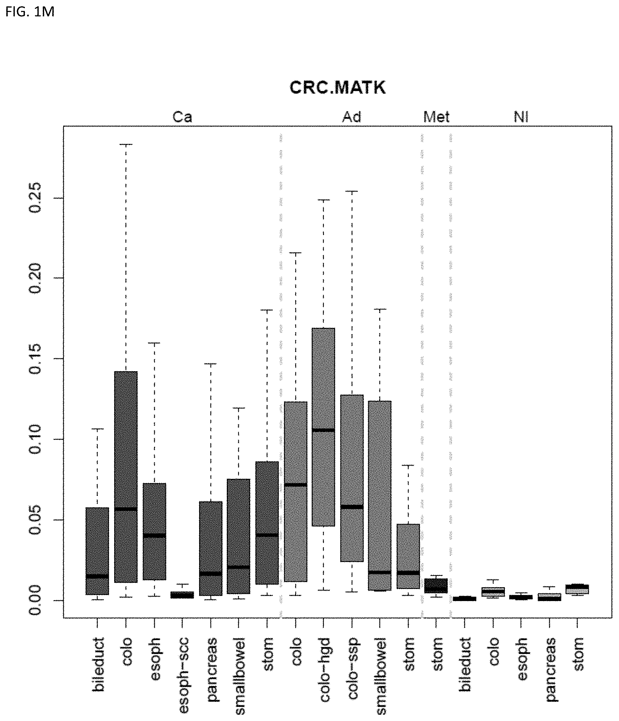
D00014
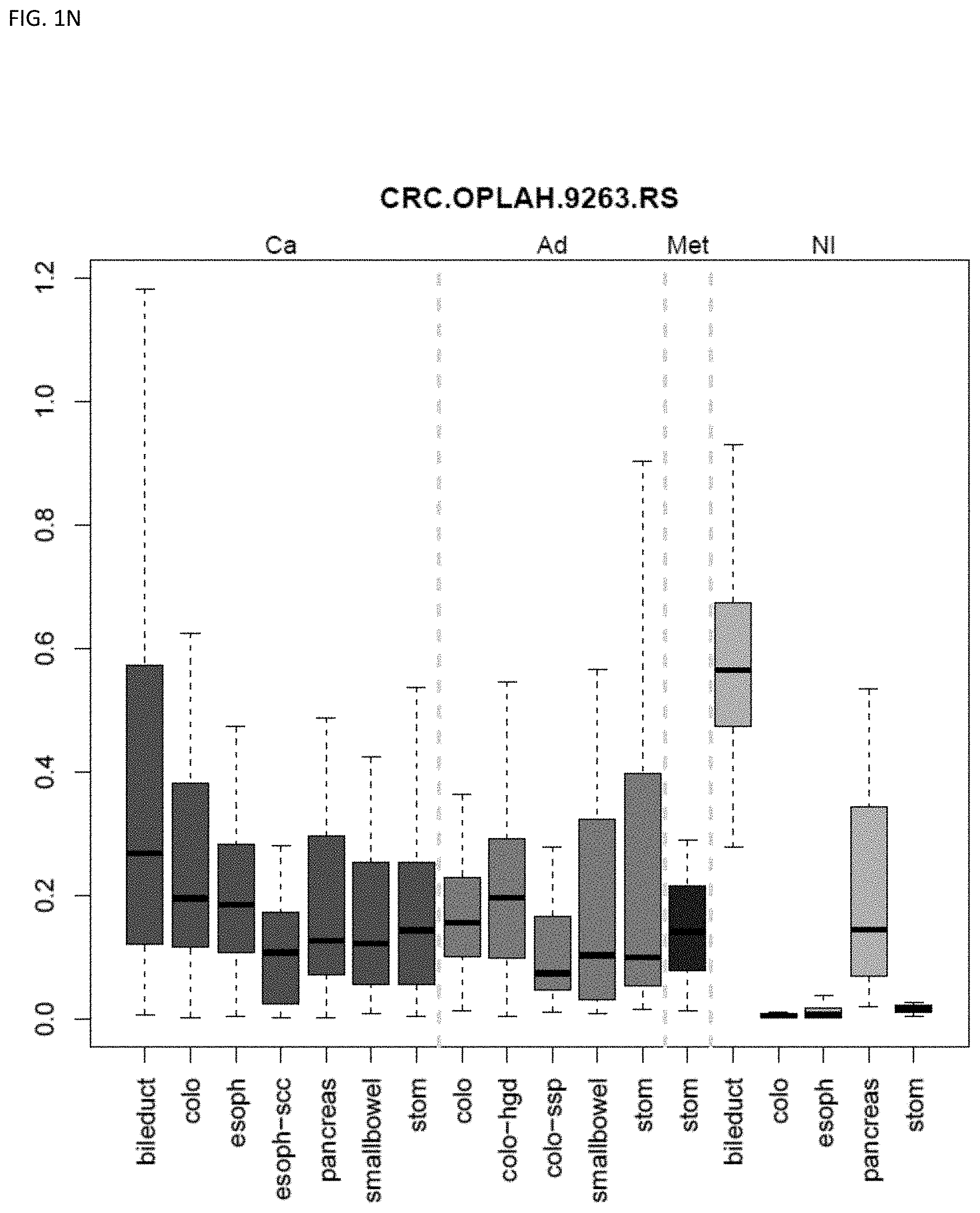
D00015
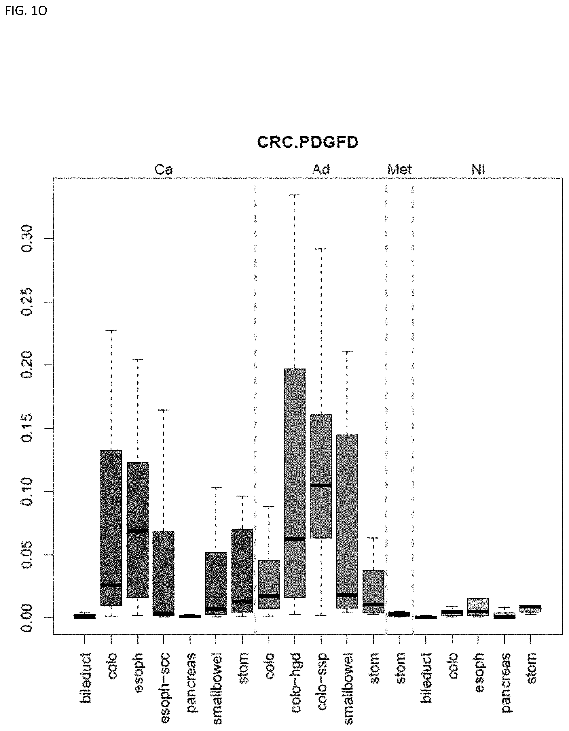
D00016
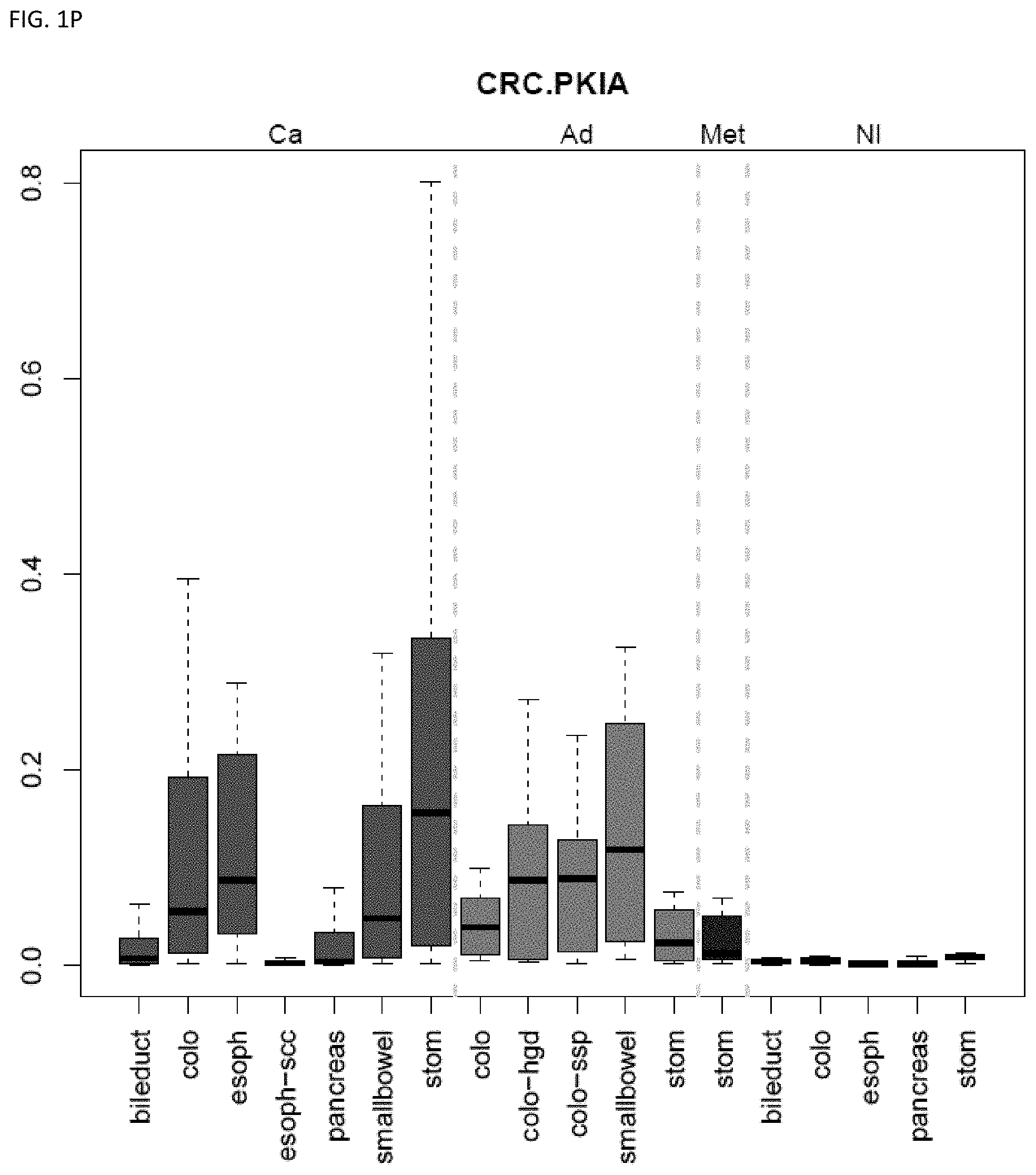
D00017
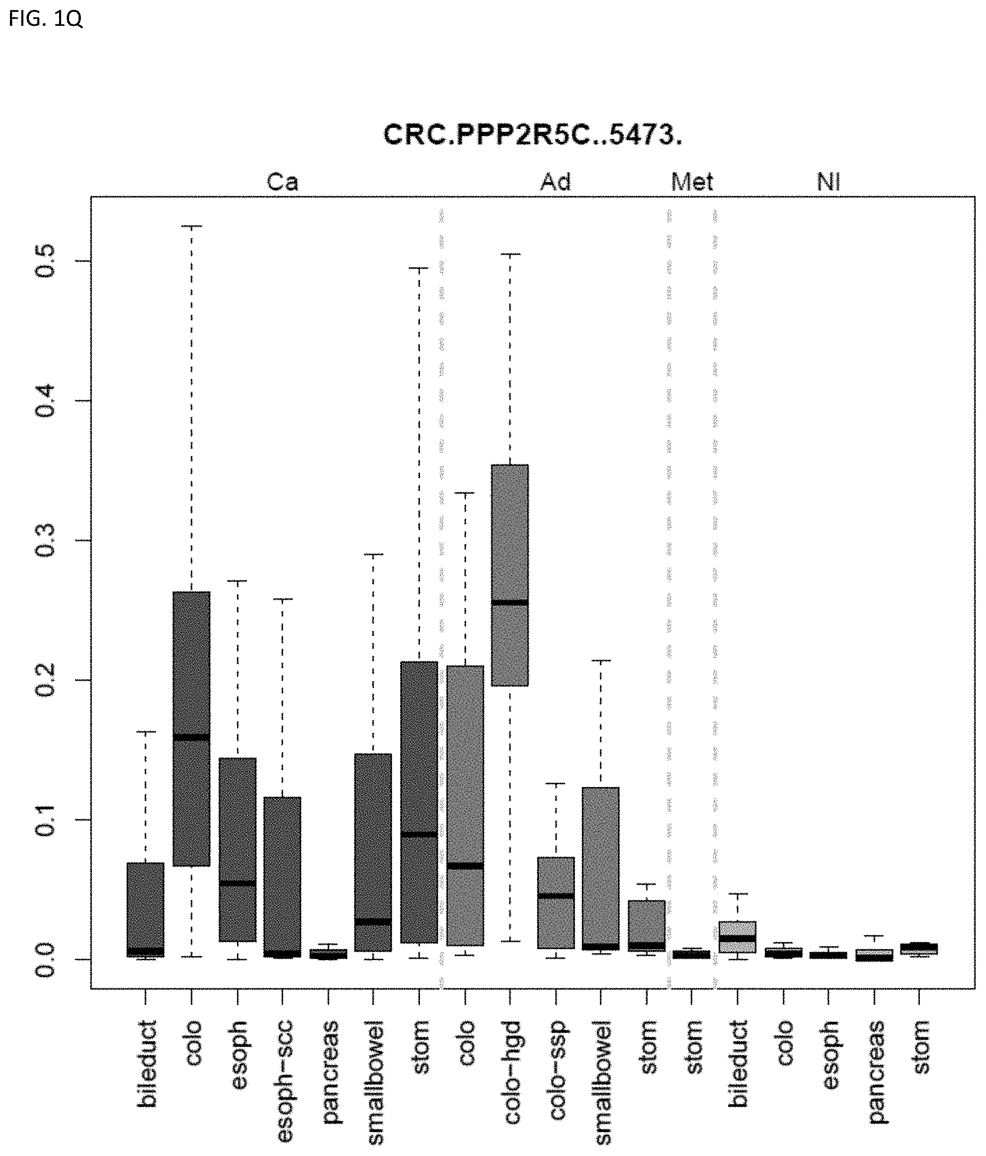
D00018
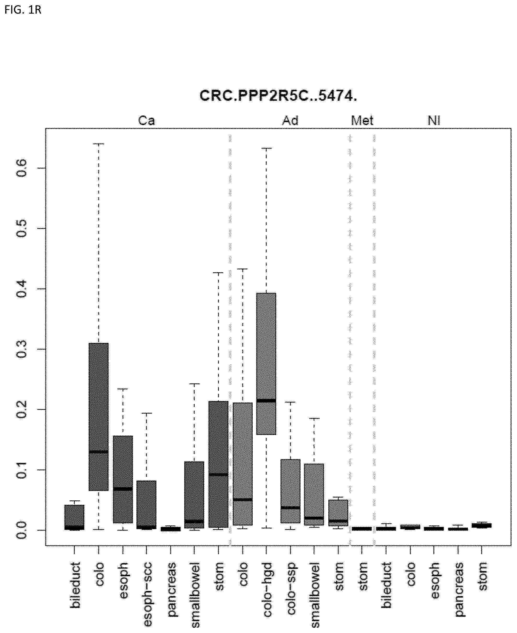
D00019
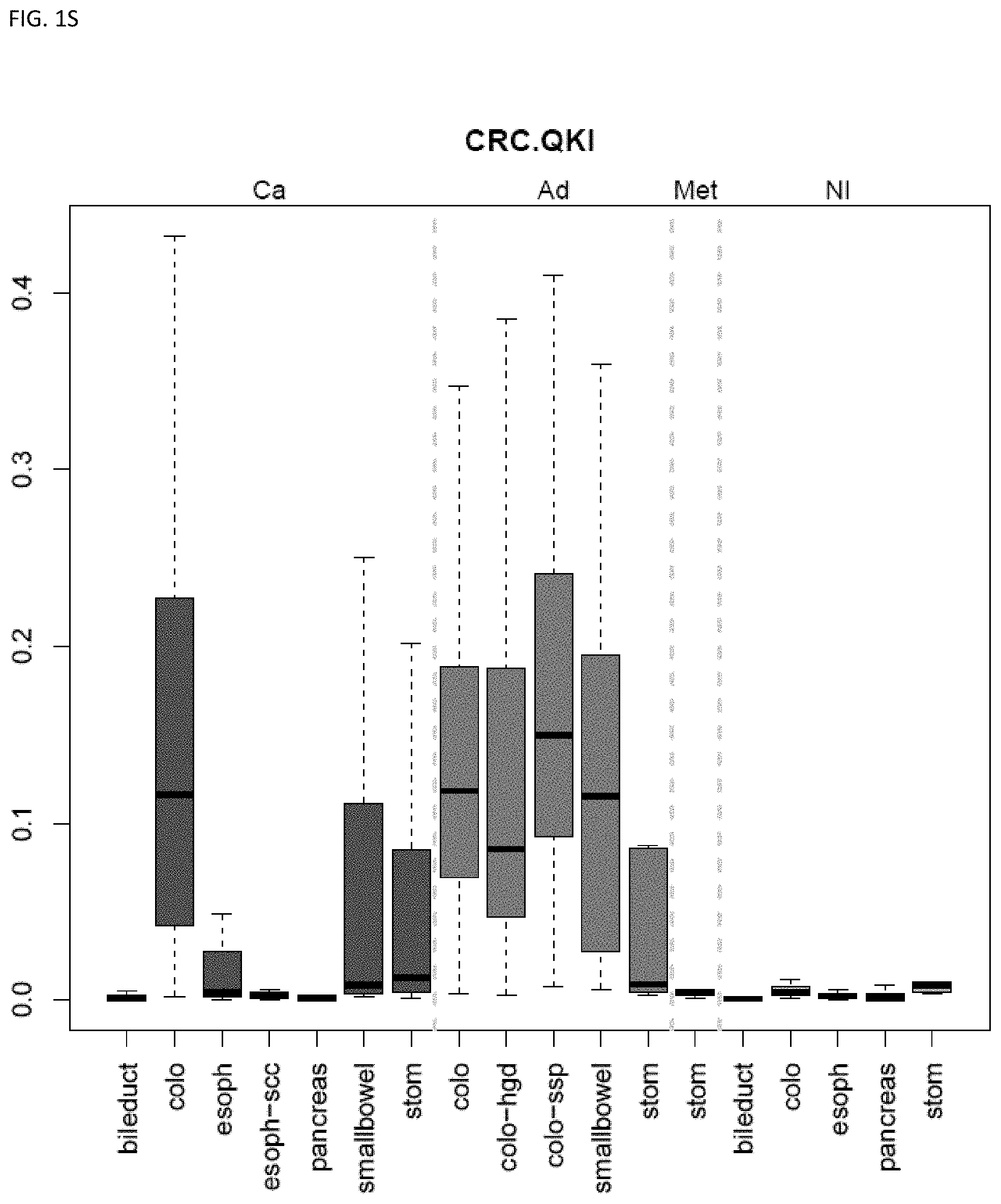
D00020
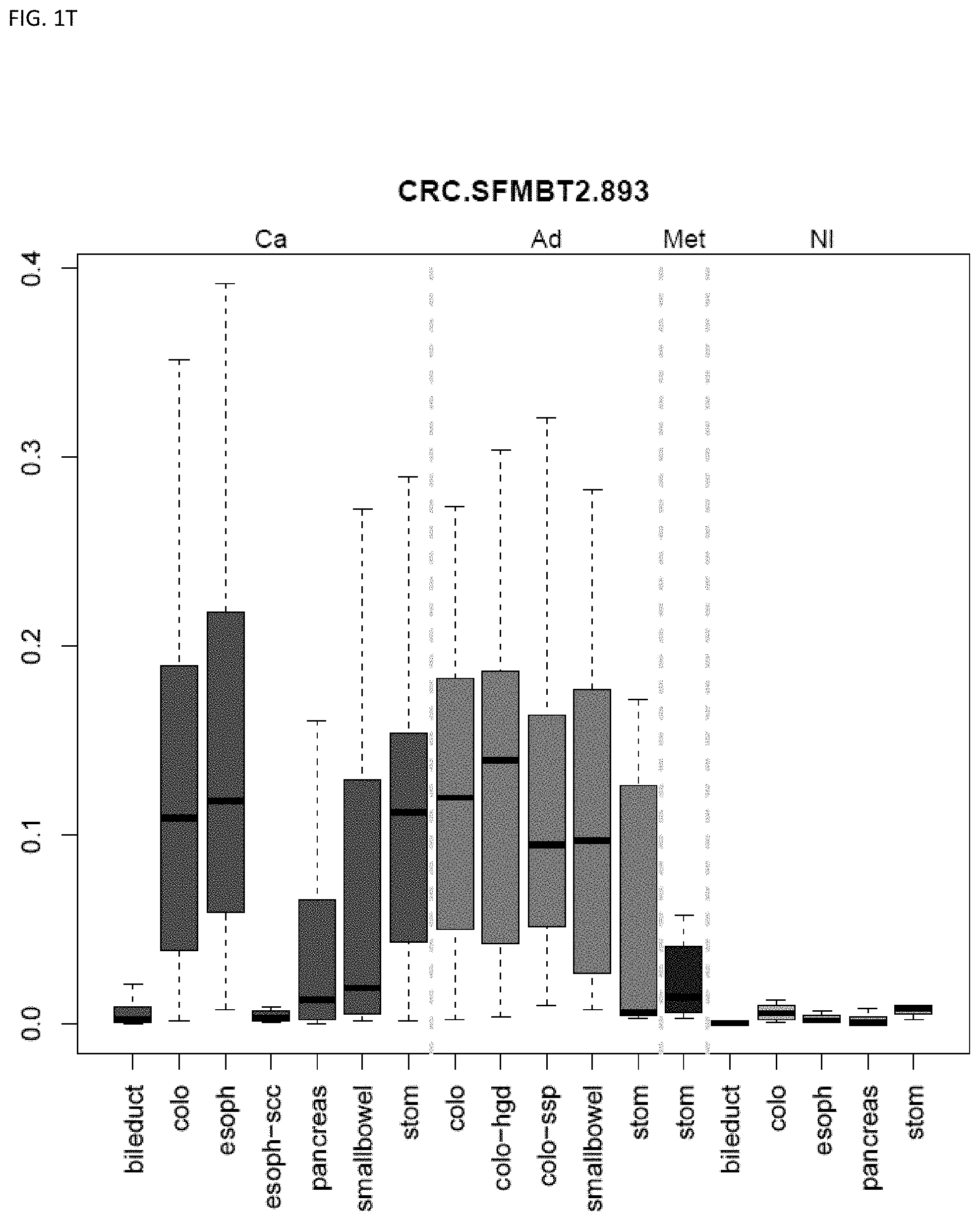
D00021
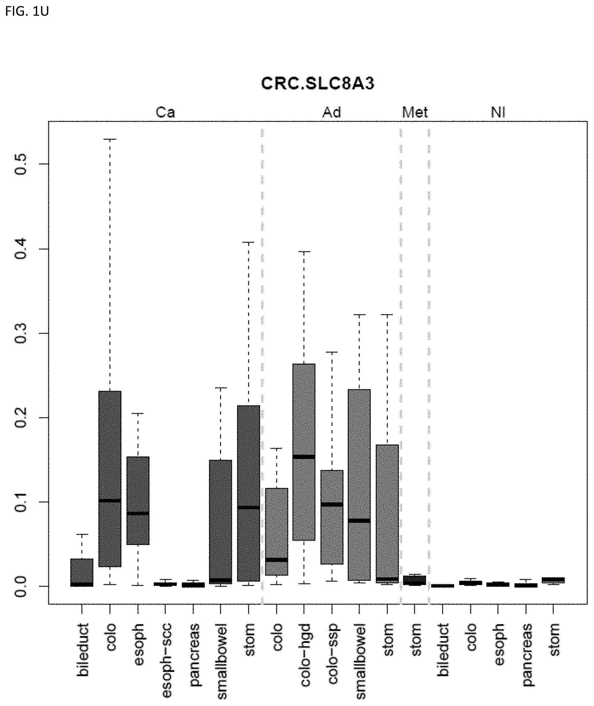
D00022
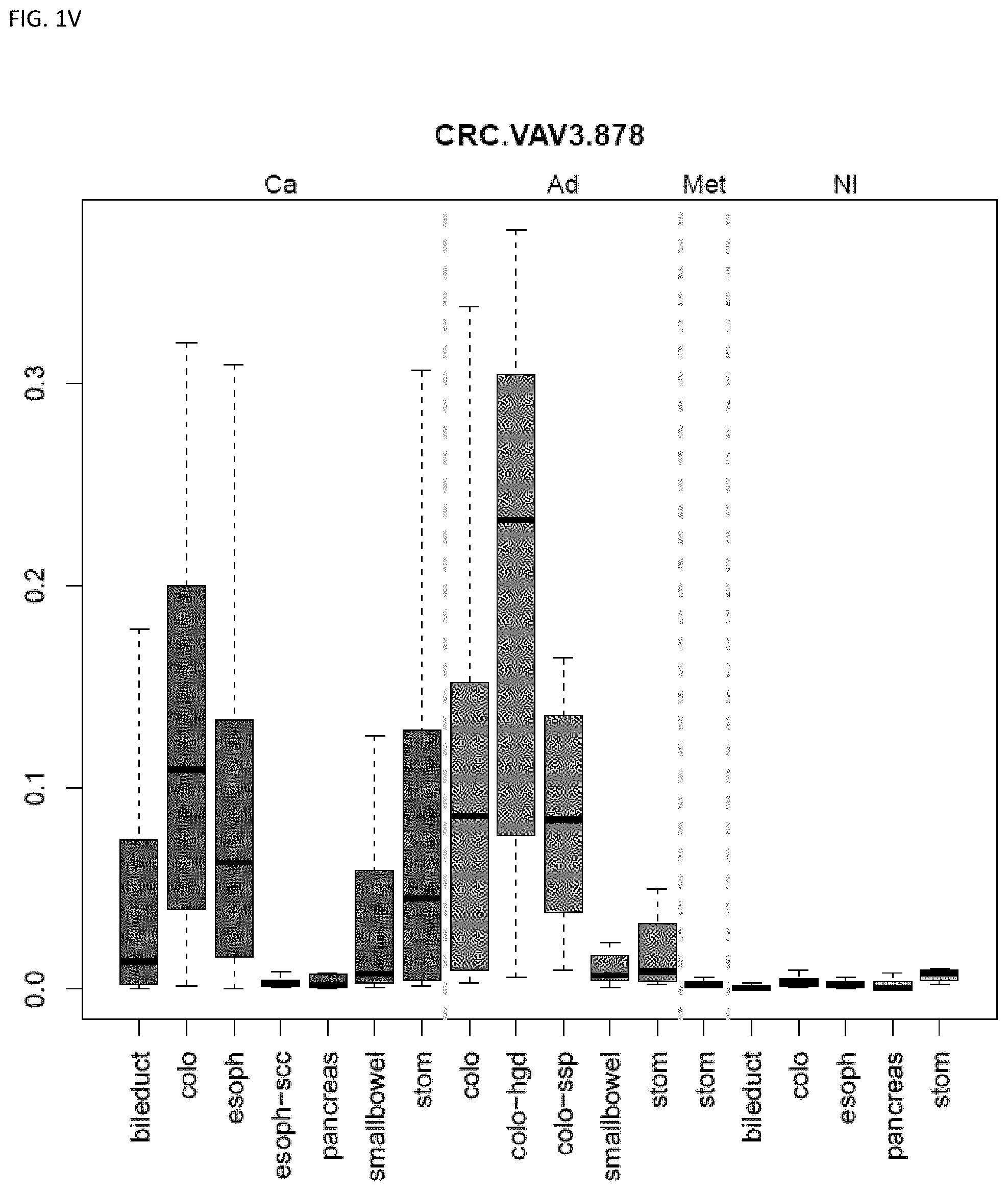
D00023
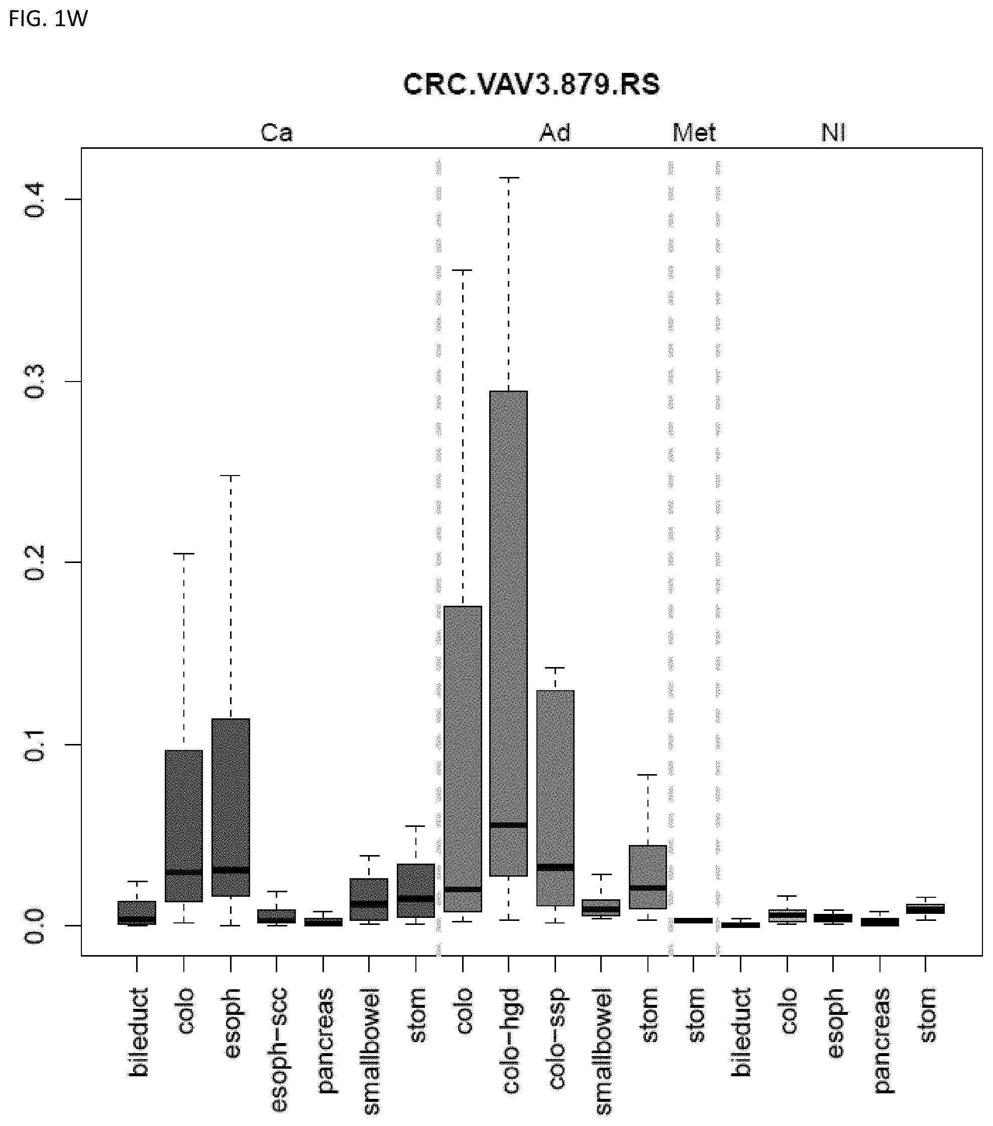
D00024
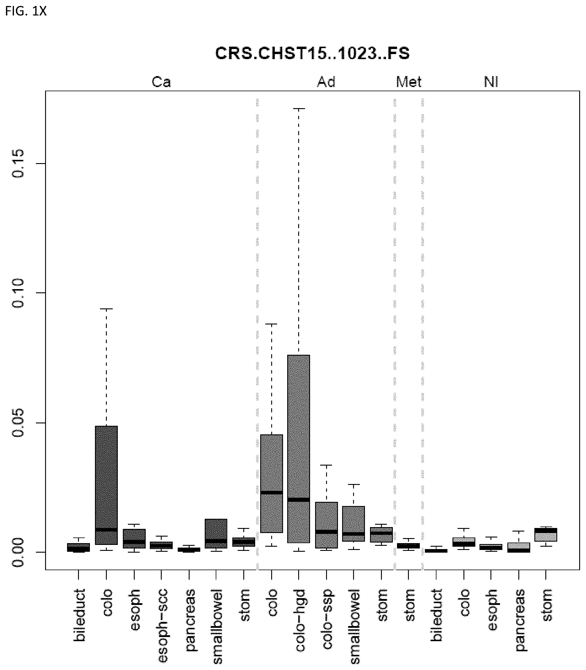
D00025
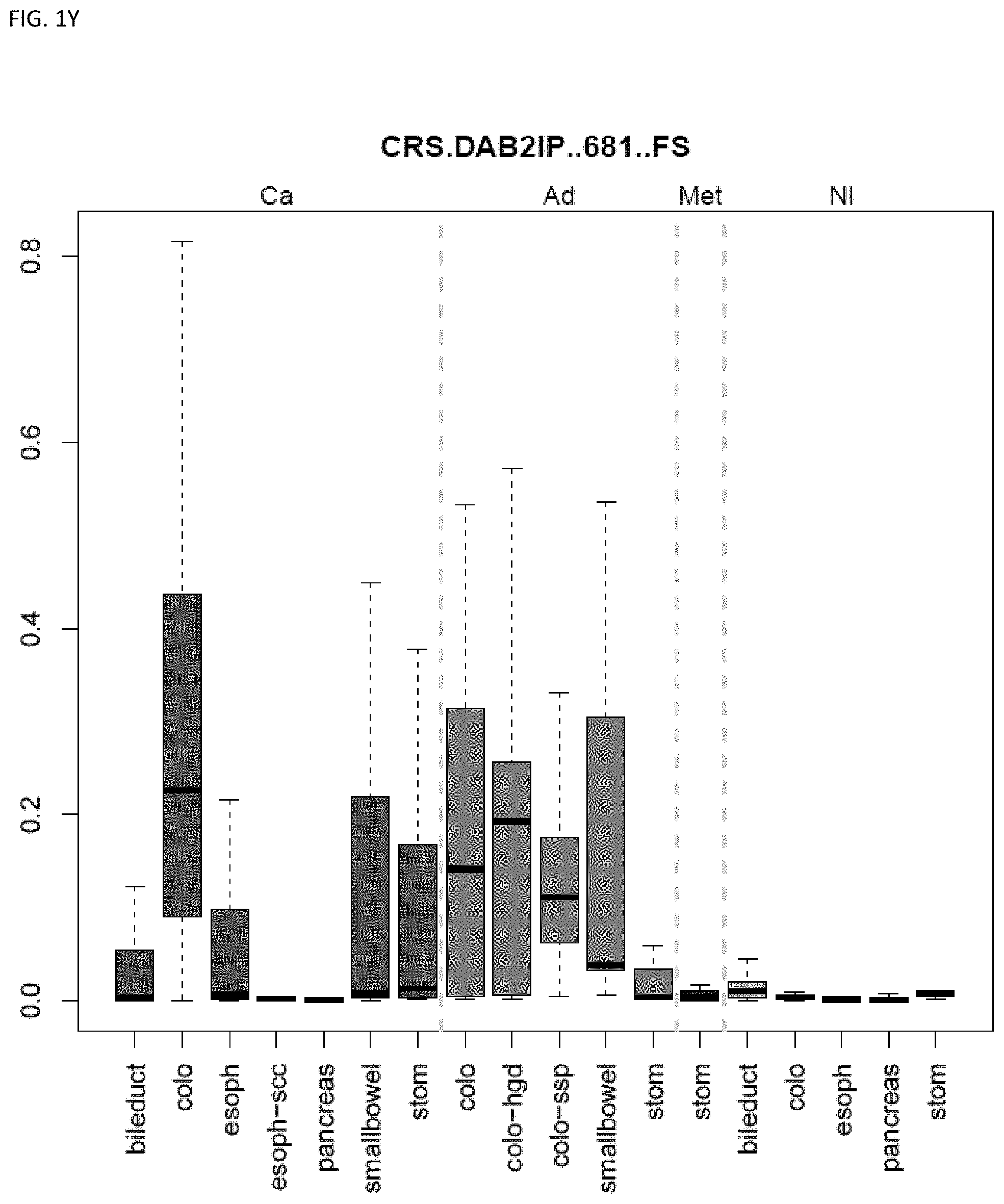
D00026
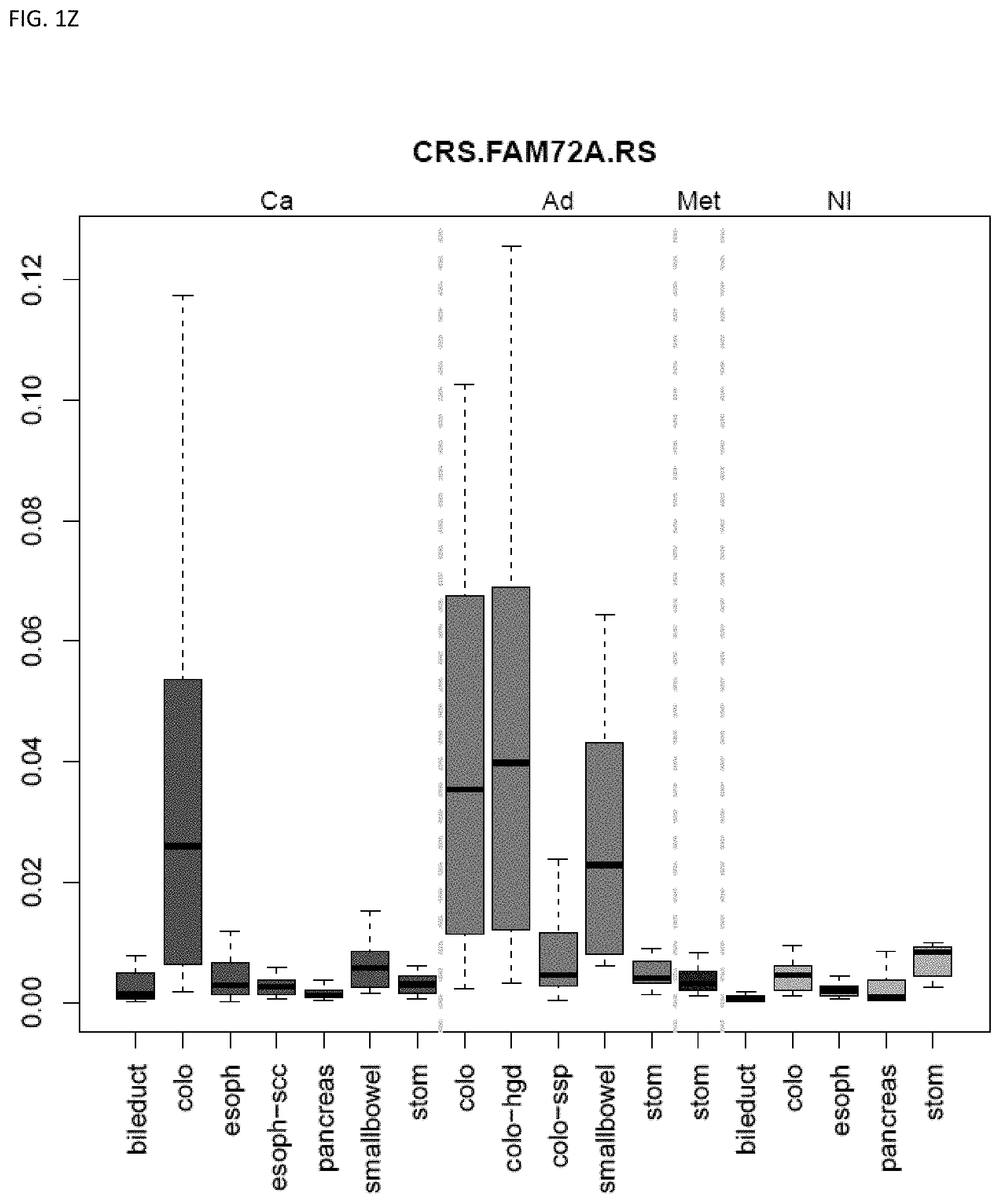
D00027
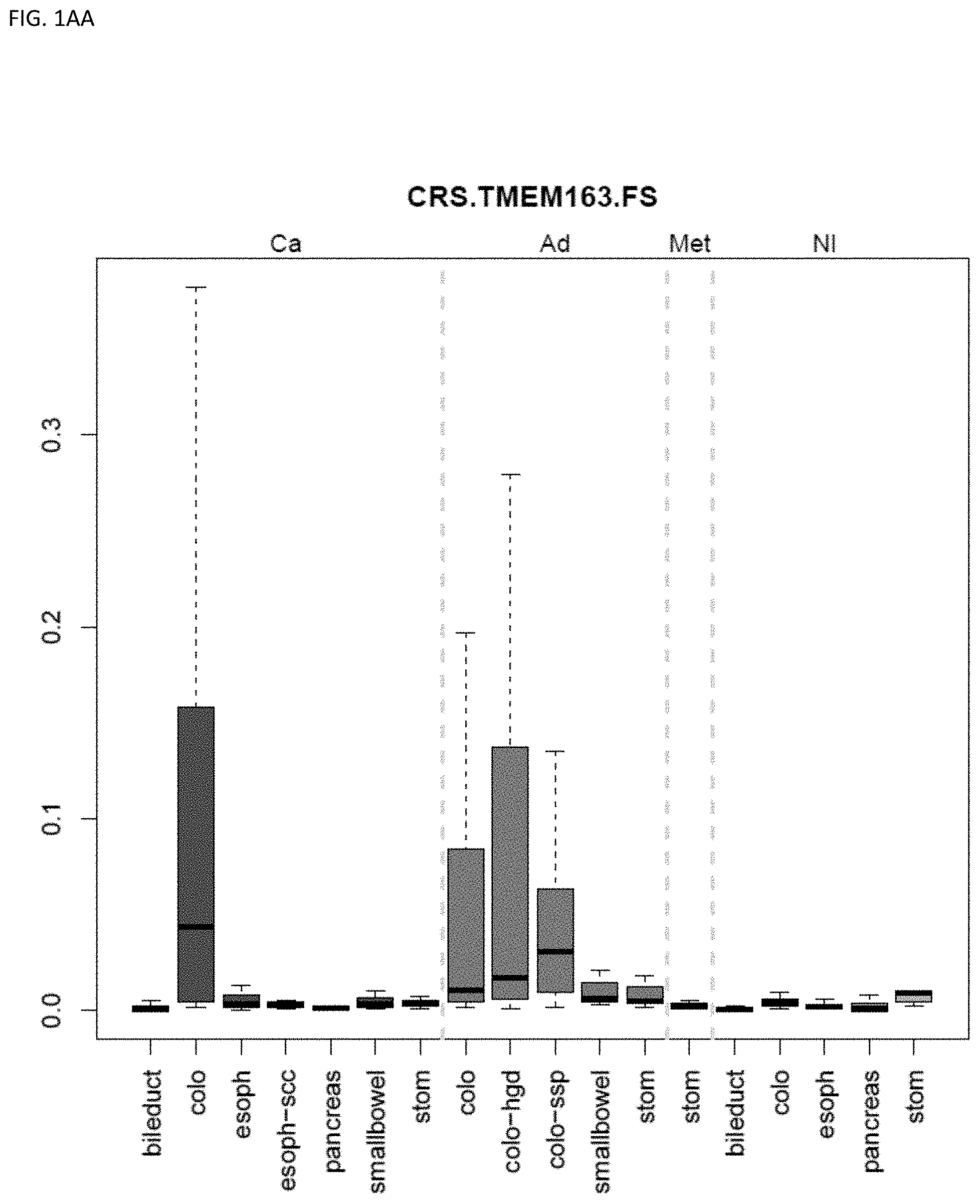
D00028
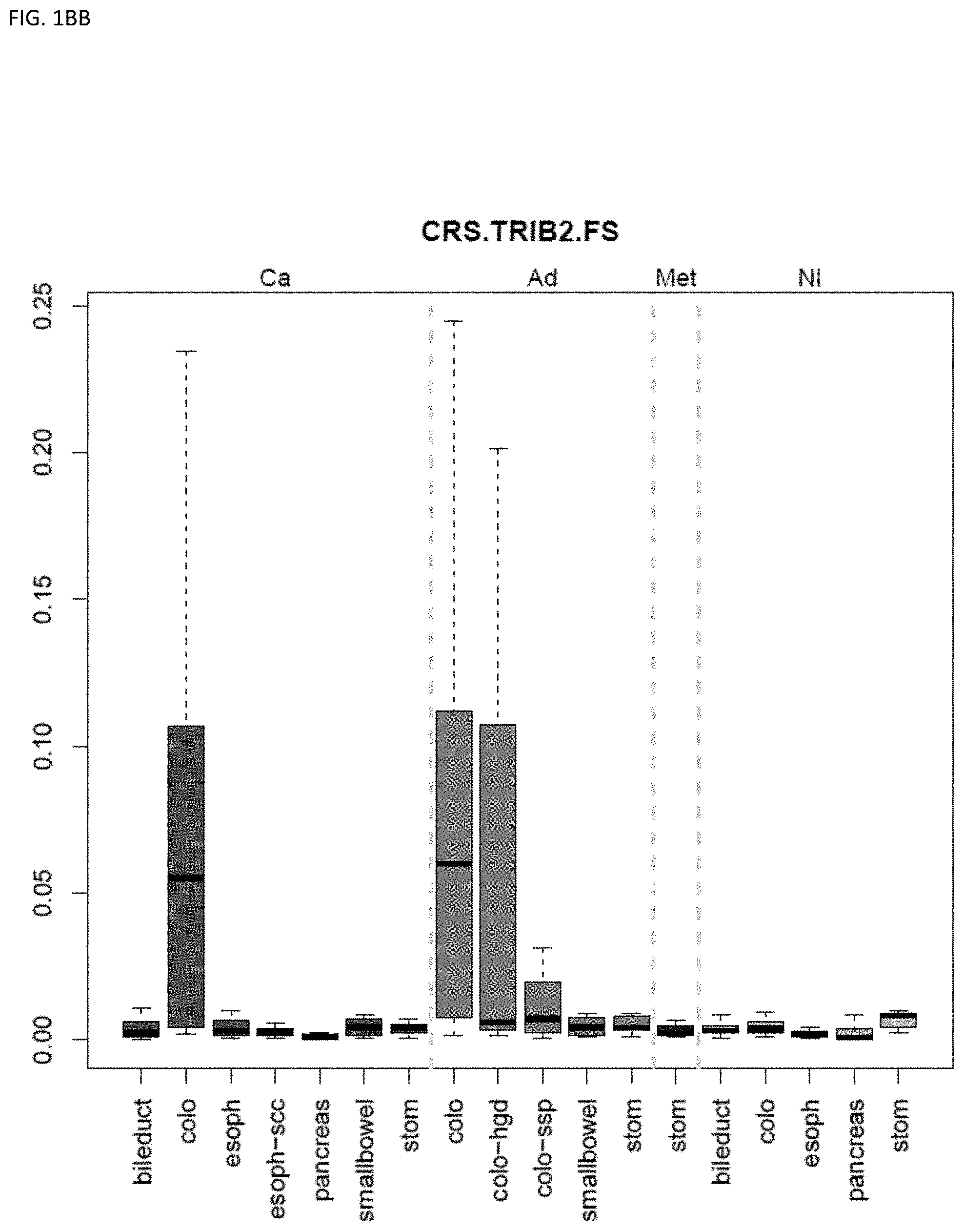
D00029
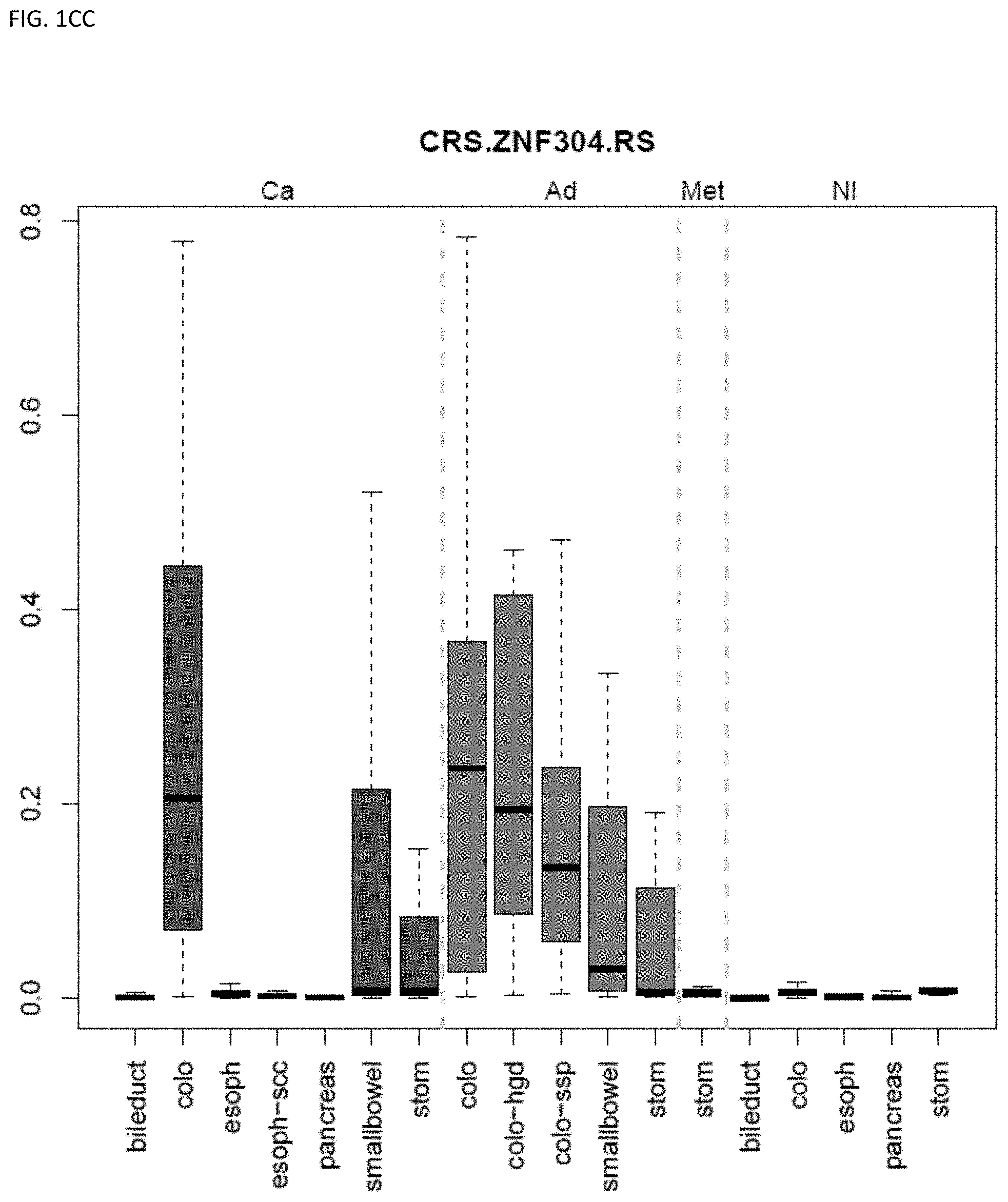
D00030
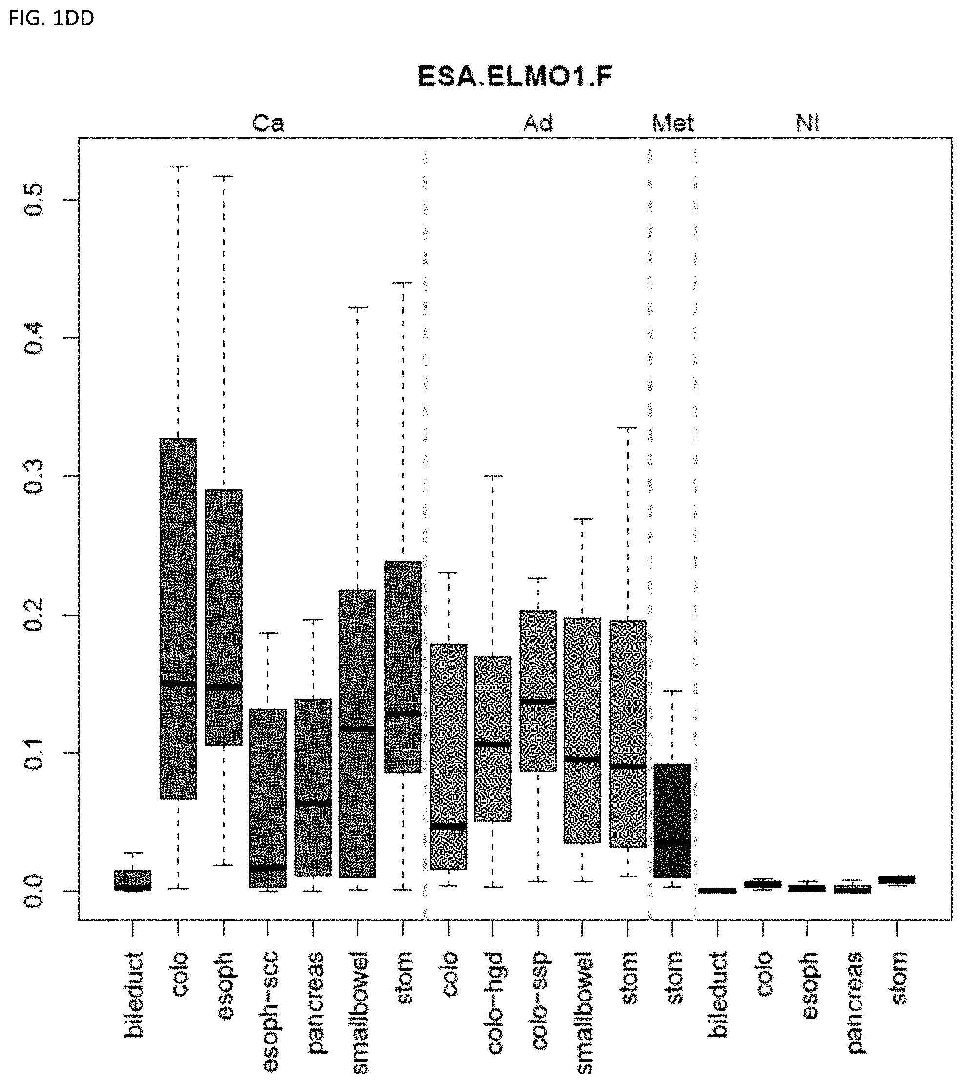
D00031
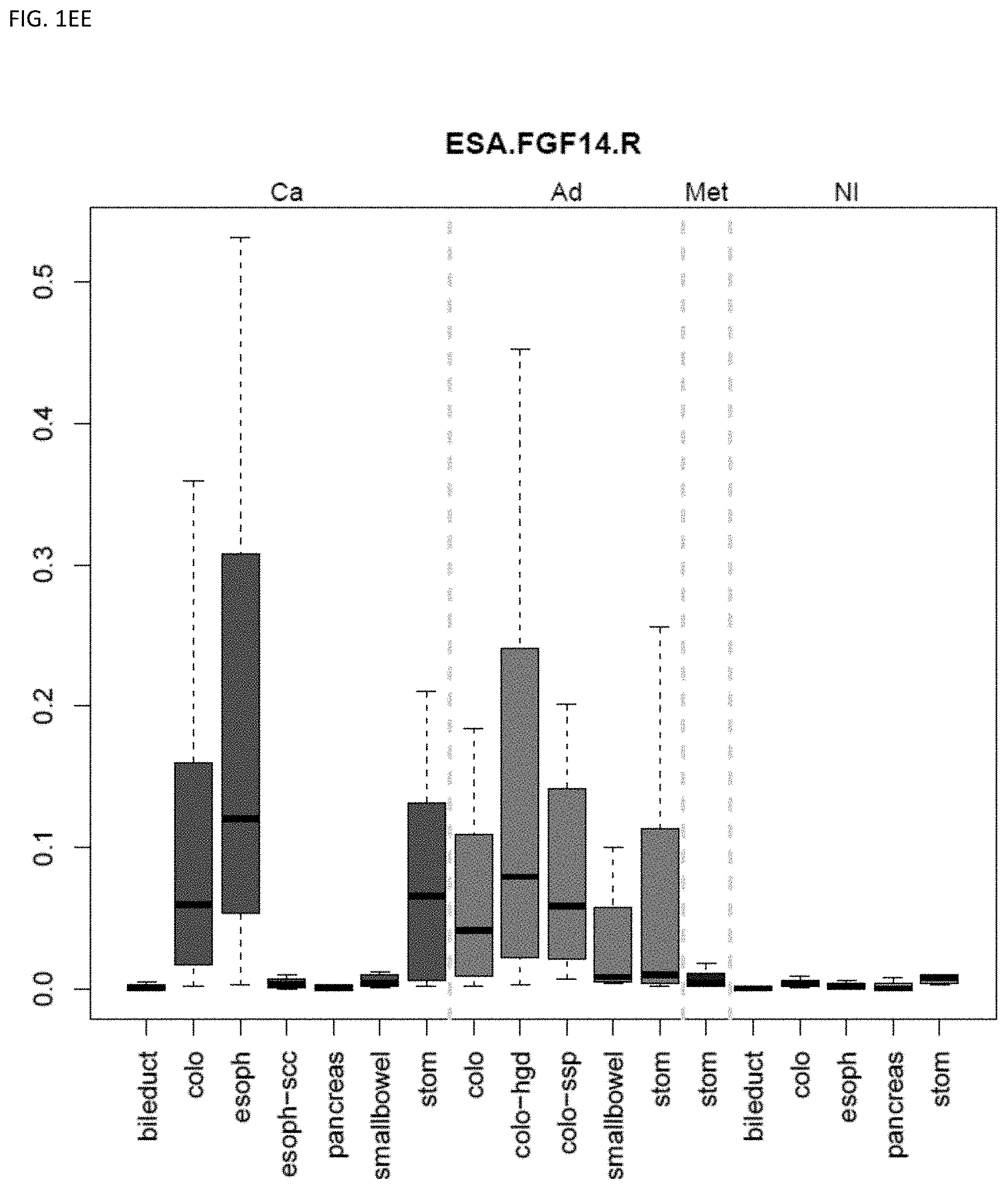
D00032
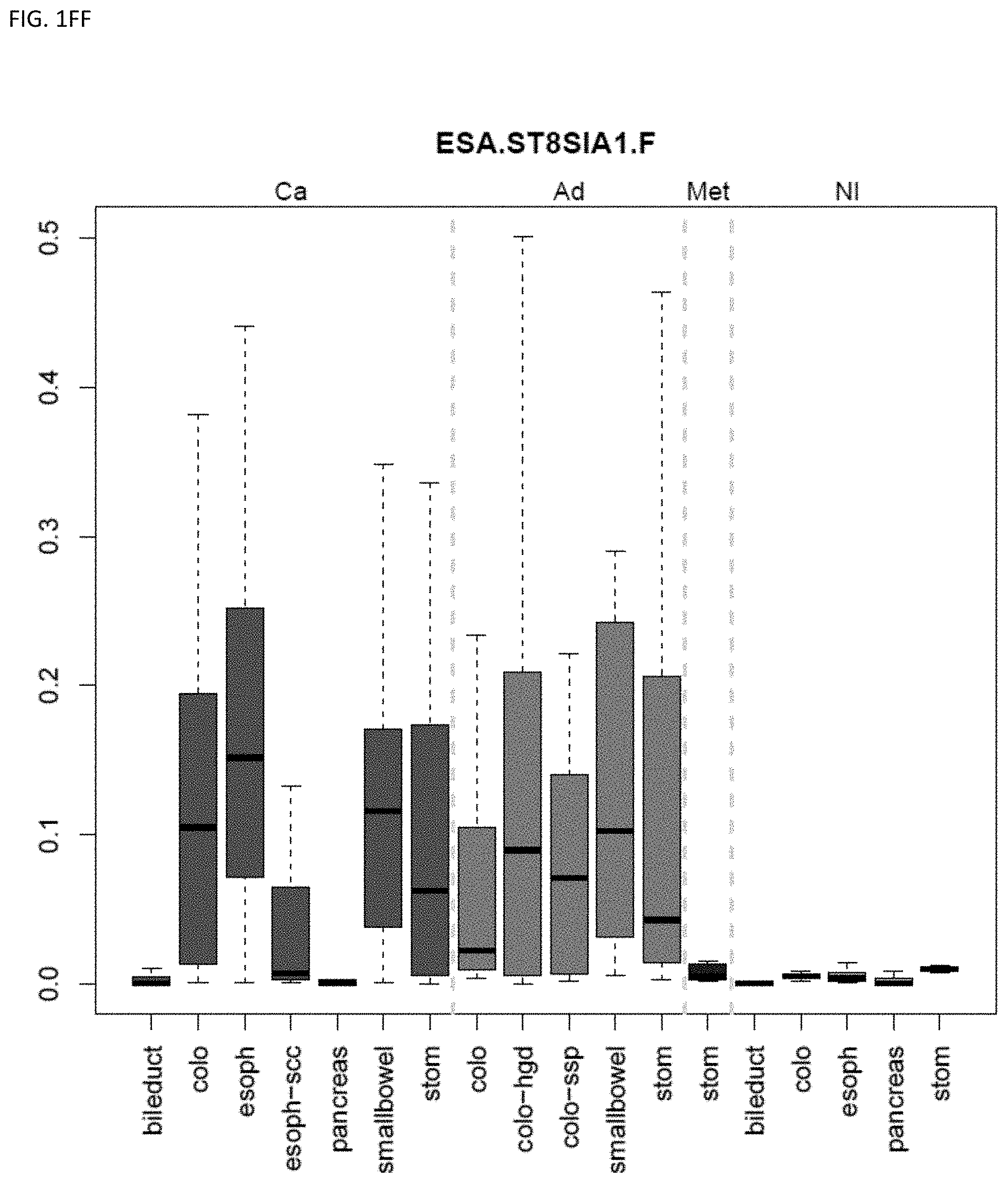
D00033
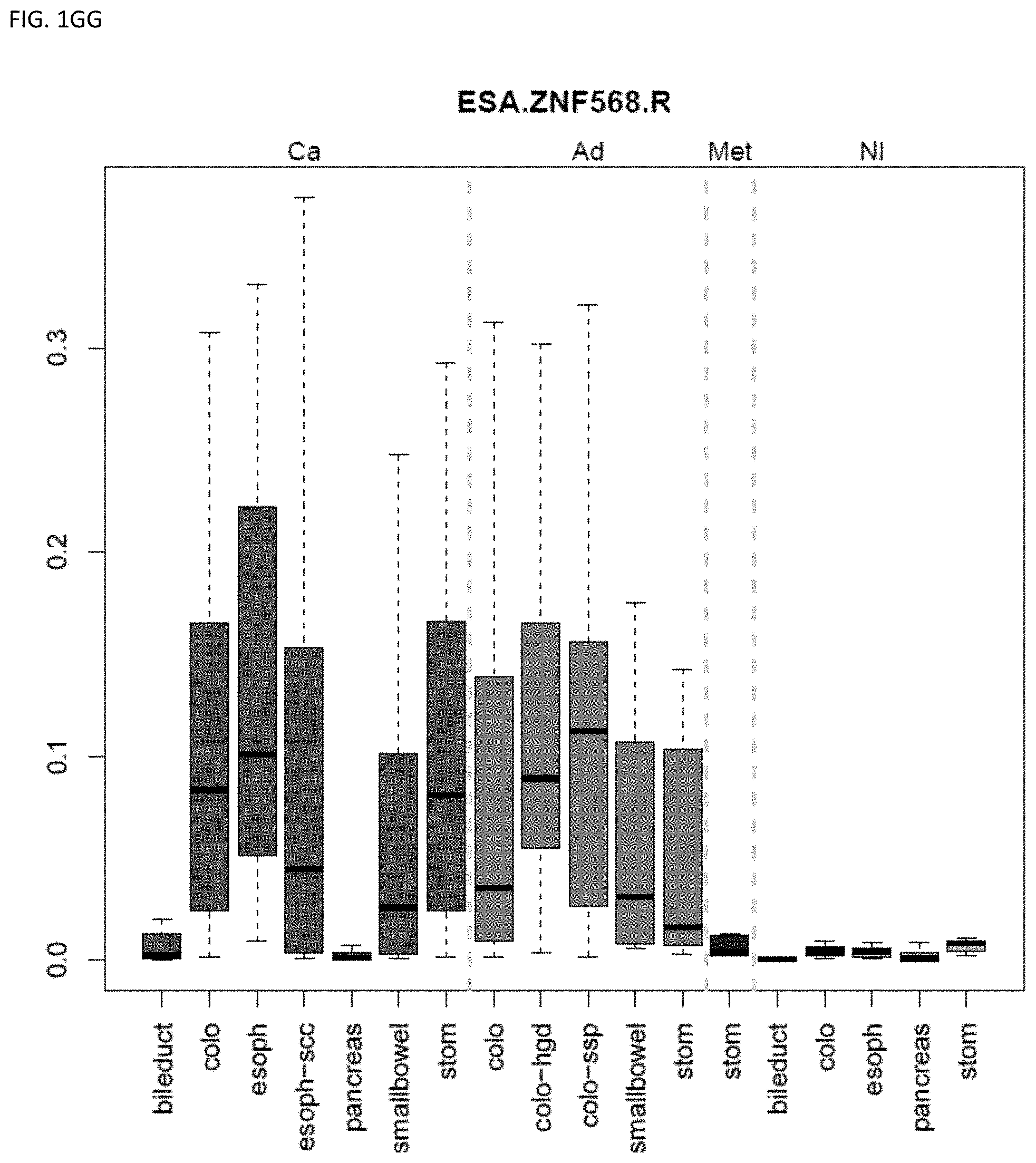
D00034
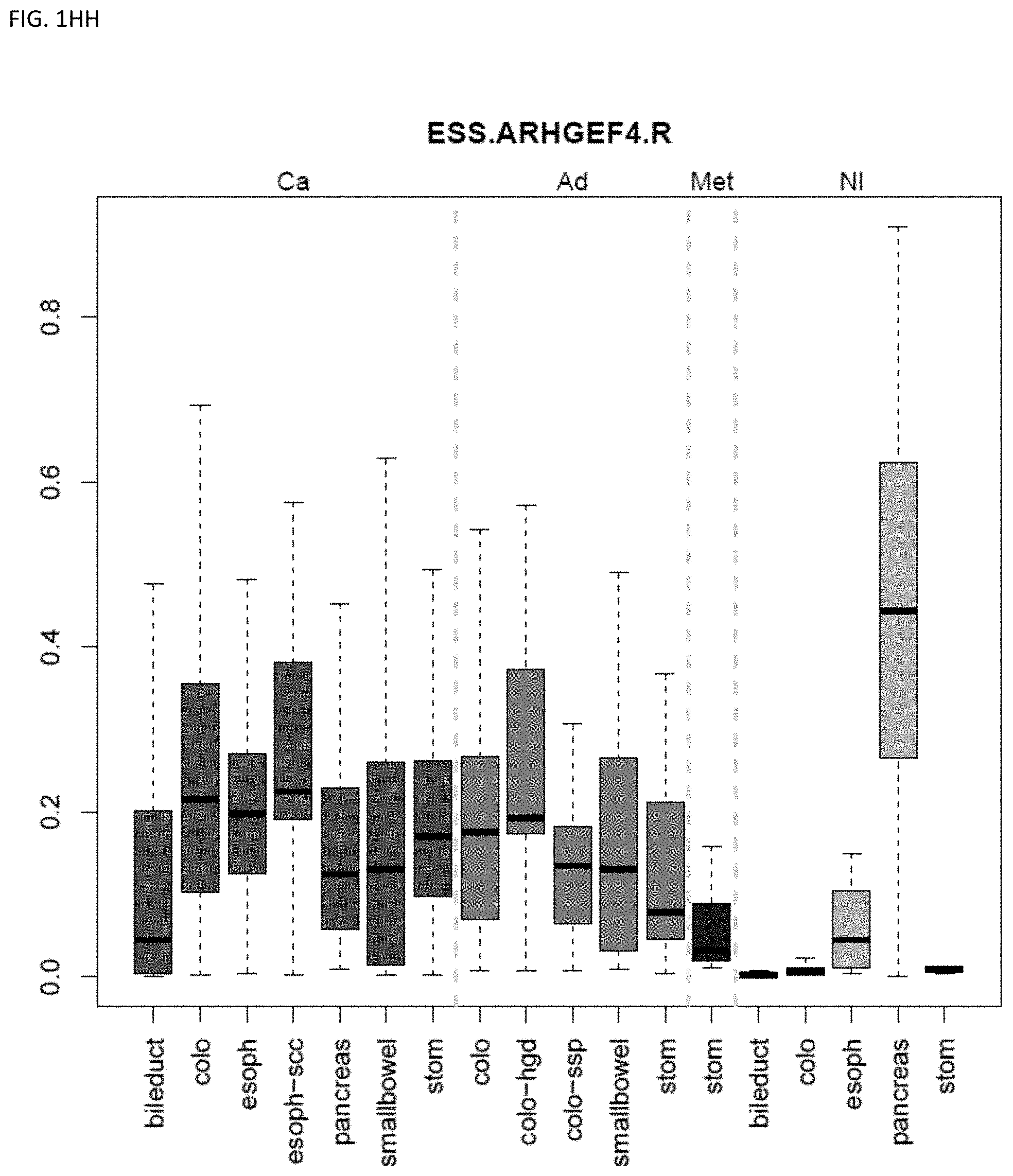
D00035
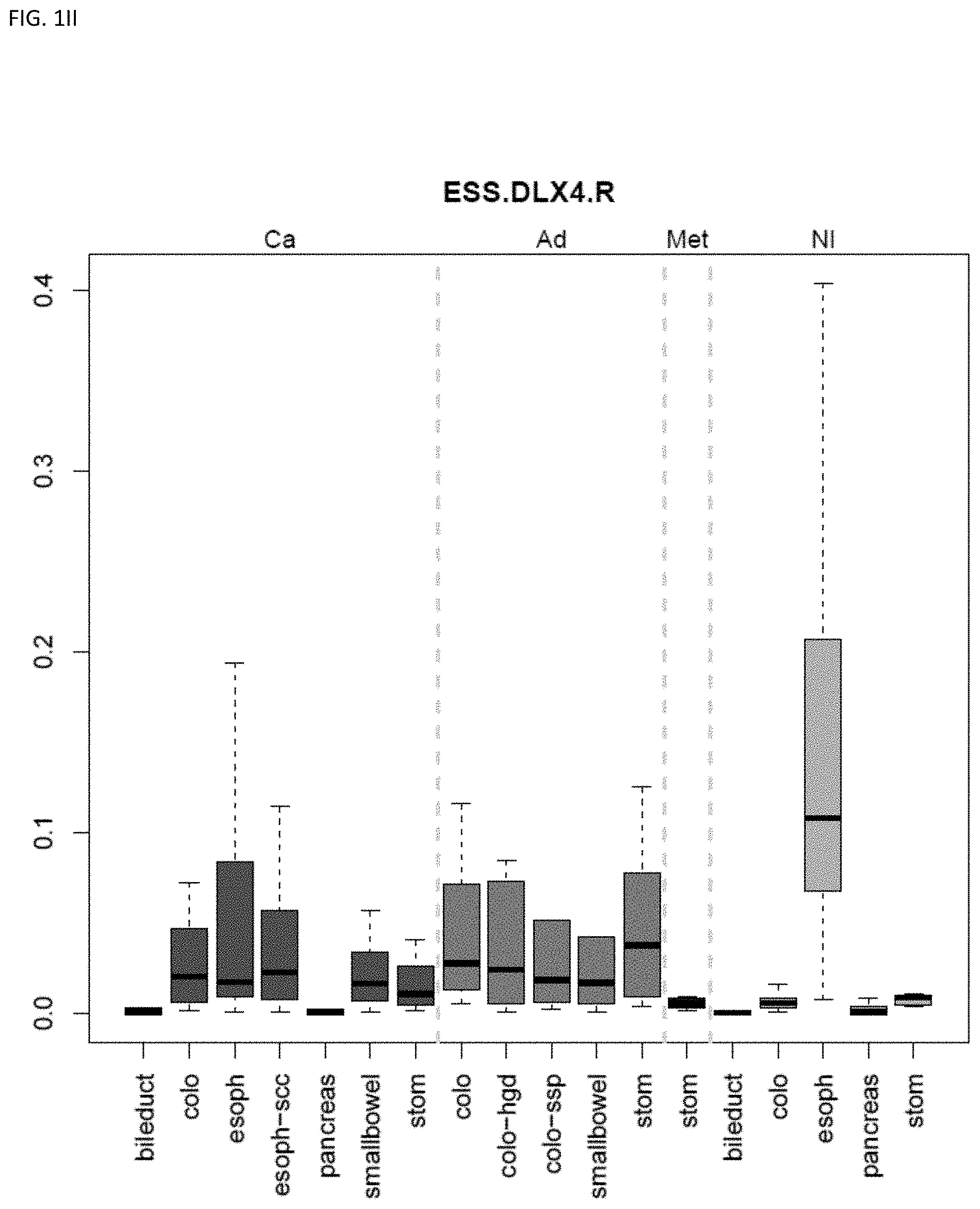
D00036
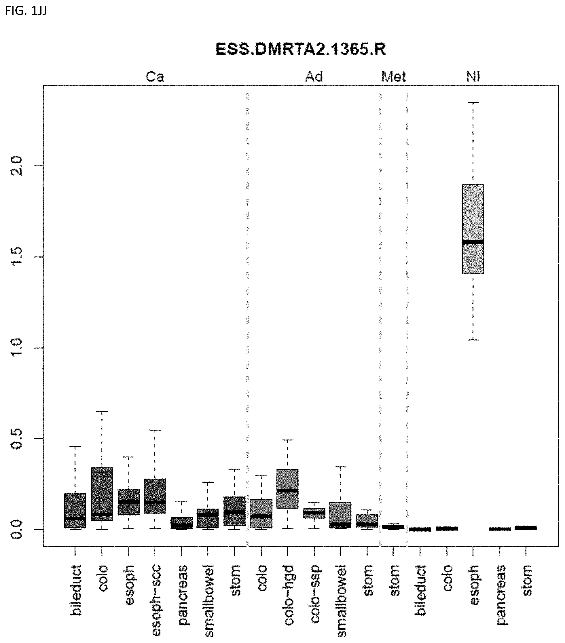
D00037
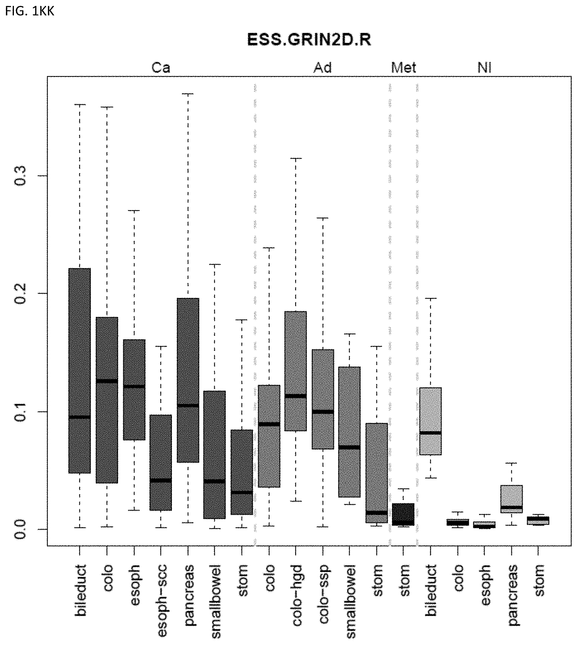
D00038
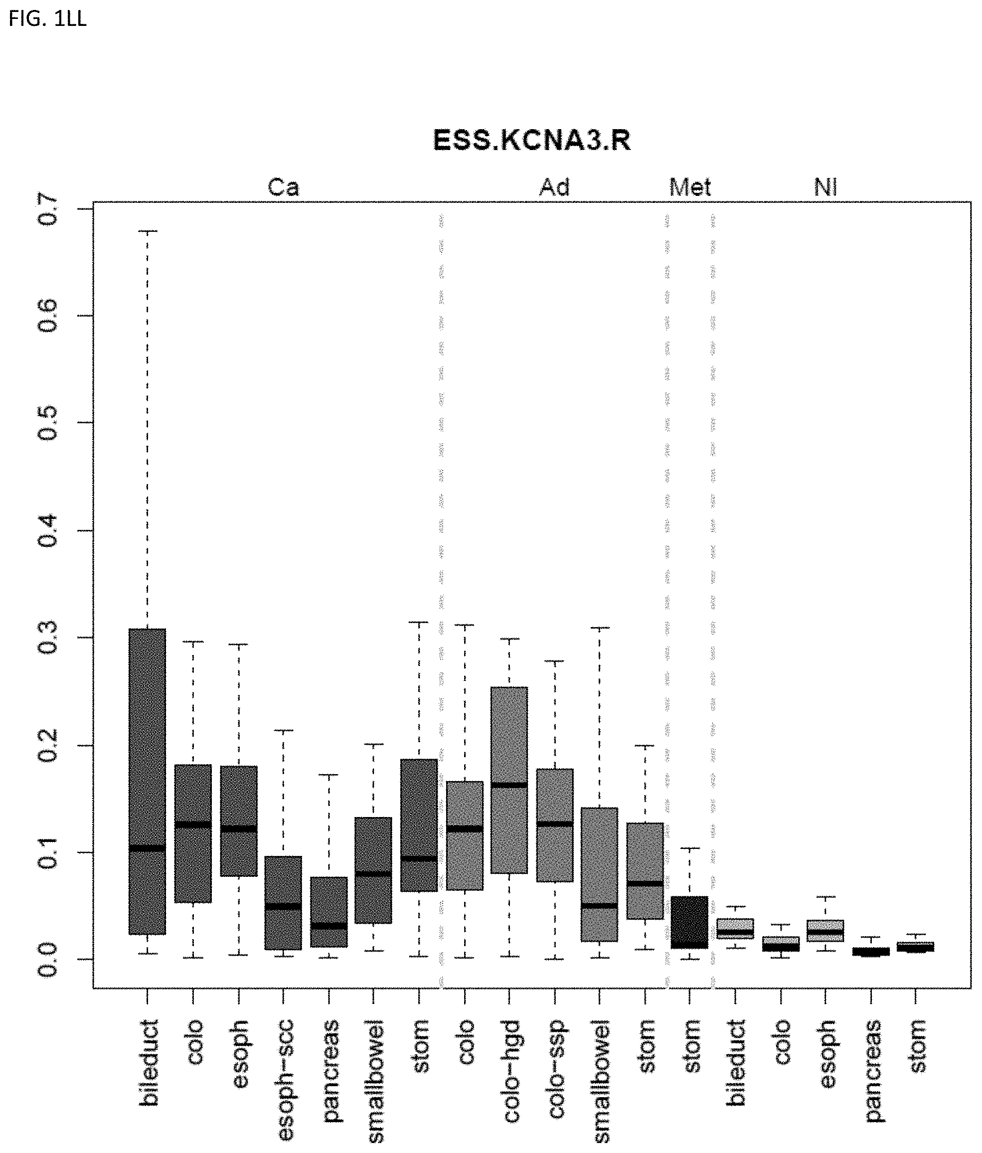
D00039
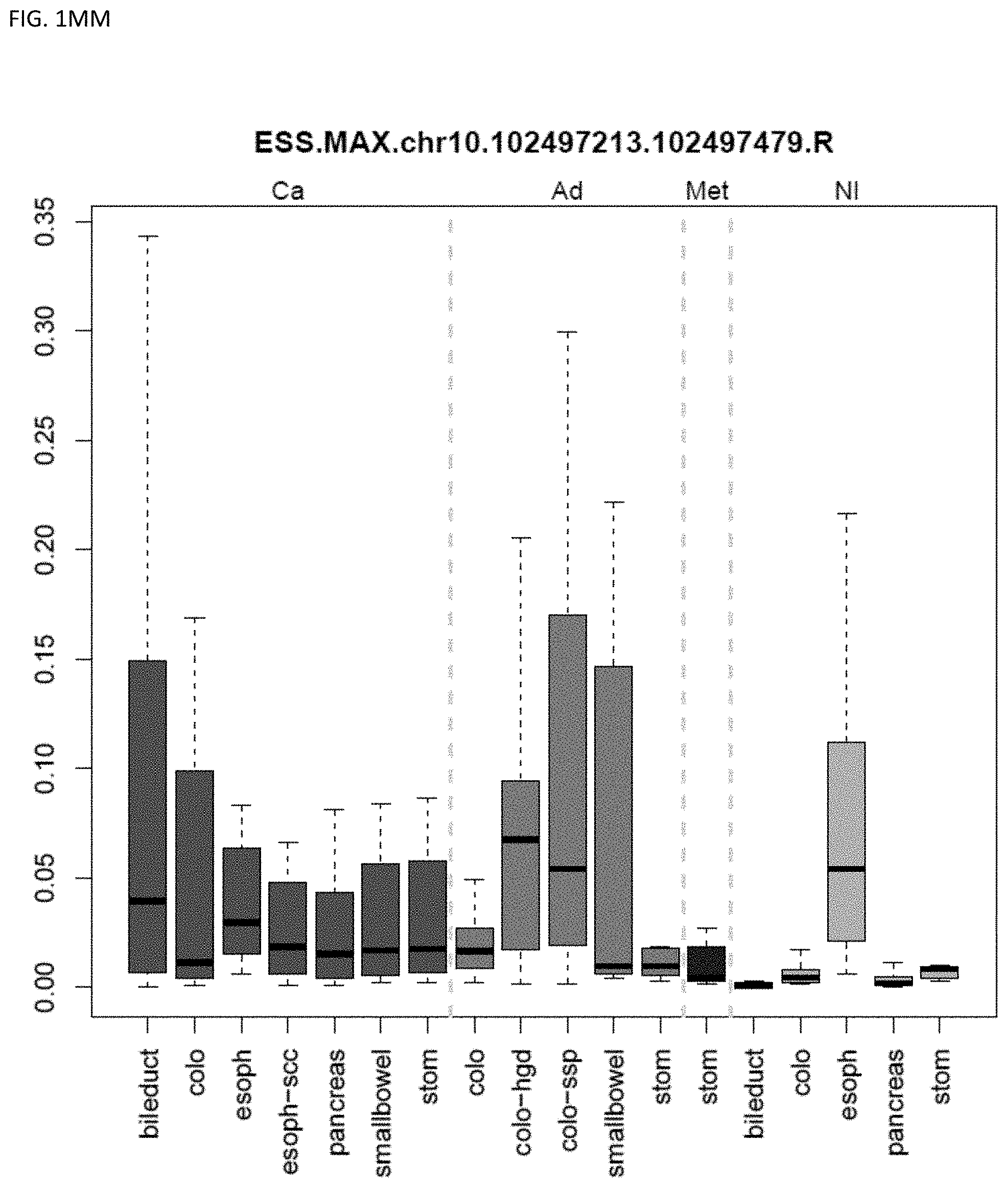
D00040
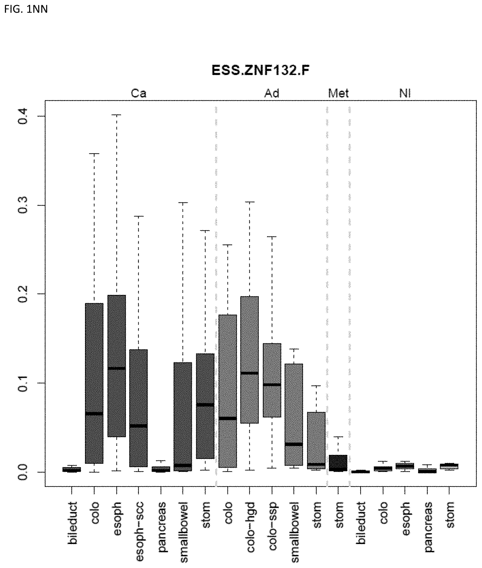
D00041
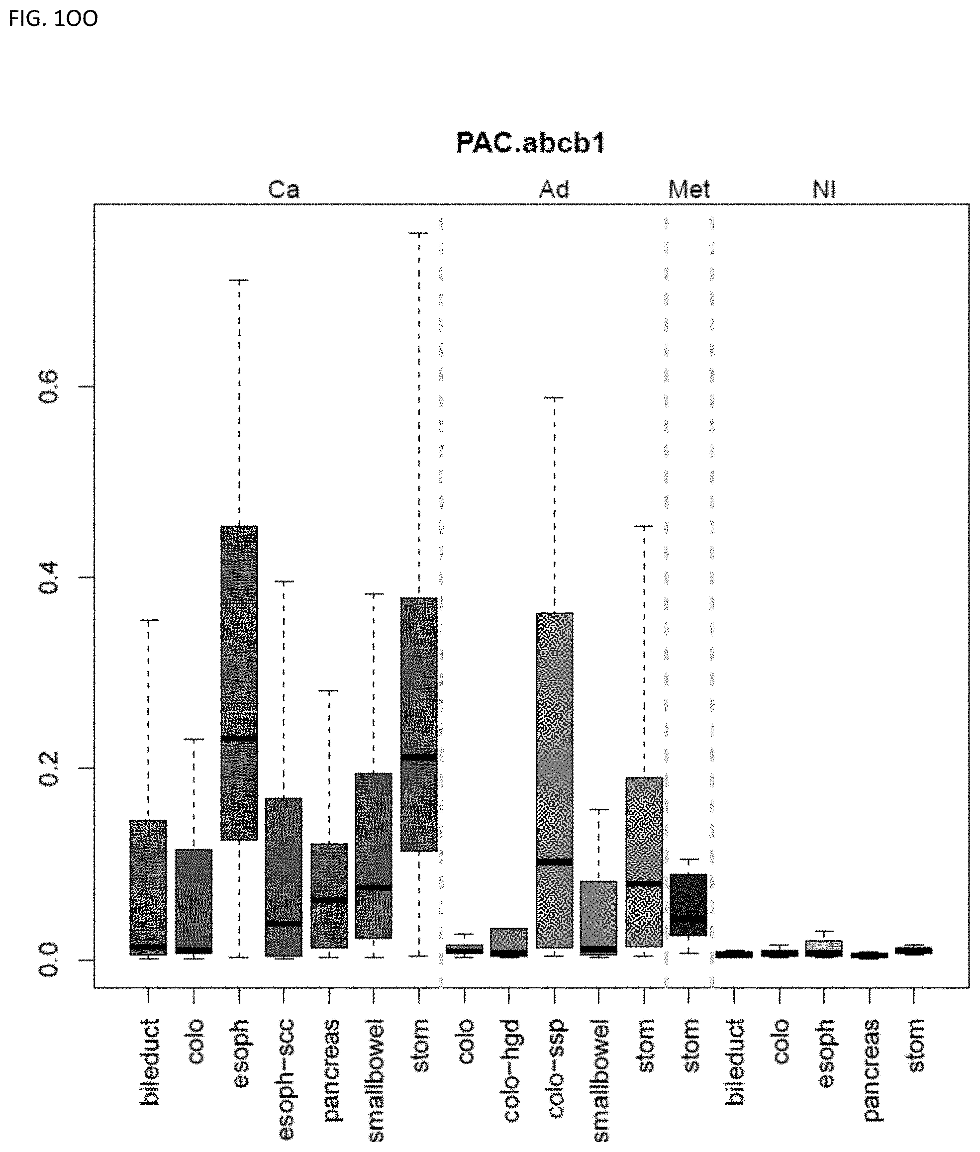
D00042
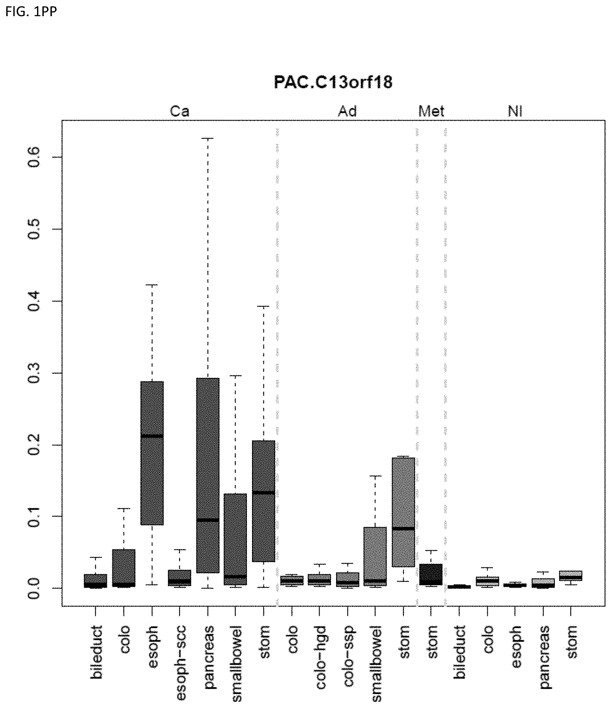
D00043
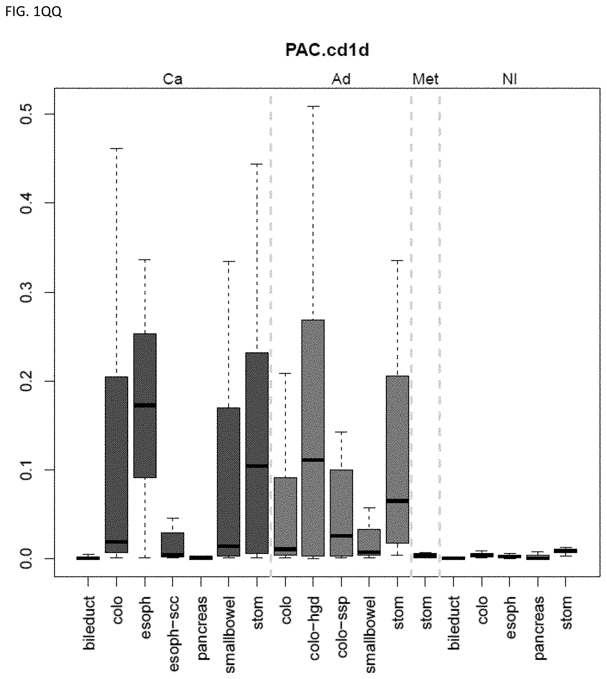
D00044
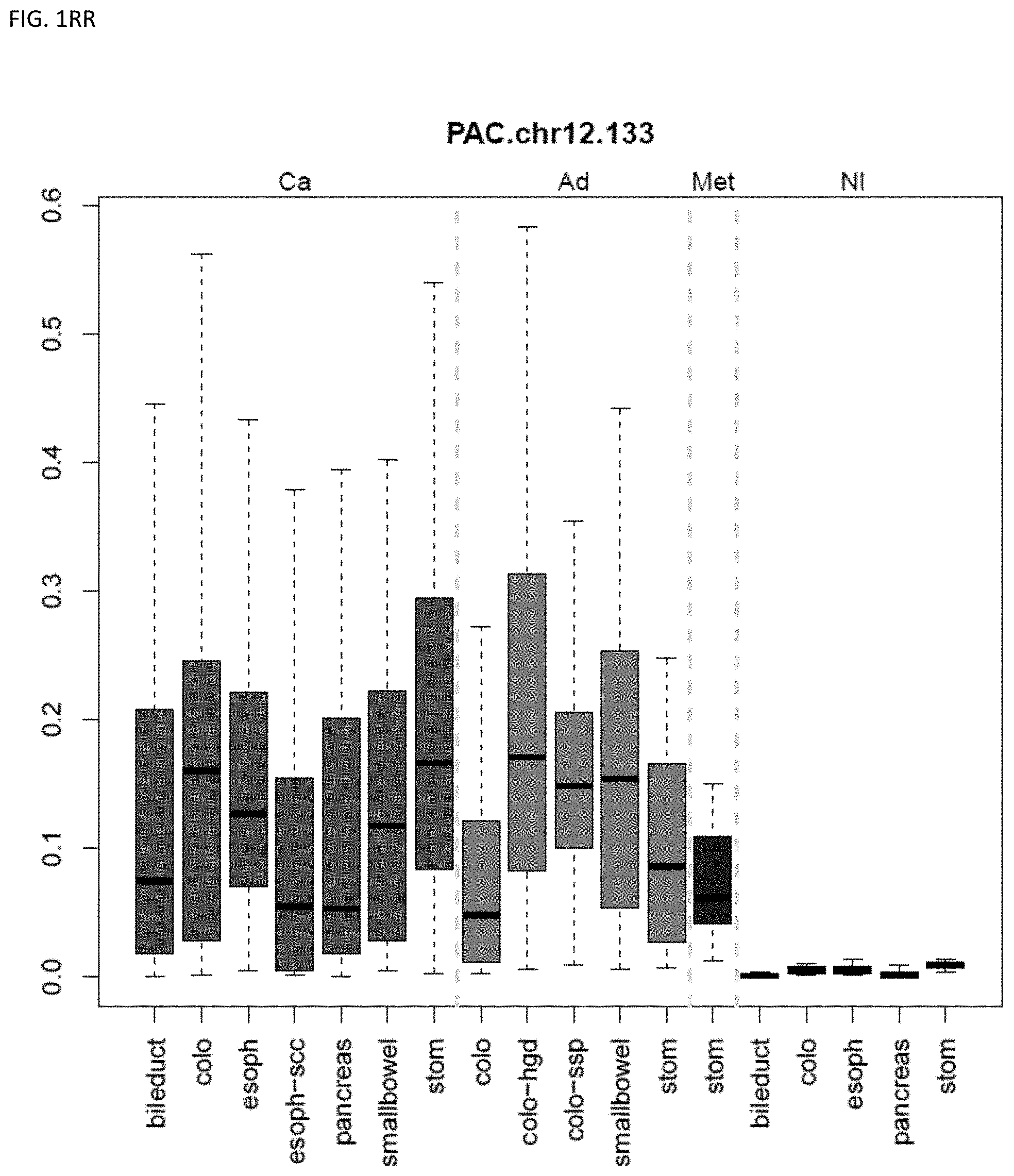
D00045
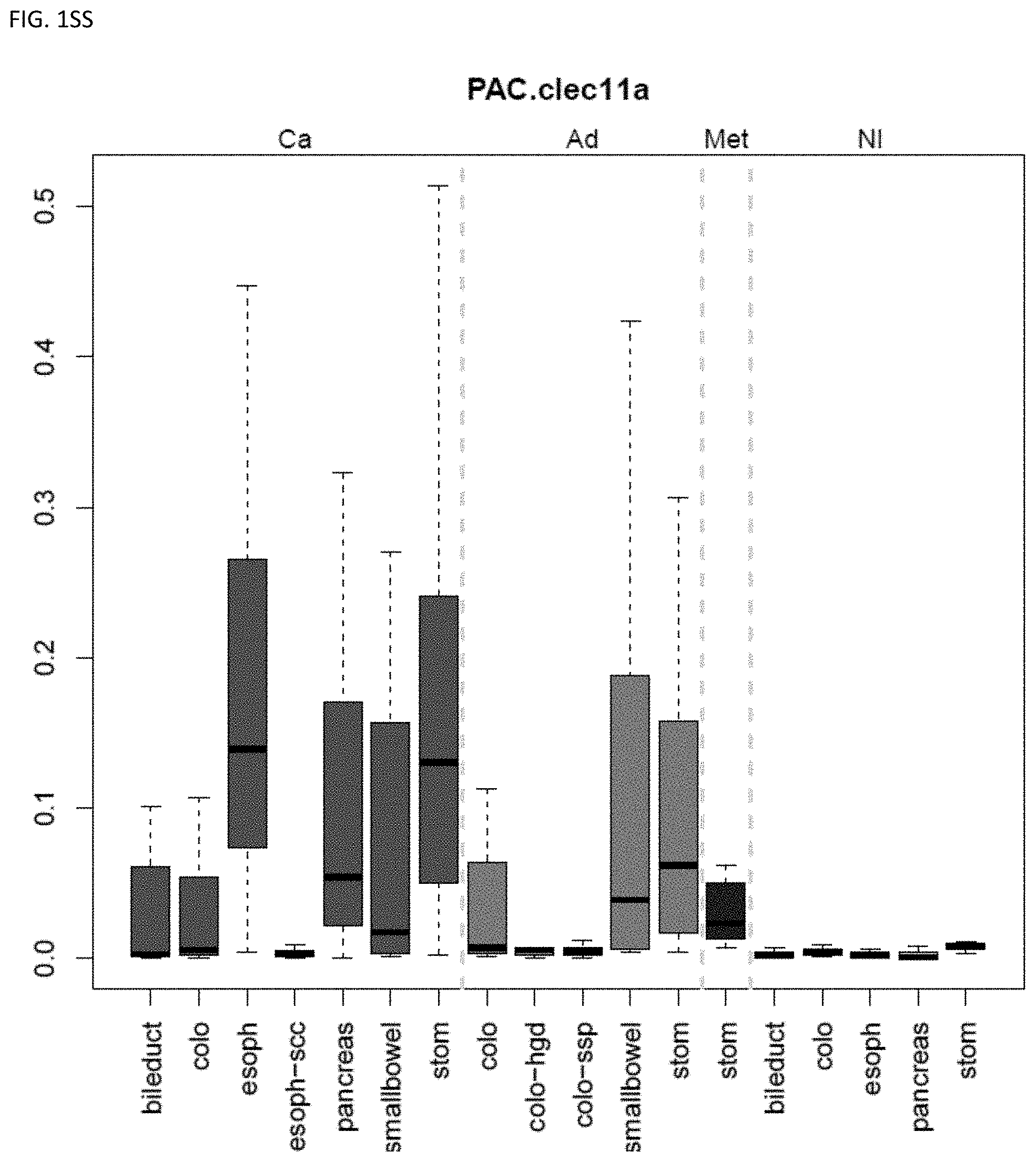
D00046
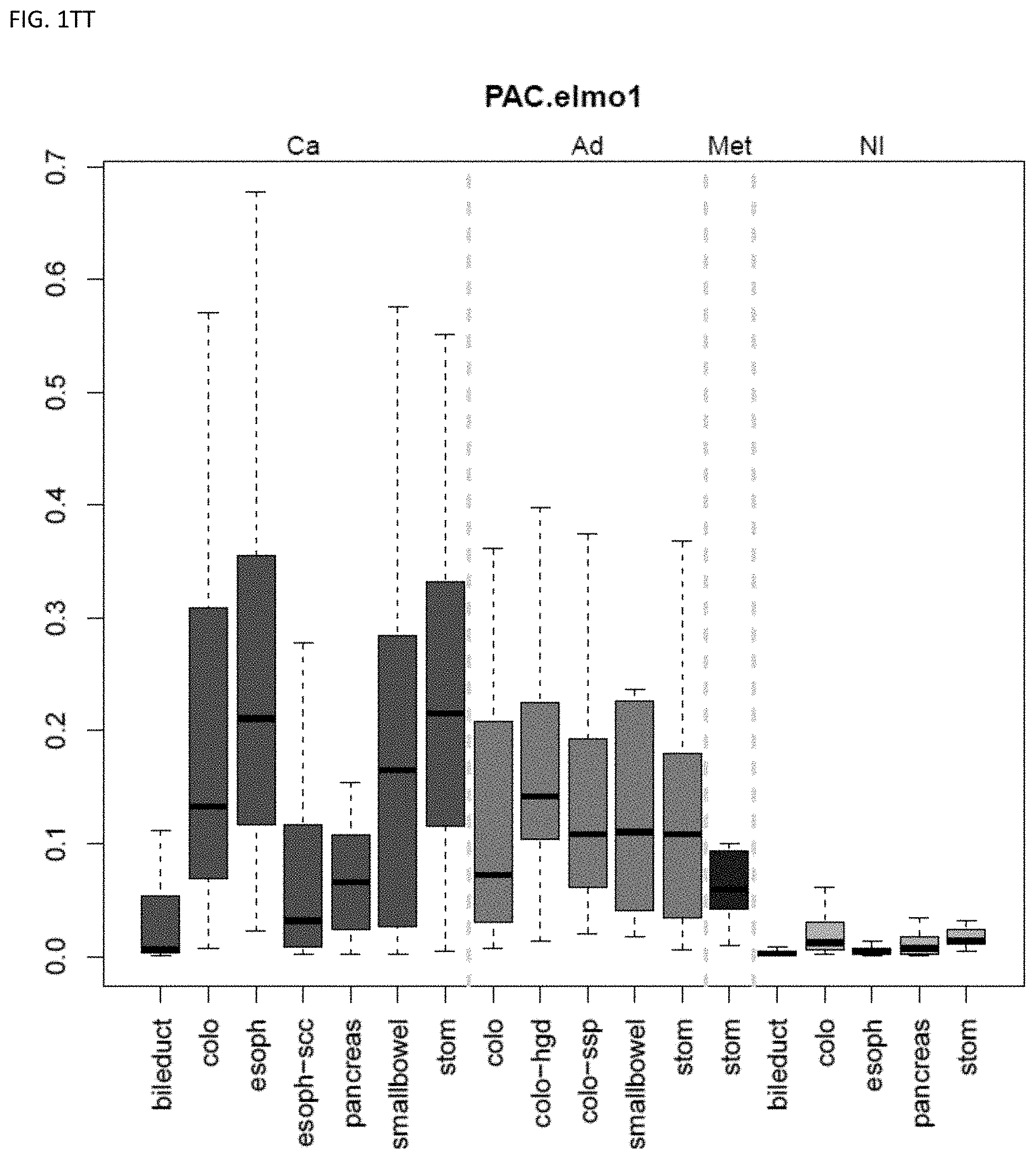
D00047
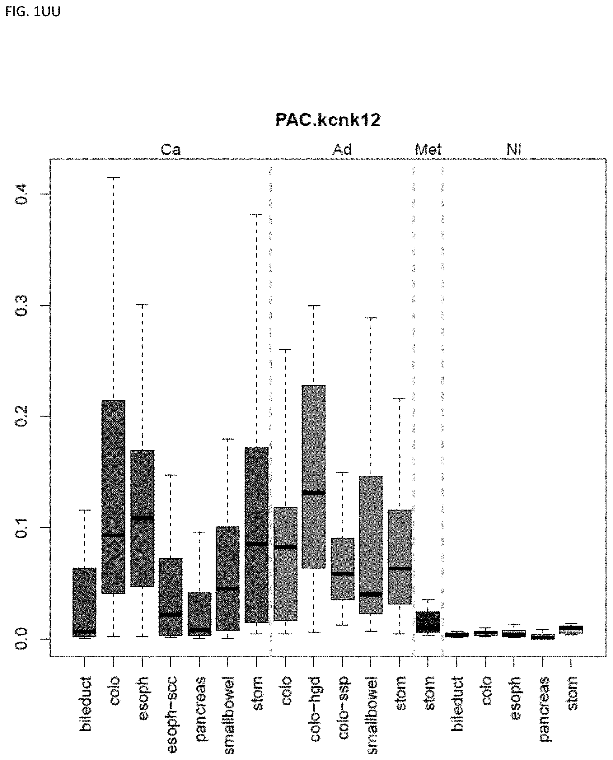
D00048
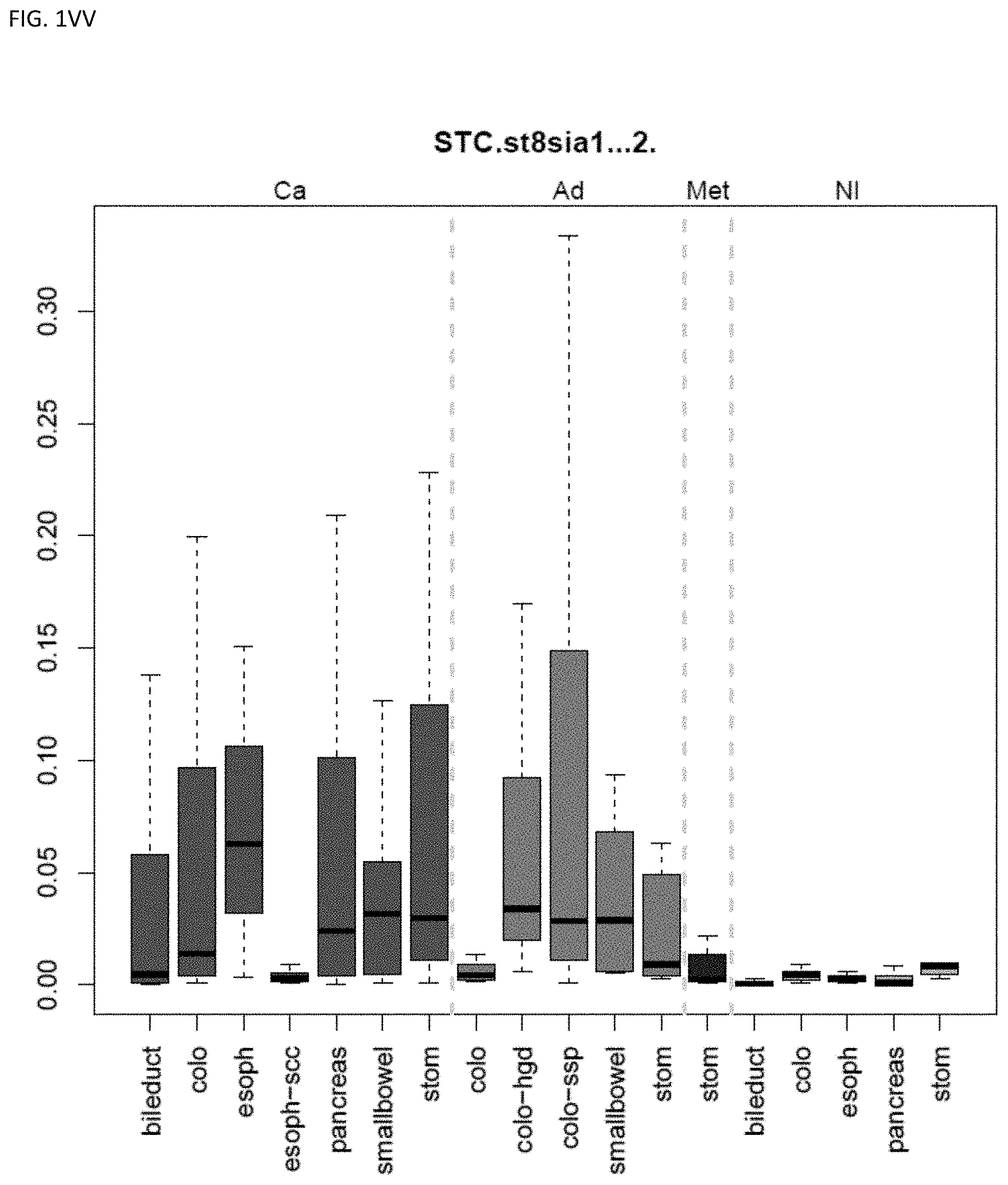
D00049
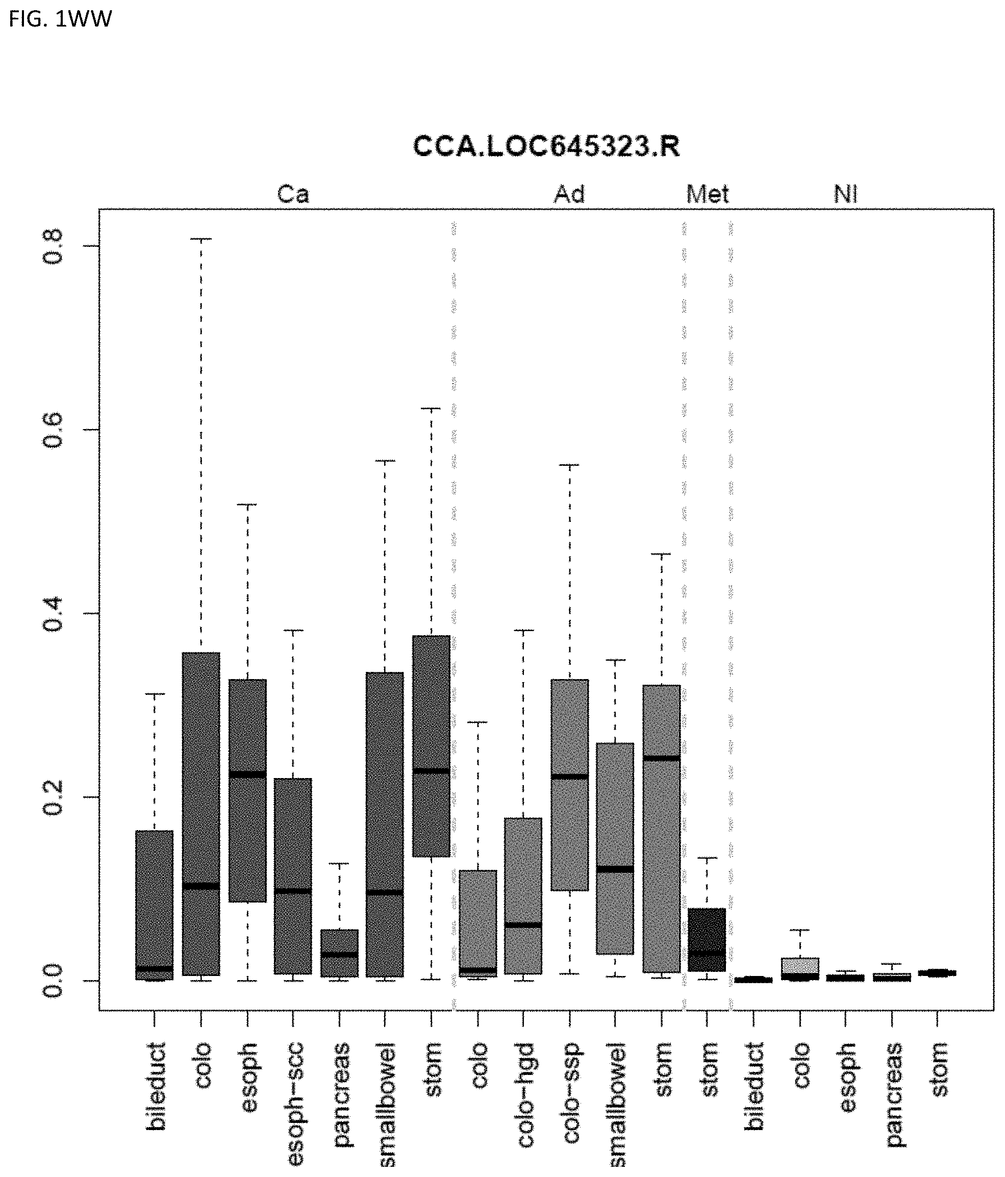
D00050
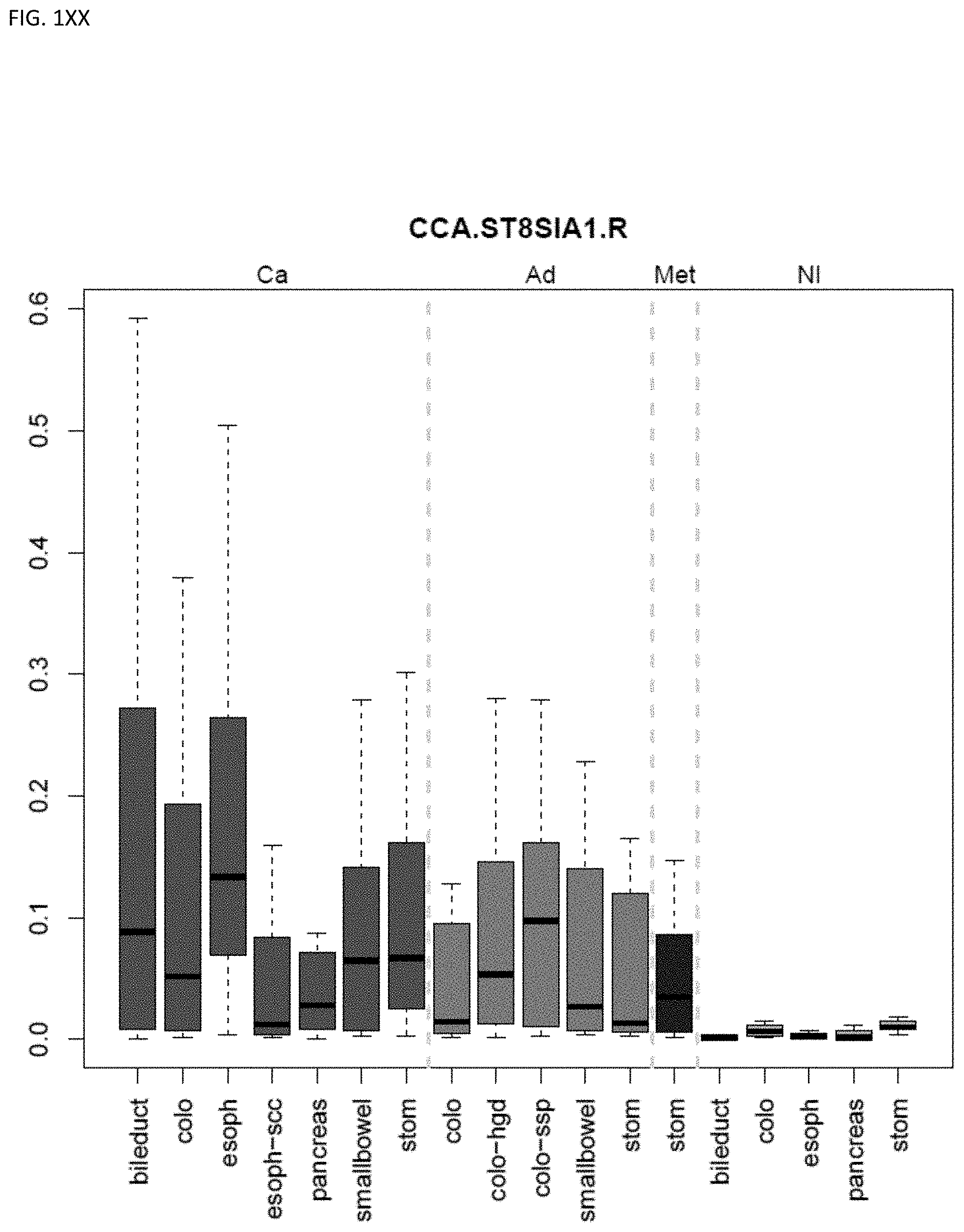
D00051
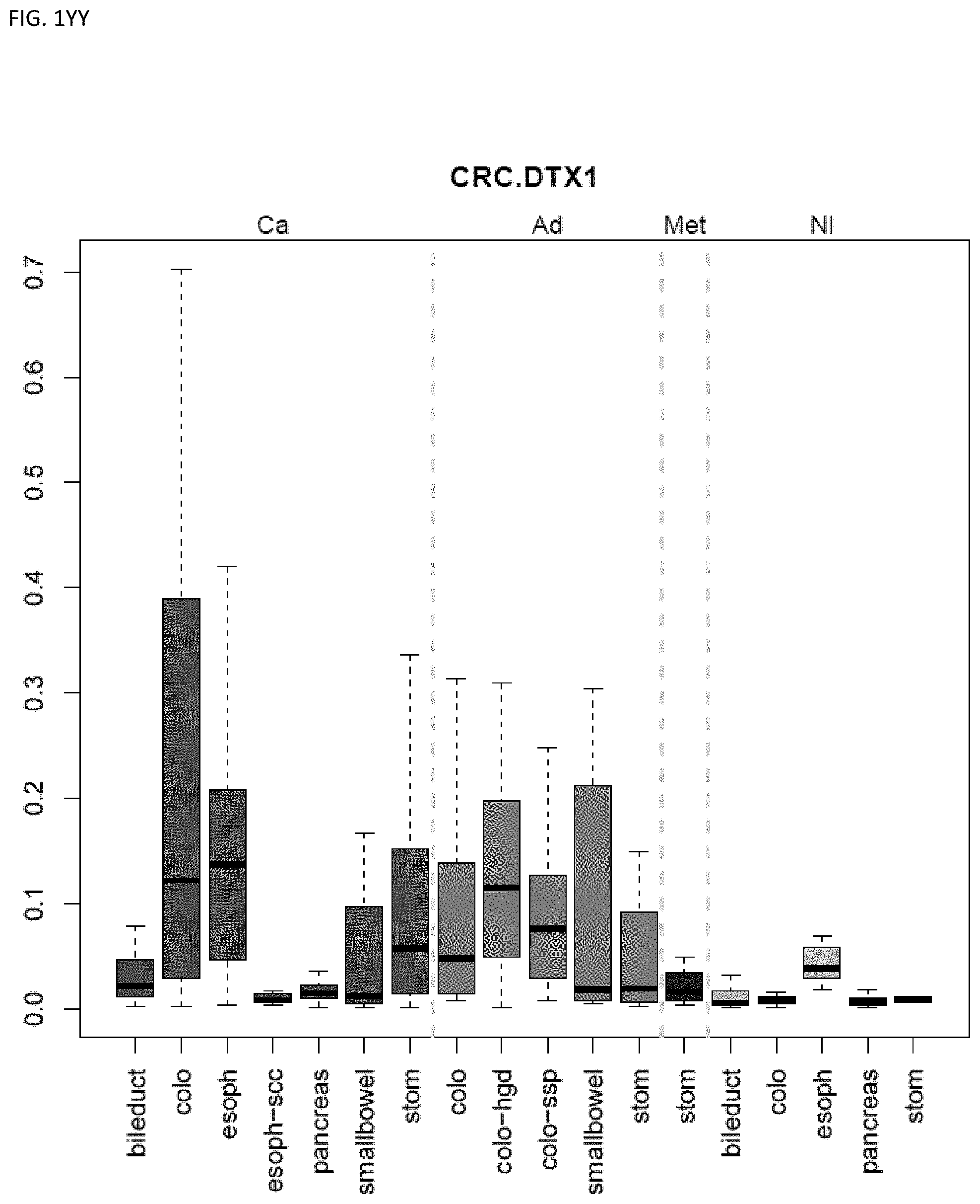
D00052
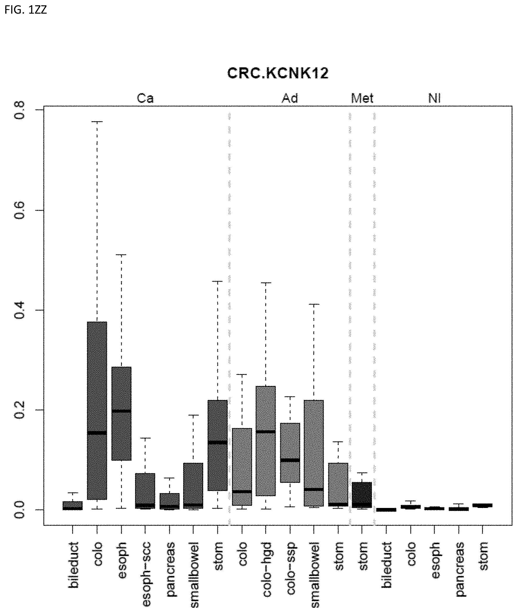
D00053
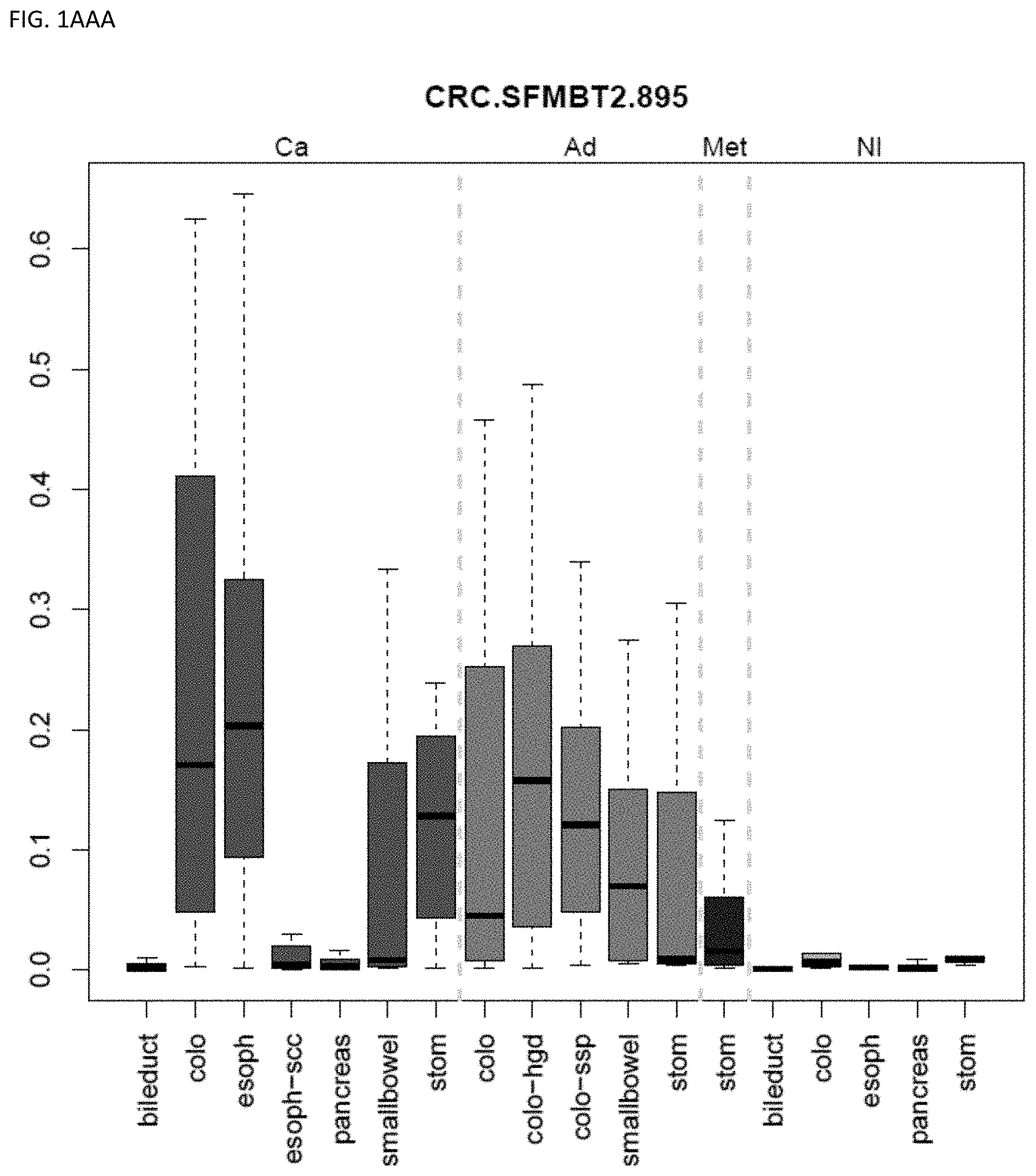
D00054
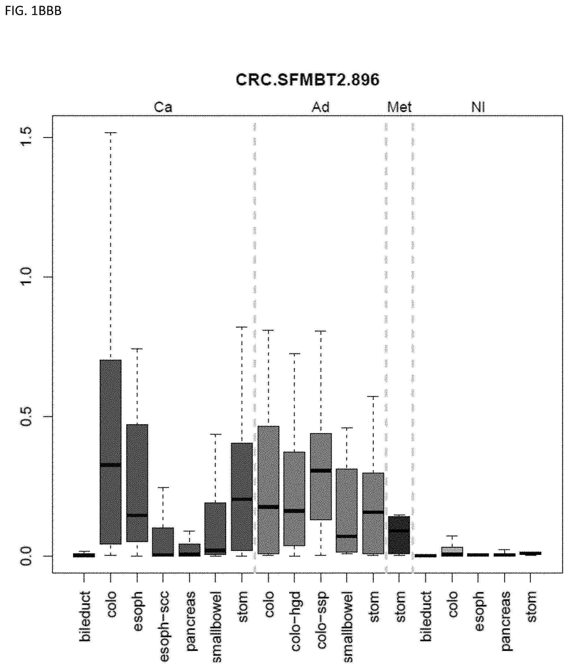
D00055
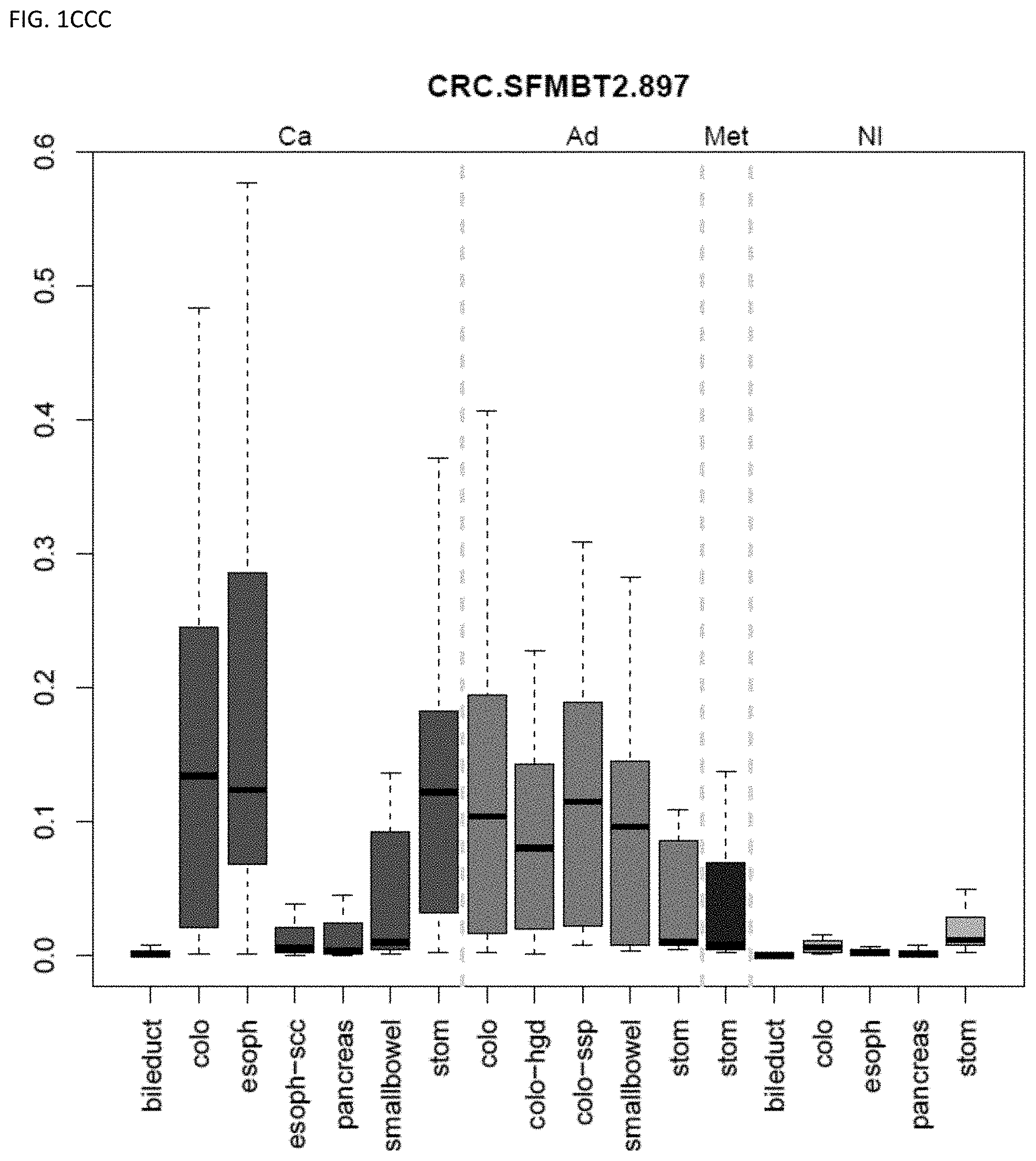
D00056
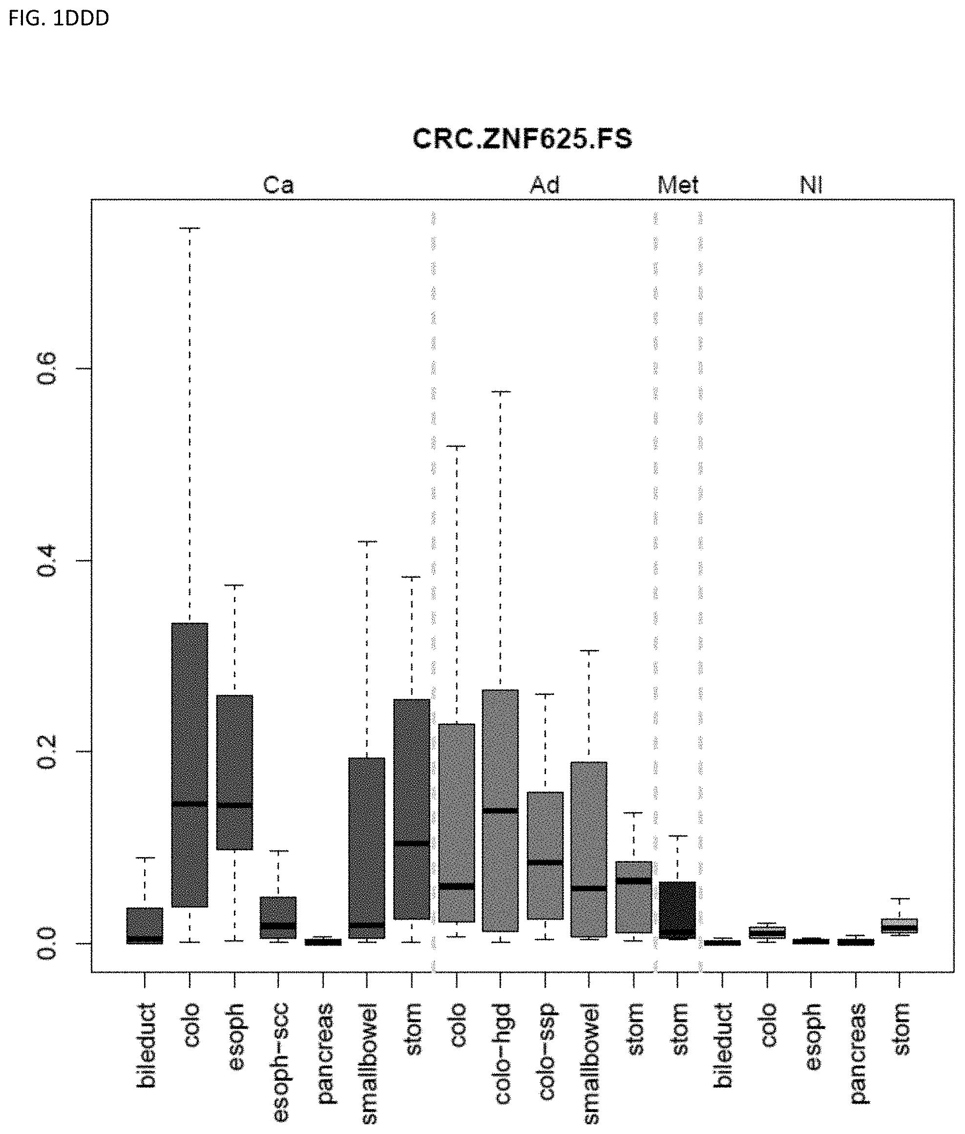
D00057
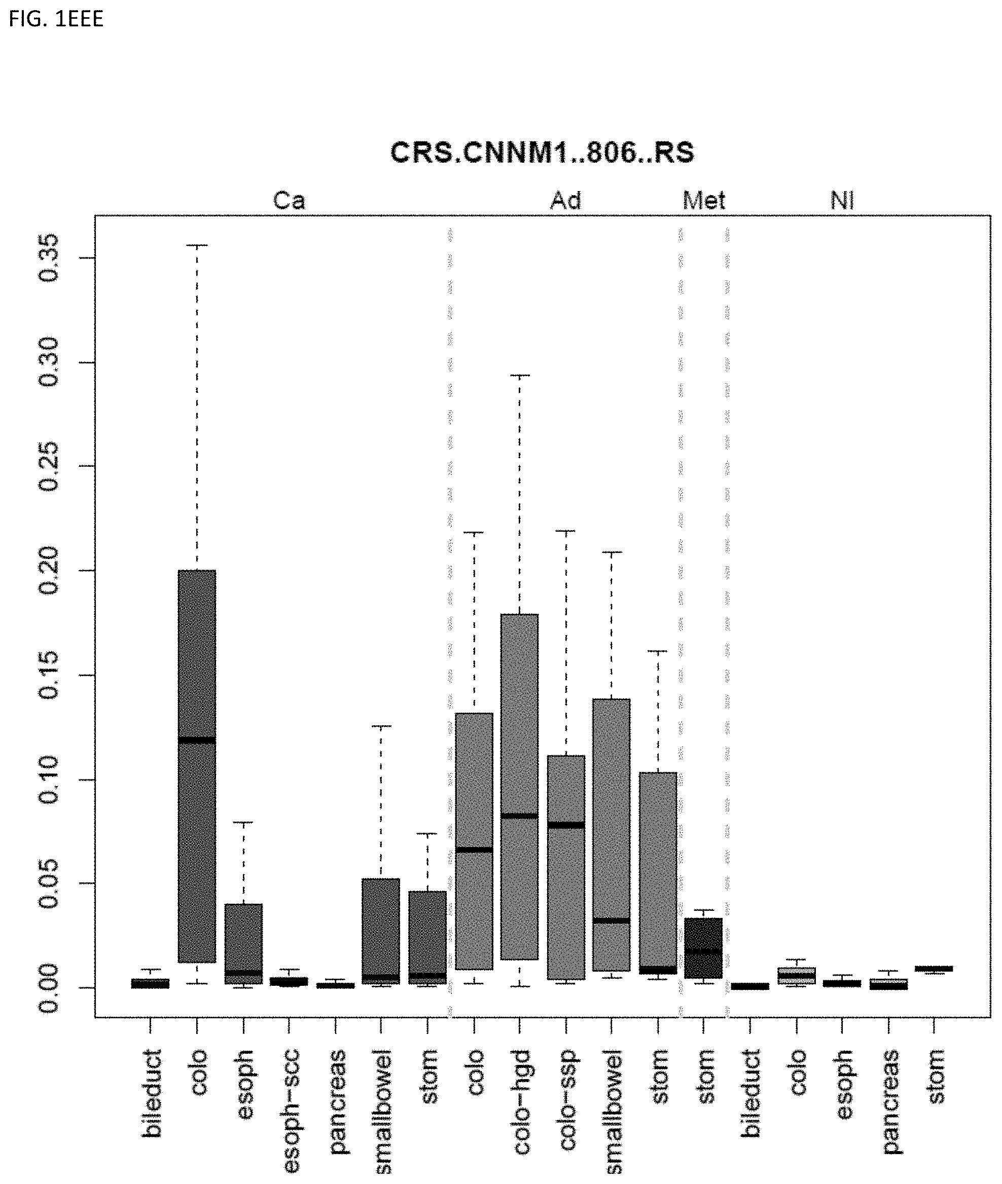
D00058
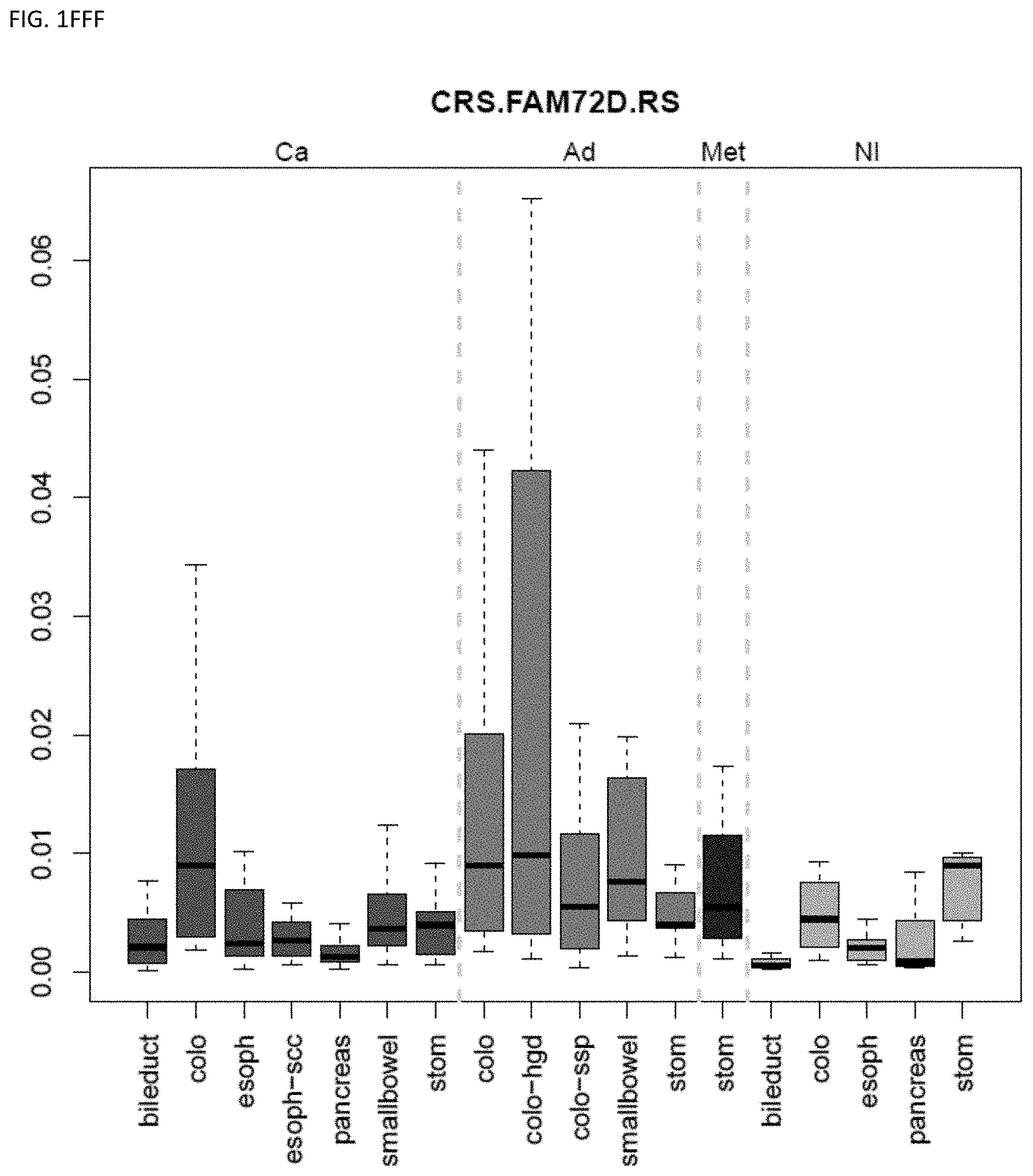
D00059
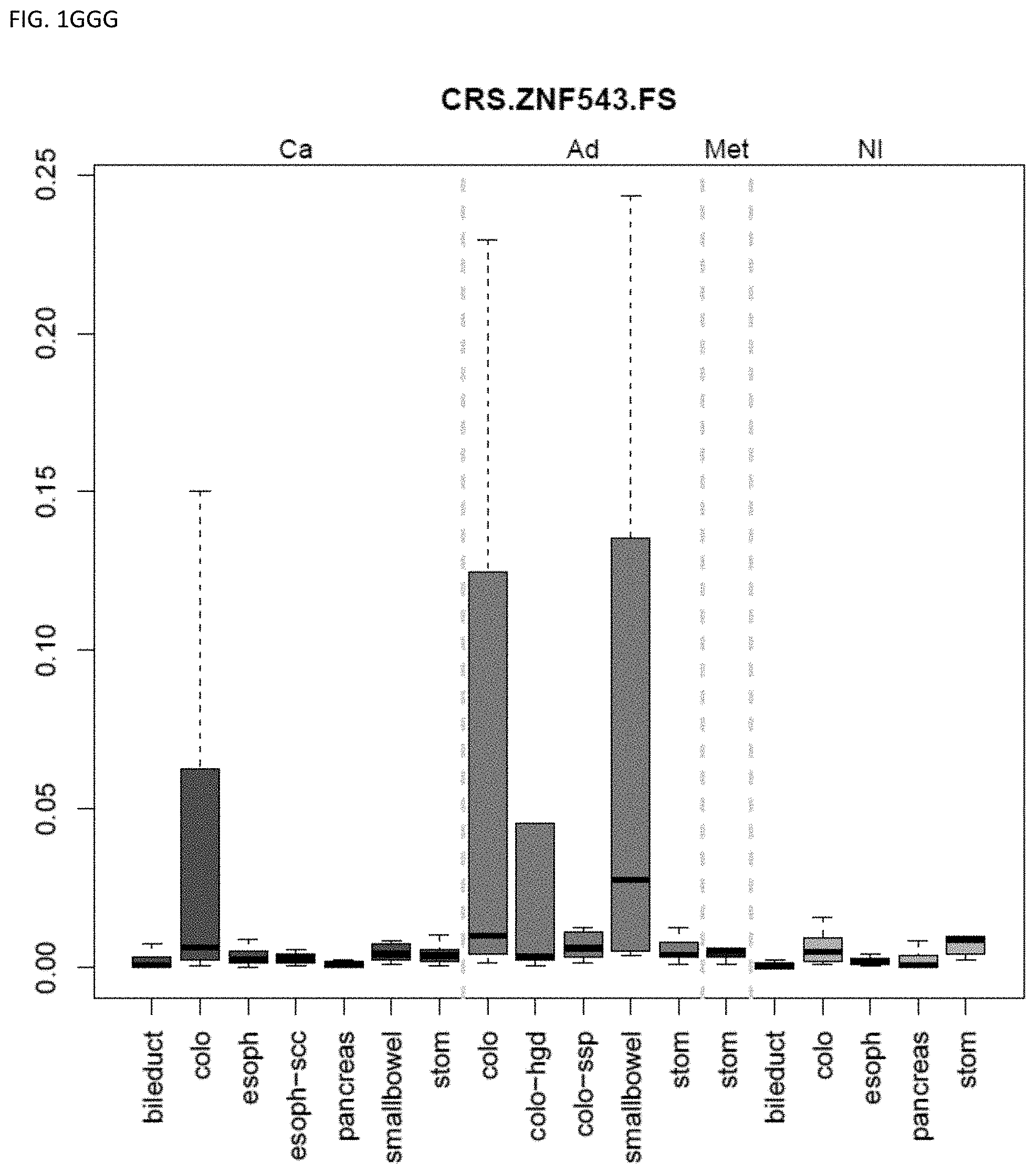
D00060
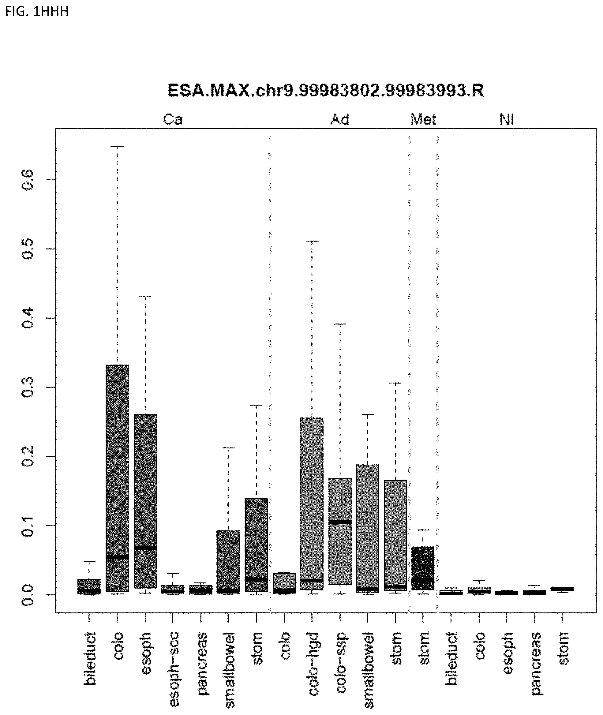
D00061
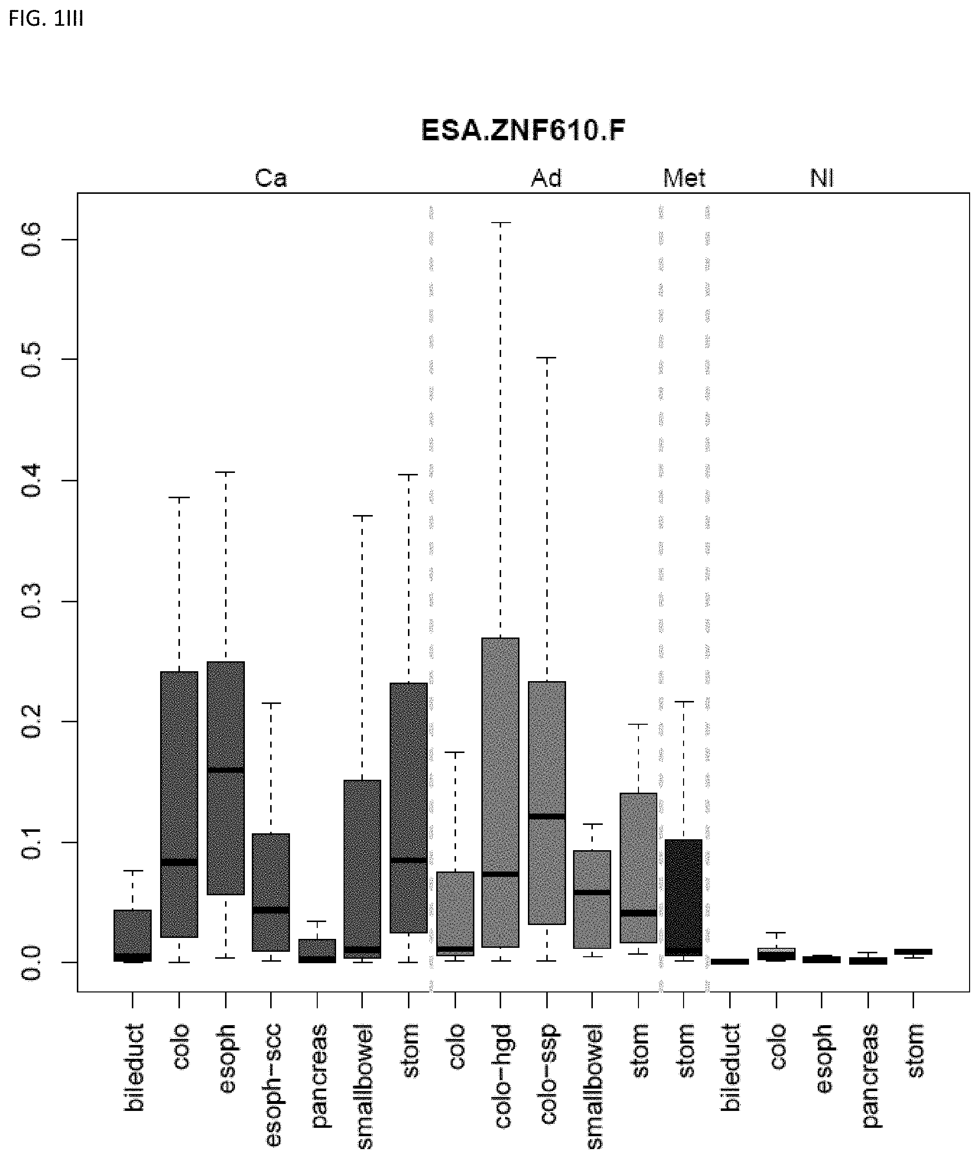
D00062
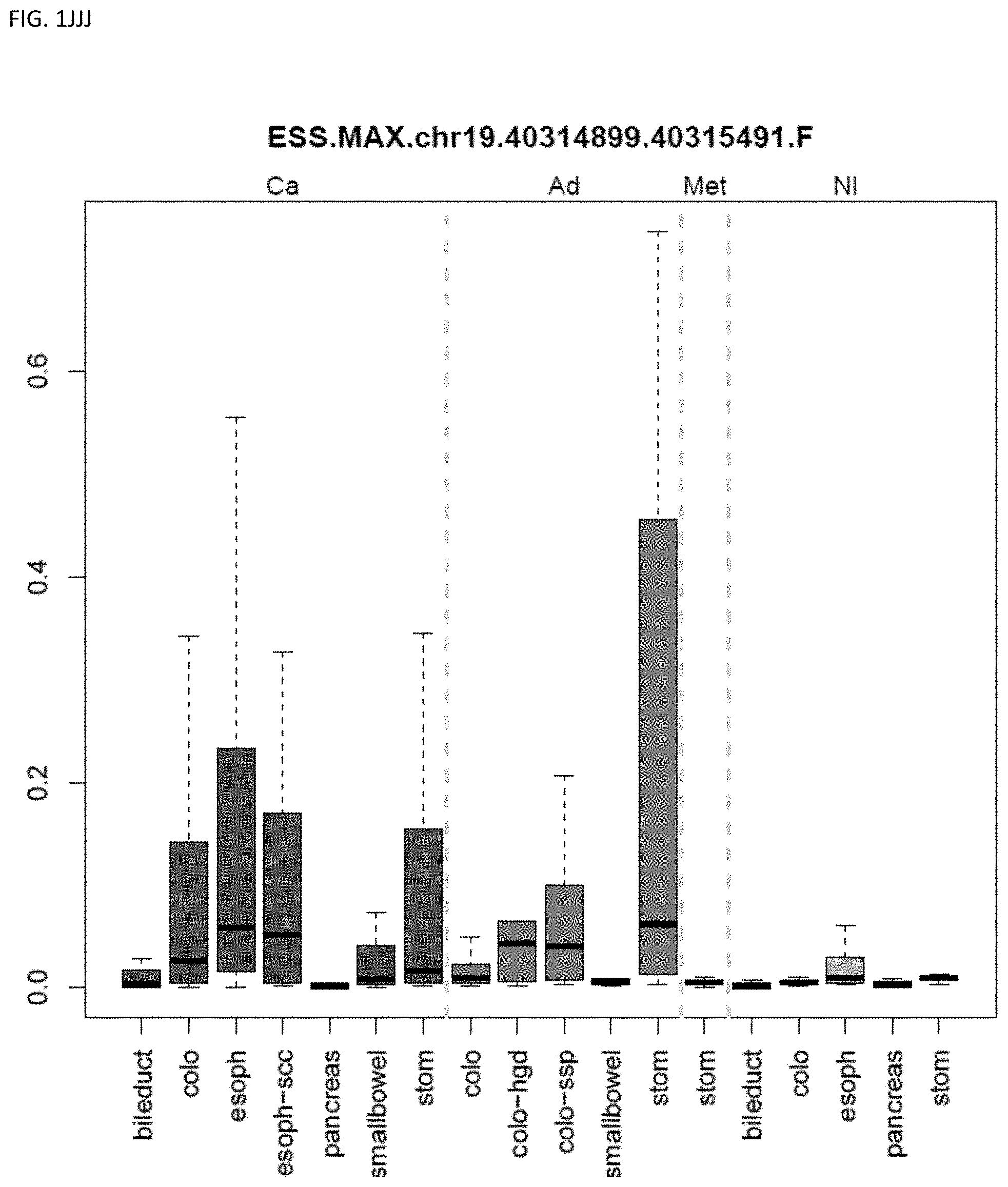
D00063
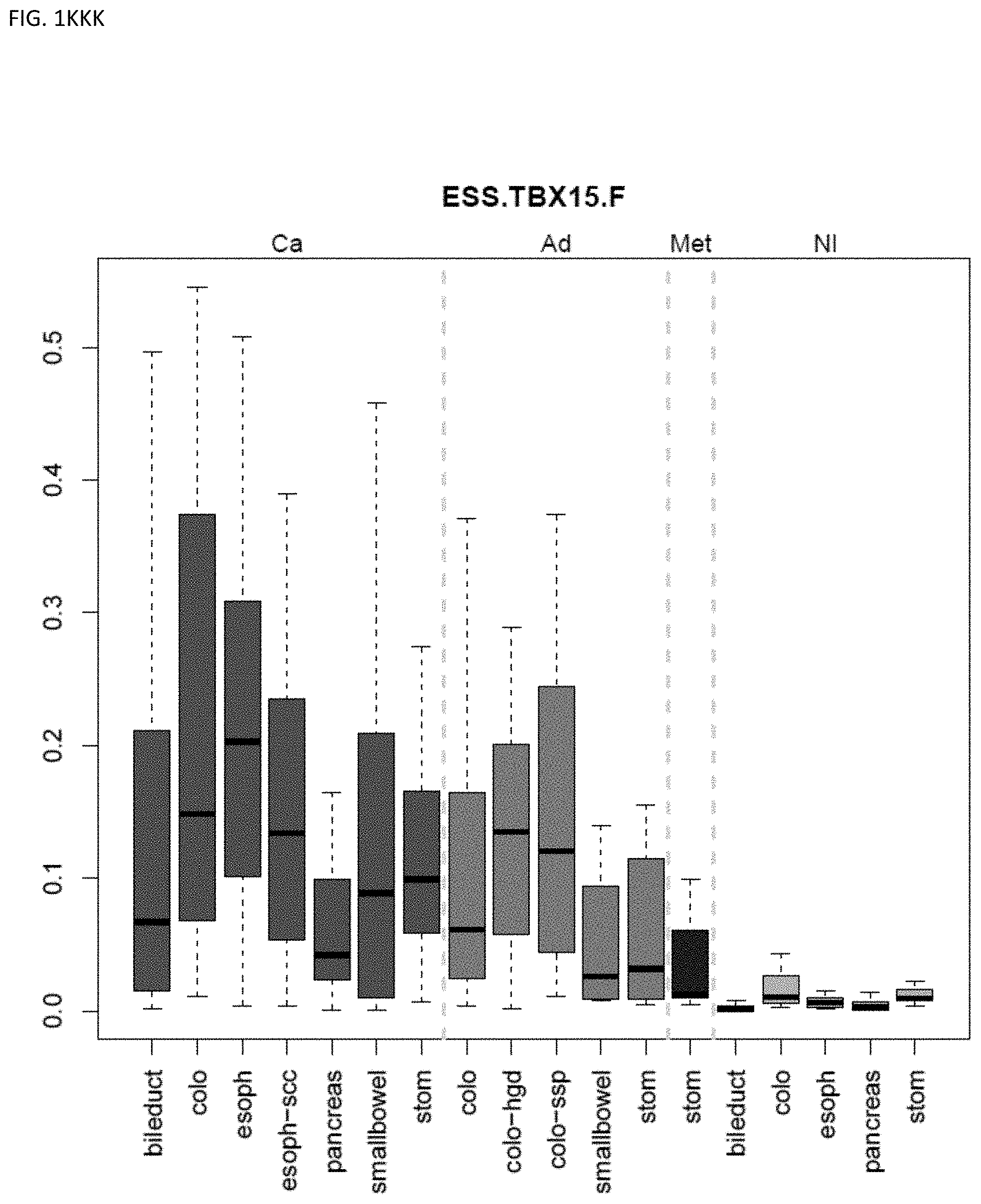
D00064
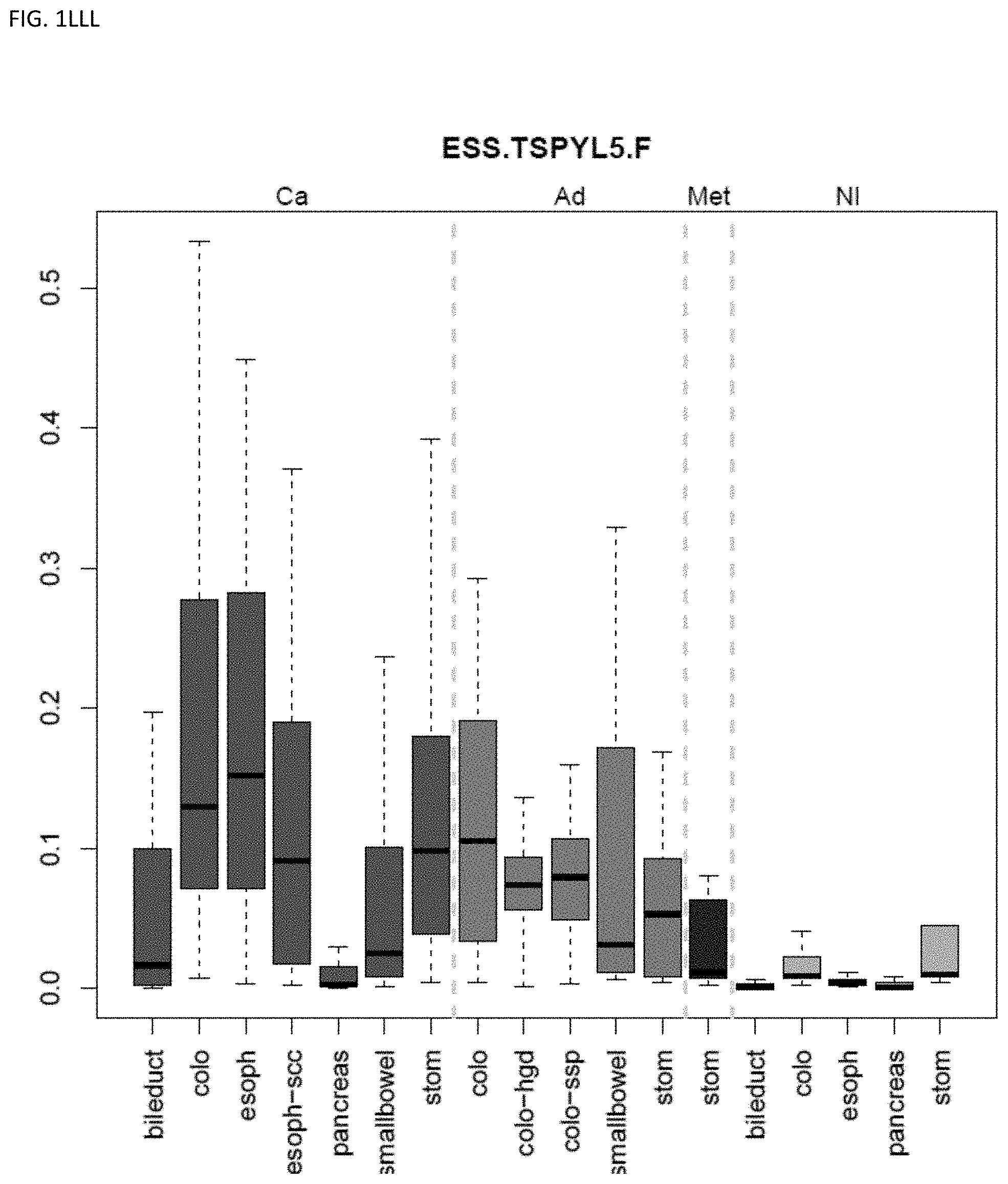
D00065
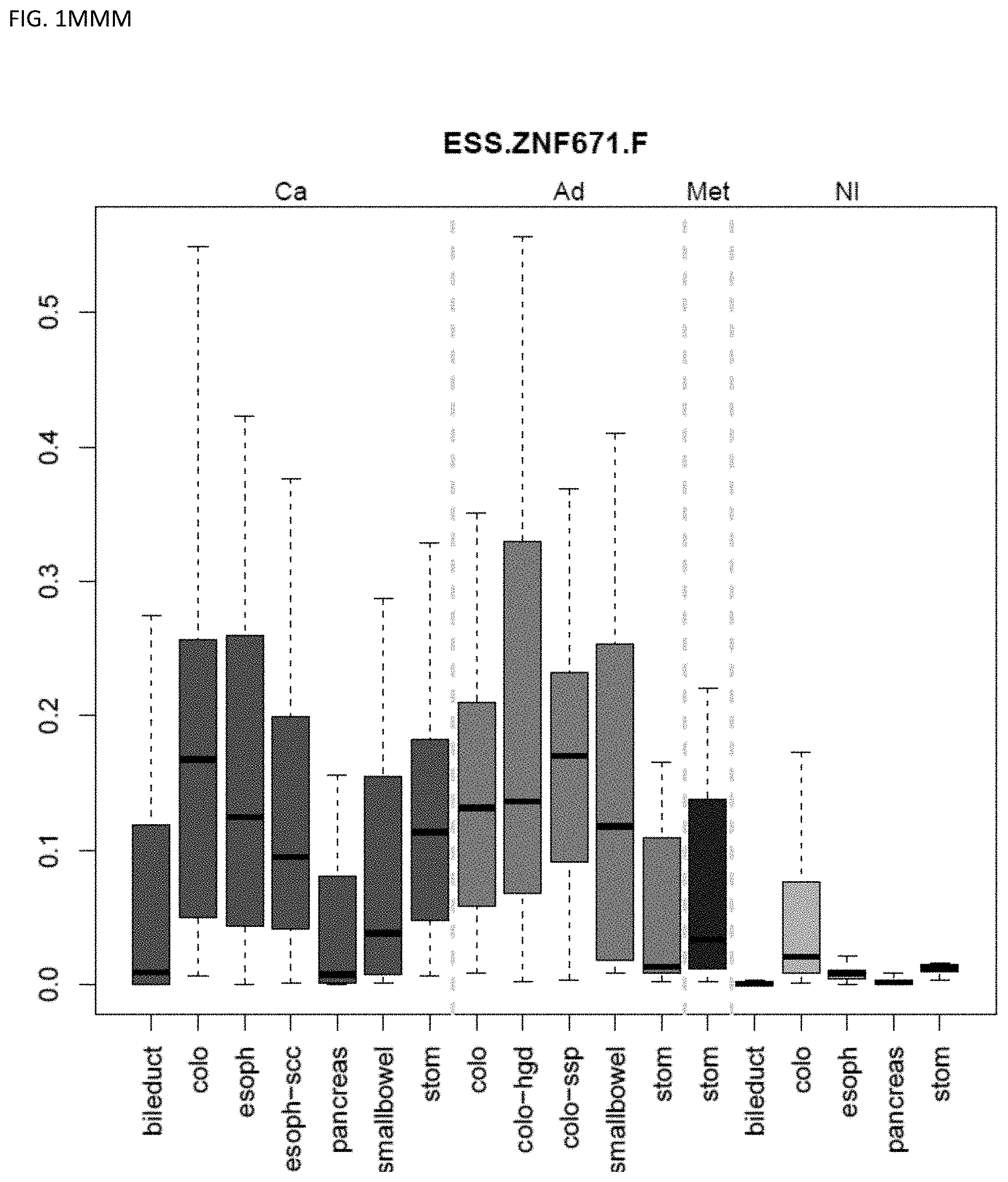
D00066
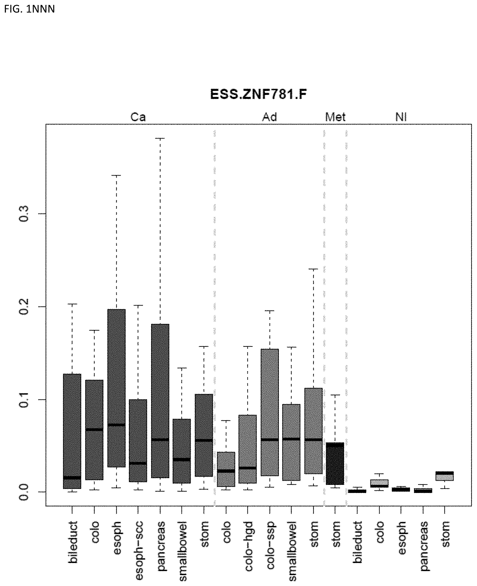
D00067
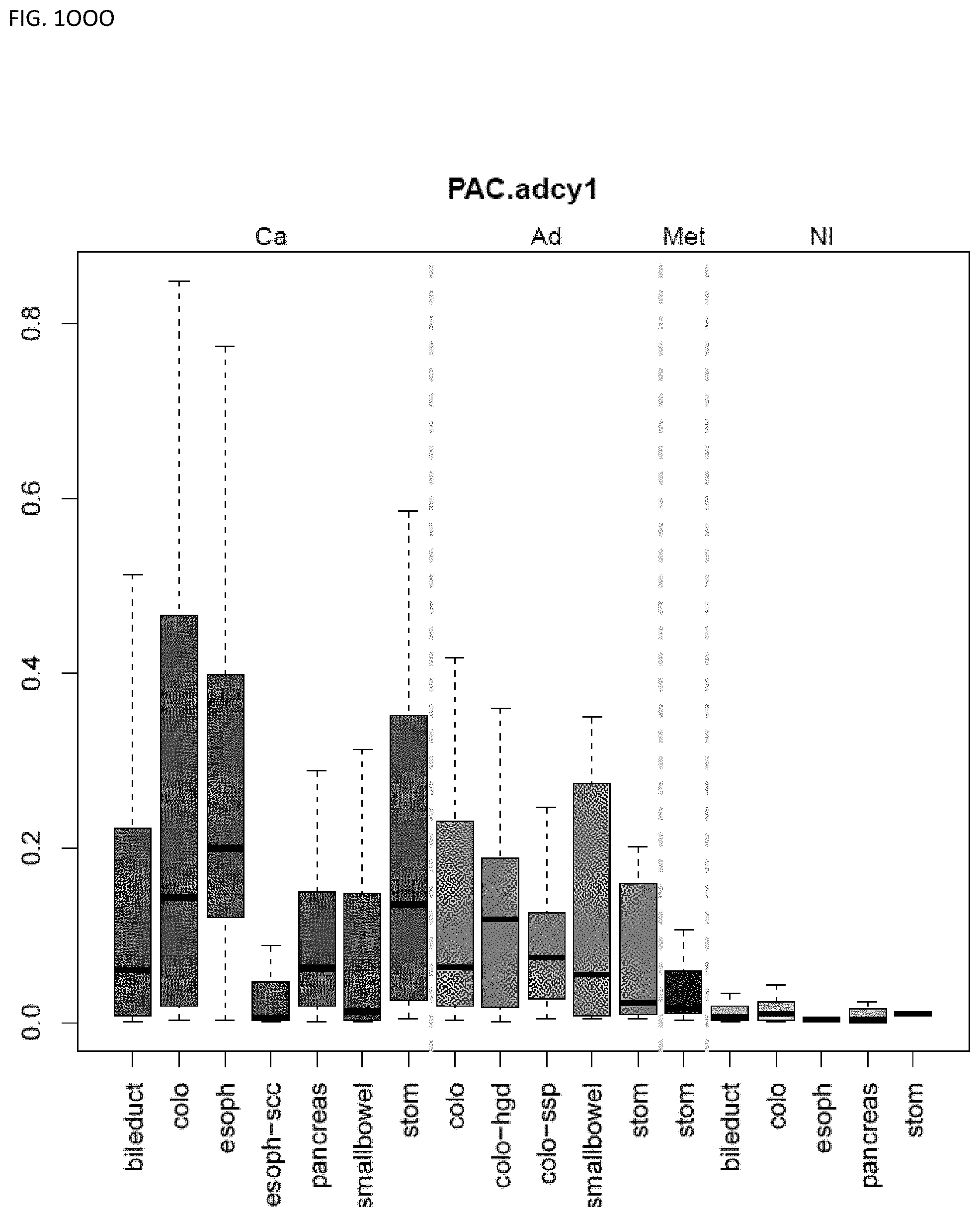
D00068
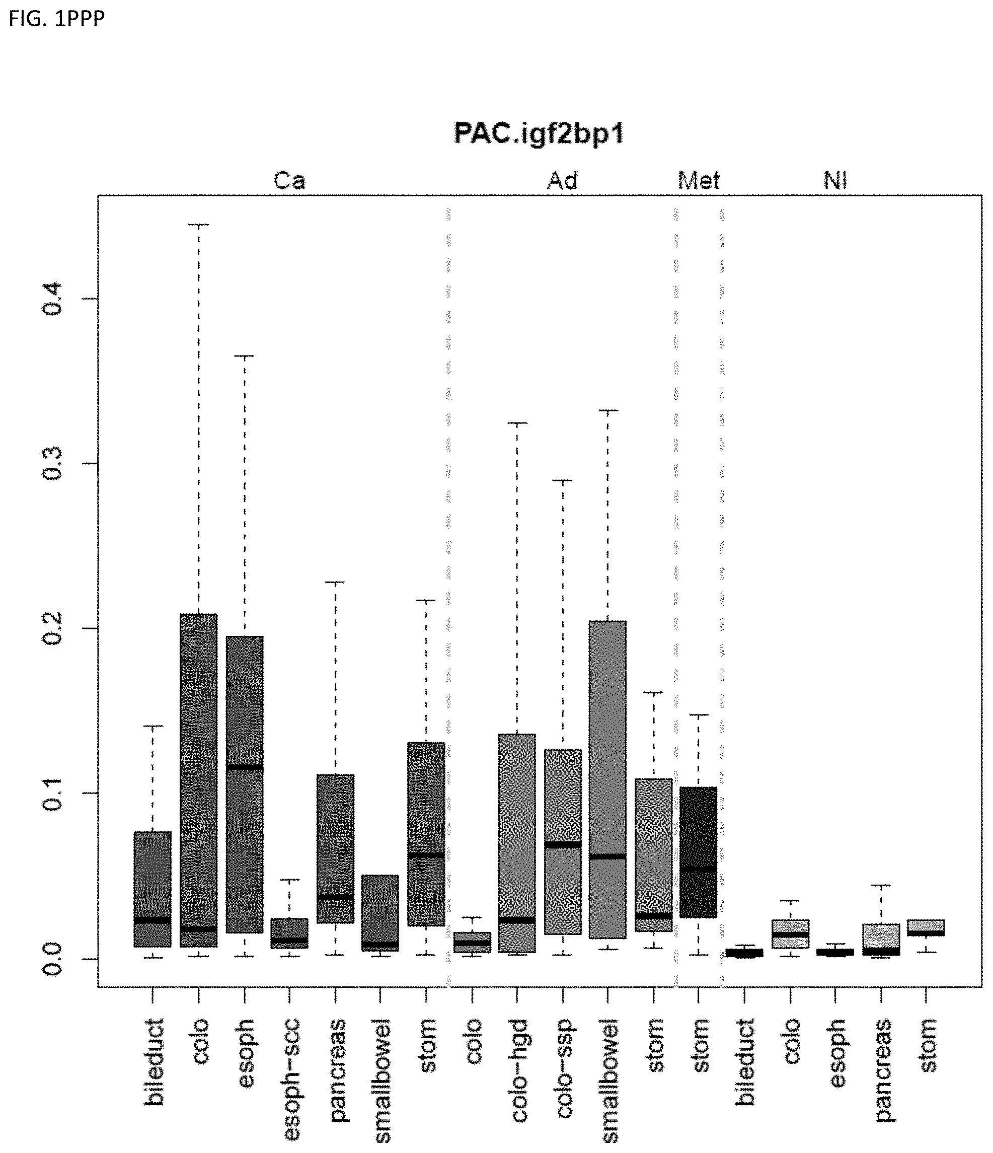
D00069
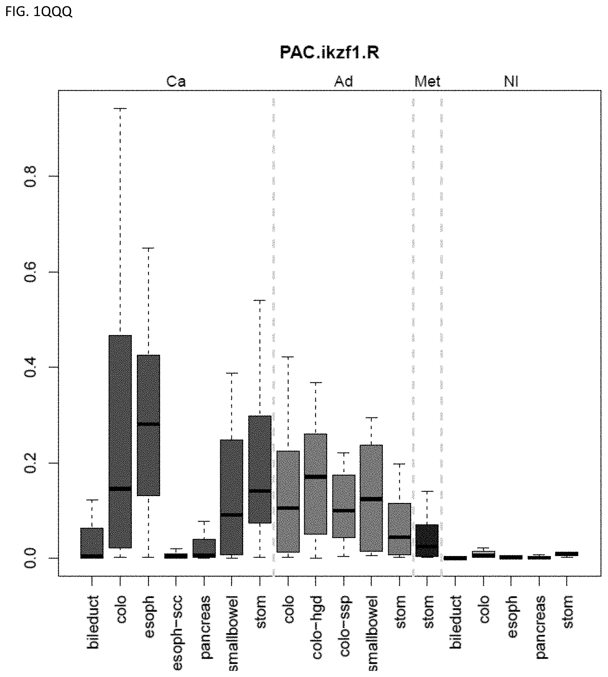
D00070
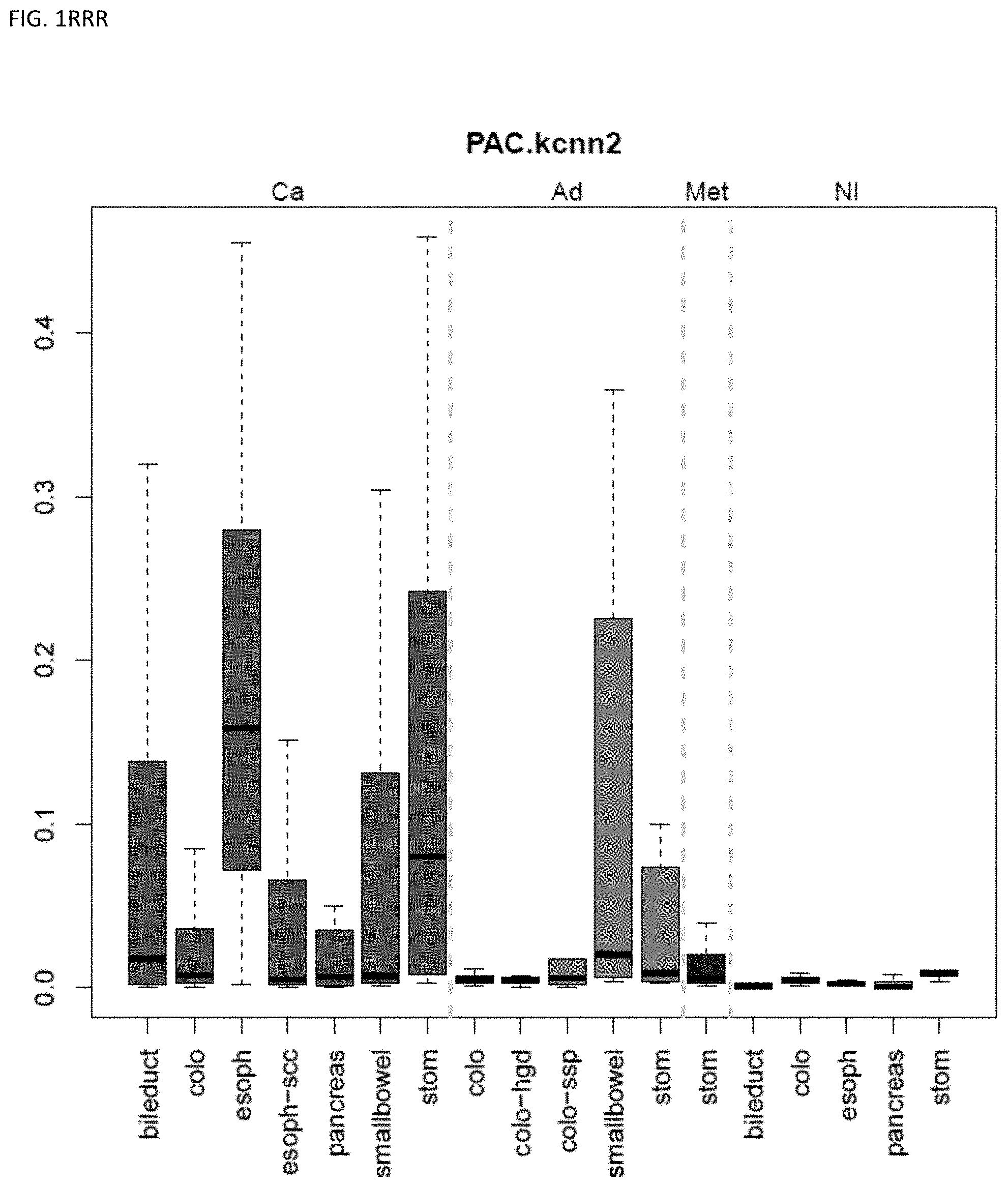
D00071
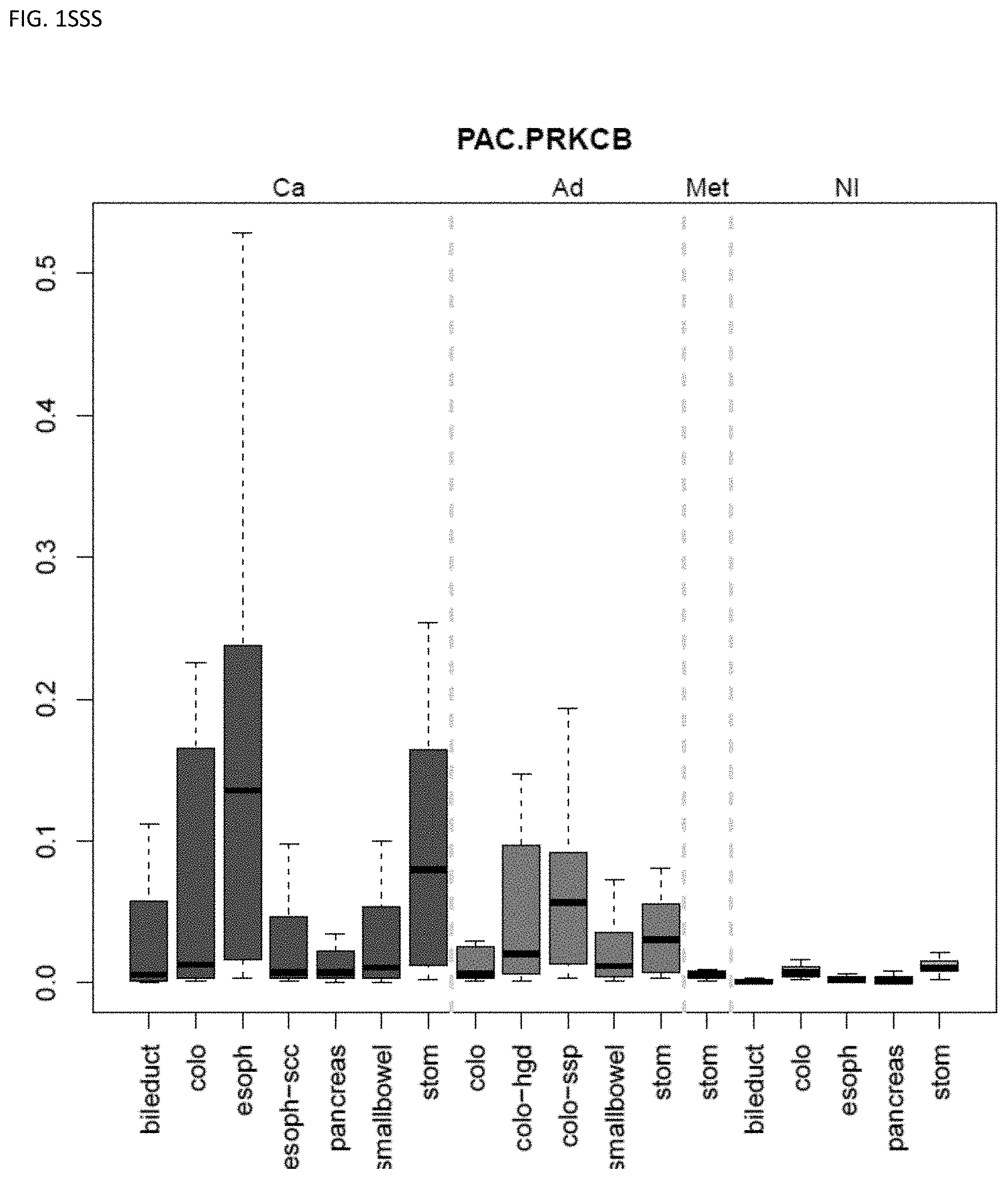
D00072
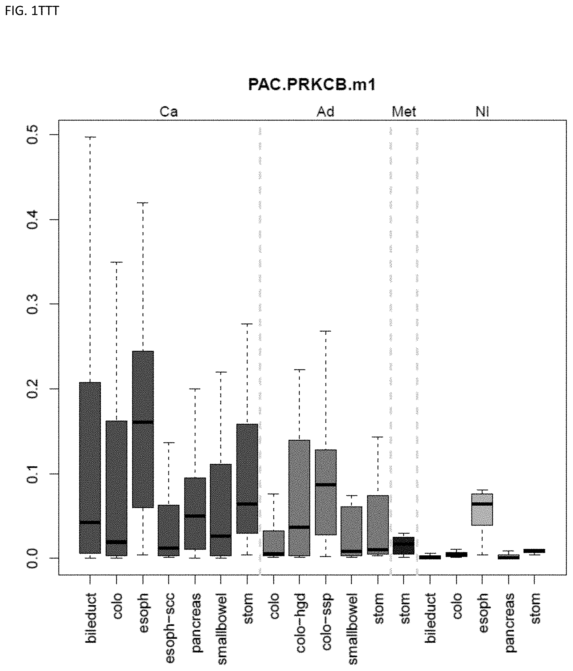
D00073
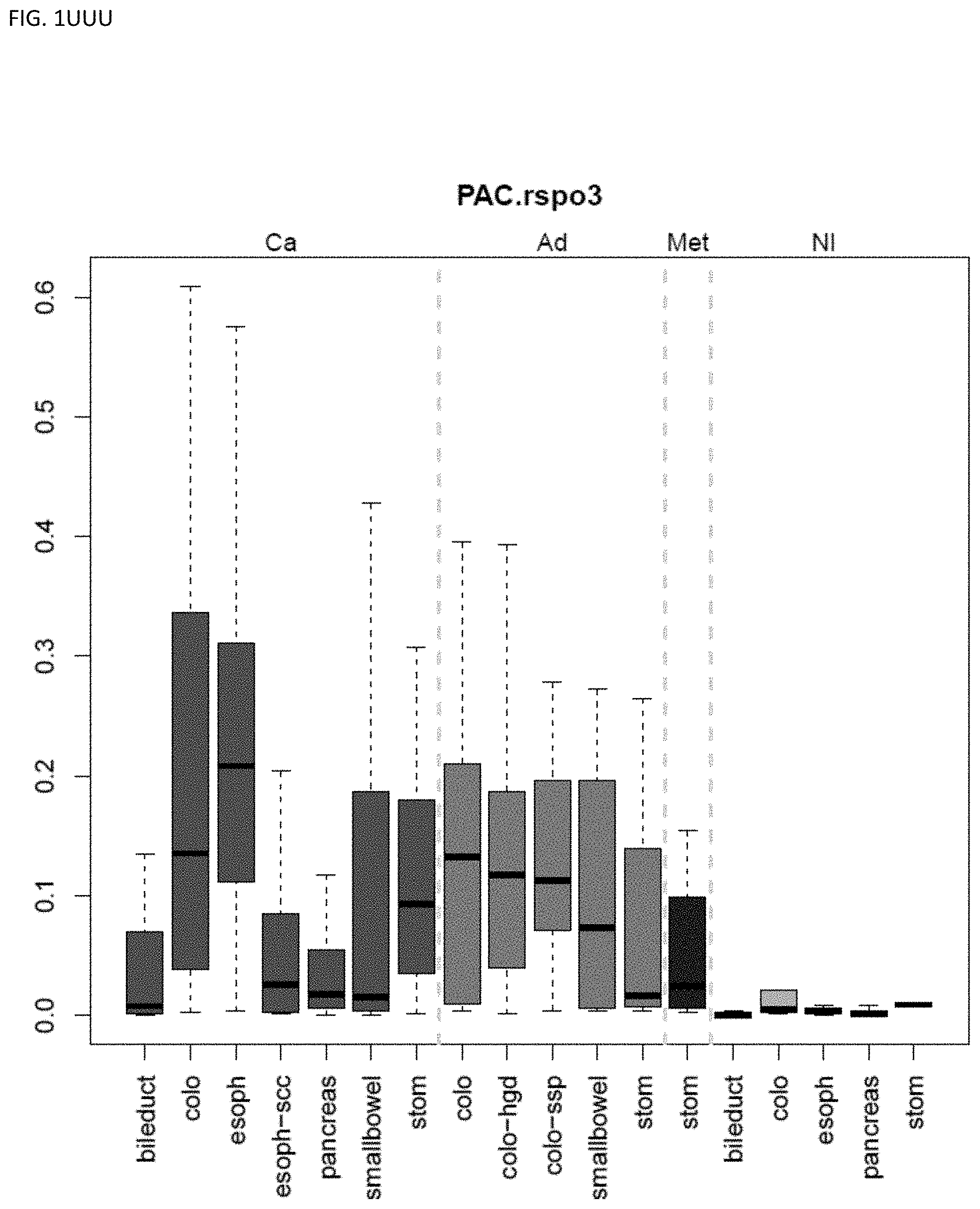
D00074
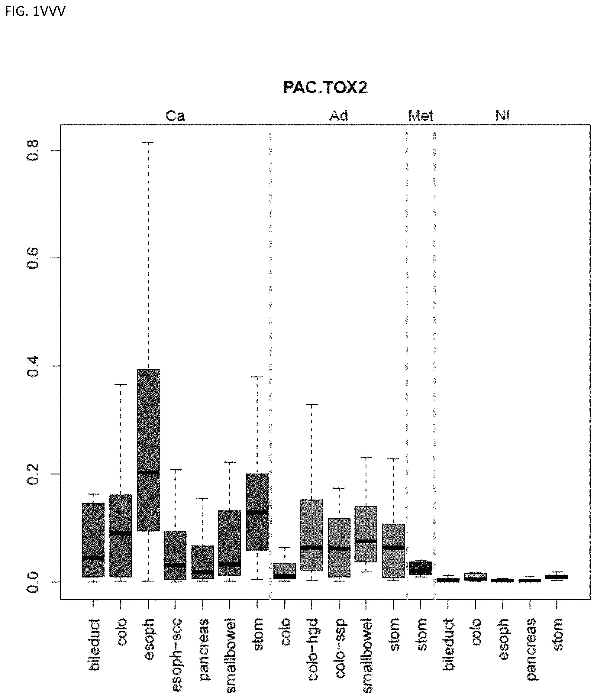
D00075
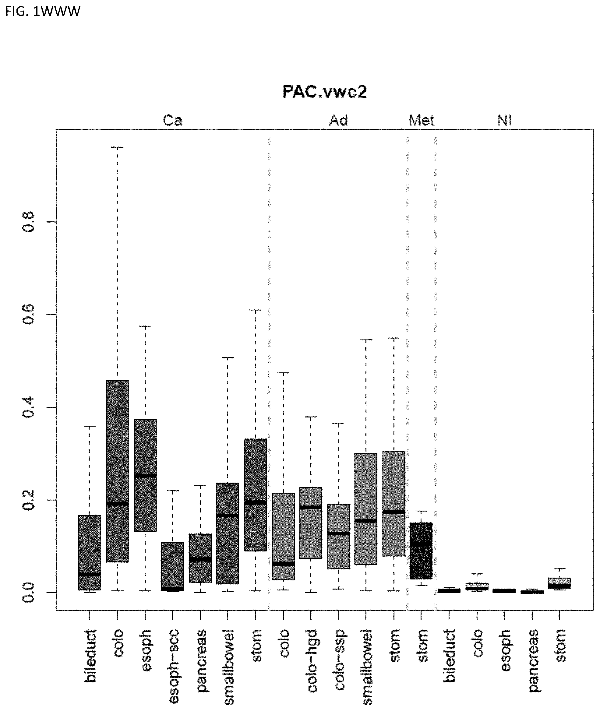
D00076
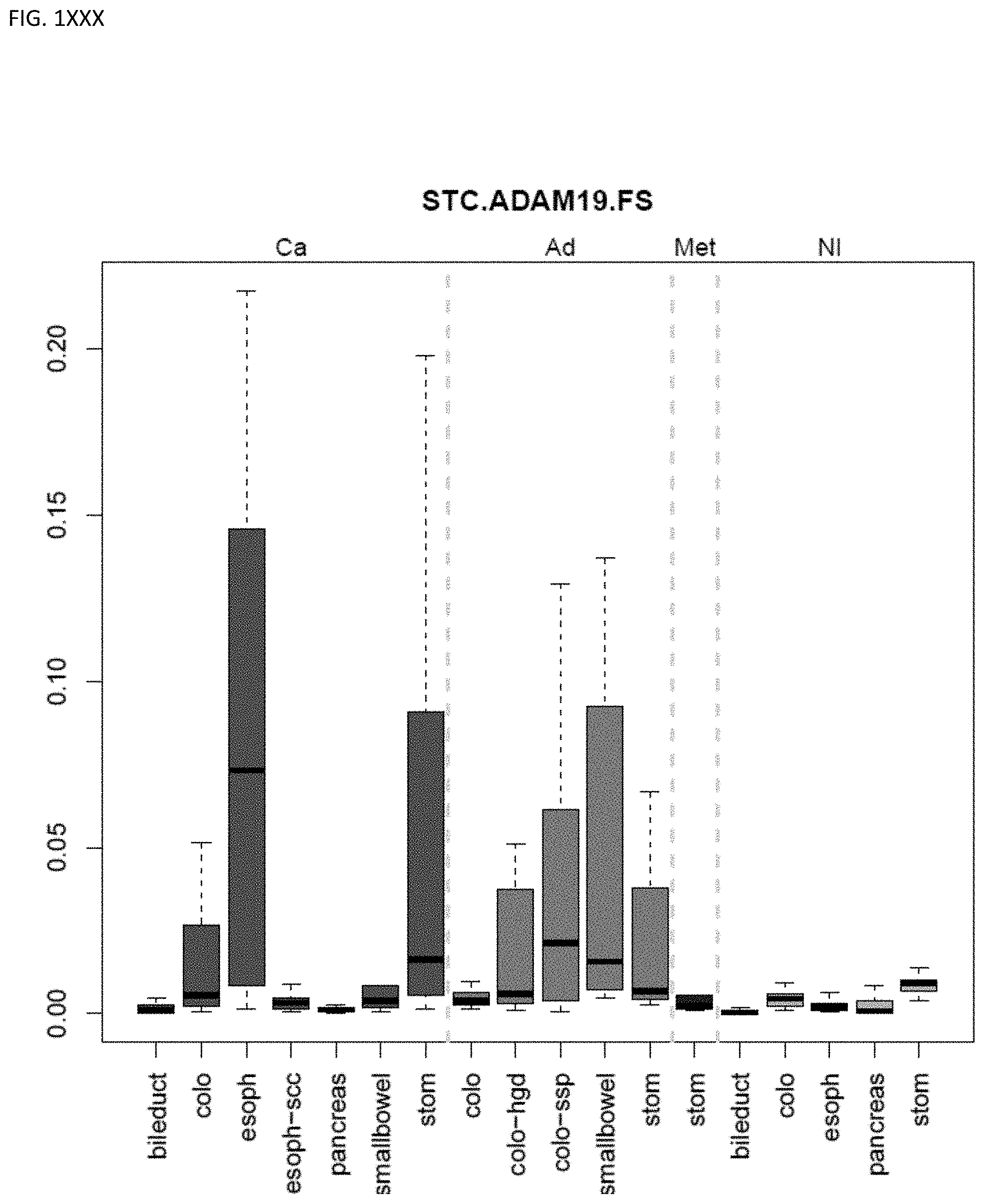
D00077
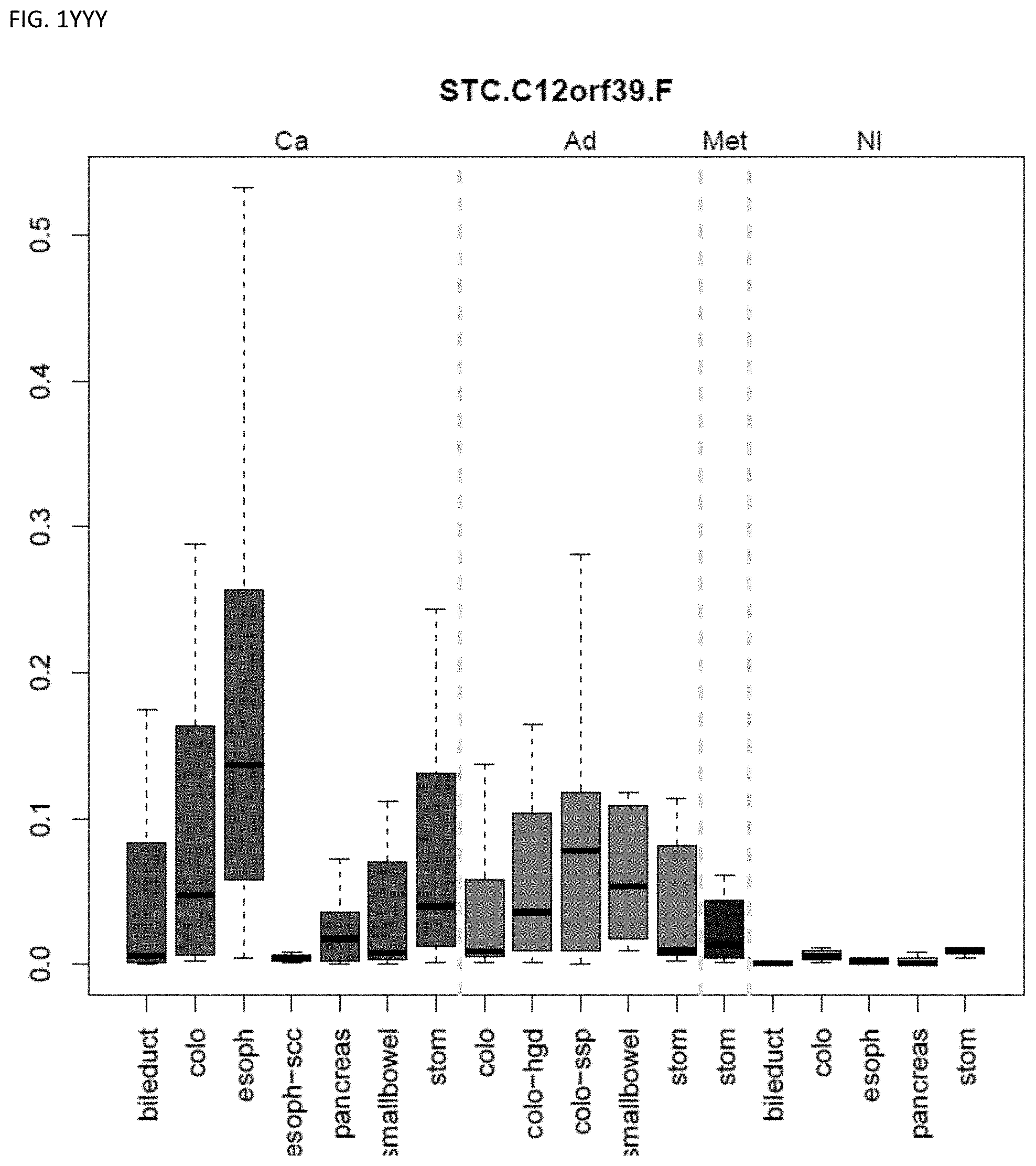
D00078
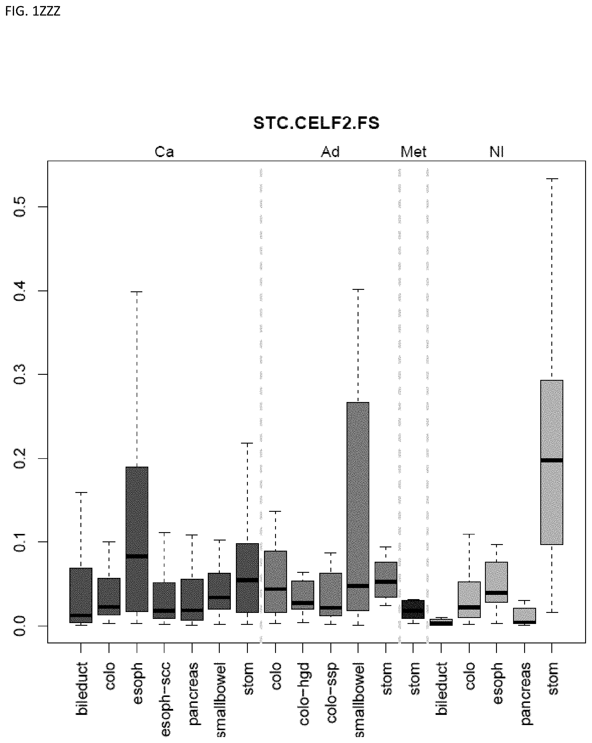
D00079
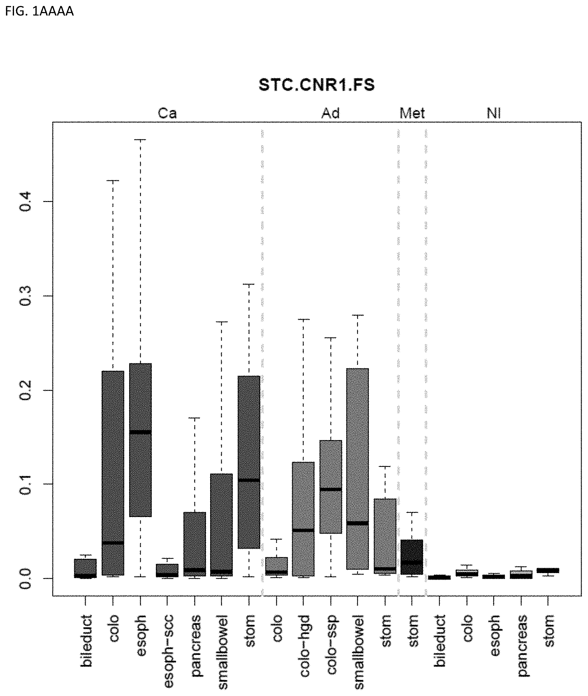
D00080
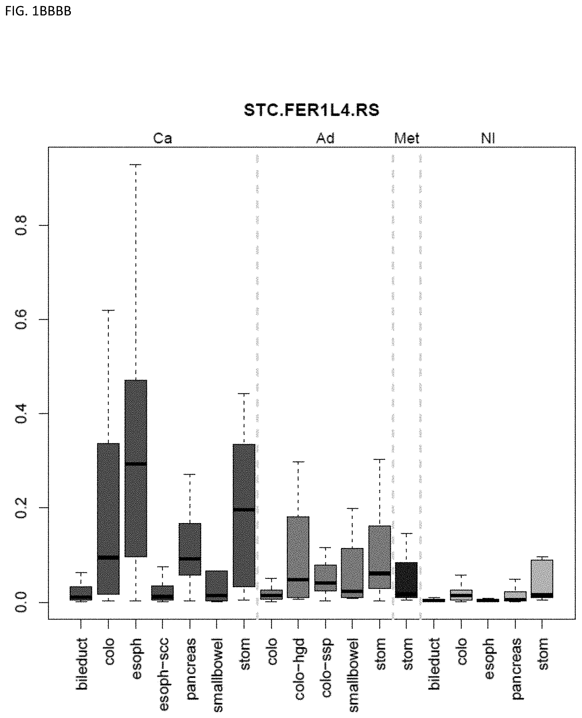
D00081
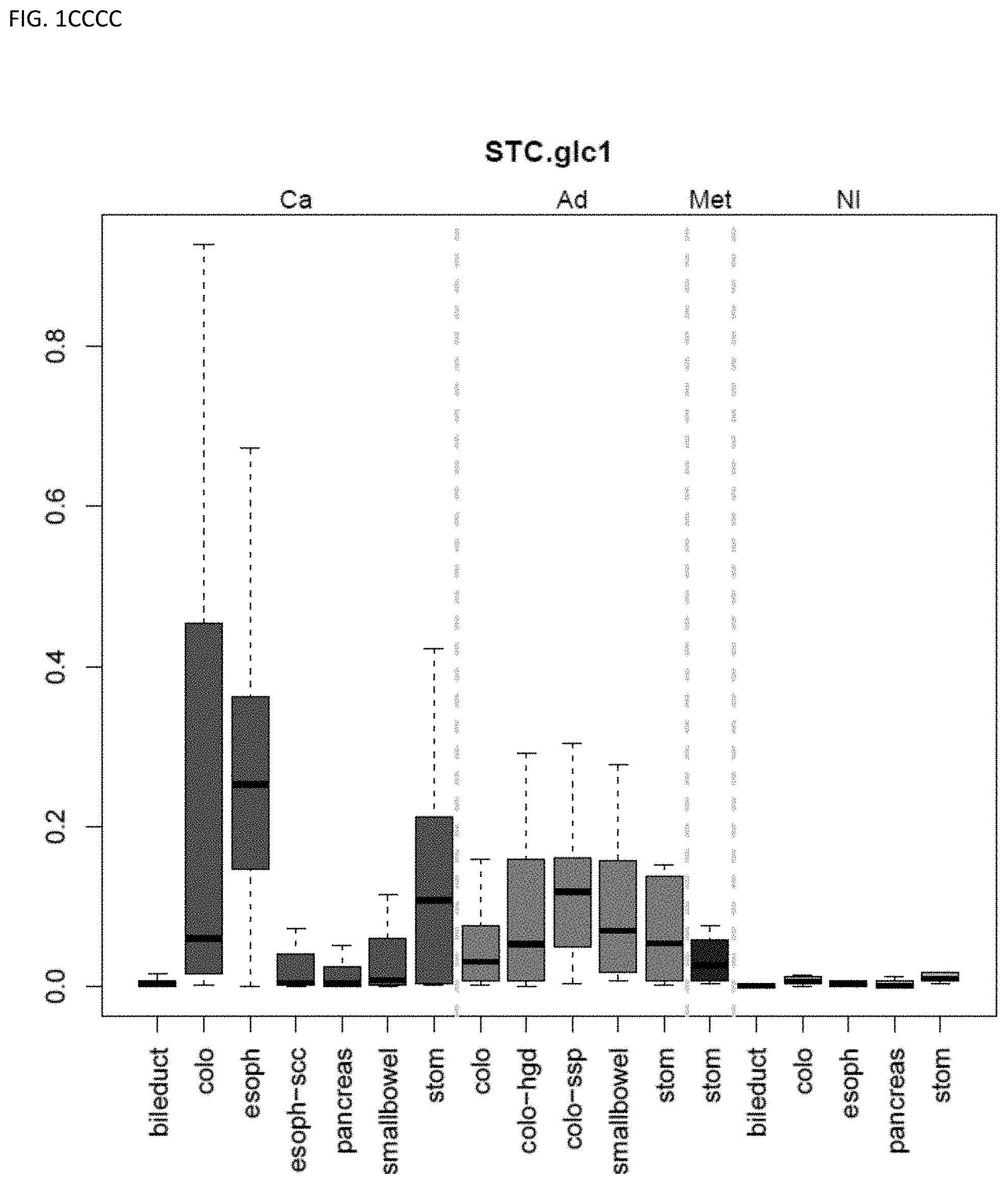
D00082
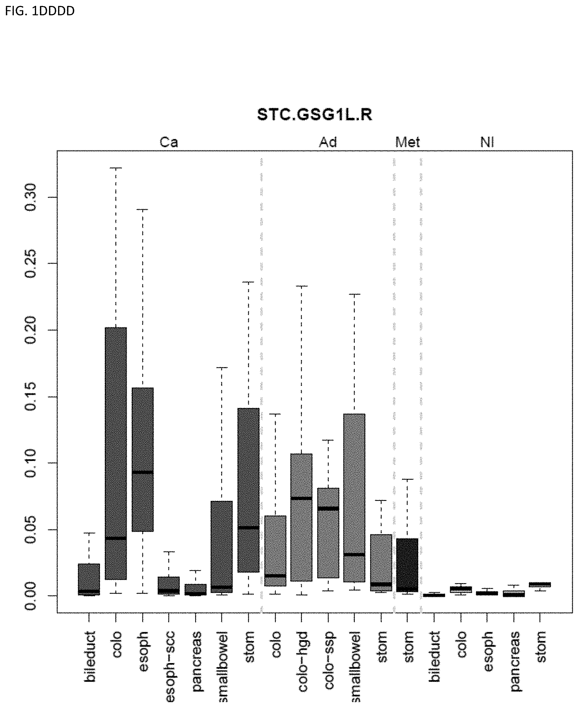
D00083
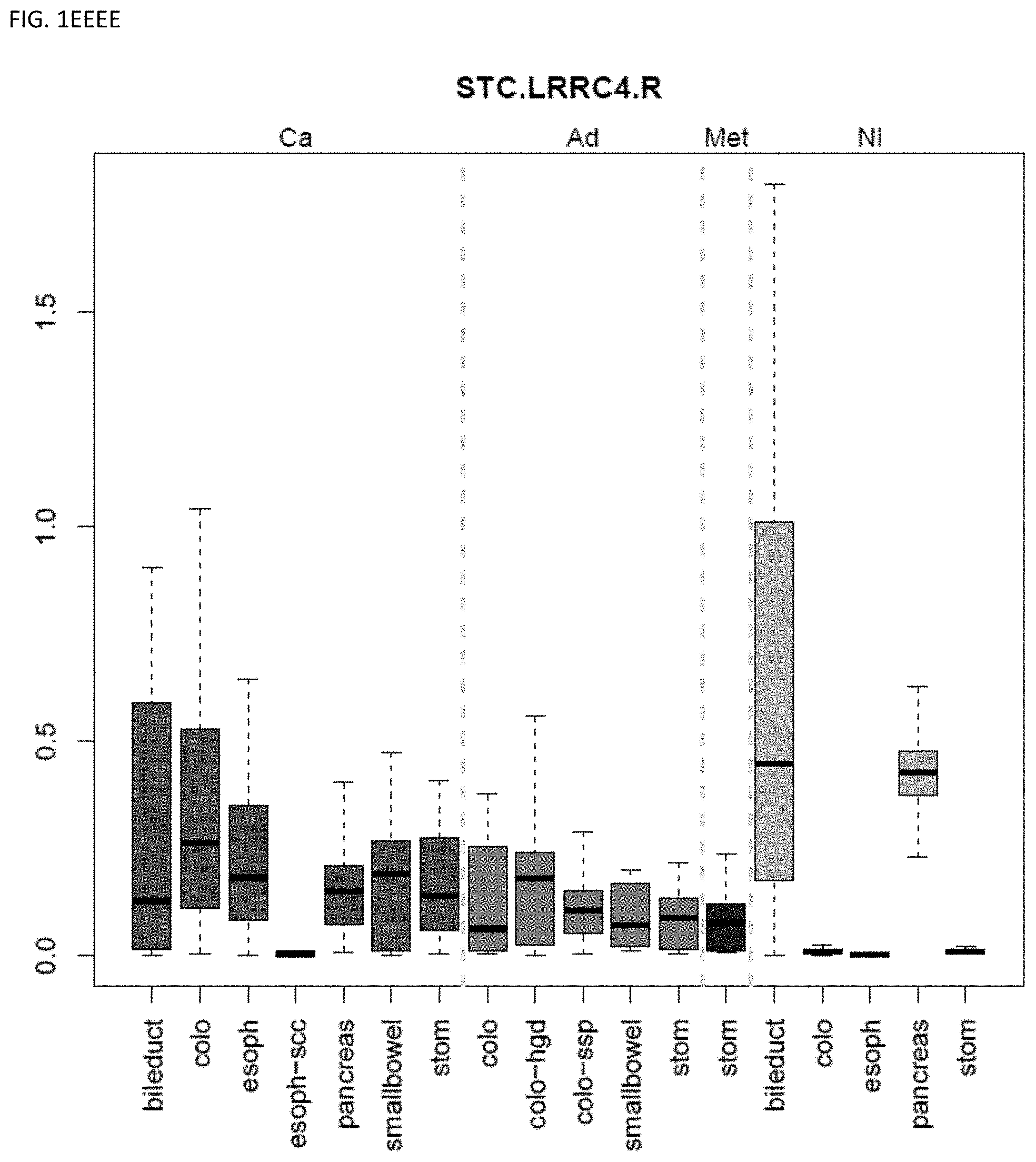
D00084
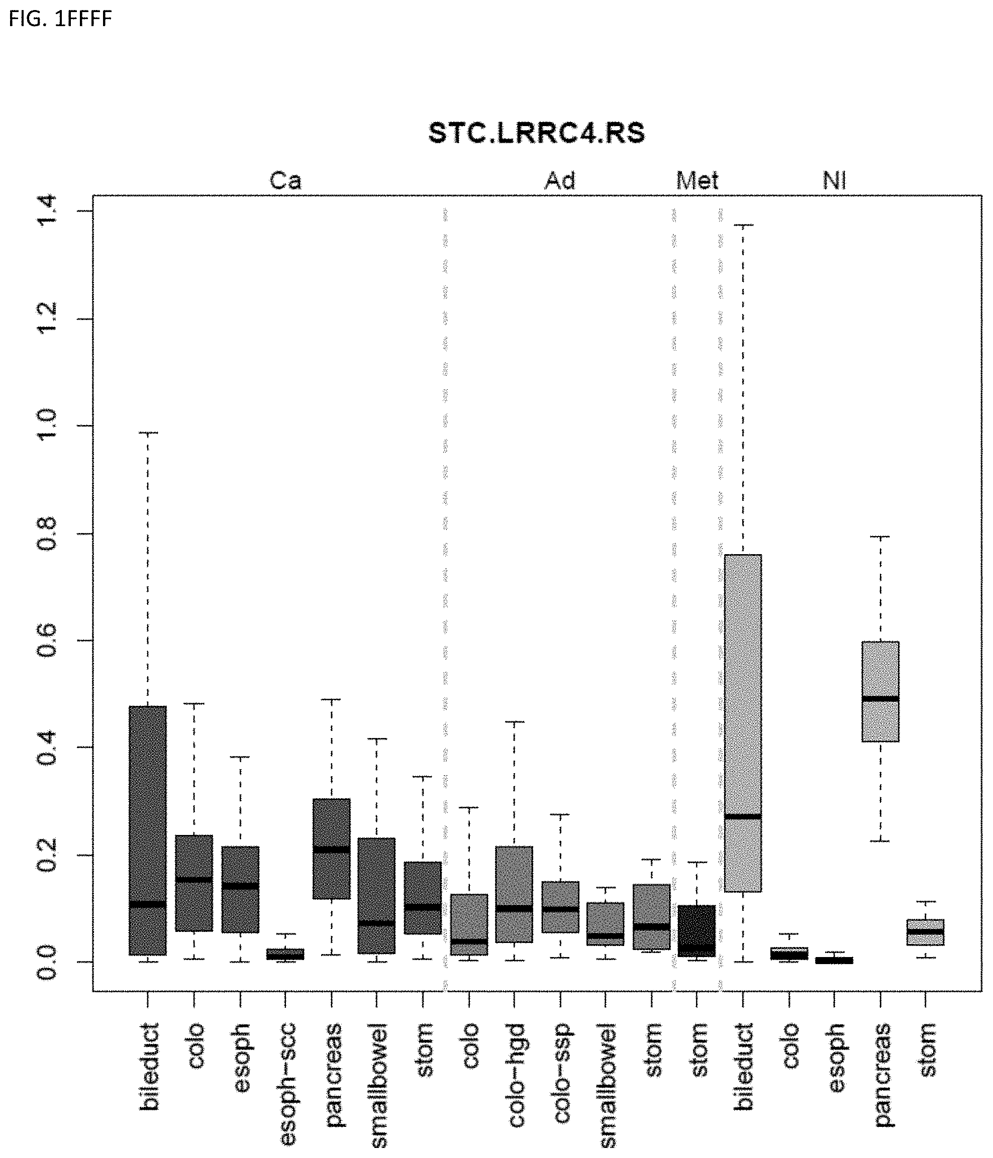
D00085
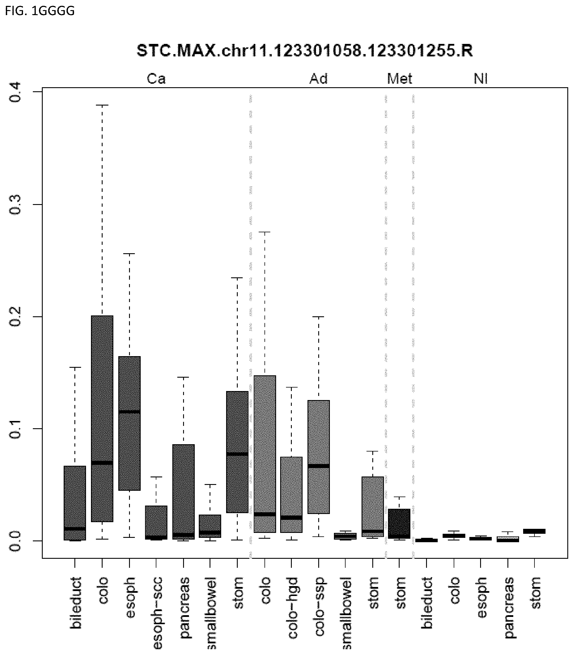
D00086
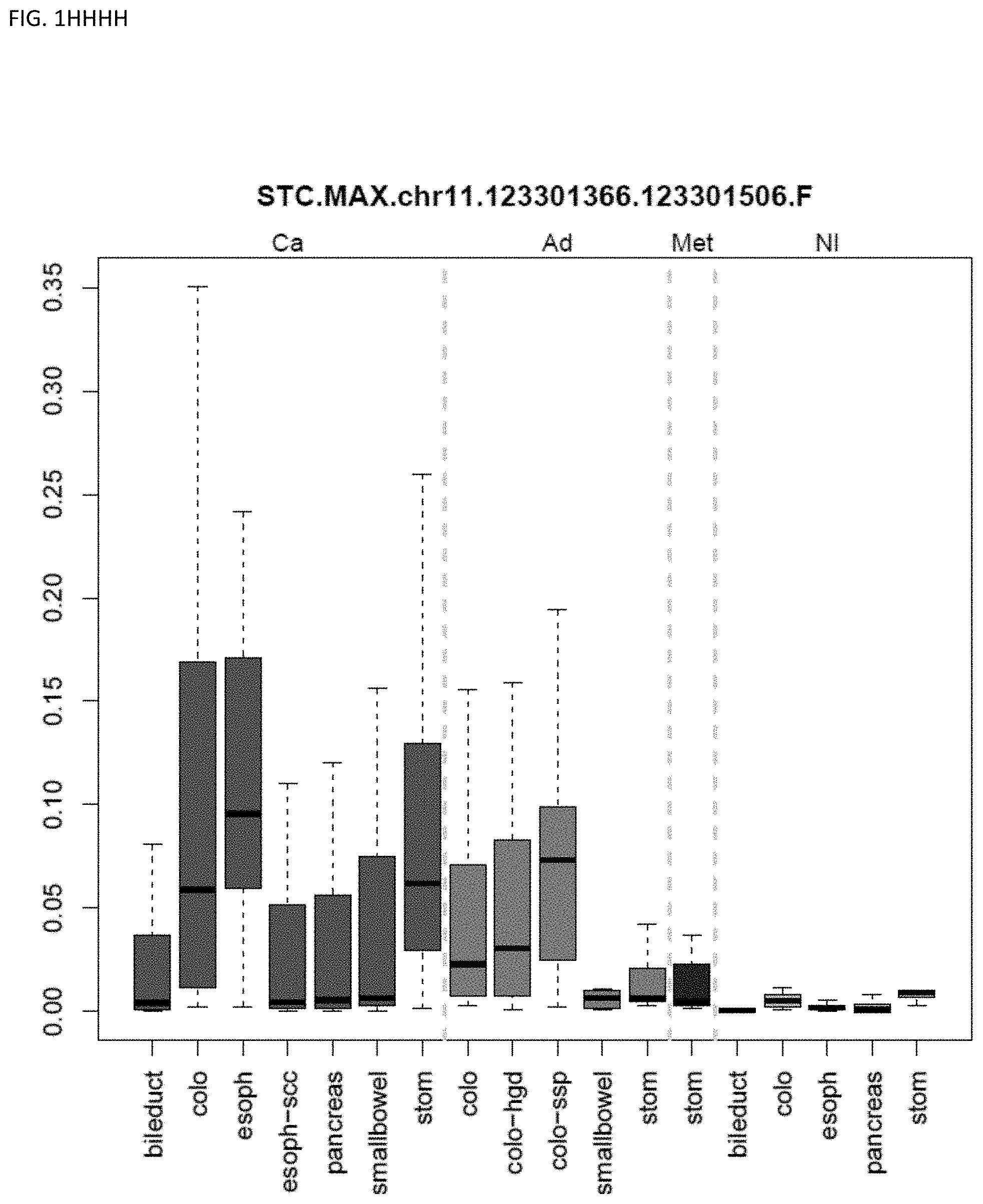
D00087
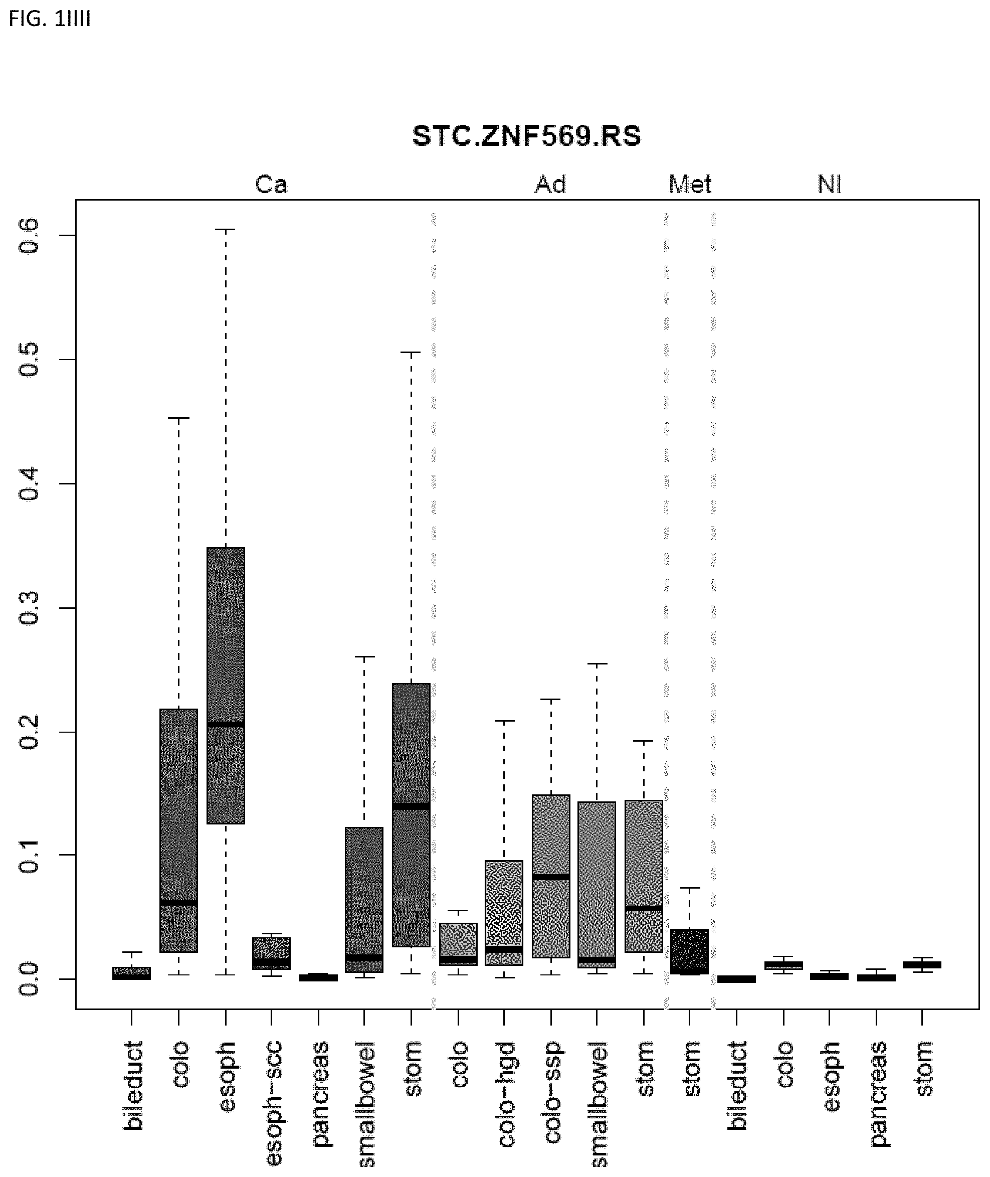
D00088
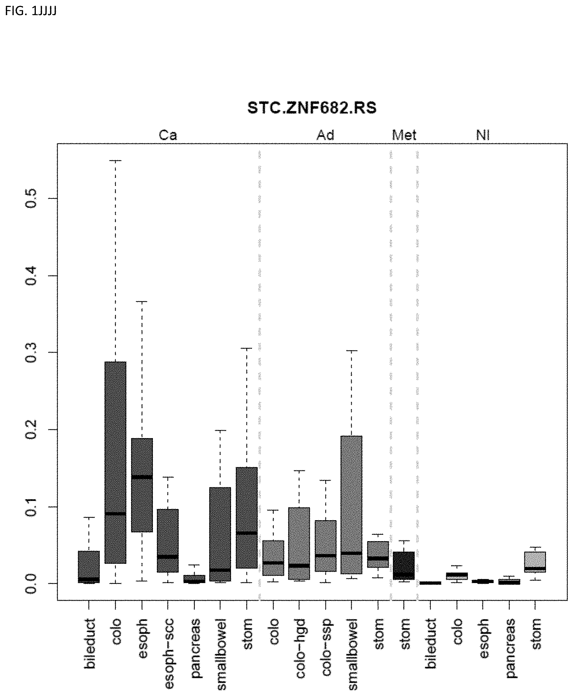
D00089
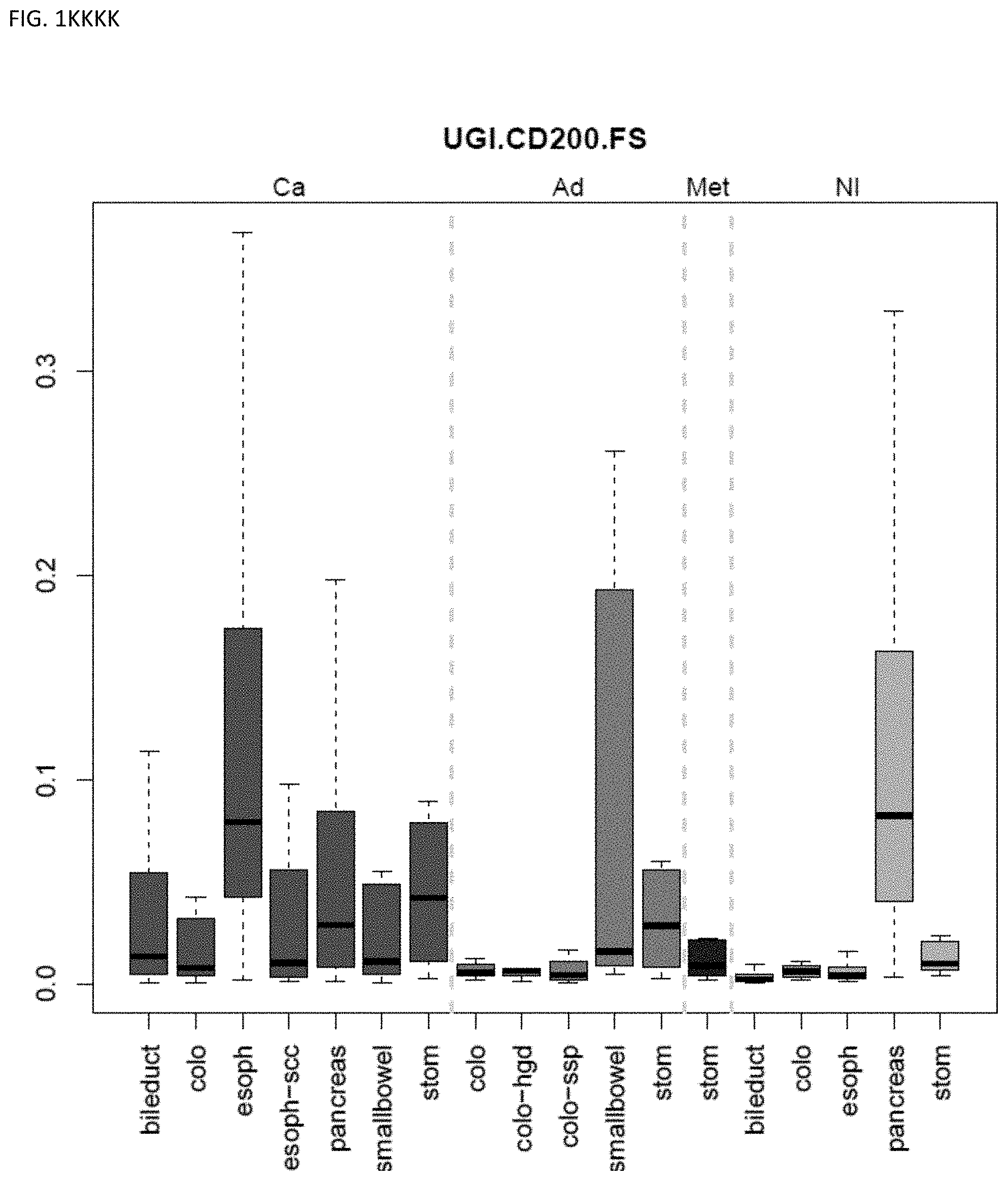
D00090
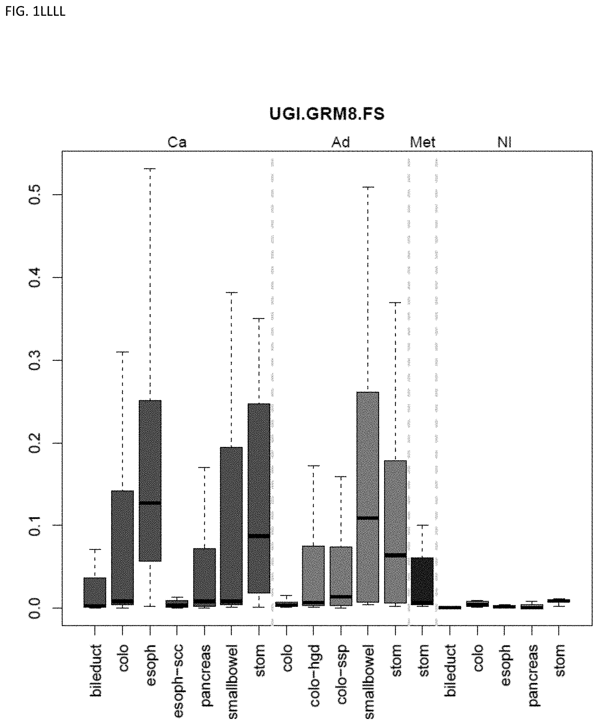
D00091
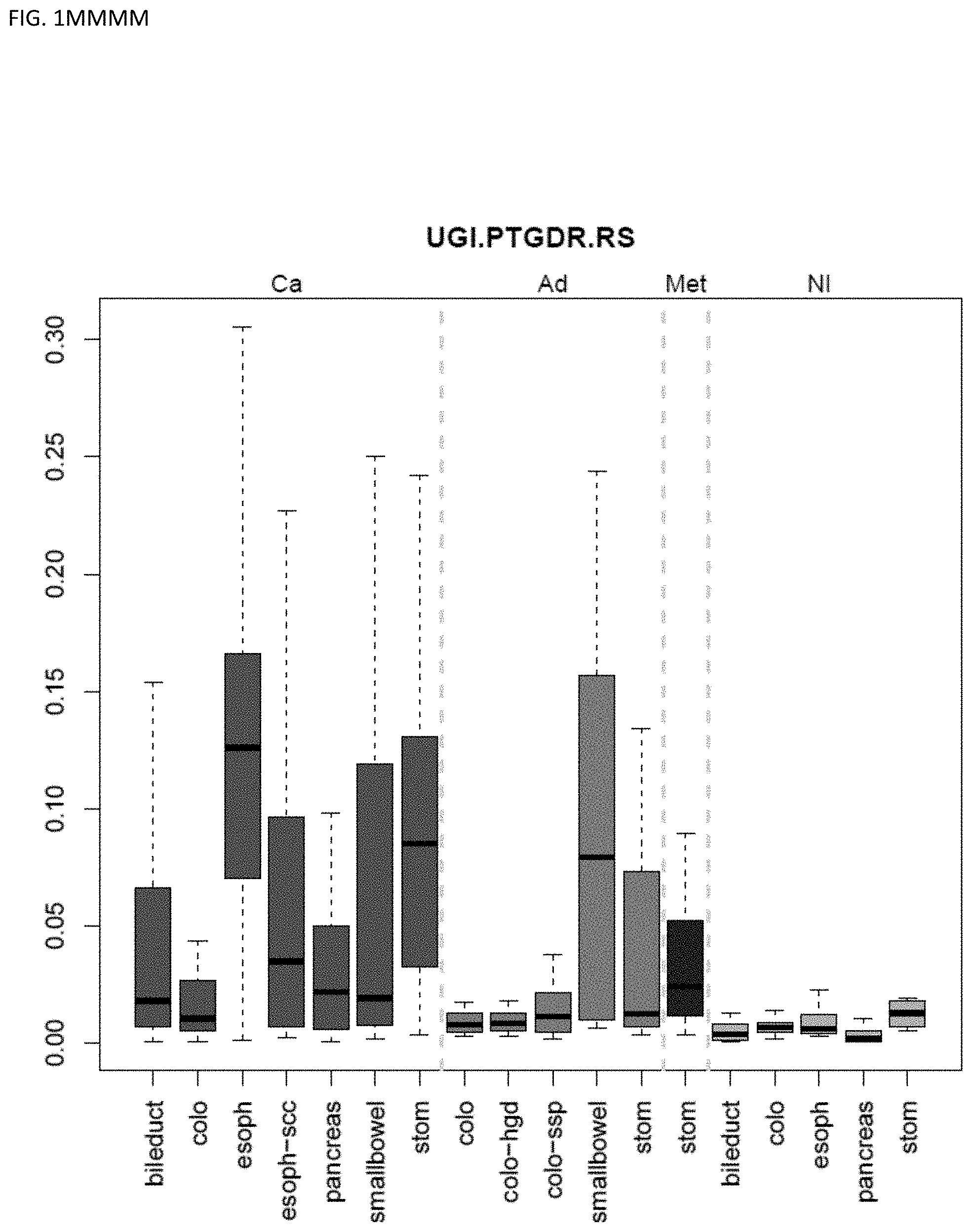
D00092
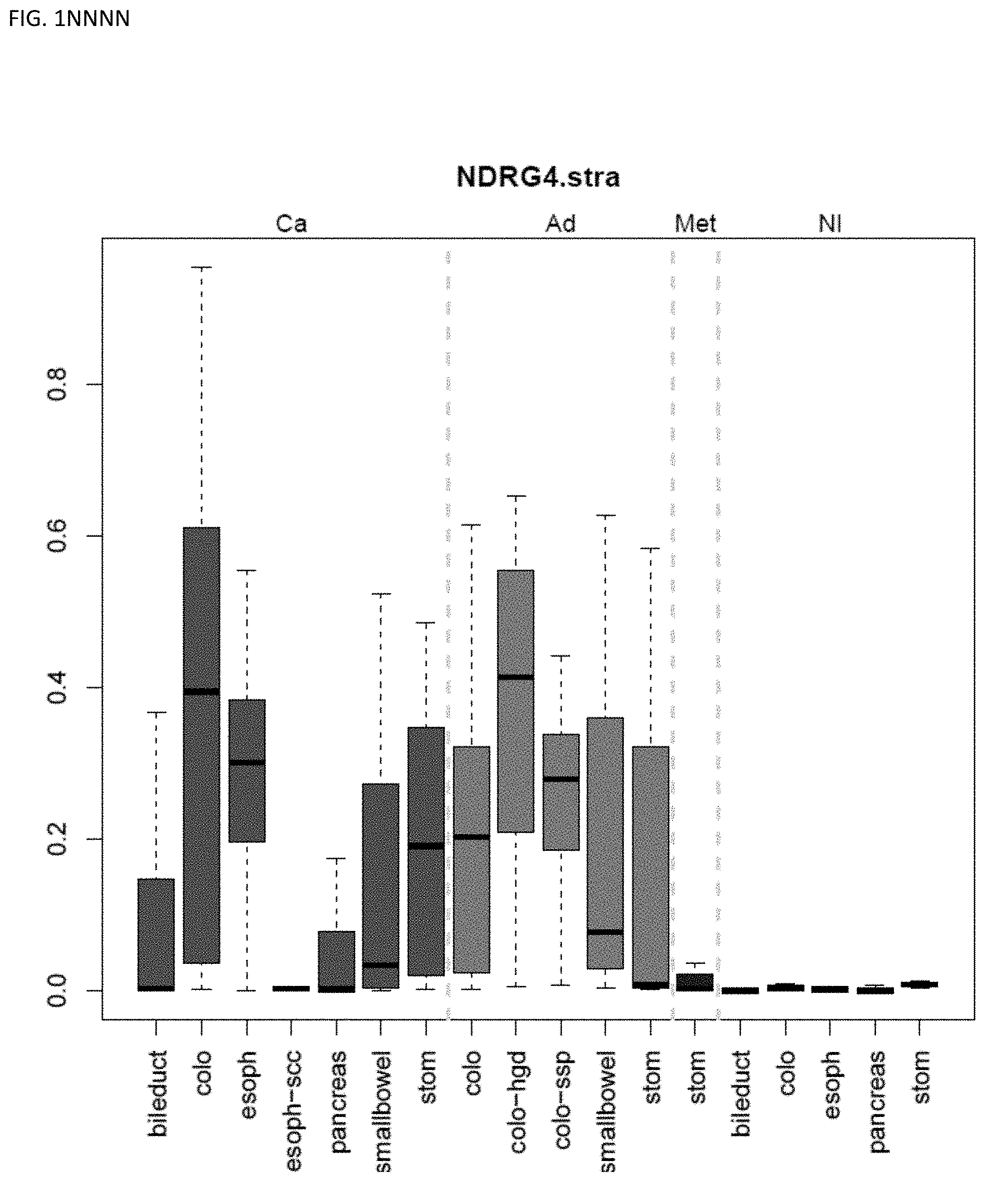
D00093
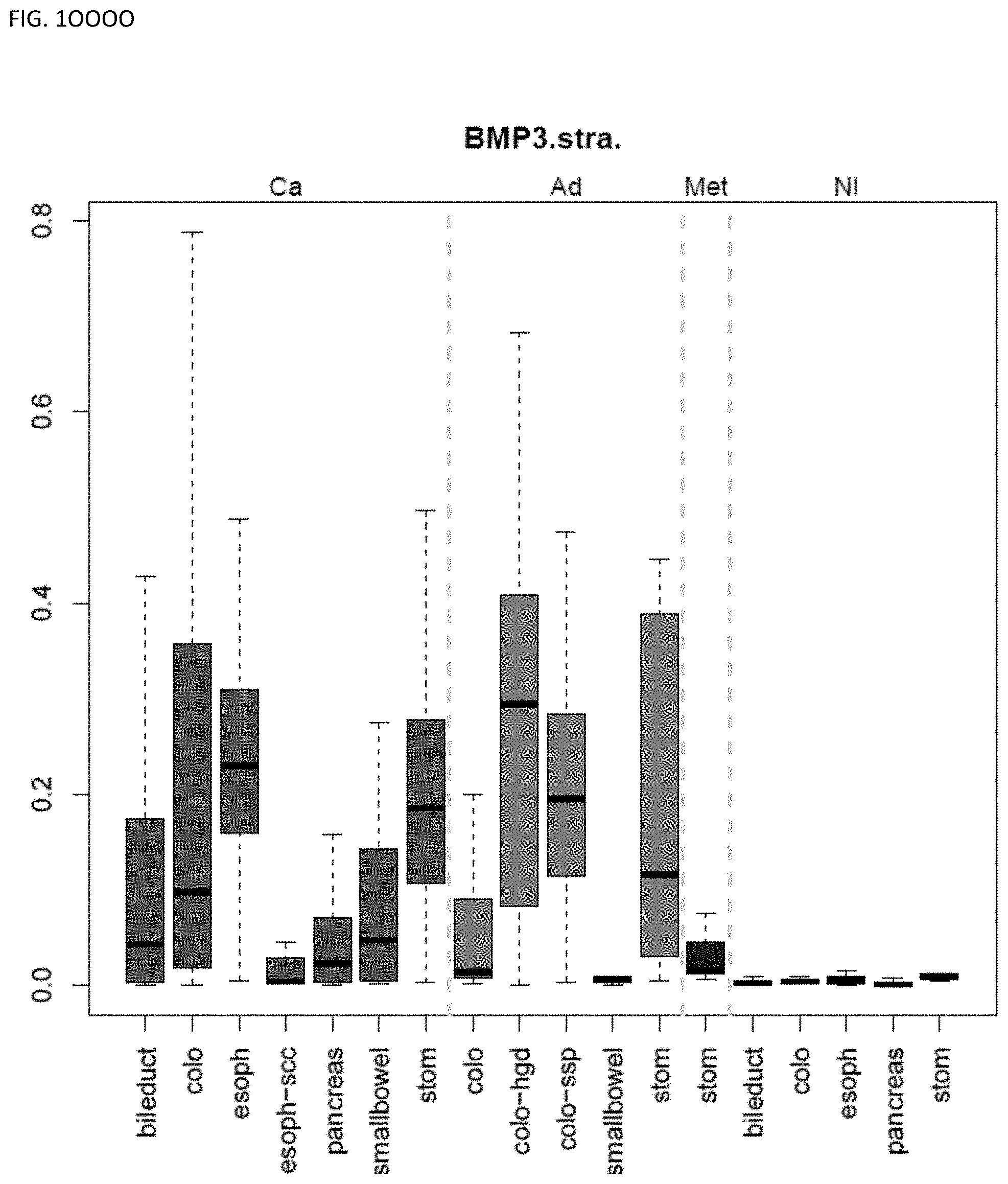
D00094
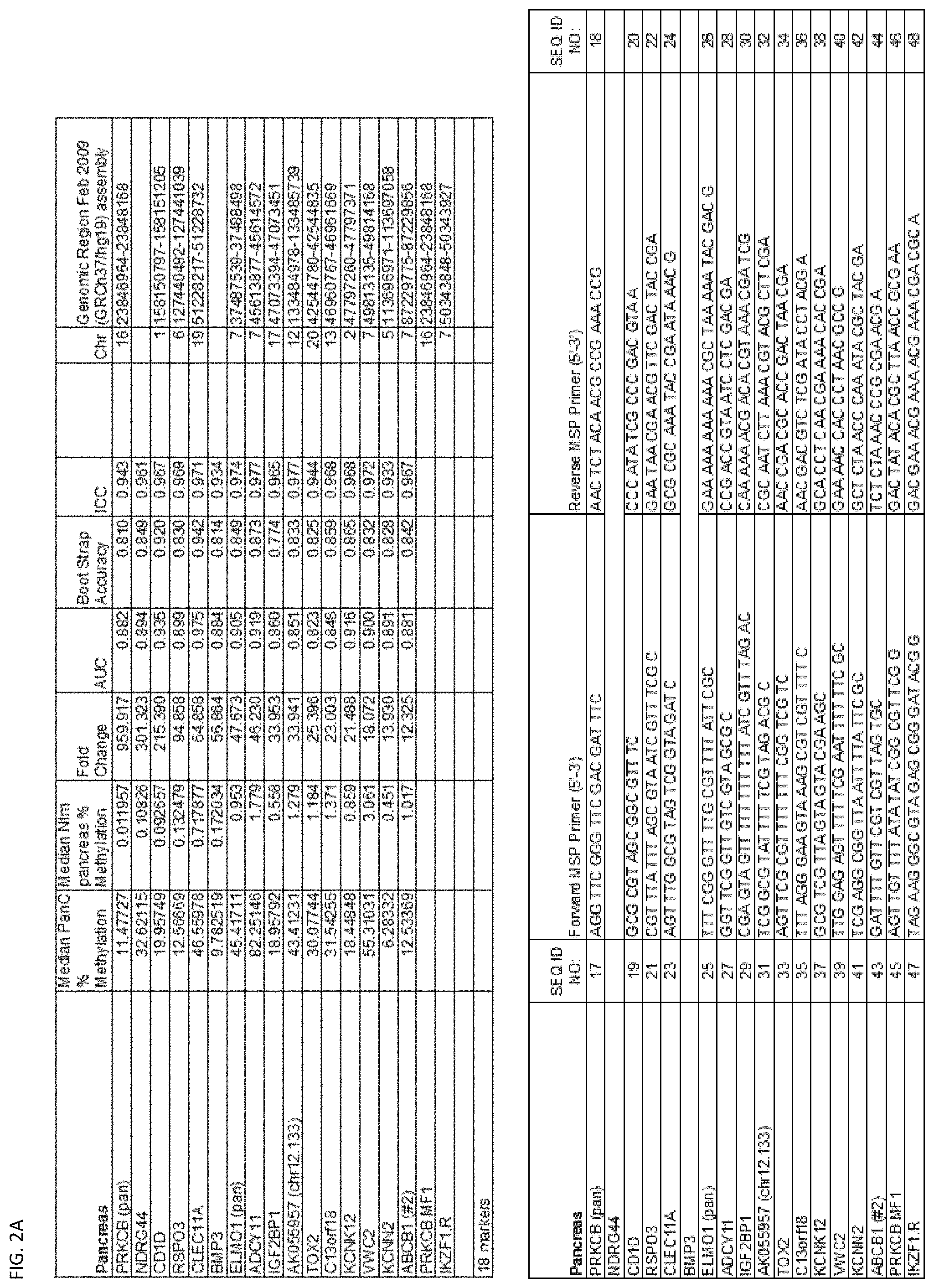
D00095
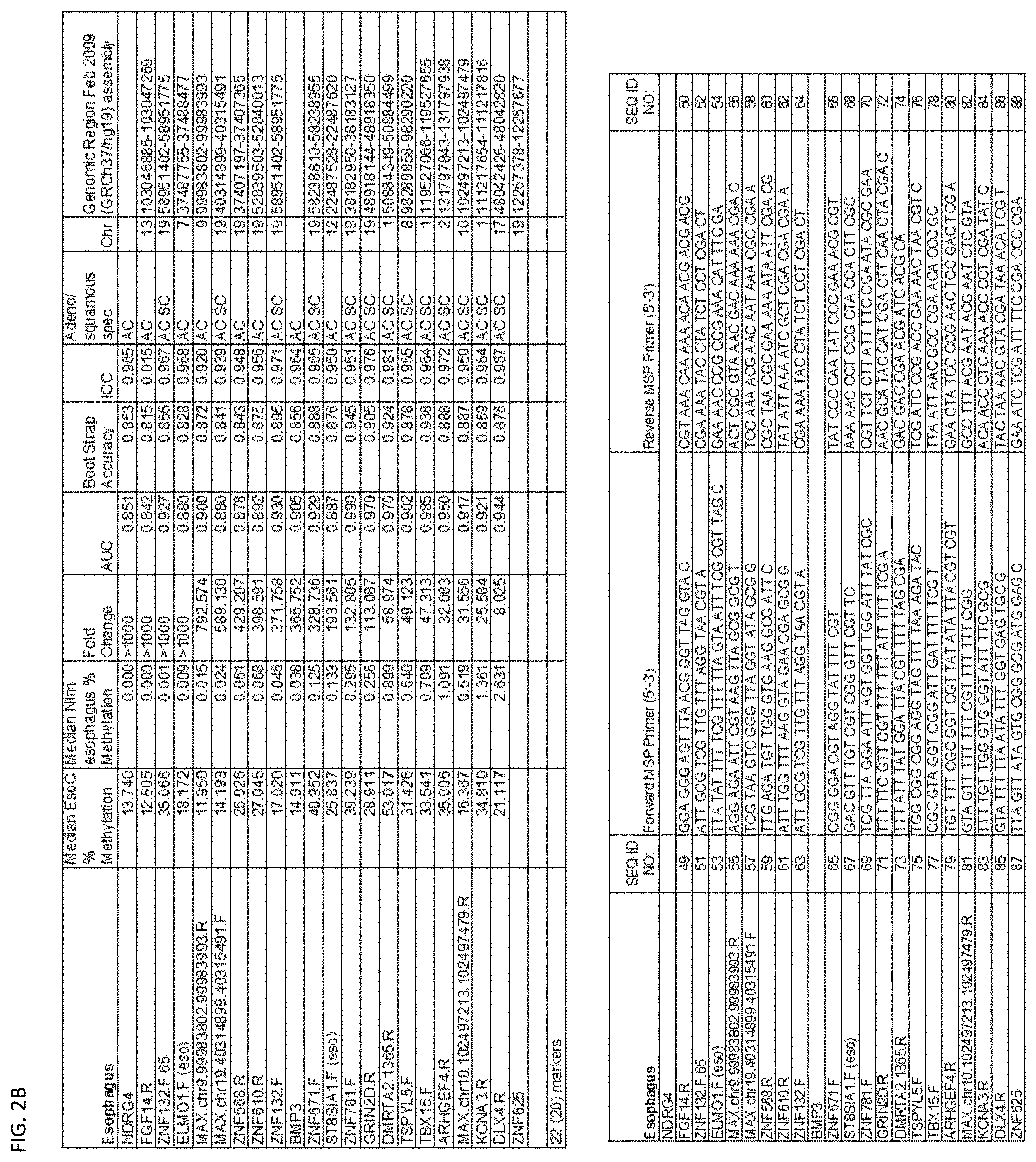
D00096
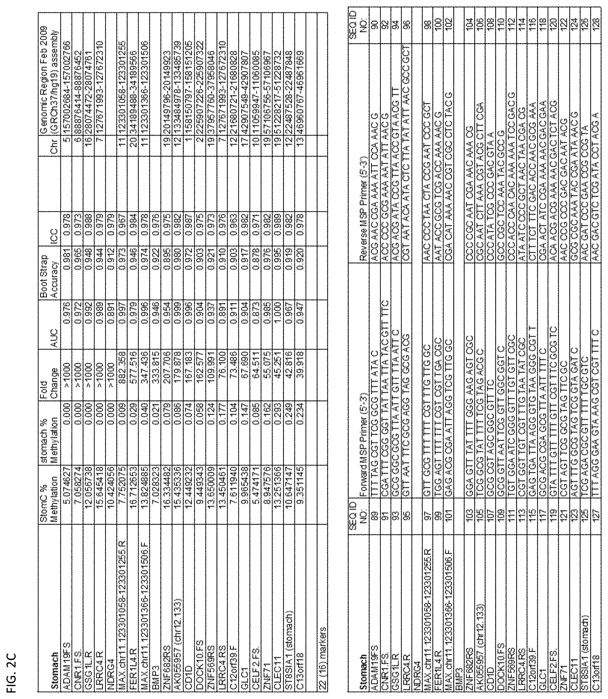
D00097
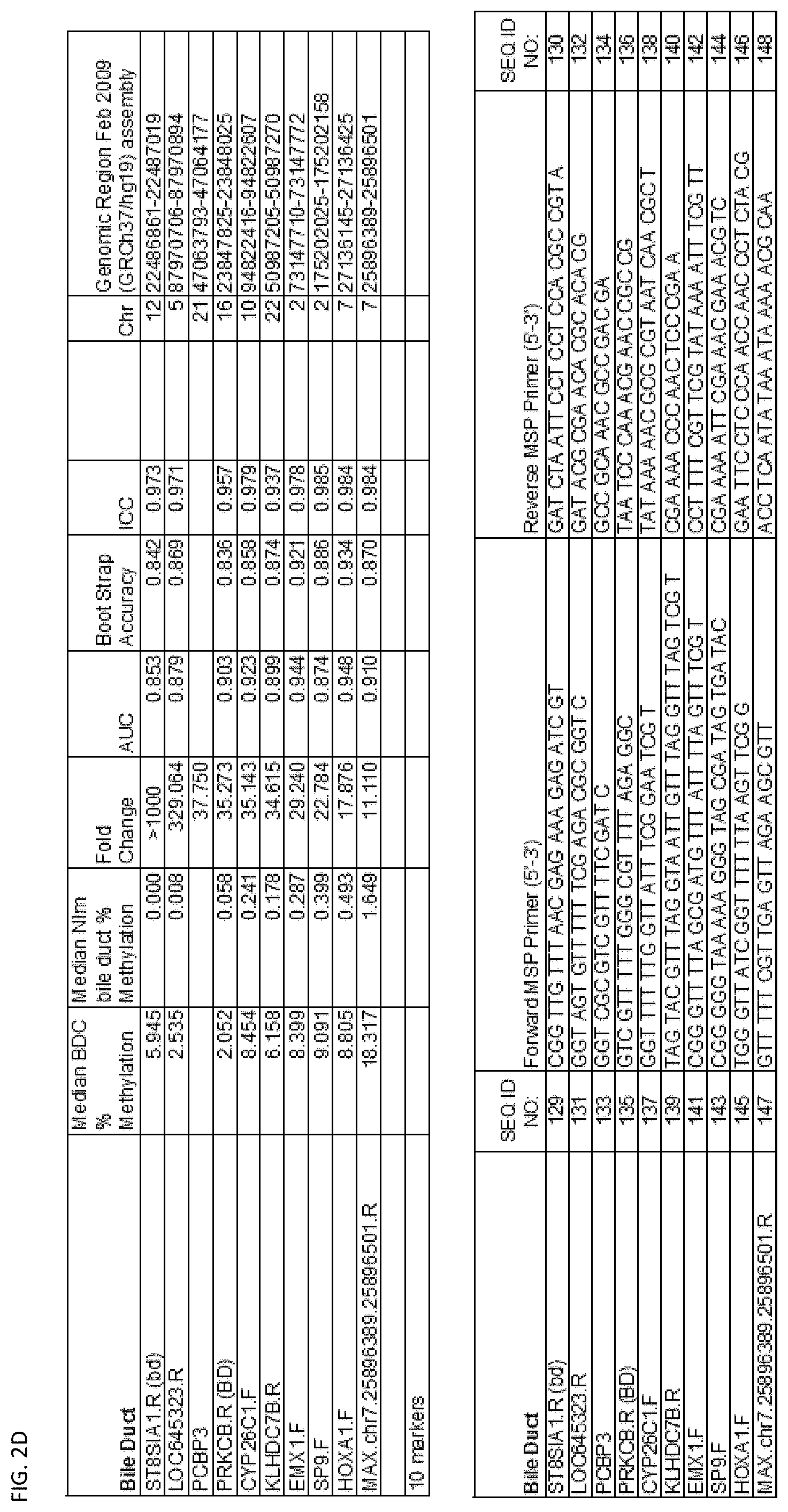
D00098
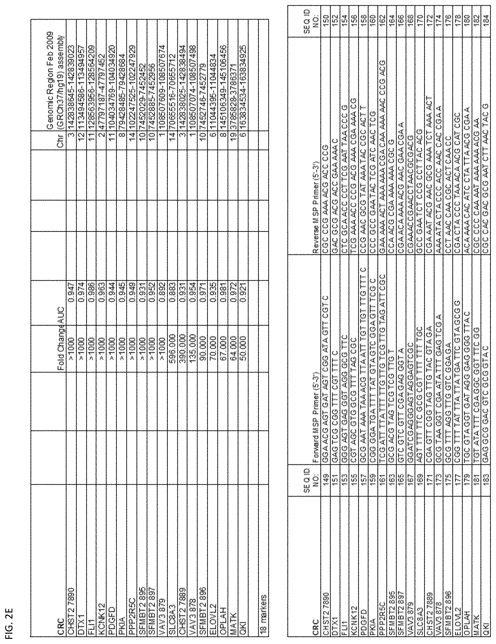
D00099
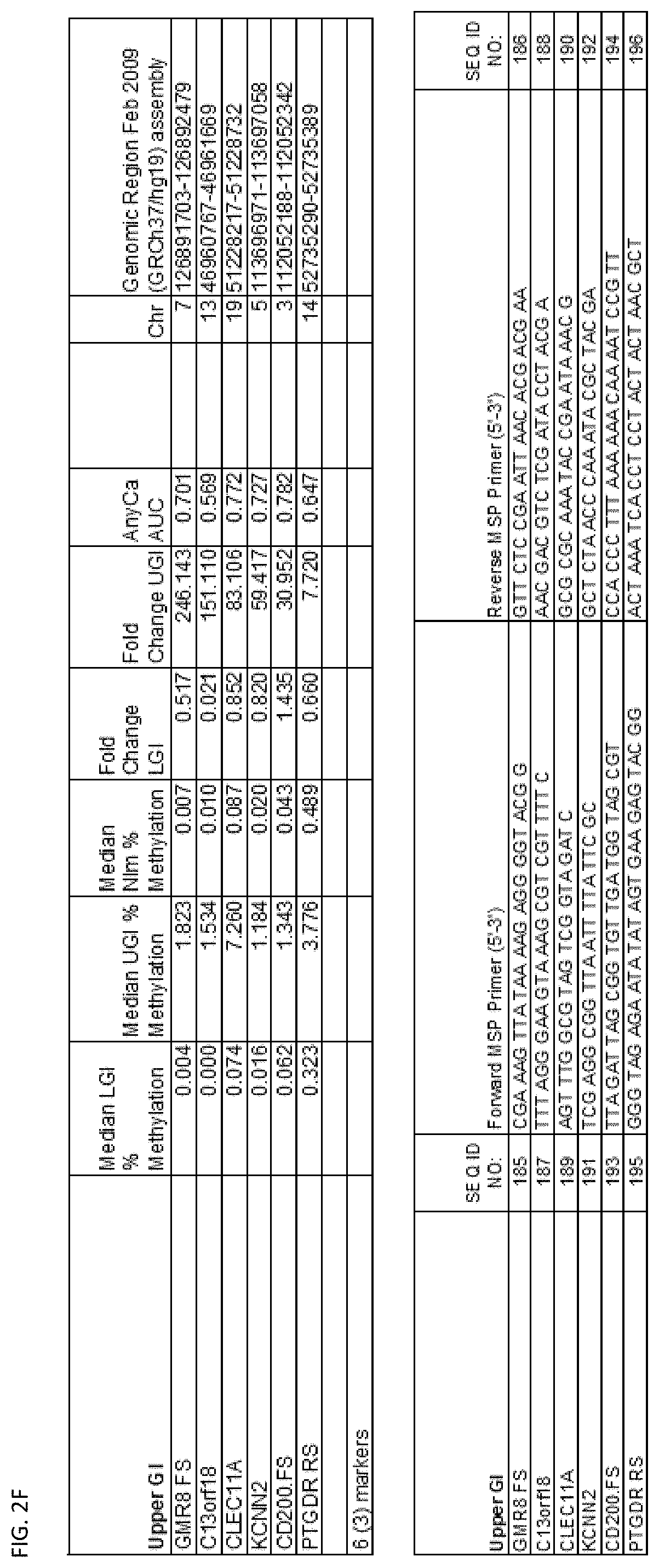
D00100
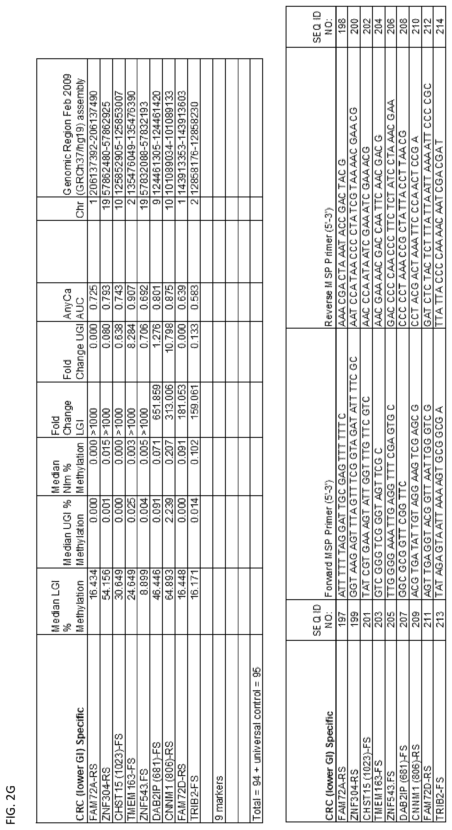
D00101
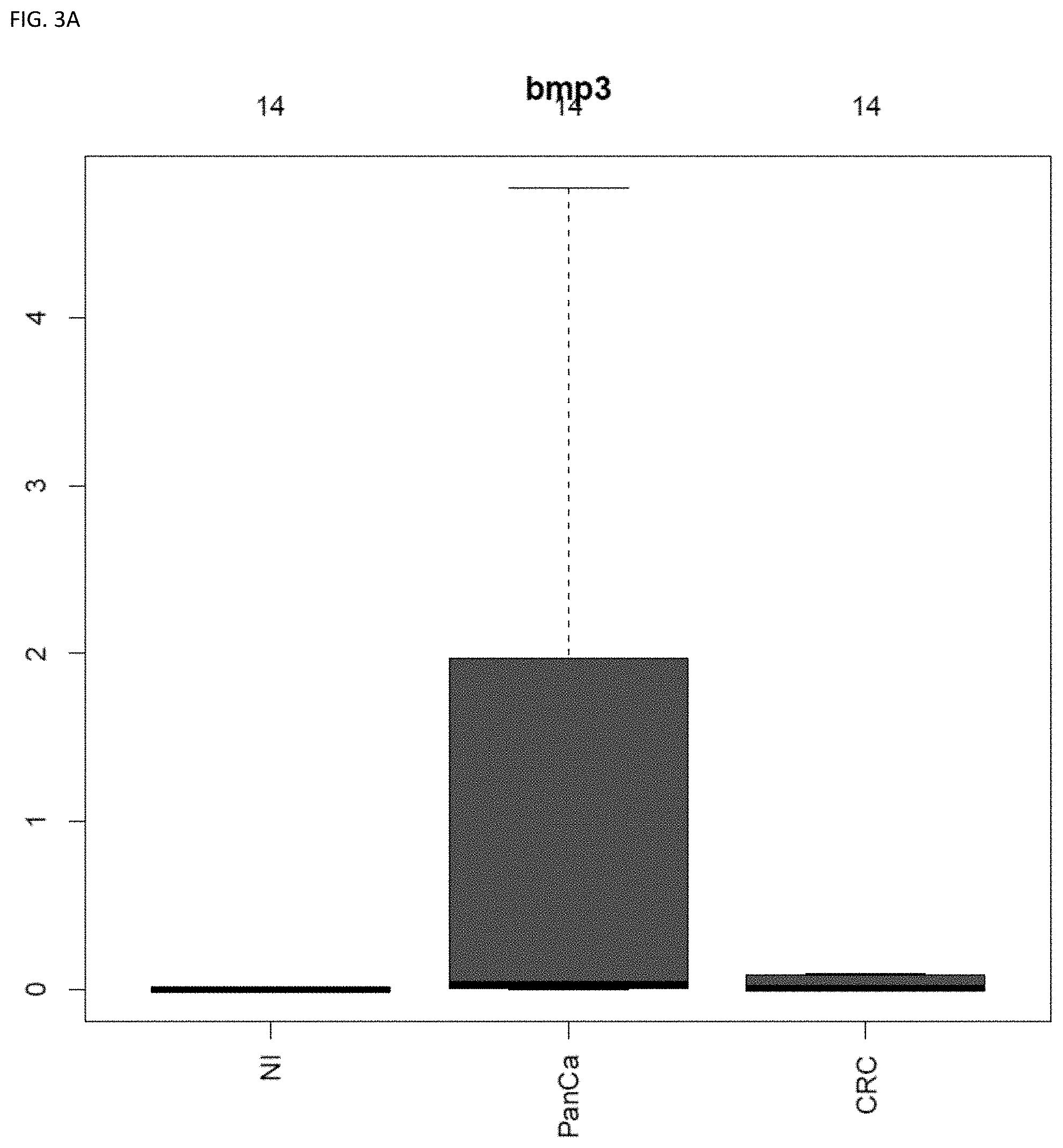
D00102
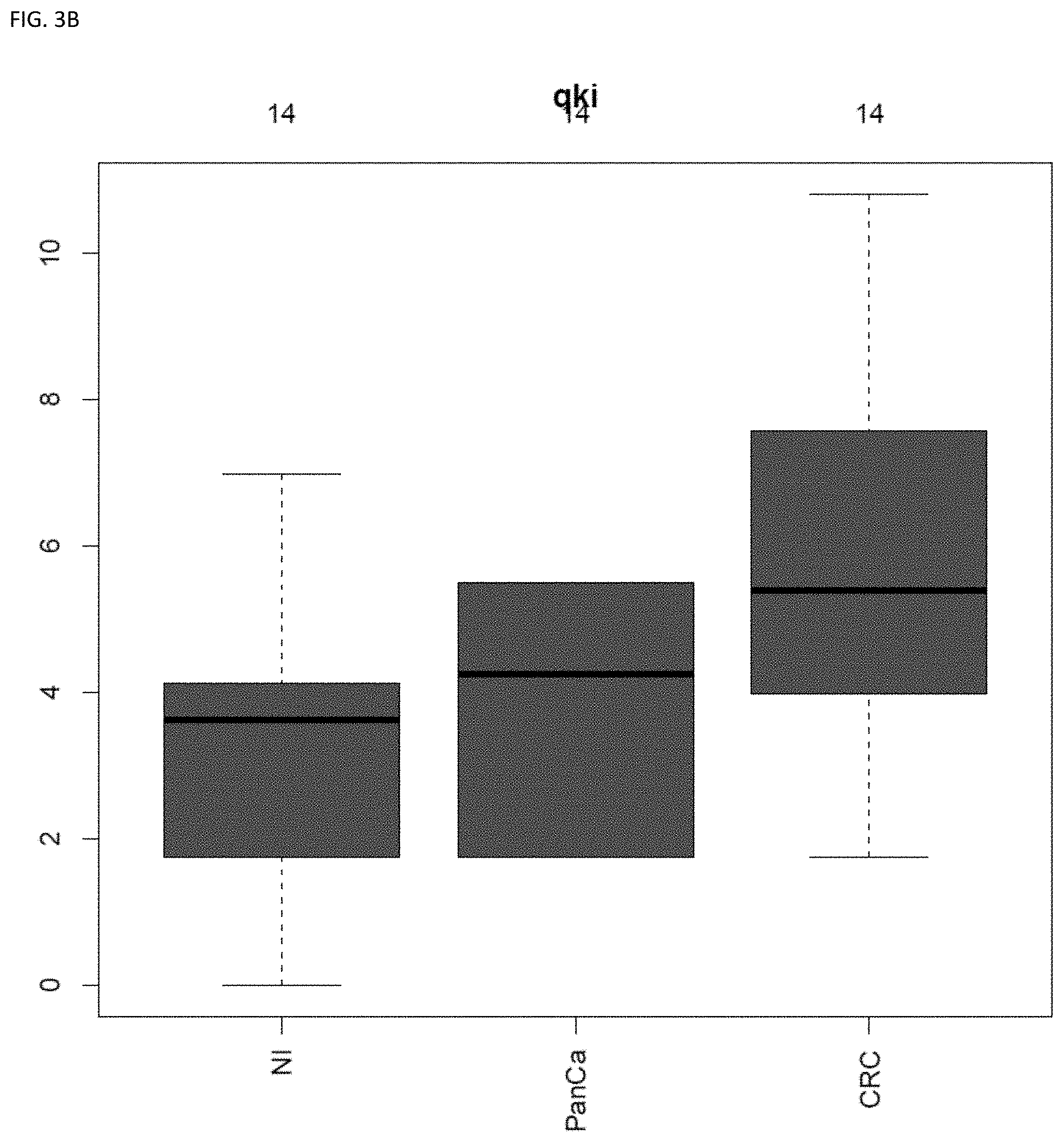
S00001
XML
uspto.report is an independent third-party trademark research tool that is not affiliated, endorsed, or sponsored by the United States Patent and Trademark Office (USPTO) or any other governmental organization. The information provided by uspto.report is based on publicly available data at the time of writing and is intended for informational purposes only.
While we strive to provide accurate and up-to-date information, we do not guarantee the accuracy, completeness, reliability, or suitability of the information displayed on this site. The use of this site is at your own risk. Any reliance you place on such information is therefore strictly at your own risk.
All official trademark data, including owner information, should be verified by visiting the official USPTO website at www.uspto.gov. This site is not intended to replace professional legal advice and should not be used as a substitute for consulting with a legal professional who is knowledgeable about trademark law.