Recombinant Protein
ANDER; Mats ; et al.
U.S. patent application number 16/615552 was filed with the patent office on 2020-10-08 for recombinant protein. This patent application is currently assigned to GE Healthcare BioProcess R&D AB. The applicant listed for this patent is GE HEALTHCARE BIOPROCESS R&D AB. Invention is credited to Mats ANDER, Magnus BERGMAN, Tomas BJORKMAN, Joakim GALLI, Gustav RODRIGO.
| Application Number | 20200318120 16/615552 |
| Document ID | / |
| Family ID | 1000004971540 |
| Filed Date | 2020-10-08 |


View All Diagrams
| United States Patent Application | 20200318120 |
| Kind Code | A1 |
| ANDER; Mats ; et al. | October 8, 2020 |
Recombinant Protein
Abstract
A recombinant protein comprising a functional polypeptide and, linked to the N-terminus of said functional polypeptide, an N-terminal spacer having a length such that the number of amino acid residues between a signal peptide cleaving site and an N-terminus proximal structural unit of said functional polypeptide is 14-24.
| Inventors: | ANDER; Mats; (Uppsala, SE) ; BERGMAN; Magnus; (Uppsala, SE) ; BJORKMAN; Tomas; (Uppsala, SE) ; GALLI; Joakim; (Uppsala, SE) ; RODRIGO; Gustav; (Uppsala, SE) | ||||||||||
| Applicant: |
|
||||||||||
|---|---|---|---|---|---|---|---|---|---|---|---|
| Assignee: | GE Healthcare BioProcess R&D
AB Uppsala SE |
||||||||||
| Family ID: | 1000004971540 | ||||||||||
| Appl. No.: | 16/615552 | ||||||||||
| Filed: | May 23, 2018 | ||||||||||
| PCT Filed: | May 23, 2018 | ||||||||||
| PCT NO: | PCT/EP2018/063435 | ||||||||||
| 371 Date: | November 21, 2019 |
| Current U.S. Class: | 1/1 |
| Current CPC Class: | C07K 2319/02 20130101; C12N 15/62 20130101; C07K 2319/50 20130101; C07K 1/22 20130101; C07K 14/31 20130101; C07K 2319/034 20130101 |
| International Class: | C12N 15/62 20060101 C12N015/62; C07K 1/22 20060101 C07K001/22; C07K 14/31 20060101 C07K014/31 |
Foreign Application Data
| Date | Code | Application Number |
|---|---|---|
| May 24, 2017 | GB | 1708277.7 |
Claims
1. A recombinant protein comprising a functional polypeptide and, linked to the N-terminus of said functional polypeptide, an N-terminal spacer having a length such that the number of amino acid residues between a signal peptide cleaving site and an N-terminus proximal structural unit of said functional polypeptide is 14-24.
2. A recombinant protein comprising an immunoglobulin-binding polypeptide and, linked to the N-terminus of said immunoglobulin-binding polypeptide, an N-terminal spacer having a length such that the number of amino acid residues between a signal peptide cleaving site and an N-terminus proximal structural unit of said functional polypeptide is 14-24.
3. The recombinant protein of claim 1, wherein said N-terminus proximal structural unit is an alpha-helix.
4. The recombinant protein of claim 3, wherein said functional polypeptide comprises at least three alpha helices.
5. The recombinant protein of claim 1, wherein said N-terminal spacer comprises at most two amino acid residues selected from the group consisting of lysine and arginine.
6. The recombinant protein of claim 1, wherein said N-terminal spacer consists of 8-24 amino acid residues.
7. A recombinant protein comprising a functional or immunoglobulin-binding polypeptide and, linked to the N-terminus of said functional or immunoglobulin-binding polypeptide, an N-terminal spacer comprising an amino acid sequence having at least 80% sequence identity to, or being defined by, an amino acid sequence selected from the group consisting of SEQ ID NOS.: 16-18, 29-30, 33-40, 43-45 and 47.
8. The recombinant protein of claim 7, wherein said N-terminal spacer has at least 80% sequence identity to, or is defined by, an amino acid sequence selected from the group consisting of SEQ ID NOS.: 16-18, 29-30, 33-40, 43-45 and 47.
9. The recombinant protein of claim 1, wherein said functional or immunoglobulin-binding polypeptide comprises one or more Fc-binding domains derived from Staphylococcus aureus Protein A.
10. The recombinant protein of claim 9, wherein said Fc-binding domains are alkali-stabilized Fc-binding domains.
11. The recombinant protein of claim 10, wherein said alkali-stabilized Fc-binding domains have at least 90% sequence identity to an amino acid sequence selected from the group consisting of SEQ ID NOS.: 1-11 and 48-64.
12. The recombinant protein of claim 9, wherein said functional or immunoglobulin-binding polypeptide comprises a multimer of at least four Fc-binding domains.
13. The recombinant protein of claim 7, wherein said functional or immunoglobulin-binding polypeptide comprises an amino acid sequence having at least 90% sequence identity to an amino acid sequence selected from the group consisting of SEQ ID NOS.: 1-13 and 48-93.
14. The recombinant protein of claim 7, further comprising at, or adjacent to, the C-terminus a coupling moiety.
15. The recombinant protein of claim 14, wherein said coupling moiety comprises a cysteine residue and/or a plurality of lysine residues.
16. The recombinant protein of claim 7, wherein said N-terminal spacer is alkali stable.
17. The recombinant protein of claim 7, wherein said N-terminal spacer consists of amino acid residues selected from the group consisting of alanine, aspartic acid, glutamine, glutamic acid, glycine, histidine, lysine, phenylalanine, serine, threonine, tryptophan, tyrosine and valine.
18. A nucleic acid molecule encoding for the recombinant protein of claim 1, said nucleic acid molecule comprising the following elements in the 5' to 3' direction, said elements being operatively linked: a) an inducible or constitutive promoter DNA sequence; b) a DNA sequence encoding a signal peptide; c) a DNA sequence encoding an N-terminal spacer; and d) a DNA sequence encoding a functional or immunoglobulin-binding polypeptide.
19. The nucleic acid molecule of claim 18, wherein said signal peptide comprises an amino acid sequence having at least 80% sequence identity to, or being defined by, an amino acid sequence selected from the group consisting of SEQ ID NOS.: 14 and 15.
20. The nucleic acid molecule of claim 18, wherein said signal peptide comprises an amino acid sequence having at least 80% sequence identity to, or being defined by SEQ ID NO.: 14.
21. A cloning vector which expresses and secretes the recombinant protein of claim 1 into the bacterial periplasm of a gram-negative cell.
22. A gram-negative bacterium transformed by the cloning vector of claim 21.
23. The gram-negative bacterium of claim 22, identified as Escherichia coli.
24. The gram-negative bacterium of claim 23, further characterized as Escherichia coli K12.
25. The gram-negative bacterium of claim 23, further characterized as Escherichia coli K12-017.
26. A method of expressing and secreting a recombinant protein in a gram-negative bacterium, said method comprising the steps of: i) providing the gram-negative bacterium of claim 22; and ii) culturing said gram-negative bacterium.
27. A separation matrix comprising the recombinant protein of claim 1, covalently linked to a support.
28. The separation matrix of claim 27, wherein said support comprises porous particles.
29. The separation matrix of claim 28, wherein said porous particles comprise a cross-linked polysaccharide.
30. The separation matrix of claim 27, wherein said recombinant protein is covalently linked to said support via a thioether bond.
31. The separation matrix of claim 27, wherein said recombinant protein is covalently linked to said support via one or more amide bonds.
32. The separation matrix of claim 27, wherein said separation matrix is alkali stable, such as wherein the recombinant protein comprises an immunoglobulin-binding polypeptide and the IgG capacity of the matrix after 24 h incubation time in 0.5 M NaOH at 22+/-2.degree. C. is at least 80% of the IgG capacity before the incubation.
33. A method of separating an immunoglobulin, comprising the steps of: i) providing the separation matrix of claim 27, wherein said recombinant protein comprises an immunoglobulin-binding polypeptide; ii) contacting said separation matrix with a liquid sample containing an immunoglobulin, to bind said immunoglobulin; iii) optionally washing said separation matrix with a washing liquid; iv) contacting said separation matrix with an elution liquid, to elute said immunoglobulin.
34. The method of claim 33, wherein in step a) said recombinant protein comprises one or more Fc-binding domains derived from Staphylococcus aureus Protein A and wherein said method further comprises, after step iv), a step v) of cleaning said separation matrix with a cleaning liquid.
35. The method of claim 34, wherein said cleaning liquid comprises at least 0.1 M NaOH or KOH, such as at least 0.5 M NaOH or KOH, or 0.5-2.5 M NaOH or KOH.
Description
CROSS REFERENCE TO RELATED APPLICATIONS
[0001] This application claims the priority benefit of PCT/EP2018/063435 filed on May 23, 2018, which claims priority benefit of Great Britain Application No. 1708277.7 filed on May 24, 2017, the entire contents of which are hereby incorporated by reference herein.
SEQUENCE LISTING
[0002] The instant application contains a Sequence Listing which has been submitted electronically in ASCII format and is hereby incorporated by reference in its entirety. Said ASCII copy, created on Oct. 18, 2019, is named 319573_ST25.txt and is 57,522 bytes in size.
TECHNICAL FIELD OF THE INVENTION
[0003] The present invention relates to recombinant proteins, and more particularly to recombinant proteins expressed in gram-negative bacteria such as Escherichia coli (E. coli). The invention also relates to nucleic acids, vectors and gram-negative bacteria for expression of recombinant proteins as well as to separation matrices with covalently linked recombinant protein ligands and to methods of separating immunoglobulins on such matrices.
BACKGROUND OF THE INVENTION
[0004] Expression of heterologous proteins in E. coli (E. coli) is commonly used for recombinant proteins in laboratory and commercial scale. Expression in E. coli using secretion generally means transport of the produced protein over the inner membrane separating the cytoplasm and the periplasm. By secretion to the periplasm, the protein many times also leaks out to the extracellular medium (Mergulhao et al., Biotech Adv 23, 177-202, 2005). Secretion has many advantages compared to cytoplasmic expression such as facilitating correct protein folding, correct N-terminal processing, simplification of down-stream processing and prevention of aggregation into inclusion bodies. However far from all proteins have been successfully expressed in soluble form in periplasm. Some of the problems that might arise is poor secretion and no, or incorrect, processing of the signal peptide. A particular example of proteins having issues with inadequate secretion is in the expression of immunoglobulin binders based on the native or mutated Fc-binding domains B or C of Staphylococcus aureus Protein A (L Abrahmsen et al. EMBO J 4(13B), 3901-3906, 1985). Such immunoglobulin binders are commonly used as ligands in affinity chromatography separation of monoclonal antibodies, a major category of modern pharmaceuticals.
[0005] Protein expression is depending on a promoter sequence that starts the transcription of the messenger ribonucleotide acid (mRNA) followed by a ribosome-binding site (RBS) that attracts the translation machinery followed by a signal peptide sequence that is facilitating the transport of the protein to the periplasm. The mature protein is often cloned after the signal peptide and the mature protein is cleaved off from the signal peptide by a signal peptidase when passing the membrane. However, an issue when cloning constructs after a signal peptide is that the restriction enzymes often needs a specific sequence to cleave the DNA, and this leaves a cloning scar after the signal peptide sequence.
[0006] Accordingly, there is a need for improvements in the expression of heterologous proteins in E. coli and other gram-negative bacteria, in particular for immunoglobulin-binding proteins derived from the B and C domains of Staphylococcus aureus Protein A.
SUMMARY OF THE INVENTION
[0007] One aspect of the invention is to provide a functional protein which is easily expressed and secreted in gram-negative bacteria like E. coli. This is achieved with a recombinant protein comprising a functional polypeptide and, linked to the N-terminus of said functional polypeptide, an N-terminal spacer having a length such that the distance between a signal peptide cleaving site and an N-terminus proximal structural unit of the functional polypeptide is 14-24 amino acid residues.
[0008] One advantage is that the expression level is improved by the introduction of the N-terminal spacer. A further advantage is that the selectivity of the signal peptide cleavage is improved.
[0009] A second aspect of the invention is to provide a nucleic acid molecule encoding for the recombinant protein. This is achieved with a nucleic acid molecule comprising the following elements in the 5' to 3' direction, said elements being operatively linked:
[0010] a) an inducible or constitutive promoter DNA sequence;
[0011] b) a DNA sequence encoding a signal peptide;
[0012] c) a DNA sequence encoding an N-terminal spacer; and
[0013] d) a DNA sequence encoding a functional or immunoglobulin-binding polypeptide.
[0014] A third aspect of the invention is to provide a cloning vector which expresses and secretes the recombinant protein of any preceding claim into the bacterial periplasm of a gram-negative cell. This is achieved with a cloning vector comprising the above nucleic acid molecule.
[0015] A fourth aspect of the invention is to provide a gram-negative bacterium transformed by the cloning vector.
[0016] A fifth aspect of the invention is to provide a method of expressing and secreting the recombinant protein in a gram-negative bacterium. This is achieved by a method comprising the steps of providing the gram-negative bacterium and culturing the gram-negative bacterium.
[0017] A sixth aspect of the invention is to provide a separation matrix comprising the recombinant protein covalently linked to a support.
[0018] A seventh aspect of the invention is to provide a method of separating an immunoglobulin, comprising the steps of:
[0019] i) providing the above separation matrix wherein the recombinant protein comprises an immunoglobulin-binding polypeptide;
[0020] ii) contacting the separation matrix with a liquid sample containing an immunoglobulin, to bind the immunoglobulin;
[0021] iii) optionally washing the separation matrix with a washing liquid;
[0022] iv) contacting the separation matrix with an elution liquid, to elute the immunoglobulin.
[0023] Further suitable embodiments of the invention are described in the dependent claims.
DRAWINGS
[0024] FIG. 1 shows an example sequence of cloning site where the spacer sequence was inserted. The restriction enzyme cleave sites are marked on top of the sequence.
[0025] FIG. 2 shows the integrated peak area at 237 nm from eluates of IgG Sepharose 6FF Tricorn 10 column.
[0026] FIG. 3 shows UV measurements at 210 nm, integrated peak area. a) pGE120 OmpA-AQGT (reference), 52% correct processed signal peptide. b) pGE144 OmpA-DsbA8AA, 96% correct signal peptide cleavage, c) pGE140 DsbA-DsbA8AA, 97% correct, signal peptide cleavage.
[0027] FIG. 4 shows the protein expression measured in heat treated fermentation broth for Zvar2.sub.6, with and without the DsbA8AA N-terminal spacer, using concentration analysis with a standard curve. The arrow indicates the time of induction.
[0028] FIG. 5 shows UV measurements at 210 nm, integrated peak area. a pGE0002 OmpA-AQGT-Zvar2.sub.6 (reference). b) pGE0180 OmpA-DsbA8AA-Zvar2.sub.6.
[0029] FIG. 6 shows a schematic picture of the construct, with signal peptide, N-terminal spacer and functional polypeptide.
[0030] FIG. 7 shows a summary of the expression results from the shake flask cultivations. Error bars corresponding to one standard deviation are included where applicable. Three replicate measurements were made for construct pGE180. Two replicate measurements were made for samples DsbA7 and DsbA4. For all other constructs, one measurement was made.
[0031] FIG. 8 shows a) an example of the relevant total ion chromatogram (TIC) peak of a construct with correct signal peptide cleavage (pGE0180 in this case) and b) a construct with incorrect signal peptide cleavage (pGE0002 in this case).
[0032] FIG. 9 shows examples of deconvoluted TIC peaks after 24 hours of incubation in 1 M NaOH. The left image displays the typical pattern seen in pGE0180 (the cluster of peaks between 500 and 800 m/z). The right image displays the same area in DsbA8_noGT, where no peaks but the background are present.
[0033] FIG. 10 shows integrated extracted ion chromatogram (XIC) areas of peptide peaks corresponding to cleaved N-terminal sequences of different lengths after 0 hours (top left), 4 hours (top right) and 24 hours (bottom left). The areas are given in arbitrary units (AU).
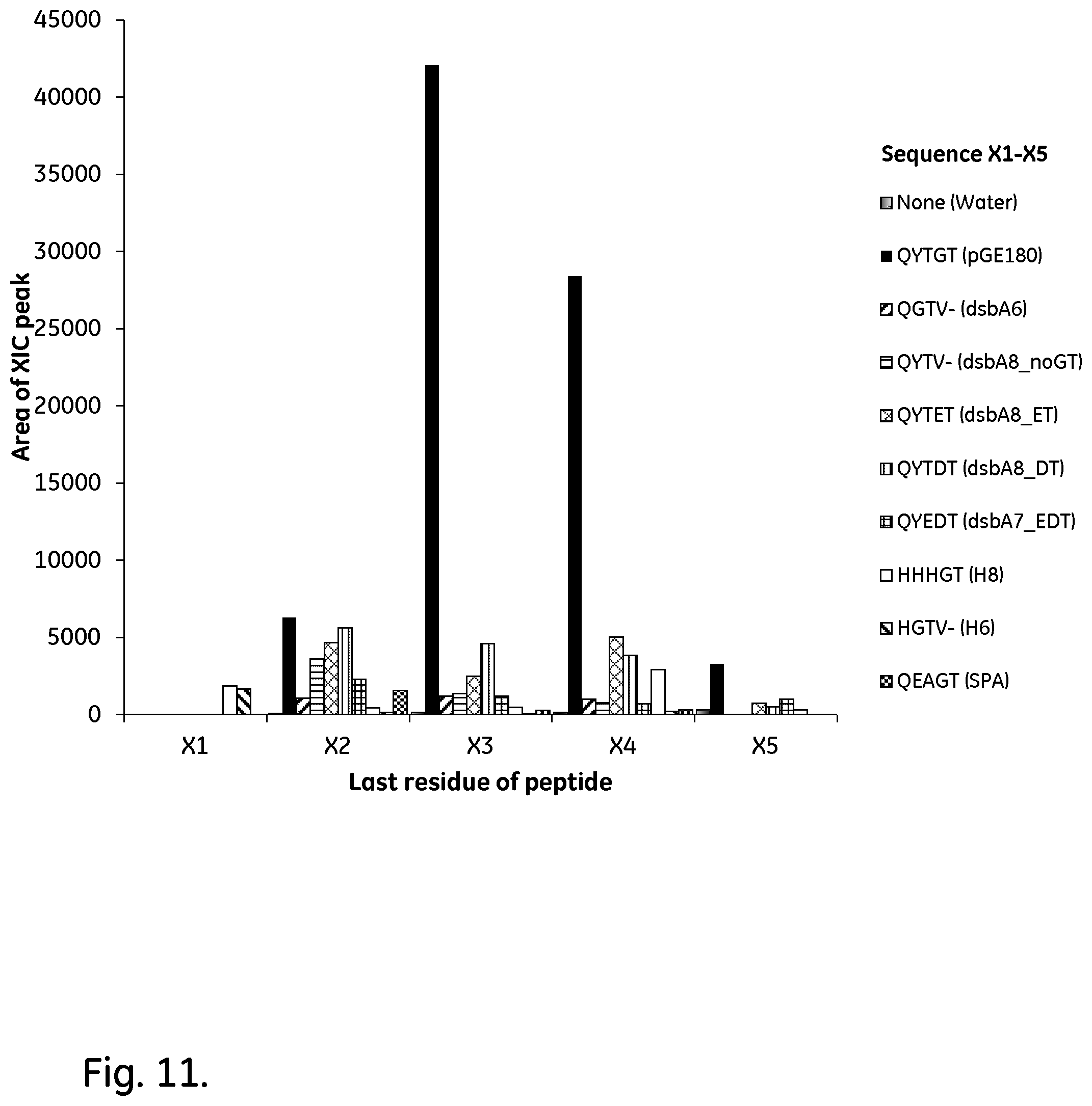
[0034] FIG. 11 shows a zoomed-in view of the relevant peptide areas of the most promising candidates. The areas are given in arbitrary units (AU).
[0035] FIG. 12 shows a break-down of the most prominently appearing peptides in the constructs showing the lowest alkaline stability in the N-terminal region. The areas are given in arbitrary units (AU).
[0036] FIG. 13 shows the measured protein concentrations, protein concentrations estimated from extinction coefficients at 280 nm, and OD600 at the end of the fed-batch cultivations. One replicate run was made for pGE180, all other constructs were only cultivated once. Error bars corresponding to a 95% confidence interval are included where data is available.
[0037] FIG. 14 shows a comparison between the dynamic binding capacities of the candidate constructs (DsbA8_noGT and DsbA8_DT), the reference sample with Zvar2, and previous data from other immobilizations using base matrices with similar dry weights, porosities and ligand densities.
DEFINITIONS
[0038] The terms "antibody" and "immunoglobulin" are used interchangeably herein, and are understood to include also fragments of antibodies, fusion proteins comprising antibodies or antibody fragments and conjugates comprising antibodies or antibody fragments.
[0039] The terms an "Fc-binding polypeptide" and "Fc-binding protein" mean a polypeptide or protein respectively, capable of binding to the crystallisable part (Fc) of an antibody and includes e.g. Staphylococcus aureus Protein A and Streptococcus Protein G, or any fragment or fusion protein thereof that has maintained said binding property.
[0040] The terms an "Fab-binding polypeptide" and "Fab-binding protein" mean a polypeptide or protein respectively, capable of binding to the antigen-binding part (Fab) of an antibody and includes e.g. Peptostreptococcus magnus Protein L, Streptococcus Protein G, native Staphylococcus aureus Protein A or any fragment or fusion protein thereof that has maintained said binding property.
[0041] The term "linker" herein means an element linking two polypeptide units, monomers or domains to each other in a multimer.
[0042] The term "% identity" with respect to comparisons of amino acid sequences is determined by standard alignment algorithms such as, for example, Basic Local Alignment Tool (BLAST.TM.) described in Altschul et al. (1990) J. Mol. Biol., 215: 403-410. A web-based software for this is freely available from the US National Library of Medicine at http://blast.ncbi.nlm.nih.gov/Blast.cgi?PROGRAM=blastp&PAGE_TYPE=BlastSea- rch&LINK_LOC=blasthome. Here, the algorithm "blastp (protein-protein BLAST)" is used for alignment of a query sequence with a subject sequence and determining i.a. the % identity.
[0043] The abbreviation "DsbA" herein means E. coli, Thiol:disulfide interchange protein, UniProt P0AEG4.
[0044] The abbreviation "OmpA" herein means E. coli, Outer membrane protein A, UniProt P0A910.
[0045] The abbreviation "PrA" herein means Staphylococcus aureus protein A, UniProt P38507.
[0046] The abbreviation "GIII" herein means Gene 3 from Bacteriophage M13, UniProt P69168.
[0047] The term "signal peptide" herein means a short (usually 16-30 amino acids long) peptide present at the N-terminus of the majority of newly synthesized proteins that are destined towards the secretory pathway. It may also be referred to as signal sequence, targeting signal, localization signal, localization sequence, transit peptide, leader sequence or leader peptide. The signal peptide is normally cleaved off from the protein by a signal peptidase enzyme.
[0048] The term "signal peptide cleavage site" herein means a dipeptide between which the signal peptidase cleaves the signal peptide from the mature protein. In most (but not all) cases the dipeptide is Ala-Ala. The signal peptide cleavage site can be calculated with algorithms such as SignalP 4.1, available on-line at http://www.cbs.dtu.dk/services/SignalP/ (Center for Biological Sequence Analysis, Technical University of Denmark).
[0049] The term "heterologous expression" herein means the expression of a gene or part of a gene in a host organism, which does not naturally have this gene or gene fragment. "Secreted" refers to across the inner membrane of gram negative bacteria, such as E. coli.
[0050] The cytoplasm (cytoplasmic), is the space inside of the inner cell membrane in gram negative bacteria containing the genetic material. The term "cytoplasmic expression" herein means protein expression within the cytoplasm.
[0051] The periplasm is a concentrated gel-like matrix in the space between the inner cytoplasmic membrane and the bacterial outer membrane called the periplasmic space in gram-negative bacteria.
[0052] The term "promoter" herein means a region of DNA that initiates transcription (writing to mRNA) of a particular gene. Promoters are normally located near the transcription start sites of genes, on the same strand and upstream on the DNA (towards the 5' region of the sense strand). A promoter can be inducible, meaning that the expression of genes operably linked to the promoter can be turned on by the presence of an inducer substance. Alternatively, the promoter may be constitutive, i.e. that it is not regulated by any inducer substance.
[0053] The abbreviation "RBS" herein means a ribosome-binding site, or ribosomal binding site. This is a sequence of nucleotides upstream of the start codon of an mRNA transcript that is responsible for the recruitment of a ribosome during the initiation of protein translation.
[0054] The abbreviation "RhaBAD" herein means the E. coli rhamnose operon promoter (also called rhamnose promoter) of genes RhaB, RhaA and RhaD. This is a promoter widely used in molecular biology.
[0055] The abbreviation "T5" herein means Bacteriophage T5 promoter for E. coli RNA polymerase, with an embedded lac operator. An operator is a segment of DNA to which a transcription factor binds to regulate gene expression by repressing it.
[0056] The abbreviation "pD861-SR" herein refers to a plasmid for E. coli protein expression with rhamnose promoter (RhaBAD) and a strong ribosome-binding site (SR).
[0057] The abbreviation "pJ401" herein refers to a plasmid for E. coli protein expression, with bacteriophage T5 promoter and dual embedded lac operator mirrored on each side of the promoter.
[0058] The abbreviation "OptEc" herein means optimized for E. coli expression, i.e. codon triplets are chosen to fit E. coli translation machinery.
[0059] The abbreviation "FspI" herein means a DNA restriction enzyme from Fischerella species (ATCC 29114). It cleaves blunt at the sequence TGCGCA.
[0060] The abbreviation "KpnI" herein means a DNA restriction enzyme from Klebsiella pneumoniae OK8 (ATCC 49790). It cleaves with overhang at the sequence GGTACC.
[0061] The abbreviation "SRP" herein means the signal recognition particle pathway, a universally conserved pathway for targeting polypeptides for secretion via the cotranslational pathway.
[0062] The abbreviation "Sec" herein means secretion or Type II secretory pathway, a system which is responsible for the secretion of proteins through the cell membrane.
[0063] The term "E. coli K12-017" herein means an E. coli expression strain, as described by Olsson M. O. and Isaksson L. A. in Molec. Gen. Genet. 169, 251-257 (1979).
[0064] The term "functional polypeptide" herein means a polypeptide with a technically useful property. Examples of such properties are: a) highly specific binding to a target species (for use as affinity binders, in particular as ligands for affinity chromatography), b) therapeutic properties (for use as a medicament), c) enzymatic properties (for use as biocatalysts) and d) signal-emitting properties (for use as reporter proteins, such as fluorescent reporter proteins).
[0065] As used herein, the terms "comprises," "comprising," "containing," "having" and the like can have the meaning ascribed to them in U.S. patent law and can mean "includes," "including," and the like; "consisting essentially of" or "consists essentially" likewise has the meaning ascribed in U.S. patent law and the term is open-ended, allowing for the presence of more than that which is recited so long as basic or novel characteristics of that which is recited is not changed by the presence of more than that which is recited, but excludes prior art embodiments.
DETAILED DESCRIPTION OF EMBODIMENTS
[0066] In one aspect, illustrated by FIGS. 1-3, the present invention discloses a recombinant protein comprising a functional polypeptide and, linked to the N-terminus of the functional polypeptide, an N-terminal spacer having a length such that the number of amino acid residues between a signal peptide cleaving site and an N-terminus proximal structural unit of the functional polypeptide is 14-24.
[0067] The functional polypeptide can be an immunoglobulin-binding polypeptide. Such a polypeptide may e.g. comprise one or more immunoglobulin-binding domains derived from a bacterial protein selected from the group consisting of Staphylococcus aureus Protein A, Peptostreptococcus magnus Protein L and Streptococcus Protein G, such as from the group consisting of Staphylococcus aureus Protein A and Peptostreptococcus magnus Protein L. The immunoglobulin-binding domain(s) can e.g. have at least 80%, such as at least 90 or 95%, sequence identity with Domain E, D, A, B or C of Staphylococcus aureus Protein A, with Protein Z (a variant of Domain B of Staphylococcus aureus Protein A), Zvar or Zvar2 (alkali-stabilized mutants of Protein Z) or with Domain 1, 2, 3, 4 or 5 of Peptostreptococcus magnus Protein L. In this context, the immunoglobulin-containing domain(s) can be defined by, or have at least 80%, such as at least 90 or 95% sequence identity with, an amino acid sequence selected from the group consisting of SEQ ID NO: 1-11. SEQ ID NO. 1-7 (Staphylococcus aureus Protein A domains E, D, A, B and C, Protein Z and Zvar) are listed in FIG. 15, and SEQ ID NO. 8-11 are specified below.
TABLE-US-00001 -Zvar2 SEQ ID NO. 8 VDAKFDKEAQ EAFYEILHLP NLTEEQRNAF IQSLKDDPSV SKAILAEAKK LNDAQAPK truncated Domain C of Staphylococcus aureus Protein A SEQ ID NO. 9 QQ NAFYEILHLP NLTEEQRNGF IQSLKDDPSV SKEILAEAKK LNDAQ -truncated version of Zvar SEQ ID NO. 10 QQ NAFYEILHLP NLTEEQRNGF IQSLKDDPSV SKEILAEAKK LNDAQ -truncated version of Zvar2 SEQ ID NO. 11 AQ EAFYEILHLP NLTEEQRNAF IQSLKDDPSV SKAILAEAKK LNDAQ
[0068] SEQ ID NO. 1-11 can all be characterized as Fc-binding domains derived from Staphylococcus aureus Protein A. Such domains may further be alkali-stabilized by mutations of the native domains, as has been done in Zvar and Zvar2. Further examples of such alkali-stabilized domains can be SEQ ID NO. 48-93 (listed under Example 6) and other examples are given e.g. in U.S. Pat. Nos. 8,329,860, 8,754,196, 9,040,661, 9,403,883, JP 2006304633A, U.S. Pat. No. 8,674,073, US 2010/0221844, US 2012/0208234, U.S. Pat. No. 9,051,375, US 2014/0031522, US 2014/0107315, US 2013/0096276, US 2013/0274451, US 2005/0143566, US 2016/0159855, US 2016/0168209, US 2016/0237124, WO 2014/146350, WO 2016/079033, WO 2016/079034, WO 2016/152946, PCT EP2017/061162, PCT EP2017/061164, PCT EP2017/061160, PCT EP2017/061158, PCT EP2017/061159, U.S. Ser. No. 14/961,164, U.S. Ser. No. 15/348,699 and U.S. Ser. No. 15/282,367, all of which are hereby incorporated by reference in their entireties. Specifically, the alkali-stabilized Fc-binding domains may have at least 80%, such as at least 90% or at least 95%, sequence identity to an amino acid sequence selected from the group consisting of SEQ ID NO. 7-11, 48-64 and 74-93.
[0069] The immunoglobulin-binding polypeptide can suitably be a multimer of immunoglobulin-binding domains, e.g. Fc-binding domains, as discussed above. The multimer can e.g. be a dimer, trimer, tetramer, pentamer, hexamer or a heptamer, such as a dimer, tetramer or hexamer. Suitably, the multimer may comprise at least four domains. The domains can be directly linked to each other (e.g. in the case of SEQ ID NO. 1-8, 48-64 and 74-93), but they can also be linked to each other via linkers, typically comprising 1-25 (such as 3-20) amino acid residues (e.g. in the case of SEQ ID NO 9-11). Examples of suitable linkers include APKADNKFNKE, APKVDAKFDKE, APK, APKVDA, AKFDKE, APKVFDKE, APAKFDKE, VDAKFDKE, APKKFDKE, APKYEDGKQYTVDAKFDKE and APKYEDGVDAKFDKE. Some specific examples of multimers include SEQ ID NO. 12 (tetramer), SEQ ID NO. 13 (hexamer) and SEQ ID NO. 65-73 (dimers).
TABLE-US-00002 -Zvar tetramer with C-terminal cysteine SEQ ID NO 12 VDAKFDKEQQ NAFYEILHLP NLTEEQRNAF IQSLKDDPSQ SANLLAEAKK LNDAQAPK VDAKFDKEQQ NAFYEILHLP NLTEEQRNAF IQSLKDDPSQ SANLLAEAKK LNDAQAPK VDAKFDKEQQ NAFYEILHLP NLTEEQRNAF IQSLKDDPSQ SANLLAEAKK LNDAQAPK VDAKFDKEQQ NAFYEILHLP NLTEEQRNAF IQSLKDDPSQ SANLLAEAKK LNDAQAPKC -Zvar2 hexamer with C-terminal cysteine SEQ ID NO 13 VDAKFDKEAQ EAFYEILHLP NLTEEQRNAF IQSLKDEPSV SKAILAEAKK LNDAQAPK VDAKFDKEAQ EAFYEILHLP NLTEEQRNAF IQSLKDEPSV SKAILAEAKK LNDAQAPK VDAKFDKEAQ EAFYEILHLP NLTEEQRNAF IQSLKDEPSV SKAILAEAKK LNDAQAPK VDAKFDKEAQ EAFYEILHLP NLTEEQRNAF IQSLKDEPSV SKAILAEAKK LNDAQAPK VDAKFDKEAQ EAFYEILHLP NLTEEQRNAF IQSLKDEPSV SKAILAEAKK LNDAQAPK VDAKFDKEAQ EAFYEILHLP NLTEEQRNAF IQSLKDEPSV SKAILAEAKK LNDAQAPKC
[0070] The recombinant protein may comprise a coupling moiety at or proximal to the C-terminus or the N-terminus, such as at the C-terminus. This coupling moiety can be used for specific coupling to a support as discussed below and may comprise a cysteine, allowing for coupling by thioether bonds. Alternatively, or additionally, the coupling moiety may comprise one or more lysines, such as a cluster of 2-8 lysines.
[0071] The functional or immunoglobulin-binding polypeptide may have secondary and tertiary structures and may suitably comprise one or more structural units, as exemplified by alpha helices, beta sheets and/or beta barrels. In particular, the polypeptide may comprise a plurality of alpha helices, such as at least three alpha helices. The Fc-binding domains derived from Staphylococcus aureus Protein A (e.g. SEQ ID NO. 1-11, 48-64 and 74-93) each comprise three alpha helices, so the number of alpha helices in a multimer as discussed above may be three times the number of domains in the multimer. The first alpha helix in the domains as exemplified by SEQ ID NO. 1-11, 48-64 and 74-93 starts at position 9 (using the position nomenclature of FIG. 15), which in the case of SEQ ID NO 1-7 and 9-10 is a glutamine and in SEQ ID NO. 8, 11, 48-64 and 74-93 is an alanine.
[0072] The N-terminal spacer suitably has a length such that the number of amino acid residues between the signal peptide cleaving site and an N-terminus proximal structural unit of the functional or immunoglobulin-binding polypeptide is 14-24. As discussed above, this structural unit can suitably be an alpha helix (or alternatively a beta sheet or beta barrel). The number of amino acid residues between the N-terminus of the functional or immunoglobulin-binding polypeptide and the N-terminus proximal structural unit can vary, e.g. between 0 (SEQ ID NO. 9-11) and 11 (SEQ ID NO. 2), and accordingly, the length of the N-terminal spacer may vary, e.g. between 3-24 amino acid residues, such as between 8-24 amino acid residues or 14-24 amino acid residues.
[0073] For example, if the N-terminal spacer is linked to an Fc-binding domain having at least 90% identity to SEQ ID NO. 1-8, 48-64 or 74-93, the N-terminal spacer may e.g. have a length of 8-12 amino acid residues and if the N-terminal spacer is linked to an Fc-binding domain having at least 90% identity to SEQ ID NO. 9-11, the N-terminal spacer may e.g. have a length of 16-20 amino acid residues. The N-terminal spacer may e.g. consist of amino acid residues selected from the group consisting of alanine, asparagine, aspartic acid, glutamine, glutamic acid, glycine, histidine, lysine, phenylalanine, serine, threonine, tryptophan, tyrosine and valine. For improved alkali stability, it may be advantageous to exclude asparagines. In this case, the N-terminal spacer may e.g. consist of amino acid residues selected from the group consisting of alanine, aspartic acid, glutamine, glutamic acid, glycine, histidine, lysine, phenylalanine, serine, threonine, tryptophan, tyrosine and valine. It can be advantageous if the N-terminal spacer does not comprise any clusters of arginines or lysines, and/or if it comprises at most two amino acid residues selected from the group consisting of lysine and arginine. In some embodiments, the two N-terminal amino acid residues of the N-terminal spacer may be AQ (alanine followed by glutamine).
[0074] In particular, the N-terminal spacer may comprise, comprise essentially or have, an amino acid sequence having at least 80% sequence identity to, or being defined by, an amino acid sequence selected from the group consisting of SEQ ID NO. 16-18, 29-30, 33-40, 43-45 and 47. The N-terminal spacer may further comprise, comprise essentially or have an amino acid sequence having at least 80% sequence identity to, or being defined by, an amino acid sequence selected from the group consisting of AQYEDGKQYTADNKFNKE, AQYEDGKQYTVDAKFDKE, AQKDQTWYTGVDAKFDKE, AQHDEAQQEAVDAKFDKE, AQGGGSGGGSVDAKFDKE, AQYEDGKQYGTVDAKFDKE, AQYEDGKQGTVDAKFDKE, AQYEDGKQYTTLEKGTVDAKFDKE, AQYEDGKQYTTLEKPVAGGTVDAKFDKE, AQYEDGKQYTVDAKFDKE, AQYEDGKQYTETVDAKFDKE, AQYEDGKQYTDTVDAKFDKE, AQYEDGKQYTATVDAKFDKE, AQYEDGKQYEDTVDAKFDKE, AQHHHHHHHHGTVDAKFDKE, AQHHHHHHGTVDAKFDKE and AQHDEAQQEAGTVDAKFDKE. The latter sequences are particularly advantageous in combination with immunoglobulin-binding polypeptides derived from SEQ ID NO. 9-11.
[0075] In terms of alkali stability it can further be advantageous to use an N-terminal spacer comprising, comprising essentially or having, an amino acid sequence having at least 80% sequence identity to, or being defined by, an amino acid sequence selected from the group consisting of SEQ ID NO. 35, 37, 38, 40, 43, 44 and 47. The N-terminal spacer may also for alkali stability comprise, comprise essentially or have an amino acid sequence having at least 80% sequence identity to, or being defined by, an amino acid sequence selected from the group consisting of AQYEDGKQYTVDAKFDKE, AQYEDGKQYTETVDAKFDKE, AQYEDGKQYTDTVDAKFDKE, AQYEDGKQYEDTVDAKFDKE, AQIIIIIIIIIIIIIIIIGTVDAKFDKE, AQIIIIIIIIIIIIGTVDAKFDKE and AQHDEAQQEAGTVDAKFDKE. The latter sequences are particularly advantageous in combination with immunoglobulin-binding polypeptides derived from SEQ ID NO. 9-11.
[0076] The alkali stability of the recombinant protein can be assessed by coupling it to an SPR chip, e.g. to Biacore CMS sensor chip as described in the examples of WO2016079033, using e.g. NHS- or maleimide coupling chemistries, and measuring the immunoglobulin-binding capacity of the chip, typically using polyclonal human IgG, before and after incubation in alkaline solutions at a specified temperature, e.g. 22+/-2.degree. C. The incubation can e.g. be performed in 0.5 M NaOH for a number of 10 min cycles, such as 100, 200 or 300 cycles. The IgG capacity of the matrix after 100 10 min incubation cycles in 0.5 M NaOH at 22+1-2.degree. C. can be at least 55, such as at least 60, at least 80 or at least 90% of the IgG capacity before the incubation. Alternatively, the remaining IgG capacity after 100 cycles for a particular mutant measured as above can be compared with the remaining IgG capacity for a parental recombinant protein. In this case, the remaining IgG capacity for the mutant may be at least 105%, such as at least 110%, at least 125%, at least 150% or at least 200% of the parental recombinant protein.
[0077] In a second aspect, the present invention discloses a nucleic acid molecule encoding for the recombinant protein as disclosed above. The nucleic acid molecule comprises the following operatively linked elements in the 5' to 3' direction:
[0078] a) an inducible or constitutive promoter DNA sequence, e.g. the RhaBAD or T5 promoter sequence. RhaBAD is inducible with rhamnose and T5 with Isopropyl .beta.-D-1-thiogalactopyranoside (IPTG). An example of a constitutive promoter is the spa promoter (the promoter of the naturally occurring Staphylococcus aureus Protein A);
[0079] b) a DNA sequence encoding a signal peptide, e.g. the OmpA (SEQ ID NO. 14) or DsbA (SEQ ID NO. 15) signal peptides, or a signal peptide having at least 80% sequence identity with either of these;
[0080] c) a DNA sequence encoding an N-terminal spacer as discussed above; and
[0081] d) a DNA sequence encoding a functional or immunoglobulin-binding polypeptide as discussed above.
[0082] The nucleic acid molecule may further comprise a ribosome-binding site (RBS) and an origin of replication. It can suitably also comprise an antibiotic resistance marker.
[0083] In a third aspect, the present invention discloses a cloning vector, e.g. a plasmid, which expresses and secretes the recombinant protein as disclosed above into the bacterial periplasm of a gram-negative cell, e.g. E. coli. The cloning vector comprises the nucleic acid molecule as discussed above.
[0084] In a fourth aspect, the present invention discloses a gram-negative bacterium transformed by the cloning vector disclosed above. The bacterium may e.g. be identified as Escherichia coli, in particular E. coli of the K12 strain, such as E. coli K12-017. Other examples of gram-negative bacteria include e.g. the genus Pseudomonas, such as e.g. Pseudomonas fluorescens. The transformation may be achieved e.g. by the heat shock method, although other methods such as electroporation are also possible.
[0085] In a fifth aspect, the present invention discloses a method of expressing and secreting the recombinant protein discussed above in a gram-negative bacterium. This method comprises the steps of:
[0086] i) providing the gram-negative bacterium disclosed above; and
[0087] ii) culturing the gram-negative bacterium.
[0088] If the nucleic acid molecule as discussed above comprises an inducible promoter, the method may further comprise a step of inducing recombinant protein expression in the gram-negative bacterium.
[0089] In a sixth aspect, the present invention discloses a separation matrix comprising the recombinant protein disclosed above, covalently linked to a support.
[0090] As the skilled person will understand, the expressed recombinant protein should be purified to an appropriate extent before being immobilized to a support. Such purification methods are well known in the field, and the immobilization of protein-based ligands to supports is easily carried out using standard methods. Suitable methods and supports will be discussed below in more detail.
[0091] The alkali stability of the matrix can be assessed by measuring the immunoglobulin-binding capacity, typically using polyclonal human IgG, before and after incubation in alkaline solutions at a specified temperature, e.g. 22+/-2.degree. C. The incubation can e.g. be performed in 0.5 M NaOH for a number of 15 min cycles, such as 100, 200 or 300 cycles, corresponding to a total incubation time of 25, 50 or 75 h. The IgG capacity of the matrix after 96-100 15 min incubation cycles or a total incubation time of 24 or 25 h in 0.5 M NaOH at 22+/-2.degree. C. can be at least 80, such as at least 85, at least 90 or at least 95% of the IgG capacity before the incubation.
[0092] The solid support of the matrix according to the invention can be of any suitable well-known kind. A conventional affinity separation matrix is often of organic nature and based on polymers that expose a hydrophilic surface to the aqueous media used, i.e. expose hydroxy (--OH), carboxy (--COOH), carboxamido (--CONH.sub.2, possibly in N-substituted forms), amino (--NH.sub.2, possibly in substituted form), oligo- or polyethylenoxy groups on their external and, if present, also on internal surfaces. The solid support can suitably be porous. The porosity can be expressed as a Kay or Kd value (the fraction of the pore volume available to a probe molecule of a particular size) measured by inverse size exclusion chromatography, e.g. according to the methods described in Gel Filtration Principles and Methods, Pharmacia LKB Biotechnology 1991, pp 6-13. By definition, both Kd and Kay values always lie within the range 0-1. The Kay value can advantageously be 0.6-0.95, e.g. 0.7-0.90 or 0.6-0.8, as measured with dextran of Mw 110 kDa as a probe molecule. An advantage of this is that the support has a large fraction of pores able to accommodate both the recombinant protein of the invention and immunoglobulins binding to the recombinant protein and to provide mass transport of the immunoglobulins to and from the binding sites.
[0093] The recombinant protein may be attached to the support via conventional coupling techniques utilising e.g. thiol, amino and/or carboxy groups present in the ligand. Bisepoxides, epichlorohydrin, CNBr, N-hydroxysuccinimide (NHS) etc are well-known coupling reagents. Between the support and the recombinant protein, a molecule known as a spacer can be introduced, which improves the availability of the recombinant protein and facilitates the chemical coupling of the recombinant protein to the support. Depending on the nature of the recombinant protein and the coupling conditions, the coupling may be a multipoint coupling (e.g. via a plurality of lysines) or a single point coupling (e.g. via a single cysteine). Alternatively, the recombinant protein may be attached to the support by non-covalent bonding, such as physical adsorption or biospecific adsorption.
[0094] In some embodiments, the matrix comprises 5-25, such as 5-20 mg/ml, 5-15 mg/ml, 5-11 mg/ml or 6-11 mg/ml of the recombinant protein coupled to the support. The amount of coupled protein can be controlled by the concentration of protein used in the coupling process, by the activation and coupling conditions used and/or by the pore structure of the support used. As a general rule, the absolute binding capacity of the matrix increases with the amount of coupled protein, at least up to a point where the pores become significantly constricted by the coupled protein. The relative binding capacity per mg coupled protein will decrease at high coupling levels, resulting in a cost-benefit optimum within the ranges specified above.
[0095] In certain embodiments, the protein is coupled to the support via thioether bonds. Methods for performing such coupling are well-known in this field and easily performed by the skilled person in this field using standard techniques and equipment. Thioether bonds are flexible and stable and generally suited for use in affinity chromatography. In particular, when the thioether bond is via a terminal or near-terminal cysteine residue on the recombinant protein, the mobility of the coupled recombinant protein is enhanced which provides improved binding capacity and binding kinetics. In some embodiments, the recombinant protein is coupled via a C-terminal cysteine provided on the protein as described above. This allows for efficient coupling of the cysteine thiol to electrophilic groups, e.g. epoxide groups, halohydrin groups etc. on a support, resulting in a thioether bridge coupling. Alternatively, the recombinant protein may be covalently linked to the support via one or more amide bonds. This can be achieved e.g. through reaction between one or more lysines in the protein and one or more activated carboxyl groups on the support. The activation can e.g. be made by the commonly known N-hydroxysuccinimide (NHS) reagent. Yet another alternative is that the protein is linked via one or more secondary amine links. Such links can be formed from lysines in the protein and either hydroxyl groups on the support, which have been activated using e.g. tresyl chloride or tosyl chloride chemistry, or by reductive amination reaction between the lysines and aldehydes on the support. The aldehydes can e.g. be formed from vicinal diols on the support through periodate oxidation.
[0096] In certain embodiments, the support comprises a polyhydroxy polymer, such as a polysaccharide. Examples of polysaccharides include e.g. dextran, starch, cellulose, pullulan, agar, agarose etc. Polysaccharides are inherently hydrophilic with low degrees of nonspecific interactions, they provide a high content of reactive (activatable) hydroxyl groups and they are generally stable towards alkaline cleaning solutions used in bioprocessing.
[0097] In some embodiments, the support comprises agar or agarose. The supports used in the present invention can easily be prepared according to standard methods, such as inverse suspension gelation (S Hjerten: Biochim Biophys Acta 79(2), 393-398 (1964). Alternatively, the base matrices are commercially available products, such as crosslinked agarose beads sold under the name of SEPHAROSE.TM. FF (GE Healthcare). In an embodiment, which is especially advantageous for large-scale separations, the support has been adapted to increase its rigidity using the methods described in U.S. Pat. Nos. 6,602,990 or 7,396,467, which are hereby incorporated by reference in their entirety, and hence renders the matrix more suitable for high flow rates.
[0098] In certain embodiments, the support, such as a polysaccharide or agarose support, is crosslinked, such as with hydroxyalkyl ether crosslinks. Crosslinker reagents producing such crosslinks can be e.g. epihalohydrins like epichlorohydrin, diepoxides like butanediol diglycidyl ether, allylating reagents like allyl halides or allyl glycidyl ether. Crosslinking is beneficial for the rigidity of the support and improves the chemical stability. Hydroxyalkyl ether crosslinks are alkali stable and do not cause significant nonspecific adsorption.
[0099] Alternatively, the solid support is based on synthetic polymers, such as polyvinyl alcohol, polyhydroxyalkyl acrylates, polyhydroxyalkyl methacrylates, polyacrylamides, polymethacrylamides etc. In case of hydrophobic polymers, such as matrices based on divinyl and monovinyl-substituted benzenes, the surface of the matrix is often hydrophilised to expose hydrophilic groups as defined above to a surrounding aqueous liquid. Such polymers are easily produced according to standard methods, see e.g. "Styrene based polymer supports developed by suspension polymerization" (R Arshady: Chimica e L'Industria 70(9), 70-75 (1988)). Alternatively, a commercially available product, such as SOURCE.TM. (GE Healthcare) is used. In another alternative, the solid support according to the invention comprises a support of inorganic nature, e.g. silica, zirconium oxide etc.
[0100] In certain embodiments, the solid support is in another form such as a surface, a chip, capillaries, or a filter (e.g. a membrane or a depth filter matrix).
[0101] In a seventh aspect, the present invention discloses a method of separating an immunoglobulin, comprising the steps of:
[0102] i) providing the separation matrix as discussed above, wherein the recombinant protein comprises an immunoglobulin-binding polypeptide, e.g. comprising one or more Fc-binding domains derived from Staphylococcus aureus Protein A;
[0103] ii) contacting the separation matrix with a liquid sample containing an immunoglobulin, to bind the immunoglobulin;
[0104] iii) optionally washing the separation matrix with a washing liquid;
[0105] iv) contacting the separation matrix with an elution liquid, to elute the immunoglobulin;
[0106] v) optionally cleaning the separation matrix with a cleaning liquid. Alkali cleaning liquids are commonly used in bioprocessing and, provided that the recombinant protein is alkali-stable, the cleaning liquid may comprise at least 0.1 M NaOH or KOH, such as at least 0.5 M NaOH or KOH, or 0.5-2.5 M NaOH or KOH.
EXAMPLES
[0107] First, two different signal peptides were tested together with four different N-terminal spacers to find the best cleavage and protein expression. This experiment (Example 1) was performed with Zvar2 monomer using the RhaBAD promoter system. In Example 2, the most promising signal peptide and N-terminal start was cloned in to Zvar2 hexamer. This construct was tested in fed-batch fermentations with and without the N-terminal start sequence. This study was performed using the T5 promoter expression system. In Example 3, all the native domains of Staphylococcus aureus protein A, Zvar monomer (Zvar.sub.1), Zvar2 monomer (Zvar2.sub.1) and Zvar tetramer (Zvar.sub.4) were tested with the chosen signal peptide and N-terminal start to see if the start sequence also had an effect on closely related domains. Also Example 3 was performed with the T5 expression system. Example 4 was performed with a set of different N-terminal spacers, Example 5 was a scale-up of fermentations using a couple of selected spacers and Example 6 was an investigation using the AQYEDGKQYTGT N-terminal spacer in combination with different mutants of an immunoglobulin-binding protein.
Materials and Methods
Constructs
[0108] Genes as described in Table 1 were synthesized by a contract manufacturer of synthetic genes (ATUM, CA, USA). Double stranded dideoxy ribonucleic acid (dsDNA) was synthesized based on the amino acid (AA) sequence and was optimized for expression in E. coli by a manufacturer proprietary algorithm. The dsDNA was inserted into expression vector pD861-SR or pJ401 with a signal peptide either from DsbA or OmpA (Table 2).
TABLE-US-00003 TABLE 1 Plasmids ordered from ATUM. N- Plasmid Pro- Signal terminal Cloning number Insert Alias moter peptide start vector Insert sequence pGE0002 Zvar2.sub.6 Zvar2.sub.6 T5 OmpA AQGT pJ401 (VDAKFDKEAQ EAFYEILHLP NLTEEQRNAF IQSLKDDPSV SKAILAEAKK LNDAQAPK)x6C pGE0068 Zvar2.sub.1 Zvar2.sub.1 RhaBAD DsbA AQGT pD861- VDAKFDKEAQ EAFYEILHLP SR NLTEEQRNAF IQSLKDDPSV SKAILAEAKK LNDAQAPKC pGE0091 Zvar.sub.1_Opt Zvar.sub.1 T5 OmpA AQGT pJ401 (VDAKFDKEQQ NAFYEILHLP Ec NLTEEQRNAF IQSLKDDPSQ SANLLAEAKK LNDAQAPK)x4C pGE0096 Zvar.sub.1_Opt Zvar.sub.1 T5 OmpA AQGT pJ401 VDAKFDKEQQ NAFYEILHLP Ec NLTEEQRNAF IQSLKDDPSQ SANLLAEAKK LNDAQAPKC pGE0119 Zvar2.sub.1 Zvar2.sub.1 RhaBAD OmpA AQGT pD861- VDAKFDKEAQ EAFYEILHLP SR NLTEEQRNAF IQSLKDDPSV SKAILAEAKK LNDAQAPKC pGE0120 Zvar2.sub.1 Zvar2.sub.1 T5 OmpA AQGT pJ401 VDAKTDKEAQ EAFYEILHLP NLTEEQRNAF IQSLKDDPSV SLAILAEAKK LNDAQAPKC pGE0127 E-domain E-domain T5 OmpA AQGT pJ401 *VDEAQQNAFY QVLNMPNLNA DQRNGFIQSL KDDPSQSANV LGEAQKLNDSQ APKC pGE0128 D-domain D-domain T5 OmpA AQGT pJ401 *VDAQQNNFNK DQQSAFYEIL NMPNLNEEQR NGFIQSLKDD PSQSTNVLGE AKKLNESQA PKC pGE0129 A-domain A-domain T5 OmpA AQGT pJ401 *VDNNFNKEQQ NAFYEILNMP NLNEEQRNGF IQSLKDDPSQ SANLLAEAKK LNESQAPKC pGE0130 B-domain B-domain T5 OmpA AQGT pJ401 *VDNKFNKEQQ NAFYEILHLP NLNEEQRNGF IQSLKDDPSQ SANLLAEAKK LNDAQAPKC pGE0131 C-domain C-domain T5 OmpA AQGT pJ401 *VDNKFNKEQQ NAFYEILHLP NLTRRQRNGF IQSLKDDPSV SKEILAEAKK LNDAQAPKC *The E, D, A, B and C domains have an A1V mutation to add a restriction site in the start of the sequence (VD) and the D-domain has an inserted D, to have uniform multiple cloning sites of the vectors.
TABLE-US-00004 TABLE 2 Signal peptides. Name AA sequence SEQ ID NO OmpA MKKTAIAIAVALAGFATVAQA 14 DsbA MKKIWLALAGLVLAFSASA 15
Cloning of N-Terminals
[0109] All plasmids were transformed into Emil K12-017 using chemically competent cells. To modify the vectors, a short spacer sequence of 8 AA was inserted between the FspI and KpnI cleave sites. The 8 AA sequences were taken from the first AA in the mature protein of OmpA (AQKDQTWYTGGT, SEQ ID NO 16), DsbA (AQYEDGKQYTGT, SEQ ID NO 17), SpA (AQHDEAQQEAGT, SEQ ID NO 18) or the flexible linker structure from M13 phage GIII AA 236-243 (AQGGGSGGGSGT, SEQ ID NO 19), with modifications in OmpA to fit the restriction site and in OmpA and SpA to mutate asparagines to glutamines (FIG. 1). Moreover, a range of different sequences were cloned into plasmid pGE0002 (Table 1).
Digestion of Plasmids
[0110] Plasmids were cleaved with restriction enzymes FspI and KpnI (New England Biolabs (NEB), MA, USA), 6 .mu.l NEB buffer 2.1 was mixed with 6 .mu.g plasmid and 2 .mu.l FspI in total 58 .mu.l. The solution was incubated in 37.degree. C. for 1 hour before KpnI was added. The incubation was continued 2 h, followed by addition of 1 .mu.l Calf intestine phosphatase (CIP) followed by 30 min further incubation. Excised bases were removed from the digested plasmids using QIAquick PCR Purification kit (Qiagen, Hilden, Germany).
Hybridization
[0111] Oligonucleotides were ordered from Integrated DNA technologies (IDT, IA, USA). All oligonucleotides were modified with a five prime phosphate group by manufacturer. Two complementary oligonucleotide pairs were mixed in ligation buffer and heated to 95.degree. C., 4 min, followed by cooling down to room temperature. The hybridized fragment was ligated into the FspI and KpnI cleaved plasmids by use of T4 DNA ligase (NEB, MA, USA). A complete list of oligonucleotides used is found in Table 3.
TABLE-US-00005 TABLE 3 List of oligonucleotides used for addition of N-terminal start sequence. SEQ Oligo Name Sequence ID NO GEOLI_191 Nterm_Omp_F GCAGAAAGATCAGACCTGGTACACCGGCGGTAC 20 GEOLI_192 Nterm_Omp_R CGCCGGTGTACCAGGTCTGATCTTTCTGC 21 GEOLI_193 Nterm_SPA_F GCAGCATGATGAAGCGCAGCAGGAAGCGGGTAC 22 GEOLI_194 Nterm_SPA_R CCGCTTCCTGCTGCGCTTCATCATGCTGC 23 GEOLI_195 Nterm_dsbA_F GCAGTATGAAGATGGCAAACAGTACACCGGTAC 24 GEOLI_196 Nterm_dsbA_R CGGTGTACTGTTTGCCATCTTCATACTGC 25 GEOLI_197 Nterm_flex_F GCAGGGTGGCGGTTCTGGCGGTGGCAGCGGTAC 26 GEOLI_198 Nterm_flex_R CGCTGCCACCGCCAGAACCGCCACCCTGC 27 GEOLI_284 Nterm_dsbA7_F GCAGTATGAAGATGGCAAACAGTACGGTAC 94 GEOLI_285 Nterm_dsbA7_R CGTACTGTTTGCCATCTTCATACTGC 95 GEOLI_286 Nterm_dsbA6_F GCAGTATGAAGATGGCAAACAGGGTAC 96 GEOLI_287 Nterm_dsbA6_R CCTGTTTGCCATCTTCATACTGC 97 GEOL1_288 Nterm_dsbA5_F GCAGTATGAAGATGGCAAAGGTAC 98 GEOLI_289 Nterm_dsbA5_R CTTTGCCATCTTCATACTGC 99 GEOLI_290 Nterm_dsbA4_F GCAGTATGAAGATGGCGGTAC 100 GEOLI_291 Nterm_dsbA4_R CGCCATCTTCATACTGC 101 GEOLI_292 Nterm_dsbA8_noGT_F GCAGTATGAAGATGGCAAACAGTACACCGT 102 GEOLI_293 Nterm_dsbA8_noGT_R CTACGGTGTACTGTTTGCCATCTTCATACTGC 103 GEOLI_294 Nterm_dsbA6_noGT_F GCAGTATGAAGATGGCAAACAGGT 104 GEOLI_295 Nterm_dsbA6_noGT_R CTACCTGTTTGCCATCTTCATACTGC 105 GEOLI_296 Nterm_dsbA8_ET_F GCAGTATGAAGATGGCAAACAGTACACCGAAACC 106 GT GEOLI_297 Nterm_dsbA8_ET_R CTACGGTTTCGGTGTACTGTTTGCCATCTTCATAC 107 TGC GEOLI_298 Nterm_dsbA8_DT_F GCAGTATGAAGATGGCAAACAGTACACCGATACC 108 GT GEOLI_299 Nterm_dsbA8_DT_R CTACGGTATCGGTGTACTGTTTGCCATCTTCATAC 109 TGC GEOLI_300 Nterm_dsbA8_AT_F GCAGTATGAAGATGGCAAACAGTACACCGCGACC 110 GT GEOLI_301 Nterm_dsbA8_AT_R CTACGGTCGCGGTGTACTGTTTGCCATCTTCATAC 111 TGC GEOLI_302 Nterm_dsbA7_EDT_F GCAGTATGAAGATGGCAAACAGTACGAAGATACC 112 GT GEOLI_303 Nterm_dsbA7_EDT_R CTACGGTATCTTCGTACTGTTTGCCATCTTCATAC 113 TGC GEOLI_304 Nterm_dsbA12_F GCAGTATGAAGATGGCAAACAGTACACCACCCTG 114 GAAAAAGGTAC GEOLI_305 Nterm_dsbA12_R CTTTTTCCAGGGTGGTGTACTGTTTGCCATCTTCA 115 TACTGC GEOLI_306 Nterm_dsbA16_F GCAGTATGAAGATGGCAAACAGTACACCACCCTG 116 GAAAAACCGGTGGCGGGCGGTAC GEOLI_307 Nterm_dsbA16_R CGCCCGCCACCGGTTTTTCCAGGGTGGTGTACTGT 117 TTGCCATCTTCATACTGC GEOLI_308 Nterm_R8_F GCAGCGCCGTCGTCGCCGTCGCCGTCGTGGTAC 118 GEOLI_309 Nterm_R8_R CACGACGGCGACGGCGACGACGGCGCTGC 119 GEOLI_310 Nterm_K8_F GCAGAAGAAGAAAAAGAAGAAGAAAAAGGGTAC 120 GEOLI_311 Nterm_K8_R CCTTTTTCTTCTTCTTTTTCTTCTTCTGC 121 GEOLI_312 Nterm_H8_F GCAGCATCACCACCATCACCATCACCATGGTAC 122 GEOLI_313 Nterm_H8_R CATGGTGATGGTGATGGTGGTGATGCTGC 123 GEOLI_314 Nterm_H6_F GCAGCATCACCACCATCACCATGGTAC 124 GEOLI_315 Nterm_H6_R CATGGTGATGGTGGTGATGCTGC 125 GEOLI_316 Nterm_H4_F GCAGCATCACCACCATGGTAC 126 GEOLI_317 Nterm_H4_R CATGGTGGTGATGCTGC 127 GEOLI_318 Nterm_Strep_F GCAGTGGAGCCATCCGCAGTTTGAAAAAGGTAC 128 GEOLI_319 Nterm_Strep_R /5Phos/CTTTTTCAAACTGCGGATGGCTCCACTGC 129
Cloned Constructs
[0112] Ligated plasmids were transformed into chemically competent E. coli K12-017 cells. The cells were thawed on ice for 30 min, 100 .mu.l competent cells were added to 10 .mu.l ligation reaction. Cells were incubated on ice for 20 min followed by heat shock at 42.degree. C. for 1 min, followed by incubation on ice 5 min. Followed by addition of 900 .mu.l SOC-medium (NEB, MA, USA). Transformation reaction was incubated in 37.degree. C. in a rotary shake incubator for 60 min, 100 .mu.l of each reaction was spread on Luria agar plates containing appropriate selective antibiotics. Positive clones were screened with Polymerase chain reaction (PCR), and clones with correct insert were selected. Selected clones were grown in 10 ml Luria broth (LB) over-night (o/n) containing appropriate antibiotics, followed by plasmid preparations using Qiagen Plasmid Miniprep kit (Qiagen, Hilden, Germany). Plasmids were sent to GATC Biotech for sequence verification (GATC Biotech, Cologne, Germany).
TABLE-US-00006 TABLE 4 Resulting plasmids after addition of N-terminal start sequence. Plasmid Pro- Signal N-terminal Cloning Insert number Insert Alias moter peptide start vector sequence pGE0138 Zvar2.sub.1 OmpA8AA RhaBAD DsbA AQKDQTWYTGGT pD861- VDAKFDKEAQ Zvar2.sub.1 SR EAFYEILHLP NLTEEQRNAF IQSLKDDPSV SKAILAEAKK LNDAQAPKC pGE0139 Zvar2.sub.1 SpA8AA- RhaBAD DsbA AQHDEAQQEAGT pD861- VDAKFDKEAQ Zvar2.sub.1 SR EAFYEILHLP NLTEEQRNAF IQSLKDDPSV SKAILAEAKK LNDAQAPKC pGE0140 Zvar2.sub.1 DsbA8AA- RhaBAD DsbA AQYEDGKQYTGT pD861- VDAKFDKEAQ Zvar2.sub.1 SR EAFYEILHLP NLTEEQRNAF OQSLKDDPSV SKAILAEAKK LNDAQAPKC pGE0141 Zvar2.sub.1 Flex8AA- RhaBAD DsbA AQGGGSGGGSGT pD861- VDAKFDKEAQ Zvar2.sub.1 SR EAFYEILHLP NLTEEQRNAF IQSLKDDPSV SKAILAEAKK LNDAQAPKC pGE0142 Zvar2.sub.1 OmpA8AA- RhaBAD OmpA AQKDQTWYTGGT pD861- VDAKFDKEAQ Zvar2.sub.1 SR EAFYEILHLP NLTEEQKNAF IQSLKDDPSV SKAILAEAKK LNDAQAPKC pGE0143 Zvar2.sub.1 SpA8AA- RhaBAD OmpA AQHDEAQQEAGT pD861- VDAKFDKEAQ Zvar2.sub.1 SR EAFYEILHLP NLTEEQRNAF IQSLKDDPSV SKAILAEAKK LNDAQAPKC pGE0144 Zvar2.sub.1 DsbA8AA- RhaBAD OmpA AQYEDGKQYTGT pD861- VDAKFDKEAQ Zvar2.sub.1 SR EAFYEILHLP NLTEEQRNAF IQSLKDDPSV SKAILAEAKK LNDAQAPKC pGE0145 Zvar2.sub.1 Flex8AA- RhaBAD OmpA AQGGGSGGGSGT pD861- VDAKFDKEAQ Zvar2.sub.1 SR EAFYEILHLP NLTEEQRNAF IQSLKDDPSV SKAILAEAKK LNDAQAPKC pGE0180 Zvar2.sub.6 DsbA8AA- T5 OmpA AQYEDGKQYTGT pJ401 (VDAKFDKEAQ Zvar2.sub.6 EAFYEILHLP NLTEEQRNAF IQSLKDDPSV SKAILAEAKK LNDAQAPK)x6C pGE0293 E-domain DsbA8AA- T5 OmpA AQYEDGKQYTGT pJ401 VDEAQQNAFY E-domain QVLNMPNLNA DQRNGFIQSL KDDPSQSANV LGEAQKLNDSQ APKC pGE0294 D-domain DsbA8AA- T5 OmpA AQYEDGKQYTGT pJ401 VDAQQNNFNK D-domain DQQSAFYEIL NMPNLNEEQR NGFIQSLKDD PSQSTNVLGE AKKLNESQA PKC pGE0295 A-domain DsbA8AA- T5 OmpA AQYEDGKQYTGT pJ401 VDNNFNKEQQ A-domain NAFYEILNMP NLNEEQRNGF IQSLKDDPSQ SANLLAEAKK LNESQAPKC pGE0296 B-domain DsbA8AA- T5 OmpA AQYEDGKQYTGT pJ401 VDNKFNKEQQ B-domain NAFYEILHLP NLNEEQRNGF IQSLKDDPSQ SANLLAEAKK LNDAQAPKC pGE0297 C-domain DsbA8AA- T5 OmpA AQYEDGKQYTGT pJ401 VDNKFNKEQQ C-domain NAFYEILHLP NLTEEQRNGF IQSLKDDPSV SKEILAEAKK LNDAQAPKC pGE0298 Zvar.sub.1_ DsbA8AA- T5 OmpA AQYEDGKQYTGT pJ401 VDAKFDKEQQ OptExp Zvar.sub.1 NAFYEILHLP NLTEEQRNAF IQSLKDDPSQ SANLLAEAKK LNDAQAPKC pGE0299 Zvar2.sub.1 DsbA8AA- T5 OmpA AQYEDGKQYTGT pJ401 VDAKFDKEAQ Zvar2.sub.1 EAFYEILHLP NLTEEQRNAF IQSLKDDPSV SKAILAEAKK LNDAQAPKC pGE0300 Zvar.sub.4_ DsbA8AA- T5 OmpA AQYEDGKQYTGT pJ401 (VDAKFDKEQQ OptEc Zvar.sub.4 NAPLEILHLP NLTEEQRNAF IQSLKDDPSQ SANLLAEAKK LNDAQAPK)x4C
Example 1
Protein Expression and Purification in Shake Flasks
[0113] Escherichia coli strain K12-017 transformed with the recombinant plasmids pGE0138-145 were cultured in 30.degree. C. for 4 h in 100 ml Terrific Broth (TB) medium (containing 12 g tryptone, 24 g yeast extract, 5 g glycerol (85%), 2.31 g KH.sub.2PO.sub.4, 12.54 g K.sub.2HPO.sub.4 and 50 mg of kanamycin sulphate per liter). Cultures were induced when OD.sub.600 nm reached 1-2, using 4 mM L-rhamnose (Sigma Aldrich, MO, USA). Cultures were further incubated in 30.degree. C. 17-20 h. Culture solution was subjected to low-speed centrifugation (at 4,000 rpm) for 20 min in a swing out rotor to collect wet cell pellet. The bacterial cell pellets were suspended in 20 ml of a 25 mM phosphate buffer solution (pH 7.4), and the cells were lysed by heat treatment at 85.degree. C. for 10 min in a heat block. Followed by a high-speed centrifugation (at 13 000 rpm) for 10 min to separate the supernatant. Then the supernatants were filtered through 0.2 .mu.m syringe filter to remove any residual particles before it was applied to an IgG Sepharose 6FF (GE Healthcare Bio-Sciences, Uppsala, Sweden) affinity chromatography column. The column was equilibrated with loading buffer (25 mM phosphate pH7, 250 mM NaCl) followed by loading of sample. After loading, the column was washed with 5 column volumes (CV) of loading buffer and one CV of low salt wash buffer (50 mM acetate pH6), followed by elution with 50 mM acetic acid, pH2.8. Absorbance at 237 nm was measured in-line using an AKTA explorer 100 chromatography system (GE Healthcare Bio-Sciences, Uppsala, Sweden) and peak integration of the eluted peak was performed in the system software (Unicorn 5.1).
Shake Flask Results of 2 Signal Peptides and 4 N-Terminal Starts
[0114] In the first work of optimization of the expression system, two different signal peptides were tested, utilizing different secretion pathways in the cell. DsbA utilizes the signal recognition particle (SRP) pathway and OmpA the Sec pathway. Results from protein expression using DsbA and OmpA signal peptides, showed that the OmpA signal peptide had higher protein expression; 2-5 times higher compared to DsbA signal peptide. Moreover, the first N-terminal AA after the cleave site of the signal peptide had a significant impact on the expression levels. In this study, the DsbA start AA had the highest expression level together with both signal peptides. The lowest expression level was seen for the Flex8AA start sequence using both signal peptides.
Liquid Chromatography Coupled Mass Spectrometry (LC/MS) Results
[0115] Eluates from IgG Sepharose 6FF were analysed using LC/MS (Waters, PA, USA). Results showed that the eluate from the reference construct pGE0120, with AQGT as N-terminal start sequence had a range of different signal peptide cleave sites (FIG. 3; a). Integrated area of peak with correct mass was 52% of the total area with correctly processed signal peptide. Other peaks corresponded to cleave sites with 6 AA extra, 9 AA extra, intact signal peptide (21 AA extra) and 7 AA missing from the main construct. After addition of 8-AA from DsbA, the signal peptide was cleaved correctly to 96% (FIG. 3; b). A similar result was seen with DsbA signal peptide and DsbA 8AA N-terminal, with 97% correctly cleaved signal peptide.
Example 2
Protein Expression in Fed-Batch Fermenter
[0116] Six, 1 L working volume fermenters, GRETA (Belach Bioteknik, Skogas, Sweden) were used. Starting volume was set to 750 ml, and end volume was approximately 1 L. Aeration was set to 1 L/min, temperature, pH and antifoam were controlled automatically. pH was kept at set value by the addition of 25% ammonia and 2 M phosphoric acid. Antifoam control was automatically adding Breox FMT 30 (BASF, Ludwigshafen, Germany), when high foaming occurred. pH and dissolved oxygen (DO) were controlled with probes from Broadley James (CA, USA). DO was kept constant at 30% by increasing stirrer speed from 300 rpm up to 1500 rpm. When the stirrer reached max speed, DO second set point was 20%, and was held constant by addition of mixing pure oxygen into the airflow. Terrific Broth (TB) supplemented with 50 mg/L kanamycin or neomycin was used in the shake flask pre-cultivations and was started by addition of 100 .mu.l cell suspension to 100 mL TB supplemented with 50 .mu.g/mL kanamycin or neomycin and incubated in 37.degree. C. for 17 h. Main fermentation media was inoculated with 10 ml pre-cultivation. Glucose feed with 60% (w/v) (VWR, PA, USA) was fed into the fermenter according to a pre-set profile after initial batch glucose was consumed. Total length of the main fermentation was 26 h.
Concentration Analysis Using Standard Curve
[0117] Fermentation samples were heat treated at 85.degree. C. for 5 min in 1.5 ml tubes. Followed by high speed centrifugation (at 13500 rpm, in benchtop centrifuge) for 5 min to separate the supernatant. Supernatants were filtered through 0.2 .mu.m syringe filter to remove any residual particles before they were applied to an IgG Sepharose 6FF HiTrap.TM. column (GE Healthcare Bio-Sciences, Uppsala Sweden). The column was equilibrated with loading buffer (Phosphate buffered Saline (Medicago, Uppsala, Sweden)) followed by loading of 50 .mu.l sample. After loading, the column was washed with 5 CV of loading buffer, followed by elution with 200 mM phosphate buffer, pH2.9. Absorbance at 237 nm was measured in-line and peak integration of the eluted peak was performed in the chromatography system software. Protein concentration of the samples were determined by a standard curve using a purified protein of known concentration.
Fermentation, Protein Expression Results
[0118] Plasmid pGE0002 containing Zvar2 hexamer (Zvar2.sub.6) was cleaved with FspI and KpnI restriction enzymes. The N-terminal start from DsbA was ligated into the cleaved vector, and the resulting plasmid was denoted pGE0180. These two vectors were transformed into E. coli K12-017 and the resulting constructs were expressed in fed-batch fermentations to see differences in protein expression and signal peptide cleavage patterns. Protein concentration analysis using standard curve showed that the protein expression increased from 3.5 g/L to 16.8 g/L, i.e. a more than 4 times increase in protein expression (FIG. 4).
LC/MS Results
[0119] Eluates from IgG Sepharose were analysed using LC/MS (Waters, PA, USA). The results showed that the eluate from the reference construct pGE0002 showed a range of different signal peptide sites including cleave sites with extra 6 AA and 9 AA, previously seen in experiment (FIG. 5, a). However, when adding 8-AA from DsbA on the N-terminus the signal peptide was cleaved correctly, resulting in a clear singular peak of correct mass (FIG. 5; b).
Example 3
[0120] Protein expression in fed-batch fermenter and concentration analysis using standard curve performed as in Example 2.
Protein Preparation
[0121] Filtered samples (See above) were applied to an IgG Sepharose 6FF Tricorn 10 column with a CV of 10.5 ml (GE Healthcare Bio-Sciences, Uppsala, Sweden). The column was equilibrated with high salt loading buffer (50 mM phosphate, pH7.0, 500 mm NaCl) followed by loading of 0.5-5 mL sample based on concentration calculations (See above). After loading, the column was washed with 5 CV of loading buffer, followed by elution with 100 mM acetic acid. Eluted samples were collected and concentrated to approximately 1 mg/ml using Vivaspin 5, 3000 Da cut-off (GE Healthcare Bio-Sciences, Uppsala, Sweden). The amount of protein was estimated by absorbance measurements at 280 nm using specific absorbance coefficients, calculated based on Pace et al. Protein Science 4, 2411-2423, (1995) and Beer Lamberts law. Sample concentration was back-calculated using total protein mass in elution pool and volume of injected sample. The concentration was confirmed by amino acid analysis. The homogeneity and the molecular weights were analysed using mass spectrometry on a Waters Q-Tof (PA, USA).
Protein Expression Results of Additional Constructs
[0122] To determine the effects of N-terminal start sequence on other domains from Staphylococcus aureus protein A and alkali stabilized domain Zvar monomer (Zvar.sub.1) and tetramer (Zvar.sub.4), new plasmids were constructed with the additional N-terminal and expressed in fed-batch fermentations using plasmid pJ401. The results showed that E-domain had no measurable expression without insert. However, with 8AA DsbA start, protein expression was measured to 2.9 g/L. Both the D- and A-domain showed a low expression that was not improved by adding N-terminal start. Both C- and B-domain (which have the same AA sequence in the start) showed very similar expression, with a clear increase in expression with 8AA. DsbA start. However, the largest increase of expression was seen on Zvar- and Zvar2 monomer (Zvar.sub.1 and Zvar2.sub.1) with more than 4 times and 10 times protein expression respectively. Also, Zvar tetramer (Zvar.sub.4) showed a significant increase in protein expression with 8AA DsbA start, even though the increase was not as dramatic as for the monomer.
TABLE-US-00007 TABLE 5 Protein expression of example proteins, all domains of prA, Zvar and Zvar tetramer, with and without N-terminal start sequence. Concentration Analysis Plasmid Domain N-terminal [g/L] Method* pGE0127 E-domain AQGT 0.0 Protein preparation pGE0293 E-domain 8AA DsbA 2.9 Standard curve pGE0128 D-domain AQGT 0.6 Protein preparation pGE0294 D-domain 8AA DsbA 0.1 Protein preparation pGE0129 A-Domain AQGT 1.9 Protein preparation pGE0295 A-Domain 8AA DsbA 0.2 Protein preparation pGE0130 B-domain AQGT 2.5 Standard curve pGE0296 B-domain 8AA DsbA 6.3 Standard curve pGE0131 C-domain AQGT 2.5 Standard curve pGE0297 C-domain 8AA DsbA 5.8 Standard curve pGE0132 Zvar.sub.1 AQGT 2.5 Standard curve pGE0298 Zvar.sub.1 8AA DsbA 10.2 Standard curve pGE0120 Zvar2.sub.1 AQGT 0.7 Protein preparation pGE0299 Zvar2.sub.1 8AA DsbA 7.3 Standard curve pGE0091 Zvar.sub.4_OptEc AQGT 9.6 Standard curve pGE0300 Zvar.sub.4_OptEc 8AA DsbA 15.3 Standard curve *Either the standard curve or protein preparation method, as described above.
[0123] In the first experiment with N-terminal spacer, two different signal peptides from two secretion pathways were tested, and it was shown that OmpA signal peptide, utilizing Sec dependent pathway had the highest expression and signal peptide cleavage in this experiment. Other signal peptides using this pathway are for example PhoA (E. coli alkaline phosphatase), MalE (E. coli maltose binding protein) and PelB (Erwinia carotovora Pectate lyase B). On the contrary, the reference construct (with AQGT start) had higher protein expression and signal peptide cleavage using DsbA signal peptide, indicating that it is a combination of signal peptide and N-terminus. Furthermore, four different N-terminals were tested and all worked better compared to the reference construct using OmpA signal peptide. Surprisingly, when the signal peptide cleavage worked optimally, also protein expression was increased, indicating that signal peptidase cleavage might be the rate limiting enzyme in the transport of proteins over the inner membrane. Without being bound by theory, a hypothesis is that protein "gets stuck" in the pores of the inner membrane and halts further expression when secretion is not working optimally. In cultivations of high protein expression, more than 50% of the protein was found outside of the cells in fermentation broth indicating periplasmic leakage to extracellular space. When other domains from Staphylococcus aureus protein A were tested, it was seen that the E-domain had no measurable protein expression, which was unexpected given that it is the first domain after the signal peptide in native Staphylococcus aureus protein A. However, one important difference is that the first AA after cleave site have been changed from AQHDEA to AQGTVDEA for cloning purposes, and it might be this substitution/insert that affects the signal peptide function negatively. Another difference is that the signal peptide has been substituted from native SpA to OmpA, this in combination with N-terminal start might not be optimal. When adding 8AA DsbA N-terminus to the E-domain in combination with the OmpA signal peptide, protein transport and expression function properly. Moreover, the A and D domains that are very closely related in the N-terminal proximal structural unit express poorly and are not improved by additional AA, whereas the B and C-domains, identical in this unit, express well with the AQGT start, and expression increases additionally when 8AA DsbA are added in the N-terminus. Also, Zvar as monomer and tetramer increase expression with additional AA. However, the most dramatical increase is seen in Zvar2 monomer where protein expression goes from 0.68 g/L to 7.28 g/L.
[0124] Without being bound by theory, a hypothesis of the difference between Zvar and Zvar2 is that Zvar2 has two mutations in the first alpha helix in position Q9 and N11, which is estimated to increase alpha-helix structure in the start of the protein, as calculated with algorithms such as GORIV, available on-line at https://npsa-prabi.ibcp.fr/cgi-bin/npsa_automat.pl?page=/NPSA/npsa_gor4.h- tml
[0125] (PRABI, Rhone-Alpes Bioinformatics Center), thus increasing steric hindrance of the signal peptidase to cleave the signal peptide of the nascent protein.
Example 4
Variants of the N-Terminal Spacer
[0126] The N-terminal inserts were each designed to investigate the effect of the insert sequence on one or more of the following parameters: protein yield, signal peptide cleavage and alkaline stability. The designs can be divided into four separate categories as follows: [0127] 1. Varying length of existing insert without modifications. [0128] 2. Removing suspected site of truncation (glycine-threonine) at alkaline conditions, while varying length of remaining insert. [0129] 3. Substituting the glycine in the known site of truncation for various other residues. [0130] 4. Substituting the entire sequence.
[0131] The aim of category 1 was mainly to investigate the effect of the insert length on signal peptide cleavage and protein yield, and did not explicitly address the suspected truncation site at alkaline conditions. Categories 2 and 3 were mostly aimed at addressing the alkaline stability issue, although they also provide some more data for the other two issues. Category 4 was a more open category, providing a wider set of sequences to provide data in all three areas of interest. The nucleotide sequences were codon optimized for E. coli, but with some degeneracy added in the more repetitive sequences. Inserts belonging to category 1 and 4 were designed to be complementary to the digestion sites of FspI in the 5' end, and KpnI in the 3' end. Since the glycine-threonine truncation site was a part of the KpnI restriction site, inserts belonging to categories 2 and 3 were designed to instead be complementary to the adjacent AccI restriction site in the 3' end, while retaining the 5' complementarity to FspI. The amino acid sequences of all inserts are shown in table 6. A schematic illustration of the general construct is shown in FIG. 6.
TABLE-US-00008 TABLE 6 All inserts used in the experiments. Residues resulting from the restriction site ''scar'' are marked in italics. Construct Insert Category name amino acid sequence SEQ ID NO Original construct pGE0002 AQGT 28 without DsbA insert Original construct pGE0180 AQYEDGKQYTGT 17 with 8 aa DsbA insert 1 DsbA7 AQYEDGKQYGT 29 DsbA6 AQYEDGKQGT 30 DsbA5 AQYEDGKGT 31 DsbA4 AQYEDGGT 32 DsbA12 AQYEDGKQYTTLEKGT 33 DsbA16 AQYEDGKQYTTLEKPVAGGT 34 2 DsbA8_ AQYEDGKQYT 35 noGT DsbA6_ AQYEDGKQ 36 noGT 3 DsbA8_ET AQYEDGKQYTET 37 DsbA8_DT AQYEDGKQYTDT 38 DsbA8_AT AQYEDGKQYTAT 39 DsbA7_EDT AQYEDGKQYEDT 40 4 R8 AQRRRRRRRRGT 41 K8 AQKKKKKKKKGT 42 H8 AQHHHHHHHHGT 43 H6 AQHHHHHHGT 44 H4 AQHHHHGT 45 Strep AQWSHPQFEKGT 46 SPA AQHDEAQQEAGT 47
[0132] The constructs were assembled by ligating different hybridized oligonucleotides into the plasmid of either pGE0002 or pGE0180, digested with the relevant restriction enzymes to remove the original N-terminal insert. After ligation, the plasmids were transformed into E. coli K12-017 cells. Positive colonies were screened using colony PCR and electrophoresis.
[0133] Two or three clones from each construct were chosen and sent to GATC biotech (Cologne, Germany) for sequence verification.
[0134] One sequence-verified clone for each construct was selected for protein expression in shake flask cultivation. FIG. 7 shows a summary of the results from the quantification of protein yields in the different shake flask cultures done with IgG Sepharose 6FF. For each construct, the left bar is the protein concentration after linear correction using a 5 g/L and a 10 g/L standard solution as reference. The right bar is this concentration divided by the OD.sub.600 nm at the end of the cultivation. This value is meant to give an idea of whether a low protein concentration is due to an actual low rate of protein production per cell, or simply due to a low culture density. Note that the concentrations are measured after pelleting the cells and re-suspending them in Phosphate buffered saline (Medicago, Uppsala, Sweden), and as such are not directly comparable to concentrations from samples taken from e.g. the multifermenter.
[0135] The protein solutions for each construct yielded from the shake flask cultivation were purified using IgG Sepharose 6FF (GE Healthcare, Uppsala, Sweden). The purified protein samples were investigated for signal peptide cleavage using LC/MS analysis (Waters, PA, USA). A protein with a correctly cleaved signal peptide only has one main mass (example Total Ion Count (TIC) shown in FIG. 8 a), while a protein with an incorrectly cleaved signal peptide yields one or more secondary peaks (as shown in FIG. 8 b). The side peaks further to the left and right of the main peaks are likely due to leakage from the cytoplasm during the heat treatment-step when extracting the protein from the cell culture. The peaks C1 to C5, N1 and N2 are likely different partially digested versions of the protein from the cytoplasm. The small peak to the right of the main peak is protein with uncleaved signal peptide, which would be reasonable to see if leakage from the cytoplasm had occurred.
[0136] In many cases, the peaks were hard to interpret conclusively due to the purity of the samples and insufficient separation during the liquid chromatography step. However, it could be seen that the following constructs had a correct signal peptide cleavage: pGE0180, DsbA7, DsbA6, DsbA8_noGT, DsbA8_DT, DsbA8_ET, DsbA8_AT, DsbA7_EDT, DsbA12, DsbA16, H8, H6 and SPA.
[0137] After the signal peptide cleavage investigation, the protein solutions of constructs with correct signal peptide cleavage were further purified using a Capto Q ImpRes anion exchange column (GE Healthcare, Uppsala, Sweden). Purified samples were subjected to treatment with 1 M sodium hydroxide (NaOH) for 0, 4 and 24 hours. Vivaspin columns (GE Healthcare, Uppsala, Sweden) were used to separate the main protein from any small peptides that might have been truncated. Both the peptide samples and the main proteins were investigated using LC/MS (Waters, PA, USA).
[0138] Peptides appeared in the mass spectra with +2 charge, that is, at a mass over charge (m/z)-value corresponding to half their molecular weight. In the cases investigated here, the peptides all had a molecular weight between about 800 Da and 1600 Da. As such, the relevant cluster of peaks appeared between 400 and 800 m/z, as can be seen for pGE0180 to the left in FIG. 9.
[0139] If peptides were present, the MassLynx search function was used on the TIC to extract the peaks of different peptide masses as extracted ion chromatograms (XIC). The XIC peaks were integrated and the integrated areas were then used to compare the amounts of different peptides in the samples.
[0140] FIG. 10 shows a summary of the results from the initial alkaline stability study done with unwashed Vivaspin columns. Despite quite a bit of overlapping background noise, clear patterns are visible after 24 hours of incubation.
[0141] FIG. 11 shows a different view of the same data for the most promising constructs (including pGE0180 as a reference), while FIG. 12 gives a breakdown of the most prominently cleaved constructs. The sequences around the cleavage site are included to give an idea of what residues might be vulnerable. Notably, for the low peak areas (around 5000 area units), background noise has a significant influence.
[0142] The second analysis with spin columns washed with isopropanol yielded mostly similar results to the initial run, although the peak areas were reduced. The notable exception is DsbA6, which was revealed to have some cleavage. This was not immediately detectable from the peptide analysis, but became apparent when investigating the retentate, where DsbA6 showed a clear truncated peak in much the same manner as pGE0180, while DsbA8_noGT and DsbA_DT showed no such peak.
Example 5
[0143] Interesting constructs from the shake-flask study and subsequent analyses of the purified protein were selected for a larger scale cultivation in a multifermenter (Belach Bioteknik, Skogas, Sweden). At the end of the fermentation, the OD.sub.000 nm and protein concentration were measured (see FIG. 13).
[0144] Since the different proteins have somewhat varying extinction coefficients, it stands to reason that they might vary in absorbance at 237 nm as well, which is where the protein concentration was estimated. Therefore, the ratio between the pGE0180 extinction coefficient (0.294) and the calculated extinction coefficient for each other protein was used to estimate a more "true" value.
[0145] The fed-batch cultures of constructs DsbA8_noGT and DsbA8_DT were heat treated to extract periplasmic protein, and centrifuged and diafiltered to remove cellular debris. The filtrate was conditioned to remove some contaminants via precipitation, and prepare the solution for purification with Capto S ImpAct (GE Healthcare, Uppsala, Sweden) using a pH gradient. The elute was reduced to remove dimers, and desalted to remove the DTT and get the correct buffer for the anion exchange purification, where a salt gradient was used. The final purified solution was concentrated via membrane ultrafiltration to reach the concentration needed for immobilization at the desired ligand densities. The end product was analysed with SEC for purity and LC/MS for molecular weight, with satisfactory results.
[0146] The purified proteins (>90% purity) were coupled to highly cross-linked agarose beads, aiming for ligand densities similar to those in a previous experiment where Zvar2 was immobilized on a base matrix of similar properties and analyzed for dynamic binding capacity. The coupled gels were packed in Tricorn 5/100 columns (GE Healthcare, Uppsala, Sweden). The packing of the gel bed was evaluated through the asymmetry of an acetone peak. The packed columns were used to measure the dynamic binding capacity of the gels using IgG. One measurement per construct was made. The results are summarized in table 7. FIG. 14 shows a comparison between measurements done in this instance, and those done in the aforementioned previous experiment.
TABLE-US-00009 TABLE 7 Asymmetry and dynamic binding capacity values for the different protein-coupled gels. Zvar2 DsbA8_noGT DsbA8_DT Qb10 (mg IgG/mL gel) 73.7 74.2 71.6 Qb80 (mg IgG/mL gel) 93.6 92.2 90.7
Example 6
[0147] The N-terminal spacer AQYEDGKQYTGT and a C-terminal cysteine was introduced in a number of monomeric and dimeric further mutants of Zvar2 (as listed below), using the OmpA signal peptide. The structure of the mature protein was thus: AQYEDGKQYTGT-IgG-binding polypeptide-C.
[0148] Plasmids were transformed into chemically competent E. coli K12-017 cells. The cells were thawed on ice for 30 min, 50 .mu.l competent cells were added to 20 ng plasmids. Cells were incubated on ice for 20 min followed by heat shock at 42.degree. C. for 1 min, followed by incubation on ice 5 min and addition of 400 .mu.l SOC-medium (NEB, MA, USA). Transformation reaction was incubated in 37.degree. C. in a rotary shake incubator for 60 min, 200 .mu.l of each reaction was spread on Luria agar plates containing appropriate selective antibiotics.
[0149] E. coli strain K12-017 transformed with the recombinant plasmids (Table 8) were cultured in 37.degree. C. in 100 ml Terrific Broth (TB) medium (containing 12 g tryptone, 24 g yeast extract, 5 g glycerol (85%), 2.31 g KH.sub.2PO.sub.4, 12.54 g K.sub.2HPO.sub.4 and 50 mg of kanamycin sulphate per liter). Cultures were induced when OD.sub.600 nm reached 1, using 1 mM IPTG (Sigma Aldrich, MO, USA). Cultures were further incubated in 30.degree. C. 17-20 h. Culture solution was subjected to low-speed centrifugation (at 4,000 rpm) for 20 min in a swing out rotor to collect wet cell pellet. The bacterial cell pellets were suspended in 10 ml of a 25 mM phosphate buffer solution (pH 7.4), and the cells were lysed by heat treatment at 85.degree. C. for 15 min in a heat block. Followed by a high-speed centrifugation (at 10 000.times.g) for 10 min to separate the supernatant. Then supernatants were filtered through 0.2 .mu.m syringe filter to remove any residual particles before it was applied to an XK 16-6 IgG Sepharose 6FF (GE Healthcare Bio-Sciences, Uppsala, Sweden). The column was equilibrated with loading buffer (25 mM phosphate pH7, 250 mM NaCl) followed by loading of sample. After loading the column was washed with 5 column volumes (CV) of loading buffer and one CV of low salt wash buffer (50 mM acetate pH6), followed by elution with 50 mM acetic acid, pH2.8. Absorbance at 237 nm was measured in-line using an AKTA explorer 100 (GE Healthcare Bio-Sciences, Uppsala, Sweden) and peak integration of the eluted peak was performed in the system software (Unicorn 5.1).
[0150] The IgG-binding fraction was eluted and collected in a pool and the volumes and protein concentrations of the pools were noted--see table 8. The amount of IgG-binding protein expressed may in several cases be higher than the amount recovered, due to loss of protein secreted into the culture medium and/or to overloading of the IgG column.
TABLE-US-00010 TABLE 8 Expression of IgG-binding proteins with N-terminal spacer AQYEDGKQYTGT and with a C-terminal cysteine. SEQ ID Protein conc. Pool volume, Recovered IgG-binding protein NO mg/mL mL protein mg Zvar2(A29G).sub.1 48 1.4 3 4.2 Zvar2 (A29S).sub.1 49 0.8 3 2.4 Zvar2 (A29Y).sub.1 50 2.0 4 8.0 Zvar2 (A29Q).sub.1 51 2.3 3 6.9 Zvar2 (A29T).sub.1 52 2.1 3 6.3 Zvar2 (A29N).sub.1 53 2.0 4 8.0 Zvar2 (A29F).sub.1 54 2.0 4 8.0 Zvar2 (A29L).sub.1 55 2.3 3 6.9 Zvar2 (A29W).sub.1 56 2.1 3 6.3 Zvar2 (A29I).sub.1 57 1.6 3 4.8 Zvar2 (A29M).sub.1 58 2.0 4 8.0 Zvar2 (A29V).sub.1 59 2.1 3 6.3 Zvar2 (A29D).sub.1 60 2.3 3 6.9 Zvar2 (A29E).sub.1 61 2.2 4 8.8 Zvar2 (A29H).sub.1 62 1.2 4 4.8 Zvar2 (A29R).sub.1 63 1.9 4 7.6 Zvar2 (A29K).sub.1 64 2.0 4 8.0 Zvar2 (.DELTA.235,236,237).sub.2 65 2.2 4 8.8 Zvar2 (.DELTA. 233,234,235).sub.2 66 2.9 4 11.6 Zvar2 (.DELTA. 5-1).sub.2 67 2.1 5 10.5 Zvar2 (.DELTA. 5-2).sub.2 68 2.8 6 16.8 Zvar2 (D3C-term).sub.2 69 3.3 5 16.5 Zvar2 (D3N-term).sub.2 70 3.5 2.7 9.4 Zvar2 (D8N-term).sub.2 71 2.6 2.1 5.46 Zvar2 (Linker + 8).sub.2 72 3.0 3.4 10.2 Zvar2 (Linker + 4).sub.2 73 3.0 2.7 8.1 Zvar2 (.DELTA. Q9).sub.1 74 1.4 1.7 2.4 Zvar2 (.DELTA. Q40).sub.1 75 2.2 3.7 8.1 Zvar2 (.DELTA. A42).sub.1 76 1.7 1.7 2.9 Zvar2 (.DELTA. N43).sub.1 77 1.6 2 3.2 Zvar2 (.DELTA. L44).sub.1 78 1.3 1.2 1.6 Zvar2 (E11N, A12F).sub.1 79 1.3 1.1 1.4 Zvar2 (E11N, A12Y).sub.1 80 1.2 0.75 0.9 Zvar2 (E11N, A12K).sub.1 81 1.4 0.72 1.0 Zvar2 (E11N, A12R).sub.1 82 1.8 1.49 2.7 Zvar2 (L22F).sub.1 83 2.9 2.77 8.0 Zvar2 (A43N, I44F).sub.1 84 1.9 4 7.6 Zvar2 (A43N, I44Y).sub.1 85 2.4 3 7.2 Zvar2 (A43N, I44W).sub.1 86 1.1 3 3.3 Zvar2 (A43N, I44R).sub.1 87 2.4 3.9 9.4 Zvar2 (A43N, I44K).sub.1 88 2.4 4 9.6 Zvar2 (D53F).sub.1 89 2.7 3 8.1 Zvar2 (D53Y).sub.1 90 2.8 3 8.4 Zvar2 (D53W).sub.1 91 1.3 2.9 3.8 Zvar2 (D53K).sub.1 92 2.2 4 8.8 Zvar2 (D53R).sub.1 93 2.2 3 6.6
TABLE-US-00011 Zvar2 (A29G) monomer (SEQ ID NO 48) VDAKFDKEAQEAFYEILHLPNLTEEQRNGFIQSLKDDPSVSKAILAEAKKLNDAQAPK Zvar2 (A29S) monomer (SEQ ID NO 49) VDAKFDKEAQEAFYEILHLPNLTEEQRNSFIQSLKDDPSVSKAILAEAKKLNDAQAPK Zvar2 (A29Y) monomer (SEQ ID NO 50) VDAKFDKEAQEAFYEILHLPNLTEEQRNYFIQSLKDDPSVSKAILAEAKKLNDAQAPK Zvar2 (A29Q) monomer (SEQ ID NO 51) VDAKFDKEAQEAFYEILHLPNLTEEQRNQFIQSLKDDPSVSKAILAEAKKLNDAQAPK Zvar2 (A29T) monomer (SEQ ID NO 52) VDAKFDKEAQEAFYEILHLPNLTEEQRNTFIQSLKDDPSVSKAILAEAKKLNDAQAPK Zvar2 (A29N) monomer (SEQ ID NO 53) VDAKFDKEAQEAFYEILHLPNLTEEQRNNFIQSLKDDPSVSKAILAEAKKLNDAQAPK Zvar2 (A29F) monomer (SEQ ID NO 54) VDAKFDKEAQEAFYEILHLPNLTEEQRNFFIQSLKDDPSVSKAILAEAKKLNDAQAPK Zvar2 (A29L) monomer (SEQ ID NO 55) VDAKFDKEAQEAFYEILHLPNLTEEQRNLFIQSLKDDPSVSKAILAEAKKLNDAQAPK Zvar2 (A29W) monomer (SEQ ID NO 56) VDAKEDKEAQEAFYEILHLPNLTEEQRNWFIQSLKDDPSVSKAILAEAKKLNDAQAPK Zvar2 (A29I) monomer (SEQ ID NO 57) VDAKFDKEAQEAFYEILHLPNLTEEQRNIFIQSLKDDPSVSKAILAEAKKLNDAQAPK Zvar2 (A29M) monomer (SEQ ID NO 58) VDAKEDKEAQEAFYEILHLPNLTEEQRNMFIQSLKDDPSVSKAILAEAKKLNDAQAPK Zvar2 (A29V) monomer (SEQ ID NO 59) VDAKEDKEAQEAFYEILHLPNLTEEQRNVFIQSLKDDPSVSKAILAEAKKLNDAQAPK Zvar2 (A29D) monomer (SEQ ID NO 60) VDAKEDKEAQEAFYEILHLPNLTEEQRNDFIQSLKDDPSVSKAILAEAKKLNDAQAPK Zvar2 (A29E) monomer (SEQ ID NO 61) VDAKEDKEAQEAFYEILHLPNLTEEQRNEFIQSLKDDPSVSKAILAEAKKLNDAQAPK Zvar2 (A29H) monomer (SEQ ID NO 62) VDAKFDKEAQEAFYEILHLPNLTEEQRNHFIQSLKDDPSVSKAILAEAKKLNDAQAPK Zvar2 (A29R) monomer (SEQ ID NO 63) VDAKFDKEAQEAFYEILHLPNLTEEQRNRFIQSLKDDPSVSKAILAEAKKLNDAQAPK Zvar2 (A29K) monomer (SEQ ID NO 64) VDAKFDKEAQEAFYEILHLPNLTEEQRNKFIQSLKDDPSVSKAILAEAKKLNDAQAPK Zvar2 (.DELTA.235, 236, 237) dimer (SEQ ID NO 65) VDAKEDKEAQEAFYEILHLPNLTEEQRNAFIQSLKDDPSVSKAILAEAKKLNDAQAPKVF DKEAQEAFYEILHLPNLTEEQRNAFIQSLKDDPSVSKAILAEAKKLNDAQAPK Zvar2 (.DELTA. 233, 234, 235) dimer (SEQ ID NO 66) VDAKFDKEAQEAFYEILHLPNLTEEQRNAFIQSLKDDPSVSKAILAEAKKLNDAQAPAKF DKEAQEAFYEILHLPNLTEEQRNAFIQSLKDDPSVSKAILAEAKKLNDAQAPK Zvar2 (.DELTA. 5-1) dimer (SEQ ID NO 67) VDAKEDKEAQEAFYEILHLPNLTEEQRNAFIQSLKDDPSVSKAILAEAKKLNDAQAKFD KEAQEAFYEILHLPNLTEEQRNAFIQSLKDDPSVSKAlLAEAKKLNDAQAPK Zvar2 (.DELTA. 5-2) dimer (SEQ ID NO 68) VDAKEDKEAQEAFYEILHLPNLTEEQRNAFIQSLKDDPSVSKAILAEAKKLNDAQAPKV DAAQEAFYEILHLPNLTEEQRNAFIQSLKDDPSVSKAILAEAKKLNDAQAPK Zvar2 (D3C-term) dimer (SEQ ID NO 69) VDAKEDKEAQEAFYEILHLPNLTEEQRNAFIQSLKDDPSVSKAILAEAKKLNDAQVDAK FDKEAQEAFYEILHLPNLTEEQRNAFIQSLKDDPSVSKAILAEAKKLNDAQAPK Zvar2 (D3N-term) dimer (SEQ ID NO 70) VDAKEDKEAQEAFYEILHLPNLTEEQRNAFIQSLKDDPSVSKAILAEAKKLNDAQAPKKF DKEAQEAFYEILHLPNLTEEQRNAFIQSLKDDPSVSKAILAEAKKLNDAQAPK Zvar2 (D8N-term) dimer (SEQ ID NO 71) VDAKEDKEAQEAFYEILHLPNLTEEQRNAFIQSLKDDPSVSKAILAEAKKLNDAQAPKA QEAFYEILHLPNLTEEQRNAFIQSLKDDPSVSKAILAEAKKLNDAQAPK Zvar2 (Linker-F8) dimer (SEQ ID NO 72) VDAKFDKEAQEAFYEILHLPNLTEEQRNAFIQSLKDDPSVSKAILAEAKKLNDAQAPKY EDGKQYTVDAKFDKEAQEAFYEILHLPNLTEEQRNAFIQSLKDDPSVSKAILAEAKKLN DAQAPK Zvar2 (Linker + 4) dimer (SEQ ID NO 73) VDAKFDKEAQEAFYEILHLPNLTEEQRNAFIQSLKDDPSVSKAILAEAKKLNDAQAPKY EDGVDAKFDKEAQEAFYEILHLPNLTEEQRNAFIQSLKDDPSVSKAILAEAKKLNDAQA PK Zvar2 (.DELTA. Q9) monomer (SEQ ID NO 74) VDAKEDKEQEAFYEILHLPNLTEEQRNAFIQSLKDDPSVSKAILAEAKKLNDAQAPK Zvar2 (.DELTA. Q40) monomer (SEQ ID NO 75) VDAKFDKEAQEAFYEILHLPNLTEEQRNAFIQSLKDDPSSKAILAEAKKLNDAQAPK Zvar2 (.DELTA. A42) monomer (SEQ ID NO 76) VDAKEDKEAQEAFYEILHLPNLTEEQRNAFIQSLKDDPSVSAILAEAKKLNDAQAPK Zvar2 (.DELTA. N43) monomer (SEQ ID NO 77) VDAKEDKEAQEAFYEILHLPNLTEEQRNAFIQSLKDDPSVSKILAEAKKLNDAQAPK Zvar2 (.DELTA. L44) monomer (SEQ ID NO 78) VDAKFDKEAQEAFYEILHLPNLTEEQRNAFIQSLKDDPSVSKALAEAKKLNDAQAPK Zvar2 (E11N, A12F) monomer (SEQ ID NO 79) VDAKFDKEAQNFFYEILHLPNLTEEQRNAFIQSLKDDPSVSKAILAEAKKLNDAQAPK Zvar2 (E11N, A12Y) monomer (SEQ ID NO 80) VDAKEDKEAQNYFYEILHLPNLTEEQRNAFIQSLKDDPSVSKAILAEAKKLNDAQAPK Zvar2 (E11N, A12K) monomer (SEQ ID NO 81) VDAKFDKEAQNKEYEILHLPNLTEEQRNAFIQSLKDDPSVSKAILAEAKKLNDAQAPK Zvar2 (E11N, A12R) monomer (SEQ ID NO 82) VDAKFDKEAQNRFYEILHLPNLTEEQRNAFIQSLKDDPSVSKAILAEAKKLNDAQAPK Zvar2 (L22F) monomer (SEQ ID NO 83) VDAKFDKEAQEAFYEILHLPNFTEEQRNAFIQSLKDDPSVSKAILAEAKKLNDAQAPK Zvar2 (A43N, I44F) monomer (SEQ ID NO 84) VDAKFDKEAQEAFYEILHLPNLTEEQRNAFIQSLKDDPSVSKNFLAEAKKLNDAQAPK Zvar2 (A43N, I44Y) monomer (SEQ ID NO 85) VDAKFDKEAQEAFYEILHLPNLTEEQRNAFIQSLKDDPSVSKNYLAEAKKLNDAQAPK Zvar2 (A43N, I44W) monomer (SEQ ID NO 86) VDAKFDKEAQEAFYEILHLPNLTEEQRNAFIQSLKDDPSVSKNWLAEAKKLNDAQAPK Zvar2 (A43N, I44R) monomer (SEQ ID NO 87) VDAKFDKEAQEAFYEILHLPNLTEEQRNAFIQSLKDDPSVSKNRLAEAKKLNDAQAPK Zvar2 (A43N, I44K) monomer (SEQ ID NO 88) VDAKFDKEAQEAFYEILHLPNLTEEQRNAFIQSLKDDPSVSKNKLAEAKKLNDAQAPK Zvar2 (D53F) monomer (SEQ ID NO 89) VDAKFDKEAQEAFYEILHLPNLTEEQRNAFIQSLKDDPSVSKAILAEAKKLNFAQAPK Zvar2 (D53Y) monomer (SEQ ID NO 90) VDAKFDKEAQEAFYEILHLPNLTEEQRNAFIQSLKDDPSVSKAILAEAKKLNYAQAPK Zvar2 (D53W) monomer (SEQ ID NO 91) VDAKFDKEAQEAFYEILHLPNLTEEQRNAFIQSLKDDPSVSKAILAEAKKLNWAQAPK Zvar2 (D53K) monomer (SEQ ID NO 92) VDAKFDKEAQEAFYEILHLPNLTEEQRNAFIQSLKDDPSVSKAILAEAKKLNKAQAPK Zvar2 (D53R) monomer (SEQ ID NO 93) VDAKFDKEAQEAFYEILHLPNLTEEQRNAFIQSLKDDPSVSKAILAEAKKLNRAQAPK
[0151] This written description uses examples to disclose the invention, including the best mode, and also to enable any person skilled in the art to practice the invention, including making and using any devices or systems and performing any incorporated methods. The patentable scope of the invention is defined by the claims, and may include other examples that occur to those skilled in the art. Such other examples are intended to be within the scope of the claims if they have structural elements that do not differ from the literal language of the claims, or if they include equivalent structural elements with insubstantial differences from the literal languages of the claims. Any patents or patent applications mentioned in the text are hereby incorporated by reference in their entireties, as if they were individually incorporated.
Sequence CWU 1
1
129151PRTStaphylococcus aureus 1Ala Gln Gln Asn Ala Phe Tyr Gln Val
Leu Asn Met Pro Asn Leu Asn1 5 10 15Ala Asp Gln Arg Asn Gly Phe Ile
Gln Ser Leu Lys Asp Asp Pro Ser 20 25 30Gln Ser Ala Asn Val Leu Gly
Glu Ala Gln Lys Leu Asn Asp Ser Gln 35 40 45Ala Pro Lys
50261PRTStaphylococcus aureus 2Ala Asp Ala Gln Gln Asn Lys Phe Asn
Lys Asp Gln Gln Ser Ala Phe1 5 10 15Tyr Glu Ile Leu Asn Met Pro Asn
Leu Asn Glu Glu Gln Arg Asn Gly 20 25 30Phe Ile Gln Ser Leu Lys Asp
Asp Pro Ser Gln Ser Thr Asn Val Leu 35 40 45Gly Glu Ala Lys Lys Leu
Asn Glu Ser Gln Ala Pro Lys 50 55 60358PRTStaphylococcus aureus
3Ala Asp Asn Asn Phe Asn Lys Glu Gln Gln Asn Ala Phe Tyr Glu Ile1 5
10 15Leu Asn Met Pro Asn Leu Asn Glu Glu Gln Arg Asn Gly Phe Ile
Gln 20 25 30Ser Leu Lys Asp Asp Pro Ser Gln Ser Ala Asn Leu Leu Ala
Glu Ala 35 40 45Lys Lys Leu Asn Glu Ser Gln Ala Pro Lys 50
55458PRTStaphylococcus aureus 4Ala Asp Asn Lys Phe Asn Lys Glu Gln
Gln Asn Ala Phe Tyr Glu Ile1 5 10 15Leu His Leu Pro Asn Leu Asn Glu
Glu Gln Arg Asn Gly Phe Ile Gln 20 25 30Ser Leu Lys Asp Asp Pro Ser
Gln Ser Ala Asn Leu Leu Ala Glu Ala 35 40 45Lys Lys Leu Asn Asp Ala
Gln Ala Pro Lys 50 55558PRTStaphylococcus aureus 5Ala Asp Asn Lys
Phe Asn Lys Glu Gln Gln Asn Ala Phe Tyr Glu Ile1 5 10 15Leu His Leu
Pro Asn Leu Thr Glu Glu Gln Arg Asn Gly Phe Ile Gln 20 25 30Ser Leu
Lys Asp Asp Pro Ser Val Ser Lys Glu Ile Leu Ala Glu Ala 35 40 45Lys
Lys Leu Asn Asp Ala Gln Ala Pro Lys 50 55658PRTArtificial
SequenceProtein A domain variant 6Val Asp Asn Lys Phe Asn Lys Glu
Gln Gln Asn Ala Phe Tyr Glu Ile1 5 10 15Leu His Leu Pro Asn Leu Asn
Glu Glu Gln Arg Asn Ala Phe Ile Gln 20 25 30Ser Leu Lys Asp Asp Pro
Ser Gln Ser Ala Asn Leu Leu Ala Glu Ala 35 40 45Lys Lys Leu Asn Asp
Ala Gln Ala Pro Lys 50 55758PRTArtificial SequenceProtein A domain
variant 7Val Asp Ala Lys Phe Asp Lys Glu Gln Gln Asn Ala Phe Tyr
Glu Ile1 5 10 15Leu His Leu Pro Asn Leu Thr Glu Glu Gln Arg Asn Ala
Phe Ile Gln 20 25 30Ser Leu Lys Asp Asp Pro Ser Gln Ser Ala Asn Leu
Leu Ala Glu Ala 35 40 45Lys Lys Leu Asn Asp Ala Gln Ala Pro Lys 50
55858PRTArtificial SequenceProtein A domain variant 8Val Asp Ala
Lys Phe Asp Lys Glu Ala Gln Glu Ala Phe Tyr Glu Ile1 5 10 15Leu His
Leu Pro Asn Leu Thr Glu Glu Gln Arg Asn Ala Phe Ile Gln 20 25 30Ser
Leu Lys Asp Asp Pro Ser Val Ser Lys Ala Ile Leu Ala Glu Ala 35 40
45Lys Lys Leu Asn Asp Ala Gln Ala Pro Lys 50 55947PRTStaphylococcus
aureus 9Gln Gln Asn Ala Phe Tyr Glu Ile Leu His Leu Pro Asn Leu Thr
Glu1 5 10 15Glu Gln Arg Asn Gly Phe Ile Gln Ser Leu Lys Asp Asp Pro
Ser Val 20 25 30Ser Lys Glu Ile Leu Ala Glu Ala Lys Lys Leu Asn Asp
Ala Gln 35 40 451047PRTArtificial SequenceProtein A domain variant
10Gln Gln Asn Ala Phe Tyr Glu Ile Leu His Leu Pro Asn Leu Thr Glu1
5 10 15Glu Gln Arg Asn Gly Phe Ile Gln Ser Leu Lys Asp Asp Pro Ser
Val 20 25 30Ser Lys Glu Ile Leu Ala Glu Ala Lys Lys Leu Asn Asp Ala
Gln 35 40 451147PRTArtificial SequenceProtein A domain variant
11Ala Gln Glu Ala Phe Tyr Glu Ile Leu His Leu Pro Asn Leu Thr Glu1
5 10 15Glu Gln Arg Asn Ala Phe Ile Gln Ser Leu Lys Asp Asp Pro Ser
Val 20 25 30Ser Lys Ala Ile Leu Ala Glu Ala Lys Lys Leu Asn Asp Ala
Gln 35 40 4512233PRTArtificial SequenceMultimer of protein A domain
variants 12Val Asp Ala Lys Phe Asp Lys Glu Gln Gln Asn Ala Phe Tyr
Glu Ile1 5 10 15Leu His Leu Pro Asn Leu Thr Glu Glu Gln Arg Asn Ala
Phe Ile Gln 20 25 30Ser Leu Lys Asp Asp Pro Ser Gln Ser Ala Asn Leu
Leu Ala Glu Ala 35 40 45Lys Lys Leu Asn Asp Ala Gln Ala Pro Lys Val
Asp Ala Lys Phe Asp 50 55 60Lys Glu Gln Gln Asn Ala Phe Tyr Glu Ile
Leu His Leu Pro Asn Leu65 70 75 80Thr Glu Glu Gln Arg Asn Ala Phe
Ile Gln Ser Leu Lys Asp Asp Pro 85 90 95Ser Gln Ser Ala Asn Leu Leu
Ala Glu Ala Lys Lys Leu Asn Asp Ala 100 105 110Gln Ala Pro Lys Val
Asp Ala Lys Phe Asp Lys Glu Gln Gln Asn Ala 115 120 125Phe Tyr Glu
Ile Leu His Leu Pro Asn Leu Thr Glu Glu Gln Arg Asn 130 135 140Ala
Phe Ile Gln Ser Leu Lys Asp Asp Pro Ser Gln Ser Ala Asn Leu145 150
155 160Leu Ala Glu Ala Lys Lys Leu Asn Asp Ala Gln Ala Pro Lys Val
Asp 165 170 175Ala Lys Phe Asp Lys Glu Gln Gln Asn Ala Phe Tyr Glu
Ile Leu His 180 185 190Leu Pro Asn Leu Thr Glu Glu Gln Arg Asn Ala
Phe Ile Gln Ser Leu 195 200 205Lys Asp Asp Pro Ser Gln Ser Ala Asn
Leu Leu Ala Glu Ala Lys Lys 210 215 220Leu Asn Asp Ala Gln Ala Pro
Lys Cys225 23013349PRTArtificial SequenceMultimer of protein A
domain variants 13Val Asp Ala Lys Phe Asp Lys Glu Ala Gln Glu Ala
Phe Tyr Glu Ile1 5 10 15Leu His Leu Pro Asn Leu Thr Glu Glu Gln Arg
Asn Ala Phe Ile Gln 20 25 30Ser Leu Lys Asp Glu Pro Ser Val Ser Lys
Ala Ile Leu Ala Glu Ala 35 40 45Lys Lys Leu Asn Asp Ala Gln Ala Pro
Lys Val Asp Ala Lys Phe Asp 50 55 60Lys Glu Ala Gln Glu Ala Phe Tyr
Glu Ile Leu His Leu Pro Asn Leu65 70 75 80Thr Glu Glu Gln Arg Asn
Ala Phe Ile Gln Ser Leu Lys Asp Glu Pro 85 90 95Ser Val Ser Lys Ala
Ile Leu Ala Glu Ala Lys Lys Leu Asn Asp Ala 100 105 110Gln Ala Pro
Lys Val Asp Ala Lys Phe Asp Lys Glu Ala Gln Glu Ala 115 120 125Phe
Tyr Glu Ile Leu His Leu Pro Asn Leu Thr Glu Glu Gln Arg Asn 130 135
140Ala Phe Ile Gln Ser Leu Lys Asp Glu Pro Ser Val Ser Lys Ala
Ile145 150 155 160Leu Ala Glu Ala Lys Lys Leu Asn Asp Ala Gln Ala
Pro Lys Val Asp 165 170 175Ala Lys Phe Asp Lys Glu Ala Gln Glu Ala
Phe Tyr Glu Ile Leu His 180 185 190Leu Pro Asn Leu Thr Glu Glu Gln
Arg Asn Ala Phe Ile Gln Ser Leu 195 200 205Lys Asp Glu Pro Ser Val
Ser Lys Ala Ile Leu Ala Glu Ala Lys Lys 210 215 220Leu Asn Asp Ala
Gln Ala Pro Lys Val Asp Ala Lys Phe Asp Lys Glu225 230 235 240Ala
Gln Glu Ala Phe Tyr Glu Ile Leu His Leu Pro Asn Leu Thr Glu 245 250
255Glu Gln Arg Asn Ala Phe Ile Gln Ser Leu Lys Asp Glu Pro Ser Val
260 265 270Ser Lys Ala Ile Leu Ala Glu Ala Lys Lys Leu Asn Asp Ala
Gln Ala 275 280 285Pro Lys Val Asp Ala Lys Phe Asp Lys Glu Ala Gln
Glu Ala Phe Tyr 290 295 300Glu Ile Leu His Leu Pro Asn Leu Thr Glu
Glu Gln Arg Asn Ala Phe305 310 315 320Ile Gln Ser Leu Lys Asp Glu
Pro Ser Val Ser Lys Ala Ile Leu Ala 325 330 335Glu Ala Lys Lys Leu
Asn Asp Ala Gln Ala Pro Lys Cys 340 3451421PRTEscherichia coli
14Met Lys Lys Thr Ala Ile Ala Ile Ala Val Ala Leu Ala Gly Phe Ala1
5 10 15Thr Val Ala Gln Ala 201519PRTEscherichia coli 15Met Lys Lys
Ile Trp Leu Ala Leu Ala Gly Leu Val Leu Ala Phe Ser1 5 10 15Ala Ser
Ala1612PRTEscherichia coli 16Ala Gln Lys Asp Gln Thr Trp Tyr Thr
Gly Gly Thr1 5 101712PRTEscherichia coli 17Ala Gln Tyr Glu Asp Gly
Lys Gln Tyr Thr Gly Thr1 5 101812PRTStaphylococcus aureus 18Ala Gln
His Asp Glu Ala Gln Gln Glu Ala Gly Thr1 5 101912PRTEscherichia
coli 19Ala Gln Gly Gly Gly Ser Gly Gly Gly Ser Gly Thr1 5
102033DNAEscherichia coli 20gcagaaagat cagacctggt acaccggcgg tac
332129DNAEscherichia coli 21cgccggtgta ccaggtctga tctttctgc
292233DNAEscherichia coli 22gcagcatgat gaagcgcagc aggaagcggg tac
332329DNAEscherichia coli 23ccgcttcctg ctgcgcttca tcatgctgc
292433DNAEscherichia coli 24gcagtatgaa gatggcaaac agtacaccgg tac
332529DNAEscherichia coli 25cggtgtactg tttgccatct tcatactgc
292633DNAEscherichia coli 26gcagggtggc ggttctggcg gtggcagcgg tac
332729DNAEscherichia coli 27cgctgccacc gccagaaccg ccaccctgc
29284PRTStaphylococcus aureus 28Ala Gln Gly Thr12911PRTArtificial
SequenceSpacer variant 29Ala Gln Tyr Glu Asp Gly Lys Gln Tyr Gly
Thr1 5 103010PRTArtificial SequenceSpacer variant 30Ala Gln Tyr Glu
Asp Gly Lys Gln Gly Thr1 5 10319PRTArtificial SequenceSpacer
variant 31Ala Gln Tyr Glu Asp Gly Lys Gly Thr1 5328PRTArtificial
SequenceSpacer variant 32Ala Gln Tyr Glu Asp Gly Gly Thr1
53316PRTArtificial SequenceSpacer variant 33Ala Gln Tyr Glu Asp Gly
Lys Gln Tyr Thr Thr Leu Glu Lys Gly Thr1 5 10 153420PRTArtificial
SequenceSpacer variant 34Ala Gln Tyr Glu Asp Gly Lys Gln Tyr Thr
Thr Leu Glu Lys Pro Val1 5 10 15Ala Gly Gly Thr 203510PRTArtificial
SequenceSpacer variant 35Ala Gln Tyr Glu Asp Gly Lys Gln Tyr Thr1 5
10368PRTArtificial SequenceSpacer variant 36Ala Gln Tyr Glu Asp Gly
Lys Gln1 53712PRTArtificial SequenceSpacer variant 37Ala Gln Tyr
Glu Asp Gly Lys Gln Tyr Thr Glu Thr1 5 103812PRTArtificial
SequenceSpacer variant 38Ala Gln Tyr Glu Asp Gly Lys Gln Tyr Thr
Asp Thr1 5 103912PRTArtificial SequenceSpacer variant 39Ala Gln Tyr
Glu Asp Gly Lys Gln Tyr Thr Ala Thr1 5 104012PRTArtificial
SequenceSpacer variant 40Ala Gln Tyr Glu Asp Gly Lys Gln Tyr Glu
Asp Thr1 5 104112PRTArtificial SequenceSpacer variant 41Ala Gln Arg
Arg Arg Arg Arg Arg Arg Arg Gly Thr1 5 104212PRTArtificial
SequenceSpacer variant 42Ala Gln Lys Lys Lys Lys Lys Lys Lys Lys
Gly Thr1 5 104312PRTArtificial SequenceSpacer variant 43Ala Gln His
His His His His His His His Gly Thr1 5 104410PRTArtificial
SequenceSpacer variant 44Ala Gln His His His His His His Gly Thr1 5
10458PRTArtificial SequenceSpacer variant 45Ala Gln His His His His
Gly Thr1 54612PRTArtificial SequenceSpacer variant 46Ala Gln Trp
Ser His Pro Gln Phe Glu Lys Gly Thr1 5 104712PRTStaphylococcus
aureus 47Ala Gln His Asp Glu Ala Gln Gln Glu Ala Gly Thr1 5
104858PRTArtificial SequenceProtein A domain variant 48Val Asp Ala
Lys Phe Asp Lys Glu Ala Gln Glu Ala Phe Tyr Glu Ile1 5 10 15Leu His
Leu Pro Asn Leu Thr Glu Glu Gln Arg Asn Gly Phe Ile Gln 20 25 30Ser
Leu Lys Asp Asp Pro Ser Val Ser Lys Ala Ile Leu Ala Glu Ala 35 40
45Lys Lys Leu Asn Asp Ala Gln Ala Pro Lys 50 554958PRTArtificial
SequenceProtein A domain variant 49Val Asp Ala Lys Phe Asp Lys Glu
Ala Gln Glu Ala Phe Tyr Glu Ile1 5 10 15Leu His Leu Pro Asn Leu Thr
Glu Glu Gln Arg Asn Ser Phe Ile Gln 20 25 30Ser Leu Lys Asp Asp Pro
Ser Val Ser Lys Ala Ile Leu Ala Glu Ala 35 40 45Lys Lys Leu Asn Asp
Ala Gln Ala Pro Lys 50 555058PRTArtificial SequenceProtein A domain
variant 50Val Asp Ala Lys Phe Asp Lys Glu Ala Gln Glu Ala Phe Tyr
Glu Ile1 5 10 15Leu His Leu Pro Asn Leu Thr Glu Glu Gln Arg Asn Tyr
Phe Ile Gln 20 25 30Ser Leu Lys Asp Asp Pro Ser Val Ser Lys Ala Ile
Leu Ala Glu Ala 35 40 45Lys Lys Leu Asn Asp Ala Gln Ala Pro Lys 50
555158PRTArtificial SequenceProtein A domain variant 51Val Asp Ala
Lys Phe Asp Lys Glu Ala Gln Glu Ala Phe Tyr Glu Ile1 5 10 15Leu His
Leu Pro Asn Leu Thr Glu Glu Gln Arg Asn Gln Phe Ile Gln 20 25 30Ser
Leu Lys Asp Asp Pro Ser Val Ser Lys Ala Ile Leu Ala Glu Ala 35 40
45Lys Lys Leu Asn Asp Ala Gln Ala Pro Lys 50 555258PRTArtificial
SequenceProtein A domain variant 52Val Asp Ala Lys Phe Asp Lys Glu
Ala Gln Glu Ala Phe Tyr Glu Ile1 5 10 15Leu His Leu Pro Asn Leu Thr
Glu Glu Gln Arg Asn Thr Phe Ile Gln 20 25 30Ser Leu Lys Asp Asp Pro
Ser Val Ser Lys Ala Ile Leu Ala Glu Ala 35 40 45Lys Lys Leu Asn Asp
Ala Gln Ala Pro Lys 50 555358PRTArtificial SequenceProtein A domain
variant 53Val Asp Ala Lys Phe Asp Lys Glu Ala Gln Glu Ala Phe Tyr
Glu Ile1 5 10 15Leu His Leu Pro Asn Leu Thr Glu Glu Gln Arg Asn Asn
Phe Ile Gln 20 25 30Ser Leu Lys Asp Asp Pro Ser Val Ser Lys Ala Ile
Leu Ala Glu Ala 35 40 45Lys Lys Leu Asn Asp Ala Gln Ala Pro Lys 50
555458PRTArtificial SequenceProtein A domain variant 54Val Asp Ala
Lys Phe Asp Lys Glu Ala Gln Glu Ala Phe Tyr Glu Ile1 5 10 15Leu His
Leu Pro Asn Leu Thr Glu Glu Gln Arg Asn Phe Phe Ile Gln 20 25 30Ser
Leu Lys Asp Asp Pro Ser Val Ser Lys Ala Ile Leu Ala Glu Ala 35 40
45Lys Lys Leu Asn Asp Ala Gln Ala Pro Lys 50 555558PRTArtificial
SequenceProtein A domain variant 55Val Asp Ala Lys Phe Asp Lys Glu
Ala Gln Glu Ala Phe Tyr Glu Ile1 5 10 15Leu His Leu Pro Asn Leu Thr
Glu Glu Gln Arg Asn Leu Phe Ile Gln 20 25 30Ser Leu Lys Asp Asp Pro
Ser Val Ser Lys Ala Ile Leu Ala Glu Ala 35 40 45Lys Lys Leu Asn Asp
Ala Gln Ala Pro Lys 50 555658PRTArtificial SequenceProtein A domain
variant 56Val Asp Ala Lys Phe Asp Lys Glu Ala Gln Glu Ala Phe Tyr
Glu Ile1 5 10 15Leu His Leu Pro Asn Leu Thr Glu Glu Gln Arg Asn Trp
Phe Ile Gln 20 25 30Ser Leu Lys Asp Asp Pro Ser Val Ser Lys Ala Ile
Leu Ala Glu Ala 35 40 45Lys Lys Leu Asn Asp Ala Gln Ala Pro Lys 50
555758PRTArtificial SequenceProtein A domain variant 57Val Asp Ala
Lys Phe Asp Lys Glu Ala Gln Glu Ala Phe Tyr Glu Ile1 5 10 15Leu His
Leu Pro Asn Leu Thr Glu Glu Gln Arg Asn Ile Phe Ile Gln 20 25 30Ser
Leu Lys Asp Asp Pro Ser Val Ser Lys Ala Ile Leu Ala Glu Ala 35 40
45Lys Lys Leu Asn Asp Ala Gln Ala Pro Lys 50 555858PRTArtificial
SequenceProtein A domain variant 58Val Asp Ala Lys Phe Asp Lys Glu
Ala Gln Glu Ala Phe Tyr Glu Ile1 5 10
15Leu His Leu Pro Asn Leu Thr Glu Glu Gln Arg Asn Met Phe Ile Gln
20 25 30Ser Leu Lys Asp Asp Pro Ser Val Ser Lys Ala Ile Leu Ala Glu
Ala 35 40 45Lys Lys Leu Asn Asp Ala Gln Ala Pro Lys 50
555958PRTArtificial SequenceProtein A domain variant 59Val Asp Ala
Lys Phe Asp Lys Glu Ala Gln Glu Ala Phe Tyr Glu Ile1 5 10 15Leu His
Leu Pro Asn Leu Thr Glu Glu Gln Arg Asn Val Phe Ile Gln 20 25 30Ser
Leu Lys Asp Asp Pro Ser Val Ser Lys Ala Ile Leu Ala Glu Ala 35 40
45Lys Lys Leu Asn Asp Ala Gln Ala Pro Lys 50 556058PRTArtificial
SequenceProtein A domain variant 60Val Asp Ala Lys Phe Asp Lys Glu
Ala Gln Glu Ala Phe Tyr Glu Ile1 5 10 15Leu His Leu Pro Asn Leu Thr
Glu Glu Gln Arg Asn Asp Phe Ile Gln 20 25 30Ser Leu Lys Asp Asp Pro
Ser Val Ser Lys Ala Ile Leu Ala Glu Ala 35 40 45Lys Lys Leu Asn Asp
Ala Gln Ala Pro Lys 50 556158PRTArtificial SequenceProtein A domain
variant 61Val Asp Ala Lys Phe Asp Lys Glu Ala Gln Glu Ala Phe Tyr
Glu Ile1 5 10 15Leu His Leu Pro Asn Leu Thr Glu Glu Gln Arg Asn Glu
Phe Ile Gln 20 25 30Ser Leu Lys Asp Asp Pro Ser Val Ser Lys Ala Ile
Leu Ala Glu Ala 35 40 45Lys Lys Leu Asn Asp Ala Gln Ala Pro Lys 50
556258PRTArtificial SequenceProtein A domain variant 62Val Asp Ala
Lys Phe Asp Lys Glu Ala Gln Glu Ala Phe Tyr Glu Ile1 5 10 15Leu His
Leu Pro Asn Leu Thr Glu Glu Gln Arg Asn His Phe Ile Gln 20 25 30Ser
Leu Lys Asp Asp Pro Ser Val Ser Lys Ala Ile Leu Ala Glu Ala 35 40
45Lys Lys Leu Asn Asp Ala Gln Ala Pro Lys 50 556358PRTArtificial
SequenceProtein A domain variant 63Val Asp Ala Lys Phe Asp Lys Glu
Ala Gln Glu Ala Phe Tyr Glu Ile1 5 10 15Leu His Leu Pro Asn Leu Thr
Glu Glu Gln Arg Asn Arg Phe Ile Gln 20 25 30Ser Leu Lys Asp Asp Pro
Ser Val Ser Lys Ala Ile Leu Ala Glu Ala 35 40 45Lys Lys Leu Asn Asp
Ala Gln Ala Pro Lys 50 556458PRTArtificial SequenceProtein A domain
variant 64Val Asp Ala Lys Phe Asp Lys Glu Ala Gln Glu Ala Phe Tyr
Glu Ile1 5 10 15Leu His Leu Pro Asn Leu Thr Glu Glu Gln Arg Asn Lys
Phe Ile Gln 20 25 30Ser Leu Lys Asp Asp Pro Ser Val Ser Lys Ala Ile
Leu Ala Glu Ala 35 40 45Lys Lys Leu Asn Asp Ala Gln Ala Pro Lys 50
5565113PRTArtificial SequenceDimer of protein A domain variants
65Val Asp Ala Lys Phe Asp Lys Glu Ala Gln Glu Ala Phe Tyr Glu Ile1
5 10 15Leu His Leu Pro Asn Leu Thr Glu Glu Gln Arg Asn Ala Phe Ile
Gln 20 25 30Ser Leu Lys Asp Asp Pro Ser Val Ser Lys Ala Ile Leu Ala
Glu Ala 35 40 45Lys Lys Leu Asn Asp Ala Gln Ala Pro Lys Val Phe Asp
Lys Glu Ala 50 55 60Gln Glu Ala Phe Tyr Glu Ile Leu His Leu Pro Asn
Leu Thr Glu Glu65 70 75 80Gln Arg Asn Ala Phe Ile Gln Ser Leu Lys
Asp Asp Pro Ser Val Ser 85 90 95Lys Ala Ile Leu Ala Glu Ala Lys Lys
Leu Asn Asp Ala Gln Ala Pro 100 105 110Lys66113PRTArtificial
SequenceDimer of protein A domain variants 66Val Asp Ala Lys Phe
Asp Lys Glu Ala Gln Glu Ala Phe Tyr Glu Ile1 5 10 15Leu His Leu Pro
Asn Leu Thr Glu Glu Gln Arg Asn Ala Phe Ile Gln 20 25 30Ser Leu Lys
Asp Asp Pro Ser Val Ser Lys Ala Ile Leu Ala Glu Ala 35 40 45Lys Lys
Leu Asn Asp Ala Gln Ala Pro Ala Lys Phe Asp Lys Glu Ala 50 55 60Gln
Glu Ala Phe Tyr Glu Ile Leu His Leu Pro Asn Leu Thr Glu Glu65 70 75
80Gln Arg Asn Ala Phe Ile Gln Ser Leu Lys Asp Asp Pro Ser Val Ser
85 90 95Lys Ala Ile Leu Ala Glu Ala Lys Lys Leu Asn Asp Ala Gln Ala
Pro 100 105 110Lys67111PRTArtificial SequenceDimer of protein A
domain variants 67Val Asp Ala Lys Phe Asp Lys Glu Ala Gln Glu Ala
Phe Tyr Glu Ile1 5 10 15Leu His Leu Pro Asn Leu Thr Glu Glu Gln Arg
Asn Ala Phe Ile Gln 20 25 30Ser Leu Lys Asp Asp Pro Ser Val Ser Lys
Ala Ile Leu Ala Glu Ala 35 40 45Lys Lys Leu Asn Asp Ala Gln Ala Lys
Phe Asp Lys Glu Ala Gln Glu 50 55 60Ala Phe Tyr Glu Ile Leu His Leu
Pro Asn Leu Thr Glu Glu Gln Arg65 70 75 80Asn Ala Phe Ile Gln Ser
Leu Lys Asp Asp Pro Ser Val Ser Lys Ala 85 90 95Ile Leu Ala Glu Ala
Lys Lys Leu Asn Asp Ala Gln Ala Pro Lys 100 105
11068111PRTArtificial SequenceDimer of protein A domain variants
68Val Asp Ala Lys Phe Asp Lys Glu Ala Gln Glu Ala Phe Tyr Glu Ile1
5 10 15Leu His Leu Pro Asn Leu Thr Glu Glu Gln Arg Asn Ala Phe Ile
Gln 20 25 30Ser Leu Lys Asp Asp Pro Ser Val Ser Lys Ala Ile Leu Ala
Glu Ala 35 40 45Lys Lys Leu Asn Asp Ala Gln Ala Pro Lys Val Asp Ala
Ala Gln Glu 50 55 60Ala Phe Tyr Glu Ile Leu His Leu Pro Asn Leu Thr
Glu Glu Gln Arg65 70 75 80Asn Ala Phe Ile Gln Ser Leu Lys Asp Asp
Pro Ser Val Ser Lys Ala 85 90 95Ile Leu Ala Glu Ala Lys Lys Leu Asn
Asp Ala Gln Ala Pro Lys 100 105 11069113PRTArtificial SequenceDimer
of protein A domain variants 69Val Asp Ala Lys Phe Asp Lys Glu Ala
Gln Glu Ala Phe Tyr Glu Ile1 5 10 15Leu His Leu Pro Asn Leu Thr Glu
Glu Gln Arg Asn Ala Phe Ile Gln 20 25 30Ser Leu Lys Asp Asp Pro Ser
Val Ser Lys Ala Ile Leu Ala Glu Ala 35 40 45Lys Lys Leu Asn Asp Ala
Gln Val Asp Ala Lys Phe Asp Lys Glu Ala 50 55 60Gln Glu Ala Phe Tyr
Glu Ile Leu His Leu Pro Asn Leu Thr Glu Glu65 70 75 80Gln Arg Asn
Ala Phe Ile Gln Ser Leu Lys Asp Asp Pro Ser Val Ser 85 90 95Lys Ala
Ile Leu Ala Glu Ala Lys Lys Leu Asn Asp Ala Gln Ala Pro 100 105
110Lys70113PRTArtificial SequenceDimer of protein A domain variants
70Val Asp Ala Lys Phe Asp Lys Glu Ala Gln Glu Ala Phe Tyr Glu Ile1
5 10 15Leu His Leu Pro Asn Leu Thr Glu Glu Gln Arg Asn Ala Phe Ile
Gln 20 25 30Ser Leu Lys Asp Asp Pro Ser Val Ser Lys Ala Ile Leu Ala
Glu Ala 35 40 45Lys Lys Leu Asn Asp Ala Gln Ala Pro Lys Lys Phe Asp
Lys Glu Ala 50 55 60Gln Glu Ala Phe Tyr Glu Ile Leu His Leu Pro Asn
Leu Thr Glu Glu65 70 75 80Gln Arg Asn Ala Phe Ile Gln Ser Leu Lys
Asp Asp Pro Ser Val Ser 85 90 95Lys Ala Ile Leu Ala Glu Ala Lys Lys
Leu Asn Asp Ala Gln Ala Pro 100 105 110Lys71108PRTArtificial
SequenceDim er of protein A domain variants 71Val Asp Ala Lys Phe
Asp Lys Glu Ala Gln Glu Ala Phe Tyr Glu Ile1 5 10 15Leu His Leu Pro
Asn Leu Thr Glu Glu Gln Arg Asn Ala Phe Ile Gln 20 25 30Ser Leu Lys
Asp Asp Pro Ser Val Ser Lys Ala Ile Leu Ala Glu Ala 35 40 45Lys Lys
Leu Asn Asp Ala Gln Ala Pro Lys Ala Gln Glu Ala Phe Tyr 50 55 60Glu
Ile Leu His Leu Pro Asn Leu Thr Glu Glu Gln Arg Asn Ala Phe65 70 75
80Ile Gln Ser Leu Lys Asp Asp Pro Ser Val Ser Lys Ala Ile Leu Ala
85 90 95Glu Ala Lys Lys Leu Asn Asp Ala Gln Ala Pro Lys 100
10572124PRTArtificial SequenceDimer of protein A domain variants
72Val Asp Ala Lys Phe Asp Lys Glu Ala Gln Glu Ala Phe Tyr Glu Ile1
5 10 15Leu His Leu Pro Asn Leu Thr Glu Glu Gln Arg Asn Ala Phe Ile
Gln 20 25 30Ser Leu Lys Asp Asp Pro Ser Val Ser Lys Ala Ile Leu Ala
Glu Ala 35 40 45Lys Lys Leu Asn Asp Ala Gln Ala Pro Lys Tyr Glu Asp
Gly Lys Gln 50 55 60Tyr Thr Val Asp Ala Lys Phe Asp Lys Glu Ala Gln
Glu Ala Phe Tyr65 70 75 80Glu Ile Leu His Leu Pro Asn Leu Thr Glu
Glu Gln Arg Asn Ala Phe 85 90 95Ile Gln Ser Leu Lys Asp Asp Pro Ser
Val Ser Lys Ala Ile Leu Ala 100 105 110Glu Ala Lys Lys Leu Asn Asp
Ala Gln Ala Pro Lys 115 12073120PRTArtificial SequenceDimer of
protein A domain variants 73Val Asp Ala Lys Phe Asp Lys Glu Ala Gln
Glu Ala Phe Tyr Glu Ile1 5 10 15Leu His Leu Pro Asn Leu Thr Glu Glu
Gln Arg Asn Ala Phe Ile Gln 20 25 30Ser Leu Lys Asp Asp Pro Ser Val
Ser Lys Ala Ile Leu Ala Glu Ala 35 40 45Lys Lys Leu Asn Asp Ala Gln
Ala Pro Lys Tyr Glu Asp Gly Val Asp 50 55 60Ala Lys Phe Asp Lys Glu
Ala Gln Glu Ala Phe Tyr Glu Ile Leu His65 70 75 80Leu Pro Asn Leu
Thr Glu Glu Gln Arg Asn Ala Phe Ile Gln Ser Leu 85 90 95Lys Asp Asp
Pro Ser Val Ser Lys Ala Ile Leu Ala Glu Ala Lys Lys 100 105 110Leu
Asn Asp Ala Gln Ala Pro Lys 115 1207457PRTArtificial
SequenceProtein A domain variant 74Val Asp Ala Lys Phe Asp Lys Glu
Gln Glu Ala Phe Tyr Glu Ile Leu1 5 10 15His Leu Pro Asn Leu Thr Glu
Glu Gln Arg Asn Ala Phe Ile Gln Ser 20 25 30Leu Lys Asp Asp Pro Ser
Val Ser Lys Ala Ile Leu Ala Glu Ala Lys 35 40 45Lys Leu Asn Asp Ala
Gln Ala Pro Lys 50 557557PRTArtificial SequenceProtein A domain
variant 75Val Asp Ala Lys Phe Asp Lys Glu Ala Gln Glu Ala Phe Tyr
Glu Ile1 5 10 15Leu His Leu Pro Asn Leu Thr Glu Glu Gln Arg Asn Ala
Phe Ile Gln 20 25 30Ser Leu Lys Asp Asp Pro Ser Ser Lys Ala Ile Leu
Ala Glu Ala Lys 35 40 45Lys Leu Asn Asp Ala Gln Ala Pro Lys 50
557657PRTArtificial SequenceProtein A domain variant 76Val Asp Ala
Lys Phe Asp Lys Glu Ala Gln Glu Ala Phe Tyr Glu Ile1 5 10 15Leu His
Leu Pro Asn Leu Thr Glu Glu Gln Arg Asn Ala Phe Ile Gln 20 25 30Ser
Leu Lys Asp Asp Pro Ser Val Ser Ala Ile Leu Ala Glu Ala Lys 35 40
45Lys Leu Asn Asp Ala Gln Ala Pro Lys 50 557757PRTArtificial
SequenceProtein A domain variant 77Val Asp Ala Lys Phe Asp Lys Glu
Ala Gln Glu Ala Phe Tyr Glu Ile1 5 10 15Leu His Leu Pro Asn Leu Thr
Glu Glu Gln Arg Asn Ala Phe Ile Gln 20 25 30Ser Leu Lys Asp Asp Pro
Ser Val Ser Lys Ile Leu Ala Glu Ala Lys 35 40 45Lys Leu Asn Asp Ala
Gln Ala Pro Lys 50 557857PRTArtificial SequenceProtein A domain
variant 78Val Asp Ala Lys Phe Asp Lys Glu Ala Gln Glu Ala Phe Tyr
Glu Ile1 5 10 15Leu His Leu Pro Asn Leu Thr Glu Glu Gln Arg Asn Ala
Phe Ile Gln 20 25 30Ser Leu Lys Asp Asp Pro Ser Val Ser Lys Ala Leu
Ala Glu Ala Lys 35 40 45Lys Leu Asn Asp Ala Gln Ala Pro Lys 50
557958PRTArtificial SequenceProtein A domain variant 79Val Asp Ala
Lys Phe Asp Lys Glu Ala Gln Asn Phe Phe Tyr Glu Ile1 5 10 15Leu His
Leu Pro Asn Leu Thr Glu Glu Gln Arg Asn Ala Phe Ile Gln 20 25 30Ser
Leu Lys Asp Asp Pro Ser Val Ser Lys Ala Ile Leu Ala Glu Ala 35 40
45Lys Lys Leu Asn Asp Ala Gln Ala Pro Lys 50 558058PRTArtificial
SequenceProtein A domain variant 80Val Asp Ala Lys Phe Asp Lys Glu
Ala Gln Asn Tyr Phe Tyr Glu Ile1 5 10 15Leu His Leu Pro Asn Leu Thr
Glu Glu Gln Arg Asn Ala Phe Ile Gln 20 25 30Ser Leu Lys Asp Asp Pro
Ser Val Ser Lys Ala Ile Leu Ala Glu Ala 35 40 45Lys Lys Leu Asn Asp
Ala Gln Ala Pro Lys 50 558158PRTArtificial SequenceProtein A domain
variant 81Val Asp Ala Lys Phe Asp Lys Glu Ala Gln Asn Lys Phe Tyr
Glu Ile1 5 10 15Leu His Leu Pro Asn Leu Thr Glu Glu Gln Arg Asn Ala
Phe Ile Gln 20 25 30Ser Leu Lys Asp Asp Pro Ser Val Ser Lys Ala Ile
Leu Ala Glu Ala 35 40 45Lys Lys Leu Asn Asp Ala Gln Ala Pro Lys 50
558258PRTArtificial SequenceProtein A domain variant 82Val Asp Ala
Lys Phe Asp Lys Glu Ala Gln Asn Arg Phe Tyr Glu Ile1 5 10 15Leu His
Leu Pro Asn Leu Thr Glu Glu Gln Arg Asn Ala Phe Ile Gln 20 25 30Ser
Leu Lys Asp Asp Pro Ser Val Ser Lys Ala Ile Leu Ala Glu Ala 35 40
45Lys Lys Leu Asn Asp Ala Gln Ala Pro Lys 50 558358PRTArtificial
SequenceProtein A domain variant 83Val Asp Ala Lys Phe Asp Lys Glu
Ala Gln Glu Ala Phe Tyr Glu Ile1 5 10 15Leu His Leu Pro Asn Phe Thr
Glu Glu Gln Arg Asn Ala Phe Ile Gln 20 25 30Ser Leu Lys Asp Asp Pro
Ser Val Ser Lys Ala Ile Leu Ala Glu Ala 35 40 45Lys Lys Leu Asn Asp
Ala Gln Ala Pro Lys 50 558458PRTArtificial SequenceProtein A domain
variant 84Val Asp Ala Lys Phe Asp Lys Glu Ala Gln Glu Ala Phe Tyr
Glu Ile1 5 10 15Leu His Leu Pro Asn Leu Thr Glu Glu Gln Arg Asn Ala
Phe Ile Gln 20 25 30Ser Leu Lys Asp Asp Pro Ser Val Ser Lys Asn Phe
Leu Ala Glu Ala 35 40 45Lys Lys Leu Asn Asp Ala Gln Ala Pro Lys 50
558558PRTArtificial SequenceProtein A domain variant 85Val Asp Ala
Lys Phe Asp Lys Glu Ala Gln Glu Ala Phe Tyr Glu Ile1 5 10 15Leu His
Leu Pro Asn Leu Thr Glu Glu Gln Arg Asn Ala Phe Ile Gln 20 25 30Ser
Leu Lys Asp Asp Pro Ser Val Ser Lys Asn Tyr Leu Ala Glu Ala 35 40
45Lys Lys Leu Asn Asp Ala Gln Ala Pro Lys 50 558658PRTArtificial
SequenceProtein A domain variant 86Val Asp Ala Lys Phe Asp Lys Glu
Ala Gln Glu Ala Phe Tyr Glu Ile1 5 10 15Leu His Leu Pro Asn Leu Thr
Glu Glu Gln Arg Asn Ala Phe Ile Gln 20 25 30Ser Leu Lys Asp Asp Pro
Ser Val Ser Lys Asn Trp Leu Ala Glu Ala 35 40 45Lys Lys Leu Asn Asp
Ala Gln Ala Pro Lys 50 558758PRTArtificial SequenceProtein A domain
variant 87Val Asp Ala Lys Phe Asp Lys Glu Ala Gln Glu Ala Phe Tyr
Glu Ile1 5 10 15Leu His Leu Pro Asn Leu Thr Glu Glu Gln Arg Asn Ala
Phe Ile Gln 20 25 30Ser Leu Lys Asp Asp Pro Ser Val Ser Lys Asn Arg
Leu Ala Glu Ala 35 40 45Lys Lys Leu Asn Asp Ala Gln Ala Pro Lys 50
558858PRTArtificial SequenceProtein A domain variant 88Val Asp Ala
Lys Phe Asp Lys Glu Ala Gln Glu Ala Phe Tyr Glu Ile1 5 10 15Leu His
Leu Pro Asn Leu Thr Glu Glu Gln Arg Asn Ala Phe Ile Gln 20 25 30Ser
Leu Lys Asp Asp Pro Ser Val Ser Lys Asn Lys Leu Ala Glu Ala
35 40 45Lys Lys Leu Asn Asp Ala Gln Ala Pro Lys 50
558958PRTArtificial SequenceProtein A domain variant 89Val Asp Ala
Lys Phe Asp Lys Glu Ala Gln Glu Ala Phe Tyr Glu Ile1 5 10 15Leu His
Leu Pro Asn Leu Thr Glu Glu Gln Arg Asn Ala Phe Ile Gln 20 25 30Ser
Leu Lys Asp Asp Pro Ser Val Ser Lys Ala Ile Leu Ala Glu Ala 35 40
45Lys Lys Leu Asn Phe Ala Gln Ala Pro Lys 50 559058PRTArtificial
SequenceProtein A domain variant 90Val Asp Ala Lys Phe Asp Lys Glu
Ala Gln Glu Ala Phe Tyr Glu Ile1 5 10 15Leu His Leu Pro Asn Leu Thr
Glu Glu Gln Arg Asn Ala Phe Ile Gln 20 25 30Ser Leu Lys Asp Asp Pro
Ser Val Ser Lys Ala Ile Leu Ala Glu Ala 35 40 45Lys Lys Leu Asn Tyr
Ala Gln Ala Pro Lys 50 559158PRTArtificial SequenceProtein A domain
variant 91Val Asp Ala Lys Phe Asp Lys Glu Ala Gln Glu Ala Phe Tyr
Glu Ile1 5 10 15Leu His Leu Pro Asn Leu Thr Glu Glu Gln Arg Asn Ala
Phe Ile Gln 20 25 30Ser Leu Lys Asp Asp Pro Ser Val Ser Lys Ala Ile
Leu Ala Glu Ala 35 40 45Lys Lys Leu Asn Trp Ala Gln Ala Pro Lys 50
559258PRTArtificial SequenceProtein A domain variant 92Val Asp Ala
Lys Phe Asp Lys Glu Ala Gln Glu Ala Phe Tyr Glu Ile1 5 10 15Leu His
Leu Pro Asn Leu Thr Glu Glu Gln Arg Asn Ala Phe Ile Gln 20 25 30Ser
Leu Lys Asp Asp Pro Ser Val Ser Lys Ala Ile Leu Ala Glu Ala 35 40
45Lys Lys Leu Asn Lys Ala Gln Ala Pro Lys 50 559358PRTArtificial
SequenceProtein A domain variant 93Val Asp Ala Lys Phe Asp Lys Glu
Ala Gln Glu Ala Phe Tyr Glu Ile1 5 10 15Leu His Leu Pro Asn Leu Thr
Glu Glu Gln Arg Asn Ala Phe Ile Gln 20 25 30Ser Leu Lys Asp Asp Pro
Ser Val Ser Lys Ala Ile Leu Ala Glu Ala 35 40 45Lys Lys Leu Asn Arg
Ala Gln Ala Pro Lys 50 559430DNAArtificial SequenceSpacer variant
94gcagtatgaa gatggcaaac agtacggtac 309526DNAArtificial
SequenceSpacer variant 95cgtactgttt gccatcttca tactgc
269627DNAArtificial SequenceSpacer variant 96gcagtatgaa gatggcaaac
agggtac 279723DNAArtificial SequenceSpacer variant 97cctgtttgcc
atcttcatac tgc 239824DNAArtificial SequenceSpacer variant
98gcagtatgaa gatggcaaag gtac 249920DNAArtificial SequenceSpacer
variant 99ctttgccatc ttcatactgc 2010021DNAArtificial SequenceSpacer
variant 100gcagtatgaa gatggcggta c 2110117DNAArtificial
SequenceSpacer variant 101cgccatcttc atactgc 1710230DNAArtificial
SequenceSpacer variant 102gcagtatgaa gatggcaaac agtacaccgt
3010332DNAArtificial SequenceSpacer variant 103ctacggtgta
ctgtttgcca tcttcatact gc 3210424DNAArtificial SequenceSpacer
variant 104gcagtatgaa gatggcaaac aggt 2410526DNAArtificial
SequenceSpacer variant 105ctacctgttt gccatcttca tactgc
2610636DNAArtificial SequenceSpacer variant 106gcagtatgaa
gatggcaaac agtacaccga aaccgt 3610738DNAArtificial SequenceSpacer
variant 107ctacggtttc ggtgtactgt ttgccatctt catactgc
3810836DNAArtificial SequenceSpacer variant 108gcagtatgaa
gatggcaaac agtacaccga taccgt 3610938DNAArtificial SequenceSpacer
variant 109ctacggtatc ggtgtactgt ttgccatctt catactgc
3811036DNAArtificial SequenceSpacer variant 110gcagtatgaa
gatggcaaac agtacaccgc gaccgt 3611138DNAArtificial SequenceSpacer
variant 111ctacggtcgc ggtgtactgt ttgccatctt catactgc
3811236DNAArtificial SequenceSpacer variant 112gcagtatgaa
gatggcaaac agtacgaaga taccgt 3611338DNAArtificial SequenceSpacer
variant 113ctacggtatc ttcgtactgt ttgccatctt catactgc
3811445DNAArtificial SequenceSpacer variant 114gcagtatgaa
gatggcaaac agtacaccac cctggaaaaa ggtac 4511541DNAArtificial
SequenceSpacer variant 115ctttttccag ggtggtgtac tgtttgccat
cttcatactg c 4111657DNAArtificial SequenceSpacer variant
116gcagtatgaa gatggcaaac agtacaccac cctggaaaaa ccggtggcgg gcggtac
5711753DNAArtificial SequenceSpacer variant 117cgcccgccac
cggtttttcc agggtggtgt actgtttgcc atcttcatac tgc
5311833DNAArtificial SequenceSpacer variant 118gcagcgccgt
cgtcgccgtc gccgtcgtgg tac 3311929DNAArtificial SequenceSpacer
variant 119cacgacggcg acggcgacga cggcgctgc 2912033DNAArtificial
SequenceSpacer variant 120gcagaagaag aaaaagaaga agaaaaaggg tac
3312129DNAArtificial SequenceSpacer variant 121cctttttctt
cttctttttc ttcttctgc 2912233DNAArtificial SequenceSpacer variant
122gcagcatcac caccatcacc atcaccatgg tac 3312329DNAArtificial
SequenceSpacer variant 123catggtgatg gtgatggtgg tgatgctgc
2912427DNAArtificial SequenceSpacer variant 124gcagcatcac
caccatcacc atggtac 2712523DNAArtificial SequenceSpacer variant
125catggtgatg gtggtgatgc tgc 2312621DNAArtificial SequenceSpacer
variant 126gcagcatcac caccatggta c 2112717DNAArtificial
SequenceSpacer variant 127catggtggtg atgctgc 1712833DNAArtificial
SequenceSpacer variant 128gcagtggagc catccgcagt ttgaaaaagg tac
3312929DNAArtificial SequenceSpacer variant 129ctttttcaaa
ctgcggatgg ctccactgc 29
References
D00000
D00001
D00002
D00003
D00004
D00005
D00006
D00007
D00008
D00009
D00010
D00011
D00012

D00013

D00014

D00015
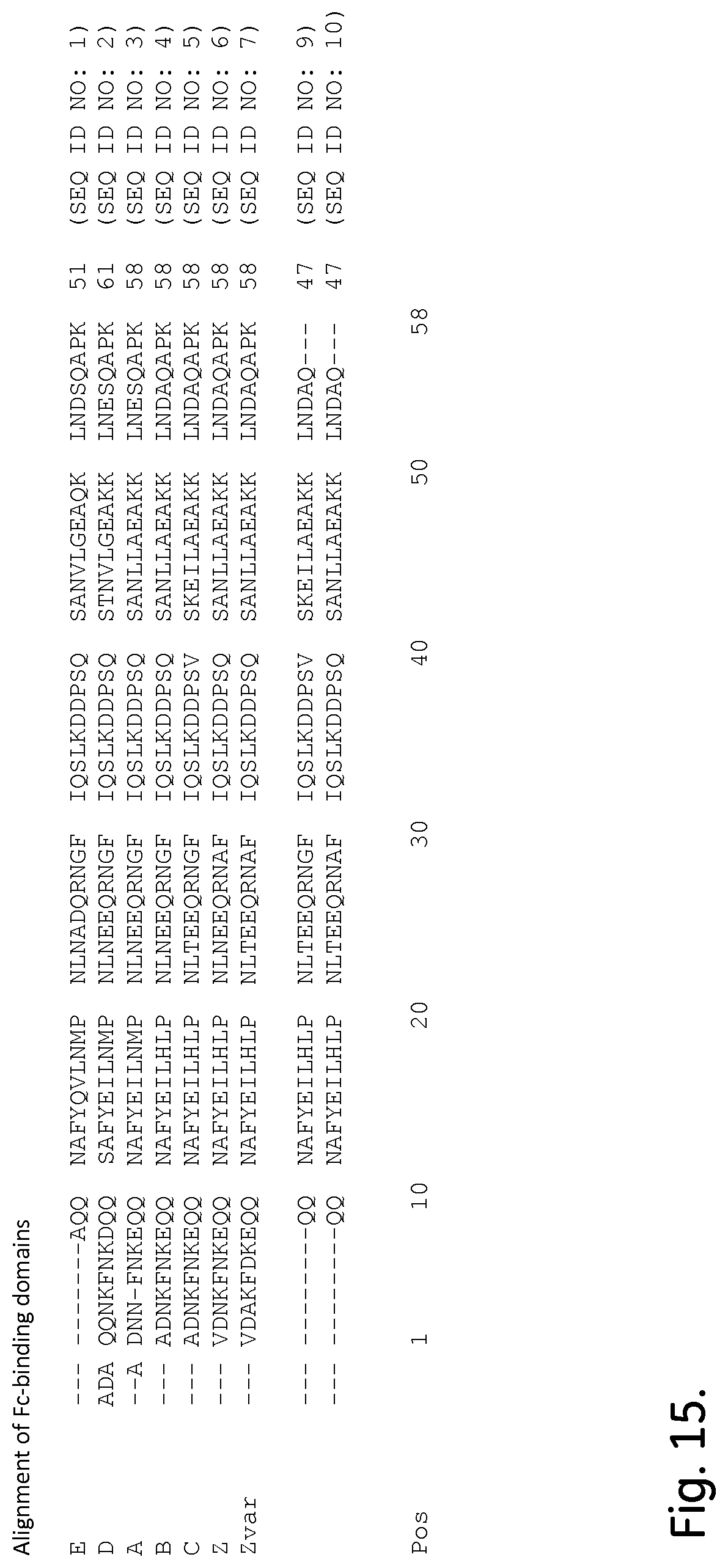
S00001
XML
uspto.report is an independent third-party trademark research tool that is not affiliated, endorsed, or sponsored by the United States Patent and Trademark Office (USPTO) or any other governmental organization. The information provided by uspto.report is based on publicly available data at the time of writing and is intended for informational purposes only.
While we strive to provide accurate and up-to-date information, we do not guarantee the accuracy, completeness, reliability, or suitability of the information displayed on this site. The use of this site is at your own risk. Any reliance you place on such information is therefore strictly at your own risk.
All official trademark data, including owner information, should be verified by visiting the official USPTO website at www.uspto.gov. This site is not intended to replace professional legal advice and should not be used as a substitute for consulting with a legal professional who is knowledgeable about trademark law.