Growth Factor Transduced Cell-loaded Ceramic Scaffold For Bone Regeneration And Repair
Lieberman; Jay R. ; et al.
U.S. patent application number 16/899363 was filed with the patent office on 2020-10-08 for growth factor transduced cell-loaded ceramic scaffold for bone regeneration and repair. The applicant listed for this patent is UNIVERSITY OF SOUTHERN CALIFORNIA. Invention is credited to Sofia Bougioukli, Yong Chen, Jay R. Lieberman, William Pannell, Xuan Song.
| Application Number | 20200316258 16/899363 |
| Document ID | / |
| Family ID | 1000004931829 |
| Filed Date | 2020-10-08 |


View All Diagrams
| United States Patent Application | 20200316258 |
| Kind Code | A1 |
| Lieberman; Jay R. ; et al. | October 8, 2020 |
GROWTH FACTOR TRANSDUCED CELL-LOADED CERAMIC SCAFFOLD FOR BONE REGENERATION AND REPAIR
Abstract
Disclosed herein are methods of regional gene-therapy with growth-factor transduced cells, in bone graft scenarios. In embodiments, the methods comprise use of 3D printed scaffolds.
| Inventors: | Lieberman; Jay R.; (Los Angeles, CA) ; Pannell; William; (Los Angeles, CA) ; Chen; Yong; (Los Angeles, CA) ; Song; Xuan; (Los Angeles, CA) ; Bougioukli; Sofia; (Los Angeles, CA) | ||||||||||
| Applicant: |
|
||||||||||
|---|---|---|---|---|---|---|---|---|---|---|---|
| Family ID: | 1000004931829 | ||||||||||
| Appl. No.: | 16/899363 | ||||||||||
| Filed: | June 11, 2020 |
Related U.S. Patent Documents
| Application Number | Filing Date | Patent Number | ||
|---|---|---|---|---|
| 16337893 | Mar 28, 2019 | |||
| PCT/US2017/054609 | Sep 29, 2017 | |||
| 16899363 | ||||
| 62401745 | Sep 29, 2016 | |||
| Current U.S. Class: | 1/1 |
| Current CPC Class: | B33Y 80/00 20141201; A61L 2430/02 20130101; B33Y 10/00 20141201; A61L 27/12 20130101; A61L 27/3821 20130101; A61L 27/54 20130101; B33Y 40/20 20200101; B28B 1/001 20130101; B29C 64/379 20170801; A61L 27/10 20130101; B29K 2509/02 20130101; B33Y 70/00 20141201; A61L 27/3847 20130101; B29C 64/129 20170801; A61L 27/3834 20130101; B29L 2031/7532 20130101; A61L 2300/414 20130101; A61L 27/227 20130101 |
| International Class: | A61L 27/38 20060101 A61L027/38; A61L 27/22 20060101 A61L027/22; A61L 27/10 20060101 A61L027/10; A61L 27/12 20060101 A61L027/12; A61L 27/54 20060101 A61L027/54; B33Y 80/00 20060101 B33Y080/00; B33Y 40/20 20060101 B33Y040/20; B28B 1/00 20060101 B28B001/00; B33Y 70/00 20060101 B33Y070/00; B29C 64/379 20060101 B29C064/379 |
Goverment Interests
GOVERNMENT LICENSE RIGHTS
[0002] This invention was made with U.S. government support under Contract #1335476 awarded by the National Science Foundation and under Contract #iR01AR057076-01A1 awarded by NIH. The U.S. government has certain rights in the invention.
Claims
1. A method for repairing a bone defect of a patient, comprising: providing a ceramic scaffold configured for filling the bone defect; loading the scaffold with growth factor transduced cells incorporating a gene that encodes a growth factor essential for bone formation; placing the ceramic scaffold with the growth factor transduced cells in or across the bone defect; and stabilizing the ceramic scaffold with the growth factor transduced cells in the patient until the bone defect is healed.
2. A method of repairing a bone defect of a patient, comprising: inserting a scaffold into a bone defect; transducing one or more cells with a growth factor essential for bone formation; and loading the one or more cells into the scaffold.
3. The method of claim 2, wherein the bone defect is a critically sized defect such that it is incapable of healing on its own.
4. The method of claim 2, wherein the one or more cells are stem cells.
5. The method of claim 4, wherein the stem cells are adipose derived stem cells.
6. The method of claim 2, wherein the one or more cells are bone marrow cells.
7. The method of claim 6, wherein the bone marrow cells are rat bone marrow cells.
8. The method of claim 2, wherein the growth factor essential for bone formation is bone morphogenetic protein 2.
9. The method of claim 2, wherein transducing the one or more cells with a growth factor essential for bone formation comprises transducing the cells with one or more vectors.
10. The method of claim 9, wherein transducing the cells with one or more vectors comprises transducing the cells with a lentiviral vector system.
11. The method of claim 10, wherein the lentiviral vector system comprises (i) a trans-activator vector and (ii) a vector encoding the growth factor essential for bone formation.
12. The method of claim 11, wherein the growth factor essential for bone formation is bone morphogenetic protein 2.
13. The method of claim 2, wherein the scaffold is comprised of calcium phosphate.
14. The method of claim 2, wherein the scaffold is comprised of tri-calcium phosphate.
15. A method for repairing a bone defect of a patient, comprising: providing a 3D model of a scaffold for bridging the bone defect; providing a ceramic scaffold comprising calcium and phosphate based on the 3D model; and loading the ceramic scaffold with cells transduced with a growth factor essential for bone formation.
16. The method of claim 15, further comprising forming the ceramic scaffold by 3D printing a calcium phosphate material.
17. The method of claim 15, wherein providing the 3D model comprises shaping the 3D model for causing the ceramic scaffold to match undamaged areas adjacent to the bone defect, so as to fit against the undamaged areas while spanning the bone defect.
18. The method of claim 15, wherein providing the ceramic scaffold comprises forming a plurality of holes in the range of 300.mu. to 1000.mu. in the ceramic scaffold.
19. The method of claim 15, further comprising preparing the cells transduced with the growth factor by transducing the cells with a lentiviral vector system comprising (i) a trans-activator vector and (ii) a vector encoding the growth factor essential for bone formation.
20. The method of claim 19, wherein the growth factor essential for bone formation is bone morphogenetic protein 2.
Description
CROSS-REFERENCE TO RELATED APPLICATIONS
[0001] This application is a continuation-in-part of U.S. patent application Ser. No. 16/337,893, filed on Mar. 28, 2019 entitled "GROWTH FACTOR TRANSDUCED CELL-LOADED CERAMIC SCAFFOLD FOR BONE REGENERATION AND REPAIR", which is a national phase (371) application of International Application PCT/US2017/054609 filed on Sep. 29, 2017 entitled "GROWTH FACTOR TRANSDUCED CELL-LOADED CERAMIC SCAFFOLD FOR BONE REGENERATION AND REPAIR", which claims the benefit and priority of U.S. provisional patent application Ser. No. 62/401,745, filed Sep. 29, 2016, the disclosures of each of which are incorporated herein in their entirety by reference.
FIELD
[0003] The present disclosure relates to methods and apparatus for repairing bone tissue, and more particularly to combined use of custom 3D-printed calcium phosphate scaffolds and regional gene therapy in bone graft scenarios to heal critically sized bone defects.
BACKGROUND
[0004] Fracture non-union and inadequate bone formation in settings such as trauma, tumor, joint replacement and limb reconstructive surgeries are among the most challenging problems in orthopedic surgery. Autologous bone graft is the gold standard to use in such situations, but its disadvantage is limited availability of the graft and complications and pain associated with graft harvest. Researchers have explored the option of using precursor cells (from bone marrow, fat, muscle or other tissues) that have potential to transform into bone forming cells, but the methods to purify these cells and potential of these cells to form bone are limited unless stimulated by the growth factors. Regional gene therapy is an attractive option as it potentially allows the investigator to incorporate the desired gene encoding the growth factor essential for bone formation into the host cells and implant these cells back into the host at a particular site where they induce new bone formation.
[0005] Prior research has reported on the effect of regional gene therapy with bone morphogenetic protein-2-producing bone marrow cells on the repair of bone defects in rats, showing promise as one aspect of bone replacement therapy. "3D printing" broadly understood as additive manufacturing, has been proposed for forming scaffolds of calcium phosphate and collagen for bone regeneration, but not in conjunction with regional gene therapy. Additive manufacturing provides the advantage of custom shaping for individual bone replacement therapy, but its suitability in conjunction with regional gene therapy is poorly understood, if at all.
[0006] Bone regeneration in vivo or in vitro is desirable for providing more rapid and more effective clinical outcomes for treatment of severe bone injury. It would be desirable, therefore, to provide more effective methods and apparatus for bone regeneration and replacement of lost bone tissue.
SUMMARY
[0007] This summary and the following detailed description should be interpreted as complementary parts of an integrated disclosure, which parts may include redundant subject matter and/or supplemental subject matter.
[0008] In an aspect, a method for repairing a bone defect of a patient is provided, comprising providing a ceramic scaffold configured for filling the bone defect, loading the scaffold with growth factor transduced cells incorporating a gene that encodes a growth factor essential for bone formation, placing the ceramic scaffold with the growth factor transduced cells in or across the bone defect, and stabilizing the ceramic scaffold with the growth factor transduced cells in the patient until the bone defect is healed.
[0009] In an aspect, a method of repairing a bone defect of a patient is provided, comprising inserting a scaffold into a bone defect; transducing one or more cells with a growth factor essential for bone formation; and loading the one or more cells into the scaffold.
[0010] In embodiments, the bone defect is a critically sized defect such that it is incapable of healing on its own.
[0011] In embodiments, the one or more cells are stem cells. In embodiments, the stem cells are adipose derived stem cells. In embodiments, the one or more cells are bone marrow cells. In embodiments, the bone marrow cells are rat bone marrow cells.
[0012] In embodiments, the growth factor essential for bone formation is bone morphogenetic protein 2.
[0013] In embodiments, transducing the one or more cells with a growth factor essential for bone formation comprises transducing the cells with one or more vectors. In embodiments, transducing the cells with one or more vectors comprises transducing the cells with a lentiviral vector system. In embodiments, the lentiviral vector system comprises a trans-activator vector and a vector encoding the growth factor essential for bone formation. In embodiments, the growth factor essential for bone formation is bone morphogenetic protein 2.
[0014] In embodiments, the scaffold is comprised of calcium phosphate. In embodiments, the scaffold is comprised of tri-calcium phosphate.
[0015] In an aspect a method for repairing a bone defect of a patient is provided, comprising providing a 3D model of a scaffold for bridging the bone defect, providing a ceramic scaffold comprising calcium and phosphate based on the 3D model, and loading the ceramic scaffold with cells transduced with a growth factor essential for bone formation.
[0016] In embodiments, the method further comprises forming the ceramic scaffold by 3D printing a calcium phosphate material. In embodiments, providing the 3D model comprises shaping the 3D model for causing the ceramic scaffold to match undamaged areas adjacent to the bone defect, so as to fit against the undamaged areas while spanning the bone defect. In embodiments, providing the ceramic scaffold comprises forming a plurality of holes in the range of 300.mu. to 1000.mu. in the ceramic scaffold.
[0017] In embodiments, the method further comprises preparing the cells transduced with the growth factor by transducing the cells with a lentiviral vector system comprising (i) a trans-activator vector and (ii) a vector encoding the growth factor essential for bone formation. In embodiments, the growth factor essential for bone formation is bone morphogenetic protein 2.
[0018] To the accomplishment of the foregoing and related ends, one or more examples comprise the features hereinafter particularly pointed out in the claims and fully described in the detailed description after the drawings.
BRIEF DESCRIPTION OF THE DRAWINGS
[0019] FIG. 1 is a chart showing in vitro results of a trial using gene therapy in conjunction with a 3D printed ceramic scaffold.
[0020] FIG. 2 is a grayscale rendering of a photo illustrating cell viability of transduced cells on a 3D printed ceramic scaffold disk after 72 hours.
[0021] FIG. 3 is an X-ray image showing results of a successful rat bone repair using a method as disclosed herein.
[0022] FIG. 4 is a flowchart illustrating operations and aspects of a method for repair of a bone defect.
[0023] FIG. 5 is a perspective view showing an example of a 3D printed ceramic scaffold for bone repair.
[0024] FIG. 6 is a flow chart illustrating operations and aspects of making a ceramic scaffold with the growth factor transduced cells.
[0025] FIG. 7A depicts a stereolithography (STL file created from a computed tomography (CT)) scan of a rodent femur.
[0026] FIG. 7B depicts a tricalcium phosphate (TCP) scaffold with 700 .mu.m pores from a 3D printed to fit a critically sized rat femoral defect.
[0027] FIG. 7C depicts an intraoperative photograph demonstrating placement of the 3D printed TCP scaffold (middle arrow) with the rat femoral defect (the left arrow points to the distal femoral segment and the right arrow points to the proximal femoral segment).
[0028] FIG. 8 depicts representative plain radiographic images from groups 1-4 taken at 12 weeks after insertion of the 3D printed TCP scaffold. Group 1 demonstrated complete healing of the defect. The arrows illustrate bone surrounding the scaffold, bridging the defect proximally and distally.
[0029] FIG. 9 depicts representative micro-CT scans with three-dimensional reconstructions (top) and axial images (bottom) obtained from groups 1-4 after specimen harvest. Group 1 demonstrated complete healing of the defect and on the axial image; circumferential bone can be seen surround the 3D printed TCP scaffold. No bone formed with the defect in groups 2-4.
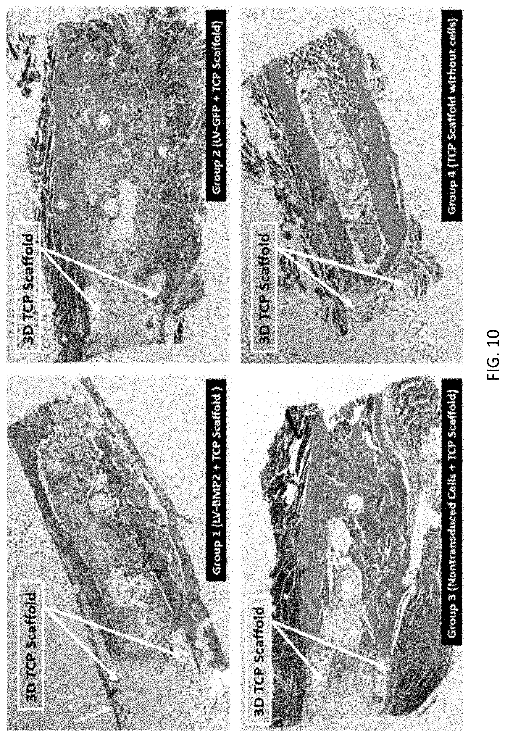
[0030] FIG. 10 depicts select longitudinal histological cuts of the proximal scaffold-defect interface from groups 1-4. The samples stained with Masson's trichrome stain. Group 1 demonstrated trabecular bone formation within the 3D printed TCP scaffold, uniting the scaffold to both ends of the defect. There is no notable bone groups 2-4, and the TCP scaffold is mostly surrounded by muscle.
[0031] FIGS. 11A-11B depict x-ray and micro-computed tomography imaging of a rat femoral defect 12 weeks after treatment with human ADSCs transduced with a two-step transcriptional amplification (TSTA) lentiviral system overexpressing BMP-2. FIG. 11A shows a femoral radiograph 12 weeks after treatment demonstrating complete bridging of defect site (see, arrows). FIG. 11B shows coronal micro-computed tomography image of a femur 12 weeks after treatment. Robust healing across the defect site is seen with reconstitution of the bony cortex.
[0032] FIG. 12 depicts histological healing in a critically sized bone defect after 12 weeks. H&E stained longitudinal section revealed the bone defects 800 and the bridging bone that spanned the defect site 810. The asterisks (*) marks native bone.
DETAILED DESCRIPTION
[0033] The present disclosure concerns use of regional gene therapy with growth factor transduced cells in bone graft scenarios with 3D-printed scaffolds. The combination of these technologies represents an innovative method of grafting bone with many potential clinical applications.
Definitions
[0034] As used herein, the phrase "a critically sized defect" or "a critically sized bone defect" refers to a bone defect that is in capable of healing on its own. As used herein, the phrase "a critically sized femoral defect" refers to a femoral defect that is incapable of healing on its own. As used herein, the phrase "a critically sized rat femoral defect," or "a rat critically sized femoral defect" refers to a rat femoral defect that is incapable of healing on its own.
[0035] As used herein, the initials "ADSC" refer to adipose-derived stems cells.
[0036] As used herein, the initials "BMP-2" refer to the bone morphogenetic protein 2.
[0037] As used herein, the initials "CaP" refer to calcium phosphate.
[0038] As used herein, the initials "G5" refer to a Ga14 responsive promoter.
[0039] As used herein, the initials "LV" refer to a lentiviral vector.
[0040] As used herein, the initials "RhMLV" refer to a murine leukemia virus long terminal repeat promoter.
[0041] As used herein, the initials "TCP" refer tri-calcium phosphate.
[0042] As used herein, the phrase "trans-activator vector" is any vector capable of mediating induction of a target gene. In embodiments, a "trans-activator vector" encodes a transcription factor. In embodiments, the transcription factor is Ga14.
[0043] As used herein, the phrase "two-step transcriptional amplification (TSTA) system" refers to any gene expression system in which transcription of a target gene is preceded by expression of a gene that drives expression of the target gene. In embodiments, the TSTA system is a vector system. In embodiments, the vector system is a lentiviral vector system.
Description of the Embodiments
[0044] In an aspect, and with reference to FIG. 5, prior to surgical implantation, the scaffold and loaded cells form an assembly made of a ceramic scaffold 500 configured for spanning the bone defect, and a culture of live mesenchymal stem cells 200 or other growth factor transduced cells incorporating a gene that encodes a growth factor essential for bone formation loaded onto the ceramic scaffold. Suitable cells for being transduced with one or more genes that encode a growth factor may include, for example, mesenchymal cells, bone marrow cells, fibroblasts, adipose-derived cells, umbilical cord cells, or muscle cells. The ceramic material may include, for example, CaP or beta TCP. In a clinical setting, the assembly may be prepared in advance of surgery and maintained alive in vitro until surgical implantation. In an alternative, the surgeon may load live growth factor transduced cells on the ceramic scaffold for the first time after it is in place in the patient's body (in vivo), or may supplement an in vitro loading of cells with a second application in vivo.
[0045] In embodiments, and with reference to FIG. 5, the scaffold may be thinner than the bone wall to be repaired and perforated with circular openings 510 in the range of about 300 to 1000 microns, for example, about 500 to 700 microns, or about 700 microns. In embodiments, the openings 510 may be spaced as desired to facilitate bone regrowth, for example, uniformly or semi-uniformly center-to-center spaced at about 1.5 to 5 times the opening's largest diameter. In embodiments, the scaffold is generally tube-shaped with an interior surface 520 and exterior surface 530. In embodiments, the assembly of scaffold and live growth factor transduced cells enables regeneration of structural bone tissue from the loaded cell culture on its openings 510, exterior 530, interior 520 and by recruitment of local progenitor cells.
[0046] In an aspect, a method of repairing a bone defect is provided comprising transducing one or more cells with a growth factor and loading the cells into a scaffold that has been implanted into a bone defect.
[0047] In embodiments, the bone defect is a critically sized defect such that the bone defect is incapable of healing on its own.
[0048] In embodiments, the cells transduced with a growth factor are stem cells. In embodiments, the stem cells are adipose derived stem cells (ADSCs). In embodiments, the stem cells can be derived from any suitable source of stem cells. In embodiments, the cells transduced with a growth factor are bone marrow cells. In embodiments, the bone marrow cells are rat bone marrow cells. In embodiments, the bone marrow cells can be derived from any suitable bone marrow source.
[0049] In embodiments, the growth factor is any factor that stimulates growth of any type of tissue. In embodiments, the growth factor stimulates growth of epithelial tissue. In embodiments, the growth factor stimulates the growth of bone. In embodiments, the growth factor is BMP-2.
[0050] In embodiments, the cells are transduced with one or more vectors. In embodiments the one or more vectors comprise a vector system. In embodiments, the vector system is a lentiviral vector system. In embodiments, the lentiviral vector system is a two-step transcriptional amplification (TSTA) system. In embodiments, the TSTA system comprises two different vectors: a first vector and a second vector. In embodiments, the first vector is a trans-activator vector. In embodiments, the trans-activator vector is the vector LV-RhMLV-Ga14. In embodiments, the second vector is a vector that encodes BMP-2. In embodiments, the vector that encodes BMP-2 is the vector LV-G5-BMP-2.
[0051] In embodiments, the scaffold is a ceramic scaffold. In embodiments, the ceramic scaffold is made up of a material comprised of calcium phosphate (CaP). In embodiments, the ceramic scaffold is made up of a material comprised of tri-calcium phosphate (TCP). In embodiments, the scaffold is made up of any other suitable material. Flowcharts and Methods
[0052] Referring to FIG. 4, an exemplary method 400 for repairing a bone defect of a patient may include, at 410, providing a ceramic scaffold configured for spanning the bone defect, for example, by 3D printing a CaP or other ceramic scaffold or by obtaining a pre-printed scaffold from a manufacturer sized to span the bone defect, for example based on a preceding CT scan as for the rodent femur described above. The method 400 may further include, at 420, loading the scaffold with growth factor transduced cells incorporating a gene that encodes a growth factor essential for bone formation, for example, by preparing or obtaining a culture of lentiviral transduced bone marrow cells as described herein, applying the culture to the scaffold, and confirming viability of the culture loaded on the scaffold prior to implantation. For example, the scaffold with pre-loaded mesenchymal cells as described may be obtained in the form of a prepared assembly from an independent source, e.g., a specialized laboratory, or the cell preparation and loading of the scaffold may be performed by a laboratory controlled by the facility performing the scaffold-implantation Surgery. The method may further include, at 430, placing the ceramic scaffold with the growth factor transduced cells in or across the bone defect.
[0053] The method 400 may further include, at 440, stabilizing the ceramic scaffold with the growth factor transduced cells in the patient until the bone defect is healed, using any suitable stabilizing technique. The ceramic scaffold is semi-structural and designed for load sharing. The scaffold may not be strong enough by itself to stabilize the defect without load sharing from other structural members. Depending on the nature of the defect, it may be stabilized using bio-compatible metal plates, rods, or other suitable structural members.
[0054] The enumerated operations 410, 420, 430, 440 may be performed in any operable order with suitable modifications. For example, the operation 430 placing the ceramic scaffold in or across the defect may be performed, but without first loading growth factor transduced cells onto the ceramic. Then, the operation 420 of loading the scaffold with growth factor transduced cells may be performed while the scaffold is in place around the defect. For example, the growth factor transduced cells may be suspended in a bio-compatible fluid and applied to the ceramic scaffold in vivo or in vitro, using a pipette or other suitable fluid applicator.
[0055] Referring to FIGS. 4, 5, and 6, further aspects of the present disclosure may include a method 600 for making a ceramic scaffold 500 loaded with growth factor transduced cells as described herein, for use in the method 400 or other suitable method. The method 600 may include, at 610, characterizing a specific bone defect of a patient. For example, the operation 610 may include scanning a bone defect by a 3D scanner, for example, a CT scanner, digitizing 30 information obtained by the scanning, and associating the digitized information with an identifier for the patient and defect site. In an alternative, the characterizing 610 may include receiving information that associates an identifier for a patient and/or bone defect with 3D information relating to the defect or to a scaffold for bridging the defect.
[0056] Referring to FIG. 6, further aspects of the method 600 may include, at 620, providing a 3D model of a scaffold for bridging the bone defect characterized by the first operation 610. As used herein, "providing" includes but is not limited to engaging a person or entity to create the 3D model based on the data characterizing the bone defect. For example, a medical technician may design a 3D model to fit stable portions of the bone that are expected to remain as scanned after the bone defect is prepared for repair, e.g., by cleaning out damaged tissue. The stable regions of the bone may be adjacent to defective regions of the bone.
[0057] The method 600 may further include, at 630, providing a ceramic scaffold that includes calcium and phosphate or that consists essentially of calcium and phosphate materials, based on the 3D model provided by the preceding operation 620. Providing may include manufacturing the ceramic scaffold, or obtaining the ceramic scaffold from a manufacturer or supplier. A slurry-based stereolithography 3D printing method as described by Dr. Song Chen (see reference herein above) may be used to manufacture the ceramic scaffold. The manufacturing may include mixing a powdered calcium-phosphate ceramic or pre-ceramic material with a photopolymer resin to create a slurry as feedstock for a stereolithographic additive manufacturing process (e.g., using tape casting as described above), thereby forming a "green" pre-fired scaffold. The green scaffold, which may include a mixture of ceramic and organic resin and/or a pre-ceramic polymer, may be heated under suitable conditions (e.g., oxidizing or non-oxidizing, depending on the process used) to purge non-ceramic materials and/or transform a pre-ceramic material into a ceramic material. At 630, the method comprises forming the ceramic scaffold 500 made of a calcium phosphate material, alone or in combination with other materials. Likewise, the method may include manufacturing the scaffold using a 3D printing technique as described herein above, or other suitable method. 3D printing may be especially advantageous for forming the ceramic-resin scaffold when it is desired to custom shape the ceramic-resin scaffold to match an individual morphology of the bone in healthy areas surrounding the defect.
[0058] For example, referring to FIG. 5, the ceramic-resin scaffold may be formed to have an inner surface 520 matching an outer surface of the healthy bone areas. Forming the ceramic-resin scaffold 500 may also include forming a plurality of holes 510 having diameters in the range of about 300.mu. to 1000.mu. (e.g., in the range of 500.mu. to 700.mu., or about 7000.mu.) in the ceramic-resin scaffold, for example during the 3D printing process.
[0059] Referring to FIG. 6, the method 600 may further include, at 640, loading the ceramic scaffold with live growth factor transduced cells incorporating a gene that encodes a growth factor essential for bone formation. As discussed above, loading may be performed in vitro, in vivo, or both. In other embodiments, the growth factor transduced cells may be prepared for incorporating the gene that encodes the growth factor by transducing the gene into the growth factor transduced cells. For example, the method may include preparing the growth factor transduced cells incorporating the gene that encodes the growth factor by a lentiviral based transcriptional activation system expressing bone morphogenetic protein 2.
[0060] Having thus described embodiments of methods and apparatus for repairing a bone defect or providing a ceramic scaffold loaded with growth factor transduced cells, it should also be appreciated that various modifications, adaptations, and alternative embodiments thereof may be made within the scope and spirit of the present invention. For example, 3D printed calcium ceramic scaffolds have been disclosed, but the inventive concepts described above may be equally applicable to scaffolds of other ceramic materials, or scaffolds made by other manufacturing methods than disclosed herein above. In addition, a culture of growth factor transduced cells incorporating a gene that encodes a growth factor essential for bone formation may be prepared by any suitable method whether or not described herein.
EXAMPLES
Example 1
3D Printing
[0061] Calcium phosphate (CaP) scaffolds were 3D printed using a slurry-based stereolithography process as developed by Dr. Song Chen et al. in Ceramic Fabrication Using Mask-lmage-Proiection-based Stereolithography: Integrated with Tape-casting. Journal of Manufacturing Processes, 2015;20(3):456-464. Briefly, this 3D printing technique is performed by first mixing a ceramic powder with a photopolymer resin to create a slurry. A tape casting system is used to aid the recoating of each slurry layer. A light source then activates the resin, curing it layer by layer until an object is built. The object, which is still a mixture of ceramic and resin, is then heated in a furnace to burn out the resin. Since the resin has a much lower melting temperature than the ceramic, the ceramic part of interest is left behind as the final product.
[0062] Computer Aided Software (CAD} was first used to create a hollow elliptical cylinder 6 mm in length in order to approximate the size and shape of a rat critically sized femoral defect. These scaffolds were 3D printed using commercially available calcium phosphate powder (Alfa Aesar #89836). An example of a resulting scaffold 500 is shown in FIG. 5.
Example 2
Regional Gene Therapy
[0063] Virk et al. 2011 ("Same day" ex-vivo regional gene therapy: a novel strategy to enhance for bone repair. Mo/Ther. 2011; 19:960-968) describes the gene therapy in detail. Briefly, a lentiviral based system (LV -BMP2) was created expressing bone morphogenetic protein 2 (BMP-2). Cultured rat bone marrow cells were transduced using a multiplicity of infection (MOI) of 25. These cells were used in the experiments detailed below.
Example 3
In Vitro BMP-2 Production
[0064] BMP-2 production of transduced RBMC was tested in vitro after 48 hours and 14 days of cell culture on 15 mm diameter, 2 mm thick 3D printed discs. At 48 hours BMP-2 production was higher on 3D-printed scaffolds as compared to control (Table 1).
TABLE-US-00001 TABLE 1 Comparison of BMP-2 production in 3D- printed scaffolds compared to control 48 Hours 14 Days Non-transduced PBMC 0.00 0.03 LV-BMP2 + 3D disc 1.71 48.62 LV-BMP2 1.26 157.32
[0065] Based on our work, in vitro BMP-2 production on the 3D printed discs is sufficient to heal a critically sized rat femoral defect.
Example 4
In Vitro Cell Viability
[0066] Transduced rat bone marrow cells (RBMC) were cultured in vitro on top of 3D printed CaP discs for 72 hours. Cell viability was determined using a commercially available Live/Dead assay kit (BioVision K501). Cells cultured on CaP disks demonstrated excellent viability at 72 hours compared to a control (standard culture well). Cell viability on 3D printed discs averaged 85% (SD 6%) relative to the control (FIG. 1 and FIG. 2, 200). These cell viability results are higher than a published study using comparable 3D printed calcium phosphate/collagen combination scaffolds (Inzana J, Olvera D, Fuller S, et al. 3D printing of composite calcium phosphate and collagen scaffolds for bone regeneration, Biomaterials. 2014;35:4026-4034).
Example 5
In Vivo Bone Formation
[0067] A pilot experiment using a 12-week-old Lewis rat was performed. A standard 6-mm mid diaphyseal femoral defect was created as described in prior publications (Alae, F., Liebermen, J. R., et al., Biodistribution of LV-TSTA transduced rat bone marrow cells used for "ex-vivo" regional gene therapy for bone repair Curr Gene Ther. 2015;15(5):481- 491, and Virk et al. "Same-day" ex-vivo regional gene therapy: A novel strategy to enhance bone repair, (2011) Molecular Therapy 19(5), 960-968). A 3D printed CaP scaffold loaded with 5 million lentiviral transduced rat bone marrow cells was placed in the defect. The defect was healed 8 weeks after the surgical procedure (FIG. 3 at x-ray image 300). The 3D printed CaP scaffold itself and the regenerated "bridging bone" are clearly visible and pointed out in FIG. 3. A drawing of a model for a similar 3D printed CaP scaffold 500 is shown in FIG. 5. FIG. 2 shows a culture 200 of mesenchymal stem cells incorporating a gene that encodes a growth factor essential for bone formation, grown on a CaP ceramic disk.
Example 6
Scaffold Shaping and Configuration
[0068] More recently, Computed Tomography (CT) data from an intact rodent femur was obtained from previous work. Commercially available software (Mimics; Materialise N V, Leuven, Belgium) was then used to convert a 6 mm section of diaphyseal bone into a file type compatible with 3D printing software, resulting in a model of a scaffold 500 as shown in FIG. 5. Additionally, 700-micrometer holes 510 were added to the model scaffold to facilitate cellular growth and communication.
[0069] An alternative ceramic powder, beta tri-calcium phosphate (beta TCP), may also be used to 3D print the scaffolds. We have 3D printed scaffolds based on the "rodent specific" CT data. These may be tested following similar methods as described above.
Example 7
Using 3D Printed Scaffolds to Heal Critically Sized Bone Defects in a Rat Model Materials and Methods
[0070] 3D Printed Scaffold: Computed tomography (CT) scans of the intact femur of a 24-week-old male Lewis rat were taken as DICOM image files and converted into a 3D stereolithography (STL) file using commercial software (Mimics; Materialise N V, Leuven, Belgium). A 6 mm section of the mid diaphysis was taken from the model and multiple 700 .mu.m holes were added to facilitate cellular communication and vascular ingrowth throughout the scaffold (FIG. 1b) (Karageorgiou & Kaplan, Porosity of 3D biomaterial scaffolds and osteogenesis, Biomaterials, 26(27), 5474-5491 (2005)). The 700 .mu.m pores were the only source of porosity in the scaffold. A projection-based ceramic STL process was used to fabricate Tricalcium phosphate (TCP) scaffold structures to precisely fit a 6 mm rat femoral defect based on the DICOM image. This process creates a complex TCP structure via solidifying a viscous slurry-mixture of photocurable resin (Formlabs, Somerville, Mass.) and TCP powder (Ceramisys; Sintered beta-Tricalcium Phosphate, Sheffield, United Kingdom) with a customized ultraviolet (UV) light engine in a layer-by-layer fashion, followed by high-temperature post-processing for removing the resin and densifying the TCP particles (Song, Chen, Lee, Wu, & Cheng, Ceramic fabrication using mask-image-projection-based stereolithography integrated with tape-casting, Journal of Manufacturing Processes, 20(3) 456-465 (2015)).
[0071] X-ray Diffraction Analysis: X-ray diffraction (XRD) (Ultima IV Diffractometer, Rigaku, Tokyo, Japan) analysis was performed to characterize the molecular compounds contained within the synthesized scaffold based on diffraction patterns. The 3D printed TCP scaffold was crushed into its powder form prior to XRD characterization. XRD was performed from 15.degree. to 75.degree. at a speed of 6.degree. per minute (dpm). A zero-diffraction slide was used to minimize background noise. Diffraction pattern analysis was completed using Jade 9 software (KS Analytical Systems, Aubrey, Tex.). A pure powder TCP sample from which the 3D printed TCP scaffold_was synthesized served as a control. Figure of merit (FoM) was calculated for the crushed scaffold and the pure powder TCP sample to allow for comparisons of the composition.
[0072] Rat Bone Marrow Cell Isolation and Transduction: Rat bone marrow cells (RBMCs) were harvested from tibias and femurs of 8-week-old rats (Male Lewis Rats; Charles River Laboratories, Wilmington, Mass.) (Sugiyama et al., Lentivirus-mediated gene transfer induces long-term transgene expression of BMP-2 in vitro and new bone formation in vivo, Molecular Therapy, 11(3), 390-398 (2005)) (FIG. 7A depicts a computed tomography scan of a rodent femur). The resulting cell pellet was resuspended in Iscove's modified Dulbecco's media (IMDM) (ThermoFisher Scientific, Waltham, Mass.) and supplemented with 15% fetal bovine serum (FBS) (Omega Scientific, Tarzana, Calif.), streptomycin 100 .mu.g/mL and penicillin 100 U/mL, and plated on a 100 mm dish for culture-expansion. When 90-100% confluent, adherent cells were trypsinized and passaged with plating 0.7-1.times.10 (Carragee et al., A critical review of recombinant human bone morphogenetic protein-2 trials in spinal surgery: Emerging safety concerns and lessons learned, The Spine Journal, 11(6), 471-491 (2011)) cells per dish. Passage 3 cells were transduced with a lentiviral vector carrying the cDNA for BMP-2 (LV-BMP-2) or green fluorescent protein (GFP) (LV-GFP) at a multiplicity of infection of 25 in the presence of 8 .mu.g/mL polybrene (Sigma, St. Louis, Mo.).
[0073] Study Groups: There were four groups in the study, one experimental group (group 1) and three negative control groups (groups 2-4) (Table 2). Rats were randomly assigned to each group.
[0074] Rat Femoral Defect Model: The study was conducted in compliance with Institutional Animal Care and Use Committee (IACUC) regulations. A 23 mm.times.4 mm.times.4 mm polyethylene four-hole plate was secured to the left femur both proximally and distally with threaded wires and cerclage wires in a 12-week-old male Lewis rat. A standard 6 mm femoral defect was created with a high-speed burr (Lieberman et al., 1999). This is a critically sized defect and will not heal without a robust osteo-inductive stimulus. The periosteum was removed, and the intramedullary canals were flushed with normal saline solution to remove remaining osteoprogenitor cells from the defect site. The 3D printed TCP scaffold specifically designed to fit the femoral defect was injected with 5 million RBMCs diluted in phosphate-buffered saline (PBS) from groups 1-3 just before implantation or left empty for animals in group 4. FIG. 7B depicts an exemplary embodiment of a tricalcium phosphate scaffold with 700 .mu.m pores. The scaffold was then inserted into the bone defect (FIG. 7C) and secured with two circumferential Vicryl sutures. Overlying muscle was closed with Vicryl suture further securing the scaffold in place. Animals were allowed to weight-bear immediately after surgery.
[0075] Radiographic Analysis: Plain radiographs of all operated femurs were taken at 4,8, and 12 weeks after the surgical procedure using a Faxitron imaging system (Faxitron Bioptics, Tucson, Ariz.). The radiographs were assessed to determine whether the defect had healed. Three blinded observers graded healing of the defect from 0 to 5 where 0 represents no healing and 5 represents complete healing defined as bridging of both cortices. Radiographs taken at 8 and 12 weeks were graded.
[0076] Microcomputed Tomography: A microcomputed tomography (micro-CT) scan (.mu.CT40, Scanco Medical, Bassersdorf, Switzerland) was performed on all of the operated femurs after euthanasia at 12 weeks to assess bone volume formed at the femoral defect (Virk et al., "Same-day" ex-vivo regional gene therapy: A novel strategy to enhance bone repair, Molecular Therapy 19(5), 960-968 (2011)). The amount of cortical bone and trabecular bone formed (bone volume) within the femoral defect region (tissue volume) was calculated after the TCP scaffold was digitally subtracted. The total volume of bone formation (bone volume fraction (BVF)=bone volume (BV)/tissue volume (TV)) was calculated and average BVF ratios were compared for each group.
[0077] Histologic and Histomorphometry: A total of 19 femurs were analyzed histologically (Table 2). Five millimeter axial and transverse sections were cut, allowing for analysis in two planes with hematoxylin & eosin (H&E), Masson' s trichrome, and tartrate-resistant acidic phosphatase (TRAP) staining. Each specimen was evaluated for the presence of cortical bridging across the femoral defect site, inflammation, and presence of osteoclasts. Following staining, the axial images were analyzed with Bioquant analysis software (Bioquant Image Analysis, Nashville, Tenn.) under 1.times.magnification. Bone area (BA) and tissue area (TA) were selected using the Bioquant software. The two axial histologic images were analyzed, and the mean BA/TA ratio was averaged for each femur. Mean BA/TA ratios were calculated for each study group and compared.
TABLE-US-00002 TABLE 2 Study groups and studies of the defects # of # that were # that were # that were studied # that were Treatment total studied studied with histologically and studied Group defects radiographically micro-CT histomorphometrically biomechanically Group 1: 14 14 14 5 9 LV-BMP2 + TCP scaffold Group 2: 5 5 5 5 0 LV-GFP + TCP scaffold Group 3: 5 5 4.sup.a 4.sup.a 0 Non-transduced RBMCs + TCP scaffold Group 4: 5 5 5 5 0 TCP scaffold without cells .sup.aOne rat failed fixation at the 8-week time point and was euthanized prior to the 12-week time point and therefore did not undergo micro-CT or histology staining.
TABLE-US-00003 TABLE 3 Radiographic outcomes 4 weeks 8 weeks 12 weeks # with # with # with Treatment complete complete complete Group Score healing Score healing Score healing Group 1: LV- N/A 2 (14%) 4.78 +/- 0.30 8 (57%) 4.93 +/- 0.14 13 (100%) BMP2 + TCP scaffold Group 2: LV- N/A 0 (0%) 0.3 +/- 0.32 0 (0%) 0.67 +/- 0.71 0 (0%) GFP + TCP scaffold Group 3: Non- N/A 0 (0%) 0.2 +/- 0.32 0 (0%) 0.33 +/- 0.19 0 (0%) transduced RBMCs + TCP scaffold Group 4: TCP N/A 0 (0%) 0.9 +/- 0.59 0 (0%) 1.07 +/- 1.01 0 (0%) scaffold without cells
[0078] Biomechanical Testing: The operative femur from nine specimens in the experimental group (group 1) underwent biomechanical testing. The contralateral, unoperated femur of each animal was also tested to serve as a control. The surrounding soft tissue, pins, wires, and plate were removed from each specimen prior to testing. Each specimen was frozen and then thawed right before testing. All testing was conducted on the same day to minimize variation due to temperature or biomechanical setup.
[0079] Each end of the specimen was potted in polymethylmethacrylate blocks and mounted on a torsional testing fixture such that the longitudinal bone axis was centered with the axis of torsion. The torsional testing fixture and specimen were attached to a universal testing machine (Minneapolis, Minn.). Using a rate of 15 per minute, the distal end of the specimen was internally rotated until bone failure. Torsional stiffness, maximum torque, peak displacement, and total energy for failure were calculated and the mean values for the operative and intact contralateral femurs were compared.
[0080] Statistical Analysis: Statistical analysis was performed using IBM SPSS Statistics 21. The results are given as mean and SD. The data were first checked for normality using Shapiro-Wilk test. One-way ANOVA and post-hoc analysis with Tukey's range test were done for comparisons in X-ray scores as well as BV and BVF between the different treatment groups. Biomechanical parameters between the operated and intact contralateral limbs were compared using Mann-Whitney U test.
Results
[0081] Scaffold Composition: The XRD pattern for the pure TCP powder was most similar to calcium phosphate with a FoM of 6.8 and 92% composition similarity. The XRD pattern for the 3D printed TCP scaffold was also most similar to calcium phosphate with a FoM of 7.5 and 92% composition similarity.
[0082] Radiographic Outcomes: All femurs (14 out of 14) in group 1 demonstrated complete healing of the bone defect at the 12-week time point with an average score of 4.93.+-.0.14 (Table 3 and FIG. 8). Plain radiographs demonstrated robust bone formation around the 3D printed TCP scaffold and across the defect at 12 weeks. Prior to the 12-week time point, two femurs (14%) had healed at 4 weeks, and 8 femurs had healed at 8 weeks (57%). No femoral defects in groups 2-4 demonstrated evidence of complete radiographic healing at any time point. Healing grades of femurs in group 1 were significantly greater than those from groups. 2-4 (p<0.001) at the 8-and 12-week time points (Table 3). There was no significant difference in radiographic scores between groups 2-4 (p >0.05) (Table 3). There was moderate interobserver agreement among the three reviewers who scored the radiographs (8-week kappa =0.60, 12-week kappa =0.61).
[0083] Microcomputed Tomography: At the 12-week time point, Micro-CT confirmed complete healing of all 14 femurs in group 1. No bone formation was noted within the defect in groups 2-4. Micro-CT demonstrated approximately fivefold greater BVF in group 1 compared to groups 2-4 (p<0.001) (FIG. 9). There was no significant difference in BVF between groups 2-4 (p>0.05). The average BV/TV in group 1 was 25.33.+-.9.05%, 6.03.+-.3.34% in group 2, 4.10.+-.0.54% in group 3, and 6.20.+-.1.79% in group 4.
[0084] Histology and Histomorphometry: The femurs from group 1 demonstrated substantial trabecular bone formation across the scaffold-bone interface both proximally and distally (FIG. 10). On cross-sectional samples, trabecular bone formed circumferentially around the entire 3D printed TCP scaffold in group 1. Groups 2-4 demonstrated minimal new bone formation at the scaffold-defect interface, and no bone formation surrounding the scaffold on cross-sectional imaging.
[0085] On H&E staining in one specimen from group 2 and a second from group 3, there was significant inflammatory cell infiltration and necrosis surrounding the pin sites. Both of these specimens had negative TRAP stains. TRAP staining around the proximal pin sites was positive in one specimen from group 2 and a second specimen from group 4; no significant inflammatory reaction in these two specimens was noted on H&E staining. No inflammatory reaction or TRAP positive specimens were noted in group 1.
[0086] Significant differences in bone formation between group 1 and groups 2-4 demonstrated on radiographs, micro-CT, and qualitative histology were confirmed with histomorphometric analysis. Total average bone volume induced by group 1 (BA/TA=0.134.+-.0.056) was significantly greater than groups 2-4 (p<0.05). Average BA/TA was 0.052.+-.0.022 in group 2, 0.056.+-.0.043 in group 3, and 0.035.+-.0.020 in group 4. There was no significant difference in BA/TA between groups 2-4 (p>0.05).
[0087] Biomechanics: The mean stiffness, total energy, peak torque, and peak displacement of the healed segmental defects from group 1 and the unoperated, contralateral femur are presented in Table 4. Stiffness between the healed defect and unoperated femur were similar (p=0.863). Total energy to failure, peak torque, and peak displacement were significantly greater in the unoperated femur (p<0.001). No defects in groups 2-4 demonstrated adequate bone formation and stability of the bone defect to undergo biomechanical testing.
[0088] Complications: There were no complications in the LV-BMP2+TCP scaffold group (group 1). One rat from group 3 died on postoperative day two likely secondary to complications from anesthesia; this rat was replaced. The proximal fixation in one rat in group 3 failed at 8 weeks and this rat was euthanized prior to the 12-week time point. In 3 rats (60%) in group 4, plain radiographs at 4, 8, and 12 weeks demonstrated migration of the TCP scaffold within the defect. This occurred because there was no bone healing in the defect. One rat from group 2 and one rat in group 4 were noted to have loose proximal pins at the time of limb harvest; these two rats had the positive TRAP staining on histology in the absence of significant inflammation, consistent with aseptic osteolysis. Two additional rats, one from group 2 and one from group 3, were noted to have purulent material at the loose proximal pin sites at the time of limb harvest. During the course of the study, the pins became loose and the superficial tip of the pins migrated and violated the epidermal layer, but this was not clearly evident during routine clinical inspection due to the rats' thick fur layer. The histologic sections of these two femurs revealed a significant inflammatory reaction but were TRAP negative.
TABLE-US-00004 TABLE 4 Biomechanics results of group 1 (LV-BMP2 + TCP scaffold) Experimental Contralateral Outcome Limb limb p-value Stiffness (nm/deg) 0.06 +/- 0.02 0.06 +/- 0.01 0.863 Total energy (nm deg) 1.36 +/- 0.68 3.66 +/- 1.13 <0.001 Peak torque (nm) 0.36 +/- 0.10 0.62 +/- 0.11 <0.001 Peak displacement (deg) 8.36 +/- 1.40 12.4 +/- 2.42 <0.001
Example 8
Growith Factor Transduced Cell-Loaded Ceramic Scaffold for Bone Generation and Repair
[0089] The regional gene therapy strategy that is being developed includes the transduction of either adipose derived stem cells (ADSCs) or bone marrow cells that have been expanded in tissue culture.
[0090] The cells will be transduced with two-step transcriptional amplification (TSTA) lentiviral vector system containing the cDNA for BMP-2. The transduced cells will then be loaded onto a scaffold which has been implanted into the bone defect. The TSTA lentiviral vector system can use two different lentiviral vectors to express BMP-2. One of the vectors is the trans-activator vector (LV-RhMLV-Ga14) and the other vector is the BMP-2 containing vector under the control of a Ga14 responsive promoter including five Ga14 binding sites (LV-G5-BMP-2).
[0091] In previous studies different multiples of infection (MOI) have been evaluated for each vector to maximize BMP-2 production. In many of these studies the MOI for the trans-activator vector was 5 and a MOI of 25 for the BMP-2 vector (This has been delineated as 5/25). Recent studies have compared the BMP-2 production when the MOI was decreased to 3/3 for the trans-activator vector and BMP-2 vector respectively (See, Table 5). It is hypothesized that the higher MOI may be more toxic to the transduced cells.
TABLE-US-00005 TABLE 5 BMP-2 production at different MOIs ADSCs: BMP-2 Production by Multiplicity of Infection (MOI) - (ng/million cells) Statistics Day 2 Day 7 Day 14 ADSC 3/3 Mean 52.05 534.68 432.13 N 4 4 4 Std. Deviation 80.63 309.97 260.87 Std. Error of the Mean 40.31 154.98 130.435 ADSC 5/25 Mean 60.83 138.93 29.17 N 3 3 3 Std. Deviation 18.22 47.37 17.10 Std. Error of the Mean 10.52 27.35 9.87
[0092] A pilot study was performed assessing the bone defect healing capacity of transduced human ADSCs using a MOI 3/3 for the trans-activator and BMP-2 vectors. The transduced cells were loaded onto a compression resistant matrix that had been placed in a critically sized femoral defect in a nude rat. Twelve weeks after cell implantation the bone defect had healed with bridging of the bone defects as confirmed on plain radiographs and micro CT scan (FIGS. 11A-11B). Histological analysis also shows healing of defects 800 with bones that bridge the defect sites 810 (FIG. 12).
* * * * *
D00000
D00001
D00002
D00003
D00004
D00005
D00006
D00007
D00008
D00009
D00010
D00011
D00012
D00013

D00014

XML
uspto.report is an independent third-party trademark research tool that is not affiliated, endorsed, or sponsored by the United States Patent and Trademark Office (USPTO) or any other governmental organization. The information provided by uspto.report is based on publicly available data at the time of writing and is intended for informational purposes only.
While we strive to provide accurate and up-to-date information, we do not guarantee the accuracy, completeness, reliability, or suitability of the information displayed on this site. The use of this site is at your own risk. Any reliance you place on such information is therefore strictly at your own risk.
All official trademark data, including owner information, should be verified by visiting the official USPTO website at www.uspto.gov. This site is not intended to replace professional legal advice and should not be used as a substitute for consulting with a legal professional who is knowledgeable about trademark law.