Techniques For Providing A Replacement Valve And Transseptal Communication
HARITON; Ilia ; et al.
U.S. patent application number 16/738516 was filed with the patent office on 2020-10-08 for techniques for providing a replacement valve and transseptal communication. The applicant listed for this patent is CARDIOVALVE LTD.. Invention is credited to Ilia HARITON, Paul KAYE, Tal REICH.
| Application Number | 20200315789 16/738516 |
| Document ID | / |
| Family ID | 1000004596507 |
| Filed Date | 2020-10-08 |


View All Diagrams
| United States Patent Application | 20200315789 |
| Kind Code | A1 |
| HARITON; Ilia ; et al. | October 8, 2020 |
TECHNIQUES FOR PROVIDING A REPLACEMENT VALVE AND TRANSSEPTAL COMMUNICATION
Abstract
A method is provided, including (1) identifying the subject as having mitral valve regurgitation; (2) during a medical procedure, in response to identifying the subject as having mitral valve regurgitation, implanting a prosthetic valve at a mitral valve site of the heart; and (3) during the same medical procedure, implanting a therapeutic septal device at a septum of the heart. Other embodiments are also described.
| Inventors: | HARITON; Ilia; (Zichron Yaackov, IL) ; REICH; Tal; (Moshav Moledet, IL) ; KAYE; Paul; (Modiin, IL) | ||||||||||
| Applicant: |
|
||||||||||
|---|---|---|---|---|---|---|---|---|---|---|---|
| Family ID: | 1000004596507 | ||||||||||
| Appl. No.: | 16/738516 | ||||||||||
| Filed: | January 9, 2020 |
Related U.S. Patent Documents
| Application Number | Filing Date | Patent Number | ||
|---|---|---|---|---|
| 15433547 | Feb 15, 2017 | 10531866 | ||
| 16738516 | ||||
| 62295701 | Feb 16, 2016 | |||
| Current U.S. Class: | 1/1 |
| Current CPC Class: | A61F 2002/249 20130101; A61M 27/002 20130101; A61F 2/2412 20130101; A61F 2/2427 20130101; A61M 2210/125 20130101; A61F 2210/0057 20130101 |
| International Class: | A61F 2/24 20060101 A61F002/24; A61M 27/00 20060101 A61M027/00 |
Claims
1. A method for use with a heart of a subject, comprising: identifying the subject as having mitral valve regurgitation; during a medical procedure, in response to identifying the subject as having mitral valve regurgitation, implanting a prosthetic valve at a mitral valve site of the heart; and during the same medical procedure, implanting a therapeutic septal device at a septum of the heart.
2. The method according to claim 1, wherein the therapeutic septal device is a shunt, and wherein implanting the therapeutic septal device at the septum comprises implanting the shunt at the septum.
3. The method according to claim 2, wherein implanting the prosthetic valve and implanting the shunt respectively comprise implanting the prosthetic valve and implanting the shunt in an absence of an identification of the subject as having heart failure.
4. The method according to claim 2, wherein the septum is an interatrial septum of the heart, and wherein implanting the shunt at the septum comprises implanting the shunt at the interatrial septum.
5. The method according to claim 2, wherein the septum is an interventricular septum of the heart, and wherein implanting the shunt at the septum comprises implanting the shunt at the interventricular septum.
6. The method according to claim 2, wherein the subject is an adult subject, and wherein the method is performed on the adult subject.
7. The method according to claim 2, further comprising making a fenestration in the septum of the heart, and delivering the prosthetic valve transfemorally to the heart and through the fenestration to the mitral valve site, and wherein implanting the shunt comprises implanting the shunt in the fenestration.
8. The method according to claim 7, further comprising transfemorally delivering the shunt to the septum.
9. The method according to claim 7, wherein making the fenestration comprises making the fenestration transfemorally.
10. The method according to claim 2, further comprising transapically making a fenestration in the septum of the heart, wherein implanting the shunt comprises implanting the shunt in the fenestration.
11. The method according to claim 2, further comprising transapically delivering the prosthetic valve to the mitral valve site.
12. The method according to claim 2, wherein implanting the shunt comprises implanting the shunt after implanting the prosthetic valve.
13. The method according to claim 2, wherein implanting the shunt comprises implanting the shunt before implanting the prosthetic valve.
14. The method according to claim 2, wherein the shunt includes a check valve, and implanting the shunt comprises implanting the shunt such that the check valve facilitates one-way blood flow from a chamber of a left side of the heart, via the shunt, to a chamber of a right side of the heart.
15. The method according to claim 2, wherein: the shunt is shaped to define a lumen, and includes a membrane that regulates blood flow through the lumen, and implanting the shunt comprises implanting the shunt that is shaped to define the lumen and includes the membrane.
16. The method according to claim 15, wherein: the membrane: has a closed position in which the membrane inhibits blood flow through the lumen, and an open position in which the inhibiting of the blood flow by the membrane is reduced, resides in the closed position while a blood pressure difference across the membrane is lower than a threshold blood pressure difference, moves from the closed position into the open position in response to the blood pressure difference exceeding the threshold blood pressure difference, and is configured such that the threshold blood pressure difference is 4-6 mmHg, and implanting the shunt that is shaped to define the lumen and includes the membrane, comprises implanting the shunt that is shaped to define the lumen and includes the membrane that: has the closed position and the open position, resides in the closed position while the blood pressure difference across the membrane is lower than the threshold blood pressure difference, moves from the closed position into the open position in response to the blood pressure difference exceeding the threshold blood pressure difference, and is configured such that the threshold blood pressure difference is 4-6 mmHg.
17. The method according to claim 16, wherein the membrane is configured to move from the closed position into the open position in response to the blood pressure difference exceeding the threshold blood pressure difference in either direction across the membrane, and wherein implanting the shunt that includes the membrane comprises implanting the shunt that includes the membrane that is configured to move from the closed position into the open position in response to the blood pressure difference exceeding the threshold blood pressure difference in either direction across the membrane.
18. The method according to claim 15, wherein the membrane is transected by intersecting slits, and wherein implanting the shunt comprises implanting the shunt that includes the membrane that is transected by intersecting slits.
19. The method according to claim 1, wherein implanting the prosthetic valve and implanting the therapeutic septal device comprise implanting the prosthetic valve and implanting the therapeutic septal device in an absence of an identification of the subject as having heart failure.
20. The method according to claim 1, wherein the septum is an interatrial septum of the heart, and wherein implanting the therapeutic septal device at the septum comprises implanting the therapeutic septal device at the interatrial septum.
21. The method according to claim 1, wherein the septum is an interventricular septum of the heart, and wherein implanting the therapeutic septal device at the septum comprises implanting the therapeutic septal device at the interventricular septum.
22. The method according to claim 1, wherein the subject is an adult subject, and wherein the method is performed on the adult subject.
23. The method according to claim 1, further comprising making a fenestration in the septum of the heart, and delivering the prosthetic valve transfemorally to the heart and through the fenestration to the mitral valve site, and wherein implanting the therapeutic septal device comprises implanting the therapeutic septal device in the fenestration.
24. The method according to claim 23, further comprising transfemorally delivering the therapeutic septal device to the septum.
25. The method according to claim 23, wherein making the fenestration comprises making the fenestration transfemorally.
26. The method according to claim 1, further comprising transapically making a fenestration in the septum of the heart, wherein implanting the therapeutic septal device comprises implanting the therapeutic septal device in the fenestration.
27. The method according to claim 1, further comprising transapically delivering the prosthetic valve to the mitral valve site.
28. The method according to claim 1, wherein implanting the therapeutic septal device comprises implanting the therapeutic septal device after implanting the prosthetic valve.
29. The method according to claim 1, wherein implanting the therapeutic septal device comprises implanting the therapeutic septal device before implanting the prosthetic valve.
Description
CROSS-REFERENCE TO RELATED APPLICATIONS
[0001] The present application is a Continuation of U.S. Ser. No. 15/433,547 to Hariton et al., filed Feb. 15, 2017, and entitled "Techniques for providing a replacement valve and transseptal communication," which claims the benefit of U.S. Provisional application 62/295,701 to Hariton et al., filed Feb. 16, 2016, and entitled "Techniques for providing a replacement valve and transseptal communication." Each of the above is incorporated herein by reference.
FIELD OF THE INVENTION
[0002] Some applications of the present invention relate in general to cardiac implants. More specifically, some applications of the present invention relate to techniques for implanting cardiac implants in a complementary manner.
BACKGROUND
[0003] Ischemic heart disease causes regurgitation of a heart valve by the combination of ischemic dysfunction of the papillary muscles, and the dilatation of the ventricle that is present in ischemic heart disease, with the subsequent displacement of the papillary muscles and the dilatation of the valve annulus.
[0004] Dilation of the annulus of the valve prevents the valve leaflets from fully coapting when the valve is closed. Regurgitation of blood from the ventricle into the atrium results in increased total stroke volume and decreased cardiac output, and ultimate weakening of the ventricle secondary to a volume overload and a pressure overload of the atrium.
SUMMARY OF THE INVENTION
[0005] For some applications, during a single medical procedure, a prosthetic valve is implanted in the heart of a subject, and a transseptal fenestration is made in the heart. For example, the implantation and fenestration may be performed via the same transapical access point (i.e., made by transapical puncture). Alternatively, the implantation or the fenestration may be performed via the transapical access point, and the other may be performed via transfemoral access. Typically, the procedure is performed on a subject that has been identified as having mitral valve regurgitation.
[0006] For some applications, the transseptal fenestration is made in the interatrial septum of the heart. For some applications, the transseptal fenestration is made in the interventricular septum of the heart.
[0007] For some applications, the transseptal fenestration is left as-is, as a transseptal shunt. For some applications, a septal device is implanted at (e.g., in) the transseptal fenestration. For some applications, the septal device is a shunt device, which is implanted at the fenestration so as to maintain patency. For some applications, the septal device is a flow-restricting device, such as a valve or a membrane, which is implanted at the fenestration so as to allow limited flow of blood between the right and left sides of the heart. For example, flow may be allowed in only one direction, and/or in response to a blood pressure difference that is greater than a threshold blood pressure difference.
[0008] For some applications, the septal device comprises a membrane or a balloon, and is implanted at the fenestration so as to facilitate temporary and reversible changes in the effective volume of the left atrium or left ventricle of the heart, e.g., without allowing mixing of blood between the right and left sides of the heart. For some applications, the septal device comprises a cell that performs a similar function. For example, the cell may slide into and out of the right side of the heart (e.g., the right atrium) in response to the blood pressure difference.
[0009] For some applications, the membrane, balloon, and/or cell is implanted in the heart wall, rather than in the septum.
[0010] There is therefore provided, in accordance with an application of the present invention, a method for use with a heart of a subject, the method including:
[0011] making a transapical puncture into a left ventricle of the heart;
[0012] making a transseptal fenestration in the heart;
[0013] delivering a prosthetic valve via the transapical puncture and implanting the prosthetic valve at a mitral valve of the heart; and
[0014] subsequently to delivering the prosthetic valve and making the transseptal fenestration, closing the transapical puncture.
[0015] In an application, the subject has not been identified as suffering from heart failure, and performing the method includes performing the method on the subject that has not been identified as suffering from heart failure.
[0016] In an application, making the transseptal fenestration includes making the transseptal fenestration via the transapical puncture.
[0017] In an application, making the transseptal fenestration includes making the transseptal fenestration via a transfemoral route.
[0018] In an application, making the transseptal fenestration includes making a fenestration in the interventricular septum.
[0019] In an application, making the transseptal fenestration includes making a fenestration in the interatrial septum.
[0020] In an application, making the transseptal fenestration includes making the transseptal fenestration after implanting the prosthetic valve.
[0021] In an application, making the transseptal fenestration includes making the transseptal fenestration before implanting the prosthetic valve.
[0022] In an application, the method further includes advancing a distal end of a tube through the transapical puncture, and delivering the prosthetic valve via the transapical puncture includes delivering the prosthetic valve via the tube.
[0023] In an application, making the transseptal fenestration includes making the transseptal fenestration via the tube.
[0024] In an application, the method further includes implanting a shunt device into the transseptal fenestration.
[0025] In an application, the shunt device includes a check valve, and implanting the shunt device includes implanting the shunt device such that the check valve facilitates one-way blood flow from a chamber of the left side of the heart, via the transseptal fenestration, to a chamber of the right side of the heart.
[0026] In an application, implanting the shunt device includes implanting a shunt device that is shaped to define a lumen, and includes a membrane that regulates blood flow through the lumen.
[0027] In an application, implanting the shunt device includes implanting a shunt device that includes a membrane that (a) has (i) a closed position in which the membrane inhibits blood flow through the lumen, and (ii) an open position in which the inhibiting of the blood flow is reduced, resides in the closed position while a blood pressure difference across the membrane is lower than a threshold blood pressure difference of 4-6 mmHg (e.g., 5 mmHg), and moves from the closed position into the open position in response to the blood pressure difference exceeding the threshold blood pressure difference.
[0028] In an application, the membrane moves from the closed position into the open position in response to the blood pressure difference exceeding the threshold blood pressure difference in either direction across the membrane, and implanting the shunt device that includes the membrane includes implanting the shunt device that includes the membrane that moves from the closed position into the open position in response to the blood pressure difference exceeding the threshold blood pressure difference in either direction across the membrane.
[0029] In an application, the membrane is transected by intersecting slits, and implanting the shunt device includes implanting the shunt device that includes the membrane that is transected by intersecting slits.
[0030] In an application, the method further includes implanting at the transseptal fenestration, a balloon device that includes a balloon having an interior and an opening into the interior, such that (i) the interior is in fluid communication, via the opening, with a chamber of the left side of the heart, and (ii) a greater blood pressure in the chamber of the left side of the heart relative to a blood pressure in a corresponding chamber of the right side of the heart inflates the balloon such that the balloon reversibly expands into the corresponding chamber of the right side of the heart.
[0031] In an application, the balloon is elastic, and implanting the balloon device includes implanting the balloon device such that the balloon automatically deflates in response to a reduction in the greater blood pressure in the chamber of the left side of the heart.
[0032] In an application, implanting the balloon device includes implanting the balloon such that the balloon inflates only when blood pressure in the chamber of the left side of the heart is more than 4-6 mmHg greater than blood pressure in the chamber of the right side of the heart.
[0033] In an application, implanting the balloon device includes sealing the transseptal fenestration with the balloon device.
[0034] In an application, the method further includes implanting at the transseptal fenestration, an elastic membrane that elastically expands in response to a difference in blood pressure across the membrane.
[0035] In an application, implanting the elastic membrane includes sealing the transseptal fenestration with the elastic membrane.
[0036] In an application, the method further includes implanting at the transseptal fenestration, a cell having an interior and an opening into the interior, such that (i) the interior is in fluid communication, via the opening, with a chamber of the left side of the heart, and (ii) a greater blood pressure in the chamber of the left side of the heart relative to a blood pressure in a corresponding chamber of the right side of the heart increases a volume of the interior that is disposed within the corresponding chamber of the right side of the heart.
[0037] In an application, the cell includes an elastic membrane, and implanting the cell includes implanting the cell that includes the elastic membrane, such that the elastic membrane elastically expands into the chamber of the right side of the heart in response to the greater blood pressure in the chamber of the left side of the heart.
[0038] In an application, the cell is a balloon, and implanting the cell includes implanting the balloon, such that the balloon expands into the chamber of the right side of the heart in response to the greater blood pressure in the chamber of the left side of the heart.
[0039] In an application, implanting the cell includes implanting a cell that is slidably mounted in a mount, such that the mount is fixed at the transseptal fenestration, and the cell slides into the chamber of the right side of the heart in response to the greater blood pressure in the chamber of the left side of the heart.
[0040] In an application, implanting the cell includes implanting the cell such that the volume of the interior that is disposed within the corresponding chamber of the right side of the heart increases only when the blood pressure in the chamber of the left side of the heart is greater than the blood pressure in the chamber of the right side of the heart by more than a threshold difference of 4-6 mmHg.
[0041] There is further provided, in accordance with an application of the present invention, a method including for use with a heart of a subject:
[0042] identifying the subject as having mitral valve regurgitation;
[0043] during a medical procedure, in response to identifying the subject as having mitral valve regurgitation, implanting a prosthetic valve at a mitral valve site of the heart; and
[0044] during the same medical procedure, implanting a shunt device at a septum of the heart.
[0045] In an application, implanting the prosthetic valve and implanting the shunt device include implanting the prosthetic valve and implanting the shunt device in the absence of an identification of the subject as having heart failure.
[0046] In an application, the septum is an interatrial septum of the heart, and implanting the shunt device at the septum includes implanting the shunt device at the interatrial septum.
[0047] In an application, the septum is an interventricular septum of the heart, and implanting the shunt device at the septum includes implanting the shunt device at the interventricular septum.
[0048] In an application, the subject is an adult subject, and the method is performed on the adult subject.
[0049] There is further provided, in accordance with an application of the present invention, a method including for use with a heart of a subject:
[0050] identifying the subject as having mitral valve regurgitation;
[0051] during a medical procedure, in response to identifying the subject as having mitral valve regurgitation, implanting a prosthetic valve at a mitral valve site of the heart; and
[0052] during the same medical procedure, implanting a septal device at a septum of the heart.
[0053] In an application, implanting the prosthetic valve and implanting the septal device include implanting the prosthetic valve and implanting the septal device in the absence of an identification of the subject as having heart failure.
[0054] In an application, the septum is an interatrial septum of the heart, and implanting the septal device at the septum includes implanting the septal device at the interatrial septum.
[0055] In an application, the septum is an interventricular septum of the heart, and implanting the septal device at the septum includes implanting the septal device at the interventricular septum.
[0056] In an application, the subject is an adult subject, and the method is performed on the adult subject.
[0057] The present invention will be more fully understood from the following detailed description of applications thereof, taken together with the drawings, in which:
BRIEF DESCRIPTION OF THE DRAWINGS
[0058] FIGS. 1A-F are schematic illustrations of a method for use with a heart of a subject, in accordance with some applications of the invention;
[0059] FIGS. 2A-F are schematic illustrations of a method for use with heart of a subject, in accordance with some applications of the invention;
[0060] FIGS. 3A-B are schematic illustrations of a method for use with heart of a subject, in accordance with some applications of the invention;
[0061] FIGS. 4, 5, 6, 7 and 8 are schematic illustrations of respective septal devices that may be implanted at the transseptal fenestration, in accordance with some applications of the invention; and
[0062] FIGS. 9 and 10 are schematic illustrations of septal devices implanted in the heart wall, in accordance with an application of the invention.
DETAILED DESCRIPTION OF EMBODIMENTS
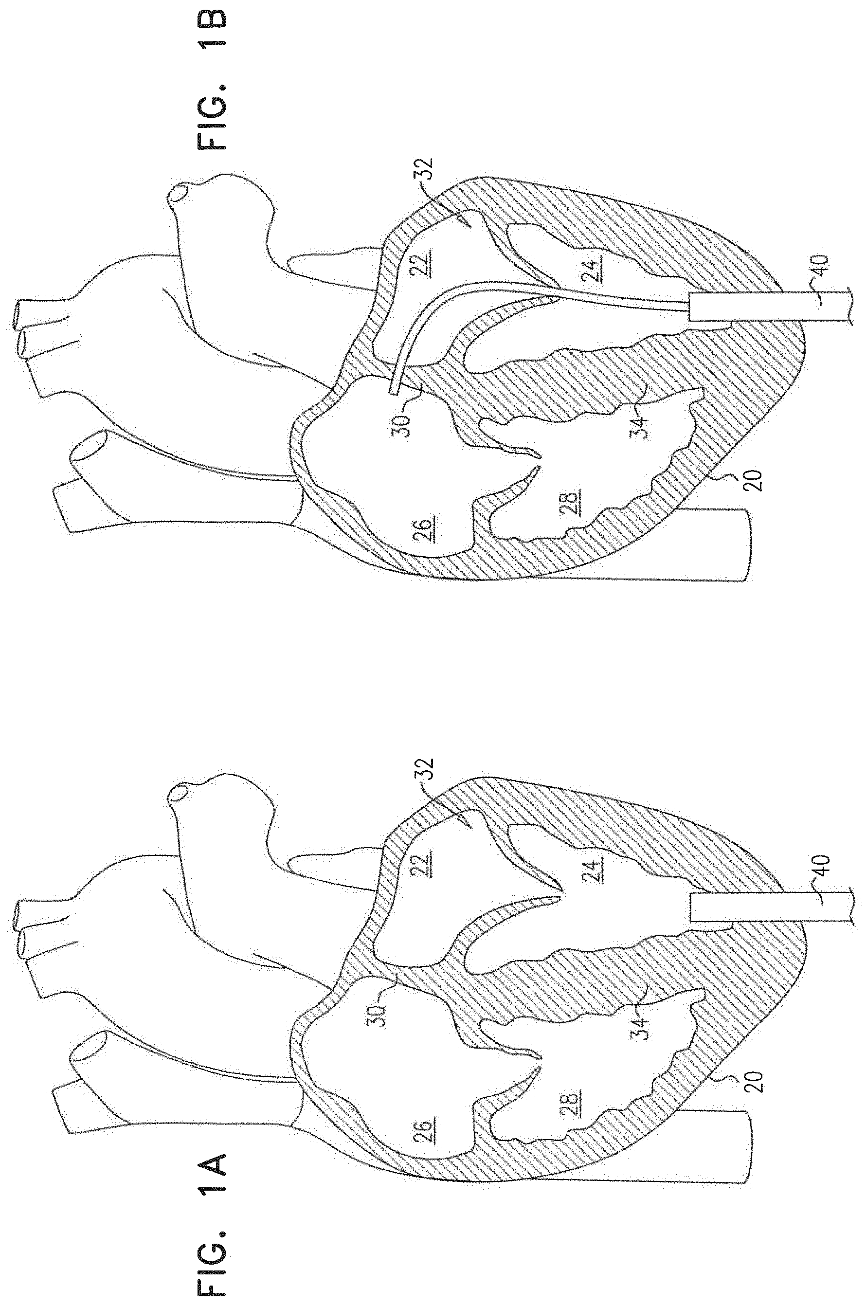
[0063] Reference is made to FIGS. 1A-F, which are schematic illustrations of a method for use with a heart 20 of a subject, in accordance with some applications of the invention. Heart 20 has a left atrium 22 and a left ventricle 24 (the left side of the heart), and a right atrium 26 and a right ventricle 28 (the right side of the heart).
[0064] A transapical puncture is made into left ventricle 24 (FIG. 1A). For some applications, a tube 40 is introduced through the transapical puncture. Via the transapical puncture (e.g., via tube 40), a transseptal fenestration is made in interatrial septum 30 (e.g., in the fossa ovalis) (FIG. 1B). For some applications, a septal device 42 (e.g., a shunt device) is implanted at the transseptal fenestration (FIG. 1C). Subsequently, via the transapical puncture (e.g., via tube 40), a prosthetic valve 44 is delivered (FIG. 1D) and implanted at mitral valve 32 of the subject (FIG. 1E). Subsequently, the transapical puncture is closed (FIG. 1F).
[0065] Reference is made to FIGS. 2A-F, which are schematic illustrations of a method for use with heart 20 of a subject, in accordance with some applications of the invention. The method of FIGS. 2A-F is similar to that of FIGS. 1A-F, except that (i) prosthetic valve 44 is delivered and implanted prior to making the transseptal fenestration (FIGS. 2B-C), and (ii) the transseptal fenestration is made (and optionally a septal device 52, such as a shunt device, is implanted) in interventricular septum 34 (FIGS. D-E).
[0066] Reference is again made to FIGS. 1A-2F. It is to be noted that the scope of the invention includes making the transseptal fenestration in interventricular septum 34 even when making the transseptal fenestration prior to implantation of prosthetic valve 44. It is also to be noted that the scope of the invention includes implanting prosthetic valve 44 prior to making the transseptal fenestration even for applications in which the transseptal fenestration is made in the interatrial septum.
[0067] Reference is made to FIGS. 3A-B, which are schematic illustrations of a method for use with heart 20 of a subject, in accordance with some applications of the invention. The method shown in FIGS. 3A-B is identical to those described with reference to FIGS. 1A-2F hereinabove, except that the transseptal fenestration is made (and optionally, septal device 42 or 52 is implanted) via a transvenous (e.g., transfemoral) route. The transseptal fenestration (and/or the implantation of septal device 42 or 52) may be made before or after prosthetic valve 44 is implanted. FIGS. 3A-B show the transseptal fenestration in interatrial septum 30, but it is to be noted that the scope of the invention includes the transseptal fenestration being in interventricular septum 34, mutatis mutandis).
[0068] Reference is again made to FIGS. 1A-3B. For some applications, rather than via transapical puncture, prosthetic valve 44 is implanted via a transatrial puncture (i.e., a puncture through the heart wall into atrium 22). For such applications, the transseptal fenestration may be made via the transatrial puncture or via a different route.
[0069] Reference is again made to FIGS. 1A-3B. It is to be noted that prosthetic valve 44 is not delivered to mitral valve 32 via the transseptal fenestration. That is, the transseptal fenestration described herein is not itself required for delivery of the prosthetic valve.
[0070] Reference is now made to FIGS. 4-8, which are schematic illustrations of septal devices that may be implanted at the transseptal fenestration, in accordance with some applications of the invention. Hereinabove a shunt device is used as an example of devices 42 and 52. A shunt device is typically used to maintain patency of the transseptal fenestration. For example, and as shown in FIG. 2C, a shunt device may have a tubular portion 54 that is shaped to define a lumen, with flanges 56 (that are typically expandable) that engage the tissue of the septum, and retain the shunt device within the transseptal fenestration. FIGS. 4-8 are other examples of devices that may be implanted in place of device 42 or device 52, mutatis mutandis.
[0071] FIG. 4 is a schematic illustration of a septal device 60 that is shaped to define a lumen, and comprises a check valve 62 that facilitates one-way blood flow through the lumen (and thereby through the transseptal fenestration). Device 60 is typically oriented such that the one-way blood flow is from a chamber of the left side of the heart to a chamber of the right side of the heart (e.g., from left atrium 22 to right atrium 26, or from left ventricle 24 to right ventricle 28). Check valve 62 is shown as a leaflet or duckbill valve, but it is to be understood that any suitable check valve known in the art may be used (such as, but not limited to, a ball-and-cage valve or a tilting-disc valve). For some applications (e.g., when check valve 62 is a leaflet or duckbill valve), check valve 62 thereby comprises a membrane (i.e., the leaflets or the duckbill membrane) that regulates blood flow through the lumen of the valve.
[0072] Frame A shows a state of device 60 (i.e., closed) when a blood pressure difference across the device (e.g., across valve 62) is less than a threshold blood pressure difference. That is, when blood pressure on the left side of the heart is less than a threshold amount greater than blood pressure on the right side of the heart (e.g., including if the pressure on the left side of the heart is not greater than the pressure on the right side of the heart). Frame B shows a state of device 60 (i.e., open) when the blood pressure difference is greater than the threshold blood pressure difference. For some applications, the threshold blood pressure difference for device 60 is 4-6 mmHg (e.g., 5 mmHg).
[0073] FIG. 5 is a schematic illustration of a septal device 70 that comprises a membrane 72 that regulates blood flow through the lumen. Device 70 is thereby similar to device 60. In contrast to device 60, membrane 72 regulates blood flow but in a bidirectional manner. Membrane 72 opens bidirectionally in response to a blood pressure difference that is greater than a threshold blood pressure difference in either direction. Frame A shows a state of device 70 when blood pressure on the left side of the heart is more than the threshold difference greater than pressure on the right side of the heart. Frame C shows the opposite state. Frame B shows a state of device 70 (i.e., closed, thereby inhibiting blood flow through the lumen of the device) when the blood pressure difference across the device (e.g., across membrane 72) is less than the threshold blood pressure difference. For some applications, the threshold blood pressure difference for device 70 is 4-6 mmHg (e.g., 5 mmHg) (in either direction).
[0074] For some applications, membrane 72 is transected by intersecting slits 74, which form the membrane into flaps, which flap open and closed as shown.
[0075] FIGS. 6 and 7 show septal devices 80 and 90, respectively, which also each comprise a membrane. However, the membranes of these devices do not facilitate blood flow between the left and right sides of the heart. Rather, implantation of these devices seals the transseptal fenestration with the membrane.
[0076] Device 80 comprises a membrane 82, and device 90 comprises a membrane 92. Membranes 82 and 92 are similar, are both elastic, and both elastically expand (i.e., stretch) in response to a difference in blood pressure across the membrane. Membrane 92 may be considered to be a balloon (e.g., having an interior 94 even in the absence of a pressure difference across the membrane), whereas membrane 82 is generally planar in the absence of a pressure difference across the membrane. Device 90 has an opening 96 into interior 94, and is implanted such that the interior is in fluid communication, via the opening, with the chamber of the left side of the heart. For both FIG. 6 and FIG. 7, frame A shows a state of the device in the absence of a pressure difference across the membrane, frame B shows a state of the device in the presence of a pressure difference across the membrane, and frame C shows a state of the device in the presence of a larger pressure difference across the membrane.
[0077] By elastically expanding, membranes 82 and 92 increase the effective volume of the chamber of the left side of the heart, thereby reducing the blood pressure in that chamber without mixing of blood between the left and right sides of the heart.
[0078] It is alternatively possible to describe the balloon of device 90, as being a cell that has an interior 94 and an opening 96 into the interior. Device 90 is implanted such that (i) the interior is in fluid communication, via the opening, with a chamber of the left side of the heart, and (ii) a greater blood pressure in the chamber of the left side of the heart relative to a blood pressure in a corresponding chamber of the right side of the heart increases a volume of interior 94 that is disposed within the corresponding chamber of the right side of the heart. This occurs by membrane 92 elastically expanding into the chamber of the right side of the heart in response to this pressure difference.
[0079] For some applications, membrane 82 and/or membrane 92 inflate only when blood pressure in the chamber of the left side of the heart is more than 4-6 mmHg (e.g., 5 mmHg) greater than blood pressure in the chamber of the right side of the heart.
[0080] The membrane/balloon of devices 80 and 90 may be biased to automatically contract/deflate in response to a reduction of the difference in blood pressure across the fenestration, even if the blood pressure in the chamber of the right side of the heart does not exceed that of the chamber of the left side of the heart.
[0081] FIG. 8 shows a septal device 100 that comprises a cell 102 that is slidably mounted in a mount 108, such that the mount is fixed at the transseptal fenestration, and the cell slides into the chamber of the right side of the heart in response to the greater blood pressure in the chamber of the left side of the heart. Cell 102 has an interior 104 and an opening 106 into the interior, and is implanted such that the interior is in fluid communication, via the opening, with the chamber of the left side of the heart. Frame A shows the cell not protruding (or protruding minimally) into the chamber of the right side of the heart, and frame B shows the cell having slid into the chamber of the right side of the heart. For some applications, cell 102 is biased (e.g., spring-loaded) to automatically slide out of the chamber of the right side of the heart in response to a reduction of the difference in blood pressure across the fenestration, even if the blood pressure in the chamber of the right side of the heart does not exceed that of the chamber of the left side of the heart.
[0082] For some applications, cell 102 slides into the chamber of the right side of the heart only when blood pressure in the chamber of the left side of the heart is more than 4-6 mmHg (e.g., 5 mmHg) greater than blood pressure in the chamber of the right side of the heart.
[0083] It is hypothesized by the inventors that the implantation of a septal device described hereinabove in addition to the implantation of prosthetic valve 44 improves a likelihood of a successful long-term outcome of the procedure. For example, the septal devices may facilitate reduction of elevated blood pressure in the right side of the heart, should regurgitation through or around prosthetic valve 44 begin to occur subsequently to implantation of the prosthetic valve. Therefore the implantation of such a septal device may be considered to be prophylactic. For some applications of the invention, the methods described hereinabove are performed on a subject (e.g., an adult subject) who does not suffer from and/or has not been identified (e.g., diagnosed) as suffering from heart failure.
[0084] Therefore, a method according to some applications of the invention comprises: (i) making a transseptal fenestration in a heart of a subject (e.g., an adult subject) who has not been identified as suffering from heart failure; (2) advancing a shunt device into the heart; and (3) implanting the shunt device at the transseptal fenestration. Similarly, another method according to some applications of the invention comprises: (1) identifying an adult subject as not suffering from heart failure; and (2) subsequently, making a transseptal fenestration in a heart of the subject.
[0085] Reference is now made to FIGS. 9 and 10, which are schematic illustrations of a device 110 and a device 120 implanted in the heart wall, in accordance with an application of the invention. For some applications, devices that reversibly increase the effective volume of the chamber of the left side of the heart are implanted in the heart wall, rather than (as described hereinabove for devices 80, 90 and 100) at a transseptal fenestration. FIG. 9 shows device 110 implanted in the wall of left atrium 22, and FIG. 10 shows device 120 implanted in the wall of left ventricle 24. Devices 110 and 120 may be similar in structure and function (if not dimension) to devices 80, 90 or 100, mutatis mutandis.
[0086] It will be appreciated by persons skilled in the art that the present invention is not limited to what has been particularly shown and described hereinabove. Rather, the scope of the present invention includes both combinations and subcombinations of the various features described hereinabove, as well as variations and modifications thereof that are not in the prior art, which would occur to persons skilled in the art upon reading the foregoing description.
* * * * *
D00000
D00001
D00002
D00003
D00004
D00005
D00006
D00007
D00008
D00009
D00010
D00011
D00012

D00013

XML
uspto.report is an independent third-party trademark research tool that is not affiliated, endorsed, or sponsored by the United States Patent and Trademark Office (USPTO) or any other governmental organization. The information provided by uspto.report is based on publicly available data at the time of writing and is intended for informational purposes only.
While we strive to provide accurate and up-to-date information, we do not guarantee the accuracy, completeness, reliability, or suitability of the information displayed on this site. The use of this site is at your own risk. Any reliance you place on such information is therefore strictly at your own risk.
All official trademark data, including owner information, should be verified by visiting the official USPTO website at www.uspto.gov. This site is not intended to replace professional legal advice and should not be used as a substitute for consulting with a legal professional who is knowledgeable about trademark law.