Anti-C3d antibody conjugates and methods of detecting complement activation
Tomlinson; Stephen ; et al.
U.S. patent application number 16/572260 was filed with the patent office on 2020-10-01 for anti-c3d antibody conjugates and methods of detecting complement activation. The applicant listed for this patent is Dept of Veterans Affairs, MUSC Foundation for Research Development, The Regents of the University of Colorado, a body corporate. Invention is credited to V. Michael Holers, Stephen Tomlinson.
| Application Number | 20200308295 16/572260 |
| Document ID | / |
| Family ID | 1000004797086 |
| Filed Date | 2020-10-01 |


View All Diagrams
| United States Patent Application | 20200308295 |
| Kind Code | A1 |
| Tomlinson; Stephen ; et al. | October 1, 2020 |
Anti-C3d antibody conjugates and methods of detecting complement activation
Abstract
Provided herein, inter alia, are compositions and methods of using the same for detecting complement activation.
| Inventors: | Tomlinson; Stephen; (Charleston, SC) ; Holers; V. Michael; (Aurora, CO) | ||||||||||
| Applicant: |
|
||||||||||
|---|---|---|---|---|---|---|---|---|---|---|---|
| Family ID: | 1000004797086 | ||||||||||
| Appl. No.: | 16/572260 | ||||||||||
| Filed: | September 16, 2019 |
Related U.S. Patent Documents
| Application Number | Filing Date | Patent Number | ||
|---|---|---|---|---|
| 15511597 | Mar 15, 2017 | 10413620 | ||
| PCT/US2015/050232 | Sep 15, 2015 | |||
| 16572260 | ||||
| 14624347 | Feb 17, 2015 | 9259488 | ||
| 15511597 | ||||
| PCT/US2013/055400 | Aug 16, 2013 | |||
| 14624347 | ||||
| 62050568 | Sep 15, 2014 | |||
| 61684691 | Aug 17, 2012 | |||
| Current U.S. Class: | 1/1 |
| Current CPC Class: | C07K 2317/622 20130101; A61K 49/0058 20130101; A61K 49/16 20130101; C07K 2317/92 20130101; C07K 16/2896 20130101 |
| International Class: | C07K 16/28 20060101 C07K016/28; A61K 49/16 20060101 A61K049/16; A61K 49/00 20060101 A61K049/00 |
Claims
51. (canceled)
52. . An isolated peptide comprising a sequence of amino acids 1 through 275 of SEQ ID NO: 32.
53. The peptide of claim 52, wherein the peptide comprises SEQ ID NO: 32.
54. The peptide of claim 52, wherein the peptide further comprises a detectable moiety.
55. The peptide of claim 54, wherein the detectable moiety is selected from the group consisting of .sup.32P, a fluorescent dye, an electron-dense reagent, an enzyme, biotin, digoxigenin, a paramagnetic molecule, a paramagnetic nanoparticle, an ultrasmall superparamagnetic iron oxide ("USPIO") nanoparticle, a USPIO nanoparticle aggregate, a superparamagnetic iron oxide ("SPIO") nanoparticle, an SPIO nanoparticle aggregate, a standard superparamagnetic iron oxide ("SSPIO"), an SSPIO nanoparticle aggregate, a polydisperse superparamagnetic iron oxide ("PSPIO"), a PSPIO nanoparticle aggregate, a monochrystalline SPIO, a monochrystalline SPIO aggregate, a monochrystalline iron oxide nanoparticle, a monochrystalline iron oxide, another nanoparticle contrast agent, a liposome or other delivery vehicle comprising Gadolinium chelate ("Gd-chelate") molecules, Gadolinium, a radioisotope, a radionuclide, carbon-11, nitrogen-13, oxygen-15, fluorine-18, rubidium-82, fluorodeoxyglucose, a gamma ray emitting radionuclide, a positron-emitting radionuclide, radiolabeled glucose, radiolabeled water, radiolabeled ammonia, a biocolloid, a microbubble, an iodinated contrast agent, barium sulfate, thorium dioxide, gold, a gold nanoparticle, a gold nanoparticle aggregate, a fluorophore, a two-photon fluorophore, a hapten, a protein, and a fluorescent moiety.
56. A composition comprising the peptide of claim 52.
57. An isolated nucleic acid molecule comprising a nucleotide sequence encoding a peptide comprising a sequence of amino acids 1 through 275 of SEQ ID NO: 32.
58. The isolated nucleic acid molecule of claim 57, wherein the nucleic acid encodes a peptide comprising SEQ ID NO: 32.
59. The isolated nucleic acid molecule of claim 57, wherein the isolated nucleic acid further comprises a nucleotide sequence encoding a detectable moiety.
60. A composition comprising the isolated nucleic acid molecule of claim 57.
Description
CROSS-REFERENCE TO RELATED APPLICATIONS
[0001] This application is a continuation of U.S. application Ser. No. 15/511,597, filed Mar. 15, 2017, which is a U.S. national phase application filed under 35 U.S.C. .sctn. 371 claiming benefit to PCT International Patent Application No. PCT/US2015/050232, filed Sep. 15, 2014, which claims the priority to U.S. Provisional Application No. 62/050,568, filed Sep. 15, 2014; and U.S. application Ser. No. 15/511,597 is a continuation-in-part of U.S. application Ser. No. 14/624,347, filed Feb. 2, 2017, which is a continuation of PCT International Patent Application No. PCT/US2013/055400, filed Aug. 16, 2013, which claims priority to U.S. Provisional Application No. 61/684,691, filed Aug. 17, 2012, each of which disclosures is incorporated herein by reference in its entirety.
REFERENCE TO A "SEQUENCE LISTING," A TABLE, ORA COMPUTER PROGRAM LISTING APPENDIX SUBMITTED AS AN ASCII TEXT FILE
[0002] The Sequence Listing written in file 85256-884072_ST25.TXT, created on Aug. 15, 2013, 93,678 bytes, machine format IBM-PC, MS-Windows operating system, is hereby incorporated by reference.
BACKGROUND OF THE INVENTION
[0003] Complement is the collective term for a series of blood proteins that constitute a major effector mechanism of the immune system. The complement system plays an important role in the pathology of many autoimmune, inflammatory and ischemic diseases. Inappropriate complement activation and its deposition on host cells can lead to complement-mediated lysis and/or injury of cells and target tissues, as well as tissue destruction due to the generation of powerful mediators of inflammation. Key to the activity of the complement system is the covalent attachment of processed protein fragments derived from a serum protein, complement C3, to tissue sites of complement activation. This unusual property is due to the presence of a thioester bond in C3 that, when cleaved during C3 activation, converts C3 to a form designated C3b which can then utilize ester or amide bonds to link to cell and tissue- attached molecules. Once C3b is covalently attached, it is rapidly processed to the iC3b, C3dg and C3d forms, each of which remain covalently attached to the target tissue site. This process results in the "marking" of the tissue as one in which an inflammatory injury or other complement-related process is underway.
[0004] Complement can be activated by any of three pathways: the classical, lectin and alternative pathways. The classical pathway is activated through the binding of the complement system protein Clq to antigen-antibody complexes, pentraxins or apoptotic cells. The pentraxins include C-reactive protein and serum amyloid P component. The lectin pathway is initiated by binding of microbial carbohydrates to mannose-binding lectin or by the binding of ficolins to carbohydrates or acetylated molecules.
[0005] The alternative pathway is activated on surfaces of pathogens that have neutral or positive charge characteristics and do not express or contain complement inhibitors. This results from the process termed `tickover` of C3 that occurs spontaneously, involving the interaction of conformationally altered C3 with factor B, and results in the fixation of active C3b on pathogens or other surfaces. The alternative pathway can also be initiated when certain antibodies block endogenous regulatory mechanisms, by IgA-containing immune complexes, or when expression of complement regulatory proteins is decreased. In addition, the alternative pathway is activated by a mechanism called the `amplification loop` when C3b that is deposited onto targets via the classical or lectin pathway, or indeed through the tickover process itself, binds factor B. See Muller-Eberhard (1988) Ann. Rev. Biochem. 57:321. For example, Holers and colleagues have shown that the alternative pathway is amplified at sites of local injury when inflammatory cells are recruited following initial complement activation. Girardi et al, J. Clin. Invest. 2003, 1 12: 1644. Dramatic complement amplification through the alternative pathway then occurs through a mechanism that involves either the additional generation of injured cells that fix complement, local synthesis of alternative pathway components, or more likely because infiltrating inflammatory cells that carry preformed C3 and properdin initiate and/or greatly increase activation specifically at that site.
[0006] Alternative pathway amplification is initiated when circulating factor B binds to activated C3b. This complex is then cleaved by circulating factor D to yield an enzymatically active C3 convertase complex, C3bBb. C3bBb cleaves additional C3 generating C3b, which drives inflammation and also further amplifies the activation process, generating a positive feedback loop. Factor H is a key regulator (inhibitor) of the alternative complement pathway activation and initiation mechanisms that competes with factor B for binding to conformationally altered C3 in the tickover mechanism and to C3b in the amplification loop. Binding of C3b to Factor H also leads to degradation of C3b by factor Ito the inactive form iC3b (also designated C3bi), thus exerting a further check on complement activation. Factor H regulates complement in the fluid phase, circulating at a plasma concentration of approximately 500 .mu.g/ml, but its binding to cells is a regulated phenomenon enhanced by the presence of a negatively charged surface as well as fixed C3b, iC3b, C3dg or C3d. Jozsi et al, Histopathol. (2004) 19:251-258.
[0007] Complement activation, C3 fragment fixation and complement-mediated inflammation are involved in the etiology and progression of numerous diseases. The down-regulation of complement activation has been shown to be effective in treating several diseases in animal models and in ex vivo studies, including, for example, systemic lupus erythematosus and glomerulonephritis (Y. Wang et al, Proc. Nat'l Acad. Sci. USA (1996) 93:8563-8568), rheumatoid arthritis (Y. Wang et al, Proc. Nat'l Acad. Sci. USA (1995) 92:8955-8959), cardiopulmonary bypass and hemodialysis (C. S. Rinder, J. Clin. Invest. (1995) 96: 1564-1572), hyperacute rejection in organ transplantation (T. J. Kroshus et al, Transplantation (1995) 60: 1194-1202), myocardial infarction (J. W. Homeister et al, J. Immunol. (1993) 150: 1055-1064; H. F. Weisman et al, Science (1990) 249: 146-151), ischemia/reperfusion injury (E. A. Amsterdam et al, Am. J. Physiol. (1995) 268:H448-H457), antibody-mediated allograft rejection, for example, in the kidneys (J. B. Colvin, J. Am. Soc. Nephrol. (2007) 18(4): 1046-56), and adult respiratory distress syndrome (R. Rabinovici et al, J. Immunol. (1992) 149: 1744-1750).
[0008] Moreover, other inflammatory conditions and autoimmune/immune complex diseases are also closely associated with complement activation (B. P. Morgan. Eur. J. Clin. Invest. (1994) 24:219-228), including, but not limited to, thermal injury, severe asthma, anaphylactic shock, bowel inflammation, urticaria, angioedema, vasculitis, multiple sclerosis, myasthenia gravis, myocarditis, membranoproliferative glomerulonephritis, atypical hemolytic uremic syndrome, Sj5gren's syndrome, renal and pulmonary ischemia/reperfusion, and other organ- specific inflammatory disorders. It is currently uncertain whether complement activation is essential to the pathogenesis and injury of all diseases in which local tissue C3 activation and inflammatory injury occurs; nevertheless, C3 fragment fixation is almost universally found as an associated event.
[0009] A variety of disorders are associated with inflammation, however, so definitive diagnosis of complement-mediated inflammation typically requires confirmation via immuno- staining or other in vitro analysis performed on tissue samples retrieved by biopsy. While biopsies are in many respects routine, they have their limitations and are not risk-free. Because commonly used needle or punch biopsies sample only a small portion of the target organ, there is a risk of sample error leading to an incorrect diagnosis. Furthermore, although biopsy is a generally safe procedure, major complications such as internal bleeding may occur in a significant number of cases.
[0010] In some cases, because of the difficulties in diagnosing disease or monitoring disease progression, for example, in patients with systemic lupus erythematosus or lupus nephritis, repeat renal biopsies are therefore frequently necessary to assess the response to therapy or to diagnose disease relapse. See e.g., S. Bajaj et al., 2000, J. Rheumatol. 27:2822-2826. Although renal biopsy is generally a safe procedure, complications may occur in 6% or more of biopsies and intra-renal bleeding and hematuria are common. Patients requiring repeat biopsies are at concomitantly greater risk of complications. See e.g., W. L. Whittier et al, 2004, J. Am. Soc. Nephrol. 15: 142-147; D. C. Mendelssohn et al, 1995, Am. J. Kidney Dis. 26:580-585. Thus, a non-invasive method of detecting or accurately assessing the presence, degree and/ or extent of complement-mediated inflammation would be of significant value in diagnosing disease, formulating treatment strategies and monitoring their efficacy for many inflammatory diseases, including lupus nephritis.
[0011] The use of complement receptor 2 (CR2), or functional fragments thereof, to target complement modulators to tissues which exhibit or express C3, or fragments of C3 to which the CR2 is able to bind, including C3b, iC3b, C3d and C3dg, is described in US 2008/0267980 and US 2008/0221011, the disclosures of which are hereby incorporated herein by reference. Such CR2 molecules, and functional fragments thereof, can be used for targeting because the first two N-terminal short consensus repeat domains (SCRs) comprise an active binding site for the exposed C3d domain that is contained within iC3b, C3dg, and C3d.
[0012] The present invention provides solutions to these and other problems in the art. The disclosures of all publications, patents, patent applications and published patent applications referred to herein are hereby incorporated herein by reference in their entirety.
BRIEF SUMMARY OF THE INVENTION
[0013] In a first aspect is provided an antibody conjugate including an antibody, or antigen binding fragment thereof, and a detectable moiety.
[0014] In a second aspect is provided a method of detecting complement-mediated inflammation in an individual including: (a) administering to the individual an effective amount of an anti-C3d antibody conjugate as described herein; (b) allowing the anti-C3d antibody conjugate to bind to a C3 protein fragment within the individual thereby forming an anti-C3d antibody conjugate-C3 protein fragment complex; and (c) detecting the anti-C3d antibody conjugate-C3 protein fragment complex in the individual.
[0015] In a third aspect is provided a method of detecting complement activation in an individual including: (a) administering to the individual an effective amount of an anti-C3d antibody conjugate as described herein; (b) allowing the anti-C3d antibody conjugate to bind to a C3 protein fragment within the individual thereby forming an anti-C3d antibody conjugate-C3 protein fragment complex; and (c) detecting the anti-C3d antibody conjugate-C3 protein fragment complex in the individual.
[0016] In a fourth aspect is provided a method of detecting complement activation including (a) administering to a biological sample (e.g. biopsy, tissue, blood, blood fraction, serum, or cells, all optionally from a subject or patient) an effective amount of an anti-C3d antibody conjugate as described herein; (b) allowing the anti-C3d antibody conjugate to bind to a C3 protein fragment within the biological sample thereby forming an anti-C3d antibody conjugate-C3 protein fragment complex; and (c) detecting the anti-C3d antibody conjugate-C3 protein fragment complex in the biological sample. In some embodiments, the C3 protein fragment is C3d or C3dg or iC3b.
BREIF DESCRIPTION OF THE DRAWINGS
[0017] FIG. 1 depicts expenmental results demonstrating changes in T2 relaxation as measured by MRI in muscle and kidney following administration of the 3d29 antibody conjugated to the surface of iron-oxide nanoparticles to factor H knockout mice (that have abundant target C3 fragment deposits (i.e. iC3b, C3dg, and C3d) in the glomeruli of their kidneys).
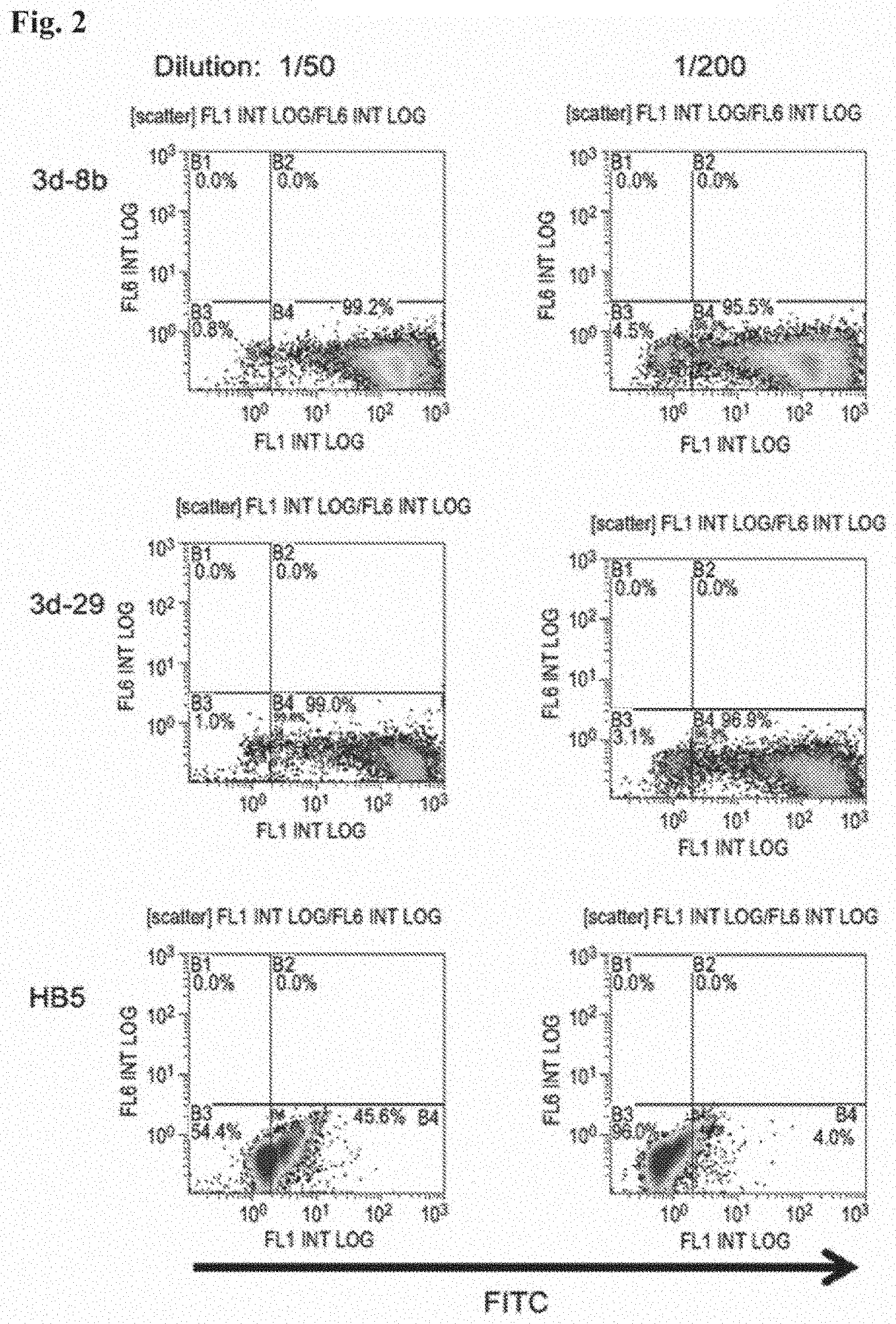
[0018] FIG. 2 depicts expenmental results demonstrating specific binding by flow cytometry of FITC-labelled Group 1 monoclonal antibodies 3d29 and 3d8, but not control monoclonal Antibody HB5, to C3d-coated zymosan particles. These monoclonal antibodies also retained binding to C3d following biotinylation and other techniques.
[0019] FIG. 3 depicts expenmental results demonstrating specific and durable in vivo binding of monoclonal antibody 3d29 to the site of complement C3 fixation at 48 hours following injection the day after laser-induced CNV injury. In vivo imaging was performed using a Micron III retinal imaging microscope (Phoenix Research Laboratories).
[0020] FIG. 4 depicts expenmental results demonstrating specific and durable in vitro binding of monoclonal antibody 3d29 to the site of complement C3 fixation at 48 hours following injection the day after induction of laser- induced CNV injury. In vitro imaging was performed using a flat mount and B.times.W image capture system.
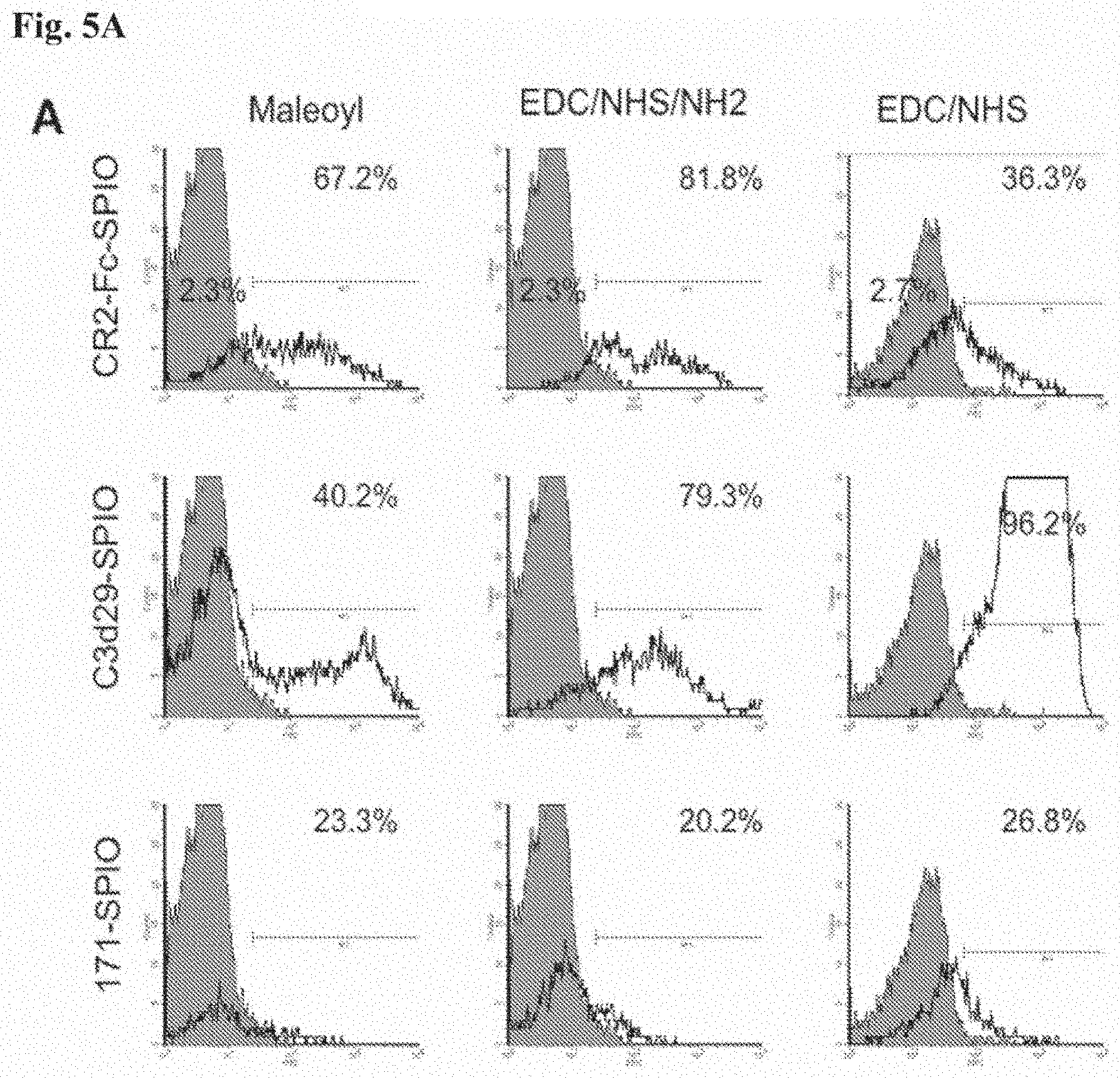
[0021] FIG. 5, comprising FIG. 5A and FIG. 5B, depicts expenmental results demonstrating conjugation of anti-C3d antibodies to the surface of superparamagnetic iron-oxide nanoparticles (SPIO). Three different methods of conjugating antibodies to the surface of SPIO were tested using three different proteins: C3d29 anti-C3d, CR2-Fc, and 171 (as a control antibody). FIG. 5A depicts FACS analysis was used to determine the efficacy of the reactions. Unconjugated SPIO are represented by filled gray curves, and conjugated are represented by black line curves. FIG. 5B depicts the percentage of antibody-positive SPIO obtained using each method of conjugation and for each protein are shown. Data are mean .+-.SEM from three independent conjugations per method per protein species.
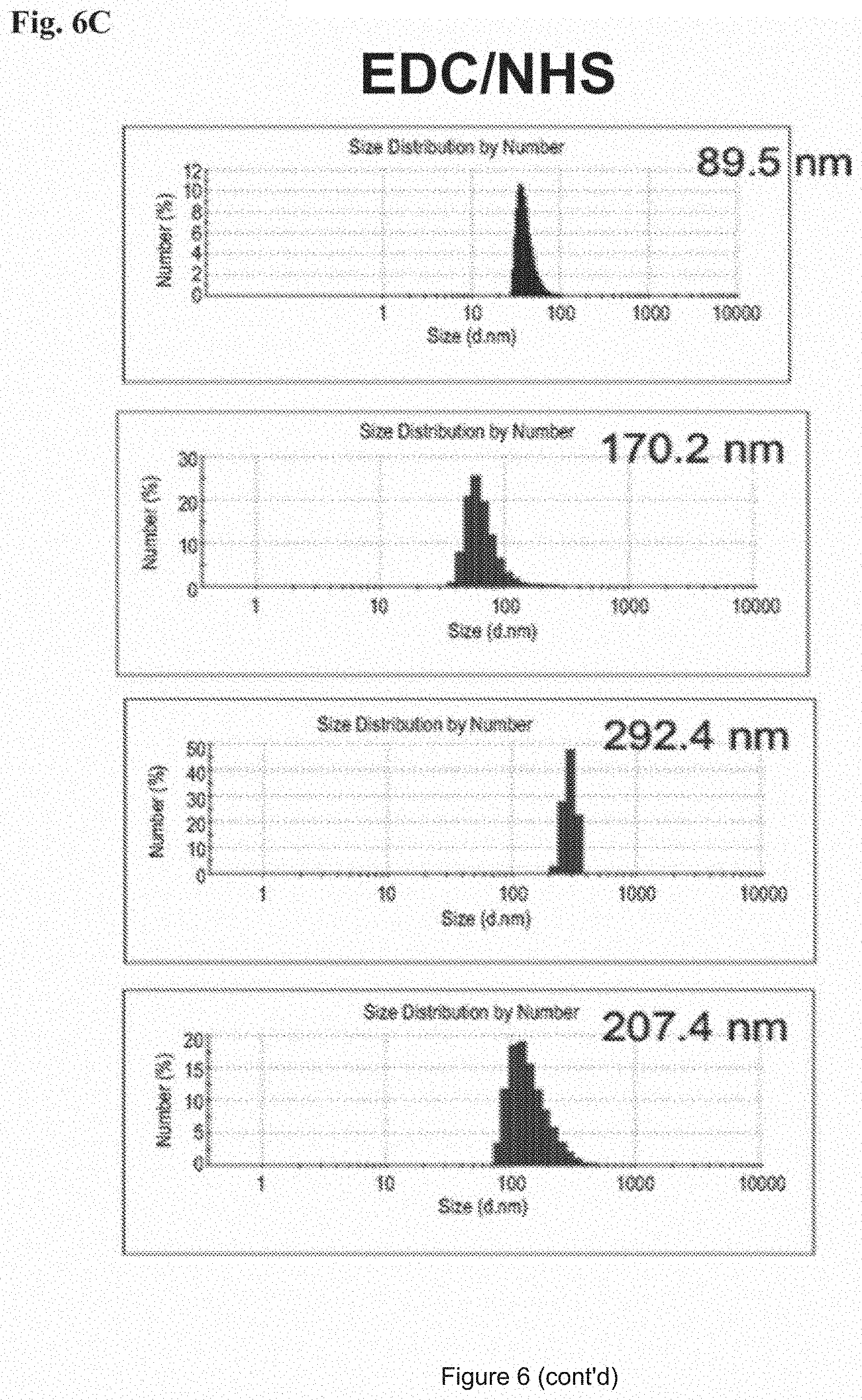
[0022] FIG. 6, comprising FIG. 6A through FIG. 6C, depicts the analysis of conjugated SPIO size as determined by dynamic light scattering (DLS). FIG. 6A depicts a representative histogram of the dominant peak of number- weighted size distribution for maleoyl conjugation methods for unconjugated SPIO, and CR2-Fc-, C3d29-, and 171 -conjugated SPIO. The mean diameter (nm) for the dominant peak of size distribution is indicated in the top right corner of every sample. FIG. 6B depicts a representative histogram of the dominant peak of number-weighted size distribution for EDC/NHS/NH2 conjugation method for unconjugated SPIO, and CR2-Fc-, C3d29-, and 171 -conjugated SPIO. The mean diameter (nm) for the dominant peak of size distribution is indicated in the top right corner of every sample. FIG. 6C depicts a representative histogram of the dominant peak of number- weighted size distribution for EDC/NHS conjugation method for unconjugated SPIO, and CR2-Fc-, C3d29-, and 171 -conjugated SPIO. The mean diameter (nm) for the dominant peak of size distribution is indicated in the top right corner of every sample. An increase in size following conjugation of SPIO with proteins was observed for all conjugated samples.
[0023] FIG. 7, comprising FIG. 7A through FIG. 7C, depicts expenmental results demonstrating the hydrodynamic size and surface characteristics of conjugated SPIO. FIG. 7A depicts expenmental results demonstrating the zeta potential of unconjugated and conjugated SPIO. The conjugated proteins change the negative zeta potential of the NH2-SPIO from slightly negative to positive, and reduce the negative zeta potential of COOH-SPIO. Data are mean .+-.SEM from three independent conjugations per method per protein species. FIG. 7B depicts expenmental results demonstrating the mean diameter and percent distribution of unconjugated and conjugated SPIO. All conjugates species of SPIO show an increase in hydrodynamic size from that of unconjugated SPIO. C3d29-SPIO of EDC/NHS method shows properties of aggregation (greater increase in hydrodynamic size and a greater reduction of the negative zeta potential). Data are mean .+-.SD from three independent conjugations per method per protein species. FIG. 7C depicts expenmental results demonstrating the estimated nmoles of protein per nmole of conjugated SPIO for three different methods of conjugation with three different proteins. Data are mean .+-.SEM per conjugation method per protein.
[0024] FIG. 8, comprising FIG. 8A through FIG. 8C, depicts expenmental results demonstrating the binding of conjugated SPIO with the target C3d antigen in ELISA. FIG. 8A depicts the detection of SPIO binding to the target C3d antigen by ELISA for SPIO conjugated with the maleoyl method. FIG. 8B depicts the detection of SPIO binding to the target C3d antigen by ELISA for SPIO conjugated with the EDC/NHS/NH2 method. FIG. 8C depicts the detection of SPIO binding to the target C3d antigen by ELISA for SPIO conjugated with the EDC/NHS method. Specific, C3d-dependent, binding was detected for CR2-Fc-SPIO and C3d29-SPIO conjugated with all three methods. Data are mean .+-.SD for samples tested in duplicates from two independent experiments. Differences in the absorbance values between CR2-Fc-SPIO and C3d29-SPIO may be due to the use of two different secondary antibodies.
[0025] FIG. 9, comprising FIG. 9A and FIG. 9B, depicts expenmental results demonstrating target-specific MM signal reduction by conjugated SPIO. FIG. 9A depicts expenmental results demonstrating reduction of T2-relaxation time of opsonized CHO cell pellets incubated with CR2-Fc-SPIO or C3d29-SPIO was seen relative to those incubated with control 171-SPIO. Three different methods of SPIO conjugation were tested. Data are mean .+-.SEM from conjugated SPIO obtained from three independent conjugations per method per protein. FIG. 9B depicts images from T2-weighted MM scans of the cell pellets. Images were obtained at TE=48 ms for all three methods of conjugation. Targeting proteins conjugated to SPIO are indicated on the left. Darkening of the pellets reflects binding of the SPIO to the opsonized cells. *p<0.05, **p<0.01, and ***p<0.001 by one-way ANOVA followed by Dunnett's post-test where data from 171-SPIO was used as control.
[0026] FIG. 10, comprising FIG. 10A and FIG. 10B, depicts expenmental results demonstrating the metabolism of C3 to iC3b and C3d during complement activation. FIG. 10A depicts a schematic demonstration of cleavage of soluble C3 initially to the C3b and C3a forms through the activity of C3 convertases (activating enzymes), followed by the sequential processing to iC3b, C3dg and C3d (latter not shown) through the activity of cofactors and proteases. The iC3b, C3dg and C3d proteins exhibit durable tissue and cell binding propertiesm. FIG. 10A depicts an illustration of the molecular entities (iC3b, C3dg (not shown) and C3d) to which the anti-C3d monoclonal antibodies described herein specifically bind.
[0027] FIG. 11. FIG. 11, comprising FIG. 11A through FIG. 11D, depicts expenmental results demonstrating the generation of monoclonal antibodies that recognize C3 activation fragments. Anti-human C3d hybridomas were generated. FIG. 11A depicts expenmental results demonstrating that the hybridomas were screened against recombinant human C3d by ELISA, and nine of the clones bound to the protein (clone 7C10 was used as a positive control, and the remaining clones were newly identified). FIG. 11B depicts expenmental results demonstrating that the reactivity of the clones against intact C3 and recombinant C3d by Western blot analysis was tested. Three patterns of reactivity were seen: Group 1 clones bound strongly to C3d, Group 2 clones bound to the a chain of intact C3, and Group 3 clones did not bind well to either moiety. The * denotes the clone whose results are shown. FIG. 11C depicts expenmental results demonstrating that Clone 3d11 recognized all of the C3 fragments by Western blot analysis. The appearance of the a, a', a1, a2, C3dg, and C3d fragments from purified proteins and from mouse plasma are shown. FIG. 11D depicts expenmental results of immunoprecipitation of C3 fragments in plasma demonstrating that the Group 1 clones recognize the iC3b form (al chain) and C3dg, but do not bind to the C3 and C3b (a and a' chains). Clone 3d16 demonstrated some binding to the C3dg and C3d fragments.
[0028] FIG. 12, comprising FIG. 12A through FIG. 12C, depicts expenmental results demonstrating surface plasmon resonance of clones 3d8b, 3d9a, and 3d29 against recombinant human C3d demonstrate high affinity binding. Surface plasmon resonance was performed using recombinant human C3d fixed to a CM5 chip. The antibodies demonstrated high affinity binding, and KDS are shown for each result. FIG. 12A depicts surface plasmon resonance of clone 3d8b. FIG. 12B depicts surface plasmon resonance of clone 3d9a FIG. 12C depicts surface plasmon resonance of clone 3d29.
[0029] FIG. 13, comprising FIG. 13A through FIG. 13E, depicts expenmental results demonstrating that clones 3d3, 3d15, and 3d16 stabilize the C3 convertase on sheep erythrocytes. Sheep erythrocytes were sensitized with antibody and opsonized with human C3b. They were than treated with factor B, factor D, and properdin to generate AP C3 -convertases (C3bBbP) on the cell surfaces. One pg of antibody was added to a 150 pi reaction mix, and the cells were used immediately (FIG. 13A and FIG. 13C) or incubated for 2 hours (FIG. 13B and FIG. 13D). FIG. 13A depicts expenmental results demonstrating that when guinea pig serum was added to the erythrocytes as a source of membrane attack complex (MAC) and the average number of MAC complexes was calculated, cells treated with clones 3d3, 3d15, and 3d16 demonstrated a greater MAC formation than control treated cells. FIG. 13B depicts expenmental results demonstrating that when the cells were incubated two hours prior to addition of the guinea pig serum the same three clones showed greater Z values, indicating that these clones stabilize the C3 convertase on the cell surface. FIG. 13C and FIG. 13D depict results of the experiment which was repeated for clones 3d3, 3d15, and 3d16 in the presence or absence of factor B. In the absence of factor B MAC formation was eliminated, demonstrating that the reaction required formation of the alternative pathway C3 convertase. FIG. 13B depicts expenmental results demonstrating that the same reaction was repeated but with the addition of 400 ng of factor H. The reaction was incubated for 30 minutes and the Z values were measured. None of the antibodies tested interfered with the ability of factor H to dissociated the C3 convertase and prevent MAC formation. Antibodies 3d8b, 3d9a, 3d29a, and 3d16 were added to sheep erythrocytes in an alternative pathway lysis assay. Varying concentrations of the anti-C3d antibodies were added, and the percent of cells lysed by the serum were calculated for each reaction. The addition of clone 3d16 caused an increase in the percentage of cells lysed using a fixed concentration of serum.
[0030] FIG. 14 depicts expenmental results demonstrating that clones 3d8b, 3d9a, and 3d29 do not increase alternative pathway activation on rabbit erythrocytes. Varying amounts of clones 3d8b, 3d9a, 3d29, and 3d16 were added to an alternative pathway lysis assay (AH50) in which rabbit erythrocytes are incubated with human serum. An increase in lysis was observed in reactions containing 3d16, but not in reactions to which 3d8b, 3d9a, or 3d29 had been added.
[0031] FIG. 15, comprising FIG. 15A through FIG. 15D, depicts expenmental results demonstrating blockade of the CR2-C3d interaction by anti-C3d mAbs. FIG. 15A depicts that a competition ELISA was performed to test whether the anti-C3d mAbs interfere with the binding of a recombinant construct of the two N-terminal domains of CR2 (MBP-CR2) and plate-bound C3d. The percentage binding of MBP-CR2 at a concentration of 10 .mu.g/ml (y-axis) was determined in the presence of individual anti-C3d mAbs (x-axis) at a concentration of 26 .mu.g/ml. Values are normalized to a positive control in which C3d-coated wells were incubated with MBP-CR2 in the absence of anti-C3d mAbs (not shown). Also shown for each sample is a negative control in which the wells were coated with BSA instead of C3d. FIG. 15B depicts expenmental results demonstrating the capacity of the Group 1 mAb 3d8b to block MBP-CR2 binding to plate-bound C3d, at mAb concentrations ranging from 1.625 to 26 .mu.g/ml. FIG. 15C depicts expenmental results demonstrating the capacity of the Group 1 mAb 3d9a to block MBP-CR2 binding to plate-bound C3d, at mAb concentrations ranging from 1.625 to 26 .mu.g/ml FIG. 15D depicts expenmental results demonstrating the capacity of the Group 1 mAb 3d29 to block MBP-CR2 binding to plate-bound C3d, at mAb concentrations ranging from 1.625 to 26 .mu.g/ml.
[0032] FIG. 16, comprising FIG. 16A and FIG. 16B depicts expenmental results demonstrating that clones 3d8b, 3d9a, and 3d29 bind to mouse C3 fragments generated in vitro and in vivo. FIG. 16A depicts expenmental results demonstrating that normal mouse serum was activated on zymosan particles, and binding of the antibodies to the C3-opsonized particles was tested. The opsonized particles were incubated with 1.mu.g of each antibody, and bound antibody was detected by flow cytometry. Polyclonal anti- mouse C3 was used as a positive control. Clones 3d8b, 3d9, and 3d29 bound to the opsonized particles. FIG. 16A depicts expenmental results demonstrating that kidney tissue sections from factor H deficient mice were used to test binding of the antibodies to C3 tissue deposits. Factor H mice are known to have abundant deposition of C3 fragments along the glomerular capillaries without IgG at this location. This was confirmed by immunostaining using a polyclonal antibody to mouse C3. Kidney tissue sections were then incubated with 5 .mu.g/mL of each clone. Clones 3d8b, 3d9, and 3d29 bound to the capillaries in a pattern identical to that of C3. The remaining 6 clones did not demonstrate substantive binding (results for clone 3d31 are shown). Original magnification x400.
[0033] FIG. 17, comprising FIG. 17A and FIG. 17B depicts expenmental results demonstrating that clones 3d8b, 3d9a (3d9), and 3d29 target tissue-bound C3 fragments after systemic in vivo injection. FIG. 17A depicts expenmental results demonstrating that factor H deficient mice were injected with 0.5 mg of each antibody. After 24 hours the mice were sacrificed, and immunofluorescence microscopy was performed to detect glomerular IgG. Mice injected with clones 3d8b, 3d9, and 3d29 demonstrated IgG deposition along the capillary walls in a pattern indistinguishable from that of C3 deposition. These mice do not have detectable C3 deposits along the tubules, and no IgG was seen in the tubulointerstitium. To confirm that the detection antibody was not binding to endogenous IgG, clone 3d29 was biotinylated and the experiment was repeated. Streptavidin- FITC was used to detect the injected antibody, and again it could be seen along the capillary loops. FIG. 17A depicts expenmental results demonstrating that wild-type C57BL/6 mice demonstrate C3 deposits along the basolateral aspect of the tubules. Unmanipulated C57BL/6 mice were injected with biotinylated 3d29 or with a biotinylated control antibody. The mice were sacrificed after 24 hours, and 3d29 was detected in the kidneys using strepatavidin-PE. The antibody was detected along the tubules, in a pattern indistinguishable from the C3 deposits. Original magnification x400.
[0034] FIG. 18, comprising FIG. 18A through FIG. 18G depicts expenmental results demonstrating that clones binds in vitro to tissue-bound C3 fragments in the retina in a model of choroidal neovascularization. Four laser spots in each eye were created by Argon laser photocoagulation. FIG. 18A depicts expenmental results demonstrating FITC-3d29 strongly bound to CNV lesions in flatmounts made from wild-type mice. FIG. 18B depicts expenmental results demonstrating that low intensity staining was observed for HBS, a control antibody, to edge of the CNV lesions in flatmounts made from wild-type mice. FIG. 18C depicts expenmental results demonstrating that low intensity staining of FITC-3d29 was observed in CNV lesions in flatmounts made from fa.sup.-A mice. Clones 3d29 targets tissue-bound C3 fragments in vivo in the retina in a model of choroidal neovascularization. Four laser spots in each eye were created by Argon laser photocoagulation. FIG. 18D depicts expenmental results of a brightfield image revealing four depigmented CNV lesions in a wild-type mouse injected with FITC-HB5. FIG. 18E depicts expenmental results of a fluorescent image of the same fundus demonstrating that no fluorescence is detectable in live CNV mice injected with FITC-HB5. FIG. 18F depicts expenmental results of a brightfield image revealing four depigmented CNV lesions in a wild-type mouse injected with FITC-3d29. (FIG. 18G) Fluorescent image of the same fundus, demonstrating that fluorescence is clearly detectable in live CNV mice injected with FITC-3d29.
[0035] FIG. 19 depicts an SDS-PAGE of 3d29 scFv and 3d8b scFv . Lane A: Protein Ladder, Lane B: 3d29scFv; Lane C: 3d8b scFv.
[0036] FIG. 20 depicts expenmental results of a 3d8b scFv block test
[0037] FIG. 21 depicts expenmental results of a 3d29 scFv block test
[0038] FIG. 22 depicts the 3d8bCrry Protein sequence. Overlapping PCR to amplify. 3d8bCrry and pselkol2Crry. Lane 1 : 1kB DNA ladder; Lane2: blanket. Lane 3: 3d8bscFvCrry; Lane 4: PselK012scFv Crry, amino acid sequence of 3d scFv Crry fusion with Sp sq sequence underlined, His Tag labeled, Factor Xa recognition sequence underlined, and linker underlined. Sequence legend: SEQ ID NO: 32.
[0039] FIG. 23 depicts expenmental results of a dot blot to select the positive clones of 3d8bscFv-Crry.
DETAILED DESCRIPTION OF THE INVENTION
Definitions
[0040] As used herein, the terms "treat" and "prevent" may refer to any delay in onset, reduction in the frequency or severity of symptoms, amelioration of symptoms, improvement in patient comfort or function (e.g. joint function), decrease in severity of the disease state, etc. The effect of treatment can be compared to an individual or pool of individuals not receiving a given treatment, or to the same patient prior to, or after cessation of, treatment. The term "prevent" generally refers to a decrease in the occurrence of a given disease (e.g. an autoimmune, inflammatory autoimmune, cancer, infectious, immune, or other disease) or disease symptoms in a patient. As indicated above, the prevention may be complete (no detectable symptoms) or partial, such that fewer symptoms are observed than would likely occur absent treatment.
[0041] By "therapeutically effective dose or amount" as used herein is meant a dose that produces effects for which it is administered (e.g. treating or preventing a disease). The exact dose and formulation will depend on the purpose of the treatment, and will be ascertainable by one skilled in the art using known techniques (see, e.g., Lieberman, Pharmaceutical Dosage Forms (vols. 1-3, 1992); Lloyd, The Art, Science and Technology of Pharmaceutical Compounding (1999); Remington: The Science and Practice of Pharmacy, 20th Edition, Gennaro, Editor (2003), and Pickar, Dosage Calculations (1999)). For example, for the given parameter, a therapeutically effective amount will show an increase or decrease of at least 5%, 10%, 15%, 20%, 25%, 40%, 50%, 60%, 75%, 80%, 90%, or at least 100%. Therapeutic efficacy can also be expressed as "-fold" increase or decrease. For example, a therapeutically effective amount can have at least a 1.2-fold, 1.5-fold, 2-fold, 5-fold, or more effect over a standard control. A therapeutically effective dose or amount may ameliorate one or more symptoms of a disease. A therapeutically effective dose or amount may prevent or delay the onset of a disease or one or more symptoms of a disease when the effect for which it is being administered is to treat a person who is at risk of developing the disease.
[0042] The term "diagnosis" refers to a relative probability that a disease (e.g. an autoimmune, inflammatory autoimmune, cancer, infectious, immune, or other disease) is present in the subject. Similarly, the term "prognosis" refers to a relative probability that a certain future outcome may occur in the subject with respect to a disease state. For example, in the context of the present invention, prognosis can refer to the likelihood that an individual will develop a disease (e.g. an autoimmune, inflammatory autoimmune, cancer, infectious, immune, or other disease), or the likely severity of the disease (e.g., duration of disease). The terms are not intended to be absolute, as will be appreciated by any one of skill in the field of medical diagnostics.
[0043] "Nucleic acid" or "oligonucleotide" or "polynucleotide" or grammatical equivalents used herein means at least two nucleotides covalently linked together. The term "nucleic acid" includes single-, double-, or multiple-stranded DNA, RNA and analogs (derivatives) thereof. Oligonucleotides are typically from about 5, 6, 7, 8, 9, 10, 12, 15, 25, 30, 40, 50 or more nucleotides in length, up to about 100 nucleotides in length. Nucleic acids and polynucleotides are a polymers of any length, including longer lengths, e.g., 200, 300, 500, 1000, 2000, 3000, 5000, 7000, 10,000, etc. In certain embodiments, the nucleic acids herein contain phosphodiester bonds. In other embodiments, nucleic acid analogs are included that may have alternate backbones, comprising, e.g., phosphoramidate, phosphorothioate, phosphorodithioate, or O-methylphosphoroamidite linkages (see Eckstein, Oligonucleotides and Analogues: A Practical Approach, Oxford University Press); and peptide nucleic acid backbones and linkages. Other analog nucleic acids include those with positive backbones; non-ionic backbones, and non-ribose backbones, including those described in U.S. Pat. Nos. 5,235,033 and 5,034,506, and Chapters 6 and 7, ASC Symposium Series 580, Carbohydrate Modifications in Antisense Research, Sanghui & Cook, eds. Nucleic acids containing one or more carbocyclic sugars are also included within one definition of nucleic acids. Modifications of the ribose-phosphate backbone may be done for a variety of reasons, e.g., to increase the stability and half-life of such molecules in physiological environments or as probes on a biochip. Mixtures of naturally occurring nucleic acids and analogs can be made; alternatively, mixtures of different nucleic acid analogs, and mixtures of naturally occurring nucleic acids and analogs may be made.
[0044] A particular nucleic acid sequence also encompasses "splice variants." Similarly, a particular protein encoded by a nucleic acid encompasses any protein encoded by a splice variant of that nucleic acid. "Splice variants," as the name suggests, are products of alternative splicing of a gene. After transcription, an initial nucleic acid transcript may be spliced such that different (alternate) nucleic acid splice products encode different polypeptides. Mechanisms for the production of splice variants vary, but include alternate splicing of exons. Alternate polypeptides derived from the same nucleic acid by read-through transcription are also encompassed by this definition. Any products of a splicing reaction, including recombinant forms of the splice products, are included in this definition. An example of potassium channel splice variants is discussed in Leicher, et al, J. Biol. Chem. 273(52):35095-35101 (1998).
[0045] Nucleic acid is "operably linked" when it is placed into a functional relationship with another nucleic acid sequence. For example, DNA for a presequence or secretory leader is operably linked to DNA for a polypeptide if it is expressed as a preprotein that participates in the secretion of the polypeptide; a promoter or enhancer is operably linked to a coding sequence if it affects the transcription of the sequence; or a ribosome binding site is operably linked to a coding sequence if it is positioned so as to facilitate translation. Generally, "operably linked" means that the DNA sequences being linked are near each other, and, in the case of a secretory leader, contiguous and in reading phase. However, enhancers do not have to be contiguous. Linking is accomplished by ligation at convenient restriction sites. If such sites do not exist, the synthetic oligonucleotide adaptors or linkers are used in accordance with conventional practice.
[0046] The terms "identical" or percent "identity," in the context of two or more nucleic acids or polypeptide sequences, refer to two or more sequences or subsequences that are the same or have a specified percentage of amino acid residues or nucleotides that are the same (i.e., about 60% identity, preferably 61%, 62%, 63%, 64%, 65%, 66%, 67%, 68%, 69%, 70%, 71%, 72%, 73%, 74%, 75%, 76%, 77%, 78%, 79%, 80%, 81%, 82%, 83%, 84%, 85%, 86%, 87%, 88%, 89%, 90%, 91%, 92%, 93% ,94%, 95%, 96%, 97%, 98%, 99% or higher identity over a specified region when compared and aligned for maximum correspondence over a comparison window or designated region) as measured using a BLAST or BLAST 2.0 sequence comparison algorithms with default parameters described below, or by manual alignment and visual inspection (see, e.g., NCBI web site or the like). Such sequences are then said to be "substantially identical." This definition also refers to, or may be applied to, the compliment of a test sequence. The definition also includes sequences that have deletions and/or additions, as well as those that have substitutions. As described below, the preferred algorithms can account for gaps and the like. Preferably, identity exists over a region that is at least about 10 amino acids or 20 nucleotides in length, or more preferably over a region that is 10-50 amino acids or 20-50 nucleotides in length. As used herein, percent (%) amino acid sequence identity is defined as the percentage of amino acids in a candidate sequence that are identical to the amino acids in a reference sequence, after aligning the sequences and introducing gaps, if necessary, to achieve the maximum percent sequence identity. Alignment for purposes of determining percent sequence identity can be achieved in various ways that are within the skill in the art, for instance, using publicly available computer software such as BLAST, BLAST-2, ALIGN, ALIGN-2 or Megalign (DNASTAR) software. Appropriate parameters for measuring alignment, including any algorithms needed to achieve maximal alignment over the full-length of the sequences being compared can be determined by known methods.
[0047] For sequence comparisons, typically one sequence acts as a reference sequence, to which test sequences are compared. When using a sequence comparison algorithm, test and reference sequences are entered into a computer, subsequence coordinates are designated, if necessary, and sequence algorithm program parameters are designated. Preferably, default program parameters can be used, or alternative parameters can be designated. The sequence comparison algorithm then calculates the percent sequence identities for the test sequences relative to the reference sequence, based on the program parameters.
[0048] A "comparison window", as used herein, includes reference to a segment of any one of the number of contiguous positions selected from the group consisting of from 10 to 600, usually about 50 to about 200, more usually about 100 to about 150 in which a sequence may be compared to a reference sequence of the same number of contiguous positions after the two sequences are optimally aligned. Methods of alignment of sequences for comparison are well-known in the art. Optimal alignment of sequences for comparison can be conducted, e.g., by the local homology algorithm of Smith & Waterman, Adv. Appl. Math. 2:482 (1981), by the homology alignment algorithm of Needleman & Wunsch, J. Mol. Biol. 48:443 (1970), by the search for similarity method of Pearson & Lipman, Proc. Nat'l. Acad. Sci. USA 85:2444 (1988), by computerized implementations of these algorithms (GAP, BESTFIT, FASTA, and TFASTA in the Wisconsin Genetics Software Package, Genetics Computer Group, 575 Science Dr., Madison, Wis.), or by manual alignment and visual inspection (see, e.g., Current Protocols in Molecular Biology (Ausubel et al., eds. 1995 supplement)).
[0049] The phrase "selectively (or specifically) hybridizes to" refers to the binding, duplexing, or hybridizing of a molecule only to a particular nucleotide sequence with a higher affinity, e.g., under more stringent conditions, than to other nucleotide sequences (e.g., total cellular or library DNA or R A).
[0050] The phrase "stringent hybridization conditions" refers to conditions under which a probe will hybridize to its target subsequence, typically in a complex mixture of nucleic acids, but to no other sequences. Stringent conditions are sequence-dependent and will be different in different circumstances. Longer sequences hybridize specifically at higher temperatures. An extensive guide to the hybridization of nucleic acids is found in Tij ssen, Techniques in Biochemistry and Molecular Biology--Hybridization with Nucleic Probes , "Overview of principles of hybridization and the strategy of nucleic acid assays" (1993). Generally, stringent conditions are selected to be about 5-10.degree. C. lower than the thermal melting point (T.sub.m) for the specific sequence at a defined ionic strength pH. The T.sub.m is the temperature (under defined ionic strength, pH, and nucleic concentration) at which 50% of the probes complementary to the target hybridize to the target sequence at equilibrium (as the target sequences are present in excess, at T.sub.m, 50% of the probes are occupied at equilibrium). Stringent conditions may also be achieved with the addition of destabilizing agents such as formamide. For selective or specific hybridization, a positive signal is at least two times background, preferably 10 times background hybridization. Exemplary stringent hybridization conditions can be as following: 50% formamide, 5.times.SSC, and 1% SDS, incubating at 42.degree. C., or, 5.times.SSC, 1% SDS, incubating at 65.degree. C., with wash in 0.2.times.SSC, and 0.1% SDS at 65.degree. C.
[0051] Nucleic acids that do not hybridize to each other under stringent conditions are still substantially identical if the polypeptides which they encode are substantially identical. This occurs, for example, when a copy of a nucleic acid is created using the maximum codon degeneracy permitted by the genetic code. In such cases, the nucleic acids typically hybridize under moderately stringent hybridization conditions. Exemplary "moderately stringent hybridization conditions" include a hybridization in a buffer of 40% formamide, 1 M NaCl, 1% SDS at 37.degree. C., and a wash in I.times.SSC at 45.degree. C. A positive hybridization is at least twice background. Those of ordinary skill will readily recognize that alternative hybridization and wash conditions can be utilized to provide conditions of similar stringency. Additional guidelines for determining hybridization parameters are provided in numerous reference, e.g., and Current Protocols in Molecular Biology, ed. Ausubel, et al.
[0052] Twenty amino acids are commonly found in proteins. Those amino acids can be grouped into nine classes or groups based on the chemical properties of their side chains. Substitution of one amino acid residue for another within the same class or group is referred to herein as a "conservative" substitution. Conservative amino acid substitutions can frequently be made in a protein without significantly altering the conformation or function of the protein. Substitution of one amino acid residue for another from a different class or group is referred to herein as a "non-conservative" substitution. In contrast, non-conservative amino acid substitutions tend to modify conformation and function of a protein.
TABLE-US-00001 TABLE 1 Example of amino acid classification Small/Aliphatic residues: Gly, Ala, Val, Leu, Ile Cyclic Imino Acid: Pro Hydroxyl-containing Residues: Ser, Thr Acidic Residues: Asp, Glu Amide Residues Asn, Gln Basic Residues: Lys, Arg Imidazole Residue: His Aromatic Residues: Phe, Tyr, Trp Sulfur-containing Residues: Met, Cys
[0053] In some embodiments, the conservative amino acid substitution comprises substituting any of glycine (G), alanine (A), isoleucine (I), saline (V), and leucine (L) for any other of these aliphatic amino acids; serine (S) for threonine (T) and vice versa; aspartic acid (D) for glutamic acid (E) and vice versa; glutamine (.sub.0) for asparagine (N) and vice versa; lysine (K) for arginine (R) and vice versa; phenylalanine (F), tyrosine (Y) and tryptophan (W) for any other of these aromatic amino acids; and methionine (M) for cysteine (C) and vice versa Other substitutions can also be considered conservative, depending on the environment of the particular amino acid and its role in the three- dimensional structure of the protein. .For example, glycine (U) and alanine (A) can frequently be interchangeable, as can alanine (A) and valine. (V). Methionine (M), which is relatively hydrophobic, can frequently be interchanged with leucine and isoleucine, and sometimes with valine. Lysine (K) and arginine (R) are frequently interchangeable in locations in which the significant feature of the amino acid residue is its charge and the differing pKs of these two amino acid residues are not significant. Still other changes can be considered "conservative" in particular environments (see, e.g., BIOCHEMISTRY at pp. 13-15, 2nd ed. Lubert Stryer ed. (Stanford University); Henikoff et al, Proc. Nat'l Acad. Set USA (1992) 89: 10915-10.919; Lei et al., J. Biol. Chem. (1995) 270(20): 1 1882-1 1886).
[0054] In some embodiments, the non-conservative amino acid substitution comprises substituting any of glycine (G), alanine (A), isoleucine (I), valine (V), and leucine (L) for any of serine (S), threonine (I), aspartic acid (1)), glutamic acid (E), glutamine (Q), asparagine (N), lysine (K), arginine (R), phenylalanine (F), tyrosine (Y), tryptophan (Vs), methionine (M), cysteine (C), histidine (H), and proline (P). In some embodiments, the non conservative amino acid substitution comprises substituting any of serine (S) and threonine (T) for any of glycine (G), alanine (A), isoleucine (I), valine (V), leucine (L), aspartic acid (D), glutatnic acid (E), glutamine (Q), asparagine (N), lysine (K), arginine (R), phenylalanine (F), tyrosine (Y), tryptophan (W), methionine (M), cysteine (C), histidine (H) and proline (P). In some embodiments, the non-conservative amino acid substitution comprises substituting any of aspartic acid (D)and glutamic, acid (E) for any of glycine (G), alanine (A), isoleucine (I), valine (V), leucine (L), serine (S), threonine (T), glutamine (Q), asparagine (N), lysine (K), arginine (R), phenylalanine (F), tyrosine (Y), tryptophan (W), methionine (M), cysteine (C), histidine (H), and proline (P). In some embodiments, the non-conservative amino acid substitution comprises substituting any of glutamine (Q) and asparagine (N) for any of glycine (G), alanine (A), isoleucine valine (V), leucine (L), serine (S), threonine (T), aspartic acid (D), glutamic acid (E), lysine (K), arginine (R), phenylalanine (F), tyrosine (Y), tryptophan (W), methionine (M), cysteine (C), histidine (H), and proline (P). In some embodiments, the non-conservative amino acid substitution comprises substituting any of lysine (K) and arginine (R) for any of glycine (G), alanine (A), isoleucine (I), valine (V), leucine (L), serine (S), threonine (T), aspartic acid (D), glutamic acid (E), glutamine (Q), asparagine (N), phenylalanine (F), tyrosine (Y), tryptophan (W), methionine (M), cysteine (C), histidine (H), and proline (P). In some embodiments, the non-conservative amino acid substitution comprises substituting any of phenylalanine (F), tyrosine (Y), and tryptophan (W) for ariy of glycine (C), alanine (A), isoleucine (I), valine (V), leucine (L), serine (S), threonine (T), aspartic acid (D), glutamic acid (E), glutamine (Q), asparagine (N), lysine (K), arginine (R), methionine (M), cysteine (C), histidine (H), and proline (P). In some embodiments, the non-conservative amino acid substitution comprises substituting any of methionine (M) and cysteine (C) for any of glycine (G), alanine (A), isoleucine (I), valine (V), leucine (L), serine (S), threonine (T), aspartic acid (D), glutamic acid (E), glutamine (Q), asparagine (N), lysine (K), arginine (R), phenylalanine (F), tyrosine (Y), tryptophan (W), histidine (H), and prolin.e (P). In some embodiments, the non.-conservative amino acid substitution comprises substituting histidine (H) for any of glycine (G), alanine (A), isoleucine (I), valine (V), leucine (L), serine (S), threonine (T), aspartic acid (D), glutamic acid (E), glutamine (Q), asparagine (N), lysine (K), arginine (R), phenylalanine (F), tyrosine (Y), tryptophan (W), methionine (M), cysteine (C), and proline (P). In some embodiments, the non-conservative amino acid substitution comprises substituting proline (P) for any of glycine (G), alanine (A), isoleucine (I), valine (V), leucine (L), serine (S), threonine (T), aspartic acid (D), glutamic acid (E), glutamine (Q), asparagine (N), lysine (K), arginine (R), phenylalanine (F), tyrosine (Y), tryptophan (W), methionine (M), cysteine (C), and histidine (H).
[0055] "Polypeptide," "peptide," and "protein" are used herein interchangeably and mean any peptide-linked chain of amino acids, regardless of length or post-translational modification. As noted below, the polypeptides described herein can be, e.g., wild-type proteins, biologically-active fragments of the wild-type proteins, or variants of the wild-type proteins or fragments. Variants, in accordance with the disclosure, can contain amino acid substitutions, deletions, or insertions. The substitutions can be conservative. or non-conservative. In some embodiments, conservative substitutions typically include substitutions within the following groups: glycine and alanine; valine, isoleucine, and leucine; aspartic acid and glutamic acid; asparagine, glutamine, serine and threonine; lysine, histidine and arginine; and phenylalanine and tyrosine.
[0056] Following expression, the proteins (e.g. antibodies, antigen-binding fragments thereof, conjugates, antibody-conjugates) can be isolated. The term "purified" or "isolated" as applied to any of the proteins described herein (e.g., a conjugate described herein, antibody or antigen-binding fragment thereof described herein) refers to a polypeptide that has been separated or purified from components (e.g., proteins or other naturally-occurring biological or organic molecules) which naturally accompany it, e.g., other proteins, lipids, and nucleic acid in a prokaryote expressing the proteins. Typically, a polypeptide is purified when it constitutes at least 60 (e.g., at least 65, 70, 75, 80, 85, 90, 92, 95, 97, or 99) %, by weight, of the total protein in a sample.
[0057] A "label" or a "detectable moiety" is a composition detectable by spectroscopic, photochemical, biochemical, immunochemical, chemical, magnetic resonance imaging, or other physical means. For example, useful detectable moieties include .sup.32P, fluorescent dyes, electron-dense reagents, enzymes (e.g., as commonly used in an ELISA), biotin, digoxigenin, parama.gnetic molecules, paramagnetic nanoparticles, ultrasmall superparamagnetic iron oxide ("USPIO") nanoparticles, USPIO nanoparticle aggregates, superparamagnetic iron oxide ("SPIO") nanoparticles, SPIO nanoparticle aggregates, standard superparamagnetic iron oxide ("SSPIO"), SSPIO nanoparticle aggregates, polydisperse superparamagnetic iron oxide ("PSPIO"), PSPIO nanoparticle aggregates, monochrystalline SPIO, monochrystalline SPIO aggregates, monochrystalline iron oxide nanoparticles, monochrystalline iron oxide, other nanoparticle contrast agents, liposomes or other delivery vehicles containing Gadolinium chelate ("Gd-chelate") molecules, Gadolinium, radioisotopes, radionuclides (e.g. carbon-11, nitrogen-13, oxygen-15, fluorine-18, rubidium-82), fluorodeoxyglucose (e.g. fluorine-18 labeled), any gamma ray emitting radionuclides, positron-emitting radionuclide, radiolabeled glucose, radiolabeled water, radiolaheled ammonia, biocolloids, microbubbles (e.g. including microbubble shells including albumin, galactose, lipid, and/or polymers; microbubble gas core including air, heavy gas(es), perfluorcarbon, nitrogen, octafluoropropane, perflex ane lipid microsphere, perflutren, etc.), iodinated contrast agents (e.g. iohexol, iodixanol, ioversol, iopatnidol, ioxilan, iopromide, diatrizoate, metrizoate, ioxaglate), barium sulfate, thorium dioxide, gold, gold nanoparticles, gold nanoparticle aggregates, fluorophores, two-photon fluorophores, or haptens and proteins or other entities which can be made detectable, e.g., by incorporating a radiolabel into a peptide or antibody specifically reactive with a target peptide. Detectable moieties also include any of the above compositions encapsulated in nanoparticles, particles, aggregates, coated with additional compositions, derivatized for binding to a targeting agent (e.g. antibody or antigen binding fragment). Any method known in the art for conjugating an antibody to the label may be employed, e.g., using methods described in Hermanson, Bioconjugate Techniques 1996, Academic Press, Inc., San Diego.
[0058] Ara "anti-C3d antibody" is an antibody, or antigen binding fragment thereof, that binds human C3d. An antigen binding fragment of an anti-C3d antibody is any fragment of an anti-C3d antibody capable of binding human C3d (e.g. as described herein). In some embodiments, an anti-C3d antibody or antigen binding fragment thereof also binds human C3dg and/or human iC3b. In some embodiments, an anti-C3d antibody or antigen binding fragment thereof specifically binds human C3d. In some embodiments, an anti-C3d antibody or antigen binding fragment thereof preferentially binds human C3d. In some embodiments, an anti-C3d antibody or antigen binding fragment thereof binds C3d with higher affinity than it binds human C3.
[0059] An "anti-C3dg antibody" is an antibody, or antigen binding fragment thereof, that binds human C3dg. An antigen binding fragment of an anti-C3dg antibody is any fragment of an anti-C3dg antibody capable of binding human C3dg (e.g. as described herein). In some embodiments, an anti-C3dg antibody or antigen binding fragment thereof also binds human C3d and/or human iC3b. In some embodiments, an anti-C3dg antibody or antigen binding fragment thereof specifically binds human C3dg. In some embodiments, an anti-C3d antibody or antigen binding fragment thereof preferentially binds human C3dg. In some embodiments, an anti-C3dg antibody or antigen binding fragment thereof binds C3dg with higher affinity than it binds human C3.
[0060] An "anti-iC3b antibody" is an antibody, or antigen binding fragment thereof, that binds human i0b. An antigen binding fragment of an anti-iC3b antibody is any fragment of an anti-iC3b antibody capable of binding human iC3b (e.g. as described herein). In some embodiments, an anti- iC3b antibody or antigen binding fragment thereof also binds human C3dg and/or human C3d. In some embodiments, an anti- iC3b antibody or antigen binding fragment thereof specifically binds human iC3b. :In some embodiments, an anti- iC3b antibody or antigen binding fragment thereof preferentially binds human iC3b. In some embodiments, an anti- iC3b antibody or antigen binding fragment thereof binds iC3b with higher affinity than it binds human C3.
[0061] As used herein, the term "pharmaceutically acceptable" is used synonymously with "physiologically acceptable" and "pharmacologically acceptable". A pharmaceutical composition will generally comprise agents for buffering and preservation in storage, and can include buffers and carriers for appropriate delivery, depending on the route of administration. The term "diagnostically acceptable" is used synonyinou,t,' with "physiologically acceptable" and "pharmacologically acceptable" and refers to diagnostic compositions.
[0062] "Pharmaceutically acceptable excipient" and "pharnmaceutically acceptable carrier" refer to a substance that aids the administration of an active agent to and absorption by a subject and can be included in the compositions of the present invention without causing a significant adverse toxicological effect on the patient. Non-limiting examples of pharmaceutically acceptable excipients include water, NaCl, normal saline solutions, lactated Ringer's, normal sucrose, normal glucose, binders, tillers, disintegrants, lubricants, coatings, sweeteners, flavors, salt solutions (such as Ringer's solution), alcohols, oils, gelatins, carbohydrates such as lactose, amylose or starch, fatty acid esters, t.sub.y'droxymethycellulose, polyvinyl pyrrolidine, and colors, and the like. Such preparations can be sterilized and, if desired, mixed with auxiliary agents such as lubricants, preservatives, stabilizers, wetting agents, emulsifiers, salts for influencing osmotic pressure, buffers, coloring, and/or aromatic substances and the like that do not deleteriously react with the compounds of the invention. One of skill in the art will recognize that other pharmaceutical excipients are useful in the present invention.
[0063] Unless indicated otherwise, the term. "about" in the context of a numeric value indicated. the nominal value +10% of the nominal value.
Antibody Compositions and Uses
[0064] As used herein, the term "antibody" or "immunoglobulin" refers to proteins (including glycoproteins) of the immunoglobulin (Ig) superfamily of proteins. An antibody or immunoglobulin (Ig) molecule may be tetrameric, comprising two identical light chain polypeptides and two identical heavy chain polypeptides. The two heavy chains are linked together by disulfide bonds, and each heavy chain is linked to a light chain by a disulfide bond. Each full-length Ig molecule contains at least two binding sites for a specific target or antigen.
[0065] The immune system produces several different classes of Ig molecules (isotypes), including IgA, IgD, IgE, IgG, and IgM, each distinguished by the particular class of heavy chain polypeptide present: alpha (a) found in IgA, delta (.delta.) found in IgD, epsilon (.epsilon.) found in IgE, gamma (.gamma.) found in IgG, and mu (.mu.) found in IgM. There are at least five different .gamma. heavy chain polypeptides (isotypes) found in IgG. In contrast, there are only two light chain polypeptide isotypes, referred to as kappa (.kappa.) and lambda (.lamda.) chains. The distinctive characteristics of antibody isotypes are defined by sequences of the constant domains of the heavy chain.
[0066] An IgG molecule comprises two light Chains (either .kappa. or .lamda. form) and two heavy chains (.gamma. form) bound together by disulfide bonds. The .kappa. and .lamda.forms of IgG light chain each contain a domain of relatively variable amino acid sequences, called the variable region (variously referred to as a "V.sub.L-," "V.sub..kappa.-," or "V.sub..lamda.-region") and a domain of relatively conserved amino acid sequences, called the constant region (C.sub.L-region). Similarly, each IgG heavy chain contains a variable region (VH-region) and one or more conserved regions: a complete IgG heavy chain contains three constant domains ("C.sub.H1" "C.sub.H2-," and "C.sub.H3-regions") and a hinge region. Within each V.sub.L- or V.sub.H-region, hypervariable regions, also known as complementarity-determining regions ("CDR"), are interspersed between relatively conserved framework regions "FR"). Generally, the variable region of a light or heavy chain polypeptide contains four ERs and three CDRs arranged in the following order along the polypeptide: NH.sub.2-FR1-CDR1-FR2-CDR2-FR3-CDR3-FR4-COOH. Together the CDRs and FRs determine the three-dimensional structure of the IgG binding site and thus, the specific target protein or antigen to which that IgG molecule binds. Each IgG molecule is dimeric, able to bind two antigen molecules. Cleavage of a dimeric IgG with the protease papain produces two identical antigen-hinding fragments ("Fab"') and an "Fc" fragment or Fc domain, so named because it is readily crystallized.
[0067] As used throughout the present disclosure, the term "antibody" further refers to a whole or intact antibody (e.g., IgM, IgG, IgA, IgD, or IgE) molecule that is generated by any one of a variety of methods that are known in the art and described herein. The term "antibody" includes a polyclonal antibody, a monoclonal antibody, a chimerized or chimeric antibody, a humanized antibody, a deirnmunized human antibody, and a fully human antibody. The antibody can be made in or derived from any of a variety of species, e.g., mammals such as humans, non-human primates (e.g., monkeys, baboons, or chimpanzees), horses, cattle, pigs, sheep, goats, dogs, cats, rabbits, guinea pigs, gerbils, hamsters, rats, and mice. The antibody can be a purified or a recombinant antibody.
[0068] As used herein, the term "epitope" refers to the site on a protein (e.g., a human complement component C.3d or C3dg or iC3b protein) that is bound by an antibody. "Overlapping epitopes" include at least one (e.g., two, three, four, five, or six) common amino acid residue(s).
[0069] As used herein, the terms "specific binding" or "specifically binds" refer to tvvo molecules forming a complex (e.g., a complex between an antibody and a complement component C3d or C3dg or iC3b protein.) that is relatively stable under physiologic conditions. Typically, binding is considered specific when the association constant (K.sub.a) is higher than 10.sup.6M-1. Thus, an antibody can specifically bind to a C3d or C3dg or iC3b protein with a Ka of at least (or greater than) 10.sup.6 (e.g., at least or greater than 10.sup.7, 10.sup.8, 10.sup.9, 10.sup.10, 10.sup.11, 10.sup.12, 10.sup.13, 10.sup.14, or 10.sup.15 or higher) M.sup.-1.
[0070] Methods for determining whether an antibody binds to a protein antigen and/or the affinity for an antibody to a protein antigen are known in the art. For example, the binding of an antibody to a protein antigen can be detected and/or quantified using a variety of techniques such as, but not limited to, Western blot, dot blot, surface plasmon resonance method (e.g., BlAcore system; Pharmacia Biosensor AB, Uppsala, Sweden and Piscataway, N.J.), or enzyme-linked immunosorbent assays (ELISA). See, e.g., Harlow and Lane (1988) "Antibodies: A Laboratory Manual" Cold Spring Harbor Laboratory Press, Cold Spring Harbor, N.Y.; Benny K. C. Lo (2004) "Antibody Engineering: Methods and Protocols," Humana Press (ISBN: 1588290921); Borrebaek (1992) "Antibody Engineering, A Practical Guide," W.H. Freeman and Co., NY; Borrebaek (1995) "Antibody Engineering," 2nd Edition, Oxford University Press, NY, Oxford; Johne et al. (1993) J. Immunol. Meth. 160: 191-198; Jonsson et al. (1993) Ann. Biol.. Clin. 51 : 19-26; and Jonsson et al. (1991) Biotechniques 1 1 :620-627. See also, U.S. Pat. No. 6,355,245.
[0071] Immunoassays which can be used to analyze immunospecific binding and cross-reactivity of the antibodies include, but are not limited to, competitive and non-competitive assay systems using techniques such as Western blots, RIA, :ELISA (enzyme linked irnmunosorbent assay), "sandwich" immunoassays, immunoprecipitation assays, immunodiffusion assays, agglutination assays, complement-fixation assays, immunoradiometric assays, fluorescent immunoassays, and protein A immunoassays. Such assays are routine and well known in the art.
[0072] Antibodies can also be assayed using any surface plasmon resonance (SPR)-based assays known in the art for characterizing the kinetic parameters of the interaction of the antibody with C3d or C3dg or a C3 protein fragment. Any SPR instrument commercially available including, but not limited to, BIAcore Instruments (Biacore AB; Uppsala, Sweden); 1Asys instruments (Affinity Sensors; Franklin, Mass.), IBIS system (Windsor Scientific Limited; Berks, UK), SPR-CELLIA systems (Nippon Laser and Electronics Lab Hokkaido, Japan), and SPR Detector Spreeta (Texas Instruments, Dallas, Tex.) can be used in the methods described herein. See, e.g., Mullett et al. (2000) Methods 22: 77-91 Dong et al. (2002) Reviews in Moi Biotech 82: 303-323; Fivash et al. (1998) Curr Opin Biotechnol 9: 97-401; and Rich et al. (2000) Curr Opin Biotechnol 1 1 54-61.
[0073] In some embodiments, the present disclosure provides an anti-C3d antibody or anti- C3dg antibody or anti-iC3b antibody, or antigen-binding fragments thereof, which specifically bind to human C3d with a K.sub.D value of 1.1.times.10.sup.-9M or better. In some embodiments, such a K.sub.D value is in the range from 1.1 10.sup.-9 M to 3.6.times.10.sup.-10 M. In some embodiments, the antibody or antigen-binding fragment thereof binds to human C3d with an affinity about K.sub.D=1.06.times.10.sup.-9 M. In some embodiments, the antibody is mAb 3d29. In some embodiments, the antibody or antigen-binding fragment thereof binds to human C3d with an affinity about K.sub.D=4.65.times.10.sup.-10 M. In some embodiments, the antibody is mAb 3d8b. In some embodiments, the antibody or antigen-binding fragment thereof binds to human C3d with an affinity about K.sub.D=3.67.times.10.sup.-10 M. In some embodiments, the antibody is In Ab 3d9a In embodiments, the antibody is a derivative (e.g. humanized, chimerized, antigen-binding fragment thereof) of mAb 3d29. In embodiments, the antibody is a derivative (e.g. humanized, chimerized, antigen-binding fragment thereof) of mAb 3d8b. In embodiments, the antibody is a derivative (e.g. humanized, chimerized, antigen-binding fragment thereof) of mAb 3d9a.
[0074] Measurements to determine antibody affinity are standard and well known techniques. As an exemplary method to measure affinity, BIAcore analysis was used to quantify humanized antibodies' respective affinities for human C5a See, e.g., Karlsson and Larsson (2004) Methods Mol Biol 248:389-415. Briefly, each of the humanized antibodies was screened with 3-4 concentrations of human C5a (antigen) using a capture technique. The antibodies were captured by an anti-Fc (human) directly immobilized on a CM5 sensor chip with various concentrations in the range from 0.6 nM to 5.9 nM of human C5a passed over the sensor chip surface. The surface was regenerated with 20 mM HCl, 0.02% P20 after each cycle to remove bound antibody and antigen. The data were evaluated using Biacore BIA evaluation software using a 1:1 Langmuir Model Fit (Rmax: Global. Fit; RLLocal Fit). Kinetics information such as (k.sub.a: Association Rate constant), (k.sub.dDissociation Rate constant) and K.sub.D (Equilibrium Dissociation constant) was obtained from the fit. These and similar techniques are applicable to other antibodies such as those that bind to C3d or C3dg or iC3b.
[0075] In some embodiments, the disclosure provides an antibody, or antigen-binding fragment thereof, which preferably binds to C3d or C3dg, or iC3b compared to binding to complement component proteins C3, C3a, C3b, C3c, or C3f. In some embodiments, the antibody is 3d8b, 3d9a, or 3d29. In some embodiments, an anti-C3d antibody or anti-C3dg antibody or anti-iC3b antibody described herein binds to C3d or C3dg or iC3b but not to any one of complement component proteins C3, C3a, C3b, C3c, or Cif. The complement C3 protein and the nucleic acid encoding the protein are well known within the fields of Immunology and Biology and the amino acid and nucleic acid sequences of C3 and the C3 fragments described herein are well known or easily obtained by one of ordinary skill in the art related to the subject matter of the disclosure herein. For example, the human C.3 amino acid and nucleic acid sequences may be found in the UniProtKB/Swiss-Prot under accession number P01024 and CienBank database under accession number NM 000064.2. Accession number P01024 and NM 000064.2 also provide sequence information for C3 fragments (e.g. C3dg, C3d, iC3b formed by cleavage of C3b) and additional references related to the proteins and nucleic acids. The identity and sequence of C.3 fragments is also discussed in De Bruijn, M. H. L. and Fey G. H., Proc Natl Acad Sci USA (1985) February; 82(3):708-12. In some embodiments, the present disclosure provides an antibody, or antigen-binding fragment thereof, which binds to C3d or C3dg or iC3b at a comparable affinity as its binding to complement component proteins C3, C3a C3b, C3c, or C3f. In some embodiments, the antibody is 3x111, or 3d31. In some embodiments, the present disclosure provides an antibody, or antigen-binding fragment thereof, which binds weakly to C3d or C3dg or iC3b but not to any one of complement component proteins C3, C3a, C3b, C3c, or C3f. In some embodiments, the antibody is 3d3, or 3d15.
[0076] Thus, in some embodiments, an antibody or antigen binding fragment thereof binds to free C3d or C3dg or iC3b (e.g. human proteins such as hC3d or hC3dg or hiC3b) with an affinity that is at least 2 (e.g. at least 2, 3, 4, 5, 6, 7, 8, 9, 10, 20, 30, 40, 50, 60, 70, 80, 90, 100, 110, 120, 130, 140, 150, 160, 170, 180, 190, 200, 225, 250, 275, 300, 400, 500, 600, 700, 300, 900, 1000, 2000, 3000, 4000, 5000, 6000, 7000, 8000, 9000, or 10000) -fold greater than its corresponding affinity for uncleaved, native C3 protein. In some embodiments, the preferential binding is 1.11- fold, 1.2-fold, 1.3-fold, 1.4-fold, 1.5-fold, 1.6-fold, 1.7-fold, 1.8 fold, 1.9-fold, 2-fold, 3-fold, 4- fold, 5-fold, 6-fold, 7-fold, 8-fold, 9-fold, 10-fold, 20-fold, 30-fold, 40-fold, 50-fold, 60-fold, 70- fold; 80-fold, 90-fold; 100-fold, 200-fold, 300-fold, 400-fold, 500-fold, 600-fold, 700-fold, 800- fold, 900-fold, 1000-fold, 2000-fold, 3000-fold, 4000-fold, 5000-fold, 6000-fold, 7000-fold, 8000-fold, 9000-fold, 10000 fold, 100,000-fold, or 1.000,000-fold greater for free C3d or C3dg or iC3b than for uncleaved, native C3 protein.
[0077] In some embodiments, the disclosure provides an antibody, or antigen-binding fragment thereof, which preferably binds to deposited or opsonized C3 fragments, e.g., C3d or C3dg or iC3b, compared to binding to free or circulating or undeposited C3 fragments. In some embodiments, such an antibody or antigen-binding fragment thereof only binds to deposited C3 fragments but not free C3 or C3 fragments. In other embodiments, the present disclosure includes antibodies which bind to complement fragment C3d or C3dg or iC3b and are able to discnminate between tissue bound C3 fragments from circulating C3 (e.g., C3, C3b, or (C3H.sub.20). In some embodiments, such antibodies include mAbs 3d9a, 3d29 and 3d8b. In some embodiments, such antibodies include an antibody selected from the antibodies described herein. In some embodiments, such antigen-binding fragments include an antigen-binding fragment described herein. In some embodiments, antibodies of the invention bind to C3d or C3dg or iC3b with greater specificity than commercially available anti-C3d antibodies. In some embodiments, the commercially available anti-C3d antibodies are designated by the Quidel catalog numbers A207 and A250, and are commercially available from the Quidel Corporation (Quidel Corp., San Diego and Santa Clara, Calif.).
[0078] In some embodiments, the present disclosure also provides antibodies, or antigen-binding fragments thereof, which are variants of mouse monoclonal antibodies 3d8b, 3d9a, 3d29, 3d11, 3d31, 3d3, 3d15, 3d10, and 3d16 and maintain the C3d or C3dg or iC3b binding ability of these mouse antibodies. For example, the present disclosure provides an anti- C3d or anti-C3dg antibody or anti-iC3b antibody, or antigen-binding fragments thereof, which is a polyclonal antibody, a monoclonal antibody or antigen-binding fragment thereof, chimerized or chimeric antibody or antigen-binding fragment thereof, humanized antibody or antigen-binding fragment thereof, deimmunized human antibody or antigen-binding fragment thereof, fully human antibody or antigen-binding fragment thereof, single chain antibody, single chain Fv fragment (say), Fv, Ed fragment, FA fragment, Fab' fragment, F(ab').sub.2 fragment, diabody or antigen-binding fragment thereof, minibody or antigen-binding fragment thereof, triabody or antigen-binding fragment thereof, domain antibody or antigen-binding fragment thereof, camelid antibody or antigen-binding fragment thereof, dromedary antibody or antigen-binding fragment thereof, phage-displayed antibody or antigen-binding fragment thereof, or antibody, or antigen-binding fragment thereof, identified with a repetitive backbone array (e.g. repetitive antigen display). For example, the present disclosure provides chimerized, humanized, or single-chain versions of 3d8b, 3d9a, 3d29, etc.
[0079] Methods for preparing a hybridoma cell line include immunizing C57B1/6 mice by injecting subcutaneously and/or intraperitoneally an immunogenic composition containing human C3d or C3dg or iC3b protein (or an immunogenic fragment thereof) several times, e.g., four to six times, over several months, e.g., between two and four months. Spleen cells from the immunized mice are taken two to four days after the last injection and fused with cells of the myeloma cell line Sp2/0 in the presence of a fusion promoter, preferably polyethylene glycol. Preferably, the myeloma cells are fused with a three- to twenty-fold excess of spleen cells from the immunized mice in a solution containing about 30% to about 50% polyethylene glycol of a molecular weight around 4000. After the fusion, the cells are expanded in suitable culture media as described supra, supplemented with a selection medium, for example HAT medium, at regular intervals in order to prevent normal myeloma cells from overgrowing the desired hybridoma cells.
[0080] The antibodies and fragments thereof can be, in some embodiments, "chimeric." Chir aeric antibodies and antigen-binding fragments thereof comprise portions from two or more different species (e.g., mouse and human). Chimeric antibodies can be produced with mouse variable regions of desired specificity spliced onto human constant domain gene segments (see, for example, U.S. Pat. No. 4,816,567). In this manner, non-human antibodies can be modified to make them more suitable for human clinical application (e.g., methods for treating or preventing a complement associated disorder in a human subject).
[0081] The monoclonal antibodies of the present disclosure include "humanized" forms of the non-human (e.g., mouse) antibodies. Humanized or CDR-grafted mAbs are particularly useful as therapeutic agents for humans because they are not cleared from the circulation as rapidly as mouse antibodies and do not typically provoke an adverse immune reaction. Methods of preparing humanized antibodies are generally well known in the art. For example, humanization can be essentially performed following the method of Winter and co-workers (see, e.g., Jones et al. (1986) Nature 321 1522-525; Riechmann et al. (1988) Nature 332:323-327; and Verhoeyen et al. (1988) Science 239: 1534-1536), by substituting rodent CDRs or CDR sequences for the corresponding sequences of a human antibody. Also see, e.g., Staelens et al. (2006) Mol Immunol 43:1243-1257. In some embodiments, humanized forms of non-human (e.g., mouse) antibodies are human antibodies (recipient antibody) in which hypervariable (CDR) region residues of the recipient antibody are replaced by hypervariable region residues from a non-human species (donor antibody) such as a mouse, rat, rabbit, or non-human pnmate having the desired specificity, affinity, and binding capacity. In some instances, framework region residues of the human immunoglobulin are also replaced by corresponding non-human residues (so called "back mutations"). In addition, phage display libraries can be used to vary amino acids at chosen positions within the antibody sequence. The properties of a humanized antibody are also affected by the choice of the human framework. Furthermore, humanized and chime.rized antibodies can be modified to comprise residues that are not found in the recipient antibody or in the donor antibody in order to further improve antibody properties, such as, for example, affinity or effector function.
[0082] Fully human antibodies are also provided in the disclosure. The term "human antibody" includes antibodies having variable and constant regions (if present) derived from human germline immunoglobulin sequences. Human antibodies can include amino acid residues not encoded by human germline immunoglobulin sequences (e.g., mutations introduced by random or site-specific mutagenesis in vitro or by somatic mutation in vivo). However, the term "human antibody" does not include antibodies in which CDR sequences derived from the germline of another mammalian species, slich as a mouse, have been grafted onto human framework sequences (i.e., humanized antibodies). Fully human or human antibodies may be derived from transgenic mice carrying human. antibody genes (carrying the variable (V), diversity (D), joining (J), and constant (C) exons) or from human cells. For example, it is now possible to produce transgenic animals (e.g., mice) that are capable, upon immunization, of producing a full repertoire of human antibodies in the absence of endogenous immunoglobulin production. (See, e.g., Jakobovits et a(. (1993) Proc. Natl. Acad. Sci. USA 90:2551; Jakohovits et at (1993) Nature 362:255-258; Bruggemann et al. (1993) Year in Immunol. 7:33; and Duchosal et al. (1992) Nature 355:258.) Transgenic mice strains can be engineered to contain gene sequences from unrearranged human immunoglobulin genes. The human sequences may code for both the heavy and light chains of human antibodies and would function correctly in the mice, undergoing rearrangement to provide a wide antibody repertoire similar to that in humans. The transgenic mice can be immunized with the target protein (e.g., a complement component C3d or C3dg or iC3b protein, fragments thereof, or cells expressing C3d or C3dg or iC3b protein) to create a diverse array of specific antibodies and their encoding RNA. Nucleic acids encoding the antibody chain components of such antibodies may then be cloned from the animal into a display vector. Typically, separate populations of nucleic acids encoding heavy and light chain sequences are cloned, and the separate populations then recombined on insertion into the vector, such that any given copy of the vector receives a random combination of a heavy and a light chain. The vector is designed to express antibody chains so that they can be assembled and displayed on the outer surface of a display package containing the vector. For example, antibody chains can be expressed as fusion proteins with a phage coat protein from the outer surface of the phage. Thereafter, display packages can be screened for display of antibodies binding to a target.
[0083] Thus, in some embodiments, the disclosure provides, e.g., humanized, deimmunized or primatized antibodies comprising one or more of the complementarity determining regions (CDRs) of the mouse monoclonal antibodies described herein, which retain the ability (e.g., at least 50, 60, 70, 80, 90, or 100%, or even greater than 100%) of the mouse monoclonal antibody counterpart to bind to its antigen (e.g., C3d or C3dg or iC3b). For example, the disclosure features a humanized antibody comprising the set of six CDRs (e.g., heavy chain CDR1, heavy chain CDR2, heavy chain CDR3, light chain CDR1, light chain CDR2, and light chain CDR3) of any one of mouse monoclonal antibodies 3d8b, 3d9a, 3d29, 3d11, 3d31, 3d3, 3d15, 3d10, or 3d16 and human framework regions (with or without human constant or Fc regions).
[0084] The exact boundaries of CDRs and framework regions have been defined differently according to different methods and are well known to one of ordinary skill in the art of antibody engineering. In some embodiments, the positions of the CDRs or framework regions within a light or heavy chain variable domain can be as defined by Kabat et al. [(1991) "Sequences of Proteins of Immunological Interest." IH Publication No. 91-3242, U.S. Department of Health and Human Services, Bethesda, MD]. In such cases, the CDRs can be referred to as "Kabat CDRs" (e.g., "Kabat LCDR2" or "Kabat HCDR1") and the framework regions can be referred to as "Kabat framework regions," (e.g., "Kabat LFR1" or "Kabat HFR3"). In some embodiments, the positions of the CDRs or framework regions of a light or heavy chain variable region can be as defined by Chothia et al. (1989) Nature 342:877-883. Accordingly, these regions can be referred to as "Chothia CDRs" (e.g., "Chothia LCDR2" or "Chothia HCDR3") or "Chothia framework regions" (e.g., "Chothia LFR1" or "Chothia LFR3"), respectively. In some embodiments, the positions of the CDRs or framework regions of the light and heavy chain variable regions can be as defined by a Kabat-Chothia combined definition. In such embodiments, these regions can be referred to as "combined Kabat-Chothia CDRs" or "combined Kabat-Chothia framework regions," respectively. Thomas et al. [(1996) Mol Immunol 33(17/18): 1389-1401] exemplifies the identification of CDRs and framework region boundaries according to Kabat and Chothia definitions
[0085] In some embodiments, the positions of the CDRs and/or framework regions with a light or heavy chain variable domain can be as defined by Honnegger and Pluckthun [(2001) J Mol Biol 309: 657-670].
[0086] In addition, human antibodies can be derived from phage-display libraries (Hoogenboom et al. (1991) J. Mol. Biol. 227:381; Marks et al. (1991) J. Mol. Biol, 222:581-597; and Vaughan et al. (1996) Nature Biotech 14:309 (1996)). Synthetic phage libraries can be created which use randomized combinations of synthetic human antibody V-regions. By selection on antigen fully human antibodies can be made in which the V-regions are very human-like in nature. See, e.g., U.S. Pat. Nos. 6,794,132, 6,680,209, 4,634,666, and Ostberg et al. (1983), Hybridoma 2:361-367, the contents of each of which are incorporated herein by reference in their entirety.
[0087] For the generation of human antibodies, also see Mendez et al. (1998) Nature Genetics 15: 146-156 and Green and Jakobovits (1998) J. Exp. Med. 188:483-495, the disclosures of which are hereby incorporated by reference in their entirety. Human antibodies are further discussed and delineated in U.S. Pat. Nos.: 5,939,598; 6,673,986; 6,1 14,598; 6,075, 181 ; 6, 162,963; 6,150,584; 6,713,610; and 6,657, 103 as well as U.S. Patent Application Publication Nos. 2003-0229905 A1, 2004-0010810 A1, US 2004-0093622 A1, 2006-0040363 A1, 2005-0054055 A1, 2005-0076395 A1, and 2005-0287630 A1. See also International Publication Nos. WO 94/02602, WO 96/34096, and WO 98/24893, and European Patent No. EP 0 463 151 B1. The disclosures of each of the above-cited patents, applications, and references are hereby incorporated by reference in their entirety.
[0088] In an alternative approach, others, including GenPharm International, Inc., have utilized a "minilocus" approach. In the minilocus approach, an exogenous Ig locus is mimicked through the inclusion of pieces (individual genes) from the Ig locus. Thus, one or more VH genes, one or more DH genes, one or more JH genes, a mu constant region, and a second constant region (preferably a gamma constant region) are formed into a construct for insertion into an animal. This approach is described in, e.g., U.S. Pat. Nos.: 5,545,807; 5,545,806; 5,625,825; 5,625, 126; 5,633,425; 5,661,016; 5,770,429; 5,789,650; and 5,814,318; 5,591,669; 5,612,205; 5,721,367; 5,789,215; 5,643,763; 5,569,825; 5,877,397; 6,300,129; 5,874,299; 6,255,458; and 7,041,871, the disclosures of which are hereby incorporated by reference. See also European Patent No. 0 546 073 B1, International Patent Publication Nos. WO 92/03918, WO 92/22645, WO 92/22647, WO 92/22670, WO 93/12227, WO 94/00569, WO 94/25585, WO 96/14436, WO 97/13852, and WO 98/24884, the disclosures of each of which are hereby incorporated by reference in their entirety. See further Taylor et al. (1992) Nucleic Acids Res. 20: 6287; Chen et al. (1993) Int. Immunol. 5: 647; Tuaillon et al. (1993) Proc. Natl. Acad. Sci. USA 90: 3720-4; Choi et al. (1993) Nature Genetics 4: 1 17; Lonberg et al. (1994) Nature 368: 856-859; Taylor et al. (1994) International Immunology 6: 579-591 ; Tuaillon et al. (1995) J. Immunol. 154: 6453-65; Fishwild et al. (1996) Nature Biotechnology 14: 845; and Tuaillon et al. (2000) Eur. J. Immunol. 10: 2998-3005, the disclosures of each of which are hereby incorporated by reference in their entirety.
[0089] In some embodiments, de-immunized antibodies or antigen-binding fragments thereof are provided. De-immunized antibodies or antigen-binding fragments thereof are antibodies that have been modified so as to render the antibody or antigen-binding fragment thereof non-immunogenic, or less immunogenic, to a given species (e.g., to a human). De-immunization can be achieved by modifying the antibody or antigen-binding fragment thereof utilizing any of a variety of techniques known to those skilled in the art (see, e.g., PCT Publication Nos. WO 04/108158 and WO 00/34317). For example, an antibody or antigen-binding fragment thereof may be de-immunized by identifying potential T cell epitopes and/or B cell epitopes within the amino acid sequence of the antibody or antigen-binding fragment thereof and removing one or more of the potential T cell epitopes and/or B cell epitopes from the antibody or antigen-binding fragment thereof, for example, using recombinant techniques. The modified antibody or antigen-binding fragment thereof may then optionally be produced and tested to identify antibodies or antigen-binding fragments thereof that have retained one or more desired biological activities, such as, for example, binding affinity, but have reduced immunogenicity. Methods for identifying potential T cell epitopes and/or B cell epitopes may be carried out using techniques known in the art, such as, for example, computational methods (see e.g., PCT Publication No. WO 02/069232), in vitro or in silico techniques, and biological assays or physical methods (such as, for example, determination of the binding of peptides to MHC molecules, determination of the binding of peptide: MHC complexes to the T cell receptors from the species to receive the antibody or antigen-binding fragment thereof, testing of the protein or peptide parts thereof using transgenic animals with the MHC molecules of the species to receive the antibody or antigen-binding fragment thereof, or testing with transgenic animals reconstituted with immune system cells from the species to receive the antibody or antigen-binding fragment thereof, etc.). In various embodiments, the de- immunized anti-C3d antibodies or anti-C3dg antibodies or anti-iC3b antibodies described herein include de-immunized antigen-binding fragments, Fab, Fv, scFv, Fab' and F(ab')2, monoclonal antibodies, murine antibodies, engineered antibodies (such as, for example, chimeric, single chain, CDR-grafted, humanized, fully human antibodies, and artificially selected antibodies), synthetic antibodies and semi-synthetic antibodies.
[0090] In some embodiments, the present disclosure also provides bispecific antibodies. Bispecific antibodies are monoclonal, preferably human or humanized, antibodies that have binding specificities for at least two different antigens. In the present case, one of the binding specificities is for C3d or C3dg or iC3b, the other one is for any other antigen than the first one.
[0091] Methods for making bispecific antibodies are within the purview of those skilled in the art. Traditionally. the recombinant production of bispecific antibodies is based on the co-expression of two immunoglobulin heavy -chain/light-chain pairs, where the two heavy chain/light-chain pairs have different specificities (Milstein and Cuello (1983) Nature 305:537-539), Antibody variable domains with the desired binding specificities (antibody-antigen combining sites) can be fused to immunoglobulin constant domain sequences. The fusion of the heavy chain variable region is preferably with an immunoglobulin heavy-chain constant domain, including at least part of the hinge, CH2, and CH3 regions. DNAs encoding the immunoglobulin heavy-chain fusions and, if desired, the immunoglobulin light chain, are inserted into separate expression vectors, and are co-transfected into a suitable host organism. For further details of illustrative currently known methods for generating bispecific antibodies see, e.g., Suresh et al. (1986) Methods in Enzymology 121 :210; PCT Publication No. WO 96/27011 ; Brennan et at (1985) Science 229:81 ; Shalaby et al, J Exp Med (1992) 175:217-225; Kostelny et al. (1992) J Immunol 148(5): 1547-1553; Hollinger et al. (1993) Proc Natl Acad Sci USA 90:6444-6448; Gruber et al. (1994) J Immunol 152:5368; and Tutt et al. (1991) J Immunol 147:60. Bispecific antibodies also include cross-linked or heteroconjugate antibodies. Heteroconjugate antibodies may be made using any convenient cross-linking methods. Suitable cross-linking agents are well known in the art, and are disclosed in U.S. Patent No. 4,676,980, along with a number of cross-linking techniques.
[0092] Various techniques for making and isolating bispecific antibody fragments directly from recombinant cell culture have also been described. For example, bispecific antibodies have been produced using leucine zippers. See, e.g., Kostelny et al. (1992) J Immunol 148(5): 1547-11553. The leucine zipper peptides from the Fos and Jun proteins may be linked to the Fab' portions of two different antibodies by gene fusion. The antibody homodimers may be reduced at the hinge region to form monomers and then re-oxidized to form the antibody heterodimers. This method can also be utilized for the production of antibody homodimers. The "diabody" technology described by Hollinger et al. (1993) Proc Natl Acad Sci USA 90:6444-6448 has provided an alternative mechanism for making bispecific antibody fragments. The fragments comprise a heavy-chain variable domain (VH) connected to a light-chain variable domain (VL) by a linker which is too short to allow pairing between the two domains on the same chain. Accordingly, the VH and VL domains of one fragment are forced to pair with the complementary VL and VH domains of another fragment, thereby forming two antigen-binding sites. Another strategy for making bispecific antibody fragments by the use of single-chain Fv (say) dimers has also been reported. See, e.g., Gruber et al. (1994) J Immunol 152:5368. Alternatively, the antibodies can be "linear antibodies" as described in, e.g., Zapata et al. (1995) Protein Eng. 8(10): 1057-1062. Briefly, these antibodies comprise a pair of tandem Fd segments (VH-CH1-VH-CH1) which form a pair of antigen binding regions. Linear antibodies can be bispecific or monospecific.
[0093] Antibodies with more than two valencies trispecific antibodies) are contemplated and described in, e.g., Tutt et al. (1991) 3 Immunol 147:60.
[0094] The disclosure also embraces variant forms of multi-specific antibodies slid). as the dual variable domain immunoglobulin (DVD-Ig) molecules described in Wu et al. (2007) Nat Biotechnol 25(11): 1290-1297. The DVD-Ig molecules are designed such that two different light chain variable domains (VL) from two different parent antibodies are linked in tandem directly or via a short linker by recombinant DNA techniques, followed by the light chain constant domain. Similarly, the heavy chain comprises two different heavy chain variable domains (VH) linked in tandem, followed by the constant domain CH1 and Fc region. Methods for making DVD-Ig molecules from two parent antibodies are further described in, e.g., PCT Publication Nos. WO 08/024188 and WO 07/024715.
[0095] The disclosure also provides camelid or dromedary antibodies (e.g., and bodies derived from Camelus bactrianus, Calelus dromaderius, or lama paccos). Such antibodies, unlike the typical two-chain (fragment) or four-chain (whole antibody) antibodies from most mammals, generally lack light chains. See U.S. Pat. No. 5,759,808; Stijlemans et al. (2004) J Biol Chem 279: 1256-1261; Dumoulin et al. (2003) Nature 424:783-788; and Pleschberger et al. (2003) Bioconjugate Chem 14:440448.
[0096] Engineered libraries of camelid antibodies and antibody fragments are commercially available, for example, from Ablynx (Ghent, Belgium). As with other and bodies of non-human origin, an amino acid sequence of a camelid antibody can be altered recombinantly to obtain a sequence that more closely resembles a human sequence, i.e., the nanobody can be "humanized" to thereby further reduce the potential immunogenicity of the antibody.
[0097] In some embodiments, the present disclosure also provides antibodies, or antigen-binding fragments thereof Which are mutants of mouse monoclonal antibodies 3d8b, 3d9a, 3d29, 3d11, 3d31, 3d3, 3d15, 3d10, and 3d16, or their variant antibodies, or antigen-binding fragments thereof, as described above and therein. Preferably, such a mutant antibody or antigen-binding fragments thereof maintain the C3d or C3dg or iC3b binding ability of the parent mouse mobs. Such mutations and the methods to prepare these mutants are standard practices and well known in the art. In some embodiments, such a mutation introduces at least a single amino acid substitution, deletion, insertion, or other modification. In some embodiments, an antibody or antigen binding fragment thereof described herein (e.g., mouse monoclonal antibodies 3d8b, 3d9a, 3d29, 3d11, 3d31, 3d3, 3d15, 3d10, or 3d16) comprises no more than 20 (e.g., no more than 1.9, 18, 17, 16, 15, .sup.-14 13, 12,11, 10, nine, eight, seven, six, five, four, three, two, or one) amino add modifications (e.g., amino acid substitutions, deletions, or additions). In some embodiments, an antibody or antigen-binding fragment thereof described herein does not contain an amino add modification in its MRS. In some embodiments, an antibody or antigen.-binding fragment thereof described herein does not contain an amino acid modification in the CDR3 of the heavy chain. In some embodiments, an antibody or antigen-binding fragment thereof described herein does contain one or more (e.g. 1, 2, 3, 4, 5, 6, 7 8, 9, 10, 11, 12 13,14, 15, 16, 17, 18, 19, or 20) amino acid modifications in its CDRs. In some embodiments, an antibody or antigen-binding fragment thereof described herein does contain one or more (e.g. 1, 2, 3, 4, 5, 6, 7, 8, 9, 10, 11, 12, 13, 14, 15, 16, 17, 18, 19, or 20) amino acid modifications in the CDR3 of the heavy chain.
[0098] As used herein, the term "antibody fragment", "antigen-binding fragment", "antigen binding fragment", or similar terms refer to fragment of an antibody that retains the ability to bind to an antigen (e.g., a C3 protein fragment or a complement component C3dg, C3d, or iC3b) wherein the antigen binding fragment may optionally include additional compositions not part of the original antibody (e.g. different framework regions or mutations) as well as the fragment(s) from the original antibody. Examples include, but are not limited to, a single chain antibody, a single chain Fv fragment (scFv), an Fd fragment, an Fab fragment, an Fab' fragment, or an F(ab').sub.2 fragment. An scFv fragment is a single polypeptide chain that includes both the heavy and light chain variable regions of the antibody from which the scFv is derived. In addition, diabodies (Poljak (1994) Structure 2(12): 1121-1123; Hudson et al. (1999) J. Immunol. Methods 23(1-2): 177-189, the disclosures of each of which are incorporated herein by reference in their entirety), minibodies, triabodies (Schoonooghe et al. (2009) BMC Biotechnol 9:70), and domain antibodies (also known as "heavy chain immunoglobulins" or camelids; Holt et al. (2003) Trends Biotechnol 21(11):484-490), (the disclosures of each of which are incorporated herein by reference in their entirety) that bind to a complement component C3d or C3dg or iC3b protein can be incorporated into the compositions, and used in the methods, described herein. In some embodiments, any of the antigen binding fragments described herein may be included under "antigen binding fragment thereof or equivalent terms, when referring to fragments related to an antibody, whether such fragments were actually derived from the antibody or are antigen binding fragments that bind the same epitope or an overlapping epitope or an epitope contained in the antibody's epitope. For example, a 3d8a murine monoclonal antibody or antigen binding fragment thereof, may include any of the antigen binding fragments described herein even if, for example, such camelid antibody is not entirely a fragment of the 3d8a murine monoclonal antibody. An antigen binding fragment thereof may include antigen-binding fragments that bind the same, or overlapping, antigen as the original antibody and wherein the antigen binding fragment includes a portion (e.g. one or more CDRs, one or more variable regions, etc.) that is a fragment of the original antibody.
[0099] In some embodiments, the anti-C3d antibodies or anti-C3dg antibodies or anti-iC3b antibodies described herein comprise an altered or mutated sequence that leads to altered stability or half-life compared to parent antibodies. This includes, for example, an increased stability or half- life for higher affinity or longer clearance time in vitro or in vivo, or a decreased stability or half-life for lower affinity or quicker removal. Additionally, the altered anti-C3d antibodies or anti-C3dg antibodies or anti-iC3b antibodies described herein may contain one or more (e.g. 1, 2, 3, 4, 5, 6, 7, 8, 9, 10, 11, 12, 13, 14, 15, 16, 17, 18, 19, or 20) amino acid substitutions, deletions, or insertions (e.g. in one or more CDRs, one or more framework regions, and/or constant region) that result in altered post-translational modifications, including, for example, an altered glycosylation pattern (e.g., the addition of one or more sugar components, the loss of one or more sugar components, or a change in composition of one or more sugar components relative to the unaltered constant region).
[0100] In some embodiments, the anti-C3d antibodies or anti-C3dg antibodies or anti-iC3b antibodies described herein comprise an altered heavy chain constant region that has reduced (e.g. or no) effector function relative to its corresponding unaltered constant region. That is, in some embodiments, an antibody described herein comprises an altered constant region that exhibits approximately 0 to 50% (e.g., less than 50, 49, 48, 47, 46, 45, 44, 43, 42, 41, 40, 39, 38, 37, 36, 35, 34, 33, 32, 31, 30, 29, 28, 27, 26, 25, 24, 23, 22, 21, 20, 19, 18, 17, 16, 15, 14, 13, 12, 11, 10, 9, 8, 7, 6, 5, 4, 3, 2, or 1%) of the effector function of the corresponding unaltered (native) form of the constant region. Effector functions involving the constant region of the anti-C3d antibody or anti-C3dg antibody or anti-iC3b antibodies may be modulated by altering properties of the constant or Fc region. Altered effector functions include, for example, a modulation in one or more of the following activities: antibody-dependent cellular cytotoxicity (ADCC), complement-dependent cytotoxicity (CDC), apoptosis, binding to one or more Fc-receptors, and pro-inflammatory responses. Modulation refers to an increase, decrease, or elimination of an effector function activity exhibited by a subject antibody containing an altered constant region as compared to the activity of the unaltered form of the constant region. In particular embodiments, modulation includes situations in which an activity is abolished or completely absent.
[0101] An altered constant region with altered FcR binding affinity and/or ADCC activity and/or altered CDC activity is a polypeptide which has an enhanced or diminished FcR binding activity and/or ADCC activity and/or CDC activity compared to the unaltered form of the constant region. An altered constant region which displays increased binding to an FcR binds at least one FcR with greater affinity than the unaltered polypeptide. An altered constant region which displays decreased binding to an FcR binds at least one FcR with lower affinity than the unaltered form of the constant region. Such variants which display decreased binding to an FcR may possess little or no appreciable binding to an FcR, e.g., 0 to 50% (e.g., less than 50, 49, 48, 47, 46, 45, 44, 43, 42, 41, 40, 39, 38, 37, 36, 35, 34, 33, 32, 31, 30, 29, 28, 27, 26, 25, 24, 23, 22, 21, 20, 19, 18, 17, 16, 15, 14, 13, 12, 11, 10, 9, 8, 7, 6, 5, 4, 3, 2, or 1%) of the binding to the FcR as compared to the level of binding of a native sequence immunoglobulin constant or Fc region to the FcR. Similarly, an altered constant region that displays modulated ADCC and/or CDC activity may exhibit either increased or reduced ADCC and/or CDC activity compared to the unaltered constant region. For example, in some embodiments, the anti-C3d antibody or anti-C3dg antibody or anti-iC3b antibody comprising an altered constant region can exhibit approximately 0 to 50% (e.g., less than 50, 49, 48, 47, 46, 45, 44, 43, 42, 41, 40, 39, 38, 37, 36, 35, 34, 33, 32, 31, 30, 29, 28, 27, 26, 25, 24, 23, 22, 21, 20, 19, 18, 17, 16, 15, 14, 13, 12, 11, 10, 9, 8, 7, 6, 5, 4, 3, 2, or 1%) of the ADCC and/or CDC activity of the unaltered form of the constant region. An anti-C3d antibody or anti-C3dg antibody or anti-iC3b antibody described herein comprising an altered constant region displaying reduced ADCC and/or CDC may exhibit reduced or no ADCC and/or CDC activity as exemplified herein.
[0102] In some embodiments, the altered constant region has at least one amino acid substitution, insertion, and/or deletion, compared to a native sequence constant region or to the unaltered constant region, e.g., from about one to about one hundred amino acid substitutions, insertions, and/or deletions in a native sequence constant region or in the constant region of the parent polypeptide. In some embodiments, the altered constant region herein will possess at least about 70% homology (similarity) or identity with the unaltered constant region and in some instances at least about 75% and in other instances at least about 80% homology or identity therewith, and in other embodiments at least about 85%, 90% or 95% homology or identity therewith. The altered constant region may also contain one or more amino acid deletions or insertions. Additionally, the altered constant region may contain one or more amino acid substitutions, deletions, or insertions that result in altered post-translational modifications, including, for example, an altered glycosylation pattern (e.g., the addition of one or more sugar components, the loss of one or more sugar components, or a change in composition of one or more sugar components relative to the unaltered constant region).
[0103] Antibodies with altered or no effector functions may be generated by engineering or producing antibodies with variant constant, Fc, or heavy chain regions; recombinant DNA technology and/or cell culture and expression conditions may be used to produce antibodies with altered function and/or activity. For example, recombinant DNA technology may be used to engineer one or more amino acid substitutions, deletions, or insertions in regions (such as, for example, Fc or constant regions) that affect antibody function including effector functions. Alternatively, changes in post- translational modifications, such as, e.g., glycosylation patterns, may be achieved by manipulating the cell culture and expression conditions by which the antibody is produced. Suitable methods for introducing one or more substitutions, additions, or deletions into an Fc region of an antibody are well known in the art and include, e.g., standard DNA mutagenesis techniques as described in, e.g., Sambrook et al. (1989) "Molecular Cloning: A Laboratory Manual, 2nd Edition," Cold Spring Harbor Laboratory Press, Cold Spring Harbor, N.Y.; Harlow and Lane (1988), supra; Borrebaek (1992), supra; Johne et al. (1993), supra; PCT publication no. WO 06/53301 ; and U.S. Pat. No. 7,704,497.
[0104] In some embodiments, an anti-C3d antibody or anti-C3dg antibody or anti-iC3b antibody described herein exhibits reduced or no effector function. In some embodiments, an anti-C3d antibody or anti-C3dg antibody or anti-iC3b antibody comprises a hybrid constant region, or a portion thereof, such as a G2/G4 hybrid constant region (see e.g., Burton et al. (1992) Adv Immun 51 : 1-18; Canfield et al. (1991) J Exp Med 173: 1483-1491 ; and Mueller et al. (1997) Mol Immunol 3 (6):441-452). See above.
[0105] In addition to using a G2/G4 construct as described above, an anti-C3d antibody or anti-C3dg antibody or anti-iC3b antibody described herein having reduced effector function may be produced by introducing other types of changes in the amino acid sequence of certain regions of the antibody. Such amino acid sequence changes include but are not limited to the Ala-Ala mutation described in, e.g., PCT Publication nos. WO 94/28027 and WO 98/47531; and Xu et al. (2000) Cell Immunol 200: 16-26. Thus, in some embodiments, an anti-C3d antibody or anti-C3dg antibody or anti-iC3b antibody with one or more mutations within the constant region including the Ala-Ala mutation has reduced or no effector function. According to these embodiments, the constant region of the antibody can comprise a substitution to an alanine at position 234 or a mutation to an alanine at position 235. Additionally, the altered constant region may contain a double mutation: a mutation to an alanine at position 234 and a second mutation to an alanine at position 235. In some embodiments, an anti-C3d antibody or anti-C3dg antibody or anti-iC3b antibody comprises an IgG4 framework, wherein the Ala-Ala mutation would describe a mutation(s) from phenylalanine to alanine at position 234 and/or a mutation from leucine to alanine at position 235. In some embodiments, the anti-C3d antibody or anti- C3dg antibody or anti-iC3b antibody comprises an IgGi framework, wherein the Ala-Ala mutation would describe a mutation(s) from leucine to alanine at position 234 and/or a mutation from leucine to alanine at position 235. An anti-C3d antibody or anti-C3dg antibody or anti- iC3b antibody may alternatively or additionally carry other mutations, including the point mutation K322A in the CH2 domain (Hezareh et al. (2001) J Virol 75: 12161-12168). An antibody with said mutation(s) in the constant region may furthermore be a blocking or non- blocking antibody
[0106] Additional substitutions that, when introduced into a heavy chain constant region, result in decreased effector function are set forth in, e.g., Shields et al. (2001) J Biol Chem 276(9):6591-6604. See particularly Table 1 ("Binding of human IgGi variants to human FcRn and Fc.gamma.R") of Shields et al, the disclosure of which is incorporated herein by reference in its entirety. By screening a library of anti-IgE antibodies, each antibody of the library differing by one or more substitutions in the heavy chain constant region, for binding to a panel of Fc receptors (including FcRn, Fc.gamma.RI, Fc.gamma.RIIA, Fc.gamma.RIIB, and Fc.gamma.RIIIA), the authors identified a number of substitutions that modulate specific Fc-Fc receptor interactions. For example, a variant IgG2a heavy chain constant region in which the CH2 domain contains a D265A substitution (heavy chain amino acid numbering according to Kabat et al. (supra)) results in a complete loss of interaction between the variant constant region and IgG Fc receptors Fc.gamma.RIIB, Fc.gamma.RIII, Fc.gamma.RI, and Fc.gamma.RIV. Shields et al. (2001) at page 6595, Table 1. See also Baudino et al. (2008) J Immunol 181 :6664-6669 (supra).
[0107] Changes within the hinge region also affect effector functions. For example, deletion of the hinge region may reduce affinity for Fc receptors and may reduce complement activation (Klein et al. (1981) Proc Natl Acad Sci USA 78: 524-528). The present disclosure therefore also relates to antibodies with alterations in the hinge region.
[0108] In some embodiments, an anti-C3d antibody or anti-C3dg antibody or anti-iC3b antibody may contain an altered constant region exhibiting enhanced or reduced complement dependent cytotoxicity (CDC). Modulated CDC activity may be achieved by introducing one or more amino acid substitutions, insertions, or deletions in an Fc region of the antibody. See, e.g., U.S. Pat. No. 6,194,551. Alternatively or additionally, cysteine residue(s) may be introduced in the Fc region, thereby allowing interchain disulfide bond formation in this region. The homodimeric antibody thus generated may have improved or reduced internalization capability and/or increased or decreased complement-mediated cell killing. See, e.g., Caron et al. (1992) J Exp Med 176: 1191-1195 and Shopes (1992) Immunol 148:2918-2922; PCT publication nos. WO 99/51642 and WO 94/29351 ; Duncan and Winter (1988) Nature 322:738-40; and U.S. Pat. Nos. 5,648,260 and 5,624,821.
[0109] Another potential means of modulating effector function of antibodies includes changes in glycosylation, which is summarized in, e.g., Raju (2003) BioProcess International 1(4):44-53. According to Wright and Morrison, the microheterogeneity of human IgG oligosaccharides can affect biological functions such as CDC and ADCC, binding to various Fc receptors, and binding to Clq protein. (1997) TIBTECH 15:26-32. Glycosylation patterns of antibodies can differ depending on the producing cell and the cell culture conditions (Raju, supra). Such differences can lead to changes in both effector function and pharmacokinetics. See, e.g., Israel et al. (1996) Immunology 89(4):573-578; Newkirk et al. (1996) Clin Exp Immunol 106(2):259-264. Differences in effector function may be related to the IgG's ability to bind to the Fey receptors (FC.gamma.Rs) on the effector cells. Shields et al. have shown that IgG, with alterations in amino acid sequence that have improved binding to FC.gamma.R, can exhibit up to 100% enhanced ADCC using human effector cells. (2001) J Biol Chem 276(9):6591-6604. While these alterations include changes in amino acids not found at the binding interface, both the nature of the sugar component as well as its structural pattern may also contribute to the differences observed. In addition, the presence or absence of fucose in the oligosaccharide component of an IgG can improve binding and ADCC. See, e.g., Shields et al. (2002) J Biol Chem 277(30):26733-26740.
[0110] An IgG that lacked a fucosylated carbohydrate linked to Asn297 exhibited normal receptor binding to the FC.gamma.RI receptor. In contrast, binding to the FC.gamma.RIIIA receptor was improved 50-fold and accompanied by enhanced ADCC, especially at lower antibody concentrations.
[0111] Shinkawa et at demonstrated that an antibody to the human IL-5 receptor produced in a rat hybridoma showed more than 50% higher ADCC when compared to the antibody produced in Chinese hamster ovary cells (CHO) (Shin.kawa, et al. (2003) J Biol Chem 278(5)3466-73). Monosaccharide composition and oligosaccharide profiling showed that the rat hybridoma-produced IgG had a lower content of fucose than the CHO-produced protein. The authors concluded that the lack of fucosylation of an IgGi has a critical role in enhancement of ADCC activity.
[0112] A different approach was taken by Umana et al. who changed the glycosylation pattern of chCE7, a chimeric IgGi anti-neuroblastoma antibody. (1999) Nat Biotechnol 17(2): 176-180). Using tetracycline, they regulated the a.ctivity of a glycosyltransferase enzyme (GnTIID which bisects oligosaccharides that have been implicated in ADCC activity. The ADCC activity of the parent antibody was barely above background level. Measurement of ADCC activity of the chCE7 produced at different tetracycline levels showed an optimal range of GnTIII expression for maximal chCE7 in vitro ADCC activity. This activity correlated with the level of constant region- associated, bisected complex oligosaccharide. Newly optimized variants exhibited substantial ADCC activity. Similarly, Wright and Morrison produced antibodies in a CHO cell line deficient in glycosylation and showed that antibodies produced in this cell line were incapable of complement-mediated cytolysis. (1994) J Exp Med 180: 1087-4096. Thus, as known alterations that affect effector function include modifications in the glycosylation pattern or a change in the number of glycosylated residues, the present disclosure relates to an anti-C3d antibody or anti-C3dg antibody or anti-iC3b antibody wherein glycosylation is altered to either enhance or decrease effector function(s) including ADCC and CDC. Altered glycosylation includes a decrease or increase in the number of glycosylated residues as well as a change in the pattern or location of glycosylated residues.
[0113] Still other approaches exist for altering the effector function of antibodies. For example, antibody-producing cells can be Irypermutagenic, thereby generating antibodies with randomly altered polypeptide residues throughout an entire antibody molecule. See, e.g., PCT publication no. WO 05/01 1735. t iypermuta.genic host cells include cells deficient in DNA mismatch repair. Antibodies produced in this manner may be less antigenic and/or have beneficial pharmacokinetic properties. Additionally, such antibodies may be selected for properties such as enhanced or decreased effector function(s). Additional details of molecular biology techniques useful for preparing an antibody or antigen-binding fragment thereof described herein are set forth below.
[0114] In some embodiments, an antibody or an antigen-binding fragment thereof includes light chain complementarity determining regions (CDR) 1, 2 and 3, wherein light chain CDR 1 is SEQ ID NO: 14 including three or less amino acid mutations (e.g. 3, 2, 1, or 0) or SEQ ID NO: 24 including three or less amino acid mutations (e.g. 3, 2, 1, or 0), light chain CDR 2 is SEQ ID NO: 15 including three or less amino add mutations (e.g. 3, 2, 1, or 0) or SEQ ID NO: 25 including three or less amino acid mutations (e..g. 3, 2, 1, or 0) and light chain CDR 3 is SEQ ID NO: 16 including three or less amino acid mutations (e.g. 3, 2, I or 0) or SEQ ID NO: 26 including three or less amino acid mutations (e.g. 3, 2, 1, or 0); or heavy chain complementarity determining regions (CDR) 1, 2 and 3, wherein heavy chain CDR 1 is SEQ ID NO: 17 including three or less amino acid mutations (e.g. 3, 2, 1, or 0) or SEQ ID NO: 27 including three or less amino acid mutations (e.g. 3, 2, 1, or 0) or SEQ ID NO: 35 including three or less amino acid mutations (e.g. 3. 2, 1, or 0), heavy chain CDR 2 is SEQ ID NO: 18 including three or less amino acid mutations (e.g. 3, 2, 1, or 0) or SEQ ID NO: 28 including three or less amino acid mutations (e.g. 3, 2, 1, or 0) or SEQ.sub.. 11) NO: 36 including three or less amino acid mutations (e.g. 3, 2, 1. or 0) and heavy chain CDR 3 is SEQ ID NO: 19 including three or less amino acid mutations (e.g. 3, 2, 1, or 0) or SEQ ID NO: 29 including three or less amino acid mutations (e.g. 3, 2, 1, or 0) or SEQ ID NO: 37 including three or less amino acid mutations (e.g. 3, 2, 1 or 0). In some embodiments, the mutations are non conservative and/or conservative amino acid substitutions. In some embodiments, the mutations are conservative amino acid substitutions. In some embodiments, the mutations are non-conservative amino acid substitutions.
[0115] In some embodiments, the antibody or antigen.-binding fragment thereof includes light chain complementarity determining regions (CDR) 1, 2 and 3, wherein light chain CDR 1 is SEQ ID NO: 14 or SEQ ID NO: 24, light chain CDR 2 is SEQ ID NO: 13 or SEQ ID NO: 25 and light chain CDR 3 is SEQ ID NO: 16 or SEQ ID NO: 26; or heavy chain complementarity determining regions (CDR) 1, 2 and 3, wherein heavy chain CDR 1 is SEQ ID NO: 17 or SEQ ID NO: 27 or SEQ ID NO: 35, heavy chain CDR 2 is SEQ ID NO: 18 or SEQ ID NO: 28 or SEQ ID NO: 36 and heavy chain CDR 3 is SEQ ID NO: 19 or SEQ ID NO: 29 or SEQ ID NO: 37. In some embodiments, the antibody or antigen-binding fragment thereof includes light chain complementarity determining regions (CDR) 1, 2 and 3, wherein light chain CDR 1 is SEQ ID NO: 14 or SEQ ID NO: 24, light chain CDR 2 is SEQ ID NO: 15 or SEQ .sup.-1.sup.-1) NO: 25 and light chain CDR 3 is SEQ ID NO: 16 or SEQ ID NO: 26; or heavy chain complementarity determining regions (CDR) 1, 2 and 3, wherein heavy chain CDR 1 is SEQ ID NO: 17 or SEQ ID NO: 27, heavy chain CDR 2 is SEQ ID NO: 18 or SEQ ID NO: 28 and heavy chain CDR 3 is SEQ ID NO: 19 or SEQ ID NO: 29.
[0116] In some embodiments, the antibody or antigen-binding fragment thereof includes light chain complementarily determining regions (CDR) 1. 2 and 3, wherein light chain CDR 1 is SEQ ID NO: 14 or SEQ ID NO: 24, light chain CDR 2 is SEQ ID NO: 15 or SEQ ID NO: 25 and light chain CDR 3 is SEQ ID NO: 16 or SEQ ID NO: 26; and heavy chain complementarity determining regions (CDR) 1, 2 and 3. wherein heavy chain CDR 1 is SEQ ID NO: 17 or SEQ ID NO: 27 or SEQ ID NO: 35, heavy chain CDR 2 is SEQ ID NO: 18 or SEQ ID NO: 28 or SEQ ID NO: 36 and heavy chain CDR 3 is SEQ ID NO: 19 or SEQ ID NO: 29 or SEQ ID NO: 37. In some embodiments, the antibody or antigen-binding fragment thereof includes light chain complementarity determining regions (CDR) 1, 2 and 3. wherein light chain CDR 1 is SEQ ID NO: 14 or SEQ ID NO: 24, light chain CDR 2 is SEQ ID NO: 15 or SEQ ID NO: 25 and light chain CDR 3 is SEQ ID NO: 16 or SEQ ID NO: 26; and heavy chain complementarity determining regions (CDR) 1, 2 and 3, wherein heavy chain CDR 1 is SEQ ID NO: 17 or SEQ ID NO: 27, heavy chain CDR 2 is SEQ ID NO: 18 or SEQ ID NO: 28 and heavy chain CDR 3 is SEQ ID NO: 19 or SEQ ID NO: 29.
[0117] In some embodiments of the antibody or antigen-binding fragment thereof, light chain CDR 1 is SEQ ID NO: 14 including three or less amino acid mutations (e.g. 3, 2, 1, or 0), light chain CDR 2 is SEQ ID NO: 15 including three or less amino acid mutations (e.g. 3, 2, 1, or 0), light chain CDR 3 is SEQ ID NO: 16 including three or less amino add mutations (e.g. 3, 2, 1, or 0), heavy chain CDR 1 is SEQ ID NO: 17 including three or less amino acid mutations (e.g. 3, 2, 1, or 0), heavy chain CDR 2 is SEQ ID NO: 18 including three or less amino acid mutations (e.g. 3, 2. 1, or 0), and heavy chain CDR 3 is SEQ ID NO: 19 including three or less amino acid mutations (e.g. 3, 2, 1, or 0). In some embodiments of the antibody or antigen-binding fragment thereof, light chain CDR 1 is SEQ ID NO: 14, light chain CDR 2 is SEQ ID NO: 15., light chain CDR 3 is SEQ TD O. 16, heavy chain CDR I is SEQ ID NO: 17, heavy chain CDR 2 is SEQ ID NO: 18, and heavy chain CDR 3 is SEQ ID NO: 19.
[0118] In some embodiments of the antibody or a.ntigen-binding fragment thereof, light chain CDR 1 is SEQ ID NO: 24 including three or less amino acid mutations (e.g. 3, 2, 1, or 0), light chain CDR 2 is SEQ ID NO: 25 including three or less amino acid mutations (e.g. 3, 2, 1, or 0), light chain CDR 3 is SEQ ID NO: 26 including three or less amino acid mutations (e.g. 3, 2, 1, or 0), heavy chain CDR 1 is SEQ ID N0:27 including three or less amino acid mutations (e.g. 3, 2, 1, or 0), heavy chain CDR 2 is SEQ ID NO: 28 including three or less amino acid mutations (e.g.
[0119] 1 or 0), and heavy chain CDR 3 is SEQ ID NO: 29 including three or less amino acid mutations (e.g. 3, 2, 1 , or 0). In some embodiments of the antibody or antigen-binding fragment thereof, light chain CDR 1 is SEQ ID NO: 24, light chain CDR 2 is SEQ ID NO: 25, light chain CDR 3 is SEQ ID NO: 26, heavy chain CDR 1 is SEQ ID NO: 27, heavy chain CDR 2 is SEQ ID NO: 28, and heavy chain CDR 3 is SEQ ID NO: 29.
[0120] In some embodiments, an antibody or an antigen-binding fragment thereof includes a light chain variable region amino acid sequence at least 60% (e.g. 60, 61, 62, 63, 64, 65, 66, 67, 68, 69, 70, 71, 72, 73, 74, 75, 76, 77, 78, 79, 80, 81, 82, 83, 84, 85, 86, 87, 88, 89, 90, 91, 92, 93, 94, 95, 96, 97, 98, 99, or 100%) identical to SEQ ID NO: 12 or SEQ NO: 22, or a heavy chain variable region amino acid sequence at least 60% (e.g. 60, 61, 62, 63, 64, 65, 66, 67, 68, 69, 70, 71, 72, 73, 74. 75, 76, 77, 78, 79, 80, 81, 82, 83, 84, 85, 86, 87, 88, 89, 90, 91, 92, 93, 94, 95, 96, 97, 98, 99, or 100%) identical to SEQ ID NO: 13 or SEQ ID NO: 23 or SEQ ID NO: 34. In some embodiments, the antibody or antigen-binding fragment thereof includes a light chain variable region amino acid sequence at least 60% (e.g. 60, 61, 62, 63, 64, 65, 66, 67, 68, 69, 70, 71, 72, 73, 74, 75, 76, 77, 78, 79, 80, 81, 82, 83, 84, 85, 86, 87, 88, 89, 90, 91, 92, 93, 94, 95, 96, 97, 98, 99, or 100%) identical to SEQ ID NO: 12 or SEQ ID NO: 22; and a heavy chain variable region amino acid sequence at least 60% (e.g. 60, 61, 62, 63, 64, 65, 66, 67, 68, 69, 70, 71, 72, 73, 74, 75, 76, 77, 78, 79, 80, 81, 82, 83, 84, 85, 86, 87, 88, 89, 90, 91, 92, 93, 94, 95, 96, 97, 98, 99, or 100%) identical to SEQ ID NO: 13 or SEQ ID NO: 23 or SEQ ID NO: 34. In some embodiments, the antibody or an.tigen-binding fragment thereof includes a light Chain variable region amino acid sequence at least 60% (e.g. 60, 61, 62, 63, 64, 65, 66, 67, 68, 69, 70, 71, 72, 73, 74, 75, 76, 77, 78, 79, 80, 81, 82, 83, 84, 85, 86, 87, 88, 89, 90, 91, 92, 93, 94, 95, 96, 97, 98, 99, or 100%) identical to SEQ ID NO: 12 or SEQ ID NO: 22; or a heavy chain variable region amino acid sequence at least 60% (e.g. 60, 61, 62, 63, 64, 65, 66, 67, 68, 69, 70, 71, 72, 73, 74, 75, 76, 77, 78, 79, 80, 81, 82, 83, 84, 85, 86, 87, 88, 89, 90, 91, 92, 93, 94, 95, 96, 97, 98, 99, or 100%) identical to SEQ ID NO: 13 or SEQ ID NO: 23. In some embodiments, the antibody or antigen-binding fragment thereof includes a light chain variable region amino acid sequence at least 60% (e.g. 60, 61, 62, 63, 64, 65, 66, 67, 68, 69, 70, 71, 72, 73, 74, 75, 76, 77, 78, 79, 80, 81, 82, 83, 84, 85, 86, 87, 88, 89, 90, 91, 92, 93, 94, 95, 96, 97, 98, 99, or Tb 100%) identical to SEQ ID NO: 12 or SEQ ID NO: 22; and a heavy chain variable region amino acid sequence at least 60% (e.g. 60, 61, 62, 63, 64, 65, 66, 67, 68, 69, 70, 71, 72, 73, 74, 75, 76, 77, 78, 79, 80, 81, 82, 83, 84, 85, 86, 87, 88, 89, 90, 91, 92, 93, 94, 95, 96, 97, 98, 99, or 100%) identical to SEQ ID NO: 13 or SEQ ID NO: 23.
[0121] In some embodiments, the antibody or antigen-binding fragment thereof includes a light chain variable region amino acid sequence at least 60% (e.g. 60, 61, 62, 63, 64, 65, 66, 67, 68, 69, 70, 71, 72, 73, 74, 75, 76, 77, 78, 79, 80, 81 82, 83, 84, 85, 86, 87, 88, 89, 90, 91, 92, 93, 94, 95, 96, 97, 98, 99, or 100%) identical to SEQ ID NO: 12. In some embodiments, the antibody or antigen-binding fragment thereof includes a heavy chain variable region amino acid sequence at least 60% (e.g. 60, 61, 62, 63, 64, 65, 66, 67, 68, 69, 70, 71, 72, 73, 74, 75, 76, 77, 78, 79, 80, 81, 82, 83, 84, 85, 86, 87, 88, 89, 90, 91, 92, 93, 94, 95, 96, 97, 98, 99, or 100%) identical to SEQ ID NO: 13. In some embodiments, the antibody or antigen-binding fragment thereof includes a light chain variable region amino acid sequence at least 60% (e.g. 60, 61, 62, 63, 64, 65, 66, 67, 68, 69, 70, 71, 72, 73, 74, 75, 76, 77, 78, 79, 80, 81, 82, 83, 84, 85, 86, 87, 88, 89, 90, 91, 92, 93, 94, 95, 96, 97, 98, 99, or 100%) identical to SEQ ID NO: 22. In some embodiments, the antibody or antigen-binding fragment thereof includes a heavy chain variable region amino acid sequence at least 60%>(e.g. 60, 61, 62, 63, 64, 65, 66, 67, 68, 69, 70, 71, 72, 73, 74, 75, 76, 77, 78, 79, 80, 81, 82, 83, 84, 85, 86, 87, 88, 89, 90, 91, 92, 93,,94, 95, 96, 97, 98, 99, or 100%) identical to SEQ ID NO: 23. In some embodiments, the antibody or antigen-binding fragment thereof includes a heavy chain variable region amino acid sequence at least 60%>(e.g. 60, 61, 62, 63, 64, 65, 66, 67, 68, 69, 70, 71, 72, 73, 74, 75, 76, 77, 78, 79, 80, 81, 82, 83, 84, 85, 86, 87, 88, 89, 90, 91, 92, 93, 94, 95, 96, 97, 98, 99, or 100%) identical to SEQ ID NO: 34. In some embodiments, the antibody or antigen-binding fragment thereof includes a light chain variable region amino acid sequence at least 60% (e.g. 60, 61, 62, 63, 64, 65, 66, 67, 68, 69, 70, 71, 72, 73, 74, 75, 76, 77, 78, 79, 80, 81, 82, 83, 84, 85, 86, 87, 88, 89, 90, 91, 92, 93, 94, 95, 96, 97, 98, 99, or 100%) identical to SEQ ID NO: 12; and a heavy chain variable region amino acid sequence at least 60% (e.g. 60, 61, 62, 63, 64, 65, 66, 67, 68, 69, 70, 71, 73, 74, 75, 76, 77, 78, 79, 80, 81, 82, 83, 84, 85, 86, 87, 88, 89, 90, 91, 92, 93, 94, 95, 96, 97, 98, 99, or 100%) identical to SEQ ID NO: 13. In some embodiments, the antibody or antigen-binding fragment thereof includes a light chain variable region amino acid sequence at least 60% (e.g. 60, 61, 62, 63, 64, 65, 66, 67, 68, 69, 70, 71, 72, 73, 74, 75, 76, 77, 78, 79, 80, 81, 82, 83, 84, 85, 86, 87, 88, 89, 90, 91, 92, 93, 94, 95, 96, 97, 98, 99, or 100%) identical to SEQ ID NO: 22; and a heavy chain variable region amino acid sequence at least 60% (e.g. 60, 61, 62, 63, 64, 65, 66, 67, 68, 69, 70, 71, 72, 73, 74, 75, 76, 77, 78, 79, 80, 81 , 82, 83, 84, 85, 86, 87, 88, 89, 90, 91, 92, 93, 94, 95, 96, 97, 98, 99, or 100%) identical to SR) NO: 23.In some embodiments, the antibody, or an antigen-binding fragment thereof, is selected from the group consisting of: a polyclonal antibody a monoclonal antibody or antigen-binding fragment thereof chimerized or chimeric antibody or antigen-binding fragment thereof, humanized antibody or antigen-binding fragment thereof deimm.uni zed human antibody or antigen-binding fragment thereof fully human antibody or antigen-binding fragment thereof, a bispecific antibody or antibody fragment, a monovalent antibody or antibody fragment, single chain antibody, single chain fragment (say), Fv, Fd fragment, Fab fragment, Fab' fragment, F(ab').sub.2 fragment, diabody or antigen-binding fragment thereof, ininibody or antigen-binding fragment thereof, triabody or antigen-binding fragment thereof, domain antibody or antigen-binding fragment thereof, camelid antibody or antigen-binding fragment thereof, dromedary antibody or antigen-binding fragment thereof CDR-grafted antibody or antigen-binding fragment thereof, synthetic antibody or antigen-binding fragment thereof, semi-synthetic antibody or antigen-binding fragment thereof, phage-displayed antibody or antigen-binding fragment thereof and antibody, or antigen-binding fragment thereof identified with a repetitive backbone array (e.g. repetitive antigen display).
[0122] In some embodiments, the antibody or antigen.-binding fragment thereof includes a light chain variable region amino acid sequence expressed from a nucleic acid sequence including a sequence at least 60% (e.g. 60, 61, 62, 63, 64, 65, 66, 67, 68, 69, 70, 71, 72, 73, 74, 75, 76, 77, 78, 79, 80, 81, 82, 83, 84, 85, 86, 87, 88, 89, 90, 91, 92, 93, 94, 95, 96, 97, 98, 99, or 100%) identical to SEQ ID NO: 20 or SEQ ID NO: 30 over a continuous nucleic acid sequence of at least 20 nucleotides (e.g. 20, 21, 22, 23, 24, 25, 26, 27, 28, 29, 30, 31, 32, 33, 34, 35, 36, 37, 38, 39, 40, 41, 42, 43, 44, 45, 46, 47, 48, 49, 50, 75, 100, 125, 150, 75, 100, 125, 150 nucleotides or over all of SEQ ID NO: 20 or SEQ ID NO: 30 respectively). In some embodiments, the antibody or antigen-binding fragment thereof includes a light chain variable region amino acid sequence expressed from a nucleic acid sequence including a sequence at least 60% (e.g. 60, 61, 62, 63, 64, 65, 66, 67, 68, 69, 70, 71, 72, 73, 74, 75, 76, 77, 78, 79, 80, 81, 82, 83, 84, 85, 86, 87, 88, 89, 90, 91, 92, 93, 94, 95, 96, 97, 98, 99, or 100%) identical to SEQ ID NO: 20 over a continuous nucleic acid sequence of at least 20 nucleotides (e.g. 20, 21, 22, 23, 24, 25, 26, 27, 28, 29, 30, 31, 32, 33, 34, 35, 36, 37, 38, 39, 40, 41, 42, 43, 44, 45, 46, 47, 48, 49, 50, 75, 100, 125, 150 nucleotides or over all of SEQ ID NO: 20). In some embodiments, the antibody or antigen-binding fragment thereof includes a light chain variable region amino acid sequence expressed from a nucleic acid sequence including a sequence at least 60% (e.g. 60, 61, 62, 63, 64, 65, 66, 67, 68, 69, 70, 71, 72, 73, 74, 75, 76, 77, 78, 79, 80, 81, 82, 83, 84, 85, 86, 87, 88, 89, 90, 91, 92, 93, 94, 95, 96, 97, 98, 99, or 100%) identical to SEQ ID NO: 30 over a continuous nucleic acid sequence of at least 20 nucleotides (e.g. 20, 21 22, 23, 24, 25, 26, 27, 28, 29, 30, 31, 32, 33, 34, 35, 36, 37, 38, 39, 40, 41, 42, 43, 44, 45, 46, 47, 48, 49, 50, 75, 100, 125, 150 nucleotides or over all of SEQ ID NO: 30). In some embodiments, the antibody or antigen-binding fragment thereof includes a heavy chain variable region amino acid sequence expressed from a nucleic acid sequence including a sequence at least 60% (e.g. 60, 61, 62, 63, 64, 65, 66, 67, 68, 69, 70, 71, 72, 73, 74, 75, 76, 77, 78, 79, 80, 81, 82, 83, 84, 85, 86, 87, 88, 89, 90, 91, 92, 93, 94, 95, 96, 97, 98, 99, or 100%) identical to SEQ ID NO: 21 or SEQ ID NO: 31 or SEQ ID NO: 33, over a continuous nucleic acid sequence of at least 20 nucleotides (e.g. 20, 21, 22, 23, 24, 25, 26, 27, 28, 29, 30, 31, 32, 33, 34, 35, 36, 37, 38, 39, 40, 41, 42, 43, 44, 45, 46 47, 48, 49, 50, 75, 100, 125, 150 nucleotides or over all of SEQ ID NO: 21 or SEQ ID NO: 31. or SEQ ID NO: 33 respectively). In some embodiments, the antibody or antigen-binding fragment thereof includes a heavy chain variable region amino acid sequence expressed from a nucleic acid sequence including a sequence at least 60% (e.g. 60, 61, 62, 63, 64, 65, 66, 67, 68, 69, 70, 71, 72, 73, 74, 76, 77, 78, 79, 80, 81, 82, 83, 84, 85, 86, 87, 88, 89, 90, 91, 92, 93, 94, 95, 96, 97, 98, 99, or 100%) identical to SEQ ID NO: 21 over a continuous nucleic acid sequence of at least 20 nucleotides (e.g. 20, 21, 22, 23, 24, 25, 26, 27, 28, 29, 30, 31, 32, 33, 34, 35, 36, 37, 38, 39, 40, 41, 42, 43, 44, 45, 46 47, 48, 49. 50, 75, 100, 125, 150 nucleotides or over all of SEQ ID NO: 21). In some embodiments, the antibody or antigen-binding fragment thereof includes a heavy chain variable region amino acid sequence expressed from a nucleic acid sequence including a sequence at least 60% (e.g. 60, 61, 62, 63, 64, 65, 66, 67, 68, 69, 70, 71, 72, 73, 74, 75, 76, 77, 78, 79, 80, 81, 82, 83, 84, 85, 86, 87, 88, 89, 90, 91, 92, 93, 94, 95, 96, 97, 98, 99, or 100%) identical to SEQ ID NO: 3.1 over a continuous nucleic acid sequence of at least 20 nucleotides (e.g. 20, 21, 22, 23, 24, 25, 26, 27, 28, 29, 30, 31, 32, 33, 34, 35, 36, 37, 38, 39, 40, 41, 42, 43, 44, 45, 46, 47, 48, 49, 50, 75, 100, 125, 150 nucleotides or over all of SEQ ID NO: 31). In some embodiments, the antibody or antigen-binding fragment thereof includes a heavy chain variable region amino add sequence expressed from a nucleic acid sequence including a sequence at least 60% (e.g. 60, 61, 62, 63, 64, 65, 66, 67, 68, 69, 70, 71, 72, 73, 74, 75, 76, 77, 78, 79, 80, 81, 82, 83, 84, 85, 86, 87, 88, 89, 90, 91, 92, 93, 94, 95, 96, 97, 98, 99, or 100%) identical to SEQ NO: 33 over a continuous nucleic acid sequence of at least 20 nucleotides (e.g. 20, 21, 22, 23, 24, 25, 26, 27, 28, 29, 30, 31, 32, 33, 34, 35, 36, 37, 38, 39, 40, 41, 42, 43, 44, 45, 46, 47, 48, 49, 50, 75, 100, 125, 150 nucleotides or over all of SEQ ID NO: 33).
[0123] In some embodiments, the antibody or antigen-binding fragment thereof includes a light chain variable region amino acid sequence expressed from a nucleic acid sequence that hybridizes to the nucleic acid consisting of SEQ ID NO: 20 or SEQ ID NO: 30 under moderately stringent hybridization conditions. In some embodiments, the nucleic acid sequence hybridizes to the nucleic acid consisting of SEQ ID NO: 20 or SEQ ID NO: 30 under stringent hybridization conditions. In. some embodiments, the antibody or antigen-bin.din.g fragment thereof includes a light chain variable region amino acid sequence expressed from a nucleic acid sequence that hybridizes to the nucleic acid consisting of SEQ ID NO: 20 under moderately stringent hybridization conditions. In some embodiments, the nucleic acid sequence hybridizes to the nucleic acid consisting of SEQ ID NO: 20 under stringent hybridization conditions. In some embodiments, the antibody or antigen-binding fragment thereof includes a light chain variable region amino acid sequence expressed from a nucleic acid sequence that hybridizes to the nucleic acid consisting of SEQ ID NO: 30 under moderately stringent hybridization conditions. In some embodiments, the nucleic acid sequence hybridizes to the nucleic acid consisting of SEQ ID NO: 30 under stringent hybridization conditions.
[0124] In some embodiments, the antibody or antigen-binding fragment thereof includes a heavy chain variable region amino acid sequence expressed from a nucleic acid sequence that hybridizes to the nucleic acid consisting of SEQ IDNO: 21 or SEQ ID NO: 31 or SEQ ID NO: 33 under moderately stringent hybridization conditions. In some embodiments, the nucleic acid sequence hybridizes to the nucleic acid consisting of SEQ ID NO: 21 car SEQ ID NO: 31 or SEQ ID NO: 33 under stringent hybridization conditions. In some embodiments, the antibody or antigen-binding fragment thereof includes a heavy chain variable region amino acid sequence expressed from a nucleic acid sequence that hybridizes to the nucleic acid Consisting of SEQ ID NO: 21 under moderately stringent hybridization conditions. In some embodiments, the nucleic acid sequence hybridizes to the nucleic acid consisting of SEQ ID NO: 21 under stringent hybridization conditions. In some embodiments, the antibody or antigen-binding fragment thereof includes a heavy chain variable region amino acid sequence expressed from a nucleic acid sequence that hybridizes to the nucleic acid consisting of SEQ ID NO: 31 under moderately stringent hybridization conditions. In some embodiments, the nucleic acid sequence hybridizes to the nucleic acid consisting of SEQ ID NO: 31 under stringent hybridization conditions. In some embodiments, the antibody or antigen-binding fragment thereof includes a heavy chain variable region amino acid sequence expressed from a nucleic acid sequence that hybridizes to the nucleic acid consisting of SEQ ID NO: 33 under moderately stringent hybridization conditions. In some embodiments, the nucleic acid sequence hybridizes to the nucleic acid consisting of SEQ ID NO: 33 under stringent hybridization conditions.
[0125] In some embodiments, the antibody or antigen-binding fragment thereof includes one or more (e.g. one, two, three, four, five, or six) CDR.s encoded by the nucleic acid sequences of SEQ ID NO: 20, SEQ ID NO: 21, SEQ ID NO: 30, car SEQ ID NO: 31, having six or less (six, five, four, three, two , one, or zero) nucleotide mutations in the nucleic acid sequences encoding the one or more CDRs (e.g. one, two three, four, five, or six). In some embodiment, the antibody or antigen-binding fragment thereof includes one or more (e.g. one, two, three, four, five, or six) CDRs encoded by the nucleic acid sequences of SEQ ID NO: 20, SEQ ID NO: 21, SEQ ID NO: 30, stir SEQ ID NO: 31. In some embodiment, the antibody or antigen-binding fragment thereof includes one or more (e.g. one, two, three, four, five, or six) CDRs encoded by the CDR nucleic acid sequences of SEQ ID NO: 20, SEQ ID NO: 21, SEQ ID NO: 30, or SEQ ID NO: 31.
[0126] In some embodiments, the antibody or antigen-binding, fragment thereof includes one or more (e.g. one, two, or three) light chain variable region CDRs encoded by the nucleic acid sequences of SEQ ID NO: 20 or SEQ ID NO: 30, having six or less (six, five four, three, two one, or zero) nucleotide mutations in the nucleic acid sequences encoding the one or more CDRs (e.g. one, two , or thhree) In some embodiment, the antibody or antigen-binding fragment thereof includes one or more (e.g. one, two, or three) light chain variable region CDRs encoded by the nucleic acid sequences of SEQ ID NO: 20 or SEQ ID NO: 30..1n some embodiment, the antibody or antigen-binding fragment thereof includes one or more (e.g. one, two, or three) light chain variable region CDRs encoded by the CDR. nucleic acid sequences of SEQ ID NO: 20 or SEQ NO: 30.
[0127] In some embodiments, the antibody or antigen-binding fragment thereof includes one or more (e.g. one, two, or three) heavy Chain variable region CDRs encoded by the nucleic acid sequences of SEQ ID NO: 21 or SEQ ID NO: 31, having six or less (six, five, four, three, two , one, or zero) nucleotide mutations in the nucleic acid sequences encoding the one or more CDRs (e.g. one, two or three). In some embodiment, the antibody or antigen-binding fragment thereof includes one or more (e.g. one, two, or three) heavy chain variable region CDRs encoded by the nucleic acid sequences of SEQ ID NO: 21 or SEQ ID NO: 31 In some embodiment, the antibody or antigen-binding fragment thereof includes one or more (e.g. one, two, or three) heavy chain variable region CDRs encoded by the CDR nucleic acid sequences of SEQ ID NO: 21 or SEQ ID NO: 31. In some embodiments, the antibody or antigen-binding fragment thereof includes one or more (e.g. one, two, or three) heavy Chain variable region CDRs encoded by the nucleic acid sequence of SEQ ID NO: 33, having six or less (six, five, four, three, two , one, or zero) nucleotide mutations in the nucleic acid sequences encoding the one or more CDRs (e.g. one, two , or three). In some embodiments, the antibody or antigen-binding fragment thereof includes one or more (e.g. one, two, or three) heavy chain variable region CDRs encoded by the nucleic acid sequence of SEQ ID NO: 33. In some embodiments, the antibody or antigen-binding fragment thereof includes one or more (e.g. one.sub.:, two, or three) heavy chain variable region CDRs encoded by the CDR nucleic acid sequence of SEQ ID NO: 33. As used herein, a CDR nucleic acid sequence refers to a nucleic acid sequence included in SEQ ID NO: 20, SEQ ID NO: 21, SEQ ID NO: 30, SEQ ID NO: 31, SEQ ID NO: 33 , which encodes a CDR having an amino acid sequence selected from SEQ ID NO: 14 to SEQ ID NO: 19, SEQ ID NO: 24 to SEQ ID NO: 29, and SEQ ID NO: 35 to SEQ ID NO: 37. CDR nucleic acid sequences (nucleic acid sequences encoding CDRs) are underlined in each of SEQ ID NO: 20, 21, 30, 31, and 33 in consecutive order (e.g. CDR 1, then CDR 2, then CDR 3).
[0128] In some embodiments, an antibody or antigen-binding fragment thereof described herein and including all or a portion of an amino acid sequence selected from SEQ ID NO: 12-19, 22-29, and 34-37 or expressed from a nucleic acid sequence including all or a portion of a sequence selected from SEQ ID NO: 20, 21, 30, 31, and 33 (including any of the antibodies or antigen-binding fragments thereof described herein and above) is an anti-C3d antibody or antigen-binding fragment thereof, anti-0dg antibody or antigen-binding fragment thereof, anti-C3d/C3dg antibody or antigen-binding fragment thereof, and-iC3b antibody or antigen-binding fragment thereof, antibody or antigen-binding fragment described herein, antibody described herein or antigen-binding fragment thereof, fragment described herein, antibody or antigen-binding fragment thereof provided by the disclosure, antibody or antigen-binding fragment thereof that the disclosures comprises, anti-C3d antibody or anti-C3dg antibody or anti-iC3b antibody, antigen-binding fragment thereof provided by the present disclosure, antibody or fragment thereof, antibody which binds a binding partner selected from the group consisting of C3dg and C3d and iC3b or fragments of such antibody which retain the ability to bind to its respective binding partner that is a suitable targeting moiety, antibody of the present invention or fragment thereof which retain the ability to bind to their respective binding partner and are suitable targeting moieties, isolated antibody or antigen-binding fragment thereof, as these terms are used herein, or equivalent terms used herein to describe an antibody or antigen-binding fragment of the invention (e.g. as isolated compositions, included in a conjugate, included in an antibody conjugate).
[0129] SEQ ID NO: 12-21 are amino acid or nucleic acid sequences, as appropriate, of the mouse antibody 3d8b. SEQ ID NO: 22-31 are amino acid or nucleic acid sequences, as appropriate, of the mouse antibody 3d29. SEQ ID NO: 33-37 are amino acid or nucleic acid sequences, as appropriate, of the mouse antibody 3d16. SEQ ID NO: 32 is the amino acid sequence of a 3d scFv Crry fusion protein (e.g. a construct).
[0130] In some embodiments, a pharmaceutical composition includes an antibody or antigen-binding fragment thereof described herein and a pharmaceutically-acceptable excipient.
[0131] In some embodiments, the isolated anti-C3d antibody or antigen-binding fragment thereof, the isolated anti-C3dg antibody or antigen-binding fragment thereof, the isolated anti-iC3b antibody or antigen-binding fragment thereof, described in the present disclosure includes, but is not limited to, a polyclonal antibody a monoclonal antibody or antigen-binding fragment thereof, chimerized or chimeric antibody or antigen-binding fragment thereof, humanized antibody or antigen-binding fragment thereof, deimmunize.d human antibody or antigen-binding fragment thereof, fully human antibody or antigen-binding fragment thereof, a bispecific antibody or antibody fragment, a monovalent antibody or antibody fragment, single chain antibody, single chain Fly fragment (scFv), Fv, Fd fragment, Fab fragment, Fab' fragment, F(ab').sub.2 fragment, diabody or antigen-binding fragment thereof, minihody or antigen-binding fragment thereof triabody or antigen-binding fragment thereof domain antibody or antigen-binding fragment thereof, camelid antibody or antigen-binding fragment thereof, dromedary antibody or antigen-binding fragment thereof, CDR-grafted antibody or antigen-binding fragment thereof, synthetic antibody or antigen-binding fragment thereof, semi-synthetic antibody or antigen-binding fragment thereof, phage-displayed antibody or antigen-binding fragment thereof, or antibody, or antigen-binding fragment thereof identified with a repetitive backbone array (e.g. repetitive antigen display. In some embodiments, the antibody described herein is a monoclonal antibody. In some embodiments, the antibody described herein is the mAb 3d8b, produced by hybridoma cell line 3d-8b/2 (ATCC Deposit PTA-10999). In some embodiments, the antibody described herein is inAb 3d9a, produced by hybridoma cell line 3d-9a/25 (ATCC Deposit PTA-10998). In some embodiments, the antibody described herein is mAb 3d29, produced by hybridoma cell line 3d-29/5/2 (ATCC Deposit PTA-11000). In some embodiments, the antibody or antigen-binding fragment thereof described in the present disclosure includes, but is not limited to, any engineered or recombinant antibody or antigen-binding fragment thereof originated from nigh 3d8b, 3d9a, 3d29, or other mAb described in this disclosure, which can be easily screened or produced by standard methods well known in the art, many of which are discussed in this disclosure. Generally, all these antibodies or fragments originating from niAbs in this disclosure may be designed, screened, produced and/or tested to modify, without being limiting, their binding affinity, avidity, or cross-species activity to the C3d/C3dg protein and/or iC3b protein, selectivity over C3 or other C3 fragments, or their expression pattern and solubility, stability, half-life, cross-reactivity to other proteins/targets, or other inherent activities or characteristics of these antibodies or fragments, such as the effector activity.
[0132] Described herein is a hybridoma cell selected from the group consisting of: 3d-8h/2 (ATCC Deposit PTA-10999), 3d-9a/25 (ATCC Deposit number: PTA-10998), 3d-29/5/2 (ATCC Deposit number: PTA-11000), 3d-I 1/14 (ATCC Deposit number: PTA-1 1011), 3d-31/A6/9 (ATCC Deposit number: PTA-11027), 3d-3/28/4 (ATCC Deposit number: PTA-1 1025), 3d-15A9 (ATCC Deposit number: PTA-1 1012), 3d-10/ 14/1 (ATCC Deposit number: PTA-1 1010), and 3d-16/3/3 (ATCC Deposit number: PTA-1 1026).
[0133] In some embodiments, the present disclosure provides an isolated antibody produced by a hybridoma cell described above. In yet another aspect, the disclosure features a humanized, primatized, or chimerized antibody comprising the set of six (6) CDRs of any of the antibodies produced by the above-listed hybridomas.
[0134] In some embodiments, the present disclosure provides an isolated nucleic acid molecule encoding the antibody or antigen-binding fragment thereof described in this disclosure. In some embodiments, the isolated nucleic acid molecule includes a nucleic acid sequence at least 60% (e.g. 60, 61, 62, 63, 64, 65, 66, 67, 68, 69, 70, 71, 72, 73, 74, 75, 76, 77, 78, 79, 80, 81, 82, 83, 84, 85, 86, 87, 88, 89, 90, 91, 92, 93, 94, 95, 96, 97, 98, 99, or 100%) identical to SEQ ID ID NO: 20 or SEQ ID NO: 30 over a continuous nucleic acid sequence of at least 20 nucleotides (e.g. 20, 21, 22, 23, 24, 25, 26, 27, 28, 29, 30, 31, 32, 33, 34, 35, 36, 37, 38, 39, 40, 41, 42, 43, 44, 45, 46, 47, 48, 49, 50, 75, 100, 125, 150 nucleotides or over all of SEQ ID NO: 20 or SEQ ID NO: 30 respectively). In some embodiments, the isolated nucleic acid molecule includes a nucleic acid sequence at least 60% (e.g. 60, 61, 62, 63, 64, 65, 66, 67, 68, 69, 70, 71, 72, 73, 74, 75, 76, 77, 78, 79, 80, 81 82, 83, 84, 85, 86, 87, 88, 89, 90, 91, 92, 93, 94, 95, 96, 97, 98, 99, or 100%) identical to SEQ ID NO: 21 or SEQ ID NO: 31 or SEQ ID NO: 33, over a continuous nucleic acid sequence of at least 20 nucleotides (e.g. 20, 21 , 22, 23, 24, 25,26, 27, 28, 29, 30, 31 32, 33, 34, 35, 36, 37, 38, 39, 40, 41, 42, 43, 44, 45, 46, 47, 48, 49, 50, 75, 100, 125, 150 nucleotides or over all of SEQ ID NO: 21 or SEQ ID NO: 31 or SEQ ID NO: 33 respectively). In some embodiments, the isolated nucleic acid molecule includes a nucleic acid sequence that hybridizes to the nucleic acid consisting of SEQ ID NO: 20 or SEQ ID NO: 30 under moderately stringent hybridization conditions. In some embodiments, the nucleic acid sequence hybridizes to the nucleic acid consisting of SEQ ID NO: 20 or SEQ ID NO: 30 under stringent hybridization conditions. In some embodiments, the isolated nucleic acid molecule includes a nucleic acid sequence that hybridizes to the nucleic acid consisting of SEQ ID NO: 21 or SEQ ID NO: 31 or SEQ ID NO: 33 under moderately stringent hybridization conditions. In some embodiments, the nucleic acid sequence hybridizes to the nucleic acid consisting of SEQ ID NO: 21 or SEQ ID NO: 31 or SEQ ID NO: 33 under stringent hybridization conditions. In some embodiments, is provided a nucleic acid encoding; a CDR having an amino acid sequence identical to SEQ ID NO: 14, SEQ ID NO: 15, SEQ ID NO: 16, SEQ ID NO: 17, SEQ ID NO: 18, SEQ ID NO: 19, SEQ ID NO: 24, SEQ ID NO: 25, SEQ ID NO: 26, SEQ ID NO: 27, SEQ ID NO: 28, or SEQ ID NO: 29. In some embodiments, is provided a nucleic acid encoding a CDR having an amino acid sequence having three or less (three, two, one, or zero) amino acid mutations when compared to SEQ ID NO: 14, SEQ ID NO: 15. SEQ ID NO: 16, SEQ ID NO: 17, SEQ ID NO: 18, SEQ ID NO: 19, SEQ I1=3 NO: 24, SEQ ID NO: 25, SEQ ID NO: 26, SEQ ID NO: 27, SEQ ID NO: 28, or SEQ ID NO: 29. in some embodiments, the amino acid mutations are conservative and non-conservative. In some embodiments, the amino acid mutations are conservative. In some embodiments, the amino acid mutations are non-conservative.
[0135] In some embodiments, is provided a nucleic acid encoding a light chain variable region CDR having an amino acid sequence identical to SEQ ID NO: 14, SEQ ID NO: 15. SEQ ID NO: 16, SEQ INO: 24, SEQ ID NO: 25, or SEQ ID NO: 26. In some embodiments, is provided a nucleic acid encoding a a light chain variable region CDR having an amino acid sequence having three or less (three, two, one, or zero) amino acid mutations when compared to SEQ ID NO: 14, SEQ ID NO: 15, SEQ ID NO: 16, SEQ ID NO: 24, SEQ ID NO: 25, or SEQ ID NO: 26. In some embodiments, the amino acid mutations are conservative and non-conservative. In some embodiments, the amino acid mutations are conservative. In some embodiments, the amino acid. mutations are non-conservative.
[0136] In some embodiments, is provided a nucleic acid encoding a heavy chain variable region CDR having an amino acid sequence identical to SEQ ID NO: 17, SEQ ID NO: 18, SEQ ID NO: 19, SEQ ID NO: 27, SEQ ID NO: 28, or SEQ ID NO: 29. In some embodiments, is provided a nucleic acid encoding a heavy chain variable region CDR having an amino acid sequence having three or less (three, two, one, or zero) amino acid mutations when compared to SEQ ID NO: 17, SEQ ID NO: 18, SEQ ID NO: 19, SEQ ID NO: 27, SEQ ID NO: 28, or SEQ ID NO: 29. In some embodiments, the amino acid mutations are conservative and non-conservative. In some embodiments, the amino acid mutations are conservative. In some embodiments, the amino acid. mutations are non-conservative.
[0137] In another embodiment, the present disclosure provides a vector containing the nucleic acid sequence of an antibody, or antigen-binding fragment thereof, or CDR, all as described herein. Such vector includes, but is not limited to, a plasmid vector, a cosmid vector, a viral vector, a shuttle vector, or any vector well known in the art for expression in prokaryotic or eukaryotic cells.
[0138] In another embodiment, the present disclosure provides a cell containing a vector containing the nucleic acid sequence of an isolated nucleic acid encoding an antibody, or antigen-binding fragment thereof, or CDR described herein. Such cell includes, for example, a prokaryotic cell or a eukaryotic cell.
[0139] In another embodiment, the disclosure features: (a) a nucleic acid encoding any one of the antibodies, antigen-binding fragments, or CDRs or constructs (e.g. conjugates, anti-C3d antibody-conjugates) described herein; (b) a vector (e.g., an expression vector) including the nucleic acid; and (c) a cell (e.g., a bacterial, plant, fungal, insect, or mammalian cell) including the vector or expression vector.
[0140] In yet another embodiment, the disclosure features a method for producing an antibody, an antigen-binding fragment of the antibody, or a CDR, or a construct (e.g. conjugates, anti-C3d antibody-conjugates) described herein. The method includes culturing the aforementioned cell under conditions suitable to allow for expression of the antibody, fragment, or construct (e.g. conjugates, anti-C.3d antibody-conjugates) by the cell. The method can optionally include purifying the antibody, fragment, or construct from the cell or from the media in which the cell is cultured.
[0141] In some embodiments, the present disclosure provides a pharmaceutical composition including any of the isolated antibodies or antigen-binding fragments thereof described in this disclosure. In some embodiments, the present disclosure provides a pharmaceutical composition including a nucleic acid encoding an antibody or antigen-binding fragment thereof described in this disclosure. In some embodiments, the present disclosure provides a pharmaceutical composition including a vector containing the nucleic acid sequence of an isolated nucleic acid encoding the antibody or antigen-binding fragment thereof described in this disclosure. In some embodiments, the present disclosure provides a pharmaceutical composition including a cell containing such vector described herein.
[0142] In some embodiments, the present disclosure provides a pharmaceutical composition including any of the constructs described in this disclosure. In some embodiments, the present disclosure provides a pharmaceutical composition including a conjugate described in this disclosure. In some embodiments, the present disclosure provides a pharmaceutical composition including any of the antibody conjugates described in this disclosure. In some embodiments, the present disclosure provides a pharmaceutical composition including any of the anti-C3d antibody conjugates described in this disclosure. In some embodiments, the pharmaceutical compositions include a pharmaceutically acceptable excipient.
[0143] In some embodiments, the present disclosure provides a pharmaceutical composition including any of the isolated antibodies or antigen-binding fragments thereof described in this disclosure and a therapeutically acceptable excipient. Suitable excipients are well known in the art and recited herein.
[0144] The present inventors have found that targeting of diagnostic agents or detectable moieties to particular epitopes present on the C3d and/or C3dg and/or iC3b fragment of complement is surprisingly effective in terms of localizing diagnostic agents such that they can exert optimal effects at tissue or cells which are the site of complement activation. Thus, the present inventors have isolated antibodies which hind to the C3d and/or C3dg and/or iC3b fragment of complement and used them. for the targeting of diagnostic agents and detectable moieties.
[0145] In some embodiments, the disclosure features an antibody, or antigen-binding fragment thereof, that binds to an epitope in the human C3d protein. In some embodiments, the disclosure features an antibody, or antigen-binding fragment thereof, that binds to an epitope in the human C3dg protein. In some embodiments, the disclosure features an antibody, or antigen.-binding fragment thereof, that binds to an epitope in the human iC3b protein. For example, the anti-C3d antibody or anti-C3dg antibody or anti-iC3b antibody can bind to are epitope within, or overlapping with, an antigenic peptide fragment of a human complement component C3d protein, or to an epitope in the human complement component C3dg protein, or to an epitope in the human complement component iC3b protein. In some embodiments, these anti-C3d antibodies or anti-C3dg antibodies or anti-iC3b antibody are monoclonal antibodies or antibody fragments maintaining the antigen-binding activity. In some embodiments, these monoclonal antibodies include those produced by hybridoma cells 3d-8b/2 (ATCC Deposit number: PTA-10999), 3d-9a/25 (ATCC Deposit number: PTA-10998), 3d-29/5/2 (ATCC Deposit number: PTA11000), 3d-11/14 (ATCC Deposit number: PTA-1101. 1), 3d-31/A6/9 (ATCC Deposit number: PTA-1 1027), 3d3/28/4 (ATCC Deposit number: PTA-11025), 3d-15A9 (ATCC Deposit number: PTA-1 1012), 3d-10/14/1 (ATCC Deposit number: PTA-1 1010), and 3d-16/3/3 (ATCC Deposit number: PTA-11026). In some embodiments, the present disclosure provides antibodies, or antigen-binding fragments thereof, that bind to an epitope within, or overlapping with, an epitope recognized by any one of antibodies 3d8b, 3d9a, 3d29, 3d11, 3d31, 3d3, 3d15, 3d10, and 3d16. In some embodiments, these antibodies, or antigen-binding fragments thereof, which bind to an epitope within, or overlapping with, an epitope recognized by any one of antibodies 3d8b, 3d9a, 3d29, 3d11, 3d31, 3d3, 3d15, 3d10, and 3d16, do not compete with at least one of antibodies 3d8b, 3d9a, 3d29, 3d11, 3d31, 3d3, 3d15, 3d10, and 3d16 for binding to C3d or C3dg or iC3b. In some embodiments, these antibodies, or antigen-binding fragments thereof, which bind to an epitope within, or overlapping with, an epitope recognized by any one of antibodies 3d8b, 3d9a, 3d29, 3d11, 3d31, 3d3, 3d15, 3d10, and 3d16, compete with at least one (e.g. 1, 2, 3, 4, 5, 6, 7, 8, or 9) of antibodies including 3d8b, 3d9a, 3d29, 3d11, 3d31 3d3, 3d15, 3d10, and 3d16 for binding to C3d or C3dg or iC3b. In some embodiments, these antibodies, or antigen-binding fragments thereof, which bind to an epitope within, or overlapping with, an epitope recognized by any one of antibodies 3d8b, 3d9a, 3d29, 3d11, 3d313d3, 3d15, 3d10, and 3d16, inhibit at least one of antibodies 3d8b, 3d9a, 3d29, 3d11, 3d31, 3d3, 3d15, 3d10, and 3d16 from binding to C3d or C3dg or iC3b. In some embodiments, an antibody, or antigen binding fragment thereof, is an say. In some embodiments, the say is derived from any one of the antibodies 3d8b, 3d9a, 3d29, 3d11, 3d31, 3d3, 3d15, 3d10, and 3d16 (a 3d scFv). In some embodiments the scFv is a 3d8b scFv. In some embodiments, the say is a 3d29 scFv.
[0146] In some embodiments, the anti-C3d antibody or anti-C3dg antibody or anti-iC3b antibody, or antigen-binding fragment thereof, provided in the present disclosure can crossblock binding of another antibody or binding partner that binds to an epitope within, or overlapping with, a human complement component C3d. or C3dg protein or iC3b protein. In some embodiments, the anti-C3d antibody or anti-C3dg antibody or anti-iC3b antibody, or antigen-binding fragment thereof, can crossblock binding of an antibody that binds to an epitope within, or overlapping with, a peptide fragment of a human complement component C3d protein or C3dg protein or iC3b protein. As used herein, the term "crossblocking antibody" refers to an antibody, or antibody fragment thereof maintaining its antigen-binding activity, that lowers the amount of binding of anti-C3d antibody or anti-C3dg antibody or anti-iC3b antibody, or antibody fragment thereof maintaining its antigen-binding activity, to an epitope on a complement component C3d protein or C3dg protein or iC3b protein relative to the amount of binding of the anti-C3d antibody or anti-C3dg antibody or anti-iC3b antibody, or antibody fragment thereof maintaining its antigen-binding activity, to the epitope in the absence of the crossblocking, antibody, or antibody fragment thereof maintaining its antigen.-binding activity. Suitable methods for determining whether a first antibody, or antibody fragment thereof, crossblocks binding of a second antibody, or antibody fragment thereof, to an epitope are known in the art. For example, crossblocking antibodies can be identified by comparing the binding of the 3d-9a/25anti-C3d monoclonal antibody (produced by the hybridoma cell line ATCC designation PTA-11025) to C3d in the presence and absence of a test antibody. In such a case, decreased binding of the 3d-9a/25 antibody in the presence of the test antibody as compared to binding of the 3d-9a/25 antibody in the absence of the test antibody indicates that the test antibody is a crossblocking antibody.
[0147] In another embodiment, provided herein are articles of manufacture or kits containing diagnostic compositions including an effective amount of any of the targeted diagnostic agent moieties (e.g. constructs, conjugates, anti-C3d antibody-conjugates) and instructions for their use in the methods described herein. Thus, in some embodiments, the article of manufacture comprises instructions for the use of diagnostic compositions including an effective amount of a anti4C3d antibody-conjugate comprising a monoclonal antibody which binds to a binding partner selected from C3d and. C3dg and iC3b, joined to a detectable moiety. The diagnostic compositions may further include one or more pharmaceutically acceptable excipients formulated for administration to an individual as described herein. The kit may further include means for administration, such as a syringe, inhaler or other device useful for systemic administration or local administration.
[0148] In yet another embodiment, the disclosure features an article of manufacture including: a container including a label; and a composition including any of the antibodies or antigen binding fragments or constructs (e.g. conjugates, anti-C3d antibody-conjugates) described herein, wherein the label indicates that the composition is to be administered to a human having, suspected of having, or at risk for developing, a complement-associated disorder, disease, or condition. The article of manufacture can include one or more additional agents.
[0149] In another aspect, the disclosure features a diagnostic or monitoring kit i (i) any of the antibodies or antigen-binding fragments thereof described herein and (ii) means for delivering the antibody or antigen-binding fragment to a human; or (ii) any of the constructs (e.g. conjugates, anti-C 3d antibody-conjugates) described herein and (iv) means for delivering the construct to a human. The means can be suitable for subcutaneous delivery of the construct (e.g. conjugates, anti-C3d antibody-conjugates) to the human. The means can be suitable for intraocular delivery of the construct (e.g. conjugates, anti-C3d antibody-conjugates), or the antibody or antigen-binding fragment thereof, to the human. The means can be suitable for intraarticular delivery of the construct (e.g. conjugates, anti-C3d antibody-conjugates), or the antibody or antigen-binding fragment thereof, to the human.
Detectable Moieties
[0150] MRI can be used to non-invasively acquire tissue images with high resolution. Paramagnetic agents or USPIO nanoparticles or aggregates thereof enhance signal attenuation on T.sub.2-weighted magnetic resonance images, and conjugation of such nanoparticles to binding ligands permits the detection of specific molecules at the cellular level. For example, MRI with nanoparticle detection agents can image cell migration (J. W. Butte et al, 2001, Nat. Biotechnol. 19: 1141-1147), apoptosis (M. Zhao et al., 2001, Nat. Med. 7: 1241-1244), and can detect small foci of cancer. See e.g., Y. W. Jun et al, 2005, J. Am. Chem. Soc. 127:5732-5733; Y. M. Huh et al, 2005, J. Am. Chem. Soc. 127: 12387-12391. Contrast-enhanced MRI is well-suited for the dynamic non- invasive imaging of macromolecules or of molecular events, but it requires ligands that specifically bind to the molecule of interest. J. W. Bulte et al, 2004, NMR Biomed. 17:484-499. Fluorescent dyes and fluorophores (e.g. fluorescein, fluorescein isothiocyanate, and fluorescein derivatives) can be used to non invasively acquire tissue images with high resolution, with for example spectrophotometry, two-photon fluorescence, two-photon laser microscropy, or fluorescence microscopy (e.g. of tissue biopsies). MRI can be used to non-invasively acquire tissue images with high resolution, with for example paramagnetic molecules, paramagnetic nanoparticles, ultrasmall superparamagnetic iron oxide ("USPIO") nanoparticles, USPIO nanoparticle aggregates, superparamagnetic iron oxide ("SPIO") nanoparticles, SPIO nanoparticle aggregates, monochrystalline iron oxide nanoparticles, monochrystalline iron oxide, other nanoparticle contrast agents. MRI can be used to non-invasively acquire tissue images with high resolution, with for example Gadolinium, including liposomes or other delivery vehicles containing Gadolinium chelate ("Gd-chelate") molecules. Positron emission tomography (PET), PET/computed tomography (CT), single photon emission computed tomography (SPECT), and SPECT/CT can be used to non-invasively acquire tissue images with high resolution, with for example radionuclides (e.g. carbon-11, nitrogen-13, oxygen-15, fluorine-18, rubidium-82), fluorodeoxyglucose (e.g. fluorine-18 labeled), any gamma ray emitting radionuclides, positron-emitting radionuclide, radiolabeled glucose, radiolabeled water, radiolabeled ammonia. Ultrasound (ultrasonography) and contrast enhanced ultrasound (contrast enhanced ultrasonography) can be used to non-invasively acquire tissue images with high resolution, with for example biocolloids or microbubbles (e.g. including microbubble shells including albumin, galactose, lipid, and/or polymers; microbubble gas core including air, heavy gas(es), perfluorcarbon, nitrogen, octafluoropropane, perflexane lipid microsphere, perflutren, etc.). X-ray imaging (radiography) or CT can be used to non-invasively acquire tissue images with high resolution, with for example iodinated contrast agents (e.g. iohexol, iodixanol, ioversol, iopamidol, ioxilan, iopromide, diatrizoate, metrizoate, ioxaglate), barium sulfate, thorium dioxide, gold, gold nanoparticles, or gold nanoparticle aggregates. These detection methods and instruments and detectable moieties capable of being measured or detected by the corresponding method are non-limiting examples.
[0151] As used herein, the term "ultrasmall superparamagnetic iron oxide nanoparticle." or "USPIO nanoparticle" refers to superparamagnetic iron oxide particles ranging from 1. to 50 nm in diameter, more typically between 5 and 40 nm in diameter (excluding any coating applied after synthesis). USPIO nanoparticles are commonly made of maghemite (Fe.sub.20s) or magnetite (Fe30.sub.4) having crystal-containing regions of unpaired spins. Those magnetic domains are disordered in the absence of a magnetic field, but when a field is applied (i.e., while taking an MRI), the magnetic domains align to create a magnetic moment much greater than the sum of the individual unpaired electrons without resulting in residual magnetization of the particles. When injected into the blood stream, USPIO nanoparticles are taken up by macrophages and accumulate in inflamed tissues. Their iron moiety negatively enhances signal attenuation on T.sub.2-weighted images, and their relative concentrations can be assessed by decreased T.sub.2-signal intensity or, more precisely, by decreased spin-spin T.sub.2-relaxation time. The decreased T.sub.2-relaxation time (the transverse relaxation. time) can. thus be used to detect inflammation. The shortened T2 relaxation time results in a darkening of the magnetic resonance image where the particles are located, thereby generating "negative contrast." This approach has been successfully utilized to detect renal inflammation in several models. In some cases, USPIO nanoparticles may be aggregated after synthesis to produce aggregates thereof (referred to herein as "ultrasmall superparamagnetic iron oxide ("USPIO") nanoparticle aggregates" or "USPIO nanoparticle aggregates") of 25 nm, 50 nm, 75 nm, 100 nm, or 150 nm, in diameter, or even larger.
[0152] The USPIO nanoparticles or aggregates thereof may be coated with a wide variety of materials, including natural or synthetic polymers, surfactants, phospholipids, or inorganic materials, any of which may be modified or derivatized to permit attachment of targeting groups, either directly or via different types of linkers, including peptides, polypeptides, proteins, or other chemical groups, or uncoated. Possible coatings include synthetic polymers, such as those based on polyethylene-co vinyl acetate), polyvinylpyrrolidone ("PYP"), poly(lactic-co-glycolic acid) ("PLGA"), polyethylene glycol ("PEG"), polyvinyl alcohol ("PYA"), polyacrylic acid, and the like; natural polymers, such as gelatin, dextran, chitosan, pullulan, and the like; surfactants, such as sodium oleate, dodecylamine, sodium carboxymethylcellulose, and the like; inorganic materials, such as gold or silica; and biological materials, such as phospholipids.
[0153] Also provided herein are non-invasive methods of detecting complement-mediated inflammation in an individual using the antibody-targeted USPIO nanoparticle or nanoparticle aggregate compositions provided herein. In certain embodiments, the invention provides noninvasive methods of detecting complement-mediated inflammation in an individual, the methods comprising: (a) administering to the individual a composition comprising an effective amount of antibody-targeted ultrasmall superparamagnetic iron oxide ("USPIO") nanoparticles or aggregates thereof; and (2) taking a magnetic resonance image of the individual. In some of the embodiments described herein, the complement-mediated inflammation is alternative complement-mediated inflammation.
[0154] In some of the embodiments described herein, the composition administered to the individual is a pharmaceutical composition comprising any of the antibody-targeted USPIO nanoparticle compositions described herein. In some of the embodiments described herein, the composition administered to the individual is a pharmaceutical composition comprising any of the antibody-targeted USPIO nanoparticle aggregate compositions described herein.
[0155] As used herein, the term "magnetic resonance imaging" or "MRI" refers to a non-invasive medical imaging technique commonly used to visualize the structure and function of the body that provides detailed images of the body in any plane. MRI provides much greater contrast between the different soft tissues of the body than other non-invasive imaging methods, such as computed tomography (CT), making it especially useful in neurological, musculoskeletal, cardiovascular, and ontological (cancer) imaging. Unlike CT, it does not require ionizing radiation, instead using a powerful magnetic field to align the nuclear magnetization of hydrogen atoms in water in the body. Radiofrequency fields are used to systematically alter the alignment of this magnetization, causing the hydrogen nuclei to produce a rotating magnetic field detectable by the scanner. This signal can be manipulated by additional magnetic fields to build up enough information to reconstruct an image of the body or a portion thereof.
[0156] When an individual lies in a scanner, the hydrogen nuclei (i.e., protons) found in abundance in water molecules throughout the individual's body, align with the strong main magnetic field. A second electromagnetic field, which oscillates at radiofrequencies and is perpendicular to the main field, is then pulsed to push a proportion of the protons out of alignment with the main field. These protons then drift back into alignment with the main field, emitting a detectable radiofrequency signal as they do so. Since protons in different body tissues (e.g., fat vs. muscle) realign at different speeds, different body structures can be imaged. Contrast agents may be injected intravenously to enhance the appearance of blood vessels, organs, tumors or sites of inflammation.
[0157] As used herein, an "effective amount" or "diagnostically effective amount" of an antibody-targeted ultrasmall superparamagnetic iron oxide ("USPIO") nanoparticle or nanoparticle aggregate composition (including any of the pharmaceutical compositions described herein) is an amount sufficient to produce a clinically useful magnetic resonance image of complement-mediated. inflammation. A clinically useful magnetic resonance image is one containing sufficient detail to enable an experienced clinician to assess the degree and/or extent of inflammation for purposes of diagnosis, monitoring the efficacy of a therapeutic intervention, and the like. As used herein, an "effective amount" or "diagnostically effective amount" of an antibody targeted detectable moiety or antibody conjugate or anti-C3d antibody conjugate (including any of the pharmaceutical compositions described herein) is an amount sufficient to produce a clinically useful characterization or measurement of complement-mediated inflammation or complement activation (e.g. in an individual, patient, human, mammal, clinical sample, tissue, or biopsy) when coupled with a detection method capable of detecting the antibody-targeted detectable moiety or antibody conjugate or anti-C3d antibody conjugate. A clinically useful characterization or measurement of complement-mediated inflammation or complement activation is one containing sufficient detail to enable an experienced clinician to assess the degree and/'or extent of inflammation or complement activation for purposes of diagnosis, monitoring the efficacy of a therapeutic intervention, and the like.
[0158] Delivery of ultrasmall superparamagnetic iron oxide ("USPIO") nanoparticles, USPIO nanoparticle aggregates, superparamagnetic iron oxide ("SPIO") nanoparticles, SPIO nanoparticle aggregates or other nanoparticle contrast agents (examples of detectable moieties) to the sites of active inflammation via antibody-targeting to sites of complement activation permits non-invasive magnetic resonance imaging of such inflammation, enabling the specific detection of complement activation throughout the body, and distinguishing complement-mediated inflammation from other types of inflammation.
[0159] Accordingly, in one aspect, the invention provides compositions comprising antibody-targeted nanoparticle contrast agents for non-invasive medical or diagnostic imaging applications. In certain embodiments, the antibody-targeted nanoparticle contrast agent compositions include USPIO nanoparticles or aggregates thereof. In certain embodiments, the antibody-targeted nanoparticle contrast agent compositions include antibody-targeted liposomes or other antibody-targeted delivery vehicles containing Gadolinium chelate ("Gd-chelate") molecules. Antibody-targeted nanoparticle contrast agents or compositions and antibody targeted ultrasmall super paramagnetic iron oxide ("USPIO") nanoparticles or aggregates are examples of antibody conjugates.
[0160] At least two physicochemical characteristics of ultrasmall super paramagnetic iron oxide ("USPIO") nanoparticles or aggregates thereof vary with the size of the individual nanoparticles or nanoparticle aggregates. First, the ability of USPIO nanoparticle preparations to enhance contrast in MRI imaging and the degree of contrast enhancement both vary with nanoparticle diameter, because the magnetic moment of individual USPIO nanoparticles also varies with particle diameter. Iron oxide nanoparticles with diameters up to approximately 15 nm (preferably less than 10 nm) remain super paramagnetic, but larger iron oxide nanoparticles lose their superparamagnetic properties. Thus, there is an upper limit to the diameter of USPIO nanoparticles suitable for use as MRI contrast reagents. This limitation can be overcome by use of multiparticle aggregates of smaller individual USPIO nanoparticles. Such USPIO nanoparticle aggregates effectively enhance MRI contrast because the magnetic moments of the individual nanoparticles within each nanoparticle aggregate are additive. Unlike individual iron oxide nanoparticles, aggregates of ultra small super paramagnetic iron oxide nanoparticles do not lose their paramagnetic properties with increased size.
[0161] Second, the in vivo half-life (e.g., circulating plasma or blood half-life and tissue half-life) and biodistribution of USPIO nanoparticles or aggregates thereof varies with nanoparticle or aggregate size. For example, USPIO nanoparticles .about.10 nm or less in diameter (monochrystalline iron oxide nanoparticles) have a circulating blood half-life of .about.81 minutes (R. Weissleder et al, 1990, Radial. 175(2)1489-493), USPIO nanoparticles .about.50 nm in diameter have a circulating half-life of -30 minutes (D. Pouliquen et al, 1991, Magnet. Resonance Imag. 9(3):275-283), USPIO nanoparticles -150 nm in diameter are thought to have a circulating half-life of less than -30 minutes, and USPIO nanoparticles -80 nm in diameter have a tissue half-life on the order of one to several days (e.g., 1, 2, 3, 4, 5, 6, 7, 8, 9, or more days) and a whole body half-life of -45 days (R. Weissleder et al., 1989, Am. J Roentgenol. 152(1): 167-173). Effective targeted MRI contrast-enhancing reagents must circulate in the vasculature long enough to recognize and bind the desired target (e.g., renal deposits of C3 breakdown products) while still being cleared quickly enough to minimize any potential toxicity. Optimal USPIO nanoparticle or nanoparticle aggregate sizes for generating clinically useful magnetic resonance images vary depending on the organ (e.g., the kidney, eye, retina), tissue, and/or physiological phenomenon (e.g., complement-mediated inflammation) to be imaged.
[0162] The circulating half-life of USPIO nanoparticles or nanoparticle aggregates can also be altered (i.e., reduced or extended) by coating them with different materials. For instance, USPIO nanoparticles or nanoparticle aggregates can be coated with natural or synthetic polymers, surfactants, or phospholipids, among other materials, any of which may be modified or derivatized to permit attachment of targeting groups, either directly or indirectly via different types of linkers, including peptides, polypeptides, proteins, or other chemical groups. In some cases, the coatings may be further modified to incorporate synthetic polymers, natural polymers, aphiphilic polymers, or other molecules (e.g., polyvinylpyrrolidone ("PVP"), poly (lactic-co-glycolic acid) ("PLGA"), polyethylene glycol ("PEG"), polyvinyl alcohol ("PYA"), acid, and the like) suitable fir stabilizing the aggregates or minimizing their susceptibility to extravasation, opsonization, phagocytosis, endocytosis or other modes of physiological clearance. As with USPIO nanoparticle or nanoparticle aggregate size, the particular coating, modification or derivatization suitable for targeting the nanoparticles or nanoparticle aggregates to a desired organ (e.g., the kidney, eye, retina), tissue, and/or physiological phenomenon (e.g., complement-mediated inflammation) may be determined empirically. Disclosed herein is identification of an optimal USPIO nanoparticle aggregate size range and coating type suitable for production of stable targeted USPIO nanoparticle aggregates with a circulating half-life long enough that the aggregates reach their targets, permitting detection of complement-mediated inflammation in particular tissues, while not being cleared so quickly that they cannot find and bind to their targets.
[0163] In embodiments, the disclosure features a diagnostic or monitoring kit including: (i) any of the antibodies or antigen-binding fragments thereof described herein and means for delivering the antibody or antigen-binding fragment to a human; or (ii) any of the constructs (e.g. conjugates, anti-C.3d antibody-conjugates) and means for delivering the constructs (e.g conjugates, anti-C3d antibody-conjugates) to a human.
[0164] In certain embodiments, the USPIO nanoparticles or aggregates thereof are between about 1 nm and about 1000 nm in diameter. In certain embodiments, the USPIO nanoparticles or aggregates thereof are between about 5 nm and about 500 nm in diameter. In certain embodiments, the USPIO nanoparticles or aggregates thereof are between about 10 nm and about 100 nm in diameter. In certain embodiments, the USPIO nanoparticles or aggregates thereof are between about 50 nm and about 150 nm in diameter. In certain embodiments, the USPIO nanoparticles or aggregates thereof are between about 65 nm and about 85 nm in diameter. In certain embodiments, the USPIO nanoparticles or aggregates thereof are about 75 nm ire diameter. in certain embodiments, the USPIO nanoparticles or aggregates thereof are about 150 nm in diameter. In certain embodiments, the USPIO nanoparticles or aggregates thereof are between about 1 nm and about 1000 nm in diameter and coated with amphiphilic polymer. In certain embodiments, the USPIO nanoparticles or aggregates thereof are between about 5 nm and about 500 nm in diameter and coated with amphiphilic polymer. In certain embodiments, the USPIO nanoparticles or aggregates thereof are between about 10 nm and about 100 nm in diameter and coated with amphiphilic polymer. In certain embodiments, the USPIO nanoparticles or aggregates thereof are between about 50 nm and about 150 nm in diameter and coated with amphiphilic polymer. In certain embodiments, the USPIO nanoparticles or aggregates thereof are between about 65 nm and about 85 nm in diameter and coated with amphiphilic polymer. In certain embodiments, the USPIO nanoparticles or aggregates thereof are about 75 nm in diameter and coated with amphiphilic polymer. In certain embodiments, the USPIO nanoparticles or aggregates thereof are about 150 nm in diameter and coated with amphiphilic polymer. In certain embodiments, the USPIO nanoparticles or aggregates thereof are between about 1 nm and about 1000 nm in diameter and phospholipid-encapsulated. In certain embodiments, the USPIO nanoparticles or aggregates thereof are between about 5 nm and about 500 nm in diameter and phospholipid-encapsulated. In certain embodiments, the USPIO nanoparticles or aggregates thereof are between about 10 nm and about 100 nm in diameter and phospholipid-encapsulated. In certain embodiments, the USPIO nanoparticles or aggregates thereof are between about 50 nm and about 150 nm in diameter and phospholipid encapsulated. In certain embodiments, the USPIO nanoparticles or aggregates thereof are between about 65 nm and about 85 nm in diameter and phospholipid-encapsulated. In certain embodiments, the USPIO nanoparticles or aggregates thereof are about 75 am in diameter and phospholipid-encapsulated. In certain embodiments, the USPIO nanoparticles or aggregates thereof are about 150 nm in diameter and phospholipid-encapsulated. In some of the embodiments described herein, the phospholipid comprises 1,2-distearoyl-sn-glycero-3-phosphoethanolamine ("DSPE"). In some of the embodiments described herein, the phospholipid is PEGylated. The term "PEGylated" refers in the customary sense to conjugation with polyethylene glycol (PEG). In certain embodiments described herein, the PEGylated phospholipid further comprises a functional group suitable for cross-linking with an antibody- or bacterial-targeting group. In certain embodiments described herein, the PEGylated phospholipid further comprises a functional group suitable for cross-linking an antibody directed to C3 or fragment thereof, including but not limited to C3b, iC3b, C3dg, C3d and the like. In certain embodiments described herein, the functional group is an amine. In some of the embodiments described herein, the functional group is maleimide. In any of the embodiments described herein, the functional group is a thiol. In some of the embodiments described herein, the PEGylated phospholipid includes polyethylene glycol ("PEG") at a molecular weight ranging from PEG 100 to PEG5000, from PEG 500 to PEG-5000, from PEG1000 to PEG5000, or from PEG2000 to PEG4000. In some of the embodiments described herein, the PEGylated phospholipid includes PEG100, PEG200, PEG-300, PEG400, PEG500, PEG600, PEG700, PEG800, PEG900, PEG1000, PEG1500, PEG2000, PEG3000, PEG3500, PEG4000, PEG4500, or PEG5000. In some of the embodiments described herein, the phospholipid includes DSPE-PEG2000. In some of the embodiments described herein, the phospholipid includes amine-functionahzed DSPE-PHi2000. In some of the embodiments described herein, the phospholipid includes amine-functionalized DSPE-PECi-2000. In some of the embodiments described herein, the phosphohpid includes maleimide-functionali zed DSPE-PEG2000. In some of the embodiments described herein, the USPIO nanoparticle aggregates are antibody-targeted, phospholipid-encapsulated, have a circulating plasma half-life of between about 20 minutes and about 40 minutes, and have a tissue half life of one to several days (e.g., 1, 7, 3, 4, 5, 6, 7, 8, 9, or more days).
[0165] In certain embodiments, the USPIO nanoparticles or aggregates thereof are between about 1 nm and about 1000 nm in diameter and coated with dextran. In certain embodiments, the USPIO nanoparticles or aggregates thereof are between about 5 nm and about 500 nm in diameter and coated with dextran. In certain embodiments, the USPIO nanoparticles or aggregates thereof are between about 10 nm and about 100 nm in diameter and coated with dextran. In certain embodiments, the USPIO nanoparticles or aggregates thereof are between about 50 nm and about 150 nm in diameter and coated with dextran. In certain embodiments, the USPIO nanoparticles or aggregates thereof are between about 65 nm and about 85 nm in diameter and coated with dextran. In certain embodiments, the USPIO nanoparticles or aggregates thereof are about 75 nm in diameter and coated with dextran. In certain embodiments, the USPIO nanoparticles or aggregates thereof are about 150 nm in diameter and coated with dextran.
[0166] In certain embodiments, the phospholipid encapsulated antibody-targeted USPIO nanoparticles or aggregates thereof are between about 1 nm and about 1000 nm in diameter and include an antibody-targeting group attached to the phospholipid coating. in certain embodiments, the phospholipid-encapsulated antibody-targeted USPIO nanoparticles or aggregates thereof are between about 5 nm and about 500 nm in diameter and include an antibody-targeting, group attached to the phospholipid coating. In certain embodiments, the phospholipid-encapsulated and body-targeted USPIO nanoparticles or aggregates thereof are between about 10 nm and about 100 nm in diameter and include an antibody-targeting group attached to the phospholipid coating. In certain embodiments, the phospholipid-encapsulated antibody-targeted USPIO nanoparticles or aggregates thereof are between about 50 nm and about 150 nm in diameter and include an antibody-targeting group attached to the phospholipid coating. In certain embodiments, the phospholipid-encapsulated antibody-targeted USPIO nanoparticles or aggregates thereof are between about 65 nm and about 85 nm in diameter and include an antibody-targeting group attached to the phospholipid coating. In certain embodiments, the phospholipid-encapsulated antibody-targeted USPIO nanoparticles or aggregates thereof are about 75 nm in diameter and include an antibody-targeting group attached to the phospholipid coating. In certain embodiments, the phospholipid-encapsulated antibody-targeted USPIO nanoparticles or aggregates thereof are about 150 nm in diameter and include an antibody-targeting. group attached to the phospholipid coating. In some of the embodiments described herein, the phospholipid includes 1,2-distearoyl-sn-glycero-3-phosphoethanolamine ("DSPE"). In some of the embodiments described herein, the phospholipid is PEGylated. In some of the embodiments described herein, the PEGylated phospholipid further includes a functional group suitable for cross-linking with a antibody-targeting group. In some of the embodiments described herein, the functional group is an amine. In some of the embodiments described herein, the functional group is maleimide. In some of the embodiments described herein, the functional group is a thiol. In some of the embodiments described herein, the PEGylated phospholipid comprises polyethylene glycol ("PEG") at a molecular weight ranging from PEG100 to PEG5000, from PEG 500 to PEG5000, from PEG-1000 to PEG5000, or from PEG2000 to PEG4000. In some of the embodiments described herein, the PEGylated phospholipid includes PEG100, PEG200, PEG300, PEG400, PEG500, PEG600, PEG700, PEG800,PEG-900, PEG-1000, PEG1500, PEG2000, PEG2500, PEG3000, PEG3500, PEG4000, PEG-4500, or PEG5000. In some of the embodiments described herein, the phospholipid includes DSPE-PEG2000, In some of the embodiments described herein, the phospholipid includes amine-functionalized DSPE-PECi2000. in some of the embodiments described herein, the phospholipid includes amine-functionalized DSPE-PEG2000. In some of the embodiments described herein, the phospholipid includes maleimide-functionalized DSPE-PEG2000.
[0167] An antibody-targeted detectable moiety is a detectable moiety (e.g. USPIO, fluorophore, fluorescent moiety, paramagnetic species, radioisotope, other detectable moiety as described herein) connected to any antibody or antigen binding fragment described herein (e.g. directly bonded, covalently bonded, bonded through a linker, reversibly bonded), wherein the target is the antigen recognized by the antibody or antigen-binding fragment thereof. Antibody-targeted detectable moieties include detectable moieties connected to antigen-binding fragments of antibodies and such moieties are included in the definition of an antibody-targeted moiety or antibody-targeted detectable molecule or antibody-targeted detectable composition. An antibody-targeted detectable moiety is an antibody conjugate. An antibody-targeted. detectable moiety wherein the antibody is an anti-C3d, anti-C3dg, or anti-iC3b antibody or antigen binding fragment thereof, is an anti-C3d antibody-conjugate.
[0168] In some embodiments, the antibody-targeted USPIO nanoparticle aggregates have a circulating plasma half-life of between about 20 minutes and about 40 minutes, and have a tissue half-life of one to several days (e.g., 1, 2, 3, 4, 5, 6, 7, 8. 9, or more days).
[0169] In certain embodiments, the dextran.-coated antibody-targeted USPIO nanoparticles or aggregates thereof are between about 1 nm and about 1000 nm in diameter and include an antibody-targeting group attached to the dextran coating. In certain embodiments, the dextran-coated antibody-targeted USPIO nanoparticles or aggregates thereof are between about 5 nm and about 500 nm in diameter and include an antibody-targeting group attached to the dextran coating. In certain embodiments, the dextran-coated antibody-targeted USPIO nanoparticles or aggregates thereof are between about 10 nm and about 100 nm in diameter and include an antibody-targeting group attached to the dextran coating. in certain embodiments, the dextran-coated antibody-targeted USPIO nanoparticles or aggregates thereof are between about 50 nm and about 150 nm in diameter and include an antibody-targeting group attached to the dextran coating. In certain embodiments, the dextran-coated antibody-targeted USPIO nanoparticles or aggregates thereof are between about 65 nm and about 85 nm in diameter and include an antibody-targeting group attached to the dextran coating. In certain embodiments, the dextran-coated antibody-targeted USPIO nanoparticles or aggregates thereof are about 75 nm in diameter and include an antibody-targeting group attached to the dextran coating. In certain embodiments, the dextran-coated antibody-targeted USPIO nanoparticles or aggregates thereof are about 150 nm in diameter and include an antibody-targeting group attached to the dextran coating.
[0170] In certain embodiments, the USPIO nanoparticles or aggregates thereof are between 1 nm and 1000 nm in diameter. In certain embodiments, the USPIO nanoparticles or aggregates thereof are between 5 nm and 500 nm in diameter. In certain embodiments, the USPIO nanoparticles or aggregates thereof are between 10 nm and 100 nm in diameter. In certain embodiments, the USPIO nanoparticles or aggregates thereof are between 50 nm and 150 nm in diameter. In certain embodiments, the USPIO nanoparticles or aggregates thereof are between 65 nm and 85 nm in diameter. In certain embodiments, the USPIO nanoparticles or aggregates thereof are 75 nm in diameter. In certain embodiments, the USPIO nanoparticles or aggregates thereof are 150 nm in diameter. In certain embodiments, the USPIO nanoparticles or aggregates thereof are between 1 nm and 1000 nm in diameter and coated with amphiphilic polymer. In certain embodiments, the USPIO nanoparticles or aggregates thereof are between 5 nm and 500 nm in diameter and coated with amphiphilic polymer. In certain embodiments, the USPIO nanoparticles or aggregates thereof are between 10 nm and 100 nm in diameter and coated with amphiphilic polymer. In certain embodiments, the USPIO nanoparticles or aggregates thereof are between 50 nm and 150 nm in diameter and coated with amphiphilic polymer. In certain embodiments, the USPIO nanoparticles or aggregates thereof are between 65 nm and 85 nm in diameter and coated with amphiphilic polymer. In certain embodiments, the USPIO nanoparticles or aggregates thereof are 75 nm in diameter and coated with amphiphilic polymer. In certain embodiments, the USPIO nanoparticles or aggregates thereof are 150 nm in diameter and coated with amphiphilic polymer. in certain embodiments, the USPIO nanoparticles or aggregates thereof are between 1 nm and 1000 nm in diameter and phospholipid-encapsulated. Ire certain embodiments, the USPIO nanoparticles or aggregates thereof are between 5 nm and 500 nm in diameter and phospholipid-encapsulated. In certain embodiments, the USPIO nanoparticles or aggregates thereof are between 10 nm and 100 nm in diameter and phospholipid-encapsulated. In certain embodiments, the USPIO nanoparticles or aggregates thereof are between 50 nm and 150 nm in diameter and phospholipid-encapsulated. In certain embodiments, the USPIO nanoparticles or aggregates thereof are between 65 nm and 85 nm in diameter and phospholipid-encapsulated. In certain embodiments, the USPIO nanoparticles or aggregates thereof are 75 nm in diameter and phospholipid-encapsulated. In certain embodiments, the USPIO nanoparticles or aggregates thereof are 150 nm in diameter and phospholipid-encapsulated.
[0171] In certain embodiments, the USPIO nanoparticles or aggregates thereof are between 1 nm and 1000 nm in diameter and coated with dextran. In certain embodiments, the USPIO nanoparticles or aggregates thereof are between 5 nm and 500 nm in diameter and coated with dextran. In certain embodiments, the USPIO nanoparticles or aggregates thereof are between 10 nm and 100 nm in diameter and coated with dextran. In certain embodiments, the USPIO nanoparticles or aggregates thereof are between 50 nm and 150 nm in diameter and coated with dextran. In certain embodiments, the USPIO nanoparticles or aggregates thereof are between 65 nm and 85 nm in diameter and coated with dextran. In certain embodiments, the USPIO nanoparticles or aggregates thereof are 75 nm in diameter and coated with dextran. In certain embodiments, the USPIO nanoparticles or aggregates thereof are 150 nm in diameter and coated with dextran.
[0172] In certain embodiments, the phospholipid encapsulated antibody-targeted USPIO nanoparticles or aggregates thereof are between 1 nm and 1000 nm in diameter and include an antibody-targeting group attached to the phospholipid coating. In certain embodiments, the phospholipid-encapsulated antibody-targeted USPIO nanoparticles or aggregates thereof are between 5 nm and 500 nm in diameter and include an antibody-targeting group attached to the phospholipid coating. In certain embodiments, the phospholipid-encapsulated antibody-targeted USPIO nanoparticles or aggregates thereof are between 10 nm and 100 nm in diameter and include an antibody-targeting group attached to the phospholipid coating. In certain embodiments, the phospholipid-encapsulated antibody-targeted USPIO nanoparticles or aggregates thereof are between 50 nm and 150 nm in diameter and include an antibody-targeting group attached to the phospholipid coating. In certain embodiments, the phospholipid-encapsulated antibody-targeted USPIO nanoparticles or aggregates thereof are between 65 nm and 85 nm in diameter and include an antibody-targeting group attached to the phospholipid coating. In certain embodiments, the phospholipid-encapsulated antibody-targeted USPIO nanoparticles or aggregates thereof are 75 nm in diameter and include an antibody-targeting group attached to the phospholipid coating. In certain embodiments, the phospholipid-encapsulated antibody-targeted USPIO nanoparticles or aggregates thereof are 150 nm in diameter and include an antibody-targeting group attached to the phospholipid coating.
[0173] In certain embodiments, the dextran-coated antibody-targeted USPIO nanoparticles or aggregates thereof are between 1 nm and 1000 nm in diameter and include an antibody-targeting group attached to the dextran coating. In certain embodiments, the dextran-coated antibody-targeted USPIO nanoparticles or aggregates thereof are between 5 nm and 500 nm in diameter and include an antibody-targeting group attached to the dextran coating. In certain embodiments, the dextran-coated antibody-targeted USPIO nanoparticles or aggregates thereof are between 10 nm and 100 nm in diameter and include an antibody-targeting group attached to the dextran coating. In certain embodiments, the dextran-coated antibody-targeted USPIO nanoparticles or aggregates thereof are between 50 nm and 150 nm in diameter and include an antibody-targeting group attached to the dextran coating. In certain embodiments, the dextran-coated antibody-targeted USPIO nanoparticles or aggregates thereof are between 65 nm and 85 nm in diameter and include an antibody-targeting group attached to the dextran coating. In certain embodiments, the dextran-coated antibody-targeted USPIO nanoparticles or aggregates thereof are 75 nm in diameter and include an antibody-targeting group attached to the dextran coating. In certain embodiments, the dextran-coated antibody-targeted USPIO nanoparticles or aggregates thereof are 150 nm in diameter and include an antibody-targeting group attached to the dextran coating.
[0174] In embodiments, the USPIO has a diameter selected from the group consisting of about 1, 2, 3, 4, 5, 6, 7, 8, 9, 10, 11, 12, 13, 14, 15, 16, 17, 18, 19, 20, 21, 22, 23, 24, 25,26, 27, 28, 29, 30, 31, 32, 33, 34, 35, 36, 37, 38, 39, 40, 41, 42, 43, 44, 45, 46, 47, 48, 49, and 50 nm. In embodiments, the USPIO has a diameter selected from the group consisting of 1, 2, 3, 4, 5, 6, 7, 8, 9, 10, 11, 12, 13, 14, 15 16, 17, 18, 19, 20, 21, 22, 23, 24, 25, 26, 27, 28, 29, 30, 31, 32, 33, 34, 35, 36, 37, 38, 39, 40, 41, 42, 43, 44, 45, 46, 47, 48, 49, and 50 nm. In embodiments, the USPIO has a diameter selected from the group consisting of about 1, 2, 3, 4, 5, 6, 7, 8, 9, 10, 11, 12, 13, 14, 15, 16, 17, 18, 19, 20, 21, 22, 23, 24, 25, 26, 27, 28, 29, 30, 31, 32, 33, 34, 35, 36, 37, 38, 39, 40, 41, 42, 43, 44, 45, 46, 47, 48, 49, 50, 51, 52, 53, 54, 55, 56, 57, 58, 59, 60, 61, 62, 63, 64, 65, 66, 67, 68, 69, 70, 71, 72, 73, 74, 75, 76, 77, 78, 79, 80, 81, 82, 83, 84, 85, 86, 87, 88, 89, 90, 91, 92, 93, 94, 95, 96, 97, 98, 99, 100, 101, 102, 103, 104, 105, 106, 107, 108, 109, 110, 111, 112, 113, 114, 115, 116, 117, 118, 119, 120, 121, 122, 123, 124, 125, 126, 127, 128, 129, 130, 131, 132, 133, 134. 135, 136, 137, 138, 139, 140, 141, 142, 143, 144, 145, 146, 147, 148, 149, 150, 151, 152, 153, 154, 155, 156, 157, 158, 159, 160, 161, 162, 163, 164, 165, 166, 167, 168, 169, 170, 171, 172, 173, 174, 175, 176, 177, 178, 179, 180, 181, 182, 183, 184, 185, 186, 187, 188, 189, 190, 191, 192, 193, 194, 195, 196, 197, 198, 199, and 200 nm. In embodiments, the USPIO has a diameter selected from the group consisting of 1, 2, 3, 4, 5, 6, 7, 8, 9, 10, 11, 12, 13, 14, 15, 16, 17, 18 19, 20, 21, 22, 23, 24, 25, 26, 27, 28, 29, 30, 31, 32, 33, 34, 35, 36, 37, 38, 39, 40, 41, 42, 43, 44, 45, 46, 47, 48, 49, 50, 51, 52, 53, 54, 55, 56, 57, 58, 59, 60, 61, 62, 63, 64, 65, 66, 67, 68, 69, 70, 71, 72, 73, 74, 75, 76, 77, 78, 79, 80, 81, 82, 83 84, 85, 86, 87, 88, 89, 90, 91, 92, 93, 94, 95, 96, 97, 98, 99, 100, 101, 102, 103, 104, 105, 106, 107, 108, 109, 110, 111, 112, 113, 114, 115, 116, 117, 118, 119, 120, 121, 122, 123, 124, 125, 126, 127, 128, 129, 130, 131, 132, 133, 134, 135, 136, 137, 138, 139, 140, 141, 142, 143, 144, 145, 146, 147, 148, 149, 150, 151, 152, 153, 154, 155, 156, 157 , 158, 159, 160, 161, 162, 163, 164, 165, 166, 167, 168, 169, 170, 171, 172, 173, 174, 175, 176, 177, 178, 179, 180, 181, 182, 183, 184, 185, 186, 187, 188, 189, 190, 191, 192, 193, 194, 195, 196, 197, 198, 199, and 200 nm. In embodiments, the USPIO aggregate has a diameter selected from the group consisting of about 1, 2, 3, 4, 5, 6, 7, 8, 9, 10, 11, 12, 13, 14, 15, 16, 17, 18, 19, 20, 21, 22, 23, 24, 25, 26, 27, 28, 29, 30, 31, 32, 33, 34, 35, 36, 37, 38, 39, 40, 41, 42, 43, 44, 45, 46, 47, 48, 49, 50, 51, 52, 53, 54, 55, 56, 57, 58, 59, 60, 61, 62, 63, 64, 65, 66, 67, 68, 69, 70, 71, 72, 73, 74, 75, 76, 77, 78, 79, 80, 81, 82, 83, 84, 85, 86, 87, 88, 89, 90, 91, 92, 93, 94, 95, 96, 97,98, 99, 100, 101, 102, 103, 104, 105, 106, 107, 108, 109, 110, 111, 112, 113, 114, 115, 116, 117, 118, 119, 120, 121, 122, 123, 124, 125, 126, 127, 128, 129, 130, 131, 132, 133, 134, 135, 136, 137, 138, 139, 140, 141, 142, 143, 144, 145,146, 147, 148, 149, 150, 151, 152, 153, 154, 155, 156, 157, 158, 159, 160, 161, 162, 163, 164, 165, 166, 167, 168, 169, 170, 171, 172, 173, 174, 175, 176, 177, 178, 179, 180, 181, 182, 183, 184, 185, 186, 187, 188, 189, 190, 191, 192, 193, 194, 195, 196, 197, 198, 199. and 200 nm. In embodiments, the USPIO aggregate has a diameter selected from the group consisting of 1, 2, 3, 4, 5, 6, 7, 8, 9, 10, 11, 12, 13, 14, 15, 16, 17, 18, 19, 20, 21, 22, 23, 24, 25, 26, 27, 28, 29, 30, 31, 32, 33, 34, 35, 36, 37, 38, 39, 40, 41, 42, 43, 44, 45, 46, 47, 48, 49, 50, 51, 52, 53, 54, 55, 56, 57, 58, 59, 60, 61, 62, 63, 64, 65, 66, 67, 68, 69, 70, 71, 72, 73, 74, 75, 76, 77, 78, 79, 80, 81, 82, 83, 84, 85, 86, 87, 88, 89, 90, 91, 92, 93, 94, 95, 96, 97, 98, 99, 100, 101, 102, 103, 104, 105, 106, 107, 108, 109, 110, 111, 112, 113, 114, 115, 116, 117, 118, 119, 120, 121, 122, 123, 124, 125, 126, 127, 128, 129, 130, 131, 132, 133, 134, 135, 136, 137, 138, 139, 140, 141, 142, 143, 144, 145, 146, 147, 148, 149, 150, 151, 152, 153, 154, 155, 156, 157, 158, 159, 160, 161, 162, 163, 164, 165, 166, 167 168, 169, 170, 171, 172, 173, 174, 175, 176, 177, 178, 179, 180, 181, 182, 183, 184, 185, 186, 187, 188, 189, 190, 191, 192, 193, 194, 195, 196, 197, 198, 199, and 200 nm. In embodiments, the USPIO aggregate has a diameter selected from the group consisting of about 1, 2, 3, 4, 5, 6, 7, 8, 9, 10, 11, 12, 13, 14, 15, 16, 17, 18, 19, 20, 21, 22, 23, 24, 25, 26, 27, 28, 29, 30, 31, 32, 33, 34, 35, 36, 37, 38, 39, 40, 41, 42, 43, 44, 45, 46, 47, 48, 49, 50, 51, 52, 53, 54, 55, 56, 57, 58, 59, 60, 61, 62, 63, 64, 65, 66, 67, 68, 69, 70, 71, 72, 73, 74, 75, 76, 77, 78, 79, 80, 81, 82, 83, 84, 85, 86, 87, 88, 89, 90, 91, 92, 93, 94, 95, 96, 97, 98, 99, 100, 101, 102, 103, 104, 105, 106, 107, 108, 109, 110, 111, 112, 113, 114, 115, 116, 117, 118, 119, 120, 121, 122, 123, 124, 125, 126, 127, 128, 129, 130, 131, 132, 133, 134, 135, 136, 137, 138, 139, 140, 141, 142, 143, 144, 145, 146, 147, 148, 149, 150, 151, 152, 153, 154, 155, 156, 157, 158, 159, 160, 161, 162, 163, 164, 165, 166, 167, 168, 169, 170, 171, 172, 173, 174, 175, 176, 177, 178, 179, 180, 181, 182, 183, 184, 185, 186, 187, 188, 189, 190, 191, 192, 193, 194, 195, 196, 197, 198, 199, 200, 210, 220, 230, 240, 250, 260, 270, 280, 290, 300, 310, 320, 330, 340, 350, 360, 370, 380, 390, 400, 410, 420, 430, 440, 450, 460, 470, 480, 490, 500, 510, 520, 530, 540, 550, 560, 570, 580, 590, 600, 610, 620, 630, 640, 650, 660, 670, 680, 690, 700, 710, 720, 730, 740, 750, 760, 770, 780, 790, 800, 810, 870, 830, 840, 850, 860, 870, 880, 890, 900, 910, 970, 930, 940, 950, 960, 970, 980, 990, and 1000 nm. In embodiments, the USPIO aggregate has a diameter selected from the group consisting of 1, 2, 3, 4, 5, 6, 7, 8, 9, 10, 11, 12, 13, 14, 15, 16, 17, 18, 19, 20, 21, 22, 23, 24, 25, 26, 27, 28, 29, 30, 31, 32, 33, 34, 35, 36, 37, 38, 39, 40, 41, 42, 43, 44, 45, 46, 47, 48, 49, 50, 51, 52, 53, 54, 55, 56, 57, 58, 59, 60, 61, 62, 63, 64, 65, 66, 67, 68, 69, 70, 71, 72, 73, 74, 75, 76, 77, 78, 79, 80, 81, 82, 83, 84, 85, 86, 87, 88, 89, 90, 91, 92, 93, 94, 95, 96, 97, 98, 99, 100, 101, 102, 103, 104, 105, 106, 107, 108, 109, 110, 111, 112, 113, 114, 115, 116, 117, 118, 119, 120, 121, 122, 123, 124, 125, 126, 127, 128, 129, 130, 131, 132, 133, 134, 135, 136, 137, 138, 139, 140, 141, 142, 143, 144, 145, 146, 147, 148, 149, 150, 151, 152, 153, 154, 155, 156, 157, 158, 159, 160, 161, 162, 163, 164, 165, 166, 167, 168, 169, 170, 171, 172, 173, 174, 175, 176, 177, 178, 179, 180, 181, 182, 183, 184, 185, 186, 187, 188, 189, 190, 191, 192, 193, 194, 195, 196, 197, 198, 199, 700, 210, 220, 230, 240, 250, 260, 270, 280, 290, 300, 310, 320, 330, 340, 350, 360, 370, 380, 390, 400, 410, 420, 430, 440, 450, 460, 470, 480, 490, 500, 510, 520, 530, 540, 550, 560, 570, 580, 590, 600, 610, 620, 630, 640, 650, 660, 670, 680, 690, 700, 710, 770, 730, 740, 750, 760, 770, 780, 790, 800, 810, 820, 830, 840, 850, 860, 870, 880, 890, 900, 910, 920, 930, 940, 950, 960, 970, 980, 990, and 1000 nm.
[0175] In embodiments, the USPIO has a diameter selected from the group consisting of 1, 2, 3, 4, 5, 6, 7, 8, 9, 10, 11, 12, 13, 14, and 15 nm. In embodiments, the USPIO has a diameter selected from the group consisting of about 1, 2, 3, 4, 5, 6, 7, 8, 9, 10, 11, 12, 13, 14, and 15 nm. In embodiments, the USPIO has a diameter selected from the group consisting of 1, 2, 3, 4, 5, 6, 7, 8, 9, and 10 nm. In embodiments, the USPIO has a diameter selected from the group consisting of about 1, 2, 3, 4, 5, 6, 7, 8, 9, and 10 nm. In embodiments, the USPIO has a diameter between about 1 and 15 nm. In embodiments, the USPIO has a diameter between 1 and 15 nm. In embodiments, the USPIO has a diameter between about 1 and 10 nm. In embodiments, the USPIO has a diameter between 1 and 10 nm. In embodiments, an of the USPIOs described herein may he modified as described above for USPIOs of different sizes (e.g. coated with an amphiphilic polymer, phospholipid encapsulated, coated with dextran, etc., or combinations thereof).
Diagnostic Compositions and Methods
[0176] Complement-mediated inflammation associated with many diseases in which any of the three complement pathways is implicated can be detected by the non- invasive methods of the present invention. Such diseases include, for example: (1) tissue damage due to ischemia-reperfusion following acute myocardial infarction, aneurysm, stroke, hemorrhagic shock, crush injury, multiple organ failure, hypovolemic shock intestinal ischemia, spinal cord injury, and traumatic brain injury; (2) inflammatory disorders, e.g., burns, endotoxemia and septic shock, adult respiratory distress syndrome, cardiopulmonary bypass, hemodialysis; anaphylactic shock, severe asthma, angioedema, Crohn's disease, sickle cell anemia, poststreptococcal glomerulonephritis, membranous nephritis, and pancreatitis; (3) transplant rejection, e.g., hyperacute xenograft rejection; (4) pregnancy related diseases such as recurrent fetal loss and pre-eclampsia, and (5) adverse drug reactions, e.g., drug allergy, IL-2 induced vascular leakage syndrome and radiographic contrast media allergy. Complement-mediated inflammation associated with autoimmune disorders including, but not limited to, myasthenia gravis, Alzheimer's disease, multiple sclerosis, rheumatoid arthritis, systemic lupus erythematosus, insulin-dependent diabetes mellitus, acute disseminated encephalomyelitis, Addison's disease, antiphospholipid antibody syndrome, autoimmune hepatitis, Crohn's disease, Goodpasture's syndrome, Graves' disease, Guillain-Barre syndrome, Hashimoto's disease, idiopathic thrombocytopenic purpura, pemphigus, Sjogren's syndrome, and Takayasu's arteritis, may also he detected with the methods described herein.
[0177] In certain embodiments, the complement-mediated inflammation to be detected by the methods provided herein is associated with a disorder selected from the following group: post cardiopulmonary bypass complications; myocardial infarction; ischemialreperfusion injury; stroke; acute respiratory distress syndrome (ARDS); sepsis; burn injury; inflammation associated with cardiopulmonary bypass and hemodialysis; plasmapheresis; plateletpheresis; leukopheresis; extracorporeal membrane oxygenation (ECMO); heparin-induced extracorporeal LDL precipitation (HELP); radiographic contrast media-induced allergic response; transplant rejection; other inflammatory conditions, autoimmune disorders, and autoimmune/immune complex diseases such as multiple sclerosis, myasthenia gravis, pancreatitis, rheumatoid arthritis, IgG4-mediated/associated diseases, Alzheimer's disease, asthma, thermal injury, anaphylactic shock, bowel inflammation, urticaria, angioedema, vasculitis, glomerulonephritis, Sjogren's syndrome, systemic lupus erythromatosus and lupus nephritis.
[0178] Membranoproliferative glomerulonephritis type II (MPGN II) is a rare kidney disease leading to persistent proteinuria, hematuria, and nephritic syndrome. FH deficiency and dysfunction in MPGN II have been reported in several cases. For example, mutations in FH have been found in human patients with MPGN II. Pigs of the Norwegian Yorkshire breed have FH defects that are inherited in a recessive pattern. These animals develop MPGN II, show massive complement deposits in the renal glomeruli and die at an early age because of the renal failure. Furthermore, an autoantibody that recognizes FH has been described in a patient with hypocomplementemic MPGN II. Thus, evidence suggests that the alternative complement pathway is involved in the development and progression of MPGN II.
[0179] Hemolytic uremic syndrome (HUS) is a disease characterized by microangiopathic hemolytic anemia and thrombocytopenia, ultimately resulting in acute renal failure, caused by continuous platelet degradation in the periphery and platelet thrombin in the microcirculation of the kidney. See e.g., Zipfel, 2001, Seminars in Thrombosis Hemostasis 27(3): 191-199. There is now considerable evidence that the nondiarrheal form of HUS (also known as atypical HUS, or aHUS) is associated with alternations and mutations of PH. In addition, autoantibodies to FH have been reported in aHUS patients. Thus, evidence suggests that the alternative complement pathway is involved in the development and progression of HUS and aHUS.
[0180] Rheumatoid arthritis is a chronic disease which can exhibit a variety of systemic manifestations. This disease has an unknown etiology and characteristically exhibits a persistent inflammatory synovitis which usually involves peripheral joints in a symmetric distribution. The most important feature of this incurable condition is complement-mediated inflammation which causes cartilage destruction, bone erosions and, ultimately, joint deformities that are the hallmark of the disease.
[0181] As used herein, the term "ischemia/reperfusion (I/R) injury" refers to inflammatory injury to the endothelium and underlying parenchymal tissues following reperfusion of hypoxic tissues. It is a general syndrome that is responsible for both acute and chronic injury to various tissues including, for example, myocardium., central nervous system, hind limb and intestine. Ischemia. reperfusion injury can result in necrosis and irreversible cell injury. The complement pathway (including the alternative complement pathway) is a major mediator of I/R injury. The non-invasive methods provided herein are thus useful for detection of complement-mediated inflammation associated with ischemic reperfusion that occurs in any organ or tissue, including, but not limited to, intestinal ischemia-reperfusion injury, renal ischemia-reperfusion injury, cardiac ischemia-reperfusion injury, ischemia-reperfusion injury of other internal organs such as the lung or liver, central nervous system ischemia-reperfusion injury, ischemia-reperfusion injury of the limbs or digits, trauma-induced hypovolemia, or ischemia-reperfusion injury of any transplanted organ or tissue. Ischemia-reperfusion injury can also occur in conjunction with a variety of other conditions including, but not limited to, stroke, spinal cord injury, trauma-induced hypovolemic shock, and autoimmune diseases such as rheumatoid artlilitis (e.g., which can be greatly worsened by ischemic injury of the synovium) or a variety of other inflammatory diseases (diseases mediated by inflammation or wherein inflamlnation is a symptom that may result in or be associated with ischemic events and reperfusion). Other conditions and diseases in which ischemia-reperfusion injury occurs will be known to those of skill in the art.
[0182] The non-invasive methods provided herein may also be used to detect complement-mediated inflammation in drusen-associated diseases. As used herein, the term "drusen-associated disease" refers to any disease in which formation of drusen or drusen-like extracellular disease plaque takes place, and for which drusen or drusen-like extracellular disease plaque causes or contributes to thereto or represents a sign thereof. For example, age-related macular degeneration (AND), characterized by the formation of macular drusen, is considered a drusen-associated disease, Non-ocular drusen-related diseases include, but are not limited to, amyloidosis, elastosis, dense deposit disease, and/or atherosclerosis. The term "drusen-related disease" also includes glomerulonephritis, such as MPGN II.
[0183] In another embodiment, the present disclosure provides a method of monitoring or diagnosing complement activation in a subject, the method including administering to the subject an effective amount of a construct (e.g. conjugates, anti-C3d antibody-conjugates) described herein.
[0184] In another embodiment, provided herein is the use of any of the compositions as described herein in connection with the methods as described herein, unless otherwise noted or as is clear from the specific context.
[0185] Administration can be achieved by, e.g., local infusion, injection, or by means of an implant. The implant can be of a porous, non-porous, or gelatinous material, including membranes, such as sialastic membranes, or fibers. The implant can be configured for sustained or periodic release of the composition to the subject. See, e.g., U.S. Patent Application Publication No.. 20080241223; U.S. Pat. Nos. 5,501 856; 4,863,457; and 3,710,795; EP488401; and. EP 430539, the disclosures of each of which are incorporated herein by reference in their entirety. An antibody or construct (e.g. conjugates, anti-C3d antibody-conjugates) described herein can be delivered to the subject by way of an implantable device based on, e.g., diffusive, erodible, or convective systems, e.g., osmotic pumps, biodegradable implants, electrodiffusion systems, electroosmosia systems, vapor pressure pumps, electrolytic pumps, effervescent pumps, piezoelectric pumps, erosion-based systems, or electromechanical systems.
[0186] In some embodiments, the means can be a syringe, e.g., a double-barreled syringe. In some embodiments, the means can be a trans-scleral patch or a contact lens including the construct (e.g. conjugates, anti-1''3d antibody-conjugates) or the antibody or antigen-binding fragment thereof.
[0187] In some embodiments, the means is suitable for intrapulmonary delivery of the construct (e.g. conjugates, anti-C3d antibody-conjugates), or the antibody or antigen-binding fragment thereof to the human. For example, the means can be an inhaler or a nebulizer.
[0188] In some embodiments, the kits include at least one additional active agent for use in monitoring or diagnosing a complement-associated disorder in a human.
[0189] In yet another embodiment, the disclosure features a pre-filled syringe including: (a) any of the antibodies or antigen-binding fragments thereof described herein or any of the constructs (e.g. conjugates, anti-C3d antibody-conjugates) described herein. The construct (e.g. conjugates, anti-C3d antibody-conjugates), or the antibody or antigen-binding fragment thereof, can be formulated for intraocular, intravitreal, or intraasticular administration.
[0190] In some embodiments, the construct (e.g. conjugates, anti-C3d antibody-conjugates), or the antibody or antigen.-bin.din.g fragment thereof, is formulated for intramuscular or subcutaneous administration.
[0191] In some embodiments, an antibody, or antigen-binding fragment thereof, or conjugate or construct (e.g. conjugates, anti-C3d antibody-conjugates) described herein is delivered to a subject by way of local administration. As used herein, "local administration" or "local delivery," refers to delivery that does not rely upon transport of the composition or agent to its intended target tissue or site via the vascular system. For example, the composition may be delivered by injection or implantation of the composition or agent or by injection or implantation of a device containing the composition or agent. Following local administration in the vicinity of a target tissue or site, the composition or agent, or one or more components thereof, may diffuse to the intended target tissue or site.
[0192] In some embodiments, an antibody, or antigen-binding fragment thereof, or conjugate or construct (e.g. conjugates, anti-C3d antibody-conjugates) described herein can be locally administered to a joint (e.g., an articulated joint). For example, in embodiments where the complement-associated disorder is arthritis, an antibody, or antigen-binding fragment thereof, or conjugate or construct (e.g., conjugates, anti-C3d antibody-conjugates) described herein can be administered directly to a joint (e.g., into a joint space) or in the vicinity of a joint. Examples of intraarticular joints to which an antibody, or antigen-binding fragment thereof, or conjugate or construct (e.g. conjugates, anti-C3d antibody-conjugates) described herein can be locally administered include, e.g., the hip, knee, elbow, wrist, sternoclavicular, temperomandibular, carpal, tarsal, ankle, and any other joint subject to arthritic conditions. An antibody, or antigen-binding fragment thereof, or conjugate or construct (e.g. conjugates, anti-C3d antibody-conjugates) described herein can also be administered to bursa such as, e.g., acromial, bicipitoradial, cubitoradial, deltoid, infrapatellar, ischial, and any other bursa known in the art of medicine.
[0193] In some embodiments, an antibody, or antigen-binding fragment thereof, or conjugate or construct (e.g. conjugates, anti-C3d antibody-conjugates) described herein can be locally administered to the eye. As used herein, the term "eye" refers to any and all anatomical tissues and structures associated with an eye. The eye has a wall composed of three distinct layers: the outer sclera, the middle choroid layer, and the inner retina The chamber behind the lens is filled with a gelatinous fluid referred to as the vitreous humor. At the back of the eye is the retina, which detects light. The cornea is an optically transparent tissue, which conveys images to the back of the eye. The cornea includes one pathway for the permeation of drugs into the eye. Other anatomical tissue structures associated with the eye include the lacnmal drainage system, which includes a secretory system, a distributive system and an excretory system. The secretory system comprises secretors that are stimulated by blinking and temperature change due to tear evaporation and reflex secretors that have an efferent parasympathetic nerve supply and secrete tears in response to physical or emotional stimulation. The distributive system includes the eyelids and the tear meniscus around the lid edges of an open eye, which spread tears over the ocular surface by blinking, Thus reducing dry areas from developing.
[0194] In some embodiments, an antibody, or antigen-binding fragment thereof or conjugate or construct (e.g. conjugates, anti-C3d antibody-conjugates) described herein is administered to the posterior chamber of the eye. In some embodiments, an antibody, or antigen-binding fragment thereof, or conjugate or construct (e.g. conjugates, anti-C3d antibody-conjugates) described. herein is administered intravitre,ally. In some embodiments, an antibody, or antigen-binding fragment thereof, or conjugate or construct (e.g. conjugates, anti-C3d antibody-conjugates) described herein is administered trans-scleraily.
[0195] In some embodiments, an antibody, or antigen-binding fragment thereof, or conjugate or construct (e.g. conjugates, anti-C3d antibody-conjugates) described herein can be administered to a subject by way of the lung. Pulmonary drug delivery may be achieved by, inhalation, and administration by inhalation herein may be oral and/or nasal. Examples of pharmaceutical devices for pulmonary delivery include metered dose inhalers, dry powder inhalers (DPIs), and nebulizers. For example, an antibody, or antigen-binding fragment thereof, or conjugate or construct (e.g. conjugates, anti-C3d antibody-conjugates) described herein can be administered to the lungs of a subject by way of a dry powder inhaler. These inhalers are propellant-free devices that deliver dispersible and stable dry powder formulations to the lungs. Dry powder inhalers are well known in the art of medicine and include, without limitation: the TurboHaler.RTM. (AstraZeneca.; London, England) the AIR.RTM. inhaler (Alkermesg; Cambridge, Mass.); Rotahaler.RTM. (Claxo SmithKline; London, England); and Eclipse.TM. (Sanofi-Aventis, Paris, France). See also, e.g., PCT Publication Nos. WO 04/026380, WO 04/024156, and WO 01/78693. DPI devices have been used for pulmonary administration of polypeptides such as insulin and growth hormone. In some embodiments, an antibody, or antigen-binding fragment thereof, or conjugate or construct (e.g. conjugates, anti-C3d antibody-conjugates) described herein can be intrapulmonarily administered by way of a metered dose inhaler. These inhalers rely on a propellant to deliver a discrete dose of a compound to the lungs. :Examples of compounds administered by metered dose inhalers include, e.g., Astovent.RTM. (Boehringer-Ingelheim, Ridgefield, Conn.) and Flovent.RTM. (GlaxoSmithKline). See also, U.S. Pat. Nos. 6,170,717; 5,447,150; and 6,095,141.
[0196] In some embodiments, an antibody, or antigen-binding fragment thereof or conjugate or construct (e.g. conjugates, anti-C3d antibody-conjugates) described herein can be administered to the lungs of a subject by way of a nebulizer. Nebulizers use compressed air to deliver a compound as a liquefied aerosol or mist. A nebulizer can he, e.g., a jet nebulizer (e.g., air or liquid jet nebulizers) or an ultrasonic nebulizer. Additional devices and intrapulmonary administration methods are set forth in, e.g., U.S. Patent Application. Publication. Nos. 70050271660 and 20090110679, the disclosures of each of which are incorporated herein by reference in their entirety.
[0197] In some embodiments, an antibody, or antigen-binding fragment thereof, or conjugate or construct (e.g. conjugates, anti-C3d antibody-conjugates) described herein is present in unit dosage form, which can be particularly suitable for self-administration. A formulated product of the present disclosure can be included within a container, typically, for example, a vial, cartridge, prefilled syringe or disposable pen. A closer such as the doser device described in U.S. Pat. No. 6,302,855 may also be used, for example, with an injection system of the present disclosure.
[0198] A "subject," as used herein, can be any mammal. For example, a subject can be a human, a non-human pnmate (e.g., orangutan, gorilla, macaque, baboon, or chimpanzee), a horse, a cow, a pig, a sheep, a goat, a dog, a cat. a rabbit, a guinea pig, a gerbil, a hamster, a rat. or a mouse. In some embodiments, the subject is an infant (e.g., a human infant). In some embodiments, the subject is a patient. In some embodiments, the subject is a human.
[0199] In some embodiments, the present disclosure provides a method of specifically targeting a detectable moiety or portion of a construct (e.g. conjugates, anti-C3d antibody-conjugates) to a pre-defined area or compartment in vivo, thus increasing the local concentration of such detectable moiety or portion in such area or compartment but not in other areas or compartments or increasing the accessibility of such detectable moiety or portion to at least one pre-defined molecule located in such area or compartment, by the specific interaction between a targeting moiety or portion of such construct (e.g., conjugates, anti-C3d antibody-conjugates) and a target molecule located in such area or compartment. In some embodiments, the present disclosure provides a method of specifically targeting an active or detectable moiety or portion of a construct (e.g. conjugates, anti-C3d antibody-conjugates) to a surface of complement activation by an antibody or antigen-binding fragment thereof which is fused (e.g. directly or through one or more linkers) to such active or detectable moiety or portion and is able to specifically hind to a coi pienient component protein. In some embodiments, such coi pienient component protein is C3d or C3dg or iC3h.
[0200] Monitoring a subject (e.g., a human patient) for an improvement in a complement-associated disorder (e.g., sepsis, severe burn, RA, lupus nephritis, Goodpasture's syndrome, or asthma), as defined herein, means evaluating the subject for a change in a disease parameter, e.g., an improvement in one or more symptoms of a given disorder. The symptoms of complement-associated disorders are well known in the art of medicine. In some embodiments, the evaluation is performed at least one (1) hour, e.g., at least 2, 4, 6, 8, 12, 24, or 48 hours, or at least 1 day, 2 days, 4 days, 10 days, 13 days, 20 days or more, or at least 1 week, 2 weeks, 4 weeks, 10 weeks, 13 weeks, 20 weeks or more, after an administration. The subject can be evaluated in one or more of the following periods: prior to beginning of treatment; during the treatment; or after one or more elements of the treatment have been administered. Evaluation can include evaluating the need for further treatment, e.g., evaluating whether a dosage, frequency of administration, or duration of treatment should be altered. It can also include evaluating the need to add or drop a selected therapeutic modality, e.g., adding or dropping any of the treatments for a complement-associated disorder described herein. In some embodiments, monitoring a subject (e.g. patient) includes use of any of the constructs or conjugates described herein in any of the methods described herein.
[0201] Also provided are articles of manufacture including the compositions described herein in suitable packaging. Suitable packaging for compositions (such as ophthalmic compositions) described herein are known in the art, and include, for example, vials (such as sealed vials), vessels, ampules, bottles, jars, flexible packaging (e.g., sealed Mylar or plastic bags), and the like. These articles of manufacture may further be sterilized and/or sealed.
[0202] The present disclosure also provides kits including compositions (or unit dosage forms and/or articles of manufacture) described herein and may further include instruction(s) on methods of using the composition, such as uses described herein. The kits described herein may further include other materials desirable from a commercial and user standpoint, including other buffers, diluents, filters, needles, syringes, and package inserts with instructions for performing any methods described herein.
[0203] The compositions and formulations of the present disclosure are useful for the diagnosis, prognosis, monitoring, or identification of conditions associated with complement activation, preferably those which involve the complement alternative pathway, which is largely unaffected by terminal complement inhibitors (e.g., inhibitors of steps of the complement pathway subsequent to the activation of C3).
[0204] In certain embodiments, the invention provides non-invasive methods of detecting complement-mediated inflammation in an individual in need thereof including: (a) administering to the individual a composition including an effective amount of antibody-targeted ultrasmall superparamagnetic iron oxide ("USPIO") nanoparticles or aggregates thereof and (b) taking a magnetic resonance image of the individual. In certain embodiments, the composition is any of the pharmaceutical compositions including antibody-targeted USPIO nanoparticles or aggregates thereof or other antibody-targeted detectable moiety or antibody conjugate described herein. In certain embodiments, the individual is a mammal. In certain embodiments, the mammal is a human, a mouse, or a rat. In certain embodiments, the compositions including antibody-targeted USPIO) nanoparticles or aggregates thereof or other antibody-targeted detectable moieties or antibody conjugates are administered by injection. In certain embodiments, the injection is parenteral, intravenous, subcutaneous, or intramuscular. In some of the embodiments described herein, the complement-mediated. inflammation is alternative complement-mediated inflammation. In some embodiments, provided herein are non-invasive methods of detecting complement-mediated inflammation or complement activation in an individual in need thereof including.: (a) administering to the individual a composition including an effedive amount of an antibody-targeted detectable moiety (i.e. antibody conjugate) and (b) measuring the presence of the antibody-targeted detectable moiety (i.e. antibody conjugate) using an instrument and/or method (e.g. MRI, CT, SPECT, radiography, spectroscopy, microscopy, PET, ultrasound, or any other detection method described herein) capable of detecting the presence of the detectable moiety.
[0205] The antibody-targeted USPIO nanoparticle or nanoparticle aggregate compositions or other antibody-targeted detectable moieties or antibody conjugates described herein can be administered to an individual via any route, including, but not limited to, intravenous (e.g., by infusion pumps), intraperitoneal, intraocular, intraarterial, intravesicular, intramuscular, subcutaneous, intrathecal, transpleural, intraarterial, subcutaneous, intraarticular, intracisternal, intraventricular, intracranial, intraurethral, intrahepatic, and intratumoral. In certain embodiments, the antibody-targeted USPIO nanoparticle or nanoparticle aggregate compositions or other antibody-targeted delectable moieties or antibody conjugates are administered systemically (for example, by intravenous injection). In some embodiments, the antibody-targeted USPIO nanoparticle or nanoparticle aggregate compositions or other antibody 4argeted detectable moieties or antibody conjugates are administered locally (for example, by intraarterial or intraocular injection).
[0206] In certain embodiments, the compositions are administered directly to the eye or the eye tissue. In certain embodiments, the compositions are administered by injection to the eye (intraocular injection) or to the tissues associated with the eye. The antibody-targeted USPIO nanoparticle or nanoparticle aggregate compositions or other antibody-targeted detectable moieties or antibody conjugates can be administered, for example, by intraocular injection, periocular injection, subretinal injection, intravitreal injection, transseptal injection, subscleral injection, intrachoroidal injection, intracameral injection, subconjunctival injection, subtenon injection, retrobulbar injection, or peribulbar injection. These methods are known in the art. For example, exemplary periocular routes for retinal drug delivery are disclosed in "Periocular routes for retinal drug delivery," Raghava et al, 2004, :Exp. Opin. Drug Deliv. 1(1):99-114. The antibody targeted USPIO nanoparticle or nanoparticle aggregate compositions or other antibody-targeted detectable moieties or antibody conjugates may be administered, for example, to the vitreous humor, aqueous humor, sclera, conjunctiva, the area between the sclera and conjunctiva, the retina the choroid, the macula, to any other area in or proximate to the eye of an individual.
[0207] In certain embodiments, the antibody-targeted compositions are administered intravascularly, such as intravenously (IV) or intraarterially. In certain embodiments (for example for the treatment of renal diseases), the compositions are administered directly into arteries (such as renal arteries).
[0208] In certain embodiments, the complement-mediated inflammation is associated with tissue damage resulting from ischemia reperfusion injury, inflammatory disorders, transplant rejection, pregnancy-related diseases, adverse drug reactions, and autoimmune or immune complex disorders. In certain embodiments, the tissue damage resulting from ischemia reperfusion injury is associated with a disorder selected from the group consisting of myocardial infarction, aneurysm, stroke, hemorrhagic shock, crush injury, multiple organ failure, hypovolemic shock, intestinal ischemia, spinal cord injury and traumatic brain injury. In certain embodiments, the inflammatory disorder is selected from the group consisting of burns, endotoxemia, septic shock, adult respiratory distress syndrome, cardiopulmonary bypass, hemodialysis, anaphylactic shock, asthma, angioedema, Crohn's disease, sickle cell anemia, poststreptococcal glomerulonephritis, membranous nephritis, and pancreatitis. In certain embodiments, the transplant rejection is hyperacute xenograft rejection. In certain embodiments, the pregnancy-related disease is selected from the group consisting of recurrent fetal loss and pre-eclampsia In certain embodintents, the adverse drug reaction is selected from the group consisting of drug allergy and IL-2 induced vascular leakage syndrome. In certain embodiments, the autoimmune or immune complex disorder is selected from the group consisting of mvasthenia gravis, Alzheimer's disease, multiple sclerosis, rheumatoid arthritis, IgG4 mediated/associated diseases, systemic lupus erythematosus, lupus nephritis, insulin-dependent diabetes mellitus, acute disseminated. encephalomyelitis, Addison's disease, antiphospholipid antibody syndrome, autoimmune hepatitis, Crohn's disease, Goodpasture's syndromes, Graves' disease, Guillain-Barre syndrome, Hashirnoto's disease, idiopathic thrombocytopenic purpura, pemphigus, Sjogren's syndrome, Takayasu's arteritis, autoimmune glomerulonephritis, membranoproliferative glomerulonephritis type II, hemolytic uremic syndrome, and atypical hemolytic uremic syndrome. In certain embodiments, the autoimmune glomerulonephritis is associated with immunoglobulin A nephropathy or membranoproliferative glomerulonephritis type I.
[0209] Also provided herein are non- invasive methods of detecting complement-mediated inflammation associated with systemic lupus erythematosus (SLE), membranous gloraerulonephritis, or lupus nephritis in an individual in need thereof using the antibody-targeted USPIO nanoparticle or nanoparticle aggregate compositions provided herein. In certain embodiments, the complement-mediated inflammation is alternative complement-mediated inflammation.
[0210] As used herein, the term "systemic lupus erythematosus" or "lupus" or "SLE" refers to a chronic, occasionally fatal, autoimmune disease. As with other autoimmune diseases, in SLE, the immune system attacks the body's cells and tissue, resulting in inflammation and tissue damage. SLE can affect any part of the body, but most often harms the heart, joints, skin, lungs, blood vessels, liver, kidneys, and nervous system. The course of the disease is unpredictable, with periods of illness, or flares, alternating with periods of remission. Diagnosis can be elusive, with patients sometimes suffering unexplained symptoms and untreated SLE for years. Common initial and chronic complaints are fever, malaise, joint pains, myalgias, fatigue and temporary loss of cognitive abilities. In some cases, the disease is accompanied by chronic renal dysfunction, including the development of lupus nephritis.
[0211] As used herein, the term "membranous glomerulonephritis" or "lupus nephritis" refers to an inflammation of the kidney caused by the chronic autoimmune disease SLE. Those afflicted with lupus nephritis may or may not have renal symptoms, but the disease can manifest itself through weight gain, high blood pressure, darker foamy urine or swelling around the eyes, legs, ankles or fingers.
[0212] SLE is a complex. autonnInune disease with pleiotropic clinical manifestationS. Up to 80% of patients with lupus develop renal abnormalities, but the renal prognosis varies greatly within this population. C. Parikh et al, (2006) "The Long Term Outcome of Glomerular Diseases, in DISEASES OF THE KIDNEY AND Urinary TRACT: CLINICOPATHO-LOGIC FOUNDATIONS OF MEDICINE (R. W. Schrier ed., 8th ed., Lippincott Williams &%ilkir s, Philadelphia, Pa.). Furthermore, in individual patients the disease may transform from one pattern to another. In some cases, the only renal manifestation of the disease is painless hematuria or proteinuria, but in some cases patients develop lupus nephritis, leading to acute or end-stage renal failure. Patients with active proliferative nephritis are usually treated with steroids in combination with cytotoxic agents or mycophenolate mofetil. Waldman, M. et al, 2006, Kidney Int. 70: 1403-1412. Because of the significant morbidity associated with these agents, however, careful consideration is necessary to identify patients who require aggressive therapy. The duration and intensity of therapy are also frequently adjusted according to how well a patient is responding. Thus, one of the great challenges to treating SLE patients with lupus nephritis is assessing the activity of the disease. and tailoring pharmacologic therapy to achieve remission while minimizing toxicity.
[0213] The most commonly used system for classifying the different histologic patterns of lupus nephritis was originally developed by the World Health Organization ("WHO"), and is based upon the appearance of glomeruli by light microscopy. J. J. Weening et al, 2004, J. Am. Soc. Nephrol. 15:241-250. Proliferative lupus nephritis (WHO class III or l\) has the worst prognosis and most large clinical trials have focused on the response of these patients to therapy. The histologic pattern of disease may change over time or in response to treatment, however, and. clinical parameters do not correlate well with disease activity. Serologic studies, such as measurement of perturbations in circulating levels of C3 and C4, are also poor markers of disease activity and are not specific to renal disease activity.
[0214] Histologically, a hallmark of SLE is membranous glomerulonephritis (also referred to as "lupus nephritis") with "wire loop" abnormalities, comprising a glomerular capillary loop with a circumferential, subendothelial immune complex deposit around the loop. The wire ioop lesion results from immune complex deposition along the glomerular basement membrane, which leads to a characteristic granular appearance in immunofluorescence images. Thus, the diagnosis of active lupus nephritis is founded upon the presence of mesangial, subendothelial, and/or subepithelial immune-complexes. Complement activation is an essential prerequisite to active immune complex disease such as lupus nephritis.
[0215] Because effective treatment of proliferative lupus nephritis often requires treatment with potent immunosuppressive agents such as cyclophosphamide or mycophenolate r iofetil, treatment is usually guided by examination of a renal biopsy. Definitive diagnosis of glomerular diseases such as active lupus nephritis is based upon the examination of renal biopsy tissue by light microscopy, electron microscopy, and immunofluorescence staining for clinical markers of inflammation, including, but not limited to, IgM, IgA, IgG, C3, C4, and Clq.
[0216] Percutaneous renal biopsy is the gold standard for the definitive diagnosis of lupus nephritis and for monitoring the course of disease. As discussed herein, however, renal biopsies have their limitations and risks. Because a needle biopsy samples only a small portion of the kidney, there is a risk of sample error leading to an incorrect diagnosis. Furthermore, although biopsy is a generally safe procedure, major complications may occur in a significant percentage of biopsies and intra-renal bleeding and hematuria are common. W. L. Whittier et al, 2004, 1. Am. Soc. Nephrol. 15: 142-147; D. C. Mendelssohn et al, 1995, Am. J. Kidney Disease 26:580-585. Therefore, there is a need to develop accurate, safe, and non-invasive methods to image and diagnose renal inflammation, including lupus nephritis associated with SLE.
[0217] MRI can be used to non-invasively acquire tissue images with high resolution. Paramagnetic agents or USPIO nanoparticles or aggregates thereof enhance signal attenuation on T.sub.2-weighted magnetic resonance images, and conjugation of such nanoparticles to binding ligands permits the detection of specific molecules at the cellular level. For example, MRI with nanoparticle detection agents can image cell migration (J. W. Bulte et al, 2001, Nat. Biotechnol. 19: 1141-1147), apoptosis (M. Zhao et al., 2001, Nat. Med. 7: 1241-1244), and can detect small foci of cancer. See e.g., Y. W. Jun et al, 2005, J. Ara Chem. Soc. 1.27:5732-5733; V. M. Huh et al, 2005, S. Am. Chem. Soc. 127: 12387-12391. Contrast-enhanced MRI is well-suited for the dynamic non- invasive imaging of macromolecules or of molecular events, but it requires ligands that specifically hind to the molecule of interest. J. W. Butte et al, 2004, NMR Biomed. 17:484-499. fluorescent dyes and fluorophores (e.g. fluorescein, fluorescein isothiocyanate, and fluorescein derivatives) can he used to non-invasively acquire tissue images with high resolution, with for example spectrophotometry, two-photon fluorescence, two-photon laser microscropy, or fluorescence microscopy (e.g. of tissue biopsies). MRI can he used to non-invasively acquire tissue images with high resolution, with for example paramagnetic molecules, paramagnetic nanoparticles, ultrasmall superparamagnetic iron oxide ("USPIO") nanoparticles, USPIO nanoparticle aggregates, superparamagnetic iron oxide ("SPIO") nanoparticles, SPIO nanoparticle aggregates, monochrystalline iron oxide nanoparticles, monochrystalline iron oxide, other nanoparticle contrast agents. MRI can be used to non-invasively acquire tissue images with high resolution, with for example Gadolinium, including liposomes or other delivery vehicles containing Gadolinium chelate ("Gd-chelate.") molecules. Positron emission tomography (PET), PET/computed tomography (CT), single photon emission computed tomography (SPECT), and SPECT/CT can be used to non-invasively acquire tissue images with high resolution, with for example radionuclides (e.g. carbon-1 1, nitrogen-13, oxygen-15, fluorine-18, rubidium.-82), fluorodeoxyglucose (e.g. fluorine-18 labeled), any gamma ray emitting radionuclides, positron-emitting radionuclide, radiolabeled glucose, radiolabeled water, radiolabeled ammonia. Ultrasound and contrast enhanced ultrasound can be used to non-invasively acquire tissue images with high resolution, with for example biocolloids or microbubbles (e.g. including microbubble shells including albumin, ,galactose, lipid, and/or polymers, microbubble gas core including air, heavy gas(es), perfluorcarbon, nitrogen, octafluoropropane, perflexane lipid microsphere, perflutren, etc.). X-ray imaging (radiography) or CT can be used to non-invasively acquire tissue images with high resolution, with for example iodinated contrast agents (e.g. iohexol, iodixanol, ioversol, iopamidol, ioxilan, iopromide, diatrizoate, metrizoate, ioxaglate), barium sulfate, thorium dioxide, gold, gold nanoparticles, or gold nanoparticle aggregates.
[0218] Because complement (e.g., the alternative complement pathway) is known to be involved in etiology and progression of renal inflammation and lupus nephritis associated with SLE, a ligand capable of targeting components of the complement pathway (e.g., the alternative complement pathway) would be useful in targeted delivery of USPIO nanoparticles or aggregates thereof to sites of renal inflammation in SLE patients. For example, specific antibodies (or antigen-binding fragment thereof) could be used, which bind C3b, iC3b, and C3d cleavage products of alternative complement protein C3. Phospholipid-encapsulated or dextran-coated USPIO nanoparticles or aggregates thereof can be covalently conjugated to a protein ligand such as an antibody or antibody fragment by linkage to thiol, amine, or carboxyl groups, either directly or through an antibody or antibody fragment. The labeled protein can then be used to target the USPIO nanoparticles or aggregates thereof or other antibody-targeted detectable moieties or antibody conjugates to sites of complement- mediated (e.g., alternative complement-mediated) inflammation.
[0219] Thus, in certain embodiments, the invention provides non-invasive methods of detecting complement-mediated inflammation associated with systemic lupus erythematosus (SLE), membranous glomerulonephritis, or lupus nephritis in an individual in need thereof, the methods comprising: (1) administering to the individual a composition comprising an effective amount of antibody-targeted USPIO nanoparticles or aggregates thereof or other antibody-targeted. detectable moiety or antibody conjugate; and (2) taking a magnetic resonance image of the individual or measurement capable of detecting the detectable moiety administered in step (1). In certain embodiments, the complement-mediated inflammation is alternative complement-mediated inflammation. In certain embodiments, the individual is a mammal. In certain embodiments, the mammal is a human, a mouse, or a rat. In certain embodiments, the compositions comprising antibody-targeted USPIO nanoparticles or aggregates thereof or other antibody targeted detectable moiety or antibody conjugate are administered by injection. In certain embodiments, the injection is parenteral, intravenous, subcutaneous, or intramuscular.
Diagnostic Conjugate Compositions
[0220] In an aspect is provided a construct including: fa) a C3d binding portion; and (b) a. complement diagnostic portion, wherein (a) and (b) are joined (a "conjugate" or "conjugate molecule").
[0221] In some embodiments, the C3d binding portion includes an anti-C3d antibody or anti-C3dg antibody or anti-i0b antibody, or antigen-binding fragment thereof, including any of the anti-C3d/C3dg antibodies or antigen-binding fragments thereof described herein, anti-C3d antibodies or antigen-binding fragments thereof described herein, anti-C3dg antibodies or antigen-binding fragments thereof described herein, or anti-iC3b antibodies or antigen-binding fragments thereof described herein. In embodiments, the complement diagnostic portion includes a detectable moiety described herein. In embodiments, the complement diagnostic portion is a detectable moiety described herein. An "an ti-C3d antibody-conjugate" or "anti-C3d antibody conjugate" is a conjugate or conjugate molecule wherein the C3d binding portion is an anti-C3d antibody or anti-C3dg antibody or anti-iC3b and body, or antigen-binding fragment thereof of any of these antibodies. In embodiments, the anti-C3d antibody-conjugate includes an anti-C3d antibody, or antigen-binding fragment thereof. In embodiments, the anti-C3d antibody conjugate includes an anti-C3dg antibody, or antigen-binding fragment thereof. In embodiments, the anti-C3d antibody conjugate includes an anti-iC3b antibody, or antigen-binding fragment thereof.
[0222] In some embodiments, the anti-C3d antibody-conjugate includes an antibody, or antigen binding fragment thereof, selected from the group consisting of any of the antibodies or antigen binding fragments described herein (e.g. including aspects, embodiments, antibody compositions and uses section above, examples, tables, figures, claims). In some embodiments, the anti-C3d antibody-conjugate includes a detectable moiety selected from the group consisting of any of the detectable moieties or labels described herein (e.g. including aspects, embodiments, detectable moieties section above, examples, tables, figures, claims). In embodiments, the anti-C3d antibody-conjugate includes an antibody, or antigen binding fragment thereof, including at least one CDR described herein. In embodiments, the anti-C3d antibody-conjugate includes an antibody, or antigen binding fragment thereof, including six CDRs described herein. In embodiments, the anti-C3d antibody-conjugate includes an antibody, or antigen binding fragment thereof, including an amino acid sequence encoded by a nucleic acid described herein. In embodiments, the anti-C3d antibody-conjugate includes an antibody, or antigen binding fragment thereof, including an amino acid sequence described herein. In embodiments, the anti-C3d antibody-conjugate includes an amino acid sequence encoded by a nucleic acid described herein. In embodiments, the anti-C3d antibody-conjugate includes an amino acid sequence described herein.
[0223] In some embodiments, the complement diagnostic portion is a compound, composition, or protein. In some embodiments, the complement diagnostic portion is any label or detectable moiety described herein. In. some embodiments. the C.3d binding portion (e.g. anti-C3d or anti-C3dg, or anti-iC3b portion) binds C3d. In some embodiments, the C3d binding portion (e.g. anti-C3d or anti-C3dg, or anti-iC3b portion.) binds C3dg. In some embodiments, the C3d binding portion (e.g. anti-C3d or anti-C3dg, or anti-iC3b portion) binds iC3b. In some embodiments, the C3d. binding portion (e.g. anti-C3d or anti-C3dg, or anti-iC3b portion) binds C3d. and C3dg. In some embodiments, the C3d binding portion (e.g. anti-C3d or anti-C3dg, or anti-iC3b portion) binds C3d, C3dg, and iC3b.
[0224] In yet another embodiment, the disclosure provides a construct for monitoring or diagnosing complement activation, including: (a) a C3d binding portion including an anti-C3d antibody or antigen-binding fragment thereof (e.g. as described herein); and (b) a complement diagnostic portion including a detectable moiety.
[0225] In some embodiments, the construct disclosed herein monitors or diagnoses complement activity in the complement alternative pathway (CAP). In some embodiments, the construct disclosed herein is a fusion protein.
[0226] The present disclosure provides conjugate molecules described herein that may include two moieties or portions, e.g., the targeting moiety or portion and the active diagnostic moiety or portion, which are directly fused together by a covalent bond or fused through a linker. Such linker may include, but is not limited to, a peptide. An exemplary peptide linker is, but is not limited to, (GlySer).sub.n (SEQ ID NO: 38), wherein n=1 to 8; (GlyGlyGlySer).sub.n (SEQ ID NO: 39), wherein n=1 to 4; (GlyGlyGlyGlySer).sub.n (SEQ ID NO: 40), wherein n=1 to 8; or (GlySerSerGly).sub.n (SEQ ID NO: 41), wherein n=1 to 4. In some embodiments, the C3d binding portion and the complement diagnostic portion of the construct disclosed herein are joined directly without a linker. In some embodiments, such two portions are joined directly through a chemical bond. In other embodiments, such two portions are joined by a linker. Examples of linker sequences are known in the art, and include, for example, (Gly.sub.4Ser) (SEQ ID NO: 42), (Gly.sub.4Ser).sub.2 (SEQ ID NO: 43), (Gly.sub.4Ser).sub.3 (SEQ ID NO: 44), (Gly.sub.4Ser).sub.4 (SEQ ID NO: 45), (SerGly.sub.4) (SEQ ID NO: 46), (SerGly.sub.4).sub.2 (SEQ ID NO: 47), (SerGly.sub.4).sub.3 (SEQ ID NO: 48), and (SerGly.sub.4)4 (SEQ ID NO: 49). Linking sequences can also comprise "natural" linking sequences found between different domains of complement factors. For example, VSVFPLE (SEQ ID NO: 50) or EYFNKYSS (SD) ID NO: 51), the linking sequence between the first two N-terminal short consensus repeat domains of human CR2, can be used. In some embodiments, the linking sequence between the fourth and the fifth N-terminal short Consensus repeat domains of human CR2 (EEIF, SEQ ID NO: 52) is used.
[0227] In some embodiments, a protein (e.g. antibody or antigen-binding fragment thereof) described herein can be conjugated to a heterologous moiety. In embodiments where the heterologous moiety is a polypeptide, a fusion protein and a corresponding heterologous moiety described herein can be joined by way of fusion protein. The heterologous moiety can be, e.g., a heterologous polypeptide, a therapeutic agent (e.g., a toxin or a drug), or a detectable label or a detectable moiety such as, but not limited to, a radioactive label, an enzymatic label, a fluorescent label, a luminescent label, .sup.32P, fluorescent dyes, electron-dense reagents, enzymes (e.g., as commonly used in an ELISA), biotin, digoxigenin, paramagnetic molecules, paramagnetic nanoparticles, ultrasmall superparamagnetic iron oxide ("USPIO") nanoparticles, USPIO nanoparticle aggregates, superparamagnetic iron oxide ("SPIO") nanoparticles, SPIO nanoparticle aggregates, monochrystalline iron oxide nanoparticles, monochrystalline iron oxide, other nanoparticle contrast agents, liposomes or other delivery vehicles containing Gadolinium chelate ("(id-chelate") molecules, Gadolinium, radioisotopes, radionuclides (e.g. carbon-11, nitrogen-13, oxygen-15, fluorine-18, rubidium-82), fluorodeoxyglucose (e.g. fluorine-18 labeled), any gamma ray emitting radionuclides, positron-emitting radionuclide, radiolabeled glucose, radiolabeled water, radiolabeled ammonia, biocolloids, microbubbles (e.g. including microbubble shells including albumin, galactose, lipid, and/or polymers; microbubble gas core including air, heavy gas(es), perfluorcarbon, nitrogen, octafluoropropane, perflexane lipid microsphere, perflutren, etc.), iodinated contrast agents (e.g. iohexol, iodixanol, ioversol, iopamidol, ioxilan, iopromide, diatrizoate, metrizoate, ioxaglate), barium sulfate, thorium dioxide, gold, gold nanoparticles, gold nanoparticle aggregates, fluorophores, two-photon fluorophores, haptens, proteins or other entities which can be made detectable. Suitable heterologous polypeptides include, e.g., an antigenic tag (e.g., FLAG, polyhistidine, hemagglutinin (HA), glutathione-S-transferase (GST), or maltose-binding protein (MBP)) for use in purifying the antibodies. Heterologous polypeptides also include polypeptides that are useful as diagnostic or detectable markers, for example, luciferase, green fluorescent protein (GFP), or chloramphenicol acetyl transferase (CAT). Where the heterologous moiety is a polypeptide, the moiety can be incorporated into a fusion. protein described herein, resulting in a fusion protein.
[0228] In embodiments, the detectable moiety is a radioactive label. In embodiments, the detectable moiety is an enzymatic label. In embodiments, the detectable moiety is a fluorescent label. In embodiments, the detectable moiety is a luminescent label. In embodiments, the detectable moiety is .sup.32P. In embodiments, the detectable moiety is a fluorescent dye. In embodiments, the detectable moiety is an electron-dense reagent. In embodiments, the detectable moiety is an enzyme (e.g., as commonly used in an ELISA). In embodiments, the detectable moiety is biotin. In embodiments, the detectable moiety is digoxigenin. In embodiments, the detectable moiety is a paramagnetic molecule. In embodiments, the detectable moiety is a paramagnetic composition. In embodiments, the detectable moiety is a paramagnetic nanoparticle. In embodiments, the detectable moiety is an ultra mail superparamagnetic iron oxide ("USPIO") nanoparticle. In embodiments, the detectable moiety is a USPIO nanoparticle aggregate. In embodiments, the detectable moiety is a superparamagnetic iron oxide ("SPIO") nanoparticle. In embodiments, the detectable moiety is an SPIO nanoparticle aggregate. In embodiments, the detectable moiety is a monochrystalline iron oxide nanoparticle. In embodiments, the detectable moiety is a monochrystalline iron oxide. In embodiments, the detectable moiety is a nanoparticle contrast agent. In embodiments, the detectable moiety is a liposome. In embodiments, the detectable moiety is a delivery vehicle including a Gadolinium chelate ("Gd-chelate") molecule. In embodiments, the detectable moiety is Gadolinium. In embodiments, the detectable moiety is a radioisotope. In embodiments. the detectable moiety is a radionuclide (e.g. carbon-11, nitrogen-13, oxygen-15, fluorine-18, or rubidium-82). in embodiments, the detectable moiety is fluorodeoxyglucose (e.g. fluorine-18 labeled). In embodiments, the detectable moiety is a gamma ray emitting radionuclide. In embodiments, the detectable moiety is a positron-emitting radionuclide. In embodiments, the detectable moiety is radiolabeled glucose. In embodiments, the detectable moiety is radiolabeled water. in embodiments, the detectable moiety is radiolabeled ammonia In embodiments, the detectable moiety is a biocolloid. In embodiments, the detectable moiety is a microbubble (e.g. including a microbubble shell including albumin, galactose, lipid, and/or polymers; microbubble gas core including air, heavy gas(es), perfluorcarbon, nitrogen, octafluoropropane, perflexane lipid microsphere, or perflutren). In embodiments, the detectable moiety is an iodinated contrast agent (e.g. iohexol, iodixanol, ioversol, iopamidol, ioxilan, iopromide, diatrizoate, metrizoate, or ioxaglate). In embodiments, the detectable moiety is barium sulfate. In embodiments, the detectable moiety is thorium dioxide. In embodiments, the detectable moiety is gold. In embodiments, the detectable moiety is a gold nanoparticle. In embodiments, the detectable moiety is a gold nanoparticle aggregate. In embodiments, the detectable moiety is a fluorophore. In embodiments, the detectable moiety is a two-photon fluorophore. In embodiments, the detectable moiety is a hapten. In embodiments, the detectable moiety is a protein. In embodiments, the detectable moiety is a.n entity which can be made detectable. In embodiments, the heterologous polypeptide is an antigenic tag (e.g., FLAG, polyhistidine, hemagglutinin (HA), glutathione- S-transferase (GST), or maltose-binding protein (MBP)) for use in purifying the antibodies. In embodiments, the heterologous polypeptide is a diagnostic or detectable marker, for example, luciferase, green fluorescent protein (Gil?), or chloramphenicol acetyl transferase (CAT).
[0229] In some embodiments, the conjugates described herein are created by linkage of two independently produced polypeptide fragments, e.g., a.n antibody, or antigen-binding fragment thereof (e.g., a Fab fragment of an anti-C3d antibody, or antigen-binding fragment thereof) and a complement modulator polypeptide (e.g., a soluble form of CD59 or detectable moiety. Two proteins (e.g., a fusion protein described herein and a heterologous moiety or the two constituent parts of a conjugate) can, in sonic embodiments, be chemically cross-linked using any of a number of known chemical cross linkers. Examples of such cross linkers are those which link two amino acid residues via a linkage that includes a "hindered" disulfide bond. in these linkages, a disulfide bond within the cross-linking unit is protected (by hindering groups on either side of the disulfide bond) from reduction by the action, for example, of reduced glutathione or the enzyme disulfide reductase. One suitable reagent, 4- succinimidyloxycarbonyl-a-methyl-a (2-pyridyldithio) toluene (SMPT), forms such a linkage between two proteins utilizing a terminal lysine on one of the proteins and a terminal cysteine on the other. Heterobifunctional reagents that cross-link by a different coupling moiety on each protein can also be used. Other useful cross-linkers include, without limitation, reagents which link two amino groups (es 1-5-azido-2-nitrobenzoyloxysuccinimide), two sulfhydryl groups (e.g., 1,4-bis-maleimidobutane), an amino group and a sulfhydryl group (e.g., m- maleimidobenzoyl-N-hydroxysuccinimide ester), an amino group and a carboxyl group (e.g., 4- [p-azidosalicylamido]butylamine), and an amino group and a guanidinium group that is present in the side chain of arginine (e.g., p-azidophenyl glyoxal monohydrate). In some embodiments, one or more linkers as described herein may connect an antibody or antigen binding fragment thereof (e.g. anti-C3d antibody, anti-C3dg antibody, anti-iC3b antibody, or antigen binding fragment thereof to a detectable moiety as described herein.
[0230] In some embodiments, a conjugate described herein can contain a heterologous moiety which is chemically linked to the protein (e.g. antibody or antigen-binding fragment thereof). For example, in some embodiments, a radioactive label can be directly conjugated to the amino acid backbone of the protein (e.g. antibody or antigen-binding fragment thereof) (e.g., for use of the labeled fusion protein for in vivo imaging studies).
[0231] In some embodiments, the proteins can be modified, e.g., with a moiety that improves the stabilization and/or retention of the antibodies in circulation, e.g., in blood, serum, or other tissues. For example, a protein described herein can be PEGylated as described in, e.g., Lee et al. (1999) Bioconjug Chem 10(6): 973-8; Kinstler et al. (2002) Advanced Drug Deliveries Reviews 54:477-485; and Roberts et al. (2002) Advanced Diug .Delivery Reviews 54:459476. The stabilization moiety can improve the stability, or retention of, the polypeptide by at least 1.5 (e.g., at least 2, 3, 10, 15, 20, 25, 30, 40, or 50 or more) fold.
[0232] In some embodiments, the proteins described herein can be glycosylated. In some embodiments, a protein described herein can be subjected to enzymatic or chemical treatment, or produced from a cell, such that the antibody has reduced or absent glycosylation. Methods for producing polypeptides with reduced glycosylation are known in the art and described in, e.g. U.S. Pat. No. 6,933,368; Wright et al. (19911) EMBO J 10(10):2717-2723; and Co et al. (1993) Mol Immunol 30: 1361.
[0233] In a first aspect is provided an and body conjugate including an and body, or antigen binding fragment thereof, and a detectable moiety.
[0234] In some embodiments, the antibody, or antigen binding fragment thereof, is selected from the group consisting of any of the antibodies or antigen binding fragments described herein (e.g. including aspects, embodiments, antibody compositions and uses section above, examples, tables, figures, claims). In some embodiments, the detectable moiety is selected from the group consisting of any of the detectable moieties or labels described herein (e.g. including aspects, embodiments, detectable moieties section above, examples, tables, figures, claims). In embodiments, the antibody, or antigen binding fragment thereof, includes at least one CDR described herein. In embodiments, the antibody, or antigen binding fragment thereof, includes six CDRs described herein. In embodiments, the antibody, or antigen binding fragment thereof, includes an amino acid sequence encoded by a nucleic acid described herein. In embodiments, the antibody, or antigen binding fragment thereof, includes an amino acid sequence described herein. In embodiments, the antibody conjugate includes an amino acid sequence encoded by a nucleic acid described herein. In embodiments, the antibody conjugate includes an amino acid sequence described herein. In embodiments, the antibody conjugate includes a linker joining the antibody, or antigen-binding fragment thereof, and the detectable moiety. In embodiments, the antibody conjugate does not includes a linker joining the antibody, or antigen-binding fragment thereof, and the detectable moiety. In embodiments of the antibody conjugate, the antibody, or antigen-binding fragment thereof, and the detectable moiety are joined by a covalent bond. In embodiments, the linker is a linker described herein.
[0235] In some embodiments, the antibody, or an antigen-binding fragment thereof, is selected from the group consisting of: a polyclonal antibody a monoclonal and body or antigen-binding fragment thereof, chimerized or chimeric antibody or antigen-binding fragment thereof, humanized antibody or antigen-binding fragment thereof, deimmunized human antibody or antigen-binding fragment thereof, fully human antibody or antigen-binding fragment thereof, a bispecific antibody or antibody fragment, a monovalent antibody or antibody fragment, single chain antibody, single chain Fv fragment (scFv), Fv, Fd fragment, Fab fragment, Fab' fragment, F(ab').sub.2 fragment, diabody or antigen-binding fragment thereof, minibody or antigen-binding fragment thereof, triabody or antigen-binding fragment thereof, domain antibody or antigen-binding fragment thereof, camelid antibody or antigen-binding fragment thereof, dromedary antibody or antigen-binding fragment thereof, CDR-grafted antibody or antigen-binding fragment thereof, synthetic antibody or antigen-binding fragment thereof, semi-synthetic antibody or antigen-binding fragment thereof, phage-displayed antibody or antigen-binding fragment thereof, and antibody, or antigen-binding fragment thereof, identified with a repetitive backbone array (e.g. repetitive antigen display).
[0236] In some embodiments, the antibody, or antigen-binding fragment thereof, portion of the antibody conjugate includes an anti-C3d antibody or anti-C3dg antibody or anti-iC3b antibody, or antigen-binding fragment thereof, including any of the anti-C3d/C3dg antibodies or antigen-binding fragments thereof described herein, anti-C3d antibodies or antigen-binding fragments thereof described herein, anti-C3dg antibodies or antigen-binding fragments thereof described herein, or anti-iC3b antibodies or antigen-binding fragments thereof described herein. An "anti-C3d antibody-conjugate" or "anti-C3d antibody conjugate" is a conjugate or conjugate molecule (e.g. an antibody conjugate or antibody conjugate molecule) wherein the binding portion (e.g. antibody, or antigen-binding fragment thereof, of an antibody conjugate) is an anti-C3d antibody or anti-C3dg antibody or anti-iC3b antibody, or antigen-binding fragment thereof of any of these antibodies. In some embodiments, the anti-C3d antibody conjugate includes an antibody, or antigen binding fragment thereof, selected from the group consisting of any of the antibodies or antigen binding fragments described herein (e.g. including aspects, embodiments, antibody compositions and uses section above, examples, tables, figures, claims). In some embodiments, the anti-C3d antibody conjugate includes a detectable moiety selected from the group consisting of any of the detectable moieties or labels described herein (e.g. including aspects, embodiments, detectable moieties section above, examples, tables, figures, claims). In embodiments, the anti-C3d antibody-conjugate includes an antibody, or antigen binding fragment thereof, including at least one CDR described herein. In embodiments, the anti-C3d antibody-conjugate includes an antibody, or antigen binding fragment thereof, including six CDRs described herein. In embodiments, the anti-C3d antibody-conjugate includes an antibody, or antigen binding fragment thereof, including an amino acid sequence encoded by a nucleic acid described herein. in embodiments, the anti-C3d antibody-conjugate includes an antibody, or antigen binding fragment thereof, including an amino acid sequence described herein. In embodiments, the anti-C3d antibody-conjugate includes an amino acid sequence encoded by a nucleic acid described herein. In embodiments, the anti-C3d antibody-conjugate includes an amino acid sequence described herein. In embodiments, the anti-C3d antibody conjugate includes a linker joining the binding portion (e.g. an anti-C3d antibody or and-C3dg antibody or anti-iC3b antibody, or antigen-binding fragment thereof of any of these antibodies) and the detectable moiety. in embodiments, the anti-C3d antibody conjugate does not include a linker joining the binding portion (e.g. an anti-C3d antibody or anti-C3dg antibody or anti-iC3b antibody, or antigen-binding fragment thereof of any of these antibodies) and the detectable moiety. In embodiments of the anti-C3d antibody conjugate, the binding portion (e.g. an anti-C3d antibody or anti-C3dg antibody or anti-iC3b antibody, or antigen-binding fragment thereof of any of these antibodies) and the detectable moiety are joined by a covalent bond. In embodiments, the linker is a linker described herein.
[0237] In some embodiments, the complement diagnostic portion is a compound, composition, or protein. In some embodiments, the complement diagnostic portion is any label or detectable moiety described herein. In some embodiments, the C3d binding portion (e.g. anti-C3d portion) binds C3d. In some embodiments, the C3d binding portion (e.g. anti-C3d portion) binds C3dg. In some embodiments, the C3d binding portion (e.g. anti-C3d portion) binds iC3b. In some embodiments, the C3d binding portion (e.g. anti-C3d portion) binds C3d and C3dg. In some embodiments, the C3d binding portion (e.g. anti-C3d portion) binds C3d, C3dg, and iC3b.
[0238] In some embodiments, an antibody or antigen-binding fragment thereof described herein and including all or a portion of an amino acid sequence selected from SEQ ID NO: 12-19, 22-29, and 34-37 or expressed from a nucleic acid sequence including all or a portion of a sequence selected from SEQ ID NO: 20, 21, 30, 31, and 33 (including any of the antibodies or antigen-binding fragments thereof described herein) is an anti-C3d antibody or antigen-binding fragment thereof, anti-C3dg antibody or antigen-binding fragment thereof, anti-C3d/C3dg antibody or antigen-binding fragment thereof and-iC3b antibody or antigen-binding fragment thereof, antibody or antigen-binding fragment described herein, antibody described herein or antigen-binding fragment thereof, fragment described herein, antibody or antigen-binding fragment thereof provided by the disclosure, antibody or antigen-binding fragment thereof that the disclosures comprises, anti-C3d antibody or anti-C3dg antibody or anti-iC3b antibody, antigen-binding fragment thereof provided by the present disclosure, antibody or fragment thereof, antibody which binds a binding partner selected from the group consisting of C3dg and C3d and iC3b or fragments of such antibody which retain the ability to bind to its respective binding partner that is a suitable targeting moiety, antibody of the present invention or fragment thereof which retain the ability to bind to their respective binding partner and are suitable targeting moieties, isolated antibody or antigen-binding fragment thereof, as these terms are used herein, or equivalent terms used herein to describe an antibody or antigen-binding fragment of the invention e.g. as isolated compositions, included in a conjugate, included in an antibody conjugate).
[0239] SEQ ID NO: 12-21 are amino acid or nucleic acid sequences, as appropriate, of the mouse antibody 3d8b. SEQ ID NO: 22-31 are amino acid or nucleic acid sequences, as appropriate, of the mouse antibody 3d29. SEQ ID NO: 33-37 are amino acid or nucleic acid sequences, as appropriate, of the mouse antibody 3d16. SEQ ID NO: 32 is the amino acid sequence of a 3d scFv Crry fusion protein (:.g. a construct).
[0240] In some embodiments, a pharmaceutical composition includes an antibody or antigen-binding fragment thereof described herein and a pharmaceutically-acceptable excipient. In some embodiments, the antibody described herein is a monoclonal antibody. In some embodiments, the antibody described herein is the mAb 3d8b, produced by hybridoma cell line 3d-8b/2 (ATCC Deposit PTA-10999). In some embodiments, the antibody described herein is mAb 3d9a, produced by hybridoma cell line 3d-9a/25 (ATCC Deposit PTA-10998). In some embodiments, the antibody described herein is mAb 3d29, produced by hybridoma cell line 3d-29/5/2 (ATCC Deposit PTA-1 1000). In some embodiments, the antibody or antigen-binding fragment thereof described in the present disclosure includes, but is not limited to, any engineered or recombinant antibody or antigen-binding fragment thereof originating from mAb 3d8b, 3d9a, 3d29, or other mAb described in this disclosure, which can be easily screened or produced by standard methods well known in the art. Generally, all these antibodies or fragments originating from mAbs in this disclosure may be designed, screened, produced and/or tested to modify, without being limiting, their binding affinity, avidity, or cross-species activity to the C3d/C3dg protein. and/or iC3b protein, selectivity over C3 or other C3 fragments, or their expression pattern and solubility, stability, half-life, cross-reactivity to other proteins/targets, or other inherent activities or characteristics of these antibodies or fragments, such as the effector activity.
[0241] Described herein is a hybridoma cell selected from the group consisting of: 3d-8b/2 (ATCC Deposit PTA-10999), 3d-9a/25 (ATCC Deposit number: PTA-10998), 3d-29/5/2 (ATCC Deposit number: PTA-11000), 3d-1 1/14 (ATCC Deposit number: PTA-1 1011), 3d-31/A6/9 (ATCC Deposit number: PTA-11027), 3d-3/28/4 (ATCC Deposit number: PTA-1. 1025), 3d-15A9 (ATCC Deposit number: PTA-1 1012), 3d-10/ 14/1 (ATCC Deposit number: PTA-1 1010), and 3d-16/3/3 (ATCC Deposit number: PTA-1 1026).
[0242] In some embodiments, an antibody or antigen-binding fragment thereof described herein is produced by one of the above-listed hybridoma cells. In some embodiments, an antibody or antigen-binding fragment thereof described herein is a humanized, primatized, or chimerized antibody including the set of six (6) CDRs of any of the antibodies produced by any of the above-listed hybridomas.
[0243] In some embodiments, the present disclosure provides an isolated nucleic acid molecule encoding the antibody or antigen-binding fragment thereof described in this disclosure. In some embodiments, the isolated nucleic acid molecule includes a nucleic acid sequence at least 60% (e.g. 60, 61, 62, 63, 64, 65, 66, 67, 68, 69, 70, 71, 72, 73, 74, 75, 76, 77, 78, 79, 80, 81, 82, 83, 84, 85, 86, 87, 88, 89, 90, 91, 92, 93, 94, 95, 96, 97, 98, 99, or 100%) identical to SEQ ID NO: 20 or SEQ ID NO: 30 over a continuous nucleic acid sequence of at least 20 nucleotides (e.g. 20, 21, 22, 23, 24, 25, 26, 27, 28, 29, 30, 31, 32, 33, 34, 35, 36, 37, 38, 39, 40, 41, 42, 43, 44, 45, 46, 47, 48, 49, 50, 75, 100, 125, 150 nucleotides or over all of SEQ ID NO: 20 or SEQ ID NO: 30 respectively). In some embodiments, the isolated nucleic acid molecule includes a nucleic acid sequence at least 60% (e.g. 60, 61, 62, 63, 64, 65, 66, 67, 68, 69, 70, 71, 72, 73, 74, 75, 76, 77, 78, 79, 80, 81, 82, 83, 84, 85, 86, 87, 88, 89, 90, 91, 92, 93, 94, 95, 96, 97, 98, 99, or 100%) identical to SEQ ID NO: 21 or SEQ ID NO: 31 or SEQ M NO: 33, over a continuous nucleic acid sequence of at least 20 nucleotides (e.g. 20, 21, 22, 23, 24, 25, 26, 27, 28, 29, 30, 31, 32, 33, 34, 35, 36, 37, 38, 39, 40, 41, 42, 43, 44, 45, 46, 47, 48, 49, 50, 75, 100, 125, 150 nucleotides or over all of SEQ ID NO: 21 or SEQ ID NO: 31 or SEQ ID NO: 33 respectively). In some embodiments, the isolated nucleic acid molecule includes a nucleic acid sequence that hybridizes to the nucleic acid consisting of SEQ ID NO: 20 or SEQ ID NO: 30 under moderately stringent hybridization conditions. In some embodiments, the nucleic acid sequence hybridizes to the nucleic acid consisting of SEQ ID NO: 20 or SEQ ID NO: 30 under stringent hybridization conditions. In some embodiments, the isolated nucleic acid molecule includes a nucleic acid sequence that hybridizes to the nucleic acid consisting of SEQ ID NO: 21 or SEQ ID NO: 31 or SEQ ID NO: 33 under moderately stringent hybridization conditions. In some embodiments, the nucleic acid sequence hybridizes to the nucleic acid consisting of SEQ ID NO: 21 or SEQ ID NO: 31 or SEQ ID NO: 33 under stringent hybridization conditions.
[0244] In some embodiments, is provided a nucleic acid encoding a CDR having an amino acid sequence identical to SEQ ID NO: 14, SEQ ID NO: 15, SEQ ID NO: 16, SEQ ID NO: 17, SEQ ID NO: 18, SEQ ID NO: 19, SEQ ID NO: 24, SEQ ID NO: 25, SEQ ID NO: 26, SEQ ID NO: 27, SEQ ID NO: 28, SEQ ID NO: 29, SEQ ID NO: 35, SEQ ID NO: 36, or SEQ ID NO: 37. In some embodiments, is provided a nucleic acid encoding a CDR having an amino acid sequence having three or less (three, two, one, or zero) amino acid mutations when compared to SEQ ID NO: 14, SEQ ID NO: 1.5, SEQ ID NO: 16. SEQ ID NO: 17, SEQ ID NO: 18, SEQ ID NO: 19, SEQ ID NO: 24, SEQ ID NO: 25, SEQ ID NO: 26, SEQ ID NO: 27, SEQ ID NO: 28, SEQ ID NO: 29, SEQ ID NO: 35, SEQ ID NO: 36. or SEQ ID NO: 37. In some embodiments, the amino acid mutations are conservative and non-conservative. In some embodiments, the amino acid mutations are conservative. In some embodiments, the amino acid mutations are non-conservative.
[0245] In some embodiments, is provided a nucleic acid encoding a CDR having an amino acid sequence identical to SEQ ID NO: 14, SEQ ID NO: 15, SEQ ID NO: 16, SEQ ID NO: 17, SEQ ID NO: 18, SEQ ID NO: 19, SEQ ID NO: 24, SEQ ID NO: 25, SEQ ID NO: 26, SEQ ID NO: 27, SEQ ID NO: 28, or SEQ ID NO: 29. In some embodiments, is provided a nucleic acid encoding a CDR having an amino acid sequence having three or less (three, two, one, or zero) amino acid mutations when compared to SEQ ID NO: 14, SEQ ID NO: 15, SEQ ID NO: 16, SEQ ID NO: 17, SEQ ID NO: 18, SEQ ID NO: 19, SEQ ID NO: 24, SEQ ID NO: 25, SEQ ID NO: 26, SEQ ID NO: 27, SEQ ID NO: 28, or SEQ ID NO: 29. In some embodiments, the amino acid mutations are conservative and non-conservative. In some embodiments, the amino acid mutations are conservative. In some embodiments, the amino acid mutations are non-conservative.
[0246] In some embodiments, is provided a nucleic acid encoding a light chain variable region CDR having an amino acid sequence identical to SEQ ID NO: 14, SEQ ID NO: 15. SEQ ID NO: 16, SEQ ID NO: 24, SEQ ID NO: 25, or SEQ ID NO: 26. in some embodiments, is provided a nucleic acid encoding a a light chain variable region CDR having an amino acid sequence having three or less (three, two, one, or zero) amino acid mutations when compared to SEQ ID NO: 14, SEQ ID NO: 15, SEQ ID NO: 16, SEQ ID NO: 24, SEQ ID NO: 25, or SEQ ID NO: 26. In some embodiments, the amino acid mutations are conservative and non-conservative. hi some embodiments, the amino acid mutations are conservative. In some embodiments, the amino acid. mutations are non-conservative.
[0247] In some embodiments, is provided a nucleic acid encoding a heavy chain variable region CDR having an amino acid sequence identical to SEQ ID NO: 17, SEQ ID NO: 18, SEQ ID NO: 19, SEQ ID NO: 27, SEQ ID NO: 28, SEQ ID NO: 29, SEQ ID NO: 35, SEQ ID NO: 36, or SEQ ID NO: 37. In some embodiments, is provided a nucleic acid encoding a heavy chain variable region CDR having an amino acid sequence having three or less (three, two, one, or zero) amino acid mutations when compared to SEQ ID NO: 17, SEQ ID NO: 18, SEQ ID NO: 19, SEQ ID NO: 27, SEQ ID NO: 28, SEQ ID NO: 29, SEQ ID NO: 35, SEQ ID NO: 36, or SEQ ID NO: 37. In some embodiments. the amino acid mutations are conservative and non-conservative. In some embodiments, the amino acid mutations are conservative. In some embodiments, the amino acid mutations are non-conservative.
[0248] In some embodiments, is provided a nucleic acid encoding a heavy chain variable region CDR having an amino acid sequence identical to SEQ ID NO: 17, SEQ ID NO: 18, SEQ ID NO: 19, SEQ ID NO: 27, SEQ ID NO: 28, or SEQ ID NO: 29. In some embodiments, is provided a nucleic acid encoding a heavy chain variable region CDR having an amino acid sequence having three or less (three, two, one, or zero) amino acid mutations when compared to SEQ ID NO: 17, SEQ ID NO: 18, SEQ ID NO: 119. SEQ ID NO: 27, SEQ ID NO: 28, or SEQ ID NO: 29. In some embodiments, the amino acid mutations are conservative and non-conservative. In some embodiments, the amino acid mutations are conservative. In some embodiments, the amino acid mutations are non-conservative.
[0249] In another embodiment, the present disclosure provides a vector containing the nucleic acid sequence of an antibody, or antigen-binding fragment thereof, or CDR described herein. Such vector includes, but is not limited to, a plasmid vector, a cosmid vector, a viral vector, a shuttle vector, or any vector well known in the art for expression in prokaryotic or eukaryotic cells.
[0250] In another embodiment, the present disclosure provides a cell containing a vector containing the nucleic acid sequence of an isolated nucleic acid encoding an antibody, or antigen-binding fragment thereof, or CDR described herein. Such cell includes, for example, a prokaryotic cell or a eukaryotic cell.
[0251] In another embodiment, the disclosure features: (a) a nucleic acid encoding any one of the antibodies, antigen-binding fragments, or CDRs or constructs (e.g. conjugates, anti-C3d antibody conjugates) described herein; (b) a vector (e.g., an expression vector) including the nucleic acid; and (c) a cell (e.g., a bacterial, plant, fungal, insect, or mammalian cell) including the vector or expression vector.
[0252] In yet another embodiment, the disclosure features a method for producing an antibody, an antigen-binding fragment of the antibody, or a CDR, or a construct (e.g. conjugates, anti-C3d antibody-conjugates) described herein. The method includes culturing the aforementioned cell under conditions suitable to allow for expression of the antibody, fragment, or construct (e.g. conjugates, anti-C3d antibody-conjugates) by the cell. The method can optionally include purifying the antibody, fragment, or construct from the cell or from the media in which the cell is cultured.
[0253] In some embodiments, the present disclosure provides a pharmaceutical composition including any of the isolated antibodies or antigen-binding fragments thereof described in this disclosure. In some embodiments, the present disclosure provides a pharmaceutical composition including a nucleic acid encoding an antibody or antigen-binding fragment thereof described in this disclosure. In some embodiments, the present disclosure provides a pharmaceutical composition including a vector containing the nucleic acid sequence of an isolated nucleic acid encoding the antibody or antigen-bin.din.g fragment thereof described in this disclosure. In some embodiments, the present disclosure provides a pharmaceutical composition including a cell containing such vector described herein. In some embodiments, the present disclosure provides a pharmaceutical composition including any of the constructs described in this disclosure. In some embodiments, the present disclosure provides a pharmaceutical composition including a conjugate described in this disclosure. In some embodiments, the present disclosure provides a pharmaceutical composition including any of the antibody conjugates described in this disclosure. In some embodiments, the present disclosure provides a pharmaceutical composition including any of the anti-C3d antibody conjugates described in this disclosure. In some embodiments, the pharmaceutical compositions include a pharmaceutically acceptable excipient.
[0254] In some embodiments, the present disclosure provides a pharmaceutical composition including any of the isolated antibodies or antigen-binding fragments thereof described in this disclosure and a therapeutically acceptable excipient. Suitable excipients are well known in the art and recited herein.
[0255] The present inventors have found that targeting of diagnostic agents or detectable moieties to particular epitopes present on the C3d and/or C3dg and/or iC3b fragment of complement is surprisingly effective in terms of localizing diagnostic agents such that they can exert optimal effects at tissue or cells which are the site of complement activation. Thus, the present inventors have isolated antibodies which bind to the C3d and/or C3dg and/or iC3b fragment of complement and used them for the targeting of diagnostic agents and detectable moieties.
[0256] In some embodiments, the disclosure features an antibody, or antigen-binding fragment thereof, that binds to an epitope in the human C3d protein. In some embodiments, the disclosure features an antibody, or antigen-binding fragment thereof, that binds to an epitope in the human C3dg protein. In some embodiments, the disclosure features an antibody, or antigen-binding fragment thereof, that binds to an epitope in the human iC3b protein. For example, the anti-C3d antibody or anti-C3dg antibody or anti-iC3b antibody can bind to an epitope within, or overlapping with, an antigenic peptide fragment of a human complement component C3d protein, or to an epitope in the human complement component C3dg protein, or to an epitope in the human complement component iC3b protein. In some embodiments, these anti-C3d antibodies or anti-C3dg antibodies or anti-iC3b antibody are monoclonal antibodies or antibody fragments maintaining the antigen-binding activity. In some embodiments. these monoclonal antibodies include those produced by hybridoma cells 3d-8b/2 (TCC Deposit number: PTA-10999), 3d-9a/25 (ATCC Deposit number: PTA-10998), 3d-29/5/2 (ATCC Deposit number: PTA-11000), 3d-I 1./14 (ATCC Deposit number: PTA-1101 1). 3d-31/A6/9 (ATCC, Deposit number: PTA-1 1027), 3d3/28/4 (ATCC Deposit number: PTA-11025), 3d-15A9 (ATCC Deposit number: PTA-1 1012), 3d-10/14/1 (ATCC Deposit number: PTA-1 1010), and 3d-I 6/3/3 (ATCC Deposit number: PTA-11024 In some embodiments, the present disclosure provides antibodies, or antigen-binding fragments thereof, that bind to an epitope within, or overlapping with, an epitope recognized by any one of antibodies 3d8b, 3d9a, 3d29, 3d11, 3d31, 3d3, 3d15, 3d10, and 3d16. In some embodiments, these antibodies, or antigen-binding fragments thereof, which bind to an epitope within, or overlapping with, an epitope recognized by any one of antibodies 3d8b, 3d9a, 3d29, 3d11, 3d31, 3d3, 3d15, 3410, and 3d16, do not compete with at least one of antibodies 3d8b, 3d9a, 3d29, 3d11, 3d31, 3d3, 3d15, 3d10, and 3d16 for binding to C3d or C3dg or iC3b. In some embodiments, these antibodies, or antigen-binding fragments thereof, which hind to an epitope within, or overlapping with, an epitope recognized by at least one of antibodies 3d8b, 3d9a, 3d29, 3d11, 3d31, 3d3, 3d15. 3d10, and 3d16, compete with at least one (e.g., 1, 2, 3, 4, 5, 6, 7. 8, or 9) of antibodies including 3d8b, 3d9a, 3d29, 3d11, 3d31, 3d3, 3d15, 3d1.0, and 3d16 for binding to C3d or C3dg or iC3b. In some embodiments, these antibodies, or antigen-binding fragments thereof, which bind to an epitope within, or overlapping, with, an epitope recognized by any one of antibodies 3d8b, 3d9a, 3d29, 3d11, 3d31, 3d3, 3d15, 3d10, and 3d16, inhibit at least one of antibodies 3d8b, 3d9a, 3d29, 3d11, 3d31, 3d3, 3d15, 3d10, and 3d16 from binding to C3d or C3dg or iC3b. In some embodiments, an antibody, or antigen binding fragment thereof, is an scFv. In some embodiments, the scFv is derived from any one of the antibodies 3d8b, 3d9a, 3d29, 3d11, 3d31, 3d3, 3d15, 0, and 3d16 (a 3d scFv). In some embodiments the scFv is a 3d8b scFv. In some embodiments, the scFv is a 3d29 scFv.
[0257] In some embodiments, the anti-C3d antibody or anti-C3dg antibody or anti-iC3h antibody, or antigen-binding fragment thereof, provided in the present disclosure can crossblock binding of another antibody or binding partner that binds to an epitope within, or overlapping with, a human complement component C3d or C3dg protein or iC3b protein. In some embodiments, the anti-C3d antibody or anti-C3dg antibody or anti-iC3b antibody, or antigen-binding fragment thereof, can crossblock binding of an antibody that binds to an epitope within, or overlapping with, a peptide fragment of a human complement component C3d protein or C3 dg protein or iC3b protein.
[0258] In embodiments, are antibody-targeted nanoparticle contrast agents for non-invasive medical or diagnostic imaging applications. In certain embodiments, the antibody-targeted nanoparticle contrast agent compositions include USPIO nanoparticles or aggregates thereof. In certain embodiments, the antibody-targeted nanoparticle contrast agent compositions include antibody-targeted liposomes or other antibody-targeted delivery vehicles containing Gadolinium chelate ("Gd-chelate") molecules. Antibody-targeted nanoparticle contrast agents or compositions and antibody targeted ultra Sill all super paramagnetic iron oxide ("USPIO") nanoparticles or aggregates are examples of antibody conjugates.
[0259] In another embodiment, provided herein are articles of manufacture or kits containing diagnostic compositions including an effective amount of any of the targeted diagnostic agent moieties (e.g. constructs, conjugates, anti-C3d antibody-conjugates) and instructions for their use in the methods described herein. Thus, in some embodiments, the article of manufacture includes instructions for the use of diagnostic compositions including an effective amount of a anti-C3d antibody-conjugate including a monoclonal antibody which binds to a binding partner selected from C3d and C3dg and iC3b, joined to a detectable moiety. The diagnostic compositions may further include one or more pharmaceutically acceptable excipients formulated for administration to an individual as described herein. The kit may further include means for administration, such as a syringe, inhaler or other device useful for systemic administration or local administration.
[0260] In yet another embodiment, the disclosure features an article of manufacture including: a container including a label; and a composition including any of the constructs (e.g. conjugates, anti-C3d antibody-conjugates) described herein, wherein the label indicates that the composition is to be administered to a human having, suspected of having, or at risk for developing, a complement-associated disorder, disease, or condition. The article of manufacture can include one or more additional agents.
[0261] In embodiments, is a diagnostic or monitoring kit including: (i) any of the antibodies or antigen-binding fragments thereof described herein and (ii) means for delivering the antibody or antigen-binding fragment to a human; or (ii) any of the constructs (e.g. conjugates, anti-C3d antibody-conjugates) described herein and (iv) means for delivering the construct to a human. The means can be suitable for subcutaneous delivery of the construct (e.g. conjugates, anti-C3d antibody-conjugates) to the human. The means can be suitable for intraocular delivery of the construct (e.g. conjugates, anti-C3d antibody-conjugates) to the human. The means can be suitable for intraarticular delivery of the construct (e.g. conjugates, anti-C3d antibody-conjugates) to the human.
[0262] In some of the embodiments described herein, the composition administered to the individual is a pharmaceutical composition including any of the antibody-targeted USPIO nanoparticle compositions described herein. In some of the embodiments described herein, the composition administered to the individual is a pharmaceutical composition including any of the antibody-targeted USPIO nanoparticle aggregate compositions described herein.
[0263] In some embodiments, the antibody conjugate is an anti-C3d antibody conjugate. In some embodiments, the anti-C3d antibody conjugate includes an anti-C3d antibody, or antigen binding fragment thereof. In some embodiments, the anti-C3d antibody conjugate includes an anti-C3dg antibody, or antigen binding fragment thereof. In some embodiments, the anti-C3d antibody conjugate includes an anti-iC3b antibody, or antigen binding fragment thereof. In embodiments, the anti-C3d antibody conjugate includes a linker joining the binding portion (e. u. an anti-C3d antibody or anti-C3dg antibody or anti-iC3b antibody, or antigen-binding fragment thereof of any of these antibodies) and the detectable moiety. In embodiments, the anti-C 3d antibody conjugate does not includes a linker joining the binding portion (e.g. an anti-(3d antibody or anti-C3dg antibody or anti-iC3b antibody, or antigen-binding fragment thereof of any of these antibodies) and the detectable moiety. In embodiments of the anti-C3d antibody conjugate, the binding portion (e.g. an anti-C3d antibody or anti-C3dg and body or anti-iC3b antibody, or antigen-binding fragment thereof of any of these antibodies) and the detectable moiety are joined by a covalent bond. In embodiments, the linker is a linker described herein.
[0264] In some embodiments, the anti-C3d antibody conjugate includes an antibody or an antigen-binding fragment thereof including light chain complementarity determining regions (CDR) 1, 2 and 3, wherein light Chain CDR 1 is SEQ ID NO: 14 including three or less amino acid mutations (e.g. 3, 2, 1, or 0) or SEQ ID NO: 24 including three or less amino acid mutations (e.g. 3, 2, 1, or 0), light chain CDR 2 is SEQ ID NO: 15 including three or less amino acid mutations (e.g. 3, 2, 1, or 0) or SEQ ID NO: 25 including three or less amino acid mutations (e.g. 3, 2, 1, or 0) and light chain CDR 3 is SEQ ID NO: 16 including. three or less amino acid mutations (e.g. 3, 2, 1, or 0) or SEQ ID NO: 26 including three or less amino acid mutations (e.g. 3, 2. 1, or 0); or heavy chain complementarity determining regions (CDR) 1, 2 and 3, wherein heavy chain CDR 1 is SEQ ID NO: 17 including three or less amino acid mutations (e.g. 3, 2, 1, or 0) or SEQ ID NO: 27 including three or less amino acid mutations (e.g. 3, 2, 1, or 0) or SEQ ID NO: 35 including three or less amino acid mutations (e.g. 3, 2, 1, or 0), heavy chain CDR 2 is SEQ ID NO: 18 including three or less amino acid mutations (e.g. 3, 2, 1, or 0) or SEQ ID NO: 28 including three or less amino acid mutations (e.g. 3, 2, 1, or 0) or SEQ ID NO: 36 including three or less amino acid mutations (e.g. 3, 2, 1, or 0) and heavy chain CDR 3 is SEQ ID NO: 19 including three or less amino acid mutations (e.g., 3, 2, 1, or 0) or SEQ ID NO: 29 including three or less amino acid mutations (e.g. 3, 2, 1, or 0) or SEQ ID NO: 37 including three or less amino acid mutations (e.g., 3, 2, 1, or 0). In some embodiments, the mutations are non-conservative and/or conservative amino acid substitutions. In some embodiments, the mutations are conservative amino acid substitutions. In some embodiments, the mutations are non-conservative amino acid substitutions.
[0265] In some embodiments, the anti-C3d antibody conjugate includes an antibody or antigen-binding fragment thereof including light chain complementarity determining regions (CDR) 1, 2 and 3, Wherein light chain CDR 1 is SEQ ID NO: 14 or SEQ ID INO: 24, light chain CDR 2 is SEQ ID NO: 15 or SEQ ID NO: 25 and light chain CDR 3 is SEQ ID NO: 16 or SEQ ID NO: 2 , or heavy chain complementarity determining regions (CDR) 1, 2 and 3, Wherein heavy chain CDR 1 is SEQ ID NO: 17 or SEQ ID NO: 27 or SEQ ID NO: 35, heavy chain CDR 2 is SEQ ID NO: 18 or SEQ ID NO: 28 or SEQ ID NO: 36 and heavy chain CDR 3 is SEQ ID NO: 19 or SEQ ID NO: 29 or SEQ ID NO: 37. In some embodiments, the antibody or antigen-binding fragment thereof includes light chain complementarity determining regions (CDR) 1, 2 and 3, wherein light chain CDR 1 is SEQ ID NO: 14 or SEQ ID NO: 24, light chain CDR 2 is SEQ ID NO: 15 or SEQ ID NO: 25 and light chain CDR 3 is SEQ ID NO: 16 or SEQ ID NO: 26; or heavy chain complementarily determining regions (CDR) 1, 2 and 3. wherein heavy chain CDR I is SEQ ID NO: 17 or SEQ ID NO: 27, heavy Chain CDR 2 is SEQ ID NO: 18 or SEQ ID NO: 28 and heavy chain CDR 3 is SEQ ID NO: 19 or SEQ ID NO: 29.
[0266] In some embodiments, the anti-C3d antibody conjugate comprises an antibody or antigen-binding fragment thereof includes light chain complementarity determining regions (CDR) 1, 2 and 3, wherein light chain CDR I is SEQ ID NO: 14 or SEQ ID NO: 24, light chain CDR 2 is SEQ ID NO: 15 or SEQ ID NO: 25 and light chain CDR 3 is SEQ ID NO: 16 or SEQ ID NO: 26 and heavy chain complementarity determining regions (CDR) 1, 2 and 3, wherein heavy chain CDR 1 is SEQ ID NO: 17 or SEQ ID NO: 27 or SEQ ID NO: 35, heavy chain CDR 2 is SEQ ID NO: 18 or SEQ ID NO: 28 or SEQ ID NO: 36 and heavy chain CDR 3 is SEQ ID NO: 19 or SEQ ID NO: 29 or SEQ ID NO: 37. In some embodiments, the antibody or antigen-binding fragment thereof includes light chain complementarity determining regions (CDR) 1, 2 and 3, wherein light chain CDR 1 is SEQ ID NO: 14 or SEQ ID NO: 24, light chain CDR 2 is SEQ ID NO: 15 or SEQ ID NO: 25 and light chain CDR 3 is SEQ ID NO: 16 or SEQ ID NO: 26; and. heavy chain complementarity determining regions (CDR) 1, 2 and 3, wherein heavy chain CDR 1 is SEQ ID NO: .sup.-17 or SEQ ID NO: 27, heavy chain CDR 2 is SEQ ID NO: 18 or SEQ ID NO: 28 and heavy chain CDR 3 is SEQ ID NO: 19 or SEQ ID NO: 29.
[0267] In some embodiments of the anti-C3d antibody conjugate the antibody or antigen-binding fragment thereof light chain CDR 1 is SEQ ID NO: 14 including three or less amino acid mutations (e.g. 3, 2, 1, or 0), light chain CDR 2 is SEQ ID NO: 15 including three or less amino acid mutations (e.g. 3, 2, 1, or 0), light chain CDR 3 is SEQ ID NO: 16 including three or less amino acid mutations (e.g. 3, 2, 1, or 0), heavy chain CDR 1 is SEQ ID NO: 17 including three or less amino acid mutations (e.g. 3, 2, 1, or 0), heavy chain CDR 2 is SEQ ID NO: 18 including three or less amino acid mutations (e.g. 3, 2, 1, or 0), and heavy chain CDR 3 is SEQ ID NO: 19 including three or less amino acid mutations (e.g. 3, 2, 1, or 0). In some embodiments of the antibody or antigen-binding fragment thereof, light chain CDR 1 is SEQ ID NO: 14, light chain CDR 2 is SEQ ID NO: 15, light chain CDR 3 is SEQ ID NO: 16, heavy chain CDR 1 is SEQ ID NO: 17, heavy chain CDR 2 is SEQ ID NO: 18, and heavy chain CDR 3 is SEQ ID NO: 19.
[0268] In some embodiments of the anti-C3d antibody conjugate the antibody or antigen-binding fragment thereof, light chain CDR 1 is SEQ ID NO: 14 including three or less amino acid mutations (e.g. 3, 2, 1, or 0), light chain CDR 2 is SEQ ID NO: 15 including three or less amino acid mutations (e.g. 3, 2, 1, or 0), and light chain CDR 3 is SEQ ID NO: 16 including three or less amino acid mutations (e.g. 3, 2, 1, or 0); or heavy chain CDR 1 is SEQ ID NO: 17 including three or less amino acid mutations (e.g. 3, 2, 1, or 0), heavy chain CDR 2 is SEQ ID NO: 18 including three or less amino acid mutations (e.g. 3, 2, 1, or 0), and heavy chain CDR 3 is SEQ ID NO: 19 including three or less amino acid mutations (e.g. 3, 2, 1, or 0). In some embodiments of the antibody or antigen-binding fragment thereof, light chain CDR 1 is SEQ ID NO: 14, light chain CDR 2 is SEQ ID NO: 15, and light chain CDR 3 is SEQ ID NO: 16; or heavy chain CDR 1 is SEQ ID NO: 17, heavy chain CDR 2 is SEQ ID NO: 18, and heavy chain CDR 3 is SEQ ID NO: 19.
[0269] In some embodiments of the anti-C3d antibody conjugate the antibody or antigen-binding fragment thereof, light chain CDR 1 is SEQ ID NO: 24 including three or less amino acid mutations (e.g. 3, 2, 1, or 0), light chain CDR 2 is SEQ ID NO: 25 including three or less amino acid mutations (e.g. 3, 2, 1, or 0), light chain CDR 3 is SEQ ID NO: 26 including three or less amino acid mutations (e.g. 3, 2, 1, or 0), heavy chain CDR 1 is SEQ ID NO: 27 including three or less amino acid mutations (e.g. 3, 2, 1, or 0), heavy chain CDR 2 is SEQ ID NO: 28 including three or less amino acid mutations (e.g. 3, 2, 1, or 0), and heavy chain CDR 3 is SEQ ID NO: 29 including three or less amino acid mutations 3, 2, 1, or 0). In some embodiments of the antibody or antigen-binding fragment thereof, light chain CDR1 is SEQ ID NO: 24, light chain CDR 2 is SEQ ID NO: 25, light chain CDR 3 is SEQ ID NO: 26, heavy chain CDR 1 is SEQ ID NO: 27, heavy chain CDR 2 is SEQ ID NO: 28, and heavy chain CDR 3 is SEQ ID NO: 29.
[0270] In some embodiments of the anti-C3d antibody conjugate the antibody or antigen-binding fragment thereof, light chain CDR 1 is SEQ ID NO: 24 including three or less amino acid mutations (e.g. 3, 2, 1, or 0), light chain CDR 2 is SEQ ID NO: 25 including three or less amino acid mutations (e.g. 3, 2, 1, or 0), and light chain CDR 3 is SEQ ID NO: 26 including three or less amino acid mutations (e.g. 3, 2, 1, or 0); or heavy chain CDR 1 is SEQ ID NO: 27 including three or less amino acid mutations (e.g. 3, 2, 1, or 0), heavy chain CDR 2 is SEQ ID NO: 28 including three or less amino acid mutations (e.g. 3, 2, 1, or 0), and heavy chain CDR 3 is SEQ ID NO: 29 including three or less amino acid mutations (e.g. 3, 2, 1, or 0). In some embodiments of the antibody or antigen-binding fragment thereof, light chain CDR 1 is SEQ ID NO: 24, light chain CDR 2 is SEQ ID NO: 25, and light chain CDR 3 is SEQ ID NO: 26; or heavy chain CDR 1 is SEQ ID NO: 27, heavy chain CDR 2 is SEQ ID NO: 28, and heavy chain CDR 3 is SEQ ID NO: 29. *add 35, 36, 37 with mutations and without
[0271] In some embodiments of the anti-C3d antibody conjugate the antibody or an antigen-binding fragment thereof includes a light chain variable region amino acid sequence at least 60% (e.g. 60, 61, 62, 63, 64, 65, 66, 67, 68, 69, 70, 71, 72, 73, 74, 75, 76, 77, 78, 79, 80, 81, 82, 83, 84, 85, 86, 87, 88, 89, 90, 91, 92, 93, 94, 95, 96, 97, 98, 99, or 100%3 identical to SEQ ID NO: 12 or SEQ ID NO: 22; or a heavy chain variable region amino acid sequence at least 60% (e.g. 60, 61, 62, 63, 64, 65, 66, 67, 68, 69, 70, 71, 72, 73, 74, 75, 76, 77, 78, 79, 80, 81, 82, 83, 84, 85, 86, 87, 88, 89, 90, 91, 92, 93, 94, 95, 96, 97, 98, 99, or 100%) identical to SEQ ID NO: 13 or SEQ ID NO: 23 or SEQ ID NO: 34. In some embodiments, the antibody or antigen-binding fragment thereof includes a light chain variable region amino acid sequence at least 60% (e.g. 60, 61, 62, 63, 64, 65, 66, 67, 68, 69, 70, 71, 72, 73, 74, 75, 76, 77, 78, 79, 80, 81, 82, 83, 84, 85, 86, 87, 88, 89, 90, 91, 92, 93, 94, 95, 96, 97, 98, 99, or 100%) identical to SEQ ID NO: 12 or SEQ ID NO: 22; and a heavy chain variable region amino acid sequence at least 60% (e.g. 60, 61, 62, 63, 64, 65, 66, 67, 68, 69, 70, 71, 72, 73, 74, 75, 76, 77, 78, 79, 80, 81, 82, 83, 84, 85, 86, 87, 88, 89, 90, 91, 92, 93, 94, 95, 96, 97, 98, 99, or 100%) identical to SEQ ID NO: 13 or SEQ ID NO: 23 or SEQ ID NO: 34.
[0272] In some embodiments, the antibody or antigen-binding fragment thereof includes a light chain variable region amino acid sequence at least 60% (e.g. 60, 61, 62, 63, 64, 65, 66, 67, 68, 69, 70, 71, 72, 73, 74, 75, 76, 77, 78, 79, 80, 81, 82, 83, 84, 85, 86, 87, 88, 89, 90, 91, 92, 93, 94, 95, 96, 97, 98, 99, or 100%) identical to SEQ :11) NO: 12 or SEQ ID NO: 22, or a heavy chain variable region amino acid sequence at least 60% (e.g. 60, 61, 62, 63, 64, 65, 66, 67, 68, 69, 70, 71, 72, 73, 74, 75, 76, 77, 78, 79, 80, 81, 82, 83, 84, 85, 86, 87, 88, 89, 90, 91, 92, 93, 94, 95, 96, 97, 98, 99, or 100%) identical to SEQ ID NO: 13 or SEQ ID NO: 23. In some embodiments, the antibody or antigen-binding fragment thereof includes a light chain variable region amino acid sequence at least 60% (e.g. 60, 61, 62, 63, 64, 65, 66, 67, 68, 69, 70, 71, 72, 73, 74, 75, 76, 77, 78, 79, 80, 81, 82, 83, 84, 85, 86, 87, 88, 89, 90, 91, 92, 93, 94, 95, 96, 97, 98, 99, or 100%) identical to SEQ ID NO: 12 or SEQ ID NO: 22; and a heavy chain variable region amino acid sequence at least 60% (e.g. 60, 61, 62, 63, 64, 65, 66, 67, 68, 69. 70, 71, 72, 73, 74, 75, 76, 77, 78, 79, 80, 81, 82, 83, 84, 85, 86, 87, 88, 89, 90, 91, 92, 93, 94, 95, 96, 97, 98, 99, or 100%) identical to SEQ ID NO: 13 or SEQ ID NO: 23.
[0273] In some embodiments of the anti-C3d antibody conjugate the antibody or antigen-binding fragment thereof includes a light chain variable region amino acid sequence at least 60% (e.g. 60, 61, 62, 63, 64, 65, 66, 67, 68. 69, 70, 71, 72, 73, 74, 75, 76, 77, 78, 79, 80, 81, 82, 83, 84, 85, 86, 87, 88, 89, 90, 91, 92, 93, 94, 95, 96, 97, 98, 99, or 100%) identical to SEQ ID NO: 12. In some embodiments, the antibody or antigen-binding fragment thereof includes a heavy chain variable region amino acid sequence at least 60% (e.g. 60, 61, 62, 63, 64, 65, 66, 67, 68, 69, 70, 71, 72, 73, 74, 75, 76, 77, 78, 79, 80, 81, 82, 83, 84, 85, 86, 87, 88, 89, 90, 91, 92, 93, 94, 95, 96, 97, 98, 99, or 100%) identical to SEQ ID NO: 13. in some embodiments, the antibody or antigen-binding fragment thereof includes a light chain variable region amino acid sequence at least 60% (e.g. 60, 61, 62, 63, 64, 65, 66, 67, 68, 69, 70, 71, 72, 73, 74, 75, 76, 77, 78, 79, 80, 81, 82, 83, 84, 85, 86, 87, 88, 89, 90, 91, 92, 93, 94, 95, 96, 97, 98, 99, or 100%) identical to SEQ ID NO: 22. In some embodiments, the antibody or antigen-binding fragment thereof includes a heavy chain variable region amino acid sequence at least 60% (e.g. 60, 61, 62, 63, 64, 65, 66, 67, 68, 69, 70, 71, 72, 73, 74, 75, 76, 77, 78, 79, 80, 81, 82, 83, 84, 85, 86, 87, 88, 89, 90, 91, 92, 93, 94, 95, 96, 97, 98, 99. or 100%) identical to SEQ ID NO: 23. In some embodiments, the antibody or antigen-binding fragment thereof includes a heavy chain variable region amino acid sequence at least 60% (e.g. 60, 61, 62, 63, 64, 65, 66, 67, 68, 69, 70, 71, 72, 73, 74, 75, 76, 77, 78, 79, 80, 81, 82, 83, 84, 85, 86, 87, 88, 89, 90, 91, 92, 93, 94, 95, 96, 97, 98, 99, or 100%) identical to SEQ ID NO: 34.
[0274] In some embodiments, the antibody or antigen-binding fragment thereof includes a light chain variable region amino acid sequence at least 60% (e.g. 60, 61, 62, 63, 64, 65, 66, 67, 68, 69, 70, 71, 72, 73, 74, 75, 76, 77, 78, 79, 80, 81, 82, 83, 84, 85, 86, 87, 88, 89, 90, 91, 92, 93 94, 95, 96, 97, 98, 99, or 100%) identical to SEQ ID NO: 12; and a heavy chain variable region amino acid sequence at least 60% (e.g. 60, 61, 62, 63, 64, 65, 66, 67, 68, 69, 70, 71, 72, 73, 74, 75, 76, 77, 78, 79, 80, 81, 82, 83, 84, 85, 86, 87, 88, 89, 90, 91, 92, 93, 94, 95, 96, 97, 98, 99, or 100%) identical to SEQ ID NO: 13.
[0275] In some embodiments, the antibody or antigen-binding fragment thereof id tides a light chain variable region amino acid sequence at least 60% (e.g. 60, 61, 62, 63, 64, 65, 66, 67, 68, 69, 70, 71, 72, 73, 74, 75, 76, 77, 78, 79, 80, 81, 82, 83, 84, 85, 86, 87, 88, 89, 90, 91, 92, 93, 94, 95, 96, 97, 98, 99, or 100%) identical to SEQ ID NO: 22; and a heavy chain variable region amino acid sequence at least 60% (e.g. 60, 61, 62, 63, 64, 65, 66, 67, 68, 69, 70, 71, 72, 73, 74, 75, 76, 77, 78, 79, 80, 81, 82, 83, 84, 85, 86, 87, 88, 89, 90, 91, 92, 93, 94, 95, 96, 97, 98, 99, or 100%) identical to SEQ ID NO: 23.
[0276] In some embodiments of the anti-C3d antibody conjugate the antibody or antigen-binding fragment thereof includes a light chain variable region amino acid sequence expressed from a nucleic acid sequence including a sequence at least 60% (e.g. 60, 61, 62, 63, 64, 65, 66, 67, 68, 69, 70, 71, 72, 73, 74, 75, 76, 77, 78, 79, 80, 81, 82, 83, 84, 85, 86, 87, 88, 89, 90, 91, 92, 93, 94, 95, 96, 97, 98, 99, or 100%) identical to SEQ ID NO: 20 or SEQ ID NO: 30 over a continuous nucleic acid sequence of at least 20 nucleotides (e.g. 20, 21, 22, 23, 24, 25,26, 27, 28, 29, 30, 31, 32, 33, 34, 35, 36, 37, 38, 39, 40, 41, 42, 43, 44, 45, 46 47, 48, 49, 50, 75, 100, 125, 150 nucleotides or over all of SEQ ID NO: 20 or SEQ ID NO: 30 respectively). In some embodiments, the antibody or antigen-binding fragment thereof includes a light chain variable region amino acid sequence expressed from a nucleic acid sequence including a sequence at least 60% (e.g. 60, 61, 62, 63, 64, 65, 66, 67, 68, 69, 70, 71, 72, 73, 74, 75, 76, 77, 78, 79, 80, 81, 82, 83, 84, 85, 86, 87, 88, 89, 90, 91, 92, 93, 94, 95, 96, 97, 98, 99, or 100%) identical to SD) ID over a continuous nucleic acid sequence of at least 20 nucleotides (e.g. 20, 21, 22, 23, 24, 25, 26, 27, 28, 29, 30, 31, 32, 33, 34, 35, 36, 37, 38, 39, 40, 41, 42, 43, 44, 45, 46, 47, 48, 49, 50, 75, 100, 125, 150 nucleotides or over all of SEQ ID NO: 20). In some embodiments, the antibody or antigen-binding fragment thereof includes a light chain variable region amino acid sequence expressed from a nucleic acid sequence including a sequence at least 60% (e.g. 60, 61, 62, 63, 64, 65, 66, 67, 68, 69, 70, 71, 72, 73, 74, 75, 76, 77, 78, 79, 80, 81, 82, 83, 84, 85, 86, 87, 88, 89, 90, 91, 92, 93, 94, 95, 96, 97, 98, 99, or 100%) identical to SEQ ID NO: 30 over a continuous nucleic acid sequence of at least 20 nucleotides (e.g. 20, 21, 22, 23, 24, 25, 26, 27, 28, 29, 30, 31, 32, 33, 34, 33, 36, 37, 38, 39, 40, 41, 42, 43, 44, 45, 46, 47, 48, 49, 50, 75, 100, 125, 150 nucleotides or over all of SEQ ID NO: 30).
[0277] In some embodiments of the anti-C3d antibody conjugate the antibody or antigen-binding fragment thereof includes a heavy chain variable region amino acid sequence expressed from a nucleic acid sequence including a sequence at least 60% (e.g. 60, 61, 62, 63, 64, 65, 66, 67, 68, 69, 70, 71, 72, 73, 74, 75, 76, 77, 78, 79, 80, 81, 82, 83, 84, 85, 86, 87, 88, 89, 90, 91, 92, 93, 94, 95, 96, 97, 98, 99, or 100%) identical to SEQ ID NO: 21 or SEQ ID NO: 31 or SEQ ID NO: 33, over a continuous nucleic acid sequence of at least 20 nucleotides (e.g. 20, 21, 22, 23, 24, 25, 26, 27, 28, 29, 30, 31, 32, 33, 34, 35, 36, 37, 38, 39, 40, 41, 42, 43, 44, 45, 46, 47, 48, 49, 50, 75, 100, 125, 150 nucleotides or over all of SEQ ID NO: 21 or SEQ ID NO: 3 .sup.-1 or SEQ ID NO: 33 respectively). In some embodiments, the antibody or antigen-binding fragment thereof includes a heavy chain variable region amino acid sequence expressed from a nucleic acid sequence including a sequence at least 60% (e.g. 60, 61, 62, 63, 64, 65, 66, 67, 68, 69, 70, 71, 72, 73, 74, 75, 76, 77, 78, 79, 80, 81, 82, 83, 84, 85, 86, 87, 88, 89, 90, 91, 92, 93, 94, 95, 96, 97, 98, 99, or 100%) identical to SEQ ID NO: 21 over a continuous nucleic acid sequence of at least 20 nucleotides (e.g. 20, 21, 22, 23, 24, 25, 26, 27, 28, 29, 30, 31, 32, 33, 34, 35, 36, 37, 38, 39, 40, 41, 42, 43, 44, 45, 46, 47, 48, 49, 50, 75, 100, 125, 150 nucleotides or over all of SEQ ID NO: 21). In some embodiments, the antibody or antigen-binding fragment thereof includes a heavy chain variable region amino acid sequence expressed from a nucleic acid sequence including a sequence at least 60% (e.g. 60, 61, 62, 63, 64, 65, 66, 67, 68, 69, 70, 71, 72, 73, 74, 75, 76, 77, 78, 79, 80, 81, 82, 83, 84, 85, 86, 87, 88, 89, 90, 91, 92, 93, 94, 95, 96, 97, 98, 99, or 100%) identical to SEQ ID NO: 31 over a continuous nucleic acid sequence of at least 20 nucleotides (e.g. 20, 21, 22, 23, 24, 25, 26, 27, 28, 29, 30, 31, 32, 33, 34, 35, 36, 37, 38, 39, 40, 41, 42, 43, 44, 45, 46, 47, 48, 49, 50, 75, 100, 125, 150 nucleotides or over all of SEQ ID NO: 31). In some embodiments, the antibody or antigen-binding fragment thereof includes a heavy chain variable region amino acid sequence expressed from a nucleic acid sequence including a sequence at least 60% (e.g. 60, 61, 62, 63, 64, 65, 66, 67, 68, 69, 70, 71, 72, 73, 74, 75, 76, 77, 78, 79, 80, 81, 82, 83, 84, 85, 86, 87, 88, 89, 90, 91, 92, 93, 94, 95, 96, 97, 98, 99, or 100%) identical to SEQ ID over a continuous nucleic acid sequence of at least 20 nucleotides (e.g. 20, 21, 22, 23, 24, 25, 26, 27, 28, 29, 30, 31, 32, 33, 34, 35, 36, 37, 38, 39, 40, 41, 42, 43, 44, 45, 46, 47, 48, 49, 50, 75, 100, 125, 150 nucleotides or over all of SEQ ID NO: 33).
[0278] In some embodiments, the antibody or antigen-binding fragment thereof includes a light chain variable region amino acid sequence expressed from a nucleic acid sequence including a sequence at least 60% (e.g., 60, 61, 62, 63, 64, 65, 66, 67, 68, 69, 70, 71, 72, 73, 74, 75, 76, 77, 78, 79, 80, 81, 82, 83, 84, 85, 86, 87, 88, 89, 90, 91, 92, 93, 94, 95, 96, 97, 98, 99, or 100%) identical to SEQ ID NO: 20 over a continuous nucleic acid sequence of at least 20 nucleotides (e.g. 20, 21, 22, 23, 24, 25, 26, 27, 28, 29, 30, 31, 32, 33, 34, 35, 36, 37, 38, 39, 40, 41, 42, 43, 44, 45, 46 47, 48, 49, 50, 75, 100, 125, 150 nucleotides or over all of SEQ ID NO: 20) and a heavy chain variable region amino acid sequence expressed from a nucleic acid sequence including a sequence at least 60% (e.g. 60, 61, 62, 63, 64, 65, 66, 67, 68, 69, 70, 71, 72, 73, 74, 75, 76, 77, 78, 79, 80, 81, 82, 83, 84, 85, 86, 87, 88, 89, 90, 91, 92, 93, 94, 95, 96, 97, 98, 99, or 100%) identical to SEQ ID NO: 21 over a continuous nucleic acid sequence of at least 20 nucleotides (e.g. 20, 21, 22, 23, 24, 25, 26, 27, 28, 29, 30, 31, 32, 33, 34, 35, 36, 37, 38, 39, 40, 41, 42, 43, 44, 45, 46, 47, 48, 49, 50, 75, 100, 125, 150 nucleotides or over all of SEQ ID NO: 21). In some embodiments, the antibody or antigen-binding fragment thereof includes a light chain variable region amino acid sequence expressed from a nucleic acid sequence including a sequence at least 60% (e.g. 60, 61, 62, 63, 64, 65, 66, 67, 68, 69, 70, 71, 72, 73, 74, 75, 76, 77, 78, 79, 80, 81, 82, 83, 84, 85, 86, 87, 88, 89, 90, 91, 92, 93, 94, 95, 96, 97, 98, 99, or 100%) identical to SEQ ID NO: 30 over a continuous nucleic acid sequence of at least 20 nucleotides (e.g. 20, 21, 22, 23, 24, 25, 26, 27, 28, 29, 30, 31, 32, 33, 34, 35, 36, 37, 38, 39, 40, 41, 42, 43, 44, 45, 46 47, 48, 49, 50, 75, 100, 125, 150 nucleotides or over all of SEQ ID NO: 30) and a. heavy chain variable region amino acid sequence expressed from a nucleic acid sequence including a sequence at least 60% (e.g. 60, 61, 62, 63, 64, 65, 66, 67, 68, 69, 70, 71, 72, 73, 74, 75, 76, 77, 78, 79, 80, 81, 82, 83, 84, 85, 86, 87, 88, 89, 90, 91, 92, 93, 94, 95, 96, 97, 98, 99, or 100%) identical to SEQ ID NO: 31 over a continuous nucleic acid sequence of at least 20 nucleotides (e.g. 20, 21, 22, 23, 24, 25, 26, 27, 28, 29, 30, 31, 32, 33, 34, 35, 36, 37, 38, 39, 40, 41, 42, 43, 44, 45, 46, 47, 48, 49, 50, 75, 100, 125, 150 nucleotides or over all of SEQ ID NO: 31).
[0279] In some embodiments of the anti-C3d antibody conjugate the antibody or antigen-binding fragment thereof includes a light chain variable region amino acid sequence expressed from a nucleic acid sequence that hybridizes to the nucleic acid consisting of SEQ ID NO: 20 or SEQ ID NO: 30 under moderately stringent hybridization conditions. In some embodiments, the nucleic acid sequence hybridizes to the nucleic acid consisting of SEQ ID NO: 20 or SEQ ID NO: 30 under stringent hybridization conditions. In some embodiments, the antibody or antigen-binding fragment thereof includes a light chain variable region amino acid sequence expressed from a nucleic acid sequence that hybridizes to the nucleic acid consisting of SEQ ID NO: 20 under moderately stringent hybridization conditions. In. some embodiments, the nucleic acid sequence hybridizes to the nucleic acid consisting of SEQID NO: 20 under stringent hybridization conditions. In some embodiments, the antibody or antigen-binding fragment thereof includes a. light chain variable region amino acid sequence expressed from a nucleic acid sequence that hybridizes to the nucleic acid consisting of SEQ ID NO: 30 under moderately stringent hybridization conditions. In some embodiments, the nucleic acid sequence hybridizes to the nucleic acid consisting of SEQ ID NO: 30 under stringent hybridization conditions.
[0280] In some embodiments of the anti-C3d antibody conjugate the antibody or antigen-binding fragment thereof includes a heavy chain variable region amino acid sequence expressed from a nucleic acid sequence that hybridizes to the nucleic acid consisting of SEQ ID NO: 21 or SEQ NO: 31 or SEQ ID NO: 33 under moderately stringent hybridization conditions. In some embodiments, the nucleic acid sequence hybridizes to the nucleic, acid consisting of SEQ IIS NO: 21 or SEQ ID NO: 31 or SEQ ID NO: 33 under stringent hybridization conditions. In some embodiments, the antibody or antigen-binding fragment thereof includes a heavy chain variable region amino acid sequence expressed from a nucleic acid sequence that hybridizes to the nucleic acid consisting of SEQ ID NO: 21 under moderately stringent hybridization conditions. In some embodiments, the nucleic acid sequence hybridizes to the nucleic acid consisting of SEQ ID NO: 21 under stringent hybridization conditions. In some embodiments, the antibody or antigen-binding fragment thereof includes a heavy chain variable region amino acid sequence expressed from a nucleic acid sequence that hybridizes to the nucleic acid consisting of SEQ ID NO: 31 under moderately stringent hybridization conditions. In some embodiments, the nucleic acid sequence hybridizes to the nucleic acid consisting of SEQ ID NO: 31 under stringent hybridization conditions. In some embodiments, the antibody or antigen-binding fragment thereof includes a heavy chain variable region amino acid sequence expressed from a nucleic acid sequence that hybridizes to the nucleic acid consisting of SEQ ID NO: 33 under moderately stringent hybridization conditions. In some embodiments, the nucleic acid sequence hybridizes to the nucleic acid consisting of SEQ ID NO: 33 under stringent hybridization conditions.
[0281] In some embodiments, the antibody or antigen-binding fragment thereof includes a light chain variable region amino acid sequence expressed from a nucleic acid sequence that hybridizes to the nucleic acid consisting of SEQ ID NO: 20 under moderately stringent hybridization conditions and a heavy chain variable region amino acid sequence expressed from a nucleic acid sequence that hybridizes to the nucleic acid consisting of SEQ ID NO: 21 under moderately stringent hybridization conditions. In some embodiments, the antibody or antigen-binding fragment thereof includes a light chain variable region amino acid sequence expressed from a nucleic acid sequence that hybridizes to the nucleic acid consisting of SEQ ID NO: 20 under stringent hybridization conditions and a heavy chain variable region amino acid sequence expressed from a nucleic acid sequence that hybridizes to the nucleic acid consisting of SEQ ID NO: 21 under stringent hybridization conditions. In some embodiments, the antibody or antigen-binding fragment thereof includes a light chain variable region amino acid sequence expressed from a nucleic acid sequence that hybridizes to the nucleic acid consisting of SEQ ID NO: 3.0 under moderately stringent hybridization conditions and a heavy chain variable region amino acid sequence expressed from a nucleic acid sequence that hybridizes to the nucleic acid consisting of SEQ ID NO: 31 under moderately stringent hybridization conditions. In some embodiments, the antibody or antigen-binding fragment thereof includes a light chain variable region amino acid sequence expressed from a nucleic acid sequence that hybridizes to the nucleic acid consisting of SEQ ID NO: 30 under stringent hybridization conditions and a heavy chain variable region amino acid sequence expressed from a nucleic acid sequence that hybridizes to the nucleic acid consisting of SEQ ID NO: 31 under stringent hybridization conditions.
[0282] In some embodiments of the anti-C3d antibody conjugate the antibody or antigen-binding fragment thereof includes one or more one, two, three, four, five, or six) CDRs encoded by the nucleic acid sequences of SEQ ID NO: 20, SEQ ID NO: 21, SEQ ID NO: 30, or SEQ ID ID NO: 31, having six. or less (six, five, four, three, two one, or zero) nucleotide mutations in the nucleic acid sequences encoding the one or more CDRs (e.g. one, two, three, four, five, or six). In some embodiment, the antibody or antigen-binding fragment thereof includes one or more (e.g. one, two, three, four, five, or six) CDRs encoded by the nucleic acid sequences of SEQ ID NO: 20, SEQ ID NO: 21, SEQ ID NO: 30, or SEQ ID NO: 31. In some embodiment, the antibody or antigen-binding fragment thereof includes one or more (e.g. one, two, three, four, five, or six) CDRs encoded by the CDR nucleic acid sequences of SEQ ID NO: 20, SEQ ID NO: 21, SEQ ID NO30, or SEQ INO: 31.
[0283] In some embodiments of the anti-C3d antibody conjugate the antibody or antigen-binding fragment thereof includes one or more (e.g., one, two, or three) light chain variable region CDRs encoded by the nucleic acid sequences of SEQ ID NO: 20 or SEQ ID NO: 30, having six or less (six, five, four, three, two , one, or zero) nucleotide mutations in the nucleic acid sequences encoding the one or more CDRs (e.g. one, two , or three). In some embodiment, the antibody or antigen-binding fragment thereof includes one or more (e.g. one, two, or three) light chain variable region CDRs encoded by the nucleic acid sequences of SEQ ID NO: 20 or SEQ ID NO: 30. In some embodiment, the antibody or antigen-binding fragment thereof includes one or more (e.g. one, two, or three) light chain variable region CDRs encoded by the CDR nucleic acid sequences of SEQ ID NO: 20 or SEQ ID NO: 30.
[0284] In some embodiments of the anti-C3d antibody conjugate the antibody or antigen-binding fragment thereof includes one or more (e.g. one, two, or three) heavy chain variable region CDRs encoded by the nucleic acid sequences of SEQ ID NO: 21. or SEQ ID NO: 31 or SEQ ID NO: 33, having six or less (six, five, four, three, two one, or zero) nucleotide mutations in the nucleic acid sequences encoding the one or more CDRs (e.g. one, two or three). In some embodiment, the antibody or antigen-binding fragment thereof includes one or more (e.g. one, two, or three) heavy chain variable region CDRs encoded by the nucleic acid sequences of SEQ ID NO: 21 or SEQ ID NO: 31. or SEQ ID NO: 33. in some embodiment, the antibody or antigen-binding fragment thereof includes one or more (e.g. one, two, or three) heavy chain variable region CDRs encoded by the CDR nucleic acid sequences of SEQ ID NO: 21 or SEQ ID NO: 31 or SEQ ID NO: 33.
[0285] In some embodiments of the anti-C3d antibody conjugate the antibody or antigen-binding fragment thereof includes one or more (e.g. one, two, or three) heavy chain variable region CDRs encoded by the nucleic acid sequences of SEQ ID NO: 21 or SEQ ID NO: 31, having six or less (six, five, four, three, two one, or zero) nucleotide mutations in the nucleic acid sequences encoding the one or more CDRs (e.g. one, two or three). In some embodiment, the antibody or antigen-binding fragment thereof includes one or more (e.g. one, two, or three) heavy chain variable region CDRs encoded by the nucleic acid sequences of SEQ ID NO: 21 or SEQ ID NO: 31. In some embodiment, the antibody or antigen-binding fragment thereof includes one or more (e.g. one, two, or three) heavy chain variable region CDRs encoded by the CDR nucleic acid sequences of SEQ ID NO: 21 or SEQ ID NO: 31.
[0286] In some embodiments, the anti-C3d antibody conjugate includes a detectable moiety and light chain complementarity determining regions (CDR) 1, 2 and 3, wherein light chain CDR 1 is SEQ ID NO: 14 or SEQ ID NO: 24, light chain CDR 2 is SEQ ID NO: 15 or SEQ ID NO: 25 and light chain GDR 3 is SEQ ID NO: 16 or 26; or heavy chain complementarity determining regions (CDR) 1, 2 and 3, wherein heavy chain CDR 1 is SEQ ID NO: 17 or SEQ ID NO: 27, heavy chain CDR 2 is SEQ ID NO: 18 or SEQ ID NO: 28 and heavy chain GDR 3 is SEQ ID NO: 19 or 29. in some embodiments, the anti-C3d antibody conjugate includes a detectable moiety and light chain complementarity determining regions (CDR) 1, 2 and 3, wherein light chain CDR 1 is SEQ ID NO: 14 or SEQ ID NO: 24, light chain CDR 2 is SEQ ID NO: 15 or SEQ ID NO: 25 and light chain CDR 3 is SEQ ID NO: 16 or 26; and heavy chain cornplementarity determining regions (CDR) 1, 2 and 3, wherein heavy chain GDR 1 is SEQ ID NO: 17 or SEQ ID NO: 27, heavy chain CDR 2 is SEQ ID NO: 18 or SEQ ID NO: 28 and heavy chain CDR 3 is SEQ ID NO: 19 or 29.
[0287] In some embodiments of the anti-C3d antibody conjugate, light chain CDR 1 is SEQ ID NO: 14, light chain CDR 2 is SEQ ID NO: 15, and light chain CDR 3 is SEQ ID NO: 16; or heavy chain CDR 1 is SEQ ID NO: 17, heavy chain CDR 2 is SEQ ID NO: 18, and heavy chain CDR 3 is SEQ ID NO: 19.
[0288] In some embodiments of the anti-C3d antibody conjugate, light chain CDR 1 is SEQ ID NO: 24, light chain CDR 2 is SEQ ID NO: 25, and light chain CDR 3 is SEQ ID NO: 26; or heavy chain CDR 1 is SEQ ID NO: 27, heavy chain CDR 2 is SEQ ID NO: 28, and heavy chain CDR 3 is SEQ ID NO: 29.
[0289] In some embodiments of the anti-C3d antibody conjugate, light chain CDR 1 is ID NO: 14, light chain CDR 2 is SEQ ID NO: 15, light chain CDR 3 is SEQ ID NO: 16, heavy chain CDR 1 is SEQ ID NO: 17, heavy chain CDR 2 is SEQ ID NO: 18, and heavy chain CDR 3 is SEQ ID NO: 19. In some embodiments of the anti-C3d antibody conjugate, light chain CDR 1 is SEQ ID NO: 24, light chain CDR 2 is SEQ ID NO: 25, light chain CDR 3 is SEQ ID NO: 26, heavy chain CDR 1 is SEQ ID NO: 27, heavy chain CDR 2 is SEQ ID NO: 28, and heavy chain CDR 3 is SEQ ID NO: 29.
[0290] In some embodiments of the anti-C3d antibody conjugate, heavy chain CDR 1 is SEQ ID NO: 35, heavy chain CDR 2 is SEQ ID NO: 36, and heavy chain CDR 3 is SEQ ID NO: 37.
[0291] In some embodiments, the anti-C3d antibody conjugate includes a detectable moiety and a light chain variable region amino acid sequence 90% identical to SEQ ID NO: 12 or SEQ ID NO: 22; or a heavy chain variable region amino acid sequence 90% identical to SEQ ID NO: 13 or SEQ ID NO: 23. In some embodiments, the anti-C3d antibody conjugate includes a light chain variable region amino acid sequence 90% identical to SEQ ID NO: 12 or SEQ ID NO: 22; and a heavy chain variable region amino acid sequence 90% identical to SEQ ID NO: 13 or SEQ ID NO: 23. In some embodiments, the anti-C3d antibody conjugate includes a light chain variable region amino acid sequence 90% identical to SEQ ID NO: 12; and a heavy chain variable region amino acid sequence 90% identical to SEQ ID NO: 13. In some embodiments, the anti-C3d antibody conjugate includes a light chain variable region amino acid sequence 90% identical to SEQ ID NO: 22; and a heavy chain variable region amino acid sequence 90% identical to SEQ ID NO: 23.
[0292] In some embodiments, the anti-C3d antibody conjugate includes a detectable moiety and a light chain variable region amino acid sequence 90% identical to SEQ ID NO. 12 or SEQ ID NO: 22; or a heavy chain variable region amino acid sequence 90% identical to SEQ ID NO: 13 or SEQ ID NO: 23 or SEQ ID NO: 33. In some embodiments, the anti-C3d antibody conjugate includes a light chain variable region amino acid sequence 90% identical to SEQ ID NO: 12 or SEQ ID NO: 22; and a heavy chain variable region amino acid sequence 90% identical to SEQ ID NO: 13 or SEQ ID NO: 23 or SEQ ID NO: 33.
[0293] In some embodiments, the anti-C3d antibody conjugate includes a monoclonal antibody or antigen-binding fragment thereof, chimerized or chimeric antibody or antigen-binding fragment thereof, humanized antibody or antigen-binding fragment thereof, deimmunized human antibody or antigen-binding fragment thereof, fully human antibody or antigen-binding fragment thereof, single chain antibody, single chain Fv fragment (scFv), Fd fragment, Fab fragment, Fab' fragment, F(ab').sub.2fragment, diabody or antigen-binding fragment thereof, minibody or antigen-binding fragment thereof, triabody or antigen-binding fragment thereof, domain antibody or antigen-binding fragment thereof, camelid antibody or antigen-binding fragment thereof, dromedary antibody or antigen-binding fragment thereof, phage-displayed antibody or antigen-binding fragment thereof, or antibody, or antigen-binding fragment thereof, identified with a repetitive backbone array (e.g. repetitive antigen display). In some embodiments, the anti-C3d antibody conjugate includes a humanized antibody, or an antigen-binding fragment thereof. In some embodiments, the anti-C3d antibody conjugate includes a monoclonal antibody, or an antigen-binding fragment thereof. In some embodiments of the anti-C3d antibody conjugate, the antibody, or antigen-binding fragment thereof, preferentially binds iC3b, C3d or C3dg with at least 10 fold greater affinity than uncleaved C3. In some embodiments of the anti-C3d antibody conjugate, the antibody, or antigen-binding fragment thereof, preferentially binds iC3b, C3d or C3dg with at least 100 fold greater affinity than uncleaned C3.
[0294] In some embodiments of the anti-C3d antibody conjugate, the detectable moiety is selected from the group consisting of .sup.32P, fluorescent dyes, electron-dense reagents, enzymes (e.g., as commonly used in an ELISA), biotin, digoxigenin, paramagnetic molecules, paramagnetic nanoparticles, ultrasmall superparamagnetic iron oxide ("USPIO") nanoparticles, USPIO nanoparticle aggregates, superparamagnetic iron oxide ("SPIO") nanoparticles, SPIO nanoparticle aggregates, monochrystalline iron oxide nanoparticles, monochrystalline iron oxide, other nanoparticle contrast agents, liposomes or other delivery vehicles containing Gadolinium chelate ("Gd-Chelate") molecules, Gadolinium, radioisotopes, radionuclides (e.g. carbon-11, nitrogen-13, oxygen-15, fluorine-18, rubidium-82), fluorodeoxyglucose (e.g. fluorine-18 labeled), any gamma ray emitting radionuclides, positron-emitting radionuclide, radiolabeled glucose, radiolabeled water, radiolabeled ammonia, biocolloids, microbubbles (e.g. including microbubble shells including albumin, galactose, lipid, and/or polymers; microbubble gas core including air, heavy gas(es), perfluorcarbon, nitrogen, octafluoropropane, perflexane lipid microsphere, perflutren, etc.), iodinated contrast agents (e.g. iohexol, iodixanol, ioversol, iopamidol, ioxilan, iopromide, diatrizoate, metrizoate, ioxaglate), barium sulfate, thorium dioxide, gold, gold nanoparticles, gold nanoparticle aggregates, fluorophores, two-photon fluorophores, or haptens and proteins or other entities which can he made detectable, e.g., by incorporating a radiolabel into a peptide or antibody specifically reactive with a target peptide.
[0295] In some embodiments of the anti-C3d antibody conjugate, the detectable moiety is .sup.32P. In some embodiments of the anti-C3d antibody conjugate, the detectable moiety is a fluorescent dye. In some embodiments of the anti-C3d antibody conjugate, the detectable moiety is an electron-dense reagents. In some embodiments of the anti-C3d antibody conjugate, the detectable moiety is an enzyme (e.g., as commonly used in an ELISA). In some embodiments of the anti-C3d antibody conjugate, the detectable moiety is biotin. In some embodiments of the anti-C3d antibody conjugate, the detectable moiety is digoxigenin. In some embodiments of the anti-C3d antibody conjugate, the detectable moiety is a paramagnetic molecule. In some embodiments of the anti-C3d antibody conjugate, the detectable moiety is a paramagnetic nanoparticle. In some embodiments of the anti-C3d antibody conjugate, the detectable moiety is an ultrasmall superparamagnetic iron oxide ("USPIO") nanoparticle. In some embodiments of the anti-C3d antibody conjugate, the detectable moiety is a USPIO nanoparticle aggregate. In some embodiments of the anti-C3d antibody conjugate, the detectable moiety is a superparamagnetic iron oxide ("SPIO") nanoparticle. In some embodiments of the anti-C3d antibody conjugate, the detectable moiety is an SPIO nanoparticle aggregate. In some embodiments of the anti-C3d antibody conjugate, the detectable moiety is a monochrystalline iron oxide nanoparticle. In some embodiments of the anti-C3d antibody conjugate, the detectable moiety is a monochrystalline iron oxide. In some embodiments of the anti-C3d antibody conjugate, the detectable moiety is another nanoparticle contrast agent. In some embodiments of the anti-C3d antibody conjugate, the detectable moiety is a liposome or other delivery vehicle containing Gadolinium chelate ("Gd-chelate") molecules. In some embodiments of the anti-C3d antibody conjugate, the detectable moiety is Gadolinium. In some embodiments of the anti-C3d antibody conjugate, the detectable moiety is a radioisotope. In some embodiments of the and-C3d antibody conjugate, the detectable moiety is a radionuclide (e.g. carbon-11, nitrogen-13, oxygen-15, fluorine-18, or rubidium-82). In some embodiments of the anti-C3d antibody conjugate, the detectable moiety is fluorodeoxyglucose (e.g. fluorine-18 labeled). In some embodiments of the anti-C3d antibody conjugate, the detectable moiety is any gamma ray emitting radionuclide. In some embodiments of the anti-C3d antibody conjugate, the detectable moiety is a positron-emitting radionuclide. In some embodiments of the anti-C3d antibody conjugate, the detectable moiety is radiolabeled glucose. In some embodiments of the anti-C3d antibody conjugate, the detectable moiety is radiolabeled water. In some embodiments of the anti-C3d antibody conjugate, the detectable moiety is radiolabeled ammonia In some embodiments of the anti-C3d antibody, conjugate, the detectable moiety is a biocolloid. In some embodiments of the anti-C3d antibody conjugate, the detectable moiety is a microbubble (e.g. including microbubble shells including albumin, galactose, lipid, and/or polymers; microbubble gas core including air, heavy gas(es), perfluorcarbon, nitrogen, octafluoropropane, perflexane lipid microsphere, perflutren, etc.). In some embodiments of the anti-C3d antibody, conjugate, the detectable moiety is an iodinated contrast agent (e.g. iohexol, iodixanol, ioversol, iopamidol, ioxilan, iopromide, diatrizoate, metrizoate, ioxaglate). In some embodiments of the anti-C3d antibody conjugate, the detectable moiety is barium sulfate. In some embodiments of the anti- C3d antibody conjugate, the detectable moiety is thorium dioxide. In some embodiments of the anti-C3d antibody conjugate, the detectable moiety is gold. In some embodiments of the anti- C3d antibody conjugate, the detectable moiety is a gold nanoparticle. In some embodiments of the anti-C3d antibody conjugate, the detectable moiety is a gold nanoparticle aggregate. In some embodiments of the anti-C3d antibody conjugate, the detectable moiety is a fluorophore. In some embodiments of the anti-C3d antibody conjugate, the detectable moiety is a two-photon fluorophore. In some embodiments of the anti-C3d antibody conjugate, the detectable moiety is a hapten. In some embodiments of the anti-C3d antibody conjugate, the detectable moiety is a protein. In some embodiments of the anti-C3d antibody conjugate, the detectable moiety is a fluorescent moiety. In some embodiments of the anti-C3d antibody conjugate, the detectable moiety is selected from the group consisting of fluorescein, fluorescein isothiocyanate, and fluorescein derivatives. In some embodiments of the anti-C3d antibody conjugate, the detectable moiety is a paramagnetic moiety. In some embodiments of the anti-C3d antibody conjugate, the detectable moiety is an ultrasmall superparamagnetic iron oxide ("USPIO") nanoparticle or aggregate thereof. In some embodiments of the anti-C3d antibody conjugate, the detectable moiety is an ultrasmall superparamagnetic iron oxide ("USPIO") nanoparticle. In some embodiments of the anti-C3d. antibody conjugate, the detectable moiety is an ultrasmall superparamagnetic iron oxide ("USPIO") nanoparticle aggregate. In some embodiments of the anti-C3d antibody conjugate, the ultrasmall superparamagnetic iron oxide ("USPIO") nanoparticle aggregate is between about 10 nm and about 150 nm in diameter. In some embodiments of the anti-C3d antibody conjugate, the ultrasmall superparamagnetic iron oxide ("USPIO") nanoparticle aggregate is between about 65 nm and about 85 nm in diameter. In some embodiments of the anti-C3d antibody conjugate, the ultrasmall superparamagnetic iron oxide ("USPIO") nanoparticle aggregate is about 75 nm in diameter. In some embodiments of the anti-C3d antibody conjugate, the ultrasmall superparamagnetic iron oxide ("USPIO") nanoparticle aggregate is about 150 nm in diameter. In some embodiments of the anti-C3d antibody conjugate, the nanoparticle aggregate is coated with dextran, coated with an amphiphilic polymer, or encapsulated with phospholipid. In some embodiments of the anti-C3d antibody conjugate, the phospholipid is PEGylated. In some embodiments of the anti-C3d antibody conjugate, the PEGylated phospholipid is amine-functionalized or carboxylic acid- functionalized. In some embodiments of the anti-C3d antibody conjugate, the PEGylated, amine-functionalized phospholipid is 1,2-distearoyl-sn-glycero-3-phosphoethanolamine-PEG2000. In some embodiments of the anti-C3d antibody conjugate, the antibody or antigen binding fragment thereof is conjugated to the detectable moiety through an antibody, or antigen binding fragment, lysine amino acid. In some embodiments of the anti-C3d antibody conjugate, the antibody or antigen binding fragment thereof is conjugated to the detectable moiety through an antibody, or antigen binding fragment, lysine sidechain. In some embodiments of the anti- C3d antibody conjugate, the antibody or antigen binding fragment thereof is conjugated to the detectable moiety through an antibody, or antigen binding fragment, cysteine, glutamate, aspartate, or arginine amino acid. In some embodiments of the anti-C3d antibody conjugate, the antibody or antigen binding fragment thereof is conjugated to the detectable moiety through an antibody, or antigen binding fragment, cysteine, glutamate, aspartate, or arginine sidechain. In some embodiments of the anti-C3d antibody conjugate, the antibody or antigen binding fragment thereof is conjugated to the detectable moiety through a 4-succinimidyloxycarbonyl-a-methyl-a (2-pyridyldithio) toluene (SMPT), N-5-azido-2-nitrobenzoyloxysuccinimide, maleimidobutane, m-maleimidobenzoyl-N-hydroxysuccinimide ester, 4-[p-azidosalicylamidolbutylamine, or p-azidophenyl glyoxal monohydrate. In some embodiments of the anti-C3d antibody conjugate, the antibody or antigen binding fragment thereof is connected to the detectable moiety by a reaction including a thiolated antibody or antigen binding fragment thereof and maleoyl-activated NH2-SPIO. In some embodiments of the anti-C3d antibody conjugate, the antibody or antigen binding fragment thereof is connected to the detectable moiety by a reaction including an EDC/NHS activated antibody or antigen binding fragment thereof and an NH2-SPIO, forming an amide bond. In some embodiments of the anti-C3d antibody conjugate, the antibody or antigen binding fragment thereof is connected to the detectable moiety by a reaction including an EDC/NHS activated COON-SPIO or other activated COOH--detectable moiety and an amine on the antibody or antigen binding fragment thereof, forming an amide bond.
[0296] In embodiments of the anti-C3d antibody conjugate, the USPIO nanoparticles or aggregates thereof are between about 1 nm and about 1000 nm in diameter. In embodiments of the anti-C3d antibody conjugate, the USPIO nanoparticles or aggregates thereof are between about 5 nm and about 500 nm in diameter. In embodiments of the anti-C3d antibody conjugate, the USPIO nanoparticles or aggregates thereof are between about 10 nm and about 100 nm in diameter. In embodiments of the anti-C3d antibody conjugate, the USPIO nanoparticles or aggregates thereof are between about 50 nm and about 150 nm in diameter. In embodiments of the anti-C3d antibody conjugate, the USPIO nanoparticles or aggregates thereof are between about 65 nm and about 85 nm in diameter. In embodiments of the anti-C3d antibody conjugate, the USPIO nanoparticles or aggregates thereof are about 75 nm in diameter. In embodiments of the anti-C3d antibody conjugate, the USPIO nanoparticles or aggregates thereof are about 150 nm in diameter. In embodiments of the anti-C3d antibody conjugate, the USPIO nanoparticles or aggregates thereof are between about 1 nm and about 1000 am in diameter and coated with amphiphilic polymer. In embodiments of the anti-C3d antibody conjugate, the USPIO nanoparticles or aggregates thereof are between about 5 nm and about 500 nm in diameter and. coated with amphiphilic polymer. In embodiments of the anti-C3d antibody conjugate, the USPIO nanoparticles or aggregates thereof are between about 10 nm and about 100 nm in diameter and coated with amphiphilic polymer. In embodiments of the anti-C3d antibody conjugate, the USPIO nanoparticles or aggregates thereof are between about 50 nm and about 150 nm in diameter and coated with amphiphilic polymer. In embodiments of the anti-C3d antibody conjugate, the USPIO nanoparticles or aggregates thereof are between about 65 nm and about 85 nm in diameter and coated with amphiphilic polymer. In embodiments of the anti-C3d antibody conjugate, the USPIO nanoparticles or aggregates thereof are about 75 nm in diameter and coated with amphiphilic polymer. In embodiments of the anti-C3d antibody conjugate, the USPIO nanoparticles or aggregates thereof are about 150 nm in diameter and coated with amphiphilic polymer. In embodiments of the anti-C3d antibody conjugate, the USPIO nanoparticles or aggregates thereof are between about 1 nm and about 1000 nm in diameter and phospholipid-encapsulated. In embodiments of the anti-C3d antibody conjugate, the USPIO nanoparticles or aggregates thereof are between about 5 nm and about 500 nm in diameter and phospholipid-encapsulated. In embodiments of the anti-C3d antibody conjugate, the USPIO nanoparticles or aggregates thereof are between about 10 nm and about 100 nm in diameter and phospholipid-encapsulated. In embodiments of the anti-C3d antibody conjugate, the USPIO nanoparticles or aggregates thereof are between about 50 nm and about 150 nm in diameter and phospholipid-encapsulated. In embodiments of the anti-C3d antibody conjugate, the USPIO nanoparticles or aggregates thereof are between about 65 nm and about 85 nm in diameter and phospholipid-encapsulated. In embodiments of the anti-C3d antibody conjugate, the USPIO nanoparticles or aggregates thereof are about 75 nm in diameter and phospholipid-encapsulated. In embodiments of the anti-C3d antibody conjugate, the USPIO nanoparticles or aggregates thereof are about 150 nm in diameter and phospholipid-encapsulated.
[0297] In embodiments of the anti-C 3d antibody conjugate, the USPIO nanoparticles or aggregates thereof are between about 1 nm and about 1000 nm in diameter and coated with dextran. In embodiments of the anti-C3d antibody conjugate, the USPIO nanoparticles or aggregates thereof are between about 5 nm and about 500 nm in diameter and coated with dextran. In embodiments of the anti-C3d antibody conjugate, the USPIO nanoparticles or aggregates thereof are between about 10 nm and about 100 nm in diameter and coated with dextran. In embodiments of the anti-C3d antibody conjugate, the USPIO nanoparticles or aggregates thereof are between about 50 nm and about 150 nm in diameter and coated with dextran. In embodiments of the anti-C3d antibody conjugate, the USPIO nanoparticles or aggregates thereof are between about 65 nm and about 85 nm in diameter and coated with dextran. In embodiments of the anti-C3d antibody conjugate, the USPIO nanoparticles or aggregates thereof are about 75 nm in diameter and coated with dextran. In embodiments of the anti-C3d antibody conjugate, the USPIO nanoparticles or aggregates thereof are about 150 nm in diameter and coated with dextran.
[0298] In embodiments of the anti-C3d antibody conjugate, the phospholipid-encapsulated antibody-targeted USPIO nanoparticles or aggregates thereof are between about 1 nm and about 1000 nm in diameter and include an antibody-targeting group attached to the phospholipid coating. In embodiments of the anti-C3d antibody conjugate, the phospholipid-encapsulated antibody-targeted USPIO nanoparticles or aggregates thereof are between about 5 nm and about 500 nm in diameter and include an antibody-targeting group attached to the phospholipid coating. In embodiments of the anti-C3d antibody conjugate, the phospholipid-encapsulated antibody-targeted USPIO nanoparticles or aggregates thereof are between about 10 nm and about 100 nm in diameter and include an antibody-targeting group attached to the phospholipid coating. In embodiments of the anti-C3d antibody conjugate, the phospholipid-encapsulated antibody-targeted USPIO nanoparticles or aggregates thereof are between about 50 nm and about 150 nm in diameter and include an antibody-targeting group attached to the phospholipid coating. In embodiments of the anti-C3d antibody conjugate, the phospholipid-encapsulated antibody-targeted USPIO nanoparticles or aggregates thereof are between about 65 nm and about 85 nm in diameter and include an antibody-targeting group attached to the phospholipid coating. In embodiments of the anti-C3d antibody conjugate, the phospholipid-encapsulated antibody-targeted USPIO nanoparticles or aggregates thereof are about 75 nm in diameter and include an antibody-targeting group attached to the phospholipid coating. In embodiments of the anti-C3d antibody conjugate, the phospholipid-encapsulated antibody targeted USPIO nanoparticles or aggregates thereof are about 150 nm in diameter and include an antibody-targeting group attached to the phospholipid coating.
[0299] In embodiments of the anti-C3d antibody conjugate, the dextran-coated antibody-targeted USPIO nanoparticles or aggregates thereof are between about 1 nm and about 1000 nm in diameter and include an antibody-targeting group attached to the dextran coating. In embodiments of the anti-C3d antibody conjugate, the dextran-coated antibody-targeted USPIO nanoparticles or aggregates thereof are between about 5 nm and about 500 nm in diameter and include an antibody-targeting group attached to the dextran coating. In embodiments of the anti-C3d antibody conjugate, the dextran-coated antibody-targeted USPIO nanoparticles or aggregates thereof are between about 10 nm and about 100 nm in diameter and include an antibody-targeting group attached to the dextran coating. In embodiments of the anti-C3d antibody conjugate, the dextran-coated antibody-targeted USPIO nanoparticles or aggregates thereof are between about 50 nm and about 150 nm in diameter and include an antibody targeting group attached to the dextran coating. In embodiments of the anti-C3d antibody conjugate, the dextran-coated antibody-targeted USPIO nanoparticles or aggregates thereof are between about 65 nm and about 85 nm in diameter and include an antibody-targeting group attached to the dextran coating. In embodiments of the anti-C3d antibody conjugate, the dextran-coated antibody-targeted USPIO nanoparticles or aggregates thereof are about 75 nm in diameter and include an antibody-targeting. group attached to the dextran coating. In embodiments of the anti-C3d antibody conjugate, the dextran-coated antibody-targeted USPIO nanoparticles or aggregates thereof are about 150 nm in diameter and include an antibody-targeting group attached to the dextran coating.
[0300] In embodiments of the anti-C3d antibody conjugate, the USPIO nanoparticles or aggregates thereof are between 1 nm and 1000 nm in diameter. In embodiments of the anti-C3d antibody conjugate, the USPIO nanoparticles or aggregates thereof are between 5 nm and 500 nm in diameter. In embodiments of the anti-C3d antibody conjugate, the USPIO nanoparticles or aggregates thereof are between 10 nm and 100 nm in diameter. In embodiments of the anti-C3d antibody conjugate, the USPIO nanoparticles or aggregates thereof are between 50 nm and 150 nm in diameter. In embodiments of the anti-C3d antibody conjugate, the USPIO nanoparticles or aggregates thereof are between 65 nm and 85 nm in diameter. In embodiments of the anti-C3d antibody conjugate, the USPIO nanoparticles or aggregates thereof are 75 nm in diameter. In embodiments of the anti-C3d antibody conjugate, the USPIO nanoparticles or aggregates thereof are 150 nm in diameter. In embodiments of the anti-C3d antibody conjugate, the USPIO nanoparticles or aggregates thereof are between 1 nm and 1000 nm in diameter and coated with amphiphilic polymer. In embodiments of the anti-C3d antibody conjugate, the USPIO nanoparticles or aggregates thereof are between 5 nm and 500 nm in diameter and coated with amphiphilic polymer. In embodiments of the anti-C3d antibody conjugate, the USPIO nanoparticles or aggregates thereof are between 10 nm and 100 nm in diameter and coated with amphiphilic polymer. In embodiments of the anti-C3d antibody conjugate, the USPIO nanoparticles or aggregates thereof are between 50 nm and 150 nm in diameter and coated with amphiphilic polymer. In embodiments of the anti-C3d antibody conjugate, the USPIO nanoparticles or aggregates thereof are between 65 nm and 85 nm in diameter and coated with amphiphilic polymer. In embodiments of the anti-C3d antibody conjugate, the USPIO nanoparticles or aggregates thereof are 75 nm in diameter and coated with amphiphilic polymer. In embodiments of the anti-C3d antibody conjugate, the USPIO nanoparticles or aggregates thereof are 150 nm in diameter and coated with amphiphilic polymer. In embodiments of the anti-C3d antibody conjugate, the USPIO nanoparticles or aggregates thereof are between 1 nm and 1000 nm in diameter and phospholipid-encapsulated. In embodiments of the anti-C 3d antibody conjugate, the USPIO nanoparticles or aggregates thereof are between 5 nm and 500 nm in diameter and phospholipid-encapsulated. In embodiments of the anti-C3d antibody conjugate, the USPIO nanoparticles or aggregates thereof are between 10 nm and 100 nm in diameter and phospholipid-encapsulated. In embodiments of the anti-C3d antibody conjugate. the USPIO nanoparticles or aggregates thereof are between 50 nm and 150 nm in diameter and phospholipid-encapsulated. In embodiments of the anti-C3d antibody conjugate, the USPIO nanoparticles or aggregates thereof are between 65 nm and 85 nm in diameter and phospholipid encapsulated. In embodiments of the anti-C3d antibody conjugate, the USPIO nanoparticles or aggregates thereof are 75 nm in diameter and phospholipid-encapsulated. In embodiments of the anti-C3d antibody conjugate, the USPIO nanoparticles or aggregates thereof are 150 nm in diameter and phospholipid-encapsulated.
[0301] In embodiments of the anti-C 3d antibody conjugate, the USPIO nanoparticles or aggregates thereof are between 1 nm and 1000 nm in diameter and coated with dextran. In embodiments of the anti-C3d antibody conjugate, the USPIO nanoparticles or aggregates thereof are between 5 nm and 500 nm in diameter and coated with dextran. In embodiments of the anti-C3d antibody conjugate, the USPIO nanoparticles or aggregates thereof are between 10 nm and 100 nm in diameter and coated with dextran. In embodiments of the anti-C3d antibody conjugate, the USPIO nanoparticles or aggregates thereof are between 50 nm and 150 nm in diameter and coated with dextran. In embodiments of the anti-C3d antibody conjugate, the USPIO nanoparticles or aggregates thereof are between 65 nm and 85 nm in diameter and coated with dextran. In embodiments of the anti-C3d antibody conjugate, the USPIO nanoparticles or aggregates thereof are 75 nm in diameter and coated with dextran. In embodiments of the anti-C3d antibody conjugate, the USPIO nanoparticles or aggregates thereof are 150 nm in diameter and coated with dextran.
[0302] In embodiments of the anti-C3d antibody conjugate, the phospholipid-encapsulated antibody-targeted USPIO nanoparticles or aggregates thereof are between 1 nm and 1000 nm in diameter and include an antibody-targeting group attached to the phospholipid coating. In embodiments of the anti-C3d antibody conjugate, the phospholipid-encapsulated antibody-targeted USPIO nanoparticles or aggregates thereof are between 5 nm and 500 nm in diameter and include an antibody-targeting group attached to the phospholipid coating. In embodiments of the anti-C3d antibody conjugate, the phospholipid-encapsulated antibody-targeted USPIO nanoparticles or aggregates thereof are between 10 nm and 100 nm in diameter and include an antibody-targeting group attached to the phospholipid coating. In embodiments of the anti-C3d antibody conjugate, the phospholipid-encapsulated antibody-targeted USPIO nanoparticles or aggregates thereof are between 50 nm and 150 nm in diameter and include an antibody-targeting group attached to the phospholipid coating. In embodiments of the anti-C3d antibody conjugate, the phospholipid-encapsulated antibody-targeted USPIO nanoparticles or aggregates thereof are between 65 nm and 85 nm in diameter and include an antibody-targeting group attached to the phospholipid coating. In embodiments of the anti-C3d and body conjugate, the phospholipid-encapsulated antibody-targeted USPIO nanoparticles or aggregates thereof are 75 nm in diameter and include an antibody-targeting group attached to the phospholipid coating. In embodiments of the anti-C3d antibody conjugate, the phosphdipid-encapsulated antibody targeted USPIO nanoparticles or aggregates thereof are 150 nm in diameter and include an antibody-targeting group attached to the phospholipid coating.
[0303] In embodiments of the anti-C3d antibody conjugate, the dextran-coated antibody-targeted USPIO nanoparticles or aggregates thereof are between 1 nm and 1000 nm in diameter and include an antibody-targeting group attached to the dextran coating. In embodiments of the anti-C3d antibody conjugate, the dextran coated antibody-targeted USPIO nanoparticles or aggregates thereof are between 5 nm and 500 nm in diameter and include an antibody-targeting group attached to the dextran coating. In embodiments of the anti-C3d antibody conjugate, the dextran-coated antibody-targeted. USPIO nanoparticles or aggregates thereof are between 10 nm and 100 nm in diameter and include an antibody-targeting group attached to the dextran coating. In embodiments of the anti-C3d antibody conjugate, the dextran-coated antibody-targeted. USPIO nanoparticles or aggregates thereof are between 50 nm and 150 nm in diameter and include an antibody-targeting group attached to the dextran coating. In embodiments of the anti-C3d antibody conjugate, the dextran-coated antibody-targeted USPIO nanoparticles or aggregates thereof are between 65 nm and 85 nm in diameter and include an antibody-targeting group attached to the dextran coating. In embodiments of the anti-C3d antibody conjugate, the dextran-coated antibody-targeted USPIO nanoparticles or aggregates thereof are 75 nm in diameter and include an antibody-targeting group attached to the dextran coating. In embodiments of the anti-C3d antibody conjugate, the dextran-coated antibody-targeted USPIO nanoparticles or aggregates thereof are 150 nm in diameter and include an antibody-targeting group attached to the dextran coating.
[0304] In embodiments, the USPIO has a diameter selected from the group consisting of 1, 3, 4, 5, 6, 7, 8, 9, 10, 11, 12, 13, 14, and 15 nm. In embodiments. the UPI.degree. has a diameter selected from the group consisting of about 1, 2, 3, 4, 5, 6, 7, 8, 9, 10, 11, 12, 13, 14, and 15 nm. In embodiments, the USPIO has a diameter selected from the group consisting of 1, 2, 3, 4, 5, 6, 7, 8, 9, and 10 nm. In embodiments, the USPIO has a diameter selected from the group consisting of about 1, 2, 3, 4, 5, 6, 7, 8, 9, and 10 nm. In embodiments, the USPIO has a diameter between about 1 and 15 nm, in embodiments, the USPIO has a diameter between 1 and 15 nm. In embodiments, the USPIO has a diameter between about 1 and 10 nm. In embodiments, the USPIO has a diameter between 1 and 10 nm. In embodiments, any of the USPIOs described herein may be modified as described above for USPIOs of different sizes (e.g. coated with an amphiphilic polymer, phospholipid encapsulated, coated with dextran, etc., or combinations thereof).
[0305] In embodiments of the construct (e.g. conjugate or conjugate molecule), the C3d binding portion may be any composition capable of binding (e.g. specifically binding) a C3d, C3dg, and/or iC3b protein (e.g. human C3d, C3dg, iC3b protein) (e.g., in any aspect or embodiment, in the antibody compositions and uses section above, any example, table, figure, or claim herein). In embodiments of the construct (e.g. conjugate or conjugate molecule), the complement diagnostic portion may be any detectable agent or detectable moiety capable of being used to detect complement, including any detectable agent or detectable moiety described herein (e.g. in any aspect or embodiment, in the detectable moieties section above, any example, table, figure, or claim herein).
Methods of Detecting Complement
[0306] In a second aspect is provided a method of detecting complement-mediated inflammation in an individual including: (a) administering to the individual an effective amount of an anti-C3d antibody conjugate as described herein; (h) allowing the anti-C3d antibody conjugate to bind to a C3 protein fragment within the individual thereby forming an anti-C3d antibody conjugate-C3 protein fragment complex; and (c) detecting the anti-C3d antibody conjugate.-C3 protein fragment complex in the individual. In some embodiments, the C3 protein fragment is C3d or C3dg or iC3b. In embodiments, the C3 protein fragment is C3d. In embodiments, the C3 protein fragment is C3dg. In embodiments, the C3 protein fragment is iC3b. In some embodiments, the detecting includes fluorescent spectroscopy. In some embodiments, the detecting includes magnetic resonance imaging. In some embodiments, the detecting includes fluorescent spectroscopy. In some embodiments, the detecting includes fluorescent microscopy. In some embodiments, the detecting includes positron emission tomography. In some embodiments, the detecting includes computed tomography. In some embodiments, the detecting includes PET/CT. In some embodiments, the detecting includes single photon emission computed tomography. In some embodiments, the detecting includes SPECT/CT. In some embodiments, the detecting includes radiography. In some embodiments, the detecting includes Xray imaging. In some embodiments, the detecting includes ultrasound. In some embodiments, the detecting includes two photon microscopy. In some embodiments, the detecting includes detecting the presence of a detectable moiety (e.g. any one of the detectable moieties described herein). In some embodiments, the detecting includes detecting a USPIO. In some embodiments, the detecting includes detecting a USPIO aggregate. In some embodiments, the detecting includes detecting a paramagnetic detectable moiety. In some embodiments, the detecting includes detecting an iron containing detectable moiety. In some embodiments, the complement-mediated inflammation is associated with an ocular inflammatory disease, ocular degenerative disease, or ocular autoimmune disease. In some embodiments, the complement- mediated inflammation is ocular inflammation some embodiments, the ocular complement- mediated inflammation is associated with age.-related macular degeneration. In some embodiments, the age.-related macular degeneration is wet age-related macular degeneration. In some embodiments, the age-related macular degeneration is dry age-related. macular degeneration. In some embodiments, the complement-mediated inflammation is associated with cancer, ischemia reperfusion it jury inflammatory disorders, transplant rejection (cellular or antibody mediated), pregnancy-related diseases, adverse drug reactions, age-related macular degeneration, glomerulonephritis, or autoimmune or immune complex disorders. In some embodiments, the complement-mediated inflammation is associated with tissue damage resulting from cancer, ischemia reperfusion injury, inflammatory disorders, transplant rejection (cellular or antibody mediated), pregnancy-related diseases, adverse drug reactions, age-related macular degeneration, glomerulonephritis, or autoimmune or immune complex disorders. In some embodiments, the issue damage resulting from ischemia reperfusion injury is associated with a disorder selected from the group consisting of myocardial infarction, aneurysm, stroke, hemorrhagic shock, crush injury, multiple organ failure, hypovolemic shock, intestinal ischemia, spinal cord injury and traumatic brain injury. In some embodiments, the inflammatory disorder is selected from the group consisting of burns, endotoxemia, septic shock, adult respiratory distress syndrome, cardiopulmonary bypass, hemodialysis, anaphylactic shock, asthma, angioedema, Crohn's disease, sickle cell anemia, poststreptococcal glomerulonephritis, membranous nephritis, and pancreatitis. In some embodiments, the transplant rejection is hyperacute xenograft rejection. In some embodiments, the transplant rejection is cellular mediate. In some embodiments, the transplant rejection. is antibody mediated. In some embodiments, the pregnancy -related disease is selected from the group consisting of: HELLP (Hemolytic anemia, elevated liver enzymes, and low platelet count), recurrent fetal loss, and preeclampsia In some embodiments, the adverse drug reaction is selected from the group consisting of drug allergy and IL-2 induced vascular leakage syndrome. In some embodiments, the autoimmune or immune complex disorder is selected from the group consisting of myasthenia gravis, Alzheimer's disease, multiple sclerosis, rheumatoid arthritis, osteoarthritis, systemic lupus erythematosus, lupus nephritis, IgG4 associated diseases, insulin-dependent diabetes mellitus, acute disseminated encephalomyelitis, Addison's disease, antiphospholipid antibody syndrome, thrombotic thrombycytopenic purpura, autoimmune hepatitis, Crohn's disease, Goodpasture's syndromes, Graves' disease, Guillain-Barre syndrome, Flashimoto's disease, idiopathic thrombocytopenic purpura, pemphigus, Sjogren.'s syndrome, Takayasu's arteritis, autoimmune glomerulonephritis, membranoproliferative glomerulonephritis type II, membranous disease, paroxysmal nocturnal hemoglobinuria, age-related macular degeneration, diabetic maculopathy, uveitis, retinal degeneration disorders, diabetic nephropathy, focal segmental glomerulosclerosis, ANCA associated yasculitis, hemolytic uremic syndrome, Ship-toxin-associated hemolytic uremic syndrome, and atypical hemolytic uremic syndrome. in some embodiments. the autoimmune or immune complex disorder is systemic lupus erythematosus. In some embodiments, the autoimmune or immune complex disorder is lupus nephritis. In some embodiments, the autoimmune or immune complex disorder is rheumatoid arthritis. In some embodiments, the autoimmune or immune complex disorder is Alzheimer's disease. In some embodiments, the autoimmune or immune complex disorder is multiple sclerosis. In some embodiments, the autoimmune or immune complex disorder is osteoarthritis. In some embodiments, the autoimmune or immune complex disorder is age-related macular degeneration. In some embodiments, the autoimmune or immune complex disorder is diabetic maculopathy. In some embodiments, the autoimmune glomerulonephritis is selected from the group consisting of immunoglobulin A nephropathy or membranoproliferative glomerularnephritis type I, in some embodiments, the individual is a mammal. In some embodiments, the mammal is a human. In some embodiments, the administering is by injection. In some embodiments, administering is parenteral, intravenous, subcutaneous, intraocular, intra articular, or intramuscular. In some embodiments, the individual is undergoing treatment for the complement-mediated inflammation. In some embodiments the method of detecting complement-mediated inflammation is a method of monitoring the efficacy of a treatment for a complement-mediated inflammation. In some embodiments, the method of detecting complement-mediated inflammation includes a method of treating complement-mediated inflammation by administering a complement inhibitor. In some embodiments, the method of detecting complement-mediated inflammation is a companion diagnostic to a complement-mediated inflammation treatment. As used herein, the term "companion diagnostic" refers to an assay used to provide a specific therapy for a disease or condition of an individual by stratifying disease status, identifying a therapeutic trey meat, tailoring dosages or administration. regimens or to assess a patient's risk of a disease or condition and identify preventative or prophylactic treatments. In some embodiments, the method of detecting includes a plurality of detecting steps to monitor the progress of a treatment for a disease (e.g. as disclosed herein) or complement-mediated inflammation.
[0307] In a third aspect is provided a method of detecting complement activation in an individual including: (a) administering to the individual an effective amount of an anti-C3d antibody conjugate as described herein; (b) allowing the anti-C3d antibody conjugate to bind to a C3 protein fragment within the individual thereby forming an anti-C3d antibody conjugate-C3 protein fragment complex; and (c) detecting the anti-C3d antibody con jugate-C3 protein fragment complex in the individual. In some embodiments, the C3 protein fragment is C3d or C3dg or iC3b. In embodiments, the C3 protein fragment is C3d. In embodiments, the C3 protein fragment is C3dg. In embodiments, the C3 protein fragment is iC3b. In some embodiments, the detecting includes fluorescent spectroscopy. In some embodiments, the detecting includes magnetic resonance imaging. In some embodiments, the detecting includes fluorescent spectroscopy. In some embodiments, the detecting includes fluorescent microscopy. In some embodiments, the detecting includes positron emission tomography. In some embodiments, the detecting includes computed tomography. In some embodiments, the detecting includes PET/CT. In some embodiments, the detecting includes single photon emission computed tomography. In some embodiments, the detecting includes SPECT/CT. In some embodiments, the detecting includes radiography. In some embodiments, the detecting includes X-ray imaging. In some embodiments, the detecting includes ultrasound. In some embodiments, the detecting includes two photon microscopy. In some embodiments, the detecting includes detecting the presence of a detectable moiety (e.g. any one of the detectable moieties described herein). In some embodiments, the detecting includes detecting a USPIO. In some embodiments, the detecting includes detecting a USPIO aggregate. In some embodiments, the detecting includes detecting a paramagnetic detectable moiety. In some embodiments, the detecting includes detecting an iron containing detectable moiety. In some embodiments, the complement activation is ocular. In some embodiments, the ocular complement activation is associated with age-related macular degeneration. In some embodiments, the age-related macular degeneration is wet age-related macular degeneration. In some embodiments, the age- related macular degeneration is dry age-related macular degeneration. In some embodiments, the complement activation is associated with cancer, ischemia reperfusion injury, inflammatory disorders, transplant rejection (cellular or antibody mediated), pregnancy-related diseases, adverse drug reactions, age-related macular degeneration, glomerulonephritis, or autoimmune or immune complex disorders. In some embodiments, the complement activation is associated with tissue damage resulting from cancer, ischemia reperfusion injury, inflammatory disorders, transplant rejection acellular or antibody mediated), pregnancy-related diseases, adverse drug reactions, age-related macular degeneration, glomerulonephritis, or autoimmune or immune complex disorders. In some embodiments, the complement activation is associated with an ocular inflammatory disease, ocular degenerative disease, or ocular autoimmune disease. In some embodiments the tissue damage resulting from ischemia reperfusion injury is associated with a disorder selected from the group consisting of myocardial infarction, aneurysm, stroke, hemorrhagic shock, crush injury, multiple organ failure, hypovolemic shock, intestinal ischemia, spinal cord injury and traumatic brain injury. In some embodiments, the inflammatory disorder is selected from the group consisting of burns, endotoxemia, septic shock, adult respiratory distress syndrome, cardiopulmonary bypass, hemodialysis, anaphylactic shock, asthma, angioedema, Crohn's disease, sickle cell anemia, poststreptococcal glomerulonephritis, membranous nephritis, and pancreatitis. In some embodiments, the transplant rejection is hyperacute xenograft rejection. In some embodiments, the pregnancy-related disease is selected from the group consisting of: HELLP (Hemolytic anemia, elevated liver enzymes, and low platelet count), recurrent fetal loss, and pre-eclampsia. In some embodiments, the adverse drug reaction is selected from the group consisting of drug allergy and IL-2 induced vascular leakage syndrome. In some embodiments, the autoimmune or immune complex disorder is selected from the group consisting of myasthenia gravis, Alzheimer's disease, multiple sclerosis, rheumatoid arthritis, osteoarthritis, systemic lupus erythematosus, lupus nephritis, IgG4 associated diseases, insulin-dependent diabetes mellitus, acute disseminated encephalomyelitis, Addison's disease, antiphospholipid antibody syndrome, thrombotic thrombycytopenic purpura, autoimmune hepatitis, Crohn's disease, Goodpasture's syndromes, Graves' disease, Guillain-Barre syndrome, Hashimoto's disease, idiopathic thrombocytopenic purpura, pemphigus, Siogren's syndrome, Takayastfs arteritis, autoimmune glomerulonephritis, membranoproliferative glomerulonephritis type II, membranous disease, paroxysmal nocturnal hemoglobinuria, age-related macular degeneration, diabetic maculopathy, uveitis, retinal degeneration disorders, diabetic nephropathy, focal segmental glomerulosclerosis, ANCA associated vasculitis, hemolytic uremic syndrome, Shiga-toxin-associated hemolytic uremic syndrome, and atypical hemolytic uremic syndrome. In some embodiments, the autoimmune or immune complex disorder is systemic lupus erythematosus. In some embodiments, the autoimmune or immune complex disorder is lupus nephritis. In some embodiments, the autoimmune or immune complex disorder is rheumatoid arthritis. In some embodiments, the autoimmune or immune complex disorder is Alzheimer's disease. In some embodiments, the autoimmune or immune complex disorder is multiple sclerosis. In some embodiments, the autoimmune or immune complex disorder is osteoarthritis. In some embodiments, the autoimmune or immune complex disorder is age-related macular degeneration. In some embodiments, the autoimmune or immune complex disorder is diabetic maculopathy. In some embodiments, the autoimmune glomerulonephritis is selected from the group consisting of immunoglobulin A nephropathy or membranoproliferative glomerularnephritis type I. In some embodiments, the individual is a mammal. In some embodiments, the mammal is a human. In some embodiments, the administering is by injection. In some embodiments, administering is parenteral, intravenous, subcutaneous, intraocular, intraarticular, or intramuscular. In some embodiments, the individual is undergoing treatment for the complement activation associated disease. In some embodiments the method of detecting complement activation is a method of monitoring the efficacy of a treatment for a complement activation associated disease. In some embodiments, the method of detecting complement activation includes a method of treating a complement activation associated disease by administering a complement inhibitor. In some embodiment, the method of detecting complement activation is a companion diagnostic to a, disease treatment. In some embodiments, the method of detecting includes a plurality of detecting steps to monitor the progress of a treatment for a disease (e.g. as disclosed herein) or disease.
[0308] In a fourth aspect is provided a method of detecting complement activation including (a) administering to a biological sample (e.g. biopsy, tissue, blood, blood fraction, serum, cells, all optionally from a subject or patient) an effective amount of an and-C3d antibody conjugate as described herein; (b) allowing the anti-C3d antibody conjugate to bind to a C3 protein fragment within the biological sample thereby forming an anti-C3d antibody conjugate-C3 protein fragment complex; and (c) detecting the anti-C3d antibody conjugate-C3 protein fragment complex in the biological sample. In some embodiments, the C3 protein fragment is C3d or C3dg or iC3b. In embodiments, the C3 protein fragment is C3d. In embodiments, the C3 protein fragment is C3dg. In embodiments, the C3 protein. fragment is iC3b. In some embodiments, the detecting includes fluorescent spectroscopy. In some embodiments, the detecting includes magnetic resonance imaging. In some embodiments, the detecting includes fluorescent spectroscopy. In some embodiments, the detecting includes fluorescent microscopy. In some embodiments, the detecting includes positron emission tomography. In some embodiments, the detecting includes computed tomography. In some embodiments, the detecting includes PET/CT. In some embodiments, the detecting includes single photon emission computed tomography. In some embodiments, the detecting includes SPECT/CT. In some embodiments, the detecting includes radiography. In some embodiments, the detecting includes X-ray imaging. In some embodiments, the detecting includes ultrasound. In some embodiments, the detecting includes two photon microscopy. In some embodiments, the detecting includes ELISA. In some embodiments, the detecting includes in situ hybridization. In some embodiments, the detecting includes immunohistochemistry. In some embodiments, the detecting includes western blotting. In some embodiments, the detecting includes detecting the presence of a detectable moiety (e.g. any one of the detectable moieties described herein). In some embodiments, the detecting includes detecting a USPIO. In some embodiments, the detecting includes detecting a USPIO aggregate. In some embodiments, the detecting includes detecting a paramagnetic detectable moiety. In some embodiments, the detecting includes detecting an iron containing detectable moiety. In some embodiments, the method of detecting complement activation is a companion diagnostic and optionally combined with any of the methods of detecting complement in an individual described herein (e.g. complement activation or complement-mediated inflammation). In embodiments, the method of detecting complement activation is conducted using a tissue sample. In embodiments, the method of detecting complement activation is conducted using a tissue biopsy.
Additional Embodiments
[0309] 1. An isolated antibody or antigen-binding fragment thereof specifically binding to a mammalian complement component C3d or C3dg or iC3b protein, wherein the antibody or antigen-binding fragment thereof is selected from the group consisting of: [0310] a an isolated anti-C3d antibody or antigen-binding fragment thereof, wherein said antibody or antigen-binding, fragment thereof has a binding affinity to C3d of 1 1 nM or better; [0311] b. an isolated anti-C3d antibody or antigen- bin.din.g fragment thereof or isolated anti-C3dg antibody or antigen-binding fragment thereof or isolated anti-iC3b antibody or antigen-binding fragment thereof, wherein said antibody or antigen-binding fragment thereof binds to C3d or C3dg or iC3b with higher affinity than to complement proteins C3, C3a, C3b, C3c or C3f; [0312] c. an isolated anti-C3d antibody or antigen-binding fragment thereof or isolated anti-C3dg, antibody or antigen-binding fragment thereof or isolated anti-iC3b antibody or antigen-binding fragment thereof, wherein the antibody or antigen-binding fragment thereof binds to deposited C3 fragments; [0313] d. an isolated anti-C3d antibody or antigen-binding fragment thereof, wherein the antibody or antigen-binding fragment thereof binds to C3d proteins of at least two species; [0314] e. an isolated anti-C3d antibody or antigen-binding fragment thereof, wherein the antibody or antigen-binding fragment thereof competes with complement receptor 2 (CR2) for binding to C3d; [0315] f. an isolated anti-C3d antibody or antigen-binding fragment thereof or isolated anti-C3dg antibody or antigen-binding fragment thereof or isolated anti-iC3b antibody or antigen-binding fragment thereof, wherein the antibody or antigen-binding fragment thereof does not enhance complement activation; [0316] g. an isolated anti-C3d antibody or antigen-binding fragment thereof or isolated anti-C3dg antibody or antigen-binding fragment thereof or isolated anti-iC3b antibody or antigen-binding fragment thereof, wherein the antibody or antigen-binding fragment thereof decreases host humoral immune response; [0317] h. an isolated antibody or an antigen-binding fragment thereof comprising light chain complementarily determining regions (CDR) 1, 2 and 3, wherein light chain CDR 1 is SEQ ID NO: 14 or SEQ ID NO: 24, light chain CDR 2 is SEQ ID NO: 15 or SEQ ID NO: 25 and light chain CDR 3 is SEQ ID NO: 16 or 26; or heavy chain complementarity determining regions (CDR) 1, 2 and 3, wherein heavy chain CDR 1 is SEQ ID NO: 17 or SEQ ID NO: 27, heavy chain CDR 2 is SEQ ID NO: 18 or SEQ ID NO: 28 and heavy chain CDR 3 is SEQ ID NO: 19 or [0318] i. an isolated antibody or an antigen-binding fragment thereof comprising light chain complementarity determining regions (CDR) 1, 2 and 3, wherein light chain CDR 1 is SEQ ID NO: 14 or SEQ ID NO: 24, light chain CDR 2 is SEQ ID NO: 15 or SEQ ID NO: 25 and light chain CDR 3 is SEQ ID NO: 16 or 26; and heavy chain complementarity determining regions (CDR) 1, 2 and 3, wherein heavy chain CDR 1 is SEQ ID NO: 17 or SEQ ID NO: 27, heavy chain CDR 2 is SEQ ID NO: 18 or SEQ ID NO: 28 and heavy chain CDR 3 is SEQ ID NO: 19 or 29; [0319] j. an isolated antibody or an antigen-binding fragment thereof, wherein light chain CDR 1 is SEQ ID NO: 14, light chain CDR 2 is SEQ ID NO: 15, light chain CDR 3 is SEQ ID NO: 16, heavy chain CDR 1 is SEQ ID NO: 17, heavy chain CDR 2 is SEQ ID NO: 18, and heavy chain CDR 3 SEQ ID NO: 19; [0320] k. an isolated antibody or an antigen-binding, fragment thereof, wherein light chain CDR 1 is SEQ ID NO: 24, light chain CDR 2 is SEQ ID NO: 25, light chain CDR 3 is SEQ ID NO: 26, heavy chain CDR 1 is SEQ ID NO: 27, heavy chain CDR 2 is SEQ ID NO: 28, and heavy chain CDR 3 is SEQ ID NO: 29; [0321] l. an isolated antibody or an antigen-binding fragment thereof comprising a light chain variable region amino acid sequence 90% identical to SEQ ID NO: 12 or SEQ ID NO: 22; or a heavy chain variable region amino acid sequence 90% identical to SEQ ID NO: 13 or SEQ ID NO: 23; [0322] m. an isolated antibody or an antigen-binding fragment thereof comprising a light chain variable region amino acid sequence 90% identical to SEQ ID NO: 12 or SEQ ID NO: 22; and a heavy chain variable region amino acid sequence 90% identical to SEQ ID NO: 13 or SEQ ID NO: 23, an isolated antibody or an antigen-binding fragment thereof comprising a light chain variable region amino acid sequence 90% identical to SEQ ID NO: 12; and a heavy chain variable region amino acid sequence 90% identical to SEQ ID NO: 13; and [0323] o. an isolated antibody or an antigen-binding fragment thereof comprising a light chain variable region amino acid sequence 90% identical to SEQ ID NO: 22; and a heavy chain variable region amino acid sequence 90% identical to SEQ ID NO: 23.
[0324] 2. The isolated antibody or antigen-binding fragment thereof of embodiment 1, wherein said 3/4 is 0.5 nM or better.
[0325] 3. The isolated antibody or antigen-binding fragment thereof of embodiment 1, wherein said antibody or antigen-binding fragment thereof does not bind to complement proteins C3, C3a, C3b, C3c or C3f.
[0326] 4. The isolated antibody or antigen-binding fragment thereof of embodiment 1, wherein said antibody or antigen-binding fragment thereof binds to deposited. C3d. or C3dg or iC3b with a higher affinity than it binds to circulating intact C3, C3b, or (C3H20).
[0327] 5. The isolated antibody or antigen-binding fragment thereof of embodiment 1, wherein said mammal is a human.
[0328] 6. The isolated antibody or antigen-binding fragment thereof of embodiment 1 wherein said mammal is a non-human pnmate selected from the group consisting of orangutan, chimpanzee, macaque, gorilla, lemur, or gibbon.
[0329] 7. The isolated antibody or antigen-binding fragment thereof of embodiment 1(f), wherein said antibody or antigen-binding fragment thereof reduces complement activation.
[0330] 8. The isolated antibody or antigen-binding fragment thereof of any one of embodiments 1-7, wherein said antibody or antigen-binding fragment thereof is selected from the group consisting of: a monoclonal antibody or antibody fragment, a diabody, a chimerized or chimeric antibody or antibody fragment, a humanized antibody or antibody fragment, a deimmunized human antibody or antibody fragment, a fully human antibody or antibody fragment, a bispecific antibody or antibody fragment, a monovalent antibody or antibody fragment, a single chain antibody, an Fv, are Fd, an Fab, an Fab', and an F(ab').sub.2.
[0331] 9. The isolated antibody of embodiment 8, wherein said antibody is a monoclonal antibody.
[0332] 10. The antibody of embodiment 9, wherein said antibody is selected from the group consisting of: i) 3db8 produced by hybridoma cell line 3d-8b/2 (ATCC Deposit PTA 10999), ii) 3d9a produced by hybridoma cell line 3d-9a125 (ATCC Deposit PTA-10998), and iii) 3d29 produced .sup.by, hybridoma cell line 3d-29/5/2 (TCC Deposit PTA.-1 1000).
[0333] 11. A hybridoma cell selected from the group consisting of: 3d-8b/2 (ATCC Deposit PTA-10999), 3d-9a125 (ATCC Deposit number: PTA-10998), 3d-29/5/2 (ATCC Deposit number: PTA-1 1000), 3d-1 1/14 (ATCC Deposit number: PTA-1101 1), 3d-31/A6/9 (ATCC Deposit number: PTA-1 1027), 3d-3/28/4 (ATCC Deposit number: PTA-1 1025), 3d-15A9 (ATCC Deposit number: PTA-11012), 3d-10/14/1 (ATCC Deposit number: PTA-1 1010), and 3d-16/3/3 (ATCC Deposit number: PTA-1 1026).
[0334] 12. An isolated antibody produced by the hybridoma of embodiment 11.
[0335] 13. An isolated nucleic acid molecule comprising a nucleotide sequence encoding the antibody or antigen-binding fragment thereof of any one of embodiments 1-10 and 12A
[0336] 14. An expression vector comprising the isolated nucleic acid molecule of embodiment 13.
[0337] 15. A cell comprising the expression vector of embodiment 14.
[0338] 16. A method for producing an antibody or antigen-binding fragment thereof, the method comprising culturing the cell of embodiment 15 under conditions suitable to allow expression of the antibody or antigen-binding fragment by the cell.
[0339] 17. The method of embodiment 16, further comprising isolating the antibody or antigen-binding fragment thereof from the cell or the culture media in which the cell was cultured.
[0340] 18. The isolated antibody or antigen-binding fragment thereof produced by the method of embodiment 17.
[0341] 19. A pharmaceutical composition comprising the isolated antibody or antigen-, binding fragment thereof of any one of embodiments 1-10, 12, or 18 and a pharmaceutically- acceptable excipient.
[0342] 20. A construct comprising:
[0343] a. C3d binding portion; and
[0344] b. detectable moiety, wherein (a) and (b) are joined.
[0345] 21. The construct of embodiment 20, wherein said C3d binding portion comprises said anti-C3d antibody or antigen-binding fragment thereof of any one of embodiments 1-10 12, or 18.
[0346] 22. The construct of embodiment 20 or 21, wherein said detectable moiety comprises 32P, fluorescent dyes, electron.-dense reagents, enzymes (e.g., as commonly used in an ELBA), biotin digoxigenin, paramagnetic molecules, paramagnetic nanoparticles, ultrasmall superparamagnetic iron oxide ("USPIO") nanoparticles, USPIO nanoparticle aggregates, superparamagnetic iron oxide ("SPIO") nanoparticles, SPIO nanoparticle aggregates, monochrystalline iron oxide nanoparticles, monochrystalline iron oxide, other nanoparticle contrast agents, liposomes or other delivery vehicles containing Gadolinium chelate ("Gd-chelate") molecules, Gadolinium, radioisotopes, radionuclides (e.g. carbon-11, nitrogen-13, oxygen-15, fluorine-18, rubidium-82), fluorodeoxyglucose (e.g. fluorine-18 labeled), any gamma ray emitting radionuclides, positron-emitting radionuclide, radiolaheled glucose, radiolabeled water, radiolabeled ammonia, biocolloids, microbubbles (e.g. including microbubble shells including albumin, galactose, lipid, and/or polymers; microbubble gas core including air, heavy gas(es), perfluorcarbon, nitrogen, octafluoropropane, perflexane lipid microsphere, perflutren, etc.), iodinated contrast agents (e.g. iohexol, iodixanol, ioversol, iopamidol, ioxilan, iopromide, diatrizoate, metrizoate, ioxaglate), barium sulfate, thorium dioxide, gold, gold nanoparticles, gold nanoparticle aggregates, fluorophores, two-photon fluorophores, haptens, or proteins.
[0347] 23. A method of detecting complement activation in a mammal comprising administering to said subject the construct of any one of embodiments 20-21 in an amount effective to detect complement activation in a mammal.
[0348] 24. A method of diagnosing or monitoring a mammal having or suspected of having a disease or wherein said disease is selected from the group consisting of: tissue damage resulting from ischemia-reperfusion injury, an inflammatory disorder, transplant rejection, a pregnancy-related disease, an adverse drug reaction, and an autoimmune or immune complex disorder, said method comprising administering to said mammal a diagnostically effective amount of the construct of any one of embodiments 20-22.
[0349] 25. The method of embodiment 24, wherein said tissue damage resulting from ischemia-reperfusion injury is associated with a disorder selected from the group consisting of: myocardial infarction, aneurysm, stroke, hemorrhagic shock, crush injury, multiple organ failure, hypovolemic shock, intestinal ischemia, spinal cord injury and traumatic brain injury.
[0350] 26. The method of embodiment 24, wherein said inflammatory disorder is selected from the group consisting of: burns, endotoxemia, septic shock, adult respiratory distress syndrome, cardiopulmonary bypass, hemodialysis, anaphylactic shock, asthma, angioedema, Crohn's disease, sickle cell anemia, poststreptococcal glomerulonephritis, membranous nephritis, and pancreatitis.
[0351] 27. The method of embodiment 24, wherein said transplant rejection is hyperacute xenograft rejection.
[0352] 28. The method of embodiment 24, wherein said pregnancy -related disease is selected from the group consisting of: HELLP syndrome (Hemolytic anemia, Elevated Liver enzymes and Low Platelet c recurrent fetal loss, and pre-eclampsia.
[0353] 29. The method of embodiment 24, wherein said adverse drug reaction is selected from the group consisting of: drug allergy and IL-2 induced vascular leakage syndrome.
[0354] 30. The method of embodiment 24, wherein said autoimmune or immune complex disorder is selected from the group consisting of: myasthenia gravis, Alzheimer's disease, multiple sclerosis, neuromyelitis optics., rheumatoid arthritis, IgG4 mediated/associated disease, systemic lupus erythematosus, lupus nephritis, insulin-dependent diabetes mellitus, acute disseminated encephalomyelitis, .Addison's disease, antiphospholipid antibody syndrome, autoimmune hepatitis, Crohn's disease. Goodpasture's syndromes, Graves' disease, Guillain-Bane syndrome, Hashimoto's disease, idiopathic thrombocytopenic purpura, pemphigus, Sj5gren's syndrome, Takayasu's arteritis, autoimmune glomerulonephritis, membranoproliferative glomerulonephritis type II, paroxysmal nocturnal hemoglobinuria, age-related macular degeneration, diabetic maculopathy, uveitis, retinal degeneration disorders, diabetic nephropathy, focal segmental glomerulosclerosis, ANCA associated vasculitis, hemolytic uremic syndrome, and atypical hemolytic uremic syndrome.
[0355] 31. The method of embodiment 30, wherein said autoimmune glomerulonephritis is selected from the group consisting of: immunoglobulin A nephropathy and membranoproliferative glomerulonephritis type I.
[0356] 32. A method of diagnosing or monitoring a mammal having or suspected of having a disease wherein said disease is selected from the group consisting of: a cancer, a viral infection, a bacterial infection, a parasitic infection, and a fungal infection, said method comprising administering to said mammal the construct of embodiment 20-22 in an amount effective to detect complement activation in said mammal.
[0357] 33. The method of any one of embodiments 23 to 32, wherein said mammal is a human.
[0358] 34. An article of manufacture comprising: [0359] (a) a container comprising a label; and [0360] (b) a composition comprising the construct of any one of embodiments 20-22, wherein the label indicates that the composition is to be administered to a human having, suspected of having, or at risk for developing, a complement-associated disorder.
[0361] 35. An anti-C3d antibody conjugate comprising a detectable moiety and light chain complementarity determining regions (CDR) 1, 2 and 3, wherein light chain CDR .sup.-1 is SEQ ID NO: 14, light chain CDR 2 is SEQ ID NO: 15 and light chain CDR 3 is SEQ ID NO: 16; or heavy chain complementarity determining regions (CDR) 1, 2 and 3, wherein heavy chain CDR 1 is SEQ ID NO: 17 or SEQ ID NO: 27, heavy chain CDR 2 is SEQ ID NO: 18 and heavy chain CDR 3 is SEQ ID NO: 19 or 29.
[0362] 36. The anti-C3d antibody conjugate of embodiment 35, comprising a detectable moiety and light Chain complementarity determining regions (CDR) 1, 2 and 3, wherein light chain CDR 1 is SEQ ID NO: 14, light chain CDR 2 is SEQ ID NO: 15 and light chain CDR 3 is SEQ ID NO: 16; and heavy chain complementarity determining regions (CDR) 1, 2 and 3, wherein heavy chain CDR 1 is SEQ ID NO: 17 or SEQ ID NO: 27, heavy chain CDR 2 is SEQ ID NO: 18 and heavy chain CDR 3 is SEQ ID NO: 19 or 29.
[0363] 37. The anti-C3d antibody conjugate of embodiment 36, wherein light chain CDR 1 is SEQ ID NO: 14, light chain CDR 2 is SEQ ID NO: 15, light chain CDR 3 is SEQ ID NO: 16, heavy chain CDR 1 is SEQ ID NO: 17, heavy Chain CDR 2 is SEQ ID NO: 18, and heavy chain CDR 3 is SEQ ID NO: 19.
[0364] 38. The anti-C3d antibody conjugate of embodiment 36, wherein light chain CDR 1 is SEQ ID NO: 24, light chain CDR 2 is SEQ ID NO: 25, light chain CDR 3 is SEQ ID NO: 26, heavy chain CDR 1 is SEQ ID NO: 27, heavy chain CDR 2 is SEQ ID NO: 28, and heavy chain CDR 3 is SEQ ID NO: 29.
[0365] 39. An anti-C3d antibody conjugate comprising a detectable moiety and a light chain. variable region amino acid sequence 90% identical to SEQ ID NO: 12 or SEQ ID NO: 22; or a heavy chain variable region amino acid sequence 90% identical to SEQ ID NO: 13 or SEQ ID NO: 23.
[0366] 40. The anti-C3d antibody conjugate of embodiment 39 comprising a light chain variable region amino acid sequence 90% identical to SEQ ID NO: 12 or SEQ ID NO: 22; and a heavy chain variable region amino acid sequence 90% identical to SEQ ID NO: 13 or SEQ ID NO: 23.
[0367] 41. The anti-C 3d antibody conjugate of embodiment 40 comprising a light chain variable region amino acid sequence 90% identical to SEQ ID NO: 12; and a heavy chain variable region amino add sequence 90% identical to SEQ ID NO: 13.
[0368] 42. The anti-C3d antibody conjugate of embodiment 40 comprising a light chain variable region amino acid sequence 90% identical to SEQ ID NO: 22; and a heavy chain variable region amino acid sequence 90% identical to SEQ ID NO: 23.
[0369] 43. The anti-C3d antibody conjugate of any one of embodiments 35 to 42, comprising a monoclonal antibody or antigen-binding fragment thereof, chimerized or chimeric antibody or antigen-binding fragment thereof, humanized antibody or antigen-binding fragment thereof, deimmunized human antibody or antigen- binding fragment thereof fully human antibody or antigen-binding fragment thereof, single chain antibody, single chain Fv fragment (scFv), Fd fragment, Fab fragment, Fab' fragment, F(ab').sub.2 fragment, diabody or antigen-binding, fragment thereof minibody or antigen-binding fragment thereof, triabody or antigen-binding fragment thereof, domain antibody or antigen-binding fragment thereof, camelid antibody or antigen-binding fragment thereof, dromedary antibody or antigen-binding fragment thereof, or phage-displayed antibody or antigen-binding fragment thereof, or antibody, or antigen-binding fragment thereof, identified with repetitive antigen array or antigen binding fragment thereof.
[0370] 44. The anti-C3d antibody conjugate of any one of embodiments 35 to 43, comprising a humanized antibody, or an a.ntigen-binding, fragment thereof.
[0371] 45. The anti-C3d antibody conjugate of any one of embodiments 35 to 44, comprising a monoclonal and body, or an antigen-binding fragment thereof.
[0372] 46. The anti-C3d antibody conjugate of any one of embodiments 35 to 45, wherein the antibody, or antigen-binding fragment thereof, preferentially binds iC3b, C3d or C3dg with at least 10 fold greater affinity than uncleaved C3.
[0373] 47. The anti-C3d antibody conjugate of any one of embodiments 35 to 46, wherein the antibody, or antigen-binding fragment thereof, preferentially binds iC3h, C3d or C3dg with at least 100 fold greater affinity than uncleaned C3.
[0374] 48. The anti-C3d antibody conjugate of any one of embodiments 35 to 47, wherein said detectable moiety is selected from the group consisting of .sup.32P, a fluorescent dye, an electron-dense reagent, an enzyme, biotin, digoxigenin, a paramagnetic molecule, a paramagnetic nanoparticle, an ultrasmall superparamagnetic iron oxide ("USPIO") nanoparticle, a USPIO nanoparticle aggregate, a superparamagnetic iron oxide ("SPIO") nanoparticle, an SPIO nanoparticle aggregate, a standard superparamagnetic iron oxide ("SSPIO"), air SSPIO nanoparticle aggregate, a polydisperse superparamagnetic iron oxide ("PSPIO"), a PSPIO nanoparticle aggregate, a monochrystalline SPIO, a monochrystalline SPIO aggregate, a monochrystalline iron oxide nanoparticle, a monochrystalline iron oxide, another nanoparticle contrast agent, a liposome or other delivery vehicle comprising Gadolinium chelate ("Gd-chelate") molecules, Gadolinium, a radioisotope, a radionuclide, carbon-11, nitrogen-13, oxygen-15, fluorine-18, rubidium-82, fluorodeoxyglucose, a gamma ray emitting radionuclide, a positron-emitting radionuclide, radiolabeled glucose, radiolabeled water, radiolabeled ammonia, a biocolloid, a microbubble, an iodinated contrast agent, barium sulfate, thorium dioxide, gold, a gold nanoparticle, a gold nanoparticle aggregate, a fluorophore, a two-photon fluorophore, hapten, a protein, and a fluorescent moiety.
[0375] 49. The anti-C3d antibody conjugate of embodiment 48, wherein said fluorescent moiety is selected from the group consisting of fluorescein, fluorescein isothiocyanate, and a fluorescein derivative.
[0376] 50. The anti-C3d antibody conjugate of any one of embodiments 35 to 48, wherein said detectable moiety is a paramagnetic moiety.
[0377] 51. The anti-C3d antibody conjugate of embodiment 50, wherein said paramagnetic moiety is an ultrasmall superparamagnetic iron oxide ("USPIO") nanoparticle or aggregate thereof.
[0378] 52. The anti-C3d antibody conjugate of embodiment 51, wherein said paramagnetic moiety is an ultrasmall superparamagnetic iron oxide ("USPIO") nanoparticle aggregate.
[0379] 53. The anti-C3d antibody conjugate of embodiment 52, wherein said ultrasmall superparamagnetic iron oxide ("USPIO") nanoparticle aggregate is between about 10 nm and about 150 nm in diameter.
[0380] 54. The anti-C3d antibody conjugate of embodiment 52, wherein said ultrasmall superparamagnetic iron oxide ("USPIO") nanoparticle aggregate is between about 65 nm and about 85 nm in diameter.
[0381] 55. The anti-C3d antibody conjugate of embodiment 52, wherein said ultrasmall superparamagnetic iron oxide ("USPIO") nanoparticle aggregate is about 75 nm in diameter.
[0382] 56. The anti-C3d antibody conjugate of embodiment 52, wherein said ultrasmall superparamagnetic iron oxide ("USPIO") nanoparticle aggregate is about 150 nm in diameter.
[0383] 57. The anti-C3d antibody conjugate of embodiment 52, wherein said ultrasmall superparamagnetic iron oxide ("USPIO") nanoparticle aggregate is coated with dextran, coated with an amphiphilic polymer, or encapsulated with phospholipid.
[0384] 58. The anti-C3d antibody conjugate of embodiment 57, wherein the phospholipid is PEGylated.
[0385] 59. The anti-C3d antibody conjugate of embodiment 58, wherein the PEGylated phospholipid is amine-functionalized or carboxylic acid-functionalized.
[0386] 60. The anti-C3d antibody conjugate of embodiment 59, wherein the PEGylated, amine-functionalized phospholipid is1,2-distearoyl-sn-glycero-3-phosphoethanolamine-PEG2000.
[0387] 61. The anti-C3d antibody conjugate of any one of embodiments 35 to 60, wherein the antibody, or antigen-binding fragment thereof is conjugated to the detectable moiety through a lysine amino acid.
[0388] 62 The anti-C3d antibody conjugate of embodiment 61, wherein the antibody, or antigen-binding fragment thereof, is conjugated to the detectable moiety through a lysine sidechain.
[0389] 63. The anti-C3d antibody conjugate of any one of embodiments 35 to 60, wherein the antibody, or antigen-binding fragment thereof, is conjugated to the detectable moiety through a cysteine, glutamate, aspartate, or arginine amino acid.
[0390] 64. The anti-C3d antibody conjugate of embodiment 63, wherein the antibody, or antigen-binding fragment thereof, is conjugated to the detectable moiety through a cysteine, glutamate, aspartate, or arginine sidechain.
[0391] 65. The anti-C3d antibody conjugate of any one of embodiments 35 to 60, wherein the antibody, or antigen-binding fragment thereof, is conjugated to the detectable moiety a) through a 4-succinimidyloxycarbonyl-a-methyl-a (2-pyridyldithio) toluene (SMP717), N-5-azido-2-nitrobenzoyloxysuccinimide, 1,4-bis-maleimidobutane, m-maleimidobenzoyl-N-hydroxysuccinimide ester, 4-[p-azidosalicylamido]butylamine, or p-azidophenyl glyoxal monohydrate; or b) through a reaction comprising a thiolated antibody, or antigen binding fragment thereof, and a maleovl-activated amine of the detectable moiety; EDC/NHS-activated antibody, or antigen binding fragment thereof, and an amine of the detectable moiety; or EDC/NHS-activated carboxylic acid of the detectable moiety and an amine of the antibody, or antigen binding fragment thereof.
[0392] 66. A method of detecting complement-mediated inflammation in an individual comprising: (a) administering to said individual an effective amount of an anti-C3d antibody conjugate of any one of embodiments 35 to 65; (b) allowing said anti-C3d antibody conjugate to bind to a C3 protein fragment within said individual thereby forming an anti-C3d antibody conjugate-C3 protein fragment complex; and (c) detecting said anti-C3d antibody conjugate-C3 protein fragment complex in said individual.
[0393] 67. The method of embodiment 66, wherein said C3 protein fragment is C3d or C3dg or iC3b.
[0394] 68. The method of any one of embodiments 66 to 67, wherein said detecting comprises fluorescent spectroscopy.
[0395] 69. The method of any one of embodiments 66 to 67, wherein said detecting comprises magnetic resonance imaging, computed tomography, positron emission tomography, single photon emission computed tomography, ultrasonography, or radiography.
[0396] 70. The method of any one of embodiments 66 to 69, wherein said complement-mediated inflammation is ocular inflammation.
[0397] 71. The method of embodiment 70, wherein said ocular complement-mediated inflammation is associated with age.-related macular degeneration.
[0398] 72. The method of embodiment 71, wherein said age-related macular degeneration is wet age-related macular degeneration.
[0399] 73. The method of embodiment 71, wherein said age-related macular degeneration is dry age-related macular degeneration.
[0400] 74. The method of any one of embodiments 66 to 70, wherein said complement-mediated inflammation is associated with tissue damage resulting from cancer, an ischemia reperfusion injury, an inflammatory disorder, transplant rejection (cellular or antibody mediated), a pregnancy-related disease, an adverse drug reaction, age-related macular degeneration, glomerulonephritis, or an autoimmune or immune complex disorder.
[0401] 75. The method of embodiment 74, wherein the tissue damage resulting from ischemia reperfusion injury is associated with a disorder selected from the group consisting of myocardial infarction, aneurysm, stroke, hemorrhagic shock, crush injury, multiple organ failure, hypovolemic shock, intestinal ischemia, spinal cord injury and traumatic brain injury.
[0402] 76. The method of embodiment 74, wherein the inflammatory disorder is selected from the group consisting of burns, endotoxemia, septic shock, adult respiratory distress syndrome, cardiopulmonary bypass, hemodialysis, anaphylactic shock, asthma, angioedema, Crohn's disease, sickle cell anemia, poststreptococcal glomerulonephritis, membranous nephritis, and pancreatitis.
[0403] 77. The method of embodiment 74, wherein the transplant rejection is hyperacute xenograft rejection.
[0404] 78. The method of embodiment 74, wherein the pregnancy related disease is selected from the group consisting of HELLP (Hemolytic anemia, elevated liver enzymes, and low platelet count), recurrent fetal loss, and pre-eclampsia.
[0405] 79. The method of embodiment 74, wherein the adverse drug reaction is selected from the group consisting of drug allergy and 1L-2 induced vascular leakage syndrome.
[0406] 80. The method of embodiment 74, wherein the autoimmune or immune complex disorder is selected from the group consisting of myasthenia gravis, Alzheimer's disease, multiple sclerosis, neuromyelitis optica, rheumatoid arthritis, osteoarthritis, systemic lupus erythematosus, lupus nephritis, IgG4 associated diseases, insulin-dependent diabetes mellitus, acute disseminated encephalomyelitis, Addison's disease, antiphospholipid and body syndrome, thrombotic thrombycytopenic purpura, autoimmune hepatitis, Crohn's disease, Goodpasture's syndromes, Graves' disease, Guillain-Barre syndrome, Hashimoto's disease, idiopathic thrombocytopenic purpura, pemphigus, Sjogren's syndrome, Takayasu's arteritis, autoimmune glomerulonephritis, membranoproliferative glomerulonephritis type II, membranous disease, paroxysmal nocturnal hemoglobinuria, age-related macular degeneration, diabetic maculopathy, uveitis, retinal degeneration disorders, diabetic nephropathy, focal segmental glomerulosclerosis, ANCA associated vasculitis, hemolytic uremic syndrome, Shiga-toxin-associated hemolytic uremic syndrome, and atypical hemolytic uremic syndrome.
[0407] 81. The method of embodiment 80, wherein the autoimmune glomerulonephritis is selected from the group consisting of immunoglobulin A nephropathy or membranoproliferative glomerularnephritis type I.
[0408] 82. The method of any one of embodiments 66 to 81, wherein said individual is a mammal.
[0409] 83. The method of embodiment 82, wherein said mammal is a human.
[0410] 84. The method of any one of embodiments 66 to 83, wherein said administering is by injection.
[0411] 85. The method of embodiment 84, wherein said injection is parenteral, intravenous, subcutaneous, intraocular, intra-articular, or intramuscular.
[0412] Unless otherwise defined, all technical and scientific terms used herein have the same meaning as commonly understood by one of ordinary skill in the art to which this disclosure pertains. In case of conflict, the present document, including definitions, will control. Exemplary methods and materials are described below, although methods and materials similar or equivalent to those described herein can also be used in the practice or testing of the presently disclosed methods and compositions. All publications, patent applications, patents, and other references mentioned herein are incorporated by reference in their entirety.
[0413] Other features and advantages of the present disclosure, e.g., compositions and methods for treating or preventing or detecting or monitoring a complement-associated disorder, will be apparent from the description, the examples, and from the claims.
EXAMPLES
Example 1
[0414] It has become increasingly apparent that covalent decoration of inflamed tissues with the complement C3 fragments iC3b, C3dg and C3d provides a unique "flag" or "target" to which therapeutic or diagnostic agents can be directed. Described herein are monoclonal antibodies having utility as molecular imaging probes for the non- invasive detection of tissue-bound C3 fragments in various tissue, including the kidney and retina Following extensive in vitro and in vivo analyses, a subset of the mAbs described herein were shown to possess unique characteristics that in aggregate prove that these mAbs can be used to identify in vivo sites of complement activation. This capability enables the antibodies to direct linked diagnostic modules to these sites. For example, the antibodies could direct contrast agents to sites of inflammation that are then detectable by MRI, positron emission tomography (PET), ultrasound, and/or optical imaging devices. Described herein are antibodies, including nucleotide and derived protein sequences of the key antigen combining sites. Also included are data demonstrating that ScFv generated using these sequences compete with the originally described monoclonal antibody, showing their interrelationships.
[0415] During activation of the complement system the C3 protein is cleaved, and C3 activation fragments are covalently fixed to nearby tissues. Tissue-bound C3 fragments are a durable sign of this process and are commonly used as biomarkers of tissue inflammation. These fragments have been exploited as addressable binding ligands for targeted therapeutics and diagnostic agents. We have generated novel murine monoclonal antibodies to human C3d (the final C3 degradation fragment generated during complement activation). We screened these antibodies to determine whether any of them bind to epitopes on C3d that are not present or exposed on intact C3. Three of the antibodies (clones 3d8b, 3d9a, and 3d29) preferentially bound to the iC3b, C3dg, and C3d fragments in solution, but do not bind to intact C3 or C3b. The same three clones also bound to tissue-bound C3 activation fragments when injected systemically. Using mouse models of renal and retinal disease, we confirmed that the antibodies accumulated at sites of C3 fragment deposition within the glomerulus, the renal tubulointerstitium, and the eye after systemic injection. To detect antibodies bound within the eye, we used optical imaging and observed accumulation of the antibodies within retinal lesions in a model of choroidal neovascularization (CNV). Our results demonstrate that imaging methods that utilize these antibodies may provide a sensitive means of detecting and monitoring tissue inflammation.
[0416] 3d scFv and 3d scFv-Complement inhibitors construction and expression 3d 29scFv and 3d8bscFv purification. Positive clones of 3dscFv CHO cells were cultured and amplified in HyperFlask cell culture vessel. Supernatant were collected and filtered, followed by loading on HisPur columns. After adjust the washing and elution condition, scFv were purified with almost 90% purity, see FIG. 19.
[0417] 3d8bscFv and 3d29 scFv block test. 3d antigen were coated on the plates (10 ng/well) 4.degree. C. overnight. Plates were blocked for 1 hr at RT with ELISA blocking buffer. After washing, a serial dilution of 3dscFv were incubated with the antigen plates 4.degree. C. overnight. After 5 times wash, a serial dilation of 3dmA b were incubated on the plates for 2hrs at RT followed by 5 times wash. Anti-mouse IgG Fc-HRP were used as secondardy Ab. After 5 times wash, add TMB solution to each well, incubate for 5-10 min, add equal volume of stopping solution (2 M H2S04) and read the optical density at 450 nM, see FIGS. 20 and 21.
[0418] 3dscFv-Crry Cloning and positive clones selected. Pnmers were designed to amplify 3d8bscFv and Crry separately. 3'primer for 3d8b have 25 bp sequence the same as the 5'primer for Crry. After overlapping 2 fragments, 3'primer for 3d8b and 5'primer for Crry were used to amplify 3dscFvCrry fusion gene. 3d8bscFv-Crry fusion gene was cloned into pEE14.1 vector at the sites of HindIII and Ecorl, see FIG. 22.
[0419] Transfection and positive clone selection. PEE14.1/3d8bscFv-Crry was sequenced with right sq. PEE14.1/3d8bscFv-Crry-1 was transfected into CHO cells. 48 hrs after transfection, CHO/ PEE14.1/3d8bscFv-Crry were transferred to MSX selection medium and after 3-4 weeks culture, cell clone were moved to 24 wells, followed with clone selection by Dot-blot, see FIG. 23. 5 .mu.l of cultured supernatant were loaded on NC membrane followed by 2% of BSA block at RT for 1 hr. Anti-6.times. His HRP: 1:1000 4.degree. C. overnight. A6 of CHO supernatant was used as negative control. Based on the result, Bl and D4 were selected as positive clones for further culture and selection, see FIG. 23.
[0420] 3d8bscFv-FH cloning and sequencing. Pnmers were designed to amplify 3d8bscFv and FH separately. 3' primer for 3d8b have 25 bp sequence the same as the 5'primer for FH. After overlapping 2 fragments, 3'primer for 3d8b and 5'primer for FH were used to amplify 3dscFv-FH fusion gene. 3d8bscFv-FH fusion gene was cloned into pEE14.1 vector at the sites of HindIII and Ecorl. pEE3d8bFH-5 have been confirmed with right sequence. The plasmid has been transfected into CHO cells for further selection.
Example 2
[0421] Ongoing work will further define the biodistribution, pharmacokinetics, and methods of tagging the antibodies with different reporter moieties for visual, radiologic and other means of detection. We have confirmed that the antibodies can be radiolabeled for detection by PET, and the radiolabeled antibodies retain greater than 95% of their immunoreactivity. The affinity constants for the labeled antibodies were:
TABLE-US-00002 Clone Kd 29 0.43 nM 9a 0.62 nM 8b 0.38 nM
[0422] Data collected indicates that the antibodies can successfully be conjugated to the surface of an MRI contrast agent (e.g. iron oxide nanoparticles), and fluorescently labeled antibody can be detected in the retina using optical imaging methods.
[0423] The complement system is an important arm of the innate immune system, providing critical protection against invasive pathogens (Ricklin, D., Hajishengallis, G., Yang, K., and Lambris, J. D. 2010. Complement: a key system for immune surveillance and homeostasis. Nat Immunol 11 :785-797). Complement activation also contributes to the pathogenesis of numerous autoimmune and inflammatory diseases (Walport, M. J. 2001. Complement. Second of two parts. N Engl J Med 344: 1 140-1144). During the course of complement activation, the C3 protein undergoes proteolytic cleavage at several different sites (FIG. 10). The cleavage fragments are fixed to nearby tissues through a covalent linkage between a thioester site on C3 and hydroxyl groups on receptor surfaces (Serkova, N. J., Renner, B., Larsen, B. A., Stoldt, C. R., Hasebroock, K. M., Bradshaw-Pierce, E. L., Holers, V. M., and Thurman, J. M. 2010. Renal inflammation: targeted iron oxide nanoparticles for molecular MR imaging in mice. Radiology 255:517-526; Law, S. K., and Dodds, A. W. 1997. The internal thioester and the covalent binding properties of the complement proteins C3 and C4. Protein Sci 6:263-274). Thus, the deposition of C3 fragments on tissue surfaces constitutes a durable signal of tissue inflammation. For this reason, tissue-bound C3 fragments are commonly used as biomarkers of immune activation. Renal biopsies, for example, are routinely immunostained for C3 fragments, and the detection of glomerular C3 fragments serves as a sensitive and robust indicator of disease activity (Schulze, M., Pruchno, C. J., Burns, M., Baker, P. J., Johnson, R. J., and Couser, W. G. 1993. Glomerular C3c localization indicates ongoing immune deposit formation and complement activation in expenmental glomerulonephritis. Am J Pathol 142: 179-187).
[0424] Because tissue-bound C3 fragments reflect local inflammation, they have also been exploited as addressable binding ligands for targeted therapeutics and diagnostic agents(Serkova, N. J., Renner, B., Larsen, B. A., Stoldt, C. R., Hasebroock, K. M., Bradshaw-Pierce, E X., Holers, V. M., and Thurman, J. M. 2010. Renal inflammation: targeted iron oxide nanoparticles for molecular MR imaging in mice. Radiology 255:517-526; Atkinson, C, Song, H., Lu, B., Qiao, F., Burns, T. A., Holers, V. M., Tsokos, G. C., and Tomlinson, S. 2005. Targeted complement inhibition by C3d recognition ameliorates tissue injury without apparent increase in susceptibility to infection. J Clin Invest 1 15:2444-2453; Sargsyan, S. A., Serkova, N. J., Renner, B., Hasebroock, K. M., Larsen, B., Stoldt, C, McFann, K., Pickering, M. C., and Thurman, J. M. 2012. Detection of glomerular complement C3 fragments by magnetic resonance imaging in murine lupus nephritis. Kidney Int 81 : 152-159; Rohrer, B., Long, Q., Coughlin, B., Wilson, R. B., Huang, Y., Qiao, F., Tang, P. H., Kunchithapautham, K., Gilkeson, G. S., and Tomlinson, S. 2009. A targeted inhibitor of the alternative complement pathway reduces angiogenesis in a mouse model of age-related macular degeneration. Invest Ophthalmol Vis Sci 50:3056-3064; Rohrer, B., Coughlin, B., Bandyopadhyay, M., and Holers, V. M. 2012. Systemic Human CR2-Targeted Complement Alternative Pathway Inhibitor Ameliorates Mouse Laser-Induced Choroidal Neovascularization. J Ocul Pharmacol The). These targeted agents have employed recombinant forms of complement receptor-2 (CR2), a protein that can discnminate between intact C3 in the plasma and tissue-bound C3 fragments. The rationale for this approach is that systemically administered agents can be delivered to sites of inflammation through their affinity with the iC3b and C3d fragments. By directing therapeutic agents to molecular targets, one can achieve a high degree of local activity for the drug while minimizing its systemic side-effects (Webb, S. 201 1. Pharma interest surges in antibody drug conjugates. Nat Biotechnol 29:297-298) We have also used a CR2 -targeted contrast agent to detect tissue-bound C3 fragments and renal disease activity by magnetic resonance imaging (Serkova, N. J., Renner, B., Larsen, B. A., Stoldt, C. R., Hasebroock, K. M., Bradshaw-Pierce, E X., Holers, V. M., and Thurman, J. M. 2010. Renal inflammation: targeted iron oxide nanoparticles for molecular MR imaging in mice. Radiology 255:517-526; Sargsyan, S. A., Serkova, N. J., Renner, B., Hasebroock, K. M., Larsen, B., Stoldt, C, McFann, K., Pickering, M. C., and Thurman, J. M. 2012. Detection of glomerular complement C3 fragments by magnetic resonance imaging in murine lupus nephritis. Kidney Int 81 : 152-159). Although specific for the cleaved forms of C3, CR2 -targeted agents may bind these fragments with a relatively low affinity [reported values range from 1-10 .mu.M at physiologic ionic strength (Guthridge, J. M., Rakstang, J. K., Young, K. A., Hinshelwood, J., Aslam, M., Robertson, A., Gipson, M. G., Sarrias, M. R., Moore, W. T., Meagher, M., et al. 2001. Structural studies in solution of the recombinant N-terminal pair of short consensus/complement repeat domains of complement receptor type 2 (CR2/CD21) and interactions with its ligand C3dg. Biochemistry 40:5931-5941 ; Isenman, D. E., Leung, E., Mackay, J. D., Bagby, S., and van den Elsen, J. M. 2010. Mutational analyses reveal that the staphylococcal immune evasion molecule Sbi and complement receptor 2 (CR2) share overlapping contact residues on C3d: implications for the controversy regarding the CR2/C3d cocrystal structure. J Immunol 184: 1946-1955). Higher affinity targeting vectors for epitopes on the cleaved forms of C3 could potentially deliver therapeutic and diagnostic agents to sites of inflammation with even greater efficiency.
[0425] Well-characterized monoclonal antibodies to tissue-bound C3 fragments thus have many biomedical applications. They could be used as in vivo delivery vehicles for new therapeutic and diagnostic agents. They could also potentially modulate the biologic functions of the C3 fragments. For example they could block the interaction of the C3 fragments with CRs 1-4 or with other proteins that bind C3, such as the complement inhibitor factor H. Such antibodies could also be useful for identifying specific C3 fragments (e.g. C3b, iC3b, C3dg, and C3d) and quantifying their relative abundance. There are, however, several barriers to the generation of such antibodies by standard methods. Like CR2, the antibodies must recognize epitopes of cleaved C3 that are not exposed on intact C3 (which circulates at a concentration of 1-2 mg/mL). This is feasible, however, since internal regions of C3d (and likely also iC3b and C3dg) are exposed by a conformational change in C3 during its activation (Janssen, B. J., Christodoulidou, A., McCarthy, A., Lambris, J. D., and Gros, P. 2006. Structure of C3b reveals conformational changes that underlie complement activity. Nature 444:213-216). Another difficulty is that standard methods for generating and cloning hybridomas may expose the hybridoma cells to C3 and C3 fragments in serum-containing media, or to C3 synthesized by cells, such as macrophages, that are used in the cultures. C3 in the media could mask positive hybridoma clones or affect the growth of such clones through cross-linkage of the B cell receptors.
[0426] We have used novel methods to overcome these difficulties and have developed nine murine monoclonal antibodies to human C3d that cross-react with both mouse and cynomologous C3d. Three of these high-affinity antibodies discnminate the cleaved forms of C3 from the intact C3 protein. Furthermore, our studies demonstrate that these antibodies can be used to target tissue sites of complement activation in vivo despite high levels of intact C3 in the circulation. We report herein the methods that we used to develop these monoclonal antibodies to C3d, and the evidence that these reagents target tissue-bound C3d in vivo.
Example 3
Development of Murine mAbs to Recombinant Human C3d
[0427] During complement activation C3 undergoes a conformational change that exposes a thioester bond (Serkova, N. J., Renner, B., Larsen, B. A., Stoldt, C. R., Hasebroock, K. M., Bradshaw-Pierce, E. L., Holers, V. M., and Thurman, J. M. 2010. Renal inflammation: targeted iron oxide nanoparticles for molecular MR imaging in mice. Radiology 255:517-526; Janssen, B. J., Christodoulidou, A., McCarthy, A., Lambris, J. D., and Gros, P. 2006. Structure of C3b reveals conformational changes that underlie complement activity. Nature 444:213-216). The thioester domain (TED) of C3 rotates during the conversion of C3 into C3b, altering the orientation of the TED on the surface of the molecule (Janssen, B.J., Christodoulidou, A., McCarthy, A., Lambris, J. D., and Gros, P. 2006. Structure of C3b reveals conformational changes that underlie complement activity. Nature 444:213-216). This region of C3b remains exposed during the subsequent cleavages that generate iC3b, C3dg, and finally C3d (FIG. 10). In order to generate mAbs to epitopes on this region of C3 we produced recombinant human C3d (Guthridge, J. M., Rakstang, J. K., Young, K. A., Hinshelwood, J., Aslam, M., Robertson, A., Gipson, M. G., Sarrias, M. R., Moore, W. T., Meagher, M., et al. 2001. Structural studies in solution of the recombinant N-terminal pair of short consensus/complement repeat domains of complement receptor type 2 (CR2/CD21) and interactions with its ligand C3dg. Biochemistry 40:5931-5941) and immunized mice bearing a targeted deletion of the C3 gene [C3-/- mice; (Wessels, M. R., Butko, P., Ma, M., Warren, H. B., Lage, A. L., and Carroll, M. C. 1995. Studies of group B streptococcal infection in mice deficient in complement component C3 or C4 demonstrate an essential role for complement in both innate and acquired immunity. Proc Natl Acad Sci US A 92: 1 1490-11494)]. Although C3-/- mice have impaired humoral immunity (Wessels, M. R., Butko, P., Ma, M., Warren, H. B., Lage, A. L., and Carroll, M. C. 1995. Studies of group B streptococcal infection in mice deficient in complement component C3 or C4 demonstrate an essential role for complement in both innate and acquired immunity. Proc Natl Acad Sci US A 92: 1 1490-1 1494), they developed a strong antibody response to the C3d immunogen (data not shown).
[0428] Two fusions were performed using splenocytes of mice with high antibody titers to C3d, but both fusions failed to yield any reactive clones. Because the desired hybridoma cells were specific for C3d, we hypothesized that the failure to generate any clones was because C3 fragments generated by serum in the tissue culture media or from macrophages used in the cloning process bound to the B cell receptors of reactive cells. This could potentially lead to apoptosis of the cells or interfere with the screening ELISA assay. Therefore, a third fusion was performed in which hybridoma cells were grown in serum- free media formulations. Because macrophages also have the capacity to synthesize all of the proteins of the alternative complement pathway and generate C3 fragments (Strunk, R. C., Kunke, K. S., and Giclas, P. C. 1983. Human peripheral blood monocyte-derived macrophages produce haemolytically active C3 in vitro. Immunology 49: 169-174), the feeder cells used during cloning were obtained by peritoneal lavage of C3-/- mice. Single cell clones were generated and screened against C3d by ELISA, and nine clones with strong reactivity were identified (FIG. 11A).
[0429] To confirm that the antibodies reacted with C3d and not with a contaminant in the immunogen, the antibodies were tested against the C3d using a sandwich ELISA in which the recombinant C3d was captured with a polyclonal anti-C3d capture antibody (Table 2). To test reactivity of the clones against murine C3d we performed indirect and sandwich ELISAs using recombinant murine C3d. We also performed direct ELISAs to test binding of the antibodies to recombinant human C3d from a second construct, purified human C3d, and to recombinant cynomologous C3d. The nine clones all showed strong reactivity against all of these targets (Table 2)
Example 4
Specificity of Clones 3d8b, 3d9, and 3d29 for C3 Activation Fragments
[0430] Western blot analysis of C3 and C3d was performed under denaturing conditions and the blots were probed with the nine anti-C3d clones. Although one would expect epitopes recognized in denatured C3d to also be exposed on the intact C3a chain in its denatured form, the antibodies demonstrated differential recognition of C3 and C3d in this assay (FIG. 11B). The antibodies displayed three distinct binding patterns by Western blot analysis: strong binding to C3d without binding to C3 (Group I), strong binding to C3 and C3d (Group II), or weak binding to both proteins (Group III). Clone 3d11 recognized all of the C3 fragments by Western blot analysis (FIG. 11C).
[0431] To test the binding of the antibodies to the different C3 fragments in their native form, immunoprecipitation reactions were performed using activated plasma that contained a mixture of the various C3 fragments (FIG. 11D). The antibodies in Group I (clones 3d8b, 3d9, and 3d29) pulled-down the iC3b and C3dg fragments. Clone 11 (Group II) pulled down C3d. Clone 16 (Group III) pulled-down C3, iC3b, C3dg, and C3d.
[0432] The affinity of clones 3d8b, 3d9, and 3d29 for C3d were tested by surface plasmon resonance (FIG. 12). The measured affinities were: 3d29: K.sub.D=1.06nM; 3d9a: K.sub.D=0.367nM; 3d8b: K.sub.D=0.465nM.
Example 5
Effects of Anti-C3d mAbs on Surface-Bound C3 Convertase Activity
[0433] The alternative pathway C3-convertase is comprised of C3b in complex with the factor B fragment Bb and the fluid phase protein properdin (P). While C3bBbP dissociation occurs spontaneously (T 1/2.about.5-10 min), this process is greatly accelerated by the fluid phase complement regulator factor H. This latter reaction plays a critical role in protecting cells and tissues from complement-mediated damage and in preserving C3 homeostasis. Certain anti-C3 autoantibodies, referred to as C3 nephritic factors (C3Nef), stabilize the alternative pathway C3 convertase and confer to it resistance to factor H, thus permitting uncontrolled complement activation (Daha, M. R., Fearon, D. T., and Austen, K. F. 1976. C3 nephritic factor (C3NeF): stabilization of fluid phase and cell-bound alternative pathway convertase. J Immunol 1 16: 1-7). To assess whether the anti-C3d antibodies have C3Nef-like activity, we first incubated C3bBbP complexes pre-assembled on sheep erythrocytes with the anti-C3d antibodies or with buffer alone for various times. We then quantified the hemolytic activity of the remaining convertases. (FIG. 13A). The group I clones (3d8b, 3d9a, and 3d29) did not have any effect on erythrocyte lysis, nor did the group II clone 3d31. The loss of hemolysis activity due to spontaneous convertase dissociation during the incubation period in these samples was comparable to that of the control cells. In contrast, the group III clones (3d3, 3d 15, 3d16) stabilized the convertase, causing greater erythrocyte lysis immediately (FIG. 13 A) and after a 2 hour incubation (FIG. 13B). In all cases, hemolysis was absolutely dependent on the presence of factor B in the pre-assembly step (FIG. 13C and D), thus confirming that the alternative pathway C3-convertase mediated the Group III effects. EGTA was included as a calcium chelator, thus precluding the involvement of the other complement activation pathways in the process.
[0434] We also examined the impact of the anti-C3d antibodies on factor H activity. Factor H is an alternative pathway regulatory protein that limits alternative pathway activation by accelerating the decay of the C3-convertase (Weiler, J. M., Daha, M. R., Austen, K. F., and Fearon, D. T. 1976. Control of the amplification convertase of complement by the plasma protein betalH. Proc Natl Acad Sci US A 73 :3268-3272) or by serving as a co-factor for factor I mediated cleavage (inactivation) of C3b (Pangburn, M. K., Schreiber, R. D., and Muller-Eberhard, H. J. 1977. Human complement C3b inactivator: isolation, characterization, and demonstration of an absolute requirement for the serum protein betalH for cleavage of C3b and C4b in solution. J Exp Med 146:257-270). The addition of factor H inhibited lysis of the erythrocytes in reactions containing each of the anti-C3d antibodies, indicating that none of the antibodies blocked the factor H binding site on the surface of C3b (FIG. 13E). This is consistent with recent data indicating that the binding site on C3b for the amino-terminal four SCRs of FH (CFH1-4), which harbor the factor I cofactor and C3bBb decay acceleration activities of FH, lies outside the TED domain (which approximates to the C3d cleavage product) (Wu, J., Wu, Y. Q., Ricklin, D., Janssen, B. J., Lambris, J. D., and Gros, P. 2009. Structure of complement fragment C3b-factor H and implications for host protection by complement regulators. Nat Immunol 10:728-733).
[0435] Finally, the antibodies were tested in an alternative pathway hemolysis assay using normal human serum and rabbit erythrocytes. This is a standard assay for measuring alternative pathway activity on activator surfaces. Even when clones 3d8b, 3d9a, and 3d29 were added to the reaction mix at high concentrations they did not increase lysis of the erythrocytes. Conversely, the addition of clone 16 at high concentrations did increase hemolysis.
Example 6
Effect of Anti-C3d mAbs on Binding of C3d by CR2
[0436] C3d is a ligand for CR2, which is expressed on B cells and follicular dendritic cells. Recognition of C3d by CR2 on B cells lowers the threshold for B cell activation by the B cell receptor (Lyubchenko, T., dal Porto, J., Cambier, J. C., and Holers, V. M. 2005. Coligation of the B cell receptor with complement receptor type 2 (CR2/CD21) using its natural ligand C3dg: activation without engagement of an inhibitory signaling pathway. J Immunol 174:3264-3272). Consequently, signaling by CR2 is important in the development of the humoral immune response and autoimmunity. We tested whether the mAbs to C3d would block this interaction (FIG. 15). Using an in vitro CR2-C3d binding assay, we found that clones 3d8b, 3d9a, 3d11, 3d29, and 3d31 blocked CR2 from binding C3d. Dose response curves for the group 1 antibodies demonstrated nearly complete inhibition of CR2 by 3d8b at high concentrations (FIG. 15B). Clones 3d9a and 3d29 achieved approximately 80% inhibition of binding by CR2 when added at high concentrations (FIGS. 13C-D). These results raise the possibility that the antibodies may have immunomodulatory function.
Example 7
Binding of Anti-C3d mAbs to Surface-Bound C3 Activation Fragments In Vitro
[0437] To assess the ability of the mAbs to bind native C3 fragments bound to activating surfaces, zymosan particles were opsonized with C3 fragments by incubation with serum (Thurman, J. M., Kraus, D. M., Girardi, G., Hourcade, D., Kang, H. J., Royer, P. A., Mitchell, L. M., Giclas, P. C., Salmon, J., Gilkeson, G., et al. 2005. A novel inhibitor of the alternative complement pathway prevents antiphospholipid antibody-induced pregnancy loss in mice. Mol Immunol 42:87-97). The particles were then incubated with the antibodies, and bound antibodies were detected by flow cytometry (FIG. 16A). Clones 8b, 9a, and 29 bound to the opsonized zymosan particles, whereas the other clones did not. To test the binding of these antibodies to C3 deposits in tissues, sections were made from the kidneys of factor H deficient mice. These glomeruli of these mice are characterized by glomerulonephritis and have abundant deposits of the C3 activation fragments iC3b and C3dg/C3d (Paixao-Cavalcante, D., Hanson, S., Botto, M., Cook, H. T., and Pickering, M. C. 2009. Factor H facilitates the clearance of GBM bound iC3b by controlling C3 activation in fluid phase. Mol Immunol 46: 1942-1950; Pickering, M. C, Cook, H. T., Warren, J., Bygrave, A. E., Moss, J., Walport, M. J., and Botto, M. 2002. Uncontrolled C3 activation causes membranoproliferative glomerulonephritis in mice deficient in complement factor H. Nat Genet 31 :424-428). Clones 8b, 9a, and 29 bound to the acetone-fixed sections in a pattern indistinguishable from that obtained using a polyclonal antibody to C3 (FIG. 16B).
Example 8
In Vivo Targeting of Anti-C3d mAbs to Tissue Sites of Complement Activation
[0438] Next, we sought to determine whether the antibodies would bind to tissue-bound C3 fragments when injected in vivo. The antibodies were injected intravenously into fH-/- mice, which do not have glomerular deposits of endogenous IgG (Pickering, M. C, Cook, H. T., Warren, J., Bygrave, A. E., Moss, J., Walport, M. J., and Botto, M. 2002. Uncontrolled C3 activation causes membranoproliferative glomerulonephritis in mice deficient in complement factor H. Nat Genet 31 :424-428). After 24 hours the kidneys were harvested and immunostained for IgG (FIG. 16A). Clones 8b, 9a, and 29 were readily detected along the glomerular basement membrane in a pattern indistinguishable from that of the C3 fragments, demonstrating that they bound to C3 deposits in the glomerular capillary wall after intravenous injection. To confirm that we were not detecting endogenous deposits of IgG, clone 3d29 was biotinylated and injected into fH-/- mice. Glomerular binding of the antibody was detected using streptavidin- FITC.
[0439] C3 fragments are ordinarily deposited along the tubular basement membrane of wild-type mice (Thurman, J. M., Ljubanovic, D., Edelstein, C. L., Gilkeson, G. S., and Holers, V. M. 2003. Lack of a functional alternative complement pathway ameliorates ischemic acute renal failure in mice. J Immunol 170: 1517-1523). Tubular C3 deposits are not seen in fH-/- mice, likely because most C3 is consumed in the fluid phase in these mice (Guthridge, J. M., Rakstang, J. K., Young, K. A., Hinshelwood, J., Aslam, M., Robertson, A., Gipson, M. G., Sarrias, M. R., Moore, W. T., Meagher, M., et al. 2001. Structural studies in solution of the recombinant N-terminal pair of short consensus/complement repeat domains of complement receptor type 2 (CR2/CD21) and interactions with its ligand C3dg. Biochemistry 40:5931-5941). No IgG was detected along the tubular basement membrane of fH-/- mice injected with the anti-C3d antibodies. However, when biotinylated 3d29 was injected into wild-type mice, it was detected along the tubular basement membrane and co-localized with the C3 deposits. These results indicate that 3d8b, 3d9a, and 3d29 target and bind to tissue deposits of C3 activation fragments in the glomeruli of nephritic mice and in the tubulointerstitium of unmanipulated wild-type mice.
Example 9
In Vivo Imaging Anti-C3d mAbs Targeted to Ocular Sites of Complement Activation
[0440] To test whether the targeted antibodies could be visualized in vivo, we turned to a system amenable to optical imaging, the eye. Complement activation is involved in pathology of age-related macular degeneration (AMD). Complement components, including C3 (Hageman, G. S., Luthert, P. J., Victor Chong, N. H., Johnson, L. V., Anderson, D. H., and Mullins, R. F. 2001. An integrated hypothesis that considers drusen as biomarkers of immune-mediated processes at the RPE-Bruch's membrane interface in aging and age-related macular degeneration. Prog Retin Eye Res 20:705-732), anaphlatoxins C3a and C5a (Nozaki, M., Raisler, B. J., Sakurai, E., Sarma, J. V., Barnum, S. R., Lambris, J. D., Chen, Y., Zhang, K., Ambati, B. K., Baffi, J. Z., et al. 2006. Drusen complement components C3a and C5a promote choroidal neovascularization. Proc Natl Acad Sci U SA 103:2328-2333) as well as components of the membrane attack complex (Hageman, G. S., Luthert, P. J., Victor Chong, N. H., Johnson, L. V., Anderson, D. H., and Mullins, R. F. 2001. An integrated hypothesis that considers drusen as biomarkers of immune-mediated processes at the RPE-Bruch's membrane interface in aging and age-related macular degeneration. Prog Retin Eye Res 20:705-732) have been found to be present in pathological structures in AMD (e.g., drusen, Bruch's membrane), and single polymorphisms in complement genes pose as risk factors for AMD (Leveziel, N., Tilleul, J., Puche, N., Zerbib, J., Laloum, F., Querques, G., and Souied, E. H. 201 1. Genetic factors associated with age-related macular degeneration. Ophthalmologica 226:87-102). AMD results in vision loss from either atrophy of the retinal pigmented epithelium (RPE) followed by loss of photoreceptors, or choroidal neovascularization (CNV) followed by loss of photoreceptors (Brown et al, 2005). The latter process can be mimicked in mice by damaging the blood retina barrier using laser photocoagulation, which triggers ingrowth of choroidal blood vessels into the subretinal space in a complement-dependent way (Rohrer et al, 2009; Rohrer et al, 2011). Likewise, complement deposition has been shown to occur at the site of injury (Nozaki et al, 2006; Rohrer et al, 2009). Utilizing the systemic CR2 targeting strategy, we have shown that complement inhibition delivered in this fashion (CR2-fH) can ameliorate CNV (Rohrer et al, 2009; Rohrer et al, 2012).
[0441] Here we tested whether we can directly image sites of complement activation in the RPE/choroid of laser-damaged mice, using the anti-C3d mAbs. First, we tested which of antibodies recognize C3d epitopes in the CNV lesion sites in flatmounted eyes. Since fluorescently labeled antibodies will be required for the in- vivo imaging, only FITC-labeled antibodies were tested. Of the FITC-labeled mAbs, clone 29 demonstrated the best binding to the CNV lesion in lightly fixed tissues (4% paraformaldehyde) (FIG. 18A). Since complement factor B knockout mice (fB-/-) show no increase in C3 in the RPE/choroid in response to the lesion and fail to develop significant CNV (Rohrer et al, 2009), fB-/- mice were used as negative controls for FITC-labeled mAb binding (FIG. 18B). An isotype control antibody (HB5) was also tested in order to confirm specificity of binding by 3d29 (FIG. 18C).
[0442] For in vivo imaging, CNV lesions were generated and FITC-labeled 3d29 or HB5 was injected intravenously on day 3 after CNV induction, a time point previously shown to correspond to the peak of C3 expression within the lesion (Rohrer et al, 2009). Animals were imaged 6, 24 and 48 hours after the injection, using fundus imaging. The CNV lesions are readily apparent in brighfield images as depigmented areas (FIG. 18D, F). At the early time points (6 and 24 hrs), both antibodies produced increased but indistinguishable fluorescence in the lesions. By 48 hours, diffuse background fluorescence is detected in control antibody--FIG. 18E, E) in comparison to the punctate pattern revealed in 3d29 mAb-injected mice (FIG. 18G, G'). These results indicate that 3d29 is retained in RPE/choroid tissue deposits of C3 activation fragments at the posterior pole of CNV-lesioned mice at a high enough concentration that it can be visualized in the living eye using conventional imaging techniques.
Example 10
Analysis
[0443] This report describes the development of three monoclonal antibodies (the group I antibodies 3d8b, 3d9a, and 3d29) to the C3 activation fragment C3d that do not bind to intact C3 in its native conformation. These are antibodies that recognize an epitope on iC3b and C3d that is either generated or exposed during complement activation. To successfully create these antibodies we made several modifications to standard methods of hybridoma fusion: the hybridoma cells were grown under serum-free conditions and macrophages from C3-/- mice were used as feeder cells during the cloning process. This approach allowed the generation of nine mAbs to human C3d that also reacted with murine and cynomologous C3d.
[0444] Three of the nine antibodies demonstrated strong binding to SDS-denatured C3d, but little binding to denatured C3. The same three antibodies pulled-down iC3b and C3dg from a mixture that also contained intact C3, but the antibodies did not pull-down the intact C3 protein. These three clones also bound to C3 fragments on the surface of opsonized zymosan particles in vitro, demonstrating the ability to bind surface bound C3 fragments. Certain anti-C3 antibodies are known to stabilize C3-convertases, effectively amplifying complement activation. The three clones that target tissue-bound C3 fragments did not have any activating activity using several different in vitro assays. One of the other clones (clone 3d16), however, increased rabbit erythrocyte lysis in an assay of alternative pathway activation, and, along with other Group III antibodies, stabilized C3 convertases that were preassembled on sheep red blood cells. None of the antibodies described here prevented factor H mediated dissociation of the C3 convertase.
[0445] When mice with glomerulonephritis were injected with clones 3d8b, 3d9a, or 3d29, the antibodies accumulated at the site of C3 deposits within the glomeruli, demonstrating that the antibodies can be used to target tissue-bound iC3b and C3d at this location. When injected into wild-type mice these antibodies bound to C3 fragments deposited along the tubular basement membrane (which have deposition of C3 fragments at baseline). Because C3 fragments are present in the circulation of fH-/- mice and wild-type mice have high circulating levels of intact C3, this experiment verified that the clones 8b, 9a, and 29 preferentially bind to the tissue-bound iC3b and C3d activation fragments even in the presence of circulating C3 and C3 fragments.
[0446] The high affinity of these antibodies for C3d and the ability to deliver agents to sites of C3d deposition in vivo potentially make them invaluable tools for the development of diagnostic and therapeutic agents. The detection of glomerular C3 deposition is critical for the accurate diagnosis of glomerulonephritis, and renal biopsy tissue is routinely stained for C3. We have developed an MRI-based method for the non-invasive detection of glomerular C3 and these high-affinity antibodies may improve the sensitivity of this method. In the current study we demonstrated that FITC-labeled 3d29 was visualized in live animals using conventional fluorescence imaging. This enabled us to non-invasively detect C3d deposits within the RPE/choroid of mice with CNV. Finally, targeted complement inhibitors have also demonstrated great promise for the treatment of inflammatory diseases (Atkinson, C, Song, FL, Lu, B., Qiao, F., Burns, T. A., Holers, V. M., Tsokos, G. C., and Tomlinson, S. 2005. Targeted complement inhibition by C3d recognition ameliorates tissue injury without apparent increase in susceptibility to infection. J Clin Invest 1 15:2444-2453; Sekine, FL, Kinser, T. T., Qiao, F., Martinez, E., Paulling, E., Ruiz, P., Gilkeson, G. S., and Tomlinson, S. 2011. The benefit of targeted and selective inhibition of the alternative complement pathway for modulating autoimmunity and renal disease in MRL/1 pr mice. Arthritis Rheum 63 : 1076-1085; Song, FL, He, C, Knaak, C, Guthridge, J. M., Holers, V. M., and Tomlinson, S. 2003. Complement receptor 2- mediated targeting of complement inhibitors to sites of complement activation. J Clin Invest 1 11:1875-1885). These antibodies may provide a high-affinity targeting vector for delivery of novel therapeutic agents to sites of tissue inflammation.
[0447] In addition to their ability to direct diagnostic and therapeutic agents to sites of tissue-bound iC3b and C3dg/C3d in vivo, these antibodies may also be useful for modulating the biologic functions of complement. Clones 3d8b, 3d9a, and 3d29 blocked the binding of C3d by CR2. Given the important role of C3d-CR2 signaling in the adaptive immune response these antibodies may have immunomodulatory activity outside of the complement system.
[0448] We screened the antibodies against a panel of C3d mutants to identify the exact epitope on C3d. The antibodies may recognize complex epitopes. Similarly, all nine of the clones recognized all forms of C3 when screened by ELISA, possibly because adherence of C3 and C3b to ELISA plates caused exposure of the target epitopes that would otherwise have been hidden. Possible methods to detect the binding site of these antibodies include co-crystal structure studies (Wu, J., Wu, Y. Q., Ricklin, D., Janssen, B. J., Lambris, J. D., and Gros, P. 2009. Structure of complement fragment C3b-factor H and implications for host protection by complement regulators. Nat Immunol 10:728-733) or nuclear magnetic resonance (Kovacs, J. M., Hannan, J. P., Eisenmesser, E. Z., and Holers, V. M. 2009. Mapping of the C3d ligand binding site on complement receptor 2 (CR2/CD21) using nuclear magnetic resonance and chemical shift analysis. J Biol Chem 284:9513-9520). Identification of the binding site for each antibody may help predict biologic functions of the antibodies, as one may then predict interactions of the C3 molecules that will be interrupted by the antibodies.
[0449] In conclusion, we have successfully generated mAbs to C3 activation fragments. Three of the antibodies recognize the activated forms of C3 (iC3b and C3dg/C3d) but do not bind to intact C3 in its native state. We have demonstrated that these antibodies can successfully target tissue-bound C3 fragments in vivo, in spite of high circulating levels of intact C3. Antibodies specific to tissue-bound C3 activation fragments may be employed for targeted delivery of therapeutic and diagnostic agents to sites of tissue inflammation. Radiologic methods of detecting these antibodies could provide an important new tool for detecting and monitoring tissue inflammation. We have demonstrated that fluorescently labeled antibody was detected in live animals with CNV. Now that therapeutic complement inhibitors have been approved for clinical use (Rother, R. P., Rollins, S. A., Mojcik, C. F., Brodsky, R. A., and Bell, L. 2007. Discovery and development of the complement inhibitor eculizumab for the treatment of paroxysmal nocturnal hemoglobinuria Nat Biotechnol 25: 1256-1264), non-invasive methods of detecting complement activation within tissues will be particularly important.
Example 11
Expenmental Methods
[0450] Recombinant human C3d. Recombinant human C3d was used as an immunogen for antibody generation. It was also used as a target antigen in ELISA binding studies, and Western blot analysis. The C3d was generated using the pGEX expression system (GE Healthcare) in E. coli as previously described (Hannan, J. P., Young, K. A., Guthridge, J. M., Asokan, R., Szakonyi, G., Chen, X. S., and Holers, V. M. 2005. Mutational analysis of the complement receptor type 2 (CR2/CD21)-C3d interaction reveals a putative charged SCR1 binding site for C3d. JMol Biol 346:845-858). Briefly, ampicillin-resistant colonies were expanded to 1 liter in Luria-Bertani (LB) broth. The cultures were grown at 37.degree. C. until an A600 of 0.3 was achieved. Cultures were induced with 0.3 mM isopropyl- -D-thiogalactoside at 30.degree. C. overnight before harvesting by centrifugation. Harvested pellets were resuspended in glutathione S-transferase column buffer (50 mM Tris-HCl, pH 8.0, 250 mM NaCl, 1 mM EDTA) and lysed by sonication. Lysate was clarified by centrifugation and applied to a GStrap column (GE Biosciences). C3d was cleaved from the column by digesting with 50 units of thrombin overnight at 4.degree. C. and subsequently purified by size exclusion chromatography. Purity of C3d was verified using SDS-PAGE. A second form of recombinant human C3d was also produced as previously described (Kulik, L., Marchbank, K. J., Lyubchenko, T., Kuhn, K. A., Liubchenko, G. A., Haluszczak, C, Gipson, M. G., Boackle, S. A., and Holers, V. M. 2007. Intrinsic B cell hypo-responsiveness in mice prematurely expressing human CR2/CD21 during B cell development. Eur J Immunol 37:623-633).
[0451] Recombinant murine C3d. Murine C3d was cloned from murine cDNA using a forward primer containing a BamH I restriction site (5' cgc gga tec gcg get gtg gac ggg gag 3') (SEQ ID NO: 53) and a reverse primer containing an EcoR I restriction site (5' ccg gaa ttc egg tea tea acg get ggg gag gtg 3') (SEQ ID NO: 54). The amplified fragment was inserted into pGEX vector and generated by the same methods as was the human C3d. The murine C3d was used as a target antigen in ELISA binding studies.
[0452] Recombinant CR2 SCR1-2 . Recombinant Maltose-binding protein (MBP-) tagged CR2 SCR1-2 (MBP-CR2) comprising residues 1-133 of wild-type CR2 and encompassing the first two SCR modules were expressed in E. coli as previously described (Szakonyi, G., Klein, M. G., Hannan, J. P., Young, K. A., Ma, R. Z., Asokan, R., Holers, V. M., and Chen, X. S. 2006. Structure of the Epstein-Barr virus major envelope glycoprotein. Nat Struct Mol Biol 13 :996-1001; Young, K. A., Chen, X. S., Holers, V. M., and Hannan, J. P. 2007. Isolating the Epstein-Barr virus gp350/220 binding site on complement receptor type 2 (CR2/CD21). J Biol Chem 282:36614-36625; Young, K. A., Herbert, A. P., Barlow, P. N., Holers, V. M., and Hannan, J. P. 2008. Molecular basis of the interaction between complement receptor type 2 (CR2/CD21) and Epstein-Barr virus glycoprotein gp350. J Virol 82: 11217-11227). Briefly, MBP-CR2 SCR1-2-transformed colonies of E. coli BL21 were expanded to 4 liters in LB media and grown at 37.degree. C. until an A600 of 0.3 was obtained. Cultures were then induced with 0.3 mM IPTG at 20.degree. C. overnight before harvesting by centrifugation. Resulting cell pellets were re-suspended in a column buffer comprising 20 mM Tris-HCl (pH 7.4), 0.2 M NaCl, and 1 mM EDTA prior to lysis by sonication. The resulting lysate was clarified by centrifugation and recombinant MBP-CR2 initially purified by successive amylose-affinity and size exclusion chromatography steps. Finally, the recombinant MBP-CR2 was applied to a C3d-affinity column, generated by binding GST-tagged C3d to a GSTrap column (GE Biosciences) and eluted with a linear NaCl gradient. The resulting protein was then concentrated, buffer-exchanged into PBS (1.6 mM MgCi.sub.2, 0.9 mM KC1, 0.5 mM KH2PO4, 45.6 mM NaCl, 2.7 mM Na2HPO4) and the purity tested by SDS-PAGE.
[0453] Purified complement proteins. Binding studies were also performed using commercially available purified complement proteins (C3, C3b, iC3b, and C3d; all from CompTech).
Example 12
Detectable Moieties and Conjugation to Antibodies, or antigen-Binding Fragments thereof
[0454] Nanoparticles of 30 nm in diameter, polyethylene glycol (PEG)-coated, amine (#SHA-30-05) or carboxylic acid group (#SHP-30-10) reactive-site containing SPIO, were purchased from Ocean NanoTech, LLC. Amine containing SPIO are termed here as NH2-SPIO, and carboxylic acid containing SPIO are termed here as COOH-SPIO. Purified C3d protein was prepared. Chimeric molecule CR2-Fc (Fc from mouse IgGl, binds iC3b and C3d), mouse antibody C3d29 (isotype IgG2a, binds C3d), and mouse antibody 171 (used as a non-specific control, isotype IgG1) were purified from respective hybridoma lines as per protocols described previously. The conjugation chemicals 1-ethyl-3[3-dimethylaminopropyl]carbodiimide hydrochloride (EDC; #22980), N-hydroxysuccinimide (NETS; #24500), hydroxylamine hydrochloride (hydroxylamine HC1; #26103), and N-succinimidyl-S-acetylthioacetate (SATA; #26102) were purchased from ThermoScientific. N-Maleoyl{circumflex over ( )}-alanine was from Sigma-Aldrich (#394815), phosphate buffered saline (PBS) was from Invitrogen (#10010-023), and PD-minitrap G-25 columns were from GE Healthcare (#28-9180-07). Anti-mouse-IgG-fluorescein isothiocyante (FITC) secondary antibody, binding all isotypes of mouse IgG, was purchased from Jackson ImmunoResearch (#1 15-095-164). The suspension Chinese hamster ovary (CHO) cells were purchased and maintained in CHO medium (Invitrogen #12651) and 1% penicillin/streptomycin (Invitrogen, #15140) at 37.degree. C. and 5% CO.sub.2. Adherent CHO cells from . . . , and maintained in Dulbecco's Modified Eagles Medium medium (DMEM; Invitrogen, #21063), 10% fetal calf serum (HyClone), and 1% penicillin/streptomycin, at 37.degree. C. and 5% CO.sub.2. Bicinchoninic acid (BCA) protein assay kit was purchased from Pierce (#23227), hydrochloric acid was from Fisher Scientific (#A14451-212), hydrogen peroxide was from Sigma (#H-1009), potassium thiocyanate was from Sigma (#P-3048), paraformaldehyde from Sigma (PFA; #158127), N,N-dimethylformamide from Sigma (DMF; #D-8654), 96-well plates from Costar (#3690), 6-well plates from Croning (#), bovine serum albumin (BSA) from Fisher Scientific (BP 1600-100), 1 -Step UltraTMB substrate from ThermoScientific (#34028), normal mouse serum from Valley Biomedical (#AS3054C57BL).
[0455] Conjugation of SPIO
[0456] Three conjugation reactions were set up: 1) thiolated protein binding maleoyl-activated NH2-SPIO (named hereafter as "maleoyl" method of conjugation); 2) EDC/NHS activated proteins binding NH2-SPIO (named hereafter as "EDC/NHS/NH2" conjugation method); and 3) EDC/NHS activated COOH-SPIO binding proteins (named hereafter as "EDC/NHS" conjugation method). For maleoyl method, 200 .mu.g of proteins in PBS were reacted with 5 of 8 mg/ml SATA in DMF for 30 min, then hydroxylamine HC1 was added at final concentration of 50 mM and reacted for 1 h. The proteins were purified through the PD-minitrap columns as per manufacturer's protocol. The purified proteins were immediately reacted with NH2-SPIO containing 100 .mu.g of Fe.sup.3+and activated for 10 min at 55.degree. C. with N-Maleoyl{circumflex over ( )}-alanine and EDC, each at 0.1 nmole. For EDC/NHS/NH2 method, 200 .mu.g of respective proteins were activated for 15 min at room temperature with EDC/NHS mixture, each at 0.4 nmole, and then added to NH2-SPIO containing 100 .mu.g of Fe.sup.3+. For EDC/NHS method, COOH-SPIO containing 250 .mu.g of Fe.sup.3- were activated for 15 min at room temperature with EDC/NHS, each at 0.8 nmole, and added to 500 .mu.g of proteins. For all three methods, the proteins and respective SPIO were reacted for 2 h at room temperature, under constant mixing. The SPIO were then washed three times with PBS, and resuspended in PBS after a brief (3-10 s) sonication (model W-380, Ultrasonics Inc.). The conjugated SPIO were stored at 4.degree. C.
Example 13
Animal Models
[0457] Mice and animal models. To generate monoclonal antibodies to C3d, mice with a targeted deletion of the C3 gene were immunized with recombinant human C3d. These mice were generated as previously described (Wessels, M. R., Butko, P., Ma, M., Warren, H. B., Lage, A. L., and Carroll, M. C. 1995. Studies of group B streptococcal infection in mice deficient in complement component C3 or C4 demonstrate an essential role for complement in both innate and acquired immunity. Proc Natl Acad Sci USA 92: 11490-1 1494). C57BL/6 wild-type mice were used for some in vivo experiments, and serum was collected from these mice for in vitro assays that required murine complement proteins. Mice with targeted deletion of the gene for factor H gene were generated as previously described (Pickering, M. C, Cook, H. T., Warren, J., Bygrave, A. E., Moss, J., Walport, M. J., and Botto, M. 2002. Uncontrolled C3 activation causes membranoproliferative glomerulonephritis in mice deficient in complement factor H. Nat Genet 31 :424-428). Kidney section from these mice were used to test binding of the anti-C3d antibodies to tissue-bound C3 fragments in vitro, and fH-/- mice were injected with purified anti-C3d antibodies to test binding of the antibodies to tissue-bound C3 fragments in vivo. Mice with targeted deletion of the gene for complement factor B gene were used as a negative control for binding of the FITC-labeled anti-C3d antibodies to CNV lesions (Matsumoto, M., Fukuda, W., Circolo, A., Goellner, J., Strauss-Schoenberger, J., Wang, X., Fujita, S., Hidvegi, T., Chaplin, D. D., and Colten, H. R. 1997. Abrogation of the alternative complement pathway by targeted deletion of murine factor B. Proc Natl Acad Sci USA 94:8720-8725).
[0458] To induce CNV lesions, 3-month-old mice were anesthetized (xylazine and ketamine, 20 and 80 mg/kg, respectively) and pupils were dilated (2.5% phenylephrine HC1 and 1% atropine sulfate). Argon laser photocoagulation (532 nm, 100 .mu.tr.sub.i spot size, 0.1 s duration, 100 mW) was used to generate four laser spots in each eye surrounding the optic nerve, using a handheld coverslip as a contact lens (Rohrer et al, 2009). For tail-vein injections, the vein was vasodilated by heat, a 25-G needle was inserted and a volume of 100 l {circumflex over ( )} injected. The dosing and treatment schedule is outlined in the results section. The CNV model and fundus imaging were performed in accordance with the ARVO Statement for the Use of Animals in Ophthalmic and Vision Research and were approved by the University Animal Care and Use Committee.
[0459] Immunization protocol and hybridoma generation. The humoral immune response to the immunizations was assessed by ELISA using the C3d as the target. The mice developed high titers of anti-C3d antibodies after three injections of 60-100m of protein (the first injection in complete Freund's adjuvant and the second injection using incomplete Freund's adjuvant). The mice were then injected intra-peritoneally with 100 .mu.g of C3d, and after 24 hours the spleen was harvested for fusion to Sp2/0 hybridoma cells (Kulik, L., Fleming, S. D., Moratz, C, Reuter, J. W., Novikov, A., Chen, K., Andrews, K. A., Markaryan, A., Quigg, R. J., Silverman, G. J., et al. 2009. Pathogenic natural antibodies recognizing annexin IV are required to develop intestinal ischemia-reperfusion injury. J Immunol 182:5363-5373). To prevent exposure of the anti-C3d hybridomas to C3d during the cloning process, the cells were grown in serum free media supplemented with hypoxanthine-aminopterin-thymidine (HAT) (Sigma-Aldrich, St. Louis, Mo.), and peritoneal macrophages from C3-/- mice were used as the feeder cells during this process. Single cell clones were generated, and specificity of the clones for C3d was confirmed by ELISA, as described below.
Example 14
Assays
[0460] C3d ELISAs. To assess reactivity of antibodies against C3d, ELISAs were performed using purified forms of C3 activation fragments from several different sources (see Reagents section above). Direct ELISAs were performed by affixing 30-50 ng of the C3 fragment to the ELISA plate overnight at 4.degree. C. The plates were blocked with 1% bovine serum albumin in PBS for 2 hours at room temperature. Bound antibodies were then detected with HRP-conjugated anti-mouse IgG (MP Biomedicals, Solon, OH). Sandwich ELISAs were performed by incubating polyclonal anti-human C3d antibody (Dako USA, Carpinteria, CA) to the ELISA plates in order to capture the C3d. Binding of the antibodies to the captured C3d was then detected as above.
[0461] C3d-CR2/anti-C3d Monoclonal Antibody Competition Assay. Plates were incubated overnight at 4.degree. C. with wild-type C3d at a concentration of 5.mu.g/ml, in a 50 mM sodium bicarbonate buffer (pH 8.8). After coating, plates were blocked utilizing 1% BSA in PBS, pH 7.4 for one hour at room temperature. Plates were then washed three times using PBS-Tween 20 (0.05%). 10.sub.1.tg/ml of recombinant wild-type MBP-CR2 were added to half of the C3d-coated wells to act as a positive control. To the other half of the C3d-coated wells 10 .mu.g/ml of recombinant wild-type MBP-CR2 additionally containing one of the following anti-C3d monoclonal antibodies: 3d8B; 3d31 ; 3d15; 3d9a; 3d11 ; 3d16; 3d10; 3d3 and 3d29 at concentrations ranging from 1.625-26.sub.11g/ml in PBS was added. After a one hour incubation period the plates were washed and the plates were than incubated with commercially available HRP-conjugated anti-MBP MBP-CR2 (New England Biolabs) according to the manufacturers instructions. After one hour, binding of MBP-CR2 SCR1-2 to the plate-bound C3d was detected with 2,2'-azinobis(3-ethylbenzthiazoline-6-sulfonic acid) (ABTS).
[0462] Western blot analysis and pull-down studies. Western blot analysis was performed by running 1 .mu.g of purified complement protein was resolved by electrophoresis with a 10% Bis-Tris polyacrylamide gel (Invitrogen, Carlsbad, CA) under denaturing conditions, and was then transferred to a nitrocellulose membrane. C3 fragments were then detected by incubating the membrane with 25 .mu.g of each antibody for 1 hour at room temperature, and bound antibody was detected with HRP-conjugated anti-mouse IgG.
Complement Assays
[0463] Zymosan activation assay. Zymosan particles were opsonized with murine C3 fragments by incubating the particles with complement sufficient mouse serum as previously described (Thurman, J. M., Kraus, D. M., Girardi, G., Hourcade, D., Kang, H. J., Royer, P. A., Mitchell, L. M., Giclas, P. C., Salmon, J., Gilkeson, G., et al. 2005. A novel inhibitor of the alternative complement pathway prevents antiphospholipid antibody-induced pregnancy loss in mice. Mol Immunol 42:87-97). The particles were incubated with 2 .mu.g of purified anti-C3d antibody, and bound antibody was detected with FITC-conjugated anti-mouse IgG (MP Biotech). The samples were analyzed by flow cytometry, and were compared to a positive control [C3 deposition detected with a polyclonal anti-mouse C3 (MP Biomedicals)] or with a negative control (no serum added).
[0464] Alternative pathway hemolytic assay. This assay was performed as previously described (Thurman, J. M., Kraus, D. M., Girardi, G., Hourcade, D., Kang, H. J., Royer, P. A., Mitchell, L. M., Giclas, P. C., Salmon, J., Gilkeson, G., et al. 2005. A novel inhibitor of the alternative complement pathway prevents antiphospholipid antibody-induced pregnancy loss in mice. Mol Immunol 42:87-97). Briefly, rabbit erythrocytes (Colorado Serum Company, Denver, Colo.) were washed and then resuspeneded in a solution of 1.1% NaCl, 0.0025% Na-5,5 diethyl barbiturate, pH 7.35, 8 mM EGTA, 2 mM MgC12. (GVB/Mg/EGTA). Fifty of this suspension was added to human serum (5 to 100 ) and buffer solution was added to bring the final volume up to 150 . Erythrocytes in buffer without serum were used as a negative control, and erythrocytes added to 100 of distilled water were used as positive controls (complete lysis). Samples were incubated at 37.degree. C. for 30 minutes with occasional shaking to keep the cells in suspension. The reactions were stopped by adding 1.5 ml of cold PBS and the samples were spun at 1000.times.g for five minutes. The optical density of each supernatant was read at 415 nm using a spectrophotometer (Biorad, Hercules, Calif.). We determined the concentration of serum that caused--50% lysis of the erythrocytes. The reactions were then repeated with the addition of 0 to 40 pg of each antibody. The percent lysis for each reaction was compared to serum alone, and the change in lysis was reported as a percentage.
[0465] Buffers. DGVB.sup.2+: ImM MgC12, 0.15mM CaC12, 71 mM NaCl, 0.1% (w/v) gelatin, 2.5%(w/v) dextrose, and 2.47 mM sodium 5', 5''-diethyl barbiturate (pH 7.35); Mg2+ EGTA buffer: 10 mM Na2EGTA, 7 mM MgC12, 59 mM NaC1, 0.083% (w/v) gelatin, 2.075% (w/v) dextrose and 2.05 mM sodium 5', 5''-diethyl barbiturate (pH 7.3-7.6); 10 mM EDTA buffer: 10 mM Na.sub.2EDTA, 128 mM NaCl, 0.1% (w/v) gelatin, and 4.45 mM sodium 5', 5''-diethyl barbiturate (pH 7.35); 40 mM EDTA buffer: 40 mM Na.sub.2EDTA, 85 mM NaCl, 0.1% (w/v) gelatin, and 2.96 mM sodium 5', 5''-diethyl barbiturate (pH 7.35).
[0466] Preparation of cell-bound C3b. Ab-sensitized sheep erythrocytes (EA cells, 5 ml, 5.times.10.sup.8/ml) obtained from CompTech were washed twice and resuspended in 5 ml of DGVB.sup.2+ buffer, mixed with 37.5 .mu.g of human CI in 5 ml of DGVB.sup.2+, and incubated for 15 min at 30.degree.. The resulting cells (EACl) were washed twice and resuspended in 5 ml of DGVB.sup.2+, mixed with 50 .mu.g of human C4 suspended in 5 ml of DGVB.sup.2+, and incubated for 15 min at 30.degree.. These cells (EACl, 4) were washed twice and suspended in 5 ml of DGVB.sup.2+, mixed with 250 .mu.g of human C3 and 5 .mu.g of human C2 suspended in 5 ml of DGVB.sup.2+, and incubated for 30 min at 30.degree.. The resulting cells (EACl, 4, 2, 3) were washed and resuspended in 5 ml of 10 mM EDTA buffer and incubated at 37.degree. C. for 2 h to allow dissociation of the active classical pathway convertases. The resulting C3b-coated cells were washed twice in 5 ml 10 mM EDTA buffer, twice in 5 ml of 10 mM Mg2+ EGTA buffer, and resuspended in 10 mM Mg2+ EGTA buffer to a final concentration of 1.times.10.sup.8/ml. They were stored at 4.degree. C. and used within a week.
[0467] Effects of anti-Ci mAbs on the activity of cell-bound C3bBbP complexes. C3b-coated sheep erythrocytes were prepared as described (Hourcade, D. E., Wagner, L. M., and Oglesby, T. J. 1995. Analysis of the short consensus repeats of human complement factor B by site-directed mutagenesis. J Biol Chem 270: 19716-19722; Whaley, K. 1985. Measurement of complement. In Methods in Complement for Clinical Immunologists. K. Whaley, editor. New York: Churchill Livingstone. 77-139). 100 uL of C3b-coated sheep erythrocytes, 50 uL of purified factor D (5 ng in Mg2+ EGTA buffer), 50 uL of properdin (P; 45 ng in Mg2+ EGTA buffer), and 50 uL of factor B (3-5 ng in Mg2+ EGTA buffer) were mixed together and incubated at 30.degree. C. for 30 min. In some cases, the factor B was replaced by 50 ml of in Mg2+ EGTA buffer. Samples were chilled to 4.degree. C. and treated with 150 uL 40 mM EDTA buffer (40 mM a.sub.2EDTA, 85 mM aCl, 0.1% (w/v) gelatin, and 2.96 mM sodium 5', 5''-diethyl barbiturate, pH 7.35), containing in some cases 1 ug of mouse anti-human C3d mAb. Samples were then incubated for 0-3 hr at 30.degree. C. to permit spontaneous C3bBbP dissociation. In some cases this incubation was undertaken with or without 400 ug the addition of factor H and for 30 min to assess factor H-dependent convertase decay acceleration. Functional convertases were then quantified by adding 150 uL of a 1/20 dilution of guinea pig serum (Colorado Serum, Denver, CO) in 40 mM EDTA buffer to all samples followed by incubation at 37.degree. C. for 60 min. Additional samples included cell lysis controls in which cells were treated with 450 ul of distilled water alone and a negative control in which cells were treated with 450 ul of DGVB' buffer alone. All samples were then centrifuged and the OD414 of the supernatants determined. Hemolytic activity levels were expressed as Z values, the average number of lytic sites per red blood cell, calculated from the expression Z=-In (1-y), where y is the proportion of lysed cells. Each determination was the average of duplicate points. All complement proteins were of human origin and purchased from CompTech (Tyler Tex.).
[0468] Immunofluorescence microscopy. For immunofluorescence microscopy, sagittal sections of the kidneys were snap frozen in OCT compound (Sakura Finetek, U.S.A., Inc.). Five p.m sections were cut with a cryostat and stored at -80.degree. C. until used. The slides were later fixed with acetone and stained with antibody to mouse C3 or mouse IgG. The slides were then counterstained with hematoxylin (Vector Laboratories, Inc.) and viewed using an Olympus BX51 microscope. When used for tissue staining, the anti-C3d antibodies were used at a concentration of 2 .mu.g/mL.
[0469] For immunofluorescence microscopy of RPE/choroid, flatmount preparations were incubated with FITC-labeled antibodies. In brief, eyes were collected and immersion-fixed in 4% paraformaldehyde for 30 min at 4.degree. C. after which the anterior chamber, lens and retina were removed. The eyecups were incubated in blocking solution (3% bovine serum albumin, 10% normal goat serum, and 0.4% Triton-X in tris-buffered saline) for one hour followed by anti-C3d antibodies (1:100 of 1 mg/mL solution) overnight at 4.degree. C. in blocking solution. Following extensive washing, eyecups were flattened using four relaxing cuts, coverslips were applied with Fluoromount (Southern Biotechnology Associates, Inc., Birmingham, Ala.), and slides were examined by confocal microscopy (Leica TCS SP2 AOBS, Leica Bannockburn, Ill.).
[0470] Fundus imaging. Fundus imaging was performed using the Micron III retinal imaging microscope (Phoenix Research Laboratories Inc, Pleasanton, Calif.) which is based on a custom optical system with a 300-W xenon light source and a three-chip CCD camera, operating at 30 frames/sec in linear/diagnostic mode. For imaging, mice were anesthetized, pupils dilated as described above and secured in the imaging cradle. Optical contact between the cornea of the mouse and the lens of the optical system was established through a drop of methylcellulose. A fundus photograph is obtained using bright field imaging to focus the CNV lesions, after which the mode is switched to FITC fluorescent imaging (excitation at 490 nm). JPEG images were exported to Photoshop to assemble photos and to extract images of individual lesions. To improve visualization of individual lesions, contrast enhancement using identical parameters for control and expenmental images was applied.
[0471] It is understood that the examples and embodiments described herein are for illustrative purposes only and that various modifications or changes in light thereof will be suggested to persons skilled in the art and are to be included within the spirit and purview of this application and scope of the appended claims. All publications, patents, and patent applications cited herein are hereby incorporated by reference in their entirety for all purposes.
TABLE-US-00003 TABLE 2 Direct Direct ELISA - Direct ELISA - Biotinylated ELISA - Direct Direct Sandwich human C3d recombinant Biotinylated Direct Sandwich ELISA - ELISA - ELISA - purified C3d on recombinant ELISA - ELISA - Recombinant Clone Class/ Recombinant Recombinant from streptavidin C3d, purified Recombinant Recombinant cynomologous name subclass human C3d human C3d plasma ELISA plate via HIS tag murine C3d murine C3d C3d 3d3 IgG1 +++ +++ +++ +++ +++ +++ +++ 3d8b IgG2b ++ +++ +++ +++ +++ +++ + 3d9a IgG2a/c +++ +++ +++ +++ +++ +++ + 3d10 IgG1 ++ +++ +++ +++ +++ + +++ 3d11 IgG1 +++ +++ +++ +++ + +++ + 3d15 IgG2a/c ++ +++ +++ +++ +++ +++ +++ 3d16 IgG1 ++ +++ ++ +++ +++ ++ ++ 3d29 IgG2a/c +++ +++ +++ +++ ++ +++ + 3d31 IgG2a/c +++ +++ +++ +++ + +++ -
Sequence CWU 1
1
541392PRTHomo sapiens 1Met Glu Pro Pro Gly Arg Arg Glu Cys Pro Phe
Pro Ser Trp Arg Phe1 5 10 15Pro Gly Leu Leu Leu Ala Ala Met Val Leu
Leu Leu Tyr Ser Phe Ser 20 25 30Asp Ala Cys Glu Glu Pro Pro Thr Phe
Glu Ala Met Glu Leu Ile Gly 35 40 45Lys Pro Lys Pro Tyr Tyr Glu Ile
Gly Glu Arg Val Asp Tyr Lys Cys 50 55 60Lys Lys Gly Tyr Phe Tyr Ile
Pro Pro Leu Ala Thr His Thr Ile Cys65 70 75 80Asp Arg Asn His Thr
Trp Leu Pro Val Ser Asp Asp Ala Cys Tyr Arg 85 90 95Glu Thr Cys Pro
Tyr Ile Arg Asp Pro Leu Asn Gly Gln Ala Val Pro 100 105 110Ala Asn
Gly Thr Tyr Glu Phe Gly Tyr Gln Met His Phe Ile Cys Asn 115 120
125Glu Gly Tyr Tyr Leu Ile Gly Glu Glu Ile Leu Tyr Cys Glu Leu Lys
130 135 140Gly Ser Val Ala Ile Trp Ser Gly Lys Pro Pro Ile Cys Glu
Lys Val145 150 155 160Leu Cys Thr Pro Pro Pro Lys Ile Lys Asn Gly
Lys His Thr Phe Ser 165 170 175Glu Val Glu Val Phe Glu Tyr Leu Asp
Ala Val Thr Tyr Ser Cys Asp 180 185 190Pro Ala Pro Gly Pro Asp Pro
Phe Ser Leu Ile Gly Glu Ser Thr Ile 195 200 205Tyr Cys Gly Asp Asn
Ser Val Trp Ser Arg Ala Ala Pro Glu Cys Lys 210 215 220Val Val Lys
Cys Arg Phe Pro Val Val Glu Asn Gly Lys Gln Ile Ser225 230 235
240Gly Phe Gly Lys Lys Phe Tyr Tyr Lys Ala Thr Val Met Phe Glu Cys
245 250 255Asp Lys Gly Phe Tyr Leu Asp Gly Ser Asp Thr Ile Val Cys
Asp Ser 260 265 270Asn Ser Thr Trp Asp Pro Pro Val Pro Lys Cys Leu
Lys Val Leu Pro 275 280 285Pro Ser Ser Thr Lys Pro Pro Ala Leu Ser
His Ser Val Ser Thr Ser 290 295 300Ser Thr Thr Lys Ser Pro Ala Ser
Ser Ala Ser Gly Pro Arg Pro Thr305 310 315 320Tyr Lys Pro Pro Val
Ser Asn Tyr Pro Gly Tyr Pro Lys Pro Glu Glu 325 330 335Gly Ile Leu
Asp Ser Leu Asp Val Trp Val Ile Ala Val Ile Val Ile 340 345 350Ala
Ile Val Val Gly Val Ala Val Ile Cys Val Val Pro Tyr Arg Tyr 355 360
365Leu Gln Arg Arg Lys Lys Lys Gly Thr Tyr Leu Thr Asp Glu Thr His
370 375 380Arg Glu Val Lys Phe Thr Ser Leu385 3902381PRTHomo
sapiens 2Met Thr Val Ala Arg Pro Ser Val Pro Ala Ala Leu Pro Leu
Leu Gly1 5 10 15Glu Leu Pro Arg Leu Leu Leu Leu Val Leu Leu Cys Leu
Pro Ala Val 20 25 30Trp Gly Asp Cys Gly Leu Pro Pro Asp Val Pro Asn
Ala Gln Pro Ala 35 40 45Leu Glu Gly Arg Thr Ser Phe Pro Glu Asp Thr
Val Ile Thr Tyr Lys 50 55 60Cys Glu Glu Ser Phe Val Lys Ile Pro Gly
Glu Lys Asp Ser Val Ile65 70 75 80Cys Leu Lys Gly Ser Gln Trp Ser
Asp Ile Glu Glu Phe Cys Asn Arg 85 90 95Ser Cys Glu Val Pro Thr Arg
Leu Asn Ser Ala Ser Leu Lys Gln Pro 100 105 110Tyr Ile Thr Gln Asn
Tyr Phe Pro Val Gly Thr Val Val Glu Tyr Glu 115 120 125Cys Arg Pro
Gly Tyr Arg Arg Glu Pro Ser Leu Ser Pro Lys Leu Thr 130 135 140Cys
Leu Gln Asn Leu Lys Trp Ser Thr Ala Val Glu Phe Cys Lys Lys145 150
155 160Lys Ser Cys Pro Asn Pro Gly Glu Ile Arg Asn Gly Gln Ile Asp
Val 165 170 175Pro Gly Gly Ile Leu Phe Gly Ala Thr Ile Ser Phe Ser
Cys Asn Thr 180 185 190Gly Tyr Lys Leu Phe Gly Ser Thr Ser Ser Phe
Cys Leu Ile Ser Gly 195 200 205Ser Ser Val Gln Trp Ser Asp Pro Leu
Pro Glu Cys Arg Glu Ile Tyr 210 215 220Cys Pro Ala Pro Pro Gln Ile
Asp Asn Gly Ile Ile Gln Gly Glu Arg225 230 235 240Asp His Tyr Gly
Tyr Arg Gln Ser Val Thr Tyr Ala Cys Asn Lys Gly 245 250 255Phe Thr
Met Ile Gly Glu His Ser Ile Tyr Cys Thr Val Asn Asn Asp 260 265
270Glu Gly Glu Trp Ser Gly Pro Pro Pro Glu Cys Arg Gly Lys Ser Leu
275 280 285Thr Ser Lys Val Pro Pro Thr Val Gln Lys Pro Thr Thr Val
Asn Val 290 295 300Pro Thr Thr Glu Val Ser Pro Thr Ser Gln Lys Thr
Thr Thr Lys Thr305 310 315 320Thr Thr Pro Asn Ala Gln Ala Thr Arg
Ser Thr Pro Val Ser Arg Thr 325 330 335Thr Lys His Phe His Glu Thr
Thr Pro Asn Lys Gly Ser Gly Thr Thr 340 345 350Ser Gly Thr Thr Arg
Leu Leu Ser Gly His Thr Cys Phe Thr Leu Thr 355 360 365Gly Leu Leu
Gly Thr Leu Val Thr Met Gly Leu Leu Thr 370 375 3803390PRTMus
musculus 3Met Ile Arg Gly Arg Ala Pro Arg Thr Arg Pro Ser Pro Pro
Pro Pro1 5 10 15Leu Leu Pro Leu Leu Ser Leu Ser Leu Leu Leu Leu Ser
Pro Thr Val 20 25 30Arg Gly Asp Cys Gly Pro Pro Pro Asp Ile Pro Asn
Ala Arg Pro Ile 35 40 45Leu Gly Arg His Ser Lys Phe Ala Glu Gln Ser
Lys Val Ala Tyr Ser 50 55 60Cys Asn Asn Gly Phe Lys Gln Val Pro Asp
Lys Ser Asn Ile Val Val65 70 75 80Cys Leu Glu Asn Gly Gln Trp Ser
Ser His Glu Thr Phe Cys Glu Lys 85 90 95Ser Cys Val Ala Pro Glu Arg
Leu Ser Phe Ala Ser Leu Lys Lys Glu 100 105 110Tyr Leu Asn Met Asn
Phe Phe Pro Val Gly Thr Ile Val Glu Tyr Glu 115 120 125Cys Arg Pro
Gly Phe Arg Lys Gln Pro Pro Leu Pro Gly Lys Ala Thr 130 135 140Cys
Leu Glu Asp Leu Val Trp Ser Pro Val Ala Gln Phe Cys Lys Lys145 150
155 160Lys Ser Cys Pro Asn Pro Lys Asp Leu Asp Asn Gly His Ile Asn
Ile 165 170 175Pro Thr Gly Ile Leu Phe Gly Ser Glu Ile Asn Phe Ser
Cys Asn Pro 180 185 190Gly Tyr Arg Leu Val Gly Val Ser Ser Thr Phe
Cys Ser Val Thr Gly 195 200 205Asn Thr Val Asp Trp Asp Asp Glu Phe
Pro Val Cys Thr Glu Ile His 210 215 220Cys Pro Glu Pro Pro Lys Ile
Asn Asn Gly Ile Met Arg Gly Glu Ser225 230 235 240Asp Ser Tyr Thr
Tyr Ser Gln Val Val Thr Tyr Ser Cys Asp Lys Gly 245 250 255Phe Ile
Leu Val Gly Asn Ala Ser Ile Tyr Cys Thr Val Ser Lys Ser 260 265
270Asp Val Gly Gln Trp Ser Ser Pro Pro Pro Arg Cys Ile Glu Lys Ser
275 280 285Lys Val Pro Thr Lys Lys Pro Thr Ile Asn Val Pro Ser Thr
Gly Thr 290 295 300Pro Ser Thr Pro Gln Lys Pro Thr Thr Glu Ser Val
Pro Asn Pro Gly305 310 315 320Asp Gln Pro Thr Pro Gln Lys Pro Ser
Thr Val Lys Val Ser Ala Thr 325 330 335Gln His Val Pro Val Thr Lys
Thr Thr Val Arg His Pro Ile Arg Thr 340 345 350Ser Thr Asp Lys Gly
Glu Pro Asn Thr Gly Gly Asp Arg Tyr Ile Tyr 355 360 365Gly His Thr
Cys Leu Ile Thr Leu Thr Val Leu His Val Met Leu Ser 370 375 380Leu
Ile Gly Tyr Leu Thr385 3904128PRTHomo sapiens 4Met Gly Ile Gln Gly
Gly Ser Val Leu Phe Gly Leu Leu Leu Val Leu1 5 10 15Ala Val Phe Cys
His Ser Gly His Ser Leu Gln Cys Tyr Asn Cys Pro 20 25 30Asn Pro Thr
Ala Asp Cys Lys Thr Ala Val Asn Cys Ser Ser Asp Phe 35 40 45Asp Ala
Cys Leu Ile Thr Lys Ala Gly Leu Gln Val Tyr Asn Lys Cys 50 55 60Trp
Lys Phe Glu His Cys Asn Phe Asn Asp Val Thr Thr Arg Leu Arg65 70 75
80Glu Asn Glu Leu Thr Tyr Tyr Cys Cys Lys Lys Asp Leu Cys Asn Phe
85 90 95Asn Glu Gln Leu Glu Asn Gly Gly Thr Ser Leu Ser Glu Lys Thr
Val 100 105 110Leu Leu Leu Val Thr Pro Phe Leu Ala Ala Ala Trp Ser
Leu His Pro 115 120 1255123PRTMus musculus 5Met Arg Ala Gln Arg Gly
Leu Ile Leu Leu Leu Leu Leu Leu Ala Val1 5 10 15Phe Cys Ser Thr Ala
Val Ser Leu Thr Cys Tyr His Cys Phe Gln Pro 20 25 30Val Val Ser Ser
Cys Asn Met Asn Ser Thr Cys Ser Pro Asp Gln Asp 35 40 45Ser Cys Leu
Tyr Ala Val Ala Gly Met Gln Val Tyr Gln Arg Cys Trp 50 55 60Lys Gln
Ser Asp Cys His Gly Glu Ile Ile Met Asp Gln Leu Glu Glu65 70 75
80Thr Lys Leu Lys Phe Arg Cys Cys Gln Phe Asn Leu Cys Asn Lys Ser
85 90 95Asp Gly Ser Leu Gly Lys Thr Pro Leu Leu Gly Thr Ser Val Leu
Val 100 105 110Ala Ile Leu Asn Leu Cys Phe Leu Ser His Leu 115
1206129PRTMus musculus 6Met Arg Ala Gln Arg Gly Leu Ile Leu Leu Leu
Leu Leu Leu Ala Val1 5 10 15Phe Cys Ser Thr Ala Val Ser Leu Lys Cys
Tyr Asn Cys Phe Gln Phe 20 25 30Val Ser Ser Cys Lys Ile Asn Thr Thr
Cys Ser Pro Asn Leu Asp Ser 35 40 45Cys Leu Tyr Ala Val Ala Gly Arg
Gln Val Tyr Gln Gln Cys Trp Lys 50 55 60Leu Ser Asp Cys Asn Ser Asn
Tyr Ile Met Ser Arg Leu Asp Val Ala65 70 75 80Gly Ile Gln Ser Lys
Cys Cys Gln Trp Gly Leu Cys Asn Lys Asn Leu 85 90 95Asp Gly Leu Glu
Glu Pro Asn Asn Ala Glu Thr Ser Ser Leu Arg Lys 100 105 110Thr Ala
Leu Leu Gly Thr Ser Val Leu Val Ala Ile Leu Lys Phe Cys 115 120
125Phe7483PRTMus musculus 7Met Glu Val Ser Ser Arg Ser Ser Glu Pro
Leu Asp Pro Val Trp Leu1 5 10 15Leu Val Ala Phe Gly Arg Gly Gly Val
Lys Leu Glu Val Leu Leu Leu 20 25 30Phe Leu Leu Pro Phe Thr Leu Gly
Glu Leu Arg Gly Gly Leu Gly Lys 35 40 45His Gly His Thr Val His Arg
Glu Pro Ala Val Asn Arg Leu Cys Ala 50 55 60Asp Ser Lys Arg Trp Ser
Gly Leu Pro Val Ser Ala Gln Arg Pro Phe65 70 75 80Pro Met Gly His
Cys Pro Ala Pro Ser Gln Leu Pro Ser Ala Lys Pro 85 90 95Ile Asn Leu
Thr Asp Glu Ser Met Phe Pro Ile Gly Thr Tyr Leu Leu 100 105 110Tyr
Glu Cys Leu Pro Gly Tyr Ile Lys Arg Gln Phe Ser Ile Thr Cys 115 120
125Lys Gln Asp Ser Thr Trp Thr Ser Ala Glu Asp Lys Cys Ile Arg Lys
130 135 140Gln Cys Lys Thr Pro Ser Asp Pro Glu Asn Gly Leu Val His
Val His145 150 155 160Thr Gly Ile Gln Phe Gly Ser Arg Ile Asn Tyr
Thr Cys Asn Gln Gly 165 170 175Tyr Arg Leu Ile Gly Ser Ser Ser Ala
Val Cys Val Ile Thr Asp Gln 180 185 190Ser Val Asp Trp Asp Thr Glu
Ala Pro Ile Cys Glu Trp Ile Pro Cys 195 200 205Glu Ile Pro Pro Gly
Ile Pro Asn Gly Asp Phe Phe Ser Ser Thr Arg 210 215 220Glu Asp Phe
His Tyr Gly Met Val Val Thr Tyr Arg Cys Asn Thr Asp225 230 235
240Ala Arg Gly Lys Ala Leu Phe Asn Leu Val Gly Glu Pro Ser Leu Tyr
245 250 255Cys Thr Ser Asn Asp Gly Glu Ile Gly Val Trp Ser Gly Pro
Pro Pro 260 265 270Gln Cys Ile Glu Leu Asn Lys Cys Thr Pro Pro Pro
Tyr Val Glu Asn 275 280 285Ala Val Met Leu Ser Glu Asn Arg Ser Leu
Phe Ser Leu Arg Asp Ile 290 295 300Val Glu Phe Arg Cys His Pro Gly
Phe Ile Met Lys Gly Ala Ser Ser305 310 315 320Val His Cys Gln Ser
Leu Asn Lys Trp Glu Pro Glu Leu Pro Ser Cys 325 330 335Phe Lys Gly
Val Ile Cys Arg Leu Pro Gln Glu Met Ser Gly Phe Gln 340 345 350Lys
Gly Leu Gly Met Lys Lys Glu Tyr Tyr Tyr Gly Glu Asn Val Thr 355 360
365Leu Glu Cys Glu Asp Gly Tyr Thr Leu Glu Gly Ser Ser Gln Ser Gln
370 375 380Cys Gln Ser Asp Gly Ser Trp Asn Pro Leu Leu Ala Lys Cys
Val Ser385 390 395 400Arg Ser Ile Ser Gly Leu Ile Val Gly Ile Phe
Ile Gly Ile Ile Val 405 410 415Phe Ile Leu Val Ile Ile Val Phe Ile
Trp Met Ile Leu Lys Tyr Lys 420 425 430Lys Arg Asn Thr Thr Asp Glu
Lys Tyr Lys Glu Val Gly Ile His Leu 435 440 445Asn Tyr Lys Glu Asp
Ser Cys Val Arg Leu Gln Ser Leu Leu Thr Ser 450 455 460Gln Glu Asn
Ser Ser Thr Thr Ser Pro Ala Arg Asn Ser Leu Thr Gln465 470 475
480Glu Val Ser82039PRTHomo sapiens 8Met Gly Ala Ser Ser Pro Arg Ser
Pro Glu Pro Val Gly Pro Pro Ala1 5 10 15Pro Gly Leu Pro Phe Cys Cys
Gly Gly Ser Leu Leu Ala Val Val Val 20 25 30Leu Leu Ala Leu Pro Val
Ala Trp Gly Gln Cys Asn Ala Pro Glu Trp 35 40 45Leu Pro Phe Ala Arg
Pro Thr Asn Leu Thr Asp Glu Phe Glu Phe Pro 50 55 60Ile Gly Thr Tyr
Leu Asn Tyr Glu Cys Arg Pro Gly Tyr Ser Gly Arg65 70 75 80Pro Phe
Ser Ile Ile Cys Leu Lys Asn Ser Val Trp Thr Gly Ala Lys 85 90 95Asp
Arg Cys Arg Arg Lys Ser Cys Arg Asn Pro Pro Asp Pro Val Asn 100 105
110Gly Met Val His Val Ile Lys Gly Ile Gln Phe Gly Ser Gln Ile Lys
115 120 125Tyr Ser Cys Thr Lys Gly Tyr Arg Leu Ile Gly Ser Ser Ser
Ala Thr 130 135 140Cys Ile Ile Ser Gly Asp Thr Val Ile Trp Asp Asn
Glu Thr Pro Ile145 150 155 160Cys Asp Arg Ile Pro Cys Gly Leu Pro
Pro Thr Ile Thr Asn Gly Asp 165 170 175Phe Ile Ser Thr Asn Arg Glu
Asn Phe His Tyr Gly Ser Val Val Thr 180 185 190Tyr Arg Cys Asn Pro
Gly Ser Gly Gly Arg Lys Val Phe Glu Leu Val 195 200 205Gly Glu Pro
Ser Ile Tyr Cys Thr Ser Asn Asp Asp Gln Val Gly Ile 210 215 220Trp
Ser Gly Pro Ala Pro Gln Cys Ile Ile Pro Asn Lys Cys Thr Pro225 230
235 240Pro Asn Val Glu Asn Gly Ile Leu Val Ser Asp Asn Arg Ser Leu
Phe 245 250 255Ser Leu Asn Glu Val Val Glu Phe Arg Cys Gln Pro Gly
Phe Val Met 260 265 270Lys Gly Pro Arg Arg Val Lys Cys Gln Ala Leu
Asn Lys Trp Glu Pro 275 280 285Glu Leu Pro Ser Cys Ser Arg Val Cys
Gln Pro Pro Pro Asp Val Leu 290 295 300His Ala Glu Arg Thr Gln Arg
Asp Lys Asp Asn Phe Ser Pro Gly Gln305 310 315 320Glu Val Phe Tyr
Ser Cys Glu Pro Gly Tyr Asp Leu Arg Gly Ala Ala 325 330 335Ser Met
Arg Cys Thr Pro Gln Gly Asp Trp Ser Pro Ala Ala Pro Thr 340 345
350Cys Glu Val Lys Ser Cys Asp Asp Phe Met Gly Gln Leu Leu Asn Gly
355 360 365Arg Val Leu Phe Pro Val Asn Leu Gln Leu Gly Ala Lys Val
Asp Phe 370 375 380Val Cys Asp Glu Gly Phe Gln Leu Lys Gly Ser Ser
Ala Ser Tyr Cys385 390 395 400Val Leu Ala Gly Met Glu Ser Leu Trp
Asn Ser Ser Val Pro Val Cys 405 410 415Glu Gln Ile Phe Cys Pro Ser
Pro Pro Val Ile Pro Asn Gly Arg His 420
425 430Thr Gly Lys Pro Leu Glu Val Phe Pro Phe Gly Lys Ala Val Asn
Tyr 435 440 445Thr Cys Asp Pro His Pro Asp Arg Gly Thr Ser Phe Asp
Leu Ile Gly 450 455 460Glu Ser Thr Ile Arg Cys Thr Ser Asp Pro Gln
Gly Asn Gly Val Trp465 470 475 480Ser Ser Pro Ala Pro Arg Cys Gly
Ile Leu Gly His Cys Gln Ala Pro 485 490 495Asp His Phe Leu Phe Ala
Lys Leu Lys Thr Gln Thr Asn Ala Ser Asp 500 505 510Phe Pro Ile Gly
Thr Ser Leu Lys Tyr Glu Cys Arg Pro Glu Tyr Tyr 515 520 525Gly Arg
Pro Phe Ser Ile Thr Cys Leu Asp Asn Leu Val Trp Ser Ser 530 535
540Pro Lys Asp Val Cys Lys Arg Lys Ser Cys Lys Thr Pro Pro Asp
Pro545 550 555 560Val Asn Gly Met Val His Val Ile Thr Asp Ile Gln
Val Gly Ser Arg 565 570 575Ile Asn Tyr Ser Cys Thr Thr Gly His Arg
Leu Ile Gly His Ser Ser 580 585 590Ala Glu Cys Ile Leu Ser Gly Asn
Ala Ala His Trp Ser Thr Lys Pro 595 600 605Pro Ile Cys Gln Arg Ile
Pro Cys Gly Leu Pro Pro Thr Ile Ala Asn 610 615 620Gly Asp Phe Ile
Ser Thr Asn Arg Glu Asn Phe His Tyr Gly Ser Val625 630 635 640Val
Thr Tyr Arg Cys Asn Pro Gly Ser Gly Gly Arg Lys Val Phe Glu 645 650
655Leu Val Gly Glu Pro Ser Ile Tyr Cys Thr Ser Asn Asp Asp Gln Val
660 665 670Gly Ile Trp Ser Gly Pro Ala Pro Gln Cys Ile Ile Pro Asn
Lys Cys 675 680 685Thr Pro Pro Asn Val Glu Asn Gly Ile Leu Val Ser
Asp Asn Arg Ser 690 695 700Leu Phe Ser Leu Asn Glu Val Val Glu Phe
Arg Cys Gln Pro Gly Phe705 710 715 720Val Met Lys Gly Pro Arg Arg
Val Lys Cys Gln Ala Leu Asn Lys Trp 725 730 735Glu Pro Glu Leu Pro
Ser Cys Ser Arg Val Cys Gln Pro Pro Pro Asp 740 745 750Val Leu His
Ala Glu Arg Thr Gln Arg Asp Lys Asp Asn Phe Ser Pro 755 760 765Gly
Gln Glu Val Phe Tyr Ser Cys Glu Pro Gly Tyr Asp Leu Arg Gly 770 775
780Ala Ala Ser Met Arg Cys Thr Pro Gln Gly Asp Trp Ser Pro Ala
Ala785 790 795 800Pro Thr Cys Glu Val Lys Ser Cys Asp Asp Phe Met
Gly Gln Leu Leu 805 810 815Asn Gly Arg Val Leu Phe Pro Val Asn Leu
Gln Leu Gly Ala Lys Val 820 825 830Asp Phe Val Cys Asp Glu Gly Phe
Gln Leu Lys Gly Ser Ser Ala Ser 835 840 845Tyr Cys Val Leu Ala Gly
Met Glu Ser Leu Trp Asn Ser Ser Val Pro 850 855 860Val Cys Glu Gln
Ile Phe Cys Pro Ser Pro Pro Val Ile Pro Asn Gly865 870 875 880Arg
His Thr Gly Lys Pro Leu Glu Val Phe Pro Phe Gly Lys Ala Val 885 890
895Asn Tyr Thr Cys Asp Pro His Pro Asp Arg Gly Thr Ser Phe Asp Leu
900 905 910Ile Gly Glu Ser Thr Ile Arg Cys Thr Ser Asp Pro Gln Gly
Asn Gly 915 920 925Val Trp Ser Ser Pro Ala Pro Arg Cys Gly Ile Leu
Gly His Cys Gln 930 935 940Ala Pro Asp His Phe Leu Phe Ala Lys Leu
Lys Thr Gln Thr Asn Ala945 950 955 960Ser Asp Phe Pro Ile Gly Thr
Ser Leu Lys Tyr Glu Cys Arg Pro Glu 965 970 975Tyr Tyr Gly Arg Pro
Phe Ser Ile Thr Cys Leu Asp Asn Leu Val Trp 980 985 990Ser Ser Pro
Lys Asp Val Cys Lys Arg Lys Ser Cys Lys Thr Pro Pro 995 1000
1005Asp Pro Val Asn Gly Met Val His Val Ile Thr Asp Ile Gln Val
1010 1015 1020Gly Ser Arg Ile Asn Tyr Ser Cys Thr Thr Gly His Arg
Leu Ile 1025 1030 1035Gly His Ser Ser Ala Glu Cys Ile Leu Ser Gly
Asn Thr Ala His 1040 1045 1050Trp Ser Thr Lys Pro Pro Ile Cys Gln
Arg Ile Pro Cys Gly Leu 1055 1060 1065Pro Pro Thr Ile Ala Asn Gly
Asp Phe Ile Ser Thr Asn Arg Glu 1070 1075 1080Asn Phe His Tyr Gly
Ser Val Val Thr Tyr Arg Cys Asn Leu Gly 1085 1090 1095Ser Arg Gly
Arg Lys Val Phe Glu Leu Val Gly Glu Pro Ser Ile 1100 1105 1110Tyr
Cys Thr Ser Asn Asp Asp Gln Val Gly Ile Trp Ser Gly Pro 1115 1120
1125Ala Pro Gln Cys Ile Ile Pro Asn Lys Cys Thr Pro Pro Asn Val
1130 1135 1140Glu Asn Gly Ile Leu Val Ser Asp Asn Arg Ser Leu Phe
Ser Leu 1145 1150 1155Asn Glu Val Val Glu Phe Arg Cys Gln Pro Gly
Phe Val Met Lys 1160 1165 1170Gly Pro Arg Arg Val Lys Cys Gln Ala
Leu Asn Lys Trp Glu Pro 1175 1180 1185Glu Leu Pro Ser Cys Ser Arg
Val Cys Gln Pro Pro Pro Glu Ile 1190 1195 1200Leu His Gly Glu His
Thr Pro Ser His Gln Asp Asn Phe Ser Pro 1205 1210 1215Gly Gln Glu
Val Phe Tyr Ser Cys Glu Pro Gly Tyr Asp Leu Arg 1220 1225 1230Gly
Ala Ala Ser Leu His Cys Thr Pro Gln Gly Asp Trp Ser Pro 1235 1240
1245Glu Ala Pro Arg Cys Ala Val Lys Ser Cys Asp Asp Phe Leu Gly
1250 1255 1260Gln Leu Pro His Gly Arg Val Leu Phe Pro Leu Asn Leu
Gln Leu 1265 1270 1275Gly Ala Lys Val Ser Phe Val Cys Asp Glu Gly
Phe Arg Leu Lys 1280 1285 1290Gly Ser Ser Val Ser His Cys Val Leu
Val Gly Met Arg Ser Leu 1295 1300 1305Trp Asn Asn Ser Val Pro Val
Cys Glu His Ile Phe Cys Pro Asn 1310 1315 1320Pro Pro Ala Ile Leu
Asn Gly Arg His Thr Gly Thr Pro Ser Gly 1325 1330 1335Asp Ile Pro
Tyr Gly Lys Glu Ile Ser Tyr Thr Cys Asp Pro His 1340 1345 1350Pro
Asp Arg Gly Met Thr Phe Asn Leu Ile Gly Glu Ser Thr Ile 1355 1360
1365Arg Cys Thr Ser Asp Pro His Gly Asn Gly Val Trp Ser Ser Pro
1370 1375 1380Ala Pro Arg Cys Glu Leu Ser Val Arg Ala Gly His Cys
Lys Thr 1385 1390 1395Pro Glu Gln Phe Pro Phe Ala Ser Pro Thr Ile
Pro Ile Asn Asp 1400 1405 1410Phe Glu Phe Pro Val Gly Thr Ser Leu
Asn Tyr Glu Cys Arg Pro 1415 1420 1425Gly Tyr Phe Gly Lys Met Phe
Ser Ile Ser Cys Leu Glu Asn Leu 1430 1435 1440Val Trp Ser Ser Val
Glu Asp Asn Cys Arg Arg Lys Ser Cys Gly 1445 1450 1455Pro Pro Pro
Glu Pro Phe Asn Gly Met Val His Ile Asn Thr Asp 1460 1465 1470Thr
Gln Phe Gly Ser Thr Val Asn Tyr Ser Cys Asn Glu Gly Phe 1475 1480
1485Arg Leu Ile Gly Ser Pro Ser Thr Thr Cys Leu Val Ser Gly Asn
1490 1495 1500Asn Val Thr Trp Asp Lys Lys Ala Pro Ile Cys Glu Ile
Ile Ser 1505 1510 1515Cys Glu Pro Pro Pro Thr Ile Ser Asn Gly Asp
Phe Tyr Ser Asn 1520 1525 1530Asn Arg Thr Ser Phe His Asn Gly Thr
Val Val Thr Tyr Gln Cys 1535 1540 1545His Thr Gly Pro Asp Gly Glu
Gln Leu Phe Glu Leu Val Gly Glu 1550 1555 1560Arg Ser Ile Tyr Cys
Thr Ser Lys Asp Asp Gln Val Gly Val Trp 1565 1570 1575Ser Ser Pro
Pro Pro Arg Cys Ile Ser Thr Asn Lys Cys Thr Ala 1580 1585 1590Pro
Glu Val Glu Asn Ala Ile Arg Val Pro Gly Asn Arg Ser Phe 1595 1600
1605Phe Ser Leu Thr Glu Ile Ile Arg Phe Arg Cys Gln Pro Gly Phe
1610 1615 1620Val Met Val Gly Ser His Thr Val Gln Cys Gln Thr Asn
Gly Arg 1625 1630 1635Trp Gly Pro Lys Leu Pro His Cys Ser Arg Val
Cys Gln Pro Pro 1640 1645 1650Pro Glu Ile Leu His Gly Glu His Thr
Leu Ser His Gln Asp Asn 1655 1660 1665Phe Ser Pro Gly Gln Glu Val
Phe Tyr Ser Cys Glu Pro Ser Tyr 1670 1675 1680Asp Leu Arg Gly Ala
Ala Ser Leu His Cys Thr Pro Gln Gly Asp 1685 1690 1695Trp Ser Pro
Glu Ala Pro Arg Cys Thr Val Lys Ser Cys Asp Asp 1700 1705 1710Phe
Leu Gly Gln Leu Pro His Gly Arg Val Leu Leu Pro Leu Asn 1715 1720
1725Leu Gln Leu Gly Ala Lys Val Ser Phe Val Cys Asp Glu Gly Phe
1730 1735 1740Arg Leu Lys Gly Arg Ser Ala Ser His Cys Val Leu Ala
Gly Met 1745 1750 1755Lys Ala Leu Trp Asn Ser Ser Val Pro Val Cys
Glu Gln Ile Phe 1760 1765 1770Cys Pro Asn Pro Pro Ala Ile Leu Asn
Gly Arg His Thr Gly Thr 1775 1780 1785Pro Phe Gly Asp Ile Pro Tyr
Gly Lys Glu Ile Ser Tyr Ala Cys 1790 1795 1800Asp Thr His Pro Asp
Arg Gly Met Thr Phe Asn Leu Ile Gly Glu 1805 1810 1815Ser Ser Ile
Arg Cys Thr Ser Asp Pro Gln Gly Asn Gly Val Trp 1820 1825 1830Ser
Ser Pro Ala Pro Arg Cys Glu Leu Ser Val Pro Ala Ala Cys 1835 1840
1845Pro His Pro Pro Lys Ile Gln Asn Gly His Tyr Ile Gly Gly His
1850 1855 1860Val Ser Leu Tyr Leu Pro Gly Met Thr Ile Ser Tyr Thr
Cys Asp 1865 1870 1875Pro Gly Tyr Leu Leu Val Gly Lys Gly Phe Ile
Phe Cys Thr Asp 1880 1885 1890Gln Gly Ile Trp Ser Gln Leu Asp His
Tyr Cys Lys Glu Val Asn 1895 1900 1905Cys Ser Phe Pro Leu Phe Met
Asn Gly Ile Ser Lys Glu Leu Glu 1910 1915 1920Met Lys Lys Val Tyr
His Tyr Gly Asp Tyr Val Thr Leu Lys Cys 1925 1930 1935Glu Asp Gly
Tyr Thr Leu Glu Gly Ser Pro Trp Ser Gln Cys Gln 1940 1945 1950Ala
Asp Asp Arg Trp Asp Pro Pro Leu Ala Lys Cys Thr Ser Arg 1955 1960
1965Ala His Asp Ala Leu Ile Val Gly Thr Leu Ser Gly Thr Ile Phe
1970 1975 1980Phe Ile Leu Leu Ile Ile Phe Leu Ser Trp Ile Ile Leu
Lys His 1985 1990 1995Arg Lys Gly Asn Asn Ala His Glu Asn Pro Lys
Glu Val Ala Ile 2000 2005 2010His Leu His Ser Gln Gly Gly Ser Ser
Val His Pro Arg Thr Leu 2015 2020 2025Gln Thr Asn Glu Glu Asn Ser
Arg Val Leu Pro 2030 203591231PRTHomo sapiens 9Met Arg Leu Leu Ala
Lys Ile Ile Cys Leu Met Leu Trp Ala Ile Cys1 5 10 15Val Ala Glu Asp
Cys Asn Glu Leu Pro Pro Arg Arg Asn Thr Glu Ile 20 25 30Leu Thr Gly
Ser Trp Ser Asp Gln Thr Tyr Pro Glu Gly Thr Gln Ala 35 40 45Ile Tyr
Lys Cys Arg Pro Gly Tyr Arg Ser Leu Gly Asn Val Ile Met 50 55 60Val
Cys Arg Lys Gly Glu Trp Val Ala Leu Asn Pro Leu Arg Lys Cys65 70 75
80Gln Lys Arg Pro Cys Gly His Pro Gly Asp Thr Pro Phe Gly Thr Phe
85 90 95Thr Leu Thr Gly Gly Asn Val Phe Glu Tyr Gly Val Lys Ala Val
Tyr 100 105 110Thr Cys Asn Glu Gly Tyr Gln Leu Leu Gly Glu Ile Asn
Tyr Arg Glu 115 120 125Cys Asp Thr Asp Gly Trp Thr Asn Asp Ile Pro
Ile Cys Glu Val Val 130 135 140Lys Cys Leu Pro Val Thr Ala Pro Glu
Asn Gly Lys Ile Val Ser Ser145 150 155 160Ala Met Glu Pro Asp Arg
Glu Tyr His Phe Gly Gln Ala Val Arg Phe 165 170 175Val Cys Asn Ser
Gly Tyr Lys Ile Glu Gly Asp Glu Glu Met His Cys 180 185 190Ser Asp
Asp Gly Phe Trp Ser Lys Glu Lys Pro Lys Cys Val Glu Ile 195 200
205Ser Cys Lys Ser Pro Asp Val Ile Asn Gly Ser Pro Ile Ser Gln Lys
210 215 220Ile Ile Tyr Lys Glu Asn Glu Arg Phe Gln Tyr Lys Cys Asn
Met Gly225 230 235 240Tyr Glu Tyr Ser Glu Arg Gly Asp Ala Val Cys
Thr Glu Ser Gly Trp 245 250 255Arg Pro Leu Pro Ser Cys Glu Glu Lys
Ser Cys Asp Asn Pro Tyr Ile 260 265 270Pro Asn Gly Asp Tyr Ser Pro
Leu Arg Ile Lys His Arg Thr Gly Asp 275 280 285Glu Ile Thr Tyr Gln
Cys Arg Asn Gly Phe Tyr Pro Ala Thr Arg Gly 290 295 300Asn Thr Ala
Lys Cys Thr Ser Thr Gly Trp Ile Pro Ala Pro Arg Cys305 310 315
320Thr Leu Lys Pro Cys Asp Tyr Pro Asp Ile Lys His Gly Gly Leu Tyr
325 330 335His Glu Asn Met Arg Arg Pro Tyr Phe Pro Val Ala Val Gly
Lys Tyr 340 345 350Tyr Ser Tyr Tyr Cys Asp Glu His Phe Glu Thr Pro
Ser Gly Ser Tyr 355 360 365Trp Asp His Ile His Cys Thr Gln Asp Gly
Trp Ser Pro Ala Val Pro 370 375 380Cys Leu Arg Lys Cys Tyr Phe Pro
Tyr Leu Glu Asn Gly Tyr Asn Gln385 390 395 400Asn Tyr Gly Arg Lys
Phe Val Gln Gly Lys Ser Ile Asp Val Ala Cys 405 410 415His Pro Gly
Tyr Ala Leu Pro Lys Ala Gln Thr Thr Val Thr Cys Met 420 425 430Glu
Asn Gly Trp Ser Pro Thr Pro Arg Cys Ile Arg Val Lys Thr Cys 435 440
445Ser Lys Ser Ser Ile Asp Ile Glu Asn Gly Phe Ile Ser Glu Ser Gln
450 455 460Tyr Thr Tyr Ala Leu Lys Glu Lys Ala Lys Tyr Gln Cys Lys
Leu Gly465 470 475 480Tyr Val Thr Ala Asp Gly Glu Thr Ser Gly Ser
Ile Thr Cys Gly Lys 485 490 495Asp Gly Trp Ser Ala Gln Pro Thr Cys
Ile Lys Ser Cys Asp Ile Pro 500 505 510Val Phe Met Asn Ala Arg Thr
Lys Asn Asp Phe Thr Trp Phe Lys Leu 515 520 525Asn Asp Thr Leu Asp
Tyr Glu Cys His Asp Gly Tyr Glu Ser Asn Thr 530 535 540Gly Ser Thr
Thr Gly Ser Ile Val Cys Gly Tyr Asn Gly Trp Ser Asp545 550 555
560Leu Pro Ile Cys Tyr Glu Arg Glu Cys Glu Leu Pro Lys Ile Asp Val
565 570 575His Leu Val Pro Asp Arg Lys Lys Asp Gln Tyr Lys Val Gly
Glu Val 580 585 590Leu Lys Phe Ser Cys Lys Pro Gly Phe Thr Ile Val
Gly Pro Asn Ser 595 600 605Val Gln Cys Tyr His Phe Gly Leu Ser Pro
Asp Leu Pro Ile Cys Lys 610 615 620Glu Gln Val Gln Ser Cys Gly Pro
Pro Pro Glu Leu Leu Asn Gly Asn625 630 635 640Val Lys Glu Lys Thr
Lys Glu Glu Tyr Gly His Ser Glu Val Val Glu 645 650 655Tyr Tyr Cys
Asn Pro Arg Phe Leu Met Lys Gly Pro Asn Lys Ile Gln 660 665 670Cys
Val Asp Gly Glu Trp Thr Thr Leu Pro Val Cys Ile Val Glu Glu 675 680
685Ser Thr Cys Gly Asp Ile Pro Glu Leu Glu His Gly Trp Ala Gln Leu
690 695 700Ser Ser Pro Pro Tyr Tyr Tyr Gly Asp Ser Val Glu Phe Asn
Cys Ser705 710 715 720Glu Ser Phe Thr Met Ile Gly His Arg Ser Ile
Thr Cys Ile His Gly 725 730 735Val Trp Thr Gln Leu Pro Gln Cys Val
Ala Ile Asp Lys Leu Lys Lys 740 745 750Cys Lys Ser Ser Asn Leu Ile
Ile Leu Glu Glu His Leu Lys Asn Lys 755 760 765Lys Glu Phe Asp His
Asn Ser Asn Ile Arg Tyr Arg Cys Arg Gly Lys 770 775 780Glu Gly Trp
Ile His Thr Val Cys Ile Asn Gly Arg Trp Asp Pro Glu785 790 795
800Val Asn Cys Ser Met Ala Gln Ile Gln Leu Cys Pro Pro Pro Pro Gln
805 810 815Ile Pro Asn Ser His Asn Met Thr Thr Thr Leu Asn Tyr Arg
Asp Gly 820 825 830Glu Lys Val Ser Val Leu Cys Gln Glu Asn Tyr Leu
Ile Gln Glu Gly 835
840 845Glu Glu Ile Thr Cys Lys Asp Gly Arg Trp Gln Ser Ile Pro Leu
Cys 850 855 860Val Glu Lys Ile Pro Cys Ser Gln Pro Pro Gln Ile Glu
His Gly Thr865 870 875 880Ile Asn Ser Ser Arg Ser Ser Gln Glu Ser
Tyr Ala His Gly Thr Lys 885 890 895Leu Ser Tyr Thr Cys Glu Gly Gly
Phe Arg Ile Ser Glu Glu Asn Glu 900 905 910Thr Thr Cys Tyr Met Gly
Lys Trp Ser Ser Pro Pro Gln Cys Glu Gly 915 920 925Leu Pro Cys Lys
Ser Pro Pro Glu Ile Ser His Gly Val Val Ala His 930 935 940Met Ser
Asp Ser Tyr Gln Tyr Gly Glu Glu Val Thr Tyr Lys Cys Phe945 950 955
960Glu Gly Phe Gly Ile Asp Gly Pro Ala Ile Ala Lys Cys Leu Gly Glu
965 970 975Lys Trp Ser His Pro Pro Ser Cys Ile Lys Thr Asp Cys Leu
Ser Leu 980 985 990Pro Ser Phe Glu Asn Ala Ile Pro Met Gly Glu Lys
Lys Asp Val Tyr 995 1000 1005Lys Ala Gly Glu Gln Val Thr Tyr Thr
Cys Ala Thr Tyr Tyr Lys 1010 1015 1020Met Asp Gly Ala Ser Asn Val
Thr Cys Ile Asn Ser Arg Trp Thr 1025 1030 1035Gly Arg Pro Thr Cys
Arg Asp Thr Ser Cys Val Asn Pro Pro Thr 1040 1045 1050Val Gln Asn
Ala Tyr Ile Val Ser Arg Gln Met Ser Lys Tyr Pro 1055 1060 1065Ser
Gly Glu Arg Val Arg Tyr Gln Cys Arg Ser Pro Tyr Glu Met 1070 1075
1080Phe Gly Asp Glu Glu Val Met Cys Leu Asn Gly Asn Trp Thr Glu
1085 1090 1095Pro Pro Gln Cys Lys Asp Ser Thr Gly Lys Cys Gly Pro
Pro Pro 1100 1105 1110Pro Ile Asp Asn Gly Asp Ile Thr Ser Phe Pro
Leu Ser Val Tyr 1115 1120 1125Ala Pro Ala Ser Ser Val Glu Tyr Gln
Cys Gln Asn Leu Tyr Gln 1130 1135 1140Leu Glu Gly Asn Lys Arg Ile
Thr Cys Arg Asn Gly Gln Trp Ser 1145 1150 1155Glu Pro Pro Lys Cys
Leu His Pro Cys Val Ile Ser Arg Glu Ile 1160 1165 1170Met Glu Asn
Tyr Asn Ile Ala Leu Arg Trp Thr Ala Lys Gln Lys 1175 1180 1185Leu
Tyr Ser Arg Thr Gly Glu Ser Val Glu Phe Val Cys Lys Arg 1190 1195
1200Gly Tyr Arg Leu Ser Ser Arg Ser His Thr Leu Arg Thr Thr Cys
1205 1210 1215Trp Asp Gly Lys Leu Glu Tyr Pro Thr Cys Ala Lys Arg
1220 1225 1230101234PRTMus musculus 10Met Arg Leu Ser Ala Arg Ile
Ile Trp Leu Ile Leu Trp Thr Val Cys1 5 10 15Ala Ala Glu Asp Cys Lys
Gly Pro Pro Pro Arg Glu Asn Ser Glu Ile 20 25 30Leu Ser Gly Ser Trp
Ser Glu Gln Leu Tyr Pro Glu Gly Thr Gln Ala 35 40 45Thr Tyr Lys Cys
Arg Pro Gly Tyr Arg Thr Leu Gly Thr Ile Val Lys 50 55 60Val Cys Lys
Asn Gly Lys Trp Val Ala Ser Asn Pro Ser Arg Ile Cys65 70 75 80Arg
Lys Lys Pro Cys Gly His Pro Gly Asp Thr Pro Phe Gly Ser Phe 85 90
95Arg Leu Ala Val Gly Ser Gln Phe Glu Phe Gly Ala Lys Val Val Tyr
100 105 110Thr Cys Asp Asp Gly Tyr Gln Leu Leu Gly Glu Ile Asp Tyr
Arg Glu 115 120 125Cys Gly Ala Asp Gly Trp Ile Asn Asp Ile Pro Leu
Cys Glu Val Val 130 135 140Lys Cys Leu Pro Val Thr Glu Leu Glu Asn
Gly Arg Ile Val Ser Gly145 150 155 160Ala Ala Glu Thr Asp Gln Glu
Tyr Tyr Phe Gly Gln Val Val Arg Phe 165 170 175Glu Cys Asn Ser Gly
Phe Lys Ile Glu Gly His Lys Glu Ile His Cys 180 185 190Ser Glu Asn
Gly Leu Trp Ser Asn Glu Lys Pro Arg Cys Val Glu Ile 195 200 205Leu
Cys Thr Pro Pro Arg Val Glu Asn Gly Asp Gly Ile Asn Val Lys 210 215
220Pro Val Tyr Lys Glu Asn Glu Arg Tyr His Tyr Lys Cys Lys His
Gly225 230 235 240Tyr Val Pro Lys Glu Arg Gly Asp Ala Val Cys Thr
Gly Ser Gly Trp 245 250 255Ser Ser Gln Pro Phe Cys Glu Glu Lys Arg
Cys Ser Pro Pro Tyr Ile 260 265 270Leu Asn Gly Ile Tyr Thr Pro His
Arg Ile Ile His Arg Ser Asp Asp 275 280 285Glu Ile Arg Tyr Glu Cys
Asn Tyr Gly Phe Tyr Pro Val Thr Gly Ser 290 295 300Thr Val Ser Lys
Cys Thr Pro Thr Gly Trp Ile Pro Val Pro Arg Cys305 310 315 320Thr
Leu Lys Pro Cys Glu Phe Pro Gln Phe Lys Tyr Gly Arg Leu Tyr 325 330
335Tyr Glu Glu Ser Leu Arg Pro Asn Phe Pro Val Ser Ile Gly Asn Lys
340 345 350Tyr Ser Tyr Lys Cys Asp Asn Gly Phe Ser Pro Pro Ser Gly
Tyr Ser 355 360 365Trp Asp Tyr Leu Arg Cys Thr Ala Gln Gly Trp Glu
Pro Glu Val Pro 370 375 380Cys Val Arg Lys Cys Val Phe His Tyr Val
Glu Asn Gly Asp Ser Ala385 390 395 400Tyr Trp Glu Lys Val Tyr Val
Gln Gly Gln Ser Leu Lys Val Gln Cys 405 410 415Tyr Asn Gly Tyr Ser
Leu Gln Asn Gly Gln Asp Thr Met Thr Cys Thr 420 425 430Glu Asn Gly
Trp Ser Pro Pro Pro Lys Cys Ile Arg Ile Lys Thr Cys 435 440 445Ser
Ala Ser Asp Ile His Ile Asp Asn Gly Phe Leu Ser Glu Ser Ser 450 455
460Ser Ile Tyr Ala Leu Asn Arg Glu Thr Ser Tyr Arg Cys Lys Gln
Gly465 470 475 480Tyr Val Thr Asn Thr Gly Glu Ile Ser Gly Ser Ile
Thr Cys Leu Gln 485 490 495Asn Gly Trp Ser Pro Gln Pro Ser Cys Ile
Lys Ser Cys Asp Met Pro 500 505 510Val Phe Glu Asn Ser Ile Thr Lys
Asn Thr Arg Thr Trp Phe Lys Leu 515 520 525Asn Asp Lys Leu Asp Tyr
Glu Cys Leu Val Gly Phe Glu Asn Glu Tyr 530 535 540Lys His Thr Lys
Gly Ser Ile Thr Cys Thr Tyr Tyr Gly Trp Ser Asp545 550 555 560Thr
Pro Ser Cys Tyr Glu Arg Glu Cys Ser Val Pro Thr Leu Asp Arg 565 570
575Lys Leu Val Val Ser Pro Arg Lys Glu Lys Tyr Arg Val Gly Asp Leu
580 585 590Leu Glu Phe Ser Cys His Ser Gly His Arg Val Gly Pro Asp
Ser Val 595 600 605Gln Cys Tyr His Phe Gly Trp Ser Pro Gly Phe Pro
Thr Cys Lys Gly 610 615 620Gln Val Ala Ser Cys Ala Pro Pro Leu Glu
Ile Leu Asn Gly Glu Ile625 630 635 640Asn Gly Ala Lys Lys Val Glu
Tyr Ser His Gly Glu Val Val Lys Tyr 645 650 655Asp Cys Lys Pro Arg
Phe Leu Leu Lys Gly Pro Asn Lys Ile Gln Cys 660 665 670Val Asp Gly
Asn Trp Thr Thr Leu Pro Val Cys Ile Glu Glu Glu Arg 675 680 685Thr
Cys Gly Asp Ile Pro Glu Leu Glu His Gly Ser Ala Lys Cys Ser 690 695
700Val Pro Pro Tyr His His Gly Asp Ser Val Glu Phe Ile Cys Glu
Glu705 710 715 720Asn Phe Thr Met Ile Gly His Gly Ser Val Ser Cys
Ile Ser Gly Lys 725 730 735Trp Thr Gln Leu Pro Lys Cys Val Ala Thr
Asp Gln Leu Glu Lys Cys 740 745 750Arg Val Leu Lys Ser Thr Gly Ile
Glu Ala Ile Lys Pro Lys Leu Thr 755 760 765Glu Phe Thr His Asn Ser
Thr Met Asp Tyr Lys Cys Arg Asp Lys Gln 770 775 780Glu Tyr Glu Arg
Ser Ile Cys Ile Asn Gly Lys Trp Asp Pro Glu Pro785 790 795 800Asn
Cys Thr Ser Lys Thr Ser Cys Pro Pro Pro Pro Gln Ile Pro Asn 805 810
815Thr Gln Val Ile Glu Thr Thr Val Lys Tyr Leu Asp Gly Glu Lys Leu
820 825 830Ser Val Leu Cys Gln Asp Asn Tyr Leu Thr Gln Asp Ser Glu
Glu Met 835 840 845Val Cys Lys Asp Gly Arg Trp Gln Ser Leu Pro Arg
Cys Ile Glu Lys 850 855 860Ile Pro Cys Ser Gln Pro Pro Thr Ile Glu
His Gly Ser Ile Asn Leu865 870 875 880Pro Arg Ser Ser Glu Glu Arg
Arg Asp Ser Ile Glu Ser Ser Ser His 885 890 895Glu His Gly Thr Thr
Phe Ser Tyr Val Cys Asp Asp Gly Phe Arg Ile 900 905 910Pro Glu Glu
Asn Arg Ile Thr Cys Tyr Met Gly Lys Trp Ser Thr Pro 915 920 925Pro
Arg Cys Val Gly Leu Pro Cys Gly Pro Pro Pro Ser Ile Pro Leu 930 935
940Gly Thr Val Ser Leu Glu Leu Glu Ser Tyr Gln His Gly Glu Glu
Val945 950 955 960Thr Tyr His Cys Ser Thr Gly Phe Gly Ile Asp Gly
Pro Ala Phe Ile 965 970 975Ile Cys Glu Gly Gly Lys Trp Ser Asp Pro
Pro Lys Cys Ile Lys Thr 980 985 990Asp Cys Asp Val Leu Pro Thr Val
Lys Asn Ala Ile Ile Arg Gly Lys 995 1000 1005Ser Lys Lys Ser Tyr
Arg Thr Gly Glu Gln Val Thr Phe Arg Cys 1010 1015 1020Gln Ser Pro
Tyr Gln Met Asn Gly Ser Asp Thr Val Thr Cys Val 1025 1030 1035Asn
Ser Arg Trp Ile Gly Gln Pro Val Cys Lys Asp Asn Ser Cys 1040 1045
1050Val Asp Pro Pro His Val Pro Asn Ala Thr Ile Val Thr Arg Thr
1055 1060 1065Lys Asn Lys Tyr Leu His Gly Asp Arg Val Arg Tyr Glu
Cys Asn 1070 1075 1080Lys Pro Leu Glu Leu Phe Gly Gln Val Glu Val
Met Cys Glu Asn 1085 1090 1095Gly Ile Trp Thr Glu Lys Pro Lys Cys
Arg Asp Ser Thr Gly Lys 1100 1105 1110Cys Gly Pro Pro Pro Pro Ile
Asp Asn Gly Asp Ile Thr Ser Leu 1115 1120 1125Ser Leu Pro Val Tyr
Glu Pro Leu Ser Ser Val Glu Tyr Gln Cys 1130 1135 1140Gln Lys Tyr
Tyr Leu Leu Lys Gly Lys Lys Thr Ile Thr Cys Thr 1145 1150 1155Asn
Gly Lys Trp Ser Glu Pro Pro Thr Cys Leu His Ala Cys Val 1160 1165
1170Ile Pro Glu Asn Ile Met Glu Ser His Asn Ile Ile Leu Lys Trp
1175 1180 1185Arg His Thr Glu Lys Ile Tyr Ser His Ser Gly Glu Asp
Ile Glu 1190 1195 1200Phe Gly Cys Lys Tyr Gly Tyr Tyr Lys Ala Arg
Asp Ser Pro Pro 1205 1210 1215Phe Arg Thr Lys Cys Ile Asn Gly Thr
Ile Asn Tyr Pro Thr Cys 1220 1225 1230Val111642PRTNaja kaouthia
11Met Glu Arg Met Ala Leu Tyr Leu Val Ala Ala Leu Leu Ile Gly Phe1
5 10 15Pro Gly Ser Ser His Gly Ala Leu Tyr Thr Leu Ile Thr Pro Ala
Val 20 25 30Leu Arg Thr Asp Thr Glu Glu Gln Ile Leu Val Glu Ala His
Gly Asp 35 40 45Ser Thr Pro Lys Gln Leu Asp Ile Phe Val His Asp Phe
Pro Arg Lys 50 55 60Gln Lys Thr Leu Phe Gln Thr Arg Val Asp Met Asn
Pro Ala Gly Gly65 70 75 80Met Leu Val Thr Pro Thr Ile Glu Ile Pro
Ala Lys Glu Val Ser Thr 85 90 95Asp Ser Arg Gln Asn Gln Tyr Val Val
Val Gln Val Thr Gly Pro Gln 100 105 110Val Arg Leu Glu Lys Val Val
Leu Leu Ser Tyr Gln Ser Ser Phe Leu 115 120 125Phe Ile Gln Thr Asp
Lys Gly Ile Tyr Thr Pro Gly Ser Pro Val Leu 130 135 140Tyr Arg Val
Phe Ser Met Asp His Asn Thr Ser Lys Met Asn Lys Thr145 150 155
160Val Ile Val Glu Phe Gln Thr Pro Glu Gly Ile Leu Val Ser Ser Asn
165 170 175Ser Val Asp Leu Asn Phe Phe Trp Pro Tyr Asn Leu Pro Asp
Leu Val 180 185 190Ser Leu Gly Thr Trp Arg Ile Val Ala Lys Tyr Glu
His Ser Pro Glu 195 200 205Asn Tyr Thr Ala Tyr Phe Asp Val Arg Lys
Tyr Val Leu Pro Ser Phe 210 215 220Glu Val Arg Leu Gln Pro Ser Glu
Lys Phe Phe Tyr Ile Asp Gly Asn225 230 235 240Glu Asn Phe His Val
Ser Ile Thr Ala Arg Tyr Leu Tyr Gly Glu Glu 245 250 255Val Glu Gly
Val Ala Phe Val Leu Phe Gly Val Lys Ile Asp Asp Ala 260 265 270Lys
Lys Ser Ile Pro Asp Ser Leu Thr Arg Ile Pro Ile Ile Asp Gly 275 280
285Asp Gly Lys Ala Thr Leu Lys Arg Asp Thr Phe Arg Ser Arg Phe Pro
290 295 300Asn Leu Asn Glu Leu Val Gly His Thr Leu Tyr Ala Ser Val
Thr Val305 310 315 320Met Thr Glu Ser Gly Ser Asp Met Val Val Thr
Glu Gln Ser Gly Ile 325 330 335His Ile Val Ala Ser Pro Tyr Gln Ile
His Phe Thr Lys Thr Pro Lys 340 345 350Tyr Phe Lys Pro Gly Met Pro
Tyr Glu Leu Thr Val Tyr Val Thr Asn 355 360 365Pro Asp Gly Ser Pro
Ala Ala His Val Pro Val Val Ser Glu Ala Phe 370 375 380His Ser Met
Gly Thr Thr Leu Ser Asp Gly Thr Ala Lys Leu Ile Leu385 390 395
400Asn Ile Pro Leu Asn Ala Gln Ser Leu Pro Ile Thr Val Arg Thr Asn
405 410 415His Gly Asp Leu Pro Arg Glu Arg Gln Ala Thr Lys Ser Met
Thr Ala 420 425 430Ile Ala Tyr Gln Thr Gln Gly Gly Ser Gly Asn Tyr
Leu His Val Ala 435 440 445Ile Thr Ser Thr Glu Ile Lys Pro Gly Asp
Asn Leu Pro Val Asn Phe 450 455 460Asn Val Lys Gly Asn Ala Asn Ser
Leu Lys Gln Ile Lys Tyr Phe Thr465 470 475 480Tyr Leu Ile Leu Asn
Lys Gly Lys Ile Phe Lys Val Gly Arg Gln Pro 485 490 495Arg Arg Asp
Gly Gln Asn Leu Val Thr Met Asn Leu His Ile Thr Pro 500 505 510Asp
Leu Ile Pro Ser Phe Arg Phe Val Ala Tyr Tyr Gln Val Gly Asn 515 520
525Asn Glu Ile Val Ala Asp Ser Val Trp Val Asp Val Lys Asp Thr Cys
530 535 540Met Gly Thr Leu Val Val Lys Gly Asp Asn Leu Ile Gln Met
Pro Gly545 550 555 560Ala Ala Met Lys Ile Lys Leu Glu Gly Asp Pro
Gly Ala Arg Val Gly 565 570 575Leu Val Ala Val Asp Lys Ala Val Tyr
Val Leu Asn Asp Lys Tyr Lys 580 585 590Ile Ser Gln Ala Lys Ile Trp
Asp Thr Ile Glu Lys Ser Asp Phe Gly 595 600 605Cys Thr Ala Gly Ser
Gly Gln Asn Asn Leu Gly Val Phe Glu Asp Ala 610 615 620Gly Leu Ala
Leu Thr Thr Ser Thr Asn Leu Asn Thr Lys Gln Arg Ser625 630 635
640Ala Ala Lys Cys Pro Gln Pro Ala Asn Arg Arg Arg Arg Ser Ser Val
645 650 655Leu Leu Leu Asp Ser Asn Ala Ser Lys Ala Ala Glu Phe Gln
Asp Gln 660 665 670Asp Leu Arg Lys Cys Cys Glu Asp Val Met His Glu
Asn Pro Met Gly 675 680 685Tyr Thr Cys Glu Lys Arg Ala Lys Tyr Ile
Gln Glu Gly Asp Ala Cys 690 695 700Lys Ala Ala Phe Leu Glu Cys Cys
Arg Tyr Ile Lys Gly Val Arg Asp705 710 715 720Glu Asn Gln Arg Glu
Ser Glu Leu Phe Leu Ala Arg Asp Asp Asn Glu 725 730 735Asp Gly Phe
Ile Ala Asp Ser Asp Ile Ile Ser Arg Ser Asp Phe Pro 740 745 750Lys
Ser Trp Leu Trp Leu Thr Lys Asp Leu Thr Glu Glu Pro Asn Ser 755 760
765Gln Gly Ile Ser Ser Lys Thr Met Ser Phe Tyr Leu Arg Asp Ser Ile
770 775 780Thr Thr Trp Val Val Leu Ala Val Ser Phe Thr Pro Thr Lys
Gly Ile785 790 795 800Cys Val Ala Glu Pro Tyr Glu Ile Arg Val Met
Lys Val Phe Phe Ile 805 810 815Asp Leu Gln Met Pro Tyr Ser Val Val
Lys Asn Glu Gln Val Glu Ile 820 825 830Arg Ala Ile Leu His Asn Tyr
Val Asn Glu Asp Ile Tyr Val
Arg Val 835 840 845Glu Leu Leu Tyr Asn Pro Ala Phe Cys Ser Ala Ser
Thr Lys Gly Gln 850 855 860Arg Tyr Arg Gln Gln Phe Pro Ile Lys Ala
Leu Ser Ser Arg Ala Val865 870 875 880Pro Phe Val Ile Val Pro Leu
Glu Gln Gly Leu His Asp Val Glu Ile 885 890 895Lys Ala Ser Val Gln
Glu Ala Leu Trp Ser Asp Gly Val Arg Lys Lys 900 905 910Leu Lys Val
Val Pro Glu Gly Val Gln Lys Ser Ile Val Thr Ile Val 915 920 925Lys
Leu Asp Pro Arg Ala Lys Gly Val Gly Gly Thr Gln Leu Glu Val 930 935
940Ile Lys Ala Arg Lys Leu Asp Asp Arg Val Pro Asp Thr Glu Ile
Glu945 950 955 960Thr Lys Ile Ile Ile Gln Gly Asp Pro Val Ala Gln
Ile Ile Glu Asn 965 970 975Ser Ile Asp Gly Ser Lys Leu Asn His Leu
Ile Ile Thr Pro Ser Gly 980 985 990Cys Gly Glu Gln Asn Met Ile Arg
Met Ala Ala Pro Val Ile Ala Thr 995 1000 1005Tyr Tyr Leu Asp Thr
Thr Glu Gln Trp Glu Thr Leu Gly Ile Asn 1010 1015 1020Arg Arg Thr
Glu Ala Val Asn Gln Ile Val Thr Gly Tyr Ala Gln 1025 1030 1035Gln
Met Val Tyr Lys Lys Ala Asp His Ser Tyr Ala Ala Phe Thr 1040 1045
1050Asn Arg Ala Ser Ser Ser Trp Leu Thr Ala Tyr Val Val Lys Val
1055 1060 1065Phe Ala Met Ala Ala Lys Met Val Ala Gly Ile Ser His
Glu Ile 1070 1075 1080Ile Cys Gly Gly Val Arg Trp Leu Ile Leu Asn
Arg Gln Gln Pro 1085 1090 1095Asp Gly Ala Phe Lys Glu Asn Ala Pro
Val Leu Ser Gly Thr Met 1100 1105 1110Gln Gly Gly Ile Gln Gly Ala
Glu Glu Glu Val Tyr Leu Thr Ala 1115 1120 1125Phe Ile Leu Val Ala
Leu Leu Glu Ser Lys Thr Ile Cys Asn Asp 1130 1135 1140Tyr Val Asn
Ser Leu Asp Ser Ser Ile Lys Lys Ala Thr Asn Tyr 1145 1150 1155Leu
Leu Lys Lys Tyr Glu Lys Leu Gln Arg Pro Tyr Thr Thr Ala 1160 1165
1170Leu Thr Ala Tyr Ala Leu Ala Ala Ala Asp Gln Leu Asn Asp Asp
1175 1180 1185Arg Val Leu Met Ala Ala Ser Thr Gly Arg Asp His Trp
Glu Glu 1190 1195 1200Tyr Asn Ala His Thr His Asn Ile Glu Gly Thr
Ser Tyr Ala Leu 1205 1210 1215Leu Ala Leu Leu Lys Met Lys Lys Phe
Asp Gln Thr Gly Pro Ile 1220 1225 1230Val Arg Trp Leu Thr Asp Gln
Asn Phe Tyr Gly Glu Thr Tyr Gly 1235 1240 1245Gln Thr Gln Ala Thr
Val Met Ala Phe Gln Ala Leu Ala Glu Tyr 1250 1255 1260Glu Ile Gln
Met Pro Thr His Lys Asp Leu Asn Leu Asp Ile Thr 1265 1270 1275Ile
Glu Leu Pro Asp Arg Glu Val Pro Ile Arg Tyr Arg Ile Asn 1280 1285
1290Tyr Glu Asn Ala Leu Leu Ala Arg Thr Val Glu Thr Lys Leu Asn
1295 1300 1305Gln Asp Ile Thr Val Thr Ala Ser Gly Asp Gly Lys Ala
Thr Met 1310 1315 1320Thr Ile Leu Thr Phe Tyr Asn Ala Gln Leu Gln
Glu Lys Ala Asn 1325 1330 1335Val Cys Asn Lys Phe His Leu Asn Val
Ser Val Glu Asn Ile His 1340 1345 1350Leu Asn Ala Met Gly Ala Lys
Gly Ala Leu Met Leu Lys Ile Cys 1355 1360 1365Thr Arg Tyr Leu Gly
Glu Val Asp Ser Thr Met Thr Ile Ile Asp 1370 1375 1380Ile Ser Met
Leu Thr Gly Phe Leu Pro Asp Ala Glu Asp Leu Thr 1385 1390 1395Arg
Leu Ser Lys Gly Val Asp Arg Tyr Ile Ser Arg Tyr Glu Val 1400 1405
1410Asp Asn Asn Met Ala Gln Lys Val Ala Val Ile Ile Tyr Leu Asn
1415 1420 1425Lys Val Ser His Ser Glu Asp Glu Cys Leu His Phe Lys
Ile Leu 1430 1435 1440Lys His Phe Glu Val Gly Phe Ile Gln Pro Gly
Ser Val Lys Val 1445 1450 1455Tyr Ser Tyr Tyr Asn Leu Asp Glu Lys
Cys Thr Lys Phe Tyr His 1460 1465 1470Pro Asp Lys Gly Thr Gly Leu
Leu Asn Lys Ile Cys Ile Gly Asn 1475 1480 1485Val Cys Arg Cys Ala
Gly Glu Thr Cys Ser Ser Leu Asn His Gln 1490 1495 1500Glu Arg Ile
Asp Val Pro Leu Gln Ile Glu Lys Ala Cys Glu Thr 1505 1510 1515Asn
Val Asp Tyr Val Tyr Lys Thr Lys Leu Leu Arg Ile Glu Glu 1520 1525
1530Gln Asp Gly Asn Asp Ile Tyr Val Met Asp Val Leu Glu Val Ile
1535 1540 1545Lys Gln Gly Thr Asp Glu Asn Pro Arg Ala Lys Thr His
Gln Tyr 1550 1555 1560Ile Ser Gln Arg Lys Cys Gln Glu Ala Leu Asn
Leu Lys Val Asn 1565 1570 1575Asp Asp Tyr Leu Ile Trp Gly Ser Arg
Ser Asp Leu Leu Pro Thr 1580 1585 1590Lys Asp Lys Ile Ser Tyr Ile
Ile Thr Lys Asn Thr Trp Ile Glu 1595 1600 1605Arg Trp Pro His Glu
Asp Glu Cys Gln Glu Glu Glu Phe Gln Lys 1610 1615 1620Leu Cys Asp
Asp Phe Ala Gln Phe Ser Tyr Thr Leu Thr Glu Phe 1625 1630 1635Gly
Cys Pro Thr 164012112PRTArtificial sequenceSynthetic polypeptide.
12Asp Val Leu Met Thr Gln Thr Pro Leu Thr Leu Ser Val Thr Ile Gly1
5 10 15Gln Pro Ala Ser Ile Ser Cys Lys Ser Ser Gln Ser Leu Leu Asp
Ser 20 25 30Asp Gly Lys Thr Tyr Leu Asn Trp Leu Leu Gln Arg Pro Gly
Gln Ser 35 40 45Pro Lys Arg Leu Ile Tyr Leu Val Ser Lys Leu Asp Ser
Gly Val Pro 50 55 60Asp Arg Phe Thr Gly Ser Gly Ser Gly Thr Asp Phe
Thr Leu Lys Ile65 70 75 80Ser Arg Val Glu Ala Glu Asp Leu Gly Val
Tyr Tyr Cys Trp Gln Gly 85 90 95Thr His Phe Pro Arg Thr Phe Gly Gly
Gly Thr Lys Leu Glu Ile Lys 100 105 11013111PRTArtificial
sequenceSynthetic polypeptide. 13Glu Val Gln Leu Gln Gln Ser Gly
Pro Val Leu Val Lys Pro Gly Ala1 5 10 15Ser Val Lys Met Ser Cys Lys
Ala Ser Gly Tyr Thr Phe Thr Asn Tyr 20 25 30Tyr Ile Asn Trp Val Lys
Gln Ser His Gly Lys Ser Leu Glu Trp Ile 35 40 45Gly Val Ile Asn Pro
Tyr Asn Gly Gly Thr Ser Tyr Asn Gln Lys Phe 50 55 60Lys Gly Lys Ala
Thr Leu Thr Val Asp Lys Ser Ser Ser Thr Ala Tyr65 70 75 80Met Glu
Leu Asn Ser Leu Thr Ser Glu Asp Ser Ala Val Tyr Phe Cys 85 90 95Ser
Ser Pro Tyr Trp Gly Gln Gly Thr Ser Val Thr Val Ser Ser 100 105
1101411PRTArtificial sequenceSynthetic polypeptide. 14Gln Ser Leu
Leu Asp Ser Asp Gly Lys Thr Tyr1 5 10153PRTArtificial
sequenceSynthetic polypeptide. 15Leu Val Ser1169PRTArtificial
sequenceSynthetic polypeptide. 16Trp Gln Gly Thr His Phe Pro Arg
Thr1 5178PRTArtificial sequenceSynthetic polypeptide. 17Gly Tyr Thr
Phe Thr Asn Tyr Tyr1 5188PRTArtificial sequenceSynthetic
polypeptide. 18Ile Asn Pro Tyr Asn Gly Gly Thr1 5194PRTArtificial
sequenceSynthetic polypeptide. 19Ser Ser Pro Tyr120336DNAArtificial
sequenceSynthetic polynucleotide. 20gatgttttga tgacccaaac
tccactcact ttgtcggtta ccattggaca accagcctcc 60atctcttgca agtcaagtca
gagcctctta gatagtgatg gaaagacata tttgaattgg 120ttgttacaga
ggccaggcca gtctccaaag cgcctaatct atctggtgtc taaactggac
180tctggagtcc ctgacaggtt cactggcagt ggatcaggga cagatttcac
actgaaaatc 240agcagagtgg aggctgagga tttgggagtt tattattgct
ggcaaggtac acattttcct 300cggacgttcg gtggaggcac caagctggaa atcaaa
33621333DNAArtificial sequenceSynthetic polynucleotide.
21gaggttcagc ttcagcagtc tggacctgtg ctggtgaagc ctggggcttc agtgaagatg
60tcctgtaagg cttctggata cacattcact aactactata taaactgggt gaagcagagc
120catggaaaga gccttgagtg gattggagtt attaatcctt acaacggtgg
tactagctac 180aaccagaagt tcaagggcaa ggccacattg actgttgaca
agtcctccag cacagcctac 240atggagctca acagcctgac atctgaggac
tctgcagtct atttctgttc aagtccctac 300tggggtcaag gaacctcagt
caccgtctcc tca 33322112PRTArtificial sequenceSynthetic polypeptide.
22Asp Val Leu Met Thr Gln Thr Pro Leu Thr Leu Ser Val Thr Ile Gly1
5 10 15Gln Pro Ala Ser Ile Ser Cys Lys Ser Ser Gln Ser Leu Leu Asp
Ser 20 25 30Asp Gly Lys Thr Tyr Leu Asn Trp Leu Leu Gln Arg Pro Gly
Gln Ser 35 40 45Pro Lys Arg Leu Ile Tyr Leu Val Ser Lys Leu Glu Ser
Gly Val Pro 50 55 60Asp Arg Phe Thr Gly Ser Gly Ser Gly Thr Asp Phe
Thr Leu Glu Ile65 70 75 80Ser Arg Val Glu Ala Glu Asp Leu Gly Val
Tyr Tyr Cys Trp Gln Gly 85 90 95Thr His Phe Pro Arg Thr Phe Gly Gly
Gly Thr Lys Leu Glu Ile Lys 100 105 11023113PRTArtificial
sequenceSynthetic polypeptide. 23Glu Val Gln Leu Gln Gln Ser Gly
Pro Val Leu Val Lys Pro Gly Ala1 5 10 15Ser Val Lys Met Ser Cys Lys
Ala Ser Gly Tyr Thr Phe Thr Asp Tyr 20 25 30Tyr Met Asn Trp Val Lys
Gln Ser His Gly Lys Ser Leu Glu Trp Ile 35 40 45Gly Val Ile Asn Pro
Tyr Asn Gly Gly Thr Ser Tyr Asn Gln Lys Phe 50 55 60Lys Gly Lys Ala
Thr Leu Thr Val Asp Lys Ser Ser Arg Thr Ala Tyr65 70 75 80Met Glu
Leu Asn Ser Leu Thr Ser Glu Asp Ser Ala Val Tyr Tyr Cys 85 90 95Ser
Arg Gly Gly Pro Tyr Trp Gly Gln Gly Thr Thr Leu Thr Val Ser 100 105
110Ser2411PRTArtificial sequenceSynthetic polypeptide. 24Gln Ser
Leu Leu Asp Ser Asp Gly Lys Thr Tyr1 5 10253PRTArtificial
sequenceSynthetic polypeptide. 25Leu Val Ser1269PRTArtificial
sequenceSynthetic polypeptide. 26Trp Gln Gly Thr His Phe Pro Arg
Thr1 5278PRTArtificial sequenceSynthetic polypeptide. 27Gly Tyr Thr
Phe Thr Asp Tyr Tyr1 5288PRTArtificial sequenceSynthetic
polypeptide. 28Ile Asn Pro Tyr Asn Gly Gly Thr1 5296PRTArtificial
sequenceSynthetic polypeptide. 29Ser Arg Gly Gly Pro Tyr1
530336DNAArtificial sequenceSynthetic polynucleotide. 30gatgttttga
tgacccaaac tccactcact ttgtcggtta ccattggaca accagcctcc 60atctcttgca
agtcaagtca gagcctctta gatagtgatg gaaagacata tttgaattgg
120ttgttacaga ggccaggcca gtctccaaag cgcctaatct atctggtgtc
taaactggaa 180tctggagtcc ctgacaggtt cactggcagt ggatcaggga
cagatttcac actggaaatc 240agcagagtgg aggctgagga tttgggagtt
tattattgct ggcaaggtac acattttcct 300cggacgttcg gtggaggcac
caagctggaa atcaaa 33631339DNAArtificial sequenceSynthetic
polynucleotide. 31gaggttcagc tgcagcagtc tggacctgtg ctggtgaagc
ctggggcttc agtgaagatg 60tcctgtaagg cttctggata cacattcact gactactata
tgaattgggt gaagcagagc 120catggaaaga gccttgagtg gattggagtt
attaatcctt acaacggtgg tactagttac 180aatcagaagt tcaagggcaa
ggccacattg actgttgata agtcctcccg cacagcctac 240atggagctca
acagcctgac atctgaggac tctgcagtct attattgttc aagagggggc
300ccctactggg gccaaggcac cactctcaca gtctcctca 33932604PRTArtificial
sequenceSynthetic polypeptide. 32Met Pro Met Gly Ser Leu Gln Pro
Leu Ala Thr Leu Tyr Leu Leu Gly1 5 10 15Met Leu Val Ala Ser Val Leu
Ala His His His His His His Ile Glu 20 25 30Gly Arg Glu Val Lys Leu
Val Glu Ser Gly Gly Gly Leu Val Gln Pro 35 40 45Gly Ser Ser Met Lys
Leu Ser Cys Thr Thr Ser Gly Phe Thr Phe Ser 50 55 60Asp Tyr Tyr Met
Ala Trp Val Arg Gln Val Pro Glu Lys Gly Leu Glu65 70 75 80Trp Val
Ala Asn Ile Asn Tyr Asp Gly Ser Ser Ala Tyr Tyr Leu Asp 85 90 95Ser
Phe Lys Ser Arg Phe Thr Ile Ser Arg Asp Asn Glu Lys Asn Ile 100 105
110Leu Tyr Leu Gln Met Ser Ser Leu Lys Ser Glu Asp Thr Ala Thr Tyr
115 120 125Tyr Cys Ala Arg Gly Asp Trp Phe Val Tyr Trp Gly Gln Gly
Thr Leu 130 135 140Val Thr Val Ser Ala Gly Gly Gly Gly Ser Gly Gly
Gly Gly Ser Gly145 150 155 160Gly Gly Gly Ser Asp Ile Gln Met Thr
Gln Ser Pro Ser Ser Met Ser 165 170 175Ala Ser Leu Gly Glu Arg Val
Thr Ile Thr Cys Lys Ala Ser Gln Asp 180 185 190Ile Asn Ser Tyr Leu
Asn Trp Phe Gln Gln Lys Pro Gly Lys Ser Pro 195 200 205Lys Thr Leu
Ile Phe Arg Ala Asn Arg Leu Val Asp Gly Val Pro Ser 210 215 220Arg
Phe Ser Gly Ser Gly Ser Gly Gln Asp Tyr Ser Leu Thr Ile Ser225 230
235 240Ser Leu Glu Phe Glu Asp Val Gly Ile Tyr Tyr Cys Leu Gln Tyr
Ala 245 250 255Glu Phe Pro Phe Thr Phe Gly Ser Gly Thr Lys Leu Glu
Ile Lys Ile 260 265 270Glu Gly Arg Gly Gly Gly Gly Ser Gly Gly Gly
Gly Ser Cys Pro Ala 275 280 285Pro Ser Gln Leu Pro Ser Ala Lys Pro
Ile Asn Leu Thr Asp Glu Ser 290 295 300Met Phe Pro Ile Gly Thr Tyr
Leu Leu Tyr Glu Cys Leu Pro Gly Tyr305 310 315 320Ile Lys Arg Gln
Phe Ser Ile Thr Cys Lys Gln Asp Ser Thr Trp Thr 325 330 335Ser Ala
Glu Asp Lys Cys Ile Arg Lys Gln Cys Lys Thr Pro Ser Asp 340 345
350Pro Glu Asn Gly Leu Val His Val His Thr Gly Ile Gln Phe Gly Ser
355 360 365Arg Ile Asn Tyr Thr Cys Asn Gln Gly Tyr Arg Leu Ile Gly
Ser Ser 370 375 380Ser Ala Val Cys Val Ile Thr Asp Gln Ser Val Asp
Trp Asp Thr Glu385 390 395 400Ala Pro Ile Cys Glu Trp Ile Pro Cys
Glu Ile Pro Pro Gly Ile Pro 405 410 415Asn Gly Asp Phe Phe Ser Ser
Thr Arg Glu Asp Phe His Tyr Gly Met 420 425 430Val Val Thr Tyr Arg
Cys Asn Thr Asp Ala Arg Gly Lys Ala Leu Phe 435 440 445Asn Leu Val
Gly Glu Pro Ser Leu Tyr Cys Thr Ser Asn Asp Gly Glu 450 455 460Ile
Gly Val Trp Ser Gly Pro Pro Pro Gln Cys Ile Glu Leu Asn Lys465 470
475 480Cys Thr Pro Pro Pro Tyr Val Glu Asn Ala Val Met Leu Ser Glu
Asn 485 490 495Arg Ser Leu Phe Ser Leu Arg Asp Ile Val Glu Phe Arg
Cys His Pro 500 505 510Gly Phe Ile Met Lys Gly Ala Ser Ser Val His
Cys Gln Ser Leu Asn 515 520 525Lys Trp Glu Pro Glu Leu Pro Ser Cys
Phe Lys Gly Val Ile Cys Arg 530 535 540Leu Pro Gln Glu Met Ser Gly
Phe Gln Lys Gly Leu Gly Met Lys Lys545 550 555 560Glu Tyr Tyr Tyr
Gly Glu Asn Val Thr Leu Glu Cys Glu Asp Gly Tyr 565 570 575Thr Leu
Glu Gly Ser Ser Gln Ser Gln Cys Gln Ser Asp Gly Ser Trp 580 585
590Asn Pro Leu Leu Ala Lys Cys Val Ser Arg Ser Ile 595
60033348DNAArtificial sequenceSynthetic polynucleotide.
33gaggtccagc tgcagcaatc tggagctgag ctggtaaggc ctgggacttc agtgaaggtg
60tcctgcaagg cttctggata cgccttcgct cactacttga tagaatgggt aaagcagagg
120cctggacagg gccttgagtg gattggcgtg attaatcctg gaactgatgg
cactaactac 180aatgagaagt tcaagggcaa ggcaacactg actgcagaca
aatcctccag cactgcctac 240atgcacctca gcagcctgac atctgaggac
tctgcggtct atttctgtgc aagagaggaa 300ctggggtttg cttactgggg
ccaagggact ctggtcactg tctctgca 34834116PRTArtificial
sequenceSynthetic polypeptide. 34Glu Val Gln Leu Gln Gln Ser Gly
Ala Glu Leu Val Arg Pro Gly Thr1 5 10 15Ser Val Lys Val Ser Cys Lys
Ala Ser Gly Tyr Ala Phe Ala His Tyr 20 25 30Leu Ile Glu Trp Val Lys
Gln Arg Pro Gly Gln Gly Leu Glu Trp Ile 35 40 45Gly Val Ile Asn Pro
Gly Thr Asp Gly Thr Asn Tyr Asn Glu Lys Phe 50 55 60Lys Gly Lys Ala
Thr
Leu Thr Ala Asp Lys Ser Ser Ser Thr Ala Tyr65 70 75 80Met His Leu
Ser Ser Leu Thr Ser Glu Asp Ser Ala Val Tyr Phe Cys 85 90 95Ala Arg
Glu Glu Leu Gly Phe Ala Tyr Trp Gly Gln Gly Thr Leu Val 100 105
110Thr Val Ser Ala 115358PRTArtificial sequenceSynthetic
polypeptide. 35Gly Tyr Ala Phe Ala His Tyr Leu1 5368PRTArtificial
sequenceSynthetic polypeptide. 36Ile Asn Pro Gly Thr Asp Gly Thr1
5379PRTArtificial sequenceSynthetic polypeptide. 37Ala Arg Glu Glu
Leu Gly Phe Ala Tyr1 53816PRTArtificial SequenceSynthetic
polypeptideMISC_FEATURE(3)..(4)Residues 3 and 4 may be
absentMISC_FEATURE(5)..(6)Residues 5 and 6 may be
absentMISC_FEATURE(7)..(8)Residues 7 and 8 may be
absentMISC_FEATURE(9)..(10)Residues 9 and 10 may be
absentMISC_FEATURE(11)..(12)Residues 11 and 12 may be
absentMISC_FEATURE(13)..(14)Residues 13 and 14 may be
absentMISC_FEATURE(15)..(16)Residues 15 and 16 may be absent 38Gly
Ser Gly Ser Gly Ser Gly Ser Gly Ser Gly Ser Gly Ser Gly Ser1 5 10
153916PRTArtificial SequenceSynthetic
polypeptideMISC_FEATURE(5)..(8)Residues 5 through 8 may be
absentMISC_FEATURE(9)..(12)Residues 9 through 12 may be
absentMISC_FEATURE(13)..(16)Residues 13 through 16 may be absent
39Gly Gly Gly Ser Gly Gly Gly Ser Gly Gly Gly Ser Gly Gly Gly Ser1
5 10 154040PRTArtificial SequenceSynthetic
polypeptideMISC_FEATURE(6)..(10)Residues 6 through 10 may be
absentMISC_FEATURE(11)..(15)Residues 11 through 15 may be
absentMISC_FEATURE(16)..(20)Residues 16 through 20 may be
absentMISC_FEATURE(21)..(25)Residues 21 through 25 may be
absentMISC_FEATURE(26)..(30)Residues 26 through 30 may be
absentMISC_FEATURE(31)..(35)Residues 31 through 35 may be
absentMISC_FEATURE(36)..(40)Residues 36 through 40 may be absent
40Gly Gly Gly Gly Ser Gly Gly Gly Gly Ser Gly Gly Gly Gly Ser Gly1
5 10 15Gly Gly Gly Ser Gly Gly Gly Gly Ser Gly Gly Gly Gly Ser Gly
Gly 20 25 30Gly Gly Ser Gly Gly Gly Gly Ser 35 404116PRTArtificial
SequenceSynthetic polypeptideMISC_FEATURE(5)..(8)Residues 5 through
8 may be absentMISC_FEATURE(9)..(12)Residues 9 through 12 may be
absentMISC_FEATURE(13)..(16)Residues 13 through 16 may be absent
41Gly Ser Ser Gly Gly Ser Ser Gly Gly Ser Ser Gly Gly Ser Ser Gly1
5 10 15425PRTArtificial SequenceSynthetic polypeptide 42Gly Gly Gly
Gly Ser1 54310PRTArtificial SequenceSynthetic polypeptide 43Gly Gly
Gly Gly Ser Gly Gly Gly Gly Ser1 5 104415PRTArtificial
SequenceSynthetic polypeptide 44Gly Gly Gly Gly Ser Gly Gly Gly Gly
Ser Gly Gly Gly Gly Ser1 5 10 154520PRTArtificial SequenceSynthetic
polypeptide 45Gly Gly Gly Gly Ser Gly Gly Gly Gly Ser Gly Gly Gly
Gly Ser Gly1 5 10 15Gly Gly Gly Ser 20465PRTArtificial
SequenceSynthetic polypeptide 46Ser Gly Gly Gly Gly1
54710PRTArtificial SequenceSynthetic polypeptide 47Ser Gly Gly Gly
Gly Ser Gly Gly Gly Gly1 5 104815PRTArtificial SequenceSynthetic
polypeptide 48Ser Gly Gly Gly Gly Ser Gly Gly Gly Gly Ser Gly Gly
Gly Gly1 5 10 154920PRTArtificial SequenceSynthetic polypeptide
49Ser Gly Gly Gly Gly Ser Gly Gly Gly Gly Ser Gly Gly Gly Gly Ser1
5 10 15Gly Gly Gly Gly 20507PRTArtificial SequenceSynthetic
polypeptide 50Val Ser Val Phe Pro Leu Glu1 5518PRTArtificial
SequenceSynthetic polypeptide 51Glu Tyr Phe Asn Lys Tyr Ser Ser1
5524PRTArtificial SequenceSynthetic polypeptide 52Glu Glu Ile
Phe15327DNAArtificial SequenceSynthetic polynucleotide 53cgcggatccg
cggctgtgga cggggag 275433DNAArtificial SequenceSynthetic
polynucleotide 54ccggaattcc ggtcatcaac ggctggggag gtg 33
D00001
D00002
D00003
D00004
D00005
D00006
D00007
D00008
D00009
D00010
D00011
D00012
D00013

D00014

D00015

D00016
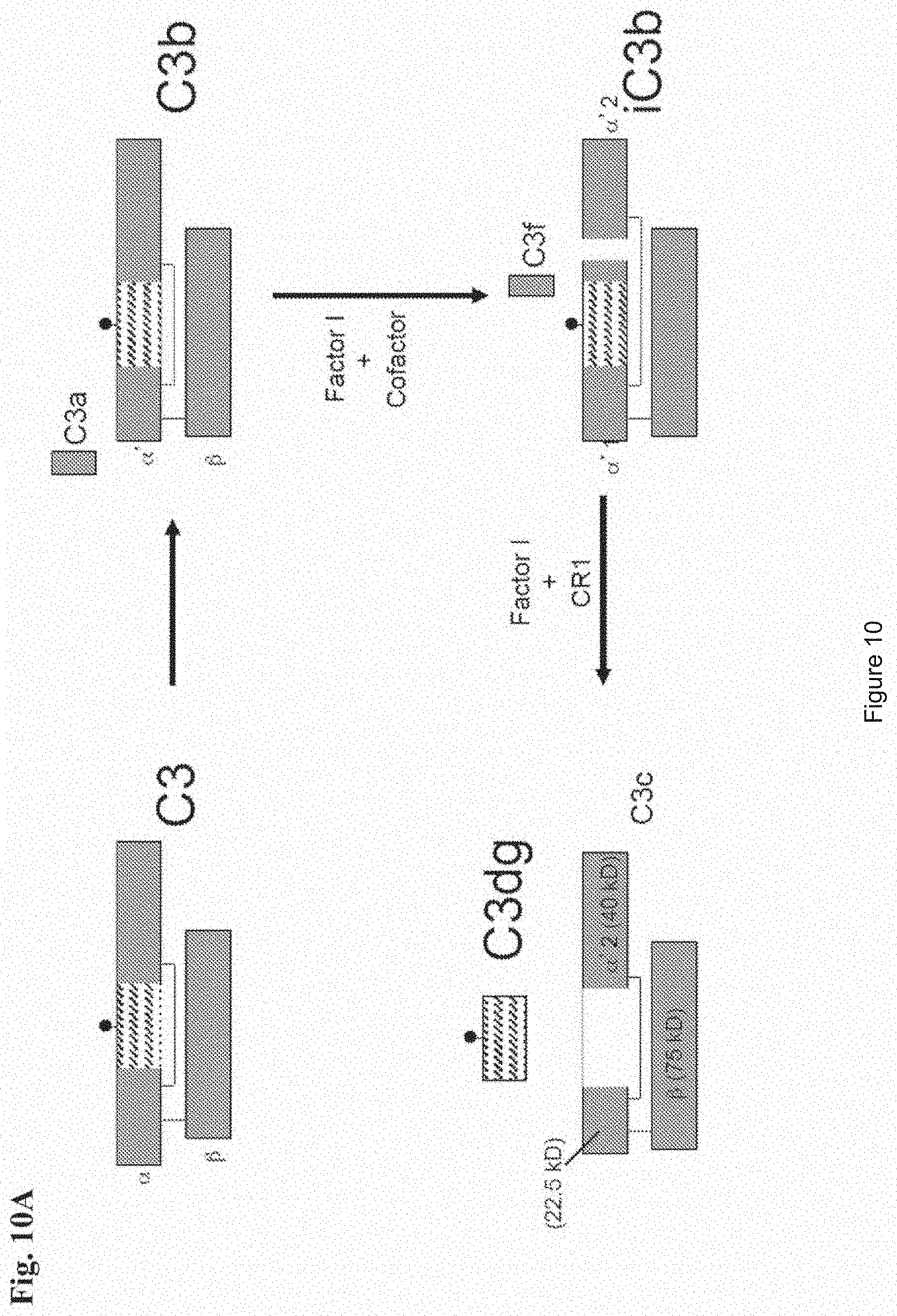
D00017
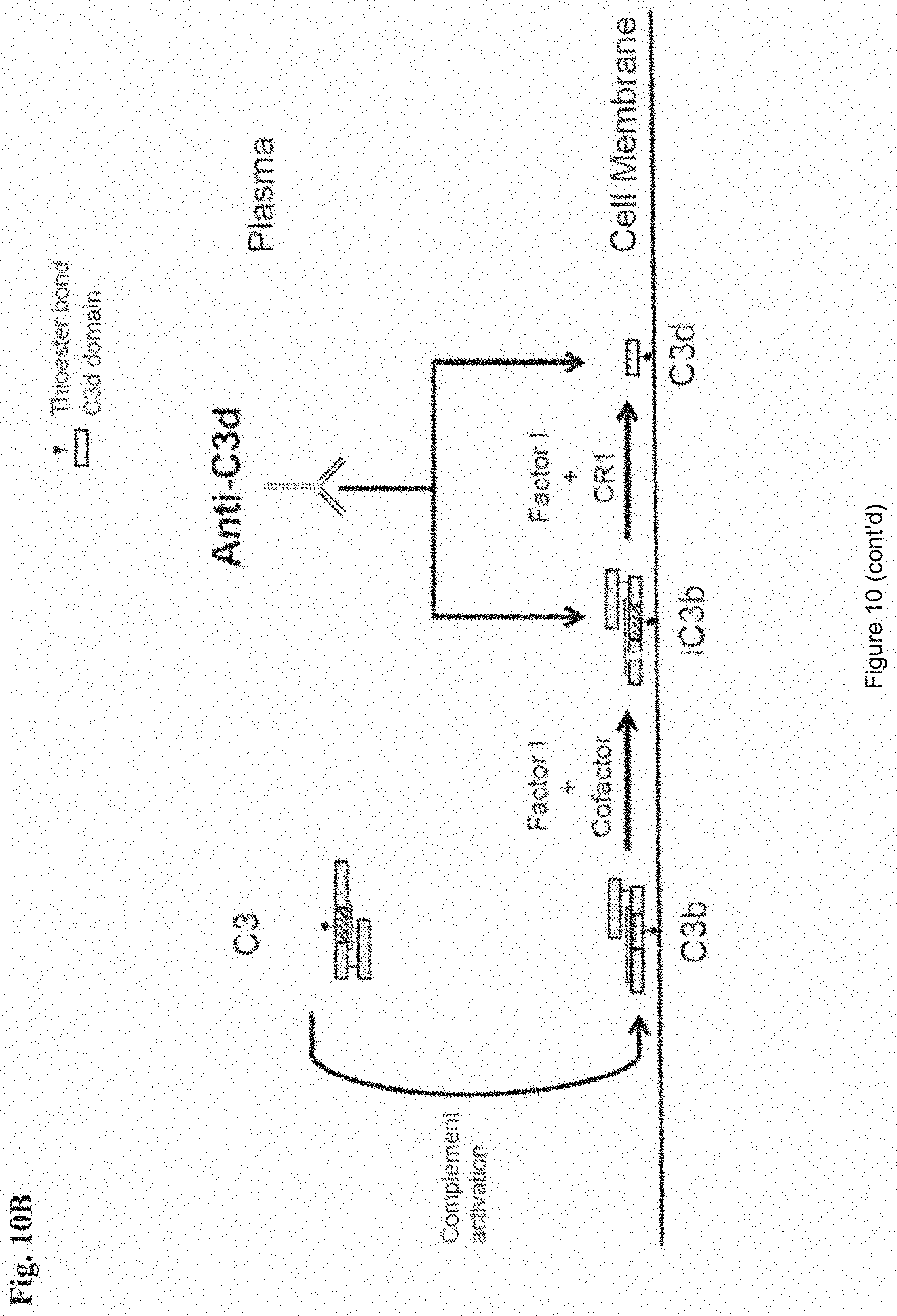
D00018
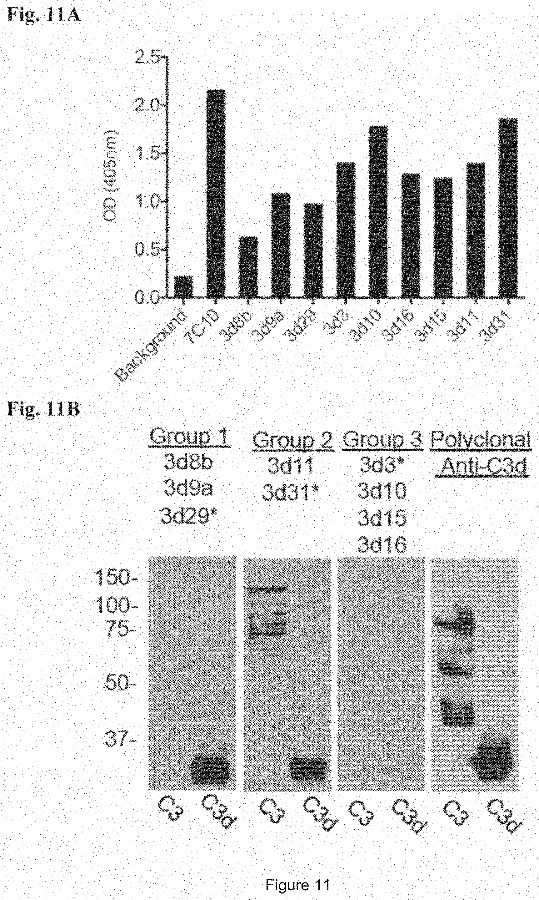
D00019

D00020

D00021

D00022

D00023
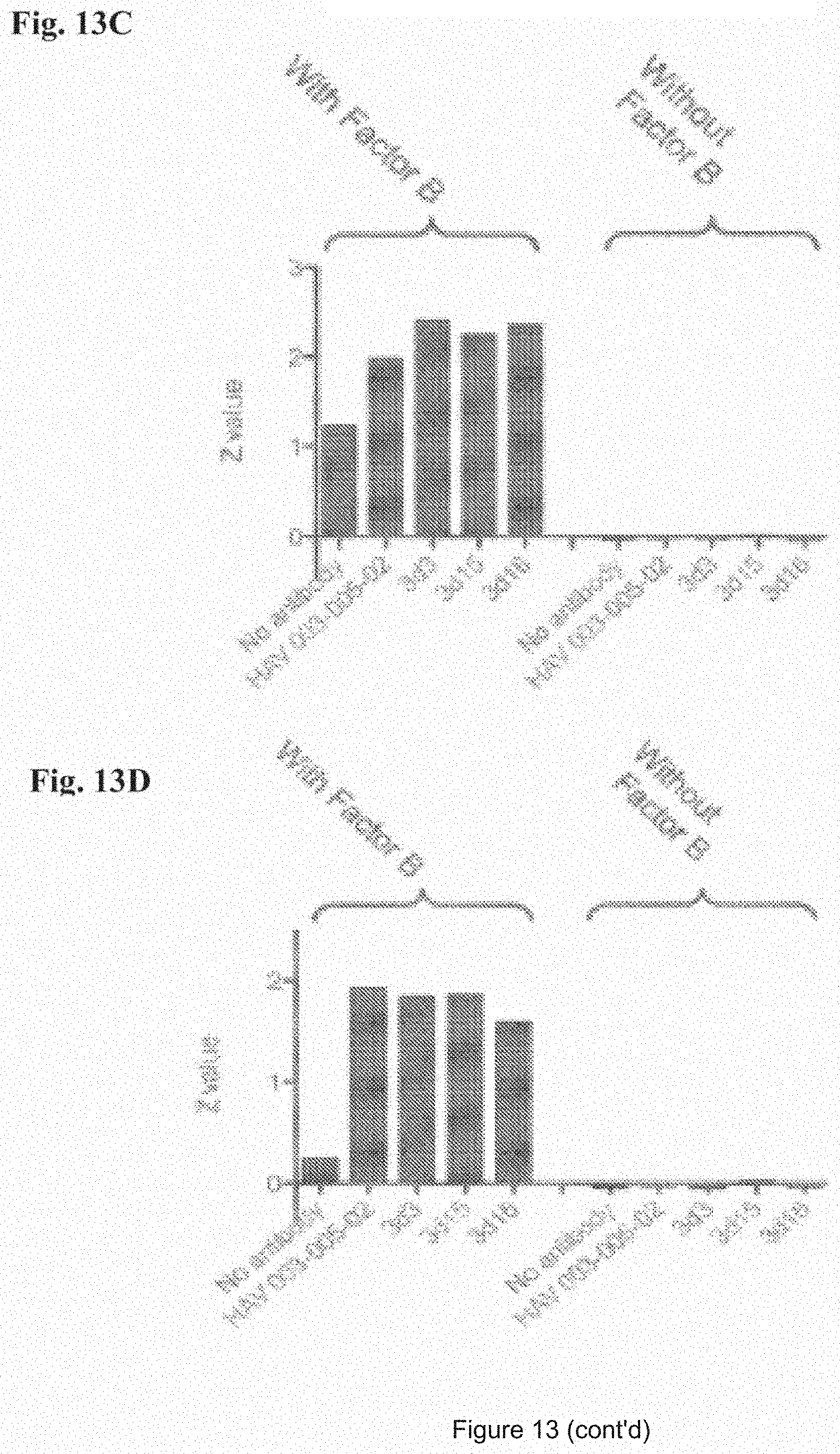
D00024

D00025
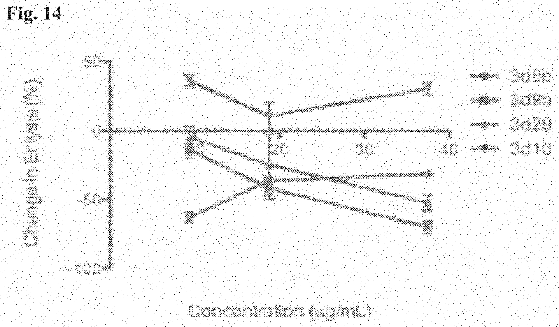
D00026

D00027

D00028

D00029

D00030
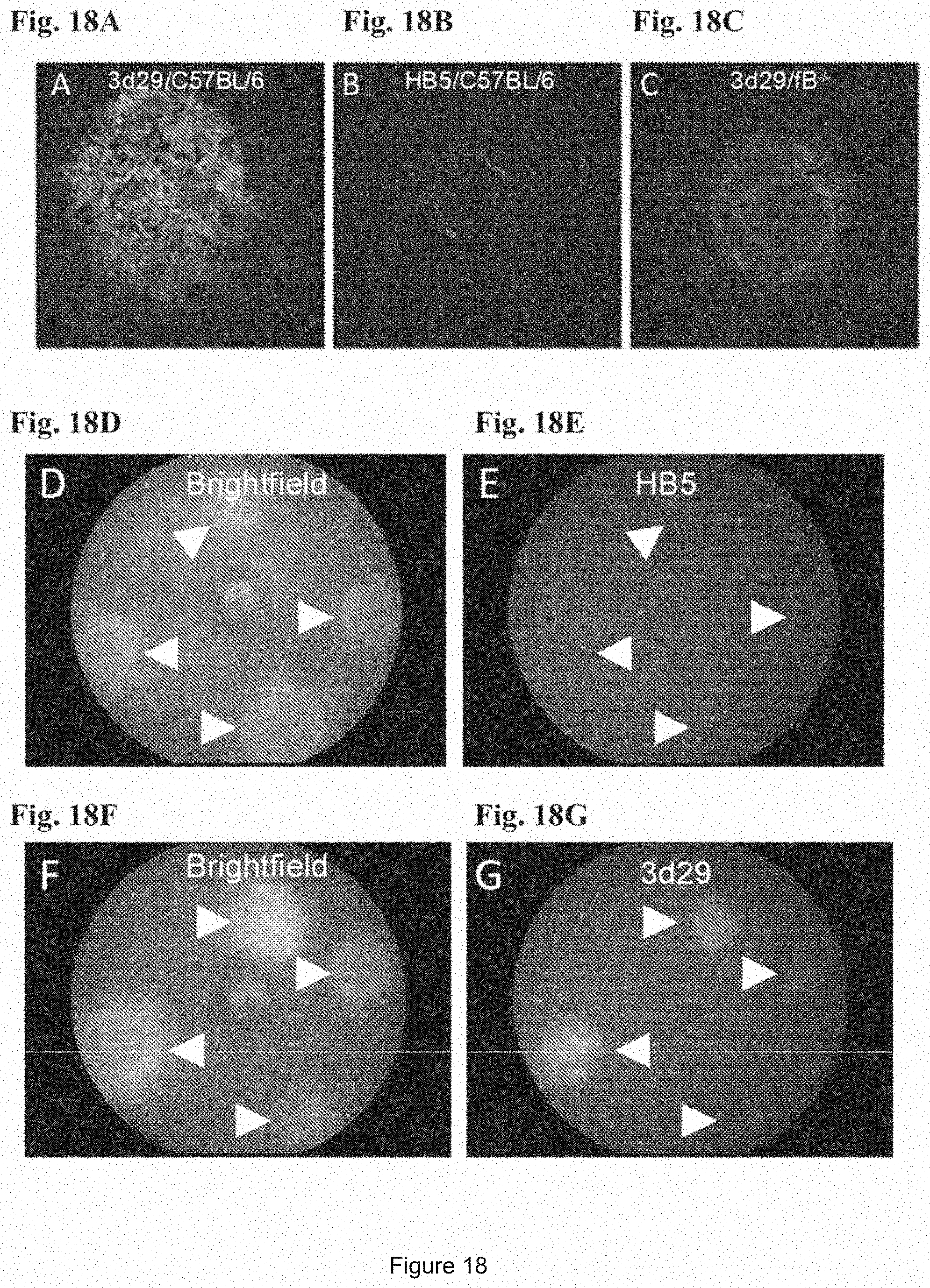
D00031

D00032

D00033

D00034

D00035

P00001

S00001
XML
uspto.report is an independent third-party trademark research tool that is not affiliated, endorsed, or sponsored by the United States Patent and Trademark Office (USPTO) or any other governmental organization. The information provided by uspto.report is based on publicly available data at the time of writing and is intended for informational purposes only.
While we strive to provide accurate and up-to-date information, we do not guarantee the accuracy, completeness, reliability, or suitability of the information displayed on this site. The use of this site is at your own risk. Any reliance you place on such information is therefore strictly at your own risk.
All official trademark data, including owner information, should be verified by visiting the official USPTO website at www.uspto.gov. This site is not intended to replace professional legal advice and should not be used as a substitute for consulting with a legal professional who is knowledgeable about trademark law.