Anti-tnf-alpha-antibodies And Functional Fragments Thereof
GUNDE; Tea ; et al.
U.S. patent application number 16/085543 was filed with the patent office on 2020-09-24 for anti-tnf-alpha-antibodies and functional fragments thereof. This patent application is currently assigned to Tillotts Pharma AG. The applicant listed for this patent is Tillotts Pharma AG. Invention is credited to Esther Maria FURRER, Tea GUNDE, Sebastian MEYER.
| Application Number | 20200299375 16/085543 |
| Document ID | / |
| Family ID | 1000004900868 |
| Filed Date | 2020-09-24 |


View All Diagrams
| United States Patent Application | 20200299375 |
| Kind Code | A1 |
| GUNDE; Tea ; et al. | September 24, 2020 |
ANTI-TNF-ALPHA-ANTIBODIES AND FUNCTIONAL FRAGMENTS THEREOF
Abstract
The present invention relates to antibody molecules and functional fragments thereof, capable of binding to tumor necrosis factor alpha (TNF.alpha.), to processes for their production, and to their therapeutic uses.
| Inventors: | GUNDE; Tea; (Zuerich, CH) ; MEYER; Sebastian; (Eggenwil, CH) ; FURRER; Esther Maria; (Rheinfelden, CH) | ||||||||||
| Applicant: |
|
||||||||||
|---|---|---|---|---|---|---|---|---|---|---|---|
| Assignee: | Tillotts Pharma AG Rheinfelden CH |
||||||||||
| Family ID: | 1000004900868 | ||||||||||
| Appl. No.: | 16/085543 | ||||||||||
| Filed: | March 16, 2017 | ||||||||||
| PCT Filed: | March 16, 2017 | ||||||||||
| PCT NO: | PCT/EP2017/056246 | ||||||||||
| 371 Date: | September 14, 2018 |
| Current U.S. Class: | 1/1 |
| Current CPC Class: | C07K 2317/24 20130101; C07K 16/241 20130101; C07K 2317/622 20130101; C07K 2317/92 20130101; C07K 2317/76 20130101; C07K 2317/33 20130101 |
| International Class: | C07K 16/24 20060101 C07K016/24 |
Foreign Application Data
| Date | Code | Application Number |
|---|---|---|
| Mar 17, 2016 | EP | 16160918.5 |
Claims
1. An antibody or a functional fragment thereof capable of binding to human tumor necrosis factor alpha (TNF.alpha.), wherein said antibody or functional fragment comprises: i. a V.sub.L domain comprising a CDR1 region having an amino acid sequence in accordance with the amino acid sequence as shown in SEQ ID NO:1, a CDR2 region having an amino acid sequence in accordance with the amino acid sequence as shown in SEQ ID NO:2, and a CDR3 region having an amino acid sequence in accordance with the amino acid sequence as shown in SEQ ID NO:3, and ii. a V.sub.H domain comprising a CDR1 region having an amino acid sequence in accordance with the amino acid sequence as shown in SEQ ID NO:4, a CDR2 region having an amino acid sequence in accordance with the amino acid sequence as shown in SEQ ID NO:5, and a CDR3 region having the amino acid sequence as shown in SEQ ID NO:6.
2. The antibody or functional fragment of claim 1, wherein said antibody or functional fragment comprises: i. a V.sub.L domain comprising a CDR1 region having the amino acid sequence as shown in SEQ ID NO:7, a CDR2 region having the amino acid sequence as shown in SEQ ID NO:8, and a CDR3 region having the amino acid sequence as shown in SEQ ID NO:9, and ii. (ii) a V.sub.H domain comprising a CDR1 region having the amino acid sequence as shown in SEQ ID NO:10, a CDR2 region having the amino acid sequence as shown in SEQ ID NO:11, and a CDR3 region having the amino acid sequence as shown in SEQ ID NO:6.
3. The antibody or functional fragment of claim 1, wherein said antibody or functional fragment (i) binds to human TNF.alpha. with a dissociation constant (K.sub.D) of less than 100 pM; (ii) is cross-reactive with Macaca mulatta (Rhesus) TNF.alpha. and with Macaca fascicularis (Cynomolgus) TNF.alpha.; (iii) has a greater potency to inhibit TNF.alpha.-induced apoptosis than infliximab, as determined by an L929 assay; (iv) comprises a variable domain having a melting temperature, determined by differential scanning fluorimetry, of at least 70.degree. C. and/or (v) is capable of binding to human TNF.alpha..sub.Trimer in a stoichiometry (antibody: TNF.alpha..sub.Trimer) of at least 2.
4. The antibody or functional fragment of claim 1, wherein said antibody or functional fragment binds to human TNF.alpha. with a K.sub.D of less than 70 pM.
5. The antibody or functional fragment of claim 1, wherein said antibody or functional fragment comprises a V.sub.H domain having the amino acid sequence as shown in SEQ ID NO:21.
6. The antibody or functional fragment of claim 1, wherein said antibody or functional fragment comprises a V.sub.L domain having an amino acid sequence selected from the group consisting of SEQ ID NO:22, SEQ ID NO:23 and SEQ ID NO:24.
7. The antibody or functional fragment of claim 1, wherein said antibody or functional fragment is a single-chain variable fragment (scFv).
8. The antibody or functional fragment of claim 7, wherein said scFv has an amino acid sequence selected from the group consisting of SEQ ID NO:25, SEQ ID NO:26 and SEQ ID NO:27.
9. The antibody or functional fragment of claim 1, wherein said antibody or functional fragment is an immunoglobulin G (IgG).
10. An antibody or functional fragment thereof, wherein said antibody of functional fragment binds to essentially the same epitope as the functional fragment of claim 8.
11. The antibody or functional fragment of claim 1, wherein the sum of (i) the number of amino acids in framework regions I, II, and III of the variable light domain of said antibody or functional fragment that are different from the respective human V.kappa.1 consensus sequences SEQ ID NOs: 31, 32, and 33, respectively, and (ii) the number of amino acids in framework region IV of the variable light domain of said antibody or functional fragment that are different from the most similar human .lamda. germline-based sequence selected from SEQ ID NOs: 34, 35, 36, and 37, is less than 7.
12. The antibody or functional fragment of claim 1, wherein the framework regions I, II, and III of the variable light domain of said antibody or functional fragment consist of human V.kappa.1 consensus sequences with SEQ ID NOs:31, 32 and 33, respectively, and framework region IV consists of a .lamda. germline-based sequence selected from among SEQ ID NOs:34, 35, 36, and 37.
13. A nucleic acid encoding the antibody or functional fragment of claim 1.
14. A vector or plasmid comprising the nucleic acid of claim 13.
15. A cell comprising the nucleic acid of claim 13 or a vector or plasmid comprising said nucleic acid.
16. A method of preparing the antibody or functional fragment of claim 1, comprising the steps of: culturing a cell comprising a nucleic acid encoding the antibody or functional fragment of claim 1 or a vector or plasmid comprising said nucleic acid in a medium under conditions that allow expression of the nucleic acid encoding the antibody or functional fragment, and recovering the antibody or functional fragment from the cells or from the medium.
17. A pharmaceutical composition comprising the antibody or functional fragment of claim 1 in combination with an optional pharmaceutically acceptable carrier and/or excipient.
18. A method of treating an inflammatory disorder or a TNF.alpha.-related disorder in a subject in need thereof, said method comprising the step of administering the pharmaceutical composition of claim 17 to said subject.
19. The method according to claim 18, wherein said inflammatory disorder is an inflammatory disorder of the gastrointestinal tract.
20. The method according to claim 19, wherein said inflammatory disorder of the gastrointestinal tract is inflammatory bowel disease.
21. The method according to claim 19, wherein said inflammatory disorder of the gastrointestinal tract is Crohn's disease or ulcerative colitis.
22. The antibody or functional fragment of claim 1, wherein the sum of (i) the number of amino acids in framework regions I, II, and III of the variable light domain of said antibody or functional fragment that are different from the respective human V.kappa.1 consensus sequences SEQ ID NOs: 31, 32, and 33, respectively, and (ii) the number of amino acids in framework region IV of the variable light domain of said antibody or functional fragment that are different from the most similar human .lamda. germline-based sequence selected from SEQ ID NOs: 34, 35, 36, and 37, is less than 4.
Description
FIELD OF THE INVENTION
[0001] The present invention relates to antibody molecules and functional fragments thereof, capable of binding to tumor necrosis factor alpha (TNF.alpha.), to processes for their production, and to their therapeutic uses.
BACKGROUND
[0002] TNF.alpha. is a homo-trimeric pro-inflammatory cytokine that is released by and interacts with cells of the immune system. TNF.alpha. has also been shown to be up-regulated in a number of human diseases, including chronic diseases such as rheumatoid arthritis, Crohn's disease, ulcerative colitis and multiple sclerosis.
[0003] Antibodies to TNF.alpha. have been proposed for the prophylaxis and treatment of endotoxic shock (Beutler et al., Science, 234, 470-474, 1985). Bodmer et al., (Critical Care Medicine, 21, S441-S446, 1993) and Wherry et al., (Critical Care Medicine, 21, S436-S440, 1993) discuss the therapeutic potential of anti-TNF.alpha. antibodies in the treatment of septic shock. The use of anti-TNF.alpha. antibodies in the treatment of septic shock is also discussed by Kirschenbaum et al., (Critical Care Medicine, 26, 1625-1626, 1998). Collagen-induced arthritis can be treated effectively using an anti-TNF.alpha. monoclonal antibody (Williams et al. (PNAS-USA, 89, 9784-9788, 1992)).
[0004] The use of anti-TNF.alpha. antibodies in the treatment of rheumatoid arthritis and Crohn's disease is discussed in Feldman et al. (Transplantation Proceedings, 30, 4126-4127, 1998), Adorini et al. (Trends in Immunology Today, 18, 209-211, 1997) and in Feldman et al. (Advances in Immunology, 64, 283-350, 1997). The antibodies to TNF.alpha. previously used in such treatments are generally chimeric antibodies, such as those described in U.S. Pat. No. 5,919,452.
[0005] Monoclonal antibodies against TNF.alpha. have been described in the prior art. Meager et al. (Hybridoma, 6, 305-311, 1987) describe murine monoclonal antibodies against recombinant TNF.alpha.. Fendly et al. (Hybridoma, 6, 359-370, 1987) describe the use of murine monoclonal antibodies against recombinant TNF.alpha. in defining neutralising epitopes on TNF.
[0006] Furthermore, in International Patent Application WO 92/11383, recombinant antibodies, including CDR-grafted antibodies, specific for TNF.alpha. are disclosed. Rankin et al. (British J. Rheumatology, 34, 334-342, 1995) describe the use of such CDR-grafted antibodies in the treatment of rheumatoid arthritis. U.S. Pat. No. 5,919,452 discloses anti-TNF.alpha. chimeric antibodies and their use in treating pathologies associated with the presence of TNF.alpha.. Further anti-TNF.alpha. antibodies are disclosed in Stephens et al. (Immunology, 85, 668-674, 1995), GB-A-2 246 570, GB-A-2 297 145, U.S. Pat. No. 8,673,310, US 2014/0193400, EP 2 390 267 B1, U.S. Pat. Nos. 8,293,235, 8,697,074, WO 2009/155723 A2 and WO 2006/131013 A2.
[0007] The prior art recombinant anti-TNF.alpha. antibody molecules generally have a reduced affinity for TNF.alpha. compared to the antibodies from which the hypervariable regions or CDRs are derived. All currently marketed inhibitors of TNF.alpha. are administered intravenously or subcutaneously in weekly or longer intervals as bolus injections, resulting in high starting concentrations that are steadily decreasing until the next injection.
[0008] Currently approved anti-TNF.alpha. biotherapeutics include (i) infliximab, a chimeric IgG anti-human monoclonal antibody (Remicade.RTM.; Wiekowski M et al: "Infliximab (Remicade)", Handbook of Therapeutic Antibodies, WILEY-VCH; Weinheim, 2007 Jan. 1, p. 885-904); (ii) etanercept, a TNFR2 dimeric fusion protein, with an IgG1 Fc (Enbrel.RTM.); (iii) adalimumab, a fully human monoclonal antibody (mAb) (Humira.RTM.; Kupper H et al: "Adalimumab (Humira)", Handbook of Therapeutic Antibodies, WILEY-VCH; Weinheim, 2007 Jan. 1, p. 697-732), (iv) certolizumab, a PEGylated Fab fragment (Cimzia.RTM.; Melmed G Y et al: "Certolizumab pegol", Nature Reviews. Drug Discovery, Nature Publishing Group, GB, Vol. 7, No. 8, 2008-08-01, p. 641-642); (v) Golimumab, a human IgGIK monoclonal antibody (Simponi.RTM.; Mazumdar S et al: "Golimumab", mAbs, Landes Bioscience, US, Vol. 1, No. 5, 2009 Sep. 1, p. 422-431). However, various biosimilars are in development, and a mimic of infliximab known as Remsima has already been approved in Europe.
[0009] Infliximab has a relatively low affinity to TNF.alpha. (K.sub.D>0.2 nM; Weir et al., 2006, Therapy 3: 535) and a limited neutralization potency in an L929 assay. In addition, infliximab shows substantially no cross-reactivity with TNF.alpha. from Cynomolgus or Rhesus monkeys. For anti-TNF.alpha. antibodies, however, cross-reactivity with TNF.alpha. from monkeys is highly desirable, as this allows for animal tests with primates, reflecting the situation in humans in many aspects.
[0010] Etanercept, although a bivalent molecule, binds TNF.alpha. at a ratio of one trimer per one etanercept molecule, precluding the formation of large antigen-biotherapeutics complexes (Wallis, 2008, Lancet Infect Dis 8: 601). It does not inhibit LPS-induced cytokine secretion in monocytes (Kirchner et al., 2004, Cytokine 28: 67).
[0011] The potency of adalimumab is similar to that of infliximab. Another disadvantage of adalimumab is its poor stability, e.g. as determined in a thermal unfolding test. The melting temperature (T.sub.m) of adalimumab in such a test was determined to be 67.5.degree. C. The lower the T.sub.m value of an antibody, however, the lower is its general stability. A lower T.sub.m makes antibodies less suitable for pharmaceutical use, e.g. for oral administration.
[0012] The potency of certolizumab is slightly greater than that of infliximab, but still not satisfying. Certolizumab does not inhibit T-cell proliferation in a MLR (Vos et al., 2011, Gastroenterology 140: 221).
[0013] EP2623515 A1 discloses humanized anti-TNF.alpha. antibodies and antigen-binding fragments (Fab) thereof. As becomes clear from the disclosed examples, the potency of the resulting humanized Fab fragments is comparable to that of infliximab in a L929 neutralization assay (see Table 2 and 5). The sole anti-TNF.alpha. IgG antibody tested for cross-reactivity binds only weakly to Rhesus TNF-.alpha. (see [0069]; FIG. 3). Cross-reactivity with Cynomolgus TNF.alpha. was not tested. Moreover, there is weak binding to human TNF.beta. (see FIG. 3). Therefore, EP2623515 A1 does not disclose anti-TNF.alpha. antibodies or functional fragments thereof, which have a potency to inhibit TNF.alpha.-induced apoptosis in L929 cells greater than that of infliximab and which are cross-reactive with Rhesus TNF.alpha. and Cynomolgus TNF.alpha..
[0014] WO 2012/007880 A2 discloses a modified single domain antigen binding molecule (SDAB) in the form of fusion proteins comprising one or more single antigen binding domains that bind to one or more targets (e.g. TNF.alpha.), a linker and one or more polymer molecules. The only specific example given is termed SDAB-01 and includes two antigen binding domains, which bind to TNF.alpha., connected with a flexible linker, and a C-terminal Cysteine supporting the site specific PEGylation (see FIG. 3). WO 2012/007880 A2 fails to compare the potency of SDAB-01 to known TNF.alpha. antibodies like infliximab in a L929 cell-based neutralization assay, or to assess other SDAB-01-specific parameters like the effectiveness to block TNF.alpha.-TNFRI/II interaction and the selectivity for binding TNF.alpha. over TNF.beta.. In an assay where the treatment with SDAB-01 and infliximab are compared in a transgenic mouse model for polyarthritis that overexpresses human TNF.alpha. (see page 54, Example 8), the two seem to be similarly effective in preventing further development of arthritis (e.g. FIGS. 17&18). However, the dosage given in this example is misleading as the molecular weight of SDAB-01 is less than half of that of infliximab. Thus, WO 2012/007880 A2 does not disclose anti-TNF.alpha. antibodies having a potency to inhibit TNF.alpha.-induced apoptosis in L929 cells greater than that of infliximab.
[0015] WO 2015/144852 A1 investigates the properties of an anti-TNF-.alpha. scFv designated "scFv1". This scFv showed a TNF.alpha. neutralization capacity in a PK-15 cell assay that was comparable to that of infliximab (see [0236]). In addition, the scFv seems to have some cross-reactivity to TNF-.alpha. from rhesus macaque and cynomolgus monkey (see Ex. 8). No affinity data are reported in WO 2015/144852 A1. The single chain antibody fragment DLX105 (also known as ESBA 105), however, which is known to have only moderate affinity (K.sub.D=157 pM; see Urech et al. 2010 Ann Rheum Dis 69: 443) shows a better binding to TNF.alpha. than scFv1 (see FIG. 1 of WO 2015/144852 A1). Therefore, WO 2015/144852 A1 does not disclose anti-TNF-.alpha. antibodies having high affinity for human TNF.alpha. (K.sub.D<100 pM).
[0016] WO 2015/065987 A1 describes anti-TNF-.alpha. antibodies, anti-IL-6 antibodies, and bispecific antibodies binding to both antigens. Certain anti-TNF.alpha. antibodies showed some cross-reactivity with TNF.alpha. from Cynomolgus (FIG. 17). The anti-TNF.alpha. antibodies, however, exhibited a significantly lower potency than infliximab in an L929 neutralization assay ([0152]; FIG. 5). Therefore, WO 2015/065987 A1 does not disclose anti-TNF-.alpha. antibodies having a potency to inhibit TNF.alpha.-induced apoptosis in L929 cells greater than that of infliximab.
[0017] Drugs in R&D, Vol. 4 No. 3, 2003, pages 174-178 describes the humanized antibody "Humicade" (CDP 571; BAY 103356), a monoclonal anti-TNF.alpha. antibody with high affinity. The potency of Humicade to inhibit TNF.alpha.-induced apoptosis in L929 cells, however, appears to be limited (see, e.g., US 2003/0199679 A1 at [0189]). The reference therefore does not disclose anti-TNF-.alpha. antibodies having a potency to inhibit TNF.alpha.-induced apoptosis in L929 cells greater than that of infliximab.
[0018] Saldanha J W et al: "Molecular Engineering I: Humanization", Handbook of Therapeutic Antibodies, Chapter 6, 2007 Jan. 1, WILEY-VCH, Weinheim, p. 119-144 discloses different strategies for humanization of monoclonal antibodies including CDR Grafting, Resurfacing/Veneering, SDR transfer and DeImmunization Technology.
[0019] There is a need for improved antibody molecules to treat chronic inflammatory diseases such as inflammatory bowel disorders. The antibody molecules should at least have (i) high affinity for human TNF.alpha. (i.e. a K.sub.D<100 pM), (ii) a high potency to inhibit TNF.alpha.-induced apoptosis in L929 cells, (iii) substantial affinity to TNF.alpha. from Cynomolgus and Rhesus (e.g. a K.sub.D<1 nM), and (iv) a high melting temperature of the variable domain as determined in a thermal unfolding experiment (e.g. a T.sub.m>70.degree. C.).
SUMMARY OF THE INVENTION
[0020] The inventors of the present application found that certain anti-TNF.alpha. antibodies and functional fragments thereof exhibit a combination of favorable properties, including high affinity for human TNF.alpha. (K.sub.D<100 pM), a potency to inhibit TNF.alpha.-induced apoptosis in L929 cells greater than that of infliximab, and substantial affinity (K.sub.D<1 nM) to TNF.alpha. from animals such as Cynomolgus monkey (Macaca fascicularis) and/or Rhesus macaques (Macaca mulatta). In addition, the antibodies and functional fragments thereof were specific for TNF.alpha. in that they did not significantly bind to TNF.beta., and exhibit a significant stability, as determined in a thermal unfolding assay of the variable domain.
[0021] The invention provides antibody molecules and functional fragments thereof.
[0022] The present invention therefore relates to the subject matter defined in the following items (1) to (47): [0023] (1) An antibody or a functional fragment thereof capable of binding to human tumor necrosis factor alpha (TNF.alpha.), wherein said antibody or functional fragment thereof comprises (i) a V.sub.L domain comprising a CDR1 region having an amino acid sequence in accordance with the amino acid sequence as shown in SEQ ID NO:1, a CDR2 region having an amino acid sequence in accordance with the amino acid sequence as shown in SEQ ID NO:2, and a CDR3 region having an amino acid sequence in accordance with the amino acid sequence as shown in SEQ ID NO:3, and (ii) a V.sub.H domain comprising a CDR1 region having an amino acid sequence in accordance with the amino acid sequence as shown in SEQ ID NO:4, a CDR2 region having an amino acid sequence in accordance with the amino acid sequence as shown in SEQ ID NO:5, and a CDR3 region having the amino acid sequence as shown in SEQ ID NO:6. [0024] (2) The antibody or functional fragment of item (1), wherein said antibody or functional fragment comprises (i) a V.sub.L domain comprising a CDR1 region having the amino acid sequence as shown in SEQ ID NO:7, a CDR2 region having the amino acid sequence as shown in SEQ ID NO:8, and a CDR3 region having the amino acid sequence as shown in SEQ ID NO:9, and (ii) a V.sub.H domain comprising a CDR1 region having the amino acid sequence as shown in SEQ ID NO:10, a CDR2 region having the amino acid sequence as shown in SEQ ID NO:11, and a CDR3 region having the amino acid sequence as shown in SEQ ID NO:6. [0025] (3) The antibody or functional fragment of any one of the preceding items, wherein said antibody or functional fragment comprises a V.sub.H domain having the amino acid sequence as shown in SEQ ID NO:21. [0026] (4) The antibody or functional fragment of any one of the preceding items, wherein said antibody or functional fragment comprises a V.sub.L domain having an amino acid sequence selected from the group consisting of SEQ ID NO:22, SEQ ID NO:23 and SEQ ID NO:24; preferably selected from the group consisting of SEQ ID NO:22 and SEQ ID NO:23. [0027] (5) The antibody or functional fragment of any one of the preceding items, wherein said antibody or functional fragment thereof specifically binds to human TNF.alpha.. [0028] (6) The antibody or functional fragment of any one of any one of the preceding items, wherein said antibody or functional fragment thereof does not significantly bind to TNF.beta.. [0029] (7) The antibody or functional fragment of any one of the preceding items, wherein said antibody or functional fragment [0030] (i) binds to human TNF.alpha. with a dissociation constant (K.sub.D) of less than 100 pM; [0031] (ii) is cross-reactive with Macaca mulatta TNF.alpha. and with Macaca fascicularis TNF.alpha.; [0032] (iii) has a greater potency than infliximab, as determined by an L929 assay; and/or [0033] (iv) is capable of binding to human TNF.alpha..sub.Trimer in a stoichiometry (antibody: TNF.alpha..sub.Trimer) of at least 2. [0034] (8) The antibody or functional fragment of any one of the preceding items, which binds to human TNF.alpha. with a K.sub.D of less than 75 pM. [0035] (9) The antibody or functional fragment of any one of the preceding items, which binds to TNF.alpha. from Macaca mulatta with a K.sub.D of less than 1 nM. [0036] (10) The antibody or functional fragment of any one of the preceding items, which binds to TNF.alpha. from Macaca fascicularis with a K.sub.D of less than 1 nM. [0037] (11) The antibody or functional fragment of any one of the preceding items, wherein the potency of the antibody or functional fragment to inhibit TNF.alpha.-induced apoptosis relative to that of infliximab (relative potency), determined in an L929 assay, is greater than 5, and wherein said relative potency is the ratio of the IC.sub.50 value in ng/mL of infliximab in the L929 assay to the IC.sub.50 value in ng/mL of the antibody in scFv format in the L929 assay. [0038] (12) The antibody or functional fragment of any one of the preceding items, wherein the melting temperature of the variable domain of the antibody in scFv format, determined by differential scanning fluorimetry, is at least 65.degree. C. [0039] (13) The antibody or functional fragment of any one of the preceding items, wherein the melting temperature of the variable domain of the antibody in scFv format, determined by differential scanning fluorimetry, is at least 70.degree. C. [0040] (14) The antibody or functional fragment of any one of the preceding items, wherein the melting temperature, determined by differential scanning fluorimetry, is at least 75.degree. C. [0041] (15) The antibody or functional fragment of any one of the preceding items, wherein the loss in monomer content, after five consecutive freeze-thaw cycles, is less than 0.2%. [0042] (16) The antibody or functional fragment of any one of the preceding items, wherein the loss in monomer content, after storage for four weeks at 4.degree. C., is less than 1%. [0043] (17) The antibody or functional fragment of any one of the preceding items, wherein the potency of the antibody or functional fragment to block the interaction between human TNF.alpha. and TNF receptor I (TNFRI), relative to that of infliximab (relative potency), as determined in an inhibition ELISA, is at least 1.3, wherein said relative potency is the ratio of the IC.sub.50 value in ng/mL of infliximab to the IC.sub.50 value in ng/mL of the antibody in scFv format. [0044] (18) The antibody or functional fragment of any one of the preceding items, wherein the potency of the antibody or functional fragment to block the interaction between human TNF.alpha. and TNF receptor II (TNFRII), relative to that of infliximab (relative potency), as determined in an inhibition ELISA, is at least 1.3, wherein said relative potency is the ratio of the IC.sub.50 value in ng/mL of infliximab to the IC.sub.50 value in ng/mL of the antibody in scFv format. [0045] (19) The antibody or functional fragment of any one of the preceding items, which is capable of inhibiting cell proliferation of peripheral blood mononuclear cells in a mixed lymphocyte reaction. [0046] (20) The antibody or functional fragment of any one of the preceding items, which is capable of inhibiting LPS-induced secretion of interleukin-1.beta. from CD14.sup.+ monocytes. [0047] (21) The antibody or functional fragment of item (20), wherein the IC.sub.50 value for inhibiting LPS-induced secretion of interleukin-1.beta. is less than 1 nM. [0048] (22) The antibody or functional fragment of item (21), wherein said IC.sub.50 value for inhibiting LPS-induced secretion of interleukin-1.beta., on a molar basis, is lower than that of adilumumab. [0049] (23) The antibody or functional fragment of any one of the preceding items, which is capable of inhibiting LPS-induced secretion of TNF.alpha. from CD14.sup.+ monocytes. [0050] (24) The antibody or functional fragment of item (23), wherein the IC.sub.50 value for inhibiting LPS-induced secretion of TNF.alpha. is less than 1 nM. [0051] (25) The antibody or functional fragment of item (24), wherein said IC.sub.50 value for inhibiting LPS-induced secretion of TNF.alpha., on a molar basis, is lower than that of adalimumab. [0052] (26) The antibody of any one of the preceding items, which is an immunoglobulin G (IgG). [0053] (27) The functional fragment of any one of items (1) to (25), which is a single-chain variable fragment (scFv). [0054] (28) The functional fragment of item (27), wherein said scFv comprises or consists of an amino acid sequence selected from the group consisting of SEQ ID NO:25, SEQ ID NO:26 and SEQ ID NO:27, preferably selected from the group consisting of SEQ ID NO:25 and SEQ ID NO:26. [0055] (29) The functional fragment of any one of items (1) to (25), which is a diabody. [0056] (30) An antibody or functional fragment thereof binding to essentially the same epitope on human TNF.alpha. as an antibody comprising a V.sub.H domain having the amino acid sequence as shown in SEQ ID NO:21 and a V.sub.L domain having the amino acid sequence selected from the group consisting of SEQ ID NO:22, SEQ ID NO:23 and SEQ ID NO:24, preferably the functional fragment comprising the V.sub.L domain consisting of an amino acid sequence selected from the group consisting of SEQ ID NO:22 and SEQ ID NO:23, in particular wherein said antibody or functional fragment exhibits one or more of the features referred to in items 1 to 29 herein above. [0057] (31) The antibody or functional fragment of any one of the preceding items, wherein the sum of (i) the number of amino acids in framework regions I to III of the variable light domain of said antibody or functional fragment that are different from the respective human VK1 consensus sequences with SEQ ID NOs: 31 to 33 (see Table 10), and (ii) the number of amino acids in framework region IV of the variable light domain of said antibody or functional fragment that are different from the most similar human A germline-based sequence selected from SEQ ID NOs: 34 to 37 (see Table 11), is less than 7, preferably less than 4. [0058] (32) The antibody or functional fragment of any one of the preceding items, wherein the framework regions I to III of the variable light domain of said antibody or functional fragment consist of human VK1 consensus sequences with SEQ ID NOs:31 to 33, respectively, and framework region IV consists of a .lamda. germline-based sequence selected from SEQ ID NOs:34 to 37. [0059] (33) A nucleic acid encoding the antibody or functional fragment of any one of the preceding items. [0060] (34) A vector or plasmid comprising the nucleic acid of item (33). [0061] (35) A cell comprising the nucleic acid of item (34) or the vector or plasmid of item (33). [0062] (36) A method of preparing the antibody or functional fragment of any one of items (1) to (32), comprising culturing the cell of item (35) in a medium under conditions that allow expression of the nucleic acid encoding the antibody or functional fragment, and recovering the antibody or functional fragment from the cells or from the medium. [0063] (37) A pharmaceutical composition comprising the antibody or functional fragment of any one of items (1) to (32), and optionally a pharmaceutically acceptable carrier and/or excipient. [0064] (38) The antibody or functional fragment as defined in any one of items (1) to (32) for use in a method of treating an inflammatory disorder or a TNF.alpha.-related disorder. [0065] (39) The antibody or functional fragment for use according to item (38), wherein said inflammatory disorder or TNF.alpha.-related disorder is selected from the list of disorders and diseases listed in Section "Disorders to be treated" below. [0066] (40) The antibody or functional fragment for use according to item (38), wherein said inflammatory disorder is an inflammatory disorder of the gastrointestinal tract. [0067] (41) The antibody or functional fragment for use according to item (40), wherein said inflammatory disorder of the gastrointestinal tract is inflammatory bowel disease. [0068] (42) The antibody or functional fragment for use according to item (40) or (41), wherein said inflammatory disorder of the gastrointestinal tract is Crohn's disease. [0069] (43) The antibody or functional fragment for use according to item (42), wherein said Crohn's disease is selected from the group consisting of ileal, colonic, ileocolonic, and/or isolated upper Crohn's disease (gastric, duodenal and/or jejunal) and including non-stricturing/non-penetrating, stricturing, penetrating and perianal disease behavior, allowing any combination of localization and disease behavior of any of the above mentioned. [0070] (44) The antibody or functional fragment for use according to item (40) or (41), wherein said inflammatory disorder of the gastrointestinal tract is ulcerative colitis. [0071] (45) The antibody or functional fragment for use according to item (44), wherein said ulcerative colitis is selected from the group consisting of ulcerative proctitis, proctosigmoiditis, left-sided colitis, pan-ulcerative colitis, and pouchitis. [0072] (46) The antibody or functional fragment for use according to item (40) or (41), wherein said inflammatory disorder of the gastrointestinal tract is microscopic colitis. [0073] (47) The antibody or functional fragment for use according to any one of items (38) to (46), wherein said method comprises orally administering the antibody or functional fragment to a subject.
BRIEF DESCRIPTION OF THE DRAWINGS
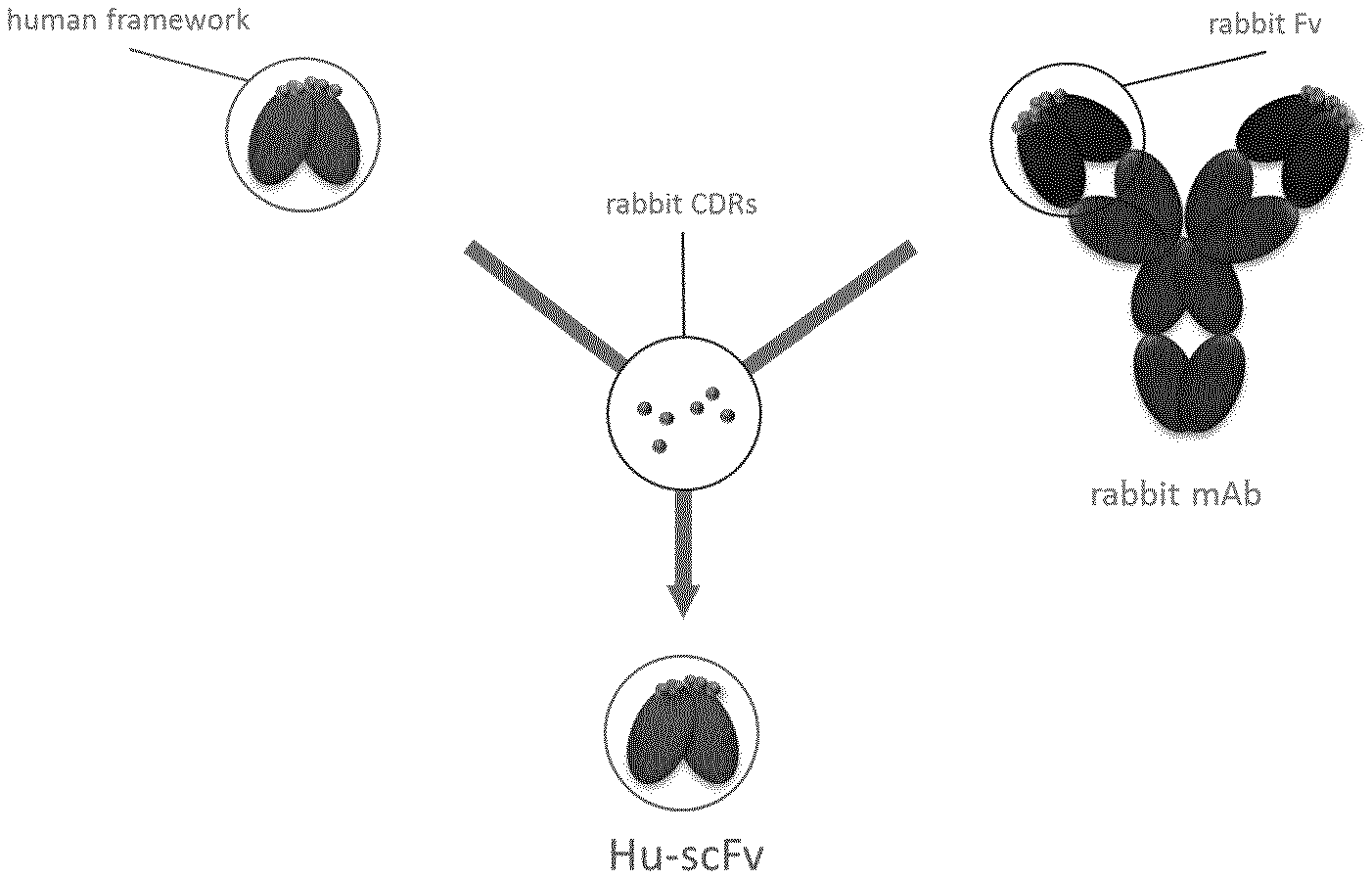
[0074] FIG. 1: Schematic representation of the humanization process.
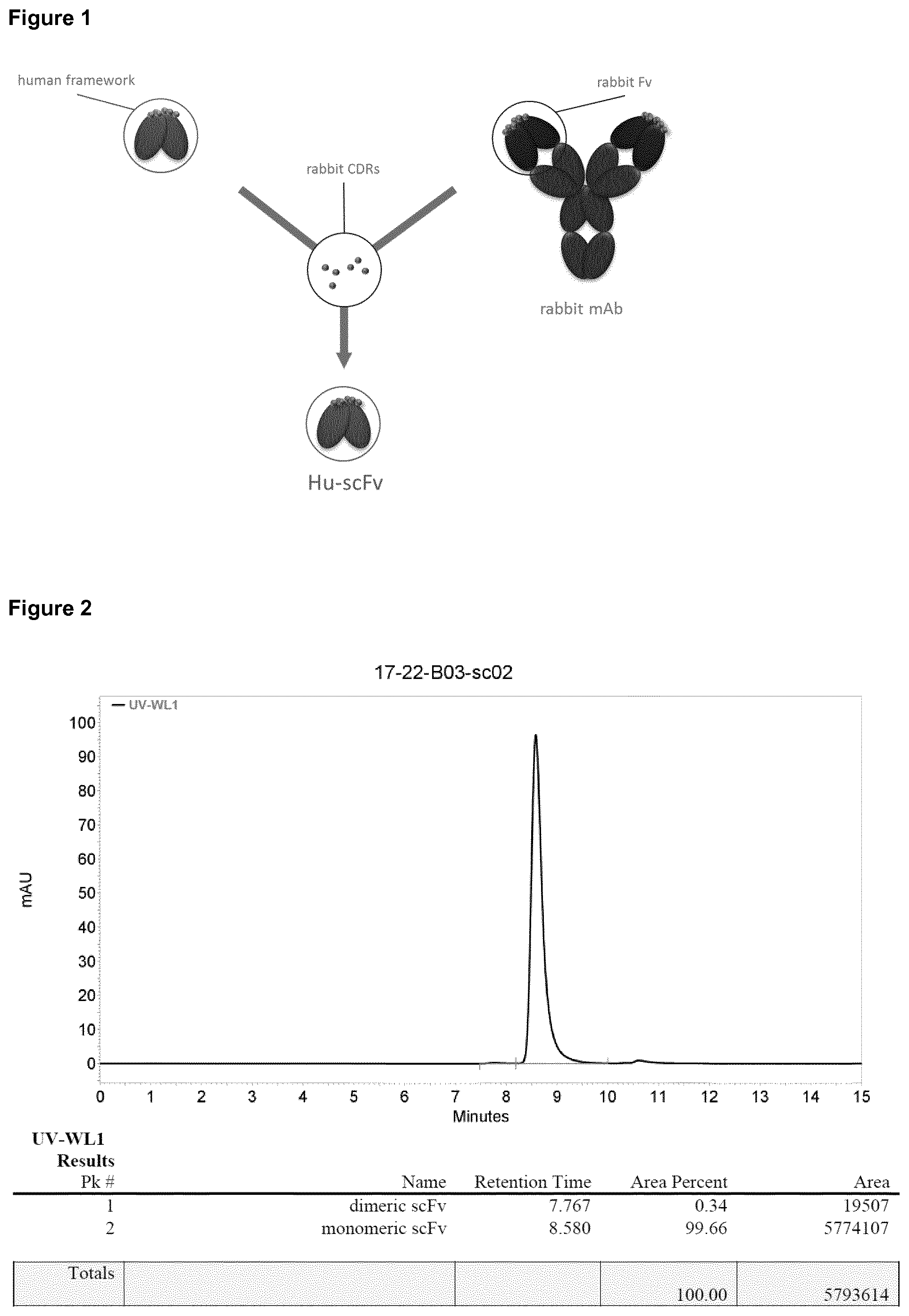
[0075] FIG. 2: SE-HPLC chromatograms of purified humanized scFv preparations of scFv. The scFv monomer elutes at retention times between 8.4 and 9.5 minutes, while buffer components elute at >10 min. All peaks from the dead volume of the column up to the respective scFv monomer were integrated as aggregates/oligomers and used for the calculation of the relative peak area.
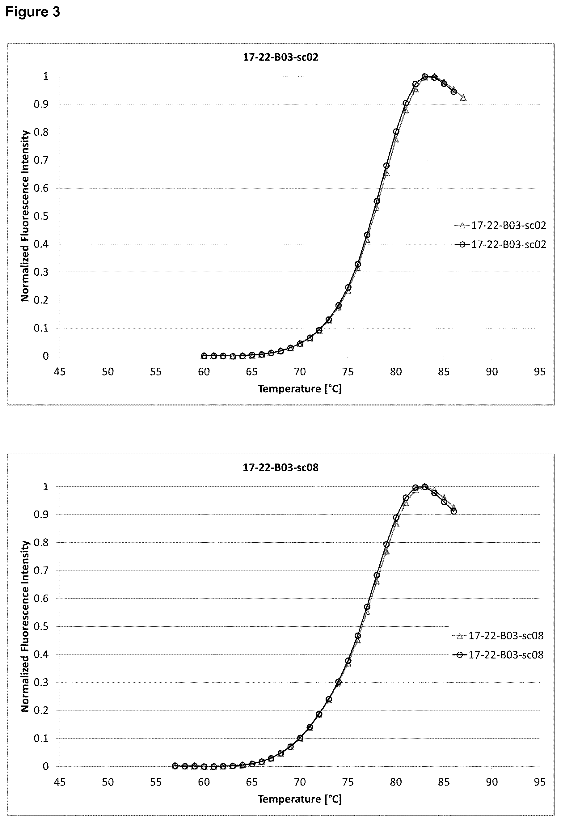
[0076] FIG. 3: Thermal unfolding curves from DSF measurements of three scFv constructs. For each construct duplicate measurements are shown. The resulting Tm values have been determined by fitting the data to a Boltzmann equation to obtain the midpoint of transition.
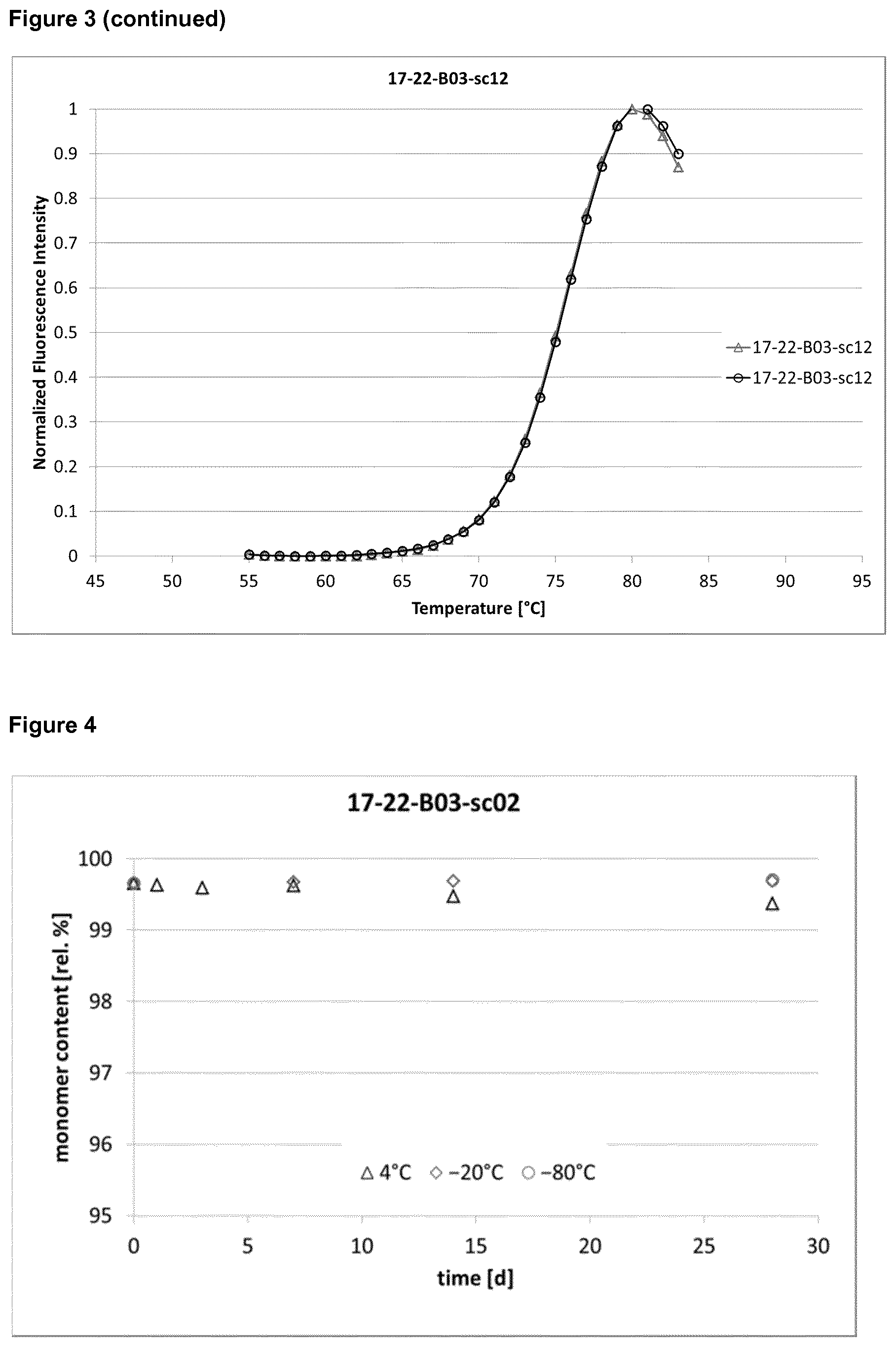
[0077] FIG. 4: Time-course of the monomer content of the three scFv constructs during storage. The monomer content as determined by SE-HPLC has been plotted for the storage temperatures 4, -20 and <-65.degree. C. for the duration of 4 weeks.
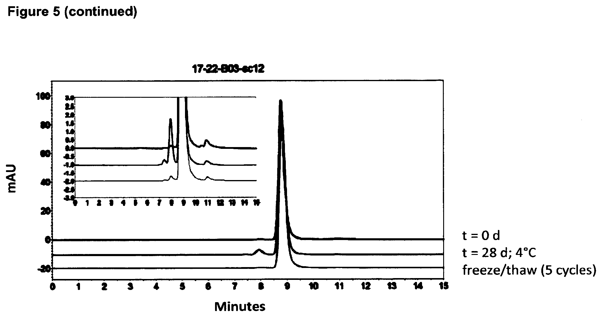
[0078] FIG. 5: Overlay of SE-HPLC chromatograms for three scFv molecules. For each scFv, the sample (10 mg/mL) at d0 and after storage for 4 weeks at 4.degree. C. is shown. In addition, the chromatogram of the sample after 5 cycles of freezing and thawing is shown. The inserted panel shows an approx. 15-fold zoom of the y-axis for each molecule to visualize also minuscule changes in oligomer content.
[0079] FIG. 6: Time-course of the monomer content of the humanized scFvs during storage. The monomer content as determined by SE-HPLC has been plotted for the 10 mg/mL samples at a storage temperature of 37.degree. C. for the duration of 4 weeks.
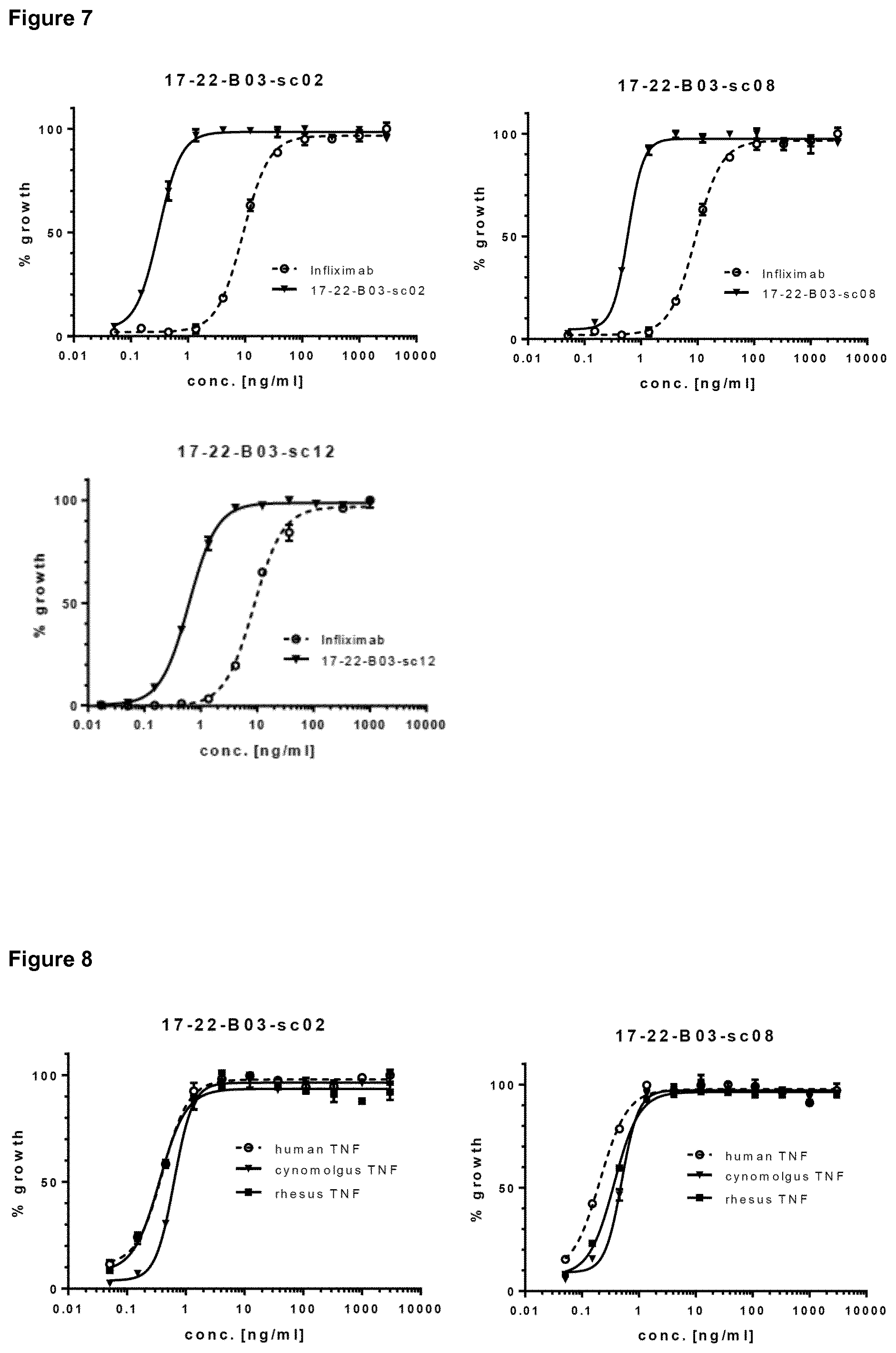
[0080] FIG. 7: Potency to neutralize human TNF.alpha. in the L929 assay of three scFvs. Dose-response curves for the scFvs and the reference antibody Infliximab are shown for each experiment. The highest scFv and infliximab concentrations as well as negative controls were set to 100% and 0% of growth.
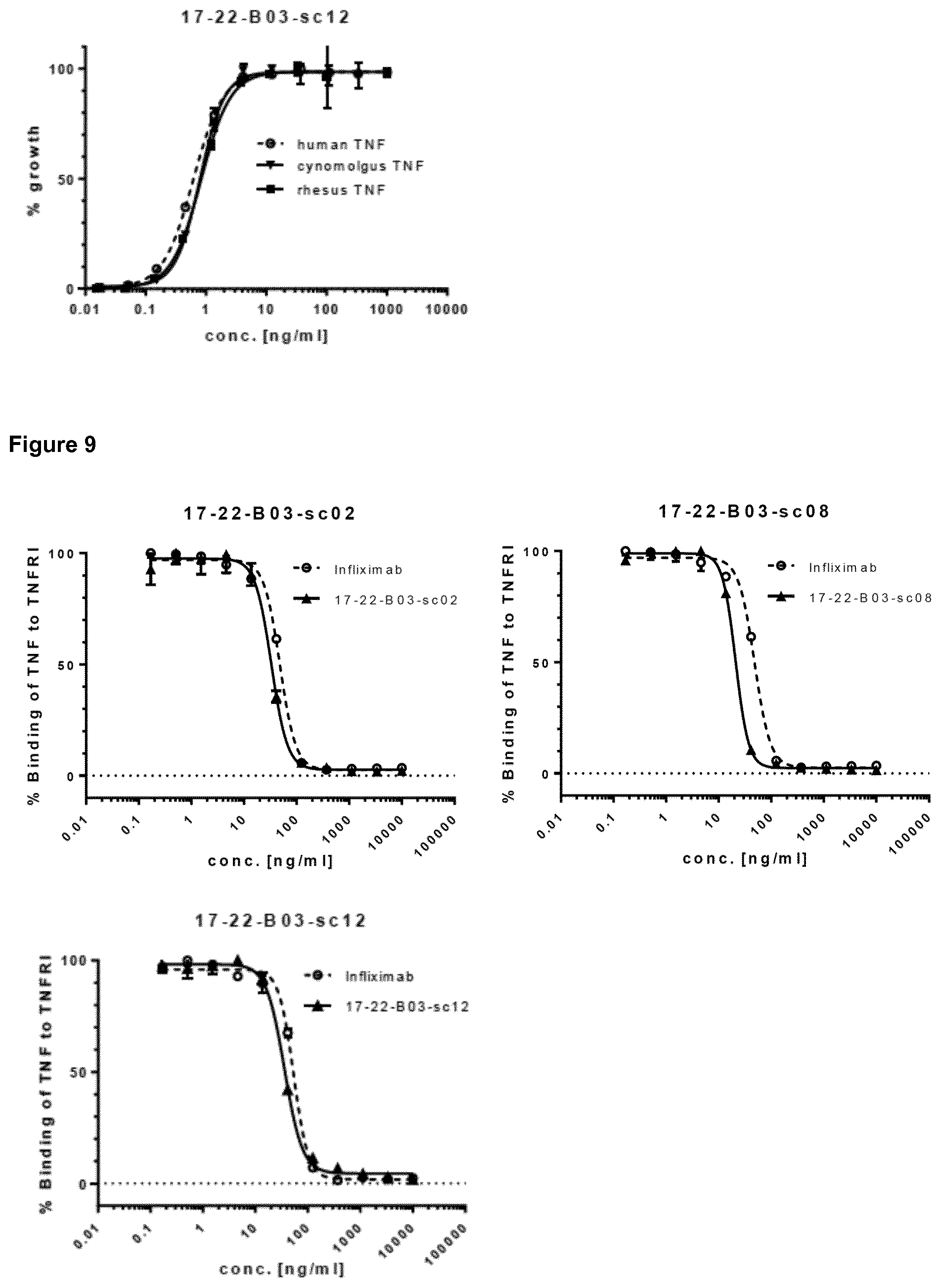
[0081] FIG. 8: Potency of two scFvs to neutralize non-human primate and human TNF.alpha. in the L929 assay. Dose-response curves for neutralization of human, Cynomolgus monkey and Rhesus monkey TNF.alpha. are shown. The highest scFv concentration and negative controls were set to 100% and 0% of growth.
[0082] FIG. 9: Potency of two scFvs to block the TNF.alpha.-TNFRI interaction. Dose-response curves are shown. The highest scFv concentration and negative controls were set to 0% and 100% of binding of TNF.alpha. to TNFRI.
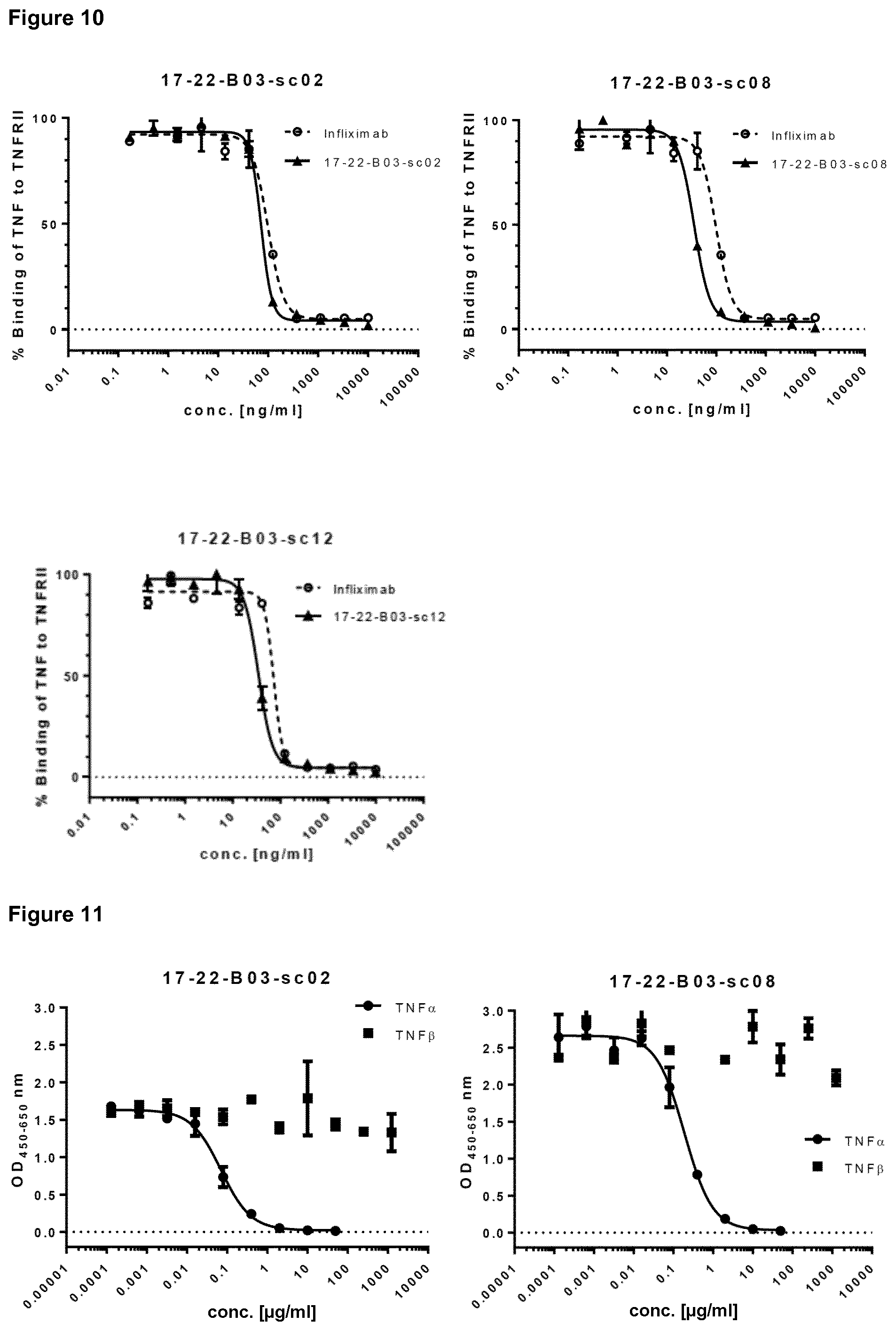
[0083] FIG. 10: Potency of two scFvs to block the TNF.alpha.-TNFRII interaction. Dose-response curves are shown. The highest scFv concentration and negative controls were set to 0% and 100% of binding of TNF.alpha. to TNFRII.
[0084] FIG. 11: Target specificity of an scFv. The potential to inhibit the interaction of biotinylated TNF.alpha. with the scFv by TNF.alpha. and TNF.beta. was analyzed by competition ELISA. Dose-dependent effects of TNF.alpha. and TNF.beta. are shown.
[0085] FIG. 12 depicts the formation of 17-22-B03-scFv:TNF.alpha. complexes determined by SE-HPLC (Example 4).
DETAILED DESCRIPTION
[0086] The present invention pertains to an antibody or a functional fragment thereof capable of binding to human TNF.alpha..
[0087] In the context of the present application, the term "antibody" is used as a synonym for "immunoglobulin" (Ig), which is defined as a protein belonging to the class IgG, IgM, IgE, IgA, or IgD (or any subclass thereof), and includes all conventionally known antibodies and functional fragments thereof. In the context of the present invention, a "functional fragment" of an antibody/immunoglobulin is defined as antigen-binding fragment or other derivative of a parental antibody that essentially maintains one or more of the properties of such parental antibody referred to in items (1) to (30) herein above. An "antigen-binding fragment" of an antibody/immunoglobulin is defined as fragment (e.g., a variable region of an IgG) that retains the antigen-binding region. An "antigen-binding region" of an antibody typically is found in one or more hypervariable region(s) of an antibody, i.e., the CDR-1, -2, and/or -3 regions. "Antigen-binding fragments" of the invention include the domain of a F(ab').sub.2 fragment and a Fab fragment. "Functional fragments" of the invention include, scFv, dsFv, diabodies, triabodies, tetrabodies and Fc fusion proteins. The F(ab').sub.2 or Fab may be engineered to minimize or completely remove the intermolecular disulphide interactions that occur between the CH1 and CL domains. The antibodies or functional fragments of the present invention may be part of bi- or multifunctional constructs.
[0088] Preferred functional fragments in the present invention are scFv and diabodies.
[0089] An scFv is a single chain Fv fragment in which the variable light ("V.sub.L") and variable heavy ("V.sub.H") domains are linked by a peptide bridge.
[0090] A diabody is a dimer consisting of two fragments, each having variable regions joined together via a linker or the like (hereinafter referred to as diabody-forming fragments), and typically contain two Ws and two V.sub.Hs. Diabody-forming fragments include those consisting of V.sub.L and V.sub.H, V.sub.L and V.sub.L, V.sub.H and V.sub.H, etc., preferably V.sub.H and V.sub.L. In diabody-forming fragments, the linker joining variable regions is not specifically limited, but preferably enough short to avoid noncovalent bonds between variable regions in the same fragment. The length of such a linker can be determined as appropriate by those skilled in the art, but typically 2-14 amino acids, preferably 3-9 amino acids, especially 4-6 amino acids. In this case, the V.sub.L and V.sub.H encoded on the same fragment are joined via a linker short enough to avoid noncovalent bonds between the V.sub.L and V.sub.H on the same chain and to avoid the formation of single-chain variable region fragments so that dimers with another fragment can be formed. The dimers can be formed via either covalent or noncovalent bonds or both between diabody-forming fragments.
[0091] Moreover, diabody-forming fragments can be joined via a linker or the like to form single-chain diabodies (sc(Fv).sub.2). By joining diabody-forming fragments using a long linker of about 15-20 amino acids, noncovalent bonds can be formed between diabody-forming fragments existing on the same chain to form dimers. Based on the same principle as for preparing diabodies, polymerized antibodies such as trimers or tetramers can also be prepared by joining three or more diabody-forming fragments.
[0092] Preferably, the antibody or functional fragment of the invention specifically binds to TNF.alpha.. As used herein, an antibody or functional fragment thereof "specifically recognizes", or "specifically binds to" human TNF.alpha., when the antibody or functional fragment is able to discriminate between human TNF.alpha. and one or more reference molecule(s). Preferably, the IC.sub.50 value for binding to each of the reference molecules is at least 1,000 times greater than the IC.sub.50 value for binding to TNF.alpha., particularly as described in Example 2, section 2.1.4. In its most general form (and when no defined reference is mentioned), "specific binding" is referring to the ability of the antibody or functional fragment to discriminate between human TNF.alpha. and an unrelated biomolecule, as determined, for example, in accordance with a specificity assay methods known in the art. Such methods comprise, but are not limited to, Western blots and ELISA tests. For example, a standard ELISA assay can be carried out. Typically, determination of binding specificity is performed by using not a single reference biomolecule, but a set of about three to five unrelated biomolecules, such as milk powder, BSA, transferrin or the like. In one embodiment, specific binding refers to the ability of the antibody or fragment to discriminate between human TNF.alpha. and human TNF.beta..
[0093] The antibody of the invention or the functional fragment of the invention comprises a V.sub.L domain and a V.sub.H domain. The V.sub.L domain comprises a CDR1 region (CDRL1), a CDR2 region (CDRL2), a CDR3 region (CDRL3) and Framework regions. The V.sub.H domain comprises a CDR1 region (CDRH1), a CDR2 region (CDRH2), a CDR3 region (CDRH3) and Framework regions.
[0094] The term "CDR" refers to one of the six hypervariable regions within the variable domains of an antibody that mainly contribute to antigen binding. One of the most commonly used definitions for the six CDRs was provided by Kabat E. A. et al., (1991) Sequences of proteins of immunological interest. NIH Publication 91-3242). As used herein, Kabat's definition of CDRs only apply for CDR1, CDR2 and CDR3 of the light chain variable domain (CDR L1, CDR L2, CDR L3, or L1, L2, L3), as well as for CDR2 and CDR3 of the heavy chain variable domain (CDR H2, CDR H3, or H2, H3). CDR1 of the heavy chain variable domain (CDR H1 or H1), however, as used herein is defined by the following residues (Kabat numbering): It starts with position 26 and ends prior to position 36.
[0095] The CDR1 region of the V.sub.L domain consists of an amino acid sequence in accordance with the amino acid sequence as shown in SEQ ID NO:1. Preferably, the CDR1 region of the V.sub.L domain consists of an amino acid sequence selected from the group consisting of SEQ ID NO:7, SEQ ID NO:16, SEQ ID NO:17 and SEQ ID NO:19. Most preferably, the CDR1 region of the V.sub.L domain consists of the amino acid sequence as shown in SEQ ID NO:7.
[0096] The CDR2 region of the V.sub.L domain consists of an amino acid sequence in accordance with the amino acid sequence as shown in SEQ ID NO:2. Preferably, the CDR2 region of the V.sub.L domain consists of an amino acid sequence selected from the group consisting of SEQ ID NO:8, SEQ ID NO:13 and SEQ ID NO:20. Most preferably, the CDR2 region of the V.sub.L domain consists of the amino acid sequence as shown in SEQ ID NO:8.
[0097] The CDR3 region of the V.sub.L domain consists of an amino acid sequence in accordance with the amino acid sequence as shown in SEQ ID NO:3. Preferably, the CDR3 region of the V.sub.L domain consists of an amino acid sequence in accordance with the amino acid sequence as shown in SEQ ID NO:28. In a particular embodiment the CDR3 region of the V.sub.L domain consists of the amino acid sequence as shown in SEQ ID NO:9.
[0098] The CDR1 region of the V.sub.H domain consists of an amino acid sequence in accordance with the amino acid sequence as shown in SEQ ID NO:4. Preferably, the CDR1 region of the V.sub.H domain consists of an amino acid sequence selected from the group consisting of SEQ ID NO:10 and SEQ ID NO:18. Most preferably, the CDR1 region of the V.sub.H domain consists of the amino acid sequence as shown in SEQ ID NO:10.
[0099] The CDR2 region of the V.sub.H domain consists of an amino acid sequence in accordance with the amino acid sequence as shown in SEQ ID NO:5. Preferably, the CDR2 region of the V.sub.H domain consists of an amino acid sequence selected from the group consisting of SEQ ID NO:11, SEQ ID NO:12, SEQ ID NO:14 and SEQ ID NO:15. Most preferably, the CDR2 region of the V.sub.H domain consists of the amino acid sequence as shown in SEQ ID NO:11.
[0100] The CDR3 region of the V.sub.H domain consists of an amino acid sequence in accordance with the amino acid sequence as shown in SEQ ID NO:6.
[0101] In a particular embodiment, the antibody of the invention or the functional fragment of the invention comprises (i) a V.sub.L domain comprising a CDR1 region having an amino acid sequence in accordance with the amino acid sequence as shown in SEQ ID NO:1, a CDR2 region having an amino acid sequence in accordance with the amino acid sequence as shown in SEQ ID NO:2, and a CDR3 region having an amino acid sequence in accordance with the amino acid sequence as shown in SEQ ID NO:3, and (ii) a V.sub.H domain comprising a CDR1 region having an amino acid sequence in accordance with the amino acid sequence as shown in SEQ ID NO:4, a CDR2 region having an amino acid sequence in accordance with the amino acid sequence as shown in SEQ ID NO:5, and a CDR3 region having an amino acid sequence in accordance with the amino acid sequence as shown in SEQ ID NO:6
[0102] In a particular embodiment, the antibody of the invention or the functional fragment of the invention comprises (i) a V.sub.L domain comprising a CDR1 region having the amino acid sequence as shown in SEQ ID NO:7, a CDR2 region having the amino acid sequence as shown in SEQ ID NO:8, and a CDR3 region having the amino acid sequence as shown in SEQ ID NO:9, and (ii) a V.sub.H domain comprising a CDR1 region having the amino acid sequence as shown in SEQ ID NO:10, a CDR2 region having the amino acid sequence as shown in SEQ ID NO:11, and a CDR3 region having the amino acid sequence as shown in SEQ ID NO:6.
[0103] In a more preferred embodiment, the antibody of the invention or the functional fragment of the invention comprises a V.sub.H domain having the amino acid sequence as shown in SEQ ID NO:21. In another more preferred embodiment the antibody or functional fragment comprises a V.sub.L domain having the amino acid sequence selected from the group consisting of SEQ ID NO:22, SEQ ID NO:23 and SEQ ID NO:24. In an even more preferred embodiment the antibody or functional fragment comprises a V.sub.L domain having the amino acid sequence selected from the group consisting of SEQ ID NO:22 and SEQ ID NO:23. Most preferably, the antibody of the invention or the functional fragment of the invention comprises (i) a V.sub.H domain having the amino acid sequence as shown in SEQ ID NO:21, and (ii) a V.sub.L domain having the amino acid sequence as shown in SEQ ID NO:22, SEQ ID NO:23 or SEQ ID NO:24
[0104] In a particularly preferred embodiment, the functional fragment is a single chain antibody (scFv) comprising a V.sub.H domain having the amino acid sequence as shown in SEQ ID NO:21 and a V.sub.L domain having the amino acid sequence selected from the group consisting of SEQ ID NO:22, SEQ ID NO:23 and SEQ ID NO:24. The V.sub.H domain and the V.sub.L domain are preferably linked by a peptide linker. The peptide linker (hereinafter referred to as "linkerA") typically has a length of about 10 to about 30 amino acids, more preferably of about 15 to about 25 amino acids. The linkerA typically comprises Gly and Ser residues, but other amino acids are also possible. In preferred embodiments the linker comprises multiple repeats of the sequence GGGGS (SEQ ID NO:30), e.g. 2 to 6, or 3 to 5, or 4 consecutive repeats of the amino acid sequence as shown in SEQ ID NO:30. Most preferably, the linkerA consists of the amino acid sequence as shown in SEQ ID NO:29. The scFv may have the following structure (with the N-terminus being left and the C-terminus being right):
[0105] V.sub.L-LinkerA-V.sub.H; or
[0106] V.sub.H-LinkerA-V.sub.L.
[0107] Most preferably, the functional fragment is a single chain antibody (scFv) consisting of the amino acid sequence selected from the group consisting of SEQ ID NO:25, SEQ ID NO:26 and SEQ ID NO:27.
[0108] In another particularly preferred embodiment, the functional fragment is a diabody comprising a V.sub.H domain having the amino acid sequence as shown in SEQ ID NO:21 and a V.sub.L domain having the amino acid sequence selected from the group consisting of SEQ ID NO:22, SEQ ID NO:23 and SEQ ID NO:24. The V.sub.H domain and the V.sub.L domain are linked by a peptide linker. The peptide linker (hereinafter referred to as "linkerB") preferably has a length of about 2 to about 10 amino acids, more preferably of about 5 amino acids. The linkerB typically comprises Gly and Ser residues, but other amino acids are also possible. Most preferably, the linkerB consists of the amino acid sequence as shown in SEQ ID NO:30.
[0109] The diabody preferably is a monospecific diabody, i.e. it is directed to one epitope only. The diabody is preferably a homodimer. The diabody may be a dimer of two polypeptide chains that are non-covalently bound to each other. Each monomer may be a polypeptide chain having the structure:
[0110] V.sub.L-LinkerB-V.sub.H; or
[0111] V.sub.H-LinkerB-V.sub.L.
[0112] Moreover, diabody-forming fragments can be joined via a linkerA or the like to form single-chain diabodies (sc(Fv).sub.2). By joining diabody-forming fragments using a long linker of about 15-20 amino acids, noncovalent bonds can be formed between diabody-forming fragments existing on the same chain to form dimers. Examples of the arrangements of single-chain diabodies include the following.
[0113] V.sub.H-linkerB-V.sub.L-linkerA-V.sub.H-linkerB-V.sub.L
[0114] V.sub.L-linkerB-V.sub.H-linkerA-V.sub.L-linkerB-V.sub.H
[0115] Preferably the diabody of the invention has the following structure:
[0116] V.sub.L-linkerB-V.sub.H-linkerA-V.sub.L-linkerB-V.sub.H
[0117] Based on the same principle as for preparing diabodies, polymerized antibodies such as trimers or tetramers can also be prepared by joining three or more diabody-forming fragments.
[0118] In another particular embodiment the antibody of the invention is an immunoglobulin, preferably an immunoglobulin G (IgG). The subclass of the IgG of the invention is not limited and includes IgG.sub.1, IgG.sub.2, IgG.sub.3, and IgG.sub.4. Preferably, the IgG of the invention is of subclass 1, i.e. it is an IgG.sub.1 molecule.
[0119] Affinity
[0120] The antibody or functional fragment of the invention has a high affinity to human TNF.alpha.. The term "K.sub.D," refers to the dissociation equilibrium constant of a particular antibody-antigen interaction. Typically, the antibody or functional fragment of the invention binds to human TNF.alpha. with a dissociation equilibrium constant (K.sub.D) of less than approximately 2.times.10.sup.-1.degree. M, preferably less than 1.5.times.10.sup.-1.degree. M, preferably less than 1.times.10.sup.-1.degree. M, most preferably less than 7.times.10.sup.-11 M, or even lower, for example, as determined using surface plasmon resonance (SPR) technology in a BIACORE instrument. In particular, the determination of the K.sub.D is carried out as described in Example 2, section 2.1.1.
[0121] Cross-Reactivity to TNF.alpha. from Cynomolgus Monkeys or from Rhesus Macaques
[0122] In particular embodiments, the antibody or functional fragment of the invention has substantial affinity to TNF.alpha. from animals such as Cynomolgus monkeys (Macaca fascicularis) and/or Rhesus macaques (Macaca mulatta). This is advantageous, as preclinical tests of anti-human TNF.alpha. antibodies such as toxicity studies are preferably performed with such animals. Accordingly, the antibody or functional fragment of the invention is preferably cross-reactive with TNF.alpha. from animals such as Cynomolgus monkeys and/or Rhesus macaques. Affinity measurements are carried out as described in Example 2, section 2.1.1.
[0123] In one embodiment, the antibody or functional fragment of the invention is cross-reactive with TNF.alpha. from Macaca fascicularis. The antibody or functional fragment of the invention preferably has an affinity to Macaca fascicularis TNF.alpha. that is less than 10-fold, particularly less than 5-fold, even more particularly less than 2-fold, ore even less than 1.5-fold different to its affinity to human TNF.alpha.. Typically, the antibody or functional fragment of the invention binds to TNF.alpha. from Macaca fascicularis with a dissociation equilibrium constant (K.sub.D), wherein the ratio R.sub.M. fascicularis of (i) the K.sub.D for binding to TNF.alpha. from Macaca fascicularis to (ii) the K.sub.D for binding to human TNF.alpha. is less than 10.
R M . fascicularis . = K D ( M . fascicularis ) K D ( human ) ##EQU00001##
[0124] R.sub.M. fascicularis is preferably less than 5, particularly less than 2, even more particularly less than 1.5.
[0125] In another embodiment, the antibody or functional fragment of the invention is cross-reactive with TNF.alpha. from Macaca mulatta. The antibody or functional fragment of the invention preferably has an affinity to Macaca mulatta TNF.alpha. that is less than 20-fold, more particularly less than 10-fold different, even more particularly less than 5-fold different to its affinity to human TNF.alpha.. Typically, the antibody or functional fragment of the invention binds to TNF.alpha. from Macaca mulatta with a dissociation equilibrium constant (K.sub.D), wherein the ratio R.sub.M. mulatta of (i) the K.sub.D for binding to TNF.alpha. from Macaca mulatta to (ii) the K.sub.D for binding to human TNF.alpha. is less than 20.
R M . mulatta = K D ( M . mulatta ) K D ( human ) ##EQU00002##
[0126] R.sub.M. mulatta is preferably less than 20, particularly less than 10, even more particularly less than 5.
[0127] In yet another embodiment, the antibody or functional fragment of the invention is cross-reactive with TNF.alpha. from Macaca fascicularis and with TNF.alpha. from Macaca mulatta. The antibody or functional fragment of the invention preferably has an affinity to Macaca fascicularis TNF.alpha. that is less than 10-fold, particularly less than 5-fold, even more particularly less than 2-fold, or even less than 1.5-fold different to its affinity to human TNF.alpha., and it preferably has an affinity to Macaca mulatta TNF.alpha. that is less than 20-fold, more particularly less than 10-fold, even more particularly less than 5-fold different to its affinity to human TNF.alpha.. The ratio R.sub.M. fascicularis of the antibody or functional fragment is preferably less than 10, particularly less than 5, even more particularly less than 2, or even less than 1.5, and the ratio R.sub.M. mulatta of the antibody or functional fragment is preferably less than 20, particularly less than 10, even more particularly less than 5.
[0128] Potency to Inhibit TNF.alpha.-Induced Apoptosis of L929 Cells
[0129] The antibody or functional fragment of the invention has a high potency to inhibit TNF.alpha.-induced apoptosis of L929 cells. In a particular embodiment, the antibody or functional fragment of the invention has a potency to inhibit TNF.alpha.-induced apoptosis of L929 cells greater than that of the known antibody infliximab.
[0130] Potency relative to infliximab can be determined in an L929 assay as described in Example 2, section 2.1.2 of this application. The relative potency of the antibody or functional fragment of the invention is greater than 1.5, preferably greater than 2, more preferably greater than 3, more preferably greater than 5, more preferably greater than 7.5, or even greater than 10, wherein the relative potency is the ratio of (i) the IC.sub.50 value of infliximab in an L929 assay over (ii) the IC.sub.50 value of the antibody or functional fragment of the invention in the L929 assay, and wherein the IC.sub.50 indicates the concentration in ng/mL of the respective molecule necessary to achieve 50% of maximal inhibition of TNF-induced apoptosis of L929 cells.
[0131] In another embodiment, the relative potency of the antibody or functional fragment of the invention is greater than 1.5, preferably greater than 2, more preferably greater than 3, more preferably greater than 5, more preferably greater than 7.5, or even greater than 10, wherein the relative potency is the ratio of (i) the IC.sub.90 value of infliximab in an L929 assay over (ii) the IC.sub.90 value of the antibody or functional fragment of the invention in the L929 assay, and wherein the IC.sub.90 value indicates the concentration in ng/mL of the respective molecule necessary to achieve 90% of maximal inhibition of TNF-induced apoptosis of L929 cells.
[0132] Inhibition of LPS-Induced Cytokine Secretion
[0133] The antibody or functional fragment of the invention may be capable of inhibiting LPS-induced cytokine secretion from monocytes. LPS-induced cytokine secretion from monocytes can be determined as described in Example 7.
[0134] In one embodiment, the antibody or functional fragment of the invention is capable of inhibiting LPS-induced secretion of interleukin-1.beta. from CD14.sup.+ monocytes. The IC.sub.50 value for inhibiting LPS-induced secretion of interleukin-1.beta. may be less than 1 nM and/or less than 100 pg/mL. The IC.sub.50 value for inhibiting LPS-induced secretion of interleukin-1.beta., on a molar basis and/or on a weight-per-volume basis, may be lower than that of adalimumab.
[0135] In another embodiment, the antibody or functional fragment of the invention is capable of inhibiting LPS-induced secretion of TNF.alpha. from CD14.sup.+ monocytes. The IC.sub.50 value for inhibiting LPS-induced secretion of TNF.alpha. may be less than 1 nM and/or less than 150 pg/mL. The IC.sub.50 value for inhibiting LPS-induced secretion of TNF.alpha., on a molar basis and/or on a weight-per-volume basis, may be lower than that of adalimumab.
[0136] Inhibition of Cell Proliferation
[0137] The antibody or functional fragment of the invention may be capable of inhibiting cell proliferation of peripheral blood mononuclear cells in a mixed lymphocyte reaction. The inhibition of cell proliferation can be determined as described in Example 6. The stimulation index of the antibody or functional fragment, e.g. of the scFv or diabody of the invention, determined according to Example 6, may be less than 5, or less than 4.5. In particular embodiments, the stimulation index of the antibody, e.g. of the IgG of the invention, is less than 4 or even less than 3.
[0138] Inhibition of Interaction Between TNF.alpha. and TNF Receptor
[0139] Typically, the antibody or functional fragment of the invention is capable of inhibiting the interaction between human TNF.alpha. and TNF receptor I (TNFRI). The inhibition of the interaction between human TNF.alpha. and TNFRI can be determined in an inhibition ELISA as described below in Example 2, section 2.1.3.
[0140] The potency of the antibody or functional fragment of the invention to inhibit the interaction between human TNF.alpha. and TNFRI, relative to that of infliximab (relative potency), as determined in an inhibition ELISA, is preferably at least 1.3, wherein said relative potency is the ratio of the IC.sub.50 value in ng/mL of infliximab to the IC.sub.50 value in ng/mL of the antibody or functional fragment thereof.
[0141] Typically, the antibody or functional fragment of the invention is capable of inhibiting the interaction between human TNF.alpha. and TNF receptor II (TNFRII). The inhibition of the interaction between human TNF.alpha. and TNFRII can be determined in an inhibition ELISA as described below in Example 2, section 2.1.3.
[0142] The potency of the antibody or functional fragment of the invention to inhibit the interaction between human TNF.alpha. and TNFRII, relative to that of infliximab (relative potency), as determined in an inhibition ELISA, is preferably at least 1.3, wherein said relative potency is the ratio of the IC.sub.50 value in ng/mL of infliximab to the IC.sub.50 value in ng/mL of the antibody or functional fragment thereof.
[0143] Stoichiometry and Crosslinking
[0144] The antibody or functional fragment of the invention is typically capable of binding to human TNF.alpha..sub.Trimer in a stoichiometry (antibody: TNF.alpha..sub.Trimer) of at least 2. The stoichiometry (antibody: TNF.alpha..sub.Trimer) is preferably greater than 2, or at least 2.5, or at least 3. In one embodiment, the stoichiometry (antibody: TNF.alpha..sub.Trimer) is about 3. The stoichiometry (antibody: TNF.alpha..sub.Trimer) can be determined as described in Example 4 below.
[0145] In another embodiment, the antibody or functional fragment of the invention is capable of forming a complex with human TNF.alpha., wherein said complex comprises at least two molecules of TNF.alpha. and at least three molecules of antibody or functional fragment. The functional fragment in accordance with this embodiment comprises at least two separate binding sites for TNF.alpha. such as, e.g. diabodies. Complex formation can be determined as described in Example 5 below.
[0146] In one embodiment, the antibody is an IgG, and is capable of forming a complex of at least 600 kDa with TNF.alpha.. In another embodiment, the functional fragment is a diabody, and is capable of forming a complex of at least 300 kDa with TNF.alpha..
[0147] Target Selectivity
[0148] In certain embodiments, the antibody or the functional fragment of the invention has a high target selectivity, i.e. it can discriminate between TNF.alpha. and TNF.beta.. Preferably, the IC.sub.50 value of TNF.beta. is at least 1,000 times greater than the IC.sub.50 value of TNF.alpha., as determined in a competition ELISA as described in Example 2, section 2.1.4. More preferably, the IC.sub.50 value of TNF.beta. is at least 5,000 times, most preferably at least 10,000 greater than the IC.sub.50 value of TNF.alpha., as determined in a competition ELISA as described in Example 2, section 2.1.4.
[0149] Expression Yield and Refolding Yield
[0150] In other embodiments, the antibody or functional fragment of the invention, preferably the scFv or diabody, can be recombinantly expressed in high yield in microorganisms such as bacteria or in other cells. Preferably, the expression yield in E. coli, determined as described in Example 2, is at least 0.25 g/L. This particularly applies to functional fragments such as scFvs.
[0151] The refolding yield, determined as described in Example 2, is at least 5 mg/L, more preferably at least 10 mg/L, more preferably at least 15 mg/L, and most preferably at least 20 mg/L. This particularly applies to functional fragments such as scFvs.
[0152] Stability
[0153] Typically, the antibody or functional fragment of the invention, preferably the scFv or diabody, has a high stability. Stability can be assessed by different methodologies. The "melting temperature" T.sub.m of the variable domain of the antibody or functional fragment of the invention, determined by differential scanning fluorimetry (DSF) as described in Example 2, section 2.2.4, is preferably at least 65.degree. C., more preferably at least 68.degree. C., more preferably at least 70.degree. C., most preferably at least 74.degree. C. The "melting temperature of the variable domain", as used herein, refers to the melting temperature of an scFv consisting of the sequence V.sub.L-LinkerA-V.sub.H, wherein the amino acid sequence of LinkerA consists of the amino acid sequence as shown in SEQ ID NO:29. For example, the melting temperature of the variable domain of an IgG is defined as the melting temperature of its corresponding scFv as defined above.
[0154] The loss in monomer content (at a concentration of 10 g/L; initial monomer content >95%) after storage for four weeks at 4.degree. C., determined by analytical size-exclusion chromatography as described in Example 2, section 2.2.5, is preferably less than 5%, more preferably less than 3%, more preferably less than 1%, most preferably less than 0.5%. The loss in monomer content (at a concentration of 10 g/L; initial monomer content >95%) after storage for four weeks at -20.degree. C., determined by analytical size-exclusion chromatography as described in Example 2, section 2.2.5, is preferably less than 5%, more preferably less than 3%, more preferably less than 1%, most preferably less than 0.5%. The loss in monomer content (at a concentration of 10 g/L; initial monomer content >95%) after storage for four weeks at -65.degree. C., determined by analytical size-exclusion chromatography as described in Example 2, section 2.2.5, is preferably less than 5%, more preferably less than 3%, more preferably less than 1%, most preferably less than 0.5%.
[0155] The monomer loss after five consecutive freeze-thaw cycles, determined as described in Example 2, is less than 5%, more preferably less than 1%, more preferably less than 0.5%, most preferably less than 0.2%, e.g. 0.1% or 0.0%.
[0156] Antibodies and Functional Fragments
[0157] Particular embodiments of the invention relate to functional fragments of the antibodies described herein. Functional fragments include, but are not limited to, F(ab').sub.2 fragment, a Fab fragment, scFv, diabodies, triabodies and tetrabodies. Preferably, the functional fragment is a single chain antibody (scFv) or a diabody. More preferably, the non-CDR sequences of the scFv or of the diabody are human sequences.
[0158] Preferably in order to minimize potential for immunogenicity in humans the chosen acceptor scaffold is composed of framework regions derived from human consensus sequences or human germline sequences. In particular framework regions I to III of the variable light domain consist of human VK1 consensus sequences according to SEQ ID NOs: 31 to 33 and a framework region IV of a .lamda. germline-based sequence selected from SEQ ID NOs:34 to 37. As residues that are not human consensus or human germline residues may cause immune reactions the number of such residues in each variable domain (VH or VL) should be as low as possible, preferably lower than 7, more preferably lower than 4, most preferably 0.
[0159] Preferably the antibody is a monoclonal antibody. The term "monoclonal antibody" as used herein is not limited to antibodies produced through hybridoma technology. The term "monoclonal antibody" refers to an antibody that is derived from a single clone, including any eukaryotic, prokaryotic, or phage clone, and not the method by which it is produced.
[0160] Monoclonal antibodies can be prepared using a wide variety of techniques known in the art including the use of hybridoma, recombinant, and phage display technologies, or a combination thereof. (Harlow and Lane, "Antibodies, A Laboratory Manual" CSH Press 1988, Cold Spring Harbor N.Y.).
[0161] In other embodiments, including embodiments relating to the in vivo use of the anti-TNF.alpha. antibodies in humans, chimeric, primatized, humanized, or human antibodies can be used. In a preferred embodiment, the antibody is a human antibody or a humanized antibody, more preferably a monoclonal human antibody or a monoclonal humanized antibody.
[0162] The term "chimeric" antibody as used herein refers to an antibody having variable sequences derived from a non-human immunoglobulin, such as a rat or mouse antibody, and human immunoglobulins constant regions, typically chosen from a human immunoglobulin template. Methods for producing chimeric antibodies are known in the art. See, e.g., Morrison, 1985, Science 229(4719): 1202-7; Oi et al, 1986, BioTechniques 4:214-221; Gillies et al., 1985, J. Immunol. Methods 125: 191-202; U.S. Pat. Nos. 5,807,715; 4,816,567; and 4,816397, which are incorporated herein by reference in their entireties.
[0163] Different recombinant methodologies are available to one of ordinary skill in the art to render a non-human (e.g., murine) antibody more human-like by generating immunoglobulins, immunoglobulin chains or fragments thereof (such as Fv, Fab, Fab', F(ab').sub.2 or other target-binding subsequences of antibodies), which contain minimal sequences derived from such non-human immunoglobulin. In general, the resulting recombinant antibody will comprise substantially all of at least one, and typically two, variable domains, in which all or substantially all of the CDR regions correspond to those of a non-human immunoglobulin and all or substantially all of the FR regions are those of a human immunoglobulin sequence, particularly a human immunoglobulin consensus sequence. CDR-grafted antibodies are antibody molecules having one or more complementarity determining regions (CDRs) from an antibody originally generated in a non-human species that bind the desired antigen and framework (FR) regions from a human immunoglobulin molecule (EP239400; PCT publication WO 91/09967; U.S. Pat. Nos. 5,225,539; 5,530,101 and 5,585,089). Often, in a process called "humanization", framework residues in the human framework regions will additionally be substituted with the corresponding residue from the CDR donor antibody to alter, preferably improve, antigen binding. These framework substitutions are identified by methods well known in the art, e.g., by modeling of the interactions of the CDR and framework residues to identify framework residues important for antigen binding and sequence comparison to identify unusual framework residues at particular positions. See, e.g., Riechmann et al., 1988, Nature 332:323-7 and Queen et al, U.S. Pat. Nos. 5,530,101; 5,585,089; 5,693,761; 5,693,762; and 6,180,370 (each of which is incorporated by reference in its entirety). Antibodies can be rendered more human using a variety of additional techniques known in the art including, for example, veneering or resurfacing (EP592106; EP519596; Padlan, 1991, Mol. Immunol, 28:489-498; Studnicka et al, 1994, Prot. Eng. 7:805-814; Roguska et al, 1994, Proc. Natl. Acad. Sci. 91:969-973, and chain shuffling (U.S. Pat. No. 5,565,332), all of which are hereby incorporated by reference in their entireties. A CDR-grafted or humanized antibody can also comprise at least a portion of an immunoglobulin constant region (Fc), typically that of a human immunoglobulin template chosen.
[0164] In some embodiments, humanized antibodies are prepared as described in Queen et al, U.S. Pat. Nos. 5,530,101; 5,585,089; 5,693,761; 5,693,762; and 6,180,370 (each of which is incorporated by reference in its entirety).
[0165] In some embodiments, the anti-TNF.alpha. antibodies are human antibodies. Completely "human" anti-TNF.alpha. antibodies can be desirable for therapeutic treatment of human patients. As used herein, "human antibodies" include antibodies having the amino acid sequence of a human immunoglobulin and include antibodies isolated from human immunoglobulin libraries or from animals transgenic for one or more human immunoglobulin and that do not express endogenous immunoglobulins. Human antibodies can be made by a variety of methods known in the art including phage display methods described above using antibody libraries derived from human immunoglobulin sequences. See U.S. Pat. Nos. 4,444,887 and 4,716,111; and PCT publications WO 98/46645; WO 98/50433; WO 98/24893; WO 98/16654; WO 96/34096; WO 96/33735; and WO 91/10741, each of which is incorporated herein by reference in its entirety. Human antibodies can also be produced using transgenic mice which are incapable of expressing functional endogenous immunoglobulins, but which can express human immunoglobulin genes. See, e.g., PCT publications WO 98/24893; WO 92/01047; WO 96/34096; WO 96/33735; U.S. Pat. Nos. 5,413,923; 5,625,126; 5,633,425; 5,569,825; 5,661,016; 5,545,806; 5,814,318; 5,885,793; 5,916,771; and 5,939,598, which are incorporated by reference herein in their entireties. Completely human antibodies that recognize a selected epitope can be generated using a technique referred to as "guided selection." In this approach a selected non-human monoclonal antibody, e.g., a mouse antibody, is used to guide the selection of a completely human antibody recognizing the same epitope (Jespers et al, 1988, Biotechnology 12:899-903).
[0166] In some embodiments, the anti-TNF.alpha. antibodies are primatized antibodies. The term "primatized antibody" refers to an antibody comprising monkey variable regions and human constant regions. Methods for producing primatized antibodies are known in the art. See e.g., U.S. Pat. Nos. 5,658,570; 5,681,722; and 5,693,780, which are incorporated herein by reference in their entireties.
[0167] In some embodiments, the anti-TNF.alpha. antibodies are derivatized antibodies. For example, but not by way of limitation, the derivatized antibodies that have been modified, e.g., by glycosylation, acetylation, pegylation, phosphorylation, amidation, derivatization by known protecting/blocking groups, proteolytic cleavage, linkage to a cellular ligand or other protein (see below for a discussion of antibody conjugates), etc. Any of numerous chemical modifications may be carried out by known techniques, including, but not limited to, specific chemical cleavage, acetylation, formylation, metabolic synthesis of tunicamycin, etc. Additionally, the derivative may contain one or more non-classical amino acids.
[0168] In yet other aspects, an anti-TNF.alpha. antibody has one or more amino acids inserted into one or more of its hypervariable region, for example as described in US 2007/0280931.
[0169] Antibody Conjugates
[0170] In some embodiments, the anti-TNF.alpha. antibodies are antibody conjugates that are modified, e.g., by the covalent attachment of any type of molecule to the antibody, such that covalent attachment does not interfere with binding to TNF.alpha.. Techniques for conjugating effector moieties to antibodies are well known in the art (See, e.g., Hellstrom et al., Controlled Drag Delivery, 2nd Ed., at pp. 623-53 (Robinson et al., eds., 1987)); Thorpe et al., 1982, Immunol. Rev. 62: 119-58 and Dubowchik et al., 1999, Pharmacology and Therapeutics 83:67-123).
[0171] In one example, the antibody or fragment thereof is fused via a covalent bond (e.g., a peptide bond), at optionally the N-terminus or the C-terminus, to an amino acid sequence of another protein (or portion thereof; preferably at least a 10, 20 or 50 amino acid portion of the protein). Preferably the antibody, or fragment thereof, is linked to the other protein at the N-terminus of the constant domain of the antibody. Recombinant DNA procedures can be used to create such fusions, for example as described in WO 86/01533 and EP0392745. In another example the effector molecule can increase half-life in vivo. Examples of suitable effector molecules of this type include polymers, albumin, albumin binding proteins or albumin binding compounds such as those described in WO 2005/117984.
[0172] In some embodiments, anti-TNF.alpha. antibodies can be attached to poly(ethyleneglycol) (PEG) moieties. For example, if the antibody is an antibody fragment, the PEG moieties can be attached through any available amino acid side-chain or terminal amino acid functional group located in the antibody fragment, for example any free amino, imino, thiol, hydroxyl or carboxyl group. Such amino acids can occur naturally in the antibody fragment or can be engineered into the fragment using recombinant DNA methods. See for example U.S. Pat. No. 5,219,996. Multiple sites can be used to attach two or more PEG molecules. Preferably PEG moieties are covalently linked through a thiol group of at least one cysteine residue located in the antibody fragment. Where a thiol group is used as the point of attachment, appropriately activated effector moieties, for example thiol selective derivatives such as maleimides and cysteine derivatives, can be used.
[0173] In another example, an anti-TNF.alpha. antibody conjugate is a modified Fab' fragment which is PEGylated, i.e., has PEG (poly(ethyleneglycol)) covalently attached thereto, e.g., according to the method disclosed in EP0948544. See also Poly(ethyleneglycol) Chemistry, Biotechnical and Biomedical Applications, (J. Milton Harris (ed.), Plenum Press, New York, 1992); Poly(ethyleneglycol) Chemistry and Biological Applications, (J. Milton Harris and S. Zalipsky, eds., American Chemical Society, Washington D.C., 1997); and Bioconjugation Protein Coupling Techniques for the Biomedical Sciences, (M. Aslam and A. Dent, eds., Grove Publishers, New York, 1998); and Chapman, 2002, Advanced Drug Delivery Reviews 54:531-545.
[0174] Pharmaceutical Compositions and Treatment
[0175] Treatment of a disease encompasses the treatment of patients already diagnosed as having any form of the disease at any clinical stage or manifestation; the delay of the onset or evolution or aggravation or deterioration of the symptoms or signs of the disease; and/or preventing and/or reducing the severity of the disease.
[0176] A "subject" or "patient" to whom an anti-TNF.alpha. antibody or functional fragment thereof is administered can be a mammal, such as a non-primate (e.g., cow, pig, horse, cat, dog, rat, etc.) or a primate (e.g., monkey or human). In certain aspects, the human is a pediatric patient. In other aspects, the human is an adult patient.
[0177] Compositions comprising an anti-TNF.alpha. antibody and, optionally one or more additional therapeutic agents, such as the second therapeutic agents described below, are described herein. The compositions typically are supplied as part of a sterile, pharmaceutical composition that includes a pharmaceutically acceptable carrier. This composition can be in any suitable form (depending upon the desired method of administering it to a patient).
[0178] The anti-TNF.alpha. antibodies and functional fragments can be administered to a patient by a variety of routes such as orally, transdermally, subcutaneously, intranasally, intravenously, intramuscularly, intrathecally, topically or locally. The most suitable route for administration in any given case will depend on the particular antibody, the subject, and the nature and severity of the disease and the physical condition of the subject. Typically, an anti-TNF.alpha. antibody or functional fragment thereof will be administered intravenously.
[0179] In a particularly preferred embodiment, the antibody or functional fragment of the invention is administered orally. If the administration is via the oral route the functional fragment is preferably a single chain antibody (scFv), diabody or IgG.
[0180] In typical embodiments, an anti-TNF.alpha. antibody or functional fragment is present in a pharmaceutical composition at a concentration sufficient to permit intravenous administration at 0.5 mg/kg body weight to 20 mg/kg body weight. In some embodiments, the concentration of antibody or fragment suitable for use in the compositions and methods described herein includes, but is not limited to, 0.5 mg/kg, 0.75 mg/kg, 1 mg/kg, 2 mg/kg, 2.5 mg/kg, 3 mg/kg, 4 mg/kg, 5 mg/kg, 6 mg/kg, 7 mg/kg, 8 mg/kg, 9 mg/kg, 10 mg/kg, 11 mg/kg, 12 mg/kg, 13 mg/kg, 14 mg/kg, 15 mg/kg, 16 mg/kg, 17 mg/kg, 18 mg/kg, 19 mg/kg, 20 mg/kg, or a concentration ranging between any of the foregoing values, e.g., 1 mg/kg to 10 mg/kg, 5 mg/kg to 15 mg/kg, or 10 mg/kg to 18 mg/kg.
[0181] The effective dose of an anti-TNF.alpha. antibody or functional fragment can range from about 0.001 to about 750 mg/kg per single (e.g., bolus) administration, multiple administrations or continuous administration, or to achieve a serum concentration of 0.01-5000 .mu.g/mL serum concentration per single (e.g., bolus) administration, multiple administrations or continuous administration, or any effective range or value therein depending on the condition being treated, the route of administration and the age, weight and condition of the subject. In certain embodiments, each dose can range from about 0.5 mg to about 50 mg per kilogram of body weight or from about 3 mg to about 30 mg per kilogram body weight. The antibody can be formulated as an aqueous solution.
[0182] Pharmaceutical compositions can be conveniently presented in unit dose forms containing a predetermined amount of an anti-TNF.alpha. antibody or functional fragment per dose. Such a unit can contain 0.5 mg to 5 g, for example, but without limitation, 1 mg, 10 mg, 20 mg, 30 mg, 40 mg, 50 mg, 100 mg, 200 mg, 300 mg, 400 mg, 500 mg, 750 mg, 1000 mg, or any range between any two of the foregoing values, for example 10 mg to 1000 mg, 20 mg to 50 mg, or 30 mg to 300 mg. Pharmaceutically acceptable carriers can take a wide variety of forms depending, e.g., on the condition to be treated or route of administration.
[0183] Determination of the effective dosage, total number of doses, and length of treatment an anti-TNF.alpha. antibody or functional fragment thereof is well within the capabilities of those skilled in the art, and can be determined using a standard dose escalation study.
[0184] Therapeutic formulations of the anti-TNF.alpha. antibodies and functional fragments suitable in the methods described herein can be prepared for storage as lyophilized formulations or aqueous solutions by mixing the antibody having the desired degree of purity with optional pharmaceutically-acceptable carriers, excipients or stabilizers typically employed in the art (all of which are referred to herein as "carriers"), i.e., buffering agents, stabilizing agents, preservatives, isotonifiers, non-ionic detergents, antioxidants, and other miscellaneous additives. See, Remington's Pharmaceutical Sciences, 16th edition (Osol, ed. 1980). Such additives must be nontoxic to the recipients at the dosages and concentrations employed.
[0185] Buffering agents help to maintain the pH in the range which approximates physiological conditions. They can present at concentration ranging from about 2 mM to about 50 mM. Suitable buffering agents include both organic and inorganic acids and salts thereof such as citrate buffers (e.g., monosodium citrate-disodium citrate mixture, citric acid-trisodium citrate mixture, citric acid-monosodium citrate mixture, etc.), succinate buffers (e.g., succinic acid-monosodium succinate mixture, succinic acid-sodium hydroxide mixture, succinic acid-disodium succinate mixture, etc.), tartrate buffers (e.g., tartaric acid-sodium tartrate mixture, tartaric acid-potassium tartrate mixture, tartaric acid-sodium hydroxide mixture, etc.), fumarate buffers (e.g., fumaric acid-monosodium fumarate mixture, fumaric acid-disodium fumarate mixture, monosodium fumarate-disodium fumarate mixture, etc.), gluconate buffers (e.g., gluconic acid-sodium glyconate mixture, gluconic acid-sodium hydroxide mixture, gluconic acid-potassium glyuconate mixture, etc.), oxalate buffer (e.g., oxalic acid-sodium oxalate mixture, oxalic acid-sodium hydroxide mixture, oxalic acid-potassium oxalate mixture, etc), lactate buffers (e.g., lactic acid-sodium lactate mixture, lactic acid-sodium hydroxide mixture, lactic acid-potassium lactate mixture, etc.) and acetate buffers (e.g., acetic acid-sodium acetate mixture, acetic acid-sodium hydroxide mixture, etc.). Additionally, phosphate buffers, histidine buffers and trimethylamine salts such as Tris can be used.
[0186] Preservatives can be added to retard microbial growth, and can be added in amounts ranging from 0.2%-1% (w/v). Suitable preservatives include phenol, benzyl alcohol, meta-cresol, methyl paraben, propyl paraben, octadecyldimethylbenzyl ammonium chloride, benzalconium halides (e.g., chloride, bromide, and iodide), hexamethonium chloride, and alkyl parabens such as methyl or propyl paraben, catechol, resorcinol, cyclohexanol, and 3-pentanol. Isotonicifiers sometimes known as "stabilizers" can be added to ensure isotonicity of liquid compositions and include polyhydric sugar alcohols, preferably trihydric or higher sugar alcohols, such as glycerin, erythritol, arabitol, xylitol, sorbitol and mannitol. Stabilizers refer to a broad category of excipients which can range in function from a bulking agent to an additive which solubilizes the therapeutic agent or helps to prevent denaturation or adherence to the container wall. Typical stabilizers can be polyhydric sugar alcohols (enumerated above); amino acids such as arginine, lysine, glycine, glutamine, asparagine, histidine, alanine, ornithine, L-leucine, 2-phenylalanine, glutamic acid, threonine, etc., organic sugars or sugar alcohols, such as lactose, trehalose, stachyose, mannitol, sorbitol, xylitol, ribitol, myoinisitol, galactitol, glycerol and the like, including cyclitols such as inositol; polyethylene glycol; amino acid polymers; sulfur containing reducing agents, such as urea, glutathione, thioctic acid, sodium thioglycolate, thioglycerol, .alpha.-monothioglycerol and sodium thio sulfate; low molecular weight polypeptides (e.g., peptides of 10 residues or fewer); proteins such as human serum albumin, bovine serum albumin, gelatin or immunoglobulins; hydrophylic polymers, such as polyvinylpyrrolidone monosaccharides, such as xylose, mannose, fructose, glucose; disaccharides such as lactose, maltose, sucrose and trisaccacharides such as raffinose; and polysaccharides such as dextran. Stabilizers can be present in the range from 0.1 to 10,000 weights per part of weight active protein.
[0187] Non-ionic surfactants or detergents (also known as "wetting agents") can be added to help solubilize the therapeutic agent as well as to protect the therapeutic protein against agitation-induced aggregation, which also permits the formulation to be exposed to shear surface stressed without causing denaturation of the protein. Suitable non-ionic surfactants include polysorbates (20, 80, etc.), polyoxamers (184, 188 etc.), Pluronic polyols, polyoxyethylene sorbitan monoethers (TWEEN.RTM.-20, TWEEN.RTM.-80, etc.). Non-ionic surfactants can be present in a range of about 0.05 mg/mL to about 1.0 mg/mL, or in a range of about 0.07 mg/mL to about 0.2 mg/mL.
[0188] Additional miscellaneous excipients include bulking agents (e.g., starch), chelating agents (e.g., EDTA), antioxidants (e.g., ascorbic acid, methionine, vitamin E), protease inhibitors and co-solvents.
[0189] The formulation herein can also contain a second therapeutic agent in addition to an anti-TNF.alpha. antibody or functional fragment thereof. Examples of suitable second therapeutic agents are provided below.
[0190] The dosing schedule can vary from once a month to daily depending on a number of clinical factors, including the type of disease, severity of disease, and the patient's sensitivity to the anti-TNF.alpha. antibody or functional fragment. In specific embodiments, an anti-TNF.alpha. antibody or functional fragment thereof is administered daily, twice weekly, three times a week, every other day, every 5 days, every 10 days, every two weeks, every three weeks, every four weeks or once a month, or in any range between any two of the foregoing values, for example from every four days to every month, from every 10 days to every two weeks, or from two to three times a week, etc.
[0191] The dosage of an anti-TNF.alpha. antibody or functional fragment to be administered will vary according to the particular antibody, the subject, and the nature and severity of the disease, the physical condition of the subject, the therapeutic regimen (e.g., whether a second therapeutic agent is used), and the selected route of administration; the appropriate dosage can be readily determined by a person skilled in the art.
[0192] It will be recognized by one of skill in the art that the optimal quantity and spacing of individual dosages of an anti-TNF.alpha. antibody or functional fragment thereof will be determined by the nature and extent of the condition being treated, the form, route and site of administration, and the age and condition of the particular subject being treated, and that a physician will ultimately determine appropriate dosages to be used. This dosage can be repeated as often as appropriate. If side effects develop the amount and/or frequency of the dosage can be altered or reduced, in accordance with normal clinical practice.
[0193] Disorders to be Treated
[0194] The invention relates to a method of treating or preventing a human TNF.alpha.-related disease in a subject, comprising administering to the subject the antibody or functional fragment as defined herein. The term "TNF-related disorder" or "TNF-related disease" refers to any disorder, the onset, progression or the persistence of the symptoms or disease states of which requires the participation of TNF.alpha.. Exemplary TNF-related disorders include, but are not limited to, chronic and/or autoimmune states of inflammation in general, immune mediated inflammatory disorders in general, inflammatory CNS disease, inflammatory diseases affecting the eye, joint, skin, mucous membranes, central nervous system, gastrointestinal tract, urinary tract or lung, states of uveitis in general, retinitis, HLA-B27+ uveitis, Behcet's disease, dry eye syndrome, glaucoma, Sjogren syndrome, diabetes mellitus (incl. diabetic neuropathy), insulin resistance, states of arthritis in general, rheumatoid arthritis, osteoarthritis, reactive arthritis and Reiter's syndrome, juvenile arthritis, ankylosing spondylitis, multiple sclerosis, Guillain-Barre syndrome, myasthenia gravis, amyotrophic lateral sclerosis, sarcoidosis, glomerulonephritis, chronic kidney disease, cystitis, psoriasis (incl. psoriatic arthritis), hidradenitis suppurativa, panniculitis, pyoderma gangrenosum, SAPHO syndrome (synovitis, acne, pustulosis, hyperostosis and osteitis), acne, Sweet's sydrome, pemphigus, Crohn's disease (incl. extraintestinal manifestastations), ulcerative colitis, asthma bronchiale, hypersensitivity pneumonitis, general allergies, allergic rhinitis, allergic sinusitis, chronic obstructive pulmonary disease (COPD), lung fibrosis, Wegener's granulomatosis, Kawasaki syndrome, Giant cell arteritis, Churg-Strauss vasculitis, polyarteritis nodosa, burns, graft versus host disease, host versus graft reactions, rejection episodes following organ or bone marrow transplantation, systemic and local states of vasculitis in general, systemic and cutaneous lupus erythematodes, polymyositis and dermatomyositis, sclerodermia, pre-eclampsia, acute and chronic pancreatitis, viral hepatitis, alcoholic hepatitis, postsurgical inflammation such as after eye surgery (e.g. cataract (eye lens replacement) or glaucoma surgery), joint surgery (incl. arthroscopic surgery), surgery at joint-related structures (e.g. ligaments), oral and/or dental surgery, minimally invasive cardiovascular procedures (e.g. PTCA, atherectomy, stent placement), laparoscopic and/or endoscopic intra-abdominal and gynecological procedures, endoscopic urological procedures (e.g. prostate surgery, ureteroscopy, cystoscopy, interstitial cystitis), or perioperative inflammation (prevention) in general, bullous dermatitis, neutrophilic dermatitis, toxic epidermal necrolysis, pustular dermatitis, cerebral malaria, hemolytic uremic syndrome, allograft rejection, otitis media, snakebite, erythema nodosum, myelodysplastic syndromes, primary sclerosing cholangitis, seronegative spondylartheropathy, autoimmune hematolytic anemia, orofacial granulamatosis, pyostomatitis vegetans, aphthous stomatitis, geographic tongue, migratory stoimatitis, Alzheimer disease, Parkinson's disease, Huntington's disease, Bell's palsy, Creutzfeld-Jakob disease and neuro-degenerative conditions in general.
[0195] Cancer-related osteolysis, cancer-related inflammation, cancer-related pain, cancer-related cachexia, bone metastases, acute and chronic forms of pain, irrespective whether these are caused by central or peripheral effects of TNF.alpha. and whether they are classified as inflammatory, nociceptive or neuropathic forms of pain, sciatica, low back pain, carpal tunnel syndrome, complex regional pain syndrome (CRPS), gout, postherpetic neuralgia, fibromyalgia, local pain states, chronic pain syndroms due to metastatic tumor, dismenorrhea.
[0196] Particular disorders to be treated include states of arthritis in general, rheumatoid arthritis, osteoarthritis, reactive arthritis, juvenile arthritis; psoriasis incl. psoriatic arthritis; inflammatory bowel disease, including Crohn's disease, ulcerative colitis incl. proctitis, sigmoiditis, left-sided colitis, extensive colitis and pancolitis, undetermined colitis, microscopic colitis incl. collagenous and lymphocytic colitis, colitis in connective tissue disease, diversion colitis, colitis in diverticular disease, eosinophilic colitis and pouchitis.
[0197] Most preferably, the antibody or functional fragment of the invention is used to treat an inflammatory bowel disease, in particular Crohn's disease, ulcerative colitis or microscopic colitis. The Crohn's disease may be ileal, colonic, ileocolonic or isolated upper Crohn's disease (gastric, duodenal and/or jejunal) including non-stricturing/non-penetrating, stricturing, penetrating and perianal disease behavior, allowing any combination of localization and disease behavior of any of the above mentioned. The ulcerative colitis may be ulcerative proctitis, proctosigmoiditis, left-sided colitis, pan-ulcerative colitis and pouchitis.
[0198] Combination Therapy and Other Aspects
[0199] Preferably, the patient being treated with an anti-TNF.alpha. antibody or functional fragment thereof is also treated with another conventional medicament. For example, a patient suffering from inflammatory bowel disease, especially if having moderate to severe disease is typically also being treated with mesalazine or derivatives or prodrugs thereof, corticosteroids, e.g. budesonide or prednisolone (oral or i.v.), immunosuppressants, e.g. azathioprine/6-mercaptopurine (6-MP) or methotrexate, cyclosporine or tacrolimus. Other medicaments which can be co-administered to the patient include biologics such as infliximab, adalimumab, etanercept, certolizumab pegol or others. Further medicaments which can be co-administered to the patient include immunosupressants (e.g. azathioprine/6-MP or methotrexate or oral cyclosporine) in order to maintain stable and longer remission. Yet another aspect of the invention is the use of an anti-TNF.alpha. antibody or functional fragment as defined hereinabove for reducing inflammation.
[0200] Yet another aspect of the invention is an anti-TNF.alpha. antibody or functional fragment as defined hereinabove for use in reducing inflammation in a patient suffering from an inflammatory condition.
[0201] A further aspect of this invention is a method of treating an inflammatory condition, comprising administering to a patient in need thereof an effective amount of an anti-TNF.alpha. antibody or functional fragment as defined hereinabove. The inflammatory condition is preferably one of the conditions described above.
TABLE-US-00001 TABLE 1 Summary of the amino acid sequences SEQ ID NO: Description 1 Generic CDR L1 2 Generic CDR L2 3 Generic CDR L3 4 Generic CDR H1 5 Generic CDR H2 6 CDR H3 of clones 17-22-B03, 16-12-E09, 16-13-E01, 16- 15-E11, 16-17-C08, 16-17-G01, 17-18-D10 and 17-22-B01 7 CDR L1 of clones 17-22-B03, 16-12-E09, 16-13-E01, 16- 15-E11 and 17-18-D10 8 CDR L2 of clones 17-22-B03, 16-12-E09, 16-15-E11, 16- 17-C08, 16-17-G01 and 17-18-D10 9 CDR L3 of clones 17-22-B03, 16-12-E09, 16-13-E01, 16- 15-E11, 16-17-C08, 16-17-G01, 17-18-D10 and 17-22-B01 10 CDR H1 of clones 17-22-B03, 16-12-E09, 16-13-E01, 16- 15-E11, 16-17-C08, 17-18-D10 and 17-22-B01 11 CDR H2 of clones 17-22-B03 and 16-17-C08 12 CDR H2 of clone 16-12-E09 13 CDR L2 of clone 16-13-E01 14 CDR H2 of clones 16-13-E01 and 17-18-D10 15 CDR H2 of clones 16-15-E11, 16-17-G01 and 17-22-B01 16 CDR L1 of clone 16-17-C08 17 CDR L1 of clone 16-17-G01 18 CDR H1 of clone 16-17-G01 19 CDR L1 of clone 17-22-B01 20 CDR L2 of clone 17-22-B01 21 V.sub.H of humanized scFv of clones 17-22-B03-sc02, 17-22- B03-sc08, 17-22-B03-sc12 22 V.sub.L of humanized scFv of clone 17-22-B03-sc02 23 V.sub.L of humanized scFv of clone 17-22-B03-sc08 24 V.sub.L of humanized scFv of clone 17-22-B03-sc12 25 Humanized scFv of clone 17-22-B03-sc02 26 Humanized scFv of clone 17-22-B03-sc08 27 Humanized scFv of clone 17-22-B03-sc12 28 Generic CDR L3 where Cys2 (X) is limited to Arg, His, Pro, Phe, Gln, Gly, Ser, Thr, Asn and Tyr 29 Linker sequence in scFv 30 Linker sequence in diabody 31 V.kappa.1 consensus sequence of framework I (Kabat positions 1-23) (see Table 10) 32 V.kappa.1 consensus sequence of framework II (Kabat positions 35-49) (see Table 10) 33 V.kappa.1 consensus sequence of framework III (Kabat positions 57-88) (see Table 10) 34 V.lamda. germline-based sequence of framework IV (see Table 11) 35 V.lamda. germline-based sequence of framework IV (see Table 11) 36 V.lamda. germline-based sequence of framework IV (see Table 11) 37 V.lamda. germline-based sequence of framework IV (see Table 11)
EXAMPLES
Example 1: Generation of Rabbit Antibodies Directed Against Human TNF.alpha.
[0202] 1. Results
[0203] 1.1 Immunization
[0204] Rabbits have been immunized with purified recombinant human TNF.alpha. (Peprotech, Cat. No. 300-01A). During the course of the immunization, the strength of the humoral immune response against the antigen was qualitatively assessed by determining the maximal dilution (titer) for the serum of each rabbit that still produced detectable binding of the polyclonal serum antibodies to the antigen. Serum antibody titers against immobilized recombinant human TNF.alpha. were assessed using an enzyme-linked immunosorbent assay (ELISA, see 2.2.1). All three rabbits showed very high titers with a 10.times.10.sup.6-fold dilution of the serum still resulting in a positive signal (at least 3-fold higher than the signal obtained with serum from a naive unrelated animal which was used as background control) in the ELISA. In addition, the ability of different rabbit sera to inhibit the biological activity of TNF.alpha. was assessed using a mouse L929 cell-based assay (see 2.2.3). All three sera inhibited TNF.alpha.-induced apoptosis of mouse L929 fibroblasts. Rabbit #3 showed the strongest neutralizing activity with 50% inhibition (IC.sub.50) reached at a serum dilution of 1:155'000. Compared to rabbit #3, rabbit #2 and rabbit #1 showed approximately 3 and 21-fold lower activity, reaching 50% inhibition at a serum dilution of 1:55'500 and 1:7'210, respectively.
[0205] Lymphocytes isolated from spleens of all three animals were chosen for the subsequent hit identification procedures. The animals were prioritized based on the potency to inhibit the biological activity of TNF.alpha. in the L929 assay. Therefore, the highest number of hits that were cultivated originated from rabbit #3, and the lowest number of hits was derived from rabbit #1.
[0206] 1.2 Hit Identification
[0207] 1.2.1 Hit Sorting
[0208] Prior to the hit identification procedure, a flow-cytometry-based sorting procedure was developed that specifically detects and allows for the isolation of high-affinity TNF.alpha. binding B-cells (see 2.1).
[0209] A total of 33.times.10.sup.6 lymphocytes (corresponding to 1.5% of total lymphocytes isolated) derived from all three rabbits were characterized in two independent sorting campaigns. Out of the 33.times.10.sup.6 cells analyzed in total 3452 B-cells expressing TNF.alpha.-specific antibodies (IgG) were isolated. The numbers of lymphocytes cloned were different for the three rabbits, as more cells were isolated from those rabbits whose sera showed strong inhibition of TNF.alpha. in the L929 assay. Of the isolated B-cells, 792 clones were derived from rabbit #1, 1144 clones from rabbit #2 and 1408 clones from rabbit #3. For 108 clones the respective rabbit origin is not known, because they are derived from a mixture of residual lymphocytes from all 3 rabbits to allow optimal recovery of small amount of lymphocytes from the vials.
[0210] 1.2.2 Hit Screening
[0211] The results obtained during the screening phase are based on assays performed with non-purified antibodies from culture supernatants of antibody secreting cells (ASC), as the scale of the high-throughput culture does not allow for purification of the individual rabbit antibodies. Such supernatants were used to rank large numbers of antibodies relative to each other, however not to provide absolute values (e.g. for inhibition of biological activity of TNF.alpha.), except for binding affinity. ASC supernatants were screened in a high-throughput ELISA for binding to recombinant human TNF.alpha.. TNF.alpha.-binding supernatants were further characterized for binding to Cynomolgus monkey TNF.alpha. by ELISA, binding kinetics and for their potential to neutralize the biological activity of human TNF.alpha. in the L929 assay. With the exception of binding kinetics, the reporting values of the high-throughput screenings should be interpreted as "yes" or "no" answers, which are based on single-point measurements (no dose-response). Affinity to Cynomolgus monkey and mouse TNF.alpha. was analyzed for all the 102 clones that were selected for amplification and sequencing of the antibody heavy and light chain variable domains.
[0212] 1.2.2.1 Binding to Human TNF.alpha.
[0213] The aim of the primary screening is to identify ASC clones that produce antibodies specific for human TNF.alpha.. For this purpose, cell culture supernatants of 3452 ASC clones were analysed for the presence of antibodies to human TNF.alpha. by ELISA (see 2.2.1). The ELISA method used assesses the "quantity" of antibodies of the IgG subtype bound to recombinant human TNF.alpha., gives however no information about the affinity or the concentration of the antibodies. In this assay, supernatants from 894 ASC clones produced a signal that was clearly above background. The hit rate in screening was similar for rabbit #1 and rabbit #2 with 153 hits out of 792 (19.3%) identified from rabbit #1 and 225 hits out of 1144 identified from rabbit #2 (19.7%). Rabbit #3 showed a significantly higher hit rate of 34.4% resulting in the identification of 484 hits out of 1408. All 894 hits identified in this primary screening, were subjected to the measurement of binding kinetics by SPR (secondary screening).
[0214] 1.2.2.2 TNF.alpha. Binding Kinetics
[0215] The aim of the secondary screening is to obtain quantitative information on the quality of target binding for each hit from the primary screening by surface plasmon resonance (SPR, see 2.2.2). In contrast to the ELISA used during the primary screening, this method assesses the kinetics of target binding as a function of time. This allows determination of the rate constants for association (k.sub.a) and dissociation (k.sub.d) of the antibody from its target. The ratio k.sub.d/k.sub.a provides the equilibrium dissociation constant (K.sub.D), which reflects the affinity of an antibody to its target. Of the 894 hits identified in the primary screening, binding affinities to human TNF.alpha. could be determined for 839 monoclonal rabbit antibodies. For the remaining 55 antibodies affinity could not be measured because the antibody concentration in the ASC supernatant was below the detection limit of the SPR instrument in the respective setup. The 839 anti-TNF.alpha. antibodies that could be measured showed dissociation constants (K.sub.D) ranging from below 1.36.times.10.sup.-13 M to 1.14.times.10.sup.-8 M. 69% of all antibodies analyzed had a K.sub.D below 0.5 nM.
[0216] The median K.sub.Ds of 2.21.times.10.sup.-10 M and 2.09.times.10.sup.-10 M for screening hits identified from rabbits #2 and #3 were similar while rabbit #1 showed about 2-fold higher values with a median K.sub.D of 4.65.times.10.sup.-10 M. When considering only neutralizing screening hits, the affinity distributions were similar for all three animals with lower values for the median K.sub.D (median K.sub.Ds between 1.4.times.10.sup.-10 M and 1.27.times.10.sup.-10 M). Affinities below 0.041 nM, 0.029 nM and 0.026 nM were measured for 5% of screening hits for rabbits #1, #2 and #3, respectively. For 2% of supernatants, affinities were even in the low picomolar range (below 6.2 pM, 7.9 pM and 11 pM). The excellent yield of high-affinity antibodies resulting from the secondary screening provides a broad basis for the selection of the most appropriate antibodies for humanization and reformatting.
[0217] 1.2.2.3 Potency
[0218] For the assessment of potency, a cell-based assay (L929 assay) has been developed (see 2.2.3). 506 out of the 894 selected antibodies (56.6%), inhibited TNF.alpha.-induced apoptosis in the L929 assay by more than 50%. In line with results obtained during titer analysis, the highest percentage of neutralizing hits was derived from rabbit #3 with a hit rate of 62.8%, followed by rabbit #2 with a hit rate of 56.4% and rabbit #1 with the lowest hit rate of 39.9%. Affinities of these neutralizing antibodies ranged between 1.36.times.10.sup.-13 to 1.19.times.10.sup.-9 M.
[0219] 1.2.2.4 Species Cross-Reactivity (Cynomolgus Monkey)
[0220] All 894 hits identified in the primary screening, were analyzed for species cross-reactivity to Cynomolgus monkey TNF.alpha. by ELISA (see 2.2.1). The aim of this additional screening was to allow selection of ASC clones that are known to cross-react with Cynomolgus monkey TNF.alpha.. The ELISA method used assesses the "quantity" of antibodies of the IgG subtype bound to recombinant Cynomolgus monkey TNF.alpha., gives however no information about the affinity or the concentration of the antibodies. Supernatants from 414 (46%) ASC clones produced a clear signal (optical density (OD) .gtoreq.1). The percentage of hits cross-reactive to Cynomolgus monkey TNF.alpha. was similar for rabbit #1 and rabbit #3 with 81 hits out of 153 (52.9%) identified from rabbit #1 and 236 hits out of 484 identified from rabbit #3 (48.8%). With 37.8%, rabbit #2 showed a slightly lower percentage of cross-reactive hits resulting in the identification of 82 hits out of 225.
[0221] 1.2.2.5 Selection of Clones for RT-PCR
[0222] As a prerequisite for hit confirmation, gene sequence analysis and subsequent humanization of the rabbit antibodies, the genetic information encoding the rabbit antibody variable domain needs to be retrieved. This was done by reverse transcription (RT) of the respective messenger RNA into the complementary DNA (cDNA), followed by amplification of the double stranded DNA by the polymerase chain reaction (PCR). The selection of ASC clones subjected to RT-PCR was primarily based on affinity and neutralizing activity. As additional criterion cross-reactivity to Cynomolgus monkey TNF.alpha. was considered. In total 102 ASC clones were selected for gene cloning by RT-PCR. First, the 93 best ranking ASC (in terms of affinity) with a K.sub.D below 80 pM, that inhibited the biological activity of TNF.alpha. in the L929 assay by more than 50% and that showed significant binding to Cynomolgus monkey TNF.alpha. were selected. Additionally, all the 9 best ranking ASC clones with K.sub.D below 20 pM that neutralized TNF.alpha. activity by more than 50% but did not bind to Cynomolgus monkey TNF.alpha. nevertheless were chosen as well. In total, 12, 13 and 66 ASC clones were successfully amplified and sequenced from rabbits #1, #2 and #3, respectively.
[0223] 1.2.2.6 Identification of Related Clones with Desired Properties
[0224] In order to characterize the genetic diversity of the panel of isolated ASC clones the sequences of the complementary determining regions (CDRs) were extracted and subjected to a multiple sequence alignment thus allowing sequence clustering in a phylogenetic tree.
[0225] While this analysis on one hand allows the selection of a diverse set of clonal sequences to be carried forward into humanization and re-formatting experiments it also identifies homologous clusters of clonal sequences that appeared to share a common parental B-cell clone in the rabbit. The hallmark of these sequence clusters are high sequence homology in the CDRs and a consistent pattern of pharmacodynamic properties. Both of these features are summarized for a cluster of eight clones in Tables 2 and 3. Despite the functional conservation of this sequence cluster the consensus sequence in Table 3 reveals that a certain variability of the CDRs is tolerated, while still resulting in the desired pharmacodynamic profile.
TABLE-US-00002 TABLE 2 Pharmacodynamic properties of monoclonal antibodies in B-cell supernatants. B-cell SN Affinity to human TNF.alpha. Affinity to Cynomolgus TNF.alpha. TNF.alpha. k.sub.a K.sub.d K.sub.D k.sub.a K.sub.d K.sub.D neutralization % Clone ID (M.sup.-1s.sup.-1) (s.sup.-1) (M) (M.sup.-1s.sup.-1) (s.sup.-1) (M) inhibition 16-12-E09 2.37E+06 1.42E-04 5.99E-11 2.46E+06 1.29E-04 5.23E-11 80 16-13-E01 3.12E+06 1.37E-04 4.40E-11 3.26E+06 1.45E-04 4.44E-11 86 16-15-E11 4.16E+06 2.90E-04 6.97E-11 6.36E+06 3.39E-04 5.33E-11 72 16-17-C08 2.54E+06 1.03E-04 4.06E-11 5.01E+06 1.41E-04 2.81E-11 74 16-17-G01 2.20E+06 6.13E-05 2.79E-11 3.50E+06 9.42E-05 2.69E-11 94 17-18-D10 1.08E+06 6.72E-05 6.20E-11 2.37E+06 8.72E-05 3.69E-11 104 17-22-B01 1.48E+06 1.06E-04 7.15E-11 2.61E+06 1.25E-04 4.79E-11 104 17-22-B03 1.43E+06 1.03E-05 7.23E-12 2.40E+06 3.93E-05 1.64E-11 98
TABLE-US-00003 TABLE 3 The following sequence data regarding the CDRs were obtained for the above clones: SEQ CDR clone Sequence ID NO: CDR L1 17-22-B03 QASQSISSYLA 7 16-12-E09 QASQSISSYLA 7 16-13-E01 QASQSISSYLA 7 16-15-E11 QASQSISSYLA 7 16-17-C08 QASQSIGSYLA 16 16-17-G01 QASQSISNYLA 17 17-18-D10 QASQSISSYLA 7 17-22-B01 QASQSISSYLS 19 Cons. Seq. QASQSIXXYLX 1 CDR L2 17-22-B03 KASTLAS 8 16-12-E09 KASTLAS 8 16-13-E01 QASTLAS 13 16-15-Ell KASTLAS 8 16-17-C08 KASTLAS 8 16-17-G01 KASTLAS 8 17-18-D10 KASTLAS 8 17-22-B01 RASTLAS 20 Generic XASTLAS 2 CDR L3 17-22-B03 QCTYYEPSYVFRA 9 16-12-E09 QCTYYEPSYVFRA 9 16-13-E01 QCTYYEPSYVFRA 9 16-15-Ell QCTYYEPSYVFRA 9 16-17-C08 QCTYYEPSYVFRA 9 16-17-G01 QCTYYEPSYVFRA 9 17-18-D10 QCTYYEPSYVFRA 9 17-22-B01 QCTYYEPSYVFRA 9 Generic QXTYYEPSYVFRA 3 CDR H1 17-22-B03 GIDFSAGYDMC 10 16-12-E09 GIDFSAGYDMC 10 16-13-E01 GIDFSAGYDMC 10 16-15-Ell GIDFSAGYDMC 10 16-17-C08 GIDFSAGYDMC 10 16-17-G01 RIDFSAGYDMC 18 17-18-D10 GIDFSAGYDMC 10 17-22-B01 GIDFSAGYDMC 10 Generic XIDFSAGYDMC 4 CDR H2 17-22-B03 CIDSRREESDYASWAKG 11 16-12-E09 CIDSRSDNTDYASWAKG 12 16-13-E01 CIDSRREDSDYASWAKG 14 16-15-Ell CIDSRNDNTDYASWAKG 15 16-17-C08 CIDSRREESDYASWAKG 11 16-17-G01 CIDSRNDNTDYASWAKG 15 17-18-D10 CIDSRREDSDYASWAKG 14 17-22-B01 CIDSRNDNTDYASWAKG 15 Generic CIDSRXXXXDYASWAKG 5 CDR H3 17-22-B03 GGYGGDGVDGAFDP 6 16-12-E09 GGYGGDGVDGAFDP 6 16-13-E01 GGYGGDGVDGAFDP 6 16-15-Ell GGYGGDGVDGAFDP 6 16-17-C08 GGYGGDGVDGAFDP 6 16-17-G01 GGYGGDGVDGAFDP 6 17-18-D10 GGYGGDGVDGAFDP 6 17-22-B01 GGYGGDGVDGAFDP 6 Generic GGYGGDGVDGAFDP 6 *Amino acids designated "X" have the meaning as defined in the accompanying sequence listing.
[0226] 1.2.2.7 Cross-Reactivity to Cynomolgus Monkey TNF.alpha. (by SPR)
[0227] Because of the high number of high affinity hits that potently neutralized TNF.alpha., species cross-reactivity was assessed for all monoclonal rabbit antibodies that were subjected to RT-PCR in order to facilitate the selection of ASC clones for Hit confirmation. Affinities to Cynomolgus monkey TNF.alpha. were determined by SPR measurements similarly as described above (see also 2.2.2). The affinities of the 93 tested antibodies for Cynomolgus monkey TNF.alpha. ranged from 9.6.times.10.sup.-12 to 2.1.times.10.sup.-9 M. 38 of the 93 cross-reactive antibodies bound human and Cynomolgus TNF.alpha. with similar affinity (less than two-fold difference in K.sub.D). Moreover, the difference in affinity between human and Cynomolgus was less than 20-fold for 79 of the 93 cross-reactive antibodies and less than 10-fold for 62 of them, which makes them acceptable for the preclinical development in the Cynomolgus monkey.
[0228] 2. Methods
[0229] 2.1 Sorting Assay
[0230] Flow-cytometry based sorting procedure for the isolation of antigen-specific B-cells from rabbit lymphatic tissue was performed as outlined by Lalor et al (Eur J Immunol. 1992; 22.3001-2011)
[0231] 2.2 Screening Assays
[0232] 2.2.1 TNF.alpha. Binding ELISA (Human and Cynomolgus Monkey TNF.alpha.)
[0233] Recombinant human TNF.alpha. (Peprotech, Cat. No. 300-01) was coated on a 96 well microtiter ELISA plate. Binding of rabbit antibodies in the ASC culture supernatants to the immobilized TNF.alpha. was detected by a secondary HRP-labelled anti-rabbit IgG (JacksonImmuno Research, Cat. No. 111-035-046). TMB substrate (3,3',5,5'-tetramethylbenzidine, KPL, Cat. No. 53-00-00) was added and the colour reaction was stopped by the addition of H.sub.2SO.sub.4. Plates were read using a microtiter plate reader (Infinity reader M200 Pro, Tecan) at a wavelength of 450 nm.
[0234] Assay performance during the screening campaigns was monitored by a commercially available positive control anti-TNF.alpha. rabbit polyclonal antibody (AbD Serotec, Cat. No. 9295-0174). For this purpose the positive control antibody was tested at 100 and 250 ng/mL in duplicate on each screening plate. Robustness and precision of the response of the positive control was monitored for each plate. At the final assay conditions, the signal-to-background ratio was between 30 to 40 for the positive control at 250 ng/mL and coefficient of variation (CV) of the positive control were below 10%. A signal with an optical density of .gtoreq.100% relative to the 250 ng/mL positive control was considered as a primary screening hit.
[0235] For serum titer determinations, the same ELISA setup was used as described above. A serum dilution was considered positive when the binding signal of the immune serum was at least 3-fold higher compared to the signal of a naive unrelated animal.
[0236] Species cross-reactivity to Cynomolgus monkey was determined using a similar ELISA as described above. Recombinant Cynomolgus monkey TNF.alpha. (Sino Biological, Cat. No. 90018-CNAE) was coated on 96 well microtiter ELISA plates. Binding of rabbit antibodies in the ASC culture supernatants to the immobilized Cynomolgus monkey TNF.alpha. was detected by the HRP-labelled secondary antibody as specified above. Immune serum from rabbit #2 was used as positive control at a dilution of 1:80'000 and 1:320'000. Robustness and precision of the response of the positive control was monitored for each plate. At the final assay conditions, the signal-to-background ratio was between 20 to 30 for the positive control at a dilution of 1:80'000 and CVs of the positive control were below 10%.
[0237] 2.2.2 Binding Kinetics to TNF.alpha. by SPR (Human and Cynomolgus Monkey)
[0238] Binding affinities of antibodies towards human TNF.alpha. were measured by surface plasmon resonance (SPR) using a MASS-1 SPR instrument (Sierra Sensors). Performance of the instrument was qualified by means of standard reference solutions as well as by analysis of a reference antibody-antigen interaction such as infliximab-TNF.alpha. interaction.
[0239] For affinity screening, an antibody specific for the Fc region of rabbit IgGs (Bethyl Laboratories, Cat. No. A120-111A) was immobilized on a sensor chip (SPR-2 Affinity Sensor, High Capacity Amine, Sierra Sensors) using a standard amine-coupling procedure. Rabbit monoclonal antibodies in ASC supernatants were captured by the immobilized anti-rabbit IgG antibody. After capturing of the monoclonal antibodies, human TNF.alpha. (Peprotech, Cat. No. 300-01) was injected into the flow cells for 3 min at a concentration of 90 nM, and dissociation of the protein from the IgG captured on the sensor chip was allowed to proceed for 5 min. After each injection cycle, surfaces were regenerated with two injections of 10 mM Glycine-HCl. The apparent dissociation (k.sub.d) and association (k.sub.a) rate constants and the apparent dissociation equilibrium constant (K.sub.D) were calculated with the MASS-1 analysis software (Analyzer, Sierra Sensors) using one-to-one Langmuir binding model and quality of the fits was monitored based on relative Chi.sup.2 (Chi.sup.2 normalized to the extrapolated maximal binding level of the analyte), which is a measure for the quality of the curve fitting. For most of the Hits the relative Chi.sup.2 value was below 15%. Results were deemed valid if the response units (RU) for ligand binding were at least 2% of the RUs for antibody capturing. Samples with RUs for ligand binding with less than 2% of the RUs for antibody capturing were considered to show no specific binding of TNF.alpha. to the captured antibody. Species cross-reactivity to Cynomolgus monkey TNF.alpha. (Sino Biological, Cat. No. 90018-CNAE) was measured using the same assay setup and TNF.alpha. concentrations and applying the same quality measures. The relative Chi.sup.2 was below 15% for most of the ASC supernatants analyzed.
[0240] 2.2.3 TNF.alpha.-Induced Apoptosis in L929 Fibroblasts
[0241] The ability of rabbit IgGs from ASC culture supernatants to neutralize the biological activity of recombinant human TNF.alpha. was assessed using mouse L929 fibroblasts (ATCC/LGC Standards, Cat. No. CCL-1). L929 cells were sensitized to TNF.alpha.-induced apoptosis by addition of 1 .mu.g/mL actinomycin D. Cells were cultured in 96-well flat-bottom microtiter plates in the presence of 50% ASC culture supernatant and 100 pM (5.2 ng/mL) human TNF.alpha. (Peprotech, Cat. No. 300-01) for 24 h. Compared to purified antibodies, higher concentrations of TNF.alpha. have to be used in the presence of ASC supernatants for hit screening. Survival of the cells was determined by a colorimetric assay using the WST-8 (2-(2-methoxy-4-nitrophenyl)-3-(4-nitrophenyl)-5-(2,4-disulfophenyl)-2H-t- etrazolium, monosodium salt) cell proliferation reagent (Sigma Aldrich, Cat. No. 96992). WST-8 is reduced by cellular dehydrogenases to an orange formazan product. The amount of formazan produced is directly proportional to the number of living cells. Data were analyzed using a four-parameter logistic curve fit using the Softmax Data Analysis Software (Molecular Devices), and the concentration of infliximab required to neutralize TNF.alpha.-induced apoptosis by 50% (IC.sub.50) was calculated at a concentration of 36.2 ng/mL. Therefore, the estimated lower limit of detection for this assay is between 30 to 40 ng/mL. This value is only a rough estimate for the detection limit, since the potential to block TNF.alpha. is not only dependent on the concentration of the monoclonal antibody but also on affinity of the antibody to the target. However, the sensitivity of the assay is sufficient for screening of ASC supernatants since IgG concentrations in most ASC supernatants are above a concentration of 40 ng/mL. Supernatants resulting in 50% neutralization of TNF.alpha.-induced apoptosis were considered positive.
[0242] To assure robust assay performance during the screening campaigns the positive control antibody infliximab was tested at 115 ng/mL (0.8 nM) and at 58 ng/mL (0.4 nM) in duplicates on each screening plate. Percent inhibition and precision of the response for the positive control was monitored for each screening plate. The acceptance criteria for each plate were set as follows: at least 60% inhibition with the positive control antibody at a concentration of 115 ng/mL with a coefficient of variation (CV) below 20%.
Example 2: Humanization and Generation of scFv
[0243] 1. Results
[0244] 1.1 Hit Confirmation & Selection of Hits for Humanization
[0245] 73 unique sets of parental rabbit light and heavy chain variable domains were retrieved during hit screening and analyzed by sequence alignment. Based on the screening assay results and the sequence homology of the individual rabbit IgG clones, 30 candidates were selected for hit confirmation. 29 monoclonal antibodies were manufactured and the best performing clones in terms of affinity and potency were selected for the humanization and lead candidate generation. The criteria for the selection of clones were i) neutralization of human TNF.alpha. in L929 assay, ii) high affinity to human TNF.alpha., iii) cross-reactivity to Cynomolgus and Rhesus monkey TNF.alpha., and iv) sequence diversity. One clone (17-22-B03) has been selected for humanization--one of the best ranking IgGs in terms of potency to neutralize human TNF.alpha. in the L929 assay. With respect to binding strength, a high affinity is desired since a certain loss of affinity needs to be anticipated as a result of humanization and reformatting into the scFv format.
[0246] The data for IgG clone No. 17-22-B03 are summarized in Table 4.
TABLE-US-00004 TABLE 4 In vitro binding and activity properties of purified monoclonal antibody (17-22-B03) Affinity to human TNF Affinity to cyno TNF Affinity to rhesus TNF k.sub.a (M.sup.-1s.sup.-1) K.sub.d (s.sup.-1) KD(M) k.sub.a (M.sup.-1s.sup.-1) K.sub.d (s.sup.-1) KD(M) k.sub.a (M.sup.-1s.sup.-1) K.sub.d (s.sup.-1) KD(M) 5.9E+06 9.3E-05 1.6E-11 8.9E+06 <1E-06 1.12E-13 4.0E+06 1.6E-04 4.1E-11 Potency in Blocking of TNF-TNFRI Blocking of TNF-TNFRII L929 assay interaction interaction rel. IC.sub.50* rel. IC.sub.50* rel. IC.sub.50* 22.58 5.1E-01 1.4E+00 *IC.sub.50, Infliximab/IC.sub.50, Test Sample
[0247] 1.2 Generation and Selection of Humanized scFv Fragments
[0248] The sequences encoding the complementarity determining regions (CDRs) were transferred in silico by CDR-loop grafting onto a human variable domain scaffold sequence as described in WO 2014/206561. In addition a second construct was generated per rabbit clone, which transferred additional amino acids from the donor sequence at positions with structural relevance for the immunoglobulin domains and CDR positioning. An artificial gene (with an optimized codon usage for bacterial expression) encoding the respective humanized single-chain antibody Fv (scFv) was synthesized (from the corresponding variable light and heavy chains). The polypeptide was then produced and subsequently characterized using similar assays as described during the hit confirmation.
[0249] 1.2.1 Humanization and Manufacture of Humanized scFv (APIs)
[0250] The humanization of the selected clone comprised the transfer of the rabbit CDRs onto a scFv acceptor framework of the VK1/VH3 type as described in WO 2014/206561. In this process, which is schematically shown in FIG. 1, the amino acid sequence of the six CDR regions was identified on the donor sequence (rabbit mAb) and grafted into the acceptor scaffold sequence, resulting in the constructs termed "CDR graft".
[0251] In addition, a second graft was designed, which included additional amino acids modifications from the rabbit donor on the positions L15, L22, L48, L57, L74, L87, L88, L90, L92, L95, L97, L99 and H24, H25, H56, H82, H84, H89, H108 (AHo numbering), which have been described to potentially influence CDR positioning and thus antigen binding (Borras et al., 2010, JBC 285:9054-9066). These humanized construct is termed "structural (STR) graft". In case the comparison of the characterization data for these two initial constructs revealed a significant advantage of the STR construct additional variants were designed that combined the CDR grafted VL with STR grafted VH. This combination has been proven to be often sufficient to retain the activity of the STR graft (Borras et al. JBC. 2010; 285:9054-9066) and would generally be preferred as fewer non-human alterations in the human acceptor scaffold reduce the risk for impaired stability and also the potential for immunogenicity.
[0252] Once the in-silico construct design described in the previous section was completed the corresponding genes were synthesized and bacterial expression vectors were constructed. The sequence of the expression constructs was confirmed on the level of the DNA and the constructs were manufactured according to generic expression and purification protocols.
[0253] The heterologous expression of the proteins was performed in E. coli as insoluble inclusion bodies. The expression culture was inoculated with an exponentially growing starting culture. The cultivation was performed in shake flasks in an orbital shaker using commercially available rich media. The cells were grown to a defined OD.sub.600 of 2 and induced by overnight expression with 1 mM Isopropyl .beta.-D-1-thiogalactopyranoside (IPTG). At the end of fermentation the cells were harvested by centrifugation and homogenized by sonication. At this point the expression level of the different constructs was determined by SDS-PAGE analysis of the cell lysate. The inclusion bodies were isolated from the homogenized cell pellet by a centrifugation protocol that included several washing steps to remove cell debris and other host cell impurities. The purified inclusion bodies were solubilized in a denaturing buffer (100 mM Tris/HCl pH 8.0, 6 M Gdn-HCl, 2 mM EDTA) and the scFvs were refolded by a scalable refolding protocol that generated milligram amounts of natively folded, monomeric scFv. A standardized protocol was employed to purify the scFvs, which included the following steps. The product after refolding was captured by an affinity chromatography employing Capto L agarose (GE Healthcare) to yield the purified scFvs. Lead candidates that met the affinity and potency criteria in initial testing were further purified by a polishing size-exclusion chromatography using a HiLoad Superdex75 column (GE Healthcare). Subsequent to the purification protocol the proteins were formulated in a buffered saline solution and characterized by the various biophysical, protein interaction and biological methods, as described in the following. The producibility of the different constructs was compared by determining the final yield of purified protein for the batch and normalizing this value to 1 L of refolding volume.
[0254] 1.2.2 Biophysical Characterization of Humanized scFv
[0255] The biophysical characterization of the scFv with respect to stability and producibility were compiled in Table 5. The producibility and stability of the scFv construct was characterized by the different reporting points as discussed in the subsequent sections.
[0256] The scFv was investigated as to certain criteria, as explained in the following.
[0257] The producibility criterion shall ensure that the selected scFv entity can be expressed, refolded and purified in sufficient amounts to support later development of the lead molecule. The defined criteria were the expression yield of scFv per liter of fermentation broth, as assessed by SDS-PAGE, and the purification yield achieved in the generic lab-scale process, as assessed by measurement of the amount of purified protein by UV spectrometry, calculated back to 1 liter of refolding solution.
[0258] The criteria for stability were intended to assess the aggregation propensity during the manufacturing process of the molecules and their structural integrity during storage and further handling. The monomer content determined by SE-HPLC allows assessing the colloidal stability of molecules during the purification process (2.2.3). In a subsequent stability study the monomer content was tested over a duration of 4 weeks at 1 and 10 mg/mL and storage at 4, -20 and <-65.degree. C. In addition, the colloidal stability of the proteins was tested after 5 freezing and thawing cycles. As an additional stability indicating parameter, the midpoint of thermal unfolding was determined by differential scanning fluorimetry (DSF) (2.2.4) to provide a read-out for the conformational stability of the lead candidates.
TABLE-US-00005 TABLE 5 Summary of the biophysical characterization data for the humanized scFvs. Stability Producibility Storage Purification Tm [.DELTA.%] Freeze/thaw Purity Expression yield clone ID Construct [.degree. C.] -65.degree. C. -20.degree. C. 4.degree. C. [.DELTA.%] [%] [g/L] [mg/L] 17-22-B03-sc02 17-22-B03 STR 77.5 0.0 0.0 0.3 0.0 99.7 0.28 23.7 17-22-B03-sc08 CDR/STR 76.2 0.0 0.1 0.5 0.0 99.9 0.47 44.3 17-22-B03-sc12 CDR(C90S)/STR 74.9 0.1 0.1 3.7 0.1 99.8 0.38 57.9
[0259] 1.2.2.1 Producibility Assessment
[0260] The lead candidate scFv molecules were expressed by shake flask fermentation in batch mode and purified by a generic lab-scale process to yield the protein samples for further characterization. During this process some key performance parameters were monitored to compare the candidate molecules and to identify potentially difficult to develop constructs.
[0261] The expression titer was determined on the level of the crude E. coli lysate after the harvest of the cells by centrifugation. During the harvest a small loss of cells is anticipated, however, this factor was chosen to be neglected for the calculation of the expression yield in favor of a more conservative estimation of the productivity. For the quantification of the scFv product in the lysate coomassie stained reducing SDS-PAGE (2.2.1) was chosen due to the high specificity of the method that allows discriminating the product from the host cell proteins in the sample.
[0262] A second criterion to assess the producibility is the purification yield of scFv calculated per liter of refolding solution. This parameter addresses the potential bottleneck in the anticipated manufacturing process that includes a protein refolding step. Since the efficiency of the refolding procedure has proven to be limiting in comparable manufacturing processes it has been decided to compare the performance of the different constructs with respect to the producibility normalized to a defined refolding volume. For the calculation of the yield the final protein sample from each batch was quantified by UV absorbance (2.2.2) and divided by the actual refolding volume of the respective purification (Table 6).
TABLE-US-00006 TABLE 6 Summary of producibility data for three humanized scFvs. The expression titer was determined by quantitative SDS-PAGE on lysates of end-of-production cells. The batch yield was determined by UV absorbance measurement of the final purification pool. The purification yield is calculated as the purified scFv per liter of refolding volume. Producibility Expression Batch Refolding Purification titer yield volume yield Construct ID [g/L] [mg] [L] [mg/mL] 17-22-B03-sc02 0.28 10.7 0.45 23.7 17-22-B03-sc08 0.47 21.7 0.49 44.3 17-22-B03-sc12 0.38 23.2 0.40 57.9
[0263] 1.2.2.2 Stability Assessment
[0264] The assessment of the conformational stability, monodispersity and structural integrity of the scFv constructs is an integral component for the ranking of the different molecules with respect to the developability. A prerequisite for the meaningful comparison of the different constructs is the preparation of purified molecules of similar quality. The criterion "monomer purity" determined by SE-HPLC is intended to ensure compatible quality of the different test substances. In addition to the SE-HPLC analysis, SDS-PAGE for the determination of protein purity and identity was performed to confirm comparable quality of the tested preparations.
[0265] The SE-HPLC results of the three scFvs reveal that all preparations could be purified to a monomer content of at least 99% (FIG. 2).
[0266] The thermal unfolding behavior of the lead candidates was tested by differential scanning fluorimetry (DSF) to allow ranking of the molecules with respect to their expected conformational stability. A normalized plot of the fluorescence raw data is shown in FIG. 3, which depicts duplicate measurements of each sample. A cooperative unfolding behavior was observed. The three molecules 17-22-B03-sc02, 17-22-B03-sc08 and 17-22-B03-sc12 showed a T.sub.m of 77.5, 76.2 and 74.9.degree. C., respectively.
[0267] In a second arm of the stability assessment the monodispersity of the molecules was monitored over the duration of 4 weeks at different temperatures. The results for the stability study and the resulting monomer contents are shown in FIG. 4. All three molecules (17-22-B03-sc02, 17-22-B03-sc08 and 17-22-B03-sc12) start at a monomer content exceeding the minimum of 95% monomer and lose less than 5% of monomer with respect to the respective starting value at a concentration of 10 mg/mL. In the frozen state at -20.degree. C. and <-65.degree. C. the samples only showed minimal differences over time. At the most stringent condition (4.degree. C.) the molecule 17-22-B03-sc08 lost as little as 0.5% of monomer during the 4 weeks. In addition a stress stability study was conducted at a temperature of 37.degree. C. and a scFv concentration of 10 mg/mL for up to 4 weeks. At this condition a more stringent discrimination of the propensity for aggregation of the different constructs is expected. The resulting data summarized in FIG. 6 revealed for 17-22-B03-sc02 and 17-22-B03-sc08 a monomer loss of 28% after 28 days. Both scFv demonstrated good monomer stability at stress conditions. Chromatograms of the stability study at 4.degree. C. are provided in FIG. 5, where the sample at day 0 and after 28 days at 4.degree. C. is shown. In this chromatogram overlay also the results of the freeze/thaw stability is shown. For this part of the study the samples were repeatedly frozen and thawed for a total of 5 cycles. The resulting quantification of the monomer content by analytical SE-HPLC did not reveal any changes in the two samples (Table 5).
[0268] A SDS-PAGE analysis was performed for the two scFvs to generate supportive data for the quantification by UV absorbance, confirming the purity of the sample preparation and thereby conferring specificity for the content quantification. In another aspect of this analysis the SDS-PAGE results revealed the absence of protein degradation during the stability study (28 days at 4.degree. C. and a concentration of 10 mg/mL compared to sample from day 0 stored at <-65.degree. C.), which is an important characteristic from a developability perspective.
[0269] It is important to note that the different studies performed within the scope of this assessment address distinct mechanistic aspects of protein stability. The determination of the thermal unfolding temperature of a protein will give complementary results to the measurement of the monodispersity by SE-HPLC upon storage at elevated temperature. While both methods are designed to give an estimation of the potential product shelf live and stability the mechanisms addressed are profoundly different. The midpoint of transition (Tm) assessed by thermal unfolding is a qualitative measure for protein domain stability (does not allow for thermodynamic determination of AG). Highly stable protein domains (high Tm) are less likely to spontaneously unfold at ambient temperature and thus less prone to irreversible aggregation/precipitation driven by unfolded domain interactions. High domain stability indicates dense packaging of amino acid residues, which also correlates with resistance towards protease cleavage. The SE-HPLC assessment on the other hand quantitatively determines the content of the monomeric fraction as well as of soluble oligomers/aggregates. Such soluble oligomers are oftentimes reversible and relatively loose associations driven by electrostatic or hydrophobic interactions between correctly folded proteins. There is some correlation between Tm as assessed by thermal unfolding and the propensity for oligomer/aggregate formation as assessed by SE-HPLC particularly for proteins with "border line" stability. Beyond a certain threshold Tm of approximately 60.degree. C. antibody variable domains are generally sufficiently stable to be resistant toward aggregation/precipitation and proteolytic degradation due to partial domain unfolding at ambient temperature. Oligomerization driven by hydrophobic and/or electrostatic interactions of surface residues may, however, still occur. Importantly, in an accelerated (stress) stability study at elevated temperature (e.g. 37.degree. C.) the various mechanisms of oligomer formation and precipitation may occur simultaneously.
[0270] 1.2.3 Characterization of In Vitro Binding and Activity of Humanized scFvs
[0271] In the following the humanized scFvs were characterized in vitro for their target binding properties and potencies. Binding kinetics (ka, kd and KD) to human TNF.alpha. and potency to neutralize TNF.alpha.-induced apoptosis of L929 fibroblasts was analyzed. Additionally, the potency to inhibit Cynomolgus monkey (Macaca fascicularis) and Rhesus monkey (Macaca mulatta) TNF.alpha. induced apoptosis as well as the potency to inhibit the interaction between human TNF.alpha. and TNFRI/TNFRII by ELISA and target selectivity for binding to TNF.alpha. over TNF.beta. was determined.
[0272] For the understanding of the results below it is important to note that both, the transfer of the rabbit CDRs onto a human variable domain scaffold as well as the change in the format from the full-size IgG to the scFv fragment may impact on pharmacological properties. For example, a certain loss of affinity is usually associated with humanization. Further, due to the smaller size of the scFv compared to the IgG the ability of a scFv to interfere with interaction partners through steric hindrance is largely reduced. Last but not least it shall be noted that due to its bivalent mode of binding to the homo-trimeric TNF.alpha., the affinity of the parent IgG may have been reported too high (SPR artifact). Consequently, when comparing affinities between the parental bivalent rabbit IgG and the humanized monovalent scFv, the reported "loss in affinity" may be overestimated.
[0273] 1.2.3.1 Affinity
[0274] Affinity of humanized scFvs to human TNF.alpha. was determined by SPR measurements (see also 2.1.1). Affinity was determined using 2-fold serial dilutions of the respective scFvs. The scFvs were derived from a rabbit monoclonal antibody. Two scFv variants were generated, named "CDR" (CDR) and "structural graft" (STR). To assess the relative contribution of framework substitutions in the light and the heavy chain and to possibly reduce the number of rabbit amino acid residues introduced in the human framework, domain shuffling experiments were performed. Therefore, scFv constructs containing a CDR grafted light chain and a structural grafted heavy chain (CDR/STR) were generated for clone 17-22-B03.
[0275] The top ranking scFvs 17-22-B03-sc02 (STR), 17-22-B03-sc08 (CDR/STR) and 17-22-B03-sc12 (CDR(C90S)/STR) bound with affinities of 5.2.times.10.sup.-11, 6.1.times.10.sup.-11 and 4.8.times.10.sup.-11 M, respectively. There was almost no variation in affinity between the three "structural graft" variants (see Table 7).
[0276] 1.2.3.2 Potency
[0277] The ability of the humanized scFvs to neutralize human TNF.alpha. was analyzed using the L929 assay (see also 2.1.2). The potency (IC.sub.50 and IC.sub.90) to neutralize TNF.alpha. induced apoptosis was analyzed for 17-22-B03 derived scFvs and compared to the potency of the reference antibody infliximab to allow for direct comparison of IC.sub.50 and IC.sub.90 values from different assay plates. Relative IC.sub.50 and IC.sub.90 values were calculated in mass units (ng/mL) of infliximab and the scFvs. Potency analysis was performed several times on different days with different lots of antibody fragments. FIG. 7 shows representative dose-response curves from one experiment for each of the two scFvs. Mean values of replicate measurements are shown in Table 7 (standard deviations are summarized in the legend of the table).
[0278] The humanized scFvs inhibited TNF.alpha. induced apoptosis with lower IC.sub.50 and IC.sub.90 values than infliximab (see Table 7). Consistent with SPR results, the domain shuffled variant 17-22-B03-sc08 (CDR/STR) exhibited equal potency when compared to the structural graft 17-22-B03-sc02 (STR). The scFvs 17-22-B03-sc02, 17-22-B03-sc08 and 17-22-B03-sc12 showed excellent TNF.alpha.-neutralizing activities, with IC.sub.50 values of 23.7-, 26.0- and 13.4-fold better than infliximab, respectively. IC.sub.90 value of 17-22-B03-sc02, 17-22-B03-sc08 and 17-22-B03-sc12 were 29.4-, 26.6- and 11.7-fold better than for infliximab, respectively. As observed for the parental rabbit monoclonal antibodies there was no clear correlation between affinity and potency of antibodies (correlation not shown). Additionally, results from the neutralization assays suggest that a certain threshold affinity needs to be achieved for efficient inhibition of TNF.alpha. signaling. For example, scFvs 16-14-D08-sc01 (CDR), 16-15-009-sc01 (CDR), 16-24-H07-sc01 (CDR) and 17-20-G01-sc01 (CDR), all binding to TNF.alpha. with affinities above 1 nM, show poor potential to neutralize TNF.alpha. (not shown).
[0279] 1.2.3.3 Species Cross-Reactivity (Cynomolgus and Rhesus Monkey TNF.alpha.)
[0280] Species cross-reactivity for the top ranking scFvs was determined by two methods: 1) potency to neutralize Cynomolgus monkey and Rhesus monkey TNF.alpha. in the L929 assay and 2) affinity to Cynomolgus monkey and Rhesus monkey TNF.alpha. by SPR. The potency to neutralize TNF.alpha. from the different species was determined by the L929 assay similarly as described above for human TNF.alpha. using Cynomolgus monkey and Rhesus monkey TNF.alpha., respectively (see also 2.1.2). TNF.alpha. from both species showed very similar potency to induce L929 apoptosis (data not shown). Therefore, same concentrations of human and monkey TNF.alpha. were used for species cross-reactivity testing. Additionally, binding kinetics (by SPR) to Cynomolgus monkey and Rhesus monkey TNF.alpha. were determined using a similar assay as for human TNF.alpha. (see also 2.1.1).
[0281] All scFvs derived from the clone 17-22-B03 showed cross-reactivity to Cynomolgus monkey and Rhesus monkey TNF.alpha. (see Table 7). The affinities were similar, namely 5.0.times.10.sup.-11 and 1.5.times.10.sup.-10 M for Cynomolgus monkey and Rhesus monkey, respectively in the case of 17-22-B03-sc02. The difference in affinity between human and Rhesus monkey TNF.alpha. was about 3-fold, while the affinity to Cynomolgus monkey TNF.alpha. was comparable to human TNF.alpha.. (see Table 7 and FIG. 8). To summarize, the scFvs derived from clone 17-22-B03 showed species cross-reactivity to Cynomolgus and Rhesus TNF.alpha..
[0282] 1.2.3.4 Blocking of the Human TNF.alpha.-TNFRI/II Interaction
[0283] In addition to the L929 assay, the potency of each humanized scFv to inhibit the interaction between human TNF.alpha. and TNFRI/II was assessed by ELISA (see 2.1.3). Similarly to the L929 assay, individual IC.sub.50 values on each plate were calibrated against the IC.sub.50 of the reference molecule infliximab that was taken along on each plate and relative IC.sub.50 and IC.sub.90 values were calculated in mass units (ng/mL) of Infliximab and the scFvs.
[0284] Neutralization assays can distinguish potencies of target blocking antibodies only if they bind their target with an equilibrium binding constant (KD) that is higher than the target concentration used in the potency assay (KD >target concentration). For the L929 assay a TNF.alpha. concentration of 5 pM was used while in the TNFRI/II inhibition ELISAs a TNF.alpha. concentration of 960 pM was used. Therefore, theoretically, the L929 assay can differentiate potencies between scFvs with KD >5 pM, while the inhibition ELISA can only differentiate potencies between scFvs with KD >960 pM. Since all of the scFvs analyzed showed KDs below 960 pM, potencies between scFvs with different affinities (but similar mechanism of action) can be differentiated only in the L929 assay.
[0285] 17-22-B03-sc02, 17-22-B03-sc08 and 17-22-B03-sc12 showed potencies for blocking of the TNF.alpha.-TNFRI interaction of 1.5-, 2.3- and 1.5-fold higher compared to infliximab, respectively, while the potency compared to infliximab in the L929 assay was significantly higher (23.7-, 26.0- and 13.4-fold). Inhibition of the TNF.alpha.-TNFRII interaction was in the similar range as for TNF.alpha.-TNFRI blocking. When comparing the relative IC.sub.50 values for the parental rabbit IgG (see Table 4) with the relative IC.sub.50 values for the humanized scFvs (Table 7) potencies of the scFvs are in general slightly higher compared to the parental IgG although affinities in general are in the same range for the parental rabbit IgG. Since potencies of antibodies and scFvs were compared in mass units, the number of valencies (TNF.alpha. binding sites) at each concentration is about 2.9-fold higher for the monovalent scFvs compared to the more than five-fold heavier but bivalent IgG. With very high-affinity binding scFvs, this results in more potent blocking of the TNF.alpha. and TNFRI/II interaction because the lack of avidity is no longer critical for activity. In contrast, with low-affinity monovalent domains the opposite has been published (Coppieters et al. Arthritis & Rheumatism 2006; 54:1856-1866). For the reasons mentioned above, results from the inhibition ELISA were not used for ranking of potencies between the different antibodies but primarily for comparison of the potential of antibodies to block the interaction with TNFRI versus TNFRII. The investigated scFvs blocked the interaction between both TNF.alpha. receptors with comparable potencies (Table 9, FIG. 9 and FIG. 10).
[0286] 1.2.3.5 Target Specificity (Selectivity for Binding to TNF.alpha. Versus TNF.beta.)
[0287] Specificity of the three scFvs (17-22-B03-5c02, 17-22-B03-sc08 and 17-22-B03-sc12) for TNF.alpha. over TNF.beta. was confirmed by assessment of the relative potential of TNF.beta. as compared to TNF.alpha. to half-maximally inhibit TNF.alpha. binding to each scFv and was measured in a competition ELISA (see also 2.1.4). The quality of recombinant human TNF.beta. has been analyzed 1) for purity by SDS-page and HPLC analysis, and 2) for biological activity in the mouse L929 cytotoxicity assay, by the manufacturer of the protein. As shown in FIG. 11, the interaction between each of the scFvs with biotinylated TNF.alpha. was blocked by unlabeled TNF.alpha. with IC.sub.50 values ranging from 60 to 260 ng/mL, while TNF.beta. did not show any significant effect even at the highest concentration of TNF.beta. tested (1250 .mu.g/mL). Hence, all of the scFvs analyzed bind specifically to TNF.alpha. but not to its closest homologue, TNF.beta.. TNF.beta. did not show any significant inhibition of TNF.alpha. binding to scFvs at the concentrations tested. Therefore, the TNF.beta. concentration required to half-maximally inhibit TNF.alpha. binding has to be significantly higher than the highest concentration of TNF.beta. used in the assay (1250 .mu.g/mL). When comparing concentrations of TNF.alpha. and TNF.beta. required to half-maximally inhibit TNF.alpha. binding to the scFvs, the selectivity for binding to TNF.alpha. over TNF.beta. is significantly higher than approximately 5000 to 20'000 fold for all of the fragments tested (see also Table 7). Therefore, off-target binding of any of the scFvs appears highly unlikely.
[0288] The results of the experiments described above are summarized in tables 7 to 9.
TABLE-US-00007 TABLE 7 In vitro binding and activity properties of humanized scFvs. Affintiy to human TNF Affintiy to cyno TNF scFv Design k.sub.a (M.sup.-1s.sup.-1) K.sub.d (s.sup.-1) KD (M) k.sub.a (M.sup.-1s.sup.-1) K.sub.d (s.sup.-1) KD (M) 17-22-B03-sc02 STR 1.1E+06 5.9E-05 5.2E-11 1.2E+06 6.0E-05 5.0E-11 17-22-B03-sc08 CDR/STR 1.1E+06 6.8E-05 6.1E-11 nd nd nd 17-22-B03-sc12 CDR(C90S)/STR 8.3E+05 4.0E-05 4.8E-11 7.5E+05 5.2E-05 7.0E-11 Affintiy to rhesus TNF Potency scFv Design k.sub.a (M.sup.-1s.sup.-1) K.sub.d (s.sup.-1) KD (M) rel. IC.sub.50* rel. IC.sub.90.sup.& 17-22-B03-sc02 STR 7.1E+05 1.1E-04 1.5E-10 23.7 29.4 17-22-B03-sc08 CDR/STR nd nd nd 26.0 26.6 17-22-B03-sc12 CDR(C90S)/STR 8.3E+05 2.0E-04 2.4E-10 13.4 11.7 Target Species-specificity selectivity IC.sub.50 [ng/mL] rel. IC.sub.50 of scFv Design human cyno rhesus TNF.beta. vs TNF.alpha. 17-22-B03-sc02 STR 0.5 0.6 0.3 >>10'000 17-22-B03-sc08 CDR/STR 0.4 0.5 0.4 >>10'000 17-22-B03-sc12 CDR(C90S)/STR 0.6 0.8 0.9 >>10'000 *IC.sub.50, Infliximab (ng/mL)/IC.sub.50, scFv (ng/mL) .sup.&IC.sub.90, Infliximab (ng/mL)/IC.sub.90, scFv (ng/mL) CDR: "CDR graft", STR: "structural graft". Potency analysis was performed several times on different days with different lots of antibody fragments with the following standard deviations: 17-22-B03-sc02 (n = 4): rel. IC.sub.50 = 23.7 .+-. 9.8 and rel. IC.sub.90 = 29.4 .+-. 9.5; 17-22-B03-sc08 (n = 3): rel. IC.sub.50 = 26.0 .+-. 13.6 and rel. IC.sub.90 = 26.6 .+-. 2.5.
TABLE-US-00008 TABLE 8 Specifications of humanized scFvs of the present invention. Expression Producibility Stability Expression Refolding Thermal Monomer loss after 4 w at Monomer loss after 5 yield yield unfolding 10 g/L at 4.degree. C. (-65, -20) freeze/thaw cycles [g/L] [mg/L] [.degree. C.] [.DELTA. %] [.DELTA. %] 17-22-B03-sc02 0.28 23.7 77.5 0.3 (0.0, 0.0) 0.0 17-22-B03-sc08 0.47 44.3 76.2 0.5 (0.0, 0.1) 0.0 17-22-B03-sc12 0.38 57.9 74.9 3.7 (0.1, 0.1) 0.1
TABLE-US-00009 TABLE 9 Potency of scFvs to block the TNF.alpha.-TNFRI and TNF.alpha.-TNFRII interaction. Blocking of TNF-TNFRI interaction Blocking of TNF-TNFRII interaction IC.sub.50 IC.sub.90 IC.sub.50 IC.sub.90 scFv rel. IC.sub.50* rel. IC.sub.90.sup.& [ng/mL] [ng/mL] rel. IC.sub.50* rel. IC.sub.90.sup.& [ng/mL] [ng/mL] 17-22-B03-sc02 1.5 1.4 33.4 70.5 1.4 1.8 71.1 123.5 17-22-B03-sc08 2.3 2.5 21.2 39.2 2.8 2.7 35.3 80.1 17-22-B03-sc12 1.5 nd nd nd 2.1 nd nd nd *IC.sub.50, Infliximab (ng/mL)/IC.sub.50, scFv (ng/mL) .sup.&IC.sub.90, Infliximab (ng/mL)/IC.sub.90, scFv (ng/mL)
[0289] 2. Methods
[0290] 2.1 Lead Characterization Assays
[0291] 2.1.1 Binding Kinetics and Species Cross-Reactivity by SPR
[0292] Binding affinities of scFvs towards human TNF.alpha. were measured by surface plasmon resonance (SPR) using a MASS-1 SPR instrument (Sierra Sensors). Performance of the SPR assay was qualified by analysis of a reference antibody antigen interaction such as certolizumab-TNF.alpha. interaction. The pegylated Fab-fragment certolizumab was selected as reference due to its monovalent binding mode similar to that of scFvs. Using the same assay setup as for affinity measurements of the scFvs, a value of 9.94.times.10.sup.-11 M was determined for the affinity of certolizumab to TNF.alpha.. This value is in good agreement with published K.sub.D values of 9.02.+-.1.43.times.10.sup.-11 M (BLA certolizumab; BLA number: 125160; submission date: Apr. 30, 2007).
[0293] For affinity measurements of scFvs human TNF.alpha. (Peprotech, Cat. No. 300-01) was immobilized on a sensor chip (SPR-2 Affinity Sensor, Amine, Sierra Sensors) by amine-coupling to reach an immobilization level of 50 to 100 RUs (immobilization levels achieved during SPR analysis were between 40 to 120 RUs). In a first step, affinity screening of scFvs was performed using only one scFv concentration (90 nM). In a second step, for the best performing scFvs, Single Injection Cycle Kinetics (SiCK) were measured from a single injection cycle by simultaneously injecting six analyte samples at different concentrations into each of the eight parallel channels in the MASS-1 system. For affinity screenings, humanized scFvs were injected into the flow cells at a concentration of 90 nM for three minutes and dissociation was monitored for 12 minutes. For the subsequent more precise affinity determinations, two-fold serial dilutions of scFv ranging from 45 to 1.4 nM were injected into the flow cells for three minutes and dissociation of the protein from TNF.alpha. immobilized on the sensor chip was allowed to proceed for 12 minutes. The apparent dissociation (k.sub.d) and association (k.sub.a) rate constants and the apparent dissociation equilibrium constant (K.sub.D) were calculated with the MASS-1 analysis software (Analyzer, Sierra Sensors) using one-to-one Langmuir binding model and quality of the fits was monitored based on Chi.sup.2, which is a measure for the quality of the curve fitting. The smaller the value for the Chi.sup.2 the more accurate is the fitting to the one-to-one Langmuir binding model. For affinity screenings, results were deemed valid if the Chi.sup.2 was below 10 for the concentration analyzed. In cases where several scFv concentrations were analyzed, results were deemed valid if the average Chi.sup.t over all the concentrations tested was below 10. Acceptance criteria were met for all scFvs tested.
[0294] Species cross-reactivity to Cynomolgus monkey (Sino Biological, Cat. No. 90018-CNAE) and Rhesus monkey (R&D Systems, Cat. No. 1070-RM-025/CF) TNF.alpha. (Peprotech, Cat. No. 315-01A) was measured using the same assay setup and applying the same quality measures as described above for human TNF.alpha.. For Cynomolgus and Rhesus monkey TNF.alpha. immobilization levels ranging from 50 to 180 RUs and from 90 to 250 RUs, respectively, were achieved. The scFvs were analyzed using two-fold serial dilutions with concentrations ranging from 45 to 1.4 nM. The average Chi.sup.2 values were below 10 for all of the scFvs tested.
[0295] 2.1.2 TNF-Induced Apoptosis in L929 Fibroblasts (Neutralization of Human, Non-Human Primate and TNF.alpha. by scFvs)
[0296] The ability of scFvs to neutralize the biological activity of recombinant human TNF.alpha. was assessed using mouse L929 fibroblasts (ATCC/LGC Standards, Cat. No. CCL-1). L929 cells were sensitized to TNF-induced apoptosis by addition of 1 .mu.g/mL actinomycin D. Three-fold serial dilutions of anti-TNF.alpha. reference antibody or scFvs (3000-0.05 ng/mL) and 5 pM recombinant human TNF.alpha. (Peprotech, Cat. No. 300-01) were pre-incubated at room temperature for 1 hour. The used TNF.alpha. concentration (5 pM) induces submaximal L929 apoptosis (EC.sub.90). After addition of the agonist/inhibitor mixtures the cells were incubated for 24 hours. Survival of the cells was determined by a colorimetric assay using the WST-8 (2-(2-methoxy-4-nitrophenyl)-3-(4-nitrophenyl)-5-(2,4-disulfophenyl)-2H-t- etrazolium, monosodium salt) cell proliferation reagent (Sigma Aldrich, Cat. No. 96992). WST-8 is reduced by cellular dehydrogenases to an orange formazan product. The amount of formazan produced is directly proportional to the number of living cells. Data were analyzed using a four-parameter logistic curve fit using the Softmax Data Analysis Software (Molecular Devices), and the concentration of reference antibody or scFvs required to neutralize TNF.alpha.-induced apoptosis by 50% and 90% (IC.sub.50 and IC.sub.90) was calculated (see also FIG. 7). In order to render IC.sub.50 and IC.sub.90 values directly comparable between experiments that were performed on different days or on different assay plates, IC.sub.50 and IC.sub.90 values were calibrated against the reference antibody infliximab. To control precision of the response, the dose-response curves were analyzed in duplicate. Standard deviations and CVs were calculated for each measurement point (CV<20%).
[0297] Species cross-reactivity to Cynomolgus monkey (Sino Biological, Cat. No. 90018-CNAE) and Rhesus monkey (R&D Systems, Cat. No. 1070-RM-025/CF) TNF.alpha. was measured using the same assay setup and applying the same quality measures as described above for human TNF.alpha.. Similarly to the human counterpart, TNF.alpha. concentrations that induce submaximal L929 apoptosis (EC.sub.90) were used for species cross-reactivity testing. TNF.alpha. from both species showed very similar potency to human TNF.alpha. to induce L929 mouse fibroblast apoptosis. Consequently the same concentration of TNF.alpha. (5 pM) was used for oth species tested. During species cross-reactivity testing CVs of most of the duplicate measurement points were below 10%.
[0298] 2.1.3 TNF.alpha. Inhibition ELISA
[0299] The inhibitory effect of scFvs on ligand binding was assessed using an ELISA, a biochemical method solely reproducing the interaction between TNF.alpha. and TNFRI and TNFRII.
[0300] For the first inhibition ELISA, the extracellular domain of TNFRI fused to the Fc region of human IgG (R&D Systems, Cat. No. 372-RI) was coated on a 96-well Maxisorp ELISA at a concentration of 0.5 .mu.g/mL. For the second inhibition ELISA, the extracellular domain of TNFRII fused to the Fc region of human IgG (R&D Systems, Cat. No. 726-R2) was coated at a concentration of 2 .mu.g/mL. All subsequent steps were identical for both assays. In order to detect binding of TNF.alpha. to TNFRI and TNFRII, TNF.alpha. was biotinylated prior to its use. Biotinylated human TNF.alpha. (960 pM, 50 ng/mL) was first incubated with 3-fold serially diluted humanized anti-TNF.alpha. scFvs and infliximab (10'000 ng/mL-0.2 ng/mL) for 1 hour at room temperature. The TNF.alpha./antibody fragment mixtures were transferred to the TNF receptor immobilized plates and binding of unblocked TNF.alpha. to the immobilized TNF.alpha. receptor was detected after incubation at room temperature for 20 minutes with the biotin-binding streptavidin-HRP (SDT Reagents, Cat. No. SP40C). Addition of 3',5,5'-tetramethylbenzidine (TMB) substrate resulted in a colorimetric read-out that was proportional to the binding of TNF.alpha. to TNFRI and TNFRII. Before use in the competition ELISA, the biological activity of the biotinylated TNF.alpha. was confirmed in the L929 assay. The EC.sub.50 of biotinylated TNF.alpha. was similar to the EC.sub.50 of unlabeled TNF.alpha. (data not shown). Similar to the L929 assay described above, data were analyzed using a four-parameter logistic curve fit using the Softmax Data Analysis Software (Molecular Devices), and the concentration of scFvs required to inhibit interaction of TNF.alpha. and TNFR by 50% and 90% (IC.sub.50 and IC.sub.00) was calculated. In order to render IC.sub.50 and IC.sub.90 values directly comparable between experiments that were performed on different days or on different assay plates, IC.sub.50 and IC.sub.90 values were calibrated against the reference antibody infliximab.
[0301] To control precision of the response, the dose-response curves were analyzed in duplicate. Standard deviations and CVs were calculated for each measurement point (CV<25%).
[0302] 2.1.4 Target Specificity
[0303] To confirm specificity of the anti-TNF.alpha. scFvs, binding to the most homologous family member TNF.beta. was assessed. The potential to inhibit the interaction of biotinylated TNF.alpha. with scFvs by unlabeled TNF.beta. (Peprotech, Cat. No. 300-01B) and TNF.alpha. (Peprotech, Cat. No. 300-01) was analyzed by competition ELISA. For this purpose, the scFvs were coated on a 96-well Maxisorp ELISA plate at a concentration of 1 .mu.g/mL. Binding of biotinylated TNF.alpha. (75 ng/mL) to the coated scFvs in presence of 5-fold serially diluted unlabeled TNF.alpha. (50 .mu.g/mL-0.00013 .mu.g/mL) or TNF.beta. (1250 .mu.g/mL-0.00013 .mu.g/mL) was detected using the biotin-binding streptavidin-HRP (SDT Reagents, Cat. No. SP40C) as described above. For the dose-response curve with TNF.alpha. data were analyzed using a four-parameter logistic curve fit using the Softmax Data Analysis Software (Molecular Devices), and the concentration of unlabeled TNF.alpha. required to block the interaction of biotinylated TNF.alpha. with the coated scFv by 50% (IC.sub.50) was calculated. TNF.beta. did not show any significant inhibition of the interaction between biotinylated TNF.alpha. and scFvs (see also FIG. 11). To quantify the relative potential of TNF.beta. as compared to TNF.alpha. to inhibit TNF.alpha. binding to each scFv the IC.sub.50 to inhibit the interaction by TNF.beta. relative to TNF.alpha. was calculated. Since no significant inhibition was observed when using TNF.beta. at an approximately 5'000 to 20'000-fold higher concentration than the IC.sub.50 of TNF.alpha., the selectivity for binding to TNF.alpha. over TNF.beta. was determined to be significantly higher than 5'000 to 20'000-fold. To control precision of the response, the dose-response curves were analyzed in duplicate. Standard deviations and CVs were calculated for each measurement point (CV<25% for all but one of the TNF.alpha./.beta. concentrations tested). All scFv fulfilled this criterion.
[0304] 2.2 CMC Analytics
[0305] 2.2.1 Reducing SDS-PAGE
[0306] Sodium Dodecyl Sulfate Polyacrylamide Gel Electrophoresis (SDS-PAGE) is an analysis technique used for qualitative characterization and to control purity of proteins. According to the United States Pharmacopeia (USP) (USP Chapter 1056) analytical gel electrophoresis is an appropriate and routine method to identify and to assess the homogeneity of proteins in drug substances.
[0307] The method is used to quantify the amount of scFv product from E. coli lysates to derive the expression yield after fermentation. Another application of the method is to verify the identity of test substances based on their molecular weight with respect to the theoretical values. For supportive purposes this method is used to quantify the purity of test samples with respect to process-related impurities (host cell proteins) and product related impurities (degradation products or adducts).
[0308] The SDS-PAGE analyses were performed with commercially available precast gel system "Mini Protean" obtained from Bio-Rad Laboratories Inc. Humanized scFvs were analyzed on "Any kD" resolving gels (#456-9036). In both cases the Tris/Glycine buffer system recommended by the manufacturer was used. For the detection of protein bands either coomassie staining with SimplyBlue.TM. staining solution (Life Technologies Corp., #LC6060) or silver staining with the Pierce Silver Stain Kit (Thermo Fisher Scientific Inc., #24612) was employed. For the staining procedures the protocols of the respective supplier were followed.
[0309] The documentation and analysis of the stained protein gels was performed with the documentation system ChemiDoc XRS System (Bio-Rad Laboratories Inc., #170-8265) and software Image Lab, Version 4.0.1 (Bio-Rad Laboratories Inc., #170-9690).
[0310] Titer Determination of Lysate Samples
[0311] SDS-PAGE allows for specific detection of the protein of interest in the mixture of host cell proteins. A reference standard dilution series in the linear range of the method (which was determined in advance) was included on every gel. A linear regression of band intensities (measured by densitometry) versus nominal concentrations of the reference standard were used to calculate a standard curve, which in turn, was used to extrapolate scFv content in the sample.
[0312] The lysate samples of unknown product concentrations were loaded in different dilutions (at least 1:10 in dilution buffer) to have at least one scFv concentration in the linear range of the method. The product amount was calculated based on the measured band intensities of the scFv and the concentration was determined using the dilution factors of the sample preparation. The values were averaged for all samples that were within the linear range of the standard curve.
[0313] As an additional test of the suitability of the method for the quantification of lysate sample an inhibition/enhancement test was performed by spiking a lysate sample with a known amount of reference standard. Calculation of the spike recovery at a sample dilution of 1:10 in dilution buffer resulted in a value of 95.4% which is at the same level of precision as observed with the reference standard in dilution buffer. Thus, no significant matrix interference in cell lysates was observed and the method was deemed suitable for the quantification of scFv content in cell lysates.
[0314] Protein Purity and Content
[0315] To show the suitability of the method to determine the content and thereby also the purity of test samples, the lower limit of detection (LOD) for a reference scFv was determined visually (by identifying the protein band) at a nominal load of 0.02 .mu.g, the evaluation of the intensity histogram of the respective lane shows a signal-to-noise ratio at this load of approximately 2. In addition, the linear range for the quantification was determined by analyzing the main bands densitometrically.
[0316] The fit of the data with a linear regression, results in a coefficient of determination (R.sup.2) of 0.9998, thus indicating a good quality of the fit. In addition to the overall quality of the fit the relative error of each individual data point was determined to document the suitability of the method in the chosen range. The relative errors are below 10% for all data points indicating good accuracy of this method.
[0317] 2.2.2 UV Absorbance at 280 nm
[0318] The method UV absorbance at 280 nm is a total protein assay as outlined in USP Chapter 1057. Protein solutions absorb UV light at a wavelength of 280 nm due to the presence of aromatic amino acids. The UV absorbance is a function of the content of tyrosine and tryptophan residues in the protein and is proportional to the protein concentration. The absorbance of an unknown protein solution can be determined according to USP Chapter 851 on spectroscopy by applying Beer's law: A=.epsilon.*l*c, where the absorbance (A) is equal to the product of the molar absorptivity (.epsilon.), the absorption path length and the concentration of the substance. The molar absorptivity for the scFv was calculated with the software Vector NTI.RTM. (Life Technologies Corporation).
[0319] The measurement of the UV absorbance was performed with the Infinity reader M200 Pro equipped with Nanoquant plate (Tecan Group Ltd.). The absorbance of the protein samples were measured at 280 nm and 310 nm, where the latter wavelength was serving as a reference signal that was subtracted from the 280 nm signal. To account for potential interference of the sample matrix a blank subtraction was performed for each measurement.
[0320] The final absorbance signal of a protein sample obtained was used to calculate the protein concentration using Lambert-Beer's law.
[0321] All measurements were performed within the range given by the instruments specifications in the measurement range of 0-4 OD, where a reproducibility of <1% and a uniformity of <3% is specified by the manufacturer.
[0322] 2.2.3 SE-HPLC (Size Exclusion High-Pressure Liquid Chromatography)
[0323] SE-HPLC is a separation technique based on a solid stationary phase and a liquid mobile phase as outlined by the USP chapter 621. This method separates molecules based on their size and shape utilizing a hydrophobic stationary phase and aqueous mobile phase. The separation of molecules is occurring between the void volume (V.sub.0) and the total permeation volume (V.sub.T) of a specific column. Measurements by SE-HPLC were performed on a Chromaster HPLC system (Hitachi High-Technologies Corporation) equipped with automated sample injection and a UV detector set to the detection wavelength of 280 nm. The equipment is controlled by the software EZChrom Elite (Agilent Technologies, Version 3.3.2 SP2) which also supports analysis of resulting chromatograms. Protein samples were cleared by centrifugation and kept at a temperature of 6.degree. C. in the autosampler prior to injection. For the analysis of scFv samples the column Shodex KW402.5-4F (Showa Denko Inc., #F6989201) was employed with a standardized buffered saline mobile phase (50 mM Sodium acetate pH 6.0, 250 mM sodium chloride) at the recommended flow rate of 0.35 mL/min. The target sample load per injection was 5 .mu.g. Samples were detected by an UV detector at a wavelength of 280 nm and the data recorded by a suitable software suite. The resulting chromatograms were analyzed in the range of V.sub.0 to V.sub.T thereby excluding matrix associated peaks with >10 min elution time.
[0324] To ensure intermediate precision of the method, a reference standard was routinely measured at the beginning and end of each HPLC sequence. The reference standard used for this system suitability test was a scFv that had been produced as a batch and was aliquoted to be used for each measurement timepoint.
[0325] 2.2.4 DSF (Differential Scanning Fluorimetry)
[0326] The method DSF is a non-compendial method to measure temperature-dependent protein unfolding. The measurement of the thermal unfolding temperature by DSF were performed with a MX3005P qPCR machine (Agilent Technologies) controlled with the MX Pro software package (Agilent Technologies) and equipped with an excitation/emission filter set at 492/610 nm. The reactions were set-up in Thermo fast 96 white PCR plates (Abgene; #AB-06001W). For the detection of protein unfolding a commercially available stock solution of the dye SYPRO orange (Molecular Probes; # S6650) was used at a final dilution of 1:1'000. The protein samples were diluted for the unfolding measurements to a final concentration of 50 .mu.g/mL in a standardized buffered saline solution. The thermal unfolding was performed by a temperature program starting at 25.degree. C. ramping up to 96.degree. C. in 1.degree. C. steps with a duration of 30 seconds. During the temperature program the fluorescence emission of each sample was recorded. The recorded raw data was processed and evaluated with a package of Microsoft Excel templates (Niesen, Nature Protocols 2007, Vol. 2 No. 9) and the fluorescence data was fitted with a Boltzmann equation using the program GraphPad Prism (GraphPad Software, Inc.) to obtain the midpoint of transition (T.sub.m).
[0327] In order to produce reliable and robust measurements of the midpoint of unfolding at least duplicate measurements were performed. With respect to the data quality only measurements with a goodness of fit (R.sup.2) >0.9900 and a 95% confidence interval of the T.sub.m of smaller than 0.5% were considered.
[0328] For an assessment of the intermediate precision a reference standard (known characterized scFv) was included with every measurement to allow for comparison of assay performance on different days.
[0329] 2.2.5 Stability Study
[0330] In order to assess the stability of different scFv constructs as a read-out for the developability of these molecules a short-term stability study protocol was designed. The protein constructs were concentrated in a simple buffered saline formulation (see above) to the target concentrations of 1 and 10 mg/mL. The monomer content was determined by SE-HPLC to confirm that the purity is exceeding the success criteria of >95%. Subsequently the protein samples were stored at <-65, -20, 4 and 37.degree. C. for the duration of 4 weeks and aliquots were analyzed at various time points. The primary read-out is the analysis by SE-HPLC, which allows the quantification of soluble higher molecular weight oligomers and aggregates. As supportive measurements the protein content is determined by UV absorbance at 280 nm, which gives an indication whether during the storage period substantial amounts of protein were lost by precipitation. For the storage screw cap tubes were used (Sarstedt, Cat. No. 72.692.005) with filling amounts of 30-1500 .mu.g per aliquot. Additionally purity is determined by SDS-PAGE that indicates the stability of the construct with respect to degradation or covalent multimerization.
Example 3: Generation of Humanized Diabody and IgG
[0331] The single-chain diabody construct is designed by arranging the variable domains in a VLA-L1-VHB-L2-VLB-L3-VHA configuration. In these constructs the VLA and VHA and VLB and VHB domains jointly form the binding site for TNF.alpha.. The peptide linkers L1-L3 connecting the variable domains are constructed of glycine/serine repeats. The two short linkers L1 and L3 are composed of a single G.sub.4S (SEQ ID NO:30) repeat, whereas the long linker L2 is composed of the sequence (G.sub.4S).sub.4 (SEQ ID NO:29). The nucleotide sequences encoding the humanized variable domains (Example 2; 1.2.1.) are de novo synthesized and cloned into an adapted vector for E. coli expression that is based on a pET26b(+) backbone (Novagen). The expression and purification is performed as described for the scFvs in Example 2; 1.2.1.
[0332] The humanized IgG was constructed by cloning the variable domains a suitable mammalian expression vector for transient heterologous expression containing a leader sequence and the respective constant domains e.g. the pFUSE-rIgG vectors (Invivogen). The transient expression of the functional IgG is performed by co-transfection of vectors encoding the heavy and light chains with the FreeStyle.TM. MAX system in CHO S cells. After cultivation for several days the supernatant of the antibody secreting cells is recovered for purification. Subsequently the secreted IgGs are affinity purified by Protein A sepharose (GE Healthcare). The elution fractions are analyzed by SDS-PAGE, UV absorbance at 280 nm and SE-HPLC.
[0333] The affinities of the antibody molecules are determined using a Biacore instrument as described in Example 2 under 2.1.1).
[0334] The potencies of the antibody molecules are determined in an L929 assay (the method is described in Example 2 under 2.1.2).
Example 4: Determination of Stoichiometry of TNF.alpha. Binding
[0335] The binding stoichiometry of 17-22-B03 to TNF.alpha. was determined using SE-HPLC. 17-22-B03-scFv and TNF.alpha. were incubated at two different molar ratios, namely at a 1:1 and 4.5:1 molar ratio. Since TNF.alpha. exists as a trimer in solution the indicated molar ratios refer to the TNF.alpha..sub.trimer. Thus, in the 4.5:1 ratio the 17-22-B03-scFv is in excess and should occupy all TNF.alpha..sub.trimer binding positions resulting in complexes of 1 TNF.alpha..sub.trimer with 3 scFv. However, under equimolar conditions there is not enough scFv present to saturate all 3 theoretical TNF.alpha. binding sites. Therefore, also complex variants with less than 3 scFv bound are expected. TNF.alpha. and 17-22-B03-scFv were incubated for 2 hours at RT to allow for complex formation. Samples were then centrifuged at 4.degree. C. for 10 min. 10 .mu.L of each sample were analysed on SE-HPLC. The SE-HPLC analysis was performed with 50 mM phosphate buffer pH 6.5, 300 mM NaCl as eluent at a flow rate of 0.35 mL/min. Eluted protein peaks were detected at a wavelength of 280 nm. The column was calibrated using the Gel filtration Calibration Kit from GE Healthcare (LMW, HMW) in advance for the determination of apparent molecular weights.
[0336] The bottom panel of FIG. 12 shows the elution profile with equimolar amounts of scFv and TNF.alpha. which is overlayed with the profiles of TNF.alpha..sub.trimer alone and scFv alone. Due to the trimerization of the TNF.alpha. in solution there are theoretically up to three equivalent binding sites for the scFv present on each trimer and hence the scFv molecules are limiting. Under these conditions all three complex species (3:1, 2:1, 1:1) were identified. The top panel of FIG. 12 shows the elution profile of the complex with excess amounts of scFv. The surplus of unbound scFv eluted at the expected retention time. The TNF.alpha. peak was quantitatively consumed for complex formation and disappeared completely. The peak of this complex shifted towards lower retention times, and correlated well with the retention time of the peak with the largest molecular weight of the equimolar setup. For this reason it was concluded that all available binding sites on the TNF.alpha. were occupied by scFv and thus, the binding stoichiometry is 3:1 (scFv:TNF.alpha.) if the scFv is available in excess.
[0337] Further to these qualitative observations, the apparent binding stoichiometry was also calculated based on the apparent MW of the 17-22-B03-scFv:TNF.alpha. complex as determined by SE-HPLC. Based on retention time, the apparent MW was calculated to be 149.2 kDa. According to equation (1) below the apparent binding stoichiometry was calculated to be 3.6. This correlates well with the theoretical number of three equivalent binding sites available for scFv on the TNF.alpha..sub.trimer and the observations above where a 3:1 binding stoichiometry was determined.
binding stochiometry ( scFv : TNF .alpha. ) = MW ( complex app ) - MW ( TNF .alpha. theo ) MW ( scFv theo ) Equation ( 1 ) ##EQU00003##
[0338] MW (complex app): 149.2 kDa
[0339] MW (TNF.alpha. theo): 52.2 kDa
[0340] MW (scFv theo): 26.5 kDa
Example 5: Formation of TNF.alpha.:Antibody Complexes (Cross-Linking of TNF.alpha.)
[0341] The ability of 17-22-B03-diabody to bind simultaneously to two TNF.alpha. molecules is tested on a Biacore T200 instrument in HEPES buffer containing 10 mM HEPES, 150 mM NaCl and 0.05% Tween. Biotinylated TNF.alpha. (Acro Biosystems) is captured via a biotinylated ssDNA oligo using the Biotin CAPture kit (GE Healthcare) according to the manufacturer's instructions. 0.25 .mu.g/mL of biotinylated TNF.alpha. are injected with a flow rate of 10 .mu.L/min for 3 min to reach a capture level of approximately 200 to 300 RUs (resonance units). The diabody and a 17-22-B03-scFv, as a control, are injected over the TNF.alpha. immobilized surface for 2 min at a flow rate of 30 .mu.L/min at a concentration of 90 nM. Following association of the antibody fragments, TNF.alpha. (Peprotech) is injected for 5 min with a flow rate of 30 .mu.L/min at 90 nM. The antibody and TNF.alpha. concentrations are selected close to saturation of the binding. The measurement is performed at 25.degree. C. The bivalent 17-22-B03-diabody is is expected to bind two TNF.alpha. molecules simultaneously while, as expected, the monovalent 17-22-B03-scFv binds only to one TNF.alpha. molecule.
[0342] Further, the formation of TNF.alpha.-antibody complexes can be assessed at different ratios of TNF.alpha. and 17-22-B03 antibody formats using SE-HPLC. 17-22-B03-IgG (150 kDa) and 17-22-B03-scDb (.about.52 kDa) are incubated with TNF.alpha. (52 kDa) at different molar ratios (1:3, 1:1, 3:1) in respect to binding sites. Thus, IgG and scDb have 2 and TNF.alpha. has 3 binding sites. The antibody-TNF.alpha. mixtures are incubated for at least 30 min at 37.degree. C., cooled down for 10 min at RT and stored overnight at 2-8.degree. C. Five to 10 uL of protein mixture at a concentration of approx. 1 mg/mL are injected onto a TOSHO TSKgel UP-SW3000 column. The analysis is performed with 150 mM phosphate buffer pH 6.8, 100 mM NaCl as eluent at a flow rate of 0.3 mL/min. Eluted protein peaks are detected at a wavelength of 214 nm. The column is calibrated using the BEH450 SEC protein standard mix (Waters) in advance for the determination of the approximate molecular weights of the complexes. Complexes that are .gtoreq.600 kDa indicate the formation of complexes consisting of .gtoreq.2 TNF.alpha. and .gtoreq.3 IgG molecules. Complexes that are .gtoreq.300 kDa indicate the formation of complexes consisting of .gtoreq.2 TNF.alpha. and .gtoreq.3 scDb molecules.
Example 6: Inhibition of Cell Proliferation
[0343] The capacity of different antibody formats of 17-22-B03 and adalimumab to inhibit the proliferation of peripheral blood mononuclear cells (PBMC) is tested in a mixed lymphocyte reaction (MLR). PBMC from 2 healthy donors are cultured (RPMI1640) in a 1:1 ratio in 96-well plates for 48 h at 37.degree. C./5% CO.sub.2. After activation, cells are treated with anti-TNF.alpha. antibodies or IgG control antibody (all at a final concentration of 10 .mu.g/mL) in sextuplicates for another 5 d at 37.degree. C./5% CO.sub.2. 24 h before the end of incubation BrdU (20 uL/well) is added to each well and proliferation is determined by measuring BrdU uptake using a commercially available cell proliferation ELISA (Roche Diagnostics). The stimulation index is determined by calculating the ratio of BrdU uptake between the antibody treated cells and mitomycin C (25 ng/mL) treated cells. All tested antibody formats of 17-22-B03 are expected to significantly inhibit T-cell proliferation comparable to adalimumab.
Example 7: Inhibition of LPS-Induced Cytokine Secretion
[0344] CD14.sup.+ monocytes in RPMI1640 are seeded in 96-well plates and incubated for 16 h at 37.degree. C./5% CO.sub.2 in a humidified incubator. Then cells are treated with anti-TNF.alpha. antibodies or IgG control antibody in duplicates for 1 h using final antibody concentrations ranging from 2 to 2000 ng/mL. The monocytes are washed 3 times with cell culture medium and subsequently incubated with LPS (100 ng/mL) for 4 h at 37.degree. C./5% CO.sub.2. IL-1.beta. and TNF.alpha. concentrations in the cell culture supernatants are determined using commercially available ELISA kits (R&D Systems). IC.sub.50 is determined using a four-parameter logistic curve fit.
TABLE-US-00010 TABLE 10 V.kappa.1 consensus sequences (rearranged) Positions SEQ according ID to Kabat: NO: Sequence Framework I 1 to 23 31 DIQMTQSPSSLSASVGDRVTI TC Framework II 35 to 49 32 WYQQKPGKAPKLLIY Framework III 57 to 88 33 GVPSRFSGSGSGTDFTLTISS LQPEDFATYYC
TABLE-US-00011 TABLE 11 V.lamda. germline-based framework IV sequences SEQ ID NO: Sequence 34 FGTGTKVTVL 35 FGGGTKLTVL 36 FGGGTQLIIL 37 FGSGTKVTVL
Sequence CWU 1
1
37111PRTArtificial SequenceGeneric CDR L1MISC_FEATURE(7)..(7)Xaa is
Ser or GlyMISC_FEATURE(8)..(8)Xaa is Ser or
AsnMISC_FEATURE(11)..(11)Xaa is Ala or Ser 1Gln Ala Ser Gln Ser Ile
Xaa Xaa Tyr Leu Xaa1 5 1027PRTArtificial SequenceGeneric CDR
L2MISC_FEATURE(1)..(1)Xaa is Arg, Gln or Lys 2Xaa Ala Ser Thr Leu
Ala Ser1 5313PRTArtificial SequenceGeneric CDR
L3misc_feature(2)..(2)Xaa can be any naturally occurring amino acid
3Gln Xaa Thr Tyr Tyr Glu Pro Ser Tyr Val Phe Arg Ala1 5
10411PRTArtificial SequenceGeneric CDR H1MISC_FEATURE(1)..(1)Xaa is
Arg or Gly 4Xaa Ile Asp Phe Ser Ala Gly Tyr Asp Met Cys1 5
10517PRTArtificial SequenceGeneric CDR H2MISC_FEATURE(6)..(6)Xaa is
Arg, Asn or SerMISC_FEATURE(7)..(7)Xaa is Asp or
GluMISC_FEATURE(8)..(8)Xaa is Asn, Asp or
GluMISC_FEATURE(9)..(9)Xaa is Ser or Thr 5Cys Ile Asp Ser Arg Xaa
Xaa Xaa Xaa Asp Tyr Ala Ser Trp Ala Lys1 5 10 15Gly614PRTArtificial
SequenceCDR H3 of clones 17-22-B03, 16-12-E09, 16-13-E01,
16-15-E11, 16-17-C08, 16-17-G01, 17-18-D10 and 17-22-B01 6Gly Gly
Tyr Gly Gly Asp Gly Val Asp Gly Ala Phe Asp Pro1 5
10711PRTArtificial SequenceCDR L1 of clones 17-22-B03, 16-12-E09,
16-13-E01, 16-15-E11 and 17-18-D10 7Gln Ala Ser Gln Ser Ile Ser Ser
Tyr Leu Ala1 5 1087PRTArtificial SequenceCDR L2 of clones
17-22-B03, 16-12-E09, 16-15-E11, 16-17-C08, 16-17-G01 and 17-18-D10
8Lys Ala Ser Thr Leu Ala Ser1 5913PRTArtificial SequenceCDR L3 of
clones 17-22-B03, 16-12-E09, 16-13-E01, 16-15-E11, 16-17-C08,
16-17-G01, 17-18-D10 and 17-22-B01 9Gln Cys Thr Tyr Tyr Glu Pro Ser
Tyr Val Phe Arg Ala1 5 101011PRTArtificial SequenceCDR H1 of clones
17-22-B03, 16-12-E09, 16-13-E01, 16-15-E11, 16-17-C08, 17-18-D10
and 17-22-B01 10Gly Ile Asp Phe Ser Ala Gly Tyr Asp Met Cys1 5
101117PRTArtificial SequenceCDR H2 of clones 17-22-B03 and
16-17-C08 11Cys Ile Asp Ser Arg Arg Glu Glu Ser Asp Tyr Ala Ser Trp
Ala Lys1 5 10 15Gly1217PRTArtificial SequenceCDR H2 of clone
16-12-E09 12Cys Ile Asp Ser Arg Ser Asp Asn Thr Asp Tyr Ala Ser Trp
Ala Lys1 5 10 15Gly137PRTArtificial SequenceCDR L2 of clone
16-13-E01 13Gln Ala Ser Thr Leu Ala Ser1 51417PRTArtificial
SequenceCDR H2 of clones 16-13-E01 and 17-18-D10 14Cys Ile Asp Ser
Arg Arg Glu Asp Ser Asp Tyr Ala Ser Trp Ala Lys1 5 10
15Gly1517PRTArtificial SequenceCDR H2 of clones 16-15-E11,
16-17-G01 and 17-22-B01 15Cys Ile Asp Ser Arg Asn Asp Asn Thr Asp
Tyr Ala Ser Trp Ala Lys1 5 10 15Gly1611PRTArtificial SequenceCDR L1
of clone 16-17-C08 16Gln Ala Ser Gln Ser Ile Gly Ser Tyr Leu Ala1 5
101711PRTArtificial SequenceCDR L1 of clone 16-17-G01 17Gln Ala Ser
Gln Ser Ile Ser Asn Tyr Leu Ala1 5 101811PRTArtificial SequenceCDR
H1 of clone 16-17-G01 18Arg Ile Asp Phe Ser Ala Gly Tyr Asp Met
Cys1 5 101911PRTArtificial SequenceCDR L1 of clone 17-22-B01 19Gln
Ala Ser Gln Ser Ile Ser Ser Tyr Leu Ser1 5 10207PRTArtificial
SequenceCDR L2 of clone 17-22-B01 20Arg Ala Ser Thr Leu Ala Ser1
521124PRTArtificial SequenceVH of humanized scFv of clones
17-22-B03-sc02, 17-22-B03-sc08, 17-22-B03-sc12 21Glu Val Gln Leu
Val Glu Ser Gly Gly Gly Leu Val Gln Pro Gly Gly1 5 10 15Ser Leu Arg
Leu Ser Cys Lys Ala Ser Gly Ile Asp Phe Ser Ala Gly 20 25 30Tyr Asp
Met Cys Trp Val Arg Gln Ala Pro Gly Lys Gly Leu Glu Trp 35 40 45Ile
Ala Cys Ile Asp Ser Arg Arg Glu Glu Ser Asp Tyr Ala Ser Trp 50 55
60Ala Lys Gly Arg Phe Thr Ile Ser Lys Asp Ser Ser Lys Asn Thr Val65
70 75 80Tyr Leu Gln Met Asn Ser Leu Arg Ala Glu Asp Thr Ala Val Tyr
Tyr 85 90 95Cys Ala Arg Gly Gly Tyr Gly Gly Asp Gly Val Asp Gly Ala
Phe Asp 100 105 110Pro Trp Gly Gln Gly Thr Leu Val Thr Val Ser Ser
115 12022112PRTArtificial SequenceVL of humanized scFv of clone
17-22-B03-sc02 22Asp Ile Gln Met Thr Gln Ser Pro Ser Ser Leu Ser
Ala Ser Val Gly1 5 10 15Asp Arg Val Thr Ile Lys Cys Gln Ala Ser Gln
Ser Ile Ser Ser Tyr 20 25 30Leu Ala Trp Tyr Gln Gln Lys Pro Gly Lys
Ala Pro Lys Leu Leu Ile 35 40 45Tyr Lys Ala Ser Thr Leu Ala Ser Gly
Val Pro Ser Arg Phe Ser Gly 50 55 60Ser Gly Ser Gly Thr Glu Phe Thr
Leu Thr Ile Ser Asp Leu Glu Pro65 70 75 80Ala Asp Phe Ala Thr Tyr
Tyr Cys Gln Cys Thr Tyr Tyr Glu Pro Ser 85 90 95Tyr Val Phe Arg Ala
Phe Gly Gly Gly Thr Lys Leu Thr Val Leu Gly 100 105
11023112PRTArtificial SequenceVL of humanized scFv of clone
17-22-B03-sc08 23Asp Ile Gln Met Thr Gln Ser Pro Ser Ser Leu Ser
Ala Ser Val Gly1 5 10 15Asp Arg Val Thr Ile Thr Cys Gln Ala Ser Gln
Ser Ile Ser Ser Tyr 20 25 30Leu Ala Trp Tyr Gln Gln Lys Pro Gly Lys
Ala Pro Lys Leu Leu Ile 35 40 45Tyr Lys Ala Ser Thr Leu Ala Ser Gly
Val Pro Ser Arg Phe Ser Gly 50 55 60Ser Gly Ser Gly Thr Asp Phe Thr
Leu Thr Ile Ser Ser Leu Gln Pro65 70 75 80Glu Asp Phe Ala Thr Tyr
Tyr Cys Gln Cys Thr Tyr Tyr Glu Pro Ser 85 90 95Tyr Val Phe Arg Ala
Phe Gly Gly Gly Thr Lys Leu Thr Val Leu Gly 100 105
11024112PRTArtificial SequenceVL of humanized scFv of clone
17-22-B03-sc12 24Asp Ile Gln Met Thr Gln Ser Pro Ser Ser Leu Ser
Ala Ser Val Gly1 5 10 15Asp Arg Val Thr Ile Thr Cys Gln Ala Ser Gln
Ser Ile Ser Ser Tyr 20 25 30Leu Ala Trp Tyr Gln Gln Lys Pro Gly Lys
Ala Pro Lys Leu Leu Ile 35 40 45Tyr Lys Ala Ser Thr Leu Ala Ser Gly
Val Pro Ser Arg Phe Ser Gly 50 55 60Ser Gly Ser Gly Thr Asp Phe Thr
Leu Thr Ile Ser Ser Leu Gln Pro65 70 75 80Glu Asp Phe Ala Thr Tyr
Tyr Cys Gln Ser Thr Tyr Tyr Glu Pro Ser 85 90 95Tyr Val Phe Arg Ala
Phe Gly Gly Gly Thr Lys Leu Thr Val Leu Gly 100 105
11025257PRTArtificial SequenceHumanized scFv of clone
17-22-B03-sc02 25Met Asp Ile Gln Met Thr Gln Ser Pro Ser Ser Leu
Ser Ala Ser Val1 5 10 15Gly Asp Arg Val Thr Ile Lys Cys Gln Ala Ser
Gln Ser Ile Ser Ser 20 25 30Tyr Leu Ala Trp Tyr Gln Gln Lys Pro Gly
Lys Ala Pro Lys Leu Leu 35 40 45Ile Tyr Lys Ala Ser Thr Leu Ala Ser
Gly Val Pro Ser Arg Phe Ser 50 55 60Gly Ser Gly Ser Gly Thr Glu Phe
Thr Leu Thr Ile Ser Asp Leu Glu65 70 75 80Pro Ala Asp Phe Ala Thr
Tyr Tyr Cys Gln Cys Thr Tyr Tyr Glu Pro 85 90 95Ser Tyr Val Phe Arg
Ala Phe Gly Gly Gly Thr Lys Leu Thr Val Leu 100 105 110Gly Gly Gly
Gly Gly Ser Gly Gly Gly Gly Ser Gly Gly Gly Gly Ser 115 120 125Gly
Gly Gly Gly Ser Glu Val Gln Leu Val Glu Ser Gly Gly Gly Leu 130 135
140Val Gln Pro Gly Gly Ser Leu Arg Leu Ser Cys Lys Ala Ser Gly
Ile145 150 155 160Asp Phe Ser Ala Gly Tyr Asp Met Cys Trp Val Arg
Gln Ala Pro Gly 165 170 175Lys Gly Leu Glu Trp Ile Ala Cys Ile Asp
Ser Arg Arg Glu Glu Ser 180 185 190Asp Tyr Ala Ser Trp Ala Lys Gly
Arg Phe Thr Ile Ser Lys Asp Ser 195 200 205Ser Lys Asn Thr Val Tyr
Leu Gln Met Asn Ser Leu Arg Ala Glu Asp 210 215 220Thr Ala Val Tyr
Tyr Cys Ala Arg Gly Gly Tyr Gly Gly Asp Gly Val225 230 235 240Asp
Gly Ala Phe Asp Pro Trp Gly Gln Gly Thr Leu Val Thr Val Ser 245 250
255Ser26257PRTArtificial SequenceHumanized scFv of clone
17-22-B03-sc08 26Met Asp Ile Gln Met Thr Gln Ser Pro Ser Ser Leu
Ser Ala Ser Val1 5 10 15Gly Asp Arg Val Thr Ile Thr Cys Gln Ala Ser
Gln Ser Ile Ser Ser 20 25 30Tyr Leu Ala Trp Tyr Gln Gln Lys Pro Gly
Lys Ala Pro Lys Leu Leu 35 40 45Ile Tyr Lys Ala Ser Thr Leu Ala Ser
Gly Val Pro Ser Arg Phe Ser 50 55 60Gly Ser Gly Ser Gly Thr Asp Phe
Thr Leu Thr Ile Ser Ser Leu Gln65 70 75 80Pro Glu Asp Phe Ala Thr
Tyr Tyr Cys Gln Cys Thr Tyr Tyr Glu Pro 85 90 95Ser Tyr Val Phe Arg
Ala Phe Gly Gly Gly Thr Lys Leu Thr Val Leu 100 105 110Gly Gly Gly
Gly Gly Ser Gly Gly Gly Gly Ser Gly Gly Gly Gly Ser 115 120 125Gly
Gly Gly Gly Ser Glu Val Gln Leu Val Glu Ser Gly Gly Gly Leu 130 135
140Val Gln Pro Gly Gly Ser Leu Arg Leu Ser Cys Lys Ala Ser Gly
Ile145 150 155 160Asp Phe Ser Ala Gly Tyr Asp Met Cys Trp Val Arg
Gln Ala Pro Gly 165 170 175Lys Gly Leu Glu Trp Ile Ala Cys Ile Asp
Ser Arg Arg Glu Glu Ser 180 185 190Asp Tyr Ala Ser Trp Ala Lys Gly
Arg Phe Thr Ile Ser Lys Asp Ser 195 200 205Ser Lys Asn Thr Val Tyr
Leu Gln Met Asn Ser Leu Arg Ala Glu Asp 210 215 220Thr Ala Val Tyr
Tyr Cys Ala Arg Gly Gly Tyr Gly Gly Asp Gly Val225 230 235 240Asp
Gly Ala Phe Asp Pro Trp Gly Gln Gly Thr Leu Val Thr Val Ser 245 250
255Ser27257PRTArtificial SequenceHumanized scFv of clone
17-22-B03-sc12 27Met Asp Ile Gln Met Thr Gln Ser Pro Ser Ser Leu
Ser Ala Ser Val1 5 10 15Gly Asp Arg Val Thr Ile Thr Cys Gln Ala Ser
Gln Ser Ile Ser Ser 20 25 30Tyr Leu Ala Trp Tyr Gln Gln Lys Pro Gly
Lys Ala Pro Lys Leu Leu 35 40 45Ile Tyr Lys Ala Ser Thr Leu Ala Ser
Gly Val Pro Ser Arg Phe Ser 50 55 60Gly Ser Gly Ser Gly Thr Asp Phe
Thr Leu Thr Ile Ser Ser Leu Gln65 70 75 80Pro Glu Asp Phe Ala Thr
Tyr Tyr Cys Gln Ser Thr Tyr Tyr Glu Pro 85 90 95Ser Tyr Val Phe Arg
Ala Phe Gly Gly Gly Thr Lys Leu Thr Val Leu 100 105 110Gly Gly Gly
Gly Gly Ser Gly Gly Gly Gly Ser Gly Gly Gly Gly Ser 115 120 125Gly
Gly Gly Gly Ser Glu Val Gln Leu Val Glu Ser Gly Gly Gly Leu 130 135
140Val Gln Pro Gly Gly Ser Leu Arg Leu Ser Cys Lys Ala Ser Gly
Ile145 150 155 160Asp Phe Ser Ala Gly Tyr Asp Met Cys Trp Val Arg
Gln Ala Pro Gly 165 170 175Lys Gly Leu Glu Trp Ile Ala Cys Ile Asp
Ser Arg Arg Glu Glu Ser 180 185 190Asp Tyr Ala Ser Trp Ala Lys Gly
Arg Phe Thr Ile Ser Lys Asp Ser 195 200 205Ser Lys Asn Thr Val Tyr
Leu Gln Met Asn Ser Leu Arg Ala Glu Asp 210 215 220Thr Ala Val Tyr
Tyr Cys Ala Arg Gly Gly Tyr Gly Gly Asp Gly Val225 230 235 240Asp
Gly Ala Phe Asp Pro Trp Gly Gln Gly Thr Leu Val Thr Val Ser 245 250
255Ser2813PRTArtificial SequenceGeneric CDR L3 where Cys2 (Xaa) is
limited to Arg, His, Pro, Phe, Gln, Gly or
TyrMISC_FEATURE(2)..(2)Xaa is Arg, His, Pro, Phe, Gln, Gly or Tyr
28Gln Xaa Thr Tyr Tyr Glu Pro Ser Tyr Val Phe Arg Ala1 5
102920PRTArtificial SequenceLinker sequence in scFv 29Gly Gly Gly
Gly Ser Gly Gly Gly Gly Ser Gly Gly Gly Gly Ser Gly1 5 10 15Gly Gly
Gly Ser 20305PRTArtificial SequenceLinker sequence in diabody 30Gly
Gly Gly Gly Ser1 53123PRTArtificial SequenceVk1 consensus sequence
of framework I (Kabat positions 1-23) 31Asp Ile Gln Met Thr Gln Ser
Pro Ser Ser Leu Ser Ala Ser Val Gly1 5 10 15Asp Arg Val Thr Ile Thr
Cys 203215PRTArtificial SequenceVk1 consensus sequence of framework
II (Kabat positions 35-49) 32Trp Tyr Gln Gln Lys Pro Gly Lys Ala
Pro Lys Leu Leu Ile Tyr1 5 10 153332PRTArtificial SequenceVk1
consensus sequence of framework III (Kabat positions 57-88) 33Gly
Val Pro Ser Arg Phe Ser Gly Ser Gly Ser Gly Thr Asp Phe Thr1 5 10
15Leu Thr Ile Ser Ser Leu Gln Pro Glu Asp Phe Ala Thr Tyr Tyr Cys
20 25 303410PRTArtificial SequenceV lamda germline-based sequence
of framework IV 34Phe Gly Thr Gly Thr Lys Val Thr Val Leu1 5
103510PRTArtificial SequenceV lamda germline-based sequence of
framework IV 35Phe Gly Gly Gly Thr Lys Leu Thr Val Leu1 5
103610PRTArtificial SequenceV lamda germline-based sequence of
framework IV 36Phe Gly Gly Gly Thr Gln Leu Ile Ile Leu1 5
103710PRTArtificial SequenceV lamda germline-based sequence of
framework IV 37Phe Gly Ser Gly Thr Lys Val Thr Val Leu1 5 10
D00000
D00001
D00002
D00003
D00004
D00005
D00006
D00007
D00008
D00009
D00010
D00011
D00012
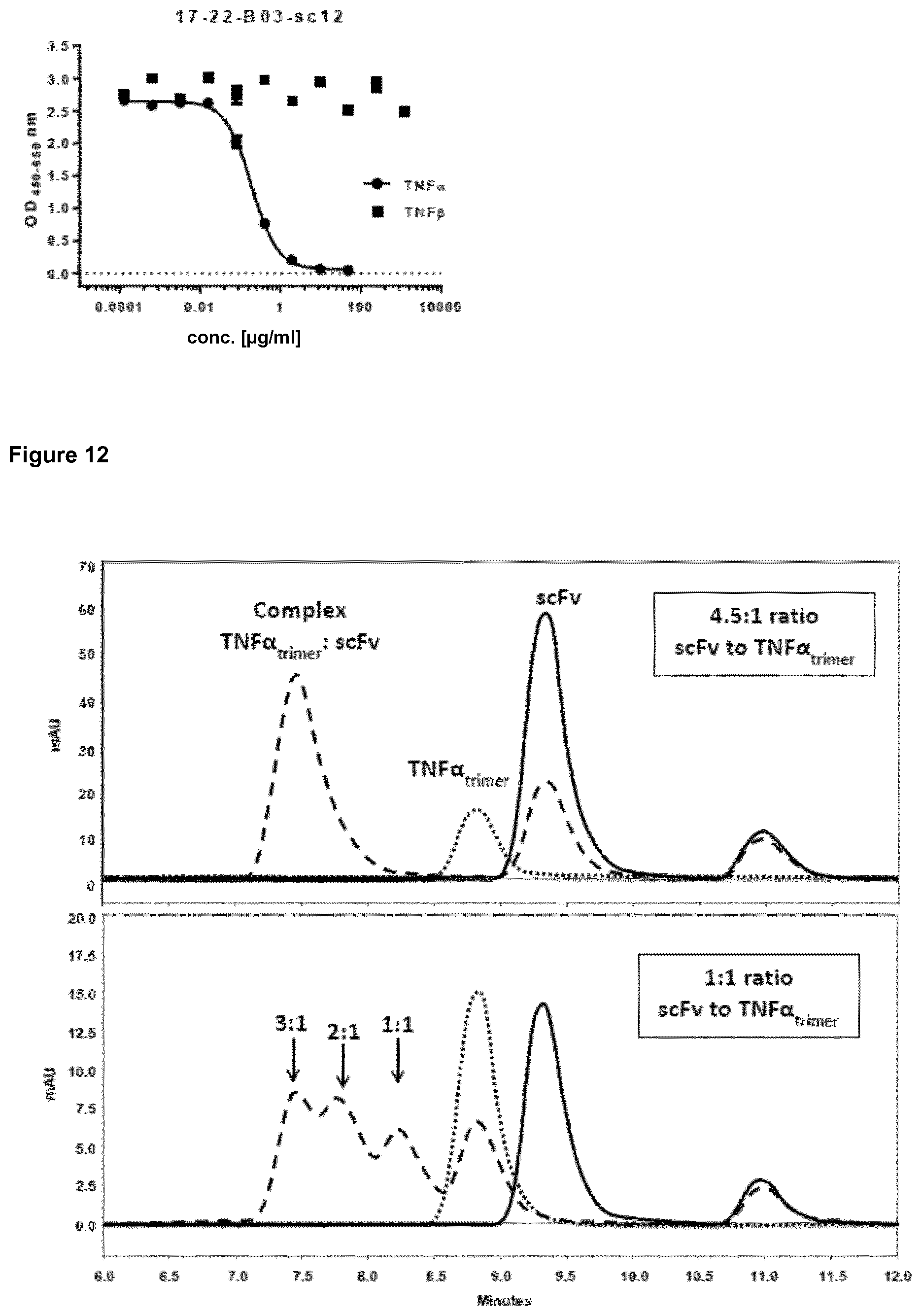


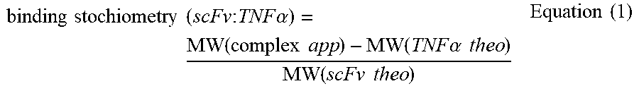
S00001
XML
uspto.report is an independent third-party trademark research tool that is not affiliated, endorsed, or sponsored by the United States Patent and Trademark Office (USPTO) or any other governmental organization. The information provided by uspto.report is based on publicly available data at the time of writing and is intended for informational purposes only.
While we strive to provide accurate and up-to-date information, we do not guarantee the accuracy, completeness, reliability, or suitability of the information displayed on this site. The use of this site is at your own risk. Any reliance you place on such information is therefore strictly at your own risk.
All official trademark data, including owner information, should be verified by visiting the official USPTO website at www.uspto.gov. This site is not intended to replace professional legal advice and should not be used as a substitute for consulting with a legal professional who is knowledgeable about trademark law.