Anti-fam19a5 Antibodies And Uses Thereof
KIM; Bongcheol ; et al.
U.S. patent application number 16/626624 was filed with the patent office on 2020-09-24 for anti-fam19a5 antibodies and uses thereof. This patent application is currently assigned to Neuracle Science Co., Ltd.. The applicant listed for this patent is Neuracle Science Co., Ltd., Seoul National University R&DB Foundation. Invention is credited to Junho CHUNG, Junyeong JIN, Bongcheol KIM, Dong Sik KIM, Jae-Keun LEE.
| Application Number | 20200299373 16/626624 |
| Document ID | / |
| Family ID | 1000004930062 |
| Filed Date | 2020-09-24 |


View All Diagrams
| United States Patent Application | 20200299373 |
| Kind Code | A1 |
| KIM; Bongcheol ; et al. | September 24, 2020 |
ANTI-FAM19A5 ANTIBODIES AND USES THEREOF
Abstract
The present disclosure provides antibodies that specifically bind to human FAM19A5 and compositions comprising such antibodies. Also provided herein are methods for treating fibrosis or cancer using the anti-FAM19A5 antibodies.
| Inventors: | KIM; Bongcheol; (Seongnam-si, KR) ; LEE; Jae-Keun; (Seoul, KR) ; KIM; Dong Sik; (Seoul, KR) ; CHUNG; Junho; (Seongnam-si, KR) ; JIN; Junyeong; (Gwachun-si, KR) | ||||||||||
| Applicant: |
|
||||||||||
|---|---|---|---|---|---|---|---|---|---|---|---|
| Assignee: | Neuracle Science Co., Ltd. Seoul KR Seoul National University R&DB Foundation Seoul KR |
||||||||||
| Family ID: | 1000004930062 | ||||||||||
| Appl. No.: | 16/626624 | ||||||||||
| Filed: | June 27, 2018 | ||||||||||
| PCT Filed: | June 27, 2018 | ||||||||||
| PCT NO: | PCT/IB2018/054785 | ||||||||||
| 371 Date: | December 26, 2019 |
Related U.S. Patent Documents
| Application Number | Filing Date | Patent Number | ||
|---|---|---|---|---|
| 62582887 | Nov 7, 2017 | |||
| 62525635 | Jun 27, 2017 | |||
| Current U.S. Class: | 1/1 |
| Current CPC Class: | C07K 2317/24 20130101; C07K 16/24 20130101; G01N 33/5058 20130101; G01N 33/57407 20130101; C07K 2317/565 20130101; G01N 2333/7158 20130101; C07K 2317/34 20130101; C07K 2317/92 20130101; C07K 2317/73 20130101; A61K 2039/505 20130101; A61P 35/00 20180101 |
| International Class: | C07K 16/24 20060101 C07K016/24; A61P 35/00 20060101 A61P035/00; G01N 33/574 20060101 G01N033/574; G01N 33/50 20060101 G01N033/50 |
Claims
1. An isolated antibody, or antigen-binding portion thereof, which specifically binds to human family with sequence similarity 19, member A5 ("FAM19A5") protein ("anti-FAM19A5 antibody") and which cross-competes for binding to a human FAM19A5 epitope with a reference antibody comprising a heavy chain CDR1, CDR2, and CDR3 and a light chain CDR1, CDR2, and CDR3, wherein: (i) the heavy chain CDR1 comprises the amino acid sequence set forth in SEQ ID NO: 11, the heavy chain CDR2 comprises the amino acid sequence set forth in SEQ ID NO: 12, the heavy chain CDR3 comprises the amino acid sequence set forth in SEQ ID NO: 13, the light chain CDR1 comprises the amino acid sequence set forth in SEQ ID NO: 23, the light chain CDR2 comprises the amino acid sequence set forth in SEQ ID NO: 24, and the light chain CDR3 comprises the amino acid sequence set forth in SEQ ID NO: 25; (ii) the heavy chain CDR1 comprises the amino acid sequence set forth in SEQ ID NO: 14, the heavy chain CDR2 comprises the amino acid sequence set forth in SEQ ID NO: 15, the heavy chain CDR3 comprises the amino acid sequence set forth in SEQ ID NO: 16, the light chain CDR1 comprises the amino acid sequence set forth in SEQ ID NO: 26, the light chain CDR2 comprises the amino acid sequence set forth in SEQ ID NO: 27, and the light chain CDR3 comprises the amino acid sequence set forth in SEQ ID NO: 28; or (iii) the heavy chain CDR1 comprises the amino acid sequence set forth in SEQ ID NO: 20, the heavy chain CDR2 comprises the amino acid sequence set forth in SEQ ID NO: 21, the heavy chain CDR3 comprises the amino acid sequence set forth in SEQ ID NO: 22, the light chain CDR1 comprises the amino acid sequence set forth in SEQ ID NO: 32, the light chain CDR2 comprises the amino acid sequence set forth in SEQ ID NO: 33, and the light chain CDR3 comprises the amino acid sequence set forth in SEQ ID NO: 34.
2. An isolated antibody, or antigen-binding portion thereof, which specifically binds to human family with sequence similarity 19, member A5 ("FAM19A5") protein ("anti-FAM19A5 antibody") and which binds to the same FAM19A5 epitope as a reference antibody comprising a heavy chain CDR1, CDR2, and CDR3 and a light chain CDR1, CDR2, and CDR3, wherein: (i) the heavy chain CDR1 comprises the amino acid sequence set forth in SEQ ID NO: 11, the heavy chain CDR2 comprises the amino acid sequence set forth in SEQ ID NO: 12, the heavy chain CDR3 comprises the amino acid sequence set forth in SEQ ID NO: 13, the light chain CDR1 comprises the amino acid sequence set forth in SEQ ID NO: 23, the light chain CDR2 comprises the amino acid sequence set forth in SEQ ID NO: 24, and the light chain CDR3 comprises the amino acid sequence set forth in SEQ ID NO: 25; (ii) the heavy chain CDR1 comprises the amino acid sequence set forth in SEQ ID NO: 14, the heavy chain CDR2 comprises the amino acid sequence set forth in SEQ ID NO: 15, the heavy chain CDR3 comprises the amino acid sequence set forth in SEQ ID NO: 16, the light chain CDR1 comprises the amino acid sequence set forth in SEQ ID NO: 26, the light chain CDR2 comprises the amino acid sequence set forth in SEQ ID NO: 27, and the light chain CDR3 comprises the amino acid sequence set forth in SEQ ID NO: 28; or (iii) the heavy chain CDR1 comprises the amino acid sequence set forth in SEQ ID NO: 20, the heavy chain CDR2 comprises the amino acid sequence set forth in SEQ ID NO: 21, the heavy chain CDR3 comprises the amino acid sequence set forth in SEQ ID NO: 22, the light chain CDR1 comprises the amino acid sequence set forth in SEQ ID NO: 32, the light chain CDR2 comprises the amino acid sequence set forth in SEQ ID NO: 33, and the light chain CDR3 comprises the amino acid sequence set forth in SEQ ID NO: 34.
3. The anti-FAM19A5 antibody of claim 1, wherein the FAM19A5 epitope comprises the amino acid sequence set forth in SEQ ID NO: 6.
4. The anti-FAM19A5 antibody of claim 3, which binds to the FAM19A5 epitope at one or more amino acids corresponding to amino acid residues (i) 46 to 51 of SEQ ID NO: 2 (i.e., DSSQPR), (ii) 46, 50, and 52 of SEQ ID NO: 2 (i.e., D---P-R), or (iii) 46, 47, 48, and 50 of SEQ ID NO: 2 (i.e., DSS-P).
5. The anti-FAM19A5 antibody of claim 1, wherein the FAM19A5 epitope comprises the amino acid sequence set forth in SEQ ID NO: 9.
6. The anti-FAM19A5 antibody of claim 1, wherein the FAM19A5 epitope comprises one or more of the following amino acids: DSSQP (SEQ ID NO: 66), ARCACRK (SEQ ID NO: 68), or TCTQPGGR (SEQ ID NO: 72).
7. The anti-FAM19A5 antibody of claim 1, which comprises a heavy chain CDR1, CDR2, and CDR3 and a light chain CDR1, CDR2, and CDR3, wherein (i) the heavy chain CDR1 comprises the amino acid sequence set forth in SEQ ID NO: 11, SEQ ID NO: 14, or SEQ ID NO: 20; (ii) the heavy chain CDR2 comprises the amino acid sequence set forth in SEQ ID NO: 12, SEQ ID NO: 15, or SEQ ID NO: 21; (iii) the heavy chain CDR3 comprises the amino acid sequence set forth in SEQ ID NO: 13, SEQ ID NO: 16, or SEQ ID NO: 22; (iv) the light chain CDR1 comprises the amino acid sequence set forth in SEQ ID NO: 23, SEQ ID NO: 26, or SEQ ID NO: 32; (v) the light chain CDR2 comprises the amino acid sequence set forth in SEQ ID NO: 24, SEQ ID NO: 27, or SEQ ID NO: 33; and/or (vi) the light chain CDR3 comprises the amino acid sequence set forth in SEQ ID NO: 25, SEQ ID NO: 28, or SEQ ID NO: 34.
8. The anti-FAM19A5 antibody of claim 1, which comprises (i) a heavy chain variable region comprising the amino acid sequence set forth in SEQ ID NO: 35 and a light chain variable region comprising the amino acid sequence set forth in SEQ ID NO: 39; (ii) a heavy chain variable region comprising the amino acid sequence set forth in SEQ ID NO: 36 and a light chain variable region comprising the amino acid sequence set forth in SEQ ID NO: 40; or (iii) a heavy chain variable region comprising the amino acid sequence set forth in SEQ ID NO: 38 and a light chain variable region comprising the amino acid sequence set forth in SEQ ID NO: 42.
9. The anti-FAM19A5 antibody of claim 1, which comprises a heavy chain variable region and a light chain variable region, wherein the heavy chain variable region comprises an amino acid sequence which is at least about 80% identical to the amino acid sequence set forth in SEQ ID NO: 35, SEQ ID NO: 36, or SEQ ID NO: 38, and/or wherein the light chain variable region comprises an amino acid sequence which is at least about 80% identical to the amino acid sequence set forth in SEQ ID NO: 39, SEQ ID NO: 40, or SEQ ID NO: 42.
10. The anti-FAM19A5 antibody of claim 1, which is a chimeric antibody, a human antibody, or a humanized antibody.
11. The anti-FAM19A5 antibody of claim 1, which comprises a heavy chain and a light chain, wherein the (i) heavy chain comprising the amino acid sequence set forth in SEQ ID NO: 57, SEQ ID NO: 58, or SEQ ID NO: 60; and (ii) the light chain comprises the amino acid sequence set forth in SEQ ID NO: 61, SEQ ID NO: 62, or SEQ ID NO: 64.
12. The anti-FAM19A5 antibody of claim 1, which comprises one or more of the following properties: (a) reduces, reverses, delays and/or prevents fibrosis; (b) reduces formation of excessive extracellular matrix (ECM); (c) delays tumor growth or progression; (d) binds to soluble human FAM19A5 with a KD of 10 nM or less as measured by enzyme-linked immunosorbent assay (ELISA); (e) binds to membrane bound human FAM19A5 with a KD of 10 nM or less as measured by ELISA; (f) reduces, reverses, delays, and/or prevents an onset of reactive gliosis; (g) suppresses an excessive proliferation of reactive astrocytes; (h) decreases expression of chondroitin sulfate proteoglycans including neurocan and neuron-glial antigen 2 (NG2); (i) increases expression of c-fos and pERK in the nucleus of neurons; (j) promotes survival of neurons; (k) increases expression of GAP43 in neurons; and (l) promotes regrowth of an axon.
13. A human family with sequence similarity 19, member A5 ("FAM19A5") epitope consisting essentially of an amino acid sequence that is at least 90% identical to the amino acid sequence set forth in SEQ ID NO: 5, 6, 9, or 10, wherein the FAM19A5 epitope is capable of being specifically bound to a reference antibody comprising (i) a heavy chain variable region, which comprises the amino acid sequence set forth in SEQ ID NO: 35, SEQ ID NO: 36, or SEQ ID NO: 38, and (ii) a light chain variable region, which comprises the amino acid sequence set forth in SEQ ID NO: 39, SEQ ID NO: 40, or SEQ ID NO: 42.
14. A nucleic acid encoding the anti-FAM19A5 antibody, or a fragment thereof, of claim 1.
15. A composition comprising the anti-FAM19A5 antibody of claim 1 and a carrier.
16. A method of treating a brain cancer in a subject in need thereof, comprising administering the anti-FAM19A5 antibody of claim 1 to the subject.
17. A method of determining a FAM19A5 protein level in a subject in need thereof comprising detecting whether the FAM19A5 protein level from a biological sample obtained from the subject is increased compared to a corresponding level from a normal control by contacting the biological sample of the subject with the anti-FAM19A5 antibody of claim 1.
18. The anti-FAM19A5 antibody of claim 1, wherein the antigen-binding portion thereof comprises a Fab, Fab', F(ab')2, Fv fragment, diabody, linear antibody, single chain antibody, multi-specific antibody formed from the antibody fragment, or combinations thereof.
19. A method of preparing an antibody, or antigen-binding portion thereof, which specifically binds to human family with sequence similarity 19, member A5 ("FAM19A5") protein ("anti-FAM19A5 antibody") comprising immunizing a non-human animal with the FAM19A5 epitope of claim 13, and producing the anti-FAM19A5 antibody.
20. A method of producing an antibody, or antigen-binding portion thereof, which specifically binds to human family with sequence similarity 19, member A5 ("FAM19A5") protein ("anti-FAM19A5 antibody") comprising culturing a cell which has been transformed with the nucleic acid of claim 14 under suitable conditions, and isolating the anti-FAM19A5 antibody.
Description
REFERENCE TO SEQUENCE LISTING SUBMITTED ELECTRONICALLY
[0001] The content of the electronically submitted sequence listing in ASCII text file (Name: 3763.005PC02_SeqListing_ST25.txt; Size: 166,889 bytes; and Date of Creation: Jun. 26, 2018) filed with the application is incorporated herein by reference in its entirety.
FIELD OF THE DISCLOSURE
[0002] The present disclosure provides antibodies that specifically bind to family with sequence similarity 19, member A5 (FAM19A5), compositions comprising such antibodies, and method of using such antibodies for preventing or treating disorders or diseases such as a fibrosis and/or cancer (e.g., brain tumor, e.g., glioblastoma) in a subject.
BACKGROUND OF THE DISCLOSURE
[0003] FAM19A5 is a member of the TAFA subfamily of proteins which is composed of five highly homologous small proteins. Tang T. Y. et al., Genomics 83(4):727-34 (2004). These proteins contain conserved cysteine residues at fixed positions, and are distantly related to macrophage inflammatory protein 1-alpha (MIP-1-alpha), a member of the CC-chemokine family. The TAFA proteins are predominantly expressed in specific regions of the brain and the spinal cord. These proteins are believed to be generated and secreted by adult neural stem cells in neurogenesis processes.
[0004] FAM19A5 is predominantly expressed in the brain of vertebrates and it is believed that FAM19A5 is important in the development, differentiation, formation of a complete central nervous system, and can be used in the prevention or treatment of central nervous system injuries and/or diseases. U.S. Patent Publication No. 2015/0118230.
[0005] In addition to regulation of the nervous system, FAM19A5 may also play a role in regulating immune cells. Fibrosis is a common health problem, often occurring in various pathological processes, and is characterized by infiltrating mononuclear immune cells which release cytokines that stimulate fibroblasts to alter connective tissue. Accordingly, there is a need to develop antibodies that specifically bind to FAM19A5 and that are capable of modulating FAM19A5 activity.
BRIEF SUMMARY OF THE DISCLOSURE
[0006] Provided herein is an isolated antibody, or antigen-binding portion thereof, which specifically binds to human family with sequence similarity 19, member A5 ("FAM19A5") protein ("anti-FAM19A5 antibody") and which cross-competes for binding to a human FAM19A5 epitope with a reference antibody comprising a heavy chain CDR1, CDR2, and CDR3 and a light chain CDR1, CDR2, and CDR3, wherein: (i) the heavy chain CDR1 comprises the amino acid sequence of SEQ ID NO: 11, the heavy chain CDR2 comprises the amino acid sequence of SEQ ID NO: 12, the heavy chain CDR3 comprises the amino acid sequence of SEQ ID NO: 13, the light chain CDR1 comprises the amino acid sequence of SEQ ID NO: 23, the light chain CDR2 comprises the amino acid sequence of SEQ ID NO: 24, and the light chain CDR3 comprises the amino acid sequence of SEQ ID NO: 25; (ii) the heavy chain CDR1 comprises the amino acid sequence of SEQ ID NO: 14, the heavy chain CDR2 comprises the amino acid sequence of SEQ ID NO: 15, the heavy chain CDR3 comprises the amino acid sequence of SEQ ID NO: 16, the light chain CDR1 comprises the amino acid sequence of SEQ ID NO: 26, the light chain CDR2 comprises the amino acid sequence of SEQ ID NO: 27, and the light chain CDR3 comprises the amino acid sequence of SEQ ID NO: 28; or (iii) the heavy chain CDR1 comprises the amino acid sequence of SEQ ID NO: 20, the heavy chain CDR2 comprises the amino acid sequence of SEQ ID NO: 21, the heavy chain CDR3 comprises the amino acid sequence of SEQ ID NO: 22, the light chain CDR1 comprises the amino acid sequence of SEQ ID NO: 32, the light chain CDR2 comprises the amino acid sequence of SEQ ID NO: 33, and the light chain CDR3 comprises the amino acid sequence of SEQ ID NO: 34.
[0007] Also provided herein is an isolated antibody, or antigen-binding portion thereof, which specifically binds to human family with sequence similarity 19, member A5 ("FAM19A5") protein ("anti-FAM19A5 antibody") and which binds to the same FAM19A5 epitope as a reference antibody comprising a heavy chain CDR1, CDR2, and CDR3 and a light chain CDR1, CDR2, and CDR3, wherein: (i) the heavy chain CDR1 comprises the amino acid sequence of SEQ ID NO: 11, the heavy chain CDR2 comprises the amino acid sequence of SEQ ID NO: 12, the heavy chain CDR3 comprises the amino acid sequence of SEQ ID NO: 13, the light chain CDR1 comprises the amino acid sequence of SEQ ID NO: 23, the light chain CDR2 comprises the amino acid sequence of SEQ ID NO: 24, and the light chain CDR3 comprises the amino acid sequence of SEQ ID NO: 25; (ii) the heavy chain CDR1 comprises the amino acid sequence of SEQ ID NO: 14, the heavy chain CDR2 comprises the amino acid sequence of SEQ ID NO: 15, the heavy chain CDR3 comprises the amino acid sequence of SEQ ID NO: 16, the light chain CDR1 comprises the amino acid sequence of SEQ ID NO: 26, the light chain CDR2 comprises the amino acid sequence of SEQ ID NO: 27, and the light chain CDR3 comprises the amino acid sequence of SEQ ID NO: 28; or (iii) the heavy chain CDR1 comprises the amino acid sequence of SEQ ID NO: 20, the heavy chain CDR2 comprises the amino acid sequence of SEQ ID NO: 21, the heavy chain CDR3 comprises the amino acid sequence of SEQ ID NO: 22, the light chain CDR1 comprises the amino acid sequence of SEQ ID NO: 32, the light chain CDR2 comprises the amino acid sequence of SEQ ID NO: 33, and the light chain CDR3 comprises the amino acid sequence of SEQ ID NO: 34.
[0008] In some embodiments, the anti-FAM19A5 antibody of the present disclosure binds to at least one FAM19A5 epitope, which is SEQ ID NO: 6. In certain embodiments, the anti-FAM19A5 antibody binds to the FAM19A5 epitope, which is SEQ ID NO: 6, at one or more amino acids corresponding to amino acid residues (i) 46 to 51 (i.e., DSSQPR), (ii) 46, 50, and 52 (i.e., D---P-R), or (iii) 46, 47, 48, and 50 (i.e., DSS-P). In some embodiments, the anti-FAM19A5 antibody binds to at least one FAM19A5 epitope, which is SEQ ID NO: 9. In certain embodiments, the anti-FAM19A5 antibody binds to at least one FAM19A5 epitope identified as EP2, EP4, and/or EP8, wherein EP2 comprises the amino acids DSSQP (SEQ ID NO: 66), wherein EP4 comprises the amino acids ARCACRK (SEQ ID NO: 68), and wherein EP8 comprises the amino acids TCTQPGGR (SEQ ID NO: 72).
[0009] In some embodiments, the anti-FAM19A5 antibody disclosed herein comprises a heavy chain CDR1, CDR2, and CDR3 and a light chain CDR1, CDR2, and CDR3, wherein (i) the heavy chain CDR1 comprises SEQ ID NO: 11, SEQ ID NO: 14, or SEQ ID NO: 20; (ii) the heavy chain CDR2 comprises SEQ ID NO: 12, SEQ ID NO: 15, or SEQ ID NO: 21; (iii) the heavy chain CDR3 comprises SEQ ID NO: 13, SEQ ID NO: 16, or SEQ ID NO: 22; (iv) the light chain CDR1 comprises SEQ ID NO: 23, SEQ ID NO: 26, or SEQ ID NO: 32; (v) the light chain CDR2 comprises SEQ ID NO: 24, SEQ ID NO: 27, or SEQ ID NO: 33; and/or (vi) the light chain CDR3 comprises SEQ ID NO: 25, SEQ ID NO: 28, or SEQ ID NO: 34.
[0010] In some embodiments, the anti-FAM19A5 antibody comprises (i) a heavy chain variable domain comprising SEQ ID NO: 35 and a light chain variable domain comprising SEQ ID NO: 39; (ii) a heavy chain variable domain comprising SEQ ID NO: 36 and a light chain variable domain comprising SEQ ID NO: 40; or (iii) a heavy chain variable domain comprising SEQ ID NO: 38 and a light chain variable domain comprising SEQ ID NO: 42.
[0011] In some embodiments, the anti-FAM19A5 antibody comprises a heavy chain variable region and a light chain variable region, wherein the heavy chain variable region comprises an amino acid sequence which is at least about 80%, at least about 85%, at least about 90%, at least about 95%, at least about 96%, at least about 97%, at least about 98%, at least about 99%, or about 100% identical to the amino acid sequence set forth as SEQ ID NO: 35, SEQ ID NO: 36, or SEQ ID NO: 38, and/or wherein the light chain variable region comprises an amino acid sequence which is at least about 80%, at least about 85%, at least about 90%, at least about 95%, at least about 96%, at least about 97%, at least about 98%, at least about 99%, or about 100% identical to the amino acid sequence set forth as SEQ ID NO: 39, SEQ ID NO: 40, or SEQ ID NO: 42.
[0012] In some embodiments, the anti-FAM19A5 antibody comprises (i) a heavy chain comprising SEQ ID NO: 57, SEQ ID NO: 58, or SEQ ID NO: 60; and (ii) a light chain comprising SEQ ID NO: 61, SEQ ID NO: 62, or SEQ ID NO: 64.
[0013] In some embodiments, the anti-FAM19A5 antibody of the present disclosure is a chimeric antibody, a human antibody, or a humanized antibody. In some embodiments, the anti-FAM19A5 antibody comprises one or more of the following properties: (a) reduces, reverses, delays and/or prevents fibrosis; (b) reduces formation of excessive extracellular matrix (ECM); (c) delays tumor growth or progression; (d) binds to soluble human FAM19A5 with a KD of 10 nM or less as measured by enzyme-linked immunosorbent assay (ELISA); (e) binds to membrane bound human FAM19A5 with a KD of 10 nM or less as measured by ELISA; (f) reduces, reverses, delays, and/or prevents an onset of reactive gliosis; (g) suppresses an excessive proliferation of reactive astrocytes; (h) decreases expression of chondroitin sulfate proteoglycans including neurocan and neuron-glial antigen 2 (NG2); (i) increases expression of c-fos and pERK in the nucleus of neurons; (j) promotes survival of neurons; (k) increases expression of GAP43 in neurons; and (l) promotes regrowth of an axon.
[0014] The present disclosure further provides a human family with sequence similarity 19, member A5 (FAM19A5) epitope consisting essentially of or consisting of an amino acid sequence at least 90%, at least about 95%, at least about 96%, at least about 97%, at least about 98%, at least about 99%, or about 100% identical to SEQ ID NO: 5, 6, 9, or 10, wherein the epitope is capable of being specifically bound to a reference antibody comprising (i) a heavy chain variable domain comprising SEQ ID NO: 35, SEQ ID NO: 36, or SEQ ID NO: 38, and (ii) a light chain variable domain comprising SEQ ID NO: 39, SEQ ID NO: 40, or SEQ ID NO: 42.
[0015] Also disclosed herein is a nucleic acid encoding an anti-FAM19A5 antibody or an FAM19A5 epitope described herein. Also provided is a composition comprising an anti-FAM19A5 antibody or an FAM19A5 epitope of the present disclosure.
[0016] In some aspects, the present application also provides an anti-FAM19A5 antibody for treating a brain cancer.
[0017] In other aspects, the present disclosure provides a method of diagnosing a subject in need thereof comprising contacting a biological sample of the subject with an anti-FAM19A5 antibody disclosed herein.
EMBODIMENTS
Embodiment 1
[0018] An isolated monoclonal antibody or antigen binding portion thereof that specifically binds to human family with sequence similarity 19, member A5 (FAM19A5) and exhibits one or more of the following properties: [0019] (a) reduces, reverses, delays and/or prevents fibrosis; [0020] (b) reduces formation of excessive extracellular matrix (ECM); and [0021] (c) delays tumor growth or progression.
Embodiment 2
[0022] The monoclonal antibody or antigen binding portion thereof of Embodiment 1, which further comprises one or more of the following properties: [0023] (d) binds to soluble human FAM19A5 with a KD of 10 nM or less as measured by enzyme-linked immunosorbent assay (ELISA); [0024] (e) binds to membrane bound human FAM19A5 with a KD of 10 nM or less as measured by ELISA; [0025] (f) reduces, reverses, delays, and/or prevents an onset of reactive gliosis; [0026] (g) suppresses an excessive proliferation of reactive astrocytes; [0027] (h) decreases expression of chondroitin sulfate proteoglycans including neurocan and neuron-glial antigen 2 (NG2); [0028] (i) increases expression of c-fos and pERK in the nucleus of neurons; [0029] (j) promotes survival of neurons; [0030] (k) increases expression of GAP43 in neurons; and [0031] (l) promotes regrowth of an axon.
Embodiment 3
[0032] The monoclonal antibody or antigen binding portion thereof of Embodiment 1 or 2, which cross-competes for binding to a human FAM19A5 epitope with (i) a reference antibody comprising heavy chain CDR1, CDR2, and CDR3 and light chain CDR1, CDR2, and CDR3, wherein the heavy chain CDR1 comprises the amino acid sequence of SEQ ID NO: 11, the heavy chain CDR2 comprises the amino acid sequence of SEQ ID NO: 12, the heavy chain CDR3 comprises the amino acid sequence of SEQ ID NO: 13, the light chain CDR1 comprises the amino acid sequence of SEQ ID NO: 23, the light chain CDR2 comprises the amino acid sequence of SEQ ID NO: 24, and the light chain CDR3 comprises the amino acid sequence of SEQ ID NO: 25; (ii) a reference antibody comprising heavy chain CDR1, CDR2, and CDR3 and light chain CDR1, CDR2, and CDR3, wherein the heavy chain CDR1 comprises the amino acid sequence of SEQ ID NO: 14, the heavy chain CDR2 comprises the amino acid sequence of SEQ ID NO: 15, the heavy chain CDR3 comprises the amino acid sequence of SEQ ID NO: 16, the light chain CDR1 comprises the amino acid sequence of SEQ ID NO: 26, the light chain CDR2 comprises the amino acid sequence of SEQ ID NO: 27, and the light chain CDR3 comprises the amino acid sequence of SEQ ID NO: 28; or (iii) a reference antibody comprising heavy chain CDR1, CDR2, and CDR3 and light chain CDR1, CDR2, and CDR3, wherein the heavy chain CDR1 comprises the amino acid sequence of SEQ ID NO: 20, the heavy chain CDR2 comprises the amino acid sequence of SEQ ID NO: 21, the heavy chain CDR3 comprises the amino acid sequence of SEQ ID NO: 22, the light chain CDR1 comprises the amino acid sequence of SEQ ID NO: 32, the light chain CDR2 comprises the amino acid sequence of SEQ ID NO: 33, and the light chain CDR3 comprises the amino acid sequence of SEQ ID NO: 34.
Embodiment 4
[0033] The monoclonal antibody or antigen binding portion thereof of Embodiment 1 or 2, which binds to the same FAM19A5 epitope as (i) a reference antibody comprising heavy chain CDR1, CDR2, and CDR3 and light chain CDR1, CDR2, and CDR3, wherein the heavy chain CDR1 comprises the amino acid sequence of SEQ ID NO: 11, the heavy chain CDR2 comprises the amino acid sequence of SEQ ID NO: 12, the heavy chain CDR3 comprises the amino acid sequence of SEQ ID NO: 13, the light chain CDR1 comprises the amino acid sequence of SEQ ID NO: 23, the light chain CDR2 comprises the amino acid sequence of SEQ ID NO: 24, and the light chain CDR3 comprises the amino acid sequence of SEQ ID NO: 25; (ii) a reference antibody comprising heavy chain CDR1, CDR2, and CDR3 and light chain CDR1, CDR2, and CDR3, wherein the heavy chain CDR1 comprises the amino acid sequence of SEQ ID NO: 14, the heavy chain CDR2 comprises the amino acid sequence of SEQ ID NO: 15, the heavy chain CDR3 comprises the amino acid sequence of SEQ ID NO: 16, the light chain CDR1 comprises the amino acid sequence of SEQ ID NO: 26, the light chain CDR2 comprises the amino acid sequence of SEQ ID NO: 27, and the light chain CDR3 comprises the amino acid sequence of SEQ ID NO: 28; or (iii) a reference antibody comprising heavy chain CDR1, CDR2, and CDR3 and light chain CDR1, CDR2, and CDR3, wherein the heavy chain CDR1 comprises the amino acid sequence of SEQ ID NO: 20, the heavy chain CDR2 comprises the amino acid sequence of SEQ ID NO: 21, the heavy chain CDR3 comprises the amino acid sequence of SEQ ID NO: 22, the light chain CDR1 comprises the amino acid sequence of SEQ ID NO: 32, the light chain CDR2 comprises the amino acid sequence of SEQ ID NO: 33, and the light chain CDR3 comprises the amino acid sequence of SEQ ID NO: 34.
Embodiment 5
[0034] The monoclonal antibody or antigen binding portion thereof of Embodiment 3 or 4, which binds to at least one FAM19A5 epitope, which is SEQ ID NO: 6.
Embodiment 6
[0035] The monoclonal antibody or antigen binding portion thereof of any one of Embodiments 2 to 5, wherein monoclonal the antibody, or antigen binding portion thereof, binds only to an FAM19A5, which is SEQ ID NO: 6.
Embodiment 7
[0036] The monoclonal antibody or antigen binding portion thereof of any one of Embodiments 2 to 5, wherein the monoclonal antibody or antigen binding portion thereof further binds to an additional FAM19A5 epitope.
Embodiment 8
[0037] The monoclonal antibody or antigen binding portion thereof of Embodiment 7, wherein the additional FAM19A5 epitope is selected from the group consisting of SEQ ID NO: 5, SEQ ID NO: 6, SEQ ID NO: 7, SEQ ID NO: 8, SEQ ID NO: 9, SEQ ID NO: 10, and any combination thereof.
Embodiment 9
[0038] The monoclonal antibody or antigen binding portion thereof of any one of Embodiments 1 to 8, which comprises heavy chain CDR1, CDR2, and CDR3 and light chain CDR1, CDR2, and CDR3.
Embodiment 10
[0039] The monoclonal antibody or antigen binding portion thereof of Embodiment 9, wherein the heavy chain CDR1 comprises SEQ ID NO: 11, SEQ ID NO: 14, or SEQ ID NO: 20.
Embodiment 11
[0040] The monoclonal antibody or antigen binding portion thereof of Embodiment 9 or 10, wherein the heavy chain CDR2 comprises SEQ ID NO: 12, SEQ ID NO: 15, or SEQ ID NO: 21.
Embodiment 12
[0041] The monoclonal antibody or antigen binding portion thereof of any one of Embodiments 9 to 11, wherein the heavy chain CDR3 comprises SEQ ID NO: 13, SEQ ID NO: 16, or SEQ ID NO: 22.
Embodiment 13
[0042] The monoclonal antibody or antigen binding portion thereof of any one of Embodiments 9 to 12, wherein the light chain CDR1 comprises SEQ ID NO: 23, SEQ ID NO: 26, or SEQ ID NO: 32.
Embodiment 14
[0043] The monoclonal antibody or antigen binding portion thereof of any one of Embodiments 9 to 13, wherein the light chain CDR2 comprises SEQ ID NO: 24, SEQ ID NO: 27, or SEQ ID NO: 33.
Embodiment 15
[0044] The monoclonal antibody or antigen binding portion thereof of any one of Embodiments 9 to 14, wherein the light chain CDR3 comprises SEQ ID NO: 25, SEQ ID NO: 28, or SEQ ID NO: 34.
Embodiment 16
[0045] The monoclonal antibody or antigen binding portion thereof of any one of Embodiments 1 to 15, which comprises (i) a heavy chain variable domain comprising SEQ ID NO: 35, SEQ ID NO: 36, or SEQ ID NO: 38, and (ii) a light chain variable domain comprising SEQ ID NO: 39, SEQ ID NO: 40, or SEQ ID NO: 42.
Embodiment 17
[0046] The monoclonal antibody or antigen binding portion thereof of any one of Embodiments 1 to 16, which comprises a heavy chain variable region and a light chain variable region, wherein the heavy chain variable region comprises an amino acid sequence which is at least about 80%, at least about 85%, at least about 90%, at least about 95%, at least about 96%, at least about 97%, at least about 98%, at least about 99%, or about 100% identical to the amino acid sequence set forth as SEQ ID NO: 35, SEQ ID NO: 36, or SEQ ID NO: 38.
Embodiment 18
[0047] The monoclonal antibody or antigen binding portion thereof of any one of Embodiments 1 to 17, which comprises a heavy chain variable region and a light chain variable region, wherein the light chain variable region comprises an amino acid sequence which is at least about 80%, at least about 85%, at least about 90%, at least about 95%, at least about 96%, at least about 97%, at least about 98%, at least about 99%, or about 100% identical to the amino acid sequence set forth as SEQ ID NO: 39, SEQ ID NO: 40, or SEQ ID NO: 42.
Embodiment 19
[0048] The monoclonal antibody of any one of Embodiments 1 to 18, wherein the monoclonal antibody is a single domain antibody.
Embodiment 20
[0049] The monoclonal antibody or antigen binding portion thereof of any one of Embodiments 1 to 19, wherein the monoclonal antibody is selected from the group consisting of an IgG1, an IgG2, an IgG3, an IgG4, a variant thereof, or a combination thereof.
Embodiment 21
[0050] The monoclonal antibody or antigen binding portion thereof of Embodiment 20, wherein the monoclonal antibody is a human IgG1 antibody.
Embodiment 22
[0051] The monoclonal antibody or antigen binding portion thereof of Embodiment 20, wherein the monoclonal antibody comprises a human IgG2 or human IgG4 isotype antibody.
Embodiment 23
[0052] The monoclonal antibody or antigen binding portion thereof of any one of Embodiments 1 to 22, further comprising a constant region without the Fc function.
Embodiment 24
[0053] The monoclonal antibody or antigen binding portion thereof of any one of Embodiments 1 to 23, which is a chimeric antibody, a human antibody, or a humanized antibody.
Embodiment 25
[0054] The monoclonal antibody of any one of Embodiments 1 to 24, wherein the monoclonal antibody comprises (i) a heavy chain comprising SEQ ID NO: 57, SEQ ID NO: 58, or SEQ ID NO: 60; and (ii) a light chain comprising SEQ ID NO: 61, SEQ ID NO: 62, or SEQ ID NO: 64.
Embodiment 26
[0055] The antigen binding portion thereof of any one of Embodiments 1 to 25, wherein the antigen binding portion thereof is an Fab, an Fab', an F(ab')2, an Fv, or a single chain Fv (scFv).
Embodiment 27
[0056] A bispecific molecule comprising the monoclonal antibody or antigen binding portion thereof of any one of Embodiments 1 to 26 that is linked to a molecule having a second binding moiety.
Embodiment 28
[0057] A human family with sequence similarity 19, member A5 (FAM19A5) epitope consisting essentially of or consisting of an amino acid sequence at least 90%, at least about 95%, at least about 96%, at least about 97%, at least about 98%, at least about 99%, or about 100% identical to SEQ ID NO: 5, 6, 9, or 10, wherein the epitope is capable of being specifically bound to a reference antibody comprising (i) a heavy chain variable domain comprising SEQ ID NO: 35, SEQ ID NO: 36, or SEQ ID NO: 38, and (ii) a light chain variable domain comprising SEQ ID NO: 39, SEQ ID NO: 40, or SEQ ID NO: 42.
Embodiment 29
[0058] A nucleic acid encoding the monoclonal antibody or antigen binding portion thereof of any one of Embodiments 1 to 26, the bispecific molecule of Embodiment 27, or the epitope of Embodiment 28.
Embodiment 30
[0059] A vector comprising the nucleic acid of Embodiment 29.
Embodiment 31
[0060] The vector of Embodiment 30, for use in gene therapy.
Embodiment 32
[0061] A cell transformed with an expression vector of Embodiment 30.
Embodiment 33
[0062] An immunoconjugate comprising the monoclonal antibody or antigen binding portion thereof of any one of Embodiments 1 to 26 or the bispecific molecule of Embodiment 27, linked to an agent.
Embodiment 34
[0063] A composition comprising (i) the monoclonal antibody, or antigen binding portion thereof, of any one of Embodiments 1 to 26, the bispecific molecule of Embodiment 27, or the immunoconjugate of Embodiment 33, and (ii) a carrier.
Embodiment 35
[0064] A kit comprising (i) the monoclonal antibody, or antigen binding portion thereof, of any one of Embodiments 1 to 26, the bispecific molecule of Embodiment 27, or the immunoconjugate of Embodiment 33, and (ii) instructions for use.
Embodiment 36
[0065] A method of preparing an anti-FAM19A5 antibody or antigen binding portion thereof comprising immunizing a non-human animal with the epitope of Embodiment 28 and producing an antibody, or antigen binding portion thereof.
Embodiment 37
[0066] A method of producing an anti-FAM19A5 antibody or antigen binding portion thereof comprising culturing the cell of Embodiment 32 under suitable condition and isolating the antibody or antigen binding portion thereof.
Embodiment 38
[0067] A method of reducing, reversing, and/or preventing fibrosis in a subject in need thereof comprising administering the monoclonal antibody or antigen binding portion thereof of any one of Embodiments 1 to 26, the bispecific molecule of Embodiment 27, or the immunoconjugate of Embodiment 33 such that the onset of fibrosis is delayed.
Embodiment 39
[0068] A method of reducing formation of excessive extracellular matrix (ECM) in a subject in need thereof comprising administering to the subject the monoclonal antibody or antigen binding portion thereof of any one of Embodiments 1 to 26, the bispecific molecule of Embodiment 27, or the immunoconjugate of Embodiment 33, wherein the formation of excess ECM is reduced.
Embodiment 40
[0069] A method of preventing, ameliorating, or treating a liver cancer, a lung cancer, a renal cancer, a breast cancer, and/or a pancreatic cancer in a subject in need thereof comprising administering to the subject the monoclonal antibody or antigen binding portion thereof of any one of Embodiments 1 to 26, the bispecific molecule of Embodiment 27, or the immunoconjugate of Embodiment 33.
Embodiment 41
[0070] The method of Embodiment 40, wherein the liver cancer is hepatocellular carcinoma.
Embodiment 42
[0071] A method of preventing, ameliorating, or treating a liver cancer, a lung cancer, a renal cancer, a breast cancer, and/or a pancreatic cancer associated with a fibrosis in a subject in need thereof comprising administering to the subject the monoclonal antibody or antigen binding portion thereof of any one of Embodiments 1 to 26, the bispecific molecule of Embodiment 27, or the immunoconjugate of Embodiment 33.
Embodiment 43
[0072] The method of Embodiment 38 or 42, wherein the fibrosis comprises a hepatic fibrosis, pulmonary fibrosis, renal fibrosis, myelofibrosis, pancreatic fibrosis, skin fibrosis, cardiac fibrosis, arterial fibrosis, arthrofibrosis, breast fibrosis, muscle fibrosis, retroperitoneal Fibrosis, thyroid fibrosis, lymph node fibrosis, bladder fibrosis, and/or pleural fibrosis.
Embodiment 44
[0073] The method of any one of Embodiments 38 to 43, wherein the monoclonal antibody or antigen binding portion thereof is administered intravenously, orally, parenterally, intrathecally, intra-cerebroventricularly, pulmonarily, subcutaneously, or intraventricularly.
Embodiment 45
[0074] The method of any one of Embodiments 38 to 44, wherein the subject is a human.
Embodiment 46
[0075] A method of measuring the level of FAM19A5 in a sample from a subject in need thereof comprising contacting the monoclonal antibody or antigen binding portion thereof of any one of Embodiments 1 to 26, the bispecific molecule of Embodiment 27, or the immunoconjugate of Embodiment 33 with a sample obtained from the subject.
Embodiment 47
[0076] The method of Embodiment 46, wherein the level of FAM19A5 is increased in the sample of the subject compared to a reference sample.
Embodiment 48
[0077] A method of diagnosing a fibrosis in a subject in need thereof comprising contacting the monoclonal antibody or antigen binding portion thereof of any one of Embodiments 1 to 26, the bispecific molecule of Embodiment 27, or the immunoconjugate of Embodiment 33 with a sample obtained from the subject.
Embodiment 49
[0078] The method of Embodiment 48, wherein the fibrosis is in lung or liver.
Embodiment 50
[0079] The method of Embodiment 48 or 49, wherein the level of FAM19A5 is increased in the sample of the subject compared to the level of FAM19A5 in a reference sample of a subject without the fibrosis.
Embodiment 51
[0080] A method of diagnosing a tumor with fibrosis in a subject in need thereof comprising contacting the monoclonal antibody or antigen binding portion thereof of any one of Embodiments 1 to 26, the bispecific molecule of Embodiment 27, or the immunoconjugate of Embodiment 33 with a sample obtained from the subject.
Embodiment 52
[0081] The method of Embodiment 51, wherein the level of FAM19A5 is increased in the sample of the subject compared to the level of FAM19A5 in a reference sample of a subject without the tumor.
Embodiment 53
[0082] The method of any one of Embodiments 46 to 52, wherein the sample is blood, sera, plasma, cerebro spinal fluid, tissue biopsy, organ biopsy, or their combination.
Embodiment 54
[0083] The method of any one of Embodiments 46 to 52, wherein the sample is a fresh tumor sample, a frozen tumor sample, or a formalin-fixed paraffin-embedded sample.
Embodiment 55
[0084] The method of any one of Embodiments 46 to 52, wherein the contacting comprises conducting an assay.
Embodiment 56
[0085] The method of Embodiment 54, wherein the assay comprises Western blot assay, slot blot assay, enzyme-linked immunosorbent assay (ELISA), radioimmunoassay (MA), flow cytometry-based immunofluorescence (FACS), mass spectrometry, surface plasmon resonance (SPR), or a combination thereof.
BRIEF DESCRIPTION OF THE DRAWINGS
[0086] FIG. 1 shows the amino acid sequences of epitopes F1-F6 (conjugated to BSA) and their location on the human FAM19A5 polypeptide. The size of the different epitope fragments are indicated in parentheses.
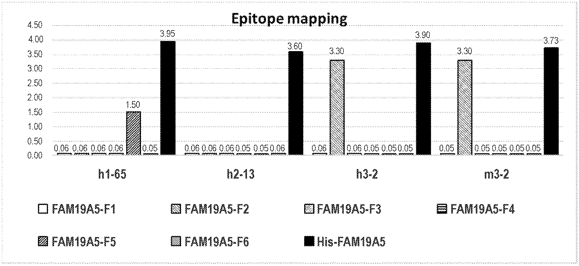
[0087] FIG. 2 shows ELISA results for the binding of anti-FAM19A5 antibodies 1-65, 2-13, and 3-2. For the 3-2 antibody, two different isotypes are shown: human IgG1 ("h3-2") and mouse IgG1 ("m3-2"). For each of the antibodies, the 1.sup.st, 2.sup.nd, 3.sup.rd, 4.sup.th, 5.sup.th, and 6.sup.th bars (starting from the left) represent binding to epitope fragments F1, F2, F3, F4, F5, and F6, respectively. The bar farthest to the right represent the positive control (i.e., His tagged FAM19A5 protein). The exact O.D. value are indicated at the top of each bar.
[0088] FIGS. 3A to 3C show the ELISA results for the binding of anti-FAM19A5 antibodies 3-2 and 1-28 to thirteen different FAM19A5 epitope F2 fragment mutant peptides: (i) F2-01-BSA (#1), (ii) F2-02-BSA (#2), (iii) F2-03-BSA (#3), (iv) F2-04-BSA (#4), (v) F2-05-BSA (#5), (vi) F2-06-BSA (#6), (vii) F2-07-BSA (#7), (viii) F2-08-BSA (#8), (ix) F2-09-BSA (#9), (x) F2-10-BSA (#10), (xi) F2-11-BSA (#11), (xii) F2-12-BSA (#12), and (xiii) F2-13-BSA (#13). FIG. 3A shows the results for the 3-2 antibody with human IgG1 isotype. FIG. 3B shows the results for the 3-2 antibody with mouse IgG1 isotype. FIG. 3C shows the results for the 1-28 antibody. The exact O.D. values are indicated at the top of each bar.
[0089] FIG. 4 shows an amino acid sequence alignment of the different members of the FAM19A family (i.e., FAM19A1-5). The regions with the greatest amino acid diversity among the members are boxed and shown as EP1 to EP8. IgG antibody ("IgG only") is shown as a control. The y-axis provides the O.D. value.
[0090] FIG. 5 shows the ELISA results for the binding of anti-FAM19A5 antibodies 1-65, 1-28, 2-13, and 3-2 to FAM19A5 mutants M1 to M8. For each of the antibodies, the eight bars correspond to mutants M1 to M8 (moving from left to right).
[0091] FIG. 6A shows the schematic diagram of the two-site sandwich ELISA assay used to assess cross-competition among the different anti-FAM19A5 antibodies. FIG. 6B shows the results of the cross-competition analysis for six different anti-FAM19A5 antibodies: 1-65, P2-A03, P2-F11, 13B4, 2-13, and 3-2. The term "S/N" refers to the signal to noise ratio, which is measured as follows: [O.D. of 10 ng/mL antigen]/[O.D. of 0 ng/mL antigen]. The grey boxes shows cross-competition (i.e., S/N ratio lower than 2).
[0092] FIGS. 7A and 7B show the ELISA results for the binding of several anti-FAM19A5 antibodies to FAM19A5. The results for the following antibodies are shown: 1-65, 13B4, 13F7, 1-28, 2-13, 3-2, P1-A03, P1-A08, P1-D03, P1-F02, P1-G09, P2-A01, P2-A03, P2-C12, P2-F07, and P2-F11 (moving left to right). FIG. 7A shows the results as bar graphs for varying concentrations of the anti-FAM19A5 antibodies. FIG. 7B shows the Kd (nM) for the different anti-FAM19A5 antibodies.
[0093] FIG. 8 shows the comparison of the level of FAM19A5 protein in the serum of rats (n=5) with liver fibrosis induced by bile duct ligation (BDL) ("disease model") against healthy rats ("normal") (n=3). The level of FAM19A5 protein is shown as fold change over the level observed in normal control animals. "*" indicates a statistically significant difference (p<0.005) compared to the normal control animals.
[0094] FIG. 9 shows the comparison of the level of FAM19A5 protein in the serum of rats (n=5) with idiopathic pulmonary fibrosis induced by intratracheal injection of 3 mg/kg bleomycin ("disease model") against healthy rats ("normal") (n=5). The level of FAM19A5 protein is shown as fold change over the level observed in normal control animals. "*" indicates a statistically significant difference (p<0.005) compared to the normal control animals.
[0095] FIG. 10 shows the fold change in the level of FAM19A5 protein in the serum of both human patients with confirmed liver fibrosis, e.g., cirrhosis, (patient #1-10) and healthy individuals (normal #1-3). The specific value shown above each bar graph denotes the fold change over the average concentration of the FAM19A5 protein detected in the serum of the healthy individuals.
[0096] FIG. 11 shows a comparison of an H&E (hematoxylin and eosin) staining of the left ventricular tissue in myocardial infarction-induced animals treated with a control antibody ("NHI") or an anti-FAM19A5 antibody ("1-65," "3-2," and "1-28"). Healthy animals (i.e., no myocardial infarction induction, "naive") were used as additional control.
[0097] FIGS. 12A and 12B show a comparison of collagen accumulation in the left ventricular tissue of myocardial infarction-induced animals. The myocardial infarction-induced animals received either a control antibody ("NHI") or an anti-FAM19A5 antibody ("1-65," "3-2," and "1-28"). Healthy animals (i.e., no myocardial infarction induction, "naive") were again used as additional control. FIG. 12A shows the collagen accumulation using the Masson's trichrome staining. FIG. 12B shows the collagen accumulation (i.e., fibrous area) as a ratio of fibrous area to the entire left ventricular area. Data are expressed as mean.+-.S.D (n=5 to 8 per group). "***/**" above the bars indicates a statistically significant difference (p<0.001, p<0.05, respectively) compared to the "Naive" group (healthy animals). "#" above the bars indicate a statistically significant difference (p<0.05) compared to the "NHI" group (myocardial infarction induction+control antibody).
[0098] FIG. 13 shows the immunohistochemistry analysis of the FAM19A5 protein expression in liver biopsies from three different liver cancer patients. As indicated, each of the patients had varying degree of fibrosis: (i) stage #0 (left column), (ii) stage #2 (middle column), and (iii) stage #4 (right column). The bottom row shows a higher magnification of the boxed region from the top row. The arrows indicate examples of FAM19A5-positive hepatic stellate cells.
[0099] FIG. 14A shows the body weight (grams) of the animals as a function of time (weeks post inoculation). Some of the animals were inoculated with Hep3B cells alone and treated with either normal human immunoglobulin (circle, Group: "Hep3B+NHI", n=3) or anti-FAM19A5 antibody (closed box, Group: "Hep3B+FAM19A5 Ab", n=3). Other animals were inoculated with both Hep3B cells and human hepatic stellate cells (HHSteC) and treated with either normal human immunoglobulin (diamond, Group: "Hep3B+HHSteC+NHI", n=3) or anti-FAM19A5 antibody (open box, Group: "Hep3B+HHSteC+FAM19A5 Ab", n=3). Data are expressed as mean.+-.S.D.
[0100] FIG. 14B shows the mean tumor volume observed in animals from the different groups as a function of time (weeks post inoculation). The groups shown are the same as those described in FIG. 14A, above. Tumor volume was calculated with the following equation: 0.5.times.length.times.width=tumor volume (mm.sup.3). Data are expressed as mean.+-.S.D.
[0101] FIGS. 14C and 14D show a photographic image and the weight (grams), respectively, of the tumors isolated from the animals as described in FIG. 14A at 42 days post inoculation. In FIG. 14C, (i) the top left shows the tumors from the "Hep3B+NHI" group (n =2); (ii) the top right shows the tumors from the "Hep3B+HHSteC+NHI" group (n=2); (iii) the bottom left shows the tumors from the "Hep3B+FAM19A5 Ab" group (n=3); and (iv) the bottom right shows the tumors from the "Hep3B+HHSteC+FAM19A5 Ab" group (n=3). FIG. 14D shows the average weight (g) of the tumors isolated from the different groups.
[0102] FIGS. 15A and 15B show the effect of the anti-FAM19A5 antibodies on reactive gliosis after traumatic brain injury. FIG. 15A provides representative immunohistochemistry images of the damaged area of the brain tissues from animals treated with the different anti-FAM19A5 antibodies: (i) 1-65 (2/4) (1.sup.st row); (ii) 1-28 (2.sup.nd row); (iii) 2-13 (3.sup.rd row); and (iv) 3-2 (4.sup.th row). The brain tissue sections were stained for GFAP (glial fibrillary acidic protein, green) and nestin (red), which are known to be induced in reactive astrocytes after brain injury. The dashed line (white) denotes lesion border following exposure to TBI. FIG. 15B provides the average distance of the GFAP- and/or nestin-positive astrocytes from the center of the TBI lesions in animals treated with 1-65, 1-28, 2-13, and 3-2 anti-FAM19A5 antibodies.
[0103] FIG. 16A provides both the antibody administration schedule and a schematic diagram demonstrating intracranial injection of glioblastoma cancer cells into mice.
[0104] FIG. 16B shows the anticancer effect of anti-FAM19A5 antibody in the glioblastoma animal models using CLARITY, which assesses the transparency of a tissue sample. The tumor regions, which appear opaque, are denoted using dashed lines in the brains harvested animals treated with either human IgG control antibody (left) or anti-FAM19A5 antibody (right).
[0105] FIGS. 17A and 17B show the anticancer effect of anti-FAM19A5 antibody in the glioblastoma animal models using Hematoxylin and Eosin (H&E) staining. FIG. 17A provides four representative H&E staining images of brain tissue sections from animals treated with either the control human IgG antibody (top row) or the anti-FAM19A5 antibody (bottom row). The glioblastoma correspond to the darkened areas in the images. FIG. 17B quantifies the data shown in FIG. 17A by providing the data as percent of control. The black bars correspond to the control group (treated with human IgG antibody). The gray bars correspond to the anti-FAM19A5 treated group. The numbers along the x-axis correspond to the representative image shown in FIG. 17A.
[0106] FIGS. 18A, 18B, 18C, and 18D show the anticancer effect of anti-FAM19A5 antibody in the glioblastoma animal model using Hoechst nuclear staining. FIG. 18A shows the image of four representative brain tissue sections from animals treated with the control human IgG antibody (top row) or the anti-FAM19A5 antibody (bottom row). The area shown in light gray correspond to the tumor in each of the images. FIG. 18B provides a table showing the numerical values for the number of cells within the tumor (Hoeschst-positive staining) ("Num. spots") and tumor volume ("Vol") measured in the four representative brain tissue sections from FIG. 18A. FIG. 18C (number of cells) and 18D (tumor volume) are graphical depictions of the data shown in FIG. 18B. In both FIGS. 18C and 18D, the data are shown as % of the corresponding value observed in the control animals. The numbers along the x-axis correspond to the representative brain tissue section from FIG. 18A. The black bars correspond to animals treated with the control human IgG antibody. The gray bars correspond to animals treated with anti-FAM19A5 antibody.
[0107] FIGS. 19A and 19B show the effect of anti-FAM19A5 antibody on blood vessel normalization in a glioblastoma mouse model. FIG. 19A provides the immunohistochemistry analysis of CD31 expression (blood vessel marker) within comparable regions of brains isolated from animals treated with the control human IgG antibody (left image) or the anti-FAM19A5 antibody (right image). The dotted white lines in FIG. 19A represent the outer boundary of the glioblastoma. FIG. 19B provides a magnified view of a representative region from FIG. 19A.
[0108] FIGS. 20A and 20B show the effect of anti-FAM19A5 antibody on the infiltration of macrophages into the tumors in a glioblastoma mouse model. FIG. 20A provides an immunohistochemical image of Iba1 expression (marker for macrophages) in the glioblastoma of mice treated with the control IgG antibody (top image) or the anti-FAM19A5 antibody (bottom image). FIG. 20B compares the volume of the macrophages observed within the images shown in FIG. 20A.
[0109] FIG. 21 shows MRI scan images of the brain of rat glioma models treated with either the control human IgG antibody (top row) or the anti-FAM19A5 antibody (bottom row). The images show various properties of the tumors: (i) T2--morphological analysis; (ii) R2--fluid content; (iii) CBV--cerebral blood volume; (iv) EnhRate (enhancement rate) and IAUC30 (initial area under the curve at 30 seconds)--tissue permeability. The tumor is denoted by black dotted marks in the images.
[0110] FIG. 22 shows the effect of anti-FAM19A5 antibody on survival in a mouse brain cancer model. The black circles represent animals treated with the human IgG control antibody. The white circles represent animals treated with the anti-FAM19A5 antibody.
DETAILED DESCRIPTION OF THE DISCLOSURE
[0111] Disclosed herein is an isolated monoclonal antibody or antigen-binding portion thereof that specifically binds to human family with sequence similarity 19, member A5 (FAM19A5) and exhibits one or more of the properties disclosed herein, e.g., reduces, reverses, and/or prevents fibrosis; reduces formation of excessive extracellular matrix (ECM); delays tumor growth or progression; binds to soluble human FAM19A5 with a K.sub.D of 10 nM or less as measured by enzyme-linked immunosorbent assay (ELISA); binds to membrane bound human FAM19A5 with a K.sub.D of 10 nM or less as measured by ELISA; reduces, reverses, delays, and/or prevents an onset of reactive gliosis; suppresses an excessive proliferation of reactive astrocytes; decreases expression of chondroitin sulfate proteoglycans including neurocan and neuron-glial antigen 2 (NG2); increases expression of c-fos and pERK in the nucleus of neurons; promotes survival of neurons; increases expression of GAP43 in neurons; and/or promotes regrowth of an axon.
[0112] To facilitate an understanding of the disclosure disclosed herein, a number of terms and phrases are defined. Additional definitions are set forth throughout the detailed description.
I. Definitions
[0113] Throughout this disclosure, the term "a" or "an" entity refers to one or more of that entity; for example, "an antibody," is understood to represent one or more antibodies. As such, the terms "a" (or "an"), "one or more," and "at least one" can be used interchangeably herein.
[0114] Furthermore, "and/or" where used herein is to be taken as specific disclosure of each of the two specified features or components with or without the other. Thus, the term "and/or" as used in a phrase such as "A and/or B" herein is intended to include "A and B," "A or B," "A" (alone), and "B" (alone). Likewise, the term "and/or" as used in a phrase such as "A, B, and/or C" is intended to encompass each of the following aspects: A, B, and C; A, B, or C; A or C; A or B; B or C; A and C; A and B; B and C; A (alone); B (alone); and C (alone).
[0115] It is understood that wherever aspects are described herein with the language "comprising," otherwise analogous aspects described in terms of "consisting of" and/or "consisting essentially of" are also provided.
[0116] Unless defined otherwise, all technical and scientific terms used herein have the same meaning as commonly understood by one of ordinary skill in the art to which this disclosure is related. For example, the Concise Dictionary of Biomedicine and Molecular Biology, Juo, Pei-Show, 2nd ed., 2002, CRC Press; The Dictionary of Cell and Molecular Biology, 3rd ed., 1999, Academic Press; and the Oxford Dictionary Of Biochemistry And Molecular Biology, Revised, 2000, Oxford University Press, provide one of skill with a general dictionary of many of the terms used in this disclosure.
[0117] Units, prefixes, and symbols are denoted in their Systeme International de Unites (SI) accepted form. Numeric ranges are inclusive of the numbers defining the range. Unless otherwise indicated, amino acid sequences are written left to right in amino to carboxy orientation. The headings provided herein are not limitations of the various aspects of the disclosure, which can be had by reference to the specification as a whole. Accordingly, the terms defined immediately below are more fully defined by reference to the specification in its entirety.
[0118] The term "about" is used herein to mean approximately, roughly, around, or in the regions of When the term "about" is used in conjunction with a numerical range, it modifies that range by extending the boundaries above and below the numerical values set forth. In general, the term "about" can modify a numerical value above and below the stated value by a variance of, e.g., 10 percent, up or down (higher or lower).
[0119] The term "family with sequence similarity 19, member A5" or "FAM19A5" refers to a protein that belongs to the TAFA family (also known as FAM19 family) of five highly homologous proteins and is predominantly expressed in brain and the spinal cord. FAM19A5 is also known as TAFA5 or Chemokine-like protein TAFA-5.
[0120] In humans, the gene encoding FAM19A5 is located on chromosome 22. There are three human FAM19A5 (UniProt: Q7Z5A7) isoforms, which are believed to be produced by alternative splicing: isoform 1 (UniProt: Q7Z5A7-1), which consists of 132 amino acids; isoform 2 (UniProt: Q7Z5A7-2), which consists of 125 amino acids; and isoform 3 (UniProt: Q7Z5A7-3), which consists of 53 amino acids. Human FAM19A5 protein is believed to exist as both membrane bound and soluble (secreted) forms. Isoform 1 is believed to be a membrane protein with one transmembrane region. Isoform 2, which was reported in Tang T. Y. et al., Genomics 83(4):727-34 (2004) as a secreted protein (soluble), contains a signal peptide at amino acid positions 1-25. Isoform 3 is predicted based on EST data. Below are the amino acid sequences of the three known human FAM19A5 isoforms. [0121] (I) Isoform 1 (UniProt: Q7Z5A7-1, transmembrane protein): this isoform has been chosen as the canonical sequence.
TABLE-US-00001 [0121] (SEQ ID NO: 1) MAPSPRTGSR QDATALPSMS STFWAFMILA SLLIAYCSQL AAGTCEIVTL DRDSSQPRRT IARQTARCAC RKGQIAGTTR ARPACVDARI IKTKQWCDML PCLEGEGCDL LINRSGWTCT QPGGRIKTTT VS
[0122] (II) Isoform 2 (UniProt: Q7Z5A7-2, soluble protein):
TABLE-US-00002 [0122] (SEQ ID NO: 2) MQLLKALWAL AGAALCCFLV LVIHAQFLKE GQLAAGTCEI VTLDRDSSQP RRTIARQTAR CACRKGQIAG TTRARPACVD ARIIKTKQWC DMLPCLEGEG CDLLINRSGW TCTQPGGRIK TTTVS
[0123] (III) Isoform 3 (UniProt: Q7Z5A7-3):
TABLE-US-00003 [0123] (SEQ ID NO: 3) MYHHREWPAR IIKTKQWCDM LPCLEGEGCD LLINRSGWTC TQPGGRIKTT TVS
[0124] The term "FAM19A5" includes any variants or isoforms of FAM19A5 which are naturally expressed by cells. Accordingly, antibodies described herein can cross-react with different isoforms in the same species (e.g., different isoforms of human FAM19A5), or cross-react with FAM19A5 from species other than human (e.g., mouse FAM19A5). Alternatively, the antibodies can be specific for human FAM19A5 and cannot exhibit any cross-reactivity with other species. FAM19A5, or any variants and isoforms thereof, can either be isolated from cells or tissues which naturally express them or be recombinantly produced. The polynucleotide encoding human FAM19A5 has the GenBank Accession No. BC039396 and the following sequence:
TABLE-US-00004 TABLE 1A Polynucleotide sequence of human FAM19A5 Polynucleotide sequence (SEQ ID NO: 4) FAM19A5 ggcggcggag gatggcgcgc gcggggcccg cacgtggagg ccggcgcggg (GenBank ggcgcgggca gggccggctg ctgagacgcg ctgctgcccc ccgcgcgggc Accession gccgcggctt caatggcgcc atcgcccagg accggcagcc ggcaagatgc No. gaccgccctg cccagcatgt cctcaacttt ctgggcgttc atgatcctgg BC039396) ccagcctgct catcgcctac tgcagtcagc tggccgccgg cacctgtgag attgtgacct tggaccggga cagcagccag cctcggagga cgatcgcccg gcagaccgcc cgctgtgcgt gtagaaaggg gcagatcgcc ggcaccacga gagcccggcc cgcctgtgtg gacgcaagaa tcatcaagac caagcagtgg tgtgacatgc ttccgtgtct ggagggggaa ggctgcgact tgttaatcaa ccggtcaggc tggacgtgca cgcagcccgg cgggaggata aagaccacca cggtctcctg acaaacacag cccctgaggg ggccccggga gtggccttgg ctccctggag agcccacgtc tcagccacag ttctccactc gcctcggact tcacccgttc tctgccgccc gcccactccg tttccctgtg gtccgtgaag gacggcctca ggccttggca tcctgagctt cggtctgtcc agccgacccg aggaggccgg actcagacac ataggcgggg ggcggcacct ggcatcagca atacgcagtc tgtgggagcc cggccgcgcc cagcccccgc cgaccgtggc gttggccctg ctgtcctcag aggaggagga ggaggaggca gctccggcag ccacagaagg ctgcagccca gcccgcctga gacacgacgc ctgccccagg ggactgtcag gcacagaagc ggcctcctcc cgtgccccag actgtccgaa ttgcttttat tttcttatac tttcagtata ctccatagac caaagagcaa aatctatctg aacctggacg caccctcact gtcagggtcc ctggggtcgc ttgtgcgggc gggagggcaa tggtggcaga gacatgctgg tggccccggc ggagcggaga gggcggccgt ggtggaggcc tccaccccag gagcaccccg cacaccctcg gaggacgggc ttcggctgcg cggaggccgt ggcacacctg cgggaggcag cgacggcccc cacgcagacg ccgggaacgc aggccgcttt attcctctgt acttagatca acttgaccgt actaaaatcc ctttctgttt taaccagtta aacatgcctc ttctacagct ccatttttga tagttggata atccagtatc tgccaagagc atgttgggtc tcccgtgact gctgcctcat cgatacccca tttagctcca gaaagcaaag aaaactcgag taacacttgt ttgaaagaga tcattaaatg tattttgcaa agcccaaaaa aaaaaaaaaa a
[0125] The terms "antibody" and "antibodies" are terms of art and can be used interchangeably herein and refer to a molecule with an antigen binding site that specifically binds an antigen. The terms as used to herein include whole antibodies and any antigen binding fragments (i.e., "antigen-binding portions") or single chains thereof. An "antibody" refers, in one embodiment, to a glycoprotein comprising at least two heavy (H) chains and two light (L) chains inter-connected by disulfide bonds, or an antigen-binding portion thereof. In another embodiment, an "antibody" refers to a single chain antibody comprising a single variable domain, e.g., VHH domain. Each heavy chain is comprised of a heavy chain variable region (abbreviated herein as VH) and a heavy chain constant region. In certain naturally-occurring antibodies, the heavy chain constant region is comprised of three domains, CH1, CH2 and CH3. In certain naturally-occurring antibodies, each light chain is comprised of a light chain variable region (abbreviated herein as VL) and a light chain constant region. The light chain constant region is comprised of one domain, CL.
[0126] The VH and VL regions can be further subdivided into regions of hypervariability, termed complementarity determining regions (CDR), interspersed with regions that are more conserved, termed framework regions (FR). Each VH and VL is composed of three CDRs and four FRs, arranged from amino-terminus to carboxy-terminus in the following order: FR1, CDR1, FR2, CDR2, FR3, CDR3, and FR4. The variable regions of the heavy and light chains contain a binding domain that interacts with an antigen. The constant regions of the antibodies can mediate the binding of the immunoglobulin to host tissues or factors, including various cells of the immune system (e.g., effector cells) and the first component (C1q) of the classical complement system.
[0127] The term "Kabat numbering" and like terms are recognized in the art and refer to a system of numbering amino acid residues in the heavy and light chain variable regions of an antibody, or an antigen-binding portion thereof. In certain aspects, the CDRs of an antibody can be determined according to the Kabat numbering system (see, e.g., Kabat E A & Wu T T (1971) Ann NY Acad Sci 190: 382-391 and Kabat E A et al., (1991) Sequences of Proteins of Immunological Interest, Fifth Edition, U.S. Department of Health and Human Services, NIH Publication No. 91-3242). Using the Kabat numbering system, CDRs within an antibody heavy chain molecule are typically present at amino acid positions 31 to 35, which optionally can include one or two additional amino acids, following 35 (referred to in the Kabat numbering scheme as 35A and 35B) (CDR1), amino acid positions 50 to 65 (CDR2), and amino acid positions 95 to 102 (CDR3). Using the Kabat numbering system, CDRs within an antibody light chain molecule are typically present at amino acid positions 24 to 34 (CDR1), amino acid positions 50 to 56 (CDR2), and amino acid positions 89 to 97 (CDR3). In a specific embodiment, the CDRs of the antibodies described herein have been determined according to the Kabat numbering scheme.
[0128] The phrases "amino acid position numbering as in Kabat," "Kabat position," and grammatical variants thereof refer to the numbering system used for heavy chain variable domains or light chain variable domains of the compilation of antibodies in Kabat et al., Sequences of Proteins of Immunological Interest, 5th Ed. Public Health Service, National Institutes of Health, Bethesda, Md. (1991). Using this numbering system, the actual linear amino acid sequence can contain fewer or additional amino acids corresponding to a shortening of, or insertion into, a FW or CDR of the variable domain. For example, a heavy chain variable domain can include a single amino acid insert (residue 52a according to Kabat) after residue 52 of H2 and inserted residues (e.g., residues 82a, 82b, and 82c, etc. according to Kabat) after heavy chain FW residue 82. See TABLE 1B.
TABLE-US-00005 TABLE 1B Loop Kabat AbM Chothia L1 L24-L34 L24-L34 L24-L34 L2 L50-L56 L50-L56 L50-L56 L3 L89-L97 L89-L97 L89-L97 H1 H31-H35B H26-H35B H26-H32 . . . 34 (Kabat Numbering) H1 H31-H35 H26-H35 H26-H32 (Chothia Numbering) H2 H50-H65 H59-H58 H52-H56 H3 H95-H102 H95-H102 H95-H102
[0129] The Kabat numbering of residues can be determined for a given antibody by alignment at regions of homology of the sequence of the antibody with a "standard" Kabat numbered sequence. Chothia refers instead to the location of the structural loops (Chothia and Lesk, J. Mol. Biol. 196:901-917 (1987)). The end of the Chothia CDR-H1 loop when numbered using the Kabat numbering convention varies between H32 and H34 depending on the length of the loop (this is because the Kabat numbering scheme places the insertions at H35A and H35B; if neither 35A nor 35B is present, the loop ends at 32; if only 35A is present, the loop ends at 33; if both 35A and 35B are present, the loop ends at 34). The AbM hypervariable regions represent a compromise between the Kabat CDRs and Chothia structural loops, and are used by Oxford Molecular's AbM antibody modeling software.
[0130] IMGT (ImMunoGeneTics) also provides a numbering system for the immunoglobulin variable regions, including the CDRs. See, e.g., Lefranc, M. P. et al., Dev. Comp. Immunol. 27: 55-77(2003), which is herein incorporated by reference. The IMGT numbering system was based on an alignment of more than 5,000 sequences, structural data, and characterization of hypervariable loops and allows for easy comparison of the variable and CDR regions for all species. According to the IMGT numbering schema VH-CDR1 is at positions 26 to 35, VH-CDR2 is at positions 51 to 57, VH-CDR3 is at positions 93 to 102, VL-CDR1 is at positions 27 to 32, VL-CDR2 is at positions 50 to 52, and VL-CDR3 is at positions 89 to 97.
[0131] For all heavy chain constant region amino acid positions discussed in the present disclosure, numbering is according to the EU index first described in Edelman et al., 1969, Proc. Natl. Acad. Sci. USA 63(1):78-85, describing the amino acid sequence of myeloma protein EU, which is the first human lgG1 sequenced. The EU index of Edelman et al. is also set forth in Kabat et al., 1991, Sequences of Proteins of Immunological Interest, 5th Ed., United States Public Health Service, National Institutes of Health, Bethesda. Thus, the phrases "EU index as set forth in Kabat" or "EU index of Kabat" and "position . . . according to the EU index as set forth in Kabat," and grammatical variants thereof refer to the residue numbering system based on the human lgG1 EU antibody of Edelman et al. as set forth in Kabat 1991.
[0132] The numbering system used for the variable domains (both heavy chain and light chain) and light chain constant region amino acid sequence is that set forth in Kabat 1991.
[0133] Antibodies can be of any type (e.g., IgG, IgE, IgM, IgD, IgA, or IgY), any class (e.g., IgD, IgG2, IgG3, IgG4, IgA1, or IgA2), or any subclass (e.g., IgG1, IgG2, IgG3, and IgG4 in humans; and IgG1, IgG2a, IgG2b, and IgG3 in mice) of immunoglobulin molecule. Immunoglobulins, e.g., IgG1, exist in several allotypes, which differ from each other in at most a few amino acids. An antibody disclosed herein can be from any of the commonly known isotypes, classes, subclasses, or allotypes. In certain embodiments, the antibodies described herein are of the IgG1, IgG2, IgG3, or IgG4 subclass or any hybrid thereof. In certain embodiments, the antibodies are of the human IgG1 subclass or the human IgG2 or human IgG4 subclass.
[0134] "Antibody" includes, by way of example, both naturally-occurring and non-naturally-occurring antibodies; monoclonal and polyclonal antibodies; chimeric and humanized antibodies; human and non-human antibodies; wholly synthetic antibodies; single chain antibodies; monospecific antibodies; multispecific antibodies (including bispecific antibodies); tetrameric antibodies comprising two heavy chain and two light chain molecules; an antibody light chain monomer; an antibody heavy chain monomer; an antibody light chain dimer, an antibody heavy chain dimer; an antibody light chain-antibody heavy chain pair; intrabodies; heteroconjugate antibodies; monovalent antibodies; camelized antibodies; affybodies; anti-idiotypic (anti-Id) antibodies (including, e.g., anti-anti-Id antibodies), and single-domain antibodies (sdAbs), which include binding molecules consisting of a single monomeric variable antibody domain that are fully capable of antigen binding (e.g., a VH domain or a VL domain). Harmen M. M. and Haard H. J. Appl Microbiol Biotechnol. 77(1): 13-22 (2007)).
[0135] The term "antigen-binding portion" of an antibody, as used herein, refers to one or more fragments of an antibody that retain the ability to specifically bind to an antigen (e.g., human FAM19A5). Such "fragments" are, for example between about 8 and about 1500 amino acids in length, suitably between about 8 and about 745 amino acids in length, suitably about 8 to about 300, for example about 8 to about 200 amino acids, or about 10 to about 50 or 100 amino acids in length. It has been shown that the antigen-binding function of an antibody can be performed by fragments of a full-length antibody. Examples of binding fragments encompassed within the term "antigen-binding portion" of an antibody, e.g., an anti-FAM19A5 antibody described herein, include (i) a Fab fragment, a monovalent fragment consisting of the VL, VH, CL, and CH1 domains; (ii) a F(ab')2 fragment, a bivalent fragment comprising two Fab fragments linked by a disulfide bridge at the hinge region; (iii) a Fd fragment consisting of the VH and CH1 domains; (iv) a Fv fragment consisting of the VL and VH domains of a single arm of an antibody, and disulfide-linked Fvs (sdFv); (v) a dAb fragment (Ward et al., (1989) Nature 341:544-546), which consists of a VH domain; and (vi) an isolated complementarity determining region (CDR) or (vii) a combination of two or more isolated CDRs which can optionally be joined by a synthetic linker. Furthermore, although the two domains of the Fv fragment, VL and VH, are coded for by separate genes, they can be joined, using recombinant methods, by a synthetic linker that enables them to be made as a single protein chain in which the VL and VH regions pair to form monovalent molecules (known as single chain Fv (scFv)); see, e.g., Bird et al., (1988) Science 242:423-426; and Huston et al., (1988) Proc. Natl. Acad. Sci. USA 85:5879-5883). Such single chain antibodies are also intended to be encompassed within the term "antigen-binding portion" of an antibody. These antibody fragments are obtained using conventional techniques known to those with skill in the art, and the fragments are screened for utility in the same manner as are intact antibodies. Antigen-binding portions can be produced by recombinant DNA techniques, or by enzymatic or chemical cleavage of intact immunoglobulins.
[0136] As used herein, the terms "variable region" or "variable domain" are used interchangeably and are common in the art. The variable region typically refers to a portion of an antibody, generally, a portion of a light or heavy chain, typically about the amino-terminal 110 to 120 amino acids in the mature heavy chain and about 90 to 115 amino acids in the mature light chain, which differ extensively in sequence among antibodies and are used in the binding and specificity of a particular antibody for its particular antigen. The variability in sequence is concentrated in those regions called complementarity determining regions (CDRs) while the more highly conserved regions in the variable domain are called framework regions (FR).
[0137] Without wishing to be bound by any particular mechanism or theory, it is believed that the CDRs of the light and heavy chains are primarily responsible for the interaction and specificity of the antibody with antigen. In certain embodiments, the variable region is a human variable region. In certain embodiments, the variable region comprises rodent or murine CDRs and human framework regions (FRs). In particular embodiments, the variable region is a primate (e.g., non-human primate) variable region. In certain embodiments, the variable region comprises rodent or murine CDRs and primate (e.g., non-human primate) framework regions (FRs).
[0138] As used herein, the term "heavy chain" when used in reference to an antibody can refer to any distinct type, e.g., alpha (.alpha.), delta (.delta.), epsilon (.epsilon.), gamma (.gamma.) and mu (.mu.), based on the amino acid sequence of the constant domain, which give rise to IgA, IgD, IgE, IgG and IgM classes of antibodies, respectively, including subclasses of IgG, e.g., IgG1, IgG2, IgG3 and IgG4.
[0139] As used herein, the term "light chain" when used in reference to an antibody can refer to any distinct type, e.g., kappa (.kappa.) or lambda (.lamda.) based on the amino acid sequence of the constant domains. Light chain amino acid sequences are well known in the art. In specific embodiments, the light chain is a human light chain.
[0140] The terms "VL" and "VL domain" are used interchangeably to refer to the light chain variable region of an antibody.
[0141] The terms "VH" and "VH domain" are used interchangeably to refer to the heavy chain variable region of an antibody.
[0142] As used herein, the term "constant region" or "constant domain" are interchangeable and have its meaning common in the art. The constant region is an antibody portion, e.g., a carboxyl terminal portion of a light and/or heavy chain which is not directly involved in binding of an antibody to antigen but which can exhibit various effector functions, such as interaction with the Fc receptor. The constant region of an immunoglobulin molecule generally has a more conserved amino acid sequence relative to an immunoglobulin variable domain.
[0143] An "Fc region" (fragment crystallizable region) or "Fc domain" or "Fc" refers to the C-terminal region of the heavy chain of an antibody that mediates the binding of the immunoglobulin to host tissues or factors, including binding to Fc receptors located on various cells of the immune system (e.g., effector cells) or to the first component (C1q) of the classical complement system. Thus, an Fc region comprises the constant region of an antibody excluding the first constant region immunoglobulin domain (e.g., CH1 or CL). In IgG, IgA and IgD antibody isotypes, the Fc region comprises two identical protein fragments, derived from the second (CH2) and third (CH3) constant domains of the antibody's two heavy chains; IgM and IgE Fc regions comprise three heavy chain constant domains (CH domains 2-4) in each polypeptide chain. For IgG, the Fc region comprises immunoglobulin domains C.gamma.2 and C.gamma.3 and the hinge between C.gamma.1 and C.gamma.2. Although the boundaries of the Fc region of an immunoglobulin heavy chain might vary, the human IgG heavy chain Fc region is usually defined to stretch from an amino acid residue at position C226 or P230 (or amino acid between these two amino acids) to the carboxy-terminus of the heavy chain, wherein the numbering is according to the EU index as in Kabat. The CH2 domain of a human IgG Fc region extends from about amino acid 231 to about amino acid 340, whereas the CH3 domain is positioned on C-terminal side of a Cm domain in an Fc region, i.e., it extends from about amino acid 341 to about amino acid 447 of an IgG. As used herein, the Fc region can be a native sequence Fc, including any allotypic variant, or a variant Fc (e.g., a non-naturally-occurring Fc). Fc can also refer to this region in isolation or in the context of an Fe-comprising protein polypeptide such as a "binding protein comprising an Fc region," also referred to as an "Fc fusion protein" (e.g., an antibody or immunoadhesion).
[0144] A "native sequence Fc region" or "native sequence Fc" comprises an amino acid sequence that is identical to the amino acid sequence of an Fc region found in nature. Native sequence human Fc regions include a native sequence human IgG1 Fc region; native sequence human IgG2 Fc region; native sequence human IgG3 Fc region; and native sequence human IgG4 Fc region as well as naturally-occurring variants thereof. Native sequence Fc includes the various allotypes of Fes (see, e.g., Jefferis et al., (2009) mAbs 1:1; Vidarsson G. et al. Front Immunol. 5:520 (published online Oct. 20, 2014)).
[0145] An "Fc receptor" or "FcR" is a receptor that binds to the Fc region of an immunoglobulin. FcRs that bind to an IgG antibody comprise receptors of the Fc.gamma.R family, including allelic variants and alternatively spliced forms of these receptors. The Fc.gamma.R family consists of three activating (Fc.gamma.RI, Fc.gamma.RIII, and Fc.gamma.RIV in mice; Fc.gamma.RIA, Fc.gamma.RIIA, and Fc.gamma.RIIIA in humans) and one inhibitory (Fc.gamma.RIIB) receptor. Human IgG1 binds to most human Fc receptors and elicits the strongest Fc effector functions. It is considered equivalent to murine IgG2a with respect to the types of activating Fc receptors that it binds to. Conversely, human IgG4 elicits the least Fc effector functions. Vidarsson G. et al. Front Immunol. 5:520 (published online Oct. 20, 2014).
[0146] The constant region can be manipulated, e.g., by recombinant technology, to eliminate one or more effector functions. An "effector function" refers to the interaction of an antibody Fc region with an Fc receptor or ligand, or a biochemical event that results therefrom. Exemplary "effector functions" include C1q binding, complement dependent cytotoxicity (CDC), Fc receptor binding, Fc.gamma.R-mediated effector functions such as ADCC and antibody dependent cell-mediated phagocytosis (ADCP), and down regulation of a cell surface receptor (e.g., the B cell receptor; BCR). Such effector functions generally require the Fc region to be combined with a binding domain (e.g., an antibody variable domain). Accordingly, the term "a constant region without the Fc function" include constant regions with reduced or without one or more effector functions mediated by Fc region.
[0147] Effector functions of an antibody can be reduced or avoided by different approaches. Effector functions of an antibody can be reduced or avoided by using antibody fragments lacking the Fc region (e.g., such as a Fab, F(ab').sub.2, single chain Fv (scFv), or a sdAb consisting of a monomeric VH or VL domain). Alternatively, the so-called aglycosylated antibodies can be generated by removing sugars that are linked to particular residues in the Fc region to reduce the effector functions of an antibody while retaining other valuable attributes of the Fc region (e.g., prolonged half-life and heterodimerization). Aglycosylated antibodies can be generated by, for example, deleting or altering the residue the sugar is attached to, removing the sugars enzymatically, producing the antibody in cells cultured in the presence of a glycosylation inhibitor, or by expressing the antibody in cells unable to glycosylate proteins (e.g., bacterial host cells). See, e.g., U.S. Pub. No. 20120100140. Another approach is to employ Fc regions from an IgG subclass that have reduced effector function, for example, IgG2 and IgG4 antibodies are characterized by having lower levels of Fc effector functions than IgG1 and IgG3. The residues most proximal to the hinge region in the CH2 domain of the Fc part are responsible for effector functions of antibodies as it contains a largely overlapping binding site for C1q (complement) and IgG-Fc receptors (Fc.gamma.R) on effector cells of the innate immune system. Vidarsson G. et al. Front Immunol. 5:520 (published online Oct. 20, 2014). Accordingly, antibodies with reduced or without Fc effector functions can be prepared by generating, e.g., a chimeric Fc region which comprises a CH2 domain from an IgG antibody of the IgG4 isotype and a CH3 domain from an IgG antibody of the IgG1 isotype, or a chimeric Fc region which comprises hinge region from IgG2 and CH2 region from IgG4 (see, e.g., Lau C. et al. J. Immunol. 191:4769-4777 (2013)), or an Fc region with mutations that result in altered Fc effector functions, e.g., reduced or no Fc functions. Such Fc regions with mutations are known in the art. See, e.g., U.S. Pub. No. 20120100140 and U.S. and PCT applications cited therein and An et al., mAbs 1:6, 572-579 (2009); the disclosures of which are incorporated by reference to their entirety.
[0148] A "hinge", "hinge domain", "hinge region", or "antibody hinge region" are used interchangeably and refer to the domain of a heavy chain constant region that joins the CH1 domain to the CH2 domain and includes the upper, middle, and lower portions of the hinge (Roux et al., J. Immunol. 1998 161:4083). The hinge provides varying levels of flexibility between the binding and effector regions of an antibody and also provides sites for intermolecular disulfide bonding between the two heavy chain constant regions. As used herein, a hinge starts at Glu216 and ends at Gly237 for all IgG isotypes (Roux et al., 1998 J Immunol 161:4083). The sequences of wild-type IgG1, IgG2, IgG3 and IgG4 hinges are known in the art. See, e.g., Kabat E A et al., (1991) Sequences of Proteins of Immunological Interest, Fifth Edition, U.S. Department of Health and Human Services, NIH Publication No. 91-3242; Vidarsson G. et al. Front Immunol. 5:520 (published online Oct. 20, 2014).
[0149] The term "CH1 domain" refers to the heavy chain constant region linking the variable domain to the hinge in a heavy chain constant domain. As used herein, a CH1 domain starts at A118 and ends at V215. The term "CH1 domain" includes wildtype CH1 domains, as well as naturally existing variants thereof (e.g., allotypes). CH1 domain sequences of IgG1, IgG2, IgG3, and IgG4 (including wildtype and allotypes) are known in the art. See, e.g., Kabat E A et al., (1991) supra and Vidarsson G. et al. Front Immunol. 5:520 (published online Oct. 20, 2014). Exemplary CH1 domains include CH1 domains with mutations that modify a biological activity of an antibody, e.g., half-life, e.g., described in U.S. Pub. No. 20120100140 and U.S. patents and publications and PCT publications cited therein.
[0150] The term "CH2 domain" refers to the heavy chain constant region linking the hinge to the CH3 domain in a heavy chain constant domain. As used herein, a CH2 domain starts at P238 and ends at K340. The term "CH2 domain" includes wildtype CH2 domains, as well as naturally existing variants thereof (e.g., allotypes). CH2 domain sequences of IgG1, IgG2, IgG3, and IgG4 (including wildtype and allotypes) are known in the art. See, e.g., Kabat E A et al., (1991) supra and Vidarsson G. et al. Front Immunol. 5:520 (published online Oct. 20, 2014). Exemplary CH2 domains include CH2 domains with mutations that modify a biological activity of an antibody, e.g., half-life and/or reduced Fc effector function, e.g., described in U.S. Pub. No. 20120100140 and U.S. patents and publications and PCT publications cited therein.
[0151] The term "CH3 domain" refers to the heavy chain constant region that is C-terminal to the CH2 domain in a heavy chain constant domain. As used herein, a CH3 domain starts at G341 and ends at K447. The term "CH3 domain" includes wildtype CH3 domains, as well as naturally existing variants thereof (e.g., allotypes). CH3 domain sequences of IgG1, IgG2, IgG3, and IgG4 (including wildtype and allotypes) are known in the art. See, e.g., Kabat E A et al., (1991) supra and Vidarsson G. et al. Front Immunol. 5:520 (published online Oct. 20, 2014). Exemplary CH3 domains include CH3 domains with mutations that modify a biological activity of an antibody, e.g., half-life, e.g., described in U.S. Pub. No. 20120100140 and U.S. patents and publications and PCT publications cited therein.
[0152] As used herein, "isotype" refers to the antibody class (e.g., IgG1, IgG2, IgG3, IgG4, IgM, IgA1, IgA2, IgD, and IgE antibody) that is encoded by the heavy chain constant region genes.
[0153] "Allotype" refers to naturally-occurring variants within a specific isotype group, which variants differ in a few amino acids (see, e.g., Jefferis et al., (2009) mAbs 1:1). Antibodies described herein can be of any allotype. Allotypes of IgG1, IgG2, IgG3, and IgG4 are known in the art. See, e.g., Kabat E A et al., (1991) supra; Vidarsson G. et al. Front Immunol. 5:520 (published online Oct. 20, 2014); and Lefranc MP, mAbs 1:4, 1-7(2009).
[0154] The phrases "an antibody recognizing an antigen" and "an antibody specific for an antigen" are used interchangeably herein with the term "an antibody which binds specifically to an antigen."
[0155] An "isolated antibody," as used herein, is intended to refer to an antibody which is substantially free of other antibodies having different antigenic specificities (e.g., an isolated antibody that specifically binds to FAM19A5 is substantially free of antibodies that specifically bind antigens other than FAM19A5). An isolated antibody that specifically binds to an epitope of FAM19A5 can, however, have cross-reactivity to other FAM19A5 proteins from different species.
[0156] "Binding affinity" generally refers to the strength of the sum total of non-covalent interactions between a single binding site of a molecule (e.g., an antibody) and its binding partner (e.g., an antigen). Unless indicated otherwise, as used herein, "binding affinity" refers to intrinsic binding affinity which reflects a 1:1 interaction between members of a binding pair (e.g., antibody and antigen). The affinity of a molecule X for its partner Y can generally be represented by the dissociation constant (K.sub.D). Affinity can be measured and/or expressed in a number of ways known in the art, including, but not limited to, equilibrium dissociation constant (K.sub.D), and equilibrium association constant (K.sub.A). The K.sub.D is calculated from the quotient of k.sub.off/k.sub.on and is expressed as a molar concentration (M), whereas K.sub.A is calculated from the quotient of k.sub.on/k.sub.off. k.sub.on refers to the association rate constant of, e.g., an antibody to an antigen, and k.sub.off refers to the dissociation of, e.g., an antibody to an antigen. The k.sub.on and k.sub.off can be determined by techniques known to one of ordinary skill in the art, such as immunoassays (e.g., enzyme-linked immunosorbent assay (ELISA)), BIACORE.RTM. or kinetic exclusion assay (KINEXA.RTM.).
[0157] As used herein, the terms "specifically binds," "specifically recognizes," "specific binding," "selective binding," and "selectively binds," are analogous terms in the context of antibodies and refer to molecules (e.g., antibodies) that bind to an antigen (e.g., epitope or immune complex) as such binding is understood by one skilled in the art. For example, a molecule that specifically binds to an antigen can bind to other peptides or polypeptides, generally with lower affinity as determined by, e.g., immunoassays, BIACORE.RTM., KINEXA.RTM. 3000 instrument (Sapidyne Instruments, Boise, Id.), or other assays known in the art. In a specific embodiment, molecules that specifically bind to an antigen bind to the antigen with a K.sub.A that is at least 2 logs, 2.5 logs, 3 logs, 4 logs or greater than the K.sub.A when the molecules bind to another antigen.
[0158] Antibodies typically bind specifically to their cognate antigen with high affinity, reflected by a dissociation constant (K.sub.D) of 10.sup.-5 to 10.sup.-11 M or less. Any K.sub.D greater than about 10.sup.-4 M is generally considered to indicate nonspecific binding. As used herein, an antibody that "binds specifically" to an antigen refers to an antibody that binds to the antigen and substantially identical antigens with high affinity, which means having a K.sub.D of 10.sup.-7 M or less, preferably 10.sup.-8 M or less, even more preferably 10.sup.-9 M or less, and most preferably between 10.sup.-8 M and 10.sup.-10 M or less, when determined by, e.g., immunoassays (e.g., ELISA) or surface plasmon resonance (SPR) technology in a BIACORE.TM. 2000 instrument using the predetermined antigen, but does not bind with high affinity to unrelated antigens.
[0159] As used herein, the term "antigen" refers to any natural or synthetic immunogenic substance, such as a protein, peptide, or hapten. An antigen can be FAM19A5 or a fragment thereof.
[0160] As used herein, an "epitope" is a term in the art and refers to a localized region of an antigen to which an antibody can specifically bind. An epitope can be, for example, contiguous amino acids of a polypeptide (linear or contiguous epitope) or an epitope can, for example, come together from two or more non-contiguous regions of a polypeptide or polypeptides (conformational, non-linear, discontinuous, or non-contiguous epitope). Epitopes formed from contiguous amino acids are typically, but not always, retained on exposure to denaturing solvents, whereas epitopes formed by tertiary folding are typically lost on treatment with denaturing solvents. An epitope typically includes at least 1, 2, 3, 4, 5, 6, 7, 8, 9, 10, 11, 12, 13, 14, 15, or 20 amino acids in a unique spatial conformation. Methods for determining what epitopes are bound by a given antibody (i.e., epitope mapping) are well known in the art and include, for example, immunoblotting and immunoprecipitation assays, wherein overlapping or contiguous peptides from (e.g., from FAM19A5) are tested for reactivity with a given antibody (e.g., anti-FAM19A5 antibody). Methods of determining spatial conformation of epitopes include techniques in the art and those described herein, for example, x-ray crystallography, 2-dimensional nuclear magnetic resonance and HDX-MS (see, e.g., Epitope Mapping Protocols in Methods in Molecular Biology, Vol. 66, G. E. Morris, Ed. (1996)).
[0161] In certain embodiments, the epitope to which an antibody binds can be determined by, e.g., NMR spectroscopy, X-ray diffraction crystallography studies, ELISA assays, hydrogen/deuterium exchange coupled with mass spectrometry (e.g., liquid chromatography electrospray mass spectrometry), array-based oligo-peptide scanning assays, and/or mutagenesis mapping (e.g., site-directed mutagenesis mapping). For X-ray crystallography, crystallization can be accomplished using any of the known methods in the art (e.g., Giege R et al., (1994) Acta Crystallogr D Biol Crystallogr 50(Pt 4): 339-350; McPherson A (1990) Eur J Biochem 189: 1-23; Chayen N E (1997) Structure 5: 1269-1274; McPherson A (1976) J Biol Chem 251: 6300-6303). Antibody:antigen crystals can be studied using well known X-ray diffraction techniques and can be refined using computer software such as X-PLOR (Yale University, 1992, distributed by Molecular Simulations, Inc.; see, e.g., Meth Enzymol (1985) volumes 114 & 115, eds Wyckoff H W et al., U.S. 2004/0014194), and BUSTER (Bricogne G (1993) Acta Crystallogr D Blot Crystallogr 49(Pt 1): 37-60; Bricogne G (1997) Meth Enzymol 276A: 361-423, ed Carter C W; Roversi P et al., (2000) Acta Crystallogr D Biol Crystallogr 56(Pt 10): 1316-1323). Mutagenesis mapping studies can be accomplished using any method known to one of skill in the art. See, e.g., O M et al., (1995) J Biol Chem 270: 1388-1394 and Cunningham B C & Wells J A (1989) Science 244: 1081-1085 for a description of mutagenesis techniques, including alanine scanning mutagenesis techniques.
[0162] The term "epitope mapping" refers to the process of identification of the molecular determinants for antibody-antigen recognition.
[0163] The term "binds to the same epitope" with reference to two or more antibodies means that the antibodies bind to the same segment of amino acid residues, as determined by a given method. Techniques for determining whether antibodies bind to the "same epitope on FAM19A5" with the antibodies described herein include, for example, epitope mapping methods, such as, x-ray analyses of crystals of antigen:antibody complexes which provides atomic resolution of the epitope and hydrogen/deuterium exchange mass spectrometry (HDX-MS). Other methods monitor the binding of the antibody to antigen fragments or mutated variations of the antigen where loss of binding due to a modification of an amino acid residue within the antigen sequence is often considered an indication of an epitope component. In addition, computational combinatorial methods for epitope mapping can also be used. These methods rely on the ability of the antibody of interest to affinity isolate specific short peptides from combinatorial phage display peptide libraries. Antibodies having the same VH and VL or the same CDR1, 2 and 3 sequences are expected to bind to the same epitope.
[0164] Antibodies that "compete with another antibody for binding to a target" refer to antibodies that inhibit (partially or completely) the binding of the other antibody to the target. Whether two antibodies compete with each other for binding to a target, i.e., whether and to what extent one antibody inhibits the binding of the other antibody to a target, can be determined using known competition experiments. In certain embodiments, an antibody competes with, and inhibits binding of another antibody to a target by at least 10%, 20%, 30%, 40%, 50%, 60%, 70%, 80%, 90% or 100%. The level of inhibition or competition can be different depending on which antibody is the "blocking antibody" (i.e., the cold antibody that is incubated first with the target). Competition assays can be conducted as described, for example, in Ed Harlow and David Lane, Cold Spring Harb Protoc; 2006; doi: 10.1101/pdb.prot4277 or in Chapter 11 of "Using Antibodies" by Ed Harlow and David Lane, Cold Spring Harbor Laboratory Press, Cold Spring Harbor, N.Y., USA 1999. Competing antibodies bind to the same epitope, an overlapping epitope or to adjacent epitopes (e.g., as evidenced by steric hindrance).
[0165] Other competitive binding assays include: solid phase direct or indirect radioimmunoassay (MA), solid phase direct or indirect enzyme immunoassay (EIA), sandwich competition assay (see Stahli et al., Methods in Enzymology 9:242 (1983)); solid phase direct biotin-avidin EIA (see Kirkland et al., J. Immunol. 137:3614 (1986)); solid phase direct labeled assay, solid phase direct labeled sandwich assay (see Harlow and Lane, Antibodies: A Laboratory Manual, Cold Spring Harbor Press (1988)); solid phase direct label MA using 1-125 label (see Morel et al., Mol. Immunol. 25(1):7 (1988)); solid phase direct biotin-avidin EIA (Cheung et al., Virology 176:546 (1990)); and direct labeled MA. (Moldenhauer et al., Scand. J. Immunol. 32:77 (1990)).
[0166] A "bispecific" or "bifunctional antibody" is an artificial hybrid antibody having two different heavy/light chain pairs and two different binding sites. Bispecific antibodies can be produced by a variety of methods including fusion of hybridomas or linking of Fab' fragments. See, e.g., Songsivilai & Lachmann, Clin. Exp. Immunol. 79:315-321 (1990); Kostelny et al., J. Immunol. 148, 1547-1553 (1992).
[0167] The term "monoclonal antibody," as used herein, refers to an antibody that displays a single binding specificity and affinity for a particular epitope or a composition of antibodies in which all antibodies display a single binding specificity and affinity for a particular epitope. Accordingly, the term "human monoclonal antibody" refers to an antibody or antibody composition that display(s) a single binding specificity and which has variable and optional constant regions derived from human germline immunoglobulin sequences. In one embodiment, human monoclonal antibodies are produced by a hybridoma which includes a B cell obtained from a transgenic non-human animal, e.g., a transgenic mouse, having a genome comprising a human heavy chain transgene and a light chain transgene fused to an immortalized cell.
[0168] The term "recombinant human antibody," as used herein, includes all human antibodies that are prepared, expressed, created or isolated by recombinant means, such as (a) antibodies isolated from an animal (e.g., a mouse) that is transgenic or transchromosomal for human immunoglobulin genes or a hybridoma prepared therefrom, (b) antibodies isolated from a host cell transformed to express the antibody, e.g., from a transfectoma, (c) antibodies isolated from a recombinant, combinatorial human antibody library, and (d) antibodies prepared, expressed, created or isolated by any other means that involve splicing of human immunoglobulin gene sequences to other DNA sequences. Such recombinant human antibodies comprise variable and constant regions that utilize particular human germline immunoglobulin sequences are encoded by the germline genes, but include subsequent rearrangements and mutations which occur, for example, during antibody maturation. As known in the art (see, e.g., Lonberg (2005) Nature Biotech. 23(9): 1117-1125), the variable region contains the antigen binding domain, which is encoded by various genes that rearrange to form an antibody specific for a foreign antigen. In addition to rearrangement, the variable region can be further modified by multiple single amino acid changes (referred to as somatic mutation or hypermutation) to increase the affinity of the antibody to the foreign antigen. The constant region will change in further response to an antigen (i.e., isotype switch). Therefore, the rearranged and somatically mutated nucleic acid molecules that encode the light chain and heavy chain immunoglobulin polypeptides in response to an antigen cannot have sequence identity with the original nucleic acid molecules, but instead will be substantially identical or similar (i.e., have at least 80% identity).
[0169] A "human antibody" (HuMAb) refers to an antibody having variable regions in which both the framework and CDR regions are derived from human germline immunoglobulin sequences. Furthermore, if the antibody contains a constant region, the constant region also is derived from human germline immunoglobulin sequences. The antibodies described herein can include amino acid residues not encoded by human germline immunoglobulin sequences (e.g., mutations introduced by random or site-specific mutagenesis in vitro or by somatic mutation in vivo). However, the term "human antibody", as used herein, is not intended to include antibodies in which CDR sequences derived from the germline of another mammalian species, such as a mouse, have been grafted onto human framework sequences. The terms "human" antibodies and "fully human" antibodies are used synonymously.
[0170] A "humanized antibody" refers to an antibody in which some, most or all of the amino acids outside the CDR domains of a non-human antibody are replaced with corresponding amino acids derived from human immunoglobulins. In one embodiment of a humanized form of an antibody, some, most or all of the amino acids outside the CDR domains have been replaced with amino acids from human immunoglobulins, whereas some, most or all amino acids within one or more CDR regions are unchanged. Small additions, deletions, insertions, substitutions or modifications of amino acids are permissible as long as they do not abrogate the ability of the antibody to bind to a particular antigen. A "humanized" antibody retains an antigenic specificity similar to that of the original antibody.
[0171] A "chimeric antibody" refers to an antibody in which the variable regions are derived from one species and the constant regions are derived from another species, such as an antibody in which the variable regions are derived from a mouse antibody and the constant regions are derived from a human antibody.
[0172] The term "cross-reacts," as used herein, refers to the ability of an antibody described herein to bind to FAM19A5 from a different species. For example, an antibody described herein that binds human FAM19A5 can also bind another species of FAM19A5 (e.g., mouse FAM19A5). As used herein, cross-reactivity can be measured by detecting a specific reactivity with purified antigen in binding assays (e.g., SPR, ELISA) or binding to, or otherwise functionally interacting with, cells physiologically expressing FAM19A5. Methods for determining cross-reactivity include standard binding assays as described herein, for example, by BIACORE.RTM. surface plasmon resonance (SPR) analysis using a BIACORE.RTM. 2000 SPR instrument (Biacore AB, Uppsala, Sweden), or flow cytometric techniques.
[0173] The term "naturally-occurring" as applied to an object herein refers to the fact that an object can be found in nature. For example, a polypeptide or polynucleotide sequence that is present in an organism (including viruses) that can be isolated from a source in nature and which has not been intentionally modified by man in the laboratory is naturally-occurring.
[0174] A "polypeptide" refers to a chain comprising at least two consecutively linked amino acid residues, with no upper limit on the length of the chain. One or more amino acid residues in the protein can contain a modification such as, but not limited to, glycosylation, phosphorylation or disulfide bond formation. A "protein" can comprise one or more polypeptides.
[0175] The term "nucleic acid molecule," as used herein, is intended to include DNA molecules and RNA molecules. A nucleic acid molecule can be single-stranded or double-stranded, and can be cDNA.
[0176] The term "vector," as used herein, is intended to refer to a nucleic acid molecule capable of transporting another nucleic acid to which it has been linked. One type of vector is a "plasmid," which refers to a circular double stranded DNA loop into which additional DNA segments can be ligated. Another type of vector is a viral vector, wherein additional DNA segments can be ligated into the viral genome. Certain vectors are capable of autonomous replication in a host cell into which they are introduced (e.g., bacterial vectors having a bacterial origin of replication and episomal mammalian vectors). Other vectors (e.g., non-episomal mammalian vectors) can be integrated into the genome of a host cell upon introduction into the host cell, and thereby are replicated along with the host genome. Moreover, certain vectors are capable of directing the expression of genes to which they are operatively linked. Such vectors are referred to herein as "recombinant expression vectors" (or simply, "expression vectors"). In general, expression vectors of utility in recombinant DNA techniques are often in the form of plasmids. In the present specification, "plasmid" and "vector" can be used interchangeably as the plasmid is the most commonly used form of vector. However, also included are other forms of expression vectors, such as viral vectors (e.g., replication defective retroviruses, adenoviruses and adeno-associated viruses), which serve equivalent functions.
[0177] The term "recombinant host cell" (or simply "host cell"), as used herein, is intended to refer to a cell that comprises a nucleic acid that is not naturally present in the cell, and can be a cell into which a recombinant expression vector has been introduced. It should be understood that such terms are intended to refer not only to the particular subject cell but to the progeny of such a cell. Because certain modifications can occur in succeeding generations due to either mutation or environmental influences, such progeny cannot, in fact, be identical to the parent cell, but are still included within the scope of the term "host cell" as used herein.
[0178] As used herein, the term "linked" refers to the association of two or more molecules. The linkage can be covalent or non-covalent. The linkage also can be genetic (i.e., recombinantly fused). Such linkages can be achieved using a wide variety of art recognized techniques, such as chemical conjugation and recombinant protein production.
[0179] As used herein, "administering" refers to the physical introduction of a therapeutic agent or a composition comprising a therapeutic agent to a subject, using any of the various methods and delivery systems known to those skilled in the art. Preferred routes of administration for antibodies described herein include intravenous, intraperitoneal, intramuscular, subcutaneous, spinal or other parenteral routes of administration, for example by injection or infusion. The phrase "parenteral administration" as used herein means modes of administration other than enteral and topical administration, usually by injection, and includes, without limitation, intravenous, intraperitoneal, intramuscular, intraarterial, intrathecal, intralymphatic, intralesional, intracapsular, intraorbital, intracardiac, intradermal, transtracheal, subcutaneous, subcuticular, intraarticular, subcapsular, subarachnoid, intraventricle, intravitreal, epidural and intrasternal injection and infusion, as well as in vivo electroporation. Alternatively, an antibody described herein can be administered via a non-parenteral route, such as a topical, epidermal or mucosal route of administration, for example, intranasally, orally, vaginally, rectally, sublingually or topically. Administering can also be performed, for example, once, a plurality of times, and/or over one or more extended periods.
[0180] The terms "treat," "treating," and "treatment," as used herein, refer to any type of intervention or process performed on, or administering an active agent to, the subject with the objective of reversing, alleviating, ameliorating, inhibiting, or slowing down or preventing the progression, development, severity or recurrence of a symptom, complication, condition or biochemical indicia associated with a disease. Treatment can be of a subject having a disease or a subject who does not have a disease (e.g., for prophylaxis).
[0181] As used herein, the term "subject" includes any human or non-human animal. The term "non-human animal" includes all vertebrates, e.g., mammals and non-mammals, such as non-human primates, sheep, dog, cow, chickens, amphibians, reptiles, etc.
[0182] As used herein, the term "onset of gliosis" or "onset of reactive gliosis" includes the beginning or initiation of gliosis. Gliosis is a nonspecific reactive change of glial cells in the central nervous system (CNS, e.g., brain and/or the spinal cord) in response to injury or damage from e.g., trauma, cerebrospinal damage, brain tumor, infection, ischemia, stroke, autoimmune responses, and/or neurodegenerative diseases, and includes the proliferation or hypertrophy of several different types of glial cells, including astrocytes, microglia, and oligodendrocytes. Onset of gliosis can lead to scar formation, which inhibits axonal regeneration in the part of the CNS that has been traumatized or injured. Detrimental effects of an onset of gliosis include irreversible or permanent damage to the neurons and/or prevention of the surrounding neurons from recovering. Accordingly, the terms "delay an onset of gliosis" and "delay an onset of reactive gliosis" include inhibit, slow down, suppress, or prevent the beginning or initiation of gliosis and its associated detrimental effects of the CNS.
[0183] As used herein, the term "excessive proliferation of reactive astrocytes" includes an abnormal increase in the number of astrocytes due to the destruction of nearby neurons from e.g., CNS damage, trauma, injury, cerebrospinal damage, brain tumor, infection, ischemia, stroke, autoimmune responses, and/or neurodegenerative disease. Excessive proliferation of reactive astrocytes can lead to detrimental effects in the CNS including scar formation, which inhibits axonal regeneration in the part of the CNS that has been traumatized or injured, exacerbation of inflammation, production and release of neurotoxic levels of reactive oxygen species, release of potentially excitotoxic glutamate, the potential contribution to seizure genesis, compromise of blood-brain barrier function, cytotoxic edema during trauma and stroke, potential for chronic cytokine activation of astrocytes to contribute to chronic pain, and secondary degeneration after CNS injury. Sofroniew, Michael V. (2009) Trends in Neurosciences, 32(12):638-47; McGraw, J. et al. (2001) Journal of Neuroscience Research 63(2):109-15; and Sofroniew, M. V. (2005) The Neuroscientist 11(5): 400-7. Accordingly, the terms "suppress excessive proliferation of reactive astrocytes" includes inhibit, slowing down, suppress, curb, or prevent excessive or abnormal proliferation of reactive astrocytes and its associated detrimental effects of the CNS.
[0184] As used herein, the term "chondroitin sulfate proteoglycans" includes proteoglycans composed of a protein core and a chondroitin sulfate. Chondroitin sulfate proteoglycans, also known as CSPGs, are extracellular matrix molecules widely expressed throughout the developing and adult CNS. CSGPs play key roles in neural development and glial scar formation, and they inhibit axon regeneration after injury in the CNS. Known CSPGs include aggrecan (CSPG1), versican (CSPG2), neurocan (CSPG3), CSPG4 (or neuron-glial antigen 2 (NG2)), CSPG5, SMC3 (CSPG6, structural maintenance of chromosomes 3), brevican (CSPG7), and CD44 (CSPG8, cluster of differentiation 44), phosphacanneurocan (CSPG3). Rhodes, K. E. and Fawcett, J. W. (2004) Journal of Anatom. 204(1):33-48. Thus, the term "decrease expression of chondroitin sulfate proteoglycans" includes decrease, inhibit, reduce the level of one or more CSGPs, or reduce the activity of or render inactive one or more CSGPs. In certain embodiments, the term includes decrease, inhibit, reduce the level of neurocan, NG2, or both, or reduce the activity of or render inactive neurocan, NG2, or both.
[0185] As used herein, the term "neuron" includes electrically excitable cells that process and transmit information through electrical and chemical signals. Neurons are the major components of the brain and spinal cord of the CNS, and of the ganglia of the peripheral nervous system (PNS), and can connect to each other to form neural networks. A typical neuron is composed of a cell body (soma), dendrites, and an axon. The soma (the cell body) of a neuron contains the nucleus. The dendrites of a neuron are cellular extensions with many branches, where the majority of input to the neuron occurs. The axon is a finer, cable-like projection extending from the soma and carries nerve signals away from the soma and certain types of information back to the soma. The term "promote regrowth of neuron" includes stimulating, promoting, increasing, or activating growth of neurons, preferably after injury or damage.
[0186] As used herein, the term "c-fos" includes the protooncogene c-fos, which is rapidly induced by stimulation of a neurotransmitter. c-fos exists in many species including mouse and human. The c-fos gene and protein are known and characterized. See Curran, T, The c-fos proto-oncogene, pp 307-327 (The Oncogene Handbook, Reddy E P et al., (eds.) Elsevier)(1988). The expression of c-fos can be determined by methods known in the art, e.g., Northern blot, quantitative PCR, or immunohistochemistry. The term "increases expression of c-fos" includes increase the level of c-fos mRNA, c-fos protein, or c-fos protein activity.
[0187] As used herein, the term "pERK" includes phosphorylated extracellular signal-regulated kinase. Extracellular signal-regulated kinase or ERK, includes ERK1 and ERK2, is a member of mitogen-activated protein kinase (MAPK) family. ERK is activated via phosphorylation by its upstream kinase to form pERK, which then activates down-stream targets. ERK is involved in neural and synaptic plasticity underlying learning, and memory and pain hypersensitivity. Ji R.R. et al., Nat Neurosci (1999) 2:1114-1119. The ERK gene, protein, phosphorylation, and activation are known and characterized, and the expression of ERK and pERK can be determined by methods known in the art (e.g., Northern blot, quantitative PCR, or immunohistochemistry). See Gao Y. J. and Ji R. R., Open Pain J. (2009) 2:11-17. The term "increase expression of pERK" includes increase the level of ERK mRNA, ERK protein, or pERK activity.
[0188] As used herein, the term "GAP43," also known as "growth Associated Protein 43," is a nervous tissue-specific protein that promotes neurite formation, regeneration, and plasticity. Benowitz L. I. and Routtenberg A. (1997) Trends in Neurosciences 20 (2): 84-91; Aarts L. H. et al., (1998) Advances in Experimental Medicine and Biology 446: 85-106. The human GAP43 is encoded by the GAP43 gene. Human GAP43 polypeptide sequence (UniProt: KB-P17677) and the cDNA sequence encoding the polypeptide are known in the art. Kosik K. S. et al., (1988) Neuron 1(2):127-32; Ng S. C. et al., (1988) Neuron 1(2):133-9. The expression of GAP43 can be determined by methods known in the art (e.g., Northern blot, quantitative PCR, or immunohistochemistry). The term "increase GAP43 in neurons" includes enhancing or increasing the level of GAP43 mRNA, GAP43 protein, or increasing the activity of GAP43 protein.
[0189] The term "therapeutically effective amount" as used herein refers to an amount of a drug, alone or in combination with another therapeutic agent, effective to "treat" a disease or disorder in a subject or reduce the risk, potential, possibility or occurrence of a disease or disorder (e.g., a central nervous system damage). A "therapeutically effective amount" includes an amount of a drug or a therapeutic agent that provides some improvement or benefit to a subject having or at risk of having a disease or disorder (e.g., a central nervous system damage such as a traumatic brain injury or other disease disclosed herein). Thus, a "therapeutically effective" amount is an amount that reduces the risk, potential, possibility or occurrence of a disease or provides disorder or some alleviation, mitigation, and/or reduces at least one indicator (e.g., an onset of gliosis), and/or decrease in at least one clinical symptom of a disease or disorder.
II. Anti-FAM19A5 Antibodies
[0190] Disclosed herein are antibodies, e.g., monoclonal antibodies, which are characterized by particular functional features or properties. For example, the antibodies specifically bind human FAM19A5, including soluble FAM19A5 and membrane bound FAM19A5. In addition to binding specifically to soluble and/or membrane bound human FAM19A5, the antibodies described herein exhibit one or more of the following functional properties: [0191] (a) reduces, reverses, and/or prevents fibrosis; [0192] (b) reduces formation of excessive extracellular matrix (ECM); [0193] (c) delays tumor growth or progression; [0194] (d) binds to soluble human FAM19A5 with a K.sub.D of 10 nM or less as measured by enzyme-linked immunosorbent assay (ELISA); [0195] (e) binds to membrane bound human FAM19A5 with a K.sub.D of 10 nM or less as measured by ELISA; [0196] (f) reduces, reverses, delays, and/or prevents an onset of reactive gliosis; [0197] (g) suppresses an excessive proliferation of reactive astrocytes; [0198] (h) decreases expression of chondroitin sulfate proteoglycans including neurocan and neuron-glial antigen 2 (NG2); [0199] (i) increases expression of c-fos and pERK in the nucleus of neurons; [0200] (j) promotes survival of neurons; [0201] (k) increases expression of GAP43 in neurons; and [0202] (l) promotes regrowth of an axon.
[0203] In some embodiments, the anti-FAM19A5 antibody or antigen-binding portion thereof specifically binds to soluble human FAM19A5 or membrane-bound human with high affinity, for example, with a K.sub.D of 10.sup.-7 M or less, 10.sup.-8 M or less, 10.sup.-9 M (1 nM) or less, 10.sup.10 M (0.1 nM) or less, 10.sup.-11 M or less, or 10.sup.-12 M (1 pM) or less, e.g., 10.sup.12 M to 10.sup.-7 M, 10.sup.-11 M to 10.sup.-7 M, 10.sup.10 M to 10.sup.-7 M, or 10.sup.-9 M to 10.sup.-7 M, e.g., 10.sup.-12 M, 5.times.10.sup.-12 M, 10.sup.-11 M, 5.times.10.sup.-10 M, 10.sup.-10 M, 5.times.10.sup.-10 M, 10.sup.-9 M, 5.times.10.sup.-9 M, 10.sup.-8 M, 5.times.10.sup.-8 M, 10.sup.-7 M, or 5.times.10.sup.-7 M. Standard assays to evaluate the binding ability of the antibody toward human FAM19A5 of various species are known in the art, including for example, ELISAs, Western blots, and RIAs. Suitable assays are described in detail in the Examples. The binding kinetics (e.g., binding affinity) of the antibodies also can be assessed by standard assays known in the art, such as by ELISA, BIACORE.RTM. analysis, or KIINEXA.RTM.. Assays to evaluate the effects of the antibodies on functional properties of FAM19A5 (e.g., ligand binding) are described in further detail infra and in the Examples.
[0204] In some embodiments, the anti-FAM19A5 antibody or antigen-binding portion thereof binds to soluble human FAM19A5 with a K.sub.D, e.g., as determined by ELISA, of 10.sup.-7 M or less, 10.sup.-8 M (10 nM) or less, 10.sup.-9 M (1 nM) or less, 10.sup.10 M or less, 10.sup.-12 M to 10.sup.-7 M, 10.sup.-11 M to 10.sup.-7 M, 10.sup.-10 M to 10.sup.-7 M, 10.sup.-9 M to 10.sup.-7 M, or 10.sup.-8 M to 10.sup.-7 M. In some embodiments, the anti-FAM19A5 antibody or antigen-binding portion thereof binds to soluble FAM19A5 with a K.sub.D of 10 nM or less, e.g., between 0.1 and 10 nM, between 0.1 and 5 nM, between 0.1 and 1 nM, between 0.5 and 10 nM, between 0.5 and 5 nM, between 0.5 and 1 nM, between 1 and 10 nM, between 1 and 5 nM, or between 5 and 10 nM. In some embodiments, the anti-FAM19A5 antibody or antigen-binding portion thereof specifically binds to soluble human FAM19A5 with a K.sub.D of about 1 pM, 2 pM, 3 pM, 4 pM, 5 pM, 6 pM, 7 pM, 8 pM, 9 pM, 10 pM, 20 pM, 30 pM, 40 pM, 50 pM, 60 pM, 70 pM, 80 pM, 90 pM, 100 pM, 200 pM, 300 pM, 400 pM, 500 pM, 600 pM, 700 pM, 800 pM, or 900 pM, or about 1 nM, 2 nM, 3 nM, 4 nM, 5 nM, 6 nM, 7 nM, 8 nM, or 9 nM, or about 10 nM, 20 nM, 30 nM, 40 nM, 50 nM, 60 nM, 70 nM, 80 nM, or 90 nM, as determined by as determined by ELISA.
[0205] In some embodiments, the anti-FAM19A5 antibody or antigen-binding portion thereof binds to membrane-bound human with a K.sub.D, e.g., as determined by ELISA, of 10.sup.-7 M or less, 10.sup.-8 M (10 nM) or less, 10.sup.-9 M (1 nM) or less, 10.sup.-10 M or less, 10.sup.-12 M to 10.sup.-7 M, 10.sup.-11 M to 10.sup.-7 M, 10.sup.-10 M to 10.sup.-7 M, 10.sup.-9 M to 10.sup.-7 M, or 10.sup.-8 M to 10.sup.-7 M. In certain embodiments, the anti-FAM19A5 antibody or antigen-binding portion thereof specifically binds to membrane-bound human FAM19A5 with a K.sub.D of 10 nM or less as determined by ELISA, e.g., between 0.1 and 10 nM, between 0.1 and 5 nM, between 0.1 and 1 nM, between 0.5 and 10 nM, between 0.5 and 5 nM, between 0.5 and 1 nM, between 1 and 10 nM, between 1 and 5 nM, or between 5 and 10 nM. In some embodiments, the anti-FAM19A5 antibody or antigen-binding portion thereof binds to membrane-bound human FAM19A5 with a K.sub.D of about 1 pM, 2 pM, 3 pM, 4 pM, 5 pM, 6 pM, 7 pM, 8 pM, 9 pM, 10 pM, 20 pM, 30 pM, 40 pM, 50 pM, 60 pM, 70 pM, 80 pM, 90 pM, 100 pM, 200 pM, 300 pM, 400 pM, 500 pM, 600 pM, 700 pM, 800 pM, or 900 pM, or about 1 nM, 2 nM, 3 nM, 4 nM, 5 nM, 6 nM, 7 nM, 8 nM, or 9 nM, or about 10 nM, 20 nM, 30 nM, 40 nM, 50 nM, 60 nM, 70 nM, 80 nM, or 90 nM, as determined by as determined by ELISA.
[0206] An anti-FAM19A5 antibody or antigen-binding portion thereof of the present disclosure can reduce, prevent, reverse, delay, or inhibit onset of reactive gliosis, e.g., delay, slow down, or suppress a nonspecific reactive change of glial cells in the central nervous system (CNS, e.g., brain and/or the spinal cord) in response to injury or damage from, e.g., trauma, cerebrospinal damage, brain tumor, infection, ischemia, stroke, autoimmune responses, and/or neurodegenerative disease.
[0207] An anti-FAM19A5 antibody or antigen-binding portion thereof of the present disclosure can delay, inhibit, slowing down, suppress, curb, or prevent excessive or abnormal proliferation of reactive astrocytes and its associated detrimental effects of the CNS. For example, an anti-FAM19A5 antibody or antigen-binding portion thereof of the present disclosure can inhibit or prevent abnormal increase in the number of astrocytes due to the destruction of neurons from e.g., CNS damage, trauma, injury, cerebrospinal damage, brain tumor, infection, ischemia, stroke, autoimmune responses, and/or neurodegenerative disease, inhibit or prevent scar formation in the CNS, inhibit or reduce the release of neurotoxic levels of reactive oxygen species or release of potentially excitotoxic glutamate, reduce or inhibit seizure, pain, and/or secondary degeneration after CNS injury. An anti-FAM19A5 antibody or antigen-binding portion thereof of the present disclosure can promote, stimulate, increase, or activate regrowth of neurons and/or axon, preferably after CNS injury or damage.
[0208] An anti-FAM19A5 antibody or antigen-binding portion thereof of the present disclosure can inhibit expression of chondroitin sulfate proteoglycans including proteoglycans composed of a protein core and a chondroitin sulfate (CSGPs), such as aggrecan (CSPG1), versican (CSPG2), neurocan (CSPG3), CSPG4 (or neuron-glial antigen 2 (NG2)), CSPG5, SMC3 (CSPG6, structural maintenance of chromosomes 3), brevican (CSPG7), CD44 (CSPG8, cluster of differentiation 44), phosphacanneurocan (CSPG3) or any combination thereof. In some embodiments, the anti-FAM19A5 antibody or antigen-binding portion thereof of the present disclosure inhibits, decreases, or reduces the level of neurocan and/or NG2, or the activities of neurocan and/or NG2.
[0209] An anti-FAM19A5 antibody or antigen-binding portion thereof of the present disclosure can increase expression of c-fos and pERK in the nucleus of neurons, e.g., increase the mRNA, protein, and/or protein activity of c-fos and pERK. An anti-FAM19A5 antibody or antigen-binding portion thereof of the present disclosure can also increase or enhance the level of expression of GAP43 mRNA, GAP43 protein or increase or enhance the GAP43 protein activities.
[0210] In certain embodiments, the anti-FAM19A5 antibody or antigen-binding portion thereof of the present disclosure thereof cross-competes for binding to (or inhibits binding of) a human FAM19A5 epitope with an anti-FAM19A5 antibody comprising CDRs or variable regions disclosed herein (e.g., 2-13, 3-2, or 1-28).
[0211] In certain embodiments, anti-FAM19A5 antibodies or antigen binding portions thereof inhibit binding of a reference antibody comprising heavy chain CDR1, CDR2, and CDR3, and light chain CDR1, CDR2, and CDR3, (i) wherein the heavy chain CDR1, CDR2, and CDR3 of the reference antibody comprise the amino acid sequence of SEQ ID NO: 11, SEQ ID NO: 12, and SEQ ID NO: 13, respectively, and light chain CDR1, CDR2, and CDR3 of the reference antibody comprise the amino acid sequence of SEQ ID NO: 23, SEQ ID NO: 24, and SEQ ID NO: 25, respectively; (ii) wherein the heavy chain CDR1 comprises the amino acid sequence of SEQ ID NO: 14, the heavy chain CDR2 comprises the amino acid sequence of SEQ ID NO: 15, and the heavy chain CDR3 comprises the amino acid sequence of SEQ ID NO: 16, the light chain CDR1 comprises the amino acid sequence of SEQ ID NO: 26, the light chain CDR2 comprises the amino acid sequence of SEQ ID NO: 27, and the light chain CDR3 comprises the amino acid sequence of SEQ ID NO: 28; or (iii) wherein the heavy chain CDR1 comprises the amino acid sequence of SEQ ID NO: 20, the heavy chain CDR2 comprises the amino acid sequence of SEQ ID NO: 21, the heavy chain CDR3 comprises the amino acid sequence of SEQ ID NO: 22, the light chain CDR1 comprises the amino acid sequence of SEQ ID NO: 32, the light chain CDR2 comprises the amino acid sequence of SEQ ID NO: 33, and the light chain CDR3 comprises the amino acid sequence of SEQ ID NO: 34. In some embodiments, the reference antibody comprises: (i) a heavy chain variable domain comprising SEQ ID NO: 35, SEQ ID NO: 36, or SEQ ID NO: 38, and (ii) a light chain variable domain comprising SEQ ID NO: 39, SEQ ID NO: 40, or SEQ ID NO: 42.
[0212] In certain embodiments, the anti-FAM19A5 antibody or antigen-binding portion thereof inhibits binding of such a reference antibody to human FAM19A5 by at least 10%, 20%, 30%, 40%, 50%, 60%, 70%, 80%, 90%, or by 100%. Competing antibodies bind to the same epitope, an overlapping epitope, or to adjacent epitopes (e.g., as evidenced by steric hindrance). Whether two antibodies compete with each other for binding to a target can be determined using competition experiments known in the art such as RIA and EIA.
[0213] In certain embodiments, the anti-FAM19A5 antibody or antigen binding portions thereof bind to the same FAM19A5 epitope as a reference antibody disclosed herein comprising heavy chain CDR1, CDR2, and CDR3 and light chain CDR1, CDR2, and CDR3, (i) wherein the heavy chain CDR1 comprises the amino acid sequence of SEQ ID NO: 11, the heavy chain CDR2 comprises the amino acid sequence of SEQ ID NO: 12, and the heavy chain CDR3 comprises the amino acid sequence of SEQ ID NO: 13, the light chain CDR1 comprises the amino acid sequence of SEQ ID NO: 23, the light chain CDR2 comprises the amino acid sequence of SEQ ID NO: 24, and the light chain CDR3 comprises the amino acid sequence of SEQ ID NO: 25; (ii) wherein the heavy chain CDR1 comprises the amino acid sequence of SEQ ID NO: 14, the heavy chain CDR2 comprises the amino acid sequence of SEQ ID NO: 15, and the heavy chain CDR3 comprises the amino acid sequence of SEQ ID NO: 16, the light chain CDR1 comprises the amino acid sequence of SEQ ID NO: 26, the light chain CDR2 comprises the amino acid sequence of SEQ ID NO: 27, and the light chain CDR3 comprises the amino acid sequence of SEQ ID NO: 28; or (iii) wherein the heavy chain CDR1 comprises the amino acid sequence of SEQ ID NO: 20, the heavy chain CDR2 comprises the amino acid sequence of SEQ ID NO: 21, the heavy chain CDR3 comprises the amino acid sequence of SEQ ID NO: 22, the light chain CDR1 comprises the amino acid sequence of SEQ ID NO: 32, the light chain CDR2 comprises the amino acid sequence of SEQ ID NO: 33, and the light chain CDR3 comprises the amino acid sequence of SEQ ID NO: 34. In some embodiments, the reference antibody comprises: (i) a heavy chain variable domain comprising SEQ ID NO: 35, SEQ ID NO: 36, or SEQ ID NO: 38, and (ii) a light chain variable domain comprising SEQ ID NO: 39, SEQ ID NO: 40, or SEQ ID NO: 42.
[0214] Techniques for determining whether two antibodies bind to the same epitope include, e.g., epitope mapping methods, such as, x-ray analyses of crystals of antigen:antibody complexes which provides atomic resolution of the epitope and hydrogen/deuterium exchange mass spectrometry (HDX-MS), methods monitoring the binding of the antibody to antigen fragments or mutated variations of the antigen, where loss of binding due to a modification of an amino acid residue within the antigen sequence is often considered an indication of an epitope component, computational combinatorial methods for epitope mapping.
[0215] An anti-FAM19A5 antibody or antigen-binding portion thereof of the present disclosure can bind to at least one epitope of mature human FAM19A5, as determined, e.g., by binding of the antibodies to fragments of human FAM19A5. In some embodiments, anti-FAM19A5 antibodies or antigen-binding portions thereof bind to at least one epitope, which has the amino acid sequence of TLDRDSSQPRRTIARQTARC (SEQ ID NO: 6 or amino acid residues 42 to 61 of SEQ ID NO: 2), or bind to a fragment located within the amino acid sequence of SEQ ID NO: 6, e.g., an epitope having at least 1, 2, 3, 4, 5, 6, 7, 8, 9, 10, 11, 12, 13, 14, 15, 16, 17, 18, 19, or 20 amino acids of SEQ ID NO: 6. In some embodiments, anti-FAM19A5 antibodies or antigen binding portion thereof bind to at one or more amino acids corresponding to amino acid residues 46 to 51 (i.e., DSSQPR), e.g., amino acid residues 46, 50, and 52 (i.e., D---P-R), e.g., amino acid residues 46, 47, 48, and 50 (i.e., DSS-P) of SEQ ID NO: 2. In some embodiments, anti-FAM19A5 antibodies or antigen binding portions thereof bind to a fragment located within the amino acid sequence of SEQ ID NO: 9, e.g., an epitope having at least 1, 2, 3, 4, 5, 6, 7, 8, 9, 10, 11, 12, 13, 14, 15, 16, 17, 18, 19, or 20 amino acids of SEQ ID NO: 9.
[0216] In some embodiments, the at least one epitope has the amino acid sequence that is at least 90%, at least about 95%, at least about 96%, at least about 97%, at least about 98%, at least about 99%, or about 100% identical to SEQ ID NO: 6. In some embodiments, the at least one epitope has the amino acid sequence that is at least 90%, at least about 95%, at least about 96%, at least about 97%, at least about 98%, at least about 99%, or about 100% identical to SEQ ID NO: 9.
[0217] In some embodiments, the anti-FAM19A5 antibody or antigen-binding portion thereof binds to a human FAM19A5 epitope only, which is SEQ ID NO: 5, 6, 7, 8, 9, or 10, or a fragment located within the amino acid sequence of SEQ ID NO: 5, 6, 7, 8, 9, or 10, e.g., an epitope having 1, 2, 3, 4, 5, 6, 7, 8, 9, 10, 11, 12, 13, 14, 15, 16, 17, 18, 19, or 20 amino acids of SEQ ID NO: 5, 6, 7, 8, 9, or 10.
[0218] In some embodiments, the anti-FAM19A5 antibody or antigen-binding portion thereof of the present disclosure binds to SEQ ID NO: 6 or a fragment thereof in its native conformation (i.e., un-denatured). In some embodiments, the anti-FAM19A5 antibody or antigen binding portion thereof of the present disclosure binds to SEQ ID NO: 9 or a fragment thereof in its native conformation (i.e., un-denatured). In some embodiments, the anti-FAM19A5 antibody or antigen-binding portion thereof binds to both glycosylated and unglycosylated human FAM19A5.
[0219] In some embodiments, the anti-FAM19A5 antibody or antigen-binding portion thereof further binds to one or more additional FAM19A5 epitopes. Therefore, certain anti-FAM19A5 antibodies or antigen-binding portions thereof bind to an epitope of SEQ ID NO: 6 and an additional epitope or an epitope of SEQ ID NO: 9 and an additional epitope. Other anti-FAM19A5 antibodies or antigen binding portions thereof can bind to an epitope of SEQ ID NO: 5, SEQ ID NO: 9, and an additional epitope. In some embodiments, anti-FAM19A5 antibodies or antigen binding portions thereof bind to an epitope of SEQ ID NO: 6, an epitope of SEQ ID NO: 10, and an additional epitope.
[0220] In some embodiments, the one or more additional FAM19A5 epitopes are selected from QLAAGTCEIVTLDR (SEQ ID NO: 5, epitope F1), TLDRDSSQPRRTIARQTARC (SEQ ID NO: 6, epitope F2), TARCACRKGQIAGTTRARPA (SEQ ID NO: 7, epitope F3), ARPACVDARIIKTKQWCDML (SEQ ID NO: 8, epitope F4), CDMLPCLEGEGCDLLINRSG (SEQ ID NO: 9, epitope F5), or NRSGWTCTQPGGRIKTTTVS (SEQ ID NO: 10, epitope F6), or a fragment located within the amino acid sequence of SEQ ID NO: 5, SEQ ID NO: 6, SEQ ID NO: 7, SEQ ID NO: 8, SEQ ID NO: 9, or SEQ ID NO: 10, or any combination thereof. A fragment located within the amino acid sequence of SEQ ID NO: 5, SEQ ID NO: 6, SEQ ID NO: 7, SEQ ID NO: 8, SEQ ID NO: 9, or SEQ ID NO: 10, includes a fragment having 1, 2, 3, 4, 5, 6, 7, 8, 9, 10, 11, 12, 13, 14, 15, 16, 17, 18, 19, or 20 amino acids of any of SEQ ID NO: 5, SEQ ID NO: 6, SEQ ID NO: 7, SEQ ID NO: 8, SEQ ID NO: 9, or SEQ ID NO: 10. In some embodiments, the one or more additional FAM19A5 epitopes are selected from SEQ ID NO: 5, 6, 7, 8, 9, or 10, or a fragment located within the amino acid sequence of SEQ ID NO: 5, 6, 7, 8, 9, or 10, e.g., a fragment having 1, 2, 3, 4, 5, 6, 7, 8, 9, 10, 11, 12, 13, 14, 15, 16, 17, 18, 19, or 20 amino acids of SEQ ID NO: 5, 6, 7, 8, 9, or 10, or any combination thereof. In some embodiments, the anti-FAM19A5 antibody or antigen-binding portion thereof of the disclosure binds to any of the one or more additional epitopes in their native conformation (i.e., un-denatured). In some embodiments, the anti-FAM19A5 antibody or antigen-binding portion thereof binds to both glycosylated and unglycosylated of the one or more additional FAM19A5 epitopes.
[0221] In some embodiments, anti-FAM19A5 antibodies or antigen binding portions thereof bind to at least one FAM19A5 epitope identified as EP2, EP4, and/or EP8, wherein EP2 comprises, consists essentially of, or consists of the amino acids DSSQP (SEQ ID NO: 66), wherein EP4 comprises, consists essentially of, or consists of the amino acids ARCACRK (SEQ ID NO: 68), and wherein EP8 comprises, consists essentially of, or consists of the amino acids TCTQPGGR (SEQ ID NO: 72). In some embodiments, the at least one epitope has the amino acid sequence that is at least 90%, at least about 95%, at least about 96%, at least about 97%, at least about 98%, at least about 99%, or about 100% identical to EP2, EP4, or EP8. In some embodiments, anti-FAM19A5 antibodies or antigen binding portion thereof only bind to EP2. In some embodiments, anti-FAM19A5 antibodies or antigen binding portion thereof bind to EP4 and EP8. In some embodiments, anti-FAM19A5 antibodies or antigen binding portion thereof bind to one or more FAM19A5 epitopes selected from the group consisting of SEQ ID NO: 65 [[EP1-IVTLD]], SEQ ID NO: 67 [[EP3-RTIAR]], SEQ ID NO: 69 [[EP5-ARPA]], SEQ ID NO: 70 [[EP6-KTKQWCDML]], and SEQ ID NO: 71 [[EP7-GCDLLINR]], and any combinations thereof.
[0222] In certain embodiments, provided herein is an antibody or antigen-binding portion thereof that binds to FAM19A5 (e.g., human FAM19A5) with a 20%, 25%, 30%, 35%, 40%, 45%, 50%, 55%, 60%, 65%, 70%, 75%, 80%, 85%, 90%, 95% or higher affinity than to another protein in the FAM19A family as measured by, e.g., a immunoassay (e.g., ELISA), surface plasmon resonance, or kinetic exclusion assay. In a specific embodiment, provided herein is an antibody or antigen-binding portion thereof that binds to FAM19A5 (e.g., human FAM19A5) with no cross reactivity with another protein in the FAM19A family as measured by, e.g., a immunoassay.
[0223] In certain embodiments, the anti-FAM19A5 antibodies are not native antibodies or are not naturally-occurring antibodies. For example, the anti-FAM19A5 antibodies have post-translational modifications that are different from those of antibodies that are naturally-occurring, such as by having more, less or a different type of post-translational modification.
III. Exemplary Anti-FAM19A5 Antibodies
[0224] Particular antibodies that can be used in the methods disclosed herein are antibodies, e.g., monoclonal antibodies, having the CDR and/or variable region sequences of antibody 2-13, 3-2, 1-65, and 1-28 isolated in Example 1, as well as antibodies having at least 80% identity (e.g., at least 85%, at least 90%, at least 95%, or at least 99% identity) to their variable region or CDR sequences. The VH amino acid sequences of 2-13, 3-2, 1-65, and 1-28 are set forth in SEQ ID NOs: 35 to 38. The VL amino acid sequences of 2-13, 3-2, 1-65, and 1-28 are set forth in SEQ ID NOs: 39 to 42.
TABLE-US-00006 TABLE 2 Variable heavy chain CDR amino acid sequences (according to Kabat system) Antibody VH-CDR1 VH-CDR2 VH-CDR3 Anti- SHGMF EITNDGSGTNYGSAVKG STYECPGGFSC FAM19A5 (SEQ ID (SEQ ID NO: 12) WGDTGQIDA ("2-13") NO: 11) (SEQ ID NO: 13) Anti- SFNMF QISSSGSSTNYAPAVRG SSYDCPYGHCSS FAM19A5 (SEQ ID (SEQ ID NO: 15) GVDSAGEIDA ("3-2") NO: 14) (SEQ ID NO: 16) Anti- GFDFSDYG IRSDGSNP AKDGNGYCALDAYRS FAM19A5 (SEQ ID (SEQ ID NO: 21) GGYSCGVYPGSIDA ("1-28") NO: 20) (SEQ ID NO: 22)
TABLE-US-00007 TABLE 3 Variable light chain CDR amino acid sequences (according to Kabat system) Antibody VL-CDR1 VL-CDR2 VL-CDR3 Anti- SGGSYSYG WDDERPS GTEDISGTAGV FAM19A5 (SEQ ID (SEQ ID (SEQ ID NO: 25) ("2-13") NO: 23) NO: 24) Anti- SGGGSYAGSYYYG ESNKRPS GSWDSSNGGI FAM19A5 (SEQ ID (SEQ ID (SEQ ID NO: 28) ("3-2") NO: 26) NO: 27) Anti- GYGYG QND GSEDSSTLAGI FAM19A5 (SEQ ID (SEQ ID (SEQ ID NO: 34) ("1-28") NO: 32) NO: 33)
TABLE-US-00008 TABLE 4 Variable heavy chain amino acid sequence Antibody VH Amino Acid Sequence (SEQ ID NO) Anti-FAM19A5 AVTLDESGGGLQTPGGALSLVCKASGFTFSSHGMFW ("2-13") VRQTPGKGLEYVAEITNDGSGTNYGSAVKGRATISR DNGQSTVRLQLNNLRAEDTGTYFCARSTYECPGGFS CWGDTGQIDAWGHGTEVIVSS (SEQ ID NO: 35) Anti-FAM19A5 AVTLDESGGGLQTPGGALSLVCKASGFTFSSFNMFW ("3-2") VRQAPGKGLEYVAQISSSGSSTNYAPAVRGRATISR DNGQSTVRLQLNNPGAEDTGTYYCAKSSYDCPYGHC SSGVDSAGEIDAWGHGTEVIVSS (SEQ ID NO: 36) Anti-FAM19A5 AVTLDESGGGLQTPGGALSLVCKASGFDFSDYGMGW ("1-28") VRQAPGKGLEWVAAIRSDGSNPSYGSAVKGRATISK DNGRSTVRLQLNNLRAEDTATYYCAKDGNGYCALDA YRSGGYSCGVYPGSIDAWGHGTEVIVSS (SEQ ID NO: 38)
TABLE-US-00009 TABLE 5 Variable light chain amino acid sequence Antibody VL Amino Acid Sequence (SEQ ID NO) Anti-FAM19A5 ALTQPSSVSANPGETVKITCSGGSYSYGWFQQKSP ("2-13") GSALVTVIYWDDERPSDIPSRFSGALSGSTNTLTI TGVQADDEAVYFCGTEDISGTAGVFGAGTTLTVL (SEQ ID NO: 39) Anti-FAM19A5 ALTQPSSVSANPGETVKITCSGGGSYAGSYYYGWY ("3-2") QQKAPGSAPVTLIYESNKRPSDIPSRFSGSTSGST ATLTITGVQADDEAIYYCGSWDSSNGGIFGAGTTL TVL (SEQ ID NO: 40) Anti-FAM19A5 ALTQPSSVSANLEGTVEITCSGSGYGYGWYQQKSP ("1-28") GSAPVTVIYQNDKRPSDIPSRFSGSKSGSTGTLTI TGVQVEDEAVYYCGSEDSSTLAGIFGAGTTLTVL (SEQ ID NO: 42)
[0225] Accordingly, provided herein is an isolated anti-FAM19A5 antibody, or an antigen-binding portion thereof, comprising heavy and light chain variable regions, wherein the heavy chain variable region comprises the amino acid sequence of SEQ ID NOs: 35, 36, or 38. In other embodiments, the isolated anti-FAM19A5 antibody, or an antigen binding portion thereof, comprises the CDRs of the heavy chain variable region selected from the group consisting of SEQ ID NOs: 35, 36, or 38.
[0226] Also provided is an isolated anti-FAM19A5 antibody, or an antigen-binding portion thereof, comprising heavy and light chain variable regions, wherein the light chain variable region comprises the amino acid sequence of SEQ ID NOs: 39, 40, or 42. In other embodiments, the isolated anti-FAM19A5 antibody, or an antigen binding portion thereof, comprises the CDRs of the light chain variable region selected from the group consisting of SEQ ID NOs: 39, 40, or 42.
[0227] In certain embodiments, the isolated anti-FAM19A5 antibody, or an antigen binding portion thereof, comprises the CDRs of the heavy chain variable region selected from the group consisting of SEQ ID NOs: 35, 36, or 38 and the CDRs of the light chain variable region selected from the group consisting of SEQ ID NOs: 39, 40, or 42.
[0228] Also provided is an isolated anti-FAM19A5 antibody, or an antigen-binding portion thereof, comprising heavy and light chain variable regions, (i) wherein the heavy chain variable region comprises the amino acid sequence of SEQ ID NO: 35 and wherein the light chain variable region comprises the amino acid sequence of SEQ ID NO: 39; (ii) wherein the heavy chain variable region comprises the amino acid sequence of SEQ ID NO: 36 and wherein the light chain variable region comprises the amino acid sequence of SEQ ID NO: 40; or (iii) wherein the heavy chain variable region comprises the amino acid sequence of SEQ ID NO: 38 and wherein the light chain variable region comprises the amino acid sequence of SEQ ID NO: 42.
[0229] Provided herein is an isolated anti-FAM19A5 antibody, or an antigen-binding portion thereof, comprising a heavy chain variable region and a light chain variable region, wherein the heavy chain variable region comprises an amino acid sequence which is at least about 80%, at least about 85%, at least about 90%, at least about 95%, at least about 96%, at least about 97%, at least about 98%, at least about 99%, or about 100% identical to the amino acid sequence set forth as SEQ ID NOs: 35, 36, or 38.
[0230] Also provided herein is an isolated anti-FAM19A5 antibody, or an antigen-binding portion thereof, comprising a heavy chain variable region and a light chain variable region, wherein the light chain variable region comprises an amino acid sequence which is at least about 80%, at least about 85%, at least about 90%, at least about 95%, at least about 96%, at least about 97%, at least about 98%, at least about 99%, or about 100% identical to the amino acid sequence set forth as SEQ ID NOs: 39, 40, or 42.
[0231] Also provided is an isolated anti-FAM19A5 antibody, or an antigen-binding portion thereof, comprising heavy and light chain variable regions, wherein the heavy chain variable region comprises an amino acid sequence which is at least about 80%, at least about 85%, at least about 90%, at least about 95%, at least about 96%, at least about 97%, at least about 98%, at least about 99%, or about 100% identical to the amino acid sequence set forth as SEQ ID NOs: 35, 36, or 38, and wherein the light chain variable region comprises an amino acid sequence which is at least about 80%, at least about 85%, at least about 90%, at least about 95%, at least about 96%, at least about 97%, at least about 98%, at least about 99%, or about 100% identical to the amino acid sequence set forth as SEQ ID NOs: 39, 40, or 42.
[0232] In some embodiments, the disclosure provides an isolated anti-FAM19A5 antibody, or an antigen-binding portion thereof, comprising: [0233] (a) heavy and light chain variable region sequences comprising SEQ ID NOs: 35 and 39, respectively; [0234] (b) heavy and light chain variable region sequences comprising SEQ ID NOs: 36 and 40, respectively; or [0235] (c) heavy and light chain variable region sequences comprising SEQ ID NOs: 38 and 42, respectively.
[0236] In certain embodiments, the anti-FAM19A5 antibody or antigen-binding portion thereof of the present disclosure comprises (i) the heavy chain CDR1, CDR2 and CDR3 of 2-13, or combinations thereof, and/or the light chain CDR1, CDR2, and CDR3 of 2-13, or any combinations thereof; (ii) the heavy chain CDR1, CDR2 and CDR3 of 3-2, or combinations thereof, and/or the light chain CDR1, CDR2, and CDR3 of 3-2, or any combinations thereof; or (iii) the heavy chain CDR1, CDR2 and CDR3 of 1-28, or combinations thereof, and/or the light chain CDR1, CDR2, and CDR3 of 1-28, or any combinations thereof.
[0237] The amino acid sequences of the VH CDR1, CDR2, and CDR3 for 2-13 are set forth in SEQ ID NOs: 11, 12, and 13, respectively. The amino acid sequences of the VL CDR1, CDR2s and CDR3 for 2-13 are set forth in SEQ ID NOs: 23, 24, and 25, respectively. The amino acid sequences of the VH CDR1, CDR2, and CDR3 for 3-2 are set forth in SEQ ID NOs: 14, 15, and 16, respectively. The amino acid sequences of the VL CDR1, CDR2s and CDR3 for 3-2 are set forth in SEQ ID NOs: 26, 27, and 28, respectively. The amino acid sequences of the VH CDR1, CDR2, and CDR3 for 1-28 are set forth in SEQ ID NOs: 20, 21, and 22, respectively. The amino acid sequences of the VL CDR1, CDR2s and CDR3 for 1-28 are set forth in SEQ ID NOs: 32, 33, and 34, respectively. The amino acid sequences of the VH CDR1, CDR2, and CDR3 for 1-65 are set forth in SEQ ID NOs: 17, 18, and 19, respectively. The amino acid sequences of the VL CDR1, CDR2s and CDR3 for 1-65 are set forth in SEQ ID NOs: 29, 30, and 31, respectively.
[0238] In some embodiments, the anti-FAM19A5 antibody or antigen-binding portion thereof of the disclosure, which specifically binds to human FAM19A5, comprises: [0239] (a) a VH CDR1 comprising the amino acid sequence of SEQ ID NO: 11; and/or [0240] (b) a VH CDR2 comprising the amino acid sequence of SEQ ID NO: 12; and/or [0241] (c) a VH CDR3 comprising the amino acid sequence of SEQ ID NO: 13.
[0242] In specific embodiments, the antibody or antigen-binding portion thereof comprises one, two, or all three of the VH CDRs above.
[0243] In some embodiments, the anti-FAM19A5 antibody or antigen-binding portion thereof, which specifically binds to human FAM19A5, comprises: [0244] (a) a VL CDR1 comprising the amino acid sequence of SEQ ID NO: 23; and/or [0245] (b) a VL CDR2 comprising the amino acid sequence of SEQ ID NO: 24; and/or [0246] (c) a VL CDR3 comprising the amino acid sequence of SEQ ID NO: 25.
[0247] In specific embodiments, the antibody or antigen-binding portion thereof comprises one, two, or all three of the VL CDRs above.
[0248] In some embodiments, the anti-FAM19A5 antibody or antigen-binding portion thereof, which specifically binds to human FAM19A5, comprise: [0249] (a) a VH CDR1 comprising the amino acid sequence of SEQ ID NO: 11; [0250] (b) a VH CDR2 comprising the amino acid sequence of SEQ ID NO: 12; [0251] (c) a VH CDR3 comprising the amino acid sequence of SEQ ID NO: 13; [0252] (d) a VL CDR1 comprising the amino acid sequence of SEQ ID NO: 23; [0253] (e) a VL CDR2 comprising the amino acid sequence of SEQ ID NO: 24; and/or [0254] (f) a VL CDR3 comprising the amino acid sequence of SEQ ID NO: 25.
[0255] In some embodiments, the anti-FAM19A5 antibody or antigen-binding portion thereof of the disclosure, which specifically binds to human FAM19A5, comprises: [0256] (a) a VH CDR1 comprising the amino acid sequence of SEQ ID NO: 14; and/or [0257] (b) a VH CDR2 comprising the amino acid sequence of SEQ ID NO: 15; and/or [0258] (c) a VH CDR3 comprising the amino acid sequence of SEQ ID NO: 16.
[0259] In specific embodiments, the antibody or antigen-binding portion thereof comprises one, two, or all three of the VH CDRs above.
[0260] In some embodiments, the anti-FAM19A5 antibody or antigen-binding portion thereof, which specifically binds to human FAM19A5, comprises: [0261] (a) a VL CDR1 comprising the amino acid sequence of SEQ ID NO: 26; and/or [0262] (b) a VL CDR2 comprising the amino acid sequence of SEQ ID NO: 27; and/or [0263] (c) a VL CDR3 comprising the amino acid sequence of SEQ ID NO: 28.
[0264] In specific embodiments, the antibody or antigen-binding portion thereof comprises one, two, or all three of the VL CDRs above.
[0265] In some embodiments, the anti-FAM19A5 antibody or antigen-binding portion thereof, which specifically binds to human FAM19A5, comprise: [0266] (a) a VH CDR1 comprising the amino acid sequence of SEQ ID NO: 14; [0267] (b) a VH CDR2 comprising the amino acid sequence of SEQ ID NO: 15; [0268] (c) a VH CDR3 comprising the amino acid sequence of SEQ ID NO: 16; [0269] (d) a VL CDR1 comprising the amino acid sequence of SEQ ID NO: 26; [0270] (e) a VL CDR2 comprising the amino acid sequence of SEQ ID NO: 27; and/or [0271] (f) a VL CDR3 comprising the amino acid sequence of SEQ ID NO: 28.
[0272] In specific embodiments, the antibody or antigen-binding portion thereof comprises one, two, or all three of the VH CDRs above.
[0273] In some embodiments, the anti-FAM19A5 antibody or antigen-binding portion thereof, which specifically binds to human FAM19A5, comprises: [0274] (a) a VL CDR1 comprising the amino acid sequence of SEQ ID NO: 29; and/or [0275] (b) a VL CDR2 comprising the amino acid sequence of SEQ ID NO: 30; and/or [0276] (c) a VL CDR3 comprising the amino acid sequence of SEQ ID NO: 31.
[0277] In specific embodiments, the antibody or antigen-binding portion thereof comprises one, two, or all three of the VL CDRs above.
[0278] In some embodiments, the anti-FAM19A5 antibody or antigen-binding portion thereof of the disclosure, which specifically binds to human FAM19A5, comprises: [0279] (a) a VH CDR1 comprising the amino acid sequence of SEQ ID NO: 20; and/or [0280] (b) a VH CDR2 comprising the amino acid sequence of SEQ ID NO: 21; and/or [0281] (c) a VH CDR3 comprising the amino acid sequence of SEQ ID NO: 22.
[0282] In specific embodiments, the antibody or antigen-binding portion thereof comprises one, two, or all three of the VH CDRs above.
[0283] In some embodiments, the anti-FAM19A5 antibody or antigen-binding portion thereof, which specifically binds to human FAM19A5, comprises: [0284] (a) a VL CDR1 comprising the amino acid sequence of SEQ ID NO: 32; and/or [0285] (b) a VL CDR2 comprising the amino acid sequence of SEQ ID NO: 33; and/or [0286] (c) a VL CDR3 comprising the amino acid sequence of SEQ ID NO: 34.
[0287] In specific embodiments, the antibody or antigen-binding portion thereof comprises one, two, or all three of the VL CDRs above.
[0288] In some embodiments, the anti-FAM19A5 antibody or antigen-binding portion thereof, which specifically binds to human FAM19A5, comprise: [0289] (a) a VH CDR1 comprising the amino acid sequence of SEQ ID NO: 20; [0290] (b) a VH CDR2 comprising the amino acid sequence of SEQ ID NO: 21; [0291] (c) a VH CDR3 comprising the amino acid sequence of SEQ ID NO: 22; [0292] (d) a VL CDR1 comprising the amino acid sequence of SEQ ID NO: 32; [0293] (e) a VL CDR2 comprising the amino acid sequence of SEQ ID NO: 33; and/or [0294] (f) a VL CDR3 comprising the amino acid sequence of SEQ ID NO: 34.
[0295] In specific embodiments, the anti-FAM19A5 antibody or antigen-binding portion thereof comprises one, two, three, four, five, or six of the CDRs above.
[0296] A VH domain, or one or more CDRs thereof, described herein can be linked to a constant domain for forming a heavy chain, e.g., a full length heavy chain. Similarly, a VL domain, or one or more CDRs thereof, described herein can be linked to a constant domain for forming a light chain, e.g., a full length light chain. A full length heavy chain and full length light chain combine to form a full length antibody.
[0297] Accordingly, in specific embodiments, provided herein is an antibody comprising an antibody light chain and heavy chain, e.g., a separate light chain and heavy chain. With respect to the light chain, in a specific embodiment, the light chain of an antibody described herein is a kappa light chain. In another specific embodiment, the light chain of an antibody described herein is a lambda light chain. In yet another specific embodiment, the light chain of an antibody described herein is a human kappa light chain or a human lambda light chain. In a particular embodiment, an antibody described herein, which specifically binds to an FAM19A5 polypeptide (e.g., human FAM19A5) comprises a light chain which comprises any VL or VL CDR amino acid sequences described herein, and wherein the constant region of the light chain comprises the amino acid sequence of a human kappa light chain constant region. In a particular embodiment, an antibody described herein, which specifically binds to an FAM19A5 polypeptide (e.g., human FAM19A5) comprises a light chain which comprises a VL or VL CDR amino acid sequences described herein, and wherein the constant region of the light chain comprises the amino acid sequence of a human lambda light chain constant region. Non-limiting examples of human constant region sequences have been described in the art, e.g., see U.S. Pat. No. 5,693,780 and Kabat E A et al, (1991) supra.
[0298] With respect to the heavy chain, in some embodiments, the heavy chain of an antibody described herein can be an alpha (a), delta (.delta.), epsilon (.epsilon.), gamma (.gamma.) or mu (.mu.) heavy chain. In another specific embodiment, the heavy chain of an antibody described can comprise a human alpha (a), delta (.delta.), epsilon (.epsilon.), gamma (.gamma.) or mu (.mu.) heavy chain. In one embodiment, an antibody described herein, which specifically binds to FAM19A5 (e.g., human FAM19A5), comprises a heavy chain which comprises a VH or VH CDR amino acid sequence described herein, and wherein the constant region of the heavy chain comprises the amino acid sequence of a human gamma (.gamma.) heavy chain constant region. In another embodiment, an antibody described herein, which specifically binds to FAM19A5 (e.g., human FAM19A5), comprises a heavy chain which comprises a VH or VH CDR amino acid sequence disclosed herein, and wherein the constant region of the heavy chain comprises the amino acid of a human heavy chain described herein or known in the art. Non-limiting examples of human constant region sequences have been described in the art, e.g., see U.S. Pat. No. 5,693,780 and Kabat E A et al., (1991) supra.
[0299] In some embodiments, an antibody described herein, which specifically binds to FAM19A5 (e.g., human FAM19A5) comprises a VL domain and a VH domain comprising the VH or VH CDRs and VL and VL CDRs described herein, and wherein the constant regions comprise the amino acid sequences of the constant regions of an IgG, IgE, IgM, IgD, IgA or IgY immunoglobulin molecule, or a human IgG, IgE, IgM, IgD, IgA or IgY immunoglobulin molecule. In another specific embodiment, an antibody described herein, which specifically binds to FAM19A5 (e.g., human FAM19A5) comprises a VL domain and a VH domain comprising any amino acid sequences described herein, and wherein the constant regions comprise the amino acid sequences of the constant regions of an IgG, IgE, IgM, IgD, IgA or IgY immunoglobulin molecule, any subclass (e.g., IgG1, IgG2, IgG3, IgG4, IgA1 and IgA2) of immunoglobulin molecule. In some embodiments, the constant regions comprise the amino acid sequences of the constant regions of a human IgG, which are naturally-occurring, including subclasses (e.g., IgG1, IgG2, IgG3 or IgG4), and allotypes (e.g., G1m, G2m, G3m, and nG4m) and variants thereof. See, e.g., Vidarsson G. et al. Front Immunol. 5:520 (published online Oct. 20, 2014) and Jefferis R. and Lefranc M P, mAbs 1:4, 1-7(2009). In some embodiments, the constant regions comprise the amino acid sequences of the constant regions of a human IgG1, IgG2, IgG3, or IgG4, or variants thereof.
[0300] In certain embodiments, the anti-FAM19A5 antibody or antigen-binding portion thereof disclosed herein does not have Fc effector functions, e.g., complement-dependent cytotoxicity (CDC) and/or antibody-dependent cellular phagocytosis (ADCP). Effector functions are mediated by the Fc region and the residues most proximal to the hinge region in the CH2 domain of the Fc region are responsible for effector functions of antibodies as it contains a largely overlapping binding site for C1q (complement) and IgG-Fc receptors (Fc.gamma.R) on effector cells of the innate immune system. Also, IgG2 and IgG4 antibodies have lower levels of Fc effector functions than IgG1 and IgG3 antibodies. Effector functions of an antibody can be reduced or avoided by different approaches known in the art, including (1) using antibody fragments lacking the Fc region (e.g., such as a Fab, F(ab').sub.2, single chain Fv (scFv), or a sdAb consisting of a monomeric VH or VL domain); (2) generating aglycosylated antibodies, which can be generated by, for example, deleting or altering the residue the sugar is attached to, removing the sugars enzymatically, producing the antibody in cells cultured in the presence of a glycosylation inhibitor, or by expressing the antibody in cells unable to glycosylate proteins (e.g., bacterial host cells, see, e.g., U.S. Pub. No. 20120100140); (3) employing Fc regions from an IgG subclass that have reduced effector function (e.g., an Fc region from IgG2 or IgG4 antibodies or a chimeric Fc region comprising a CH2 domain from IgG2 or IgG4 antibodies, see, e.g., U.S. Pub. No. 20120100140 and Lau C. et al. J. Immunol. 191:4769-4777 (2013)); and (4) generating an Fc region with mutations that result in reduced or no Fc functions. See, e.g., U.S. Pub. No. 20120100140 and U.S. and PCT applications cited therein and An et al., mAbs 1:6, 572-579 (2009).
[0301] Thus, in some embodiments, the anti-FAM19A5 antibody or antigen-binding portion thereof disclosed herein is a Fab, a Fab', a F(ab')2, a Fv, a single chain Fv (scFv), or a sdAb consisting of a monomeric VH or VL domain. Such antibody fragments are well known in the art and are described supra.
[0302] In some embodiments, the anti-FAM19A5 antibody or antigen-binding portion thereof disclosed herein comprises an Fc region with reduced or no Fc effector function. In some embodiments, the constant regions comprise the amino acid sequences of the Fc region of a human IgG2 or IgG4, in some embodiments, the anti-FAM19A5 antibody is of an IgG2/IgG4 isotype. In some embodiments, the anti-FAM19A5 antibody comprises a chimeric Fc region which comprises a CH2 domain from an IgG antibody of the IgG4 isotype and a CH3 domain from an IgG antibody of the IgG1 isotype, or a chimeric Fc region which comprises a hinge region from IgG2 and a CH2 region from IgG4, or an Fc region with mutations that result in reduced or no Fc functions. Fc regions with reduced or no Fc effector function include those known in the art. See, e.g., Lau C. et al. J. Immunol. 191:4769-4777 (2013); An et al., mAbs 1:6, 572-579 (2009); and U.S. Pub. No. 20120100140 and the U.S. patents and publications and PCT publications cited therein. Also Fc regions with reduced or no Fc effector function can be readily made by a person of ordinary skill in the art.
IV. Nucleic Acid Molecules
[0303] Another aspect described herein pertains to one or more nucleic acid molecules that encode any one of the antibodies or antigen-binding portions thereof described herein. The nucleic acids can be present in whole cells, in a cell lysate, or in a partially purified or substantially pure form. A nucleic acid is "isolated" or "rendered substantially pure" when purified away from other cellular components or other contaminants, e.g., other cellular nucleic acids (e.g., other chromosomal DNA, e.g., the chromosomal DNA that is linked to the isolated DNA in nature) or proteins, by standard techniques, including alkaline/SDS treatment, CsCl banding, column chromatography, restriction enzymes, agarose gel electrophoresis and others well known in the art. See, F. Ausubel, et al., ed. (1987) Current Protocols in Molecular Biology, Greene Publishing and Wiley Interscience, New York. A nucleic acid described herein can be, for example, DNA or RNA and can or cannot contain intronic sequences. In a certain embodiments, the nucleic acid is a cDNA molecule.
[0304] Nucleic acids described herein can be obtained using standard molecular biology techniques. For antibodies expressed by hybridomas (e.g., hybridomas prepared from transgenic mice carrying human immunoglobulin genes as described further below), cDNAs encoding the light and heavy chains of the antibody made by the hybridoma can be obtained by standard PCR amplification or cDNA cloning techniques. For antibodies obtained from an immunoglobulin gene library (e.g., using phage display techniques), nucleic acid encoding the antibody can be recovered from the library.
[0305] Certain nucleic acids molecules described herein are those encoding the VH and VL sequences of the 2-13, 3-2, 1-65, and 1-28 monoclonal antibody. Exemplary DNA sequences encoding the VH sequence of 2-13, 3-2, 1-65, and 1-28 are set forth in SEQ ID NOs: 43, 44, 45, and 46, respectively. Exemplary DNA sequences encoding the VL sequences of 2-13, 3-2, 1-65, and 1-28 are set forth in SEQ ID NOs: 47, 48, 49, and 50, respectively.
TABLE-US-00010 TABLE 6 Variable heavy chain polynucleotide sequence Variable Heavy Chain Polynucleotide Antibody Sequence (SEQ ID NO) Anti- GCCGTGACGTTGGACGAGTCCGGGGGCGGCCTCCAGACGC FAM19A5 CCGGAGGAGCGCTCAGCCTCGTCTGCAAGGCCTCCGGGTT (2-13) CACCTTCAGCAGCCATGGCATGTTCTGGGTGCGACAGACG CCCGGCAAGGGGTTGGAATATGTCGCTGAAATTACCAATG ATGGTAGTGGCACAAACTACGGGTCGGCGGTGAAGGGCCG TGCCACCATCTCGAGGGACAACGGGCAGAGCACAGTGAGG CTGCAGCTGAACAACCTCAGGGCTGAGGACACCGGCACCT ACTTCTGCGCCAGATCTACTTATGAATGTCCTGGTGGTTT TAGTTGTTGGGGTGATACTGGTCAAATAGACGCATGGGGC CACGGGACCGAAGTCATCGTCTCCTCCA (SEQ ID NO: 43) Anti- GCCGTGACGTTGGACGAGTCCGGGGGCGGCCTCCAGACGC FAM19A5 CCGGAGGAGCGCTCAGCCTCGTCTGCAAGGCCTCCGGGTT (3-2) CACCTTCAGCAGCTTCAACATGTTCTGGGTGCGACAGGCG CCCGGCAAGGGGCTGGAATACGTCGCTCAAATTAGCAGCA GTGGTAGTAGCACAAACTACGCACCCGCGGTGAGGGGCCG TGCCACCATCTCGAGGGACAACGGGCAGAGCACAGTGAGG CTGCAGCTGAACAACCCCGGGGCTGAAGACACCGGCACCT ACTACTGCGCCAAAAGTAGTTATGACTGTCCTTACGGTCA TTGTAGTAGTGGTGTTGATAGTGCTGGTGAGATCGACGCA TGGGGCCACGGGACCGAAGTCATCGTCTCCTCCA (SEQ ID NO: 44) Anti- GCCGTGACGTTGGACGAGTCCGGGGGCGGCCTCCAGACGC FAM19A5 CCGGAGGAGCGCTCAGCCTCGTCTGCAAGGCCTCCGGGTT (1-28) CGACTTCAGCGATTATGGCATGGGTTGGGTGCGACAGGCT CCAGGCAAGGGGCTGGAGTGGGTTGCTGCTATTAGAAGTG ATGGTAGTAACCCATCATACGGGTCGGCGGTGAAGGGCCG TGCCACCATCTCGAAGGACAACGGGCGAAGCACAGTGAGG CTGCAGCTGAACAACCTCAGGGCTGAGGACACCGCCACCT ACTACTGCGCCAAGGATGGTAATGGTTACTGTGCTCTCGA TGCTTATCGTAGTGGTGGTTATAGTTGTGGTGTTTATCCT GGTAGCATCGACGCATGGGGCCACGGGACCGAAGTCATCG TCTCCTCC (SEQ ID NO: 46)
TABLE-US-00011 TABLE 7 Variable light chain polynucleotide sequence Variable Light Chain Polynucleotide Antibody Sequence (SEQ ID NO) Anti- GGCCCTGACTCAGCCGTCCTCGGTGTCAGCAAACCCAGG FAM19A5 AGAAACCGTCAAGATAACCTGCTCCGGGGGTAGCTATAG (2-13) CTATGGCTGGTTCCAGCAGAAGTCTCCTGGCAGTGCCCT TGTCACTGTGATCTACTGGGATGATGAGAGACCCTCGGA CATCCCTTCACGATTCTCCGGTGCCCTATCCGGCTCCAC AAACACATTAACCATCACTGGGGTCCAAGCCGACGACGA GGCTGTCTATTTCTGTGGGACTGAAGACATCAGCGGCAC TGCTGGTGTATTTGGGGCCGGGACAACCCTGACCGTCCT GGG (SEQ ID NO: 47) Anti- GGCCCTGACTCAGCCGTCCTCGGTGTCAGCAAACCCAGG FAM19A5 AGAAACCGTCAAGATCACCTGCTCCGGGGGTGGCAGCTA (3-2) TGCTGGAAGTTACTATTATGGCTGGTACCAGCAGAAGGC ACCTGGCAGTGCCCCTGTCACTCTGATCTATGAAAGCAA CAAGAGACCCTCGGACATCCCTTCACGATTCTCCGGTTC CACATCTGGCTCCACAGCCACACTAACCATCACTGGGGT CCAAGCCGATGACGAGGCTATCTATTACTGTGGGAGCTG GGACAGTAGCAATGGTGGTATATTTGGGGCCGGGACAA CCCTGACCGTCCTAGG (SEQ ID NO: 48) Anti- GCCCTGACTCAGCCGTCCTCGGTGTCAGCAAACCTGGAA FAM19A5 GGAACCGTCGAGATCACCTGCTCCGGGAGTGGCTATGGT (1-28) TATGGCTGGTATCAGCAGAAGTCTCCTGGCAGTGCCCCT GTCACTGTGATCTATCAGAACGACAAGAGACCCTCGGAC ATCCCTTCACGATTCTCCGGTTCCAAATCCGGCTCCACG GGCACATTAACCATCACTGGGGTCCAAGTCGAGGACGAG GCTGTCTATTACTGTGGGAGTGAAGACAGCAGCACTCTT GCTGGTATATTTGGGGCCGGGACAACCCTGACCGTCCTA (SEQ ID NO: 50)
[0306] A method for making an anti-FAN/119A5 antibody as disclosed herein can comprise expressing the heavy chain and the light chains in a cell line comprising the nucleotide sequences encoding the heavy and light chains with a signal peptide, e.g., SEQ ID NOs: 43 and 47, SEQ ID NOs: 44 and 48, SEQ ID NOs: 46 and 50, respectively. Host cells comprising these nucleotide sequences are encompassed herein.
[0307] Once DNA fragments encoding VH and VL segments are obtained, these DNA fragments can be further manipulated by standard recombinant DNA techniques, for example to convert the variable region genes to full-length antibody chain genes, to Fab fragment genes or to a scFv gene. In these manipulations, a VL- or VH-encoding DNA fragment is operatively linked to another DNA fragment encoding another protein, such as an antibody constant region or a flexible linker. The term "operatively linked", as used in this context, is intended to mean that the two DNA fragments are joined such that the amino acid sequences encoded by the two DNA fragments remain in-frame.
[0308] The isolated DNA encoding the VH region can be converted to a full-length heavy chain gene by operatively linking the VH-encoding DNA to another DNA molecule encoding heavy chain constant regions (hinge, CH1, CH2, and/or CH3). The sequences of human heavy chain constant region genes are known in the art (see, e.g., Kabat, E. A., et al., (1991) Sequences of Proteins of Immunological Interest, Fifth Edition, U.S. Department of Health and Human Services, NIH Publication No. 91-3242) and DNA fragments encompassing these regions can be obtained by standard PCR amplification. The heavy chain constant region can be an IgG1, IgG2, IgG3, IgG4, IgA, IgE, IgM, or IgD constant region, for example, an IgG2 and/or IgG4 constant region. For a Fab fragment heavy chain gene, the VH-encoding DNA can be operatively linked to another DNA molecule encoding only the heavy chain CH1 constant region.
[0309] The isolated DNA encoding the VL region can be converted to a full-length light chain gene (as well as a Fab light chain gene) by operatively linking the VL-encoding DNA to another DNA molecule encoding the light chain constant region, CL. The sequences of human light chain constant region genes are known in the art (see, e.g., Kabat, E. A., et al., (1991) Sequences of Proteins of Immunological Interest, Fifth Edition, U.S. Department of Health and Human Services, NIH Publication No. 91-3242) and DNA fragments encompassing these regions can be obtained by standard PCR amplification. The light chain constant region can be a kappa or lambda constant region.
[0310] To create a scFv gene, the VH- and VL-encoding DNA fragments are operatively linked to another fragment encoding a flexible linker, e.g., encoding the amino acid sequence (Gly4-Ser)3, such that the VH and VL sequences can be expressed as a contiguous single-chain protein, with the VL and VH regions joined by the flexible linker (see, e.g., Bird et al., (1988) Science 242:423-426; Huston et al., (1988) Proc. Natl. Acad. Sci. USA 85:5879-5883; McCafferty et al., (1990) Nature 348:552-554).
[0311] In some embodiments, the present disclosure provides a vector comprising an isolated nucleic acid molecule comprising a nucleotide sequence encoding an antibody or antigen-binding portion thereof. In other embodiments, the vectors can be used for gene therapy.
[0312] Suitable vectors for the disclosure include expression vectors, viral vectors, and plasmid vectors. In one embodiment, the vector is a viral vector.
[0313] As used herein, an expression vector refers to any nucleic acid construct which contains the necessary elements for the transcription and translation of an inserted coding sequence, or in the case of an RNA viral vector, the necessary elements for replication and translation, when introduced into an appropriate host cell. Expression vectors can include plasmids, phagemids, viruses, and derivatives thereof.
[0314] Expression vectors of the disclosure can include polynucleotides encoding the antibody or antigen-binding portion thereof described herein. In one embodiment, the coding sequences for the antibody or antigen-binding portion thereof are operably linked to an expression control sequence. As used herein, two nucleic acid sequences are operably linked when they are covalently linked in such a way as to permit each component nucleic acid sequence to retain its functionality. A coding sequence and a gene expression control sequence are said to be operably linked when they are covalently linked in such a way as to place the expression or transcription and/or translation of the coding sequence under the influence or control of the gene expression control sequence. Two DNA sequences are said to be operably linked if induction of a promoter in the 5' gene expression sequence results in the transcription of the coding sequence and if the nature of the linkage between the two DNA sequences does not (1) result in the introduction of a frame-shift mutation, (2) interfere with the ability of the promoter region to direct the transcription of the coding sequence, or (3) interfere with the ability of the corresponding RNA transcript to be translated into a protein. Thus, a gene expression sequence would be operably linked to a coding nucleic acid sequence if the gene expression sequence were capable of effecting transcription of that coding nucleic acid sequence such that the resulting transcript is translated into the desired antibody or antigen-binding portion thereof.
[0315] Viral vectors include, but are not limited to, nucleic acid sequences from the following viruses: retrovirus, such as Moloney murine leukemia virus, Harvey murine sarcoma virus, murine mammary tumor virus, and Rous sarcoma virus; lentivirus; adenovirus; adeno-associated virus; SV40-type viruses; polyomaviruses; Epstein-Barr viruses; papilloma viruses; herpes virus; vaccinia virus; polio virus; and RNA virus such as a retrovirus. One can readily employ other vectors well-known in the art. Certain viral vectors are based on non-cytopathic eukaryotic viruses in which non-essential genes have been replaced with the gene of interest. Non-cytopathic viruses include retroviruses, the life cycle of which involves reverse transcription of genomic viral RNA into DNA with subsequent proviral integration into host cellular DNA. Retroviruses have been approved for human gene therapy trials. Most useful are those retroviruses that are replication-deficient (i.e., capable of directing synthesis of the desired proteins, but incapable of manufacturing an infectious particle). Such genetically altered retroviral expression vectors have general utility for the high efficiency transduction of genes in vivo. Standard protocols for producing replication-deficient retroviruses (including the steps of incorporation of exogenous genetic material into a plasmid, transfection of a packaging cell line with plasmid, production of recombinant retroviruses by the packaging cell line, collection of viral particles from tissue culture media, and infection of the target cells with viral particles) are provided in Kriegler, M., Gene Transfer and Expression, A Laboratory Manual, W.H. Freeman Co., New York (1990) and Murry, E. J., Methods in Molecular Biology, Vol. 7, Humana Press, Inc., Cliffton, N.J. (1991).
[0316] In one embodiment, the virus is an adeno-associated virus, a double-stranded DNA virus. The adeno-associated virus can be engineered to be replication-deficient and is capable of infecting a wide range of cell types and species. It further has advantages such as heat and lipid solvent stability; high transduction frequencies in cells of diverse lineages, including hematopoietic cells; and lack of superinfection inhibition thus allowing multiple series of transductions. Reportedly, the adeno-associated virus can integrate into human cellular DNA in a site-specific manner, thereby minimizing the possibility of insertional mutagenesis and variability of inserted gene expression characteristic of retroviral infection. In addition, wild-type adeno-associated virus infections have been followed in tissue culture for greater than 100 passages in the absence of selective pressure, implying that the adeno-associated virus genomic integration is a relatively stable event. The adeno-associated virus can also function in an extrachromosomal fashion.
[0317] In other embodiments, the vector is derived from lentivirus. In certain embodiments, the vector is a vector of a recombinant lentivirus capable of infecting non-dividing cells.
[0318] The lentiviral genome and the proviral DNA typically have the three genes found in retroviruses: gag, pol and env, which are flanked by two long terminal repeat (LTR) sequences. The gag gene encodes the internal structural (matrix, capsid and nucleocapsid) proteins; the pol gene encodes the RNA-directed DNA polymerase (reverse transcriptase), a protease and an integrase; and the env gene encodes viral envelope glycoproteins. The 5' and 3' LTR's serve to promote transcription and polyadenylation of the virion RNA's. The LTR contains all other cis-acting sequences necessary for viral replication. Lentiviruses have additional genes including vif, vpr, tat, rev, vpu, nef and vpx (in HIV-1, HIV-2 and/or SIV).
[0319] Adjacent to the 5' LTR are sequences necessary for reverse transcription of the genome (the tRNA primer binding site) and for efficient encapsidation of viral RNA into particles (the Psi site). If the sequences necessary for encapsidation (or packaging of retroviral RNA into infectious virions) are missing from the viral genome, the cis defect prevents encapsidation of genomic RNA.
[0320] However, the resulting mutant remains capable of directing the synthesis of all virion proteins. The disclosure provides a method of producing a recombinant lentivirus capable of infecting a non-dividing cell comprising transfecting a suitable host cell with two or more vectors carrying the packaging functions, namely gag, pol and env, as well as rev and tat. As will be disclosed herein below, vectors lacking a functional tat gene are desirable for certain applications. Thus, for example, a first vector can provide a nucleic acid encoding a viral gag and a viral pol and another vector can provide a nucleic acid encoding a viral env to produce a packaging cell. Introducing a vector providing a heterologous gene, herein identified as a transfer vector, into that packaging cell yields a producer cell which releases infectious viral particles carrying the foreign gene of interest.
[0321] According to the above-indicated configuration of vectors and foreign genes, the second vector can provide a nucleic acid encoding a viral envelope (env) gene. The env gene can be derived from nearly any suitable virus, including retroviruses. In some embodiments, the env protein is an amphotropic envelope protein which allows transduction of cells of human and other species.
[0322] Examples of retroviral-derived env genes include, but are not limited to: Moloney murine leukemia virus (MoMuLV or MMLV), Harvey murine sarcoma virus (HaMuSV or HSV), murine mammary tumor virus (MuMTV or MMTV), gibbon ape leukemia virus (GaLV or GALV), human immunodeficiency virus (HIV) and Rous sarcoma virus (RSV). Other env genes such as Vesicular stomatitis virus (VSV) protein G (VSV G) that of hepatitis viruses and of influenza also can be used.
[0323] The vector providing the viral env nucleic acid sequence is associated operably with regulatory sequences described elsewhere herein.
[0324] In certain embodiments, the vector includes a lentiviral vector in which the HIV virulence genes env, vif, vpr, vpu and nef were deleted without compromising the ability of the vector to transduce non-dividing cells.
[0325] In some embodiments, the vector includes a lentiviral vector which comprises a deletion of the U3 region of the 3' LTR. The deletion of the U3 region can be the complete deletion or a partial deletion.
[0326] In some embodiments, the lentiviral vector of the disclosure comprising the FVIII nucleotide sequence described herein can be transfected in a cell with (a) a first nucleotide sequence comprising a gag, a pol, or gag and pol genes and (b) a second nucleotide sequence comprising a heterologous env gene; wherein the lentiviral vector lacks a functional tat gene. In other embodiments, the cell is further transfected with a fourth nucleotide sequence comprising a rev gene. In certain embodiments, the lentiviral vector lacks functional genes selected from vif, vpr, vpu, vpx and nef, or a combination thereof.
[0327] In certain embodiments, a lentiviral vector comprises one or more nucleotide sequences encoding a gag protein, a Rev-response element, a central polypurine track (cPPT), or any combination thereof.
[0328] Examples of the lentiviral vectors are disclosed in WO9931251, WO9712622, WO9817815, WO9817816, and WO9818934, which are incorporated herein by reference in their entireties.
[0329] Other vectors include plasmid vectors. Plasmid vectors have been extensively described in the art and are well-known to those of skill in the art. See, e.g., Sambrook et al., Molecular Cloning: A Laboratory Manual, Second Edition, Cold Spring Harbor Laboratory Press, 1989. In the last few years, plasmid vectors have been found to be particularly advantageous for delivering genes to cells in vivo because of their inability to replicate within and integrate into a host genome. These plasmids, however, having a promoter compatible with the host cell, can express a peptide from a gene operably encoded within the plasmid. Some commonly used plasmids available from commercial suppliers include pBR322, pUC18, pUC19, various pcDNA plasmids, pRC/CMV, various pCMV plasmids, pSV40, and pBlueScript. Additional examples of specific plasmids include pcDNA3.1, catalog number V79020; pcDNA3.1/hygro, catalog number V87020; pcDNA4/myc-His, catalog number V86320; and pBudCE4.1, catalog number V53220, all from Invitrogen (Carlsbad, Calif.). Other plasmids are well-known to those of ordinary skill in the art. Additionally, plasmids can be custom designed using standard molecular biology techniques to remove and/or add specific fragments of DNA.
V. Antibody Production
[0330] Antibodies or fragments thereof that immunospecifically bind to FAM19A5 (e.g., human FAM19A5) can be produced by any method known in the art for the synthesis of antibodies, for example, by chemical synthesis or by recombinant expression techniques. The methods described herein employs, unless otherwise indicated, conventional techniques in molecular biology, microbiology, genetic analysis, recombinant DNA, organic chemistry, biochemistry, PCR, oligonucleotide synthesis and modification, nucleic acid hybridization, and related fields within the skill of the art. These techniques are described, for example, in the references cited herein and are fully explained in the literature. See, e.g., Maniatis T et al., (1982) Molecular Cloning: A Laboratory Manual, Cold Spring Harbor Laboratory Press; Sambrook J et al., (1989), Molecular Cloning: A Laboratory Manual, Second Edition, Cold Spring Harbor Laboratory Press; Sambrook J et al., (2001) Molecular Cloning: A Laboratory Manual, Cold Spring Harbor Laboratory Press, Cold Spring Harbor, N.Y.; Ausubel F M et al., Current Protocols in Molecular Biology, John Wiley & Sons (1987 and annual updates); Current Protocols in Immunology, John Wiley & Sons (1987 and annual updates) Gait (ed.) (1984) Oligonucleotide Synthesis: A Practical Approach, IRL Press; Eckstein (ed.) (1991) Oligonucleotides and Analogues: A Practical Approach, IRL Press; Birren B et al., (eds.) (1999) Genome Analysis: A Laboratory Manual, Cold Spring Harbor Laboratory Press.
[0331] In a specific embodiment, an antibody described herein is an antibody (e.g., recombinant antibody) prepared, expressed, created or isolated by any means that involves creation, e.g., via synthesis, genetic engineering of DNA sequences. In certain embodiments, such antibody comprises sequences (e.g., DNA sequences or amino acid sequences) that do not naturally exist within the antibody germline repertoire of an animal or mammal (e.g., human) in vivo.
[0332] In a certain aspect, provided herein is a method of making an antibody or an antigen-binding fragment thereof which immunospecifically binds to FAM19A5 (e.g., human FAM19A5) comprising culturing a cell or host cell described herein. In a certain aspect, provided herein is a method of making an antibody or an antigen-binding fragment thereof which immunospecifically binds to FAM19A5 (e.g., human FAM19A5) comprising expressing (e.g., recombinantly expressing) the antibody or antigen-binding fragment thereof using a cell or host cell described herein (e.g., a cell or a host cell comprising polynucleotides encoding an antibody described herein). In a particular embodiment, the cell is an isolated cell. In a particular embodiment, the exogenous polynucleotides have been introduced into the cell. In a particular embodiment, the method further comprises the step of purifying the antibody or antigen-binding fragment thereof obtained from the cell or host cell.
[0333] Methods for producing polyclonal antibodies are known in the art (see, for example, Chapter 11 in: Short Protocols in Molecular Biology, (2002) 5th Ed., Ausubel F M et al., eds., John Wiley and Sons, New York).
[0334] Monoclonal antibodies can be prepared using a wide variety of techniques known in the art including the use of hybridoma, recombinant, and phage display technologies, or a combination thereof. For example, monoclonal antibodies can be produced using hybridoma techniques including those known in the art and taught, for example, in Harlow E & Lane D, Antibodies: A Laboratory Manual, (Cold Spring Harbor Laboratory Press, 2nd ed. 1988); Hammerling G J et al., in: Monoclonal Antibodies and T-Cell Hybridomas 563 681 (Elsevier, N.Y., 1981). The term "monoclonal antibody" as used herein is not limited to antibodies produced through hybridoma technology. For example, monoclonal antibodies can be produced recombinantly from host cells exogenously expressing an antibody described herein or a fragment thereof, for example, light chain and/or heavy chain of such antibody.
[0335] In specific embodiments, a "monoclonal antibody," as used herein, is an antibody produced by a single cell (e.g., hybridoma or host cell producing a recombinant antibody), wherein the antibody immunospecifically binds to FAM19A5 (e.g., human FAM19A5) as determined, e.g., by ELISA or other antigen-binding or competitive binding assay known in the art or in the Examples provided herein. In particular embodiments, a monoclonal antibody can be a chimeric antibody or a humanized antibody. In certain embodiments, a monoclonal antibody is a monovalent antibody or multivalent (e.g., bivalent) antibody. In particular embodiments, a monoclonal antibody is a monospecific or multispecific antibody (e.g., bispecific antibody). Monoclonal antibodies described herein can, for example, be made by the hybridoma method as described in Kohler G & Milstein C (1975) Nature 256: 495 or can, e.g., be isolated from phage libraries using the techniques as described herein, for example. Other methods for the preparation of clonal cell lines and of monoclonal antibodies expressed thereby are well known in the art (see, for example, Chapter 11 in: Short Protocols in Molecular Biology, (2002) 5th Ed., Ausubel F M et al., supra).
[0336] Methods for producing and screening for specific antibodies using hybridoma technology are routine and well known in the art. For example, in the hybridoma method, a mouse or other appropriate host animal, such as a sheep, goat, rabbit, rat, hamster or macaque monkey, is immunized to elicit lymphocytes that produce or are capable of producing antibodies that will specifically bind to the protein (e.g., human FAM19A5) used for immunization. Alternatively, lymphocytes can be immunized in vitro. Lymphocytes then are fused with myeloma cells using a suitable fusing agent, such as polyethylene glycol, to form a hybridoma cell (Goding J W (Ed), Monoclonal Antibodies: Principles and Practice, pp. 59-103 (Academic Press, 1986)). Additionally, a RIMMS (repetitive immunization multiple sites) technique can be used to immunize an animal (Kilpatrick K E et al., (1997) Hybridoma 16:381-9, incorporated by reference in its entirety).
[0337] In some embodiments, mice (or other animals, such as chickens, rats, monkeys, donkeys, pigs, sheep, hamster, or dogs) can be immunized with an antigen (e.g., FAM19A5 such as human FAM19A5) and once an immune response is detected, e.g., antibodies specific for the antigen are detected in the mouse serum, the mouse spleen is harvested and splenocytes isolated. The splenocytes are then fused by well-known techniques to any suitable myeloma cells, for example cells from cell line SP20 available from the American Type Culture Collection (ATCC) (Manassas, Va.), to form hybridomas. Hybridomas are selected and cloned by limited dilution. In certain embodiments, lymph nodes of the immunized mice are harvested and fused with NSO myeloma cells.
[0338] The hybridoma cells thus prepared are seeded and grown in a suitable culture medium that preferably contains one or more substances that inhibit the growth or survival of the unfused, parental myeloma cells. For example, if the parental myeloma cells lack the enzyme hypoxanthine guanine phosphoribosyl transferase (HGPRT or HPRT), the culture medium for the hybridomas typically will include hypoxanthine, aminopterin, and thymidine (HAT medium), which substances prevent the growth of HGPRT-deficient cells.
[0339] Specific embodiments employ myeloma cells that fuse efficiently, support stable high-level production of antibody by the selected antibody-producing cells, and are sensitive to a medium such as HAT medium. Among these myeloma cell lines are murine myeloma lines, such as NSO cell line or those derived from MOPC-21 and MPC-11 mouse tumors available from the Salk Institute Cell Distribution Center, San Diego, Calif., USA, and SP-2 or X63-Ag8.653 cells available from the American Type Culture Collection, Rockville, Md., USA. Human myeloma and mouse-human heteromyeloma cell lines also have been described for the production of human monoclonal antibodies (Kozbor D (1984) J Immunol 133: 3001-5; Brodeur et al., Monoclonal Antibody Production Techniques and Applications, pp. 51-63 (Marcel Dekker, Inc., New York, 1987)).
[0340] Culture medium in which hybridoma cells are growing is assayed for production of monoclonal antibodies directed against FAM19A5 (e.g., human FAM19A5). The binding specificity of monoclonal antibodies produced by hybridoma cells is determined by methods known in the art, for example, immunoprecipitation or by an in vitro binding assay, such as radioimmunoassay (MA) or enzyme-linked immunoabsorbent assay (ELISA).
[0341] After hybridoma cells are identified that produce antibodies of the desired specificity, affinity, and/or activity, the clones can be subcloned by limiting dilution procedures and grown by standard methods (Goding J W (Ed), Monoclonal Antibodies: Principles and Practice, supra). Suitable culture media for this purpose include, for example, D-MEM or RPMI 1640 medium. In addition, the hybridoma cells can be grown in vivo as ascites tumors in an animal.
[0342] The monoclonal antibodies secreted by the subclones are suitably separated from the culture medium, ascites fluid, or serum by conventional immunoglobulin purification procedures such as, for example, protein A-Sepharose, hydroxylapatite chromatography, gel electrophoresis, dialysis, or affinity chromatography.
[0343] Antibodies described herein include antibody fragments which recognize specific FAM19A5 (e.g., human FAM19A5) and can be generated by any technique known to those of skill in the art. For example, Fab and F(ab')2 fragments described herein can be produced by proteolytic cleavage of immunoglobulin molecules, using enzymes such as papain (to produce Fab fragments) or pepsin (to produce F(ab')2 fragments). A Fab fragment corresponds to one of the two identical arms of an antibody molecule and contains the complete light chain paired with the VH and CH1 domains of the heavy chain. A F(ab')2 fragment contains the two antigen-binding arms of an antibody molecule linked by disulfide bonds in the hinge region.
[0344] Further, the antibodies described herein or antigen-binding fragments thereof can also be generated using various phage display methods known in the art. In phage display methods, functional antibody domains are displayed on the surface of phage particles which carry the polynucleotide sequences encoding them. In particular, DNA sequences encoding VH and VL domains are amplified from animal cDNA libraries (e.g., human or non-human such as murine or chicken cDNA libraries of affected tissues). The DNAs encoding the VH and VL domains are recombined together with a scFv linker by PCR and cloned into a phagemid vector. The vector is electroporated in E. coli and the E. coli is infected with helper phage. Phage used in these methods are typically filamentous phage including fd and M13, and the VH and VL domains are usually recombinantly fused to either the phage gene III or gene VIII. Phage expressing an antigen-binding domain that binds to a particular antigen can be selected or identified with antigen, e.g., using labeled antigen or antigen bound or captured to a solid surface or bead. Examples of phage display methods that can be used to make the antibodies described herein include those disclosed in Brinkman U et al., (1995) J Immunol Methods 182: 41-50; Ames R S et al., (1995) J Immunol Methods 184: 177-186; Kettleborough C A et al., (1994) Eur J Immunol 24: 952-958; Persic L et al., (1997) Gene 187: 9-18; Burton D R & Barbas C F (1994) Advan Immunol 57: 191-280; PCT Application No. PCT/GB91/001134; International Publication Nos. WO 90/02809, WO 91/10737, WO 92/01047, WO 92/18619, WO 93/11236, WO 95/15982, WO 95/20401, and WO 97/13844; and U.S. Pat. Nos. 5,698,426, 5,223,409, 5,403,484, 5,580,717, 5,427,908, 5,750,753, 5,821,047, 5,571,698, 5,427,908, 5,516,637, 5,780,225, 5,658,727, 5,733,743 and 5,969,108.
[0345] As described in the above references, after phage selection, the antibody coding regions from the phage can be isolated and used to generate whole antibodies, including human antibodies, or any other desired antigen binding fragment, and expressed in any desired host, including mammalian cells, insect cells, plant cells, yeast, and bacteria, e.g., as described below. Techniques to recombinantly produce antibody fragments such as Fab, Fab' and F(ab')2 fragments can also be employed using methods known in the art such as those disclosed in PCT publication No. WO 92/22324; Mullinax R L et al., (1992) BioTechniques 12(6): 864-9; Sawai H et al., (1995) Am J Reprod Immunol 34: 26-34; and Better M et al., (1988) Science 240: 1041-1043.
[0346] In one aspect, to generate whole antibodies, PCR primers including VH or VL nucleotide sequences, a restriction site, and a flanking sequence to protect the restriction site can be used to amplify the VH or VL sequences from a template, e.g., scFv clones. Utilizing cloning techniques known to those of skill in the art, the PCR amplified VH domains can be cloned into vectors expressing a VH constant region, and the PCR amplified VL domains can be cloned into vectors expressing a VL constant region, e.g., human kappa or lambda constant regions. The VH and VL domains can also be cloned into one vector expressing the necessary constant regions. The heavy chain conversion vectors and light chain conversion vectors are then co-transfected into cell lines to generate stable or transient cell lines that express full-length antibodies, e.g., IgG, using techniques known to those of skill in the art.
[0347] A chimeric antibody is a molecule in which different portions of the antibody are derived from different immunoglobulin molecules. For example, a chimeric antibody can contain a variable region of a non-human animal (e.g., mouse, rat or chicken) monoclonal antibody fused to a constant region of a human antibody. Methods for producing chimeric antibodies are known in the art. See, e.g., Morrison S L (1985) Science 229: 1202-7; Oi V T & Morrison S L (1986) BioTechniques 4: 214-221; Gillies S D et al., (1989) J Immunol Methods 125: 191-202; and U.S. Pat. Nos. 5,807,715, 4,816,567, 4,816,397, and 6,331,415.
[0348] A humanized antibody is capable of binding to a predetermined antigen and which comprises a framework region having substantially the amino acid sequence of a human immunoglobulin and CDRs having substantially the amino acid sequence of a non-human immunoglobulin (e.g., a murine or a chicken immunoglobulin). In particular embodiments, a humanized antibody also comprises at least a portion of an immunoglobulin constant region (Fc), typically that of a human immunoglobulin. The antibody also can include the CH1, hinge, CH2, CH3, and CH4 regions of the heavy chain. A humanized antibody can be selected from any class of immunoglobulins, including IgM, IgG, IgD, IgA, and IgE, and any isotype, including IgG1, IgG2, IgG3, and IgG4. Humanized antibodies can be produced using a variety of techniques known in the art, including but not limited to, CDR-grafting (European Patent No. EP 239400; International Publication No. WO 91/09967; and U.S. Pat. Nos. 5,225,539, 5,530,101, and 5,585,089), veneering or resurfacing (European Patent Nos. EP 592106 and EP 519596; Padlan E A (1991) Mol Immunol 28(4/5): 489-498; Studnicka G M et al., (1994) Prot Engineering 7(6): 805-814; and Roguska M A et al., (1994) PNAS 91: 969-973), chain shuffling (U.S. Pat. No. 5,565,332), and techniques disclosed in, e.g., U.S. Pat. Nos. 6,407,213, 5,766,886, International Publication No. WO 93/17105; Tan P et al., (2002) J Immunol 169: 1119-25; Caldas C et al., (2000) Protein Eng. 13(5): 353-60; Morea V et al., (2000) Methods 20(3): 267-79; Baca M et al., (1997) J Blot Chem 272(16): 10678-84; Roguska M A et al., (1996) Protein Eng 9(10): 895 904; Couto J R et al., (1995) Cancer Res. 55 (23 Supp): 5973s-5977s; Couto J R et al., (1995) Cancer Res 55(8): 1717-22; Sandhu J S (1994) Gene 150(2): 409-10 and Pedersen J T et al., (1994) J Mot Biol 235(3): 959-73. See also U.S. Application Publication No. US 2005/0042664 A1 (Feb. 24, 2005), which is incorporated by reference herein in its entirety.
[0349] Methods for making multispecific (e.g., bispecific antibodies) have been described. See, for example, U.S. Pat. Nos. 7,951,917; 7,183,076; 8,227,577; 5,837,242; 5,989,830; 5,869,620; 6,132,992 and 8,586,713.
[0350] Single domain antibodies, for example, antibodies lacking the light chains, can be produced by methods well known in the art. See Riechmann L & Muyldermans S (1999) J Immunol 231: 25-38; Nuttall S D et al., (2000) Curr Pharm Biotechnol 1(3): 253-263; Muyldermans S, (2001) J Biotechnol 74(4): 277-302; U.S. Pat. No. 6,005,079; and International Publication Nos. WO 94/04678, WO 94/25591 and WO 01/44301.
[0351] Further, antibodies that immunospecifically bind to a FAM19A5 antigen can, in turn, be utilized to generate anti-idiotype antibodies that "mimic" an antigen using techniques well known to those skilled in the art. (See, e.g., Greenspan N S & Bona C A (1989) FASEB J 7(5): 437-444; and Nissinoff A (1991) J Immunol 147(8): 2429-2438).
[0352] In particular embodiments, an antibody described herein, which binds to the same epitope of FAM19A5 (e.g., human FAM19A5) as an anti-FAM19A5 antibody described herein, is a human antibody or an antigen-binding fragment thereof. In particular embodiments, an antibody described herein, which competitively blocks (e.g., in a dose-dependent manner) antibodies described herein, (e.g., 2-13 and 3-2) from binding to FAM19A5 (e.g., human FAM19A5), is a human antibody or an antigen-binding fragment thereof.
[0353] Human antibodies can be produced using any method known in the art. For example, transgenic mice which are incapable of expressing functional endogenous immunoglobulins, but which can express human immunoglobulin genes, can be used. In particular, the human heavy and light chain immunoglobulin gene complexes can be introduced randomly or by homologous recombination into mouse embryonic stem cells. Alternatively, the human variable region, constant region, and diversity region can be introduced into mouse embryonic stem cells in addition to the human heavy and light chain genes. The mouse heavy and light chain immunoglobulin genes can be rendered non-functional separately or simultaneously with the introduction of human immunoglobulin loci by homologous recombination. In particular, homozygous deletion of the JH region prevents endogenous antibody production. The modified embryonic stem cells are expanded and microinjected into blastocysts to produce chimeric mice. The chimeric mice are then bred to produce homozygous offspring which express human antibodies. The transgenic mice are immunized in the normal fashion with a selected antigen, e.g., all or a portion of an antigen (e.g., FAM19A5). Monoclonal antibodies directed against the antigen can be obtained from the immunized, transgenic mice using conventional hybridoma technology. The human immunoglobulin transgenes harbored by the transgenic mice rearrange during B cell differentiation, and subsequently undergo class switching and somatic mutation. Thus, using such a technique, it is possible to produce therapeutically useful IgG, IgA, IgM and IgE antibodies. For an overview of this technology for producing human antibodies, see Lonberg N & Huszar D (1995) Int Rev Immunol 13:65-93. For a detailed discussion of this technology for producing human antibodies and human monoclonal antibodies and protocols for producing such antibodies, see, e.g., International Publication Nos. WO 98/24893, WO 96/34096 and WO 96/33735; and U.S. Pat. Nos. 5,413,923, 5,625,126, 5,633,425, 5,569,825, 5,661,016, 5,545,806, 5,814,318 and 5,939,598. Examples of mice capable of producing human antibodies include the XENOMOUSE.TM. (Abgenix, Inc.; U.S. Pat. Nos. 6,075,181 and 6,150,184), the HUAB-MOUSE.TM. (Mederex, Inc./Gen Pharm; U.S. Pat. Nos. 5,545,806 and 5,569,825), the TRANS CHROMO MOUSE.TM. (Kirin) and the KM MOUSE.TM. (Medarex/Kirin).
[0354] Human antibodies which specifically bind to FAM19A5 (e.g., human FAM19A5) can be made by a variety of methods known in the art including phage display methods described above using antibody libraries derived from human immunoglobulin sequences. See also U.S. Pat. Nos. 4,444,887, 4,716,111, and 5,885,793; and International Publication Nos. WO 98/46645, WO 98/50433, WO 98/24893, WO 98/16654, WO 96/34096, WO 96/33735, and WO 91/10741.
[0355] In some embodiments, human antibodies can be produced using mouse-human hybridomas. For example, human peripheral blood lymphocytes transformed with Epstein-Barr virus (EBV) can be fused with mouse myeloma cells to produce mouse-human hybridomas secreting human monoclonal antibodies, and these mouse-human hybridomas can be screened to determine ones which secrete human monoclonal antibodies that immunospecifically bind to a target antigen (e.g., FAM19A5 such as human FAM19A5)). Such methods are known and are described in the art, see, e.g., Shinmoto H et al., (2004) Cytotechnology 46: 19-23; Naganawa Y et al., (2005) Human Antibodies 14: 27-31.
VI. Methods of Engineering Antibodies
[0356] As discussed above, the anti-FAM19A5 antibody or antigen-binding portion thereof having VH and VL sequences disclosed herein can be used to create new anti-FAM19A5 antibody or antigen-binding portion thereof by modifying the VH and/or VL sequences, or the constant region(s) attached thereto. Thus, in another aspect described herein, the structural features of an anti-FAM19A5 antibody described herein, e.g., 2-13 and 3-2, is used to create structurally related anti-FAM19A5 antibodies that retain at least one functional property of the antibodies described herein, such as binding to human FAM19A5. For example, the starting material for the engineering method is VH and/or VL sequences provided herein, or one or more CDR regions thereof. To create the engineered antibody, it is not necessary to actually prepare (i.e., express as a protein) an antibody having one or more of the VH and/or VL sequences provided herein, or one or more CDR regions thereof. Rather, the information contained in the sequence(s) is used as the starting material to create a "second generation" sequence(s) derived from the original sequence(s) and then the "second generation" sequence(s) is prepared and expressed as a protein.
[0357] Accordingly, provided herein are methods for preparing an anti-FAM19A5 antibody or antigen-binding portion thereof comprising: [0358] (a) providing: (i) a heavy chain variable region sequence comprising a CDR1, CDR2, and/or CDR3 sequence as set forth in Table 2 or a CDR1, CDR2, and/or CDR3 of the heavy chain variable region as set forth in Table 4; and (ii) a light chain variable region sequence comprising a CDR1, CDR2, and/or CDR3 sequence as set forth in Table 3 or a CDR1, CDR2, and/or CDR3 of the light chain variable region as set forth in Table 5; [0359] (b) altering at least one amino acid residue within the heavy chain variable region sequence and/or the light chain variable region sequence to create at least one altered antibody or antigen-binding portion sequence; and [0360] (c) expressing the altered antibody or antigen-binding portion sequence as a protein.
[0361] Standard molecular biology techniques can be used to prepare and express the altered antibody or antigen-binding portion sequence.
[0362] In some embodiments, the antibody or antigen-binding portion thereof encoded by the altered antibody or antigen-binding portion sequence(s) is one that retains one, some or all of the functional properties of the anti-FAM19A5 antibodies described herein, which include, [0363] (1) binds to soluble human FAM19A5, e.g., with a K.sub.D of 10 nM or less (e.g., 0.01 nM to 10 nM), e.g., as measured by Biacore; [0364] (2) binds to membrane bound human FAM19A5, e.g., with a K.sub.D of 1 nM or less (e.g., 0.01 nM to 1 nM), e.g., as measured by ELISA; [0365] (3) binds to membrane bound human FAM19A5, e.g., with an EC50 of 1 nM or less (e.g., 0.01 nM to 1 nM), e.g., as measured by ELISA; [0366] (4) reduces, reverses, delays, and/or prevents an onset of reactive gliosis; [0367] (5) suppresses an excessive proliferation of reactive astrocytes; [0368] (6) decreases expression of chondroitin sulfate proteoglycans including neurocan and neuron-glial antigen 2 (NG2); [0369] (7) increases expression of c-fos and pERK in the nucleus of neurons; [0370] (8) promotes survival of neurons; [0371] (9) increases expression of GAP43 in neurons; [0372] (10) promotes regrowth of an axon; and [0373] (11) competes in either direction or both directions for binding to human FAM19A5 with 2-13, 3-2, or 1-28.
[0374] The altered antibody or antigen-binding portion thereof can exhibit one or more, two or more, three or more, four or more, five or more, six or more, seven or more, eight or more, nine or more, ten or more, or all of the functional properties set forth as (1) through (11) above. The functional properties of the altered antibodies or antigen-binding portions thereof can be assessed using standard assays available in the art and/or described herein, such as those set forth in the Examples (e.g., ELISAs, FACS).
[0375] In certain embodiments of the methods of engineering antibodies described herein, mutations can be introduced randomly or selectively along all or part of an anti-FAM19A5 antibody coding sequence and the resulting modified anti-FAM19A5 antibodies can be screened for binding activity and/or other functional properties as described herein. Mutational methods have been described in the art. For example, PCT Publication WO 02/092780 by Short describes methods for creating and screening antibody mutations using saturation mutagenesis, synthetic ligation assembly, or a combination thereof. Alternatively, PCT Publication WO 03/074679 by Lazar et al. describes methods of using computational screening methods to optimize physiochemical properties of antibodies.
VII. Cells and Vectors
[0376] In certain aspects, provided herein are cells (e.g., host cells) expressing (e.g., recombinantly) antibodies described herein (or an antigen-binding fragment thereof) which specifically bind to FAM19A5 (e.g., human FAM19A5) and related polynucleotides and expression vectors. Provided herein are vectors (e.g., expression vectors) comprising polynucleotides comprising nucleotide sequences encoding anti-FAM19A5 antibodies or a fragment for recombinant expression in host cells, e.g., in mammalian cells. Also provided herein are host cells comprising such vectors for recombinantly expressing anti-FAM19A5 antibodies described herein (e.g., human or humanized antibody). In a particular aspect, provided herein are methods for producing an antibody described herein, comprising expressing such antibody from a host cell.
[0377] Recombinant expression of an antibody described herein (e.g., a full-length antibody, heavy and/or light chain of an antibody, or a single chain antibody described herein) that specifically binds to FAM19A5 (e.g., human FAM19A5) involves construction of an expression vector containing a polynucleotide that encodes the antibody. Once a polynucleotide encoding an antibody molecule, heavy and/or light chain of an antibody, or a fragment thereof (e.g., heavy and/or light chain variable domains) described herein has been obtained, the vector for the production of the antibody molecule can be produced by recombinant DNA technology using techniques well known in the art. Thus, methods for preparing a protein by expressing a polynucleotide containing an antibody or antibody fragment (e.g., light chain or heavy chain) encoding nucleotide sequence are described herein. Methods which are well known to those skilled in the art can be used to construct expression vectors containing antibody or antibody fragment (e.g., light chain or heavy chain) coding sequences and appropriate transcriptional and translational control signals. These methods include, for example, in vitro recombinant DNA techniques, synthetic techniques, and in vivo genetic recombination. Also provided are replicable vectors comprising a nucleotide sequence encoding an antibody molecule described herein, a heavy or light chain of an antibody, a heavy or light chain variable domain of an antibody or a fragment thereof, or a heavy or light chain CDR, operably linked to a promoter. Such vectors can, for example, include the nucleotide sequence encoding the constant region of the antibody molecule (see, e.g., International Publication Nos. WO 86/05807 and WO 89/01036; and U.S. Pat. No. 5,122,464) and variable domains of the antibody can be cloned into such a vector for expression of the entire heavy, the entire light chain, or both the entire heavy and light chains.
[0378] An expression vector can be transferred to a cell (e.g., host cell) by conventional techniques and the resulting cells can then be cultured by conventional techniques to produce an antibody described herein (e.g., an antibody comprising the VH and/or VL, or one or more of the VH and/or VL CDRs, of 2-13, 3-2, or 1-28) or a fragment thereof. Thus, provided herein are host cells containing a polynucleotide encoding an antibody described herein or fragments thereof, or a heavy or light chain thereof, or fragment thereof, or a single chain antibody described herein, operably linked to a promoter for expression of such sequences in the host cell. In certain embodiments, for the expression of double-chained antibodies, vectors encoding both the heavy and light chains, individually, can be co-expressed in the host cell for expression of the entire immunoglobulin molecule, as detailed below. In certain embodiments, a host cell contains a vector comprising a polynucleotide encoding both the heavy chain and light chain of an antibody described herein, or a fragment thereof. In specific embodiments, a host cell contains two different vectors, a first vector comprising a polynucleotide encoding a heavy chain or a heavy chain variable region of an antibody described herein, or a fragment thereof, and a second vector comprising a polynucleotide encoding a light chain or a light chain variable region of an antibody described herein, or a fragment thereof. In other embodiments, a first host cell comprises a first vector comprising a polynucleotide encoding a heavy chain or a heavy chain variable region of an antibody described herein, or a fragment thereof, and a second host cell comprises a second vector comprising a polynucleotide encoding a light chain or a light chain variable region of an antibody described herein. In specific embodiments, a heavy chain/heavy chain variable region expressed by a first cell associated with a light chain/light chain variable region of a second cell to form an anti-FAM19A5 antibody described herein or an antigen-binding fragment thereof. In certain embodiments, provided herein is a population of host cells comprising such first host cell and such second host cell.
[0379] In a particular embodiment, provided herein is a population of vectors comprising a first vector comprising a polynucleotide encoding a light chain/light chain variable region of an anti-FAM19A5 antibody described herein, and a second vector comprising a polynucleotide encoding a heavy chain/heavy chain variable region of an anti-FAM19A5 antibody described herein.
[0380] A variety of host-expression vector systems can be utilized to express antibody molecules described herein. Such host-expression systems represent vehicles by which the coding sequences of interest can be produced and subsequently purified, but also represent cells which can, when transformed or transfected with the appropriate nucleotide coding sequences, express an antibody molecule described herein in situ. These include but are not limited to microorganisms such as bacteria (e.g., E. coli and B. subtilis) transformed with recombinant bacteriophage DNA, plasmid DNA or cosmid DNA expression vectors containing antibody coding sequences; yeast (e.g., Saccharomyces Pichia) transformed with recombinant yeast expression vectors containing antibody coding sequences; insect cell systems infected with recombinant virus expression vectors (e.g., baculovirus) containing antibody coding sequences; plant cell systems (e.g., green algae such as Chlamydomonas reinhardtii) infected with recombinant virus expression vectors (e.g., cauliflower mosaic virus, CaMV; tobacco mosaic virus, TMV) or transformed with recombinant plasmid expression vectors (e.g., Ti plasmid) containing antibody coding sequences; or mammalian cell systems (e.g., COS (e.g., COS1 or COS), CHO, BHK, MDCK, HEK 293, NSO, PER.C6, VERO, CRL7030, HsS78Bst, HeLa, and NIH 3T3, HEK-293T, HepG2, SP210, R1.1, B-W, L-M, BSC1, BSC40, YB/20 and BMT10 cells) harboring recombinant expression constructs containing promoters derived from the genome of mammalian cells (e.g., metallothionein promoter) or from mammalian viruses (e.g., the adenovirus late promoter; the vaccinia virus 7.5K promoter). In a specific embodiment, cells for expressing antibodies described herein or an antigen-binding fragment thereof are CHO cells, for example CHO cells from the CHO GS SYSTEM.TM. (Lonza). In a particular embodiment, cells for expressing antibodies described herein are human cells, e.g., human cell lines. In a specific embodiment, a mammalian expression vector is POPTIVEC.TM. or pcDNA3.3. In a particular embodiment, bacterial cells such as Escherichia coli, or eukaryotic cells (e.g., mammalian cells), especially for the expression of whole recombinant antibody molecule, are used for the expression of a recombinant antibody molecule. For example, mammalian cells such as Chinese hamster ovary (CHO) cells, in conjunction with a vector such as the major intermediate early gene promoter element from human cytomegalovirus is an effective expression system for antibodies (Foecking M K & Hofstetter H (1986) Gene 45: 101-5; and Cockett M I et al., (1990) Biotechnology 8(7): 662-7). In certain embodiments, antibodies described herein are produced by CHO cells or NSO cells. In a specific embodiment, the expression of nucleotide sequences encoding antibodies described herein which immunospecifically bind FAM19A5 (e.g., human FAM19A5) is regulated by a constitutive promoter, inducible promoter or tissue specific promoter.
[0381] In bacterial systems, a number of expression vectors can be advantageously selected depending upon the use intended for the antibody molecule being expressed. For example, when a large quantity of such an antibody is to be produced, for the generation of pharmaceutical compositions of an antibody molecule, vectors which direct the expression of high levels of fusion protein products that are readily purified can be desirable. Such vectors include, but are not limited to, the E. coli expression vector pUR278 (Ruether U & Mueller-Hill B (1983) EMBO J 2: 1791-1794), in which the antibody coding sequence can be ligated individually into the vector in frame with the lac Z coding region so that a fusion protein is produced; pIN vectors (Inouye S & Inouye M (1985) Nuc Acids Res 13: 3101-3109; Van Heeke G & Schuster S M (1989) J Biol Chem 24: 5503-5509); and the like. For example, pGEX vectors can also be used to express foreign polypeptides as fusion proteins with glutathione 5-transferase (GST). In general, such fusion proteins are soluble and can easily be purified from lysed cells by adsorption and binding to matrix glutathione agarose beads followed by elution in the presence of free glutathione. The pGEX vectors are designed to include thrombin or factor Xa protease cleavage sites so that the cloned target gene product can be released from the GST moiety.
[0382] In an insect system, Autographa californica nuclear polyhedrosis virus (AcNPV), for example, can be used as a vector to express foreign genes. The virus grows in Spodoptera frugiperda cells. The antibody coding sequence can be cloned individually into non-essential regions (for example the polyhedrin gene) of the virus and placed under control of an AcNPV promoter (for example the polyhedrin promoter).
[0383] In mammalian host cells, a number of viral-based expression systems can be utilized. In cases where an adenovirus is used as an expression vector, the antibody coding sequence of interest can be ligated to an adenovirus transcription/translation control complex, e.g., the late promoter and tripartite leader sequence. This chimeric gene can then be inserted in the adenovirus genome by in vitro or in vivo recombination. Insertion in a non-essential region of the viral genome (e.g., region E1 or E3) will result in a recombinant virus that is viable and capable of expressing the antibody molecule in infected hosts (e.g., see Logan J & Shenk T (1984) PNAS 81(12): 3655-9). Specific initiation signals can also be required for efficient translation of inserted antibody coding sequences. These signals include the ATG initiation codon and adjacent sequences. Furthermore, the initiation codon must be in phase with the reading frame of the desired coding sequence to ensure translation of the entire insert. These exogenous translational control signals and initiation codons can be of a variety of origins, both natural and synthetic. The efficiency of expression can be enhanced by the inclusion of appropriate transcription enhancer elements, transcription terminators, etc. (see, e.g., Bitter G et al., (1987) Methods Enzymol. 153: 516-544).
[0384] In addition, a host cell strain can be chosen which modulates the expression of the inserted sequences, or modifies and processes the gene product in the specific fashion desired. Such modifications (e.g., glycosylation) and processing (e.g., cleavage) of protein products can be important for the function of the protein. Different host cells have characteristic and specific mechanisms for the post-translational processing and modification of proteins and gene products. Appropriate cell lines or host systems can be chosen to ensure the correct modification and processing of the foreign protein expressed. To this end, eukaryotic host cells which possess the cellular machinery for proper processing of the primary transcript, glycosylation, and phosphorylation of the gene product can be used. Such mammalian host cells include but are not limited to CHO, VERO, BHK, Hela, MDCK, HEK 293, NIH 3T3, W138, BT483, Hs578T, HTB2, BT20 and T47D, NSO (a murine myeloma cell line that does not endogenously produce any immunoglobulin chains), CRL7030, COS (e.g., COS 1 or COS), PER.C6, VERO, HsS78Bst, HEK-293T, HepG2, SP210, R1.1, B-W, L-M, BSC 1, BSC40, YB/20, BMT10 and HsS78Bst cells. In certain embodiments, anti-FAM19A5 antibodies described herein are produced in mammalian cells, such as CHO cells.
[0385] In a specific embodiment, the antibodies described herein or antigen-binding portions thereof have reduced fucose content or no fucose content. Such antibodies can be produced using techniques known one skilled in the art. For example, the antibodies can be expressed in cells deficient or lacking the ability of to fucosylate. In a specific example, cell lines with a knockout of both alleles of 1,6-fucosyltransferase can be used to produce antibodies or antigen-binding portions thereof with reduced fucose content. The POTELLIGENT.RTM. system (Lonza) is an example of such a system that can be used to produce antibodies or antigen-binding portions thereof with reduced fucose content.
[0386] For long-term, high-yield production of recombinant proteins, stable expression cells can be generated. For example, cell lines which stably express an anti-FAM19A5 antibody described herein an antigen-binding portion thereof can be engineered. In specific embodiments, a cell provided herein stably expresses a light chain/light chain variable domain and a heavy chain/heavy chain variable domain which associate to form an antibody described herein or an antigen-binding portion thereof.
[0387] In certain aspects, rather than using expression vectors which contain viral origins of replication, host cells can be transformed with DNA controlled by appropriate expression control elements (e.g., promoter, enhancer, sequences, transcription terminators, polyadenylation sites, etc.), and a selectable marker. Following the introduction of the foreign DNA/polynucleotide, engineered cells can be allowed to grow for 1-2 days in an enriched media, and then are switched to a selective media. The selectable marker in the recombinant plasmid confers resistance to the selection and allows cells to stably integrate the plasmid into their chromosomes and grow to form foci which in turn can be cloned and expanded into cell lines. This method can advantageously be used to engineer cell lines which express an anti-FAM19A5 antibody described herein or an antibody binding portion thereof. Such engineered cell lines can be particularly useful in screening and evaluation of compositions that interact directly or indirectly with the antibody molecule.
[0388] A number of selection systems can be used, including but not limited to, the herpes simplex virus thymidine kinase (Wigler M et al., (1977) Cell 11(1): 223-32), hypoxanthineguanine phosphoribosyltransferase (Szybalska E H & Szybalski W (1962) PNAS 48(12): 2026-2034) and adenine phosphoribosyltransferase (Lowy I et al., (1980) Cell 22(3): 817-23) genes can be employed in tk-, hgprt- or aprt-cells, respectively. Also, antimetabolite resistance can be used as the basis of selection for the following genes: dhfr, which confers resistance to methotrexate (Wigler M et al., (1980) PNAS 77(6): 3567-70; O'Hare K et al., (1981) PNAS 78: 1527-31); gpt, which confers resistance to mycophenolic acid (Mulligan R C & Berg P (1981) PNAS 78(4): 2072-6); neo, which confers resistance to the aminoglycoside G-418 (Wu G Y & Wu C H (1991) Biotherapy 3: 87-95; Tolstoshev P (1993) Ann Rev Pharmacol Toxicol 32: 573-596; Mulligan R C (1993) Science 260: 926-932; and Morgan R A & Anderson W F (1993) Ann Rev Biochem 62: 191-217; Nabel G J & Feigner P L (1993) Trends Biotechnol 11(5): 211-5); and hygro, which confers resistance to hygromycin (Santerre R F et al., (1984) Gene 30(1-3): 147-56). Methods commonly known in the art of recombinant DNA technology can be routinely applied to select the desired recombinant clone and such methods are described, for example, in Ausubel F M et al., (eds.), Current Protocols in Molecular Biology, John Wiley & Sons, NY (1993); Kriegler M, Gene Transfer and Expression, A Laboratory Manual, Stockton Press, NY (1990); and in Chapters 12 and 13, Dracopoli N C et al., (eds.), Current Protocols in Human Genetics, John Wiley & Sons, N Y (1994); Colbere-Garapin F et al., (1981) J Mot Biol 150: 1-14, which are incorporated by reference herein in their entireties.
[0389] The expression levels of an antibody molecule can be increased by vector amplification (for a review, see Bebbington C R & Hentschel C C G, The use of vectors based on gene amplification for the expression of cloned genes in mammalian cells in DNA cloning, Vol. 3 (Academic Press, New York, 1987)). When a marker in the vector system expressing antibody is amplifiable, increase in the level of inhibitor present in culture of host cell will increase the number of copies of the marker gene. Since the amplified region is associated with the antibody gene, production of the antibody will also increase (Crouse G F et ah, (1983) Mol Cell Biol 3: 257-66).
[0390] The host cell can be co-transfected with two or more expression vectors described herein, the first vector encoding a heavy chain derived polypeptide and the second vector encoding a light chain derived polypeptide. The two vectors can contain identical selectable markers which enable equal expression of heavy and light chain polypeptides. The host cells can be co-transfected with different amounts of the two or more expression vectors. For example, host cells can be transfected with any one of the following ratios of a first expression vector and a second expression vector: 1:1, 1:2, 1:3, 1:4, 1:5, 1:6, 1:7, 1:8, 1:9, 1:10, 1:12, 1:15, 1:20, 1:25, 1:30, 1:35, 1:40, 1:45, or 1:50.
[0391] Alternatively, a single vector can be used which encodes, and is capable of expressing, both heavy and light chain polypeptides. In such situations, the light chain should be placed before the heavy chain to avoid an excess of toxic free heavy chain (Proudfoot N J (1986) Nature 322: 562-565; and Kohler G (1980) PNAS 77: 2197-2199). The coding sequences for the heavy and light chains can comprise cDNA or genomic DNA. The expression vector can be monocistronic or multicistronic. A multicistronic nucleic acid construct can encode 2, 3, 4, 5, 6, 7, 8, 9, 10 or more, or in the range of 2-5, 5-10 or 10-20 genes/nucleotide sequences. For example, a bicistronic nucleic acid construct can comprise in the following order a promoter, a first gene (e.g., heavy chain of an antibody described herein), and a second gene and (e.g., light chain of an antibody described herein). In such an expression vector, the transcription of both genes can be driven by the promoter, whereas the translation of the mRNA from the first gene can be by a cap-dependent scanning mechanism and the translation of the mRNA from the second gene can be by a cap-independent mechanism, e.g., by an IRES.
[0392] Once an antibody molecule described herein has been produced by recombinant expression, it can be purified by any method known in the art for purification of an immunoglobulin molecule, for example, by chromatography (e.g., ion exchange, affinity, particularly by affinity for the specific antigen after Protein A, and sizing column chromatography), centrifugation, differential solubility, or by any other standard technique for the purification of proteins. Further, the antibodies described herein can be fused to heterologous polypeptide sequences described herein or otherwise known in the art to facilitate purification.
[0393] In specific embodiments, an antibody or an antigen-binding portion thereof described herein is isolated or purified. Generally, an isolated antibody is one that is substantially free of other antibodies with different antigenic specificities than the isolated antibody. For example, in a particular embodiment, a preparation of an antibody described herein is substantially free of cellular material and/or chemical precursors. The language "substantially free of cellular material" includes preparations of an antibody in which the antibody is separated from cellular components of the cells from which it is isolated or recombinantly produced. Thus, an antibody that is substantially free of cellular material includes preparations of antibody having less than about 30%, 20%, 10%, 5%, 2%, 1%, 0.5%, or 0.1% (by dry weight) of heterologous protein (also referred to herein as a "contaminating protein") and/or variants of an antibody, for example, different post-translational modified forms of an antibody or other different versions of an antibody (or antibody binding portions). When the antibody is recombinantly produced, it is also generally substantially free of culture medium, i.e., culture medium represents less than about 20%>, 10%>, 2%, 1%, 0.5%, or 0.1% of the volume of the protein preparation. When the antibody is produced by chemical synthesis, it is generally substantially free of chemical precursors or other chemicals, i.e., it is separated from chemical precursors or other chemicals which are involved in the synthesis of the protein. Accordingly, such preparations of the antibody have less than about 30%, 20%, 10%, or 5% (by dry weight) of chemical precursors or compounds other than the antibody of interest. In a specific embodiment, antibodies described herein are isolated or purified.
VIII. Assays
[0394] Antibodies described herein can be tested for binding to FAM19A5 by, for example, standard ELISA. Briefly, microtiter plates are coated with purified FAM19A5 at 1-2 .mu.g/ml in PBS, and then blocked with 5% bovine serum albumin in PBS. Dilutions of antibody (e.g., dilutions of plasma from FAM19A5-immunized mice) are added to each well and incubated for 1-2 hours at 37.degree. C. The plates are washed with PBS/Tween and then incubated with secondary reagent (e.g., for human antibodies, a goat-anti-human IgG Fc-specific polyclonal reagent) conjugated to horseradish peroxidase (HRP) for 1 hour at 37.degree. C. After washing, the plates are developed with ABTS substrate (Moss Inc, product: ABTS-1000) and analyzed by a spectrophotometer at OD 415-495. Sera from immunized mice are then further screened by flow cytometry for binding to a cell line expressing human FAM19A5, but not to a control cell line that does not express FAM19A5. Briefly, the binding of anti-FAM19A5antibodies is assessed by incubating FAM19A5 expressing CHO cells with the anti-FAM19A5antibody at 1:20 dilution. The cells are washed and binding is detected with a PE-labeled anti-human IgG Ab. Flow cytometric analyses are performed using a FACS can flow cytometry (Becton Dickinson, San Jose, Calif.). Preferably, mice which develop the highest titers will be used for fusions.
[0395] An ELISA assay as described above can be used to screen for antibodies and, thus, hybridomas that produce antibodies that show positive reactivity with the FAM19A5 immunogen. Hybridomas that produce antibodies that bind, preferably with high affinity, to FAM19A5 can then be subcloned and further characterized. One clone from each hybridoma, which retains the reactivity of the parent cells (by ELISA), can then be chosen for making a cell bank, and for antibody purification.
[0396] To purify anti-FAM19A5 antibodies, selected hybridomas can be grown in two-liter spinner-flasks for monoclonal antibody purification. Supernatants can be filtered and concentrated before affinity chromatography with protein A-sepharose (Pharmacia, Piscataway, N.J.). Eluted IgG can be checked by gel electrophoresis and high performance liquid chromatography to ensure purity. The buffer solution can be exchanged into PBS, and the concentration can be determined by OD 280 using 1.43 extinction coefficient. The monoclonal antibodies can be aliquoted and stored at -80.degree. C.
[0397] To determine if the selected anti-FAM19A5 monoclonal antibodies bind to unique epitopes, each antibody can be biotinylated using commercially available reagents (Pierce, Rockford, Ill.). Biotinylated MAb binding can be detected with a streptavidin labeled probe. Competition studies using unlabeled monoclonal antibodies and biotinylated monoclonal antibodies can be performed using FAM19A5 coated-ELISA plates as described above.
[0398] To determine the isotype of purified antibodies, isotype ELISAs can be performed using reagents specific for antibodies of a particular isotype. For example, to determine the isotype of a human monoclonal antibody, wells of microtiter plates can be coated with 1 .mu.g/ml of anti-human immunoglobulin overnight at 4.degree. C. After blocking with 1% BSA, the plates are reacted with 1 .mu.g/ml or less of test monoclonal antibodies or purified isotype controls, at ambient temperature for one to two hours. The wells can then be reacted with either human IgG1 or human IgM-specific alkaline phosphatase-conjugated probes. Plates are developed and analyzed as described above.
[0399] To test the binding of monoclonal antibodies to live cells expressing FAM19A5, flow cytometry can be used, as described in the Examples. Briefly, cell lines expressing membrane-bound FAM19A5 (grown under standard growth conditions) are mixed with various concentrations of monoclonal antibodies in PBS containing 0.1% BSA at 4.degree. C. for 1 hour. After washing, the cells are reacted with Fluorescein-labeled anti-IgG antibody under the same conditions as the primary antibody staining. The samples can be analyzed by FACScan instrument using light and side scatter properties to gate on single cells and binding of the labeled antibodies is determined. An alternative assay using fluorescence microscopy can be used (in addition to or instead of) the flow cytometry assay. Cells can be stained exactly as described above and examined by fluorescence microscopy. This method allows visualization of individual cells, but can have diminished sensitivity depending on the density of the antigen.
[0400] Anti-FAM19A5 antibodies can be further tested for reactivity with the FAM19A5 antigen by Western blotting. Briefly, cell extracts from cells expressing FAM19A5 can be prepared and subjected to sodium dodecyl sulfate polyacrylamide gel electrophoresis. After electrophoresis, the separated antigens will be transferred to nitrocellulose membranes, blocked with 20% mouse serum, and probed with the monoclonal antibodies to be tested. IgG binding can be detected using anti-IgG alkaline phosphatase and developed with BCIP/NBT substrate tablets (Sigma Chem. Co., St. Louis, Mo.).
[0401] Methods for analyzing binding affinity, cross-reactivity, and binding kinetics of various anti-FAM19A5 antibodies include standard assays known in the art, for example, BIACORE.TM. surface plasmon resonance (SPR) analysis using a BIACORE.TM. 2000 SPR instrument (Biacore AB, Uppsala, Sweden).
[0402] In one embodiment, an antibody specifically binds to the soluble form of human FAM19A5. In one embodiment, an antibody specifically binds to the membrane-bound form of human FAM19A5. An antibody can specifically bind to a particular epitope of FAM19A5 (e.g., a SEQ ID NO: 6 or a fragment within SEQ ID NO: 6). In certain embodiments, the antibody specifically binds human FAM19A5, preferably, with high affinity, and does not cross-react to other members of the FAM19 subfamily of proteins.
IX. Bispecific Molecules
[0403] Antibodies described herein can be used for forming bispecific molecules. An anti-FAM19A5 antibody, or antigen-binding portions thereof, can be derivatized or linked to another functional molecule, e.g., another peptide or protein (e.g., another antibody or ligand for a receptor) to generate a bispecific molecule that binds to at least two different binding sites or target molecules. Cytokines such as IL-6, CNTF, LIF, EGF and TGF.alpha., have been implicated as triggers of onset of gliosis and/or reactive astrogliosis (Balasingam et al., J. Neurosci. 14(2):846-56 (1994); Winter et al., Proc. Natl. Acad. Sci. U.S.A 20; 92(13):5865-9 (1995)) by activating the protein signal transducer and activator of transcription 3 (STAT3), which then regulates many aspects of reactive astrogliosis after CNS injury. Herrmann J. E. et al., J. Neurosci. 28(28): 7231-7243 (2008). For example, absence or reduced STAT3 leads to attenuated up-regulation of Glial fibrillary acidic protein (GFAP), failure of astrocyte hypertrophy, and increased spread of inflammation, increased lesion volume and partially attenuated motor recovery after CNS injury. Herrmann J. E. et al., J. Neurosci. 28(28): 7231-7243 (2008). Thus, for example, an anti-FAM19A5 antibody can be linked to an antibody or scFv that binds specifically to any protein that is involved in inhibiting onset of gliosis and/or excessive proliferation of reactive astrogliosis for combination treatments, e.g., antibodies to IL-6 CNTF, LIF, EGF or TGF.alpha..
[0404] Also, an anti-FAM19A5 antibody can be linked to an antibody or scFv that treats a disease or disorder including a central nervous system damage (e.g., a traumatic brain injury, a cerebrospinal damage, a stroke, or a brain tumor), a cerebrospinal system damage, a degenerative brain disorder (e.g., Huntington's disease, Parkinson's disease, Alzheimer's disease, multiple sclerosis, ALS), a degenerative cerebrospinal or nerve disorder, or a neuropathic pain in a subject (see diseases or disorders in Section XIII below). For example, an anti-FAM19A5 antibody can be linked to an antibody or scFv, e.g., Natalizumab (Tysabri), or Alemtuzumab (Lemtrada), that treats multiple sclerosis.
[0405] The antibody described herein can in fact be derivatized or linked to more than one other functional molecule to generate multispecific molecules that bind to more than two different binding sites and/or target molecules; such multispecific molecules are also intended to be encompassed by the term "bispecific molecule" as used herein. To create a bispecific molecule described herein, an antibody described herein can be functionally linked (e.g., by chemical coupling, genetic fusion, noncovalent association or otherwise) to one or more other binding molecules, such as another antibody, antibody binding portion thereof, peptide or binding mimetic, such that a bispecific molecule results. In one embodiment, a bispecific molecule binds to FAM19A5 and VEGF. In another embodiment, a bispecific molecule binds to FAM19A5 and EGF.
[0406] Accordingly, provided herein are bispecific molecules comprising at least one first binding specificity for FAM19A5 and a second binding specificity for a second target epitope. In an embodiment described herein in which the bispecific molecule is multispecific, the molecule can further include a third binding specificity.
[0407] In one embodiment, the bispecific molecules described herein comprise as a binding specificity at least one antibody, or an antibody binding portion thereof, including, e.g., an Fab, Fab', F(ab').sub.2, Fv, or a single chain Fv (scFv). The antibody can also be a light chain or heavy chain dimer, or any minimal fragment thereof such as a Fv or a single chain construct as described in Ladner et al. U.S. Pat. No. 4,946,778, the contents of which is expressly incorporated by reference.
[0408] While human monoclonal antibodies are preferred, other antibodies which can be employed in the bispecific molecules described herein are murine, chimeric and humanized monoclonal antibodies.
[0409] The bispecific molecules described herein can be prepared by conjugating the constituent binding specificities using methods known in the art. For example, each binding specificity of the bispecific molecule can be generated separately and then conjugated to one another. When the binding specificities are proteins or peptides, a variety of coupling or cross-linking agents can be used for covalent conjugation. Examples of cross-linking agents include protein A, carbodiimide, N-succinimidyl-S-acetyl-thioacetate (SATA), 5,5'-dithiobis(2-nitrobenzoic acid) (DTNB), o-phenylenedimaleimide (oPDM), N-succinimidyl-3-(2-pyridyldithio)propionate (SPDP), and sulfosuccinimidyl 4-(N-maleimidomethyl) cyclohaxane-1-carboxylate (sulfo-SMCC) (see, e.g., Karpovsky et al., (1984) J. Exp. Med. 160: 1686; Liu, M A et al., (1985) Proc. Natl. Acad. Sci. USA 82:8648). Other methods include those described in Paulus (1985) Behring Ins. Mitt. No. 78, 118-132; Brennan et al., (1985) Science 229:81-83), and Glennie et al., (1987) J. Immunol. 139: 2367-2375). Preferred conjugating agents are SATA and sulfo-SMCC, both available from Pierce Chemical Co. (Rockford, Ill.).
[0410] When the binding specificities are antibodies, they can be conjugated via sulfhydryl bonding of the C-terminus hinge regions of the two heavy chains. In a particularly preferred embodiment, the hinge region is modified to contain an odd number of sulfhydryl residues, preferably one, prior to conjugation.
[0411] Alternatively, both binding specificities can be encoded in the same vector and expressed and assembled in the same host cell. This method is particularly useful where the bispecific molecule is a mAb.times.mAb, mAb.times.Fab, mAb.times.(scFv) 2, Fab.times.F(ab')2 or ligand x Fab fusion protein. A bispecific antibody can comprise an antibody comprising a scFv at the C-terminus of each heavy chain. A bispecific molecule described herein can be a single chain molecule comprising one single chain antibody and a binding determinant, or a single chain bispecific molecule comprising two binding determinants. Bispecific molecules can comprise at least two single chain molecules. Methods for preparing bispecific molecules are described for example in U.S. Pat. Nos. 5,260,203; 5,455,030; 4,881,175; 5,132,405; 5,091,513; 5,476,786; 5,013,653; 5,258,498; and 5,482,858.
[0412] Binding of the bispecific molecules to their specific targets can be confirmed using art-recognized methods, such as enzyme-linked immunosorbent assay (ELISA), radioimmunoassay (RIA), FACS analysis, bioassay (e.g., growth inhibition), or Western Blot assay. Each of these assays generally detects the presence of protein-antibody complexes of particular interest by employing a labeled reagent (e.g., an antibody) specific for the complex of interest.
X. Diagnosis
[0413] In one embodiment the moiety attached to an anti-FAM19A5 antibody is selected from the group consisting of a binding moiety, a labeling moiety, and a biologically active moiety.
[0414] Antibodies described herein can be used for diagnostic purposes, including sample testing and in vivo imaging, and for this purpose the antibody (or binding portion thereof) can be conjugated to an appropriate detectable agent, to form an immunoconjugate. For diagnostic purposes, appropriate agents are detectable labels that include radioisotopes, for whole body imaging, and radioisotopes, enzymes, fluorescent labels and other suitable antibody tags for sample testing.
[0415] The detectable labels can be any of the various types used currently in the field of in vitro diagnostics, including particulate labels including metal sols such as colloidal gold, isotopes such as I.sup.125 or Tc.sup.99 presented for instance with a peptidic chelating agent of the N2S2, N3S or N4 type, chromophores including fluorescent markers, luminescent markers, phosphorescent markers and the like, as well as enzyme labels that convert a given substrate to a detectable marker, and polynucleotide tags that are revealed following amplification such as by polymerase chain reaction. Suitable enzyme labels include horseradish peroxidase, alkaline phosphatase and the like. For instance, the label can be the enzyme alkaline phosphatase, detected by measuring the presence or formation of chemiluminescence following conversion of 1,2 dioxetane substrates such as adamantyl methoxy phosphoryloxy phenyl dioxetane (AMPPD), di sodium 3-(4-(methoxyspiro{1,2-dioxetane-3,2'-(5'-chloro)tricyclo{3.3.1.1 3,7}decan}-4-yl) phenyl phosphate (CSPD), as well as CDP and CDP-STAR.RTM. or other luminescent substrates well-known to those in the art, for example the chelates of suitable lanthanides such as Terbium(III) and Europium(III). The detection means is determined by the chosen label. Appearance of the label or its reaction products can be achieved using the naked eye, in the case where the label is particulate and accumulates at appropriate levels, or using instruments such as a spectrophotometer, a luminometer, a fluorimeter, and the like, all in accordance with standard practice.
[0416] Antibodies described herein can also be conjugated to a therapeutic agent to form an immunoconjugate such as an antibody-drug conjugate (ADC). Suitable therapeutic agents include agents modulate onset of gliosis and/or reactive astrogliosis and/or treating degenerative brain disorders, central nervous system damage, or neuropathic pain. Therapeutic agents for treating degenerative brain disorders include drugs for treating Huntington's disease, Parkinson's disease, Alzheimer's disease, multiple sclerosis, and Amyotrophic Lateral Sclerosis (ALS). This includes drugs commonly used for treating such degenerative brain disorders, e.g., drugs disclosed infra in Section XII.
[0417] Immunoconjugates can be prepared by methods known in the art. Preferably, conjugation methods result in linkages which are substantially (or nearly) non-immunogenic, e.g., peptide- (i.e., amide-), sulfide-, (sterically hindered), disulfide-, hydrazone-, and ether linkages. These linkages are nearly non-immunogenic and show reasonable stability within serum (see, e.g., Senter, P. D., Curr. Opin. Chem. Biol. 13 (2009) 235-244; WO 2009/059278; WO 95/17886).
[0418] Depending on the biochemical nature of the moiety and the antibody, different conjugation strategies can be employed. In case the moiety is naturally-occurring or recombinant of between 50 to 500 amino acids, there are standard procedures in text books describing the chemistry for synthesis of protein conjugates, which can be easily followed by the skilled artisan (see, e.g., Hackenberger, C. P. R., and Schwarzer, D., Angew. Chem. Int. Ed. Engl. 47 (2008) 10030-10074). In one embodiment the reaction of a maleinimido moiety with a cysteine residue within the antibody or the moiety is used. This is an especially suited coupling chemistry in case e.g., a Fab or Fab'-fragment of an antibody is used. Alternatively in one embodiment coupling to the C-terminal end of the antibody or moiety is performed. C-terminal modification of a protein, e.g. of a Fab-fragment can e.g. be performed as described (Sunbul, M. and Yin, J., Org. Biomol. Chem. 7 (2009) 3361-3371).
[0419] In general, site specific reaction and covalent coupling is based on transforming a natural amino acid into an amino acid with a reactivity which is orthogonal to the reactivity of the other functional groups present. For example, a specific cysteine within a rare sequence context can be enzymatically converted in an aldehyde (see Frese, M. A., and Dierks, T., ChemBioChem. 10 (2009) 425-427). It is also possible to obtain a desired amino acid modification by utilizing the specific enzymatic reactivity of certain enzymes with a natural amino acid in a given sequence context (see, e.g., Taki, M. et al., Prot. Eng. Des. Sel. 17 (2004) 119-126; Gautier, A. et al., Chem. Biol. 15 (2008) 128-136; and Protease-catalyzed formation of C--N bonds is used by Bordusa, F., Highlights in Bioorganic Chemistry (2004) 389-403).
[0420] Site specific reaction and covalent coupling can also be achieved by the selective reaction of terminal amino acids with appropriate modifying reagents. The reactivity of an N-terminal cysteine with benzonitrils (see Ren, H. et al., Angew. Chem. Int. Ed. Engl. 48 (2009) 9658-9662) can be used to achieve a site-specific covalent coupling. Native chemical ligation can also rely on C-terminal cysteine residues (Taylor, E. Vogel; Imperiali, B, Nucleic Acids and Molecular Biology (2009), 22 (Protein Engineering), 65-96).
[0421] EP 1 074 563 describes a conjugation method which is based on the faster reaction of a cysteine within a stretch of negatively charged amino acids with a cysteine located in a stretch of positively charged amino acids.
[0422] The moiety can also be a synthetic peptide or peptide mimic. In case a polypeptide is chemically synthesized, amino acids with orthogonal chemical reactivity can be incorporated during such synthesis (see, e.g., de Graaf, A. J. et al., Bioconjug. Chem. 20 (2009) 1281-1295). Since a great variety of orthogonal functional groups is at stake and can be introduced into a synthetic peptide, conjugation of such peptide to a linker is standard chemistry.
[0423] In order to obtain a mono-labeled polypeptide, the conjugate with 1:1 stoichiometry can be separated by chromatography from other conjugation side-products. This procedure can be facilitated by using a dye labeled binding pair member and a charged linker. By using this kind of labeled and highly negatively charged binding pair member, mono conjugated polypeptides are easily separated from non-labeled polypeptides and polypeptides which carry more than one linker, since the difference in charge and molecular weight can be used for separation. The fluorescent dye can be useful for purifying the complex from un-bound components, like a labeled monovalent binder.
XI. Pharmaceutical Compositions
[0424] Provided herein are compositions comprising an antibody or antigen-binding portion thereof described herein having the desired degree of purity in a physiologically acceptable carrier, excipient or stabilizer (Remington's Pharmaceutical Sciences (1990) Mack Publishing Co., Easton, Pa.). Acceptable carriers, excipients, or stabilizers are nontoxic to recipients at the dosages and concentrations employed, and include buffers such as phosphate, citrate, and other organic acids; antioxidants including ascorbic acid and methionine; preservatives (such as octadecyldimethylbenzyl ammonium chloride; hexamethonium chloride; benzalkonium chloride, benzethonium chloride; phenol, butyl or benzyl alcohol; alkyl parabens such as methyl or propyl paraben; catechol; resorcinol; cyclohexanol; 3-pentanol; and m-cresol); low molecular weight (less than about 10 residues) polypeptides; proteins, such as serum albumin, gelatin, or immunoglobulins; hydrophilic polymers such as polyvinylpyrrolidone; amino acids such as glycine, glutamine, asparagine, histidine, arginine, or lysine; monosaccharides, disaccharides, and other carbohydrates including glucose, mannose, or dextrins; chelating agents such as EDTA; sugars such as sucrose, mannitol, trehalose or sorbitol; salt-forming counter-ions such as sodium; metal complexes (e.g., Zn-protein complexes); and/or non-ionic surfactants such as TWEEN.TM. PLURONICS.TM. or polyethylene glycol (PEG).
[0425] In a specific embodiment, pharmaceutical compositions comprise an antibody or antigen-binding portion thereof, a bispecific molecule, or a immunoconjugate described herein, and optionally one or more additional prophylactic or therapeutic agents, in a pharmaceutically acceptable carrier. In a specific embodiment, pharmaceutical compositions comprise an effective amount of an antibody or antigen-binding portion thereof described herein, and optionally one or more additional prophylactic of therapeutic agents, in a pharmaceutically acceptable carrier. In some embodiments, the antibody is the only active ingredient included in the pharmaceutical composition. Pharmaceutical compositions described herein can be useful in enhancing, inducing or activating a FAM19A5 activity and treating a condition, such as central nervous system damage, a degenerative brain disorder, or a neuropathic pain.
[0426] Pharmaceutically acceptable carriers used in parenteral preparations include aqueous vehicles, nonaqueous vehicles, antimicrobial agents, isotonic agents, buffers, antioxidants, local anesthetics, suspending and dispersing agents, emulsifying agents, sequestering or chelating agents and other pharmaceutically acceptable substances. Examples of aqueous vehicles include Sodium Chloride Injection, Ringers Injection, Isotonic Dextrose Injection, Sterile Water Injection, Dextrose and Lactated Ringers Injection. Nonaqueous parenteral vehicles include fixed oils of vegetable origin, cottonseed oil, corn oil, sesame oil and peanut oil. Antimicrobial agents in bacteriostatic or fungistatic concentrations can be added to parenteral preparations packaged in multiple-dose containers which include phenols or cresols, mercurials, benzyl alcohol, chlorobutanol, methyl and propyl p-hydroxybenzoic acid esters, thimerosal, benzalkonium chloride and benzethonium chloride. Isotonic agents include sodium chloride and dextrose. Buffers include phosphate and citrate. Antioxidants include sodium bisulfate. Local anesthetics include procaine hydrochloride. Suspending and dispersing agents include sodium carboxymethylcelluose, hydroxypropyl methylcellulose and polyvinylpyrrolidone. Emulsifying agents include Polysorbate 80 (TWEEN.RTM. 80). A sequestering or chelating agent of metal ions includes EDTA. Pharmaceutical carriers also include ethyl alcohol, polyethylene glycol and propylene glycol for water miscible vehicles; and sodium hydroxide, hydrochloric acid, citric acid or lactic acid for pH adjustment.
[0427] A pharmaceutical composition can be formulated for any route of administration to a subject. Specific examples of routes of administration include intranasal, oral, parenterally, intrathecally, intra-cerebroventricularly, pulmonarily, subcutaneously, or intraventricularly. Parenteral administration, characterized by either subcutaneous, intramuscular, or intravenous injection, is also contemplated herein. Injectables can be prepared in conventional forms, either as liquid solutions or suspensions, solid forms suitable for solution or suspension in liquid prior to injection, or as emulsions. The injectables, solutions and emulsions also contain one or more excipients. Suitable excipients are, for example, water, saline, dextrose, glycerol or ethanol. In addition, if desired, the pharmaceutical compositions to be administered can also contain minor amounts of non-toxic auxiliary substances such as wetting or emulsifying agents, pH buffering agents, stabilizers, solubility enhancers, and other such agents, such as for example, sodium acetate, sorbitan monolaurate, triethanolamine oleate and cyclodextrins.
[0428] Preparations for parenteral administration of an antibody include sterile solutions ready for injection, sterile dry soluble products, such as lyophilized powders, ready to be combined with a solvent just prior to use, including hypodermic tablets, sterile suspensions ready for injection, sterile dry insoluble products ready to be combined with a vehicle just prior to use and sterile emulsions. The solutions can be either aqueous or nonaqueous.
[0429] If administered intravenously, suitable carriers include physiological saline or phosphate buffered saline (PBS), and solutions containing thickening and solubilizing agents, such as glucose, polyethylene glycol, and polypropylene glycol and mixtures thereof.
[0430] Topical mixtures comprising an antibody are prepared as described for the local and systemic administration. The resulting mixture can be a solution, suspension, emulsions or the like and can be formulated as creams, gels, ointments, emulsions, solutions, elixirs, lotions, suspensions, tinctures, pastes, foams, aerosols, irrigations, sprays, suppositories, bandages, dermal patches or any other formulations suitable for topical administration.
[0431] An antibody or antigen-binding portion thereof described herein can be formulated as an aerosol for topical application, such as by inhalation (see, e.g., U.S. Pat. Nos. 4,044,126, 4,414,209 and 4,364,923, which describe aerosols for delivery of a steroid useful for treatment of inflammatory diseases, particularly asthma). These formulations for administration to the respiratory tract can be in the form of an aerosol or solution for a nebulizer, or as a microtine powder for insufflations, alone or in combination with an inert carrier such as lactose. In such a case, the particles of the formulation will, in one embodiment, have diameters of less than 50 microns, in one embodiment less than 10 microns.
[0432] An antibody or antigen-binding portion thereof described herein can be formulated for local or topical application, such as for topical application to the skin and mucous membranes, such as in the eye, in the form of gels, creams, and lotions and for application to the eye or for intracisternal or intraspinal application. Topical administration is contemplated for transdermal delivery and also for administration to the eyes or mucosa, or for inhalation therapies. Nasal solutions of the antibody alone or in combination with other pharmaceutically acceptable excipients can also be administered.
[0433] Transdermal patches, including iontophoretic and electrophoretic devices, are well known to those of skill in the art, and can be used to administer an antibody. For example, such patches are disclosed in U.S. Pat. Nos. 6,267,983, 6,261,595, 6,256,533, 6,167,301, 6,024,975, 6,010715, 5,985,317, 5,983,134, 5,948,433, and 5,860,957.
[0434] In certain embodiments, a pharmaceutical composition comprising an antibody or antigen-binding portion thereof described herein is a lyophilized powder, which can be reconstituted for administration as solutions, emulsions and other mixtures. It can also be reconstituted and formulated as solids or gels. The lyophilized powder is prepared by dissolving an antibody or antigen-binding portion thereof described herein, or a pharmaceutically acceptable derivative thereof, in a suitable solvent. In some embodiments, the lyophilized powder is sterile. The solvent can contain an excipient which improves the stability or other pharmacological component of the powder or reconstituted solution, prepared from the powder. Excipients that can be used include, but are not limited to, dextrose, sorbitol, fructose, corn syrup, xylitol, glycerin, glucose, sucrose or other suitable agent. The solvent can also contain a buffer, such as citrate, sodium or potassium phosphate or other such buffer known to those of skill in the art at, in one embodiment, about neutral pH. Subsequent sterile filtration of the solution followed by lyophilization under standard conditions known to those of skill in the art provides the desired formulation. In one embodiment, the resulting solution will be apportioned into vials for lyophilization. Each vial will contain a single dosage or multiple dosages of the compound. The lyophilized powder can be stored under appropriate conditions, such as at about 4.degree. C. to room temperature.
[0435] Reconstitution of this lyophilized powder with water for injection provides a formulation for use in parenteral administration. For reconstitution, the lyophilized powder is added to sterile water or other suitable carrier. The precise amount depends upon the selected compound. Such amount can be empirically determined.
[0436] The antibodies or antigen-binding portions thereof, the bispecific molecule, or the immunoconjugate described herein and other compositions provided herein can also be formulated to be targeted to a particular tissue, receptor, or other area of the body of the subject to be treated. Many such targeting methods are well known to those of skill in the art. All such targeting methods are contemplated herein for use in the instant compositions. For non-limiting examples of targeting methods, see, e.g., U.S. Pat. Nos. 6,316,652, 6,274,552, 6,271,359, 6,253,872, 6,139,865, 6,131,570, 6,120,751, 6,071,495, 6,060,082, 6,048,736, 6,039,975, 6,004,534, 5,985,307, 5,972,366, 5,900,252, 5,840,674, 5,759,542 and 5,709,874. In a specific embodiment, an antibody or antigen-binding portion thereof described herein is targeted to treat central nervous system damage, a degenerative brain disorder, or a neuropathic pain.
[0437] The compositions to be used for in vivo administration can be sterile. This is readily accomplished by filtration through, e.g., sterile filtration membranes.
XII. Kits
[0438] Provided herein are kits comprising one or more antibodies described herein, or antigen-binding portions thereof, bispecific molecules, or immunoconjugates thereof. In a specific embodiment, provided herein is a pharmaceutical pack or kit comprising one or more containers filled with one or more of the ingredients of the pharmaceutical compositions described herein, such as one or more antibodies provided herein or an antigen-binding portion thereof, optional an instructing for use. In some embodiments, the kits contain a pharmaceutical composition described herein and any prophylactic or therapeutic agent, such as those described herein.
XIII. Therapeutic Uses and Methods
[0439] In one aspect, presented herein are methods for mitigating injury or damage to the CNS in a subject, comprising to a subject in need thereof administering an anti-FAM19A5 antibody or antigen-binding portion thereof, a bispecific molecule, or an immunoconjugate described herein, or a composition thereof.
[0440] In one embodiment, presented herein are methods for inhibiting, slowing down, suppressing, curbing, reducing, reversing, or preventing the beginning or initiation of gliosis and its associated detrimental effects of the CNS in a subject, comprising administering to the subject an anti-FAM19A5 antibody or antigen-binding portion thereof, a bispecific molecule or an immunoconjugate disclosed herein, or a composition thereof. In one embodiment, presented herein are methods for inhibiting, slowing down, suppressing, curbing, reducing, reversing, or preventing excessive or abnormal proliferation of reactive astrocytes and its associated detrimental effects of the CNS in a subject, comprising administering to the subject an anti-FAM19A5 antibody or antigen-binding portion thereof, a bispecific molecule or an immunoconjugate disclosed herein, or a composition thereof. In one embodiment, presented herein are methods for decreasing, inhibiting, or reducing the expression of chondroitin sulfate proteoglycans (including the level of neurocan, NG2, or both), or reducing the activity of, or rendering inactive neurocan, NG2, or both in a subject comprising administering to the subject an anti-FAM19A5 antibody or antigen-binding portion thereof, a bispecific molecule or an immunoconjugate disclosed herein, or a composition thereof. In one embodiment, presented herein are methods for stimulating, promoting, increasing, or activating the growth of neurons, preferably after injury or damage in a subject comprising administering to the subject an anti-FAM19A5 antibody or antigen-binding portion thereof, a bispecific molecule or an immunoconjugate disclosed herein, or a composition thereof. In one embodiment, presented herein are methods for increasing the level of c-fos mRNA, c-fos protein, or c-fos protein activity, and increasing the level of ERK mRNA, ERK protein, or pERK activity, preferably in the nucleus of neurons, in a subject in need thereof, comprising administering to the subject an anti-FAM19A5 antibody or antigen-binding portion thereof, a bispecific molecule, or an immunoconjugate disclosed herein, or a composition thereof. In one embodiment, presented herein are methods for enhancing or increasing the level of GAP43 mRNA, GAP43 protein, or increasing the activity of GAP43 protein, preferably in the neurons, in a subject in need thereof, comprising administering to the subject an anti-FAM19A5 antibody or antigen-binding portion thereof, a bispecific molecule or an immunoconjugate disclosed herein, or a composition thereof. In one embodiment, presented herein are methods for enhancing or promoting the survival of neurons and/or promoting the regrowth of an axon, in a subject in need thereof comprising administering to the subject an anti-FAM19A5 antibody or antigen-binding portion thereof, a bispecific molecule or an immunoconjugate disclosed herein, or a composition thereof. In one embodiment, the subject is a human, preferably a human having an injury or damage to a neuron from e.g., CNS damage, trauma, injury, cerebrospinal damage, brain tumor, infection, ischemia, stroke, autoimmune responses, and/or neurodegenerative disease.
[0441] In some embodiments, presented herein are methods for treating a disease or disorder including a central nervous system damage, a cerebrospinal system damage, a degenerative brain disorder, a degenerative cerebrospinal or nerve disorder, or a neuropathic pain, in a subject in need thereof, comprising administering to the subject an anti-FAM19A5 antibody or antigen-binding portion thereof, a bispecific molecule or an immunoconjugate disclosed herein, or a composition thereof. In one embodiment, the central nervous system damage is a traumatic brain injury, a cerebrospinal damage, a stroke, a brain tumor (e.g., glioma or glioblastoma), or a combination thereof. In one embodiment, the degenerative brain disorder is Huntington's disease, Parkinson's disease, Alzheimer's disease, multiple sclerosis, Amyotrophic Lateral Sclerosis (ALS), or a combination thereof. Thus, in one embodiment, disclosed herein is a method for treating a traumatic brain injury, a cerebrospinal damage, a stroke, a brain tumor (e.g., glioma or glioblastoma), or a combination thereof in a subject in need thereof comprising administering to the subject an anti-FAM19A5 antibody or antigen-binding portion thereof, a bispecific molecule or an immunoconjugate disclosed herein, or a composition thereof. In one embodiment, disclosed herein is a method for treating Huntington's disease, Parkinson's disease, Alzheimer's disease, multiple sclerosis, ALS in a subject in need thereof comprising administering to the subject an anti-FAM19A5 antibody or antigen-binding portion thereof, a bispecific molecule or an immunoconjugate disclosed herein, or a composition thereof. In some embodiments, the subject is a human.
[0442] In some embodiments, methods disclosed herein can be used to treat a brain tumor, wherein the methods comprise administering to a subject in need thereof an anti-FAM19A5 antibody (e.g., those disclosed herein). In certain embodiments, the brain tumor is a glioma or glioblastoma.
[0443] In some embodiments, the administration of an anti-FAM19A5 antibody reduces the growth of a tumor (e.g., glioblastoma), compared to a reference (e.g., corresponding value in a subject that did not receive the anti-FAM19A5 antibody). In certain embodiments, the tumor growth (e.g., tumor volume or weight) is reduced by at least 5%, at least 10%, at least 20%, at least 30%, at least 40%, at least 50%, at least 60%, at least 70%, at least 80%, at least 90%, or 100%, compared to a reference (e.g., corresponding value in a subject that did not receive the anti-FAM19A5 antibody).
[0444] In some embodiments, the administration of an anti-FAM19A5 antibody promotes blood vessel normalization in a tumor (e.g., glioblastoma). In certain embodiments, blood vessel normalization comprises (i) increased number of blood vessels, (ii) improved connectivity of blood vessels, (iii) decreased blood vessel permeability, (iv) increased blood flow rate, or (v) any combinations thereof. In certain embodiments, the methods disclosed herein increase the number of blood vessels (with increased thickness and improved connectivity) that extend into the tumors by at least 5%, 10%, 20%, 30%, 40%, 50%, 60%, 70%, 80%, 90%, or 100% or more.
[0445] In some embodiments, the administration of an anti-FAM19A5 antibody increases the infiltration of an immune cell into a tumor (e.g., glioblastoma). In some embodiments, the infiltration of immune cells into a tumor is increased by at least 5%, 10%, 20%, 30%, 40%, 50%, 60%, 70%, 80%, 90%, or 100% or more, compared to a reference (e.g., number of immune cells present in a tumor not exposed to anti-FAM19A5 antibody). In certain embodiments, the immune cells comprise macrophages, dendritic cells, microglia, and T-lymphocytes. In further embodiments, the methods disclosed herein increase the cellular volume of the immune cells present in a tumor. In certain embodiments, the cellular volume of the immune cells is increased by at least 5%, 10%, 20%, 30%, 40%, 50%, 60%, 70%, 80%, 90%, or 100% or more, compared to a reference (e.g., volume of immune cells present in a tumor not exposed to anti-FAM19A5 antibody).
[0446] In some embodiments, the administration of an anti-FAM19A5 antibody (e.g., those disclosed herein) increases the survival of a subject having a tumor (e.g., brain tumor). In certain embodiments, the survival is increased by at least 5%, 10%, 20%, 30%, 40%, 50%, 60%, 70%, 80%, 90%, or 100% or more, compared to a reference (e.g., corresponding value in a subject not receiving an anti-FAM19A5 antibody). In some embodiments, survival is increased by at least 1 day, 2 days, 3 days, 4 days, 5 days, 6 days, 7 days, 2 weeks, 3 weeks, 4 weeks, 2 months, 3 months, 4 months, 5 months, 6 months, 7 months, 8 months, 9 months, 10 months, 11 months, 12 months, 2 years, 3 years, 4 years, or 5 years or more.
[0447] In some embodiments, a brain tumor (e.g., glioblastoma) that can be treated with the present methods can be metastatic, unresectable, refractory to previous cancer therapy. In certain embodiments, the previous cancer therapy comprises a chemotherapy. In some embodiments, the chemotherapy comprises a platinum-based therapy. In some embodiments, the platinum-based therapy comprises a platinum-based antineoplastic selected from the group consisting of cisplatin, carboplatin, oxaliplatin, nedaplatin, triplatin tetranitrate, phenanthriplatin, picoplatin, satraplatin, and any combination thereof. In certain embodiments, the platinum-based therapy comprises cisplatin. In further embodiments, the platinum-based therapy comprises carboplatin. In some embodiments, the previous cancer therapy comprises an immunotherapy (e.g., an anti-PD-1 antibody).
[0448] In some embodiments, anti-FAM19A5 antibodies useful for the present methods (e.g., to treat a brain tumor, e.g., glioblastoma) can be given in combination with other compounds, drugs, and/or agents used for the treatment of cancer. Such compounds, drugs, and/or agents can include, for example, chemotherapy drugs, small molecule drugs, or antibodies that stimulate the immune response to a given cancer. In some embodiments, the methods described herein are used in combination with a standard of care treatment (e.g., surgery, radiation, and chemotherapy). In other embodiments, the methods described herein are used as a maintenance therapy, e.g., a therapy that is intended to prevent the occurrence or recurrence of tumors.
[0449] In some embodiments, anti-FAM19A5 antibodies useful for the present disclosure can be combined with more than one immuno-oncology agent, and can be, e.g., combined with a combinatorial approach that targets multiple elements of the immune pathway, such as one or more of the following: a therapy that enhances tumor antigen presentation (e.g., dendritic cell vaccine, GM-CSF secreting cellular vaccines, CpG oligonucleotides, imiquimod); a therapy that inhibits negative immune regulation e.g., by inhibiting CTLA-4 and/or PD1/PD-L1/PD-L2 pathway and/or depleting or blocking Tregs or other immune suppressing cells (e.g., myeloid-derived suppressor cells); a therapy that stimulates positive immune regulation, e.g., with agonists that stimulate the CD-137, OX-40, and/or CD40 or GITR pathway and/or stimulate T cell effector function; a therapy that increases systemically the frequency of anti-tumor T cells; a therapy that depletes or inhibits Tregs, such as Tregs in the tumor, e.g., using an antagonist of CD25 (e.g., daclizumab) or by ex vivo anti-CD25 bead depletion; a therapy that impacts the function of suppressor myeloid cells in the tumor; a therapy that enhances immunogenicity of tumor cells (e.g., anthracyclines); adoptive T cell or NK cell transfer including genetically modified cells, e.g., cells modified by chimeric antigen receptors (CAR-T therapy); a therapy that inhibits a metabolic enzyme such as indoleamine dioxigenase (IDO), dioxigenase, arginase, or nitric oxide synthetase; a therapy that reverses/prevents T cell anergy or exhaustion; a therapy that triggers an innate immune activation and/or inflammation at a tumor site; administration of immune stimulatory cytokines; or blocking of immuno repressive cytokines. In some instances, the additional anti-cancer agent that can be combined with anti-FAM19A5 antibodies useful for the disclosure can include, but are not limited to, one or more of an anti-CTLA-4 antibody, an anti-PD-1 antibody, an anti-PD-L1 antibody, an anti-OX40 (also known as CD134, TNFRSF4, ACT35 and/or TXGP1L) antibody, an anti-CD137 antibody, an anti-LAG-3 antibody, an anti-GITR antibody, or any combination thereof.
[0450] In certain embodiments, the anti-FAM19A5 antibodies disclosed herein can treat, reduce, reverse, prevent, ameliorate, control, or inhibit a fibrosis in a subject in need thereof, comprising administering to the subject an anti-FAM19A5 antibody or an antigen binding portion thereof. In one embodiment, the fibrosis is benign (i.e., causes no symptoms). In another embodiment, the fibrosis is associated with a pathological state, which can or cannot be reversible. In some embodiments, the fibrosis is selected from the group consisting of a hepatic fibrosis, pulmonary fibrosis, renal fibrosis, myelofibrosis, pancreatic fibrosis, skin fibrosis, cardiac fibrosis, arterial fibrosis, arthrofibrosis, breast fibrosis, muscle fibrosis, retroperitoneal Fibrosis, thyroid fibrosis, lymph node fibrosis, bladder fibrosis, and pleural fibrosis. In one embodiment, the fibrosis is a liver fibrosis or a pulmonary fibrosis. In another embodiment, the fibrosis is associated with a tumor derived from a cancer, such as a liver cancer, a lung cancer, a renal cancer, a breast cancer, a brain cancer and/or a pancreatic cancer. In one embodiment, the method lessens, reverses, alleviates, ameliorates, inhibits, or slows down or prevents a fibrosis, a symptom associated with the fibrosis, an underlining cause of the fibrosis, or a combination thereof.
[0451] In other embodiments, the anti-FAM19A5 antibodies disclosed herein can treat, reduce, reverse, prevent, ameliorate, control, or inhibit a fibrosis associated disease or disorder in a subject in need thereof, comprising administering to the subject an anti-FAM19A5 antibody or an antigen binding portion thereof. The term "a fibrosis associated with a disease or disorder" refers to a fibrosis that accompanies a disease or disorder, or caused by or resulting from a disease or a disorder. The term includes a fibrosis that results from or is caused by the disease or disorder disclosed above, or a fibrosis that accompanies the disease or disorder.
[0452] In yet other embodiments, the anti-FAM19A5 antibodies disclosed herein can treat, reduce, reverse, prevent, delay, ameliorate, control, or inhibit a liver cancer, a lung cancer, a renal cancer, a breast cancer, a brain cancer (e.g., glioma or glioblastoma), and/or a pancreatic cancer in a subject in need thereof, comprising administering to the subject an anti-FAM19A5 antibody or an antigen binding portion thereof. In some embodiments, the liver cancer, lung cancer, renal cancer, breast cancer, brain cancer, and/or pancreatic cancer is associated with or caused by fibrosis. In some embodiments, the brain cancer is a glioma or a glioblastoma.
[0453] In some embodiments, a therapeutically effective amount of an anti-FAM19A5 antibody or antigen-binding portion thereof, a bispecific molecule or an immunoconjugate disclosed herein is administered. When treating a subject (e.g., a human), a therapeutically effective amount of an anti-FAM19A5 antibody or antigen-binding portion thereof, a bispecific molecule or an immunoconjugate disclosed herein depends on factors such as age, gender, severity of the disease. Typically, the anti-FAM19A5 antibody or antigen-binding portion thereof, a bispecific molecule or an immunoconjugate disclosed herein is administered at a dose of between 0.01 .mu.g to 1000 mg per day.
[0454] In some embodiments, an anti-FAM19A5 antibody or antigen-binding portion thereof, a bispecific molecule or an immunoconjugate, or a composition thereof disclosed herein is administered intravenously, orally, parenterally, transthecally, intrathecally, intra-cerebroventricularly, pulmonarily, subcutaneously, or intraventricularly.
[0455] In some embodiments, an anti-FAM19A5 antibody or antigen-binding portion thereof, or a composition thereof can be administered in combination with one or more additional agent for treating a fibrosis (e.g., pirfenidone (ESBRIET.RTM.) or nintedanib (OFEV.RTM.) for idiopathic pulmonary fibrosis). Dose and administration of the one or more additional therapeutic drugs are known in the art, e.g., as instructed by the product label of the respective drug.
[0456] In some embodiments, an anti-FAM19A5 antibody or antigen-binding portion thereof, or a composition thereof can be administered in combination with one or more additional agent for treating a central nervous system damage (e.g., a traumatic brain injury, a cerebrospinal damage, a stroke, or a brain tumor (e.g., glioma or glioblastoma)), a cerebrospinal system damage, a degenerative brain disorder (e.g., Huntington's disease, Parkinson's disease, Alzheimer's disease, multiple sclerosis, ALS), a degenerative cerebrospinal or nerve disorder, or a neuropathic pain. For example, non-limiting exemplary agents for treating Huntington's disease include Tetrabenazine (XENAZINE.RTM.), antipsychotic drugs, such as haloperidol (HALDOL.RTM.), chlorpromazine, risperidone (RISPERDAL.RTM.) and quetiapine (SEROQUEL.RTM.).
[0457] Non-limiting exemplary agents for treating Parkinson's disease include levodopa (with or without Carbidopa), dopamine agonists such as pramipexole (MIRAPEX.RTM.), ropinirole (REQUIP.RTM.), and rotigotine (NEUPRO.RTM.), and apomorphine (APOKYN.RTM.), selegiline (ELDEPRYL.RTM. and ZELAPAR.RTM.), rasagiline (AZILECT.RTM.), Entacapone (COMTAN.RTM.), benztropine (COGENTIN.RTM.), trihexyphenidyl, and amantadine.
[0458] Non-limiting exemplary agents for treating Alzheimer's disease include Donepezil (ARICEPT.RTM.), Galantamine (RAZADYNE.RTM.), and Rivastigmine (EXELON.RTM.).
[0459] Non-limiting exemplary agents for treating multiple sclerosis include Glatiramer acetate (COPAXONE.RTM.), Dimethyl fumarate (TECFIDERA.RTM.), Fingolimod (GILENYA.RTM.), Teriflunomide (AUBAGIO.RTM.), Natalizumab (TYSABRI.RTM.), Alemtuzumab (LEMTRADA.RTM.), and Mitoxantrone.
[0460] Non-limiting exemplary agents for treating ALS include riluzole (RILUTEK.RTM.).
[0461] Dose and administration of the one or more additional therapeutic drugs are known in the art, e.g., as instructed by the product label of the respective drug.
[0462] The following examples are offered by way of illustration and not by way of limitation.
Examples
[0463] The following experimental methods and details are referenced in the Examples that follow.
Example 1 Expression and Purification of Human FAM19A5 Protein
[0464] Recombinant human FAM19A5 protein was produced and purified and the purified protein was used in an antibody screening assay based on binding affinity analysis. First, LPS-hT plasmid containing the FAM19A5 gene was transformed into bacteria and protein over-expression was induced. Once produced, the FAM19A5 protein was purified using an Ni-NTA affinity chromatography (Qiagen, Valencia, Calif., USA). Using gradually higher concentration of imidazole, we removed the His-tagged FAM19A5 protein from the Ni-column. The protein expression in the solution is measured using Coomassie Brilliant Blue R-250 Dye. Taking only the FAM19A5 immidazole containing solution, we concentrated the FAM19A5 protein using PBS. When the concentration was complete, both the purity and concentration of the FAM19A5 protein were measured using a Western Blot assay. The concentrated protein was subsequently used to screen for FAM19A5-specific antibodies.
Example 2 Production of Antibody Libraries FAM19A5
[0465] Cell wall components of TDW and CWS containing a water-in-oil emulsion adjuvant (RIBI+MPL+TDM+CWS adjuvant, Sigma, St. Louis, Mo., USA) in emulsified, which was then subcutaneously injected into three chickens. The chickens were immunized for a total of three times, approximately 2-3 weeks apart between immunization. The titer of the antibodies obtained from the immunized chickens was measured via immuno blotting using lysates of HEK293T cells which overexpressed the FAM19A5 protein.
[0466] Single-chain variable fragment (scFv) library was prepared from immunized chicken Using TRI reagent (Invitrogen, Carlsbad, Calif. USA), we extracted RNAs from the spleen, bone marrow, and synovial sac of the immunized chickens described above. Oligo-dT primers and SUPERSCRIPT.TM. III First-Strand Synthesis System (Invitrogen) were used to synthesize the first strand cDNA. For the cDNA obtained from the immune system of chickens, Expand High Fidelity PCR System (Roche Molecular Systems, IN, USA) was used to produce a single chain variable region library. In each reaction, 1 .mu.L of cDNA, 60 pmol of each primer, 10 .mu.L of 10.times. reaction buffer solution, 8 .mu.L of 2.5 mM dNTP (Promega, Madison, Wis., USA), and 0.5 .mu.L of Taq DNA polymerase were mixed with water. The final volume was 100 .mu.L PCR reaction was performed using the following conditions: 30 cycles of (i) 15 seconds at 94.degree. C. (ii) 30 seconds at 56.degree. C., and (iii) 90 seconds at 72.degree. C., followed by a final extension for 10 minutes at 72.degree. C. The PCR products comprising a fragment having a length of about 350 bp were loaded onto a 1.5% agarose gel and after electrophoresis, QIAGEN Gel II Extraction Kit (QIAGEN, Valencia, Calif., USA) was used to purify the nucleotide fragment. The purified PCR product was quantified by reading at OD 260 nm. (1 unit OD=50 .mu.g/ml).
[0467] Two VH and VL first products from the second PCR were connected randomly by the overlap extension PCR (Overlap extension PCR). Each PCR reaction was mixed with 100 ng of the purified VL and VH products, 60 pmol of each primer, 10 .mu.L 10.times. reaction buffer, 8 .mu.L of 2.5 mM dNTP, 0.5 .mu.L of Taq DNA polymerase, and water in a final volume of 100 .mu.L of. PCR was performed under the following conditions: 25 cycles of (i) 15 seconds at 94.degree. C., (ii) 30 seconds at 56.degree. C., and (iii) 2 minutes at 72.degree. C., followed by final extension for 10 minutes at 72.degree. C. The PCR products comprising a single chain variable region fragment having a length of about 700 bp were loaded onto a 1.5% agarose gel and after electrophoresis, QIAGEN II Gel Extraction Kit (QIAGEN) was used to purify the nucleotide fragment. The purified PCR product was quantified by reading at OD 260 nm. (1 unit OD=50/ml).
[0468] The scFv fragment of the PCR product and vector pComb3X--SS (The Scripps Research Institute, Calif., USA) were digested with a Sfi I restriction enzyme. 10 .mu.g of the purified overlapping PCT product was mixed with 360 units of Sif I, (.mu.g DNA per 16 units, Roche Molecular Systems, Pleasanton, Calif., USA), 20 .mu.L of a 10.times. reaction buffer, and water to the final volume with 200 .mu.L. 20 .mu.g of the pComb3X-SS vector was mixed with 120 units of Sfi I (.mu.g DNA per 6 units), 20 .mu.L of a10.times.reaction buffer solution, and water to the final volume to 200 .mu.L. The mixture was digested at 50.degree. C. for 8 hours. Afterwards, the digested product comprising the scFv fragment (about 700 bp) and the vector (about 3400 bp) was loaded onto a 1% agarose gel and purified using a Gel Extraction Kit II QIAGEN (QIAGEN, Valencia, Calif., USA). 1400 ng of the Sfi I-restricted pComb3X vector and 700 ng of the digested scFv fragments were mixed with 5.times. a ligase buffer, 10 .mu.L of T4 DNA ligase (Invitrogen, Carlsbad, Calif., USA), and water to a final volume of 200 .mu.L. The mixture was incubated at 16.degree. C. for 16 hours to perform the ligation.
[0469] After precipitation with ethanol, the DNA pellet was dissolved in 15 .mu.L of water. To produce a library, the ligation sample was transformed into E. coli strain ER2738 (New England Biolabs Inc, Hitchin, Hertfordshine, SG4 OTY, England, UK) via electroporation using the vibrator gene (Gene pulser: Bio-Rad Laboratories, Hercules, Calif., USA). Cells were mixed in a 5 ml Super Broth (SB) medium and incubated while stirring at 250 rpm for one hour at 37.degree. C. Then, 3 .mu.L of 100 mg/mL kanamycin was added to 10 mL of SB medium. To determine the library size, 0.1 .mu.L and 10 .mu.L of the culture sample were smeared onto Luria Broth (LB) agar plates containing 50 .mu.g/mL of kanamycin. After stirring for 1 hour, 4.5 .mu.L of 100 mg/mL kanamycin was added to the LB culture and further stirred for an additional 1 hour. Then, 2 ml of the VCM13 helper phage in water (>10.sup.11 cfu/ml) was added to the LB medium, along with pre-heated LB (183 mL) containing 92.5 .mu.L of 100 mg/mL kanamycin. This mixture was stirred at 250 rpm at 37.degree. C. for an additional 2 hours. Next, 280 .mu.L (50 mg/mL) of kanamycin was added to the culture and stirred overnight at 37.degree. C. The next day, the bacteria pellet was centrifuged using a high-speed centrifuge (Beckman, JA-10 rotor) at 3,000 g, 4.degree. C. Afterwards, the bacterial pellet was used to extract phagemid DNA, while the supernatant was transferred to sterile centrifuge bottles. Next 8 grams of polyethylene glycol-8000 (PEG-8000, Sigma) and 6 grams of sodium chloride was added (NaCl, Merck) to the supernatant, and then kept for 30 minutes in ice. Afterwards, the supernatant was centrifuged 15 minutes at 15,000 g, 4.degree. C. The supernatant was then discarded, and the phage pellet Tris containing 1% BSA--reproduction was suspended in buffered saline (TB S).
Example 3 Library Panning (Bio-Panning) on an Immobilized Antigen
[0470] Bio-panning was performed using magnetic beads (Dynabeads M--270 Epoxy, Invitrogen). At room temperature, approximately 1.times.10.sup.7 beads were coated with 5 .mu.g of recombinant FAM19A5 protein by stirring, while rotating, the beads and the protein together for 20 hours at room temperature. Once the coating was done, the beads were washed 4 times with phosphate buffered saline (PBS) and blocked for one hour in PBS containing 3% BSA at room temperature. Then, the coated beads were cultured for two hours at room temperature with Phage-displayed scFv described above. To remove any phage that was not bound to the antigen coated beads, the beads were washed with 0.05% Tween20/PBS. Then the bound phages were eluted with 50 .mu.L of 0.1M glycine/hydrogen chloride (0.1M Glycine-HCl, pH 2.2) and neutralized with 3 .mu.L of 2M Tris with hydrogen chloride (tris-HCl, pH 9.1). This phage-containing supernatants were used to infect E. coli ER2738 cells and VCSM13 helper phage was used to amplify and rescue overnight. Also the input (input) and production (output) by phage titers from the phage-infected cultures were determined by blotting the phage-infected cultures on LB agar plates containing 50 .mu.g/ml of kanamycin. The next day, PEG-8000 and NaCl were used to precipitate phages, which were used subsequently for bio-panning. Bio-panning was performed up to a total of five different times by repeating the above process. With each amplification, the phages were screened and selected for high affinity to the FAM19A5 protein.
Example 4 Selection of Clone by Phage ELISA
[0471] To analyze the clones selected from the bio-panning, we randomly selected individual clones from the phase-displayed scFv and confirmed using ELISA that the clones bind to the FAM19A5 recombinant protein. The FAM19A5 recombinant protein was diluted in 0.1M NaHCO.sub.3 buffer, and 100 ng/well of the protein was used to coat 96-well microtiter plates at 4.degree. C. for 16 hours. Next day, the plates were blocked with 3% BSA/PBS at 37.degree. C. for 1 hour. Then, the phage supernatant was mixed with 6% BSA/PBS and was cultured for 2 hours at 37.degree. C. The plates containing the supernatant were then washed with 0.05% Tween20/PBS. The HRP-conjugated M13 antibody (a-M13-HRP, Pierce Chemical Co, Rockford, Ill., USA) was diluted to 1/5000. 50 .mu.L of the diluted antibody was added to the plates and incubated for 1 hour at 37.degree. C. After the incubation and washing, the plates were added with 0.05M citrate buffer solution, 1 .mu.g/ml of 2,2'-azino-bis (3-ethylbenzothiazoline-6-sulphonic acid) (ABTS, Amresco, Solon, Ohio, USA), and 0.1% 14.sub.2O.sub.2 for color development. The absorbance for each well was measured at 405 nm.
[0472] 24 clones that bind to the FAM19A5 recombinant protein and show high absorbance were then analyzed, and from those 13 scFv clones having unique sequences were obtained. After further selection, clones 2-13, 1-28, and 3-2 were obtained.
Example 5 Production of Anti-FAM19A5-IgG2/4 Antibody
[0473] Anti-FAM19A5 scFv was subcloned into a mammalian expression vector In the FAM19A5 scFv gene sequence, a human C.kappa. gene was connected to the light chain variable domain, and human immunoglobulin isotype IgG2/4 of CH1, CH2, and CH3 genes were connected to the heavy chain variable region. The antibody having each light chain and each heavy chain was synthesized by adding restriction sites (Genscript, USA). The synthesized gene was inserted into the mammalian cell expression vector having a modified restriction site to facilitate cloning. First, the light chain gene was inserted into the vector using Hind III and Xba I (New England Biolabs, UK) restriction enzymes and then adding the heavy chain gene to the vector by using NheI and BamHI (New England Biolabs, UK) restriction enzymes.
[0474] In order to express and purify an anti-FAM19A5-IgG2/4 antibody, we used a mammalian cell transfection and overexpression injection system. We mixed 2 .mu.g/ml of the mammalian expression vector with 4 .mu.g of polyethyleneimine (PEI, Polysciences, Warrington, Pa., USA) in 150 mM sodium chloride (NaCl, Merck) corresponding to 1/10 of the cell culture volume. The mixture was allowed to stand for 15 minutes at room temperature. The mixture was added to HEK293F cells (2.times.10.sup.6 cells/ml, Invitrogen), which were then incubated in the FREESTYLE.TM. 293 expression culture medium containing 100U/ml of penicillin and streptomycin (Invitrogen) at 7% CO.sub.2 and 37.degree. C. and in a stirring condition of 135 rpm for six days. To purify the expressed anti-FAM19A5 IgG2/4 antibodies from the cell culture supernatant, we used Protein A bead (RepliGen, Waltham, Mass., USA) affinity gel chromatography. The protein A chromatography was run on 4-12% Bis-Tris gradient gel electrophoresis. The size and yield of the protein was confirmed by the Coomassie Brilliant Blue staining.
Example 6 Epitope Mapping Analysis Using FAM19A5 Epitope Fragments F1-F6
[0475] Overlapping peptide fragments (F1-F6, See FIG. 1) of the human FAM19A5 protein were synthesized and conjugated to BSA. Binding of the different anti-FAM19A5 antibodies to the BSA-conjugated peptide fragments F1-F6 was determined by ELISA assay. Briefly, FAM19A5 fragment F1-F6 (diluted to 1 .mu.g/mL in 50 mM carbonate buffer (Biosesang) or to 20 .mu.g/mL for high concentration analysis) were used to coat the wells of 96-well immuno plates (Thermo Scientific) (100 .mu.L/well) overnight at 4.degree. C. and then subsequently washed twice in 1.times.PBS. The plates were then blocked with the blocking buffer (100 .mu.L/well) for 1 hour at room temperature. During the 1-hour incubation, the relevant anti-FAM19A5 antibodies were diluted to 1 .mu.g/mL (or 20 .mu.g/mL for high concentration analysis) in the diluent buffer. Once the plates were washed (2.times. using 1.times.PBS), the diluted anti-FAM19A5 antibodies were added to the appropriate wells, and the plates were incubated at room temperature for 1 hour. The plates were subsequently washed for a total of five times using the washing buffer. Next, the ODP substrate (prepared by dissolving one ODP tablet (0-phenylenediamine Dihydrochloride, Thermo) into 9 mL of sterilized deionized water and 1 mL of 10.times. stable Peroxide Stable buffer (Theromo)) was added to each of the wells, and the color change reaction was allowed to occur for 10 minutes. This reaction was stopped by adding 100 .mu.L of 2N H2SO4 (Daejung) to the wells. The absorption value of each of the wells was detected at 492 nm using a 96-well microplate reader (Molecular Device).
[0476] As shown in FIG. 2, the anti-FAM19A5 antibody 3-2 bound strongly to epitope fragment F2 with minimal binding to the other fragments. This was also true for the anti-FAM19A5 antibody 1-28 (data not shown). In contrast, the 1-65 antibody bound strongly to epitope fragment F5 only. Interestingly, the anti-FAM19A5 antibody 2-13 did not appear to bind to any of the F1-F6 epitope fragments.
[0477] Next, to identify the specific amino acid residues within the epitope fragment F2 that the 3-2 and 1-28 antibodies bind to, different amino acid residues of the F2 fragment were replaced with alanine or valine as shown in Table 9 (below). The mutated residues are bolded and underlined. The binding affinity of the indicated anti-FAM19A5 antibodies were measured using an ELISA assay as described above.
TABLE-US-00012 TABLE 9 Mutant peptide (#) Sequences Wild Type TLDRDSSQPRRTIARQTARC (SEQ ID NO: 6) F2-01-BSA TADRDSSQPRRTIARQTARC (SEQ ID NO: 73) (#1) F2-02-BSA TLARDSSQPRRTIARQTARC (SEQ ID NO: 74) (#2) F2-03-BSA TLDRASSQPRRTIARQTARC (SEQ ID NO: 75) (#3) F2-04-BSA TLDRDASQPRRTIARQTARC (SEQ ID NO: 76) (#4) F2-05-BSA TLDRDSAQPRRTIARQTARC (SEQ ID NO: 77) (#5) F2-06-BSA TLDRDSSAPRRTIARQTARC (SEQ ID NO: 78) (#6) F2-07-BSA TLDRDSSQARRTIARQTARC (SEQ ID NO: 79) (#7) F2-08-BSA TLDRDSSQPRATIARQTARC (SEQ ID NO: 80) (#8) F2-09-BSA TLDRDSSQPRRAIARQTARC (SEQ ID NO: 81) (#9) F2-10-BSA TLDRDSSQPRRTAARQTARC (SEQ ID NO: 82) (#10) F2-11-BSA TLDRDSSQPRRTIRRQTARC (SEQ ID NO: 83) (#11) F2-12-BSA TLDRDSSQPRRTIAAQTARC (SEQ ID NO: 84) (#12) F2-13-BSA TLDRDSSQPRRTIARQTVRC (SEQ ID NO: 85) (#13)
[0478] As shown in FIGS. 3A and 3B, the 3-2 antibody bound strongly to mutant F2 peptide fragments #1, 2, 4-6, and 9-13 with reduced binding to fragments #3, 7, and 8. These latter three mutant F2 peptide fragments comprise an alanine substitution at amino acid residues D5, P9, and R11 (numbering according to SEQ ID NO: 6 in Table 9, above), respectively, suggesting that the 3-2 antibody binds to FAM19A5 at primarily these amino acid residues.
[0479] As for the 1-28 antibody, it bound strongly to mutant F2 peptide fragments #1, 2, 6, and 8-13, whereas to peptide fragments #3, 4, 5, and 7, the 1-28 antibody exhibited much reduced binding. See FIG. 3C. This data suggests that the anti-FAM19A5 antibody 1-28 binds to FAM19A5 primarily at amino acid residues D5, S6, S7, and P9 (numbering according to SEQ ID NO: 6 in Table 9, above) within the epitope F2 fragment.
Example 7 Epitope Mapping Analysis Using FAM19A5 Mutants M1-M8
[0480] To further characterize the binding epitopes of the anti-FAM19A5 antibodies disclosed in the present disclosure, the amino acid sequences for the different FAM19 family members (i.e., FAM19A1-5) were aligned as shown in FIG. 4. Based on this alignment, eight regions where the amino acid sequences of the FAM19A5 protein differed the most significantly from the other members of the FAM19A family (i.e., FAM19A1-4) were identified (M1-M8). The amino acid sequences of these regions were replaced with the consensus sequence of the corresponding regions for the FAM19A1-4 proteins. See Table 10 (the mutated amino acid residues are bolded and underlined).
[0481] The mutant FAM19A5-expressing phages were prepared as follows. To prepare the media for phage culture, 55.6 ml of 2M Glucose(D-(+)-Glucose, Sigma), 5 mL of 1M MgCl2 (Magnesium chloride, Junsei) 1 mL of 34 mg/mL chloramphenicol (Sigma) were added to 2.times.YT media. Colonies obtained through mono-phage ELISA were selected and placed in 5 mL of prepared media (2.times.YT-GMC) to be cultured for 16 hours at 37.degree. C. with shaking incubator (VS-8480, Vision). 100 .mu.L of each culture was transferred to 10 mL 2.times.YT-GMC individually which were cultured at 37.degree. C. in the incubator until detection value at O.D. 600 nm reached 0.5. Once detection value has reached 0.5 at O.D. 600 nm, 5 mL of each culture were obtained as samples to be infected. Following sample preparation, 50 .mu.L of M1 helper phage was added to individual samples then incubated at 37.degree. C. without shaking for 30 minutes and with shaking for additional 30 minutes. Individual cultures were centrifuged for 15 minutes at 3,850 rpm using Micro centrifuge (Micro12, Hanil). The supernatants of centrifuged cultures were removed to be kept separate and replaced with 5 mL of 2.times.YT media containing 1 mL of 1M IPTG (AG Scientific), 5 mL of 1M MgCl2.1 ml of 70 .mu.g/.mu.L kanamycin (Biopure) and 1 mL of Chloramphenicol. The resulting pellets were dispersed thoroughly in newly added media followed by incubation at 30.degree. C. for 16 hours with stirring.
TABLE-US-00013 TABLE 10 FAM19A5 Sequences Wild QFLKEGQLAAGTCEIVTLDRDSSQPRRTIARQTARCAC Type RKGQIAGTTRARPACVDARIIKTKQWCDMLPCLEGEGC DLLINRSGWTCTQPGGRIKTTTVS (SEQ ID NO: 86) M1 QFLKEGQLAAGTCEVIAAHRDSSQPRRTIARQTARCAC RKGQIAGTTRARPACVDARIIKTKQWCDMLPCLEGEGC DLLINRSGWTCTQPGGRIKTTTVS (SEQ ID NO: 87) M2 QFLKEGQLAAGTCEIVTLDRCCNKNRRTIARQTARCAC RKGQIAGTTRARPACVDARIIKTKQWCDMLPCLEGEGC DLLINRSGWTCTQPGGRIKTTTVS (SEQ ID NO: 88) M3 QFLKEGQLAAGTCEIVTLDRDSSQPRIEERSQTARCAC RKGQIAGTTRARPACVDARIIKTKQWCDMLPCLEGEGC DLLINRSGWTCTQPGGRIKTTTVS (SEQ ID NO: 51) M4 QFLKEGQLAAGTCEIVTLDRDSSQPRRTIARQTVKCSC FPGQIAGTTRARPACVDARIIKTKQWCDMLPCLEGEGC DLLINRSGWTCTQPGGRIKTTTVS (SEQ ID NO: 52) M5 QFLKEGQLAAGTCEIVTLDRDSSQPRRTIARQTARCAC RKGQIAGTTRNKPSCVDARIIKTKQWCDMLPCLEGEGC DLLINRSGWTCTQPGGRIKTTTVS (SEQ ID NO: 53) M6 QFLKEGQLAAGTCEIVTLDRDSSQPRRTIARQTARCAC RKGQIAGTTRARPACVDARIILQRWWCQMELCLEGEGC DLLINRSGWTCTQPGGRIKTTTVS (SEQ ID NO: 54) M7 QFLKEGQLAAGTCEIVTLDRDSSQPRRTIARQTARCAC RKGQIAGTTRARPACVDARIIKTKQWCDMLPCLEGEEC KTLPDNSGWTCTQPGGRIKTTTVS (SEQ ID NO: 55) M8 QFLKEGQLAAGTCEIVTLDRDSSQPRRTIARQTARCAC RKGQIAGTTRARPACVDARIIKTKQWCDMLPCLEGEGC DLLINRSGWTCSCSSGNKIKTTTVS (SEQ ID NO: 56)
[0482] As shown in FIG. 5, the anti-FAM19A5 antibodies 1-28 and 3-2 failed to bind to FAM19A5 mutant M2. As shown in Table 10 (above), the M2 mutant has substitutions at amino acid residues 21-25 (i.e., epitope EP2), which correspond to a region within epitope fragment F2 of FAM19A5. This is in agreement with the earlier data from Example 7 and confirms the importance of the epitope fragment F2 for the binding of the 1-28 and 3-2 antibodies to FAM19A5.
[0483] As for the 2-13 antibody, it appeared to bind to FAM19A5 at epitopes that are completely different from the 1-28, 3-2, and 1-65 antibodies. As shown in FIG. 5, anti-FAM19A5 antibody 2-13 had moderate to high binding to FAM19A5 mutants M1-M3 and M5-M7 but failed to bind mutants M4 and M8, suggesting the importance of epitopes EP4 and EP8 in the 2-13 antibody binding to FAM19A5. Considering that the 2-13 antibody did not appear to bind to any of the FAM19A5 epitope fragments from Example 7 (see FIG. 2), this data also seems to suggest that the 2-13 antibody has a conformational epitope as opposed to a linear epitope.
Example 8 Cross-Competition Analysis
[0484] Next, a two-site sandwich ELISA was used as described below to assess whether the different anti-FAM19A5 antibodies with similar binding epitopes cross-compete with each other. See FIG. 6A.
[0485] First, the indicated anti-FAM19A5 antibodies were diluted in 1.times.PBS to a concentration of 10 .mu.g/mL. The diluted anti-FAM19A5 antibodies (10 .mu.g/mL in 1.times.PBS) ("capture antibody") were used to coat the 96-well plates (100 .mu.L/well) at 37.degree. C. for approximately 1 hour. After the incubation, the plates were washed with the washing buffer (0.01% Tween-20/PBS; also called 0.01% PBST) for a total of five washes, blocked with the blocking solution (5% BSA/PBS; also called 5% PBSA) (250 .mu.L/well) for one hour at 37.degree. C., and then washed again. Next, 100 .mu.g/mL of the FAM19A5 antigen (diluted in PBS containing 5% BSA, 0.01% Tween-20; also called 5% PBSAT; "diluent buffer) was added to each of the wells, and the 96-well plates were incubated for 2 hours at 37.degree. C. After the incubations, the plates were washed using 0.01% PBST for a total of five washes. After the final wash, the indicated biotinylated anti-FAM19A5 antibodies ("detection antibody") (diluted to 1 .mu.g/mL in 5% PBSAT) were added to the relevant wells (100 .mu.L volume) and the plates were incubated for an additional 1 hour at 37.degree. C. Afterwards, the plates were washed again with 0.01% PBST (five total washes). Next, 100 .mu.L of the diluted (1/2000 in 5% PBSAT) Streptavidin-HRP (1 mg/mL, Sigma, USA) was added to the wells, and the plates were incubated for 30 minutes at room temperature. The plates were then washed and treated with 100 .mu.L of the TMB substrate (3,3plates were then washed and treated with 100 .mu.L solution, Thermo Fisher Scientific). After an additional 30-minute incubation at room temperature, the color change reaction was induced by the addition of the TMB substrate. This reaction was stopped using 50 .mu.L of sulfuric acid (2N H2SO4), and the extent of color change was detected via absorption at 450 nm with reference wavelength at 620 nm using a 96 well microplate reader (Molecular Device).
[0486] As shown in FIG. 6B, anti-FAM19A5 antibodies 1-65, P2-A03, P2-F11, and 13B4 all cross-competed with one another. Like the 1-65 antibody, the P2-A03, P2-F11, and 13B4 antibodies all bind to FAM19A5 at epitopes M6, M7, and/or M8 (i.e., within the F5 and/or F6 epitope fragments) (data not shown). In contrast, antibodies 2-13 and 3-2 did not cross-compete with the other anti-FAM19A5 antibodies, confirming the earlier epitope mapping analysis, which showed these antibodies bind primarily at the F2 epitope fragment.
Example 9 Binding Affinity Analysis
[0487] To determine the binding affinity of the different anti-FAM19A5 antibodies to FAM19A5, an ELISA assay was used as described earlier (e.g., Example 6). As shown in FIGS. 7A and 7B, the 1-28, 3-2, and 1-65 antibodies bound to FAM19A5 with similar binding affinity (Kd=0.17 nM, 0.12 nM, and 0.10 nM, respectively). The 2-13 antibody bound with a Kd value of about 2.77 nM. See FIG. 7B.
Example 10 Serum Analysis in Liver Fibrosis
[0488] To begin assessing the in vivo function of the anti-FAM19A5 antibodies disclosed herein, the level of FAM19A5 protein in liver fibrosis was measured using a rat model of liver fibrosis (i.e., bile duct ligation (BLD)-induced hepatic fibrosis Sprague-Dawley (SD) rat model). See Kountouras et al., Br JExp Path 65:305-311 (1984). Briefly, the animals were anesthetized with ketamine and xylazine. A 1.5-cm midline incision was made and the common bile duct was located and double ligated with 3-0 silk ligatures. Then, at 21 days post hepatic fibrosis induction, whole blood was collected from the animals and the serum was separated from the whole blood.
[0489] The level of FAM19A5 protein in the sera was measured using the FAM19A5 sandwich ELISA assay. A 96-well ELISA plate was coated overnight with 1 .mu.g/mL of 1-65 human IgG1 anti-FAM19A5 antibody in a 100 .mu.L volume. The plate was then washed with PBS and subsequently blocked with 1% BSA in PBS for 1 hour at room temperature. Next, the plate was again washed with PBS and approximately 100 .mu.L of the standard and the sample were dispensed into the appropriate wells. The plate was then incubated at room temperature for 2 hours. Afterwards, the plate was washed with 0.05% PBS-Tween 20 (i.e., the wash buffer) and approximately 100 .mu.L of the detection antibody (horse radish peroxidase (HRP) conjugated goat a-rabbit IgG antibody (Santa Cruz, Cat # SC-2030), diluted 1:5000 in 1% BSA in PBS) was added to the wells. The plate was incubated at room temperature for an additional 1 hour. After additional washes with the wash buffer, approximately 100 .mu.L of the 0-Phenylenediamine Dihydrochloride (OPD) substrate was added to each of the wells, and the plate was allowed to develop at room temperature. After about 10 minutes, approximately 100 .mu.L of IN H2SO4 (sulfuric acid) was added to the wells to stop the reaction. The absorbance was immediately measured at a wavelength of 492 nM using a 96-well MicroPlate Reader (Molecular Device, Versa Max).
[0490] As shown in FIG. 8, the level of FAM19A5 protein was more than 3 times higher in the serum of the BDL-induced hepatic fibrosis SD rats ("disease model") as compared to that of the normal (i.e., non-BDL-induced) rats.
Example 11 Serum Analysis in Lung Fibrosis
[0491] In order to assess whether the level of FAM19A5 protein also increased in lung fibrosis, idiopathic pulmonary fibrosis was induced in 7-week-old male SD rats by administering 3 mg/kg of bleomycin via intratracheal injection. At 24 days post bleomycin administration, whole blood was collected from the animals and the serum was separated from the whole blood. The level of FAM19A5 protein in the sera was measured using the FAM19A5 sandwich ELISA assay as shown in Example 10 above. As shown in FIG. 9, the level of FAM19A5 protein was more than 1.5 times higher in the serum of the pulmonary fibrosis induced SD rats ("disease model") as compared to that of the normal (i.e., no idiopathic pulmonary fibrosis) rats.
Example 12 Serum Analysis of Human Liver Fibrosis Patients
[0492] AST and/or ALT levels can increase in the serum upon hepatocyte damage and such increases have often been relied on to suggest liver damage. See Gowda et al., Pan Afr Med J 3:17 (2009); Giannini et al., Arch Intern Med 163:218-224 (2003). However, when the damage is severe (e.g., cirrhosis), the AST and ALT levels can appear normal. Therefore, to assess whether FAM19A5 expression can be a better indicator of liver damage, the level of FAM19A5 protein was measured in the serum of human patients with confirmed cirrhosis using the FAM19A5 sandwich ELISA assay.
[0493] The 2-13 anti-FAM19A5 human IgG1 antibody was reconstituted at a concentration of 1 .mu.g/.mu.L in 50 mM carbonate buffer (Bio-World, Cat. C2070-9.6, Lot. C70-9.616Z11C). The anti-FAM19A5 antibody was then diluted at a ratio of 1:1000 in PBS and subsequently used to coat a 96-well ELISA plate overnight. The next day, the plate was washed with PBS and subsequently blocked with 1% BSA in PBS at room temperature for about 1 hour. Afterwards, the plate was again washed with PBS and approximately 100 .mu.L of the standard and samples were dispensed into the appropriate wells. The plate was incubated at room temperature for 2 hours. Afterwards, the plate was washed with 0.05% PBS-Tween 20 (i.e., the wash buffer). Next, the 3-2 anti-FAM19A5 mouse IgG2a antibody (reconstituted at a concentration of 1 .mu.g/.mu.L) was diluted at a ratio of 1:2000 in 1% BSA in PBS. Approximately 100 .mu.L of this diluted antibody was added to each of the wells and the plate was allowed to incubate for an additional 2 hours at room temperature. After additional washes with the wash buffer, approximately 100 .mu.L of the horse radish peroxidase (HRP) conjugated donkey a-mouse IgG antibody (Jackson Lab, Cat. 715-035-151, Lot. 122401) (diluted 1:5000 in 1% BSA in PBS) was added to the wells. The plate was subsequently incubated for 1 hour at room temperature. After the incubation, the plate was washed with the wash buffer and approximately 100 .mu.L of the 1-STEP.TM. Ultra TMB-ELISA substrate solution (Pierce, Cat. 34028, Lot. QJ20436540) was added to each of the wells. After a 10-minute incubation, approximately 100 .mu.L of 1N H2SO4 (sulfuric acid) was added to the wells to stop the reaction. The absorbance was immediately measured at a wavelength of 450 nM using a 96-well MicroPlate Reader (Molecular Device, Versa Max).
[0494] As shown in FIG. 10, patients with cirrhosis had FAM19A5 protein levels that were approximately 2-6 fold greater than those observed in the sera of normal healthy individuals. Such data is in agreement with the animal studies described above and indicates that the serum concentration of FAM19A5 protein can be an ideal diagnostic marker for cirrhosis.
Example 13 Analysis of Effect on Myocardial Infarction
[0495] Ischemic cell death that occurs during a myocardial infarction leads to a reparative response in which the damaged tissue is replaced with a fibrotic scar. This is followed by remodeling of the surrounding tissue (e.g., left ventricular wall thinning and chamber dilatation) and eventually leading to impaired cardiac function. See Talman et al., Cell Tissue Res 365(3):563-581 (2016); Firth et al., Cardiovasc Drugs Ther 4:1363-1374 (1990). To assess the effect of anti-FAM19A5 antibody on myocardial infarction, a mouse ischemic reperfusion model was used. Briefly, Balb/cAnNCrljOri mice (6 weeks old, male) (Orient Bio Inc., Seoul, South Korea) were purchased and housed in specific pathogen-free (SPF) conditions for approximately 1 week. Afterwards, the mice were deeply anesthetized with Zoletil 50 (VIRBAC, France) and zylazine (ROMPUN.RTM., Bayer AG, Germany). Once anesthetized, open thoracotomy was performed on the mice to expose the coronary artery, and myocardial infarction was induced by ligating the artery using a 7-0 suture and a PE20 tube. Forced breathing was retained in the mice by connecting the endotracheal tube to the ventilator after air intubation. The ischemic condition was maintained for 40 minutes, at which point, the blood was reperfused by removing the PE20 tube. Beginning at day 3 post myocardial infarction induction, the anti-FAM19A5 antibodies (1-65, 3-2, or 1-28 antibodies) were administered to the mice via intravenous injection at weekly intervals for a total of three injections. Naive (i.e., healthy) and NHI mice were used as controls.
[0496] On day 21 post myocardial infarction induction, the mice were sacrificed and their hearts harvested. The heart tissues were fixed in a 10% formalin solution. Then, the fixed tissues were sliced transversely with a thickness of 2 mm around the apex and placed in paraffin blocks, which were further sliced into 5 .mu.m sections and mounted onto glass tissue slides for analysis. Hematoxylin and eosin (H & E) staining was performed on the heart tissue samples to assess the effect of each anti-FAM19A5 antibody (i.e., 1-65, 3-2, and 1-28 antibodies) on myocardial infarction-associated tissue remodeling.
[0497] As expected, the heart tissues from the NHI control group exhibited significant dilatation of the left ventricle and thinning of the left ventricular wall. See FIG. 11 ("NHI"). In contrast, the heart tissues from mice that received the anti-FAM19A5 antibodies resembled heart tissues from naive ("healthy") animals (i.e., decreased left ventricular dilatation and preservation of the left ventricular wall tissues).
[0498] To help quantify this effect, Masson's trichrome, which stains for collagen, was performed on the heart tissue sections. As noted earlier, collagen is a major component of the extracellular matrix (ECM) and therefore, Masson's Trichrome staining of increased collagen accumulation in a tissue sample can be an accurate indicator of fibrosis. The stained slides were photographed using an optical microscope (Olympus BX53, Japan) and the ratio of fibrous area to the total left ventricular area was also calculated using an image analyzer.
[0499] As shown in FIG. 12A, mice that received the anti-FAM19A5 antibodies (1-65, 3-2, and 1-28) had significantly less accumulation of collagen compared to the NHI control group. In the anti-FAM19A5 antibody treated groups, approximately 10-15% of the total left ventricular area was made up of fibrous tissue. See FIG. 12B. In contrast, in mice from the NHI control group, the fibrous area made up approximately 20% of the total left ventricular area. Among the different FAM19A5 antibodies, the 1-65 antibody appeared to have the greatest effect.
[0500] Collectively, the above results demonstrate that the administration of FAM19A5 antibodies soon after myocardial infarction can greatly improve tissue remodeling and fibrosis formation.
Example 14 Human Liver Cancer Patient Analysis
[0501] Liver fibrosis and cirrhosis are major risk factors for liver cancer. There is often fibrosis similar to liver cirrhosis surrounding cancerous tissues in human hepatic cancer patients. Therefore, to assess FAM19A5 expression in liver cancer, FAM19A5 protein expression was measured in human liver biopsies using immunohistochemistry. Briefly, liver samples were obtained from liver cancer patients with varying degree of fibrosis (i.e., stage #0 to stage #4). The tissue samples were immunostained with anti-FAM19A5 antibody and then counterstained with hematoxylin and eosin (H&E).
[0502] As shown in FIG. 13, there was a significant increase in FAM19A5 protein expression in the liver tissue from stage #2 and stage #4 cancer patient as compared to stage #0 patient. The increased FAM19A5 expression was centralized primarily around areas of scar formation and in the hepatic stellate cells. The increase in FAM19A5 expression also correlated with disease progression, with stage #4 liver tissue expressing much higher levels of FAM19A5 as compared to stage #2 liver tissue. Collectively, this data demonstrates that FAM19A5 also plays an important role in human liver cancer.
Example 15 Analysis of the Efficacy of Anti-FAM19A5 Antibody Administration in Liver Cancer Xenograft Model
[0503] To assess the anti-tumor efficacy of anti-FAM19A5 antibodies on human liver cancer, the xenograft mouse model of liver cancer was used. Briefly, nude mice were purchased and housed in specific pathogen-free (SPF) conditions. After approximately a week of adaptation period, the animals were injected subcutaneously with Hep3B cells and/or human hepatic stellate cells (HHSteC). To do so, the cells were first treated with trypsin to prepare a single cell suspension. Next, the cells were washed and approximately 5.times.10.sup.6 of Hep3B and/or 0.4.times.10.sup.6 HHSteC cells were resuspended in 100 uL of DMEM media. The cells were then injected subcutaneously into the right flank of the nude mice using an insulin syringe. About 3 weeks post-injection, the animals were observed for tumor formation.
[0504] Upon tumor formation (.about.3 weeks after injection), the animals were injected intravenously with either normal human immunoglobulin (NHI, control) or 3-2 human IgG1 anti-FAM19A5 antibody (2.5 mg/kg). The animals received the antibodies once a week for a total of 3 weeks. Both the body weight and tumor size were assessed each week. And at 42 days post inoculation, the animals were sacrificed and the tumors were further analyzed.
[0505] As shown in FIG. 14A, the animals from all the different groups had similar body weight throughout the duration of the experiments. And as shown in FIGS. 14B-14D, the administration of the anti-FAM19A5 antibody had minimal anti-tumor effect on animals inoculated with only Hep3B cells (i.e., "Hep3B+NHI" and "Hep3B+FAM19A5 Ab"). But in animals inoculated with both Hep3B cells and HHSteC, the administration of the anti-FAM19A5 antibody resulted in significant reduction in tumor size/weight (i.e., "Hep3B+HHSteC+NHI" v. "Hep3B+HHSteC+FAM19A5 Ab"). Such data shows that the interaction of anti-FAM19A5 antibody with the hepatocellular hepatic stellate cells in the tumor microenvironment can treat liver cancer.
Example 16 Effect of Anti-FAM19A5 Antibody Administration after Traumatic Brain Injury
[0506] To assess the effect of the anti-FAM19A5 antibodies disclosed herein on traumatic brain injury, a traumatic brain injury (TBI) model was used. Briefly, C57BL/6 adult male mice (8-9 weeks of age) were deeply anesthetized with isofluorane. Cryogenic TBI was performed by placing an iron bar (prechilled using liquid nitrogen) on the calvarium for 1 minute. The mouse's skin was then sutured and housed in the same manner as normal mice. Moon et al., Neuro Report 22: 304-308 (2011). Approximately 1 day after TBI induction, the animals were intravenously administered with 100 .mu.g of different anti-FAM19A5 antibodies (1-65 (2/4); 1-28; 2-13; and 3-2) diluted in phosphate-buffered saline (PBS).
[0507] On day 5 post TBI induction (TBI5D), the animals were sacrificed, perfused with 4% paraformaldehyde (PFA) in PBS, and their brain tissues harvested. The harvested brain tissues were further fixed in the 4% PFA solution for an additional 24 hours. Then, the brain tissues were cyroprotected in 30% sucrose, sectioned serially on a cryostat (40 .mu.m), and stored in 50% glycerol/50% PBS at -20.degree. C. until use.
[0508] To stain for the reactive astrocytes associated with gliosis (positive for nestin and GFAP expression), the brain tissue sections were blocked with 3% bovine serum albumin (BSA) and 0.1% Triton X-100 in PBS for 30 min. Primary antibodies were then incubated with the sections overnight at 4.degree. C. Primary antibodies used in this study were mouse anti-nestin (Millipore, Billerica, Mass., USA) and rabbit anti-GFAP (Dako, Carpinteria, Calif., USA). After several washes with PBS, appropriate secondary antibodies were applied for 30 min. Nuclei were labeled with Hoechst 33342 (Invitrogen, Carlsbad, Calif., USA). Subsequently, the sections were washed, mounted, and observed under a fluorescence or confocal microscope (Leica, Wetzlar, Germany).
[0509] To quantitate the effect, the ROI (Region of Interest) of the GFAP-negative region for each image was firstly specified and then the area (.mu.m.sup.2, A) of the corresponding ROI was calculated with the use of the LAS AF lite software (Leica Microsystem CMS GmbH, Mannheim, Germany). The lateral length (.mu.m, B) of the boundary line of the injury core contacting the penumbra was also measured with the same software. The result of A/B designates average distance from lesion core (A/B, .mu.m).
[0510] As shown in FIGS. 15A and 15B, the 1-28, 2-13, and 3-2 anti-FAM19A5 antibodies similarly reduced, reversed, and/or prevented the onset of reactive gliosis in the penumbra areas after traumatic brain injury. The overall effect was similar to that of the 1-65 anti-FAM19A5 antibody, which had previously been shown to significantly attenuate the generation of reactive astrocytes after traumatic brain injury. See U.S. Pat. No. 9,579,398. Despite binding FAM19A5 protein at a different epitope than the 1-65 antibody, the 1-28, 2-13, and 3-2 antibodies were just as effective when administered after traumatic brain injury.
Example 17 Evaluation of the Anticancer Effect of FAM19A5 Antibody in Mouse Glioblastoma Cell Line GL-261 Cancer Cells
[0511] To induce the glioblastoma model, heads of 4 weeks old C57BL/6 mouse were fixed in a stereotaxic apparatus and GL-261 cells (1.times.10.sup.5 cells/5 .mu.l) were intracranially injected (intracranial injection site: 0.2 mm anterior to the bregma, 2.2 mm lateral to the midline, 3.5 mm depth from the skull surface into the middle of the caudate-putamen) to induce gioblastoma (FIG. 16A). Human IgG (negative control) and anti-FAM19A5 antibody were each injected intravenously at seven days post GL-261 inoculation to the respective animals. Each mouse received either the human IgG control antibody or the anti-FAM19A5 antibody, at a dose of 2.5 mg/kg, once a week, three times (FIG. 16A).
[0512] Starting at day 23 post GL-261 inoculation, death began to occur and, therefore, all surviving mice were sacrificed and the brains were removed and fixed with phosphate buffered saline containing 4% paraformaldehyde. To measure the size of the tumor in brain tissue, three analytical methods were used: 1. tissue clarification (CLARITY) (FIG. 16B), 2. Hematoxylin and Eosin (H & E) staining (FIGS. 17A and 17B), and 3. Hoechst nuclear staining (FIGS. 18A-18D).
[0513] (1) For tissue transparency, the analytical tissue was first immersed in hydrogel monomer solution (A4P0) and stored at 4.degree. C. for one day, to ensure that the hydrogel penetrated all over the tissue. Following this, the temperature was raised to 37.degree. C. to induce polymerization to form a net-like structure. Electrophoresis tissue clearing (ETC) was then used to remove lipids in the tissue. The prepared tissue was then placed on a lightbox. The surface of the relatively opaque tissue was considered as the surface of the tumor, and the approximate tumor mass was measured using a scale bar.
[0514] (2) Hematoxylin and Eosin Stain (H & E stain) was post-fixed with phosphate buffered saline containing 4% paraformaldehyde for 24 hours and then suspended in phosphate-buffered saline containing 30% sucrose for further 24 hours. The tissue was then placed in a mold for brain tissue, and the OCT compound containing 30% sugar solution was frozen on the dry ice, and the brain tissue was thinly cut to 30 .mu.m using a cryostat microtome (Leica).
[0515] After the completion of H & E staining, images of the prepared specimen were obtained through a slide scanner (Carl Zeiss/AxioScanZ) and the boundary surface of the tumor was arbitrarily specified using ZEN microscope and imaging software (Zeiss) to obtain the surface area value of the tumor.
[0516] (3) Thinly cut tissue for Hoechst staining was treated with Hoechst-containing phosphate buffered saline (1: 2000) for 30 minutes. The fluorescence image of the prepared specimen was obtained using a confocal microscope (Leica) and the tumor boundary was determined using IMAMS 3D software (Bitplane) to obtain the volume of the tumor and the number of cells within the tumor.
[0517] As shown in FIGS. 16B, 17A, 17B, and 18A-18D, measurement of the tumor size of each mouse revealed that the size of the glioblastoma in anti-FAM19A5 antibody-treated group decreased significantly in comparison to the human IgG-treated group, in all three experimental methods.
Example 18 Promotion of Vessel Normalization Following FAM19A5 Antibody Treatment in Glioblastoma Mouse Model
[0518] Brain obtained by sacrificing glioblastoma-induced mouse models (see Example 17) were post-fixed with phosphate-buffered saline (PBS) containing 4% paraformaldehyde for 24 hours, followed by infiltration in PBS containing 30% sucrose for further 24 hours. The brain was then placed in a mold for brain tissue, and it was frozen over dry ice with OCT compound containing 30% sugar solution. The frozen brain was then thinly cut to 30 .mu.m using a cryostat microtome. Immunohistochemistry was performed to determine the expression pattern of CD31 protein (marker for vascular endothelial cells) in the brain of the glioblastoma-induced mouse. Fluorescence images of the prepared specimen were obtained using a confocal microscope (Leica).
[0519] When comparable surface areas were examined (FIG. 19A, demarcated in white dots), in the control group, the number of blood vessels and its connectivity were very poor (FIG. 19A, left column), whereas in the anti-FAM19A5 antibody-treated group, the number of blood vessels and its connectivity were relatively high (FIG. 19A, right column). See FIG. 19B for higher magnification of a representative region from FIG. 19A.
[0520] The above results indicated that anti-FAM19A5 antibody treatment can promote blood vessel normalization, which can facilitate the treatment of glioblastoma.
Example 19 Distributional Difference of Immune Cells Following FAM19A5 Antibody Treatment in a Mouse Model of Glioblastoma
[0521] Brain isolated from glioblastoma-induced mouse models (see Example 17) were post-fixed with phosphate-buffered saline (PBS) containing 4% paraformaldehyde for 24 hours, followed by penetrating in PBS containing 30% sucrose for further 24 hours. The brain was then placed in a mold for brain tissue, and it was frozen over dry ice with OCT compound containing 30% sugar solution. The frozen brain was then thinly cut to 30 .mu.m using a cryostat microtome. Distribution of astrocytes and macrophages in glioblastoma was observed by performing immunohistochemistry on GFAP (astrocyte marker) and Iba-1 (macrophage marker).
[0522] In the control group, astrocytes were not observed in the glioblastoma, whereas in the FAM19A5 antibody-treated group, a relatively large number of astrocytes remained in the tumor. Astrocytes in the tumor are expected to have a positive effect on the peripheral neurons or on the extracellular environment of the neurons. Moreover, analysis of the macrophage infiltration level in the glioblastoma showed that macrophage infiltration increased dramatically in the anti-FAM19A5 antibody-treated group compared to the negative control human IgG-treated group (FIGS. 20A and 20B).
[0523] These results indicated that anti-FAM19A5 antibody treatment can also promote the infiltration of macrophages into the glioblastoma, which can, then, facilitate the treatment of glioblastoma.
Example 20 Evaluation of the Anticancer Efficacy of FAM19A5 Antibody in Rat Glioma Model Using C6 Tumor Cells
[0524] To induce a syngeneic rat glioma model, 1.8.times.10.sup.5/1 .mu.L of C6 tumor cells were inoculated to male Wistar rat by intracranial injection using stereotaxic frame From day 7 post tumor inoculation, each rat was intravenously administered with human IgG (negative control) antibody or anti-FAM19A5 antibody, at a dose of 2.5 mg/kg, every three days for two times. Image of MRI scan to analyze inoculated tumor size, location and qualitative results of each rat were taken over 2-3 day interval after day 7 post tumor cell inoculation.
[0525] A T2-weighted image scan is a basic morphological analysis which provides visual and qualitative evaluation of inoculated tumor's location, size, etc. R2 mapping analysis used as indication of fluid content within the tumor tissue, allows visual confirmation of edema volume within tumor. The imaging analytical method used to detect overall blood flow was used to derive change in R2* value before and after injection of high molecular weight T2 contrast agent MION (Size: 20.+-.5 nm, dose: 5 mg/kg), which calculated CBV (cerebral blood volume) through de1R2* mapping. CBV (Cerebral blood volume) value was derived by variation of R2 and was used to visually analyze general blood volume in the brain tissue. Enhancement rate (EnhRate) and area under curve (AUC) were the representative parameters for tissue permeability detection
[0526] As shown in the image scan result (FIG. 21), the T2-weighted image showed that anti-FAM19A5 antibody administration effectively suppressed tumor growth compared to human IgG antibody (negative control). R2 mapping result of anti-FAM19A5 antibody administered animals showed a decrease in necrosis area in the tumor resulting in reduced water content in the core of the tumor when compared to human IgG (negative control) treated animals. CBV analysis results showed relatively large blood volume within the tumor tissue of anti-FAM19A5 antibody administered animals compared to human IgG (negative control) treated animals, which demonstrated enhancement of blood flow rate within the tumor tissue. Lastly, evaluation of tumor tissue permeability showed reduction in permeability in anti-FAM19A5 antibody administered animals compared to the human IgG (negative control) treated animals. "IAUC30" indicates initial area under curve at 30 seconds.
[0527] These findings from rat glioma C6 tumor cell model treated with anti-FAM19A5 antibody compared to the negative control group treated with human IgG show reduction of the necrosis area leading to amelioration of edema. Effective tumor growth suppression occurred by decrease in tumor cell permeability and amelioration of blood flow rate.
Example 21 Evaluation of the Anticancer Efficacy of Anti-FAM19A5 Antibody in Mouse Brain Cancer Model Using GL261 Tumor Cells
[0528] To further assess the anticancer effects of anti-FAM19A5 antibody, GL261 Red-FLuc cells were used to induce brain cancer in mice. Briefly, the GL261 Red-FLuc cells (3.times.10.sup.5) were injected into the brain of C57BL/6 mice (Day 0). Then, 3 days after the injection, the mice were intravenously administered with either human IgG (Sigma, cat. #14506) or anti-FAM19A5 antibody (3-2 clone, Lot #171123). The antibodies were administered at a dose of 5 mg/kg once a week for a total of four weeks. After the last antibody administration (Day 24), the mice were monitored for survival until all the animals had died.
[0529] As shown in FIG. 22, in the human IgG group, the last mouse died at day 28 post GL261 cell injection. In contrast, in animals treated with the anti-FAM19A5 antibody, the last mouse died a week later (i.e., day 35 post GL261 cell injection). This result further confirmed the anticancer efficacy of the anti-FAM19A5 antibody by demonstrating that the anti-FAM19A5 antibody can enhance survival in animals with brain cancer.
[0530] It is to be appreciated that the Detailed Description section, and not the Summary and Abstract sections, is intended to be used to interpret the claims. The Summary and Abstract sections can set forth one or more but not all exemplary embodiments of the present disclosure as contemplated by the inventor(s), and thus, are not intended to limit the present disclosure and the appended claims in any way.
[0531] The present disclosure has been described above with the aid of functional building blocks illustrating the implementation of specified functions and relationships thereof. The boundaries of these functional building blocks have been arbitrarily defined herein for the convenience of the description. Alternate boundaries can be defined so long as the specified functions and relationships thereof are appropriately performed.
[0532] The foregoing description of the specific embodiments will so fully reveal the general nature of the disclosure that others can, by applying knowledge within the skill of the art, readily modify and/or adapt for various applications such specific embodiments, without undue experimentation, without departing from the general concept of the present disclosure. Therefore, such adaptations and modifications are intended to be within the meaning and range of equivalents of the disclosed embodiments, based on the teaching and guidance presented herein. It is to be understood that the phraseology or terminology herein is for the purpose of description and not of limitation, such that the terminology or phraseology of the present specification is to be interpreted by the skilled artisan in light of the teachings and guidance.
[0533] The breadth and scope of the present disclosure should not be limited by any of the above-described exemplary embodiments, but should be defined only in accordance with the following claims and their equivalents.
[0534] All publications, patents, patent applications, internet sites, and accession numbers/database sequences (including both polynucleotide and polypeptide sequences) cited herein are hereby incorporated by reference in their entirety for all purposes to the same extent as if each individual publication, patent, patent application, internet site, or accession number/database sequence were specifically and individually indicated to be so incorporated by reference.
[0535] This PCT application claims the priority benefit of U.S. Provisional Application Nos. 62/525,635, filed Jun. 27, 2017, and 62/582,887, filed Nov. 7, 2017, both of which are incorporated herein by reference in their entireties.
Sequence CWU 1
1
2001132PRTHomo Sapiens 1Met Ala Pro Ser Pro Arg Thr Gly Ser Arg Gln
Asp Ala Thr Ala Leu1 5 10 15Pro Ser Met Ser Ser Thr Phe Trp Ala Phe
Met Ile Leu Ala Ser Leu 20 25 30Leu Ile Ala Tyr Cys Ser Gln Leu Ala
Ala Gly Thr Cys Glu Ile Val 35 40 45Thr Leu Asp Arg Asp Ser Ser Gln
Pro Arg Arg Thr Ile Ala Arg Gln 50 55 60Thr Ala Arg Cys Ala Cys Arg
Lys Gly Gln Ile Ala Gly Thr Thr Arg65 70 75 80Ala Arg Pro Ala Cys
Val Asp Ala Arg Ile Ile Lys Thr Lys Gln Trp 85 90 95Cys Asp Met Leu
Pro Cys Leu Glu Gly Glu Gly Cys Asp Leu Leu Ile 100 105 110Asn Arg
Ser Gly Trp Thr Cys Thr Gln Pro Gly Gly Arg Ile Lys Thr 115 120
125Thr Thr Val Ser 1302125PRTHomo Sapiens 2Met Gln Leu Leu Lys Ala
Leu Trp Ala Leu Ala Gly Ala Ala Leu Cys1 5 10 15Cys Phe Leu Val Leu
Val Ile His Ala Gln Phe Leu Lys Glu Gly Gln 20 25 30Leu Ala Ala Gly
Thr Cys Glu Ile Val Thr Leu Asp Arg Asp Ser Ser 35 40 45Gln Pro Arg
Arg Thr Ile Ala Arg Gln Thr Ala Arg Cys Ala Cys Arg 50 55 60Lys Gly
Gln Ile Ala Gly Thr Thr Arg Ala Arg Pro Ala Cys Val Asp65 70 75
80Ala Arg Ile Ile Lys Thr Lys Gln Trp Cys Asp Met Leu Pro Cys Leu
85 90 95Glu Gly Glu Gly Cys Asp Leu Leu Ile Asn Arg Ser Gly Trp Thr
Cys 100 105 110Thr Gln Pro Gly Gly Arg Ile Lys Thr Thr Thr Val Ser
115 120 125353PRTHomo Sapiens 3Met Tyr His His Arg Glu Trp Pro Ala
Arg Ile Ile Lys Thr Lys Gln1 5 10 15Trp Cys Asp Met Leu Pro Cys Leu
Glu Gly Glu Gly Cys Asp Leu Leu 20 25 30Ile Asn Arg Ser Gly Trp Thr
Cys Thr Gln Pro Gly Gly Arg Ile Lys 35 40 45Thr Thr Thr Val Ser
5041501DNAHomo Sapiens 4ggcggcggag gatggcgcgc gcggggcccg cacgtggagg
ccggcgcggg ggcgcgggca 60gggccggctg ctgagacgcg ctgctgcccc ccgcgcgggc
gccgcggctt caatggcgcc 120atcgcccagg accggcagcc ggcaagatgc
gaccgccctg cccagcatgt cctcaacttt 180ctgggcgttc atgatcctgg
ccagcctgct catcgcctac tgcagtcagc tggccgccgg 240cacctgtgag
attgtgacct tggaccggga cagcagccag cctcggagga cgatcgcccg
300gcagaccgcc cgctgtgcgt gtagaaaggg gcagatcgcc ggcaccacga
gagcccggcc 360cgcctgtgtg gacgcaagaa tcatcaagac caagcagtgg
tgtgacatgc ttccgtgtct 420ggagggggaa ggctgcgact tgttaatcaa
ccggtcaggc tggacgtgca cgcagcccgg 480cgggaggata aagaccacca
cggtctcctg acaaacacag cccctgaggg ggccccggga 540gtggccttgg
ctccctggag agcccacgtc tcagccacag ttctccactc gcctcggact
600tcacccgttc tctgccgccc gcccactccg tttccctgtg gtccgtgaag
gacggcctca 660ggccttggca tcctgagctt cggtctgtcc agccgacccg
aggaggccgg actcagacac 720ataggcgggg ggcggcacct ggcatcagca
atacgcagtc tgtgggagcc cggccgcgcc 780cagcccccgc cgaccgtggc
gttggccctg ctgtcctcag aggaggagga ggaggaggca 840gctccggcag
ccacagaagg ctgcagccca gcccgcctga gacacgacgc ctgccccagg
900ggactgtcag gcacagaagc ggcctcctcc cgtgccccag actgtccgaa
ttgcttttat 960tttcttatac tttcagtata ctccatagac caaagagcaa
aatctatctg aacctggacg 1020caccctcact gtcagggtcc ctggggtcgc
ttgtgcgggc gggagggcaa tggtggcaga 1080gacatgctgg tggccccggc
ggagcggaga gggcggccgt ggtggaggcc tccaccccag 1140gagcaccccg
cacaccctcg gaggacgggc ttcggctgcg cggaggccgt ggcacacctg
1200cgggaggcag cgacggcccc cacgcagacg ccgggaacgc aggccgcttt
attcctctgt 1260acttagatca acttgaccgt actaaaatcc ctttctgttt
taaccagtta aacatgcctc 1320ttctacagct ccatttttga tagttggata
atccagtatc tgccaagagc atgttgggtc 1380tcccgtgact gctgcctcat
cgatacccca tttagctcca gaaagcaaag aaaactcgag 1440taacacttgt
ttgaaagaga tcattaaatg tattttgcaa agcccaaaaa aaaaaaaaaa 1500a
1501514PRTArtificial SequenceEpitope F1 5Gln Leu Ala Ala Gly Thr
Cys Glu Ile Val Thr Leu Asp Arg1 5 10620PRTArtificial
SequenceEpitope F2 6Thr Leu Asp Arg Asp Ser Ser Gln Pro Arg Arg Thr
Ile Ala Arg Gln1 5 10 15Thr Ala Arg Cys 20720PRTArtificial
SequenceEpitope F3 7Thr Ala Arg Cys Ala Cys Arg Lys Gly Gln Ile Ala
Gly Thr Thr Arg1 5 10 15Ala Arg Pro Ala 20820PRTArtificial
SequenceEpitope F4 8Ala Arg Pro Ala Cys Val Asp Ala Arg Ile Ile Lys
Thr Lys Gln Trp1 5 10 15Cys Asp Met Leu 20920PRTArtificial
SequenceEpitope F5 9Cys Asp Met Leu Pro Cys Leu Glu Gly Glu Gly Cys
Asp Leu Leu Ile1 5 10 15Asn Arg Ser Gly 201020PRTArtificial
SequenceEpitope F6 10Asn Arg Ser Gly Trp Thr Cys Thr Gln Pro Gly
Gly Arg Ile Lys Thr1 5 10 15Thr Thr Val Ser 20115PRTArtificial
Sequence2-13 Antibody VH-CDR1 11Ser His Gly Met Phe1
51217PRTArtificial Sequence2-13 Antibody VH-CDR2 12Glu Ile Thr Asn
Asp Gly Ser Gly Thr Asn Tyr Gly Ser Ala Val Lys1 5 10
15Gly1320PRTArtificial Sequence2-13 Antibody VH-CDR3 13Ser Thr Tyr
Glu Cys Pro Gly Gly Phe Ser Cys Trp Gly Asp Thr Gly1 5 10 15Gln Ile
Asp Ala 20145PRTArtificial Sequence3-2 Antibody VH-CDR1 14Ser Phe
Asn Met Phe1 51517PRTArtificial Sequence3-2 Antibody VH-CDR2 15Gln
Ile Ser Ser Ser Gly Ser Ser Thr Asn Tyr Ala Pro Ala Val Arg1 5 10
15Gly1622PRTArtificial Sequence3-2 Antibody VH-CDR3 16Ser Ser Tyr
Asp Cys Pro Tyr Gly His Cys Ser Ser Gly Val Asp Ser1 5 10 15Ala Gly
Glu Ile Asp Ala 20175PRTArtificial Sequence1-65 Antibody VH-CDR1
17Ser Tyr Gln Met Gly1 51810PRTArtificial Sequence1-65 Antibody
VH-CDR2 18Val Ile Asn Lys Ser Gly Ser Asp Thr Ser1 5
101913PRTArtificial Sequence1-65 Antibody VH-CDR3 19Gly Ser Ala Ser
Tyr Ile Thr Ala Ala Thr Ile Asp Ala1 5 10208PRTArtificial
Sequence1-28 Antibody VH-CDR1 20Gly Phe Asp Phe Ser Asp Tyr Gly1
5218PRTArtificial Sequence1-28 Antibody VH-CDR2 21Ile Arg Ser Asp
Gly Ser Asn Pro1 52229PRTArtificial Sequence1-28 Antibody VH-CDR3
22Ala Lys Asp Gly Asn Gly Tyr Cys Ala Leu Asp Ala Tyr Arg Ser Gly1
5 10 15Gly Tyr Ser Cys Gly Val Tyr Pro Gly Ser Ile Asp Ala 20
25238PRTArtificial Sequence2-13 Antibody VL-CDR1 23Ser Gly Gly Ser
Tyr Ser Tyr Gly1 5247PRTArtificial Sequence2-13 Antibody VL-CDR2
24Trp Asp Asp Glu Arg Pro Ser1 52511PRTArtificial Sequence2-13
Antibody VL-CDR3 25Gly Thr Glu Asp Ile Ser Gly Thr Ala Gly Val1 5
102613PRTArtificial Sequence3-2 Antibody VL-CDR1 26Ser Gly Gly Gly
Ser Tyr Ala Gly Ser Tyr Tyr Tyr Gly1 5 10277PRTArtificial
Sequence3-2 Antibody VL-CDR2 27Glu Ser Asn Lys Arg Pro Ser1
52810PRTArtificial Sequence3-2 Antibody VL-CDR3 28Gly Ser Trp Asp
Ser Ser Asn Gly Gly Ile1 5 102911PRTArtificial Sequence1-65
Antibody VL-CDR1 29Ser Gly Gly Gly Ser Ser Gly Tyr Gly Tyr Gly1 5
10307PRTArtificial Sequence1-65 Antibody VL-CDR2 30Trp Asn Asp Lys
Arg Pro Ser1 53114PRTArtificial Sequence1-65 Antibody VL-CDR3 31Gly
Asn Asp Asp Tyr Ser Ser Asp Ser Gly Tyr Val Gly Val1 5
10325PRTArtificial Sequence1-28 Antibody VL-CDR1 32Gly Tyr Gly Tyr
Gly1 5333PRTArtificial Sequence1-28 Antibody VL-CDR2 33Gln Asn
Asp13411PRTArtificial Sequence1-28 Antibody VL-CDR3 34Gly Ser Glu
Asp Ser Ser Thr Leu Ala Gly Ile1 5 1035129PRTArtificial
Sequence2-13 Antibody VH 35Ala Val Thr Leu Asp Glu Ser Gly Gly Gly
Leu Gln Thr Pro Gly Gly1 5 10 15Ala Leu Ser Leu Val Cys Lys Ala Ser
Gly Phe Thr Phe Ser Ser His 20 25 30Gly Met Phe Trp Val Arg Gln Thr
Pro Gly Lys Gly Leu Glu Tyr Val 35 40 45Ala Glu Ile Thr Asn Asp Gly
Ser Gly Thr Asn Tyr Gly Ser Ala Val 50 55 60Lys Gly Arg Ala Thr Ile
Ser Arg Asp Asn Gly Gln Ser Thr Val Arg65 70 75 80Leu Gln Leu Asn
Asn Leu Arg Ala Glu Asp Thr Gly Thr Tyr Phe Cys 85 90 95Ala Arg Ser
Thr Tyr Glu Cys Pro Gly Gly Phe Ser Cys Trp Gly Asp 100 105 110Thr
Gly Gln Ile Asp Ala Trp Gly His Gly Thr Glu Val Ile Val Ser 115 120
125Ser36131PRTArtificial Sequence3-2 Antibody VH 36Ala Val Thr Leu
Asp Glu Ser Gly Gly Gly Leu Gln Thr Pro Gly Gly1 5 10 15Ala Leu Ser
Leu Val Cys Lys Ala Ser Gly Phe Thr Phe Ser Ser Phe 20 25 30Asn Met
Phe Trp Val Arg Gln Ala Pro Gly Lys Gly Leu Glu Tyr Val 35 40 45Ala
Gln Ile Ser Ser Ser Gly Ser Ser Thr Asn Tyr Ala Pro Ala Val 50 55
60Arg Gly Arg Ala Thr Ile Ser Arg Asp Asn Gly Gln Ser Thr Val Arg65
70 75 80Leu Gln Leu Asn Asn Pro Gly Ala Glu Asp Thr Gly Thr Tyr Tyr
Cys 85 90 95Ala Lys Ser Ser Tyr Asp Cys Pro Tyr Gly His Cys Ser Ser
Gly Val 100 105 110Asp Ser Ala Gly Glu Ile Asp Ala Trp Gly His Gly
Thr Glu Val Ile 115 120 125Val Ser Ser 13037122PRTArtificial
Sequence1-65 Antibody VH 37Ala Val Thr Leu Asp Glu Ser Gly Gly Gly
Leu Gln Thr Pro Gly Gly1 5 10 15Ala Leu Ser Leu Val Cys Lys Ala Ser
Gly Phe Thr Phe Ser Ser Tyr 20 25 30Gln Met Gly Trp Val Arg Gln Ala
Pro Gly Lys Gly Leu Glu Trp Val 35 40 45Gly Val Ile Asn Lys Ser Gly
Ser Asp Thr Ser Tyr Gly Ser Ala Val 50 55 60Lys Gly Arg Ala Thr Ile
Ser Arg Asp Asn Gly Gln Ser Thr Val Arg65 70 75 80Leu Gln Leu Asn
Asn Leu Arg Ala Glu Asp Thr Gly Thr Tyr Phe Cys 85 90 95Ala Lys Gly
Ser Ala Ser Tyr Ile Thr Ala Ala Thr Ile Asp Ala Trp 100 105 110Gly
His Gly Thr Glu Val Ile Val Ser Ser 115 12038136PRTArtificial
Sequence1-28 Antibody VH 38Ala Val Thr Leu Asp Glu Ser Gly Gly Gly
Leu Gln Thr Pro Gly Gly1 5 10 15Ala Leu Ser Leu Val Cys Lys Ala Ser
Gly Phe Asp Phe Ser Asp Tyr 20 25 30Gly Met Gly Trp Val Arg Gln Ala
Pro Gly Lys Gly Leu Glu Trp Val 35 40 45Ala Ala Ile Arg Ser Asp Gly
Ser Asn Pro Ser Tyr Gly Ser Ala Val 50 55 60Lys Gly Arg Ala Thr Ile
Ser Lys Asp Asn Gly Arg Ser Thr Val Arg65 70 75 80Leu Gln Leu Asn
Asn Leu Arg Ala Glu Asp Thr Ala Thr Tyr Tyr Cys 85 90 95Ala Lys Asp
Gly Asn Gly Tyr Cys Ala Leu Asp Ala Tyr Arg Ser Gly 100 105 110Gly
Tyr Ser Cys Gly Val Tyr Pro Gly Ser Ile Asp Ala Trp Gly His 115 120
125Gly Thr Glu Val Ile Val Ser Ser 130 13539104PRTArtificial
Sequence2-13 Antibody VL 39Ala Leu Thr Gln Pro Ser Ser Val Ser Ala
Asn Pro Gly Glu Thr Val1 5 10 15Lys Ile Thr Cys Ser Gly Gly Ser Tyr
Ser Tyr Gly Trp Phe Gln Gln 20 25 30Lys Ser Pro Gly Ser Ala Leu Val
Thr Val Ile Tyr Trp Asp Asp Glu 35 40 45Arg Pro Ser Asp Ile Pro Ser
Arg Phe Ser Gly Ala Leu Ser Gly Ser 50 55 60Thr Asn Thr Leu Thr Ile
Thr Gly Val Gln Ala Asp Asp Glu Ala Val65 70 75 80Tyr Phe Cys Gly
Thr Glu Asp Ile Ser Gly Thr Ala Gly Val Phe Gly 85 90 95Ala Gly Thr
Thr Leu Thr Val Leu 10040108PRTArtificial Sequence3-2 Antibody VL
40Ala Leu Thr Gln Pro Ser Ser Val Ser Ala Asn Pro Gly Glu Thr Val1
5 10 15Lys Ile Thr Cys Ser Gly Gly Gly Ser Tyr Ala Gly Ser Tyr Tyr
Tyr 20 25 30Gly Trp Tyr Gln Gln Lys Ala Pro Gly Ser Ala Pro Val Thr
Leu Ile 35 40 45Tyr Glu Ser Asn Lys Arg Pro Ser Asp Ile Pro Ser Arg
Phe Ser Gly 50 55 60Ser Thr Ser Gly Ser Thr Ala Thr Leu Thr Ile Thr
Gly Val Gln Ala65 70 75 80Asp Asp Glu Ala Ile Tyr Tyr Cys Gly Ser
Trp Asp Ser Ser Asn Gly 85 90 95Gly Ile Phe Gly Ala Gly Thr Thr Leu
Thr Val Leu 100 10541110PRTArtificial Sequence1-65 Antibody VL
41Ala Leu Thr Gln Pro Ser Ser Val Ser Ala Asn Pro Gly Glu Thr Val1
5 10 15Lys Ile Thr Cys Ser Gly Gly Gly Ser Ser Gly Tyr Gly Tyr Gly
Trp 20 25 30Tyr Gln Gln Lys Ser Pro Ser Ser Ala Pro Leu Thr Val Ile
Tyr Trp 35 40 45Asn Asp Lys Arg Pro Ser Asp Ile Pro Ser Arg Phe Ser
Gly Ser Lys 50 55 60Ser Gly Ser Thr His Thr Leu Thr Ile Thr Gly Val
Gln Ala Glu Asp65 70 75 80Glu Ala Val Tyr Phe Cys Gly Asn Asp Asp
Tyr Ser Ser Asp Ser Gly 85 90 95Tyr Val Gly Val Phe Gly Ala Gly Thr
Thr Leu Thr Val Leu 100 105 11042104PRTArtificial Sequence1-28
Antibody VL 42Ala Leu Thr Gln Pro Ser Ser Val Ser Ala Asn Leu Glu
Gly Thr Val1 5 10 15Glu Ile Thr Cys Ser Gly Ser Gly Tyr Gly Tyr Gly
Trp Tyr Gln Gln 20 25 30Lys Ser Pro Gly Ser Ala Pro Val Thr Val Ile
Tyr Gln Asn Asp Lys 35 40 45Arg Pro Ser Asp Ile Pro Ser Arg Phe Ser
Gly Ser Lys Ser Gly Ser 50 55 60Thr Gly Thr Leu Thr Ile Thr Gly Val
Gln Val Glu Asp Glu Ala Val65 70 75 80Tyr Tyr Cys Gly Ser Glu Asp
Ser Ser Thr Leu Ala Gly Ile Phe Gly 85 90 95Ala Gly Thr Thr Leu Thr
Val Leu 10043388DNAArtificial Sequence2-13 Antibody VH 43gccgtgacgt
tggacgagtc cgggggcggc ctccagacgc ccggaggagc gctcagcctc 60gtctgcaagg
cctccgggtt caccttcagc agccatggca tgttctgggt gcgacagacg
120cccggcaagg ggttggaata tgtcgctgaa attaccaatg atggtagtgg
cacaaactac 180gggtcggcgg tgaagggccg tgccaccatc tcgagggaca
acgggcagag cacagtgagg 240ctgcagctga acaacctcag ggctgaggac
accggcacct acttctgcgc cagatctact 300tatgaatgtc ctggtggttt
tagttgttgg ggtgatactg gtcaaataga cgcatggggc 360cacgggaccg
aagtcatcgt ctcctcca 38844394DNAArtificial Sequence3-2 Antibody VH
44gccgtgacgt tggacgagtc cgggggcggc ctccagacgc ccggaggagc gctcagcctc
60gtctgcaagg cctccgggtt caccttcagc agcttcaaca tgttctgggt gcgacaggcg
120cccggcaagg ggctggaata cgtcgctcaa attagcagca gtggtagtag
cacaaactac 180gcacccgcgg tgaggggccg tgccaccatc tcgagggaca
acgggcagag cacagtgagg 240ctgcagctga acaaccccgg ggctgaagac
accggcacct actactgcgc caaaagtagt 300tatgactgtc cttacggtca
ttgtagtagt ggtgttgata gtgctggtga gatcgacgca 360tggggccacg
ggaccgaagt catcgtctcc tcca 39445366DNAArtificial Sequence1-65
Antibody VH 45gccgtgacgt tggacgagtc cgggggcggc ctccagacgc
ccggaggagc gctcagcctc 60gtctgcaagg cctccgggtt caccttcagc agctatcaga
tgggctgggt gcgacaggcg 120cccggcaagg ggctggaatg ggtcggtgtt
attaacaaga gtggtagtga cacatcatac 180gggtcggcgg tgaagggccg
tgccaccatc tcgagggaca acgggcagag cacagtgagg 240ctgcagctga
acaacctcag ggctgaggac accggcacct acttctgcgc caaaggttct
300gctagttata taactgctgc taccatcgac gcatggggcc acgggaccga
agtcatcgtc 360tcctcc 36646408DNAArtificial Sequence1-28 Antibody VH
46gccgtgacgt tggacgagtc cgggggcggc ctccagacgc ccggaggagc gctcagcctc
60gtctgcaagg cctccgggtt cgacttcagc gattatggca tgggttgggt gcgacaggct
120ccaggcaagg ggctggagtg ggttgctgct attagaagtg atggtagtaa
cccatcatac 180gggtcggcgg tgaagggccg tgccaccatc tcgaaggaca
acgggcgaag cacagtgagg 240ctgcagctga acaacctcag ggctgaggac
accgccacct actactgcgc caaggatggt 300aatggttact gtgctctcga
tgcttatcgt agtggtggtt atagttgtgg tgtttatcct 360ggtagcatcg
acgcatgggg ccacgggacc gaagtcatcg tctcctcc 40847315DNAArtificial
Sequence2-13 Antibody VL 47ggccctgact cagccgtcct cggtgtcagc
aaacccagga gaaaccgtca agataacctg 60ctccgggggt agctatagct atggctggtt
ccagcagaag tctcctggca gtgcccttgt 120cactgtgatc tactgggatg
atgagagacc ctcggacatc
ccttcacgat tctccggtgc 180cctatccggc tccacaaaca cattaaccat
cactggggtc caagccgacg acgaggctgt 240ctatttctgt gggactgaag
acatcagcgg cactgctggt gtatttgggg ccgggacaac 300cctgaccgtc ctggg
31548327DNAArtificial Sequence3-2 Antibody VL 48ggccctgact
cagccgtcct cggtgtcagc aaacccagga gaaaccgtca agatcacctg 60ctccgggggt
ggcagctatg ctggaagtta ctattatggc tggtaccagc agaaggcacc
120tggcagtgcc cctgtcactc tgatctatga aagcaacaag agaccctcgg
acatcccttc 180acgattctcc ggttccacat ctggctccac agccacacta
accatcactg gggtccaagc 240cgatgacgag gctatctatt actgtgggag
ctgggacagt agcaatggtg gtatatttgg 300ggccgggaca accctgaccg tcctagg
32749331DNAArtificial Sequence1-65 Antibody VL 49ggccctgact
cagccgtcct cggtgtcagc aaaccctggg gaaactgtca agatcacctg 60ctccgggggt
ggtagcagtg gctatggtta tggctggtat cagcagaagt cacctagcag
120tgcccctctc actgtgatct actggaacga caagagaccc tcggacatcc
cttcacgatt 180ctccggttcc aaatccggct ccacacacac attaaccatc
actggggtcc aagccgagga 240cgaggctgta tatttctgtg ggaatgatga
ctacagcagt gatagtggat atgtcggtgt 300atttggggcc gggacaaccc
tgaccgtcct a 33150312DNAArtificial Sequence1-28 Antibody VL
50gccctgactc agccgtcctc ggtgtcagca aacctggaag gaaccgtcga gatcacctgc
60tccgggagtg gctatggtta tggctggtat cagcagaagt ctcctggcag tgcccctgtc
120actgtgatct atcagaacga caagagaccc tcggacatcc cttcacgatt
ctccggttcc 180aaatccggct ccacgggcac attaaccatc actggggtcc
aagtcgagga cgaggctgtc 240tattactgtg ggagtgaaga cagcagcact
cttgctggta tatttggggc cgggacaacc 300ctgaccgtcc ta
31251100PRTArtificial SequenceM3 mutant 51Gln Phe Leu Lys Glu Gly
Gln Leu Ala Ala Gly Thr Cys Glu Ile Val1 5 10 15Thr Leu Asp Arg Asp
Ser Ser Gln Pro Arg Ile Glu Glu Arg Ser Gln 20 25 30Thr Ala Arg Cys
Ala Cys Arg Lys Gly Gln Ile Ala Gly Thr Thr Arg 35 40 45Ala Arg Pro
Ala Cys Val Asp Ala Arg Ile Ile Lys Thr Lys Gln Trp 50 55 60Cys Asp
Met Leu Pro Cys Leu Glu Gly Glu Gly Cys Asp Leu Leu Ile65 70 75
80Asn Arg Ser Gly Trp Thr Cys Thr Gln Pro Gly Gly Arg Ile Lys Thr
85 90 95Thr Thr Val Ser 10052100PRTArtificial SequenceM4 mutant
52Gln Phe Leu Lys Glu Gly Gln Leu Ala Ala Gly Thr Cys Glu Ile Val1
5 10 15Thr Leu Asp Arg Asp Ser Ser Gln Pro Arg Arg Thr Ile Ala Arg
Gln 20 25 30Thr Val Lys Cys Ser Cys Phe Pro Gly Gln Ile Ala Gly Thr
Thr Arg 35 40 45Ala Arg Pro Ala Cys Val Asp Ala Arg Ile Ile Lys Thr
Lys Gln Trp 50 55 60Cys Asp Met Leu Pro Cys Leu Glu Gly Glu Gly Cys
Asp Leu Leu Ile65 70 75 80Asn Arg Ser Gly Trp Thr Cys Thr Gln Pro
Gly Gly Arg Ile Lys Thr 85 90 95Thr Thr Val Ser
10053100PRTArtificial SequenceM5 mutant 53Gln Phe Leu Lys Glu Gly
Gln Leu Ala Ala Gly Thr Cys Glu Ile Val1 5 10 15Thr Leu Asp Arg Asp
Ser Ser Gln Pro Arg Arg Thr Ile Ala Arg Gln 20 25 30Thr Ala Arg Cys
Ala Cys Arg Lys Gly Gln Ile Ala Gly Thr Thr Arg 35 40 45Asn Lys Pro
Ser Cys Val Asp Ala Arg Ile Ile Lys Thr Lys Gln Trp 50 55 60Cys Asp
Met Leu Pro Cys Leu Glu Gly Glu Gly Cys Asp Leu Leu Ile65 70 75
80Asn Arg Ser Gly Trp Thr Cys Thr Gln Pro Gly Gly Arg Ile Lys Thr
85 90 95Thr Thr Val Ser 10054100PRTArtificial SequenceM6 mutant
54Gln Phe Leu Lys Glu Gly Gln Leu Ala Ala Gly Thr Cys Glu Ile Val1
5 10 15Thr Leu Asp Arg Asp Ser Ser Gln Pro Arg Arg Thr Ile Ala Arg
Gln 20 25 30Thr Ala Arg Cys Ala Cys Arg Lys Gly Gln Ile Ala Gly Thr
Thr Arg 35 40 45Ala Arg Pro Ala Cys Val Asp Ala Arg Ile Ile Leu Gln
Arg Trp Trp 50 55 60Cys Gln Met Glu Leu Cys Leu Glu Gly Glu Gly Cys
Asp Leu Leu Ile65 70 75 80Asn Arg Ser Gly Trp Thr Cys Thr Gln Pro
Gly Gly Arg Ile Lys Thr 85 90 95Thr Thr Val Ser
10055100PRTArtificial SequenceM7 mutant 55Gln Phe Leu Lys Glu Gly
Gln Leu Ala Ala Gly Thr Cys Glu Ile Val1 5 10 15Thr Leu Asp Arg Asp
Ser Ser Gln Pro Arg Arg Thr Ile Ala Arg Gln 20 25 30Thr Ala Arg Cys
Ala Cys Arg Lys Gly Gln Ile Ala Gly Thr Thr Arg 35 40 45Ala Arg Pro
Ala Cys Val Asp Ala Arg Ile Ile Lys Thr Lys Gln Trp 50 55 60Cys Asp
Met Leu Pro Cys Leu Glu Gly Glu Glu Cys Lys Thr Leu Pro65 70 75
80Asp Asn Ser Gly Trp Thr Cys Thr Gln Pro Gly Gly Arg Ile Lys Thr
85 90 95Thr Thr Val Ser 10056101PRTArtificial SequenceM8 mutant
56Gln Phe Leu Lys Glu Gly Gln Leu Ala Ala Gly Thr Cys Glu Ile Val1
5 10 15Thr Leu Asp Arg Asp Ser Ser Gln Pro Arg Arg Thr Ile Ala Arg
Gln 20 25 30Thr Ala Arg Cys Ala Cys Arg Lys Gly Gln Ile Ala Gly Thr
Thr Arg 35 40 45Ala Arg Pro Ala Cys Val Asp Ala Arg Ile Ile Lys Thr
Lys Gln Trp 50 55 60Cys Asp Met Leu Pro Cys Leu Glu Gly Glu Gly Cys
Asp Leu Leu Ile65 70 75 80Asn Arg Ser Gly Trp Thr Cys Ser Cys Ser
Ser Gly Asn Lys Ile Lys 85 90 95Thr Thr Thr Val Ser
10057459PRTArtificial Sequence2-13 Antibody HC 57Ala Val Thr Leu
Asp Glu Ser Gly Gly Gly Leu Gln Thr Pro Gly Gly1 5 10 15Ala Leu Ser
Leu Val Cys Lys Ala Ser Gly Phe Thr Phe Ser Ser His 20 25 30Gly Met
Phe Trp Val Arg Gln Thr Pro Gly Lys Gly Leu Glu Tyr Val 35 40 45Ala
Glu Ile Thr Asn Asp Gly Ser Gly Thr Asn Tyr Gly Ser Ala Val 50 55
60Lys Gly Arg Ala Thr Ile Ser Arg Asp Asn Gly Gln Ser Thr Val Arg65
70 75 80Leu Gln Leu Asn Asn Leu Arg Ala Glu Asp Thr Gly Thr Tyr Phe
Cys 85 90 95Ala Arg Ser Thr Tyr Glu Cys Pro Gly Gly Phe Ser Cys Trp
Gly Asp 100 105 110Thr Gly Gln Ile Asp Ala Trp Gly His Gly Thr Glu
Val Ile Val Ser 115 120 125Ser Ala Ser Thr Lys Gly Pro Ser Val Phe
Pro Leu Ala Pro Ser Ser 130 135 140Lys Ser Thr Ser Gly Gly Thr Ala
Ala Leu Gly Cys Leu Val Lys Asp145 150 155 160Tyr Phe Pro Glu Pro
Val Thr Val Ser Trp Asn Ser Gly Ala Leu Thr 165 170 175Ser Gly Val
His Thr Phe Pro Ala Val Leu Gln Ser Ser Gly Leu Tyr 180 185 190Ser
Leu Ser Ser Val Val Thr Val Pro Ser Ser Ser Leu Gly Thr Gln 195 200
205Thr Tyr Ile Cys Asn Val Asn His Lys Pro Ser Asn Thr Lys Val Asp
210 215 220Lys Lys Val Glu Pro Lys Ser Cys Asp Lys Thr His Thr Cys
Pro Pro225 230 235 240Cys Pro Ala Pro Glu Leu Leu Gly Gly Pro Ser
Val Phe Leu Phe Pro 245 250 255Pro Lys Pro Lys Asp Thr Leu Met Ile
Ser Arg Thr Pro Glu Val Thr 260 265 270Cys Val Val Val Asp Val Ser
His Glu Asp Pro Glu Val Lys Phe Asn 275 280 285Trp Tyr Val Asp Gly
Val Glu Val His Asn Ala Lys Thr Lys Pro Arg 290 295 300Glu Glu Gln
Tyr Asn Ser Thr Tyr Arg Val Val Ser Val Leu Thr Val305 310 315
320Leu His Gln Asp Trp Leu Asn Gly Lys Glu Tyr Lys Cys Lys Val Ser
325 330 335Asn Lys Ala Leu Pro Ala Pro Ile Glu Lys Thr Ile Ser Lys
Ala Lys 340 345 350Gly Gln Pro Arg Glu Pro Gln Val Tyr Thr Leu Pro
Pro Ser Arg Glu 355 360 365Glu Met Thr Lys Asn Gln Val Ser Leu Thr
Cys Leu Val Lys Gly Phe 370 375 380Tyr Pro Ser Asp Ile Ala Val Glu
Trp Glu Ser Asn Gly Gln Pro Glu385 390 395 400Asn Asn Tyr Lys Thr
Thr Pro Pro Val Leu Asp Ser Asp Gly Ser Phe 405 410 415Phe Leu Tyr
Ser Lys Leu Thr Val Asp Lys Ser Arg Trp Gln Gln Gly 420 425 430Asn
Val Phe Ser Cys Ser Val Met His Glu Ala Leu His Asn His Tyr 435 440
445Thr Gln Lys Ser Leu Ser Leu Ser Pro Gly Lys 450
45558461PRTArtificial Sequence3-2 Antibody HC 58Ala Val Thr Leu Asp
Glu Ser Gly Gly Gly Leu Gln Thr Pro Gly Gly1 5 10 15Ala Leu Ser Leu
Val Cys Lys Ala Ser Gly Phe Thr Phe Ser Ser Phe 20 25 30Asn Met Phe
Trp Val Arg Gln Ala Pro Gly Lys Gly Leu Glu Tyr Val 35 40 45Ala Gln
Ile Ser Ser Ser Gly Ser Ser Thr Asn Tyr Ala Pro Ala Val 50 55 60Arg
Gly Arg Ala Thr Ile Ser Arg Asp Asn Gly Gln Ser Thr Val Arg65 70 75
80Leu Gln Leu Asn Asn Pro Gly Ala Glu Asp Thr Gly Thr Tyr Tyr Cys
85 90 95Ala Lys Ser Ser Tyr Asp Cys Pro Tyr Gly His Cys Ser Ser Gly
Val 100 105 110Asp Ser Ala Gly Glu Ile Asp Ala Trp Gly His Gly Thr
Glu Val Ile 115 120 125Val Ser Ser Ala Ser Thr Lys Gly Pro Ser Val
Phe Pro Leu Ala Pro 130 135 140Ser Ser Lys Ser Thr Ser Gly Gly Thr
Ala Ala Leu Gly Cys Leu Val145 150 155 160Lys Asp Tyr Phe Pro Glu
Pro Val Thr Val Ser Trp Asn Ser Gly Ala 165 170 175Leu Thr Ser Gly
Val His Thr Phe Pro Ala Val Leu Gln Ser Ser Gly 180 185 190Leu Tyr
Ser Leu Ser Ser Val Val Thr Val Pro Ser Ser Ser Leu Gly 195 200
205Thr Gln Thr Tyr Ile Cys Asn Val Asn His Lys Pro Ser Asn Thr Lys
210 215 220Val Asp Lys Lys Val Glu Pro Lys Ser Cys Asp Lys Thr His
Thr Cys225 230 235 240Pro Pro Cys Pro Ala Pro Glu Leu Leu Gly Gly
Pro Ser Val Phe Leu 245 250 255Phe Pro Pro Lys Pro Lys Asp Thr Leu
Met Ile Ser Arg Thr Pro Glu 260 265 270Val Thr Cys Val Val Val Asp
Val Ser His Glu Asp Pro Glu Val Lys 275 280 285Phe Asn Trp Tyr Val
Asp Gly Val Glu Val His Asn Ala Lys Thr Lys 290 295 300Pro Arg Glu
Glu Gln Tyr Asn Ser Thr Tyr Arg Val Val Ser Val Leu305 310 315
320Thr Val Leu His Gln Asp Trp Leu Asn Gly Lys Glu Tyr Lys Cys Lys
325 330 335Val Ser Asn Lys Ala Leu Pro Ala Pro Ile Glu Lys Thr Ile
Ser Lys 340 345 350Ala Lys Gly Gln Pro Arg Glu Pro Gln Val Tyr Thr
Leu Pro Pro Ser 355 360 365Arg Glu Glu Met Thr Lys Asn Gln Val Ser
Leu Thr Cys Leu Val Lys 370 375 380Gly Phe Tyr Pro Ser Asp Ile Ala
Val Glu Trp Glu Ser Asn Gly Gln385 390 395 400Pro Glu Asn Asn Tyr
Lys Thr Thr Pro Pro Val Leu Asp Ser Asp Gly 405 410 415Ser Phe Phe
Leu Tyr Ser Lys Leu Thr Val Asp Lys Ser Arg Trp Gln 420 425 430Gln
Gly Asn Val Phe Ser Cys Ser Val Met His Glu Ala Leu His Asn 435 440
445His Tyr Thr Gln Lys Ser Leu Ser Leu Ser Pro Gly Lys 450 455
46059452PRTArtificial Sequence1-65 Antibody HC 59Ala Val Thr Leu
Asp Glu Ser Gly Gly Gly Leu Gln Thr Pro Gly Gly1 5 10 15Ala Leu Ser
Leu Val Cys Lys Ala Ser Gly Phe Thr Phe Ser Ser Tyr 20 25 30Gln Met
Gly Trp Val Arg Gln Ala Pro Gly Lys Gly Leu Glu Trp Val 35 40 45Gly
Val Ile Asn Lys Ser Gly Ser Asp Thr Ser Tyr Gly Ser Ala Val 50 55
60Lys Gly Arg Ala Thr Ile Ser Arg Asp Asn Gly Gln Ser Thr Val Arg65
70 75 80Leu Gln Leu Asn Asn Leu Arg Ala Glu Asp Thr Gly Thr Tyr Phe
Cys 85 90 95Ala Lys Gly Ser Ala Ser Tyr Ile Thr Ala Ala Thr Ile Asp
Ala Trp 100 105 110Gly His Gly Thr Glu Val Ile Val Ser Ser Ala Ser
Thr Lys Gly Pro 115 120 125Ser Val Phe Pro Leu Ala Pro Ser Ser Lys
Ser Thr Ser Gly Gly Thr 130 135 140Ala Ala Leu Gly Cys Leu Val Lys
Asp Tyr Phe Pro Glu Pro Val Thr145 150 155 160Val Ser Trp Asn Ser
Gly Ala Leu Thr Ser Gly Val His Thr Phe Pro 165 170 175Ala Val Leu
Gln Ser Ser Gly Leu Tyr Ser Leu Ser Ser Val Val Thr 180 185 190Val
Pro Ser Ser Ser Leu Gly Thr Gln Thr Tyr Ile Cys Asn Val Asn 195 200
205His Lys Pro Ser Asn Thr Lys Val Asp Lys Lys Val Glu Pro Lys Ser
210 215 220Cys Asp Lys Thr His Thr Cys Pro Pro Cys Pro Ala Pro Glu
Leu Leu225 230 235 240Gly Gly Pro Ser Val Phe Leu Phe Pro Pro Lys
Pro Lys Asp Thr Leu 245 250 255Met Ile Ser Arg Thr Pro Glu Val Thr
Cys Val Val Val Asp Val Ser 260 265 270His Glu Asp Pro Glu Val Lys
Phe Asn Trp Tyr Val Asp Gly Val Glu 275 280 285Val His Asn Ala Lys
Thr Lys Pro Arg Glu Glu Gln Tyr Asn Ser Thr 290 295 300Tyr Arg Val
Val Ser Val Leu Thr Val Leu His Gln Asp Trp Leu Asn305 310 315
320Gly Lys Glu Tyr Lys Cys Lys Val Ser Asn Lys Ala Leu Pro Ala Pro
325 330 335Ile Glu Lys Thr Ile Ser Lys Ala Lys Gly Gln Pro Arg Glu
Pro Gln 340 345 350Val Tyr Thr Leu Pro Pro Ser Arg Glu Glu Met Thr
Lys Asn Gln Val 355 360 365Ser Leu Thr Cys Leu Val Lys Gly Phe Tyr
Pro Ser Asp Ile Ala Val 370 375 380Glu Trp Glu Ser Asn Gly Gln Pro
Glu Asn Asn Tyr Lys Thr Thr Pro385 390 395 400Pro Val Leu Asp Ser
Asp Gly Ser Phe Phe Leu Tyr Ser Lys Leu Thr 405 410 415Val Asp Lys
Ser Arg Trp Gln Gln Gly Asn Val Phe Ser Cys Ser Val 420 425 430Met
His Glu Ala Leu His Asn His Tyr Thr Gln Lys Ser Leu Ser Leu 435 440
445Ser Pro Gly Lys 45060466PRTArtificial Sequence1-28 Antibody HC
60Ala Val Thr Leu Asp Glu Ser Gly Gly Gly Leu Gln Thr Pro Gly Gly1
5 10 15Ala Leu Ser Leu Val Cys Lys Ala Ser Gly Phe Asp Phe Ser Asp
Tyr 20 25 30Gly Met Gly Trp Val Arg Gln Ala Pro Gly Lys Gly Leu Glu
Trp Val 35 40 45Ala Ala Ile Arg Ser Asp Gly Ser Asn Pro Ser Tyr Gly
Ser Ala Val 50 55 60Lys Gly Arg Ala Thr Ile Ser Lys Asp Asn Gly Arg
Ser Thr Val Arg65 70 75 80Leu Gln Leu Asn Asn Leu Arg Ala Glu Asp
Thr Ala Thr Tyr Tyr Cys 85 90 95Ala Lys Asp Gly Asn Gly Tyr Cys Ala
Leu Asp Ala Tyr Arg Ser Gly 100 105 110Gly Tyr Ser Cys Gly Val Tyr
Pro Gly Ser Ile Asp Ala Trp Gly His 115 120 125Gly Thr Glu Val Ile
Val Ser Ser Ala Ser Thr Lys Gly Pro Ser Val 130 135 140Phe Pro Leu
Ala Pro Ser Ser Lys Ser Thr Ser Gly Gly Thr Ala Ala145 150 155
160Leu Gly Cys Leu Val Lys Asp Tyr Phe Pro Glu Pro Val Thr Val Ser
165 170 175Trp Asn Ser Gly Ala Leu Thr Ser Gly Val His Thr Phe Pro
Ala Val 180 185 190Leu Gln Ser Ser Gly Leu Tyr Ser Leu Ser Ser Val
Val Thr Val Pro 195 200 205Ser Ser Ser Leu Gly Thr Gln Thr Tyr Ile
Cys Asn Val Asn His Lys 210
215 220Pro Ser Asn Thr Lys Val Asp Lys Lys Val Glu Pro Lys Ser Cys
Asp225 230 235 240Lys Thr His Thr Cys Pro Pro Cys Pro Ala Pro Glu
Leu Leu Gly Gly 245 250 255Pro Ser Val Phe Leu Phe Pro Pro Lys Pro
Lys Asp Thr Leu Met Ile 260 265 270Ser Arg Thr Pro Glu Val Thr Cys
Val Val Val Asp Val Ser His Glu 275 280 285Asp Pro Glu Val Lys Phe
Asn Trp Tyr Val Asp Gly Val Glu Val His 290 295 300Asn Ala Lys Thr
Lys Pro Arg Glu Glu Gln Tyr Asn Ser Thr Tyr Arg305 310 315 320Val
Val Ser Val Leu Thr Val Leu His Gln Asp Trp Leu Asn Gly Lys 325 330
335Glu Tyr Lys Cys Lys Val Ser Asn Lys Ala Leu Pro Ala Pro Ile Glu
340 345 350Lys Thr Ile Ser Lys Ala Lys Gly Gln Pro Arg Glu Pro Gln
Val Tyr 355 360 365Thr Leu Pro Pro Ser Arg Glu Glu Met Thr Lys Asn
Gln Val Ser Leu 370 375 380Thr Cys Leu Val Lys Gly Phe Tyr Pro Ser
Asp Ile Ala Val Glu Trp385 390 395 400Glu Ser Asn Gly Gln Pro Glu
Asn Asn Tyr Lys Thr Thr Pro Pro Val 405 410 415Leu Asp Ser Asp Gly
Ser Phe Phe Leu Tyr Ser Lys Leu Thr Val Asp 420 425 430Lys Ser Arg
Trp Gln Gln Gly Asn Val Phe Ser Cys Ser Val Met His 435 440 445Glu
Ala Leu His Asn His Tyr Thr Gln Lys Ser Leu Ser Leu Ser Pro 450 455
460Gly Lys46561211PRTArtificial Sequence2-13 Antibody LC 61Ala Leu
Thr Gln Pro Ser Ser Val Ser Ala Asn Pro Gly Glu Thr Val1 5 10 15Lys
Ile Thr Cys Ser Gly Gly Ser Tyr Ser Tyr Gly Trp Phe Gln Gln 20 25
30Lys Ser Pro Gly Ser Ala Leu Val Thr Val Ile Tyr Trp Asp Asp Glu
35 40 45Arg Pro Ser Asp Ile Pro Ser Arg Phe Ser Gly Ala Leu Ser Gly
Ser 50 55 60Thr Asn Thr Leu Thr Ile Thr Gly Val Gln Ala Asp Asp Glu
Ala Val65 70 75 80Tyr Phe Cys Gly Thr Glu Asp Ile Ser Gly Thr Ala
Gly Val Phe Gly 85 90 95Ala Gly Thr Thr Leu Thr Val Leu Arg Ser Val
Ala Ala Pro Ser Val 100 105 110Phe Ile Phe Pro Pro Ser Asp Glu Gln
Leu Lys Ser Gly Thr Ala Ser 115 120 125Val Val Cys Leu Leu Asn Asn
Phe Tyr Pro Arg Glu Ala Lys Val Gln 130 135 140Trp Lys Val Asp Asn
Ala Leu Gln Ser Gly Asn Ser Gln Glu Ser Val145 150 155 160Thr Glu
Gln Asp Ser Lys Asp Ser Thr Tyr Ser Leu Ser Ser Thr Leu 165 170
175Thr Leu Ser Lys Ala Asp Tyr Glu Lys His Lys Val Tyr Ala Cys Glu
180 185 190Val Thr His Gln Gly Leu Ser Ser Pro Val Thr Lys Ser Phe
Asn Arg 195 200 205Gly Glu Cys 21062215PRTArtificial Sequence3-2
Antibody LC 62Ala Leu Thr Gln Pro Ser Ser Val Ser Ala Asn Pro Gly
Glu Thr Val1 5 10 15Lys Ile Thr Cys Ser Gly Gly Gly Ser Tyr Ala Gly
Ser Tyr Tyr Tyr 20 25 30Gly Trp Tyr Gln Gln Lys Ala Pro Gly Ser Ala
Pro Val Thr Leu Ile 35 40 45Tyr Glu Ser Asn Lys Arg Pro Ser Asp Ile
Pro Ser Arg Phe Ser Gly 50 55 60Ser Thr Ser Gly Ser Thr Ala Thr Leu
Thr Ile Thr Gly Val Gln Ala65 70 75 80Asp Asp Glu Ala Ile Tyr Tyr
Cys Gly Ser Trp Asp Ser Ser Asn Gly 85 90 95Gly Ile Phe Gly Ala Gly
Thr Thr Leu Thr Val Leu Arg Ser Val Ala 100 105 110Ala Pro Ser Val
Phe Ile Phe Pro Pro Ser Asp Glu Gln Leu Lys Ser 115 120 125Gly Thr
Ala Ser Val Val Cys Leu Leu Asn Asn Phe Tyr Pro Arg Glu 130 135
140Ala Lys Val Gln Trp Lys Val Asp Asn Ala Leu Gln Ser Gly Asn
Ser145 150 155 160Gln Glu Ser Val Thr Glu Gln Asp Ser Lys Asp Ser
Thr Tyr Ser Leu 165 170 175Ser Ser Thr Leu Thr Leu Ser Lys Ala Asp
Tyr Glu Lys His Lys Val 180 185 190Tyr Ala Cys Glu Val Thr His Gln
Gly Leu Ser Ser Pro Val Thr Lys 195 200 205Ser Phe Asn Arg Gly Glu
Cys 210 21563217PRTArtificial Sequence1-65 Antibody LC 63Ala Leu
Thr Gln Pro Ser Ser Val Ser Ala Asn Pro Gly Glu Thr Val1 5 10 15Lys
Ile Thr Cys Ser Gly Gly Gly Ser Ser Gly Tyr Gly Tyr Gly Trp 20 25
30Tyr Gln Gln Lys Ser Pro Ser Ser Ala Pro Leu Thr Val Ile Tyr Trp
35 40 45Asn Asp Lys Arg Pro Ser Asp Ile Pro Ser Arg Phe Ser Gly Ser
Lys 50 55 60Ser Gly Ser Thr His Thr Leu Thr Ile Thr Gly Val Gln Ala
Glu Asp65 70 75 80Glu Ala Val Tyr Phe Cys Gly Asn Asp Asp Tyr Ser
Ser Asp Ser Gly 85 90 95Tyr Val Gly Val Phe Gly Ala Gly Thr Thr Leu
Thr Val Leu Arg Ser 100 105 110Val Ala Ala Pro Ser Val Phe Ile Phe
Pro Pro Ser Asp Glu Gln Leu 115 120 125Lys Ser Gly Thr Ala Ser Val
Val Cys Leu Leu Asn Asn Phe Tyr Pro 130 135 140Arg Glu Ala Lys Val
Gln Trp Lys Val Asp Asn Ala Leu Gln Ser Gly145 150 155 160Asn Ser
Gln Glu Ser Val Thr Glu Gln Asp Ser Lys Asp Ser Thr Tyr 165 170
175Ser Leu Ser Ser Thr Leu Thr Leu Ser Lys Ala Asp Tyr Glu Lys His
180 185 190Lys Val Tyr Ala Cys Glu Val Thr His Gln Gly Leu Ser Ser
Pro Val 195 200 205Thr Lys Ser Phe Asn Arg Gly Glu Cys 210
21564211PRTArtificial Sequence1-28 Antibody LC 64Ala Leu Thr Gln
Pro Ser Ser Val Ser Ala Asn Leu Glu Gly Thr Val1 5 10 15Glu Ile Thr
Cys Ser Gly Ser Gly Tyr Gly Tyr Gly Trp Tyr Gln Gln 20 25 30Lys Ser
Pro Gly Ser Ala Pro Val Thr Val Ile Tyr Gln Asn Asp Lys 35 40 45Arg
Pro Ser Asp Ile Pro Ser Arg Phe Ser Gly Ser Lys Ser Gly Ser 50 55
60Thr Gly Thr Leu Thr Ile Thr Gly Val Gln Val Glu Asp Glu Ala Val65
70 75 80Tyr Tyr Cys Gly Ser Glu Asp Ser Ser Thr Leu Ala Gly Ile Phe
Gly 85 90 95Ala Gly Thr Thr Leu Thr Val Leu Arg Ser Val Ala Ala Pro
Ser Val 100 105 110Phe Ile Phe Pro Pro Ser Asp Glu Gln Leu Lys Ser
Gly Thr Ala Ser 115 120 125Val Val Cys Leu Leu Asn Asn Phe Tyr Pro
Arg Glu Ala Lys Val Gln 130 135 140Trp Lys Val Asp Asn Ala Leu Gln
Ser Gly Asn Ser Gln Glu Ser Val145 150 155 160Thr Glu Gln Asp Ser
Lys Asp Ser Thr Tyr Ser Leu Ser Ser Thr Leu 165 170 175Thr Leu Ser
Lys Ala Asp Tyr Glu Lys His Lys Val Tyr Ala Cys Glu 180 185 190Val
Thr His Gln Gly Leu Ser Ser Pro Val Thr Lys Ser Phe Asn Arg 195 200
205Gly Glu Cys 210655PRTArtificial SequenceEP1 Epitope 65Ile Val
Thr Leu Asp1 5665PRTArtificial SequenceEP2 Epitope 66Asp Ser Ser
Gln Pro1 5675PRTArtificial SequenceEP3 Epitope 67Arg Thr Ile Ala
Arg1 5687PRTArtificial SequenceEP4 Epitope 68Ala Arg Cys Ala Cys
Arg Lys1 5694PRTArtificial SequenceEP5 Epitope 69Ala Arg Pro
Ala1709PRTArtificial SequenceEP6 Epitope 70Lys Thr Lys Gln Trp Cys
Asp Met Leu1 5718PRTArtificial SequenceEP7 Epitope 71Gly Cys Asp
Leu Leu Ile Asn Arg1 5728PRTArtificial SequenceEP8 Epitope 72Thr
Cys Thr Gln Pro Gly Gly Arg1 57320PRTArtificial SequenceF2-01-BSA
(#1) 73Thr Ala Asp Arg Asp Ser Ser Gln Pro Arg Arg Thr Ile Ala Arg
Gln1 5 10 15Thr Ala Arg Cys 207420PRTArtificial SequenceF2-02-BSA
(#2) 74Thr Leu Ala Arg Asp Ser Ser Gln Pro Arg Arg Thr Ile Ala Arg
Gln1 5 10 15Thr Ala Arg Cys 207520PRTArtificial SequenceF2-03-BSA
(#3) 75Thr Leu Asp Arg Ala Ser Ser Gln Pro Arg Arg Thr Ile Ala Arg
Gln1 5 10 15Thr Ala Arg Cys 207620PRTArtificial SequenceF2-04-BSA
(#4) 76Thr Leu Asp Arg Asp Ala Ser Gln Pro Arg Arg Thr Ile Ala Arg
Gln1 5 10 15Thr Ala Arg Cys 207720PRTArtificial SequenceF2-05-BSA
(#5) 77Thr Leu Asp Arg Asp Ser Ala Gln Pro Arg Arg Thr Ile Ala Arg
Gln1 5 10 15Thr Ala Arg Cys 207820PRTArtificial SequenceF2-06-BSA
(#6) 78Thr Leu Asp Arg Asp Ser Ser Ala Pro Arg Arg Thr Ile Ala Arg
Gln1 5 10 15Thr Ala Arg Cys 207920PRTArtificial SequenceF2-07-BSA
(#7) 79Thr Leu Asp Arg Asp Ser Ser Gln Ala Arg Arg Thr Ile Ala Arg
Gln1 5 10 15Thr Ala Arg Cys 208020PRTArtificial SequenceF2-08-BSA
(#8) 80Thr Leu Asp Arg Asp Ser Ser Gln Pro Arg Ala Thr Ile Ala Arg
Gln1 5 10 15Thr Ala Arg Cys 208120PRTArtificial SequenceF2-09-BSA
(#9) 81Thr Leu Asp Arg Asp Ser Ser Gln Pro Arg Arg Ala Ile Ala Arg
Gln1 5 10 15Thr Ala Arg Cys 208220PRTArtificial SequenceF2-10-BSA
(#10) 82Thr Leu Asp Arg Asp Ser Ser Gln Pro Arg Arg Thr Ala Ala Arg
Gln1 5 10 15Thr Ala Arg Cys 208320PRTArtificial SequenceF2-11-BSA
(#11) 83Thr Leu Asp Arg Asp Ser Ser Gln Pro Arg Arg Thr Ile Arg Arg
Gln1 5 10 15Thr Ala Arg Cys 208420PRTArtificial SequenceF2-12-BSA
(#12) 84Thr Leu Asp Arg Asp Ser Ser Gln Pro Arg Arg Thr Ile Ala Ala
Gln1 5 10 15Thr Ala Arg Cys 208520PRTArtificial SequenceF2-12-BSA
(#13) 85Thr Leu Asp Arg Asp Ser Ser Gln Pro Arg Arg Thr Ile Ala Arg
Gln1 5 10 15Thr Val Arg Cys 2086100PRTArtificial SequenceWild-type
FAM19A5 Isoform 2 (without signal peptide) 86Gln Phe Leu Lys Glu
Gly Gln Leu Ala Ala Gly Thr Cys Glu Ile Val1 5 10 15Thr Leu Asp Arg
Asp Ser Ser Gln Pro Arg Arg Thr Ile Ala Arg Gln 20 25 30Thr Ala Arg
Cys Ala Cys Arg Lys Gly Gln Ile Ala Gly Thr Thr Arg 35 40 45Ala Arg
Pro Ala Cys Val Asp Ala Arg Ile Ile Lys Thr Lys Gln Trp 50 55 60Cys
Asp Met Leu Pro Cys Leu Glu Gly Glu Gly Cys Asp Leu Leu Ile65 70 75
80Asn Arg Ser Gly Trp Thr Cys Thr Gln Pro Gly Gly Arg Ile Lys Thr
85 90 95Thr Thr Val Ser 10087100PRTArtificial SequenceM1 mutant
87Gln Phe Leu Lys Glu Gly Gln Leu Ala Ala Gly Thr Cys Glu Val Ile1
5 10 15Ala Ala His Arg Asp Ser Ser Gln Pro Arg Arg Thr Ile Ala Arg
Gln 20 25 30Thr Ala Arg Cys Ala Cys Arg Lys Gly Gln Ile Ala Gly Thr
Thr Arg 35 40 45Ala Arg Pro Ala Cys Val Asp Ala Arg Ile Ile Lys Thr
Lys Gln Trp 50 55 60Cys Asp Met Leu Pro Cys Leu Glu Gly Glu Gly Cys
Asp Leu Leu Ile65 70 75 80Asn Arg Ser Gly Trp Thr Cys Thr Gln Pro
Gly Gly Arg Ile Lys Thr 85 90 95Thr Thr Val Ser
10088100PRTArtificial SequenceM2 mutant 88Gln Phe Leu Lys Glu Gly
Gln Leu Ala Ala Gly Thr Cys Glu Ile Val1 5 10 15Thr Leu Asp Arg Cys
Cys Asn Lys Asn Arg Arg Thr Ile Ala Arg Gln 20 25 30Thr Ala Arg Cys
Ala Cys Arg Lys Gly Gln Ile Ala Gly Thr Thr Arg 35 40 45Ala Arg Pro
Ala Cys Val Asp Ala Arg Ile Ile Lys Thr Lys Gln Trp 50 55 60Cys Asp
Met Leu Pro Cys Leu Glu Gly Glu Gly Cys Asp Leu Leu Ile65 70 75
80Asn Arg Ser Gly Trp Thr Cys Thr Gln Pro Gly Gly Arg Ile Lys Thr
85 90 95Thr Thr Val Ser 100895PRTArtificial SequenceP2-C12
antibodyVH CDR1 89Thr Tyr Ala Val Thr1 59016PRTArtificial
SequenceP2-C12antibodyVH CDR2 90Tyr Ile Asn Trp Arg Gly Gly Thr Ser
Tyr Ala Asn Trp Ala Lys Gly1 5 10 159115PRTArtificial
SequenceP2-C12antibodyVH CDR3 91Asp Ala Ser Ser Gly Ala Ala Phe Gly
Ser Tyr Gly Met Asp Pro1 5 10 159211PRTArtificial SequenceP2-C12
antibodyVL CDR1 92Gln Ala Ser Gln Ser Ile Ser Ser Tyr Leu Ser1 5
10937PRTArtificial SequenceP2-C12antibodyVL CDR2 93Glu Ala Ser Lys
Leu Ala Ser1 59412PRTArtificial SequenceP2-C12antibodyVL CDR3 94Gln
Gln Gly Tyr Ser Ser Thr Asn Val Trp Asn Ala1 5 10956PRTArtificial
Sequence13B4antibodyVH CDR1 95Ser Ser Asn Trp Trp Ser1
59616PRTArtificial Sequence13B4antibodyVH CDR2 96Glu Ile Tyr His
Gly Gly Thr Thr Asn Tyr Asn Pro Ser Leu Lys Gly1 5 10
15979PRTArtificial Sequence13B4antibodyVH CDR3 97Trp Gln Leu Val
Gly Gly Leu Asp Val1 59811PRTArtificial Sequence13B4antibodyVL CDR1
98Ser Gly Asp Lys Leu Gly Asn Val Tyr Ala Ser1 5 10997PRTArtificial
Sequence13B4antibodyVL CDR2 99Gln Asp Asn Lys Arg Pro Ser1
51009PRTArtificial Sequence13B4antibodyVL CDR3 100Gln Ala Trp Asp
Ser Ser Thr Ala Val1 51015PRTArtificial Sequence13F7antibodyVH CDR1
101Gly Tyr Ser Trp Thr1 510216PRTArtificial Sequence13F7antibodyVH
CDR2 102Glu Ile Ser His Phe Gly Ser Ala Asn Tyr Asn Pro Ser Leu Lys
Ser1 5 10 1510315PRTArtificial Sequence13F7antibodyVH CDR3 103Ala
Leu Arg Gly Thr Tyr Ser Arg Phe Tyr Tyr Gly Met Asp Val1 5 10
1510416PRTArtificial Sequence13F7antibodyVL CDR1 104Arg Ser Ser Gln
Ser Leu Leu His Ser Asn Gly Tyr Asn Tyr Leu Asp1 5 10
151057PRTArtificial Sequence13F7antibodyVL CDR2 105Leu Gly Ser Asn
Arg Ala Ser1 51069PRTArtificial Sequence13F7antibodyVL CDR3 106Met
Gln Ala Arg Gln Thr Pro Leu Thr1 51075PRTArtificial
Sequence15A9antibodyVH CDR1 107Ser Tyr Tyr Trp Ser1
510816PRTArtificial Sequence15A9antibodyVH CDR2 108Tyr Ile Tyr Pro
Ser Gly Ser Thr Asn Tyr Asn Pro Ser Leu Lys Ser1 5 10
1510912PRTArtificial Sequence15A9antibodyVH CDR3 109Val Asn Pro Phe
Gly Tyr Tyr Tyr Ala Met Asp Val1 5 1011011PRTArtificial
Sequence15A9antibodyVL CDR1 110Arg Ala Ser Gln Ser Ile Ser Thr Ser
Leu Asn1 5 101117PRTArtificial Sequence15A9antibodyVL CDR2 111Gly
Ala Ser Thr Leu Gln Ser1 51129PRTArtificial Sequence15A9antibodyVL
CDR3 112Gln Glu Ser Ala Ser Ile Pro Arg Thr1 51135PRTArtificial
SequenceP1-A03antibodyVH CDR1 113Ser Asp Tyr Met Ser1
511416PRTArtificial SequenceP1-A03antibodyVH CDR2 114Ile Ile Tyr
Pro Ser Thr Thr Thr Tyr Tyr Ala Ser Trp Ala Lys Gly1 5 10
1511510PRTArtificial SequenceP1-A03antibodyVH CDR3 115Gly Ser Asn
Trp Ser Ser Gly Met Asn Leu1 5 1011611PRTArtificial
SequenceP1-A03antibodyVL CDR1 116Leu Ala Ser Glu Asp Ile Tyr Ser
Gly Ile Ser1 5 101177PRTArtificial SequenceP1-A03antibodyVL CDR2
117Gly Ala Ser Asn Leu Glu Ser1 511812PRTArtificial
SequenceP1-A03antibodyVL CDR3 118Leu Gly Gly Tyr Ser Tyr Ser Ser
Thr Gly Leu Thr1 5 101195PRTArtificial SequenceP1-A08antibodyVH
CDR1 119Thr Tyr Tyr Met Ser1 512016PRTArtificial
SequenceP1-A08antibodyVH CDR2 120Ile Val Tyr Pro Ser Gly Thr Thr
Tyr Tyr Ala Asn Trp Ala Lys Gly1 5 10 151218PRTArtificial
SequenceP1-A08antibodyVH CDR3 121Gly Asp Ser Phe Gly Tyr Gly Leu1
512211PRTArtificial SequenceP1-A08antibodyVL CDR1 122Thr Ala Asp
Thr Leu Ser Arg Ser Tyr Ala Ser1 5 101237PRTArtificial
SequenceP1-A08antibodyVL CDR2 123Arg Asp Thr Ser Arg
Pro Ser1 512413PRTArtificial SequenceP1-A08antibodyVL CDR3 124Ala
Thr Ser Asp Gly Ser Gly Ser Asn Tyr Gln Tyr Val1 5
101255PRTArtificial SequenceP1-F02antibodyVH CDR1 125Asn Tyr Tyr
Met Gly1 512616PRTArtificial SequenceP1-F02antibodyVH CDR2 126Ile
Ile Tyr Ala Ser Gly Ser Thr Tyr Tyr Ala Ser Trp Ala Lys Gly1 5 10
1512717PRTArtificial SequenceP1-F02antibodyVH CDR3 127Ile Asp Ile
Gly Val Gly Asp Tyr Gly Trp Ala Tyr Asp Arg Leu Asp1 5 10
15Leu12811PRTArtificial SequenceP1-F02antibodyVL CDR1 128Leu Ala
Ser Glu Asp Ile Tyr Ser Gly Ile Ser1 5 101297PRTArtificial
SequenceP1-F02antibodyVL CDR2 129Gly Ala Ser Asn Leu Glu Ser1
513010PRTArtificial SequenceP1-F02antibodyVL CDR3 130Leu Gly Gly
Tyr Ser Tyr Ser Ser Ile Thr1 5 101315PRTArtificial
SequenceP2-A01antibodyVH CDR1 131Gly Tyr Tyr Met Ser1
513216PRTArtificial SequenceP2-A01antibodyVH CDR2 132Ile Ile Tyr
Pro Ser Gly Ser Thr Asp Tyr Ala Ser Trp Ala Lys Gly1 5 10
1513315PRTArtificial SequenceP2-A01antibodyVH CDR3 133Val Ala Gly
Tyr Val Gly Tyr Gly Tyr Glu Thr Phe Phe Asp Ile1 5 10
1513411PRTArtificial SequenceP2-A01antibodyVL CDR1 134Leu Ala Ser
Glu Asp Ile Tyr Ser Gly Ile Ser1 5 101357PRTArtificial
SequenceP2-A01antibodyVL CDR2 135Gly Ala Ser Asn Leu Glu Ser1
513614PRTArtificial SequenceP2-A01antibodyVL CDR3 136Leu Gly Gly
Val Thr Tyr Ser Ser Thr Gly Thr His Leu Thr1 5 101375PRTArtificial
SequenceP2-A03antibodyVH CDR1 137Asn Tyr Asp Met Ser1
513816PRTArtificial SequenceP2-A03antibodyVH CDR2 138Phe Met Asp
Thr Asp Gly Ser Ala Tyr Tyr Ala Thr Trp Ala Lys Gly1 5 10
1513911PRTArtificial SequenceP2-A03antibodyVH CDR3 139Arg Gly Ser
Ser Tyr Tyr Gly Gly Ile Asp Ile1 5 1014011PRTArtificial
SequenceP2-A03antibodyVL CDR1 140Gln Ala Ser Gln Ser Ile Gly Gly
Asn Leu Ala1 5 101417PRTArtificial SequenceP2-A03antibodyVL CDR2
141Arg Ala Ser Thr Leu Ala Ser1 514214PRTArtificial
SequenceP2-A03antibodyVL CDR3 142Gln Ser Pro Ala Tyr Asp Pro Ala
Ala Tyr Val Gly Asn Ala1 5 101435PRTArtificial
SequenceP2-F07antibodyVH CDR1 143Ser Tyr Tyr Met Asn1
514416PRTArtificial SequenceP2-F07antibodyVH CDR2 144Ile Ile Tyr
Pro Ser Gly Thr Thr Tyr Tyr Ala Gly Trp Ala Lys Gly1 5 10
151458PRTArtificial SequenceP2-F07antibodyVH CDR3 145Thr Val Ser
Gly Tyr Phe Asp Ile1 514611PRTArtificial SequenceP2-F07antibodyVL
CDR1 146Leu Ala Ser Glu Asp Ile Tyr Ser Ala Leu Ala1 5
101477PRTArtificial SequenceP2-F07antibodyVL CDR2 147Gly Thr Ser
Asn Leu Glu Ser1 51489PRTArtificial SequenceP2-F07antibodyVL CDR3
148Gln Gly Tyr Ser Ser Tyr Pro Leu Thr1 51495PRTArtificial
SequenceP2-F11antibodyVH CDR1 149Ser Tyr Gly Val Ser1
515016PRTArtificial SequenceP2-F11antibodyVH CDR2 150Tyr Ile Ala
Asn Asn Tyr Asn Pro His Tyr Ala Ser Trp Ala Lys Gly1 5 10
151517PRTArtificial SequenceP2-F11antibodyVH CDR3 151Asp Asn Tyr
Gly Met Asp Pro1 515213PRTArtificial SequenceP2-F11antibodyVL CDR1
152Gln Ala Ser Gln Ser Val Tyr Asn Asn Lys Asn Leu Ala1 5
101537PRTArtificial SequenceP2-F11antibodyVL CDR2 153Ala Ala Ser
Thr Leu Ala Ser1 515413PRTArtificial SequenceP2-F11antibodyVL CDR3
154Gln Gly Glu Phe Ser Cys Ser Ser Ala Asp Cys Asn Ala1 5
10155121PRTArtificial SequenceP2-C12antibodyVH 155Gln Ser Leu Glu
Glu Ser Gly Gly Arg Leu Val Thr Pro Gly Thr Pro1 5 10 15Leu Thr Leu
Thr Cys Thr Val Ser Gly Phe Ser Leu Ser Thr Tyr Ala 20 25 30Val Thr
Trp Val Arg Gln Ala Pro Gly Lys Gly Leu Glu Trp Ile Gly 35 40 45Tyr
Ile Asn Trp Arg Gly Gly Thr Ser Tyr Ala Asn Trp Ala Lys Gly 50 55
60Arg Phe Thr Ile Ser Lys Thr Ser Ser Thr Thr Val Asp Leu Lys Met65
70 75 80Thr Ser Pro Thr Thr Glu Asp Thr Ala Thr Tyr Phe Cys Ala Arg
Asp 85 90 95Ala Ser Ser Gly Ala Ala Phe Gly Ser Tyr Gly Met Asp Pro
Trp Gly 100 105 110Pro Gly Thr Leu Val Thr Val Ser Ser 115
120156118PRTArtificial Sequence13B4antibodyVH 156Gln Val Gln Leu
Gln Glu Ser Gly Pro Gly Leu Val Lys Pro Ser Gly1 5 10 15Thr Leu Ser
Leu Asn Cys Ala Val Ser Gly Gly Ser Ile Ser Ser Ser 20 25 30Asn Trp
Trp Ser Trp Val Arg Gln Pro Pro Gly Lys Gly Leu Glu Trp 35 40 45Ile
Gly Glu Ile Tyr His Gly Gly Thr Thr Asn Tyr Asn Pro Ser Leu 50 55
60Lys Gly Arg Val Thr Met Ser Val Asp Lys Thr Lys Asn Gln Phe Ser65
70 75 80Leu Arg Leu Ser Ser Val Thr Ala Val Asp Thr Ala Val Tyr Tyr
Cys 85 90 95Ala Arg Trp Gln Leu Val Gly Gly Leu Asp Val Trp Gly Gln
Gly Thr 100 105 110Thr Val Thr Val Ser Ser 115157123PRTArtificial
Sequence13F7antibodyVH 157Gln Val Gln Leu Gln Glu Trp Gly Ala Gly
Leu Leu Lys Pro Ser Glu1 5 10 15Thr Leu Ser Leu Thr Cys Ala Ile Asn
Ala Glu Ser Phe Asn Gly Tyr 20 25 30Ser Trp Thr Trp Ile Arg Gln Thr
Pro Gly Lys Gly Leu Glu Trp Ile 35 40 45Gly Glu Ile Ser His Phe Gly
Ser Ala Asn Tyr Asn Pro Ser Leu Lys 50 55 60Ser Arg Ala Thr Ile Ser
Ala Asp Lys Ser Lys Asn Gln Phe Ser Leu65 70 75 80Lys Leu Thr Ser
Val Thr Ala Val Asp Thr Ala Val Tyr Tyr Cys Ala 85 90 95Arg Ala Leu
Arg Gly Thr Tyr Ser Arg Phe Tyr Tyr Gly Met Asp Val 100 105 110Trp
Gly Gln Gly Thr Thr Val Thr Val Ser Ser 115 120158120PRTArtificial
Sequence15A9antibodyVH 158Gln Val Gln Leu Gln Glu Ser Gly Pro Gly
Leu Val Lys Pro Ser Glu1 5 10 15Thr Leu Ser Leu Thr Cys Thr Val Ser
Gly Gly Ser Ile Ser Ser Tyr 20 25 30Tyr Trp Ser Trp Ile Arg Gln Pro
Pro Gly Lys Gly Leu Glu Trp Ile 35 40 45Gly Tyr Ile Tyr Pro Ser Gly
Ser Thr Asn Tyr Asn Pro Ser Leu Lys 50 55 60Ser Arg Val Thr Ile Ser
Val Asp Thr Ser Lys Asn Gln Phe Ser Leu65 70 75 80Asn Leu Lys Ser
Val Thr Ala Val Asp Thr Ala Val Tyr Tyr Cys Ala 85 90 95Arg Val Asn
Pro Phe Gly Tyr Tyr Tyr Ala Met Asp Val Trp Gly Gln 100 105 110Gly
Thr Thr Val Thr Val Ser Ser 115 120159116PRTArtificial
SequenceP1-A03antibodyVH 159Gln Ser Val Glu Glu Ser Gly Gly Arg Leu
Val Thr Pro Gly Thr Pro1 5 10 15Leu Thr Leu Thr Cys Thr Val Ser Gly
Phe Ser Leu Ser Ser Asp Tyr 20 25 30Met Ser Trp Val Arg Gln Ala Pro
Gly Glu Gly Leu Glu Trp Ile Gly 35 40 45Ile Ile Tyr Pro Ser Thr Thr
Thr Tyr Tyr Ala Ser Trp Ala Lys Gly 50 55 60Arg Phe Thr Ile Ser Lys
Thr Ser Ser Thr Thr Val Glu Leu Lys Met65 70 75 80Thr Ser Leu Thr
Thr Glu Asp Thr Ala Thr Tyr Phe Cys Ala Arg Gly 85 90 95Ser Asn Trp
Ser Ser Gly Met Asn Leu Trp Gly Pro Gly Thr Leu Val 100 105 110Thr
Val Ser Ser 115160113PRTArtificial SequenceP1-A08antibodyVH 160Gln
Ser Leu Glu Glu Ser Gly Gly Arg Leu Val Thr Pro Gly Thr Pro1 5 10
15Leu Thr Leu Thr Cys Thr Ala Ser Gly Phe Ser Leu Ser Thr Tyr Tyr
20 25 30Met Ser Trp Val Arg Gln Ala Pro Gly Lys Gly Leu Glu Trp Ile
Gly 35 40 45Ile Val Tyr Pro Ser Gly Thr Thr Tyr Tyr Ala Asn Trp Ala
Lys Gly 50 55 60Arg Phe Thr Ile Ser Thr Ala Ser Thr Thr Val Asp Leu
Met Ile Thr65 70 75 80Ser Pro Thr Thr Glu Asp Thr Ala Thr Tyr Phe
Cys Ala Arg Gly Asp 85 90 95Ser Phe Gly Tyr Gly Leu Trp Gly Pro Gly
Thr Leu Val Thr Val Ser 100 105 110Ser161122PRTArtificial
SequenceP1-F02antibodyVH 161Gln Ser Leu Glu Glu Ser Gly Gly Arg Leu
Val Thr Pro Gly Thr Pro1 5 10 15Leu Thr Leu Thr Cys Thr Ala Ser Gly
Phe Ser Leu Ser Asn Tyr Tyr 20 25 30Met Gly Trp Val Arg Gln Ala Pro
Gly Glu Gly Leu Glu Trp Ile Gly 35 40 45Ile Ile Tyr Ala Ser Gly Ser
Thr Tyr Tyr Ala Ser Trp Ala Lys Gly 50 55 60Arg Phe Thr Ile Ser Lys
Thr Ser Thr Thr Val Asp Leu Lys Met Thr65 70 75 80Ser Leu Thr Thr
Glu Asp Thr Ala Thr Tyr Phe Cys Ala Arg Ile Asp 85 90 95Ile Gly Val
Gly Asp Tyr Gly Trp Ala Tyr Asp Arg Leu Asp Leu Trp 100 105 110Gly
Gln Gly Thr Leu Val Thr Val Ser Ser 115 120162121PRTArtificial
SequenceP2-A01antibodyVH 162Gln Glu Gln Leu Val Glu Ser Gly Gly Arg
Leu Val Thr Pro Gly Thr1 5 10 15Pro Leu Thr Leu Ser Cys Thr Ala Ser
Gly Phe Phe Leu Ser Gly Tyr 20 25 30Tyr Met Ser Trp Val Arg Gln Ala
Pro Gly Lys Gly Leu Glu Trp Ile 35 40 45Gly Ile Ile Tyr Pro Ser Gly
Ser Thr Asp Tyr Ala Ser Trp Ala Lys 50 55 60Gly Arg Phe Thr Ile Ser
Lys Thr Ser Thr Thr Val Asp Leu Lys Ile65 70 75 80Thr Thr Pro Thr
Thr Glu Asp Thr Ala Thr Tyr Phe Cys Ala Arg Val 85 90 95Ala Gly Tyr
Val Gly Tyr Gly Tyr Glu Thr Phe Phe Asp Ile Trp Gly 100 105 110Pro
Gly Thr Leu Val Thr Val Ser Leu 115 120163116PRTArtificial
SequenceP2-A03antibodyVH 163Gln Ser Val Glu Glu Ser Gly Gly Arg Leu
Val Thr Pro Gly Thr Pro1 5 10 15Leu Thr Leu Thr Cys Thr Val Ser Gly
Phe Ser Leu Asn Asn Tyr Asp 20 25 30Met Ser Trp Val Arg Gln Ala Pro
Gly Lys Gly Leu Glu Tyr Ile Gly 35 40 45Phe Met Asp Thr Asp Gly Ser
Ala Tyr Tyr Ala Thr Trp Ala Lys Gly 50 55 60Arg Phe Thr Ile Ser Arg
Thr Ser Thr Thr Val Asp Leu Lys Met Thr65 70 75 80Ser Pro Thr Thr
Glu Asp Thr Ala Thr Tyr Phe Cys Ala Arg Arg Gly 85 90 95Ser Ser Tyr
Tyr Gly Gly Ile Asp Ile Trp Gly Pro Gly Thr Pro Val 100 105 110Thr
Val Ser Leu 115164113PRTArtificial SequenceP2-F07antibodyVH 164Gln
Ser Leu Glu Glu Ser Gly Gly Arg Leu Val Thr Pro Gly Thr Pro1 5 10
15Leu Thr Leu Thr Cys Thr Ala Ser Gly Phe Ser Leu Ser Ser Tyr Tyr
20 25 30Met Asn Trp Val Arg Gln Ala Pro Gly Lys Gly Leu Glu Trp Ile
Gly 35 40 45Ile Ile Tyr Pro Ser Gly Thr Thr Tyr Tyr Ala Gly Trp Ala
Lys Gly 50 55 60Arg Phe Thr Ile Ser Lys Thr Ser Thr Thr Val Asp Leu
Lys Ile Thr65 70 75 80Ser Pro Thr Ser Glu Asp Thr Ala Thr Tyr Phe
Cys Ala Arg Thr Val 85 90 95Ser Gly Tyr Phe Asp Ile Trp Gly Pro Gly
Thr Leu Val Thr Val Ser 100 105 110Leu165114PRTArtificial
SequenceP2-F11antibodyVH 165Gln Glu Gln Leu Val Glu Ser Gly Gly Arg
Leu Val Thr Pro Gly Thr1 5 10 15Thr Leu Thr Leu Thr Cys Thr Val Ser
Gly Phe Ser Leu Ser Ser Tyr 20 25 30Gly Val Ser Trp Val Arg Gln Ala
Pro Gly Lys Gly Leu Glu Trp Ile 35 40 45Gly Tyr Ile Ala Asn Asn Tyr
Asn Pro His Tyr Ala Ser Trp Ala Lys 50 55 60Gly Arg Phe Thr Ile Ser
Lys Thr Ser Ser Thr Thr Val Asp Leu Lys65 70 75 80Met Thr Ser Leu
Thr Thr Glu Asp Thr Ala Thr Tyr Phe Cys Ala Arg 85 90 95Asp Asn Tyr
Gly Met Asp Pro Trp Gly Pro Gly Thr Leu Val Thr Val 100 105 110Ser
Ser166110PRTArtificial SequenceP2-C12antibodyVL 166Glu Leu Asp Met
Thr Gln Thr Pro Ser Ser Val Ser Ala Ala Val Gly1 5 10 15Gly Thr Val
Thr Ile Lys Cys Gln Ala Ser Gln Ser Ile Ser Ser Tyr 20 25 30Leu Ser
Trp Tyr Gln Gln Lys Pro Gly Gln Pro Pro Lys Leu Leu Ile 35 40 45Tyr
Glu Ala Ser Lys Leu Ala Ser Gly Val Pro Ser Arg Phe Ser Gly 50 55
60Ser Gly Tyr Gly Thr Glu Phe Thr Leu Thr Ile Ser Asp Leu Glu Cys65
70 75 80Ala Asp Ala Ala Thr Tyr Tyr Cys Gln Gln Gly Tyr Ser Ser Thr
Asn 85 90 95Val Trp Asn Ala Phe Gly Gly Gly Thr Asn Val Glu Ile Lys
100 105 110167106PRTArtificial Sequence13B4antibodyVL 167Ser Tyr
Glu Leu Thr Gln Pro Leu Ser Val Ser Val Ser Pro Gly Gln1 5 10 15Thr
Ala Ser Ile Thr Cys Ser Gly Asp Lys Leu Gly Asn Val Tyr Ala 20 25
30Ser Trp Tyr Gln Gln Lys Pro Gly Gln Ser Pro Thr Leu Val Ile Tyr
35 40 45Gln Asp Asn Lys Arg Pro Ser Gly Ile Pro Glu Arg Phe Ser Gly
Ser 50 55 60Asn Ser Gly Lys Thr Ala Thr Leu Thr Ile Ser Gly Thr Gln
Ala Leu65 70 75 80Asp Glu Ala Asp Tyr Tyr Cys Gln Ala Trp Asp Ser
Ser Thr Ala Val 85 90 95Phe Gly Gly Gly Thr Lys Leu Thr Val Leu 100
105168112PRTArtificial Sequence13F7antibodyVL 168Asp Ile Val Met
Thr Gln Thr Pro Leu Ser Leu Pro Val Ala Pro Gly1 5 10 15Glu Pro Ala
Ser Ile Ser Cys Arg Ser Ser Gln Ser Leu Leu His Ser 20 25 30Asn Gly
Tyr Asn Tyr Leu Asp Trp Tyr Val Gln Lys Pro Gly Gln Pro 35 40 45Pro
Gln Leu Leu Ile Tyr Leu Gly Ser Asn Arg Ala Ser Gly Val Pro 50 55
60Asp Arg Phe Ser Gly Ser Gly Ser Gly Thr Asp Phe Thr Leu Lys Ile65
70 75 80Ser Arg Val Glu Ala Glu Asp Val Gly Val Tyr Tyr Cys Met Gln
Ala 85 90 95Arg Gln Thr Pro Leu Thr Phe Gly Gly Gly Thr Lys Val Glu
Ile Lys 100 105 110169107PRTArtificial Sequence15A9antibodyVL
169Asp Ile Gln Met Thr Gln Ser Pro Ser Ser Leu Ser Ala Ser Val Gly1
5 10 15Asp Arg Ile Thr Ile Ser Cys Arg Ala Ser Gln Ser Ile Ser Thr
Ser 20 25 30Leu Asn Trp Tyr Gln Gln Thr Pro Gly Lys Ala Pro Arg Leu
Leu Ile 35 40 45Tyr Gly Ala Ser Thr Leu Gln Ser Gly Val Pro Ser Arg
Phe Ser Gly 50 55 60Gly Gly Ser Gly Thr Asp Phe Ser Leu Thr Ile Thr
Ser Leu Gln Pro65 70 75 80Glu Asp Phe Ala Thr Tyr Tyr Cys Gln Glu
Ser Ala Ser Ile Pro Arg 85 90 95Thr Phe Gly Gln Gly Thr Lys Leu Asp
Ile Lys 100 105170110PRTArtificial SequenceP1-A03antibodyVL 170Glu
Leu Val Met Thr Gln Thr Pro Pro Ser Leu Ser Ala Ser Val Gly1 5 10
15Glu Thr Val Arg Ile Arg Cys Leu Ala Ser Glu Asp Ile Tyr Ser Gly
20 25 30Ile Ser Trp Tyr Gln Gln Lys Pro Glu Lys Pro Pro Thr Leu Leu
Ile 35 40 45Ser Gly Ala Ser Asn Leu Glu Ser Gly Val Pro Pro Arg Phe
Ser Gly 50 55 60Ser Gly Ser Gly Thr Asp Tyr Thr Leu Thr Ile Gly Gly
Val Gln Ala65 70
75 80Glu Asp Ala Ala Thr Tyr Tyr Cys Leu Gly Gly Tyr Ser Tyr Ser
Ser 85 90 95Thr Gly Leu Thr Phe Gly Ala Gly Thr Asn Val Glu Ile Lys
100 105 110171110PRTArtificial SequenceP1-A08antibodyVL 171Glu Leu
Val Leu Thr Gln Ser Pro Ser Val Gln Val Asn Leu Gly Gln1 5 10 15Thr
Val Ser Leu Thr Cys Thr Ala Asp Thr Leu Ser Arg Ser Tyr Ala 20 25
30Ser Trp Tyr Gln Gln Lys Pro Gly Gln Ala Pro Val Leu Leu Ile Tyr
35 40 45Arg Asp Thr Ser Arg Pro Ser Gly Val Pro Asp Arg Phe Ser Gly
Ser 50 55 60Ser Ser Gly Asn Thr Ala Thr Leu Thr Ile Ser Gly Ala Gln
Ala Gly65 70 75 80Asp Glu Ala Asp Tyr Tyr Cys Ala Thr Ser Asp Gly
Ser Gly Ser Asn 85 90 95Tyr Gln Tyr Val Phe Gly Gly Gly Thr Gln Leu
Thr Val Thr 100 105 110172108PRTArtificial SequenceP1-F02antibodyVL
172Glu Leu Asp Met Thr Gln Thr Pro Pro Ser Leu Ser Ala Ser Val Gly1
5 10 15Glu Thr Val Arg Ile Arg Cys Leu Ala Ser Glu Asp Ile Tyr Ser
Gly 20 25 30Ile Ser Trp Tyr Gln Gln Lys Pro Gly Lys Pro Pro Thr Leu
Leu Ile 35 40 45Tyr Gly Ala Ser Asn Leu Glu Ser Gly Val Pro Pro Arg
Phe Ser Gly 50 55 60Ser Gly Ser Gly Thr Asp Tyr Thr Leu Thr Ile Gly
Gly Val Gln Ala65 70 75 80Glu Asp Ala Ala Thr Tyr Tyr Cys Leu Gly
Gly Tyr Ser Tyr Ser Ser 85 90 95Ile Thr Phe Gly Ala Gly Thr Asn Val
Glu Ile Lys 100 105173112PRTArtificial SequenceP2-A01antibodyVL
173Glu Leu Val Met Thr Gln Thr Pro Pro Ser Leu Ser Ala Ser Val Gly1
5 10 15Glu Thr Val Arg Ile Arg Cys Leu Ala Ser Glu Asp Ile Tyr Ser
Gly 20 25 30Ile Ser Trp Tyr Gln Gln Lys Pro Gly Lys Pro Pro Thr Leu
Leu Ile 35 40 45Tyr Gly Ala Ser Asn Leu Glu Ser Gly Val Pro Pro Arg
Phe Ser Gly 50 55 60Ser Gly Ser Gly Ser Asp Tyr Thr Leu Thr Ile Gly
Gly Val Gln Ala65 70 75 80Glu Asp Ala Ala Thr Tyr Tyr Cys Leu Gly
Gly Val Thr Tyr Ser Ser 85 90 95Thr Gly Thr His Leu Thr Phe Gly Ala
Gly Thr Asn Val Glu Ile Lys 100 105 110174112PRTArtificial
SequenceP2-A03antibodyVL 174Glu Leu Asp Leu Thr Gln Thr Pro Ala Ser
Val Ser Glu Pro Val Gly1 5 10 15Gly Thr Val Thr Ile Lys Cys Gln Ala
Ser Gln Ser Ile Gly Gly Asn 20 25 30Leu Ala Trp Tyr Gln Gln Lys Pro
Gly Gln Pro Pro Lys Leu Leu Ile 35 40 45Tyr Arg Ala Ser Thr Leu Ala
Ser Gly Val Pro Ser Arg Phe Lys Gly 50 55 60Ser Gly Ser Gly Thr Asp
Phe Thr Leu Thr Ile Ser Asp Leu Glu Cys65 70 75 80Ala Asp Ala Ala
Thr Tyr Tyr Cys Gln Ser Pro Ala Tyr Asp Pro Ala 85 90 95Ala Tyr Val
Gly Asn Ala Phe Gly Gly Gly Thr Glu Leu Glu Ile Leu 100 105
110175107PRTArtificial SequenceP2-F07antibodyVL 175Glu Leu Asp Leu
Thr Gln Thr Pro Pro Ser Leu Ser Ala Ser Val Gly1 5 10 15Gly Thr Val
Thr Ile Asn Cys Leu Ala Ser Glu Asp Ile Tyr Ser Ala 20 25 30Leu Ala
Trp Tyr Gln Gln Lys Pro Gly Lys Pro Pro Thr Leu Leu Ile 35 40 45Ser
Gly Thr Ser Asn Leu Glu Ser Gly Val Pro Pro Arg Phe Ser Gly 50 55
60Ser Gly Ser Gly Thr Asp Tyr Thr Leu Thr Ile Gly Gly Val Gln Ala65
70 75 80Glu Asp Ala Ala Thr Tyr Phe Cys Gln Gly Tyr Ser Ser Tyr Pro
Leu 85 90 95Thr Phe Gly Ala Gly Thr Asn Val Glu Ile Lys 100
105176113PRTArtificial SequenceP2-F11antibodyVL 176Glu Leu Asp Leu
Thr Gln Thr Pro Ser Ser Val Ser Ala Ala Val Gly1 5 10 15Gly Thr Val
Thr Ile Asn Cys Gln Ala Ser Gln Ser Val Tyr Asn Asn 20 25 30Lys Asn
Leu Ala Trp Tyr Gln Gln Lys Pro Gly Gln Pro Pro Lys Leu 35 40 45Leu
Ile Tyr Ala Ala Ser Thr Leu Ala Ser Gly Val Ser Ser Arg Phe 50 55
60Lys Gly Ser Gly Ser Gly Thr Gln Phe Thr Leu Thr Ile Ser Asp Val65
70 75 80Gln Cys Asp Asp Ala Ala Thr Tyr Tyr Cys Gln Gly Glu Phe Ser
Cys 85 90 95Ser Ser Ala Asp Cys Asn Ala Phe Gly Gly Gly Thr Glu Leu
Glu Ile 100 105 110Leu177363DNAArtificial SequenceP2-C12antibodyVH
177cagtcgctgg aggagtccgg gggtcgcctg gtcacgcctg ggacacccct
gacactcacc 60tgcaccgtct ctggattctc cctcagtacc tatgcagtga cctgggtccg
ccaggctcca 120gggaaggggc tggaatggat cggatacatt aattggcgtg
gtgggacatc ctacgcgaac 180tgggcgaaag gccgattcac catctccaaa
acctcgtcga ccacggtgga tctgaaaatg 240accagtccga caaccgagga
cacggccacc tatttctgtg ccagagatgc tagtagtggt 300gctgcttttg
ggtcttacgg catggacccc tggggcccag ggaccctcgt caccgtctct 360tca
363178330DNAArtificial SequenceP2-C12antibodyVL 178gagctcgata
tgacccagac tccatcctcc gtgtctgcag ctgtgggagg cacagtcacc 60atcaagtgcc
aggccagtca gagcattagt agctacttat cctggtatca gcagaaacca
120gggcagcctc ccaagctcct gatctatgaa gcatccaaac tggcctctgg
ggtcccatcg 180cggttcagcg gcagtggata tgggacagag ttcactctca
ccatcagcga cctggagtgt 240gccgatgctg ccacttacta ctgtcaacag
ggttatagta gtactaatgt ttggaatgct 300ttcggcggag gcaccaatgt
ggaaatcaaa 330179451PRTArtificial SequenceP2-C12 HC 179Gln Ser Leu
Glu Glu Ser Gly Gly Arg Leu Val Thr Pro Gly Thr Pro1 5 10 15Leu Thr
Leu Thr Cys Thr Val Ser Gly Phe Ser Leu Ser Thr Tyr Ala 20 25 30Val
Thr Trp Val Arg Gln Ala Pro Gly Lys Gly Leu Glu Trp Ile Gly 35 40
45Tyr Ile Asn Trp Arg Gly Gly Thr Ser Tyr Ala Asn Trp Ala Lys Gly
50 55 60Arg Phe Thr Ile Ser Lys Thr Ser Ser Thr Thr Val Asp Leu Lys
Met65 70 75 80Thr Ser Pro Thr Thr Glu Asp Thr Ala Thr Tyr Phe Cys
Ala Arg Asp 85 90 95Ala Ser Ser Gly Ala Ala Phe Gly Ser Tyr Gly Met
Asp Pro Trp Gly 100 105 110Pro Gly Thr Leu Val Thr Val Ser Ser Ala
Ser Thr Lys Gly Pro Ser 115 120 125Val Phe Pro Leu Ala Pro Ser Ser
Lys Ser Thr Ser Gly Gly Thr Ala 130 135 140Ala Leu Gly Cys Leu Val
Lys Asp Tyr Phe Pro Glu Pro Val Thr Val145 150 155 160Ser Trp Asn
Ser Gly Ala Leu Thr Ser Gly Val His Thr Phe Pro Ala 165 170 175Val
Leu Gln Ser Ser Gly Leu Tyr Ser Leu Ser Ser Val Val Thr Val 180 185
190Pro Ser Ser Ser Leu Gly Thr Gln Thr Tyr Ile Cys Asn Val Asn His
195 200 205Lys Pro Ser Asn Thr Lys Val Asp Lys Lys Val Glu Pro Lys
Ser Cys 210 215 220Asp Lys Thr His Thr Cys Pro Pro Cys Pro Ala Pro
Glu Leu Leu Gly225 230 235 240Gly Pro Ser Val Phe Leu Phe Pro Pro
Lys Pro Lys Asp Thr Leu Met 245 250 255Ile Ser Arg Thr Pro Glu Val
Thr Cys Val Val Val Asp Val Ser His 260 265 270Glu Asp Pro Glu Val
Lys Phe Asn Trp Tyr Val Asp Gly Val Glu Val 275 280 285His Asn Ala
Lys Thr Lys Pro Arg Glu Glu Gln Tyr Asn Ser Thr Tyr 290 295 300Arg
Val Val Ser Val Leu Thr Val Leu His Gln Asp Trp Leu Asn Gly305 310
315 320Lys Glu Tyr Lys Cys Lys Val Ser Asn Lys Ala Leu Pro Ala Pro
Ile 325 330 335Glu Lys Thr Ile Ser Lys Ala Lys Gly Gln Pro Arg Glu
Pro Gln Val 340 345 350Tyr Thr Leu Pro Pro Ser Arg Glu Glu Met Thr
Lys Asn Gln Val Ser 355 360 365Leu Thr Cys Leu Val Lys Gly Phe Tyr
Pro Ser Asp Ile Ala Val Glu 370 375 380Trp Glu Ser Asn Gly Gln Pro
Glu Asn Asn Tyr Lys Thr Thr Pro Pro385 390 395 400Val Leu Asp Ser
Asp Gly Ser Phe Phe Leu Tyr Ser Lys Leu Thr Val 405 410 415Asp Lys
Ser Arg Trp Gln Gln Gly Asn Val Phe Ser Cys Ser Val Met 420 425
430His Glu Ala Leu His Asn His Tyr Thr Gln Lys Ser Leu Ser Leu Ser
435 440 445Pro Gly Lys 450180448PRTArtificial Sequence13B4 HC
180Gln Val Gln Leu Gln Glu Ser Gly Pro Gly Leu Val Lys Pro Ser Gly1
5 10 15Thr Leu Ser Leu Asn Cys Ala Val Ser Gly Gly Ser Ile Ser Ser
Ser 20 25 30Asn Trp Trp Ser Trp Val Arg Gln Pro Pro Gly Lys Gly Leu
Glu Trp 35 40 45Ile Gly Glu Ile Tyr His Gly Gly Thr Thr Asn Tyr Asn
Pro Ser Leu 50 55 60Lys Gly Arg Val Thr Met Ser Val Asp Lys Thr Lys
Asn Gln Phe Ser65 70 75 80Leu Arg Leu Ser Ser Val Thr Ala Val Asp
Thr Ala Val Tyr Tyr Cys 85 90 95Ala Arg Trp Gln Leu Val Gly Gly Leu
Asp Val Trp Gly Gln Gly Thr 100 105 110Thr Val Thr Val Ser Ser Ala
Ser Thr Lys Gly Pro Ser Val Phe Pro 115 120 125Leu Ala Pro Ser Ser
Lys Ser Thr Ser Gly Gly Thr Ala Ala Leu Gly 130 135 140Cys Leu Val
Lys Asp Tyr Phe Pro Glu Pro Val Thr Val Ser Trp Asn145 150 155
160Ser Gly Ala Leu Thr Ser Gly Val His Thr Phe Pro Ala Val Leu Gln
165 170 175Ser Ser Gly Leu Tyr Ser Leu Ser Ser Val Val Thr Val Pro
Ser Ser 180 185 190Ser Leu Gly Thr Gln Thr Tyr Ile Cys Asn Val Asn
His Lys Pro Ser 195 200 205Asn Thr Lys Val Asp Lys Lys Val Glu Pro
Lys Ser Cys Asp Lys Thr 210 215 220His Thr Cys Pro Pro Cys Pro Ala
Pro Glu Leu Leu Gly Gly Pro Ser225 230 235 240Val Phe Leu Phe Pro
Pro Lys Pro Lys Asp Thr Leu Met Ile Ser Arg 245 250 255Thr Pro Glu
Val Thr Cys Val Val Val Asp Val Ser His Glu Asp Pro 260 265 270Glu
Val Lys Phe Asn Trp Tyr Val Asp Gly Val Glu Val His Asn Ala 275 280
285Lys Thr Lys Pro Arg Glu Glu Gln Tyr Asn Ser Thr Tyr Arg Val Val
290 295 300Ser Val Leu Thr Val Leu His Gln Asp Trp Leu Asn Gly Lys
Glu Tyr305 310 315 320Lys Cys Lys Val Ser Asn Lys Ala Leu Pro Ala
Pro Ile Glu Lys Thr 325 330 335Ile Ser Lys Ala Lys Gly Gln Pro Arg
Glu Pro Gln Val Tyr Thr Leu 340 345 350Pro Pro Ser Arg Glu Glu Met
Thr Lys Asn Gln Val Ser Leu Thr Cys 355 360 365Leu Val Lys Gly Phe
Tyr Pro Ser Asp Ile Ala Val Glu Trp Glu Ser 370 375 380Asn Gly Gln
Pro Glu Asn Asn Tyr Lys Thr Thr Pro Pro Val Leu Asp385 390 395
400Ser Asp Gly Ser Phe Phe Leu Tyr Ser Lys Leu Thr Val Asp Lys Ser
405 410 415Arg Trp Gln Gln Gly Asn Val Phe Ser Cys Ser Val Met His
Glu Ala 420 425 430Leu His Asn His Tyr Thr Gln Lys Ser Leu Ser Leu
Ser Pro Gly Lys 435 440 445181453PRTArtificial Sequence13F7 HC
181Gln Val Gln Leu Gln Glu Trp Gly Ala Gly Leu Leu Lys Pro Ser Glu1
5 10 15Thr Leu Ser Leu Thr Cys Ala Ile Asn Ala Glu Ser Phe Asn Gly
Tyr 20 25 30Ser Trp Thr Trp Ile Arg Gln Thr Pro Gly Lys Gly Leu Glu
Trp Ile 35 40 45Gly Glu Ile Ser His Phe Gly Ser Ala Asn Tyr Asn Pro
Ser Leu Lys 50 55 60Ser Arg Ala Thr Ile Ser Ala Asp Lys Ser Lys Asn
Gln Phe Ser Leu65 70 75 80Lys Leu Thr Ser Val Thr Ala Val Asp Thr
Ala Val Tyr Tyr Cys Ala 85 90 95Arg Ala Leu Arg Gly Thr Tyr Ser Arg
Phe Tyr Tyr Gly Met Asp Val 100 105 110Trp Gly Gln Gly Thr Thr Val
Thr Val Ser Ser Ala Ser Thr Lys Gly 115 120 125Pro Ser Val Phe Pro
Leu Ala Pro Ser Ser Lys Ser Thr Ser Gly Gly 130 135 140Thr Ala Ala
Leu Gly Cys Leu Val Lys Asp Tyr Phe Pro Glu Pro Val145 150 155
160Thr Val Ser Trp Asn Ser Gly Ala Leu Thr Ser Gly Val His Thr Phe
165 170 175Pro Ala Val Leu Gln Ser Ser Gly Leu Tyr Ser Leu Ser Ser
Val Val 180 185 190Thr Val Pro Ser Ser Ser Leu Gly Thr Gln Thr Tyr
Ile Cys Asn Val 195 200 205Asn His Lys Pro Ser Asn Thr Lys Val Asp
Lys Lys Val Glu Pro Lys 210 215 220Ser Cys Asp Lys Thr His Thr Cys
Pro Pro Cys Pro Ala Pro Glu Leu225 230 235 240Leu Gly Gly Pro Ser
Val Phe Leu Phe Pro Pro Lys Pro Lys Asp Thr 245 250 255Leu Met Ile
Ser Arg Thr Pro Glu Val Thr Cys Val Val Val Asp Val 260 265 270Ser
His Glu Asp Pro Glu Val Lys Phe Asn Trp Tyr Val Asp Gly Val 275 280
285Glu Val His Asn Ala Lys Thr Lys Pro Arg Glu Glu Gln Tyr Asn Ser
290 295 300Thr Tyr Arg Val Val Ser Val Leu Thr Val Leu His Gln Asp
Trp Leu305 310 315 320Asn Gly Lys Glu Tyr Lys Cys Lys Val Ser Asn
Lys Ala Leu Pro Ala 325 330 335Pro Ile Glu Lys Thr Ile Ser Lys Ala
Lys Gly Gln Pro Arg Glu Pro 340 345 350Gln Val Tyr Thr Leu Pro Pro
Ser Arg Glu Glu Met Thr Lys Asn Gln 355 360 365Val Ser Leu Thr Cys
Leu Val Lys Gly Phe Tyr Pro Ser Asp Ile Ala 370 375 380Val Glu Trp
Glu Ser Asn Gly Gln Pro Glu Asn Asn Tyr Lys Thr Thr385 390 395
400Pro Pro Val Leu Asp Ser Asp Gly Ser Phe Phe Leu Tyr Ser Lys Leu
405 410 415Thr Val Asp Lys Ser Arg Trp Gln Gln Gly Asn Val Phe Ser
Cys Ser 420 425 430Val Met His Glu Ala Leu His Asn His Tyr Thr Gln
Lys Ser Leu Ser 435 440 445Leu Ser Pro Gly Lys
450182450PRTArtificial Sequence15A9 HC 182Gln Val Gln Leu Gln Glu
Ser Gly Pro Gly Leu Val Lys Pro Ser Glu1 5 10 15Thr Leu Ser Leu Thr
Cys Thr Val Ser Gly Gly Ser Ile Ser Ser Tyr 20 25 30Tyr Trp Ser Trp
Ile Arg Gln Pro Pro Gly Lys Gly Leu Glu Trp Ile 35 40 45Gly Tyr Ile
Tyr Pro Ser Gly Ser Thr Asn Tyr Asn Pro Ser Leu Lys 50 55 60Ser Arg
Val Thr Ile Ser Val Asp Thr Ser Lys Asn Gln Phe Ser Leu65 70 75
80Asn Leu Lys Ser Val Thr Ala Val Asp Thr Ala Val Tyr Tyr Cys Ala
85 90 95Arg Val Asn Pro Phe Gly Tyr Tyr Tyr Ala Met Asp Val Trp Gly
Gln 100 105 110Gly Thr Thr Val Thr Val Ser Ser Ala Ser Thr Lys Gly
Pro Ser Val 115 120 125Phe Pro Leu Ala Pro Ser Ser Lys Ser Thr Ser
Gly Gly Thr Ala Ala 130 135 140Leu Gly Cys Leu Val Lys Asp Tyr Phe
Pro Glu Pro Val Thr Val Ser145 150 155 160Trp Asn Ser Gly Ala Leu
Thr Ser Gly Val His Thr Phe Pro Ala Val 165 170 175Leu Gln Ser Ser
Gly Leu Tyr Ser Leu Ser Ser Val Val Thr Val Pro 180 185 190Ser Ser
Ser Leu Gly Thr Gln Thr Tyr Ile Cys Asn Val Asn His Lys 195 200
205Pro Ser Asn Thr Lys Val Asp Lys Lys Val Glu Pro Lys Ser Cys Asp
210 215 220Lys Thr His Thr Cys Pro Pro Cys Pro Ala Pro Glu
Leu Leu Gly Gly225 230 235 240Pro Ser Val Phe Leu Phe Pro Pro Lys
Pro Lys Asp Thr Leu Met Ile 245 250 255Ser Arg Thr Pro Glu Val Thr
Cys Val Val Val Asp Val Ser His Glu 260 265 270Asp Pro Glu Val Lys
Phe Asn Trp Tyr Val Asp Gly Val Glu Val His 275 280 285Asn Ala Lys
Thr Lys Pro Arg Glu Glu Gln Tyr Asn Ser Thr Tyr Arg 290 295 300Val
Val Ser Val Leu Thr Val Leu His Gln Asp Trp Leu Asn Gly Lys305 310
315 320Glu Tyr Lys Cys Lys Val Ser Asn Lys Ala Leu Pro Ala Pro Ile
Glu 325 330 335Lys Thr Ile Ser Lys Ala Lys Gly Gln Pro Arg Glu Pro
Gln Val Tyr 340 345 350Thr Leu Pro Pro Ser Arg Glu Glu Met Thr Lys
Asn Gln Val Ser Leu 355 360 365Thr Cys Leu Val Lys Gly Phe Tyr Pro
Ser Asp Ile Ala Val Glu Trp 370 375 380Glu Ser Asn Gly Gln Pro Glu
Asn Asn Tyr Lys Thr Thr Pro Pro Val385 390 395 400Leu Asp Ser Asp
Gly Ser Phe Phe Leu Tyr Ser Lys Leu Thr Val Asp 405 410 415Lys Ser
Arg Trp Gln Gln Gly Asn Val Phe Ser Cys Ser Val Met His 420 425
430Glu Ala Leu His Asn His Tyr Thr Gln Lys Ser Leu Ser Leu Ser Pro
435 440 445Gly Lys 450183446PRTArtificial SequenceP1-A03 HC 183Gln
Ser Val Glu Glu Ser Gly Gly Arg Leu Val Thr Pro Gly Thr Pro1 5 10
15Leu Thr Leu Thr Cys Thr Val Ser Gly Phe Ser Leu Ser Ser Asp Tyr
20 25 30Met Ser Trp Val Arg Gln Ala Pro Gly Glu Gly Leu Glu Trp Ile
Gly 35 40 45Ile Ile Tyr Pro Ser Thr Thr Thr Tyr Tyr Ala Ser Trp Ala
Lys Gly 50 55 60Arg Phe Thr Ile Ser Lys Thr Ser Ser Thr Thr Val Glu
Leu Lys Met65 70 75 80Thr Ser Leu Thr Thr Glu Asp Thr Ala Thr Tyr
Phe Cys Ala Arg Gly 85 90 95Ser Asn Trp Ser Ser Gly Met Asn Leu Trp
Gly Pro Gly Thr Leu Val 100 105 110Thr Val Ser Ser Ala Ser Thr Lys
Gly Pro Ser Val Phe Pro Leu Ala 115 120 125Pro Ser Ser Lys Ser Thr
Ser Gly Gly Thr Ala Ala Leu Gly Cys Leu 130 135 140Val Lys Asp Tyr
Phe Pro Glu Pro Val Thr Val Ser Trp Asn Ser Gly145 150 155 160Ala
Leu Thr Ser Gly Val His Thr Phe Pro Ala Val Leu Gln Ser Ser 165 170
175Gly Leu Tyr Ser Leu Ser Ser Val Val Thr Val Pro Ser Ser Ser Leu
180 185 190Gly Thr Gln Thr Tyr Ile Cys Asn Val Asn His Lys Pro Ser
Asn Thr 195 200 205Lys Val Asp Lys Lys Val Glu Pro Lys Ser Cys Asp
Lys Thr His Thr 210 215 220Cys Pro Pro Cys Pro Ala Pro Glu Leu Leu
Gly Gly Pro Ser Val Phe225 230 235 240Leu Phe Pro Pro Lys Pro Lys
Asp Thr Leu Met Ile Ser Arg Thr Pro 245 250 255Glu Val Thr Cys Val
Val Val Asp Val Ser His Glu Asp Pro Glu Val 260 265 270Lys Phe Asn
Trp Tyr Val Asp Gly Val Glu Val His Asn Ala Lys Thr 275 280 285Lys
Pro Arg Glu Glu Gln Tyr Asn Ser Thr Tyr Arg Val Val Ser Val 290 295
300Leu Thr Val Leu His Gln Asp Trp Leu Asn Gly Lys Glu Tyr Lys
Cys305 310 315 320Lys Val Ser Asn Lys Ala Leu Pro Ala Pro Ile Glu
Lys Thr Ile Ser 325 330 335Lys Ala Lys Gly Gln Pro Arg Glu Pro Gln
Val Tyr Thr Leu Pro Pro 340 345 350Ser Arg Glu Glu Met Thr Lys Asn
Gln Val Ser Leu Thr Cys Leu Val 355 360 365Lys Gly Phe Tyr Pro Ser
Asp Ile Ala Val Glu Trp Glu Ser Asn Gly 370 375 380Gln Pro Glu Asn
Asn Tyr Lys Thr Thr Pro Pro Val Leu Asp Ser Asp385 390 395 400Gly
Ser Phe Phe Leu Tyr Ser Lys Leu Thr Val Asp Lys Ser Arg Trp 405 410
415Gln Gln Gly Asn Val Phe Ser Cys Ser Val Met His Glu Ala Leu His
420 425 430Asn His Tyr Thr Gln Lys Ser Leu Ser Leu Ser Pro Gly Lys
435 440 445184443PRTArtificial SequenceP1-A08 HC 184Gln Ser Leu Glu
Glu Ser Gly Gly Arg Leu Val Thr Pro Gly Thr Pro1 5 10 15Leu Thr Leu
Thr Cys Thr Ala Ser Gly Phe Ser Leu Ser Thr Tyr Tyr 20 25 30Met Ser
Trp Val Arg Gln Ala Pro Gly Lys Gly Leu Glu Trp Ile Gly 35 40 45Ile
Val Tyr Pro Ser Gly Thr Thr Tyr Tyr Ala Asn Trp Ala Lys Gly 50 55
60Arg Phe Thr Ile Ser Thr Ala Ser Thr Thr Val Asp Leu Met Ile Thr65
70 75 80Ser Pro Thr Thr Glu Asp Thr Ala Thr Tyr Phe Cys Ala Arg Gly
Asp 85 90 95Ser Phe Gly Tyr Gly Leu Trp Gly Pro Gly Thr Leu Val Thr
Val Ser 100 105 110Ser Ala Ser Thr Lys Gly Pro Ser Val Phe Pro Leu
Ala Pro Ser Ser 115 120 125Lys Ser Thr Ser Gly Gly Thr Ala Ala Leu
Gly Cys Leu Val Lys Asp 130 135 140Tyr Phe Pro Glu Pro Val Thr Val
Ser Trp Asn Ser Gly Ala Leu Thr145 150 155 160Ser Gly Val His Thr
Phe Pro Ala Val Leu Gln Ser Ser Gly Leu Tyr 165 170 175Ser Leu Ser
Ser Val Val Thr Val Pro Ser Ser Ser Leu Gly Thr Gln 180 185 190Thr
Tyr Ile Cys Asn Val Asn His Lys Pro Ser Asn Thr Lys Val Asp 195 200
205Lys Lys Val Glu Pro Lys Ser Cys Asp Lys Thr His Thr Cys Pro Pro
210 215 220Cys Pro Ala Pro Glu Leu Leu Gly Gly Pro Ser Val Phe Leu
Phe Pro225 230 235 240Pro Lys Pro Lys Asp Thr Leu Met Ile Ser Arg
Thr Pro Glu Val Thr 245 250 255Cys Val Val Val Asp Val Ser His Glu
Asp Pro Glu Val Lys Phe Asn 260 265 270Trp Tyr Val Asp Gly Val Glu
Val His Asn Ala Lys Thr Lys Pro Arg 275 280 285Glu Glu Gln Tyr Asn
Ser Thr Tyr Arg Val Val Ser Val Leu Thr Val 290 295 300Leu His Gln
Asp Trp Leu Asn Gly Lys Glu Tyr Lys Cys Lys Val Ser305 310 315
320Asn Lys Ala Leu Pro Ala Pro Ile Glu Lys Thr Ile Ser Lys Ala Lys
325 330 335Gly Gln Pro Arg Glu Pro Gln Val Tyr Thr Leu Pro Pro Ser
Arg Glu 340 345 350Glu Met Thr Lys Asn Gln Val Ser Leu Thr Cys Leu
Val Lys Gly Phe 355 360 365Tyr Pro Ser Asp Ile Ala Val Glu Trp Glu
Ser Asn Gly Gln Pro Glu 370 375 380Asn Asn Tyr Lys Thr Thr Pro Pro
Val Leu Asp Ser Asp Gly Ser Phe385 390 395 400Phe Leu Tyr Ser Lys
Leu Thr Val Asp Lys Ser Arg Trp Gln Gln Gly 405 410 415Asn Val Phe
Ser Cys Ser Val Met His Glu Ala Leu His Asn His Tyr 420 425 430Thr
Gln Lys Ser Leu Ser Leu Ser Pro Gly Lys 435 440185452PRTArtificial
SequenceP1-F02 HC 185Gln Ser Leu Glu Glu Ser Gly Gly Arg Leu Val
Thr Pro Gly Thr Pro1 5 10 15Leu Thr Leu Thr Cys Thr Ala Ser Gly Phe
Ser Leu Ser Asn Tyr Tyr 20 25 30Met Gly Trp Val Arg Gln Ala Pro Gly
Glu Gly Leu Glu Trp Ile Gly 35 40 45Ile Ile Tyr Ala Ser Gly Ser Thr
Tyr Tyr Ala Ser Trp Ala Lys Gly 50 55 60Arg Phe Thr Ile Ser Lys Thr
Ser Thr Thr Val Asp Leu Lys Met Thr65 70 75 80Ser Leu Thr Thr Glu
Asp Thr Ala Thr Tyr Phe Cys Ala Arg Ile Asp 85 90 95Ile Gly Val Gly
Asp Tyr Gly Trp Ala Tyr Asp Arg Leu Asp Leu Trp 100 105 110Gly Gln
Gly Thr Leu Val Thr Val Ser Ser Ala Ser Thr Lys Gly Pro 115 120
125Ser Val Phe Pro Leu Ala Pro Ser Ser Lys Ser Thr Ser Gly Gly Thr
130 135 140Ala Ala Leu Gly Cys Leu Val Lys Asp Tyr Phe Pro Glu Pro
Val Thr145 150 155 160Val Ser Trp Asn Ser Gly Ala Leu Thr Ser Gly
Val His Thr Phe Pro 165 170 175Ala Val Leu Gln Ser Ser Gly Leu Tyr
Ser Leu Ser Ser Val Val Thr 180 185 190Val Pro Ser Ser Ser Leu Gly
Thr Gln Thr Tyr Ile Cys Asn Val Asn 195 200 205His Lys Pro Ser Asn
Thr Lys Val Asp Lys Lys Val Glu Pro Lys Ser 210 215 220Cys Asp Lys
Thr His Thr Cys Pro Pro Cys Pro Ala Pro Glu Leu Leu225 230 235
240Gly Gly Pro Ser Val Phe Leu Phe Pro Pro Lys Pro Lys Asp Thr Leu
245 250 255Met Ile Ser Arg Thr Pro Glu Val Thr Cys Val Val Val Asp
Val Ser 260 265 270His Glu Asp Pro Glu Val Lys Phe Asn Trp Tyr Val
Asp Gly Val Glu 275 280 285Val His Asn Ala Lys Thr Lys Pro Arg Glu
Glu Gln Tyr Asn Ser Thr 290 295 300Tyr Arg Val Val Ser Val Leu Thr
Val Leu His Gln Asp Trp Leu Asn305 310 315 320Gly Lys Glu Tyr Lys
Cys Lys Val Ser Asn Lys Ala Leu Pro Ala Pro 325 330 335Ile Glu Lys
Thr Ile Ser Lys Ala Lys Gly Gln Pro Arg Glu Pro Gln 340 345 350Val
Tyr Thr Leu Pro Pro Ser Arg Glu Glu Met Thr Lys Asn Gln Val 355 360
365Ser Leu Thr Cys Leu Val Lys Gly Phe Tyr Pro Ser Asp Ile Ala Val
370 375 380Glu Trp Glu Ser Asn Gly Gln Pro Glu Asn Asn Tyr Lys Thr
Thr Pro385 390 395 400Pro Val Leu Asp Ser Asp Gly Ser Phe Phe Leu
Tyr Ser Lys Leu Thr 405 410 415Val Asp Lys Ser Arg Trp Gln Gln Gly
Asn Val Phe Ser Cys Ser Val 420 425 430Met His Glu Ala Leu His Asn
His Tyr Thr Gln Lys Ser Leu Ser Leu 435 440 445Ser Pro Gly Lys
450186451PRTArtificial SequenceP2-A01 HC 186Gln Glu Gln Leu Val Glu
Ser Gly Gly Arg Leu Val Thr Pro Gly Thr1 5 10 15Pro Leu Thr Leu Ser
Cys Thr Ala Ser Gly Phe Phe Leu Ser Gly Tyr 20 25 30Tyr Met Ser Trp
Val Arg Gln Ala Pro Gly Lys Gly Leu Glu Trp Ile 35 40 45Gly Ile Ile
Tyr Pro Ser Gly Ser Thr Asp Tyr Ala Ser Trp Ala Lys 50 55 60Gly Arg
Phe Thr Ile Ser Lys Thr Ser Thr Thr Val Asp Leu Lys Ile65 70 75
80Thr Thr Pro Thr Thr Glu Asp Thr Ala Thr Tyr Phe Cys Ala Arg Val
85 90 95Ala Gly Tyr Val Gly Tyr Gly Tyr Glu Thr Phe Phe Asp Ile Trp
Gly 100 105 110Pro Gly Thr Leu Val Thr Val Ser Leu Ala Ser Thr Lys
Gly Pro Ser 115 120 125Val Phe Pro Leu Ala Pro Ser Ser Lys Ser Thr
Ser Gly Gly Thr Ala 130 135 140Ala Leu Gly Cys Leu Val Lys Asp Tyr
Phe Pro Glu Pro Val Thr Val145 150 155 160Ser Trp Asn Ser Gly Ala
Leu Thr Ser Gly Val His Thr Phe Pro Ala 165 170 175Val Leu Gln Ser
Ser Gly Leu Tyr Ser Leu Ser Ser Val Val Thr Val 180 185 190Pro Ser
Ser Ser Leu Gly Thr Gln Thr Tyr Ile Cys Asn Val Asn His 195 200
205Lys Pro Ser Asn Thr Lys Val Asp Lys Lys Val Glu Pro Lys Ser Cys
210 215 220Asp Lys Thr His Thr Cys Pro Pro Cys Pro Ala Pro Glu Leu
Leu Gly225 230 235 240Gly Pro Ser Val Phe Leu Phe Pro Pro Lys Pro
Lys Asp Thr Leu Met 245 250 255Ile Ser Arg Thr Pro Glu Val Thr Cys
Val Val Val Asp Val Ser His 260 265 270Glu Asp Pro Glu Val Lys Phe
Asn Trp Tyr Val Asp Gly Val Glu Val 275 280 285His Asn Ala Lys Thr
Lys Pro Arg Glu Glu Gln Tyr Asn Ser Thr Tyr 290 295 300Arg Val Val
Ser Val Leu Thr Val Leu His Gln Asp Trp Leu Asn Gly305 310 315
320Lys Glu Tyr Lys Cys Lys Val Ser Asn Lys Ala Leu Pro Ala Pro Ile
325 330 335Glu Lys Thr Ile Ser Lys Ala Lys Gly Gln Pro Arg Glu Pro
Gln Val 340 345 350Tyr Thr Leu Pro Pro Ser Arg Glu Glu Met Thr Lys
Asn Gln Val Ser 355 360 365Leu Thr Cys Leu Val Lys Gly Phe Tyr Pro
Ser Asp Ile Ala Val Glu 370 375 380Trp Glu Ser Asn Gly Gln Pro Glu
Asn Asn Tyr Lys Thr Thr Pro Pro385 390 395 400Val Leu Asp Ser Asp
Gly Ser Phe Phe Leu Tyr Ser Lys Leu Thr Val 405 410 415Asp Lys Ser
Arg Trp Gln Gln Gly Asn Val Phe Ser Cys Ser Val Met 420 425 430His
Glu Ala Leu His Asn His Tyr Thr Gln Lys Ser Leu Ser Leu Ser 435 440
445Pro Gly Lys 450187446PRTArtificial SequenceP2-A03 HC 187Gln Ser
Val Glu Glu Ser Gly Gly Arg Leu Val Thr Pro Gly Thr Pro1 5 10 15Leu
Thr Leu Thr Cys Thr Val Ser Gly Phe Ser Leu Asn Asn Tyr Asp 20 25
30Met Ser Trp Val Arg Gln Ala Pro Gly Lys Gly Leu Glu Tyr Ile Gly
35 40 45Phe Met Asp Thr Asp Gly Ser Ala Tyr Tyr Ala Thr Trp Ala Lys
Gly 50 55 60Arg Phe Thr Ile Ser Arg Thr Ser Thr Thr Val Asp Leu Lys
Met Thr65 70 75 80Ser Pro Thr Thr Glu Asp Thr Ala Thr Tyr Phe Cys
Ala Arg Arg Gly 85 90 95Ser Ser Tyr Tyr Gly Gly Ile Asp Ile Trp Gly
Pro Gly Thr Pro Val 100 105 110Thr Val Ser Leu Ala Ser Thr Lys Gly
Pro Ser Val Phe Pro Leu Ala 115 120 125Pro Ser Ser Lys Ser Thr Ser
Gly Gly Thr Ala Ala Leu Gly Cys Leu 130 135 140Val Lys Asp Tyr Phe
Pro Glu Pro Val Thr Val Ser Trp Asn Ser Gly145 150 155 160Ala Leu
Thr Ser Gly Val His Thr Phe Pro Ala Val Leu Gln Ser Ser 165 170
175Gly Leu Tyr Ser Leu Ser Ser Val Val Thr Val Pro Ser Ser Ser Leu
180 185 190Gly Thr Gln Thr Tyr Ile Cys Asn Val Asn His Lys Pro Ser
Asn Thr 195 200 205Lys Val Asp Lys Lys Val Glu Pro Lys Ser Cys Asp
Lys Thr His Thr 210 215 220Cys Pro Pro Cys Pro Ala Pro Glu Leu Leu
Gly Gly Pro Ser Val Phe225 230 235 240Leu Phe Pro Pro Lys Pro Lys
Asp Thr Leu Met Ile Ser Arg Thr Pro 245 250 255Glu Val Thr Cys Val
Val Val Asp Val Ser His Glu Asp Pro Glu Val 260 265 270Lys Phe Asn
Trp Tyr Val Asp Gly Val Glu Val His Asn Ala Lys Thr 275 280 285Lys
Pro Arg Glu Glu Gln Tyr Asn Ser Thr Tyr Arg Val Val Ser Val 290 295
300Leu Thr Val Leu His Gln Asp Trp Leu Asn Gly Lys Glu Tyr Lys
Cys305 310 315 320Lys Val Ser Asn Lys Ala Leu Pro Ala Pro Ile Glu
Lys Thr Ile Ser 325 330 335Lys Ala Lys Gly Gln Pro Arg Glu Pro Gln
Val Tyr Thr Leu Pro Pro 340 345 350Ser Arg Glu Glu Met Thr Lys Asn
Gln Val Ser Leu Thr Cys Leu Val 355 360 365Lys Gly Phe Tyr Pro Ser
Asp Ile Ala Val Glu Trp Glu Ser Asn Gly 370 375 380Gln Pro Glu Asn
Asn Tyr Lys Thr Thr Pro Pro Val Leu Asp Ser Asp385 390 395 400Gly
Ser Phe Phe Leu Tyr Ser Lys Leu Thr Val Asp Lys Ser Arg Trp 405 410
415Gln Gln Gly Asn Val Phe Ser Cys Ser Val Met His Glu Ala Leu His
420 425 430Asn His Tyr Thr Gln Lys Ser Leu Ser Leu Ser
Pro Gly Lys 435 440 445188443PRTArtificial SequenceP2-F07 HC 188Gln
Ser Leu Glu Glu Ser Gly Gly Arg Leu Val Thr Pro Gly Thr Pro1 5 10
15Leu Thr Leu Thr Cys Thr Ala Ser Gly Phe Ser Leu Ser Ser Tyr Tyr
20 25 30Met Asn Trp Val Arg Gln Ala Pro Gly Lys Gly Leu Glu Trp Ile
Gly 35 40 45Ile Ile Tyr Pro Ser Gly Thr Thr Tyr Tyr Ala Gly Trp Ala
Lys Gly 50 55 60Arg Phe Thr Ile Ser Lys Thr Ser Thr Thr Val Asp Leu
Lys Ile Thr65 70 75 80Ser Pro Thr Ser Glu Asp Thr Ala Thr Tyr Phe
Cys Ala Arg Thr Val 85 90 95Ser Gly Tyr Phe Asp Ile Trp Gly Pro Gly
Thr Leu Val Thr Val Ser 100 105 110Leu Ala Ser Thr Lys Gly Pro Ser
Val Phe Pro Leu Ala Pro Ser Ser 115 120 125Lys Ser Thr Ser Gly Gly
Thr Ala Ala Leu Gly Cys Leu Val Lys Asp 130 135 140Tyr Phe Pro Glu
Pro Val Thr Val Ser Trp Asn Ser Gly Ala Leu Thr145 150 155 160Ser
Gly Val His Thr Phe Pro Ala Val Leu Gln Ser Ser Gly Leu Tyr 165 170
175Ser Leu Ser Ser Val Val Thr Val Pro Ser Ser Ser Leu Gly Thr Gln
180 185 190Thr Tyr Ile Cys Asn Val Asn His Lys Pro Ser Asn Thr Lys
Val Asp 195 200 205Lys Lys Val Glu Pro Lys Ser Cys Asp Lys Thr His
Thr Cys Pro Pro 210 215 220Cys Pro Ala Pro Glu Leu Leu Gly Gly Pro
Ser Val Phe Leu Phe Pro225 230 235 240Pro Lys Pro Lys Asp Thr Leu
Met Ile Ser Arg Thr Pro Glu Val Thr 245 250 255Cys Val Val Val Asp
Val Ser His Glu Asp Pro Glu Val Lys Phe Asn 260 265 270Trp Tyr Val
Asp Gly Val Glu Val His Asn Ala Lys Thr Lys Pro Arg 275 280 285Glu
Glu Gln Tyr Asn Ser Thr Tyr Arg Val Val Ser Val Leu Thr Val 290 295
300Leu His Gln Asp Trp Leu Asn Gly Lys Glu Tyr Lys Cys Lys Val
Ser305 310 315 320Asn Lys Ala Leu Pro Ala Pro Ile Glu Lys Thr Ile
Ser Lys Ala Lys 325 330 335Gly Gln Pro Arg Glu Pro Gln Val Tyr Thr
Leu Pro Pro Ser Arg Glu 340 345 350Glu Met Thr Lys Asn Gln Val Ser
Leu Thr Cys Leu Val Lys Gly Phe 355 360 365Tyr Pro Ser Asp Ile Ala
Val Glu Trp Glu Ser Asn Gly Gln Pro Glu 370 375 380Asn Asn Tyr Lys
Thr Thr Pro Pro Val Leu Asp Ser Asp Gly Ser Phe385 390 395 400Phe
Leu Tyr Ser Lys Leu Thr Val Asp Lys Ser Arg Trp Gln Gln Gly 405 410
415Asn Val Phe Ser Cys Ser Val Met His Glu Ala Leu His Asn His Tyr
420 425 430Thr Gln Lys Ser Leu Ser Leu Ser Pro Gly Lys 435
440189444PRTArtificial SequenceP2-F11 HC 189Gln Glu Gln Leu Val Glu
Ser Gly Gly Arg Leu Val Thr Pro Gly Thr1 5 10 15Thr Leu Thr Leu Thr
Cys Thr Val Ser Gly Phe Ser Leu Ser Ser Tyr 20 25 30Gly Val Ser Trp
Val Arg Gln Ala Pro Gly Lys Gly Leu Glu Trp Ile 35 40 45Gly Tyr Ile
Ala Asn Asn Tyr Asn Pro His Tyr Ala Ser Trp Ala Lys 50 55 60Gly Arg
Phe Thr Ile Ser Lys Thr Ser Ser Thr Thr Val Asp Leu Lys65 70 75
80Met Thr Ser Leu Thr Thr Glu Asp Thr Ala Thr Tyr Phe Cys Ala Arg
85 90 95Asp Asn Tyr Gly Met Asp Pro Trp Gly Pro Gly Thr Leu Val Thr
Val 100 105 110Ser Ser Ala Ser Thr Lys Gly Pro Ser Val Phe Pro Leu
Ala Pro Ser 115 120 125Ser Lys Ser Thr Ser Gly Gly Thr Ala Ala Leu
Gly Cys Leu Val Lys 130 135 140Asp Tyr Phe Pro Glu Pro Val Thr Val
Ser Trp Asn Ser Gly Ala Leu145 150 155 160Thr Ser Gly Val His Thr
Phe Pro Ala Val Leu Gln Ser Ser Gly Leu 165 170 175Tyr Ser Leu Ser
Ser Val Val Thr Val Pro Ser Ser Ser Leu Gly Thr 180 185 190Gln Thr
Tyr Ile Cys Asn Val Asn His Lys Pro Ser Asn Thr Lys Val 195 200
205Asp Lys Lys Val Glu Pro Lys Ser Cys Asp Lys Thr His Thr Cys Pro
210 215 220Pro Cys Pro Ala Pro Glu Leu Leu Gly Gly Pro Ser Val Phe
Leu Phe225 230 235 240Pro Pro Lys Pro Lys Asp Thr Leu Met Ile Ser
Arg Thr Pro Glu Val 245 250 255Thr Cys Val Val Val Asp Val Ser His
Glu Asp Pro Glu Val Lys Phe 260 265 270Asn Trp Tyr Val Asp Gly Val
Glu Val His Asn Ala Lys Thr Lys Pro 275 280 285Arg Glu Glu Gln Tyr
Asn Ser Thr Tyr Arg Val Val Ser Val Leu Thr 290 295 300Val Leu His
Gln Asp Trp Leu Asn Gly Lys Glu Tyr Lys Cys Lys Val305 310 315
320Ser Asn Lys Ala Leu Pro Ala Pro Ile Glu Lys Thr Ile Ser Lys Ala
325 330 335Lys Gly Gln Pro Arg Glu Pro Gln Val Tyr Thr Leu Pro Pro
Ser Arg 340 345 350Glu Glu Met Thr Lys Asn Gln Val Ser Leu Thr Cys
Leu Val Lys Gly 355 360 365Phe Tyr Pro Ser Asp Ile Ala Val Glu Trp
Glu Ser Asn Gly Gln Pro 370 375 380Glu Asn Asn Tyr Lys Thr Thr Pro
Pro Val Leu Asp Ser Asp Gly Ser385 390 395 400Phe Phe Leu Tyr Ser
Lys Leu Thr Val Asp Lys Ser Arg Trp Gln Gln 405 410 415Gly Asn Val
Phe Ser Cys Ser Val Met His Glu Ala Leu His Asn His 420 425 430Tyr
Thr Gln Lys Ser Leu Ser Leu Ser Pro Gly Lys 435
440190217PRTArtificial SequenceP2-C12 LC 190Glu Leu Asp Met Thr Gln
Thr Pro Ser Ser Val Ser Ala Ala Val Gly1 5 10 15Gly Thr Val Thr Ile
Lys Cys Gln Ala Ser Gln Ser Ile Ser Ser Tyr 20 25 30Leu Ser Trp Tyr
Gln Gln Lys Pro Gly Gln Pro Pro Lys Leu Leu Ile 35 40 45Tyr Glu Ala
Ser Lys Leu Ala Ser Gly Val Pro Ser Arg Phe Ser Gly 50 55 60Ser Gly
Tyr Gly Thr Glu Phe Thr Leu Thr Ile Ser Asp Leu Glu Cys65 70 75
80Ala Asp Ala Ala Thr Tyr Tyr Cys Gln Gln Gly Tyr Ser Ser Thr Asn
85 90 95Val Trp Asn Ala Phe Gly Gly Gly Thr Asn Val Glu Ile Lys Arg
Ser 100 105 110Val Ala Ala Pro Ser Val Phe Ile Phe Pro Pro Ser Asp
Glu Gln Leu 115 120 125Lys Ser Gly Thr Ala Ser Val Val Cys Leu Leu
Asn Asn Phe Tyr Pro 130 135 140Arg Glu Ala Lys Val Gln Trp Lys Val
Asp Asn Ala Leu Gln Ser Gly145 150 155 160Asn Ser Gln Glu Ser Val
Thr Glu Gln Asp Ser Lys Asp Ser Thr Tyr 165 170 175Ser Leu Ser Ser
Thr Leu Thr Leu Ser Lys Ala Asp Tyr Glu Lys His 180 185 190Lys Val
Tyr Ala Cys Glu Val Thr His Gln Gly Leu Ser Ser Pro Val 195 200
205Thr Lys Ser Phe Asn Arg Gly Glu Cys 210 215191213PRTArtificial
Sequence13B4 LC 191Ser Tyr Glu Leu Thr Gln Pro Leu Ser Val Ser Val
Ser Pro Gly Gln1 5 10 15Thr Ala Ser Ile Thr Cys Ser Gly Asp Lys Leu
Gly Asn Val Tyr Ala 20 25 30Ser Trp Tyr Gln Gln Lys Pro Gly Gln Ser
Pro Thr Leu Val Ile Tyr 35 40 45Gln Asp Asn Lys Arg Pro Ser Gly Ile
Pro Glu Arg Phe Ser Gly Ser 50 55 60Asn Ser Gly Lys Thr Ala Thr Leu
Thr Ile Ser Gly Thr Gln Ala Leu65 70 75 80Asp Glu Ala Asp Tyr Tyr
Cys Gln Ala Trp Asp Ser Ser Thr Ala Val 85 90 95Phe Gly Gly Gly Thr
Lys Leu Thr Val Leu Arg Ser Val Ala Ala Pro 100 105 110Ser Val Phe
Ile Phe Pro Pro Ser Asp Glu Gln Leu Lys Ser Gly Thr 115 120 125Ala
Ser Val Val Cys Leu Leu Asn Asn Phe Tyr Pro Arg Glu Ala Lys 130 135
140Val Gln Trp Lys Val Asp Asn Ala Leu Gln Ser Gly Asn Ser Gln
Glu145 150 155 160Ser Val Thr Glu Gln Asp Ser Lys Asp Ser Thr Tyr
Ser Leu Ser Ser 165 170 175Thr Leu Thr Leu Ser Lys Ala Asp Tyr Glu
Lys His Lys Val Tyr Ala 180 185 190Cys Glu Val Thr His Gln Gly Leu
Ser Ser Pro Val Thr Lys Ser Phe 195 200 205Asn Arg Gly Glu Cys
210192219PRTArtificial Sequence13F7 LC 192Asp Ile Val Met Thr Gln
Thr Pro Leu Ser Leu Pro Val Ala Pro Gly1 5 10 15Glu Pro Ala Ser Ile
Ser Cys Arg Ser Ser Gln Ser Leu Leu His Ser 20 25 30Asn Gly Tyr Asn
Tyr Leu Asp Trp Tyr Val Gln Lys Pro Gly Gln Pro 35 40 45Pro Gln Leu
Leu Ile Tyr Leu Gly Ser Asn Arg Ala Ser Gly Val Pro 50 55 60Asp Arg
Phe Ser Gly Ser Gly Ser Gly Thr Asp Phe Thr Leu Lys Ile65 70 75
80Ser Arg Val Glu Ala Glu Asp Val Gly Val Tyr Tyr Cys Met Gln Ala
85 90 95Arg Gln Thr Pro Leu Thr Phe Gly Gly Gly Thr Lys Val Glu Ile
Lys 100 105 110Arg Ser Val Ala Ala Pro Ser Val Phe Ile Phe Pro Pro
Ser Asp Glu 115 120 125Gln Leu Lys Ser Gly Thr Ala Ser Val Val Cys
Leu Leu Asn Asn Phe 130 135 140Tyr Pro Arg Glu Ala Lys Val Gln Trp
Lys Val Asp Asn Ala Leu Gln145 150 155 160Ser Gly Asn Ser Gln Glu
Ser Val Thr Glu Gln Asp Ser Lys Asp Ser 165 170 175Thr Tyr Ser Leu
Ser Ser Thr Leu Thr Leu Ser Lys Ala Asp Tyr Glu 180 185 190Lys His
Lys Val Tyr Ala Cys Glu Val Thr His Gln Gly Leu Ser Ser 195 200
205Pro Val Thr Lys Ser Phe Asn Arg Gly Glu Cys 210
215193214PRTArtificial Sequence15A9 LC 193Asp Ile Gln Met Thr Gln
Ser Pro Ser Ser Leu Ser Ala Ser Val Gly1 5 10 15Asp Arg Ile Thr Ile
Ser Cys Arg Ala Ser Gln Ser Ile Ser Thr Ser 20 25 30Leu Asn Trp Tyr
Gln Gln Thr Pro Gly Lys Ala Pro Arg Leu Leu Ile 35 40 45Tyr Gly Ala
Ser Thr Leu Gln Ser Gly Val Pro Ser Arg Phe Ser Gly 50 55 60Gly Gly
Ser Gly Thr Asp Phe Ser Leu Thr Ile Thr Ser Leu Gln Pro65 70 75
80Glu Asp Phe Ala Thr Tyr Tyr Cys Gln Glu Ser Ala Ser Ile Pro Arg
85 90 95Thr Phe Gly Gln Gly Thr Lys Leu Asp Ile Lys Arg Ser Val Ala
Ala 100 105 110Pro Ser Val Phe Ile Phe Pro Pro Ser Asp Glu Gln Leu
Lys Ser Gly 115 120 125Thr Ala Ser Val Val Cys Leu Leu Asn Asn Phe
Tyr Pro Arg Glu Ala 130 135 140Lys Val Gln Trp Lys Val Asp Asn Ala
Leu Gln Ser Gly Asn Ser Gln145 150 155 160Glu Ser Val Thr Glu Gln
Asp Ser Lys Asp Ser Thr Tyr Ser Leu Ser 165 170 175Ser Thr Leu Thr
Leu Ser Lys Ala Asp Tyr Glu Lys His Lys Val Tyr 180 185 190Ala Cys
Glu Val Thr His Gln Gly Leu Ser Ser Pro Val Thr Lys Ser 195 200
205Phe Asn Arg Gly Glu Cys 210194217PRTArtificial SequenceP1-A03 LC
194Glu Leu Val Met Thr Gln Thr Pro Pro Ser Leu Ser Ala Ser Val Gly1
5 10 15Glu Thr Val Arg Ile Arg Cys Leu Ala Ser Glu Asp Ile Tyr Ser
Gly 20 25 30Ile Ser Trp Tyr Gln Gln Lys Pro Glu Lys Pro Pro Thr Leu
Leu Ile 35 40 45Ser Gly Ala Ser Asn Leu Glu Ser Gly Val Pro Pro Arg
Phe Ser Gly 50 55 60Ser Gly Ser Gly Thr Asp Tyr Thr Leu Thr Ile Gly
Gly Val Gln Ala65 70 75 80Glu Asp Ala Ala Thr Tyr Tyr Cys Leu Gly
Gly Tyr Ser Tyr Ser Ser 85 90 95Thr Gly Leu Thr Phe Gly Ala Gly Thr
Asn Val Glu Ile Lys Arg Ser 100 105 110Val Ala Ala Pro Ser Val Phe
Ile Phe Pro Pro Ser Asp Glu Gln Leu 115 120 125Lys Ser Gly Thr Ala
Ser Val Val Cys Leu Leu Asn Asn Phe Tyr Pro 130 135 140Arg Glu Ala
Lys Val Gln Trp Lys Val Asp Asn Ala Leu Gln Ser Gly145 150 155
160Asn Ser Gln Glu Ser Val Thr Glu Gln Asp Ser Lys Asp Ser Thr Tyr
165 170 175Ser Leu Ser Ser Thr Leu Thr Leu Ser Lys Ala Asp Tyr Glu
Lys His 180 185 190Lys Val Tyr Ala Cys Glu Val Thr His Gln Gly Leu
Ser Ser Pro Val 195 200 205Thr Lys Ser Phe Asn Arg Gly Glu Cys 210
215195217PRTArtificial SequenceP1-A08 LC 195Glu Leu Val Leu Thr Gln
Ser Pro Ser Val Gln Val Asn Leu Gly Gln1 5 10 15Thr Val Ser Leu Thr
Cys Thr Ala Asp Thr Leu Ser Arg Ser Tyr Ala 20 25 30Ser Trp Tyr Gln
Gln Lys Pro Gly Gln Ala Pro Val Leu Leu Ile Tyr 35 40 45Arg Asp Thr
Ser Arg Pro Ser Gly Val Pro Asp Arg Phe Ser Gly Ser 50 55 60Ser Ser
Gly Asn Thr Ala Thr Leu Thr Ile Ser Gly Ala Gln Ala Gly65 70 75
80Asp Glu Ala Asp Tyr Tyr Cys Ala Thr Ser Asp Gly Ser Gly Ser Asn
85 90 95Tyr Gln Tyr Val Phe Gly Gly Gly Thr Gln Leu Thr Val Thr Arg
Ser 100 105 110Val Ala Ala Pro Ser Val Phe Ile Phe Pro Pro Ser Asp
Glu Gln Leu 115 120 125Lys Ser Gly Thr Ala Ser Val Val Cys Leu Leu
Asn Asn Phe Tyr Pro 130 135 140Arg Glu Ala Lys Val Gln Trp Lys Val
Asp Asn Ala Leu Gln Ser Gly145 150 155 160Asn Ser Gln Glu Ser Val
Thr Glu Gln Asp Ser Lys Asp Ser Thr Tyr 165 170 175Ser Leu Ser Ser
Thr Leu Thr Leu Ser Lys Ala Asp Tyr Glu Lys His 180 185 190Lys Val
Tyr Ala Cys Glu Val Thr His Gln Gly Leu Ser Ser Pro Val 195 200
205Thr Lys Ser Phe Asn Arg Gly Glu Cys 210 215196215PRTArtificial
SequenceP1-F02 LC 196Glu Leu Asp Met Thr Gln Thr Pro Pro Ser Leu
Ser Ala Ser Val Gly1 5 10 15Glu Thr Val Arg Ile Arg Cys Leu Ala Ser
Glu Asp Ile Tyr Ser Gly 20 25 30Ile Ser Trp Tyr Gln Gln Lys Pro Gly
Lys Pro Pro Thr Leu Leu Ile 35 40 45Tyr Gly Ala Ser Asn Leu Glu Ser
Gly Val Pro Pro Arg Phe Ser Gly 50 55 60Ser Gly Ser Gly Thr Asp Tyr
Thr Leu Thr Ile Gly Gly Val Gln Ala65 70 75 80Glu Asp Ala Ala Thr
Tyr Tyr Cys Leu Gly Gly Tyr Ser Tyr Ser Ser 85 90 95Ile Thr Phe Gly
Ala Gly Thr Asn Val Glu Ile Lys Arg Ser Val Ala 100 105 110Ala Pro
Ser Val Phe Ile Phe Pro Pro Ser Asp Glu Gln Leu Lys Ser 115 120
125Gly Thr Ala Ser Val Val Cys Leu Leu Asn Asn Phe Tyr Pro Arg Glu
130 135 140Ala Lys Val Gln Trp Lys Val Asp Asn Ala Leu Gln Ser Gly
Asn Ser145 150 155 160Gln Glu Ser Val Thr Glu Gln Asp Ser Lys Asp
Ser Thr Tyr Ser Leu 165 170 175Ser Ser Thr Leu Thr Leu Ser Lys Ala
Asp Tyr Glu Lys His Lys Val 180 185 190Tyr Ala Cys Glu Val Thr His
Gln Gly Leu Ser Ser Pro Val Thr Lys 195 200 205Ser Phe Asn Arg Gly
Glu Cys 210 215197219PRTArtificial SequenceP2-A01 LC 197Glu Leu Val
Met Thr Gln Thr Pro Pro Ser Leu Ser Ala Ser Val Gly1 5 10 15Glu Thr
Val Arg Ile Arg Cys Leu Ala Ser Glu Asp Ile Tyr Ser Gly 20
25 30Ile Ser Trp Tyr Gln Gln Lys Pro Gly Lys Pro Pro Thr Leu Leu
Ile 35 40 45Tyr Gly Ala Ser Asn Leu Glu Ser Gly Val Pro Pro Arg Phe
Ser Gly 50 55 60Ser Gly Ser Gly Ser Asp Tyr Thr Leu Thr Ile Gly Gly
Val Gln Ala65 70 75 80Glu Asp Ala Ala Thr Tyr Tyr Cys Leu Gly Gly
Val Thr Tyr Ser Ser 85 90 95Thr Gly Thr His Leu Thr Phe Gly Ala Gly
Thr Asn Val Glu Ile Lys 100 105 110Arg Ser Val Ala Ala Pro Ser Val
Phe Ile Phe Pro Pro Ser Asp Glu 115 120 125Gln Leu Lys Ser Gly Thr
Ala Ser Val Val Cys Leu Leu Asn Asn Phe 130 135 140Tyr Pro Arg Glu
Ala Lys Val Gln Trp Lys Val Asp Asn Ala Leu Gln145 150 155 160Ser
Gly Asn Ser Gln Glu Ser Val Thr Glu Gln Asp Ser Lys Asp Ser 165 170
175Thr Tyr Ser Leu Ser Ser Thr Leu Thr Leu Ser Lys Ala Asp Tyr Glu
180 185 190Lys His Lys Val Tyr Ala Cys Glu Val Thr His Gln Gly Leu
Ser Ser 195 200 205Pro Val Thr Lys Ser Phe Asn Arg Gly Glu Cys 210
215198219PRTArtificial SequenceP2-A03 LC 198Glu Leu Asp Leu Thr Gln
Thr Pro Ala Ser Val Ser Glu Pro Val Gly1 5 10 15Gly Thr Val Thr Ile
Lys Cys Gln Ala Ser Gln Ser Ile Gly Gly Asn 20 25 30Leu Ala Trp Tyr
Gln Gln Lys Pro Gly Gln Pro Pro Lys Leu Leu Ile 35 40 45Tyr Arg Ala
Ser Thr Leu Ala Ser Gly Val Pro Ser Arg Phe Lys Gly 50 55 60Ser Gly
Ser Gly Thr Asp Phe Thr Leu Thr Ile Ser Asp Leu Glu Cys65 70 75
80Ala Asp Ala Ala Thr Tyr Tyr Cys Gln Ser Pro Ala Tyr Asp Pro Ala
85 90 95Ala Tyr Val Gly Asn Ala Phe Gly Gly Gly Thr Glu Leu Glu Ile
Leu 100 105 110Arg Ser Val Ala Ala Pro Ser Val Phe Ile Phe Pro Pro
Ser Asp Glu 115 120 125Gln Leu Lys Ser Gly Thr Ala Ser Val Val Cys
Leu Leu Asn Asn Phe 130 135 140Tyr Pro Arg Glu Ala Lys Val Gln Trp
Lys Val Asp Asn Ala Leu Gln145 150 155 160Ser Gly Asn Ser Gln Glu
Ser Val Thr Glu Gln Asp Ser Lys Asp Ser 165 170 175Thr Tyr Ser Leu
Ser Ser Thr Leu Thr Leu Ser Lys Ala Asp Tyr Glu 180 185 190Lys His
Lys Val Tyr Ala Cys Glu Val Thr His Gln Gly Leu Ser Ser 195 200
205Pro Val Thr Lys Ser Phe Asn Arg Gly Glu Cys 210
215199214PRTArtificial SequenceP2-F07 LC 199Glu Leu Asp Leu Thr Gln
Thr Pro Pro Ser Leu Ser Ala Ser Val Gly1 5 10 15Gly Thr Val Thr Ile
Asn Cys Leu Ala Ser Glu Asp Ile Tyr Ser Ala 20 25 30Leu Ala Trp Tyr
Gln Gln Lys Pro Gly Lys Pro Pro Thr Leu Leu Ile 35 40 45Ser Gly Thr
Ser Asn Leu Glu Ser Gly Val Pro Pro Arg Phe Ser Gly 50 55 60Ser Gly
Ser Gly Thr Asp Tyr Thr Leu Thr Ile Gly Gly Val Gln Ala65 70 75
80Glu Asp Ala Ala Thr Tyr Phe Cys Gln Gly Tyr Ser Ser Tyr Pro Leu
85 90 95Thr Phe Gly Ala Gly Thr Asn Val Glu Ile Lys Arg Ser Val Ala
Ala 100 105 110Pro Ser Val Phe Ile Phe Pro Pro Ser Asp Glu Gln Leu
Lys Ser Gly 115 120 125Thr Ala Ser Val Val Cys Leu Leu Asn Asn Phe
Tyr Pro Arg Glu Ala 130 135 140Lys Val Gln Trp Lys Val Asp Asn Ala
Leu Gln Ser Gly Asn Ser Gln145 150 155 160Glu Ser Val Thr Glu Gln
Asp Ser Lys Asp Ser Thr Tyr Ser Leu Ser 165 170 175Ser Thr Leu Thr
Leu Ser Lys Ala Asp Tyr Glu Lys His Lys Val Tyr 180 185 190Ala Cys
Glu Val Thr His Gln Gly Leu Ser Ser Pro Val Thr Lys Ser 195 200
205Phe Asn Arg Gly Glu Cys 210200220PRTArtificial SequenceP2-F11 LC
200Glu Leu Asp Leu Thr Gln Thr Pro Ser Ser Val Ser Ala Ala Val Gly1
5 10 15Gly Thr Val Thr Ile Asn Cys Gln Ala Ser Gln Ser Val Tyr Asn
Asn 20 25 30Lys Asn Leu Ala Trp Tyr Gln Gln Lys Pro Gly Gln Pro Pro
Lys Leu 35 40 45Leu Ile Tyr Ala Ala Ser Thr Leu Ala Ser Gly Val Ser
Ser Arg Phe 50 55 60Lys Gly Ser Gly Ser Gly Thr Gln Phe Thr Leu Thr
Ile Ser Asp Val65 70 75 80Gln Cys Asp Asp Ala Ala Thr Tyr Tyr Cys
Gln Gly Glu Phe Ser Cys 85 90 95Ser Ser Ala Asp Cys Asn Ala Phe Gly
Gly Gly Thr Glu Leu Glu Ile 100 105 110Leu Arg Ser Val Ala Ala Pro
Ser Val Phe Ile Phe Pro Pro Ser Asp 115 120 125Glu Gln Leu Lys Ser
Gly Thr Ala Ser Val Val Cys Leu Leu Asn Asn 130 135 140Phe Tyr Pro
Arg Glu Ala Lys Val Gln Trp Lys Val Asp Asn Ala Leu145 150 155
160Gln Ser Gly Asn Ser Gln Glu Ser Val Thr Glu Gln Asp Ser Lys Asp
165 170 175Ser Thr Tyr Ser Leu Ser Ser Thr Leu Thr Leu Ser Lys Ala
Asp Tyr 180 185 190Glu Lys His Lys Val Tyr Ala Cys Glu Val Thr His
Gln Gly Leu Ser 195 200 205Ser Pro Val Thr Lys Ser Phe Asn Arg Gly
Glu Cys 210 215 220
D00000
D00001
D00002
D00003
D00004
D00005
D00006
D00007
D00008
D00009
D00010
D00011
D00012

D00013

D00014

D00015

D00016

D00017

D00018

D00019

D00020

D00021

D00022

D00023

D00024

D00025
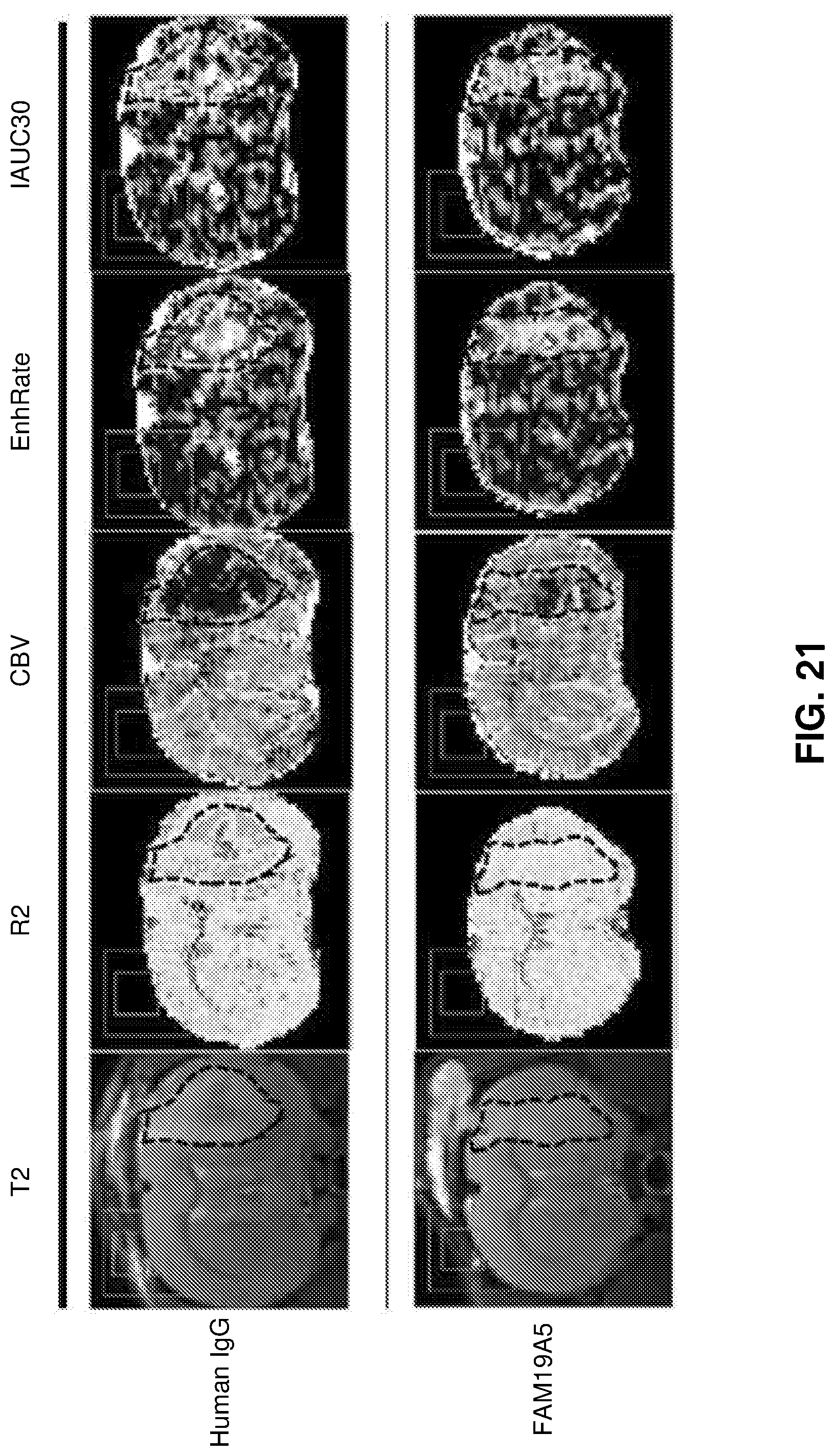
D00026

S00001
XML
uspto.report is an independent third-party trademark research tool that is not affiliated, endorsed, or sponsored by the United States Patent and Trademark Office (USPTO) or any other governmental organization. The information provided by uspto.report is based on publicly available data at the time of writing and is intended for informational purposes only.
While we strive to provide accurate and up-to-date information, we do not guarantee the accuracy, completeness, reliability, or suitability of the information displayed on this site. The use of this site is at your own risk. Any reliance you place on such information is therefore strictly at your own risk.
All official trademark data, including owner information, should be verified by visiting the official USPTO website at www.uspto.gov. This site is not intended to replace professional legal advice and should not be used as a substitute for consulting with a legal professional who is knowledgeable about trademark law.