Medicinal Liquid Injection Port And Medicinal Liquid Injection Device
SAKAI; Shinichi ; et al.
U.S. patent application number 16/617213 was filed with the patent office on 2020-09-24 for medicinal liquid injection port and medicinal liquid injection device. This patent application is currently assigned to PIOLAX MEDICAL DEVICES, INC.. The applicant listed for this patent is PIOLAX MEDICAL DEVICES, INC.. Invention is credited to Yuuki INAMI, Shinichi SAKAI.
| Application Number | 20200297926 16/617213 |
| Document ID | / |
| Family ID | 1000004903070 |
| Filed Date | 2020-09-24 |


| United States Patent Application | 20200297926 |
| Kind Code | A1 |
| SAKAI; Shinichi ; et al. | September 24, 2020 |
MEDICINAL LIQUID INJECTION PORT AND MEDICINAL LIQUID INJECTION DEVICE
Abstract
There is provided a medicinal liquid injection port to be implanted subcutaneously. A housing main body is provided with a medicinal liquid storage portion therein, an opening portion that opens the medicinal liquid storage portion, and a discharge hole that communicates with the medicinal liquid storage portion and configured to be connected with a tube. A septum attached to the opening portion of the housing main body and provided with a puncturable region that can be punctured with a puncture needle. A contrast portion is disposed in the septum over a thickness direction of the septum so as to detect the puncturable region of the septum with the ultrasonic wave. The contrast portion is provided in the septum within a thickness region of the septum and all of the contrast portion is positioned at an inner side of the septum in the thickness direction of the septum.
| Inventors: | SAKAI; Shinichi; (Kanagawa, JP) ; INAMI; Yuuki; (Kanagawa, JP) | ||||||||||
| Applicant: |
|
||||||||||
|---|---|---|---|---|---|---|---|---|---|---|---|
| Assignee: | PIOLAX MEDICAL DEVICES,
INC. Kanagawa JP |
||||||||||
| Family ID: | 1000004903070 | ||||||||||
| Appl. No.: | 16/617213 | ||||||||||
| Filed: | May 25, 2018 | ||||||||||
| PCT Filed: | May 25, 2018 | ||||||||||
| PCT NO: | PCT/JP2018/020195 | ||||||||||
| 371 Date: | November 26, 2019 |
| Current U.S. Class: | 1/1 |
| Current CPC Class: | A61M 2039/0232 20130101; A61M 5/162 20130101; A61M 39/0208 20130101; A61M 5/36 20130101; A61M 39/10 20130101; A61M 2039/0081 20130101 |
| International Class: | A61M 5/162 20060101 A61M005/162; A61M 39/10 20060101 A61M039/10; A61M 39/02 20060101 A61M039/02; A61M 5/36 20060101 A61M005/36 |
Foreign Application Data
| Date | Code | Application Number |
|---|---|---|
| May 31, 2017 | JP | 2017-108351 |
Claims
1. A medicinal liquid injection port configured to be implanted subcutaneously, the medicinal liquid injection port into which medicinal liquid can be injected by puncturing with a puncture needle from the outside, the medicinal liquid injection port comprising: a housing main body provided with a medicinal liquid storage portion therein, an opening portion that opens the medicinal liquid storage portion, and a discharge hole that communicates with the medicinal liquid storage portion and configured to be connected with a tube; and a septum attached to the opening portion of the housing main body and provided with a puncturable region that can be punctured with the puncture needle, wherein a contrast portion is disposed in the septum over a thickness direction of the septum so as to detect the puncturable region of the septum with the ultrasonic wave, wherein the contrast portion is provided in the septum within a thickness region of the septum and all of the contrast portion is positioned at an inner side of the septum in the thickness direction of the septum, and wherein the contrast portion is provided in an entire region of the puncturable region in a radial direction of the puncturable region.
2. The medicinal liquid injection port according to claim 1, wherein the contrast portion is formed of a member containing air bubbles.
3. The medicinal liquid injection port according to claim 1, wherein the contrast portion is formed of a member containing air bubbles and the air bubbles are provided in the contrast portion that is disposed in the septum.
4. (canceled)
5. The medicinal liquid injection port according to claim 1, wherein the contract portion is formed of a member containing air bubbles and the air bubbles are provided in the puncturable region of the septum excluding a part of the septum, exposed from the opening portion.
6. A medicinal liquid injection device comprising: the medicinal liquid injection port according to claim 1; and a puncture needle provided with a detectable portion that is detectable with an ultrasonic wave at a tip end portion thereof, wherein the detectable portion is configured to be positioned in the medicinal liquid storage portion of the housing main body in a state in which the puncturable region of the septum is punctured with the puncture needle.
7. The medicinal liquid injection port according to claim 1, wherein the septum is constituted by the contrast portion formed to have a predetermined thickness in the thickness direction, an exposed portion disposed at a front side of the contrast portion, and a bottom portion disposed at a back side of the contrast portion, and the contrast portion is thicker than the exposed portion and the bottom portion.
8. A medicinal liquid injection port configured to be implanted subcutaneously, the medicinal liquid injection port into which medicinal liquid can be injected by puncturing with a puncture needle from the outside, the medicinal liquid injection port comprising: a housing main body provided with a medicinal liquid storage portion therein, an opening portion that opens the medicinal liquid storage portion, and a discharge hole that communicates with the medicinal liquid storage portion and configured to be connected with a tube; and a septum attached to the opening portion of the housing main body and provided with a puncturable region that can be punctured with the puncture needle, wherein a contrast portion is disposed in the septum over a thickness direction of the septum so as to detect the puncturable region of the septum with the ultrasonic wave, wherein the contrast portion is provided in the septum within a thickness region of the septum and all of the contrast portion is positioned at an inner side of the septum in the thickness direction of the septum, and wherein a concave portion having an annular shape is formed in an outer periphery of the septum at a central portion in the thickness direction of the septum and the contrast portion having an annular shape is disposed in the concave portion.
9. The medicinal liquid injection port according to claim 8, wherein the contrast portion is formed of a member containing air bubbles.
10. The medicinal liquid injection port according to claim 8, wherein the contrast portion is formed of a member containing air bubbles and the air bubbles are provided in the contrast portion that is disposed in the septum around the puncturable region excluding the puncturable region of the septum.
11. The medicinal liquid injection port according to claim 8, wherein the contract portion is formed of a member containing air bubbles and the air bubbles are provided in the puncturable region of the septum excluding a part of the septum, exposed from the opening portion.
12. A medicinal liquid injection device comprising: the medicinal liquid injection port according to claim 8; and a puncture needle provided with a detectable portion that is detectable with an ultrasonic wave at a tip end portion thereof, wherein the detectable portion is configured to be positioned in the medicinal liquid storage portion of the housing main body in a state in which the puncturable region of the septum is punctured with the puncture needle.
13. A medicinal liquid injection port configured to be implanted subcutaneously, the medicinal liquid injection port into which medicinal liquid can be injected by puncturing with a puncture needle from the outside, the medicinal liquid injection port comprising: a housing main body provided with a medicinal liquid storage portion therein, an opening portion that opens the medicinal liquid storage portion, and a discharge hole that communicates with the medicinal liquid storage portion and configured to be connected with a tube; and a septum attached to the opening portion of the housing main body and provided with a puncturable region that can be punctured with the puncture needle, wherein a contrast portion is disposed in the housing main body over a thickness direction of the septum so as to detect the puncturable region of the septum with the ultrasonic wave, wherein the contrast portion is provided in an outer periphery of the septum within a thickness region of the septum and all of the contrast portion is positioned at an inner side of the housing main body in a thickness direction of the housing main body, and wherein the contrast portion is formed over an entire region in the thickness direction of the septum and disposed in an attachment space which is provided between the outer periphery of the septum and an inner periphery of the housing main body.
14. The medicinal liquid injection port according to claim 13, wherein the contrast portion is formed of a member containing air bubbles.
15. The medicinal liquid injection port according to claim 13, wherein the contrast portion is formed of a member containing air bubbles and the air bubbles are provided in the contrast portion that is disposed in the septum around the puncturable region excluding the puncturable region of the septum.
16. A medicinal liquid injection device comprising: the medicinal liquid injection port according to claim 13; and a puncture needle provided with a detectable portion that is detectable with an ultrasonic wave at a tip end portion thereof, wherein the detectable portion is configured to be positioned in the medicinal liquid storage portion of the housing main body in a state in which the puncturable region of the septum is punctured with the puncture needle.
Description
TECHNICAL FIELD
[0001] The present invention relates to a medicinal liquid injection port configured to be implanted subcutaneously, into which medicinal liquid can be injected by puncturing with a puncture needle from the outside and a medicinal liquid injection device.
BACKGROUND ART
[0002] Conventionally, a medicinal liquid injection port is implanted subcutaneously as an instrument for efficiently injecting medicinal liquid such as an anti-cancer agent. In general, the medicinal liquid injection port includes a liquid reservoir portion therein, and an opening thereof is closed by a septum made of an elastic material. The medicinal liquid injection port is connected with a tube which is to be placed in a blood vessel such as the heart. When puncturing the septum with a puncture needle such as a non-coring needle from outside the body, and injecting medicinal liquid such as an anti-cancer agent, the medicinal liquid flows in the tube through the liquid reservoir portion, and the medicinal liquid is supplied to the blood vessel or the like.
[0003] However, since the septum of the medicinal liquid injection port cannot be visually recognized from outside the body, it is difficult to appropriately puncture the septum of the medicinal liquid injection port with the puncture needle. If the medicinal liquid is injected in a state in which the puncture needle has not been inserted into the septum, the medicinal liquid leaked out of the port causes inflammation in a tissue and necrosis of the tissue. In addition, although the medicinal liquid injection port should be implanted with the septum facing toward skin, in a case in which the medicinal liquid injection port is inverted and the liquid reservoir portion is positioned close to the skin, it is difficult to puncture the septum with the puncture needle.
[0004] Accordingly, it is desired that a position of the septum of the medicinal liquid injection port can be confirmed to be at an appropriate position. An X-ray marker for determining the position of the septum is provided at the medicinal liquid injection port, and the position of the septum can be grasped by X-ray contrast with CT. However, in this case, since the position of the septum can be grasped only in a dedicated diagnostic room having a CT facility, it is inconvenient. Therefore, a method has been developed that can grasp the position of the septum by a compact ultrasonic irradiation device that can be used in a hospital room, instead of CT.
[0005] For example, the following Patent Document 1 describes an access port having a marker that can be contrasted with ultrasonic irradiation, and the access port has a structure in which three markers are arranged on an outer periphery of a partition wall (a septum) of a main body so as to form a substantially triangular shape.
PRIOR ART DOCUMENT
Patent Document
[0006] Patent Document 1: Japanese Patent Application Publication No. 2016-64238 A
SUMMARY OF INVENTION
Problems to be Solved by Invention
[0007] In the access port of Patent Document 1 described above, when the access port is irradiated with an ultrasonic wave, a position of the septum can be grasped by the three markers on the outer periphery of the septum. However, since a surface of the septum is only partially contrasted on an echo image, it may be difficult to confirm the position of the septum.
[0008] An object of the present invention is to provide a medicinal liquid injection port and a medicinal liquid injection device for easily grasping a position of a septum under the skin by ultrasonic irradiation to appropriately puncture the septum with the puncture needle.
Means for Solving Problems
[0009] In order to achieve the above object, according to the present invention, there is provided a medicinal liquid injection port configured to be implanted subcutaneously, the medicinal liquid injection port into which medicinal liquid can be injected by puncturing with puncture needle from the outside, the medicinal liquid injection port including: a housing main body provided with a medicinal liquid storage portion therein, an opening portion that opens the medicinal liquid storage portion, and a discharge hole that communicates with the medicinal liquid storage portion and configured to be connected to a tube; and a septum attached to the opening portion of the housing main body and provided with a puncturable region that can be punctured with the puncture needle, wherein a contrast portion is disposed in the housing main body and/or the septum over a thickness direction of the septum so as to detect the puncturable region of the septum with the ultrasonic wave.
Advantageous Effects of Invention
[0010] According to the present invention, since the contrast portion is disposed in the housing main body and/or the septum over the thickness direction of the septum so as to detect the puncturable region of the septum, when the medicinal liquid injection port is irradiated with the ultrasonic wave, an echo image of the contrast portion is contrasted over the thickness direction of the septum so as to easily grasp a position of the puncturable region of the septum, so that the puncturable region of the septum can be appropriately punctured with the puncture needle.
BRIEF DESCRIPTION OF DRAWINGS
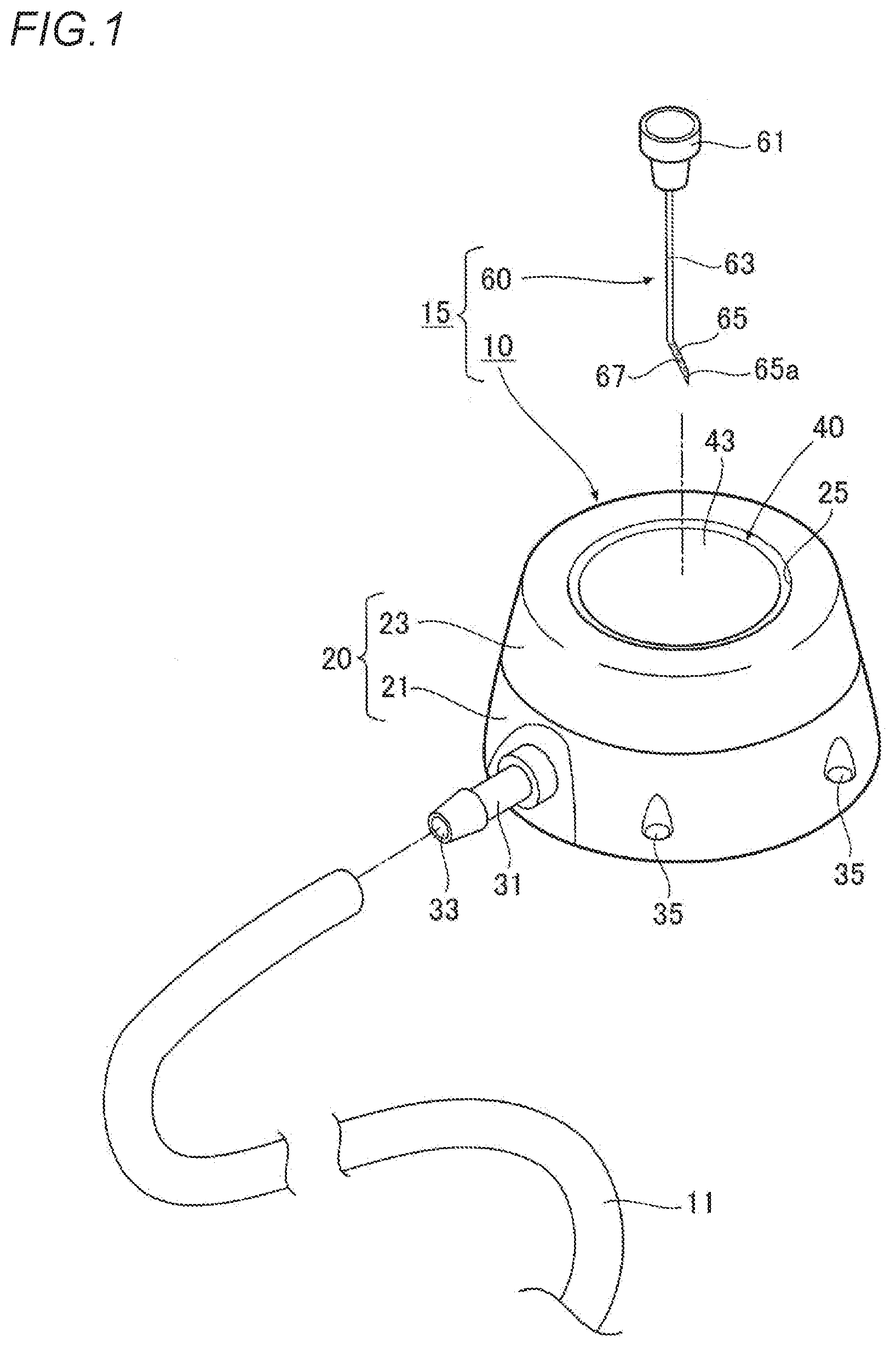
[0011] FIG. 1 is an exploded perspective view showing a medicinal liquid injection port and a medicinal liquid injection device according to an embodiment of the present invention.
[0012] FIG. 2 is a perspective view of the medicinal liquid injection port and the medicinal liquid injection device.
[0013] FIG. 3 is a sectional view of the medicinal liquid injection port.
[0014] FIG. 4 is an explanatory diagram showing a use state of the medicinal liquid injection port and the medicinal liquid injection device.
[0015] FIG. 5A is a sectional view showing a medicinal liquid injection port according to another embodiment of the present invention, FIG. 5B is a sectional view showing a medicinal liquid injection port according to still another embodiment of the present invention, and FIG. 5C is a sectional view showing a medicinal liquid injection port according to yet another embodiment of the present invention.
EMBODIMENTS OF INVENTION
[0016] Hereinafter, an embodiment of a medicinal liquid injection port and a medicinal liquid injection device according to the present invention will be described with reference to the drawings.
[0017] As shown in FIGS. 1 and 3, a medicinal liquid injection port 10 (hereinafter, also simply referred to as "port 10") in this embodiment can be implanted subcutaneously to inject medicinal liquid by puncturing it with a puncture needle 60 from the outside. The medicinal liquid injection port 10 includes a housing main body 20 provided with a medicinal liquid storage portion 30 therein and an opening portion 25 that opens the medicinal liquid storage portion 30, and a septum 40 that is attached to the opening portion 25 of the housing main body 20.
[0018] In this embodiment, the housing main body 20 is formed of two parts of a base portion 21 and a lid portion 23, and a bottom surface 23b of the lid portion 23 is joined to a ceiling surface 21a of the base portion 21 by welding, adhesion, or the like to form a structure in which both parts are integrated. In addition, an outer peripheral surface of the housing main body 20 has a tapered shape gradually increasing in a diameter from a ceiling surface 23a (a surface opposite to the bottom surface 23b) of the lid portion 23 toward a bottom surface 21b (a surface opposite to the ceiling surface 21a) of the base portion 21, and has a substantially truncated cone shape as a whole.
[0019] The opening portion 25 having a circular shape is formed on the ceiling surface 23a side (a side opposite to a joining surface with the base portion 21) of the lid portion 23. As shown in FIG. 3, inside the lid portion 23, a septum attachment space 27 for disposing the septum 40 and attaching it to the opening portion 25 is formed in communication with the opening portion 25 at a deep side in a thickness direction (bottom surface 23b side) from the opening portion 25. The septum attachment space 27 has a shape whose diameter is larger than that of the opening portion 25, so that a peripheral edge portion 25a of the opening portion 25 has a thin flange shape. The peripheral edge portion 25a is locked to a ceiling surface side peripheral edge of the septum 40 disposed in the septum attachment space 27 to prevent the septum 40 from coming off from the opening portion 25. In addition, the peripheral edge portion 25a of the opening portion 25 extends in a flange shape so as not to overlap a contrast portion 45, which will be described later, of the septum 40. However, the peripheral edge portion 25a of the opening portion 25 may be shaped so as to be locked to a frame-shaped body 55, which will be described later, disposed on to an outer periphery of the septum, and preferably the peripheral edge portion 25a has a shape that does not overlap the contrast portion 45 of the septum 40.
[0020] On the other hand, the medicinal liquid storage portion 30 is formed inside the base portion 21. The medicinal liquid storage portion 30 in this embodiment is opened at the top and has a circular concave shape with a diameter smaller than that of the septum attachment space 27 (see FIG. 3). Further, the medicinal liquid storage portion 30 is opened to the outside of the port through the septum attachment space 27 and the opening portion 25. In addition, an upper opening of the medicinal liquid storage portion 30 is closed by the septum 40 which is disposed in the septum attachment space 27 of the lid portion 23, and sealed so as not to leak out.
[0021] As shown in FIG. 1, a connector 31 for connecting the tube 11 thereto is attached to the base portion 21. As shown in FIG. 3, an opening at a base end side of the connector 31 communicates with the medicinal liquid storage portion 30, and the connector 31 extends from an outer periphery of the base portion 21 in an outer diameter direction so as to connect the tube 11 to an outer periphery of a tip end side thereof. An opening on the tip end side of the connector 31 forms a discharge hole 33 through which the medicinal liquid stored in the medicinal liquid storage portion 30 is discharged to the tube 11. In addition, as shown in FIGS. 1 and 2, a plurality of holes 35 are formed at predetermined intervals on the outer periphery of the base portion 21 at a bottom surface side. A suture thread (not shown) can be inserted through the holes 35, and the suture thread is sewn to a predetermined site in the body to prevent displacement of the medicinal liquid injection port 10 implanted subcutaneously.
[0022] A shape and an internal structure of the housing main body 20 are merely examples. For example, the housing main body may not have a structure formed of two parts, and an outer periphery of the housing main body may have an elliptical shape or the like. The housing main body is not limited to the above shape and structure. In addition, the housing main body 20 may be formed of, for example, synthetic resin having biocompatibility such as nylon, polyethylene, polypropylene, fluororesin, polyimide, polyether sulfone resin, epoxy resin, polysulfone, polyacetal, liquid crystal polymer, polyurethane, silicone rubber, vinyl chloride resin, and polyethylene terephthalate resin, a metal having the biocompatibility such as Ti, stainless steel, or Ni--Ti alloy, a composite thereof, or the like. The connector 31 may be formed of, for example, a metal having the biocompatibility such as Ti, stainless steel, or Ni--Ti alloy, ceramic, hard resin, or super engineering plastic such as PEEK, a composite thereof, or the like.
[0023] On the other hand, the septum 40 has a disk shape that is larger than an inner diameter of the medicinal liquid storage portion 30 and has a predetermined thickness, and the frame-shaped body 55 having an annular shape is disposed on the outer periphery thereof (see FIG. 3). The frame-shaped body 55 prevents the septum 40 from coming out of the opening portion 25 of the housing main body 20 or reinforces the septum 40, but the frame-shaped body 55 may not be disposed.
[0024] The septum 40 is disposed in the septum attachment space 27 of the lid portion 23 together with the frame-shaped body 55 on the outer periphery thereof. In this state, the bottom surface 23b of the lid portion 23 is joined to the ceiling surface 21a of the base portion 21, so that the septum 40 is sandwiched between the peripheral edge portion 25a of the opening portion 25 and the ceiling surface 21a of the base portion 21, and the septum 40 is attached to the opening portion 25.
[0025] As shown in FIGS. 3 and 4, the septum 40 is provided with a puncturable region 41 which can be punctured by the puncture needle 60. The puncturable region 41 means a region which can allow the puncture needle 60 to pass through the septum 40 up to the medicinal liquid storage portion 30 when puncturing it with the puncture needle 60 from a surface 43 (hereinafter, also simply referred to as an "exposed surface 43") of the septum 40, exposed through the opening portion 25. Herein, the puncturable region 41 is a region surrounded by left and right dashed lines on paper surfaces of FIGS. 3 and 4, and the puncturable region 41 excludes a region in which the puncture needle 60 hits the frame-shaped body 55 or the like when puncturing the septum 40 with the puncture needle 60 and the puncture needle 60 cannot reach the medicinal liquid storage portion 30. That is, the puncturable region 41 is a region that is disposed at a position aligned with the medicinal liquid storage portion 30 in the thickness direction of the port 10, and is in a range equal to or smaller than the inner diameter of the medicinal liquid storage portion 30.
[0026] The contrast portion 45 is disposed in the septum 40 over a thickness direction T1 of the septum 40 so as to detect the puncturable region 41 of the septum 40. Although the contrast portion 45 is disposed in the septum 40 in this embodiment, the contrast portion 45 may be disposed in the housing main body-side, and the contrast portion may also be disposed in both the housing main body and the septum.
[0027] The septum 40 in this embodiment includes the thick disk-shaped contrast portion 45 formed to have a predetermined thickness along the thickness direction T1, a thin disc-shaped exposed portion 47 disposed at a front side (one end surface side) of the contrast portion 45, and a thin disc-shaped bottom portion 49 disposed at a back side (the other end surface side) of the contrast portion 45. That is, the septum 40 has a three-layer structure in which the contrast portion 45 is disposed at a central portion in the thickness direction thereof, the exposed portion 47 is disposed at the front side of the contrast portion 45, and the bottom portion 49 is disposed at the back side of the contrast portion 45.
[0028] The exposed portion 47 of the septum 40 is disposed at a side of the opening portion 25 of the housing main body 20, and a surface thereof forms the exposed surface 43. Further, the bottom portion 49 closes the upper opening of the medicinal liquid storage portion 30.
[0029] In this embodiment, the septum 40 has a structure in which the contrast portion 45 is provided in a part of the septum 40 itself. Further, the contrast portion 45 in this embodiment is formed over the entire region in a radial direction of the septum 40 with an outer diameter larger than the inner diameter of the medicinal liquid storage portion 30, so as to cover the entire region of the medicinal liquid storage portion 30 in the radial direction thereof. In this embodiment, the contrast portion 45, the exposed portion 47, and the bottom portion 49 have the same outer diameter. The contrast portion, the exposed portion, and the bottom portion may have different outer diameters, and the outer diameters thereof are not particularly limited. In addition, although the exposed surface 43 of the septum 40 in this embodiment is positioned below an upper end position of the opening portion 25 of the housing main body 20, the exposed surface 43 of the septum 40 may be protruded so as to be positioned above an upper end of the opening portion 25 of the housing main body 20.
[0030] In this embodiment, by irradiating the medicinal liquid injection port 10 with the ultrasonic wave, the contrast portion 45 is contrasted in an echo image, so that the puncturable region 41 of the septum 40 can be detected over the thickness direction T1 of the septum 40.
[0031] The contrast portion only needs to be able to detect the puncturable region of the septum with the ultrasonic wave over the thickness direction of the septum, and location, shape, or the like of the contrast portion are not particularly limited. For example, the contrast portion may be formed in an outer peripheral portion of the septum over the thickness direction, and the contrast portion may be formed only in the puncturable region 41 without being formed over the entire region of the septum in the radial direction thereof.
[0032] The thickness direction T1 of the septum 40 means a direction orthogonal to the exposed surface 43 of the septum 40 (a direction perpendicular to a surface direction of the exposed surface 43), which is exposed through the opening portion 25 of the housing main body 20.
[0033] As the septum 40, a resin material is used, having elasticity that can be punctured with the puncture needle 60 and can self-seal (tack) a puncture hole formed by the puncture needle 60 when the puncture needle 60 is pulled out. Examples of such a resin material include silicone rubber, latex, fluororubber, isoprene, and styrene-based elastomer, for example.
[0034] As shown in FIG. 3, the contrast portion 45 in this embodiment is formed of a member containing air bubbles 46. In this embodiment, the air bubbles 46 are not contained in an outer periphery of the contrast portion 45 so as not to overlap the peripheral edge portion 25a of the opening portion 25 of the housing main body 20. However, the air bubbles 46 may also be contained in the outer periphery of the contrast portion 45 (this structure will be described in an embodiment described later). In addition, the air bubbles 46 can be contained, for example, by injection molding a resin material containing gas into a mold frame or injecting the gas into molten resin injected into the mold frame.
[0035] In the above embodiment, the air bubbles are contained in the contrast portion. However, except for containing the air bubbles, the following methods can be used to form the contrast portion. For example, (1) the contrast portion includes a resin, metal, or the like having a different acoustic impedance from that of a living body, (2) the contrast portion is provided with a layer of air instead of the air bubbles, (3) a front surface of the septum is uneven or roughened (in this case, the front surface of the septum having the uneven or the roughened surface forms the "contrast portion"), (4) the front surface of the septum is coated with a material having different acoustic impedance (in this case, the coating layer forms the "contrast portion").
[0036] As in the present embodiment, in a case of forming the septum 40 having a three-layer structure, for example, a resin material containing the gas and a resin material containing no gas are simultaneously injection-molded in a mold frame to form the contrast portion 45, the exposed portion 47, the bottom portion 49, or by injecting the resin material containing the gas into the mold frame and supercooling a part of the material in contact with an inner surface of the mold frame, the air bubbles 46 can be provided only in an inner side part (i.e., the contrast portion 45) which is not in contact with the inner surface of the mold frame without providing the air bubbles 46 in the part (that is, the exposed portion 47 and the bottom portion 49) in contact with the inner surface of the mold frame.
[0037] On the other hand, a medicinal liquid injection device 15 in this embodiment includes the port 10 having the above described structure and the puncture needle 60.
[0038] As shown in FIG. 1, the puncture needle 60 in this embodiment includes a connection portion 61 to which a syringe of an injector or a tip end portion of a drip (not shown) is detachably connected, a tubular straight line portion 63 that extends linearly from the connection portion 61, and a tip end portion 65 that has a tubular shape bent at a predetermined angle with respect to the straight line portion 63, and has a sharp blade tip.
[0039] A blade surface 65a of the tip end portion 65 is parallel to an axial direction of the straight line portion 63. That is, the puncture needle 60 is a well-known non-coring needle that does not scrape away a body of the septum 40 at the time of puncturing the septum 40. Further, the tip end portion 65 of the puncture needle 60 is provided with a detectable portion 67 that can be detected with the ultrasonic wave. The detectable portion 67 can be formed, for example, by performing a surface processing that makes the surface rougher than the straight line portion 63.
[0040] As shown in FIG. 4, the medicinal liquid injection device 15 is configured such that the detectable portion 67 is positioned in the medicinal liquid storage portion 30 of the housing main body 20 in a state in which the puncturable region 41 of the septum 40 is punctured with the puncture needle 60.
[0041] Next, operation and effect of the medicinal liquid injection port 10 and the medicinal liquid injection device 15 having the above described configuration will be described.
[0042] FIG. 4 shows a state in which the medicinal liquid injection device 15 is used with the port 10 implanted subcutaneously. That is, the port 10 is implanted so as to be embedded in a subcutaneous tissue 2 at an inner surface side of a skin 1 and the port 10 is able to supply a medicinal liquid such as an anti-cancer agent or a nutrient to a tubular organ 3 such as a blood vessel through the tube 11 connected to the connector 31 of the port 10.
[0043] When the port 10 is implanted subcutaneously, a tip end portion of the tube 11 is placed in a predetermined position of the tubular organ 3 by a well-known Seldinger method or the like, and then the base end portion of the tube 11 is connected to the connector 31 of the port 10 outside the body. The port 10 is disposed at a predetermined position of the subcutaneous tissue 2 by incising the skin 1, and the suture thread inserted into the plurality of holes 35 on the outer periphery of the port is sewn to a predetermined site, so that the port 10 is implanted to be embedded in the subcutaneous tissue 2. Thereafter, as shown in FIG. 4, by closing the incised skin 1, the port 10 can be placed subcutaneously (under the skin 1).
[0044] By irradiating the port 10 with the ultrasonic wave from an ultrasonic probe (probe) of an ultrasonic irradiation device (not shown), the echo image is contrasted on an monitor of the ultrasonic irradiation device.
[0045] At this time, in the port 10, since the septum 40 is provided with the contrast portion 45 over the thickness direction T1 of the septum 40 so that the puncturable region 41 of the septum 40 can be detected with the ultrasonic wave, when the port 10 is irradiated with the ultrasonic wave, the echo image of the contrast portion 45 is displayed on the monitor in a wide range over the thickness direction T1 of the septum 40, so that a position of the puncturable region 41 of the septum 40 can be grasped securely. As a result, a person who use the puncture needle 60 can smoothly puncture the puncturable region 41 of the septum 40 with the puncture needle 60.
[0046] Therefore, in the port 10, it is possible to easily grasp a position of the septum 40, particularly in the thickness direction T1 of the septum 40, under the skin at the time of the ultrasonic irradiation, and the septum 40 can be appropriately punctured so that the puncture needle 60 can appropriately puncture the septum 40 to reach the medicinal liquid storage portion 30. In addition, the puncture needle 60 can be prevented from being incorrectly inserted in a location other than the septum 40, and inflammation and necrosis of the tissue can be suppressed. If the septum 40 is not disposed at a position close to the skin 1 and is disposed in an inverted manner, it is easy to grasp that the septum 40 is disposed at a position far from the skin 1, and a subsequent treatment (the skin 1 is re-incised to correct an orientation of the septum 40, or the like) can be performed quickly, so that a burden on a patient can be suppressed.
[0047] As shown in FIG. 3, in this embodiment, since the contrast portion 45 is formed of a member containing the air bubbles 46, and when the port 10 is irradiated with the ultrasonic wave, the ultrasonic wave is irregularly reflected by the air bubbles 46, the echo image can be more easily contrasted over the thickness direction of the septum 40, and thus it becomes more easy to puncture the puncturable region 41 of the septum 40 with the puncture needle 60, and to grasp the position of the septum 40 (particularly, the thickness direction T1 of the septum 40) under the skin at the time of the ultrasonic irradiation, and the septum 40 can be appropriately punctured with the puncture needle 60 so that the puncture needle 60 can reach the medicinal liquid storage portion 30.
[0048] As shown in FIG. 3, in this embodiment, since the air bubbles 46 are provided in the septum 40, that is, in the contrast portion 45, when puncture the puncturable region 41 of the septum 40 with the puncture needle 60, a state in which the septum 40 is recessed by puncture pressure can be confirmed, and a puncture state of the puncture needle 60 can be easily grasped. In addition, since the air bubbles 46 are provided in the septum 40, the location where the echo image is contrasted is punctured with the puncture needle 60, and thus the puncture needle 60 can be easily inserted into the puncturable region 41 sensuously, so that the puncture needle 60 can be prevented from incorrectly being inserted into a region other than the puncturable region 41 (if a location where the echo image is contrasted and the puncturable region 41 are misaligned, a puncture location is likely to be mistaken). Further, puncture resistance of the puncture needle 60 with respect to the puncturable region 41 of the septum 40 can be reduced by the air bubbles 46 provided in the septum 40, and the puncture needle 60 can be smoothly inserted into the puncturable region 41 of the septum 40.
[0049] In this embodiment, the air bubbles 46 are provided in the puncturable region 41 of the septum 40 except for a part of the septum 40 that is exposed through the opening portion 25 of the housing main body 20 (the exposed portion 47 including the exposed surface 43). Accordingly, durability of the part of the septum 40 exposed through the opening portion 25 of the housing main body 20 can be increased to prevent the medicinal liquid from leaking to the outside of the septum. Further, since there are no air bubbles 46 in the exposed portion, the front surface portion (exposed surface 43) of the septum 40 exposed through the opening portion 25 of the housing main body 20 can be smoothed (effect of claim 5). In a case where the air bubbles 46 are not provided on the surface of the septum 40 at a side of the medicinal liquid storage portion 30 (a bottom surface on an opposite side to the exposed surface 43), the surface of the septum 40 at the side of the medicinal liquid storage portion 30 can be smoothed to prevent the medicine from entering into the septum 40 and remaining inside the septum.
[0050] On the other hand, as shown in FIG. 4, the medicinal liquid injection device 15 according to the present invention is configured such that the detectable portion 67 which is detectable with the ultrasonic wave is positioned in the medicinal liquid storage portion 30 of the port 10 in a state in which the puncturable region 41 of the septum 40 of the port 10 is punctured with the puncture needle 60 provided with the detectable portion 67 at the tip end portion 65. Therefore, by irradiating the port 10 and the puncture needle 60 with the ultrasonic wave, the detectable portion 67 of the puncture needle 60 can be detected in addition to the puncturable region 41 of the septum 40, so that a position of the puncture needle 60 can be reliably grasped, and the puncture needle 60 can be more easily inserted into the puncturable region 41 of the septum 40.
[0051] FIG. 5A shows a medicinal liquid injection port according to another embodiment of the present invention. Parts that are substantially the same as those of the above embodiment are given the same reference numerals, and descriptions thereof are omitted.
[0052] In a medicinal liquid injection port 10A (hereinafter, also simply referred to as "port 10A") in this embodiment, a contrast portion 45A provided with the air bubbles 46 is disposed on an outer periphery of the septum 40A. Specifically, a concave portion 44 having an annular shape is formed in the outer periphery of the septum 40A at a central portion in the thickness direction of the septum 40A, and the annular contrast portion 45A is fitted in the concave portion 44. That is, the air bubbles 46 are provided in the contrast portion 45A around the puncturable region 41 excluding the puncturable region 41 of the septum 40A.
[0053] The port 10A of this embodiment has the following operation and effect. The puncturable region 41 of the septum 40A is repeatedly punctured with the puncture needle 60, so that durability is reduced. However, according to the port 10A, since the air bubbles 46 are provided in the contrast portion 45A around the puncturable region 41 excluding the puncturable region 41 of the septum 40A, by contrasting a periphery of the puncturable region 41, the puncturable region 41 can be visually recognized while maintaining the durability of the puncturable region 41 of the septum 40A. In addition, since the air bubbles 46 are not provided in the puncturable region 41 of the septum 40A, a front surface part (exposed surface 43) of the septum 40A exposed through the opening portion 25 can be smoothed (effect of claim 4). In the case in which the air bubbles 46 are not provided on a surface of the septum 40A at the side of the medicinal liquid storage portion 30, the surface of the septum 40 at the side of the medicinal liquid storage portion 30 can be smoothed to prevent the medicine from entering into the septum 40A and remaining inside the septum.
[0054] FIG. 5B shows a medicinal liquid injection port according to still another embodiment of the present invention. Parts that are substantially the same as those of the above embodiment are given the same reference numerals, and descriptions thereof are omitted.
[0055] In a medicinal liquid injection port 10B (hereinafter, also simply referred to as "port 10B") in this embodiment, a contrast portion 45B provided with the air bubbles 46 is disposed in the housing main body 20. Specifically, the contrast portion 45B having an annular shape and containing the air bubbles 46 is disposed on an outer periphery of the septum 40B in the septum attachment space 27 of the housing main body 20. Similar to the port 10A of the above embodiment, the air bubbles 46 are provided in the contrast portion 45B around the puncturable region 41 excluding the puncturable region 41 of the septum 40B. Also in the port 10B of this embodiment, the same operation and effect as the port 10A of the above embodiment can be obtained.
[0056] FIG. 5C shows a medicinal liquid injection port according to yet another embodiment of the present invention. Parts that are substantially the same as those of the above embodiment are given the same reference numerals, and descriptions thereof are omitted.
[0057] In the medicinal liquid injection port 10C (hereinafter, also simply referred to as "port 10C") in this embodiment, the septum 40 has a three-layer structure in which a contrast portion 45C is disposed at a central portion in the thickness direction, and the exposed portion 47 is disposed on the front side, and the bottom portion 49 is disposed on the back side, as in the embodiment shown in FIGS. 1 to 4. The contrast portion 45C has basically the same structure as that of the contrast portion 40 of the above embodiment, but the air bubbles 46 are also contained in the outer periphery thereof.
[0058] A contrast portion 45D having a disc shape and containing the air bubbles 46 is disposed at a bottom portion of the medicinal liquid storage portion 30.
[0059] The contrast portion 45D is disposed at a position that is aligned with the contrast portion 45C of the septum 40 in the thickness direction of the port 10C with a predetermined distance (depth of the medicinal liquid storage portion 30 excluding the contrast portion 45D) in the thickness direction of the port 10C. In addition, the contrast portion 45D is aligned with the puncturable region 41 of the septum 40) in the thickness direction of the port 10C.
[0060] In the port 10C of this embodiment, when irradiating with the ultrasonic wave, the contrast portion 45C provided in the septum 40 and the contrast portion 45D disposed at the bottom portion of the medicinal liquid storage portion 30 are contrasted at a predetermined distance, so that it is possible to firmly grasp a direction in which the puncture needle 60 should be inserted (that is, a direction toward a bottom portion side of the medicinal liquid storage portion 30), and it is possible to more accurately grasp whether or not the tip end of the puncture needle 60 reaches the medicinal liquid storage portion 30.
[0061] The present invention is not limited to the above-described embodiments, and various modified embodiments are possible within the scope of the present invention, and such embodiments are also included in the scope of the present invention.
DESCRIPTION OF REFERENCE NUMERALS
[0062] 10, 10A, 10B, 10C Medicinal Liquid Injection Port (Port) [0063] 11 Tube [0064] 15 Medicinal Liquid Injection Device [0065] 20 Housing Main Body [0066] 25 Opening Portion [0067] 30 Medicinal Liquid Storage Portion [0068] 33 Discharge Hole [0069] 40, 40A, 40B Septum [0070] 41 Puncturable Region [0071] 45, 45A, 45B, 45C, 45D Contrast Portion [0072] 46 Air Bubble [0073] 60 Puncture Needle
* * * * *
D00000
D00001
D00002
D00003
D00004
D00005
XML
uspto.report is an independent third-party trademark research tool that is not affiliated, endorsed, or sponsored by the United States Patent and Trademark Office (USPTO) or any other governmental organization. The information provided by uspto.report is based on publicly available data at the time of writing and is intended for informational purposes only.
While we strive to provide accurate and up-to-date information, we do not guarantee the accuracy, completeness, reliability, or suitability of the information displayed on this site. The use of this site is at your own risk. Any reliance you place on such information is therefore strictly at your own risk.
All official trademark data, including owner information, should be verified by visiting the official USPTO website at www.uspto.gov. This site is not intended to replace professional legal advice and should not be used as a substitute for consulting with a legal professional who is knowledgeable about trademark law.