Nanoparticles Conjugated with Vasoactive Intestinal Peptide Antagonists
Waller; Edmund ; et al.
U.S. patent application number 16/763197 was filed with the patent office on 2020-09-24 for nanoparticles conjugated with vasoactive intestinal peptide antagonists. The applicant listed for this patent is Emory University. Invention is credited to David Francis, Rebecca Pankove, Sruthi Ravindranathan, Susan Thomas, Edmund Waller.
| Application Number | 20200297767 16/763197 |
| Document ID | / |
| Family ID | 1000004929591 |
| Filed Date | 2020-09-24 |


| United States Patent Application | 20200297767 |
| Kind Code | A1 |
| Waller; Edmund ; et al. | September 24, 2020 |
Nanoparticles Conjugated with Vasoactive Intestinal Peptide Antagonists
Abstract
This disclosure relates to compositions comprising an antagonist of vasoactive intestinal polypeptide signaling coupled to a nanoparticle and methods of use related thereto. In certain embodiments, the nanoparticle is a poloxamer-stabilized polypropylene sulfide nanoparticle.
| Inventors: | Waller; Edmund; (Atlanta, GA) ; Thomas; Susan; (Atlanta, GA) ; Pankove; Rebecca; (Atlanta, GA) ; Ravindranathan; Sruthi; (Decatur, GA) ; Francis; David; (Atlanta, GA) | ||||||||||
| Applicant: |
|
||||||||||
|---|---|---|---|---|---|---|---|---|---|---|---|
| Family ID: | 1000004929591 | ||||||||||
| Appl. No.: | 16/763197 | ||||||||||
| Filed: | November 9, 2018 | ||||||||||
| PCT Filed: | November 9, 2018 | ||||||||||
| PCT NO: | PCT/US2018/060155 | ||||||||||
| 371 Date: | May 11, 2020 |
Related U.S. Patent Documents
| Application Number | Filing Date | Patent Number | ||
|---|---|---|---|---|
| 62584196 | Nov 10, 2017 | |||
| Current U.S. Class: | 1/1 |
| Current CPC Class: | A61K 35/17 20130101; A61K 38/00 20130101; A61K 47/6935 20170801; C07K 14/57563 20130101; A61K 9/0019 20130101; A61K 47/62 20170801 |
| International Class: | A61K 35/17 20060101 A61K035/17; C07K 14/575 20060101 C07K014/575; A61K 47/69 20060101 A61K047/69; A61K 47/62 20060101 A61K047/62 |
Goverment Interests
STATEMENT REGARDING FEDERALLY SPONSORED RESEARCH OR DEVELOPMENT
[0002] This invention was made with government support under RO1 CA207619-01 awarded by the National Institutes of Health. The government has certain rights in the invention.
Claims
1. A composition comprising a nanoparticle comprising an antagonist of vasoactive intestinal polypeptide signaling.
2. The composition of claim 1, wherein the nanoparticle is comprised of poloxamer-stabilized polypropylene sulfide.
3. The composition of claim 2, wherein the nanoparticle has a diameter of between 10 and 100 nm.
4. The composition of claim 2, wherein the nanoparticle has a diameter between 20 and 50 nm or about 30 nm.
5. The composition of claim 1, wherein the antagonist of vasoactive intestinal polypeptide signaling contains the peptide sequence of VIPhyb: TABLE-US-00003 (SEQ ID NO: 1) KPRRPYTDNYTRLRKQMAVKKYLNSILN.
6. The composition of claim 1, wherein the peptide sequence couple to the nanoparticle is contains VIPhyb plus a C-terminal linker peptide: TABLE-US-00004 (SEQ ID NO: 13) KPRRPYTDNYTRLRKQMAVKKYLNSILNGGGGSC.
7. The composition of claim 1, wherein the molar ratio of antagonist peptide to the nanoparticles is about 60.
8. The composition of claim 1, wherein the chemical linkage between the VIP antagonistic peptide and the nanoparticles is a disulfide bond.
9. A method of augmenting T-cell activation and ex vivo expansion by co-incubation of human T cells with a nanoparticle containing a small molecule antagonist of VIP signaling.
10. The method of claim 9, wherein the human T cells are activated with anti-CD3 antibody bound to a plate.
11. The method of claim 9, wherein human T cells are activated in a mixed lymphocyte reaction.
12. The method of claim 9, wherein the human T cells are activated in vitro by co-incubation with tumor associated antigens.
13. The method of claim 12, wherein the tumor associate antigens are presented on tumor microvesicles.
14. The method of claim 12, wherein the activated human T cells are infused into a human patient with cancer.
15. The method of claim 9, wherein the human T cells are activated in vitro by co-incubation with viral antigens.
16. The method of claim 15, wherein the viral antigens are presented on microvesicles.
17. The method of claim 15, wherein the viral antigens are presented on dendritic cells.
18. The method of claim 12, wherein the activated human T cells are infused into a human patient with cancer.
Description
CROSS-REFERENCE TO RELATED APPLICATIONS
[0001] This application claims the benefit of U.S. Provisional Application No. 62/584,196 filed Nov. 10, 2017. The entirety of this application is hereby incorporated by reference for all purposes.
INCORPORATION-BY-REFERENCE OF MATERIAL SUBMITTED AS A TEXT FILE VIA THE OFFICE ELECTRONIC FILING SYSTEM (EFS-WEB)
[0003] The Sequence Listing associated with this application is provided in text format in lieu of a paper copy, and is hereby incorporated by reference into the specification. The name of the text file containing the Sequence Listing is 18028PCT_ST25.txt. The text file is 12 KB, was created on Nov. 9, 2018, and is being submitted electronically via EFS-Web.
BACKGROUND
[0004] Cytotoxic T cells directly kill cells, and the response is antigen specific. In order to destroy tumors, cytotoxic cells must proliferate in sufficient numbers to overtake the dividing cancer cells. However, T cells are considered senescent if they have limited proliferative potential in response to antigenic stimulation. Filaci et al. report that a subset of effector T-cells, CD8+ CD28- T regulatory lymphocytes, inhibit T cell proliferative and cytotoxic functions in human cancers. J Immunol, 2007, 179: 4323-4334. Montes et al. indicate that tumors induce senescent T cells as a potential form of tumor immune evasion. Cancer Res, 2008, 68: 870-879. Senescent T cells are characterized by the loss of CD27 and CD28 expression, lack of proliferative capacity, and increased expression of senescence-associated molecules. See Ramello et al. Cell Death and Disease, 2014, 5, e1507. Thus, there is a need to identify improved methods of treating cancer by reversing senescence in T cells.
[0005] Magnetic beads coated with anti-CD3 and anti-CD28 (anti-CD3/CD28 beads) have been reported for the expansion of T cells that has been used experimentally to boost T cell immunity in immunosuppressed cancer patients. See Porter et al. A phase 1 trial of donor lymphocyte infusions expanded and activated ex vivo via CD3/CD28 costimulation. Blood, 2006, 107:1325-1331.
[0006] Li et al report modulation of graft-versus-leukemia in allogeneic transplants by antagonizing vasoactive intestinal peptide signalling. Cancer Res, 2016, 76(23):6802-681. See also Li et al. PLoS One, 2013, 8(5):e63381; Li et al., Blood, 2013, 121(12):2347-51, Li et al., J Immunol, 2011, 187(2):1057-65; U.S. Pat. Nos. 9,458,217 and 9,669,092; and U.S. Published Application number 2013/0302351. Petersen et al report administration of a vasoactive intestinal peptide antagonist enhances the autologous anti-leukemia T cell response in murine models of acute leukemia. OncoImmunology 6.5 (2017): e 1304336.
[0007] Thomas et al. report targeting the tumor-draining lymph node with adjuvanted nanoparticles reshapes the anti-tumor immune response. Biomaterials, 2014, 35 (2): 814-824.
[0008] References cited herein are not an admission of prior art.
SUMMARY
[0009] In certain embodiments, this disclosure relates to compositions and methods of coupling an antagonist of vasoactive intestinal polypeptide signaling to a nanoparticle and methods of use related thereto. In certain embodiments, the nanoparticle is a poloxamer-stabilized polypropylene sulfide nanoparticle. In certain embodiments, the nanoparticle has a diameter of between 10 and 100 nm. In certain embodiments, the nanoparticle has a diameter between 20 and 50 nm, preferably about 30 nm. In certain embodiments, the antagonist of vasoactive intestinal polypeptide signaling contains the peptide sequence of VIPhyb, KPRRPYTDNYTRLRKQMAVKKYLNSILN (SEQ ID NO: 1).
[0010] In certain embodiments, the disclosure contemplates using particles disclosed herein when the peptide sequence couple to the nanoparticle contains VIPhyb plus a C-terminal linker peptide, KPRRPYTDNYTRLRKQMAVKKYLNSILNGGGGSC (SEQ ID NO: 13).
[0011] In certain embodiments, methods of making nanoparticles disclosed herein comprise mixing a VIP antagonist comprising a C-terminal or N-terminal sequence Gly-Ser-Cys with a poloxamer-stabilized polypropylene sulphide nanoparticles under conditions such that the peptide is covalently attached to the nanoparticles. In certain embodiments, the molar ratio of antagonist peptide to the nanoparticles is about 60 or between 40 and 70 per nanoparticle. In certain embodiments, the chemical linkage between the VIP antagonistic peptide and the nanoparticles is a disulfide bond.
[0012] In certain embodiments, this disclosure relates to methods of augmenting T-cell activation and ex vivo expansion by co-incubation of human T cells with a nanoparticle containing a small molecule antagonist of VIP signaling. In certain embodiments, the human T cells are activated with anti-CD3 antibody bound to a plate. In certain embodiments, the human T cells are activated in a mixed lymphocyte reaction. In certain embodiments, the human T cells are activated in vitro by co-incubation with tumor-associated antigens. In certain embodiments, the tumor associate antigens are presented on tumor microvesicles.
BRIEF DESCRIPTION OF THE DRAWINGS
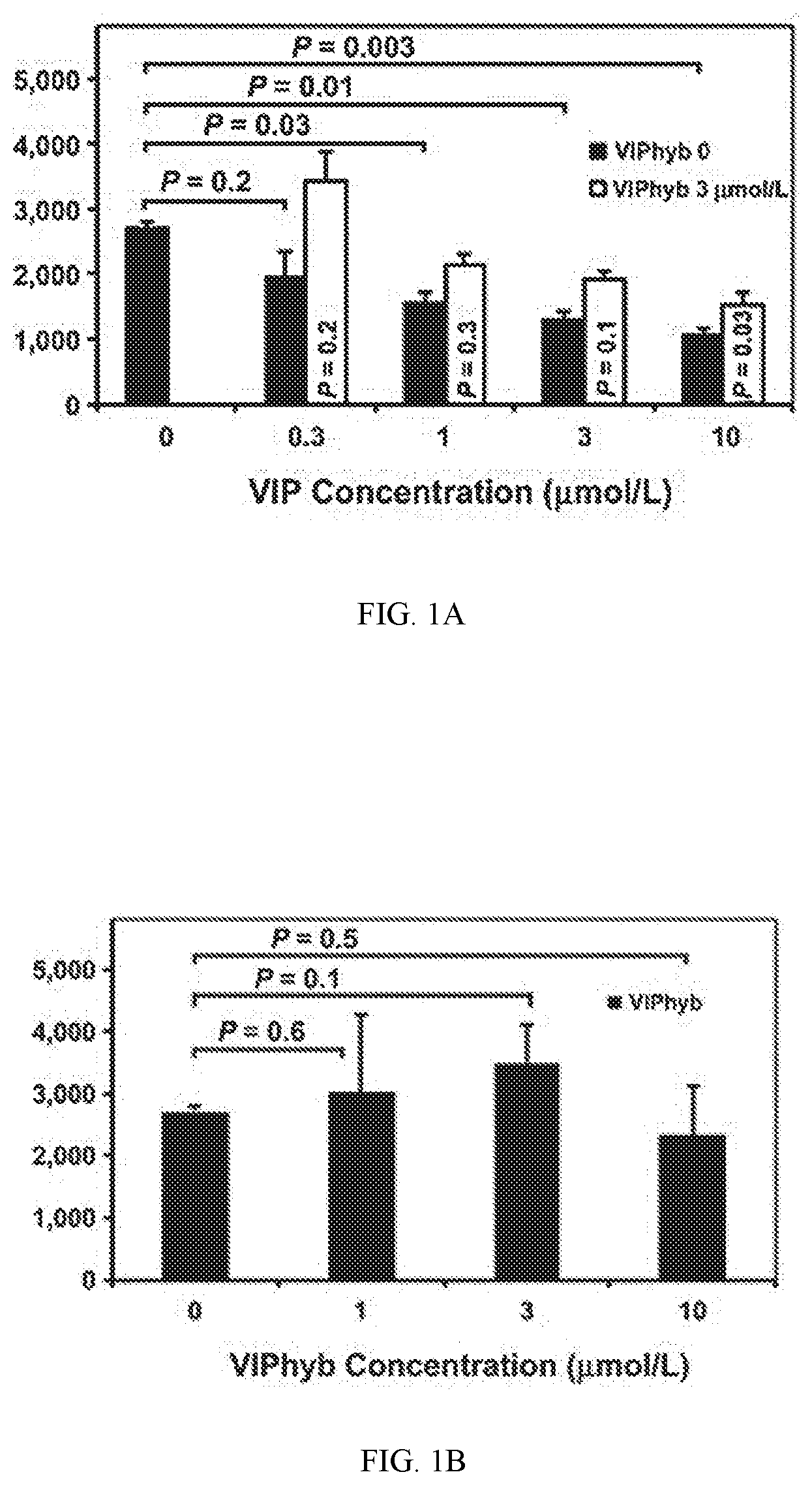
[0013] FIG. 1A shows data indicating VIPhyb treatment augmented in vitro T-cell proliferation stimulated by alloantigen. Proliferation of luciferase B6 splenic T cells cultured for 3 days in MLR with irradiated FVB splenocytes, with daily addition of VIP and/or VIPhyb to achieve the concentrations shown (0-10 mmol/L).
[0014] FIG. 1B shows data for variant concentrations of VIPhyb.
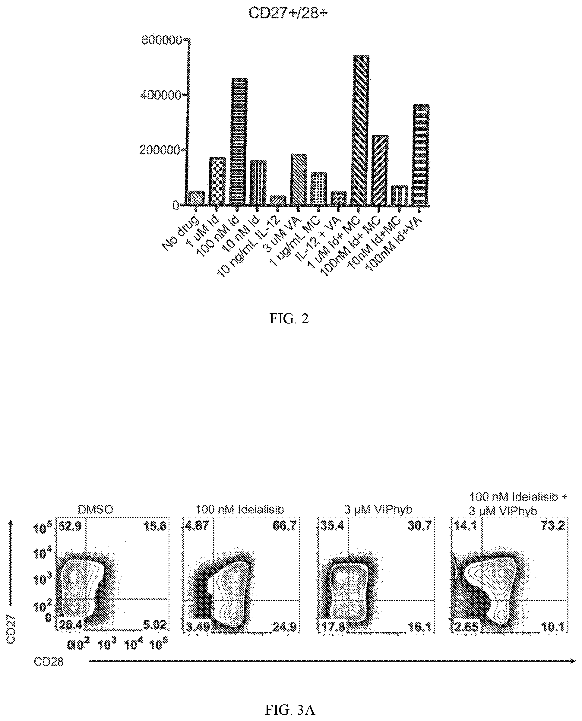
[0015] FIG. 2 shows data on the relative number of CD27+ and CD28+ cells produced in an in vitro culture on a sample wherein the patient has senescent T cells. "Id" refers to idelalisib. "VA" refers to VIPhyb. "MC" refers to human mast cell chymase (CMA1).
[0016] FIG. 3A shows data on the expression of CD27 and CD28 on the total CD3+ population on day 14 of expansion. DLBCL patient T cells were expanded for 14 days with CD3/CD28 beads in the presence of 30 U/mL IL-2 with or without the indicated compounds.
[0017] FIG. 3B shows an expansion curve of patient T cells in the presence or absence of the indicated compounds. The preservation of the CD27+CD28+ compartment and increased overall yield in a DLBCL patient T cell expansion with Idelalisib and VIPhyb.
[0018] FIG. 4 shows data indicating the expression of Bcl-2 in lymphoma patient T cells is increased with the addition of VIPhyb, Idelalisib, or a combination of both. Protein lysates from patient T cell expansion cultures were prepared on days 7 and 14. Samples were run on an SDS-PAGE gel prior to transfer to a nitrocellulose membrane. The membrane was then probed for the pro-survival Bcl-2 protein, stripped, and then re-probed for actin as a loading control.
[0019] FIG. 5A shows data on tumor volumes on day 18 post-tumor cell injection. Mice were injected subcutaneously with 5.times.10.sup.5 E.G7-OVA cells that were allowed to grow for 7 days until a palpable tumor formed. During this period, OT-I, OT-II, and B6 T cells were expanded with CD3/CD28 beads in the presence of IL-2 and the indicated compounds for 3 days. On day 7 of tumor growth, mice were injected intravenously with a combination of expanded OT-I, OT-II, and B6 T cells (2.times.10.sup.6 OT-I, 1.times.10.sup.6 OT-II, and 2.times.10.sup.6 B6 cells) or 2.times.10.sup.6 B6 T cells as a control. Mice were then monitored for tumor growth that was calculated as (L.times.W.sup.2)/2.
[0020] FIG. 5B is a tumor growth curve showing the rate of growth among groups over time. Expansion of mouse T cells in the presence of Idelalisib, VIPhyb, or a combination of both enhances their therapeutic efficacy in an OVA-expressing lymphoma model.
[0021] FIG. 6A shows data for a bioluminescent MLR assay to test immunomodulatory activity of VIPhyb-NP. Bioluminescent image of a 96-well MLR plate.
[0022] FIG. 6B shows quantification data by BU of average radiance of T-cells in MLR wells treated with VIPhyb-NP and VIP.
[0023] FIG. 7 shows data using flow cytometry analysis of VIP-KO Mice indicating increased levels of Granzyme B producing CD8+ T cells in melanoma bearing VIP-KO mice.
DETAILED DESCRIPTION
[0024] Before the present disclosure is described in greater detail, it is to be understood that this disclosure is not limited to particular embodiments described, and as such may, of course, vary. It is also to be understood that the terminology used herein is for the purpose of describing particular embodiments only, and is not intended to be limiting, since the scope of the present disclosure will be limited only by the appended claims.
[0025] Unless defined otherwise, all technical and scientific terms used herein have the same meaning as commonly understood by one of ordinary skill in the art to which this disclosure belongs. Although any methods and materials similar or equivalent to those described herein can also be used in the practice or testing of the present disclosure, the preferred methods and materials are now described.
[0026] All publications and patents cited in this specification are herein incorporated by reference as if each individual publication or patent were specifically and individually indicated to be incorporated by reference and are incorporated herein by reference to disclose and describe the methods and/or materials in connection with which the publications are cited. The citation of any publication is for its disclosure prior to the filing date and should not be construed as an admission that the present disclosure is not entitled to antedate such publication by virtue of prior disclosure. Further, the dates of publication provided could be different from the actual publication dates that may need to be independently confirmed.
[0027] As will be apparent to those of skill in the art upon reading this disclosure, each of the individual embodiments described and illustrated herein has discrete components and features which may be readily separated from or combined with the features of any of the other several embodiments without departing from the scope or spirit of the present disclosure. Any recited method can be carried out in the order of events recited or in any other order that is logically possible.
[0028] Embodiments of the present disclosure will employ, unless otherwise indicated, techniques of immunology, medicine, organic chemistry, biochemistry, molecular biology, pharmacology, physiology, and the like, which are within the skill of the art. Such techniques are explained fully in the literature.
[0029] As described herein, the level of at least one protein based surface biomarker is measured in a biological sample from an individual. The protein level(s) may be measured using any available measurement technology that is capable of specifically determining the level of the biomarker in a biological sample. The measurement may be either quantitative or qualitative, so long as the measurement is capable of indicating whether the level of the biomarker in the biological sample is above or below the reference value.
[0030] Although some assay formats will allow testing of biological samples without prior processing of the sample, peripheral blood biological fluid samples may be processed prior to testing. Processing generally takes the form of elimination of cells (nucleated and non-nucleated), such as erythrocytes, leukocytes, and platelets in blood samples, and may also include the elimination of certain proteins, such as certain clotting cascade proteins from blood. In some examples, the peripheral biological fluid sample is collected in a container comprising EDTA.
[0031] The process of comparing a measured value and a reference value can be carried out in any convenient manner appropriate to the type of measured value and reference value for the biomarker at issue. As discussed above, measuring can be performed using quantitative or qualitative measurement techniques, and the mode of comparing a measured value and a reference value can vary depending on the measurement technology employed. For example, when a qualitative colorimetric assay is used to measure biomarker levels, the levels may be compared by visually comparing the intensity of the colored reaction product, or by comparing data from densitometric or spectrometric measurements of the colored reaction product (e.g., comparing numerical data or graphical data, such as bar charts, derived from the measuring device). However, it is expected that the measured values used in the methods of the disclosure will most commonly be quantitative values (e.g., quantitative measurements of concentration, such as nanograms of biomarker per millilitre of sample, or absolute amount). As with qualitative measurements, the comparison can be made by inspecting the numerical data, by inspecting representations of the data (e.g., inspecting graphical representations such as bar or line graphs).
[0032] It must be noted that, as used in the specification and the appended claims, the singular forms "a," "an," and "the" include plural referents unless the context clearly dictates otherwise. In this specification and in the claims that follow, reference will be made to a number of terms that shall be defined to have the following meanings unless a contrary intention is apparent.
[0033] As used herein the term "idelalisib" refers to the compound (S)-2-(1-(9H-purin-6-ylamino)propyl)-5-fluoro-3-phenylquinazolin-4(3H)-on- e or alternative salts thereof.
[0034] As used herein, "T cells that are negative for CD28 and/or CD27" refers to having low expression or lack expression of relative concentrations of these markers when compared to normal T-cells that express CD3 surface antigen markers in a healthy subject.
[0035] The term "fluorescence-activated cell sorting" or "FACS" refers to a method of sorting a mixture of cells into two or more areas, typically one cell at a time, based upon the fluorescent characteristics of each cell, a respectively applied electrical charge, and separation by movement through an electrostatic field. Typically, a vibrating mechanism causes a stream of cells to break into individual droplets. Just prior to droplet formation, cells in a fluid pass through an area for measuring fluorescence of the cell. An electrical charging mechanism is configured at the point where the stream breaks into droplets. Based on the fluorescence intensity measurement, a respective electrical charge is imposed on the droplet as it breaks from the stream. The charged droplets then move through an electrostatic deflection system that diverts droplets into areas based upon their relative charge. In some systems, the charge is applied directly to the stream, and the droplet breaking off retains charge of the same sign as the stream. The stream is then returned to neutral after the droplet breaks off. In other systems, a charge is provided on a conduit inducing an opposite charge on the droplet. Cells are typically made fluorescent by mixing the cell with antibody that specifically binds a marker that is made fluorescent by conjugation to a fluorescent molecule. However, other methods of making a cell fluorescent are contemplated such as by the use of molecular beacons.
[0036] A "minimal essential medium" refers to a medium containing salts of calcium, magnesium, potassium, sodium, phosphate, and bicarbonate, vitamins, and essential amino acids. The 12 essential amino acids are L-arginine, L-cysteine, L-glutamine, L-histidine, L-isoleucine, L-leucine, L-methionine, L-phenylalanine, L-threonine, L-tryptophan, L-tyrosine, and L-valine. A minimal essential medium is often supplemented with components such as bicarbonate or glutamine. In certain embodiments, this disclosure contemplates a minimal essential medium supplemented with non-essential amino acids: L-ala, L-asn, L-asp, L-glu, L-gly, L-pro, and L-ser. In certain embodiments, this disclosure contemplates a minimal essential medium supplemented with nucleosides (ribonucleosides and/or deoxyribonucleosides).
[0037] The term "recombinant" when made in reference to a nucleic acid molecule refers to a nucleic acid molecule that is comprised of segments of nucleic acid joined together by means of molecular biological techniques. The term "recombinant" when made in reference to a protein or a polypeptide refers to a protein molecule that is expressed using a recombinant nucleic acid molecule. The term recombinant nucleic acid is distinguished from the natural recombinants that result from crossing-over between homologous chromosomes. Recombinant nucleic acids as used herein are an unnatural union of nucleic acids from nonhomologous sources, usually from different organisms.
[0038] The terms "vector" or " expression vector " refer to a recombinant nucleic acid containing a desired coding sequence and appropriate nucleic acid sequences necessary for the expression of the operably linked coding sequence in a particular host organism or expression system, e.g., cellular or cell-free. Nucleic acid sequences necessary for expression in prokaryotes usually include a promoter, an operator (optional), and a ribosome-binding site, often along with other sequences. Eukaryotic cells are known to utilize promoters, enhancers, and termination and polyadenylation signals.
[0039] The terms "vasoactive intestinal peptide" and "VIP" refer to (SEQ ID NO: 12) HSDAVFTDNYTRLRKQMAVKKYLNSILN unless the context suggests otherwise. VIP is a multifunctional endogenous polypeptide that modulates both innate and adaptive immunity at multiple levels of immune cell differentiation and activation.
[0040] VIP is typically secreted by a variety of cells such as neurons (in both the central and peripheral nervous systems) B-cells, T-cells, and accessory cells. VIP and the closely related neuropeptide pituitary adenylyl cyclase-activating polypeptide (PACAP) bind to three known receptors--VPAC1, VPAC2, and PAC1. It is believed that T-cells and dendritic cells (DC) express VPAC1 and VPAC2, but not PAC1.
[0041] The term "VIP antagonist" or "VIP receptor antagonist" refers to any molecule that inhibits or detracts from the ability of VIP to alter immune responses. VIP receptor antagonists are known including VIP analogues, VIP fragments, growth hormone-releasing factor analogues and hybrid peptides. A number of VIP receptor antagonists are disclosed in U.S. Pat. Nos. 5,565,424; 7,094,755; 6,828,304, and are all hereby incorporated by reference. Some examples of VIP receptor antagonist include [Ac-Tyr1,D-Phe2]GRF 1-29, amide, i.e., (SEQ ID NO: 4) YFDAIFTNSYRKVLGQLSARKLLQDIMSR (Modifications: Tyr-1=N-terminal Ac, Phe-2=D-Phe, Arg-29=C-terminal amide); VIP (6-28), i.e., (SEQ ID NO: 5) FTDNYTRLRKQMAVKKYLNSILN (Modifications: Asn-23=C-terminal amide); [D-p-Cl-Phe6, Leu17]-VIP, i.e., (SEQ ID NO: 6) HSDAVFTDNYTRLRKQLAVKKYLNSILN (Modifications: Phe-6=p-Cl-D-Phe, Asn=C-terminal amide); VIP-hyb also known as VIPhybrid (SEQ ID NO: 1) KPRRPYTDNYTRLRKQMAVKKYLNSILN, i.e., a hybrid peptide of neurotensin and VIP consisting of an N-terminal (SEQ ID NO: 7) KPRRPY, also designated neurotensin (6-11)] followed by the C-terminal 22 amino acids of VIP, i.e., (SEQ ID NO: 8) TDNYTRLRKQMAVKKYLNSILN, also designated VIP (7-28); N-terminal stearyl, norleucine 17 VIPhyb, i.e., (SEQ ID NO: 9) KPRRPYTDNYTRLRKQXAVKKYLNSILN, wherein X is norleucine; Ac Hisl [D-Phe(2), Lys(15), Arg(16), Leu(27)]-VIP(1-7)/GRF(8-27), i.e., (SEQ ID NO: 10) HFDAVFTNSYRKVLKRLSARKLLQDIL, C-terminal amide; and pituitary adenylate cyclase-activating polypeptide, PACAP (6-38) C-terminal amide, i.e., (SEQ ID NO: 11) TDSYSRYRKQMAVKKYLAAVLGKRYKQRVKNK. It is contemplated that any of these molecules may be modified with hydrocarbon or polyethylene glycol groups in order to provide improve properties such as solubility, bioavailability, and/or biological degradation.
[0042] As used herein, the terms "treat" and "treating" are not limited to the case where the subject (e.g., patient) is cured and the disease is eradicated. Rather, embodiments, of the present disclosure also contemplate treatment that merely reduces symptoms, and/or delays disease progression.
[0043] A "poloxamer-stabilized polypropylene sulfide nanoparticle" refers to a particle comprising a poly(propylene sulfide) (PPS) core and a poloxamer coating. A poloxamer is an amphiphilic block-co-polymer, i.e., triblock copolymers composed of a central hydrophobic chain of polyoxypropylene flanked by two hydrophilic chains of polyoxyethylene (poly(ethylene oxide). When exposed to oxygen, poly(propylene sulfide) (PPS) may be oxidized to disulfides, polysulfoxides and polysulfones. Methods of preparing poloxamer-stabilized polypropylene sulfide nanoparticles with attached peptides are described in Rehor et al. Oxidation-Sensitive Polymeric Nanoparticles, Langmuir 2005, 21, 411-417 and Vlies et la. Synthesis of Pyridyl Disulfide-Functionalized Nanoparticles for Conjugating Thiol-Containing Small Molecules, Peptides, and Proteins; Bioconjugate Chem., 2010, 21 (4), 657. Schudel et al. report S-nitrosated polypropylene sulfide nanoparticles for thiol-dependent transnitrosation. Adv. Healthcare Mater, 2015, 4, 1484-1490.
[0044] As used herein, "CpG oligonucleotides" or "CpG ODNs" refer to oligonucleotides typically of less than 100, 50, or 25 nucleotides that contains at least one motif composed of an unmethylated CG dinucleotide and flanking regions. CpG ODNs are recognized by Toll-like receptor 9 (TLR9) leading to immunostimulatory effects. Unmethylated CpG motifs are present at a greater frequency in bacterial DNA compared to mammalian DNA. CpG ODNs typically contain palindromic CpG motifs that can form stem loop structures. Some CpG ODNs contain 1 to 5 CpG motifs. CpG ODNs may contain a phosphorothioate backbone to enhance resistance to nuclease digestion. CpG ODNs may contain a phosphodiester core flanked by phosphorothioate terminal nucleotides. CpG ODNS may contain poly G motifs at the 3' end and/or 5' end. CpG ODNS may be composed of phosphorothioate nucleotides containing palindromic CpG motifs that can form stem loop structures or dimers. CpG ODNS may contain phosphodiester linkages introduced into the CG dinucleotides (semi-soft).
Methods of Use
[0045] In certain embodiments, this disclosure relates to methods of augmenting T-cell activation and ex vivo expansion by co-incubation of human T cells with a nanoparticle containing a small molecule antagonist of VIP signaling. In certain embodiments, the human T cells are activated with anti-CD3 antibody bound to a plate. In certain embodiments, the human T cells are activated in a mixed lymphocyte reaction. In certain embodiments, the human T cells are activated in vitro by co-incubation with tumor-associated antigens. In certain embodiments, the tumor associate antigens are presented on tumor microvesicles.
[0046] In certain embodiments, the human T cells are activated in vitro by co-incubation with viral antigens. In certain embodiments, the viral antigens are presented on microvesicles. In certain embodiments, the viral antigens are presented on dendritic cells.
[0047] In certain embodiments, the activated human T cells are infused into a human patient with cancer. In certain embodiments, the human patient with cancer has leukemia. In certain embodiments, the human patient with cancer has lymphoma. In certain embodiments, the human patient with cancer has multiple myeloma. In certain embodiments, the human patient with cancer has an epithelial cancer. In certain embodiments, the human patient has lung cancer. In certain embodiments, the human patient has breast cancer. In certain embodiments, the human patient has colon cancer. In certain embodiments, the human patient has prostate cancer. In certain embodiments, the human patient has malignant melanoma. In certain embodiments, the human patient has brain cancer.
[0048] In certain embodiments, this disclosure relates to methods of augmenting anti-cancer immune responses by infusion of a nanoparticle expressing a small molecule antagonist of VIP signaling. In certain embodiments, the particle contains the peptide sequence of VIPhyb, KPRRPYTDNYTRLRKQMAVKKYLNSILN (SEQ ID NO: 1).
[0049] In certain embodiments, the activated T-cells are infused into a patient with chronic CMV infection. In certain embodiments, the activated T-cells are infused into a patient with chronic EBV infection. In certain embodiments, the activated T-cells are infused into a patient with chronic BK virus infection. In certain embodiments, the activated T-cells are infused into a patient with chronic adenovirus infection.
[0050] In certain embodiments, this disclosure relates to compositions and methods of reversing senescence in T cells by interrupting vasoactive intestinal peptide (VIP) signalling and/or inhibiting phosphatidylinositol-3-kinase (PI3 kinase) inhibitor signalling and uses in managing cancer and chronic viral infections. In certain embodiments, the disclosure contemplates methods of reversing T cell senescence by mixing T cell in vitro with an agent or nanoparticle that prevents VIP from interacting with VIP receptors and/or the addition of a PI3 Kinase inhibitor. In certain embodiments, the disclosure contemplates the expansion of senescent T cells by mixing with a PI3 kinase inhibitor, a nanoparticle or agent that block VIP and VIP receptor signalling, a VIP degrading enzyme, and combinations thereof.
[0051] In certain embodiments, the disclosure contemplates methods of stimulating isolated T cells or expanding senescent T cells by in vitro exposure of T cells to antibodies that bind CD3 and/or CD28 in combination with the PI3 kinase inhibitor, idelalisib, an agent or nanoparticle that prevents VIP from interacting with VIP receptors, e.g. prevents signalling through the VIP receptor, a VIP degrading enzyme, and combinations thereof. In certain embodiments, the disclosure contemplates using anti-CD3 and anti-CD28 antibodies or binding agents optionally linked to a solid substrate such as magnetic beads.
[0052] In certain embodiments, the disclosure contemplates methods of proliferating T cells that are negative for CD28 and/or CD27 using an in vitro cell culture as disclosed herein providing replicated T cells that have increased expression of CD28 and/or CD27 compared with levels prior to replication.
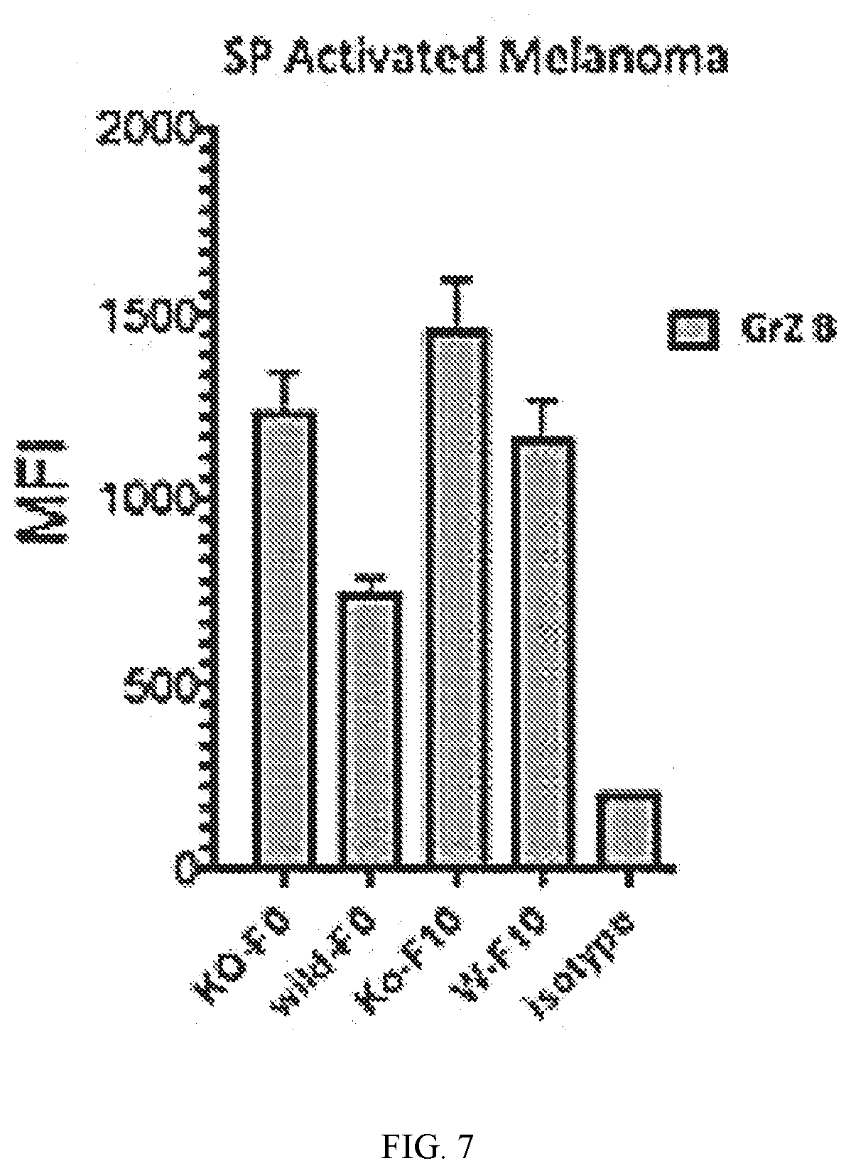
[0053] In certain embodiments, the disclosure contemplates methods of proliferating T cells wherein prior to, during, or after proliferating the T cells, the T cells are mixed with a vector having a nucleic acid sequence encoding a chimeric antigen receptor, wherein the chimeric antigen receptor comprises cancer targeting sequence, a transmembrane domain, a T cell costimulatory molecule domain, and a signal-transduction component of a T-cell antigen receptor domain under conditions such that the cells express a chimeric antigen receptor on the surface of the cells.
[0054] In certain embodiments, the disclosure relates to in vitro cell culture compositions comprising a minimal essential medium and T cells and a VIP receptor antagonist or nanoparticle thereof, a PI3 kinase inhibitor, VIP-degrading enzyme, and combinations thereof and optionally further comprising anti-CD3 antibodies and anti-CD28 antibodies optionally immobilized on a solid substrate such as beads. In certain embodiments, the T cells are purified from bone marrow cells or blood cells.
[0055] In certain embodiments, the phosphatidylinositol-3-kinase inhibitor is selected from idelalisib, wortmannin, demethoxyviridin, perifosine, buparlisib, duvelisib, copanlisib, and alpelisib. In certain embodiments, the phosphatidylinositol-3-kinase inhibitor is selected from idelalisib in a culture at a concentration of greater than 0.001, 0.1, 1, 10, 100 nM or between 10 nM and 10 micromolar or between 10 nM and 500 nM, or between 10 nM and 1 micromolar.
[0056] In certain embodiments, the culture comprises a VIP receptor antagonist or nanoparticle thereof such as VIPhyb comprising (SEQ ID NO: 1) KPRRPYTDNYTRLRKQMAVKKYLNSILN optionally having a C-terminal amide. In certain embodiments, the culture comprises a VIP receptor antagonist or nanoparticle thereof such as VIPhyb in at a concentration of at least of added at 0.001, 0.01, 0.1, 0.5, 1.0, 2, or 3 micromolar. In certain embodiments, the culture comprises a VIP receptor antagonist or nanoparticle thereof such as VIPhyb in at a concentration of between 0.5 and 10 micromolar or between 1 and 8 micromolar.
[0057] In certain embodiments, the culture comprises an enzyme that hydrolyses VIP. In certain embodiments, the culture comprises a VIP degrading enzyme such as a peptidase, serine peptidase, a tryptase, chymase, human chymase 1 (CMA1), or chymotrypsin serine proteinase. In certain embodiments, the culture has at least at least 0.001, 0.01, 0.1, or 1 microgram per mL of the VIP degrading enzyme such as a mast cell chymase. In certain embodiments, the disclosure contemplates a T cell culture comprising minimal essential medium and isolated cells that express CD3 and/or CD4 and/or CD8 and are negative for CD27 and/or CD28 and a PI3 kinase inhibitor, agent or nanoparticles that block VIP and VIP receptor signalling, and combinations thereof. The cells may be isolated by negative or positive selection using binding agents attached to solid supports such as beads, magnetic beads, or particles of fluorescent binding agents.
[0058] In certain embodiments, the anti-CD3 antibodies and anti-CD28 antibodies are immobilized on a bead, magnetic bead, or solid surface. In certain embodiments, more than 5.0% or 10% or 15% of the total cells in the culture express CD3 and/or CD4 and/or CD8. In certain embodiments, more than 20%, 25% or 50% of the total cells express CD3 and/or CD4 and/or CD8. In certain embodiments, more than 15% or 20% or 30% of the T cells in the culture are negative for CD28 and/or CD27. In certain embodiments, more than 20%, 25% or 50% of the T cells are negative for CD28 and/or CD27.
[0059] In certain embodiments, the purified T cells are obtained from centrifuging blood under conditions such that plasma and red blood cells separate providing purified T cells in a mixture of white blood cells between the plasma and red blood cells. In certain embodiments, the purified T cells are obtained by bone marrow aspirates or a bone marrow biopsy.
[0060] In certain embodiments, the purified T cells are obtained by mixing cells with a fluorescent marker that binds CD3 and purifying cells by fluorescent activated cell sorting. In certain embodiments, the purified T cells are obtained by mixing cells with a magnetized marker that binds CD3 and purifying cells by magnetic sorting. In certain embodiments, the purified T cells are obtained by mixing cells with a fluorescent marker that binds CD3 and/or CD4 and/or CD8 and purifying cells by fluorescent activated cell sorting. In certain embodiments, the purified T cells are obtained by mixing cells with a magnetized marker that binds CD3 and/or CD4 and/or CD8 and purifying cells by magnetic sorting.
[0061] In certain embodiments, the disclosure contemplates a solid substrate, such as beads, with anti-CD3 and anti-CD28 antibodies and having a VIP-degrading enzyme coupled to the surface. In certain embodiments, it is contemplated that the beads are arranged in the medium and the T cells are expanded on top of the medium such that the beads are sub-cellular.
[0062] In certain embodiments, the VIP degrading enzyme comprises human CMA1 Accession number GenBank: AAI03975.1:
TABLE-US-00001 (SEQ ID NO: 2) MLLKLKEKASLTLAVGTLPFPSQFNFVPPGRMCRVAGWGRTGVLKPGSDTL QEVKLRLMDPQACSHFRDFDHNLQLCVGNPRKTKSAFKGDSGGPLLCAGVA QGIVSYGRSDAKPPAVFTRISHYRPWINQILQAN.
[0063] In certain embodiments, the VIP-degrading enzyme is human recombinant enkephalinase (neutral endopeptidase, EC 3.4.24.11) having (SEQ ID NO: 3)
TABLE-US-00002 DGICKSSDCIKSAARLIQNMDATTEPCTDFFKYACGGWLKRNVIPETSSRY GNFDILRDELEVVLKDVLQEPKTEDIVAVQKAKALYRSCINESAIDSRGGE PLLKLLPDIYGWPVATENWEQKYGASWTAEKAIAQLNSKYGKKVLINLFVG TDDKNSVNHVIHIDQPRLGLPSRDYYECTGIYKEACTAYVDFMISVARLIR QEERLPIDENQLALEMNKVMELEKEIANATAKPEDRNDPMLLYNKMTLAQI QNNFSLEINGKPFSWLNFTNEIMSTVNISITNEEDVVVYAPEYLTKLKPIL TKYSARDLQNLMSWRFIMDLVSSLSRTYKESRNAFRKALYGTTSETATWRR CANYVNGNMENAVGRLYVEAAFAGESKHVVEDLIAQIREVFIQTLDDLTWM DAETKKRAEEKALAIKERIGYPDDIVSNDNKLNNEYLELNYKEDEYFENII QNLKFSQSKQLKKLREKVDKDEWISGAAVVNAFYSSGRNQIVFPAGILQPP FFSAQQSNSLNYGGIGMVIGHEITHGFDDNGRNFNKDGDLVDWWTQQSASN FKEQSQCMVYQYGNFSWDLAGGQHLNGINTLGENIADNGGLGQAYRAYQNY IKKNGEEKLLPGLDLNHKQLFFLNFAQVWCGTYRPEYAVNSIKTDVESPGN FRIIGTLQNSAEFSEAFHCRKNSYMNPEKKCRVW.
[0064] In certain embodiments, cell cultures and methods described herein further include IL-12. In certain embodiments, the IL-12 is contemplated to enhance the effect of the VIP receptor antagonist or nanoparticle thereof on T cell proliferation stimulated in vitro with antibodies to CD3 and CD28.
[0065] In certain embodiments, the disclosure relates to expanding T cells, or expanding or reversing senescence in T cells, with a naturally occurring reactivity to cancer can be found infiltrated in tumors of the subject. The tumor can be harvested, and these tumor-infiltrating lymphocytes (TIL) can be expanded using methods discloses herein.
[0066] In certain embodiments, nanoparticles disclosed herein, e.g., nanoparticles comprised of poloxamer-stabilized polypropylene sulphide, comprises paclitaxel incorporated within the hydrophobic core. In certain embodiments, nanoparticles disclosed herein are conjugated to a CpG oligonucleotide that is a TLR-9 agonist.
[0067] In certain embodiments, the disclosure contemplates methods of treating cancer or a chronic infection comprising: purifying T cells from a subject providing isolated T cells; mixing the isolated T cells with anti-CD3 antibodies and anti-CD28 antibodies optionally immobilized on a bead or solid surface in combination with a PI3 kinase inhibitor, a VIP receptor antagonist or nanoparticle thereof, a VIP degrading enzyme, or combinations thereof; under conditions such that the T cells replicate providing replicated T cells have increased expression of CD28 compared with levels prior to replication; and administering an effective amount of the replicated T cells to a subject in need thereof.
[0068] In certain embodiments, the disclosure contemplates methods of treating cancer comprising: purifying T cells from a subject providing isolated T cells; mixing the isolated T cells with anti-CD3 antibodies and anti-CD28 antibodies optionally immobilized on a bead or solid surface in combination with a PI3 kinase inhibitor, a VIP receptor antagonist or nanoparticle thereof, a VIP degrading enzyme, or combinations thereof; under conditions such that the T cells replicate providing replicated T cells have increased expression of CD28 compared with levels prior to replication; and administering an effective amount of the replicated T cells to a subject in need thereof. In certain embodiments, the replicated T cells express a chimeric antigen receptor on the surface of the cells. In certain embodiments, the method further comprises administering a PI3 kinase inhibitor, a VIP receptor antagonist or nanoparticle thereof, a VIP degrading enzyme, or combinations thereof, before, during, or after administering the replicated T cells.
[0069] In certain embodiments, the disclosure contemplates method of treating cancer comprising: purifying T cells from a subject providing isolated T cells; culturing the isolated T cells by in vitro exposure of T cells to antibodies that bind CD3 and/or CD28 in combination with the PI3 kinase inhibitor, idelalisib, an agent or nanoparticle that prevents VIP from interacting with VIP receptors, e.g. prevents signalling through the VIP receptor, a VIP degrading enzyme, and combinations thereof providing expanded T cells with an increased expression of CD28; and administering an effective amount of the expanded T cells to the subject.
[0070] In certain embodiments, the disclosure contemplates method of treating cancer comprising administering an effective amount of a bi-specific antibody in combination with a VIP receptor antagonist or nanoparticle thereof and/or a phosphatidylinositol-3-kinase inhibitor to a subject in need thereof, wherein the bi-specific antibody comprises a cancer targeting binding sequence and a CD3 binding sequence. In certain embodiments, the bi-specific antibody is catumaxomab or blinatumomab.
[0071] In certain embodiments, the disclosure contemplates methods of treating cancer with a combination of bi-specific antibodies or anticancer antibody or antibody to a tumor associated antigen and administration or parental administration of a VIP receptor antagonist or nanoparticle thereof such as VIPhyb. In certain embodiments, the anti-tumor specific antibody is directed to CD19. In certain embodiments, the anti-cancer antibody is directed to CD123. In certain embodiments, the anti-cancer antibody is directed to HER2/neu. In certain embodiments, the anti-cancer antibody is directed to BMCA, a myeloma associated antigen. In certain embodiments, the anti-cancer antibody is directed to EGFR. In certain embodiments, the anti-cancer antibody is directed to PD-L1 or PD1.
[0072] In certain embodiments, the disclosure contemplates methods of treating cancer comprising administering an effective amount of cells having a chimeric antigen receptor in combination with a VIP receptor antagonist or nanoparticle thereof or phosphatidylinositol-3-kinase inhibitor to a subject in need thereof, wherein the chimeric antigen receptor comprises cancer targeting sequence, a transmembrane domain, a T cell costimulatory molecule domain, and a signal-transduction component of a T-cell antigen receptor domain.
[0073] In certain embodiments, the disclosure contemplates in vivo methods of reversing senescence of T cells comprising genetically modifying T cells to express on their surface a VIP degrading enzyme. In certain embodiments, the VIP degrading enzyme is recombinant human CMA1 mast cell chymase. In certain embodiments, the genetically modified T cells also express a chimeric antigen receptor that targets them to cancer cells. In certain embodiments, the genetically modified T cells are administered or infused into a subject with cancer.
[0074] In certain embodiments, this disclosure relates to methods of treating cancer comprising: purifying T cells from a subject that express a T cell receptor wherein the T cells express CD3 and optionally CD4 and/or CD8 providing isolated T cells; mixing the isolated T cells with cell culture disclosed herein under conditions such that the cells expand, and implanting or administering an effective amount of the expanded cells into the subject.
[0075] In certain embodiments, the disclosure contemplates methods of treating cancer comprising administering T cells comprising a vector configured to express a chimeric antigen receptor, e.g., the cells has been infected with a recombinant virus that has a nucleic acid that codes a chimeric antigen receptor, in combination with administration of a VIP receptor antagonist or nanoparticle thereof such as VIPhyb, a VIP-degrading enzyme, a PI3 inhibitor, or combinations thereof.
[0076] In certain embodiments, the disclosure contemplates methods of treating cancer comprising: purifying cells from a subject that express CD3 and/or CD4 and/or CD8 providing isolated T cells; measuring the expression of CD27 and/or CD28, on the isolated T cells providing measured values of CD27 and/or CD28; comparing the measured values of CD27 and/or CD28, to a reference level, and if the measured levels of CD27 are lower than normal and/or the measured levels of CD28 are lower than normal, then administering an effective amount of a VIP receptor antagonist or nanoparticle thereof, a VIP-degrading enzyme, a PI3 inhibitor, or combinations thereof to a subject.
[0077] In certain embodiments, the disclosure contemplates methods of treating cancer comprising purifying cells from a subject that express CD3 and/or CD4 and/or CD8 providing isolated T cells; measuring the expression of CD27 and/or CD28, on the isolated T cells providing measured values of CD27 and/or CD28; if the measured values of CD27 is lower than normal and/or the measured value of CD28 is lower than normal providing replicated T cells then mixing the isolated T cells with a VIP receptor antagonist or nanoparticle thereof, a VIP-degrading enzyme, a PI3 inhibitor, or combinations thereof under conditions such that the isolated T cells replicate; and implanting or administering an effective amount of the replicated T cells into the subject optionally in combination with administering a VIP receptor antagonist or nanoparticle thereof, a VIP-degrading enzyme, a PI3 inhibitor, or combinations thereof to the subject.
[0078] In certain embodiments, the disclosure relates to expanding T cells that are positive for CD3 and negative for CD4 and CD8 such as gamma delta T cells (.gamma..delta. T cells). Gamma delta T cells that have a distinctive T-cell receptor (TCR) on their surface. Most T cells are .alpha..beta. (alpha beta) T cells with TCR composed of two glycoprotein chains called .alpha. (alpha) and .beta. (beta) TCR chains. In contrast, gamma delta (.gamma..delta. ) T cells have a TCR that is made up of one .gamma. (gamma) chain and one .delta. (delta) chain.
[0079] In certain embodiments, the disclosure contemplates a T-cell genetically modified to express on their surface a VIP-degrading enzyme. In certain embodiments, the VIP-degrading enzyme is recombinant human CMA1 mast cell chymase. In certain embodiments, the genetically modified T cells also express a chimeric antigen receptor that targets them to cancer cells. In certain embodiments, the genetically modified T cells are administered or infused into a human subject with cancer.
[0080] In certain embodiments, this disclosure relates to methods of treating cancer comprising administering an effective amount of a bi-specific antibody in combination with a VIP receptor antagonist or nanoparticle thereof, a PI3 kinase inhibitor, VIP-degrading enzyme, and combinations thereof. In certain embodiments, the bi-specific antibody is catumaxomab. In certain embodiments, the subject is diagnosed with malignant ascites. In certain embodiments, the bi-specific antibody is blinatumomab. In certain embodiments, the cancer is leukemia.
[0081] In certain embodiments, the disclosure contemplates methods of treating cancer comprising: purifying and expanding T cells using methods provided herein providing isolated T cells; mixing the isolated T cells with bi-specific antibodies under conditions such that the bispecific antibodies bind the CD3-T cell receptor complex; and administering an effective amount of the bispecific antibody bound T cells to the subject in combination with administering a VIP receptor antagonist or nanoparticle thereof, a PI3 kinase inhibitor, VIP-degrading enzyme, and combinations thereof to a subject in need thereof.
[0082] Bi-specific antibodies contain two targeting sequences, the first targets a tumor-associated antigen and the second targets the CD3 T-cell receptor complex such that T-cells can engage cancer cells. The Bi-specific antibody is linking the T cells to the cancer cells. See Zhukovsky et al. Bispecific antibodies and CARs: generalized immunotherapeutics harnessing T cell redirection, Current Opinion in Immunology, 2016, 40: 24-35. In certain embodiments, this disclosure contemplates that the bi-specific antibody is directed to the tumor-associated antigen, CD19 epitope, CD123, HER2/neu, or a BMCA, a myeloma associated antigen.
[0083] In order to improve the ability of immune cells to kill cancerous cells, T cells can be isolated from the blood of a patient and genetically altered to express chimeric antigen receptors to specifically target proteins expressed on the surface of cancerous cells and stimulate an immune response. When put back into the patient, the cells attack the cancerous cells. In certain embodiment, this disclosure contemplates using CAR T cells that target the CD22 and/or CD19 antigens. CD19 is a protein expressed on cancerous B cells. Brentjens et al. report that T cells altered to bind CD19 can induce remissions of cancer in adults with chemotherapy-refractory acute lymphoblastic leukemia. Sci Transl Med, 2013, 5(177):177ra38.
[0084] In a typical procedure, T cells are purified and isolated from blood or bone marrow. For example, T cells are collected via apheresis, a process that withdraws blood from the body and removes one or more blood components (such as plasma, platelets or other white blood cells). The remaining blood is then returned back into the body. The cells are exposed to a recombinant vector, such as a lentiviral vector, that infects the cells in a way that a CAR protein is produced to be present in the cell membrane. The T cells may be sent to a laboratory or a drug manufacturing facility where they are genetically engineered to produce chimeric antigen receptors (CARs) on their surface. Before and/or after infecting the isolated cells with the recombinant vector, the cells may be induced to replicate using methods disclosed herein. The genetically modified T cells may be expanded by growing cells in the laboratory until there are sufficient number of them. Optionally, these CAR T cells are frozen. The modified cells are then administered back to the patient. In certain embodiments, this disclosure contemplates that the subjects are administered with a VIP receptor antagonist or nanoparticle thereof, VIP-depredating enzyme, and/or an IP3 kinase inhibitor optionally in combination with one or more chemotherapy agent or nanoparticles before they receive the infusion of CAR T cells.
[0085] In certain embodiments, the disclosure relates to cells made by processes disclosed herein that contain recombinants vector comprising a nucleic acid that encodes a chimeric polypeptide comprising a targeting sequence, a transmembrane domain, a T cell costimulatory molecule domain, and a signal-transduction component of a T-cell antigen receptor domain.
[0086] In certain embodiments, the targeting sequence in a chimeric antigen receptor refers to any variety of polypeptide sequences capable of selectively binding to a surface protein on target cells, e.g., cancer cells. Other targeting sequences may be variable binding regions of antibodies, single chain antibodies, and antibody mimetic. In certain embodiments, targeting is achieved via a single-chain variable fragment (scFv) derived from a monoclonal antibody. The targeting sequence it typically connected to the intracellular domains by a hinge/transmembrane region, commonly derived from CD8 or IgG4. The intracellular domains may contain co-stimulatory domains such as 4-1BBzeta and/or CD28zeta linked to the cytoplasmic signaling domain of CD3zeta.
Immunophenotype of Patients with Relapsed Lymphoma
[0087] During physiologic aging and in individuals exposed to repeated rounds of chemotherapy or chronic inflammatory conditions, T-cells develop a condition of senescence in which they are anergic or have limited proliferative potential in response to antigenic stimulation. The phenotype of senescence T-cells has been described as those T-cells that lack co-stimulatory receptors CD27 and CD28 or have high levels of the CD57 marker. In addition, expression of PD1, a receptor for programmed death-ligand has been associated with the anergic state. The presence of anergic T-cells is a critical cofactor in the ability of patients to respond to vaccines or to mount protective and durable immune responses to chronic viral infections or cancer. Approaches to reverse T-cell senescence and anergy are needed to offer patients with chronic infections and cancer the prospect of effective cell based immunotherapy.
[0088] The immunophenotype of patients with relapsed lymphoma are characterized as having an over representation of T-cells with the anergic phenotype: CD3 positive, CD27 negative, CD28 negative. Whether signaling through the vasoactive intestinal polypeptide receptor contributed to T-cell senescence and antigen specific energy was performed. The addition of a peptide antagonist to VIP receptor called VIPhyb augmented T cell proliferation in vitro and mitigated the inhibitory effect of the native VIP peptide on T-cell proliferation.
[0089] Whether the addition of the VIPhyb antagonist could reverse the proliferative deficits of anergic T cells from patients with long-standing relapsed cancer was tested. The immunophenotype of T-cells in a patient with relapsed lymphoma was evaluated. There was a relative over representation of cells lacking the costimulatory receptor CD27 and CD28. In addition, the T cells from this station have high levels of CD57 expression, PD1 and Tim-3.
[0090] Using high-speed cell sorting subsets of T-cells were isolated defined by their levels of CD27 and CD28 expression from the patient with relapsed lymphoma and a healthy control. CD3 positive T-cells with each of the four phenotypes defined by CD27 and CD28 expression were sorted and cultured in vitro in the presence of anti-CD.sup.3/anti-CD28 beads. Total T-cells from healthy donors had 100% expression of the Ki67 proliferation marker following four days of culture. However, the corresponding total T-cell population from the patient with anergic T-cells following treatment for lymphoma had only 90% of T-cells expressing the Ki67 marker.
[0091] Following the daily addition of 3 uM VIPhyb to these cultures, the proportion of Ki67 positive T-cells from the anergic lymphoma patient was increased to 100%. Subsets of T-cells sorted based on CD27 and CD28 expression from this patient showed that the CD27 positive, CD28 positive (double positive) population proliferated normally in response to anti-CD3/CD28 beads. In contrast, the T-cell subsets lacking expression of either CD27 or CD28 or both markers had impaired proliferated response when incubated with the anti-CD3/CD28 beads.
[0092] Addition of the antagonist VIPhyb augmented T-cell proliferation in the subsets lacking either CD27 or CD28. The addition of IL 12 to these cultures had a significant impact in increasing the proliferation fraction of T-cells that were CD28 negative but not those that were CD27 negative. Flow cytometry analysis of the T cells cultured with anti-CD.sup.3/anti-CD28 beads showed that the addition of 3 uM VIPhyb led to decreased levels of PD-1 expression compared with control cultures to which no VIP antagonist was added. This data indicates that VIP is induced during antigen specific activation of T-cells and that strategy to block VIP signaling could augment T-cell proliferation and reverse anergy among senescence T-cells from individuals with chronic inflammatory conditions or those who have been exposed to multiple cycles of chemotherapy.
[0093] The VIP peptide has a very short half-life in vivo: less than two minutes. As VIP peptide is cleaved by endopeptidases, over expression of VIP specific peptidases near T-cells activated through their T-cell receptor are believed to result in augmented activation and proliferation of T-cells, which could reverse immune senescence.
VIP Signaling Regulates T-Cell Proliferation Induced by Alloantigen
[0094] To explore the role of VIP signaling in allo-immune responses, one-way mixed lymphocyte reactions (MLR) were performed containing VIP and/or the antagonistic VIPhyb peptide. Addition of VIP decreased Luciferase T-cell proliferation in a dose-dependent manner, while adding VIPhyb increased T-cell proliferation. VIPhyb reversed the suppressive effect of VIP in MLR, restoring T-cell proliferation to higher levels than control cultures.
Pharmacological Blockade During Ex Vivo T Cell Expansion for CAR T Manufacture Increases Yield and Preserves the Naive and Central Memory Compartments
[0095] Diffuse large B cell lymphoma (DLBCL) is an aggressive B cell malignancy that mainly affects older patient populations. Despite the robust treatment regimens available, a subset of DLBCL patients exists with lymphoma that is highly treatment-resistant. Due to the fact that these patients disease are refractory to virtually all of the current treatment regimens, CAR T therapy holds tremendous promise as an effective salvage option, however many patients have failed to receive treatment in clinical trials due to a failure of ex vivo T cell expansion. This failure is due in large part to the damage done by numerous rounds of therapies as well as the age of the patients. Additionally, data indicates that patients with NHL have a skewed ratio of memory to naive T cells resulting in impaired cellular immunotherapy.
[0096] Two of the surface proteins that are indicative of a patient's potential for successful T cell expansion are CD27 and CD28. T cells that express both CD27 and CD28 have the greatest proliferative potential while T cells that lack expression of both often do not expand and die during anti-CD3/CD28 activation and expansion. Healthy individuals have an abundance of CD27+CD28+ (double positive) T cells while patients with DLBCL have an over-abundance of CD27-CD28- (double negative) T cells. This over-representation of the double negative population in lymphoma patients helps to explain the failure of the T cells to expand adequately during CAR T manufacture. In addition to the lack of CD27 and CD28 expression, DLBCL patient T cells also exhibit signs of exhaustion and senescence that both lead to functional impairments and the inability to expand.
[0097] To address the issues with DLBCL patient T cell expansion during CAR T cell manufacture, a variety of technologies were utilized in order to enhance expansion of specific desired T cell populations. Inclusion of the PI3K .delta. inhibitor idelalisib alone or in combination with a mast cell chymase or antagonist of vasoactive intestinal peptide (VIPhyb) during T cell activation increases the number of viable T cells obtained and increases the proportion of naive and central memory cells. Naive and central memory cells are the most effective subset for adoptive cellular immunotherapy. Additionally, inclusion of either idelalisib or VIPhyb increases the frequency of CD27+CD28+ cells while decreasing the frequency of CD27-CD28- cells. Further studies using murine and human T cells have indicated that while blockade of PI3K .delta. has an inhibitory effect on proliferation, the prevention of terminal differentiation and the preservation of the naive compartment enhance T cell survival and yield. This data indicates that the culture conditions used not only increase the number of total patient T cells, but also preserve the compartments most effective in adoptive T cell immunotherapy. As such, utilization of our methods during CAR T manufacture has the potential to enable the development of the last-resort treatment that many DLBCL patients have previously been unable to receive.
Expansion of Senescent T Cells with P13 Kinase Inhibitors and Blocking VIP Signalling
[0098] Idelalisib is an inhibitor of PI3 kinase. PI3 kinase is an important signalling pathway for lymphocyte activation and differentiation, and regulates AKT and mTOR1 activity. Idelalisib is FDA-approved for treatment of patients with chronic lymphocytic leukemia and indolent B cell malignancy. Some patients treated with idelalisib have anti-cancer responses after drug treatment has stopped. Some patient also develop auto immune-like signs and symptoms including colitis and rash, leading to the hypothesis that idelalisib modulates the immune system and activates or preserves Thl-polarized T cells.
[0099] Idelalisib treatment leads to increased T-cell activation in patients with CLL. Ex vivo exposure of activated T cells to idelalisib potentiate their activation and in vitro expansion. After a range of idelalisib concentrations was adding to T cells cultured in vitro with anti-CD3/CD28 beads, T cell expansion and differentiation was measured.
[0100] A T cell sample that was used from a patient with lymphoma who had senescent T cells that failed to expand in vitro when manufacturing of CAR T cell was attempted. Monocytes were depleted from the T cell sample, as monocytes are known to inhibit T cell expansion in cultures containing anti-CD3/CD28 beads. The effect of a range of idelalisib concentrations were compared alone and in combination with 3 uM VIP peptide antagonist (VIPhyb) or 1 ug/ml mast cell chymase, an enzyme that degrades endogenous VIP on T cell activation, differentiation and proliferation. Numbers of viable T cells were measured during a 10 day in vitro cultures as well as the T cell differentiation and activation profiles, focusing on the relative numbers of T cells with a senescent phenotype in which co-stimulatory receptors CD27 and CD28 are both absent (CD27-CD28-), to an activated phenotype, in which either CD27, CD28, or both co-stimulatory receptors are present (CD27+CD28+).
[0101] Aliquots of frozen patient apheresis products or PBMC from ficolled whole blood were rapidly thawed and rested overnight in complete RPMI 1640 supplemented with 10% fetal bovine serum, 100 U/mL penicillin, 100 .mu.g/mL streptomycin, and 50 .mu.M 2-mercaptoethanol (complete media). The following day, red blood cells were removed from apheresis products by ficoll gradient. The leukocytes were then enriched for T cells using an EasySep human T cell enrichment kit according to manufacturer's instructions. Cells were plated in 200 .mu.L of complete media in each well of a 96 well flat-bottom plate. Compounds were added at the indicated concentrations, and the DMSO concentration was normalized to 0.1% for all wells. Anti-CD3/CD28 beads were added at a 1:1 bead:cell ratio. On day 7 post-stimulation, cells from each treatment were transferred to new media. Fresh compounds and beads were then added, and the cells were cultured for an additional 3 days. On day 10 post-initial stimulation, the beads were removed, and the cells were stained for flow cytometric analysis.
[0102] Cells were washed twice in PBS. The cells were also stained using a fixable viability dye. Surface markers were then stained by adding fluorochrome-conjugated antibodies to CD3, CD4, CD8, CD27, CD28, CD45RA, CD45RO, PD-1, and CCR7. To assess proliferation, half the cells were evaluated for expression of Ki67. Ki67 staining was performed using a FoxP3 intracellular staining kit. Just prior to running samples, Accucheck.TM. counting beads were added to each tube. Samples were acquired on a BD FACS Aria, and analysis was performed using FlowJo.TM. software. Viable cells were used in the analysis. The absolute numbers of cells was calculated for each population according to instructions provided by the manufacturer of the Accucheck.TM. counting beads.
[0103] The addition of idelalisib to the T cell cultures at concentrations of 1 uM or 100 nM, significantly increased the proportion of T cells co-expressing CD27 and CD28 compared to control cultures with no added drug. Of note the fraction of senescent T cells that lacked both
[0104] CD27 and CD28 costimulatory receptors was decreased from 55.2% in control cultures with no added drug, to 16.1% in cultures with 10 nM idelalisib, 35.7% in cultures with 3 uM VIPhyb, and 37.4% in cultures with 1 .mu.g per ml mast cell chymase.
[0105] Idelalisib at 100 nM concentration was synergistic in enhancing the proportion of T cells expressing co-stimulatory receptors when combined with VIPhyb or chymase, with the fraction of CD27+CD28+ cells 21.5% with the combination of 100 nM idelalisib and VIPhyb and 14.2% in the combination of 100 nM idelalisib and chymase versus to 3.5% in control cultures with no added drug.
[0106] Numbers of effector memory T cells (T.sub.em) in the 10-day cultures were also increased by exposure to idelalisib, VIPhyb and the combinations of idelalisib with chymase and VIPhyb. The total numbers of viable T cells in cultures was significantly increased with combinations of idelalisib and chymase or idelalisib and VIPhyb compared with control cultures with no added drug. Of note, the total numbers of CD27+CD28+ T cell subset were enriched more than 10-fold in the cultures containing 100 nM idelalisib, 1 uM idelalisib plus 1 ug/ml chymase, or 100 nM idelalisib plus 3 uM VIPhyb compared with cultures containing no added drugs, while total numbers of CD3+ Tem in the 10-day cultures were increased 10-fold in cultures containing 1 uM idelalisib plus chymase compared with control cultures with no added drugs. CD3+ central memory T cells (T.sub.cm) were increased 5-10 fold in cultures containing idelalisib alone, VIPhyb alone, chymase alone, or combinations of idelalisib with either VIPhyb or chymase compared with control cultures with no added drugs.
[0107] This data indicates that senescent T cells that cannot be expanded in culture with anti CD3/CD28 beads and that failed to yield sufficient manufactured CAR T cells for clinical use can be significantly expanded in vitro with the addition of idelalisib, VIPhyb or mast cell chymase as single agent or nanoparticles or in combination. These results support the addition of these agent or nanoparticles during CAR T manufacturing and support their use in vitro to expand senescent T cells to enhance cancer immunotherapy and anti-viral immunity.
Nanoparticle Synthesis
[0108] One synthesises poloxamer-stabilized poly(propylene sulfide) (PPS) NPs with average diameters of about 30 nm in diameter by inverse emulsion polymerization. One uses a Pluronic.TM. F-127 (a block copolymer of polyethylene glycol and polypropylene glycol terminated by hydroxyl groups) in combination with carboxyl-terminated derivatization. Polymerization in the hydrophobic core results in PPS chains with a terminal thiolate, which can lead to stabilization of the core by intermolecular disulfide crosslinking. However, since crosslinking does not reach completion, remaining free sulfide groups on the NP surface may be irreversibly capped by reaction with the alkylating reagent iodoacetamide.
[0109] For coupling to cysteine containing VIP antagonists one synthesizes pyridyl disulfide-poloxamer-stabilized poly(propylene sulfide) (PPS) NPs using procedures illustrated in FIG. 1 of Vlies et al. Synthesis of Pyridyl Disulfide-Functionalized Nanoparticles for Conjugating Thiol-Containing Small Molecules, Peptides, and Proteins; Bioconjugate Chem., 2010, 21 (4), 657. A NP suspension is formed using 30% carboxylate poloxamer and 70% hydroxylate poloxamer. One adds solid 2-(N-morpholino)ethanesulfonic acid followed by pyridyl disulfide cysteamine HCl salt, N-hydroxysulfosuccinimide sodium salt, and N-(3-dimethylaminopropyl)-N'-ethylcarbodiimide, at an excess of 20 equivalents relative to the amount of carboxylate poloxamer. The NPs carry they pyridine thiol-reactive groups are mixed with thiol-containing VIP antagonists to form disulfide conjugates.
Nanoparticles Conjugated with an Antagonist for Vasoactive Intestinal Peptide
[0110] Vasoactive intestinal peptide (VIP) is an immunosuppressive neuropeptide that leads to reduced T cell proliferation and reduced production of pro-inflammatory cytokines. Thus, blockade of the VIP pathway as a therapeutic target, should result in a robust immune response. Previous research in a mouse leukemia models supports that VIPhyb, a peptide antagonist of VIP, increases the number of effector/memory CD8+ T cells and mature NK cells. Further, VIPhyb down regulates the expression of PD-1 and PD-L1 in activated T cells and dendritic cells, respectively. Conjugating VIPhyb (KPRRPYTDNYTRLRKQMAVKKYLNSILNGGGGSC (SEQ ID NO: 13)) to a 30 nm poloxamer-stabilized polypropylene sulfide nanoparticle (VIPhyb-NP) specifically designed to target tumor draining lymph nodes (TDLN), can further augment its immunomodulatory effects.
[0111] In solid tumor immunotherapy, the immunomodulatory effect of VIPhyb can be further augmented by conjugation with nanoparticles (NP) specifically designed to target tumor draining lymph nodes (TDLN). Since TDLNs are a major tissue involved in draining tumor-associated antigens, targeted delivery of VIPhyb to TDLNs could improve the antitumor immune response and hamper overall growth of solid tumors susceptible to immunotherapy.
[0112] Splenocytes were extracted from WT and VIP-KO mice treated with B16-F0 and B16-F10 melanoma, and analyzed with flow cytometry for Granzyme B (GrZ B) and CD8 expression. VIPhyb was conjugated to a biocompatible, non-toxic 30 nm NP through emulsion polymerization and tested for purity and stability. A mixed lymphocyte reaction (MLR) using luciferase-expressing splenic T cells from B6 Luciferase mice were co-cultured with irradiated splenic T cells from FVB mice. VIP, VIPhyb, and VIPhyb-NP at concentrations of 0 ug/mL to 975 ug/mL were added to the MLR on 3 consecutive days and their effect on T cell proliferation assessed with bioluminescent imaging.
[0113] A one-way allogeneic mixed lymphocyte reaction (MLR) between luciferase positive splenic T cells (B6 Luc) and 20 Gy irradiated stimulator cells from B6 Albino mice were co-cultured at 37.degree. C. in a 96-well black plate. VIPhyb-NP in concentrations 0-2.5 uM was added to each well, followed by VIP in concentrations of 0-10 uM, 30 minutes later. This treatment was administered daily for 3 days, and an IVIS spectrum was used to quantify photon intensity, which correlates to cell number, on day 3. An increase in average radiance indicates an increase in T cell proliferation. There is a significant dose-dependent increase in average radiance in cells treated with increasing concentrations of VIPhyb-NP up to 2.5 uM (FIG. 6 A-B). In addition, VIPhyb-NP is able to counteract the immunosuppressive effects of VIP.
[0114] The addition of VIPhyb and VIPhyb-NP counteracted the inhibitory effects of VIP by increasing T cell proliferation in vitro. VIPhyb had a peak efficacy at a concentration of 300 ug/mL while the effect of VIPhyb-NP was dose-dependent. Melanoma growth in VIP knockout (KO) mice results in increased amounts of GrZ B and cytotoxic CD8+ splenocytes (FIG. 7).
Sequence CWU 1
1
13128PRTArtificialSynthetic construct 1Lys Pro Arg Arg Pro Tyr Thr
Asp Asn Tyr Thr Arg Leu Arg Lys Gln1 5 10 15Met Ala Val Lys Lys Tyr
Leu Asn Ser Ile Leu Asn 20 252136PRTHomo sapiens 2Met Leu Leu Lys
Leu Lys Glu Lys Ala Ser Leu Thr Leu Ala Val Gly1 5 10 15Thr Leu Pro
Phe Pro Ser Gln Phe Asn Phe Val Pro Pro Gly Arg Met 20 25 30Cys Arg
Val Ala Gly Trp Gly Arg Thr Gly Val Leu Lys Pro Gly Ser 35 40 45Asp
Thr Leu Gln Glu Val Lys Leu Arg Leu Met Asp Pro Gln Ala Cys 50 55
60Ser His Phe Arg Asp Phe Asp His Asn Leu Gln Leu Cys Val Gly Asn65
70 75 80Pro Arg Lys Thr Lys Ser Ala Phe Lys Gly Asp Ser Gly Gly Pro
Leu 85 90 95Leu Cys Ala Gly Val Ala Gln Gly Ile Val Ser Tyr Gly Arg
Ser Asp 100 105 110Ala Lys Pro Pro Ala Val Phe Thr Arg Ile Ser His
Tyr Arg Pro Trp 115 120 125Ile Asn Gln Ile Leu Gln Ala Asn 130
1353697PRTHomo sapiens 3Asp Gly Ile Cys Lys Ser Ser Asp Cys Ile Lys
Ser Ala Ala Arg Leu1 5 10 15Ile Gln Asn Met Asp Ala Thr Thr Glu Pro
Cys Thr Asp Phe Phe Lys 20 25 30Tyr Ala Cys Gly Gly Trp Leu Lys Arg
Asn Val Ile Pro Glu Thr Ser 35 40 45Ser Arg Tyr Gly Asn Phe Asp Ile
Leu Arg Asp Glu Leu Glu Val Val 50 55 60Leu Lys Asp Val Leu Gln Glu
Pro Lys Thr Glu Asp Ile Val Ala Val65 70 75 80Gln Lys Ala Lys Ala
Leu Tyr Arg Ser Cys Ile Asn Glu Ser Ala Ile 85 90 95Asp Ser Arg Gly
Gly Glu Pro Leu Leu Lys Leu Leu Pro Asp Ile Tyr 100 105 110Gly Trp
Pro Val Ala Thr Glu Asn Trp Glu Gln Lys Tyr Gly Ala Ser 115 120
125Trp Thr Ala Glu Lys Ala Ile Ala Gln Leu Asn Ser Lys Tyr Gly Lys
130 135 140Lys Val Leu Ile Asn Leu Phe Val Gly Thr Asp Asp Lys Asn
Ser Val145 150 155 160Asn His Val Ile His Ile Asp Gln Pro Arg Leu
Gly Leu Pro Ser Arg 165 170 175Asp Tyr Tyr Glu Cys Thr Gly Ile Tyr
Lys Glu Ala Cys Thr Ala Tyr 180 185 190Val Asp Phe Met Ile Ser Val
Ala Arg Leu Ile Arg Gln Glu Glu Arg 195 200 205Leu Pro Ile Asp Glu
Asn Gln Leu Ala Leu Glu Met Asn Lys Val Met 210 215 220Glu Leu Glu
Lys Glu Ile Ala Asn Ala Thr Ala Lys Pro Glu Asp Arg225 230 235
240Asn Asp Pro Met Leu Leu Tyr Asn Lys Met Thr Leu Ala Gln Ile Gln
245 250 255Asn Asn Phe Ser Leu Glu Ile Asn Gly Lys Pro Phe Ser Trp
Leu Asn 260 265 270Phe Thr Asn Glu Ile Met Ser Thr Val Asn Ile Ser
Ile Thr Asn Glu 275 280 285Glu Asp Val Val Val Tyr Ala Pro Glu Tyr
Leu Thr Lys Leu Lys Pro 290 295 300Ile Leu Thr Lys Tyr Ser Ala Arg
Asp Leu Gln Asn Leu Met Ser Trp305 310 315 320Arg Phe Ile Met Asp
Leu Val Ser Ser Leu Ser Arg Thr Tyr Lys Glu 325 330 335Ser Arg Asn
Ala Phe Arg Lys Ala Leu Tyr Gly Thr Thr Ser Glu Thr 340 345 350Ala
Thr Trp Arg Arg Cys Ala Asn Tyr Val Asn Gly Asn Met Glu Asn 355 360
365Ala Val Gly Arg Leu Tyr Val Glu Ala Ala Phe Ala Gly Glu Ser Lys
370 375 380His Val Val Glu Asp Leu Ile Ala Gln Ile Arg Glu Val Phe
Ile Gln385 390 395 400Thr Leu Asp Asp Leu Thr Trp Met Asp Ala Glu
Thr Lys Lys Arg Ala 405 410 415Glu Glu Lys Ala Leu Ala Ile Lys Glu
Arg Ile Gly Tyr Pro Asp Asp 420 425 430Ile Val Ser Asn Asp Asn Lys
Leu Asn Asn Glu Tyr Leu Glu Leu Asn 435 440 445Tyr Lys Glu Asp Glu
Tyr Phe Glu Asn Ile Ile Gln Asn Leu Lys Phe 450 455 460Ser Gln Ser
Lys Gln Leu Lys Lys Leu Arg Glu Lys Val Asp Lys Asp465 470 475
480Glu Trp Ile Ser Gly Ala Ala Val Val Asn Ala Phe Tyr Ser Ser Gly
485 490 495Arg Asn Gln Ile Val Phe Pro Ala Gly Ile Leu Gln Pro Pro
Phe Phe 500 505 510Ser Ala Gln Gln Ser Asn Ser Leu Asn Tyr Gly Gly
Ile Gly Met Val 515 520 525Ile Gly His Glu Ile Thr His Gly Phe Asp
Asp Asn Gly Arg Asn Phe 530 535 540Asn Lys Asp Gly Asp Leu Val Asp
Trp Trp Thr Gln Gln Ser Ala Ser545 550 555 560Asn Phe Lys Glu Gln
Ser Gln Cys Met Val Tyr Gln Tyr Gly Asn Phe 565 570 575Ser Trp Asp
Leu Ala Gly Gly Gln His Leu Asn Gly Ile Asn Thr Leu 580 585 590Gly
Glu Asn Ile Ala Asp Asn Gly Gly Leu Gly Gln Ala Tyr Arg Ala 595 600
605Tyr Gln Asn Tyr Ile Lys Lys Asn Gly Glu Glu Lys Leu Leu Pro Gly
610 615 620Leu Asp Leu Asn His Lys Gln Leu Phe Phe Leu Asn Phe Ala
Gln Val625 630 635 640Trp Cys Gly Thr Tyr Arg Pro Glu Tyr Ala Val
Asn Ser Ile Lys Thr 645 650 655Asp Val Glu Ser Pro Gly Asn Phe Arg
Ile Ile Gly Thr Leu Gln Asn 660 665 670Ser Ala Glu Phe Ser Glu Ala
Phe His Cys Arg Lys Asn Ser Tyr Met 675 680 685Asn Pro Glu Lys Lys
Cys Arg Val Trp 690 695429PRTArtificialSynthetic construct 4Tyr Phe
Asp Ala Ile Phe Thr Asn Ser Tyr Arg Lys Val Leu Gly Gln1 5 10 15Leu
Ser Ala Arg Lys Leu Leu Gln Asp Ile Met Ser Arg 20
25523PRTArtificialSynthetic construct 5Phe Thr Asp Asn Tyr Thr Arg
Leu Arg Lys Gln Met Ala Val Lys Lys1 5 10 15Tyr Leu Asn Ser Ile Leu
Asn 20628PRTArtificialSynthetic construct 6His Ser Asp Ala Val Phe
Thr Asp Asn Tyr Thr Arg Leu Arg Lys Gln1 5 10 15Leu Ala Val Lys Lys
Tyr Leu Asn Ser Ile Leu Asn 20 2576PRTArtificialSynthetic construct
7Lys Pro Arg Arg Pro Tyr1 5822PRTArtificialSynthetic construct 8Thr
Asp Asn Tyr Thr Arg Leu Arg Lys Gln Met Ala Val Lys Lys Tyr1 5 10
15Leu Asn Ser Ile Leu Asn 20928PRTArtificialSynthetic
constructmisc_feature(17)..(17)Xaa can be any naturally occurring
amino acid 9Lys Pro Arg Arg Pro Tyr Thr Asp Asn Tyr Thr Arg Leu Arg
Lys Gln1 5 10 15Xaa Ala Val Lys Lys Tyr Leu Asn Ser Ile Leu Asn 20
251027PRTArtificialSynthetic construct 10His Phe Asp Ala Val Phe
Thr Asn Ser Tyr Arg Lys Val Leu Lys Arg1 5 10 15Leu Ser Ala Arg Lys
Leu Leu Gln Asp Ile Leu 20 251132PRTArtificialSynthetic construct
11Thr Asp Ser Tyr Ser Arg Tyr Arg Lys Gln Met Ala Val Lys Lys Tyr1
5 10 15Leu Ala Ala Val Leu Gly Lys Arg Tyr Lys Gln Arg Val Lys Asn
Lys 20 25 301228PRTArtificialSynthetic construct 12His Ser Asp Ala
Val Phe Thr Asp Asn Tyr Thr Arg Leu Arg Lys Gln1 5 10 15Met Ala Val
Lys Lys Tyr Leu Asn Ser Ile Leu Asn 20 251334PRTArtificialSynthetic
construct 13Lys Pro Arg Arg Pro Tyr Thr Asp Asn Tyr Thr Arg Leu Arg
Lys Gln1 5 10 15Met Ala Val Lys Lys Tyr Leu Asn Ser Ile Leu Asn Gly
Gly Gly Gly 20 25 30Ser Cys
D00000
D00001
D00002
D00003
D00004
D00005
D00006
S00001
XML
uspto.report is an independent third-party trademark research tool that is not affiliated, endorsed, or sponsored by the United States Patent and Trademark Office (USPTO) or any other governmental organization. The information provided by uspto.report is based on publicly available data at the time of writing and is intended for informational purposes only.
While we strive to provide accurate and up-to-date information, we do not guarantee the accuracy, completeness, reliability, or suitability of the information displayed on this site. The use of this site is at your own risk. Any reliance you place on such information is therefore strictly at your own risk.
All official trademark data, including owner information, should be verified by visiting the official USPTO website at www.uspto.gov. This site is not intended to replace professional legal advice and should not be used as a substitute for consulting with a legal professional who is knowledgeable about trademark law.