Identification And Monitoring Of Apoptosis Inhibitor Of Macrophage
Murray; David L. ; et al.
U.S. patent application number 16/646289 was filed with the patent office on 2020-09-17 for identification and monitoring of apoptosis inhibitor of macrophage. This patent application is currently assigned to Mayo Foundation for Medical Education and Research. The applicant listed for this patent is Mayo Foundation for Medical Education and Research. Invention is credited to David R. Barnidge, David L. Murray.
| Application Number | 20200292556 16/646289 |
| Document ID | / |
| Family ID | 1000004868356 |
| Filed Date | 2020-09-17 |


| United States Patent Application | 20200292556 |
| Kind Code | A1 |
| Murray; David L. ; et al. | September 17, 2020 |
IDENTIFICATION AND MONITORING OF APOPTOSIS INHIBITOR OF MACROPHAGE
Abstract
This document provides materials and methods for identifying and quantifying AIM polypeptides in a sample using mass spectrometry techniques. For example, methods of using mass spectrometry to identify and quantify AIM polypeptides in a serum sample are provided. In some cases, quantification of AIM polypeptides can be used to diagnose and/or treat patients having a disease or disorder characterized by altered (e.g., increased or decreased) AIM polypeptide levels.
| Inventors: | Murray; David L.; (Rochester, MN) ; Barnidge; David R.; (Rochester, MN) | ||||||||||
| Applicant: |
|
||||||||||
|---|---|---|---|---|---|---|---|---|---|---|---|
| Assignee: | Mayo Foundation for Medical
Education and Research Rochester MN |
||||||||||
| Family ID: | 1000004868356 | ||||||||||
| Appl. No.: | 16/646289 | ||||||||||
| Filed: | September 13, 2018 | ||||||||||
| PCT Filed: | September 13, 2018 | ||||||||||
| PCT NO: | PCT/US2018/050849 | ||||||||||
| 371 Date: | March 11, 2020 |
Related U.S. Patent Documents
| Application Number | Filing Date | Patent Number | ||
|---|---|---|---|---|
| 62558040 | Sep 13, 2017 | |||
| Current U.S. Class: | 1/1 |
| Current CPC Class: | G01N 33/6848 20130101; G01N 33/6872 20130101; G01N 33/6854 20130101 |
| International Class: | G01N 33/68 20060101 G01N033/68 |
Claims
1. A method for identifying apoptosis inhibitor of macrophage (AIM) polypeptides in a sample, the method comprising: providing a sample comprising immunoglobulins; immunopurifying IgM immunoglobulins from the sample; subjecting the immunopurified immunoglobulins to a mass spectrometry technique to obtain a mass spectrum of the sample; and identifying the presence of AIM polypeptides based on the multiply charged ion peaks in the spectrum corresponding to the AIM polypeptides.
2. A method for quantifying apoptosis inhibitor of macrophage (AIM) polypeptides in a sample, the method comprising: providing a sample comprising immunoglobulins; immunopurifying IgM immunoglobulins from the sample; subjecting the immunopurified immunoglobulins to a mass spectrometry technique to obtain a mass spectrum of the sample; identifying the presence of AIM polypeptides based on the multiply charged ion peaks in the spectrum corresponding to the AIM polypeptides; and converting the peak area of the identified peaks to a molecular mass to quantify the AIM polypeptides in the sample.
3. The method of any one of claim 1 or 2, wherein said immunopurifying comprises using an anti-IgM antibody.
4. The method of claim 3, wherein said anti-IgM antibody is an anti-human IgM antibody.
5. The method of any one of claims 1 to 4, wherein said immunopurifying comprises using a non-human antibody.
6. The method of claim 5, wherein said non-human antibody is a camelid antibody, a cartilaginous fish antibody, llama, sheep, goat, rabbit, or a mouse antibody.
7. The method of claim 6, wherein said non-human antibody is a camelid antibody.
8. The method of claim 5, wherein said antibody is a single domain antibody fragment.
9. The method of claim 8, wherein said single domain antibody fragment is derived from a camelid antibody.
10. The method of any one of claims 1 to 9, wherein said AIM polypeptides are not fragmented during the mass spectrometry technique.
11. The method of any one of claims 1 to 10, wherein said sample is a biological sample.
12. The method of claim 11, wherein said biological sample is a biological fluid.
13. The method of claim 12, wherein said biological fluid is selected from the group consisting of blood, serum, plasma, urine, lachrymal fluid, and saliva.
14. The method of claim 13, wherein said biological fluid is serum.
15. The method of any one of claims 1 to 14 wherein said mass spectrometry technique comprises a liquid chromatography-mass spectrometry (LC-MS) technique.
16. The method of any one of claims 1 to 15, wherein the mass spectrometry technique is electrospray ionization mass spectrometry (ESI-MS).
17. The method of claim 16, wherein the ESI-MS technique comprises a quadrupole time-of-flight (TOF) mass spectrometer.
18. A method for diagnosing a disorder in a patient, wherein said disorder is associated with increased apoptosis inhibitor of macrophage (AIM) polypeptide levels, said method comprising: providing a sample comprising immunoglobulins from said patient; immunopurifying IgM immunoglobulins from the sample; subjecting the immunopurified immunoglobulins to a mass spectrometry technique to obtain a mass spectrum of the sample; identifying the presence of AIM polypeptides based on the multiply charged ion peaks in the spectrum corresponding to the AIM polypeptides; converting the peak area of the identified peaks to a molecular mass to quantify the AIM polypeptides in the sample; and comparing the quantity of the AIM polypeptides to a reference value.
19. A method for treating a disorder in a patient, wherein said disorder is associated with increased apoptosis inhibitor of macrophage (AIM) polypeptide levels, said method comprising: identifying said patient as having said disorder, said identifying comprising: providing a sample comprising immunoglobulins from said patient; immunopurifying IgM immunoglobulins from the sample; subjecting the immunopurified immunoglobulins to a mass spectrometry technique to obtain a mass spectrum of the sample; identifying the presence of AIM polypeptides based on the multiply charged ion peaks in the spectrum corresponding to the AIM polypeptides; converting the peak area of the identified peaks to a molecular mass to quantify the AIM polypeptides in the sample; comparing the quantity of the AIM polypeptides to a reference value; and administering to said patient a therapeutic agent to treat said disorder.
20. A method of monitoring a treatment of a disorder in a patient, wherein said disorder is associated with increased apoptosis inhibitor of macrophage (AIM) polypeptide levels, said method comprising: providing an initial sample comprising immunoglobulins from the patient, wherein said initial sample is obtained from the patient prior to the treatment; providing one or more secondary samples comprising immunoglobulins, wherein said one or more secondary samples are obtained from the patient during the treatment, after the treatment, or both; immunopurifying IgM immunoglobulins from the samples; subjecting the immunopurified immunoglobulins to a mass spectrometry technique to obtain a mass spectrum of the samples; identifying the presence of AIM polypeptides in said samples based on the multiply charged ion peaks in the spectrum corresponding to the AIM polypeptides; converting the peak areas of the identified peaks to molecular masses to quantify the AIM polypeptides in said samples; and comparing the quantities of AIM polypeptides from the initial sample and the one or more secondary samples.
21. The method of any one of claims 18 to 20, wherein said disorder is selected from the group consisting of kidney disease, multiple myeloma, and an inflammatory response disease.
22. The method of any one of claims 18 to 21, wherein said patient is a mammal.
23. The method of claim 22, wherein said mammal is a human.
24. The method of any one of claims 18 to 23, wherein said sample is a biological sample.
25. The method of claim 24, wherein said biological sample is a biological fluid.
26. The method of claim 25, wherein said biological fluid is selected from the group consisting of blood, serum, plasma, urine, lachrymal fluid, and saliva.
27. The method of claim 26, wherein said biological fluid is serum.
28. The method of any one of claims 18 to 27, wherein said immunopurifying comprises using an anti-IgM antibody.
29. The method of claim 28, wherein said anti-IgM antibody is an anti-human IgM antibody.
30. The method of any one of claims 18 to 29, wherein said immunopurifying comprises using a non-human antibody.
31. The method of claim 30, wherein said non-human antibody is a camelid antibody, a cartilaginous fish antibody, llama, sheep, goat, rabbit, or a mouse antibody.
32. The method of claim 31, wherein said non-human antibody is a camelid antibody.
33. The method of claim 30, wherein said non-human antibody is a single domain antibody fragment.
34. The method of claim 33, wherein said single domain antibody fragment is derived from a camelid antibody.
35. The method of any one of claims 18 to 34, wherein said AIM polypeptides are not fragmented during the mass spectrometry technique.
36. The method of any one of claims 18 to 35 wherein said mass spectrometry technique comprises a liquid chromatography-mass spectrometry (LC-MS) technique.
37. The method of any one of claims 18 to 36, wherein the mass spectrometry technique is electrospray ionization mass spectrometry (ESI-MS).
38. The method of claim 37, wherein the ESI-MS technique comprises a quadrupole time-of-flight (TOF) mass spectrometer.
39. A method for determining a ratio of IgM immunoglobulins to apoptosis inhibitor of macrophage (AIM) polypeptides (IgM:AIM) in a sample, the method comprising: providing a sample comprising immunoglobulins from the patient; immunopurifying IgM immunoglobulins from the sample; subjecting the immunopurified immunoglobulins to a mass spectrometry technique to obtain a mass spectrum of the sample; quantifying IgM immunoglobulins, wherein said quantifying comprises identifying the presence of IgM immunoglobulins based on the multiply charged ion peaks in the spectrum corresponding to the IgM immunoglobulins, and converting the peak area of the identified peaks to a molecular mass to quantify the IgM immunoglobulins in the sample; quantifying AIM polypeptides, wherein said quantifying comprises identifying the presence of AIM polypeptides based on the multiply charged ion peaks in the spectrum corresponding to the AIM polypeptides, and converting the peak area of the identified peaks to a molecular mass to quantify the AIM polypeptides in the sample; and determining the IgM:AIM ratio in the sample.
40. The method of claim 39, wherein said immunopurifying comprises using an anti-IgM antibody.
41. The method of claim 40, wherein said anti-IgM antibody is an anti-human IgM antibody.
42. The method of any one of claims 39 to 41, wherein said immunopurifying comprises using a non-human antibody.
43. The method of claim 42, wherein said non-human antibody is a camelid antibody, a cartilaginous fish antibody, llama, sheep, goat, rabbit, or a mouse antibody.
44. The method of claim 43, wherein said non-human antibody is a camelid antibody.
45. The method of claim 42, wherein said non-human antibody is a single domain antibody fragment.
46. The method of claim 45, wherein said single domain antibody fragment is derived from a camelid antibody.
47. The method of any one of claims 39 to 46, wherein said AIM polypeptides are not fragmented during the mass spectrometry technique.
48. The method of any one of claims 39 to 47, wherein said sample is a biological sample.
49. The method of claim 48, wherein said biological sample is a biological fluid.
50. The method of claim 49, wherein said biological fluid is selected from the group consisting of blood, serum, plasma, urine, lachrymal fluid, and saliva.
51. The method of claim 50, wherein said biological fluid is serum.
52. The method of any one of claims 39 to 51 wherein said mass spectrometry technique comprises a liquid chromatography-mass spectrometry (LC-MS) technique.
53. The method of any one of claims 39 to 52, wherein the mass spectrometry technique is electrospray ionization mass spectrometry (ESI-MS).
54. The method of claim 53, wherein the ESI-MS technique comprises a quadrupole time-of-flight (TOF) mass spectrometer.
55. A method for diagnosing a disorder in a patient, wherein said disorder is associated with an increased ratio of IgM immunoglobulins to apoptosis inhibitor of macrophage (AIM) polypeptides (IgM:AIM), said method comprising: providing a sample comprising immunoglobulins from said patient; immunopurifying IgM immunoglobulins from the sample; subjecting the immunopurified immunoglobulins to a mass spectrometry technique to obtain a mass spectrum of the sample; quantifying IgM immunoglobulins, wherein said quantifying comprises identifying the presence of IgM immunoglobulins based on the multiply charged ion peaks in the spectrum corresponding to the IgM immunoglobulins, and converting the peak area of the identified peaks to a molecular mass to quantify the IgM immunoglobulins in the sample; quantifying AIM polypeptides, wherein said quantifying comprises identifying the presence of AIM polypeptides based on the multiply charged ion peaks in the spectrum corresponding to the AIM polypeptides, and converting the peak area of the identified peaks to a molecular mass to quantify the AIM polypeptides in the sample; determining the IgM:AIM ratio in the sample; and comparing the IgM:AIM ratio to a reference value.
56. A method for treating a disorder in a patient, wherein said disorder is associated with an increased ratio of IgM immunoglobulins to apoptosis inhibitor of macrophage (AIM) polypeptides (IgM:AIM), said method comprising: identifying said patient as having said disorder, said identifying comprising: quantifying IgM immunoglobulins, wherein said quantifying comprises identifying the presence of IgM immunoglobulins based on the multiply charged ion peaks in the spectrum corresponding to the IgM immunoglobulins, and converting the peak area of the identified peaks to a molecular mass to quantify the IgM immunoglobulins in the sample; quantifying AIM polypeptides, wherein said quantifying comprises identifying the presence of AIM polypeptides based on the multiply charged ion peaks in the spectrum corresponding to the AIM polypeptides, and converting the peak area of the identified peaks to a molecular mass to quantify the AIM polypeptides in the sample; determining the IgM:AIM ratio in the sample; and comparing the IgM:AIM ratio to a reference value; and administering to said patient a therapeutic agent to treat said disorder.
57. A method of monitoring a treatment of a disorder in a patient, wherein said disorder is associated with an increased ratio of IgM immunoglobulins to apoptosis inhibitor of macrophage (AIM) polypeptides (IgM:AIM), said method comprising: providing an initial sample comprising immunoglobulins from the patient, wherein said initial sample is obtained from the patient prior to the treatment; providing one or more secondary samples comprising immunoglobulins, wherein said one or more secondary samples are obtained from the patient during the treatment, after the treatment, or both; immunopurifying IgM immunoglobulins from the samples; subjecting the immunopurified immunoglobulins to a mass spectrometry technique to obtain a mass spectrum of the samples; quantifying IgM immunoglobulins in said samples, wherein said quantifying comprises identifying the presence of IgM immunoglobulins based on the multiply charged ion peaks in the spectrum corresponding to the IgM immunoglobulins, and converting the peak area of the identified peaks to a molecular mass to quantify the IgM immunoglobulins in the sample; quantifying AIM polypeptides in said samples, wherein said quantifying comprises identifying the presence of AIM polypeptides based on the multiply charged ion peaks in the spectrum corresponding to the AIM polypeptides, and converting the peak area of the identified peaks to a molecular mass to quantify the AIM polypeptides in the sample; determining the IgM:AIM ratio in the samples; and comparing the IgM:AIM ratio from the initial sample and the one or more secondary samples.
58. The method of any one of claims 55 to 57, wherein said disorder is obesity associated autoimmunity.
59. The method of any one of claims 55 to 58, wherein said patient is a mammal.
60. The method of claim 59, wherein said mammal is a human.
61. The method of any one of claims 55 to 60, wherein said sample is a biological sample.
62. The method of claim 61, wherein said biological sample is a biological fluid.
63. The method of claim 62, wherein said biological fluid is selected from the group consisting of blood, serum, plasma, urine, lachrymal fluid, and saliva.
64. The method of claim 63, wherein said biological fluid is serum.
65. The method of any one of claims 55 to 64, wherein said immunopurifying comprises using an anti-IgM antibody.
66. The method of claim 65, wherein said anti-IgM antibody is an anti-human IgM antibody.
67. The method of any one of claims 55 to 66, wherein said immunopurifying comprises using a non-human antibody.
68. The method of claim 67, wherein said non-human antibody is a camelid antibody, a cartilaginous fish antibody, llama, sheep, goat, rabbit, or a mouse antibody.
69. The method of claim 68, wherein said non-human antibody is a camelid antibody.
70. The method of claim 67, wherein said non-human antibody is a single domain antibody fragment.
71. The method of claim 70, wherein said single domain antibody fragment is derived from a camelid antibody.
72. The method of any one of claims 55 to 71, wherein said AIM polypeptides are not fragmented during the mass spectrometry technique.
73. The method of any one of claims 55 to 72 wherein said mass spectrometry technique comprises a liquid chromatography-mass spectrometry (LC-MS) technique.
74. The method of any one of claims 55 to 73, wherein the mass spectrometry technique is electrospray ionization mass spectrometry (ESI-MS).
75. The method of claim 74, wherein the ESI-MS technique comprises a quadrupole time-of-flight (TOF) mass spectrometer.
Description
CROSS-REFERENCE TO RELATED APPLICATIONS
[0001] This application claims the benefit of U.S. Patent Application Ser. No. 62/558,040, filed on Sep. 13, 2017. The disclosure of the prior application is considered part of (and is incorporated by reference in) the disclosure of this application.
BACKGROUND
1. Technical Field
[0002] This document relates to materials and methods for identifying and quantifying apoptosis inhibitor of macrophage (AIM) polypeptides in a sample, such as a biological sample, using mass spectrometry techniques.
2. Background Information
[0003] Human immunoglobulins contain two identical heavy chain polypeptides and two identical light chain polypeptides bound together by disulfide bonds. There are two different light chain isotypes (kappa and lambda) while there are 5 different heavy chain isotypes (IgG, IgA, IgM, IgD, and IgE). Each isotype has a unique amino acid sequence and set of post-translational modifications, such as glycosylation, that are used to identify them.
SUMMARY
[0004] This document provides materials and methods for identifying and quantifying AIM polypeptides in a sample, such as a biological sample (e.g., serum), using mass spectrometry (MS) techniques. For example, the materials and methods provided herein can be used to identify and quantify AIM polypeptides (e.g., AIM polypeptides bound to IgM immunoglobulins) and/or IgM immunoglobulins in a serum sample. In some cases, quantification of AIM polypeptides and/or IgM immunoglobulins can be used to diagnose and/or treat patients having a disease or disorder characterized by altered (e.g., increased or decreased) AIM polypeptide levels, altered IgM immunoglobulin levels, and/or an altered ratio of IgM immunoglobulins to AIM polypeptides (e.g., an altered IgM:AIM ratio).
[0005] As demonstrated herein, MS can be used to identify and quantify AIM polypeptides in IgM-purified serum. The ability to identify and quantify intact AIM polypeptides in IgM-purified serum allows characterization (e.g., quantitative measurement) of AIM polypeptides without the need for additional instrumentation or reagents specific to AIM polypeptides. Also demonstrated herein, the level of AIM polypeptides in the serum is higher in patients having kidney disease and in patients having multiple myeloma (MM) than the level of AIM polypeptides in the serum of normal patients. Having the ability identify and quantify AIM polypeptides in serum provides a unique and unrealized opportunity to diagnose and/or treat patients having a disease or disorder characterized by increased AIM polypeptide levels. For example, identifying and quantifying AIM polypeptides (e.g., AIM polypeptides bound to IgM) in a sample (e.g., serum from a patient) can be used to determine the level (e.g., the current level) of immune response in a patient.
[0006] In general, one aspect of this document features a method for identifying AIM polypeptides in a sample. The method includes, or consists essentially of, providing a sample comprising immunoglobulins, immunopurifying IgM immunoglobulins from the sample, subjecting the immunopurified immunoglobulins to a mass spectrometry technique to obtain a mass spectrum of the sample, and identifying the presence of AIM polypeptides based on the multiply charged ion peaks in the spectrum corresponding to the AIM polypeptides. The immunopurifying can include using an anti-IgM antibody (e.g., an anti-human IgM antibody). The immunopurifying can include using a non-human antibody. The non-human antibody can be a camelid antibody, a cartilaginous fish antibody, llama, sheep, goat, rabbit, or a mouse antibody. In some cases, the non-human antibody can be a camelid antibody. The antibody can be a single domain antibody fragment. The single domain antibody fragment can be derived from a camelid antibody. The AIM polypeptides can be intact (e.g., are not fragmented) during the mass spectrometry technique. The sample can be a biological sample. The biological sample can be a biological fluid. The biological fluid can be blood, serum, plasma, urine, lachrymal fluid, or saliva. In some cases, the biological fluid can be serum. The mass spectrometry technique can include a liquid chromatography-mass spectrometry (LC-MS) technique. The mass spectrometry technique can be electrospray ionization mass spectrometry (ESI-MS). The ESI-MS technique can include quadrupole time-of-flight (TOF) mass spectrometer.
[0007] In another aspect, this document features a method for quantifying AIM polypeptides in a sample. The method includes, or consists essentially of, providing a sample comprising immunoglobulins, immunopurifying IgM immunoglobulins from the sample, subjecting the immunopurified immunoglobulins to a mass spectrometry technique to obtain a mass spectrum of the sample, identifying the presence of AIM polypeptides based on the multiply charged ion peaks in the spectrum corresponding to the AIM polypeptides, and converting the peak area of the identified peaks to a molecular mass to quantify the AIM polypeptides in the sample. The immunopurifying can include using an anti-IgM antibody (e.g., an anti-human IgM antibody). The immunopurifying can include using a non-human antibody. The non-human antibody can be a camelid antibody, a cartilaginous fish antibody, llama, sheep, goat, rabbit, or a mouse antibody. In some cases, the non-human antibody can be a camelid antibody. The antibody can be a single domain antibody fragment. The single domain antibody fragment can be derived from a camelid antibody. The AIM polypeptides can be intact (e.g., are not fragmented) during the mass spectrometry technique. The sample can be a biological sample. The biological sample can be a biological fluid. The biological fluid can be blood, serum, plasma, urine, lachrymal fluid, or saliva. In some cases, the biological fluid can be serum. The mass spectrometry technique can include a LC-MS technique. The mass spectrometry technique can be ESI-MS. The ESI-MS technique can include quadrupole TOF mass spectrometer.
[0008] In another aspect, this document features a method for diagnosing a disorder in a patient, wherein said disorder is associated with increased AIM polypeptide levels. The method includes, or consists essentially of, providing a sample comprising immunoglobulins from said patient, immunopurifying IgM immunoglobulins from the sample, subjecting the immunopurified immunoglobulins to a mass spectrometry technique to obtain a mass spectrum of the sample, identifying the presence of AIM polypeptides based on the multiply charged ion peaks in the spectrum corresponding to the AIM polypeptides, converting the peak area of the identified peaks to a molecular mass to quantify the AIM polypeptides in the sample, and comparing the quantity of the AIM polypeptides to a reference value. The disorder can be kidney disease, multiple myeloma, or an inflammatory response disease. The patient can be a mammal (e.g., a human). The sample can be a biological sample. The biological sample can be a biological fluid. The biological fluid can be blood, serum, plasma, urine, lachrymal fluid, or saliva. In some cases, the biological fluid can be serum. The immunopurifying can include using an anti-IgM antibody (e.g., an anti-human IgM antibody). The immunopurifying can include using a non-human antibody. The non-human antibody can be a camelid antibody, a cartilaginous fish antibody, llama, sheep, goat, rabbit, or a mouse antibody. In some cases, a non-human antibody can be a camelid antibody. The non-human antibody can be a single domain antibody fragment. The single domain antibody fragment can be derived from a camelid antibody. The AIM polypeptides can be intact (e.g., not fragmented) during the mass spectrometry technique. The mass spectrometry technique can include a LC-MS technique. The mass spectrometry technique can be ESI-MS. The ESI-MS technique can include a quadrupole TOF mass spectrometer.
[0009] In another aspect, this document features a method for treating a disorder in a patient, wherein said disorder is associated with increased AIM polypeptide levels. The method includes, or consists essentially of, identifying said patient as having a disorder is associated with increased AIM polypeptide levels by providing a sample comprising immunoglobulins from said patient, immunopurifying IgM immunoglobulins from the sample, subjecting the immunopurified immunoglobulins to a mass spectrometry technique to obtain a mass spectrum of the sample, identifying the presence of AIM polypeptides based on the multiply charged ion peaks in the spectrum corresponding to the AIM polypeptides, converting the peak area of the identified peaks to a molecular mass to quantify the AIM polypeptides in the sample, and comparing the quantity of the AIM polypeptides to a reference value; and administering to said patient a therapeutic agent to treat said disorder. The disorder can be kidney disease, multiple myeloma, or an inflammatory response disease. The patient can be a mammal (e.g., a human). The sample can be a biological sample. The biological sample can be a biological fluid. The biological fluid can be blood, serum, plasma, urine, lachrymal fluid, or saliva. In some cases, the biological fluid can be serum. The immunopurifying can include using an anti-IgM antibody (e.g., an anti-human IgM antibody). The immunopurifying can include using a non-human antibody. The non-human antibody can be a camelid antibody, a cartilaginous fish antibody, llama, sheep, goat, rabbit, or a mouse antibody. In some cases, a non-human antibody can be a camelid antibody. The non-human antibody can be a single domain antibody fragment. The single domain antibody fragment can be derived from a camelid antibody. The AIM polypeptides can be intact (e.g., not fragmented) during the mass spectrometry technique. The mass spectrometry technique can include a LC-MS technique. The mass spectrometry technique can be ESI-MS. The ESI-MS technique can include a quadrupole TOF mass spectrometer.
[0010] In another aspect, this document features a method for monitoring a treatment of a disorder in a patient, wherein said disorder is associated with increased AIM polypeptide levels. The method includes, or consists essentially of, providing an initial sample comprising immunoglobulins from the patient, wherein said initial sample is obtained from the patient prior to the treatment; providing one or more secondary samples comprising immunoglobulins, wherein said one or more secondary samples are obtained from the patient during the treatment, after the treatment, or both; immunopurifying IgM immunoglobulins from the samples; subjecting the immunopurified immunoglobulins to a mass spectrometry technique to obtain a mass spectrum of the samples; identifying the presence of AIM polypeptides in said samples based on the multiply charged ion peaks in the spectrum corresponding to the AIM polypeptides; converting the peak areas of the identified peaks to molecular masses to quantify the AIM polypeptides in said samples; and comparing the quantities of AIM polypeptides from the initial sample and the one or more secondary samples. The disorder can be kidney disease, multiple myeloma, or an inflammatory response disease. The patient can be a mammal (e.g., a human). The sample can be a biological sample. The biological sample can be a biological fluid. The biological fluid can be blood, serum, plasma, urine, lachrymal fluid, or saliva. In some cases, the biological fluid can be serum. The immunopurifying can include using an anti-IgM antibody (e.g., an anti-human IgM antibody). The immunopurifying can include using a non-human antibody. The non-human antibody can be a camelid antibody, a cartilaginous fish antibody, llama, sheep, goat, rabbit, or a mouse antibody. In some cases, a non-human antibody can be a camelid antibody. The non-human antibody can be a single domain antibody fragment. The single domain antibody fragment can be derived from a camelid antibody. The AIM polypeptides can be intact (e.g., not fragmented) during the mass spectrometry technique. The mass spectrometry technique can include a LC-MS technique. The mass spectrometry technique can be ESI-MS. The ESI-MS technique can include a quadrupole TOF mass spectrometer.
[0011] In another aspect, this document features a method for determining a ratio of IgM immunoglobulins to AIM polypeptides (IgM:AIM) in a sample, wherein said disorder is associated with increased IgM:AIM ratios. The method includes, or consists essentially of, providing a sample including immunoglobulins from a patient, immunopurifying IgM immunoglobulins from the sample, subjecting the immunopurified immunoglobulins to a mass spectrometry technique to obtain a mass spectrum of the sample, quantifying IgM immunoglobulins where the quantifying includes identifying the presence of IgM immunoglobulins based on the multiply charged ion peaks in the spectrum corresponding to the IgM immunoglobulins and converting the peak area of the identified peaks to a molecular mass to quantify the IgM immunoglobulins in the sample, quantifying AIM polypeptides where the quantifying includes identifying the presence of AIM polypeptides based on the multiply charged ion peaks in the spectrum corresponding to the AIM polypeptides and converting the peak area of the identified peaks to a molecular mass to quantify the AIM polypeptides in the sample, and determining the IgM:AIM ratio in the sample. The immunopurifying can include using an anti-IgM antibody. The anti-IgM antibody can be an anti-human IgM antibody. The immunopurifying can include using a non-human antibody. The non-human antibody can be a camelid antibody, a cartilaginous fish antibody, llama, sheep, goat, rabbit, or a mouse antibody. The non-human antibody can be a camelid antibody. The non-human antibody can be a single domain antibody fragment. The single domain antibody fragment can be derived from a camelid antibody. The AIM polypeptides can be intact (e.g., not fragmented) during the mass spectrometry technique. The sample can be a biological sample such as a biological fluid. The biological fluid can be blood, serum, plasma, urine, lachrymal fluid, or saliva. The biological fluid can be serum. The mass spectrometry technique can include a LC-MS technique. The mass spectrometry technique can be ESI-MS. The ESI-MS technique can include a quadrupole TOF mass spectrometer.
[0012] In another aspect, this document features a method for diagnosing a disorder in a patient, where the disorder is associated with increased IgM:AIM ratio. The method includes, or consists essentially of, providing a sample including immunoglobulins from a patient; immunopurifying IgM immunoglobulins from the sample; subjecting the immunopurified immunoglobulins to a mass spectrometry technique to obtain a mass spectrum of the sample; quantifying IgM immunoglobulins, where the quantifying comprises identifying the presence of IgM immunoglobulins based on the multiply charged ion peaks in the spectrum corresponding to the IgM immunoglobulins, and converting the peak area of the identified peaks to a molecular mass to quantify the IgM immunoglobulins in the sample; quantifying AIM polypeptides, where the quantifying comprises identifying the presence of AIM polypeptides based on the multiply charged ion peaks in the spectrum corresponding to the AIM polypeptides, and converting the peak area of the identified peaks to a molecular mass to quantify the AIM polypeptides in the sample; determining the IgM: AIM ratio in the sample; and comparing the IgM:AIM ratio to a reference value. The disorder can be obesity associated autoimmunity. The patient can be a mammal (e.g., a human). The sample can be a biological sample such as a biological fluid. The biological fluid can be blood, serum, plasma, urine, lachrymal fluid, or saliva. The biological fluid can be serum. The immunopurifying can include using an anti-IgM antibody. The anti-IgM antibody can be an anti-human IgM antibody. The immunopurifying can include using a non-human antibody. The non-human antibody can be a camelid antibody, a cartilaginous fish antibody, llama, sheep, goat, rabbit, or a mouse antibody. The non-human antibody can be a camelid antibody. The non-human antibody can be a single domain antibody fragment. The single domain antibody fragment can be derived from a camelid antibody. The AIM polypeptides can be intact (e.g., not fragmented) during the mass spectrometry technique. The mass spectrometry technique can include a LC-MS technique. The mass spectrometry technique can be ESI-MS. The ESI-MS technique can include a quadrupole TOF mass spectrometer.
[0013] In another aspect, this document features a method for treating a disorder in a patient, where the disorder is associated with increased IgM:AIM ratios. The method includes, or consists essentially of, identifying a patient as having a disorder, a identifying including quantifying IgM immunoglobulins, where the quantifying includes identifying the presence of IgM immunoglobulins based on the multiply charged ion peaks in the spectrum corresponding to the IgM immunoglobulins, and converting the peak area of the identified peaks to a molecular mass to quantify the IgM immunoglobulins in the sample; quantifying AIM polypeptides, where the quantifying includes identifying the presence of AIM polypeptides based on the multiply charged ion peaks in the spectrum corresponding to the AIM polypeptides, and converting the peak area of the identified peaks to a molecular mass to quantify the AIM polypeptides in the sample; determining the IgM:AIM ratio in the sample; and comparing the IgM:AIM ratio to a reference value; and administering to the patient a therapeutic agent to treat the disorder. The disorder can be obesity associated autoimmunity. The patient can be a mammal (e.g., a human). The sample can be a biological sample such as a biological fluid. The biological fluid can be blood, serum, plasma, urine, lachrymal fluid, or saliva. The biological fluid can be serum. The immunopurifying can include using an anti-IgM antibody. The anti-IgM antibody can be an anti-human IgM antibody. The immunopurifying can include using a non-human antibody. The non-human antibody can be a camelid antibody, a cartilaginous fish antibody, llama, sheep, goat, rabbit, or a mouse antibody. The non-human antibody can be a camelid antibody. The non-human antibody can be a single domain antibody fragment. The single domain antibody fragment can be derived from a camelid antibody. The AIM polypeptides can be intact (e.g., not fragmented) during the mass spectrometry technique. The mass spectrometry technique can include a LC-MS technique. The mass spectrometry technique can be ESI-MS. The ESI-MS technique can include a quadrupole TOF mass spectrometer.
[0014] In another aspect, this document features a method for monitoring a treatment of a disorder in a patient, where the disorder is associated with increased IgM:AIM ratios. The method includes, or consists essentially of, providing an initial sample including immunoglobulins from a patient, where the initial sample is obtained from the patient prior to the treatment; providing one or more secondary samples including immunoglobulins, where the one or more secondary samples are obtained from the patient during the treatment, after the treatment, or both; immunopurifying IgM immunoglobulins from the samples; subjecting the immunopurified immunoglobulins to a mass spectrometry technique to obtain a mass spectrum of the samples; quantifying IgM immunoglobulins in the samples, where the quantifying includes identifying the presence of IgM immunoglobulins based on the multiply charged ion peaks in the spectrum corresponding to the IgM immunoglobulins, and converting the peak area of the identified peaks to a molecular mass to quantify the IgM immunoglobulins in the sample; quantifying AIM polypeptides in the samples, where the quantifying includes identifying the presence of AIM polypeptides based on the multiply charged ion peaks in the spectrum corresponding to the AIM polypeptides, and converting the peak area of the identified peaks to a molecular mass to quantify the AIM polypeptides in the sample; determining the IgM:AIM ratio in the samples; and comparing the IgM:AIM ratio from the initial sample and the one or more secondary samples. The disorder can be obesity associated autoimmunity. The patient can be a mammal (e.g., a human). The sample can be a biological sample such as a biological fluid. The biological fluid can be blood, serum, plasma, urine, lachrymal fluid, or saliva. The biological fluid can be serum. The immunopurifying can include using an anti-IgM antibody. The anti-IgM antibody can be an anti-human IgM antibody. The immunopurifying can include using a non-human antibody. The non-human antibody can be a camelid antibody, a cartilaginous fish antibody, llama, sheep, goat, rabbit, or a mouse antibody. The non-human antibody can be a camelid antibody. The non-human antibody can be a single domain antibody fragment. The single domain antibody fragment can be derived from a camelid antibody. The AIM polypeptides can be intact (e.g., not fragmented) during the mass spectrometry technique. The mass spectrometry technique can include a LC-MS technique. The mass spectrometry technique can be ESI-MS. The ESI-MS technique can include a quadrupole TOF mass spectrometer. lo Unless otherwise defined, all technical and scientific terms used herein have the same meaning as commonly understood by one of ordinary skill in the art to which this invention pertains. Although methods and materials similar or equivalent to those described herein can be used to practice the invention, suitable methods and materials are described below. All publications, patent applications, patents, and other references mentioned herein are incorporated by reference in their entirety. In case of conflict, the present specification, including definitions, will control. In addition, the materials, methods, and examples are illustrative only and not intended to be limiting.
[0015] The details of one or more embodiments of the invention are set forth in the accompanying drawings and the description below. Other features, objects, and advantages of the invention will be apparent from the description and drawings, and from the claims.
DESCRIPTION OF THE DRAWINGS
[0016] FIGS. 1A-1C contain MS spectra of IgM in serum from a patient with kidney disease. A) A total ion chromatogram (TIC) of IgM purified from serum. B) A summed mass spectrum of the unknown peak in FIG. 1A. C) A deconvoluted mass spectrum of the peaks in FIG. 1B showing two different isoforms (36,044.4 Da and 36,115.5 Da) of the unknown protein.
[0017] FIG. 2 is a Scaffold protein identification chart from a bottom-up analysis of the same sample from the patient with kidney disease shown in FIG. 1.
[0018] FIG. 3 is an exemplary amino acid sequence (SEQ ID NO:1) of an AIM polypeptide.
[0019] FIGS. 4A-4C contain MS spectra of IgM in normal serum. A) A TIC of IgM in a sample of normal human serum. B) A summed mass spectrum of the peaks in the boxed LC retention times in FIG. 4A. C) A deconvoluted mass spectrum of AIM polypeptides from the peaks in FIG. 4B.
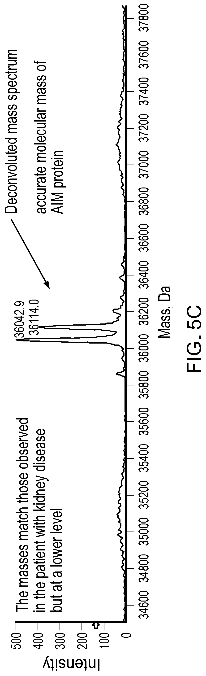
[0020] FIGS. 5A-5C contain MS spectra of IgM in serum from a patient with multiple myeloma. A) A TIC of IgM in a sample of serum from a patient with multiple myeloma. B) A summed mass spectrum of the peaks in the boxed LC retention times in FIG. 5A. C) A deconvoluted mass spectrum of AIM polypeptides from the peaks in FIG. 5B.
DETAILED DESCRIPTION
[0021] This document provides materials and methods for identifying and quantifying AIM polypeptides (e.g., AIM polypeptides bound to IgM immunoglobulins) in a sample, such as a biological sample (e.g., serum), using MS techniques. In some cases, the materials and methods provided herein can be used to identify and quantify AIM polypeptides in an IgM-purified serum sample. For example, identification and/or quantification of AIM polypeptides in a serum sample can be used to diagnose and/or treat patients having a disease or disorder characterized by altered (e.g., increased or decreased) AIM polypeptides levels. As described herein, the presence of an increased level of AIM polypeptides can indicate that a patient (e.g., a mammal such as a human) has kidney disease or multiple myeloma.
[0022] In circulation IgM exists as a pentamer with an AIM polypeptide bound to the IgM fragment crystallizable region (Fc region). As described herein, AIM polypeptides bound to IgM immunoglobulins can be detected in IgM-purified samples using mass spectroscopy. Since the molecular mass of the AIM polypeptide is known, and it is different than IgM light and heavy chains, the AIM polypeptide is easily identified by mass spectroscopy and can be identified in the same analysis used to analyze IgM immunoglobulins. In some cases, a method described herein can include the use of a LC-MS. For example, AIM polypeptides can be identified by molecular mass using LC-MS. In some cases, ESI-MS techniques can be used, for example, an electrospray ionization quadrupole time-of-flight MS (ESI-Q-TOF MS) technique. In some cases, a MS technique can be a top-down MS technique. The use of mass over charge (m/z), optionally with additional techniques, such as gel electrophoresis and/or peptide sequencing, provides a more direct assessment of AIM polypeptides because it can be used to determine the quantity of the intact (e.g., not fragmented during the mass spectrometry technique) AIM polypeptides as well as the presence or absence of AIM polypeptide glycosylation in a single assay. In some cases, the identification and/or quantification of intact AIM polypeptides also can include (e.g., in the same assay) identification and/or quantification of intact IgM immunoglobulins.
[0023] In some cases, AIM polypeptides can be identified in the purified sample by molecular mass. The mean molecular mass for AIM polypeptides is about 36,100 Da. Different AIM polypeptide glycoforms can have a molecular mass of about 36,044.4 Da and about 36,115.5 Da. The presence or absence, quantity, and/or glycoform(s) of AIM polypeptides can be determined by detecting an AIM polypeptide amino acid sequence. In some cases, a human AIM polypeptide can have an amino acid sequence set forth in, for example, UniProt Entry No: O43866. An exemplary AIM polypeptide can be a human AIM polypeptide amino acid sequence as set forth below (SEQ ID NO:1).
TABLE-US-00001 SPSGVRLVGGLHRCEGRVEVEQKGQWGTVCDDGWDIKDVAVLCRELGC GAASGTPSGILYEPPAEKEQKVLIQSVSCTGTEDTLAQCEQEEVYDCS HDEDAGASCENPESSFSPVPEGVRLADGPGHCKGRVEVKHQNQWYTVC QTGWSLRAAKVVCRQLGCGRAVLTQKRCNKHAYGRKPIWLSQMSCSGR EATLQDCPSGPWGKNTCNHDEDTWVECEDPFDLRLVGGDNLCSGRLEV LHKGVWGSVCDDNWGEKEDQVVCKQLGCGKSLSPSFRDRKCYGPGVGR IWLDNVRCSGEEQSLEQCQHRFWGFHDCTHQEDVAVICSG
[0024] In some cases, an AIM polypeptide can include a posttranslational modification (e.g., glycosylation). For example, a glycosylated AIM polypeptide can include any appropriate carbohydrate (e.g., hexose, hexosamines, and sialic acid).
[0025] The methods described herein, also referred to as monoclonal immunoglobulin Rapid Accurate Mass Measurement (miRAMM), can be coupled with immunopurification (e.g., IgM immunopurification) to identify and/or quantify AIM polypeptides in a sample (e.g., a serum sample) without the need for additional instrumentation or reagents specific to AIM polypeptides. The methods described herein also provide the ability to identify and quantify the AIM polypeptide and IgM immunoglobulins together in the same assay. These methods can be used for screening biological samples for the presence or absence, quantity, and/or glycoform of the AIM polypeptide, for screening biological samples for the presence or absence and/or the quantity the IgM immunoglobulins, and/or for determining the IgM:AIM ratio. For example, these methods are useful for identifying a disease or disorder characterized by altered (e.g., increased or decreased) AIM polypeptide levels in a patient, for monitoring AIM polypeptide levels in a patient, for identifying a disease or disorder characterized by altered (e.g., increased or decreased) IgM immunoglobulin levels in a patient, for monitoring IgM immunoglobulin levels in a patient and/or for monitoring treatment of a disease or disorder in a patient. The speed, sensitivity, resolution, and robustness of mass spectroscopy make the present methods superior than gel electrophoresis for screening samples for presence or absence, quantity, and/or glycoform(s) of AIM polypeptides.
Samples and Sample Preparation
[0026] The materials and methods for identifying and quantifying AIM polypeptides as described herein can include any appropriate sample. A sample can be any biological sample, such as a tissue (e.g., adipose, liver, kidney, heart, muscle, bone, or skin tissue) or biological fluid (e.g., blood, serum, plasma, urine, lachrymal fluid, or saliva). The sample can be from a patient that has immunoglobulins, which includes but is not limited to a mammal, e.g. a human, dog, cat, primate, rodent, pig, sheep, cow, horse, bird, reptile, or fish. A sample can also be a man-made reagent, such as a mixture of known composition or a control sample. In some cases, the sample is serum from a human patient.
[0027] A sample can be treated to remove components that could interfere with the MS technique. A variety of techniques known to those having skill in the art can be used based on the sample type. Solid and/or tissue samples can be ground and extracted to free the analytes of interest from interfering components. In such cases, a sample can be centrifuged, filtered, and/or subjected to chromatographic techniques to remove interfering components (e.g., cells or tissue fragments). In yet other cases, reagents known to precipitate or bind the interfering components can be added. For example, whole blood samples can be treated using conventional clotting techniques to remove red and white blood cells and platelets. A sample can be deproteinized. For example, a plasma sample can have serum proteins precipitated using conventional reagents such as acetonitrile, KOH, NaOH, or others known to those having ordinary skill in the art, optionally followed by centrifugation of the sample.
[0028] Immunoglobulins (e.g., immunoglobulins and polypeptides bound to the immunoglobulins) can be isolated from the samples or enriched (i.e. concentrated) in a sample using standard methods known in the art. Such methods include removing one or more non-immunoglobulin contaminants from a sample. In some cases, the samples can be enriched or purified using immunopurification, centrifugation, filtration, ultrafiltration, dialysis, ion exchange chromatography, size exclusion chromatography, protein A/G affinity chromatography, affinity purification, precipitation, gel electrophoresis, capillary electrophoresis, chemical fractionation (e.g., antibody purification kits, such as Melon Gel Purification), and aptamer techniques. For example, the immunoglobulins can be purified by chemical-based fractionation, e.g., Melon Gel Chromatography (Thermo Scientific), where Melon Gel resins bind to non-immunoglobulin proteins in a sample and allow immunoglobulins to be collected in the flow-through fraction; or by affinity purification, e.g., by Protein M purification, where immunoglobulins are bound by those proteins at physiologic pH and then released from the proteins by lowering the pH. When serum, plasma, or whole blood samples are used, a sample, such as a 10-250 .mu.l sample (e.g., a 50 .mu.l sample), can be directly subjected to purification (e.g., immunopurification). Size exclusion principles such as a TurboFlow column can also be employed to separate the non-immunoglobulin contaminants from a sample. When urine samples are used, a urine sample can be buffered, e.g., a 50 .mu.l urine sample can be diluted first with 50 .mu.l of 50 mM ammonium bicarbonate.
[0029] A sample can be subjected to immunopurification prior to analysis by mass spectrometry. In some cases, the sample can be immunoglobulin enriched. Immunopurification can result in enrichment of one or more immunoglobulins (e.g., IgM). IgM-purified samples can be polyclonal or monoclonal. For example, immunopurification can separate or enrich IgM immunoglobulins in a sample. Immunopurification can involve contacting a sample containing the desired antigen with an affinity matrix including an antibody (e.g. single domain antibody fragments, also referred to as nanobodies) to the antigen covalently attached to a solid phase (e.g., beads such as agarose beads). Antigens in the sample become bound to the affinity matrix through an immunochemical bond. The affinity matrix is then washed to remove any unbound species. The antigen is then removed from the affinity matrix by altering the chemical composition of a solution in contact with the affinity matrix. The immunopurification may be conducted on a column containing the affinity matrix, in which case the solution is an eluent or in a batch process, in which case the affinity matrix is maintained as a suspension in the solution. In some cases, the antibody can be a labelled antibody (e.g. a biotinylated antibody) and a binding partner of the label (e.g., avidin and/or streptavidin) can be attached to the solid phase.
[0030] In some embodiments, single domain antibody fragments (SDAFs) with an affinity for immunoglobulins can be used in the immunopurification process. SDAFs can be derived from heavy chain antibodies of non-human sources (e.g., camelids, fish, llama, sheep, goat, rabbit, or mouse), heavy chain antibodies of human sources, and light chain antibodies of humans. SDAFs possess unique characteristics, such as low molecular weight, high physical-chemical stability, good water solubility, and the ability to bind antigens inaccessible to conventional antibodies. For example, IgM immunoglobulins can be immunopurified using anti-IgM camelid nobodies.
[0031] In some embodiments, isolation of immunoglobulins can be performed with an to entity other than a traditional antibody--which contains both heavy and light chains (such as those used in immunofixation electrophoresis (IFE) and various known clinical immunoassays). Traditional antibodies contain heavy and/or light chains with masses that may overlap with the masses of the immunoglobulins in the sample of interest (e.g., human immunoglobulins). Therefore, these antibodies may interfere in the mass spectra of the patient's immunoglobulins. Single domain antibody fragments (SDAFs) may have masses ranging from 12,500-15,000 Da and, using the methods described herein, may carry a single charge thus generating a signal in the range of 12,500-15,000 m/z, which does not overlap with the signals generated by human heavy chains or light chains. The identification of human light chains and heavy chains by molecular mass can be done as described elsewhere (see, e.g., WO 2015/154052). Thus, in some embodiments, the use of specific isolation of IgM immunoglobulins (e.g., immunoglobulins and polypeptides bound to the immunoglobulins) utilizing SDAFs, coupled with mass identification, results in a specific and sensitive method for the detection of AIM polypeptides.
[0032] In some cases, the immunoglobulins (e.g., IgM) are substantially isolated. By "substantially isolated" is meant that the immunoglobulins are at least partially or substantially separated from the sample from which they were provided. Partial separation can include, for example, a sample enriched in the immunoglobulins. Substantial separation can include samples containing at least about 10%, at least about 20%, at least about 30%, at least about 40%, at least about 50%, at least about 60%, at least about 70%, at least about 80%, at least about 90%, at least about 95%, at least about 97%, or at least about 99% by weight of the immunoglobulin.
[0033] In some cases, intact immunoglobulins (e.g., not fragmented) can be further processed to decouple the light chains in a total immunoglobulin sample from the heavy chain immunoglobulins. Decoupling can be achieved by treating the total immunoglobulins with a reducing agent, such as DTT (2,3 dihydroxybutane-1,4-dithiol), DTE (2,3 dihydroxybutane-1,4-dithiol), thioglycolate, cysteine, sulfites, bisulfites, sulfides, bisulfides, TCEP (tris(2-carboxyethyl)phosphine), 2-mercaptoethanol, and salt forms thereof. In some cases, the reducing step is performed at elevated temperature, e.g., in a range from about 30.degree. C. to about 65.degree. C., such as about 55.degree. C., in order to denature the proteins. In some cases, the sample is further treated, e.g., by modifying the pH of the sample or buffering the sample. In some cases, the sample can be acidified. In some cases, the sample can be neutralized (e.g., by the addition of a base such as bicarbonate).
[0034] In some cases, the antigen binding fragments (Fab) of immunoglobulins can be cleaved from the intact immunoglobulins using proteases such as pepsin. Excess reagents and salts can be removed from the samples using methods known to those having ordinary skill in the art.
[0035] In some cases, AIM polypeptides bound to the immunoglobulins can be released from the immunoglobulins prior to the MS analysis. For example, AIM polypeptides bound to IgM immunoglobulins in circulation can be released from purified IgM pentamers when the sample is purified.
Mass Spectrometry Methods
[0036] The materials and methods for identifying and quantifying AIM polypeptides as described herein can include any appropriate MS technique. After sample preparation, a sample can be subjected to a MS technique, either directly or after separation on a high performance liquid chromatography column (HPLC). In some cases, LC-MS can be used to analyze the mass spectrum of the ions. For example, the method can be used to identify multiply charged ions (e.g., the +1 ions, +2 ions, +3 ions, +4 ions, +5 ions, +6 ions, +7 ions, +8 ions, +9 ions, +10 ions, +11 ions, +12 ions, +13 ions, +14 ions, +15 ions, +16 ions, +17 ions, +18 ions, +19 ions, +20 ions, +21 ions, and +22 ions), resulting from the AIM polypeptides in the sample. In some cases, the samples are not fragmented during the MS technique. LC-MS is an analytical technique that combines the physical separation capabilities of liquid chromatography with the mass analysis capabilities of MS, and is suitable for detection and potential identification of chemicals in a complex mixture. Any LC-MS instrument can be used, e.g., the ABSciex 5600 Mass Spectrometer. In some cases, microflowLC-MS can be utilized. Any suitable microflow instrument can be used, e.g., the Eksigent Ekspert 200 microLC. The ion mass spectrum can be analyzed for one or more peaks corresponding to one or more AIM polypeptides in the sample.
[0037] In some cases, ESI-Q-TOF MS can be used to analyze the mass spectrum of a sample, e.g., the mass spectrum of the AIM polypeptides in the sample. ESI MS is a useful technique for producing ions from macromolecules because it overcomes the propensity of these molecules to fragment when ionized. In addition, ESI often produces multiply charged ions, effectively extending the mass range of the analyzer to accommodate the orders of magnitude observed in proteins and other biological molecules. A quadrupole mass analyzer (Q) consists of four cylindrical rods, set parallel to each other. In a quadrupole mass spectrometer, the quadrupole is the component of the instrument responsible for filtering sample ions based on their mass-to-charge ratio (m/z). The time-of-flight (TOF) analyzer uses an electric field to accelerate the ions through the same potential, and then measures the time they take to reach the detector. If the particles all have the same charge, the kinetic energies are identical, and their velocities depend only on their masses. Lighter ions reach the detector first. Any ESI-Q-TOF mass spectrometer can be used, e.g., the ABSciex TripleTOF 5600 quadrupole TOF mass spectrometer. The mass spectrum, e.g., the mass spectrum of multiply charged intact AIM polypeptide, light chain, and/or heavy chain polypeptide ions, can be analyzed to identify one or more peaks at an appropriate mass/charge expected for the chain.
[0038] The multiply charged ion peaks can be converted to a molecular mass using known techniques. For example, multiply charged ion peak centroids can be used to calculate average molecular mass and the peak area value used for quantification is supplied by a software package. Specifically, multiple ion deconvolution can be performed using the Bayesian Protein Reconstruct software package in the BioAnalyst companion software package in ABSCIEX Analyst TF 1.6. Deconvoluted and multiply charged ions can also be manually integrated using the Manual Integration 33 script in Analyst TF. Providing the molecular mass for the AIM polypeptides in the sample facilitates sequencing and identification of the AIM polypeptides in the sample. For example, the methods provided herein can be used to identify and quantify AIM polypeptides in the sample. In addition, the methods provided herein can be used to compare the relative abundance of the AIM polypeptides as compared to a control or reference sample. As described herein, the AIM polypeptides can include the amino acid sequence set forth in SEQ ID NO:1. In some cases, the abundance of this AIM polypeptide sequence can used for diagnosing, treating, and/or monitoring patients with a disease or disorder characterized by altered (e.g., increased) AIM polypeptides levels.
[0039] In some cases, matrix assisted laser adsorption ionization-TOF (MALDI-TOF) MS can be used to analyze the mass spectrum of a sample. MALDI-TOF MS identifies proteins and peptides as mass charge (m/z) spectral peaks. Further, the inherent resolution of MALDI-TOF MS allows assays to be devised using multiple affinity ligands to selectively purify/concentrate and then analyze multiple proteins in a single assay.
Methods for Assessing AIM Polypeptides
[0040] The materials and methods provided herein can be used for identifying and monitoring AIM polypeptides. In some cases, the methods provided herein can be used to determine the presence or absence, quantity, and/or glycoform of AIM polypeptides. For example, the presence or absence, quantity, and/or glycoform of AIM polypeptides can be used for identifying and/or treating a disease or disorder characterized by altered (e.g., increased) AIM polypeptide levels in a patient, for monitoring AIM polypeptides in a patient, and/or for monitoring treatment of a disease or disorder characterized by altered (e.g., increased) AIM polypeptide levels in a patient.
[0041] The materials and methods provided herein also can be used for identifying and monitoring IgM immunoglobulins. In some cases, the methods provided herein can be used to determine the presence or absence and/or quantity of IgM immunoglobulins. For example, the presence or absence and/or quantity of IgM immunoglobulins can be used for identifying and/or treating a disease or disorder characterized by altered (e.g., increased) IgM immunoglobulin levels in a patient, for monitoring IgM immunoglobulin in a patient, and/or for monitoring treatment of a disease or disorder characterized by altered (e.g., increased) IgM immunoglobulin levels in a patient.
[0042] The materials and methods provided herein also can be used for identifying and monitoring IgM:AIM ratios. In some cases, the methods provided herein can be used to determine the IgM:AIM ratio. For example, the quantity of IgM immunoglobulins and the quantity of AIM polypeptides can be used for determining the IgM:AIM ratio in a patient, for monitoring the IgM:AIM ratio in a patient, and/or for monitoring treatment of a disease or disorder characterized by altered IgM:AIM ratios in a patient.
[0043] The MS based methods disclosed herein can be used to screen a sample (e.g., a biological sample) for the presence, absence, or amount of AIM polypeptides and/or the presence, absence, or amount of IgM immunoglobulins. In some cases, the MS based methods disclosed herein can be used for detecting AIM polypeptides and/or IgM immunoglobulins in a sample from a patient.
[0044] The MS based methods disclosed herein can include subjecting a sample having one or more immunoglobulins to a MS assay. The sample can be pretreated to isolate or enrich immunoglobulins present in the sample. The immunoglobulin light chains can be decoupled from the immunoglobulin heavy chains prior to the MS analysis. The spectrum obtained from the assay can then be used to identify AIM polypeptides in the sample. In some cases, the abundance (e.g., quantity) of AIM polypeptides and/or IgM immunoglobulins can be determined by converting the peak areas of one or more of the identified peaks into a molecular mass.
[0045] The abundance (e.g., quantity) of the AIM polypeptides and/or the IgM immunoglobulins can be used to diagnose and/or treat various disorders associated with an altered (e.g., increased or decreased) level of AIM polypeptides, diseases associated with an altered (e.g., increased or decreased) level of IgM immunoglobulins, and/or diseases associated with an altered (e.g., increased or decreased) IgM:AIM ratio. In some cases, an altered level of AIM polypeptides and/or IgM immunoglobulins is an increased (e.g., elevated) level of AIM polypeptides and/or IgM immunoglobulins. The term "increased level" as used herein with respect to a level of AIM polypeptides and/or IgM immunoglobulins refers to any level that is greater than the median level of AIM polypeptides and/or IgM immunoglobulins typically observed in a sample (e.g., a control sample) from one or more healthy (e.g., normal) mammals (e.g., humans). In some cases, the abundance of the AIM polypeptides and/or IgM immunoglobulins can be compared to a reference value or a control sample. For example, a reference value can be an abundance of AIM polypeptides and/or IgM immunoglobulins in a healthy patient (e.g., a healthy human). In some cases, a control sample can be a sample (e.g., serum) obtained from one or more healthy patients (e.g., healthy humans). A control sample can be from a single healthy (e.g., normal) mammal, or a control sample can be a pool of samples from two or more (e.g., three, four, five, six, seven, eight, nine, 10, 11, 12, 13, 14, 15, 16, 17, 18, 19, 20, or more) healthy (e.g., normal) mammals. In some cases, an abundance (e.g., quantity) of AIM polypeptides in a sample can be determined as described, for example, in Example 1 and/or Example 2. For example, an increased level of AIM polypeptides can be a level where the intensity (e.g., the peak intensity of the ions as measured by a MS technique) is greater than about 50 (e.g., greater than about 55, greater than about 60, greater than about 65, greater than about 70, greater than about 75, or greater than about 80) in serum. In some cases, an increased level of AIM polypeptides can be a level where the intensity (e.g., the peak intensity of the ions as measured by a MS technique) is about 60 in serum. For example, an increased level of AIM polypeptides can be a level where the intensity as measured by LC-ESI-Q-TOF mass spectrometry is from about 3500 to about 5500 (e.g., from about 3800 to about 5500, from about 4000 to about 5500, from about 4200 to about 5500, from about 4500 to about 5500, from about 3500 to about 5200, from about 3500 to about 5000, from about 3500 to about 4800, from about 3500 to about 4500, from about 3800 to about 5300, or from about 4000 to about 5000). In some cases, an increased level of AIM polypeptides can be a level where the intensity as measured by LC-ESI-Q-TOF mass spectrometry is about 4500 in serum. For example, an increased level of AIM polypeptides can be a level where the intensity as measured by LC-ESI-Q-TOF mass spectrometry is from about 100 to about 200 (e.g., from about 120 to about 200, from about 150 to about 200, from about 170 to about 200, from about 100 to about 180, from about 100 to about 150, from about 100 to about 130, from about 130 to about 180, or from about 140 to about 170) in serum. In some cases, an abundance (e.g., quantity) of IgM immunoglobulins in a sample can be determined as described elsewhere (see, e.g., WO 2015/154052). In some cases, an increased level of AIM polypeptides can be a level where the intensity as measured by LC-ESI-Q-TOF mass spectrometry is about 150 in serum. In cases where control samples have undetectable levels of AIM polypeptides and/or IgM immunoglobulins, an increased level can be a detectable level of AIM polypeptides and/or IgM immunoglobulins. It will be appreciated that levels from comparable samples are used when determining whether or not a particular level is an increased level.
[0046] When diagnosing and/or treating a patient having a disease or disorder characterized by altered (e.g., increased or decreased) AIM polypeptide levels, IgM immunoglobulin levels, and/or IgM:AIM ratios, the disease or disorder can be any appropriate disease or disorder. Examples of diseases and disorders characterized by increased AIM polypeptide levels, IgM immunoglobulin levels, and/or IgM:AIM ratios include, without limitation, kidney disease, multiple myeloma, inflammatory response diseases (e.g., lupus and rheumatoid arthritis), and diseases associated with autoantibody production (e.g., obesity associated autoimmunity).
[0047] In some cases, the methods provided herein can be used to confirm a diagnosis made by current methods such as gel electrophoresis. For example, if a negative result is obtained from gel electrophoresis, the present methods can be used as a secondary test to confirm or counter such results. In some cases, the diagnosis provided herein can be confirmed using such standard methods.
[0048] In some cases, the methods provided herein can be used for treating a patient having a gammopathy. For example, after diagnosing the patient as having a disease or disorder characterized by increased or decreased AIM polypeptide and/or IgM immunoglobulin levels, the methods can include administering to the patient one or more therapeutic agents to treat the disease or disorder characterized by increased or decreased AIM polypeptide and/or IgM immunoglobulins levels (e.g., a therapeutically effective amount) and/or performing a treatment (e.g., a plasma exchange or a stem cell transplant). The therapeutic agent can be any appropriate therapeutic agent. For example, when the disease or disorder characterized by increased or decreased AIM polypeptide levels is kidney disease, the therapeutic agent can be any agent useful for treating kidney disease. For example, when the disease or disorder characterized by increased or decreased AIM polypeptide levels is multiple myeloma, the therapeutic agent can be any agent useful for treating multiple myeloma. In some cases, after diagnosing the patient as having a disease or disorder characterized by increased or decreased AIM polypeptide levels, the method can include administering to the patient a therapeutically effective amount of a therapeutic agent to treat the disease or disorder characterized by increased or decreased AIM polypeptide levels and one or more of a plasma exchange and a stem cell transplant (e.g., an autologous peripheral blood stem cell transplantation).
[0049] In some cases, the methods provided herein can also be used for monitoring a patient. For example, the MS based methods disclosed herein can be used for monitoring a disease or disorder characterized by increased or decreased AIM polypeptide and/or IgM immunoglobulin levels in a patient. The MS based methods disclosed herein can include providing a first sample and a second sample of the patient. For example, the MS based methods disclosed herein can include providing a first sample of the patient before the treatment and a second sample of the patient during or after the treatment. The first and second samples can be pretreated to isolate or enrich immunoglobulins present in the first and second samples. In some cases, AIM polypeptides bound to the immunoglobulins can be released from the immunoglobulins prior to the MS analysis. The spectrum obtained from the assay can then be used to identify AIM polypeptides and/or IgM immunoglobulins in the first and second samples.
[0050] In some cases, the relative abundance of AIM polypeptides in the first and second samples can be determined by converting the peak areas of one or more of the identified peaks into a molecular mass. For example, the presence or absence of AIM polypeptides can be determined in the first and second samples. A decrease (or loss) of the amount of AIM polypeptides indicates that the disease or disorder characterized by increased or decreased AIM polypeptide levels in the patient has been reduced (or eliminated); while an increase in the amount of AIM polypeptides indicates that the disease or disorder characterized by increased or decreased AIM polypeptide levels in the patient has increased. In cases where a first sample of the patient is before the treatment and a second sample of the patient is during or after the treatment, the presence or absence of AIM polypeptides is determined before and after the treatment and compared. A decrease (or loss) of the amount of AIM polypeptides indicates that the treatment may be effective for the patient; while an increase or no change in the amount of AIM polypeptides indicates that the treatment may be ineffective for the patient. For example, the amount of AIM polypeptides in a first sample and in a second sample can be determined, and the amount of AIM polypeptides in the first sample can be compared to the amount of AIM polypeptides in the second sample. For example, the concentration of AIM polypeptides in a first sample and in a second sample can be determined, and the concentration of AIM polypeptides in the first sample can be compared to the amount of AIM polypeptides in the second sample.
[0051] In some cases, the relative abundance of IgM immunoglobulins in the first and second samples can be determined by converting the peak areas of one or more of the identified peaks into a molecular mass. The presence or absence of IgM immunoglobulins can be determined in the first and second samples. A decrease (or loss) of the amount of IgM immunoglobulins indicates that the disease or disorder characterized by increased or decreased IgM immunoglobulin levels in the patient has been reduced (or eliminated); while an increase in the amount of IgM immunoglobulins indicates that the disease or disorder characterized by increased or decreased IgM immunoglobulin levels in the patient has increased. In cases where a first sample of the patient is before the treatment and a second sample of the patient is during or after the treatment, the presence or absence of IgM immunoglobulins is determined before and after the treatment and compared. A decrease (or loss) of the amount of IgM immunoglobulins indicates that the treatment may be effective for the patient; while an increase or no change in the amount of IgM immunoglobulins indicates that the treatment may be ineffective for the patient. For example, the amount of IgM immunoglobulins in a first sample and in a second sample can be determined, and the amount of IgM immunoglobulins in the first sample can be compared to the amount of IgM immunoglobulins in the second sample. For example, the concentration of IgM immunoglobulins in a first sample and in a second sample can be determined, and the concentration of IgM immunoglobulins in the first sample can be compared to the amount of IgM immunoglobulins in the second sample.
[0052] In some cases, the quantity of IgM immunoglobulins and the quantity of AIM polypeptides in the first and second samples can be determined, and can be used to determine the IgM:AIM ratio. The IgM:AIM ratio can be determined in the first and second samples. A decrease (or loss) of the IgM:AIM ratio indicates that the disease or disorder characterized by increased or decreased IgM:AIM ratios in the patient has been reduced (or eliminated); while an increase in the IgM:AIM ratio indicates that the disease or disorder characterized by increased or decreased IgM:AIM ratios in the patient has increased. In cases where a first sample of the patient is before the treatment and a second sample of the patient is during or after the treatment, the IgM:AIM ratio is determined before and after the treatment and compared. A decrease (or loss) of the IgM:AIM ratio indicates that the treatment may be effective for the patient; while an increase or no change in the IgM:AIM ratio indicates that the treatment may be ineffective for the patient. For example, the IgM:AIM ratio in a first sample and in a second sample can be determined, and the IgM:AIM ratio in the first sample can be compared to the IgM:AIM ratio in the second sample. For example, the IgM:AIM ratio in a first sample and in a second sample can be determined, and the IgM:AIM ratio in the first sample can be compared to the IgM:AIM ratio in the second sample.
[0053] The invention will be further described in the following examples, which do not limit the scope of the invention described in the claims.
EXAMPLES
Example 1: Identification of AIM Protein in IgM Purified Serum
Methods
[0054] A volume of 50 .mu.L of serum was added to 20 .mu.L of anti-IgM camelid nanobody beads. The serum was allowed to incubate with the nanobody beads for 45 minutes. The beads were washed with 1 mL of PBS 3 times, each time removing and discarding the supernatant. The beads were then washed with water 1 time. The water was removed and 50 .mu.L of 5% acetic acid containing 50 mM TCEP was added to the beads to elute the purified IgM. This elute was then analyzed by microflow LC-ESI-Q-TOF mass spectrometry using a SCIEX 5600 mass spectrometer.
Results
[0055] Serum from a patient with kidney disease was IgM purified using camelid nanobody beads and subjected to MS. The total ion chromatogram shows many peaks each representing a different monoclonal light chains, indicating that the sample was oligoclonal (FIG. 1A). The summed mass spectrum showed multiply charged ions from an unknown protein that eluted at 4.0 minutes (FIG. 1B). The deconvoluted mass spectrum showed the accurate molecular mass of two different isoforms (36,044.4 Da and 36,115.5 Da) differing in mass by 71.1 Da (FIG. 1C). The molecular mass of the unknown protein was different than any expected masses for either the IgM heavy chain or the IgM light chain, and had an LC elution time earlier than expected for either the IgM heavy chain or the IgM light chain. In addition, the mass difference between the two isoforms did not match any expected post-translational modifications such as glycosylation.
[0056] To identify the unknown protein, an analysis was done using the same sample from the patient with kidney disease shown in FIG. 1. The sample was reduced, alkylated, and digested with trypsin, and then was analyzed by bottom-up LC-MS/MS. A total of 9 peptides from AIM (also referred to as CD5 antigen-like protein) were found in the Scaffold protein search (FIG. 2). The molecular mass of the intact protein (listed as 38 kDa) was close to molecular mass of the unknown protein in FIG. 1. Bottom-up MS was used to calculate a theoretical molecular mass of AIM. The calculated molecular mass of AIM (see, e.g., FIG. 3) was 36,055.2 Da. This calculated molecular mass was 10.8 Da higher than the mass than the primary isoform observed in FIG. 1. AIM contains 11 inter-chain disulfide bonds. Thus, this difference can be due to incomplete reduction of, for example, 5 of the 11 inter-chain disulfide bonds.
[0057] These results demonstrated that AIM protein bound to IgM in circulation is released from the IgM pentamer during sample purification, and that MS can be used to identify and quantify AIM protein.
Example 2: AIM Protein in Multiple Myeloma
Methods
[0058] A volume of 50 .mu.L of serum was added to 20 .mu.L of anti-IgM camelid nanobody beads. The serum was allowed to incubate with the nanobody beads for 45 minutes. The beads were washed with 1 mL of PBS 3 times, each time removing and discarding the supernatant. The beads were then washed with water 1 time. The water was removed and 50 .mu.L of 5% acetic acid containing 50 mM TCEP was added to the beads to elute the purified IgM. This elute was then analyzed by microflow LC-ESI-Q-TOF mass spectrometry using a SCIEX 5600 mass spectrometer.
Results
[0059] A sample of pooled serum from healthy (e.g., normal) humans was IgM purified using camelid nanobody beads, and subjected to MS. The total ion chromatogram is shown in FIG. 4A. The LC retention times identified for AIM in FIG. 1A were used to obtain a summed mass spectrum from the peaks in FIG. 4A. The summed mass spectrum showed the multiply charged ions from the AIM protein (FIG. 4B). The deconvoluted mass spectrum showed the accurate molecular mass of two different AIM isoforms (36,044.4 Da and 36,115.5 Da) (Figure FC). The level of AIM proteins in normal serum was much lower than was seen in serum from a patient with kidney disease (comparing FIG. 4C to FIG. 1C).
[0060] Serum from a patient with multiple myeloma was IgM purified using camelid nanobody beads and subjected to MS. The total ion chromatogram is shown in FIG. 5A. The LC retention times identified for AIM in FIG. 1A were used to obtain a summed mass spectrum from the peaks in FIG. 5A. The summed mass spectrum showed the multiply charged ions from the AIM protein (FIG. 5B). The deconvoluted mass spectrum showed the accurate molecular mass of two different AIM isoforms (36,044.4 Da and 36,115.5 Da) (Figure FC). The level of AIM proteins in serum from a patient with multiple myeloma was much higher than was seen in normal serum (comparing FIG. 5C to FIG. 4C).
[0061] These results demonstrated that the identification and quantification of AIM polypeptides in serum (e.g., by MS) can be used to identify patients having kidney disease and/or multiple myeloma.
[0062] Other Embodiments
[0063] It is to be understood that while the invention has been described in conjunction with the detailed description thereof, the foregoing description is intended to illustrate and not limit the scope of the invention, which is defined by the scope of the appended claims. Other aspects, advantages, and modifications are within the scope of the following claims.
Sequence CWU 1
1
11328PRTHomo sapiens 1Ser Pro Ser Gly Val Arg Leu Val Gly Gly Leu
His Arg Cys Glu Gly1 5 10 15Arg Val Glu Val Glu Gln Lys Gly Gln Trp
Gly Thr Val Cys Asp Asp 20 25 30Gly Trp Asp Ile Lys Asp Val Ala Val
Leu Cys Arg Glu Leu Gly Cys 35 40 45Gly Ala Ala Ser Gly Thr Pro Ser
Gly Ile Leu Tyr Glu Pro Pro Ala 50 55 60Glu Lys Glu Gln Lys Val Leu
Ile Gln Ser Val Ser Cys Thr Gly Thr65 70 75 80Glu Asp Thr Leu Ala
Gln Cys Glu Gln Glu Glu Val Tyr Asp Cys Ser 85 90 95His Asp Glu Asp
Ala Gly Ala Ser Cys Glu Asn Pro Glu Ser Ser Phe 100 105 110Ser Pro
Val Pro Glu Gly Val Arg Leu Ala Asp Gly Pro Gly His Cys 115 120
125Lys Gly Arg Val Glu Val Lys His Gln Asn Gln Trp Tyr Thr Val Cys
130 135 140Gln Thr Gly Trp Ser Leu Arg Ala Ala Lys Val Val Cys Arg
Gln Leu145 150 155 160Gly Cys Gly Arg Ala Val Leu Thr Gln Lys Arg
Cys Asn Lys His Ala 165 170 175Tyr Gly Arg Lys Pro Ile Trp Leu Ser
Gln Met Ser Cys Ser Gly Arg 180 185 190Glu Ala Thr Leu Gln Asp Cys
Pro Ser Gly Pro Trp Gly Lys Asn Thr 195 200 205Cys Asn His Asp Glu
Asp Thr Trp Val Glu Cys Glu Asp Pro Phe Asp 210 215 220Leu Arg Leu
Val Gly Gly Asp Asn Leu Cys Ser Gly Arg Leu Glu Val225 230 235
240Leu His Lys Gly Val Trp Gly Ser Val Cys Asp Asp Asn Trp Gly Glu
245 250 255Lys Glu Asp Gln Val Val Cys Lys Gln Leu Gly Cys Gly Lys
Ser Leu 260 265 270Ser Pro Ser Phe Arg Asp Arg Lys Cys Tyr Gly Pro
Gly Val Gly Arg 275 280 285Ile Trp Leu Asp Asn Val Arg Cys Ser Gly
Glu Glu Gln Ser Leu Glu 290 295 300Gln Cys Gln His Arg Phe Trp Gly
Phe His Asp Cys Thr His Gln Glu305 310 315 320Asp Val Ala Val Ile
Cys Ser Gly 325
D00001
D00002
D00003
D00004
D00005
D00006
D00007
D00008
S00001
XML
uspto.report is an independent third-party trademark research tool that is not affiliated, endorsed, or sponsored by the United States Patent and Trademark Office (USPTO) or any other governmental organization. The information provided by uspto.report is based on publicly available data at the time of writing and is intended for informational purposes only.
While we strive to provide accurate and up-to-date information, we do not guarantee the accuracy, completeness, reliability, or suitability of the information displayed on this site. The use of this site is at your own risk. Any reliance you place on such information is therefore strictly at your own risk.
All official trademark data, including owner information, should be verified by visiting the official USPTO website at www.uspto.gov. This site is not intended to replace professional legal advice and should not be used as a substitute for consulting with a legal professional who is knowledgeable about trademark law.