Klotho Modulation
Leonhardt; Howard J.
U.S. patent application number 16/352756 was filed with the patent office on 2020-09-17 for klotho modulation. The applicant listed for this patent is Cal-X Stars Business Accelerator, Inc.. Invention is credited to Howard J. Leonhardt.
| Application Number | 20200289826 16/352756 |
| Document ID | / |
| Family ID | 1000003992190 |
| Filed Date | 2020-09-17 |


View All Diagrams
| United States Patent Application | 20200289826 |
| Kind Code | A1 |
| Leonhardt; Howard J. | September 17, 2020 |
KLOTHO MODULATION
Abstract
Described is a low voltage, pulsed electrical stimulation device for controlling expression of klotho, a useful protein, by tissues. Also described are methods of enhancing expression of klotho in cells.
| Inventors: | Leonhardt; Howard J.; (Playa Vista, CA) | ||||||||||
| Applicant: |
|
||||||||||
|---|---|---|---|---|---|---|---|---|---|---|---|
| Family ID: | 1000003992190 | ||||||||||
| Appl. No.: | 16/352756 | ||||||||||
| Filed: | March 13, 2019 |
| Current U.S. Class: | 1/1 |
| Current CPC Class: | A61N 1/36002 20170801; A61N 1/36007 20130101; A61N 2001/083 20130101; A61N 1/36157 20130101; A61N 1/36153 20130101; A61N 1/044 20130101; A61N 1/36003 20130101; A61N 1/36021 20130101; A61N 1/36025 20130101; A61N 1/3601 20130101; A61N 1/0464 20130101; A61N 1/0468 20130101 |
| International Class: | A61N 1/36 20060101 A61N001/36; A61N 1/04 20060101 A61N001/04 |
Claims
1. A bioelectric stimulator programmed to produce a bioelectric signal that stimulates target tissue to express and/or release Klotho polypeptide by the target tissue, wherein the bioelectric signal comprises: a biphasic pulse at 20 Hz, 0.1 V, and a 7.8 ms pulse duration for 24 hours.
2. The bioelectric stimulator of claim 1, wherein the bioelectric stimulator is further programmed to produce a bioelectric signal (SDF-1) of 30 pulses per second with a voltage of 3.5 mV, and successively alternating currents of 700 to 1500 picoamps for one minute, and again with 700 to 1500 picoamps for one minute, plus stimulated with a current of 0.25 mA, pulse duration of 40 pulses per second, pulse width of 100 .mu.s, and frequency of 100 Hz, each signal for 40 minutes to 8 hours a day.
3. The bioelectric stimulator of claim 1, wherein the bioelectric stimulator is further programmed to produce a bioelectric signal of 3 V/cm, 10 Hz, 2 .mu.A (0.000002 amps), and pulse duration of 0.2 ms.
4. The bioelectric stimulator of claim 1, wherein the bioelectric stimulator is further programmed to produce a bioelectric signal of 20 V/cm, 100 Hz, 0.25 .mu.A (2.5e-7 amps), and pulse duration of 40 pulses/s, width of 100 .mu.s.
5. The bioelectric stimulator of claim 1, wherein the bioelectric stimulator is further programmed to produce a bioelectric signal of 10V at 50 HZ and 100 Hz, 0.25 mA for one (1) minute.
6. The bioelectric stimulator of claim 1, wherein the bioelectric stimulator is further programmed to produce a bioelectric signal of 0.06 V with 50 Hz alternating electrical field and electric current of 1 mA for 15 minutes and 3 mA for 15 minutes.
7. The bioelectric stimulator of claim 1, wherein the bioelectric stimulator is further programmed to produce a bioelectric signal applied to the target tissue of 3 mV with electric frequency of 22 Hz, and current of 1 mA for 15 minutes and 3 mA for 15 minutes.
8. A method of using the bioelectric stimulator of claim 1 to stimulate tissue of a subject, the method comprising: connecting the bioelectric stimulator to the target tissue of the subject, and actuating the bioelectric stimulator to produce the programmed bioelectric signal.
9. The method according to claim 8, wherein the target tissue is selected from the group consisting of muscle, brain, kidney, pancreas, bone, tumor, and nerve.
10. The method according to claim 8, wherein the subject has been diagnosed as suffering from kidney failure, diabetes, bone degeneration, aging, cancer, and/or immune system dysfunction.
11. The method according to claim 8, wherein the subject has age-related cognitive decline, cognitive decline resulting from a neurodegenerative disease, and/or cognitive decline resulting from traumatic brain injury.
12. The method according to claim 8, wherein the subject is receiving or has received radiation treatment or chemotherapy for cancer.
13. A method of treating a cell, the method comprising: applying a bioelectric signal to the cell that stimulates the cell to express and/or release Klotho polypeptide, wherein the bioelectric signal comprises a biphasic pulse at 20 Hz, 0.1 V, and a 7.8 ms pulse duration for 24 hours.
14. The method according to claim 13, further comprising producing a bioelectric signal of 30 pulses per second with a voltage of 3.5 mV, and successively alternating currents of 700 to 1500 picoamps for one minute, and again with 700 to 1500 picoamps for one minute, plus stimulated with a current of 0.25 mA, pulse duration of 40 pulses per second, pulse width of 100 .mu.s, and frequency of 100 Hz, each signal for 40 minutes to 8 hours a day.
15. The method according to claim 13, further comprising producing a bioelectric signal of 3 V/cm, 10 Hz, 2 .mu.A (0.000002 amps), and pulse duration of 0.2 ms.
16. The method according to claim 13, further comprising producing a bioelectric signal of 20 V/cm, 100 Hz, 0.25 .mu.A (2.5e-7 amps), and pulse duration of 40 pulses/s, width of 100 .mu.s.
17. The method according to claim 13, further comprising producing a bioelectric signal of 10V at 50 HZ and 100 Hz, 0.25 mA for one (1) minute.
18. The method according to claim 13, further comprising producing a bioelectric signal of 0.06 V with 50 Hz alternating electrical field and electric current of 1 mA for 15 minutes and 3 mA for 15 minutes.
19. The method according to claim 13, further comprising producing a bioelectric signal applied to the cell of 3 mV with electric frequency of 22 Hz, and current of 1 mA for 15 minutes and 3 mA for 15 minutes.
20. The method according to claim 13, wherein the cell is located within a subject.
21. The method according to claim 20, wherein the subject has been diagnosed as having kidney failure, diabetes, bone degeneration, aging, cancer, and/or immune system dysfunction.
Description
FIELD
[0001] The application relates generally to the field of medical devices and associated treatments, and more specifically to precise bioelectrical stimulation of a subject's tissue, possibly augmented with the administration of a composition comprising, among other things, stem cells and nutrients, useful to stimulate and treat the subject, the subject's tissue(s), the subject's organ(s), and/or the subject's cells. More specifically, the application relates to a device, programmed bioelectric signaling sequences, and associated methods for the controlled expression of Klotho via precise bioelectrical signaling sequences.
BACKGROUND
[0002] Klotho protein is a kidney-secreted hormone that is known to be both membrane-bound and secreted. In man, Klotho is associated with muscle regeneration, rejuvenation, and neural protection. Loss of Klotho contributes to the aging-like features of human chronic kidney disease ("CKD") and progression of CKD. Its deficiency is also associated with degenerative processes and accelerated aging.
[0003] As found by S. Ranjit et al., "Since Klotho cannot cross the blood brain barrier, it is speculated that there exist two different pools of Klotho, one secreted from kidney into serum and other secreted by the choroid plexus into cerebrospinal fluid. Due to these reasons, therapeutic use of Klotho to provide neuroprotection [to reduce neuroinflammation and oxidative damage] is limited". S. Ranjit et al. "Potential neuroprotective role of astroglial exosomes against smoking-induced oxidative stress and HIV-1 replication in the central nervous system." Expert Opin Ther Targets. 2018 August; 22(8):703-714.
[0004] Ricardo Ferrari described that the enhanced regenerative response in aged muscle following two weeks of electrical stimulation "Estim" (i.e., a Neuromuscular Stimulator (Empi 300 PV, St Paul, Minn., US)) was associated with a somewhat limited, but still increased expression of Klotho (similar to that achieved from muscle contraction, e.g., exercise). Ricardo Ferrari "The Effect of Electrical Stimulation on Aged Skeletal Muscle Regenerative Potential" http://d-scholarship.pitt.edu/28094/1/FerrariR J_ETD_May_31_2016_PDF.pdf.
[0005] Ferrari also observed a direct relationship between Klotho expression and the percentage of senescent muscle precursor cells ("MPCs"). When Klotho was inhibited through siRNA in young MPCs and aged MPCs exposed to an Estim protocol, Ferrari observed a significantly increased percentage of senescence cells. Such findings suggest that Klotho is inversely associated with senescence cells, and that Estim modulates Klotho expression in aged MPCs, and there is precedent to suggest that Klotho plays a role in inhibiting cellular senescence. See also, Dalise et al. "Biological effects of dosing aerobic exercise and neuromuscular electrical stimulation in rats", Sci Rep. 2017 September 7; 7(1):10830.
[0006] Using the skin and small intestine as models, others have demonstrated that Klotho enhances stem cell regenerative potential and promotes tissue healing through an inhibition of Wnt signaling activation. Recent studies demonstrated that Klotho is able to directly bind to Wnt ligands extracellularly, thereby inhibiting renal fibrosis formation.
BRIEF SUMMARY
[0007] Described herein is a bioelectric stimulator particularly configured to activate expression and/or release of Klotho in cellular tissue. In certain embodiments, the bioelectric stimulator is further configured to activate expression and/.or release of stromal cell-derived factor 1 ("SDF-1"), insulin-like growth factor 1 ("IGF-1"), platelet-derived growth factor ("PDGF"), follistatin, tropoelastin, and any combination thereof.
[0008] Also described is a bioelectric stimulator including: a power source (e.g., battery, capacitor, or other suitable source of electricity), and means for delivering an electrical signal to a subject's tissue (e.g., via electrode(s) or wirelessly). The bioelectric stimulator utilizes the electrical signal to precisely control protein expression in the tissue on demand.
[0009] In certain cases, the bioelectric stimulator is programmed to produce a bioelectric signal that stimulates target tissue to express and/or release Klotho polypeptide by the target tissue by utilizing a bioelectric signal comprising a biphasic square pulse at 20 Hz, 0.1 V (100 mV), and a 7.8 ms pulse duration for 24 hours of stimulation.
[0010] The amount of Klotho expression enhanced by the herein described system is greater than that seen with muscle stimulation or muscle contraction alone.
[0011] In certain cases, the bioelectric stimulator is further programmed to produce a bioelectric signal (to produce SDF-1) of 30 pulses per second with a voltage of 3.5 mV, and successively alternating currents of 700 to 1500 picoamps for one minute, and again with 700 to 1500 picoamps for one minute, plus stimulated with a current of 0.25 mA, pulse duration of 40 pulses per second, pulse width of 100 .mu.s, and frequency of 100 Hz, each signal for 40 minutes to 8 hours a day.
[0012] In certain cases, the bioelectric stimulator is further programmed to produce (to produce PDGF) a bioelectric signal of 3 V/cm, 10 Hz, 2 .mu.A (0.000002 amps), and pulse duration of 0.2 ms. In certain cases, the bioelectric stimulator is further programmed to produce (to produce PDGF) a bioelectric signal of 20 V/cm, 100 Hz, 0.25 .mu.A (2.5e-7 amps), and pulse duration of 40 pulses/s, width of 100 .mu.s.
[0013] In certain cases, the bioelectric stimulator is further programmed to produce (to produce follistatin) a bioelectric signal of 10V at 50 HZ and 100 Hz, 0.25 mA for one (1) minute.
[0014] In certain cases, the bioelectric stimulator is further programmed to produce a bioelectric signal (to produce tropoelastin) of 0.06 V with 50 Hz alternating electrical field and electric current of 1 mA for 15 minutes and 3 mA for 15 minutes.
[0015] In certain cases, the bioelectric stimulator is further programmed to produce (for the expression of IGF-1) a bioelectric signal applied to the target tissue of 3 mV with electric frequency of 22 Hz, and current of 1 mA for 15 minutes and 3 mA for 15 minutes.
[0016] In certain cases, a method of using the bioelectric stimulator to stimulate tissue of a subject includes connecting (directly or wirelessly) the bioelectric stimulator to the target tissue or cells of the subject. The target tissue may be selected from, e.g., the group consisting of muscle, brain, kidney, pancreas, bone, tumor, and nerve.
[0017] In certain cases, the subject is interested in body building.
[0018] In certain cases, the subject has been diagnosed as suffering from kidney failure, diabetes, bone degeneration, aging, cancer, and/or immune system dysfunction.
[0019] In certain cases, the subject has age-related cognitive decline. In certain cases, the subject has cognitive decline resulting from a neurodegenerative disease. In certain cases, the subject has cognitive decline resulting from traumatic brain injury. In certain cases, the subject is receiving or has received radiation treatment or chemotherapy for cancer.
[0020] A preferred system includes: a bioelectric stimulator that controls/stimulates the release/production of Klotho by a target cell or tissue. The stimulator may be associated with (e.g., connected to) the organ or tissue to be treated with a pacing infusion lead (available from Nanoscribe of Eggenstein-Leopoldshafen, Germany) or wirelessly. In certain cases, the interface with the subject's tissue may be by a conductive soft wrap.
[0021] The stimulator can be designed to externally deliver all regeneration promoting signals wirelessly to the subject's organ(s), tissue(s), and/or cells. In certain embodiments, a micro infusion pump may be included in the system to deliver other supportive substances in greater volume more quickly.
[0022] While not intending to be bound by theory, the described system utilizes precise bioelectric signaling sequences that appear to communicate with DNA and cell membranes within stimulated tissues of the subject to cause the cells to produce high volumes of the Klotho protein. Potential indications include muscle regeneration and treatment, body building, kidney regeneration and treatment, brain regeneration and treatment, cognitive function and memory improvement, skin regeneration and treatment, wound healing, erectile dysfunction, eye regeneration and treatment, anti-aging, Multiple Sclerosis, lung regeneration and treatment, COPD, liver regeneration and treatment, hearing regeneration and treatment, blood pressure management, polyp treatment, cyst treatment, fibroid treatment, Cystic Fibrosis, heart failure, and heart valve decalcification.
BRIEF DESCRIPTION OF THE DRAWINGS
[0023] FIG. 1 depicts a programmed bioelectric stimulator for delivery to a subject connected to multiple soft conductive electrode pads.
[0024] FIG. 2 depicts a programmed bioelectric stimulator as described herein.
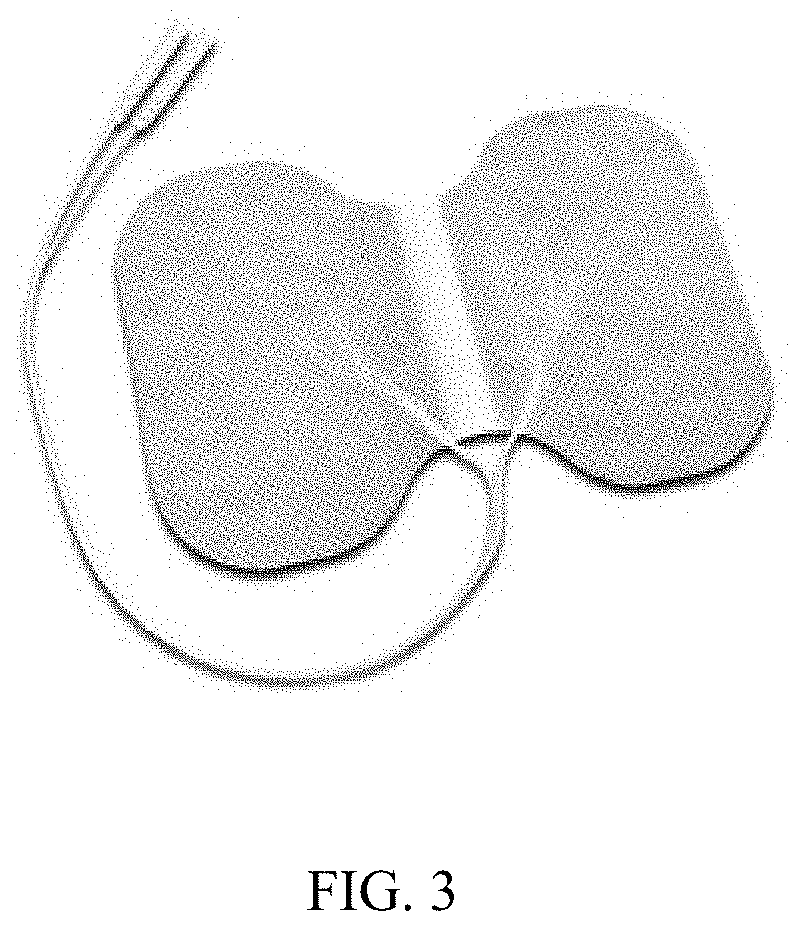
[0025] FIG. 3 depicts a conductive soft wrap for use with the system.
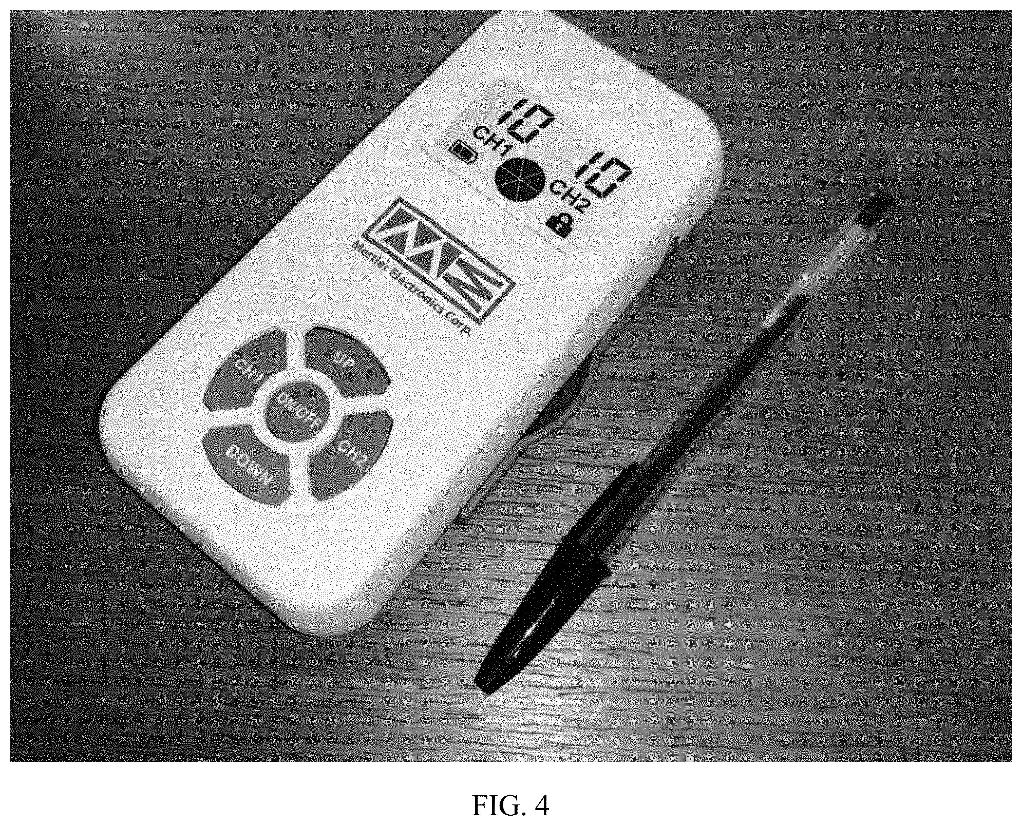
[0026] FIG. 4 depicts a programmed bioelectric stimulator depicted alongside a pen.
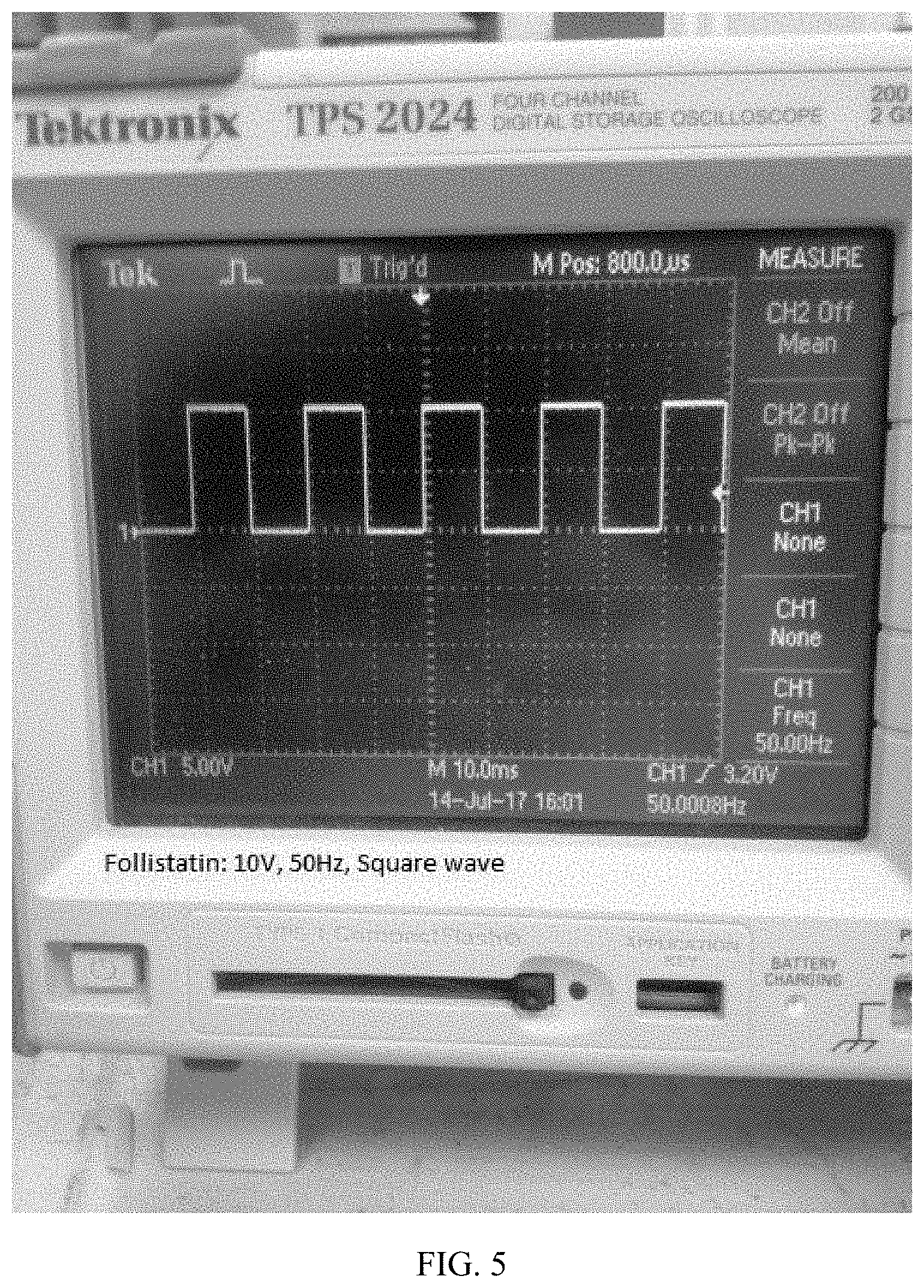
[0027] FIG. 5 depicts an image of the signal (voltage and frequency) associated with follistatin at 10V/cm, 50 Hz, square wave.
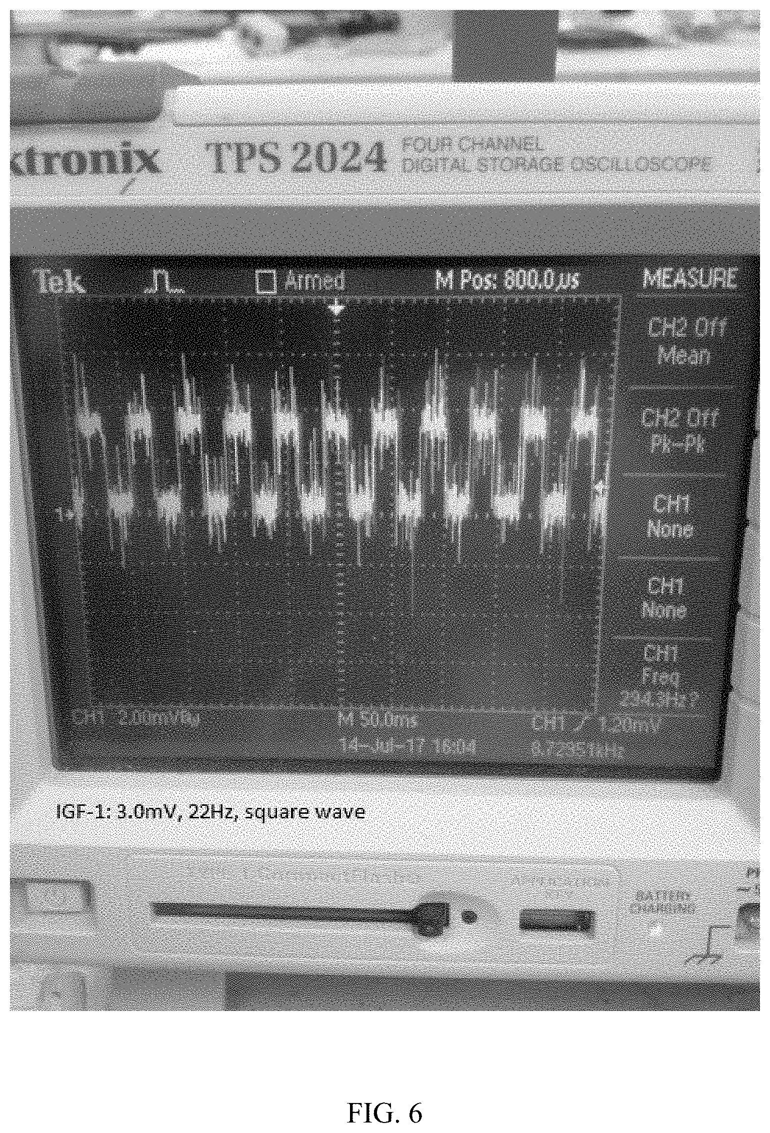
[0028] FIG. 6 depicts an image of the signal (voltage and frequency) associated with IGF-1: 3.0 mV, 22 Hz, square wave.
[0029] FIG. 7 depicts an image of a signal (voltage and frequency) associated with PDGF30%: 3V/cm (100 mV here), 10 Hz, pulse width 200 .mu.s, square wave.
[0030] FIG. 8 depicts an image of a signal (voltage and frequency) associated with PDGF230%: 20V/cm (7.0V here), 100 Hz, pulse width 100 .mu.s, square wave.
[0031] FIG. 9 depicts an image of a signal (voltage and frequency) associated with stem cell proliferation (or homing): 15 mV, 70 Hz, square wave.
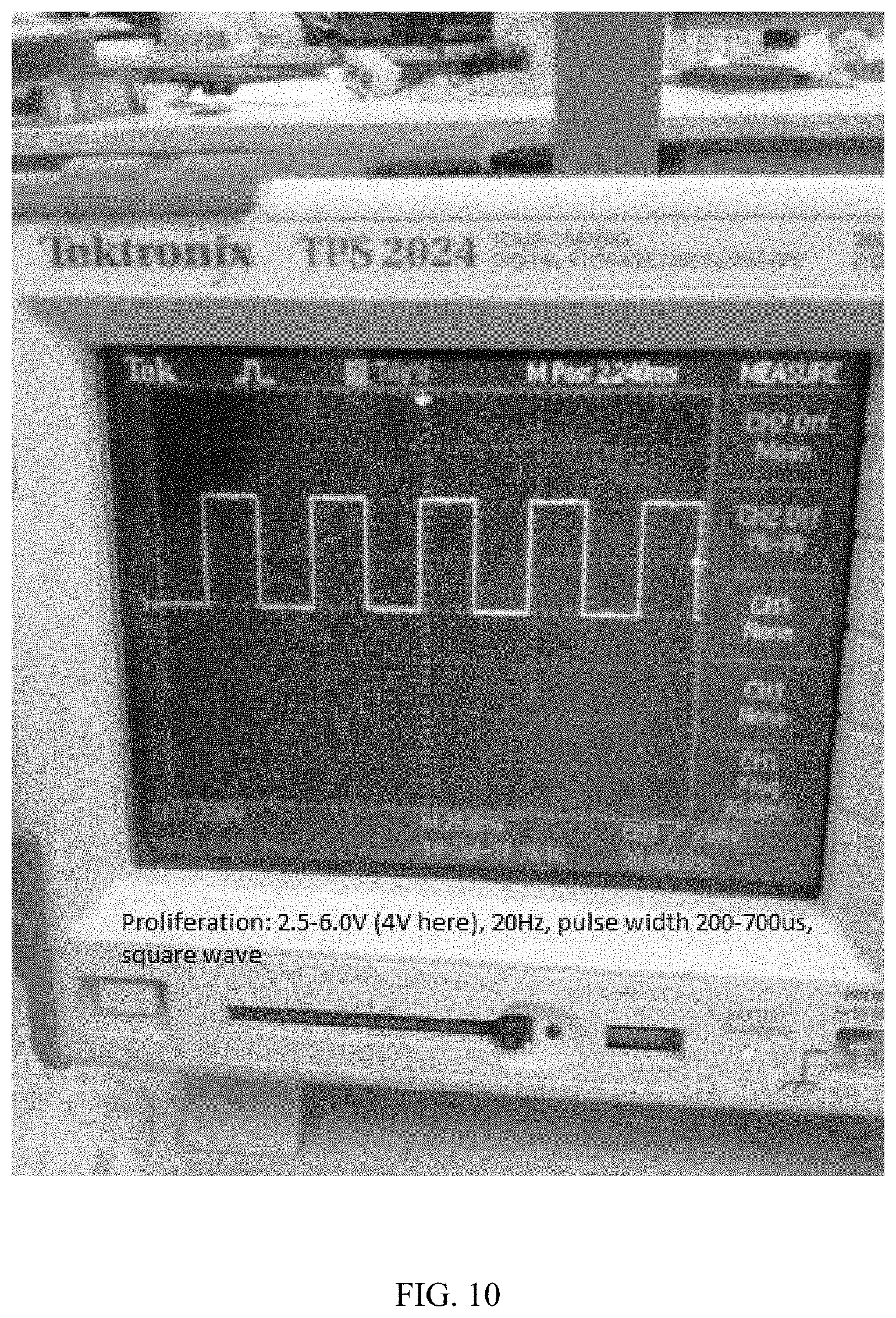
[0032] FIG. 10 depicts an image of a signal (voltage and frequency) associated with stem cell proliferation: 2.5-6.0 V (4V here), 20 Hz, pulse width 200-700 .mu.s, square wave.
[0033] FIG. 11 depicts an image of the signal (voltage and frequency) associated with SDF-1: 3.5 mV, 30 Hz, square wave.
[0034] FIG. 12 depicts an image of the signal (voltage and frequency) associated with SDF-1 (2.sup.nd part): 0.25 mA (3.0V shown here), 100 Hz, 100 .mu.s pulse width, square wave.
[0035] FIG. 13 depicts an image of the signal (voltage and frequency) associated with tropoelastin: 60 mV, 50 Hz, square wave.
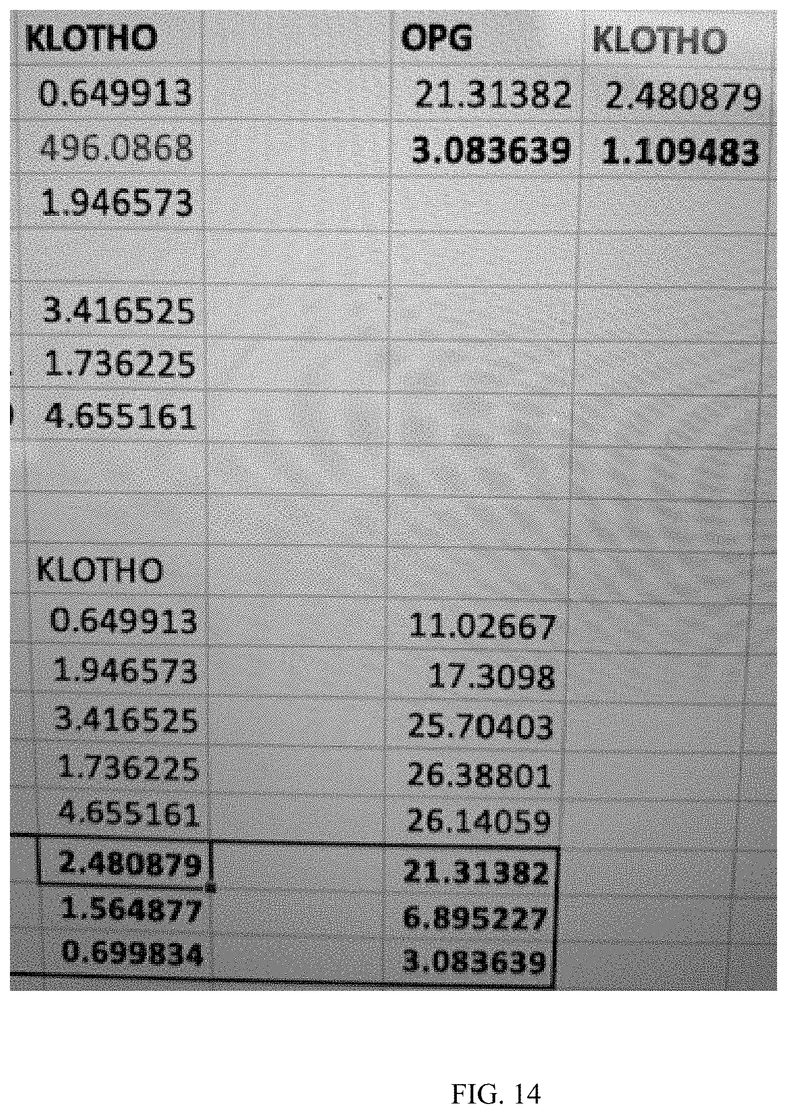
[0036] FIG. 14 is a table depicting the results of the Example.
DETAILED DESCRIPTION
[0037] In certain embodiments, described is a bandage wrap that is applied to the affected region. A micro-stimulator may be located conveniently in the bandage wrap and is utilized to distribute specific bioelectric signals to the affected tissue and nerves that regulate various protein expressions for stem cell homing, stem cell proliferation, stem cell differentiation, blood vessel formation, blood circulation improvement, muscle function repair, and DNA repair.
[0038] Referring now to FIG. 1, depicted is a stimulator for use in treating a human. The depicted device is about the size of a pen (FIG. 4) and is programmable.
[0039] Preferably, the system utilizes a bioelectric stimulator programmed to control expression and/or release of Klotho, SDF-1, IGF-1, PDGF, follistatin, and tropoelastin.
[0040] Klotho is as described above. Follistatin promotes muscle growth and counteracts myostatin. SDF-1 is generally for recruiting stem cells and maturing blood vessels. IGF-1 is for DNA repair. PDGF is a second stem cell homing factor and helps tissue regeneration. Any one of the protein expression signals work well on their own for organ regeneration, but they work better together. SDF-1 is a powerful regeneration protein, as is IGF-1.
[0041] The micro voltage signal generator may be produced utilizing the same techniques to produce a standard heart pacemaker well known to a person of ordinary skill in the art. An exemplary microvoltage generator is available (for experimental purposes from Cal-X Stars Business Accelerator, Inc. DBA Leonhardt's Launchpads or Leonhardt Vineyards LLC DBA Leonhardt Ventures of Salt Lake City, Utah, US). The primary difference is the special electrical stimulation signals needed to control, e.g., precise follistatin release on demand (which signals are described later herein). The leading pacemaker manufacturers are Medtronic, Boston Scientific Guidant, Abbott St. Jude, BioTronik and Sorin Biomedica.
[0042] Construction of the electric signal generators and pacemakers, are known in the art and can be obtained from OEM suppliers as well as their accompanying chargers and programmers. The electric signal generators are programmed to produce specific signals to lead to specific protein expressions at precisely the right time for, e.g., optimal organ treatment or regeneration.
[0043] The pacing infusion lead may be constructed or purchased from the same suppliers that build standard heart pacemaker leads. Pacing infusion leads may be purchased from a variety of OEM vendors. The pacing infusion lead may, for example, be a standard one currently used in heart failure pacing studies in combination with drug delivery.
[0044] An infusion and electrode wide area patch may be constructed by cutting conduction polymer to shape, and forming plastic into a flat bag with outlet ports in strategic locations.
[0045] Micro stimulators may be purchased or constructed in the same manner heart pacemakers have been made since the 1960's. When used with a micro infusion pump, such pumps can be purchased or produced similar to how they have been produced for drug, insulin, and pain medication delivery since the 1970's. The programming computer can be standard laptop computer. The programming wand customary to wireless programming wands may be used to program heart pacers.
[0046] Both wireless non-invasive and/or implantable wire lead ("electrode") based means may be used to deliver the regeneration and healing promoting bioelectric signals to target organs.
[0047] A wireless, single lumen infusion pacing lead or infusion conduction wide array patch may all be used to deliver the regeneration signals and substances to the organ of interest to be treated or they may be used in combination.
[0048] A re-charging wand for use herein is preferably similar to the pacemaker re-charging wand developed by Alfred Mann in the early 1970's for recharging externally implantable pacemakers.
[0049] Bioelectric stimulation can be done with the described microstimulator, which can have a pacing infusion lead with, e.g., a corkscrew lead placed/attached at, e.g., the center of the tissue to be stimulated and/or treated.
[0050] The microstimulator is actuated and runs through programmed signals to signal the release of, e.g., Klotho. In such a method, when the electrical signal includes (within 15%): a biphasic square pulse at 20 Hz, 0.1 V (100 mV), and a 7.8 ms pulse duration for 24 hours of stimulation (wherein the electrical signal is as measured three (3) mm deep into the tissue), the protein expressed and/or released is Klotho.
[0051] In such a method, when the electrical signal includes (within 15%): 10V at 50 HZ and 100 HZ for about 12 hours each (duration 1 minute) (wherein the electrical signal is as measured three (3) mm deep into the tissue), the protein further expressed and/or released by the subject is follistatin.
[0052] In such a method, when the electrical signal includes (within 15%): 3 mv with a frequency of about 22 Hz, and a current of about 1 mA for about fifteen (15) minutes and 3ma for about fifteen (15) minutes (duration 5 minutes) (wherein the electrical signal is as measured three (3) mm deep into the tissue), the protein further expressed and/or released by the subject is IGF-1.
[0053] For example, upregulation of IGF-1, and SDF-1 was achieved in cardiomyocytes using such signals. Upregulation of SDF-1 was achieved in pig heart. It has been found that signals for one cellular tissue work with other cellular tissues too.
[0054] Also described is a method of activating a tissue to further produce stromal cell-derived factor 1 ("SDF-1"), the method including: stimulating the (e.g., human) tissue with an electrical signal, wherein the electrical signal includes (within 15%): 30 pulses per second with a voltage of about 3.5 mV, and successively alternating currents of about 700 to 1500 picoamps for about one minute, and again with 700 to 1500 picoamps for about one minute and stimulated with current of about 0.25 mA, pulse duration of about 40 pulses/s, pulse width of about 100 .mu.s, wherein the electrical signal is as measured three (3) mm deep into the tissue. In such a method, the period of time is typically at least 24 hours. In such a method, the field strength is typically at least 0.1 V/cm.
[0055] What follows are preferred signals from the stimulator. For example, described are two PDGF expression control signals, one low voltage and one higher voltage. The test tissue is sheep heart tissue. The test cells are mesenchymal stem cells.
[0056] 30% PDGF increase >3 V/cm, 10 Hz, 2 micro amps (0.000002 amps) and the pulse duration 0.2 ms.
[0057] 230% PDGF increase >20 V/cm 100 Hz, 0.25 mA (2.5e-7 amps) and pulse duration of 40 pulses/s, width of 100 .mu.s.
[0058] PDGF Signal: 20V for 1 minute, 20MVs for 10 minutes, current of 0.25 mA, pulse duration of 40 pulses/s, pulse width of 100 .mu.s, and frequency of 100 Hz for 5 minutes followed by 528 Hz for 3 minutes and 432 Hz for 3 minutes and 50 Hz for 3 minutes.
[0059] SDF-1--Stem cell recruiting signal: 30 pulses per second with a voltage of 3.5 mV, and successively alternating currents of 700 to 1500 picoamps for one minute, and again with 700 to 1500 picoamps for one minute and stimulated with current of 0.25 mA, pulse duration of 40 pulses/s, pulse width of 100 .mu.s, and frequency of 100 Hz--each signal for 40 minutes to 8 hours a day for 2 to 36 months as needed for ideal results. Duration 7 minutes.
[0060] Stem cell proliferation signals: 15 mV and a current of 500 picoamps at 70 pulses per minute for 3 hours and 20 pulses per minute, a pulse amplitude of from 2.5-6 volts, and a pulse width of from 0.2-0.7 milliseconds for 3 hours. Duration 3 minutes.
[0061] Follistatin--(muscle growth) production signal: 10V at 50 HZ and 100 HZ 0.25 mA. Duration 1 minute.
[0062] IGF-1: 3mv with electric frequency of 22 Hz, and electric current of 1 mA for 15 minutes and 3ma for 15 minutes. Duration 5 minutes.
[0063] An exemplary bioelectric signal sequence in humans (after Klotho) is as follows.
[0064] SDF-1 (stem cell homing signal)--5 minutes
[0065] IGF-1 signal (DNA repair)--3 minutes
[0066] Follistatin signal (myostatin antagonist) at 1 volt (not 10 volts) -3 minutes.
[0067] PDGF--1 minute
[0068] A week after treatment, samples can be collected for morphometric evaluation by in-situ hybridization or RT-PCR.
[0069] Among the accompanying figures are included images of the corresponding signals with the name, voltage, and frequency of each signal written on each image The signals are to be further defined in terms of current and frequency, not voltage and frequency as shown. The voltage delivered to the cells will be different for each tissue type, but with current all of the signals can be kept constant regardless of tissue type. The device should have a current driven signal (instead of voltage driven like most other devices).
[0070] FIG. 5 depicts an image of the signal (voltage and frequency) associated with follistatin at 10V/cm, 50 Hz, square wave. Follistatin is a powerful antagonist of myostatin. Follistatin was first isolated from the ovary and is known to suppress follicle-stimulating hormone. The system has precise bioelectric signaling sequences that have demonstrated an ability to control release of the follistatin protein in target tissue on demand.
[0071] FIG. 6 depicts an image of the signal (voltage and frequency) associated with IGF-1: 3.0 mV, 22 Hz, square wave.
[0072] FIG. 7 depicts an image of the signal (voltage and frequency) associated with PDGF30%: 3V/cm (100 mV here), 10 Hz, pulse width 200 .mu.s, square wave. FIG. 8 also depicts an image of the signal (voltage and frequency) associated with PDGF230%: 20V/cm (7.0V here), 100 Hz, pulse width 100 .mu.s, square wave.
[0073] FIG. 9 depicts an image of the signal (voltage and frequency) associated with stem cell proliferation: 15 mV, 70 Hz, square wave.
[0074] FIG. 10 depicts an image of the signal (voltage and frequency) associated with stem cell proliferation: 2.5-6.0 V (4V here), 20 Hz, pulse width 200-700 .mu.s, square wave.
[0075] FIG. 11 depicts an image of the signal (voltage and frequency) associated with SDF-1: 3.5 mV, 30 Hz, square wave.
[0076] FIG. 12 depicts an image of the signal (voltage and frequency) associated with SDF-1 (2.sup.nd part): 0.25 mA (3.0V shown here), 100 Hz, 100 .mu.s pulse width, square wave.
[0077] FIG. 13 depicts an image of the signal (voltage and frequency) associated with tropoelastin: 60 mV, 50 Hz, square wave.
[0078] Relationship Between The Components:
[0079] The micro voltage signal generator is attached to the pacing infusion lead with, e.g., a corkscrew tip, deep vein stimulation lead (Medtronic) (e.g., for bioelectric stimulation of the brain), or conductive polymer bandage or patch to the tissue or organ to be treated. An external signal programmer may be used to program the micro voltage signal generator with the proper signals for treatment including the Klotho producing signal. The device battery may be re-chargeable with an external battery charging wand.
[0080] The essential elements are the micro voltage signal generator and the means for delivering the signal to the target tissue.
[0081] The signal generator may be external or internal. The transmission of the signal may be wireless, via liquid and/or via wires.
[0082] The tissue contact interface may be, e.g., a patch or bandage or may be via electrodes or leads. FDA cleared gel tape electrodes (Mettler) may be used for skin delivery. Electro acupuncture needles may be used to ensure the signals positively reach target tissues under the skin.
[0083] In certain embodiments, a subject's organ(s) and/or tissue(s) are first scanned or analyzed with a device to determine what his or her needs may be before treatment begins. The scanning/analysis can be by, e.g., generating mechanical vibrations at position adjacent the location to be an analyzed as described in, e.g., US 2003/0220556 A1 to Porat et al. (the contents of which are incorporated herein by this reference) and/or by measuring transmembrane voltage potential of a cell (see, e.g., Chernet & Levin, "Transmembrane voltage potential is an essential cellular parameter for the detection and control of tumor development in a Xenopus model," Dis. Models & Mech. 6, pp. 595-607 (2013); doi:10.1242/dmm.010835, the contents of which are also incorporated herein by this reference. See, also, Brooks et al. "Bioelectric impedance predicts total body water, blood pressure, and heart rate during hemodialysis in children and adolescents" J. Ren Nutr., 18(3):304-311 (May 2008); doi: 10.1053/j.jrn.2007.11.008, the contents of which are incorporated herein by this reference, describing the use of bioelectric impedance to evaluate the variability of blood pressure, systolic blood pressure, etc.
[0084] As used herein, "scanning" means measuring bioelectrical electrical activity of organs, sometimes by placement of a bion coil reader and transmitter in the organ, and direct that information to a computer. The computer stores the bioelectrical read measurements of diseased organs and healthy organs and makes a comparative exam classifying the organ into one category or another, which is much like a doctor using information to make a diagnosis.
[0085] Presently, the best approach for whole body and individual organ scanning is to use a combination of: a. 3D Body Scannint, b. Quantum Magnetic Resonance Scanning, c. Biofeedback scanning, d. Bioelectric scanning, e. Bion implant scanning, f Nervous system scanning, and g. Light activated cell reaction reading.
[0086] Scanners such as the Ina'Chi scanner, the Quantum Magnetic Resonance Analyzer (QMRA), the 3D Quantum Health Analyzer Scan whole body organ health 2, BodyScan.RTM. scanner, and the "BIONic muscle spindle" are also useful.
[0087] For example, the subject is positioned for analysis with a device, preferably with a non-invasive testing device for evaluating, e.g., the autonomic nervous system, organ function(s), and risk factors associated with heart disease, diabetes, and stroke. The non-invasive testing device may analyze data from, e.g., the subject's skin galvanic response, skin color, oximeter, blood pressure, and body composition analyzer to determine hardening and thickening of the subject's arteries, the subject's heart health, exercise capacity, thyroid function, neurotransmitter balance, and multiple other markers for health. See, also, Fatemi et al. "Imaging elastic properties of biological tissues by low-frequency harmonic vibration" Proceedings of the IEEE, 91(10):1503-1519 (October 2003).
[0088] In an alternative embodiment, the analysis conducted by the device comprises (or further includes) detecting minute energy fields around the human body with, e.g., a "SQUID magnetometer" (SQUID is an acronym for "Superconducting Quantum Interference Device"), able to detect biomagnetic fields associated with physiological activities in the subject's body. A quantum resonant magnetic analyzer analyzes such fields. The magnetic frequency and energy of a subject's organ(s) and/or tissue(s) are collected by appropriately positioning the sensor with respect to the portion of the subject's organ(s) and/or tissue(s) to be analyzed, and after amplification of the signal by the instrument, the data are compared with standard quantum resonant spectrum of diseases, nutrition, and other indicators/markers to determine whether the sample waveforms are irregular using a Fourier approach.
[0089] Treatment may include, e.g., moving magnets or changing magnetic fields (pulsed electromagnetic fields) about the tissue and/or organ, for example, to reduce inflammation or treat pain or induce tissue growth in the subject.
[0090] The invention is further described with the aid of the following illustrative Example.
EXAMPLES
Example
Controlling Expression and/or Release of Klotho
[0091] Twelve samples of gingiva cells were stimulated with a biphasic square pulse at 20 Hz, 0.1 V (100 mV), and a 7.8 ms pulse duration for 24 hours of stimulation. The cells were gingival fibroblasts from a 28 year old Caucasian male (https://www.atcc.org/en/Products/All.CRL-2014.aspx), which were passaged less than 8 times. RT-PCR was used to measure results before and after the described bioelectric stimulation. Results: Klotho expression up an average of 248% (n=5) and as high as 465% (see FIG. 14).
REFERENCES
[0092] (The Contents of the Entirety of Each of Which is Incorporated Herein by this Reference.)
[0093] Prochazka et al. "Cocktail of Factors from Fat-derived Stem Cells Shows Promise for Critical Limb Ischemia" http://www.sciencenewsline.com/news/2016012204520017.html (Jan. 21, 2016).
[0094] Salcedo et al. "Low current electrical stimulation upregulates cytokine expression in the anal sphincter," Int. J. Colorectal Dis., 2012 February; 27(2):221-5. doi: 10.1007/s00384-011-1324-3. Epub (October 2011).
[0095] Hopkins Medicine "Overview of Pacemakers and Implantable Cardioverter Defibrillators (ICDs)," hopkinsmedicine.org/healthlibrary/conditions/cardiovascular diseases/overview_of_pacemakers_and_implantable_cardioverter_defibrillato- rs_icds_85,P00234/.
[0096] Columbia "Implant Procedure Concepts--Pacemaker, ICD and CRT Overview," columbia.edu/itc/hs/medical/hickey/docs/Pacemaker,%20ICD %20and %20CRT %200verview %2 0022007.pdf
* * * * *
References
D00000
D00001
D00002
D00003
D00004
D00005
D00006
D00007
D00008
D00009
D00010
D00011
D00012

D00013
XML
uspto.report is an independent third-party trademark research tool that is not affiliated, endorsed, or sponsored by the United States Patent and Trademark Office (USPTO) or any other governmental organization. The information provided by uspto.report is based on publicly available data at the time of writing and is intended for informational purposes only.
While we strive to provide accurate and up-to-date information, we do not guarantee the accuracy, completeness, reliability, or suitability of the information displayed on this site. The use of this site is at your own risk. Any reliance you place on such information is therefore strictly at your own risk.
All official trademark data, including owner information, should be verified by visiting the official USPTO website at www.uspto.gov. This site is not intended to replace professional legal advice and should not be used as a substitute for consulting with a legal professional who is knowledgeable about trademark law.