Killer Cell Capable Of Efficiently And Stably Expressing Antibody, And Uses Thereof
QIAN; Qijun ; et al.
U.S. patent application number 16/311084 was filed with the patent office on 2020-09-17 for killer cell capable of efficiently and stably expressing antibody, and uses thereof. The applicant listed for this patent is Shanghai Cell Therapy Group Co., Ltd., Shanghai Cell Therapy Research Institute. Invention is credited to Lianzhen CUI, Zhou HE, Huajun JIN, Linfang LI, Qijun QIAN, Hongping WU, Jieying XU, Zhenlong YE.
| Application Number | 20200289561 16/311084 |
| Document ID | / |
| Family ID | 1000004887073 |
| Filed Date | 2020-09-17 |


View All Diagrams
| United States Patent Application | 20200289561 |
| Kind Code | A1 |
| QIAN; Qijun ; et al. | September 17, 2020 |
KILLER CELL CAPABLE OF EFFICIENTLY AND STABLY EXPRESSING ANTIBODY, AND USES THEREOF
Abstract
Provided is a transgenic killer cell, the genome of which is stably integrated with a coding sequence comprising an antibody of a human Fc section, or an expression cassette of a coding sequence comprising a chimeric antigen receptor or an inhibitory or agonistic antibody, and an inverted terminal repeat sequence from a transposon at both ends. Also provided is a pharmaceutical composition comprising the transgenic killer cell, and uses thereof.
| Inventors: | QIAN; Qijun; (Shanghai, CN) ; JIN; Huajun; (Shanghai, CN) ; XU; Jieying; (Shanghai, CN) ; LI; Linfang; (Shanghai, CN) ; YE; Zhenlong; (Shanghai, CN) ; HE; Zhou; (Shanghai, CN) ; CUI; Lianzhen; (Shanghai, CN) ; WU; Hongping; (Shanghai, CN) | ||||||||||
| Applicant: |
|
||||||||||
|---|---|---|---|---|---|---|---|---|---|---|---|
| Family ID: | 1000004887073 | ||||||||||
| Appl. No.: | 16/311084 | ||||||||||
| Filed: | June 19, 2017 | ||||||||||
| PCT Filed: | June 19, 2017 | ||||||||||
| PCT NO: | PCT/CN2017/088955 | ||||||||||
| 371 Date: | March 30, 2020 |
| Current U.S. Class: | 1/1 |
| Current CPC Class: | C07K 2317/24 20130101; C07K 16/08 20130101; C07K 2317/622 20130101; C07K 16/30 20130101; C07K 2317/52 20130101; C07K 16/2818 20130101; C07K 2317/76 20130101; A61K 35/17 20130101; C12N 5/0646 20130101; C12N 2510/00 20130101; C07K 2319/33 20130101; C07K 2319/03 20130101; A61P 35/00 20180101 |
| International Class: | A61K 35/17 20060101 A61K035/17; C12N 5/0783 20060101 C12N005/0783; C07K 16/30 20060101 C07K016/30; C07K 16/08 20060101 C07K016/08; C07K 16/28 20060101 C07K016/28; A61P 35/00 20060101 A61P035/00 |
Foreign Application Data
| Date | Code | Application Number |
|---|---|---|
| Jun 20, 2016 | CN | 201610443886.6 |
Claims
1. A transgenic killer cell, wherein the genome of the killer cell is stably integrated with acoding sequence of an antibody comprising human Fc segment, or an expression cassette comprising an coding sequence of a chimeric antigen receptor and an inhibitory antibody or an agonistic antibody, and wherein both ends of the expression cassette comprise inverted terminal repeat sequences of a transposon.
2. The transgenic killer cell according to claim 1, wherein the amount of the antibody expressed per million of the killer cells within 48 hours is higher than 2 .mu.g.
3. The transgenic killer cell according to claim 1, wherein the transposon is selected from the group consisting of piggybac, sleeping beauty, frog prince, Tn5 and Ty.
4. The transgenic killer cell according to claim 1, wherein the killer cell is selected from the group consisting of cytokine induced killer cell, dendritic cell stimulated cytokine-induced killer cell, cytotoxic T lymphocyte, .gamma..delta.T cell, natural killer cell, NKT cell, tumor infiltrating lymphocyte, lymphokine-activated killer cell, anti-CD3 monoclonal antibody killer cell, genetically modified CAR-T, genetically modified CAR-NK and genetically modified TCR-T cells.
5. The transgenic killer cell according to claim 1, wherein the killer cell further comprises molecular brake(s).
6. The transgenic killer cell according to claim 1, wherein the antibody comprising human Fc segment is an anti-tumor or anti-virus antibody; wherein the agonistic antibody is an agonistic antibody against immune co-stimulating molecule or a receptor thereof; and wherein the inhibitory antibody is an immune checkpoint inhibitory antibody.
7. The transgenic killer cell according to claim 1, wherein: (1) the antibody is directed against one or more of the following antigens: PD-1, HER2, CTLA4, PDL1, PDL2, PDL3, TIM3, LAG3, CD28, CD137, CD40, CD40L, CD47, CD19, CD20, CEA, GD2 (also known as B4GALNT1, .beta.1,4-acetyl-aminogalactosyl transferase 1), FR (flavin reductase), PSMA (prostate specific membrane antigen), gp100 (PMEL premelanosome protein), CA9 (carbonic anhydrase IX), CD171/L-CAM, IL-13R.alpha.2, MART-1 (also known as melan-A), ERBB2, NY-ESO-1 (also known as CTAG1B, cancer/testicle antigen 1B), MAGE (melanoma associated antigen E1) family proteins, BAGE (B melanoma antigen family) family proteins, GAGE (Growth Hormone Releasing Factor) family proteins, AFP (.alpha.-fetoprotein), MUC1 (mucin 1, cell-surface related), CD22, CD23, CD30, CD33, CD44v7/8, CD70, VEGFR1, VEGFR2, IL-11R.alpha., EGP-2, EGP-40, FBP, GD3 (also known as ST8SIA1, ST8.alpha.-N-acetyl-ceramide .alpha.-2,8-sialic acid convertase 1), PSCA (prostate stem cell antigen), FSA (also known as KIAA1109), PSA (also known as KLK3, Kallikrein-related peptidase 3), HMGA2, fetal acetylcholine receptor, LeY (also known as FUT3), EpCAM, MSLN (mesothelin), IGFR1, EGFR, EGFRvIII, ERBB3, ERBB4, CA125 (also known as MUC16, mucin 16, cell-surface related), CA15-3, CA19-9, CA72-4, CA242, CA50, CYFRA21-1, SCC (also known as SERPINB3), AFU (also known as FUCA1), EBV-VCA, POA (also known as VDR, vitamin D (1,25-dihydroxy vitamin D3) receptor), .beta.2-MG (.beta.-2-Microglobulin) and PROGRP (GRP, Gastrin releasing peptide), HBV and HIV; (2) the antigen binding fragment of the antibody is derived from the antibody in (1); and/or (3) the chimeric antigen receptor is directed against one or more of the following antigens: CD19, CD20, CEA, GD2 (also known as B4GALNT1, .beta.1,4-acetyl-aminogalactosyl transferase 1), FR (flavin reductase), PSMA (prostate specific membrane antigen), PMEL (premelanosome protein), CA9 (carbonic anhydrase IX), CD171/L1-CAM, IL-13R.alpha.2, MART-1 (also known as melan-A), ERBB2, NY-ESO-1 (also known as CTAG1B, cancer/testicle antigen 1B), MAGE (melanoma associated antigen E1) family proteins, BAGE (B melanoma antigen family) family proteins, GAGE (Growth Hormone Releasing Factor) family proteins, AFP (.alpha.-fetoprotein), MUC1 (mucin 1, cell-surface related), CD22, CD23, CD30, CD33, CD44v7/8, CD70, VEGFR1, VEGFR2, IL-11R.alpha., EGP-2, EGP-40, FBP, GD3 (also known as ST8SIA1, ST8 .alpha.-N-acetyl-ceramide .alpha.-2,8-sialic acid convertase 1), PSCA (prostate stem cell antigen), FSA (also known as KIAA1109), PSA (also known as KLK3, Kallikrein-related peptidase 3), HMGA2, fetal acetylcholine receptor, LeY (also known as FUT3), EpCAM, MSLN (mesothelin), IGFR1, EGFR, EGFRvIII, ERBB3, ERBB4, CA125 (also known as MUC16, mucin 16, cell-surface related), CA15-3, CA19-9, CA72-4, CA242, CA50, CYFRA21-1, SCC (also known as SERPINB3), AFU (also known as FUCA1), EBV-VCA, POA (also known as VDR, vitamin D (1,25-dihydroxy vitamin D3) receptor), .beta.2-MG (.beta.-2-Microglobulin) and PROGRP (GRP, Gastrin releasing peptide).
8. The transgenic killer cell according to claim 1, wherein the transposon is piggybac and the killer cell is a T cell, NK cell or CAR-T cell.
9. The transgenic killer cell according to claim 5, wherein the molecular brake is a membrane antigen that can be identified by a commercially available antibody drug.
10. The transgenic killer cell according to claim 9 wherein the membrane antigen is selected from the group consisting of CD11a, CD15, CD19, CD20, CD25, CD44, CD47, CD52, EGFR, ERBB2, ERBB3, ERBB4, VEGFR1, VEGFR2, EpCAM, MSLN, GPIIb/IIIa, .alpha.4 integrin and .alpha.4.beta.7 integrin.
11. The transgenic killer cell according to claim 6, wherein the antibody comprising human Fc segment is selected from the group consisting of: immune check point antibody, T cell co-stimulation signal antibody, anti-angiogenic antibody, antibody against tumor cell growth factor receptor, antibody against tumor cell membrane antigen and antibody against virus; wherein the agonistic antibody is an agonistic antibody directed against one or more of the following antigens: CD28, CD137, CD134, CD40, CD40L, ICOS, HVEM, CD2, CD27, CD30, GITR, LIGHT, DR3, SLAM, CD226, CD80 and CD86; and wherein the inhibitory antibody is an immune checkpoint inhibitory antibody directed against one or more of the following antigens: PD-1, CTLA4, PDL1, PDL2, PDL3, TIM3, LAG3, CD47, BTLA, TIGIT, CD160, LAIR1, B7-H1, B7-1, VSIR and CD244.
12. The transgenic killer cell according to claim 11, wherein the agonistic antibody is an scFv of anti-CD28 antibody; and the inhibitory antibody an scFv-Fc of anti-PD1 antibody.
13. The transgenic killer cell according to claim 1, wherein the transgenic killer cell is transformed with the following nucleic acid constructs A and B, or nucleic acid construct C: nucleic acid construct A, which comprises: a transposon 5' inverted terminal repeat sequence (5' ITR), a coding sequence of an antibody comprising human Fc segment and a promoter controlling the expression of such a nucleic acid sequence, a polyA tailing signal sequence and a transposon 3'inverted terminal repeat sequence (3' ITR); nucleic acid construct B, which comprises: a transposon 5' inverted terminal repeat sequence (5'ITR), a nucleic acid sequence encoding molecular brake(s) and a promoter controlling the expression of such a nucleic acid sequence, a polyA tailing signal sequence and a transposon 3' inverted terminal repeat sequence (3' ITR), a transposase coding sequence and optionally, a promoter controlling the expression of the transposase coding sequence; nucleic acid construct C, which comprises: a transposon 5' inverted terminal repeat sequence (5' ITR); a nucleic acid sequences encoding optionally molecular brake(s), a chimeric antigen receptor (CAR) and an inhibitory antibody or an agonistic antibody, and a promoter controlling the expression of such a nucleic acid sequence; a polyA tailing signal sequence: a transposon 3' inverted terminal repeat sequence (3' ITR); a transposase coding sequence, and optionally a promoter controlling the expression of the transposase coding sequence.
14. The transgenic killer cell according to claim 13, wherein the nucleic acid construct A comprises a nucleotide sequence as shown in SEQ ID NO:3 or 7, the nucleic acid construct B comprises a nucleotide sequence as shown in SEQ ID NO:4, and the nucleic acid construct C comprises a nucleotide sequence as shown in SEQ ID NO:11 or 14.
15. The transgenic killer cell according to claim 13, wherein the construct(s) are transformed into cells by one or more methods selected from the group consisting of virus transduction, microinjection, particle bombardment, gene gun transformation and electroporation.
16. A pharmaceutical composition comprising the transgenic killer cell of claim 1 and pharmaceutically acceptable excipient(s).
17. The pharmaceutical composition of claim 16, wherein the transgenic killer cell is a T cell, NK cell or CAR-T cell; the transposon is piggybac; the antibody comprising human Fc segment is selected from the group consisting of: immune check point antibody, T cell co-stimulation signal antibody, anti-angiogenic antibody, antibody against tumor cell growth factor receptor, antibody against tumor cell membrane antigen and antibody against virus; the agonistic antibody is an scFv of anti-CD28 antibody; the inhibitory antibody an scFv-Fc of anti-PD1 antibody; and the chimeric antigen receptor is a chimeric antigen receptor directed against EGFR family; and optionally, the killer cell further comprises a molecular brake, which is CD20.
18. The pharmaceutical composition of claim 16, wherein the transgenic killer cell is transformed nucleic acid constructs A and B, or with nucleic acid construct C, wherein the nucleic acid construct A comprises a nucleotide sequence as shown in SEQ ID NO:3 or 7, the nucleic acid construct B comprises a nucleotide sequence as shown in SEQ ID NO:4, and the nucleic acid construct C comprises a nucleotide sequence as shown in SEQ ID NO:11 or 14.
19. A method of inhibiting tumor cell growth or virus growth, or for treating tumors, viral infectious diseases, bacterial infectious diseases or autoimmune diseases, comprising administering to a subject in need thereof the transgenic killer cell of of claim 1 or a pharmaceutical composition comprising the transgenic killer cell of claim 1 and pharmaceutically acceptable excipient(s).
20. The method according to claim 19, wherein the tumor is selected from the group consisting of liver cancer, lung cancer, colon cancer, pancreatic cancer, gastric cancer, breast cancer, nasopharyngeal carcinoma, lymphoma, ovarian cancer, bladder cancer, prostate cancer and head and neck tumors.
Description
TECHNICAL FIELD
[0001] The invention belongs to the field of cytobiology and oncology, and particularly relates to a killer cell capable of efficiently and stably expressing an antibody and uses thereof.
BACKGROUND TECHNOLOGY
[0002] Cancer has now become the leading human health killer. Fast life rhythm, tremendous working pressure, unhealthy diet habit, and bad environment are all cancer inducing factors, and lead to a remarkably increased incidence of cancer with younger incidence age. At present, traditional treatment methods have reached to a bottleneck, and there is a need to explore more effective treatment methods to improve the survival rate and the living quality of cancer patients. Immunotherapy for malignant tumors rapidly develops in recent years and achieves remarkable clinical curative effects. In 2011, both Nature and the top magazine in clinical oncology JCO published review articles with the same title "Cancer immunotherapy comes of age" (Nature. 2011, 480 (7378): 480; J Clin Oncol. 2011; 29 (36): 4828), respectively. A new peak of tumor immune cell therapy research has come. In the future, it is possible to occupy a pivotal position in tumor therapy.
[0003] "Immunity" can be classified into cellular immunity and humoral immunity, which have distinct manners in killing tumors or viruses. Cellular immunity removes foreign substances by the direct effects imposed by cells, which is a participant of an immune response in the body and also an executor of an immune function. At present, the effector immune cells used for clinical treatment, namely the killer immune cells, mainly comprise natural killer cells (nature killer, NK), lymphokine activated killer cell (LAK), cytokine activated killer cells (CIK), cytokine-activated killer cells stimulated by dendritic cells (DC-CIK), cytotoxic T-lymphocyte (CTL), .gamma..delta.T cell, tumor infiltrating lymphocyte (TIL), CD3AK cells (anti-CD3 monoclonal antibody killer cells) and the like. These cells do not express antibodies and directly kill or act through antibody dependent cell-mediated cytotoxicity effect (ADCC).
[0004] Humoral immunity is mediated by antibodies, and antibodies in human body are generated by B lymphocytes. In 1997, the first monoclonal antibody for clinical cancer treatment, namely an anti-human CD20 (rituximab) was approved by FDA. With the development of antibody technology, till now there are more than 100,000 antibodies reported in the world, among which more than 1,000 are genetically engineered antibodies, and more than 200 are humanized antibodies. Till March 2016, FDA has approved 50 antibodies for marketing, wherein 26 are used for tumor therapy. These antibodies not only act directly on tumor cells, but also act on immune cells and take effect indirectly.
[0005] However, although effector immune cells have certain anti-tumor effects, the clinical treatment effect thereof is limited; while the penetrability of macromolecular antibodies to the solid tumors is insufficient, and systemic administration can cause systematic adverse reactions. For example, the HER2 monoclonal antibody has cardiac toxicity, and the PD1 antibody breaks the body immune balance so that autoimmune diseases can be caused. In addition, due to the fact that the antibody drug involves complicated in vitro production and preparation processes, with high purity requirement and large dosage, the medication cost is high. Therefore, if the cellular immunity effector cells keep the cell killing toxicity while being able to efficiently express antibody with anti-tumor activity, the problems that the immune treatment effect of cells is insufficient and a macromolecular antibody is difficult to enter into solid tumors will be simultaneously overcome, with the treatment cost reduced. Under the effect of chemokines, the cells with both cell killing toxicity and high-level expression of antibodies can actively enter into tumor tissue via cytomorphosis, so that local high-level expression of antibody in the tumor tissue can be achieved, and the side effects caused by systemic administration can be avoided. Meanwhile, due to the co-existence of the antibody and the cytotoxic cell killing immune cells, the antibody acting on the immune cells (such as HER2 antibody Herceptin) can induce strong ADCC effect and CDC effect to efficiently kill tumor cells. Moreover, the antibody acting on T cells (such as PD1 antibody Keytruda) can prevent the inhibitory effects of tumor microenvironment on the re-infused effector T cells, making them continuously exert a therapeutic effect.
[0006] Although it has been reported that exogenous genes are transduced into, for example, NK cells or T cells, currently the conventionally used gene transfection vector systems are low in transfection efficiency when applied to effector immune cells with cell killing toxicity, or are difficult to express exogenous genes in the cells at high level. The adenovirus vector (non-integrated) can mediate short-time high-efficiency expression of exogenous genes in NK cells or T cells, but the proliferation speed of the activated NK cells or T cells is so fast that the exogenous gene expression cassette carried can be rapidly lost in cell passaging, and the expression is difficult to last. The integration of an exogenous gene into NK cell or T cell genome can be mediated by a retrovirus or a lentivirus, for which stable expression can be realized theoretically. However, an antibody contains both light chains and heavy chains with long coding sequences and large molecular weight, making it very difficult to package and prepare retrovirus or lentivirus with full-length antibody expression cassette and to express the antibody efficiently. They can only be used to express the structurally simple single-chain antibody (lacking Fc segment, incomplete in function and short in half-life). Therefore, there is no report about transgenic cells that have cell killing toxicity and can stably express antibodies with human Fc segment with high efficiency.
SUMMARY OF THE INVENTION
[0007] The first aspect of the invention provides a transgenic killer cell, wherein acoding sequence of an antibody with human Fc segment is stably integrated in the genome of the killer cell, or the genome of the killer cell comprises an expression cassette comprising a coding sequence of a chimeric antigen receptor and a single-chain antibody of interest, wherein both ends of the expression cassette comprise inverted terminal repeat sequences of the transposon.
[0008] In one or more embodiments, the amount of the antibody expressed within 48 hours per million of the killer cells is more than 2 .mu.g.
[0009] In one or more embodiments, the transposon is selected from the group consisting of piggybac, sleeping beauty, frog prince, Tn5 and Ty; preferably, the transposon is piggybac.
[0010] In one or more embodiments, the killer cells are selected from the group consisting of: cytokine activated killer cells, dendritic cell stimulated cytokine-activated killer cells, cytotoxic T lymphocytes, .gamma..delta.T cells, natural killer cells, NKT cells, tumor infiltrating lymphocytes, lymphokine-activated killer cells, anti-CD3 monoclonal antibody killer cells and genetically modified CAR-T/CAR-NK/TCR-T cells: preferably, the killer cells are T cells, NK cells or CAR-T cells.
[0011] In one or more embodiments, the killer cells further comprise a molecular brake.
[0012] In one or more embodiments, the molecular brake is a membrane antigen that can be recognized by a commercially available antibody drug.
[0013] In one or more embodiments, the membrane antigen is selected from the group consisting of: CD11a, CD15, CD19, CD20, CD25, CD44, CD47, CD52, EGFR, ERBB2, ERBB3, ERBB4, VEGFR1, VEGFR2, EpCAM, MSLN, GPIIb/IIIa, .alpha.4 integrin and .alpha.4.beta.7 integrin; preferably, the membrane antigen is CD20.
[0014] In one or more embodiments, the antibody is an anti-tumor or anti-virus antibody.
[0015] In one or more embodiments, the antibody is selected from the group consisting of: immune check point antibody, T cell co-stimulation signal antibody, anti-angiogenic antibody, antibody against tumor cell growth factor receptor, antibody against tumor cell membrane antigen and antibody against viruses.
[0016] In one or more embodiments, the antibody is directed to one or more of the following antigens: PD-1, CTLA4, PDL1, PDL2, PDL3, TIM3, LAG3, CD28, CD137, CD40, CD40L, CD47, CD19, CD20, CEA, GD2 (also known as B4GALNT1, .beta.-1,4-acetyl-aminogalactosyl transferase 1), FR (flavin reductase), PSMA (prostate specific membrane antigen), gp100 (PMEL premelanosome protein), CA9 (carbonic anhydrase IX), CD171/L1-CAM, IL-13R.alpha.2, MART-1 (also known as melan-A), ERBB2, NY-ESO-1 (also known as CTAG1B, cancer/testicle antigen 1B), MAGE (melanoma associated antigen E1) family proteins, BAGE (B melanoma antigen family) family proteins, GAGE (Growth Hormone Releasing Factor) family proteins, .alpha.-fetoprotein (AFP), MUC1 (mucin 1, cell-surface related), CD22, CD23, CD30, CD33, CD44v7/8, CD70, VEGFR1, VEGFR2, IL-11R.alpha., EGP-2, EGP-40, FBP, GD3 (also known as ST8SIA1, ST8.alpha.-N-acetyl-ceramide .alpha.-2,8-sialic acid convertase 1), PSCA (prostate stem cell antigen), FSA (also known as KIAA1109). PSA (also known as KLK3, Kallikrein-related peptidase 3), HMGA2, Fetal acetylcholine receptor, LeY (also known as FUT3), EpCAM, MSLN (mesothelin), IGFR1, EGFR, EGFRvIII, ERBB3, ERBB4, CA125 (also known as MUC16, mucin 16, cell-surface related), CA15-3, CA19-9, CA72-4, CA242, CA50, CYFRA21-1, SCC (also known as SERPINB3), AFU (also known as FUCA1), EBV-VCA, POA (also known as VDR, vitamin D (1,25-dihydrovitamin D3) receptor), .beta.2-MG (.beta.-2-Microglobulin) and PROGRP (GRP, Gastrin releasing peptide), HBV, and HIV.
[0017] In one or more embodiments, the antibody is a PD-1 antibody or a HER2 antibody.
[0018] In one or more embodiments, the transgenic killer cells are transformed with nucleic acid constructs A and B, or nucleic acid construct C as shown below:
[0019] Nucleic acid construct A: comprising a transposon's 5'-inverted terminal repeat sequence (5'ITR), a coding sequence of an antibody comprising human Fc segment and a promoter controlling the expression of such a nucleic acid sequence, a polyA tailing signal sequence and a transposon's 3'-inverted terminal repeat sequence (3' ITR);
[0020] Nucleic acid construct B: comprising a transposon's 5'-inverted terminal repeat sequence (5'ITR), a coding sequence of molecular brake and a promoter controlling the expression of such a nucleic acid sequence, a polyA tailing signal sequence and a transposon's 3'-inverted terminal repeat sequence (3' ITR), a transposase coding sequence and optionally, a promoter controlling the expression of the coding sequence of the transposase;
[0021] Nucleic acid construct C: comprising a transposon's 5'-inverted terminal repeat sequence (5'ITR), a nucleic acid sequences encoding optionally a molecular brake, a chimeric antigen receptor (CAR) and an antigen-binding fragment of a single-chain antibody of interest, and a promoter controlling the expression of such a nucleic acid sequence, a polyA tailing signal sequence, a transposon's 3'-inverted terminal repeat sequence (3' ITR), a transposase coding sequence and a promoter controlling the expression of the coding sequence of the transposase.
[0022] In one or more embodiments, one or more methods selected from virus transduction, microinjection, particle bombardment, gene gun transformation and electroporation are employed to transform the nucleic acid construct(s) into the cells, preferably, electroporation is used.
[0023] The second aspect of the invention provides a pharmaceutical composition, wherein the pharmaceutical composition comprises the transgenic killer cells as described herein and a pharmaceutically acceptable excipient.
[0024] The third aspect of the invention provides the use of the transgenic killer cells or the pharmaceutical composition described herein, wherein the use is selected from the group consisting of: preparation of a medicine for inhibiting tumor cell growth, preparation of a medicine for inhibiting virus growth, preparation of a medicine for treating tumors, preparation of a medicament for treating viral infectious diseases, preparation of a medicament for treating bacterial infectious diseases and preparation of a medicament for treating autoimmune diseases:
[0025] wherein the tumor is selected from the group consisting of liver cancer, lung cancer, colon cancer, pancreatic cancer, gastric cancer, breast cancer, nasopharyngeal carcinoma, lymphoma, ovarian cancer, bladder cancer, prostate cancer and head and neck tumors.
BRIEF DESCRIPTION OF THE DRAWINGS
[0026] FIG. 1: A schematic diagram of the expression cassette of PD-1 antibody. ITR is a transposon's inverted terminal repeat sequence; HyPB is a piggybac transposase.
[0027] FIGS. 2A, 2B and 2C: ELISA assays of PD1 antibody expression levels in PIK-T cells from different donors. The control is non-transgenic T cells of the same origin.
[0028] FIG. 3: Western blotting assay of the expression of PD1 antibody in PIK-T cells from different donors.
[0029] FIG. 4: The detection of PD1 antibody expression cassette in PIK-T cell genome.
[0030] FIG. 5: Flow cytometry assay of PD1 molecules on the surface of PIK-T cells.
[0031] FIG. 6: PIK-T cell proliferation assay.
[0032] FIG. 7: Assay of PIK-T cell secretion of cytokines IL-2, IL-4, IL-6, IL-10, TNF.alpha. and IFN-.gamma..
[0033] FIG. 8: The detection of in vitro cytotoxity of PIK-T cells on tumor cells.
[0034] FIG. 9: The detection of in vivo inhibitory effects of PIK-T cells on the transplanted tumor.
[0035] FIG. 10: Functional detection of the molecular brake system in PIK-T cells.
[0036] FIG. 11: A schematic diagram of the expression cassette of HER2 antibody. ITR is a transposon's inverted terminal repeat sequence; HyPB is a piggybac transposase.
[0037] FIGS. 12A and 12B: ELISA assays of HER2 antibody expression levels in PIK-NK cells from different donors. The control is non-transgenic NK cells of the same origin.
[0038] FIG. 13: Western blotting assay of the expression of HER2 antibody in PIK-NK.
[0039] FIG. 14: The detection of HER2 antibody expression cassette in PIK-NK cell genome.
[0040] FIG. 15: Flow cytometry assay of antibody molecules expressed on the surface of PIK-NK cells.
[0041] FIGS. 16A, 16B, 16C and 16D: The detection of in vitro cytotoxity of PIK-NK cells on different tumor cells.
[0042] FIG. 17: The detection of in vivo inhibitory effects of PIK-NK cells on the transplanted tumor.
[0043] FIG. 18: Functional detection of the molecular brake system in PIK-NK cells.
[0044] FIG. 19: A schematic diagram of the expression cassette of herinCAR-PD1. ITR is a transposon's inverted terminal repeat sequence; HyPB is a piggybac transposase. Hinge is an antibody's hinge.
[0045] FIG. 20: ELISA assay of PD1 antibody expression levels in herinCAR-PD1 cells. The control is herinCAR cells.
[0046] FIG. 21: Western blotting assay of the expression of PD1 antibody in herinCAR-PD1.
[0047] FIG. 22: Cell proliferation assay of herinCAR-PD1 cells.
[0048] FIGS. 23A, 23B, and 23C: The detection of in vitro cytotoxity of herinCAR-PD1 cells on tumor cells. E:T, effector to target ratio.
[0049] FIG. 24: The detection of in vivo inhibitory effects of herinCAR-PD1 cells on transplanted tumor. Control, PBS treatment group, Mock-T, non-transgenic T cells.
[0050] FIG. 25: Proliferation of HerinCAR-PD1 cells in the transplanted tumor. Mock-T is non-transgenic T cells.
[0051] FIG. 26: Functional detection of the molecular brake system in HerinCAR-PD1 cells.
[0052] FIG. 27: A schematic diagram of the expression cassette of herinCAR-CD28. ITR is a transposon's inverted terminal repeat sequence; HyPB is a piggybac transposase.
[0053] FIG. 28: Flow cytometry assay of CD28 antibody molecules expressed on herinCAR-CD28 cell surface. The control is herinCAR cells.
[0054] FIG. 29: Flow cytometry assay of CD28 molecules on herinCAR-CD28 cell surface.
[0055] FIG. 30: Cell proliferation assay of herinCAR-CD28 cells.
[0056] FIG. 31: The detection of in vitro cytotoxity of herinCAR-CD28 cells on tumor cells. E:T effector to target ratio.
[0057] FIG. 32: The detection of in vivo inhibitory effects of herinCAR-CD28 cells on the transplanted tumor.
[0058] FIG. 33: Proliferation of HerinCAR-CD28 cells in the transplanted tumor.
DETAILED EMBODIMENTS
[0059] Some terms involved in the invention are explained below.
[0060] In the present invention, the term "expression cassette" refers to the complete elements required to express a gene, including promoter, gene coding sequence and polyA tailing signal sequence.
[0061] The term "coding sequence" is defined herein as a part of a nucleic acid sequence that directly determines the amino acid sequence of its protein product. The boundaries of a coding sequence are usually determined by a ribosome binding site closely adjacent to the upstream of open reading frame at mRNA 5'end (for prokaryotic cells) and a transcriptional termination sequence closely adjacent to the downstream of the open reading frame at mRNA 3'end. The coding sequence can include, but is not limited to, DNA, cDNA and recombinant nucleic acid sequences.
[0062] The term "agonistic antibody" refers to an antibody that can agonise/activate a specific type of immune response when present. The term "inhibitory antibody" refers to an antibody that can inhibit a specific type of immune response when present. The agonistic antibody or inhibitory antibody can be the full length sequence of an antibody or a functional fragment thereof. In one or more embodiments, the agonistic antibody or the inhibitory antibody may be selected from: Fab, Fab', F (ab') 2, Fv, scfv and scfv-Fc.
[0063] The term "antigen-binding fragment" (Fab) refers to a peptide fragment located at the end of the two arms of the "Y" structure of an antibody molecule, which consists of amino acid sequences of hypervariable region and determines the antigen-binding specificity of the antibody.
[0064] The term "Fc" refers to the crystallizable fragment of an antibody, which means a peptide fragment located at the terminal of the "Y" handle structure of the antibody, comprising peptide segments of antibody heavy chain constant regions CH2 and CH3, which is the part of an antibody that interacts with effector molecule or cell.
[0065] The term "antigen epitope", also known as antigenic determinant (AD), refers to a special chemical moiety in an antigen which determines the antigen specificity. In general, a polypeptide epitope contains 5-6 amino acid residue antigen epitopes, which can be recognized by specific antibodies. The specificity of the antigen is determined by the nature, the number and the spatial configuration of the antigen epitopes. According to difference in amino acid continuity of the antigen epitopes, the antigen epitopes can be classified into linear epitopes and spatial epitopes. The linear epitope is an epitope consisting of sequentially proximate amino acids, and the space epitope is an epitope formed by several amino acids that are not sequentially proximate, but are in spatial vicinity.
[0066] The term "co-stimulating molecule" refers to a molecule that exists on the surface of an antigen-presenting cell, which is capable of binding to co-stimulating molecular receptors on Th cells to generate synergistic stimulation signals. The proliferation of lymphocyte not only requires binding of the antigen, but also the receiving of the co-stimulating molecule signal. The transmission of co-stimulation signal to T cell is mainly by the binding of co-stimulating molecules CD80 and CD86 expressed on the surface of an antigen-presenting cell to CD28 molecule on the surface of T cells. The receiving of co-stimulation signal by B cells can be mediated by common pathogen components such as LPS, or by complement component, or CD40L on activated antigen-specific Th cell surface.
[0067] The term "linker" or hinge is a polypeptide fragment that links different proteins or polypeptides, and the purpose of which is to keep the independent spatial conformation of the linked protein or polypeptide to maintain the function or activity of the protein or polypeptide.
[0068] The term "specific binding" refers to the reaction between an antibody or antigen binding fragment and an antigen which it recognizes. In certain embodiments, an antibody specifically binding to a certain antigen (or an antibody that is specific to a certain antigen) means that the antibody binds to the antigen with an affinity (Kd) of less than about 10.sup.-5M, such as less than about 10.sup.-6M, 10.sup.-7M, 10.sup.-8M, 10.sup.-9M or 10.sup.-10M or less. "Specific recognition" has a similar meaning.
[0069] The term "a pharmaceutically acceptable excipient" refers to a carrier and/or an excipient that are pharmacologically and/or physiologically compatible with a subject and active ingredient(s), which is well known in the art (see, for example, Remington's Pharmaceutical Sciences, Gennaro A R Ed., 19.sup.th edition, Pennsylvania: Mack Publishing Company, 1995), including but not limited to, pH adjusting agent, surfactant, adjuvant, ion strength enhancer. For example, the pH adjusting agent includes, but is not limited to, phosphate buffer; the surfactant includes, but is not limited to, cationic, anionic or non-ionic surfactant, such as Tween-80; the ion strength enhancer includes, but is not limited to, sodium chloride.
[0070] The term "an effective amount" refers to a dosage amount that can treat, prevent, reduce and/or alleviate the disease or condition of the present invention in a subject.
[0071] The term "disease and/or condition" refers to a physical state of the subject, wherein the physical state is related to the diseases and/or conditions of the invention.
[0072] The term "subject" may refer to a patient or other animals, particularly a mammal, such as a human, a dog, a monkey, a cow, a horse and the like, that receives the pharmaceutical composition of the invention for treating, preventing, reducing and/or alleviating the diseases or conditions of the present invention.
[0073] A type of nucleic acid construct (also referred to herein as "nucleic acid construct A") is provided herein. Such a type of nucleic acid construct comprises transposon's 5' inverted terminal repeat sequence (5'ITR), a nucleic acid sequence of interest and optionally, a promoter controlling the expression of said nucleic acid of interest, a polyA tailing signal sequence and a transposon's 3' inverted terminal repeat sequence (3'ITR).
[0074] Also provided herein is another type of nucleic acid construct (also referred to herein as "nucleic acid construct B"). Such a type of nucleic acid construct comprises transposon's 5' inverted terminal repeat sequence (5' ITR), optionally a nucleic acid sequence encoding a molecular brake and optionally a promoter controlling the expression of said nucleic acid sequence, a polyA tailing signal sequence, a transposon's 3' inverted terminal repeat sequence (3' ITR), a transposase coding sequence and optionally a promoter controlling the expression of the coding sequence of the transposase.
[0075] Also provided herein is another type of nucleic acid construct (also referred to herein as "nucleic acid construct C"). Such a type of nucleic acid construct comprises a transposon's 5' inverted terminal repeat sequence (5' ITR), a nucleic acid sequences encoding optionally a molecular brake, a chimeric antigen receptor (CAR) and inhibitory antibody or agonistic antibody (such as a single-chain antibody of interest) and a promoter controlling the expression of said nucleic acid sequence, a polyA tailing signal sequence, a transposon's 3' inverted terminal repeat sequence (3' ITR), a transposase coding sequence and optionally a promoter controlling the expression of the coding sequence of the transposase.
[0076] As used herein, "a nucleic acid sequence of interest" may be a nucleic acid sequence encoding various functional proteins known in the art. Such functional proteins include various antibodies, in particular constant regions and/or variable regions of the antibodies, including but not limited to heavy chain constant region, light chain constant region, heavy chain variable region and light chain variable region. In some embodiments, the nucleic acid sequence of interest encodes a full length sequence of the Fc segment of an antibody or the functional fragment thereof. In some embodiments, the nucleic acid sequence of interest encodes a heavy chain constant region (e.g., Fc) and a light chain of the antibody. In certain embodiments, the nucleic acid sequence of interest encodes the full-length heavy-chain sequence and the full-length light-chain sequence of the antibody. In some embodiments, nucleic acid sequences encoding heavy chain segment and light chain segment can be linked by commonly used linker sequences (such as the coding sequence of Furin 2A). "Segment" as used herein refers to the fundamental structural unit of an antibody, such as the C.sub.H1, C.sub.H2, C.sub.H3, C.sub.L, V.sub.L, V.sub.H parts of an antibody and the like.
[0077] The antibodies of interest may be human antibodies, including human-murine chimeric antibodies and humanized antibodies. Antibodies of interest may be selected from an immune check point antibody, a T cell co-stimulation signal antibody, an anti-angiogenic antibody, an antibody against tumor cell growth factor receptor, and an antibody against tumor cell membrane antigen. In some embodiments, the antibody of interest may be an antibody against one or more of the following antigens: PD-1, CTLA4, PDL1, PDL2, PDL3, TIM3. LAG3, CD28, CD137, CD40, CD40L, CD47, CD19, CD20, CEA, GD2 (also known as B4GALNT1, .beta.1,4-acetyl-aminogalactosyl transferase 1), FR (flavin reductase). PSMA (prostate specific membrane antigen), gp100 (PMEL premelanosome protein), CA9 (carbonic anhydrase IX), CD171/L1-CAM, IL-13R.alpha.2. MART-1 (also known as melan-A), ERBB2, NY-ESO-1 (also known as CTAG1B, cancer/testicle antigen 1B), MAGE (melanoma associated antigen E1) family proteins, BAGE (B melanoma antigen family) family proteins, GAGE (Growth Hormone Releasing Factor) family proteins, .alpha.-fetoprotein (AFP), MUC1 (mucin 1, cell-surface related), CD22, CD23, CD30, CD33, CD44v7/8, CD70, VEGFR1, VEGFR2, IL-11R.alpha., EGP-2, EGP-40, FBP, GD3 (also known as ST8SIA1, ST8.alpha.-N-acetyl-ceramide .alpha.-2,8-sialic acid convertase 1), PSCA (prostate stem cell antigen), FSA (also known as KIAA1109), PSA (also known as KLK3, Kallikrein-related peptidase 3), HMGA2, Fetal acetylcholine receptor, LeY (also known as FUT3). EpCAM, MSLN (mesothelin), IGFR1, EGFR, EGFRvIII, ERBB3, ERBB4, CA125 (also known as MUC16, mucin 16, cell-surface related), CA15-3, CA19-9, CA72-4, CA242, CA50, CYFRA21-1, SCC (also known as SERPINB3), AFU (also known as FUCA1), EBV-VCA, POA (also known as VDR, vitamin D (1,25-dihydro vitamin D3) receptor), .beta.2-MG (.beta.-2-Microglobulin) and PROGRP (GRP, Gastrin releasing peptide), HBV, and HIV. In some embodiments, the antibody is an antibody acting on T cell itself, and after being expressed by T cells, it can protect T cells from being inhibited by tumor microenvironment and allow the T cells to locally express antibodies in tumor with reduced toxic side-effects. In other embodiments, the antibody is antibody acting on tumor cells, which after being expressed by killer cells such as NK cells, can, through guidance of the antibody, produce a synergistic ADCC effect with the killer cells. In some embodiments, the antibody is a secretory antibody. In other embodiments, the antibody is a membrane anchoring antibody. In one or more embodiments, the antibody is PD-1 antibody or HER2 antibody.
[0078] The term "single chain antibody" (scFv) refers to an antibody fragment formed by linking the amino acid sequences of antibody light chain variable region (V.sub.L region) and heavy chain variable region (Vii region) with hinge, which has antigen-binding ability. In certain embodiments, a single chain antibody of interest (scFv) is derived from the antibodies of interest described above and comprises the respective heavy chain variable region and light chain variable region of the respective antibodies of interest, or consists of a heavy chain variable region, a light chain variable region and optionally a linker. The heavy chain variable region and the light chain variable region can be linked by a well-known linker, for example, a linker containing G and S. The length of the linker is generally of 15-20 amino acids. In some embodiments, the linker is (GGGS).sub.n, and n is an integer of 1 to 5. It should be understood that the nucleic acid constructs herein may encode two single chain antibodies, one exists in the shown CAR, and the other is a single-chain antibody linked to the CAR. The two single-chain antibodies can be the same or different, preferably, the two single-chain antibodies perform different antibody functions.
[0079] In certain embodiments, nucleic acid construct C of the present invention can further comprise coding sequences of the hinge region from immunoglobulin and the Fc region at a location downstream from the nucleic acid sequence encoding the inhibitory antibody (for example the single chain antibody of interest). Various types of immunoglobulins known in the art can be used, but in certain preferred embodiments, the immunoglobulin is human IgG4.
[0080] In certain embodiments, nucleic acid construct C of the present invention can further comprise extracellular hinge region and transmembrane region sequences from the antigen to which the antibody is directed at a location downstream from the nucleic acid sequence encoding an agonistic antibody (for example the single chain antibody of interest). For example, when anti-CD28 antibody is used, scFv derived from the antibody can be used and connected with CD28 extracellular hinge region and transmembrane region sequence at the downstream location thereof.
[0081] In some embodiments, the inhibitory antibody is an immune checkpoint inhibitory antibody. In certain embodiments, the inhibitory antibody is directed against one or more of the following antigens: PD-1, CTLA4, PDL1, PDL2, PDL3, TIM3, LAG3, CD47, BTLA, TIGIT, CD160, LAIRI, B7-H1, B7-1, VSIR and CD244; preferably, the inhibitory antibody is an anti-PD-1 antibody (such as an scFv-Fc). In some embodiments, the inhibitory antibody is an scFv-Fc antibody.
[0082] In some embodiments, the agonistic antibody is an agonistic antibody directed against immune co-stimulating molecules and receptors thereof. In some embodiments, the agonistic antibody is directed against one or more of the following antigens: CD28, CD137, CD134, CD40, CD40L, ICOS, HVEM, CD2, CD27, CD30, GITR, LIGHT, DR3, SLAM and CD226. Preferably, the agonistic antibody is an anti-CD28 antibody (such as an scFv). In some embodiments, the inhibitory antibody is a single chain antibody.
[0083] As used herein, "PD1" refers to a programmed death receptor 1, which has the official name of "PDCD1" in NCBI genebank and an ID number of 5133, whose cDNA sequence/protein sequence are NM_005018.2/NP_005009.2.
[0084] "ERBB2" refers to a cell growth factor receptor 2 (also called HER2), which has an official name in NCBI GeneBank of ERBB2 and an ID number of 2064. ERBB2 has five isoforms, whose cDNA sequences/proteins are NM_004448.3/NP_004439.2, NM_001005862.2/NP_001005862.1, NM_001289936.1/NP_001276865.1, NM_001289937.1/NP_001276866.1, NM_001289938.1/NP_001276867.1, respectively.
[0085] "CD20" refers to human leukocyte differentiation antigen 20, which has an official name in the NCBI GeneBank of MS4A1 and an ID No. of 931. CD20 has two isoforms whose cDNA sequences/protein sequences are NM_021950.3/NP_068769.2, NM_152866.2/NP_690605.1, respectively. When the amino acid sequence of CD20 is mentioned, it comprises the full length of the CD20 protein or the CD20 fragments with CD20 function: and also comprises fusion proteins comprising the full-length CD20 or the fragment thereof. Moreover, a skilled person in the art appreciates that, in the amino acid sequence of CD20, mutations or variations (including but not limited to substituent, deletion and/or addition) can naturally occur or can be artificially introduced, without affecting the biological function. Moreover, when a protein sequence fragment of CD20 is described, it further comprises the corresponding sequence fragment in its natural form or in an artificial variant thereof.
[0086] "CD28" refers to human leukocyte differentiation antigen 28, which has an official name CD28 and an ID number 940 in the NCBI GeneBank. CD28 has three isoforms whose cDNA sequences/protein sequences are NM_006139.3/NP_006130.1, NM_001243077.1/NP_001230006.1, NM_001243078.1/NP_001230007.1, respectively.
[0087] "Chimeric antigen receptor" (CAR) is an artificially modified receptor which can anchor the specific molecules (such as antibodies) recognizing tumor cell surface antigens to immune cells (such as T cells), so that the immune cells can recognize tumor antigens or virus antigens and kill tumor cells or virus-infected cells. Chimeric antigen receptors suitable for the present invention can be various CARs known in the art. In some embodiments, the chimeric antigen receptor is directed against one or more of the following antigens: CD19, CD20, CEA, GD2 (also known as B4GALNT1, .beta.1,4-acetyl-aminogalactosyl transferase 1), FR (flavin reductase), PSMA (prostate specific membrane antigen), PMEL (premelanosome protein), CA9 (carbonic anhydrase IX). CD171/L1-CAM, IL-13R.alpha.2, MART-1 (also known as melan-A). ERBB2, NY-ESO-1 (also known as CTAG1B, cancer/testicle antigen 1B), MAGE (melanoma associated antigen E1) family proteins, BAGE (B melanoma antigen family) family proteins, GAGE (Growth Hormone Releasing Factor) family proteins, AFP (.alpha.-fetoprotein), MUC1 (mucin 1, cell-surface related), CD22, CD23, CD30, CD33, CD44v7/8, CD70, VEGFR1, VEGFR2, IL-11R.alpha., EGP-2, EGP-40, FBP, GD3 (also known as ST8SIA1, ST8.alpha.-N-acetyl-ceramide .alpha.-2,8-sialic acid convertase 1), PSCA (prostate stem cell antigen), FSA (also known as KIAA1109), PSA (also known as KLK3, Kallikrein-related peptidase 3), HMGA2, fetal acetylcholine receptor, LeY (also known as FUT3), EpCAM, MSLN (mesothelin), IGFR1, EGFR, EGFRvIII, ERBB3, ERBB4. CA125 (also known as MUC16, mucin 16, cell-surface related), CA15-3, CA19-9, CA72-4, CA242. CA50. CYFRA21-1, SCC (also known as SERPINB3), AFU (also known as FUCA1), EBV-VCA, POA (also known as VDR, vitamin D (1,25-dihydroxy vitamin D3) receptor), 12-MG (.beta.-2-Microglobulin) and PROGRP (GRP, Gastrin releasing peptide). Preferably, the CAR is a chimeric antigen receptor for the EGFR family.
[0088] In certain embodiments, herinCAR from CN201510812654.9 is used herein (incorporated herein by reference in its entirety).
[0089] In some embodiments, the polypeptide binding to tumor cell membrane antigen is a natural polypeptide, and is an amino acid sequence of HERIN encoded by the eighth intron Herin of human Her2 gene; preferably, the amino acid sequence is as shown in SEQ ID NO: 5 of CN201510812654.9.
[0090] In certain embodiments, the amino acid sequence of the CAR signal peptide is as shown in SEQ ID NO: 3 of CN201510812654.9.
[0091] In some embodiments, the hinge region of the CAR of the present invention is selected from one or more of the extracellular hinge region of CD8, the extracellular hinge region of CD28 and the extracellular hinge region of CD4; preferably, the extracellular hinge region of CD8. In certain embodiments, the extracellular hinge region of CD8 is as shown in SEQ ID NO: 7 of CN201510812654.9.
[0092] In some embodiments, the transmembrane region of the CAR of the present invention is selected from one or more of the transmembrane region of CD8, the transmembrane region of CD28 and the transmembrane region of CD4; preferably, the transmembrane region of CD8; preferably, the amino acid sequence of the CD8 transmembrane region is as shown in SEQ ID NO: 8 in CN201510812654.9.
[0093] In some embodiments, the intracellular signal region of the CAR of the present invention may be selected from one or more of the intracellular signal region of CD28, CD134/OX40, CD137/4-1BB, LCK, ICOS. DAP10, CD3.zeta. and Fc.epsilon.RI.gamma., preferably the 4-1BB intracellular signal region and CD3.zeta. intracellular signal region, or the CD28 intracellular signal region and the CD3.zeta. intracellular signal region; preferably, the 4-1BB intracellular signal region and the CD3.zeta. intracellular signal region have amino acid sequences as shown in SEQ ID NO:9 and SEQ ID NO: 10 of CN 201510812654.9, respectively. Preferably, the CD28 intracellular signal region and the CD3.zeta. intracellular signal region have amino acid sequences as shown in SEQ ID NO:11 and SEQ ID NO: 10 of CN 201510812654.9, respectively.
[0094] In certain embodiments, the antigen epitope is linked with the polypeptide binding to tumor cell membrane antigen directly or via a protein linker. Generally, the linker consists of at least two glycines, such as 2, 3, 4, 5, 6, 7, 8, 9 or 10 glycines.
[0095] In some embodiments, the chimeric antigen receptors of the present invention consist of sequentially arranged: a signal peptide, a CD20 antigen epitope, an amino acid sequence of HERIN encoded by the eighth intron Herin of human HER2 gene, a CD20 antigen epitope, a CD8 hinge region, a CD8 transmembrane region, and a 4-1BB co-stimulating peptide segment. Preferably, the amino acid sequence thereof is as shown in SEQ ID NO: 1 of CN201510812654.9.
[0096] Corresponding promoter sequence can be selected according to the selected nucleic acid sequence of interest. Examples of such promoters include, but are not limited to, EF1 alpha promoters. As described in CN201510021408.1 (the contents of which are incorporated herein by reference in their entirety), the upstream of the promoter can further comprise enhancers, such as one, any two or all three of mCMV enhancer, hCMV enhancer and CD3e enhancer.
[0097] Therefore, in certain embodiments, various promoter sequences published in CN201510021408.1 are used herein, including but not limited to: CCEF promoter as shown in SEQ ID NO: 1 of the application, comprising mCMV enhancer, hCMV enhancer, and EF1.alpha. promoter; TEF promoter as shown in SEQ ID NO:2, comprising CD3e enhancer and EF1.alpha. promoter; TCEF promoter as shown in SEQ ID NO:3, comprising CD3e enhancer, mCMV enhancer, hCMV enhancer and EF1.alpha. promoter, CCEFI promoter as shown in SEQ ID NO:4, comprising mCMV enhancer, hCMV enhancer and intron-containing EF1.alpha. promoter, TEFI promoter as shown in SEQ ID NO:5, comprising CD3e enhancer and intron-containing EF1.alpha. promoter; and TCEFI promoter as shown in SEQ ID NO:6, comprising CD3e enhancer, mCMV enhancer, hCMV enhancer and intron-containing EF1.alpha. promoter.
[0098] The transposases herein can be transposases from piggybac, sleeping beauty, frog prince, Tn5 or Ty transposon systems. When transposases from different transposon systems are used, the sequences of 5'ITR and 3'ITR in the nucleic acid construct disclosed by the invention are correspondingly changed into sequences matching the transposon systems, which can be easily determined by those skilled in the art.
[0099] In certain embodiments, the transposase is a transposase from piggybac transposon system. Thus, in these embodiments, the 5' ITR sequence and 3' ITR sequence of the transposon are the 5' ITR sequence and 3' ITR sequence of the piggybac transposon, respectively. In certain embodiments, the 5' ITR sequence is as shown in SEQ ID NO: 1 of CN20151038974.7, which is hereby incorporated by reference in its entirety. In certain embodiments, the 3' ITR sequence is as shown in SEQ ID NO: 4 of CN201510638974.7. In certain embodiments, the piggybac transposase is a transposase comprising c-myc nuclear localization signal coding sequence. In certain embodiments, the coding sequence of piggybac transposase is as shown in SEQ ID NO: 5 of CN201510638974.7.
[0100] The promoter of the transposase coding sequence can be various promoters known in the art for controlling the expression of transposase coding sequences. In certain embodiments, the expression of transposase coding sequence is controlled by CMV promoter. The sequence of CMV promoter can be as shown in SEQ ID NO:6 of CN201510638974.7.
[0101] PolyA tailing signal sequence known in the art can be used. In certain embodiments, the polyA is from SV40. In certain embodiments, the sequence shown in SEQ ID NO: 3 of CN201510638974.7 can be used.
[0102] As used herein. "molecular brake" refers to membrane antigens that can be recognized by a commercially available antibody drug. By adding an antibody drug that recognizes the "molecular brake", cells carrying such a molecular brake can be rapidly removed, so that the treatment safety is improved. Suitable molecular brakes (membrane antigens) can be selected from the group consisting of CD11a, CD15, CD19, CD20, CD25, CD44, CD47, CD52, EGFR, ERBB2, ERBB3, ERBB4, VEGFR1, VEGFR2, EpCAM, MSLN (mesothelin), GPIIb/IIIa, .alpha.4 integrin and .alpha.4.beta.7 integrin. In some embodiments, the membrane antigen is CD20. In certain embodiments, a linear epitope or spatial epitope of the membrane antigen can be used.
[0103] Suitable promoters can be selected for the selected membrane antigens to control the expression of said membrane antigens. In certain embodiments, the promoter is EF1.alpha. promoter. In some embodiments, the sequence of EF1.alpha. promoter is as shown in SEQ ID NO:8 of CN201510638974.7.
[0104] In certain embodiments, nucleic acid construct A disclosed herein comprises sequentially arranged: a transposon 5' inverted terminal repeat sequence (5' ITR), a promoter controlling the expression of a nucleic acid of interest, the nucleic acid sequence of interest, a polyA tailing signal sequence and a transposon 3' inverted terminal repeat sequence (3' ITR).
[0105] In certain embodiments, nucleic acid construct B disclosed herein comprises sequentially arranged: a transposon 5' inverted terminal repeat sequence (5' ITR), a promoter controlling the expression of a nucleic acid sequence encoding a molecular brake, the nucleic acid sequence encoding the molecular brake, a polyA tailing signal sequence, a transposon 3' inverted terminal repeat sequence (3' ITR), a transposase coding sequence and a promoter controlling the expression of the coding sequence of the transposase.
[0106] In certain embodiments, "nucleic acid construct C" described herein comprises sequentially arranged: a transposon 5' inverted terminal repeat sequence (5' ITR), a promoter controlling the expression of a nucleic acid sequence encoding a molecular brake, a chimeric antigen receptor (CAR) and an inhibitory antibody or an agonistic antibody (such as a single-chain antibody of interest), the nucleic acid sequences encoding the molecular brake, the chimeric antigen receptor (CAR) and the inhibitory antibody or the agonistic antibody (such as the single-chain antibody of interest), a polyA tailing signal sequence, a transposon 3' inverted terminal repeat sequence (3' ITR), a transposase coding sequence and a promoter controlling the expression of the coding sequence of the transposase.
[0107] In certain embodiments, nucleic acid construct C disclosed herein comprises sequentially arranged: a transposon 5' inverted terminal repeat sequence (5' ITR), an EF1.alpha. promoter, a molecular brake (CD20) coding sequence, a CAR coding sequence, a coding sequence of anti-PD1 single-chain antibody, an immunoglobulin hinge region and an Fc coding sequence, a polyA tailing signal sequence, transposon 3' inverted terminal repeat sequence (3' ITR), a transposase coding sequence and a promoter (CMV) controlling the expression of the transposase coding sequence.
[0108] In certain embodiments, nucleic acid construct C disclosed herein comprises sequentially arranged: a transposon 5' inverted terminal repeat sequence (5' ITR), an EF1.alpha. promoter, a molecular brake (CD20) coding sequence, a CAR coding sequence, a coding sequence of anti-PD1 single-chain antibody, an IgG4 hinge region coding sequence, an IgG4 Fc coding sequence, a polyA tailing signal sequence, a transposon 3' inverted terminal repeat sequence (3' ITR), a transposase coding sequence and a promoter (CMV) controlling the expression of the transposase coding sequence.
[0109] In certain embodiments, nucleic acid construct C disclosed herein comprises sequentially arranged: a transposon 5' inverted terminal repeat sequence (5' ITR); a promoter controlling nucleic acid sequences encoding a molecular brake, a chimeric antigen receptor (CAR), a single-chain antibody of interest, an antibody hinge region and a transmembrane region; the nucleic acid sequences encoding the molecular brake, the chimeric antigen receptor (CAR), the agonistic single-chain antibody (such as the single-chain antibody of interest), the antibody hinge region and the transmembrane region directed by the agonistic antibody; a polyA tailing signal sequence; a transposon 3' inverted terminal repeat sequence (3' ITR); a transposase coding sequence; and a promoter controlling the expression of the transposase coding sequence.
[0110] In certain embodiments, nucleic acid construct C disclosed herein comprises sequentially arranged: a transposon 5' inverted terminal repeat sequence (5' ITR), an EF1.alpha. promoter, a molecular brake (CD20), a CAR, an anti-CD28 single-chain antibody, CD28 hinge region and transmembrane region, a polyA tailing signal sequence, a transposon 3' inverted terminal repeat sequence (3' ITR), a transposase coding sequence and a promoter (CMV) controlling the expression of the transposase coding sequence.
[0111] In some embodiments, nucleic acid construct A comprises a nucleic acid sequence as shown in SEQ ID NO: 3 or 7. In some embodiments, nucleic acid construct B comprises a nucleic acid sequence as shown in SEQ ID NO: 4. In some embodiments, nucleic acid construct C comprises a nucleic acid sequence as shown in SEQ ID NO: 11 or 14.
[0112] Nucleic acid constructs A, B and C disclosed herein can be recombinant expression vectors (recombinant expression vectors A, B and C), respectively, for expressing nucleic acid sequence(s) of interest and the nucleic acid sequence of the optional coding sequence for molecular brakes. Preferably, the expression vector is a transposon vector. In certain embodiments, the vector is one or more selected from the following transposon vectors: piggybac, sleeping beauty, frog prince, Tn5 and Ty. Besides the nucleic acid sequences comprised in nucleic acid constructs A, B and C, the expression vector generally comprises further elements which are usually included in the vector, such as multiple-cloning site(s), resistance gene(s), replication origin(s) and the like. It should be understood that in general, nucleic acid construct A/recombinant expression vector A of the invention does not comprise a coding sequence of transposase.
[0113] In certain embodiments, the recombinant expression vector uses pUC18, pUC19, pMD18-T, pMD19-T, pGM-T vector, pUC57, pMAX or pDC315 as its backbone. In other embodiments, the recombinant expression vector uses pCDNA3 series vectors, pCDNA4 series vectors, pCDNA5 series vectors, pCDNA6 series vectors, pRL series vectors, pUC57 vector, pMAX vector or pDC315 as its backbone. In certain embodiments, the present invention uses pSN vector constructed in CN201510638974.7, whose structure is as shown in FIG. 1 of thet application.
[0114] The present nucleic acid construct A/recombinant expression vector A and nucleic acid construct B/recombinant expression vector B, or nucleic acid construct C/recombinant expression vector C, can be transformed into cells of interest. The method for transformation is conventional method in the field, including but not limited to, virus transduction, microinjection, particle bombardment, gene gun transformation and electroporation and the like. In some embodiments, electroporation is employed to transfer the nucleic acid construct or recombinant expression vector.
[0115] The cells of interest can be various functional cells known in the art, for example, various killer cells, including but not limited to cytokine-induced killer cells (CIK), cytokine-induced killer cells stimulated by dendritic cells (DC-CIK), cytotoxic T lymphocytes (CTL), .gamma..delta.T cells, natural killer cells (NK), tumor infiltrating lymphocytes (TIL), lymphokine activated killer cells (LAK), CD3AK cells (anti-CD3 monoclonal antibody killer cells) and CAR-T/TCR-T cells. In some embodiments, the killer cells are T cells or NK cells.
[0116] Exemplary NK cells include, but are not limited to, primary NK cells, NK cell strains (such as NK92) and NKT cells. In some embodiments, the NK cells are primary NK cells. Exemplary T cells include, but are not limited to, peripheral blood T lymphocytes, cytotoxic T cells (CTLs), helper T cells, suppressing/regulating T cells, .gamma..delta.T cells and cytokine-induced killer cells (CIK), tumor infiltrating lymphocytes (TIL) and T cells in mixed cell populations thereof. In certain embodiments, the T cells are peripheral blood T lymphocytes and T cells derived from TIL.
[0117] Since nucleic acid construct A/recombinant expression vector A comprises ITR elements necessary for transposition but does not comprise a transposase, and nucleic acid construct B/recombinant expression vector B comprises an transposase necessary for the integration of exogenous genes, only in the cells transfected by both the nucleic acid construct A/recombinant expression vector A and the nucleic acid construct B/recombinant expression vector B can the integration of expression cassettes that comprise nucleic acid sequences of interest be realized. Furthermore, the nucleic acid sequence between transposon 5' ITR and transposon 3' ITR in nucleic acid construct A or recombinant expression vector A, together with 5' and 3' ITRs themselves are all integrated into the cell genome. When nucleic acid construct B/recombinant expression vector B comprises the nucleic acid sequence encoding the molecular brake, the cell will further express the molecular brake. Also, the nucleic acid sequence between transposon 5' ITR and transposon 3' ITR in nucleic acid construct B or the recombinant expression vector B, together with 5' and 3' ITRs themselves are all integrated into the cell genome.
[0118] On the other hand, when using nucleic acid construct C/recombinant expression vector C to transfect killer cells of interest, since it comprises ITR elements and transposase which are required for transposition, the nucleic acid sequence between transposon 5' ITR and transposon 3' ITR in the nucleic acid construct or the recombinant expression vector, together with the 5' and 3' ITRs themselves are all integrated into the cell genome of interest. When nucleic acid construct C/recombinant expression vector C comprises the nucleic acid sequence encoding a molecular brake, the cell will further express the molecular brake.
[0119] Therefore, the invention further provides a kind of killer cells, wherein the expression cassette comprising the nucleic acid sequence of interest is stably integrated in the genome of the killer cells. Furthermore, the sequentially linked transposon 5' inverted terminal repeat sequence (5'ITR), the promoter for controlling the expression of the nucleic acid sequence of interest, the nucleic acid sequence of interest, the polyA tailing signal sequence and transposon 3' inverted terminal repeat sequence (3'ITR), are stably integrated into the genome of the killer cell. In a preferred embodiment, the genome of the killer cells is further integrated with sequentially linked: the transposon 5' inverted terminal repeat sequence (5'ITR), the promoter for controlling the expression of the nucleic acid sequence of the molecular brake, the nucleic acid sequence encoding the molecular brake, the polyA tailing signal sequence and the transposon 3'inverted terminal repeat sequence (3'ITR).
[0120] In other embodiments, the genome of the killer cells is stably integrated with the expression cassette comprising the nucleic acid sequence encoding the CAR and the inhibitory antibody or agonistic antibody (such as a single-chain antibody of interest). Furthermore, the genome of the killer cells is stably integrated with transposon 5' inverted terminal repeat sequence (5'ITR); the promoter for controlling the expression of the nucleic acid sequences of a molecular brake, the chimeric antigen receptor (CAR) and the inhibitory antibody or agonistic antibody (such as a single-chain antibody of interest); the nucleic acid sequences encoding the molecular brake, the chimeric antigen receptor (CAR) and the inhibitory antibody or agonistic antibody (such as the single-chain antibody of interest); the polyA tailing signal sequence; and transposon 3'inverted terminal repeat sequence (3'ITR), linked in sequence.
[0121] In certain embodiments, the disclosure further provides a kind of transgenic NK cells, wherein the expression cassette comprising the nucleic acid sequence of interest is stably integrated in the genome of the transgenic NK cells. Furthermore, the sequentially linked transposon 5' inverted terminal repeat sequence (5' ITR), the promoter for controlling the expression of the nucleic acid sequence of interest, the nucleic acid sequence of interest, the polyA tailing signal sequence and transposon 3'inverted terminal repeat sequence (3' ITR), are stably integrated into the genome of the NK cell. In a preferred embodiment, the genome of the transgenic NK cells is further integrated with sequentially linked: transposon 5' inverted terminal repeat sequence (5'ITR), the promoter for controlling the expression of the nucleic acid sequence of the molecular brake, the nucleic acid sequence encoding the molecular brake, the polyA tailing signal sequence and transposon 3'inverted terminal repeat sequence (3' ITR).
[0122] In some embodiments, the transgenic NK cells herein stably express full-length sequences of antibody Fc segments or functional fragments thereof. In certain embodiments, the transgenic NK cells herein stably express antibody heavy chain constant region (such as Fc) and light chain. In certain embodiments, the transgenic NK cells herein stably express heavy chain(s) and light chain(s) of antibodies. In certain embodiments, nucleic acid construct A/recombinant expression vector A and nucleic acid construct B/recombinant expression vector B disclosed herein are transfected into the transgenic NK cells herein. In other embodiments, the amount of the antibody expressed per million of the transgenic NK cells disclosed herein within 48 hours is higher than 2 .mu.g.
[0123] In certain embodiments, the invention further provides a kind of transgenic T cells, wherein the expression cassette comprising the nucleic acid sequence of interest is stably integrated in the genome of the transgenic T cells. Furthermore, the sequentially linked transposon 5' inverted terminal repeat sequence (5'ITR), the promoter for controlling the expression of the nucleic acid sequence of interest, the nucleic acid sequence of interest, the polyA tailing signal sequence and transposon 3'inverted terminal repeat sequence (3'ITR), are stably integrated into the genome of said T cell. In a preferred embodiment, the genome of the transgenic T cells is further integrated with transposon 5' inverted terminal repeat sequence (5'ITR), the promoter for controlling the expression of the nucleic acid sequence of the molecular brake, the nucleic acid sequence encoding the molecular brake, the polyA tailing signal sequence and transposon 3'inverted terminal repeat sequence (3'ITR), linked in sequence.
[0124] In some embodiments, the transgenic T cells herein stably express full-length sequences of antibody Fc segments or functional fragments thereof. In certain embodiments, the transgenic T cells herein stably express antibody heavy chain constant region (such as Fc) and light chain. In certain embodiments, the transgenic NK cells herein stably express heavy chain(s) and light chain(s) of antibodies. In certain embodiments, nucleic acid construct A/recombinant expression vector A and nucleic acid construct B/recombinant expression vector B disclosed herein are transfected into the transgenic T cells herein. In other embodiments, the amount of the antibody expressed per million of the transgenic T cells disclosed herein within 48 hours is higher than 2 .mu.g.
[0125] In certain embodiments, the killer cells described herein are CAR-T cells, which express inhibitory antibodies. The antibody can be a secretory antibody or a membrane-anchoring antibody, preferably a secretory antibody. The antibody can be an immune checkpoint inhibitory antibody acting on T cell itself. Upon expression of said antibody in CAR-T cells, the cells are protected from being inhibited by tumor microenvironment, and residual tumor-specific T cells can be activated.
[0126] In other embodiments, the CAR-T cells express agonistic antibodies. The antibody can be a secretory antibody or a membrane-anchoring antibody, preferably a membrane anchoring antibody. The antibody can be an immune co-stimulating molecule agonistic antibody acting on T cell itself. After the antibody is expressed by CAR-T cells, the clustering effect can be enhanced.
[0127] In some embodiments, the killer cells herein stably express full-length sequences of antibody Fc segments or functional fragments thereof, or stably express the antibody heavy chain constant region (such as Fc) or the full length sequence and the light chain full length sequence, or stably express CAR of interest and scFv of interest. In certain embodiments, nucleic acid construct A/recombinant expression vector A and nucleic acid construct B/recombinant expression vector B disclosed herein, or nucleic acid construct C/recombinant expression vector C disclosed herein are transfected into the killer cells herein. In other embodiments, the amount of the antibody expressed per million of killer cells within 48 hours is higher than 2 .mu.g.
[0128] The killer cells disclosed herein have dual functions of cellular immunity and humoral immunity. Herein they are named pluripotent immune killer Cell (which is referred to as PIK for short), such as pluripotent immune killer T cell (PIK-T for short), and pluripotent natural killer cells (PIK-NK for short). On one hand, the cells can have anti-tumor cellular immunity (mainly mediated by killer cells). On the other hand, they have humoral immunity (mainly mediated by antibodies), which can effectively inhibit the proliferation of tumors, viruses and bacteria. Meanwhile, when the molecular brake exists, the killer cells herein can be removed by commercially available antibody medicines that recognize said molecular brake, which improves the safety of treatment.
[0129] According to the biological functions of the expressed antibodies, the killer cells disclosed herein can have different biological activities, including but not limited to, inhibiting tumors, viruses, bacteria and the like. Therefore, the killer cells disclosed herein can be used for inhibiting the growth of tumor cells, inhibiting the growth of viruses, treating tumors, treating viral infectious diseases, treating bacterial infectious diseases and treating autoimmune diseases. The tumors include, but are not limited to, liver cancer, lung cancer, colon cancer, pancreatic cancer, gastric cancer, breast cancer, nasopharyngeal carcinoma, lymphoma, ovarian cancer, bladder cancer, prostate cancer and head and neck tumors.
[0130] Therefore, also provided herein is a pharmaceutical composition, which comprises the killer cells described herein and a pharmaceutically acceptable carrier or excipient. Also provided herein is the use of the killer cells herein in the preparation of a medicament for inhibiting tumor cell growth, inhibiting virus growth, treating tumors, treating viral infectious diseases, treating bacterial infectious diseases and treating autoimmune diseases.
[0131] The invention also provides a method of transfecting killer cells (especially T cells and NK cells), the method comprising using two recombinant expression vectors to co-transfect the killer cells; wherein one recombinant expression vector comprises the expression cassette for the nucleic acid sequence of interest, and at both ends of the expression cassette, inverted terminal repeat sequences of the transposon are included; wherein the recombinant expression vector does not comprise a coding sequence of transposase; the other recombinant expression vector comprises the expression cassette of optional molecular brake(s), wherein at both ends of the expression cassette, the inverted terminal repeat sequences of the transposon are included, and the recombinant expression vector comprises a coding sequence of transposase. In one or more embodiments, the two recombinant expression vectors are the recombinant expression vectors A and B as described herein, respectively. Methods of transfection are well known in the art, including but not limited to one or more of virus transduction, microinjection, particle bombardment, gene gun transformation and electroporation. During transfection, the usage amount of the two recombinant expression vectors can be adjusted according to specific situations. Usually, the amount ratio of the recombinant expression vector without transposase coding sequence to the recombinant expression vector with transposase coding sequence can be within the range of 1-5:1.
[0132] Therefore, the present invention further provides a kit comprising two kinds of recombinant expression vectors: one recombinant expression vector comprises the expression cassette for the nucleic acid sequence of interest, and at both ends of the expression cassette, inverted terminal repeat sequences of the transposon are included, wherein the recombinant expression vector does not comprise a coding sequence of transposase; the other recombinant expression vector comprises the expression cassette of optional molecular brake(s), and at both ends of the expression cassette, the inverted terminal repeat sequences of the transposon are included, wherein the recombinant expression vector comprises a coding sequence of transposase. In one or more embodiments, the two recombinant expression vectors are the recombinant expression vectors A and B as described herein, respectively. The kit can also comprises various reagents suitable for transfecting the recombinant expression vectors into cells, and optionally an instruction for guiding those skilled in the art to transfect the recombinant expression vectors into cells. In the kit, the two recombinant expression vectors can be independently packaged or can be packaged in the same container as a mixture.
[0133] Therefore, in certain embodiments, the present invention also relates to a composition of recombinant expression vectors, the composition comprising at least two recombinant expression vectors: one recombinant expression vector comprises the expression cassette for the nucleic acid sequence of interest, and at both ends of the expression cassette, inverted terminal repeat sequences of the transposon are included, wherein the recombinant expression vector does not comprise a coding sequence of transposase; the other recombinant expression vector comprises the expression cassette of optional molecular brake(s), and at both ends of the expression cassette, the inverted terminal repeat sequences of transposon are included, wherein the recombinant expression vector comprises a coding sequence of transposase. In one or more embodiments, the two recombinant expression vectors are the recombinant expression vectors A and B as described herein, respectively. The composition can comprise corresponding solvent(s) or carrier(s).
[0134] The present invention overcomes the deficiencies of the present commonly used gene transfection vector system (low transfection efficiency for killer cells, and low expression level of antibody), so that the immune killer cells can stably express high level of antibodies comprising full-length human Fc segment. The present invention overcomes the difficulty of insufficient cellular immune-therapy effect and the difficulty of macromolecular antibodies entering into solid tumors. In addition, the present killer cells can remain the cytotoxic while they can also stably express antibodies comprising human Fc segment at high levels, or stably express antibodies comprising human Fc segment, or stably express the antigen-binding fragment of the antibodies of interest and CAR. In addition, in order to prevent the proliferation of immune cells stably expressing antibodies in vivo which may lead to over-expression of the antibodies and in turn systemic toxicity and autoimmune disease, a molecular brake system (such as a CD20-Rituxan molecular brake system, CD20BR) is introduced. Using the commercially available monoclonal antibody (such as Rituxan), killing cells integrated with antibody expression cassette will be rapidly removed via the ADCC effect and CDC effect mediated by the monoclonal antibody, and thus the safety for therapy is effectively improved.
[0135] The embodiments of the present invention will be described in detail with reference to the examples. It will be understood by those skilled in the art that the following examples are only used for explaining the present invention, and should not be regarded as limiting the scope of the present invention. Where specific techniques or conditions are not detailed in the embodiments, they are carried out according to the techniques or conditions described in the literature of the field (see, for example, Molecular cloning. A laboratory manual by J Sambrook et al., and translated by Peitang HUANG et al., 3.sup.rd ed. Science Press, China) or carried out according to manufacturer's instruction. Reagent(s) or instrument(s) used herein, the manufacturers of which are not mentioned, are commercially available conventional products.
Example 1: Construction of Recombinant Plasmids pS838-AntiPD1 and pNB328-CD20BR
[0136] Two DNA sequences as shown below were synthesized by Shanghai Genray Biotech Co., Ltd:
TABLE-US-00001 Seq1: (SEQ ID NO: 1) CGATAGGACGCTGATCTTAAT Seq2: (SEQ ID NO: 2) TACCTGCGACTAGAAT
[0137] The DNA sequences were denatured at 98.degree. C. for 5 minutes, and allowed to cool down naturally to form a double-stranded DNA linker with ClaI and PacI cohesive ends at the upstream and downstream sides, respectively.
[0138] pNB vector was double digested by CalI and PacI (constructed according to CN201510638974.7) and loaded with the above mentioned double-stranded DNA to obtain pS vector.
[0139] CCEF promoter sequence (disclosed in CN201510021408.1) was synthesized by Shanghai Genray Biotech Co., Ltd, with XbaI and EcoRI restriction sites introduced into its upstream and downstream, respectively. The promoter was loaded into pS vector which was double digested by XbaI and EcoRI, so as to obtain pS838 vector.
[0140] PD1 antibody coding sequence as shown in SEQ ID NO: 3 was synthesized by Shanghai Genray Biotech Co., Ltd, with EcoRI and Sail restriction sites introduced into its upstream and downstream, respectively, and loaded into pS838 vector to obtain pS838-AntiPD1.
TABLE-US-00002 antiPD1 coding sequence: (SEQ ID NO: 3) GAATTCGCCACCATGGAAGCCCCAGCTCAGCTTCTCTTCCTCCTGCTACTC TGGCTCCCAGATACCACCGGACAGGTGTACTTGGTAGAGTCTGGGGGAGGC GTGGTCCAGCCTGGGAGGTCCCTGAGACTCTCCTGTGCAGCGTCTGGATTC ACCTTCAGTAACTATGGCATGCACTGGGTCCGCCAGGCTCCAGGCAAGGGG CTGGAGTGGGTGGCACTTATATGGTATGATGGAAGTAATAAATACTATGCA GACTCCGTGAAGGGCCGATTCACCATCTCCAGAGACAATTCCAAGAACACG CTGTATCTGCAAATGACCAGTCTGAGAGTCGAGGACACGGCTGTGTATTAT TGTGCGAGCAACGTTGACCATTGGGGCCAGGGAACCCTGGTCACCGTCTCC TCAGCTTCCACCAAGGGCCCATCCGTCTTCCCCCTGGCGCCCTGCTCCAGG AGCACCTCCGAGAGCACAGCCGCCCTGGGCTGCCTGGTCAAGGACTACTTC CCCGAACCGGTGACGGTGTCGTGGAACTCAGGCGCCCTGACCAGCGGCGTG CACACCTTCCCGGCTGTCCTACAGTCCTCAGGACTCTACTCCCTCAGCAGC GTGGTGACCGTGCCCTCCAGCAGCTTGGGCACGAAGACCTACACCTGCAAC GTAGATCACAAGCCCAGCAACACCAAGGTGGACAAGAGAGTTGAGTCCAAA TATGGTCCCCCATGCCCACCATGCCCAGCACCTGAGTTCCTGGGGGGACCA TCAGTCTTCCTGTTCCCCCCAAAACCCAAGGACACTCTCATGATCTCCCGG ACCCCTGAGGTCACGTGCGTGGTGGTGGACGTGAGCCAGGAAGACCCCGAG GTCCAGTTCAACTGGTACGTGGATGGCGTGGAGGTGCATAATGCCAAGACA AAGCCGCGGGAGGAGCAGTTCAACAGCACGTACCGTGTGGTCAGCGTCCTC ACCGTCCTGCACCAGGACTGGCTGAACGGCAAGGAGTACAAGTGCAAGGTC TCCAACAAAGGCCTCCCGTCCTCCATCGAGAAAACCATCTCCAAAGCCAAA GGGCAGCCCCGAGAGCCACAGGTGTACACCCTGCCCCCATCCCAGGAGGAG ATGACCAAGAACCAGGTCAGCCTGACCTGCCTGGTCAAAGGCTTCTACCCC AGCGACATCGCCGTGGAGTGGGAGAGCAATGGGCAGCCGGAGAACAACTAC AAGACCACGCCTCCCGTGCTGGACTCCGACGGCTCCTTCTTCCTCTACAGC AGGCTAACCGTGGACAAGAGCAGGTGGCAGGAGGGGAATGTCTTCTCATGC TCCGTGATGCATGAGGCTCTGCACAACCACTACACACAGAAGAGCCTCTCC CTGTCTCTGGGTAAACGTAAAAGGCGAGCTCCTGTTAAACAGACTTTGAAT TTTGACCTTCTCAAGTTGGCGGGAGACGTCGAGTCCAACCCTGGGCCCATG GAAGCCCCAGCTCAGCTTCTCTTCCTCCTGCTACTCTGGCTCCCAGATACC ACCGGAGAAATTGTGTTGACACAGTCTCCAGCCACCCTGTCTTTGTCTCCA GGGGAAAGAGCCACCCTCTCCTGCAGGGCCAGTCAGAGTGTTAGTAGTTAC TTAGCCTGGTACCAACAGAAACCTGGCCAGGCTCCCAGGCTCCTCATCTAT GATGCATCCAACAGGGCCACTGGCATCCCAGCCAGGTTCAGTGGCAGTGGG TCTGGGACAGACTTCACTCTCACCATCAGCAGCCTAGAGCCTGAAGATTTT GCAGTTTATTACTGTCAGCAGAGTAGCAACTGGCCTCGGACGTTCGGCCAA GGGACCAAGGTGGAAATCAAACGAACTGTGGCTGCACCATCTGTCTTCATC TTCCCGCCATCTGATGAGCAGTTGAAATCTGGAACTGCCTCTGTTGTGTGC CTGCTGAATAACTTCTATCCCAGAGAGGCCAAAGTACAGTGGAAGGTGGAT AACGCCCTCCAATCGGGTAACTCCCAGGAGAGTGTCACAGAGCAGGACAGC AAGGACAGCACCTACAGCCTCAGCAGCACCCTGACGCTGAGCAAAGCAGAC TACGAGAAACACAAAGTCTACGCCTGCGAAGTCACCCATCAGGGCCTGAGC TCGCCCGTCACAAAGAGCTTCAACAGGGGAGAGTGTTGATAAGTCGAC,
wherein double underline represents restriction sites, and single underline represents Furin 2A coding sequence.
[0141] CD20BR coding sequence as shown in SEQ ID NO: 4 was synthesized by Shanghai Genray Biotech Co., Ltd, with EcoRI and SalI restriction sites introduced into its upstream and downstream respectively, and loaded into pNB328 vector (constructed according to CN201510638974.7) and named as pNB328-CD20BR.
TABLE-US-00003 CD20BR coding sequence: (SEQ ID NO: 4) GAATTCGCCACCATGGAGTTTTGGCTGAGCTGGGTTTTCCTTGTTGCTATT TTAAAAGGTGTCCAGTGTAACATATACAACTGTGAACCAGCTAATCCCTCT GAGAAAAACTCCCCATCTACCCAATACTGTTACAGCATACAATCTCTGGGT GGAGGTGGAGGTGGAGGTGGAGGTATCTACATCTGGGCGCCCTTGGCCGGG ACTTGTGGGGTCCTTCTCCTGTCACTGGTTATCACCCTTTACTGCAACCAC AGGAACCGTAAAAGGCGAGCTCCTGTTAAACAGACTTTGAATTTTGACCTT CTCAAGTTGGCGGGAGACGTCGAGTCCAACCCTGGGCCCATGGTGAGCAAG CAGATCCTGAAGAACACCGGCCTGCAGGAGATCATGAGCTTCAAGGTGAAC CTGGAGGGCGTGGTGAACAACCACGTGTTCACCATGGAGGGCTGCGGCAAG GGCAACATCCTGTTCGGCAACCAGCTGGTGCAGATCCGCGTGACCAAGGGC GCCCCCCTGCCCTTCGCCTTCGACATCCTGAGCCCCGCCTTCCAGTACGGC AACCGCACCTTCACCAAGTACCCCGAGGACATCAGCGACTTCTTCATCCAG AGCTTCCCCGCCGGCTTCGTGTACGAGCGCACCCTGCGCTACGAGGACGGC GGCCTGGTGGAGATCCGCAGCGACATCAACCTGATCGAGGAGATGTTCGTG TACCGCGTGGAGTACAAGGGCCGCAACTTCCCCAACGACGGCCCCGTGATG AAGAAGACCATCACCGGCCTGCAGCCCAGCTTCGAGGTGGTGTACATGAAC GACGGCGTGCTGGTGGGCCAGGTGATCCTGGTGTACCGCCTGAACAGCGGC AAGTTCTACAGCTGCCACATGCGCACCCTGATGAAGAGCAAGGGCGTGGTG AAGGACTTCCCCGAGTACCACTTCATCCAGCACCGCCTGGAGAAGACCTAC GTGGAGGACGGCGGCTTCGTGGAGCAGCACGAGACCGCCATCGCCCAGCTG ACCAGCCTGGGCAAGCCCCTGGGCAGCCTGCACGAGTGGGTGTGAGTCGAC b,
wherein double underline represents restriction sites, and single underline represents CD20 epitope recognized by Rituxan antibody.
[0142] The schematic diagrams of pNB328-CD20BR and pS838-antiPD1 vectors are shown in FIG. 1.
Example 2: Genetic Modification of Peripheral Blood T Lymphocytes
[0143] 1.times.10.sup.7 freshly isolated peripheral blood mononuclear cell (PBMC) were prepared, pNB328-CD20BR and pS838-antiPD1 were co-transfected at the ratio of 1:2 into cell nucleus by Lonza 2b-Nucleofector. The cells were incubated at 37.degree. C., 5% CO.sub.2 in an incubator: after 6 hours, cells were transferred to a 6-well plate containing 30 ng/ml anti-CD3 antibody and 3000 IU/ml IL-2 (purchased from Novoprotein), and incubated at 37.degree. C., 5% CO.sub.2 in an incubator. After the cells had reached the state of healthy growth, cells were obtained as pluripotent T cells expressing PD-1, abbreviated as PIK-T. Non-transfected PBMCs were plated on culture plates containing 30 ng/ml anti-CD3 antibody and 3000 IU/ml IL-2 (purchased from Novoprotein), and incubated at 37.degree. C. 5% CO.sub.2 as control. Since only pNB328 vector contains transposase necessary for the integration of the exogenous genes while pS838 only contains ITR elements necessary for transposition, only cells co-transfected with both pNB328-CD20BR and pS838-antiPD1 vectors can realize the integration of PD1 antibody expression cassette, thus ensuring that all the PIK-T cells have CD20 molecular brakes.
Example 3: Quantitative Assay of the Amount of PD1 Antibody Expression in PIK-T Cells
[0144] PIK-T and control T cells obtained in Example 2 were passaged at a split ratio of 1:3. After two weeks, they were plated in 6-well plates supplemented with 4 ml AIM-V solution (purchased from GIBCO) at the density of 1.0.times.10.sup.6 cells/well and incubated at 37.degree. C., 5% CO.sub.2 in an incubator. After incubating for 24 hours, 48 hours, 72 hours and 96 hours, 800 .mu.l of supernatant was collected and stored for later use at -20.degree. C. ELISA plates (purchased from SinoBiological) were coated with humanized PD1 recombinant protein, and HRP-labeled mouse anti-human IgG mAb (purchased from Abcam) was used for the assay, with commercialized anti-PD1 antibody (purchased from Merck) as reference standard. The amount of PD1 antibody expression was measured by double-sandwich ELISA.
[0145] Results show that PIK-T cells from 3 different donors can all stably express high level of PD1 antibody, as shown in FIGS. 2A, 2B and 2C.
Example 4: Qualitative Assay of the Expression of PD1 Antibody in PIK-T Cells
[0146] PIK-T cells obtained in Example 2 were plated in 6-well plates supplemented with 4 ml AIM-V solution at the density of 1.0.times.10.sup.6 cells/well and incubated at 37.degree. C. 5% CO.sub.2 in an incubator. After incubating for 48 hours, 72 hours and 96 hours, supernatant harboring cells was collected. To 120 .mu.l supernatant collected at each time point was added 30 .mu.l of 5.times.SDS-PAGE Loading buffer, and the sample was boiled at 100.degree. C. for 10 minutes, and stored for later use at -20.degree. C. Western blotting assay for the detection of the anti-PD1 antibody expression was carried out using goat anti-human IgG (H+L) as the primary antibody and HRP-rabbit-anti-goat antibody as the secondary antibody, both purchased from Jackson ImmunoResearch. Results show that anti-PD1 antibody expressed by PIK cells comprises the correct heavy chains (50 kD) and light chain (25 kD), as shown in FIG. 3.
Example 5: Detection of PD1 Antibody Expression Cassette in PIK-T Cell Genome
[0147] Genomic DNA from PIK-T cells obtained in Example 2 and control T cells were extracted according to the instruction included in the kit. DNA concentrations of the PIK-T cells and the control T cells were measured by fluorescent real-time quantitative PCR with the following reaction program: 95.degree. C., 15 s.fwdarw.95.degree. C., 5 s.fwdarw.60.degree. C., 15 s, 40 cycles. Results show that PD1 antibody expression cassette is integrated into T cell genome, as shown in FIG. 4.
[0148] Primer Sequences:
[0149] F: ATCTCCAAAGCCAAAGGGCA (SEQ ID NO: 5);
[0150] R: CGATGTCGCTGGGGTAGAAG (SEQ ID NO: 6).
Example 6: Flow Cytometric Detection of PD1 Molecules on PIK-T Cell Surface
[0151] Suspended PIK-T cells (obtained in example 2) and control T cells were collected and counted, then added into two 1.5 ml EP tubes respectively at 1.times.10.sup.6 cells/tube, washed twice with PBS and centrifuged at 1,200 rpm for 5 minutes. Supernatants were discarded, 2 .mu.l of isotype control antibody IgG1-PE and anti-CD279-PE antibodies (both purchased from BD) were added respectively. Pellets were tapped gently until well mixed, incubated for 30 minutes at room temperature in the dark, washed with PBS once, and centrifuged at 1,200 rpm for 5 minutes. Supernatants were discarded, 400 .mu.l of normal saline was added before transferring the cells into flow cytometry tubes, and the sample was loaded onto the flow cytometry device for detection. Experimental results show that compared to control cells, PIK-T cells have significantly reduced number of PD1 molecules, indicating that PIK-T cells can effectively block cell surface PD1 molecules by secreting PD1 antibody through autocrine and paracrine, as shown in FIG. 5.
Example 7: Proliferation Assay of PIK-T Cells
[0152] PIK-T cells obtained in Example 2 and control T cells were plated in 96-well plates at the density of 4.times.10.sup.4 cells/well (each cell type in triplicate, with a total volume of 200 .mu.l), and incubated at 37.degree. C., 5% CO.sub.2 in an incubator. After the cells were incubated for 24 hours, 48 hours, 72 hours and 96 hours, 20 .mu.l of of CCK8 reagent was added before the samples were incubated for 6 hours at 37.degree. C. in the dark. The OD.sub.450 value was measured on ELISA analyzer, and cell number was calculated based on standard curve. Results show that the proliferation speed of PIK-T cells is significantly higher than that of control T cells, indicating that antibody secreted by PIK-T cells can promote the proliferation of T cells, as shown in FIG. 6.
Example 8: Cytokine Secretion Assay of PIK-T Cells
[0153] 24-well plate was coated by 5 .mu.g/ml CD3 antibody (purchased from Novoprotein) overnight at 4.degree. C., washed with PBS three times, and 3.times.10.sup.5 PIK-T cells obtained in Example 2 and control T cells were added to the plate and incubated for 24 h before collecting the cell-containing supernatant. Cytokine secretion of PIK-T cells and control T cells after CD3 antibody stimulation was determined by BD.TM. CBA Human Th1/Th2 Cytokine Kit II (purchased from BD). Results show that IL-4, IL-6, TNF-.alpha. and IFN-.gamma. secreted by PIK-T cells are remarkably enhanced compared to control T cells, while the secretion of the two cytokines, IL-2 and IL-10, do not have significant difference, as shown in FIG. 7.
Example 9: In Vitro Tumor Cell Killing Assay of TIL-Derived PIK-T Cells
[0154] Freshly-removed lung cancer specimens were collected and immediately processed under aseptic conditions. The specific method comprises the following steps: normal tissue and necrotic areas around lung cancer specimens were removed, and small pieces of tissues sized 1-2 mm.sup.3 from different areas of the specimen were collected and added into a 24-well plate at one piece/well. To each well, 2 mL of Complete Culture medium (GT-T551 culture medium with 10% FBS) and 3000 IU/ml IL-2 were added, and then the plate was incubated in an incubator at 37.degree. C. and 5% CO.sub.2. After 5-6 days from the start of the incubation, half of the medium for all the wells was replaced with fresh medium. After that, according to the growth condition of tumor infiltrating lymphocyte (TIL), half of the medium was replaced every 1-2 days. Once TILs in the wells reached confluency and all the adherent cells had been removed, the TILs that had filled each well were collected. Then, 1.times.10.sup.6 TILs were re-suspended in a T175 culture flasks with 150 ml of complete culture medium, 30 ng/ml anti-CD3 antibody, radiated feeder cells (PBMCs from three different healthy people) no less than 200 times of the TILs and 6000 IU/ml IL-2, and the bottle was vertically cultured. On Day 5 of culture, 65% of the liquid in the bottle was replaced with fresh complete culture medium with IL-2. On Day 7 of culture, cell suspension in the two T175 culture flasks were transferred into cell culture bag, and 300 mL of complete culture medium with IL-2 were added. From Day 6 on, Trypan blue staining and cell counting were carried out every other day and cell density was kept at 0.5-2.times.10.sup.6/mL by adding fresh complete culture medium with IL-2. Then, following the method of Example 2, pNB328-CD20BR and pS838-antiPD1 plasmids were transfected to obtain TIL-derived PIK-T cells.
[0155] Lung cancer cell strain NCI-H460 (purchased from American typical culture center. ATCC) with matched MHC class I type was chosen and plated on RTCA cell proliferation plate (purchased from ACEA Biosciences. USA) at a ratio of 10,000 cells/well. Then, according to the manufacturer's instruction, the in vitro killing activity of the cells was detected on real-time unlabeled cell function analyzer (RTCA), with the effector:target ratios being 8:1, 4:1, and 2:1, respectively. TILs not transfected with plasmids were used as control (E:T=8:1), and cell proliferation curve was observed. Results show that compared to control cells, PIK-T cell can kill H446 tumor cells more efficiently, as shown in FIG. 8.
Example 10: In Vivo Assay for Treating Tumor Xenograft with TIL-Derived PIK-T Cells
[0156] NOD-SCID mice (purchased from Shanghai SLAC Laboratory Animal Co., Ltd) were subcutaneously injected with 5.times.10.sup.6 H446 malignant lung cancer cells. After 10 days, TIL-derived PIK-T cells (prepared in Example 9), control TIL cells (injection dosage 2.times.10.sup.5) or PBS buffer were administered via tail vein injection, respectively. The growth condition of the tumor xenograft was measured. Results show a significant difference between the inhibitory effects of PIK-T cells on lung cancer and that of the control group (FIG. 9).
Example 11: In Vivo Clearance Assay of PIK-T Cells (Validation of Molecular Brake Function)
[0157] BABL/c nude mice (purchased from Shanghai SLAC Laboratory Animal Co., Ltd) were injected with PIK-T cells prepared in Example 2 via tail vein (injection dosage 5.times.10.sup.6). After 3 days, 100 .mu.g of Rituxan antibody or human IgG control antibody were injected intravenously. After 12 hours, blood and bone marrow samples were collected, and flow cytometer was used for detecting the proportion of PIK-T cells (CD20 and CD3 dual-positive cells).
[0158] Results show that compared to control group injected with human IgG antibody, after injection of Rituxan antibody, the proportions of the infused PIK-T cells in blood and bone marrow were remarkably reduced (FIG. 10). Therefore. CD20 molecular brake can effectively act in vivo and remove PIK-T cells with CD20 epitope by ADCC and CDC effects.
Example 12: Construction of Recombinant Plasmid pS838-AntiHER2
[0159] pS38-AntiHER2 recombinant plasmid was constructed according to a method similar to Example 1.
[0160] Specifically, HER2 antibody coding sequence as shown in SEQ ID NO: 7 was synthesized by Shanghai Genray Biotech Co., Ltd, with EcoRI and SalI restriction sites introduced into its upstream and downstream, respectively, loaded into pS838 vector, and named pS838-AntiHER2.
TABLE-US-00004 antiHER2 coding sequence: (SEQ ID NO: 7) GAATTCGCCACCATGGAGTTTTGGCTGAGCTGGGTTTTCCTTGTTGCTATT TTAAAAGGTGTCCAGTGTGAGGTTCAGCTGGTGGAGTCTGGCGGTGGCCTG GTGCAGCCAGGGGGCTCACTCCGTTTGTCCTGTGCAGCTTCTGGCTTCAAC ATTAAAGACACCTATATACACTGGGTGCGTCAGGCCCCGGGTAAGGGCCTG GAATGGGTTGCAAGGATTTATCCTACGAATGGTTATACTAGATATGCCGAT AGCGTCAAGGGCCGTTTCACTATAAGCGCAGACACATCCAAAAACACAGCC TACCTGCAGATGAACAGCCTGCGTGCTGAGGACACTGCCGTCTATTATTGT TCTAGATGGGGAGGGGACGGCTTCTATGCTATGGACTACTGGGGTCAAGGA ACCCTGGTCACCGTCTCCTCGGCCTCCACCAAGGGCCCATCGGTCTTCCCC CTGGCACCCTCCTCCAAGAGCACCTCTGGGGGCACAGCGGCCCTGGGCTGC CTGGTCAAGGACTACTTCCCCGAACCGGTGACGGTGTCGTGGAACTCAGGC GCCCTGACCAGCGGCGTGCACACCTTCCCGGCTGTCCTACAGTCCTCAGGA CTCTACTCCCTCAGCAGCGTGGTGACCGTGCCCTCCAGCAGCTTGGGCACC CAGACCTACATCTGCAACGTGAATCACAAGCCCAGCAACACCAAGGTGGAC AAGAAAGTTGAGCCCAAATCTTGTGACAAAACTCACACATGCCCACCGTGC CCAGCACCTGAACTCCTGGGGGGACCGTCAGTCTTCCTCTTCCCCCCAAAA CCCAAGGACACCCTCATGATCTCCCGGACCCCTGAGGTCACATGCGTGGTG GTGGACGTGAGCCACGAAGACCCTGAGGTCAAGTTCAACTGGTACGTGGAC GGCGTGGAGGTGCATAATGCCAAGACAAAGCCGCGGGAGGAGCAGTACAAC AGCACGTACCGTGTGGTCAGCGTCCTCACCGTCCTGCACCAGGACTGGCTG AATGGCAAGGAGTACAAGTGCAAGGTCTCCAACAAAGCCCTCCCAGCCCCC ATCGAGAAAACCATCTCCAAAGCCAAAGGGCAGCCCCGAGAACCACAGGTG TACACCCTGCCCCCATCCCGGGATGAGCTGACCAAGAACCAGGTCAGCCTG ACCTGCCTGGTCAAAGGCTTCTATCCCAGCGACATCGCCGTGGAGTGGGAG AGCAATGGGCAGCCGGAGAACAACTACAAGACCACGCCTCCCGTGCTGGAC TCCGACGGCTCCTTCTTCCTCTACAGCAAGCTCACCGTGGACAAGAGCAGG TGGCAGCAGGGGAACGTCTTCTCATGCTCCGTGATGCATGAGGCTCTGCAC AACCACTACACGCAGAAGAGCCTCTCCCTGTCTCCGGGTAAACGTAAAAGG CGAGCTCCTGTTAAACAGACTTTGAATTTTGACCTTCTCAAGTTGGCGGGA GACGTCGAGTCCAACCCTGGGCCCATGGAAGCCCCAGCTCAGCTTCTCTTC CTCCTGCTACTCTGGCTCCCAGATACCACCGGAGATATCCAGATGACCCAG TCCCCGAGCTCCCTGTCCGCCTCTGTGGGCGATAGGGTCACCATCACCTGC CGTGCCAGTCAGGATGTGAATACTGCTGTAGCCTGGTATCAACAGAAACCA GGAAAAGCTCCGAAACTACTGATTTACTCGGCATCCTTCCTCTACTCTGGA GTCCCTTCTCGCTTCTCTGGATCCAGATCTGGGACGGATTTCACTCTGACC ATCAGCAGTCTGCAGCCGGAAGACTTCGCAACTTATTACTGTCAGCAACAT TATACTACTCCTCCCACGTTCGGACAGGGTACCAAGGTGGAGATCAAAACT GTGGCTGCACCATCTGTCTTCATCTTCCCGCCATCTGATGAGCAGTTGAAA TCTGGAACTGCCTCTGTTGTGTGCCTGCTGAATAACTTCTATCCCAGAGAG GCCAAAGTACAGTGGAAGGTGGATAACGCCCTCCAATCGGGTAACTCCCAG GAGAGTGTCACAGAGCAGGACAGCAAGGACAGCACCTACAGCCTCAGCAGC ACCCTGACGCTGAGCAAAGCAGACTACGAGAAACACAAAGTCTACGCCTGC GAAGTCACCCATCAGGGCCTGAGCTCGCCCGTCACAAAGAGCTTCAACAGG GGAGAGTGTTGATAAGTCGAC,
wherein double underline represents restriction sites, and single underline represents Furin 2A coding sequence.
[0161] The schematic diagrams of pNB328-CD20BR (constructed as described in Example 1) and pS838-antiHER2 vectors are shown in FIG. 11.
Example 13: Genetic Modification of Peripheral Blood-Derived NK Cells
[0162] Freshly isolated peripheral blood mononuclear cells (PBMC) were prepared, added into culture plate (purchased from STEMCELL Technologies) containing 30 ng/ml anti-CD16 antibody, 500 IU/ml IL-21 (purchased from Novoprotein) and 3 ml of NK medium, and incubated at 37.degree. C. 5% CO.sub.2 in an incubator, 5.times.10.sup.6 NK cells were collected, and pNB328-CD20BR and pS838-antiHER2 were co-transfected at a ratio of 1:2 into cell nucleus by Lonza 2b-Nucleofector. After the cells had reached the state of healthy growth, pluripotent NK cells expressing HER2 antibody were obtained, abbreviated as PIK-NK. Since only pNB328 vector contains the transposase necessary for the integration of the exogenous genes while pS838 vector only contains the ITR elements necessary for transposition, only in the cells co-transfected with pNB328-CD20BR and pS838-antiHER2 vectors can the integration of HER2 antibody expression cassette be realized, thus ensuring that all PIK-NK cells have CD20 molecular brakes.
Example 14: Quantitative Assay of the Amount of Anti-HER2 Antibody Expression in PIK-NK Cells
[0163] PIK-NK cells obtained in Example 13 and control T cells were passaged at a split ratio of 1:3, and plated in 6-well plates supplemented with NK culture solution (purchased from STEMCELL Technologies) at the density of 1.0.times.10.sup.6 cells/well and incubated at 37.degree. C., 5% CO.sub.2 in an incubator. After incubating for 24 hours, 48 hours, 72 hours and 96 hours, 800 .mu.l of cell-containing supernatant was collected and stored at -20.degree. C. for later use. ELISA plates (purchased from SinoBiological) were coated with human recombinant HER2 protein, and HRP-labeled mouse anti-human IgG mAb (purchased from Abcam) was used for the assay, with commercialized anti-HER2 antibody (Herceptin, purchased from Roche) as reference standard. The amount of HER2 antibody expression was measured by double-sandwich ELISA.
[0164] Results show that PIK-NK cells from 2 different donors can both stably express anti-HER2 antibody at high level, as shown in FIGS. 12A and 12B.
Example 15: Qualitative Assay of the Expression of Anti-HER2 Antibody in PIK-NK Cells
[0165] PIK-NK cells obtained in Example 13 were plated in 6-well plates supplemented with 4 ml of NK culture solution (purchased from STEMCELL Technologies) at the density of 1.0.times.10.sup.6 cells/well and incubated at 37.degree. C., 5% CO.sub.2 in an incubator. After incubating for 48 hours, cell-containing supernatant was collected. To 120 .mu.l of the supernatant collected, 30.1 of 5.times.SDS-PAGE Loading buffer was added, and the sample was boiled at 100.degree. C. for 10 minutes, and stored at -20.degree. C. for later use. Western blotting assay was carried out to detect anti-HER2 antibody expression using goat anti-human IgG (H+L) as primary antibody and HRP-rabbit-anti-goat antibody as secondary antibody, both purchased from Jackson ImmunoResearch. Results show that anti-HER2 antibody expressed by PIK-NK cells comprises the correct heavy chains (50 kD) and light chains (25 kD), as shown in FIG. 13.
Example 16: Detection of Anti-HER2 Antibody Expression Cassette in PIK-NK Cell Genome
[0166] Genomic DNA from PIK-NK cells obtained in Example 13 and control NK cells were extracted according to the manufacturer's instruction included in the kit. DNA concentrations of the PIK-NK cells and the control NK cells were measured by fluorescent real-time quantitative PCR with the following reaction program (primers as shown in SEQ ID NO:8 and SEQ ID NO:9): 95.degree. C., 15 s.fwdarw.95.degree. C., 5 s.fwdarw.60.degree. C., 15 s, 40 cycles. Results show that the anti-HER2 antibody expression cassette is integrated into NK cell genome, as shown in FIG. 14.
TABLE-US-00005 F: (SEQ ID NO: 8) GGCTGTCCTACAGTCCTCAG R: (SEQ ID NO: 9) TTGTCCACCTTGGTGTTGCT
Example 17: Flow Cytometric Detection of Antibody Molecules on PIK-NK Cell Surface
[0167] Suspended PIK-NK cells (obtained in example 13) and control NK cells were collected and counted before they were added into two 1.5 ml EP tubes at 1.times.10.sup.6 cells/tube, respectively, washed twice with PBS and centrifuged at 1,200 rpm for 5 minutes. Supernatants were discarded, 2 .mu.l of anti-human IgG Fab2'antibody (purchased from Jackson ImmunoResearch) was added. Pellets were tapped gently until well mixed, incubated for 30 minutes at room temperature in the dark, washed with PBS once, and centrifuged at 1,200 rpm for 5 minutes. Supernatants were discarded and 400 .mu.l of normal saline was added before transferring the cells into flow cytometry tubes, and the sample was loaded onto flow cytometry device for detection. Experimental results show that compared to control cells, PIK-NK cells have anti-HER2 antibody molecules on their surface, as shown in FIG. 15.
Example 18: In Vitro Tumor Cell Killing Assay of PIK-NK Cells
[0168] HER2 positive tumor cell strains SK-OV3, BGC27 and MCF7 and HER2 negative tumor cell strain A549 (all purchased from American typical culture center, ATCC) were selected, and lactic dehydrogenase (LDH)-Cytotoxicity Assay Kit (Biovision) was used to detect the in vitro killing abilities of the NK cells with or without transgenes on different types of tumor cells. The effector, target ratios were set to 8:1, 4:1, and 2:1, respectively, and NK cells not transfected with plasmid were used as control (E:T=8:1), and cell proliferation curves were observed. Results show that compared to control cells, PIK-NK cells expressing anti-HER2 antibody (prepared in example 13) can kill HER2 positive tumor cells more efficiently, but has no significant killing effects on HER2 negative tumor cells, as shown in FIGS. 16A-16D.
Example 19: In Vivo Assay for Treating Tumor Xenograft with PIK-NK Cells
[0169] NOD-SCID mice (purchased from Shanghai SLAC Laboratory Animal Co., Ltd) were subcutaneously injected with 5.times.10.sup.6 SK-OV3 malignant ovarian cancer cells. After 10 days, PIK-NK cells (prepared in Example 13), control NK cells (injection dosage 1.times.10.sup.7) or PBS buffer were injected respectively via tail vein. The growth condition of the tumor xenograft was measured. Results show a significant difference between the inhibitory effects of PIK-NK cells and that of the control group on lung cancer (FIG. 17).
Example 20: In Vitro Clearance Assay of PIK-NK Cells (Validation of Molecular Brake Function)
[0170] PIK-NK cells obtained in Example 13 and NK cells were plated in 12-well plates supplemented with 4 ml of NK culture solution (purchased from STEMCELL Technologies) at the density of 1.0.times.10.sup.5 cells/well, and 50 .mu.g/ml Rituxan antibody or human IgG antibody were added, respectively. Lactic dehydrogenase (LDH)-Cytotoxicity Assay Kit (Biovision) was used to detect the lysis rate of PIK-NK cells after the addition of the antibodies. Results show that compared to control human IgG antibody, after adding Rituxan antibody, PIK-NK cells can be quickly lyzed; while NK cells do not have obvious response to Rituxan antibody or control human IgG antibody. The result indicates that since NK cells can highly efficiently mediate ADCC effect, after adding Rituxan antibody, they can bind to the CD20 epitope on the PIK-NK cell surface initiating the killing effects between cells, leading to the clearance of PIK-NK cells. This indicates that the CD20-Rituxan molecular brake system can effectively clear PIK-NK cells, which has good safety, as shown in FIG. 18.
Example 21: Construction of Recombinant Plasmid pNB328-herinCAR-PD1
[0171] HerinCAR coding sequence comprising CD20-Rituxan molecular brake as shown in SEQ ID NO: 10 (CAR targeting EGFR family, disclosed in CN201510812654.9) and herinCAR-PD1 coding sequence as shown in SEQ ID NO:11 (CAR targeting EGFR family and comprising CD20-Rituxan molecular brake and PD1 single chain-Fc antibody, linked by 2A) were synthesized by Shanghai Genray Biotech Co., Ltd, with EcoRI and SalI restriction sites introduced into its upstream and downstream, respectively, loaded into pNB328 vector, and were named pNB328-herinCAR and pNB328-herinCAR-PD1, respectively.
TABLE-US-00006 Coding sequence of herinCar comprising CD20-Rituxan molecular brake: (SEQ ID NO: 10) GAATTCGCCACCATGGAGTTTTGGCTGAGCTGGGTTTTCCTTGTTGCTATTTAAAA ##STR00001## ##STR00002## GGTGGTACCCACTCACTGCCCCCGAGGCCAGCTGCAGTTCCTGTCCCTCTGCGCAT GCAGCCTGGCCCAGCCCACCCTGTCCTATCCTTCCTCAGACCCTCTTGGGACCTAGT CTCTGCCTTCTACTCTCTACCCCTGGCCCCCCTCAGCCCTACAAGTGTCCCTATATC CCCTGTCAGTGTGGGGAGGGGCCCGGACCCTGATGCTCATGTGGCTGTTGACCTGT CCCGGTATGAAGGCGAGCCCAAATCTTGTGACAAAACTCACACATGCCCACCGTGC CCAGCACCTGAGTTCGTGCCGGTCTTCCTGCCAGCGAAGCCCACCACGACGCCAGC GCCGCGACCACCAACACCGGCGCCCACCATCGCGTCGCAGCCCCTGTCCCTGCGCC CAGAGGCGTGCCGGCCAGCGGCGGGGGGCGCAGTGCACACGAGGGGGCTGGACTT CGCCTGTGATATCTACATCTGGGCGCCCCTGGCCGGGACTTGTGGGGTCCTTCTCCT GTCACTGGTTATCACCCTTTACTGCAACCACAGGAGTAAGAGGAGCAGGCTCCTGC ACAGTGACTACATGAACATGACTCCCCGCCGCCCCGGGCCCACCCGCAAGCATTAC CAGCCCTATGCCCCACCACGCGACTTCGCAGCCTATCGCTCCAGAGTGAAGTTCAG CAGGAGCGCAGACGCCCCCGCGTACCAGCAGGGCCAGAACCAGCTCTATAACGAG CTCAATCTAGGACGAAGAGAGGAGTACGATGTTTTGGACAAGAGACGTGGCCGGG ACCCTGAGATGGGGGGAAAGCCGAGAAGGAAGAACCCTCAGGAAGGCCTGTACAA TGAACTGCAGAAAGATAAGATGGCGGAGGCCTACAGTGAGATTGGGATGAAAGGC GAGCGCCGGAGGGGCAAGGGGCACGATGGCCTTTACCAGGGTCTCAGTACAGCCA CCAAGGACACCTACGACGCCCTTCACATGCAGGCCCTGCCCCCTCGCTGATAAGTC GAC,
wherein double underline represents restriction sites, abd wave underline represents CD20 epitope coding sequence which is recognized by Rituxan antibody.
TABLE-US-00007 herinCAR-PD1 coding sequence: (SEQ ID NO: 11) GAATTCGCCACCATGGAGTTTTGGCTGAGCTGGGTTTTCCTTGTTGCTATTTTAAAA ##STR00003## ##STR00004## GGTGGTACCCACTCACTGCCCCCCGAGGCCAGCTGCAGTTCCTGTCCCTCTGCGCAT GCAGCCTGGCCCAGCCCACCCTGTCCTATCCTTCCTCAGACCCTCTTGGGACCTAGT CTCTGCCTTCTACTCTCTACCCCTGGCCCCCCTCAGCCCTACAAGTGTCCCTATATC CCCTGTCAGTGTGGGGAGGGGCCCGGACCCTGATGCTCATGTGGCTGTTGACCTGT CCCGGTATGAAGGCGAGCCCAAATCTTGTGACAAAACTCACACATGCCCACCGTGC CCAGCACCTGAGTTCGTGCCGGTCTTCCTGCCAGCGAAGCCCACCACGACGCCAGC GCCGCGACCACCAACACCGGCGCCCACCATCGCGTCGCAGCCCCTGTCCCTGCGCC CAGAGGCGTGCCGGCCAGCGGCGGGGGGCGCAGTGCACACGAGGGGGCTGGACTT CGCCTGTGATATCTACATCTGGGCGCCCCTGGCCGGGACTTGTGGGGTCCTTCTCCT GTCACTGGTTATCACCCTTTACTGCAACCACAGGAGTAAGAGGAGCAGGCTCCTGC ACAGTGACTACATGAACATGACTCCCCGCCGCCCCGGGCCCACCCGCAAGCATTAC CAGCCCTATGCCCCACCACGCGACTTCGCAGCCTATCGCTCCAGAGTGAAGTTCAG CAGGAGCGCAGACGCCCCCGCGTACCAGCAGGGCCAGAACCAGCTCTATAACGAG CTCAATCTAGGACGAAGAGAGGAGTACGATGTTTTGGACAAGAGACGTGGCCGGG ACCCTGAGATGGGGGGAAAGCCGAGAAGGAAGAACCCTCAGGAAGGCCTGTACAA TGAACTGCAGAAAGATAAGATGGCGGAGGCCTACAGTGAGATTGGGATGAAAGGC GAGCGCCGGAGGGGCAAGGGGCACGATGGCCTTTACCAGGGTCTCAGTACAGCCA CCAAGGACACCTACGACGCCCTTCACATGCAGGCCCTGCCCCTCGCCGTAAAAGG CGAGCTCCTGTTAAACAGACTTTGAATTTTGACCTTCTCAAGTTGGCGGGAGACGT CGAGTCCAACCCTGGGCCCATGGAAGCCCCAGCTCAGCTTCTCTTCCTCCTGCTACT CTGGCTCCCAGATACCACCGGACAGGTGCAGCTGGTGCAGTCCGGCGTGGAGGTG AAGAAGCCTGGCGCCTCCGTCAAGGTGTCCTGTAAGGCCTCCGGCTACACCTTCAC CAACTACTACATGTACTGGGTGCGGCAGGCCCCAGGCCAGGGACTGGAGTGGATG GGCGGCATCAACCCTTCCAACGGCGGCACCAACTTCAACGAGAAGTTCAAGAACC GGGTGACCCTGACCACCGACTCCTCCACCACAACCGCCTACATGGAACTGAAGTCC CTGCAGTTCGACGACACCGCCGTGTACTACTGCGCCAGGCGGGACTACCGGTTCGA CATGGGCTTCGACTACTGGGGCCAGGGCACCACCGTGACCGTGTCCTCCGGTGGAG GCGGTTCAGGCGGAGGTGGCAGCGGCGGTGGCGGGTCGGAAATTGTGTTGACACA GTCTCCAGCCACCCTGTCTTTGTCTCCAGGGGAAAGAGCCACCCTCTCCTGCAGGG CCAGCAAAGGTGTCAGTACATCTGGCTATAGTTATTTGCACTGGTATCAACAGAAA CCTGGCCAGGCTCCCAGGCTCCTCATCTATCTTGCATCCTACCTAGAATCTGGCGTC CCAGCCAGGTTCAGTGGTAGTGGGTCTGGGACAGACTTCACTCTCACCATCAGCAG CCTAGAGCCTGAAGATTTTGCAGTTTATTACTGTCAGCACAGCAGGGACCTTCCGC ##STR00005## ##STR00006## ##STR00007## TGGTGGACGTGAGCCAGGAAGACCCCGAGGTCCAGTTCAACTGGTACGTGGATGG CGTGGAGGTGCATAATGCCAAGACAAAGCCGCGGGAGGAGCAGTTCAACAGCACG TACCGTGTGGTCAGCGTCCTCACCGTCCTGCACCAGGACTGGCTGAACGGCAAGGA GTACAAGTGCAAGGTCTCCAACAAAGGCCTCCCGTCCTCCATCGAGAAAACCATCT CCAAAGCCAAAGGGCAGCCCCGAGAGCCACAGGTGTACACCCTGCCCCCATCCCA ##STR00008## CCCAGCGACATCGCCGTGGAGTGGGAGAGCAATGGGCAGCCGGAGAACAACTACA AGACCACGCCTCCCGTGCTGGACTCCGACGGCTCCTTCTTCCTCTACAGCAGGCTA ACCGTGGACAAGAGCAGGTGGCAGGAGGGGAATGTCTTCTCATGCTCCGTGATGC ATGAGGCTCTGCACQAACCACTACACACAGAAGAGCCTCTCCCTGTCTCTGGGTAAA TGATAAGTCGAC,
wherein double underline represents restriction sites, single underline represents the coding sequence of Furin 2A, wavy underline represents CD20 epitope coding sequence recognized by Rituxan antibody, dashed underline represents IgG4 hinge coding sequence, and double-wave underline represents IgG4 Fc coding sequence.
[0172] The schematic diagram of pNB328-herinCAR-PD1 vector is shown in FIG. 19.
Example 22: Construction of herinCAR-PD1 Cells
[0173] 1.times.10.sup.7 freshly isolated peripheral blood mononuclear cell (PBMC) were prepared. 6 .mu.g of pNB328-herinCAR-PD1 plasmid was transfected into cell nucleus by Lonza 2b-Nucleofector. The cells were incubated at 37.degree. C., 5% CO.sub.2 in an incubator. After 6 hours, the cells were transferred to a 6-well plate containing 30 ng/ml anti-CD3 antibody and 3000 IU/ml IL-2 (purchased from Novoprotein), and incubated at 37.degree. C., 5% CO.sub.2 in an incubator. When reaching confluency, cells were passaged at a split ratio of 1:5 to obtain genetically modified T cells that co-express CAR targeting EGFR family comprising CD20-Rituxan molecular brake and anti-PD1 single chain-Fc antibody (abbreviated as herinCAR-PD1 cells). Meanwhile, herinCAR-T cells were obtained by transfecting PBMCs from the same donors with pNB328-herinCAR plasmid.
Example 23: Quantitative Assay of the Amount of Anti-PD1 Antibody Expression in herinCAR-PD1 Cells
[0174] Cells obtained in Example 22 and herinCAR cells were passaged at a split ratio of 1:3. After two weeks, they were plated in 6-well plates supplemented with 4 ml of AIM-V culture solution (purchased from GIBCO) at the density of 1.0.times.10.sup.6 cells/well and incubated at 37.degree. C., 5% CO.sub.2 in an incubator. After incubating for 24 hours, 48 hours, 72 hours and 96 hours, 800 .mu.l of cell-containing supernatant was collected and stored at -20.degree. C. for later use. ELISA plates (purchased from SinoBiological) were coated with recombinant human PD1 protein, and HRP-labeled mouse anti-human IgG mAb (purchased from Abcam) was used for the assay, with commercialized anti-PD1 antibody (purchased from Merck) as reference standard. The amount of PD1 antibody expression was measured by double-sandwich ELISA.
[0175] Results show that herinCAR-PD1 cells can stably express high level of PD1 antibody, specifically as shown in FIG. 20.
Example 24: Qualitative Assay of the Expression of Anti-PD1 Antibody in herinCAR-PD1 Cells
[0176] HerinCAR-PD1 cells obtained in Example 22 were plated in 6-well plates supplemented with 4 ml of AIM-V culture solution at the density of 1.0.times.10.sup.6 cells/well and incubated at 37.degree. C., 5% CO.sub.2 in an incubator. After incubating for 48 hours, cell-containing supernatant was collected. To 120 .mu.l of supernatant 30 .mu.l of 5.times.SDS-PAGE Loading buffer was added, and the sample was boiled at 100.degree. C. for 10 minutes, and stored at -20.degree. C. for later use. Western blotting assay was carried out to detect anti-PD1 antibody expression using goat anti-human IgG (H+L) as primary antibody and HRP-rabbit-anti-goat antibody as secondary antibody, both purchased from Jackson ImmunoResearch. Results show that anti-PD1 antibody expressed by herinCAR-PD1 cells comprises the correct scFv-Fc antibody (52 kD), as shown in FIG. 21.
Example 25: Proliferation Assay of herinCAR-PD1 Cells
[0177] HerinCAR-PD1 cell obtained in Example 22 and herinCAR cells were plated in 96-well plates at the density of 4.times.10.sup.4 cells/well (each cell type in triplicate, with a total volume of 200 .mu.l), and incubated at 37.degree. C., 5% CO.sub.2 in an incubator. After incubating for 24 hours, 48 hours, 72 hours and 96 hours, 20 .mu.l of CCK8 reagent was added, and the sample was incubated for 6 hours at 37.degree. C. in the dark. The OD.sub.450 value was measured on an ELISA analyzer. Results show that the proliferation speed of herinCAR-PD1 cells is significantly higher than that of herinCAR control, indicating that the antibody secreted by herinCAR-PD1 cells can promote the proliferation of T cells, specifically as shown in FIG. 22.
Example 26: In Vitro Killing Assay of herinCAR-PD1 Cells
[0178] Lung cancer cell strains NCI-H23, H446, H460 and H1299 (purchased from American typical culture center. ATCC) were plated on RTCA cell proliferation plates (purchased from ACEA Biosciences, USA) at the ratio of 10,000/well. The plates were placed on xCELLigence RTCA DP multi-functional real-time unlabeled cell analyzer to record the real-time growth of the cells (indicated by the cell index measured, higher value means that the cells are in a better state). After 24 hours, herinCAR cells and herinCAR-PD1 cells constructed in Example 22 were added respectively with effector:target ratio (E:T) at 4:1 and 2:1. The plates are placed again on xCELLigence RTCA DP multi-functional real-time unlabeled cell analyzer to record the real-time growth of the cells. Results show that compared to herinCAR-T cells not expressing anti-PD1 antibody, herinCAR-T cells expressing anti-PD1 antibody, that is, herinCAR-PD1 cells have significantly improved cytotoxity to lung cancer cells (FIGS. 23A-C). The above results indicate that the co-expression of anti-PD1 antibody can improve the cytotoxity of CAR-T cells to lung cancer cells.
Example 27: Identification of In Vivo Killing Activity of herinCAR-PD1 Cells
[0179] NOD-SCID mice (purchased from Shanghai SLAC Laboratory Animal Co., Ltd) were subcutaneously injected with 5.times.10.sup.6 Huh7 malignant liver cancer cells. After 10 days, herinCAR cells, herinCAR-PD1 cells prepared in Example 22 and T cells without transgenes from the same donor (dosage of injection 2.times.10.sup.5) or PBS buffer were injected via tail vein, respectively. The state of growth of the tumor xenograft was measured. Results show a significant difference between the inhibitory effects of herinCAR-PD1 cells on liver cancer and that of the control group (FIG. 24). HerinCAR-T cells with co-expression of anti-PD1 antibody shows excellent in vivo anti-tumor effect.
Example 28: Proliferation Assay of herinCAR-PD1 Cells in Tumor Xenograft
[0180] Thirty-five days after the treatment in Example 27, the tumor-bearing mice were killed. The tumor xenograft tissues were retrieved and genomic DNA was extracted (operated according to the tissue DNA extraction kit of Sigma-Aldrich company). Relative copy number of herinCAR gene in the tumor xenograft was detected by RT-PCR (primers as shown in SEQ ID NO: 12 and SEQ ID NO: 13, the reaction system and the program were operated according to the RealMaster Mix (SYBR Green) detection kit from TIANGEN BIOTECH). Results show that compared to herinCAR-T cells, the copy number of herinCAR-PD1 cells in tumor xenograft is remarkably increased, indicating that the expression of anti-PD1 antibody can extend the survival Lime of CAR-T cells in tumor xenograft (FIG. 25).
TABLE-US-00008 F: (SEQ ID NO: 12) CAGTGAGATTGGGATGAAAGG; R: (SEQ ID NO: 13) GAAGGGCGTCGTAGGTGTC
Example 29: In Vivo Clearance Assay of herinCAR-PD1 Cells (Validation of Molecular Brake Function)
[0181] BABL/c nude mice (purchased from Shanghai SLAC Laboratory Animal Co., Ltd) were injected with the herinCAR-PD1 cells prepared in Example 22 via tail vein (dosage of injection 5.times.10.sup.6). After 3 days, 100 .mu.g of Rituxan antibody or human IgG control antibody were injected intravenously. After 12 hours, blood and bone marrow samples were collected and the proportion of CAR-T cells was detected using flow cytometry.
[0182] Results show that compared to control group that was injected with human IgG antibody, after injection of Rituxan antibody, the proportion of the infused herinCAR-PD1 cells in blood and bone marrow is remarkably reduced (FIG. 26). Therefore, CD20 molecular brake can effectively act in vivo and remove herinCAR-PD1 cells with CD20 epitope through ADCC and CDC effects.
Example 30: Construction of Recombinant Plasmid pNB328-herinCAR-CD28
[0183] HerinCAR-CD28 coding sequence as shown in SEQ ID NO: 14 (CAR targeting EGFR family and comprising CD20-Rituxan molecular brake and CD28 single chain-membrane binding antibody, linked by 2A) was synthesized by Shanghai Genray Biotech Co., Ltd, with EcoRI and Sail restriction sites introduced into its upstream and downstream, respectively, loaded into pNB328 vectors, and were named pNB328-herinCAR and pNB328-herinCAR-CD28, respectively.
TABLE-US-00009 herinCAR-CD28 coding sequence: (SEQ ID NO: 14) GAATTCGCCACCATGGAGTTTTGGCTGAGCTGGGTTTTCCTTGTTGCTAT ##STR00009## ##STR00010## GGTGGAGGTGGTACCCACTCACTGCCCCCGAGGCCAGCTGCAGTTCCTGTCCCTCT GCGCATGCAGCCTGGCCCAGCCCACCCTGTCCTATCCTTCCTCAGACCCTCTTGGGA CCTAGTCTCTGCCTTCTACTCTCTACCCCTGGCCCCCCTCAGCCCTACAAGTGTCCC TATATCCCCTGTCAGTGTGGGGAGGGGCCCGGACCCTGATGCTCATGTGGCTGTTG ACCTGTCCCGGTATGAAGGCGAGCCCAAATCTTGTGACAAAACTCACACATGCCCA CCGTGCCCAGCACCTGAGTTCGTGCCGGTCTTCCTGCCAGCGAAGCCCACCACGAC GCCAGCGCCGCGACCACCAACACCGGCGCCCACCATCGCGTCGCAGCCCCTGTCCC TGCGCCCAGAGGCGTGCCGGCCAGCGGCGGGGGGCGCAGTGCACACGAGGGGGCT GGACTTCGCCTGTGATATCTACATCTGGGCGCCCCTGGCCGGGACTTGTGGGGTCC TTCTCCTGTCACTGGTTATCACCCTTTACTGCAACCACAGGAGTAAGAGGAGCAGG CTCCTGCACAGTGACTACATGAACATGACTCCCCGCCGCCCCGGGCCCACCCGCAA GCATTACCAGCCCTATGCCCCACCACGCGACTTCGCAGCCTATCGCTCCAGAGTGA AGTTCAGCAGGAGCGCAGACGCCCCCGCGTACCAGCAGGGCCAGAACCAGCTCTA TAACGAGCTCAATCTAGGACGAAGAGAGGAGTACGATGTTTTGGACAAGAGACGT GGCCGGGACCCTGAGATGGGGGGAAAGCCGAGAAGGAAGAACCCTCAGGAAGGC CTGTACAATGAACTGCAGAAAGATAAGATGGCGGAGGCCTACAGTGAGATTGGGA TGAAAGGCGAGCGCCGGAGGGGCAAGGGGCACGATGGCCTTTACCAGGGTCTCAG TACAGCCACCAAGGACACCTACGACGCCCTTCACATGCAGGCCCTGCCCCCTCGCC GTAAAAGGCGAGCTCCTGTTAAACAGACTTTGAATTTTGACCTTCTCAAGTTGGCG GGAGACGTCGAGTCCAACCCTGGGCCCATGGAGTTTTGGCTGAGCTGGGTTTTCT ##STR00011## ##STR00012## ##STR00013## ##STR00014## ##STR00015## ##STR00016## ##STR00017## ##STR00018## ##STR00019## ##STR00020## ##STR00021## ##STR00022## ##STR00023## ##STR00024## GACAATGAGAAGAGCAATGGAACCATTATCCATGTGAAAGGGAAACACCTTTGTC CAAGTCCCCTATTTCCCGGACCTTCTAAGCCCTTTTGGGTGCTGGTGGTGGTTGGTG GAGTCCTGGCTTGCTATAGCTTGCTAGTAACAGTGGCCTTTATTATTTTCTGGGTGA GGAGTAAGTGATAAGTCGAC,
wherein double underline represents restriction sites, single underline represents Furin 2A coding sequence, and dashed underline represents CD28 single chain antibody coding sequence.
[0184] The schematic diagram of pNB328-herinCAR-CD28 vector is shown in FIG. 27.
Example 31: Construction of herinCAR-CD28 Cells
[0185] 1.times.10.sup.7 freshly isolated peripheral blood mononuclear cell (PBMC) were prepared. 6 .mu.g of pNB328-herinCAR-CD28 plasmid was transfected into cell nucleus by Lonza 2b-Nucleofector. The cells were incubated at 37.degree. C., 5% CO.sub.2 in an incubator for 6 hours, and then transferred to a 6-well plate containing 30 ng/ml anti-CD3 antibody and 3000 IU/ml IL-2 (purchased from Novoprotein), and incubated at 37.degree. C., 5% CO.sub.2 in an incubator. After the cells had grown to confluency, they were passaged at a split ratio of 1:5 to obtain genetically modified T cells that express both EGFR family targeting-CAR with CD20-Rituxan molecular brake, and CD28 single chain antibody, abbreviated as herinCAR-CD28 cells. Meanwhile, PBMC from the same donor was transfected with pNB328-herinCAR plasmid to obtain herinCAR-T cells.
Example 32: Detection of Anti-CD28 Antibody Molecules on herinCAR-CD28 Cell Surface
[0186] Suspended herinCAR-CD28 cells (constructed in Example 31) and control herinCAR-T cells were collected and counted, and then added into two 1.5 ml EP tubes, respectively at the density of 1.times.10.sup.6 cells/tube, washed twice with PBS and centrifuged at 1,200 rpm for 5 minutes. Supernatants were discarded, and 2 .mu.l of anti-human IgG Fab2' antibody (purchased from Jackson ImmunoResearch) was added. Pellets were tapped gently until well mixed, incubated for 30 minutes at room temperature in the dark, washed with PBS once, and centrifuged for 5 minutes at 1.200 rpm. Supernatants were discarded. 400 .mu.l of normal saline was added before transferring the cells into flow cytometry tubes, and loaded onto the flow cytometry device for detection. Experimental results show that compared to control cells, herinCAR-CD28 cells have anti-CD28 antibody molecules on the surface, specifically as shown in FIG. 28.
Example 33: Detection of CD28 Molecules on herinCAR-CD28 Cell Surface
[0187] Suspended herinCAR-CD28 cells (constructed in Example 31) and control herinCAR-T cells were collected and counted, and then added into two 1.5 ml EP tubes, respectively, at the density of 1.times.10.sup.6 cells/tube, washed twice with PBS and centrifuged at 1,200 rpm for 5 minutes. Supernatants were discarded, and 2 .mu.l of isotype control antibody IgG I-PE and anti-CD28-PE antibodies (both purchased from BD) were added, respectively. Pellets were tapped gently until well mixed, incubated for 30 minutes at room temperature in the dark, washed with PBS once, centrifuged for 5 minutes at 1,200 rpm. Supernatants were discarded, and 400 .mu.l of normal saline was added before transferring the cells into flow cytometry tubes, and loaded onto the flow cytometry device for detection. Experimental results show that compared to control herinCAR cells, the CD28 positive rate is significantly increased in herinCAR-CD28 cells, indicating that herinCAR-CD28 cells can effectively activate CD28 signal of adjacent T cells through anti-CD28 antibody on the membrane surface thereof, specifically as shown in FIG. 29.
Example 34: Proliferation Assay of herinCAR-CD28 Cells
[0188] herinCAR-CD28 cell and herinCAR cells constructed in Example 31 were plated in 96-well plates at the density of 4.times.10.sup.4 cells/well (each cell type in triplicate, with a total volume of 200 .mu.l), and incubated at 37.degree. C., 5% CO.sub.2 in an incubator. After the cells were incubated for 24 hours, 48 hours, 72 hours and 96 hours, 20 .mu.l of CCK8 reagent was added, and the sample was incubated for 6 hours at 37.degree. C. in the dark. The OD.sub.450 value was measured on an ELISA analyzer. Results show that the proliferation speed of herinCAR-CD28 cells is significantly higher than that of herinCAR control cells, indicating that anti-CD28 antibody on herinCAR-CD28 cell membrane surface can promote the proliferation of T cells, specifically as shown in FIG. 30.
Example 35: In Vitro Killing Activity Assay of herinCAR-CD28 Cells
[0189] Gastric cancer cell strain BGC-27 and ovarian cancer strain SK-OV3 (purchased from American typical culture center, ATCC) were plated on RTCA cell proliferation plates (purchased from ACEA Biosciences. USA) at a ratio of 10,000/well. The plates were placed on xCELLigence RTCA DP multi-functional real-time unlabeled cell analyzer to record the real-time growth of the cells (indicated by the cell index measured, the higher means that the cell is in better state). After 24 hours, herinCAR cells and herinCAR-CD28 cells constructed in Example 31 were added, respectively, with effector: target (E:T) ratio at 8:1, 4:1, 2:1 and 0:1. The plates were placed again on xCELLigence RTCA DP multi-functional real-time unlabeled cell analyzer to record the real-time growth of the cells. After 48 h, tumor cell lysis rate was determined according to the ratio of cell index under each E:T ratio to the corresponding cell index under 0:1 E:T rate (without effector cells). Results show that compared to herinCAR-T cells not expressing anti-CD28 antibody, the herinCAR-T cells expressing anti-CD28 antibody, that is, herinCAR-CD28 cells have significantly improved killing activity for BGC-27 and SK-OV3 cells (FIG. 31). The above results indicate that the co-expression of CD28 antibody can improve the killing activity of CAR-T cells for tumor cells.
Example 36: Identification of In Vivo Killing Activity of herinCAR-CD28 Cells
[0190] NOD-SCID mice (purchased from Shanghai SLAC Laboratory Animal Co., Ltd) were subcutaneously injected with 5.times.10.sup.6 BGC-27 malignant gastric cancer cells. After 10 days, herinCAR cells, herinCAR-CD28 cells prepared in Example 31 (dosage of injection 2.times.10.sup.5) or PBS buffer were injected, respectively, via tail vein. The state of growth of the tumor xenograft was measured. Results show a significant difference between the inhibitory effects of herinCAR-CD28 cells on liver cancer and that of the control group (FIG. 32), indicating that herinCAR-T cells with CD28 co-expression have excellent in vivo anti-tumor effects.
Example 37: Proliferation Assay of herinCAR-CD28 Cells in Tumor Xenograft
[0191] Thirty-five days after the treatment of example 36, the tumor-bearing mice were killed, tumor xenograft tissues were retrieved and genomic DNA was extracted (operated according to the tissue DNA extraction kit of Sigma-Aldrich company). Relative copy number of herinCAR gene in the tumor xenograft was detected by RT-PCR (primers as shown in SEQ ID NO: 12 and SEQ ID NO: 13, the reaction system and the program were set according to the RealMaster Mix (SYBR Green) detection kit from TIANGEN BIOTECH). Results show that compared to herinCAR-T cells, the copy number of herinCAR-CD28 cells in tumor xenograft is remarkably increased (FIG. 33), indicating that the expression of anti-CD28 antibody can extend the survival time of CAR-T cells in tumor xenograft.
[0192] Although specific embodiments of the present invention have been described in detail, those skilled in the art will appreciate that various modifications and substitutions may be made to those details in accordance with all of the teachings that have been disclosed, and all the modifications are within the protection scope of the invention. The scope of the invention is given by the appended claims and any equivalent thereof.
Sequence CWU 1
1
14121DNAartificial sequenceprimer 1cgataggacg ctgatcttaa t
21216DNAartificial sequenceprimer 2tacctgcgac tagaat
1632190DNAartificial sequenceantiPD1 coding sequence 3gaattcgcca
ccatggaagc cccagctcag cttctcttcc tcctgctact ctggctccca 60gataccaccg
gacaggtgta cttggtagag tctgggggag gcgtggtcca gcctgggagg
120tccctgagac tctcctgtgc agcgtctgga ttcaccttca gtaactatgg
catgcactgg 180gtccgccagg ctccaggcaa ggggctggag tgggtggcac
ttatatggta tgatggaagt 240aataaatact atgcagactc cgtgaagggc
cgattcacca tctccagaga caattccaag 300aacacgctgt atctgcaaat
gaccagtctg agagtcgagg acacggctgt gtattattgt 360gcgagcaacg
ttgaccattg gggccaggga accctggtca ccgtctcctc agcttccacc
420aagggcccat ccgtcttccc cctggcgccc tgctccagga gcacctccga
gagcacagcc 480gccctgggct gcctggtcaa ggactacttc cccgaaccgg
tgacggtgtc gtggaactca 540ggcgccctga ccagcggcgt gcacaccttc
ccggctgtcc tacagtcctc aggactctac 600tccctcagca gcgtggtgac
cgtgccctcc agcagcttgg gcacgaagac ctacacctgc 660aacgtagatc
acaagcccag caacaccaag gtggacaaga gagttgagtc caaatatggt
720cccccatgcc caccatgccc agcacctgag ttcctggggg gaccatcagt
cttcctgttc 780cccccaaaac ccaaggacac tctcatgatc tcccggaccc
ctgaggtcac gtgcgtggtg 840gtggacgtga gccaggaaga ccccgaggtc
cagttcaact ggtacgtgga tggcgtggag 900gtgcataatg ccaagacaaa
gccgcgggag gagcagttca acagcacgta ccgtgtggtc 960agcgtcctca
ccgtcctgca ccaggactgg ctgaacggca aggagtacaa gtgcaaggtc
1020tccaacaaag gcctcccgtc ctccatcgag aaaaccatct ccaaagccaa
agggcagccc 1080cgagagccac aggtgtacac cctgccccca tcccaggagg
agatgaccaa gaaccaggtc 1140agcctgacct gcctggtcaa aggcttctac
cccagcgaca tcgccgtgga gtgggagagc 1200aatgggcagc cggagaacaa
ctacaagacc acgcctcccg tgctggactc cgacggctcc 1260ttcttcctct
acagcaggct aaccgtggac aagagcaggt ggcaggaggg gaatgtcttc
1320tcatgctccg tgatgcatga ggctctgcac aaccactaca cacagaagag
cctctccctg 1380tctctgggta aacgtaaaag gcgagctcct gttaaacaga
ctttgaattt tgaccttctc 1440aagttggcgg gagacgtcga gtccaaccct
gggcccatgg aagccccagc tcagcttctc 1500ttcctcctgc tactctggct
cccagatacc accggagaaa ttgtgttgac acagtctcca 1560gccaccctgt
ctttgtctcc aggggaaaga gccaccctct cctgcagggc cagtcagagt
1620gttagtagtt acttagcctg gtaccaacag aaacctggcc aggctcccag
gctcctcatc 1680tatgatgcat ccaacagggc cactggcatc ccagccaggt
tcagtggcag tgggtctggg 1740acagacttca ctctcaccat cagcagccta
gagcctgaag attttgcagt ttattactgt 1800cagcagagta gcaactggcc
tcggacgttc ggccaaggga ccaaggtgga aatcaaacga 1860actgtggctg
caccatctgt cttcatcttc ccgccatctg atgagcagtt gaaatctgga
1920actgcctctg ttgtgtgcct gctgaataac ttctatccca gagaggccaa
agtacagtgg 1980aaggtggata acgccctcca atcgggtaac tcccaggaga
gtgtcacaga gcaggacagc 2040aaggacagca cctacagcct cagcagcacc
ctgacgctga gcaaagcaga ctacgagaaa 2100cacaaagtct acgcctgcga
agtcacccat cagggcctga gctcgcccgt cacaaagagc 2160ttcaacaggg
gagagtgttg ataagtcgac 219041071DNAartificial sequenceCD20BR coding
sequence 4gaattcgcca ccatggagtt ttggctgagc tgggttttcc ttgttgctat
tttaaaaggt 60gtccagtgta acatatacaa ctgtgaacca gctaatccct ctgagaaaaa
ctccccatct 120acccaatact gttacagcat acaatctctg ggtggaggtg
gaggtggagg tggaggtatc 180tacatctggg cgcccttggc cgggacttgt
ggggtccttc tcctgtcact ggttatcacc 240ctttactgca accacaggaa
ccgtaaaagg cgagctcctg ttaaacagac tttgaatttt 300gaccttctca
agttggcggg agacgtcgag tccaaccctg ggcccatggt gagcaagcag
360atcctgaaga acaccggcct gcaggagatc atgagcttca aggtgaacct
ggagggcgtg 420gtgaacaacc acgtgttcac catggagggc tgcggcaagg
gcaacatcct gttcggcaac 480cagctggtgc agatccgcgt gaccaagggc
gcccccctgc ccttcgcctt cgacatcctg 540agccccgcct tccagtacgg
caaccgcacc ttcaccaagt accccgagga catcagcgac 600ttcttcatcc
agagcttccc cgccggcttc gtgtacgagc gcaccctgcg ctacgaggac
660ggcggcctgg tggagatccg cagcgacatc aacctgatcg aggagatgtt
cgtgtaccgc 720gtggagtaca agggccgcaa cttccccaac gacggccccg
tgatgaagaa gaccatcacc 780ggcctgcagc ccagcttcga ggtggtgtac
atgaacgacg gcgtgctggt gggccaggtg 840atcctggtgt accgcctgaa
cagcggcaag ttctacagct gccacatgcg caccctgatg 900aagagcaagg
gcgtggtgaa ggacttcccc gagtaccact tcatccagca ccgcctggag
960aagacctacg tggaggacgg cggcttcgtg gagcagcacg agaccgccat
cgcccagctg 1020accagcctgg gcaagcccct gggcagcctg cacgagtggg
tgtgagtcga c 1071520DNAartificial sequenceprimer 5atctccaaag
ccaaagggca 20620DNAartificial sequenceprimer 6cgatgtcgct ggggtagaag
2072214DNAartificial sequenceantiHER2 coding sequence 7gaattcgcca
ccatggagtt ttggctgagc tgggttttcc ttgttgctat tttaaaaggt 60gtccagtgtg
aggttcagct ggtggagtct ggcggtggcc tggtgcagcc agggggctca
120ctccgtttgt cctgtgcagc ttctggcttc aacattaaag acacctatat
acactgggtg 180cgtcaggccc cgggtaaggg cctggaatgg gttgcaagga
tttatcctac gaatggttat 240actagatatg ccgatagcgt caagggccgt
ttcactataa gcgcagacac atccaaaaac 300acagcctacc tgcagatgaa
cagcctgcgt gctgaggaca ctgccgtcta ttattgttct 360agatggggag
gggacggctt ctatgctatg gactactggg gtcaaggaac cctggtcacc
420gtctcctcgg cctccaccaa gggcccatcg gtcttccccc tggcaccctc
ctccaagagc 480acctctgggg gcacagcggc cctgggctgc ctggtcaagg
actacttccc cgaaccggtg 540acggtgtcgt ggaactcagg cgccctgacc
agcggcgtgc acaccttccc ggctgtccta 600cagtcctcag gactctactc
cctcagcagc gtggtgaccg tgccctccag cagcttgggc 660acccagacct
acatctgcaa cgtgaatcac aagcccagca acaccaaggt ggacaagaaa
720gttgagccca aatcttgtga caaaactcac acatgcccac cgtgcccagc
acctgaactc 780ctggggggac cgtcagtctt cctcttcccc ccaaaaccca
aggacaccct catgatctcc 840cggacccctg aggtcacatg cgtggtggtg
gacgtgagcc acgaagaccc tgaggtcaag 900ttcaactggt acgtggacgg
cgtggaggtg cataatgcca agacaaagcc gcgggaggag 960cagtacaaca
gcacgtaccg tgtggtcagc gtcctcaccg tcctgcacca ggactggctg
1020aatggcaagg agtacaagtg caaggtctcc aacaaagccc tcccagcccc
catcgagaaa 1080accatctcca aagccaaagg gcagccccga gaaccacagg
tgtacaccct gcccccatcc 1140cgggatgagc tgaccaagaa ccaggtcagc
ctgacctgcc tggtcaaagg cttctatccc 1200agcgacatcg ccgtggagtg
ggagagcaat gggcagccgg agaacaacta caagaccacg 1260cctcccgtgc
tggactccga cggctccttc ttcctctaca gcaagctcac cgtggacaag
1320agcaggtggc agcaggggaa cgtcttctca tgctccgtga tgcatgaggc
tctgcacaac 1380cactacacgc agaagagcct ctccctgtct ccgggtaaac
gtaaaaggcg agctcctgtt 1440aaacagactt tgaattttga ccttctcaag
ttggcgggag acgtcgagtc caaccctggg 1500cccatggaag ccccagctca
gcttctcttc ctcctgctac tctggctccc agataccacc 1560ggagatatcc
agatgaccca gtccccgagc tccctgtccg cctctgtggg cgatagggtc
1620accatcacct gccgtgccag tcaggatgtg aatactgctg tagcctggta
tcaacagaaa 1680ccaggaaaag ctccgaaact actgatttac tcggcatcct
tcctctactc tggagtccct 1740tctcgcttct ctggatccag atctgggacg
gatttcactc tgaccatcag cagtctgcag 1800ccggaagact tcgcaactta
ttactgtcag caacattata ctactcctcc cacgttcgga 1860cagggtacca
aggtggagat caaaactgtg gctgcaccat ctgtcttcat cttcccgcca
1920tctgatgagc agttgaaatc tggaactgcc tctgttgtgt gcctgctgaa
taacttctat 1980cccagagagg ccaaagtaca gtggaaggtg gataacgccc
tccaatcggg taactcccag 2040gagagtgtca cagagcagga cagcaaggac
agcacctaca gcctcagcag caccctgacg 2100ctgagcaaag cagactacga
gaaacacaaa gtctacgcct gcgaagtcac ccatcagggc 2160ctgagctcgc
ccgtcacaaa gagcttcaac aggggagagt gttgataagt cgac
2214820DNAartificial sequenceprimer 8ggctgtccta cagtcctcag
20920DNAartificial sequenceprimer 9ttgtccacct tggtgttgct
20101176DNAartificial sequenceherinCAR coding sequence comprising
CD20-Rituxan molecular brake 10gaattcgcca ccatggagtt ttggctgagc
tgggttttcc ttgttgctat tttaaaaggt 60gtccagtgta acatatacaa ctgtgaacca
gctaatccct ctgagaaaaa ctccccatct 120acccaatact gttacagcat
acaaggtgga ggtggaggtg gaggtggagg tggtacccac 180tcactgcccc
cgaggccagc tgcagttcct gtccctctgc gcatgcagcc tggcccagcc
240caccctgtcc tatccttcct cagaccctct tgggacctag tctctgcctt
ctactctcta 300cccctggccc ccctcagccc tacaagtgtc cctatatccc
ctgtcagtgt ggggaggggc 360ccggaccctg atgctcatgt ggctgttgac
ctgtcccggt atgaaggcga gcccaaatct 420tgtgacaaaa ctcacacatg
cccaccgtgc ccagcacctg agttcgtgcc ggtcttcctg 480ccagcgaagc
ccaccacgac gccagcgccg cgaccaccaa caccggcgcc caccatcgcg
540tcgcagcccc tgtccctgcg cccagaggcg tgccggccag cggcgggggg
cgcagtgcac 600acgagggggc tggacttcgc ctgtgatatc tacatctggg
cgcccctggc cgggacttgt 660ggggtccttc tcctgtcact ggttatcacc
ctttactgca accacaggag taagaggagc 720aggctcctgc acagtgacta
catgaacatg actccccgcc gccccgggcc cacccgcaag 780cattaccagc
cctatgcccc accacgcgac ttcgcagcct atcgctccag agtgaagttc
840agcaggagcg cagacgcccc cgcgtaccag cagggccaga accagctcta
taacgagctc 900aatctaggac gaagagagga gtacgatgtt ttggacaaga
gacgtggccg ggaccctgag 960atggggggaa agccgagaag gaagaaccct
caggaaggcc tgtacaatga actgcagaaa 1020gataagatgg cggaggccta
cagtgagatt gggatgaaag gcgagcgccg gaggggcaag 1080gggcacgatg
gcctttacca gggtctcagt acagccacca aggacaccta cgacgccctt
1140cacatgcagg ccctgccccc tcgctgataa gtcgac 1176112745DNAartificial
sequenceherinCAR-PD1 coding sequence 11gaattcgcca ccatggagtt
ttggctgagc tgggttttcc ttgttgctat tttaaaaggt 60gtccagtgta acatatacaa
ctgtgaacca gctaatccct ctgagaaaaa ctccccatct 120acccaatact
gttacagcat acaaggtgga ggtggaggtg gaggtggagg tggtacccac
180tcactgcccc cgaggccagc tgcagttcct gtccctctgc gcatgcagcc
tggcccagcc 240caccctgtcc tatccttcct cagaccctct tgggacctag
tctctgcctt ctactctcta 300cccctggccc ccctcagccc tacaagtgtc
cctatatccc ctgtcagtgt ggggaggggc 360ccggaccctg atgctcatgt
ggctgttgac ctgtcccggt atgaaggcga gcccaaatct 420tgtgacaaaa
ctcacacatg cccaccgtgc ccagcacctg agttcgtgcc ggtcttcctg
480ccagcgaagc ccaccacgac gccagcgccg cgaccaccaa caccggcgcc
caccatcgcg 540tcgcagcccc tgtccctgcg cccagaggcg tgccggccag
cggcgggggg cgcagtgcac 600acgagggggc tggacttcgc ctgtgatatc
tacatctggg cgcccctggc cgggacttgt 660ggggtccttc tcctgtcact
ggttatcacc ctttactgca accacaggag taagaggagc 720aggctcctgc
acagtgacta catgaacatg actccccgcc gccccgggcc cacccgcaag
780cattaccagc cctatgcccc accacgcgac ttcgcagcct atcgctccag
agtgaagttc 840agcaggagcg cagacgcccc cgcgtaccag cagggccaga
accagctcta taacgagctc 900aatctaggac gaagagagga gtacgatgtt
ttggacaaga gacgtggccg ggaccctgag 960atggggggaa agccgagaag
gaagaaccct caggaaggcc tgtacaatga actgcagaaa 1020gataagatgg
cggaggccta cagtgagatt gggatgaaag gcgagcgccg gaggggcaag
1080gggcacgatg gcctttacca gggtctcagt acagccacca aggacaccta
cgacgccctt 1140cacatgcagg ccctgccccc tcgccgtaaa aggcgagctc
ctgttaaaca gactttgaat 1200tttgaccttc tcaagttggc gggagacgtc
gagtccaacc ctgggcccat ggaagcccca 1260gctcagcttc tcttcctcct
gctactctgg ctcccagata ccaccggaca ggtgcagctg 1320gtgcagtccg
gcgtggaggt gaagaagcct ggcgcctccg tcaaggtgtc ctgtaaggcc
1380tccggctaca ccttcaccaa ctactacatg tactgggtgc ggcaggcccc
aggccaggga 1440ctggagtgga tgggcggcat caacccttcc aacggcggca
ccaacttcaa cgagaagttc 1500aagaaccggg tgaccctgac caccgactcc
tccaccacaa ccgcctacat ggaactgaag 1560tccctgcagt tcgacgacac
cgccgtgtac tactgcgcca ggcgggacta ccggttcgac 1620atgggcttcg
actactgggg ccagggcacc accgtgaccg tgtcctccgg tggaggcggt
1680tcaggcggag gtggcagcgg cggtggcggg tcggaaattg tgttgacaca
gtctccagcc 1740accctgtctt tgtctccagg ggaaagagcc accctctcct
gcagggccag caaaggtgtc 1800agtacatctg gctatagtta tttgcactgg
tatcaacaga aacctggcca ggctcccagg 1860ctcctcatct atcttgcatc
ctacctagaa tctggcgtcc cagccaggtt cagtggtagt 1920gggtctggga
cagacttcac tctcaccatc agcagcctag agcctgaaga ttttgcagtt
1980tattactgtc agcacagcag ggaccttccg ctcacgttcg gcggagggac
caaagtggag 2040atcaaagagt ccaaatatgg tcccccatgc ccaccatgcc
cagcacctga gttcctgggg 2100ggaccatcag tcttcctgtt ccccccaaaa
cccaaggaca ctctcatgat ctcccggacc 2160cctgaggtca cgtgcgtggt
ggtggacgtg agccaggaag accccgaggt ccagttcaac 2220tggtacgtgg
atggcgtgga ggtgcataat gccaagacaa agccgcggga ggagcagttc
2280aacagcacgt accgtgtggt cagcgtcctc accgtcctgc accaggactg
gctgaacggc 2340aaggagtaca agtgcaaggt ctccaacaaa ggcctcccgt
cctccatcga gaaaaccatc 2400tccaaagcca aagggcagcc ccgagagcca
caggtgtaca ccctgccccc atcccaggag 2460gagatgacca agaaccaggt
cagcctgacc tgcctggtca aaggcttcta ccccagcgac 2520atcgccgtgg
agtgggagag caatgggcag ccggagaaca actacaagac cacgcctccc
2580gtgctggact ccgacggctc cttcttcctc tacagcaggc taaccgtgga
caagagcagg 2640tggcaggagg ggaatgtctt ctcatgctcc gtgatgcatg
aggctctgca caaccactac 2700acacagaaga gcctctccct gtctctgggt
aaatgataag tcgac 27451221DNAartificial sequenceprimer 12cagtgagatt
gggatgaaag g 211319DNAartificial sequenceprimer 13gaagggcgtc
gtaggtgtc 19142250DNAartificial sequenceherinCAR-CD28 coding
sequence 14gaattcgcca ccatggagtt ttggctgagc tgggttttcc ttgttgctat
tttaaaaggt 60gtccagtgta acatatacaa ctgtgaacca gctaatccct ctgagaaaaa
ctccccatct 120acccaatact gttacagcat acaaggtgga ggtggaggtg
gaggtggagg tggtacccac 180tcactgcccc cgaggccagc tgcagttcct
gtccctctgc gcatgcagcc tggcccagcc 240caccctgtcc tatccttcct
cagaccctct tgggacctag tctctgcctt ctactctcta 300cccctggccc
ccctcagccc tacaagtgtc cctatatccc ctgtcagtgt ggggaggggc
360ccggaccctg atgctcatgt ggctgttgac ctgtcccggt atgaaggcga
gcccaaatct 420tgtgacaaaa ctcacacatg cccaccgtgc ccagcacctg
agttcgtgcc ggtcttcctg 480ccagcgaagc ccaccacgac gccagcgccg
cgaccaccaa caccggcgcc caccatcgcg 540tcgcagcccc tgtccctgcg
cccagaggcg tgccggccag cggcgggggg cgcagtgcac 600acgagggggc
tggacttcgc ctgtgatatc tacatctggg cgcccctggc cgggacttgt
660ggggtccttc tcctgtcact ggttatcacc ctttactgca accacaggag
taagaggagc 720aggctcctgc acagtgacta catgaacatg actccccgcc
gccccgggcc cacccgcaag 780cattaccagc cctatgcccc accacgcgac
ttcgcagcct atcgctccag agtgaagttc 840agcaggagcg cagacgcccc
cgcgtaccag cagggccaga accagctcta taacgagctc 900aatctaggac
gaagagagga gtacgatgtt ttggacaaga gacgtggccg ggaccctgag
960atggggggaa agccgagaag gaagaaccct caggaaggcc tgtacaatga
actgcagaaa 1020gataagatgg cggaggccta cagtgagatt gggatgaaag
gcgagcgccg gaggggcaag 1080gggcacgatg gcctttacca gggtctcagt
acagccacca aggacaccta cgacgccctt 1140cacatgcagg ccctgccccc
tcgccgtaaa aggcgagctc ctgttaaaca gactttgaat 1200tttgaccttc
tcaagttggc gggagacgtc gagtccaacc ctgggcccat ggagttttgg
1260ctgagctggg ttttccttgt tgctatttta aaaggtgtcc agtgtcaggt
gcagctggtg 1320cagtctgggg ctgaggtgaa gaagcctggg gcctcagtga
aggtctcctg caaggcttct 1380ggatacacct tcaccagcta ctatatacac
tgggtgcgac aggcccctgg acaagggctt 1440gagtggattg gatgtattta
tcctggaaat gtcaatacta actataatga gaagttcaag 1500gacagggcca
ccctgaccgt agacacgtcc atcagcacag cctacatgga gctgagcagg
1560ctgagatctg acgacacggc cgtgtatttc tgtacaagat cacactacgg
cctcgactgg 1620aacttcgatg tctggggcca agggaccacg gtcaccgtct
cctcaggtgg aggcggttca 1680ggcggaggtg gcagcggcgg tggcgggtcg
gacatccaga tgacccagtc tccatcctcc 1740ctgtctgcat ctgtaggaga
cagagtcacc atcacttgcc atgccagtca aaacatttat 1800gtttggttaa
actggtatca gcagaaacca gggaaagccc ctaagctcct gatctataag
1860gcttccaacc tgcacacagg ggtcccatca aggttcagtg gcagtggatc
tgggacagat 1920ttcactctca ccatcagcag tctgcaacct gaagattttg
caacttacta ctgtcaacag 1980ggtcaaactt atccgtacac gttcggcgga
gggaccaagg tggagatcaa aattgaagtt 2040atgtatcctc ctccttacct
agacaatgag aagagcaatg gaaccattat ccatgtgaaa 2100gggaaacacc
tttgtccaag tcccctattt cccggacctt ctaagccctt ttgggtgctg
2160gtggtggttg gtggagtcct ggcttgctat agcttgctag taacagtggc
ctttattatt 2220ttctgggtga ggagtaagtg ataagtcgac 2250


D00001

D00002
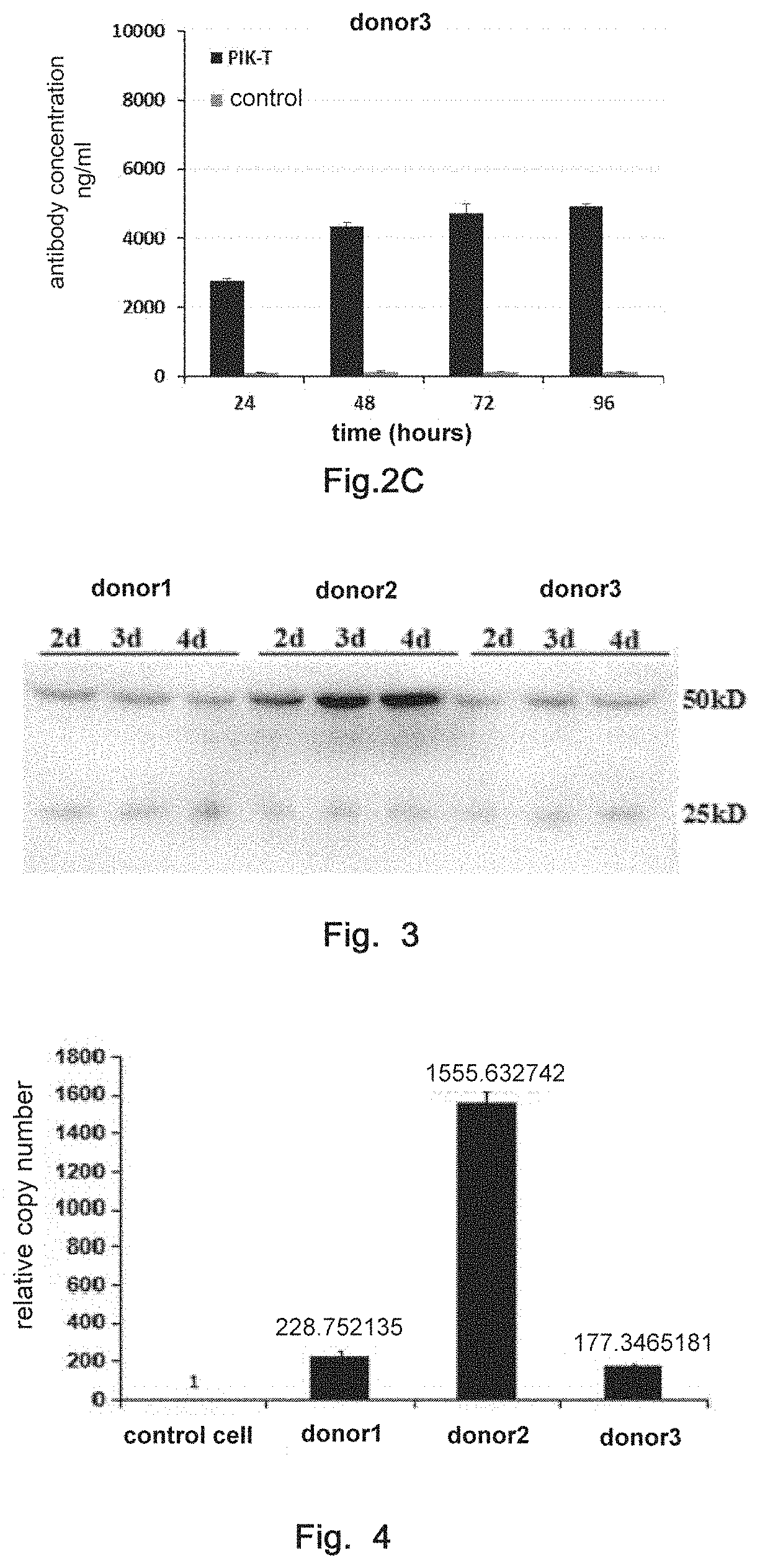
D00003
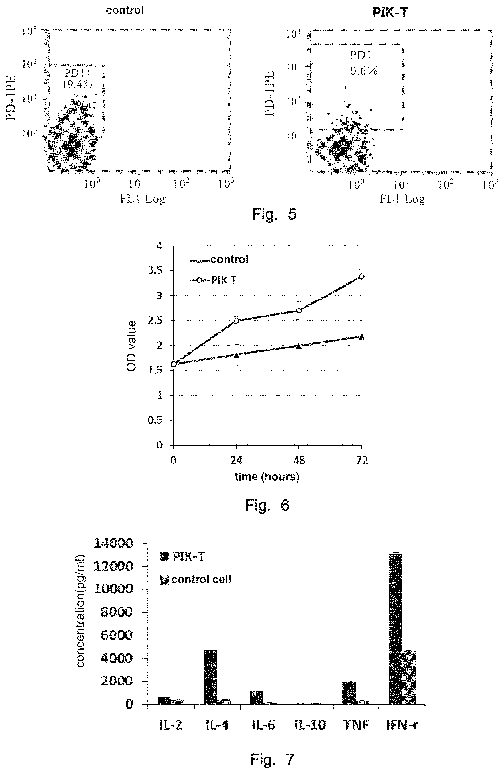
D00004

D00005
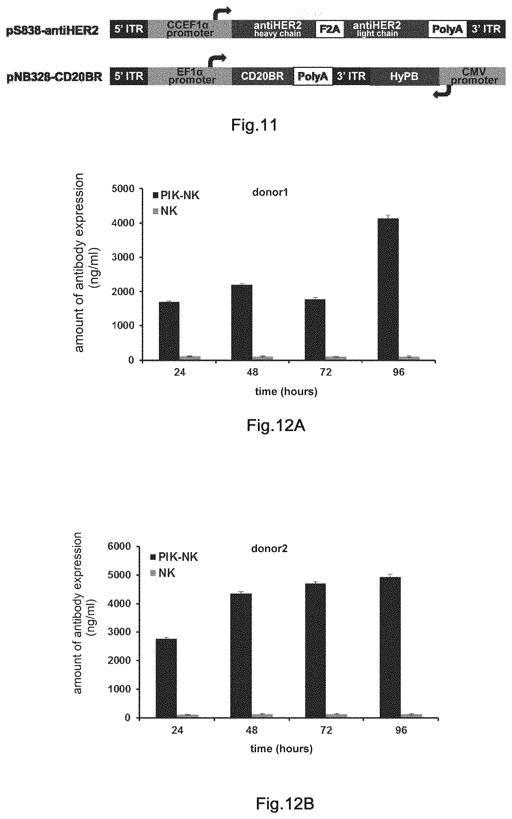
D00006
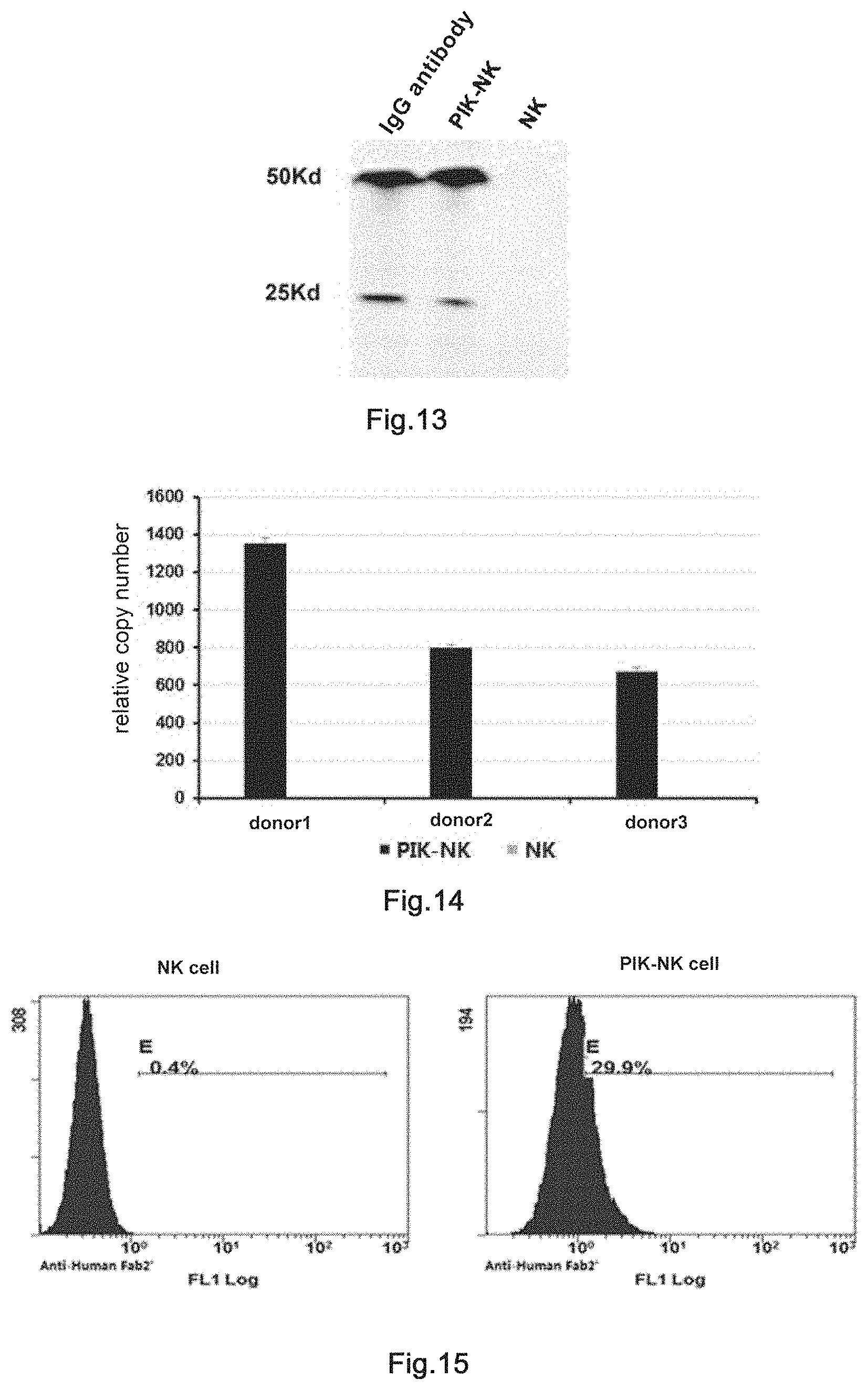
D00007
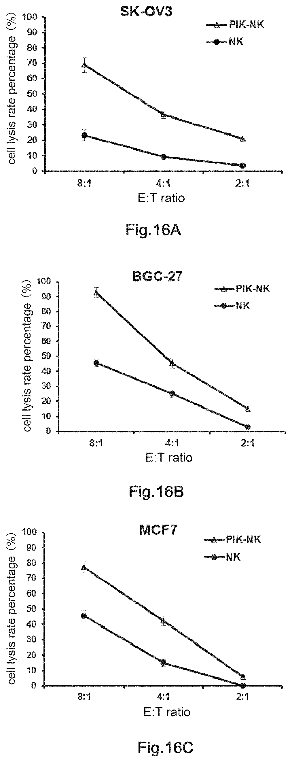
D00008
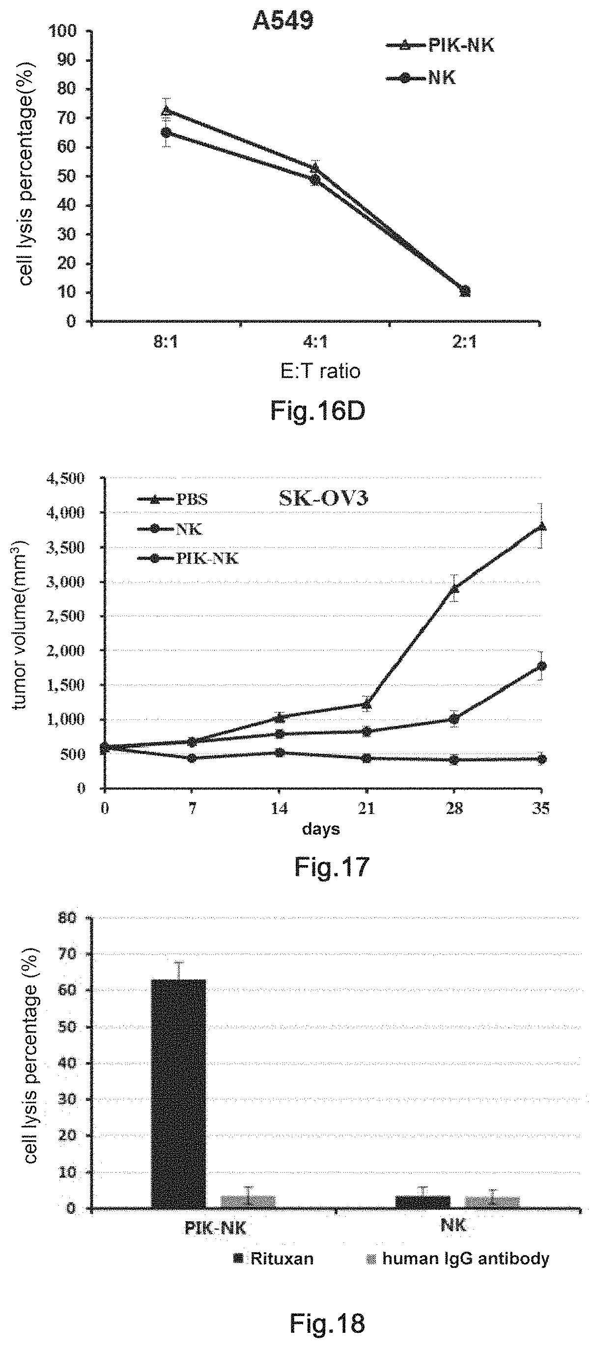
D00009
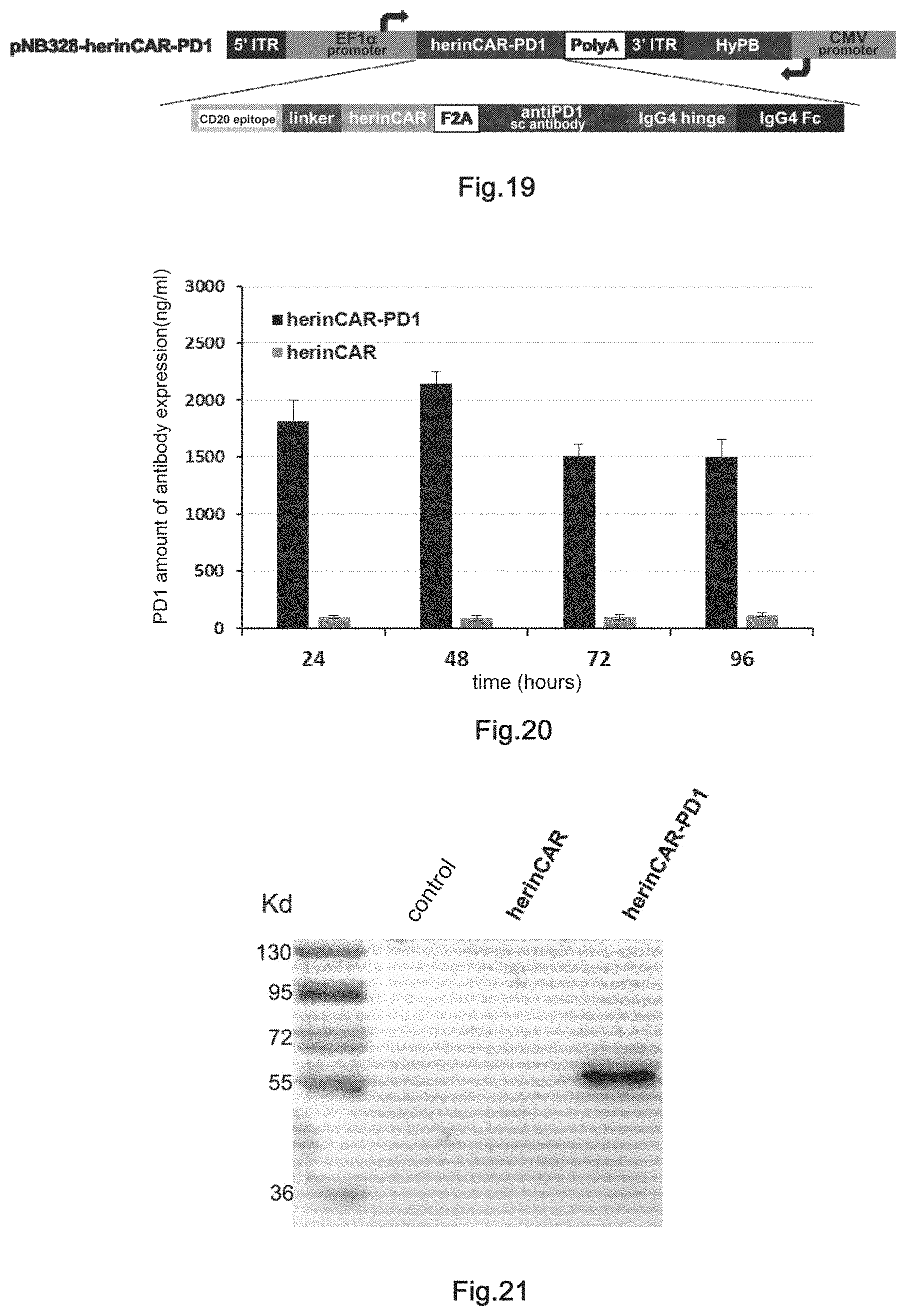
D00010
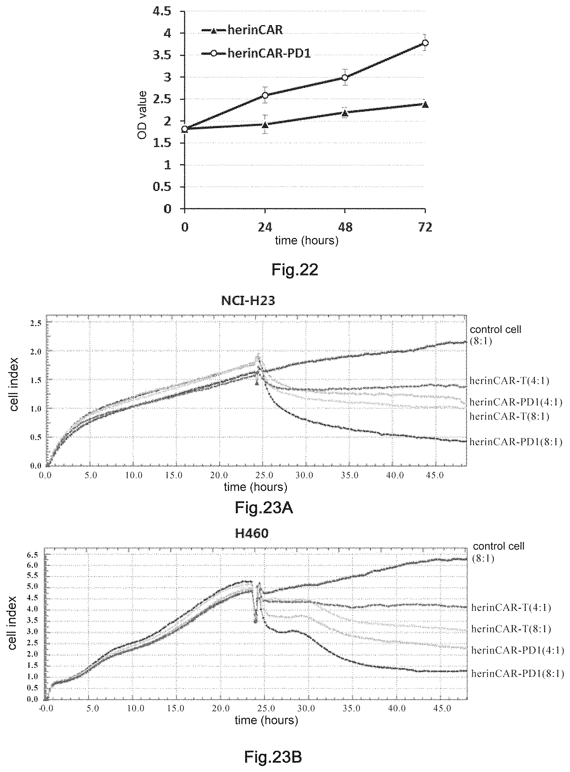
D00011
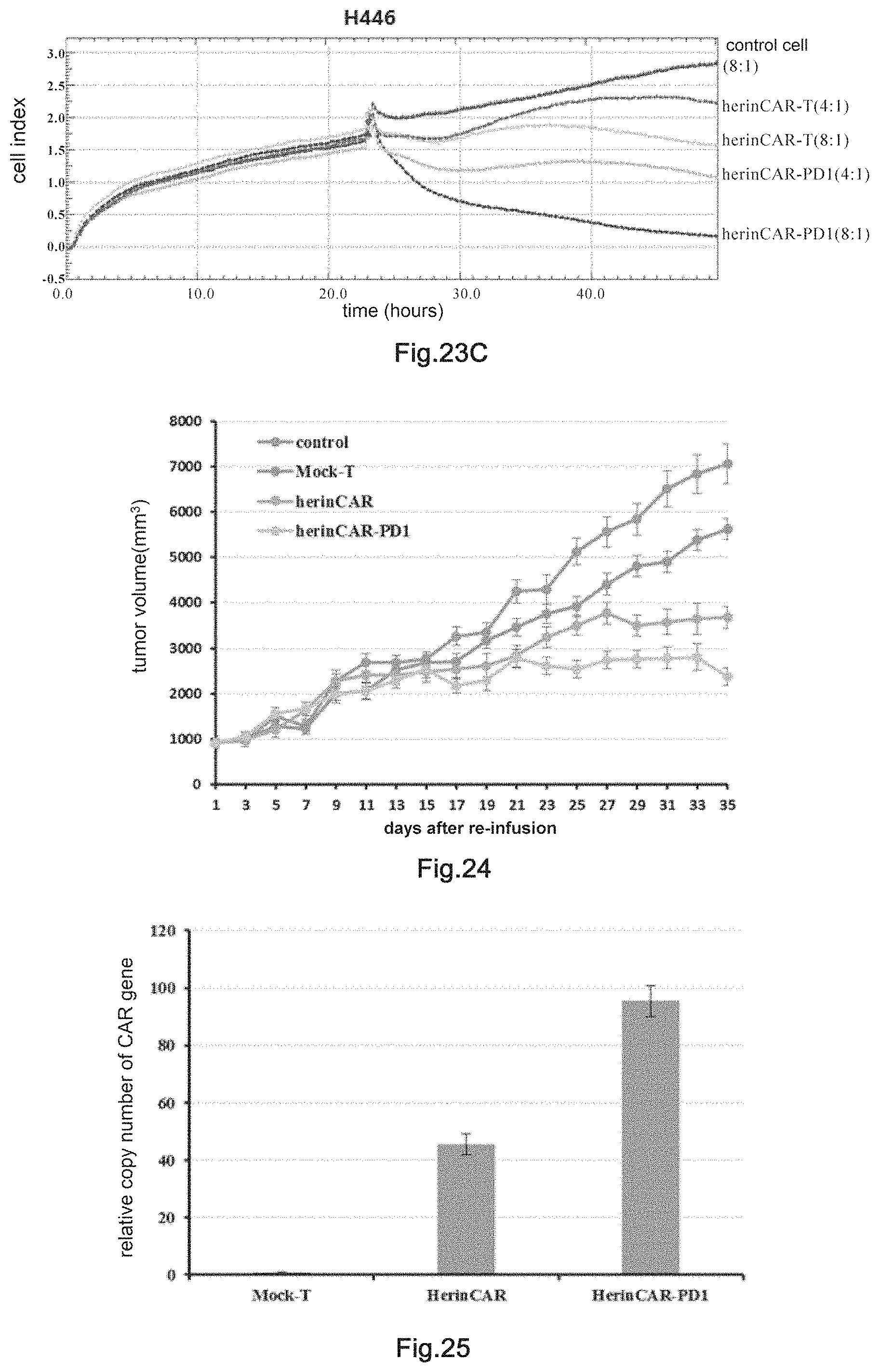
D00012

D00013

D00014
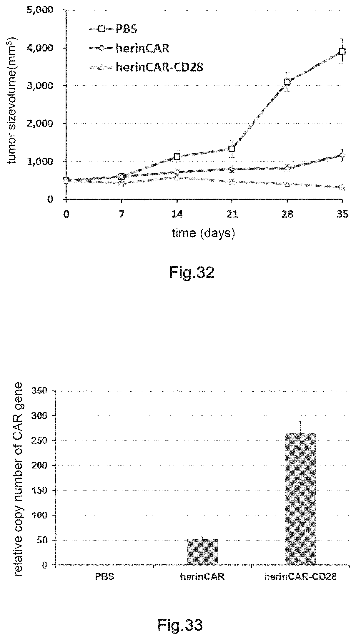
S00001
XML
uspto.report is an independent third-party trademark research tool that is not affiliated, endorsed, or sponsored by the United States Patent and Trademark Office (USPTO) or any other governmental organization. The information provided by uspto.report is based on publicly available data at the time of writing and is intended for informational purposes only.
While we strive to provide accurate and up-to-date information, we do not guarantee the accuracy, completeness, reliability, or suitability of the information displayed on this site. The use of this site is at your own risk. Any reliance you place on such information is therefore strictly at your own risk.
All official trademark data, including owner information, should be verified by visiting the official USPTO website at www.uspto.gov. This site is not intended to replace professional legal advice and should not be used as a substitute for consulting with a legal professional who is knowledgeable about trademark law.