Monoclonal Antibodies Against Bcma
Vu; Minh Diem ; et al.
U.S. patent application number 16/821895 was filed with the patent office on 2020-09-10 for monoclonal antibodies against bcma. The applicant listed for this patent is ENGMAB SARL. Invention is credited to Oliver Ast, Marina Bacac, Camille Delon, Anne Freimoser-Grundschober, Lydia Jasmin Hanisch, Christian Klein, Ekkehard Moessner, Samuel Moser, Klaus Strein, Pablo Umana, Minh Diem Vu, Tina Weinzierl.
| Application Number | 20200283545 16/821895 |
| Document ID | / |
| Family ID | 1000004845364 |
| Filed Date | 2020-09-10 |


View All Diagrams
| United States Patent Application | 20200283545 |
| Kind Code | A1 |
| Vu; Minh Diem ; et al. | September 10, 2020 |
MONOCLONAL ANTIBODIES AGAINST BCMA
Abstract
The invention relates to new antibodies against BCMA, their manufacture and use.
| Inventors: | Vu; Minh Diem; (Wollerau, CH) ; Strein; Klaus; (Weinheim, DE) ; Ast; Oliver; (Bassersdorf, CH) ; Bacac; Marina; (Zuerich, CH) ; Delon; Camille; (Oberengstingen, CH) ; Hanisch; Lydia Jasmin; (Birmensdorf, CH) ; Freimoser-Grundschober; Anne; (Zuerich, CH) ; Klein; Christian; (Bonstetten, CH) ; Moessner; Ekkehard; (Kreuzlingen, CH) ; Moser; Samuel; (Rotkreuz, CH) ; Umana; Pablo; (Wollerau, CH) ; Weinzierl; Tina; (Schlieren, CH) | ||||||||||
| Applicant: |
|
||||||||||
|---|---|---|---|---|---|---|---|---|---|---|---|
| Family ID: | 1000004845364 | ||||||||||
| Appl. No.: | 16/821895 | ||||||||||
| Filed: | March 17, 2020 |
Related U.S. Patent Documents
| Application Number | Filing Date | Patent Number | ||
|---|---|---|---|---|
| 15747385 | Jan 24, 2018 | 10683369 | ||
| PCT/EP2016/068549 | Aug 3, 2016 | |||
| 16821895 | ||||
| Current U.S. Class: | 1/1 |
| Current CPC Class: | C07K 16/2809 20130101; C07K 16/468 20130101; C07K 2317/66 20130101; C07K 2317/31 20130101; C07K 2319/70 20130101; C07K 2317/73 20130101; A61P 35/02 20180101; C07K 16/2878 20130101 |
| International Class: | C07K 16/46 20060101 C07K016/46; A61P 35/02 20060101 A61P035/02; C07K 16/28 20060101 C07K016/28 |
Foreign Application Data
| Date | Code | Application Number |
|---|---|---|
| Aug 3, 2015 | EP | 15179549.9 |
Claims
1.-23. (canceled)
24. A method of treatment of a plasma cell disorder comprising administering to a subject in need thereof an antibody specifically binding to BCMA, wherein the antibody is characterized in comprising a VH region comprising a CDR1H region having SEQ ID NO:21, a CDR2H region having SEQ ID NO:22 and a CDR3H region having SEQ ID NO:17 and a VL region comprising a CDR3L region having SEQ ID NO:20 and a CDR1L and CDR2L region combination selected from the group of: a) CDR1L region having SEQ ID NO:23 and CDR2L region having SEQ ID NO:24, b) CDR1L region having SEQ ID NO:25 and CDR2L region having SEQ ID NO:26, and c) CDR1L region having SEQ ID NO:27 and CDR2L region having SEQ ID NO:28.
25. The method of claim 24, wherein the antibody is characterized in comprising a VH region comprising a CDR1H region having SEQ ID NO:21, a CDR2H region having SEQ ID NO:22 and a CDR3H region having SEQ ID NO:17; and a VL region comprising a CDR1L region having SEQ ID NO:27, a CDR2L region having SEQ ID NO:28 and a CDR3L region having SEQ ID NO:20.
26. The method of claim 24, wherein the plasma cell disorder is selected from the group consisting of multiple myeloma, systemic lupus erythematosus, plasma cell leukemia, and AL-amyloidosis.
27. The method of claim 24, wherein the plasma cell disorder is multiple myeloma.
28. The method of claim 24, wherein the antibody is administered at a dose of 200 to 2000 mg/m.sup.2/week.
29. The method of claim 24, wherein the antibody is characterized in comprising a VH region having SEQ ID NO:10 and a VL region selected from the group consisting of VL regions SEQ ID NO:12, 13, and 14.
30. The method of claim 25, wherein the plasma cell disorder is selected from the group consisting of multiple myeloma, systemic lupus erythematosus, plasma cell leukemia, and AL-amyloidosis.
31. The method of claim 25, wherein the plasma cell disorder is multiple myeloma.
32. A method of treatment of a plasma cell disorder comprising administering to a subject in need thereof a bispecific antibody specifically binding to BCMA and human CDR3.epsilon. (CD3), wherein the antibody is characterized in comprising a CDR3H region having SEQ ID NO:17 and a CDR3L region having SEQ ID NO:20 and a CDR1H, CDR2H, CDR1L, and CDR2L region combination selected from the group of: a) CDR1H region having SEQ ID NO:21 and CDR2H region having SEQ ID NO:22, CDR1L region having SEQ ID NO:23, and CDR2L region having SEQ ID NO:24, b) CDR1H region having SEQ ID NO:21 and CDR2H region having SEQ ID NO:22, CDR1L region having SEQ ID NO:25, and CDR2L region having SEQ ID NO:26, c) CDR1H region having SEQ ID NO:21 and CDR2H region having SEQ ID NO:22, CDR1L region having SEQ ID NO:27, and CDR2L region having SEQ ID NO:28, d) CDR1H region having SEQ ID NO:29 and CDR2H region having SEQ ID NO:30, CDR1L region having SEQ ID NO:31, and CDR2L region having SEQ ID NO:32, e) CDR1H region having SEQ ID NO:34 and CDR2H region having SEQ ID NO:35, CDR1L region having SEQ ID NO:31, and CDR2L region having SEQ ID NO:32, and f) CDR1H region having SEQ ID NO:36 and CDR2H region having SEQ ID NO:37, CDR1L region having SEQ ID NO:31, and CDR2L region having SEQ ID NO:32.
33. The method of claim 32, characterized in comprising a light chain and a heavy chain of an antibody specifically binding to CD3, wherein the variable domains VL and VH or the constant domains CL and CH1 are replaced by each other.
34. The method of claim 32, characterized in that a variable domain VH of an anti-CD3 antibody portion of the bispecific antibody (CD3 VH) comprises heavy chain CDRs of SEQ ID NO: 1, 2 and 3 as respective heavy chain CDR1, CDR2 and CDR3 and the variable domain VL of the anti-CD3 antibody portion (CD3 VL) comprises light chain CDRs having SEQ ID NO: 4, 5 and 6 as respective light chain CDR1, CDR2 and CDR3.
35. The method of claim 32, wherein the bispecific antibody is characterized in comprising a VH region of an anti-BCMA antibody portion of the bispecific antibody comprising a CDR1H region having SEQ ID NO:21, a CDR2H region having SEQ ID NO:22 and a CDR3H region having SEQ ID NO:17; and a VL region of an anti-BCMA antibody portion of the bispecific antibody comprising a CDR1L region having SEQ ID NO:27, a CDR2L region having SEQ ID NO:28 and a CDR3L region having SEQ ID NO:20; and, wherein the bispecific antibody is further characterized in comprising a VH region of an anti-CD3 antibody portion of the bispecific antibody comprising a CDR1H region having SEQ ID NO:1, a CDR2H region having SEQ ID NO:2 and a CDR3H region having SEQ ID NO:3; and a VL region of an anti-CD3 antibody portion of the bispecific antibody comprising a CDR1L region having SEQ ID NO:4, a CDR2L region having SEQ ID NO:5 and a CDR3L region having SEQ ID NO:6.
36. The method of claim 32, wherein the plasma cell disorder is selected from the group consisting of multiple myeloma, systemic lupus erythematosus, plasma cell leukemia, and AL-amyloidosis.
37. The method of claim 32, wherein the plasma cell disorder is multiple myeloma.
38. The method of claim 32, wherein the bispecific antibody is administered at a dose of 0.1 to 250 mg/m.sup.2/week.
39. A method of treatment of a plasma cell disorder comprising administering to a subject in need thereof a bispecific antibody specifically binding to two targets which are the extracellular domain of human BCMA (BCMA) and human CDR3.epsilon. (CD3), characterized in comprising a VH region comprising a CDR1H region having SEQ ID NO:21, a CDR2H region having SEQ ID NO:22 and a CDR3H region having SEQ ID NO:17 and a VL region comprising a CDR3L region having SEQ ID NO:20 and a CDR1L and CDR2L region combination selected from the group of: a) CDR1L region having SEQ ID NO:23 and CDR2L region having SEQ ID NO:24, b) CDR1L region having SEQ ID NO:25 and CDR2L region having SEQ ID NO:26, and c) CDR1L region having SEQ ID NO:27 and CDR2L region having SEQ ID NO:28.
40. The method of claim 39, characterized in comprising a BCMA VH region having SEQ ID NO:10 and a VL region having SEQ ID NO:12, or a BCMA VH region having SEQ ID NO:10 and a VL region having SEQ ID NO:13, or a BCMA VH region having SEQ ID NO:10 and a VL region having SEQ ID NO:14.
41. A method of treatment of a plasma cell disorder comprising administering to a subject in need thereof a bispecific antibody specifically binding to BCMA and CD3, wherein the bispecific antibody is characterized in comprising a) a first light chain and a first heavy chain of a first anti-BCMA antibody characterized in comprising a CDR3H region having SEQ ID NO:17 and a CDR3L region having SEQ ID NO:20 and a CDR1H, CDR2H, CDR1L, and CDR2L region combination selected from the group of: i) CDR1H region having SEQ ID NO:21 and CDR2H region having SEQ ID NO:22, CDR1L region having SEQ ID NO:23, and CDR2L region having SEQ ID NO:24, ii) CDR1H region having SEQ ID NO:21 and CDR2H region having SEQ ID NO:22, CDR1L region having SEQ ID NO:25, and CDR2L region having SEQ ID NO:26, iii) CDR1H region having SEQ ID NO:21 and CDR2H region having SEQ ID NO:22, CDR1L region having SEQ ID NO:27, and CDR2L region having SEQ ID NO:28, iv) CDR1H region having SEQ ID NO:29 and CDR2H region having SEQ ID NO:30, CDR1L region having SEQ ID NO:31, and CDR2L region having SEQ ID NO:32, v) CDR1H region having SEQ ID NO:34 and CDR2H region having SEQ ID NO:35, CDR1L region having SEQ ID NO:31, and CDR2L region having SEQ ID NO:32, and vi) CDR1H region having SEQ ID NO:36 and CDR2H region having SEQ ID NO:37, CDR1L region having SEQ ID NO:31, and CDR2L region having SEQ ID NO:32; and b) a second light chain and a second heavy chain of a second antibody which specifically binds to CD3, and wherein the variable domains VL and VH in the second light chain and second heavy chain of the second antibody are replaced by each other; and c) wherein in a constant domain CL of the first light chain a) an amino acid at position 124 is substituted independently by lysine (K), arginine (R) or histidine (H) (numbering according to Kabat), and wherein in a constant domain CH1 of the first heavy chain a) an amino acid at position 147 and an amino acid at position 213 are substituted independently by glutamic acid (E), or aspartic acid (D) (numbering according to EU index of Kabat).
42. The method of claim 41, wherein the bispecific antibody is characterized in comprising in addition a Fab fragment of said first antibody (BCMA-Fab) and wherein in the constant domain CL of said BCMA-Fab the amino acid at position 124 is substituted independently by lysine (K), arginine (R) or histidine (H) (numbering according to Kabat), and wherein in the constant domain CH1 of said BCMA-Fab the amino acid at positions 147 and the amino acid at position 213 are substituted independently by glutamic acid (E), or aspartic acid (D) (numbering according to EU index of Kabat) (see e.g. FIGS. 2A, 2C).
43. The method of claim 41, wherein the plasma cell disorder is selected from the group consisting of multiple myeloma, systemic lupus erythematosus, plasma cell leukemia, and AL-amyloidosis.
44. The method of claim 41, wherein the plasma cell disorder is multiple myeloma.
45. A method of treatment of a plasma cell disorder comprising administering to a subject in need thereof a bispecific antibody specifically binding to BCMA and CD3, characterized in comprising a) a first light chain and a first heavy chain of a first antibody specifically binding to human B cell maturation antigen (BCMA), characterized in comprising a CDR3H region having SEQ ID NO:17 and a CDR3L region having SEQ ID NO:20 and a CDR1H, CDR2H, CDR1L, and CDR2L region combination selected from the group of: i) CDR1H region having SEQ ID NO:21 and CDR2H region having SEQ ID NO:22, CDR1L region having SEQ ID NO:23, and CDR2L region having SEQ ID NO:24, ii) CDR1H region having SEQ ID NO:21 and CDR2H region having SEQ ID NO:22, CDR1L region having SEQ ID NO:25, and CDR2L region having SEQ ID NO:26, iii) CDR1H region having SEQ ID NO:21 and CDR2H region having SEQ ID NO:22, CDR1L region having SEQ ID NO:27, and CDR2L region having SEQ ID NO:28, iv) CDR1H region having SEQ ID NO:29 and CDR2H region having SEQ ID NO:30, CDR1L region having SEQ ID NO:31, and CDR2L region having SEQ ID NO:32, v) CDR1H region having SEQ ID NO:34 and CDR2H region having SEQ ID NO:35, CDR1L region having SEQ ID NO:31, and CDR2L region having SEQ ID NO:32, and vi) CDR1H region having SEQ ID NO:36 and CDR2H region having SEQ ID NO:37, CDR1L region having SEQ ID NO:31, and CDR2L region having SEQ ID NO:32; and b) a second light chain and a second heavy chain of a second antibody which specifically binds to CD3, and wherein the variable domains VL and VH in the second light chain and second heavy chain of the second antibody are replaced by each other; and wherein c) in a constant domain CL of the second light chain under b) an amino acid at position 124 is substituted independently by lysine (K), arginine (R) or histidine (H) (numbering according to Kabat), and wherein in a constant domain CH1 of the second heavy chain under b) an amino acid at positions 147 and an amino acid at position 213 are substituted independently by glutamic acid (E), or aspartic acid (D) (numbering according to EU index of Kabat).
46. The method of claim 45, wherein the plasma cell disorder is selected from the group consisting of multiple myeloma, systemic lupus erythematosus, plasma cell leukemia, and AL-amyloidosis.
47. The method of claim 45, wherein the plasma cell disorder is multiple myeloma.
48. A method of treatment of a plasma cell disorder comprising administering to a subject in need thereof a bispecific antibody specifically binding to BCMA and to CD3, characterized in comprising a heavy and light chain set selected from the group consisting of polypeptides: i) SEQ ID NO:48, SEQ ID NO:49, SEQ ID NO:50, and SEQ ID NO:51 (2.times.), ii) SEQ ID NO:48, SEQ ID NO:52, SEQ ID NO:53, and SEQ ID NO:54 (2.times.), or iii) SEQ ID NO:48, SEQ ID NO:55, SEQ ID NO:56, and SEQ ID NO:57 (2.times.).
49. The method of claim 48, wherein the plasma cell disorder is selected from the group consisting of multiple myeloma, systemic lupus erythematosus, plasma cell leukemia, and AL-amyloidosis.
50. The method of claim 48, wherein the plasma cell disorder is multiple myeloma.
51. The method of claim 48, wherein the bispecific antibody is administered at a dose of 0.1 to 250 mg/m.sup.2/week.
Description
CROSS-REFERENCE TO RELATED APPLICATIONS
[0001] The present application is a continuation application of U.S. application Ser. No. 15/747,385, filed Jan. 24, 2018, which is a 35 U.S.C. .sctn. 371 national phase application of International Application No. PCT/EP2016/068549, filed Aug. 3, 2016, and published under PCT Article 21(2) in English, which designated the U.S., and claims the benefit of priority from European Application No. EP15179549.9, filed Aug. 3, 2015, and each of which prior applications are incorporated by reference herein into this application in their entirety including all tables, figures and claims.
SEQUENCE LISTING
[0002] The instant application contains a Sequence Listing which is submitted in ASCII format via EFS-Web and is hereby incorporated by reference in its entirety. Said ASCII copy, created on Mar. 15, 2020, is named 298068-00319_Sequence_Listing.txt and is 70,441 bytes in size.
FIELD OF THE INVENTION
[0003] The present invention relates to new antibodies against BCMA, their manufacture and use.
BACKGROUND OF THE INVENTION
[0004] Human B cell maturation antigen, also known as BCMA; TR17_HUMAN, TNFRSF17 (UniProt Q02223), is a member of the tumor necrosis receptor superfamily that is preferentially expressed in differentiated plasma cells (Laabi et al. 1992; Madry et al. 1998). BCMA is a non-glycosylated type III transmembrane protein, which is involved in B cell maturation, growth and survival. BCMA is a receptor for two ligands of the TNF superfamily: APRIL (a proliferation-inducing ligand), the high-affinity ligand to BCMA and the B cell activation factor BAFF, the low-affinity ligand to BCMA (THANK, BlyS, B lymphocyte stimulator, TALL-1 and zTNF4). APRIL and BAFF show structural similarity and overlapping yet distinct receptor binding specificity. The negative regulator TACI also binds to both BAFF and APRIL. The coordinate binding of APRIL and BAFF to BCMA and/or TACI activates transcription factor NF-.kappa.B and increases the expression of pro-survival Bcl-2 family members (e.g. Bcl-2, Bcl-xL, Bcl-w, Mc1-1, A1) and the downregulation of pro-apoptotic factors (e.g. Bid, Bad, Bik, Bim, etc.), thus inhibiting apoptosis and promoting survival. This combined action promotes B cell differentiation, proliferation, survival and antibody production (as reviewed in Rickert R C et al., Immunol Rev (2011) 244 (1): 115-133).
[0005] Antibodies against BCMA are described e.g. in Gras M-P. et al. Int Immunol. 7 (1995) 1093-1106, WO200124811, WO200124812, WO2010104949 and WO2012163805. Antibodies against BCMA and their use for the treatment of lymphomas and multiple myeloma are mentioned e.g. in WO2002066516 and WO2010104949. WO2013154760 and WO2015052538 relate to chimeric antigen receptors (CAR) comprising a BCMA recognition moiety and a T-cell activation moiety. Ryan, M C et al., Mol. Cancer Ther.
[0006] 6 (2007) 3009-3018 relate to anti BCMA antibodies with ligand blocking activity that could promote cytotoxicity of multiple myeloma (MM) cell lines as naked antibodies or as antibody-drug conjugates. Ryan showed that SG1, an inhibitory BCMA antibody, blocks APRIL-dependent activation of nuclear factor-KB in a dose-dependent manner in vitro. Ryan also mentioned antibody SG2 which inhibited APRIL binding to BCMA not significantly.
[0007] A wide variety of recombinant bispecific antibody formats have been developed in the recent past, e.g. by fusion of, e.g. an IgG antibody format and single chain domains (see e.g. Kontermann R E, mAbs 4:2, (2012) 1-16). Bispecific antibodies wherein the variable domains VL and VH or the constant domains CL and CH1 are replaced by each other are described in WO2009080251 and WO2009080252.
[0008] An approach to circumvent the problem of mispaired byproducts, which is known as `knobs-into-holes`, aims at forcing the pairing of two different antibody heavy chains by introducing mutations into the CH3 domains to modify the contact interface. On one chain bulky amino acids were replaced by amino acids with short side chains to create a `hole`. Conversely, amino acids with large side chains were introduced into the other CH3 domain, to create a `knob`. By coexpressing these two heavy chains (and two identical light chains, which have to be appropriate for both heavy chains), high yields of heterodimer formation (`knob-hole`) versus homodimer formation (`hole-hole` or `knob-knob`) was observed (Ridgway J B, Presta L G, Carter P. Protein Eng. 9, 617-621 (1996); and WO1996027011). The percentage of heterodimer could be further increased by remodeling the interaction surfaces of the two CH3 domains using a phage display approach and the introduction of a disulfide bridge to stabilize the heterodimers (Merchant A. M, et al, Nature Biotech 16 (1998) 677-681; Atwell S, Ridgway J B, Wells J A, Carter P., J Mol. Biol 270 (1997) 26-35). New approaches for the knobs-into-holes technology are described in e.g. in EP 1870459A1. Although this format appears very attractive, no data describing progression towards the clinic are currently available. One important constraint of this strategy is that the light chains of the two parent antibodies have to be identical to prevent mispairing and formation of inactive molecules. Thus this technique is not appropriate for easily developing recombinant, bispecific antibodies against two targets starting from two antibodies against the first and the second target, as either the heavy chains of these antibodies and/or the identical light chains have to be optimized. Xie, Z., et al, J Immunol. Methods 286 (2005) 95-101 refers to a format of bispecific antibody using scFvs in combination with knobs-into-holes technology for the FC part.
[0009] The TCR/CD3 complex of T-lymphocytes consists of either a TCR alpha (.alpha.)/beta (.beta.) or TCR gamma (.gamma.)/delta (.delta.) heterodimer coexpressed at the cell surface with the invariant subunits of CD3 labeled gamma (.gamma.), delta (.delta.), epsilon (E), zeta (0, and eta (q). Human CD3E is described under UniProt P07766 (CD3E_HUMAN).
[0010] An anti CD3E antibody described in the state of the art is SP34 (Yang S J, The Journal of Immunology (1986) 137; 1097-1100). SP34 reacts with both primate and human CD3. SP34 is available from Pharmingen. A further anti CD3 antibody described in the state of the art is UCHT-1 (see WO2000041474). A further anti CD3 antibody described in the state of the art is BC-3 (Fred Hutchinson Cancer Research Institute; used in Phase I/II trials of GvHD, Anasetti et al., Transplantation 54: 844 (1992)). SP34 differs from UCHT-1 and BC-3 in that SP-34 recognizes an epitope present on solely the .epsilon. chain of CD3 (see Salmeron et al., (1991) J. Immunol. 147: 3047) whereas UCHT-1 and BC-3 recognize an epitope contributed by both the .epsilon. and .gamma. chains. Further anti-CD3 antibodies are described in WO2008119565, WO2008119566, WO2008119567, WO2010037836, WO2010037837, WO2010037838, and U.S. Pat. No. 8,236,308 (WO2007042261). CDRs, VH and VL sequences of a further anti-CD3 antibody are shown in SEQ ID NO:7 and 8.
[0011] Bispecific antibodies against CD3 and BCMA are mentioned in WO2007117600, WO2009132058, WO2012066058, and WO2012143498. CAR compounds of antibodies against BCMA are mentioned in WO2013154760, WO2013154760, and WO2014140248.
[0012] Cell-mediated effector functions of monoclonal antibodies (like antibody dependent cellular cytotoxicity (ADCC)) can be enhanced by engineering their oligosaccharide composition at Asn297 as described in Umana, P., et al., Nature Biotechnol. 17 (1999) 176-180; and U.S. Pat. No. 6,602,684. WO1999054342, WO2004065540, WO2007031875, and WO2007039818, Hristodorov D, Fischer R, Linden L., Mol Biotechnol. 2012 Oct. 25. (Epub) also relate to the glycosylation engineering of antibodies to enhance Fc-mediated cellular cytotoxicity.
[0013] Also several amino acid residues in the hinge region and the CH2 domain influence cell-mediated effector functions of monoclonal antibodies (Eur. J. Immunol., 23, 1098 (1993), Immunology, 86, 319 (1995), Chemical Immunology, 65, 88 (1997)] Chemical Immunology, 65, 88 (1997)]. Therefore modification of such amino acids can enhance cell-mediated effector functions. Such antibody modifications to increase cell-mediated effector functions are mentioned in EP1931709, WO200042072 and comprise in the Fc part substitutions at amino acid position(s) 234, 235, 236, 239, 267, 268, 293, 295, 324, 327, 328, 330, and 332.
[0014] Further antibody modifications to increase cell-mediated effector functions are mentioned in EP1697415 and comprise amino acid replacement of EU amino acid positions 277, 289, 306, 344, or 378 with a charged amino acid, a polar amino acid, or a nonpolar amino acid.
[0015] Antibody formats and formats of bispecific and multispecific antibodies are also pepbodies (WO200244215), Novel Antigen Receptor ("NAR") (WO2003014161), diabody-diabody dimers "TandAbs" (WO2003048209), polyalkylene oxide-modified scFv (U.S. Pat. No. 7,150,872), humanized rabbit antibodies (WO2005016950), synthetic immunoglobulin domains (WO2006072620), covalent diabodies (WO2006113665), flexibodies (WO2003025018), domain antibodies, dAb (WO2004058822), vaccibody (WO2004076489), antibodies with new world primate framework (WO2007019620), antibody-drug conjugate with cleavable linkers (WO2009117531), IgG4 antibodies with hinge region removed (WO2010063785), bispecific antibodies with IgG4 like CH3 domains (WO2008119353), camelid Antibodies (U.S. Pat. No. 6,838,254), nanobodies (U.S. Pat. No. 7,655,759), CAT diabodies (U.S. Pat. No. 5,837,242), bispecific (scFv).sub.2 directed against target antigen and CD3 (U.S. Pat. No. 7,235,641),), sIgA plAntibodies (U.S. Pat. No. 6,303,341), minibodies (U.S. Pat. No. 5,837,821), IgNAR (US2009148438), antibodies with modified hinge and Fc regions (US2008227958, US20080181890), trifunctional antibodies (U.S. Pat. No. 5,273,743), triomabs (U.S. Pat. No. 6,551,592), troybodies (U.S. Pat. No. 6,294,654).
[0016] WO2014122143 disclose anti-human BCMA antibodies characterized in that the binding of said antibody is not reduced by 100 ng/ml APRIL for more than 20% measured in an ELISA assay as OD at 405 nm compared to the binding of said antibody to human BCMA without APRIL, said antibody does not alter APRIL-dependent NF-.kappa.B activation for more than 20%, as compared to APRIL alone, and said antibody does not alter NF-.kappa.B activation without APRIL for more than 20%, as compared without said antibody. WO2014122144 discloses bispecific antibodies specifically binding to the two targets human CD3E and human BCMA, comprising anti-human BCMA antibodies of WO2014122143. An anti-human BCMA antibody with unique properties, especially in regard to its therapeutic use as a bispecific T cell binder, is antibody 83A10, characterized by comprising as CDR regions CDR1H of SEQ ID NO:15, CDR2H of SEQ ID NO16, CDR3H of SEQ ID NO:17, CDR1L of SEQ ID NO:18, CDR3L of SEQ ID NO:19, and CDR3L of SEQ ID NO:20, disclosed also in WO2014122143 and WO2014122144.
SUMMARY OF THE INVENTION
[0017] The invention comprises monoclonal antibodies specifically binding to human B cell maturation antigen (BCMA). The antibodies according to the invention comprise as CDR3H and CDR3L regions the same CDR regions as antibody 83A10.
[0018] The antibodies according to the invention comprise in an embodiment as CDR3H and CDR3L regions the same CDR regions as antibody 83A10, but show especially potent and efficient advantages in comparison to antibody 83A10 for killing of MM cells in patient bone marrow aspirates.
[0019] The invention comprises a monoclonal antibody specifically binding to BCMA, characterized in comprising a CDR3H region of SEQ ID NO:17 and a CDR3L region of SEQ ID NO:20 and a CDR1H, CDR2H, CDR1L, and CDR2L region combination selected from the group of
a) CDR1H region of SEQ ID NO:21 and CDR2H region of SEQ ID NO:22, CDR1L region of SEQ ID NO:23, and CDR2L region of SEQ ID NO:24, b) CDR1H region of SEQ ID NO:21 and CDR2H region of SEQ ID NO:22, CDR1L region of SEQ ID NO:25, and CDR2L region of SEQ ID NO:26, c) CDR1H region of SEQ ID NO:21 and CDR2H region of SEQ ID NO:22, CDR1L region of SEQ ID NO:27, and CDR2L region of SEQ ID NO:28, d) CDR1H region of SEQ ID NO:29 and CDR2H region of SEQ ID NO:30, CDR1L region of SEQ ID NO:31, and CDR2L region of SEQ ID NO:32, e) CDR1H region of SEQ ID NO:34 and CDR2H region of SEQ ID NO:35, CDR1L region of SEQ ID NO:31, and CDR2L region of SEQ ID NO:32, and f) CDR1H region of SEQ ID NO:36 and CDR2H region of SEQ ID NO:37, CDR1L region of SEQ ID NO:31, and CDR2L region of SEQ ID NO:32,
[0020] The invention comprises a monoclonal antibody specifically binding to BCMA, characterized in comprising a VH region comprising a CDR1H region of SEQ ID NO:21, a CDR2H region of SEQ ID NO:22 and a CDR3H region of SEQ ID NO:17 and a VL region comprising a CDR3L region of SEQ ID NO:20 and a CDR1L and CDR2L region combination selected from the group of
a) CDR1L region of SEQ ID NO:23 and CDR2L region of SEQ ID NO:24, b) CDR1L region of SEQ ID NO:25 and CDR2L region of SEQ ID NO:26, or c) CDR1L region of SEQ ID NO:27 and CDR2L region of SEQ ID NO:28.
[0021] The invention provides an antibody according to the invention, characterized in comprising a VL region selected from the group consisting of VL regions of SEQ ID NO:12, 13, and 14 wherein amino acid 49 is selected from the group of amino acids tyrosine(Y), glutamic acid (E), serine (S), and histidine (H). In one embodiment amino acid 49 is E within SEQ ID NO:12, S within SEQ ID NO:13 or H within SEQ ID NO:14.
[0022] The invention provides an antibody according to the invention, characterized in comprising a VL region selected from the group consisting of VL regions of SEQ ID NO:12, 13, and 14 wherein amino acid 74 is threonine (T) or alanine (A). In one embodiment amino acid 74 is A within SEQ ID NO:14.
[0023] The antibodies according to the invention comprise in an embodiment as CDR3H, CDR1L, CDR2L, and CDR3L regions the same CDR regions as antibody 83A10. The invention comprises a monoclonal antibody specifically binding to BCMA, characterized in comprising a VH region comprising a CDR3H region of SEQ ID NO:17 and a VL region comprising a CDR1L region of SEQ ID NO:31, a CDR2L region of SEQ ID NO:32 and a CDR3L region of SEQ ID NO:20 and a CDR1L and CDR2L region combination selected from the group of
a) CDR1H region of SEQ ID NO:29 and CDR2H region of SEQ ID NO:30, b) CDR1H region of SEQ ID NO:34 and CDR2H region of SEQ ID NO:35, or c) CDR1H region of SEQ ID NO:36 and CDR2H region of SEQ ID NO:37.
[0024] The invention provides in one embodiment an antibody according to the invention, characterized in comprising a VL region of SEQ ID NO:12 and a VH region selected from the group comprising the VH regions of SEQ ID NO:38, 39, and 40. The invention provides an antibody according to the invention, characterized in comprising a VL region SEQ ID NO:12, wherein amino acid 49 is selected from the group of amino acids tyrosine(Y), glutamic acid (E), serine (S), and histidine (H). In one embodiment amino acid 49 is E.
[0025] The invention provides in one embodiment an antibody according to the invention, characterized in comprising as VH region a VH region of SEQ ID NO:10. The invention provides in one embodiment an antibody according to the invention, characterized in comprising as VL region a VL region selected from the group consisting of VL regions of SEQ ID NO:12, 13, and 14. The invention provides in one embodiment an antibody according to the invention, characterized in that comprising as VH region a VH region of SEQ ID NO:10 and as VL region a VL region of SEQ ID NO:12. The invention provides in one embodiment an antibody according to the invention, characterized in that comprising as VH region a VH region of SEQ ID NO:10 and as VL region a VL region of SEQ ID NO:13. The invention provides in one embodiment an antibody according to the invention, characterized in that comprising as VH region a VH region of SEQ ID NO:10 and as VL region a VL region of SEQ ID NO:14.
[0026] The invention provides in one embodiment an antibody according to the invention, characterized in comprising as VH region a VH region selected from the group consisting of SEQ ID NO:38, 39, and 40. The invention provides in one embodiment an antibody according to the invention, characterized in that comprising as VH region a VH region of SEQ ID NO:38 and as VL region a VL region of SEQ ID NO:12. The invention provides in one embodiment an antibody according to the invention, characterized in that comprising as VH region a VH region of SEQ ID NO:39 and as VL region a VL region of SEQ ID NO:12.
[0027] The invention provides in one embodiment an antibody according to the invention, characterized in that comprising as VH region a VH region of SEQ ID NO:40 and as VL region a VL region of SEQ ID NO:12.
[0028] In one embodiment the antibody according to the invention is further characterized in that it binds also specifically to cynomolgus BCMA. In one embodiment an antibody of the invention shows regarding binding to BCMA a cyno/human affinity gap between 1.5 and 5 or 1.5 and 10 or 1.5 and 16 (table 5).
[0029] The bispecific antibody according to the invention is therefore in one embodiment characterized in that it binds also specifically to cynomolgus CD3. In one embodiment the bispecific anti-BCMA/anti-CD3 antibody of the invention shows a cyno/human gap of Mab CD3 between 1.25 and 5 or between 0.8 and 1.0.
[0030] In a further embodiment of the invention the antibody according to the invention is an antibody with an Fc part or without an Fc part including a multispecific antibody, bispecific antibody, a single chain variable fragment (scFv) such as a bispecific T cells engager, diabody, or tandem scFv, an antibody mimetic such as DARPin, a naked monospecific antibody, or an antibody drug conjugate. In one embodiment a multispecific antibody, bispecific antibody, a bispecific T cells engager, diabody, or tandem scFv is specifically binding to BCMA and CD3.
[0031] Based on an antibody according to the invention it is possible to generate antibody-drug conjugates against BCMA and multispecific or bispecific antibodies against BCMA and one or more further targets in different formats with or without an Fc portion known in the state of the art (see e. g. above in "background of the invention"), single chain variable fragments (scFv) such as bispecific T cells engagers, diabodies, tandem scFvs, and antibody mimetics such as DARPins, all of them are also embodiments of the invention.
[0032] Bispecific antibody formats are well known in the state of the art and e.g. also described in Kontermann R E, mAbs 4:2 1-16 (2012); Holliger P., Hudson P J, Nature Biotech. 23 (2005) 1126-1136 and Chan A C, Carter P J Nature Reviews Immunology 10, 301-316 (2010) and Cuesta A M et al., Trends Biotech 28 (2011) 355-362.
[0033] A further embodiment of the invention is a bispecific antibody against the two targets human CD3.epsilon. (further named also as "CD3") and the extracellular domain of human BCMA (further named also as "BCMA"), characterized in comprising as BCMA binding portion an anti-BCMA antibody according to the invention.
[0034] The invention relates in one embodiment to a bispecific antibody against BCMA and CD3, characterized in comprising within the BCMA binding portion a CDR3H region of SEQ ID NO:17 and a CDR3L region of SEQ ID NO:20 and a CDR1H, CDR2H, CDR1L, and CDR2L region combination selected from the group of
a) CDR1H region of SEQ ID NO:21 and CDR2H region of SEQ ID NO:22, CDR1L region of SEQ ID NO:23, and CDR2L region of SEQ ID NO:24, b) CDR1H region of SEQ ID NO:21 and CDR2H region of SEQ ID NO:22, CDR1L region of SEQ ID NO:25, and CDR2L region of SEQ ID NO:26, c) CDR1H region of SEQ ID NO:21 and CDR2H region of SEQ ID NO:22, CDR1L region of SEQ ID NO:27, and CDR2L region of SEQ ID NO:28, d) CDR1H region of SEQ ID NO:29 and CDR2H region of SEQ ID NO:30, CDR1L region of SEQ ID NO:31, and CDR2L region of SEQ ID NO:32, e) CDR1H region of SEQ ID NO:34 and CDR2H region of SEQ ID NO:35, CDR1L region of SEQ ID NO:31, and CDR2L region of SEQ ID NO:32, and f) CDR1H region of SEQ ID NO:36 and CDR2H region of SEQ ID NO:37, CDR1L region of SEQ ID NO:31, and CDR2L region of SEQ ID NO:32,
[0035] The invention relates in one embodiment to a bispecific antibody against BCMA and CD3, characterized in comprising a VH region of an antibody according to the invention (further named "BCMA VH") comprising a CDR1H region of SEQ ID NO:21, a CDR2H region of SEQ ID NO:22 and a CDR3H region of SEQ ID NO:17 and a VL region (further named "BCMA VL") comprising a CDR3L region of SEQ ID NO:20 and a CDR1L and CDR2L region combination selected from the group of
a) CDR1L region of SEQ ID NO:23 and CDR2L region of SEQ ID NO:24, b) CDR1L region of SEQ ID NO:25 and CDR2L region of SEQ ID NO:26, or c) CDR1L region of SEQ ID NO:27 and CDR2L region of SEQ ID NO:28.
[0036] The invention provides in one embodiment a bispecific antibody according to the invention, characterized in comprising as BCMA VH a VH region of SEQ ID NO:10.
[0037] The invention relates in one embodiment to a bispecific antibody against BCMA and CD3, characterized in that the BCMA VL is selected from the group consisting of VL regions of SEQ ID NO:12, 13, and 14. The invention provides in one embodiment an antibody according to the invention, characterized in comprising as BCMA VH region a VH region of SEQ ID NO:10 and as VL region a VL region of SEQ ID NO:12. The invention provides in one embodiment an antibody according to the invention, characterized in comprising as BCMA VH a VH region of SEQ ID NO:10 and as VL region a VL region of SEQ ID NO:13. The invention provides in one embodiment an antibody according to the invention, characterized in comprising as BCMA VH a VH region of SEQ ID NO:10 and as VL region a VL region of SEQ ID NO:14.
[0038] The invention provides a bispecific antibody according to the invention, characterized in comprising a VL region selected from the group consisting of VL regions of SEQ ID NO:12, 13, and 14 wherein amino acid 49 is selected from the group of amino acids tyrosine(Y), glutamic acid (E), serine (S), and histidine (H). In one embodiment amino acid 49 is E (SEQ ID NO:12), S (SEQ ID NO:13) or H (SEQ ID NO:14). The invention provides a bispecific antibody according to the invention, characterized in comprising a VL region selected from the group consisting of VL regions of SEQ ID NO:12, 13, and 14 wherein amino acid 74 is threonine (T) or alanine (A). In one embodiment amino acid 74 is A within SEQ ID NO:14.
[0039] The invention relates to a bispecific antibody against BCMA and CD3, characterized in comprising a BCMA VH comprising a CDR3H region of SEQ ID NO:17 and a BCMA VL comprising a CDR1L region of SEQ ID NO:31, a CDR2L region of SEQ ID NO:32 and a CDR3L region of SEQ ID NO:20 and a CDR1L and CDR2L region combination selected from the group of
a) CDR1H region of SEQ ID NO:29 and CDR2H region of SEQ ID NO:30, b) CDR1H region of SEQ ID NO:34 and CDR2H region of SEQ ID NO:35, or c) CDR1H region of SEQ ID NO:36 and CDR2H region of SEQ ID NO:37.
[0040] The bispecific antibody against BCMA and CD3 is characterized in one embodiment in comprising an anti BCMA antibody according to the invention and an anti CD3 antibody, wherein
a) the light chain and heavy chain of an antibody specifically binding to one of said targets CD3 and BCMA; and b) the light chain and heavy chain of an antibody specifically binding to the other one of said targets, wherein the variable domains VL and VH or the constant domains CL and CH1 are replaced by each other.
[0041] In one embodiment a VH domain of said anti-CD3 antibody portion is linked to a CH1 or CL domain of said anti-BCMA antibody portion. In one embodiment a VL domain of said anti-CD3 antibody portion is linked to a CH1 or CL domain of said anti-BCMA antibody portion.
[0042] In one embodiment the bispecific antibody comprises not more than one Fab fragment of an anti-CD3 antibody portion, not more than two Fab fragments of an anti-BCMA antibody portion and not more than one Fc part, in one embodiment a human Fc part. In one embodiment not more than one Fab fragment of the anti-CD3 antibody portion and not more than one Fab fragment of the anti-BCMA antibody portion are linked to the Fc part and linking is performed via C-terminal binding of the Fab fragment(s) to the hinge region. In one embodiment the second Fab fragment of the anti-BCMA antibody portion is linked via its C-terminus either to the N-terminus of the Fab fragment of the anti-CD3 antibody portion or to the hinge region of the Fc part and therefore between the Fc part and the anti-CD3 antibody portion. The preferred bispecific antibodies are shown in FIGS. 1 to 3.
[0043] Especially preferred are the bispecific antibodies comprising only the Fab fragments and the Fc part as specified, with or without "aa substitution":
Fab BCMA-Fc-Fab CD3 (bispecific format FIG. 1A or 1B), Fab BCMA-Fc-Fab CD3-Fab BCMA (bispecific format FIG. 2A or 2B), Fab BCMA-Fc-Fab BCMA-Fab CD3 (bispecific format FIG. 2C or 2D), Fc-Fab CD3-Fab BCMA (bispecific format FIG. 3A or 3B), Fc-Fab BCMA-Fab CD3 (bispecific format FIG. 3C or 3D).
[0044] As shown in FIGS. 1 to 3 "Fab BCMA-Fc, "Fab BCMA-Fc-Fab CD3" and "Fab BCMA-Fc-Fab CD3" means that the Fab fragment(s) is (are) bound via its (their) C-terminus to the N-terminus of the Fc fragment. "Fab CD3-Fab BCMA" means that the Fab CD3fragment is bound with its N-terminus to the C-terminus of the Fab BCMA fragment. "Fab BCMA-Fab CD3" means that the Fab BCMA fragment is bound with its N-terminus to the C-terminus of the Fab CD3 fragment.
[0045] In one embodiment the bispecific antibody comprises a second Fab fragment of said anti-BCMA antibody linked with its C-terminus to the N-terminus of the CD3 antibody portion of said bispecific antibody. In one embodiment a VL domain of said first anti-CD3 antibody portion is linked to a CH1 or CL domain of said second anti-BCMA antibody.
[0046] In one embodiment the bispecific antibody comprises a second Fab fragment of said anti-BCMA antibody linked with its C-terminus to the Fc part (like the first Fab fragment of said anti-BCMA antibody) and linked with its N-terminus to the C-terminus of the CD3 antibody portion. In one embodiment a CH1 domain of said anti-CD3 antibody portion is linked to the VH domain of said second anti-BCMA antibody portion.
[0047] In one embodiment the bispecific antibody comprises an Fc part linked with its N-terminus to the C-terminus of said CD3 antibody Fab fragment. In one embodiment the bispecific antibody comprises an Fc part linked with its first N-terminus to the C-terminus of said CD3 antibody Fab fragment and a second Fab fragment of said anti-BCMA antibody linked with its C-terminus to the second N-terminus of the Fc part. In one embodiment the CL domain of the CD3 antibody Fab fragment is linked to the hinge region of the Fc part. In one embodiment the CH1 domain of the BCMA antibody Fab fragment is linked to the hinge region of the Fc part.
[0048] The Fab fragments are chemically linked together by the use of an appropriate linker according to the state of the art. In one embodiment a (Gly4-Ser1)3 linker is used (Desplancq D K et al., Protein Eng. 1994 August; 7(8):1027-33 and Mack M. et al., PNAS Jul. 18, 1995 vol. 92 no. 15 7021-7025). "Chemically linked" (or "linked") means according to the the invention that the fragments are linked by covalent binding. As the linker is a peptidic linker, such covalent binding is usually performed by biochemical recombinant means, using a nucleic acid encoding the VL and/or VH domains of the respective Fab fragments, the linker and if appropriate the Fc part chain.
[0049] The invention relates in one embodiment to a bispecific antibody against BCMA and CD3 according to the invention, characterized in that the variable domain VH of the anti-CD3 antibody portion (further named as "CD3 VH") comprises the heavy chain CDRs of SEQ ID NO: 1, 2 and 3 as respectively heavy chain CDR1H, CDR2H and CDR3H and the variable domain VL of the anti-CD3 antibody portion (further named as "CD3 VL") comprises the light chain CDRs of SEQ ID NO: 4, 5 and 6 as respectively light chain CDR1L, CDR2L and CDR3L.
[0050] In one embodiment such a bispecific antibody according to the invention is characterized in that the variable domains of the anti CD3E antibody portion are of SEQ ID NO:7 and 8.
[0051] The invention relates to a bispecific antibody according to the invention, characterized in that the anti-CD3 antibody portion is linked at its N-terminus to the C-terminus of a of the anti-BCMA antibody portion and the variable domains VL and VH of the anti-CD3 antibody portion or the constant domains CL and CH1 are replaced by each other.
[0052] In one embodiment the VH domain of said anti-CD3 antibody portion is linked to a CH1 or CL domain of said anti-BCMA antibody portion. In one embodiment a VL domain of said anti-CD3 antibody portion is linked to a CH1 or CL domain of said anti-BCMA antibody portion.
[0053] An antibody portion according to the invention is in one embodiment a Fab fragment of the respective antibody.
[0054] In a further embodiment of the invention the bispecific antibody wherein the variable domains VL and VH in the light chain and the respective heavy chain of the anti-CD3 antibody portion or the anti-BCMA antibody portion are replaced by each other, is characterized in comprising a constant domain CL of the anti-CD3 antibody portion or the anti-BCMA antibody portion wherein the amino acid at position 124 is substituted independently by lysine (K), arginine (R) or histidine (H) (numbering according to Kabat), and in the respective constant domain CH1 the amino acid at position 147 and the amino acid at position 213 is substituted independently by glutamic acid (E), or aspartic acid (D). In one embodiment the antibody is monovalent for CD3 binding. In one embodiment in addition to the amino acid replacement at position 124 in the constant domain CL the amino acid at position 123 is substituted independently by lysine (K), arginine (R) or histidine (H) (further called as "charge variant exchange"). In one embodiment the antibody is monovalent for CD3 binding and amino acid 124 is K, amino acid 147 is E, amino acid 213 is E, and amino acid 123 is R. In one embodiment the bispecific antibody comprises in addition the same anti-BCMA binding portion once more (in one embodiment a Fab fragment). That means also, that if the first anti-BCMA binding portion comprises the charge variant exchange, then the second anti-BCMA binding portion comprise the same charge variant exchange.
[0055] The invention relates to a bispecific antibody according to the invention, characterized in comprising a) the first light chain and the first heavy chain of a first antibody which specifically binds to BCMA; and
b) the second light chain and the second heavy chain of a second antibody which specifically binds to CD3, and wherein the variable domains VL and VH in the second light chain and second heavy chain of the second antibody are replaced by each other; and c) wherein in the constant domain CL of the first light chain under a) the amino acid at position 124 is substituted independently by lysine (K), arginine (R) or histidine (H) (numbering according to Kabat), and wherein in the constant domain CH1 of the first heavy chain under a) the amino acid at position 147 and the amino acid at position 213 is substituted independently by glutamic acid (E), or aspartic acid (D) (numbering according to EU index of Kabat) (see e.g. FIGS. 1A, 2A, 2C, 3A, 3C).
[0056] In one embodiment said bispecific antibody described in the last preceding paragraph is further characterized in that said bispecific antibody comprises in addition a Fab fragment of said first antibody (further named also as "BCMA-Fab") and in the constant domain CL said BCMA-Fab the amino acid at position 124 is substituted independently by lysine (K), arginine (R) or histidine (H) (numbering according to Kabat), and wherein in the constant domain CH1 of said BCMA-Fab the amino acid at positions 147 and the amino acid at position 213 is substituted independently by glutamic acid (E), or aspartic acid (D) (numbering according to EU index of Kabat) (see e.g. FIGS. 2A, 2C).
[0057] The invention further relates to a bispecific antibody according to the invention, characterized in comprising
a) the first light chain and the first heavy chain of a first antibody which specifically binds to BCMA; and b) the second light chain and the second heavy chain of a second antibody which specifically binds to CD3, and wherein the variable domains VL and VH in the second light chain and second heavy chain of the second antibody are replaced by each other; and wherein c) in the constant domain CL of the second light chain under b) the amino acid at position 124 is substituted independently by lysine (K), arginine (R) or histidine (H) (numbering according to Kabat), and wherein in the constant domain CH1 of the second heavy chain under b) the amino acid at positions 147 and the amino acid at position 213 is substituted independently by glutamic acid (E), or aspartic acid (D) (numbering according to EU index of Kabat).
[0058] In one embodiment in addition to the amino acid replacement at position 124 in the constant domain CL of the first or second light chain the amino acid at position 123 is substituted independently by lysine (K), arginine (R) or histidine (H).
[0059] In one embodiment in the constant domain CL the amino acid at position 124 is substituted by lysine (K), in the constant domain CH1 the amino acid at position 147 and the amino acid at position 213 are substituted by glutamic acid (E). In one embodiment in addition in the constant domain CL in the amino acid at position 123 is substituted by arginine (R).
[0060] In a preferred embodiment of the invention the bispecific antibody according to the invention consists of one Fab fragment of an antibody specifically binding to CD3 (further named also as "CD3-Fab"), and one Fab fragment of an anti-BCMA antibody according to the invention (further named also as "BCMA-Fab(s)") and a Fc part, wherein the CD3-Fab and the BCMA-Fab are linked via their C-termini to the hinge region of said Fc part. Either the CD3-Fab or the BCMA-Fab comprises aa substitution and the CD3-Fab comprises crossover (FIGS. 1A and 1B).
[0061] In a preferred embodiment of the invention the bispecific antibody according to the invention consists of one CD3-Fab, and one BCMA-Fab and a Fc part, wherein the CD3-Fab and the BCMA-Fab are linked via their C-termini to the hinge region of said Fc part and a second BCMA-Fab, which is linked with its C-terminus to the N-terminus of the CD3-Fab. The CD3-Fab comprises crossover and either the CD3-Fab or both BCMA-Fabs comprise aa substitution (FIGS. 2A and 2B). Especially preferred is a bispecific antibody comprising BCMA-Fab-Fc-CD3-Fab-BCMA-Fab, wherein both BCMA-Fabs comprise aa substitution and the CD3-Fab comprises VL/VH crossover (FIG. 2A). Especially preferred is a bispecific antibody consisting of BCMA-Fab-Fc-CD3-Fab-BCMA-Fab, wherein both BCMA-Fabs comprise aa substitution Q124K, E123R, K147E and K213E and the CD3-Fab comprises VL/VH crossover. Especially preferred is that both BCMA-Fabs comprise as CDRs the CDRs of antibody 21, 22, or 42, or as VH/VL the VH/VL of antibody 21, 22, or 42.
[0062] In a preferred embodiment of the invention the bispecific antibody according to the invention consists of two BCMA-Fabs and an Fc part, wherein one BCMA-Fab and the CD3 Fab are linked via their C-termini to the hinge region of said Fc part and the second BCMA-Fab is linked with its C-terminus to the N-terminus of the CD3-Fab. The CD3-Fab comprises crossover and either the CD3-Fab or both BCMA-Fabs comprise aa substitution (FIGS. 2A and 2B).
[0063] In a preferred embodiment of the invention the bispecific antibody according to the invention consists of two BCMA-Fabs and an Fc part, wherein the BCMA-Fabs are linked via their C-termini to the hinge region of said Fc part and a CD3-Fab, which is linked with its C-terminus to the N-terminus of one BCMA-Fab. The CD3-Fab comprises crossover and either the CD3-Fab or both BCMA-Fabs comprise aa substitution (FIGS. 2C and 2D).
[0064] In a preferred embodiment of the invention the antibody according to the invention consists of one CD3-Fab, which is linked via its C-terminus to the hinge region of said Fc part and a BCMA-Fab, which is linked with its C-terminus to the N-terminus of the CD3-Fab. The CD3-Fab comprises crossover and either the CD3-Fab or the BCMA-Fab comprise aa substitution (FIGS. 1A and 1B).
[0065] In a preferred embodiment of the invention the antibody according to the invention consists of one CD3-Fab, which is linked via its C-terminus to the hinge region of said Fc part and a BCMA-Fab, which is linked with its C-terminus to the N-terminus of the CD3-Fab. The CD3-Fab comprises crossover and either the CD3-Fab or the BCMA-Fab comprise aa substitution (FIGS. 3A and 3B).
[0066] In a preferred embodiment of the invention the antibody according to the invention consists of one BCMA-Fab, which is linked via its C-terminus to the hinge region of said Fc part and a CD3-Fab, which is linked with its C-terminus to the N-terminus of the BCMA-Fab. The CD3-Fab comprises crossover and either the CD3-Fab or the BCMA-Fab comprise aa substitution (FIGS. 3C and 3D).
[0067] The Fab fragments are chemically linked together by the use of an appropriate linker according to the state of the art. In one embodiment a (Gly4-Ser1)3 linker is used (Desplancq D K et al., Protein Eng. 1994 August; 7(8):1027-33 and Mack M. et al., PNAS Jul. 18, 1995 vol. 92 no. 15 7021-7025). Linkage between two Fab fragments is performed between the heavy chains. Therefore the C-terminus of CH1 of a first Fab fragment is linked to the N-terminus of VH of the second Fab fragment (no crossover) or to VL (crossover). Linkage between a Fab fragment and the Fc part is performed according to the invention as linkage between CH1 and CH2.
[0068] The first and a second Fab fragment of an antibody specifically binding to BCMA are in one embodiment derived from the same antibody and in one embodiment identical in the CDR sequences, variable domain sequences VH and VL and/or the constant domain sequences CH1 and CL. In one embodiment the amino acid sequences of the first and a second Fab fragment of an antibody specifically binding to BCMA are identical. In one embodiment the BCMA antibody is an antibody comprising the CDR sequences of antibody 21, 22, or 42, an antibody comprising the VH and VL sequences of antibody 21, 22, or 42, or an antibody comprising the VH, VL, CH1, and CL sequences of antibody 21, 22, or 42.
[0069] In one embodiment the bispecific antibody comprises as Fab fragments and Fc part, not more than one Fab fragment of an anti-CD3 antibody, not more than two Fab fragments of an anti-BCMA antibody and not more than one Fc part, in one embodiment a human Fc part. In one embodiment the second Fab fragment of an anti-BCMA antibody is linked via its C-terminus either to the N-terminus of the Fab fragment of an anti-CD3 antibody or to the hinge region of the Fc part. In one embodiment linkage is performed between CH1 of BCMA-Fab and VL of CD3-Fab (VL/VH crossover).
[0070] In one embodiment the antibody portion specifically binding to human CD3, in one embodiment the Fab fragment, is characterized in comprising a variable domain VH comprising the heavy chain CDRs of SEQ ID NO: 1, 2 and 3 as respectively heavy chain CDR1, CDR2 and CDR3 and a variable domain VL comprising the light chain CDRs of SEQ ID NO: 4, 5 and 6 as respectively light chain CDR1, CDR2 and CDR3 of the anti-CD3E antibody (CDR MAB CD3). In one embodiment the antibody portion specifically binding to human CD3 is characterized in that the variable domains are of SEQ ID NO:7 and 8 (VHVL MAB CD3).
[0071] The invention relates to a bispecific antibody specifically binding to the extracellular domain of human BCMA and to human CD3.epsilon., characterized in comprising a heavy and light chain set selected from the group consisting of polypeptides
i) SEQ ID NO:48, SEQ ID NO:49, SEQ ID NO:50, and SEQ ID NO:51(2.times.); (set 1 TCB of antibody 21), ii) SEQ ID NO:48, SEQ ID NO:52, SEQ ID NO:53, and SEQ ID NO:54 (2.times.) (set 2 TCB of antibody 22), and iii) SEQ ID NO:48, SEQ ID NO:55, SEQ ID NO:56, and SEQ ID NO:57(2.times.) (set 3 TCB of antibody 42).
[0072] In one embodiment the bispecific antibody according to the invention is characterized in that the CH3 domain of one heavy chain and the CH3 domain of the other heavy chain each meet at an interface which comprises an original interface between the antibody CH3 domains; wherein said interface is altered to promote the formation of the bispecific antibody, wherein the alteration is characterized in that: [0073] a) the CH3 domain of one heavy chain is altered, so that within the original interface the CH3 domain of one heavy chain that meets the original interface of the CH3 domain of the other heavy chain within the bispecific antibody, an amino acid residue is replaced with an amino acid residue having a larger side chain volume, thereby generating a protuberance within the interface of the CH3 domain of one heavy chain which is positionable in a cavity within the interface of the CH3 domain of the other heavy chain and [0074] b) the CH3 domain of the other heavy chain is altered, so that within the original interface of the second CH3 domain that meets the original interface of the first CH3 domain within the bispecific antibody an amino acid residue is replaced with an amino acid residue having a smaller side chain volume, thereby generating a cavity within the interface of the second CH3 domain within which a protuberance within the interface of the first CH3 domain is positionable.
[0075] In one embodiment such a bispecific antibody is characterized in that said amino acid residue having a larger side chain volume is selected from the group consisting of arginine (R), phenylalanine (F), tyrosine (Y), tryptophan (W).
[0076] In one embodiment such a bispecific antibody is characterized in that said amino acid residue having a smaller side chain volume is selected from the group consisting of alanine (A), serine (S), threonine (T), valine (V).
[0077] In one embodiment such a bispecific antibody is characterized in that both CH3 domains are further altered by the introduction of cysteine (C) as amino acid in the corresponding positions of each CH3 domain.
[0078] In one embodiment such a bispecific antibody is characterized in that one of the constant heavy chain domains CH3 of both heavy chains is replaced by a constant heavy chain domain CH1; and the other constant heavy chain domain CH3 is replaced by a constant light chain domain CL.
[0079] The invention relates further to an antibody according to the invention, comprising a modified Fc part inducing cell death of 20% or more cells of a preparation BCMA expressing cells after 24 hours at a concentration of said antibody of 100 nM by ADCC relative to a control under identical conditions using the same antibody with the parent Fc part as control. Such an antibody is in one embodiment a naked antibody.
[0080] In one embodiment the antibody according to the invention is an antibody with an amount of fucose of 60% or less of the total amount of oligosaccharides (sugars) at Asn297 (see e.g. US20120315268).
[0081] In one embodiment the Fc part comprises the amino acid substitutions which are introduced in a human Fc part and disclosed in SEQ ID NO:55 and 56.
[0082] A further embodiment of the invention is a chimeric antigen receptor (CAR) of an anti-BCMA antibody according to the invention. In such an embodiment the anti-BCMA antibody consists of a single chain VH and VL domain of an antibody according to the invention and a CD3-zeta transmembrane and endodomain. Preferably the CD3 zeta domain is linked via a spacer with the C-terminus of said VL domain and the N terminus of the VL domain is linked via a spacer to the C terminus of said VH domain. Chimeric antigen receptors of BCMA antibodies, useful transmembrane domains and endodomains, and methods for the production are described e.g. in Ramadoss N S. et al., J. Am. Chem. Soc. J., DOI: 10.1021/jacs.5b01876 (2015), Carpenter R O et al., Clin. Cancer. Res. DOI: 10.1158/1078-0432.CCR-12-2422 (2013), WO2015052538 and WO2013154760.
[0083] Further embodiments of the invention are the antibodies Mab21, Mab22, Mab42, Mab27, Mab33, and Mab39 as described herein by their CDR sequences, and/or VH/VL sequences together with the described CL and CH1 sequences, as antigen binding fragments, especially Fab fragments, as bispecific antibodies binding to BCMA and CD3, with and without Fc part, as bispecific antibodies in the described formats, especially the 2+1 format, and the bispecific antibodies with the heavy and light chains as described herein, especially as described in table 1A.
[0084] A further embodiment of the invention is a method of generation an anti-BCMA antibody which depletes, in the bispecific format according to the invention, human malignant plasma cells in Multiple Myeloma MM bone marrow aspirates to at least 80% after a 48 hour treatment in a concentration of between 10 nM and 1 fM inclusively, characterized in panning a variable heavy chain (VH) and a variable light chain (VL) phage-display library of antibody 83A10 (VH library, VL library) with 1-50 nM cyno BCMA in 1-3 rounds and selecting a variable light chain and a variable heavy chain which have such properties as such bispecific T cell binder. Preferably panning is performed in 3 rounds, using 50 nM cynoBCMA for round 1, 25 nM cyBCMA for round 2 and 10 nM cyBCMA for round 3. Preferably the libraries are randomized in either the light chain CDR1 and CDR2 or the heavy chain CDR1 and CDR2. Preferably a light and heavy chain are identified which each bind as Fab fragment, comprising in addition the corresponding VH or VL of antibody 83A10, to huBCMA with a Kd of 50 pM to 5 nM and to cyno BCMA with a Kd of 0.1 nM to 20 nM. Preferably the bispecific format is the format of FIG. 2A, comprising the respective constant domains VL and VH of the CD3 Fab replacement by each other and within both BCMA Fabs amino acid exchanges K213E and K147E in the CH1 domain and amino acid exchanges E123R and Q124K in the CL domain.
[0085] A further embodiment of the invention is a method for the preparation of an antibody according to the invention comprising the steps of [0086] a) transforming a host cell with [0087] b) vectors comprising nucleic acid molecules encoding the light chain and heavy chain of an antibody according to the invention, [0088] c) culturing the host cell under conditions that allow synthesis of said antibody molecule; and [0089] d) recovering said antibody molecule from said culture.
[0090] A further embodiment of the invention is a method for the preparation of a bispecific antibody according to the invention comprising the steps of [0091] e) transforming a host cell with [0092] f) vectors comprising nucleic acid molecules encoding the light chain and heavy chain of an antibody specifically binding to the first target [0093] g) vectors comprising nucleic acid molecules encoding the light chain and heavy chain of an antibody specifically binding to the second target, wherein the variable domains VL and VH or the constant domains CL and CH1 are replaced by each other; [0094] h) culturing the host cell under conditions that allow synthesis of said antibody molecule; and [0095] i) recovering said antibody molecule from said culture.
[0096] A further embodiment of the invention is a host cell comprising vectors comprising nucleic acid molecules encoding an antibody according to the invention. A further embodiment of the invention is a host cell comprising vectors comprising nucleic acid molecules encoding the light chain and heavy chain of an antibody specifically binding to the first target and vectors comprising nucleic acid molecules encoding the light chain and heavy chain of an antibody specifically binding to the second target, wherein the variable domains VL and VH or the constant domains CL and CH1 are replaced by each other.
[0097] A further embodiment of the invention is a pharmaceutical composition comprising an antibody according to the invention and a pharmaceutically acceptable excipient.
[0098] A further embodiment of the invention is a pharmaceutical composition comprising an antibody according to the invention for use as a medicament.
[0099] A further embodiment of the invention is a pharmaceutical composition comprising an antibody according to the invention for use as a medicament in the treatment of plasma cell disorders.
[0100] A further embodiment of the invention is a pharmaceutical composition comprising an antibody according to the invention for use as a medicament in the treatment of Multiple Myeloma.
[0101] A further embodiment of the invention is pharmaceutical composition comprising an antibody according to the invention for use as a medicament in the treatment of systemic lupus erythematosus.
[0102] A further embodiment of the invention is pharmaceutical composition comprising an antibody according to the invention, including a monospecific antibody, an ADCC enhanced naked antibody, an antibody-drug conjugate, a multispecific antibody or a bispecific antibody for use as a medicament in the treatment of antibody-mediated rejection.
[0103] In one embodiment an antibody according to the invention can be used for the treatment of plasma cell disorders like Multiple Myeloma MM or other plasma cell disorders expressing BCMA as described below.
[0104] MM is a plasma cell malignancy characterized by a monoclonal expansion and accumulation of abnormal plasma cells in the bone marrow compartment. MM also involves circulating clonal plasma cells cells with same IgG gene rearrangement and somatic hypermutation. MM arises from an asymptomatic, premalignant condition called monoclonal gammopathy of unknown significance (MGUS), characterized by low levels of bone marrow plasma cells and a monoclonal protein. MM cells proliferate at low rate. MM results from a progressive occurrence of multiple structural chromosomal changes (e.g. unbalanced translocations). MM involves the mutual interaction of malignant plasma cells and bone marrow microenvironment (e.g. normal bone marrow stromal cells). Clinical signs of active MM include monoclonal antibody spike, plasma cells overcrowding the bone marrow, lytic bone lesions and bone destruction resulting from overstimulation of osteoclasts (Dimopulos & Terpos, Ann Oncol 2010; 21 suppl 7: vii143-150). Another plasma cell disorder involving plasma cells i.e. expressing BCMA is systemic lupus erythematosus (SLE), also known as lupus. SLE is a systemic, autoimmune disease that can affect any part of the body and is represented with the immune system attacking the body's own cells and tissue, resulting in chronic inflammation and tissue damage. It is a Type III hypersensitivity reaction in which antibody-immune complexes precipitate and cause a further immune response (Inaki & Lee, Nat Rev Rheumatol 2010; 6: 326-337). Further plasma cell disorders are plasma cell leukemia and AL-Amyloidosis (see also Examples 19 and 20). In all these plasma cell disorders depletion of plasma cells/malignant plasma cells by antibodies according to this invention is expected to be beneficial for the patients suffering from such a disease.
[0105] A further embodiment of this invention is an antibody according to the invention for the treatment of antibody-mediated allograft rejection involving plasma cells and alloantibodies including acute and chronic antibody-mediated rejection (AMR). Acute AMR is characterized by graft dysfunction that occurs over days and is the result of either pre-formed or de novo donor specific antibodies developed post-transplant. It occurs in about 5-7% of all kidney transplants and causes 20-48% of acute rejection episodes among pre-sensitized positive crossmatch patients (Colvin and Smith, Nature Rev Immunol 2005; 5 (10): 807-817). Histopathology in patients with acute AMR often reveals endothelial cell swelling, neutrophilic infiltration of glomeruli and peritubular capillaries, fibrin thrombi, interstitial edema, and hemorrhage (Trpkov et al. Transplantation 1996; 61 (11): 1586-1592). AMR can be identified with C4d-staining or other improved methods of antibody detection in allograft biopsies. Another form of AMR is also known as chronic allograft injury which also involves donor specific antibodies but manifests within months and even years after transplantation. It is seen as transplant glomerulopathy (also known as chronic allograft glomerulopathy) on kidney biopsies and is characterized by glomerular mesangial expansion and capillary basement membrane duplication (Regele et al. J Am Soc Nephrol 2002; 13 (9): 2371-2380). The clinical manifestations vary from patients being asymptomatic in the early stages to having nephrotic range proteinuria, hypertension, and allograft dysfunction in the advanced stages. Disease progression can be quite rapid, especially with ongoing acute AMR, resulting in graft failure within months (Fotheringham et al. Nephron--Clin Pract 2009; 113 (1): c1-c7). The prevalence of transplant glomerulopathy in patient biopsies varies between 5% at 1 yr to 20% at 5 years (Cosio et al. Am J Transplant 2008; 8: 292-296).
[0106] A further embodiment of the invention is an antibody according to the invention for use as a medicament.
[0107] A further embodiment of the invention is a pharmaceutical composition comprising an antibody according to the invention for use as a medicament.
[0108] A further embodiment of the invention is a pharmaceutical composition comprising a naked antibody or a bispecific antibody according to the invention for use as a medicament.
[0109] A further embodiment of the invention is a pharmaceutical composition comprising an antibody according to the invention with increased effector function for use as a medicament.
[0110] A further embodiment of the invention is a pharmaceutical composition comprising an antibody according to the invention with decreased effector function for use as a medicament.
[0111] A further embodiment of the invention is a pharmaceutical composition comprising an antibody according to the invention as bispecific antibody for use as a medicament.
[0112] A further embodiment of the invention is a pharmaceutical composition comprising an antibody according to the invention as multispecific antibody for use as a medicament.
[0113] A further embodiment of the invention is a pharmaceutical composition comprising an antibody according to the invention as conjugate with a therapeutic agent (drug conjugate) e.g. with a cytotoxic agent or radiolabel for use as a medicament.
[0114] A further embodiment of the invention is a pharmaceutical composition comprising an antibody according to the invention as a diabody for use as a medicament.
[0115] In one embodiment the antibody according to the invention, especially when being a bispecific antibody against CD3 and BCMA, is administered once or twice a week in one embodiment via subcutaneous administration (e.g. in one embodiment in the dose range of 0.1 to 2.5, preferably to 25 mg/m.sup.2/week, preferably to 250 mg/m.sup.2/week). Due to superior cytotoxicity activities of the antibody according to the invention it can be administered at least at the same magnitude of clinical dose range (or even lower) as compared to conventional monospecific antibodies or conventional bispecific antibodies that are not T cell bispecifics (i.e. do not bind to CD3 on one arm). It is envisaged that for an antibody according to the invention subcutaneous administration is preferred in the clinical settings (e.g. in the dose range of 0.1-250 mg/m.sup.2/week). In addition, in patients with high levels of serum APRIL and BAFF (e.g. multiple myeloma patients) it may not be required to increase the dose for an antibody according to this invention as it may not be affected by ligand competition. In contrast, the doses for other ligand-blocking/competing anti-BCMA antibodies may need to be increased in those patients. Another advantage of the antibody according to the invention is an elimination half-life of about 4 to 12 days which allows at least once or twice/week administration.
[0116] In one embodiment the antibody according to the invention in the case of naked/unconjugated ADCC enhanced monospecific antibodies is an antibody with properties allowing for once/twice a week treatment by intravenous route but preferably via subcutaneous administration (e.g. a dosage in the range of 200-2000 mg/m/week for 4 weeks). It is envisaged that for an antibody according to the invention subcutaneous administration is possible and preferred in the clinical settings (e.g. in the dose range of 200-2000 mg/m.sup.2/week, depending on the disease indications). In addition, in patients with high levels of serum APRIL and BAFF (e.g. multiple myeloma patients) it may not be required to increase the dose for an antibody according to this invention (e.g. non-ligand blocking/competing antibody) as it may not be affected by ligand competition. In contrast, the doses for other ligand-blocking/competing anti-BCMA antibodies may need to be increased in those patients, making subcutaneous administration technically more challenging (e.g. pharmaceutical). Another advantage of the antibody according to the invention is based on the inclusion of an Fc portion, which is associated with an elimination half-life of 4 to 12 days and allows at least once or twice/week administration.
[0117] A further preferred embodiment of the invention is a diagnostic composition comprising an antibody according to the invention.
DESCRIPTION OF THE FIGURES
[0118] FIGS. 1A-1B. Bispecific bivalent antibodies comprising only the Fab fragments (specific to CD3 and BCMA) and the Fc part as specified: (FIG. 1A) Fab BCMA(RK/EE)-Fc-Fab CD3; (FIG. 1B) Fab BCMA-Fc-Fab CD3(RK/EE). aa substitutions for RK/EE introduced in CL-CH1 to reduce LC mispairing/side products in production. The Fab CD3 includes a VL-VH crossover to reduce LC mispairing and side-products.
[0119] FIGS. 2A-2D. Preferred bispecific trivalent antibodies comprising only the Fab fragments (specific to CD3 and BCMA) and the Fc part as specified: (FIG. 2A) Fab BCMA(RK/EE)-Fc-Fab CD3-Fab BCMA(RK/EE); (FIG. 2B) Fab BCMA-Fc-Fab CD3(RK/EE)-Fab BCMA; (FIG. 2C) Fab BCMA(RK/EE)-Fc-Fab BCMA(RK/EE)-Fab CD3; (FIG. 2D) Fab BCMA-Fc-Fab BCMA-Fab CD3(RK/EE). aa substitutions for RK/EE introduced in CL-CH1 to reduce LC mispairing/side-products in production. Preferably, the Fab CD3 includes a VL-VH crossover to reduce LC mispairing and side-products. Preferably, Fab CD3 and Fab BCMA are linked to each other with flexible linkers.
[0120] FIGS. 3A-3D. Bispecific bivalent antibodies comprising only the Fab fragments (specific to CD3 and BCMA) and the Fc part as specified: (FIG. 3A) Fc-Fab CD3-Fab BCMA(RK/EE); (FIG. 3B) Fc-Fab CD3(RK/EE)-Fab BCMA; (FIG. 3C) Fc-Fab BCMA(RK/EE)-Fab CD3; (FIG. 3D) Fc-Fab BCMA-Fab CD3(RK/EE). Preferably, the Fabs CD3 include a VL-VH crossover to reduce LC mispairing and side-products. Fab CD3 and Fab BCMA are linked to each other with flexible linkers.
[0121] FIGS. 4A-4D. Redirected T-cell lysis of H929 MM cells induced by anti-BCMA/anti-CD3 T-cell bispecific antibodies as measured by LDH release. Concentration response curves for lysis of H929 MM cells induced by 21-TCBcv (closed circle), 22-TCBcv (closed triangle), 42-TCBcv (closed square) in comparison with 83A10-TCBcv (open circle, dotted line). There was a concentration-dependent killing of H929 cells for all anti-BCMA/anti-CD3 T cell bispecific antibodies while no killing was observed with the control-TCB. Experiments were performed with PBMC donor 1 (FIG. 4A), donor 3 (FIG. 4B), donor 4 (FIG. 4C), donor 5 (FIG. 4D) using an effector cell to tumor target cell (E:T) ratio of 10 PBMCs to 1 MM cell (see example 8).
[0122] FIGS. 5A-5E. Redirected T-cell lysis of L363 MM cells induced by anti-BCMA/anti-CD3 T-cell bispecific antibodies as measured by LDH release. Concentration response curves for lysis of L363 MM cells induced by 21-TCBcv (closed circle), 22-TCBcv (closed triangle), 42-TCBcv (closed square) in comparison with 83A10-TCBcv (open circle, dotted line). A concentration-dependent killing of L363 cells was observed for all anti-BCMA/anti-CD3 T cell bispecific antibodies while no killing was observed with the control-TCB. Experiments were performed with PBMC donor 1 (FIG. 5A), donor 2 (FIG. 5B), donor 3 (FIG. 5C), donor 4 (FIG. 5D), donor 5 (FIG. 5E) using an E:T ratio of 10 PBMCs to 1 MM cell (see example 9).
[0123] FIGS. 6A-6D. Redirected T-cell lysis of RPMI-8226 MM cells induced by anti-BCMA/anti-CD3 T-cell bispecific antibodies as measured by LDH release. Concentration response curves for lysis of RPMI-8226 MM cells induced by 21-TCBcv (closed circle), 22-TCBcv (closed triangle), 42-TCBcv (closed square) in comparison with 83A10-TCBcv (open circle, dotted line). A concentration-dependent killing of RPMI-8226 cells was observed for all anti-BCMA/anti-CD3 T cell bispecific antibodies while no killing was observed with the control-TCB. Experiments were performed with PBMC donor 2 (FIG. 6A), donor 3 (FIG. 6B), donor 4 (FIG. 6C), donor 5 (FIG. 6D) using an E:T ratio of 10 PBMCs to 1 MM cell (see example 10).
[0124] FIGS. 7A-7D. Redirected T-cell lysis of JJN-3 MM cells induced by anti-BCMA/anti-CD3 T-cell bispecific antibodies as measured by flow cytometry. Concentration-dependent killing of JJN-3 MM cells by 22-TCBcv (closed triangle), 42-TCBcv (closed square) in comparison with 83A10-TCBcv (open circle, dotted line). Percentage of annexin-V positive JJN-3 cells (FIG. 7A, FIG. 7C) and tumor cell lysis (FIG. 7B, FIG. 7D) were determined and plotted. The percentage of lysis of JJN-3 cells induced by a specific concentration of anti-BCMA/anti-CD3 T cell bispecific antibody determined as the following: the absolute count of annexin-V-negative JJN-3 cells at a given TCB concentration and subtracting it from the absolute count of annexin-V-negative JJN-3 cells without TCB; divided by the absolute count of annexin-V-negative JJN-3 cells without TCB. Experiments were performed with 2 PBMC donors: donor 1 (FIG. 7A, FIG. 7B) and donor 2 (FIG. 7C, FIG. 7D) using an E:T ratio of 10 PBMCs to 1 MM cell (see example 11).
[0125] FIGS. 8A-8C. Redirected T-cell lysis of multiple myeloma patient bone marrow myeloma plasma cells in presence of autologous bone marrow infiltrating T cells (patient's whole bone marrow aspirates) induced by anti-BCMA/anti-CD3 T-cell bispecific antibodies as measured by multiparameter flow cytometry. Percentage of annexin-V positive myeloma plasma cells was determined and plotted against TCB concentrations. Concentration-dependent and specific lysis of patient myeloma plasma cells were observed while lysis of T cells, B cells, and NK cells was not observed based on an 8-color multiparameter panel. No induction of cell death of myeloma plasma cells with control-TCB at the highest concentration of TCB antibodies tested. As compared to 83A10-TCBcv (FIG. 8A), 42-TCBcv (FIG. 8B) and 22-TCBcv (FIG. 8C) were more potent to induce killing of patient bone marrow myeloma plasma cells (see example 13).
[0126] FIGS. 9A-9B. Redirected T-cell lysis of multiple myeloma patient bone marrow myeloma plasma cells in presence of autologous bone marrow infiltrating T cells (patient's whole bone marrow aspirates) induced by anti-BCMA/anti-CD3 T-cell bispecific antibodies as measured by flow cytometry. Percentage of annexin-V negative myeloma plasma cells was determined and plotted against TCB concentrations. Concentration-dependent and specific lysis of patient myeloma plasma cells were observed while lysis of non-malignant bone marrow cells was not observed (data not shown). No induction of cell death of myeloma plasma cells observed with control-TCB at the highest concentration of TCB antibodies tested (data not shown). As compared to 83A10-TCBcv, 42-TCBcv and 22-TCBcv were more potent to induce killing of patient bone marrow myeloma plasma cells as reflected by the concentration-dependent reduction of viable (annexin-V negative) myeloma plasma cells. Representative experiments in patient 001 (FIG. 9A) and patient 007 (FIG. 9B) (see example 13).
[0127] FIGS. 10A-10H. Redirected T-cell lysis of multiple myeloma patient bone marrow myeloma plasma cells in presence of autologous bone marrow infiltrating T cells induced by anti-BCMA/anti-CD3 T-cell bispecific antibodies as measured by flow cytometry. Percentage of propidium iodide negative myeloma plasma cells was determined and the percentage of viable bone marrow plasma cells relative to the medium control (MC) was plotted against TCB concentrations. Concentration-dependent and specific lysis of patient myeloma plasma cells were observed (FIG. 10A-FIG. 10G) while lysis of bone marrow microenvironment (BMME) was not observed (FIG. 10H). No induction of cell death of myeloma plasma cells observed with control-TCB at the highest concentration of TCB antibodies tested. As compared to 83A10-TCBcv, 42-TCBcv and 22-TCBcv were more potent to induce killing of patient bone marrow myeloma plasma cells as reflected by the concentration-dependent reduction of viable (propidium iodide negative) myeloma plasma cells. An effect was considered statistically significant if the P-value of its corresponding statistical test was <5% (*), <1% (**) or <0.1% (***). Experiments performed using bone marrow aspirate samples collected from patient 1 (FIG. 10A), patient 2 (FIG. 10B), patient 3 (FIG. 10C), patient 4 (FIG. 10D), patient 5 (FIG. 10E), patient 6 (FIG. 10F), and patient 7 (FIG. 10G, FIG. 10H) (see example 13).
[0128] FIGS. 11A-11C. Activation of myeloma patient bone marrow T cells in presence of bone marrow plasma cells (patient whole bone marrow aspirates) induced by anti-BCMA/anti-CD3 T-cell bispecific antibodies as measured by multiparameter flow cytometry (8-color staining panel). FIG. 11A: Magnitude of T-cell activation (top graph: CD4 T-Cell Activation; bottom graph: CD8 T-Cell Activation) was shown for 83A10-TCBcv. FIG. 11B: Magnitude of T-cell activation (top graph: CD4 T-Cell Activation; bottom graph: CD8 T-Cell Activation) was shown for 42-TCBcv. FIG. 11C: Magnitude of T-cell activation (top graph: CD4 T-Cell Activation; bottom graph: CD8 T-Cell Activation) was shown for 22-TCBcv. (see example 14).
[0129] FIG. 12. Concentrations of 83A10-TCBcv measured from serum samples (closed symbols with full lines) and bone marrow samples (open symbols with dotted lines) after single intravenous (IV) injection in cynomolgus monkeys with 0.003, 0.03 and 0.1 mg/kg of 83A10-TCBcv. Serum samples collection was performed at pre-dose and 30, 90, 180 min, 7, 24, 48, 96, 168, 336, 504 h after dosing. Bone marrow samples were collected at pre-dose, and 96 and 336 h after dosing (see example 16).
[0130] FIG. 13. Peripheral T-cell redistribution observed in cynomolgus monkeys following a single IV injection of 83A10-TCBcv (0.003, 0.03 and 0.3 mg/kg) Animals A and B, C and D, and E and F respectively received an IV injection of 0.003, 0.03 and 0.3 mg/kg of 83A10-TCBcv. Absolute blood T-cell cell counts (CD2+ cells per .mu.L of blood) were plotted against time post treatment (see example 16).
[0131] FIGS. 14A-14B. Reduction of blood plasma cells observed in cynomolgus monkeys following a single IV injection of 83A10-TCBcv (0.3 mg/kg) as measured by multiparameter flow cytometry. FIG. 14A: Plasma cells (PCs) were identified based on a 6-color staining panel and percentages of PCs over lymphocytes were measured and plotted in contour plots. FIG. 14B: Kinetic of blood plasma cell depletion after treatment with 83A10-TCBcv 0.3 mg/kg in cynomolgus monkeys was plotted (see example 16).
[0132] FIGS. 15A-15C. Antitumoral activity induced by 83A10-TCBcv anti-BCMA/anti-CD3 T cell bispecific antibody in the H929 human myeloma xenograft model using PBMC-humanized NOG mice. Immunodeficient NOD/Shi-scid IL2rgamma (null) (NOG) received on day 0 (d0) human multiple myeloma H929 cells as a subcutaneous (SC) injection into the right dorsal flank. On day 15 (d15), NOG mice received a single intraperitoneal (IP) injection of human PBMCs. Mice were then carefully randomized into the different treatment and control groups (n=9/group) and a statistical test was performed to test for homogeneity between groups. The experimental groups were the control untreated group, control-TCB treated group, 83A10-TCBcv 2.6 nM/kg treated group and BCMA50-BiTE.RTM. (BCMAxCD3 (scFv).sub.2) 2.6 nM/kg treated group. Antibody treatment given by tail vein injection started on day 19 (d19), i.e. 19 days after SC injection of H929 tumor cells. The TCB antibody treatment schedule consisted of a once a week IV administration for up to 3 weeks (i.e. total of 3 injections of TCB antibody). Tumor volume (TV) was measured by caliper during the study and progress evaluated by intergroup comparison of TV. TV (mm3) plotted against day post tumor injection. On d19, first day of treatment, the mean tumor volume had reached 300.+-.161 mm3 for the vehicle treated control group (FIG. 15A), 315.+-.148 mm3 for the 2.6 nM/kg control-TCB treated group (FIG. 15A), 293.+-.135 mm3 for the 2.6 nM/kg 83A10-TCBcv group (FIG. 15B) and 307.+-.138 mm3 for the 2.6 nM/kg BCMA50-BiTE.RTM. group (FIG. 15C). TV of each individual mouse per experimental group were plotted against day post tumor injection: (FIG. 15A) control groups including vehicle control (full line) and control-TCB (dotted line), (FIG. 15B) 83A10-TCBcv (2.6 nM/kg) group, and (FIG. 15C) BCMA50-BiTE.RTM. (2.6 nM/kg). Black arrows show the TCB treatment given by IV injection. In the 83A10-TCBcv (2.6 nM/kg) group, 6 out of 9 mice (67%) had their tumor regressed even below TV recorded at d19 i.e. first TCB treatment and tumor regression was maintained until termination of study. The 3 mice in the 83A10-TCBcv (2.6 nM/kg) treated group which failed to show tumor regression had their TV equal to 376, 402 and 522 mm3 respectively at d19. In contrast, none of the 9 mice (0%) treated with an equimolar dose of BCMA50-BiTE.RTM. (2.6 nM/kg) at a once a week schedule for 3 weeks had their tumor regressed at any timepoint (see example 17).
[0133] FIG. 16. Percentage of tumor growth (TG) calculated for d19 to d43 and compared between 83A10-TCBcv (2.6 nM/kg) group and BCMA50-BiTE.RTM. (2.6 nM/kg). The percentage of tumor growth defined as TG (%) was determined by calculating TG (%)=100.times. (median TV of analyzed group)/(median TV of control vehicle treated group). For ethical reason, mice were euthanized when TV reached at least 2000 mm3 TG (%) was consistently and significantly reduced in the 83A10-TCBcv (2.6 nM/kg) group as well as the TG (%) was always lower when compared to BCMA50-BiTE.RTM. (2.6 nM/kg) (see example 17).
[0134] FIGS. 17A-17B. Surface plasmon resonance (SPR) of 70 clones selected from ELISA. All experiments were performed at 25.degree. C. using PBST as running buffer (10 mM PBS, pH 7.4 and 0.005% (v/v) Tween.RTM.20) with a ProteOn XPR36 biosensor equipped with GLC and GLM sensor chips and coupling reagents. Immobilizations were performed at 30 .mu.l/min on a GLM chip. pAb (goat) anti hu IgG, F(ab)2 specific Ab (Jackson) was coupled in vertical direction using a standard amine-coupling procedure: all six ligand channels were activated for 5 min with a mixture of EDC (200 mM) and sulfo-NHS (50 mM) Immediately after the surfaces were activated, pAb (goat) anti hu IgG, F(ab)2 specific antibody (50 .mu.g/ml, 10 mM sodium acetate, pH 5) was injected across all six channels for 5 min. Finally, channels were blocked with a 5 min injection of 1 M ethanolamine-HCl (pH 8.5). Final immobilization levels were similar on all channels, ranging from 11000 to 11500 RU. The Fab variants were captured from E. coli supernantants by simultaneous injection along five of the separate whole horizontal channels (30 .mu.l/min) for 5 min and resulted in levels, ranging from 200 to 900 RU, depending on the concentration of Fab in supernatant; conditioned medium was injected along the sixth channel to provide an `in-line` blank for double referencing purposes. FIG. 17A: One-shot kinetic measurements were performed by injection of a dilution series of human (50, 10, 2, 0.4, 0.08, 0 nM, 50 .mu.l/min) for 3 min along the vertical channels. Dissociation was monitored for 5 min. Kinetic data were analyzed in ProteOn Manager v. 2.1. FIG. 17B: One-shot kinetic measurements were performed by injection of a dilution series of cyno BCMA (50, 10, 2, 0.4, 0.08, 0 nM, 50 .mu.l/min) for 3 min along the vertical channels. Dissociation was monitored for 5 min. Kinetic data were analyzed in ProteOn Manager v. 2.1. Processing of the reaction spot data involved applying an interspot-reference and a double-reference step using an inline buffer blank (Myszka, 1999). The processed data from replicate one-shot injections were fit to a simple 1:1 Langmuir binding model without mass transport (O'Shannessy et al., 1993).
[0135] FIGS. 18A-18B. FIG. 18A: Binding affinity of BCMA antibodies on HEK-huBCMA cells as measured by flow cytometry. The anti-BCMA antibodies were used as first antibody then a secondary PE-labeled anti-human Fc was used as detection antibody. It was found that binding of antibodies Mab 21, Mab 22, Mab 27, Mab 39 and Mab 42 to huBCMA on HEK cells was not significantly better than the binding of Mab 83A10 to huBCMA-HEK cells. FIG. 18B: Binding affinity of BCMA antibodies on HEK-huBCMA cells as measured by flow cytometry. The anti-BCMA antibodies were used as first antibody then a secondary PE-labeled anti-human Fc was used as detection antibody. It was found that binding of antibodies Mab 21, Mab 22, Mab 27, Mab 39 and Mab 42 to huBCMA on HEK cells was not significantly better than the binding of Mab 83A10 to huBCMA-HEK cells.
[0136] FIGS. 19A-19B. Concentrations of 42-TCBcv measured in serum and bone marrow after single IV or SC injection in cynomolgus monkeys. Animals received a single IV (FIG. 19A) or SC (FIG. 19B) injection of 42-TCBcv) and blood samples per timepoint were collected via the peripheral vein for PK evaluations at Pre-dose, 30, 90, 180 min, 7, 24, 48, 96, 168, 336, 504 h after dosing. Blood samples were allowed to clot in tubes for serum separation for 60 min at room temperature. The clot was spun down by centrifugation. The resultant serum was directly stored at -80.degree. C. until further analysis. Bone marrow samples for PK evaluations were also collected at the femur under anesthesia/analgesic treatment at Pre-dose, 96 and 336 h after dosing. Bone marrow samples were allowed to clot in tubes for serum separation for 60 min at room temperature. The clot was spun down by centrifugation. The resultant bone marrow was directly stored at -80.degree. C. until further analysis. The PK data analysis and evaluation were performed. Standard non compartmental analysis was performed using Watson package (v 7.4, Thermo Fisher Scientific Waltman, Mass., USA) or Phoenix WinNonlin system (v. 6.3, Certara Company, USA). Effective concentration range of 42-TCBcv in multiple myeloma patient bone marrow aspirates corresponding to 10 pm to 10 nM (grey area). Concentrations in parenthesis are in nM.
[0137] FIGS. 20A-20B. Redirected T-cell lysis of plasma cell leukemia patient bone marrow leukemic cells in presence of autologous T cells or bone marrow infiltrating T cells induced by anti-BCMA/anti-CD3 T-cell bispecific antibodies as measured by flow cytometry. Percentage of propidium iodide negative myeloma plasma cells was determined and the percentage of viable bone marrow plasma cell leukemic cells relative to the medium control (MC) was plotted against TCB concentrations. Concentration-dependent and specific lysis of patient plasma cell leukemic cells were observed (FIG. 20A, FIG. 20B) while lysis of bone marrow microenvironment (BMME) was not observed (data not shown). No induction of cell death of myeloma plasma cells observed with control-TCB at the highest concentration of TCB antibodies tested. 42-TCBcv was very potent to induce killing of patient bone marrow plasma cell leukemic cells as reflected by the concentration-dependent reduction of viable (propidium iodide negative) myeloma plasma cells. An effect was considered statistically significant if the P-value of its corresponding statistical test was <5% (*), <1% (**) or <0.1% (***). The figure shows results obtained from bone marrow samples of patient 1 (FIG. 20A) and patient 2 (FIG. 20B) (see also example 20).
DETAILED DESCRIPTION OF THE INVENTION
[0138] The term "BCMA, the target BCMA, human BCMA" as used herein relates to human B cell maturation antigen, also known as BCMA; TR17_HUMAN, TNFRSF17 (UniProt Q02223), which is a member of the tumor necrosis receptor superfamily that is preferentially expressed in differentiated plasma cells. The extracellular domain of BCMA consists according to UniProt of amino acids 1-54 (or 5-51). The term "antibody against BCMA, anti-BCMA antibody" as used herein relates to an antibody specifically binding to the extracellular domain of BCMA.
[0139] "Specifically binding to BCMA or binding to BCMA" refer to an antibody that is capable of binding to the target BCMA with sufficient affinity such that the antibody is useful as a therapeutic agent in targeting BCMA. In some embodiments, the extent of binding of an anti-BCMA antibody to an unrelated, non-BCMA protein is about 10-fold preferably >100-fold less than the binding of the antibody to BCMA as measured, e.g., by surface plasmon resonance (SPR) e.g. Biacore.RTM., enzyme-linked immunosorbent (ELISA) or flow cytometry (FACS). In one embodiment the antibody that binds to BCMA has a dissociation constant (Kd) of 10 M or less, preferably from 10 M to 10'' M, preferably from 10.sup.9 M to 10.sup.-13 M. In one embodiment the anti-BCMA antibody binds to an epitope of BCMA that is conserved among BCMA from different species, preferably among human and cynomolgus, and in addition preferably also to mouse and rat BCMA. "Bispecific antibody specifically binding to CD3 and BCMA, bispecific antibody against CD3 and BCMA" refers to a respective definition for binding to both targets. An antibody specifically binding to BCMA (or BCMA and CD3) does not bind to other human antigens. Therefore in an ELISA, OD values for such unrelated targets will be equal or lower to that of the limit of detection of the specific assay, preferably >0.3 ng/mL, or equal or lower to OD values of control samples without plate-bound-BCMA or with untransfected HEK293 cells.
[0140] Preferably the anti-BCMA antibody is specifically binding to a group of BCMA, consisting of human BCMA and BCMA of non-human mammalian origin, preferably BCMA from cynomolgus, mouse and/or rat. "cyno/human gap" refer to the affinity ratio KD cynomolgus BCMA[M]/KD human BCMA[M] (details see example 3). "cyno/human gap of Mab CD3" as used herein refer to affinity ratio KD cynomolgus CD3[M]/KD human CD3[M]. In one embodiment the bispecific anti-BCMA/anti-CD3 antibody of the invention shows a cyno/human gap of Mab CD3 between 1.25 and 5 or between 0.8 and 1.0. The bispecific antibody according to the invention is in one embodiment characterized in that it binds also specifically to cynomolgus CD3. In one embodiment the bispecific anti-BCMA/anti-CD3 antibody of the invention shows a cyno/human gap of Mab CD3 between 1.25 and 5 or between 0.8 and 1.0. Preferably the cyno/human gap is in the same range for anti-BCMA- and the anti-CD3 antibody.
[0141] The term "APRIL" as used herein relates to recombinant, truncated murine APRIL (amino acids 106-241; NP_076006). APRIL can be produced as described in Ryan, 2007 (Mol Cancer Ther; 6 (11): 3009-18).
[0142] The term "BAFF" as used herein relates to recombinant, truncated human BAFF (UniProt Q9Y275 (TN13B_HUMAN) which can be produced as described in Gordon, 2003 (Biochemistry; 42 (20): 5977-5983). Preferably a His-tagged BAFF is used according to the invention. Preferably the His-tagged BAFF is produced by cloning a DNA fragment encoding BAFF residues 82-285 into an expression vector, creating a fusion with an N-terminal His-tag followed by a thrombin cleavage site, expressing said vector and cleaving the recovered protein with thrombin.
[0143] Anti-BCMA antibodies are analyzed by ELISA for binding to human BCMA using plate-bound BCMA. For this assay, an amount of plate-bound BCMA preferably 1.5 .mu.g/mL and concentration(s) ranging from 0.1 pM to 200 nM of anti-BCMA antibody are used.
[0144] The term "NF-.kappa.B" as used herein relates to recombinant NF-.kappa.B p50 (accession number (P19838). NF-.kappa.B activity can be measured by a DNA-binding ELISA of an extract of NCI-H929 MM cells (CRL-9068.TM.). NCI-H929 MM cells, untreated or treated with 0.1 .mu.g/mL TNF-.alpha., 1000 ng/mL heat-treated HT-truncated-BAFF, 1000 ng/mL truncated-BAFF, 0.1 pM to 200 nM isotype control, and with or without 0.1 pM to 200 nM anti-BCMA antibodies are incubated for 20 min. NF-.kappa.B activity can be assayed using a functional ELISA that detects chemiluminescent signal from p65 bound to the NF-.kappa.B consensus sequence (U.S. Pat. No. 6,150,090).
[0145] The term "further target" as used herein means preferably CD3.epsilon.. The term "first target and second target" means either CD3 as first target and BCMA as second target or means BCMA as first target and CD3 as second target.
[0146] The term "CD3.epsilon. or CD3" as used herein relates to human CD3.epsilon. described under UniProt P07766 (CD3E_HUMAN). The term "antibody against CD3E, anti CD3.epsilon. antibody" relates to an antibody specifically binding to CD3E. In one embodiment the antibody comprises a variable domain VH comprising the heavy chain CDRs of SEQ ID NO: 1, 2 and 3 as respectively heavy chain CDR1H, CDR2H and CDR3H and a variable domain VL comprising the light chain CDRs of SEQ ID NO: 4, 5 and 6 as respectively light chain CDR1L, CDR2L and CDR3L. In one embodiment the antibody comprises the variable domains of SEQ ID NO:7 (VH) and SEQ ID NO:8 (VL).
[0147] The term "antibody" as used herein refers to a monoclonal antibody. An antibody consists of two pairs of a "light chain" (LC) and a "heavy chain" (HC) (such light chain (LC)/heavy chain pairs are abbreviated herein as LC/HC). The light chains and heavy chains of such antibodies are polypeptides consisting of several domains. Each heavy chain comprises a heavy chain variable region (abbreviated herein as HCVR or VH) and a heavy chain constant region. The heavy chain constant region comprises the heavy chain constant domains CH1, CH2 and CH3 (antibody classes IgA, IgD, and IgG) and optionally the heavy chain constant domain CH4 (antibody classes IgE and IgM). Each light chain comprises a light chain variable domain VL and a light chain constant domain CL. The variable domains VH and VL can be further subdivided into regions of hypervariability, termed complementarity determining regions (CDR), interspersed with regions that are more conserved, termed framework regions (FR). Each VH and VL is composed of three CDRs and four FRs, arranged from amino-terminus to carboxy-terminus in the following order: FR1, CDR1, FR2, CDR2, FR3, CDR3, FR4. The "constant domains" of the heavy chain and of the light chain are not involved directly in binding of an antibody to a target, but exhibit various effector functions. The term "antibody" as used herein refers comprises also the portion of an antibody which is needed at least for specific binding to the antigen CD3 resp. BCMA. Therefore such an antibody (or antibody portion) can be in one embodiment a Fab fragment, if said antibody portion is comprised in a bispecific antibody according to the invention. The antibody according to the invention can also be a Fab', F(ab').sub.2, a scFv, a di-scFv, or a bi-specific T-cell engager (BiTE).
[0148] The term "antibody" includes e.g. mouse antibodies, human antibodies, chimeric antibodies, humanized antibodies and genetically engineered antibodies (variant or mutant antibodies) as long as their characteristic properties are retained. Especially preferred are human or humanized antibodies, especially as recombinant human or humanized antibodies. Further embodiments are heterospecific antibodies (bispecific, trispecific etc.) and other conjugates, e.g. with cytotoxic small molecules.
[0149] The term "bispecific antibody" as used herein refers in one embodiment to an antibody in which one of the two pairs of heavy chain and light chain (HC/LC) is specifically binding to CD3 and the other one is specifically binding to BCMA. The term also refers to other formats of bispecific antibodies according to the state of the art, in one embodiment to bispecific single-chain antibodies.
[0150] The term "TCB" as used herein refer to a bispecific antibody specifically binding to BCMA and CD3. The term "83A10-TCBcv" as used herein refer to a bispecific antibody specifically binding to BCMA and CD3 as specified by its heavy and light chain combination of SEQ ID NO:45, SEQ ID NO:46, SEQ ID NO:47 (2.times.), and SEQ ID NO:48, and as shown in FIG. 2A and described in EP14179705. The terms "21-TCBcv, 22-TCBcv, 42-TCBcv" as used herein refer to the respective bispecific antibodies of Mab21, as specified by its heavy and light chain combination of SEQ ID NO:48, SEQ ID NO:49, SEQ ID NO:50, and SEQ ID NO:51 (2.times.), Mab 22 as specified by its heavy and light chain combinations of SEQ ID NO:48, SEQ ID NO:52, SEQ ID NO:53, and SEQ ID NO:54 (2.times.), and Mab42 as specified by its heavy and light chain combination of SEQ ID NO:48 of SEQ ID NO:55, SEQ ID NO:56, and SEQ ID NO:57-(2.times.).
[0151] The term "naked antibody" as used herein refers to an antibody which is specifically binding to BCMA, comprising an Fc part and is not conjugated with a therapeutic agent e.g. with a cytotoxic agent or radiolabel. The term "conjugated antibody, drug conjugate" as used herein refers to an antibody which is specifically binding to BCMA, and is conjugated with a therapeutic agent e.g. with a cytotoxic agent or radiolabel.
[0152] The term "bispecific single-chain antibody" as used herein refers to a single polypeptide chain comprising in one embodiment two binding domains, one specifically binding to BCMA and the other one in one embodiment specifically binding to CD3. Each binding domain comprises one variable region from an antibody heavy chain ("VH region"), wherein the VH region of the first binding domain specifically binds to the CD3 molecule, and the VH region of the second binding domain specifically binds to BCMA. The two binding domains are optionally linked to one another by a short polypeptide spacer. A non-limiting example for a polypeptide spacer is Gly-Gly-Gly-Gly-Ser (G-G-G-G-S) and repeats thereof. Each binding domain may additionally comprise one variable region from an antibody light chain ("VL region"), the VH region and VL region within each of the first and second binding domains being linked to one another via a polypeptide linker, long enough to allow the VH region and VL region of the first binding domain and the VH region and VL region of the second binding domain to pair with one another such that, together, they are able to specifically bind to the respective first and second binding domains (see e.g. EP0623679). Bispecific single-chain antibodies are also mentioned e.g. in Choi B D et al., Expert Opin Biol Ther. 2011 July; 11(7):843-53 and Wolf E. et al., Drug Discov Today. 2005 Sep. 15; 10(18):1237-44.
[0153] The term "diabody" as used herein refers to a small bivalent and bispecific antibody fragment comprising a heavy (VH) chain variable domain connected to a light chain variable domain (VL) on the same polypeptide chain (VH-VL) connected by a peptide linker that is too short to allow pairing between the two domains on the same chain (Kipriyanov, Int. J. Cancer 77 (1998), 763-772). This forces pairing with the complementary domains of another chain and promotes the assembly of a dimeric molecule with two functional antigen binding sites. To construct bispecific diabodies of the invention, the V-domains of an anti-CD3 antibody and an anti-BCMA antibody are fused to create the two chains VH(CD3)-VL(BCMA), VH(BCMA)-VL(CD3). Each chain by itself is not able to bind to the respective antigen, but recreates the functional antigen binding sites of anti-CD3 antibody and anti-BCMA antibody on pairing with the other chain. The two scFv molecules, with a linker between heavy chain variable domain and light chain variable domain that is too short for intramolecular dimerization, are co-expressed and self-assemble to form bi-specific molecules with the two binding sites at opposite ends. By way of example, the variable regions encoding the binding domains for BCMA and CD3, respectively, can be amplified by PCR from DNA constructs obtained as described, such that they can be cloned into a vector like pHOG, as described in Kipiriyanov et al., J. Immunol, Methods, 200, 69-77 (1997a). The two scFV constructs are then combined in one expression vector in the desired orientation, whereby the VH-VL linker is shortened to prevent backfolding of the chains onto themselves. The DNA segments are separated by a STOP codon and a ribosome binding site (RBS). The RBS allows for the transcription of the mRNA as a bi-cistronic message, which is translated by ribosomes into two proteins which non-covalently interact to form the diabody molecule. Diabodies, like other antibody fragments, have the advantage that they can be expressed in bacteria (E. coli) and yeast (Pichia pastoris) in functional form and with high yields (up to Ig/1).
[0154] The term "tandem scFVs" as used herein refers to a single chain Fv molecule (i.e. a molecule formed by association of the immunoglobulin heavy and light chain variable domains, VH and VL, respectively) as described e.g, in WO 03/025018 and WO 03/048209. Such Fv molecules, which are known as TandAbs.RTM. comprise four antibody variable domains, wherein (i) either the first two or the last two of the four variable domains bind intramolecularly to one another within the same chain by forming an antigen binding scFv in the orientation VH/VL or VL/VH (ii) the other two domains bind intermolecularly with the corresponding VH or VL domains of another chain to form antigen binding VH/VL pairs. In a preferred embodiment, as mentioned in WO 03/025018, the monomers of such Fv molecule comprise at least four variable domains of which two neighboring domains of one monomer form an antigen-binding VH-VL or VL-VH scFv unit.
[0155] The term "DARPins" as used herein refers to a bispecific ankyrin repeat molecule as described e.g. in US 2009082274. These molecules are derived from natural ankyrin proteins, which can be found in the human genome and are one of the most abundant types of binding proteins. A DARPin library module is defined by natural ankyrin repeat protein sequences, using 229 ankyrin repeats for the initial design and another 2200 for subsequent refinement. The modules serve as building blocks for the DARPin libraries. The library modules resemble human genome sequences. A DARPin is composed of 4 to 6 modules. Because each module is approx. 3.5 kDa, the size of an average DARPin is 16-21 kDa. Selection of binders is done by ribosome diplay, which is completely cell-free and is described in He M and Taussig M J., Biochem Soc Trans. 2007, November; 35(Pt 5):962-5.
[0156] The term "T cell bispecific engager" are fusion proteins consisting of two single-chain variable fragments (scFvs) of different antibodies, or amino acid sequences from four different genes, on a single peptide chain of about 55 kilodaltons. One of the scFvs binds to T cells via the CD3 receptor, and the other to a BCMA.
[0157] There are five types of mammalian antibody heavy chains denoted by the Greek letters: .alpha., .delta., .epsilon., .gamma., and .mu. (Janeway C A, Jr et al (2001). Immunobiology. 5th ed., Garland Publishing). The type of heavy chain present defines the class of antibody; these chains are found in IgA, IgD, IgE, IgG, and IgM antibodies, respectively (Rhoades R A, Pflanzer R G (2002). Human Physiology, 4th ed., Thomson Learning). Distinct heavy chains differ in size and composition; .alpha. and .gamma. contain approximately 450 amino acids, while .mu. and .epsilon. have approximately 550 amino acids.
[0158] Each heavy chain has two regions, the constant region and the variable region. The constant region is identical in all antibodies of the same isotype, but differs in antibodies of different isotype. Heavy chains .gamma., .alpha. and .delta. have a constant region composed of three constant domains CH1, CH2, and CH3 (in a line), and a hinge region for added flexibility (Woof J, Burton D Nat Rev Immunol 4 (2004) 89-99); heavy chains .mu. and .epsilon. have a constant region composed of four constant domains CH1, CH2, CH3, and CH4 (Janeway C A, Jr et al (2001). Immunobiology. 5th ed., Garland Publishing). The variable region of the heavy chain differs in antibodies produced by different B cells, but is the same for all antibodies produced by a single B cell or B cell clone. The variable region of each heavy chain is approximately 110 amino acids long and is composed of a single antibody domain.
[0159] In mammals there are only two types of light chain, which are called lambda (20 and kappa (.kappa.). A light chain has two successive domains: one constant domain CL and one variable domain VL. The approximate length of a light chain is 211 to 217 amino acids. In one embodiment the light chain is a kappa (.kappa.) light chain, and the constant domain CL is in one embodiment derived from a kappa (K) light chain (the constant domain CK).
[0160] "aa substitution" as used herein refer to independent amino acid substitution in the constant domain CH1 at the amino acid at positions 147 and 213 by glutamic acid (E), or aspartic acid (D) and in the constant domain CL the amino acid at position 124 is substituted by lysine (K), arginine (R) or histidine (H). In one embodiment in addition in the constant domain CL the amino acid at position 123 is independently substituted by lysine (K), arginine (R) or histidine (H). In one embodiment amino acid 124 is K, amino acid 147 is E, amino acid 213 is E, and amino acid 123 is R. The aa substitutions are either in the CD3 Fab or in one or two BCMA Fabs. Bispecific antibodies against BCMA and CD3 as charge variants are described in EP14179705, disclosed by reference (further called as "charge variants resp. charge variant exchange").
[0161] All amino acid numbering herein is according to Kabat (Kabat, E. A. et al, Sequences of Proteins of Immunological Interest, 5th ed. Public Health Service, National Institutes of Health, Bethesda, Md. (1991), NIH Publication 91-3242).
[0162] The terms "monoclonal antibody" or "monoclonal antibody composition" as used herein refer to a preparation of antibody molecules of a single amino acid composition.
[0163] The "antibodies" according to the invention can be of any class (e.g. IgA, IgD, IgE, IgG, and IgM, preferably IgG or IgE), or subclass (e.g., IgG1, IgG2, IgG3, IgG4, IgA1 and IgA2, preferably IgG1), whereby both antibodies, from which the bivalent bispecific antibody according to the invention is derived, have an Fc part of the same subclass (e.g. IgG1, IgG4 and the like, preferably IgG1), preferably of the same allotype (e.g. Caucasian).
[0164] A "Fc part of an antibody" is a term well known to the skilled artisan and defined on the basis of papain cleavage of antibodies. The antibodies according to the invention contain as Fc part, in one embodiment a Fc part derived from human origin and preferably all other parts of the human constant regions. The Fc part of an antibody is directly involved in complement activation, C1q binding, C3 activation and Fc receptor binding. While the influence of an antibody on the complement system is dependent on certain conditions, binding to C1q is caused by defined binding sites in the Fc part. Such binding sites are known in the state of the art and described e.g. by Lukas, T J., et al., J. Immunol. 127 (1981) 2555-2560; Brunhouse, R., and Cebra, J. J., MoI. Immunol. 16 (1979) 907-917; Burton, D. R., et al., Nature 288 (1980) 338-344; Thommesen, J. E., et al., MoI. Immunol. 37 (2000) 995-1004; Idusogie, E. E., et al., J. Immunol. 164 (2000) 4178-4184; Hezareh, M., et al., J. Virol. 75 (2001) 12161-12168; Morgan, A., et al, Immunology 86 (1995) 319-324; and EP 0 307 434.
[0165] Such binding sites are e.g. L234, L235, D270, N297, E318, K320, K322, P331 and P329 (numbering according to EU index of Kabat). Antibodies of subclass IgG1, IgG2 and IgG3 usually show complement activation, C1 q binding and C3 activation, whereas IgG4 do not activate the complement system, do not bind C1q and do not activate C3. In one embodiment the Fc part is a human Fc part.
[0166] In one embodiment an antibody according to the invention comprises an Fc variant of a wild-type human IgG Fc region, said Fc variant comprising an amino acid substitution at position Pro329 and at least one further amino acid substitution, wherein the residues are numbered according to the EU index of Kabat, and wherein said antibody exhibits a reduced affinity to the human Fc.gamma.RIIIA and/or Fc.gamma.RIIA and/or Fc.gamma.RI compared to an antibody comprising the wildtype IgG Fc region, and wherein the ADCC induced by said antibody is reduced to at least 20% of the ADCC induced by the antibody comprising a wild-type human IgG Fc region. In a specific embodiment Pro329 of a wild-type human Fc region in the antibody according to the invention is substituted with glycine or arginine or an amino acid residue large enough to destroy the proline sandwich within the Fc/Fc.gamma. receptor interface, that is formed between the proline329 of the Fc and tryptophane residues Trp 87 and Tip 110 of Fc.gamma.RIII (Sondermann et al.: Nature 406, 267-273 (20 Jul. 2000)). In a further aspect of the invention the at least one further amino acid substitution in the Fc variant is S228P, E233P, L234A, L235A, L235E, N297A, N297D, or P331S and still in another embodiment said at least one further amino acid substitution is L234A and L235A of the human IgG1 Fc region or S228P and L235E of the human IgG4 Fc region. Such Fc variants are described in detail in WO2012130831.
[0167] By "effector function" as used herein is meant a biochemical event that results from the interaction of an antibody Fc region with an Fc receptor or ligand. Effector functions include but are not limited to ADCC, ADCP, and CDC. By "effector cell" as used herein is meant a cell of the immune system that expresses one or more Fc receptors and mediates one or more effector functions. Effector cells include but are not limited to monocytes, macrophages, neutrophils, dendritic cells, eosinophils, mast cells, platelets, B cells, large granular lymphocytes, Langerhans' cells, natural killer (NK) cells, and .gamma..delta. T cells, and may be from any organism including but not limited to humans, mice, rats, rabbits, and monkeys. By "library" herein is meant a set of Fc variants in any form, including but not limited to a list of nucleic acid or amino acid sequences, a list of nucleic acid or amino acid substitutions at variable positions, a physical library comprising nucleic acids that encode the library sequences, or a physical library comprising the Fc variant proteins, either in purified or unpurified form.
[0168] By "Fc gamma receptor" or "Fc.gamma.R" as used herein is meant any member of the family of proteins that bind the IgG antibody Fc region and are substantially encoded by the Fc.gamma.R genes. In humans this family includes but is not limited to Fc.gamma.RI (CD64), including isoforms Fc.gamma.RIa, Fc.gamma.RIb, and Fc.gamma.RIc; Fc.gamma.RII (CD32), including isoforms Fc.gamma.R11a (including allotypes H131 and R131), Fc.gamma.R11b (including Fc.gamma.R11b-1 and Fc.gamma.R11b-2), and Fc.gamma.R11c; and Fc.gamma.RIII (CD16), including isoforms Fc.gamma.R111a (including allotypes V158 and F158) and Fc.gamma.R111b (including allotypes Fc.gamma.R111b-NA1 and Fc.gamma.R111b-NA2) (Jefferis et al., 2002, Immunol Lett 82:57-65), as well as any undiscovered human Fc.gamma.Rs or Fc.gamma.R isoforms or allotypes. An Fc.gamma.R may be from any organism, including but not limited to humans, mice, rats, rabbits, and monkeys. Mouse Fc.gamma.Rs include but are not limited to Fc.gamma.RI (CD64), Fc.gamma.RII (CD32), Fc.gamma.RIII (CD16), and Fc.gamma.RIII-2 (CD16-2), as well as any undiscovered mouse Fc.gamma.Rs or Fc.gamma.R isoforms or allotypes.
[0169] "Fc variant with increased effector function" as used herein is meant an Fc sequence that differs from that of a parent Fc sequence by virtue of at least one amino acid modification or relates to other modifications like amendment of glycosylation at e.g. Asn279 that increase effector functions. Such modifications are e.g. mentioned in Duncan et al., 1988, Nature 332:563-564; Lund et al., 1991, J Immunol 147:2657-2662; Lund et al., 1992, Mol Immunol 29:53-59; Alegre et al., 1994, Transplantation 57:1537-1543; Hutchins et al., 1995, Proc Natl Acad Sci USA 92:11980-11984; Jefferis et al., 1995, //77muno/Lett 44:111-117; Lund et al., 1995, Faseb J 9:115-119; Jefferis et al., 1996, Immunol Lett 54:101-104; Lund et al., 1996, J Immunol 157:4963-4969; Armour et al., 1999, Eur J Immunol 29:2613-2624; Idusogie et al., 2000, J Immunol 164:4178-4184; Reddy et al., 2000, J Immunol 164:1925-1933; Xu et al., 2000, Cell Immunol 200: 16-26; Idusogie et al., 2001, J Immunol 166:2571-2575; Shields et al., 2001, J Biol Chem 276:6591-6604; Jefferis et al., 2002, Immunol Lett 82:57-65; Presta et al., 2002, Biochem Soc Trans 30:487-490; U.S. Pat. Nos. 5,624,821; 5,885,573; 6,194,551; WO200042072; WO199958572. Such Fc modifications also include according to the invention engineered glycoforms of the Fc part. By "engineered glycoform" as used herein is meant a carbohydrate composition that is covalently attached to an Fc polypeptide, wherein said carbohydrate composition differs chemically from that of a parent Fc polypeptide. Engineered glycoforms may be generated by any method, for example by using engineered or variant expression strains, by co-expression with one or more enzymes, for example D1-4-N-acetylglucosaminyltransferase III (GnTIII), by expressing an Fc polypeptide in various organisms or cell lines from various organisms, or by modifying carbohydrate(s) after the Fc polypeptide has been expressed. Methods for generating engineered glycoforms are known in the art and mentioned in Umana et al., 1999, Nat Biotechnol 17:176-180; Davies et al., 2001, Biotechnol Bioeng 74:288-294; Shields et al., 2002, J Biol Chem 277:26733-26740; Shinkawa et at, 2003, J Biol Chem 278:3466-3473) U.S. Pat. No. 6,602,684; WO200061739; WO200129246; WO200231140; WO200230954; Potelligent.TM. technology (Biowa, Inc., Princeton, N.J.); GlycoMAb.TM. glycosylation engineering technology (GLYCART biotechnology AG, Zurich, Switzerland)). Engineered glycoform typically refers to the different carbohydrate or oligosaccharide composition than the parent Fc polypeptide.
[0170] Antibodies according to the invention comprising a Fc variant with increased effector function show high binding affinity to the Fc gamma receptor III (Fc.gamma.RIII, CD 16a). High binding affinity to Fc.gamma.RIII denotes that binding is enhanced for CD16a/F158 at least 10-fold in relation to the parent antibody (95% fucosylation) as reference expressed in CHO host cells, such as CHO DG44 or CHO K1 cells, or/and binding is enhanced for CD16a/V158 at least 20-fold in relation to the parent antibody measured by Surface Plasmon Resonance (SPR) using immobilized CD 16a at an antibody concentration of 100 nM. Fc.gamma.RIII binding can be increased by methods according to the state of the art, e.g. by modifying the amino acid sequence of the Fc part or the glycosylation of the Fc part of the antibody (see e.g. EP2235061). Mori, K et al., Cytotechnology 55 (2007)109 and Satoh M, et al., Expert Opin Biol Ther. 6 (2006) 1161-1173 relate to a FUT8 (.alpha.-1,6-fucosyltransferase) gene knockout CHO line for the generation of afucosylated antibodies.
[0171] The term "chimeric antibody" refers to an antibody comprising a variable region, i.e., binding region, from one source or species and at least a portion of a constant region derived from a different source or species, usually prepared by recombinant DNA techniques. Chimeric antibodies comprising a murine variable region and a human constant region are preferred. Other preferred forms of "chimeric antibodies" encompassed by the present invention are those in which the constant region has been modified or changed from that of the original antibody to generate the properties according to the invention, especially in regard to C1q binding and/or Fc receptor (FcR) binding. Such chimeric antibodies are also referred to as "class-switched antibodies". Chimeric antibodies are the product of expressed immunoglobulin genes comprising DNA segments encoding immunoglobulin variable regions and DNA segments encoding immunoglobulin constant regions. Methods for producing chimeric antibodies involve conventional recombinant DNA and gene transfection techniques are well known in the art. See, e.g., Morrison, S. L., et al., Proc. Natl. Acad. Sci. USA 81 (1984) 6851-6855; U.S. Pat. Nos. 5,202,238 and 5,204,244.
[0172] The term "humanized antibody" refers to antibodies in which the framework or "complementarity determining regions" (CDR) have been modified to comprise the CDR of an immunoglobulin of different specificity as compared to that of the parent immunoglobulin. In a preferred embodiment, a murine CDR is grafted into the framework region of a human antibody to prepare the "humanized antibody." See, e.g., Riechmann, L., et al., Nature 332 (1988) 323-327; and Neuberger, M. S., et al., Nature 314 (1985) 268-270. Other forms of "humanized antibodies" encompassed by the present invention are those in which the constant region has been additionally modified or changed from that of the original antibody to generate the properties according to the invention, especially in regard to C1q binding and/or Fc receptor (FcR) binding.
[0173] The term "human antibody", as used herein, is intended to include antibodies having variable and constant regions derived from human germ line immunoglobulin sequences. Human antibodies are well-known in the state of the art (van Dijk, M. A., and van de Winkel, J. G., Curr. Opin. Chem. Biol. 5 (2001) 368-374). Human antibodies can also be produced in transgenic animals (e.g., mice) that are capable, upon immunization, of producing a full repertoire or a selection of human antibodies in the absence of endogenous immunoglobulin production. Transfer of the human germ-line immunoglobulin gene array in such germ-line mutant mice will result in the production of human antibodies (see, e.g., Jakobovits, A., et al., Proc. Natl. Acad. Sci. USA 90 (1993) 2551-2555; Jakobovits, A., et al., Nature 362 (1993) 255-258; Bruggemann, M., et al., Year Immunol. 7 (1993) 33-40). Human antibodies can also be produced in phage display libraries (Hoogenboom, H. R., and Winter, G., J. MoI. Biol. 227 (1992) 381-388; Marks, J. D., et al., J. MoI. Biol. 222 (1991) 581-597). The techniques of Cole et al. and Boerner et al. are also available for the preparation of human monoclonal antibodies (Cole et al., Monoclonal Antibodies and Cancer Therapy, Alan R. Liss, p. 77 (1985); and Boerner, P., et al., J. Immunol. 147 (1991) 86-95). As already mentioned for chimeric and humanized antibodies according to the invention the term "human antibody" as used herein also comprises such antibodies which are modified in the constant region to generate the properties according to the invention, especially in regard to C1q binding and/or FcR binding, e.g. by "class switching" i.e. change or mutation of Fc parts (e.g. from IgG1 to IgG4 and/or IgG1/IgG4 mutation.)
[0174] The term "recombinant human antibody", as used herein, is intended to include all human antibodies that are prepared, expressed, created or isolated by recombinant means, such as antibodies isolated from a host cell such as a NSO or CHO cell or from an animal (e.g. a mouse) that is transgenic for human immunoglobulin genes or antibodies expressed using a recombinant expression vector transfected into a host cell. Such recombinant human antibodies have variable and constant regions in a rearranged form. The recombinant human antibodies according to the invention have been subjected to in vivo somatic hypermutation. Thus, the amino acid sequences of the VH and VL regions of the recombinant antibodies are sequences that, while derived from and related to human germ line VH and VL sequences, may not naturally exist within the human antibody germ line repertoire in vivo.
[0175] The "variable domain" (variable domain of a light chain (VL), variable region of a heavy chain (VH)) as used herein denotes each of the pair of light and heavy chains which is involved directly in binding the antibody according to the invention. The domains of variable human light and heavy chains have the same general structure and each domain comprises four framework (FR) regions whose sequences are widely conserved, connected by three "hypervariable regions" (or complementarity determining regions, CDRs). The framework regions adopt a .beta.-sheet conformation and the CDRs may form loops connecting the .beta.-sheet structure. The CDRs in each chain are held in their three-dimensional structure by the framework regions and form together with the CDRs from the other chain the binding site. The antibody heavy and light chain CDR3 regions play a particularly important role in the binding specificity/affinity of the antibodies according to the invention and therefore provide a further object of the invention.
[0176] The terms "hypervariable region" or "target-binding portion of an antibody" when used herein refer to the amino acid residues of an antibody which are responsible for target-binding. The hypervariable region comprises amino acid residues from the "complementarity determining regions" or "CDRs". "Framework" or "FR" regions are those variable domain regions other than the hypervariable region residues as herein defined. Therefore, the light and heavy chains of an antibody comprise from N- to C-terminus the domains FR1, CDR1, FR2, CDR2, FR3, CDR3, and FR4. CDRs on each chain are separated by such framework amino acids. Especially, CDR3 of the heavy chain is the region which contributes most to target binding. CDR and FR regions are determined according to the standard definition of Kabat et al., Sequences of Proteins of Immunological Interest, 5th ed., Public Health Service, National Institutes of Health, Bethesda, Md. (1991). The terms "CDR1H, CDR2H and CDR3H" as used herein refer to the respective CDRs of the heavy chain located in the variable domain VH. The terms "CDR1L, CDR2L and CDR3L" as used herein refer to the respective CDRs of the light chain located in the variable domain VL.
[0177] The constant heavy chain domain CH1 by which the heavy chain domain CH3 is replaced can be of any Ig class (e.g. IgA, IgD, IgE, IgG, and IgM), or subclass (e.g., IgG1, IgG2, IgG3, IgG4, IgA1 and IgA2). The constant light chain domain CL by which the heavy chain domain CH3 is replaced can be of the lambda (.lamda.) or kappa (.kappa.) type, preferably the kappa (.kappa.) type.
[0178] The term "target" or "target molecule" as used herein are used interchangeable and refer to human BCMA. In regard to bispecific antibodies the term refers to BCMA and the second target. Preferably in regard to bispecific antibodies the term refers to BCMA and CD3.
[0179] The term "epitope" includes any polypeptide determinant capable of specific binding to an antibody. In certain embodiments, epitope determinant include chemically active surface groupings of molecules such as amino acids, sugar side chains, phosphoryl, or sulfonyl, and, in certain embodiments, may have specific three dimensional structural characteristics, and or specific charge characteristics. An epitope is a region of a target that is bound by an antibody.
[0180] In general there are two vectors encoding the light chain and heavy chain of an antibody according to the invention. In regard to a bispecific antibody there are two vectors encoding the light chain and heavy chain of said antibody specifically binding to the first target, and further two vectors encoding the light chain and heavy chain of said antibody specifically binding to the second target. One of the two vectors is encoding the respective light chain and the other of the two vectors is encoding the respective heavy chain. However in an alternative method for the preparation of an antibody according to the invention, only one first vector encoding the light chain and heavy chain of the antibody specifically binding to the first target and only one second vector encoding the light chain and heavy chain of the antibody specifically binding to the second target can be used for transforming the host cell.
[0181] The term "nucleic acid or nucleic acid molecule", as used herein, is intended to include DNA molecules and RNA molecules. A nucleic acid molecule may be single-stranded or double-stranded, but preferably is double-stranded DNA.
[0182] As used herein, the expressions "cell," "cell line," and "cell culture" are used interchangeably and all such designations include progeny. Thus, the words "transformants" and "transformed cells" include the primary subject cell and cultures derived therefrom without regard for the number of transfers. It is also understood that all progeny may not be precisely identical in DNA content, due to deliberate or inadvertent mutations. Variant progeny that have the same function or biological activity as screened for in the originally transformed cell are included. Where distinct designations are intended, it will be clear from the context.
[0183] The term "transformation" as used herein refers to process of transfer of a vectors/nucleic acid into a host cell. If cells without formidable cell wall barriers are used as host cells, transfection is carried out e.g. by the calcium phosphate precipitation method as described by Graham and Van der Eh, Virology 52 (1978) 546ff. However, other methods for introducing DNA into cells such as by nuclear injection or by protoplast fusion may also be used. If prokaryotic cells or cells which contain substantial cell wall constructions are used, e.g. one method of transfection is calcium treatment using calcium chloride as described by Cohen S N, et al, PNAS 1972, 69 (8): 2110-2114.
[0184] Recombinant production of antibodies using transformation is well-known in the state of the art and described, for example, in the review articles of Makrides, S. C, Protein Expr. Purif. 17 (1999) 183-202; Geisse, S., et al., Protein Expr. Purif. 8 (1996) 271-282; Kaufman, R J., MoI. Biotechnol. 16 (2000) 151-161; Werner, R. G., et al., Arzneimittelforschung 48 (1998) 870-880 as well as in U.S. Pat. Nos. 6,331,415 and 4,816,567.
[0185] As used herein, "expression" refers to the process by which a nucleic acid is transcribed into mRNA and/or to the process by which the transcribed mRNA (also referred to as transcript) is subsequently being translated into peptides, polypeptides, or proteins. The transcripts and the encoded polypeptides are collectively referred to as gene product. If the polynucleotide is derived from genomic DNA, expression in a eukaryotic cell may include splicing of the mRNA.
[0186] A "vector" is a nucleic acid molecule, in particular self-replicating, which transfers an inserted nucleic acid molecule into and/or between host cells. The term includes vectors that function primarily for insertion of DNA or RNA into a cell (e.g., chromosomal integration), replication of vectors that function primarily for the replication of DNA or RNA, and expression vectors that function for transcription and/or translation of the DNA or RNA. Also included are vectors that provide more than one of the functions as described.
[0187] An "expression vector" is a polynucleotide which, when introduced into an appropriate host cell, can be transcribed and translated into a polypeptide. An "expression system" usually refers to a suitable host cell comprised of an expression vector that can function to yield a desired expression product.
[0188] The antibodies according to the invention are preferably produced by recombinant means. Such methods are widely known in the state of the art and comprise protein expression in prokaryotic and eukaryotic cells with subsequent isolation of the antibody polypeptide and usually purification to a pharmaceutically acceptable purity. For the protein expression, nucleic acids encoding light and heavy chains or fragments thereof are inserted into expression vectors by standard methods. Expression is performed in appropriate prokaryotic or eukaryotic host cells like CHO cells, NSO cells, SP2/0 cells, HEK293 cells, COS cells, yeast, or E. coli cells, and the antibody is recovered from the cells (supernatant or cells after lysis). The bispecific antibodies may be present in whole cells, in a cell lysate, or in a partially purified or substantially pure form. Purification is performed in order to eliminate other cellular components or other contaminants, e.g. other cellular nucleic acids or proteins, by standard techniques, including alkaline/SDS treatment, column chromatography and others well known in the art. See Ausubel, F., et al., ed., Current Protocols in Molecular Biology, Greene Publishing and Wiley Interscience, New York (1987).
[0189] Expression in NS0 cells is described by, e.g., Barnes, L. M., et al., Cytotechnology 32 (2000) 109-123; and Barnes, L. M., et al., Biotech. Bioeng. 73 (2001) 261-270. Transient expression is described by, e.g., Durocher, Y., et al., Nucl. Acids. Res. 30 (2002) E9. Cloning of variable domains is described by Orlandi, R., et al., Proc. Natl. Acad. Sci. USA 86 (1989) 3833-3837; Carter, P., et al., Proc. Natl. Acad. Sci. USA 89 (1992) 4285-4289; and Norderhaug, L., et al., J. Immunol. Methods 204 (1997) 77-87. A preferred transient expression system (HEK293) is described by Schlaeger, E.-J., and Christensen, K., in Cytotechnology 30 (1999) 71-83 and by Schlaeger, E.-J., in J. Immunol. Methods 194 (1996) 191-199.
[0190] The control sequences that are suitable for prokaryotes, for example, include a promoter, optionally an operator sequence, and a ribosome binding site. Eukaryotic cells are known to utilize promoters, enhancers and polyadenylation signals.
[0191] The antibodies are suitably separated from the culture medium by conventional immunoglobulin purification procedures such as, for example, protein A-Sepharose, hydroxylapatite chromatography, gel electrophoresis, dialysis, or affinity chromatography. DNA or RNA encoding the monoclonal antibodies is readily isolated and sequenced using conventional procedures. The hybridoma cells can serve as a source of such DNA and RNA. Once isolated, the DNA may be inserted into expression vectors, which are then transfected into host cells such as HEK293 cells, CHO cells, or myeloma cells that do not otherwise produce immunoglobulin protein, to obtain the synthesis of recombinant monoclonal antibodies in the host cells.
[0192] Amino acid sequence variants (or mutants) of an antibody according to the invention are prepared by introducing appropriate nucleotide changes into the antibody DNA, or by nucleotide synthesis. Such modifications can be performed, however, only in a very limited range, e.g. as described above. For example, the modifications do not alter the above mentioned antibody characteristics such as the IgG isotype and target binding, but may improve the yield of the recombinant production, protein stability or facilitate the purification.
[0193] The invention provides in one embodiment an isolated or purified nucleic acid sequence encoding a chimeric antigen receptor (CAR), wherein the CAR comprises an antigen recognition moiety directed against BCMA, a transmembrane moiety and a T-cell activation moiety, characterized in that the antigen recognition moiety is an antibody according to the invention (here not the bispecific antibody). The encoded antibody can be also an antigen binding fragment thereof as specified. Structures and generation of such "BCMA CARs" are described e.g. in WO2013154760, WO2015052538, WO2015090229, and WO2015092024.
[0194] In one embodiment the invention comprises a chimeric antigen receptor (CAR) comprising: [0195] (i) a B cell maturation antigen (BCMA) recognition moiety; [0196] (ii) a spacer domain; and [0197] (ii) a transmembrane domain; and [0198] (iii) an intracellular T cell signaling domain, characterized in that the BCMA recognition moiety is a monoclonal antibody specifically binding to BCMA, characterized in comprising a CDR3H region of SEQ ID NO:17 and a CDR3L region of SEQ ID NO:20 and a CDR1H, CDR2H, CDR1L, and CDR2L region combination selected from the group of a) CDR1H region of SEQ ID NO:21 and CDR2H region of SEQ ID NO:22, CDR1L region of SEQ ID NO:23, and CDR2L region of SEQ ID NO:24, b) CDR1H region of SEQ ID NO:21 and CDR2H region of SEQ ID NO:22, CDR1L region of SEQ ID NO:25, and CDR2L region of SEQ ID NO:26, c) CDR1H region of SEQ ID NO:21 and CDR2H region of SEQ ID NO:22, CDR1L region of SEQ ID NO:27, and CDR2L region of SEQ ID NO:28, d) CDR1H region of SEQ ID NO:29 and CDR2H region of SEQ ID NO:30, CDR1L region of SEQ ID NO:31, and CDR2L region of SEQ ID NO:32, e) CDR1H region of SEQ ID NO:34 and CDR2H region of SEQ ID NO:35, CDR1L region of SEQ ID NO:31, and CDR2L region of SEQ ID NO:32, and f) CDR1H region of SEQ ID NO:36 and CDR2H region of SEQ ID NO:37, CDR1L region of SEQ ID NO:31, and CDR2L region of SEQ ID NO:32.
[0199] The T-cell activation moiety can be any suitable moiety derived or obtained from any suitable molecule. In one embodiment, for example, the T-cell activation moiety comprises a transmembrane domain. The transmembrane domain can be any transmembrane domain derived or obtained from any molecule known in the art. For example, the transmembrane domain can be obtained or derived from a CD8a molecule or a CD28 molecule. CD8 is a transmembrane glycoprotein that serves as a co-receptor for the T-cell receptor (TCR), and is expressed primarily on the surface of cytotoxic T-cells. The most common form of CD8 exists as a dimer composed of a CD8 alpha and CD8 beta chain. CD28 is expressed on T-cells and provides co-stimulatory signals required for T-cell activation. CD28 is the receptor for CD80 (B7.1) and CD86 (B7.2). In a preferred embodiment, the CD8 alpha and CD28 are human. In addition to the transmembrane domain, the T-cell activation moiety further comprises an intracellular (i.e., cytoplasmic) T-cell signaling domain. The intercellular T-cell signaling domain can be obtained or derived from a CD28 molecule, a CD3 zeta molecule or modified versions thereof, a human Fc receptor gamma (FcRy) chain, a CD27 molecule, an OX40 molecule, a 4-IBB molecule, or other intracellular signaling molecules known in the art. As discussed above, CD28 is a T-cell marker important in T-cell co-stimulation. CD3 zeta, associates with TCRs to produce a signal and contains immunoreceptor tyrosine-based activation motifs (ITAMs). 4-1BB, also known as CD137, transmits a potent costimulatory signal to T-cells, promoting differentiation and enhancing long-term survival of T lymphocytes. In one embodiment, the CD28, CD3 zeta, 4-1BB, OX40, and CD27 are human.
[0200] The invention provides in one embodiment an isolated or purified nucleic acid sequence encoding a chimeric antigen receptor (CAR) as specified above.
[0201] T cell bispecific (TCB) binders have very high concentration/tumor-cell-receptor-occupancy dependent potency in cell killing (e.g. EC.sub.50 in in vitro cell killing assays in the sub- or low picomolar range; Dreier et al. Int J Cancer 2002), T-cell bispecific binder (TCB) are given at much lower doses than conventional monospecific antibodies. For example, blinatumomab (CD19.times.CD3) is given at a continuous intravenous dose of 5 to 15 .mu.g/m.sup.2/day (i.e. only 0.35 to 0.105 mg/m.sup.2/week) for treatment of acute lymphocytic leukemia or 60 .mu.g/m.sup.2/day for treatment of Non Hodgkin Lymphoma, and the serum concentrations at these doses are in the range of 0.5 to 4 ng/ml (Klinger et al., Blood 2012; Topp et al., J Clin Oncol 2011; Goebeler et al. Ann Oncol 2011). Because low doses of TCB can exert high efficacy in patients, it is envisaged that for an antibody according to the invention subcutaneous administration is possible and preferred in the clinical settings (preferably in the dose range of 0.1 to 2.5, preferably 25 mg/m.sup.2/week, preferably 250 mg/m2/week). Even at these low concentrations/doses/receptor occupancies, TCB can cause considerable adverse events (Klinger et al., Blood 2012). Therefore it is critical to control tumor cell occupancy/coverage. In patients with high and variable levels of serum APRIL and BAFF (e.g. multiple myeloma patients, Moreaux et al. 2004; Blood 103(8): 3148-3157) number of TCB bound to the tumor cells resp. tumor cell occupancy may be considerably influenced by APRIL/BAFF. But by using said antibody of this invention, tumor cell occupancy respectively efficacy/safety it may not be required to increase the dose for an antibody according to this invention as said antibody may not be affected by APRIL/BAFF ligand competition. Another advantage of the antibody according to the invention is based on the inclusion of an Fc portion, which increases the elimination half-life to about 4 to 12 days and allows at least once or twice/week administrations as compared to TCBs without an Fc portion (e.g. blinatumomab) which are required to be given intravenously and continuously with a pump carried by patients.
[0202] The biological properties of the antibodies according to the invention respectively their anti-BCMA/anti-CD3 TCB antibodies have been investigated in several studies in comparison to 83A10-TCBcv. The potency to induce T-cell redirected cytotoxicity of e.g. anti-BCMA/anti-CD3 TCB antibodies 21-TCBcv, 22-TCBcv, 42-TCBcv in comparison to 83A10-TCBcv was measured on H929 MM cell line (Example 8, Table 12, FIG. 4). The antibodies of this invention were studied and analysis showed that concentration dependent killing of H929 cells resp. the EC50 values were found to be higher than EC50 values determined for 83A10-TCBcv; suggesting that the anti-BCMA antibodies according to the invention as TCBs were less potent to induce killing of H929 MM cells than Mab 83A10 as TCB. Surprisingly a turnover was observed when T-cell redirected cytotoxicity was measured on RPMI-8226 MM cell line and also JJN-3 cell line (respectively, examples 10 and 11, Tables 13, and 14 and 15, FIGS. 6 and 7): the antibodies according to the invention as TCBs showed lower EC50 and therefore higher potency than 83A10-TCBcv. To the surprise of the inventors, the antibodies according to the invention as TCBs showed several advantages in a direct comparison with 83A10 TCBcv in bone marrow aspirates freshly taken from MM patients (note: to get the best possible comparison, in all bone marrow aspirates always all T-cell bispecific (TCB) antibodies have been tested at same concentrations);
[0203] Higher killing potency of myeloma cells, i.e. same % of killing already at lower concentrations than with 83A10-TCBcv respectively concentration response curves for killing shifted to the left (Example 13, Tables 18, 19 and 20, FIGS. 8, 9 and 10). Already at a concentration of 1 nM of antibodies as TCBs according to the invention in seven different patient bone marrow aspirates reduction relative to control of propidium iodide negative viable multiple myeloma cancer cells was between 77.1 and 100%. With 1 nM 83A10-TCBcv in same seven bone marrow aspirates reductions of only 37.1 to 98.3% have been achieved (Tables 20 and 21).
[0204] Higher maximal killing as compared to 83A10-TCBcv was achieved at the highest concentration tested (10 nM) in the same experiment with the seven (7) bone marrow aspirates for antibodies as TCBs according to the invention (Tables 20 and 21).
[0205] Non responders to 83A10-TCBcv can be turned to responders if 22-TCBcv/42-TCBcv are used: In two (2) bone marrow patient samples in which no killing response to 83A10-TCBcv was observed, surprisingly killing could be found with antibodies as TCBs according to the invention (FIGS. 9A and 9B).
[0206] The BCMAxCD3 TCB of this invention bind to human and cynomolgus monkeys (cyno) BCMA and to BCMA of mice and rat, appropriate for toxicological examination in cynomolgus monkeys if the CD3 binder also binds to cynomolgus CD3 or in mouse/rat if the CD3 binder also binds to mouse/rat BCMA. Surprisingly the binding affinity to cyno BCMA is very close to the binding affinity to human BCMA. SPR has been used to measure binding affinities to human and cyno BCMA (Example 2, Table 4). Cyno/human gap (ratio of affinity for cyno to human BCMA, KD) has been calculated from measured affinity data by dividing affinity to cyno BCMA through affinity to human BCMA (Example 3, Table 5). For 83A10 a cyno/human gap of 15.3 was found (i.e. 15.3 times lower binding affinity to cyno than to human BCMA). To the surprise of the inventors the antibodies according to the invention showed cyno/human gaps between 15.4 and 1.7, which is similar or in majority more favorable cyno/human gap than that of 83A10 (Table 5). Because the CD3 binder used in the BCMAxCD3 TCB according to the invention is cross-reactive to cynomolgus monkey CD3, pharmacokinetics and pharmacodynamics investigations can be obtained from cynomolgus monkeys (see Example 16). Also toxicological investigations in cynomolgus monkeys are predictive of the pharmacological and toxicological effects in humans and the cross-reactivity to cynomolgus monkeys feature is to the benefit of patients. The BCMA antibodies of this invention also bind to murine BCMA (e.g. Kd of clones 22 and 42 measured by SPR as 0.9 nM and 2.5 nM) see table 2D in Example 1.1.1A.4). The CD3 binder of the BCMAxCD3 TCB is not cross-reactive to murine CD3.
[0207] In summary the potency and efficacy advantages for killing of low BCMA expressing MM cell lines like RPMI-8226 and JJN-3 and especially for killing of MM cells in patient bone marrow aspirates and in addition the very favorable cyno/human gap in binding affinities to BCMA make the antibodies of this invention and respective TCBs essentially promising agents for treatment of MM patients. In addition the anti-BCMAxCD3 TCBcv of this invention have, as 83A10-TCBcv, favorable properties like long elimination half-life, efficacy at once a week administration (intravenously, subcutaneously), low or no tendency to aggregation and can be manufactured with high purity and good yield.
TABLE-US-00001 TABLE 1A Antibody sequences SEQ ID NO: Name(s) aa sequences 1 CD3 CDR1H TYAMN 2 CD3 CDR2H RIRSKYNNYATYYADSVKG 3 CD3 CDR3H HGNFGNSYVSWFAY 4 CD3 CDR1L GSSTGAVTTSNYAN 5 CD3 CDR2L GTNKRAP 6 CD3 CDR3L ALWYSNLWV 7 CD3 VH EVQLLESGGGLVQPGGSLRLSCAASGFTFSTYAMNWVR QAPGKGLEWVSRIRSKYNNYATYYADSVKGRFTISRDD SKNTLYLQMNSLRAEDTAVYYCVRHGNFGNSYVSWFA YWGQGTLVTVSS 8 CD3 VL QAVVTQEPSLTVSPGGTVTLTCGSSTGAVTTSNYANWV QEKPGQAFRGLIGGTNKRAPGTPARFSGSLLGGKAALT LSGAQPEDEAEYYCALWYSNLWVFGGGTKLTVL 9 83A10 VH EVQLLESGGGLVQPGGSLRLSCAASGFTFSSYAMSWVR QAPGKGLEWVSAISGSGGSTYYADSVKGRFTISRDNSK NTLYLQMNSLRAEDTAVYYCAKVLGWFDYWGQGTLV TVSS 10 Mab21 VH EVQLLESGGGLVQPGGSLRLSCAASGFTFSDNAMGWV Mab22 VH RQAPGKGLEWVSAISGPGSSTYYADSVKGRFTISRDNSK Mab42 VH NTLYLQMNSLRAEDTAVYYCAKVLGWFDYWGQGTLV TVSS 11 83A10 VL EIVLTQSPGTLSLSPGERATLSCRASQSVSSSYLAWYQQ KPGQAPRLLIYGASSRATGIPDRFSGSGSGTDFTLTISRLE PEDFAVYYCQQYGYPPDFTFGQGTKVEIK 12 Mab21 VL EIVLTQSPGTLSLSPGERATLSCRASQSVSEYYLAWYQQ Mab27 VL KPGQAPRLLIEHASTRATGIPDRFSGSGSGTDFTLTISRLE Mab33 VL PEDFAVYYCQQYGYPPDFTFGQGTKVEIK Mab39 VL 13 Mab22 VL EIVLTQSPGTLSLSPGERATLSCRASQSVSSYYLAWYQQ KPGQAPRLLISGAGSRATGIPDRFSGSGSGTDFTLTISRLE PEDFAVYYCQQYGYPPDFTFGQGTKVEIK 14 Mab42 VL EIVLTQSPGTLSLSPGERATLSCRASQSVSDEYLSWYQQ KPGQAPRLLIHSASTRATGIPDRFSGSGSGTDFTLAISRLE PEDFAVYYCQQYGYPPDFTFGQGTKVEIK 15 83A10 CDR1H SYAMS 16 83A10 CDR2H AISGSGGSTYYADSVKG 17 83A10 CDR3H VLGWFDY Mab21 CDR3H Mab22 CDR3H Mab42 CDR3H Mab27 CDR3H Mab33 CDR3H Mab39 CDR3H 18 83A10 CDR1L RASQSVSSSYLAW 19 83A10 CDR2L YGASSRAT 20 83A10 CDR3L QQYGYPPDFT Mab21 CDR3L Mab22 CDR3L Mab42 CDR3L 21 Mab21 CDR1H DNAMG Mab22 CDR1H Mab42 CDR1H 22 Mab21 CDR2H AISGPGSSTYYADSVKG Mab22 CDR2H Mab42 CDR2H 23 Mab21 CDR1L RASQSVSEYYLAW 24 Mab21 CDR2L EHASTRAT 25 Mab22 CDR1L RASQSVSSYYLAW 26 Mab22 CDR2L SGAGSRAT 27 Mab42 CDR1L RASQSVSDEYLSW 28 Mab42 CDR2L HSASTRAT 29 Mab27 CDR1H SAPMG 30 Mab27 CDR2H AISYIGHTYYADSVKG 31 Mab27 CDR1L RASQSVSEYYLA Mab33 CDR1L Mab39 CDR1L 32 Mab27 CDR2L HASTRAT Mab33 CDR2L Mab39 CDR2L 33 Mab27 CDR3L QQYGYPPDFT Mab33 CDR3L Mab39 CDR3L 34 Mab33 CDR1H TNAMG 35 Mab33 CDR2H AINRFGGSTYYADSVKG 36 Mab39 CDR1H QNAMG 37 Mab39 CDR2H AISPTGFSTYYADSVKG 38 Mab27 VH EVQLLESGGGLVQPGGSLRLSCAASGFTFSSAPMGWVR QAPGKGLEWVSAISYIGHTYYADSVKGRFTISRDNSKNT LYLQMNSLRAEDTAVYYCAKVLGWFDYWGQGTLVTV SS 39 Mab33 VH EVQLLESGGGLVQPGGSLRLSCAASGFTFYTNAMGWV RQAPGKGLEWVSAINRFGGSTYYADSVKGRFTISRDNS KNTLYLQMNSLRAEDTAVYYCAKVLGWFDYWGQGTL VTVSS 40 Mab39 VH EVQLLESGGGLVQPGGSLRLSCAASGFTFTQNAMGWV RQAPGKGLEWVSAISPTGFSTYYADSVKGRFTISRDNSK NTLYLQMNSLRAEDTAVYYCAKVLGWFDYWGQGTLV TVSS 41 83A10 BCMA CH1 ASTKGPSVFPLAPSSKSTSGGTAALGCLVEDYFPEPVTV Mab21 BCMA CH1 SWNSGALTSGVHTFPAVLQSSGLYSLSSVVTVPSSSLGT Mab22 BCMA CH1 QTYICNVNHKPSNTKVDEKVEPKSC Mab42 BCMA CH1 42 83A10 BCMA CL RTVAAPSVFIFPPSDRKLKSGTASVVCLLNNFYPREAKV Mab21 BCMA CL QWKVDNALQSGNSQESVTEQDSKDSTYSLSSTLTLSKA Mab22 BCMA CL DYEKHKVYACEVTHQGLSSPVTKSFNRGEC Mab42 BCMA CL 43 CD3 CH1 ASTKGPSVFPLAPSSKSTSGGTAALGCLVKDYFPEPVTV SWNSGALTSGVHTFPAVLQSSGLYSLSSVVTVPSSSLGT QTYICNVNHKPSNTKVDKKVEPKSC 44 CD3 CL ASVAAPSVFIFPPSDEQLKSGTASVVCLLNNFYPREAKV QWKVDNALQSGNSQESVTEQDSKDSTYSLSSTLTLSKA DYEKHKVYACEVTHQGLSSPVTKSFNRGEC 45 83A10 knob HC EVQLLESGGGLVQPGGSLRLSCAASGFTFSSYAMSWVR QAPGKGLEWVSAISGSGGSTYYADSVKGRFTISRDNSK NTLYLQMNSLRAEDTAVYYCAKVLGWFDYWGQGTLV TVSSASTKGPSVFPLAPSSKSTSGGTAALGCLVEDYFPEP VTVSWNSGALTSGVHTFPAVLQSSGLYSLSSVVTVPSSS LGTQTYICNVNHKPSNTKVDEKVEPKSCDGGGGSGGG GSQAVVTQEPSLTVSPGGTVTLTCGSSTGAVTTSNYAN WVQEKPGQAFRGLIGGTNKRAPGTPARFSGSLLGGKAA LTLSGAQPEDEAEYYCALWYSNLWVFGGGTKLTVLSS ASTKGPSVFPLAPSSKSTSGGTAALGCLVKDYFPEPVTV SWNSGALTSGVHTFPAVLQSSGLYSLSSVVTVPSSSLGT QTYICNVNHKPSNTKVDKKVEPKSCDKTHTCPPCPAPE AAGGPSVFLFPPKPKDTLMISRTPEVTCVVVDVSHEDPE VKFNWYVDGVEVHNAKTKPREEQYNSTYRVVSVLTVL HQDWLNGKEYKCKVSNKALGAPIEKTISKAKGQPREPQ VYTLPPCRDELTKNQVSLWCLVKGFYPSDIAVEWESNG QPENNYKTTPPVLDSDGSFFLYSKLTVDKSRWQQGNVF SCSVMHEALHNHYTQKSLSLSPGK 46 83A10 hole HC EVQLLESGGGLVQPGGSLRLSCAASGFTFSSYAMSWVR QAPGKGLEWVSAISGSGGSTYYADSVKGRFTISRDNSK NTLYLQMNSLRAEDTAVYYCAKVLGWFDYWGQGTLV TVSSASTKGPSVFPLAPSSKSTSGGTAALGCLVEDYFPEP VTVSWNSGALTSGVHTFPAVLQSSGLYSLSSVVTVPSSS LGTQTYICNVNHKPSNTKVDEKVEPKSCDKTHTCPPCP APEAAGGPSVFLFPPKPKDTLMISRTPEVTCVVVDVSHE DPEVKFNWYVDGVEVHNAKTKPREEQYNSTYRVVSVL TVLHQDWLNGKEYKCKVSNKALGAPIEKTISKAKGQPR EPQVCTLPPSRDELTKNQVSLSCAVKGFYPSDIAVEWES NGQPENNYKTTPPVLDSDGSFFLVSKLTVDKSRWQQGN VFSCSVMHEALHNHYTQKSLSLSPGK 47 83A10 LC EIVLTQSPGTLSLSPGERATLSCRASQSVSSSYLAWYQQ KPGQAPRLLIYGASSRATGIPDRFSGSGSGTDFTLTISRLE PEDFAVYYCQQYGYPPDFTFGQGTKVEIKRTVAAPSVFI FPPSDRKLKSGTASVVCLLNNFYPREAKVQWKVDNAL QSGNSQESVTEQDSKDSTYSLSSTLTLSKADYEKHKVY ACEVTHQGLSSPVTKSFNRGEC 48 CD3 LC EVQLLESGGGLVQPGGSLRLSCAASGFTFSTYAMNWVR QAPGKGLEWVSRIRSKYNNYATYYADSVKGRFTISRDD SKNTLYLQMNSLRAEDTAVYYCVRHGNFGNSYVSWFA YWGQGTLVTVSSASVAAPSVFIFPPSDEQLKSGTASVVC LLNNFYPREAKVQWKVDNALQSGNSQESVTEQDSKDS TYSLSSTLTLSKADYEKHKVYACEVTHQGLSSPVTKSFN RGEC 49 Mab21 knob HC EVQLLESGGGLVQPGGSLRLSCAASGFTFSDNAMGWV RQAPGKGLEWVSAISGPGSSTYYADSVKGRFTISRDNSK NTLYLQMNSLRAEDTAVYYCAKVLGWFDYWGQGTLV TVSSASTKGPSVFPLAPSSKSTSGGTAALGCLVEDYFPEP VTVSWNSGALTSGVHTFPAVLQSSGLYSLSSVVTVPSSS LGTQTYICNVNHKPSNTKVDEKVEPKSCDGGGGSGGG GSQAVVTQEPSLTVSPGGTVTLTCGSSTGAVTTSNYAN WVQEKPGQAFRGLIGGTNKRAPGTPARFSGSLLGGKAA LTLSGAQPEDEAEYYCALWYSNLWVFGGGTKLTVLSS ASTKGPSVFPLAPSSKSTSGGTAALGCLVKDYFPEPVTV SWNSGALTSGVHTFPAVLQSSGLYSLSSVVTVPSSSLGT QTYICNVNHKPSNTKVDKKVEPKSCDKTHTCPPCPAPE AAGGPSVFLFPPKPKDTLMISRTPEVTCVVVDVSHEDPE VKFNWYVDGVEVHNAKTKPREEQYNSTYRVVSVLTVL HQDWLNGKEYKCKVSNKALGAPIEKTISKAKGQPREPQ VYTLPPCRDELTKNQVSLWCLVKGFYPSDIAVEWESNG QPENNYKTTPPVLDSDGSFFLYSKLTVDKSRWQQGNVF SCSVMHEALHNHYTQKSLSLSPGK 50 Mab21 hole HC EVQLLESGGGLVQPGGSLRLSCAASGFTFSDNAMGWV RQAPGKGLEWVSAISGPGSSTYYADSVKGRFTISRDNSK NTLYLQMNSLRAEDTAVYYCAKVLGWFDYWGQGTLV TVSSASTKGPSVFPLAPSSKSTSGGTAALGCLVEDYFPEP VTVSWNSGALTSGVHTFPAVLQSSGLYSLSSVVTVPSSS LGTQTYICNVNHKPSNTKVDEKVEPKSCDKTHTCPPCP APEAAGGPSVFLFPPKPKDTLMISRTPEVTCVVVDVSHE DPEVKFNWYVDGVEVHNAKTKPREEQYNSTYRVVSVL TVLHQDWLNGKEYKCKVSNKALGAPIEKTISKAKGQPR EPQVCTLPPSRDELTKNQVSLSCAVKGFYPSDIAVEWES NGQPENNYKTTPPVLDSDGSFFLVSKLTVDKSRWQQGN VFSCSVMHEALHNHYTQKSLSLSPGK 51 Mab21 LC EIVLTQSPGTLSLSPGERATLSCRASQSVSEYYLAWYQQ KPGQAPRLLIEHASTRATGIPDRFSGSGSGTDFTLTISRLE PEDFAVYYCQQYGYPPDFTFGQGTKVEIKRTVAAPSVFI FPPSDRKLKSGTASVVCLLNNFYPREAKVQWKVDNAL QSGNSQESVTEQDSKDSTYSLSSTLTLSKADYEKHKVY ACEVTHQGLSSPVTKSFNRGEC 52 Mab22 knob HC EVQLLESGGGLVQPGGSLRLSCAASGFTFSDNAMGWV RQAPGKGLEWVSAISGPGSSTYYADSVKGRFTISRDNSK NTLYLQMNSLRAEDTAVYYCAKVLGWFDYWGQGTLV TVSSASTKGPSVFPLAPSSKSTSGGTAALGCLVEDYFPEP VTVSWNSGALTSGVHTFPAVLQSSGLYSLSSVVTVPSSS LGTQTYICNVNHKPSNTKVDEKVEPKSCDGGGGSGGG GSQAVVTQEPSLTVSPGGTVTLTCGSSTGAVTTSNYAN WVQEKPGQAFRGLIGGTNKRAPGTPARFSGSLLGGKAA LTLSGAQPEDEAEYYCALWYSNLWVFGGGTKLTVLSS ASTKGPSVFPLAPSSKSTSGGTAALGCLVKDYFPEPVTV SWNSGALTSGVHTFPAVLQSSGLYSLSSVVTVPSSSLGT QTYICNVNHKPSNTKVDKKVEPKSCDKTHTCPPCPAPE AAGGPSVFLFPPKPKDTLMISRTPEVTCVVVDVSHEDPE
VKFNWYVDGVEVHNAKTKPREEQYNSTYRVVSVLTVL HQDWLNGKEYKCKVSNKALGAPIEKTISKAKGQPREPQ VYTLPPCRDELTKNQVSLWCLVKGFYPSDIAVEWESNG QPENNYKTTPPVLDSDGSFFLYSKLTVDKSRWQQGNVF SCSVMHEALHNHYTQKSLSLSPGK 53 Mab22 hole HC EVQLLESGGGLVQPGGSLRLSCAASGFTFSDNAMGWV RQAPGKGLEWVSAISGPGSSTYYADSVKGRFTISRDNSK NTLYLQMNSLRAEDTAVYYCAKVLGWFDYWGQGTLV TVSSASTKGPSVFPLAPSSKSTSGGTAALGCLVEDYFPEP VTVSWNSGALTSGVHTFPAVLQSSGLYSLSSVVTVPSSS LGTQTYICNVNHKPSNTKVDEKVEPKSCDKTHTCPPCP APEAAGGPSVFLFPPKPKDTLMISRTPEVTCVVVDVSHE DPEVKFNWYVDGVEVHNAKTKPREEQYNSTYRVVSVL TVLHQDWLNGKEYKCKVSNKALGAPIEKTISKAKGQPR EPQVCTLPPSRDELTKNQVSLSCAVKGFYPSDIAVEWES NGQPENNYKTTPPVLDSDGSFFLVSKLTVDKSRWQQGN VFSCSVMHEALHNHYTQKSLSLSPGK 54 Mab22 LC EIVLTQSPGTLSLSPGERATLSCRASQSVSSYYLAWYQQ KPGQAPRLLISGAGSRATGIPDRFSGSGSGTDFTLTISRLE PEDFAVYYCQQYGYPPDFTFGQGTKVEIKRTVAAPSVFI FPPSDRKLKSGTASVVCLLNNFYPREAKVQWKVDNAL QSGNSQESVTEQDSKDSTYSLSSTLTLSKADYEKHKVY ACEVTHQGLSSPVTKSFNRGEC 55 Mab42 knob HC EVQLLESGGGLVQPGGSLRLSCAASGFTFSDNAMGWV RQAPGKGLEWVSAISGPGSSTYYADSVKGRFTISRDNSK NTLYLQMNSLRAEDTAVYYCAKVLGWFDYWGQGTLV TVSSASTKGPSVFPLAPSSKSTSGGTAALGCLVEDYFPEP VTVSWNSGALTSGVHTFPAVLQSSGLYSLSSVVTVPSSS LGTQTYICNVNHKPSNTKVDEKVEPKSCDGGGGSGGG GSQAVVTQEPSLTVSPGGTVTLTCGSSTGAVTTSNYAN WVQEKPGQAFRGLIGGTNKRAPGTPARFSGSLLGGKAA LTLSGAQPEDEAEYYCALWYSNLWVFGGGTKLTVLSS ASTKGPSVFPLAPSSKSTSGGTAALGCLVKDYFPEPVTV SWNSGALTSGVHTFPAVLQSSGLYSLSSVVTVPSSSLGT QTYICNVNHKPSNTKVDKKVEPKSCDKTHTCPPCPAPE AAGGPSVFLFPPKPKDTLMISRTPEVTCVVVDVSHEDPE VKFNWYVDGVEVHNAKTKPREEQYNSTYRVVSVLTVL HQDWLNGKEYKCKVSNKALGAPIEKTISKAKGQPREPQ VYTLPPCRDELTKNQVSLWCLVKGFYPSDIAVEWESNG QPENNYKTTPPVLDSDGSFFLYSKLTVDKSRWQQGNVF SCSVMHEALHNHYTQKSLSLSPGK 56 Mab42 hole HC EVQLLESGGGLVQPGGSLRLSCAASGFTFSDNAMGWV RQAPGKGLEWVSAISGPGSSTYYADSVKGRFTISRDNSK NTLYLQMNSLRAEDTAVYYCAKVLGWFDYWGQGTLV TVSSASTKGPSVFPLAPSSKSTSGGTAALGCLVEDYFPEP VTVSWNSGALTSGVHTFPAVLQSSGLYSLSSVVTVPSSS LGTQTYICNVNHKPSNTKVDEKVEPKSCDKTHTCPPCP APEAAGGPSVFLFPPKPKDTLMISRTPEVTCVVVDVSHE DPEVKFNWYVDGVEVHNAKTKPREEQYNSTYRVVSVL TVLHQDWLNGKEYKCKVSNKALGAPIEKTISKAKGQPR EPQVCTLPPSRDELTKNQVSLSCAVKGFYPSDIAVEWES NGQPENNYKTTPPVLDSDGSFFLVSKLTVDKSRWQQGN VFSCSVMHEALHNHYTQKSLSLSPGK 57 Mab42 LC EIVLTQSPGTLSLSPGERATLSCRASQSVSDEYLSWYQQ KPGQAPRLLIHSASTRATGIPDRFSGSGSGTDFTLAISRLE PEDFAVYYCQQYGYPPDFTFGQGTKVEIKRTVAAPSVFI FPPSDRKLKSGTASVVCLLNNFYPREAKVQWKVDNAL QSGNSQESVTEQDSKDSTYSLSSTLTLSKADYEKHKVY ACEVTHQGLSSPVTKSFNRGEC Remark: SEQ ID NO: 20 and SEQ ID NO: 33 are identical
TABLE-US-00002 TABLE 1B Antibody sequences (short list) SEQ ID NO: VH VL CDR1H CDR2H CDR3H CDR1L CDR2L CDR3L CD3 antibody 7 8 1 2 3 4 5 6 BCMA antibody 83A10 9 11 15 16 17 18 19 20 Mab21 10 12 21 22 17 23 24 20 Mab22 10 13 21 22 17 25 26 20 Mab42 10 14 21 22 17 27 28 20 Mab27 38 12 29 30 17 31 32 33 Mab33 39 12 34 35 17 31 32 33 Mab39 40 12 36 37 17 31 32 33
TABLE-US-00003 TABLE 2A Additional constructs SEQ ID NO: Fragment/Construct 83A10 Mab21 Mab22 Mab42 BCMA CH1 41 41 41 41 BCMA CL 42 42 42 42 CD3 CH1 43 43 43 43 CD3 CL 44 44 44 44
TABLE-US-00004 TABLE 2B Additional constructs SEQ ID NO: Construct 83A10 Mab21 Mab22 Mab42 BCMA VH_CH1cv .times. CD3 45 49 52 55 VL_CH1 Fc knob LALA PG (knob HC) BCMAcv HC hole LALA PG 46 50 53 56 (hole HC) BCMAcv hum IgG1 LC (BCMA 47 51 54 57 LC) CD3 VH_CL (CD3 LC) 48 48 48 48
[0208] To make the following (2+1) Fc-containing anti-BCMA/anti-CD3 TCBs, the respective constructs/sequence IDs as mentioned in the table 2B above were used:
83A10-TCBcv: 45, 46, 47 (.times.2), 48 (FIG. 2A)
21-TCBcv: 48, 49, 50, 51 (.times.2) (FIG. 2A)
22-TCBcv: 48, 52, 53, 54 (.times.2) (FIG. 2A)
42-TCBcv: 48, 55, 56, 57 (.times.2) (FIG. 2A)
[0209] In the following specific embodiments of the invention are listed:
1. A monoclonal antibody specifically binding to BCMA, characterized in comprising a CDR3H region of SEQ ID NO:17 and a CDR3L region of SEQ ID NO:20 and a CDR1H, CDR2H, CDR1L, and CDR2L region combination selected from the group of a) CDR1H region of SEQ ID NO:21 and CDR2H region of SEQ ID NO:22, CDR1L region of SEQ ID NO:23, and CDR2L region of SEQ ID NO:24, b) CDR1H region of SEQ ID NO:21 and CDR2H region of SEQ ID NO:22, CDR1L region of SEQ ID NO:25, and CDR2L region of SEQ ID NO:26, c) CDR1H region of SEQ ID NO:21 and CDR2H region of SEQ ID NO:22, CDR1L region of SEQ ID NO:27, and CDR2L region of SEQ ID NO:28, d) CDR1H region of SEQ ID NO:29 and CDR2H region of SEQ ID NO:30, CDR1L region of SEQ ID NO:31, and CDR2L region of SEQ ID NO:32, e) CDR1H region of SEQ ID NO:34 and CDR2H region of SEQ ID NO:35, CDR1L region of SEQ ID NO:31, and CDR2L region of SEQ ID NO:32, and f) CDR1H region of SEQ ID NO:36 and CDR2H region of SEQ ID NO:37, CDR1L region of SEQ ID NO:31, and CDR2L region of SEQ ID NO:32. 2. A monoclonal antibody specifically binding to BCMA, characterized in comprising a VH region comprising a CDR1H region of SEQ ID NO:21, a CDR2H region of SEQ ID NO:22 and a CDR3H region of SEQ ID NO:17 and a VL region comprising a CDR3L region of SEQ ID NO:20 and a CDR1L and CDR2L region combination selected from the group of a) CDR1L region of SEQ ID NO:23 and CDR2L region of SEQ ID NO:24, b) CDR1L region of SEQ ID NO:25 and CDR2L region of SEQ ID NO:26, or c) CDR1L region of SEQ ID NO:27 and CDR2L region of SEQ ID NO:28. 3. The antibody according to embodiment for 2, characterized in comprising as VL region a VL region selected from the group consisting of VL regions of SEQ ID NO:12, 13, and 14. 4. The antibody according to any one of embodiments 1 to 3, characterized in comprising as VH region a VH region of SEQ ID NO:10 and as VL region a VL region of SEQ ID NO:12. 5. The antibody according to any one of embodiment 1 to 3, characterized in comprising as VH region a VH region of SEQ ID NO:10 and as VL region a VL region of SEQ ID NO:13. 6. The antibody according to any one of embodiment 1 to 3, characterized in comprising as VH region a VH region of SEQ ID NO:10 and as VL region a VL region of SEQ ID NO:14. 7. The antibody according to embodiment 1 or 2, characterized in that amino acid 49 of the VL region is selected from the group of amino acids tyrosine (Y), glutamic acid (E), serine (S), and histidine (H). 8. The antibody according to embodiment 7, characterized in that amino acid 74 of the VL region is threonine (T) or alanine (A). 9. A monoclonal antibody specifically binding to BCMA, characterized in comprising a VH region comprising a CDR3H region of SEQ ID NO:17 and a VL region comprising a CDR1L region of SEQ ID NO:31, a CDR2L region of SEQ ID NO:32 and a CDR3L region of SEQ ID NO:20 and a CDR1L and CDR2L region combination selected from the group of a) CDR1H region of SEQ ID NO:29 and CDR2H region of SEQ ID NO:30, b) CDR1H region of SEQ ID NO:34 and CDR2H region of SEQ ID NO:35, or c) CDR1H region of SEQ ID NO:36 and CDR2H region of SEQ ID NO:37. 10. The antibody according to embodiment 9, characterized in comprising a VL region of SEQ ID NO:12 and a VH region selected from the group comprising the VH regions of SEQ ID NO:38, 39, and 40. 11. The antibody according to embodiment 9 or 10, characterized in comprising in in that amino acid 49 of the VL region is selected from the group of amino acids tyrosine(Y), glutamic acid (E), serine (S), and histidine (H). 12. The antibody according to embodiment 9 or 10, characterized in that amino acid 74 of the VL region is threonine (T) or alanine (A). 13. The antibody according to any one of embodiments 1 to 12, characterized in that it binds also specifically to cynomolgus BCMA and comprises an additional Fab fragment specifically binding to CD3.epsilon.. 14. The antibody according to any one of embodiments 1 to 13, characterized in being an antibody with an Fc or without an Fc part. 15. A bispecific antibody specifically binding to BCMA and CD3e, characterized in comprising a CDR3H region of SEQ ID NO:17 and a CDR3L region of SEQ ID NO:20 and a CDR1H, CDR2H, CDR1L, and CDR2L region combination selected from the group of a) CDR1H region of SEQ ID NO:21 and CDR2H region of SEQ ID NO:22, CDR1L region of SEQ ID NO:23, and CDR2L region of SEQ ID NO:24, b) CDR1H region of SEQ ID NO:21 and CDR2H region of SEQ ID NO:22, CDR1L region of SEQ ID NO:25, and CDR2L region of SEQ ID NO:26, c) CDR1H region of SEQ ID NO:21 and CDR2H region of SEQ ID NO:22, CDR1L region of SEQ ID NO:27, and CDR2L region of SEQ ID NO:28, d) CDR1H region of SEQ ID NO:29 and CDR2H region of SEQ ID NO:30, CDR1L region of SEQ ID NO:31, and CDR2L region of SEQ ID NO:32, e) CDR1H region of SEQ ID NO:34 and CDR2H region of SEQ ID NO:35, CDR1L region of SEQ ID NO:31, and CDR2L region of SEQ ID NO:32, and f) CDR1H region of SEQ ID NO:36 and CDR2H region of SEQ ID NO:37, CDR1L region of SEQ ID NO:31, and CDR2L region of SEQ ID NO:32. 16. A bispecific antibody specifically binding to the two targets which are the extracellular domain of human BCMA (further named also as "BCMA") and human CD3.epsilon. (further named also as "CD3"), characterized in comprising a VH region comprising a CDR1H region of SEQ ID NO:21, a CDR2H region of SEQ ID NO:22 and a CDR3H region of SEQ ID NO:17 and a VL region comprising a CDR3L region of SEQ ID NO:20 and a CDR1L and CDR2L region combination selected from the group of a) CDR1L region of SEQ ID NO:23 and CDR2L region of SEQ ID NO:24, b) CDR1L region of SEQ ID NO:25 and CDR2L region of SEQ ID NO:26, or c) CDR1L region of SEQ ID NO:27 and CDR2L region of SEQ ID NO:28. 17. The bispecific antibody according to embodiment 15 or 16, characterized in comprising as VH region a VH region of SEQ ID NO:10. 18. The bispecific antibody according to any one of embodiments 15 to 16, characterized in that the BCMA VL is selected from the group consisting of VL regions of SEQ ID NO:12, 13, and 14. 19. The bispecific antibody according to any one of embodiments 14 to 18, characterized in comprising as BCMA VH region a VH region of SEQ ID NO:10 and as VL region a VL region of SEQ ID NO:12, or as BCMA VH a VH region of SEQ ID NO:10 and as VL region a VL region of SEQ ID NO:13, or as BCMA VH a VH region of SEQ ID NO:10 and as VL region a VL region of SEQ ID NO:14. 20. The bispecific antibody according to any one of embodiments 15 or 19, characterized in comprising in in that amino acid 49 of the VL region is selected from the group of amino acids tyrosine(Y), glutamic acid (E), serine (S), and histidine (H). 21. The bispecific antibody according to any one of embodiments 15 to 20, characterized in that amino acid 74) of the VL region is threonine (T) or alanine (A). 22. A bispecific antibody specifically binding to BCMA and CD3, characterized in comprising a VH region comprising a CDR3H region of SEQ ID NO:17 and a VL region comprising a CDR1L region of SEQ ID NO:31, a CDR2L region of SEQ ID NO:32 and a CDR3L region of SEQ ID NO:20 and a CDR1L and CDR2L region combination selected from the group of a) CDR1H region of SEQ ID NO:29 and CDR2H region of SEQ ID NO:30, b) CDR1H region of SEQ ID NO:34 and CDR2H region of SEQ ID NO:35, or c) CDR1H region of SEQ ID NO:36 and CDR2H region of SEQ ID NO:37. 23. The bispecific antibody according to embodiment 22, characterized in comprising a VL region of SEQ ID NO:12 and a VH region selected from the group comprising the VH regions of SEQ ID NO:38, 39, and 40. 24. The bispecific antibody according to embodiment 22 or 23, characterized in comprising in in that amino acid 49 of the VL region is selected from the group of amino acids tyrosine(Y), glutamic acid (E), serine (S), and histidine (H). 25. The bispecific antibody according to any one of embodiments 22 to 24, characterized in that amino acid 74 of the VL region is threonine (T) or alanine (A). 26. The bispecific antibody according to any one of embodiments 15 to 25, characterized in comprising an anti BCMA antibody according to the invention and an anti CD3 antibody, wherein a) the light chain and heavy chain of an antibody according to any one of embodiments 1 to 7; and b) the light chain and heavy chain of an antibody specifically binding to CD3, wherein the variable domains VL and VH or the constant domains CL and CH1 are replaced by each other. 27. The bispecific antibody according to any one of embodiments 15 to 26, characterized in comprising not more than one Fab fragment of an anti-CD3 antibody portion, not more than two Fab fragments of an anti-BCMA antibody portion and not more than one Fc part. 28. The bispecific antibody according to any one of embodiments 15 to 27, characterized in comprising a Fc part linked with its N-terminus to the C-terminus of said CD3 antibody Fab fragment and to the C-terminus of one of said BCMA antibody Fab fragments. 29. The bispecific antibody according to any one of embodiments 15-28, characterized in comprising a second Fab fragment of said anti-BCMA antibody (BCMA antibody portion) linked with its C-terminus to the N-terminus of said Fab fragment of said anti-CD3 antibody (CD3 antibody portion) of said bispecific antibody. 30. The bispecific antibody according to embodiment 29, characterized in that the VL domain of said anti-CD3 antibody Fab fragment is linked to the CH1 domain of said second anti-BCMA antibody Fab fragment. 31. The bispecific antibody according to any one of embodiments 15 to 30, characterized in that the variable domain VH of the anti-CD3 antibody portion (further named as "CD3 VH") comprises the heavy chain CDRs of SEQ ID NO: 1, 2 and 3 as respectively heavy chain CDR1, CDR2 and CDR3 and the variable domain VL of the anti-CD3 antibody portion (further named as "CD3 VL") comprises the light chain CDRs of SEQ ID NO: 4, 5 and 6 as respectively light chain CDR1, CDR2 and CDR3. 32. The bispecific antibody according to any one of embodiments 15 to 31, characterized in that the variable domains of the anti CD3.epsilon. antibody portion are of SEQ ID NO:7 and 8. 33. A bispecific antibody specifically binding to the two targets which are the extracellular domain of human BCMA and human CD3E, characterized in comprising a) the first light chain and the first heavy chain of a first antibody according to any one of claims 1 to 7; and b) the second light chain and the second heavy chain of a second antibody which specifically binds to CD3, and wherein the variable domains VL and VH in the second light chain and second heavy chain of the second antibody are replaced by each other; and c) wherein in the constant domain CL of the first light chain under a) the amino acid at position 124 is substituted independently by lysine (K), arginine (R) or histidine (H) (numbering according to Kabat), and wherein in the constant domain CH1 of the first heavy chain under a) the amino acid at position 147 and the amino acid at position 213 is substituted independently by glutamic acid (E), or aspartic acid (D) (numbering according to EU index of Kabat) (see e.g. FIGS. 1A, 2A, 2C, 3A, 3C). 34. A bispecific antibody specifically according to claim 33, characterized in comprising in addition a Fab fragment of said first antibody (further named also as "BCMA-Fab") and in the constant domain CL said BCMA-Fab the amino acid at position 124 is substituted independently by lysine (K), arginine (R) or histidine (H) (numbering according to Kabat), and wherein in the constant domain CH1 of said BCMA-Fab the amino acid at positions 147 and the amino acid at position 213 is substituted independently by glutamic acid (E), or aspartic acid (D) (numbering according to EU index of Kabat) (see e.g. FIGS. 2A, 2C). 35. A bispecific antibody specifically binding to the two targets which are the extracellular domain of human BCMA and human CD3E, characterized in comprising a) the first light chain and the first heavy chain of a first antibody according to any one of claims 1 to 7; and b) the second light chain and the second heavy chain of a second antibody which specifically binds to CD3, and wherein the variable domains VL and VH in the second light chain and second heavy chain of the second antibody are replaced by each other; and wherein c) in the constant domain CL of the second light chain under b) the amino acid at position 124 is substituted independently by lysine (K), arginine (R) or histidine (H) (numbering according to Kabat), and wherein in the constant domain CH1 of the second heavy chain under b) the amino acid at positions 147 and the amino acid at position 213 is substituted independently by glutamic acid (E), or aspartic acid (D) (numbering according to EU index of Kabat). 36. A bispecific antibody specifically binding to the two targets which are the extracellular domain of human BCMA and human CD3E, characterized in comprising a heavy and light chain set selected from the group consisting of polypeptides i) SEQ ID NO:48, SEQ ID NO:49, SEQ ID NO:50, and SEQ ID NO:51 (2.times.); (set 1 TCB of antibody 21), ii) SEQ ID NO:48, SEQ ID NO:52, SEQ ID NO:53, and SEQ ID NO:54 (2.times.) (set 2 TCB of antibody 22), and iii) SEQ ID NO:48, SEQ ID NO:55, SEQ ID NO:56, and SEQ ID NO:57 (2.times.) (set 3 TCB of antibody 42). 37. A method for the preparation of an antibody according to any one of claims 1 to 36 comprising the steps of a) transforming a host cell with b) vectors comprising nucleic acid molecules encoding the light chain and heavy chains of an antibody according to any one of claims 1 to 36, c) culturing the host cell under conditions that allow synthesis of said antibody molecule; and d) recovering said antibody molecule from said culture. 38. A host cell comprising vectors comprising nucleic acid molecules encoding an antibody according to any one of claims 1 to 36. 39. A pharmaceutical composition comprising an antibody according to any one of claims 1 to 36 and a pharmaceutically acceptable excipient. 40. A pharmaceutical composition comprising an antibody according to any one of claims 1 to 36 for use as a medicament. 41. A pharmaceutical composition comprising an antibody according to any one of claims 1 to 36 for use as a medicament in the treatment of plasma cell disorders. 42. A pharmaceutical composition comprising an antibody according to any one of claims 1 to 36 for use as a medicament in the treatment of Multiple Myeloma, Plasma Cell Leukemia and AL-Amyloidosis 43. A pharmaceutical composition comprising an antibody according to any one of claims 1 to 36 for use as a medicament in the treatment of systemic lupus erythematosus. 44. A pharmaceutical composition comprising an antibody according to any one of claims 1 to 36, including a monospecific antibody, an ADCC enhanced naked antibody, an antibody-drug conjugate or a bispecific antibody for use as a medicament in the treatment of antibody-mediated rejection. 45. A chimeric antigen receptor (CAR) comprising: an antigen recognition moiety directed against BCMA and a T-cell activation moiety, characterized in that the antigen recognition moiety is a monoclonal antibody or antibody fragment according to any one of embodiments 1 to 14.
46. A chimeric antigen receptor (CAR) according to embodiment 45, characterized in comprising: (i) a B cell maturation antigen (BCMA) recognition moiety; (ii) a spacer domain; and (ii) a transmembrane domain; and (iii) an intracellular T cell signaling domain. 47. A chimeric antigen receptor (CAR) according to embodiment 45 or 46, characterized in that the antigen recognition moiety is a monoclonal antibody or antibody fragment specifically binding to BCMA, characterized in comprising a CDR3H region of SEQ ID NO:17 and a CDR3L region of SEQ ID NO:20 and a CDR1H, CDR2H, CDR1L, and CDR2L region combination selected from the group of a) CDR1H region of SEQ ID NO:21 and CDR2H region of SEQ ID NO:22, CDR1L region of SEQ ID NO:23, and CDR2L region of SEQ ID NO:24, b) CDR1H region of SEQ ID NO:21 and CDR2H region of SEQ ID NO:22, CDR1L region of SEQ ID NO:25, and CDR2L region of SEQ ID NO:26, c) CDR1H region of SEQ ID NO:21 and CDR2H region of SEQ ID NO:22, CDR1L region of SEQ ID NO:27, and CDR2L region of SEQ ID NO:28, d) CDR1H region of SEQ ID NO:29 and CDR2H region of SEQ ID NO:30, CDR1L region of SEQ ID NO:31, and CDR2L region of SEQ ID NO:32, e) CDR1H region of SEQ ID NO:34 and CDR2H region of SEQ ID NO:35, CDR1L region of SEQ ID NO:31, and CDR2L region of SEQ ID NO:32, and f) CDR1H region of SEQ ID NO:36 and CDR2H region of SEQ ID NO:37, CDR1L region of SEQ ID NO:31, and CDR2L region of SEQ ID NO:32. 48. An isolated or purified nucleic acid sequence encoding a chimeric antigen receptor (CAR), according to any one of embodiments 45 to 47. 49. A method of generation a monoclonal antibody specifically binding to BCMA, which depletes as bispecific antibody according to any one of embodiments 15 to 36 human malignant plasma cells in multiple myeloma MM bone marrow aspirates in a manner to at least 80% after a 48 hour treatment in a concentration of between 10 nM and 1 fM, anti-BCMA antibody, characterized in a) panning a variable heavy chain (VH) phage-display library of SEQ ID NO:9 with 1-50 nM cynomolgus BCMA in 1-3 rounds and selecting a variable heavy chain, which when combined with the variable light chain of SEQ ID NO:11 to a bispecific antibody according to any one of embodiments 15 to 36 which depletes such human malignant plasma cells in such manner, c) panning a variable light chain (VL) phage-display library of SEQ ID NO:11 with 1-50 nM cynomolgus BCMA in 1-3 rounds and b) selecting a variable light chain, which when combined with the variable heavy chain of SEQ ID NO:9 to to a bispecific antibody according to any one of embodiments 15 to 36 which depletes such human malignant plasma cells in such manner, and combining said selected variable heavy chain and selected variable light chain to to a bispecific antibody according to any one of embodiments 4 to 16 which depletes such human malignant plasma cells in such manner. 50. A pharmaceutical composition comprising an antibody according to any one of claims 1 to 36 and 45 to 47 for use as a medicament in the treatment of multiple myeloma or systemic lupus erythematosus or plasma cell leukemia or AL-amyloidosis.
[0210] In one embodiment the binding of the antibody according to the invention is not reduced by 100 ng/ml APRIL for more than 20% measured in an ELISA assay as OD at 405 nm compared to the binding of said antibody to human BCMA without APRIL, does not alter APRIL-dependent NF-.kappa.B activation for more than 20%, as compared to APRIL, and does not alter NF-.kappa.B activation without APRIL for more than 20%, as compared without said antibody.
[0211] In one embodiment the binding the antibody in a concentration of 6.25 nM is not reduced by 140 ng/ml murine APRIL for more than 10%, preferably not reduced by for more than 1% measured in an ELISA assay as OD at 450 nm compared to the binding of said antibody to human BCMA without APRIL. The binding of said antibody in a concentration of 50 nM is not reduced by 140 ng/ml murine APRIL for more than 10%, measured in an ELISA assay as OD at 450 nm, compared to the binding of said antibody to human BCMA without APRIL.
[0212] In one embodiment the binding of said antibody is not reduced by 100 ng/ml APRIL and not reduced by 100 ng/ml BAFF for more than 20% measured in an ELISA assay as OD at 405 nm compared to the binding of said antibody to human BCMA without APRIL or BAFF respectively, the antibody does not alter APRIL-dependent NF-.kappa.B activation for more than 20%, as compared to APRIL alone, does not alter BAFF-dependent NF-.kappa.B activation for more than 20%, as compared to BAFF alone, and does not alter NF-.kappa.B activation without BAFF and APRIL for more than 20%, as compared without said antibody.
[0213] In one embodiment the binding of said antibody to human BCMA is not reduced by 100 ng/ml APRIL for more than 15%, measured in said ELISA, not reduced by 1000 ng/ml APRIL, for more than 20%, measured in said ELISA, and not reduced by 1000 ng/ml APRIL for more than 15%, measured in said ELISA.
[0214] In one embodiment the binding of said antibody to human BCMA is not reduced by 100 ng/ml APRIL and not reduced by 100 ng/ml BAFF for more than 15%, measured in said ELISA, not reduced by 1000 ng/ml APRIL and not reduced by 1000 ng/ml BAFF, for more than 20%, measured in said ELISA, not reduced by 1000 ng/ml APRIL and not reduced by 1000 ng/ml BAFF for more than 15%, measured in said ELISA.
[0215] In one embodiment the antibody according to the invention does not alter APRIL-dependent NF-kB activation for more than 15%, does not alter BAFF-dependent NF-kB activation for more than 15%, and does not alter NF-.kappa.B activation without APRIL and BAFF for more than 15%.
[0216] In one embodiment the binding of the antibody to BCMA is not reduced by APRIL, not reduced by BAFF for more than 25%, not more than 20%, and not more than 10%, measured as binding of said antibody in a concentration of 5 nM, preferably 50 nM, and 140 nM to NCI-H929 cells (ATCC.RTM. CRL9068.TM.) in presence or absence of APRIL or respectively BAFF in a concentration of 2.5 .mu.g/ml compared to the binding of said antibody to NCI-H929 cells without APRIL or BAFF respectively.
[0217] In one embodiment the following examples, sequence listing and figures are provided to aid the understanding of the present invention, the true scope of which is set forth in the appended claims. It is understood that modifications can be made in the procedures set forth without departing from the spirit of the invention.
[0218] Materials & General Methods
[0219] Recombinant DNA Techniques
[0220] Standard methods were used to manipulate DNA as described in Sambrook, J. et al., Molecular cloning: A laboratory manual; Cold Spring Harbor Laboratory Press, Cold Spring Harbor, N.Y., 1989. The molecular biological reagents were used according to the manufacturer's instructions. General information regarding the nucleotide sequences of human immunoglobulins light and heavy chains is given in: Kabat, E. A. et al., (1991) Sequences of Proteins of Immunological Interest, 5.sup.th ed., NIH Publication No. 91-3242. Amino acids of antibody chains were numbered and referred to according to Kabat, E. A., et al., Sequences of Proteins of Immunological Interest, 5th ed., Public Health Service, National Institutes of Health, Bethesda, Md., (1991).
[0221] Gene Synthesis
a) Desired gene segments were prepared from oligonucleotides made by chemical synthesis. The 600-1800 bp long gene segments, which were flanked by singular restriction endonuclease cleavage sites, were assembled by annealing and ligation of oligonucleotides including PCR amplification and subsequently cloned via the indicated restriction sites e.g. Kpnl/Sad or Ascl/Pacl into a pPCRScript (Stratagene) based pGA4 cloning vector. The DNA sequences of the subcloned gene fragments were confirmed by DNA sequencing. Gene synthesis fragments were ordered according to given specifications at Geneart (Regensburg, Germany). b) Desired gene segments where required were either generated by PCR using appropriate templates or were synthesized by Geneart A G (Regensburg, Germany) from synthetic oligonucleotides and PCR products by automated gene synthesis. The gene segments flanked by singular restriction endonuclease cleavage sites were cloned into standard expression vectors or into sequencing vectors for further analysis. The plasmid DNA was purified from transformed bacteria using commercially available plasmid purification kits. Plasmid concentration was determined by UV spectroscopy. The DNA sequence of the subcloned gene fragments was confirmed by DNA sequencing. Gene segments were designed with suitable restriction sites to allow sub-cloning into the respective expression vectors. If required, protein coding genes were designed with a 5'-end DNA sequence coding for a leader peptide which targets proteins for secretion in eukaryotic cells.
[0222] DNA Sequence Determination
[0223] DNA sequences were determined by double strand sequencing.
DNA and Protein Sequence Analysis and Sequence Data Management
[0224] The Clone Manager (Scientific & Educational Software) software package version 9.2 was used for sequence mapping, analysis, annotation and illustration.
[0225] Expression Vectors
a) The fusion genes comprising the described antibody chains as described below were generated by PCR and/or gene synthesis and assembled with known recombinant methods and techniques by connection of the according nucleic acid segments e.g. using unique restriction sites in the respective vectors. The subcloned nucleic acid sequences were verified by DNA sequencing. For transient transfections larger quantities of the plasmids are prepared by plasmid preparation from transformed E. coli cultures (Nucleobond A X, Macherey-Nagel). b) For the generation of anti-BCMA antibody expression vectors, the variable regions of heavy and light chain DNA sequences were subcloned in frame with either the human IgG1 constant heavy chain or the hum IgG1 constant light chain pre-inserted into the respective generic recipient expression vector optimized for expression in mammalian cell lines. The antibody expression is driven by a chimeric MPSV promoter comprising a CMV enhancer and a MPSV promoter followed by a 5' UTR, an intron and a Ig kappa MAR element. The transcription is terminated by a synthetic polyA signal sequence at the 3' end of the CDS. All vectors carry a 5'-end DNA sequence coding for a leader peptide which targets proteins for secretion in eukaryotic cells. In addition each vector contains an EBV OriP sequence for episomal plasmid replication in EBV EBNA expressing cells.
[0226] c) For the generation of BCMAxCD3 bispecific antibody vectors, the IgG1 derived bispecific molecules consist at least of two antigen binding moieties capable of binding specifically to two distinct antigenic determinants CD3 and BCMA. The antigen binding moieties are Fab fragments composed of a heavy and a light chain, each comprising a variable and a constant region. At least one of the Fab fragments was a "Crossfab" fragment, wherein VH and VL were exchanged. The exchange of VH and VL within the Fab fragment assures that Fab fragments of different specificity do not have identical domain arrangements. The bispecific molecule design was monovalent for CD3 and bivalent for BCMA where one Fab fragment was fused to the N-terminus of the inner CrossFab (2+1). The bispecific molecule contained an Fc part in order for the molecule to have a long half-life. A schematic representation of the constructs is given in FIG. 2; the preferred sequences of the constructs are shown in SEQ ID NOs 39 to 52. The molecules were produced by co-transfecting HEK293 EBNA cells growing in suspension with the mammalian expression vectors using polymer-based solution. For preparation of 2+1 CrossFab-IgG constructs, cells were transfected with the corresponding expression vectors in a 1:2:1:1 ratio ("vector Fc(knob)": "vector light chain": "vector light chain CrossFab": "vector heavy chain-CrossFab").
[0227] Cell Culture Techniques
[0228] Standard cell culture techniques are used as described in Current Protocols in Cell Biology (2000), Bonifacino, J. S., Dasso, M., Harford, J. B., Lippincott-Schwartz, J. and Yamada, K. M. (eds.), John Wiley & Sons, Inc.
[0229] Transient Expression in HEK293 Cells (HEK293-EBNA System)
[0230] Bispecific antibodies were expressed by transient co-transfection of the respective mammalian expression vectors in HEK293-EBNA cells, which were cultivated in suspension, using polymer-based solution. One day prior to transfection the HEK293-EBNA cells were seeded at 1.5 Mio viable cells/mL in Ex-Cell medium, supplemented with 6 mM of L-Glutamine. For every mL of final production volume 2.0 Mio viable cells were centrifuged (5 minutes at 210.times.g). The supernatant was aspirated and the cells resuspended in 100 .mu.L of CD CHO medium. The DNA for every mL of final production volume was prepared by mixing 1 .mu.g of DNA (Ratio heavy chain:modified heavy chain:light chain:modified light chain=1:1:2:1) in 100 .mu.L of CD CHO medium. After addition of 0.27 .mu.L of polymer-based solution (1 mg/mL) the mixture was vortexed for 15 seconds and left at room temperature for 10 minutes. After 10 minutes, the resuspended cells and DNA/polymer-based solution mixture were put together and then transferred into an appropriate container which was placed in a shaking device (37.degree. C., 5% CO.sub.2). After a 3 hours incubation time 800 .mu.L of Ex-Cell Medium, supplemented with 6 mM L-Glutamine, 1.25 mM valproic acid and 12.5% Pepsoy (50 g/L), was added for every mL of final Production volume. After 24 hours, 70 .mu.L of feed solution was added for every mL of final production volume. After 7 days or when the cell viability was equal or lower than 70%, the cells were separated from the supernatant by centrifugation and sterile filtration. The antibodies were purified by an affinity step and one or two polishing steps, being cation exchange chromatography and size exclusion chromatography. When required, an additional polishing step was used. The recombinant anti-BCMA human antibody and bispecific antibodies were produced in suspension by co-transfecting HEK293-EBNA cells with the mammalian expression vectors using polymer-based solution. The cells were transfected with two or four vectors, depending in the format. For the human IgG1 one plasmid encoded the heavy chain and the other plasmid the light chain. For the bispecific antibodies four plasmids were co-transfected. Two of them encoded the two different heavy chains and the other two encoded the two different light chains. One day prior to transfection the HEK293-EBNA cells were seeded at 1.5 Mio viable cells/mL in F17 Medium, supplemented with 6 mM of L-Glutamine.
[0231] Protein Determination
[0232] Determination of the antibody concentration was done by measurement of the absorbance at 280 nm, using the theoretical value of the absorbance of a 0.1% solution of the antibody. This value was based on the amino acid sequence and calculated by GPMAW software (Lighthouse data).
[0233] SDS-PAGE
[0234] The NuPAGE.RTM. Pre-Cast gel system (Invitrogen) is used according to the manufacturer's instruction. In particular, 10% or 4-12% NuPAGE.RTM. Novex.RTM. Bis-TRIS Pre-Cast gels (pH 6.4) and a NuPAGE.RTM. MES (reduced gels, with NuPAGE.RTM. Antioxidant running buffer additive) or MOPS (non-reduced gels) running buffer is used.
[0235] Protein Purification
[0236] By Protein a Affinity Chromatography
[0237] For the affinity step the supernatant was loaded on a protein A column (HiTrap Protein A FF, 5 mL, GE Healthcare) equilibrated with 6 CV 20 mM sodium phosphate, 20 mM sodium citrate, pH 7.5. After a washing step with the same buffer the antibody was eluted from the column by step elution with 20 mM sodium phosphate, 100 mM sodium chloride, 100 mM Glycine, pH 3.0. The fractions with the desired antibody were immediately neutralized by 0.5 M Sodium Phosphate, pH 8.0 (1:10), pooled and concentrated by centrifugation. The concentrate was sterile filtered and processed further by cation exchange chromatography and/or size exclusion chromatography.
[0238] By Cation Exchange Chromatography
[0239] For the cation exchange chromatography step the concentrated protein was diluted 1:10 with the elution buffer used for the affinity step and loaded onto a cation exchange colume (Poros 50 HS, Applied Biosystems). After two washing steps with the equilibration buffer and a washing buffer resp. 20 mM sodium phosphate, 20 mM sodium citrate, 20 mM TRIS, pH 5.0 and 20 mM sodium phosphate, 20 mM sodium citrate, 20 mM TRIS, 100 mM sodium chloride pH 5.0 the protein was eluted with a gradient using 20 mM sodium phosphate, 20 mM sodium citrate, 20 mM TRIS, 100 mM sodium chloride pH 8.5. The fractions containing the desired antibody were pooled, concentrated by centrifugation, sterile filtered and processed further a size exclusion step.
[0240] By Analytical Size Exclusion Chromatography
[0241] For the size exclusion step the concentrated protein was injected in a XK16/60 HiLoad Superdex 200 column (GE Healthcare), and 20 mM Histidine, 140 mM Sodium Chloride, pH 6.0 with or without Tween20 as formulation buffer. The fractions containing the monomers were pooled, concentrated by centrifugation and sterile filtered into a sterile vial.
[0242] Measurement of Purity and Monomer Content
[0243] Purity and monomer content of the final protein preparation was determined by CE-SDS (Caliper LabChip GXII system (Caliper Life Sciences)) resp. HPLC (TSKgel G3000 SW XL analytical size exclusion column (Tosoh)) in a 25 mM potassium phosphate, 125 mM Sodium chloride, 200 mM L-arginine monohydrochloride, 0.02% (w/v) Sodium azide, pH 6.7 buffer.
[0244] Molecular Weight Confirmation by LC-MS Analyses
[0245] Deglycosylation
[0246] To confirm homogeneous preparation of the molecules final protein solution of was analyzed by LC-MS analyses. To remove heterogeneity introduced by carbohydrates the constructs are treated with PNGaseF (ProZyme). Therefore the pH of the protein solution was adjusted to pH7.0 by adding 2 .mu.l 2 M Tris to 20 .mu.g protein with a concentration of 0.5 mg/ml. 0.8 .mu.g PNGaseF was added and incubated for 12 h at 37.degree. C.
[0247] LC-MS Analysis--On Line Detection
[0248] The LC-MS method was performed on an Agilent HPLC 1200 coupled to a TOF 6441 mass spectrometer (Agilent). The chromatographic separation was performed on a Macherey Nagel Polysterene column; RP1000-8 (8 .mu.m particle size, 4.6.times.250 mm; cat. No. 719510). Eluent A was 5% acetonitrile and 0.05% (v/v) formic acid in water, eluent B was 95% acetonitrile, 5% water and 0.05% formic acid. The flow rate was 1 ml/min, the separation was performed at 40.degree. C. and 6 .mu.g (15 .mu.l) of a protein sample obtained with a treatment as described before.
TABLE-US-00005 Time (min.) % B 0.5 15 10 60 12.5 100 14.5 100 14.6 15 16 15 16.1 100
[0249] During the first 4 minutes the eluate was directed into the waste to protect the mass spectrometer from salt contamination. The ESI-source was running with a drying gas flow of 121/min, a temperature of 350.degree. C. and a nebulizer pressure of 60 psi. The MS spectra were acquired using a fragmentor voltage of 380 V and a mass range 700 to 3200 m/z in positive ion mode using. MS data were acquired by the instrument software from 4 to 17 minutes.
[0250] Isolation of Human PBMCs from Blood
[0251] Peripheral blood mononuclear cells (PBMCs) were prepared by Histopaque density centrifugation from enriched lymphocyte preparations (huffy coats) obtained from local blood banks or from fresh blood from healthy human donors. Briefly, blood was diluted with sterile PBS and carefully layered over a Histopaque gradient (Sigma, H8889). After centrifugation for 30 minutes at 450.times.g at room temperature (brake switched off), part of the plasma above the PBMC containing interphase was discarded. The PBMCs were transferred into new 50 ml Falcon tubes and tubes were filled up with PBS to a total volume of 50 ml. The mixture was centrifuged at room temperature for 10 minutes at 400.times.g (brake switched on), The supernatant was discarded and the PBMC pellet washed twice with sterile PBS (centrifugation steps at 4.degree. C. for 10 minutes at 350.times.g). The resulting PBMC population was counted automatically (ViCell) and stored in RPMI1640 medium, containing 10% FCS and 1% L-alanyl-L-glutamine (Biochrom, K0302) at 37.degree. C., 5% CO.sub.2 in the incubator until assay start.
[0252] Isolation of Primary Cynomolgus PBMCs from Heparinized Blood
[0253] Peripheral blood mononuclear cells (PBMCs) were prepared by density centrifugation from fresh blood from healthy cynomolgus donors, as follows: Heparinized blood was diluted 1:3 with sterile PBS, and Lymphoprep medium (Axon Lab #1114545) was diluted to 90% with sterile PBS. Two volumes of the diluted blood were layered over one volume of the diluted density gradient and the PBMC fraction was separated by centrifugation for 30 min at 520.times.g, without brake, at room temperature. The PBMC band was transferred into a fresh 50 ml Falcon tube and washed with sterile PBS by centrifugation for 10 min at 400.times.g at 4.degree. C. One low-speed centrifugation was performed to remove the platelets (15 min at 150.times.g, 4.degree. C.), and the resulting PBMC population was automatically counted. (ViCell) and immediately used for further assays.
EXAMPLES
Example 1: Generation of Anti-BCMA Antibodies
Example 1.1: Production of Antigens and Tool Reagents
Example 1.1.1: Recombinant, Soluble, Human BCMA Extracellular Domain
[0254] The extracellular domains of human, cynomolgus and murine BCMA that were used as antigens for phage display selections were transiently expressed as N-terminal monomeric Fc-fusion in HEK EBNA cells and in vivo site-specifically biotinylated via co-expression of BirA biotin ligase at the avi-tag recognition sequence located at the C-terminus of the Fc portion carrying the receptor chain (Fc knob chain). The extracellular domains of human and cynomolgus BCMA comprised methionine 4 to asparagine 53, and methionine 4 to asparagine 52, respectively. These were N-terminally fused to the hinge of a human IgG1 enabling heterodimerization with an unfused human IgG1 Fc portion (hole chain) by knobs-into-holes technology.
Example 1.1.1A: Generation of Anti-BCMA Antibodies by Maturation
1.1.1A.1 Libraries and Selections
[0255] Two libraries were constructed on basis of antibody 83A10. These libraries are randomized in either CDR1 and CDR2 of the light chain (83A10 L1/12) or CDR1 and CDR2 of the heavy chain (83A10 H1412), respectively. Each of these libraries was constructed by 2 subsequent steps of amplification and assembly. Final assembly products have been digested NcoI/BsiWI for 83A10 L1/L2 library, Muni and NheI for 83A10 library, alongside with similarly treated acceptor vectors based on plasmid preparations of clone 83A10. The following amounts of digested randomized (partial) V-domains and digested acceptor vector(s) were ligated for the respective libraries (.mu.g V-domain/.mu.g vector): a.m.83A10 L1/L2 library (3/10), 83A10 H1/H2 library (3/10), Purified ligations of 83A10 L1/L2 and 83A10 H1/H2 libraries were pooled, respectively, and used for 15 transformations of E. coli TG1 cells for each of the 2 libraries, to obtain final library sizes of 2.44.times.10.sup.10 for 83A10 L1/L2 library, and 1.4.times.10.sup.10 for a.m.83A10 H1/H2 library. Phagemid particles displaying these Fab libraries were rescued and purified.
1.1.1A.2 Selections of Clones
[0256] Selections were carried out against the ectodomain of human or cyno B-cell maturation antigen (BCMA) to which were cloned upstream a Fc and an avi-tag. Prior to selections, the Fc depleter was coated onto neutravidin plates at a concentration of 500 nM. Selections were carried out according to the following pattern:
1) binding of .about.10.sup.12 phagemid particles of library.83A10 L1/L2 library or 83A10 H1/H2 library to immobilized Fc depleter for 1h, 2) transfer of unbound phagemid particles of library.83A10 L1/L2 library or 83.A10 H1/H2 library to 50 nM, 25 nM, 10 nM, or 2 nM human or cyno BCMA (depending on library and selection round) for 20 min, 3) adding magnetic streptavidin beads for 10 min, 4) washing of magnetic streptavidin beads using 10.times.1 ml PBS/Tween.RTM. 20 and 10.times.1 ml PBS, 5) elution of phage particles by addition of 1 ml 100 mM TEA (triethylamine) for 10 min and neutralization by addition of 500 ul 1M Tris.RTM./HCl pH 7.4 and 6) re-infection of log-phase E. coli TG1 cells, infection with helperphage VCSM13 and subsequent PEG/NaCl precipitation of phagemid particles to be used in subsequent selection rounds.
[0257] Selections have been carried out over 3 rounds and conditions were adjusted in 5 streamlines for each of the 2 libraries individually. In detail selection parameters were:
Streamline 1 (50 nM huBCMA for round 1, 25 nM cynoBCMA for round 2. 10 nM huBCMA for round 3). Streamline 2 (50 nM huBCMA for round 1, 10 nM huBCMA for round 2. 2 nM huBCMA for round 3), Streamline 3 (50 nM huBCMA for round 1, 25 nM huBCMA for round 2, 1 nM cynoBCMA for round 3), Streamline 4 (50 nM huBCMA for round 1, 25 nM cynoBCMA for round 2, 10 nM cynoBCMA for round 3), Streamline 5 (50 nM cynoBCMA for round 1, 25 nM cynoBCMA for round 2, 10 nM cynoBCMA for round 3).
[0258] The heavy chains of Mab 21, Mab 22, Mab 33, and Mab 42 BCMA antibodies were derived from Streamline 5 which used only cynoBCMA.
[0259] 1.1.1A.3 Screening Method
[0260] Individual clones were bacterially expressed as 1 ml cultures in 96-well format and supernatants were subjected to a screening by ELISA. Specific binders were defined as signals higher than 5.times.background for human and cyno BCMA and signals lower than 3.times. background for Fc depleter. Neutravidin 96 well strip plates were coated with 10 nM of huBCMA. 10 nM cyBCMA or 50 nM Fc-depleter followed by addition of Fab-containing bacterial supernatants and detection of specifically binding Fabs via their Flag-tags by using an anti-Flag/HRP secondary antibody. ELISA-positive clones were bacterially expressed as 1 ml cultures in 96-well format and supernatants were subjected to a kinetic screening experiment ProteOn. 500 positive clones were identified, most of them having similar affinity.
[0261] 1.1.1A.4 Surface Plasmon Resonance Screen with Soluble Fabs and IgGs
[0262] 70 clones were further tested by SPR. All experiments were performed at 25.degree. C. using PBST as running buffer (10 mM PBS. pH 7.4 and 0.005% (v/v) Tween.RTM.20). A ProteOn XPR36 biosensor equipped with GLC and GLM sensor chips and coupling reagents (10 mM sodium acetate, pH 4.5, sulfo-N-hydroxysuccinimide, 1-ethyl-3-(3-dimethylaminpropyl)-carbodiimide hydrochloride [EDC] and ethanolamine) was purchased from BioRad Inc. (Hercules, Calif.). Immobilizations were performed at 30 .mu.l/min on a GLM chip. pAb (goat) anti hu IgG, F(ab)2 specific Ab (Jackson) was coupled in vertical direction using a standard amine-coupling procedure: all six ligand channels were activated for 5 min with a mixture of EDC (200 mM) and sulfo-NHS (50 mM). Immediately after the surfaces were activated, pAb (goat) anti hu IgG, F(ab)2 specific antibody (50 .mu.g/ml, 10 mM sodium acetate, pH 5) was injected across all six channels for 5 min. Finally, channels were blocked with a 5 min injection of 1 M ethanolamine-HCl (pH 8.5). Final immobilization levels were similar on all channels, ranging from 11000 to 11500 RU. The Fab variants were captured from E. coli supernantants by simultaneous injection along five of the separate whole horizontal channels (30 .mu.l/min) for 5 min and resulted in levels, ranging from 200 to 900 RU, depending on the concentration of Fab in supernatant; conditioned medium was injected along the sixth channel to provide an `in-line` blank for double referencing purposes. One-shot kinetic measurements were performed by injection of a dilution series of human, cyno and mouse BCMA (50, 10, 2, 0.4, 0.08, 0 nM. 50 .mu.l/min) for 3 min along the vertical channels. Dissociation was monitored for 5 min. Kinetic data were analyzed in ProteOn Manager v. 2.1. Processing of the reaction spot data involved applying an interspot-reference and a double-reference step using an inline buffer blank (Myszka, 1999). The processed data from replicate one-shot injections were fit to a simple 1:1 Langmuir binding model without mass transport (O'Shannessy et al., 1993).
[0263] For measurements of IgG from supernatants of HEK productions in 6-well format, the IgG variants were captured from HEK293 supernatants by simultaneous injection along five of the separate whole horizontal channels (30 .mu.l/min) for 5 min and resulted in levels, ranging from 200 to 400 RU: conditioned medium was injected along the sixth channel to provide an `in-line` blank for double referencing purposes. One-shot kinetic measurements were performed by injection of a dilution series of human, cyno and mouse BCMA (25, 5, 1, 0.2, 0.04, 0 nM, 50 .mu.l/min) for 3 min along the vertical channels. Dissociation was monitored for 5 min. Kinetic data were analyzed as described above. The OSK measurements are summarized in Table 2D; i/m, inconclusive measurement. Affinity to huBCMA was found to be between about 50 pm to 5 nM. Affinity to cynoBCMA was found to be between about 2 nM to 20 nM (few clones fall outside the range, see FIG. 17).
[0264] 1.1.1A5. Further Selection of HC and LC Clones
[0265] Due to their experience the inventors selected out of these 70 clones further 27 clones based on their binding properties to huBCMA, cynoBCMA, murineBCMA, and ratio, measured in different assays. Out of these clones 4VH and 9VL clones were selected, which results in 34 VH/VL combinations. Binding affinity on HEK-huBCMA cells was measured (FIG. 18 and Table 2E). It was found that binding of antibodies Mab 21, Mab 22, Mab 27, Mab 39 and Mab 42 to huBCMA on HEK cells was not significantly better than the binding of Mab 83A10 to huBCMA-HEK cells. However Mab21, Mab 22, Mab27, Mab33, Mab39, and Mab42 were selected due to their overall properties, like affinity for huBCMA, cynoBCMA, binding as bispecific antibody to BCMA-positive multiple myeloma cell lines H929, L363 and RPMI-8226 by flow cytometry, killing potency of myeloma cells H929, L363 and RPMI-8226, of viable myeloma plasma cells from patient bone marrow aspirates, and pharmacokinetics (PK)) and pharmacodynamics (killing of BCMA positive cells) data in cynomolgus monkeys.
TABLE-US-00006 TABLE 2C Relationship of antibodies to streamlines Derived from Derived from Mab No. library 2 (HC) Clone HC library 1 (LC) Clone LC Mab 21 Streamline 5 5F04 Streamline 1 1D04 Mab 22 Streamline 5 5F04 Streamline 1 1C05 Mab 27 Streamline 1 1A08 Streamline 1 1D04 Mab 33 Streamline 5 5D03 Streamline 1 1D04 Mab 39 Streamline 2 2E12 Streamline 1 1D04 Mab 42 Streamline 5 5F04 Streamline 5 5A11
TABLE-US-00007 TABLE 2D One-shot-kinetic affinity measurements to human, cynomolgus and mouse BCMA Mab KD KD KD No. VH VL huBCMA cyBCMA muBCMA 83A10 pCON1532 pCON1080 1.5E-09 1.4E-08 i/m Mab 21 pCON1531 pCON1522 2.8E-11 5.1E-11 7.3E-10 Mab 22 pCON1531 pCON1521 4.8E-11 i/m 9.0E-10 Mab 27 pCON1520 pCON1522 3.9E-13 1.0E-10 9.7E-10 Mab 33 pCON1530 pCON1522 1.7E-11 3.4E-11 4.9E-10 Mab 39 pCON1524 pCON1522 6.2E-11 2.7E-10 i/m Mab 42 pCON1531 pCON1527 2.3E-10 3.9E-10 2.5E-09
TABLE-US-00008 TABLE 2E Binding of IgG variants on HEK-huBCMA cells Binding Binding EC50 Mab No VH VL EC50 [nM] [.mu.g/mL] 83A10 PCON1532 PCON1080 2.4 0.34 Mab 14 PCON1530 PCON1527 1.47 0.21 Mab 21 pCON1531 PCON1522 2.46 0.35 Mab 22 PCON1531 pCON1521 2.08 0.30 Mab 23 PCON1531 PCON1519 4.97 0.71 Mab 27 PCON1520 PCOM1522 10.57 1.52 Mab 28 PCON1520 PCOM1521 11.34 1.63 Mab 30 PCON1530 PCON1526 10.35 1.49 Mab 31 PCON1530 PCON1525 1.34 0.19 Mab 33 pCOM1530 PCON1522 1.18 0.17 Mab 34 PCON1530 PCON1521 1.24 0.18 Mab 35 PCON1530 PCON1519 1.63 0.23 Mab 39 PCON1524 PCON1522 1.73 0.25 Mab 42 PCON1531 pCON1527 2.10 0.30 Mab 44 PCON1520 PCON1527 1.55 0.22
Example 1.2: BCMA-Expressing Cells as Tools
Example 1.2.1: Human Myeloma Cell Lines Expressing BCMA on their Surface and Quantification of BCMA Receptor Number on Cell Surface
[0266] BCMA expression was assessed on five human myeloma cell lines (NCI-H929, RPMI-8226, U266B1, L-363 and JJN-3) by flow cytometry. NCI-H929 cells ((H929) ATCC.RTM. CRL-9068.TM.) were cultured in 80-90% RPMI 1640 with 10-20% heat-inactivated FCS and could contain 2 mM L-glutamine, 1 mM sodium pyruvate and 50 .mu.M mercaptoethanol. RPMI-8226 cells ((RPMI) ATCC.RTM. CCL-155.TM.) were cultured in a media containing 90% RPMI 1640 and 10% heat-inactivated FCS. U266B1 ((U266) ATCC.RTM. TIB-196.TM.) cells were cultured in RPMI-1640 medium modified to contain 2 mM L-glutamine, 10 mM HEPES, 1 mM sodium pyruvate, 4500 mg/L glucose, and 1500 mg/L sodium bicarbonate and 15% heat-inactivated FCS. L-363 cell line (Leibniz Institute DSMZ--German collection of microorganisms and cell cultures; DSMZ No. ACC 49) was cultured in 85% RPMI 1640 and 15% heat-inactivated FCS. JJN-3 cell line (DSMZ No. ACC 541) was cultured in 40% Dulbecco's MEM+40% Iscove's MDM+20% heat-inactivated FBS. Briefly, cells were harvested, washed, counted for viability, resuspended at 50,000 cells/well of a 96-well round bottom plate and incubated with anti-human BCMA antibody (Abcam, # ab54834, mouse IgG1) at 10 .mu.g/ml for 30 min at 4.degree. C. (to prevent internalization). A mouse IgG1 was used as isotype control (BD Biosciences, #554121). Cells were then centrifuged (5 min at 350.times.g), washed twice and incubated with the FITC-conjugated anti mouse secondary antibody for 30 min at 4.degree. C. At the end of incubation time, cells were centrifuged (5 min at 350.times.g), washed twice with FACS buffer, resuspended in 100 ul FACS buffer and analyzed on a Cantoll device running FACS Diva software. The relative quantification of BCMA receptor number on the surface membrane of H929, RPMI-8226 and U266B1 myeloma cell lines was assessed by QIFIKIT analysis (Dako, # K0078, following manufacturer's instructions). H929 cells expressed human BCMA with the highest density, up to 5-6-fold higher more than other myeloma cell lines. H929 is considered as a high BCMA-expressing myeloma cell line as compared to U266 and L363 which are medium/low BCMA-expressing myeloma cells, RPMI-8226 which are low BCMA-expressing myeloma cells and JJN-3 which are very low BCMA-expressing myeloma cells. Table 3 summarizes the relative BCMA receptor number on the cell surface of human multiple myeloma cell lines per each experiment (n=5).
TABLE-US-00009 TABLE 3 Quantification of BCMA receptor number on membrane surface of H929, L363, RPMI-8226, U266B1 and JJN-3 human myeloma cell lines Human Specific antigen binding capacity (SABC) myeloma Exper- Exper- Exper- Exper- Exper- cell lines iment 1 iment 2 iment 3 iment 4 iment 5 H929 19357 54981 44800 100353 98050 L363 16,970 / 11300 11228 / U266(B1) / 12852 11757 / 9030 RPMI-8226 1165 5461 / 11361 2072 JJN-3 / / / / 650
Example 2: BCMA Binding Assays: Surface Plasmon Resonance
[0267] Assessment of binding of anti-BCMA antibodies to recombinant BCMA by surface plasmon resonance (SPR) as follow. All SPR experiments were performed on a Biacore T200 at 25.degree. C. with HBS-EP as running buffer (0.01 M HEPES pH 7.4, 0.15 M NaCl, 3 mM EDTA, 0.005% Surfactant P20, Biacore, Freiburg/Germany). The avidity of the interaction between anti-BCMA antibodies and recombinant BCMA Fc(kih) (human and cynomolgus) was determined. Biotinylated recombinant human and cynomolgus BCMA Fc(kih) were directly coupled on a SA chip following instructions (Biacore, Freiburg/Germany). The immobilization level ranged from 200 to 700 RU. The anti-BCMA antibodies were passed at a 2-fold concentration range (1.95 to 500 nM) with a flow of 30 .mu.L/minutes through the flow cells over 120 seconds. The dissociation was monitored for 180 seconds. Bulk refractive index differences were corrected for by subtracting the response obtained on the reference flow cell. Here, the anti-BCMA antibodies were flown over an empty surface previously activated and deactivated as described in the standard amine coupling kit. Apparent kinetic constants were derived using the Biacore T200 Evaluation Software (vAA, Biacore AB, Uppsala/Sweden), to fit rate equations for 1:1 Langmuir binding by numerical integration, despite the bivalency of the interaction for comparison purposes. The affinity of the interaction between anti-BCMA antibodies and recombinant human BCMA Fc(kih) was also determined. Anti-human Fab antibody (GE Healthcare) was directly coupled on a CM5 chip at pH 5.0 using the standard amine coupling kit (Biacore, Freiburg/Germany). The immobilization level was about 6500 RU. Anti-BCMA antibody was captured for 90 seconds at 25 nM. Recombinant human BCMA Fc(kih) was passed at a 4-fold concentration range (1.95 to 500 nM) with a flow of 30 .mu.J/minutes through the flow cells over 120 seconds. The dissociation was monitored for 120 seconds. Bulk refractive index differences were corrected for by subtracting the response obtained on reference flow cell. Here, recombinant BCMA was flown over a surface with immobilized anti-human Fab antibody but on which HBS-EP has been injected rather than anti-BCMA antibody. Kinetic constants were derived using the Biacore T100 Evaluation Software (vAA, Biacore AB, Uppsala/Sweden), to fit rate equations for 1:1 Langmuir binding by numerical integration (Table 4).
TABLE-US-00010 TABLE 4 Affinity constants determined by fitting rate equations for 1:1 Langmuir binding Ligand Analyte Kon[1/Ms] Koff[1/s] KD[M] 83A10 IgG huBCMA Fc(kih) 5.07E+05 2.92E-03 5.76E-09 cynoBCMA Fc(kih) 2.29E+05 2.03E-02 8.86E-08 Mab 21 IgG huBCMA Fc(kih) 8.51E+05 4.39E-05 5.16E-11 cynoBCMA Fc(kih) 4.91E+05 2.35E-04 4.78E-10 Mab 22 IgG huBCMA Fc(kih) 8.14E+05 5.15E-05 6.33E-11 cynoBCMA Fc(kih) 4.54E+05 4.42E-04 9.74E-10 Mab 42 IgG huBCMA Fc(kih) 8.03E+05 2.98E-04 3.71E-10 cynoBCMA Fc(kih) 7.07E+05 4.53E-04 6.41E-10 Mab 27 IgG huBCMA Fc(kih) 3.59E+05 5.93E-05 1.65E-10 cynoBCMA Fc(kih) 2.16E+05 4.55E-04 2.11E-09 Mab 33 IgG huBCMA Fc(kih) 2.00E+05 3.55E-05 1.78E-10 cynoBCMA Fc(kih) 1.32E+05 9.76E-05 7.39E-10 Mab 39 IgG huBCMA Fc(kih) 3.61E+05 5.58E-05 1.55E-10 cynoBCMA Fc(kih) 2.15E+05 4.67E-04 2.17E-09
Example 3: Human/Cynomolgus (Hu/Cyno) Affinity Gap
[0268] Based on the affinity values described in Example 2, the affinity of anti-BCMA antibodies to human BCMA vs. cynomolgus BCMA were compared and cyno/hu affinity ratio (gap) values were calculated (Table 5). Affinity cyno/hu gap was calculated as affinity of antibody to cynomolgus BCMA divided by affinity to human BCMA and means that BCMA antibody binds to human BCMA with x fold binding affinity than to cynomolgus BCMA, where x=cyno/hu gap value. Results are shown in Table 5.
TABLE-US-00011 TABLE 5 Affinity of anti-BCMA antibodies to human BCMA vs. cynomolgus BCMA and hu/cyno gap values K.sub.D human KD cynomolgus Affinity .alpha.-BCMA IgG BCMA[M] BCMA[M] cyno/hu gap 83A10 5.76E-09 8.86E-08 15.3 Mab 21 5.16E-11 4.78E-10 9.3 Mab 22 6.33E-11 9.74E-10 15.4 Mab 42 3.71E-10 6.41E-10 1.7 Mab 27 1.65E-10 2.11E-09 12.7 Mab 33 1.78E-10 7.39E-10 4.2 Mab 39 1.55E-10 2.17E-09 14
Example 4: Generation of Anti-BCMA/Anti-CD3 T Cell Bispecific Antibodies
[0269] Anti-BCMA/anti-CD3 T cell bispecific antibodies were generated according to WO2014/122144, which is incorporated by reference.
Example 4.1: Anti-CD3 Antibodies
[0270] The term "CD3.epsilon. or CD3" as used herein relates to human CD3.epsilon. described under UniProt P07766 (CD3.epsilon._HUMAN). The term "antibody against CD3, anti CD3 antibody" relates to an antibody binding to CD3.epsilon.. Preferably the antibody comprises a variable domain VH comprising the heavy chain CDRs of SEQ ID NO: 1, 2 and 3 as respectively heavy chain CDR1. CDR2 and CDR3 and a variable domain VL comprising the light chain CDRs of SEQ ID NO: 4, 5 and 6 as respectively light chain CDR1. CDR2 and CDR3. Preferably the antibody comprises the variable domains of SEQ ID NO:7 (VH) and SEQ ID NO:8 (VL). Anti-CD3 antibody as described above was used to generate the T cell bispecific antibodies which were used in the following examples.
Example 4.2: Generation of Anti-BCMA/Anti-CD3 T Cell Bispecific Antibodies of Fc-Containing 2+1 Format
[0271] cDNAs encoding the full heavy and light chains of the corresponding anti-BCMA IgG1 antibodies as well as the anti-CD3 VH and VL cDNAs were used as the starting materials. For each bispecific antibody, four protein chains were involved comprising the heavy and light chains of the corresponding anti-BCMA antibody and the heavy and light chains of the anti-CD3 antibody described above, respectively. In order to minimize the formation of side-products with mispaired heavy chains, for example with two heavy chains of the anti-CD3 antibody, a mutated heterodimeric Fc region is used carrying "knob-into-hole mutations" and an engineered disulphide bond, as described in WO2009080251 and in WO2009080252. In order to minimize the formation of side-products with mispaired light chains, for example with two light chains of the anti-BCMA antibody, a CH1.times. constant kappa crossover is applied to the heavy and light chains of the anti-CD3 antibody using the methodology described in WO2009080251 and in WO2009080252.
[0272] a) An anti-BCMA/anti-CD3 T cell bispecific antibody with a 2+1 format i.e. bispecific (Fab).sub.2.times.(Fab) antibody that is bivalent for BCMA and monovalent for CD3 would have advantages on potency, predictability for efficacy and safety because it would preferentially bind to the tumor target BCMA and avoid CD3 antibody sink, thus higher probability for drug exposure focused to the tumor.
[0273] Anti-BCMA/anti-CD3 T cell bispecific of the 2+1 format (i.e. bispecific (Fab).sub.2.times.(Fab) antibody bivalent for BCMA and monovalent for CD3 with Fc were produced for the human BCMA antibodies previously selected. cDNAs encoding the full Fabs (heavy chain VH and CH1 domains plus light chain VL and CL domains) of the corresponding anti-BCMA IgG1 antibodies as well as the anti-CD3 VH and VL cDNAs, were used as the starting materials. For each bispecific antibody, four protein chains were involved comprising the heavy and light chains of the corresponding anti-BCMA antibody and the heavy and light chains of the anti-CD3 antibody described above, respectively, with Fc regions.
[0274] Briefly, each bispecific antibody is produced by simultaneous cotransfection of four mammalian expression vectors encoding, respectively: a) the full light chain cDNA of the corresponding BCMA antibody, b) a fusion cDNA generated by standard molecular biology methods, such as splice-overlap-extension PCR, encoding a fusion protein made of (in N- to C-terminal order) secretory leader sequence, Fab (VH followed by CH1 domains) of the corresponding anti-BCMA antibody described above, a flexible glycine(Gly)-serine(Ser) linker with the sequence Gly-Gly-Gly-Gly-Ser-Gly-Gly-Gly-Gly-Ser, Fab (VH followed by CH1 domains) of the corresponding anti-BCMA antibody described above, a flexible glycine(Gly)-serine(Ser) linker with the sequence Gly-Gly-Gly-Gly-Ser-Gly-Gly-Gly-Gly-Ser, the VH of the anti-CD3 antibody described above and the constant kappa domain of a human light chain cDNA, c) a fusion cDNA generated by standard molecular biology methods, such as splice-overlap-extension PC, encoding a fusion protein made of (in N- to C-terminal order) secretory leader sequence, VL of the anti-CD3 antibody described above, constant CH1 domain of a human IgG1 cDNA. Co-transfection of mammalian cells and antibody production and purification using the methods described above for production of human or humanized IgG1 antibodies, with one modification: for purification of antibodies, the first capture step is not done using ProteinA, but instead is done using an affinity chromatography column packed with a resin binding to human kappa light chain constant region, such as KappaSelect (GE Healthcare Life Sciences). In addition, a disulfide can be included to increase the stability and yields as well as additional residues forming ionic bridges and increasing the heterodimerization yields (EP 1870459A1).
[0275] For the generation of BCMAxCD3 bispecific antibody vectors, the IgG1 derived bispecific molecules consist at least of two antigen binding moieties capable of binding specifically to two distinct antigenic determinants CD3 and BCMA. The antigen binding moieties were Fab fragments composed of a heavy and a light chain, each comprising a variable and a constant region. At least one of the Fab fragments was a "Crossfab" fragment, wherein the constant domains of the Fab heavy and light chain were exchanged. The exchange of heavy and light chain constant domains within the Fab fragment assures that Fab fragments of different specificity do not have identical domain arrangements and consequently do not interchange light chains. The bispecific molecule design was monovalent for CD3 and bivalent for BCMA where one Fab fragment is fused to the N-terminus of the inner CrossFab (2+1). The bispecific molecule contained an Fc part in order to have a longer half-life. A schematic representation of the constructs is given in FIGS. 1-3; the sequences of the preferred constructs are shown in Table 2A. The molecules were produced by co-transfecting HEK293 EBNA cells growing in suspension with the mammalian expression vectors using polymer-based solution. For preparation of 2+1 CrossFab-IgG constructs, cells were transfected with the corresponding expression vectors in a 1:2:1:1 ratio ("vector Fc(knob)":"vector light chain":"vector light chain CrossFab":"vector heavy chain-CrossFab").
Example 4.3: Generation of Anti-BCMA/Anti-CD3 T Cell Bispecific Antibodies for Comparison
[0276] The generation of BCMA50-sc(Fv).sub.2 (also known as BCMA50-BiTE.RTM.) anti-BCMA/anti-CD3 T cell bispecific antibody and the amino acid sequences used were according to WO2013072406 and WO2013072415.
Example 5: Production and Purification of Anti-BCMA/Anti-CD3 Fc-Containing (2+1) T Cell Bispecific Antibodies with Charge Variants
[0277] Anti-BCMA/anti-CD3 T cell bispecific antibodies were produced and purified according to WO2014/122144, which is incorporated by reference.
[0278] For the production of the bispecific antibodies, bispecific antibodies were expressed by transient co-transfection of the respective mammalian expression vectors in HEK293-EBNA cells, which were cultivated in suspension, using polymer-based solution. One day prior to transfection the HEK293-EBNA cells were seeded at 1.5 Mio viable cells/mL in Ex-Cell medium, supplemented with 6 mM of L-Glutamine. For every mL of final production volume 2.0 Mio viable cells were centrifuged (5 minutes at 210.times.g). The supernatant was aspirated and the cells resuspended in 100 .mu.L of CD CHO medium. The DNA for every mL of final production volume was prepared by mixing 1 .mu.g of DNA (Ratio heavy chain:modified heavy chain:light chain:modified light chain=1:1:2:1) in 100 .mu.L of CD CHO medium. After addition of 0.27 .mu.L of polymer-based solution (1 mg/mL) the mixture was vortexed for 15 seconds and left at room temperature for 10 minutes. After 10 minutes, the resuspended cells and DNA/polymer-based solution mixture were put together and then transferred into an appropriate container which was placed in a shaking device (37.degree. C., 5% CO.sub.2). After a 3 hours incubation time 800 .mu.L of Ex-Cell Medium, supplemented with 6 mM L-Glutamine, 1.25 mM valproic acid and 12.5% Pepsoy (50 g/L), was added for every mL of final Production volume. After 24 hours, 70 .mu.L of feed solution was added for every mL of final production volume. After 7 days or when the cell viability was equal or lower than 70%, the cells were separated from the supernatant by centrifugation and sterile filtration. The antibodies were purified by an affinity step and one or two polishing steps, being cation exchange chromatography and size exclusion chromatography. When required, an additional polishing step was used.
[0279] For the affinity step the supernatant was loaded on a protein A column (HiTrap Protein A FF, 5 mL, GE Healthcare) equilibrated with 6 CV 20 mM sodium phosphate, 20 mM sodium citrate, pH 7.5. After a washing step with the same buffer the antibody was eluted from the column by step elution with 20 mM sodium phosphate, 100 mM sodium chloride, 100 mM Glycine, pH 3.0. The fractions with the desired antibody were immediately neutralized by 0.5 M Sodium Phosphate, pH 8.0 (1:10), pooled and concentrated by centrifugation. The concentrate was sterile filtered and processed further by cation exchange chromatography and/or size exclusion chromatography.
[0280] For the cation exchange chromatography step the concentrated protein was diluted 1:10 with the elution buffer used for the affinity step and loaded onto a cation exchange colume (Poros 50 HS, Applied Biosystems). After two washing steps with the equilibration buffer and a washing buffer resp. 20 mM sodium phosphate, 20 mM sodium citrate, 20 mM TRIS, pH 5.0 and 20 mM sodium phosphate, 20 mM sodium citrate, 20 mM TRIS, 100 mM sodium chloride pH 5.0 the protein was eluted with a gradient using 20 mM sodium phosphate, 20 mM sodium citrate, 20 mM TRIS, 100 mM sodium chloride pH 8.5. The fractions containing the desired antibody were pooled, concentrated by centrifugation, sterile filtered and processed further a size exclusion step.
[0281] For the size exclusion step the concentrated protein was injected in a XK16/60 HiLoad Superdex 200 column (GE Healthcare), and 20 mM Histidine, 140 mM Sodium Chloride, pH 6.0 with or without Tween20 as formulation buffer. The fractions containing the monomers were pooled, concentrated by centrifugation and sterile filtered into a sterile vial.
[0282] Determination of the antibody concentration was done by measurement of the absorbance at 280 nm, using the theoretical value of the absorbance of a 0.1% solution of the antibody. This value was based on the amino acid sequence and calculated by GPMAW software (Lighthouse data).
[0283] Purity and monomer content of the final protein preparation was determined by CE-SDS (Caliper LabChip GXII system (Caliper Life Sciences)) resp. HPLC (TSKgel G3000 SW XL analytical size exclusion column (Tosoh)) in a 25 mM potassium phosphate, 125 mM Sodium chloride, 200 mM L-arginine monohydrochloride, 0.02% (w/v) Sodium azide, pH 6.7 buffer.
[0284] To verify the molecular weight of the final protein preparations and confirm the homogeneous preparation of the molecules final protein solution, liquid chromatography-mass spectometry (LC-MS) was used. A deglycosylation step was first performed. To remove heterogeneity introduced by carbohydrates, the constructs were treated with PNGaseF (ProZyme). Therefore, the pH of the protein solution was adjusted to pH7.0 by adding 2 .mu.l 2 M Tris to 20 .mu.g protein with a concentration of 0.5 mg/ml. 0.8 .mu.g PNGaseF was added and incubated for 12 h at 37.degree. C. The LC-MS online detection was then performed. LC-MS method was performed on an Agilent HPLC 1200 coupled to a TOF 6441 mass spectrometer (Agilent). The chromatographic separation was performed on a Macherey Nagel Polysterene column; RP1000-8 (8 .mu.m particle size, 4.6.times.250 mm; cat. No. 719510). Eluent A was 5% acetonitrile and 0.05% (v/v) formic acid in water, eluent B was 95% acetonitrile, 5% water and 0.05% formic acid. The flow rate was 1 ml/min, the separation was performed at 40.degree. C. and 6 .mu.g (15 .mu.l) of a protein sample obtained with a treatment as described before.
[0285] During the first 4 minutes, the eluate was directed into the waste to protect the mass spectrometer from salt contamination. The ESI-source was running with a drying gas flow of 12 l/min, a temperature of 350.degree. C. and a nebulizer pressure of 60 psi. The MS spectra were acquired using a fragmentor voltage of 380 V and a mass range 700 to 3200 m/z in positive ion mode using. MS data were acquired by the instrument software from 4 to 17 minutes.
[0286] FIG. 10 of EP 14179705 (incorporated by reference) depicts the CE-SDS (non-reduced) graphs of the final protein preparations after different methods of purification for 83A10-TCB and 83A10-TCBcv antibodies. Protein A (PA) affinity chromatography and size exclusion chromatographic (SEC) purification steps applied to 83A10-TCB antibody resulted in a purity of <30% and 82.8% of monomer content (A). When additional purifications steps including cation exchange chromatography (cIEX) and a final size exclusion chromatographic (re-SEC) steps were applied to the final protein preparations in (A), the purity was increased to 93.4% but the monomer content remained the same and the yield was significantly reduced to 0.42 mg/L. However, when specific charge modifications were applied to 83A10 anti-BCMA Fab CL-CH1, namely 83A10-TCBcv antibody, a superior production/purification profile of the TCB molecule, as demonstrated by a purity of 95.3%, monomer content of 100% and yield of up to 3.3 mg/L, could already be observed even when PA+cIEX+SEC purification steps were applied (C) in comparison to (B) with a production/purification profile showing a 7.9-fold lower yield and 17.2% lower monomer content despite including an additional re-SEC purification step.
[0287] A head-to-head production run to compare the production/purification profile of 83A10-TCB vs. 83A10-TCBcv antibodies was then conducted to further evaluate the advantages of the CL-CH1 charge modifications applied to the antibodies. 83A10-TCB and 83A10-TCBcv molecules are both of molecular format as described in FIG. 2a. As depicted in FIG. 11, properties of 83A10-TCB and 83A10-TCBcv antibodies were measured side-by-side and compared after each purification steps 1) PA affinity chromatography only (A, B), 2) PA affinity chromatography then SEC (C, D) and 3) PA affinity chromatography then SEC then cIEX and re-SEC (E, F). The CE-SDS (non-reduced) graphs of the final protein solutions after the respective methods of purification for 83A10-TCB and 83A10-TCBcv antibodies are demonstrated in FIG. 11 of EP14179705 (incorporated by reference). As shown in FIGS. 11A and 11B of EP14179705 (incorporated by reference), improvements with applying the charge variants to the TCB antibody were already observed after purification by PA affinity chromatography only. In this head-to-head study, PA affinity chromatography purification step applied to 83A10-TCB antibody resulted in a purity of 61.3%, a yield of 26.2 mg/L and 63.7% of monomer content (11A). In comparison, when 83A10-TCBcv antibody was purified by PA affinity chromatography all the properties were improved with a better purity of 81.0%, a better yield of 51.5 mg/L and 68.2% of monomer content (11B). When an additional SEC purification step was applied to the final protein preparations as seen in FIGS. 12A and 12B of EP14179705 (incorporated by reference), 83A10-TCB gained a purity of 69.5%, a yield of 14.1 mg/L and 74.7% of monomer content (C) as compared to 83A10-TCBcv with improved purity and monomer content of up to 91.0% and 83.9% respectively, and a yield of 10.3 mg/L (D). Even though the yield was slightly less (i.e. 27% less) for 83A10-TCBcv than for 83A10-TCB in this particular experiment, the percentage of correct molecule was much better for 83A10-TCBcv than for 83A10-TCB, respectively 90% vs. 40-60%, as measured by LC-MS. In the third head-to-head comparison, 83A10-TCB and 83A10-TCBcv final protein preparations from FIGS. 11C and 11D of EP14179705 (incorporated by reference) were pooled with approximately 1 L (equivolume) of respective final protein preparations from another purification batch (same production) following PA affinity chromatography purification step only. The pooled protein preparations were then being further purified by cIEX and SEC purification methods. As depicted in FIGS. 11E and 11F of EP14179705 (incorporated by reference), improvement of the production/purification profile of the TCB antibody with the charge variants was consistently observed when compared to TCB antibody without charge variant. After several steps of purification methods (i.e. PA+/-SEC+cIEX+SEC) were used to purify 83A10-TCB antibody, only 43.1% purity was reached and 98.3% of monomer content could be achieved but to the detriment of the yield which was reduced to 0.43 mg/L. The percentage of correct molecule as measured by LC-MS was still poor with 60-70%. At the end, the quality of the final protein preparation was not acceptable for in vitro use. In stark contrast, when the same multiple purification steps with the same chronology were applied to 83A10-TCBcv antibody, 96.2% purity and 98.9% of monomer content were reached as well as 95% of correct molecule as measured by LC-MS. The yield however was also greatly reduced to 0.64 mg/L after cIEX purification step. The results show that better purity, higher monomer content, higher percentage of correct molecule and better yield can be achieved with 83A10-TCBcv antibody only after two standard purification steps i.e. PA affinity chromatography and SEC (FIG. 11D of EP14179705) while such properties could not be achieved with 83A10-TCB even when additional purification steps were applied (FIG. 11E of EP14179705). Table 12 of EP14179705 (incorporated by reference) summarizes the properties of 83A10-TCB as compared to 83A10-TCVcv following PA purification step. Table 13 of EP14179705 (incorporated by reference) summarizes the properties of 83A10-TCB as compared to 83A10-TCVcv following PA and SEC purification steps. Table 14 of EP14179705 (incorporated by reference) summarizes the properties of 83A10-TCB as compared to 83A10-TCVcv following PA and SEC plus PA alone then cIEX and re-SEC purification steps. For Tables 12 to 14 of EP14179705 (incorporated by reference), the values in bold highlight the superior property as compared between 83A10-TCB vs. 83A10-TCVcv. With one exception (i.e. yield respectively amount, see Table 13 of EP14179705 (incorporated by reference)) which may not be representative, all the production/purification parameters and values resulting from the 3 head-to-head comparison experiments were superior for 83A10-TCBcv as compared to 83A10-TCB. The overall results clearly demonstrate that advantages in production/purification features could be achieved with applying CL-CH1 charge modifications to TCB antibodies and that only two purification steps (i.e PA affinity chromatography and SEC) were required to achieve already high quality protein preparations with very good developability properties. Based on the improved production/purification properties of 83A10-TCBcv, 21-TCBcv, 22-TCBcv, 27-TCBcv, 33-TCBcv, 39-TCBcv and 42-TCBcv were generated with charge variants, in a similar way as 83A10-TCBcv.
TABLE-US-00012 TABLE 6 Production/purification profile of anti-BCMA/anti- CD3 T cell bispecific antibodies following protein A affinity chromatography purification step 83A10-TCB 83A10-TCBcv Purity (%) 61.3 81.0 Yield (mg/L) 26.2 51.5 Amount (mg) 24.3 50.2 Monomer (%) 63.7 68.2 Correct molecule by n.d. n.d LC-MS (%)
TABLE-US-00013 TABLE 7 Production/purification profile of anti-BCMA/anti-CD3 T cell bispecific antibodies following protein A affinity chromatography and size exclusion chromatography purification steps 83A10-TCB 83A10-TCBcv Purity (%) 69.5 91.0 Yield (mg/L) 14.1 10.3 Amount (mg) 13.1 10.0 Monomer (%) 74.7 83.9 Correct molecule by 40-60 90 LC-MS (%)
TABLE-US-00014 TABLE 8 Production/purification profile of anti-BCMA/anti-CD3 T cell bispecific antibodies following 1.a) protein A affinity chromatography and size exclusion chromatography and 1.b) protein A affinity chromatography only pooled together then 2) cation exchange chromatography and 3) final size exclusion chromatography purification steps 83A10-TCB 83A10-TCBcv Purity (%) 43.1 96.2 Yield (mg/L) 0.43 0.64 Amount (mg) 0.73 1.27 Monomer (%) 98.3 98.9 Correct molecule by 60-70% >95% LC-MS (%)
Example 6: Binding of Anti-BCMA/Anti-CD3 T-Cell Bispecific Antibodies to BCMA-Positive Multiple Myeloma Cell Lines (Flow Cytometry)
[0288] Anti-BCMA/anti-CD3 TCB antibodies (21-TCBcv, 22-TCBcv, 42-TCBcv, 83A10-TCBcv) were analyzed by flow cytometry for binding to human BCMA on BCMA-expressing H929, L363 and RPMI-8226 cells. MKN45 (human gastric adenocarcinoma cell line that does not express BCMA) was used as negative control. Briefly, cultured cells are harvested, counted and cell viability was evaluated using ViCell. Viable cells are then adjusted to 2.times.10.sup.6 cells per ml in BSA-containing FACS Stain Buffer (BD Biosciences). 100 .mu.l of this cell suspension were further aliquoted per well into a round-bottom 96-well plate and incubated with 30 .mu.l of the anti-BCMA antibodies or corresponding IgG control for 30 min at 4.sup.0c. All Anti-BCMA/anti-CD3 TCB antibodies (and TCB controls) were titrated and analyzed in final concentration range between 1-300 nM. Cells were then centrifuged (5 min, 350.times.g), washed with 120 .mu.l/well FACS Stain Buffer (BD Biosciences), resuspended and incubated for an additional 30 min at 4.degree. C. with fluorochrome-conjugated PE-conjugated AffiniPure F(ab')2 Fragment goat anti-human IgG Fc Fragment Specific (Jackson Immuno Research Lab; 109-116-170). Cells were then washed twice with Stain Buffer (BD Biosciences), fixed using 100 ul BD Fixation buffer per well (# BD Biosciences, 554655) at 4.degree. C. for 20 min, resuspended in 120 .mu.l FACS buffer and analyzed using BD FACS CantoII. When applicable, EC50 were calculated using Prism GraphPad (LaJolla, Calif., USA) and EC50 values denoting the antibody concentration required to reaching 50% of the maximal binding for the binding of anti-BCMA/anti-CD3 TCB antibodies to H929 cells, L363 cells and RPMI-8226 cells are summarized in Table 8, Table 9, and Table 10 respectively. Asterix denotes estimated EC50 values as extrapolated and calculated by Prism software. EC50 values for binding of 21-TCBcv to L363 cells and binding of 22-TCBcv to RPMI-8226 cells could not be estimated
TABLE-US-00015 TABLE 8 EC50 values for binding of anti-BCMA/anti-CD3 T-cell bispecific antibodies to H929 multiple myeloma cells Estimated EC50 83A10-TCBcv 21-TCBcv 22-TCBcv 42-TCBcv nM 12.0 11.0 7.9 13.6 .mu.g/ml 1.725 1.589 1.142 1.956
TABLE-US-00016 TABLE 9 EC50 values for binding of anti-BCMA/anti-CD3 T-cell bispecific antibodies to L363 multiple myeloma cells Estimated EC50 83A10-TCBcv 21-TCBcv 22-TCBcv 42-TCBcv nM 17.4 / 30.0 3.8 .mu.g/ml 2.507 / 4.328 0.5534
TABLE-US-00017 TABLE 10 EC50 values for binding of anti-BCMA/anti-CD3 T-cell bispecific antibodies to RPMI-8226 multiple myeloma cells Estimated EC50 83A10-TCBcv 21-TCBcv 22-TCBcv 42-TCBcv nM ~188428* 6.8 / 13.2 .mu.g/ml ~27151* 0.9817 / 1.907
Example 7: Cytokine Production from Activated T Cells Upon Binding of Anti-BCMA/Anti-CD3 T Cell Bispecific Antibodies to CD3-Positive T Cells and BCMA-Positive Multiple Myeloma Cell Lines (Cytokine Release Assay CBA Analysis)
[0289] Anti-BCMA/anti-CD3 T cell bispecific antibodies are analyzed for their ability to induce T-cell mediated cytokine production de novo in the presence or absence of human BCMA-expressing human myeloma cells (RPMI-8226, JJN-3). Briefly, human PBMCs are isolated from Buffy Coats and 0.3 million cells per well are plated into a round-bottom 96-well plate. Alternatively, 280 .mu.l whole blood from a healthy donor are plated per well of a deep-well 96-well plate. BCMA-positive tumor target cells are added to obtain a final E:T-ratio of 10:1. Anti-BCMA/anti-CD3 TCB antibodies and controls are added for a final concentration of 0.1 pM-10 nM. After an incubation of up to 24 h at 37.degree. C., 5% CO.sub.2, the assay plate is centrifuged for 5 min at 350.times.g and the supernatant is transferred into a new deep-well 96-well plate for the subsequent analysis. The CBA analysis was performed on FACS Cantoll according to manufactur'er's instructions, using either the Human Th1/Th2 Cytokine Kit II (BD #551809) or the combination of the following CBA Flex Sets: human granzyme B (BD #560304), human IFN-.gamma. Flex Set (BD #558269), human TNF-.alpha. Flex Set (BD #558273), human IL-10 Flex Set (BD #558274), human IL-6 Flex Set (BD #558276), human IL-4 Flex Set (BD #558272), human IL-2 Flex Set (BD #558270). Table 13 shows that 83A10-TCBcv induced a concentration-dependent increase in cytokine production and serine protease granzyme B, a marker of cytotoxic T-cell function. Table 11 shows the EC50 values and amount of secreted cytokines/proteases per anti-BCMA/anti-CD3 T-cell bispecific antibody concentrations.
TABLE-US-00018 TABLE 11 Secretion of cytokine and proteases induced by anti-BCMA/anti- CD3 T-cell bispecific antibodies in presence of RPMI-8226 cells Cytokines/ EC50 83A10-TCBcv concentration (nM) proteases (nM) 0.00064 0.0032 0.016 0.08 0.4 2 10 TNF-.alpha. 0.52 -6.95 -6.49 -0.65 46.72 161.24 315.11 371.47 (pg/mL) IL-10 0.30 -9.21 1.95 25.17 125.82 401.42 602.64 680.05 (pg/mL) Granzyme B 0.34 220.54 331.55 889.13 5855.02 15862.84 21270.43 27120.52 (pg/mL)
Example 8: Redirected T-Cell Cytotoxicity of BCMA-High Expressing H929 Myeloma Cells Induced by Anti-BCMA/Anti-CD3 T Cell Bispecific Antibodies (Colorimetric LDH Release Assay)
[0290] Anti-BCMA/anti-CD3 TCB antibodies were analyzed for their potential to induce T cell-mediated apoptosis in BCMA high-expressing MM cells upon crosslinking of the construct via binding of the antigen binding moieties to BCMA on cells. Briefly, human BCMA high-expressing H929 multiple myeloma target cells were harvested with Cell Dissociation Buffer, washed and resuspended in RPMI supplemented with 10% fetal bovine serum (Invitrogen). Approximately, 30,000 cells per well were plated in a round-bottom 96-well plate and the respective dilution of the construct was added for a desired final concentration (in triplicates); final concentrations ranging from 0.1 pM to 10 nM. For an appropriate comparison, all TCB constructs and controls were adjusted to the same molarity. Human PBMCs (effector cells) were added into the wells to obtain a final E:T ratio of 10:1, corresponding to a E:T ratio of approximately 3 to 5 T cells for 1 tumor target cells. Negative control groups were represented by effector or target cells only. For normalization, maximal lysis of the H929 MM target cells (=100%) was determined by incubation of the target cells with a final concentration of 1% Triton X-100, inducing cell death. Minimal lysis (=0%) was represented by target cells co-incubated with effector cells only, i.e. without any T cell bispecific antibody. After 20-24h or 48h incubation at 37.degree. C., 5% CO.sub.2, LDH release from the apoptotic/necrotic MM target cells into the supernatant was then measured with the LDH detection kit (Roche Applied Science), following the manufacturer's instructions. The percentage of LDH release was plotted against the concentrations of anti-BCMA/anti-CD3 T cell bispecific antibodies in concentration-response curves. The EC50 values were measured using Prism software (GraphPad) and determined as the TCB antibody concentration that results in 50% of maximum LDH release. As shown in FIG. 4, all anti-BCMA/anti-CD3 TCB antibodies (21-, 22-, 42-, and 83A10-TCBcv) induced a concentration-dependent killing of BCMA-positive H929 myeloma cells as measured by LDH release. The lysis of H929 cells was specific since control-TCB antibody which does not bind to BCMA-positive target cells but only to CD3 on T cells did not induce LDH release, even at the highest concentration tested. Table 12 summarizes the EC50 values for the redirected T-cell killing of BCMA high-expressing H929 cells induced by anti-BCMA/anti-CD3 TCB antibodies.
TABLE-US-00019 TABLE 12 EC50 values for redirected T-cell killing of H929 cells induced by anti-BCMA/anti-CD3 TCB antibodies Anti-BCMA/ anti-CD3 EC50 (pM) TCB antibodies Donor 1 Donor 2 Donor 3 Donor 4 Donor 5 Donor 6 21-TCBcv 97.1 / 42.1 53.9 38.7 / 22-TCBcv 53.2 / 42.2 23.2 28.9 / 42-TCBcv 9.7 / 11.7 7.2 6.8 / 83A10-TCBcv 3.9 / 8.5 5.0 4.3 1.5
Example 9: Redirected T-Cell Cytotoxicity of BCMA-Medium/Low Expressing L363 Myeloma Cells Induced by Anti-BCMA/Anti-CD3 T Cell Bispecific Antibodies (LDH Release Assay)
[0291] Anti-BCMA/anti-CD3 TCB antibodies were also analyzed for their ability to induce T cell-mediated apoptosis in BCMA medium/low-expressing MM cells upon crosslinking of the construct via binding of the antigen binding moieties to BCMA on cells. Briefly, human BCMA medium/low-expressing L363 multiple myeloma target cells are harvested with Cell Dissociation Buffer, washed and resuspended in RPMI supplemented with 10% fetal bovine serum (Invitrogen). Approximately, 30,000 cells per well are plated in a round-bottom 96-well plate and the respective dilution of the construct is added for a desired final concentration (in triplicates); final concentrations ranging from 0.1 pM to 10 nM. For an appropriate comparison, all TCB constructs and controls are adjusted to the same molarity. Human PBMCs (effector cells) were added into the wells to obtain a final E:T ratio of 10:1, corresponding to a E:T ratio of approximately 3 to 5 T cells for 1 tumor target cells. Negative control groups were represented by effector or target cells only. For normalization, maximal lysis of the MM target cells (=100%) is determined by incubation of the target cells with a final concentration of 1% Triton X-100, inducing cell death. Minimal lysis (=0%) was represented by target cells co-incubated with effector cells only, i.e. without any T cell bispecific antibody. After 20-24 h incubation at 37.degree. C., 5% CO.sub.2, LDH release from the apoptotic/necrotic MM target cells into the supernatant was then measured with the LDH detection kit (Roche Applied Science), following the manufacturer's instructions. The percentage of LDH release was plotted against the concentrations of anti-BCMA/anti-CD3 T cell bispecific antibodies in concentration-response curves. The EC50 values were measured using Prism software (GraphPad) and determined as the TCB antibody concentration that results in 50% of maximum LDH release. As shown in FIG. 5, all anti-BCMA/anti-CD3 TCB antibodies (21-, 22-, 42-, and 83A10-TCBcv) induced a concentration-dependent killing of BCMA-positive L363 myeloma cells as measured by LDH release. The lysis of L363 cells was specific since control-TCB antibody which does not bind to BCMA-positive target cells but only to CD3 on T cells did not induce LDH release, even at the highest concentration tested. Table 13 summarizes the EC50 values for the redirected T-cell killing of BCMA medium/low-expressing L363cells induced by anti-BCMA/anti-CD3 TCB antibodies.
TABLE-US-00020 TABLE 13 EC50 values for redirected T-cell killing of L363 cells induced by anti-BCMA/anti-CD3 TCB antibodies Anti-BCMA/anti- CD3 EC50 (pM) TCB antibodies Donor 1 Donor 2 Donor 3 Donor 4 Donor 5 21-TCBcv 83.6 38.4 18.9 19.1 46.4 22-TCBcv 97.5 27.7 16.5 14.6 56.0 42-TCBcv 15.5 16.7 5.2 2.2 10.6 83A10-TCBcv 16.8 47.8 28.4 12.6 39.0
Example 10: Redirected T-Cell Cytotoxicity of BCMA-Medium/Low Expressing RPMI-8226 Myeloma Cells Induced by Anti-BCMA/Anti-CD3 T Cell Bispecific Antibodies (LDH Release Assay)
[0292] Anti-BCMA/anti-CD3 TCB antibodies were analyzed for their ability to induce T cell-mediated apoptosis in BCMA medium/low-expressing MM cells upon crosslinking of the construct via binding of the antigen binding moieties to BCMA on cells. Briefly, human BCMA medium/low-expressing L363 multiple myeloma target cells are harvested with Cell Dissociation Buffer, washed and resuspended in RPMI supplemented with 10% fetal bovine serum (Invitrogen). Approximately, 30,000 cells per well are plated in a round-bottom 96-well plate and the respective dilution of the construct is added for a desired final concentration (in triplicates); final concentrations ranging from 0.1 pM to 10 nM. For an appropriate comparison, all TCB constructs and controls are adjusted to the same molarity. Human PBMCs (effector cells) were added into the wells to obtain a final E:T ratio of 10:1, corresponding to a E:T ratio of approximately 3 to 5 T cells for 1 tumor target cells. Negative control groups were represented by effector or target cells only. For normalization, maximal lysis of the MM target cells (=100%) was determined by incubation of the target cells with a final concentration of 1% Triton X-100, inducing cell death. Minimal lysis (=0%) was represented by target cells co-incubated with effector cells only, i.e. without any T cell bispecific antibody. After 20-24 h incubation at 37.degree. C., 5% CO.sub.2, LDH release from the apoptotic/necrotic MM target cells into the supernatant was then measured with the LDH detection kit (Roche Applied Science), following the manufacturer's instructions. The percentage of LDH release was plotted against the concentrations of anti-BCMA/anti-CD3 T cell bispecific antibodies in concentration-response curves. The EC50 values were measured using Prism software (GraphPad) and determined as the TCB antibody concentration that results in 50% of maximum LDH release. As shown in FIG. 6, all anti-BCMA/anti-CD3 TCB antibodies (21-, 22-, 42-, and 83A10-TCBcv) induced a concentration-dependent killing of BCMA-positive RPMI-8226 myeloma cells as measured by LDH release. The lysis of RPMI-8226 cells was specific since control-TCB antibody which does not bind to BCMA-positive target cells but only to CD3 on T cells did not induce LDH release, even at the highest concentration tested. Table 13 summarizes the EC50 values for the redirected T-cell killing of BCMA medium/low-expressing RPMI-8226 cells induced by anti-BCMA/anti-CD3 TCB antibodies.
TABLE-US-00021 TABLE 13 EC50 values for redirected T-cell killing of RPMI-8226 cells induced by anti-BCMA/anti-CD3 TCB antibodies Anti-BCMA/anti- CD3 EC50 (pM) TCB antibodies Donor 1 Donor 2 Donor 3 Donor 4 Donor 5 21-TCBcv / 41.3 8.8 4.0 8.4 22-TCBcv / 47.6 7.6 3.2 5.5 42-TCBcv / 382.8 18.7 3.5 1.5 83A10-TCBcv / 620.5 229.3 35.0 64.9
Example 11: Redirected T-Cell Cytotoxicity of BCMA-Low Expressing JJN-3 Myeloma Cells Induced by Anti-BCMA/Anti-CD3 T Cell Bispecific Antibodies (Flow Cytometry and LDH Release)
[0293] Anti-BCMA/anti-CD3 TCB antibodies were analyzed for their ability to induce T cell-mediated apoptosis in BCMA low-expressing MM cells upon crosslinking of the construct via binding of the antigen binding moieties to BCMA on cells. Briefly, human BCMA low-expressing JJN-3 multiple myeloma target cells are harvested with Cell Dissociation Buffer, washed and resuspended in RPMI supplemented with 10% fetal bovine serum (Invitrogen). Approximately, 30,000 cells per well are plated in a round-bottom 96-well plate and the respective dilution of the construct is added for a desired final concentration (in triplicates); final concentrations ranging from 0.1 pM to 10 nM. For an appropriate comparison, all TCB constructs and controls are adjusted to the same molarity. Human PBMCs (effector cells) were added into the wells to obtain a final E:T ratio of 10:1, corresponding to a E:T ratio of approximately 3 to 5 T cells for 1 tumor target cells. Negative control groups were represented by effector or target cells only. For normalization, maximal lysis of the MM target cells (=100%) was determined by incubation of the target cells with a final concentration of 1% Triton X-100, inducing cell death. Minimal lysis (=0%) was represented by target cells co-incubated with effector cells only, i.e. without any T cell bispecific antibody. i) After 48 h incubation at 37.degree. C., 5% CO.sub.2, the cultured myeloma cells were collected, washed and stained with fluorochrome-conjugated antibodies and Annexin-V for determination of apoptotic myeloma cells. The staining panel comprised CD138-APCC750/CD38-FITC/CD5-BV510/CD56-PE/CD19-PerCP-Cy7/CD45-V- 450/Annexin-V-PerCP-Cy5.5. Fluorochrome-labelled antibodies used were purchased from BD Biosciences (San Jose, Calif.) and Caltag Laboratories (San Francisco Calif.). Acquisition was performed using a multicolor flow cytometer and installed software (e.g. Cantoll device running FACS Diva software or FACSCalibur flow cytometer using the CellQUEST software). The Paint-A-Gate PRO program (BD Biosciences) was used for data analysis. Annexin-V was measured on JJN-3 cells and the percentage of annexin-v-positive JJN-3 cells was plotted against the concentration of anti-BCMA/anti-CD3 T cell bispecific antibodies. The percentage of lysis of JJN-3 cells induced by a specific concentration of anti-BCMA/anti-CD3 T cell bispecific antibody was also determined by measuring the absolute count of annexin-V-negative JJN-3 cells at a given TCB concentration and subtracting it from the absolute count of annexin-V-negative JJN-3 cells without TCB; divided by the absolute count of annexin-V-negative JJN-3 cells without TCB. FIG. 7 shows that anti-BCMA/anti-CD3 TCB antibodies (22-, 42-, and 83A10-TCBcv) induced a concentration-dependent killing of BCMA low-expressing JJN-3 myeloma cells as measured by flow cytometry. The lysis of JJN-3 cells was specific since control-TCB antibody which does not bind to BCMA-positive target cells but only to CD3 on T cells did not induce increase in annexin-v positive JJN-3 cells or JJN-3 cell lysis, even at the highest concentration tested. Table 14 and Table 15 summarize respectively the percentages of annexin-v positive JJN-3 cells and percentages of lysis of JJN-3 cells induced by anti-BCMA/anti-CD3 TCB antibodies.
[0294] Detection of LDH is also performed after 20-24 h or 48 h incubation at 37.degree. C., 5% CO.sub.2. LDH release from the apoptotic/necrotic JJN-3 MM target cells into the supernatant is then measured with the LDH detection kit (Roche Applied Science), following the manufacturer's instructions. The percentage of LDH release is plotted against the concentrations of anti-BCMA/anti-CD3 T cell bispecific antibodies in concentration-response curves. The EC50 values are measured using Prism software (GraphPad) and determined as the TCB antibody concentration that results in 50% of maximum LDH release.
TABLE-US-00022 TABLE 14 Redirected T-cell killing of BCMA low-expressing JJN-3 cells induced by anti-BCMA/anti-CD3 TCB antibodies: percentages of annexin-V positive cells Annexin-V positive Anti-BCMA/anti-CD3 TCB concentration (pM) JJN-3 cells (%) 10000 1000 100 10 1 0.1 0 Experiment 1 83A10-TCBcv 16.78 10.21 9.12 11.11 11.36 8.14 9.6 42-TCBcv 24.83 16.84 8.62 12.3 11.9 / 9.6 22-TCBcv 22.95 26.15 12.48 13.29 9.3 12.48 9.6 Control-TCB 8.84 / / / / / / Experiment 2 83A10-TCBcv 22.86 17.53 16.5 15.94 14.32 13.07 10.74 42-TCBcv 26.88 21.68 14.42 13.6 13.47 12.75 10.74 22-TCBcv 29.72 26.97 18.35 15.94 15 14.8 10.74 Control-TCB 12.82 / / / / / /
TABLE-US-00023 TABLE 15 Redirected T-cell killing of BCMA low-expressing JJN-3 cells induced by anti-BCMA/anti-CD3 TCB antibodies: percentages of lysis of JJN-3 cells Lysis of Anti-BCMA/anti-CD3 TCB concentration (pM) JJN-3 cells (%) 10000 1000 100 10 1 0.1 0 Experiment 1 83A10-TCBcv 70.30 26.66 18.43 41.88 24.42 -14.45 0.00 42-TCBcv 92.92 84.02 41.87 38.96 40.29 / 0.00 22-TCBcv 88.02 90.54 56.26 73.56 -4.29 26.28 0.00 Control-TCB -6.55 / / / / / / Experiment 2 83A10-TCBcv 51.18 25.30 20.12 39.58 -1.88 22.28 0.00 42-TCBcv 90.37 81.12 55.32 39.44 34.94 17.62 0.00 22-TCBcv 91.21 94.12 53.03 41.66 24.36 36.47 0.00 Control-TCB 4.18 / / / / / /
Example 12: BCMA Expression on Bone Marrow Myeloma Plasma Cells from Multiple Myeloma Patients
[0295] Human cell lines expressing the tumor target of interest are very useful and practical tools for the measurement of TCB antibody potency to induce tumor cell cytotoxicity in presence of T cells and determination of EC50 values and for the ranking of TCB molecules. However, despite being readily accessible and practical human myeloma cell lines have the caveat of not representing the heterogeneity of multiple myeloma, a very complex disease which is characterized by a significant heterogeneity at the molecular level. In addition, myeloma cell lines do not express BCMA receptor with the same intensity and density as some cells express BCMA more strongly than others (e.g. H929 cells vs. RPMI-8226 cells), and such heterogeneity at the cellular level may also be observed among different patients. Throughout academic collaborations with key opinion leaders in multiple myeloma, determination of BCMA expression and density in patient samples and evaluation of the anti-BCMA/anti-CD3 TCB antibodies with clinical patient samples are being investigated. Blood and bone marrow aspirates are collected from multiple myeloma patients after informed consent is given, in accordance with local ethical committee guidelines and the Declaration of Helsinki.
[0296] a) BCMA Expression as Detected by Multiparameter Flow Cytometry (Mean Fluorescence Intensity)
[0297] To determine the expression of BCMA receptor on bone marrow myeloma cells, immunophenotypic analyses were performed using freshly isolated whole bone marrow aspirates. Erythrocyte-lysed K.sub.3-EDTA (ethylenediaminetetraacetic acid) anticoagulated whole bone marrow samples were used for the immunophenotypic analyses. A total of 2.times.10.sup.6 cells per tube were stained, lysed, and then washed using a direct immunofluorescence technique and multicolor staining, which was aimed at the specific identification and immunophenotypic characterization of malignant plasma cells identified as CD138.sup.+ CD38.sup.+ CD45.sup.+ CD19.sup.- CD56.sup.+. The cells were then stained using a panel of fluorochrome-conjugated antibodies including at least CD38-FITC/CD56-PE/CD19-PerCP-Cy7/CD45-V450/BCMA-APC. Fluorochrome-labelled antibodies used are purchased from BD Biosciences (San Jose, Calif.) and Caltag Laboratories (San Francisco Calif.). In-house APC-conjugated anti-human BCMA antibody was used in the immunophenotypic analyses. Acquisition was performed using a multicolor flow cytometer and installed software (e.g. Cantoll device running FACS Diva software or FACSCalibur flow cytometer using the CellQUEST software). The Paint-A-Gate PRO program (BD Biosciences) was used for data analysis. BCMA expression was measured gated on the malignant plasma cell population and mean fluorescence intensity (MFI) values were determined and compared among the myeloma patients.
TABLE-US-00024 TABLE 16 BCMA expression on patient bone marrow myeloma plasma cells as detected by multiparameter flow cytometry (mean fluorescence intensity) Patient No MFI.sub.BCMA P1 2863 P2 3528 P3 602 P4 389 P5 955 P6 1475 P7 282 P8 1621 P9 116 P10 125 P11 1495 P12 2451 P13 398 P14 2040 P15 678 P16 945 P17 1672 P18 1491 P19 2198 P20 1058 P21 3594 P22 615 P23 159
[0298] b) Determination of BCMA Specific Antigen Binding Capacity (Quantitative Flow Cytometry Analysis)
[0299] The Qifikit (Dako) method was used to quantify BCMA specific antigen binding capacity (SABC) on the cell surface of patient bone marrow myeloma plasma cells. Myeloma plasma cells isolated from whole bone marrow aspirates were stained with 50 .mu.l of mouse anti-human BCMA IgG (BioLegend #357502) or a mouse IgG2a isotype control (BioLegend #401501) diluted in FACS buffer (PBS, 0.1% BSA) to a final concentration of 25 .mu.g/ml (or at saturation concentrations) and staining was performed for 30 min at 4.degree. C. in the dark. Next, 100 .mu.l of the Set-up or Calibration Beads were added in separate wells and the cells, as well as the beads were washed twice with FACS buffer. Cells and beads were resuspended in 25 .mu.l FACS buffer, containing fluorescein conjugated anti-mouse secondary antibody (at saturation concentrations), provided by the Qifikit. Cells and beads were stained for 45 min at 4.degree. C. in the dark. The cells were washed once and all samples were resuspended in 100 .mu.l FACS buffer. Samples were analyzed immediately on a multicolor flow cytometer and installed software (e.g. Cantoll device running FACS Diva software or FACSCalibur flow cytometer using the CellQUEST software).
TABLE-US-00025 TABLE 17 BCMA specific antigen binding capacity on patient bone marrow myeloma plasma cells as measured by quantitative flow cytometry analysis Patient No SABC.sub.BCMA P1 n/a P2 n/a P3 679 P4 145 P5 957 P6 969 P7 554 P8 4479 P9 350 P10 414 P11 2756 P12 2911 P13 1267 P14 3453 P15 1006 P16 1097 P17 1622 P18 429 P19 1684 P20 383 P21 1602 P22 799 P23 204
Example 13: Redirected T-Cell Cytotoxicity of Bone Marrow Patient Myeloma Plasma Cells in Presence of Autologous Bone Marrow Infiltrating T Cells Induced by Anti-BCMA/Anti-CD3 T Cell Bispecific Antibodies (Multiparameter Flow Cytometry)
[0300] One of the most meaningful and critical in vitro characterization during preclinical evaluation of TCB antibody candidates for multiple myeloma is whether the TCB molecule could activate the patients' T cells and induce redirected T-cell killing of primary myeloma plasma cells from the patients' bone marrow. To evaluate the effect of anti-BCMA/anti-CD3 TCB antibodies to induce redirected T-cell killing of bone marrow myeloma plasma cells, whole bone marrow aspirates were collected from multiple myeloma patients in EDTA-coated tubes and immediately used for the cell culture assays. The ratio of effector cells to tumor cells (E:T ratio) present in the whole bone marrow samples was determined and measured by flow cytometry. Briefly, 200 .mu.l of bone marrow samples were transferred into 96 deep-well plates. Anti-BCMA/anti-CD3 TCB antibody and control antibody dilutions were prepared in sterile medium and 10 .mu.l of the preparation were added to the respective wells for final concentrations ranging from 0.1 pM to 30 nM. The bone marrow-antibody suspension is mixed by gentle shaking and then incubated at 37.degree. C., 5% CO.sub.2 for 48 h, sealed with paraffin film. After the incubation period, 20 .mu.l of a corresponding FACS antibody solution prepared based on an antibody-panel including CD138-APCC750/CD38-FITC/CD5-BV510/CD56-PE/CD19-PerCP-Cy7/CD45-V450/BCMA-A- PC/Annexin-V-PerCP-Cy5.5 were added into a 96-U-bottom plate. Fluorochrome-labelled antibodies were purchased from BD Biosciences (San Jose, Calif.) and Caltag Laboratories (San Francisco Calif.) and in-house APC-conjugated anti-human BCMA antibody was used. The samples were then incubated for 15 minutes in the dark at room temperature and acquired and analyzed using a multicolor flow cytometer. Cell death of the myeloma cells was determined by evaluating annexin-V positive expression gated on the myeloma cell populations CD138.sup.+ CD38.sup.+ CD45.sup.+ CD19.sup.- CD56.sup.+. Percentage of myeloma cell death was then determined. The percentage of lysis of patient bone marrow myeloma plasma cells induced by a specific concentration of anti-BCMA/anti-CD3 T cell bispecific antibody was also determined by measuring the absolute count of annexin-V-negative myeloma plasma cells at a given TCB concentration and subtracting it from the absolute count of annexin-V-negative myeloma plasma cells without TCB; divided by the absolute count of annexin-V-negative myeloma plasma cells without TCB. To verify the specificity of the anti-BCMA/anti-CD3 T cell bispecific antibodies, annexin-V expression was also measured in other bone marrow cell types such as T cells, B cells and NK cells. As shown in FIG. 8, there was a concentration-dependent and specific lysis of patient myeloma plasma cells while lysis of T cells, B cells, and NK cells was not observed. In addition, control-TCB which binds to CD3 only but not to BCMA did not induce cell death of myeloma plasma cells at the highest concentrations of TCB antibodies. As shown in Table 18, percentage of annexin-V positive patient bone marrow myeloma cells at the highest concentration (30 nM) reached up to 52.54% and 55.72% for 42-TCBcv and 22-TCBcv respectively as compared to 29.31% for 83A10-TCBcv, concluding that 42-TCBcv and 22-TCBcv are more potent than 83A10-TCBcv to induce killing of patient bone marrow myeloma plasma cells.
TABLE-US-00026 TABLE 18 Percentage of annexin-V positive myeloma plasma cells from patient bone marrow aspirates induced by anti- BCMA/anti-CD3 T cell bispecific antibodies. Annexin-V positive Anti-BCMA/anti-CD3 T cell bispecific myeloma plasma antibody concentration (pM) cells (%) 30000 10000 1000 100 10 0 83A10-TCBcv 29.31 30.95 23.14 15.74 16.76 13.11 42-TCBcv 52.54 39.87 29.96 10.51 19.6 13.11 22-TCBcv 55.72 51.71 31.01 14.81 14.19 13.11 Control-TCB 15.18 10.93 / / / /
[0301] In another study in bone marrow aspirates from 5 different MM patients, the percentage of viable myeloma plasma cells was determined by gating on annexin-V negative cell population and plotted against the concentration of anti-BCMA/anti-CD3 T cell bispecific antibody. The EC50 values were measured and determined as the TCB antibody concentration that results in 50% of maximum viable myeloma plasma cells. EMAX (%) was determined as maximum of viable myeloma plasma cells in presence of respective anti-BCMA/anti-CD3 T cell bispecific antibody. 83A10-TCBcv was much less potent in inducing lysis of myeloma plasma cells than 22-TCBcv and 42-TCBcv in majority of the five myeloma patient bone marrow aspirate samples (Table 26; FIG. 9 shows as example concentration response curves for 2 of the 5 patients). Concentration-dependent reduction of viable myeloma cells was observed in 5/5 patient samples treated with 22-TCBcv or 42-TCBcv, as compared to only 1/5 patient samples for 83A10-TCBcv. Table 19 shows the comparison of 83A10-TCBcv with 22-TCBcv and 42-TCBcv and the effect of the anti-BCMA/anti-CD3 T cell bispecific antibodies on viability of bone marrow myeloma plasma cells. The results clearly show that there were less viable bone marrow myeloma plasma cells with 22-TCBcv and 42-TCBcv (i.e. more lysis of the bone marrow myeloma plasma cells) in 4/5 patient samples as demonstrated by lower EMAX (%) values for 22-TCBcv and 42-TCBcv vs. 83A10-TCBcv in respective patient samples. Concentration-dependent and specific lysis of patient myeloma plasma cells were observed while lysis of non-malignant bone marrow cells was not observed (data not shown).
TABLE-US-00027 TABLE 19 EMAX (%) values in respect to annexin-V negative viable myeloma plasma cells from patient bone marrow aspirates in presence of by anti-BCMA/anti-CD3 T cell bispecific antibodies. Bone marrow aspirate 83A10-TCBcv 22-TCBcv 42-TCBcv patient sample (Study 2) EMAX (%) Patient 001 100 7.6 22.6 Patient 003 54.3 38.9 44.6 Patient 004 100 66.6 53.9 Patient 006 81.8 65.9 73.5 Patient 007 81.8 48.6 72.8
[0302] In a further investigations of the new anti-BCMA/anti-CD3 T cell bispecific antibodies of this invention compared to 83A10-TCBcv, seven freshly taken patient whole bone marrow samples/aspirates were stained with CD138 magnetic microbeads (Miltenyi Biotec, Bergisch Gladbach, Germany), passed through an autoMACS cell separation column and the collected fractions with sufficient remaining number of MM plasma cells of usually >4% myeloma plasma cells were used for further experiments. In 24-well plates, 500,000 cells/well were incubated and cultured for 48 hours. Anti-BCMA/anti-CD3 TCB antibodies and control antibody dilutions were added to the respective wells for a final TCB concentration of 0.1. pM to 10 nM. Each dose point was done in triplicates. Viability of the plasma cells and cells of the bone marrow microenvironment was investigated by propidium iodide/CD138-FITC double-staining using flow cytometry (FACSCalibur; Becton Dickinson). Data analysis was performed using FACSDiva Software (Becton Dickinson). As depicted in FIG. 10, bar plots show mean values normalized on the mean over the triplicates of the respective medium control (MC). For statistical analysis, a one-sided t-test was used.
[0303] The maximum inhibition of MM plasma cell growth at a concentration of 10 nM (IMAX10) and the inhibition measured at 1 nM (IMAX1), respectively, were given in percent as referred to the medium control. The maximum inhibition of the control-TCB antibody (10 nM) compared to the medium control was also depicted. Computations were performed using R 3.1.19, and Bioconductor 2.1310, but for calculation of the IMAX values (Microsoft Excel.RTM.; Microsoft Office Professional 2013). An effect was considered statistically significant if the P-value of its corresponding statistical test was <5% (*), <1% (**) or <0.1% (***). As shown in FIGS. 10A-10G, the results clearly show that there were less viable bone marrow myeloma plasma cells with 22-TCBcv and 42-TCBcv (i.e. more lysis of the bone marrow myeloma plasma cells) in 717 patient samples as compared to 83A10-TCBcv. Table 20 demonstrates the percentage of viable myeloma plasma cells from patient bone marrow aspirates induced by anti-BCMA/anti-CD3 T cell bispecific antibodies relative to medium control. Table 21 shows the IMAX10 and IMAX1 values. The results demonstrate that 22-TCBcv and 42-TCBcv are clearly more potent than 83A10-TCBcv to induce killing of patient bone marrow myeloma plasma cells. Despite specific lysis of bone marrow plasma cells (BMPC) induced by the anti-BCMA/anti-CD3 T cell bispecific antibodies and observed in all bone marrow patient samples, the bone marrow microenvironment (BMME) was unaffected in the respective samples (FIG. 10H, representative of 7 patient samples).
TABLE-US-00028 TABLE 20 Relative percentage of propidium iodide negative viable myeloma plasma cells from patient bone marrow aspirates induced by anti-BCMA/anti-CD3 T cell bispecific antibodies. Anti-BCMA/anti-CD3 T cell bispecific antibody concentration (nM) 0.01 0.1 1 10 Patient sample No. 1/Viable myeloma plasma cells (%) 83A10-TCBcv 181.3 106.3 31.3 9.4 42-TCVcv 81.3 15.6 9.4 9.4 22-TCVcv 37.5 6.3 6.3 9.4 Ctrl-TCB / / / 162.5 Patient sample No. 2/Viable myeloma plasma cells (%) 83A10-TCBcv 89.5 31.6 5.3 0 42-TCVcv 42.1 10.5 0 0 22-TCVcv 15.8 5.3 0 0 Ctrl-TCB / / / 94.7 Patient sample No. 3/Viable myeloma plasma cells (%) 83A10-TCBcv 76.7 35.0 1.7 0 42-TCVcv 13.3 0 0 0 22-TCVcv 3.3 0 0 0 Ctrl-TCB / / / 86.7 Patient sample No. 4/Viable myeloma plasma cells (%) 83A10-TCBcv 93.9 51.5 9.1 6.1 42-TCVcv 9.1 0 0 0 22-TCVcv 15.2 15.2 0 0 Ctrl-TCB / / / 127.3 Patient sample No. 5/Viable myeloma plasma cells (%) 83A10-TCBcv 100 91.4 62.9 20.0 42-TCVcv 71.4 34.3 22.9 11.4 22-TCVcv 20.0 22.9 14.3 11.4 Ctrl-TCB / / / 85.7 Patient sample No. 6/Viable myeloma plasma cells (%) 83A10-TCBcv 55.6 22.2 6.7 4.4 42-TCVcv 35.6 6.7 4.4 4.4 22-TCVcv 24.4 3.3 8.9 2.2 Ctrl-TCB / / / 117.8 Patient sample No. 7/Viable myeloma plasma cells (%) 83A10-TCBcv 84.4 82.6 46.8 19.3 42-TCVcv 67.0 33.9 12.8 5.5 22-TCVcv 24.4 3.3 8.9 2.2 Ctrl-TCB / / / 106.4
TABLE-US-00029 TABLE 21 IMAX10 and IMAX1 values in respect to maximal inhibition of MM plasma cell growth at 10 nM IMAX10 and inhibition at 1 nM IMAX1 based on propidium iodide negative viable myeloma plasma cells from patient bone marrow aspirates in presence of by anti-BCMA/anti-CD3 T cell bispecific antibodies. Patient 83A10-TCBcv 42-TCBcv 22-TCBcv Ctrl-TCB Sample IMAX10 IMAX1 IMAX10 IMAX1 IMAX10 IMAX1 IMAX10 No. (%) (%) (%) (%) (%) (%) (%) 1 90.6 68.8 90.6 90.6 90.6 93.8 -62.5 3 100 94.7 100 100 100 100 5.3 4 100 98.3 100 100 100 100 13.3 5 93.9 90.9 100 100 100 100 -27.3 6 80.0 37.1 88.6 77.1 88.6 85.7 14.3 7 95.6 93.3 95.6 95.6 97.8 91.1 -17.8 8 80.7 53.2 94.5 87.2 97.2 97.2 -6.4
Example 14: T-Cell Activation of Patient Bone Marrow T Cells Induced by Anti-BCMA/Anti-CD3 T Cell Bispecific Antibodies (Multiparameter Flow Cytometry)
[0304] To evaluate whether anti-BCMA/anti-CD3 TCB antibodies induce activation of myeloma patient CD4.sup.+ and CD8.sup.+ T cells (i.e. bone marrow infiltrated T cells (MILs)), the samples from the respective treated, untreated and control groups after 48 h of incubation were also stain with a FACS antibody solution prepared based on an antibody-panel including eight markers: CD8/CD69/TIM-3/CD16/CD25/CD4/HLA-DR/PD-1. The samples were then incubated for 15 minutes in the dark at room temperature and acquired and analyzed using a multicolor flow cytometer. T-cell activation was determined by evaluating CD25, CD69 and/or HLA-DR positive expression gated on CD4.sup.+ and CD8.sup.+ T-cell populations. Percentages of T-cell activation were then measured. FIG. 11 shows a concentration-dependent upregulation of CD69 and CD25 on bone marrow-infiltrated CD4.sup.+ and CD8.sup.+ T cells from multiple myeloma patients. Table 22 summarizes the increase of CD69 and CD25 expression on CD4.sup.+ and CD8.sup.+ T cells induced by anti-BCMA/anti-CD3 TCB antibodies; data from one patient.
TABLE-US-00030 TABLE 22 T-cell activation of myeloma patient autologous T cells induced by anti-BCMA/anti-CD3 T-cell bispecific antibodies in presence of patient bone marrow myeloma plasma cells Anti-BCMA/anti-CD3 T cell bispecific antibody concentration (pM) 30000 10000 1000 100 10 0 CD69+/CD4 T cells (%) 83A10-TCBcv 21.8 14.93 1.80 0.93 1.02 0.85 42-TCBcv 29.6 24.8 1.90 1.57 0.94 0.85 22-TCBcv 34.99 30.72 3.62 1.69 2.31 0.85 Control-TCB 0.7 0.62 / / / / CD69+/CD8 T cells (%) 83A10-TCBcv 25.50 22.07 8.330 5.60 5.14 5.30 42-TCBcv 23.61 24.22 11.125 9.26 6.28 5.30 22-TCBcv 25.48 28.14 11.460 6.64 14.08 5.30 Control-TCB 5.71 4.93 / / / / CD25+/CD4 T cells (%) 83A10-TCBcv 17.47 12.86 5.18 4.58 4.07 7.5 42-TCBcv 8.65 7.42 3.51 2.71 2.81 7.5 22-TCBcv 12.34 11.52 5.23 4.89 4.90 7.5 Control-TCB 6.90 6.50 / / / / CD25+/CD8 T cells (%) 83A10-TCBcv 9.79 6.560 0.42 0.13 0.12 0.12 42-TCBcv 2.20 2.231 0.42 0.14 0.08 0.12 22-TCBcv 3.57 4.110 0.65 0.10 0.08 0.12 Control-TCB 0.09 0.100 / / / /
Example 15: Increased T-Cell Function (Cytokine Production) of Patient Bone Marrow T Cells Induced by Anti-BCMA/Anti-CD3 T Cell Bispecific Antibodies (Multiplexed-Bead Based Immunoassay/Flow Cytometry)
[0305] To evaluate whether anti-BCMA/anti-CD3 TCB antibodies (83A10-TCBcv, 22-TCBcv and 42-TCBcv) induce T-cell activation and increased function of myeloma patient bone marrow infiltrating CD4.sup.+ and CD8.sup.+ T cells, supernatant were collected from the culture of the respective treated, untreated and control groups after 48 h of incubation and the content of cytokines and serine proteases were measured. The cytokine bead array (CBA) analysis is performed on a multicolor flow cytometer according to manufacturer's instructions, using either the Human Th1/Th2 Cytokine Kit II (BD #551809) or the combination of the following CBA Flex Sets: human granzyme B (BD #560304), human IFN-.gamma. Flex Set (BD #558269), human TNF-.alpha.c Flex Set (BD #558273), human IL-10 Flex Set (BD #558274), human IL-6 Flex Set (BD #558276), human IL-4 Flex Set (BD #558272), human IL-2 Flex Set (BD #558270).
Example 16: Pharmacokinetic/Pharmacodynamic (PK/PD) Study in Cynomolgus Monkeys
[0306] A clear advantage an anti-BCMA/anti-CD3 TCBcv antibody could have over other bispecific antibodies such as (scFV).sub.2 (e.g. BCMAxCD3 bispecific T-cell engager BiTE.RTM. as described in WO2013072415 and WO2013072406) is the much longer elimination half-life/lower clearance in vivo which could allow a twice or once a week IV or SC administration as compared to the very short elimination half-life of (scFV).sub.2 (e.g. 1 to 4 hours) requiring treatment administered via a pump carried by the patients for weeks to months (Topp et al. J Clin Oncol 2011; 29(18): 2493-8). A twice or once a week administration would be much more convenient for the patients and also much less risky (e.g. failure of pump, issues with the catheter, etc.).
[0307] a) To verify the elimination half-life/clearance of anti-BCMA/anti-CD3 83A10-TCBcv antibody in vivo, single dose pharmacokinetic (PK) pharmacodynamic (PD) studies with anti-BCMA/anti-CD3 T-cell bispecific antibodies (83A10-TCBcv, 22-TCBcv and 42-TCBcv) were conducted at experienced AAALAC-accredited CRO. Biologically naive adult cynomolgus monkeys of about two years old and weighing approximately 3 kg were acclimatized for at least 40 days and selected on the basis of body weight, clinical observations and clinical pathology examinations. Animals were identified by Individual tattoos and color-coded cage cards. All the animal procedures (including housing, health monitoring, restrain, dosing, etc) and ethical revision was performed according to the current country legislation enforcing the Directive on the protection of animals used for biomedical research. Animals were randomly assigned to the treatment group based on the most recent pretest body weight. After excluding animals with unacceptable pretest findings, a computer program included in the Pristima.RTM. system designed to achieve balance with respect to pretest body weights was used to exclude animals from both body weight extremes and randomize the remaining animals to the treatment group. Animals were assigned to three treatment groups with 83A10-TCBcv (n=2 animals i.e. 1 female and 1 male per group) at 0.003; 0.03; and 0.3 mg/kg. Animals received a single i.v. injection of 83A10-TCBcv and at least 0.8 mL of blood samples per timepoint were collected via the peripheral vein for PK evaluations according to the following collection schedule and procedures: Pre-dose, 30, 90, 180 min, 7, 24, 48, 96, 168, 336, 504 h after dosing. Blood samples were allowed to clot in tubes for serum separation for 60 min at room temperature. The clot was spun down by centrifugation (at least 10 min., 1200 g, +4.degree. C.). The resultant serum (about 300 .mu.L) was directly stored at -80.degree. C. until further analysis. Bone marrow samples for PK evaluations were also collected at the femur under anesthesia/analgesic treatment according to the following collection schedule: Pre-dose, 96 and 336 h after dosing. Bone marrow samples were allowed to clot in tubes for serum separation for 60 min at room temperature. The clot was spun down by centrifugation (at least 10 min, 1200 g, +4.degree. C.). The resultant bone marrow (about 1 mL) was directly stored at -80.degree. C. until further analysis. The PK data analysis and evaluation are performed. Standard non compartmental analysis is performed using Watson package (v 7.4, Thermo Fisher Scientific Waltman, Mass., USA) or Phoenix WinNonlin system (v. 6.3, Certara Company, USA). As shown in FIG. 12 and Table 23, serum concentrations of 83A10-TCBcv were measured by ELISA from serum samples collected at different timepoints after IV injection. Table 24 shows the concentrations of 83A10-TCBcv in bone marrow as measured by ELISA for each treatment group (BLQ means below level of quantification).
[0308] Several information relevant for potential clinical use of a bispecific antibody according to the invention can be taken from FIG. 12, Table 23 and Table 24: [0309] In bone marrow aspirates from MM patients, concentrations of 1 nM or 10 nM of TCBs of this invention induce significant or even total killing of MM plasma cells; at the dose 0.03 mg/kg in the interval from injection to 168 hours (7 days) plasma concentrations between approx. 1 nM and 4 nM have been achieved showing that once a week therapy with doses of approx. 0.03 mg/kg may well be feasible (200 ng/ml corresponds to approx. 1 nM) [0310] FIG. 12 shows that in the investigated dose range PK is largely dose linear; that means concentrations are proportional to dose; a useful property for clinical therapy MM is a disease mainly located in the bone marrow; Concentrations of 83A10-TCBcv detected in bone marrow are close to serum concentrations (Table 24), e.g. at 96 h after injection bone marrow concentrations of approx. 1 and 2 nM have been measured; these are concentrations of TCB of this invention at which significant killing of MM plasma cells is observed in bone marrow aspirates freshly taken from MM Patients; demonstrating again the opportunity for convenient dosing intervals like once a week [0311] Between 24 and 504 hours post injection, the elimination is largely first order with an elimination half-life of approx. 6 to 8 days showing again the opportunity for e.g. once a week dosing
TABLE-US-00031 [0311] TABLE 23 Serum concentrations of 83A10-TCBcv after IV treatment in cynomolgus monkeys 83A10-TCBcv 0.003 mg/ 0.03 mg/ 0.3 mg/ Conc. kg IV kg IV kg IV (ng/mL) A B C D E F Pre-dose 0.00 0.00 0.00 0.00 0.00 0.00 30 min 75.69 74.99 668.66 796.54 17207.20 14943.95 90 min 70.92 74.56 951.81 628.72 12831.54 16248.97 180 min 76.54 62.55 981.42 722.27 10653.28 6824.72 7 h 53.17 77.39 700.67 972.38 8204.77 4560.36 24 h 33.16 50.41 358.90 532.11 4609.28 4127.41 48 h 26.05 37.40 279.80 433.30 3546.09 2700.43 96 h 17.28 19.52 226.01 429.80 1959.96 2006.92 168 h 17.33 15.87 55.58 365.67 1918.06 1382.57 336 h 11.21 4.43 102.94 153.54 1102.96 773.55 504 h 4.33 BLQ 43.99 130.14 952.03 377.04
TABLE-US-00032 TABLE 24 Bone marrow concentrations of 83A10-TCBcv after single IV treatment in cynomolgus monkeys Conc. 0.003 mg/kg 0.03 mg/kg 0.3 mg/kg (ng/mL) A B C D E F Pre-dose 0.00 0.00 0.00 0.00 0.00 0.00 96 h 25.07 37.15 179.87 469.08 3432.54 2674.70 336 h 9.92 6.90 59.39 47.22 1987.48 850.87
[0312] Pharmacodynamics (PD) measurements: Blood samples (timepoints: pre-dose, 24, 48, 96, 168, 336, 504 h after dosing) and bone marrow samples (timepoints: pre-dose, 96 and 336 hs after dosing) were collected in tubes containing 7.5% K3 EDTA for PD evaluation by flow cytometry to evaluate the effect of 83A10-TCBcv give i.v. as single dose on blood and bone marrow plasma cells, B cells, and T cells. A "lyse and wash" direct immunofluorescence staining method of the surface markers was applied. Briefly, 100 .mu.L of blood or bone marrow was incubated with two antibody mixtures including CD45/CD2/CD16/CD20/CD27/CD38 or CD45/CD2/CD16/CD4/CD25/CD8 in the dark for 30 min at +4.degree. C. To lyse red blood cells, 2 mL of lysing buffer solution was added to the sample and incubated 15 min at room temperature in the dark. Cells were collected by centrifugation and washed with staining buffer (PBS 2% Fetal Bovine Serum). The stained samples were kept refrigerated, protected from light, until acquisition with cytometer on the same day. FACS data acquisition was performed with a Becton Dickinson flow cytometer equipped with 488 and 635 laser lines, BD FACS Canto II. BD FACSDiva software was used for data collection and analysis. The absolute cell number enumeration was performed with a double platform, based upon the WBC count obtained by the hematology analyzer (ADVIA.TM. 120, Siemens). As shown in FIG. 13, peripheral T-cell redistribution was observed in all animals receiving a single dose IV treatment of 83A10-TCBcv as shown by the decrease in circulating T cell counts. As shown in FIG. 14A, already at 24h after treatment with 83A10-TCBcv 0.3 mg/kg a decrease in blood plasma cells (BCMA-positive cells) was observed in animals treated while there was no decrease in total B cells (BCMA-negative cells). FIG. 14b shows the kinetic of plasma cell reduction in blood after treatment with 83A10-TCBcv 0.3 mg/kg in cynomolgus monkeys.
[0313] Blood samples were also processed for plasma collection for cytokine analysis (IL-1b, IL-2, IL-6, IL-10, TNF-.alpha. and IFN-.gamma.) in accordance with the following collection schedule: Pre-dose, 30, 90, 180 min, 7, 24, 48, 96, 168 h after dosing. Blood samples were put in plastic tubes kept in an ice-water bath, then centrifuged (at least 10 min., 1200 g, +4.degree. C.). The resultant plasma was directly stored at -80.degree. C. until analysis. Cytokines analysis is performed with Multiplex bead-based cytokine immunoassay (Luminex Technology). Data are analyzed using Bio-Plex Manager 4.1 software (Bio-Rad): a five-parameter logistic regression model (5PL) is used.
[0314] b) In a further study, cynomolgus monkeys were treated with 42-TCBcv or 22-TCBcv. Animals (n=2/group) received a single IV (0.01; 0.1; and 1.0 mg/kg) or SC (0.01 and 0.1 mg/kg) injection of 42-TCBcv or single IV injection with 22-TCBCv (0.1 mg/kg). Blood and bone marrow samples are collected at timepoints following a defined collection schedule and processed accordingly for PK and PD measurement (immunophenotyping and cytokine production).
[0315] Animals received a single IV or SC. injection of 42-TCBcv or 22-TCBcv (only IV) and blood samples per timepoint were collected via the peripheral vein for PK evaluations according to the following collection schedule and procedures: Pre-dose, 30, 90, 180 min, 7, 24, 48, 96, 168, 336, 504 h after dosing. Blood samples were allowed to clot in tubes for serum separation for 60 min at room temperature. The clot was spun down by centrifugation (at least 10 min., 1200 g, +4.degree. C.). The resultant serum (about 300 .mu.L) was directly stored at -80.degree. C. until further analysis. Bone marrow samples for PK evaluations were also collected at the femur under anesthesia/analgesic treatment according to the following collection schedule: Pre-dose, 96 and 336 h after dosing. Bone marrow samples were allowed to clot in tubes for serum separation for 60 min at room temperature. The clot was spun down by centrifugation (at least 10 min, 1200 g, +4.degree. C.). The resultant bone marrow (about 1 mL) was directly stored at -80.degree. C. until further analysis. The PK data analysis and evaluation were performed. Standard non compartmental analysis was performed using Watson package (v 7.4, Thermo Fisher Scientific Waltman, Mass., USA) or Phoenix WinNonlin system (v. 6.3, Certara Company, USA). As shown in FIG. 19 and Table 24A-D, concentrations of 42-TCBcv were measured by ELISA from serum and bone marrow samples collected at different timepoints after IV or SC injection. Effective concentration range of 42-TCBcv in multiple myeloma patient bone marrow aspirates corresponding to 10 pm to 10 nM (grey area). Concentrations in parenthesis are in nM. BLQ, below level of quantification; i/m, inconclusive measurement.
TABLE-US-00033 TABLE 24A Serum concentrations of 42-TCBcv after IV treatment in cynomolgus monkeys 42-TCBcv Conc. 0.01 mg/kg IV 0.1 mg/kg IV 1.0 mg/kg IV (ng/mL) Male Female Male Female Male Female Pre-dose BLQ BLQ i/m BLQ BLQ BLQ 30 min 468.57 613.44 4720.33 4506.64 41939.31 32677.23 90 min 333.09 427.16 4284.66 3214.61 30889.73 103925.73 180 min 392.37 422.36 4336.89 2865.36 29201.69 36157.78 7 h 421.96 356.34 4028.47 3070.84 25064.81 29962.62 24 h 242.64 305.74 2996.24 2321.66 19365.86 23656.65 48 h i/m 192.97 2595.62 1781.91 20539.59 13523.68 96 h 128.50 148.02 2153.34 1277.02 13147.09 12755.58 168 h 51.13 72.64 1388.24 948.31 6189.79 3952.05 336 h 27.68 13.03 195.51 190.87 5337.85 54.15 504 h 18.17 8.04 275.93 13.96 3678.69 37.88
TABLE-US-00034 TABLE 24B Bone marrow concentrations of 42-TCBcv after single IV treatment in cynomolgus monkeys 42-TCBcv Conc. 0.01 mg/kg IV 0.1 mg/kg IV 1.0 mg/kg IV (ng/mL) Female Male Female Female Male Female Pre-dose BLQ BLQ 406.99 BLQ BLQ BLQ 96 h 54.39 130.03 956.56 1022.87 4089.88 4339.33 336 h 27.23 18.49 227.20 170.34 3705.74 62.44
TABLE-US-00035 TABLE 24C Serum concentrations of 42-TCBcv after SC treatment in cynomolgus monkeys 42-TCBcv Conc. 0.01 mg/kg SC 0.1 mg/kg SC (ng/mL) Male Female Male Female Pre-dose 4.76 12.41 BLQ BLQ 30 min 8.25 12.51 25.11 14.62 90 min 16.38 22.71 140.73 145.39 180 min 23.75 48.51 334.95 269.66 7 h 37.46 63.48 836.86 565.10 24 h 68.15 115.31 2100.42 904.22 48 h 116.63 118.03 1956.60 1111.06 96 h 150.77 120.62 1810.13 1817.52 168 h 106.28 98.64 1192.65 1653.26 336 h 67.02 46.21 482.39 571.04 504 h 25.69 31.99 4.08 83.91
TABLE-US-00036 TABLE 24D Bone marrow concentrations of 42-TCBcv after single SC treatment in cynomolgus monkeys 42-TCBcv Conc. 0.01 mg/kg SC 0.1 mg/kg SC (ng/mL) Female Male Female Female Pre-dose 5.59 10.70 BLQ BLQ 96 h 109.88 73.93 1064.66 1066.79 336 h 29.35 48.78 518.40 906.48
[0316] The results from Tables 24A and 24C show an attractive serum concentration profile suitable for once a week or even once every two weeks treatment with 42-TCBcv. Area under the curve AUC for serum concentrations after IV and SC administration were determined, comparison of the AUC values showed high bioavailability of close to 100% with SC injection of 42-TCBcv. In addition, the results show that concentration of 42-TCBcv in bone marrow is very similar to 42-TCBcv serum concentrations. 42-TCBcv concentrations in the serum could well represent the concentrations of 42-TCBcv available in the bone marrow i.e. at the main location where the myeloma tumor cells are enriched.
[0317] Pharmacodynamic (PD) measurements are valuable information to corroborate with PK measurements. Further PD analyses were performed. Cynomolgus CD20.sup.+ B cells from blood also express BCMA on the cell surface and are significantly more frequent (higher absolute count) than plasma cells in blood. Blood B-cell depletion was used as a reliable pharmacodynamic effect of anti-BCMA/anti-CD3 TCBcv antibodies and to compare the in vivo efficacy between 83A10-TCBcv, 42-TCBcv and 22-TCBcv. Absolute B-cell counts were calculated based on the double platform consisting of flow cytometry and WBC count obtained with a hematology analyser and measured at the following timepoints: pre-dose, 24h, 48h, 96h and 196h after 10-min IV infusion. The percentage of B-cell depletion was calculated as followed:
= [ absolute B - cell count at pre - dose ] - [ absolute B - cell count at timepoint ] [ absolute B - cell count at pre - dose ] * 100 ##EQU00001##
TABLE-US-00037 TABLE 24E Pharmacodynamic effects of anti-BCMA/anti- CD3 TCBcv antibodies: B-cell depletion B-cell depletion relative to pre-dose (%) Time after 83A10-TCBcv 42-TCBcv 22-TCBcv IV injection 0.3 mg/kg 0.1 mg/kg 0.1 mg/kg (hours) (n = 2) (n = 2) (n = 2) 24 h 19.9 .+-. 0.21 91.4 .+-. 3.8 77.8 .+-. 3.7 48 h 11.9 .+-. 17.6 88.8 .+-. 3.9 61.5 .+-. 9.8 96 h 5.0 .+-. 10.8 93.0 .+-. 7.2 89.2 .+-. 4.8 168 h -0.23 .+-. 61.4 96.6 .+-. 3.5 91.9 .+-. 3.9
[0318] 42-TCBcv and 22-TCBcv are more potent than 83A10-TCBcv to induce depletion of BCMA-expressing B cells in cynomolgus monkeys following a single dose IV injection (see Table 24E). Since the three molecules share the same molecular structure and CD3 binder, the difference in efficacy in cynomolgus monkeys could be mainly attributed to the respective BCMA antibody.
[0319] To confirm that depletion of BCMA-expressing B cells in cynomolgus monkeys after IV injection is a result of the mechanistic pharmacodynamic effects of anti-BCMA/anti-CD3 TCBcv antibodies, the increase of activated CD8.sup.+ cytotoxic T cells (i.e. effector cells) was measured in the bone marrow enriched of BCMA-positive cells (i.e. target cells) 4 days (96 h) and 3 weeks (336h) after IV injection. Absolute CD8.sup.+ CD25.sup.+ activated T-cell counts were calculated based on the double platform consisting of flow cytometry and WBC count obtained with a hematology analyser
TABLE-US-00038 TABLE 24F Pharmacodynamic effects of anti-BCMA/anti-CD3 TCBcv antibodies: Increase in CD8.sup.+CD25.sup.+ activated T cells Increase in CD8.sup.+ CD25.sup.+ activated T cells relative to pre-dose (%) Time after 83A10-TCBcv 42-TCBcv 22-TCBcv IV injection 0.3 mg/kg 0.1 mg/kg 0.1 mg/kg (hours) (n = 2) (n = 2) (n = 2) 96 h 284 .+-. 244% 585 .+-. 496% 1449 .+-. 1715% 336 h -0.9 .+-. 1.3% 110 .+-. 187% -6.6 .+-. 45.3%
[0320] 42-TCBcv and 22-TCBcv are more potent than 83A10-TCBcv to induce T-cell activation in cynomolgus monkeys following a single dose IV injection (see Table 24F). Since the three molecules share the same molecular structure and CD3 binder, the difference in pharmacodynamic effects in cynomolgus monkeys could be mainly attributed to the respective BCMA antibody. The results indicate that depletion of BCMA-positive B cells in bone marrow and in blood is most likely the result of activation of cytotoxic T cells induced by anti-BCMA/anti-CD3 TCBcv antibodies.
Example 17: Antitumoral Activity Induced by Anti-BCMA/Anti-CD3 T Cell Bispecific Antibody in the H929 Human Myeloma Xenograft Model Using PBMC-Humanized NOG Mice
[0321] With a long elimination half-life, Fc-containing anti-BCMA/anti-CD3 TCBcv antibodies could be more efficacious than (scFv).sub.2-based bispecific antibodies such as BCMA50-BiTE.RTM. given at equimolar doses, in a once a week schedule. The in vivo effect of 83A10-TCBcv and BCMA50-BiTE.RTM. (as described in WO2013072415 and WO2013072406) was compared and evaluated in the H929 human myeloma xenograft model in PBMC-humanized NOG mice. NOG mice are appropriate for humanized mouse models as they completely lack of immune cells including resident NK cell population and are therefore more permissive to tumor engraftment of human xenogeneic cells (Ito et al. Curr Top Microbiol Immunol 2008; 324: 53-76). Briefly, on day 0 (d0) of the study, 5.times.10.sup.6 human myeloma cell line NCI-H929 (NCI-H929, ATCC.RTM. CRL-9068.TM.) in 100 .mu.L RPMI 1640 medium containing 50:50 matrigel (BD Biosciences, France) were subcutaneously (SC) injected into the right dorsal flank of immunodeficient NOD/Shi-scid IL2rgamma (null) (NOG) female mice of 8-10 weeks of age (Taconic, Ry, Danemark). Twenty-four to 72 hours prior to H929 tumor cell SC implantation, all mice received a whole body irradiation with a .gamma.-source (1.44 Gy, .sup.60Co, BioMep, Bretenieres, France). On day 15 (d15), NOG mice received a single intraperitoneal (IP) injection of 2.times.10.sup.7 human PBMCs (in 500 .mu.L PBS 1.times.pH7.4). Characterization of the human PBMC was performed by immunophenotyping (flow cytometry). Mice were then carefully randomized into the different treatment and control groups (n=9/group) using Vivo Manager.RTM. software (Biosystemes, Couternon, France) and a statistical test (analysis of variance) was performed to test for homogeneity between groups. Antibody treatment started on day 19 (d19), i.e. 19 days after SC injection of H929 tumor cells when the tumor volume had reached at least 100-150 mm.sup.3 in all mice, with a mean tumor volume of 300.+-.161 mm.sup.3 for the vehicle treated control group, 315.+-.148 mm.sup.3 for the 2.6 nM/kg control-TCB treated group, 293.+-.135 mm.sup.3 for the 2.6 nM/kg 83A10-TCBcv group and 307.+-.138 mm.sup.3 for the 2.6 nM/kg BCMA50-(scFv).sub.2 (BCMA50-BiTE.RTM.) group. The TCB antibody treatment schedule was based on the pharmacokinetic results previously obtained with 83A10-TCBcv and consisted of a once a week IV administration for up to 3 weeks (i.e. total of 3 injections of TCB antibody). Four days after reconstitution of the host mice with human PBMCs (d19), a first dose of the anti-BCMA/anti-CD3 83A10-TCBcv antibody (2.6 nM/kg respectively 0.5 mg/kg) was given via tail vein injection. Blood samples were collected by jugular/mandibular vein puncture (under anesthesia) 1 h before each treatment, 2 h before the second treatment and at termination in mice from all groups treated with 83A10-TCBcv and control-TCBcv. Blood samples were immediately transferred into clot activator containing tubes (T MG tubes, cherry red top, Capiject.RTM., Terumo.RTM.). Tubes were left at room temperature for 30 min to allow clotting. Then tubes were centrifuged at 1,300 g for 5 min for clot/serum separation. Serum aliquots were prepared, flash frozen in liquid nitrogen and stored at -80.degree. C. until further analysis. Tumor volume (TV) was measured by caliper during the study and progress evaluated by intergroup comparison of TV. The percentage of tumor growth defined as TG (%) was determined by calculating TG (%)=100.times. (median TV of analysed group)/(median TV of control vehicle treated group). For ethical reason, mice were euthanized when TV reached at least 2000 mm.sup.3. FIG. 15 shows the TV of each individual mouse per experimental group: (A) control groups including vehicle control (full line) and control-TCB (dotted line), (B) 83A10-TCBcv (2.6 nM/kg) group, and (C) BCMA50-BiTE.RTM. (2.6 nM/kg). In the 83A10-TCBcv (2.6 nM/kg) group, 6 out of 9 mice (67%) had their tumor regressed even below TV recorded at d19 i.e. first TCB treatment and tumor regression was maintained until termination of study. The 3 mice in the 83A10-TCBcv (2.6 nM/kg) treated group which failed to show tumor regression had their TV equal to 376, 402 and 522 mm.sup.3 respectively at d19. In contrast, none of the 9 mice (0%) treated with an equimolar dose of BCMA50-BiTE.RTM. (2.6 nM/kg) at a once a week schedule for 3 weeks had their tumor regressed at any timepoints. Table 25 shows progression of tumor volumes over time in all experimental groups. The percentage of tumor growth was calculated for d19 to d43 and compared between 83A10-TCBcv (2.6 nM/kg) group and BCMA50-BiTE.RTM. (2.6 nM/kg) (FIG. 16). The results demonstrate that TG (%) is consistently and significantly reduced in the 83A10-TCBcv (2.6 nM/kg) group as well as the TG (%) is always lower when compared to BCMA50-BiTE.RTM. (2.6 nM/kg). Table 26 shows the median tumor volume (TV) and percentage of tumor growth (TG (%)) at days 19 to 43. The overall results clearly demonstrated that 83A10-TCBcv is superior to BCMA50-BiTE.RTM. to induce antitumor activity in vivo when treatment is given at equimolar dose in once a week schedule for 3 weeks.
TABLE-US-00039 TABLE 25 Progression of tumor volumes over time in mice from control vehicle group and mice treated with equimolar doses of control-TCB, 83A10-TCBcv and BCMA50-(scFv).sub.2 (BCMA50-BiTE .RTM.) Tumor volume Control vehicle Group A (mm.sup.3) A1 A2 A3 A4 A5 A6 A7 A8 A9 Mean SD Day 5 95 58 63 71 63 68 67 65 36 65 15 Day 8 70 61 71 70 56 68 74 70 49 66 8 Day 12 66 65 53 50 57 58 60 59 56 58 5 Day 15 101 95 131 80 61 65 89 37 161 91 37 Day 19 333 327 566 123 197 191 444 92 427 300 161 Day 23 565 481 1105 470 310 309 517 281 581 513 249 Day 27 1071 877 1989 823 560 675 1089 530 870 943 440 Day 30 1870 1129 x 419.2 867 1060 1368 673 1331 1090 450 Day 34 x 1653 507 1056 1521 1805 1008 2042 1370 535 Day 37 2140 2043 1309 2017 2394 1267 x 1862 464 Day 40 x x 1592 x x 1346 1469 174 Day 43 1548 1994 1771 314 Day 47 x x Day 51 Tumor volume 2.6 nM/kg Control TCB Group B (mm.sup.3) B1 B2 B3 B4 B5 B6 B7 B8 B9 Mean SD Day 5 68 65 84 83 46 63 73 74 67 69 11 Day 8 55 64 54 73 60 103 56 55 76 66 16 Day 12 45 92 73 76 83 78 103 69 76 77 16 Day 15 72 169 64 99 69 150 223 115 88 117 54 Day 19 257 334 71 318 268 460 602 236 285 315 148 Day 23 430 773 95 444 553 738 808 381 461 520 227 Day 27 924 1252 232 780 768 1009 915 606 630 791 289 Day 30 1191 1714 326 867 1230 1349 1118 817 783 1044 398 Day 34 1684 x 592 1466 1660 1954 1765 1180 576 1359 529 Day 37 2522 597 1735 1105 x x 1402 861 1370 691 Day 40 x 978 2388 1952 2277 1365 1792 604 Day 43 1302 x x x 1895 1599 419 Day 47 2346 2373 2359 19 Day 51 x x Tumor volume 2.6 nM/kg 83A10-TCBcv Group C (mm.sup.3) C1 C2 C3 C4 C5 C6 C7 C8 C9 Mean SD Day 5 78 79 55 77 53 47 39 53 60 60 15 Day 8 69 37 67 75 62 59 59 77 75 64 12 Day 12 58 61 60 69 48 59 46 63 87 61 12 Day 15 136 41 61 138 48 57 76 71 217 94 58 Day 19 376 151 238 522 154 133 377 287 402 293 135 Day 23 656 322 375 847 311 249 642 395 681 498 210 Day 27 1119 376 443 1400 253 253 678 371 1166 673 441 Day 30 1607 187 260 1975 88 113 219 191 1590 692 783 Day 34 2143 68 100 x 34 54 63 53 2429 618 1033 Day 37 x 41 44 43 34 34 35 x 38 5 Day 40 64 40 43 38 32 39 43 11 Day 43 40 43 33 24 32 25 33 8 Day 47 14 21 16 12 19 14 16 3 Day 51 15 30 20 20 15 18 20 6 Tumor volume 2.6 nM/kg BCMA50-(scFv).sub.2 (BCMA50-BiTE .RTM.) Group D (mm.sup.3) D1 D2 D3 D4 D5 D6 D7 D8 D9 Mean SD Day 5 75 92 78 86 57 91 74 58 62 75 13 Day 8 51 87 61 99 70 88 90 73 71 77 15 Day 12 70 73 63 76 84 76 85 58 113 78 16 Day 15 142 72 61 128 87 77 121 60 188 104 44 Day 19 232 212 81 474 303 260 360 304 539 307 138 Day 23 560 483 121 811 665 408 654 457 1115 586 278 Day 27 827 879 216 1224 1092 732 886 908 1526 921 359 Day 30 1026 1414 227 1476 1373 1256 1210 1228 2433 1294 567 Day 34 1368 1855 418 2185 1734 1936 1465 1645 x 1576 535 Day 37 1691 2754 599 2542 2102 2062 1958 765 Day 40 2764 x 706 x x x 1735 1455 Day 43 x 807 807 n/a Day 47 x Day 51
TABLE-US-00040 TABLE 26 Median tumor volume (TV) and percentage of tumor growth (TG (%)) at days 19 to 43: 83A10-TCBcv in comparison to BCMA50-BiTE .RTM.. Tumor Vehicle treated 83A10-TCBcv BCMA50-BiTE .RTM. Control-TCB growth Control 2.6 nM/kg 2.6 nM/kg 2.6 nM/kg inhibition Median TG Median TG Median TG Median TG TG.sub.inh (%) TV (%) TV (%) TV (%) TV (%) Day 19 327 100 287 87.8 303 92.7 285 87.2 Day 23 481 100 395 82.1 560 116.4 461 95.8 Day 27 870 100 443 50.9 886 101.8 780 89.7 Day 30 1094.5 100 219 20.0 1256 114.8 1118 102.1 Day 34 1521 100 65.5 4.3 1689.5 111.1 1563 102.8 Day 37 2030 100 38 1.9 2082 102.6 1253.5 61.7 Day 40 1469 100 39.5 2.7 1735 118.1 1952 132.9 Day 43 1771 100 32.5 1.8 807 45.6 1598.5 90.3 Day 47 / / 15 / / / 2359.5 / Day 51 / / 19 / / / / /
Example 18: Antitumoral Activity Induced by Anti-BCMA/Anti-CD3 T-Cell Bispecific Antibodies in RPMI-8226 Human Myeloma Xenograft Model in PBMC-Humanized NOG Mice
[0322] Alternatively to H929 myeloma cell line, human myeloma RPMI-8226 cell line for which the level of expression of surface BCMA is lower than that of H929 and more representative of the level detected on primary myeloma cells is used as tumor xenograft. Briefly, on day 0 (d0) of the study, 10.times.10.sup.6-20.times.10.sup.6 human myeloma cell line RPMI-8226 (ATCC.RTM. CCL-155.TM.) in 200 .mu.L 0.9% NaCl solution containing 50:50 matrigel (BD Biosciences, France) are subcutaneously (SC) injected into the right dorsal flank of immunodeficient NOD/Shi-scid IL2rgamma (null) (NOG) female mice of 8-10 weeks of age (Taconic, Ry, Danemark). Twenty-four to 72 hours prior to RPMI-8226 cell line SC implantation, all mice received a whole body irradiation with a .gamma.-source (1.44 Gy, .sup.6Co, BioMep, Bretenieres, France). NOG mice receive a single intraperitoneal (IP) injection of 2.times.10.sup.7 human PBMCs (in 500 .mu.L PBS 1.times.pH7.4) once between day 9 (d9) and day 45 (d45) once the tumor volumes reach at least 100-150 mm.sup.3. Characterization of the human PBMC is performed by immunophenotyping (flow cytometry). Mice are then carefully randomized into the different treatment and control groups (n=9/group) using Vivo Manager.RTM. software (Biosystemes, Couternon, France) and a statistical test (analysis of variance) is performed to test for homogeneity between the groups. Antibody treatment starts at least 24h to 48h after human PBMC IP injection and when the tumor volume reaches at least 100-150 mm.sup.3 in all mice. The TCB antibody treatment schedule is based on previous pharmacokinetic results and consisted of a once or twice a week IV administration via the tail vein for up to 3 weeks (i.e. total of 3 injections of TCB antibody). Blood samples are collected by jugular/mandibular vein puncture (under anesthesia) 1 h before each treatment, 2 h before the second treatment and at termination. Blood samples are immediately transferred into clot activator containing tubes (T MG tubes, cherry red top, Capiject.RTM., Terumo.RTM.). Tubes are left at room temperature for 30 min to allow clotting. Then tubes are centrifuged at 1,300 g for 5 min for clot/serum separation. Serum aliquots are prepared, flash frozen in liquid nitrogen and stored at -80.degree. C. until further analysis. Tumor volume (TV) is measured by caliper during the study and progress evaluated by intergroup comparison of TV. The percentage of tumor growth as defined as inhibition TG (%) is determined by calculating TG (%)=100.times. (median TV of analysed group)/(median TV of control vehicle treated group).
[0323] The model development of the RPMI-8226 human myeloma xenograft in PBMC-humanized NOG mice was first performed to ensure that the xenograft model was appropriate for testing anti-BCMA/anti-CD3 T-cell bispecific antibodies. BCMA.sup.low-expressing RPMI-8226 MM cells were injected SC to NOG mice on day 0. At day 22, human PBMCs were injected IP and human T cells could be detected in blood one week later (data not shown). As depicted in FIG. 20, tumor growth and bodyweight were measured until day 50. Unfortunately and unexpectedly, this xenograft model turned out to be unsuitable to test the antitumor activity of anti-BCMA/anti-CD3 T-cell bispecific antibodies for the following reasons: 1) RPMI-8226 human myeloma xenograft failed to grow consistently in the PBMC-humanized NOG mice; 2) the PBMC-humanized NOG mice transplanted with RPMI-8226 xenograft started losing bodyweight soon after IP injection of human PBMC, a sign of graft-versus host disease. These mice were euthanized for ethical reasons; 3) loss of BCMA expression observed in tumor xenograft post SC injection at sacrifice of the host mice.
Example 19: Redirected T-Cell Cytotoxicity of Plasma Cells from Peripheral Blood Mononuclear Cells or Bone Marrow Aspirates of Patient with Plasma Cell Leukemia (PCL) in Presence of Autologous T Cells Induced by Anti-BCMA/Anti-CD3 T Cell Bispecific Antibodies as Measured by Flow Cytometry
[0324] Plasma cell leukemia (PCL) is a leukemic variant of myeloma arising either de novo or from clinically pre-existent multiple myeloma (MM). The current available treatments are rather limited and consist mainly of combinations of MM drugs and chemotherapy. To date no therapy has ever been explicitly registered for this highly aggressive and deadly disease. BCMA plays an essential role in the survival of normal plasma cells and an anti-BCMA/anti-CD3 T cell bispecific antibodies according to the invention can be used for plasma cell leukemia treatment in a patient suffering from said disease. Freshly taken peripheral blood mononuclear cells (PBMC) from plasma cell leukemia patient samples containing >80% plasma cells at high leucocyte counts are isolated by density gradient using Ficoll or other comparable methods and incubated for 24h and 48h with anti-BCMA/anti-CD3 T cell bispecific antibody concentrations or control antibodies of 0.1 pM to 30 nM at 37.degree. C. in a humidified air atmosphere. Whole bone marrow aspirates from plasma cell leukemia patients can also be used as samples. Each dose point is done in triplicates. Apoptosis is determined by annexin/propidium iodide staining of the whole population and of the CD138 positive cells on a FACSCalibur using Diva software (BD). Viability of the plasma cells and PBMC whole population are investigated by propidium iodide/CD138-FITC double-staining using flow cytometry (FACSCalibur; Becton Dickinson). Data analysis is performed using FACSDiva Software (Becton Dickinson). Mean values are normalized on the mean over the triplicates of the respective medium control (MC). For statistical analysis, a one-sided t-test is used. The maximum inhibition of PCL cell growth at a concentration of 10 nM (IMAX10) and the inhibition measured at 1 nM (IMAX1), respectively, are given in percent as referred to the medium control. The maximum inhibition of the control-TCB antibody (10 or 30 nM) compared to the medium control is also measured. Computations are performed using R 3.1.19, and Bioconductor 2.1310, but for calculation of the IMAX values (Microsoft Excel.RTM.; Microsoft Office Professional 2013). An effect is considered statistically significant if the P-value of its corresponding statistical test is <5% (*), <1% (**) or <0.1% (***). BCMA expression is also measured on PBMC CD138.sup.+ plasma cells from plasma cell leukemia patient samples as well as effector cells to tumor cells (E:T) ratio is determined. As shown in FIG. 20, the results clearly show that there was significantly reduced viable bone marrow plasma cell leukemic cells with 42-TCBcv (i.e. more lysis of the bone marrow plasma cell leukemic cells) in two plasma cell leukemia patient samples as compared to the medium control Table 27 demonstrates the percentage of maximum inhibition of plasma cell leukemic cells from patient bone marrow aspirates or peripheral blood induced by 10 nM (IMAX10) and 1 nM (IMAX1) anti-BCMA/anti-CD3 T cell bispecific antibodies relative to medium control. The results demonstrate that 42-TCBcv is very potent to induce killing of patient bone marrow plasma cell leukemic cells. Despite specific lysis of bone marrow plasma cell leukemic cells induced by the anti-BCMA/anti-CD3 T cell bispecific antibodies and observed bone marrow samples (PCL patient 1), the bone marrow microenvironment (BMME) was unaffected in the respective samples (data not shown).
TABLE-US-00041 TABLE 27 IMAX10 and IMAX1 values in respect to maximal inhibition of plasma cell leukemia plasma cell growth at 10 nM (IMAX10) and inhibition at 1 nM (IMAX1) based on propidium iodide negative viable plasma cell leukemic cells from patient bone marrow aspirates in presence of by anti-BCMA/anti-CD3 T cell bispecific antibodies. 42-TCBcv Ctrl-TCB Patient IMAX10 IMAX1 IMAX10 Sample No. (%) (%) (%) 1 99.6 88.2 -2.7 2 ~60.0 ~40.0 ~8.0
Example 20: Redirected T-Cell Cytotoxicity of Bone Marrow Plasma Cells from Patient with AL Amyloidosis in Presence of Autologous T Cells Induced by Anti-BCMA/Anti-CD3 T Cell Bispecific Antibodies as Measured by Flow Cytometry
[0325] AL amyloidosis is a rare disease caused by a disorder of the bone marrow which usually affects people from ages 50-80 and with two-third of the patients being male. AL amyloidosis is reflected by an abnormal production of antibody/immunoglobulin protein by the plasma cells. In AL amyloidosis, the light chains (LC) of the antibody are misfolded and the abnormal LC misfolded protein result is the formation of amyloid. These misfolded amyloid proteins are deposited in and around tissues, nerves and organs. As the amyloid builds up in an organ, nerve or tissue, it gradually causes damage and affects their function. Patients with AL amyloidosis are often affected with more than one organ. Since BCMA plays an essential role in the survival of normal plasma cells, it is highly justified to evaluate the effect of anti-BCMA/anti-CD3 T cell bispecific antibodies in killing plasma cells in AL amyloidosis. Freshly taken AL amyloidosis patient whole bone marrow samples/aspirates are either exposed directly to the anti-BCMA/anti-CD3 TCB antibodies or stained with CD138 magnetic microbeads (Miltenyi Biotec, Bergisch Gladbach, Germany), passed through an autoMACS cell separation column and the collected fractions with sufficient remaining number of AL amyloidosis plasma cells of usually >4% are used for further experiments. In 24-well plates, 500,000 cells/well are incubated and cultured for 48 hours. Anti-BCMA/anti-CD3 TCB antibodies and control antibody dilutions are added to the respective wells for a final TCB concentration of 0.1 pM to 30 nM. Each dose point is done in triplicates. Viability of the plasma cells and cells of the bone marrow microenvironment is investigated by propidium iodide/CD138-FITC double-staining using flow cytometry (FACSCalibur; Becton Dickinson). Data analysis is performed using FACSDiva Software (Becton Dickinson). Mean values are normalized on the mean over the triplicates of the respective medium control (MC). For statistical analysis, a one-sided t-test is used. The maximum inhibition of PCL cell growth at a concentration of 10 nM (IMAX10) and the inhibition measured at 1 nM (IMAX1), respectively, are given in percent as referred to the medium control. The maximum inhibition of the control-TCB antibody (10 or 30 nM) compared to the medium control is also measured. Computations are performed using R 3.1.19, and Bioconductor 2.1310, but for calculation of the IMAX values (Microsoft Excel.RTM.; Microsoft Office Professional 2013). An effect is considered statistically significant if the P-value of its corresponding statistical test is <5% (*), <1% (**) or <0.1% (***). BCMA expression is also measured on bone marrow CD138.sup.+ plasma cells from AL amyloidosis patient samples as well as effector cells to tumor cells (E:T) ratio is determined.
Sequence CWU 1
1
5715PRTMus musculus 1Thr Tyr Ala Met Asn1 5219PRTMus musculus 2Arg
Ile Arg Ser Lys Tyr Asn Asn Tyr Ala Thr Tyr Tyr Ala Asp Ser1 5 10
15Val Lys Gly314PRTMus musculus 3His Gly Asn Phe Gly Asn Ser Tyr
Val Ser Trp Phe Ala Tyr1 5 10414PRTMus musculus 4Gly Ser Ser Thr
Gly Ala Val Thr Thr Ser Asn Tyr Ala Asn1 5 1057PRTMus musculus 5Gly
Thr Asn Lys Arg Ala Pro1 569PRTMus musculus 6Ala Leu Trp Tyr Ser
Asn Leu Trp Val1 57125PRTMus musculus 7Glu Val Gln Leu Leu Glu Ser
Gly Gly Gly Leu Val Gln Pro Gly Gly1 5 10 15Ser Leu Arg Leu Ser Cys
Ala Ala Ser Gly Phe Thr Phe Ser Thr Tyr 20 25 30Ala Met Asn Trp Val
Arg Gln Ala Pro Gly Lys Gly Leu Glu Trp Val 35 40 45Ser Arg Ile Arg
Ser Lys Tyr Asn Asn Tyr Ala Thr Tyr Tyr Ala Asp 50 55 60Ser Val Lys
Gly Arg Phe Thr Ile Ser Arg Asp Asp Ser Lys Asn Thr65 70 75 80Leu
Tyr Leu Gln Met Asn Ser Leu Arg Ala Glu Asp Thr Ala Val Tyr 85 90
95Tyr Cys Val Arg His Gly Asn Phe Gly Asn Ser Tyr Val Ser Trp Phe
100 105 110Ala Tyr Trp Gly Gln Gly Thr Leu Val Thr Val Ser Ser 115
120 1258109PRTMus musculus 8Gln Ala Val Val Thr Gln Glu Pro Ser Leu
Thr Val Ser Pro Gly Gly1 5 10 15Thr Val Thr Leu Thr Cys Gly Ser Ser
Thr Gly Ala Val Thr Thr Ser 20 25 30Asn Tyr Ala Asn Trp Val Gln Glu
Lys Pro Gly Gln Ala Phe Arg Gly 35 40 45Leu Ile Gly Gly Thr Asn Lys
Arg Ala Pro Gly Thr Pro Ala Arg Phe 50 55 60Ser Gly Ser Leu Leu Gly
Gly Lys Ala Ala Leu Thr Leu Ser Gly Ala65 70 75 80Gln Pro Glu Asp
Glu Ala Glu Tyr Tyr Cys Ala Leu Trp Tyr Ser Asn 85 90 95Leu Trp Val
Phe Gly Gly Gly Thr Lys Leu Thr Val Leu 100 1059116PRTArtificial
SequenceVariable region VH 9Glu Val Gln Leu Leu Glu Ser Gly Gly Gly
Leu Val Gln Pro Gly Gly1 5 10 15Ser Leu Arg Leu Ser Cys Ala Ala Ser
Gly Phe Thr Phe Ser Ser Tyr 20 25 30Ala Met Ser Trp Val Arg Gln Ala
Pro Gly Lys Gly Leu Glu Trp Val 35 40 45Ser Ala Ile Ser Gly Ser Gly
Gly Ser Thr Tyr Tyr Ala Asp Ser Val 50 55 60Lys Gly Arg Phe Thr Ile
Ser Arg Asp Asn Ser Lys Asn Thr Leu Tyr65 70 75 80Leu Gln Met Asn
Ser Leu Arg Ala Glu Asp Thr Ala Val Tyr Tyr Cys 85 90 95Ala Lys Val
Leu Gly Trp Phe Asp Tyr Trp Gly Gln Gly Thr Leu Val 100 105 110Thr
Val Ser Ser 11510116PRTArtificial SequenceVariable region VH 10Glu
Val Gln Leu Leu Glu Ser Gly Gly Gly Leu Val Gln Pro Gly Gly1 5 10
15Ser Leu Arg Leu Ser Cys Ala Ala Ser Gly Phe Thr Phe Ser Asp Asn
20 25 30Ala Met Gly Trp Val Arg Gln Ala Pro Gly Lys Gly Leu Glu Trp
Val 35 40 45Ser Ala Ile Ser Gly Pro Gly Ser Ser Thr Tyr Tyr Ala Asp
Ser Val 50 55 60Lys Gly Arg Phe Thr Ile Ser Arg Asp Asn Ser Lys Asn
Thr Leu Tyr65 70 75 80Leu Gln Met Asn Ser Leu Arg Ala Glu Asp Thr
Ala Val Tyr Tyr Cys 85 90 95Ala Lys Val Leu Gly Trp Phe Asp Tyr Trp
Gly Gln Gly Thr Leu Val 100 105 110Thr Val Ser Ser
11511109PRTArtificial SequenceVariable region VL 83A10 11Glu Ile
Val Leu Thr Gln Ser Pro Gly Thr Leu Ser Leu Ser Pro Gly1 5 10 15Glu
Arg Ala Thr Leu Ser Cys Arg Ala Ser Gln Ser Val Ser Ser Ser 20 25
30Tyr Leu Ala Trp Tyr Gln Gln Lys Pro Gly Gln Ala Pro Arg Leu Leu
35 40 45Ile Tyr Gly Ala Ser Ser Arg Ala Thr Gly Ile Pro Asp Arg Phe
Ser 50 55 60Gly Ser Gly Ser Gly Thr Asp Phe Thr Leu Thr Ile Ser Arg
Leu Glu65 70 75 80Pro Glu Asp Phe Ala Val Tyr Tyr Cys Gln Gln Tyr
Gly Tyr Pro Pro 85 90 95Asp Phe Thr Phe Gly Gln Gly Thr Lys Val Glu
Ile Lys 100 10512109PRTArtificial SequenceVariable region VL 12Glu
Ile Val Leu Thr Gln Ser Pro Gly Thr Leu Ser Leu Ser Pro Gly1 5 10
15Glu Arg Ala Thr Leu Ser Cys Arg Ala Ser Gln Ser Val Ser Glu Tyr
20 25 30Tyr Leu Ala Trp Tyr Gln Gln Lys Pro Gly Gln Ala Pro Arg Leu
Leu 35 40 45Ile Glu His Ala Ser Thr Arg Ala Thr Gly Ile Pro Asp Arg
Phe Ser 50 55 60Gly Ser Gly Ser Gly Thr Asp Phe Thr Leu Thr Ile Ser
Arg Leu Glu65 70 75 80Pro Glu Asp Phe Ala Val Tyr Tyr Cys Gln Gln
Tyr Gly Tyr Pro Pro 85 90 95Asp Phe Thr Phe Gly Gln Gly Thr Lys Val
Glu Ile Lys 100 10513109PRTArtificial SequenceVariable region VL
Mab22 13Glu Ile Val Leu Thr Gln Ser Pro Gly Thr Leu Ser Leu Ser Pro
Gly1 5 10 15Glu Arg Ala Thr Leu Ser Cys Arg Ala Ser Gln Ser Val Ser
Ser Tyr 20 25 30Tyr Leu Ala Trp Tyr Gln Gln Lys Pro Gly Gln Ala Pro
Arg Leu Leu 35 40 45Ile Ser Gly Ala Gly Ser Arg Ala Thr Gly Ile Pro
Asp Arg Phe Ser 50 55 60Gly Ser Gly Ser Gly Thr Asp Phe Thr Leu Thr
Ile Ser Arg Leu Glu65 70 75 80Pro Glu Asp Phe Ala Val Tyr Tyr Cys
Gln Gln Tyr Gly Tyr Pro Pro 85 90 95Asp Phe Thr Phe Gly Gln Gly Thr
Lys Val Glu Ile Lys 100 10514109PRTArtificial SequenceVariable
region VL Mab42 14Glu Ile Val Leu Thr Gln Ser Pro Gly Thr Leu Ser
Leu Ser Pro Gly1 5 10 15Glu Arg Ala Thr Leu Ser Cys Arg Ala Ser Gln
Ser Val Ser Asp Glu 20 25 30Tyr Leu Ser Trp Tyr Gln Gln Lys Pro Gly
Gln Ala Pro Arg Leu Leu 35 40 45Ile His Ser Ala Ser Thr Arg Ala Thr
Gly Ile Pro Asp Arg Phe Ser 50 55 60Gly Ser Gly Ser Gly Thr Asp Phe
Thr Leu Ala Ile Ser Arg Leu Glu65 70 75 80Pro Glu Asp Phe Ala Val
Tyr Tyr Cys Gln Gln Tyr Gly Tyr Pro Pro 85 90 95Asp Phe Thr Phe Gly
Gln Gly Thr Lys Val Glu Ile Lys 100 105155PRTHomo sapiens 15Ser Tyr
Ala Met Ser1 51617PRTHomo sapiens 16Ala Ile Ser Gly Ser Gly Gly Ser
Thr Tyr Tyr Ala Asp Ser Val Lys1 5 10 15Gly177PRTHomo sapiens 17Val
Leu Gly Trp Phe Asp Tyr1 51813PRTHomo sapiens 18Arg Ala Ser Gln Ser
Val Ser Ser Ser Tyr Leu Ala Trp1 5 10198PRTHomo sapiens 19Tyr Gly
Ala Ser Ser Arg Ala Thr1 52010PRTHomo sapiens 20Gln Gln Tyr Gly Tyr
Pro Pro Asp Phe Thr1 5 10215PRTArtificial SequenceCDR1H 21Asp Asn
Ala Met Gly1 52217PRTArtificial SequenceCDR2H 22Ala Ile Ser Gly Pro
Gly Ser Ser Thr Tyr Tyr Ala Asp Ser Val Lys1 5 10
15Gly2313PRTArtificial SequenceMab21 CDR1L 23Arg Ala Ser Gln Ser
Val Ser Glu Tyr Tyr Leu Ala Trp1 5 10248PRTArtificial SequenceMab21
CDR2L 24Glu His Ala Ser Thr Arg Ala Thr1 52513PRTArtificial
SequenceMab22 CDR1L 25Arg Ala Ser Gln Ser Val Ser Ser Tyr Tyr Leu
Ala Trp1 5 10268PRTArtificial SequenceMab22 CDR2L 26Ser Gly Ala Gly
Ser Arg Ala Thr1 52713PRTArtificial SequenceMab42 CDR1L 27Arg Ala
Ser Gln Ser Val Ser Asp Glu Tyr Leu Ser Trp1 5 10288PRTArtificial
SequenceMab42 CDR2L 28His Ser Ala Ser Thr Arg Ala Thr1
5295PRTArtificial SequenceMab27 CDR1H 29Ser Ala Pro Met Gly1
53016PRTArtificial SequenceMab27 CDR2H 30Ala Ile Ser Tyr Ile Gly
His Thr Tyr Tyr Ala Asp Ser Val Lys Gly1 5 10 153112PRTArtificial
SequenceCDR1L 31Arg Ala Ser Gln Ser Val Ser Glu Tyr Tyr Leu Ala1 5
10327PRTArtificial SequenceCDR2L 32His Ala Ser Thr Arg Ala Thr1
53310PRTArtificial SequenceCDR3L 33Gln Gln Tyr Gly Tyr Pro Pro Asp
Phe Thr1 5 10345PRTArtificial SequenceMab33 CDR1H 34Thr Asn Ala Met
Gly1 53517PRTArtificial SequenceMab33 CDR2H 35Ala Ile Asn Arg Phe
Gly Gly Ser Thr Tyr Tyr Ala Asp Ser Val Lys1 5 10
15Gly365PRTArtificial SequenceMab39 CDR1H 36Gln Asn Ala Met Gly1
53717PRTArtificial SequenceMab39 CDR2H 37Ala Ile Ser Pro Thr Gly
Phe Ser Thr Tyr Tyr Ala Asp Ser Val Lys1 5 10
15Gly38115PRTArtificial SequenceMab27 VH 38Glu Val Gln Leu Leu Glu
Ser Gly Gly Gly Leu Val Gln Pro Gly Gly1 5 10 15Ser Leu Arg Leu Ser
Cys Ala Ala Ser Gly Phe Thr Phe Ser Ser Ala 20 25 30Pro Met Gly Trp
Val Arg Gln Ala Pro Gly Lys Gly Leu Glu Trp Val 35 40 45Ser Ala Ile
Ser Tyr Ile Gly His Thr Tyr Tyr Ala Asp Ser Val Lys 50 55 60Gly Arg
Phe Thr Ile Ser Arg Asp Asn Ser Lys Asn Thr Leu Tyr Leu65 70 75
80Gln Met Asn Ser Leu Arg Ala Glu Asp Thr Ala Val Tyr Tyr Cys Ala
85 90 95Lys Val Leu Gly Trp Phe Asp Tyr Trp Gly Gln Gly Thr Leu Val
Thr 100 105 110Val Ser Ser 11539116PRTArtificial SequenceMab33 VH
39Glu Val Gln Leu Leu Glu Ser Gly Gly Gly Leu Val Gln Pro Gly Gly1
5 10 15Ser Leu Arg Leu Ser Cys Ala Ala Ser Gly Phe Thr Phe Tyr Thr
Asn 20 25 30Ala Met Gly Trp Val Arg Gln Ala Pro Gly Lys Gly Leu Glu
Trp Val 35 40 45Ser Ala Ile Asn Arg Phe Gly Gly Ser Thr Tyr Tyr Ala
Asp Ser Val 50 55 60Lys Gly Arg Phe Thr Ile Ser Arg Asp Asn Ser Lys
Asn Thr Leu Tyr65 70 75 80Leu Gln Met Asn Ser Leu Arg Ala Glu Asp
Thr Ala Val Tyr Tyr Cys 85 90 95Ala Lys Val Leu Gly Trp Phe Asp Tyr
Trp Gly Gln Gly Thr Leu Val 100 105 110Thr Val Ser Ser
11540116PRTArtificial SequenceMab39 VH 40Glu Val Gln Leu Leu Glu
Ser Gly Gly Gly Leu Val Gln Pro Gly Gly1 5 10 15Ser Leu Arg Leu Ser
Cys Ala Ala Ser Gly Phe Thr Phe Thr Gln Asn 20 25 30Ala Met Gly Trp
Val Arg Gln Ala Pro Gly Lys Gly Leu Glu Trp Val 35 40 45Ser Ala Ile
Ser Pro Thr Gly Phe Ser Thr Tyr Tyr Ala Asp Ser Val 50 55 60Lys Gly
Arg Phe Thr Ile Ser Arg Asp Asn Ser Lys Asn Thr Leu Tyr65 70 75
80Leu Gln Met Asn Ser Leu Arg Ala Glu Asp Thr Ala Val Tyr Tyr Cys
85 90 95Ala Lys Val Leu Gly Trp Phe Asp Tyr Trp Gly Gln Gly Thr Leu
Val 100 105 110Thr Val Ser Ser 11541103PRTArtificial SequenceCH1
domain 41Ala Ser Thr Lys Gly Pro Ser Val Phe Pro Leu Ala Pro Ser
Ser Lys1 5 10 15Ser Thr Ser Gly Gly Thr Ala Ala Leu Gly Cys Leu Val
Glu Asp Tyr 20 25 30Phe Pro Glu Pro Val Thr Val Ser Trp Asn Ser Gly
Ala Leu Thr Ser 35 40 45Gly Val His Thr Phe Pro Ala Val Leu Gln Ser
Ser Gly Leu Tyr Ser 50 55 60Leu Ser Ser Val Val Thr Val Pro Ser Ser
Ser Leu Gly Thr Gln Thr65 70 75 80Tyr Ile Cys Asn Val Asn His Lys
Pro Ser Asn Thr Lys Val Asp Glu 85 90 95Lys Val Glu Pro Lys Ser Cys
10042107PRTArtificial SequenceCL domain 42Arg Thr Val Ala Ala Pro
Ser Val Phe Ile Phe Pro Pro Ser Asp Arg1 5 10 15Lys Leu Lys Ser Gly
Thr Ala Ser Val Val Cys Leu Leu Asn Asn Phe 20 25 30Tyr Pro Arg Glu
Ala Lys Val Gln Trp Lys Val Asp Asn Ala Leu Gln 35 40 45Ser Gly Asn
Ser Gln Glu Ser Val Thr Glu Gln Asp Ser Lys Asp Ser 50 55 60Thr Tyr
Ser Leu Ser Ser Thr Leu Thr Leu Ser Lys Ala Asp Tyr Glu65 70 75
80Lys His Lys Val Tyr Ala Cys Glu Val Thr His Gln Gly Leu Ser Ser
85 90 95Pro Val Thr Lys Ser Phe Asn Arg Gly Glu Cys 100
10543103PRTArtificial SequenceCD3 CH1 43Ala Ser Thr Lys Gly Pro Ser
Val Phe Pro Leu Ala Pro Ser Ser Lys1 5 10 15Ser Thr Ser Gly Gly Thr
Ala Ala Leu Gly Cys Leu Val Lys Asp Tyr 20 25 30Phe Pro Glu Pro Val
Thr Val Ser Trp Asn Ser Gly Ala Leu Thr Ser 35 40 45Gly Val His Thr
Phe Pro Ala Val Leu Gln Ser Ser Gly Leu Tyr Ser 50 55 60Leu Ser Ser
Val Val Thr Val Pro Ser Ser Ser Leu Gly Thr Gln Thr65 70 75 80Tyr
Ile Cys Asn Val Asn His Lys Pro Ser Asn Thr Lys Val Asp Lys 85 90
95Lys Val Glu Pro Lys Ser Cys 10044107PRTArtificial SequenceCD3 CL
44Ala Ser Val Ala Ala Pro Ser Val Phe Ile Phe Pro Pro Ser Asp Glu1
5 10 15Gln Leu Lys Ser Gly Thr Ala Ser Val Val Cys Leu Leu Asn Asn
Phe 20 25 30Tyr Pro Arg Glu Ala Lys Val Gln Trp Lys Val Asp Asn Ala
Leu Gln 35 40 45Ser Gly Asn Ser Gln Glu Ser Val Thr Glu Gln Asp Ser
Lys Asp Ser 50 55 60Thr Tyr Ser Leu Ser Ser Thr Leu Thr Leu Ser Lys
Ala Asp Tyr Glu65 70 75 80Lys His Lys Val Tyr Ala Cys Glu Val Thr
His Gln Gly Leu Ser Ser 85 90 95Pro Val Thr Lys Ser Phe Asn Arg Gly
Glu Cys 100 10545671PRTArtificial Sequence83A10 knob HC 45Glu Val
Gln Leu Leu Glu Ser Gly Gly Gly Leu Val Gln Pro Gly Gly1 5 10 15Ser
Leu Arg Leu Ser Cys Ala Ala Ser Gly Phe Thr Phe Ser Ser Tyr 20 25
30Ala Met Ser Trp Val Arg Gln Ala Pro Gly Lys Gly Leu Glu Trp Val
35 40 45Ser Ala Ile Ser Gly Ser Gly Gly Ser Thr Tyr Tyr Ala Asp Ser
Val 50 55 60Lys Gly Arg Phe Thr Ile Ser Arg Asp Asn Ser Lys Asn Thr
Leu Tyr65 70 75 80Leu Gln Met Asn Ser Leu Arg Ala Glu Asp Thr Ala
Val Tyr Tyr Cys 85 90 95Ala Lys Val Leu Gly Trp Phe Asp Tyr Trp Gly
Gln Gly Thr Leu Val 100 105 110Thr Val Ser Ser Ala Ser Thr Lys Gly
Pro Ser Val Phe Pro Leu Ala 115 120 125Pro Ser Ser Lys Ser Thr Ser
Gly Gly Thr Ala Ala Leu Gly Cys Leu 130 135 140Val Glu Asp Tyr Phe
Pro Glu Pro Val Thr Val Ser Trp Asn Ser Gly145 150 155 160Ala Leu
Thr Ser Gly Val His Thr Phe Pro Ala Val Leu Gln Ser Ser 165 170
175Gly Leu Tyr Ser Leu Ser Ser Val Val Thr Val Pro Ser Ser Ser Leu
180 185 190Gly Thr Gln Thr Tyr Ile Cys Asn Val Asn His Lys Pro Ser
Asn Thr 195 200 205Lys Val Asp Glu Lys Val Glu Pro Lys Ser Cys Asp
Gly Gly Gly Gly 210 215 220Ser Gly Gly Gly Gly Ser Gln Ala Val Val
Thr Gln Glu Pro Ser Leu225 230 235 240Thr Val Ser Pro Gly Gly Thr
Val Thr Leu Thr Cys Gly Ser Ser Thr 245 250 255Gly Ala Val Thr Thr
Ser Asn Tyr Ala Asn Trp Val Gln Glu Lys Pro 260 265 270Gly Gln Ala
Phe Arg Gly Leu Ile Gly Gly Thr Asn Lys Arg Ala Pro 275 280 285Gly
Thr Pro Ala Arg Phe Ser Gly Ser Leu Leu Gly Gly Lys Ala Ala 290 295
300Leu Thr Leu Ser Gly Ala Gln Pro Glu Asp Glu Ala Glu Tyr Tyr
Cys305 310 315
320Ala Leu Trp Tyr Ser Asn Leu Trp Val Phe Gly Gly Gly Thr Lys Leu
325 330 335Thr Val Leu Ser Ser Ala Ser Thr Lys Gly Pro Ser Val Phe
Pro Leu 340 345 350Ala Pro Ser Ser Lys Ser Thr Ser Gly Gly Thr Ala
Ala Leu Gly Cys 355 360 365Leu Val Lys Asp Tyr Phe Pro Glu Pro Val
Thr Val Ser Trp Asn Ser 370 375 380Gly Ala Leu Thr Ser Gly Val His
Thr Phe Pro Ala Val Leu Gln Ser385 390 395 400Ser Gly Leu Tyr Ser
Leu Ser Ser Val Val Thr Val Pro Ser Ser Ser 405 410 415Leu Gly Thr
Gln Thr Tyr Ile Cys Asn Val Asn His Lys Pro Ser Asn 420 425 430Thr
Lys Val Asp Lys Lys Val Glu Pro Lys Ser Cys Asp Lys Thr His 435 440
445Thr Cys Pro Pro Cys Pro Ala Pro Glu Ala Ala Gly Gly Pro Ser Val
450 455 460Phe Leu Phe Pro Pro Lys Pro Lys Asp Thr Leu Met Ile Ser
Arg Thr465 470 475 480Pro Glu Val Thr Cys Val Val Val Asp Val Ser
His Glu Asp Pro Glu 485 490 495Val Lys Phe Asn Trp Tyr Val Asp Gly
Val Glu Val His Asn Ala Lys 500 505 510Thr Lys Pro Arg Glu Glu Gln
Tyr Asn Ser Thr Tyr Arg Val Val Ser 515 520 525Val Leu Thr Val Leu
His Gln Asp Trp Leu Asn Gly Lys Glu Tyr Lys 530 535 540Cys Lys Val
Ser Asn Lys Ala Leu Gly Ala Pro Ile Glu Lys Thr Ile545 550 555
560Ser Lys Ala Lys Gly Gln Pro Arg Glu Pro Gln Val Tyr Thr Leu Pro
565 570 575Pro Cys Arg Asp Glu Leu Thr Lys Asn Gln Val Ser Leu Trp
Cys Leu 580 585 590Val Lys Gly Phe Tyr Pro Ser Asp Ile Ala Val Glu
Trp Glu Ser Asn 595 600 605Gly Gln Pro Glu Asn Asn Tyr Lys Thr Thr
Pro Pro Val Leu Asp Ser 610 615 620Asp Gly Ser Phe Phe Leu Tyr Ser
Lys Leu Thr Val Asp Lys Ser Arg625 630 635 640Trp Gln Gln Gly Asn
Val Phe Ser Cys Ser Val Met His Glu Ala Leu 645 650 655His Asn His
Tyr Thr Gln Lys Ser Leu Ser Leu Ser Pro Gly Lys 660 665
67046446PRTArtificial Sequence83A10 hole HC 46Glu Val Gln Leu Leu
Glu Ser Gly Gly Gly Leu Val Gln Pro Gly Gly1 5 10 15Ser Leu Arg Leu
Ser Cys Ala Ala Ser Gly Phe Thr Phe Ser Ser Tyr 20 25 30Ala Met Ser
Trp Val Arg Gln Ala Pro Gly Lys Gly Leu Glu Trp Val 35 40 45Ser Ala
Ile Ser Gly Ser Gly Gly Ser Thr Tyr Tyr Ala Asp Ser Val 50 55 60Lys
Gly Arg Phe Thr Ile Ser Arg Asp Asn Ser Lys Asn Thr Leu Tyr65 70 75
80Leu Gln Met Asn Ser Leu Arg Ala Glu Asp Thr Ala Val Tyr Tyr Cys
85 90 95Ala Lys Val Leu Gly Trp Phe Asp Tyr Trp Gly Gln Gly Thr Leu
Val 100 105 110Thr Val Ser Ser Ala Ser Thr Lys Gly Pro Ser Val Phe
Pro Leu Ala 115 120 125Pro Ser Ser Lys Ser Thr Ser Gly Gly Thr Ala
Ala Leu Gly Cys Leu 130 135 140Val Glu Asp Tyr Phe Pro Glu Pro Val
Thr Val Ser Trp Asn Ser Gly145 150 155 160Ala Leu Thr Ser Gly Val
His Thr Phe Pro Ala Val Leu Gln Ser Ser 165 170 175Gly Leu Tyr Ser
Leu Ser Ser Val Val Thr Val Pro Ser Ser Ser Leu 180 185 190Gly Thr
Gln Thr Tyr Ile Cys Asn Val Asn His Lys Pro Ser Asn Thr 195 200
205Lys Val Asp Glu Lys Val Glu Pro Lys Ser Cys Asp Lys Thr His Thr
210 215 220Cys Pro Pro Cys Pro Ala Pro Glu Ala Ala Gly Gly Pro Ser
Val Phe225 230 235 240Leu Phe Pro Pro Lys Pro Lys Asp Thr Leu Met
Ile Ser Arg Thr Pro 245 250 255Glu Val Thr Cys Val Val Val Asp Val
Ser His Glu Asp Pro Glu Val 260 265 270Lys Phe Asn Trp Tyr Val Asp
Gly Val Glu Val His Asn Ala Lys Thr 275 280 285Lys Pro Arg Glu Glu
Gln Tyr Asn Ser Thr Tyr Arg Val Val Ser Val 290 295 300Leu Thr Val
Leu His Gln Asp Trp Leu Asn Gly Lys Glu Tyr Lys Cys305 310 315
320Lys Val Ser Asn Lys Ala Leu Gly Ala Pro Ile Glu Lys Thr Ile Ser
325 330 335Lys Ala Lys Gly Gln Pro Arg Glu Pro Gln Val Cys Thr Leu
Pro Pro 340 345 350Ser Arg Asp Glu Leu Thr Lys Asn Gln Val Ser Leu
Ser Cys Ala Val 355 360 365Lys Gly Phe Tyr Pro Ser Asp Ile Ala Val
Glu Trp Glu Ser Asn Gly 370 375 380Gln Pro Glu Asn Asn Tyr Lys Thr
Thr Pro Pro Val Leu Asp Ser Asp385 390 395 400Gly Ser Phe Phe Leu
Val Ser Lys Leu Thr Val Asp Lys Ser Arg Trp 405 410 415Gln Gln Gly
Asn Val Phe Ser Cys Ser Val Met His Glu Ala Leu His 420 425 430Asn
His Tyr Thr Gln Lys Ser Leu Ser Leu Ser Pro Gly Lys 435 440
44547216PRTArtificial Sequence83A10 LC 47Glu Ile Val Leu Thr Gln
Ser Pro Gly Thr Leu Ser Leu Ser Pro Gly1 5 10 15Glu Arg Ala Thr Leu
Ser Cys Arg Ala Ser Gln Ser Val Ser Ser Ser 20 25 30Tyr Leu Ala Trp
Tyr Gln Gln Lys Pro Gly Gln Ala Pro Arg Leu Leu 35 40 45Ile Tyr Gly
Ala Ser Ser Arg Ala Thr Gly Ile Pro Asp Arg Phe Ser 50 55 60Gly Ser
Gly Ser Gly Thr Asp Phe Thr Leu Thr Ile Ser Arg Leu Glu65 70 75
80Pro Glu Asp Phe Ala Val Tyr Tyr Cys Gln Gln Tyr Gly Tyr Pro Pro
85 90 95Asp Phe Thr Phe Gly Gln Gly Thr Lys Val Glu Ile Lys Arg Thr
Val 100 105 110Ala Ala Pro Ser Val Phe Ile Phe Pro Pro Ser Asp Arg
Lys Leu Lys 115 120 125Ser Gly Thr Ala Ser Val Val Cys Leu Leu Asn
Asn Phe Tyr Pro Arg 130 135 140Glu Ala Lys Val Gln Trp Lys Val Asp
Asn Ala Leu Gln Ser Gly Asn145 150 155 160Ser Gln Glu Ser Val Thr
Glu Gln Asp Ser Lys Asp Ser Thr Tyr Ser 165 170 175Leu Ser Ser Thr
Leu Thr Leu Ser Lys Ala Asp Tyr Glu Lys His Lys 180 185 190Val Tyr
Ala Cys Glu Val Thr His Gln Gly Leu Ser Ser Pro Val Thr 195 200
205Lys Ser Phe Asn Arg Gly Glu Cys 210 21548232PRTArtificial
SequenceCD3 LC 48Glu Val Gln Leu Leu Glu Ser Gly Gly Gly Leu Val
Gln Pro Gly Gly1 5 10 15Ser Leu Arg Leu Ser Cys Ala Ala Ser Gly Phe
Thr Phe Ser Thr Tyr 20 25 30Ala Met Asn Trp Val Arg Gln Ala Pro Gly
Lys Gly Leu Glu Trp Val 35 40 45Ser Arg Ile Arg Ser Lys Tyr Asn Asn
Tyr Ala Thr Tyr Tyr Ala Asp 50 55 60Ser Val Lys Gly Arg Phe Thr Ile
Ser Arg Asp Asp Ser Lys Asn Thr65 70 75 80Leu Tyr Leu Gln Met Asn
Ser Leu Arg Ala Glu Asp Thr Ala Val Tyr 85 90 95Tyr Cys Val Arg His
Gly Asn Phe Gly Asn Ser Tyr Val Ser Trp Phe 100 105 110Ala Tyr Trp
Gly Gln Gly Thr Leu Val Thr Val Ser Ser Ala Ser Val 115 120 125Ala
Ala Pro Ser Val Phe Ile Phe Pro Pro Ser Asp Glu Gln Leu Lys 130 135
140Ser Gly Thr Ala Ser Val Val Cys Leu Leu Asn Asn Phe Tyr Pro
Arg145 150 155 160Glu Ala Lys Val Gln Trp Lys Val Asp Asn Ala Leu
Gln Ser Gly Asn 165 170 175Ser Gln Glu Ser Val Thr Glu Gln Asp Ser
Lys Asp Ser Thr Tyr Ser 180 185 190Leu Ser Ser Thr Leu Thr Leu Ser
Lys Ala Asp Tyr Glu Lys His Lys 195 200 205Val Tyr Ala Cys Glu Val
Thr His Gln Gly Leu Ser Ser Pro Val Thr 210 215 220Lys Ser Phe Asn
Arg Gly Glu Cys225 23049671PRTArtificial SequenceMab21 knob HC
49Glu Val Gln Leu Leu Glu Ser Gly Gly Gly Leu Val Gln Pro Gly Gly1
5 10 15Ser Leu Arg Leu Ser Cys Ala Ala Ser Gly Phe Thr Phe Ser Asp
Asn 20 25 30Ala Met Gly Trp Val Arg Gln Ala Pro Gly Lys Gly Leu Glu
Trp Val 35 40 45Ser Ala Ile Ser Gly Pro Gly Ser Ser Thr Tyr Tyr Ala
Asp Ser Val 50 55 60Lys Gly Arg Phe Thr Ile Ser Arg Asp Asn Ser Lys
Asn Thr Leu Tyr65 70 75 80Leu Gln Met Asn Ser Leu Arg Ala Glu Asp
Thr Ala Val Tyr Tyr Cys 85 90 95Ala Lys Val Leu Gly Trp Phe Asp Tyr
Trp Gly Gln Gly Thr Leu Val 100 105 110Thr Val Ser Ser Ala Ser Thr
Lys Gly Pro Ser Val Phe Pro Leu Ala 115 120 125Pro Ser Ser Lys Ser
Thr Ser Gly Gly Thr Ala Ala Leu Gly Cys Leu 130 135 140Val Glu Asp
Tyr Phe Pro Glu Pro Val Thr Val Ser Trp Asn Ser Gly145 150 155
160Ala Leu Thr Ser Gly Val His Thr Phe Pro Ala Val Leu Gln Ser Ser
165 170 175Gly Leu Tyr Ser Leu Ser Ser Val Val Thr Val Pro Ser Ser
Ser Leu 180 185 190Gly Thr Gln Thr Tyr Ile Cys Asn Val Asn His Lys
Pro Ser Asn Thr 195 200 205Lys Val Asp Glu Lys Val Glu Pro Lys Ser
Cys Asp Gly Gly Gly Gly 210 215 220Ser Gly Gly Gly Gly Ser Gln Ala
Val Val Thr Gln Glu Pro Ser Leu225 230 235 240Thr Val Ser Pro Gly
Gly Thr Val Thr Leu Thr Cys Gly Ser Ser Thr 245 250 255Gly Ala Val
Thr Thr Ser Asn Tyr Ala Asn Trp Val Gln Glu Lys Pro 260 265 270Gly
Gln Ala Phe Arg Gly Leu Ile Gly Gly Thr Asn Lys Arg Ala Pro 275 280
285Gly Thr Pro Ala Arg Phe Ser Gly Ser Leu Leu Gly Gly Lys Ala Ala
290 295 300Leu Thr Leu Ser Gly Ala Gln Pro Glu Asp Glu Ala Glu Tyr
Tyr Cys305 310 315 320Ala Leu Trp Tyr Ser Asn Leu Trp Val Phe Gly
Gly Gly Thr Lys Leu 325 330 335Thr Val Leu Ser Ser Ala Ser Thr Lys
Gly Pro Ser Val Phe Pro Leu 340 345 350Ala Pro Ser Ser Lys Ser Thr
Ser Gly Gly Thr Ala Ala Leu Gly Cys 355 360 365Leu Val Lys Asp Tyr
Phe Pro Glu Pro Val Thr Val Ser Trp Asn Ser 370 375 380Gly Ala Leu
Thr Ser Gly Val His Thr Phe Pro Ala Val Leu Gln Ser385 390 395
400Ser Gly Leu Tyr Ser Leu Ser Ser Val Val Thr Val Pro Ser Ser Ser
405 410 415Leu Gly Thr Gln Thr Tyr Ile Cys Asn Val Asn His Lys Pro
Ser Asn 420 425 430Thr Lys Val Asp Lys Lys Val Glu Pro Lys Ser Cys
Asp Lys Thr His 435 440 445Thr Cys Pro Pro Cys Pro Ala Pro Glu Ala
Ala Gly Gly Pro Ser Val 450 455 460Phe Leu Phe Pro Pro Lys Pro Lys
Asp Thr Leu Met Ile Ser Arg Thr465 470 475 480Pro Glu Val Thr Cys
Val Val Val Asp Val Ser His Glu Asp Pro Glu 485 490 495Val Lys Phe
Asn Trp Tyr Val Asp Gly Val Glu Val His Asn Ala Lys 500 505 510Thr
Lys Pro Arg Glu Glu Gln Tyr Asn Ser Thr Tyr Arg Val Val Ser 515 520
525Val Leu Thr Val Leu His Gln Asp Trp Leu Asn Gly Lys Glu Tyr Lys
530 535 540Cys Lys Val Ser Asn Lys Ala Leu Gly Ala Pro Ile Glu Lys
Thr Ile545 550 555 560Ser Lys Ala Lys Gly Gln Pro Arg Glu Pro Gln
Val Tyr Thr Leu Pro 565 570 575Pro Cys Arg Asp Glu Leu Thr Lys Asn
Gln Val Ser Leu Trp Cys Leu 580 585 590Val Lys Gly Phe Tyr Pro Ser
Asp Ile Ala Val Glu Trp Glu Ser Asn 595 600 605Gly Gln Pro Glu Asn
Asn Tyr Lys Thr Thr Pro Pro Val Leu Asp Ser 610 615 620Asp Gly Ser
Phe Phe Leu Tyr Ser Lys Leu Thr Val Asp Lys Ser Arg625 630 635
640Trp Gln Gln Gly Asn Val Phe Ser Cys Ser Val Met His Glu Ala Leu
645 650 655His Asn His Tyr Thr Gln Lys Ser Leu Ser Leu Ser Pro Gly
Lys 660 665 67050446PRTArtificial SequenceMab21 hole HC 50Glu Val
Gln Leu Leu Glu Ser Gly Gly Gly Leu Val Gln Pro Gly Gly1 5 10 15Ser
Leu Arg Leu Ser Cys Ala Ala Ser Gly Phe Thr Phe Ser Asp Asn 20 25
30Ala Met Gly Trp Val Arg Gln Ala Pro Gly Lys Gly Leu Glu Trp Val
35 40 45Ser Ala Ile Ser Gly Pro Gly Ser Ser Thr Tyr Tyr Ala Asp Ser
Val 50 55 60Lys Gly Arg Phe Thr Ile Ser Arg Asp Asn Ser Lys Asn Thr
Leu Tyr65 70 75 80Leu Gln Met Asn Ser Leu Arg Ala Glu Asp Thr Ala
Val Tyr Tyr Cys 85 90 95Ala Lys Val Leu Gly Trp Phe Asp Tyr Trp Gly
Gln Gly Thr Leu Val 100 105 110Thr Val Ser Ser Ala Ser Thr Lys Gly
Pro Ser Val Phe Pro Leu Ala 115 120 125Pro Ser Ser Lys Ser Thr Ser
Gly Gly Thr Ala Ala Leu Gly Cys Leu 130 135 140Val Glu Asp Tyr Phe
Pro Glu Pro Val Thr Val Ser Trp Asn Ser Gly145 150 155 160Ala Leu
Thr Ser Gly Val His Thr Phe Pro Ala Val Leu Gln Ser Ser 165 170
175Gly Leu Tyr Ser Leu Ser Ser Val Val Thr Val Pro Ser Ser Ser Leu
180 185 190Gly Thr Gln Thr Tyr Ile Cys Asn Val Asn His Lys Pro Ser
Asn Thr 195 200 205Lys Val Asp Glu Lys Val Glu Pro Lys Ser Cys Asp
Lys Thr His Thr 210 215 220Cys Pro Pro Cys Pro Ala Pro Glu Ala Ala
Gly Gly Pro Ser Val Phe225 230 235 240Leu Phe Pro Pro Lys Pro Lys
Asp Thr Leu Met Ile Ser Arg Thr Pro 245 250 255Glu Val Thr Cys Val
Val Val Asp Val Ser His Glu Asp Pro Glu Val 260 265 270Lys Phe Asn
Trp Tyr Val Asp Gly Val Glu Val His Asn Ala Lys Thr 275 280 285Lys
Pro Arg Glu Glu Gln Tyr Asn Ser Thr Tyr Arg Val Val Ser Val 290 295
300Leu Thr Val Leu His Gln Asp Trp Leu Asn Gly Lys Glu Tyr Lys
Cys305 310 315 320Lys Val Ser Asn Lys Ala Leu Gly Ala Pro Ile Glu
Lys Thr Ile Ser 325 330 335Lys Ala Lys Gly Gln Pro Arg Glu Pro Gln
Val Cys Thr Leu Pro Pro 340 345 350Ser Arg Asp Glu Leu Thr Lys Asn
Gln Val Ser Leu Ser Cys Ala Val 355 360 365Lys Gly Phe Tyr Pro Ser
Asp Ile Ala Val Glu Trp Glu Ser Asn Gly 370 375 380Gln Pro Glu Asn
Asn Tyr Lys Thr Thr Pro Pro Val Leu Asp Ser Asp385 390 395 400Gly
Ser Phe Phe Leu Val Ser Lys Leu Thr Val Asp Lys Ser Arg Trp 405 410
415Gln Gln Gly Asn Val Phe Ser Cys Ser Val Met His Glu Ala Leu His
420 425 430Asn His Tyr Thr Gln Lys Ser Leu Ser Leu Ser Pro Gly Lys
435 440 44551216PRTArtificial SequenceMab21 LC 51Glu Ile Val Leu
Thr Gln Ser Pro Gly Thr Leu Ser Leu Ser Pro Gly1 5 10 15Glu Arg Ala
Thr Leu Ser Cys Arg Ala Ser Gln Ser Val Ser Glu Tyr 20 25 30Tyr Leu
Ala Trp Tyr Gln Gln Lys Pro Gly Gln Ala Pro Arg Leu Leu 35 40 45Ile
Glu His Ala Ser Thr Arg Ala Thr Gly Ile Pro Asp Arg Phe Ser 50 55
60Gly Ser Gly Ser Gly Thr Asp Phe Thr Leu Thr Ile Ser Arg Leu Glu65
70 75
80Pro Glu Asp Phe Ala Val Tyr Tyr Cys Gln Gln Tyr Gly Tyr Pro Pro
85 90 95Asp Phe Thr Phe Gly Gln Gly Thr Lys Val Glu Ile Lys Arg Thr
Val 100 105 110Ala Ala Pro Ser Val Phe Ile Phe Pro Pro Ser Asp Arg
Lys Leu Lys 115 120 125Ser Gly Thr Ala Ser Val Val Cys Leu Leu Asn
Asn Phe Tyr Pro Arg 130 135 140Glu Ala Lys Val Gln Trp Lys Val Asp
Asn Ala Leu Gln Ser Gly Asn145 150 155 160Ser Gln Glu Ser Val Thr
Glu Gln Asp Ser Lys Asp Ser Thr Tyr Ser 165 170 175Leu Ser Ser Thr
Leu Thr Leu Ser Lys Ala Asp Tyr Glu Lys His Lys 180 185 190Val Tyr
Ala Cys Glu Val Thr His Gln Gly Leu Ser Ser Pro Val Thr 195 200
205Lys Ser Phe Asn Arg Gly Glu Cys 210 21552671PRTArtificial
SequenceMab22 knob HC 52Glu Val Gln Leu Leu Glu Ser Gly Gly Gly Leu
Val Gln Pro Gly Gly1 5 10 15Ser Leu Arg Leu Ser Cys Ala Ala Ser Gly
Phe Thr Phe Ser Asp Asn 20 25 30Ala Met Gly Trp Val Arg Gln Ala Pro
Gly Lys Gly Leu Glu Trp Val 35 40 45Ser Ala Ile Ser Gly Pro Gly Ser
Ser Thr Tyr Tyr Ala Asp Ser Val 50 55 60Lys Gly Arg Phe Thr Ile Ser
Arg Asp Asn Ser Lys Asn Thr Leu Tyr65 70 75 80Leu Gln Met Asn Ser
Leu Arg Ala Glu Asp Thr Ala Val Tyr Tyr Cys 85 90 95Ala Lys Val Leu
Gly Trp Phe Asp Tyr Trp Gly Gln Gly Thr Leu Val 100 105 110Thr Val
Ser Ser Ala Ser Thr Lys Gly Pro Ser Val Phe Pro Leu Ala 115 120
125Pro Ser Ser Lys Ser Thr Ser Gly Gly Thr Ala Ala Leu Gly Cys Leu
130 135 140Val Glu Asp Tyr Phe Pro Glu Pro Val Thr Val Ser Trp Asn
Ser Gly145 150 155 160Ala Leu Thr Ser Gly Val His Thr Phe Pro Ala
Val Leu Gln Ser Ser 165 170 175Gly Leu Tyr Ser Leu Ser Ser Val Val
Thr Val Pro Ser Ser Ser Leu 180 185 190Gly Thr Gln Thr Tyr Ile Cys
Asn Val Asn His Lys Pro Ser Asn Thr 195 200 205Lys Val Asp Glu Lys
Val Glu Pro Lys Ser Cys Asp Gly Gly Gly Gly 210 215 220Ser Gly Gly
Gly Gly Ser Gln Ala Val Val Thr Gln Glu Pro Ser Leu225 230 235
240Thr Val Ser Pro Gly Gly Thr Val Thr Leu Thr Cys Gly Ser Ser Thr
245 250 255Gly Ala Val Thr Thr Ser Asn Tyr Ala Asn Trp Val Gln Glu
Lys Pro 260 265 270Gly Gln Ala Phe Arg Gly Leu Ile Gly Gly Thr Asn
Lys Arg Ala Pro 275 280 285Gly Thr Pro Ala Arg Phe Ser Gly Ser Leu
Leu Gly Gly Lys Ala Ala 290 295 300Leu Thr Leu Ser Gly Ala Gln Pro
Glu Asp Glu Ala Glu Tyr Tyr Cys305 310 315 320Ala Leu Trp Tyr Ser
Asn Leu Trp Val Phe Gly Gly Gly Thr Lys Leu 325 330 335Thr Val Leu
Ser Ser Ala Ser Thr Lys Gly Pro Ser Val Phe Pro Leu 340 345 350Ala
Pro Ser Ser Lys Ser Thr Ser Gly Gly Thr Ala Ala Leu Gly Cys 355 360
365Leu Val Lys Asp Tyr Phe Pro Glu Pro Val Thr Val Ser Trp Asn Ser
370 375 380Gly Ala Leu Thr Ser Gly Val His Thr Phe Pro Ala Val Leu
Gln Ser385 390 395 400Ser Gly Leu Tyr Ser Leu Ser Ser Val Val Thr
Val Pro Ser Ser Ser 405 410 415Leu Gly Thr Gln Thr Tyr Ile Cys Asn
Val Asn His Lys Pro Ser Asn 420 425 430Thr Lys Val Asp Lys Lys Val
Glu Pro Lys Ser Cys Asp Lys Thr His 435 440 445Thr Cys Pro Pro Cys
Pro Ala Pro Glu Ala Ala Gly Gly Pro Ser Val 450 455 460Phe Leu Phe
Pro Pro Lys Pro Lys Asp Thr Leu Met Ile Ser Arg Thr465 470 475
480Pro Glu Val Thr Cys Val Val Val Asp Val Ser His Glu Asp Pro Glu
485 490 495Val Lys Phe Asn Trp Tyr Val Asp Gly Val Glu Val His Asn
Ala Lys 500 505 510Thr Lys Pro Arg Glu Glu Gln Tyr Asn Ser Thr Tyr
Arg Val Val Ser 515 520 525Val Leu Thr Val Leu His Gln Asp Trp Leu
Asn Gly Lys Glu Tyr Lys 530 535 540Cys Lys Val Ser Asn Lys Ala Leu
Gly Ala Pro Ile Glu Lys Thr Ile545 550 555 560Ser Lys Ala Lys Gly
Gln Pro Arg Glu Pro Gln Val Tyr Thr Leu Pro 565 570 575Pro Cys Arg
Asp Glu Leu Thr Lys Asn Gln Val Ser Leu Trp Cys Leu 580 585 590Val
Lys Gly Phe Tyr Pro Ser Asp Ile Ala Val Glu Trp Glu Ser Asn 595 600
605Gly Gln Pro Glu Asn Asn Tyr Lys Thr Thr Pro Pro Val Leu Asp Ser
610 615 620Asp Gly Ser Phe Phe Leu Tyr Ser Lys Leu Thr Val Asp Lys
Ser Arg625 630 635 640Trp Gln Gln Gly Asn Val Phe Ser Cys Ser Val
Met His Glu Ala Leu 645 650 655His Asn His Tyr Thr Gln Lys Ser Leu
Ser Leu Ser Pro Gly Lys 660 665 67053446PRTArtificial SequenceMab22
hole HC 53Glu Val Gln Leu Leu Glu Ser Gly Gly Gly Leu Val Gln Pro
Gly Gly1 5 10 15Ser Leu Arg Leu Ser Cys Ala Ala Ser Gly Phe Thr Phe
Ser Asp Asn 20 25 30Ala Met Gly Trp Val Arg Gln Ala Pro Gly Lys Gly
Leu Glu Trp Val 35 40 45Ser Ala Ile Ser Gly Pro Gly Ser Ser Thr Tyr
Tyr Ala Asp Ser Val 50 55 60Lys Gly Arg Phe Thr Ile Ser Arg Asp Asn
Ser Lys Asn Thr Leu Tyr65 70 75 80Leu Gln Met Asn Ser Leu Arg Ala
Glu Asp Thr Ala Val Tyr Tyr Cys 85 90 95Ala Lys Val Leu Gly Trp Phe
Asp Tyr Trp Gly Gln Gly Thr Leu Val 100 105 110Thr Val Ser Ser Ala
Ser Thr Lys Gly Pro Ser Val Phe Pro Leu Ala 115 120 125Pro Ser Ser
Lys Ser Thr Ser Gly Gly Thr Ala Ala Leu Gly Cys Leu 130 135 140Val
Glu Asp Tyr Phe Pro Glu Pro Val Thr Val Ser Trp Asn Ser Gly145 150
155 160Ala Leu Thr Ser Gly Val His Thr Phe Pro Ala Val Leu Gln Ser
Ser 165 170 175Gly Leu Tyr Ser Leu Ser Ser Val Val Thr Val Pro Ser
Ser Ser Leu 180 185 190Gly Thr Gln Thr Tyr Ile Cys Asn Val Asn His
Lys Pro Ser Asn Thr 195 200 205Lys Val Asp Glu Lys Val Glu Pro Lys
Ser Cys Asp Lys Thr His Thr 210 215 220Cys Pro Pro Cys Pro Ala Pro
Glu Ala Ala Gly Gly Pro Ser Val Phe225 230 235 240Leu Phe Pro Pro
Lys Pro Lys Asp Thr Leu Met Ile Ser Arg Thr Pro 245 250 255Glu Val
Thr Cys Val Val Val Asp Val Ser His Glu Asp Pro Glu Val 260 265
270Lys Phe Asn Trp Tyr Val Asp Gly Val Glu Val His Asn Ala Lys Thr
275 280 285Lys Pro Arg Glu Glu Gln Tyr Asn Ser Thr Tyr Arg Val Val
Ser Val 290 295 300Leu Thr Val Leu His Gln Asp Trp Leu Asn Gly Lys
Glu Tyr Lys Cys305 310 315 320Lys Val Ser Asn Lys Ala Leu Gly Ala
Pro Ile Glu Lys Thr Ile Ser 325 330 335Lys Ala Lys Gly Gln Pro Arg
Glu Pro Gln Val Cys Thr Leu Pro Pro 340 345 350Ser Arg Asp Glu Leu
Thr Lys Asn Gln Val Ser Leu Ser Cys Ala Val 355 360 365Lys Gly Phe
Tyr Pro Ser Asp Ile Ala Val Glu Trp Glu Ser Asn Gly 370 375 380Gln
Pro Glu Asn Asn Tyr Lys Thr Thr Pro Pro Val Leu Asp Ser Asp385 390
395 400Gly Ser Phe Phe Leu Val Ser Lys Leu Thr Val Asp Lys Ser Arg
Trp 405 410 415Gln Gln Gly Asn Val Phe Ser Cys Ser Val Met His Glu
Ala Leu His 420 425 430Asn His Tyr Thr Gln Lys Ser Leu Ser Leu Ser
Pro Gly Lys 435 440 44554216PRTArtificial SequenceMab22 LC 54Glu
Ile Val Leu Thr Gln Ser Pro Gly Thr Leu Ser Leu Ser Pro Gly1 5 10
15Glu Arg Ala Thr Leu Ser Cys Arg Ala Ser Gln Ser Val Ser Ser Tyr
20 25 30Tyr Leu Ala Trp Tyr Gln Gln Lys Pro Gly Gln Ala Pro Arg Leu
Leu 35 40 45Ile Ser Gly Ala Gly Ser Arg Ala Thr Gly Ile Pro Asp Arg
Phe Ser 50 55 60Gly Ser Gly Ser Gly Thr Asp Phe Thr Leu Thr Ile Ser
Arg Leu Glu65 70 75 80Pro Glu Asp Phe Ala Val Tyr Tyr Cys Gln Gln
Tyr Gly Tyr Pro Pro 85 90 95Asp Phe Thr Phe Gly Gln Gly Thr Lys Val
Glu Ile Lys Arg Thr Val 100 105 110Ala Ala Pro Ser Val Phe Ile Phe
Pro Pro Ser Asp Arg Lys Leu Lys 115 120 125Ser Gly Thr Ala Ser Val
Val Cys Leu Leu Asn Asn Phe Tyr Pro Arg 130 135 140Glu Ala Lys Val
Gln Trp Lys Val Asp Asn Ala Leu Gln Ser Gly Asn145 150 155 160Ser
Gln Glu Ser Val Thr Glu Gln Asp Ser Lys Asp Ser Thr Tyr Ser 165 170
175Leu Ser Ser Thr Leu Thr Leu Ser Lys Ala Asp Tyr Glu Lys His Lys
180 185 190Val Tyr Ala Cys Glu Val Thr His Gln Gly Leu Ser Ser Pro
Val Thr 195 200 205Lys Ser Phe Asn Arg Gly Glu Cys 210
21555671PRTArtificial SequenceMab42 knob HC 55Glu Val Gln Leu Leu
Glu Ser Gly Gly Gly Leu Val Gln Pro Gly Gly1 5 10 15Ser Leu Arg Leu
Ser Cys Ala Ala Ser Gly Phe Thr Phe Ser Asp Asn 20 25 30Ala Met Gly
Trp Val Arg Gln Ala Pro Gly Lys Gly Leu Glu Trp Val 35 40 45Ser Ala
Ile Ser Gly Pro Gly Ser Ser Thr Tyr Tyr Ala Asp Ser Val 50 55 60Lys
Gly Arg Phe Thr Ile Ser Arg Asp Asn Ser Lys Asn Thr Leu Tyr65 70 75
80Leu Gln Met Asn Ser Leu Arg Ala Glu Asp Thr Ala Val Tyr Tyr Cys
85 90 95Ala Lys Val Leu Gly Trp Phe Asp Tyr Trp Gly Gln Gly Thr Leu
Val 100 105 110Thr Val Ser Ser Ala Ser Thr Lys Gly Pro Ser Val Phe
Pro Leu Ala 115 120 125Pro Ser Ser Lys Ser Thr Ser Gly Gly Thr Ala
Ala Leu Gly Cys Leu 130 135 140Val Glu Asp Tyr Phe Pro Glu Pro Val
Thr Val Ser Trp Asn Ser Gly145 150 155 160Ala Leu Thr Ser Gly Val
His Thr Phe Pro Ala Val Leu Gln Ser Ser 165 170 175Gly Leu Tyr Ser
Leu Ser Ser Val Val Thr Val Pro Ser Ser Ser Leu 180 185 190Gly Thr
Gln Thr Tyr Ile Cys Asn Val Asn His Lys Pro Ser Asn Thr 195 200
205Lys Val Asp Glu Lys Val Glu Pro Lys Ser Cys Asp Gly Gly Gly Gly
210 215 220Ser Gly Gly Gly Gly Ser Gln Ala Val Val Thr Gln Glu Pro
Ser Leu225 230 235 240Thr Val Ser Pro Gly Gly Thr Val Thr Leu Thr
Cys Gly Ser Ser Thr 245 250 255Gly Ala Val Thr Thr Ser Asn Tyr Ala
Asn Trp Val Gln Glu Lys Pro 260 265 270Gly Gln Ala Phe Arg Gly Leu
Ile Gly Gly Thr Asn Lys Arg Ala Pro 275 280 285Gly Thr Pro Ala Arg
Phe Ser Gly Ser Leu Leu Gly Gly Lys Ala Ala 290 295 300Leu Thr Leu
Ser Gly Ala Gln Pro Glu Asp Glu Ala Glu Tyr Tyr Cys305 310 315
320Ala Leu Trp Tyr Ser Asn Leu Trp Val Phe Gly Gly Gly Thr Lys Leu
325 330 335Thr Val Leu Ser Ser Ala Ser Thr Lys Gly Pro Ser Val Phe
Pro Leu 340 345 350Ala Pro Ser Ser Lys Ser Thr Ser Gly Gly Thr Ala
Ala Leu Gly Cys 355 360 365Leu Val Lys Asp Tyr Phe Pro Glu Pro Val
Thr Val Ser Trp Asn Ser 370 375 380Gly Ala Leu Thr Ser Gly Val His
Thr Phe Pro Ala Val Leu Gln Ser385 390 395 400Ser Gly Leu Tyr Ser
Leu Ser Ser Val Val Thr Val Pro Ser Ser Ser 405 410 415Leu Gly Thr
Gln Thr Tyr Ile Cys Asn Val Asn His Lys Pro Ser Asn 420 425 430Thr
Lys Val Asp Lys Lys Val Glu Pro Lys Ser Cys Asp Lys Thr His 435 440
445Thr Cys Pro Pro Cys Pro Ala Pro Glu Ala Ala Gly Gly Pro Ser Val
450 455 460Phe Leu Phe Pro Pro Lys Pro Lys Asp Thr Leu Met Ile Ser
Arg Thr465 470 475 480Pro Glu Val Thr Cys Val Val Val Asp Val Ser
His Glu Asp Pro Glu 485 490 495Val Lys Phe Asn Trp Tyr Val Asp Gly
Val Glu Val His Asn Ala Lys 500 505 510Thr Lys Pro Arg Glu Glu Gln
Tyr Asn Ser Thr Tyr Arg Val Val Ser 515 520 525Val Leu Thr Val Leu
His Gln Asp Trp Leu Asn Gly Lys Glu Tyr Lys 530 535 540Cys Lys Val
Ser Asn Lys Ala Leu Gly Ala Pro Ile Glu Lys Thr Ile545 550 555
560Ser Lys Ala Lys Gly Gln Pro Arg Glu Pro Gln Val Tyr Thr Leu Pro
565 570 575Pro Cys Arg Asp Glu Leu Thr Lys Asn Gln Val Ser Leu Trp
Cys Leu 580 585 590Val Lys Gly Phe Tyr Pro Ser Asp Ile Ala Val Glu
Trp Glu Ser Asn 595 600 605Gly Gln Pro Glu Asn Asn Tyr Lys Thr Thr
Pro Pro Val Leu Asp Ser 610 615 620Asp Gly Ser Phe Phe Leu Tyr Ser
Lys Leu Thr Val Asp Lys Ser Arg625 630 635 640Trp Gln Gln Gly Asn
Val Phe Ser Cys Ser Val Met His Glu Ala Leu 645 650 655His Asn His
Tyr Thr Gln Lys Ser Leu Ser Leu Ser Pro Gly Lys 660 665
67056446PRTArtificial SequenceMab42 hole HC 56Glu Val Gln Leu Leu
Glu Ser Gly Gly Gly Leu Val Gln Pro Gly Gly1 5 10 15Ser Leu Arg Leu
Ser Cys Ala Ala Ser Gly Phe Thr Phe Ser Asp Asn 20 25 30Ala Met Gly
Trp Val Arg Gln Ala Pro Gly Lys Gly Leu Glu Trp Val 35 40 45Ser Ala
Ile Ser Gly Pro Gly Ser Ser Thr Tyr Tyr Ala Asp Ser Val 50 55 60Lys
Gly Arg Phe Thr Ile Ser Arg Asp Asn Ser Lys Asn Thr Leu Tyr65 70 75
80Leu Gln Met Asn Ser Leu Arg Ala Glu Asp Thr Ala Val Tyr Tyr Cys
85 90 95Ala Lys Val Leu Gly Trp Phe Asp Tyr Trp Gly Gln Gly Thr Leu
Val 100 105 110Thr Val Ser Ser Ala Ser Thr Lys Gly Pro Ser Val Phe
Pro Leu Ala 115 120 125Pro Ser Ser Lys Ser Thr Ser Gly Gly Thr Ala
Ala Leu Gly Cys Leu 130 135 140Val Glu Asp Tyr Phe Pro Glu Pro Val
Thr Val Ser Trp Asn Ser Gly145 150 155 160Ala Leu Thr Ser Gly Val
His Thr Phe Pro Ala Val Leu Gln Ser Ser 165 170 175Gly Leu Tyr Ser
Leu Ser Ser Val Val Thr Val Pro Ser Ser Ser Leu 180 185 190Gly Thr
Gln Thr Tyr Ile Cys Asn Val Asn His Lys Pro Ser Asn Thr 195 200
205Lys Val Asp Glu Lys Val Glu Pro Lys Ser Cys Asp Lys Thr His Thr
210 215 220Cys Pro Pro Cys Pro Ala Pro Glu Ala Ala Gly Gly Pro Ser
Val Phe225 230 235 240Leu Phe Pro Pro Lys Pro Lys Asp Thr Leu Met
Ile Ser Arg Thr Pro 245 250 255Glu Val Thr Cys Val Val Val Asp Val
Ser His Glu Asp Pro Glu Val 260 265 270Lys Phe Asn Trp Tyr Val Asp
Gly Val Glu Val His Asn Ala Lys Thr 275 280 285Lys Pro Arg Glu Glu
Gln Tyr Asn Ser Thr Tyr Arg Val Val Ser Val 290 295
300Leu Thr Val Leu His Gln Asp Trp Leu Asn Gly Lys Glu Tyr Lys
Cys305 310 315 320Lys Val Ser Asn Lys Ala Leu Gly Ala Pro Ile Glu
Lys Thr Ile Ser 325 330 335Lys Ala Lys Gly Gln Pro Arg Glu Pro Gln
Val Cys Thr Leu Pro Pro 340 345 350Ser Arg Asp Glu Leu Thr Lys Asn
Gln Val Ser Leu Ser Cys Ala Val 355 360 365Lys Gly Phe Tyr Pro Ser
Asp Ile Ala Val Glu Trp Glu Ser Asn Gly 370 375 380Gln Pro Glu Asn
Asn Tyr Lys Thr Thr Pro Pro Val Leu Asp Ser Asp385 390 395 400Gly
Ser Phe Phe Leu Val Ser Lys Leu Thr Val Asp Lys Ser Arg Trp 405 410
415Gln Gln Gly Asn Val Phe Ser Cys Ser Val Met His Glu Ala Leu His
420 425 430Asn His Tyr Thr Gln Lys Ser Leu Ser Leu Ser Pro Gly Lys
435 440 44557216PRTArtificial SequenceMab42 LC 57Glu Ile Val Leu
Thr Gln Ser Pro Gly Thr Leu Ser Leu Ser Pro Gly1 5 10 15Glu Arg Ala
Thr Leu Ser Cys Arg Ala Ser Gln Ser Val Ser Asp Glu 20 25 30Tyr Leu
Ser Trp Tyr Gln Gln Lys Pro Gly Gln Ala Pro Arg Leu Leu 35 40 45Ile
His Ser Ala Ser Thr Arg Ala Thr Gly Ile Pro Asp Arg Phe Ser 50 55
60Gly Ser Gly Ser Gly Thr Asp Phe Thr Leu Ala Ile Ser Arg Leu Glu65
70 75 80Pro Glu Asp Phe Ala Val Tyr Tyr Cys Gln Gln Tyr Gly Tyr Pro
Pro 85 90 95Asp Phe Thr Phe Gly Gln Gly Thr Lys Val Glu Ile Lys Arg
Thr Val 100 105 110Ala Ala Pro Ser Val Phe Ile Phe Pro Pro Ser Asp
Arg Lys Leu Lys 115 120 125Ser Gly Thr Ala Ser Val Val Cys Leu Leu
Asn Asn Phe Tyr Pro Arg 130 135 140Glu Ala Lys Val Gln Trp Lys Val
Asp Asn Ala Leu Gln Ser Gly Asn145 150 155 160Ser Gln Glu Ser Val
Thr Glu Gln Asp Ser Lys Asp Ser Thr Tyr Ser 165 170 175Leu Ser Ser
Thr Leu Thr Leu Ser Lys Ala Asp Tyr Glu Lys His Lys 180 185 190Val
Tyr Ala Cys Glu Val Thr His Gln Gly Leu Ser Ser Pro Val Thr 195 200
205Lys Ser Phe Asn Arg Gly Glu Cys 210 215
D00000
D00001
D00002
D00003
D00004
D00005
D00006
D00007
D00008
D00009
D00010
D00011
D00012

D00013

D00014

D00015

D00016

D00017

D00018

D00019

D00020

D00021

D00022

D00023

D00024

D00025
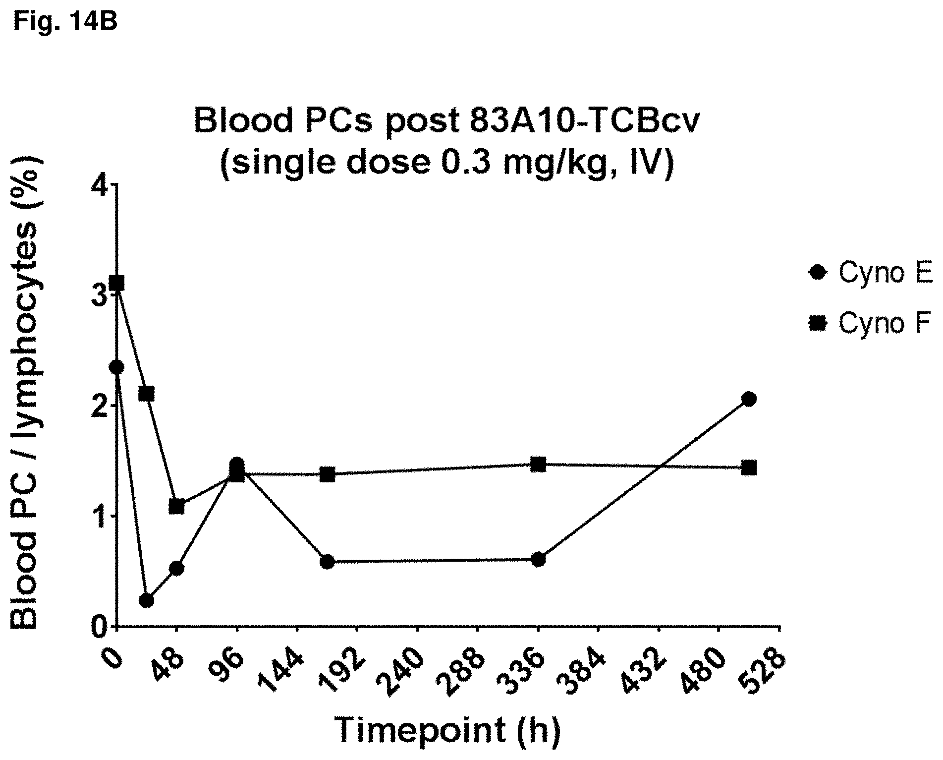
D00026
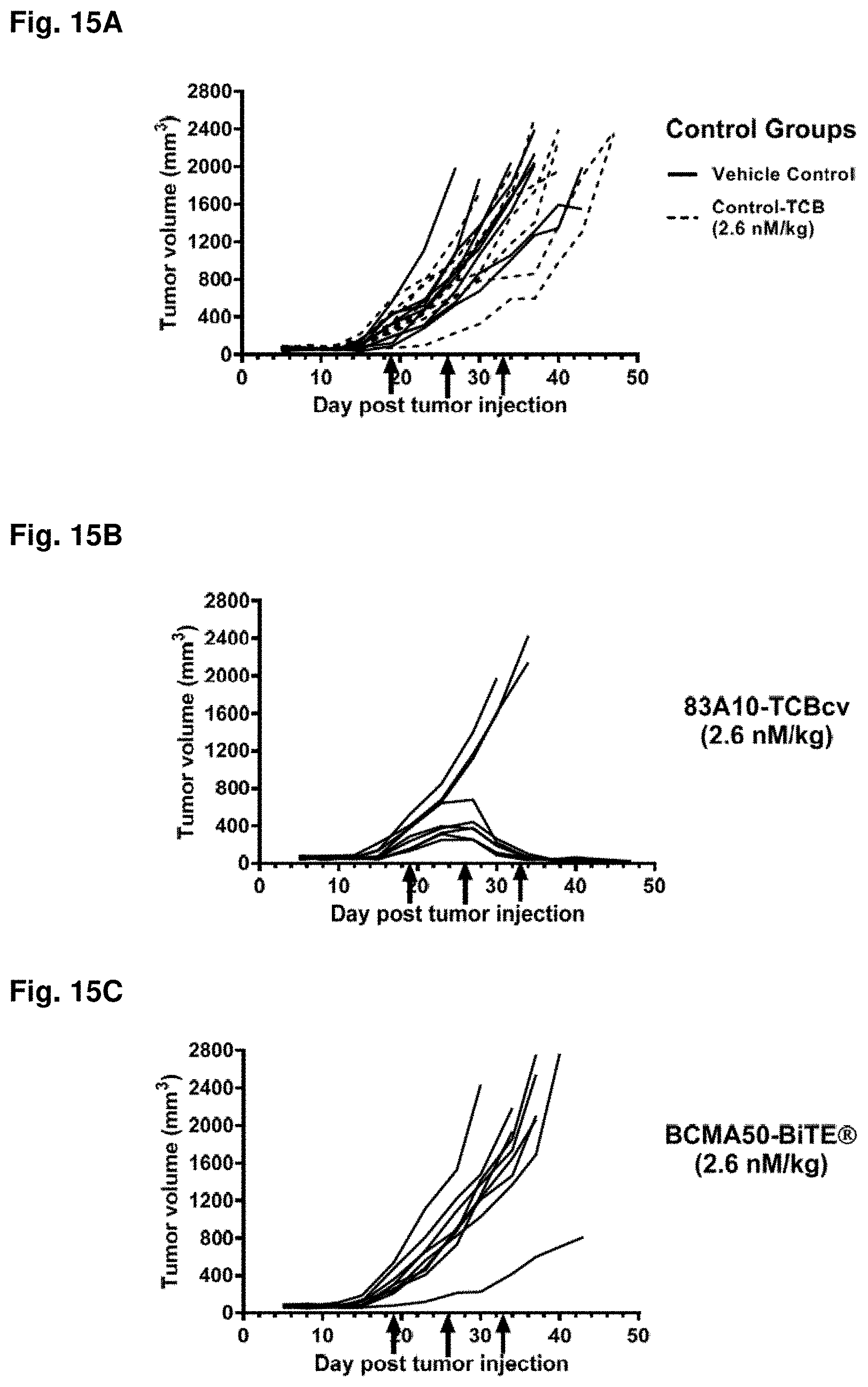
D00027

D00028

D00029

D00030

D00031

D00032


S00001
XML
uspto.report is an independent third-party trademark research tool that is not affiliated, endorsed, or sponsored by the United States Patent and Trademark Office (USPTO) or any other governmental organization. The information provided by uspto.report is based on publicly available data at the time of writing and is intended for informational purposes only.
While we strive to provide accurate and up-to-date information, we do not guarantee the accuracy, completeness, reliability, or suitability of the information displayed on this site. The use of this site is at your own risk. Any reliance you place on such information is therefore strictly at your own risk.
All official trademark data, including owner information, should be verified by visiting the official USPTO website at www.uspto.gov. This site is not intended to replace professional legal advice and should not be used as a substitute for consulting with a legal professional who is knowledgeable about trademark law.