Protein Binding To Fibronectin B Domain
Zhang; Mei ; et al.
U.S. patent application number 16/652384 was filed with the patent office on 2020-09-10 for protein binding to fibronectin b domain. This patent application is currently assigned to HEFEI LIFEON PHARMACEUTICAL CO., LTD.. The applicant listed for this patent is HEFEI LIFEON PHARMACEUTICAL CO., LTD.. Invention is credited to Jun Chen, Meihua Gao, Junqiu Ji, Mei Zhang.
| Application Number | 20200283511 16/652384 |
| Document ID | / |
| Family ID | 1000004860037 |
| Filed Date | 2020-09-10 |


| United States Patent Application | 20200283511 |
| Kind Code | A1 |
| Zhang; Mei ; et al. | September 10, 2020 |
PROTEIN BINDING TO FIBRONECTIN B DOMAIN
Abstract
The present invention relates to an epitope on fibronectin B (ED-B) domain, more specifically to an antibody or an antibody fragment of ED-B domain, and can be widely applied in in-vitro detection and in-vivo positioning of ED-B domain as well as in targeted cancer therapy.
| Inventors: | Zhang; Mei; (Hefei, CN) ; Ji; Junqiu; (Hefei, CN) ; Gao; Meihua; (Hefei, CN) ; Chen; Jun; (Hefei, CN) | ||||||||||
| Applicant: |
|
||||||||||
|---|---|---|---|---|---|---|---|---|---|---|---|
| Assignee: | HEFEI LIFEON PHARMACEUTICAL CO.,
LTD. Hefei CN |
||||||||||
| Family ID: | 1000004860037 | ||||||||||
| Appl. No.: | 16/652384 | ||||||||||
| Filed: | September 29, 2018 | ||||||||||
| PCT Filed: | September 29, 2018 | ||||||||||
| PCT NO: | PCT/CN2018/108532 | ||||||||||
| 371 Date: | March 30, 2020 |
| Current U.S. Class: | 1/1 |
| Current CPC Class: | C07K 16/18 20130101; A61P 35/00 20180101; C07K 2317/34 20130101; A61K 2039/505 20130101; C07K 2317/92 20130101; C07K 14/47 20130101; C07K 2317/622 20130101; C07K 2317/21 20130101 |
| International Class: | C07K 16/18 20060101 C07K016/18; C07K 14/47 20060101 C07K014/47; A61P 35/00 20060101 A61P035/00 |
Foreign Application Data
| Date | Code | Application Number |
|---|---|---|
| Sep 30, 2017 | CN | 201710918370.7 |
Claims
1-15. (canceled)
16. A product, which is one of the following products I) to V): I) an isolated antibody or antigen-binding fragment thereof that specifically binds to a peptide selected from the group consisting of VDITDS (SEQ ID NO: 76), TGLEPGIDY (SEQ ID NO: 88), and NSSTIIGYR (SEQ ID NO: 81); II) a pharmaceutical composition comprising the isolated antibody or antigen-binding fragment of I); III) a kit comprising the isolated antibody or antigen-binding fragment thereof of I), used for: diagnosing in vivo distribution of a tumor tissue or a pathological tissue section, analyzing and characterizing a cell or a protein, or affinity purifying a cell or a protein molecule containing an ED-B protein domain; IV) a nucleic acid molecule encoding the isolated antibody or antigen-binding fragment thereof of I); V) an isolated peptide consisting of the amino acid sequence selected from the group consisting of VDITDS (SEQ ID NO: 76), TGLEPGIDY (SEQ ID NO: 88), and NSSTIIGYR (SEQ ID NO: 81).
17. The product of claim 16, which is I) the isolated antibody or antigen-binding fragment thereof, wherein the heavy chain complementarity determining region (CDR) 3 comprises an amino acid sequence as set forth in SEQ ID NO: 7, and/or the light chain CDR3 comprises an amino acid sequence as set forth in SEQ ID NO: 10.
18. The product of claim 16, which is I) the isolated antibody or antigen-binding fragment thereof, wherein its heavy chain CDR1 and CDR2 comprise an amino acid sequences as set forth in SEQ NO: 5 and SEQ ID NO: 6, respectively, and/or its light chain CDR1 comprises an amino acid sequence selected from the group consisting of SEQ ID NOs: 8, 95-118 and 143, and its light chain CDR2 comprises an amino acid sequence selected from the group consisting of SEQ ID NOs: 9, 119-142 and 144.
19. The product of claim 16, which is I) the isolated antibody or antigen-binding fragment thereof, comprising a light chain variable region (VL) and a heavy chain variable region (VH), wherein the heavy chain variable region comprises: a VH CDR1 comprising an amino acid sequence as set forth in SEQ ID NO: 5, a VH CDR2 comprising an amino acid sequence as set forth in SEQ ID NO: 6, and a VH CDR3 comprising an amino acid sequence as set forth in SEQ ID NO: 7; the light chain variable region comprises: a VL CDR1 comprising an amino acid sequence as set forth in SEQ NOs: 8, 95, 97, 99, 100, 114 or 143, a VL CDR2 comprising an amino acid sequence as set forth in SEQ ID NOs: 9, 119, 121, 123, 124, 138, or 144, and a VL CDR3 comprising an amino acid sequence as set forth in SEQ ID NO: 10.
20. The product of claim 16, which is I) the isolated antibody or antigen-binding fragment thereof, wherein its heavy chain variable region comprises an amino acid sequence as set forth in SEQ ID NO: 1, and/or its light chain variable region comprises an amino acid sequence as set forth in SEQ ID NO: 3 or SEQ ID NO: 145 or an amino acid sequence having at least 95%, 96%, 97%, 98% or 99% identity thereto.
21. The product of claim 16, which is I) the isolated antibody or antigen-binding fragment thereof, wherein the antibody or antigen-binding fragment thereof has an equilibrium binding dissociation constant (KD) of 1.times.10.sup.-8 M or less, 1.times.10.sup.-9 M or less, or 1.times.10.sup.-10 M or less.
22. The product of claim 16, which is I) the isolated antibody or antigen-binding fragment thereof, wherein the antibody or antigen-binding fragment thereof is selected from the group consisting of a single-chain antibody, a single domain antibody, a hypervariable region polypeptide, a double-chain antibody, a triple-chain antibody, a minibody, a synthetic antibody, a recombinantly produced antibody, a multispecific antibody (e.g., a bispecific antibody), a human antibody, a non-human antibody, a humanized antibody, a chimeric antibody, an intracellular antibody, an Fab fragment, an Fab' fragment, an F(ab').sub.2 fragment, an Fv fragment, a disulfide bond-ligated Fv (dsFv), an Fd fragment, an Fd' fragment, a single-chain Fv (scFv), a single-chain Fab (scFab), and a diabody.
23. The product of claim 16, which is I) the isolated antibody or antigen-binding fragment thereof, wherein the antibody is a human immunoglobulin IgG.
24. The product of claim 16, which is I) the isolated antibody or antigen-binding fragment thereof, which is a single-chain Fv (scFv), wherein the VH comprises an amino acid sequence as set forth in SEQ ID NO: 1, and the VL comprises an amino acid sequence as set forth in SEQ ID NO: 3 or 145 or an amino acid sequence having at least 95%, 96%, 97%, 98% or 99% identity thereto.
25. The product of claim 24, wherein the scFv comprises amino acids 1-241 of SEQ ID NO: 13, amino acid residues 1-242 of SEQ ID NO: 13, or an amino acid sequence as set forth in SEQ ID NO: 13, SEQ ID NO: 14 or SEQ ID NO: 146.
26. The product of claim 16, which is II) the pharmaceutical composition, comprising a fusion protein, a radioisotope, a fluorescent dye, a chemical and/or a nanoparticle.
27. The product of claim 16, which is II) the pharmaceutical composition, for use in the diagnosis or treatment of a tumor or cancer expressing FN comprising ED-B domain.
28. The product of claim 27, wherein the tumor or cancer is selected from the group consisting of nasopharyngeal carcinoma, head and neck cancer, esophageal cancer, gastric cancer, colorectal cancer, lung cancer, breast cancer and soft tissue sarcoma.
29. The product of claim 16, which is IV) the nucleic acid molecule, comprising: (a) a nucleotide sequence as set forth in SEQ ID NO: 2, and/or (b) a nucleotide sequence as set forth in SEQ ID NO: 4 or 147, and/or (c) a degenerate nucleotide sequence of (a) or (b).
30. A method, which is one of the following methods I) to II): I) a method of preventing, diagnosing or treating a tumor or cancer-related disease in a subject, comprising administrating the subject an effective amount of the isolated antibody or antigen-binding fragment thereof of claim 16 I) or the pharmaceutical composition of claim 16 II), wherein the tumor or cancer is one expressing FN comprising ED-B domain; II) a method of analyzing and characterizing a cell or a protein, or affinity purifying a cell or a protein containing an ED-B protein domain, comprising contacting the cell or the protein with the isolated antibody or antigen-binding fragment thereof of claim 16 I).
31. The method of claim 30, which is I) the method, wherein the tumor or cancer is selected from the group consisting of nasopharyngeal carcinoma, head and neck cancer, esophageal cancer, gastric cancer, colorectal cancer, lung cancer, breast cancer and soft tissue sarcoma.
Description
TECHNICAL FIELD
[0001] The present invention relates to a novel epitope of fibronectin B domain, and an antibody, especially a monoclonal antibody or an antibody fragment, specifically binding to the epitope, having a biological activity of recognizing tumor or inhibiting tumor growth, or of killing tumor cells. In addition, the present invention also relates to a method of preparing the antibody and a pharmaceutical composition comprising the antibody.
BACKGROUND OF THE INVENTION
[0002] Chemotherapy and radiotherapy are commonly-used tumor treatments, which result in remarkable toxic and side effects on normal tissues due to low specificity. Physiological functions of normal organs are affected while cancer cells are killed, and the immunity and the life quality of patients are reduced. One way to improve the specificity of a tumor therapeutic agent is to target the agent to tumor cells by means of an antibody or an antibody fragment capable of recognizing a tumor cell marker, resulting in targeted killing of the tumor. First, one needs to find a tumor marker that can be expressed on the outer surface of the cell membrane, i.e. a protein that is expressed only on the surface or in the extracellular matrix of tumor cells but in a very small amount or even not expressed in normal cells or tissues.
[0003] Fibronectin (FN) is a multifunctional glycoprotein, widely found in the extracellular matrix, plasma and other body fluids, expressed by epithellal cells, endothelial cells, fibroblasts, hepatocytes and so on, and involved in the cell adhesion, deformation and distribution, as well as the formation of blood vessels.
[0004] The FN gene is about 75 kb in length, including about 50 exons, and mainly composed of three types of homologous repeat units, type I, II and III. Domain B is a complete repeat of 91 amino acids in the type III repeat sequence of FN encoded by a single exon. During the expression of the FN gene, there are two cases, the FN(B+) including the B domain and the other FN(B-) not including the B domain, and both are considered to play an important role in the development of the individual. The FN(B+) is rarely expressed in normal tissues of an adult, but is highly expressed again during a trauma, disease, wound healing, especially tumor growth, e.g. the FN(B+) containing the B domain is highly expressed in cells such as gastric cancer, colorectal cancer, lung cancer, breast cancer cells and the like. The FN(B+) is a marker protein of tumor tissues, as it is not expressed in normal tissues of an adult but is highly expressed in blood vessels of various tumor tissues as shown by in-vitro immunohistochemistry assay and in-vivo targeting localization. Thus, the B domain of FN is also referred as an Extra-domain B (ED-B).
[0005] A targeting antibody developed on the basis of the ED-B domain can be used for targeting a candidate drug such as a cytotoxin and so on to a tumor site and effectively inhibit or kill tumor cells, thereby achieving the purpose of treatment while reducing damage to normal tissues and reducing toxic side effects.
[0006] It is trend that a therapeutic antibody is developed to a fully human antibody, which is clinically advantageous over a murine antibody, such as low immunogenicity, longer in vive half-life of antibody and mediating immunoregulation, ADCC and CDC effects by virtue of human immunoglobulin Fc segment, thereby enhancing the biological effects of antibody.
[0007] Since the amino acid sequence of the human ED-B is 100% homologous to that of the mouse ED-B, it is difficult for the mouse immune system to immunoreact with the human ED-B domain to produce an anti-ED-B antibody, e.g. the mouse monoclonal antibody BC-1 obtained by hybridoma technology (Camemolla, Leprini et al. J Biol Chem 1992, 24689-24692) cannot recognize the ED-B domain directly, but indirectly specifically binds to the FN of ED-B positive by recognizing an antigenic epitope in a domain adjacent to the ED-B. Now, with the aid of genetic engineering technologies, an artificially synthesized antibody gene is directly expressed utilizing phage display technology, and an antibody with particular functions is obtained through panning, without need of an immune system of a higher animal body. Therefore, there provids a technology for developing a fully human-origin recombinant antibody. Currently, the ED-B human antibodies with high specificity such as CGS-1, CGS-2, L19 (Camemolla, Neri et al. Int J Cancer 1996, 397-405, Pini, Viti et al. J Biol Chem 1998, 21769-21776) and B5 (Ji J, Zhang M, Gao M, et al. HUMAN ANTIBODY AGAINST ED-B DOMAIN OF FIBRONECTIN AND USES THEREOF. WO2014194784A1) have been reported. The CGS-1, CGS-2 and L19 have an affinity of 5.4.times.10.sup.-8 M, 1.1.times.10.sup.-9M and 8.7.times.10.sup.-10M respectively as measured by BIAcore assay. In particular, the clinical research of the antibody L19 sufficiently proved that a specific antibody drug developed against the antigen ED-B has a remarkable tumor inhibiting or killing effect.
SUMMARY OF THE INVENTION
[0008] In one aspect, the invention provides a novel linear epitope of ED-B domain, consisting of an amino acid sequence selected from the group consisting of VDITDS (SEQ ID NO: 76), TGLEPGIDY (SEQ ID NO: 88) and NSSTIIGYR (SEQ ID NO: 81).
[0009] In one aspect, the invention provides an isolated antibody or antigen-binding fragment thereof that specifically binds to an epitope selected from the group consisting of VDITDS (SEQ ID NO: 76), TGLEPGIDY (SEQ ID NO: 88) and NSSTIIGYR (SEQ ID NO: 81).
[0010] In an embodiment, the invention provides an isolated antibody or antigen-binding fragment thereof, wherein the antibody or antigen-binding fragment thereof comprises three heavy chain complementarity determining regions (HCDR) and three light chain complementarity determining regions (LCDR), wherein the HCDR1, HCDR2 and HCDR3 comprise the amino acid sequences as set forth in SEQ ID NO. 5, SEQ ID NO: 6 and SEQ ID NO: 7 respectively, and the LCDR1 comprises an amino acid sequence selected from the group consisting of SEQ ID NOs: 8, 95-118 and 143, the LCDR2 comprises an amino acid sequence selected from the group consisting of SEQ ID NOs: 9, 119-142 and 144, and the LCDR3 comprises an amino acid sequence as set forth in SEQ ID NO: 10.
[0011] In an embodiment, an isolated antibody or antigen-binding fragment thereof according to the invention comprises a heavy chain variable region (VH) comprising an amino acid sequence as set forth in SEQ ID NO: 1. In an embodiment, an antibody or antigen-binding fragment thereof according to the invention comprises a light chain variable region (VL) comprising an amino acid sequence as set forth in SEQ ID NO: 3 or 145 or an amino acid sequence having at least 95%, 96%, 97%, 98% or 99% identity thereto.
[0012] In an embodiment, an antibody according to the invention comprises an amino acid sequence as set forth in SEQ ID NO: 13, SEQ ID NO. 14, SEQ ID NO: 146, amino acids 1-241 of SEQ ID NO: 13, or amino acids 1-242 of SEQ ID NO. 13.
[0013] In one aspect, the invention provides a pharmaceutical composition comprising an isolated antibody or antigen-binding fragment thereof according to the invention, optionally further comprising a fusion protein, a radioisotope, a chemical drug and/or a nanoparticle.
[0014] In one aspect, the invention provides use of an isolated antibody or antigen-binding fragment thereof or a pharmaceutical composition comprising the same according to the invention in the manufacture of a medicament for the prevention, diagnosis and treatment of cancer.
[0015] In an embodiment, the cancer is a cancer expressing FN(B+) containing ED-B domain, e.g., selected from the group consisting of nasopharyngeal carcinoma, head and neck cancer, esophageal cancer, gastric cancer, colorectal cancer, lung cancer, breast cancer and soft tissue sarcoma.
[0016] In one aspect, the invention provides a kit comprising an isolated antibody or antigen-binding fragment thereof according to the invention, which can be used for (i) diagnosing an in vivo distribution or a pathological tissue section of tumor tissue, (ii) analyzing and characterizing a cell or a protein, or (iii) affinity purifying a cell or a protein molecule comprising an ED-B protein domain.
[0017] In one aspect, the invention provides use of an isolated antibody or antigen-binding fragment thereof according to the invention in the manufacture of a kit for (I) diagnosing an in vivo distribution or a pathological tissue section of tumor tissue, (ii) assaying and characterizing a cell or a protein, or (iii) affinity purifying a cell or a protein molecule comprising an ED-B protein domain.
[0018] In one aspect, the invention provides a polynucleotide molecule encoding an isolated antibody or antigen-binding fragment thereof according to the invention.
BRIEF DESCRIPTION OF THE DRAWINGS
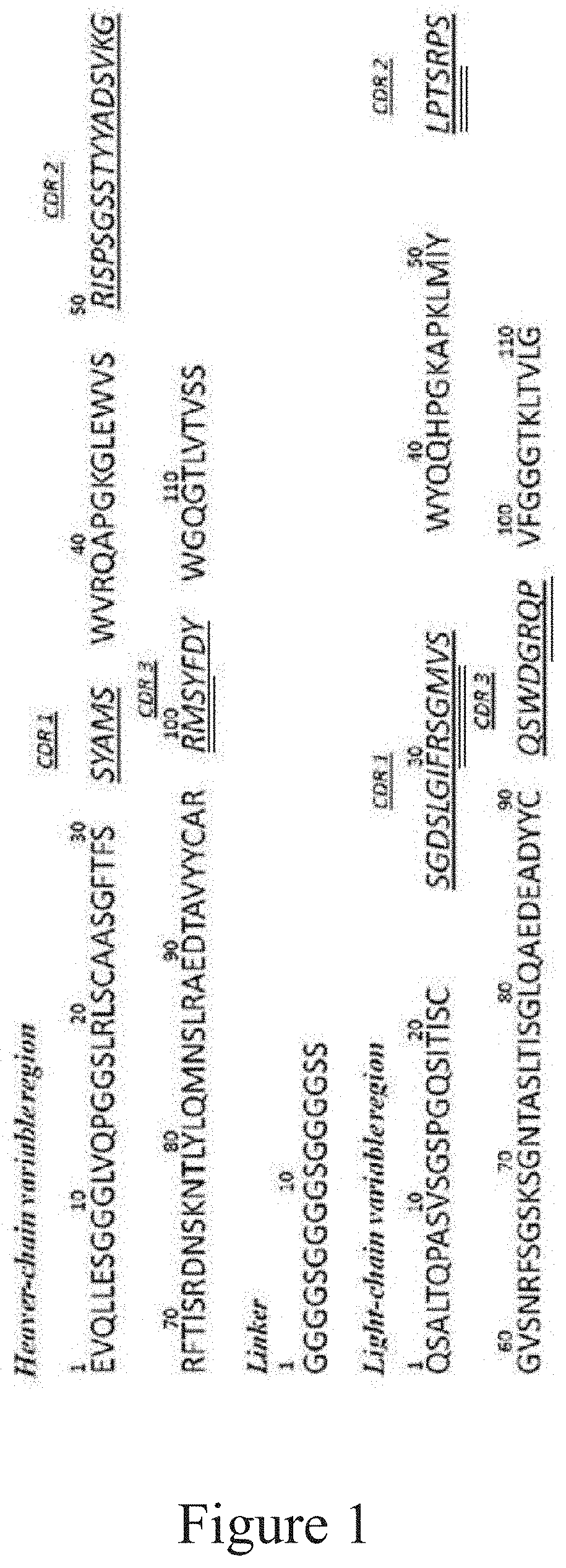
[0019] FIG. 1 shows the heavy and light chain variable region amino acid sequences of the DE2 antibody, wherein the underlined amino acids are CDR regions, the double-underlined amino acids are random sequences in the antibody repertoire, and the three-underlined amino acids are random sequences in the A11 antibody repertoire.
[0020] FIG. 2 verifies that the DE2 antibody specifically recognizes an antigenic ED-B domain by ELISA experiments.
[0021] FIG. 3 verifies that the DE2 antibody specifically recognizes FN(B+) cells by cellular immunofluorescence experiments. FIG. 3A is a negative control, wherein the cells are CHO-789 cells that do not express ED-B, and the cells of FIG. 3B are CHO-7B89 cells that express ED-B. The antibody is the FITC-labeled DE2 antibody.
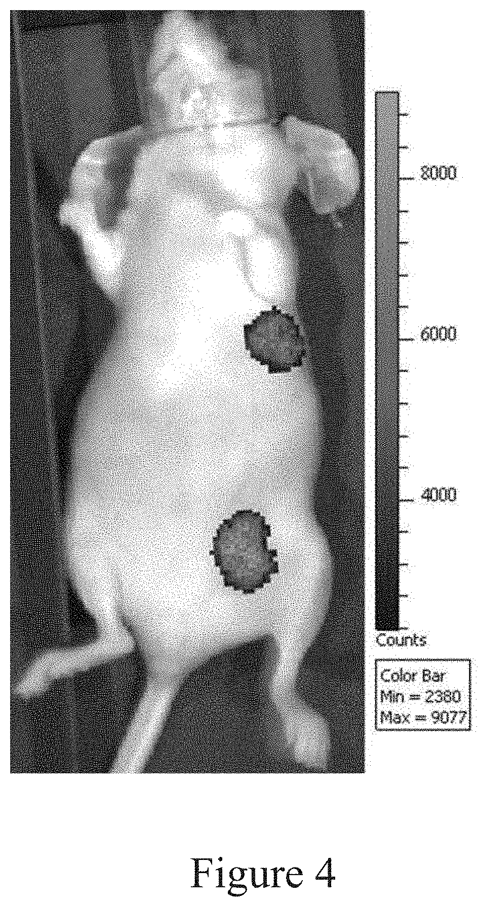
[0022] FIG. 4 shows the distribution of DE2 antibody in tumor-bearing mice. The Cy5.5 labeled DE2 antibody can be specifically distributed in an ED-B high-expressing implanted tumor tissue by the living body fluorescence imaging method for a small animal, and the antibody distribution is difficult to be detected in other tissues.
[0023] FIG. 5 shows the distribution of the DE2 antibody in solid tumor tissues. Tumor tissue cryosection analysis was performed 16 hours after Cy55-labeled DE2 antibody was injected intravenously into mice. Cy5.5-labeled antibodies were mainly distributed on blood vessels and tumor extracellular matrix.
[0024] FIG. 6 shows an epitope by which the DE2 antibody binds to ED-B. The synthetic polypeptide sequence is represented by horizontal lines, and the thickness of the lines indicates the relative affinity.
[0025] FIG. 7 shows an epitope by which the DE2 antibody binds to ED-B. The regions in the polypeptide with the highest binding capacity to the DE2 antibody are marked by light gray spheres and dark gray spheres.
CONTENTS OF THE INVENTION
[0026] The inventors, using antibody gene synthesis and phage display technology, have discovered a specific single-chain antibody which directly recognizes an ED-B domain alone or fibronectin containing ED-B domain FN(B+), as well as a fusion protein of other protein(s) and an ED-B domain. The affinity of the obtained candidate antibody is 16 times of that of the L19 antibody by an ELISA assay. The affinity of the obtained antibody is 9 times of that of the L19 by BIAcore assay. The antibody is expected to have a higher affinity and better targeting effect on the antigen ED-B. The obtained antibody's light chain has an amino acid sequence of human Lambda chain, which is different from the amino acid sequence of human Kappa chain of the L19, and diversifies the types of antibody against ED-B.
[0027] The obtained antibody can be used in the development of an antitumor drug, such as radioisotope labeling of antibody, fusion with a cytotoxin, coupling with a chemical drug, coupling with a nanoparticle and the like. A drug can be highly enriched in a tumor tissue highly expressing FN(B+) to specifically kill tumor cells in a targeted manner by virtue of the targeting effect of the obtained antibody, so that the drug effect can be improved with less damage to normal tissues. The structure of the obtained single-chain antibody can miniaturize the antibody drug, improve the permeability of the antibody in a human body, get into the inside of a tumor tissue along tumor blood vessels and exert better tumor killing effects; moreover, the single-chain antibody can be flexibly changed into various forms of antibody, such as a natural antibody structure, a minibody, a dibody, a multi-antibody fusion form and the like. The stability, permeability, application and the like of the antibody can be changed through modification of the antibody structure. In addition, the obtained antibody can be developed into a tumor diagnostic agent for detecting the tumor tissue in vivo and in vitro, or for development of other various immune-related reagents.
[0028] The inventors have found that there are two regions in the ED-B domain of fibronectin that bind to an antibody with very high affinity, located in an irregular coiled region of the ED-B domain. It can be found through further analysis that an antigenic epitope of ED-B recognized by a high affinity ED-B antibody consists of an ED-B polypeptide fragment selected from the group consisting of: VDITDS (SEQ ID NO: 76), TGLEPGIDY (SEQ ID NO: 88) and NSSTIIGYR (SEQ ID NO: 81).
[0029] The inventors have obtained a recombinant anti-human ED-B protein domain monoclonal antibody or an antibody fragment, wherein the antibody fragment is preferably an antigen-binding fragment, and the antibody or the antibody fragment can be effectively bind to fibronectin (FN) containing ED-B domain (abbreviated as FN(B+)) and can be used for the detection and diagnosis of the FN(B+) as well as for targeted therapy of FN(B+)-high-expressing tumors, or can be used in a combined treatment with other antineoplastic agents.
[0030] Accordingly, the invention provides an isolated antibody or an antibody fragment that specifically recognizes and binds the ED-B domain of human fibronectin (FN). In a particular embodiment, the invention provides an antibody or an antibody fragment that specifically recognizes and/or binds to a linear epitope consisting of an ED-B polypeptide fragment selected from the group consisting of: VDITDS (SEQ ID NO: 76), TGLEPGIDY (SEQ ID NO: 88) and NSSTIIGYR (SEQ ID NO: 81).
[0031] In a particular embodiment, an antibody or antibody fragment according to the invention comprises an amino acid sequence of the heavy chain variable region CDR3 comprising the amino acid sequence RMSYFDY (SEQ ID NO: 7).
[0032] In another particular embodiment, an antibody or antibody fragment according to the invention comprises an amino acid sequence of the light chain variable region CDR3 comprising the amino acid sequence QSWDGRQP (SEQ ID NO: 10).
[0033] In another particular embodiment, an antibody or antibody fragment according to the invention comprises a heavy chain variable region CDR3 having the amino acid sequence RMSYFDY (SEQ ID NO: 7) and a light chain variable region CDR3 having the amino acid sequence QSWDGRQP (SEQ ID NO: 10).
[0034] In another particular embodiment, an antibody or antibody fragment according to the invention comprises three heavy chain variable region CDRs having the amino acid sequences SYAMS (SEQ ID NO: 5), RISPSGSSTYYADSVKG (SEQ ID NO: 6), and RMSYFDY (SEQ ID NO: 7), respectively.
[0035] In a particular embodiment, an antibody or antibody fragment according to the invention comprises a light chain CDR1 comprising an amino acid sequence selected from the group consisting of SEQ ID NOs: 8, 95-118 and 143.
[0036] In a particular embodiment, an antibody or antibody fragment according to the invention comprises a light chain CDR2 comprising an amino acid sequence selected from the group consisting of SEQ ID NOs: 9,119-142 and 144.
[0037] In another particular embodiment, an antibody or antibody fragment according to the invention comprises three light chain variable region CDRs, each having an amino acid sequence of:
[0038] LCDR1: the amino acid sequence selected from the group consisting of SEQ ID NOs: 8, 95-118 and 143, e.g. SEQ ID NO: 8, 95, 97, 99, 100, 114 or 143,
[0039] LCDR2: the amino acid sequence selected from the group consisting of SEQ ID NOs: 9, 119-142 and 144, e.g., SEQ ID NO: 9, 119, 121, 123, 124, 138 or 144, and
[0040] LCDR3: SEQ ID NO: 10.
[0041] In another particular embodiment, an antibody or antibody fragment according to the invention comprises three heavy chain variable region CDRs and three light chain variable region CDRs, wherein the amino acid sequences of the three heavy chain variable region CDRs consist of SYAMS (SEQ ID NO: 5), RISPSGSSTYYADSVKG (SEQ ID NO: 6) and RMSYFDY (SEQ ID NO: 7), respectively, and the amino acid sequences of the three light chain variable region CDRs consist of SGDSLGIFRSGMVS (SEQ ID NO: 8), LPTSRPS (SEQ ID NO: 9) and QSWDGRQP (SEQ ID NO: 10), respectively.
[0042] In another particular embodiment, the invention provides an isolated antibody or antigen-binding fragment thereof that specifically recognizes and binds the ED-B domain of human fibronectin (FN), wherein the antibody or antigen-binding fragment thereof comprises a light chain variable region (VL) and a heavy chain variable region (VH),
[0043] the heavy chain variable region comprising:
[0044] a VH CDR1 comprising an amino acid sequence as set forth in SEQ ID NO: 5,
[0045] a VH CDR2 comprising an amino acid sequence as set forth in SEQ ID NO: 6, and
[0046] a VH CDR3 comprising an amino acid sequence as set forth in SEQ ID NO: 7,
[0047] the light chain variable region comprising:
[0048] a VL CDR1 comprising an amino acid sequence as set forth in SEQ NO: 8 or 143,
[0049] a VL CDR2 comprising an amino acid sequence as set forth in SEQ ID NO: 9 or 144, and
[0050] a VL CDR3 comprising an amino acid sequence as set forth in SEQ ID NO: 10.
[0051] An "isolated antibody" means that the antibody (1) is not associated with a naturally associated component that accompanies it in its natural state, (2) does not contain other proteins from the same species, (3) is expressed by cells from different species, or (4) does not occur in nature. Thus, a chemically synthesized polypeptide or a polypeptide synthesized in a cell system other than a cell from which the polypeptide is naturally derived will be "isolated" from its naturally associated components. The protein may also be substantially free of naturally associated components by isolation, i.e., using protein purification techniques well-known in the art.
[0052] As used herein, an "antibody" refers to an immunoglobulin and an immunoglobulin fragment, regardless of natural or partially or fully synthetically (e.g., recombinantly) produced, including any fragment thereof that comprises at least partial variable region of an immunoglobulin molecule and retains the binding specificity of the full-length immunoglobulin.
[0053] Thus, an antibody includes any protein having a binding domain that is homologous or substantially homologous to the antigen binding domain (antibody binding site) of an immunoglobulin. Antibody includes synthetic antibodies, recombinantly produced antibodies, multispecific antibodies (e.g., bispecific antibodies), human antibodies, non-human antibodies, humanized antibodies, chimeric antibodies, intracellular antibodies, and antibody fragments, e.g., but not limited to, Fab fragments, Fab' fragments, F(ab').sub.2 fragments, Fv fragments, disulfide bridge-linked Fv (dsFv), Fd fragments, Fd' fragments, single-chain Fvs (scFvs), single-chain Fabs (scFabs), diabodes, or antigen-binding fragments of any of the foregoing antibodies. The antibody provided herein comprises a member of any immunoglobulin types (e.g., IgG, IgM, IgD, IgE, IgA, and IgY), any class (e.g., IgG1, IgG2, IgG3, IgG4, IgA1 and IgA2), or subclass (e.g., IgG2a and IgG2b).
[0054] As used herein, the "antibody fragment" or "antigen-binding fragment" of an antibody refers to any portion of a full-length antibody that is less than full-length, but comprises at least a portion of the variable region (e.g., one or more CDRs and/or one or more antibody binding sites) of the antibody that binds an antigen, and thus retains the binding specificity as well as at least partial specific binding capacity of the full-length antibody. Thus, an antigen-binding fragment comprises an antigen-binding portion that binds the same antigen as the antibody from which the antibody fragment is derived. Antibody fragment includes antibody derivatives produced by enzymatic treatment of a full-length antibody, as well as synthetically produced derivatives, such as recombinantly produced derivatives. Antibody includes an antibody fragment, example of which includes, but not limited to, an Fab, an Fab', an F(ab').sub.2, a single-chain Fv (scFv), an Fv, an dsFv, a diabody, an Fd and an Fd' fragment, and a single domain antibody (dAb) fragment, including a modified fragment (see e.g. Methods in Molecular Biology, Vol 207: Recombinant Antibodies for Cancer Therapy Methods and Protocols (2003); Chapter 1; p 3-25, Kiprlyanov; Ward et al., Nature 341: 544-546, 1989, and Wu, A. M. and P. D. Senter. Nat Biotechnol 23(9): 1137-1146(2005)). The fragment may comprise multiple chains linked together, for example by a disulfide linkage and/or by a peptide linker.
[0055] As used herein, a full-length antibody is an antibody having two full-length heavy chains (e.g., VH-CH1-CH2-CH3 or VH-CH1-CH2-CH3-CH4) and two full-length light chains (VL-CL) and hinge regions, such as an antibody naturally produced by antibody secreting B cells and a synthetically produced antibody having the same domains.
[0056] As used herein, dsFv refers to an Fv having an engineered intermolecular disulfide bond that stabilizes a VH-VL pair.
[0057] As used herein, an Fab fragment is an antibody fragment obtained by digestion of a full-length immunoglobulin with papain, or a fragment of the same structure synthesized, for example, by a recombinant method. The Fab fragment comprises a light chain (comprising VL and CL) and another chain comprising a variable domain of heavy chain (VH) and a constant domain of heavy chain (CH1).
[0058] As used herein, an F(ab').sub.2 fragment is an antibody fragment resulted from digestion of an immunoglobulin with pepsin at pH 4.0-4.5, or a fragment of the same structure synthesized, for example, by a recombinant method. The F(ab').sub.2 fragment essentially comprises two Fab fragments, wherein each of the heavy chain portions contains several additional amino acids, comprising cysteines that form disulfide bond linking the two fragments.
[0059] As used herein, an Fab' fragment is a fragment comprising half of the F(ab').sub.2 fragment (one heavy chain and one light chain).
[0060] As used herein, an "Fv" is a minimal antibody fragment that contains a complete antigen recognition and binding site. This fragment consists of a tightly non-covalently associated dimer of a heavy chain variable region domain and a light chain variable region domain. Folding of these two domains produces six hypervariable loops (three loops from each of the H and L chains), contributing amino acid residues for antigen binding and conferring antigen binding specificity to the antibody. A single variable domain (or half of Fv comprising only three CDRs specific for an antigen) also has the ability to recognize and bind the antigen.
[0061] As used herein, an scFv fragment refers to an antibody fragment comprising a variable light chain (VL) and a variable heavy chain (VH) covalently linked by a polypeptide linker in any order. The length of the linker is such that the two variable domains can be bridged without interfering each other substantially. An example of the linker is (Gly-Ser)n residues with some Glu or Lys residues to increase solubility. The review about sFv can refer to "The Pharmacology of Monoclonal Antibodies", vol. 113, Rosenburg and Moore eds., Springer-Verlag, New York, pp. 269-315 (1994).
[0062] The term "chimeric antibody" refers to an antibody in which the variable region sequence is derived from one species and the constant region sequence is derived from another, such as an antibody in which the variable region sequence is derived from a mouse antibody and the constant region sequence is derived from a human antibody.
[0063] The term "diabody" refers to a small antibody fragment prepared by constructing a sFv fragment with a short linker (about 5-10 residues) between VH and VL domains, thereby leading to an interchain rather than intrachain pairing of the V domains, resulting in a bivalent fragment, i.e., a fragment having two antigen binding sites.
[0064] Bispecific antibody is a heterodimer of two "cross" sFv fragments in which the VH and VL domains of the two antibodies are present on different polypeptide chains. See, e.g., EP 404,097, WO 93/11161 and Hollinger et al., Proc. Natl. Acad. Sci. USA, 90: 6444-6448 (1993).
[0065] "A human antibody" refers to an antibody comprising only human immunoglobulin sequences. If the human antibody is produced in a mouse, in a mouse cell, or in a hybridoma derived from a mouse cell, the human antibody may contain a murine carbohydrate chain. Similarly, "a mouse antibody" or "a rat antibody" refers to an antibody comprising only mouse or rat immunoglobulin sequences, respectively.
[0066] A "humanized" antibody refers to a form of a non-human (e.g., mouse) antibody that is a chimeric immunoglobulin, immunoglobulin chain, or fragment thereof (e.g., Fv, Fab, Fab', F(ab').sub.2, or other antigen-binding subsequences of an antibody) containing minimal sequences derived from a non-human immunoglobulin. Preferably, the humanized antibody is a human immunoglobulin (recipient antibody) in which the residues of the complementarity determining regions (CDRs) of the recipient antibody are replaced by CDR residues from a non-human species (donor antibody) such as mouse, rat or rabbit having the desired specificity, affinity and capacity.
[0067] Furthermore, in humanization, it is also possible to mutate amino acid residues within CDR1, CDR2 and/or CDR3 regions of VH and/or VL, thereby improving one or more binding properties (e.g., affinity) of the antibody. Mutations can be introduced, for example, by PCR-mediated mutation, the effect of which on antibody binding or other functional properties can be assessed using in vitro or in vivo assays described herein. Typically, conservative mutation is introduced.
[0068] Such mutation may be amino acid substitution, addition or deletion. In addition, mutations within a CDR are typically no more than one or two. Thus, the humanized antibody of the present invention also encompasses an antibody comprising one or two amino acid mutations within a CDR.
[0069] Generally, an immunoglobulin has heavy and light chains. Each heavy and light chain comprises a constant region and a variable region (these regions are also referred to as "domains"). In combination, the heavy and light chain variable regions specifically bind to an antigen. The light and heavy chain variable regions comprise a "framework" region interrupted by three hypervariable regions (also referred to as "complementarity determining regions" or "CDRs"). Ranges of the framework region and CDRs have been determined (see Kabat et al., Sequences of Proteins of Immunological Interest, U.S. Department of Health and Human Services, 1991, which is incorporated herein by reference). The sequences of different light or heavy chain framework regions are relatively conserved within one species.
[0070] The CDRs are primarily responsible for binding to an epitope of an antigen. The CDRs of each chain are commonly referred to as CDR1, CDR2 and CDR3, numbered sequentially from the N-terminus, and are also typically identified by the chain in which the particular CDR is located. Thus, the VH CDR3 is located in the antibody heavy chain variable domain from which it was found, while the VL CDR1 is the CDR1 from the antibody light chain variable domain from which it was found.
[0071] "VH" or "V.sub.H" refers to the variable region of an immunoglobulin heavy chain, comprising the variable region of Fv, scFv, dsFv or Fab. "VL" or "V.sub.L" refers to the variable region of an immunoglobulin light chain, comprising the variable region of Fv, scFv, dsFv or Fab.
[0072] As used herein, the "monoclonal antibody" or "mAb" or "Mab" refers to a population of substantially homogeneous antibodies, i.e., except for possible natural occurring mutations that may be present in a minor amount, the amino acid sequences of the antibody molecules that make up the population are identical. In contrast, a conventional (polyclonal) antibody preparation typically includes many different antibodies having different amino acid sequences in variable domains, particularly CDRs, which are typically specific for different epitopes. The modifier "monoclonal" means that an antibody is characterized as being obtained from a population of substantially homogeneous antibodies, and should not be construed as requiring any particular method to produce the antibody. For example, a monoclonal antibody to be used in accordance with the present invention can be prepared by the hybridoma method described by Kohler et al. (1975) Nature 256: 495 at first, or by a recombinant DNA method (see, e.g., U.S. Pat. No. 4,816,567). "Monoclonal antibody" can also be isolated from a phage antibody library, for example, using the techniques described in Clackson et al. (1991) Nature 352: 624-628 and Marks et al. (1991) J. Mol. Biol. 222: 581-597. See also Presta (2005) J. Allergy Clin. Immunol. 116: 731.
[0073] In another particular embodiment, an antibody or antibody fragment according to the invention comprises a heavy chain variable region (VH) comprising an amino acid sequence as set forth in SEQ ID NO: 1.
[0074] In another particular embodiment, an antibody or antibody fragment according to the invention comprises a light chain variable region (VL) comprising an amino acid sequence as set forth in SEQ ID NO: 3 or SEQ ID NO: 145 or an amino acid sequence having at least 95%, 96%, 97%, 98% or 99% identity thereto.
[0075] The term "% identity" is determined by comparing two optimally aligned sequences over a comparison window, wherein a portion of a polypeptide sequence in the comparison window may contain an addition or deletion (i.e., gap) as compared to a reference sequence (which does not contain the addition or deletion) to optimally align the two sequences. "% identity" is calculated as follows: the number of positions at which the same nucleic acid bases or amino acid residues occur in both sequences is divided by the total number of positions in the comparison window and the result is multiplied by 100.
[0076] In another particular embodiment, an antibody or antibody fragment according to the invention comprises a heavy chain variable region (VH) comprising the amino acid sequence of SEQ ID NO: 1 and a light chain variable region (VL) comprising the amino acid sequence of SEQ ID NO: 3 or SEQ ID NO: 145 or an amino acid sequence having at least 95%, 96%, 97%, 98% or 99% identical thereto.
[0077] In another particular embodiment, an antibody according to the invention comprises an amino acid sequence as set forth in SEQ ID NO: 13, SEQ ID NO: 14, SEQ ID NO: 146, amino acids 1-241 of SEQ ID NO: 13, or amino acid residues 1-242 of SEQ ID NO: 13.
[0078] In another particular embodiment, an antibody according to the invention comprises an amino acid sequence as set forth in SEQ ID NO: 13. In another particular embodiment, an antibody or antibody fragment according to the invention is a monoclonal antibody having an equilibrium binding dissociation constant (KD) of 1.times.10.sup.-8 M or less, 1.times.10.sup.-9 M or less, or 1.times.10.sup.-10 M or less.
[0079] In another particular embodiment, an antibody or antibody fragment according to the invention is a monomer or polymer.
[0080] In another particular embodiment, an antibody or antibody fragment according to the invention is an antibody sequence derived from a mammalian antibody sequence, in particular from a human.
[0081] In another particular embodiment, the antibody or antibody fragment binds directly to ED-B domain. In particular, the ED-B domain may be an independent ED-B recombinant protein, or a recombinant protein formed by fusing an ED-B protein with other protein(s), or natural fibronectin containing ED-B domain. More particularly, the ED-B domain may be from human, mouse, rat, chicken or other species. More particularly, the ED-B domain may be a glycosylated or a non-glycosylated protein.
[0082] In a particular embodiment, the antibody fragment may be a monovalent small molecule antibody such as a single-chain antibody, a single domain antibody, a hypervariable region polypeptide or the like, an Fab, or a multivalent small molecule antibody such as a double-, triple-chain antibody or a mini-antibody.
[0083] In another particular embodiment, the antibody is a human immunoglobulin IgG.
[0084] In another aspect, the invention provides a pharmaceutical composition comprising an antibody or antibody fragment according to the invention.
[0085] In one embodiment, the pharmaceutical composition further comprises, but is not limited to, a fusion protein, a radioisotope, a fluorescent dye, a chemical, a nanoparticle, and the like. In a particular embodiment, the pharmaceutical composition is used for the diagnosis or treatment of a tumor or cancer-related disease.
[0086] In yet another aspect, the invention provides the use of an antibody or antibody fragment or pharmaceutical composition according to the invention for the prevention, diagnosis and treatment of a cancer-related disease.
[0087] In yet another aspect, the invention provides a method of preventing, diagnosing and treating a cancer-related disease in a subject, comprising administering to the subject a therapeutically or prophylactically effective amount of an antibody or antigen-binding fragment thereof or a pharmaceutical composition according to the invention.
[0088] In yet another aspect, the invention provides the use of an antibody or antibody fragment or pharmaceutical composition according to the invention in the manufacture of a medicament for the prevention, diagnosis and treatment of a cancer-related disease.
[0089] Particularly, a cancer-related disease according to the invention is a cancer expressing FN(B+) containing ED-B domain. More particularly, the cancer-related disease is gastric cancer, colorectal cancer, lung cancer or breast cancer.
[0090] As used herein, "treating" a subject having a disease or disease condition means that the subject's symptom(s) are partially or completely alleviated, or remain unchanged after treatment.
[0091] Thus, treatment includes prevention, therapy and/or cure. Prevention refers to prevent a potential disease and/or worsening of a symptom or disease progression. Treatment also includes any medical use of any antibody or antigen-binding fragment thereof provided, as well as the composition provided herein.
[0092] As used herein, "therapeutically effective amount" or "therapeutically effective dose" refers to an amount of a substance, compound, material, or composition comprising a compound at least sufficient to produce a therapeutic effect after administration to a subject. Thus, it is the amount necessary to prevent, cure, ameliorate, arrest or partially arrest a symptom of a disease or disorder.
[0093] As used herein, "therapeutic effect" means an effect resulted from treatment of an individual that alters, generally ameliorates or improves the symptom(s) of, or cures a disease or disease condition.
[0094] As used herein, a "prophylactically effective amount" or "prophylactically effective dose" refers to an amount of a substance, compound, material, or composition comprising a compound that, when administered to a subject, has a desired prophylactic effect, e.g., preventing or delaying the onset or recurrence of a disease or disease condition, and reducing the likelihood of the onset or recurrence of a disease or disease condition. A fully prophylactically effective dose does not have to occur by administration of one dose, and may occur only after administration of a series of doses. Thus, a prophylactically effective amount can be administered in one or more administrations.
[0095] As used herein, the term "subject" refers to human and non-human mammals, such as mice, rats, sheep, cattle, dogs, cats, rabbits, and the like.
[0096] An antibody or antigen-binding fragment thereof according to the invention or a pharmaceutical composition according to the invention may be administered in one or more ways using one or more methods well-known in the art. It will be appreciated by those skilled in the art that the way and/or manner of administration will vary depending on the desired result.
[0097] The way of administration according to the invention includes, for example, parenteral administration, such as injection or infusion. As used herein, the phrase "parenteral administration" refers to a way of administration other than enteral and topical administration, typically injection and infusion, including but not limited to intravenous, intramuscular, intra-arterial, intradermal, intraperitoneal and subcutaneous. Alternatively, an antibody or antigen-binding fragment thereof according to the invention for tumors or the pharmaceutical composition according to the invention may also be administered non-parenterally, such as topically, epidermally or mucosally, e.g., intranasally, orally, vaginally, rectally, sublingually or topically.
[0098] In yet another aspect, the invention provides a diagnostic kit comprising an antibody or antibody fragment according to the invention, which can be used for in vivo diagnosing distribution of tumor tissues, pathological tissue sections, etc., or for assaying and characterizing cells, proteins, etc., or for affinity purifying cells or protein molecules containing an ED-B domain. The invention further provides the use of a kit according to the invention for in vivo diagnosing distribution of tumor tissues, pathological tissue sections, for assaying and characterizing cells, proteins, or for affinity purifying cells or protein molecules containing an ED-B domain. The invention still further provides the use of a kit according to the invention for preparing a kit used for in vivo diagnosing distribution of tumor tissues, pathological tissue sections, for assaying and characterizing cells, proteins, or for affinity purifying cells or protein molecules containing an ED-B domain.
[0099] An antibody or antigen-binding fragment thereof according to the invention contained in the pharmaceutical composition or kit may also be conjugated to a therapeutic moiety such as a cytotoxin, radioisotope or biologically active protein. See, e.g., "Remington's Pharmaceutical Sciences", 19.sup.th Edition (Mack Publishing Co. 1995); and Goodman and Gilman, "The Pharmacological Basis of Therapeutics", 7.sup.th Edition (MacMillan Publishing Co. 1985).
[0100] In another aspect, the invention provides a polynucleotide molecule encoding an antibody or antibody fragment according to the invention.
[0101] In a particular embodiment, the polynucleotide molecule comprises a nucleotide sequence encoding an amino acid sequence of the heavy chain variable region of an antibody as set forth in SEQ ID NO: 2 and a nucleotide sequence encoding an amino acid sequence of the light chain variable region of an antibody as set forth in SEQ ID NO: 4.
[0102] Conventional symbols are used herein to describe nucleotide sequences: the left end of a single-stranded nucleotide sequence is the 5' end; the left direction of a double-stranded nucleotide sequence is referred to as the 5'-direction. The direction of 5' to 3' addition of nucleotides to a nascent RNA transcript is referred to as the direction of transcription. A DNA strand having the same sequence as mRNA is referred to as "coding strand", a sequence on a DNA strand that has the same sequence as the mRNA transcribed from the DNA and is located 5' to the 5' end of the RNA transcript is referred to as "upstream sequence", and a sequence on a DNA strand that has the same sequence as the RNA and is located 3' to the 3' end of the coding RNA transcript is referred to as "downstream sequence".
[0103] In particular, the invention provides a monoclonal antibody (abbreviated as DE2) against the human ED-B protein domain, which is a fully human antibody, comprising a human heavy chain variable region and a light chain variable region, and a linker fragment linking the heavy chain variable region and the light chain variable region. In particular, the DE2 antibody described herein may be a DE2 single-chain antibody (scFv) comprising a heavy chain variable region, a light chain variable region and a linker which are preferably constructed in the form of antibody heavy chain variable region-linker-antibody light chain variable region, wherein the heavy chain variable region comprises an amino acid sequence as set forth in SEQ NO: 1; and the light chain variable region comprises an amino acid sequence as set forth in SEQ ID NO: 3. Preferably, the linker fragment comprises an amino acid sequence as set forth in (G.sub.4S).sub.3S (SEQ ID NO: 11) or (G.sub.4S).sub.3 (SEQ ID NO: 94). In a particular embodiment, the DE2 antibody comprises an amino acid sequence as set forth in SEQ ID NO: 13.
[0104] In yet another aspect, the invention also provides a DNA molecule encoding the monoclonal antibody described above.
[0105] In one embodiment, the DNA molecule comprises a nucleotide sequence as set forth in SEQ ID NO: 2 encoding a heavy chain variable region of a monoclonal antibody and a nucleotide sequence as set forth in SEQ ID NO: 4 encoding a light chain variable region of a monoclonal antibody, and optionally a nucleotide sequence as set forth in SEQ ID NO: 12 encoding a linker fragment useful for linking the heavy and light chain variable regions of a monoclonal antibody.
[0106] The amino acid sequence of ED-B domain according to the invention can refer to SEQ ID NO: 16.
[0107] As used herein, a "monoclonal antibody", also referred to simply as a "monoclonal antibody", refers to a class of highly homogeneous, specific antibodies in which the amino acid sequence and structure of each antibody is identical, except for a few naturally occurring mutations that may occur naturally. A monoclonal antibody recognizes only one antigenic epitope (epitope) and is highly specific. "Monoclonal" refers only to the homogeneity of the source or composition of an antibody and is a description of the characteristics of an antibody, and is not a specific method or technique of production. A monoclonal antibody can be prepared by a number of well-known methods (Smith et al. (2004) J. Clin. Pathol. 57, 912-917; and Nelson et al., J Clin Pathol (2000), 53, 111-117). For example, a monoclonal antibody can be prepared by immortalizing B cells, for example, by fusing with myeloma cells to produce a hybridoma cell line or by infecting B cells with a virus such as EBV. Recombinant techniques may also be used to prepare an antibody in vitro from a cloned population of host cells by transforming the host cells with a plasmid carrying an artificial sequence of nucleotides encoding the antibody.
[0108] As used herein, the terms "antibody", "single-chain antibody", "antibody fragment", or "immunoglobulin" each comprise an antibody heavy chain variable region (VH) and light chain variable region (VI), or portions thereof. The heavy and light chains may be linked by a covalent disulfide bond or by an artificially synthesized polypeptide to form a monomer or polymer. Each variable region may be linked to a constant region or fused to other protein(s).
[0109] As used herein, the term "variable region" means that certain specified sequences in an antibody differ greatly among different antibodies, and the difference in variable regions results in a specific recognition of a particular antigen or antigenic epitope by antibody. The variable region is at the N-terminus of the heavy and light chains, is a region with greater variation in amino acid sequence and typically has a molecular weight of about 25,000 Daltons. The variable region contains three complementarity determining regions (CDRs, or hypervariable regions), and the more conserved portions between different CDR regions are referred to as framework regions (FRs). CDR regions are regions of an antibody for recognizing and binding to an antigen, and directly determine the specificity of the antibody.
[0110] The antibody constant region described herein comprises a heavy chain constant region and a light chain constant region, and the heavy chain constant region can be divided into five types according to the difference classification of amino acid sequences, including IgA, IgD, IgE, IgG and IgM, some of which may be further subdivided into subclasses; the light chain constant region can be divided into .kappa. and .lamda., respectively.
[0111] A "fusion protein" as used herein refers to a protein formed by two or more natural proteins or artificially modified proteins which are linked in series by genetic engineering techniques, and the original proteins may be linked by an artificially designed polypeptide fragment or directly by a peptide bond. Reference herein is generally to a fusion form of an antibody with other protein(s), such as with other antibody to form a bispecific antibody or multispecific antibody; with a cytokine such as interleukin-2 (IL-2), interleukin-15 (11-15), interleukin-12 (IL-12), tumor necrosis factor .alpha. (TNF .alpha.) to form a fusion protein; with a protein toxoid and a partial polypeptide sequence or a subunit of a toxin, including plant toxins (e.g., ricin), bacterial toxins (e.g., cholera toxin), animal toxins (e.g., melittin), and fungal toxins. The techniques used are well-known to those skilled in the art.
[0112] As used herein, a novel drug molecule formed by antibody-drug conjugates (ADCs) refers to a novel compound formed by an antibody and a chemical, including an antimitotic agent, an alkylating agent, an antimetabolite, an anticancer agent, an antiangiogenic agent, an apoptotic agent, an alkaloid, an antibiotic, or a combination thereof, through a covalent reaction.
[0113] As used herein, "polymer" refers to a polymer formed by a plurality of protein monomers in a covalent or non-covalent form. For example, human antibody IgGs are typically covalently bound by disulfide bonds to form a tetramer.
[0114] As used herein, the term "radioisotope" refers to a radioactive nuclide, typically iodine 131, iodine 125, lutetium 177, etc. An immunotherapeutic agent with radioactivity prepared by labeling an antibody with high specificity and affinity for an antigen with a radioisotope can reach a target organ after being injected into body and exert the biological effect of radiation, and can also be used in the diagnosis.
[0115] A "pharmaceutical composition" as used herein refers to a new drug formed by cross-linking an antibody or an antibody fragment with other chemical(s) or radioisotope(s) through a chemical bond; also to a fusion protein of an antibody or an antibody fragment with other protein(s) such as a cytotoxin and expressed by a cell; also to a novel targeting agent generated by linking an antibody or an antibody fragment to the surface of a nanoparticle; also included are a composition comprising the antibody, the antibody fragment, the novel drug, the fusion protein or the agent as described above and a pharmaceutically acceptable carrier.
[0116] A major component of the "kit" described herein is the antibody or antibody fusion protein described herein, or a novel antibody composition with a labeling fluorescein, radioisotope, peroxidase, alkaline phosphatase and the like based thereon, optionally containing a buffer, an antibody not described herein, a substrate for an enzymatic reaction such as diaminobenzidine (DAB), and the like, and a corresponding support such as an EUSA plate, magnetic beads, and the like. The kit can be used in the diagnosis of in vivo distribution of tumor tissues, pathological tissue sections and the like, or for assaying and characterizing a cell, a protein and the like, or for affinity purifying cells or a protein molecule containing an ED-B protein domain.
[0117] The "diagnosis" method described herein refers to a qualitative or quantitative detection of a substance that reflects the health status of a human body, such as human body fluid, blood, and tissue, and the commonly used experimental techniques include immunohistochemistry, immunocytochemistry, enzyme-ligated immunosorbent assay, etc.
[0118] The "treatment" method described herein refers to a process of applying an antibody or an antibody fragment or a drug combination formed by the antibody or the antibody fragment via intravenous injection or local focus injection which could reach a tumor tissue after entering body and exert the drug effect.
[0119] The invention provides an antibody capable of specifically recognizing an ED-B protein, comprising a heavy chain variable region amino acid sequence as set forth in SEQ NO: 1 and a light chain variable region amino acid sequence as set forth in SEQ NO: 3 or SEQ ID NO: 145. The antibody may also exert its biological functions in a variety of forms, but basically, the antibody fragment comprises the CDR3 region sequence of the heavy chain or the CDR3 region sequence of the light chain.
[0120] The invention also provides a DNA sequence encoding the antibody. In one example, the DNA comprises a nucleotide sequence as set forth in SEQ ID NO: 2, which encodes an antibody heavy chain variable region amino acid sequence, and/or the DNA comprises a nucleotide sequence as set forth in SEQ ID NO: 4 or 147, which encodes an antibody light chain variable region amino acid sequence.
[0121] The invention further provides a method of preparing the monoclonal antibody above.
[0122] Once the amino acid or nucleotide sequence encoding an antibody according to the invention is obtained, a fully human monoclonal antibody according to the invention can be prepared by those skilled in the art using conventional methods in the art, for example, by hybridoma technology or genetic engineering technology well-known to those skilled in the art, or can be obtained by screening hybridoma cell strains or by separating via phage antibody library display technology.
[0123] As used herein, the term "hybridoma" or "hybridoma cell" refers to a cell or cell line (typically a myeloma or lymphoma cell) produced by fusing an antibody-producing lymphocyte with a cancer cell not producing antibody. As known to those of ordinary skill in the art, hybridomas can proliferate and continue to produce a specific monoclonal antibody. Methods for producing hybridomas are known in the art (see e.g. Harlow & Lane, 1988). Reference to the term "hybridoma" or "hybridoma cell" also includes subclones and progeny cells of the hybridoma.
[0124] An ED-B monoclonal antibody according to the invention can be obtained by cloning the single-chain antibody nucleotide sequence provided by the invention into a protein expression vector, or respectively cloning the antibody heavy chain nucleotide sequence and the antibody light chain nucleotide sequence into different expression vectors or the same vector.
[0125] The protein expression vectors described herein include, but not limited to, prokaryotic cell protein expression vectors, yeast cell protein expression vectors, insect cell protein expression vectors, plant cell protein expression vectors, and mammalian cell protein expression vectors, which contain the functional elements required for expression of a protein in corresponding host cells, such as a promoter, a terminator, a resistance screening fragment, and the like.
[0126] A DNA sequence encoding a monoclonal antibody according to the invention can be obtained by a means well-known to those skilled in the art, such as by DNA sequence inference based on the amino acid sequence, or by reverse transcription by extracting mRNA, or directly by artificially synthetic methods. These DNA sequences are then inserted into expression vectors by a means such as enzymatic restriction and ligation, and the DNA sequence encoding the antibody is in an appropriate reading frame with a necessary start codon and a stop codon. The expression vectors used in the invention are various commercial expression vectors well-known to those skilled in the art.
[0127] The constructed expression vector is transformed or transfected into an appropriate host cell, and the antibody protein can be expressed under suitable culture conditions.
[0128] "Host cell" includes a prokaryotic cell, a eukaryotic cell, and the like. In the present application, a eukaryotic host cell is preferable, and a mammalian cell is more preferable, which is commercially available or available in cooperative units, including but not limited to, Chinese hamster ovary cells (CHO), human embryonic kidney cells (HEK293), African green monkey kidney cells (Vero), baby hamster kidney cells (BHK), African green monkey kidney cells (COS), and other various immortalized cell lines. CHO and HEK293 cells are generally used as host cells in the invention, and it is well-accepted in the art that these cell strains are capable of providing correct translation, protein folding, disulfide bond formation, glycosylation and other modifications to a protein molecule to the natural state of a human protein. However, it is well-known to those skilled in the art that the various cell lines mentioned above and their derivatives can express an antibody or fusion protein according to the invention.
[0129] A method for transforming or transfecting a host cell with an expression vector includes electroporation, a liposome-mediated method, calcium phosphate precipitation, a PEI-mediated method and the like, and those skilled in the art can employ an appropriate transfection method according to different host cells and purposes. For example, HEK293 cells are transfected with a PEI-mediated vector containing an antibody nucleotide sequence, while CHO cells are transfected with a liposome from Invitrogen for the construction of a cell strain stably expressing an antibody.
[0130] The purification of an antibody according to the invention is determined according to the characteristics of the protein or the used protein tag, for example, an antibody containing a constant region fragment of an antibody can be subjected to Protein A affinity chromatography; or an antibody containing a 6.times. histidine tag can be subjected to nickel column affinity chromatography, or ion exchange chromatography, hydrophobic chromatography, molecular sieve chromatography, dialysis, gel electrophoresis and the like. An antibody according to the invention or a fusion protein containing the antibody can be obtained by those skilled in the art using a conventional isolation and purification method.
[0131] An antibody according to the invention can be characterized using a variety of methods, such as enzyme-ligated immunosorbent assay (ELISA), Western Blot, and affinity assay can be carried out using BIAcore technology or Scatchard assay (Beatty et al. J Immunol Methods 1987, 173-179).
[0132] An antibody or antibody fragment according to the invention can be labeled with a chemical such as a fluorescent dye or a radioisotope for in vivo or in vitro tracing. In one specific embodiment, after an antibody is labeled with Cy5.5 fluorescent dye, its distribution or metabolism in a mouse body can be analyzed through a fluorescence imager, and a solid tumor with high expression of an ED-B in a mouse body can also be detected through the method.
[0133] An antibody or antibody fragment according to the invention can be labeled with a fluorescent dye or a biologically active enzyme for in vitro detection or experimental studies. In one specific embodiment, an antibody is labeled with a FITC fluorescent dye and used to detect the expression of ED-B in tissues or cells. In particular, an antibody according to the invention can also be without any label, and the above-mentioned detection can be realized by means of a secondary antibody with a fluorescent dye or a bioactive enzyme label, and the related antibody application methods and experimental techniques can be realized by those skilled in the art through conventional technical means.
[0134] An antibody or antibody fragment according to the invention can be fused with various proteins or coupled with chemical drugs, radioisotopes, nanoparticles and the like, to play a targeting effect to form a targeting drug. The efficacy of an antibody or antibody fragment and the corresponding targeting drug according to the invention can be verified at the cellular level or the living body level, which can be realized by those skilled in the art via conventional drug experimental methods.
[0135] Unless defined otherwise, all technical and scientific terms used herein have the same meanings as commonly understood by those skilled in the art, to which this disclosure belongs. All references cited herein, including patents, patent applications, articles, textbooks, publications, GenBank sequences, websites, and other published materials, and the like, and the references cited therein, are hereby incorporated by reference in their entireties. In case of conflict, the present specification, including definitions, will control. It will be apparent to those skilled in the art that various changes and modifications can be made to the above-described description or embodiments of the invention without departing from the spirit and scope of the invention, and it is intended that the appended claims cover all such modifications that are within the scope of the invention.
[0136] In addition, a singular term shall include the plural and a plural term shall include the singular, unless specifically required. Similarly, the word "or" is intended to include "and" unless it is clearly indicated by the context otherwise. The techniques and methods described herein are generally carried out according to conventional methods well-known in the art and described in the references cited in this specification. It should also be understood that all base sizes or amino acid sizes as well as all molecular weights or molecular mass values for a given nucleic acid or polypeptide are approximate and are for illustration only.
[0137] The invention will now be further described with reference to examples. It should be understood, however, that the examples are for illustration and not intended to limit the invention.
EXAMPLES
[0138] The invention is further illustrated by the following examples, but any example or combination thereof should not be construed as limiting the scope or embodiments of the invention. The scope of the invention is defined by the appended claims; and along with the description and general knowledge in the art, the scope of the claims will be clear to those skilled in the art. Those skilled in the art can make any modifications or variations to the technical solutions of the invention without departing from the spirit and scope of the invention, and such modifications and variations also fall within the scope of the present invention.
Example 1: Panning of Human Single-Chain Antibody Specific for ED-B Domain in Human FN
[0139] 1) ED-B gene (SEQ ID NO: 16) was cloned into pET-22b(+) (Novagen) by EcoRI and Not endonuclease restriction sites using conventional molecular cloning methods and expressed in Escherichia coli. A six-histidine tag was introduced at the C-terminus of the ED-B and used to obtain a purified protein by affinity chromatography. Other type III domains in FN, FN-789 (containing FN-7, FN-8 and FN-9) and FN-7B89 (containing FN-7, ED-B, FN-8 and FN-9), were expressed and purified in the same manner, and the respective sequences were obtained with reference to the literature (Leahy, Aukhil et al. Cell 1996, 155-164, and Schiefner, Gebauer et al. J Biol Chem 2012, 17578-17588).
[0140] 2) Construction of an Antibody Library A library of fully human synthetic antibodies was designed for panning a single-chain antibody against the antigen ED-B. DP47, the most common heavy chain template in human antibody molecules (Tomlinson, I. M., Waiter, G., Marks, J. D., Uewelyn, M. B. & Winter, G. (1992), The repertoire of human germline VH segments reveals about fifty groups of VH segments with different hypervariable loops. J. Mol. Biol., 227, 776-798) was used as the heavy chain template, and DPL11 (Williams, S. C. & Winter, G. (1993), Cloning and sequencing of human immunoglobulin V lambda gene segments. Eur. J. Immunol., 23, 1456-1461) was used as the light chain template, which were linked together using the DNA corresponding to the commonly used peptide (G.sub.4S).sub.3S (SEQ ID NO: 11) to form a single-chain antibody library template in which the CDR regions of the light and heavy chains were randomly added empirically. Specifically, besides the heavy chain CDR3 region and the light chain CDR3 region of the DNA template of this antibody library were randomly added, other sequences refer to the A11 nucleotide sequence as set forth in SEQ ID NO: 15. The template was obtained by chemical synthesis. Primer sequences for construction of the random antibody library are shown in the following table. Using the above synthetic library as template, fragments DP47 and DPL11 containing random sequences were cloned with a pair of primers DP47EcoRback and DP47for and another pair of primers DP47FR4 back and DPL11 for, respectively, and then using purified DP47 and DPL11 as mixed template, PCR amplification was carried out with primers DP47EcoRback and DPL11 Notfor to obtain a single-chain antibody DNA fragment containing random sequences. The heavy chain and the light chain in the single-chain antibody DNA fragment respectively consist of DP47 and DPL11, and the random sequences in the heavy chain and the light chain are respectively introduced by DP47 for and DPL11 for primers. The EcoRI and NotI restriction sites were introduced into the 5' and 3' ends of the single-chain antibody DNA fragments by the primers DP47EcoBack and DPL11Notifor, respectively, and by these restriction sites, the DNA fragment was constructed into the multiple cloning sites of pCANTAB5E (Amersham Biosciences) plasmid vector for construction of plasmids producing a random antibody library. Plasmids were transformed into Escherichia coli TG1 by electroporation for the next phage antibody library display and antibody panning.
TABLE-US-00001 Primer Name Primer Sequence DP47for TGCCCTGGCCCCAGTAATCGAAMNNMNNMNNMNNAC GCGCACAGTAATATACGGCG (SEQ ID NO: 18) DP47FR4back TTCGATTACTGGGGCCAGGGCACCCTGGTC (SEQ ID NO: 19) DPL11for TTTAGTGCCACCGCCGAAGACMNNMNNMNNMNNATC CCATGACTGGCAGTAATAATCAGC (SEQ ID NO: 20) DPL11Notlfor ATAAGAATGCGGCCGCACCCAGCACAGTCAGTTTAG TGCCACCGCCGAAGAC (SEQ ID NO: 21) Note: N is any nucleotide selected from A, C, G or T, and M is any nucleotide selected from A or C.
[0141] 3) Phage Antibody Library Display and Antibody Panning
[0142] Referring to recombinant phage panning user manual of Amersham Biosciences (Expression module/Recombinant Phage Antibody system product insert, Pharmacia Biotech document XY-040-00-08). Briefly, 50 .mu.l of antibody library Escherichia coli TG1 broth was added to 50 ml of fresh 2.times.YT medium and cultured with shaking at 37.degree. C. until OD value was 0.4-0.5. Helper phage was added to infect at 37.degree. C. for 30 min, and after precipitated with PEG8000 (Bioengineering Co., Ltd.), centrifuged at 10000 g for 30 min and re-suspended in PBS for later use. The titer of the prepared antibody fusion phage library should be above 10.sup.12 CFU/m.
[0143] The EDB antigen (SEQ ID NO: 16) was coated on a 5 ml immune tube, blocked with milk and incubated with 2 ml of the above prepared PBS solution of the phage antibody library at 37.degree. C. for 2 hours, decanting the liquid in the immune tube, washed 20 times with PBS containing 0.1% Tween-20, adding 4 ml of TG1 bacteria in log phase and incubating standstill at 37.degree. C. for 1 hour. The bacterial liquid was coated on an SOB plate containing ampicillin and glucose, cultured overnight to collect Escherichia coli TG1 bacterial liquid infected with phage, and the first round of panning was completed. The obtained TG1 bacterial liquid was used to prepare an antibody fusion phage library to go through a second round of panning.
[0144] Three rounds of panning were carried out with the ED-B as antigen, and the obtained Escherichia coli TG1 was diluted in proper proportion and then coated on an agarose plate to obtain single colonies, which were respectively used to express soluble antibodies for EUSA detection, and phage clones with the highest color development under the conditions of the same coated antigen were selected for further analysis.
[0145] 4) Cloning of an Antibody from a Prokaryotic Vector to an Eukaryotic Vector
[0146] The DNA fragment of the single-chain antibody obtained by panning was digested with enzymes EcoR I and Not I and then ligated into a pCI-neo (Promega) vector containing a DNA sequence encoding the constant region Fc fragment of human IgG1 antibody, and the DNA sequence of the antibody and the DNA sequence of the IgG1 Fc were in the same open reading frame, so that a fusion protein of them can be expressed, wherein the IgG1Fc is at the C end of the fusion protein, and can be used for purifying of the single-chain antibody and improving the stability of the antibody protein. The antibody DE2-Fc (SEQ ID NO. 13) or L19-Fc used in the following Examples 3, 4, 5 and 8 are single-chain antibody recombinant proteins with IgG1Fc.
[0147] 5) Eukaryotic Expression of Antibody Protein
[0148] Inoculation was with an initial density of 500,000 cells/ml in the day prior to transfection, and the cells were centrifuged (100 g, 5 min) when the cells reached to 1,000,000 cells/ml. The supernatant was discarded and the cells were rinsed with 3 mL of SFM4Transfx293 (Hyclone), and then the supernatant was discarded and the cells were re-suspended in an equal volume of SFM4Transfx293 medium.
[0149] Preparation of transfection complex: 60 .mu.g of DNA was added to 1 mL of pre-warmed PBS (Hyclone, SH30256.01B), mixed gently and left standstill at room temperature to form solution A. 120 .mu.l of PEI (1 mg/ml) was added to 1 mL of pre-warmed PBS and vortexed well to form solution B. The solution B was added into the solution A, pipetted gently and evenly with the tip, and left standstill for 15-25 min at room temperature. The above transfection complex was added to 20 ml of the cell culture system, and cultured. An equal volume of the protein expression medium SFM4HEK293 (Hyclone, SH30521.02) was added directly in the next day and continued to culture. After 4 days of transfection, the medium was collected by centrifugation (800 rpm, 10 min) for protein purification. The purified protein was confirmed by direct EUSA (the antigen coating the solid-phase support was the ED-B protein, the primary antibody was Fc antibody-containing cell culture solution to be detected, and the secondary antibody was HRP-labeled mouse anti-human IgG) and SDS-PAGE experiments that the target protein was obtained, and the yield of the antibody protein was about 40 mg/L
[0150] 6) Purification of Monoclonal Antibody
[0151] The His-tagged protein was purified by Ni affinity chromatography column (Qiagen) and the antibody with the IgG1 constant region Fc was purified by Protein A affinity column (Genscript), according to the corresponding manufacturer's instructions.
[0152] By assaying the antibody protein expressed by mammalian cells and purified directly with ELISA (the solid-phase support was coated with the antigen protein ED-B, the primary antibody was the purified Fc-containing antibody, and the secondary antibody was HRP-labeled mouse anti-human IgG), the A11 antibody according to the invention was obtained. The affinity of the A11 antibody (using Scatchard analysis, c.f. Beatty et al. J Immunol Methods 1987, 173-179) was significantly higher than that of L19, about 3-fold higher than that of L19, comparable to the B5 antibody. The amino acid sequence of the A11 antibody is set forth in SEQ ID NO: 14.
Example 2: Antibody Affinity Maturation
[0153] 1) Construction of Phage A11 Antibody Sub-Library
[0154] To obtain an antibody mutant with higher affinity, the A11 was selected and its encoding DNA sequence was SEQ ID NO: 15. The light chain CDR1: SGDSLGIGSNNWS (SEQ ID NO: 143) and CDR2: DDNKRPS (SEQ ID NO: 144) regions of the A11 antibody was randomly mutated as follows: the first round of PCR amplification was carried out by using the A11 antibody DNA sequence as a template with the primers DPL1CDR1back and DPL11CDR2for in the following table, and the obtained product was purified and then subjected to the second round of PCR amplification by using the primers DPL11Pstlback and DPL11Aglfor, and the obtained product was cloned into the phagemid vector pCANTAB5E-A11 (i.e. the plasmid vector pCANTAB5E containing the A11 antibody sequence in example 1) by means of the PstI and AgeI restriction sites introduced by the primers to generate a fully synthetic phage single-chain antibody sub-library
TABLE-US-00002 DPL11Pstlback ATCACCATCTCCTGCAGCGGTGATAGCCTGGGTA TT (SEQ ID NO: 22) DPL11CDR1back CAGCGGTGATAGCCTGGGTATTNNKNNKNNKNNK NNKGTCTCCTGGTACCAACAGCACC (SEQ ID NO: 23) DPL11CDR2for GGTTAGAAACGCCTGAGGGGCGMNNMNNMNNMNN ATAAATCATGAGTTTGGGGGCT (SEQ ID NO: 24) DPL1lAgelfor TTGGAGCCAGAGAACCGGTTAGAAACGCCTGAGG GGCG (SEQ ID NO: 25) Note: N is any nucleotide selected from A, C, G or T; M is any nucleotide selected from A or C; and K is any nucleotide selected from T or G.
[0155] 2) Panning of High Affinity Mutants from the Antibody Sub-Library
[0156] Four rounds of solid phase panning were performed using the A11 antibody sub-library constructed in this example with the prokaryotically expressed ED-B as antigen. The products of the last three rounds of panning were subjected to single colony phage display and positive clones were preliminarily identified by ELISA using prokaryotically expressed soluble antibody proteins. Further activity characterization was performed by eukaryotically expressed antibody proteins by HEK293T (from Boster Biological Technology Co., Ltd.). The specific methods for panning, antibody expression, and positive clone identification were the same as the Example 1.
[0157] 3) Panning Results
[0158] Among the most chromogenic phage positive clones obtained by panning, 50 were selected for sequencing. 47 sequencing maps were obtained, and 24 different sequences were obtained. Among them, 32 sequences had repeats, in which the most frequently repeated were CB3, DE2 and CA5, the next were CA12 and CD1, and the 15 sequences were single sequences. Increasing the number of sequencing samples could increase the number of enriched repeat sequences.
TABLE-US-00003 LCDR1 LCDR2 Number Sequence Mutant SEQ ID Mutant SEQ ID of Name Residue NO: Residue NO: Enrichment CB3 AWSSA 26 SASE 50 5 DE2 FRSGM 27 LPTS 51 5 CA5 RAFTP 28 QSHW 52 5 CA12 RLPPG 29 SNTT 53 4 CD1 SRSTY 30 SAEL 54 4 CB2 PGFSP 31 SYRS 55 3 CA1 HMPPY 32 SSQH 56 2 CD7 LARSP 33 SHGR 57 2 DE1 LWLSP 34 WSND 58 2 DF3 AWSSS 35 FRVS 59 1 CB4 CPLVS 36 SAYL 60 1 DF1 FLPGR 37 DLMS 61 1 DH3 GWSGS 38 LKSQ 62 1 DE10 LGSGP 39 LPTF 63 1 CA2 LRPAP 40 ANEL 64 1 CD4 PARSP 41 PRTP 65 1 DG2 PSFSP 42 PNAR 66 1 CA3 PWVSG 43 PSDH 67 1 CB7 RLFLP 44 ASQS 68 1 CC3 SRSRY 45 SAEL 69 1 CB1 SRSTY 46 SAKL 70 1 CC2 TQSKY 47 SAEL 71 1 DE4 VRSLG 48 LNGR 72 1 DE6 WLCGP 49 LPRS 73 1
[0159] The antibody obtained by HEK293T expression was used for ELISA affinity assay (see Beatty et al. J Immunol Methods 1987, 173-179), and the antibody with higher affinity was preferably used as a candidate antibody. The relative affinities of mutant antibodies were assayed by EUSA, and all the obtained new antibodies could specifically bind to the ED-B with an affinity equal to or significantly higher than that of A11. The affinity of DE2 antibody was the highest and the results of repeated experiments were consistent. The affinities of CB3, CA5, CD1 and CB2 antibodies were basically the same and slightly lower than that of DE2. DE2 antibody was included in candidate antibodies for further analysis.
[0160] Based on primer sequences used, the light chain CDR1 and CDR2 sequences of the antibodies described above are shown in the following table.
TABLE-US-00004 Sequence SEQ ID SEQ ID Name LCDR1 NO: LCDR2 NO: CB3 SGDSLGIAWSSAVS 95 SASERPS 119 DE2 SGDSLGIFRSGMVS 96 LPTSRPS 120 CA5 SGDSLGIRAFTPVS 97 QSHWRPS 121 CA12 SGDSLGIRLPPGVS 98 SNTTRPS 122 CD1 SGDSLGISRSTYVS 99 SAELRPS 123 CB2 SGDSLGIPGFSPVS 100 SYRSRPS 124 CA1 SGDSLGIHMPPYVS 101 SSQHRPS 125 CD7 SGDSLGILARSPVS 102 SHGRRPS 126 DE1 SGDSLGILWLSPVS 103 WSNDRPS 127 DF3 SGDSLGIAWSSSVS 104 FRVSRPS 128 CB4 SGDSLGICPLVSVS 105 SAYLRPS 129 DF1 SGDSLGIFLPGRVS 106 DLMSRPS 130 DH3 SGDSLGIGWSGSVS 107 LKSQRPS 131 DE10 SGDSLGILGSGPVS 108 LPTFRPS 132 CA2 SGDSLGILRPAPVS 109 ANELRPS 133 CD4 SGDSLGIPARSPVS 110 PRTPRPS 134 DG2 SGDSLGIPSFSPVS 111 PNARRPS 135 CA3 SGDSLGIPWVSGVS 112 PSDHRPS 136 CB7 SGDSLGIRLFLPVS 113 ASQSRPS 137 CC3 SGDSLGISRSRYVS 114 SAELRPS 138 CB1 SGDSLGISRSTYVS 115 SAKLRPS 139 CC2 SGDSLGITQSKYVS 116 SAELRPS 140 DE4 SGDSLGIVRSLGVS 117 LNGRRPS 141 DE6 SGDSLGIWLCGPVS 118 LPRSRPS 142
[0161] In addition, it is shown from the above results that if the CDR3 region of the candidate antibodies was unchanged, after modification to non-CDR3 regions (e.g., the light chain CDR1, CDR2 or heavy chain CDR1, CDR2 regions), the affinity of the antibody was changed and the affinity of some mutants was significantly improved. Single point mutation in a CDR region of an antibody generally does not alter the specificity of the antibody, such as the antibody CD1 and the antibody CC3 light chain CDR1 region after affinity maturation of A11 antibody. Similarly, multiple point mutations in a CDR region of an antibody generally do not alter the specificity of the antibody, such as the antibody CB1 and the antibody CC3 light chain CDR1 region after affinity maturation of A11 antibody. However, mutations in a CDR region of a candidate antibody may alter the affinity of the antibody.
Example 3: Antibody Immunofluorescence Labeling
[0162] 1) FITC Labeling of Antibody
[0163] The antibody was dissolved in 0.1 M of carbonate buffer (pH=9.5) to a final concentration of 2 mg/ml. A DMSO solution of FITC was freshly prepared in a final concentration of 1 mg/ml in dark. FITC was slowly added into a protein solution at intervals by a mass ratio of the antibody to the FITC of 10:1, and stirred while adding to uniformly mix the FITC and the antibody; after addition, the solution was mixed for 1 hour and reacted in the dark overnight (in a refrigerator at 4.degree. C.). 5 mol/L of NH.sub.4Cl was added to a final concentration of 50 mM and mixed at 4.degree. C. for 2 hours to stop the reaction. By using HiTrap Desalting pre-load column from the company GE, rapid desalting was carried out with Sephadex G-25 filler and the product was stored as required. The fluorescent dye needs to be protected from light in use.
[0164] 2) Cy5.5 NHS Ester Labeling of Antibody
[0165] The antibody was dissolved in 0.1 M of carbonate buffer (PH=9.0) to a final concentration of 2 mg/ml. A DMSO solution of Cy5.5 NHS Ester (GE Healthcare) was freshly prepared at a final concentration of 1 mg/ml in dark. Cy5.5 NHS Ester was slowly added into the protein solution at intervals by a mass ratio of the antibody to the Cy5.5 NHS Ester of 10:1, and stirred while adding to uniformly mix the Cy5.5 NHS Ester and the antibody; after addition, the solution was reacted in dark for 4 hours at room temperature. By using HiTrap Desalting pre-load column from the company GE, rapid desalting was performed with Sephadex G-25 filler as required, and the product was stored as required. The fluorescent dye needs to be protected from light in use.
Example 4: Specific Detection of Antibody
[0166] 1) Specific Detection at the Molecular Level
[0167] Three proteins, FN-789, FN-7B89 and ED-B (same as example 1), were used as antigens, and after blocked with milk, EUSA assay was carried out by using DE2 antibody as the primary antibody, the rabbit anti-human IgG antibody labeled with horseradish peroxidase as the secondary antibody, and TMB as substrate. The experimental results indicated (FIG. 2) that the DE2 antibody can specifically bind to the ED-B protein and the FN-7B89 protein containing the ED-B domain, but cannot bind to the FN-789 protein without the ED-B.
[0168] 2) Specific Detection at the Cellular Level
[0169] The FN-7B89, containing the transmembrane region W968-K989 of the integrin alpha-IIb (Li, R., et al. (2004). J Biol Chem 279(25): 26666-26673) at the carboxyl end, was constructed into the pCI-neo plasmid which was then transiently transfected CHO-K1 cells (Boster Biological Technology Co., Ltd.) to obtain a CHO-K1 cell strain expressing the FN-7B89 across the membrane and designated as CHO-7B89, the ED-B expression on the cell membrane surface of which was positive. A CHO-K1 cell strain expressing the FN-789 across the membrane was established and designated as CHO-789, the ED-B of which was negative.
[0170] 10 mL of each of the cells CHO-7B89 and CHO-789 (200.times.10.sup.4/mL) was centrifuged at 600 rpm for 5 min; the supernatant was discarded, washed once with PBS (Hyclone, SH30256.01B) at 37.degree. C. Single cell suspension was obtained by filtering through 100 mesh. The cells were counted and re-suspended in PBS, divided into 8 groups, each having one million cells. The antibodies were labeled as described in example 3.
[0171] Sample 1: the antibody was the FITC-labeled L19-Fc, at a concentration of 50 ng/.mu.l and 100 .mu.l in total.
[0172] Sample 2: the antibody was the FITC-labeled DE2-Fc, at a concentration of 50 ng/.mu.l and 1001 in total.
[0173] The labeled antibodies were added, incubated at room temperature for 1 hour, and centrifuged at 800 rpm for 5 min. The cells were re-suspended in 1 ml of cold PBS, centrifuged at 800 rpm for 5 min, and unbound antibodies were washed off, and repeating washing once. An anti-fluorescence quenching mounting agent (Boster Biological Technology Co., Ltd.) was added to protect cell morphology. Detection was by a fluorescence microscope.
[0174] The results showed (FIG. 3) that the DE2 antibody specifically bound to the CHO-7B89 cells, but the experimental results of the CHO-789 cells were negative, which was the same as that of the L19 antibody.
Example 5: Antibody Affinity Assay
[0175] 1) Determination of the Absolute Affinity of an Antibody Against the Antigen ED-B by EUSA
[0176] Scatchard assay was used for affinity assay (Beatty et al. J Immunol Methods 1987, 173-179). 0.1 .mu.g/l of the antigen ED-B was diluted in four concentration gradients, 1:1, 1:2, 1:4, 1:8 folds and to coat an EUSA plate. After incubated overnight at 4.degree. C., the plate was blocked with 4% skim milk powder. The single-chain antibody diluted by a 3-fold series of gradient was added, the initial concentration of the antibody being 10 .mu.g/ml, and reacted for 1 hour at room temperature, thereafter the horseradish peroxidase-labeled rabbit anti-human IgG antibody was added for 1 hour, and tetramethylbenzidine (TMB) was added for development to an appropriate depth. The reaction was stopped by addition of 2M H.sub.2SO.sub.4, and the OD450 absorbance value was measured.
[0177] The results from calculation showed that the equilibrium dissociation constant KD of the DE2 antibody was 0.37 nM and the equilibrium dissociation constant KD of the CC3 antibody was 1.18 nM. The equilibrium dissociation constant KD of the L19 antibody was 6.00 nM, i.e. the affinity of the DE2 and CC3 antibody was higher than that of L19.
[0178] 2) Determination of the Absolute Affinity of an Antibody Against the Antigen ED-B by Biocore 3000.
[0179] Monolayer carboxyl chip CM5 (GE, BR100012) (coupled to the amino group of a protein) was used. For coating, the antigen EDB diluted by acetate buffer (pH=4) (at a concentration of 10 .mu.g/ml) as the stationary phase. The chip was blocked with 1 M ethanolamine-HCl buffer (pH=8.5).
[0180] When the kinetic constant was determined, an appropriate antibody concentration gradient was selected, and the antibody concentration was diluted downwards by a 1:2 gradient, and eight concentrations in total were served as mobile phase, each concentration was injected in parallel three times, and the dynamic binding and dissociation of the two proteins were detected by BIAcore 3000.
[0181] The equilibrium dissociation constant K.sub.d of DE2 antibody was 93.6 pM; and the equilibrium dissociation constant K.sub.d of the CC3 antibody was 325 pM.
[0182] The reported affinity of the L19 antibody determined by BIAcore has a k.sub.on of 1.1.times.10.sup.5, a k.sub.off of 9.6.times.10.sup.-5, and thus K.sub.d=8.7.times.10.sup.-10 M, i.e. the affinity of the L19 was 872.7 pM (Pini, Viti et al. J Biol Chem 1998, 21769-21776). The affinity of the DE2 antibody detected by this method was also significantly higher than that of the L19.
Example 6: Distribution of the Monoclonal Antibody
[0183] (1) Biodistribution of the Monoclonal Antibody
[0184] BALB/c Nude mice (6-8 weeks old, female, SPF grade), in vitro cultured mouse teratoma cells F9 (from Shanghai Fuxiang Biotechnology Co., Ltd.) were injected subcutaneously for modeling, 3 million of F9 cells per implantation site, and solid tumors occurred in the mice 5 days later. Antibody DE2-Cy5.5 (labeled antibody as described in Example 3) was injected via the tail vein at 10 .mu.g/mouse 9 days after modeling and a small animal living-body imager (Xenogen) was used for imaging after 24 hours.
[0185] Living-body imaging (FIG. 4) showed that tumor tissues implanted via the FN(B+)-high-expressing F9 cells had very strong near-infrared fluorescence, stronger near-infrared fluorescence was occasionally observed in the bladder, and the fluorescence intensity of other sites was very weak or difficult to detect, indicating that the DE2-Cy5.5 fluorescent dye focused on the tumor tissues.
[0186] The experiments also showed that the DE2 antibody has a targeting effect in organisms, can focus on the ED-B expressing tumor tissues, and the carried indicator substance such as Cy5.5 can be used for analyzing information such as tumor site and size, namely, for tumor diagnosis.
[0187] (2) Tissue Distribution of the Monoclonal Antibody
[0188] After 16 hours of the intravenous injection of 10 .mu.g of the Cy5.5-labeled DE2 antibody (labeled antibody as described in Example 3) into mice, tumor tissues were sampled for cryosection, nuclei were stained with DAPI and analyzed by laser confocal microscopy. The Cy5.5-labeled antibody was mainly distributed in the vascular wall of the tumor tissue and tumor extracellular matrix (FIG. 5), and a large number of dispersed Cy5.5 fluorescent particles were observed in the cytoplasm. No apparent Cy5.5 labeled antibody was observed in the non-tumor tissues.
Example 7: Pharmacodynamic Effect of the Antibody in Animals
[0189] The amino acid sequence of the fusion protein of the DE2 antibody and interleukin-2 is set forth in SEQ NO: 74. After expressed by CHO-K1 cells and purified, it was used to experimentally verify that the DE2 antibody can improve the inhibition effect of interleukin-2 on tumor growth.
[0190] Healthy Bal b/c mice of approximately 20 g/mouse were injected axillary with 3.times.10.sup.6 mouse teratoma cells F9, and after 6 days, the mice with a tumor volume of 70-100 mm.sup.3 were selected for pharmacodynamic study. The tumor volume was calculated by: tumor maximal diameter (mm).times.minimal diameter (mm).times.minimal diameter (mm).times.0.5. The relative tumor volume was calculated by: ratio of tumor volume 15 days after inoculation to tumor volume 6 days after inoculation.
[0191] The administration method: the experimental group was administered 30 .mu.g of the antibody drug DE2-IL2 per mouse via tail vein injection on day 6 and day 10 after tumor cell inoculation. The positive control group was injected via tail vein with 10 .mu.g of human interleukin-2 (hIL-2) per mouse on day 6 and day 10, respectively. The negative control group was injected with an equal volume of saline, i.e. 50 .mu.l per mouse. There were 7 mice in each group.
[0192] Experimental results: 15 days after tumor cell inoculation, the relative tumor volume of the negative control group was 22.0.+-.9.5, the relative tumor volume of the positive control group was 16.6.+-.6.1, and the relative tumor volume of the experimental group was 5.7.+-.4.0. The relative tumor volumes between the experimental group and the negative or the positive control group were statistically significant (p<0.01). There was no statistical difference in the relative tumor volumes between the negative and the positive control group.
[0193] The experiments showed that both hIL-2 and DE2-1L2 can inhibit the growth of mouse teratoma; and the DE2-112 is more remarkable than hIL-2 in the tumor inhibition effect, and slows down the growth speed of tumors obviously.
Example 8: Linear Epitope Analysis of High Affinity Antibodies
[0194] Polypeptides of varying lengths were synthesized according to the ED-B sequence, which generally consisted of 6-10 amino acids. Synthetic polypeptides were separately coupled to bovine serum albumin (BSA) with glutaraldehyde to form polypeptide-BSAs.
[0195] A proper amount of BSA was taken to be fully dissolved in 0.1 mol/L of boric acid buffer (pH=8.5), and a certain amount of a synthesized polypeptide was added proportionally and uniformly mixed. The binding ratio of the BSA molecule to the polypeptide molecule was 1:10. Then 1 mL of 0.3% glutaraldehyde in boric acid buffer was added slowly under shaking and left for 2 hours at room temperature. To neutralize glutaraldehyde that was not reacted completely, 0.25 mL of glycine was added to the above reaction solution, and continued for 30 min at room temperature for neutralization. Dialysis was performed with boric acid buffer for 24 hours, changing dialysate for 4 times and stored at -20.degree. C. for use.
[0196] Different peptide-BSAs were diluted to the same concentration, coated on an ELISA plate respectively and the ED-B as positive control, left standstill at 37.degree. C. for 2 hours. The plate was washed 3 times in PBS (pH=7.4), and then blocked with 5% skim milk powder at 37.degree. C. for 1 hour. After washing 3 times in PBS, horseradish peroxidase-labeled DE2-Fc was added, incubating at 37.degree. C. for 1 hour and the plate was washed 6 times in PBS. The substrate tetramethylbenzidine (TMB) of horseradish peroxidase was added, and the plate was left standstill at room temperature for 15 min in dark. 2 mol/L of sulfuric acid was added to stop the reaction. OD450 absorbance values were determined and the absorbance values of different polypeptide samples were compared. A high optical value indicates a greater binding to the DE2-Fc antibody.
[0197] The results were shown in the following table:
TABLE-US-00005 Posi- Binding tions ability of The of the poly- num- poly- pep- ber peptide Poly- tides Poly- SEQ of to the peptide in the peptide ID amino anti- No. antigen sequence NO: acids body Notes EDB111 11-19 VDITDSSIG 75 9 + EDB112 11-16 VDITDS 76 6 +++++ Anti- genic epi- tope EDB113 12-17 DITDSS 77 6 +++ EDB114 14-21 TDSSIGLR 78 8 ++ EDB115 16-21 SSIGLR 79 6 ++ EDB12 24-34 PLNSSTIIGYR 80 11 EDB121 26-34 NSSTIIGYR 81 9 +++++ Anti- genic epi- tope EDB122 29-34 TIIGYR 82 6 +++ EDB123 27-34 SSTIIGYR 83 8 EDB124 28-34 STIIGYR 84 7 EDB125 26-33 NSSTIIGY 85 8 EDB126 26-32 NSSTIIG 86 7 EDB14 60-70 VTGLEPGIDYD 87 11 ++ EDB141 61-69 TGLEPGIDY 88 9 ++++ Anti- genic epi- tope EDB142 60-67 VTGLEPGI 89 8 ++ EDB143 64-70 EPGIDYD 90 7 + EDB144 62-69 GLEPGIDY 91 8 EDB145 61-68 TGLEPGID 92 8 EDB146 62-68 GLEPGID 93 7
[0198] Based on the test results, antigenic epitope maps 6 and 7 were plotted. FIG. 6 shows an antigenic epitope by which the DE2 antibody binds to the ED-B. The synthetic polypeptide sequence is represented by horizontal lines, and the thickness of the lines indicates the relative affinity. FIG. 7 also shows the antigenic epitope by which the DE2 antibody binds to the ED-B, in which the region of the polypeptide with the highest binding capacity to the DE2 antibody is marked by a transparent circle.
[0199] The experimental results showed that the DE2 antibody can bind to a peptide fragment of the antigen ED-B, i.e., the DE2 can recognize a linear epitope of ED-B, suggesting that the DE2 can recognize and bind to the denatured ED-B protein or fragment. The linear epitope VDITDS (SEQ ID NO: 76) numbered EDB112, is one of the important regions recognized by the DE2 antibody; the linear epitope TGLEPGIDY (SEQ ID No. 88) numbered EDB141, is one of the important regions recognized by the DE2 antibody; and the linear epitope NSSTIIGYR (SEQ ID NO 81) numbered EDB121, is one of the important regions recognized by the DE2 antibody.
[0200] As shown in FIG. 7, the EDB112, EDB 141 and EDB 121 are all in the irregular curled regions of the ED-B. The polypeptides EDB112 and EDB141 are spatially adjacent and have strong affinity for the DE2 antibody.
Sequence CWU 1
1
1471116PRTArtificial SequenceVH 1Glu Val Gln Leu Leu Glu Ser Gly
Gly Gly Leu Val Gln Pro Gly Gly1 5 10 15Ser Leu Arg Leu Ser Cys Ala
Ala Ser Gly Phe Thr Phe Ser Ser Tyr 20 25 30Ala Met Ser Trp Val Arg
Gln Ala Pro Gly Lys Gly Leu Glu Trp Val 35 40 45Ser Arg Ile Ser Pro
Ser Gly Ser Ser Thr Tyr Tyr Ala Asp Ser Val 50 55 60Lys Gly Arg Phe
Thr Ile Ser Arg Asp Asn Ser Lys Asn Thr Leu Tyr65 70 75 80Leu Gln
Met Asn Ser Leu Arg Ala Glu Asp Thr Ala Val Tyr Tyr Cys 85 90 95Ala
Arg Arg Met Ser Tyr Phe Asp Tyr Trp Gly Gln Gly Thr Leu Val 100 105
110Thr Val Ser Ser 1152348DNAArtificial SequenceDNA coding for VH
2gaagtgcagc tgttggaatc tgggggtggc ttggtacagc ctggggggtc cctgcgtctc
60tcctgtgcag ccagcggctt cacctttagt agctatgcca tgagctgggt ccgccaggct
120ccagggaaag gcctggaatg ggtcagtcgt attagtccga gtggcagcag
tacatactac 180gcagactccg tgaaaggtcg cttcaccatc tcccgtgaca
attccaaaaa cacgctgtat 240ctgcaaatga acagcctgcg tgccgaagac
accgccgtat attactgtgc gcgtaggatg 300agttatttcg attactgggg
ccagggcacc ctggtcaccg tctccagt 3483110PRTArtificial SequenceVL 3Gln
Ser Ala Leu Thr Gln Pro Ala Ser Val Ser Gly Ser Pro Gly Gln1 5 10
15Ser Ile Thr Ile Ser Cys Ser Gly Asp Ser Leu Gly Ile Phe Arg Ser
20 25 30Gly Met Val Ser Trp Tyr Gln Gln His Pro Gly Lys Ala Pro Lys
Leu 35 40 45Met Ile Tyr Leu Pro Thr Ser Arg Pro Ser Gly Val Ser Asn
Arg Phe 50 55 60Ser Gly Ser Lys Ser Gly Asn Thr Ala Ser Leu Thr Ile
Ser Gly Leu65 70 75 80Gln Ala Glu Asp Glu Ala Asp Tyr Tyr Cys Gln
Ser Trp Asp Gly Arg 85 90 95Gln Pro Val Phe Gly Gly Gly Thr Lys Leu
Thr Val Leu Gly 100 105 1104330DNAArtificial SequenceDNA coding for
VL 4cagtctgccc tgactcagcc tgcctccgtg tctgggtctc ctggccagtc
gatcaccatc 60tcctgcagcg gtgatagcct gggtattttt cggtctggta tggtctcctg
gtaccaacag 120cacccaggca aagcccccaa actcatgatt tatcttccta
cgtcgcgccc ctcaggcgtt 180tctaaccggt tctctggctc caagtctggc
aacacggcct ccctgaccat ctctggtctc 240caggctgagg acgaggctga
ttattactgc cagtcatggg atgggcggca gccggtcttc 300ggcggtggca
ctaaactgac tgtgctgggt 33055PRTArtificial SequenceHCDR1 5Ser Tyr Ala
Met Ser1 5617PRTArtificial SequenceHCDR2 6Arg Ile Ser Pro Ser Gly
Ser Ser Thr Tyr Tyr Ala Asp Ser Val Lys1 5 10 15Gly77PRTArtificial
SequenceHCDR3 7Arg Met Ser Tyr Phe Asp Tyr1 5814PRTArtificial
SequenceLCDR1 8Ser Gly Asp Ser Leu Gly Ile Phe Arg Ser Gly Met Val
Ser1 5 1097PRTArtificial SequenceLCDR2 9Leu Pro Thr Ser Arg Pro
Ser1 5108PRTArtificial SequenceLCDR3 10Gln Ser Trp Asp Gly Arg Gln
Pro1 51116PRTArtificial Sequencelinker 11Gly Gly Gly Gly Ser Gly
Gly Gly Gly Ser Gly Gly Gly Gly Ser Ser1 5 10 151248DNAArtificial
SequenceDNA coding for linker 12ggtggtggcg gttcaggcgg tggtggctct
ggtggcggtg ggtccagc 4813477PRTArtificial SequenceDE2-Fc 13Glu Val
Gln Leu Leu Glu Ser Gly Gly Gly Leu Val Gln Pro Gly Gly1 5 10 15Ser
Leu Arg Leu Ser Cys Ala Ala Ser Gly Phe Thr Phe Ser Ser Tyr 20 25
30Ala Met Ser Trp Val Arg Gln Ala Pro Gly Lys Gly Leu Glu Trp Val
35 40 45Ser Arg Ile Ser Pro Ser Gly Ser Ser Thr Tyr Tyr Ala Asp Ser
Val 50 55 60Lys Gly Arg Phe Thr Ile Ser Arg Asp Asn Ser Lys Asn Thr
Leu Tyr65 70 75 80Leu Gln Met Asn Ser Leu Arg Ala Glu Asp Thr Ala
Val Tyr Tyr Cys 85 90 95Ala Arg Arg Met Ser Tyr Phe Asp Tyr Trp Gly
Gln Gly Thr Leu Val 100 105 110Thr Val Ser Ser Gly Gly Gly Gly Ser
Gly Gly Gly Gly Ser Gly Gly 115 120 125Gly Gly Ser Ser Gln Ser Ala
Leu Thr Gln Pro Ala Ser Val Ser Gly 130 135 140Ser Pro Gly Gln Ser
Ile Thr Ile Ser Cys Ser Gly Asp Ser Leu Gly145 150 155 160Ile Phe
Arg Ser Gly Met Val Ser Trp Tyr Gln Gln His Pro Gly Lys 165 170
175Ala Pro Lys Leu Met Ile Tyr Leu Pro Thr Ser Arg Pro Ser Gly Val
180 185 190Ser Asn Arg Phe Ser Gly Ser Lys Ser Gly Asn Thr Ala Ser
Leu Thr 195 200 205Ile Ser Gly Leu Gln Ala Glu Asp Glu Ala Asp Tyr
Tyr Cys Gln Ser 210 215 220Trp Asp Gly Arg Gln Pro Val Phe Gly Gly
Gly Thr Lys Leu Thr Val225 230 235 240Leu Gly Ala Ala Ala Glu Pro
Lys Ser Cys Asp Lys Thr His Thr Cys 245 250 255Pro Pro Cys Pro Ala
Pro Glu Leu Leu Gly Gly Pro Ser Val Phe Leu 260 265 270Phe Pro Pro
Lys Pro Lys Asp Thr Leu Met Ile Ser Arg Thr Pro Glu 275 280 285Val
Thr Cys Val Val Val Asp Val Ser His Glu Asp Pro Glu Val Lys 290 295
300Phe Asn Trp Tyr Val Asp Gly Val Glu Val His Asn Ala Lys Thr
Lys305 310 315 320Pro Arg Glu Glu Gln Tyr Asn Ser Thr Tyr Arg Val
Val Ser Val Leu 325 330 335Thr Val Leu His Gln Asp Trp Leu Asn Gly
Lys Glu Tyr Lys Cys Lys 340 345 350Val Ser Asn Lys Ala Leu Pro Ala
Pro Ile Glu Lys Thr Ile Ser Lys 355 360 365Ala Lys Gly Gln Pro Arg
Glu Pro Gln Val Tyr Thr Leu Pro Pro Ser 370 375 380Arg Asp Glu Leu
Thr Lys Asn Gln Val Ser Leu Thr Cys Leu Val Lys385 390 395 400Gly
Phe Tyr Pro Ser Asp Ile Ala Val Glu Trp Glu Ser Asn Gly Gln 405 410
415Pro Glu Asn Asn Tyr Lys Thr Thr Pro Pro Val Leu Asp Ser Asp Gly
420 425 430Ser Phe Phe Leu Tyr Ser Lys Leu Thr Val Asp Lys Ser Arg
Trp Gln 435 440 445Gln Gly Asn Val Phe Ser Cys Ser Val Met His Glu
Ala Leu His Asn 450 455 460His Tyr Thr Gln Lys Ser Leu Ser Leu Ser
Pro Gly Lys465 470 47514242PRTArtificial SequenceA11 14Glu Val Gln
Leu Leu Glu Ser Gly Gly Gly Leu Val Gln Pro Gly Gly1 5 10 15Ser Leu
Arg Leu Ser Cys Ala Ala Ser Gly Phe Thr Phe Ser Ser Tyr 20 25 30Ala
Met Ser Trp Val Arg Gln Ala Pro Gly Lys Gly Leu Glu Trp Val 35 40
45Ser Arg Ile Ser Pro Ser Gly Ser Ser Thr Tyr Tyr Ala Asp Ser Val
50 55 60Lys Gly Arg Phe Thr Ile Ser Arg Asp Asn Ser Lys Asn Thr Leu
Tyr65 70 75 80Leu Gln Met Asn Ser Leu Arg Ala Glu Asp Thr Ala Val
Tyr Tyr Cys 85 90 95Ala Arg Arg Met Ser Tyr Phe Asp Tyr Trp Gly Gln
Gly Thr Leu Val 100 105 110Thr Val Ser Ser Gly Gly Gly Gly Ser Gly
Gly Gly Gly Ser Gly Gly 115 120 125Gly Gly Ser Ser Gln Ser Ala Leu
Thr Gln Pro Ala Ser Val Ser Gly 130 135 140Ser Pro Gly Gln Ser Ile
Thr Ile Ser Cys Ser Gly Asp Ser Leu Gly145 150 155 160Ile Gly Ser
Asn Asn Tyr Val Ser Trp Tyr Gln Gln His Pro Gly Lys 165 170 175Ala
Pro Lys Leu Met Ile Tyr Asp Asp Asn Lys Arg Pro Ser Gly Val 180 185
190Ser Asn Arg Phe Ser Gly Ser Lys Ser Gly Asn Thr Ala Ser Leu Thr
195 200 205Ile Ser Gly Leu Gln Ala Glu Asp Glu Ala Asp Tyr Tyr Cys
Gln Ser 210 215 220Trp Asp Gly Arg Gln Pro Val Phe Gly Gly Gly Thr
Lys Leu Thr Val225 230 235 240Leu Gly15726DNAArtificial SequenceDNA
coding for A11 15gaagtgcagc tgttggaatc tgggggtggc ttggtacagc
ctggggggtc cctgcgtctc 60tcctgtgcag ccagcggctt cacctttagt agctatgcca
tgagctgggt ccgccaggct 120ccagggaaag gcctggaatg ggtcagtcgt
attagtccga gtggcagcag tacatactac 180gcagactccg tgaaaggtcg
cttcaccatc tcccgtgaca attccaaaaa cacgctgtat 240ctgcaaatga
acagcctgcg tgccgaagac accgccgtat attactgtgc gcgtaggatg
300agttatttcg attactgggg ccagggcacc ctggtcaccg tctccagtgg
tggtggcggt 360tcaggcggtg gtggctctgg tggcggtggg tccagccagt
ctgccctgac tcagcctgcc 420tccgtgtctg ggtctcctgg ccagtcgatc
accatctcct gcagcggtga tagcctgggt 480attggttcca ataactatgt
ctcctggtac caacagcacc caggcaaagc ccccaaactc 540atgatttatg
acgataataa acgcccctca ggcgtttcta accggttctc tggctccaag
600tctggcaaca cggcctccct gaccatctct ggtctccagg ctgaggacga
ggctgattat 660tactgccagt catgggatgg gcggcagccg gtcttcggcg
gtggcactaa actgactgtg 720ctgggt 7261691PRTHomo sapiens 16Glu Val
Pro Gln Leu Thr Asp Leu Ser Phe Val Asp Ile Thr Asp Ser1 5 10 15Ser
Ile Gly Leu Arg Trp Thr Pro Leu Asn Ser Ser Thr Ile Ile Gly 20 25
30Tyr Arg Ile Thr Val Val Ala Ala Gly Glu Gly Ile Pro Ile Phe Glu
35 40 45Asp Phe Val Asp Ser Ser Val Gly Tyr Tyr Thr Val Thr Gly Leu
Glu 50 55 60Pro Gly Ile Asp Tyr Asp Ile Ser Val Ile Thr Leu Ile Asn
Gly Gly65 70 75 80Glu Ser Ala Pro Thr Thr Leu Thr Gln Gln Thr 85
901725DNAArtificial Sequenceprimer 17ggcgaattcc gaagtgcagc tgttg
251856DNAArtificial Sequenceprimermisc_feature(23)..(23)m is a, or
cmisc_feature(24)..(25)n is a, c, g, or tmisc_feature(26)..(26)m is
a, or cmisc_feature(27)..(28)n is a, c, g, or
tmisc_feature(29)..(29)m is a, or cmisc_feature(30)..(31)n is a, c,
g, or tmisc_feature(32)..(32)m is a, or cmisc_feature(33)..(34)n is
a, c, g, or t 18tgccctggcc ccagtaatcg aamnnmnnmn nmnnacgcgc
acagtaatat acggcg 561930DNAArtificial Sequenceprimer 19ttcgattact
ggggccaggg caccctggtc 302060DNAArtificial
Sequenceprimermisc_feature(22)..(22)m is a, or
cmisc_feature(23)..(24)n is a, c, g, or tmisc_feature(25)..(25)m is
a, or cmisc_feature(26)..(27)n is a, c, g, or
tmisc_feature(28)..(28)m is a, or cmisc_feature(29)..(30)n is a, c,
g, or tmisc_feature(31)..(31)m is a, or cmisc_feature(32)..(33)n is
a, c, g, or t 20tttagtgcca ccgccgaaga cmnnmnnmnn mnnatcccat
gactggcagt aataatcagc 602152DNAArtificial Sequenceprimer
21ataagaatgc ggccgcaccc agcacagtca gtttagtgcc accgccgaag ac
522236DNAArtificial Sequenceprimer 22atcaccatct cctgcagcgg
tgatagcctg ggtatt 362359DNAArtificial
Sequenceprimermisc_feature(23)..(24)n is a, c, g, or
tmisc_feature(25)..(25)k is t, or gmisc_feature(26)..(27)n is a, c,
g, or tmisc_feature(28)..(28)k is t, or gmisc_feature(29)..(30)n is
a, c, g, or tmisc_feature(31)..(31)k is t, or
gmisc_feature(32)..(33)n is a, c, g, or tmisc_feature(34)..(34)k is
t, or gmisc_feature(35)..(36)n is a, c, g, or
tmisc_feature(37)..(37)k is t, or g 23cagcggtgat agcctgggta
ttnnknnknn knnknnkgtc tcctggtacc aacagcacc 592456DNAArtificial
Sequenceprimermisc_feature(23)..(23)m is a, or
cmisc_feature(24)..(25)n is a, c, g, or tmisc_feature(26)..(26)m is
a, or cmisc_feature(27)..(28)n is a, c, g, or
tmisc_feature(29)..(29)m is a, or cmisc_feature(30)..(31)n is a, c,
g, or tmisc_feature(32)..(32)m is a, or cmisc_feature(33)..(34)n is
a, c, g, or t 24ggttagaaac gcctgagggg cgmnnmnnmn nmnnataaat
catgagtttg ggggct 562538DNAArtificial Sequenceprimer 25ttggagccag
agaaccggtt agaaacgcct gaggggcg 38265PRTArtificial SequenceLCDR
mutated residues 26Ala Trp Ser Ser Ala1 5275PRTArtificial
SequenceLCDR mutated residues 27Phe Arg Ser Gly Met1
5285PRTArtificial SequenceLCDR mutated residues 28Arg Ala Phe Thr
Pro1 5295PRTArtificial SequenceLCDR mutated residues 29Arg Leu Pro
Pro Gly1 5305PRTArtificial SequenceLCDR mutated residues 30Ser Arg
Ser Thr Tyr1 5315PRTArtificial SequenceLCDR mutated residues 31Pro
Gly Phe Ser Pro1 5325PRTArtificial SequenceLCDR mutated residues
32His Met Pro Pro Tyr1 5335PRTArtificial SequenceLCDR mutated
residues 33Leu Ala Arg Ser Pro1 5345PRTArtificial SequenceLCDR
mutated residues 34Leu Trp Leu Ser Pro1 5355PRTArtificial
SequenceLCDR mutated residues 35Ala Trp Ser Ser Ser1
5365PRTArtificial SequenceLCDR mutated residues 36Cys Pro Leu Val
Ser1 5375PRTArtificial SequenceLCDR mutated residues 37Phe Leu Pro
Gly Arg1 5385PRTArtificial SequenceLCDR mutated residues 38Gly Trp
Ser Gly Ser1 5395PRTArtificial SequenceLCDR mutated residues 39Leu
Gly Ser Gly Pro1 5405PRTArtificial SequenceLCDR mutated residues
40Leu Arg Pro Ala Pro1 5415PRTArtificial SequenceLCDR mutated
residues 41Pro Ala Arg Ser Pro1 5425PRTArtificial SequenceLCDR
mutated residues 42Pro Ser Phe Ser Pro1 5435PRTArtificial
SequenceLCDR mutated residues 43Pro Trp Val Ser Gly1
5445PRTArtificial SequenceLCDR mutated residues 44Arg Leu Phe Leu
Pro1 5455PRTArtificial SequenceLCDR mutated residues 45Ser Arg Ser
Arg Tyr1 5465PRTArtificial SequenceLCDR mutated residues 46Ser Arg
Ser Thr Tyr1 5475PRTArtificial SequenceLCDR mutated residues 47Thr
Gln Ser Lys Tyr1 5485PRTArtificial SequenceLCDR mutated residues
48Val Arg Ser Leu Gly1 5495PRTArtificial SequenceLCDR mutated
residues 49Trp Leu Cys Gly Pro1 5504PRTArtificial SequenceLCDR
mutated residues 50Ser Ala Ser Glu1514PRTArtificial SequenceLCDR
mutated residues 51Leu Pro Thr Ser1524PRTArtificial SequenceLCDR
mutated residues 52Gln Ser His Trp1534PRTArtificial SequenceLCDR
mutated residues 53Ser Asn Thr Thr1544PRTArtificial SequenceLCDR
mutated residues 54Ser Ala Glu Leu1554PRTArtificial SequenceLCDR
mutated residues 55Ser Tyr Arg Ser1564PRTArtificial SequenceLCDR
mutated residues 56Ser Ser Gln His1574PRTArtificial SequenceLCDR
mutated residues 57Ser His Gly Arg1584PRTArtificial SequenceLCDR
mutated residues 58Trp Ser Asn Asp1594PRTArtificial SequenceLCDR
mutated residues 59Phe Arg Val Ser1604PRTArtificial SequenceLCDR
mutated residues 60Ser Ala Tyr Leu1614PRTArtificial SequenceLCDR
mutated residues 61Asp Leu Met Ser1624PRTArtificial SequenceLCDR
mutated residues 62Leu Lys Ser Gln1634PRTArtificial SequenceLCDR
mutated residues 63Leu Pro Thr Phe1644PRTArtificial SequenceLCDR
mutated residues 64Ala Asn Glu Leu1654PRTArtificial SequenceLCDR
mutated residues 65Pro Arg Thr Pro1664PRTArtificial SequenceLCDR
mutated residues 66Pro Asn Ala Arg1674PRTArtificial SequenceLCDR
mutated residues 67Pro Ser Asp His1684PRTArtificial SequenceLCDR
mutated residues 68Ala Ser Gln Ser1694PRTArtificial SequenceLCDR
mutated residues 69Ser Ala Glu Leu1704PRTArtificial SequenceLCDR
mutated residues 70Ser Ala Lys Leu1714PRTArtificial SequenceLCDR
mutated residues 71Ser Ala Glu Leu1724PRTArtificial SequenceLCDR
mutated residues 72Leu Asn Gly Arg1734PRTArtificial SequenceLCDR
mutated residues 73Leu Pro Arg Ser174383PRTArtificial
SequenceDE2-IL-2 fusion 74Glu Val Gln Leu Leu Glu Ser Gly Gly Gly
Leu Val Gln Pro Gly Gly1 5 10 15Ser Leu Arg Leu Ser Cys Ala Ala Ser
Gly Phe Thr Phe Ser Ser Tyr 20 25 30Ala Met Ser Trp Val Arg Gln Ala
Pro Gly Lys Gly Leu Glu Trp Val 35 40 45Ser Arg Ile Ser Pro Ser Gly
Ser Ser Thr Tyr Tyr Ala Asp Ser Val 50 55 60Lys Gly Arg Phe Thr Ile
Ser Arg Asp Asn Ser Lys Asn Thr Leu Tyr65 70 75 80Leu Gln Met Asn
Ser Leu Arg Ala Glu Asp Thr Ala Val Tyr Tyr Cys 85 90 95Ala Arg Arg
Met Ser Tyr Phe Asp Tyr Trp Gly Gln Gly Thr Leu Val 100 105 110Thr
Val Ser Ser Gly Gly Gly Gly Ser Gly Gly Gly Gly Ser Gly Gly 115 120
125Gly Gly Ser Gln Ser Ala Leu Thr Gln Pro Ala Ser Val Ser Gly Ser
130 135 140Pro Gly Gln Ser Ile Thr Ile Ser Cys Ser Gly Asp Ser Leu
Gly Ile145 150 155 160Phe Arg Ser Gly Met Val Ser Trp Tyr Gln Gln
His Pro Gly Lys Ala 165 170 175Pro Lys Leu Met Ile Tyr Leu Pro Thr
Ser Arg Pro Ser Gly Val Ser 180 185 190Asn Arg Phe Ser Gly Ser Lys
Ser Gly Asn Thr Ala Ser Leu Thr Ile 195 200 205Ser Gly Leu Gln
Ala Glu Asp Glu Ala Asp Tyr Tyr Cys Gln Ser Trp 210 215 220Asp Gly
Arg Gln Pro Val Phe Gly Gly Gly Thr Lys Leu Thr Val Leu225 230 235
240Gly Gly Gly Gly Ser Gly Gly Gly Gly Ser Ala Pro Thr Ser Ser Ser
245 250 255Thr Lys Lys Thr Gln Leu Gln Leu Glu His Leu Leu Leu Asp
Leu Gln 260 265 270Met Ile Leu Asn Gly Ile Asn Asn Tyr Lys Asn Pro
Lys Leu Thr Arg 275 280 285Met Leu Thr Phe Lys Phe Tyr Met Pro Lys
Lys Ala Thr Glu Leu Lys 290 295 300His Leu Gln Cys Leu Glu Glu Glu
Leu Lys Pro Leu Glu Glu Val Leu305 310 315 320Asn Leu Ala Gln Ser
Lys Asn Phe His Leu Arg Pro Arg Asp Leu Ile 325 330 335Ser Asn Ile
Asn Val Ile Val Leu Glu Leu Lys Gly Ser Glu Thr Thr 340 345 350Phe
Met Cys Glu Tyr Ala Asp Glu Thr Ala Thr Ile Val Glu Phe Leu 355 360
365Asn Arg Trp Ile Thr Phe Cys Gln Ser Ile Ile Ser Thr Leu Thr 370
375 380759PRTArtificial SequenceEDB111 75Val Asp Ile Thr Asp Ser
Ser Ile Gly1 5766PRTArtificial SequenceEDB112 76Val Asp Ile Thr Asp
Ser1 5776PRTArtificial SequenceEDB113 77Asp Ile Thr Asp Ser Ser1
5788PRTArtificial SequenceEDB114 78Thr Asp Ser Ser Ile Gly Leu Arg1
5796PRTArtificial SequenceEDB115 79Ser Ser Ile Gly Leu Arg1
58011PRTArtificial SequenceEDB12 80Pro Leu Asn Ser Ser Thr Ile Ile
Gly Tyr Arg1 5 10819PRTArtificial SequenceEDB121 81Asn Ser Ser Thr
Ile Ile Gly Tyr Arg1 5826PRTArtificial SequenceEDB122 82Thr Ile Ile
Gly Tyr Arg1 5838PRTArtificial SequenceEDB123 83Ser Ser Thr Ile Ile
Gly Tyr Arg1 5847PRTArtificial SequenceEDB124 84Ser Thr Ile Ile Gly
Tyr Arg1 5858PRTArtificial SequenceEDB125 85Asn Ser Ser Thr Ile Ile
Gly Tyr1 5867PRTArtificial SequenceEDB126 86Asn Ser Ser Thr Ile Ile
Gly1 58711PRTArtificial SequenceEDB14 87Val Thr Gly Leu Glu Pro Gly
Ile Asp Tyr Asp1 5 10889PRTArtificial SequenceEDB141 88Thr Gly Leu
Glu Pro Gly Ile Asp Tyr1 5898PRTArtificial SequenceEDB142 89Val Thr
Gly Leu Glu Pro Gly Ile1 5907PRTArtificial SequenceEDB143 90Glu Pro
Gly Ile Asp Tyr Asp1 5918PRTArtificial SequenceEDB144 91Gly Leu Glu
Pro Gly Ile Asp Tyr1 5928PRTArtificial SequenceEDB145 92Thr Gly Leu
Glu Pro Gly Ile Asp1 5937PRTArtificial SequenceEDB146 93Gly Leu Glu
Pro Gly Ile Asp1 59415PRTArtificial SequenceLinker 94Gly Gly Gly
Gly Ser Gly Gly Gly Gly Ser Gly Gly Gly Gly Ser1 5 10
159514PRTArtificial SequenceLCDR1 95Ser Gly Asp Ser Leu Gly Ile Ala
Trp Ser Ser Ala Val Ser1 5 109614PRTArtificial SequenceLCDR1 96Ser
Gly Asp Ser Leu Gly Ile Phe Arg Ser Gly Met Val Ser1 5
109714PRTArtificial SequenceLCDR1 97Ser Gly Asp Ser Leu Gly Ile Arg
Ala Phe Thr Pro Val Ser1 5 109814PRTArtificial SequenceLCDR1 98Ser
Gly Asp Ser Leu Gly Ile Arg Leu Pro Pro Gly Val Ser1 5
109914PRTArtificial SequenceLCDR1 99Ser Gly Asp Ser Leu Gly Ile Ser
Arg Ser Thr Tyr Val Ser1 5 1010014PRTArtificial SequenceLCDR1
100Ser Gly Asp Ser Leu Gly Ile Pro Gly Phe Ser Pro Val Ser1 5
1010114PRTArtificial SequenceLCDR1 101Ser Gly Asp Ser Leu Gly Ile
His Met Pro Pro Tyr Val Ser1 5 1010214PRTArtificial SequenceLCDR1
102Ser Gly Asp Ser Leu Gly Ile Leu Ala Arg Ser Pro Val Ser1 5
1010314PRTArtificial SequenceLCDR1 103Ser Gly Asp Ser Leu Gly Ile
Leu Trp Leu Ser Pro Val Ser1 5 1010414PRTArtificial SequenceLCDR1
104Ser Gly Asp Ser Leu Gly Ile Ala Trp Ser Ser Ser Val Ser1 5
1010514PRTArtificial SequenceLCDR1 105Ser Gly Asp Ser Leu Gly Ile
Cys Pro Leu Val Ser Val Ser1 5 1010614PRTArtificial SequenceLCDR1
106Ser Gly Asp Ser Leu Gly Ile Phe Leu Pro Gly Arg Val Ser1 5
1010714PRTArtificial SequenceLCDR1 107Ser Gly Asp Ser Leu Gly Ile
Gly Trp Ser Gly Ser Val Ser1 5 1010814PRTArtificial SequenceLCDR1
108Ser Gly Asp Ser Leu Gly Ile Leu Gly Ser Gly Pro Val Ser1 5
1010914PRTArtificial SequenceLCDR1 109Ser Gly Asp Ser Leu Gly Ile
Leu Arg Pro Ala Pro Val Ser1 5 1011014PRTArtificial SequenceLCDR1
110Ser Gly Asp Ser Leu Gly Ile Pro Ala Arg Ser Pro Val Ser1 5
1011114PRTArtificial SequenceLCDR1 111Ser Gly Asp Ser Leu Gly Ile
Pro Ser Phe Ser Pro Val Ser1 5 1011214PRTArtificial SequenceLCDR1
112Ser Gly Asp Ser Leu Gly Ile Pro Trp Val Ser Gly Val Ser1 5
1011314PRTArtificial SequenceLCDR1 113Ser Gly Asp Ser Leu Gly Ile
Arg Leu Phe Leu Pro Val Ser1 5 1011414PRTArtificial SequenceLCDR1
114Ser Gly Asp Ser Leu Gly Ile Ser Arg Ser Arg Tyr Val Ser1 5
1011514PRTArtificial SequenceLCDR1 115Ser Gly Asp Ser Leu Gly Ile
Ser Arg Ser Thr Tyr Val Ser1 5 1011614PRTArtificial SequenceLCDR1
116Ser Gly Asp Ser Leu Gly Ile Thr Gln Ser Lys Tyr Val Ser1 5
1011714PRTArtificial SequenceLCDR1 117Ser Gly Asp Ser Leu Gly Ile
Val Arg Ser Leu Gly Val Ser1 5 1011814PRTArtificial SequenceLCDR1
118Ser Gly Asp Ser Leu Gly Ile Trp Leu Cys Gly Pro Val Ser1 5
101197PRTArtificial SequenceLCDR2 119Ser Ala Ser Glu Arg Pro Ser1
51207PRTArtificial SequenceLCDR2 120Leu Pro Thr Ser Arg Pro Ser1
51217PRTArtificial SequenceLCDR2 121Gln Ser His Trp Arg Pro Ser1
51227PRTArtificial SequenceLCDR2 122Ser Asn Thr Thr Arg Pro Ser1
51237PRTArtificial SequenceLCDR2 123Ser Ala Glu Leu Arg Pro Ser1
51247PRTArtificial SequenceLCDR2 124Ser Tyr Arg Ser Arg Pro Ser1
51257PRTArtificial SequenceLCDR2 125Ser Ser Gln His Arg Pro Ser1
51267PRTArtificial SequenceLCDR2 126Ser His Gly Arg Arg Pro Ser1
51277PRTArtificial SequenceLCDR2 127Trp Ser Asn Asp Arg Pro Ser1
51287PRTArtificial SequenceLCDR2 128Phe Arg Val Ser Arg Pro Ser1
51297PRTArtificial SequenceLCDR2 129Ser Ala Tyr Leu Arg Pro Ser1
51307PRTArtificial SequenceLCDR2 130Asp Leu Met Ser Arg Pro Ser1
51317PRTArtificial SequenceLCDR2 131Leu Lys Ser Gln Arg Pro Ser1
51327PRTArtificial SequenceLCDR2 132Leu Pro Thr Phe Arg Pro Ser1
51337PRTArtificial SequenceLCDR2 133Ala Asn Glu Leu Arg Pro Ser1
51347PRTArtificial SequenceLCDR2 134Pro Arg Thr Pro Arg Pro Ser1
51357PRTArtificial SequenceLCDR2 135Pro Asn Ala Arg Arg Pro Ser1
51367PRTArtificial SequenceLCDR2 136Pro Ser Asp His Arg Pro Ser1
51377PRTArtificial SequenceLCDR2 137Ala Ser Gln Ser Arg Pro Ser1
51387PRTArtificial SequenceLCDR2 138Ser Ala Glu Leu Arg Pro Ser1
51397PRTArtificial SequenceLCDR2 139Ser Ala Lys Leu Arg Pro Ser1
51407PRTArtificial SequenceLCDR2 140Ser Ala Glu Leu Arg Pro Ser1
51417PRTArtificial SequenceLCDR2 141Leu Asn Gly Arg Arg Pro Ser1
51427PRTArtificial SequenceLCDR2 142Leu Pro Arg Ser Arg Pro Ser1
514314PRTArtificial SequenceA11 LCDR1 143Ser Gly Asp Ser Leu Gly
Ile Gly Ser Asn Asn Tyr Val Ser1 5 101447PRTArtificial SequenceA11
LCDR2 144Asp Asp Asn Lys Arg Pro Ser1 5145109PRTArtificial
SequenceVL 109 145Gln Ser Ala Leu Thr Gln Pro Ala Ser Val Ser Gly
Ser Pro Gly Gln1 5 10 15Ser Ile Thr Ile Ser Cys Ser Gly Asp Ser Leu
Gly Ile Phe Arg Ser 20 25 30Gly Met Val Ser Trp Tyr Gln Gln His Pro
Gly Lys Ala Pro Lys Leu 35 40 45Met Ile Tyr Leu Pro Thr Ser Arg Pro
Ser Gly Val Ser Asn Arg Phe 50 55 60Ser Gly Ser Lys Ser Gly Asn Thr
Ala Ser Leu Thr Ile Ser Gly Leu65 70 75 80Gln Ala Glu Asp Glu Ala
Asp Tyr Tyr Cys Gln Ser Trp Asp Gly Arg 85 90 95Gln Pro Val Phe Gly
Gly Gly Thr Lys Leu Thr Val Leu 100 105146241PRTArtificial
SequenceA11 variant 146Glu Val Gln Leu Leu Glu Ser Gly Gly Gly Leu
Val Gln Pro Gly Gly1 5 10 15Ser Leu Arg Leu Ser Cys Ala Ala Ser Gly
Phe Thr Phe Ser Ser Tyr 20 25 30Ala Met Ser Trp Val Arg Gln Ala Pro
Gly Lys Gly Leu Glu Trp Val 35 40 45Ser Arg Ile Ser Pro Ser Gly Ser
Ser Thr Tyr Tyr Ala Asp Ser Val 50 55 60Lys Gly Arg Phe Thr Ile Ser
Arg Asp Asn Ser Lys Asn Thr Leu Tyr65 70 75 80Leu Gln Met Asn Ser
Leu Arg Ala Glu Asp Thr Ala Val Tyr Tyr Cys 85 90 95Ala Arg Arg Met
Ser Tyr Phe Asp Tyr Trp Gly Gln Gly Thr Leu Val 100 105 110Thr Val
Ser Ser Gly Gly Gly Gly Ser Gly Gly Gly Gly Ser Gly Gly 115 120
125Gly Gly Ser Ser Gln Ser Ala Leu Thr Gln Pro Ala Ser Val Ser Gly
130 135 140Ser Pro Gly Gln Ser Ile Thr Ile Ser Cys Ser Gly Asp Ser
Leu Gly145 150 155 160Ile Gly Ser Asn Asn Tyr Val Ser Trp Tyr Gln
Gln His Pro Gly Lys 165 170 175Ala Pro Lys Leu Met Ile Tyr Asp Asp
Asn Lys Arg Pro Ser Gly Val 180 185 190Ser Asn Arg Phe Ser Gly Ser
Lys Ser Gly Asn Thr Ala Ser Leu Thr 195 200 205Ile Ser Gly Leu Gln
Ala Glu Asp Glu Ala Asp Tyr Tyr Cys Gln Ser 210 215 220Trp Asp Gly
Arg Gln Pro Val Phe Gly Gly Gly Thr Lys Leu Thr Val225 230 235
240Leu147327DNAArtificial Sequencecoding for VL 109 147cagtctgccc
tgactcagcc tgcctccgtg tctgggtctc ctggccagtc gatcaccatc 60tcctgcagcg
gtgatagcct gggtattttt cggtctggta tggtctcctg gtaccaacag
120cacccaggca aagcccccaa actcatgatt tatcttccta cgtcgcgccc
ctcaggcgtt 180tctaaccggt tctctggctc caagtctggc aacacggcct
ccctgaccat ctctggtctc 240caggctgagg acgaggctga ttattactgc
cagtcatggg atgggcggca gccggtcttc 300ggcggtggca ctaaactgac tgtgctg
327
D00000
D00001
D00002
D00003
D00004
D00005
S00001
XML
uspto.report is an independent third-party trademark research tool that is not affiliated, endorsed, or sponsored by the United States Patent and Trademark Office (USPTO) or any other governmental organization. The information provided by uspto.report is based on publicly available data at the time of writing and is intended for informational purposes only.
While we strive to provide accurate and up-to-date information, we do not guarantee the accuracy, completeness, reliability, or suitability of the information displayed on this site. The use of this site is at your own risk. Any reliance you place on such information is therefore strictly at your own risk.
All official trademark data, including owner information, should be verified by visiting the official USPTO website at www.uspto.gov. This site is not intended to replace professional legal advice and should not be used as a substitute for consulting with a legal professional who is knowledgeable about trademark law.