Anti-coagulation Factor Viii Antibody And Use Thereof
YOON; Jaeseung ; et al.
U.S. patent application number 16/464287 was filed with the patent office on 2020-09-03 for anti-coagulation factor viii antibody and use thereof. The applicant listed for this patent is PANGEN BIOTECH INC.. Invention is credited to Kwanghee BAEK, Taeho BYUN, Ji Tai KIM, Jongmin LEE, Hankyu OH, Jeong Soo PARK, Jaeseung YOON.
| Application Number | 20200277401 16/464287 |
| Document ID | / |
| Family ID | 1000004853150 |
| Filed Date | 2020-09-03 |
| United States Patent Application | 20200277401 |
| Kind Code | A1 |
| YOON; Jaeseung ; et al. | September 3, 2020 |
ANTI-COAGULATION FACTOR VIII ANTIBODY AND USE THEREOF
Abstract
The present invention relates to an antibody specifically bound to a coagulation factor VIII or an antigen binding fragment thereof, and a use thereof. More specifically, the present invention relates to: an antibody which is specifically bound to a coagulation factor VIII including specific sequences of heavy chain CDR and light chain CDR, or an antigen binding fragment thereof; a column in which the antibody or the antigen binding fragment thereof is coupled to a column stationary phase as a ligand for isolating or purifying a recombinant coagulation factor VIII; and a method for purifying a recombinant coagulation factor VIII using the same.
| Inventors: | YOON; Jaeseung; (Gyeonggi-do, KR) ; BAEK; Kwanghee; (Gyeonggi-do, KR) ; BYUN; Taeho; (Gyeonggi-do, KR) ; PARK; Jeong Soo; (Gyeonggi-do, KR) ; KIM; Ji Tai; (Gyeonggi-do, KR) ; OH; Hankyu; (Gyeonggi-do, KR) ; LEE; Jongmin; (Seoul, KR) | ||||||||||
| Applicant: |
|
||||||||||
|---|---|---|---|---|---|---|---|---|---|---|---|
| Family ID: | 1000004853150 | ||||||||||
| Appl. No.: | 16/464287 | ||||||||||
| Filed: | December 14, 2017 | ||||||||||
| PCT Filed: | December 14, 2017 | ||||||||||
| PCT NO: | PCT/KR2017/014747 | ||||||||||
| 371 Date: | May 27, 2019 |
| Current U.S. Class: | 1/1 |
| Current CPC Class: | C07K 2317/567 20130101; C07K 16/36 20130101; C07K 2317/565 20130101; C07K 1/16 20130101; C07K 14/755 20130101 |
| International Class: | C07K 16/36 20060101 C07K016/36; C07K 1/16 20060101 C07K001/16; C07K 14/755 20060101 C07K014/755 |
Foreign Application Data
| Date | Code | Application Number |
|---|---|---|
| Dec 14, 2016 | KR | 10-2016-0170646 |
| Dec 14, 2017 | KR | 10-2017-0171867 |
Claims
1. An antibody binding specifically to a coagulation factor VIII, or an antibody-binding fragment thereof, comprising: a heavy chain CDR selected from the group consisting of SEQ ID NO: 1, SEQ ID NO: 2, and Tyr His Phe; and a light chain CDR selected from the group consisting of SEQ ID NOS: 3 to 5.
2. The antibody or antibody-binding fragment thereof according to claim 1, wherein the antibody or antibody-binding fragment thereof comprises: a heavy chain variable region including CDR1 of SEQ ID NO: 1, CDR2 of SEQ ID NO: 2 and CDR3 of Tyr His Phe; and a light chain variable region including CDR1 of SEQ ID NO: 3, CDR2 of SEQ ID NO: 4, and CDR3 of SEQ ID NO: 5.
3. The antibody or antibody-binding fragment thereof according to claim 1, wherein the antibody or antibody-binding fragment comprises: a heavy chain variable region framework region (FR) selected from the group consisting of SEQ ID NOS: 6 to 9; and a light chain variable region framework region (FR) selected from the group consisting of SEQ ID NOS: 10 to 13.
4. The antibody or antibody-binding fragment thereof according to claim 1, wherein the antibody or antibody-binding fragment thereof comprises a heavy chain variable region of SEQ ID NO: 14 and a light chain variable region of SEQ ID NO: 15.
5. The antibody or antibody-binding fragment thereof according to claim 1, wherein the antibody or antibody-binding fragment thereof comprises a heavy chain of SEQ ID NO: 16 and a light chain of SEQ ID NO: 17.
6. A polynucleotide encoding the antibody or antigen-binding fragment thereof according to any one of claims 1 to 5.
7. An expression vector comprising the polynucleotide according to claim 6.
8. A recombinant host cell transformed with the expression vector according to claim 7.
9. A method of producing an antibody or antigen-binding fragment thereof comprising: culturing the recombinant host cell according to claim 8 to produce an antibody or an antigen-binding fragment thereof; and recovering the produced antibody or antigen-binding fragment thereof, followed by isolation and purification.
10. A column for isolating or purifying a recombinant coagulation factor VIII, wherein the antibody or antigen-binding fragment thereof according to claim 1 as a ligand for isolating or purifying the recombinant coagulation factor VIII is coupled to a stationary phase of the column.
11. A method of isolating or purifying a recombinant coagulation factor VIII comprising loading a sample containing a recombinant coagulation factor VIII on the column according to claim 10.
Description
TECHNICAL FIELD
[0001] The present invention relates to an antibody binding specifically to a coagulation factor VIII or an antigen-binding fragment thereof, and to the use thereof. More specifically, the present invention relates to an antibody specifically binding to a coagulation factor VIII or an antigen-binding fragment thereof, which includes a heavy chain CDR and a light chain CDR, each having a specific sequence, a column for purifying a recombinant coagulation factor VIII, to which the antibody or an antigen-binding fragment thereof is coupled, and a method for isolating or purifying a recombinant coagulation factor VIII using the same.
BACKGROUND ART
[0002] Hemocoagulation (blood clotting) is conducted using a process of forming stabilized (cross-linked) fibrin through a coagulation cascade of various constituent components such as coagulation factor I (fibrinogen) as well as coagulation factors II, III, V, VII, VIII, IX, X, XI, XII and XIII (the Roman numeral of the coagulation factor does not indicate the order of the sequential reaction). When even one of these coagulation factors is deficient, bleeding will occur in the human body due to unfavorable hemocoagulation. Symptoms that can be clinically observed due to such hemocoagulation disorders include joint deformities due to intraarticular hemorrhaging, edema due to intramuscular hemorrhaging, and intracranial hemorrhaging. Excessive hemorrhaging (bleeding) can lead to life-threatening symptoms.
[0003] Hemophilia, which is a representative hemocoagulation disorder, is a genetic X-linked recessive disorder and is rarely caused by mutations. Hemophilia is classified into Type A hemophilia, caused by deficiency of coagulation factor VIII (FVII), and Type B hemophilia, caused by deficiency of coagulation factor IX (FIX). In addition, hemophilia can be classified into mild, moderate and severe hemophilia depending on the degree of deficiency of the relevant coagulation factor. It is estimated that Type A hemophilia occurs in about 1 in 5,000 male births and Type B hemophilia occurs in about 1 in 20,000 male births, and there are at least 400,000 hemophilia patients worldwide. Methods for externally administering deficient coagulation factors for the on-demand and prophylactic treatment of hemophilia have been used. The replacement therapy of deficient coagulation factors can prolong the life expectancy of hemophilia patients.
[0004] For half a century, coagulation factor VIII has been concentrated, separated and purified from the plasma of normal people and has been used for the treatment of Type A hemophilia. However, the risk of infections with hemorrhagic viruses such as HIV and hepatitis viruses (Type B and C) caused by the administration of plasma-derived drugs started to arise in the early 1980s. For this reason, the use of recombinant coagulation factor VIII, which eliminates the risk of hemorrhagic viral infections, has gradually increased since 1988.
[0005] Isolation and purification of the recombinant coagulation factor VIII are carried out through a series of chromatographic steps like common processes for separating and purifying protein therapeutics. Chromatography is a method of separating a target substance using two mutually immiscible phases, for example, by bringing a mobile phase in which the target substance to be separated is dissolved together with various substances into contact with another phase, that is, a stationary phase. A sample mixture, which is dissolved in the mobile phase, undergoes a series of interactions with the stationary phase while the mobile phase flows through a column filled with the stationary phase. The behavior of such interaction depends on the physical or chemical properties of the ingredients (solutes) of the sample mixture. The difference in the properties of each constituent component causes a difference in interaction strength with the stationary phase and consequently leads to a difference in the migration rate of each solute under the influence of the composition of the mobile phase flowing through the column filled with the stationary phase. Each solute separated depending on the composition of the mobile phase and the flow rate is eluted in the reverse order of strength of interaction with the stationary phase. The solute that is most weakly retained in the stationary phase is first eluted from the column, and the solute that is the most strongly retained in the stationary phase is finally eluted. When, during elution of sample components, separation of components is unfavorable since the elution time of one component (solute) is similar to the elution time of another component (solute) from the column, the difference in elution time between the components can be made sufficiently long through changes in chromatographic conditions (stationary phase and/or mobile phase) in order to separate the components as desired. Therefore, a stationary phase satisfying optimal conditions for separation of each specific component is required, and efforts to design the same are continuously being made. The above-described stationary phase generally consists of a support or matrix, to which a ligand including a functional group, i.e., a linking group, is attached. Chromatography can be characterized into various types of chromatography, each based on the principle of interaction of sample components in used stationary and mobile phases. Examples of such chromatography include ion exchange chromatography, hydropathic interaction chromatography, and affinity chromatography.
[0006] Among them, affinity chromatography is based on the specific interaction between the target biomolecule and the biospecific ligand, according to the principle of lock and key recognition. Thus, the target and the ligand consist of an affinity pair, such as an antigen/antibody, an enzyme/substrate, and a ligand/receptor. Protein A, protein G and the like are well-known as ligands for protein-based affinity chromatography, which is a method that is widely used for the separation and purification of proteins. It is well-known that protein A chromatography in particular provides notable specificity for monoclonal antibodies, and consequently is capable of being used to obtain monoclonal antibodies with high purity.
[0007] Under these technical background, the inventors of the present application have developed a novel antibody specifically binding to a coagulation factor VIII, and have found that a recombinant coagulation factor VIII can be purified at high purity by applying the antibody to the purification of recombinant coagulation factor VIII. Based on this finding, the present invention has been completed.
DISCLOSURE
Technical Problem
[0008] Therefore, the present invention has been made in view of the above problems, and it is one object of the present invention to provide a novel antibody binding specifically to a coagulation factor VIII or an antigen-binding fragment thereof.
[0009] It is another object of the present invention to provide a polynucleotide encoding the antibody or antigen-binding fragment thereof, a recombinant expression vector including the same, a host cell including the same and a method of producing the antibody.
[0010] It is another object of the present invention to provide a column for purifying a recombinant coagulation factor VIII, to which the antibody or antigen-binding fragment thereof binds, and a method for isolating or purifying a recombinant coagulation factor VIII using the same.
Technical Solution
[0011] In accordance with the present invention, the above and other objects can be accomplished by the provision of an antibody binding specifically to a coagulation factor VIII, or an antigen-binding fragment thereof including: a heavy chain CDR selected from the group consisting of SEQ ID NOS: 1 to 3; and a light chain CDR selected from the group consisting of SEQ ID NOS: 4 to 6.
[0012] In accordance with another aspect of the present invention, provided is a polynucleotide encoding the antibody or an antigen-binding fragment thereof.
[0013] In accordance with another aspect of the present invention, provided is a recombinant expression vector including the polynucleotide.
[0014] In accordance with another aspect of the present invention, provided is a host cell transformed with the recombinant expression vector.
[0015] In accordance with another aspect of the present invention, provided is a method of producing an antibody including culturing the host cell to produce an antibody or an antigen-binding fragment thereof and recovering the produced antibody or antigen-binding fragment thereof, followed by isolation and purification.
[0016] In accordance with another aspect of the present invention, provided is a column for isolating or purifying a recombinant coagulation factor VIII, to which the antibody or antigen-binding fragment thereof is coupled.
[0017] In accordance with another aspect of the present invention, provided is a method of purifying the recombinant coagulation factor VIII including loading a sample containing a recombinant coagulation factor VIII on the column.
[0018] Other technical features and embodiments of the present invention will be more clearly described in the following Detailed Description of the Invention and Claims set forth later.
BRIEF DESCRIPTION OF THE DRAWINGS
[0019] The above and other objects, features and other advantages of the present invention will be more clearly understood from the following detailed description taken in conjunction with the accompanying drawings, in which:
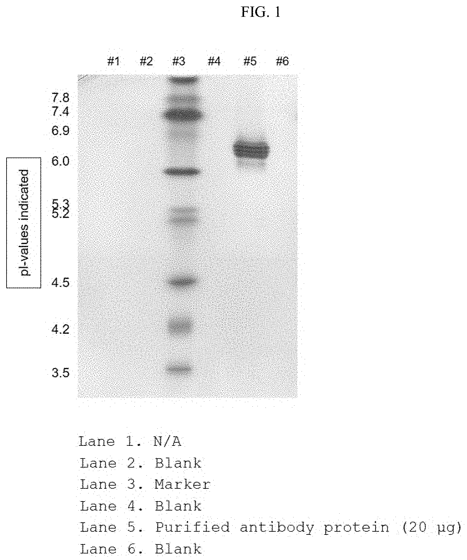
[0020] FIG. 1 shows the result of identifying the isoelectric point of an antibody by separating a purified antibody protein based on an isoelectric point;
[0021] FIG. 2 shows the results of identifying the immunological characteristics of the purified antibody protein through western blotting; and
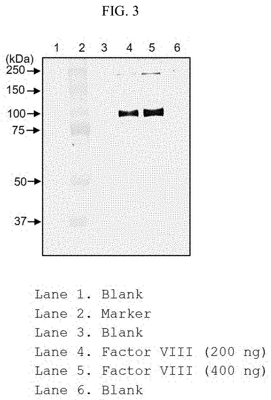
[0022] FIG. 3 shows the result of identifying whether or not the purified anti-FVIII antibody protein specifically binds to an antigenic recombinant coagulation factor FVIII through antigen-antibody reaction.
BEST MODE
[0023] In one aspect, the present invention is directed to an antibody binding specifically to a coagulation factor VIII or an antigen-binding fragment thereof including: a heavy chain CDR selected from the group consisting of SEQ ID NOS: 1 to 3; and a light chain CDR selected from the group consisting of SEQ ID NOS: 4 to 6.
[0024] The term "antibody" as used herein means a protein molecule including an immunoglobulin molecule that specifically recognizes an antigen and thus immunologically reacts with the specific antigen, and includes a whole antibody binding specifically to a coagulation factor VIII as well as an antigen-binding fragment of the antibody molecule.
[0025] The whole antibody is composed of two full-length light chains and two full-length heavy chains, wherein each light chain is linked to a heavy chain by a disulfide bond. In mammals, five antibody isotypes, known as IgA, IgD, IgE, IgM, and IgG, exist and IgG is further classified into the four antibody subtypes IgG1, IgG2, IgG3 and IgG4.
[0026] The term "antibody fragment" as used herein refers to a minimal fragment that maintains an antigen-binding ability and includes Fab, F(ab'), F(ab').sub.2, and Fv. Fab includes a variable region of each of the heavy chain and the light chain, the constant domain of the light chain, and the first constant domain (CH1) of the heavy chain, each having an antigen-binding site. Fab' is different from Fab in that it further includes at least one cysteine residue at a C-terminus of the CH1 domain of the heavy chain. F(ab').sub.2 includes two Fab' molecules having a disulfide bond between cysteine residues in a hinge region. An Fv (variable fragment) including a variable region of each of the heavy chain and the light chain is the minimal antibody fragment having original specificity for the parent immunoglobulin. Disulfide-stabilized Fv (dsFv) is formed by binding the variable region of the light chain to the variable region of the heavy chain via a disulfide bond. Single-chain Fv (scFV) is an Fv where the respective variable regions of the heavy chain and the light chain are covalently linked via a peptide linker. These antibody fragments can be obtained by treating the whole antibody with a protease (for example, papain or pepsin providing Fab or F(ab').sub.2), and are preferably constructed using genetic recombination technology.
[0027] In one embodiment, the antibody according to the present invention is a FV (for example, scFV) or complete antibody type. In addition, the heavy-chain constant region may be selected from .gamma., .mu., .alpha., .delta. and .epsilon. isotypes. The light-chain constant region may be a .kappa. or .lamda. type.
[0028] The term "heavy chain" as used herein may be interpreted to include a full-length heavy chain including a variable region domain VH including an amino acid sequence having a variable region sequence sufficient to impart specificity to an antigen and three constant region domains CH1, CH2 and CH3 and a fragment thereof. Also, the term "light chain" as used herein may be interpreted to include a full-length light chain including a variable region domain VL including an amino acid sequence having a variable region sequence sufficient to impart specificity to an antigen and a constant region domain CL and a fragment thereof.
[0029] In general, immunoglobulin has a basic structural unit including two heavy chains and two light chains. Each heavy chain includes one variable region and three constant domains, whereas each light chain includes one variable region and one constant domain. The variable region of each of the heavy chain and the light chain includes three complementarity-determining regions (referred to as "CDRs") and four framework regions. CDRs function to bind to epitopes of antibodies. CDRs on each chain start from the N-terminus and are aligned in the order CDR1, CDR2, and CDR3. These CDRs are distinguished from one another by the chain on which they are positioned.
[0030] The antibody according to the present invention, for example, includes a heavy chain variable region including CDR1 of SEQ ID NO: 1, CDR2 of SEQ ID NO: 2 and CDR3 of SEQ ID NO: 3, and a light chain variable region including CDR1 of SEQ ID NO: 4, CDR2 of SEQ ID NO: 5, and CDR3 of SEQ ID NO: 6.
[0031] In addition, the antibody according to the present invention may include: a heavy chain variable region framework region (FR) selected from the group consisting of SEQ ID NOS: 7 to 10; and a light chain variable region framework region (FR) selected from the group consisting of SEQ ID NOS: 11 to 14.
[0032] In addition, the antibody according to the present invention may include a heavy chain variable region of SEQ ID NO: 15 and a light chain variable region of SEQ ID NO: 16, and may include a heavy chain of SEQ ID NO: 17 and a light chain of SEQ ID NO: 18.
[0033] The antibody according to the present invention includes polyclonal antibodies, monoclonal antibodies, whole antibodies and antibody fragments. In addition, chimeric antibodies (e.g., humanized mouse antibodies), bivalent or bispecific molecules (e.g., bispecific antibodies), diabodies, triabodies and tetrabodies fall within the scope of the antibody used in the present invention.
[0034] The term "monoclonal antibody" as used herein refers to an antibody molecule having a uniform molecule composition which is obtained from a substantially identical population of antibodies and exhibits binding specificity and affinity to a single epitope.
[0035] A "humanized"-type non-human (e.g., murine) antibody is a chimeric antibody that contains a minimal sequence derived from non-human immunoglobulin. In most cases, the humanized antibody is a human immunoglobulin (acceptor antibody) in which a residue from the hypervariable region of an acceptor is replaced by a residue from the hypervariable region of a non-human species (donor antibody) having the desired specificity, affinity and ability, such as a mouse, rat, rabbit or non-human primate.
[0036] The term "human antibody" as used herein refers to a molecule that consists entirely of amino acid sequences of all components of human immunoglobulin including CDRs, framework regions and the like. Human antibodies have at least three potential benefits in the treatment of human diseases. First, human antibodies further preferably interact with the human immune system to mediate more effectively the destruction of target cells by, for example, complement-dependent cytotoxicity (CDC) or antibody-dependent cell-mediated cytotoxicity (ADCC). Another benefit is that the human immune system does not recognize the human antibody as being an exogenic molecule so that adverse effects caused by formation of immune complexes that may occur in chimeric antibodies or humanized antibodies can be minimized when administered to the human body. Third, such formation of immune complexes may lead to shortened half-lives of chimeric or humanized antibody preparations administered to the human body, but the half-lives of human antibodies are similar to those of antibodies naturally derived from the human circulatory system, even when administered in smaller amounts or at a lower frequency.
[0037] When the antibody according to the present invention includes a constant domain, it may be derived from IgG, IgA, IgD, IgE, IgM, or a combination or hybrid thereof.
[0038] The term "combination" as used herein means that a polypeptide encoding a single-chain immunoglobulin Fc fragment having the identical origin is linked to a single-chain polypeptide having a different origin in order to produce a dimer or multimer. Such a dimer or multimer may be produced from two or more constant domains selected from the group consisting of the constant domains of IgG, IgA, IgD, IgE and IgM.
[0039] The term "hybrid" as used herein means that sequences encoding two or more heavy chain constant domains having different origins are present in a single-chain immunoglobulin heavy chain constant domain. For example, a domain hybrid may be composed of one to four domains selected from the group consisting of CH1, CH2, CH3 and CH4 of IgG, IgA, IgD, IgE and IgM. In addition, a combination of hybrids may be formed from heavy chain constant domains of IgG subtypes, i.e., IgG1, IgG2, IgG3 and IgG4. The combination of hybrids is as defined above.
[0040] The antibody or antibody fragment of the present invention may include the sequence of the anti-coagulation factor VIII antibody mentioned herein as well as biological equivalents thereof, as long as it meets the criterion of specifically recognizing the coagulation factor VIII. For example, additional changes can be made to the amino acid sequence of the antibody in order to further improve the binding affinity and/or other biological properties of the antibody. Such modifications include, for example, deletion, insertion and/or substitution of the amino acid sequence residues of the antibody. Such amino acid variations are based on the relative similarity of amino acid side chain substituents, such as the hydropathicity, hydrophilicity, charge and size thereof. It can be seen through analysis of the size, shape and type of amino acid side chain substituents that all of arginine, lysine and histidine are classified as basic positively-charged residues; alanine, glycine and serine are classified as amino acids which are similar and small in size; and phenylalanine, tryptophan and tyrosine are classified as amino acids having aromatic side chains. Thus, based on these considerations, arginine, lysine and histidine; alanine, glycine and serine; and phenylalanine, tryptophan and tyrosine are considered to be biologically functional equivalents.
[0041] The hydropathic index of amino acids may be considered in introducing mutations. Each amino acid is given a hydropathic index depending on a hydropathicity and charge thereof: isoleucine (+4.5); valine (+4.2); leucine (+3.8); phenylalanine (+2.8); cysteine (+2.5); methionine (+1.9); alanine (+1.8); glycine (-0.4); threonine (-0.7); serine (-0.8); tryptophan (-0.9); tyrosine (-1.3); proline (-1.6); histidine (-3.2); glutamate (-3.5); glutamine (-3.5); aspartate (-3.5); asparagine (-3.5); lysine (-3.9); and arginine (-4.5).
[0042] The hydropathic index of amino acids is very important in imparting the interactive biological function of proteins. It is well-known that substitution with an amino acid having a similar hydropathic index is needed in order to retain similar biological activity. When a mutation is introduced with reference to a hydropathic index, substitution is made between amino acids having a hydropathic index difference preferably within .+-.2, more preferably within .+-.1, and even more preferably within .+-.0.5.
[0043] Meanwhile, it is also well-known that substitution between amino acids having similar hydrophilicity values leads to proteins having equivalent biological activity. As disclosed in U.S. Pat. No. 4,554,101, the following hydrophilicity values are given to respective amino acid residues: arginine (+3.0); lysine (+3.0); aspartate (+3.0.+-.1); glutamate (+3.0.+-.1); serine (+0.3); asparagine (+0.2); glutamine (+0.2); glycine (0); threonine (-0.4); proline (-0.5.+-.1); alanine (-0.5); histidine (-0.5); cysteine (-1.0); methionine (-1.3); valine (-1.5); leucine (-1.8); isoleucine (-1.8); tyrosine (-2.3); phenylalanine (-2.5); and tryptophan (-3.4).
[0044] Amino acid exchange in proteins that does not entirely change the activity of the molecule is known in the art (H. Neurath and R. L. Hill (Eds), "The Proteins", Academic Press, New York, 1979). The most common exchange is exchange between the amino acid residues Ala/Ser, Val/Ile, Asp/Glu, Thr/Ser, Ala/Gly, Ala/Thr, Ser/Asn, Ala/Val, Ser/Gly, Thr/Phe, Ala/Pro, Lys/Arg, Asp/Asn, Leu/Ile, Leu/Val, Ala/Glu and Asp/Gly.
[0045] When taking into consideration variations having biologically equivalent activity, the antibody or a nucleotide molecule encoding the same according to the present invention is interpreted to include a sequence having substantial identity with the sequence set forth in the sequence number. The term "substantial identity" means that a sequence has a homology of at least 61%, more preferably a homology of 70%, even more preferably a homology of 80%, and most preferably a homology of 90%, when aligning the sequence of the present invention with any other sequence so as to correspond to each other as much as possible and analyzing the aligned sequence using algorithms commonly used in the art. The NCBI Basic Local Alignment Search Tool (BLAST) is accessible from NCBI or the like and can be used in conjunction with sequence analysis programs such as BLASTP, BLASM, BLASTX, TBLASTN and TBLASTX over the Internet. BLAST is available at www.ncbi.nlm.nih.gov/BLAST/. A method of comparing a sequence homology using this program can be found at www.ncbi.nlm.nih.gov/BLAST/blast_help.html.
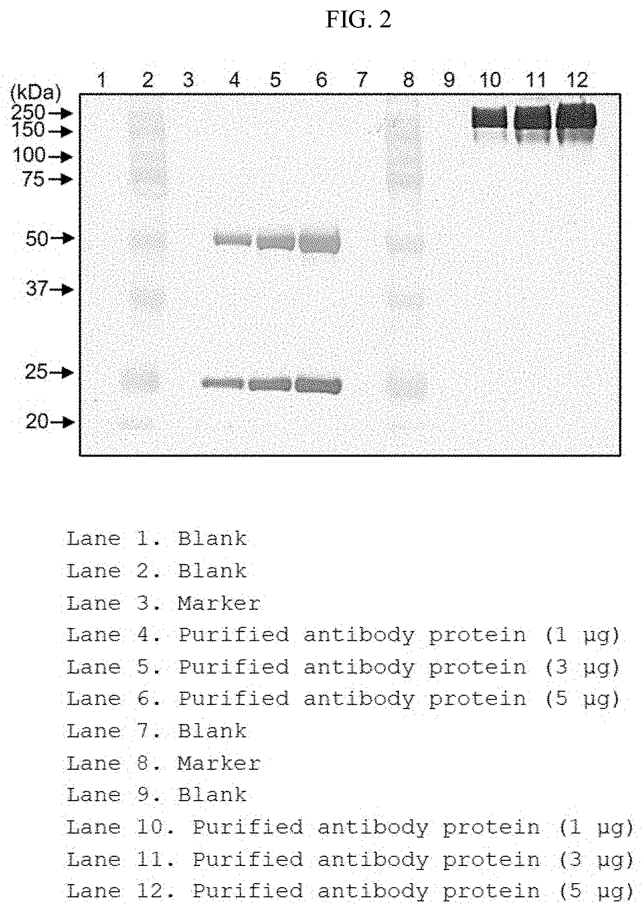
[0046] In another aspect, the present invention is directed to a polynucleotide encoding the antibody or an antigen-binding fragment thereof.
[0047] By isolating the polynucleotide encoding the antibody or an antigen-binding fragment thereof according to the present invention, an antibody or antigen-binding fragment thereof can be produced recombinantly. The polynucleotide is isolated and inserted into a replicable vector for further cloning (amplification of DNA) or further expression. Based on this, in another aspect, the present invention is directed to a vector including the polynucleotide.
[0048] The term "nucleotide" is intended to encompass both DNA (gDNA and cDNA) and RNA molecules, and a nucleotide, of which is a basic constituent unit, includes naturally derived nucleotides as well as analogues wherein sugar or base moieties are modified. The sequence of the polynucleotide encoding heavy and light chain variable regions of the present invention can be varied. Such variation includes addition, deletion, or non-conservative or conservative substitution of nucleotides.
[0049] In one embodiment, the present invention may be a polynucleotide encoding the heavy chain variable region of SEQ ID NO: 20 or a polynucleotide encoding the light chain variable region of SEQ ID NO: 21. In another embodiment, the present invention may be a polynucleotide encoding the heavy chain of SEQ ID NO: 22 or a polynucleotide encoding the light chain of SEQ ID NO: 23.
[0050] The present invention is interpreted to include a nucleotide sequence having substantial identity with the nucleotide sequence. The nucleotide sequence having substantial identity means a nucleotide sequence that has a homology of at least 80%, more preferably a homology of at least 90%, and most preferably a homology of at least 95%, when aligning the sequence of the present invention with any other sequence so as to correspond to each other as much as possible and analyzing the aligned sequence using algorithms commonly used in the art.
[0051] The DNA encoding the antibody can be easily separated or synthesized using conventional procedures (for example, using an oligonucleotide probe capable of specifically binding to DNA encoding heavy and light chains of the antibody). A variety of vectors are obtainable. Vector components generally include, but are not limited to, one or more of the following components: 1) signal sequences (signal peptides), 2) replication origins, 3) one or more marker genes, 4) enhancer elements, and 5) promoters and transcription termination sequences.
[0052] As used herein, the term "vector" refers to a means for expressing target genes in host cells and includes plasmid vectors and cosmid vectors, as well as viral vectors such as bacteriophage vectors, adenovirus vectors, retroviral vectors and adeno-associated viral vectors (AAV). The polynucleotide encoding the antibody in the vector is operably linked to a promoter.
[0053] The term "operably linked" means a functional linkage between a polynucleotide expression regulation sequence (e.g., promoter, signal sequence or array of transcription regulator binding site) and another polynucleotide sequence, and is regulated by transcription and/or translation of the polynucleotide sequence.
[0054] When a prokaryotic cell is used as a host, the vector generally includes a potent promoter capable of conducting transcription (such as tac promoter, lac promoter, lacUV5 promoter, ipp promoter, pLX promoter, pRX promoter, rac5 promoter, amp promoter, recA promoter, SP6 promoter, trp promoter, or T7 promoter), a ribosome binding site for initiation of translation, and a transcription/translation termination sequence. In addition, for example, when a eukaryotic cell is used as a host, the vector includes a promoter (e.g., a metallothionein promoter, a R-actin promoter, a human hemoglobin promoter and a human muscle creatine promoter) derived from the genome of mammalian cells, or a promoter derived from an animal virus such as adenovirus late promoter, vaccinia virus 7.5K promoter, SV40 promoter, cytomegalovirus (CMV) promoter, HSV tk promoter, mouse mammary tumor virus (MMTV) promoter, HIV LTR promoter, Moloney virus promoter, Epstein Barr virus (EBV) promoter, and Rous sarcoma virus (RSV) promoter, and generally has a polyadenylation sequence as a transcription termination sequence.
[0055] Optionally, the vector may be fused with another sequence in order to facilitate purification of the antibody expressed therefrom. The sequence to be fused includes, for example, glutathione S-transferase (Pharmacia, USA), maltose-binding protein (NEB, USA), FLAG (IBI, USA), 6.times. His (hexahistidine; Qiagen, USA) and the like.
[0056] The vector includes antibiotic-resistant genes commonly used in the art as selectable markers, and examples thereof include genes resistant to ampicillin, gentamycin, carbenicillin, chloramphenicol, streptomycin, kanamycin, geneticin, neomycin and tetracycline.
[0057] In another aspect, the present invention is directed to a cell transformed with the above-mentioned vector. The cell used to produce the antibody of the present invention may be a prokaryote, yeast or higher eukaryotic cell, but is not limited thereto.
[0058] Prokaryotic host cells such as Escherichia coli, the genus Bacillus such as, Bacillus subtilis and Bacillus thuringiensis, Streptomyces spp., Pseudomonas spp. (for example, Pseudomonas putida), Proteus mirabilis and Staphylococcus spp. (for example, Staphylococcus carnosus) can be used.
[0059] Interest in animal cells is the greatest, and examples of useful host cell lines include, but are not limited to, COS-7, BHK, CHO, CHOK1, DXB-11, DG-44, CHO/-DHFR, CV1, COS-7, HEK293, BHK, TM4, VERO, HELA, MDCK, BRL 3A, W138, Hep G2, SK-Hep, MMT, TRI, MRC 5, FS4, 3T3, RIN, A549, PC12, K562, PER.C6, SP2/0, NS-0, U20S, or HT1080.
[0060] In another aspect, the present invention is directed to a method for producing the antibody or an antigen-binding fragment thereof, including: culturing the recombinant host cells to produce an antibody or an antigen-binding fragment thereof and recovering the antibody or an antigen-binding fragment thereof, followed by isolation and purification.
[0061] The cells can be cultured in various media. Any commercially available medium can be used as a culture medium without limitation. All other essential supplements well-known to those skilled in the art may be included in appropriate concentrations. Culture conditions such as temperature and pH are conventionally used with host cells selected for expression, which will be apparent to those skilled in the art.
[0062] The recovery of the antibody or antigen-binding fragment thereof can be carried out, for example, by centrifugation or ultrafiltration to remove impurities and purification of the resulting product using, for example, affinity chromatography. Other additional purification techniques such as anion or cation exchange chromatography, hydrophobic interaction chromatography and hydroxyapatite (HA) chromatography may be used.
[0063] In another aspect, the present invention is directed to a column for isolating or purifying a recombinant coagulation factor VIII, to which the antibody or an antigen-binding fragment thereof binds.
[0064] "Isolation and purification" refers to a series of methods/processes for distinguishing a certain protein, e.g., coagulation factor VIII, from impurities present in a complex mixture and removing other impurities in order to enhance purity or quality. Depending on the anti-coagulation factor VIII antibody that is attached to the column, the coagulation factor VIII specifically binding to the antibody is detected from the impurities present in the sample. The impurities may for example include host cell proteins, host cell residues, cell lysates and proteins, DNA, endotoxins, and culture factors for cell growth.
[0065] The term "column" as used herein refers to a device filled with a stationary phase used for a chromatographic procedure for the separation, detection or purification of a target protein (such as recombinant coagulation factor VIII) from a complex mixture using the physical-chemical properties of the target protein. Specifically, the target protein can be selectively separated from a mixture or an impurity by producing a column filled with the stationary phase depending on the degree of hydrophilicity or hydrophobicity of the target material, the potential of the molecule, the possibility of binding to a specific substance, and the like.
[0066] The column may be filled with a stationary phase material having affinity for coagulation factor VIII, and the stationary phase material may be, for example, a resin or an agarose bead. As described in one embodiment of the present invention, produced is a column for separation or purification of a recombinant coagulation factor VIII, which is filled with a stationary phase in which an anti-coagulation factor VIII antibody, serving as a ligand, is linked to Sepharose, which is a resin in the form of a cross-linked agarose bead.
[0067] In another aspect, the present invention is directed to a method of purifying a coagulation factor VIII including allowing a sample containing a recombinant coagulation factor VIII to interact with a ligand linked to the stationary phase of the column.
[0068] The sample to be loaded into the column may be, for example, a cell culture liquid or a fermentation broth, and when the sample is loaded into the column and the purification process is performed, host cell proteins and host cell residues other than the target protein, for example, cell lysates and proteins, DNA and endotoxins, can be removed.
[0069] The recombinant coagulation factor VIII contained in the sample binds to the anti-coagulation factor VIII antibody linked to the column stationary phase through an antigen-antibody reaction. Then, the recombinant coagulation factor VIII binding to the anti-coagulation factor VIII antibody can be isolated through a separate elution procedure. The composition and elution conditions of the eluate can be set to conditions that can be implemented by those skilled in the art and can be combined with a suitable buffer or liquid to provide a mobile phase.
[0070] This allows substantially pure recombinant coagulation factor VIII to be isolated and purified. "Substantially pure" means that substantially all of the material other than the recombinant coagulation factor VIII is removed, and advantageously about 80% or more, for example, about 95% or more, i.e., 95 to 100%, for example, about 98% or more, i.e., 98 to 100%, preferably about 99% or more, i.e., 99 to 100%, based on the total amount of a contaminant, may be removed. The degree of purity that is possible may be determined based on the concentration of the recombinant coagulation factor VIII in the sample applied to the column and other conditions used.
[0071] Hereinafter, the present invention will be described in more detail with reference to examples. However, it will be obvious to those skilled in the art that these examples are provided only for illustration of the present invention and should not be construed as limiting the scope of the present invention.
Example 1: Production of Hybridoma
[0072] The recombinant coagulation factor VIII protein to be used for vaccination (immunization) was prepared through isolation from a cell culture solution and purification in order to produce a monoclonal antibody of the recombinant coagulation factor VIII protein (SEQ ID NO: 19). Balb/c mice were used for immunization.
[0073] The recombinant coagulation factor VIII protein and an antigen adjuvant (Freund's complete adjuvant) were thoroughly mixed in equivalent amounts, and the resulting mixture was intraperitoneally injected into the Balb/c mice. 25 days later, the recombinant coagulation factor VIII protein and the antigen adjuvant were mixed in equivalent amounts, the resulting mixture was further injected into the mice, the serum was collected from the mice, and additional injection was conducted until the recombinant coagulation factor VIII protein showed an excellent positive reaction.
[0074] On the third day after the last vaccination (immunization), the serum was collected from the mice and whether or not antibodies were produced in the serum was identified through enzyme-linked immunosorbent assay (ELISA). Then, the abdomen of each mouse was opened and the spleen was taken out and pulverized. The pulverized splenocytes were fused with mouse myeloma cells (SP2/0) according to a known method (Galfre G. et al., Nature 266, 550-552 (1977)). Mouse splenocytes and mouse myeloma cells equivalent to 1/5 of the splenocytes were prepared and mixed with PEG1500, followed by inducing a fusion reaction. After the fusion reaction, the cells were suspended in a selective medium containing HAT (hypoxanthine, aminopterin, thymidine) and transferred to a 96-well plate to inhibit the growth of unfused cells. When the fused cells were sufficiently grown while the medium was replaced with a selective medium containing HAT, analysis was conducted through ELISA specific for coagulation factor VIII protein, and cells showing a positive reaction were transferred to a new plate and then cultured therein. When the count of cells was sufficient, single clonal production cell line selection was conducted through limiting dilution. 33 clones showing positive responses were selected through ELISA using the culture solution of the well where colonies were formed. After identifying the binding ability of the culture solution of selected clones to the antigen using Biacore, two clones (3F6-2-5 and 5F4-9-1), which were considered to be the most suitable for use as antibodies for purifying the recombinant coagulation factor VIII protein, were selected. 3F6-2-5, which has better binding ability to the antigen among the two clones, was selected as an antibody-producing hybridoma clone for the recombinant coagulation factor VIII protein. The results of analysis of the isotype of the antibody produced by the 3F6-2-5 clone showed that the heavy chain was IgG1 and the light chain was an antibody belonging to the kappa group.
Example 2: Antibody Gene Securing
[0075] The heavy and light chain genes of an antibody against a recombinant coagulation factor VIII protein (FVIII) (hereinafter referred to as anti-FVIII antibody) were obtained by extracting RNA from mouse hybridoma cells (3F6-2-5) producing antibodies against FVIII, followed by RT-PCR.
[0076] In order to secure heavy chain genes, RNA was extracted from 3F6-2-5 hybridoma cells and subjected to 5' RACE. GSP1 (gene-specific primer), GSP2 and a nested primer for the 3' constant region were prepared, RACE PCR was performed, the resulting PCR product was cloned into the T-vector, and then the nucleotide sequence was analyzed. As a result, the nucleotide sequence of the 5' region including a signal sequence was identified. Primers were produced according to the identified nucleotide sequences and heavy chain genes were obtained through RT-PCR.
TABLE-US-00001 (SEQ ID NO: 24) GSP1: 5'-TGAGGAGACGGTGACCGTGGT (SEQ ID NO: 25) GSP2: 5'-CCTTGGCCCCAGAAGTGGTAA (SEQ ID NO: 26) Nested: 5'-TAAATGCCAGTGICTICAGC
[0077] After identifying the nucleotide sequences, primers were constructed so as to include the 5'-Nhe I and 3'-Xho I restriction enzyme recognition sequences in order to clone heavy chain genes into the Pangen expression vector (pPGIX), and then the heavy chain genes inserted into the T-vector were amplified. The PCR products and expression vectors were treated with Nhe I and Xho I restriction enzymes, pPGIX-anti-FVIII-HC was produced, and the nucleotide sequence of the inserted heavy chain gene was identified (SEQ ID NO: 22).
[0078] The light chain genes were obtained by extracting RNA from 3F6-2-5 hybridoma cells and conducting RT-PCR using 11 types of 5'-primers and primer (MKCIII) for 3'-constant regions produced based on the known signal sequence.
[0079] The results of RT-PCR identified a 700 bp band including the light chain variable region and the constant region in the PCR product using the MKV7 primer and the 3'-MKCIII primer. The obtained PCR products were cloned into the T-vector, and the nucleotide sequences were then identified.
TABLE-US-00002 MKV1: (SEQ ID NO: 27) 5'-GCT AGC GCC ACC ATG AAG TTG CCT GTT AGG CTG TTG GTG CTG MKV2: (SEQ ID NO: 28) 5'-GCT AGC GCC ACC ATG GAG WCA GAC ACA CTC CTG YTA TGG GTG MKV3: (SEQ ID NO: 29) 5'-GCT AGC GCC ACC ATG AGT GTG CTC ACT CAG GTC CTG GSG TTG MKV4: (SEQ ID NO: 30) 5'-GCT AGC GCC ACC ATG AGG RCC CCT GCT CAG WTT YTT GGM WTC MKV5: (SEQ ID NO: 31) 5'-GCT AGC GCC ACC ATG GAT TTW CAG GTG CAG ATT WTC AGC TTC MKV6: (SEQ ID NO: 32) 5'-GCT AGC GCC ACC ATG AGG TKC YYT GYT SAG YTY CTG RGG MKV7: (SEQ ID NO: 33) 5'-GCT AGC GCC ACC ATG GGC WTC AAG ATG GAG TCA CAK WYY CWG G MKV8: (SEQ ID NO: 34) 5'-GCT AGC GCC ACC ATG TGG GGA YCT KTT TYC MMT TTT TCA ATT G MKV9: (SEQ ID NO: 35) 5'-GCT AGC GCC ACC ATG GTR TCC WCA SCT CAG TTC CTT G MKV10: (SEQ ID NO: 36) 5'-GCT AGC GCC ACC ATG TAT ATA TGT TTG TTG TCT ATT TCT MKV11: (SEQ ID NO: 37) 5'-GCT AGC GCC ACC ATG GAA GCC CCA GCT CAG CTT CTC TTC C MKCIII: (SEQ ID NO: 38) 5'-CTA ACA CTC ATT CCT GTT GAA GCT C
[0080] After identifying the nucleotide sequences, primers were prepared so as to include the 5'-Nhe I and 3'-Xho I restriction enzyme recognition sequences in order to clone the light chain genes into the Pangen's expression vector (pPGIX), and the light chain genes inserted into the T-vector were then amplified using PCR. The PCR products and expression vectors were treated with Nhe I and Xho I restriction enzymes and then ligated to produce pPGIX-anti-FVIII-LC, and then the nucleotide sequence of the inserted light chain gene was identified (SEQ ID NO: 23). The heavy and light chain genes inserted into the expression plasmids, pPGIX-Anti-FVIII-HC and pPGIX-anti-FVIII-LC, were transferred to the Pangen's expression vector (pPGX) and used for transfection in order to develop a cell line having higher expression efficiency. The pPGX-anti-FVIII-HC and pPGX-Anti-FVIII-LC plasmid DNA was treated with Nhe I and Xho I restriction enzymes and then ligated to pPGX treated with the same restriction enzymes, to construct pPGX-Anti-FVIII-HC and pPGX-Anti-FVIII-LC plasmid DNA, respectively.
Example 3: CHO Cell Expression
[0081] The transfection of the expression plasmids with the CHO DG44 host cell line was carried out in 24 wells, and pPGX-anti-FVIII-HC, pPGX-anti-FVIII-LC and pDCH1P (dhfr) were simultaneously transfected by electroporation. When the cells were fully grown after being cultured in a 37.degree. C., 5% CO.sub.2 incubator, the cells were cultured in a selective medium so as to grow only the transformed cells. Approximately two weeks later, when the cells were sufficiently grown, a portion of the culture supernatant was taken and the anti-FVIII-antibody-expressing cell group was selected through ELISA analysis. In order to select stable cell lines with high expression efficiency, single cell selection was performed by selecting five cell lines (ADBA1001, ADBA1003, ADBA1011, ADBA1013, ADBA1014), based on the results of the ELISA analysis. Cells were seeded in a 96-well plate at a density of 1 cell/well in the culture medium, and colonies formed after about 4 weeks were analyzed to select cell lines having high expression efficiency, and the cell lines were cultured. When a sufficient number of cells were obtained, the expression efficiency of monoclonal cell lines was comparatively analyzed through ELISA. Six monoclonal cell lines having high production efficiency (ADBA1003-22, ADBA1011-19, ADBA1013-14, ADBA1013-27, ADBA1014-11, ADBA1014-69) were selected as candidate cell lines.
[0082] Six monoclonal cell lines having high antibody productivity were identified to stably express antibody proteins during 90 days of long-term subculture. Among them, the ADBA1013-14 cell line having the highest antibody productivity was selected as the final cell line. It was identified that the antibody produced therefrom had the sequence shown in Table 1.
TABLE-US-00003 TABLE 1 CDR1 CDR2 CDR3 Heavy region GFTFSDAWMD EIRSKAKNHATNY YHF CDR (SEQ ID NO: 1) AESVKG (SEQ ID NO: 3) (SEQ ID NO: 2) Heavy region EVKIEESGGGLVQPGGSMKLSCAASGFTFSDAWMDWVRQSPEK variable region GLEWVAEIRSKAKNHATNY AESVKGRFT ISRDDSKSRVYLQMNTLRAE DTGIYYCTNYHFWGPGTTLT VS Light chain KSSQSLLNSINQEN FGSTRES QQHYSTPYT CDR YLA (SEQ ID NO: 5) (SEQ ID NO: 6) (SEQ ID NO: 4) Light chain DIVMTQ SPSSLAMSVG QKVTMSCKSS variable region QSLLNSINQENYLAWYQQKP GQSPKLLIY FGSTRESGVPD RFIGSGSGTD FSLTISDVQA EDLADYFCQQHYSTPYTFGG GTKLEMKRA
Example 4: Antibody Purification
[0083] Antibody proteins were isolated and purified using the culture solution of the ADABA1013-14 cell line. Affinity chromatography was performed using a column filled with a protein A resin. Columns were mounted in an AKTA Pure system (GE healthcare) and were equilibrated with an equilibrium buffer of 20 mM sodium phosphate and 150 mM sodium chloride (pH 7.4), and the culture solution was loaded thereon. Then, the column was washed and only an antibody protein was eluted with 20 mM sodium citrate (pH 3.5) as an elution buffer. For the virus inactivation of the eluted protein fraction, the pH of the eluate was lowered to 3.5 with 1N HCl and the eluate was reacted at room temperature for 180 minutes.
[0084] After virus inactivation, the eluate were diluted with four times the volume of water for injection and subjected to Q Sepharose column chromatography. Columns were mounted in an AKTA Pure system (GE healthcare), were equilibrated with an equilibrium buffer of 4 mM citric acid and 10 mM sodium phosphate, and the samples were loaded thereon. The flowthrough (effluent) was collected during sample loading, filtered through a 0.22 .mu.m PES membrane, and subjected to nanofiltration.
Example 5: Characterization of Purified Antibodies
[0085] 1. N-Glycosylation Site Determination
[0086] The N-glycosylation site of the purified antibody protein was identified using peptide-mapping analysis.
[0087] The purified antibody protein was desalted and concentrated using a trichloroacetic acid precipitation method. The obtained sample was reduced and alkylated through treatment with Trypsin and Trypsin/PNGase F, followed by treatment with DTT and iodoacetamide. The results of analysis of glycosylated asparagine amino acid positions through LC-MS/MS peptide sequencing analysis using the sample before and after treatment with PNGase F showed that the sugar was bonded to Asparagine No. 288 of the heavy chain.
[0088] 2. Identification (Isoelectric Focusing) of Isoelectric Point (pI)
[0089] The purified antibody protein was separated according to the isoelectric point (pI) to determine the isoelectric point of the antibody.
[0090] 20 .mu.g of an IEF marker (pI 3-10) and the purified antibody protein were loaded onto a Novex.RTM. pH 3-7 IEF gel and electrophoresis was performed for 1 hour, 1 hour, and 30 minutes at voltages of 100V, 200V, and 500V, respectively. The gel was fixed in 12% TCA (trichloroacetic acid) for 30 minutes, washed three times with ultrapure water for 10 minutes, and treated until staining with GelCode Blue Stain Reagent was sufficient. After the staining was completed, the antibody was washed with ultrapure water, a pI band was identified with the naked eye, and the pI value was analyzed using a Gel Doc XR+imager.
[0091] The pI of the IEF band was analyzed using a Gel Doc XR+imager. As a result, the pI of the purified antibody protein was found to be 6.2 to 6.8 (FIG. 1).
[0092] 3. Intact Molecular Weight Determination
[0093] The one-dimensional structure of the protein was identified by measuring the molecular weight of the purified antibody protein.
[0094] The purified antibody protein diluted to 1 mg/mL was treated with a 1M DTT solution at a final concentration of 20 mM and reacted at room temperature for 40 minutes, and the molecular weight of the heavy chain/light chain was measured using LC-MS.
[0095] The peptides isolated during HPLC were connected to the ESI source of the Q-TOF MS to determine the mass value of the peptide ions. The molecular weight was determined using DataAnalysis software and analyzed by deconvolution using a maximum entropy method.
[0096] The molecular weight of the native form of the purified antibody protein was found to be 147,859 to 148,394 Da. This was about 3,000 Da higher than the molecular weight of the theoretical antibody protein, excluding the molecular weight of sugar, which was considered to be the average of the molecular weights of sugars bonded to two heavy chains. The molecular weight of the light chain was measured to be 24,323 Da (.+-.2 Da), which was exactly the same as the light chain molecular weight of the theoretical antibody protein. The major molecular weight of the heavy chain was detected to be 49,715 Da. This was increased by about 1,300 Da compared to the molecular weight of the theoretical heavy chain, which is considered to be due to the molecular weight of the sugar bonded to the Asn 288 of the heavy chain.
[0097] 4. Western Blotting
[0098] The immunological characteristics of the purified antibody protein through antigen-antibody reaction were analyzed.
[0099] The purified antibody protein was diluted to 1 mg/mL, 1, 3, and 5 .mu.g of the dilution were prepared, and PBS was added thereto at a final volume of 10 .mu.L. After mixing 2.5 .mu.L of a 5.times. non-reducing sample buffer with 2.5 .mu.L of a 5.times. reducing sample buffer, the sample was prepared under reducing conditions by boiling at 95.degree. C. or higher for 10 minutes and cooling on the ice for 5 minutes. The prepared sample was loaded on a 10% SDS-PAGE gel and then subjected to electrophoresis at 80V for 30 minutes and at 100V for 1 hour 30 minutes. After running, the cells were transferred to a nitrocellulose membrane and blocked with 5% skim milk for 1 hour. After blocking, the goat anti-mouse IgG, AP conjugate diluted to 1:1,000 was treated at room temperature for 2 hours and then washed with 1.times.TBST for 10 minutes three times. After washing, the membrane was treated with NBT/BCIP for protein detection to induce color development. After observing sufficient color development, the reaction was stopped by washing with running water.
[0100] As a result of western blotting, under reducing conditions, the light chain was detected at about 25 kDa and the heavy chain was detected at about 50 kDa, and under the non-reducing conditions, the antibody protein was detected at about 150 kDa (FIG. 2). Thus, it was identified that the purified antibody protein, mouse IgG, specifically binds to anti-mouse IgG antibody.
[0101] 5. Identification of Specific Binding to FVIII
[0102] Whether or not the purified anti-FVIII antibody protein specifically binds to the antigen, recombinant coagulation factor FVIII was identified through antigen-antibody reaction.
[0103] 200 and 400 ng of the recombinant coagulation factor FVIII diluted to 0.02 mg/mL were prepared and PBS was added thereto to achieve a final volume of 20 .mu.L. The resulting mixture was mixed with 5 .mu.L of a 5.times. reducing sample buffer, boiled at a temperature of 95.degree. C. or higher for 10 minutes and cooled on ice for 5 minutes to prepare a sample. The prepared sample was loaded on a 7.5% gel and then subjected to electrophoresis at 80V for 30 minutes and at 100V for 1 hour 30 minutes. After running, the cells were transferred to a nitrocellulose membrane and blocked with 5% skim milk for 1 hour. After blocking, the purified antibody protein (mouse IgG), diluted to 1:1,000, was treated at 4.degree. C. overnight and then washed with 1.times.TBST for 10 minutes three times. After washing, the secondary antibody (goat anti-mouse IgG, AP conjugate) diluted to 1:5,000 was reacted at room temperature for 2 hours and then washed three times for 10 minutes with 1.times.TBST. The membrane was treated with NBT/BCIP to induce color development for protein detection. After observing sufficient color development, the reaction was stopped by washing with running water.
[0104] As a result of western blotting, as shown in the drawing, bands were detected in the vicinity of 90 kDa corresponding to the heavy chain of the coagulation factor FVIII, which means that the purified antibody protein specifically binds to the coagulation factor FVIII (FIG. 3).
[0105] 6. Antibody Binding Affinity (SPR)
[0106] The kinetic affinity (Ka, Kd, KD) between the purified antibody protein and the recombinant coagulation factor FVIII protein through antigen-antibody reaction was measured by a SPR (surface plasmon resonance) method.
[0107] The CM5 chip was mounted on a Biacore T200 instrument, and the recombinant coagulation factor FVIII protein as a ligand was diluted in a 10 mM sodium acetate buffer (pH 5.0) and injected into a sample channel for 10 minutes for immobilization. The purified antibody protein, as the analyte, was prepared at a concentration of 2 .mu.M by diluting with a buffer and was then serially diluted to ten concentrations (0.0039, 0.0078, 0.015, 0.031, 0.062, 0.125, 0.25, 0.5, 1, 2 .mu.M). Association and dissociation times were set to 3 minutes and 4 minutes, respectively, and association and dissociation were measured.
[0108] BIAevaluation analysis software was used to analyze the kinetic affinity of the purified antibody protein and the recombinant coagulation factor FVIII protein. The association force, dissociation force and dissociation constant were calculated as follows by applying ten concentrations of association/dissociation data to a 1:1 kinetic binding model (A+BAB).
[0109] As a result of the SPR test, the KD value of the purified antibody protein for the recombinant coagulation factor FVIII protein was analyzed and determined to be 5.53E.sup.-08 M.
TABLE-US-00004 TABLE 2 Name K.sub.a (M.sup.-1s.sup.-1) K.sub.d (s.sup.-1) K.sub.D (M) R.sub.max (.mu.RIU) Anti-FVIII 1.23E.sup.+04 6.80E.sup.-04 5.53E.sup.-08 701.2 antibody
Example 6: Production of Column for Antigen Purification
[0110] The Mab gel was prepared using the purified antibody to produce a column for purifying a recombinant coagulation factor FVIII protein as a hemophilia medicine.
[0111] The purified antibody was exchanged with a coupling buffer solution (sodium bicarbonate, sodium phosphate, pH 8.3) for coupling to a CNBr-activated Sepharose 4B resin.
[0112] The CNBr-activated Sepharose 4B resin was converted into a slurry by swelling with 1N HCl, mixed with the antibody exchanged with the coupling buffer, and reacted at room temperature for 2 hours. After alternately washing the resin with a washing solution with a low pH and a washing solution with a high pH, preparation of the Mab gel was completed.
Example 7: Identification of Antigen-Antibody Binding
[0113] In order to identify the binding ability of the prepared Mab gel to the recombinant coagulation factor FVIII protein, a performance test was conducted using a culture solution of the recombinant coagulation factor FVIII protein. After the Mab gel-filled column was equilibrated with an equilibration buffer, a culture solution containing 5000 IU of the recombinant FVIII protein was loaded. Nonspecifically binding proteins were removed with a washing buffer and then proteins were eluted.
[0114] The recombinant FVIII culture solution, eluate and flow-through were quantitatively analyzed by chromogenic assay. As a result of the analysis, it was confirmed that approximately 85% of the recombinant FVIII protein was bound to the Mab gel and was then eluted.
[0115] Therefore, the Mab gel for purification produced using the produced antibody was found to be capable of binding to the recombinant coagulation factor FVIII protein and thus to be suitably used for the production of an antibody resin for purification in order to commercially produce the recombinant coagulation factor FVIII protein.
Example 8 Purification of Recombinant Coagulation Factor FVIII Using Mab Gel for Purification
[0116] Immunoaffinity chromatography was conducted using a 50 L culture solution in order to identify whether or not the produced Mab gel for purification was suitable as a resin for purification in order to commercially produce the recombinant coagulation factor FVIII.
[0117] A column (diameter: 11.6 cm, height: 9.5 cm) filled with a Mab gel (about 1,000 mL) for purification was used for purification. The column was mounted in an AKTA Pure system (GE healthcare) and equilibrated. When the column was stabilized, only half of the culture solution was loaded, washed for 3 CV, and eluted for 7 to 9 CV. The remaining culture medium was loaded on the column in the same manner, immunoaffinity chromatography was performed, and the eluted recombinant coagulation factor FVIII was recovered. The recovered recombinant coagulation factor FVIII was filtered through a 0.22 .mu.m PES membrane and then stored at 4.degree. C.
[0118] A chromogenic assay was performed using pre- and post-purification samples in order to determine the purification yield of the immunoaffinity chromatography. The results of activity analysis showed that the recombinant coagulation factor FVIII contained in 50 L of the culture solution was 3.95 MIU and the purified protein was 3.25 MIU, and thus the purification yield was found to be 82.3%.
[0119] Therefore, it was considered that the Mab gel for purification produced using the produced antibody is capable of binding to the recombinant coagulation factor FVIII protein on a commercial production scale, and thus is suitable for use as an antibody resin for purification in order to commercially produce the recombinant coagulation factor FVIII protein.
INDUSTRIAL APPLICABILITY
[0120] Since the antibody or antigen-binding fragment thereof that specifically binds to a coagulation factor FVIII according to the present invention exhibits excellent affinity and binding ability to a coagulation factor FVIII, purification of high-purity recombinant coagulation factor FVIII and production of stable coagulation factor FVIII with improved quality are possible by providing a column for isolating or purifying a recombinant coagulation factor FVIII, to which the antibody or antigen-binding fragment thereof is coupled, or a method of purifying the recombinant coagulation factor FVIII using the column.
[0121] Although the present invention has been described in detail with reference to specific configurations, those skilled in the art will appreciate that this description is provided as preferred embodiments for illustrative purposes and should not be construed as limiting the scope of the present invention. Therefore, the substantial scope of the present invention is defined by the accompanying claims filed and equivalents thereto.
SEQUENCE LISTING FREE TEXT
[0122] An electronic file is attached.
Sequence CWU 1
1
37110PRTArtificial SequenceVH CDR1 1Gly Phe Thr Phe Ser Asp Ala Trp
Met Asp1 5 10219PRTArtificial SequenceVH CDR2 2Glu Ile Arg Ser Lys
Ala Lys Asn His Ala Thr Asn Tyr Ala Glu Ser1 5 10 15Val Lys
Gly317PRTArtificial SequenceVL CDR1 3Lys Ser Ser Gln Ser Leu Leu
Asn Ser Ile Asn Gln Glu Asn Tyr Leu1 5 10 15Ala47PRTArtificial
SequenceVL CDR2 4Phe Gly Ser Thr Arg Glu Ser1 559PRTArtificial
SequenceVL CDR3 5Gln Gln His Tyr Ser Thr Pro Tyr Thr1
5630PRTArtificial SequenceVH FR1 6Glu Val Lys Ile Glu Glu Ser Gly
Gly Gly Leu Val Gln Pro Gly Gly1 5 10 15Ser Met Lys Leu Ser Cys Ala
Ala Ser Gly Phe Thr Phe Ser 20 25 30714PRTArtificial SequenceVH FR2
7Trp Val Arg Gln Ser Pro Glu Lys Gly Leu Glu Trp Val Ala1 5
10839PRTArtificial SequenceVH FR3 8Tyr Ala Glu Ser Val Lys Gly Arg
Phe Thr Ile Ser Arg Asp Asp Ser1 5 10 15Lys Ser Arg Val Tyr Leu Gln
Met Asn Thr Leu Arg Ala Glu Asp Thr 20 25 30Gly Ile Tyr Tyr Cys Thr
Asn 35911PRTArtificial SequenceVH FR4 9Trp Gly Pro Gly Thr Thr Leu
Thr Val Ser Ser1 5 101023PRTArtificial SequenceVL FR1 10Asp Ile Val
Met Thr Gln Ser Pro Ser Ser Leu Ala Met Ser Val Gly1 5 10 15Gln Lys
Val Thr Met Ser Cys 201115PRTArtificial SequenceVL FR2 11Trp Tyr
Gln Gln Lys Pro Gly Gln Ser Pro Lys Leu Leu Ile Tyr1 5 10
151232PRTArtificial SequenceVL FR3 12Gly Val Pro Asp Arg Phe Ile
Gly Ser Gly Ser Gly Thr Asp Phe Ser1 5 10 15Leu Thr Ile Ser Asp Val
Gln Ala Glu Asp Leu Ala Asp Tyr Phe Cys 20 25 301312PRTArtificial
SequenceVL FR4 13Phe Gly Gly Gly Thr Lys Leu Glu Met Lys Arg Ala1 5
1014114PRTArtificial SequenceVH 14Glu Val Lys Ile Glu Glu Ser Gly
Gly Gly Leu Val Gln Pro Gly Gly1 5 10 15Ser Met Lys Leu Ser Cys Ala
Ala Ser Gly Phe Thr Phe Ser Asp Ala 20 25 30Trp Met Asp Trp Val Arg
Gln Ser Pro Glu Lys Gly Leu Glu Trp Val 35 40 45Ala Glu Ile Arg Ser
Lys Ala Lys Asn His Ala Thr Asn Tyr Ala Glu 50 55 60Ser Val Lys Gly
Arg Phe Thr Ile Ser Arg Asp Asp Ser Lys Ser Arg65 70 75 80Val Tyr
Leu Gln Met Asn Thr Leu Arg Ala Glu Asp Thr Gly Ile Tyr 85 90 95Tyr
Cys Thr Asn Tyr His Phe Trp Gly Pro Gly Thr Thr Leu Thr Val 100 105
110Ser Ser15115PRTArtificial SequenceVL 15Asp Ile Val Met Thr Gln
Ser Pro Ser Ser Leu Ala Met Ser Val Gly1 5 10 15Gln Lys Val Thr Met
Ser Cys Lys Ser Ser Gln Ser Leu Leu Asn Ser 20 25 30Ile Asn Gln Glu
Asn Tyr Leu Ala Trp Tyr Gln Gln Lys Pro Gly Gln 35 40 45Ser Pro Lys
Leu Leu Ile Tyr Phe Gly Ser Thr Arg Glu Ser Gly Val 50 55 60Pro Asp
Arg Phe Ile Gly Ser Gly Ser Gly Thr Asp Phe Ser Leu Thr65 70 75
80Ile Ser Asp Val Gln Ala Glu Asp Leu Ala Asp Tyr Phe Cys Gln Gln
85 90 95His Tyr Ser Thr Pro Tyr Thr Phe Gly Gly Gly Thr Lys Leu Glu
Met 100 105 110Lys Arg Ala 11516438PRTArtificial SequenceHeavy
Chain 16Glu Val Lys Ile Glu Glu Ser Gly Gly Gly Leu Val Gln Pro Gly
Gly1 5 10 15Ser Met Lys Leu Ser Cys Ala Ala Ser Gly Phe Thr Phe Ser
Asp Ala 20 25 30Trp Met Asp Trp Val Arg Gln Ser Pro Glu Lys Gly Leu
Glu Trp Val 35 40 45Ala Glu Ile Arg Ser Lys Ala Lys Asn His Ala Thr
Asn Tyr Ala Glu 50 55 60Ser Val Lys Gly Arg Phe Thr Ile Ser Arg Asp
Asp Ser Lys Ser Arg65 70 75 80Val Tyr Leu Gln Met Asn Thr Leu Arg
Ala Glu Asp Thr Gly Ile Tyr 85 90 95Tyr Cys Thr Asn Tyr His Phe Trp
Gly Pro Gly Thr Thr Leu Thr Val 100 105 110Ser Ser Ala Lys Thr Thr
Pro Pro Ser Val Tyr Pro Leu Ala Pro Gly 115 120 125Ser Ala Ala Gln
Thr Asn Ser Met Val Thr Leu Gly Cys Leu Val Lys 130 135 140Gly Tyr
Phe Pro Glu Pro Val Thr Val Thr Trp Asn Ser Gly Ser Leu145 150 155
160Ser Ser Gly Val His Thr Phe Pro Ala Val Leu Gln Ser Asp Leu Tyr
165 170 175Thr Leu Ser Ser Ser Val Thr Val Pro Ser Ser Thr Trp Pro
Ser Glu 180 185 190Thr Val Thr Cys Asn Val Ala His Pro Ala Ser Ser
Thr Lys Val Asp 195 200 205Lys Lys Ile Val Pro Arg Asp Cys Gly Cys
Lys Pro Cys Ile Cys Thr 210 215 220Val Pro Glu Val Ser Ser Val Phe
Ile Phe Pro Pro Lys Pro Lys Asp225 230 235 240Val Leu Thr Ile Thr
Leu Thr Pro Lys Val Thr Cys Val Val Val Asp 245 250 255Ile Ser Lys
Asp Asp Pro Glu Val Gln Phe Ser Trp Phe Val Asp Asp 260 265 270Val
Glu Val His Thr Ala Gln Thr Gln Pro Arg Glu Glu Gln Phe Asn 275 280
285Ser Thr Phe Arg Ser Val Ser Glu Leu Pro Ile Met His Gln Asp Trp
290 295 300Leu Asn Gly Lys Glu Phe Lys Cys Arg Val Asn Ser Ala Ala
Phe Pro305 310 315 320Ala Pro Ile Glu Lys Thr Ile Ser Lys Thr Lys
Gly Arg Pro Lys Ala 325 330 335Pro Gln Val Tyr Thr Ile Pro Pro Pro
Lys Glu Gln Met Ala Lys Asp 340 345 350Lys Val Ser Leu Thr Cys Met
Ile Thr Asp Phe Phe Pro Glu Asp Ile 355 360 365Thr Val Glu Trp Gln
Trp Asn Gly Gln Pro Ala Glu Asn Tyr Lys Asn 370 375 380Thr Gln Pro
Ile Met Asp Thr Asp Gly Ser Tyr Phe Val Tyr Ser Lys385 390 395
400Leu Asn Val Gln Lys Ser Asn Trp Glu Ala Gly Asn Thr Phe Thr Cys
405 410 415Ser Val Leu His Glu Gly Leu His Asn His His Thr Glu Lys
Ser Leu 420 425 430Ser His Ser Pro Gly Lys 43517220PRTArtificial
SequenceLight Chain 17Asp Ile Val Met Thr Gln Ser Pro Ser Ser Leu
Ala Met Ser Val Gly1 5 10 15Gln Lys Val Thr Met Ser Cys Lys Ser Ser
Gln Ser Leu Leu Asn Ser 20 25 30Ile Asn Gln Glu Asn Tyr Leu Ala Trp
Tyr Gln Gln Lys Pro Gly Gln 35 40 45Ser Pro Lys Leu Leu Ile Tyr Phe
Gly Ser Thr Arg Glu Ser Gly Val 50 55 60Pro Asp Arg Phe Ile Gly Ser
Gly Ser Gly Thr Asp Phe Ser Leu Thr65 70 75 80Ile Ser Asp Val Gln
Ala Glu Asp Leu Ala Asp Tyr Phe Cys Gln Gln 85 90 95His Tyr Ser Thr
Pro Tyr Thr Phe Gly Gly Gly Thr Lys Leu Glu Met 100 105 110Lys Arg
Ala Asp Ala Ala Pro Thr Val Ser Ile Phe Pro Pro Ser Ser 115 120
125Glu Gln Leu Thr Ser Gly Gly Ala Ser Val Val Cys Phe Leu Asn Asn
130 135 140Phe Tyr Pro Lys Asp Ile Asn Val Lys Trp Lys Ile Asp Gly
Ser Glu145 150 155 160Arg Gln Asn Gly Val Leu Asn Ser Trp Thr Asp
Gln Asp Ser Lys Asp 165 170 175Ser Thr Tyr Ser Met Ser Ser Thr Leu
Thr Leu Thr Lys Asp Glu Tyr 180 185 190Glu Arg His Asn Ser Tyr Thr
Cys Glu Ala Thr His Lys Thr Ser Thr 195 200 205Ser Pro Ile Val Lys
Ser Phe Asn Arg Asn Glu Cys 210 215 220181438PRTArtificial
SequenceRecombinant Blood Coagulation Factor VIII 18Ala Thr Arg Arg
Tyr Tyr Leu Gly Ala Val Glu Leu Ser Trp Asp Tyr1 5 10 15Met Gln Ser
Asp Leu Gly Glu Leu Pro Val Asp Ala Arg Phe Pro Pro 20 25 30Arg Val
Pro Lys Ser Phe Pro Phe Asn Thr Ser Val Val Tyr Lys Lys 35 40 45Thr
Leu Phe Val Glu Phe Thr Asp His Leu Phe Asn Ile Ala Lys Pro 50 55
60Arg Pro Pro Trp Met Gly Leu Leu Gly Pro Thr Ile Gln Ala Glu Val65
70 75 80Tyr Asp Thr Val Val Ile Thr Leu Lys Asn Met Ala Ser His Pro
Val 85 90 95Ser Leu His Ala Val Gly Val Ser Tyr Trp Lys Ala Ser Glu
Gly Ala 100 105 110Glu Tyr Asp Asp Gln Thr Ser Gln Arg Glu Lys Glu
Asp Asp Lys Val 115 120 125Phe Pro Gly Gly Ser His Thr Tyr Val Trp
Gln Val Leu Lys Glu Asn 130 135 140Gly Pro Met Ala Ser Asp Pro Leu
Cys Leu Thr Tyr Ser Tyr Leu Ser145 150 155 160His Val Asp Leu Val
Lys Asp Leu Asn Ser Gly Leu Ile Gly Ala Leu 165 170 175Leu Val Cys
Arg Glu Gly Ser Leu Ala Lys Glu Lys Thr Gln Thr Leu 180 185 190His
Lys Phe Ile Leu Leu Phe Ala Val Phe Asp Glu Gly Lys Ser Trp 195 200
205His Ser Glu Thr Lys Asn Ser Leu Met Gln Asp Arg Asp Ala Ala Ser
210 215 220Ala Arg Ala Trp Pro Lys Met His Thr Val Asn Gly Tyr Val
Asn Arg225 230 235 240Ser Leu Pro Gly Leu Ile Gly Cys His Arg Lys
Ser Val Tyr Trp His 245 250 255Val Ile Gly Met Gly Thr Thr Pro Glu
Val His Ser Ile Phe Leu Glu 260 265 270Gly His Thr Phe Leu Val Arg
Asn His Arg Gln Ala Ser Leu Glu Ile 275 280 285Ser Pro Ile Thr Phe
Leu Thr Ala Gln Thr Leu Leu Met Asp Leu Gly 290 295 300Gln Phe Leu
Leu Phe Cys His Ile Ser Ser His Gln His Asp Gly Met305 310 315
320Glu Ala Tyr Val Lys Val Asp Ser Cys Pro Glu Glu Pro Gln Leu Arg
325 330 335Met Lys Asn Asn Glu Glu Ala Glu Asp Tyr Asp Asp Asp Leu
Thr Asp 340 345 350Ser Glu Met Asp Val Val Arg Phe Asp Asp Asp Asn
Ser Pro Ser Phe 355 360 365Ile Gln Ile Arg Ser Val Ala Lys Lys His
Pro Lys Thr Trp Val His 370 375 380Tyr Ile Ala Ala Glu Glu Glu Asp
Trp Asp Tyr Ala Pro Leu Val Leu385 390 395 400Ala Pro Asp Asp Arg
Ser Tyr Lys Ser Gln Tyr Leu Asn Asn Gly Pro 405 410 415Gln Arg Ile
Gly Arg Lys Tyr Lys Lys Val Arg Phe Met Ala Tyr Thr 420 425 430Asp
Glu Thr Phe Lys Thr Arg Glu Ala Ile Gln His Glu Ser Gly Ile 435 440
445Leu Gly Pro Leu Leu Tyr Gly Glu Val Gly Asp Thr Leu Leu Ile Ile
450 455 460Phe Lys Asn Gln Ala Ser Arg Pro Tyr Asn Ile Tyr Pro His
Gly Ile465 470 475 480Thr Asp Val Arg Pro Leu Tyr Ser Arg Arg Leu
Pro Lys Gly Val Lys 485 490 495His Leu Lys Asp Phe Pro Ile Leu Pro
Gly Glu Ile Phe Lys Tyr Lys 500 505 510Trp Thr Val Thr Val Glu Asp
Gly Pro Thr Lys Ser Asp Pro Arg Cys 515 520 525Leu Thr Arg Tyr Tyr
Ser Ser Phe Val Asn Met Glu Arg Asp Leu Ala 530 535 540Ser Gly Leu
Ile Gly Pro Leu Leu Ile Cys Tyr Lys Glu Ser Val Asp545 550 555
560Gln Arg Gly Asn Gln Ile Met Ser Asp Lys Arg Asn Val Ile Leu Phe
565 570 575Ser Val Phe Asp Glu Asn Arg Ser Trp Tyr Leu Thr Glu Asn
Ile Gln 580 585 590Arg Phe Leu Pro Asn Pro Ala Gly Val Gln Leu Glu
Asp Pro Glu Phe 595 600 605Gln Ala Ser Asn Ile Met His Ser Ile Asn
Gly Tyr Val Phe Asp Ser 610 615 620Leu Gln Leu Ser Val Cys Leu His
Glu Val Ala Tyr Trp Tyr Ile Leu625 630 635 640Ser Ile Gly Ala Gln
Thr Asp Phe Leu Ser Val Phe Phe Ser Gly Tyr 645 650 655Thr Phe Lys
His Lys Met Val Tyr Glu Asp Thr Leu Thr Leu Phe Pro 660 665 670Phe
Ser Gly Glu Thr Val Phe Met Ser Met Glu Asn Pro Gly Leu Trp 675 680
685Ile Leu Gly Cys His Asn Ser Asp Phe Arg Asn Arg Gly Met Thr Ala
690 695 700Leu Leu Lys Val Ser Ser Cys Asp Lys Asn Thr Gly Asp Tyr
Tyr Glu705 710 715 720Asp Ser Tyr Glu Asp Ile Ser Ala Tyr Leu Leu
Ser Lys Asn Asn Ala 725 730 735Ile Glu Pro Arg Ser Phe Ser Gln Asn
Pro Pro Val Leu Lys Arg His 740 745 750Gln Arg Glu Ile Thr Arg Thr
Thr Leu Gln Ser Asp Gln Glu Glu Ile 755 760 765Asp Tyr Asp Asp Thr
Ile Ser Val Glu Met Lys Lys Glu Asp Phe Asp 770 775 780Ile Tyr Asp
Glu Asp Glu Asn Gln Ser Pro Arg Ser Phe Gln Lys Lys785 790 795
800Thr Arg His Tyr Phe Ile Ala Ala Val Glu Arg Leu Trp Asp Tyr Gly
805 810 815Met Ser Ser Ser Pro His Val Leu Arg Asn Arg Ala Gln Ser
Gly Ser 820 825 830Val Pro Gln Phe Lys Lys Val Val Phe Gln Glu Phe
Thr Asp Gly Ser 835 840 845Phe Thr Gln Pro Leu Tyr Arg Gly Glu Leu
Asn Glu His Leu Gly Leu 850 855 860Leu Gly Pro Tyr Ile Arg Ala Glu
Val Glu Asp Asn Ile Met Val Thr865 870 875 880Phe Arg Asn Gln Ala
Ser Arg Pro Tyr Ser Phe Tyr Ser Ser Leu Ile 885 890 895Ser Tyr Glu
Glu Asp Gln Arg Gln Gly Ala Glu Pro Arg Lys Asn Phe 900 905 910Val
Lys Pro Asn Glu Thr Lys Thr Tyr Phe Trp Lys Val Gln His His 915 920
925Met Ala Pro Thr Lys Asp Glu Phe Asp Cys Lys Ala Trp Ala Tyr Phe
930 935 940Ser Asp Val Asp Leu Glu Lys Asp Val His Ser Gly Leu Ile
Gly Pro945 950 955 960Leu Leu Val Cys His Thr Asn Thr Leu Asn Pro
Ala His Gly Arg Gln 965 970 975Val Thr Val Gln Glu Phe Ala Leu Phe
Phe Thr Ile Phe Asp Glu Thr 980 985 990Lys Ser Trp Tyr Phe Thr Glu
Asn Met Glu Arg Asn Cys Arg Ala Pro 995 1000 1005Cys Asn Ile Gln
Met Glu Asp Pro Thr Phe Lys Glu Asn Tyr Arg 1010 1015 1020Phe His
Ala Ile Asn Gly Tyr Ile Met Asp Thr Leu Pro Gly Leu 1025 1030
1035Val Met Ala Gln Asp Gln Arg Ile Arg Trp Tyr Leu Leu Ser Met
1040 1045 1050Gly Ser Asn Glu Asn Ile His Ser Ile His Phe Ser Gly
His Val 1055 1060 1065Phe Thr Val Arg Lys Lys Glu Glu Tyr Lys Met
Ala Leu Tyr Asn 1070 1075 1080Leu Tyr Pro Gly Val Phe Glu Thr Val
Glu Met Leu Pro Ser Lys 1085 1090 1095Ala Gly Ile Trp Arg Val Glu
Cys Leu Ile Gly Glu His Leu His 1100 1105 1110Ala Gly Met Ser Thr
Leu Phe Leu Val Tyr Ser Asn Lys Cys Gln 1115 1120 1125Thr Pro Leu
Gly Met Ala Ser Gly His Ile Arg Asp Phe Gln Ile 1130 1135 1140Thr
Ala Ser Gly Gln Tyr Gly Gln Trp Ala Pro Lys Leu Ala Arg 1145 1150
1155Leu His Tyr Ser Gly Ser Ile Asn Ala Trp Ser Thr Lys Glu Pro
1160 1165 1170Phe Ser Trp Ile Lys Val Asp Leu Leu Ala Pro Met Ile
Ile His 1175 1180 1185Gly Ile Lys Thr Gln Gly Ala Arg Gln Lys Phe
Ser Ser Leu Tyr 1190 1195 1200Ile Ser Gln Phe Ile Ile Met Tyr Ser
Leu Asp Gly Lys Lys Trp 1205 1210 1215Gln Thr Tyr Arg Gly Asn Ser
Thr Gly Thr Leu Met Val Phe Phe 1220 1225 1230Gly Asn Val Asp Ser
Ser Gly Ile Lys His Asn Ile Phe Asn Pro 1235 1240 1245Pro Ile Ile
Ala Arg Tyr Ile Arg Leu His Pro Thr His Tyr Ser 1250
1255 1260Ile Arg Ser Thr Leu Arg Met Glu Leu Met Gly Cys Asp Leu
Asn 1265 1270 1275Ser Cys Ser Met Pro Leu Gly Met Glu Ser Lys Ala
Ile Ser Asp 1280 1285 1290Ala Gln Ile Thr Ala Ser Ser Tyr Phe Thr
Asn Met Phe Ala Thr 1295 1300 1305Trp Ser Pro Ser Lys Ala Arg Leu
His Leu Gln Gly Arg Ser Asn 1310 1315 1320Ala Trp Arg Pro Gln Val
Asn Asn Pro Lys Glu Trp Leu Gln Val 1325 1330 1335Asp Phe Gln Lys
Thr Met Lys Val Thr Gly Val Thr Thr Gln Gly 1340 1345 1350Val Lys
Ser Leu Leu Thr Ser Met Tyr Val Lys Glu Phe Leu Ile 1355 1360
1365Ser Ser Ser Gln Asp Gly His Gln Trp Thr Leu Phe Phe Gln Asn
1370 1375 1380Gly Lys Val Lys Val Phe Gln Gly Asn Gln Asp Ser Phe
Thr Pro 1385 1390 1395Val Val Asn Ser Leu Asp Pro Pro Leu Leu Thr
Arg Tyr Leu Arg 1400 1405 1410Ile His Pro Gln Ser Trp Val His Gln
Ile Ala Leu Arg Met Glu 1415 1420 1425Val Leu Gly Cys Glu Ala Gln
Asp Leu Tyr 1430 143519399DNAArtificial SequenceVH coding
nucleotide 19atgtacttgg gactgaacta tgtattcata gtttttctct taaatggtgt
ccagagtgaa 60gtgaagattg aggagtctgg aggaggcttg gtgcagcctg gaggatccat
gaaactctct 120tgtgctgcct ctggattcac ttttagtgac gcctggatgg
actgggtccg ccagtctcca 180gagaagggtc ttgagtgggt tgctgaaatt
agaagcaaag ctaaaaatca tgcaacaaac 240tatgctgagt ctgtgaaagg
gaggttcacc atctcaagag atgattccaa aagtcgtgtc 300tacctgcaaa
tgaacacctt aagagctgaa gacactggca tttattactg taccaattac
360cacttctggg gcccaggcac cactctcaca gtctcctca 39920417DNAArtificial
SequenceVL coding nucleotide 20atgggcatca agatggagtc acattttcag
gtcctcatgt ttcttctgct ctgggtatct 60ggtgcctgtg cagacattgt gatgacacag
tctccatcct ccctggctat gtcagtagga 120cagaaggtca ctatgagctg
caagtccagt cagagtcttt taaatagtat caatcaagag 180aactatttgg
cctggtacca gcagaaacca ggacagtctc ctaaacttct gatatacttt
240ggatccacta gggaatctgg ggtccctgat cgcttcatag gcagtggatc
tgggacagat 300ttctctctta ccattagtga cgtgcaggct gaagacctgg
cagattactt ctgtcagcaa 360cattatagta ctccgtacac gttcggaggg
gggaccaagc tagaaatgaa acgggct 417211374DNAArtificial SequenceHeavy
Chain coding nucleotide 21atgtacttgg gactgaacta tgtattcata
gtttttctct taaatggtgt ccagagtgaa 60gtgaagattg aggagtctgg aggaggcttg
gtgcagcctg gaggatccat gaaactctct 120tgtgctgcct ctggattcac
ttttagtgac gcctggatgg actgggtccg ccagtctcca 180gagaagggtc
ttgagtgggt tgctgaaatt agaagcaaag ctaaaaatca tgcaacaaac
240tatgctgagt ctgtgaaagg gaggttcacc atctcaagag atgattccaa
aagtcgtgtc 300tacctgcaaa tgaacacctt aagagctgaa gacactggca
tttattactg taccaattac 360cacttctggg gcccaggcac cactctcaca
gtctcctcag ccaaaacgac acccccatct 420gtctatccac tggcccctgg
atctgctgcc caaactaact ccatggtgac cctgggatgc 480ctggtcaagg
gctatttccc tgagccagtg acagtgacct ggaactctgg atccctgtcc
540agcggtgtgc acaccttccc agctgtcctg cagtctgacc tctacactct
gagcagctca 600gtgactgtcc cctccagcac ctggcccagc gagaccgtca
cctgcaacgt tgcccacccg 660gccagcagca ccaaggtgga caagaaaatt
gtgcccaggg attgtggttg taagccttgc 720atatgtacag tcccagaagt
atcatctgtc ttcatcttcc ccccaaagcc caaggatgtg 780ctcaccatta
ctctgactcc taaggtcacg tgtgttgtgg tagacatcag caaggatgat
840cccgaggtcc agttcagctg gtttgtagat gatgtggagg tgcacacagc
tcagacgcaa 900ccccgggagg agcagttcaa cagcactttc cgctcagtca
gtgaacttcc catcatgcac 960caggactggc tcaatggcaa ggagttcaaa
tgcagggtca acagtgcagc tttccctgcc 1020cccatcgaga aaaccatctc
caaaaccaaa ggcagaccga aggctccaca ggtgtacacc 1080attccacctc
ccaaggagca gatggccaag gataaagtca gtctgacctg catgataaca
1140gacttcttcc ctgaagacat tactgtggag tggcagtgga atgggcagcc
agcggagaac 1200tacaagaaca ctcagcccat catggacaca gatggctctt
acttcgtcta cagcaagctc 1260aatgtgcaga agagcaactg ggaggcagga
aatactttca cctgctctgt gttacatgag 1320ggcctgcaca accaccatac
tgagaagagc ctctcccact ctcctggtaa atga 137422735DNAArtificial
SequenceLight Chain coding nucleotide 22atgggcatca agatggagtc
acattttcag gtcctcatgt ttcttctgct ctgggtatct 60ggtgcctgtg cagacattgt
gatgacacag tctccatcct ccctggctat gtcagtagga 120cagaaggtca
ctatgagctg caagtccagt cagagtcttt taaatagtat caatcaagag
180aactatttgg cctggtacca gcagaaacca ggacagtctc ctaaacttct
gatatacttt 240ggatccacta gggaatctgg ggtccctgat cgcttcatag
gcagtggatc tgggacagat 300ttctctctta ccattagtga cgtgcaggct
gaagacctgg cagattactt ctgtcagcaa 360cattatagta ctccgtacac
gttcggaggg gggaccaagc tagaaatgaa acgggctgat 420gctgcaccaa
ctgtatccat cttcccacca tccagtgagc agttaacatc tggaggtgcc
480tcagtcgtgt gcttcttgaa caacttctac cccaaagaca tcaatgtcaa
gtggaagatt 540gatggcagtg aacggcaaaa tggcgtcctg aacagttgga
ctgatcagga cagcaaagac 600agcacctaca gcatgagcag caccctcacg
ttgaccaagg acgagtatga acgacataac 660agctatacct gtgaggccac
tcacaagaca tcaacttcac ccattgtcaa gagcttcaac 720aggaatgagt gttag
7352321DNAArtificial SequenceGSP1 Primer 23tgaggagacg gtgaccgtgg t
212421DNAArtificial SequenceGSP2 Primer 24ccttggcccc agaagtggta a
212520DNAArtificial SequenceNested Primer 25taaatgccag tgtcttcagc
202642DNAArtificial SequenceMKV1 Primer 26gctagcgcca ccatgaagtt
gcctgttagg ctgttggtgc tg 422742DNAArtificial SequenceMKV2 Primer
27gctagcgcca ccatggagwc agacacactc ctgytatggg tg
422842DNAArtificial SequenceMKV3 Primer 28gctagcgcca ccatgagtgt
gctcactcag gtcctggsgt tg 422942DNAArtificial SequenceMKV4 Primer
29gctagcgcca ccatgaggrc ccctgctcag wttyttggmw tc
423042DNAArtificial SequenceMKV5 Primer 30gctagcgcca ccatggattt
wcaggtgcag attwtcagct tc 423139DNAArtificial SequenceMKV6 Primer
31gctagcgcca ccatgaggtk cyytgytsag ytyctgrgg 393243DNAArtificial
SequenceMKV7 Primer 32gctagcgcca ccatgggcwt caagatggag tcacakwyyc
wgg 433343DNAArtificial SequenceMKV8 Primer 33gctagcgcca ccatgtgggg
ayctktttyc mmtttttcaa ttg 433437DNAArtificial SequenceMKV9 Primer
34gctagcgcca ccatggtrtc cwcasctcag ttccttg 373539DNAArtificial
SequenceMKV10 Primer 35gctagcgcca ccatgtatat atgtttgttg tctatttct
393640DNAArtificial SequenceMKV11 Primer 36gctagcgcca ccatggaagc
cccagctcag cttctcttcc 403725DNAArtificial SequenceMKCIII Primer
37ctaacactca ttcctgttga agctc 25
References
D00001
D00002
D00003
S00001
XML
uspto.report is an independent third-party trademark research tool that is not affiliated, endorsed, or sponsored by the United States Patent and Trademark Office (USPTO) or any other governmental organization. The information provided by uspto.report is based on publicly available data at the time of writing and is intended for informational purposes only.
While we strive to provide accurate and up-to-date information, we do not guarantee the accuracy, completeness, reliability, or suitability of the information displayed on this site. The use of this site is at your own risk. Any reliance you place on such information is therefore strictly at your own risk.
All official trademark data, including owner information, should be verified by visiting the official USPTO website at www.uspto.gov. This site is not intended to replace professional legal advice and should not be used as a substitute for consulting with a legal professional who is knowledgeable about trademark law.