Antigen-binding Protein That Recognizes Mage-a4-derived Peptide
SHIKU; Hiroshi ; et al.
U.S. patent application number 16/619212 was filed with the patent office on 2020-09-03 for antigen-binding protein that recognizes mage-a4-derived peptide. The applicant listed for this patent is MIE UNIVERSITY. Invention is credited to Yasushi AKAHORI, Yuya KATO, Yoshihiro MIYAHARA, Hiroshi SHIKU.
| Application Number | 20200276237 16/619212 |
| Document ID | / |
| Family ID | 1000004895749 |
| Filed Date | 2020-09-03 |


View All Diagrams
| United States Patent Application | 20200276237 |
| Kind Code | A1 |
| SHIKU; Hiroshi ; et al. | September 3, 2020 |
ANTIGEN-BINDING PROTEIN THAT RECOGNIZES MAGE-A4-DERIVED PEPTIDE
Abstract
CAR-T cells for cancer therapy are provided with an antibody that recognizes the MAGE-A4-derived-peptide/HLA-A2 complex. The antibody includes the VH amino acid sequence of SEQ ID NO: 36 and the VL amino acid sequence of SEQ ID NO: 38. The antibody preferably is provided with the amino acid sequence of SEQ ID NO: 32. Such CAR-T cells can be used in CAR infusion therapy in which a cancer-specific intracellular antigen is used.
| Inventors: | SHIKU; Hiroshi; (Tsu-shi, JP) ; AKAHORI; Yasushi; (Tsu-shi, JP) ; KATO; Yuya; (Tsu-shi, JP) ; MIYAHARA; Yoshihiro; (Tsu-shi, JP) | ||||||||||
| Applicant: |
|
||||||||||
|---|---|---|---|---|---|---|---|---|---|---|---|
| Family ID: | 1000004895749 | ||||||||||
| Appl. No.: | 16/619212 | ||||||||||
| Filed: | June 5, 2018 | ||||||||||
| PCT Filed: | June 5, 2018 | ||||||||||
| PCT NO: | PCT/JP2018/021561 | ||||||||||
| 371 Date: | December 4, 2019 |
| Current U.S. Class: | 1/1 |
| Current CPC Class: | C07K 2317/54 20130101; C07K 16/2833 20130101; C07K 2317/622 20130101; A61K 2039/505 20130101; C07K 2317/55 20130101; C07K 14/70521 20130101; C12N 15/63 20130101; C07K 2317/24 20130101; C07K 14/7051 20130101; A61K 35/17 20130101; C07K 14/70578 20130101; C07K 2317/56 20130101 |
| International Class: | A61K 35/17 20060101 A61K035/17; C12N 15/63 20060101 C12N015/63; C07K 16/28 20060101 C07K016/28; C07K 14/725 20060101 C07K014/725; C07K 14/705 20060101 C07K014/705 |
Foreign Application Data
| Date | Code | Application Number |
|---|---|---|
| Jun 5, 2017 | JP | 2017-111157 |
Claims
1. An antigen binding protein that recognizes the HLA-A2-MAGE-A4 complex and comprises the following polypeptide (A) or (B): (A) a polypeptide comprising the amino acid sequence of VH (heavy chain variable region) of SEQ ID NO: 36 and the amino acid sequence of VL (light chain variable region) of SEQ ID NO: 38; or (B) a polypeptide comprising an amino acid sequence having at least 90% homology of VH (heavy chain variable region) of SEQ ID NO: 36 and an amino acid sequence having at least 90% homology of VL (light chain variable region) of SEQ ID NO: 38.
2. The antigen binding protein according to claim 1, comprising the following polypeptide (C) or (D) between VH and VL: (C) a polypeptide comprising the amino acid sequence of sc (single chain) of SEQ ID NO: 37; (D) a polypeptide comprising the amino acid sequence having at least 90% of sc (single chain) of SEQ ID NO: 37.
3. The antigen binding protein according to claim 1, comprising a polypeptide comprising the amino acid sequence of SEQ ID NO: 32, or a polypeptide having at least 90% homology of SEQ ID NO: 32.
4. The antigen binding protein according to claim 1, wherein the antigen binding protein is Fab, Fab', F(ab')2, Fv or single chain Fv (scFv).
5. A nucleic acid encoding the antigen binding protein according to claim 1.
6. A vector comprising the nucleic acid of claim 5.
7. A chimeric antigen receptor comprising the antigen binding protein according to claim 1 and an intracellular domain of a signal transduction protein.
8. The chimeric antigen receptor of claim 7, wherein the signal transduction protein is any one of CD3zeta (CD3.zeta.) and costimulatory molecules CD (GITR).
9. The chimeric antigen receptor of claim 8, further comprising any one of the intracellular domain of CD28 and GITR.
10. A nucleic acid encoding the chimeric antigen receptor according to claim 7.
11. A vector comprising the nucleic acid of claim 10.
12. A cell expressing the chimeric antigen receptor according to claim 7.
13. A pharmaceutical composition comprising the cell of claim 12 as an active ingredient.
14. A cell expressing the chimeric antigen receptor according to claim 9.
15. A pharmaceutical composition comprising the cell of claim 14 as an active ingredient.
16. A nucleic acid encoding the chimeric antigen receptor according to claim 9.
17. The nucleic acid according to claim 16, wherein: the antigen binding protein includes a polypeptide comprising the amino acid sequence of SEQ ID NO: 32, or a polypeptide having at least 90% homology of SEQ ID NO: 32; and the antigen binding protein is Fab, Fab', F(ab')2, Fv or single chain Fv (scFv).
18. A vector comprising the nucleic acid of claim 17.
19. The antigen binding protein according to claim 2, wherein: the antigen binding protein includes a polypeptide comprising the amino acid sequence of SEQ ID NO: 32, or a polypeptide having at least 90% homology of SEQ ID NO: 32; and the antigen binding protein is Fab, Fab', F(ab')2, Fv or single chain Fv (scFv).
Description
CROSS-REFERENCE
[0001] This application is the US national stage of International Patent Application No. PCT/JP2018/021561 filed on Jun. 5, 2018, which claims priority to Japanese Patent Application No. 2017-111157 filed on Jun. 5, 2017.
REFERENCE TO SEQUENCE LISTING FILED VIA EFS-WEB
[0002] The present application contains a Sequence Listing that has been electronically submitted in ASCII text format via EFS-Web and is incorporated herein by reference in its entirety. The sequence listing is identified on the electronically-filed text file as follows:
TABLE-US-00001 File Name Date of Creation Size (KB) MIE009_SEQ_LIST1.txt Mar. 3, 2020 13
TECHNICAL FIELD
[0003] The invention relates to the antigen-binding protein recognizing MAGE-A4-derived peptide and the like.
BACKGROUND ART
[0004] Currently, malignant tumors account for more than 30% of deaths, and are the leading cause of death in developed countries. Some cancers can be cured by advanced medical care, but others are difficult to treat. It is an urgent task to develop new treatments for cancer. For treating cancer, surgery, chemotherapy, and radiation therapy are three major therapies. Combination of the three therapies are used for treating cancer.
[0005] Recently, a fourth cancer therapy called immunotherapy has been progressed, the non-invasiveness and high effectiveness of the immunotherapy are focused. Immunotherapy is roughly classified in three types, i.e., vaccination, antibody therapy, and cell-transfusion-therapy. In cell-transfusion-therapy, two types therapies are known. One is the T-cell receptor gene transferred lymphocyte transfusion therapy, wherein T cell receptor (TCR) gene which reacts specifically cancer is induced to patient's lymphocytes in vitro and infused to the patient. The other is the specific T-cell transfusion therapy, wherein a chimeric antigen receptor (CAR) which contains scFv as antigen recognition site and CD3.zeta. as intracellular signal transduction molecule is transfused to patient's lymphocytes, and the lymphocytes are infused to the patient. In these treatments, since a large amount of lymphocytes comprising specificity to a cancer antigen are made by gene transduction in a short time, specific T cell therapies can be applied to various kinds of cancer patients.
[0006] The TCR gene infusion therapy, wherein the peptide MHC complex (pMHC) is recognized by T cells and cancer cells are killed, is safe and effective, and has been developed in the world. However, there is a problem that the killer T cell clones are extremely difficult to isolate.
[0007] On the other hand, there are three advantages to CAR infusion therapy. (1) Various patients can receive the therapy, since T cells transfected with CAR comprising single chain antibody (scFv) and T-cell receptor (TCR) and signaling domain of costimulatory molecules can recognize cancer antigen MHC-independently and attack cancer cells, different from the original T cells, (2) CD4-positive T cells as well as CD8-positive T cells and non-T cells may be affected to function, (3) higher affinity is hold compared to TCR, for the reactivity of an antibody inherits. Indeed, CAR therapy using anti-CD19 antibody has been reported with high clinical efficacy for patients with leukemia and lymphoma (Non-patent document 1: documents are summarized at the end). CD19 molecule is also expressed in B cells, the disappearance of B cells can be covered by supplement of immunoglobulin. In general, it is ideal to use antigens expressed only in cancer cells. However, there is a problem that such cell surface antigens have not been observed at present.
[0008] On the other hand, cancer specific antigens in intracellular molecules are reported, including cancer testis antigen and neoantigen. However, the CAR infusion therapy with cancer-specific intracellular antigens, has not been known.
[0009] In view of such circumstances, the inventors isolated antibodies that recognize the complex of MHC and intracellular antigens-derived-peptides, and constructed the CAR immunotherapy using the antibody. Only a few antibodies that specifically recognize the peptide/MHC complex have been reported (Non-patent document 2). If an antibody that specifically recognizes the peptide/MHC complex, and CAR-T cells which specifically kill cancer cells can be produced, it would become an innovative therapy. The CAR which recognizes the peptide/MHC complex can be used for infusion of CAR T cells, and when the peptide which is recognized specifically by the CAR-T cells is infused simultaneously, the proliferation of CAR-T cells can be expected by antigen presentation.
SUMMARY OF THE INVENTION
[0010] It is one non-limiting object of the present teachings to provide an antigen binding protein that specifically recognizes the MAGE-A4-derived peptide/HLA-A2 complex.
[0011] MAGE-A4 molecule was used as the antigen to be presented to MHC. MAGE-A4 is expressed in solid cancers including malignant melanoma (Non-patent document 3), and is CTA (cancer testis antigen) like NY-ESO-1. These molecules are expressed only in testis and placenta, not observed in other normal tissues. After MAGA-A4 is expressed in cells, it undergoes degradation in the proteasome. After degradation, MAGE-A4 peptides containing 10 amino acids are produced, are associated with HLA-A* 0201, beta-2 microglobulin in the endoplasmic reticulum, and are presented on the cell surface as a cancer-specific antigen. As the peptide presented on HLA-A* 0201, p 230-239 (GVYDGREHTV: SEQ ID NO:1) was selected (Non-patent document 4).
[0012] Methods to produce a CAR-T cell exhibiting a specific response to the target cancer by transfecting a CAR that specifically recognizes the A2-MAGE-A4 into human lymphocytes, and to apply the cell infusion therapy to human cells were established. (1) Antibodies that recognize the MAGE-A4-derived peptide and HLA-A2 complex with high affinity were isolated, (2) the specificity of the antibodies were examined, (3) antibodies with high specificity were screened, (4) CAR-T cells were prepared using the antibody, they were activated specifically by the target cancer cell, and cytotoxicity was caused, were confirmed. In a cancer therapy model using human tumor transplanted mice infused with CAR-T cells in vivo, it was observed that CAR-T cells specifically proliferated in the mouse body, and infiltrated into the tumor.
[0013] In one aspect of the present teachings, an antigen binding protein comprises the following polypeptide (A) or (B), and recognizes the HLA-A2-MAGE-A4 complex; (A) the polypeptide comprising the amino acid sequence of VII (heavy chain variable region) of SEQ ID NO; 36 and the amino acid sequence of VL (light chain variable region) of SEQ ID NO; 38; (B) polypeptides comprising an amino acid sequence having at least 90% homology with the VII (heavy chain variable region) of SEQ ID NO; 36 and an amino acid sequence having at least 90% homology with the VL (light chain variable region) of SEQ ID NO; 38. At this time, it is preferred that the antigen binding protein comprises the following polypeptide (C) or (D) between VII and VL: (C) the polypeptide comprising the amino acid sequence of a sc (single chain) of SEQ ID NO; 37; (D) a polypeptide comprising an amino acid sequence having at least 90% of the sc (single chain) of SEQ ID NO; 37. Further, it is preferred that the antigen binding protein comprises the polypeptide comprising the amino acid sequence of SEQ ID NO; 32, or a polypeptide having at least 90% homology with SEQ ID NO; 32. It is preferred that the antigen binding protein is a Fab, Fab', F(ab').sub.2, Fv or single chain Fv (scFv). In the specification, the antigen binding protein encompasses an antibody itself or an antibody derivative.
[0014] In the present teachings, homology means the relationship between two (or three or more) amino acid sequences or nucleotide sequences determined by comparing them. Homology means the degree of correlation determined by an alignment of amino acid sequences or nucleotide sequences, or between a series of partial sequences. Specifically, it is determined by the identity and retention of sequence (substitution to maintain the physicochemical properties of a particular amino acid or sequence in the sequence). The method to determine the homology is preferably one that contrasts the longest alignment between the sequences. To determine the homology, programs available on the Internet, for example, BLAST (Basic Local Alignment Search Tool) <https://blast.ncbi.nlm.nih.gov/Blast.cgi> are used. A polypeptide having at least 90% homology (preferably at least 95%, more preferably at least 98%) with SEQ ID NO: 32, 36, 37, 38 and recognizing the HLA-A2-MAGE-A4 complex is preferable.
[0015] In another aspect of the present teachings, a nucleic acid encodes the above-described antigen binding protein.
[0016] In another aspect of the present teachings, a vector comprises the above-mentioned nucleic acid, wherein the nucleic acid includes DNA or RNA. Nucleic acid can be either single stranded or double stranded. Vectors include a plasmid vector, and a virus vector as the like.
[0017] In another aspect of the present teachings, a chimeric antigen receptor includes the antigen binding protein and an intracellular domain of a signal transduction protein. In this aspect, the signal transduction protein is preferably any one of CD3zeta (CD3) chain and costimulatory molecules CD (GITR). Further comprising any one of the intracellular domain of CD28 and GITR is preferable.
[0018] In another aspect of the present teachings, a nucleic acid encodes the chimeric antigen receptor. In another aspect of the present teachings, a vector includes the nucleic acid.
[0019] In another aspect of the present teachings, a cell expresses the chimeric antigen receptor. In another aspect of the present teachings, a pharmaceutical composition comprises the cell as an active ingredient.
[0020] According to one or more aspects of the present teachings, an antigen binding protein specifically recognizing the MAGE-A4-derived peptide/HLA-A2 complex, nucleic acids encoding the antigen binding protein, vectors containing the nucleic acids, chimeric antigen receptors binding the antigen binding protein, nucleic acids encoding the chimeric antigen receptor, vectors containing the nucleic acids, cells expressing the chimeric antigen receptor, and pharmaceutical compositions comprising the cells are provided. The antigen binding protein specifically recognizing the MAGE-A4-derived peptide/HLA-A2 complex can be used in cell therapy and in the field of gene therapy. In addition, the protein is extremely useful for the detection of tumor cells expressing MAGE-A4-derived peptide/HLA-A2 complex, for treatment, research, and testing of the tumor. Furthermore, CAR-T cells of the present teachings can be used for an effective cancer therapy, since their side effects are small.
BRIEF DESCRIPTION OF THE FIGURES
[0021] FIG. 1 shows the sequence image of MAGE #17 scFV.
[0022] FIG. 2 shows the gene sequence image for explaining the construction method of CAR.
[0023] FIGS. 3(A) and 3(B) show bar graphs illustrating the results of reactivity of picked-up-clones to multiple antigens evaluated by ELISA.
[0024] FIG. 4 shows a graph of the measurements of KD values of MAGE #17 in which FIG. 4(A) shows the graph of dynamic changes in detective sensitivity over time, and FIG. 4(B) shows a table with the results of calculating the KD values based on the data read from the graph.
[0025] FIG. 5(A) shows graphs of the results of examining the shift amounts obtained by reacting antibodies MAGE #17, #86, #209, #345 and #672 to T2 cells pulsed with MAGE-A4p230, CMV, and DMSO, and FIG. 5(B) shows bar graph of the results.
[0026] FIG. 6 shows a graph of the results of examining the HLA amounts on the surface of T2 cells pulsed with the alanine-substituted peptides measured by FACS.
[0027] FIG. 7 shows a graph of the results of examining the amino acid related to antibody recognition by reacting MAGE #17 to T2 cells pulsed with the alanine-substituted peptides.
[0028] FIG. 8 shows a graph of the results of examining the HLA amounts of the surface of T2 cells pulsed with risk peptides measured by FACS.
[0029] FIG. 9 shows a graph of the results of examining the reactivity with the risk peptides by reacting MAGE #17 to T2 cells pulsed with the risk peptides.
[0030] FIG. 10 shows the results of examining whether CAR(mRNA) introduced to PBMCs was expressed or not, by tetramer staining.
[0031] FIGS. 11(A) and 11(B) show graphs of the results that CAR-T cells transfected with mRNA recognize target cells specifically, and increase IFN.gamma. production.
[0032] FIG. 12 shows the results of examining whether CAR gene (retrovirus) introduced to PBMCs was examined or not, by tetramer staining.
[0033] FIGS. 13(A) and 13(B) are graphs showing that CAR-T cells transfected with retrovirus specifically recognize the target cells, and increase IFN.gamma. production.
[0034] FIG. 14 is a graph showing that T cells transfected with MAGE #17 CAR were activated by co-culture with T2 cells pulsed with the MAGE-A4 peptide.
[0035] FIG. 15 is a graph showing that T cells transfected with MAGE #17 CAR were activated by co-culture with A2 positive MAGE-A4-positive cells.
[0036] FIG. 16 shows a graph of the results that interferon production by CAR-T was increased when antigen-presenting cells incorporating MAGE-A4 long peptides were co-cultured with CAR-T.
[0037] FIG. 17 is a graph showing that T2 cells pulsed with the MAGE-A4 peptide have the MAGE-A4-specific cytotoxic activity.
[0038] FIG. 18 is a graph showing that the target cells have the specific cytotoxic activity to A2-positive-MAGE-positive cells.
[0039] FIG. 19 shows the results of examining the CAR positive rate of infused CAR-T cells by tetramer staining.
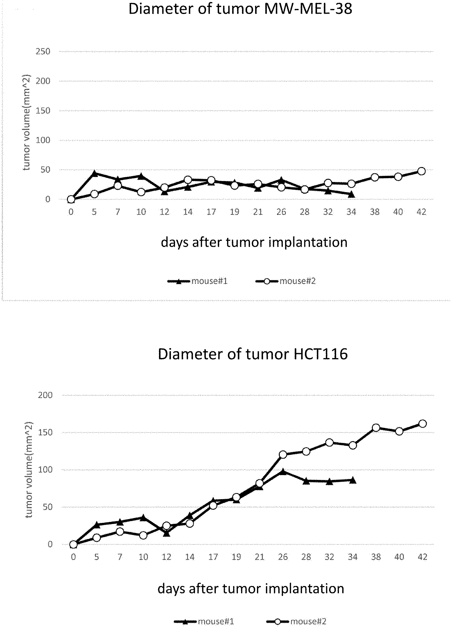
[0040] FIG. 20 (A) is a graph showing the results of the effects of CAT-T cells against the A2-positive-MAGE-A4-positive tumor (NW-MEL-38), and FIG. 20(B) is a graph showing the results of the effects of CAR-T cells against the A2-positive-MAGE-A4-negative tumors (HCT116).
[0041] FIG. 21 shows the gene sequence image of CAR zG.
[0042] FIGS. 22(A) and 22(B) are graphs showing the results of the effects of CAR-T cells having different intracellular domain (ICD) against A2-positive MAGE-A4-positive tumors (NW-MEL-38). More particularly, FIG. 22(A) shows tumor sizes, and FIG. 22(B) shows body weights. Left arrows in the graphs show the treatment day of body irradiation (TBI) (day 3); right arrows show the lymphocytes administration date (day 4).
[0043] FIG. 23 contains figures showing the results of examining the CAR positive rates of no-CAR introduced CD8-positive T cells (NGMC: control), zG type CAR introduced CD8-positive T cells (zG), and 28z type CAR introduced CD8-positive T cells (28z) by tetramer staining.
[0044] FIG. 24 is a graph which shows the results of examining whether CAR-T cells specifically recognize the target cells and increase the IFN.gamma. production.
[0045] FIG. 25 is a graph which shows the results of examining whether CAR-T cells specifically recognize the target cells and increase the IFN.gamma. production.
DETAILED DESCRIPTION
[0046] Next, embodiments of the invention will be described with reference to the Figures and tables. The technical scope of the invention is not limited by these embodiments, and can be implemented in various forms without changing the gist of the invention.
[0047] In the specification, the terms "antibody" and "antigen-binding fragment" refer to antigen binding protein in the immune system. An antibody having an antigen-binding region is a glycoprotein comprising two heavy chains (H chains) and two light chains (L chains) linked each other by disulfide bonds. Each heavy chain includes a heavy chain variable region (VH) and a heavy chain constant region (CH). The heavy chain constant region is comprised of three domains, CH1, CH2 and CH3. Each light chain includes a light chain variable region (VL) and a light chain constant region (CL). VL is composed of CL domain. VII and VL regions can further be subdivided into complementarity determining regions (CDR) with hypervariability and framework regions (FR) with some extent conserved-sequence. In VII and VL, FR1, CDR1, FR2, CDR2, FR3, CDR3 and FR4, i.e. three CDRs and four FRs, are arranged from the amino terminus to the carboxyl terminus, respectively. For binding to antigen, CDR3 is known to be important. Both VII and VL comprise binding domains that interact with an antigen.
[0048] In the specification, the term "HLA" is Human Leukocyte Antigen (human leukocyte antigen), means the human major histocompatibility complex (MHC), and a complex of genes encoding cell surface molecules that are required for antigen presentation to immune cells. HLA can be classified into class I and class II. Class I HLA is composed of .alpha.-chain and .beta.2-microglobulin. Class I HLA is expressed in almost all nucleated cells, and functions to CD8-positive T cells in antigen presentation. Class I HLA can be classified into HLA-A, HLA-B and HLA-C.
[0049] In the specification, the term "chimeric antigen receptor (CAR)" means a fusion protein containing an extracellular domain that binds to an antigen, a transmembrane domain different from the extracellular domain, and at least one intracellular domain. CAR can be called "chimeric receptor", "T-body", or "chimeric immune receptor (CIR)". "Extracellular domain that binds to an antigen" means any oligopeptide or polypeptide that binds to an antigen; "intracellular domain" means any oligopeptide or polypeptide that is known to function as a domain for a transmitting signal that activates or inhibits a biological process in a cell.
[0050] In the specification, "antigen binding protein" means a part of an antibody that binds to an antigen, or a fragment of an antibody that confers antibody specificity to the antigen.
[0051] Next, the embodiments of the present teachings will be specifically described.
[0052] (1) Antigen-Binding Fragments and Nucleic Acid Encoding them According to the Present Teachings
[0053] The anti-MAGE-A4-derived peptide/HLA-A2 (HLA-A2-MAGE-A4) complex antibody is an antibody that specifically recognizes and binds to the complex of HLA-A2 and P230-239 (SEQ ID NO: 1), which is a peptide derived from HLA-A2-restricted MAGE-A4. Antibodies of the present teachings are highly specific, and do not bind to the complex of HLA-A2 and a peptide other than P230-239, or to the complex of P230-239 peptide and an HLA other than HLA-A2 (or exhibits very low binding activity). Thus, the antigen-binding fragment of the present teachings can specifically detect or target the HLA-A2-MAGE-A4 complex.
[0054] Antigen-binding fragments of the present teachings may contain (A) the polypeptide having the amino acid sequence of VII (heavy chain variable region) of SEQ ID NO: 36 and the amino acid sequence of VL (light chain variable region) of SEQ ID NO: 38, or (B) a polypeptide having an amino acid sequence for the VII (heavy chain variable region) with an amino acid sequence homology of 90% or more with respect to SEQ ID NO: 36, and an amino acid sequence for the VL (light chain variable region) with an amino acid sequence homology of 90% or more with respect to SEQ ID NO: 38. Antigen-binding fragment of the present teachings can contain (C) a polypeptide having the amino acid sequence of a sc (single chain) of SEQ ID NO: 37, or (D) a polypeptide having an amino acid sequence of sc (single chain) with an amino acid sequence homology of 90% or more with respect to SEQ ID NO: 37 between VII and VL.
[0055] Antigen-binding fragments of the present teachings include one or more fragments that specifically bind to an antigen. The fragment of an antibody comprising an antigen-binding region of the antibody specifically recognizes an antigen similar to the full-length antibody. As the antigen-binding fragments containing antibodies and fragments thereof, Fab, Fab', F(ab').sub.2, Fv and single chain Fv (scFv) are exemplified. Fab is a monovalent antibody fragment constituted from VL, VII, CL and CH1 domains. Fab' is a monovalent antibody fragment constituted from VL, VII, CL, CH1 domains, and a hinge region. F(ab')2 is a divalent antibody fragment comprising two Fab fragments linked by disulfide bonds of a hinge region. Fv is a monovalent antibody fragment constituted from VL and VII. VL and VII domains of Fv fragment are encoded by separate genes, scFv (which is a protein of one molecule) can be produced by gene recombination techniques by combining two genes with a linker. scFv contains a linker (single chain) between VII and VL. The sc connects VII and VL, and is a peptide commonly used in the present field to stabilize the antigen binding ability of scFv antibodies (e.g., Huston et al, Methods in Enzymology, 203: 46-88 (1991), Whitlow et al, Protein Eng., 6: 989 (1993)). Generally speaking, sc contains glycine and serine, and has a length of 15 to 18 amino acids.
[0056] One aspect of the present teachings comprises a combination of an antigen-binding fragment. For such molecules, Fab3, Diabody, Triabody, Tetrabody, Minibody, Bis-scFv, (scFv)2-Fc, intact-IgG are exemplified (Holliger et al., Nature Biotechnology, 23(9), p. 1126-36 (2005)).
[0057] One or several amino acids of antigen binding fragments may be modified, so long as it does not substantially affect its properties. For example, one or several amino acids in the constant region or FR region can be substituted, deleted, added or inserted. This modification can be easily performed by combining known methods that include site-directed mutagenesis (point mutation introduced and cassette mutagenesis, etc.), gene homologous recombination, primer extension, and PCR.
[0058] In one aspect of the present teachings, a nucleic acid encodes the antigen-binding fragment. Such an nucleic acid may comprise the nucleotide sequence shown in SEQ ID NO: 31 and 33 to 35 in the Sequence Listing. The nucleic acid can be modified to provide codons (optimal codon) suitable for host cells without changing the amino acid sequence encoded by the nucleic acid. The expression efficiency of a polypeptide in a host cell can be improved by modifying to provide optimum codons.
[0059] Such antigen-binding fragments can be prepared using known genetic engineering techniques or chemical synthesis methods. Such genetic engineering techniques include: preparing a cloning vector or an expression vector containing the appropriate nucleic acid, introducing the vector into a host cell, culturing host cells expressing the nucleic acid, and recovering the polypeptide by purification, whereby the polypeptide can be produced. In embodiments comprising a plurality of nucleic acids, a combination of a plurality of vectors each containing one pertinent nucleic acid molecule can be introduced into a host cell, or one vector comprising a plurality of nucleic acids can be introduced into a host cell. In preparing the polypeptide, if a peptide tag is linked to the polypeptide, the polypeptide can be recovered and purified using the peptide tag.
[0060] A vector according to the present teachings is operably linked to suitable control sequences to express the nucleic acids in a suitable host. Such control sequences contain a promoter to transcribe the nucleic acids, an operator sequence to control transcription, a sequence encoding a ribosome binding site, an enhancer, a polyadenylation sequence, and a sequence for controlling termination of transcription and translation and the like. Various known sequences (e.g., restriction enzyme cleavage site(s), marker gene(s) such as drug resistance genes (selection genes), signal sequence(s), leader sequence(s), and the like) can be used in the vector. Various sequences can be selected and used appropriately according to the type of expressed polypeptide, the host cell, the conditions of the medium and the like.
[0061] A vector that is incorporated into the host genome, or is not incorporated the host genome, and an episomal vector that exists in the cytoplasm to replicate autonomously and the like can be used in the invention. As a vector, a retrovirus vector (containing oncoretrovirus vectors, lentivirus vectors, pseudotyped vector), adenoviral vector, adeno-associated virus (AAV) vector, simian virus vector, vaccinia virus vector, Sendai virus vector, Epstein-Barr virus (EBV) vector, HSV vector and the like are exemplified. As a virus vector, viruses that lack replication ability in the infected cells can be preferably used. Further, non-virus vectors can also be used by combination with a liposome and a condensing agent such as cationic lipids. Furthermore, the nucleic acid can be introduced into cells by performing calcium phosphate transfection, DEAE-dextran, electroporation, particle bombardment or the like.
[0062] As the host cell, known host cells can be used. For example, prokaryotic cells such as E. coli, mammalian cells such as Chinese hamster ovary cells (CHO cells), human cells, and eukaryotic cells such as yeast and insect cells and the like are exemplified.
[0063] If the antigen-binding fragment is expressed in a host cell, it can be purified from host cell medium, extracts and/or lysate of host cells. Purification methods can be performed by combining appropriate known methods. For example, centrifugation, hydroxyapatite chromatography, gel electrophoresis, dialysis, fractionation on an ion-exchange column, ethanol precipitation, reversed phase HPLC, chromatography by silica, chromatography by heparin sepharose, anion or cation resin chromatography (polyaspartic acid column, etc.), chromatofocusing, SDS-PAGE, ammonium sulfate precipitation, and affinity chromatography can be used appropriately.
[0064] (2) Compositions According to the Present Teachings
[0065] In other aspects of the present teachings, a composition may comprise one or more of the antibodies or the antigen-binding fragments described above in section (1), and/or a composition may comprises a nucleic acid encoding the antibody or the antigen-binding fragment(s).
[0066] Such an antibody or antigen-binding fragment can be used as a probe for the MAGE-A4-derived peptide and HLA-A2 complex. MAGE-A4 is known to be expressed inside cancer cells and be presented by HLA; T cells attack the cancer cells based on antigen recognition. Therefore, antigen-binding fragments of the present teachings can be used as a probe for cancer cells, i.e. a detection or diagnostic composition.
[0067] For such detection methods, known detection methods, such as ELISA, fluorescent antibody assay, radioimmunoassay, radioimmunoprecipitation, dot blot assays, inhibition or competition assay, sandwich assay, and latex beads agglutination assays, can be exemplified.
[0068] Samples for diagnosis and detection contain biological samples (e.g. disease site tissues, cells, body fluid containing blood, serum, plasma, lymph, and urine). For these samples, after having been pre-purified, homogenized, centrifuged, or diluted in a suitable buffer, if necessary, contacted to a antigen-binding fragment according to the present teachings, an antigen-antibody complex is detected. In this case, the antigen-binding fragment can be labeled, or a labeled secondary antibody can used. For the label, an enzyme (such as horseradish peroxidase, alkaline phosphatase, etc.), radioactive isotopes (e.g. .sup.32P, .sup.35S, .sup.3H, .sup.125I, etc.), fluorescent substance (e.g. rhodamine, fluorescamine, dansyl chloride, and their derivatives etc.), and tag peptide and the like can be used.
[0069] Such antigen-binding fragments can be used to deliver a pharmaceutical composition to a target. The antibody or the antigen-binding fragment may be linked to a pharmaceutical that specifically recognizes the WT1 peptide and HLA-A24 complex. For a pharmaceutical composition for delivery to a target, cytokines (IL2, TNF, IFN-.gamma. etc.), receptor proteins of them, cytotoxin (ricin, diphtheria, gelonin etc.), radioactive isotopes (.sup.90Y, .sup.131I, .sup.225Ac, .sup.213Bi, .sup.223Ra and .sup.227Th etc.), cells (T cells, NK cells, etc.) or low molecular weight compound (calicheamicin, doxorubicin etc.) are exemplified.
[0070] Effective amounts of the active ingredients of the pharmaceutical composition of the present teachings are determined appropriately based on the purpose, the tumor type, the medical conditions such as decease site and size of the patient's condition, and administration route and the like. When used as a pharmaceutical composition, various components that may be accepted pharmaceutically (e.g., carrier, excipient, buffer, stabilizer, etc.) can be added in addition to the active ingredient. The pharmaceutical composition may be provided in the form of a tablet, liquid, powder, gel, spray, a microcapsule, colloidal distribution system (liposomes, microemulsions, etc.), and macroemulsion as the like based on the conditions. As an administration route of the pharmaceutical composition, intravenous, intraperitoneal, intracerebral, intrathecal, intramuscular, intraocular, intraarterial, bile duct, and intralesional injection, infusion, sustained release systems formulation and the like are exemplified. The pharmaceutical composition can be administered by continuous infusion or injection.
[0071] Pharmaceutical compositions according to the present teachings can be used for treating hematopoietic tumors such as chronic myeloid leukemia, acute lymphocytic leukemia (ALL), and acute myeloid leukemia (AML), and solid cancers such as stomach cancer, colon cancer, lung cancer, breast cancer, germ cell cancer, live cancer, skin cancer, bladder cancer, prostate cancer, uterine cancer, cervical cancer, ovarian cancer, mesothelioma and the like, and for the diagnosis and detection of tumors and solid cancers. In particular, it is useful for the treatment, diagnosis and detection of MAGE-A4-positive or HLA-A2 positive hematopoietic tumor and solid cancer.
[0072] (3) Chimeric Antigen Receptor According to the Present Teachings
[0073] In a further aspect of the present teachings, a chimeric antigen receptor (CAR) may comprise one or more antigen-binding fragments described above in section (1) and an intracellular domain of a signaling protein. When a CAR that specifically recognizes the HLA-A2-MAGE-A4-derived-peptide complex binds to it, a signal can be transmitted to cells expressing CAR. CAR comprises an extracellular domain, a transmembrane domain and an intracellular domain. In the extracellular domain, an antigen binding fragment of the present teachings is contained. When the extracellular domain binds to the HLA-A2-MAGE-A4 complex specifically, a signal is transmitted into the cell via the intracellular domain of CAR, and stimulates the cell expressing CAR. Stimulated cells produce cytokines and the like, exhibit cytotoxicity to the target cells expressing the complex, or induce cytotoxicity of other immune cells.
[0074] As an intracellular domain of CAR, a domain that can transmit a signal into the cell, when the extracellular domain of the CAR molecule interacts (binds) to the MAGE-A4-derived peptide/HLA-A2 complex, can be used. As such signaling proteins, membrane proteins, signal receptors, and cytokine receptors are exemplified. For the intracellular domain of CAR, the intracellular domains containing a primary cytoplasmic signal transmitting sequence derived from CD3zeta (CD3.zeta.), FCR.gamma., FcR.beta., CD3.gamma., CD3.delta., CD3.epsilon., CD5, CD22, CD79a, and CD66d are exemplified. Further, the intracellular domains containing a secondary cytoplasmic signal (costimulatory signal) transmitting sequence derived from CD2, CD4, CD5, CD8.alpha., CD8.beta., CD28, CD137 (also known as "4-1BB"), CD134, ICOS, GITR and CD154 are exemplified. Furthermore, the intracellular domains containing a tertiary cytoplasmic signal transmitting sequence derived from IL-2 receptor and IL-21 receptor are exemplified.
[0075] As a transmembrane domain of CAR, alpha chain or beta chain of T-cell receptor, the transmembrane domain of CD3 zeta chain, CD28, CD3E, CD45, CD4, CD5, CD8, CD9, CD16, CD22, CD33, CD37, CD64, CD80, CD86, CD134, CD137 (also known as "4-1BB"), ICOS, CD154, and GITR and the like can be used. Artificially designed sequence can be used.
[0076] Further aspects of the present teachings include a nucleic acid that encodes a CAR. Nucleic acid encoding a CAR can be connected with another nucleic acid to be expressed under the control of a suitable promoter. A promoter that is expressed constitutively, or expressed inducibly by drug (e.g., tetracycline or doxorubicin) can be used. For example, mammalian-derived promoter containing phosphoglycerate kinase (PGK) promoter, Xist promoter, beta-actin promoter, and RNA polymerase II promoter, virus-derived promoter containing SV40 early promoter, cytomegalovirus promoter, thymidine kinase promoter of herpes simplex virus, LTR promoter of various retrovirus and the like can be used. To achieve efficient transcription of the nucleic acid, another regulatory element which cooperates with a promoter or a transcription initiation site (e.g., an enhancer sequence or a terminator sequence) can be linked to the nucleic acid. Furthermore, a marker gene which can be used for confirmation of the expression of the nucleic acid (e.g., drug resistance gene, a gene encoding a reporter enzyme, or a gene encoding a fluorescent protein etc.) can be incorporated.
[0077] Nucleic acid encoding the CAR can be introduced into cells by a vector. Moreover, nucleic acid encoding the CAR may be used as an active ingredient of a pharmaceutical composition. A pharmaceutical composition comprising the nucleic acid encoding the CAR can be prepared and used in accordance with the description in section (2) above.
[0078] In another aspect of the present teachings, a cell that express the CAR is included. The cells can be prepared by a process of introducing an appropriate CAR into a cell. When a cell expressing the CAR binds to the HLA-A2-MAGE-A4 complex via the CAR, a signal is transmitted to the cell and activated. Activation of cells can be confirmed by the release of cytokines, increased proliferation rate, the change of cell surface molecules and the like, depending on the host cell type and the intracellular domain. For example, the release of cytotoxic cytokines (tumor necrosis factor, lymphotoxin etc.) from activated cells results in the destruction of tumor cells expressing the complex. Further, cytokine release and the change of cell surface molecules stimulates other immune cells, for example, B cells, dendritic cells, NK cells, macrophages and the like. Thus, CAR expressing cells are useful for adoptive immunotherapy, especially against MAGE-A4-positive, HLA-A2 positive tumors or cancer.
[0079] The process of introducing the nucleic acid encoding the CAR into cells is carried out in vitro (ex vivo) or in vivo. For cells introduced by nucleic acid, mammalian cells such as human-derived cells, and non-human mammal-derived cells containing monkey, mouse, rat, pig, cow, and dog, etc. can be used. For types of cells, for example, cells from body fluid such as blood (peripheral blood, umbilical cord blood, etc.), bone marrow, tissue or an organ, collected, isolated, purified, and induced can be used. PBMCs, immune cells (dendritic cells, B cells, hematopoietic stem cells, macrophages, monocytes or NK cells, blood cells (neutrophils, basophils, monocytes), hematopoietic stem cells, cord blood mononuclear cells, fibrosis blasts, preadipocytes, hepatocytes, blood cells, skin keratinocytes, mesenchymal stem cells, hematopoietic stem cells, adipose stem cells, pluripotent stem cells, various cancer cell lines or neural stem cells can be used. In the invention, particularly T cells, T cell precursors (hematopoietic stem cells, lymphoid progenitor cells, etc.), pluripotent stem cells or cell populations containing them are used preferably. T cells contain CD8-positive T cells, CD4-positive T cells, regulatory T cells, cytotoxic T cells, tumor infiltrating lymphocytes. The cell population containing T cells and progenitor cells of T cells include PBMCs. Cells used in the invention can be any of cells sampled from living body, those expanded by culture, and cell lines. When cells transfected a nucleic acid or differentiated cells from them can be expected to be transplanted into a living body, cells collected from the living body or the same kind of living body are preferably transplanted.
[0080] For cells expressing a CAR according to the present teachings, culture and/or stimulation with the appropriate culture medium and/or stimulatory molecules prior to administration to a subject can be performed. Examples of the stimulatory molecules include cytokines, appropriate proteins, and other components. As cytokines, for example, IL-2, IL-7, IL-12, IL-15, and IFN-.gamma. and the like are exemplified, preferably IL-2 is exemplified. The concentration of IL-2 in the medium is not particularly limited; for example, the concentration is 0.01-1.times.10.sup.5 U/mL, preferably 1-1.times.10.sup.4 U/mL. Also, as suitable proteins, CD3 ligand, CD28 ligand, and anti-IL-4 antibody are exemplified. In addition, lymphocyte stimulator such as lectins can be added. In addition, serum or plasma can be added to the culture medium. The amount of the additives to the medium is not particularly limited; 0-20% by volume is exemplified, and the amount of serum or plasma can be changed accordance with the culture stage. The concentration of serum or plasma can be decreased stepwise. The origin of the serum or plasma can be the same as self (the same origin as cultured cells) or can be non-self (different from the origin of cultured cells), preferably those from self are used for the sake of safety.
[0081] The cell culture equipment to be used for cell culturing is not particularly limited; for example, a petri dish, a flask, a bag, a large culture bath, a bioreactor and the like can be used. As the bag, a cell-culture-CO.sub.2 gas-permeable bag can be used. When a mass of cell populations is produced industrially, a large culture bath can be used. Further, the culture can be conducted in an open system or a closed system; a closed system is preferably used for safety of the resulting cell population.
[0082] In another aspect of the present teachings, a pharmaceutical composition may contain cells that express an CAR according to the present teachings as an active ingredient and may be administered parenterally. As parenteral administration, intravenous, intraarterial, intramuscular, intraperitoneal, and subcutaneous administration and the like are exemplified. To increase the anti-tumor effect, administration to meningioma or nearby tissue, for example, subcutaneous administration, can be used. The administration dose is suitably selected depending on the subject's condition, weight, age and the like. Usually, as the number of cells, 10.sup.7-10.sup.9 cells per 60 kg body weight per administration, preferably about 5.times.10.sup.7-5.times.10.sup.8 cell, are administered. The pharmaceutical composition can be administered one time or multiple times. The pharmaceutical composition can be in a form suitable for parenteral administration, for example an injection or infusion. The pharmaceutical composition can optionally comprise a pharmaceutically acceptable excipient. The pharmaceutical composition can comprise saline, phosphate buffered saline (PBS), medium or the like, in order to maintain cells stably. The medium is not particularly limited; RPMI, AIM-V, X-Vivo 10 and the like are generally exemplified.
[0083] <Test Method>
1. Antibody Screening with Human Antibody Phage Library (1) Preparation of pMHC Beads
[0084] 20 .mu.g of A2-MAGE-A4 as pMHC and 200 mg of magnetic beads bound with Streptavidin were mixed and reacted at 4.degree. C. in 0.05% Tween/PBS(-) solution. 3 .mu.L of 2 mM biotin/PBS(-) was added and reacted for 1 hour. The solution was washed twice by 0.05% Tween/PBS(-), and suspended in 400 .mu.L of 0.05% Tween/PBS(-). The suspension was used as the antigen beads.
(2) Human Antibody Library
[0085] VH and VL were amplified by PCR from tonsils, umbilical cord blood, peripheral blood, and bone marrow from dozens of healthy individuals. The PCR products were recombined to the phagemid vector pUC119, to make scFv with repertoire of 3.4.times.10.sup.12. The scFv was displayed on cp3 of M13 phage. The product was used as a human antibody library.
(3) Screening Using Human Antibody Library, Isolation of Antibodies, Preparation of Cp3 Culture Supernatant
[0086] The reaction mixture containing 3.4.times.10.sup.12 cfu of human antibody library solution, 100 .mu.g of Streptavidin (Pierce), 30 .mu.g of A2-CMV tetramer, 30 .mu.g of A2-Foxp69 tetramer, 30 .mu.g of A2-IDOp41 tetramer, 30 .mu.g of A2-IDOp195 tetramer, 20 .mu.g of Gamma guard, 100 .mu.L of 2% BSA solution, 65 .mu.L of 10% TritonX100, 1000 .mu.L of 1% TritonX100/PBS was prepared and mixed and rotated for 1 hour at room temperature and 100 .mu.l of magnetic beads solution which was bound to A2-MAGE-A4 tetramer was added and mixed and rotated for 1 hour. Then, the magnetic beads were trapped with Magnet Trapper (Toyobo), while being washed 5 times with 1% TritonX100/PBS. The magnetic beads were added to cultured XL1-blue, infected 1 hour at 37.degree. C., centrifuged, and suspended in 600 .mu.L of 2.times.YTAG medium (200 .mu.g/mL ampicillin, 1% glucose/2.times.YT). The suspension was plated on YTAG agar plate (200 .mu.g/mL ampicillin, 2% glucose/nutrient agar (Nissui)), and cultured for 15 hours at 37.degree. C. Colonies were harvested with 30 mL of 2.times.YTAG medium, 200 .mu.L of the harvested solution was added to 20 mL of 2.times.YTAG medium, and 30 .mu.L of helper phage VCSM was added. The mixture was incubated for 30 minutes at 37.degree. C. for infection, and incubated for 90 minutes at 37.degree. C. 80 mL of 2.times.YTAG medium, 60 .mu.L of 50 .mu.g/mL kanamycin, and 50 .mu.L of 1M IPTG were added to the cultured mixture, and incubated for 20 hours at 28.degree. C. After the incubated mixture was centrifuged, the supernatant was mixed with 25 mL of PEG solution (20% PEG #600, 2.5M NaCl). The precipitate was collected by centrifugation, was suspended in 1 mL PBS, and sterilized with filter. The operation was repeated two times. Recovered E. coli were plated on YTAG agar medium. After being incubated at 30.degree. C., colonies were picked up and cultured in LB medium at 30.degree. C. overnight. DNA was extracted by Miniprep DNA Kit (QIAGEN) using a portion of the cultured LB medium, and sequenced. The, 50 .mu.L of the culture was mixed with 1.5 mL of 2.times.YTAI (2.times.YT medium containing 0.5 mM of IPTG), and cultured at 30.degree. C. overnight. The supernatant was separated by centrifugation. The supernatant was used as cp3 supernatant. ELISA was conducted by using the cp3 supernatant.
2. Enzyme-Linked Immunosorbent Assay (ELISA)
(1) MHC-Peptide Complex (Monomer)
[0087] The complex that HLA-A*0201 bound to MAGE-A4 p230, MAGE-A4 P286, MAGE-A3 P195, CMV, MelanA, GP3, HTLV-1, NY-ESO-1 p157, NY-ESO-1 p159, Foxp3 p69, Foxp3 p127, Foxp3 p390, IDO p41, IDO p41, IDO p195, and IDO p199 were used as MHC-peptide complex.
(2) Immobilization of pMHC
[0088] 500 ng of neutraavidin (PIERCE Co.) in 50 .mu.L of PBS was suspended in Maxisorp loose (Nunc), and was shaken at 4.degree. C. overnight. After discarding the solution, 200 .mu.L of 2% BSA/PBS was added and the solution was placed and blocked overnight. After discarding the solution, 300 ng of HLA-A2-MAGE-A4 monomer in 50 .mu.L of PBS was added and shaken at 4.degree. C. overnight. The solution was washed with PBS.
[0089] Immobilization of HLA-A2-CMV as a negative control was conducted in the same manner.
(3) Measurement of ELISA Reaction
[0090] 100 .mu.L of cp3 culture supernatant was poured into the immobilized antigen plate well, after shaking for 1 hour at room temperature, the plate well was washed with PBS, 100 .mu.l of anti-cp3 rabbit antibody diluted 2000-fold with 0.05% Tween20/PBS was poured into the well, and the plate was shaken for 1 hour at room temperature. After the well was washed with PBS, 100 .mu.l of HRP-labeled anti-rabbit IgG (MBL Co., Ltd.) at 4000-fold diluted in 0.05% Tween20/PBS was poured into the well, and the plate was shaken for 1 hour at room temperature. After the well was washed with PBS, OPD (WAKO Co., Ltd.) suspended in 0.01% 11202, 0.1M Na.sub.2PO.sub.4, 0.1M citric acid (p115.1) was poured and reacted, after confirming color development, 2N H.sub.2SO.sub.4 was added to stop the reaction. The absorbance was measured at 490 nm wavelength at SpectraMaxM2 (molecular devices).
3. Purification of Antibody
(1) Transfection to the Vector for Purification
[0091] Antibody gene was transferred to His/FLAG expression vector for changing to the form of scFv-FLAG/His. FLAG tag was used for detection by FACS, His-tag was used for purification by column, respectively.
(2) Transformation
[0092] After the isolated antibody cloned DNA was reacted by Sall (Takara Bio Inc.), the DNA fragment was reacted with the vector by T4 DNA ligase. Competent cells DH5a (TOYOBO Co. Ltd.) was transformed by the DNA, and was plated on LBGA plate. After incubated at 30.degree. C. overnight, colonies were cultured in 2.times.YTAG, and part of the culture solution was cultured in 2.times.YTAI, supernatant was prepared by centrifugation.
(3) Confirmation of scFv-FLAG/His Expression
[0093] After 100 .mu.l of culture supernatant was poured in a well of antigen plate, shaking for 1 hour at room temperature and the well was washed with PBS. For confirmation of expression of His-tag, HRP-labeled anti-His (MBL Co., Ltd.) at 4000-fold diluted in 0.05% Tween20/PBS was poured and shaken for 1 hour at room temperature. For confirmation of expression of FLAG-tag, anti-FLAG(DDDK) antibody, and HRP-labeled antibody were reacted in the order.
[0094] After the antigen plate was washed with PBS, OPD (WAKO Co., Ltd.) suspended in 0.01% H.sub.2O.sub.2, 0.1M Na.sub.2PO.sub.4, 0.1M citric acid (pH5.1) was poured and reacted, after confirming color development, 2N H.sub.2SO.sub.4 was added to stop the reaction. The absorbance was measured at 490 nm wavelength at SpectraMaxM2 (molecular devices).
(4) Crude Purification of Antibody
[0095] 2 mL of pre-cultured bacterial solution was put into 100 mL of 2.times.YTAI, and cultured at 30.degree. C. overnight. The solution was centrifuged (8000 rpm, 10 min, 4.degree. C.)., the supernatant was collected, 100 mL of saturated sulfuric acid were added to the supernatant, and stirred. The solution was centrifuged (8000 rpm, 20 min, 4.degree. C.), and the precipitate was suspended in 10.5 mL of PBS complete. After the PBS complete was centrifuged (12000 rpm, 1 h, 4.degree. C.)., 1 mL of Ni Sepharose excel (GE Healthcare) equilibrated with PBS was added to the supernatant, and incubated at 4.degree. C. overnight. A column was filled with the sample, washed with 0.5M NaCl PBS containing 0.5M NaCl PBS and 20 mM imidazole, and eluted with 0.5M NaCl PBS containing 250 mM imidazole. The eluent was dialyzed against PBS, after the dialysis, was concentrated with Amicon Ultra 10K (Millipore). SDS-PAGE was conducted with a part of the sample, to confirm the band by CBB staining, to determine the protein concentration of the crude purified antibody.
(5) Purification of His-Tag Protein Using AKTA Prime Plus
[0096] The crude antibody was further purified using HisTRAP column by AKTA Prime plus (GE Healthcare). The procedure was carried out according to the attached His-tag purification protocol. The recovered peak fractions were detected by SDS-PAGE.
4. Measurement of the Dissociation Constant (KD Value)
[0097] KD values of purified antibodies were measured by BiacoreX100 (GE Healthcare). Immobilization of ligands was conducted with Biotion capture kit (GE Healthcare).
(1) Immobilization of Ligand
[0098] HLA-A2-MAGE-A4 p230 was immobilized to the sensor chip CAP. The concentration was adjusted to 200 nM with HBS buffer, and the ligand was immobilized as suitable RU (resonance unit).
(2) Preparation of the Analyte Solution
[0099] Purified antibody was adjusted to 12.5 nM, 25 nM, 50 nM, 100 nM and 200 nM with HBS EP+buffer.
(3) Measurement of KD Values
[0100] The binding reaction was measured for 180 seconds while analyte solution was flowing through the flow cell of sensor chip. After the measurement, analyte solution was switched to HBS buffer, and the dissociation reaction was measured for 300 seconds. The measurement was carried out using a single cycle method of the program to flow continuously analyte from low concentration to high concentration.
[0101] After the reactions has ended with all concentrations, the binding constant were calculated using the curve fitting program.
5. Flow Cytometry Analysis (FACS)
(1) Equipment
[0102] FACS Calibur/FACS Cant/FACS Cantll (BD) were used to measure flow cytometer.
(2) Fluorescence-Labeled Antibody
[0103] Fluorescent-labeled antibodies were used as described in Table 1.
TABLE-US-00002 TABLE 1 Fluorescent label Antigen Clone ID. Company FITC CDS HIT8a BD bioscience APC CD4 RPA-T4 Biolegend APC CDS RPA-T8 Biolegend APC 107a H4A3 Biolegend APC-Cy7 CD4 RPA-T4 Biolegend APC-Cy7 CDS RPA-T8 Biolegend perCP-Cy5.5 CDS RPA-T8 Biolegend PE-Cy7 TNF-.alpha. MAb11 eBioscience V450 IFN-.gamma. 4S.B3 eBioscience
(3) Tetramer PE
[0104] Tetramer PE means the streptavidin-PE to which four biotinylated monomers are attached. The tetramer PE was used for detection of antigen-specific T cells. Tetramers used in the specification were A2-MAGE-A4 p230 tetramer and A2-NY-ESO-1 p157 tetramer.
6. HLA Stability Assay and scFv Binding Assay
(1) T2 Cell (HLA-A*02+Human BT Hybrid Lymphoblastoid Cell Line)
[0105] T2 cell is a kind of cell line that lacks Transporter Associated with Antigen Processing (TAP), has a specificity which the intracellular protein is not presented to HLA.
(2) Peptide Pulse to T2 Cells
[0106] Peptide was added to a final concentration of 10 .mu.M to T2 cells in human RPMI1640, placed for 15 minutes at room temperature, 20% FCS human RPMI1640 was added to a final concentration of 10% FCS. The cells were incubated in CO.sub.2 incubator for 45 min. at 37.degree. C. After two washes, the cells were used to assay.
(3) HLA Stability Assay
[0107] Anti-HLA-A2 Alexa Fluor 488 was reacted with peptide-pulsed T2 cells for 1 hour, the amount of HLA stabilized on the T cell surface was determined by detecting with FACS.
(4) scFv Binding Assay
[0108] Peptide-pulsed T2 cells were reacted with acquired antibodies for 1 hour, with anti-FLAG antibody for 1 hour, with anti-mouse FITC antibody for 1 hour, and they were detected with FACS.
(5) One Amino Acid Substituted Peptides and Risk Peptide
[0109] As shown in Table 2, one amino acid substituted peptides wherein one of 10 amino acids of MAGE-A4p230-239 was substituted with alanine (A), methionine (M), phenylalanine (F) and tryptophan (VV) were synthesized and examined IC50 of each peptide. Each peptide was adjusted to a final concentration of 10 mM in DMSO.
[0110] MHC IC50 is the theoretical value calculated by the IEDB (http://tools.immuneepitope.org/processing/), and when the value is lower, it is indicated that the bond between HLA-A*0201 and the peptide is stronger.
[0111] Table 2 shows the amino acid sequence of one amino acid substituted peptides and IC50.
TABLE-US-00003 TABLE 2 Amino acid MHC I.D. sequence IC50 (nM) MAGE-A4p230-239 GVYDGREHTV 225.4 (SEQ ID NO: 1) 1A AVYDGREHTV 118.1 (SEQ ID NO: 2) 2A GAYDGREHTV 2482.2 (SEQ ID NO: 3) 3A GVADGREHTV 1139.1 (SEQ ID NO: 4) 4A GVYAGREHTV 437.6 (SEQ ID NO: 5) 5A GVYDAREHTV 330.9 (SEQ ID NO: 6) 6A GVYDGAEHTV 75.0 (SEQ ID NO: 7) 7A GVYDGRAHTV 108.9 (SEQ ID NO: 8) 8A GVYDGREATV 289.1 (SEQ ID NO: 9) 9A GVYDGREHAV 197.0 (SEQ ID NO: 10) 10A GVYDGREHTA 2323.2 (SEQ ID NO: 11) 2.10A GAYDGREHTA 10668.2 (SEQ ID NO: 12) 3M GVMDGREHTV 148.6 (SEQ ID NO: 13) 3F GVFDGREHTV 169.8 (SEQ ID NO: 14) 5F GVYDFREHTV 119.8 (SEQ ID NO: 15) 5W GVYDWREHTV 119.8 (SEQ ID NO: 16) A2-CMV 21.29
[0112] Table 3 shows the amino acid sequence of risk peptides and IC50 values which were extracted from MAGE family peptide and amino acid recognizing MAGE #17 antibody by BLAST search.
TABLE-US-00004 TABLE 3 Amino acid MHC Risk Peptide sequence IC50 (nM) MAGE-A4 GVYDGREHTV 225.4 (SEQ ID NO: 1) MAGE-A1 EVYDGREHSA 13523.5 (SEQ ID NO: 17) MAGE-A8 GLYDGREHSV 9.7 (SEQ ID NO: 18) MAGE-A9 GVYVGKEHMF 35519.6 (SEQ ID NO: 19) MAGE-A10 GLYDGREHLI 35.4 (SEQ ID NO: 20) MAGE-A11 GVYAGREHFL 736.5 (SEQ ID NO: 21) MAGE-B2 GVYDGEEHSV 110.5 (SEQ ID NO: 22) MAGE-B10 GLYDGIEHFM 10.2 (SEQ ID NO: 23) MAGE-C2 GVYAGREHFV 156.2 (SEQ ID NO: 24) ADAMTS HVYNGRTHQW 36230.3 (SEQ ID NO: 25) RBBP5 RVYDGREILT 5880.0 (SEQ ID NO: 26) Calcyphosin GVYSGRAHPK 35865.8 (SEQ ID NO: 27)
7. Construction of CAR Construct and Vector
(1) MAGE #17 28z CAR
[0113] FIG. 1 shows the sequence image of the construction of CAR. The construct contains Leader, VH, VL, CL, CD28TM, CD28ICD (intracellular domain of CD28) and CD3zeta (CD3) chains from the 5' end. VII and VL is MAGE #17 scFv in these chains.
(2) Construction of CAR
[0114] FIG. 2 shows the construction steps of CAR schematically. To explain each step is as follows.
[0115] First, the PCR reaction was conducted using scFv-cp3 as a template, wherein Leader with EcoRI recognition site in 5' end of the VH-VL and CL with AscI recognition site in 3' end were added, respectively. Next, the fragment of PCR product treated with EcoRI and AscI, and the fragment of 28z CAR vector treated with EcoRI and AscI were reacted by ligase to obtain CAR 28z.
8. Preparation of Human Lymphocytes
[0116] Human lymphocytes were prepared from healthy donor blood using Ficoll-PaqueTM PLUS (17-1440-03, GE Healthcare). They were obtained by separating PBMCs (Peripheral Blood Mononuclear cells). Collection of specimens containing human peripheral blood used in the study and the analysis were conducted in accordance with the Declaration of Helsinki, in accordance with the protocol that was approved by Mie University School of Medicine Research Ethics Committee with the written consent of the subjects.
[0117] Collected samples were stored in a refrigerator or a liquid nitrogen tank that had been encrypted not to identify the subjects and antitheft treatment. The personal information of the subjects was anonymous, and close attention were paid to personal privacy and the results of gene analysis so as not to be leaked to the outside.
9. mRNA Introduction into Cells by Electroporation (1) Preparation of mRNA
[0118] DNA of CAR was cut into single stranded to produce a linear DNA template. DNA was purified using Wizard SV Gel and PCR Clean-UP System (Promega). mRNA was prepared from the purified DNA using mMESSAGEmMACHINE T7 Ultra Kit (Life technologies).
(2) Check by Bioanalyzer
[0119] Addition of Poly A to mRNA was confirmed by Bioanalyzer (Agilent Technologies).
(3) Preparation of PBMC Culture Plate
[0120] Anti-CD3 antibody (OKT-3 eBioscience) was diluted by ACD-A solution (TERUMO) at a final concentration of 5 .mu.g/mL, RetroNectin (Takara Bio) was diluted by ACD-A solution at a final concentration of 25 .mu.g/mL, 400 .mu.L of two solutions were poured in each well of 12-well plate (Nunc), and incubated for 5-10 hours at 37.degree. C. in CO.sub.2 incubator.
(4) Preparation of CM (Culture Medium)
[0121] PBMC culture containing 50 mL of lymphocyte medium GT-T503, 50 .mu.L of human IL-2 (600 IU), 400 .mu.L of Albuminar(25% HSA), and 300 .mu.L of plasma of lymphocytes donor was prepared.
(5) Isolation and Culture of PBMCs
[0122] Peripheral blood mononuclear cells were separated from healthy human blood (peripheral blood mononuclear cells, PBMCs) by Ficoll, and were suspended at 2.times.10.sup.5 cells/mL by CM, 2.5 mL of the suspension was poured in plate, and cultured for 4 days at 37.degree. C. in CO.sub.2 incubator.
(6) Electroporation
[0123] The produced mRNA was introduced in PBMCs after 10 days from separation by electroporation. The introduced PBMCs were used in assay within 24 hours.
10. Gene Transfer into Cells by Retrovirus (Infection Experiment)
(1) Preparation of Retrovirus by Packaging Cell Plat-A
[0124] CAR gene was introduced in packaging cell plat-A using FuGENE (Promega), after incubated for two days, the supernatant was recovered and used as virus solution.
(2) Isolation and Culture of PBMCs
[0125] Isolation and culture of PBMCs were carried out in accordance with above 9. (3)-(5).
(3) Production of plate for retrovirus infection
[0126] RetroNectin was diluted with ACD-A solution at a final concentration of 20 .mu.g/mL. 500 .mu.L of the solution was poured in each well of 24-well plate, and stored at 4.degree. C. overnight. Before pouring virus solution, the plate was washed twice with ACD-A.
(4) Preparation of Retrovirus Plate for Infection, Infection to Cells
[0127] The virus solution prepared in above 10.(1) was poured in plate of retrovirus infection by 1 mL, the plate was centrifuged at 2000.times.g for 2 hours at 32.degree. C. to coat the plate with virus. The plate was washed two times with 1.5% Alubuminar/PBS.
(5) Retrovirus Infection to PBMCs, Preparation of CAR-T Cells
[0128] PBMC cultured in above 10.(2) was recovered, after diluted by CM at 4.times.10.sup.5, 950 .mu.L of the suspension was seed on the plate for retrovirus infection, after the plate was centrifuged 1000.times.g for 10 minutes at 32.degree. C., and incubated at 37.degree. C. in CO2 incubator.
[0129] Prepared CAR-T cell was used in assay after 12-14 days from PBMCs separation.
(6) Preparation of Control Cell
[0130] Part of cultured cells in above 10.(2) was used as a control cells (no operation of gene transfection).
11. Characteristics of Tumor Cell Lines Used in In Vitro Experiments
[0131] Characteristics of the cell lines used in in vitro experiments were shown in Table 4.
TABLE-US-00005 TABLE 4 Cell line A2 A24 MAGE-A4 NW-MEL-38 + - 6.01E+03 (+) LB-23 + + 1.95E+03 (+) LC-1/sq - + 1.61E+03 (+) MEL72 + + 4.00E-01 (-) HCT116 + - - T2 + - - SK-MEL-37 + - +
12. IF-.gamma. Release Assay
[0132] The concentration of IF-.gamma. in culture was determined using eBioscience kit by ELISA.
[0133] 10.times.Coating buffer was diluted 10-fold with purified water to prepare Coating buffer. 48 .mu.L of the primary antibody was added to 12 mL of Coating buffer. The buffer was added to 96 well-flat-bottom-plate by 100 .mu.L per well. After the plate was stored at the 4.degree. C. overnight, it was washed five times with 0.05% PBS-T. 5.times.Assay diluents was diluted 5-fold with purified water to prepare Assay diluents. Assay diluents was added by 200 .mu.L per well. After 1 hour at room temperature for blocking, the plate was washed 5 times with 0.05% PBS-T. IFN-.gamma. was prepared at 1000 pg/mL with maxima, it was diluted with 7 stages by two folds to prepare standards. Samples and standards were added to the plate and reacted for 2 hours at room temperature, and the plate was washed 5 times with 0.05% PBS-T. 48 .mu.L of the secondary antibody was added to 12 mL of Assay diluents, the diluents was added by 100 .mu.L per well. After the plate was stored for 1 hour at room temperature, it was washed five times with 0.05% PBS-T. 48 .mu.L of Streptavidin-HRP was added to 12 mL of Assay diluents, the solution was added by 100 .mu.L per well. The plate was reacted in dark for 30 minutes at room temperature, and washed seven times with 0.05% PBS-T. TMB Substrate solution was added by 100 .mu.L, reacting in dark for 15 minutes at room temperature, 50 .mu.L of 0.18M H.sub.2SO.sub.4 was added to stop the reaction. Immediately, the plate was measured at a wavelength of 450 nm with Microplate Reader Model 680 (Bio-Rad).
13. Intracellular Cytokine Staining (ICS)
[0134] Target cells were adjusted to 1.times.10.sup.5 cells/mL, the effector cells were adjusted to 1.times.10.sup.5 cells/mL. 0.5 .mu.L of APC Anti human CD107a per sample was added, target cells and effector cells were added at the ratio of 1:1 in 96-well plate (U), was co-cultured for 1 hour at 37.degree. C. Then, 0.7 .mu.L of Golgistop (Protein Transport Inhibitor) was added for each well, and incubated for 4 hours at 37.degree. C. The cells in each well were transferred to V-type 96-well plate, after centrifuged at 1200 rpm for 5 minutes at 4.degree. C., then washed twice with 0.5% BSA/PBS. 0.5 .mu.L of APC-cy7 anti-human CD4 and 0.5 .mu.L of FITC anti-human CD8 were added to each well, and stood on ice for 20 minutes in dark, and the plate was washed twice with 0.5% BSA/PBS. 100 .mu.L of cell fixative solution (Cytofix/Cytoperm: BD) was added, stood on ice for 20 minutes in dark, 100 .mu.L of perm/wash (BD) was added and centrifuged. Then, the plate was washed two times with perm/wash solution. 0.5 .mu.L of V450 IFN-.gamma. and 0.5 .mu.L of PE-Cy7 TNF-.alpha. were added to each well. Under dark conditions, the plate was stood on ice for 30 minutes, washed twice with 0.5% BSA/PBS. Thereafter, the plate was measured by FACSCanto II flow cytometer (BD), and data was analyzed by FACS Diva Software (BD).
14. Long Peptide Uptake by APC, Cross Presentation
(1) MAGE-A4 Long Peptide
[0135] The amino acid sequence of MAGE-A4 long peptide was NYKRCFPVIF GKASEGVYDG REHTVYGEPR KAETSYVKVL EHVVRVNARV RI (SEQ ID NO: 28), the molecular weight of the peptide was 6006.811. The long peptide was designed by connecting A24 MAGE-A4 143-151 epitope (NYKRCFPVI: SEQ ID NO: 29), A2 MAGE-A4 230-239 epitope (GVYDGREHTV: SEQ ID NO: 1) and a helper epitope (AETSYVKVLE HVVRVNARVR I: SEQ ID NO: 30).
(2) CHP-Long Peptides
[0136] CHP-long peptide means an object that MAGE-A4 long peptide was wrapped in CHP nanogel as a drug delivery system. Cellular immunity induction can be expected by using the object (Non-patent document 5).
(3) CD3 Negative Beads Selection
[0137] 1.times.10.sup.7 cells of human lymphocytes were suspended in 80 .mu.L of 2% FCS/PBS. To the suspension, 20 .mu.L of CD3 Micro Beads (Miltenyi Biotec) was added, and mixed well. After 15 minutes at 4.degree. C. in light shielding, the suspension was washed with 10 volumes of 2% FCS/PBS. Cells were suspended in 3 mL of 2% FCS/PBS to use as cell suspension. The cell suspension was applied to MACS Separator equipped with a MACS column. Fractions that was passed through the column was CD3-negative fractions, used as APC.
15. Assay Cytotoxicity
(1) Cr Release Assay
[0138] After target cells were collected by centrifugation, they were suspended in 100% FCS at a final concentration of 1.times.10.sup.6 cells/50 .mu.L. Thereto, 50 .mu.Ci (3.7.times.10.sup.6 Bq) of .sup.51Cr was added, and incubated for 1 hour at 37.degree. C. in CO.sub.2 incubator. The concentration of effector cells were adjusted according to the E/Tratio by 10% FCS-RPMI1640, and were seeded in U-shaped 96-well plate. After cultivation, cells were washed three times with 10% FCS-RPMI1640. Cells were suspended in 10% FCS-RPMI1640 at a final concentration of 1.times.10.sup.5 cells/mL, were seed at 1.times.10.sup.4 cells/100 .mu.L/well in U-shaped 96-well plate, and were cultured for 8 hours in CO2 incubator at 37.degree. C. After cultivation, the plate was centrifuged at 1200 rpm for 5 minutes at 4.degree. C., 30 .mu.L of supernatant was transferred to the plate for reading, and dried up by air drying overnight. The amount of .sup.51Cr was determined by scintillation counter MicroBeta2 (Perkin Elmer). Calculation of the cytotoxic activity was determined by the following equation. The maximum release value was the measured value when 100 .mu.L of 1% NP-40 was added.
Cytotoxic activity (%)=100.times.(measured value (cpm)-spontaneous release value (cpm))/(maximum release value (cpm)-spontaneous release value (cpm))
16. In Vivo Cancer Treatment Model Experiment Using NOG Mouse
(1) Used Mice
[0139] NOG mice (NOD/Shi-scid, IL-2RyKO Jic) was purchased from CLEA Japan, Inc. The female mice were used in the experiment at 7-8 weeks of age. T cell infusion therapy with laboratory animals and the study of gene immunotherapy were approved by the recombinant DNA experiment committee of Mie University, Mie University School of Medicine Research Ethics Committee, and the animal research committee. The mice were bred in Mie University Life Science Center for the Research and Support of Animal Research Facility.
(2) Implantation of Human Tumor and Total Body Irradiation (TBI)
[0140] As pretreatment, total body irradiation (TBI) with 2.5Gy to NOG mice was conducted the day before cell infusion. The number of lymphocytes in recipient was reduced by the pretreatment, for transfused donor lymphocytes to be engrafted easily. CAR-T cells and human lymphocytes for infusion were 1.times.10.sup.7 cells/animal. Cells were transfused from the tail vein of NOG mice. Body weight was measured every 2-3 days.
[0141] Tumor transplantation into NOG mice were carried out in 7 days prior to infusion of the CAR-T cells, NW-MEL-38 (A2-positive MAGE-A4-positive) by 2.5.times.10.sup.6 cells/mouse were injected in the right-side of mouse, HCT116 (A2-positive MAGE-A4-negative) by 2.5.times.10.sup.6 cells/mouse were injected subcutaneously in the left side of mouse. Tumor diameters were measured every 2-3 days.
(3) Preparation of Cells for Infusion
[0142] CAR-T cells for infusion were prepared according to the method of above "10. Gene transfer into cells by retrovirus".
(4) PBMCs Isolation in Mouse Peripheral Blood by Orbital Blood Sampling
[0143] Peripheral blood was collected from the orbital of mouse, PBMCs were separated using Ficoll. They were stained with A2-MAGE-A4 tetramer, human CD4CD8 fluorescent antibody, infused cells were confirmed by FACS.
(5) Confirmation of TIL (Tumor Infiltrating Lymphocytes)
[0144] Tumor taken from mouse was mashed, stained with A2-MAGE-A4 tetramer human CD4CD8 fluorescent antibody, and transfused cells were confirmed by FACS.
(6) Immunostaining
[0145] Tumor excised from mouse was frozen in liquid nitrogen, frozen sections were prepared by cryostat. These sections ware stained by DAPI, human CD4-FITC and human CD8-FITC, and observed with a fluorescence microscope.
17. In Vitro Experiments with zG-Type-CAR-Introduced-CD8-Positive T Cells and 4-1BBz-Type-CAR-Introduced-CD8-Positive T Cells
[0146] The effects of CAR-T cells due to differences of ICD region were confirmed. Specifically, GITR (zG) type and CD137 (4-1BBz) type CAR were produced from CD28 (28z) type CAR, the effects of them were confirmed. As shown in FIG. 21, GITR (glucocorticoid-induced tumor necrosis factor receptor) was used instead of CD28 as ICD in CAR zG. Similarly, CD137 (4-1BBz) was used instead of CD28
[0147] As a control, CAR-untransfected-CD8-positive T cells (NGMC) was used.
[0148] <Results>
1. Isolation and Evaluation of Antibody that Recognizes HLA-A2-MAGE-A4
[0149] MAGE-A4 (HLA-A2-MAGE-A4) is known to be highly expressed in melanoma. Therefore, a plurality of antibodies that recognize MAGE-A4 were isolated by screening using human antibody libraries. Phage display method was used for isolation of antibodies. In the method, scFv is presented as part of a phage coat protein cp3, clones that bind to antigen with high affinity can be selected by ELISA. After picked up about 1500 clones, ELISA was conducted in the state of scFv-CP3. The clones that responded to HLA-A2-MAGE-A4 as a positive target and did not respond to A2-CMV as a negative target were selected. To examine the binding specificity of the selected antibodies, ELISA was conducted using plural peptides-MHC complex (pMHC). Each pMHC monomer, containing HLA-A2-MAGE-A4 p230, MAGE-A4 p286, MAGE-A3 p195, CMV, MelanA, GP3, HTLV-1 p11, NY-ESO-1 p157, NY-ESO-1 p159, Foxp3 p69, Foxp3 p127, Foxp3 p390, IDO p41, IDO p41, IDO p195 and IDO p199 were immobilized, ELISA was conducted using selected antibodies. Some results of the ELISA were shown in FIG. 3.
[0150] Fifteen clones exhibiting a specific reaction to HLA-A2-MAGE-A4 were obtained after compared the sequence of the clones showing strong binding to HLA-A2-MAGE-A4. Among them, 5 clones showing strong binding, i.e. MAGE #17, #86, #209, #345 and #672, were selected as candidate clones and proceeded analytics.
[0151] Next, KD value of MAGE #17 antibody was measured with BiacoreX100. Determination of KD value by Biacore was determined by measuring the dynamic changes in detection sensitivity (resonance, reflecting the change in mass on the chip) over time. The dissociation rate constant (Kd) and the binding rate constant (Ka) were determined by dynamic change curve, the binding constant was determined from the ratio of the two constants. As a result of the measurement, the binding constant of MAGE #17 was 22 nM (FIG. 4).
2. Study Concerning Recognition Specificity to the A2-MAGE-A4 Complex and Risk Antigen of the Obtained Antibodies
[0152] T2 cell lacks Transporter Associated with Antigen Processing (TAP), does not present the intracellular protein to HLA. When peptide was pulsed to T2 cell, as HLA was stabilized on T2, HLA expression level was confirmed by using fluorescent-anti-HLA antibody, it can be recognized what extent of peptide binds to HLA.
[0153] Selected antibodies, i.e. anti-MAGE #17, #86, #209, #345, and #672 were reacted with T2 cells pulsed with MAGE-A4p230, CMV, and DMSO, the shift amount was determined by FACS. Only in T2 cell pulsed with MAGE-A4 p230, selected antibody clones, MAGE #17, #86, #209, #345, and #672 showed the change in the shift amount (FIG. 5).
[0154] Next, in order to search for amino acids that correlated the recognition of antibodies, peptides with one amino acid substitution by another amino acid (mainly alanine) of MAGE-A4 p230 (GVYDGREHTV: SEQ ID NO: 1) were prepared, the binding force between HLA-A*0201 and the peptide was examined using IEDB (Immune Epitope Database), and determined MHC IC50 (Table 2). MHC IC50 is the theoretical value calculated by the IEDB, when the value is lower, the bond with HLA-A* 0201 and the peptide is stronger. Alanine substituted peptides were pulsed to T2 cell, the amount of HLA on the surface of T2 cell were determined by FACS (FIG. 6). More strongly a peptide bind to HLA, the structure of peptide-HLA was stabilized, since the shift amount of FACS was increased, the value of MFI was an indicator of binding strength of the peptide and HLA. Except some of the peptides, the MFI value and the MHC IC50 value corresponded to each other.
[0155] Then, alanine-substituted peptide was pulsed to T2 cell, and reacted with MAGE #17, amino acid involved in the recognition of the antibody was examined (FIG. 7). Since the MFI value dropped with 1A, 2A, 3A, 4A, 5A, 6A, and 8A, the seven amino acids were determined to be involved in the recognition. The amino acid sequence of GVYDGRxHxx was conducted by BLAST search, risk peptide was extracted (Table 3). After T2 cell was pulsed by risk peptide, the amount of HLA on the surface of T2 cell was determined by FACS. Similar to the results shown in FIG. 6, the MFI value and the MHC IC50 value of MFI corresponded to each other (FIG. 8).
[0156] Then, T2 cell was pulsed by risk peptide, reacted with MAGE #17, and confirmed that the T2 cell does not recognize the risk peptide (FIG. 9). Similar assays were practiced using other clones #86, #209, #345, and #672, in the results, clone #17 had most amino acids involved in the antigen recognition. For this reason, MAGE #17 was made as the final candidate.
TABLE-US-00006 Nucleotide sequence (SEQ ID NO: 31) and amino acid sequence (SEQ ID NO: 32) of MAGE #17 were shown. Nucleotide sequence of MAGE #17 (SEQ ID NO: 31): CAGGTCCAGC TGGTACAGTC TGGGGCTGAG GTGAAGAAGC CTGGGTCCTC GGTGAAGGTC TCCTGCAAGG CTTCTGGAGG CACCTTCAGC AGCTATGCTA TCAGCTGGGT GCGACAGGCC CCTGGACAAG GGCTTGAGTG GATGGGAGGG ATCATCCCTA TCTTTGGTAC AGCAAACTAC GCACAGAAGT TCCAGGGCAG AGTCACGATT ACCGCGGACA AATCCACGAG CACAGCCTAC ATGGAGCTGA GCAGCCTGAG ATCTGAGGAC ACGGCCGTGT ATTACTGTGC GAGATCCCCC CGGCGGGCAT ATCATGATGC TTTTGATATC TGGGGCCAAG GGACAATGGT CACCGTCTCT TCAGGTGGAG GCGGTTCAGG CGGAGGTGGC AGCGGCGGTG GCGGGAGTTC CTATGAGCTG ACTCAGCCAC CCTCGATGTC AGTGGCCCCA GGAAAGACGG CCAGCATTAC CTGTGGCGGA GACCATATTG GAAGTAAAAG TGTTCACTGG TACCAGCAGA AGCCAGGCCA GGCCCCTGTA CTGGTCGTCT ATGATGATAG CGACCGGCCC TCAGGGATCC CTGAGCGATT CTCTGGCTCC AACTCTGGGA ACACAGCCAC TCTGACCATC AGCGGGACCC AGGCTATGGA TGAGGCTGAC TATTACTGTC TGGCGTGGGA CAGCAGCACT GCGATCTTCG GCGGAGGGAC CAAGCTGACC GTCCTC. The amino acid sequence of MAGE #17 (SEQ ID NO: 32): QVQLVQSGAE VKKPGSSVKV SCKASGGTFS SYAISWVRQA PGQGLEWMGG IIPIFGTANY AQKFQGRVTI TADKSTSTAY MELSSLRSED TAVYYCARSP RRAYHDAFDI WGQGTMVTVS SGGGGSGGGG SGGGGSSYEL TQPPSMSVAP GKTASITCGG DHIGSKSVHW YQQKPGQAPV LVVYDDSDRP SGIPERFSGS NSGNTATLTI SGTQAMDEAD YYCLAWDSST AIFGGGTKLT VL. The above sequences show that the nucleotide sequence contains VH (SEQ ID NO: 33), sc (SEQ ID NO: 34) and VL (SEQ ID NO: 35), and the amino acid sequence contains VH (SEQ ID NO: 36), sc (SEQ ID NO: 37) and VL (SEQ ID NO: 38), as shown in following. The nucleotide sequence of VH (SEQ ID NO 33): CAGGTCCAGC TGGTACAGTC TGGGGCTGAG GTGAAGAAGC CTGGGTCCTC GGTGAAGGTC TCCTGCAAGG CTTCTGGAGG CACCTTCAGC AGCTATGCTA TCAGCTGGGT GCGACAGGCC CCTGGACAAG GGCTTGAGTG GATGGGAGGG ATCATCCCTA TCTTTGGTAC AGCAAACTAC GCACAGAAGT TCCAGGGCAG AGTCACGATT ACCGCGGACA AATCCACGAG CACAGCCTAC ATGGAGCTGA GCAGCCTGAG ATCTGAGGAC ACGGCCGTGT ATTACTGTGC GAGATCCCCC CGGCGGGCAT ATCATGATGC TTTTGATATC TGGGGCCAAG GGACAATGGT CACCGTCTCT TCA. The nucleotide sequence of sc region (SEQ ID NO 34): GGTGGAGGCG GTTCAGGCGG AGGTGGCAGC GGCGGTGGCG GGAGTTCCTA T. The nucleotide sequence of the VL region (SEQ ID NO 35): GAGCTGACTC AGCCACCCTC GATGTCAGTG GCCCCAGGAA AGACGGCCAG CATTACCTGT GGCGGAGACC ATATTGGAAG TAAAAGTGTT CACTGGTACC AGCAGAAGCC AGGCCAGGCC CCTGTACTGG TCGTCTATGA TGATAGCGAC CGGCCCTCAG GGATCCCTGA GCGATTCTCT GGCTCCAACT CTGGGAACAC AGCCACTCTG ACCATCAGCG GGACCCAGGC TATGGATGAG GCTGACTATT ACTGTCTGGC GTGGGACAGC AGCACTGCGA TCTTCGGCGG AGGGACCAAG CTGACCGTCC TC. The amino acid sequence of VH region (SEQ ID NO 36): QVQLVQSGAE VKKPGSSVKV SCKASGGTFS SYAISWVRQA PGQGLEWMGG IIPIFGTANY AQKFQGRVTI TADKSTSTAY MELSSLRSED TAVYYCARSP RRAYHDAFDI WGQGTMVTVS S. The amino acid sequence of sc region (SEQ ID NO: 37): GGGGSGGGGS GGGGSSY. The amino acid sequence of VL region (SEQ ID NO: 38): ELTQPPSMSV APGKTASITC GGDHIGSKSV HWYQQKPGQA PVLVVYDDSD RPSGIPERFS GSNSGNTATL TISGTQAMDE ADYYCLAWDS STAIFGGGTK LTVL.
3. MAGE #17 CAR Specifically Recognizes Target Cancer Cells and the Production of IFN-.gamma. was Increased
[0157] Since the specificity of the antibody clone MAGE #17 was confirmed, MAGE #17 was replaced to the vector of CAR. Then, after preparing mRNA, it was confirmed by bioanalyzer, CAR gene was introduced to human peripheral blood lymphocytes (PBMCs) by electroporation method. Whether or not the introduced CAR expressed normally was confirmed by tetramer staining. Consequently, CAR positive rate of CAR-T cells was 98.8% by CD8, and was 98.5% by CD4 (FIG. 10). CAR-T cell and peptide-pulsed T2 cell were co-cultured for 24 hours, and IFN-.gamma. in the culture supernatant was measured, production of IFN-.gamma. in T2 cell pulsed with MAGE-A4 was specifically increased. Similar assay was conducted using control cells (cells with no gene transfection). Further, tumor-specific production of IFN-.gamma. was observed in target cells of A2 positive MAGE-A4-positive NW-MEL-38 and LB-23, as compared with off-target cells of LC-1/sq and MEL72 (FIG. 11).
[0158] Since the expression of CAR by mRNA was transient, gene transfection by retrovirus was conducted for permanent expression of CAR. After a retrovirus vector was constructed for CAR introduction, the retrovirus was prepared by packaging cells plat-A, CAR-T cell was produced by infecting the retrovirus to PBMCs. Gene transfer was conducted according to the method of the above section 10 (Gene transfer into cells by retrovirus). The expression of CAR was confirmed by tetramer staining. Consequently, the CAR positive rate of CAR-T cells was 59.3% by CD8, and was 49.5% by CD4 (FIG. 12).
[0159] Gene-transferred CAR-T cells by retrovirus and peptide-pulsed T2 cells were co-cultured for 24 hours, IFN-.gamma. in the culture supernatant was measured, production of IFN-.gamma. in T2 cell pulsed with MAGE-A4 was specifically increased. Similar assay was conducted using control cells (cells with no gene transfection). Further, tumor-specific production of IFN-.gamma. was observed in target cells of A2 positive MAGE-A4-positive NW-MEL-38 and LB-23, as compared with off-target cells of LC-1/sq and MEL72 (FIG. 13). It was confirmed that CAR was expressed by introduction of mRNA and introduction of CAR gene by retrovirus vectors, the cell specifically recognized tumor, and produced IFN-.gamma..
4. CAR-T Cell Introduced with MAGE #17 CAR Gene Specifically Recognized Target Cancer Cells and Showed Cytotoxic Activity
[0160] The CAR-T cell was expected to recognize A2-MAGE-A4 expressed as antigen on cancer cells, become activated and exhibit cytotoxic activity. IFN-.gamma., TNF-.alpha. and CD107a are known indicators of T cell activation, i.e. they are expressed increasingly according to the activation of T cell. Therefore, intracellular cytokine staining (intracellular cytokine staining: ICS) of peptide-pulsed T2 cell and MAGE #17 CAR co-cultured with target cells was conducted.
[0161] As a result, the ratio of CAR-T cell stained with IFN-.gamma., TNF-.alpha. and CD107a was increased in co-culture with T2 cell pulsed with MAGE-A4p230 (FIG. 14). Moreover, the ratio of CAR-T cell stained with IFN-.gamma., TNF-.alpha. and CD107a was increased in co-culture with target cells when co-cultured with A2-positive-MAGE-A4-positive NW-MEL-38 and LB-23 (FIG. 15).
5. The Production of IFN-.gamma. in CAR-T Cells was Increased by Antigen Presentation of Antigen Presenting Cell (APC) that Took MAGE-A4 Long Peptide Antigen
[0162] PBMCs were separated from healthy human peripheral blood, negative selection was conducted by CD3 magnet beads, and CD3-fractions were used as antigen-presenting cells (APC). A2-APC as APC on which MAGE-A4p230 was presented and A24-APC as APC for negative control were used. MAGE-A4 long peptide and CHP-MAGE-A4 long peptide were added to a final concentration of 10 .mu.M, cultured overnight and co-cultured with CAR-T cells for 24-hours. Gene transfection to CAR-T cells was conducted according to the method of the above section 10 (Gene transfer into cells by retrovirus). As a result, the concentration of IFN-.gamma. in the culture supernatant was increased in co-culture with A2-APC pulsed with MAGE-A4 long peptide (FIG. 16).
[0163] MAGE-A4 long peptides were incorporated into APC, MAGE-A4p230 was presented with MHC-class I, IFN-.gamma. production of CAR-T cells which recognized the MAGE-A4p230-MHC-class I complex increased specifically. On the other hand, specific production of IFN-.gamma. was not observed in CHP-MAGEA4 long peptides wrapped in CHP Nanogel as a drug delivery system.
6. CAR-T Cells Wherein MAGE #17 CAR Gene was Transferred Lysed Specifically Target Cancer Cells
[0164] It was confirmed whether the specific cytotoxic activity of MAGE #17 CAR was observed or not by .sup.51Cr release assay. When the ratio of CAR-T cells and target cancer cells was E/T ratio (effector/target ratio), cancer cells that took Cr and CAR-T cells were co-cultured for 8 hours with ET ratio was 10:1, 3:1, 1:1, cytotoxicity rate (% lysis) was determined from the radioactivity of .sup.51Cr in the supernatant. Gene transfer to CAR-T cells was conducted according to the method of the above "10. Gene transfer into cells by retrovirus".
[0165] As a result, MAGE-A4 specific cytotoxicity was observed in T2 cells pulsed with MAGE-A4 p230 peptide (FIG. 17). Further, in target cells, specific cytotoxicity was confirmed to NW-MEL-38 cells which were A2-positive-MAGE-A4-positive cells (FIG. 18).
7. Mouse Cancer Therapy Model
(1) Model Using NW-MEL-38 and HCT116
[0166] Since the NOG mouse lacks common .gamma. receptors, NK cells do not exist in the NOG mouse. Therefore, engraftment of human cells and human tissue is significantly higher in a NOG mouse than a NOD-SCID mouse, such that human cancer and human tissue can be easily engrafted onto NOG. Further, since differentiation of human T cells is observed after human hematopoietic stem cell transplantation, the NOG mouse model has been expected to serve as a suitable human immune system model. NOG mice have the features containing deletion of T cells and B cells, deletion of natural killer (NK) cells, reduction of dendritic cell functions, decrease of macrophage functions, loss of complement activity, lack of leakage of B cells and T cells associated with aging (leakiness).
[0167] For NOG mice, NW-MEL-38 (A2-positive MAGE-A4-positive) at 2.5.times.10.sup.6 cells/mouse were injected in the right-side of each mouse, HCT116 (A2-positive MAGE-A4-negative) at 2.5.times.10.sup.6 cells/mouse were injected subcutaneously in the left side of each mouse. As a pretreatment, total body irradiation (TBI) with 2.5Gy to NOG mice was conducted 6 days after tumor transplantation. As the TBI reduced lymphocytes in the recipients, transplanted donor lymphocytes would be engrafted easily. On day 7, MAGE #17 CAR-T cells and human lymphocytes were infused via the tail vein at 1.times.10.sup.7 cells/animal. Gene transfer to CAR-T cells was conducted according to the method of the above section 10 (Gene transfer into cells by retrovirus). The CAR positive rate was 36.3% by CD8, and was 38.5% by CD4 (FIG. 19).
[0168] The experiments were carried out containing CAR-T cells infused group (n=2), human lymphocytes infusion group (CAR untransfected (n=2)), and PBS group (n=2), tumor sizes and body weights were measured every 2-3 days. The measurements were carried out until 42 days after tumor implantation.
[0169] As a result, in CAR-T cells infused group, growth inhibition of NW-MEL-38 (A2 positive MAGE-A4-positive) was observed in mouse #1 and mouse #2. On the other hand, growth inhibition of HCT116 (A2-positive MAGE-A4-negative) was not observed. Mouse #1 died after 34 days from tumor implantation. The cause of death was not a tumor death, but rather an infection (FIG. 20).
(2) Experiments Concerning the Effects of the Differences of the Intracellular Domain (ICD)
[0170] Next, the effects of CAR-T cells due to differences in the ICD portion was confirmed. FIG. 21 showed the gene sequence image of CAR zG. In CAR zG, ICD of GITR was used instead that of CD28. GITR is a glucocorticoid-inducible tumor necrosis factor receptor; CAR using this ICR is known that the expression level of cytokines per that of CAR was higher (WO2013051718). In CAR zG, ICD of CD28 in CAR 28z in FIG. 2 was changed ICD of GITR, and the order of CD3 and ICD domains in CAR 28z were changed. Other parts were used with identical sequences of CAR 28z.
[0171] NW-MEL-38 (A2-positive MAGE-A4-positive) at 2.5.times.10.sup.6 cells/mouse were injected subcutaneously into NOG mice. Three days after tumor implantation, total body irradiation (TBI) with 2.0Gy was conducted as a pre-treatment. On day 4, cells or PBS and human lymphocytes 4.times.10.sup.6 cells/mouse to each group A-C (n=4) shown in the following were infused through the tail vein. Gene transfer to CAR-T cells was conducted according to the method of the above section 10 (Gene transfer into cells by retrovirus). PBS in group A, MAGE #17 CAR-T cells in group B (28z), and CAR-T cells (zG) in group C were infused.
[0172] The diameter of the tumors and body weight were measured every 2-3 days for groups A to C. The measurements were carried out at 24 days after tumor implantation.
[0173] As a result, growth inhibition of NW-MEL-38 (A2-positive MAGE-A4-positive) was observed when CAR-T cells with 28z of ICT or zG of ICT were infused (FIG. 22). In the embodiment, it was found that the same effect was obtained when changing the intracellular domain.
8. Experiments Concerning the Effects of zG Type CAR Introduced CD8-Positive T Cells and 4-1BBz Type CAR Introduced CD8-Positive T Cells
[0174] (1) Constructing retrovirus vector introduced zG type CAR and 28z type CAR, after preparing retrovirus with packaging cells plat-A, CAR-T cells were produced by infecting the retrovirus into PBMCs. Gene transfer was conducted according to the method of the above section 10 (Gene transfer into cells by retrovirus). The expression of CAR was confirmed by tetramer staining. Consequently, the CAR positive rate of zG type CAR-T cells was 99.1% by CD8, and that of 28z type CAR-T cells was 99.8% by CD8 (FIG. 23).
[0175] (2) Gene-transfected CAR-T cells by retrovirus (4.times.10.sup.4 cells) and peptide-pulsed T2 cells, NW-MEL-38, SK-MEL-37, and HCT116 (respectively 2.times.10.sup.5 cells) were co-cultured for 18 hours, INF-.gamma. in the culture supernatant was measured. As shown in FIG. 24, IFN-.gamma. production of was increased specifically in CAR-T cells transfected with zG type CAR and 28z type CAR, in T2 cells pulsed with MAGE-A4 and NW-MEL-38 cells and SK-MEL-37 cells.
[0176] A similar assay was conducted using control cells (cells with no gene transfection). IFN-.gamma. production was not observed in A2-positive MAGE-A4 HCT116 as the target cell. IFN-.gamma. production was not observed in effectors only (Effector-only) and target only (Target-only).
[0177] Thus, zG type CAR introduced CD8-positive T cells induced INF-.gamma. production in vitro. The data can successfully explain the results obtained in vivo.
[0178] (3) Next, similar assays as (1) and (2) were conducted using 4-1BBz type CAR introduced CD8-positive T cells to obtain the characters of the cells. As shown in FIG. 25, in CAR-T cells transfected with 4-1BBz type CAR, IFN-.gamma. production was increased specifically to T2 cells pulsed with the MAGE-A4.
[0179] In T2 cells pulsed with control peptide, IFN-.gamma. production was not observed. In effectors only (Effector-only) and target only (Target-only), IFN-.gamma. production was not observed.
[0180] Thus, even when ICD was changed to 4-1BBz type, the same effect was observed. The data in vitro can easily explain the results obtained in vivo.
[0181] In this way, even when ICD was changed, the same effect as zG type was observed.
[0182] Thus, according to these embodiments, an antigen binding protein specifically recognizing the MAGE-A4-derived peptide/HLA-A2 complex, nucleic acids encoding the antigen binding protein, a vector containing the nucleic acids, a chimeric antigen receptor including the antigen binding protein, nucleic acids encoding the chimeric antigen receptor, a vector containing the nucleic acids, a cell expressing the chimeric antigen receptor, and a pharmaceutical composition containing the cells were provided.
[0183] The antigen binding protein specifically recognizing the MAGE-A4-derived peptide/HLA-A2 complex can be used in cell therapy and in the field of gene therapy. The present teachings are extremely useful for the detection of tumor cells expressing the MAGE-A4-derived peptide/HLA-A2 complex, for curing tumors, for research and for experiments on tumors. CAR-T cells of the present teachings can provide very effective cancer therapy, since the side effects are very small.
PRIOR ART
Non-Patent Documents
[0184] Non-patent Document 1: Brentjens R J, et al: Safety and persistence of adoptively transferred autologous CD19-targeted T cells in patients with relapsed or chemotherapy refractory B-leukemias. Blood 2011, 118:4817-4828.
[0185] Non-patent Document 2: Dao T, Yan S, Veomett N, Pankov D, Zhou L, Korontsvit T, et al: Targeting the intracellular WT1 oncogene product with a therapeutic human antibody. Sci Transl Med 2013; 5: 176ra133.
[0186] Non-patent Document 3: Van Der Bruggen P, Zhang Y, Chaux P, Stroobant V, Panichelli C, Schultz E S, Chapiro J, Van Den Eynde B J, Brasseur F, Boon T. Tumor-specific shared antigenic peptides recognized by human T cells. Immunological Reviews 2002. Vol. 188: 51-64.
[0187] Non-patent Document 4: Duffour M T, Chaux P, Lurquin C, Cornelis G, Boon T, van der Bruggen P. A MAGE-A4 peptide presented by HLA-A2 is recognized by cytolytic T Lymphocytes. Eur. J. Immunol 1999. 29: 3329-3337.
[0188] Non-patent Document 5: Muraoka D, Harada N,Hayashi T, Tahara Y, Momose F, Sawada S, Mukai S A, Akiyoshi K, Shiku H. Nanogel-based immunologically stealth vaccine targets macrophages in the medulla of lymph node and induces potent antitumor immunity. ACS Nano. Sep. 23, 2014; 8(09209-18.
[0189] Non-patent Document 6: Hillig R C, Coulie P G, Stroobant V, Saenger W, Ziegler A, Huelsmeyer M. High-resolution Structure of HLA-A*0201 in Complex with a Tumour-specific Antigenic Peptide Encoded by the MAGE-A4 Gene. J. Mol. Biol. 2001, 310, 1167-1176.
Sequence CWU 1
1
38110PRTHomo sapiens 1Gly Val Tyr Asp Gly Arg Glu His Thr Val1 5
10210PRTArtificial1A 2Ala Val Tyr Asp Gly Arg Glu His Thr Val1 5
10310PRTArtificial2A 3Gly Ala Tyr Asp Gly Arg Glu His Thr Val1 5
10410PRTArtificial3A 4Gly Val Ala Asp Gly Arg Glu His Thr Val1 5
10510PRTArtificial4A 5Gly Val Tyr Ala Gly Arg Glu His Thr Val1 5
10610PRTArtificial5A 6Gly Val Tyr Asp Ala Arg Glu His Thr Val1 5
10710PRTArtificial6A 7Gly Val Tyr Asp Gly Ala Glu His Thr Val1 5
10810PRTArtificial7A 8Gly Val Tyr Asp Gly Arg Ala His Thr Val1 5
10910PRTArtificial8A 9Gly Val Tyr Asp Gly Arg Glu Ala Thr Val1 5
101010PRTArtificial9A 10Gly Val Tyr Asp Gly Arg Glu His Ala Val1 5
101110PRTArtificial10A 11Gly Val Tyr Asp Gly Arg Glu His Thr Ala1 5
101210PRTArtificial2.10A 12Gly Ala Tyr Asp Gly Arg Glu His Thr Ala1
5 101310PRTArtificial3M 13Gly Val Met Asp Gly Arg Glu His Thr Val1
5 101410PRTArtificial3F 14Gly Val Phe Asp Gly Arg Glu His Thr Val1
5 101510PRTArtificial5F 15Gly Val Tyr Asp Phe Arg Glu His Thr Val1
5 101610PRTArtificial5W 16Gly Val Tyr Asp Trp Arg Glu His Thr Val1
5 101710PRTHomo sapiens 17Glu Val Tyr Asp Gly Arg Glu His Ser Ala1
5 101810PRTHomo sapiens 18Gly Leu Tyr Asp Gly Arg Glu His Ser Val1
5 101910PRTHomo sapiens 19Gly Val Tyr Val Gly Lys Glu His Met Phe1
5 102010PRTHomo sapiens 20Gly Leu Tyr Asp Gly Arg Glu His Leu Ile1
5 102110PRTHomo sapiens 21Gly Val Tyr Ala Gly Arg Glu His Phe Leu1
5 102210PRTHomo sapiens 22Gly Val Tyr Asp Gly Glu Glu His Ser Val1
5 102310PRTHomo sapiens 23Gly Leu Tyr Asp Gly Ile Glu His Phe Met1
5 102410PRTHomo sapiens 24Gly Val Tyr Ala Gly Arg Glu His Phe Val1
5 102510PRTHomo sapiens 25His Val Tyr Asn Gly Arg Thr His Gln Trp1
5 102610PRTHomo sapiens 26Arg Val Tyr Asp Gly Arg Glu Ile Leu Thr1
5 102710PRTHomo sapiens 27Gly Val Tyr Ser Gly Arg Ala His Pro Lys1
5 102852PRTArtificialMAGE-A4 Long Peptide 28Asn Tyr Lys Arg Cys Phe
Pro Val Ile Phe Gly Lys Ala Ser Glu Gly1 5 10 15Val Tyr Asp Gly Arg
Glu His Thr Val Tyr Gly Glu Pro Arg Lys Ala 20 25 30Glu Thr Ser Tyr
Val Lys Val Leu Glu His Val Val Arg Val Asn Ala 35 40 45Arg Val Arg
Ile 50299PRTHomo sapiens 29Asn Tyr Lys Arg Cys Phe Pro Val Ile1
53021PRTHomo sapiens 30Ala Glu Thr Ser Tyr Val Lys Val Leu Glu His
Val Val Arg Val Asn1 5 10 15Ala Arg Val Arg Ile
2031726DNAArtificialMAGE #17 31caggtccagc tggtacagtc tggggctgag
gtgaagaagc ctgggtcctc ggtgaaggtc 60tcctgcaagg cttctggagg caccttcagc
agctatgcta tcagctgggt gcgacaggcc 120cctggacaag ggcttgagtg
gatgggaggg atcatcccta tctttggtac agcaaactac 180gcacagaagt
tccagggcag agtcacgatt accgcggaca aatccacgag cacagcctac
240atggagctga gcagcctgag atctgaggac acggccgtgt attactgtgc
gagatccccc 300cggcgggcat atcatgatgc ttttgatatc tggggccaag
ggacaatggt caccgtctct 360tcaggtggag gcggttcagg cggaggtggc
agcggcggtg gcgggagttc ctatgagctg 420actcagccac cctcgatgtc
agtggcccca ggaaagacgg ccagcattac ctgtggcgga 480gaccatattg
gaagtaaaag tgttcactgg taccagcaga agccaggcca ggcccctgta
540ctggtcgtct atgatgatag cgaccggccc tcagggatcc ctgagcgatt
ctctggctcc 600aactctggga acacagccac tctgaccatc agcgggaccc
aggctatgga tgaggctgac 660tattactgtc tggcgtggga cagcagcact
gcgatcttcg gcggagggac caagctgacc 720gtcctc
72632242PRTArtificialMAGE #17 32Gln Val Gln Leu Val Gln Ser Gly Ala
Glu Val Lys Lys Pro Gly Ser1 5 10 15Ser Val Lys Val Ser Cys Lys Ala
Ser Gly Gly Thr Phe Ser Ser Tyr 20 25 30Ala Ile Ser Trp Val Arg Gln
Ala Pro Gly Gln Gly Leu Glu Trp Met 35 40 45Gly Gly Ile Ile Pro Ile
Phe Gly Thr Ala Asn Tyr Ala Gln Lys Phe 50 55 60Gln Gly Arg Val Thr
Ile Thr Ala Asp Lys Ser Thr Ser Thr Ala Tyr65 70 75 80Met Glu Leu
Ser Ser Leu Arg Ser Glu Asp Thr Ala Val Tyr Tyr Cys 85 90 95Ala Arg
Ser Pro Arg Arg Ala Tyr His Asp Ala Phe Asp Ile Trp Gly 100 105
110Gln Gly Thr Met Val Thr Val Ser Ser Gly Gly Gly Gly Ser Gly Gly
115 120 125Gly Gly Ser Gly Gly Gly Gly Ser Ser Tyr Glu Leu Thr Gln
Pro Pro 130 135 140Ser Met Ser Val Ala Pro Gly Lys Thr Ala Ser Ile
Thr Cys Gly Gly145 150 155 160Asp His Ile Gly Ser Lys Ser Val His
Trp Tyr Gln Gln Lys Pro Gly 165 170 175Gln Ala Pro Val Leu Val Val
Tyr Asp Asp Ser Asp Arg Pro Ser Gly 180 185 190Ile Pro Glu Arg Phe
Ser Gly Ser Asn Ser Gly Asn Thr Ala Thr Leu 195 200 205Thr Ile Ser
Gly Thr Gln Ala Met Asp Glu Ala Asp Tyr Tyr Cys Leu 210 215 220Ala
Trp Asp Ser Ser Thr Ala Ile Phe Gly Gly Gly Thr Lys Leu Thr225 230
235 240Val Leu33363DNAArtificialVH of MAGE #17 33caggtccagc
tggtacagtc tggggctgag gtgaagaagc ctgggtcctc ggtgaaggtc 60tcctgcaagg
cttctggagg caccttcagc agctatgcta tcagctgggt gcgacaggcc
120cctggacaag ggcttgagtg gatgggaggg atcatcccta tctttggtac
agcaaactac 180gcacagaagt tccagggcag agtcacgatt accgcggaca
aatccacgag cacagcctac 240atggagctga gcagcctgag atctgaggac
acggccgtgt attactgtgc gagatccccc 300cggcgggcat atcatgatgc
ttttgatatc tggggccaag ggacaatggt caccgtctct 360tca
3633451DNAArtificialsc of MAGE#17 34ggtggaggcg gttcaggcgg
aggtggcagc ggcggtggcg ggagttccta t 5135312DNAArtificialVL of
MAGE#17 35gagctgactc agccaccctc gatgtcagtg gccccaggaa agacggccag
cattacctgt 60ggcggagacc atattggaag taaaagtgtt cactggtacc agcagaagcc
aggccaggcc 120cctgtactgg tcgtctatga tgatagcgac cggccctcag
ggatccctga gcgattctct 180ggctccaact ctgggaacac agccactctg
accatcagcg ggacccaggc tatggatgag 240gctgactatt actgtctggc
gtgggacagc agcactgcga tcttcggcgg agggaccaag 300ctgaccgtcc tc
31236121PRTArtificialVH of MAGE#17 36Gln Val Gln Leu Val Gln Ser
Gly Ala Glu Val Lys Lys Pro Gly Ser1 5 10 15Ser Val Lys Val Ser Cys
Lys Ala Ser Gly Gly Thr Phe Ser Ser Tyr 20 25 30Ala Ile Ser Trp Val
Arg Gln Ala Pro Gly Gln Gly Leu Glu Trp Met 35 40 45Gly Gly Ile Ile
Pro Ile Phe Gly Thr Ala Asn Tyr Ala Gln Lys Phe 50 55 60Gln Gly Arg
Val Thr Ile Thr Ala Asp Lys Ser Thr Ser Thr Ala Tyr65 70 75 80Met
Glu Leu Ser Ser Leu Arg Ser Glu Asp Thr Ala Val Tyr Tyr Cys 85 90
95Ala Arg Ser Pro Arg Arg Ala Tyr His Asp Ala Phe Asp Ile Trp Gly
100 105 110Gln Gly Thr Met Val Thr Val Ser Ser 115
1203717PRTArtificialsc of MAGE#17 37Gly Gly Gly Gly Ser Gly Gly Gly
Gly Ser Gly Gly Gly Gly Ser Ser1 5 10 15Tyr38104PRTArtificialVL of
MAGE#17 38Glu Leu Thr Gln Pro Pro Ser Met Ser Val Ala Pro Gly Lys
Thr Ala1 5 10 15Ser Ile Thr Cys Gly Gly Asp His Ile Gly Ser Lys Ser
Val His Trp 20 25 30Tyr Gln Gln Lys Pro Gly Gln Ala Pro Val Leu Val
Val Tyr Asp Asp 35 40 45Ser Asp Arg Pro Ser Gly Ile Pro Glu Arg Phe
Ser Gly Ser Asn Ser 50 55 60Gly Asn Thr Ala Thr Leu Thr Ile Ser Gly
Thr Gln Ala Met Asp Glu65 70 75 80Ala Asp Tyr Tyr Cys Leu Ala Trp
Asp Ser Ser Thr Ala Ile Phe Gly 85 90 95Gly Gly Thr Lys Leu Thr Val
Leu 100
References
D00000
D00001
D00002
D00003
D00004
D00005
D00006
D00007
D00008
D00009
D00010
D00011
D00012

D00013

D00014
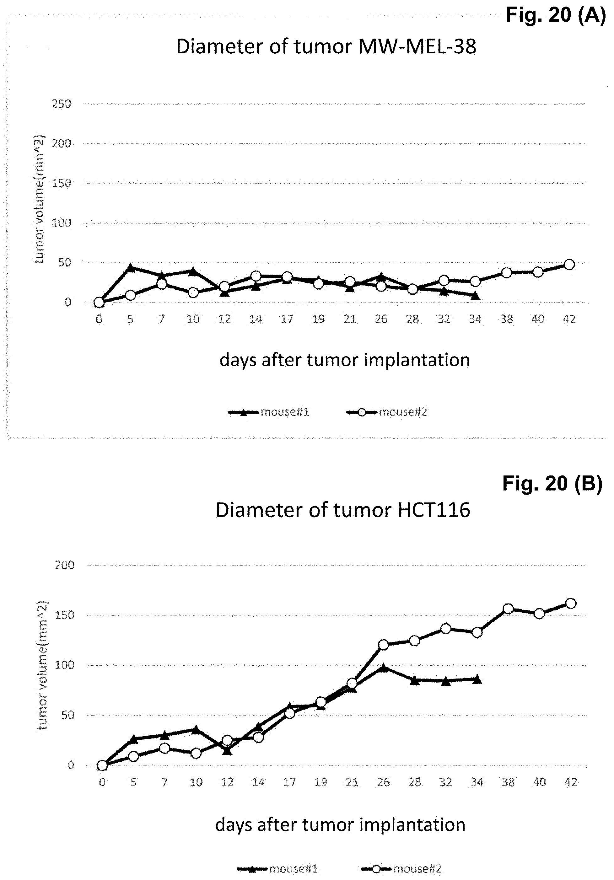
D00015

D00016

D00017

D00018

D00019

S00001
XML
uspto.report is an independent third-party trademark research tool that is not affiliated, endorsed, or sponsored by the United States Patent and Trademark Office (USPTO) or any other governmental organization. The information provided by uspto.report is based on publicly available data at the time of writing and is intended for informational purposes only.
While we strive to provide accurate and up-to-date information, we do not guarantee the accuracy, completeness, reliability, or suitability of the information displayed on this site. The use of this site is at your own risk. Any reliance you place on such information is therefore strictly at your own risk.
All official trademark data, including owner information, should be verified by visiting the official USPTO website at www.uspto.gov. This site is not intended to replace professional legal advice and should not be used as a substitute for consulting with a legal professional who is knowledgeable about trademark law.