Cancer Treatments
Markovic; Svetomir N. ; et al.
U.S. patent application number 16/872840 was filed with the patent office on 2020-08-27 for cancer treatments. The applicant listed for this patent is Mayo Foundation for Medical Education and Research. Invention is credited to Svetomir N. Markovic, Wendy K. Nevala.
| Application Number | 20200268884 16/872840 |
| Document ID | / |
| Family ID | 1000004824606 |
| Filed Date | 2020-08-27 |


View All Diagrams
| United States Patent Application | 20200268884 |
| Kind Code | A1 |
| Markovic; Svetomir N. ; et al. | August 27, 2020 |
CANCER TREATMENTS
Abstract
This invention relates to antibody-albumin nanoparticle complexes comprising albumin, an antibody with binding specificity for a cancer antigen (e.g. panitumumab), and paclitaxel, wherein the nanoparticle complex has been pre-formed in vitro such that the nanoparticle complex has antigen-binding specificity (e.g. EGFR binding specificity), for the purpose of providing cancer (e.g. EGFR-related cancer) treatments in a subject in need thereof.
| Inventors: | Markovic; Svetomir N.; (Rochester, MN) ; Nevala; Wendy K.; (Rochester, MN) | ||||||||||
| Applicant: |
|
||||||||||
|---|---|---|---|---|---|---|---|---|---|---|---|
| Family ID: | 1000004824606 | ||||||||||
| Appl. No.: | 16/872840 | ||||||||||
| Filed: | May 12, 2020 |
Related U.S. Patent Documents
| Application Number | Filing Date | Patent Number | ||
|---|---|---|---|---|
| 15187672 | Jun 20, 2016 | 10668151 | ||
| 16872840 | ||||
| 14432979 | Apr 1, 2015 | 10413606 | ||
| PCT/US2013/062638 | Sep 30, 2013 | |||
| 15187672 | ||||
| 61708575 | Oct 1, 2012 | |||
| 61725293 | Nov 12, 2012 | |||
| Current U.S. Class: | 1/1 |
| Current CPC Class: | A61K 45/06 20130101; A61K 2039/505 20130101; C07K 2317/24 20130101; C07K 2317/73 20130101; A61K 47/6929 20170801; A61K 31/555 20130101; C07K 16/22 20130101; C07K 2317/94 20130101; A61K 47/6863 20170801; C07K 16/2887 20130101; A61K 33/24 20130101; A61K 47/6865 20170801; A61K 2039/6056 20130101; A61K 47/6845 20170801; A61K 39/395 20130101; A61K 47/6849 20170801; A61K 47/6867 20170801; A61K 47/643 20170801; A61K 41/0052 20130101; B82Y 5/00 20130101; A61K 47/6931 20170801; A61K 47/6851 20170801; A61K 47/6801 20170801; C07K 2317/21 20130101; C07K 16/2863 20130101; A61K 9/51 20130101; C07K 2317/92 20130101; C07K 16/32 20130101; A61K 31/282 20130101; A61K 2039/545 20130101; A61K 9/14 20130101; A61K 39/39558 20130101; A61K 9/5169 20130101; A61K 9/0019 20130101; A61K 31/337 20130101; A61K 47/6855 20170801; C07K 16/2893 20130101; C07K 16/3015 20130101; A61K 47/6803 20170801 |
| International Class: | A61K 39/395 20060101 A61K039/395; C07K 16/32 20060101 C07K016/32; A61K 33/24 20060101 A61K033/24; A61K 31/555 20060101 A61K031/555; A61K 9/51 20060101 A61K009/51; A61K 47/64 20060101 A61K047/64; A61K 47/68 20060101 A61K047/68; A61K 47/69 20060101 A61K047/69; A61K 9/00 20060101 A61K009/00; A61K 41/00 20060101 A61K041/00; A61K 45/06 20060101 A61K045/06; C07K 16/22 20060101 C07K016/22; C07K 16/28 20060101 C07K016/28; A61K 31/337 20060101 A61K031/337; C07K 16/30 20060101 C07K016/30; A61K 31/282 20060101 A61K031/282; A61K 9/14 20060101 A61K009/14 |
Claims
1-72. (canceled)
73. A method for treating cancer in a subject in need thereof, the method comprising administering to the subject a therapeutically effective amount of a pharmaceutical composition comprising a pharmaceutically acceptable carrier and antibody-albumin nanoparticle complexes, said complexes comprising albumin, a panitumumab antibody, and paclitaxel, wherein the nanoparticle complexes have been pre-formed in vitro by mixing aqueous albumin-paclitaxel nanoparticles with the antibody under conditions to form the nanoparticle complexes, such that the nanoparticle complexes have EGFR binding specificity, wherein the cancer expresses EGFR, and wherein the average diameter of said complexes is between 0.1 .mu.m and 1 .mu.m.
74. The method of claim 73, wherein the average diameter of said complexes is between 0.1 .mu.m and 0.9 .mu.m.
75. The method of claim 73, wherein the average diameter of said complexes is between 0.1 .mu.m and 0.3 .mu.m.
76. The method of claim 73, wherein the ratio of albumin-paclitaxel nanoparticle to antibody is between 5:1 and 1:2.5.
77. The method of claim 73, wherein the pharmaceutical composition is administered by intravenous injection.
78. The method of claim 73, wherein the subject is a human.
79. The method of claim 73, wherein the cancer is head and neck cancer, lung cancer, or colon cancer.
80. The method of claim 73, wherein the therapeutically effective amount is between about 30 mg/m.sup.2 and about 70 mg/m.sup.2 antibody.
81. The method of claim 73, wherein the therapeutically effective amount is between about 5 mg/kg and about 20 mg/kg antibody.
82. The method of claim 73, wherein the therapeutically effective amount is between about 50 mg/m.sup.2 and about 175 mg/m.sup.2 albumin-paclitaxel nanoparticles.
83. The method of claim 73, wherein the pharmaceutical composition is administered from about once a month to about three times a month.
84. The method of claim 73, wherein the pharmaceutical composition is administered three times per 28 day cycle for at least two cycles.
85. The method of claim 73, wherein said composition comprises an alkylating agent.
86. The method of claim 85, wherein said alkylating agent is a platinum compound.
87. The method of claim 73, wherein the pharmaceutically acceptable carrier is saline, water, lactic acid, mannitol, or a combination thereof.
88. An antibody-albumin nanoparticle complex comprising albumin, a panitumumab antibody, and paclitaxel, wherein the nanoparticle complex has been pre-formed in vitro by mixing an aqueous albumin-paclitaxel nanoparticle with panitumumab under conditions to form the nanoparticle complex, such that the nanoparticle complex has EGFR binding specificity, and wherein the complex has a diameter of between 0.1 .mu.m and 1 .mu.m.
89. A pharmaceutical composition comprising a pharmaceutically acceptable carrier and antibody-albumin nanoparticle complexes, said nanoparticle complexes comprising albumin, a panitumumab antibody, and paclitaxel, wherein the nanoparticle complexes have been pre-formed in vitro by mixing aqueous albumin-paclitaxel nanoparticles with the antibody under conditions to form the nanoparticle complexes, such that the nanoparticle complexes have EGFR binding specificity, and wherein the average diameter of said complexes is between 0.1 .mu.m and 1 .mu.m.
Description
CROSS-REFERENCE TO RELATED APPLICATIONS
[0001] This application is a continuation of U.S. patent application Ser. No. 15/187,672, filed Jun. 20, 2016, which is a continuation of U.S. patent application Ser. No. 14/432,979, filed Apr. 1, 2015, now U.S. Pat. No. 10,413,606, which is a U.S. National Phase application under 37 CFR .sctn. 371 of International Application Serial No. PCT/US2013/062638, filed Sep. 30, 2013, which claims priority to U.S. Provisional Application Ser. No. 61/708,575, filed Oct. 1, 2012, and U.S. Provisional Application Ser. No. 61/725,293, filed Nov. 12, 2012, the contents of each of which are incorporated herein by reference in their entireties.
FIELD OF THE INVENTION
[0002] This document relates to methods and materials involved in treating cancer (e.g., skin cancers such as melanoma). For example, this document relates to methods and materials involved in using complexes containing albumin-containing nanoparticles (e.g., ABRAXANE.RTM. nanoparticles) and antibodies (e.g., anti-VEGF polypeptide antibodies such as AVASTIN.RTM.) to treat cancer. This document also relates to methods and materials involved in using ABRAXANE.RTM. in combination with an anti-VEGF polypeptide antibody (e.g., AVASTIN.RTM.) to treat skin cancer.
BACKGROUND OF THE INVENTION
[0003] Melanoma is the most serious form of skin cancer. It is a malignant tumor that originates in melanocytes, the cells which produce the pigment melanin that colors skin, hair, and eyes and is heavily concentrated in most moles. While it is not the most common type of skin cancer, melanoma underlies the majority of skin cancer-related deaths. About 48,000 deaths worldwide are registered annually as being due to malignant melanoma. Worldwide, there are about 160,000 new cases of melanoma each year. Melanoma is more frequent in white men and is particularly common in white populations living in sunny climates. Other risk factors for developing melanoma include a history of sunburn, excessive sun exposure, living in a sunny climate or at high altitude, having many moles or large moles, and a family or personal history of skin cancer.
[0004] Melanomas fall into four major categories. Superficial spreading melanoma can travel along the top layer of the skin before penetrating more deeply. Lentigo maligna typically appears as a flat or mildly elevated mottled tan, brown, or dark brown discoloration and is found most often in the elderly. Nodular melanoma can occur anywhere on the body as a dark, protuberant papule or a plaque that varies from pearl to gray to black. Acral-lentiginous melanoma, although uncommon, is the most common form of melanoma in blacks. It can arise on palmar, plantar, or subungual skin. Metastasis of melanoma occurs via lymphatics and blood vessels. Local metastasis results in the formation of nearby satellite papules or nodules that may or may not be pigmented. Direct metastasis to skin or internal organs can occur.
SUMMARY OF THE INVENTION
[0005] This document provides methods and materials involved in treating cancer (e.g., skin cancers such as melanoma). For example, this document provides methods and materials for using complexes containing albumin-containing nanoparticles (e.g., ABRAXANE.RTM. nanoparticles) and antibodies (e.g., anti-VEGF polypeptide antibodies such as AVASTIN.RTM.) to treat cancer. This document also provides methods and materials involved in using ABRAXANE.RTM. in combination with an anti-VEGF polypeptide antibody (e.g., AVASTIN.RTM.) to treat skin cancer (e.g., melanoma). ABRAXANE.RTM. is available from Celgene Corp. and is a nanoparticle formulation that combines paclitaxel with human albumin. AVASTIN.RTM. is also known as bevacizumab and is available from Genentech Corp. and Roche Corp. AVASTIN.RTM. is a humanized monoclonal antibody that binds to vascular endothelial growth factor A. As described herein, in vitro mixing of albumin-containing nanoparticles (e.g., ABRAXANE.RTM. nanoparticles) and antibodies (e.g., bevacizumab, bevacizumab, trastuzamab, or rituxan) can result in the formation of macromolecular complexes, the characteristics of which (e.g., size, antibody content, or chemotherapeutic drug content) can be customized depending on need. In some cases, such macromolecular complexes can retain antibody mediated target binding specificity, can retain or exhibit enhanced chemotherapeutic tumor cell cytotoxicity, and can exhibit no additional toxicity beyond that of ABRAXANE.RTM. nanoparticles alone. As also described herein, contacting ABRAXANE.RTM. with an anti-VEGF polypeptide antibody (e.g., AVASTIN.RTM.) prior to administration to a human (e.g., a human melanoma cancer patient) can result in a complex that, when administered as a complex, has an increased ability to treat melanoma as compared to a treatment regimen that includes administering ABRAXANE.RTM. and the anti-VEGF polypeptide antibody separately in a manner that does not form ABRAXANE.RTM./anti-VEGF polypeptide antibody complexes.
[0006] The methods and materials provided herein can be used to increase the progression-free survival rate in skin cancer patients. Increasing progression-free survival can allow skin cancer patients to live longer.
[0007] In general, one aspect of this document features a method for treating a mammal having skin cancer. The method comprises, or consists essentially of, administering to the mammal a composition containing ABRAXANE.RTM./anti-VEGF polypeptide antibody complexes (or complexes of (a) an anti-VEGF polypeptide antibody and (b) human albumin-containing nanoparticles having an agent other than placitaxel) under conditions wherein the length of progression-free survival is increased. The mammal can be a human. The skin cancer can be melanoma. The skin cancer can be stage IV melanoma. In some cases, a composition comprising ABRAXANE.RTM./AVASTIN.RTM. complexes can be administered to the mammal. The composition can comprise an alkylating agent. The alkylating agent can be a platinum compound. The platinum compound can be carboplatin. The anti-VEGF polypeptide antibody can be a humanized antibody. The anti-VEGF polypeptide antibody can be bevacizumab. The composition can be administered by injection. The progression-free survival can be increased by 25 percent. The progression-free survival can be increased by 50 percent. The progression-free survival is increased by 75 percent. The progression-free survival can be increased by 100 percent. The composition can be administered under conditions wherein the time to progression is increased.
[0008] In another aspect, this document features a method for treating a mammal having cancer. The method comprises, or consists essentially of, administering, to the mammal, a composition comprising albumin-containing nanoparticle/antibody complexes, wherein the average diameter of the complexes is between 0.1 and 0.9 .mu.m. The mammal can be a human. The cancer can be skin cancer. The skin cancer can be melanoma. The skin cancer can be stage IV melanoma. The albumin-containing nanoparticle/antibody complexes can be ABRAXANE.RTM./AVASTIN.RTM. complexes. The composition or the albumin-containing nanoparticle/antibody complexes can comprise an alkylating agent. The alkylating agent can be a platinum compound. The platinum compound can be carboplatin. The antibodies of the albumin-containing nanoparticle/antibody complexes can be anti-VEGF polypeptide antibodies. The anti-VEGF polypeptide antibodies can be humanized antibodies. The anti-VEGF polypeptide antibodies can be bevacizumab. The composition can be administered by injection. The administration of the composition can be effective to increase progression-free survival by 25 percent. The administration of the composition can be effective to increase progression-free survival by 50 percent. The administration of the composition can be effective to increase progression-free survival by 75 percent. The administration of the composition can be effective to increase progression-free survival by 100 percent. The administration of the composition can be under conditions wherein the median time to progression for a population of mammals with the cancer is at least 150 days. The administration of the composition can be under conditions wherein the median time to progression for a population of mammals with the cancer is at least 165 days. The administration of the composition can be under conditions wherein the median time to progression for a population of mammals with the cancer is at least 170 days. The average diameter of the complexes can be from 0.1 .mu.m to 0.3 .mu.m. The average diameter of the complexes can be from 0.15 .mu.m to 0.3 .mu.m. The average diameter of the complexes can be from 0.2 .mu.m to 0.5 .mu.m. The average diameter of the complexes can be from 0.3 .mu.m to 0.5 .mu.m. The average diameter of the complexes can be from 0.2 .mu.m to 0.8 .mu.m. The average diameter of the complexes can be from 0.2 .mu.m to 0.7 .mu.m.
[0009] In another aspect, this document features a method for treating a mammal having cancer. The method comprises, or consists essentially of, administering, to the mammal, a composition comprising albumin-containing nanoparticle/antibody complexes, wherein the average diameter of at least 5 percent of the complexes of the composition is between 0.1 and 0.9 .mu.m. The mammal can be a human. The cancer can be skin cancer. The skin cancer can be melanoma. The skin cancer can be stage IV melanoma. The albumin-containing nanoparticle/antibody complexes can be ABRAXANE.RTM./AVASTIN.RTM. complexes. The composition or the albumin-containing nanoparticle/antibody complexes can comprise an alkylating agent. The alkylating agent can be a platinum compound. The platinum compound can be carboplatin. The antibodies of the albumin-containing nanoparticle/antibody complexes can be anti-VEGF polypeptide antibodies. The anti-VEGF polypeptide antibodies can be humanized antibodies. The anti-VEGF polypeptide antibodies can be bevacizumab. The composition can be administered by injection. The administration of the composition can be effective to increase progression-free survival by 25 percent. The administration of the composition can be effective to increase progression-free survival by 50 percent. The administration of the composition can be effective to increase progression-free survival by 75 percent. The administration of the composition can be effective to increase progression-free survival by 100 percent. The administration of the composition can be under conditions wherein the median time to progression for a population of mammals with the cancer is at least 150 days. The administration of the composition can be under conditions wherein the median time to progression for a population of mammals with the cancer is at least 165 days. The administration of the composition can be under conditions wherein the median time to progression for a population of mammals with the cancer is at least 170 days. The average diameter of at least 5 percent of the complexes of the composition can be from 0.2 .mu.m to 0.9 .mu.m. The average diameter of at least 5 percent of the complexes of the composition can be from 0.2 .mu.m to 0.8 .mu.m. The average diameter of at least 5 percent of the complexes of the composition can be from 0.2 .mu.m to 0.7 .mu.m. The average diameter of at least 5 percent of the complexes of the composition can be from 0.2 .mu.m to 0.6 .mu.m. The average diameter of at least 5 percent of the complexes of the composition can be from 0.2 .mu.m to 0.5 .mu.m. The average diameter of at least 5 percent of the complexes of the composition can be from 0.2 .mu.m to 0.4 .mu.m. The average diameter of at least 10 percent of the complexes of the composition can be between 0.1 and 0.9 .mu.m. The average diameter of at least 50 percent of the complexes of the composition can be between 0.1 and 0.9 .mu.m. The average diameter of at least 75 percent of the complexes of the composition can be between 0.1 and 0.9 .mu.m. The average diameter of at least 90 percent of the complexes of the composition can be between 0.1 and 0.9 Pun.
[0010] Unless otherwise defined, all technical and scientific terms used herein have the same meaning as commonly understood by one of ordinary skill in the art to which this invention pertains. Although methods and materials similar or equivalent to those described herein can be used to practice the invention, suitable methods and materials are described below. All publications, patent applications, patents, and other references mentioned herein are incorporated by reference in their entirety. In case of conflict, the present specification, including definitions, will control. In addition, the materials, methods, and examples are illustrative only and not intended to be limiting.
[0011] The details of one or more embodiments of the invention are set forth in the accompanying drawings and the description below. Other features, objects, and advantages of the invention will be apparent from the description and drawings, and from the claims.
DESCRIPTION OF DRAWINGS
[0012] FIG. 1 is a diagram of an ABRAXANE.RTM. nanoparticle (labeled A) complexed with an anti-VEGF polypeptide antibody (bevacizumab; labeled B). In two of the three cases, the anti-VEGF polypeptide antibody is shown binding to a VEGF-A polypeptide (labeled V), and a fluorescently-labeled anti-VEGF antibody (labeled aV*) is shown bound to the VEGF-A polypeptide.
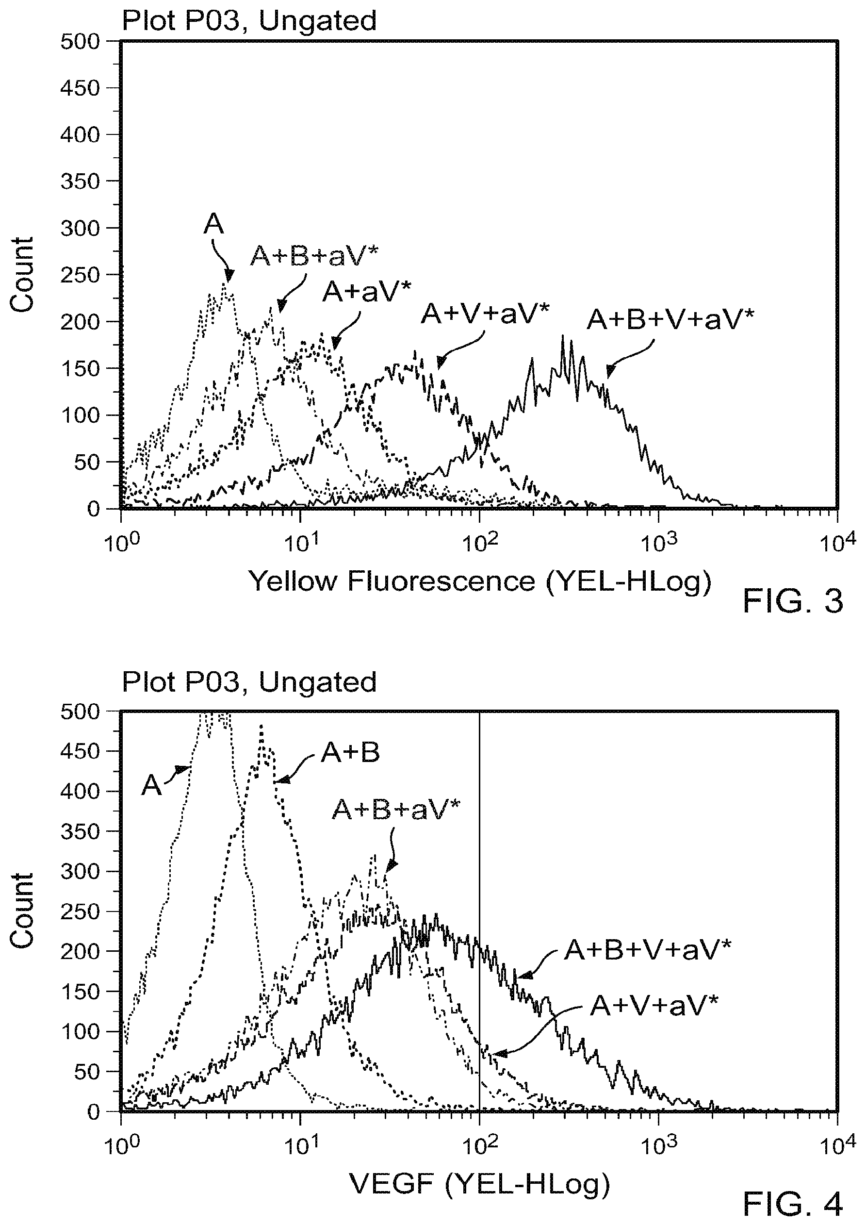
[0013] FIGS. 2A-2E contain scatter plots of a flow cytometry analysis plotting the level of yellow fluorescence. FIG. 2A depicts A alone. FIG. 2B depicts A plus a V*. FIG. 2C depicts A plus B plus a V*. FIG. 2D depicts A plus V plus a V**. FIG. 2E depicts A plus B plus V plus a V*. The labels are as indicated in FIG. 1. These results demonstrate that A and B spontaneously associate and preserve a VEGF polypeptide binding potential.
[0014] FIG. 3 is graph that contains the flow cytometry data from FIG. 2.
[0015] FIG. 4 is a repeat of the experiment of FIG. 3, comparing A alone, A plus aV*, A plus B plus aV*, A plus V plus aV*, or A plus B plus V plus aV*. One difference is in FIG. 3, 500 ng of VEGF was used. In FIG. 4, 100 ng VEGF was used to visualize the complex.
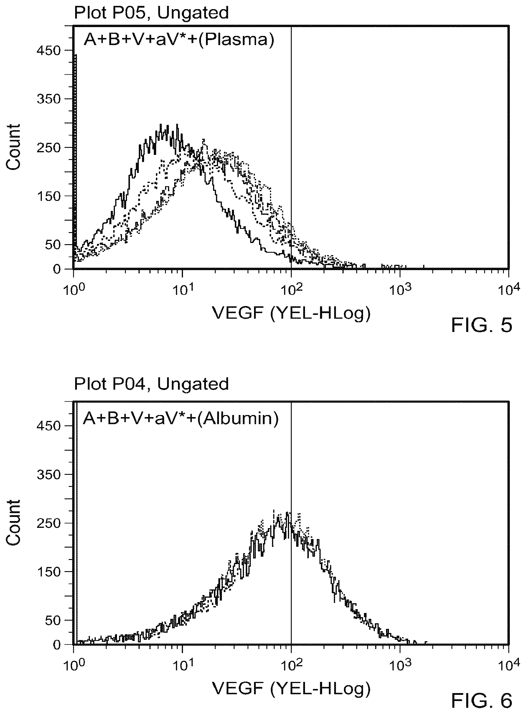
[0016] FIG. 5 is a graph plotting flow cytometry data of A plus B incubated in the presence of various concentrations of human plasma (1:1 to 1:16) followed by addition of V and aV*. These results indicate that human plasma diluted in a range of relative volumes (1:1 to 1:16) successfully inhibited the formation of the A+B complex relative to controls.
[0017] FIG. 6 is a graph plotting flow cytometry data of A plus B incubated in the presence of various concentrations of human serum albumin (500 .mu.g, 50 .mu.g, 5 .mu.g, 0.5 .mu.g, and 0.05 .mu.g/mL) followed by addition of V and aV*. These results indicate that incubation with serum albumin (concentrations ranging from 500 .mu.g/mL to 0.05 .mu.g/mL) did not affect the complexing of A and B.
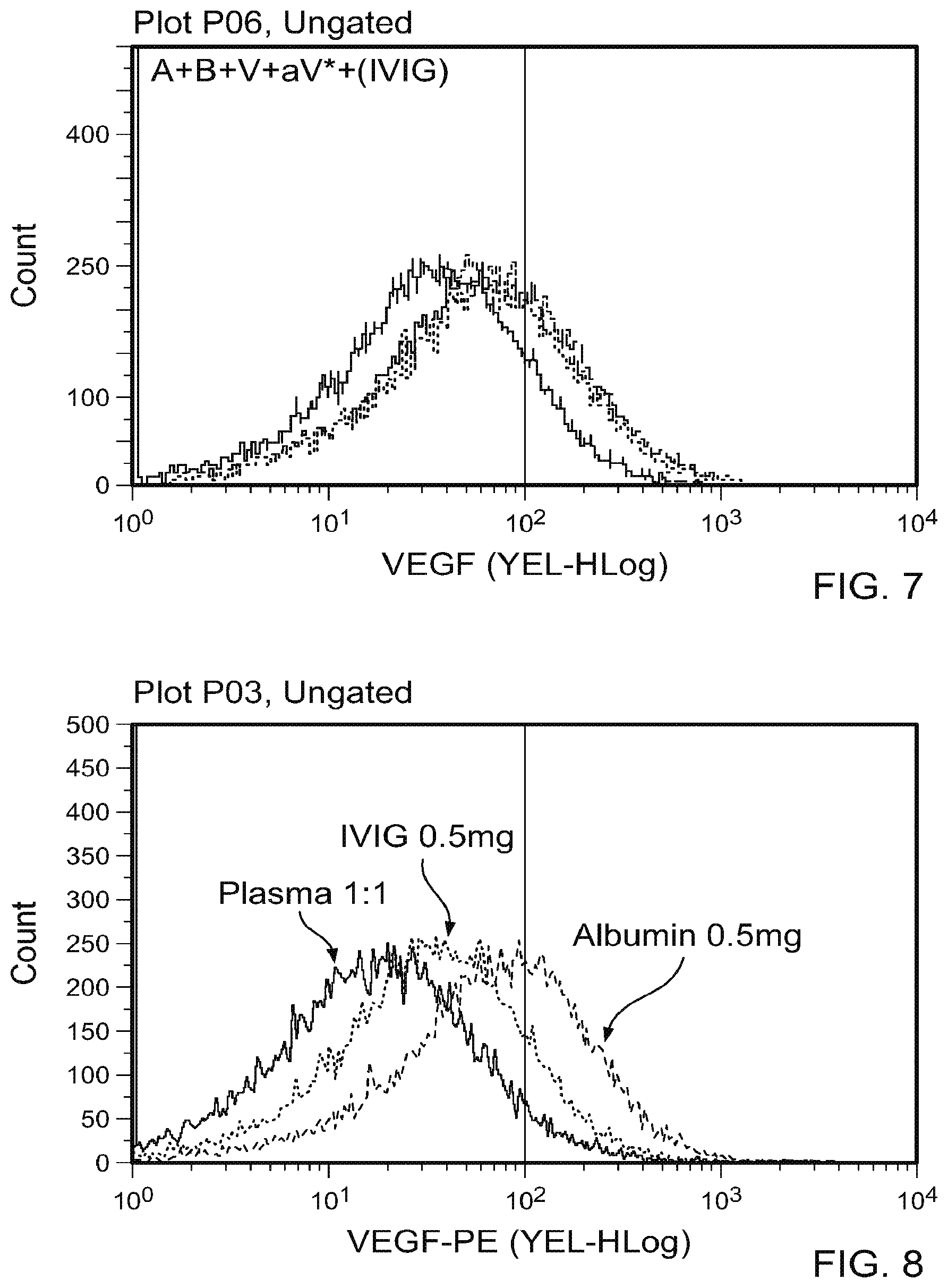
[0018] FIG. 7 is a graph plotting flow cytometry data of A plus B incubated in the presence of various concentrations of human polyclonal immunoglobulin (500 .mu.g, 50 .mu.g, 5 .mu.g, 0.5 .mu.g, and 0.05 .mu.g/mL) followed by addition of V and aV*. These results indicate that incubation of A and B with a range of concentrations of human immunoglobulin (IVIG; 500 .mu.g/mL to 0.05 .mu.g/mL) partially inhibited A and B complexing.
[0019] FIG. 8 contain A plus B complexing results in the presence of plasma (1:1), IVIG (0.5 mg/mL), or albumin (0.5 mg/mL). At the highest concentrations of plasma (1:1), IVIG (0.5 mg/mL), or albumin (0.5 mg/mL) tested, the levels of relative inhibition of A plus B complexing differ in diminishing order.
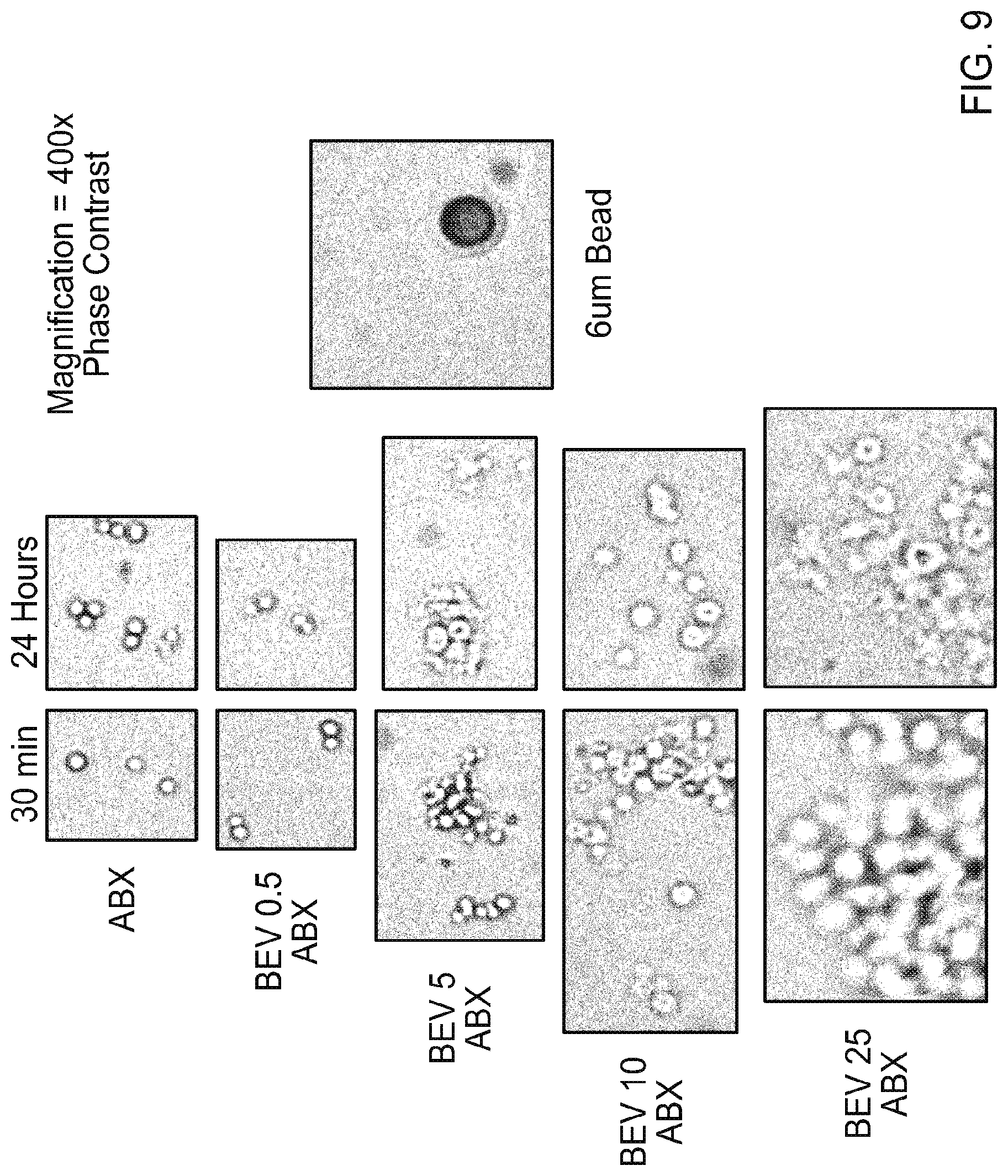
[0020] FIG. 9 contains photographs of light microscope images of ABRAXANE.RTM. (ABX) or mixtures of ABRAXANE.RTM. (ABX) and bevacizumab (BEV; 0.5, 5, 10, or 25 mg/mL) either 4 or 24 hours after mixing.
[0021] FIG. 10 is a graph plotting flow cytometry results of ABRAXANE.RTM. alone, ABX:BEV complexes, and 2 .mu.m standard beads.
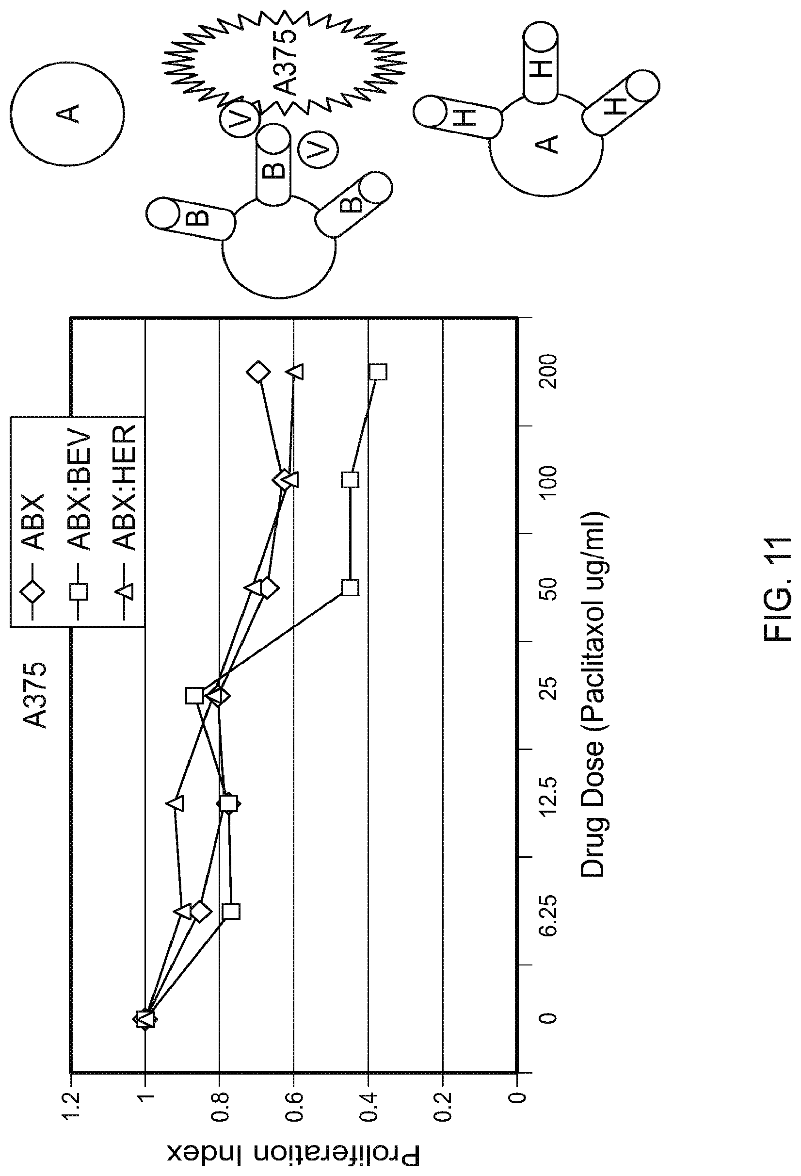
[0022] FIG. 11 is graph plotting the proliferation index for A375 cells (a melanoma tumor cell line) exposed to ABRAXANE.RTM. (ABX) only, ABRAXANE.RTM.:Herceptin (non-VEGF targeting) complexes, or ABRAXANE.RTM.:Bevacizumab (VEGF targeting) complexes at the indicated dose.
[0023] FIG. 12 contains graphs plotting the percent BEV binding for ABX:BEV complexes exposed to 0.9% saline at room temperature or human plasma at 37.degree. C. for the indicated times.
[0024] FIG. 13 contains a line graph plotting the proliferation index for A375 cells exposed to ABRAXANE.RTM. (ABX) only, cisplatin only, or ABRAXANE.RTM.:cisplatin complexes at the indicated dose and contains a bar graph plotting demonstrating that 30% of cisplatin (CDDP) remained unbound after ABX:cisplatin were mixed and incubated for 30 minutes.
[0025] FIGS. 14A-14J contain scatter plots of a flow cytometry analysis of the indicated complexes containing ABRAXANE.RTM.. FIG. 14A depicts complexes of ABX and bevacizumab. FIG. 14B depicts complexes of ABX and trastuzumab. FIG. 14C depicts complexes of ABX and rituximab. FIG. 14D depicts complexes of ABX and bevacizumab. FIG. 14E depicts complexes of ABX and trastuzumab. FIG. 14F depicts complexes of ABX and rituximab. FIG. 14G depicts ABX alone. FIG. 14H depicts complexes of ABX and bevacizumab. FIG. 14I depicts complexes of ABX and trastuzumab. FIG. 14J depicts complexes of ABX and rituximab.
[0026] FIG. 15 contains photographs of Western blot analyses of the indicated materials assessed for bevacizumab or taxol.
[0027] FIG. 16 contains graphs of the size distributions of the indicated complexes incubated for the indicated time.
[0028] FIGS. 17A-17F contain graphs of the size distributions of the indicated complexes incubated for one hour at room temperature. FIG. 17A is a graph of complexes of ABX:BEV 1 mg/ml. FIG. 17B is a graph of complexes of ABX:BEV 1 mg/ml spun. FIG. 17C is a graph of complexes of ABX:BEV 2 mg/ml. FIG. 17D is a graph of complexes of ABX:BEV 3 mg/ml. FIG. 17E is a graph of complexes of ABX:BEV 4 mg/ml. FIG. 17F is a graph of complexes of ABX:BEV 8 mg/ml.
[0029] FIG. 18 is a photograph of a Western blot analysis of ABX:BEV complexes exposed to serum for 15, 30, 45, or 60 minutes. The ABX:BEV complexes were formed by incubating either 6 mg or 8 mg of BEV with ABX for 30 minutes at room temperature. The primary antibody used for the Western blot was an anti-paclitaxel antibody. Lane 1: ABX:BEV (6 mg) exposed to serum for 15 minutes; Lane 2: ABX: BEV (6 mg) exposed to serum for 30 minutes; Lane 3: ABX:BEV (6 mg) exposed to serum for 45 minutes; Lane 4: ABX:BEV (6 mg) exposed to serum for 60 minutes; Lane 5: blank; Lane 6: ABX:BEV (8 mg) exposed to serum for 15 minutes; Lane 7: ABX:BEV 8 mg) exposed to serum for 30 minutes; Lane 8: ABX:BEV (8 mg) exposed to serum for 45 minutes; Lane 9: ABX:BEV (8 mg) exposed to serum for 60 minutes.
[0030] FIG. 19 is a photograph of a Western blot analysis of mixtures of paclitaxel (0.1, 0.5, 1, or 2 mg) and BEV (4 mg) incubated together for 30 minutes at room temperature. The primary antibody used for the Western blot was an anti-paclitaxel antibody. Lane 1: Bev (4 mg); Lane 2: Taxol (2 mg); Lane 3: Taxol (2 mg)+Bev (4 mg); Lane 4: Taxol (1 mg)+Bev (4 mg); Lane 5: Taxol (0.5 mg)+Bev (4 mg); Lane 6: Taxol (0.1 mg)+Bev (4 mg).
[0031] FIG. 20 contains graphs plotting the particle size distribution for ABX:BEV complexes as determined using a Mastersizer 2000E (Malvern Instruments Ltd., Worcestershire, England). ABX (20 mg/mL) and BEV (16, 24, or 32 mg/mL) were incubated for 1, 2, or 4 hours at room temperature. After incubation, the mixtures were diluted 1:4 for a final concentration of ABX (5 mg/mL) and BEV (4, 6, or 8 mg/mL), and the diluted samples analyzed using a Mastersizer 2000E.
[0032] FIG. 21. After a 4 hour incubation at room temperature of ABX:BEV complexes (two different concentrations of ABX) in saline (left panel), soluble ABX/BEV complexes were detected by Western blot analysis that migrated in the MW range of approximately 200 kD. Identical bands were identified by Western blotting with anti-paclitaxel (.alpha.-paclitaxel) and anti-mouse IgG (.alpha.-mouse IgG) antibodies. Similarly, following incubation of ABX:BEV complexes in heparinized human plasma (right panel) at 37.degree. C. for 15, 30, 45, or 60 minutes, the majority of the soluble paclitaxel (.alpha.-paclitaxel) migrated at a MW of 200 kD.
[0033] FIGS. 22A-22C contain graphs plotting percent change (A), tumor size (B), and survival (C) for Group 1 mice treated with PBS, Bevacizumab (8 mg/kg), ABRAXANE.RTM. (30 mg/kg), Bevacizumb (day 0, 8 mg/kg) followed by ABRAXANE.RTM. (day 1, 30 mg/kg), or small nanoAB (complex).
[0034] FIGS. 23A-23C contain graphs plotting percent change (A), tumor size (B), and survival (C) for Group 2 mice treated with PBS (day 0 and day7), Bevacizumab (8 mg/kg; day 0 and day7), ABRAXANE.RTM. (30 mg/kg; day 0 and day7), Bevacizumb (day 0, 8 mg/kg) followed by ABRAXANE.RTM. (day 1, 30 mg/kg), or small nanoAB (complex; day 0 and day7).
[0035] FIGS. 24A-24C contain graphs plotting percent change (A), tumor size (B), and survival (C) for Group 3 mice treated with PBS, Bevacizumab (24 mg/kg), ABRAXANE.RTM. (30 mg/kg), Bevacizumb (day 0, 24 mg/kg) followed by ABRAXANE.RTM. (day 1, 30 mg/kg), small nanoAB (nanoAB), or big nanoAB.
[0036] FIG. 25 is a graph plotting the particle size distribution for ABRAXANE.RTM. (ABX) dissolved in Bevacizumab (BEV) as determined using a Mastersizer 2000E (Malvern Instruments Ltd., Worcestershire, England). ABX (10 mg/mL) was reconstituted in 1 mL of the indicated amount of BEV, and the mixtures were incubated at room temperature for 30 minutes.
[0037] FIG. 26 is a graph plotting the particle size distribution for ABRAXANE.RTM. (ABX) dissolved in Rituxan (RIT) as determined using a Mastersizer 2000E (Malvern Instruments Ltd., Worcestershire, England). ABX (10 mg/mL) was reconstituted in 1 mL of the indicated amount of RIT, and the mixtures were incubated at room temperature for 30 minutes.
[0038] FIG. 27 is a graph plotting the particle size distribution for ABRAXANE.RTM. (ABX) dissolved in Herceptin (HER) as determined using a Mastersizer 2000E (Malvern Instruments Ltd., Worcestershire, England). ABX (10 mg/mL) was reconstituted in 1 mL of the indicated amount of HER, and the mixtures were incubated at room temperature for 30 minutes.
[0039] FIG. 28 is a graph plotting percent change at seven days in tumor size from baseline of A375 tumor bearing nude mice treated with PBS, Bevacizumab (24 mg/kg) only, ABRAXANE.RTM. (30 mg/kg) only, Bevacizumb (24 mg/kg) followed the next day by ABRAXANE.RTM. (30 mg/kg) (BEV+ABX), ABRAXANE.RTM. (30 mg/kg) followed the next day by Bevacizumb (24 mg/kg) (ABX+BEV), and big nanoAB complexes (0.225 .mu.m; big nanoAB), in which ABRAXANE.RTM. (10 mg/mL) was premixed with 8 mg/mL Bevacizumb and incubated for 30 minutes before injection.
[0040] FIG. 29 is a Kaplan Meier graph plotting survival of A375 tumor bearing nude mice treated with PBS, Bevacizumab (24 mg/kg) only, ABRAXANE.RTM. (30 mg/kg) only, Bevacizumb (24 mg/kg) followed the next day by ABRAXANE.RTM. (30 mg/kg) (BEV+ABX), ABRAXANE.RTM. (30 mg/kg) followed the next day by Bevacizumb (24 mg/kg) (ABX+BEV), and big nanoAB complexes (0.225 .mu.m; big nanoAB), in which ABRAXANE.RTM. (10 mg/mL) was premixed with 8 mg/mL Bevacizumb and incubated for 30 minutes before injection.
[0041] FIG. 30 is a graph plotting percent change at seven days in tumor size from baseline of A375 tumor bearing nude mice treated intravenously with PBS, Bevacizumab (45 mg/kg) only, ABRAXANE.RTM. (30 mg/kg) only, Bevacizumb (45 mg/kg) followed the next day by ABRAXANE.RTM. (30 mg/kg) (BEV+ABX), ABRAXANE.RTM. (30 mg/kg) followed the next day by Bevacizumb (45 mg/kg) (ABX+BEV), and nanoAB complexes of increasing sizes (0.16 .mu.m, 0.225 .mu.m, 0.58 .mu.m, and 1.13 .mu.m).
[0042] FIG. 31 is a graph plotting tumor size of A375 tumors within nude mice treated intravenously with PBS, Bevacizumab (45 mg/kg) only, ABRAXANE.RTM. (30 mg/kg) only, Bevacizumb (45 mg/kg) followed the next day by ABRAXANE.RTM. (30 mg/kg) (BEV+ABX), ABRAXANE.RTM. (30 mg/kg) followed the next day by Bevacizumb (45 mg/kg) (ABX+BEV), and nanoAB complexes of increasing sizes (0.16 .mu.m, 0.225 .mu.m, 0.58 .mu.m, and 1.13 .mu.m).
[0043] FIG. 32 is a Kaplan Meier graph plotting survival of A375 tumor bearing nude mice treated intravenously with PBS, Bevacizumab (45 mg/kg) only, ABRAXANE.RTM. (30 mg/kg) only, Bevacizumb (45 mg/kg) followed the next day by ABRAXANE.RTM. (30 mg/kg) (BEV+ABX), ABRAXANE.RTM. (30 mg/kg) followed the next day by Bevacizumb (45 mg/kg) (ABX+BEV), and nanoAB complexes of increasing sizes (0.16 .mu.m, 0.225 .mu.m, 0.58 .mu.m, and 1.13 .mu.m).
[0044] FIG. 33 is a graph plotting the proliferation (proliferation index) of A375 melanoma tumor cells treated in vitro with ABRAXANE.RTM. (ABX) 0-1000 .mu.g/mL, nanoAB (ABX:BEV) 0-1000 .mu.g/mL, Cisplatin 0-200 .mu.g/mL, or nanoABC (0-1000 .mu.g/mL ABX, 4 mg/mL BEV, and 0-200 .mu.g/mL Cisplatin).
[0045] FIG. 34 is a graph plotting the particle size distribution for particles of ABRAXANE.RTM. (ABX) only, ABRAXANE.RTM. together with Bevacizumab (ABX:BEV), and ABRAXANE.RTM. together with Bevacizumab and Cisplatin (ABX:BEV:CIS; or ABC) as determined using a Mastersizer 2000E (Malvern Instruments Ltd., Worcestershire, England).
[0046] FIG. 35 is a graph plotting percent change at seven days in tumor size from baseline of A375 tumor bearing nude mice treated intravenously with PBS, ABRAXANE.RTM. (30 mg/kg), cisplatin (2 mg/kg), nanoAB complexes (30 mg/mL ABX and 8 mg/mL BEV), nanoAB (30 mg/mL ABX and 8 mg/mL BEV) plus cisplatin (2 mg/kg), and nanoABC (30 mg/kg ABX, 8 mg/kg BEV, and 2 mg/kg Cis).
[0047] FIG. 36 is a Kaplan Meier graph plotting survival of A375 tumor bearing nude mice treated with PBS, ABRAXANE.RTM. (30 mg/kg), cisplatin (2 mg/kg), nanoAB complexes (30 mg/mL ABX and 8 mg/mL BEV), nanoAB (30 mg/mL ABX and 8 mg/mL BEV) plus cisplatin (2 mg/kg), and nanoABC (30 mg/kg ABX, 8 mg/kg BEV, and 2 mg/kg Cis).
[0048] FIG. 37 is a graph plotting tumor size of A375 tumors within nude mice treated intravenously with PBS, ABRAXANE.RTM. (30 mg/kg), cisplatin (2 mg/kg), nanoAB complexes (30 mg/mL ABX and 8 mg/mL BEV), nanoAB (30 mg/mL ABX and 8 mg/mL BEV) plus cisplatin (2 mg/kg), and nanoABC (30 mg/kg ABX, 8 mg/kg BEV, and 2 mg/kg Cis).
[0049] FIG. 38 is a set of flow cytometry results demonstrating the stability of nanoAB complexes incubated at room temperature for the indicated durations.
[0050] FIG. 39 is a set of flow cytometry results demonstrating the stability of nanoAB complexes incubated at 37.degree. C. in plasma for the indicated durations.
[0051] FIG. 40 contains a photograph of a Western blot analysis of nanoAB complexes incubated at 37.degree. C. in plasma for the indicated durations using an anti-taxol antibody.
[0052] FIG. 41A contains fluorescent microscopy images and FIG. 41B contains scatter plots from a flow cytometry analysis. These studies, using immunofluorescent labeling of BEV and/or ABX, demonstrate dual labeling of the in vitro AB complexes, and suggest binding of BEV and ABX.
[0053] FIG. 42A contains a pie chart (top) demonstrating that 78% of the paclitaxel content can be removed with centrifugation under conditions used to remove particulate ABX. In the remaining supernatant, the majority (21%) of the paclitaxel is of a molecular weight>100 kD, suggesting binding to BEV (140 kD), with a minor fraction (1%) of MW less than 100 kD. Western blot analysis (bottom) demonstrated that the majority of the non-particulate paclitaxel is of a molecular weight in the 200 kD range, suggesting that free paclitaxel/albumin dimers (60 kD) may be binding to excess BEV (140 kD). Gel lanes: (1) ABX supernatant 45 mg/mL after 4 h incubation at room temperature; (2) AB160 supernatant 1 mg/mL, at 4 hours; (3) AB160 supernatant 10 mg/mL, at 4 hours; (4) ABX supernatant overnight (45 mg/mL); (5) AB160 supernatant 1 mg/mL, overnight; (6) AB 160 supernatant, 10 mg/mL, overnight.
[0054] FIG. 42B contains a cartoon (top) and an electron microscopy (EM) image (bottom) obtained by AB 160 staining with anti-human-Ig gold conjugate, demonstrating that the median size of the AB160 complexes is in the range of 157 to 159 nm. This suggests a monolayer coating of the ABX nanoparticle by BEV.
[0055] FIG. 42C is a graph plotting cell counts from a flow-cytometry analysis of ABX and AB 160 incubated with or without VEGF and an anti-VEGF fluorescinated monoclonal antibody, suggesting maximal VEGF binding to the AB160 complex over that of ABX alone, and indicating that BEV binds to the ABX albumin mantle via its Fc segment, preserving binding affinity for VEGF.
[0056] FIG. 42D is a picture of a non-denatured western blot, demonstrating the association of commercial human serum albumin (hSA, MW=60 kD) with bevacizumab (B, MW=140 kD).
[0057] FIGS. 43A and 43B contain a series of graphs plotting proliferation and VEGF binding for AB 160 relative to either ABX or BEV, respectively. Under in vitro conditions, AB160 was equally as effective in inhibiting human melanoma proliferation (A375) as was seen with ABX alone (FIG. 43A). In addition, AB160 was equally as efficient in binding soluble human VEGF as was free BEV (FIG. 43B).
[0058] FIGS. 44A-44E are series of graphs plotting tumor volume in nude mice bearing human A375 melanoma xenografts, in which tumors were allowed to grow to a size of 1000 mm.sup.3 before initiation of therapy. Mice received bevacizumab (BEV, FIG. 44A), nab-paclitaxel (ABX, FIG. 44B), bevacizumab followed by nab-paclitaxel (BEV+ABX, FIG. 44C), AB160 (FIG. 44D) or saline (PBS, FIG. 44E). The absolute dose of nab-paclitaxel and bevacizumab was identical in all treatment cohorts. Each group included at least 5 mice.
[0059] FIG. 44F is a graph plotting the % change in tumor size from baseline, on day 7 following treatment on day 0, for the groups of animals treated as in FIGS. 44A-44E.
[0060] FIG. 44G is a Kaplan-Meier plot for the time from drug delivery until a tumor size of 2500 mm.sup.3 was reached (pre euthanasia), as in FIGS. 44A-44E.
[0061] FIGS. 45A-45C contain graphs plotting data related to the in vivo biologic activity of AB 160. FIG. 45A is a graph plotting mouse plasma bevacizumab levels, demonstrating delayed plasma clearance of bevacizumab when administered as part of AB 160 vs alone (five mice/cohort). FIG. 45B is a graph plotting the percentage of immunohistochemical staining for paclitaxel of tumor tissues 24 hours after treatment with either saline (S), nab-paclitaxel (ABX) or AB 160 (two mice per cohort, 5 random tissue sections), suggesting an approximately 50% increase in the number of paclitaxel positive staining tumor cells after mice were treated with AB 160 over that of ABX alone. A similar increase in staining was observed with anti-human Ig IHC detecting human bevacizumab (greater number of cells in the AB160 cohort vs ABX or saline). FIG. 45C is a graph plotting the percentage of paclitaxel and Ig IHC staining for mice treated with ABX or AB160. There was no correlation (R values of 0.0229 and 0.0176 per mouse) between the levels of paclitaxel and Ig IHC staining for mice treated with ABX, but a suggestion of correlation (R values of 0.7445 and 0.5496, per mouse) in mice treated with AB160.
DETAILED DESCRIPTION OF THE EMBODIMENTS
[0062] This document provides methods and materials involved in treating cancer (e.g., skin cancers such as melanoma). For example, this document provides methods and materials for using complexes containing albumin-containing nanoparticles (e.g., ABRAXANE.RTM. nanoparticles) and antibodies (e.g., anti-VEGF polypeptide antibodies such as AVASTIN.RTM.) to treat cancer.
[0063] The methods and materials provided herein can be used to treat any type of cancer. For example, the methods and materials provided herein can be used to treat skin cancer (e.g., melanoma) and breast cancer. In some cases, the methods and materials provided herein can be used to treat cancer (e.g., skin cancer) in any type of mammal including, without limitation, mice, rats, dogs, cats, horses, cows, pigs, monkeys, and humans. When treating skin cancer, any type of skin cancer, such as melanoma, can be treated using the methods and materials provided herein. For example, stage I, stage II, stage III, or stage IV melanoma can be treated. In some cases, a lymph node positive, a lymph node negative, or a metastatic melanoma can be treated as described herein.
[0064] In some cases, complexes containing albumin-containing nanoparticles (e.g., ABRAXANE.RTM. nanoparticles) and antibodies (e.g., anti-VEGF polypeptide antibodies such as AVASTIN.RTM.) can be designed to have an average diameter that is greater than 1 .mu.m. For example, appropriate concentrations of albumin-containing nanoparticles and antibodies can be used such that complexes having an average diameter that is greater than 1 .mu.m are formed. In some cases, manipulations such as centrifugation can be used to form preparations of albumin-containing nanoparticle/antibody complexes where the average diameter of those complexes is greater than 1 .mu.m. In some cases, the preparations of albumin-containing nanoparticle/antibody complexes provided herein can have an average diameter that is between 1 .mu.m and 5 .mu.m (e.g., between 1.1 .mu.m and 5 .mu.m, between 1.5 .mu.m and 5 .mu.m, between 2 .mu.m and 5 .mu.m, between 2.5 .mu.m and 5 .mu.m, between 3 .mu.m and 5 .mu.m, between 3.5 .mu.m and 5 .mu.m, between 4 .mu.m and 5 .mu.m, between 4.5 .mu.m and 5 .mu.m, between 1.1 .mu.m and 4.5 .mu.m, between 1.1 .mu.m and 4 .mu.m, between 1.1 .mu.m and 3.5 .mu.m, between 1.1 .mu.m and 3 .mu.m, between 1.1 .mu.m and 2.5 .mu.m, between 1.1 .mu.m and 2 .mu.m, or between 1.1 .mu.m and 1.5 .mu.m). Preparations of albumin-containing nanoparticle/antibody complexes provided herein having an average diameter that is between 1 .mu.m and 5 .mu.m can be administered systemically (e.g., intravenously) to treat cancers located within a mammal's body. In some cases, the preparations of albumin-containing nanoparticle/antibody complexes provided herein can have an average diameter that is between 5 .mu.m and 50 .mu.m (e.g., between 6 .mu.m and 50 .mu.m, between 7 .mu.m and 50 .mu.m, between 10 .mu.m and 50 .mu.m, between 15 .mu.m and 50 .mu.m, between 20 .mu.m and 50 .mu.m, between 25 .mu.m and 50 .mu.m, between 30 .mu.m and 50 .mu.m, between 35 .mu.m and 50 .mu.m, between 5 .mu.m and 45 .mu.m, between 5 .mu.m and 40 .mu.m, between 5 .mu.m and 35 .mu.m, between 5 .mu.m and 30 .mu.m, between 5 .mu.m and 25 .mu.m, between 5 .mu.m and 20 .mu.m, between 5 .mu.m and 15 .mu.m, or between 10 .mu.m and 30 .mu.m). Preparations of albumin-containing nanoparticle/antibody complexes provided herein having an average diameter that is between 5 .mu.m and 50 .mu.m can be administered into a tumor (e.g., intratumorally) or in a region of a tumor located within a mammal's body.
[0065] In some cases, a preparation of albumin-containing nanoparticle/antibody complexes provided herein can have greater than 60 percent (e.g., greater than 65, 70, 75, 80, 90, 95, or 99 percent) of the complexes having a diameter that is between 1 .mu.m and 5 .mu.m (e.g., between 1.1 .mu.m and 5 .mu.m, between 1.5 .mu.m and 5 .mu.m, between 2 .mu.m and 5 .mu.m, between 2.5 .mu.m and 5 .mu.m, between 3 .mu.m and 5 .mu.m, between 3.5 .mu.m and 5 .mu.m, between 4 .mu.m and 5 .mu.m, between 4.5 .mu.m and 5 .mu.m, between 1.1 .mu.m and 4.5 .mu.m, between 1.1 .mu.m and 4 .mu.m, between 1.1 .mu.m and 3.5 .mu.m, between 1.1 .mu.m and 3 .mu.m, between 1.1 .mu.m and 2.5 .mu.m, between 1.1 .mu.m and 2 .mu.m, or between 1.1 .mu.m and 1.5 .mu.m). Preparation of albumin-containing nanoparticle/antibody complexes provided herein having greater than 60 percent (e.g., greater than 65, 70, 75, 80, 90, 95, or 99 percent) of the complexes with a diameter that is between 1 .mu.m and 5 .mu.m can be administered systemically (e.g., intravenously) to treat cancers located within a mammal's body. In some cases, a preparation of albumin-containing nanoparticle/antibody complexes provided herein can have greater than 60 percent (e.g., greater than 65, 70, 75, 80, 90, 95, or 99 percent) of the complexes having a diameter that is between 5 .mu.m and 50 .mu.m (e.g., between 6 .mu.m and 50 .mu.m, between 7 .mu.m and 50 .mu.m, between 10 .mu.m and 50 jm, between 15 .mu.m and 50 .mu.m, between 20 .mu.m and 50 .mu.m, between 25 .mu.m and 50 .mu.m, between 30 .mu.m and 50 .mu.m, between 35 .mu.m and 50 .mu.m, between 5 .mu.m and 45 .mu.m, between 5 .mu.m and 40 .mu.m, between 5 .mu.m and 35 .mu.m, between 5 .mu.m and 30 .mu.m, between 5 .mu.m and 25 .mu.m, between 5 .mu.m and 20 .mu.m, between 5 .mu.m and 15 .mu.m, or between 10 .mu.m and 30 .mu.m). Preparation of albumin-containing nanoparticle/antibody complexes provided herein having greater than 60 percent (e.g., greater than 65, 70, 75, 80, 90, 95, or 99 percent) of the complexes with a diameter that is between 5 .mu.m and 50 .mu.m can be administered into a tumor (e.g., intratumorally) or in a region of a tumor located within a mammal's body.
[0066] In some cases, complexes containing albumin-containing nanoparticles (e.g., ABRAXANE.RTM. nanoparticles) and antibodies (e.g., anti-VEGF polypeptide antibodies such as AVASTIN.RTM.) can be designed to have an average diameter that is less than 1 .mu.m.
[0067] For example, appropriate concentrations of albumin-containing nanoparticles and antibodies can be used such that complexes having an average diameter that is less than 1 .mu.m are formed. In some cases, the preparations of albumin-containing nanoparticle/antibody complexes provided herein can have an average diameter that is between 0.1 .mu.m and 1 .mu.m (e.g., between 0.1 .mu.m and 0.95 .mu.m, between 0.1 .mu.m and 0.9 .mu.m, between 0.1 .mu.m and 0.8 .mu.m, between 0.1 .mu.m and 0.7 .mu.m, between 0.1 .mu.m and 0.6 .mu.m, between 0.1 .mu.m and 0.5 .mu.m, between 0.1 .mu.m and 0.4 .mu.m, between 0.1 .mu.m and 0.3 .mu.m, between 0.1 .mu.m and 0.2 .mu.m, between 0.2 .mu.m and 1 .mu.m, between 0.3 .mu.m and 1 .mu.m, between 0.4 .mu.m and 1 .mu.m, between 0.5 .mu.m and 1 .mu.m, between 0.2 .mu.m and 0.6 .mu.m, between 0.3 .mu.m and 0.6 .mu.m, between 0.2 .mu.m and 0.5 .mu.m, or between 0.3 .mu.m and 0.5 .mu.m). Preparations of albumin-containing nanoparticle/antibody complexes provided herein having an average diameter that is between 0.1 .mu.m and 0.9 .mu.m can be administered systemically (e.g., intravenously) to treat cancers located within a mammal's body.
[0068] In some cases, a preparation of albumin-containing nanoparticle/antibody complexes provided herein can have greater than 60 percent (e.g., greater than 65, 70, 75, 80, 90, 95, or 99 percent) of the complexes having a diameter that is between 0.1 .mu.m and 0.9 .mu.m (e.g., between 0.1 .mu.m and 0.95 .mu.m, between 0.1 .mu.m and 0.9 .mu.m, between 0.1 .mu.m and 0.8 .mu.m, between 0.1 .mu.m and 0.7 .mu.m, between 0.1 .mu.m and 0.6 .mu.m, between 0.1 .mu.m and 0.5 .mu.m, between 0.1 .mu.m and 0.4 .mu.m, between 0.1 .mu.m and 0.3 .mu.m, between 0.1 .mu.m and 0.2 .mu.m, between 0.2 .mu.m and 1 .mu.m, between 0.3 .mu.m and 1 .mu.m, between 0.4 .mu.m and 1 .mu.m, between 0.5 .mu.m and 1 .mu.m, between 0.2 .mu.m and 0.6 .mu.m, between 0.3 .mu.m and 0.6 .mu.m, between 0.2 .mu.m and 0.5 .mu.m, or between 0.3 .mu.m and 0.5 .mu.m). Preparation of albumin-containing nanoparticle/antibody complexes provided herein having greater than 60 percent (e.g., greater than 65, 70, 75, 80, 90, 95, or 99 percent) of the complexes with a diameter that is between 0.1 .mu.m and 0.9 .mu.m can be administered systemically (e.g., intravenously) to treat cancers located within a mammal's body.
[0069] In general, albumin-containing nanoparticles such as ABRAXANE.RTM. can be contacted with an antibody such as an anti-VEGF polypeptide antibody (e.g., AVASTIN.RTM.) prior to administration to a human to form an albumin-containing nanoparticle/antibody complex (e.g., an ABRAXANE.RTM./anti-VEGF polypeptide antibody complex). Any appropriate albumin-containing nanoparticle preparation and any appropriate antibody can be used as described herein. For example, ABRAXANE.RTM. nanoparticles can be used as described herein. Examples of antibodies that can be used to form albumin-containing nanoparticle/antibody complexes as described herein include, without limitation, bevacizumab (AVASTIN.RTM.), trastuzamab, and rituxan. For example, an appropriate dose of ABRAXANE.RTM. and an appropriate dose of AVASTIN.RTM. can be mixed together in the same container. This mixture can be incubated at an appropriate temperature (e.g., room temperature, between 15.degree. C. and 30.degree. C., between 15.degree. C. and 25.degree. C., between 20.degree. C. and 30.degree. C., or between 20.degree. C. and 25.degree. C.) for a period of time (e.g., about 30 minutes, or between about 5 minutes and about 60 minutes, between about 5 minutes and about 45 minutes, between about 15 minutes and about 60 minutes, between about 15 minutes and about 45 minutes, between about 20 minutes and about 400 minutes, or between about 25 minutes and about 35 minutes) before being administered to a cancer patient (e.g., a melanoma patient). In some cases, ABRAXANE.RTM. can be contacted with an anti-VEGF polypeptide antibody by injecting both ABRAXANE.RTM. and the anti-VEGF polypeptide antibody either individually or as a pre-mixed combination into an IV bag containing an IV bag solution.
[0070] The contents of the IV bag including ABRAXANE.RTM./anti-VEGF polypeptide antibody complexes can be introduced into the patient to be treated.
[0071] In some cases, albumin-containing nanoparticles such as ABRAXANE.RTM. can be contacted with an antibody such as an anti-VEGF polypeptide antibody (e.g., AVASTIN.RTM.) to form albumin-containing nanoparticle/antibody complexes (e.g., ABRAXANE.RTM./anti-VEGF polypeptide antibody complexes) that are stored prior to being administered to a cancer patient (e.g., a melanoma patient). For example, a composition containing albumin-containing nanoparticle/antibody complexes can be formed as described herein and stored for a period of time (e.g., days or weeks) prior to being administered to a cancer patient.
[0072] Any appropriate method can be used to obtain albumin-containing nanoparticles such as ABRAXANE.RTM. and an antibody such as an anti-VEGF polypeptide antibody. For example, ABRAXANE.RTM. can be obtained from Celgene Corp. or as described elsewhere (U.S. Pat. No. 6,537,579). AVASTIN.RTM. can be obtained from Genentech Corp. or Roche Corp. or as described elsewhere (U.S. Pat. No. 6,054,297).
[0073] In some cases, the combination of an albumin-containing nanoparticle such as ABRAXANE.RTM. and an antibody such as anti-VEGF polypeptide antibody can include one or more other agents such as an alkylating agent (e.g., a platinum compound). Examples of platinum compounds that can be used as an alkylating agent include, without limitation, carboplatin (PARAPLATIN.RTM.), cisplatin (PLATINOL.RTM.), oxaliplatin (ELOXATIN.RTM.), and BBR3464. Examples of other agents that can be included within an albumin-containing nanoparticle/antibody complex provided herein include, without limitation, bendamustine, bortezomib, cabazitaxel, chlorambucil, dasatinib, docetaxel, doxorubicin, epirubicin, erlotinib, etoposide, everolimus, gefitinib, idarubicin, hydroxyurea, imatinib, lapatinib, melphalan, mitoxantrone, nilotinib, oxaliplatin, pazopanib, pemetrexed, romidepsin, sorafenib, sunitinib, teniposide, vinblastine, and vinorelbine.
[0074] Any appropriate method can be used to administer an albumin-containing nanoparticle/antibody complex provided herein (e.g., ABRAXANE.RTM./anti-VEGF polypeptide antibody complexes) to a mammal. For example, a composition containing albumin-containing nanoparticle/antibody complexes such as ABRAXANE.RTM./anti-VEGF polypeptide antibody complexes can be administered via injection (e.g., subcutaneous injection, intramuscular injection, intravenous injection, or intrathecal injection).
[0075] Before administering a composition containing an albumin-containing nanoparticle/antibody complex provided herein (e.g., ABRAXANE.RTM./anti-VEGF polypeptide antibody complexes) to a mammal, the mammal can be assessed to determine whether or not the mammal has cancer (e.g., skin cancer). Any appropriate method can be used to determine whether or not a mammal has cancer (e.g., skin cancer). For example, a mammal (e.g., human) can be identified as having skin cancer using standard diagnostic techniques. In some cases, a tissue biopsy can be collected and analyzed to determine whether or not a mammal has skin cancer.
[0076] After identifying a mammal as having cancer (e.g., skin cancer), the mammal can be administered a composition containing albumin-containing nanoparticle/antibody complexes provided herein (e.g., ABRAXANE.RTM./anti-VEGF polypeptide antibody complexes). For example, a composition containing ABRAXANE.RTM./anti-VEGF polypeptide antibody complexes can be administered prior to or in lieu of surgical resection of a tumor. In some cases, a composition containing albumin-containing nanoparticle/antibody complexes provided herein (e.g., ABRAXANE.RTM./anti-VEGF polypeptide antibody complexes) can be administered following resection of a tumor.
[0077] A composition containing albumin-containing nanoparticle/antibody complexes provided herein (e.g., ABRAXANE.RTM./anti-VEGF polypeptide antibody complexes) can be administered to a mammal in any appropriate amount, at any appropriate frequency, and for any appropriate duration effective to achieve a desired outcome (e.g., to increase progression-free survival). In some cases, a composition containing albumin-containing nanoparticle/antibody complexes provided herein (e.g., ABRAXANE.RTM./anti-VEGF polypeptide antibody complexes) can be administered to a mammal having cancer (e.g., skin cancer) to reduce the progression rate of the cancer (e.g., melanoma) by 5, 10, 25, 50, 75, 100, or more percent. For example, the progression rate can be reduced such that no additional cancer progression is detected. Any appropriate method can be used to determine whether or not the progression rate of cancer (e.g., skin cancer) is reduced. For example, the progression rate of skin cancer can be assessed by imaging tissue at different time points and determining the amount of cancer cells present. The amounts of cancer cells determined within tissue at different times can be compared to determine the progression rate. After treatment as described herein, the progression rate can be determined again over another time interval. In some cases, the stage of cancer (e.g., skin cancer) after treatment can be determined and compared to the stage before treatment to determine whether or not the progression rate was reduced.
[0078] In some cases, a composition containing albumin-containing nanoparticle/antibody complexes provided herein (e.g., ABRAXANE.RTM./anti-VEGF polypeptide antibody complexes) can be administered to a mammal having cancer (e.g., skin cancer) under conditions where progression-free survival is increased (e.g., by 5, 10, 25, 50, 75, 100, or more percent) as compared to the median progression-free survival of corresponding mammals having untreated cancer (e.g., untreated skin cancer) or the median progression-free survival of corresponding mammals having cancer (e.g., skin cancer) treated with ABRAXANE.RTM. and an antibody (e.g., an anti-VEGF polypeptide antibody) without forming ABRAXANE.RTM./antibody complexes (e.g., without forming ABRAXANE.RTM./anti-VEGF polypeptide antibody complexes). In some cases, a composition containing albumin-containing nanoparticle/antibody complexes provided herein (e.g., ABRAXANE.RTM./anti-VEGF polypeptide antibody complexes) can be administered to a mammal having cancer (e.g., skin cancer) to increase progression-free survival by 5, 10, 25, 50, 75, 100, or more percent as compared to the median progression-free survival of corresponding mammals having cancer (e.g., skin cancer) and having received ABRAXANE.RTM. or an antibody (e.g., an anti-VEGF polypeptide antibody) alone. Progression-free survival can be measured over any length of time (e.g., one month, two months, three months, four months, five months, six months, or longer).
[0079] In some cases, a composition containing albumin-containing nanoparticle/antibody complexes provided herein (e.g., ABRAXANE.RTM./anti-VEGF polypeptide antibody complexes) can be administered to a mammal having cancer (e.g., skin cancer) under conditions where the 8-week progression-free survival rate for a population of mammals is 65% or greater (e.g., 66%, 67%, 68%, 69%, 70%, 71%, 72%, 73%, 74%, 75%, 76%, 77%, 78%, 79%, 80% or greater) than that observed in a population of comparable mammals not receiving a composition containing albumin-containing nanoparticle/antibody complexes provided herein (e.g., ABRAXANE.RTM./anti-VEGF polypeptide antibody complexes). In some cases, a composition containing albumin-containing nanoparticle/antibody complexes provided herein (e.g., ABRAXANE.RTM./anti-VEGF polypeptide antibody complexes) can be administered to a mammal having cancer (e.g., skin cancer) under conditions where the median time to progression for a population of mammals is at least 150 days (e.g., at least 155, 160, 163, 165, or 170 days).
[0080] An effective amount of a composition containing albumin-containing nanoparticle/antibody complexes provided herein (e.g., ABRAXANE.RTM./anti-VEGF polypeptide antibody complexes) can be any amount that reduces the progression rate of cancer (e.g., skin cancer), increases the progression-free survival rate, or increases the median time to progression without producing significant toxicity to the mammal. Typically, an effective amount of ABRAXANE.RTM. can be from about 50 mg/m.sup.2 to about 150 mg/m.sup.2 (e.g., about 80 mg/m.sup.2), and an effective amount of an anti-VEGF polypeptide antibody such as bevacizumab can be from about 5 mg/kg to about 20 mg/kg (e.g., about 10 mg/kg). If a particular mammal fails to respond to a particular amount, then the amount of ABRAXANE.RTM. or anti-VEGF polypeptide antibody can be increased by, for example, two fold. After receiving this higher concentration, the mammal can be monitored for both responsiveness to the treatment and toxicity symptoms, and adjustments made accordingly. The effective amount can remain constant or can be adjusted as a sliding scale or variable dose depending on the mammal's response to treatment. Various factors can influence the actual effective amount used for a particular application. For example, the frequency of administration, duration of treatment, use of multiple treatment agents, route of administration, and severity of the cancer (e.g., skin cancer) may require an increase or decrease in the actual effective amount administered.
[0081] The frequency of administration can be any frequency that reduces the progression rate of cancer (e.g., skin cancer), increases the progression-free survival rate, or increases the median time to progression without producing significant toxicity to the mammal. For example, the frequency of administration can be from about once a month to about three times a month, or from about twice a month to about six times a month, or from about once every two months to about three times every two months. The frequency of administration can remain constant or can be variable during the duration of treatment. A course of treatment with a composition containing ABRAXANE.RTM./anti-VEGF polypeptide antibody complexes can include rest periods. For example, a composition containing ABRAXANE.RTM./anti-VEGF polypeptide antibody complexes can be administered over a two week period followed by a two week rest period, and such a regimen can be repeated multiple times. As with the effective amount, various factors can influence the actual frequency of administration used for a particular application. For example, the effective amount, duration of treatment, use of multiple treatment agents, route of administration, and severity of the skin cancer may require an increase or decrease in administration frequency.
[0082] An effective duration for administering a composition provided herein can be any duration that reduces the progression rate of cancer (e.g., skin cancer), increases the progression-free survival rate, or increases the median time to progression without producing significant toxicity to the mammal. Thus, the effective duration can vary from several days to several weeks, months, or years. In general, the effective duration for the treatment of skin cancer can range in duration from several weeks to several months. In some cases, an effective duration can be for as long as an individual mammal is alive. Multiple factors can influence the actual effective duration used for a particular treatment. For example, an effective duration can vary with the frequency of administration, effective amount, use of multiple treatment agents, route of administration, and severity of the cancer (e.g., skin cancer).
[0083] A composition containing albumin-containing nanoparticle/antibody complexes provided herein (e.g., ABRAXANE.RTM./anti-VEGF polypeptide antibody complexes) can be in any appropriate form. For example, a composition provided herein can be in the form of a solution or powder with or without a diluent to make an injectable suspension. A composition also can contain additional ingredients including, without limitation, pharmaceutically acceptable vehicles. A pharmaceutically acceptable vehicle can be, for example, saline, water, lactic acid, mannitol, or combinations thereof.
[0084] After administering a composition provided herein to a mammal, the mammal can be monitored to determine whether or not the cancer (e.g., skin cancer) was treated. For example, a mammal can be assessed after treatment to determine whether or not the progression rate of melanoma was reduced (e.g., stopped). As described herein, any method can be used to assess progression and survival rates.
[0085] In some cases, a formulation of ABRAXANE.RTM./AVASTIN.RTM. complexes described in Example 1 can be administered to a human melanoma patient as described in the methods set forth in Example 10.
[0086] In some cases, nanoparticles containing albumin (e.g., nanoparticles with an albumin shell) and an agent other than placitaxel can be used as described herein in place of or in combination with ABRAXANE.RTM.. For example, albumin-containing nanoparticles designed to carry a cancer chemotherapeutic agent can be used to form nanoparticle/anti-VEGF polypeptide antibody complexes that can be used as described herein. An example of such a cancer chemotherapeutic agent includes, without limitation, vinblastine.
[0087] In some cases, a composition can be formulated to include nanoparticles containing albumin (e.g., nanoparticles with an albumin shell) that are conjugated to an antibody, agent, or combination of antibodies and agents listed in Table 1 to form complexes for treating cancer. For example, albumin nanoparticles can be formulated to include Cetuximab to treat head and neck cancer. In some cases, albumin nanoparticles can be formulated to include Cetuximab and vinblastine as complexes to treat head and neck cancer. In some cases, a composition can be formulated to include nanoparticles containing albumin (e.g., nanoparticles with an albumin shell) that are conjugated to a combination of different antibodies or agents listed in Table 1 to form complexes capable of treating multiple different cancers. For example, albumin nanoparticles can be formulated to include Herceptin, Bevacizumab, and Docetaxel as complexes for treating breast cancer and ovarian cancer.
TABLE-US-00001 TABLE 1 List of possible antibodies and agents for forming anti-cancer complexes with albumin. Cancer Antibody Agent Head and neck cancer Cetuximab vinblastine Breast cancer Herceptin Docetaxel; doxorubicin; epirubicin; Everolimus; gefitinib; lapatinib; mitoxantrone; pemetrexed; sunitinib; vinblastine; vinorelbine Colon cancer Bevacizumab; Oxaliplatin; pemetrexed; Cetuximab; sunitinib Panitumumab Ovarian cancer Bevacizumab Docetaxel; doxorubicin; epirubicin; hydroxyurea; melphalan; oxaliplatin; pazopanib Lung cancer Bevacizumab Docetaxel; doxorubicin; epirubicin; erlotinib; etoposide; gefitinib; pazopanib; pemetrexed; sunitinib; vinblastine; vinorelbine Pancreatic cancer Erlotinib; sunitinib Bladder cancer Doxorubicin; pemetrexed myeloma Bortezomib; melphalan CLL/lymphoma Ofatumumab; Bendamustine; Alemtuzumab Prostate cancer Cabazitaxel; docetaxel CLL chlorambucil CML/ALL dasatinib Stomach cancer Herceptin Doxorubicin; epirubicin Leukemia (AML, ANLL, Rituximab Doxorubicin; idarubicin; ALL) imatinib; mitoxantrone; nilotinib; teniposide Hodgkin's disease Chlorambucil; doxorubicin; vinblastine non-Hodgkin's Chlorambucil; doxorubicin; lymphoma mitoxantrone Thyroid cancer Doxorubicin Bone sarcoma Doxorubicin Wilms' tumor Doxorubicin Kaposi's sarcoma Etoposide Ewing's sarcoma Etoposide Testicular cancer Etoposide; vinblastine Lymphoma Rituximab Etoposide; romidepsin renal cell carcinoma Bevacizumab Everolimus; pazopanib; sorafenib; sunitinib melanoma Hydroxyurea: melphalan gastrointestinal stromal Imatinib; sunitinib tumors Soft tissue sarcoma pazopanib Cervical cancer pemetrexed Hepatocellular carcinoma sorafenib
[0088] In some cases, nanoparticles containing albumin (e.g., nanoparticles with an albumin shell) or a complex described herein (e.g., ABRAXANE.RTM./AVASTIN.RTM. complexes) can be formulated to include one or more anti-chronic inflammation treatment agents designed to reduce the global state of immune dysfunction and/or chronic inflammation present within a cancer patient. For example, steroidal anti-inflammatory agents (e.g., prednisone), non-steroidal anti-inflammatory agents (e.g., naproxen), lympho-depleting cytotoxic agents (e.g., cyclophosphamide), immune cell and/or cytokine targeting antibodies (e.g., infliximab), or a combination thereof can be incorporated into nanoparticles containing albumin or ABRAXANE.RTM./AVASTIN.RTM. complexes. In some cases, anti-IL-4 agents (e.g., anti-IL-4 antibodies), anti-IL-13 agents (e.g., soluble IL-13 receptor), and combinations thereof can be incorporated into nanoparticles containing albumin or ABRAXANE.RTM./AVASTIN.RTM. complexes.
[0089] Any appropriate method can be used to assess whether or not the global state of immune dysfunction and/or chronic inflammation was reduced following an anti-chronic inflammation treatment. For example, cytokine profiles (e.g., IL-4, IL-13, IL-4, IL-13, IL-5, IL-10, L-2, and interferon gamma) present in blood can be assessed before and after an anti-chronic inflammation treatment to determine whether or not the global state of immune dysfunction and/or chronic inflammation was reduced.
[0090] The invention will be further described in the following examples, which do not limit the scope of the invention described in the claims.
EXAMPLES
Example 1--Contacting ABRAXANE.RTM. with AVASTIN.RTM. Results in the Formation of ABRAXANE.RTM./AVASTIN.RTM. Complexes
[0091] ABRAXANE.RTM. (1 mg/mL) and AVASTIN.RTM. (25 mg/mL) were stored at 4.degree. C. 10 .mu.g (10 .mu.L) of ABRAXANE.RTM. nanoparticles and 500 .mu.g (20 .mu.L) of AVASTIN.RTM. were mixed in a total volume of 30 .mu.L. The ABRAXANE.RTM. and AVASTIN.RTM. were incubated at room temperature for 30 minutes.
[0092] After incubation, the ABRAXANE.RTM. nanoparticles were spun and washed three times with 1.times.PBS to eliminate unbound bevacizumab. The nanoparticles were spun at 5000 rpm for 5 minutes and resuspended in 50 .mu.L of 1.times.PBS. 100 ng or 500 ng of VEGF was added to each tube for 30 minutes at room temperature, and the washes were repeated to eliminate unbound VEGF. PE anti-human VEGF was added at a 1:50 dilution, and the particles were once again incubated and washed. Visualization was done by flow cytometry, and percentage of PE (VEGF) positive particles was determined (FIGS. 1-4). Various combinations of agents were tested as indicated in the figures. These results demonstrate that ABRAXANE.RTM. and bevacizumab spontaneously associate in a manner that preserves VEGF binding potential.
[0093] ABRAXANE.RTM. nanoparticles were mixed with varying concentrations of bevacizumab (0.5, 5, 10, and 25 mg/mL). The particles were viewed by light microscopy at 4 and 24 hours after mixing. The macromolecular size of the ABX:BEV complexes was dependent on the concentration of the bevacizumab added and the ABRAXANE.RTM. nanoparticles (FIG. 9). Once a maximum size was reached, the ABX:BEV complexes began to break down within about 24 hours (FIG. 9).
[0094] Bevacizumab was added to ABRAXANE.RTM. nanoparticles in varying concentrations (0.5, 5, 10, 25 mg/mL) and incubated for 30 minutes at room temperature to allow complex formation. ABRAXANE.RTM. nanoparticles alone, ABX:BEV complexes, and 2 .mu.m standard beads were visualized by flow cytometry. The complex size increased with increased concentrations of bevacizumab (FIG. 10). The larger the particle-size, the further to the right the peak will be. These results demonstrate that complex size can be manipulated by varying the concentration of bevacizumab added.
[0095] In another study, ABRAXANE.RTM. nanoparticles and bevacizumab were incubated together for 4 hours and overnight at 1 mg/mL or 10 mg/mL. ABRAXANE.RTM. nanoparticles alone were also incubated for 4 hours and overnight as a control. After the allotted time was reached, the complexes were spun down at 7500 RPM for 5 minutes. The supernatants were collected and mixed 1:1 with Laemmli buffer and boiled at 100 degrees for 3 minutes. 20 .mu.L of sample was loaded onto a 7.5% Tris-HCl Criteron gel. A high range molecular weight marker (BioRad) was added for size determination. The gel was run for 3 hours at 75V.
[0096] After the gel ran to completion, the gel was placed in a transfer cassette so the proteins could be moved onto a PVDF membrane. The transfer took place overnight at 4.degree. C. running at 20V. The membrane was removed and rocked in TBST containing 5% milk to block for 3 hours at room temperature. The primary antibodies used were Rabbit anti-Taxol (1:500 dilution) and goat anti-mouse IgG-Fab specific-HRP conjugated (1:500 dilution). Antibodies were diluted into 10 mL of TBST with 5% milk. Primary antibodies were allowed to bind overnight at 4.degree. C. while rocking.
[0097] Primary antibodies were removed, and the membranes were washed three times for 10 minutes with TBST. The taxol blot was incubated in a 1:1000 dilution of secondary anti-rabbit IgG-HRP for 1.5 hours rocking at room temperature. The anti-mouse IgG (Bevacizumab) membrane was incubated in ECL detection reagent (GE Amershem) for 5 minutes before it was exposed to film. Membrane was exposed for 10 seconds, 1 minute, and 5 minutes.
[0098] After the incubation in secondary antibody, the taxol blot was washed with TBST for 10 minutes three times. The membrane was then placed in ECL detection reagent for minutes and exposed to film. The exposure times were 1 second, 2 seconds, and 10 seconds.
[0099] The IgG blot was specific for the mouse portion of the bevacizumab humanized antibody. A clear concentration dependent increase from complexes mixed at 1 mg/mL to 10 mg/mL was observed (FIG. 15). Taxol is a small molecule around 20 kDa. Free taxol was observed at the bottom of the blot, but it also was observed running at the bevacizumab molecular weight (149 kDa; FIG. 15). These results demonstrate that taxol was bound to the bevacizumab in the supernatant after the large particles were removed by centrifugation.
[0100] In another study, ABRAXANE.RTM. nanoparticles and bevacizumab were incubated for various times (1, 4, and 12 hours), and the particle size distribution of the resulting complexes was determined relative to ABRAXANE.RTM. nanoparticles alone using the Malvern Mastersizer 2000E. The size of the complexes generated was a function of antibody concentration and incubation time FIGS. 16 and 17A-17F). In FIGS. 16, 1 and 10 mg/mL of bevacizumab was incubated with ABRAXANE.RTM. nanoparticles for 4 hours and overnight. The complexes generated with 10 mg/mL bevacizumab were much larger (8.479 .mu.m) than those with 1 mg/mL bevacizumab (0.165 .mu.m). After an overnight incubation, the larger complexes began to break down.
[0101] In FIGS. 17A-17F, complex size increased with concentration of bevacizumab added when incubated for 1 hour at room temperature. In addition, larger complexes were formed when 1 mg/mL bevacizumab was incubated with ABRAXANE.RTM. nanoparticles, spun, and resuspended as compared to the size observed when the same amount (1 mg/mL) of bevacizumab was incubated with ABRAXANE.RTM. nanoparticles without spinning the preparation (FIGS. 17A-17F). These results demonstrate that complex size can be manipulated by altering concentrations, by manual forces (e.g., centrifugation), or by both.
[0102] In another study, ABRAXANE.RTM. nanoparticles were dissolved at a concentration of 20 mg/mL, and bevacizumab was added at a final concentration of 16, 24, or 32 mg/mL. The mixtures were incubated at room temperature for various times (1, 2, and 4 hours). After this incubation, the mixture was diluted 1:4 (final concentration of ABRAXANE=5 mg/mL; final concentrations of bevacizumab=4, 6, or 8 mg/mL). The particle size distribution of the resulting complexes was determined relative to ABRAXANE.RTM. nanoparticles alone using the Malvern Mastersizer 2000E. The size of the complexes generated was a function of antibody concentration and incubation time (FIG. 20).
[0103] In another study, 10 mg of ABRAXANE.RTM. nanoparticles was reconstituted in 1 mL of bevacizumab at 0, 2, 4, 6, 8, 10, 15, or 25 mg/mL, and the mixture was incubated for 1 hour at room temperature. The particle size distribution of the resulting complexes was determined by light-refraction of unlabeled complexes (Table 2). The size of the complexes generated was a function of antibody concentration (Table 2).
TABLE-US-00002 TABLE 2 ABX BEV d (0.1) d (0.5) d (0.9) (mg/mL) (mg/mL) .mu.m .mu.m .mu.m 10 0 0.125 0.146 0.174 10 2 0.122 0.157 0.196 10 4 0.138 0.159 0.182 10 6 0.124 0.174 0.235 10 8 0.171 0.226 0.278 10 10 0.516 0.577 0.67 10 15 0.981 1.129 1.31 10 25 1.036 2.166 3.233
[0104] ABRAXANE and bevacizmab were mixed and incubated for 30 minutes at room temperature to allow complex formation. Mice were injected with 100 .mu.L of the complexes containing 5 mg of ABRAXANE and 1 mg of bevacizumab in the dorsal tail vein. Injection of the complexes did not harm any mice.
Example 2--Human Plasma Inhibits the Formation of ABRAXANE.RTM./AVASTIN.RTM. Complexes
[0105] 10 .mu.L (10 .mu.g) of ABRAXANE.RTM. was added to eppendorf tubes, and 500 .mu.g (25 .mu.L) of AVASTIN.RTM. was added and resuspended in a final volume of 50 .mu.L. Human plasma was titrated using 1:2 dilutions (1:2, 1:4, 1:8, or 1:16). 50 .mu.L of plasma and 50 .mu.L of each plasma titration were added to the tubes with ABRAXANE.RTM. and AVASTIN. In some cases, human serum albumin (500 .mu.g, 50 .mu.g, 5 .mu.g, 0.5 .mu.g, or 0.05 .mu.g/mL) or human polyclonal immunoglobulin (500 .mu.g, 50 .mu.g, 5 .mu.g, 0.5 .mu.g, and 0.05 .mu.g/mL) was added to the tubes in place of human plasma.
[0106] After a 30 minute incubation at room temperature, the ABRAXANE.RTM. nanoparticles were washed in 1.times.PBS twice. 100 ng of VEGF was added to each tube for 30 minutes at room temperature, and the washes were repeated. PE anti-human VEGF was added at a 1:50 dilution, and particles were once again incubated and washed. Visualization was done by flow cytometry, and percentage of PE (VEGF) positive particles was determined (FIGS. 5-8).
Example 3--ABRAXANE.RTM./AVASTIN.RTM. Complexes have a Higher Level of Cell Toxicity than ABRAXANE.RTM. alone or ABRAXANE.RTM./Herceptin Complexes
[0107] The VEGF producing melanoma tumor cell line, A375, was incubated overnight in the presence of ABRAXANE.RTM. nanoparticles only, ABRAXANE.RTM./Herceptin (non-VEGF targeting) complexes, and ABRAXANE.RTM./AVASTIN.RTM. (ABX:BEV; VEGF targeting) complexes. Increasing doses of drug were added to the cells to give 6.25, 12.5, 25, 50, 100, and 200 .mu.g/mL of taxol. After the overnight incubation, cell proliferation was determined by measuring the level of DNA synthesis. A higher level of cell toxicity (less DNA synthesis) of cells incubated with the VEGF targeting complexes (ABX:BEV) relative the ABX alone and non-VEGF targeted complexes (ABX:HER) (FIG. 11).
Example 4--Stability of ABRAXANE.RTM./AVASTIN.RTM. Complexes
[0108] ABRAXANE.RTM./AVASTIN.RTM. complexes were fluorescently labeled such that both the albumin of the ABRAXANE.RTM. and the bevacizumab were directly labeled with a fluorescent marker. The complexes were visualized by flow cytometry after 0, 1, 2, 3, 4, 24, and 48 hours in 0.9% saline at room temperature and after 0, 15, 30, 60, and 120 minutes in human plasma at 37.degree. C. The complexes were stable in saline at room temperature with only about 10% loss at 24 hours (FIG. 12). In human plasma at 37.degree. C., the complexes began to break down in about 15 minutes and were completely undetectable by 120 minutes.
Example 5--ABRAXANE.RTM./Cisplatin Complexes
[0109] ABRAXANE.RTM. nanoparticles were incubated with cisplatin (cisplatinum or cis-diamminedichloroplatinum(II) (CDDP)) for 30 minutes at 37.degree. C. The particles were spun, and the supernatant was tested by HPLC to determine how much free cisplatin was in suspension. Cisplatin spontaneously bound to the ABRAXANE.RTM. nanoparticles, and the amount remaining in suspension after the 30 minute incubation with the ABRAXANE.RTM. nanoparticles was only about 30% of the original concentration (FIG. 13). These results demonstrate that about 70% of the cisplatin bound to the ABRAXANE.RTM. nanoparticles.
[0110] In another experiment, ABRAXANE.RTM./cisplatin complexes were generated as described above and added to A375 tumor cells. After an overnight incubation, proliferation of the cells was measured by determining the level of DNA synthesis. The toxicity of the ABRAXANE.RTM./cisplatin complexes was measured relative to the two drugs individually. The ABRAXANE.RTM./cisplatin complexes were more toxic to cells (lower level of DNA synthesis) than ABRAXANE.RTM. alone but less toxic than cisplatin alone (FIG. 13). These results demonstrate that cisplatin can be bound to ABRAXANE.RTM. nanoparticles and delivered to tumors without the highly toxic side effects of cisplatin alone.
Example 6--ABRAXANE.RTM./Antibody Complexes
[0111] Three therapeutic monoclonal antibodies (bevacizumab, trastuzumab, and rituxan) were fluorescently labeled and incubated with fluorescently labeled ABRAXANE.RTM. nanoparticles. The particles were spun down, washed, and visualized by flow cytometry. All three of these recombinant therapeutic antibodies spontaneously formed complexes with ABRAXANE.RTM. nanoparticles (FIGS. 14A-14J). These results demonstrate that albumin-containing nanoparticles can be used to form larger complexes not only with bevacizumab antibodies but also with other antibodies such as trastuzumab and rituxan.
[0112] Taken together, the results provided herein demonstrate that in vitro mixing of albumin-containing nanoparticles (e.g., ABRAXANE.RTM. nanoparticles) and antibodies (e.g., bevacizumab, trastuzamab, or rituxan) leads to macromolecular complex formation, the characteristics of which (e.g., size, antibody content, or chemotherapeutic drug content) can be customized depending on need. These results also demonstrate that the macromolecular complexes retain antibody mediated target binding specificity, retain or exhibit enhanced chemotherapeutic tumor cell cytotoxicity, and exhibit no additional toxicity beyond that of ABRAXANE.RTM. nanoparticles alone.
Example 7--ABRAXANE.RTM./AVASTIN.RTM. Complexes Disassociate in Serum
[0113] The following was performed to determine what happens to ABRAXANE.RTM./AVASTIN.RTM. complexes in serum over time. 6 mg or 8 mg of AVASTIN.RTM. were bound to ABRAXANE.RTM. for 30 minutes at room temperature. The complexes were incubated with serum for 15, 30, 45, or 60 minutes. After this incubation, the serum/complex solution was spun down at 10,000 rpm for 10 minutes at 4.degree. C. The supernatants were collected, separated using gel electrophoresis, and analyzed via Western blotting with an anti-paclitaxel antibody and an HRP-conjugated secondary antibody.
[0114] Incubation in the presence of serum resulted in complex disassociation, not disintegration (FIG. 18).
[0115] In another experiment, ABRAXANE.RTM./AVASTIN.RTM. complexes (in saline or plasma) appeared to dissociate primarily into ABX/BEV complexes (FIG. 21). Results appeared to suggest that the ABRAXANE.RTM./VASTIN.RTM. association is mediated via albumin binding to the Fc fragment of AVASTIN.RTM..
Example 8--Bevacizumab does not Bind Free Paclitaxel
[0116] The following was performed to determine if bevacizumab binds free paclitaxel. 4 mg of bevacizumab was incubated with paclitaxel (0.1, 0.5, 1, or 2 mg) for 30 minutes at room temperature. After this incubation, the mixtures were separated using gel electrophoresis and analyzed via Western blotting with an anti-paclitaxel antibody and an HRP-conjugated secondary antibody.
[0117] Bevacizumab did not bind free paclitaxel (FIG. 19).
Example 9--ABRAXANE.RTM./AVASTIN.RTM. Complexes Inhibit Tumor Growth More Effectively than ABRAXANE.RTM. Alone, AVASTIN.RTM. Alone, and the Sequential Use of ABRAXANE.RTM. and AVASTIN.RTM.
[0118] Female athymic nude mice were injected with 1.times.10.sup.6 A375 melanoma cells. Tumors were allowed to grow, and treatments were administered when tumors were between 600 and 1000 mm.sup.3. Mice were treated with (a) 100 .mu.L PBS, (b) Bevacizumab (8 mg/kg for Group I and II; 24 mg/kg for Group III), (c) ABRAXANE.RTM. (30 mg/kg), (d) Bevacizumb (day 0, 8 mg/kg for Group I and II; 24 mg/kg for Group III) followed by ABRAXANE.RTM. (day 1, 30 mg/kg), (e) small nanoAB (Group I, II, and III), or (f) big nanoAB (Group III). Mice were treated one time for Groups I and III and two times (day 0 and day 7) for Group II. Tumor size was monitored 2-3 times per week. Mice were sacrificed when tumors reached 2000-2500 mm.sup.3. Percent change from baseline was calculated by [(tumor size on day 7 (Group I and III) or day 21 (Group II)--tumor size on day of treatment)/tumor size on day of treatment]*100.
[0119] Small nanoAB (also nanoAB or Complex in Group I and II when only one size nanoparticle was tested) was produced as follows. 10 mg ABRAXANE.RTM. was reconstituted in 3.6 mg of Bevacizumab in 500 .mu.L 0.9% saline and incubated for 1 hour at room temperature. After incubation, nanoAB was brought to 1 mL with 0.9% saline. NanoAB was further diluted, and 100 .mu.L was administered to mice for an 8 mg/kg bevacizumab and 30 mg/kg ABRAXANE.RTM. dose. Average particle size for small nanoAB was 0.149 .mu.m.
[0120] Big nanoAB was produced as follows. 10 mg ABRAXANE.RTM. was reconstituted in 8 mg of Bevacizumab in 500 .mu.L 0.9% saline and incubated for 1 hour at room temperature. After incubation, big nanoAB was brought to 1 mL with 0.9% saline. Big nanoAB was further diluted, and 100 .mu.L was administered to mice for a 24 mg/kg bevacizumab and 30 mg/kg ABRAXANE.RTM. dose. Average particle size for big nanoAB was 0.226 .mu.m.
[0121] Anti-tumor outcomes were statistically superior in mice treated with nanoAB (small and big nanoAB) from the standpoint of tumor volume reduction and survival (FIGS. 23A-23C for Group I, 24A-C for Group II, and 25A-C for Group III). In addition, histologic (formalin fixed paraffin embedded) analysis of necropsied organs in mice receiving nanoAB therapy did not reveal any unusual toxicities.
[0122] In another experiment, female athymic nude mice were injected with 1.times.10.sup.6 A375 melanoma cells. Tumors were allowed to grow, and treatments were administered when tumors were between 600 and 1000 mm.sup.3. Mice were treated intravenously with (a) 100 .mu.L PBS, (b) Bevacizumab (24 mg/kg) only, (c) ABRAXANE.RTM. (30 mg/kg) only, (d) Bevacizumb (24 mg/kg) followed the next day by ABRAXANE.RTM. (30 mg/kg) (BEV+ABX), (e) ABRAXANE.RTM. (30 mg/kg) followed the next day by Bevacizumb (24 mg/kg) (ABX+BEV), or (f) ABRAXANE.RTM./AVASTIN.RTM. complexes with an average diameter of about 0.225 .mu.m, in which ABRAXANE.RTM. (10 mg/mL) was premixed with Bevacizumb (8 mg/mL) and incubated for 30 minutes before injection (big nanoAB). The percent change is tumor size at seven days was calculated as follows: [(size on day 7--size on day of treatment)/size on day of treatment]*100. In addition, mice were sacrificed when tumors were 2500 mm.sup.3 or at 60 days if tumor size never reached 2500 mm.sup.3.
[0123] No significant difference was observed between the BEV+ABX and ABX+BEV groups (FIGS. 28-29). These results suggest that the order of drug administration does not impact tumor response seven days after treatment. The mice treated with ABRAXANE.RTM./AVASTIN.RTM. complexes, however, exhibited significant differences as compared to the other treatment arms with profound tumor response in all mice at day 7 after treatment (FIGS. 28-29). These results demonstrate that ABRAXANE.RTM./AVASTIN.RTM. complexes with an average diameter between about 0.15 .mu.m and about 0.3 .mu.m can be used successfully to reduce the number of cancer cells within a mammal.
[0124] In another experiment, female athymic nude mice were injected with 1.times.10.sup.6 A375 melanoma cells. Tumors were allowed to grow, and treatments were administered when tumors were between 600 and 1000 mm.sup.3. Mice were treated intravenously with (a) 100 .mu.L PBS, (b) Bevacizumab (45 mg/kg) only, (c) ABRAXANE.RTM. (30 mg/kg) only, (d) Bevacizumb (45 mg/kg) followed the next day by ABRAXANE.RTM. (30 mg/kg) (BEV+ABX), (e) ABRAXANE.RTM. (30 mg/kg) followed the next day by Bevacizumb (45 mg/kg) (ABX+BEV), or (f) ABRAXANE.RTM./AVASTIN.RTM. complexes with an average diameter of about 0.160 .mu.m (nanoAB 160), 0.225 .mu.m (nanoAB 225), 0.560 .mu.m (nanoAB 560), or 1.130 .mu.m (nanoAB 1130). The nanoAB 160 complexes were prepared by incubating 10 mg of ABRAXANE.RTM. in 4 mg/mL of AVASTIN.RTM.; the nanoAB 225 complexes were prepared by incubating 10 mg of ABRAXANE.RTM. in 8 mg/mL of AVASTIN.RTM.; the nanoAB 560 complexes were prepared by incubating 10 mg of ABRAXANE.RTM. in 10 mg/mL of AVASTIN.RTM.; and the nanoAB 1130 complexes were prepared by incubating 10 mg of ABRAXANE.RTM. in 15 mg/mL of AVASTIN.RTM.. The mixtures incubated for 60 minutes at room temperature and diluted prior to injection. The percent change is tumor size at seven days was calculated as follows: [(size on day 7--size on day of treatment)/size on day of treatment]*100. In addition, mice were sacrificed when tumors were 2500 mm.sup.3 or at 60 days if tumor size never reached 2500 mm.sup.3.
[0125] On day 7 post treatment, the mice treated with nanoAB 225, 560, or 1130 exhibited tumors with significantly smaller tumor size as compared to all the other treatment groups (FIG. 30). The nanoAB 160 treatment group was significantly different than the PBS, BEV only, and ABX only groups, but not statistically different from the sequential administration groups, BEV+ABX and ABX+BEV. There were no significant differences between the different nanoAB groups, although the nanoAB 1130 group and the nanoAB 160 group approached significance (p=0.0952). All mice in the nanoAB 225, 580 and 1130 groups exhibited tumor regression at day 7 post treatment (FIG. 30). The nanoAB 160 group had 5 of 7 mice exhibit tumor regression at day 7.
[0126] These results demonstrate that ABRAXANE.RTM./AVASTIN.RTM. complexes with a larger average diameter (e.g., greater than 0.2 .mu.m such as between 0.2 .mu.m and 0.9 .mu.m or between 0.2 .mu.m and 1.5 .mu.m) can be more effective than ABRAXANE.RTM./AVASTIN.RTM. complexes with a smaller average diameter (e.g., less than 0.2 .mu.m such as between 0.05 .mu.m and 0.190 .mu.m or between 0.1 .mu.m and 0.190 .mu.m) at seven days post treatment.
[0127] Tumor size over time was also assessed (FIG. 31). A delay in tumor growth was observed in all mice treated with nanoAB particles, and several mice treated with nanoAB particles experienced complete tumor regression (FIG. 31). Survival data also revealed an improvement for mice treated with ABRAXANE.RTM./AVASTIN.RTM. complexes (FIG. 32). In this experiment, while there was no survival advantage for mice treated with nanoAB 160, the remaining nanoAB treatment groups exhibited increased survival as the average particle six of the nanoAB particles increased with nanoAB median survival being 30.5, 31.5, and 36 days for nanoAB 225, 580, and 1130, respectively. In addition, these survival data demonstrate a survival advantage for mice treated with BEV+ABX (25 days) as opposed to ABX+BEV (20 days). These results also suggest that larger ABRAXANE.RTM./AVASTIN.RTM. complexes may last longer in circulation or result in a higher deposition of drug in the tumor, thereby resulting in increased tumor regression.
Example 10--ABRAXANE.RTM./AVASTIN.RTM. Complexes as Targeted Therapy for Melanoma
Patient Eligibility
[0128] The following items are used as inclusion criteria: age.gtoreq.18 years, histologic proof of surgically unresectable stage IV malignant melanoma, at least one prior systematic therapy in the metastatic setting that is not an angiogenesis inhibitor, and measurable disease defined as at least one lesion whose longest diameter can be accurately measured as .gtoreq.2.0 cm with chest x-ray or as .gtoreq.1.0 cm with CT scan, MRI scan, or CT component of a PET/CT. Disease that is measurable by physical examination only is not eligible. Additional inclusion criteria are the following laboratory values obtained .gtoreq.14 days prior to registration: hemoglobin>9.0 gm/dL (patients may be transfused to meet Hgb requirement), ANC.gtoreq.1500/mm.sup.3, PLT.gtoreq.100,000/mm.sup.3, total bilirubin.ltoreq.1.5 upper limit of normal (ULN), SGOT (AST).ltoreq.2.5.times.ULN Creatinine.ltoreq.1.5.times.ULN, Creatinine.ltoreq.1.5.times.LN, and an absence of proteinuria at screening as demonstrated by urine protein/creatinine (UPC) ratio<1.0 at screening or urine dipstick for proteinuria<2+. Patients discovered to have .gtoreq.2+ proteinuria on dipstick urinalysis at baseline should undergo a 24 hour urine collection and demonstrate .ltoreq.Ig of protein in 24 hours to be eligible. Additional inclusion criteria are the following: an ECOG Performance Status (PS) of 0, 1, or 2, the ability to understand and the willingness to sign a written informed consent document, a willingness to return to enrolling institution for follow-up (during the active monitoring phase of the study), a life expectancy.gtoreq.84 days (3 months), a willingness to provide tissue and blood samples for correlative research purposes, and a negative pregnancy test done .ltoreq.7 days prior to registration, for women of childbearing potential only.
[0129] Exclusion criteria include a known standard therapy for the patient's disease that is potentially curative or definitely capable of extending life expectancy, prior therapy with an angiogenesis inhibitor, any anti-cancer therapy or investigational agents.gtoreq.4 weeks prior to registration, uncontrolled intercurrent illness including, but not limited to, ongoing or active infection, symptomatic congestive heart failure, unstable angina pectoris, cardiac arrhythmia, or psychiatric illness/social situations that would limit compliance with study requirements, a failure to fully recover from acute, reversible effects of prior chemotherapy regardless of interval since last treatment, or brain metastases per MRI or CT at any time prior to registration (patients that have had primary therapy for brain metastasis, i.e., surgical resection, whole brain radiation, or SRT even if stable, are not eligible). Exclusion criteria also include any of the following: pregnant women, nursing women, and men or women of childbearing potential who are unwilling to employ adequate contraception. Exclusion criteria also include co-morbid systemic illnesses or other severe concurrent disease which, in the judgment of the investigator, would make the patient inappropriate for entry into this study or interfere significantly with the proper assessment of safety and toxicity of the prescribed regimens, other active malignancy.ltoreq.3 years prior to registration (exceptions: non-melanotic skin cancer or carcinoma-in-situ of the cervix. If there is a history or prior malignancy, they must not be receiving other specific treatment for their cancer), other medical conditions including, but not limited to, history of liver disease such as cirrhosis, chronic active hepatitis, chronic persistent hepatitis or hepatitis B or C; active infection requiring parenteral antibiotics; immunocompromised patients and patients known to be HIV positive and currently receiving antiretroviral therapy (Patients known to be HIV positive, but without clinical evidence of an immunocompromised state, are eligible for this trial); New York Heart Association class II-IV congestive heart failure (Serious cardiac arrhythmia requiring medication); myocardial infarction or unstable angina.ltoreq.6 months prior to registration; congestive heart failure requiring use of ongoing maintenance therapy for life-threatening ventricular arrhythmias; clinically significant peripheral vascular disease; history of CNS disease (e.g., primary brain tumor, vascular abnormalities, etc.), clinically significant stroke or TIA.ltoreq.6 months prior to registration, seizures not controlled with standard medical therapy; or history of hypertensive crisis or hypertensive encephalopathy.
[0130] The test schedule is performed as set forth in Table 3.
TABLE-US-00003 TABLE 3 Test Schedule Active Monitoring Phase .ltoreq.21 days .ltoreq.14 days Prior to each prior to prior to Cycle 1 subsequent Tests and procedures registration registration Day 1 Day 8 Day 15 cycle History and exam, wt, X X PS Height X Adverse event X X X X assessment Hematology group X X WBC ANC Hgb PLT Chemistry group X X X X (AST, total bill, Alk Phos, Creatinine, potassium, sodium, LDH) Serum pregnancy test.sup.1 X Tumor Measurement/ X .sup. X .sup.2 Evaluation of indicator lesion (CT, MRI, etc.) Mandatory blood X .sup.3 specimens Mandatory tissue X .sup.4 specimens, post dose 1/cycle 1 only .sup.1For women of childbearing potential only. Must be done .ltoreq.7 days prior to registration. .sup.2 Evaluations are performed on day 28 (+/-3 days) of cycles 2, 4, 6, . . . until disease progression. The same imaging modality is used throughout the study. .sup.3 Blood specimens for PK studies (cycle 1 dose 1, only) are collected in an inpatient facility prior to treatment with ABRAXANE .RTM./AVASTIN .RTM. complexes, immediately after treatment, and every 4 hours for a total of 48 hours. At 24 and 48 hours, patients also undergo a CBC and chemistry group blood test to asses for toxicity. Study blood tests for PK analysis are collected prior to each treatment with ABRAXANE .RTM./AVASTIN .RTM. complexes during cycle #1 (day 8 and 15). .sup.4 Study tissue specimens are collected between 20 and 26 hours after dose 1/cycle 1 of therapy with ABRAXANE .RTM./AVASTIN .RTM. complexes while the patients are hospitalized in an in-patient facility. Patients undergo ultrasound or CT guided (radiologist's discretion) 18 g core needle biopsy (3 passes). One core is collected and processed for paraffin embedding (FFPE); the other 2 cores are snap frozen for paclitaxel quantification.
Protocol Treatment with ABRAXANE.RTM./AVASTIPN.RTM. Complexes
[0131] Actual weight or estimated dry weight if fluid retention is used. The treatment schedule for ABRAXANE.RTM./AVASTIN.RTM. complexes is repeated each month (every 28 days+/-3 days) or until disease progression, patient refusal, or unacceptable toxicity (Table 4) with the indicated dose escalation scheme (Table 5) and dose limiting toxicities (Table 6).
TABLE-US-00004 TABLE 4 Agent Dose Route Days ReRx ABRAXANE .RTM./ assigned at IV over 60 minutes 1, 8 Every AVASTIN .RTM. time of (only 1.sup.st dose; and 15 28 complexes registration subsequent doses days* infused over 30 minutes) *One treatment cycle = 28 days +/- 3 days
TABLE-US-00005 TABLE 5 Dose Escalation Scheme. Dose Level Dose (ABX) Dose (BEV) -2 75 mg/m.sup.2 30 mg/m.sup.2 -1 100 mg/m.sup.2 40 mg/m.sup.2 1* 125 mg/m.sup.2 50 mg/m.sup.2 2 150 mg/m.sup.2 60 mg/m.sup.2 3 175 mg/m.sup.2 70 mg/m.sup.2 *Starting dose.
TABLE-US-00006 TABLE 6 Dose Limiting Toxicities (DLT). Toxicity DLT Definition Hematologic Grade 4 ANC, Grade 4 Hgb, or PLT <25,000 Renal Serum creatinine .gtoreq.2 times baseline Other nonhematologic .gtoreq.grade 3 as per NCI Common Terminology Criteria for Adverse Events (CTCAE) version 4.0
Determination of Maximum Tolerated Dose (MTD)
[0132] The maximum tolerated dose is defined as the highest dose level among those tested where at most one out of six patients develops a DLT prior to the start of their second cycle of treatment and the next highest dose level is such that two out of a maximum of six patients treated at this dose level developed a DLT prior to the start of their second cycle of treatment.
Enrollment and Determination of MTD
[0133] A minimum of two or a maximum of six patients are accrued to a given dose level. For dose level 1 (and if accrued to, dose levels -1 & -2), enrollment is temporarily halted after each patient has been enrolled in order to gather acute adverse event data over the first cycle of their treatment. For dose levels 2 & 3, patients are accrued to these dose levels so that at any given time no more than two patients are receiving their first cycle of treatment and acute adverse event data over the first treatment cycle for all other patients treated at the current dose level is known. If, at any time in the enrollment process, two patients treated at the current dose level develop a DLT during the first cycle of treatment, enrollment is closed to that dose level. Enrollment is re-opened to the next lower dose level if fewer than six patients have been treated at that dose level. If none of the first three patients treated at a given dose level develops a DLT during the first cycle of treatment, enrollment to the dose level is closed and enrollment is reopen at next higher dose level. If there are no other higher dose levels to be tested, three additional patients are enrolled at the current dose level to confirm MTD. If one of the first three patients treated at a given dose level develops a DLT during the first cycle of treatment, three additional patients are enrolled (sequentially) onto the current dose level. If, at any time in the enrollment of these three additional patients, a patient develops a DLT, enrollment is closed to this dose level. Enrollment is re-opened to the next lower dose level if fewer than six patients are treated at that dose level. If none of these three additional patients develops a DLT during the first cycle of treatment, enrollment to this dose level is closed and enrollment is reopened at next higher dose level. If there are no other higher dose levels to be tested, this is considered the MTD.
[0134] For this protocol, the patient returns for evaluation and retreatment (at least every 28+/-3 days) according to the schedule. If a patient fails to complete the first cycle of treatment for reasons other than toxicity, an additional patient is enrolled to replace this patient.
[0135] Dosage Modification Based on Adverse Events
[0136] The modifications in Table 7 are strictly followed until individual treatment tolerance is ascertained. If multiple adverse events (Table 8) are seen, dose is administered based on greatest reduction required for any single adverse event observed. Dose modifications apply to the treatment given in the preceding cycle and are based on adverse events observed since the prior dose.
TABLE-US-00007 TABLE 7 Dose Levels Based on Adverse Events. ABRAXANE .RTM./AVASTIN .RTM. complexes - Both drugs are reduced Dose Accompanying BEV dose Level ABX dose (40% of ABX dose) 2 175 mg/m.sup.2 70 mg/m.sup.2 -1 150 mg/m.sup.2 60 mg/m.sup.2 1 125 mg/m.sup.2 50 mg/m.sup.2 -2 100 mg/m.sup.2 40 mg/m.sup.2 -2 75 mg/m.sup.2 30 mg/m.sup.2 *Dose level 1 refers to the starting dose.
TABLE-US-00008 TABLE 8 Use Common Terminology Criteria for Adverse Events (CTCAE) v. 4.0* unless otherwise specified CTCAE Category Adverse Event Dose Reduction Investigations ANC < 1000 Day 1: Hold until counts above these levels. or Day 8: Omit dose that day and retreat at same dose PLT < 75,000 level on day 15 if counts have recovered. Day 15: Omit dose that day. NOTE: if two consecutive cycles of therapy require omission of a dose, subsequent treatment cycles should begin (day 1) at next lower dose. AST or Day 1: Hold until resolved to <Grade 2 then reduce Alkaline dose by ONE dose level. Phosphatase .gtoreq. If treatment needs to be held >4 weeks, discontinue Grade 2 study treatment and go to event monitoring. Neurology Neuropathy .gtoreq. Day 1: Hold until resolved to <Grade 2 then reduce disorders Grade 2 dose by ONE dose level. Day 8 OR 15- Omit dose that day. If resolved to <Grade 2 by next scheduled dose, then dose reduce by one level If treatment needs to be held >4 weeks, discontinue study treatment and go to Event Monitoring All other non- .gtoreq.Grade 3 Day 1: Hold until resolved to .ltoreq.Grade 2 then reduce hematologic dose by ONE dose level. adverse events Day 8: Omit dose that day. If resolved to .ltoreq.Grade 2 by day 15, then dose reduce by one level and retreat. Day 15: Omit dose that day. NOTE: if two consecutive cycles of therapy require omission of a dose, subsequent treatment cycles should begin (day 1) at next lower dose. If treatment needs to be held >4 weeks, discontinue study treatment and go to Event Monitoring Gastrointestinal Bowel Discontinue all study treatment and proceed to Disorders perforation Event Monitoring Bowel Continue patient on study for partial bowel Obstruction obstruction NOT requiring medical intervention. Grade 1 Hold for partial obstruction requiring medical Grade 2 intervention. If resolved to Grade 0 within 4 weeks, Grade 3 or 4 treatment may be restarted. If treatment needs to be held >4 weeks, discontinue all study treatment and go to Event Monitoring. For complete bowel obstruction, discontinue study treatment and proceed to Event Monitoring Cardiac Disorders Hypertension .gtoreq. Hypertension should be treated as per general Grade 3 practice. If hypertension (.gtoreq.150/100) persists despite treatment, hold treatment until blood pressure is below this level If treatment needs to be held >4 weeks due to uncontrolled hypertension, discontinue study treatment and go to Event Monitoring. Left ventricular Hold until resolution to Grade .ltoreq. 1. If treatment systolic needs to be held >4 weeks, discontinue all study function- treatment and go to Event Monitoring. Grade 3 Discontinue treatment and proceed to Event Grade 4 Monitoring Respiratory, Bronchopulmonary Discontinue all study treatment and proceed to thoracic and Hemorrhage .gtoreq. Event Monitoring mediastinal Grade 2 disorders Coagulation Hemorrhage Hold until ALL of the following criteria are met: Grade 3 1. Bleeding has resolved and Hb is stable. Grade 4 2. There is no bleeding diathesis that would increase the risk of therapy. 3. There is no anatomic or pathologic condition that could increase the risk of hemorrhage recurrence. If treatment needs to be held >4 weeks, discontinue study treatment and go to Event Monitoring Patients who experience a recurrence of Grade 3 hemorrhage are to discontinue all study treatment and proceed to Event Monitoring. Discontinue study treatment and proceed to Event Monitoring Bleeding Discontinue study treatment and proceed to Event diathesis Monitoring Grade 3 or 4 Vascular disorders Venous Hold treatment. If the planned duration of full- thrombosis dose anticoagulation is <2 weeks,treatment should Grade 3 be held until the full-dose anticoagulation period or is over. asymptomatic If the planned duration of full-dose Grade 4 anticoagulation is >2 weeks, treatment may be Symptomatic resumed during the period of full-dose Grade 4 anticoagulation IF all of the criteria below are met: The subject must have an in-range INR (usually 2-3) on a stable dose of warfarin, or on stable dose of heparin prior to restarting treatment. The subject must not have pathological conditions that carry high risk of bleeding (e.g. tumor involving major vessels or other conditions) The subject must not have had hemorrhagic events while on study If thromboemboli worsen/recur upon resumption of study therapy, discontinue treatment. Discontinue treatment and proceed to Event Monitoring Arterial Discontinue treatment and proceed to Event thrombosis Monitoring (Angina, myocardial infarction, transient ischemic attack, cerebrovascular accident, or any other arterial thromboembolic events) ANY Grade
Ancillary Treatment/Supportive Care
[0137] Routine use of colony-stimulating factors (G-CSF or GM-CSF) is not recommended. Prophylactic use of colony-stimulating factors during the study is not allowed. Therapeutic use in patients with serious neutropenic complications such as tissue infection, sepsis syndrome, fungal infection, etc., may be considered at physician discretion. Recombinant erythropoietin to maintain adequate hemoglobin levels and avoid packed red blood cell transfusions is allowed.
[0138] Patients should receive full supportive care while on this study. This includes blood product support, antibiotic treatment and treatment of other newly diagnosed or concurrent medical conditions. All blood products and concomitant medications such as antidiarrheals, analgesics, and anti-emetics received from the first administration of study drugs until 30 days after the final dose are to be recorded in the medical record. Patients participating in phase I program clinical trials are not to be considered for enrollment in any other study involving a pharmacologic agent-(drugs, biologics, immunotherapy approaches, gene therapy) whether for symptom control or therapeutic intent.
Hypersensitivity Reactions
[0139] Patients do not require premedication prior to administration of ABRAXANE.RTM./AVASTIN.RTM. complexes. In the unlikely event of a hypersensitivity reaction, treatment with antihistamines, H2 blockers, and corticosteroids is recommended. Patients should be pre-medicated with the typical regimen for paclitaxel regimens for subsequent cycles. In the unlikely event of a mild hypersensitivity reaction, premedication may be administered using the premedication regimen the institution typically uses for solvent-based paclitaxel.
ABRAXANE.RTM./AVASTIN.RTM. Complexes
[0140] ABRAXANE.RTM./AVASTIN.RTM. complexes are prepared as a hazardous low risk product. ABRAXANE.RTM. is supplied as a white to off-white lyophilized powder containing 100 mg of paclitaxel and approximately 900 mg Albumin Human USP (HA) as a stabilizer in a 50 mL, single-use vial. Each vial of the lyophilized product is reconstituted as set forth below. Unreconstituted ABRAXANE.RTM. is stored at controlled room temperature in its carton. Reconstituted ABRAXANE.RTM. is used immediately. AVASTIN.RTM. (bevacizumab) is classified as an anti-VEGF monoclonal antibody and a vascular endothelial growth factor (VEGF) inhibitor. AVASTIN.RTM. is supplied in 100 mg (4 mL) glass vials, with a concentration of 25 mg/mL. Vials contain AVASTIN.RTM. typically with phosphate, trehalose, polysorbate 20, and sterile water for injection (SWFI), USP. Vials contain no preservative and are suitable for single use only. AVASTIN.RTM. vials are stored in a refrigerator at 2.degree. C.-8.degree. C. AVASTIN.RTM. vials are kept in the outer carton due to light sensitivity. They are not frozen or shaken.
[0141] Chemical and physical in-use stability of AVASTIN.RTM. is acceptable for 48 hours at 2.degree. C.-30.degree. C. in 0.9% sodium chloride solution. AVASTIN.RTM. is not administered or mixed with dextrose solution. AVASTIN.RTM. is further diluted as set forth below.
[0142] The ABRAXANE.RTM./AVASTIN.RTM. complexes are prepared as a hazardous low risk product. The dose appropriate number of 4 mL vials of 25 mg/mL AVASTIN.RTM. (bevacizumab) are obtained, and each vial is further diluted per the following directions to 4 mg/mL. The dose appropriate number of ABRAXANE.RTM. (paclitaxel) 100 mg vials is obtained and each vial is reconstituted per the following directions to a final concentration containing 10 mg/mL nanoparticle albumin-bound (nab) paclitaxel. It is not a requirement to use filter needles in the preparation of, or in-line filters during administration. In addition, filters of pore-size less than 15 micrometers are to be avoided.
[0143] As with other cytotoxic anticancer drugs, caution is exercised in handling ABRAXANE.RTM.. The use of gloves is recommended.
[0144] Using a sterile 3 mL syringe, 1.6 mL (40 mg) of AVASTIN.RTM.25 mg/mL is withdraw and slowly injected, over a minimum of 1 minute, onto the inside wall of each of the vials containing 100 mg of ABRAXANE.RTM.. Unused AVASTIN.RTM. left in the 25 mg/mL vial is discarded, as the product contains no preservatives. Injecting the AVASTIN.RTM. solution directly onto the lyophilized cake is avoided as this will result in foaming. Using a sterile 12 mL sterile syringe, 8.4 mL of 0.9% Sodium Chloride Injection, USP, is withdraw and slowly injected, over a minimum of 1 minute, onto the inside wall of each vial containing ABRAXANE.RTM. 100 mg and AVASTIN.RTM. 40 mg. Once the addition of AVASTIN.RTM. 1.6 mL and 0.9% Sodium Chloride Injection, USP 8.4 mL is complete in each vial, each vial is gently swirled and/or inverted slowly for at least 2 minutes until complete dissolution of any cake/powder occurs. The generation of foam is avoided. The concentration of each vial is 100 mg/10 mL ABRAXANE.RTM. and 40 mg/10 mL AVASTIN.RTM.. The vials containing the ABRAXANE.RTM. and AVASTIN.RTM. are allowed to sit for 60 minutes. The vial(s) are gently swirled and/or inverted every 10 minutes to continue to mix the complexes. After 60 minutes is elapsed, a sterile 60- to 100-mL syringe (appropriate size for the volume being administered) is used to withdraw the calculated dosing volume of ABRAXANE.RTM. and AVASTIN.RTM. from each vial. A sufficient quantity of 0.9% Sodium Chloride Injection, USP is added to make the final concentration of ABRAXANE.RTM. 5 mg/mL and AVASTIN.RTM. 2 mg/mL. The syringe is gently swirled and/or inverted slowly for 1 minute to mix. The storage and stability is for up to 4 hours at room temperature following final dilution.
Administration
[0145] The IV initial complex dose is infused over 60 minutes via syringe pump. The infusion may be shortened to 30 minutes if the initial infusion is well tolerated. Infusion is monitored closely during the infusion process for signs/symptoms of an infusion reaction. The patient's line is flushed after administration with 20 mL 0.9% Sodium Chloride. An example calculation and preparation is as follows: [0146] Dose level 1: ABRAXANE.RTM. 125 mg/m.sup.2/AVASTIN.RTM. 50 mg/m.sup.2 BSA=2 m.sup.2 [0147] Doses required: ABRAXANE.RTM. 250 mg/AVASTIN.RTM. 100 mg [0148] Obtain three 100 mg vials of ABRAXANE.RTM.. [0149] Obtain one 100 mg vial of AVASTIN.RTM. 25 mg/mL. [0150] Withdraw 1.6 mL (40 mg) of AVASTIN.RTM. 25 mg/mL and slowly inject over 1 minute onto the inside wall of one of the 100 mg ABRAXANE.RTM. vials. Repeat this procedure for each of the remaining two ABRAXANE.RTM. 100 mg vials. [0151] Add 8.4 mL 0.9% Sodium Chloride Injection, USP onto the inside wall of one of the vials containing ABRAXANE.RTM. and AVASTIN.RTM.. Repeat this procedure for each of the remaining two ABRAXANE.RTM. and AVASTIN.RTM. vials. [0152] Let mixture sit for 60 minutes (swirling every 10 minutes). The final concentration of each vial should be 100 mg ABRAXANE.RTM./10 mL and 40 mg AVASTIN.RTM./10 mL. [0153] Withdraw 25 mL from the ABRAXANE.RTM. and AVASTIN.RTM. containing vial and place in a 100 mL sterile syringe. Add 25 mL 0.9% Sodium Chloride Injection, USP for a final ABRAXANE.RTM. concentration of 5 mg/mL and AVASTIN.RTM. concentration of 2 mg/mL. Infuse via syringe pump over 60 minutes (first dose, 30 minutes subsequent doses).
Response to ABRAXANE.RTM./AVASTIN.RTM. Complex Treatment
[0154] Each patient's response to treatment with a ABRAXANE.RTM./AVASTIN.RTM. complex formulation is monitored.
Example 11--Making ABRAXANE.RTM./AVASTIN.RTM. Complexes
[0155] ABRAXANE.RTM. was incubated with various increasing concentrations of AVASTIN.RTM. to form ABRAXANE.RTM./AVASTIN.RTM. complexes of increasing diameter. Ten milligrams of ABRAXANE.RTM. was reconstituted in 1 mL of AVASTIN.RTM. at 0, 2, 4, 6, 8, 10, 15, and 25 mg/mL, and the mixture was incubated at room temperature for 30 minutes. After incubation, the distributions of particle sizes were determined with the Mastersizer 2000. The median particle size ranged from 0.146 .mu.m to 2.166 .mu.m for 0 and 25 mg/mL AVASTIN.RTM., respectively (FIG. 25). It appeared that as AVASTIN.RTM. concentrations increased, the particles formed singlets, doublets, and tetramers. These results demonstrate that the antibody concentration in which ABRAXANE.RTM. is incubated impacts the size of the nanoparticle. As demonstrated herein, manipulating the size of the particles can change the pharmacokinetics of the drug complex as well as its bio-distribution, which in turn can improve the clinical efficacy of the drug complex.
Example 12--Making ABRAXANE.RTM./Rituxan.RTM. Complexes
[0156] ABRAXANE.RTM. was incubated with various increasing concentrations of Rituxan.RTM. (rituximab) to form ABRAXANE.RTM./Rituxan.RTM. complexes of increasing diameter. Ten milligrams of ABRAXANE.RTM. was reconstituted in 1 mL of Rituxan.RTM. at 0, 2, 4, 6, 8, and 10 mg/mL, and the mixture was incubated at room temperature for 30 minutes. After incubation, the distributions of particle sizes were determined with the Mastersizer 2000. The median particle size ranged from 0.147 .mu.m to 8.286 .mu.m for 0 and 10 mg/mL Rituxan.RTM., respectively (FIG. 26). These results demonstrate that the antibody concentration in which ABRAXANE.RTM. is incubated impacts the size of the nanoparticle. These results also demonstrate that different humanized therapeutic antibodies can result in different particle sizes when mixed with ABRAXANE.RTM. at the same concentration. As demonstrated herein, manipulating the size of the particles can change the pharmacokinetics of the drug complex as well as its bio-distribution, which in turn can improve the clinical efficacy of the drug complex.
Example 13--Making ABRAXANE.RTM./Herceptin.RTM. Complexes
[0157] ABRAXANE.RTM. was incubated with various increasing concentrations of Herceptin.RTM. (also referred to as rituximab or trastuzumab) to form ABRAXANE.RTM./Herceptin.RTM. complexes of increasing diameter. Ten milligrams of ABRAXANE.RTM. was reconstituted in 1 mL of Herceptin.RTM. at 0, 2, 4, 6, 8, 10, 15, and 22 mg/mL, and the mixture was incubated at room temperature for 30 minutes. After incubation, the distributions of particle sizes were determined with the Mastersizer 2000. The median particle size ranged from 0.147 .mu.m to 2.858 .mu.m for 0 and 22 mg/mL Herceptin.RTM., respectively (FIG. 27). These results demonstrate that the antibody concentration in which ABRAXANE.RTM. is incubated impacts the size of the nanoparticle. These results also demonstrate that different humanized therapeutic antibodies can result in different particle sizes when mixed with ABRAXANE.RTM. at the same concentration. As demonstrated herein, manipulating the size of the particles can change the pharmacokinetics of the drug complex as well as its bio-distribution, which in turn can improve the clinical efficacy of the drug complex.
Example 14--Dissociation Constants
[0158] The association and dissociation of AVASTIN.RTM. with human serum albumin and ABRAXANE.RTM. were determined. In this experiment, biotinylated AVASTIN.RTM. was bound to a streptavidin sensor. After AVASTIN.RTM. loading to the sensor, the sensor was exposed to either 1 mg/mL of human serum albumin or 1 mg/mL of ABRAXANE.RTM.. This experiment demonstrated that AVASTIN.RTM. binds to both human serum albumin and ABRAXANE.RTM.. The dissociation constants were calculated to be 6.2.times.10.sup.6 and 5.873.times.10.sup.-7 for human serum albumin and ABRAXANE.RTM., respectively.
[0159] The association and dissociation of albumin with AVASTIN.RTM. and AVASTIN.RTM. with ABRAXANE.RTM. were determined. In this experiment, biotinylated AVASTIN.RTM. or biotinylated Albumin.RTM. was bound to a streptavidin sensor. After albumin or AVASTIN.RTM. loading to the sensor, the sensor was exposed to 1 mg/mL of AVASTIN.RTM. or 1 mg/mL of ABRAXANE.RTM., respectively. This experiment demonstrated that albumin binds to AVASTIN.RTM. and AVASTIN.RTM. binds to ABRAXANE.RTM.. The dissociation constant calculated for albumin and AVASTIN.RTM. was 6.588.times.10.sup.-10. The dissociation constant calculated for AVASTIN.RTM. and ABRAXANE.RTM. in this experiment was 1.698.times.10.sup.-5.
Example 15--ABRAXANE.RTM./AVASTIN.RTM./Cisplatin Complexes Inhibit Tumor Cell Proliferation
[0160] Proliferation of A375 melanoma tumor cells in vitro following exposure to various treatments was assessed. Briefly, cells were exposed to increasing concentrations of (a) ABRAXANE.RTM. only (ABX; 0-1000 .mu.g/mL), (b) cisplatin only (0-200 .mu.g/mL), (c) ABRAXANE.RTM./AVASTIN.RTM. complexes with an average diameter of 0.155 (nanoAB; 0-1000 .mu.g/mL), or ABRAXANE.RTM./AVASTIN.RTM./cisplatin complexes with an average diameter of 0.141. Cisplatin is a chemotherapy drug that is highly effective against tumors, but has such a high toxicity to normal tissue that it is infrequently used in the clinic. One can appreciate the high toxicity of cisplatin alone in that 100% of cells are killed at 100 .mu.g/mL (FIG. 33). With reference to FIG. 33, the complexes were made with an ABRAXANE.RTM.:AVASTIN.RTM. at a 2.5 to 1 ratio. The x-axis numbers refer to only the paclitaxel and cisplatin concentrations with the higher number being the paclitaxel concentration and the other being cisplatin. The range of doses were different because cisplatin is so toxic. To make the ABRAXANE.RTM./AVASTIN.RTM./cisplatin complexes, ABRAXANE.RTM. (10 mg/mL), AVASTIN.RTM. (4 mg/mL), and cisplatin (2 mg/mL) were co-incubated at room temperature for 30 minutes. The nanoparticles were spun for 10 minutes at 5000 rpm to remove unbound cisplatin. The nanoparticles were resuspended in 0.9% saline and added to the wells containing 50,000 A375 cells. The cells were incubated overnight at 37.degree. C. The cells were stained with a thymidine analog, EdU, which incorporates into the DNA as cells proliferate. Cells that are actively proliferating stain positive. The proliferation index was calculated as the % positive cells in treated cells/% positive cells in untreated cells. This resulted in an index of the number of proliferating cells for treatment relative to the highest reading of % positive for untreated cells.
[0161] Cisplatin was determined to be present in the nanoABC particles due to the increase in drug toxicity relative to ABRAXANE.RTM. alone and nanoAB (FIG. 33). The toxicity of nanoABC, however, was not as high as cisplatin only, suggesting that cisplatin may be used in the complex to increase drug toxicity to the tumor while having limited toxicity to normal tissue.
[0162] The particle size distributions for the ABRAXANE.RTM. particles, the ABRAXANE.RTM./AVASTIN.RTM. complexes (nanoAB), and the ABRAXANE.RTM./AVASTIN.RTM./cisplatin complexes (nanoABC) used above were determined. The median size of the particles was 0.146 .mu.m, 0.155 .mu.m, and 0.141 .mu.m, for ABX, nanoAB, and nanoABC, respectively (FIG. 34). These results demonstrate that particle size when cisplatin is added was not different from the particle size when only ABRAXANE.RTM. and AVASTIN.RTM. are present.
Example 16--Treating Cancer with ABRAXANE.RTM./AVASTIN.RTM./Cisplatin Complexes
[0163] Athymic nude mice were injected with 1.times.10.sup.6 A375 human melanoma tumor cells. The tumors were allowed to grow, and when the tumors were 600 to 1000 mm.sup.3, the mice were treated intravenously with PBS, ABRAXANE.RTM. (30 mg/kg), cisplatin (2 mg/kg), nanoAB160 (30 mg/mL ABX and 8 mg/mL BEV), nanoAB160 and cisplatin at the same concentrations as above, and nanoABC (30 mg/kg ABX, 8 mg/kg BEV, and 2 mg/kg Cis). NanoABC was prepared as follows: 10 mg of ABRAXANE.RTM. was reconstituted in 4 mg/mL bevacizumab and 2 mg/mL cisplatin and allowed to incubate at room temperature for 30 minutes. Following incubation, the complexes were diluted for mouse injection. Mice were treated once, and tumor growth was monitored for at least 80 days for all mice.
[0164] Tumor growth kinetics among the treatment groups demonstrated delayed tumor growth in three groups: nanoAB160 (AB Complex), nanoAB 160+Cisplatin, and nanoABC (ABC Complex) (FIGS. 35-37). There was one complete response in each of the nanoAB (1/7, 14%) and nanoAB+cisplatin (1/7, 14%) groups, and two complete responses in the nanoABC group (2/7, 28.5%). At day 7 post-treatment, 20 of 21 (95%) mice in the three groups receiving a nanoparticle demonstrated a tumor response while 0 of 15 mice had tumor responses in the control groups. The nanoABC group had the highest median survival at 35 days. The other groups median survival was 8, 15, 9, 24, and 26 days for PBS, ABRAXANE.RTM., cisplatin, nanoAB160, and nanoAB 160 plus cisplatin, respectively.
Example 17--Heat Stability
[0165] To measure nanoAB stability, ABRAXANE.RTM. and bevacizumab were directly labeled with the fluorescent markers FITC and APC, respectively, as per the manufacturer's instructions (Thermo Scientific). Unincorporated label was removed by size filtration on a sepharose column. Once labeled ABRAXANE.RTM. and bevacizumab were incubated together to form complexes as described herein. The complexes were then run on a flow cytometer (Guava, Millipore), and data was analyzed using Guava Incyte software. The stability of the complexes at room temperature in PBS was assessed (FIG. 38). The percentage of complexes double positive for ABRAXANE.RTM. and bevacizumab was 75% at time 0, and the percentage at 24 hours was 72%, demonstrating that the complexes were highly stable at room temperature.
[0166] The complexes also were incubated at 37.degree. C. in human plasma (FIG. 39). The data indicated that the complexes break down within 30 minutes in plasma at 37.degree. C. and only about 13% remains in the large complex. The percentage shown in FIG. 39 are the percentages of complexes staying constant after 30 minutes for at least 2 hours. In a similar experiment, the complexes were incubated at 37.degree. C. for up to 3 hours (FIG. 40). After incubation, the samples were run on a polyacrylamide gel to separate proteins by size and Western blotted with an anti-taxol antibody. These results suggest that while the big complexes break down in human plasma at 37.degree. C., the break down product was about a 200 kD protein that contains albumin, bevacizumab, and paclitaxel.
Example 18--Binding Characteristics
[0167] The following was performed to assess protein binding due to deglycosylation of bevacizumab compared to naturally occurring IgG. In order to determine if bevacizumab binding to ABRAXANE.RTM. is due to the deglycosylation of the Fc chain of the antibody, the binding kinetics of bevacizumab were compare to naturally produced IgG isolated from human plasma. These experiments suggest that naturally occurring, fully glycosylated IgG exhibited a higher dissociation constant than bevacizumab.
Example 19--Nanoparticles and the Use of Nanoparticles to Treat Cancer
[0168] The following provides a summary of selected results from the above Examples, which in some cases may include results from additional studies.
[0169] Materials and Methods
[0170] AB160 Preparation and Size Estimation:
[0171] Ten milligrams of nab-paclitaxel power was reconstituted in 0.9% saline or bevacizumab at the following concentrations; 2, 4, 6, 8, 10, 15, and 25 mg/ml. The 1 ml mixtures were allowed to incubate for 1 hour at room temperature. The size of the particles was measured by light refraction using a Mastersizer 2000 (Malvern Instruments, Worcestershire, UK).
[0172] Immunofluorescence Imaging of AB160:
[0173] One hundred microliters of nab-paclitaxel was mixed with 100 ul of 0.5, 5, 10 and 25 mg/ml of beacizumab. The mixtures were incubated for 1 hour at room temperature and light microscopy pictures were taken at a magnification of 400.times.. For confocal and flow cytometry, nab-paclitaxel was directly labeled with FITC and bevacizumab was labeled with APC according to manufacturer's instructions (Thermo Scientific, Rockford, Ill.). Once labeled the nab-paclitaxel and bevacizumab were co-incubated for 30 minutes at room temperature and looked at by confocal microscopy (3LSM Confocal, Carl Zeiss MicroImaging) and flow cytometry (Guava Easycyte 8HT EMD Millipore). Flow cytometry data was analyzed using GuavaSoft software (EMD Millipore, Billerica, Mass.).
[0174] Western Blotting:
[0175] Nab-paclitaxel (45 mg/mL) was mixed 1:1 with bevizcumab at a concentration of 10 mg/mL or 1 mg/mL and incubated 4 hours or overnight at room temperature (25.degree. C.). After incubation, the mixture was spun at 13,000 RPM for 10 minutes at 4.degree. C. The supernatant was collected, mixed 1:1 with Laemmli buffer and boiled for 3 minutes prior to being loaded on at 7.5% Tris-HCl criterion gel. The gel was run at 100 volts for 2 hours before it was transferred overnight at 20 volts. 5% milk in TBST was used to block the membrane after transfer and a primary anti-mouse (Fab) IgG-HRP (1:1000) and rabbit anti-taxol (1:500) antibody was used to probe the membrane. Membranes were washed 3 times for 15 minutes. A secondary anti-rabbit IgG-HRP antibody (1:10,000) was used to label the taxol membrane. The membranes were again washed and ECL detection reagent was added to each membrane for 5 minutes. Membranes were developed on a Kodak M35A-M X-OMAT Processor and exposed for 1 second (Taxol) or 1 minute (Bevizcumab).
[0176] Bevicuzumab was diluted to a concentration of 0.25 mg/mL and hSA was diluted to a concentration of 0.05 mg/mL. The two were added 1:1 and incubated for 30 minutes at room temperature. Laemmli buffer was added 1:1 with the samples and boiled for 3 minutes without 2ME. The samples were loaded on at 7.5% Tris-HCl criterion gel and run at 100 volts for 2 hours before being transferred overnight at 20 volts. 5% milk in TBST was used to block the membrane after transfer and a primary anti-human albumin (1:10,000) antibody was used to probe the membrane. Membranes were washed 3 times for 15 minutes. A secondary anti-rabbit IgG-HRP antibody (1:10,000) was used to label the hSA membrane. The membranes were again washed and ECL detection reagent was added to the membrane for 5 minutes. The membrane was developed on a Kodak M35A-M X-OMAT Processor and exposed for 1 minute.
[0177] In Vitro AB160 Function--Proliferation Assay:
[0178] The melanoma cell line, A375, was exposed to nab-paclitaxel alone or AB 160 at concentrations from 0 to 200 ug/ml paclitaxel overnight in the presence of EdU, a thymidine analog. After the overnight incubation, the A375 cells were harvested, permeabolized and intracellularly stained with a FITC conjugated anti-EdU antibody. Cell proliferation was determined by DNA synthesis as a percentage of cells, which were FITC positive on a Guava 8HT flow cytometer (Millipore Billerica, Mass.). Data analysis was performed using Gauva Incyte software (Millipore Billerica, Mass.). The proliferation index was calculated by dividing the percentage of proliferating cells in treated wells (FITC positive) by the percentage of cells proliferating in the untreated well.
[0179] In Vitro AB160 Function-Ligand Binding of AB160:
[0180] High protein binding 96 well plates were coated overnight at 4.degree. C. with 5 mg/ml nab-paclitaxel, 1.25 mg/ml bevacizumab or AB 160 containing 5 mg/ml ABRAXANE plus 1.25 mg/ml bevacizumab. The plates were washed 3 times with PBS+0.5% Tween-20. VEGF was added to the drug coated wells at concentrations from 0 to 4000 .mu.g/ml and incubated at room temperature for 2 hours. After 2 hours the unbound VEGF was removed and measured via standard VEGF ELISA (R and D Systems Minneapolis, Minn.). The percent of drug bound VEGF was calculated by (concentration VEGF after drug exposure/total concentration of VEGF measured from standard curve)*100.
[0181] In Vitro AB160 Function--Small Animal Model:
[0182] Female athymic nude mice were injected with 1.times.10.sup.6 A375 melanoma cells in the flank. Tumors were allowed to grow, and treatments were administered when tumors were between 600 and 1000 mm.sup.3. Mice were treated with (a) 100 .mu.L PBS, (b) bevacizumab (8 mg/kg), (c) nab-paclitaxel (30 mg/kg), (d) bevacizumb (day 0 8 mg/kg) followed by nab-paclitaxel (day 1, 30 mg/kg), (e) AB160 which was produced as follows: 10 mg nab-paclitaxel was reconstituted in 3.6 mg of bevacizumab in 500 .mu.L 0.9% saline and incubated for 1 hour at room temperature. After incubation, AB160 was brought to 1 mL with 0.9% saline. AB160 was further diluted, and 100 .mu.L was administered to mice for a final 8 mg/kg bevacizumab and 30 mg/kg nab-paclitaxel dose. Tumor size was monitored 2-3 times per week. Mice were sacrificed when tumors reached 2000-2500 mm.sup.3. Percent change from baseline was calculated by [(tumor size on day 7--tumor size on day of treatment)/tumor size on day of treatment]*100.
[0183] Results Under specific in vitro mixing conditions, the mixing of varying concentrations of clinical grade bevacizumab and nab-paclitaxel creates a range of different size macromolecular complexes as determined by direct visualization with phase contrast light microscopy (FIG. 9) and light scatter size distribution analysis (FIG. 25). Immunofluorescent labeling of bevacizumab and/or nab-paclitaxel demonstrated double-labeling of the macromolecular complexes using immunofluorescence microscopy (FIG. 41A) and flow cytometry (FIG. 41B). These data suggest that in vitro mixing of bevacizumab and nab-paclitaxel in varying relative concentrations results in the creation of macromolecular complexes of different sizes containing both drugs.
[0184] With the aim of developing an agent (macromolecular complex) most amenable to rapid clinical translation (minimal alteration of existing FDA approved agents), efforts were concentrated on further characterizing the bevacizumab/nab-paclitaxel complex demonstrating a median particle size of 160 nm (AB 160). Under the conditions used, approximately 80% of the complex formed the 160 nm particle, and roughly 20% consisted of 200 kD molecules containing paclitaxel and bevacizumab (FIG. 42A). The 160 nm diameter of the AB160 complex approximated a monolayer coating of nab-paclitaxel by bevacizumab (FIG. 42B; cartoon and EM image of AB 160 stained with anti-human-Ig-gold conjugate). Bevacizumab appeared to bind to nab-paclitaxel at the level of the albumin mantle of the nanoparticle via its Fc domain retaining VEGF binding capacity (FIG. 42C). Flow-cytometric analysis of the AB 160 particle (versus nab-paclitaxel) demonstrated that maximal fluorescence of the nanoparticles was achieved when a fluorescently labeled anti-VEGF antibody was incubated with the AB 160 complex in the presence of VEGF; significantly less anti-VEGF staining was observed for nab-paclitaxel incubated with VEGF alone. This suggested a somewhat unexpected affinity of bevacizumab to albumin that can easily be demonstrated by co-incubating human serum albumin (hSA) and bevacizumab and blotting for albumin (FIG. 42D). Under non-denaturing conditions, the albumin band, in the bevacizumab/albumin mixture, migrated at a MW of approximately 200 Kd, as would be predicted for an albumin/bevacizumab complex (60 kD+140 kD, respectively). Affinity analyses of the bevacizumab/nab-paclitaxel and the bevacizumab/hSA complex dissociation constants (Kd=5.8.times.10.sup.7, and Kd=6.2.times.10.sup.6, respectively) suggested that the bevacizumab/albumin interaction is hydrophobic. Additionally, the AB160 complex retained both the antiproliferative properties of nab-paclitaxel as well as the VEGF binding properties of bevacizumab (FIGS. 43A and 43B).
[0185] Nude mice implanted with the human A375 melanoma developed tumors with sizes in the range of 1000 mm.sup.3 at the time of single IV injection with AB 160, bevacizumab, nab-paclitaxel, or sequential infusion of bevacizumab followed by next-day nab-paclitaxel (FIG. 44). Tumor growth kinetics as well as percent change of tumor size following the single injection of drug demonstrated that the most favorable outcomes were observed in the AB160 cohort. Pharmacokinetic analysis of peripheral blood and tumors following a single injection of either AB160, nab-paclitaxel or bevacizumab, demonstrated: (a) prolongation of the plasma elimination of bevacizumab in AB160 versus that of bevacizumab alone (FIG. 45A); and (b) significantly increased percentage of tumor cells demonstrating intracellular paclitaxel (by IHC) in the AB160 cohort relative to that of nab-paclitaxel alone or saline treated controls (FIG. 45B). Of note, there appeared to be a correlation between tissue sections that stained positive with paclitaxel and those staining positive for human immune globulin (detecting bevacizumab, FIG. 45C, R values of 0.7445 and 0.5496). No such correlations were detected in nab-paclitaxel (ABX) alone treated mice (R values of 0.0176 and 0.0229).
[0186] Collectively, these data suggest that the AB160 formulation of nab-paclitaxel allows for prolonged circulation and increased delivery of paclitaxel at the VEGF expressing tumor site, likely responsible for the observed "clinical" benefit. In effect, the AB 160 macromolecule seems to increase the efficiency of paclitaxel delivery into the VEGF expressing malignancy. Ongoing data further support this observation by describing the in vivo AB 160 dissociation subunits as hetero-trimers consisting of bevacizumab-albumin-paclitaxel. This is further supported by the observed improved clinical benefit of larger AB complexes in similar in vivo A375 mouse xenograft experiments.
Other Embodiments
[0187] It is to be understood that while the invention has been described in conjunction with the detailed description thereof, the foregoing description is intended to illustrate and not limit the scope of the invention, which is defined by the scope of the appended claims. Other aspects, advantages, and modifications are within the scope of the following claims.
* * * * *
D00001
D00002
D00003
D00004
D00005
D00006
D00007
D00008
D00009
D00010
D00011
D00012
D00013

D00014

D00015

D00016

D00017

D00018

D00019

D00020

D00021

D00022

D00023

D00024
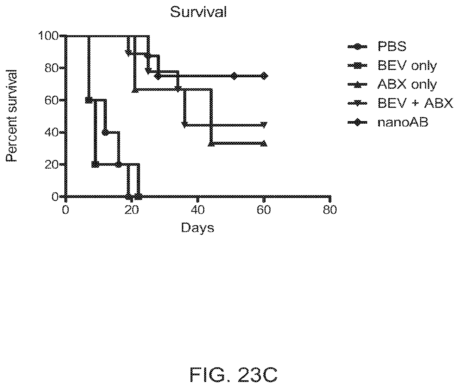
D00025

D00026

D00027

D00028

D00029
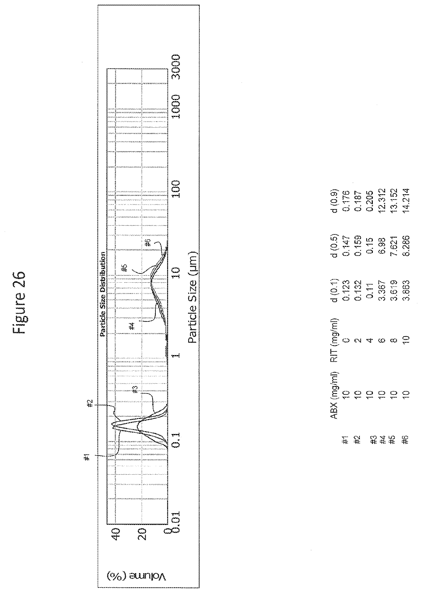
D00030

D00031

D00032

D00033

D00034

D00035

D00036

D00037

D00038

D00039

D00040
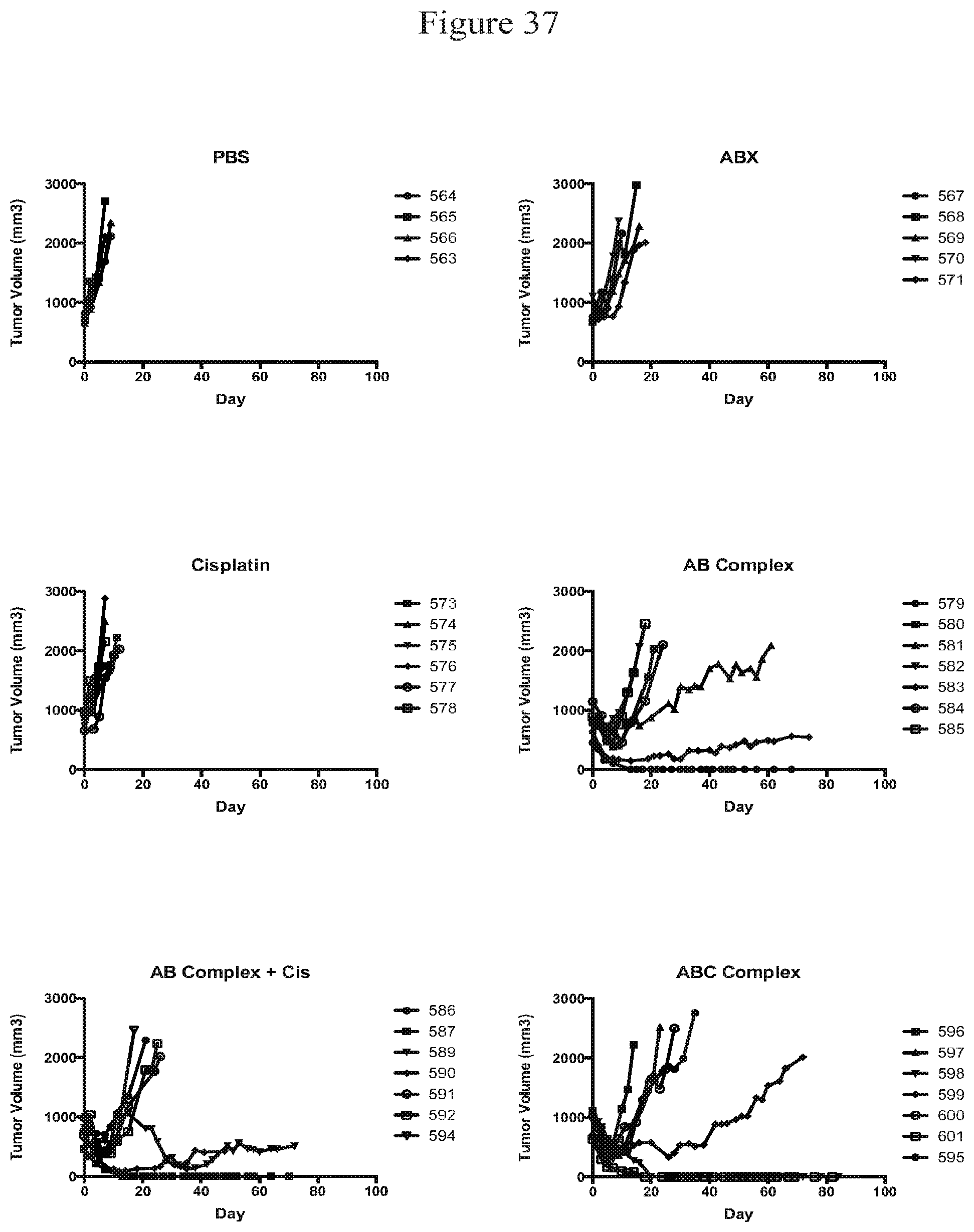
D00041

D00042

D00043

D00044

D00045
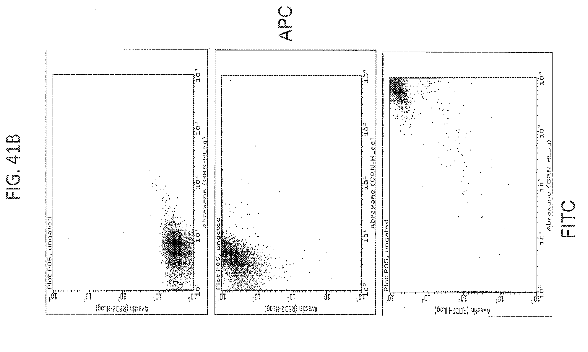
D00046

D00047
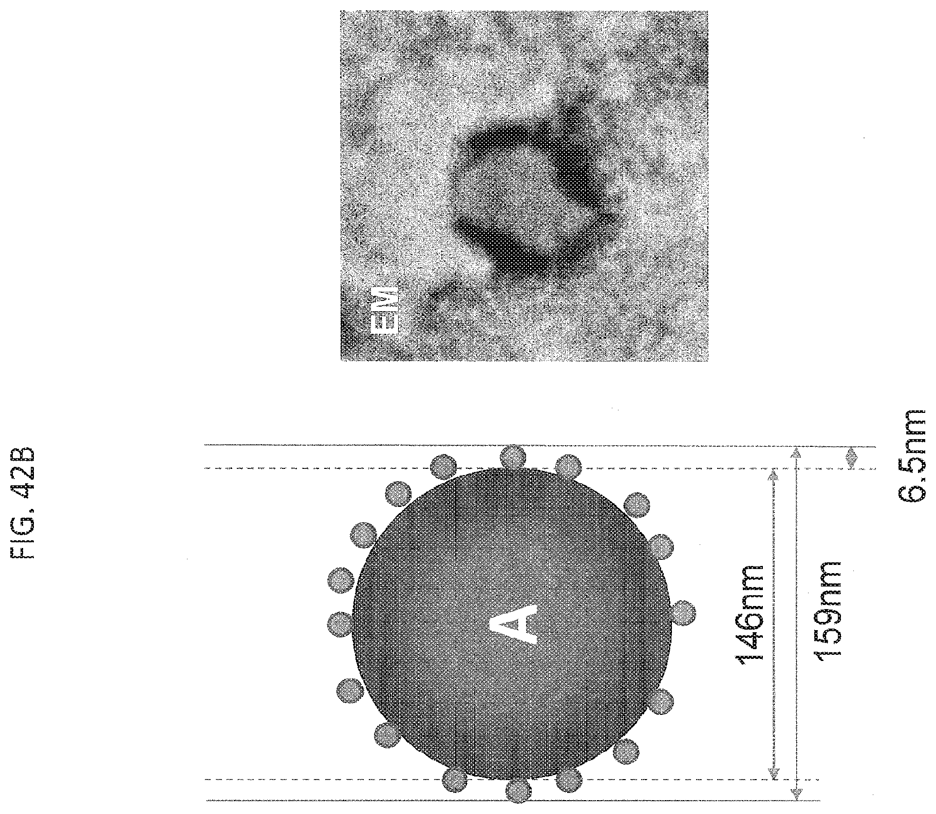
D00048

D00049

D00050

D00051

D00052

D00053

D00054
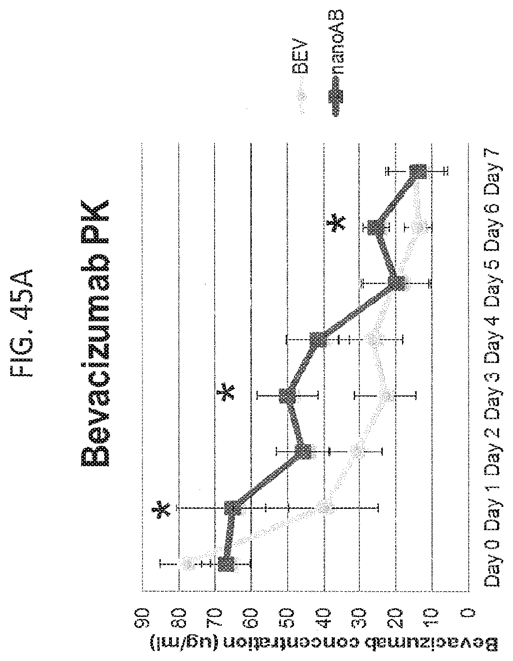
D00055

D00056

XML
uspto.report is an independent third-party trademark research tool that is not affiliated, endorsed, or sponsored by the United States Patent and Trademark Office (USPTO) or any other governmental organization. The information provided by uspto.report is based on publicly available data at the time of writing and is intended for informational purposes only.
While we strive to provide accurate and up-to-date information, we do not guarantee the accuracy, completeness, reliability, or suitability of the information displayed on this site. The use of this site is at your own risk. Any reliance you place on such information is therefore strictly at your own risk.
All official trademark data, including owner information, should be verified by visiting the official USPTO website at www.uspto.gov. This site is not intended to replace professional legal advice and should not be used as a substitute for consulting with a legal professional who is knowledgeable about trademark law.