Anti- Isoasp7 Amyloid (a ) Antibodies And Uses Thereof
PIECHOTTA; Anke ; et al.
U.S. patent application number 16/632289 was filed with the patent office on 2020-08-20 for anti- isoasp7 amyloid (a ) antibodies and uses thereof. The applicant listed for this patent is FRAUNHOFER-GESELLSCHAFT ZUR FORDERUNG DER ANGEWANDTEN FORSCHUNG E.V.. Invention is credited to Holger CYNIS, Hans-Ulrich DEMUTH, Kathrin GNOTH, Anke PIECHOTTA, Jens-Ulrich RAHFELD, Stephan SCHILLING.
| Application Number | 20200264197 16/632289 |
| Document ID | 20200264197 / US20200264197 |
| Family ID | 1000004824205 |
| Filed Date | 2020-08-20 |
| Patent Application | download [pdf] |


View All Diagrams
| United States Patent Application | 20200264197 |
| Kind Code | A1 |
| PIECHOTTA; Anke ; et al. | August 20, 2020 |
ANTI- ISOASP7 AMYLOID (A ) ANTIBODIES AND USES THEREOF
Abstract
The present invention can be included in the field of medicine. Specifically, the present invention provides antibodies and antigen-binding fragments thereof which can bind isoAsp7 amyloid .beta. (A.beta.) and a pharmaceutical composition comprising the antibodies or antigen-binding fragments thereof. IsoAsp7 A.beta. can be found in plaques of Alzheimer's patients and is thus a suitable target for the treatment and/or prevention of A.beta.-related diseases such as Alzheimer's disease. Thus, the antibodies, antigen-binding fragments thereof and the pharmaceutical composition comprising either can be used to treat and/or prevent neurodegenerative diseases. Further, the present invention provides hybridoma cell lines, the use of the antibodies or antigen-binding fragments thereof for the diagnosis and/or prognosis of a neurodegenerative disease and a method for detecting isoAsp7 A.beta. in an isolated sample.
| Inventors: | PIECHOTTA; Anke; (Halle (Saale), DE) ; GNOTH; Kathrin; (Halle (Saale), DE) ; CYNIS; Holger; (Halle (Saale), DE) ; RAHFELD; Jens-Ulrich; (Seegebiet Mansfelder Land, DE) ; SCHILLING; Stephan; (Halle (Saale), DE) ; DEMUTH; Hans-Ulrich; (Halle (Saale), DE) | ||||||||||
| Applicant: |
|
||||||||||
|---|---|---|---|---|---|---|---|---|---|---|---|
| Family ID: | 1000004824205 | ||||||||||
| Appl. No.: | 16/632289 | ||||||||||
| Filed: | July 17, 2018 | ||||||||||
| PCT Filed: | July 17, 2018 | ||||||||||
| PCT NO: | PCT/EP2018/069404 | ||||||||||
| 371 Date: | January 17, 2020 |
| Current U.S. Class: | 1/1 |
| Current CPC Class: | C07K 2317/565 20130101; C07K 16/18 20130101; C07K 2317/92 20130101; C07K 2317/24 20130101; G01N 33/6896 20130101 |
| International Class: | G01N 33/68 20060101 G01N033/68; C07K 16/18 20060101 C07K016/18 |
Foreign Application Data
| Date | Code | Application Number |
|---|---|---|
| Jul 19, 2017 | EP | 17182167.1 |
Claims
1. A pharmaceutical composition comprising: (i) an antibody or antigen-binding fragment thereof which specifically binds to L-isoAsp7 amyloid .beta. (A.beta.) wherein the K.sub.D of the interaction between the antibody and SEQ ID NO: 44 is at least 10 times less than the K.sub.D of the interaction between the antibody and SEQ ID NO: 8 and the K.sub.D is determined by surface plasmon resonance at 25.degree. C.; and (ii) a pharmaceutically acceptable carrier or diluent.
2. An antibody or antigen-binding fragment thereof which specifically binds to L-isoAsp7 A.beta. for use as a medicament, wherein the K.sub.D of the interaction between the antibody and SEQ ID NO: 44 is at least 10 times less than the K.sub.D of the interaction between the antibody and SEQ ID NO: 8 and the K.sub.D is determined by surface plasmon resonance at 25.degree. C.; or the pharmaceutical composition according to claim 1 for use as a medicament.
3. An antibody or antigen-binding fragment thereof which specifically binds to L-isoAsp7 A.beta. for use in the treatment and/or prevention of a neurodegenerative disease, wherein the K.sub.D of the interaction between the antibody and SEQ ID NO: 44 is at least 10 times less than the K.sub.D of the interaction between the antibody and SEQ ID NO: 8 and the K.sub.D is determined by surface plasmon resonance at 25.degree. C.; or the pharmaceutical composition according to claim 1 for use in the treatment and/or prevention of a neurodegenerative disease.
4. The pharmaceutical composition according to claim 1, the antibody or antigen-binding fragment thereof for use according to any one of claims 2-3 or the pharmaceutical composition for use according to any one of claims 2-3, wherein the antibody or antigen-binding fragment thereof comprises a light chain variable region (VL) and a heavy chain variable region (VH), wherein said VL comprises LCDR1, LCDR2 and LCDR3 polypeptides and VH comprises HCDR1, HCDR2 and HCDR3 polypeptides which are selected from the group consisting of: (a) LCDR1 is KSSQSLLNSRNRKNYLA (SEQ ID NO: 9), LCDR2 is WASTRDS (SEQ ID NO: 11), LCDR3 is KQSYNLRT (SEQ ID NO: 13), HCDR1 is GFSLTSYGVH (SEQ ID NO: 14), HCDR2 is ALWASGNTDYSSTLMS (SEQ ID NO: 15), and HCDR3 is DRGILTGGYFDV (SEQ ID NO: 17); (b) LCDR1 is KSSQSLFNSRTRKNYVA (SEQ ID NO: 27), LCDR2 is WASTRES (SEQ ID NO: 29), LCDR3 is KQSYNLRA (SEQ ID NO: 30), HCDR1 is GFTFTDYYMS (SEQ ID NO: 32), HCDR2 is FIRNKANGYTTEYSASVKG (SEQ ID NO: 34), and HCDR3 is DIPTIMDY (SEQ ID NO: 35); (c) LCDR1 is KSSQSLLNX.sub.1RX.sub.2RKNYLA (SEQ ID NO: 10), LCDR2 is WASTRX.sub.3S (SEQ ID NO: 12), LCDR3 is KQSYNLRT (SEQ ID NO: 13), HCDR1 is GFSLTSYGVH (SEQ ID NO: 14), HCDR2 is X.sub.4LWASGX.sub.5TDYX.sub.6SX.sub.7LMS (SEQ ID NO: 16), and HCDR3 is DRGIX.sub.8TGGYFDV (SEQ ID NO: 18), wherein X.sub.1 is S or R, X.sub.2 is N or T, X.sub.3 is D or E, X.sub.4 is A or V, X.sub.5 is N or R, X.sub.6 is S or N, X.sub.7 is T or A, and X.sub.8 is L, T or M; and (d) LCDR1 is KSSQX.sub.1LX.sub.2NSRTRKNYX.sub.3A (SEQ ID NO: 28), LCDR2 is WASTRES (SEQ ID NO: 29), LCDR3 is X.sub.4QSYNLRX.sub.5 (SEQ ID NO: 31), HCDR1 is GFTFX.sub.6DYYMX.sub.7 (SEQ ID NO: 33), HCDR2 is FIRNKANGYTTEYSASVKG (SEQ ID NO: 34), and HCDR3 is DIPTIMDY (SEQ ID NO: 35), wherein X.sub.1 is S or N, X.sub.2 is F or L, X.sub.3 is V or L, X.sub.4 is K or M, X.sub.5 is A or T, X.sub.6 is T or S, and X.sub.7 is S or N.
5. The pharmaceutical composition according to claim 1, the antibody or antigen-binding fragment thereof for use according to any one of claims 2-3 or the pharmaceutical composition for use according to any one of claims 2-3, wherein the antibody or antigen-binding fragment thereof comprises a light chain (LC) and a heavy chain (HC), wherein said LC and HC are polypeptides selected from the group consisting of: (a) LC of SEQ ID NO: 21 and HC of SEQ ID NO: 22; (b) LC of SEQ ID NO: 38 and HC of SEQ ID NO: 39; (c) LC of SEQ ID NO: 21 and HC of SEQ ID NO: 39; and (d) LC of SEQ ID NO: 38 and HC of SEQ ID NO: 22.
6. The pharmaceutical composition according to any one of claims 1-5, the antibody or antigen-binding fragment thereof for use according to any one of claims 2-5 or the pharmaceutical composition for use according to any one of claims 2-5, wherein the antibody or antigen-binding fragment thereof is a monoclonal antibody or antigen-binding fragment thereof.
7. The pharmaceutical composition according to any one of claims 1, 2, 3, 4 and 6, the antibody or antigen-binding fragment thereof for use according to any one of claims 2, 3, 4 and 6 or the pharmaceutical composition for use according to any one of claims 2, 3, 4 and 6, wherein the antibody or antigen-binding fragment thereof is a humanized antibody or antigen-binding fragment thereof.
8. The pharmaceutical composition according to claim 1 or the pharmaceutical composition for use according to any one of claims 2-7, wherein the composition further comprises donepezil, gelantamine, memantine, rivastigmine, a beta secretase inhibitor, a gamma secretase modulator, an additional antibody selected from the group of pan-A.beta. specific antibodies like aducanumab, bapineuzumab, crenezumab, ganteneumab, solanezumab and/or an antibody with specificity to posttranslational phosphorylated or nitrated A.beta. peptides.
9. The antibody or antigen-binding fragment thereof for use according to any one of claims 3-8 or the pharmaceutical composition for use according to any one of claims 3-8, wherein the neurodegenerative disease is selected from the list consisting of mild cognitive impairment, clinical or preclinical Alzheimer's disease, neurodegeneration in Down Syndrome, clinical and preclinical amyloid angiopathy, progressive supranuclear palsy, multiple sclerosis, Creutzfeld Jacob disease, Parkinson's disease, HIV-related dementia, ALS (amyotrophic lateral sclerosis), dementia related to Adult Onset Diabetes, senile cardiac amyloidosis and muscular degeneration.
10. The antibody or antigen-binding fragment thereof for use according to any one of claims 3-9 or the pharmaceutical composition for use according to any one of claims 3-9, wherein the neurodegenerative disease is clinical or preclinical Alzheimer's disease.
11. Use of an antibody or antigen-binding fragment thereof which specifically binds to L-isoAsp7 A.beta. for the diagnosis and/or prognosis of a neurodegenerative disease, wherein the K.sub.D of the interaction between the antibody and SEQ ID NO: 44 is at least 10 times less than the K.sub.D of the interaction between the antibody and SEQ ID NO: 8 and the K.sub.D is determined by surface plasmon resonance at 25.degree. C.
12. A method for detecting isoAsp7 A.beta. comprising a step wherein an isolated sample is put into contact with an antibody or antigen-binding fragment thereof which specifically binds to L-isoAsp7 A.beta., wherein the K.sub.D of the interaction between the antibody and SEQ ID NO: 44 is at least 10 times less than the K.sub.D of the interaction between the antibody and SEQ ID NO: 8 and the K.sub.D is determined by surface plasmon resonance at 25.degree. C.
13. The method according to claim 12, wherein the isolated sample is an isolated serum, cerebrospinal fluid or tissue sample.
14. A method of determining the percentage of A.beta. peptide comprising L-isoAsp at position 7 of SEQ ID NO: 1 in an isolated sample comprising: (a) quantifying the amount of L-isoAsp-comprising peptide through the use of a sandwich immunoassay, wherein the immobilized capture antibody or antigen-binding fragment thereof specifically binds to L-isoAsp7 A.beta., and the detection antibody is an antibody or antigen-binding fragment thereof that specifically binds to a polypeptide comprising SEQ ID NO: 1; (b) quantifying the total amount of A.beta. through the use of a sandwich immunoassay, wherein the immobilized capture antibody or antigen-binding fragment thereof is an antibody or antigen-binding fragment thereof that specifically binds to a polypeptide comprising SEQ ID NO: 1, and the detection antibody is an antibody or antigen-binding fragment thereof that specifically binds to a polypeptide comprising SEQ ID NO: 1; and (c) determining the percentage value by using the values obtained in steps (a) and (b), wherein the K.sub.D of the interaction between the antibody that specifically binds to L-isoAsp7 A.beta. and SEQ ID NO: 44 is at least 10 times less than the K.sub.D of the interaction between the same antibody and SEQ ID NO: 8 and the K.sub.D is determined by surface plasmon resonance at 25.degree. C.
15. The method according to claim 14, wherein the isolated sample is an isolated serum, cerebrospinal fluid or tissue sample.
Description
TECHNICAL FIELD
[0001] The present invention can be included in the field of medicine. Specifically, the present invention provides antibodies and antigen-binding fragments thereof which can bind isoAsp7 amyloid .beta. (A(.beta.) and a pharmaceutical composition comprising the antibodies or antigen-binding fragments thereof. The antibodies, antigen-binding fragments thereof and the pharmaceutical composition can be used to treat and/or prevent neurodegenerative diseases. Further, the present invention provides hybridoma cell lines, the use of the antibodies or antigen-binding fragments thereof for the diagnosis and/or prognosis of a neurodegenerative disease and a method for detecting isoAsp7 A.beta. in an isolated sample.
BACKGROUND ART
[0002] Alzheimer's disease (AD) is a progressive incurable neuronal damage of the brain, occurring in mid or late life. One of the first symptoms is short-term memory loss, followed by behavioral issues and disorientation up to loss of body functions. AD always leads to death; usually people die 7 to 10 years after diagnosis.
[0003] Two histological alterations can be seen post mortem in AD patients: senile plaques and neurofibrillary tangles, consisting of hyperphosphorylated Tau protein. The former are extracellular deposits, basically composed of fibrillary amyloid beta (A.beta.). A.beta. peptides arise through endoproteolytic cleavage of APP. Because of their presence within the transmembrane region of APP, A.beta. peptides are high in hydrophobic side chains, resulting in poor solubility. After release of A.beta. peptides, they have a strong tendency to aggregate. In AD patients, there is an imbalance between generation and degradation of A.beta. peptides, they aggregate and deposit, leading to neuronal cell death. The main A.beta. variants observed in human brain are A.beta.40 and A.beta.42, but also N-terminal truncated variants and other posttranslational modified forms are observed in plaques.
[0004] There are two different types of AD: the very rare hereditary form (familial AD-FAD) and the sporadic form, of which about 95 percent of patients are affected. FAD is marked by AD symptoms that appear at an unusually early age. Beside presenilin 1 (PSI) and presenilin 2 (PS2), APP belongs to the three known genes that can cause FAD. Mutations in these genes lead to increased A.beta. production and virtually guarantee the development of AD. However, it remains unresolved how A.beta. exerts its toxic effects. As a genetic disorder, FAD is clearly the consequence of the malfunctioning of the mutated genes, whereas the cause of late-onset spontaneous AD is still not completely understood.
[0005] Not only are the reasons for the development of spontaneous AD unknown, scientists all over the world are trying to find AD biomarkers in order to identify AD patients in a possibly early stage. AD can be divided into a pre-symptomatic phase in which subjects are cognitively normal, a prodromal phase known as mild cognitive impairment (MCI) and a third phase when patients show dementia with impairments or even loss of function in daily activities (Petersen (2004) J Intern Med 256:183-94; Savva et al. (2009) N Engl J Med 360:2302-2309). To date, the only highly predictive biomarkers for AD are the genetic mutations that are pathogenic for FAD. They can be detected years before disease onset and identify those individuals who will go on to develop AD later in life. The Alzheimer's disease Neuroimaging Initiative (ADNI) generated a model for the temporal ordering of AD biomarkers which suggests that A.beta. amyloid biomarkers become abnormal first, followed by changes in neurodegenerative biomarkers (CSF tau, FDG-PET, MRI) and the onset of clinical symptoms. That means, A.beta. peptide arises to pathological concentrations in brain even before patients show first neurological symptoms in the MCI phase. Furthermore, it could be shown that not only mutations, but also posttranslational modifications of the A.beta. peptide can accelerate their aggregation behavior, possibly resulting in a severe course of disease.
[0006] One posttranslational modification observed in the A.beta. peptide is the formation of isoaspartate (isoAsp). The generation of isoAsp from aspartyl residues is a spontaneous posttranslational modification of peptides and proteins. This reaction is considered to determine the half-life of proteins (Robinson and Rudd (1974) Curr Top Cell Regul. 8(0):247-95; Robinson and Robinson (2001) PNAS 98(3):944-949). Besides that, isoAsp-formation introduces an additional methylene group into the backbone of the protein or peptide (Aswad et al. (2000) J Pharm Biomed Anal. 21(6):1129-36; Geiger and Clarke (1987) J Biol Chem 262:785-794), consequently altering its structure. This post-translational modification may also change the properties of proteins like solubility, conformation and function. IsoAsp forms most easily at sequences in which the side chain of the C-flanking amino acid is relatively small and hydrophilic, and is less likely to form when bulky or hydrophobic residues are in this position. The most favorable C-flanking amino acids are glycine, serine, and histidine (Shimizu et al. (2005) Biol Pharm Bull. 28(9):1590-6).
[0007] After deposition of insoluble A.beta. in senile plaques, the formation of isoAsp7 is likely to occur as time goes by, since A.beta. has a serine residue in position 8. The presence of isoAsp7 A.beta. in brains of AD patients was first described in 1993 (Roher et al. (1993) Proc. Natl. Acad. Sci. USA 90:10836-10840). By using polyclonal anti-isoAsp7 A.beta. antibodies, it could be shown that isoAsp7 A.beta. is present in extracellular deposits in AD brain as well as amyloid-bearing vessels and serves as an indicator of plaque age (Fonseca et al. (1999) Exp Neurol. 157(2):277-88; Shimizu et al. (2000) Arch Biochem Biophys. 381(2):225-34). A.beta. is able to activate the classical complement pathway (CCP) by direct binding of Clq, resulting in the recruitment of reactive glial cells to the site of fibrillary A.beta. protein plaque. Velazquez and colleagues found that isomerization of Asp7 resulted in complete elimination of CCP activation. This could prevent plaque recognition by the complement system.
[0008] Since an isoAsp7 modification does not influence aggregation of A.beta. peptides (Fukuda et al. (1999) Bioorg Med Chem Lett. 9(7):953-6; Shimizu et al. (2002) J Neurosci Res. 70(3):451-61), it is not likely that this modification accelerates deposition and plaque formation. However, Wakutani et al. described in the year 2004 a new case of FAD, called Japanese-Tottori FAD. In some members of this family, a missense mutation within APP (D678N) replaces the aspartate 7 of A.beta. with asparagine (Wakutani et al. (2004) J Neurol Neurosurg Psychiatry. 75(7):1039-42). Asparagine residues undergo isomerization about 10 times quicker than aspartate (Stephenson and Clarke (1989) J Biol Chem 264:6164-6170). Manifestation of AD symptoms in this pedigree may be not due to Asn7-A.beta., but the enhanced formation of isoAsp7 A.beta..
[0009] Although AD has been known for over 100 years, there are still only symptomatic treatments available on the market. Active immunization approaches with A.beta. and fragments thereof as well as passive immunization with anti-A.beta. antibodies was effective in different animal models. Vaccination of humans with A.beta. inhibited the development of A.beta. plaques and reduced the A.beta. burden in AD patients. However, the clinical studies needed to be stopped due to some patients developing severe meningoencephalitis (Orgogozo et al. (2003) Neurology 61:46-54) or a humoral and cellular response against A.beta. resulting in a strong immune response against the endogenous A.beta. peptide (Holmes et al. (2008) Lancet 372:216-223).
[0010] Consequently, passive immunization was considered safer and more controllable than active immunization. Several antibodies targeting the A.beta. peptide have been used in clinical trials of passive immunization therapy in AD patients. However, most antibodies are directed against linear epitopes in the native non modified peptide. Treatment studies showed a positive effect, but have side effects such as amyloid-related imaging abnormalities (ARIA), seizures and death (Moreth et al. (2013) Immun Ageing. 10(1):18).
[0011] Thus, there is a need for antibodies for the effective treatment and/or prevention of A.beta. plaque-associated diseases such as Alzheimer's disease. The present application provides antibodies that were found to be more effective in a relevant animal model and are thus expected to be more effective at treating and/or preventing A.beta. plaque-associated diseases such as Alzheimer's disease.
FIGURES
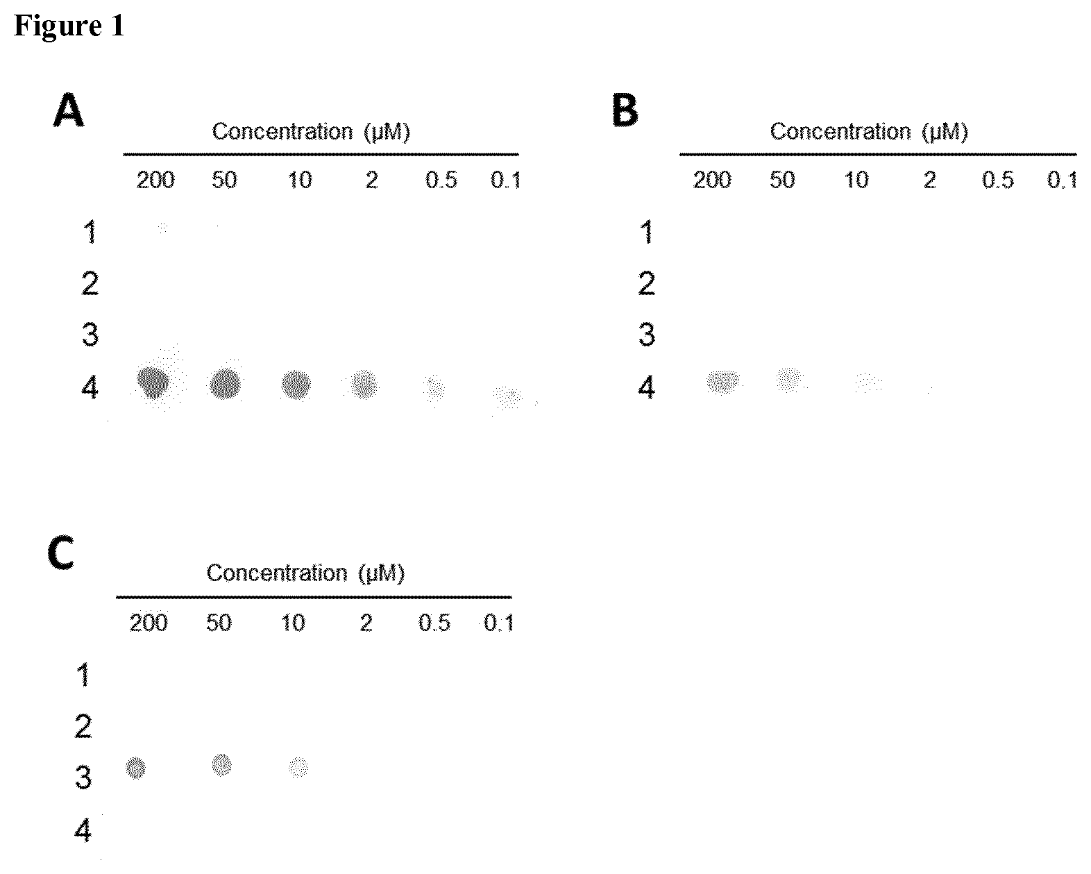
[0012] FIG. 1: Investigation of antibody specificity by Dot Blot analysis. 2 .mu.l of A.beta. peptides were spotted in descending concentrations on a nitrocellulose membrane and blocked for 1 h in blocking solution (5% (w/v) milk powder in TBS-T (TBS+0.05% Tween 20 (v/v)). Antibodies K11 (A), K119 (B) and 6E10 (C) were diluted to 1 .mu.g/ml in blocking solution and incubated with the membrane for 1 h, followed by 3.times.5 minutes washing steps with TBS-T. Anti-mouse antibody conjugated to alkaline phosphatase (AP) was added and incubated for 1 h, followed by 3.times.5 minutes washing steps and subsequent colorimetric detection of AP activity by addition of substrates BCIP (5-bromo-4-chloro-3-indolyl-phosphate) and NBT (nitro blue tetrazolium). 1--isod7-A.beta.(1-17); 2--isoD7-A.beta.(5-9)rep; 3--A.beta.(1-18); 4--isoD7-A.beta.(1-12). Of note, antibody 6E10 does not recognize isoAsp-modified A.beta..
[0013] FIG. 2: Determination of binding constants of immobilized K11 with different A.beta.-peptides by SPR analysis. 1,500-2,000 RU of antibody K11 were immobilized on a CMS chip by binding to goat anti mouse IgG. Monomeric A.beta. peptides were injected in varying concentrations. K.sub.D values were calculated by using single-cycle kinetic analysis of BIAevaluation software. A--isoD7-A.beta.(1-18) was applied at 1 nM, 3 nM, 9 nM, 27 nM, 81 nM and 243 nM. B--isod7-A.beta.(1-17) was applied at 1 nM, 3 nM, 9 nM, 27 nM, 81 nM and 243 nM. C--A.beta.(1-18) was applied at 10 mM, 30 nM, 90 nM, 270 nM, 810 nM and 2430 nM.
[0014] FIG. 3: K.sub.D value determination of immobilized K119 with isoD7-A.beta.(1-18) by SPR analysis. About 2,000 RU of antibody K119 were immobilized on a CMS chip by binding to goat anti mouse IgG. Monomeric isoD7-A.beta.(1-18) peptides were injected in varying concentrations (1 nM, 2 nM, 5 nM, 10 nM, 20 nM, 50 nM, 100 nM, 200 nM, 500 nM, 1 .mu.M). K.sub.D value was calculated by using multi-cycle kinetic analysis of BIAevaluation software.
[0015] FIG. 4: Thermodynamic characterization of K11 with different A.beta.-peptides. Determination of the thermodynamic parameter of K11 with different antigens at 25.degree. C. K11 was dialyzed against an A.beta.-peptides dissolved in ITC buffer (25 mM KH.sub.2PO.sub.4; 25 mM Na.sub.2HPO.sub.4; 150 mM NaCl; 1 mM EDTA, pH 7.4). Peptides were added every 5 minutes to antibody solution in the following concentrations:
[0016] A--50.8 .mu.M isoD7-A.beta.(1-18) was titrated to 2.5 .mu.M K11
[0017] B--56.7 .mu.M A.beta.(1-18) was titrated to 11.9 .mu.M K11
[0018] C--238 .mu.M isod7-A.beta.(1-17) was titrated to 11.9 .mu.M K11
[0019] The top graphs show raw data of heat pulses resulting from titration of antigen in the calorimetric cell with antibody K11. The bottom graphs show the integrated heat pulses, normalized per mol of injectant as a function of molar ratio.
[0020] FIG. 5: Thermodynamic characterization of K119 binding to isoD7-A.beta.(1-18) by using isothermal titration calorimetry. K119 was dialyzed against and the peptide isoD7-A.beta.(1-18) dissolved in ITC buffer (25 mM KH2PO4; 25 mM Na2HPO4; 150 mM NaCl; 1 mM EDTA, pH 7.4). 44.9 .mu.M peptide solution was added every 5 minutes to 2.2 .mu.M antibody solution. The top graph shows raw data of heat pulses resulting from titration of antigen in the calorimetric cell with antibody K119. The bottom graph shows the integrated heat pulses, normalized per mol of injectant as a function of molar ratio.
[0021] FIG. 6: Immunohistochemical analysis of AD aggregates in brain samples from 5.times.FAD and wildtype mice by using K11. Paraformaldehyde treated and cryoconserved brain slices from 5.times.FAD mice (B-E and G-J) with different ages and 12 month old wildtype mice (A and F) have been treated with anti isoAsp7-A.beta. antibody K11 (A-E) and commercially available antibody 6E10, followed by application of biotinylated anti mouse IgG. Immunostaining was performed by addition of ExtrAvidin-Peroxidase (Sigma-Aldrich) and the chromogenic substrate 3,3'-Diaminobenzidin (DAB). A) Wildtype mouse, 12 month old, stained with K11. B) 5.times.FAD mouse, 3 month old, stained with K11. C) 5.times.FAD mouse, 6 month old, stained with K11. D) 5.times.FAD mouse, 9 month old, stained with K11. E) 5.times.FAD mouse, 12 month old, stained with K11. F) Wildtype mouse, 12 month old, stained with 6E10. G) 5.times.FAD mouse, 3 month old, stained with 6E10. H) 5.times.FAD mouse, 6 month old, stained with 6E10. I) 5.times.FAD mouse, 9 month old, stained with 6E10. J) 5.times.FAD mouse, 12 month old, stained with 6E10.
[0022] FIG. 7: Comparative staining of A.beta. aggregates in brain samples from 12 month old 5.times.FAD mice with different antibodies. Formaldehyde treated and cryoconserved brain slices from 12 month old 5.times.FAD mice have been treated with anti isoAsp7-A.beta. antibody K119 (A), K11 (B), 6E10 (C) and without primary antibody (D), followed by application of biotinylated anti mouse IgG. FIG. 7 (E) shows staining of a wildtype mouse brain slice with K119. Immunostaining was performed by addition of ExtrAvidin-Peroxidase (Sigma-Aldrich) and the chromogenic substrate 3,3'-Diaminobenzidin (DAB).
[0023] FIG. 8: Sandwich ELISA for quantification of isoAsp7-A.beta. and total A.beta. concentrations in biological samples. Capture antibodies K11 (A) and 3D6 (B) were diluted to 2 .mu.g/ml and immobilized on microtiter plates overnight at 4.degree. C. Blocking occurred for 2 hours at room temperature. isoD7-A.beta.(1-30) (SEQ ID NO: 51) and A.beta.(1-30) (SEQ ID NO: 52) were serially diluted and added to the wells in duplicate. After an incubation period of 2 hours at 4.degree. C., plates were washed six times with TBS-T. HRP-conjugated anti A.beta. antibody clone 4G8 was added in a final concentration of 1 .mu.g/ml and incubated for 1 hour at 4.degree. C. After washing with TBS-T, a color reaction with TMB was performed and stopped by the addition of 1.2 N H.sub.2SO.sub.4. The standard curve was calculated from measured absorption at 450/540 nm by a 4-Parameter-Logistic-Fit: y=(A2+(A1-A2)/(1+(x/x.sub.0){circumflex over ( )}p).
[0024] FIG. 9: Quantification of total A.beta. (A) and isoAsp7-A.beta. (B) peptides in 5 M GdmCl fractions from 5.times.FAD mice brain treated with K11 and isotype control. Three month old 5.times.FAD mice were treated intraperitoneally once a week with 500 .mu.g, 150 .mu.g K11 or 500 .mu.g isotype control. After 12 weeks of treatment, mice were sacrificed and the left hemisphere was homogenized in T-Per buffer, followed by centrifugation. The resulting pellet was resuspended in 5 M GdmCl (150 mg/ml), again centrifuged and the supernatant applied to a total A.beta. (A) or isoAsp7-A.beta. (B) specific ELISA.
[0025] FIG. 10: Immunohistochemical analysis of isoAsp7-A.beta. containing aggregates in hippocampal brain slices from 5.times.FAD mice treated with K11 and isotype control. Three month old 5.times.FAD mice were treated intraperitoneally once a week with 500 .mu.g, 150 .mu.g K11 or 500 .mu.g isotype control. After 12 weeks of treatment, mice were sacrificed and the right hemisphere was treated with formaldehyde, cryoconserved and used for immunohistochemical staining. 30 .mu.m slices were made from hippocampal sections and incubated with 2 .mu.g/ml K11, followed by application of biotinylated anti mouse IgG. Immunostaining was performed by addition of ExtrAvidin-Peroxidase (Sigma-Aldrich) and the chromogenic substrate 3,3'-Diaminobenzidin (DAB). Regions of interest (ROI) in hippocampal brain slices were selected by staining with 2 .mu.g/ml 6E10 (for general A.beta.) and 2 .mu.g/ml isoAsp7-A.beta. specific antibody K11 (for isoAsp7-A.beta.). All pictures were recorded by using the microscope Biorevo BZ-9000 (Keyence) with transmitted light modus and an exposure time of 1/200 s. Percentage area of isoAsp7-A.beta. (ROI isoD7 in %) was quantified based on total area of ROI by using the program BZ II Analyzer.
[0026] FIG. 11: Immunohistochemical staining of isoAsp7-A.beta. containing aggregates in brain samples from 5.times.FAD mice treated with K11 and isotype control. Three month old 5.times.FAD mice were treated intraperitoneally once a week with 500 .mu.g, 150 .mu.g K11 or 500 .mu.g isotype control. After 12 weeks of treatment, mice were sacrificed. The right hemisphere was used to prepare paraformaldehyde treated and cryoconserved brain slices. Brain slices have been treated with K11, followed by application of biotinylated anti mouse IgG. Immunostaining was performed by addition of ExtrAvidin-Peroxidase (Sigma-Aldrich) and the chromogenic substrate 3,3'-Diaminobenzidin (DAB). A) 3 month old 5.times.FAD mouse (baseline control), B) 6 month old 5.times.FAD mouse, treated with 500 .mu.g Isotype control antibody, C) 6 month old 5.times.FAD mouse, treated with 150 .mu.g K11, D) 6 month old 5.times.FAD mouse, treated with 500 .mu.g K11.
[0027] FIG. 12: Immunohistochemical staining of A.beta. aggregates (antibody 6E10) in brain samples from 5.times.FAD mice treated with K11 and isotype control. Three month old 5.times.FAD mice were treated intraperitoneally once a week with 500 .mu.g, 150 .mu.g K11 or 500 .mu.g isotype control. After 12 weeks of treatment, mice were sacrificed. The right hemisphere was used to prepare paraformaldehyde treated and cryoconserved brain slices. Brain slices have been treated with commercially available antibody 6E10, followed by application of biotinylated anti mouse IgG. Immunostaining was performed by addition of ExtrAvidin-Peroxidase (Sigma-Aldrich) and the chromogenic substrate 3,3'-Diaminobenzidin (DAB). A) 3 month old 5.times.FAD mouse (baseline control), B) 6 month old 5.times.FAD mouse, treated with 500 .mu.g Isotype control antibody, C) 6 month old 5.times.FAD mouse, treated with 150 .mu.g K11, D) 6 month old 5.times.FAD mouse, treated with 500 .mu.g K11.
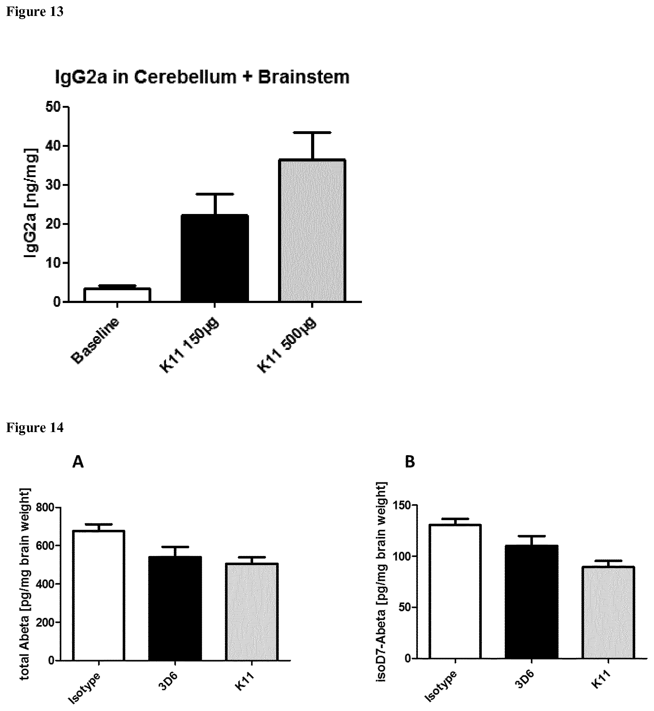
[0028] FIG. 13: Quantification of IgG2a in cerebellum and brainstem from 5.times.FAD mice treated with KM Three month old 5.times.FAD mice were treated intraperitoneally once a week with 500 .mu.g or 150 .mu.g K11. After 12 weeks of treatment, mice were sacrificed; cerebellum and brainstem were homogenized in ELISA Blocker+Tween, followed by centrifugation. The resulting supernatant was applied to a mouse IgG2a specific ELISA. Baseline represents 3 month old 5.times.FAD mice without treatment.
[0029] FIG. 14: Quantification of total A.beta. (A) and isoD7-A.beta. (B) peptides in T-Per fractions from 5.times.FAD mice brain treated with anti-A.beta. antibodies K11, 3D6 and isotype control. Three month old 5.times.FAD mice were treated intraperitoneally once a week with 300 .mu.g K11, 3D6 or isotype control. After 6 months of treatment, mice were sacrificed and the left hemisphere was homogenized in T-Per buffer (Thermofisher) to 50 mg brain weight/m1 T-Per buffer. The resulting T-Per fractions were applied to a total A.beta. as well as isoAsp7-A.beta. specific ELISA. Sample size consisted of at least 10 animals per group (some groups contained one or two more animals). Statistical analysis was performed using Bonferroni's Multiple Comparison Test. * means p.ltoreq.0.05. ** means p.ltoreq.0.01. *** means p.ltoreq.0.001.
[0030] FIG. 15: Quantification of total A.beta. (A) and isoD7-A.beta. (B) peptides in 5 M GdmCl fractions from 5.times.FAD mice brain treated with anti-A.beta. antibodies K11, 3D6 and isotype control. Three month old 5.times.FAD mice were treated intraperitoneally once a week with 300 .mu.g K11, 3D6 and isotype control. After 6 months of treatment, mice were sacrificed and the left hemisphere was homogenized in T-Per buffer (50 mg/ml), followed by centrifugation. The resulting pellet was resuspended in 5 M GdmCl (150 mg/ml), again centrifuged and the supernatant was subjected to a total A.beta. as well as isoAsp7-A.beta. specific ELISA. Sample size consisted of at least 10 animals per group (some groups contained one or two more animals). Statistical analysis was performed using Bonferroni's Multiple Comparison Test. * means p.ltoreq.0.05. ** means p.ltoreq.0.01. *** means p.ltoreq.0.001.
[0031] FIG. 16: Elevated Plus Maze (EPM) test of 5.times.FAD animals treated weekly for 38 weeks with 300 .mu.g K11, 3D6 and isotype control. Antibody-treated 5.times.FAD groups were compared with wildtype animals treated with 300 .mu.g isotype control. Test animals were placed with their head to the end of a defined closed arm of an elevated, plus-shaped (+) apparatus with two open and two enclosed arms. During the next 10 minutes, every movement of the test animals was recorded.
[0032] A--The time the animals spent in the open arms was summed up in order to calculate % in exposed area.
[0033] B--Arm entries are defined as presence of the complete animal (except tail) in the open arm.
[0034] Sample size consisted of at least 9 animals per group (some groups contained one or two more animals). Statistical analysis was performed using Bonferroni's Multiple Comparison Test. * means p.ltoreq.0.05. ** means p.ltoreq.0.01. *** means p.ltoreq.0.001.
[0035] FIG. 17: Fear conditioning test of 5.times.FAD animals treated weekly for 38 weeks with 300 .mu.g K11, 3D6 and isotype control. Antibody-treated 5.times.FAD groups were compared with wildtype animals treated with 300 .mu.g isotype control. Test animals were placed in an automated FearConditioning System and submitted to the following procedure: pause (180 s), sound (28 s), electric stimulus (0.7 mA for 2 s). After 24 h, test animals were again placed in the FearConditioning System, left there for 210 s and then removed. One hour later, animals were placed back in the container in order to expose them to a 180 s pause, followed by 180 s of sound (neutral stimulus). A state of fear was expressed by the mice by freezing in place. Freezing time during the 180 s pause was counted and subtracted from the freezing times during the 180 s sound in order to obtain % freezing time. Sample size consisted of at least 8 animals per group (some groups contained one or two more animals). Statistical analysis was performed using Bonferroni's Multiple Comparison Test. * means p.ltoreq.0.05. ** means p.ltoreq.0.01. *** means p.ltoreq.0.001.
[0036] FIG. 18: Pole test of 5.times.FAD animals treated weekly for 38 weeks with 300 .mu.g K11, 3D6 and isotype control. Antibody-treated 5.times.FAD groups are compared with wildtype animals treated with 300 .mu.g isotype control. Animals were placed with their head directed to the top on a 50 cm high pole (diameter 1.5 cm). The animals were unhanded and time was counted until (A) animals turned around (defined as every single paw is directed to the ground) and (B) animals reached the ground with every paw. Sample size consisted of at least 10 animals per group (some groups contained one or two more animals). Statistical analysis was performed using Bonferroni's Multiple Comparison Test. * means p.ltoreq.0.05. ** means p.ltoreq.0.01. *** means p.ltoreq.0.001.
[0037] FIG. 19: Morris water maze test of 5.times.FAD animals treated weekly for 38 weeks with 300 .mu.g K11, 3D6 and isotype control. Antibody-treated 5.times.FAD groups are compared with wildtype animals treated with 300 .mu.g isotype control. Test animals were placed in a circular pool and are required to find an invisible platform that allows them to escape the water. The circular pool is divided into 4 equal quadrants. Test animals were placed into the first quadrant and time was counted until they reached the platform. After at least a 5 min pause, test animals were placed into the second quadrant and exposed to the same procedure. The animals were allowed to pause again, followed by putting them in quadrant 3, followed by another pause and putting them again in quadrant 2. At the end, time until the test animals reach the platform was counted and summed up for every mouse in 4 trials per day. The graph shows the average trial time until animals reached the platform on day 4. Sample size consisted of at least 10 animals per group (some groups contained one or two more animals). Statistical analysis was performed using Bonferroni's Multiple Comparison Test. * means p.ltoreq.0.05. ** means p.ltoreq.0.01. *** means p.ltoreq.0.001.
[0038] FIG. 20: Aggregation of wildtype A.beta. (A) and isoD7-A.beta. (B) peptides after addition of K11, 3D6 and isotype control. Synthetic wildtype A.beta.(1-40) and isoD7-A.beta.(1-40) peptides have been monomerized by dissolving them in hexafluoroisopropanol (HFIP). HFIP was allowed to evaporate overnight, peptides were dissolved in 1 volume 0.1 M NaOH, followed by the addition of 18 volumes PBS and 1 volume 0.1 M HCl. Antibodies K11, 3D6 and isotype control were added subsequently to a final concentration of 5 .mu.M, leading to a concentration of 10 .mu.M A.beta. peptides. After addition of 200 .mu.M ThT (Thioflavin T), fluorescence at 435/485 nm (ex/em) was tracked in a microplate reader (FluoStar Optima, BMG Labtech) at 37.degree. C. under shaking conditions (600 rpm).
SUMMARY OF THE INVENTION
[0039] Targeting of isoAsp7 A.beta. in AD patients is a new promising approach, because antibodies will solely bind modified and aged A.beta. peptides. Consequently, freshly synthesized circulating A.beta. in the periphery will be largely unaffected, thereby preventing loss of active antibodies via A.beta. binding in e.g. blood or CSF. This possibly allows a reduction of antibody dosage. Furthermore, the epitope density of isoAsp7-modified species in A.beta. deposits is low in comparison to native A.beta. variants. This leads to a better antibody distribution within the brain tissue and a lower reactivity with amyloid deposits in the walls of blood vessels in the central nervous system (cerebral amyloid angiopathy, CAA), thereby preventing ARIA.
[0040] Here, we show that treatment of transgenic mice with Alzheimer-pathology with an isoAsp7-A.beta. specific antibody results in attenuation of disease pathology. Surprisingly, we observed that application of a highly isoAsp-specific antibody does not only reduce isoAsp7-A.beta. in these mice but also shows an unexpected reduction of amyloid plaques and non-isoAsp7-modified A.beta. (see FIGS. 9 and 12 below). This result was obtained by using an antibody derived from clone 6E10, which, as shown here for the first time, specifically detects unmodified (i.e. containing Asp instead of isoAsp in position 7) A.beta. (FIG. 1). Therefore, both antibodies were used to prove this differential activity of isoAsp-A.beta. specific antibodies, enabling an application of isoAsp-A.beta. for treatment of A.beta. plaque-associated diseases such as Alzheimer's disease.
[0041] Moreover, we also show that the isoAsp7-modified A.beta. is, compared to total A.beta., an underrepresented species, making up only 4% in mice (FIG. 9). Despite this low concentration, we observed an unexpected reduction of amyloid plaques after treatment (FIGS. 9, 11, 12, and 15). This novel finding might help to circumvent previous limitations of A.beta. immunotherapy.
[0042] Surprisingly, an antibody of the present invention was able to remove more fibrillary A.beta. than 3D6 (an antibody that binds to residues 1-5 of an A.beta.42 peptide without an L-isoAsp 7 modification) as shown in FIG. 15A. This appears to be associated with a greater amount of monomeric, oligomeric and fibrillary isoAsp-modified A.beta. removal (FIGS. 14B and 15B). Further, an antibody of the present invention improves the treatment of 5.times.FAD mice in comparison to 3D6 (see FIGS. 17-19).
[0043] The present invention thus provides an antibody or antigen-binding fragment thereof which specifically binds to isoAsp7 amyloid .beta. (A(62 ), wherein the K.sub.D of the interaction between the antibody and SEQ ID NO: 44 is at least 10 times less than the K.sub.D of the interaction between the antibody and SEQ ID NO: 8. The present invention also provides a pharmaceutical composition comprising the antibody or antigen-binding fragment thereof of the present invention and a pharmaceutically acceptable carrier or diluent. Also, the present invention encompasses the use of the antibody or antigen-binding fragment thereof of the present invention or the pharmaceutical composition of the present invention as a medicament, specifically for the treatment and/or prevention of a neurodegenerative disease. Further, the present invention provides the use of the antibody or antigen-binding fragment thereof of the present invention for the diagnosis and/or prognosis of a neurodegenerative disease. The present invention also provides hybridoma cell-lines and a method for detecting isoAsp7 A.beta. comprising a step wherein an isolated sample is put into contact with the antibody or antigen-binding fragment thereof of the present invention. Finally, the present invention provides a method of determining the percentage of peptides in an amyloid plaque which comprises an L-isoAsp at position 7 of SEQ ID NO: 1.
DETAILED DESCRIPTION OF THE INVENTION
Antibodies
[0044] In a first aspect, the present invention provides an antibody or antigen-binding fragment thereof which specifically binds to isoAsp7 amyloid .beta. (A.beta.), wherein the K.sub.D of the interaction between the antibody and SEQ ID NO: 44 is at least 10 times less than the K.sub.D of the interaction between the antibody and SEQ ID NO: 8. In a preferred embodiment, the K.sub.D of the interaction between the antibody and SEQ ID NO: 48 is at least 10 times less than the K.sub.D of the interaction between the antibody and SEQ ID NO: 8. Preferably, the K.sub.D is determined by surface plasmon resonance or isothermal titration calorimetry. More preferably, the K.sub.D is determined by surface plasmon resonance at 25.degree. C.
[0045] In an alternative aspect, the present invention provides an antibody or antigen-binding fragment thereof which specifically binds to pE3 (contains L-pyroglutamate at position 3) isoAsp7 amyloid .beta. (A.beta.) wherein the K.sub.D of the interaction between the antibody and SEQ ID NO: 48 is at least 10 times less than the K.sub.D of the interaction between the antibody and SEQ ID NO: 8.
[0046] SEQ ID NO: 44 is isoAsp7 A.beta. (1-18) and has the following sequence:
TABLE-US-00001 DAEFRHXSGYEVHHQKLV,
wherein X is L-isoAsp.
[0047] SEQ ID NO: 8 is A.beta. (1-18) and has the following sequence:
TABLE-US-00002 DAEFRHDSGYEVHHQKLV
[0048] SEQ ID NO: 48 is pE3-isoD7-A.beta.(3-18) and has the following sequence:
TABLE-US-00003 ZFRHXSGYEVHHQKLV,
wherein X is L-isoAsp and Z is L-pyroglutamate.
[0049] As used herein, the term "antibody" refers to a protein comprising at least one immunoglobulin variable domain sequence. The term antibody includes, for example, full-length and mature antibodies. For example, an antibody can include a heavy (H) chain variable domain sequence (abbreviated herein as VH), and a light (L) chain variable domain sequence (abbreviated herein as VL). In another example, an antibody molecule includes two heavy (H) chain variable domain sequences and two light (L) chain variable domain sequence, thereby forming two antigen binding sites, such as Fab, Fab', F(ab')2, Fc, Fd, Fd', Fv, single chain antibodies (scFv for example), single variable domain antibodies, diabodies (Dab) (bivalent and bispecific), and chimeric (e.g., humanized) antibodies, which may be produced by the modification of whole antibodies or those synthesized de novo using recombinant DNA technologies. These functional antibody fragments retain the ability to selectively bind with their respective antigen or receptor. Antibodies and antibody fragments can be from any class of antibodies including, but not limited to, IgG, IgA, IgM, IgD, and IgE, and from any subclass (e.g., IgG1, IgG2, IgG3, and IgG4) of antibodies. The antibodies of the present invention can be monoclonal or polyclonal. The antibody can also be a human, humanized, CDR-grafted, or in vitro generated antibody. The antibody can have a heavy chain constant region chosen from, e.g., IgG1, IgG2, IgG3, or IgG4. The antibody can also have a light chain chosen from, e.g., kappa or lambda.
[0050] Examples of antigen-binding fragments include: (i) a Fab fragment, a monovalent fragment consisting of the VL, VH, CL and CH1 domains; (ii) a F(ab')2 fragment, a bivalent fragment comprising two Fab fragments linked by a disulfide bridge at the hinge region; (iii) a Fd fragment consisting of the VH and CH1 domains; (iv) a Fv fragment consisting of the VL and VH domains of a single arm of an antibody, (v) a diabody (dAb) fragment, which consists of a VH domain; (vi) a camelid or camelized variable domain; (vii) a single chain Fv (scFv), see e.g., Bird et al. (1988) Science 242:423-426; and Huston et al. (1988) Proc. Natl. Acad. Sci. USA 85:5879-5883); (viii) a single domain antibody. These antibody fragments are obtained using conventional techniques known to those with skill in the art, and the fragments are screened for utility in the same manner as are intact antibodies.
[0051] The term "antibody" includes intact molecules. Constant regions of the antibodies can be altered, e.g., mutated, to modify the properties of the antibody (e.g., to increase or decrease one or more of: Fc receptor binding, antibody glycosylation, the number of cysteine residues, effector cell function or complement function).
[0052] Antibody molecules can also be single domain antibodies. Single domain antibodies can include antibodies whose complementary determining regions are part of a single domain polypeptide. Examples include, but are not limited to, heavy chain antibodies, antibodies naturally devoid of light chains, single domain antibodies derived from conventional 4-chain antibodies, engineered antibodies and single domain scaffolds other than those derived from antibodies. Single domain antibodies may be any of the art, or any future single domain antibodies. Single domain antibodies may be derived from any species including, but not limited to mouse, human, camel, llama, fish, shark, goat, rabbit, and bovine. According to another aspect of the invention, a single domain antibody is a naturally occurring single domain antibody known as heavy chain antibody devoid of light chains. Such single domain antibodies are disclosed in WO 9404678, for example. For clarity reasons, this variable domain derived from a heavy chain antibody naturally devoid of light chain is known herein as a VHH or nanobody to distinguish it from the conventional VH of four chain immunoglobulins. Such a VHH molecule can be derived from antibodies raised in Camelidae species, for example in camel, llama, dromedary, alpaca and guanaco. Other species besides Camelidae may produce heavy chain antibodies naturally devoid of light chain; such VHHs are within the scope of the invention.
[0053] The VH and VL regions can be subdivided into regions of hypervariability, termed "complementarily determining regions" (CDR), interspersed with regions that are more conserved, termed "framework regions" (FR or FW).
[0054] The extent of the framework region and CDRs has been precisely defined by a number of methods (see, Kabat, E. A., et al. (1991) Sequences of Proteins of Immunological Interest, Fifth Edition, U.S. Department of Health and Human Services, NIH Publication No. 91-3242; Chothia, C. et al. (1987) J. Mol. Biol. 196:901-917; and the AbM definition used by Oxford Molecular's AbM antibody modeling software). See, generally, e.g., Protein Sequence and Structure Analysis of Antibody Variable Domains. In: Antibody Engineering Lab Manual (Ed.: Duebel, S. and Kontermann, R., Springer-Verlag, Heidelberg).
[0055] The terms "complementarity determining region," and "CDR," as used herein refer to the sequences of amino acids within antibody variable regions which confer antigen specificity and binding affinity. In general, there are three CDRs in each heavy chain variable region (HCDR1, HCDR2, HCDR3) and three CDRs in each light chain variable region (LCDR1, LCDR2, LCDR3).
[0056] The precise amino acid sequence boundaries of a given CDR can be determined using any of a number of well-known schemes, including those described by Kabat et al. (1991), "Sequences of Proteins of Immunological Interest," 5th Ed. Public Health Service, National Institutes of Health, Bethesda, Md. ("Kabat" numbering scheme), Al-Lazikani et al., (1997) JMB 273,927-948 ("Chothia" numbering scheme). As used herein, the CDRs defined according the "Chothia" number scheme are also sometimes referred to as "hypervariable loops".
[0057] The terms "monoclonal antibody" as used herein refers to a preparation of antibody molecules of single molecular composition. A monoclonal antibody composition displays a single binding specificity and affinity for a particular epitope. A monoclonal antibody can be made by hybridoma technology or by methods that do not use hybridoma technology (e.g., recombinant methods).
[0058] The antibody or antigen-binding fragment thereof can be a polyclonal or a monoclonal antibody. In other embodiments, the antibody can be recombinantly produced, e.g., produced by phage display or by combinatorial methods. Preferably, the antibody or antigen-binding fragment thereof is a monoclonal antibody or antigen-binding fragment thereof.
[0059] Phage display and combinatorial methods for generating antibodies are known in the art (as described in, e.g., Ladner et al. U.S. Pat. No. 5,223,409; Kang et al. International Publication No. WO 92/18619; Dower et al. International Publication No. WO 91/17271; Winter et al. International Publication WO 92/20791; Markland et al. International Publication No. WO 92/15679; Breitling et al. International Publication WO 93/01288; McCafferty et al. International Publication No. WO 92/01047; Garrard et al. International Publication No. WO 92/09690; Ladner et al. International Publication No. WO 90/02809; Fuchs et al. (1991) Bio/Technology 9:1370-1372; Hay et al. (1992) Hum Antibod Hybridomas 3:81-85; Huse et al. (1989) Science 246:1275-1281; Griffths et al. (1993) EMBO J 12:725-734; Hawkins et al. (1992) J Mol Biol 226:889-896; Clackson et al. (1991) Nature 352:624-628; Gram et al. (1992) PNAS 89:3576-3580; Garrad et al. (1991) Bio/Technology 9:1373-1377; Hoogenboom et al. (1991) Nuc Acid Res 19:4133-4137; and Barbas et al. (1991) PNAS 88:7978-7982, the contents of all of which are incorporated by reference herein).
[0060] In one embodiment, the antibody is a fully human antibody (e.g., an antibody made in a mouse which has been genetically engineered to produce an antibody from a human immunoglobulin sequence), or a non-human antibody, e.g., a rodent (mouse or rat), goat, primate (e.g., monkey), camel antibody. Preferably, the non-human antibody is a rodent (mouse or rat antibody). Methods of producing rodent antibodies are known in the art.
[0061] Human monoclonal antibodies can be generated using transgenic mice carrying the human immunoglobulin genes rather than the mouse system. Splenocytes from these transgenic mice immunized with the antigen of interest are used to produce hybridomas that secrete human mAbs with specific affinities for epitopes from a human protein (see, e.g., Wood et al. International Application WO 91/00906, Kucherlapati et al. PCT publication WO 91/10741; Lonberg et al. International Application WO 92/03918; Kay et al. International Application 92/03917; Lonberg, N. et al. 1994 Nature 368:856-859; Green, L. L. et al. 1994 Nature Genet. 7:13-21; Morrison, S. L. et al. 1994 Proc. Natl. Acad. Sci. USA 81:6851-6855; Bruggeman et al. 1993 Year Immunol 7:33-40; Tuaillon et al. 1993 PNAS 90:3720-3724; Bruggeman et al. 1991 Eur J Immunol 21:1323-1326).
[0062] An antibody can be one in which the variable region, or a portion thereof, e.g., the CDRs, are generated in a non-human organism, e.g., a rat or mouse. Chimeric, CDR-grafted, and humanized antibodies are within the invention. Antibodies generated in a non-human organism, e.g., a rat or mouse, and then modified, e.g., in the variable framework or constant region, to decrease antigenicity in a human are within the invention.
[0063] Chimeric antibodies can be produced by recombinant DNA techniques known in the art (see Robinson et al., International Patent Publication PCT/US86/02269; Akira, et al., European Patent Application 184,187; Taniguchi, M., European Patent Application 171,496; Morrison et al., European Patent Application 173,494; Neuberger et al., International Application WO 86/01533; Cabilly et al. U.S. Pat. No. 4,816,567; Cabilly et al., European Patent Application 125,023; Better et al. (1988 Science 240:1041-1043); Liu et al. (1987) PNAS 84:3439-3443; Liu et al., 1987, J. Immunol. 139:3521-3526; Sun et al. (1987) PNAS 84:214-218; Nishimura et al., 1987, Canc. Res. 47:999-1005; Wood et al. (1985) Nature 314:446-449; and Shaw et al., 1988, J. Natl Cancer Inst. 80:1553-1559).
[0064] A humanized or CDR-grafted antibody will have at least one or two but generally all three recipient CDRs (of heavy and or light immuoglobulin chains) replaced with a donor CDR. The antibody may be replaced with at least a portion of a non-human CDR or only some of the CDRs may be replaced with non-human CDRs. It is only necessary to replace the number of CDRs required for binding of the humanized antibody to isoAsp7 A.beta.. Preferably, the donor will be a rodent antibody, e.g., a rat or mouse antibody, and the recipient will be a human framework or a human consensus framework. Typically, the immunoglobulin providing the CDRs is called the "donor" and the immunoglobulin providing the framework is called the "acceptor". In one embodiment, the donor immunoglobulin is a non-human (e.g., rodent) immunoglobulin. The acceptor framework is a naturally-occurring (e.g., a human) framework or a consensus framework, or a sequence about 85% or higher, preferably 90%, 95%, 99% or higher identical thereto.
[0065] As used herein, the term "consensus sequence" refers to the sequence formed from the most frequently occurring amino acids (or nucleotides) in a family of related sequences (see e.g., Winnaker, From Genes to Clones (Verlagsgesellschaft, Weinheim, Germany 1987)). In a family of proteins, each position in the consensus sequence is occupied by the amino acid occurring most frequently at that position in the family. If two amino acids occur equally frequently, either can be included in the consensus sequence. A "consensus framework" refers to the framework region in the consensus immunoglobulin sequence.
[0066] An antibody can be humanized by methods known in the art (see e.g., Morrison, S. L., 1985, Science 229:1202-1207, by Oi et al., 1986, BioTechniques 4:214, and by Queen et al. U.S. Pat. Nos. 5,585,089, 5,693,761 and 5,693,762, the contents of all of which are hereby incorporated by reference).
[0067] Humanized or CDR-grafted antibodies can be produced by CDR-grafting or CDR substitution, wherein one, two, or all CDRs of an immunoglobulin chain can be replaced. See e.g., U.S. Pat. No. 5,225,539; Jones et al. 1986 Nature 321:552-525; Verhoeyan et al. 1988 Science 239:1534; Beidler et al. 1988 J. Immunol. 141:4053-4060; Winter U.S. Pat. No. 5,225,539, the contents of all of which are hereby expressly incorporated by reference. Winter describes a CDR-grafting method which may be used to prepare the humanized antibodies of the present invention (UK Patent Application GB 2188638A, filed on Mar. 26, 1987; Winter U.S. Pat. No. 5,225,539), the contents of which is expressly incorporated by reference.
[0068] Also within the scope of the invention are humanized antibodies in which specific amino acids have been substituted, deleted or added. Criteria for selecting amino acids from the donor are described in U.S. Pat. No. 5,585,089, e.g., columns 12-16 of U.S. Pat. No. 5,585,089, e.g., columns 12-16 of U.S. Pat. No. 5,585,089, the contents of which are hereby incorporated by reference. Other techniques for humanizing antibodies are described in Padlan et al. EP 519596 A1, published on Dec. 23, 1992.
[0069] The antibody can be a single chain antibody. A single-chain antibody (scFV) may be engineered (see, for example, Colcher, D. et al. (1999) Ann N Y Acad Sci 880:263-80; and Reiter, Y. (1996) Clin Cancer Res 2:245-52). The single chain antibody can be dimerized or multimerized to generate multivalent antibodies having specificities for different epitopes of the same target protein.
[0070] In yet other embodiments, the antibody has a heavy chain constant region chosen from, e.g., the heavy chain constant regions of IgG1, IgG2, IgG3, IgG4, IgM, IgA1,IgA2, IgD, and IgE; particularly, chosen from, e.g., the (e.g., human) heavy chain constant regions of IgG1, IgG2, IgG3, and IgG4. In another embodiment, the antibody has a light chain constant region chosen from, e.g., the (e.g., human) light chain constant regions of kappa or lambda. The constant region can be altered, e.g., mutated, to modify the properties of the antibody (e.g., to increase or decrease one or more of: Fc receptor binding, antibody glycosylation, the number of cysteine residues, effector cell function, and/or complement function). In one embodiment the antibody has effector function and can fix complement. In other embodiments the antibody does not recruit effector cells or fix complement. In another embodiment, the antibody has reduced or no ability to bind an Fc receptor. For example, it is a isotype or subtype, fragment or other mutant, which does not support binding to an Fc receptor, e.g., it has a mutagenized or deleted Fc receptor binding region.
[0071] Methods for altering an antibody constant region are known in the art. Antibodies with altered function, e.g. altered affinity for an effector ligand, such as FcR on a cell, or the C1 component of complement can be produced by replacing at least one amino acid residue in the constant portion of the antibody with a different residue (see e.g., EP 388,151 A1, U.S. Pat. Nos. 5,624,821 and 5,648,260, the contents of all of which are hereby incorporated by reference). Similar type of alterations could be described which if applied to the murine, or other species immunoglobulin would reduce or eliminate these functions.
[0072] An antibody can be derivatized or linked to another functional molecule (e.g., another peptide or protein). As used herein, a "derivatized" antibody molecule is one that has been modified. Methods of derivatization include but are not limited to the addition of a fluorescent moiety, a radionucleotide, a toxin, an enzyme or an affinity ligand such as biotin. Accordingly, the antibody molecules of the invention are intended to include derivatized and otherwise modified forms of the antibodies described herein, including immunoadhesion molecules. For example, an antibody molecule can be functionally linked (by chemical coupling, genetic fusion, noncovalent association or otherwise) to one or more other molecular entities, such as another antibody (e.g., a bispecific antibody or a diabody), a detectable agent, a cytotoxic agent, a pharmaceutical agent, and/or a protein or peptide that can mediate association of the antibody or antibody portion with another molecule (such as a streptavidin core region or a polyhistidine tag).
[0073] One type of derivatized antibody molecule is produced by crosslinking two or more antibodies (of the same type or of different types, e.g., to create bispecific antibodies). Suitable crosslinkers include those that are heterobifunctional, having two distinctly reactive groups separated by an appropriate spacer (e.g., m-maleimidobenzoyl-N-hydroxysuccinimide ester) or homobifunctional (e.g., disuccinimidyl suberate). Such linkers are available from Pierce Chemical Company, Rockford, Ill.
[0074] An antibody molecule may be conjugated to another molecular entity, typically a label or a therapeutic (e.g., a cytotoxic or cytostatic) agent or moiety. Radioactive isotopes can be used in diagnostic or therapeutic applications. Such radioactive isotopes include, but are not limited to iodine (131I or 125I), yttrium (90Y), lutetium (177Lu), actinium (225Ac), praseodymium, astatine (211At), rhenium (186Re), bismuth (212Bi or 213Bi), indium (111In), technetium (99 mTc), phosphorus (32P), rhodium (188Rh), sulfur (35S) , carbon (14C), tritium (3H), chromium (51Cr), chlorine (36Cl), cobalt (57Co or 58Co), iron (59Fe), selenium (75Se), or gallium (67Ga). Radioisotopes useful as labels, e.g., for use in diagnostics, include iodine (131I or 125I), indium (111In), technetium (99mTc), phosphorus (32P), carbon (14C), and tritium (3 H), or one or more of the therapeutic isotopes listed above.
[0075] The invention provides radiolabeled antibody molecules and methods of labeling the same. In one embodiment, a method of labeling an antibody molecule is disclosed. The method includes contacting an antibody molecule, with a chelating agent, to thereby produce a conjugated antibody. The conjugated antibody is radiolabeled with a radioisotope, e.g., 111Indium, 90Yttrium and 177Lutetium, to thereby produce a labeled antibody molecule.
[0076] The antibody molecule can be conjugated to a therapeutic agent. The antibody may be labeled. For example, the antibody may be labeled with a biotin molecule, an enzyme or a fluorophore.
[0077] The terms "isoAsp7 amyloid .beta.", "isoAsp7 A.beta.", "isoD7 A.beta." and "isoD7 amyloid A.beta." refer to an amyloid .beta. (A.beta.) polypeptide wherein the Asp at position 7 has isomerized. Thus, isoAsp7 A.beta. refers to an A.beta. polypeptide which comprises SEQ ID NO: 44 and an antibody which is specific for isoAsp7 A.beta. will preferentially bind an epitope comprising the L-isoAsp present in SEQ ID NO: 44.
[0078] The term "K.sub.D" refers to the dissociation constant. In a preferred embodiment, the K.sub.D is determined by surface plasmon resonance or isothermal titration calorimetry. Preferably, the K.sub.D is determined by surface plasmon resonance at 25.degree. C.
[0079] In a preferred embodiment, the K.sub.D of the interaction between the antibody and SEQ ID NO: 44 is at least 100 times less than the K.sub.D of the interaction between the antibody and SEQ ID NO: 8. More preferably, the K.sub.D of the interaction between the antibody and SEQ ID NO: 44 is at least 150, 200, 250, 300, 350 or 400 times less than the K.sub.D of the interaction between the antibody and SEQ ID NO: 8. Preferably, the K.sub.D is determined by surface plasmon resonance or isothermal titration calorimetry. More preferably, the K.sub.D is determined by surface plasmon resonance at 25.degree. C.
[0080] In a preferred embodiment, the K.sub.D of the interaction of the antibody or antigen-binding fragment and SEQ ID NO: 44 at 25.degree. C. is less than 100 nM. Preferably, the K.sub.D is less than 50, 40, 30, 20 or 10 nM. More preferably, the K.sub.D is less than 50 nM. Most preferably, the K.sub.D is less than 10 nM. Preferably, the K.sub.D is determined by surface plasmon resonance or isothermal titration calorimetry. More preferably, the K.sub.D is determined by surface plasmon resonance at 25.degree. C.
[0081] In a preferred embodiment, the antibody or antigen-binding fragment thereof comprises a light chain variable region (VL) and a heavy chain variable region (VH), wherein said VL comprises LCDR1, LCDR2 and LCDR3 polypeptides and VH comprises HCDR1, HCDR2 and HCDR3 polypeptides which are selected from the group consisting of: [0082] (a) LCDR1 is KSSQSLLNSRNRKNYLA (SEQ ID NO: 9), LCDR2 is WASTRDS (SEQ ID NO: 11), LCDR3 is KQSYNLRT (SEQ ID NO: 13), HCDR1 is GFSLTSYGVH (SEQ ID NO: 14), HCDR2 is ALWASGNTDYSSTLMS (SEQ ID NO: 15), and HCDR3 is DRGILTGGYFDV (SEQ ID NO: 17); [0083] (b) LCDR1 is KSSQSLFNSRTRKNYVA (SEQ ID NO: 27), LCDR2 is WASTRES (SEQ ID NO: 29), LCDR3 is KQSYNLRA (SEQ ID NO: 30), HCDR1 is GFTFTDYYMS (SEQ ID NO: 32),
[0084] HCDR2 is FIRNKANGYTTEYSASVKG (SEQ ID NO: 34), and HCDR3 is DIPTIMDY (SEQ ID NO: 35); [0085] (c) LCDR1 is KSSQSLLNX.sub.1RX.sub.2RKNYLA (SEQ ID NO: 10), LCDR2 is WASTRX.sub.3S (SEQ ID NO: 12), LCDR3 is KQSYNLRT (SEQ ID NO: 13), HCDR1 is GFSLTSYGVH (SEQ ID NO: 14), HCDR2 is X.sub.4LWASGX.sub.5TDYX.sub.6SX.sub.7LMS (SEQ ID NO: 16), and HCDR3 is DRGIX.sub.8TGGYFDV (SEQ ID NO: 18), wherein X.sub.1 is S or R, X.sub.2 is N or T, X.sub.3 is D or E, X.sub.4 is A or V, X.sub.5 is N or R, X.sub.6 is S or N, X.sub.7 is T or A, and X.sub.8 is L, T or M; and [0086] (d) LCDR1 is KSSQX.sub.1LX.sub.2NSRTRKNYX.sub.3A (SEQ ID NO: 28), LCDR2 is WASTRES (SEQ ID NO: 29), LCDR3 is X.sub.4QSYNLRX.sub.5 (SEQ ID NO: 31), HCDR1 is GFTFX.sub.6DYYMX.sub.7 (SEQ ID NO: 33), HCDR2 is FIRNKANGYTTEYSASVKG (SEQ ID NO: 34), and HCDR3 is DIPTIMDY (SEQ ID NO: 35), wherein X.sub.1 is S or N, X.sub.2 is F or L, X.sub.3 is V or L, X.sub.4 is K or M, X.sub.5 is A or T, X.sub.6 is T or S, and X.sub.7 is S or N.
[0087] In a preferred embodiment, the antibody or antigen-binding fragment thereof comprises a VL and a VH, wherein the VL and VH are polypeptides selected from the group consisting of: (a) VL of SEQ ID NO: 19 and VH of SEQ ID NO: 20; (b) VL of SEQ ID NO: 36 and VH of SEQ ID NO: 37; (c) VL of SEQ ID NO: 19 and VH of SEQ ID NO: 37; and (d) VL of SEQ ID NO: 36 and VH of SEQ ID NO: 20.
[0088] SEQ ID NO: 19 is the VL fragment present in K11 and has the following sequence:
TABLE-US-00004 DIVMSQSPTSLAVSAGEKVTMSCKSSQSLLNSRNRKNYLAWYQQKPGQSP K11IYWASTRDSGVPDRFTGSGSGTDFTLTISSVQAEDLAVYYCKQSYNL RTFGGGTKLEIK
[0089] SEQ ID NO: 20 is the VH fragment present in K11 and has the following sequence:
TABLE-US-00005 QVQLKESGPGLVAPSQSLSITCTVSGFSLTSYGVHWVRQPPGKGLEW LGALWASGNTDYSSALMSRLSISKDNSKSQVFLKMNSLQTDDTAMYY CARDRGIMTGGYFDVWGAGTTVTVSS
[0090] SEQ ID NO: 36 is the VL fragment present in K119 and has the following sequence:
TABLE-US-00006 DIVMSQSPSSLAVSAGEKATMSCKSSQSLFNSRTRKNYVAWLQQKPG QSPK11ISWASTRESGVPDRFTGSGSGTDFALTITNVQAEDLAVYYC KQSYNLRAFGGGTKLEIT
[0091] SEQ ID NO: 37 is the VH fragment present in K119 and has the following sequence:
TABLE-US-00007 EVKLVESGGGLVQPGGSLRLSCATSGFTFTDYYMSWVRQPPGKALEW LGFIRNKANGYTTEYSASVKGRFTISRDNSQSILYLQMNTLRTEDSA TYYCTRDIPTIMDYWGQGTSVTVSS
[0092] In a preferred embodiment, the antibody or antigen-binding fragment thereof comprises a light chain (LC) and a heavy chain (HC), wherein said LC and HC are polypeptides selected from the group consisting of: (a) LC of SEQ ID NO: 21 and HC of SEQ ID NO: 22; (b) LC of SEQ ID NO: 38 and HC of SEQ ID NO: 39;(c) LC of SEQ ID NO: 21 and HC of SEQ ID NO: 39; and (d) LC of SEQ ID NO: 38 and HC of SEQ ID NO: 22.
[0093] SEQ ID NO: 21 is the LC present in K11 and has the following sequence:
TABLE-US-00008 DIVMSQSPTSLAVSAGEKVTMSCKSSQSLLNSRNRKNYLAWYQQKPG QSPK11IYWASTRDSGVPDRFTGSGSGTDFTLTISSVQAEDLAVYYC KQSYNLRTFGGGTKLEIKRADAAPTVSIFPPSSEQLTSGGASVVCFL NNFYPKDINVKWKIDGSERQNGVLNSWTDQDSKDSTYSMSSTLTLTK DEYERHNSYTCEATHKTSTSPIVKSFNRNEC
[0094] SEQ ID NO: 22 is the HC present in K11 and has the following sequence:
TABLE-US-00009 QVQLKESGPGLVAPSQSLSITCTVSGFSLTSYGVHWVRQPPGKGLEW LGALWASGNTDYSSTLMSRLSISKDNSKSQVFLKMNSLQTDDTAMYY CARDRGILTGGYFDVWGAGTTVTVSSAKTTPPSVYPLAPGSAAQTNS MVTLGCLVKGYFPEPVTVTWNSGSLSSGVHTFPAVLQSDLYTLSSSV TVPSSTWPSETVTCNVAHPASSTKVDKKIVPRDCGCKPCICTVPEVS SVFIFPPKPKDVLTITLTPKVTCVVVDISKDDPEVQFSWFVDDVEVH TAQTQPREEQFNSTFRSVSELPIMHQDWLNGKEFKCRVNSAAFPAPI EKTISKTKGRPKAPQVYTIPPPKEQMAKDKVSLTCMITDFFPEDITV EWQWNGQPAENYKNTQPIMDTDGSYFVYSKLNVQKSNWEAGNTFTCS VLHEGLHNHHTEKSLSHSPGK
[0095] SEQ ID NO: 38 is the LC present in K119 and has the following sequence:
TABLE-US-00010 DIVMSQSPSSLAVSAGEKATMSCKSSQSLFNSRTRKNYVAWLQQKPG QSPK11ISWASTRESGVPDRFTGSGSGTDFALTITNVQAEDLAVYYC KQSYNLRAFGGGTKLEITRADAAPTVSIFPPSSEQLTSGGASVVCFL NNFYPKDINVKWKIDGSERQNGVLNSWTDQDSKDSTYSMSSTLTLTK DEYERHNSYTCEATHKTSTSPIVKSFNRNEC
[0096] SEQ ID NO: 39 is the HC present in K119 and has the following sequence:
TABLE-US-00011 EVKLVESGGGLVQPGGSLRLSCATSGFTFTDYYMSWVRQPPGKALEW LGFIRNKANGYTTEYSASVKGRFTISRDNSQSILYLQMNTLRTEDSA TYYCTRDIPTIMDYWGQGTSVTVSSAKTTAPSVYPLAPVCGDTTGSS VTLGCLVKGYFPEPVTLTWNSGSLSSGVHTFPAVLQSDLYTLSSSVT VTSSTWPSQSITCNVAHPASSTKVDKKIEPRGPTIKPCPPCKCPAPN LLGGPSVFIFPPKIKDVLMISLSPIVTCVVVDVSEDDPDVQISWFVN NVEVHTAQTQTHREDYNSTLRVVSALPIQHQDWMSGKEFKCKVNNKD LPAPIERTISKPKGSVRAPQVYVLPPPEEEMTKKQVTLTCMVTDFMP EDIYVEWTNNGKTELNYKNTEPVLDSDGSYFMYSKLRVEKKNWVERN SYSCSVVHEGLHNHHTTKSFSRTPGK
[0097] In a preferred embodiment, the antibody or antigen-binding fragment thereof comprises two LCs and two HCs, wherein each LC and each HC are polypeptides selected from the group consisting of: (a) LC of SEQ ID NO: 21 and HC of SEQ ID NO: 22; (b) LC of SEQ ID NO: 38 and HC of SEQ ID NO: 39; (c) LC of SEQ ID NO: 21 and HC of SEQ ID NO: 39; and (d) LC of SEQ ID NO: 38 and HC of SEQ ID NO: 22.
[0098] In a preferred embodiment, the antibody or antigen-binding fragment thereof is a humanized antibody or antigen-binding fragment thereof which comprises an LC and HC, wherein the LC comprises a polypeptide selected from SEQ ID NO: 53 and SEQ ID NO: 55, and the HC comprises a polypeptide selected from SEQ ID NO: 61 and SEQ ID NO: 63. Preferably, the LC comprises SEQ ID NO: 53 and the HC comprises SEQ ID NO: 61.
[0099] In a preferred embodiment, the antibody or antigen-binding fragment thereof is a humanized antibody or antigen-binding fragment thereof which comprises two LCs and two HCs, wherein each LC comprises a polypeptide selected from SEQ ID NO: 53 and SEQ ID NO: 55, and each HC comprises a polypeptide selected from SEQ ID NO: 61 and SEQ ID NO: 63. Preferably, each LC comprises SEQ ID NO: 53 and each HC comprises SEQ ID NO: 61.
[0100] SEQ ID NO: 53 has the following sequence:
TABLE-US-00012 DIVMTQSPDSLAVSLGERATINCKSSQSLLNSRNRKNYLAWYQQKPG QPPKLLIYWASTRDSGVPDRFSGSGSGTDFTLTISSLQAEDVAVYYC KQSYNLRTFGQGTKLEIK
[0101] SEQ ID NO: 55 has the following sequence:
TABLE-US-00013 EIVLTQSPGTLSLSPGERATLSCKSSQSLLNSRNRKNYLAWYQQKPG QAPRLLIYWASTRDSGIPDRFSGSGSGTDYTLTISRLEPEDFAVYYC KQSYNLRTFGGGTKVEIK
[0102] SEQ ID NO: 61 has the following sequence:
TABLE-US-00014 QVQLQESGPGLVKPSGTLSLTCAVSGFSLTSYGVHWVRQPPGKGLEW LGALWASGNTDYSSTLMSRVTISVDKSKNQFSLRLSSVTAADTAVYY CARDRGILTGGYFDVWGKGTTVTVSS
[0103] SEQ ID NO: 63 has the following sequence:
TABLE-US-00015 QVQLQESGPGLVKPSETLSLTCTVSGFSLTSYGVHWIRQPPGKGLEW LGALWASGNTDYSSTLMSRVTISVDTSKNQFSLKLSSVTAADTAVYY CARDRGILTGGYFDLWGRGTLVTVSS
[0104] In a preferred embodiment, the antibody or antigen-binding fragment thereof is a humanized antibody or antigen-binding fragment thereof which comprises an LC and an HC, wherein the LC is selected from SEQ ID NO: 57 and SEQ ID NO: 59, and the HC is selected from SEQ ID NO: 65 and SEQ ID NO: 67. Preferably, the LC is SEQ ID NO: 57 and the HC is SEQ ID NO: 65.
[0105] In a preferred embodiment, the antibody or antigen-binding fragment thereof is a humanized antibody or antigen-binding fragment thereof which comprises two LCs and two HCs, wherein each LC is selected from SEQ ID NO: 57 and SEQ ID NO: 59, and each HC is selected from SEQ ID NO: 65 and SEQ ID NO: 67. Preferably, each LC is SEQ ID NO: 57 and each HC is SEQ ID NO: 65.
[0106] SEQ ID NO: 57 has the following sequence:
TABLE-US-00016 DIVMTQSPDSLAVSLGERATINCKSSQSLLNSRNRKNYLAWYQQKPG QPPKLLIYWASTRDSGVPDRFSGSGSGTDFTLTISSLQAEDVAVYYC KQSYNLRTFGQGTKLEIKRTVAAPSVFIFPPSDEQLKSGTASVVCLL NNFYPREAKVQWKVDNALQSGNSQESVTEQDSKDSTYSLSSTLTLSK ADYEKHKVYACEVTHQGLSSPVTKSFNRGEC
[0107] SEQ ID NO: 59 has the following sequence:
TABLE-US-00017 EIVLTQSPGTLSLSPGERATLSCKSSQSLLNSRNRKNYLAWYQQKPG QAPRLLIYWASTRDSGIPDRFSGSGSGTDYTLTISRLEPEDFAVYYC KQSYNLRTFGGGTKVEIKRTVAAPSVFIFPPSDEQLKSGTASVVCLL NNFYPREAKVQWKVDNALQSGNSQESVTEQDSKDSTYSLSSTLTLSK ADYEKHKVYACEVTHQGLSSPVTKSFNRGEC
[0108] SEQ ID NO: 65 has the following sequence:
TABLE-US-00018 QVQLQESGPGLVKPSGTLSLTCAVSGFSLTSYGVHWVRQPPGKGLEW LGALWASGNTDYSSTLMSRVTISVDKSKNQFSLRLSSVTAADTAVYY CARDRGILTGGYFDVWGKGTTVTVSSASTKGPSVFPLAPSSKSTSGG TAALGCLVKDYFPEPVTVSWNSGALTSGVHTFPAVLQSSGLYSLSSV VTVPSSSLGTQTYICNVNHKPSNTKVDKKVEPKSCDKTHTCPPCPAP ELLGGPSVFLFPPKPKDTLMISRTPEVTCVVVDVSHEDPEVKFNWYV DGVEVHNAKTKPREEQYNSTYRVVSVLTVLHQDWLGKEYKCKVSNKA LPAPIEKTISKAKGQPREPQVYTLPPSRDELTKNQVSLTCLVKGFYP SDIAVEWESNGQPENNYKTTPPVLDSDGSFFLYSKLTVDKSRWQQGN VFSCSVMHEALHNHYTQKSLSLSPGK
[0109] SEQ ID NO: 67 has the following sequence:
TABLE-US-00019 QVQLQESGPGLVKPSETLSLTCTVSGFSLTSYGVHWIRQPPGKGLEW LGALWASGNTDYSSTLMSRVTISVDTSKNQFSLKLSSVTAADTAVYY CARDRGILTGGYFDLWGRGTLVTVSSASTKGPSVFPLAPSSKSTSGG TAALGCLVKDYFPEPVTVSWNSGALTSGVHTFPAVLQSSGLYSLSSV VTVPSSSLGTQTYICNVNHKPSNTKVDKKVEPKSCDKTHTCPPCPAP ELLGGPSVFLFPPKPKDTLMISRTPEVTCVVVDVSHEDPEVKFNWYV DGVEVHNAKTKPREEQYNSTYRVVSVLTVLHQDWLNGKEYKCKVSNK ALPAPIEKTISKAKGQPREPQVYTLPPSRDELTKNQVSLTCLVKGFY PSDIAVEWESNGQPENNYKTTPPVLDSDGSFFLYSKLTVDKSRWQQG NVFSCSVMHEALHNHYTQKSLSLSPGK
[0110] In a preferred embodiment, the antibody or antigen-binding fragment thereof is obtained or is obtainable from the hybridoma cell line [0111] (a) MWT 11-1-3, Deposit No: DSM ACC3314, Deposit date: Dec. 1, 2016 or [0112] (b) MWT 119-8-6, Deposit No: DSM ACC3316, Deposit date: Dec. 1, 2016, deposited by: Fraunhofer-Institut fur Zelltherapie und Immunologie IZI Perlickstr. 1; 04103 Leipzig; Germany.
[0113] The hybridoma cell lines were deposited in accordance with the Budapest Treaty and are available at the Deutsche Sammlung fur Mikroorganismen und Zellkulturen (DSMZ), Inhoffenstr. 7b, 38124 Braunschweig, DE.
[0114] In a preferred embodiment, the antibody or antigen-binding fragment thereof is a monoclonal antibody or antigen-binding fragment thereof.
[0115] In another embodiment, the invention provides antibodies and functional fragments thereof that bind to isoAsp7 A.beta. peptides in the circulation and tissue, in particular in the brain. The antibodies of the invention are capable of binding isoAsp7 A.beta. peptide molecules in a monomeric, dimeric, trimeric, etc, or a polymeric form, in form of an aggregate, oligomer, fibers, filaments or in the condensed form of a plaque.
[0116] In a further embodiment, the invention provides antibodies and antigen binding fragments thereof, wherein the antibodies specifically bind to the isoaspartate modification of isoAsp7 A.beta..
Pharmaceutical Composition
[0117] In a second aspect, the present invention provides a pharmaceutical composition comprising the antibody or antigen-binding fragment thereof of the present invention and a pharmaceutically acceptable carrier or diluent.
[0118] As used herein, "pharmaceutically acceptable carrier" or "pharmaceutically acceptable diluent" means any and all solvents, dispersion media, coatings, antibacterial and antifungal agents, isotonic and absorption delaying agents, compatible with pharmaceutical administration. The use of such media and agents for pharmaceutically active substances is well known in the art. Acceptable carriers, excipients, or stabilizers are nontoxic to recipients at the dosages and concentrations employed and, without limiting the scope of the present invention, include: additional buffering agents; preservatives; co-solvents; antioxidants, including ascorbic acid and methionine; chelating agents such as EDTA; metal complexes (e.g., Zn-protein complexes); biodegradable polymers, such as polyesters; salt-forming counterions, such as sodium, polyhydric sugar alcohols; amino acids, such as alanine, glycine, glutamine, asparagine, histidine, arginine, lysine, ornithine, leucine, 2-phenylalanine, glutamic acid, and threonine; organic sugars or sugar alcohols, such as lactitol, stachyose, mannose, sorbose, xylose, ribose, ribitol, myoinisitose, myoinisitol, galactose, galactitol, glycerol, cyclitols (e.g., inositol), polyethylene glycol; sulfur containing reducing agents, such as urea, glutathione, thioctic acid, sodium thioglycolate, thioglycerol, [alpha]-monothioglycerol, and sodium thio sulfate; low molecular weight proteins, such as human serum albumin, bovine serum albumin, gelatin, or other immunoglobulins; and hydrophilic polymers, such as polyvinylpyrrolidone.
[0119] A pharmaceutical composition as described herein may also contain other substances. These substances include, but are not limited to, cryoprotectants, lyoprotectants, surfactants, bulking agents, anti-oxidants, and stabilizing agents. In some embodiments, the pharmaceutical composition may be lyophilized.
[0120] The term "cryoprotectant" as used herein, includes agents which provide stability to the antibody against freezing-induced stresses, by being preferentially excluded from the antibody's surface. Cryoprotectants may also offer protection during primary and secondary drying and long-term product storage. Non-limiting examples of cryoprotectants include sugars, such as sucrose, glucose, trehalose, mannitol, mannose, and lactose; polymers, such as dextran, hydroxyethyl starch and polyethylene glycol; surfactants, such as polysorbates (e.g., PS-20 or PS-80); and amino acids, such as glycine, arginine, leucine, and serine. A cryoprotectant exhibiting low toxicity in biological systems is generally used.
[0121] In one embodiment, a lyoprotectant is added to a pharmaceutical composition described herein. The term "lyoprotectant" as used herein, includes agents that provide stability to the antibody during the freeze-drying or dehydration process (primary and secondary freeze-drying cycles), by providing an amorphous glassy matrix and by binding with the antibody's surface through hydrogen bonding, replacing the water molecules that are removed during the drying process. This helps to minimize product degradation during the lyophilization cycle, and improve the long-term product stability. Non-limiting examples of lyoprotectants include sugars, such as sucrose or trehalose; an amino acid, such as monosodium glutamate, non-crystalline glycine or histidine; a methylamine, such as betaine; a lyotropic salt, such as magnesium sulfate; a polyol, such as trihydric or higher sugar alcohols, e.g., glycerin, erythritol, glycerol, arabitol, xylitol, sorbitol, and mannitol; propylene glycol; polyethylene glycol; pluronics; and combinations thereof. The amount of lyoprotectant added to a pharmaceutical composition is generally an amount that does not lead to an unacceptable amount of degradation of the strain when the pharmaceutical composition is lyophilized.
[0122] In some embodiments, a bulking agent is included in the pharmaceutical composition. The term "bulking agent" as used herein, includes agents that provide the structure of the freeze- dried product without interacting directly with the pharmaceutical product. In addition to providing a pharmaceutically elegant cake, bulking agents may also impart useful qualities in regard to modifying the collapse temperature, providing freeze-thaw protection, and enhancing the strain stability over long-term storage. Non-limiting examples of bulking agents include mannitol, glycine, lactose, and sucrose. Bulking agents may be crystalline (such as glycine, mannitol, or sodium chloride) or amorphous (such as dextran, hydroxyethyl starch) and are generally used in formulations in an amount from 0.5% to 10%.
[0123] Other pharmaceutically acceptable carriers, excipients, or stabilizers, such as those described in Remington's Pharmaceutical Sciences 16th edition, Osol, A. Ed. (1980) may also be included in a pharmaceutical composition described herein, provided that they do not adversely affect the desired characteristics of the pharmaceutical composition. As used herein, "pharmaceutically acceptable carrier" means any and all solvents, dispersion media, coatings, antibacterial and antifungal agents, isotonic and absorption delaying agents, compatible with pharmaceutical administration. The use of such media and agents for pharmaceutically active substances is well known in the art. Acceptable carriers, excipients, or stabilizers are nontoxic to recipients at the dosages and concentrations employed and include: additional buffering agents; preservatives; co-solvents; antioxidants, including ascorbic acid and methionine; chelating agents such as EDTA; metal complexes (e.g., Zn-protein complexes); biodegradable polymers, such as polyesters; salt-forming counterions, such as sodium, polyhydric sugar alcohols; amino acids, such as alanine, glycine, glutamine, asparagine, histidine, arginine, lysine, ornithine, leucine, 2-phenylalanine, glutamic acid, and threonine; organic sugars or sugar alcohols, such as lactitol, stachyose, mannose, sorbose, xylose, ribose, ribitol, myoinisitose, myoinisitol, galactose, galactitol, glycerol, cyclitols (e.g., inositol), polyethylene glycol; sulfur containing reducing agents, such as urea, glutathione, thioctic acid, sodium thioglycolate, thioglycerol, [alpha]-monothioglycerol, and sodium thio sulfate; low molecular weight proteins, such as human serum albumin, bovine serum albumin, gelatin, or other immunoglobulins; and hydrophilic polymers, such as polyvinylpyrrolidone.
[0124] The pharmaceutical composition may be prepared for oral, sublingual, buccal, intravenous, intramuscular, subcutaneous, intraperitoneal, conjunctival, rectal, transdermal, intrathecal, topical and/or inhalation-mediated administration. In a preferred embodiment, the pharmaceutical composition may be a solution which is suitable for intravenous, intramuscular, conjunctival, transdermal, intraperitoneal and/or subcutaneous administration. In an alternative embodiment, the pharmaceutical composition may be a gel or solution which is suitable for intrathecal administration.
[0125] The pharmaceutical composition may further comprise common excipients and carriers which are known in the state of the art. For solid pharmaceutical compositions, conventional nontoxic solid carriers may be used which include, for example, pharmaceutical grades of mannitol, lactose, starch, magnesium stearate, sodium saccharin, talcum, cellulose, glucose, sucrose, magnesium carbonate, and the like. For solution for injection, the pharmaceutical composition may further comprise cryoprotectants, lyoprotectants, surfactants, bulking agents, anti-oxidants, stabilizing agents and pharmaceutically acceptable carriers. For aerosol administration, the pharmaceutical compositions are generally supplied in finely divided form along with a surfactant and propellant. The surfactant must, of course, be nontoxic, and is generally soluble in the propellant. Representative of such agents are the esters or partial esters of fatty acids containing from 6 to 22 carbon atoms, such as caproic, octanoic, lauric, palmitic, stearic, linoleic, linolenic, olesteric and oleic acids with an aliphatic polyhydric alcohol or its cyclic anhydride. Mixed esters, such as mixed or natural glycerides may be employed. A carrier can also be included, as desired, as with, e.g., lecithin for intranasal delivery. For suppositories, traditional binders and carriers may include, for example, polyalkalene glycols or triglycerides.
[0126] In a preferred embodiment, the pharmaceutical composition further comprises donepezil, gelantamine, memantine, rivastigmine, a beta sectretase inhibitor, a gamma secretase modulator, an additional antibody selected from the group of pan-A.beta. specific antibodies like aducanumab, bapineuzumab, crenezumab, ganteneumab, solanezumab and/or an antibody with specificity to posttranslational phosphorylated or nitrated A.beta. peptides.
Medical Uses
[0127] In a third aspect, the present invention provides the antibody or antigen-binding fragment thereof of the present invention or the pharmaceutical composition of the present invention for use as a medicament. In a fourth aspect, the present invention provides the antibody or antigen-binding fragment thereof of the present invention or the pharmaceutical composition of the present invention for use in the treatment and/or prevention of a neurodegenerative disease.
[0128] The terms "treatment" and "therapy", as used in the present application, refer to a set of hygienic, pharmacological, surgical and/or physical means used with the intent to cure and/or alleviate a disease and/or symptoms with the goal of remediating the health problem. The terms "treatment" and "therapy" include preventive and curative methods, since both are directed to the maintenance and/or reestablishment of the health of an individual or animal. Regardless of the origin of the symptoms, disease and disability, the administration of a suitable medicament to alleviate and/or cure a health problem should be interpreted as a form of treatment or therapy within the context of this application.
[0129] The term "prevention", as used in the present application, refers to a set of hygienic, pharmacological, surgical and/or physical means used to prevent the onset and/or development of a disease and/or symptoms. The term "prevention" encompasses prophylactic methods, since these are used to maintain the health of an animal or individual.
[0130] The term "therapeutically effective amount" refers to an amount of antibody or fragment thereof which has a therapeutic effect and which is able to alleviate and/or cure a neurological disease.
[0131] The terms "individual", "patient" or "subject" are used interchangeably in the present application and are not meant to be limiting in any way. The "individual", "patient" or "subject" can be of any age, sex and physical condition.
[0132] In the context of the present invention, the term "neurodegenerative disorder" or "neurodegenerative disease" is understood as any hereditary and/or sporadic condition which is characterized by a progressive nervous system dysfunction. These disorders are often associated with the atrophy of the affected central or peripheral structures of the nervous system. In a preferred embodiment, the neurodegenerative disease is A.beta.-related. Preferably, the neurodegenerative disease is associated with the formation of isoAsp7 A.beta.-containing plaques.
[0133] In a particular embodiment, the antibodies of the invention, which are capable of binding to and clearing or removing isoAsp7 A.beta. peptides in biological fluids and tissues, are useful for the prevention and/or treatment of conditions associated with the formation of isoAsp7 A.beta.-containing plaques, such as diffuse, neuritic, and cerebrovascular plaques in the brain.
[0134] The administration of the antibodies of the invention, including immunologically reactive fragments thereof, may lead to the clearance or removal of isoAsp7 A.beta. from the aforementioned plaques or other biological complexes. Thus, the humanized antibody of the invention will be readily transported in the circulation, other body fluids and to sites where the aforementioned plaques and/or other biological complexes are formed or elsewhere where isoAsp7 A.beta. exhibits damaging effects.
[0135] In addition, removal of isoAsp7 A.beta. from plaques or other biological complexes by antibodies of the invention, including immunologically reactive fragments thereof, may lead to the solubilization of insoluble forms of plaques and thus lead to the removal of complete plaques from the affected tissue, such as brain tissue. This, in turn, may lead to improvement of cognition in patients diagnosed with a neurodegenerative disease, such as mild cognitive impairment (MCI), clinical or pre-clinical Alzheimer's disease (AD), like for instance sporadic Alzheimer's disease (SAD) or Familial Alzheimer's dementias (FAD) like Familial British Dementia (FBD) and Familial Danish Dementia (FDD) or others, neurodegeneration in Down Syndrome, and clinical or pre-clinical CAA.
[0136] In a preferred embodiment, the present invention provides a method for treating and/or preventing a neurodegenerative disorder. Preferably, the method comprises administering the antibody of the present invention or antigen-binding thereof to a patient.
[0137] In a preferred embodiment, the present invention provides a method of treating Alzheimer's disease. Preferably, the method comprises administering the antibody of the present invention or antigen-binding fragment thereof to a patient.
[0138] The binding of antibodies or antigen binding fragments of the invention to isoAsp7 A.beta. in the circulation or other body fluids may further result in the removal of the circulating or soluble forms of isoAsp7 A.beta. IsoAsp7 A.beta. has a high affinity to other, modified or unmodified A.beta. peptides, which results in the formation of oligomeric and supermolecular structures, such as amyloid plaques. It has been shown that in particular these oligomeric structures are highly neurotoxic. The formation of oligomeric structures leads to cell damage and death of neuronal cells. Thus, the removal of circulating or soluble forms of A.beta. isoAsp7 or even of oligomers comprising isoAsp7 A.beta. leads to the prevention of cell damage and/or neurotoxicity. Thus, the invention also provides methods of preventing neurodegenerative diseases, such as mild cognitive impairment (MCI), clinical or pre-clinical Alzheimer's disease (AD), like for instance sporadic Alzheimer's disease (SAD) or Familial Alzheimer's dementias (FAD) like Familial British Dementia (FBD) and Familial Danish Dementia (FDD) or others, neurodegeneration in Down Syndrome, and clinical or pre-clinical CAA.
[0139] The invention further provides methods of preventing and/or treating other diseases which are based on or associated with amyloid-like proteins, in particular A.beta. isoAsp7, such as progressive supranuclear palsy, multiple sclerosis, Creutzfeld Jacob disease, Parkinson's disease, HIV-related dementia, ALS (amyotrophic lateral sclerosis), dementia related to Adult Onset Diabetes; senile cardiac amyloidosis, and others, including macular degeneration.
[0140] In a preferred embodiment, the neurodegenerative disease is selected from the list consisting of mild cognitive impairment, clinical or preclinical Alzheimer's disease, neurodegeneration in Down Syndrome, clinical and preclinical amyloid angiopathy, progressive supranuclear palsy, multiple sclerosis, Creutzfeld Jacob disease, Parkinson's disease, HIV-related dementia, ALS (amyotrophic lateral sclerosis), dementia related to Adult Onset Diabetes; senile cardiac amyloidosis and muscular degeneration. Preferably, the disease is clinical or preclinical Alzheimer's disease.
Hybridoma Cell Line
[0141] In a fifth aspect, the present invention provides a hybridoma cell line deposited as follows:
[0142] (a) MWT 11-1-3, Deposit No: DSM ACC3314, Deposit date: Dec. 1, 2016; and/or
[0143] (b) MWT 119-8-6, Deposit No: DSM ACC3316, Deposit date: Dec. 1, 2016.
[0144] MWT 11-1-3 secretes monoclonal antibody K11 and MWT 119-8-6 secretes monoclonal antibody K119.
[0145] In an alternative aspect, the present invention provides a hybridoma cell line that can express any one of the antibodies of the present invention or antigen-binding fragments thereof.
[0146] The present invention also provides (i) a nucleic acid encoding the antibody of the present invention or an antigen-binding fragment thereof, (ii) a vector comprising the nucleic acid, and (iii) a cell comprising the nucleic acid and/or vector. Preferably the cell is a mammalian cell.
Diagnosis
[0147] In a sixth aspect, the present invention provides the use of the antibody or antigen-binding fragment thereof of the present invention for the diagnosis and/or prognosis of a neurodegenerative disease.
[0148] In a preferred embodiment, the antibody or antigen-binding fragment is derivatized in any manner which has been previously discussed. For example, the antibody may be fused to hydrogen peroxidase and used in an ELISA to diagnose and/or prognose a neurodegenerative disease.
[0149] The invention further envisions the use of the antibody or antigen-binding fragment thereof of the present invention in a highly sensitive and concomitantly robust detection technique that allows quantitative determination of A.beta. variants, in particular isoAsp7 A.beta., in biological samples, e.g. liquor (cerebrospinal fluid) or serum samples, preferably serum samples, or tissue samples. This is a tremendous challenge, taking the low abundance of these isoAsp7 A.beta. peptides in blood into account. Having such a detection technique available is, however, a prerequisite for studying efficacy of small molecule inhibitors in drug screening and drug development programs.
[0150] In a seventh aspect, the present invention provides a method for detecting L-isoAsp7 A.beta. comprising a step wherein an isolated sample is put into contact with the antibody or antigen-binding fragment thereof of the present invention.
[0151] The method may involve the method steps of a direct or indirect ELISA, an agglutination assay, an immunochromatography assay, a radioimmunology assay, a pull-down assay, an immunofluorescence assay or an immunostaining assay.
[0152] In a preferred embodiment, the method comprises: (a) quantifying the amount of L-isoAsp7-comprising peptide through the use of a sandwich immunoassay, wherein the immobilized capture antibody or antigen-binding fragment thereof is an antibody of the present invention or an antigen-binding fragment thereof, and the detection antibody is an antibody or antigen-binding fragment thereof that specifically binds to a polypeptide comprising SEQ ID NO: 1.
[0153] In a preferred embodiment, the isolated sample is an isolated serum, liquor/cerebrospinal or another body fluid or tissue sample. Preferably a serum sample.
[0154] In an eighth aspect, the present invention provides a method of determining the percentage of A.beta. peptide comprising L-isoAsp at position 7 of SEQ ID NO: 1 in an isolated sample. In a preferred embodiment, the method comprises: (a) quantifying the amount of L-isoAsp-comprising peptide through the use of a sandwich immunoassay, wherein the immobilized capture antibody or antigen-binding fragment thereof is an antibody of the present invention or an antigen-binding fragment thereof, and the detection antibody is an antibody or antigen-binding fragment thereof that specifically binds to a polypeptide comprising SEQ ID NO: 1; (b) quantifying the total amount of A.beta. through the use of a sandwich immunoassay, wherein the immobilized capture antibody or antigen-binding fragment thereof is an antibody or antigen-binding fragment thereof that specifically binds to a polypeptide comprising SEQ ID NO: 1, and the detection antibody is an antibody or antigen-binding fragment thereof that specifically binds to a polypeptide comprising SEQ ID NO: 1; and (c) determining the percentage value by using the values obtained in steps (a) and (b).
[0155] Sandwich immunoassays are common in the art. There are plenty of articles available. For example: Cox K L, Devanarayan V, Kriauciunas A, et al. Immunoassay Methods. 2012 May 1 [Updated 2014 Dec. 24]. In: Sittampalam G S, Coussens N P, Brimacombe K, et al., editors. Assay Guidance Manual [Internet]. Bethesda (Md.): Eli Lilly & Company and the National Center for Advancing Translational Sciences; 2004-. Available from: https://www.ncbi.nlm.nih.gov/books/NBK92434/. In a preferred embodiment, the sandwich immunoassay is a direct or indirect ELISA.
[0156] The term "specifically bind" refers to a molecule (e.g., antibody or antibody fragment) that binds to a target with at least 2-fold greater affinity than non-target compounds, e.g., at least 4-fold, 5-fold, 6-fold, 7-fold, 8-fold, 9-fold, 10-fold, 20-fold, 25-fold, 50-fold, or 100-fold greater affinity.
[0157] In a preferred embodiment, the antibody or antigen-binding fragment thereof that specifically binds to a polypeptide comprising SEQ ID NO: 1 binds to an epitope found at any one of the positions encompassed by residues 1-6 and 8-42 of SEQ ID NO: 1. In other words, the epitope does not comprise the aspartate at position 7 of SEQ ID NO: 1.
[0158] The present invention also comprises the following items:
[0159] [1] An antibody or antigen-binding fragment thereof which specifically binds to isoAsp7 amyloid .beta. (A.beta.), wherein the K.sub.D of the interaction between the antibody and SEQ ID NO: 44 is at least 10 times less than the K.sub.D of the interaction between the antibody and SEQ ID NO: 8.
[0160] [2] The antibody or antigen-binding fragment thereof according to item [1], wherein the K.sub.D of the interaction between the antibody and SEQ ID NO: 44 is at least 100 times less than the K.sub.D of the interaction between the antibody and SEQ ID NO: 8.
[0161] [3] The antibody or antigen-binding fragment thereof according to any one of items [1]-[2], wherein the antibody or antigen-binding fragment thereof comprises a light chain variable region (VL) and a heavy chain variable region (VH), wherein said VL comprises LCDR1, LCDR2 and LCDR3 polypeptides and VH comprises HCDR1, HCDR2 and HCDR3 polypeptides which are selected from the group consisting of: [0162] (a) LCDR1 is KSSQSLLNSRNRKNYLA (SEQ ID NO: 9), LCDR2 is WASTRDS (SEQ ID NO: 11), LCDR3 is KQSYNLRT (SEQ ID NO: 13), HCDR1 is GFSLTSYGVH (SEQ ID NO: 14), HCDR2 is ALWASGNTDYSSTLMS (SEQ ID NO: 15), and HCDR3 is DRGILTGGYFDV (SEQ ID NO: 17); [0163] (b) LCDR1 is KSSQSLFNSRTRKNYVA (SEQ ID NO: 27), LCDR2 is WASTRES (SEQ ID NO: 29), LCDR3 is KQSYNLRA (SEQ ID NO: 30), HCDR1 is GFTFTDYYMS (SEQ ID NO: 32), HCDR2 is FIRNKANGYTTEYSASVKG (SEQ ID NO: 34), and HCDR3 is DIPTIMDY (SEQ ID NO: 35); [0164] (c) LCDR1 is KSSQSLLNX.sub.1RX.sub.2RKNYLA (SEQ ID NO: 10), LCDR2 is WASTRX.sub.3S (SEQ ID NO: 12), LCDR3 is KQSYNLRT (SEQ ID NO: 13), HCDR1 is GFSLTSYGVH (SEQ ID NO: 14), HCDR2 is X.sub.4LWASGX.sub.5TDYX.sub.6SX.sub.7LMS (SEQ ID NO: 16), and HCDR3 is DRGIX.sub.8TGGYFDV (SEQ ID NO: 18), wherein X.sub.1 is S or R, X.sub.2 is N or T, X.sub.3 is D or E, X.sub.4 is A or V, X.sub.5 is N or R, X.sub.6 is S or N, X.sub.7 is T or A, and X.sub.8 is L, T or M; and [0165] (d) LCDR1 is KSSQX.sub.1LX.sub.2NSRTRKNYX.sub.3A (SEQ ID NO: 28), LCDR2 is WASTRES (SEQ ID NO: 29), LCDR3 is X.sub.4QSYNLRX.sub.5 (SEQ ID NO: 31), HCDR1 is GFTFX.sub.6DYYMX.sub.7 (SEQ ID NO: 33), HCDR2 is FIRNKANGYTTEYSASVKG (SEQ ID NO: 34), and HCDR3 is DIPTIMDY (SEQ ID NO: 35), wherein X.sub.1 is S or N, X.sub.2 is F or L, X.sub.3 is V or L, X.sub.4 is K or M, X.sub.5 is A or T, X.sub.6 is T or S, and X.sub.7 is S or N.
[0166] [4] The antibody or antigen-binding fragment thereof according to any one of items [1]-[3], wherein the antibody or antigen-binding fragment thereof comprises a VL and a VH, wherein the VL and VH are polypeptides selected from the group consisting of: [0167] (a) VL of SEQ ID NO: 19 and VH of SEQ ID NO: 20; [0168] (b) VL of SEQ ID NO: 36 and VH of SEQ ID NO: 37; [0169] (c) VL of SEQ ID NO: 19 and VH of SEQ ID NO: 37; and [0170] (d) VL of SEQ ID NO: 36 and VH of SEQ ID NO: 20.
[0171] [5] The antibody or antigen-binding fragment thereof according to any one of items [1]-[4], wherein the antibody or antigen-binding fragment thereof comprises a light chain (LC) and a heavy chain (HC), wherein said LC and HC are polypeptides selected from the group consisting of: [0172] (a) LC of SEQ ID NO: 21 and HC of SEQ ID NO: 22; [0173] (b) LC of SEQ ID NO: 38 and HC of SEQ ID NO: 39; [0174] (c) LC of SEQ ID NO: 21 and HC of SEQ ID NO: 39; and [0175] (d) LC of SEQ ID NO: 38 and HC of SEQ ID NO: 22.
[0176] [6] The antibody or antigen-binding fragment thereof according to item [5], wherein the antibody or antigen-binding fragment thereof comprises two LCs and two HCs, wherein each LC and each HC are polypeptides selected from the group consisting of: [0177] (a) LC of SEQ ID NO: 21 and HC of SEQ ID NO: 22; [0178] (b) LC of SEQ ID NO: 38 and HC of SEQ ID NO: 39; [0179] (c) LC of SEQ ID NO: 21 and HC of SEQ ID NO: 39; and [0180] (d) LC of SEQ ID NO: 38 and HC of SEQ ID NO: 22.
[0181] [7] The antibody or antigen-binding fragment thereof according to any one of items [1]-[6], wherein the antibody or antigen-binding fragment thereof is obtained or is obtainable from the hybridoma cell line [0182] (a) MWT 11-1-3, Deposit No: DSM ACC3314, Deposit date: Dec. 1, 2016 or [0183] (b) MWT 119-8-6, Deposit No: DSM ACC3316, Deposit date: Dec. 1, 2016.
[0184] [8] The antibody or antigen-binding fragment thereof according to any one of items [1]-[7], wherein the antibody or antigen-binding fragment thereof is a monoclonal antibody or antigen-binding fragment thereof.
[0185] [9] A pharmaceutical composition comprising the antibody or antigen-binding fragment thereof according to any one of items [1]-[8] and a pharmaceutically acceptable carrier or diluent.
[0186] [10] The pharmaceutical composition according to item [9], wherein the composition further comprises donepezil, gelantamine, memantine, rivastigmine, a beta secretase inhibitor, a gamma secretase modulator, an additional antibody selected from the group of pan-A.beta. specific antibodies like aducanumab, bapineuzumab, crenezumab, ganteneumab, solanezumab and/or an antibody with specificity to posttranslational phosphorylated or nitrated A.beta. peptides.
[0187] [11] The antibody or antigen-binding fragment thereof according to any one of items [1]-[8] or the pharmaceutical composition according to any one of items [9]-[10] for use as a medicament.
[0188] [12] The antibody or antigen-binding fragment thereof according to any one of items [1]-[8] or the pharmaceutical composition according to any one of items [9]-[10] for use in the treatment and/or prevention of a neurodegenerative disease.
[0189] [13] The antibody or antigen-binding fragment thereof or the pharmaceutical composition for use according to item [12], wherein the neurodegenerative disease is selected from the list consisting of mild cognitive impairment, clinical or preclinical Alzheimer's disease, neurodegeneration in Down Syndrome, clinical and preclinical amyloid angiopathy, progressive supranuclear palsy, multiple sclerosis, Creutzfeld Jacob disease, Parkinson's disease, HIV-related dementia, ALS (amyotrophic lateral sclerosis), dementia related to Adult Onset Diabetes, senile cardiac amyloidosis and muscular degeneration.
[0190] [14] The antibody or antigen-binding fragment thereof or the pharmaceutical composition for use according to any one of items [12]-[13], wherein the neurodegenerative disease is clinical or preclinical Alzheimer's disease.
[0191] [15] A hybridoma cell line deposited as follows: [0192] (a) MWT 11-1-3, Deposit No: DSM ACC3314, Deposit date: Dec. 1, 2016; and/or [0193] (b) MWT 119-8-6, Deposit No: DSM ACC3316, Deposit date: Dec. 1, 2016.
[0194] [16] Use of the antibody or antigen-binding fragment thereof according to any one of claims 1-8 for the diagnosis and/or prognosis of a neurodegenerative disease.
[0195] [17] A method for detecting isoAsp7 A.beta. comprising a step wherein an isolated sample is put into contact with the antibody or antigen-binding fragment thereof according to any one of claims 1-8.
EXAMPLES
Example 1: Preparation and Screening of Monoclonal Antibodies Directed Against IsoAsp7-A.beta.
[0196] The aim was the generation of monoclonal antibodies, which react with isoAsp7-A.beta. and shorter peptides thereof containing isoaspartate at position 7, but not with the same molecules possessing an aspartate at position 7.
[0197] For immunization, a mixture of the peptides isoD7-A.beta.(1-12)Cys (SEQ ID NO: 3) and isoD7-A.beta. (5-9)repCys (SEQ ID NO: 4) was used. The sulfhydryl groups of terminal cysteine residues were used to conjugate the peptides to Bacterial Transglutaminase (BTG) as carrier. BTG was activated by using the crosslinker SMPH (Succinimidyl-6-[(.beta.-maleimidopropionamido)hexanoate]).
[0198] For generation of monoclonal antibodies, 8-week-old female BALB/c mice were immunized with the peptide-BTG-conjugates. Mice were immunized intraperitoneally with a water-in-oil emulsion that was prepared by emulsifying both antigens in equal volumes of Freund's complete adjuvant (priming) or incomplete adjuvant (boosting).
[0199] After mice showed sufficient antibody titer in serum, they were sacrificed by cervical dislocation. Spleens were aseptically removed, pooled, homogenized and immortalized by cell fusion using myeloma cell line SP2/0-Agl4 purchased from the German Collection of Microorganisms and Cell Culture (DSMZ GmbH, Braunschweig).
[0200] The resulting hybridoma clones were screened according their ability to bind SEQ ID NO: 3-BSA-conjugate, SEQ ID NO: 4-BSA-conjugate, isoD7-A.beta.(1-18)-PEG-Biotin (SEQ ID NO: 5) but not the wildtype peptide A.beta.(1-18)-PEG-Biotin (SEQ ID NO: 6). Screening of BSA conjugated antigens occurred via direct enzyme-linked immunosorbent assay (ELISA). Binding to biotinylated antigens was analysed by applying the peptides to streptavidin coated plates, followed by direct ELISA.
[0201] Stable antibody-producing hybridomas have been selected and subsequently cloned for a second time by limited dilution in order to ensure the monoclonality of the hybridomas. Hybridoma subclones were screened again by ELISA and the best clones chosen for cryopreservation, isotyping and mycoplasma testing.
Example 2: Characterization of Monoclonal Antibodies by Dot Blot Analysis
[0202] A simple Dot Blot protocol was accomplished to obtain a general idea about the specificity and cross reactivity of antibodies. 2 .mu.l of the following peptides based on human A.beta. were spotted in descending concentrations (200-0.02 .mu.M) on a nitrocellulose membrane: 1--isoD7-A.beta.(1-17) (SEQ ID NO:7); 2--isoD7-A.beta. (5-9)repCys (SEQ ID NO: 4); 3--A.beta.(1-18) (SEQ ID NO: 8); 4--isoD7-A.beta.(1-18) (SEQ ID NO: 44). For analysis, membranes were blocked for one hour with TBST-M (TBST (Tris buffered saline+0.05% (v/v) Tween-20)+5% (w/v) skimmed milk) at room temperature with gentle shaking. Afterwards, membranes were incubated for 3 hours up to overnight at 4.degree. C. with antibodies K11, K119 and 6E10 diluted to 1 .mu.g/ml in equal volumes of TBST-M. Secondary anti-mouse antibody conjugated with alkaline phosphatase was used for signal detection, following standard procedures.
Results
[0203] As shown in FIGS. 1A and B, K11 and K119 are specific for L-isoaspartate at position 7 in the A.beta. peptide (Lane 4). In contrast to our isoAsp7-A.beta. antibodies, 6E10 is specific for A.beta.(1-18) and does not recognize isoAsp7-modified A.beta. peptides. Only a weak reactivity was observed for K11 with the highest concentration of the peptide possessing the corresponding D-isoaspartate (Lane 1). The wildtype peptide containing aspartate at position 7 (Lane 3) was not recognized IsoD7 A.beta.(1-18) peptide concentrations down to 0.5 .mu.M were clearly detected by K11, but not K119.
Example 3: Characterization of Monoclonal Antibodies by SPR Analysis
Methods
[0204] Binding affinities of K11 and K119 to different A.beta. species was determined by using Biacore 3000 at a temperature of 25.degree. C. In order to bind analyzing antibodies K11 and K119 to a CMS sensor Chip (GE Healthcare, Product code BR100012), approximately 15,000-20,000 RU of goat anti mouse IgG (Thermo Fisher Scientific, PA1 28555) were immobilized first. To immobilize the anti-mouse IgG, the carboxymethylated dextran surface of the sensor chip was activated by mixing 0.1 M N-hydroxysuccinimide (NHS) with 0.4 M N-ethyl-N'-(dimethylaminopropyl)carbodiimide hydrochloride (EDC) 1:1. EDC/NHS was applied to the sensor chip for 10 minutes with a flow rate of 10 .mu.l/min. Goat anti mouse IgG was diluted to 50 .mu.g/ml in 10 mM Sodium acetate, pH 5.5 and injected for 2.times.3 minutes with a flow rate of 10 .mu.l/min. After deactivation with 1 M ethanolamine, pH 8.5 for 2.times.7 minutes with a flow rate of 10 .mu.l/min, 0.1 M glycine, pH 1.7 was applied to the sensor chip with a flow rate of 30 .mu.l/min for 3 minutes, followed by a washing step with HBS-EP buffer (GE Healthcare, Product code BR100188).
[0205] Binding of about 2,000 RU anti isoAsp7-A.beta. antibodies occurred with a flow rate of 10 .mu.l/min. To achieve this, antibodies were diluted to 25 .mu.g/ml in HBS-EP buffer and applied to the sensor chip, followed by washing with HBS-EP until the RU signal remains constant.
[0206] Kinetic constants were determined by applying A.beta. peptides at different concentrations and calculated from the combined set of data by using BIAevaluation software (Biacore AB) (FIG. 2A and FIG. 3).
Results
[0207] Table 1 shows the kinetic constants obtained for binding of isoD7-A.beta.(1-18) by K11 and K119. K11 binds the L-isoAsp containing antigen isoD7-A.beta. (1-18) (SEQ ID NO: 44) with a K.sub.D value of 6.3 nM. K119 has a lower affinity to the antigen (K.sub.D value of 68.5 nM). Much higher K.sub.D values were obtained for binding the D-isoaspartate isomer containing peptide derivative isod7-A.beta.(1-17) (SEQ ID NO:7) and the appropriate wildtype sequence A.beta.(1-18) (SEQ ID NO: 8) (Table 2). Both antibodies share a strong antigen specificity to L-isoaspartate 7 modified A.beta. peptides.
[0208] Table 3 shows that isoAsp7-A.beta. binding by K11 is not dependent on peptide length. Additionally, a number of posttranslationally modified A.beta. peptides found in amyloid plaques of AD patients were tested. Surprisingly, binding of isoD7-A.beta. peptides by K11 is enhanced if the N-terminus consists of L-pyroglutamate at position 3 (SEQ ID NO: 48).
TABLE-US-00020 TABLE 1 Kinetic constants of isoD7-A.beta.(1-18) binding by K11 and K119 Clone k.sub.on in s.sup.-1M.sup.-1 k.sub.off in s.sup.-1 K.sub.D (k.sub.off/k.sub.on) K11 4.07 .times. 10.sup.4 2.57 .times. 10.sup.-4 6.31 nM K119 102 .times. 10.sup.4 0.07 68.5 nM
TABLE-US-00021 TABLE 2 Kinetic constants for binding of different A.beta. peptides by K11 and K119 K.sub.D (nM) Peptide SEQ ID NO K11 K119 isoD7-A.beta.(1-18) 44 6.3 68.5 isod7-A.beta.(1-17) 7 2690 n.e.* A.beta.(1-18) 8 2700 931,000 *Not evaluable because of too fast dissociation
TABLE-US-00022 TABLE 3 Kinetic constants for binding of different A.beta. peptides by K11 Peptide SEQ ID NO K.sub.D (nM) isoD7-A.beta.(1-40) 45 3.5 A.beta.(1-40) 2 1670 isoD7-3NY10-A.beta.(1-18) 46 652 pE3-A.beta.(3-18) 47 402 pE3-isoD7-A.beta.(3-18) 48 1.7 isoD7-PhosphoSer8-A.beta.(1-18) 49 No binding Mouse isoD7-A.beta.(1-18) 50 153
Example 4: Characterization of Monoclonal Antibodies by Isothermal Titration Calorimetry (ITC)
[0209] In order to verify K.sub.D values obtained by SPR analysis and to further analyze binding kinetics of K11 to isoD7-A.beta.(1-18) (SEQ ID NO: 44), A.beta.(1-18) (SEQ ID NO: 8), and isod7-A.beta.(1-17) (SEQ ID NO: 7) in more detail, the association constant K.sub.A, reaction enthalpy .DELTA.H as well as reaction entropy .DELTA.S have been determined by using a VP-ITC microcalorimeter (MicroCal).
[0210] Antibodies were dialyzed against ITC buffer (25 mM KH.sub.2PO.sub.4; 25 mM Na.sub.2HPO.sub.4; 150 mM NaCl; 1 mM EDTA, pH 7.4) overnight at 4.degree. C. Lyophilized peptides were dissolved in ITC buffer to concentrations between 50-240 .mu.M. Afterwards the exact concentration of antibody and peptides was calculated from absorbance at 280 nm and the respective extinction coefficient. The binding heat was recorded at 25.degree. C. by titration of 1.times.2 .mu.l and 21.times.14 .mu.l of antigens into the antibody solution every 5 minutes. In order to evaluate the heat development originated by the dilution of A.beta. peptides, these values were determined by titration of peptides into dialysis buffer using defined conditions and instrument setup. Plotting of data occurred by MicroCal ORIGIN software. The calculated binding heat was corrected by the heat originated by dilution of the antigen. The resulting curve was fitted by the "One Set of Sites" binding model. With this model, the stoichiometry, association constant, reaction enthalpy and reaction entropy can be calculated.
Results
[0211] The top graphs in FIGS. 4 and 5 show the titration curves resulting from titration of antigens in the calorimetric cell with antibody (see FIG. 4 for K11, FIG. 5 for K119). The bottom graphs show the integrated heat pulses, normalized per mol of injectant as a function of molar ratio. In contrast to SPR analysis, no binding of K11 to A.beta.(1-18) (SEQ ID NO: 8), and isod7-A.beta.(1-17) (SEQ ID NO: 7) was detected.
[0212] Table 4 gives an overview about the values calculated for stoichiometry, association constant, reaction enthalpy and reaction entropy for K11 and K119 binding to isoD7-A.beta.(1-18) (SEQ ID NO: 44). In order to compare kinetic parameter received from different methods, K.sub.D values obtained by SPR analysis and ITC are shown.
TABLE-US-00023 TABLE 4 Thermodynamic parameters of K11 and K119 binding to isoD7-A.beta.(1-18) Thermodynamic parameter K11 K119 N 2.02 1.88 K.sub.A (mol.sup.-1) 1.15 .times. 10.sup.8 0.205 .times. 10.sup.8 .DELTA.H (kcal/mol) -26.12 -20.39 .DELTA.S (cal/mol/K) -0.051 0.035 T.DELTA.S (kcal/mol) -10.99 -9.8 .DELTA.G (kcal/mol) -15.13 -10.59 K.sub.D ITC (nM) 8.7 48.8 K.sub.D SPR (nM) 6.3 68.5
Example 5: Immunohistochemical (IHC) Staining of Mouse Brain Samples
[0213] In order to verify the usage of our antibodies for the identification of isoAsp7-A.beta. modification in amyloid plaques, sections of brain tissue from animals of a 5.times.FAD mouse model in different ages have been prepared. 5.times.FAD transgenic mice overexpress mutant human APP(695) with the Swedish (K670N, M671L), Florida (I716V), and London (V717I) FAD mutations along with human PS1 harboring two FAD mutations, M146L and L286V. In consequence of these mutations, they start to develop an Alzheimer's disease phenotype. The mice have been first described by Oakley et al. (Oakley et al., (2006) J. Neurosci. 26(40):10129-40) and used in numerous pharmacological assessments since then (Ardestani et al., (2017) Neuropharmacology 116:371-386; Ano et al., (2017) J. Biol. Chem. 292(9):3720-3728; Cha et al., (2017) Stem Cells Transl. Med. 6(1):293-305; Torika et al., (2017) Brain Behav. Immun. 64:80-90; MacPherson et al., (2017) Neurobiol. Dis. 102:81-95). Male and female 5.times.FAD mice have been narcotized with sodium pentobarbital, followed by brain withdrawal. Brains were fixed with paraformaldehyde, incubated for 3 d in 30% (w/v) sucrose in 0.1 M phosphate buffer, shock frozen in methylbutane and stored at -20.degree. C. until further use. Frozen brains were sliced in 30 .mu.m thick sections by using Cryostar NX70. Immunostaining occurred by the avidin biotin peroxidase complex (ABC) method described by HSU et al (HSU et al (1981) J Histochem Cytochem. 29(4):577-80). After washing 3.times.5 min with TBS, brain slices were incubated for 30 min in 1% (v/v) H.sub.2O.sub.2; 60% (v/v) methanol, followed by another wash step and incubation for 30 min in blocking solution (5% (v/v) goat serum; 2.3% (v/v) M.O.M..TM. Blocking Reagent in TBS with 0.3% (v/v) Triton-X 100). Incubation with 2 .mu.g/ml primary antibodies K11, K119 or 6E10 (Hiss Diagnostics) in 5% (v/v) goat serum; 0.1% (v/v) Triton-X 100 in TBS occurred overnight at 4.degree. C. After 3.times.5 min washing with TBS, biotinylated goat anti mouse IgG (Thermo Fisher Scientific) was diluted 1:1000 in TBS with 2% BSA and incubated for 60 min with the samples. ExtrAvidin-Peroxidase (Sigma-Aldrich) was added 1:1000 in TBS with 2% (v/v) BSA after 3x5 min washing steps and incubated for 60 min, followed by 3.times.5 min washing with TBS and another 5 min washing step with 0.05 M Tris-HCl, pH 7.6. Chromogenic substrate 3,3'-Diaminobenzidin (DAB) (0.05% (w/v) DAB; 0.015% (v/v) H.sub.2O.sub.2 in 0.05 M Tris-HCl, pH 7.6) was added for 4-7 min
Results
[0214] Amyloid plaque formation occurs in 5.times.FAD mice about six months after birth (FIG. 6C and H). In 3 month old animals, A.beta. N-terminal specific antibody 6E10 reacts with cells in cortex (FIG. 6G, arrow 1), Stratum pyramidale (FIG. 6G, arrow 2) and basolateral amygdala (FIG. 6G, arrow 3). This is likely due to staining of freshly synthesized A.beta., e.g. membrane bound A.beta. species in form of APP. In contrast to 6E10, isoAsp7-A.beta. specific antibody K11 stains exclusively extracellular plaques, presumably consisting of deposited and aged A.beta. species. Quantity and proliferation of amyloid plaques further proceed during aging of 5.times.FAD mice, shown by 6E10 and K11 positive staining (FIG. 6C-E, H-J). While there is no isoAsp7-A.beta. staining in wildtype mouse brain (FIG. 6A), antibody 6E10 reacts with single A.beta. aggregates, distributed in different brain regions (FIG. 6F).
[0215] FIG. 7 shows a comparative staining of brain slices from 12 month old 5.times.FAD mice with K119 (A), K11 (B), 6E10 (C) and without secondary antibody (D). Furthermore, there is no isoAsp7-A.beta. staining with antibody K119 in wildtype brain slices (E). K119 shows similar staining results like K11.
Example 6: Application of K11 in an IsoAsp7-A.beta. Specific ELISA
Methods
[0216] In order to determine the amounts of isoD7-A.beta. in biological samples, an indirect Sandwich ELISA was established. Therefore, K11 was diluted in PBS to 2.mu.g/ml and immobilized on polystyrene 96-well microtiter plates overnight at 4.degree. C. Blocking occurred for 2 hours at 4.degree. C. with ELISA Blocker (Thermo Fisher Scientific). For preparation of the standard curve, synthetic isoD7-A.beta.(1-30) was serially diluted with ELISA Blocker+Tween (Thermo Fisher Scientific) from 150 pg/ml down to 1.6 pg/ml and added to the wells in duplicate. Two wells filled with ELISA Blocker+Tween represent the standard curve value 0 pg/ml. After an incubation period of 2 hours at room temperature, plates were washed six times with TBS-T. For detection of bound isoD7-A.beta. species, the HRP-conjugated anti A.beta. antibody clone 4G8 (Biolegends) was diluted to a final concentration of 1 .mu.g/ml in ELISA Blocker+Tween and incubated for 1 hour at 4.degree. C. with the samples. After three washing steps with TBS-T, a color reaction with commercially available HRP substrate TMB (SureBlue Reserve TMB Microwell Peroxidase Substrate (1-component), KPL) was performed (30 minutes incubation at room temperature in the dark) and subsequently stopped by the addition of 1.2 N H.sub.2SO.sub.4. Absorption at 450/540 nm was determined by a Tecan Sunrise plate reader. The standard curve was calculated from measured absorption by a 4-Parameter-Logistic-Fit: y=(A2+(A1-A2)/(1+(x/x0){circumflex over ( )}p).
[0217] In order to determine the amount of total A.beta., a comparative ELISA was established by immobilizing the A.beta.-N-terminal specific antibody 3D6 (ATCC Murine Hybridoma Cell Line RB96 3D6.32.2.4) on 96-well microtiter plates, replacing K11. 3D6 recognizes the N-terminus of A.beta.(1-X) independent from an isoAsp7 modification (see FIG. 8B).
Results
[0218] By using the anti isoD7-A.beta. specific antibody K11, an indirect Sandwich ELISA was developed for the quantitative detection of isoD7-A.beta. down to 1.6 pg/ml. FIG. 8A shows a characteristic standard curve for the isoAsp7-A.beta. specific ELISA. The graph further demonstrates that the non-modified A.beta. peptide is not detected.
[0219] The development of a total A.beta. ELISA, which detects A.beta. independent from an isoAsp7 modification, allows on the one hand the determination of the percentage isoAsp7-content in amyloid plaques. On the other hand, the ELISA enables us to determine the influence of K11 antibody treatment (see Example 7+8) on non isoAsp7-modified A.beta. peptides.
Example 7: Application of K11 in a 5.times.FAD Mouse Model
Methods
[0220] The ability of K11 to reduce A.beta. plaque load in a 5.times.FAD mouse model was evaluated. To do this, K11 was expressed with an IgG2a subtype in Hek293 cells and purified by protein G affinity chromatography.
[0221] In order to determine the appropriate K11 treatment dosage, three month old 5.times.FAD mice were treated in a first trial intraperitoneally once a week with 500 .mu.g, 150 .mu.g K11 or 500 .mu.g isotype control in PBS. In this initial trial, mice were sacrificed after 12 weeks of treatment. One hemisphere was used for immunohistological staining (for method see Example 5), the other one for ELISA analysis, and the cerebellum and brainstem were used for IgG2a quantification (see Example 8). The last antibody application occurred 7 days before brain preparation.
[0222] Regions of interest (ROI) in hippocampal brain slices were selected by staining with 2 .mu.g/ml 6E10 (for general A.beta.) and 2 .mu.g/ml isoAsp7-A.beta. specific antibody K11 (for isoAsp7-A.beta.). All pictures were recorded by using the microscope Biorevo BZ-9000 (Keyence) with transmitted light modus and an exposure time of 1/200 s. Percental area of isoAsp7-Abeta (ROI isoD7 in %) was quantified based on total area of ROI by using the program BZ II Analyzer.
[0223] In order to prepare mouse brain for ELISA analysis, the left hemisphere was homogenized in T-Per buffer (Thermo Fisher Scientific) with Protease Inhibitor Cocktail Tablets (Roche) by using a Precellys Homogenizer (VWR), followed by sonification for 10 s. The homogenate was centrifuged for 1 hour at 100,000.times.g. The resulting pellet was dissolved to 150 mg/ml in 5 M Guanidine hydrochloride (5 M GdmCl), followed by an incubation step in an overhead shaker for 3 hours at room temperature. After a centrifugation step (1 h at 100,000.times.g), supernatant was collected and stored at -20.degree. C. until use. For details on how the ELISA measurements were performed, see Example 6.
Results
[0224] By using the isoAsp7- and total A.beta. specific ELISAs, a dose dependent reduction of isoAsp7- and total A.beta. plaque load in comparison to the isotype control group was shown (FIG. 9).
[0225] Immunohistological evaluation after plaque staining with K11 also shows a clear reduction of A.beta. plaque load in comparison to the isotype control group (FIGS. 10 and 11). As shown by Dot Blot analysis in FIG. 1, commercially available antibody 6E10 is specific for unmodified A.beta. peptides and does not react with isoAsp7-A.beta.. FIG. 12 shows immunohistological staining of brain slices from K11-treated 5.times.FAD mice by using 6E10. Despite 6E10 not recognizing isoAsp7-A.beta., we saw a clear reduction in A.beta. plaque load after K11 treatment. This clearly demonstrates that targeting isoAsp7-A.beta. results in a reduction of non-modified total A.beta. plaque load (FIG. 12).
Example 8: Brain Penetration of K11 in 5.times.FAD Mice
[0226] Since cerebellum and brainstem have no or very low plaque load in 5.times.FAD mice, they have been used for quantification of IgG2a content. Therefore, cerebellum and brainstem were homogenized in ELISA Blocker+Tween (Thermo Fisher Scientific) by using a Precellys Homogenizer (VWR), followed by 30 minutes centrifugation at 25,000.times.g. Protein concentration in the resulting supernatants was determined by using BCA assay (Thermo Fisher Scientific). IgG2a concentration was quantified by a mouse IgG2a specific ELISA. Therefore, rat anti mouse IgG2a (BD Bioscience) was diluted in PBS to 1 .mu.g/ml and immobilized on polystyrene 96-well microtiter plates overnight at 4.degree. C. Blocking occurred for 2 hours at room temperature with ELISA Blocker (Thermo Fisher Scientific). For preparation of the standard curve, recombinant IgG2a subtype K11 was serially diluted with ELISA Blocker+Tween (Thermo Fisher Scientific) from 500 ng/ml down to 0.7 ng/ml and added to the wells in duplicate. Two wells filled with ELISA Blocker+Tween represent the standard curve value 0 pg/ml. After an incubation period of 2 hours at room temperature, plates were washed three times with TBS-T. For detection of bound IgG2a molecules, goat anti mouse HRP (KPL) was diluted 1:5000 in ELISA Blocker +Tween and incubated for 1 hour at 4.degree. C. with the samples. For color reaction and calculation of standard curve see isoAsp7-A.beta. specific ELISA (Example 6).
Results
[0227] FIG. 13 shows a dose dependent increase of brain IgG2a after application of K11 to 5.times.FAD mice in comparison to an untreated control group.
Example 9: Application of K11 in a 5.times.FAD Mouse Model--38 Weeks of Treatment
Methods
[0228] In contrast to Example 7, three month old 5.times.FAD mice were treated for 38 weeks with 300 .mu.g K11, 300 .mu.g 3D6 and 300 .mu.g isotype control. Positive control antibody 3D6 was purified after expression in the Murine Hybridoma Cell Line RB96 3D6.32.2.4 (ATCC). Because 12 month old 5.times.FAD mice show significant memory deficits in comparison to wildtype animals, in addition to ELISAs (see Example 6), behavior tests were performed. To preclude any influence of our isotype control antibody, wildtype mice have also been treated with 300 .mu.g isotype control per week.
Elevated Plus Maze (EPM)
[0229] EPM is a test for the measurement of anxiety, based on the test animal's aversion to open spaces. Test animals were placed with their head to the end of a defined closed arm of an elevated, plus-shaped (+) apparatus with two open and two enclosed arms (Bioserve GmbH, Bonn, Germany). During the following 10 minutes, every movement of test animals has been recorded. The time the animals spent in the open arms was summed up in order to calculate % in exposed area.
[0230] A movement was defined as arm entry when the complete animal (except tail) was present in the open arm.
Fear Conditioning (FC)
[0231] FC is a test for the measurement of learning in which an aversive stimulus (electrical shock) is associated with a particular neutral stimulus (a tone). Successful learning will lead to the evocation of state of fear (freezing) by the neutral stimulus alone. Test animals were placed in an automated FearConditioning System (TSE Systems, Bad Homburg, Germany) and submitted to the following procedure: pause (180 s), sound (28 s), electric stimulus (0.7 mA for 2 s). After 24 h, test animals were again placed in the FearConditioning System, left there for 210 s and have been removed. One hour later, animals came back in the container in order to expose them to 180 s pause, followed by 180 s of sound (neutral stimulus). Freezing times during 180 s pause were counted and subtracted from the freezing times during 180 s sound.
Pole Test
[0232] This is a test for the measurement of motor coordination Animals were placed with their head directed to the top of a 50 cm high pole (diameter 1.5 cm). Immediately after unhanding the animal, the amount of time required for the animals to turn around (defined as every single paw is directed to the ground) and reach the ground with every paw were recorded.
Morris Water Maze (MWM) Test
[0233] This is a test for the measurement of spatial learning and memory. Test animals were placed in a circular pool and were required to find an invisible platform that allows them to escape the water. Thereby, the animals use distal cues on the edge of the pool as points of reference to orient themselves. The circular pool is divided into 4 equal quadrants, which can be visually distinguished by the cues. Test animals were placed into the first quadrant and the time was counted until they reached the platform. If they did not reach the platform after 60 s, the mice were led to it. After at least a 5 min rest, test animals were placed into the second quadrant and exposed to the same procedure. The animals were allowed to rest again, followed by putting them in quadrant 3, followed by another rest and then putting them again in quadrant 2. At the end, the time it took the test animals reach the platform was recorded and summed up for every mouse in 4 trials per day.
Results
[0234] T-Per (Tissue Protein Extraction Reagent, Thermo Fisher Scientific) contains a mild detergent and was shown to extract target proteins from various cellular compartments, for example from plasma membrane. Mainly monomeric and oligomeric A.beta. peptides are supposed to be present in the T-Per fraction. GdmCl is a strong denaturant of folded protein structures. A.beta. peptides from fibrillary structures will be dissolved in the 5 M GdmCl fraction.
[0235] FIG. 14 shows the amount of isoAsp7- and total A.beta. in T-Per fractions after 38 weeks of treatment with three different antibodies. The ELISA results show a significant reduction of isoD7-A.beta. in animals treated with K11 (FIG. 14B). Surprisingly, total A.beta. levels are also significantly reduced in comparison to the animals treated with isotype control (FIG. 14A). Similar results were obtained after the analysis of the 5 M GdmCl brain fractions (FIG. 15). Total A.beta. levels as well as isoD7-A.beta. levels are significantly reduced in the left hemisphere of 5.times.FAD mice treated with our anti isoD7-A.beta. antibody K11. Treatment with the positive control antibody 3D6 also resulted in lower A.beta. contents in T-Per as well as 5 M GdmCl fractions, but a statistically significant difference was only observed for the amount total A.beta. found in T-Per fractions (FIG. 14A).
[0236] The EPM test shows significant differences between wildtype and 5.times.FAD mice treated with isotype control (FIG. 16). Treatment with K11 as well as 3D6 reduced the difference in the time animals spent in open arms (FIG. 16A). Considering the number of arm entries, treatment with K11 leads to the same results obtained with wildtype animals (FIG. 16B).
[0237] 5.times.FAD animals treated with isotype control show significantly lower freezing times in comparison to the wildtype group in the Fear Conditioning test (FIG. 17). Treatment with K11 enhances freezing times to a level not significantly different to wildtype animals. Treatment with 3D6 has lower effects on total freezing times.
[0238] The Pole test shows significant differences between wildtype and 5.times.FAD mice treated with isotype control (FIG. 18). Treatment with 3D6 has no effect on behavior results in the Pole test. K11 treatment reduced the time until animals turned around (FIG. 18A) as well as the time until they reached the ground (FIG. 18B), resulting in a non-significant difference in comparison to the wildtype group.
[0239] 5.times.FAD animals treated with isotype control needed significantly more time until they reached the platform in the MWM test in comparison to the wildtype animals (FIG. 19). The differences observed in 3D6 treated animals are still significant; whereas K11 treated animals show no significant differences to the wildtype.
Example 10: Inhibition of A.beta. Aggregation
Methods
[0240] Peptides were dissolved in hexafluoroisopropanol (HFIP) in order to yield their monomeric form. HFIP was evaporated overnight, A.beta. peptides were then dissolved in 1 volume 0.1 M NaOH, followed by addition of 18 volumes PBS and 1 volume 0.1 M HCl. Antibodies K11, 3D6 and isotype control were added subsequently to a final concentration of 5 .mu.M, leading to a final concentration of 10 .mu.M A.beta. peptides. After addition of 200 .mu.M ThT (Thioflavin T), fluorescence at 435/485 nm (excitation/emission) was measured in a microplate reader (FluoStar Optima, BMG Labtech) at 37.degree. C. under shaking conditions (600 rpm).
Results
[0241] FIG. 20 shows the aggregation of wildtype A.beta.(1-40) (FIG. 20A) and isoD7-A.beta.(1-40) (FIG. 20B) with or without the addition of antibodies K11, 3D6 and isotype control. The N-terminal specific antibody 3D6 inhibits aggregation of wildtype A.beta. as well as isoD7-A.beta.. The isoD7-A.beta. specific antibody K11 inhibits aggregation of isoD7-A.beta. and induces a delay in the aggregation of wildtype A.beta.. The isotype control antibody has no effect on aggregation of both A.beta. peptides. However, the maximum ThT fluorescence signal is enhanced by the interaction of wildtype A.beta.(1-40) with K11 (FIG. 20A).
Example 11: Humanization of Clone K11 by CDR Grafting
Methods
[0242] The definition of the CDRs in the variable domains of K11 light and heavy chain is in accordance with the "Enhanced Chothia Numbering Scheme" (http://www.bioinf.org.uk/abs/).
[0243] For grafting, appropriate human framework sequences were identified. These were the human framework sequences with the highest similarity to the non-human antibody and they were identified by performing a Blast analysis on the IMGT germline library (http://www.imgt.org/blast/). The CDRs of the mouse antibody clone K11 were combined with the respective human antibody frameworks to create a humanized antibody. The heavy chain constant region of human IgG1 was used for the "intact" antibodies. The light chain variable domain was fused to the human kappa chain constant region (Table 5).
TABLE-US-00024 TABLE 5 Antibody framework subfamilies of mouse K11 and humanized K11 antibodies. mouse human 1 human 2 VL IGKV8-21*01 IGKV4-1*01 IGKV3-20*01 VH IGHV2-9*02 IGHV4-4*01 IGHV4-59*01
Example 12: Characterization of Recombinantly Expressed Humanized Antibody K11 Variants
Methods
Cloning
[0244] The sequences of the light and heavy chain of the humanized antibodies were cloned separately into the mammalian expression vector pVITRO-neo. To identify the optimal combination of VL and VH frameworks, different plasmid combinations were used to perform transient expressions in HEK293F cells.
Transient Transfection and Purification
[0245] Transfection was performed in a 15 ml culture medium containing 2.times.10.sup.6 HEK293 cells/ml by using 3 .mu.g/ml of polyethylenimine (PEI) mixed with a single plasmid combination (1 .mu.g/ml for each plasmid). At day six, the supernatant was collected and purified by Protein A chromatography.
Surface Plasmon Resonance measurement
[0246] To prevent mass transfer and avidity effects during measurement, the following procedure was used. First a polyclonal anti-human antibody was coupled to an SPR-Chip and subsequently loaded with the humanized antibody until the RU was more than 1000.
[0247] Kinetic measurement was performed at different concentrations (of 5 to 1000 nM) of isoD7-A.beta.(1-18) peptide. The results are evaluated according to a simple 1:1 interaction model (Langmuir fit), which determines the k.sub.off and k.sub.on rate constants (Table 6).
Results
TABLE-US-00025 [0248] TABLE 6 Antigen binding of different humanized K11 antibody variants Light chain Heavy chain K.sub.D value hLC11-IGKV4-1 hHC11-IGHV4-4 5.67 nM (SEQ ID NO: 57) (SEQ ID NO: 65) hLC11-IGKV4-1 hHC11-IGHV4-59 14.2 nM (SEQ ID NO: 57) (SEQ ID NO: 67) hLC11-IGKV3-20*01 hHC11-IGHV4-4 10.2 nM (SEQ ID NO: 59) (SEQ ID NO: 65) hLC11-IGKV3-20*01 hHC11-IGHV4-59 18.3 nM (SEQ ID NO: 59) (SEQ ID NO: 67)
[0249] The humanization of K11 by CDR-grafting yielded antibodies that successfully conserved the binding affinity towards SEQ ID NO: 44 of the ancestral mouse antibody K11 (6.3 nM according to Table 2).
Sequence CWU 1
1
68142PRTArtificial SequenceA.beta. 1-42 1Asp Ala Glu Phe Arg His
Asp Ser Gly Tyr Glu Val His His Gln Lys1 5 10 15Leu Val Phe Phe Ala
Glu Asp Val Gly Ser Asn Lys Gly Ala Ile Ile 20 25 30Gly Leu Met Val
Gly Gly Val Val Ile Ala 35 40240PRTArtificial SequenceA.beta. 1-40
2Asp Ala Glu Phe Arg His Asp Ser Gly Tyr Glu Val His His Gln Lys1 5
10 15Leu Val Phe Phe Ala Glu Asp Val Gly Ser Asn Lys Gly Ala Ile
Ile 20 25 30Gly Leu Met Val Gly Gly Val Val 35 40313PRTArtificial
SequenceisoD7-A.beta.(1-12)CysMOD_RES7L-IsoAsp 3Asp Ala Glu Phe Arg
His Xaa Ser Gly Tyr Glu Val Cys1 5 10416PRTArtificial
SequenceisoD7-A.beta.(5-9)repCysMOD_RES3L-IsoAspMOD_RES8L-IsoAspMOD_RES13-
L-IsoAsp 4Arg His Xaa Ser Gly Arg His Xaa Ser Gly Arg His Xaa Ser
Gly Cys1 5 10 15518PRTArtificial
SequenceisoD7-A.beta.(1-18)PEG-BiotinMOD_RES7L-IsoAspMOD_RES18PEG-Biotin
5Asp Ala Glu Phe Arg His Xaa Ser Gly Tyr Glu Val His His Gln Lys1 5
10 15Leu Val618PRTArtificial
SequenceA.beta.(1-18)PEG-BiotinMOD_RES18PEG-Biotin 6Asp Ala Glu Phe
Arg His Asp Ser Gly Tyr Glu Val His His Gln Lys1 5 10 15Leu
Val717PRTArtificial Sequenceisod7-A.beta.(1-17)MOD_RES7D-IsoAsp
7Asp Ala Glu Phe Arg His Xaa Ser Gly Tyr Glu Val His His Gln Lys1 5
10 15Leu818PRTArtificial SequenceA.beta.(1-18) 8Asp Ala Glu Phe Arg
His Asp Ser Gly Tyr Glu Val His His Gln Lys1 5 10 15Leu
Val917PRTArtificial SequenceK11 LCDR1 9Lys Ser Ser Gln Ser Leu Leu
Asn Ser Arg Asn Arg Lys Asn Tyr Leu1 5 10 15Ala1017PRTArtificial
SequenceK11 LCDR1 consensusVARIANT9S or RVARIANT11N or T 10Lys Ser
Ser Gln Ser Leu Leu Asn Xaa Arg Xaa Arg Lys Asn Tyr Leu1 5 10
15Ala117PRTArtificial SequenceK11 LCDR2 11Trp Ala Ser Thr Arg Asp
Ser1 5127PRTArtificial SequenceK11 LCDR2 consensusVARIANT6D or E
12Trp Ala Ser Thr Arg Xaa Ser1 5138PRTArtificial SequenceK11 LCDR3
13Lys Gln Ser Tyr Asn Leu Arg Thr1 51410PRTArtificial SequenceK11
HCDR1 14Gly Phe Ser Leu Thr Ser Tyr Gly Val His1 5
101516PRTArtificial SequenceK11 HCDR2 15Ala Leu Trp Ala Ser Gly Asn
Thr Asp Tyr Ser Ser Thr Leu Met Ser1 5 10 151616PRTArtificial
SequenceK11 HCDR2 consensusVARIANT1A or VVARIANT7N or RVARIANT11S
or NVARIANT13T or A 16Xaa Leu Trp Ala Ser Gly Xaa Thr Asp Tyr Xaa
Ser Xaa Leu Met Ser1 5 10 151712PRTArtificial SequenceK11 HCDR3
17Asp Arg Gly Ile Leu Thr Gly Gly Tyr Phe Asp Val1 5
101812PRTArtificial SequenceK11 HCDR3 consensusVARIANT5L, T or M
18Asp Arg Gly Ile Xaa Thr Gly Gly Tyr Phe Asp Val1 5
1019112PRTArtificial SequenceK11 VL 19Asp Ile Val Met Ser Gln Ser
Pro Thr Ser Leu Ala Val Ser Ala Gly1 5 10 15Glu Lys Val Thr Met Ser
Cys Lys Ser Ser Gln Ser Leu Leu Asn Ser 20 25 30Arg Asn Arg Lys Asn
Tyr Leu Ala Trp Tyr Gln Gln Lys Pro Gly Gln 35 40 45Ser Pro Lys Leu
Leu Ile Tyr Trp Ala Ser Thr Arg Asp Ser Gly Val 50 55 60Pro Asp Arg
Phe Thr Gly Ser Gly Ser Gly Thr Asp Phe Thr Leu Thr65 70 75 80Ile
Ser Ser Val Gln Ala Glu Asp Leu Ala Val Tyr Tyr Cys Lys Gln 85 90
95Ser Tyr Asn Leu Arg Thr Phe Gly Gly Gly Thr Lys Leu Glu Ile Lys
100 105 11020120PRTArtificial SequenceK11 VH 20Gln Val Gln Leu Lys
Glu Ser Gly Pro Gly Leu Val Ala Pro Ser Gln1 5 10 15Ser Leu Ser Ile
Thr Cys Thr Val Ser Gly Phe Ser Leu Thr Ser Tyr 20 25 30Gly Val His
Trp Val Arg Gln Pro Pro Gly Lys Gly Leu Glu Trp Leu 35 40 45Gly Ala
Leu Trp Ala Ser Gly Asn Thr Asp Tyr Ser Ser Ala Leu Met 50 55 60Ser
Arg Leu Ser Ile Ser Lys Asp Asn Ser Lys Ser Gln Val Phe Leu65 70 75
80Lys Met Asn Ser Leu Gln Thr Asp Asp Thr Ala Met Tyr Tyr Cys Ala
85 90 95Arg Asp Arg Gly Ile Met Thr Gly Gly Tyr Phe Asp Val Trp Gly
Ala 100 105 110Gly Thr Thr Val Thr Val Ser Ser 115
12021219PRTArtificial SequenceK11 LC 21Asp Ile Val Met Ser Gln Ser
Pro Thr Ser Leu Ala Val Ser Ala Gly1 5 10 15Glu Lys Val Thr Met Ser
Cys Lys Ser Ser Gln Ser Leu Leu Asn Ser 20 25 30Arg Asn Arg Lys Asn
Tyr Leu Ala Trp Tyr Gln Gln Lys Pro Gly Gln 35 40 45Ser Pro Lys Leu
Leu Ile Tyr Trp Ala Ser Thr Arg Asp Ser Gly Val 50 55 60Pro Asp Arg
Phe Thr Gly Ser Gly Ser Gly Thr Asp Phe Thr Leu Thr65 70 75 80Ile
Ser Ser Val Gln Ala Glu Asp Leu Ala Val Tyr Tyr Cys Lys Gln 85 90
95Ser Tyr Asn Leu Arg Thr Phe Gly Gly Gly Thr Lys Leu Glu Ile Lys
100 105 110Arg Ala Asp Ala Ala Pro Thr Val Ser Ile Phe Pro Pro Ser
Ser Glu 115 120 125Gln Leu Thr Ser Gly Gly Ala Ser Val Val Cys Phe
Leu Asn Asn Phe 130 135 140Tyr Pro Lys Asp Ile Asn Val Lys Trp Lys
Ile Asp Gly Ser Glu Arg145 150 155 160Gln Asn Gly Val Leu Asn Ser
Trp Thr Asp Gln Asp Ser Lys Asp Ser 165 170 175Thr Tyr Ser Met Ser
Ser Thr Leu Thr Leu Thr Lys Asp Glu Tyr Glu 180 185 190Arg His Asn
Ser Tyr Thr Cys Glu Ala Thr His Lys Thr Ser Thr Ser 195 200 205Pro
Ile Val Lys Ser Phe Asn Arg Asn Glu Cys 210 21522444PRTArtificial
SequenceK11 HC 22Gln Val Gln Leu Lys Glu Ser Gly Pro Gly Leu Val
Ala Pro Ser Gln1 5 10 15Ser Leu Ser Ile Thr Cys Thr Val Ser Gly Phe
Ser Leu Thr Ser Tyr 20 25 30Gly Val His Trp Val Arg Gln Pro Pro Gly
Lys Gly Leu Glu Trp Leu 35 40 45Gly Ala Leu Trp Ala Ser Gly Asn Thr
Asp Tyr Ser Ser Thr Leu Met 50 55 60Ser Arg Leu Ser Ile Ser Lys Asp
Asn Ser Lys Ser Gln Val Phe Leu65 70 75 80Lys Met Asn Ser Leu Gln
Thr Asp Asp Thr Ala Met Tyr Tyr Cys Ala 85 90 95Arg Asp Arg Gly Ile
Leu Thr Gly Gly Tyr Phe Asp Val Trp Gly Ala 100 105 110Gly Thr Thr
Val Thr Val Ser Ser Ala Lys Thr Thr Pro Pro Ser Val 115 120 125Tyr
Pro Leu Ala Pro Gly Ser Ala Ala Gln Thr Asn Ser Met Val Thr 130 135
140Leu Gly Cys Leu Val Lys Gly Tyr Phe Pro Glu Pro Val Thr Val
Thr145 150 155 160Trp Asn Ser Gly Ser Leu Ser Ser Gly Val His Thr
Phe Pro Ala Val 165 170 175Leu Gln Ser Asp Leu Tyr Thr Leu Ser Ser
Ser Val Thr Val Pro Ser 180 185 190Ser Thr Trp Pro Ser Glu Thr Val
Thr Cys Asn Val Ala His Pro Ala 195 200 205Ser Ser Thr Lys Val Asp
Lys Lys Ile Val Pro Arg Asp Cys Gly Cys 210 215 220Lys Pro Cys Ile
Cys Thr Val Pro Glu Val Ser Ser Val Phe Ile Phe225 230 235 240Pro
Pro Lys Pro Lys Asp Val Leu Thr Ile Thr Leu Thr Pro Lys Val 245 250
255Thr Cys Val Val Val Asp Ile Ser Lys Asp Asp Pro Glu Val Gln Phe
260 265 270Ser Trp Phe Val Asp Asp Val Glu Val His Thr Ala Gln Thr
Gln Pro 275 280 285Arg Glu Glu Gln Phe Asn Ser Thr Phe Arg Ser Val
Ser Glu Leu Pro 290 295 300Ile Met His Gln Asp Trp Leu Asn Gly Lys
Glu Phe Lys Cys Arg Val305 310 315 320Asn Ser Ala Ala Phe Pro Ala
Pro Ile Glu Lys Thr Ile Ser Lys Thr 325 330 335Lys Gly Arg Pro Lys
Ala Pro Gln Val Tyr Thr Ile Pro Pro Pro Lys 340 345 350Glu Gln Met
Ala Lys Asp Lys Val Ser Leu Thr Cys Met Ile Thr Asp 355 360 365Phe
Phe Pro Glu Asp Ile Thr Val Glu Trp Gln Trp Asn Gly Gln Pro 370 375
380Ala Glu Asn Tyr Lys Asn Thr Gln Pro Ile Met Asp Thr Asp Gly
Ser385 390 395 400Tyr Phe Val Tyr Ser Lys Leu Asn Val Gln Lys Ser
Asn Trp Glu Ala 405 410 415Gly Asn Thr Phe Thr Cys Ser Val Leu His
Glu Gly Leu His Asn His 420 425 430His Thr Glu Lys Ser Leu Ser His
Ser Pro Gly Lys 435 44023336DNAArtificial SequenceK11 VL DNA
23gacattgtga tgtcacagtc tccaacctcc ctggctgtgt cagcaggaga gaaggtcacc
60atgagctgca aatccagtca gagtctactc aacagtagaa accgaaagaa ctacttggct
120tggtaccagc agaaaccagg gcagtctcct aaactgctga tctactgggc
atccactagg 180gattctgggg tccctgatcg cttcacaggc agtggatctg
ggacagattt cactctcacc 240atcagcagtg tgcaggctga agacctggca
gtttattact gcaagcaatc ttataatctt 300cggacgttcg gtggaggcac
caagctggaa atcaaa 33624360DNAArtificial SequenceK11 VH DNA
24caggtgcagc tgaaggagtc aggacctggc ctggtggcgc cctcacagag cctgtccatc
60acttgcactg tctctggatt ttcattaacc agctatggtg tacactgggt tcgccagcct
120ccaggaaagg gtctggagtg gctgggagca ctatgggcta gtggaaacac
agattatagt 180tcgactctca tgtccagact gagcatcagc aaagacaact
ccaagagcca agttttctta 240aaaatgaaca gtctgcaaac tgatgacaca
gccatgtatt actgtgccag agatcggggg 300attctgacgg gagggtactt
cgatgtctgg ggcgcaggga ccacggtcac cgtctcctca 36025657DNAArtificial
SequenceK11 LC DNA 25gacattgtga tgtcacagtc tccaacctcc ctggctgtgt
cagcaggaga gaaggtcacc 60atgagctgca aatccagtca gagtctactc aacagtagaa
accgaaagaa ctacttggct 120tggtaccagc agaaaccagg gcagtctcct
aaactgctga tctactgggc atccactagg 180gattctgggg tccctgatcg
cttcacaggc agtggatctg ggacagattt cactctcacc 240atcagcagtg
tgcaggctga agacctggca gtttattact gcaagcaatc ttataatctt
300cggacgttcg gtggaggcac caagctggaa atcaaacggg ctgatgctgc
accaactgta 360tccatcttcc caccatccag tgagcagtta acatctggag
gtgcctcagt cgtgtgcttc 420ttgaacaact tctaccccaa agacatcaat
gtcaagtgga agattgatgg cagtgaacga 480caaaatggcg tcctgaacag
ttggactgat caggacagca aagacagcac ctacagcatg 540agcagcaccc
tcacgttgac caaggacgag tatgaacgac ataacagcta tacctgtgag
600gccactcaca agacatcaac ttcacccatt gtcaagagct tcaacaggaa tgagtgt
657261332DNAArtificial SequenceK11 HC DNA 26caggtgcagc tgaaggagtc
aggacctggc ctggtggcgc cctcacagag cctgtccatc 60acttgcactg tctctggatt
ttcattaacc agctatggtg tacactgggt tcgccagcct 120ccaggaaagg
gtctggagtg gctgggagca ctatgggcta gtggaaacac agattatagt
180tcgactctca tgtccagact gagcatcagc aaagacaact ccaagagcca
agttttctta 240aaaatgaaca gtctgcaaac tgatgacaca gccatgtatt
actgtgccag agatcggggg 300attctgacgg gagggtactt cgatgtctgg
ggcgcaggga ccacggtcac cgtctcctca 360gccaaaacga cacccccatc
tgtctatcca ctggcccctg gatctgctgc ccaaactaac 420tccatggtga
ccctgggatg cctggtcaag ggctatttcc ctgagccagt gacagtgacc
480tggaactctg gatccctgtc cagcggtgtg cacaccttcc cagctgtcct
gcagtctgac 540ctctacactc tgagcagctc agtgactgtc ccctccagcc
ctcggcccag cgagaccgtc 600acctgcaacg ttgcccaccc ggccagcagc
accaaggtgg acaagaaaat tgtgcccagg 660gattgtggtt gtaagccttg
catatgtaca gtcccagaag tatcatctgt cttcatcttc 720cccccaaagc
ccaaggatgt cctcaccatt actctgactc ctaaggtcac gtgtgttgtg
780gtagacatca gcaaggatga tcccgaggtc cagttcagct ggtttgtaga
tgatgtggag 840gtgcacacag ctcagacgca accccgggag gagcagttca
acagcacttt ccgctcagtc 900agtgaacttc ccatcatgca ccaggactgg
ctcaatggca aggagttcaa atgcagggtc 960aacagtgcag ctttccctgc
ccccatcgag aaaaccatat ccaaaaccaa aggcagaccg 1020aaggctccac
aggtgtacac cattccacct cccaaggagc agatggccaa ggataaagtc
1080agtctgacct gcatgataac agacttcttc cctgaagaca taacagtgga
gtggcagtgg 1140aatgggcagc cagcggagaa ctacaagaac actcagccca
tcatgaacac gaatggctct 1200tacttcgtct acagcaagct caatgtgcag
aagagcaact gggaggcagg aaatactttc 1260acctgctctg tgttacatga
gggcctgcac aaccaccata ctgagaagag cctctcccac 1320tctcctggta aa
13322717PRTArtificial SequenceK119 LCDR1 27Lys Ser Ser Gln Ser Leu
Phe Asn Ser Arg Thr Arg Lys Asn Tyr Val1 5 10
15Ala2817PRTArtificial SequenceK119 LCDR1 consensusVARIANT5S or
NVARIANT7F or LVARIANT16V or L 28Lys Ser Ser Gln Xaa Leu Xaa Asn
Ser Arg Thr Arg Lys Asn Tyr Xaa1 5 10 15Ala297PRTArtificial
SequenceK119 LCDR2 29Trp Ala Ser Thr Arg Glu Ser1 5308PRTArtificial
SequenceK119 LCDR3 30Lys Gln Ser Tyr Asn Leu Arg Ala1
5318PRTArtificial SequenceK119 LCDR3 consensusVARIANT1K or
MVARIANT8A or T 31Xaa Gln Ser Tyr Asn Leu Arg Xaa1
53210PRTArtificial SequenceK119 HCDR1 32Gly Phe Thr Phe Thr Asp Tyr
Tyr Met Ser1 5 103310PRTArtificial SequenceK119 HCDR1
consensusVARIANT5T or SVARIANT10S or N 33Gly Phe Thr Phe Xaa Asp
Tyr Tyr Met Xaa1 5 103419PRTArtificial SequenceK119 HCDR2 34Phe Ile
Arg Asn Lys Ala Asn Gly Tyr Thr Thr Glu Tyr Ser Ala Ser1 5 10 15Val
Lys Gly358PRTArtificial SequenceK119 HCDR3 35Asp Ile Pro Thr Ile
Met Asp Tyr1 536112PRTArtificial SequenceK119 VL 36Asp Ile Val Met
Ser Gln Ser Pro Ser Ser Leu Ala Val Ser Ala Gly1 5 10 15Glu Lys Ala
Thr Met Ser Cys Lys Ser Ser Gln Ser Leu Phe Asn Ser 20 25 30Arg Thr
Arg Lys Asn Tyr Val Ala Trp Leu Gln Gln Lys Pro Gly Gln 35 40 45Ser
Pro Lys Leu Leu Ile Ser Trp Ala Ser Thr Arg Glu Ser Gly Val 50 55
60Pro Asp Arg Phe Thr Gly Ser Gly Ser Gly Thr Asp Phe Ala Leu Thr65
70 75 80Ile Thr Asn Val Gln Ala Glu Asp Leu Ala Val Tyr Tyr Cys Lys
Gln 85 90 95Ser Tyr Asn Leu Arg Ala Phe Gly Gly Gly Thr Lys Leu Glu
Ile Thr 100 105 11037119PRTArtificial SequenceK119 VH 37Glu Val Lys
Leu Val Glu Ser Gly Gly Gly Leu Val Gln Pro Gly Gly1 5 10 15Ser Leu
Arg Leu Ser Cys Ala Thr Ser Gly Phe Thr Phe Thr Asp Tyr 20 25 30Tyr
Met Ser Trp Val Arg Gln Pro Pro Gly Lys Ala Leu Glu Trp Leu 35 40
45Gly Phe Ile Arg Asn Lys Ala Asn Gly Tyr Thr Thr Glu Tyr Ser Ala
50 55 60Ser Val Lys Gly Arg Phe Thr Ile Ser Arg Asp Asn Ser Gln Ser
Ile65 70 75 80Leu Tyr Leu Gln Met Asn Thr Leu Arg Thr Glu Asp Ser
Ala Thr Tyr 85 90 95Tyr Cys Thr Arg Asp Ile Pro Thr Ile Met Asp Tyr
Trp Gly Gln Gly 100 105 110Thr Ser Val Thr Val Ser Ser
11538219PRTArtificial SequenceK119 LC 38Asp Ile Val Met Ser Gln Ser
Pro Ser Ser Leu Ala Val Ser Ala Gly1 5 10 15Glu Lys Ala Thr Met Ser
Cys Lys Ser Ser Gln Ser Leu Phe Asn Ser 20 25 30Arg Thr Arg Lys Asn
Tyr Val Ala Trp Leu Gln Gln Lys Pro Gly Gln 35 40 45Ser Pro Lys Leu
Leu Ile Ser Trp Ala Ser Thr Arg Glu Ser Gly Val 50 55 60Pro Asp Arg
Phe Thr Gly Ser Gly Ser Gly Thr Asp Phe Ala Leu Thr65 70 75 80Ile
Thr Asn Val Gln Ala Glu Asp Leu Ala Val Tyr Tyr Cys Lys Gln 85 90
95Ser Tyr Asn Leu Arg Ala Phe Gly Gly Gly Thr Lys Leu Glu Ile Thr
100 105 110Arg Ala Asp Ala Ala Pro Thr Val Ser Ile Phe Pro Pro Ser
Ser Glu 115 120 125Gln Leu Thr Ser Gly Gly Ala Ser Val Val Cys Phe
Leu Asn Asn Phe 130 135 140Tyr Pro Lys Asp Ile Asn Val Lys Trp Lys
Ile Asp Gly Ser Glu Arg145 150 155 160Gln Asn Gly Val Leu Asn Ser
Trp Thr Asp Gln Asp Ser Lys Asp Ser 165 170 175Thr Tyr Ser Met Ser
Ser Thr Leu Thr Leu Thr Lys Asp Glu Tyr Glu 180 185 190Arg His Asn
Ser Tyr Thr Cys Glu Ala Thr His Lys Thr Ser Thr Ser 195 200
205Pro
Ile Val Lys Ser Phe Asn Arg Asn Glu Cys 210 21539449PRTArtificial
SequenceK119 HC 39Glu Val Lys Leu Val Glu Ser Gly Gly Gly Leu Val
Gln Pro Gly Gly1 5 10 15Ser Leu Arg Leu Ser Cys Ala Thr Ser Gly Phe
Thr Phe Thr Asp Tyr 20 25 30Tyr Met Ser Trp Val Arg Gln Pro Pro Gly
Lys Ala Leu Glu Trp Leu 35 40 45Gly Phe Ile Arg Asn Lys Ala Asn Gly
Tyr Thr Thr Glu Tyr Ser Ala 50 55 60Ser Val Lys Gly Arg Phe Thr Ile
Ser Arg Asp Asn Ser Gln Ser Ile65 70 75 80Leu Tyr Leu Gln Met Asn
Thr Leu Arg Thr Glu Asp Ser Ala Thr Tyr 85 90 95Tyr Cys Thr Arg Asp
Ile Pro Thr Ile Met Asp Tyr Trp Gly Gln Gly 100 105 110Thr Ser Val
Thr Val Ser Ser Ala Lys Thr Thr Ala Pro Ser Val Tyr 115 120 125Pro
Leu Ala Pro Val Cys Gly Asp Thr Thr Gly Ser Ser Val Thr Leu 130 135
140Gly Cys Leu Val Lys Gly Tyr Phe Pro Glu Pro Val Thr Leu Thr
Trp145 150 155 160Asn Ser Gly Ser Leu Ser Ser Gly Val His Thr Phe
Pro Ala Val Leu 165 170 175Gln Ser Asp Leu Tyr Thr Leu Ser Ser Ser
Val Thr Val Thr Ser Ser 180 185 190Thr Trp Pro Ser Gln Ser Ile Thr
Cys Asn Val Ala His Pro Ala Ser 195 200 205Ser Thr Lys Val Asp Lys
Lys Ile Glu Pro Arg Gly Pro Thr Ile Lys 210 215 220Pro Cys Pro Pro
Cys Lys Cys Pro Ala Pro Asn Leu Leu Gly Gly Pro225 230 235 240Ser
Val Phe Ile Phe Pro Pro Lys Ile Lys Asp Val Leu Met Ile Ser 245 250
255Leu Ser Pro Ile Val Thr Cys Val Val Val Asp Val Ser Glu Asp Asp
260 265 270Pro Asp Val Gln Ile Ser Trp Phe Val Asn Asn Val Glu Val
His Thr 275 280 285Ala Gln Thr Gln Thr His Arg Glu Asp Tyr Asn Ser
Thr Leu Arg Val 290 295 300Val Ser Ala Leu Pro Ile Gln His Gln Asp
Trp Met Ser Gly Lys Glu305 310 315 320Phe Lys Cys Lys Val Asn Asn
Lys Asp Leu Pro Ala Pro Ile Glu Arg 325 330 335Thr Ile Ser Lys Pro
Lys Gly Ser Val Arg Ala Pro Gln Val Tyr Val 340 345 350Leu Pro Pro
Pro Glu Glu Glu Met Thr Lys Lys Gln Val Thr Leu Thr 355 360 365Cys
Met Val Thr Asp Phe Met Pro Glu Asp Ile Tyr Val Glu Trp Thr 370 375
380Asn Asn Gly Lys Thr Glu Leu Asn Tyr Lys Asn Thr Glu Pro Val
Leu385 390 395 400Asp Ser Asp Gly Ser Tyr Phe Met Tyr Ser Lys Leu
Arg Val Glu Lys 405 410 415Lys Asn Trp Val Glu Arg Asn Ser Tyr Ser
Cys Ser Val Val His Glu 420 425 430Gly Leu His Asn His His Thr Thr
Lys Ser Phe Ser Arg Thr Pro Gly 435 440 445Lys40336DNAArtificial
SequenceK119 VL DNA 40gacattgtga tgtcacagtc tccatcctcc ctggctgtgt
cagcaggaga gaaggccact 60atgagctgca aatccagtca gagtctgttc aacagtagaa
cccgaaagaa ctacgtggct 120tggctccagc agaaaccagg gcagtctcct
aaactactga tctcctgggc atccactagg 180gaatctgggg tccctgatcg
cttcacaggc agtggatctg ggacagattt cgctctcacc 240atcaccaatg
tgcaggctga agacctggca gtttattact gcaagcaatc ttataatctt
300cgggcgttcg gtggaggcac caagctggaa atcaca 33641357DNAArtificial
SequenceK119 VH DNA 41gaggtgaagc tggtggagtc tggaggaggc ttggtacagc
ctgggggttc tctgagactc 60tcctgtgcaa cttctgggtt caccttcact gattattata
tgagctgggt ccgccagcct 120ccaggaaagg cacttgagtg gttgggtttt
attagaaaca aagctaatgg ttatacaaca 180gagtacagtg catctgtgaa
gggtcggttc accatctcca gagataattc ccaaagcatc 240ctctatcttc
aaatgaacac cctgagaact gaggacagtg ccacttatta ctgtacaaga
300gatatcccca ctatcatgga ctactggggt caaggaacct cagtcaccgt ctcctca
35742657DNAArtificial SequenceK119 LC DNA 42gacattgtga tgtcacagtc
tccatcctcc ctggctgtgt cagcaggaga gaaggccact 60atgagctgca aatccagtca
gagtctgttc aacagtagaa cccgaaagaa ctacgtggct 120tggctccagc
agaaaccagg gcagtctcct aaactactga tctcctgggc atccactagg
180gaatctgggg tccctgatcg cttcacaggc agtggatctg ggacagattt
cgctctcacc 240atcaccaatg tgcaggctga agacctggca gtttattact
gcaagcaatc ttataatctt 300cgggcgttcg gtggaggcac caagctggaa
atcacacggg ctgatgctgc accaactgta 360tccatcttcc caccatccag
tgagcagtta acatctggag gtgcctcagt cgtgtgcttc 420ttgaacaact
tctaccccaa agacatcaat gtcaagtgga agattgatgg cagtgaacga
480caaaatggcg tcctgaacag ttggactgat caggacagca aagacagcac
ctacagcatg 540agcagcaccc tcacgttgac caaggacgag tatgaacgac
ataacagcta tacctgtgag 600gccactcaca agacatcaac ttcacccatt
gtcaagagct tcaacaggaa tgagtgt 657431347DNAArtificial SequenceK119
HC DNA 43gaggtgaagc tggtggagtc tggaggaggc ttggtacagc ctgggggttc
tctgagactc 60tcctgtgcaa cttctgggtt caccttcact gattattata tgagctgggt
ccgccagcct 120ccaggaaagg cacttgagtg gttgggtttt attagaaaca
aagctaatgg ttatacaaca 180gagtacagtg catctgtgaa gggtcggttc
accatctcca gagataattc ccaaagcatc 240ctctatcttc aaatgaacac
cctgagaact gaggacagtg ccacttatta ctgtacaaga 300gatatcccca
ctatcatgga ctactggggt caaggaacct cagtcaccgt ctcctcagcc
360aaaacaacag ccccatcggt ctatccactg gcccctgtgt gtggagatac
aactggctcc 420tcggtgactc taggatgcct ggtcaagggt tatttccctg
agccagtgac cttgacctgg 480aactctggat ccctgtccag tggtgtgcac
accttcccag ctgtcctgca gtctgacctc 540tacaccctca gcagctcagt
gactgtaacc tcgagcacct ggcccagcca gtccatcacc 600tgcaatgtgg
cccacccggc aagcagcacc aaggtggaca agaaaattga gcccagaggg
660cccacaatca agccctgtcc tccatgcaaa tgcccagcac ctaacctctt
gggtggacca 720tccgtcttca tcttccctcc aaagatcaag gatgtactca
tgatctccct gagccccata 780gtcacatgtg tggtggtgga tgtgagcgag
gatgacccag atgtccagat cagctggttt 840gtgaacaacg tggaagtaca
cacagctcag acacaaaccc atagagagga ttacaacagt 900actctccggg
tggtcagtgc cctccccatc cagcaccagg actggatgag tggcaaggag
960ttcaaatgca aggtcaacaa caaagacctc ccagcgccca tcgagagaac
catctcaaaa 1020cccaaagggt cagtaagagc tccacaggta tatgtcttgc
ctccaccaga agaagagatg 1080actaagaaac aggtcactct gacctgcatg
gtcacagact tcatgcctga agacatttac 1140gtggagtgga ccaacaacgg
gaaaacagag ctaaactaca agaacactga accagtcctg 1200gactctgatg
gttcttactt catgtacagc aagctgagag tggaaaagaa gaactgggtg
1260gaaagaaata gctactcctg ttcagtggtc cacgagggtc tgcacaatca
ccacacgact 1320aagagcttct cccggactcc gggtaaa 13474418PRTArtificial
SequenceisoD7-A.beta.(1-18)MOD_RES7L-IsoAsp 44Asp Ala Glu Phe Arg
His Xaa Ser Gly Tyr Glu Val His His Gln Lys1 5 10 15Leu
Val4540PRTArtificial SequenceisoD7-A.beta.(1-40)MOD_RES7L-IsoAsp
45Asp Ala Glu Phe Arg His Xaa Ser Gly Tyr Glu Val His His Gln Lys1
5 10 15Leu Val Phe Phe Ala Glu Asp Val Gly Ser Asn Lys Gly Ala Ile
Ile 20 25 30Gly Leu Met Val Gly Gly Val Val 35 404618PRTArtificial
SequenceisoD7-3nitroY10-A.beta.(1-18)MOD_RES7L-IsoAspMOD_RES103-Nitrotyro-
sine 46Asp Ala Glu Phe Arg His Xaa Ser Gly Xaa Glu Val His His Gln
Lys1 5 10 15Leu Val4716PRTArtificial
SequenceMOD_RES1L-pyroglutamatepE3-A.beta.(3-18) 47Xaa Phe Arg His
Asp Ser Gly Tyr Glu Val His His Gln Lys Leu Val1 5 10
154816PRTArtificial
SequenceMOD_RES1L-pyroglutamatepE3-isoD7-A.beta.(3-18)MOD_RES5L-IsoAsp
48Xaa Phe Arg His Xaa Ser Gly Tyr Glu Val His His Gln Lys Leu Val1
5 10 154918PRTArtificial
SequenceisoD7-phosphoSer8-A.beta.(1-18)MOD_RES7L-IsoAspMOD_RES8Phosphoser-
ine 49Asp Ala Glu Phe Arg His Xaa Xaa Gly Tyr Glu Val His His Gln
Lys1 5 10 15Leu Val5018PRTArtificial SequenceMouse
isoD7-A.beta.(1-18)MOD_RES7L-IsoAsp 50Asp Ala Glu Phe Gly His Xaa
Ser Gly Phe Glu Val Arg His Gln Lys1 5 10 15Leu
Val5130PRTArtificial SequenceisoD7-A.beta.(1-30)MOD_RES7L-IsoAsp
51Asp Ala Glu Phe Arg His Xaa Ser Gly Tyr Glu Val His His Gln Lys1
5 10 15Leu Val Phe Phe Ala Glu Asp Val Gly Ser Asn Lys Gly Ala 20
25 305230PRTArtificial SequenceA.beta.(1-30) 52Asp Ala Glu Phe Arg
His Asp Ser Gly Tyr Glu Val His His Gln Lys1 5 10 15Leu Val Phe Phe
Ala Glu Asp Val Gly Ser Asn Lys Gly Ala 20 25 3053112PRTArtificial
SequencehVL11-IGKV4-1 53Asp Ile Val Met Thr Gln Ser Pro Asp Ser Leu
Ala Val Ser Leu Gly1 5 10 15Glu Arg Ala Thr Ile Asn Cys Lys Ser Ser
Gln Ser Leu Leu Asn Ser 20 25 30Arg Asn Arg Lys Asn Tyr Leu Ala Trp
Tyr Gln Gln Lys Pro Gly Gln 35 40 45Pro Pro Lys Leu Leu Ile Tyr Trp
Ala Ser Thr Arg Asp Ser Gly Val 50 55 60Pro Asp Arg Phe Ser Gly Ser
Gly Ser Gly Thr Asp Phe Thr Leu Thr65 70 75 80Ile Ser Ser Leu Gln
Ala Glu Asp Val Ala Val Tyr Tyr Cys Lys Gln 85 90 95Ser Tyr Asn Leu
Arg Thr Phe Gly Gln Gly Thr Lys Leu Glu Ile Lys 100 105
11054336DNAArtificial SequencehVL11-IGKV4-1 DNA 54gacatcgtga
tgacacagag ccccgattct ctggccgtgt ctctgggaga gagagccacc 60atcaactgca
agagcagcca gagcctgctg aacagccgga accggaagaa ttacctggcc
120tggtatcagc agaagcccgg ccagcctcct aagctgctga tctactgggc
cagcaccaga 180gatagcggcg tgcccgatag attttctggc tctggcagcg
gcaccgactt caccctgaca 240atttctagcc tgcaagccga ggacgtggcc
gtgtactact gcaagcagag ctacaacctg 300cggacctttg gccagggcac
caagctggaa atcaaa 33655112PRTArtificial SequencehVL11-IGKV3-20*01
55Glu Ile Val Leu Thr Gln Ser Pro Gly Thr Leu Ser Leu Ser Pro Gly1
5 10 15Glu Arg Ala Thr Leu Ser Cys Lys Ser Ser Gln Ser Leu Leu Asn
Ser 20 25 30Arg Asn Arg Lys Asn Tyr Leu Ala Trp Tyr Gln Gln Lys Pro
Gly Gln 35 40 45Ala Pro Arg Leu Leu Ile Tyr Trp Ala Ser Thr Arg Asp
Ser Gly Ile 50 55 60Pro Asp Arg Phe Ser Gly Ser Gly Ser Gly Thr Asp
Tyr Thr Leu Thr65 70 75 80Ile Ser Arg Leu Glu Pro Glu Asp Phe Ala
Val Tyr Tyr Cys Lys Gln 85 90 95Ser Tyr Asn Leu Arg Thr Phe Gly Gly
Gly Thr Lys Val Glu Ile Lys 100 105 11056336DNAArtificial
SequencehVL11-IGKV3-20*01 DNA 56gagatcgtgc tgacacagag ccccggaaca
ctgtcactgt ctccaggcga aagagccaca 60ctgagctgca agagcagcca gagcctgctg
aacagccgga accggaagaa ttacctggcc 120tggtatcagc agaagcccgg
acaggctcct cggctgctga tctattgggc cagcacaaga 180gacagcggca
tccccgatag attttccggc agcggcagcg gaaccgacta caccctgaca
240atcagcagac tggaacccga ggacttcgcc gtgtactact gcaagcagag
ctacaacctg 300cggacctttg gcggaggcac caaggtggaa atcaaa
33657219PRTArtificial SequencehLC11-IGKV4-1 (kappa) 57Asp Ile Val
Met Thr Gln Ser Pro Asp Ser Leu Ala Val Ser Leu Gly1 5 10 15Glu Arg
Ala Thr Ile Asn Cys Lys Ser Ser Gln Ser Leu Leu Asn Ser 20 25 30Arg
Asn Arg Lys Asn Tyr Leu Ala Trp Tyr Gln Gln Lys Pro Gly Gln 35 40
45Pro Pro Lys Leu Leu Ile Tyr Trp Ala Ser Thr Arg Asp Ser Gly Val
50 55 60Pro Asp Arg Phe Ser Gly Ser Gly Ser Gly Thr Asp Phe Thr Leu
Thr65 70 75 80Ile Ser Ser Leu Gln Ala Glu Asp Val Ala Val Tyr Tyr
Cys Lys Gln 85 90 95Ser Tyr Asn Leu Arg Thr Phe Gly Gln Gly Thr Lys
Leu Glu Ile Lys 100 105 110Arg Thr Val Ala Ala Pro Ser Val Phe Ile
Phe Pro Pro Ser Asp Glu 115 120 125Gln Leu Lys Ser Gly Thr Ala Ser
Val Val Cys Leu Leu Asn Asn Phe 130 135 140Tyr Pro Arg Glu Ala Lys
Val Gln Trp Lys Val Asp Asn Ala Leu Gln145 150 155 160Ser Gly Asn
Ser Gln Glu Ser Val Thr Glu Gln Asp Ser Lys Asp Ser 165 170 175Thr
Tyr Ser Leu Ser Ser Thr Leu Thr Leu Ser Lys Ala Asp Tyr Glu 180 185
190Lys His Lys Val Tyr Ala Cys Glu Val Thr His Gln Gly Leu Ser Ser
195 200 205Pro Val Thr Lys Ser Phe Asn Arg Gly Glu Cys 210
21558657DNAArtificial SequencehLC11-IGKV4-1 (kappa) DNA
58gacatcgtga tgacacagag ccccgattct ctggccgtgt ctctgggaga gagagccacc
60atcaactgca agagcagcca gagcctgctg aacagccgga accggaagaa ttacctggcc
120tggtatcagc agaagcccgg ccagcctcct aagctgctga tctactgggc
cagcaccaga 180gatagcggcg tgcccgatag attttctggc tctggcagcg
gcaccgactt caccctgaca 240atttctagcc tgcaagccga ggacgtggcc
gtgtactact gcaagcagag ctacaacctg 300cggacctttg gccagggcac
caagctggaa atcaaaagga ccgtggccgc accctctgtg 360ttcatcttcc
cccccagcga cgagcagctg aagagcggca ctgcatctgt cgtgtgtctg
420ctgaacaact tctacccaag ggaggcgaaa gtgcagtgga aggtagacaa
cgccttgcaa 480tccggcaact cccaggagag cgtgaccgag caggacagca
aagactcaac ctacagcctg 540agcagtactt tgaccctgtc taaggccgat
tacgagaagc acaaggtgta cgcctgcgag 600gtaacccacc agggactgag
ctctcccgtg accaagagct tcaacagggg cgagtgc 65759219PRTArtificial
SequencehLC11-IGKV3-20*01 (kappa) 59Glu Ile Val Leu Thr Gln Ser Pro
Gly Thr Leu Ser Leu Ser Pro Gly1 5 10 15Glu Arg Ala Thr Leu Ser Cys
Lys Ser Ser Gln Ser Leu Leu Asn Ser 20 25 30Arg Asn Arg Lys Asn Tyr
Leu Ala Trp Tyr Gln Gln Lys Pro Gly Gln 35 40 45Ala Pro Arg Leu Leu
Ile Tyr Trp Ala Ser Thr Arg Asp Ser Gly Ile 50 55 60Pro Asp Arg Phe
Ser Gly Ser Gly Ser Gly Thr Asp Tyr Thr Leu Thr65 70 75 80Ile Ser
Arg Leu Glu Pro Glu Asp Phe Ala Val Tyr Tyr Cys Lys Gln 85 90 95Ser
Tyr Asn Leu Arg Thr Phe Gly Gly Gly Thr Lys Val Glu Ile Lys 100 105
110Arg Thr Val Ala Ala Pro Ser Val Phe Ile Phe Pro Pro Ser Asp Glu
115 120 125Gln Leu Lys Ser Gly Thr Ala Ser Val Val Cys Leu Leu Asn
Asn Phe 130 135 140Tyr Pro Arg Glu Ala Lys Val Gln Trp Lys Val Asp
Asn Ala Leu Gln145 150 155 160Ser Gly Asn Ser Gln Glu Ser Val Thr
Glu Gln Asp Ser Lys Asp Ser 165 170 175Thr Tyr Ser Leu Ser Ser Thr
Leu Thr Leu Ser Lys Ala Asp Tyr Glu 180 185 190Lys His Lys Val Tyr
Ala Cys Glu Val Thr His Gln Gly Leu Ser Ser 195 200 205Pro Val Thr
Lys Ser Phe Asn Arg Gly Glu Cys 210 21560657DNAArtificial
SequencehLC11-IGKV3-20*01 (kappa) DNA 60gagatcgtgc tgacacagag
ccccggaaca ctgtcactgt ctccaggcga aagagccaca 60ctgagctgca agagcagcca
gagcctgctg aacagccgga accggaagaa ttacctggcc 120tggtatcagc
agaagcccgg acaggctcct cggctgctga tctattgggc cagcacaaga
180gacagcggca tccccgatag attttccggc agcggcagcg gaaccgacta
caccctgaca 240atcagcagac tggaacccga ggacttcgcc gtgtactact
gcaagcagag ctacaacctg 300cggacctttg gcggaggcac caaggtggaa
atcaaaagga ccgtggccgc accctctgtg 360ttcatcttcc cccccagcga
cgagcagctg aagagcggca ctgcatctgt cgtgtgtctg 420ctgaacaact
tctacccaag ggaggcgaaa gtgcagtgga aggtagacaa cgccttgcaa
480tccggcaact cccaggagag cgtgaccgag caggacagca aagactcaac
ctacagcctg 540agcagtactt tgaccctgtc taaggccgat tacgagaagc
acaaggtgta cgcctgcgag 600gtaacccacc agggactgag ctctcccgtg
accaagagct tcaacagggg cgagtgc 65761120PRTArtificial
SequencehVH11-IGHV4-4 61Gln Val Gln Leu Gln Glu Ser Gly Pro Gly Leu
Val Lys Pro Ser Gly1 5 10 15Thr Leu Ser Leu Thr Cys Ala Val Ser Gly
Phe Ser Leu Thr Ser Tyr 20 25 30Gly Val His Trp Val Arg Gln Pro Pro
Gly Lys Gly Leu Glu Trp Leu 35 40 45Gly Ala Leu Trp Ala Ser Gly Asn
Thr Asp Tyr Ser Ser Thr Leu Met 50 55 60Ser Arg Val Thr Ile Ser Val
Asp Lys Ser Lys Asn Gln Phe Ser Leu65 70 75 80Arg Leu Ser Ser Val
Thr Ala Ala Asp Thr Ala Val Tyr Tyr Cys Ala 85 90 95Arg Asp Arg Gly
Ile Leu Thr Gly Gly Tyr Phe Asp Val Trp Gly Lys 100 105 110Gly Thr
Thr Val Thr Val Ser Ser 115 12062360DNAArtificial
SequencehVH11-IGHV4-4 DNA 62caggttcagc tgcaagaatc tggccctggc
ctggtcaagc ctagcggaac actgtctctg 60acctgtgccg tgtccggctt tagcctgaca
agctatgggg tgcactgggt ccgacagcct 120ccaggcaaag gactggaatg
gctgggagca ctgtgggcct
ctggcaacac agattacagc 180agcaccctga tgagcagagt gaccatcagc
gtggacaaga gcaagaacca gttcagcctg 240cggctgagca gcgtgacagc
tgctgataca gccgtgtact actgcgccag agacagaggc 300attctgaccg
gcggctactt cgatgtgtgg ggcaagggaa ccaccgtgac cgttagttct
36063120PRTArtificial SequencehVH11-IGHV4-59 63Gln Val Gln Leu Gln
Glu Ser Gly Pro Gly Leu Val Lys Pro Ser Glu1 5 10 15Thr Leu Ser Leu
Thr Cys Thr Val Ser Gly Phe Ser Leu Thr Ser Tyr 20 25 30Gly Val His
Trp Ile Arg Gln Pro Pro Gly Lys Gly Leu Glu Trp Leu 35 40 45Gly Ala
Leu Trp Ala Ser Gly Asn Thr Asp Tyr Ser Ser Thr Leu Met 50 55 60Ser
Arg Val Thr Ile Ser Val Asp Thr Ser Lys Asn Gln Phe Ser Leu65 70 75
80Lys Leu Ser Ser Val Thr Ala Ala Asp Thr Ala Val Tyr Tyr Cys Ala
85 90 95Arg Asp Arg Gly Ile Leu Thr Gly Gly Tyr Phe Asp Leu Trp Gly
Arg 100 105 110Gly Thr Leu Val Thr Val Ser Ser 115
12064360DNAArtificial SequencehVH11-IGHV4-59 DNA 64caggttcagc
tgcaagaatc tggccctggc ctggtcaagc ctagcgagac actgagcctg 60acctgtaccg
tgtccggctt tagcctgaca agctacgggg tgcactggat cagacagcct
120ccaggcaaag gcctggaatg gctgggagca ctttgggcct ctggcaacac
cgattacagc 180agcaccctga tgagcagagt gaccatcagc gtggacacca
gcaagaacca gttcagcctg 240aagctgagca gcgtgacagc cgccgataca
gccgtgtact actgcgccag agacagaggc 300attctgaccg gcggctactt
cgatctgtgg ggcagaggaa cactggtcac cgtcagttct 36065450PRTArtificial
SequencehHC11-IGHV4-4 (IgG1) 65Gln Val Gln Leu Gln Glu Ser Gly Pro
Gly Leu Val Lys Pro Ser Gly1 5 10 15Thr Leu Ser Leu Thr Cys Ala Val
Ser Gly Phe Ser Leu Thr Ser Tyr 20 25 30Gly Val His Trp Val Arg Gln
Pro Pro Gly Lys Gly Leu Glu Trp Leu 35 40 45Gly Ala Leu Trp Ala Ser
Gly Asn Thr Asp Tyr Ser Ser Thr Leu Met 50 55 60Ser Arg Val Thr Ile
Ser Val Asp Lys Ser Lys Asn Gln Phe Ser Leu65 70 75 80Arg Leu Ser
Ser Val Thr Ala Ala Asp Thr Ala Val Tyr Tyr Cys Ala 85 90 95Arg Asp
Arg Gly Ile Leu Thr Gly Gly Tyr Phe Asp Val Trp Gly Lys 100 105
110Gly Thr Thr Val Thr Val Ser Ser Ala Ser Thr Lys Gly Pro Ser Val
115 120 125Phe Pro Leu Ala Pro Ser Ser Lys Ser Thr Ser Gly Gly Thr
Ala Ala 130 135 140Leu Gly Cys Leu Val Lys Asp Tyr Phe Pro Glu Pro
Val Thr Val Ser145 150 155 160Trp Asn Ser Gly Ala Leu Thr Ser Gly
Val His Thr Phe Pro Ala Val 165 170 175Leu Gln Ser Ser Gly Leu Tyr
Ser Leu Ser Ser Val Val Thr Val Pro 180 185 190Ser Ser Ser Leu Gly
Thr Gln Thr Tyr Ile Cys Asn Val Asn His Lys 195 200 205Pro Ser Asn
Thr Lys Val Asp Lys Lys Val Glu Pro Lys Ser Cys Asp 210 215 220Lys
Thr His Thr Cys Pro Pro Cys Pro Ala Pro Glu Leu Leu Gly Gly225 230
235 240Pro Ser Val Phe Leu Phe Pro Pro Lys Pro Lys Asp Thr Leu Met
Ile 245 250 255Ser Arg Thr Pro Glu Val Thr Cys Val Val Val Asp Val
Ser His Glu 260 265 270Asp Pro Glu Val Lys Phe Asn Trp Tyr Val Asp
Gly Val Glu Val His 275 280 285Asn Ala Lys Thr Lys Pro Arg Glu Glu
Gln Tyr Asn Ser Thr Tyr Arg 290 295 300Val Val Ser Val Leu Thr Val
Leu His Gln Asp Trp Leu Asn Gly Lys305 310 315 320Glu Tyr Lys Cys
Lys Val Ser Asn Lys Ala Leu Pro Ala Pro Ile Glu 325 330 335Lys Thr
Ile Ser Lys Ala Lys Gly Gln Pro Arg Glu Pro Gln Val Tyr 340 345
350Thr Leu Pro Pro Ser Arg Asp Glu Leu Thr Lys Asn Gln Val Ser Leu
355 360 365Thr Cys Leu Val Lys Gly Phe Tyr Pro Ser Asp Ile Ala Val
Glu Trp 370 375 380Glu Ser Asn Gly Gln Pro Glu Asn Asn Tyr Lys Thr
Thr Pro Pro Val385 390 395 400Leu Asp Ser Asp Gly Ser Phe Phe Leu
Tyr Ser Lys Leu Thr Val Asp 405 410 415Lys Ser Arg Trp Gln Gln Gly
Asn Val Phe Ser Cys Ser Val Met His 420 425 430Glu Ala Leu His Asn
His Tyr Thr Gln Lys Ser Leu Ser Leu Ser Pro 435 440 445Gly Lys
450661350DNAArtificial SequencehHC11-IGHV4-4 (IgG1) DNA
66caggttcagc tgcaagaatc tggccctggc ctggtcaagc ctagcggaac actgtctctg
60acctgtgccg tgtccggctt tagcctgaca agctatgggg tgcactgggt ccgacagcct
120ccaggcaaag gactggaatg gctgggagca ctgtgggcct ctggcaacac
agattacagc 180agcaccctga tgagcagagt gaccatcagc gtggacaaga
gcaagaacca gttcagcctg 240cggctgagca gcgtgacagc tgctgataca
gccgtgtact actgcgccag agacagaggc 300attctgaccg gcggctactt
cgatgtgtgg ggcaagggaa ccaccgtgac cgttagttct 360gccagcacta
agggcccgag cgtgttcccc ctcgccccta gcagtaagag caccagcggt
420ggcacggcgg cacttggctg cttggttaag gactacttcc cagagcccgt
gaccgtgtcc 480tggaactctg gggcacttac cagtggcgtg cacaccttcc
ccgctgtact gcagagcagc 540ggcttgtaca gcttgtcttc cgtcgtaacg
gtgcccagca gcagcttggg aacccagacc 600tacatctgca acgtaaacca
caagccatcc aacaccaagg tagacaaaaa ggtcgaaccc 660aagtcctgcg
acaagaccca cacctgtcca ccctgtcctg cacccgagct cctgggaggt
720cccagcgttt tcctgttccc tccaaagcca aaggataccc tgatgatcag
caggaccccc 780gaggtgacct gcgtggtggt ggacgtgagc cacgaagacc
ctgaggtcaa gttcaactgg 840tacgttgatg gggtggaggt acacaatgcc
aagaccaaac ctcgagagga gcaatacaac 900agcacctacc gagttgtgag
cgtgcttacc gtgctgcacc aggactggct gaacggcaag 960gagtacaagt
gcaaggtgag caacaaggct ctgccggctc ccatcgagaa gaccatcagc
1020aaggccaagg gccagcccag ggagccacag gtttacacgt tgcccccctc
aagggacgag 1080ttgaccaaga accaggtttc cctcacgtgc cttgtgaagg
gcttctaccc cagcgacatc 1140gccgtggaat gggagagcaa cgggcagccc
gagaacaact acaagacgac cccccctgtt 1200ctggacagcg acggctcttt
cttcctgtat tcaaagctca ccgtggacaa aagcaggtgg 1260cagcagggta
atgtgttctc ctgcagcgtg atgcacgagg ccctgcataa ccactacacc
1320caaaagagct tgagcctctc ccccggtaag 135067450PRTArtificial
SequencehHC11-IGHV4-59 (IgG1) 67Gln Val Gln Leu Gln Glu Ser Gly Pro
Gly Leu Val Lys Pro Ser Glu1 5 10 15Thr Leu Ser Leu Thr Cys Thr Val
Ser Gly Phe Ser Leu Thr Ser Tyr 20 25 30Gly Val His Trp Ile Arg Gln
Pro Pro Gly Lys Gly Leu Glu Trp Leu 35 40 45Gly Ala Leu Trp Ala Ser
Gly Asn Thr Asp Tyr Ser Ser Thr Leu Met 50 55 60Ser Arg Val Thr Ile
Ser Val Asp Thr Ser Lys Asn Gln Phe Ser Leu65 70 75 80Lys Leu Ser
Ser Val Thr Ala Ala Asp Thr Ala Val Tyr Tyr Cys Ala 85 90 95Arg Asp
Arg Gly Ile Leu Thr Gly Gly Tyr Phe Asp Leu Trp Gly Arg 100 105
110Gly Thr Leu Val Thr Val Ser Ser Ala Ser Thr Lys Gly Pro Ser Val
115 120 125Phe Pro Leu Ala Pro Ser Ser Lys Ser Thr Ser Gly Gly Thr
Ala Ala 130 135 140Leu Gly Cys Leu Val Lys Asp Tyr Phe Pro Glu Pro
Val Thr Val Ser145 150 155 160Trp Asn Ser Gly Ala Leu Thr Ser Gly
Val His Thr Phe Pro Ala Val 165 170 175Leu Gln Ser Ser Gly Leu Tyr
Ser Leu Ser Ser Val Val Thr Val Pro 180 185 190Ser Ser Ser Leu Gly
Thr Gln Thr Tyr Ile Cys Asn Val Asn His Lys 195 200 205Pro Ser Asn
Thr Lys Val Asp Lys Lys Val Glu Pro Lys Ser Cys Asp 210 215 220Lys
Thr His Thr Cys Pro Pro Cys Pro Ala Pro Glu Leu Leu Gly Gly225 230
235 240Pro Ser Val Phe Leu Phe Pro Pro Lys Pro Lys Asp Thr Leu Met
Ile 245 250 255Ser Arg Thr Pro Glu Val Thr Cys Val Val Val Asp Val
Ser His Glu 260 265 270Asp Pro Glu Val Lys Phe Asn Trp Tyr Val Asp
Gly Val Glu Val His 275 280 285Asn Ala Lys Thr Lys Pro Arg Glu Glu
Gln Tyr Asn Ser Thr Tyr Arg 290 295 300Val Val Ser Val Leu Thr Val
Leu His Gln Asp Trp Leu Asn Gly Lys305 310 315 320Glu Tyr Lys Cys
Lys Val Ser Asn Lys Ala Leu Pro Ala Pro Ile Glu 325 330 335Lys Thr
Ile Ser Lys Ala Lys Gly Gln Pro Arg Glu Pro Gln Val Tyr 340 345
350Thr Leu Pro Pro Ser Arg Asp Glu Leu Thr Lys Asn Gln Val Ser Leu
355 360 365Thr Cys Leu Val Lys Gly Phe Tyr Pro Ser Asp Ile Ala Val
Glu Trp 370 375 380Glu Ser Asn Gly Gln Pro Glu Asn Asn Tyr Lys Thr
Thr Pro Pro Val385 390 395 400Leu Asp Ser Asp Gly Ser Phe Phe Leu
Tyr Ser Lys Leu Thr Val Asp 405 410 415Lys Ser Arg Trp Gln Gln Gly
Asn Val Phe Ser Cys Ser Val Met His 420 425 430Glu Ala Leu His Asn
His Tyr Thr Gln Lys Ser Leu Ser Leu Ser Pro 435 440 445Gly Lys
450681350DNAArtificial SequencehHC11-IGHV4-59 (IgG1) DNA
68caggttcagc tgcaagaatc tggccctggc ctggtcaagc ctagcgagac actgagcctg
60acctgtaccg tgtccggctt tagcctgaca agctacgggg tgcactggat cagacagcct
120ccaggcaaag gcctggaatg gctgggagca ctttgggcct ctggcaacac
cgattacagc 180agcaccctga tgagcagagt gaccatcagc gtggacacca
gcaagaacca gttcagcctg 240aagctgagca gcgtgacagc cgccgataca
gccgtgtact actgcgccag agacagaggc 300attctgaccg gcggctactt
cgatctgtgg ggcagaggaa cactggtcac cgtcagttct 360gccagcacta
agggcccgag cgtgttcccc ctcgccccta gcagtaagag caccagcggt
420ggcacggcgg cacttggctg cttggttaag gactacttcc cagagcccgt
gaccgtgtcc 480tggaactctg gggcacttac cagtggcgtg cacaccttcc
ccgctgtact gcagagcagc 540ggcttgtaca gcttgtcttc cgtcgtaacg
gtgcccagca gcagcttggg aacccagacc 600tacatctgca acgtaaacca
caagccatcc aacaccaagg tagacaaaaa ggtcgaaccc 660aagtcctgcg
acaagaccca cacctgtcca ccctgtcctg cacccgagct cctgggaggt
720cccagcgttt tcctgttccc tccaaagcca aaggataccc tgatgatcag
caggaccccc 780gaggtgacct gcgtggtggt ggacgtgagc cacgaagacc
ctgaggtcaa gttcaactgg 840tacgttgatg gggtggaggt acacaatgcc
aagaccaaac ctcgagagga gcaatacaac 900agcacctacc gagttgtgag
cgtgcttacc gtgctgcacc aggactggct gaacggcaag 960gagtacaagt
gcaaggtgag caacaaggct ctgccggctc ccatcgagaa gaccatcagc
1020aaggccaagg gccagcccag ggagccacag gtttacacgt tgcccccctc
aagggacgag 1080ttgaccaaga accaggtttc cctcacgtgc cttgtgaagg
gcttctaccc cagcgacatc 1140gccgtggaat gggagagcaa cgggcagccc
gagaacaact acaagacgac cccccctgtt 1200ctggacagcg acggctcttt
cttcctgtat tcaaagctca ccgtggacaa aagcaggtgg 1260cagcagggta
atgtgttctc ctgcagcgtg atgcacgagg ccctgcataa ccactacacc
1320caaaagagct tgagcctctc ccccggtaag 1350
References
D00001
D00002
D00003
D00004
D00005
D00006
D00007
D00008
D00009
D00010
D00011
D00012
S00001
XML
uspto.report is an independent third-party trademark research tool that is not affiliated, endorsed, or sponsored by the United States Patent and Trademark Office (USPTO) or any other governmental organization. The information provided by uspto.report is based on publicly available data at the time of writing and is intended for informational purposes only.
While we strive to provide accurate and up-to-date information, we do not guarantee the accuracy, completeness, reliability, or suitability of the information displayed on this site. The use of this site is at your own risk. Any reliance you place on such information is therefore strictly at your own risk.
All official trademark data, including owner information, should be verified by visiting the official USPTO website at www.uspto.gov. This site is not intended to replace professional legal advice and should not be used as a substitute for consulting with a legal professional who is knowledgeable about trademark law.