Venous Access Port Assembly with X-Ray Discernable Indicia
Zinn; Kenneth M.
U.S. patent application number 16/851459 was filed with the patent office on 2020-08-20 for venous access port assembly with x-ray discernable indicia. The applicant listed for this patent is Innovative Medical Devices, LLC. Invention is credited to Kenneth M. Zinn.
| Application Number | 20200261709 16/851459 |
| Document ID | 20200261709 / US20200261709 |
| Family ID | 1000004807011 |
| Filed Date | 2020-08-20 |
| Patent Application | download [pdf] |
| United States Patent Application | 20200261709 |
| Kind Code | A1 |
| Zinn; Kenneth M. | August 20, 2020 |
Venous Access Port Assembly with X-Ray Discernable Indicia
Abstract
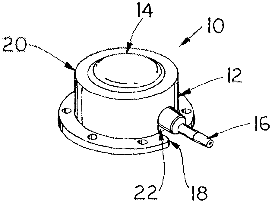
A venous access port assembly having a housing base with a discharge stem, a cap, and an interior reservoir. The port assembly is provided with X-ray discernable indicia to identify an attribute of the assembly after its implantation and clearly appear on an X-ray of the patient in a manner informing the radiologist or technologist and the medical practitioner of that particular attribute. The indicia are cuts in a reservoir lining of radiopaque material such as metal where the cuts having narrow slot width are in the form of one or more sets of alphabetical letters such as "CT" in the lining's side wall or bottom wall.
| Inventors: | Zinn; Kenneth M.; (Westport, CT) | ||||||||||
| Applicant: |
|
||||||||||
|---|---|---|---|---|---|---|---|---|---|---|---|
| Family ID: | 1000004807011 | ||||||||||
| Appl. No.: | 16/851459 | ||||||||||
| Filed: | April 17, 2020 |
Related U.S. Patent Documents
| Application Number | Filing Date | Patent Number | ||
|---|---|---|---|---|
| 15441762 | Feb 24, 2017 | 10639465 | ||
| 16851459 | ||||
| 12175270 | Jul 17, 2008 | 9610432 | ||
| 15441762 | ||||
| 60961160 | Jul 19, 2007 | |||
| Current U.S. Class: | 1/1 |
| Current CPC Class: | A61M 39/0208 20130101; A61M 2039/0238 20130101; A61M 2039/0226 20130101; A61M 2039/0045 20130101; A61M 2205/32 20130101 |
| International Class: | A61M 39/02 20060101 A61M039/02 |
Claims
1. A venous access port assembly for implantation into a patient, comprising: a housing, comprising a housing base; a reservoir lining defining a reservoir, wherein the reservoir lining comprising a needle impenetrable radiopaque material; a discharge port extending from the housing, the discharge port comprising a discharge stem forming a fluid passageway between the reservoir and the discharge port; and an X-ray discernable indicium that indicates an attribute of the venous access port assembly when X-rays are incident upon the venous access port assembly, wherein the X-ray discernable indicium is cut in at least a part of the reservoir lining.
2. The assembly of claim 1, wherein the reservoir lining is cup-shaped with an open end.
3. The assembly of claim 1, wherein the radiopaque material is metal.
4. The assembly of claim 1, wherein the radiopaque material is titanium.
5. The assembly of claim 1, wherein the X-ray discernable indicium comprises at least one alphabetical letter.
6. The assembly of claim 2, wherein the X-ray discernable indicium comprises a plurality of cuts in a side wall of the reservoir lining.
7. The assembly of claim 2, wherein the X-ray discernable indicium comprises at least one cut in a bottom wall of the reservoir lining.
8. The assembly of claim 2, wherein the X-ray discernable indicium comprises a plurality of cuts in a side wall and a bottom wall of the reservoir lining.
9. The assembly of claim 2, wherein the at least one cut is formed as at least one narrow slot having a width only sufficiently wide to be X-ray discernable.
10. The assembly of claim 2, wherein the X-ray discernable indicium is centered in a bottom wall of the reservoir lining.
Description
CROSS-REFERENCE TO RELATED APPLICATION
[0001] This application is a continuation application of Ser. No. 15/441,762, filed Feb. 24, 2017, which is a continuation application of, and claims the benefit of, co-pending U.S. patent application Ser. No. 12/175,270, filed Jul. 17, 2008, now U.S. Pat. No. 9,610,432, issued Apr. 4, 2017, which claims the benefit of U.S. Provisional Patent Application Ser. No. 60/961,160, filed Jul. 19, 2007, the disclosures of which are incorporated by reference in their entirety.
FIELD OF THE INVENTION
[0002] This relates to the field of medical devices and more particularly to venous access ports for the infusion of fluids into the patient and/or withdrawal of fluids from the patient.
BACKGROUND OF THE INVENTION
[0003] Venous access ports for the infusion and/or withdrawal of fluids from a patient are well-known, secured to the proximal end of an implanted catheter. These ports are typically used for drug infusion or for withdrawal of small amounts of blood, where large flows of fluid are not required. The ports are assemblies of a needle-impenetrable housing with a discharge port in fluid communication with the catheter and the reservoir within the port housing, and provide a subcutaneous self-sealing septum that defines an access site for multiple needle sticks through the covering skin tissue of the patient, through the septum and into the reservoir, without the need to continuously search for new access sites. Examples of such ports are disclosed, for example, in U.S. Pat. Nos. 4,704,103; 4,762,517; 4,778,452; 5,185,003; 5,213,574; 5,637,102; and 5,833,654. In U.S. Pat. No. 5,833,654 is set forth a dual chamber port assembly having a metal casing as a liner in one of the chambers of the port assembly.
[0004] It is desired to provide a venous access port assembly that provides for a radiologist, radiology technologist, nurse and ultimately a medical practitioner to be able to discern an important property of the port assembly after the port assembly has been implanted into a patient.
BRIEF SUMMARY OF THE INVENTION
[0005] The present invention is related to a venous access port having a housing and a septum, providing an interior reservoir and a passageway extending from the reservoir through a stem of a discharge port to establish fluid communication with a proximal end of a catheter lumen to which the port assembly is secured prior to placement of the assembly into a patient. The port may optionally have more than one reservoir and associated septum. The invention is the incorporation of X-ray discernable indicia onto a venous access port that is discernible under X-ray examination to provide information concerning the nature or key attribute of the venous access port, so that the practitioner, subsequent to the date of implantation thereof, can determine that nature or key attribute under X-ray examination. One such key attribute in particular would be, for example, that where the venous access port is rated to be used for power injection, such as used for contrast fluid injection during a contrast enhanced computed tomography, the letters "CT" (for "computed tomography) would be provided on the port assembly in such a manner that they are radiographically visible. The attribute in this example is the property of the port's being adapted to withstand high pressures that are used for injection of contrast fluid into a patient, and the letters "CT" would be understood in medical practice to indicate that the port is suitable for the high pressure injection of contrast fluid.
[0006] In the preferred embodiment, a reservoir lining of radiopaque material such as titanium, includes cutouts such as of letters "CT" (although other indicia may be utilized) through the body of the lining, with the cutouts being radiographically visible. The lining for the reservoir is contained within the port housing and includes an aperture through the side wall for fluid communication with a discharge stem of the port assembly, establishing fluid communication with a catheter sealingly and securely affixed to the discharge stem of the assembly. The reservoir lining of titanium provides protection against penetration by a needle when it is inserted through the septum of the port assembly for injection of fluid into the chamber. The letters "CT" are readable from exterior of the patient in an X-ray. The lining may have several such sets of cutouts located at various locations about the lining's side wall and/or in the bottom wall thereof. The cutouts preferably are substantially narrow for exposing therethrough only a minimum amount of plastic of the surrounding housing.
BRIEF DESCRIPTION OF THE DRAWINGS
[0007] The accompanying drawings, which are incorporated herein and constitute part of this specification, illustrate the presently preferred embodiments of the invention, and, together with the general description given above and the detailed description given below, serve to explain the features of the invention. In the drawings:
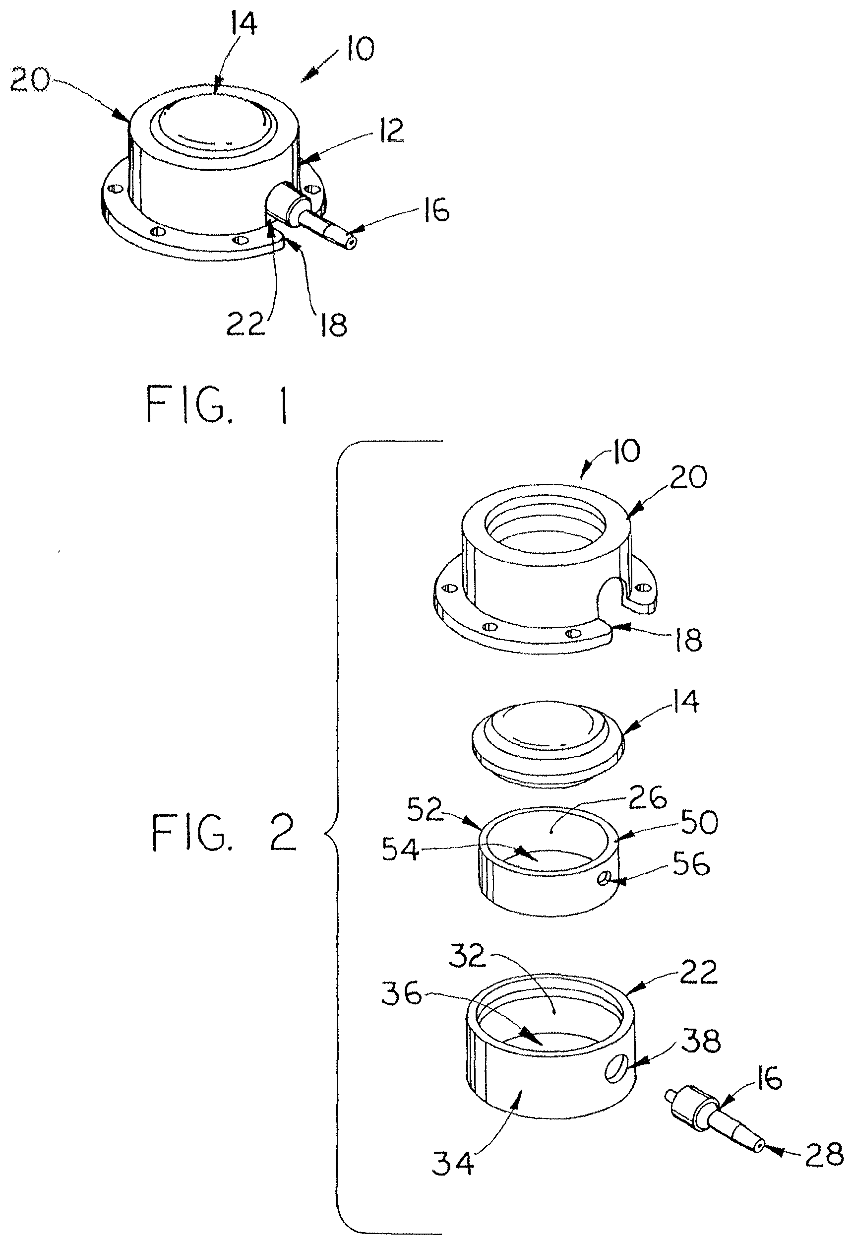
[0008] FIGS. 1 and 2 are an isometric view and an exploded view, respectively, of the venous access port assembly containing the present invention;
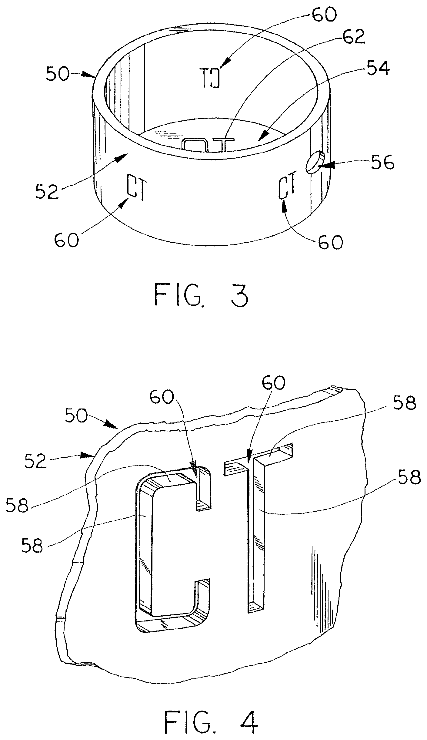
[0009] FIG. 3 is an isometric view of a reservoir lining of the present invention defining X-ray discernable indicia;
[0010] FIG. 4 is an enlarged view of portion of the lining of FIG. 3 illustrating the cutout indicia provided by the lining; and
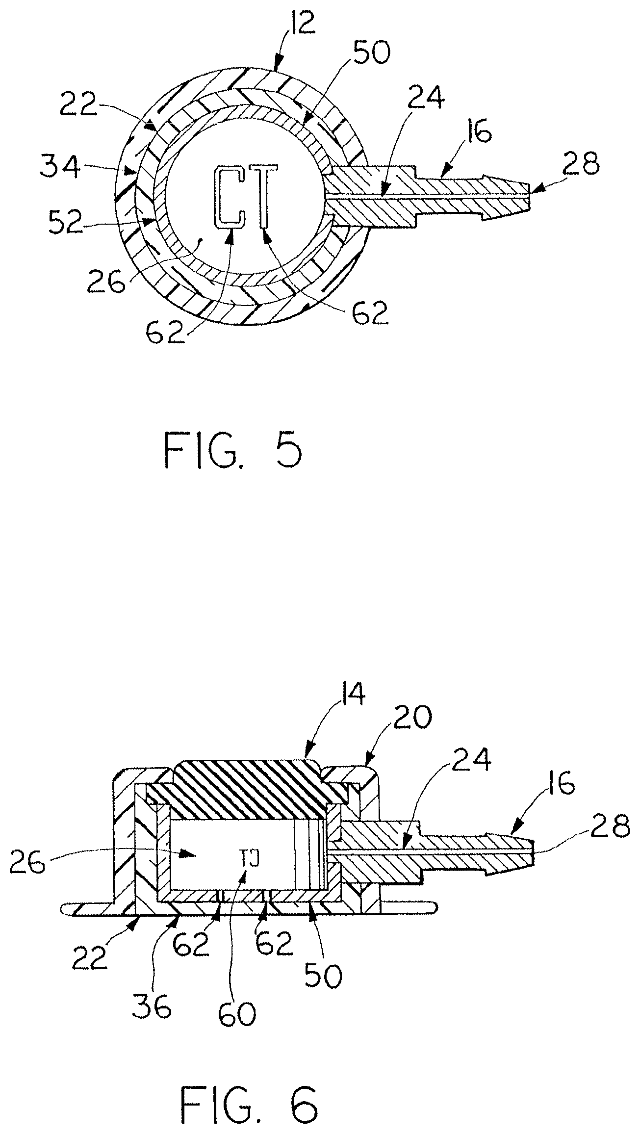
[0011] FIGS. 5 and 6 are cross-section views of the port of FIGS. 1 and 2.
DETAILED DESCRIPTION OF THE INVENTION
[0012] Certain terminology is used herein for convenience only and is not to be taken as a limitation on the present invention. The terms "distal" and "proximal" refer, respectively, to directions closer to and away from the insertion tip of a catheter in an implantable catheter assembly. The terminology includes the words specifically mentioned, derivatives thereof and words of similar import. The embodiments illustrated below are not intended to be exhaustive or to limit the invention to the precise form disclosed. These embodiments are chosen and described to best explain the principle of the invention and its application and practical use and to enable others skilled in the art to best utilize the invention.
[0013] Venous access port assembly 10 of FIGS. 1, 2, 5 and 6 includes a housing 12 and a septum 14, with a discharge stem 16 extending from a distal end 18 of the port assembly 10 to be attached securely and sealingly to the proximal end of a catheter (not shown). Cap 20 of housing 12 secures to housing base 22 to in turn secure septum 14 in position in the port assembly 10 in a manner that exposes the top surface of the septum for needle insertion. A passageway 24 (see FIGS. 5 and 6) extends from an interior reservoir 26 to the distal tip opening 28 of discharge stem 16. A recess 30 is seen to be provided along both sides of discharge stem 16, facilitating insertion of the discharge stem 16 into the catheter lumen and providing a clearance for a locking sleeve or clamp (not shown) utilized to compress the catheter lumen wall against the exterior surface of the discharge port 16 for assured sealed connection of the catheter with the port assembly 10.
[0014] With reference now to FIGS. 2 to 4, showing reservoir lining 50 of the present invention, lining 50 is cup-shaped and is inserted into well 32 of housing base 22 beneath septum 14, and secured therewithin. Lining 50 is made of needle-impenetrable material such as metal, which may be titanium or stainless steel, and its side wall 52 and bottom wall 54 protects the side walls 34 and bottom wall 36 of housing base 22 defining well 32, from being penetrated by a needle (not shown) inserted into and through septum 14 for injection of fluids into reservoir 26 or withdrawal of blood therefrom. An aperture 56 near or at the bottom of side wall 52, in alignment with a corresponding aperture 38 of housing base 22, establishes fluid communication with passageway 24 of discharge stem 16 for fluid to pass between the reservoir 26 and the catheter and thus the patient. Discharge stem 16 may be a metal component such as titanium or stainless steel which would extend through aperture 38 of the housing base 22, to preferably be welded to lining 50.
[0015] In accordance with the present invention, the X-ray discernable indicia are cutouts 60 formed through the body of lining 50, shown as the alphabetical letters "CT". The letters "CT" are visible when the X-ray of the patient is viewed, readable from outside the lining 50 and are easily discerned by the radiologist or technologist. In lining 50, preferably a plurality of sets of cutout indicia 60,62 are provided equi-angularly spaced about the circumference of the side wall 52 and through bottom wall 54 to assure that the indicia appear in the X-ray irrespective of the angular location at which the X-ray is taken. With particular reference to FIG. 4, it is preferable that the width of each cutout slot 58 of the indicia or letters be as narrow as possible but still be discernable by X-ray; the narrowness of the slots 58 minimizes any possibility that a needle inserted through the septum could penetrate through a slot of a cutout by chance, thus harming the patient and resulting in injection of fluid directly into the tissue surrounding the port. The width of each cutout slot 58 would thus preferably be less wide than the diameter of a needle. The set of cutout indicia 62 through the bottom wall may be dimensionally larger as a set, but still with narrow slot width. Centering of the cutout indicia 62 along the bottom wall 54 positions the indicia directly beneath the reservoir and septum, minimizes any obscuring thereof by the structure of the venous access port assembly, and the indicia may also be easily discernable should the port assembly be at an angle from the horizontal plane of the X-ray.
[0016] It will be appreciated by those skilled in the art that changes could be made to the embodiments described above without departing from the broad inventive concept thereof. It is understood, therefore, that this invention is not limited to the particular embodiments disclosed, but it is intended to cover modifications within the spirit and scope of the present invention as defined by the appended claims.
* * * * *
D00000
D00001
D00002
D00003
XML
uspto.report is an independent third-party trademark research tool that is not affiliated, endorsed, or sponsored by the United States Patent and Trademark Office (USPTO) or any other governmental organization. The information provided by uspto.report is based on publicly available data at the time of writing and is intended for informational purposes only.
While we strive to provide accurate and up-to-date information, we do not guarantee the accuracy, completeness, reliability, or suitability of the information displayed on this site. The use of this site is at your own risk. Any reliance you place on such information is therefore strictly at your own risk.
All official trademark data, including owner information, should be verified by visiting the official USPTO website at www.uspto.gov. This site is not intended to replace professional legal advice and should not be used as a substitute for consulting with a legal professional who is knowledgeable about trademark law.