Apoptotic Cell-mediated Induction Of Antigen Specific Regulatory T-cells For The Therapy Of Autoimmune Diseases In Animals And H
Chen; Wan Jun ; et al.
U.S. patent application number 16/773982 was filed with the patent office on 2020-08-20 for apoptotic cell-mediated induction of antigen specific regulatory t-cells for the therapy of autoimmune diseases in animals and h. The applicant listed for this patent is The United States of America, as represented by the Secretary, Department of Health & Human Services. Invention is credited to Wan Jun Chen, Shimpei Kasagi, Pin Zhang.
| Application Number | 20200261576 16/773982 |
| Document ID | 20200261576 / US20200261576 |
| Family ID | 1000004808602 |
| Filed Date | 2020-08-20 |
| Patent Application | download [pdf] |


View All Diagrams
| United States Patent Application | 20200261576 |
| Kind Code | A1 |
| Chen; Wan Jun ; et al. | August 20, 2020 |
APOPTOTIC CELL-MEDIATED INDUCTION OF ANTIGEN SPECIFIC REGULATORY T-CELLS FOR THE THERAPY OF AUTOIMMUNE DISEASES IN ANIMALS AND HUMANS
Abstract
The invention provides methods of tolerizing or treating a subject suffering from an autoimmune or autoinflammatory disease or disorder to an antigen associated with the autoimmune disease or disorder. The invention also features kits for carrying out the methods of the invention.
| Inventors: | Chen; Wan Jun; (Bethesda, MD) ; Kasagi; Shimpei; (Bethesda, MD) ; Zhang; Pin; (Bethesda, MD) | ||||||||||
| Applicant: |
|
||||||||||
|---|---|---|---|---|---|---|---|---|---|---|---|
| Family ID: | 1000004808602 | ||||||||||
| Appl. No.: | 16/773982 | ||||||||||
| Filed: | January 27, 2020 |
Related U.S. Patent Documents
| Application Number | Filing Date | Patent Number | ||
|---|---|---|---|---|
| 14904054 | Jan 8, 2016 | |||
| PCT/US2014/046065 | Jul 10, 2014 | |||
| 16773982 | ||||
| 61844564 | Jul 10, 2013 | |||
| Current U.S. Class: | 1/1 |
| Current CPC Class: | A61K 2039/505 20130101; A61K 2039/507 20130101; A61K 39/008 20130101; A61K 2039/55566 20130101; C07K 16/2815 20130101; A61K 45/06 20130101; A61K 2300/00 20130101; A61K 2039/577 20130101; A61K 39/0007 20130101; A61K 35/15 20130101; A61K 39/0008 20130101; A61K 39/3955 20130101; C07K 2317/73 20130101; C07K 2317/75 20130101; C07K 16/2812 20130101 |
| International Class: | A61K 39/395 20060101 A61K039/395; C07K 16/28 20060101 C07K016/28; A61K 39/008 20060101 A61K039/008; A61K 45/06 20060101 A61K045/06; A61K 39/00 20060101 A61K039/00; A61K 35/15 20060101 A61K035/15 |
Goverment Interests
STATEMENT OF RIGHTS TO INVENTIONS MADE UNDER FEDERALLY SPONSORED RESEARCH
[0002] Research supporting this application was carried out by the United States of America as represented by the Secretary, Department of Health and Human Services. The Government has certain rights in this invention.
Claims
1-21. (canceled)
22. A pharmaceutical composition for use in the treatment of an autoimmune disease or disorder in a subject, the composition comprising: (i) antibodies for inducing apoptosis in T cells, wherein the antibodies are an anti-CD8 antibody and an anti-CD20 antibody; and (ii) an autoantigen specific to the autoimmune disease or disorder, selected from the group consisting of myelin basic protein (MBP), myelin proteolipid protein (PLP), insulin, GAD65 (glutamic acid decarboxylase), DiaPep227, heat-shock proteins, preferably Hsp65, Hsp90 and DnaJ, immunoglobulin binding protein (BiP), heterogeneous nuclear RNPs, annexin V, calpastatin, type II collagen, glucose-6-phosphate isomerase (GPI), elongation factor human cartilage gp39, and mannose binding lectin (MBL); wherein the subject is tolerized to an antigen of the autoimmune disease.
23. The pharmaceutical composition for use according to claim 22 for tolerizing a subject suffering from an autoimmune disease or disorder to an antigen associated with the autoimmune disease or disorder.
24. The pharmaceutical composition for use according to claim 23, wherein the autoimmune disease or disorder is selected from the group consisting of multiple sclerosis, diabetes mellitus, rheumatoid arthritis, Sjogren syndrome and systemic sclerosis, preferably a late stage of the aforementioned diseases, more preferably type 1 diabetes mellitus.
25. The pharmaceutical composition for use according to claim 22, wherein the anti-CD 8 and anti-CD 20 antibodies induce depletion and/or apoptosis of B cells and T cells.
26. The pharmaceutical composition for use according to claim 22, wherein the subject is an animal, preferably a mammal, more preferably a human or non-human mammal selected from the group consisting of a human, primate, murine, bovine, equine, canine, ovine and feline.
27. The composition for use according to claim 24, wherein the disease or disorder is Sjogren's Syndrome.
Description
CROSS REFERENCE TO RELATED APPLICATIONS
[0001] This application claims the benefit of U.S. Provisional Patent Application Ser. No. 61/844,564, filed Jul. 10, 2013, International Patent Application Ser. No. PCT/US2014/046065 filed Jul. 10, 204, and U.S. patent application Ser. No. 14/904,054, filed Jan. 8, 2016, the entire contents of these applications are hereby incorporated by reference herein.
INCORPORATION BY REFERENCE
[0003] In compliance with 37 C.F.R. .sctn. 1.52, a computer readable form of the sequence listing is submitted herewith, file name: 84243WO_ST25.txt; size 43 KB; created on: Jul. 10, 2014; using PatentIn-3.5, which is hereby incorporated by reference in its entirety.
BACKGROUND OF THE INVENTION
[0004] Regulatory T cells (T.sub.reg cells) hold promise for autoimmune disease therapy. However, a challenge remains as to how to induce antigen-specific T.sub.reg cells that only target inflammatory immune cells without compromising the entire immune response. Peripheral immune tolerance is key to preventing overreactivity of the immune system to various antigens. CD4+CD25+Foxp3+ regulatory T (Treg) cells are critical for maintaining immune tolerance, and deficiency of Treg cells causes severe autoimmune diseases and chronic inflammation. Indeed, the emergence and characterization of CD4+ CD25+ Foxp3+ Tre.sub.g cells have offered the hope of developing novel immunotherapy for human autoimmune diseases and chronic inflammation." However, the lack of knowledge of antigen specificity and the difficulty of expanding thymus-derived T.sub.reg cells (tT.sub.reg cells) have limited their potential clinical application. The discovery of TGF.beta. induction of T.sub.reg cells (iT.sub.reg cells) from peripheral naive CD4+ T cells has brought new hope of inducing antigen-specific T.sub.reg cells for autoimmune disease therapy.sup.5,6. However, published studies have been limited to the prevention of experimental diseases by pre-injection of in vitro induced antigen-specific iT.sub.reg cells into unmanipulated mice or in vivo by induction of antigen-specific T.sub.reg cells in naive mice before the disease is established. There is a considerable difference in the immune status of an unmanipulated, naive mouse and a mouse with an established disease. The immune tolerance toward self-tissues in naive mice is broken in mice with autoimmunity, where the autoantigen-responsive immune cells are uncontrollably activated and proinflammatory cytokines are produced. Treg cells that fully exhibit immunosuppressive capacity in the immune quiescent state in naive mice may lose their suppressive activity or even convert to effector cells under the dysregulated inflammation in mice with autoimmune diseases. This problem is particularly salient in clinical settings, in which patients with autoimmune disease present with an already dysregulated immune response. Therefore, a challenge is to make cells in the inflammatory, dysregulated immune system in animals with established autoimmune diseases, and ultimately in patients, that can specifically inhibit inflammation in the organs/tissues affected and treat the diseases, i.e. to reprogram the dysregulated immune system in animals and patients so that it is restored, or to direct it to an immune-tolerant state to the target auto-antigens in the tissues affected with autoimmunity.
[0005] Accordingly, there is a need in the art for novel autoimmune disease therapies.
SUMMARY OF THE INVENTION
[0006] The present invention provides a therapeutic method for the treatment of autoimmune or autoinflammatory diseases by first breaking down the dysregulated immune system and then reprogramming the immune system to restore tolerance to the patient's self-antigens by induction of antigen specific regulatory T cells. It has been shown here that only with the combination of apoptosis, phagocytes, and antigen can antigen-specific T.sub.reg cells be optimally generated and long-term immune tolerance developed, i.e., the proper antigenic peptide needs to be introduced in a timely manner into subjects in which an immunoregulatory milieu was created by apoptosis-triggered phagocytes.
[0007] Exemplary tolerizing and/or treatment methods of the invention involve a) identifying a subject as sufficing from an autoimmune disease or disorder; performing at least one of the following steps: b) administering an effective amount of an anti-CD4 antibody, anti-CD8 antibody, or both to the subject to induce apoptosis in T cells of the subject suffering from the autoimmune disease or disorder; b) administering an effective amount of low-dose irradiation to the subject suffering from the autoimmune disease or disorder to induce apoptotic cells with adoptive transfer of said macrophage; and/or b) administering an effective amount of an anti-CD8 antibody and/or an anti-CD-20 antibody to the subject to induce depletion and apoptosis of B cells and T cells of the subject suffering from the autoimmune disease or disorder; and c) administering an autoantigen specific to the autoimmune disease or disorder that the subject is suffering from, whereby the subject is tolerized to the antigen of the autoimmune or autoinflammatory disease and the disease or disorder is treated. The invention also features kits for carrying out the methods of the invention.
[0008] In one aspect, this invention provides a method of tolerizing a subject suffering from an autoimmune or autoinflammatory disease or disorder to an antigen associated with the autoimmune disease or disorder comprising steps a to c in order: a) identifying a subject as suffering from an autoimmune disease or disorder; b) administering an effective. amount of an anti-CD4 antibody, anti-CD8 antibody, or both to the subject to induce apoptosis in T cells of the subject suffering from the autoimmune disease or disorder; and c) administering an autoantigen specific to the autoimmune disease or disorder that the subject is suffering from, whereby the subject is tolerized to the antigen of the autoimmune or autoinflammatory disease.
[0009] In another aspect, the invention provides a method of treating a subject suffering from an autoimmune or autoinflammatory disease or disorder comprising steps a to c in order: a) identifying a subject as suffering from an autoimmune disease or disorder; b) administering an effective amount of an anti-CD4 antibody, anti-CDS antibody, or both to the subject to induce apoptosis in T cells of the subject suffering from the autoimmune disease or disorder; and c) administering an autoantigen specific to the autoimmune disease or disorder that. the subject is suffering from, whereby the subject is tolerized to the autoantigen, thereby treating the autoimmune or autoinflammatory disease or disorder.
[0010] In one embodiment, the autoantigen is one or more autoantigens selected from the group consisting of: the myelin basic protein (MBP), the myelin proteolipid protein (PLP), insulin, GAD65 (glutamic acid decarboxylase), DiaPep277, heal-shock proteins (Hsp65, Hsp90, DnaJ), immunoglobulin binding protein (BiP), heterogeneous nuclear RNPs, annexin V, calpastatin, type II collagen, glucose-6-phosphate isomerase (GPI), elongation factor human cartilage gp39, and mannose binding lectin (MBL).
[0011] In another embodiment of the above aspects, step b is performed more than once prior to the performance of step c. In a further embodiment of the above aspects, the time for performance of step b and the time of performance of step c are separated by 3 to 14 days. In still another embodiment of the above aspects, step b induces apoptosis in a subset of T cells. In another embodiment of the above aspects, performance of steps a, b, and c is more effective than the performance of either steps a and b or steps a and c alone.
[0012] In one embodiment of the above aspects, the method further comprises monitoring the subject for amelioration of at least one sign or symptom of an autoimmune disease or disorder.
[0013] In another embodiment of the above aspects, the autoimmune disease or disorder is selected from the group consisting of multiple sclerosis, diabetes mellitus and rheumatoid arthritis, Sjogren's syndrome, and systemic sclerosis.
[0014] In certain embodiments, the monitoring comprises a diagnostic test or assessment. In another embodiment, the diagnostic test or assessment is selected from the expanded Disability Status Scale, the timed 25-foot walk test or the nine-hole peg test. In another related embodiment, the diagnostic test or assessment is selected from an oral glucose tolerance test (OGTT) glycosylated hemoglobin test or fasting plasma glucose test. In a further related embodiment, the diagnostic test or assessment is selected from the American College of Rheumatology (ACR) response, the Simplified Disease Activity Index (SDAI), the Clinical Disease Activity Index (CDAI) or the Global Arthritis Score (GAS).
[0015] In certain embodiments, the diagnostic test or assessment comprises determining the amount of inflammatory cell infiltration.
[0016] In another embodiment, the subject suffering from an autoimmune disease or disorder is at a late stage of disease.
[0017] In another embodiment, the method further comprises administration of an additional agent.
[0018] In one embodiment, the autoimmune disease or disorder is type I diabetes mellitus.
[0019] In certain embodiments, the methods of the invention also include a step of administering an anti-CD20 antibody to the subject suffering from an autoimmune or autoinflammatory disease or disorder.
[0020] Another aspect of the invention provides a method of treating a subject suffering from an autoimmune or autoinflammatory disease or disorder that includes performing the following steps in order: a) identifying a subject as suffering from an autoimmune disease or disorder; b) administering an effective amount of low-dose irradiation and macrophage to the subject sufficing from the autoimmune disease or disorder to induce apoptotic cells together with adoptive transfer of the macrophage; and c) administering an autoantigen specific to the autoimmune disease or disorder that the subject is suffering from, where the subject is tolerized to the autoantigen, effecting treatment of the autoimmune or autoinflammatory disease or disorder.
[0021] A further aspect of the invention provides a method of treating a subject suffering from an autoimmune or autoinflammatory disease or disorder that involves performing the following steps in order: a) identifying a subject as suffering from an autoimmune disease or disorder; b) administering an amount of an anti-CD8 antibody and/or an anti-CD-20 antibody to the subject effective to induce depletion and/or apoptosis of B cells and T cells (e.g., CD8.sup.- T cells) of the subject suffering from the autoimmune disease or disorder; and c) administering an autoantigen specific to the autoimmune disease or disorder that the subject is suffering from, where the subject is tolerized to the autoantigen, thereby treating the autoimmune or autoinflammatory disease or disorder.
[0022] In another aspect, the invention features a kit comprising an effective amount of an anti-CD4 antibody, anti-CD8 antibody in a pharmaceutical carrier; an autoantigen specific to an autoimmune or autoinflammatory disease or disorder; and instructions for use in treating the autoimmune or autoinflammatory disease or disorder.
[0023] In a further aspect, the invention provides a kit comprising an effective amount of an autoantigen specific to an autoimmune or autoinflammatory disease or disorder; and instructions for use in treating or preventing the autoimmune or autoinflammatory disease or disorder in a subject, optionally when used in combination with administration of one or more of the following: administering an effective amount of an anti-CD4 antibody, anti-CD-8 antibody, or both to the subject to induce apoptosis in T cells of the subject suffering from the autoimmune disease or disorder; administering an effective amount of low-dose irradiation and macrophage to the subject suffering from the autoimmune disease or disorder to induce apoptotic cells with adoptive transfer of the macrophage; and/or administering an amount of an anti-CDS antibody and an anti-CD-20 antibody to the subject effective to induce depletion and/or apoptosis of B cells and T cells (e.g., CD8.sup.1 T cells) of the subject suffering from the autoimmune disease or disorder.
Definitions
[0024] To facilitate an understanding of the present invention, a number of terms and phrases are defined below.
[0025] As used herein, the singular forms "a", "an", and "the" include plural forms unless the context clearly dictates otherwise. Thus, for example, reference to "a biomarker" includes reference to more than one biomarker.
[0026] Unless specifically stated or obvious from context, as used herein, the term "or" is understood to be inclusive.
[0027] As used herein, the terms "comprises," "comprising," "containing," "having" and the like can have the meaning ascribed to them in U.S. Patent law and can mean "includes," "including," and the like: "consisting essentially of" or "consists essentially" likewise has the meaning ascribed in U.S. Patent law and the term is open-ended, allowing for the presence of more than that which is recited so long as basic or novel characteristics of that which is recited is not changed by the presence of more than that which is recited, but excludes prior art embodiments.
[0028] "Absence" of an autoantibody means that an autoantibody which is indicative fair at least one autoimmune disorder is not immunologically detectable. Accordingly, the autoantibody is not. hound to an autoantigen. Any immunoassay known in the art can be used, including an exemplary assay such as ELISA performed upon blood obtained from a subject (initially) identified as having an autoimmune disease or disorder and optionally undergoing a treatment as described herein.
[0029] As used herein, the term "antibody" is meant to refer to a full-length (i.e., naturally occurring or thrilled by normal immunoglobulin gene fragment recombinatorial processes) immunoglobulin molecule or an immunologically active (i.e., antigen-binding) portion of an immunoglobulin molecule, like an antibody fragment. In certain embodiments, the antibody is anti-CD4 or anti-CD8.
[0030] As used herein, the term "agent" is meant any small molecule chemical compound, antibody, nucleic acid molecule, or polypeptide, or fragments thereof.
[0031] As used herein, the term "autoantigen" is meant to refer to any antigen that stimulates autoantibodies in the organism that produced it.
[0032] As used herein, the term "autoimmune disease or disorder" is meant to refer to a disease state caused by an inappropriate immune response that is directed to a self-encoded entity which is known as an autoantigen. Examples of autoimmune diseases include vasculitis, arthritis, autoimmune diseases of the connective tissue, inflammatory bowel diseases, autoimmune diseases of the liver and the bile duct, autoimmune disease of the thyroid gland, dermatologic autoimmune diseases, neurologic immune diseases, Diabetes type 1. Exemplary vasculitis can be selected from medium to small vessel vasculitis or large vessel vasculitis, exemplary arthritis can be selected from seronegative and seropositive rheumatoid arthritis, psoriatic arthritis, Bechterew's disease, juvenile idiopathic arthritis; exemplary inflammatory bowel diseases can be selected from Crohn's disease or ulcerative colitis; exemplary diseases of the liver and the bile duet can be selected from autoimmuno-hepatitis, primary biliary cirrhosis and primary sclerosing Cholangitis; exemplary autoimmune diseases of the thyroid gland can be selected from Hashimoto's thyreoiditis and Grave's disease; exemplary autoimmune diseases of the connective tissue can be selected from systemic lupus erythematosus (SLE) disease, Sjogren's syndrome (SS), scleroderma, dermato- and poly-myositis, Sharp syndrome, systemic sclerosis and CREST syndrome; exemplary neurologic autoimmune diseases can be selected from multiple sclerosis (MS), chronic inflammatory demyelating polyneuropathy (CIDP) and myasthenia gravis. Medium to small vasculitis can optionally be selected from classical panarteritis nodosa, granulomatosis with polyangiitis, microscopic panarteritis, Churg-Strauss syndrome Behcet's disease and the large vessel vasculitis can optionally be selected from giant cell arteritis, polymyalgia rheumatic and Takayasu's arteritis. An autoimmune disease or disorder in a subject can be identified by any art-recognized method, including by assessment of symptoms or by evaluation of marker levels (e.g., autoantibody levels).
[0033] As used herein, the term "autoinflammatory disease or disorder" is meant to refer to a group of disorders characterized by seemingly unprovoked inflammation.
[0034] As used herein, the terms "determining", "assessing", "assaying", "measuring" and "detecting" refer to both quantitative anti qualitative determinations, and as such, the term "determining" is used interchangeably herein with "assaying," "measuring," and the like.
[0035] The term "subject" refers to an animal which is the object of treatment, observation, or experiment. By way of example only, a subject includes, but is not limited to, a mammal, including, but not limited to, a human or a non-human mammal, such as a non-human primate, murine, bovine, equine, canine, ovine, or feline.
[0036] As used herein, the term "tolerizing" is meant to refer to a failure to attack the body's own proteins and other antigens. The term "tolerizing" may also include inducing tolerance, inducing immunological tolerance, or rendering nonimmunogenic.
[0037] As used herein, the term "treating" is meant to include alleviating, preventing and/or eliminating one or mom symptoms associated with inflammatory responses or an autoimmune disease. It will be appreciated that, although not precluded, treating a disease or condition does not require that the disease, condition, or symptoms associated therewith be completely eliminated.
DESCRIPTION OF THE DRAWINGS
[0038] FIGS. 1a to 1d show that the combination of T cell apoptosis and peptide administration suppressed Experimental autoimmune encephalomyelitis EAE. SJL mice were immunized with pPLP peptides to induce (day 0). In FIG. 1a, the upper panel shows the experimental scheme, while the lower panel shows clinical scores of EAE in SJL mice (mean.+-.s.e.m. n=3 mice). 5 .mu.g of pPLP or pOVA was injected each day. PBS (untreated), PLP (pPLP alone), .alpha.CD4/CD8+PBS (deletion alone), .alpha.CD4/CD8+PLP (deletion plus pPLP), .alpha.CD4/CD8+OVA (deletion plus pOVA). FIG. 1b shows flow cytometry results for IL-17+ versus IFN-.gamma.+ (upper panel) or CD25+ versus Foxp3+ (lower panel) within CD4+ T cells in the spinal cords (pooled in each group) at the end of the experiments. In FIG. 1c, splenocytes (pooled from each group) were stimulated by pPLP, and T cell proliferation was assessed by H-thymidine incorporation (mean.+-.s.d. of triplicate measurements). In FIG. 1d, protein levels of IL-17,IFN-.gamma., and IL-6 in the cultured supernatants of the same splenocytes as in FIG. 1c were measured by ELISA (mean.+-.s.d. of duplicate measurements). *P<0.05 determined by Student's t test. Data of a representative example selected from two independent experiments are shown.
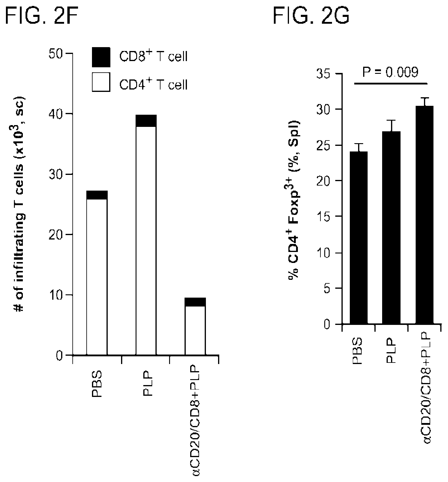
[0039] FIGS. 2a to 2g show the therapeutic effects of B cell apoptosis and peptide administration on EAE. SJL mice were immunized with pPLP and CFA (day 0). 9 days after immunization, mice were injected with anti-CD8- and CD20-specific antibodies, followed by 5 .mu.g of pPLP i.p. every other day for 14 days. In FIG. 2a, The mean clinical scores of EAE are shown (mean.+-.s.e.m.) in mice treated with PBS (PBS), pPLP alone (PLP). CD8- and CD20-specific antibody (.alpha.CD20/CD8), or CD8- and CD20- specific antibody plus PLP (.alpha.CD20/CD8+PLP). Each group contained 3 mice. FIG. 2b presents flow cytometry results for CD4+ T cells in the spinal cords. The numbers indicate the frequencies of Foxp3+ cells (Upper panel) or IL-17+ IFN-.gamma.- and IL-17- IFN-.gamma.+ cells (Lower panel). In FIGS. 2c and 2d, splenocyes (pooled in each group of the mice) were stimulated by either pPLP (c) or MT (d), and T cell proliferation was assessed by H-thymidine incorporation (mean.+-.s.d. of triplicate measurements). In FIG. 2e, pPLP (1.0 .mu.g/ml)-specific IL-17, IFN-.gamma., TXF-.alpha. and IL-6 cytokines in the cultured supernatants of the same splenocytes as in FIGS. 2c and 2d were measured by ELISA. FIG. 2f shows the total number of infiltrating T cell in the spinal cords determined by FACS, while FIG. 2g shows the frequency of Foxp3+ T cells in CD4+ T cells in the spleen. *P<0.05, determined by Student's t test. Data of a representative example selected from two independent experiments are shown.
[0040] FIGS. 3a to 3e show the function of professional phagocytes in apoptosis-antigen mediated therapy of EAE SJL mice were immunized with pPLP and CFA to induce EAE (Day 0). Mice were either left untreated or irradiated with .gamma.-irradiation (IRR) 10 days after immunization. Some mice received normal splenic macrophages and DCs (Mo) as indicated. Some mice were administered with 5 .mu.g of pPLP or pOVA every other day as indicated. In FIG. 3a, the upper panel shows the experimental scheme, while the lower panel shows the clinical mean scores of (mean.+-.s.e.m., n=3-4 mice) for PBS (untreated control), IRR+M.PHI.+PLP (irradiation plus Mo plus pPLP), IRR+Mo+OVA (irradiation plus M.PHI. plus pOVA), IRR+Mo (irradiation plus M.PHI.), and IRR+PLP (irradiation plus pPLP) treatments. FIG. 3b shows histological analysis of spinal cord sections obtained from representative mice from indicated groups. Yellow dots indicate inflammatory infiltrates. FIG. 3c shows flow cytometry results for CD4+ T cells in the spinal cords. The numbers in the upper panels indicate the frequencies of CD25+ Foxp3- (lower right) CD25+ Foxp3+ (upper right) and CD25-Foxp3+(upper left) cells, while numbers in the lower panels indicate the frequencies of IL-17+ IFN-.gamma.- (lower right) IL-17+ IFN-.gamma.+ (upper right) and IL-17-IFN-.gamma.+ (upper left) cells. In FIG. 3d, splenocytes were stimulated by pPLP, and antigen-specific T cell proliferation was assessed by H-thymidine incorporation (mean.+-.s.d. of triplicate measurements). In FIG. 3e, the protein levels of pPLP-specific IL-17, IFN-.gamma., TNF-.alpha., and IL-6 in the cultured supernatants of same splenocytes as in FIG. 3d were measured by ELISA. *p<0.01 determined by Student's t test. The data were representative two independent experiments.
[0041] FIGS. 4a to 4g demonstrate that TGF.beta. plays a key role in apoptosis-antigen combined therapy of EAE. C57BL/6J mice were immunized with pMOG (myelin oligodendrocyte glycoprotein (MOG)) to induce EAE (day 0) and were given anti-CD4- and CD8-specific antibody (.alpha.CD4/CD8) at day 14 to induce T cell apoptosis. Mice were treated with either anti-TGF-.beta. (.alpha.TGF.beta.) or isotype control antibody mouse IgG1 (control Ab) twice from day 14 to 15 (indicated as inverted open trianglestriangles in the upper panel of FIG. 4a). FIG. 4a shows the clinical mean scores of EAE (mean.+-.s.e.m.) for PBS (untreated control, 3 mice), .alpha.CD4/CD8+MOG+ .alpha.TGF.beta. (.alpha.CD4/CD8 plus pMOG plus anti-TGF-.beta. antihody, 5 mice), and .alpha.CD4/CD8+MOG+ Control Ab (.alpha.CD4/CD8 plus pMOG plus control antibody, 5 mice) treatments. FIGS. 4b and 4c show flow cytometry of CD4+cells in the spinal cords of the indicated groups. In FIG. 4b, the numbers indicate the frequencies of CD25+ Foxp3+ (upper right) and CD25- Foxp3+ (upper left), while in FIG. 4c, the numbers indicate the frequencies of IL-17+ CD4+ cells. In FIGS. 4d to 4f, splenocyes (pooled in each group before culture) were stimulated pMOG (d). MT (e), and anti-CD3 (f), respectively, and T cell proliferation was assessed by 3H-thymidine incorporation (mean.+-.s.d. of triplicate measurements). In FIG. 4g, the protein levels of pMOG-specific IL-17, IFN-.gamma., INF-.alpha. and IL-6 in the cultured supernatants of the same splenocytes as in FIG. 4d was measured by ELISA (mean.+-.s.d. of duplicate measurements). *P<0.01 determined by Student's t test. The inverted open trianglestriangles indicate the frequency of anti-TGF.beta. or control antibody (200 .mu.g/day/mouse) treatment. Data of a representative example selected from four independent experiments are shown.
[0042] FIGS. 5a to 5j show generation of antigen-specific CD4+CD25+ T.sub.reg cells in mice with long-term remission of EAE induced by apoptosis-antigen treatment. Splenocytes were isolated from the SJL/J mice shown in FIG. 3a (FIGS. 5a-5d), FIG. 2a (FIGS. 5e-5g). and FIG. 1a (FIGS. 5h-5j). In each experiment, splenocytes were pooled from mice in each group and CD4+, CD4+ CD25-, and CD4+ CD25+cells were purified and cultured with irradiated APCs in the presence of either pPLP (a, c, h) or MT (b, i) or anti-CD3 (c, f). T cell proliferation was assessed by H-thymidine incorporation (mean.+-.s.d. of triplicate wells). In FIGS. 5d, 5g and 5j, the supernatant levels of pPLP-specific IL-17 and IFN-.gamma. of the indicated CD4+ T cell subsets were determined by ELISA (mean.+-.s.d. of duplicate wells). *P<0.05, determined by Student's t test. Data of a representative example selected from two independent experiments are shown.
[0043] FIGS. 6a to 6h show antigen-specific Foxp3+ T.sub.reg cells were converted from naive CD4+ T cells by apoptosis-antigen combined therapy in vivo. FIGS. 6a to 6d demonstrate the conversion of pOVA-specific T.sub.reg cells by anti-CD8 and CD20 specific antibodies (.alpha.CD8/CD20) plus pOVA in vivo. In FIG. 6a, the upper panel depicts the experimental scheme, while in the lower panel, flow cytometry data for splenic CD4+ KJ1-26+ Foxp3+ T.sub.reg cells in the BALB/c recipients are shown. FIGS. 6b and 6c show quantitative analysis of frequency (b) and total number (c) of the CD4+ KJ1-26+ Foxp3+ T.sub.reg cells of FIG. 6a. FIG. 6d shows analysis of cytokine positive cells within CD4+ KJ1-26+ T cells in the BALB/c mice. Data are shown as mean.+-.s.d. of individual mice (n=3), and constitute a representative example selected from two independent experiments. FIGS. 6e to 6h show the conversion of pOVA-specific T.sub.reg cells by .gamma.-irradiation followed by macrophages plus pOVA administration in vivo. In FIG. 6c, the upper panel shows the experimental scheme, while the lower panel shows a representative flow cytometry profile of splenic CD4+ KJ1-26+ Foxp3+ T.sub.reg cells in BALB/c recipients. FIGS. 6f to 6h show the frequencies of Foxp3+ (f), IL-17+ (g) and IFN-.gamma.+ (h) cells in the same CD4+ KJ1-26+ T cells of FIG. 6e (mean.+-.s.d., n=3 mice per group) *P<0.05, P=0.053, determined by Student's t test. The inverted triangles indicate the frequency of anti-TGF.beta. or control antibody (200 .mu.g/day/mouse) treatment.
[0044] FIG. 7 shows a schematic design of the study identifying therapy of by generation of auto-antigen-specific Foxp3+ T.sub.reg cells in vivo. The process can be divided into three steps. (I), Induction of transient yet sufficient number of apoptotic immune cells in vivo; (II), Apoptotic cells trigger professional phagocytes to produce immunosuppressive cytokine TGF.beta., which then creates an immmunoregulatory milieu; and Specific auto-antigenic-peptides are administered into mice that had a TGF.beta.-rich immunoregulatory microenvironment, under which naive CD4+ T cells are directed to differentiate into Foxp3+ T.sub.reg cells rather than T effector cells. These generated antigen-specific cells may further prevent and suppress potential auto-antigen-specific inflammatory T effector cell differentiation to lead to a state of immune tolerance. Using this strategy, The dysregulated immune system in the mice with EAE was corrected and re-programmed. The disease of EAE is suppressed.
[0045] FIGS. 8a to 8f show that the combination of T cell apoptosis and peptide administration prevented EAE. SJL mice were treated with CD4- and CD8-specific antibody (Deletion) 21 days before immunization, followed by pPLP administration (25 .mu.g/every other day for 16 days). Mice were sacrificed at day 48 after immunization. In FIG. 8a, the upper panel shows the experimental scheme, while the lower panel shows the mean clinical score of EAE in SJL mice (mean.+-.s.e.m.) for PBS (untreated control, 10 mice), DEL/PBS (.alpha.CD4/CD8 plus PBS, 10 mice), DEL/PLP (.alpha.CD4/CD8 plus pPLP administration, 10 mice), and DEL/OVA (.alpha.CD4/CD8 plus pOVA administration, 10 mice) treatments. Data of two independent experiments were combined. FIG. 8b shows results of histological analysis of brain and spinal cord. Data are shown as H&E staining of formalin-fixed sections obtained from representative mice from each group. Blue dots or areas surrounded by yellow dashed lines indicate inflammatory infiltrates. FIGS. 8c and 8d show flow cytometry data for IL-17, IFN-.alpha., and Foxp3 expression within CD4+ T cells in the spinal cords (cells were pooled in each group). FIG. 8e shows flow cytometry data for IL-17, IFN-{tilde over (.alpha.)} and Foxp3 expression in CD8+ T cells in the spinal cords (cells were pooled in each group). FIG. 8f shows that administration of pPLP alone failed to prevent EAE. In some experiments, SJL mice were treated with PBS (PBS, 5 mice), with pPLP alone (PLP, 5 mice) or .alpha.CD4/CD8 plus pPLP (DEL+PLP, 5 mice) before immunization. Upper panel, the experimental scheme, Lower panel, the mean clinical score of EAE in SJL mice (mean.+-.s.e.m). Data of a representative example selected from two independent experiments are shown.
[0046] FIGS. 9a to 9h show the therapeutic effect of T cell apoptosis-antigen administration in mice with established EAE induced by pPLP (SJL mice, a-f) or pMOG (C57BL/6 mice, g-h). FIG. 9a shows the frequency of CD4+ T cells of splenocytes of SJL mice in the indicated groups. Frequencies of Foxp3+ (b). IL-17+ (c), and IFN-.gamma.+ (d) within CD4+ cells in the spleens were also determined by flow cytometry. Splenocytes from the mice in FIG. 1a were pooled together in each group and re-stimulated by either MT(50 .mu.g/ml) (c) or anti-CD3 (0.5 .mu.g/ml) (f), and the respective T cell proliferative responses were assessed by 3H-thymidine incorporation. In FIG. 9g, the upper panel shows the experimental scheme, while the lower panel shows the mean clinical scores of EAE in C57BL/6 mice. PBS (untreated control, 5 mice), .alpha.CD4/CD8+PBS (.alpha.(CD4/8 plus PBS, 5 mice), and .alpha.CD4/CD8+MOG (.alpha.CD4/8 plus pMOG injection, 5 mice). In FIG. 9h, splenocytes from the mice in FIG. 9g were pooled together in each group and re-stimulated by pMOG (10 .mu.g/ml) in cultures. pMOG-specific T cell proliferation was assessed by 3H-thymidine incorporation. Data are shown as mean.+-.s.e.m in FIG. 9g, and mean.+-.s.d. in the rest of the panels. Data of a representative example selected from four independent experiments are shown. *P<0.05, determined by student's t test.
[0047] FIGS. 10a to 10h show that a combination of B cell and CD8 T cell depletion and peptide administration prevents EAE in SJL mice. In FIG. 10a, the upper panel depicts the experimental scheme, while in the lower panel, the mean clinical scores of EAE are plotted for (mean.+-.s.e.m.) PBS (untreated control, 3 mice), .alpha.CD20/CD8+PBS (.alpha.CD20/CD8 antibody treatment plus PBS, 3 mice), and .alpha.CD20/CD8+PLP (CD20/CD8 antibody treatment plus pPLP administration, 3 mice) treatments. FIG. 10b shows results of flow cytometry for T.sub.reg cells in gated CD4+ T cells in the spinal cords. Numbers in the quadrants indicate CD25+.revreaction.Foxp3- (upper left), CD25+Foxp3+(upper right), and CD25-Foxp3+ (bottom right). FIG. 10c shows results of flow cytomeny of CD4+ T cells for cytokine producing cells in the spinal cords. The numbers in the quadrants indicate IL-17+IFN-.alpha.- (upper left) and IL-17-IFN-.alpha.+ (bottom right). FIG. 10d shows a histogram of the frequency of Foxp3+CD430 T cells in the spleens of the indicated groups (mean.+-.s.d. n=3 mice), *P<0.005 (Student's t test). In FIGS. 10e to 10g, splenocytes were pooled in each group and re-stimulated by either pPLP (0-5 .mu.g/ml, FIG. 10e) or MT (50 .mu.g/ml, FIG. 10f) or anti-CD3 (0.5 .mu.g/ml, FIG. 10g) for 3 days. The respective T cell proliferative responses were assessed by 3H-thymidine (mean.+-.s.d. of triplicate samples). **P<0.05 (PBS vs. .alpha.CD20/8+PLP, .alpha.CD20/8+PBS vs. .alpha.CD20/8+PLP); ***P<0.01 (.alpha.CD20/8+PBS vs. .alpha.CD20/8+PLP). (Student's t test). In FIG. 10h, the protein levels of IL-17, IFN-.alpha., TNF-.alpha..alpha.and IL-6 in the culture supernatant of the same splenocytes were measured by (mean.+-.s.d. of duplicate samples). Data of a representative example selected from two independent experiments are shown.
[0048] FIG. 11 shows mean clinical scores for C57BL/6 mice treated with either PBS-liposome (n=5) or Clodranate-liposome (n=5), at day 22 after immunization. All mice received anti-CD4+anti-CD8 antibody (.alpha.CD4/CD8) (day 23) and anti-CD4 antibody (.alpha.CD4) (day 24-25) to deplete T cells, and were sacrificed at day 57. The upper panel depicts the experimental scheme, while the lower panel plots the clinical scores of (mean.+-.s.e.m.)
[0049] FIGS. 12a and 12b show that CD4+Foxp3+ T.sub.reg cells were increased in the mice threated with irradiation plus phagocytes and pPLP in SJL mice. Frequency (a) and total number (b) of Foxp3+ CD4+ cells in the spleen of mice in the indicated groups were determined by flow cytometry. The data shown here are from the same mice of FIG. 3a. *P<0.05, **P<0.01, ***P<0.005, determined by student's t test.
[0050] FIGS. 13a to 13f show the function of professional phagocytes in apoptosis-antigen mediated prevention of remitting-relapsing EAE in SJL mice. The upper panel of FIG. 13a depicts the experimental scheme, while the lower panel plots the mean clinical scores of EAE (mean.+-.s.e.m.) for PBS (untreated control, 4 mice), IRR+PLP (irradiation plus pPLP, 5 mice), IRR+Mo+PLP (irradiation plus macrophages and DCs plus pPLP, 4 mice) treatments. In FIG. 13b, the number of infiltrating CD4+ T cells in the spinal cords (pooled in each group of mice) were detected by flow cytometry. In FIGS. 13c and 13d, splenocytes in the mice of FIG. 13a were pooled together in each group and re-stimulated by either pPLP (0-10 .mu.g/ml, c) or MT (50 .mu.g/ml, d). The respective T cell proliferative responses were assessed by 3H-thymidine (mean.+-.s.d. of triplicate samples). *P=0.014 (IRR+M.PHI.+PLP vs. IRR+PLP); **P=0.004 (IRR+M.PHI.+PLP vs. PBS), determined by student's t test. In FIGS. 13e and 13f, the protein levels of IL-17 (e) and IFN-.gamma. (f) in the culture supernatants of the splenocytes were measured by ELISA (mean.+-.s.d. of duplicate samples). Data of a representative example selected from two independent experiments are shown.
[0051] FIGS. 14a to 14g show a key role for in apoptosis-antigen mediated treatment. of established In FIGS. 14a and 14b, infiltrated immune cells were isolated from the spinal cords of C57BL/6 mice, as in FIG. 14a. FIG. 14a shows the total number of infiltrated immune cells in the spinal cords, while FIG. 14b shows the frequency of Foxp3+ CD4+ T cells in gated CD4+ T cells in the spinal cords (mean.+-.s.d.), which was determined by flow cytometry. n=3-5 per group. In FIGS. 14c to 14g, C57BL/6 mice were immunized at day 0 and irradiated with .gamma.-irradiation at the peak of the disease (day 14), followed by macrophage and DC (herein Mo) administration. In irradiated mice, animals were treated with either anti-TGF.beta.(.alpha.TGF.beta.) or isotype control Ab (control Ab) at day 14-15. pMOG was administered every other day for 12 days. Mice were sacrificed at day 32. PBS (untreated control, 3 mice), IRR+Mo+MOG+Control Ab (3 mice), and IRR+Mo+MOG+.alpha.TGF.alpha. (3 mice) treatments are depicted. In FIG. 14c, the upper panel depicts the experimental scheme, while the lower panel shows the mean clinical scores of EAE (mean.+-.s.e.m. n=3 mice). Splenocytes of the indicated groups were pooled and re-stimulated by pMOG (0-10 .mu.g/ml)(d) or MT (50 .mu.g/ml) (e), and the respective T cell proliferative responses were assessed by 3H-thymidine incorporation (mean.+-.s.d. of triplicate measurements). The same splenocytes were stimulated by pMOG (10 .mu.g/ml) (g) or MT (50 .mu.g/ml) (h) for 3 days, and the protein levels of IL-17, IFN-.gamma. in the culture supernatants were measured by ELISA (mean.+-.s.d. of duplicate wells). *P<0.05, determined by student's t test.
[0052] FIGS. 15a to 15g show that generation of antigen-specific CD4+CD25+ T.sub.reg cells occurred in mice with long-term remission of EAE induced by apoptosis-antigen treatment in the prevention models. Splenocytes were isolated from the SJL mice shown in FIGS. 10a (FIGS. 15a to 15c) and 13a (FIGS. 15d to 15f). In each experiment, splenocytes were pooled from the mice in each group and CD4+, CD4+CD25-, and CD4+CD25+ T cells were purified and cultured with irradiated APCs in the presence of either pPLP (10 .mu.g/ml) (a,c,d,g), MT (50 .mu.g/ml) (b,e) or anti-CD3 antibody (0.5 .mu.g/ml) (f). The respective T cell proliferative responses were assessed by .sup.3H-thymidine (mean.+-.s.d. of triplicate samples). For cytokine induction, the indicated CD4+ T cell subpopulations were cultured with pPLP (10 .mu.g/ml) and APCs, and the protein levels of IL-17(c, g. 72 h culture), (c, g, 72 h culture), IL-4(c, 24 h culture), IL-6 (g, 72 h culture) and IL-9 (c, 72 h culture) in the supernatants of the indicated groups was measured by ELISA (mean.+-.s.d. of duplicate wells). *P<0.05, **P<0.01 determined by Student's t test. Data of a representative example selected from two independent experiments are shown.
[0053] FIGS. 16a to 16e show that TGF.beta. is required for generation of MOG-specific CD4+1Foxp3+ T.sub.reg cells in tolerized mice induced by apoptosis-antigen combination. C57BL/6 mice were treated as in FIG. 14c. In FIGS. 16a to 16c, tetramers recognizing MOG(38-49)-specific cells were utilized to identify MOG-specific CD4+ T cells in the spinal cords. Flow cytometry was performed for Foxp3+ (a), IL-17+ (b), and IFN-.gamma.+ (c) in gated CD4+ T cells in the spinal cords. The numbers indicate the tetramer-negative (upper left) and tetramer-positive (upper right) of T cells in each FACS profile. CD4+ and CD4+CD25- T cells in the spleens of mice in each group (pooled) were cultured with irradiated APCs in the presence of either pMOG (10 .mu.g/ml)(d) or MT (50 .mu.g/ml)(e) for 3 days. T cell proliferation was assessed by 3H-thymidine incorporation. Data are shown as mean.+-.s.d. of triplicate measurements. *P=0.003, determined by Student's t test.
[0054] FIGS. 17a to 17d show that antigen-specific T.sub.reg cells were converted from naive CD4+ cells in vivo. Syngenic C57BL/6 (CD45.1) mice were either irradiated with .gamma.-irradiation followed by macrophage and DC (M.PHI.) administration or were left untreated (PBS) before immunization. Both groups were given TCR transgenic CD4+CD25D T cells (2D2, CD45.2+) which were specific to peptide antigen one day after irradiation. For irradiation-treated group, pMOG was given every other day by i.p. injection 4 times. Each group had 5 mice. The upper panel of FIG. 17a depicts the experimental scheme, while the lower panel plots the mean clinical scores of (mean.+-.s.e.m.) FIG. 17b shows representative FACS data for Foxp3, IFN-.gamma., and IL-17 expression in 2D2 specific cells in the spinal cords of each group (pooled). Data of a representative example selected from two independent experiments are shown. FIGS. 17c and 17d demonstrate the conversion of OVA-specific cells by apoptosis-antigen treatment in vivo. BALB/c mice were treated with anti-CD8 and CD20 specific antibodies (.alpha.CD8/20) to deplete B cells and CD8+ T cells. The frequency of the transgenic Foxp3+KJ1-26+CD4+ T cells in peripheral lymph nodes (c) is shown. In FIG. 17d, splenocytes of each group of mice were pooled and stimulated by pOVA (5.0 .mu.g/ml). The concentrations of IL-17, IFN-.gamma., TNF-.alpha. and IL-6 in the culture supernatants were measured by ELISA (mean.+-.s.d. of duplicate wells). Each group had 3 mice. Data are shown as mean.+-.s.d. in (c). *p<0.05, **p<0.01, ***p<0.001, determined by Student's t test.
[0055] FIGS. 18a to 18f show results for apoptosis-antigen mediated therapy of type 1 diabetes model in NOD mice. As indicated, 9 wk-old NOD mice were irradiated with .gamma.-irradiation (IRR) with a dose of 200 rad. Some mice received normal splenic macrophages and DCs (MODC). Some mice Were administered with 5 .mu.g of GAD65 peptide or PBS every other day as indicated. The upper panel of FIG. 18a depicts the experimental scheme, while the lower panel plots the frequency of diabetes free mice observed For PBS (untreated control, n=3), GAD65 (GAD65 alone, n=3), IRR+MODC+GAD65 (irradiation plus MODC plus GAD65, n=5) and IRR+MODC (irradiation plus MODC, n=5) treatments. In FIG. 18b, the frequency of Foxp3+(left) and IFN-.gamma.+ (center) cells within CD4+ T cells in the pancreas draining lymph nodes (DLN) are shown, as are the frequency of IFN-.gamma.+ (right) cells within CD8+ T cells in the pancreas DLN, as determined by flow cytometry. FIG. 18c shows the frequency of islets showing grade X insulitis in indicated groups. FIG. 18d shows representative FACS data of CD4+ T cells and CD8+ T cells in the pancreatic DLN. FIG. 18e depicts the experimental scheme (upper panel) and the frequency of type 1 diabetes (T1D)-free mice (lower panel). Mice were treated with either PBS (n=4) or .gamma.-irradiation followed by intraperitoneal injection of M.PHI. and GAD65 in the presence of either anti-TGF.beta. (IRR+M.PHI.+GAD65 +.alpha.TGF.beta., n=5 mice) or mouse immunoglobulin G1 (IRR+M.PHI.+GAD65+contrl Ab, n=5 mice). FIG. 18f shows the frequencies of Foxp3+ or IFN-.gamma.+ CD4+ T cells in CD4+ T cells or IFN-.gamma.+ CD8+ T cells in CD8+ T cells in mice shown in FIG. 18e (mean.+-.SD). *P<0.05, determined by Student's t test (two-tail). *CD4+IL-17+ T cells were undetectable in pancreas DLN among all groups (not shown).
[0056] FIGS. 19A to 19G show that NOD mice treated with irradiation plus phagocytes and GAD65 peptide (IRR+M.PHI.+GAD65) showed decreased GAD65 -specific T cell response compared to untreated (PBS) or GAD65-treated (GAD65) mice. In FIGS. 19A to 19E, cells were isolated from the spleen and DLN from the NOD mice shown in FIG. 18a and PBS (untreated control, n=3 mice), GAD65 (pGAD65 administration alone, n=3 mice), or IRR+M.PHI.+GAD65 (irradiation plus macrophages and iDCs transfer plus GAD65 administration, n=5 mice) treatments were administered. FIG. 19A shows the total number of Foxp3+CD4+(left), IFN-.gamma.+CD4+ (middle), and IFN-.gamma.+CD8+ (right) T cells in the DLN of pancreas (mean.+-.s.d., n=3-5 mice in each group). In FIGS. 19B and 19C, splenocytes of the indicated groups were pooled and re-stimulated with GAD65 (0-50 .mu.g/ml) (B) or aCD3 (0.5 .mu.g/ml) (C), and the respective T cell proliferative responses were assessed by 3H-thymidine incorporation (mean.+-.s.d. of triplicate measurements). *P=0.004 (IRR+M.PHI.+GAD65 vs. PBS). P=0.0008 (IRR+M.PHI.+GAD65 vs. GAD65), determined by student's t test. In FIGS. 19D and 19E, the same splenocytes were stimulated by GAD65 (50 .mu.g/ml) (D) or aCD3 (0.5 .mu.g/ml) (F), for 3 days, and the protein level of IFN-.gamma. in the culture supernatants was measured by (mean.+-.s.d. of duplicate wells). In FIGS. 19F and 19G, cells were isolated from the spleen from the NOD mice shown in FIG. 18e. Splenocytes of the indicated groups were pooled and re-stimulated by GAD65 (0-50 .mu.g/ml) (F) or aCD3 (0.5 .mu.g/ml) (G), and the respective T cell proliferative responses were assessed by 3H-thymidine incorporation (mean.+-.s.d. of triplicate measurements). **P=0.032 (IRR+M.PHI.+GAD65 v.s. PBS). P=0.0016 (IRR+M.PHI.+GAD65 v.s. GAD65), determined by student's t test. (A, B, D, F), Statistical analysis was determined by student's t test. (A-G). Data of a representative example selected from two independent experiments are shown.
[0057] FIG. 20 shows that Th17 cells were undetectable in NOD mice. NOD mice were either untreated (PBS) or treated with GAD65 or .gamma.-irradiation followed by administration of macrophages and GAD65 (IRR+M.PHI.+GAD65). Pancreatic draining lymph node was isolated from indicated groups of mice, and the frequency of Th17 was determined by FACS. Data of a representative example selected from two independent experiments are shown.
[0058] FIG. 21 shows that the total number of T cells in the spleen was comparable between tolerized mice and untreated mice. NOD mice were either untreated (PBS) or treated with GAD65 .gamma.-irradiation followed by administration of macrophages and GAD65 (IRR+-M.PHI.+GAD65). Total number of T cells in the spleen obtained from indicated groups of mice was determined by FACS. Data of a representative example selected from two independent experiments are shown.
[0059] FIGS. 22A to 22F show that systemic .gamma.-irradiation, together with macrophages and autopeptide, suppresses T1D in recently hyperglycemic NOD mice. Recently hyperglycemic NOD mice were treated with .gamma.-irradiation followed by administration of M.PHI. and GAD65 peptide as described in FIG. 18 (IRR+M.PHI.+GAD65, n=6) or untreated (PBS, n=6) for 3 weeks. Some of the hyperglycemic NOD mice were immediately sacrificed before treatment as pretreatment controls (Pretreatment, n=3). The date of initiation of the treatment was considered as day 0. FIG. 22A shows levels of glucose in the blood of mice before and after treatment. Each line represents blood glucose levels in one mouse. FIG. 22B shows histological analysis (hematoxylin and eosin staining) of pancreas sections obtained from representative mice from indicated groups. FIG. 22C shows the frequency of islets showing grade X insulitis in the indicated groups. FIG. 22D shows the number of islets in the histological pancreas section in the indicated groups (mean.+-.SD). FIG. 22E shows flow cytometry results of gated CD4+ or CD8+ TCR.beta.+ T cells in the pancreatic DLNs, with frequencies of CD4+Foxp3+CD25- (top left), CD4+Foxp3+CD25+ (top right), CD4+IFN-.gamma.+ (middle row), or CD8+IFN-.gamma.+ (bottom row) cells assessed. In FIG. 22F, frequencies of CD4+Foxp3+ (top left), CD4+IFN-.gamma.+(top right), or CD8+IFN-.gamma.+(bottom left) cells were assessed (mean.+-.SD, n=6) in mice of FIG. 22E. Statistical analysis was determined by Student's t test. Data of a representative example selected from two independent experiments are shown.
[0060] FIGS. 23A to 23D show that TGF.beta. plays a key role in apoptosis-antigen combined therapy of EAE. C57BL/6 mice were immunized with pMOG at day 0 and left untreated (PBS) or irradiated with 200 rad of .gamma.-irradiation at the peak of the disease (day 14) followed by macrophage and iDC (herein M.PHI.) administration. In irradiated mice, mice were treated with either anti-TGF.beta. (.alpha.TGF.beta.) or isotype control Ab (contrl Ab) at day 14 and day 15. Irradiated mice were also treated with i.p. injection of pMOG every other day from day 15 to day 26. Mice were sacrificed at day 32. Mice were either left untreated (PBS, n=3 mice), or treated with .gamma.-irradiation followed by i.p. injection of splenic macrophages and iDCs and administered with MOG peptide in the presence of either anti-TGF.beta. (IRR+M.PHI.+MOG+.alpha.TGF.beta., n=3 mice) or isotype control Ab (IRR+M.PHI.+MOG+Contrl Ab, n=3 mice). In FIG. 23A, the upper panel shows the experimental scheme, while the lower panel shows the mean clinical scores of (mean.+-.s.e.m.). FIG. 23B shows the total number of infiltrating T cells in the spinal cord, as determined by FACS. In FIG. 23C, splenocytes of the indicated groups were pooled and re-stimulated by pMOG and the respective T cell proliferative responses were assessed by 3H-thymidine incorporation, (mean.+-.s.d. of triplicate measurements). In FIG. 23D, pMOG (10 .mu.g/ml)-specific IL-17 and IFN-.gamma. in the cultured supernatants of the same splenocytes as in FIG. 23C were measured by ELISA (mean.+-.s.d. of duplicate wells). Statistical analysis was determined by student's t test. Data of a representative example selected from three independent experiments are shown.
[0061] FIGS. 24A to 24C show that MOG.sub.38 49-Tetramer+Foxp3+ Treg cells increased in the spinal cords of apoptosis-antigen treated mice. C57BL/6 mice were immunized with pMOG plus CFA to develop EAE. At the peak of EAE (day 14), the mice were treated as described in FIG. 23. The infiltrated leukocytes in the spinal cords were isolated at the end of experiments (day 32) and pooled for each group (3 mice per group). The cells were then stained with Tetramers recognizing MOG.sub.38 49-specific T cells together with the indicated antibodies recognizing respective molecules and cytokines and analyzed with flow cylometry. The data show representative FACS profiles of Foxp3+ (A), IL-17+ (B), and IFN-.gamma.+ (C) in gated CD4+ 'T cells in the spinal cords. The experiment was repeated for three times with similar results.
[0062] FIGS. 25A and 25B show MT-driven T cell proliferation and IFN-.gamma. and IL-17 production in IRR+M.PHI.+MOG+Contrl Ab-treated mice showed similar levels as those in untreated (PBS) mice. Cells were isolated from the spleen from the EAE mice shown in FIG. 23A. Mice were sacrificed at day 32. In FIG. 25A, splenocytes of the indicated groups were pooled and re-stimulated by MT (50 .mu.g/ml), and the respective T cell proliferative responses were assessed by 3H-thymidine incorporation (mean.+-.s.d. of triplicate measurements, n=3 mice in each group). In FIG. 25B, the same splenocytes were stimulated by MT (50 .mu.g/ml) for 3 days, and the protein levels of IL-17, IFN-.beta. in the culture supernatants were measured by ELISA (mean.+-.s.d. of duplicate wells). Data of a representative example selected from three independent experiments are shown.
[0063] FIGS. 26A to 26D show Treg cells play a key role in apoptosis-antigen combined therapy of C57BL/6 mice were immunized with pMOG/FCA. At day 14, the mice were either left untreated (PBS, n=10 mice) or treated with .gamma.-irradiation (200 rads) and normal splenic macrophage and iDC (MF) administration. In irradiated groups, mice were injected intraperitoneally with pMOG and either anti-CD25 (IRR+M.PHI.+MOG+aCD25, n=10 mice) or isotype control antibody (IRR+M.PHI.+MOG+contrl Ab, n=10 mice) every other day from day 15 to day 26. Mice were sacrificed at day 32. FIG. 26A shows the experimental scheme and mean EAE clinical scores (mean.+-.SEM). FIG. 26B shows the total number of infiltrated T cells detected in the spinal cords. In FIG. 26C, splenocytes of each group of mice were pooled and stimulated by pMOG in in vitro T cell proliferative responses, which were assessed by 13fIIthymidine incorporation (mean.+-.SD of triplicate measurements). In FIG. 26D, pMOG (10 mg/ml)--specific IL-17 and IFN-y in the cultured supernatants of the same splenocytes as in FIG. 26C were measured by ELISA (mean.+-.SD of duplicate wells). Statistical analysis was determined by Student's t test. Data of a representative example selected from two independent experiments are shown.
[0064] FIGS. 27A-27C show that cell-membrane-bound TGF-131 of macrophages and Treg cells was increased in EAE mice treated with IRR+MO+MOG. C57BL/6 mice were immunized with pMOG plus CFA to develop EAE. At the peak of EAE (day 14), the mice were treated with IRR+MO+MOG or untreated (PBS) as described in FIG. 27A. Cells were isolated from the spleens of EAE mice at indicated dates. In FIG. 27C, the frequency of cell-membrane-bound TGF-131 (LAP-TGF(31+) cells in CD11b+F4/80+MO was determined by flow cytometry. In FIG. 27B, the frequency of LAP-TGF131+Foxp3+CD4+Treg cells in CD4+ T cells was determined by flow cytometry. Data are shown as mean.+-.s.d. of the values of individual mice (n=3 mice per group at each time point) Statistical analysis was conducted by student's t test.
[0065] FIGS. 28A and 28B show that TGF13 was required for the generation of antigen-specific Treg cells in established EAE. Cells were isolated from the spleen from the C57BL/6 EAE mice shown in FIG. 23A. CD4+ and CD4+CD25- T cells in the spleens of mice in each group (pooled) were cultured with irradiated antigen presenting cells (APCs) obtained from untreated pMOG-immunized mice (PBS) mice in the presence of either pMOG35-55 (10 ug/ml)(A) or MT (50 ug/ml)(B) for 3 days. T cell proliferation was assessed by 3H-thymidine incorporation. Data are shown as mean.+-.s.d. of triplicate measurements. Statistical analysis was conducted by student's t test. Data of a representative example selected from two independent experiments are shown.
[0066] FIGS. 29A and 29B demonstrate the generation of antigen-specific CD4+CD25+Treg cells in mice with long-term remission of T1D induced by apoptosis-antigen treatment. Splenocytes were isolated from the NOD mice shown in FIG. 18e. In each experiment, splenocytes were pooled from the mice in each group and CD4+ and CD4+CD25- T cells were purified and cultured with irradiated APCs in the presence of either pGAD65 (50 .mu.g/ml) (A) or anti-CD3 antihody (0.5 .mu.g/ml) (B). The protein levels of IFN-.gamma. (72 h culture) in the supernatants of the indicated groups was measured by ELISA (mean.+-.s.d. of duplicate wells). Statistical analysis was conducted by student's t test. Data of a representative example selected from two independent experiments are shown.
[0067] FIGS. 30A to 30C show that Nrp-1 negative MOG38-49+Foxp3+cells were increased in EAE mice after IRR+M.PHI.+MOG treatment. C57BL/6 mice were immunized with pMOG plus FCA to develop EAE. At the peak of EAE (day 14), the mice were either left untreated (PBS) or treated with .gamma.-irradiation (200 rad) and normal splenic macrophage and iDC (herein M.PHI.) administration. In irradiated groups, mice were treated with i.p. injection of pMOG every other day from day 15 through day 26 with either anti-TGF.beta. (.alpha.TGF.beta.) or isotype control Ab (contrl Ab) at day 14 and 15. FIG. 30A shows flow cytometry of MOG38-49+Foxp3+ Treg cells in the spleen and the spinal cords obtained from the mice at the end of experiments (day 32). The spinal cords were obtained from three mice in each group and pooled together before analysis. FIG. 30B shows the frequency of Nrp-1 negative cells in MOG38-49+Foxp3+Treg cells in the spleen obtained from the mice at the end of experiments (n=6 mice per group). Statistical analysis was conducted by student's t test. FIG. 30C shows the frequency of Nrp-1 negative cells in MOG38-49+Foxp3+ Treg cells in the spinal cords of the mice at indicated days after immunization (3 mice were pooled in each group at each time point). Data of a representative example selected from three independent experiments are shown.
[0068] FIGS. 31A to 31D show that antigen-specific Foxp3+ Treg cells were converted from naive CD4+ T cells by .GAMMA.-irradiation-induced apoptosis-antigen therapy in vivo. BALB/c mice were either untreated (PBS, n=3 mice) or treated with .gamma.-irradiation (IRR) on day 0 followed by intraperitoneal injection of pOVA on day 1 (IRR+OVA, n=3 mice) or treated with pOVA on day 1 alone without .gamma.-irradiation (OVA, n=3 mice). Some mice were treated with .gamma.-irradiation followed by intraperitoneal administration of MF (1.5 million cells per mouse) on day 0 and pOVA in the presence of either anti-TGF.beta. (IRR+OVA+MF+contrl Ab, n=3 mice) or isotype control antibody treatment. (IRR+OVA+MF+contrl Ab, n=3 mice). All mice received intraperitoneal injection of DI11.10xRag-/- TCR-transgenic CD4+CD25- T cells (KJ1-26+) at day 1. At day 4, all mice were immunized with OVA/FCA on the food pad. At day 11, all mice were sacrificed. The FIG. 31A upper panel shows the experimental scheme, while the lower panel shows a representative flow cylometry profile of splenic CD4+KJ1-26+Foxp3+ Treg cells in BALB/c recipients. FIGS. 31B to 31D show the frequency of Foxp3+, IL-17+, and IFN-.gamma.+ cells in the same CD4+KJ1-26+T cells in FIG. 31A (mean.+-.SD, n=3 mice per group). Statistical analysis was determined by Student's t test. Data of a representative example selected from two independent experiments are shown.
[0069] FIGS. 32A and 32B show that .gamma.-irradiation induced immune cell apoptosis in vitro and in vivo. In FIG. 32A, thymus was harvested from C57BL/6 mice and thymocytes were irradiated (.gamma.-irradiation) with a dose of 1000 rad. Cells were collected from either untreated mice or irradiated mice (6 and 12 hrs after .gamma.-irradiation), and the frequency of apoptotic cells and dead cells was assessed with Annexin V and 7-AAD staining. In FIG. 32B, C57BL/6 mice were irradiated with .gamma.-irradiation with a dose of 200 rad or untreated, and cells were collected from the spleen (Spl), peripheral lymph nodes (PLN), and peritoneal cavity (PeC) 2 days after .gamma.-irradiation. Cells from spleen and peripheral lymph nodes were treated with collagenase. Cells were stained with TCRb, CD19, CD11b, CD11c, F4/80 for further analysis. In FIG. 32A, data of a single, representative example selected from two independent experiments are shown, while in FIG. 32B, the data of a single, representative example selected from three independent experiments are shown.
[0070] FIG. 33 demonstrates gating control of CD4-lymphocytes stained with isotype control antibodies of IL-17 and IFN-.gamma.. CD41 lymphocytes were isolated from CNS of the SJL mice shown in FIG. 3a, and stained with isotype control Abs for IL-17 (rat IgG2a) and IFN-.gamma. (rat IgG1).
[0071] FIG. 34 shows that in vivo treatment with anti-CD20 and anti-CD8a antibodies depleted B and CD8. T cells in vivo. C57BL/6 mice were injected i.p. with anti-CD8a (100 .mu.g) and CD20 antibodies (Abs) (250 .mu.g) or their isotype control antibodies and peripheral blood mononuclear cells (PBMC) were collected two days after treatment. PBMC were stained with anti-CD19 and CD8b for analysis by FACS. Data of a representative example selected from two independent experiments are shown.
[0072] FIG. 35 shows that in vivo treatment with anti-CD20 and anti-CD8.alpha. antibodies did not affect the frequency of CD4+ T cells in the whole lymphocytes. Splenocytes were isolated from the SJL mice shown in FIG. 2a at the end of the experiment (day 49), and frequency of CD4+ 'T cells in indicated groups were determined by flow cytometry. Data of a representative group selected from two independent groups are shown.
DETAILED DESCRIPTION OF THE INVENTION
[0073] This invention is based, at least in part, on the discovery that tolerization to antigens by T cell depletion using anti-CD4 and/ or anti-CD8 antibodies or other apoptotic cell induction methods to produce apoptosis, followed by antigen administration could be used for the tolerization of a dysfunctional immune system.
Methods
[0074] Featured in the present invention are methods of tolerizing a subject suffering from an autoimmune or autoinflammatory disease or disorder to an antigen associated with the autoimmune disease or disorder comprising steps a to c in order: a) identifying a subject as suffering from an autoimmune disease or disorder; b) administering an effective amount of an anti-CD4 antibody, anti-CD8 antibody, or both to the subject to induce apoptosis in T cells of the subject suffering from the autoimmune disease or disorder; and c) administering an autoantigen specific to the autoimmune disease or disorder that the subject is suffering from, whereby the subject is tolerized to the antigen of the autoimmune or autoinflammatory disease. Also featured are methods of treating a subject suffering from an autoimmune or autoinflammatory disease or disorder comprising steps a to c in order: a) identifying a subject. as sufficing from an autoimmune disease or disorder; b) administering an effective. amount of an anti-CD4 antibody, anti-CD8 antibody, or both to the subject to induce apoptosis in T cells of the subject suffering from the autoimmune disease or disorder; and c) administering an autoantigen specific to the autoimmune disease or disorder that the subject is suffering from, whereby the subject is tolerized to the autoantigen, thereby treating the autoimmune or autoinflammatory disease or disorder.
[0075] It has further been discovered and is disclosed herein that step b) of the above method can optionally be substituted with or supplemented by a step of b) administering an effective amount of low-dose irradiation and macrophage to the subject sufficing from the autoimmune disease or disorder to induce apoptotic cells with adoptive transfer of the macrophage or b) administering an effective amount of an anti-CD8 antibody and/or an anti-CD20 antibody to the subject to induce depletion and/or apoptosis of B cells and/or cells of the subject. suffering from the autoimmune disease or disorder. Following such alternate administering steps, an autoantigen specific to the autoimmune disease or disorder that the subject is suffering from can then be administered, with the effect of tolerizing the subject to the autoantigen, thereby treating the autoimmune or autoinflammatory disease or disorder in the subject.
[0076] In certain embodiments, step b is performed more than once prior to the performance of step c. In other further embodiments, the Lime fair performance of step b and the time of performance of step c are separated by 1 to 21 days, more preferably 3 to 14 days.
[0077] The immune system develops tolerance to self-antigens early in life, primarily through the process of deleting self-reactive cell clones in the thymus. This means that in order to impose tolerance in the adult to new antigens, such as those on an allograft, it is necessary either to ablate the entire immune system and attempt to recapitulate development. with presentation of the new antigens in the thymus with a fresh source of haemopoietic stem cells, or to find a means to reprogramme the peripheral T cell repertoire in situ. The development of monoclonal antibodies that can deplete or modulate cell function in vivo have made both of these routes to tolerance a practical possibility. Monoclonal antibodies that could deplete either CD4+ or CD8+ cells in mice became available in the 1980s and were found to be able to suppress the rejection of allogeneic skin or hone marrow grafts..sup.35 While T cell depletion strategies of immunosuppression are still practically useful in clinical bone marrow.sup.36 and organ transplantation to this day.sup.37, it was the discovery that a brief treatment. with non-depleting CD4 antibodies could induce a permanent state of antigen specific tolerance in mice.sup.38 that has provided a potential route to true therapeutic reprogramming of the adult immune system.
[0078] Cluster of differentiation 4 (hereinafter, referred to as "CD4") is a glycoprotein having a molecular weight of about 55 kDa, which is expressed on the cell surface of most of thymic about 2/3 of peripheral blood T cells, monocytes, and macrophage. CD4 is a type I transmembrane protein in which four immunoglobulin superfamily domains (designated in order as D1 to D4 from the N terminal to the cell membrane side) are present on the outside of the cells, and two N-linked sugar chains in total are hound to the domains D3 to D4. CD4 binds to a major histocompatibility complex (MHC) class II molecule through D1 and D2 domains, and then activates the T cells. Further, it is also known that CD4 polymerizes through D3 and D4 domains. CD4 is also known as T4, and the gene has been cloned in 1985, and the DNA sequence, the amino acid sequence and the three-dimensional structure of CD4 are publicly available from a known database. For example, these can be obtained by reference to Accession Nos. P01730 (SWISSPROT), MI2807 (EMBL).
[0079] Although antibodies against CD4 were the first to be found capable of inducing tolerance to protein antigens, it has become clear that other antibody specificities are capable, either when used alone or in combinations, of reprogramming the immune system.sup.39. While non-depleting CD4 antibody used alone is sufficient to achieve tolerance to long-lived protein antigens, such as foreign IgG, it was found to be essential to combine this with anti-CD8 antibodies to achieve reliable tolerance to skin grafts.sup.38.
[0080] In certain embodiments of the present invention, CD4- and CD8-depleting antibodies are used to induce T cell apoptosis. It has been shown here that only with the combination of apoptosis, phagocytes, and antigen can antigen-specific cells be optimally generated and long-term immune tolerance developed, i.e., the proper antigenic peptide needs to be introduced in a timely manner into subjects in which an immunoregulatory milieu was created by apoptosis-triggered phagocytes.
[0081] Anti-CD3 antibodies, or fragments thereof, have been employed in the treatment of autoimmune diseases, including diabetes. For example, U.S. Pat. No. 7,041,289 and published Canadian Patent Application No. 2,224,256 teach the treatment of autoimmune diseases, including diabetes, by administering an anti-CD3 antihody, or fragment thereof. However, in the present invention, use of anti-CD4 or anti-CD8 antibodies is preferable to the use of anti-CD3 antibodies.
[0082] It has previously been shown that CD3-specific antihody is able to deplete large numbers of T cells and consequently induce remission of EAE through an apoptosis-mediated mechanism.sup.14. However, CD3-specific antihody-mediated immune tolerance has two possible unwanted side effects. One is that it can transiently powerfully trigger TCR an T cells to release large amounts of pro-inflammatory cytokines including IFN.gamma., TNF.alpha., and IL-6 in vivo, which may not only interfere with the generation of T.sub.reg cells, but also is a major barrier to translate the therapy into the future clinical settings. The other potential drawback of the CD3-specific antihody treatment is that the antihody engages TCR on all T cells indiscriminately, which could theoretically direct all T cells to differentiate into T.sub.reg cells or other T cell subsets depending on the environmental cytokine milieu. This might lead to T.sub.reg cells lacking antigen specificity, which would potentially render unwanted side effects to the animals and patients.
Subject Monitoring
[0083] Slops of monitoring the subject suffering front an autoimmune disease or disorder may be included in the methods of the invention. In certain embodiments, the methods of tolerizing or treating a subject further comprise monitoring the subject for amelioration of at least one sign or symptom of an autoimmune disease.
[0084] According to embodiments of the present invention, monitoring can be by specific diagnostic methods with quantitative measures of disease severity. Art-recognized diagnostic methods are preferably used, for example, in multiple sclerosis, the Expanded Disability Status Scale and two quantitative tests (the timed 25-fool walk test and the nine-hole peg test) can be used separately and in combination, to detect improvement. In diabetes, an oral glucose tolerance test (OGTT) can be used to monitor how well the body handles a standard amount of glucose. HbA1C (A1C or glycosylated hemoglobin test) measures average blood glucose control for the past 2 to 3 months. Diabetes is diagnosed when the A1C is 6.5% or higher. The fasting plasma glucose test (FPG) is used to determine the amount of glucose in the plasma, as measured in mg/dL. In rheumatoid arthritis. The American College of Rheumatology (ACR) Core Data Set was developed to provide a consistent group of outcome measures for RA. ACR20, 50, and 70 responses have been used. The Disease Activity Score (DAS) and its derivatives, DAS28 (a 28-joint count) and DAS-CRP (using CRP in place of ESR), are widely used. The Simplified Disease Activity Index (SDAI) and an even further simplified version (no acute phase reactant needed), the Clinical Disease Activity Index (CDAI), have also been proposed. The Global Arthritis Score (GAS) is a sum of three measures, patient pain, the raw mHAQ score, and tender joint count, and is closely correlated with both the SDAI and DAS.
Diseases and Disorders
[0085] The present invention is useful for treating autoimmune and autoinflammatory diseases, and in particular embodiments, any autoimmune diseases with at least tine known specific autoantigen. The present invention is also contemplated as useful for preventing or treating allogenic transplantation rejection via depletion of the immune cells of a recipient by one or more of the methods disclosed elsewhere herein, followed by administration of the allogeneic antigens front a donor that would otherwise trigger (non-self) transplantation rejection.
[0086] In both autoimmune and inflammatory diseases the condition arises through aberrant reactions of the human adaptive or innate immune systems. Autoinflammatory diseases are a relatively new category of diseases that are different from autoimmune diseases. However, autoimmune and autoinflammatory diseases share common characteristics in that both groups of disorders result from the immune system attacking the body's own tissues, and also result in increased inflammation. The term "autoimmune disease" is meant to refer to a disease state caused by an inappropriate immune response that is directed to a self-encoded entity which is known as an autoantigen. An autoimmune disease results when a host's immune response fails to distinguish foreign antigens from sell-molecules (autoantigens) thereby eliciting an aberrant immune response. The immune response towards self-molecules in an autoimmune disease results in a deviation from the normal state of self-tolerance, which involves the destruction of T cells and B cells capable of reacting against autoantigens, which has been prevented by events that occur in the development of the immune system early in life. The cell surface proteins that play a central role in regulation of immune responses through their ability to bind and present processed peptides to T cells are the major histocompatibility complex (MHC) molecules (Rothbard, J. B. et al., 1991. Annu. Rev. Immunol. 9:527). Autoimmune diseases are further considered cell mediated or antibody mediated. Cell mediated autoimmune diseases arise from activities of lymphocytes such as T cells and natural killer cells, while antibody mediated diseases are caused by attack of antibodies produced by B cells and secreted into the circulatory system. Examples of cell mediated autoimmune conditions or diseases are diabetes, multiple sclerosis, and Hashimoto's thyroiditis. Examples of antibody mediated conditions or diseases are systemic lupus erythematosus and myasthenia gravis.
[0087] Exemplary autoimmune diseases that can be treated by the methods of the invention include, but are not limited to, autoimmune disease selected from the group consisting of rheumatoid arthritis, systemic lupus erythematosus, alopecia greata, anklosing spondylitis, antiphospholipid syndrome, autoimmune addison's disease, autoimmune hemolytic anemia, autoimmune hepatitis, autoimmune inner ear disease, autoimmune lymphoproliferative syndrome (alps), autoimmune thrombocytopenic purpura (ATP), Behcet's disease, bullous pemphigoid, cardiomyopathy, celiac sprue-dermatitis, chronic fatigue syndrome immune deficiency, syndrome (CFIDS), chronic inflammatory demyelinating polyneuropathy, cicatricial pemphigoid, cold agglutinin disease, Crest syndrome, Crohn's disease, Dego's disease, dermatomyositis, dermatomyositis-juvenile, discoid lupus, essential mixed cryoglohulinemia, fibromyalgia-libromyositis, grave's disease, guillain-barre, hashimoto's thyroiditis, idiopathic pulmonary fibrosis, idiopathic thrombocytopenia purpura (ITP), Iga nephropathy, insulin dependent diabetes (Type I), juvenile arthritis, Meniere's disease, mixed connective tissue disease, multiple sclerosis, myasthenia gravis, pemphigus vulgaris, pernicious anemia, polyarteritis nodosa, polychondritis, polyglancular syndromes, polymyalgia rheumatica, polymyositis and dermatomyositis, primary agammaglobulinemia, primary biliary cirrhosis, psoriasis, Raynaud's phenomenon, Reiter's syndrome, rheumatic fever, sarcoidosis, scleroderma, Sjogren's syndrome, stiff-man syndrome, Takayasu arteritis, temporal arteritis/giant cell arteritis, ulcerative colitis, uveitis, vasculitis, vitiligo, and Wegener's granulomatosis.
[0088] In certain embodiments, the autoimmune disease or disorder is preferably selected from the group consisting of multiple sclerosis, diabetes mellitus and rheumatoid arthritis, graft versus host diseases (GVHD) in bone marrow transplantation, organ transplantation such as kidney, liver, heart, skin, and others, in transplantation the antigen can be simply apoptotic donor leukocytes from blood; allergy/asthma, the antigen can be whatever allergen the individual is sensitive; other autoimmune diseases such as RA and systemic sclerosis.
[0089] In further embodiment, the method is useful for the treatment of an autoimmune disease that is in a later stage. Frequently, autoimmune diseases are recognized at later stages of the disease. For example, as organ-specific autoimmune diseases do not become manifest until well-advanced, interventive therapies must inhibit late-stage disease processes. While not to be limited by a particular theory, one reason for this is because the method "resets" the immune system.
[0090] In one embodiment, the autoimmune disease or disorder is Sjogren's syndrome ("SS"). Experimental Sjogren's syndrome ("ESN") can be induced in mice using an exemplary procedure set forth in, e.g., Lin et al. (Ann. Rheum Dis 2014; 0: 1-9). Specifically, an ESS mouse model can be induced in 8-week-old female wildtype mice (e.g., C57BL/6 mice) by introduction of salivary gland proteins as described in Lin et al. (Int Immunol 2011; 23: 613-24). For ESS induction of such mice, each mouse received subcutaneous multiinjections on the back with 0.1 ml of the emulsion on days 0 and 7, respectively. On day 14, a booster injection was carried out with a dose of 1 mg/mL salivary gland (SG) proteins emulsified in Freund's incomplete adjuvant (Sigma-Aldrich). Mice immunized with either proteins extracted from pancreas or adjuvant alone can serve as controls. Phenotypes associated with development of ESS in such mice include reduced saliva secretion, elevated serum autoantibody production and tissue destruction with lymphocytic infiltration in submandibular gland. Performance of the methods disclosed herein for treating or preventing autoimmune or autoinflammatory diseases by first breaking down the dysregulated immune system and then reprogramming the immune system to restore tolerance to the patient's self-antigens by induction of antigen specific regulatory T cells is contemplated upon both model mice such as those described above and upon subjects having or at risk of developing Sjogren's syndrome.
[0091] Autoantigens
[0092] It has been shown herein that with the combination of apoptosis, phagocytes, and antigen can antigen-specific T.sub.reg cells be optimally generated and long-term immune tolerance developed, the proper antigenic peptide needs to be introduced in a timely manner into subjects in which an immunoregulatory milieu was created by apoptosis-triggered phagocytes. In addition, the specificity of the antigenic peptide is also critical in tolerance induction.
[0093] Accordingly, certain aspects of the invention include methods and compositions concerning antigenic compositions including segments, fragments, or epitopes of polypeptides, peptides, nucleic acids, carbohydrates, lipids and other molecules that provoke or induce an antigenic response, generally referred to as antigens. As used herein, an "autoantigen" is a cellular molecule and usually is a protein. An autoantigen is typically not antigenic because the immune system is tolerized to its presence in the body under normal conditions. An autoantigen can be produced by natural cells, using recombinant methods, or through chemical synthesis, as appropriate. In particular, autoantigens, or antigenic segments or fragments of such autoantigens, which lead to the destruction of a cell via an autoimmune response, can be identified and used in the methods claimed herein.
[0094] Multiple sclerosis (MS) is an autoimmune inflammatory disease of the central nervous system (CNS) caused by lymphocyte and macrophage infiltrations into the white matter resulting in demyelination. The disease is commonly observed in young Caucasian adults with Northern European ancestry and is associated with the HLA-DR2 haplotype. Myelin basic protein (MBP) is thought to be one of the major target antigens in the pathogenesis of MS. Particularly, T cell reactivity to the immunodominant MBP 85-99 epitope is found in subjects carrying HLA-DR2, a genetic marker for susceptibility to MS. MS has been linked to the autoimmune response of T cells to myelin self-antigens presented by HLA-DR2 with which MS is genetically associated. Myelin basic protein (MBP) is a major candidate autoantigen in this disease. Its immunodominant epitope, MBP85-99, forms a complex with HLA-DR2. Copolymer 1 (Cop1, Copaxone.RTM., Glatiramer Acetate, poly(Y, E, A, K) n), a random amino acid copolymer [poly (Y,E,A,K)n or YEAK] as well as two new synthetic copolymers [poly (F,Y,A,K)n or FYAK, and poly (V,W,A,K)n or VWAK] also form complexes with HLA-DR2 (DRA/DRB1*1501) and compete with MBP85-99 for binding. U.S. 20070264229, incorporated by reference in its entirety herein, provides MS autoantigens that can be used in the claimed method.
[0095] The understanding of the cell-mediated pathological process leading to MS has been advanced by the development of an animal model known as experimental autoimmune encephalomyelitis (EAE). EAE in mice mimics the inflammatory infiltrate, the neurological paralytic symptoms and demyelination observed in MS. EAE is mediated by CD4 T cells and can be induced actively by immunization with myelin antigens or their immunodominant peptides emulsified in complete Freund's adjuvant in combination with pertussis toxin injections. The myelin components myelin basic protein (MBP), proteolipid protein and myelin oligodendrocyte glycoprotein are the most studied encephalitogenic self-antigens. In certain embodiments, the self-antigen is myelin proteolipid protein (PLP).
[0096] Type I diabetes is an organ-specific autoimmune disease caused by chronic inflammation (insulitis), which damages the insulin producing .beta.-cells of the pancreatic Islets of Langerhans. Dendritic cells (DCs) are generally the first cells of the immune system to process .beta.-cell autoantigens and, by promoting autoreactivity, play a major role in the onset of insulitis. Although no cure for diabetes presently exists, the onset of insulitis can be diminished in the non-obese diabetic (NOD) mouse type 1 diabetes model by inoculation with endogenous .beta.-cell autoantigens. These include the single peptide vaccines insulin, GAD65 (glutamic acid decarboxylase), and DiaPep277 (an immunogenic peptide from the 60-kDa heat shock protein).
[0097] Rheumatoid arthritis (RA) is a major systemic autoimmune disease. Etiology of the disease most likely involves genetic risk factors, activation of autoimmune response as well as environmental factors. The disease is systemic at all stages, characterized by inflammatory cell infiltration, synovial cell proliferation, destruction of cartilage and aberrant post-translational modifications of self-proteins that may play a role in breaking T and B cell tolerance. However, in patients with established disease, a synovial manifestation clearly dominates.
[0098] The early clinical presentation may not be specific since RA is initially indistinguishable from other forms of arthritis. So far, there is no single biomarker for the early detection of RA. The characteristic feature of this disorder is the presence of autoantibodies in the patient serum that distinguishes it from non-autoimmune joint pathogenesis like reactive arthritis or osteoarthritis (OA).
[0099] Several autoantibodies have been descried in RA including antibodies against heat-shock proteins (Hsp65, Hsp90, DnaJ), immunoglobulin binding protein (BiP), heterogeneous nuclear RNPs, annexin V, calpastatin, type II collagen, glucose-6-phosphate isomerase (GPI), elongation factor human cartilage gp39 [7] and mannose binding lectin (MBL). There are some antigens such as citrullinated vimentin, type II collagen, fibrinogen and alpha enolase against which high titers of autoantibodies are specifically found in RA patients' sera. More recent discoveries include antibodies to carbamylated antigens (anti-CarP), to peptidyl arginine deiminase type 4 (PAD4), to BRAF (v raf murine sarcoma viral oncogene homologue B1) and to 14 autoantigens identified by phage display technology.
Pharmaceutical Compositions in the Methods of the Invention
[0100] A pharmaceutically acceptable carrier includes any and all solvents, dispersion media, coatings, antimicrobials such as antibacterial and antifungal agents, isotonic and absorption delaying agents and the like that are physiologically compatible. Preferably, the carrier is suitable for intravenous, intramuscular, oral, intraperitoneal, transdermal, or subcutaneous administration, and the active compound can be coated in a material to protect it from inactivation by the action of acids or other adverse natural conditions.
[0101] The methods of the invention include incorporation of administering an effective amount of an anti-CD4 antibody, anti-CD8 antibody, or both to the subject and administering an autoantigen specific to the autoimmune disease or disorder that the subject is suffering from. Accordingly, the methods of the invention include an anti-CD4 antihody, anti-CD8 antibody, or both, and an autoantigen, as provided herein into a pharmaceutical composition suitable for administration to a subject. A composition of the present invention can be administered by a variety of methods known in the art as will be appreciated by the skilled artisan. The active compound can be prepared with carriers that will protect it against rapid release, such as a controlled release formulation, including implants, transdermal patches, and microencapsulated delivery systems. Many methods for the preparation of such formulations are patented and are generally known to those skilled in the art. See, e.g., Sustained and Controlled Release Drug Delivery Systems, J. R. Robinson, Ed., Marcel Dekker, Inc., NY, 1978. Therapeutic compositions for delivery in a pharmaceutically acceptable carrier are sterile, and are preferably stable under the conditions of manufacture and storage. The composition can be formulated as a solution, microemulsion, liposome, or other ordered structure suitable to high drug concentration.
[0102] Dosage regimens can be adjusted to provide the optimum desired response (e.g., tolerizing a subject and/or a therapeutic response). For example, a single bolus or oral dose can be administered, several divided doses can be administered over time, or the dose can be proportionally induced or increased as indicated by the exigencies of the disease situation.
[0103] A physician or veterinarian having ordinary skill in the art can readily determine and prescribe the effective dose of the pharmaceutical composition required. For example, the physician or veterinarian could start doses of the compound of the invention employed in the pharmaceutical composition at a level lower than that required in order to achieve the desired therapeutic effect, and increase the dosage with time until the desired effect is achieved.
[0104] In another embodiment, the pharmaceutical composition may also include also an additional therapeutic agent, i.e. in combination with an additional agent or agents. Examples of materials that can be used as combination therapeutics for treatment of autoimmune disease as additional therapeutic agents include: an antibody or an antibody fragment that can bind specifically to an inflammatory molecule or an unwanted cytokine such as interleukin-6, interleukin-8, granulocyte macrophage colony stimulating factor, and tumor necrosis factor-.alpha.; an enzyme inhibitor which can be a protein, such as alpha.sub.1-antitrypsin, or aprotinin; an enzyme inhibitor which can be a cyclooxygenase inhibitor; an engineered binding protein, for example, an engineered protein that is a protease inhibitor such an engineered inhibitor of a kallikrein; an antibacterial agent, which can be an antibiotic such as amoxicillin, rifampicin, erythromycin; an antiviral agent, which can be a low molecular weight chemical, such as acyclovir, a steroid, for example a corticosteroid, or a sex steroid such as progesterone; a non-steroidal anti-inflammatory agent such as aspirin, ibuprofen, or acetaminophen: an anti-cancer agent such as methotrexate, cis-platin, 5-fluomuracil, or adriamycin; a cytokine blocking agent; an adhesion molecule blocking agent; or a cytokine.
[0105] An additional therapeutic agent can be a cytokine, which as used herein includes without limitation agents which are naturally occurring proteins or variants and which function as growth factors, lymphokines, interferons particularly interferon-beta, tumor necrosis factors, angiogenic or antiangiogenic factors, orythropoietins, thrombopoietins, interleukins, maturation factors, chemotactic proteins, or the like.
[0106] An improvement in the symptoms as a result of such administration is noted by a decrease in frequency of recurrences of episodes of the autoimmune condition such as MS, by decrease in severity of symptoms, and by elimination of recurrent episodes for a period of time after the start of administration. Quantitative measures of disease severity are provided herein. A therapeutically effective dosage preferably reduces symptoms and frequency of recurrences by at least about 20%, for example, by at least about 40%, by at least about 60%, and by at least about 80% or by about 100% elimination of one or more symptoms, or elimination of recurrences of the autoimmune disease, relative to untreated subjects. The period of time can be at least about one month, at least about six months, or at least about one year.
[0107] The invention also contemplates administration of an additional agent.
[0108] Exemplary agents include, hut are not limited to non-steroidal anti-inflammatory drugs (NSAIDs), such as Aspirin, Choline and magnesium salicylates, Choline salicylate, Celecoxib, Diclofenac potassium, Diclofenac sodium, Diclofenac sodium with misoprostol, Etodolac, Fenoprofen calcium, Flurbiprofen, Ibuprofen, Indomethacin, Ketoprofen, Magnesium salicylate, Meclofenamate sodium, Mefenamic acid, Meloxicam, Nabumetone, Naproxen, Naproxen sodium, Oxaprozin, Piroxicam, Rolecoxib, Salsalate, Sodium salicylate, Sulindac Tolmetin sodium, Valdecoxib.
[0109] Other exemplary agents include disease-modifying antirheumatic drugs (DMARDs), for example, but not limited to, abatacept, adalimumab, azathioprine, chloroquine and hydroxychloroquine (antimalarials), ciclosporin (Cyclosporin A), D-penicillamine, etanercept, golimumab, gold salts (sodium aurothiomalate, auranofin), infliximab, leflunomide, methotrexate (MTX), rituximab, sulfasalazine (SSZ).
[0110] Other exemplary agents include metformin, glipizide, glyburide, glimepiride, acarbose, pioglitazone, Sitagliptin, Saxagliptin, Repaglinide, Nateglinide, Exenatide, Liraglutide.
[0111] Other exemplary agents include corticosteroids, beta-interferons, glatiramer acetate, fingolimod, natalizumab, mitoxantrone, teriflunomide.
Kits
[0112] Also provided are kits. Kits according to the present invention can contain pharmaceutical compositions for use in the methods of the Invention (e.g. anti-CD4 antibody, anti-CD8 antibody, or both, and an autoantigen specific to the autoimmune disease or disorder). In preferred embodiments, the kits contain all of the components necessary to perform the methods of the invention, including directions for performing the methods, and any necessary software for analysis and presentation of results.
In Vivo-Generated Antigen Specific Regulatory T Cells Were Identified to Treat Autoimmunity Without Compromising Antibacterial Immune Response
[0113] The instant application describes development of a process/pathway for generating autoantigen-specific Treg cells in vivo, which showed therapeutic effects on experimental autoimmune encephalomyelitis and nonobese diabetes in mice, and is applicable to autoimmune disease more generally. Specifically, apoptosis of immune cells was induced by systemic sublethal irradiation or depleted B and CD8+ T cells with specific antibodies and then autoantigenic peptides were administered to mice possessing established autoimmune diseases. It was mechanistically demonstrated that apoptotic cells triggered professional phagocytes (e.g., neutrophils, monocytes, macrophages, dendritic cells, and mast cells, having receptors on their surfaces which can detect harmful objects, such as bacteria) to produce transforming growth factor .beta., under which the autoantigenic peptides directed naive CD4+ T cells to differentiate into Foxp3+ Treg cells, instead of into T effector cells, in vivo. These antigen-specific Treg cells specifically ameliorated autoimmunity without compromising immune responses to bacterial antigen. Thus, antigen-specific Treg cells with therapeutic activity toward autoimmunity were successfully generated. The present findings can be broadly applied to development of antigen-specific Treg cell-mediated immunotherapy for multiple sclerosis and type 1 diabetes and also other autoimmune diseases.
[0114] An antigen-specific therapy for autoimmune disease that does not compromise the overall immune response is the ultimate goal for medical researchers studying treatment of autoimmune disease. The current application provides a process/pathway to generate autoantigen-specific Treg cells in vivo in mouse autoimmunity models in which mice exhibit disease before therapeutic intervention. Herein it was identified that apoptosis-antigen therapy could suppress autoimmune T cell responses to the target tissues, without compromising the overall immune response. The dysregulaled immune system was reprogrammed and, importantly, the disease was controlled. This apoptosis-antigen-mediated immune tolerance occurred in both TH17-mediated EAE and TH1-mediated T1D. Hence, apoptosis-antigen therapy represents a new therapeutic approach that could be used in the treatment of autoimmune diseases.
[0115] Although it has been discovered that the apoptosis-antigen treatment described herein induces autoantigen-specific Treg cells, it remains possible that the already present autoantigen-specific tTreg cells may also participate in suppressing autoimmune responses in the tolerized mice. Nonetheless, adaptation of this therapy to human patients is contemplated, with dose, time, and length of the specific self-peptide injection being important factors for evaluation in clinical application to patients.
[0116] Thus, a process/pathway for reprogramming the dysregulated immune system to promote tolerance in EAE and T1D has been discovered and described herein. It is contemplated that the apoptotic induction approach described herein can be performed upon patients with autoimmunity disease or disorder. For example, anti-CD20 antibody (rituximab) has been used in patients with autoimmune diseases, and it is contemplated that such therapy can be combined with the antibody-mediated depletion (apoptosis) of B cells together with administration of known autoantigenic peptide(s) to achieve additional and better therapeutic effects for patients. Similarly, one-time low/middle dose of irradiation with the aim to induce sufficient number of apoptotic cells with adoptive transfer of autologous macrophages can also be used to induce apoptosis in patients. Total body irradiation followed by hematopoietic stem cell transplantation has been conducted previously in patients with severe autoimmune disease (Nash et al. Blood 102: 2364 2372); therefore, low-dose irradiation together with macrophage and autoantigenic peptide administration is contemplated as providing therapeutic benefits for these patients. Nonetheless, this discovery relies on the induction of autoantigen-specific Treg cells that functionally suppress autoimmunity in the target tissues without compromising the overall immune response in the host. Thus, the currently identified protocol can be applied to other types of autoimmune disease, provided that one or more self-peptides are identified.
EXAMPLES
[0117] It should be appreciated that the invention should not be construed to be limited to the examples that are now described; rather, the invention should be construed to include any and all applications provided herein and all equivalent variations within the skill of the ordinary artisan.
Example 1
Therapy of Experimental Autoimmune Encephalomyelitis by Inducing Antigen-Specific Regulatory T Cells In Vivo
[0118] Described herein is an immunotherapy on experimental autoimmune encephalomyelitis (EAE) in mice by generating autoantigen-specific cells in vivo. Mechanistically, this was accomplished by first inducing apoptotic immune cells that trigger professional phagocytes (e.g., neutrophils, monocytes, macrophages, dendritic cells, and mast cells, having receptors on their surfaces which can detect harmful objects, such as bacteria) to produce TGF.beta., and then administering auto-antigenic peptides, which directed naive CD4+ T cells to differentiate into Foxp3+ T.sub.reg cells instead of into T effector cells in vivo. Importantly, these antigen-specific cells suppressed T cell response to the autoantigens, but not to bacterial antigen. Thus, antigen-specific cells with therapeutic activity toward EAE have been successfully generated. These findings have clinical implications for the development of therapy for various autoimmune diseases, including multiple sclerosis and diabetes.
[0119] The present invention describes, in part, the development of a novel pathway to induce antigen-specific cells in vivo that have therapeutic effects on mice with EAE. The principle of the experimental design is to first "break down" the dysregulated, autoimmune immune system, and then "reprogram" it to be immune-tolerant to self-antigens. This specific immune tolerance was accomplished by a combination of immune cell apoptosis followed by specific antigenic-peptide administration (herein apoptosis-antigen) in mice, which induced antigen-specific T.sub.reg cells (FIG. 7). The generated antigen-specific cells selectively suppressed T effector cells responsive to auto-antigens, but not to bacterial antigens, and showed no compromise of overall T cell immune responses.
T cell Apoptosis and Peptide Administration Leads to Long-Term Suppression of EAE
[0120] First, the hypothesis was tested of apoptosis-antigen combination to induce tolerance in a model of relapsing-remitting EAE in proteolipid protein peptide PLP139-151(pPLP)-susceptible SJL mice.sup.10,11. CD4- and Cl8-depleting antibodies were used (herein .alpha.CD4/CD8) to induce T cell apoptosis.sup.12,13. Indeed, the antibody treatment depleted 90% of CD4+ and 50% of CD8+ cells, which were recovered in about 3 weeks (data not shown). SJL mice were immunized with pPLP and complete Freud's adjuvant (CFA) to induce EAE. After mice reached the peak of disease, they were divided into five groups that were either left untreated (PBS), injected with pPLP (PLP) or received .alpha.CD4/CD8 followed by pPLP injection (.alpha.CD4/CD8+PLP), control pOVA (.alpha.CD4/CD8+IVA), or PBS (.alpha.CD4/CD8+PBS) (FIG. 1a, upper panel). .alpha.CD4/CD8+PLP-treated mice showed significantly decreased disease scores and less frequent relapses in chronic EAE than did PBS, pPLP or .alpha.CD4/CD8+OVA-treated mice (FIG. 1a). Unexpectedly, .alpha.CD4/CD8+PBS-treated mice also showed decreased disease scores as reported before.sup.13,14 (FIG. 1a), which was in contrast to the exacerbation of EAE in the prevention experiments before the EAE is induced using the same regimen (.alpha.CD4/CD8+PBS) (FIG. 8). Consistent with the disease score, the spinal cords and brain in tolerized mice (.alpha.CD4/CD8+PLP, .alpha.CD4/CD8+PBS-treated mice) showed considerably less inflammatory cell infiltration (data not shown). Analysis of CD4+ T cells in the spinal cords revealed that the frequencies of Foxp3+ Treg cells increased and IL-17+ (Th17), IFN-.gamma.+ (Th1) or IL-17+IFN-.gamma.+ double positive T cells decreased in the tolerized mice compared to untreated or pPLP injected mice (FIG. 1b). In the spleen, an increase in Foxp3+ Treg cells and a decrease in Th17 cells were observed in the tolerized mice, yet the frequencies of CD4+ T cells were comparable to untreated mice (FIG. 9a-d). In the antigen-recall T cell responses in cultures, the peripheral pPLP-specific T cell proliferation and inflammatory cytokine production were dramatically suppressed in the tolerized mice compared to untreated mice (FIG. 1c,d).
[0121] However, T cells from PLP or .alpha.CD4/CD8+OVA-treated spleens showed no reduction in the above inflammatory cytokines in response to pPLP stimulation in cultures (FIG. 1c,d). Importantly, these same T cells in the tolerized spleens exhibited levels of T cell proliferation to bacterial M. tuberculosis antigen (MT) or to anti-CD3 similar to those of other control groups (FIG. 9e,f). Next, another EAE model induced by MOG35-55 peptide (pMOG) in C57BL/6 mice was used to confirm the generality of the therapeutic effects of apoptosis-antigen treatment on EAE. As in SJL mice, .alpha.CD4/CD8 followed by pMOG injection (.alpha.Cd4/CD8+MOG) after the onset of EAE led to significant suppression of ensuing disease (FIG. 9g). In the spleen, PMOG-specific T cell proliferation and inflammatory cytokines production in tolerized mice were also inhibited compared to untreated mice (FIG. 9d data not shown). Collectively, T cells apoptosis-autoantigen peptide administration had therapeutic effects in mice with established EAE.
Combination of B Cell Depletion and Peptide Administration Suppresses EAE
[0122] To exclude the possibility that the tolerance effects seen in the aforementioned therapy of EAE were due to the signaling and/or depletion of CD4+ effector T cells (and to further validate that the tolerance effects in the therapy of EAE, as described elsewhere herein, were triggered by cell apoptosis and mediated by phagocytes), B cells and CD8+ T cells were depleted with respective antibodies followed by pPLP injection in SJL mice with established EAE (FIG. 2a). Single injection of CD20- and CD8-specific antibodies (herein .alpha.CD20/CD8) eliminated more than 90% of B cells and 50% of CD8+ T cells without affecting the frequency of CD4+ T cells (FIGS. 34 and 35). It was found that .alpha.CD20/CD8 plus pPLP administration suppressed the severity and prevented relapses of chronic EAE in SJL mice (FIG. 2a). .alpha.CD20/CD8 treatment alone also resulted in therapeutic effects on the chronic EAE (FIG. 2a), which was again in contrast to the failure of suppressing EAE and accompanying pPLP-specific T cell proliferation and inflammatory cytokine production in the spleen in the prevention model using the same regimen (.alpha.CD20/CD8PBS, FIG. 10; specifically, .alpha.CD20/CD8 plus pPLP administration into naive mice before EAE was induced significantly suppressed EAE (FIG. 10a), which was accompanied by down-regulation of antigen-specific inflammatory T cell responses and increased Treg cells (FIG. 10d, e, h)). Consistent with reduction of the disease, the total number of infiltrating T cells in the spinal cords was decreased in the tolerized mice (FIG. 2f). Significantly, the frequency of Th17 cells was dramatically decreased, but Foxp3+ T.sub.reg cells were increased in the spinal cords of the tolerized mice (.alpha.CD20/CD8 alone or plus pPLP) compared to untreated mice (FIG. 2b). In the spleen, tolerized mice showed increased frequencies of T.sub.reg cells, but no changes of CD4+ T cells in whole lymphocytes (FIG. 2g). Additionally, the pPLP-driven T cell proliferation and inflammatory cytokine production in the cultures were specifically suppressed in those tolerized mice (FIG. 2c-e), whereas the MT-specific T cell responses were not affected (FIG. 2d). These data together provided further evidence that the suppression of EAE by immune cell depletion together with autoantigen peptide treatment was attributable to apoptotic cell-triggered- and antigen-instructive tolerance.
A Critical Function of Phagocytes in Apoptosis-Antigen-Mediated Suppression of EAE
[0123] Next, the underlying mechanisms responsible for the long-term EAE remission were examined. It was hypothesized that professional phagocytes (in certain embodiments, particularly macrophages and immature DCs), by sensing and digesting apoptotic played essential roles in the tolerance induction presented here (FIG. 7). First, phagocytes were pre-depleted with clodronade-loaded liposomes before .alpha.CD4/CD8 and pPLP administration in SJL mice with established EAE. The data shows that elimination of phagocytes reversed apoptosis-antigen-induced suppression of (FIG. 11). Next, sublethal whole body .gamma.-irradiation was used to eliminate immune cells, followed by injection of peptides plus normal syngeneic phagocytes. This approach would not only directly validate the crucial function of phagocytes, but also completely exclude the possibility that the immune tolerance seen in the antibody-treatment experiments was due to signaling by CD4, CD8, or CD20 molecules triggered by the antibodies. In contrast to specific antibody injection, .gamma.-irradiation indiscriminately caused the apoptosis of a substantial number (40-80% or 60-80%) of immune cells (especially T and B cells and macrophages; FIG. 2b). Because professional phagocytes were also reduced, if these cells played a critical function in the induction of long-term tolerance in the current test system, it would be expected that irradiation plus peptide injection in the absence of replenishing exogenous phagocytes would not suppress EAE. The hypothesis was first tested in SJL mice with established EAE. At the peak of acute disease, mice received .gamma.-irradiation plus pPLP and normal professional phagocytes (IRR+M.PHI.+PLP) (FIG. 3a). The control groups included mice with immunization alone (PBS), irradiation plus pPLP (IRR+PLP), irradiation plus phagocytes (IRR+M.PHI.), or irradiation plus phagocytes and pOVA (IRR+M.PHI.+OVA). All the mice receiving irradiation showed a transient remission in disease; however, differences were evident when the mice started to relapse. While PBS, IRR+PLP or IRR+M.PHI.+OVA-treated mice developed typical relapsing and remitting IRR+M.PHI. alone or plus pPLP-treated mice (tolerized mice) showed significant suppression of chronic (FIG. 3a). These results were similar to those in T cell (FIG. 1a) or B cell (FIG. 2a) apoptosis-antigen therapies. Analysis of the CNS showed a substantial reduction in inflammatory cell infiltration (FIG. 3b) in tolerized mice. In the spinal cords, although all mice receiving irradiation increased their frequency of Foxp3+ T.sub.reg cells compared with untreated mice, tolerized mice had a considerably lower frequency of Th17 cells (FIG. 3c). In marked contrast, mice treated with IRR+PLP or IRR+M.PHI.+OVA exhibited increased Th17 cells (FIG. 3c). The changes in Th1 cells, however, were inconclusive (FIG. 3c). Consequentially, the ratio of Th17 cells to Foxp3+ T.sub.reg cells was lowest in IRR+M.PHI.+PLP-treated mice (tolerized mice). In the spleens, CD4+Foxp3+ T.sub.reg cells were increased in IRR+M.PHI.+PLP-treated mice (FIGS. 2f, 26 and FIGS. 12a and b). pPLP-specific T cell proliferation and inflammatory cytokine production were significantly inhibited in the spleens of IRR+M.PHI.+PLP-treated mice compared to untreated mice (FIG. 3d, e). These data indicated that only the combined presence of: (1) apoptotic cells and (2) phagocytes along with (3) the EAE-specific self-peptide was capable of suppressing disease in mice with established EAE.
[0124] This question was also addressed in the prevention model of the relapsing-remitting EAE model in SJL mice. The data showed that indeed co-transfer of normal SJL splenic macrophages and iDCs with pPLP, but not with PBS, into irradiated mice before was induced significantly suppressed acute and chronic (FIG. 13a). As expected, irradiation plus pPLP injection without co-transfer of professional phagocytes failed to prevent or inhibit acute and chronic in fact, the acute phase of the disease was much more severe than in control immunized mice (FIG. 13a). Consistent with inhibition of the disease, the tolerized mice treated with irradiation plus phagocytes and pPLP showed a substantial reduction in infiltrated inflammatory cells in the spinal cords and significant reduction of pPLP-specific T cell proliferation and proinflammatory cytokine production in the splenocytes (FIG. 13c,e,f). Mycobacterium tuberculosis (MT) antigen specific T cell proliferation in the same tolerized mice was not affected (FIG. 13d). The data together indicated a crucial function for professional phagocytes in apoptosis-antigen-mediated immune tolerance (specifically, .gamma.-irradiation-induced apoptosis of immune cells was the primary trigger of EAE suppression, but professional phagocytes and specific autoantigen must be present). It was concluded that cell apoptosis rather than molecular signaling in immune cells was the primary trigger of EAE suppression.
TGP.beta. is Key in Apoptosis-Antigen-Mediated Therapy of EAE and Type 1 Diabetes (T1D)
[0125] Since TGF.beta. is one of the primary cytokines produced by phagocytes upon digestion of apoptotic cells.sup.11, 15, 16, the function of TGF.beta. in apoptosis-antigen-mediated suppression in EAE, and T1D was determined.
[0126] TGF.beta. in vivo completely reversed the tolerance in NOD mice induced by apoptosis-antigen therapy (IRR+M.PHI.+GAD65+.alpha.TGF.beta.). Anti-TGF.beta. treated mice exhibited earlier-onset and more severe disease than untreated (PBS) NOD mice (FIG. 18e). The abrogation of tolerance after administration of anti-TGF.beta. was largely attributed to the reversion of increased T cells and of decreased IFN-.gamma.-producing T111 cells in the tolerized mice (FIG. 18f). Moreover, anti-TGF.beta. treatment reversed decreased GAD65-specific splenic T cell proliferation in tolerized mice (FIG. 19F). Anti-CD3 driven T cell proliferation was comparable in tolerized mice compared with untreated (PBS) mice or anti-TGF.beta.-treated mice (FIG. 10g).
[0127] The role of TGF.beta. in apoptosis-antigen-mediated suppression of EAE was examined, using the myelin oligodendrocyte glycoprotein peptide(pMOG) induced EAE model in C57BL/6 mice. EAE mice were treated at the peak of acute with .alpha.CD4/CD8 and pMOG (herein .alpha.CD4/CD8/MOG) in the absence (.alpha.CD4/CD8MOG+Contrl Ab) and presence of anti-TGF.beta. neutralizing antibody (.alpha.CD4/CD8/MOG+.alpha.TGF.beta.) (FIG. 4a, upper panel). As expected, all mice with T cell depletion and pMOG injection showed rapid remission of EAE, which lasted for or about a week. However, the difference in disease emerged at 10-14 days after the treatment. While control antibody-treated mice (tolerized) continued to show remission of the mice treated with anti-TGF.beta. started to show relapses of EAE, and the disease scores soon reached and even overtook the levels of mice receiving only immunization (FIG. 4a). In the spinal cords, the total number of infiltrating immune cells in .alpha.TGF.beta.-treated mice was substantially higher than that in tolerized mice (FIG. 14a). Significantly, the decreased frequency of Th17 cells and the increased frequency of Foxp3+ T.sub.reg cells in tolerized mice were both completely reversed by anti-TGF.beta. injection (FIG. 4b). The changes of Th1 cells were inconclusive (data not shown). In the spleens, the increase in CD4+ Foxp3+ T.sub.reg cells in tolerized mice was completely abrogated by .alpha.TGF.beta. treatment (FIG. 14b). In cell cultures, .alpha.TGF.beta. treatment reversed the inhibition of pMOG-specific T cell proliferation (FIG. 4d-f) and of inflammatory cytokine secretion in tolerized mice (FIG. 4g). However, another immunoregulatory cytokine produced by phagocytes after digesting apoptotic cell 17 seemed dispensable in the apoptosis-antigen-mediated therapy of EAE. Blockade of IL-10 signaling with anti-IL-10 receptor antibody failed to reverse the suppression of EAE in C57BL/6 mice induced by T cell depletion and pMOG injection compared to its isotype control antibodies (data not shown). Thus, the findings indicated that TGF.beta. but not IL-10 was critical in the therapeutic effects on EAE by the apoptosis-antigen combination.
[0128] EAE mice were also treated at the peak of acute EAE with .gamma.-irradiation plus phagocytes and pMOG in the absence (IRR+M.PHI.+MOG+contrl Ab) or presence of anti-TGF.beta. neutralizing antibody (IRR+M.PHI.+MOG+.alpha.TGF.beta.) (FIG. 23A). All mice treated with .gamma.-irradiation plus phagocytes and pMOG injection showed rapid remission of EAE, which lasted for about a week. However, the difference in EAE disease emerged at 10 to 14 days after the treatment. Whereas control antibody treated mice (tolerized) continued to show remission of EAE, the mice treated with anti-TGF.beta. started relapse, and the disease scores soon reached the levels of mice receiving only immunization (FIG. S23A). In the spinal cords, the total number of infiltrating CD4+ in anti-TGF.beta.-treated mice was increased (FIG. S22B). Notably, anti-TGF.beta. treatment reversed the increased pMOG-specific (determined by MOG38-49 tetramer staining) Treg cells (FIG. 24A), and also reversed the increased ratios of Treg cells to TH17 and TH1 cells in the spinal cord (FIG. 24A to C). In the spleen, the pMOG-specific T cell proliferation and cytokine production in tolerized mice were completely abrogated by anti-TGF.beta. treatment in vivo (FIG. 23C and D). In contrast, MT-driven T cell proliferation and cytokine production were unchanged in tolerized mice compared to untreated (PBS) or anti-TGF.beta. treated mice (FIG. 25A and B).
Antigen-Specific Treg Cells were Generated in Apoptosis-Antigen Tolerized EAE Mice
[0129] Next, validation of the function of TGF.beta. in irradiation-phagocytes -pMOG therapeutic model of EAE in C57BL/6 mice was performed, and similar results were observed (FIG. 14d-g). The data collectively indicated that TGF.beta. was key in the apoptosis-antigen-mediated therapy of EAE. Generation of antigen-specific T.sub.reg cells in apoptosis-antigen tolerized mice As TGF.beta. was found to be essential in mediating the therapeutic effects (FIG. 4), and TGF.beta. is the critical factor in generating Foxp3+ T.sub.reg cells in vitro.sup.5, it was hypothesized that the apoptosis-antigen treatment induced antigen-specific CD4+ Foxp3+ T.sub.reg cells. Since CD4+CD25+ Foxp3+ T cells in the SJL mice with established EAE are a pool of T.sub.reg cells recognizing many different antigens, it was impossible to identify pPLP-specific T.sub.reg cells with the markers of CD25 and Foxp3. An in vitro experimental culture system was therefore developed to determine the presence of an increase in pPLP-specific T.sub.reg cells. CD4+ T cells and their CD4+ CD25- and CD4+ CD25+ subsets from the spleens of EAE mice after apoptosis-antigen therapy were isolated, and their antigen-specific T cell proliferation and cytokine production was examined by culturing them with pPLP and splenic antigen-presenting cells (APCs) isolated from the immunized (PBS) mice. As controls, the same T cell subpopulations were also re-stimulated with MT antigen or with anti-CD3. The rationale for this experimental approach was the fact that the Foxp3+ T.sub.reg cell requires TCR stimulation by specific antigen in order to suppress its target cells.sup.18,19. If pPLP-specific Foxp3+T.sub.reg cells were generated and functioned as suppressor T cells in the tolerized mice, it would have been expected to see decreased CD4+ T cell responses to pPLP in these mice relative to responses in the untreated (PBS) mice. However, when CD4+ CD25+ Foxp3+ T cells were removed from CD4+ T cells, the remaining CD4+ CD25-T cells in the tolerized mice exhibited similar or even stronger T cell responses to pPLP. The CD4+ CD25+ T subpopulation in the same tolerant mice would also exhibit weaker responses, if any, to pPLP compared to their counterparts in the untreated mice.
[0130] On the other hand, the same subpopulations of CD4+ T cells would exhibit no significant alterations of T cell responses to MT or CD3 antibody compared to untreated control mice. Indeed, non-separated CD4+ T cells from the spleens of IRR+M.PHI.+PLP-treated tolerized mice (FIG. 3) showed significantly decreased CD4+ T cell proliferation to pPLP (FIG. 5a), but not to MT antigen (FIG. 5b) or to anti-CD3 (FIG. 5c) stimulation. However, CD4+ CD25- T cells strikingly regained their proliferation at pPLP at levels the same as (or even higher than) those from untreated mice (FIG. 5a). pPLP-specific inflammatory cytokines production was also inhibited in splenic CD4+ T cells in tolerized mice. Again, the inhibition was completely restored to the levels of untreated CD4+ cells when the CD4+ CD25+ cells were removed (FIG. 5d). In marked contrast, CD4+ T cells from IRR+M.PHI.+OVA-treated mice showed no inhibition of pPLP-specific T cell responses (FIG. 5a-d), consistent with their failure to suppress (FIG. 3).
[0131] pPLP-specific T.sub.reg cell generation in other therapy models of SJL mice was then investigated with .alpha.CD20/CD8 plus pPLP (FIG. 5e-g) or with .alpha.CD4/CD8 plus pPLP (FIG. 5h-j) and similar results were observed. This generation of autoantigen-specific cells was confirmed in apoptosis-antigen-induced remission of pMOG-induced EAE (FIG. 16d,e and FIG. 28A, B). Tetramers recognizing MOG38-49-specific CD4+ T cells were also used with Foxp3 staining to determine the specificity of Treg cells in C57BL/6 mice with established EAE. Indeed, the frequency of MOG38-49 tetramer-positive Treg cells was substantially increased, and the ratios of these Treg cells to TH17 or TH1 cells were also increased in the spinal cords of tolerized mice (IRR+M.PHI.+MOG) compared to untreated (PBS) mice. (FIG. 24A to C).
[0132] Furthermore, the generation and function of autoantigen-specific Foxp3+ T.sub.reg cells in the tolerized mice induced by irradiation plus professional phagocytes and peptides or by .alpha.CD20/CD8 plus peptides in the prevention model of EAE in SJL, mice was also determined (FIG. 15). These data collectively indicated that the apoptosis-antigen treatment generated autoantigen-specific T.sub.reg cells in therapeutic and preventive models of EAE. The antigen-specific T.sub.reg cells selectively suppressed autoimmune T cell responses without compromising the overall immune responses.
[0133] Tetramers recognizing MOG(38-49)-specific CD4+ T cells were used with Foxp3 staining to determine the specific T.sub.reg cells in C57BL/6 mice with established EAE that were suppressed with irradiation plus transfer of phagocytes plus pMOG injection (IRR+M.PHI.+MOG+Control Ab). The frequency of CD4+ Foxp3+ tetramer-positive T.sub.reg cells was indeed substantially increased, and that of tetramer-positive Th17 or Th1 cells was decreased in the spinal cords of tolerized mice compared to untreated groups (FIG. 16a-c). Significantly, .alpha.TGF.beta. completely abrogated the increase in tetramer-positive cells and reversed the decrease in tetramer-positive Th17 or Th1 cells in CNS. (FIG. 16a-c). Consistent with these results, the pMOG-specific CD4+ T cell proliferation in IRR+M.PHI.+MOG+Control Ab-treated mice was completely abrogated by .alpha.TGF.beta. treatment in vivo (FIG. 16d), suggesting an indispensible function of TGF.beta. in the antigen-specific T.sub.reg cell generation in the tolerance.
[0134] To determine the cellular sources of TGF.beta. in treated (tolerized) mice, cell membrane-bound TGF.beta. was examined in both macrophages and Treg cells (Perruche et al. Nat. Med. 14: 528-535; Nakamura et al. J. Exp. Med. 194: 629-644; Belghith et al. Nat. Med. 9: 1202-1208). C57BL/6 mice were immunized with pMOG plus FCA to develop EAE. At the peak of EAE, mice were treated with IRR+M.PHI.+MOG or untreated (PBS). It was observed that macrophages expressed higher amounts of latency-associated peptide (LAP) TGF.beta.1 than did control macrophages on the second day after apoptosis induction (day 16; FIG. 27A). The LAP-TGF.beta.1+ macrophages in the tolerized mice gradually decreased, suggesting that macrophage production of TGF.beta. was transient (FIG. 27A). In contrast, LAP-TGF.beta.1 expression by Treg cells was kinetically different. There was no increase in Foxp3+ TGF.beta.1+ Treg cells at day 16 or day 23, but LAP-TGF.beta.1+Treg cells were increased at day 32 (FIG. 27B). These data suggested that TGF.beta. in tolerized mice could be produced by both macrophages and Treg cells. However, macrophage-derived TGF.beta. was most likely caused by exposure to apoptotic cells after irradiation, whereas Treg-TGF.beta.1+ cells were likely generated later and were antigen-induced/expanded antigen-specific Treg cells (Chen et al. J. Exp. Med. 198: 1875-1886; Perruche et al. Nat. Med. 14: 528-535). Nevertheless, these data indicated that TGF.beta. was key to establishing tolerance in apoptosis-antigen therapy.
Treg Cells Play a Key Role in Apoptosis-Antigen Therapy
[0135] The role of Treg cells was also determined for apoptosis-antigen therapy in pMOG-induced EAE CD4+CD25+ Treg cells were depleted using anti-CD25 antibody (Sakaguchi et al. Immunol. Rev. 212: 8-27) in mice that were also treated with IRR+M.PHI.+MOG (FIG. 26A). Depletion of Treg cells completely abolished apoptosis-antigen-driven tolerance in mice with established EAE (FIG. 26A). Treg cell depleted mice showed enhanced numbers of infiltrating cells in the spinal cords compared to tolerized mice (IRR+M.PHI.+MOG+contrl Ab) (FIG. 26B). MOG-specific proliferation and cytokine production were also restored in the spleens of Treg cell depleted mice (FIG. 26C and D). These data indicated that Treg cells were vital for tolerance in apoptosis-antigen therapy.
[0136] Thus, these data have provided strong evidence that autoantigen-specific T.sub.reg cells were indeed generated and functional in suppressing autoantigen-specific T effector cell responses, and this plays a major role in the therapy of induced by apoptosis-antigen combination.
Antigen-Specific T.sub.reg Cells Converted From Naive CD4+ T Cells in Apoptosis-Antigen Tolerized Mice
[0137] To determine if antigen-specific T.sub.reg cells were indeed converted from naive T cells in vivo in the immunosuppressive milieu triggered by apoptosis-antigen administration, TCR transgenic naive T cells (2D2) specific to pMOG were injected into syngeneic C57BL/6 mice either treated with IRR+Mo+MOG or untreated before immunization. IRR+M.PHI.+MOG-treated mice showed suppression of EAE compared to the untreated mice (FIG. 17a). 2D2 T cells in the CNS showed an increased frequency of Foxp3+ T Treg cells and a decrease in Th17 and Th1 cells in mice with suppressed EAE (FIG. 17b), suggesting that apoptosis and antigen treatment drove more antigen-specific T.sub.reg cell generation from naive T cells. To further confirm that apoptosis-antigen treatment induced antigen-specific iTreg cells in nontransgenic settings, Treg cells were stained for neuropilin 1 (Nrp-1), a marker to identify tTreg cells (Nrp-1+) from iTreg cells (Nrp-1-) (Yadav et al. J. Exp. Med. 209: 1713 1722, S1-S19). It was demonstrated that MOG.sub.38 49 tetramer+Nrp-1Foxp3+ Treg cells were significantly increased in the spleen and spinal cords of tolerized mice (FIG. 30A), indicating that apoptosis-antigen treatment induced iTreg cells. This was further supported by the fact that neutralization of TGF.beta. substantially reduced Foxp3+Nrp-1-MOG.sub.38 49 tetramer+ Treg cells (FIG. 30B).
[0138] To provide unambiguous evidence that the apoptosis-antigen combination indeed promoted Foxp3 T.sub.reg cell conversion from naive CD4+ T cells in vivo. TCR transgenic CD4+ CD25- T cells (KJ1-26+, specifically recognizing pOVA) isolated from DO11.10xRag-/- mice (which have no endogenous Foxp3+ Treg cells) were injected into syngeneic 7-week-old BALB/c mice. To avoid depletion of transferred transgenic T cells in BALB/c mice, two immune cell depletion models were used: by anti-CD8CD20 antibody injection or systemic .gamma.-irradiation. Here, it was shown that anti-CD8/CD20 injection and pOVA administration resulted in significantly more KJ1-26+ Foxp3+ T.sub.reg cells (FIG. 6a-c, FIG. 17c), but less KJ1-26+ IL-17+ cells compared to pOVA treatment alone in BALB/c mice (FIG. 6d). It also dramatically decreased pOVA-specific inflammatory cytokines in the spleen cell cultures (FIG. 17d). Similar results were obtained when BALB/c mice were treated with irradiation plus phagocytes and pOVA (FIG. 6e-h, FIG. 31A to D). Importantly, anti-TGF.beta. antibody injection completely abrogated the increase in KJ1-26+ Foxp3+ T.sub.reg cells and the decrease in KJ1-26+ IL-17+ cells in the treated/tolerized BALB/C mice (FIG. 6 and FIG. 17c).
[0139] The above data collectively provided clear evidence that apoptosis-antigen treatment converted naive CD4+ T cells to antigen-specific Foxp3+ T.sub.reg cells, and this conversion required TGF.beta. and also phagocytes that sense and digest the apoptotic cells.
[0140] An antigen-specific therapy for autoimmune disease that does not compromise overall immune response is the ultimate goal for medical researchers and clinicians. In the above described experiments, a novel pathway was discovered for generation of autoantigen-specific T.sub.reg cells in vivo that specifically suppress autoimmune T cell responses to the target tissues without compromising the overall immune response in mice. The dysregulated immune system was reprogrammed and, importantly, the disease was controlled.
[0141] Several novel conclusions can be drawn from the current studies. First, apoptosis, rather than signaling in immune cells, is a key to initialing long-term immune tolerance. The apoptosis process requires transient yet sufficient apoptosis of cells in vivo. Supporting this conclusion is the fact that tolerance can be induced irrespective of the procedure for apoptotic cell induction or the type of apoptotic cells, as long as the phagocytes are present. Depletion of CD4+ T cells to suppress EAE was reported more than 20 years ago.sup.13,14, but the mechanisms underlying the effects were unknown. The studies here have identified that the depletion of T cells can serve as an initiator of a series of events that ultimately produces long-term immune tolerance. Indeed, depletion of non-CD4+ immune cells lead to similar therapy of EAE. The mechanisms of apoptosis-triggered tolerance reported here are also different from recent studies using non-depleting CD4-specific antibody treatment. The non-depletion CD4-specific antibody is based on the blockade of CD4 molecules on T cells and also does not involve administration of peptide.sup.21, whereas the present study relied on the transient and sufficient extent of cell apoptosis that initiated the whole tolerance process. Second, apoptosis-antigen treatment was not linked to inflammatory cytokine release by immune cells. This differs from CD3-antibody-mediated T cell depletion, which can transiently yet powerfully trigger TCR on T cells to release proinflammatory cytokines in vivo.sup.11,22,23. This lack of overt inflammatory cytokines in a TGF.beta.-rich immunoregulatory milieu could provide a precondition for the ensuing generation of T.sub.reg cells. Third, phagocytes.sup.24 were key in mediating the long-term immune tolerance and therapy of EAE presented here. This notion was supported by experiments of either depletion of endogenous phagocytes in tolerized mice induced by T cell depletion plus self-peptide treatment, or by adoptive transfer of syngeneic normal splenic macrophages and DCs plus self-peptide in irradiated mice. This conclusion was further supported by the data showing that depletion of immune cells alone or plus self-peptide in the absence of a sufficient number of professional phagocytes failed to generate antigen-specific cells and immune tolerance, which led to no suppression of EAE. In fact, these conditions may exacerbate by enhancing T effector cells, likely due to empty space-driven T cell expansion. Fourth, TGF.beta., but not IL-10, was vital to inducing long-term immune tolerance and remission of EAE induced by apoptosis-antigen therapy through generation of antigen-specific T.sub.reg cells in vivo. Although many cells can produce TGF.beta. in vivo.sup.25,26, macrophages are likely the major cellular source of TGF.beta. in apoptosis-mediated immune tolerance immediately upon contact/digestion of apoptotic cells. Treg cells (likely antigen-specific Treg cells), however, can be another cellular source of TGF.beta., especially at the later stage of the apoptosis-antigen therapy. Fifth, proper and timely antigenic peptide introduction into the transiently established immunosuppressive milieu in mice was shown to be key to induce antigen-specific cells. It has been shown here that only with the combination of apoptosis, phagocytes, and antigen can the antigen-specific T.sub.reg cells be optimally generated and long-term immune tolerance developed, the proper antigenic peptide needs to be introduced in a timely manner into mice in which an immunoregulatory milieu was created by apoptosis-triggered phagocytes. In addition, the specificity of the antigenic peptide is also critical in tolerance induction. It was found here that injecting the same amounts of an irrelevant control peptide such as pOVA instead of self-peptide (pPLP or pMOG) failed to suppress EAE in SJL or B6 mice, respectively. The administration of pOVA could theoretically induce pOVA-specific T.sub.reg cells in an apoptosis-triggered TGF.beta.-rich immunosuppressive microenvironment. However, these pOVA-specific cells could not survive, expand, and function sufficiently to suppress the disease, as there is no continuous pOVA stimulation in EAE mice.sup.7,18,27,31. This finding has implications in translating the study to human patients, as even if some unwanted peptide was present during the transient immunosuppressive milieu and T.sub.reg cells specific to that antigen was induced by by-product, it would not affect immune response to the antigen as long as the unwanted peptide does not stay around. Another important point to mention is that, unlike in prevention models, depletion of immune cells in the presence of phagocytes without addition of exogenous autoantigenic-peptides in the therapy models (after immunization) did result in some suppression of EAE. This phenomenon might be attributable to the fact that the mice with established likely have some endogenous self-peptide present that was derived from the immunization step. Thus, it is conceivable that dose, time, and length of the specific sell-peptide injection will be important factors to consider in future clinical application to patients.
[0142] Importantly, it was determined that the apoptosis-antigen combination treatment induced and increased antigen-specific T.sub.reg cells in vivo. TGF.beta. is absolutely required for this process. These antigen-specific cells could serve the major force for inducing and maintaining long-term immune tolerance, and thus inhibition of EAE and T1D. These findings were significant, at least because this appears to be the first identification that antigen-specific cells can be induced in mice with established autoimmune disease and suppress and prevent relapses of the disease, which should have clinical implication in patients with multiple sclerosis (MS) and T1D. These studies suggested that the dysregulated immune responses in patients with autoimmune diseases can be reprogrammed.
[0143] Another significant feature of this approach is that there is no obvious nonspecific overall immunosuppression or immune defect to antigens from pathogens in this induced tolerance. This conclusion is supported by the current data that the CD4+ T cells isolated from mice with long-term remission of EAE and T1D showed intact T cell responses to the pan-TCR stimulation using CD3 antibody, suggesting no overall immune defect occurred in the tolerized mice. Importantly, CD4+ T cells that exhibit tolerance to auto-antigen stimulation showed normal T cell proliferation and effector differentiation to bacterial antigens. How this occurs in vivo remains unknown, but several possible explanations can be postulated. First, immune cell depletion and the consequent suppressive immune environment are transient, and T cells recover. Second, if, in the TGF.beta.-immunoregulatory milieu, the body by chance also encounters bacterial or virus antigens, the pathogen-specific T cells might be unlikely to direct to T.sub.reg differentiation, but instead to T effector cells, because the pathogens could through their TLR pathways trigger inflammatory cytokines that abrogate T.sub.reg cells.sup.30, 32, 34.
[0144] In sum, described herein is the development of a novel pathway for reprogramming the dysregulated immune system and responses to EAE and T1D therapy in mice, with an ultimate use as therapy for patients with T1D, MS and other autoimmune diseases or disorders. This discovery relies on the induction of autoantigen-specific T.sub.reg cells that functionally suppress autoimmunity in the target tissues without compromising the overall immune response in the host. This protocol has applications to other types of autoimmune disease, as long as one or more self-peptides are identified.
Example 2
Therapy of Diabetes by Inducing Antigen-Specific Regulatory T Cells In Vivo
[0145] Apoptosis-antigen mediated therapy of type 1 diabetes model in NOD mice was examined. 9 wk-old NOD mice were irradiated with .gamma.-irradiation (IRR) with dose of 200 rad. Some mice received normal splenic macrophages and DCs (MODC). Some mice were administered 5 .mu.g of Glutamic acid decarboxylase 65 (GAD.sub.65) peptide or PBS every other day as indicated. (a), upper panel, the experimental scheme; Lower panel, the frequency of diabetes free mice. PBS (untreated control, n=3), GAD.sub.65 (GAD65 alone, n=3), IRR+MODC+GAD65 (irradiation plus MODC plus GAD.sub.65, n=5), IRR+MODC (irradiation plus MODC, n=5). (b), The frequency of Foxp3+ (left) and IFN-.gamma.+ (right) cells within CD4+ T cells in the pancreas draining lymph nodes (DLN) are shown, as determined by flow cytometry. *P<0.05, determined by Student's t test (two-tail).
Apoptosis-Antigen Therapy Suppressed T1D in Non-Obese Diabetic Mice
[0146] The generality of apoptosis-antigen therapy in other autoimmune settings was assessed by examining T1D in nonobese diabetic (NOD) mice (Anderson and Bluestone, Annu. Rev. Immunol. 23: 447 485). T1D develops from 12 weeks of age in female NOD mice as a result of insulitis, a leukocytic infiltrate in the pancreatic islets. GAD.sub.65 has been identified as one of the autoantigens in NOD mice and in patients with T1D (Kaufman et al. Nature 366: 69 72; Lohmann et al. Lancet 356:31 35). NOD mice were treated at the age of 9 weeks, when the mice are considered diabetic without hyperglycemia, as the inflammatory process has been initiated and is in progress, yet the levels of glucose in the blood are still within the normal range (Anderson and Bluestone). NOD mice were either untreated (PBS) or treated with GAD.sub.65 peptide (GAD.sub.65,) or with .gamma.-irradiation followed by administration of GAD.sub.65 peptides plus phagocytes (IRR+M.PHI.+GAD.sub.65) (FIG. 18a). As expected, untreated NOD mice as well as GAD.sub.65-treated mice started to develop diabetes at 12 weeks of age, and all of them were diabetic by the age of 23 weeks (FIG. 18a). Strikingly, however, NOD mice that were treated with IRR+M.PHI.+GAD.sub.65 were diabetes-free through 23 weeks of age when the experiment was terminated (FIG. 18a). Consistent with the levels of blood glucose, the IRR+M.PHI.+GAD.sub.65 treated (tolerized) mice showed considerably less insulitis than untreated NOD or GAD.sub.65-treated mice (FIG. 18c). The frequency and absolute number of interferon-.gamma. (IFN-.gamma.)-producing CD4+ and CD8+ T cells were markedly decreased, whereas the frequency of Foxp3+ Treg cells was increased in the pancreatic draining lymph node (DLN) in the tolerized mice (FIG. 2f and 2g, and FIG. 19A). TH17 cells were undetectable in all the groups (FIG. 20), consistent with the dispensable role of TH17 cells in diabetes in NOD mice (Joseph el al. J. Immunology 188: 216-21). The total number of T cells in the spleen was comparable between tolerized mice and untreated mice (FIG. 21). GAD.sub.65-specific T cell proliferation and IFN-.gamma. production in the spleen were specifically suppressed in tolerized mice (FIGS. 19B and 19D). Notably, anti-CD3-driven T cell proliferation and IFN-.gamma. production were comparable, between tolerized and untreated spleens (FIGS. 19C and 19E).
[0147] To investigate whether the apoptosis antigen therapy was effective, in NOD mice with established T1D, hyperglycemic NOD mice with glucose levels >200 mg/dl were treated with .gamma.-irradiation plus phagocytes and GAD65 peptide injection. Strikingly, it was found that the treatment blocked the progress of diabetes, preventing further increases in blood glucose levels, which remained at 200 to 300 mg/dl, whereas the untreated mice quickly exhibited elevated blood glucose levels (>600 mg/dl) (FIG. 22A). Histological analysis of the pancreases revealed that the treated mice still had preserved islets in the pancreases (FIG. 22, B to D). The untreated NOD mice, however, hardly showed any islets (FIG. 22, B to D). Analysis of pancreatic DLNs revealed that the treated (tolerized) mice showed significantly higher frequencies of Treg cells and lower TH1 cells than did untreated mice (FIG. 22, E and F). Thus, apoptosis-antigen therapy not only prevented prediabetic NOD mice from developing diabetes but also halted diabetes progression in recently hyperglycemic NOD. Together, these data demonstrated that apoptosis-antigen-mediated immune tolerance occurred in a T1D setting.
Example 3
GAD65-specific Treg Cells were Induced in Tolerized NOD Mice
[0148] To confirm that observed tolerance was mediated by induction and increase of GAD.sub.65-specific Treg cells in NOD mice, CD4+ T cells and CD4+CD25-T cells were stimulated with GAD.sub.65 peptide in the same manner as outlined in the study. Significantly decreased GAD.sub.65-driven production was observed by CD4+T cell in the IRR+M.PHI.+GAD.sub.65 +contrl Ab treated tolerized mice compared to untreated (PBS) mice. Levels of anti-CD3 driven CD4+ T cell production were comparable between these two groups (FIG. 29A and B). When CD4+CD25+Treg cells were removed, the remaining CD4+CD25- T cells in the tolerized mice regained their GAD.sub.65-stimulated production (FIG. 29A and B). Again, neutralization of TGF.gamma. in vivo completely abrogated the suppression of GAD65-specific IFN-.gamma. in tolerized CD4+ T cells (FIG. 29A). These data indicated that apoptosis-antigen therapy suppressed T1D by inducing antigen-specific Treg cells in vivo. Together, these data provided compelling evidence that autoantigen-specific Treg cells could be induced by apoptosis-antigen therapy. Specifically, it was shown that macrophages, when encountering apoptotic cells, could drive the development of tolerance in mice with existing autoimmunity and that this was effective in both EAE and T1D.
[0149] An exemplary application of this approach involves administration of single-dose irradiation to a subject (e.g., 200-400 rad dosage of radiation, resulting in depletion of all types of immune cells, such as T cells, B cells, and macrophages, etc.) followed by adoptive transfer of normal macrophages and administration of autoantigenic peptides (Kasagi et al., Science Translational Medicine 6(241): 241ra78). A similar exemplary application of a different aspect of the invention involves administration of anti-CD20 and anti-CD8 antibodies to a subject (resulting in depletion of B lymphocytes and CD8+ T cells, without depleting CD4+ T cells) and administration of autoantigenic peptides (Kasagi et al., Science Translational Medicine 6(241): 241 ra78).
Example 4
Apoptosis-Phagocytosis Induced Immune Tolerance for the Treatment of Collagen-Induced Arthritis
[0150] Despite the development of biological agents, a large portion of patients with arthritis still suffer from disability. Collagen-Induced Arthritis (CIA) is the prototype model of autoimmune arthritis, which shares many features with rheumatoid arthritis.
[0151] Mice used in the following experiments are preferably DBA/ILacJ in SPF condition however, other options include B10,RIII, B10.M-DR1 and C57BL/6, although their susceptibility varies.
[0152] Reagents for immunization are type II collagen (CII) and complete Freund's adjuvant (CFA).
[0153] Preparation is as follows: [0154] 1. Dissolve the 100 .mu.g CII in 10 mM acetic acid to final concentration 4 mg/ml by stirring overnight at 4 C. [0155] 2. Grind heat-killed Mycobacterium tuberculosis finely in a mortar and pestle and combine with IFA at a 4 mg/ml final concentration to prepare complete Freund's adjuvant (CFA). Commercially prepared CFA may also be used, but may result in a lower incidence and severity of arthritis. [0156] 3. Using a Virtis high-speed homogenizer emulsify CII in an equal volume of CFA just prior to immunization.
[0157] Administration is as follows: (day 0): inject with 100 .mu.g CII and 100 .mu.g CFA emulsion in a total volume of 50 .mu.l intradermally at the base of the tail.
[0158] Disease course is as follows: [0159] Onset: 21-28 days after immunization [0160] Peak: 35-50 days after immunization [0161] Incidence: 90-100%.
[0162] Assessments are made three times a week as follows: [0163] 1. Visual Assessment: Disease severity score (0-4) [0164] 2. Plethysmography Assessment: Plethysmometer or water displacement for measuring paw volume [0165] 3. Radiological Assessment (Optional): Joint cortical bone volume (JCBV), as determined by microcomputed tomography analysis of metatarsophalangeal and metacarpophalangeal joints. [0166] 4. Histological Assessment: H&E stains of hind paws spanning metatarsal, tarsal and calcaneous bones.
[0167] Treatment Groups are as follows: [0168] Arm A: No; [0169] Arm B: sublethal dose (200 rad) of .gamma.-Irradiation (day 35); [0170] Arm C: sublethal dose (200 rad) of .gamma.-Irradiation (day 35); [0171] administration of professional phagocytes (day 35); [0172] Arm D: sublethal dose (200 rad) of .gamma.-Irradiation (day (35); [0173] CII (dose is to be determined) (day 35); [0174] Arm E: sublethal dose (200 rad) of .gamma.-Irradiation (day 35); [0175] administration of professional phagocytes (day 35); [0176] CII (dose is to be determined) (day 35);
[0177] Screening and grouping into comparable arms is carried out prior to the beginning of treatment. Based on the incidence and the number of groups, the estimated total number of mice in one experiment is 20.
[0178] The Endpoint is at day 56, and the following are assessed: [0179] 1. Total disease severity score in each group [0180] 2. Total joint swollen change in each group [0181] 3. JCBV (Optional) in each group [0182] 4. Histological Change in each group
Example 5
Apoptotic Cells Induce Tolerance in Prevention and Therapy of Lung Fibrosis and Systemic Sclerosis
[0183] Systemic sclerosis (SSc) is a connective tissue disease characterized by excessive extracellular matrix deposition with an autoimmune background. Presence of autoantibodies is a central feature of SSc, antinuclear antibodies (ANAs), such as anti-DNA topoisomerase I (anti topo I) antibody, are detected of patients. Furthermore, abnormal activation of several immune cells has been identified in SSc. Prognosis is very poor in patients with diffuse type SSc (10-year survival of 55%) because of limited application of medication. Therefore, establishing treatment is urgent need for these patients.
[0184] Mice used in these experiments are C57BL/6 mice (The Jackson Laboratory) in a SPF condition.
[0185] For immunization, recombinant human topo I (TopoGEN) was dissolved in saline (500 units/ml). The topo I solution was mixed 1:1 (volume/volume) with CFA II37Ra (Sigma-Aldrich). These solutions (300 .mu.l) were injected 4 times subcutaneously into a single location on the shaved back of the mice with a 26-gauge needle at an interval of 2 weeks. Human serum albumin (Protea Biosciences) was used as an irrelevant control human protein.
[0186] For treatment, mice were treated with either sublethal dose (200rad) of .gamma.-Irradiation or anti-CD4/CD8 T cell depletion antibody followed by administration of professional phagocytes at the peak of disease (around day 42).
[0187] The End point for these experiments is the time point in which the differences in dermal thickness between treated mice and untreated mice are clinically apparent.
[0188] This model is chosen because TopoI is recognized as an antigen in patients with SSc. Human topo I has 93% sequence identity to mouse topo I. Further, titers of anti-topo I antibody are positively correlated with disease activity in 20% of patients with SSc. Titers of antitopo I antibody are selectively upregulated after immunization, which has correlation with lung and skin disease in this model mice. This phenomenon is similar to human disease. Finally, IL-6 and IL-17 seem to play a pathological role in this model mice. As there is evidence that irradiation plus professional phagocyte treatment induce T.sub.regs and suppress IL-6 or IL-17 mediated inflammation in model mice, this therapy is promising and expected to fit this mouse model.
Example 6
Sjogren's Syndrome (SS) Therapy
[0189] A subject having or at risk of developing Sjogren's syndrome ("SS") is obtained or identified (exemplary subjects for Sjogren's syndrome therapy as described herein include a human subject having or at risk of developing SS, or a mouse model subject, such as mice having induced, experimental Sjogren's syndrome ("ESS") as described, e.g., in Lin et al. (Ann. Rheum Dis 2014; 0: 1-9)). One or more of the following is administered to the subject:
[0190] (1) anti-CD4 and anti-CD8 antibodies (thereby inducing T cell apoptosis), followed by administration of auto-antigenic peptides (e.g., salivary gland peptides as described in Lin et al. (Int Immunol 2011; 23: 613-24) for mice);
[0191] (2) Anti-CD20 and anti-CD8 antibodies (thereby depleting B lymphocytes and CD8+ T cells, without depleting CD4+ T cells), followed by administration of autoantigenic peptides; and/or
[0192] (3) irradiation (optionally single dose irradiation, e.g., 200-400 rad), thereby depleting all types of immune cells such as T cells, B cells, and macrophages, etc., with adoptive transfer of normal macrophages, followed by administration of autoantigenic peptides.
[0193] The subject is then monitored for reduction, alleviation and/or therapeutic mitigation of markers and/or phenotypes of SS, and an effective anti-SS therapeutic regimen is thereby identified.
Materials and Methods
[0194] The results reported herein were obtained using the following methods and materials. Mice. D57BL/6 C57BL/6-Tg (Tera 2D2, Terb 2D2) 1Kuch (2D2), BALB/c, SJL, and CD45.1 (D57BL/6) mice were purchased from the Jackson Laboratory. DO11.10xRag1-/- mice were purchased from Taconic. Mice were maintained under specific pathogen-free conditions according to the National Institutes of Health guidelines for the use and care of live animals.
[0195] Flow Citometry. Single-cell suspension were stained with the following flourochrome-conjugated antibodies; from eBioscience: CD8 (clone 53-6.7), DO11.10 TCR (clone KJ1-26), TNF-.alpha. (clone MP6-XT22), (clone ebio17B7), CD4 (clone RM4-5), Foxp3 (clone FJK-16s), and from BD Biosciences; V.alpha.3.2 (Clone RR3-16), v.beta.11 (clone RR3-15), and IFN-.gamma. (clone XMG1.2). Foxp3 expression was examined using the eBioscience Foxp3 mouse T.sub.reg kit. MOG38-49-specific TCR tetramer (PE-labeled 1-A (b) GWYRSPESRVVII (SEQ ID NO: 1) tetramer) and I-A (b)/hCLIP taramers (negative control) were provided by NIH Tetramer Core Facility at Emory University. Cells from spinal cord or spleen were incubated with a 1:300 dilution of MOG38-49-specific TCR tetramer in DMEM plus 10%. FBS for 3 hour at 4.degree. C. Cells were then washed and stained for cell surface markers described above. For intracellular cytokine measurement cells were incubated with PMA (5 ng/ml, Sigma), ionomyocine (250 ng/ml, Sigma) and GolgiPlug (1 .mu.l/ml, BD Biosciences) to determine intracellular expression of IL-17, IFN-.gamma., and TXF-.alpha.. All samples were analyzed using a FACSCalibur flow cytometer (BD Biosciences) and data were analyzed using Flowjo software (Treestar).
TABLE-US-00001 Peptides. PLP.sub.139 151(HSLGKWLGHPDKF; SEQ ID NO: 2), MOG.sub.35 55(MEVGWYRSPFSRVVHLYRNGK; SEQ ID NO: 3) and OVA.sub.323 339(ISQAVHAAHAEINEAGR; SEQ ID NO: 4) were purchased from Invitrogen. GAD.sub.524 543(SRLSKVAPVIKARMMEYGTT; SEQ ID NO: 5) was purchased from Anaspec.
[0196] Cell isolation. CD4+ T cells, CD4+CD25+ T cells, CD4+CD25- T cells, CD11b+ cells, and CD11c+ cells were isolated from spleens via either positive or negative selection using MACS isolation kits (Miltenyi Biotech) following the manufacturer's protocols. Briefly, CD4+CD25- T cells were isolated by the CD4+CD25+ regulatory T cell isolation kit (T.sub.reg kit). For isolation of CD11b+ cells (Mo) and CD11c+ cells (DCs), spleens were incubated for 20 min at 37.degree. C. in a DMEM including 8 mg/ml collagenase. Then cells were gently meshed through a cell strainer (70 .mu.m, BD Falcon). Mo and DCs were isolated via position selection by CD11b and CD11c microbeads, respectively. Non-CD4+ T cells were isolated via negative selection by T.sub.reg kit, and used as antigen presenting cells (APCs) after irradiation with 3000 rad of .gamma.-irradiation (Gammacell 1000, Best Theratronics).
[0197] Cytokine assays. Splenocytes were cultured at 37.degree. C. in 5% CO2 for 2-3 days with either soluble CD3-specific antibody (anti-CD3) (0.5 .mu.g/ml) or MT (heat-killed M. tuberculosis, H37RA, DIFCO) (50 .mu.g/ml) or peptides (pMOG, pPLP) (0-50 .mu.g/ml as indicated). Cytokines were quantified in culture supernatants by ELISA; TNF-.alpha., IL-6, and IFN-.gamma. (BioLegend) and IL-17 (eBioscience). EAE induction, scoring, analysis and in vitro cell cultures. Peptide-induced EAE was induced in SJL mice and C57BL/6 mice as previously reported.sup.11,20. Individual mice were observed daily and clinical scores were assessed on a 0-5 scale as follows: 0, no abnormality; 1, limp tail or hind limb weakness; 2, limp tail and hind limb weakness; 3, hind limb paralysis; 4, hind limb paralysis and forelimb weakness; and 5, moribund. 7-weak-old male C57BL/6 or CD45.1 mice were immunized subcutaneously with 200 .mu.g/mouse of pMOG emulsified in CFA (IFA supplemented with 300 .mu.g/mouse of pPLP emulsified in CFA (MT 300 .mu.g/mice). Mice also received 200 ng of Bordetella pertussis (List Biological Lab) i.p. on the day of immunization and 2 days later. At the end of each experiment spinal cords and brain were harvested and a part was fixed in neutral 10% formalin, extracted, embedded in paraffin and cut in 5 .mu.m sections for H&E staining. Cells were isolated from brain and spinal cord as previously reported.sup.11. Spleen was also harvested for further staining and culture. For cell cultures, splenocytes were cultured at 37.degree. C. in 5% CO2 for 3 days with either soluble anti-CD3 (0.5 .mu.g/ml) or MT (50 .mu.g/ml) or peptides (pMOG, pPLP). After 3 days culture, cells were pulsed with 1 .mu.Ci [3H] thymidine for 8-16 h. Radioactive incorporation was counted using a flatbed .beta.-counter (Wallac). To examine the function of peptide-specific CD4+CD25+ T.sub.reg cells in the spleen of mice, CD4+, CD4+ CD25-, and CD4+CD25+ T cells were MACS sorted and cultured with irradiated APCs from peptide-immunized EAE mice in the presence of either pPLP or pMOG (10 .mu.g/ml), or MT (50 .mu.g/ml), or anti-CD3 (0.5 .mu.g/ml). After 3 days of culture cells and supernatant were collected for cytokine assays and determination of T cell proliferation.
[0198] Antibodies used for in vivo. Anti-CD4 (clone Gk1.5), anti-CD8 antibody (clone 53-6.72), anti-TGF.beta. antibody (clone ID11) and mouse IgG1 (clone MOPC-21) were purchased from Bio X cell. Anti-CD20 antibody (clone 5D2) was a gift from Genentech. T cell depletion studies in EAE disease models. For EAE prevention studies SJL/J mice were either untreated or treated with CD4- (100 .mu.g/mouse) and CD8- (50 .mu.g/mouse) specific antibody (T cell depletion antibody). Some mice were immediately injected i.p. with pPLP or pOVA (25 .mu.g/mouse) or PBS every other day for 16 days. All mice were immunized with pPLP and CFA (day 0). For EAE treatment studies, SJL mice were treated with CD4- and CD8-specific antibody and 5 .mu.g of pPLP, pOVA or PBS i.p. every other day from day 12 to day 26 following immunization with pPLP (day 0). For EAE prevention studies, SJL mice were treated with CD4- and CD8-specific antibody at day -21, and 25 .mu.g of pPLP, pOVA or PBS i.p. every other day from day -18 to day -2 before immunization with pPLP (day 0). In C57BL/6 mice, same T cell depletion regimen was utilized but pMOG (10 .mu.g mouse) was administered via i.p. In some mice, anti-TGF.beta. or isotype control antibody (mIgG1) (200 .mu.g/mice each day) were injected i.p. one day after T cell depletion. To examine the role of phagocytes in apoptosis-antigen combined treatment of EAE mice were treated with either 300 .mu.l of Clodronate liposome to deplete phagocytes as reported.sup.11,15,16. B cell and CD8+ T cell depletion in EAE disease model. All mice were then immunized with pPLP and CFA (day 0) followed by twice injection of pertussis (day 0, 2). Mice were monitored for clinical score of disease and sacrificed at indicated time points. For therapy experiments, SJL mice were treated with anti-CD8/CD20 antibodies (day 9 after immunization), followed by i.p. administration of pPLP (5.mu.g/mouse) every other day from day 10 to 23.
[0199] For prevention experiments, SJL mice were treated with anti-CD8/CD20 antibodies at day -21, and 5 .mu.g of pPLP or PBS i.p. every other day from day -18 to day -2 before immunization with pPLP and CFA (day 0).
[0200] .gamma.-irradiation in EAE disease model. For therapeutic experiments, mice were irradiated with 200 rad of .gamma.-irradiation (Gammacell 40 Exactor, Best Theratronics) at the peak of acute EAE (usually day 10 after immunization) followed by normal splenic Mo/DC transfer (3 million/mice). Some mice were given 5 .mu.g of peptides i.p. every other day from day 10-21. For prevention experiments, SJL mice were irradiated followed by normal splenic M.PHI./DC transfer (7.times.10.sup.6/mouse). Some mice were given 25 .mu.g of pPLP i.p. every other day from -18 to day -2 before immunization with pPLP and CFA (day 0). In certain experiments, CD4+ CD25- T cells isolated from pMOG-specific TCR transgenic mice (2D2) (CD45.2+) were adoptively transferred into CD45.1+C57BL/6 mice after the recipients were irradiated. The irradiated mice were then injected i.p. with pMOG (25 .mu.g) every other day for 4 times. Statistical analyses. Group comparisons of parametric data were made by Student's t-test (unpaired two-tail). Data was tested for normality and variance and considered a P value of <0.05 significant.
[0201] .gamma.-irradiation in T1D disease model. Nine-week-old female NOD mice were irradiated with 200 or 450 rads of .gamma.-irradiation followed by transfer of MF/DCs) (2 to 3 million per mouse). Some mice were given 5 mg of GAD.sub.65 peptides intraperitoneally every other day for six times.
[0202] Conversion of pOVA-specific Treg cells by B cell and CD8+ T cell depletion antibodies. Balb/c mice were either untreated or treated with single i.p. injection of anti-CD8 (100 mg/mouse) and anti-CD20 (250 mg/mouse) at day 0. Then all mice were treated i.p. with pOVA (50 mg/day/mouse) and i.p. injection of DO11.10xRag-/- TCR-transgenic CD4+CD25 T cells (KJ1-26+, pOVA-specific). The mice treated with aCD8/20 depletion antibodies were either treated with anti-TGF.beta. (.alpha.TGF.beta.) or isotype control Ab (200 mg/day/mouse) once a day from day 2 to 4 (total 3 times, invert triangles). At day 8, all mice received i.p. injection of splenic DC (0.4 million cells/mouse). All mice were sacrificed at day 13.
[0203] Conversion of pOVA-specific Treg cells by .gamma.-irradiation. Balb/c mice were either untreated or treated with 200 rad of .gamma.-irradiation on day 0. All mice received i.p. injection of 5 mg of pOVA on day 1. Some mice were treated with .gamma.-irradiation followed by i.p. injection of splenic macrophages (Mf, 1.5 million cells/mouse) on day 0, and i.p. injection of 5 mg of pOVA either in the presence of anti-TGF.beta. isotype control Ab treatment. Anti-TGF.beta. or isotype control Ab (200 mg/day/mouse) was administered once a day from day 0 to 2 (invert triangles). All mice received i.p. injection of DO11.10xRag-/- TCR transgenic CD4+CD25 T cells (KJ1-26+) day 2. At day 4, all mice were immunized with pOVA (100 mg/mouse) and (CFA (200 mg/mice) subcutaneously in the food pad. At day 11, all mice were sacrificed.
Other Embodiments
[0204] From the foregoing description, it will be apparent that variations and modifications may be made to the invention described herein to adopt it to various usages and conditions. Such embodiments are also within the scope of the following claims. The recitation of a listing of elements in any definition of a variable herein includes definitions of that variable as any single element or combination (or subcombination) of listed elements. The recitation of an embodiment herein includes that embodiment as any single embodiment or in combination with any other embodiments or portions thereof.
Incorporation by Reference
[0205] All patents, publications, and CAS numbers mentioned in this specification are herein incorporated by reference to the same extent as if each independent patent and publication was specifically and individually indicated to be incorporated by reference.
REFERENCES
[0206] 1. Sakaguchi, S., Powrie, F. & Ransohoff, R. M. Re-establishing immunological sell-tolerance in autoimmune disease. Nat Med 18, 54-58 (2012).
[0207] 2. Littman, D. R. & Rudensky, A. Y. Th17 and regulatory T cells in mediating and restraining inflammation. Cell 140, 845-858 (2010).
[0208] 3. Bluestone, J. R. & Abbas, A. K. Natural versus adaptive regulatory T cells. Nat Rev Immunol 3, 253-257 (2003).
[0209] 4. Sakaguchi, S., Miyara, M., Costantino, C. M. & Hafler, D. A. FOXP3+ regulatory T cells in the human immune system. Nat. Rev Immunol 10, 490-500 (2010).
[0210] 5. Chen, W., et al. Conversion of peripheral CD4+CD25- naive T cells to CD4+CD25+ regulatory T cells by TGF-beta induction of transcription factor Foxp3. J Exp Med 198, 1875-1886 (2003).
[0211] 6. Bilate, A. M. & Lafaille, J. J. Induced CD4+Foxp3+ regulatory T cells in immune tolerance. Annu Rev Immunol 30, 733-758 (2012).
[0212] 7. Daniel, C. & von Boehmer, H. Extrathymic generation of regulatory T cells--chances and challenges for prevention of autoimmune disease. Adv Immunol 112, 177-213 (2011).
[0213] 8. Huter, E. N., Stummvoll, G. H. DiPaolo, Glass, D. D. &. Shevach, E. M. Cutting edge: antigen-specific TGF beta-induced regulatory T cells suppress Th17- mediated autoimmune disease. J Immunol 181, 8209-8213 (2008).
[0214] 9. Chen, W. Tregs in immunotherapy: opportunities and challenges. Immunotherapy 3, 911-914 (2011).
[0215] 10. Kennedy, M. K., Clatch, R. J. Dal Canto, M. C., Trotter, J. L. & Miller. S. D. Monoclonal antibody-induced inhibition of relapsing EAE in SJL/J mice correlates with inhibition of neuroantigen-specific cell-mediated immune responses. J Neuroimmunol 16, 345-364 (1987).
[0216] 11. Perruche, S., et al. CD3-specific antibody-induced immune tolerance involves transforming growth factor-beta from phagocytes digesting apoptotic T cells. Nat Med 14, 528-535 (2008).
[0217] 12. Cobbold, S. P. &. Waldmann, H. Therapeutic potential of monovalent monoclonal antibodies. Nature 308, 460-462 (1984).
[0218] 13. Waldor, M. K., et al. Reversal of experimental allergic encephalomyelitis with monoclonal antibody to a T-cell subset marker. Science 227, 415-417 (1985).
[0219] 14. Cobbold, S. P., Jayasuriya, A., Nash, A., Prospero, T. D. & Waldmann, H. Therapy with monoclonal antibodies by elimination of T-cell subsets in vivo. Nature 312, 548-551 (1984).
[0220] 15. Fadok, V. A., et al. Macrophages that have ingested apoptotic cells in vitro proinflammatory cytokine production through autocrine/paracrine mechanisms involving TGF-beta, PGE2, and PAF. J Clin invest 101, 890-898 (1998).
[0221] 16. Chen, W., Frank., M. E., Jin. W. & Wahl, S. M. TGF-beta released by apoptotic T cells contributes to an immunosuppressive milieu. Immunity 14, 715-725 (2001).
[0222] 17. Chung, E. Y., et al. Interleukin-10 expression in macrophages during phagocytosis of apoptotic cells is mediated by homeodomain proteins Pbx1 and Prep-1. Immunity 27, 952-964 (2007).
[0223] 18. Shevach, E. M. Mechanisms of foxp3+ T regulatory cell-mediated suppression. Immunity 30, 636-645 (2009).
[0224] 19. Chen, W. & Wahl. S. M. TGF-beta: the missing link in CD4+CD25+ regulatory T cell-mediated immunosuppression. Cytokine: Growth Factor Rev 14, 85-89 (2003).
[0225] 20. et al. Myelin oligodendrocyte glycoprotein-specific T cell receptor transgenic mice develop spontaneous autoimmune optic neuritis. J Exp Med 197, 1073-1081 (2003).
[0226] 21. Duarte, J., et al. T cell apoptosis and induction of Foxp3+ regulatory cells underlie the therapeutic efficacy of CD4 blockade in experimental autoimmune encephalomyelitis. J Immunol 189, 1680-1688 (2012).
[0227] 22. Chatenoud, L. & Bluestone, J. A. CD3-specific antibodies: a portal to the treatment of autoimmunity. Nat. Rev Immunol 7, 622-632 (2007).
[0228] 23. Ferran, C., Bluestone, J., Bach, J. F. & Chatenoud, L. In vivo T lymphocyte activation induced in mice following the injection of anti-CD3 monoclonal antibody. Transplant Proc 22, 1922-1923 (1990).
[0229] 24. Serhan, C. N. & Savill, J. Resolution of inflammation: the beginning programs the end. Nat. Immunol 6. 1191-1197 (2005).
[0230] 25. Wan, Y. Y. & Flavell, R. A. `Yin-Yang` functions of transforming growth factor beta and T regulatory cells in immune regulation. Immunol Rev 220, 199-213 (2007).
[0231] 26. Chen, W. & Wahl, S. M. TGF-beta: receptors, signaling pathways and autoimmunity. Curr Dir Autoimmun 5, 62-91 (2002).
[0232] 27. Hsieh, C. S., Lee, H. M. & Lio, C. W. Selection of regulatory T cells in the thymus. Nat Rev Immunol 12, 157-167 (2012).
[0233] 28. Wan, Y. Y. & Flavell, R. A. Regulatory T-cell functions are subverted and converted owing to attenuated Foxp3 expression. Nature 445, 766-770 (2007).
[0234] 29. Zheng, Y. & Rudensky, A. Y. Foxp3 in control of the regulatory T cell lineage. Nat Immunol 8, 457-462 (2007).
[0235] 30. Zhou, L., et al. TGF-beta-induced Foxp3 inhibits T(H)17 cell differentiation by antagonizing RORgammat function. Nature 453, 236-241) (2008).
[0236] 31. Ohkura, N. & Sakaguchi, S. Regulatory T cells: roles of T cell receptor for their development and function. Semin Immunopathol 32, 95-11)6 (2010).
[0237] 32. Bettelli, E., et al. Reciprocal developmental pathways for the generation of pathogenic effector TH17 and regulatory cells. Nature 441, 235-238 (2006).
[0238] 33. Dong, C. Diversification of T-helper-cell lineages: finding the family root of IL 17-producing cells. Nat Rev Immunol 6, 329-333 (2006).
[0239] 34. Veldhoen, M., Hocking, R. J., Atkins, C. J., Locksley, R. M. & Stockinger, B. TGFbeta in the context of an inflammatory cytokine milieu supports de novo differentiation of IL-17-producing T cells. Immunity 24, 179-189 (2006).
[0240] 35. Cobbold S. P, el al. 1986. Monoclonal antibodies to promote marrow engraftment and tissue graft tolerance. Nature. 323, 164 166.
[0241] 36. Hale el al. 2001 CAMPATH-1 antibodies in stein-cell transplantation. Cytotherapy. 3, 145-164.
[0242] 37. Calne R, et al. 2000 Prope tolerance with induction using Campath III and low-dose cyclosporin monotherapy in 31 cadaveric renal allograft recipients.
[0243] 38. Qin S, el al. 1993 "Infectious" transplantation tolerance. Science. 259, 974 977.
[0244] 39. Waldmann H, Cobbold S 2001 Regulating the immune response to transplants. A role for CD4+ regulatory cells?. Immunity. 14, 399-406.
Sequence CWU 1
1
5112PRTMus musculus 1Gly Trp Tyr Arg Ser Pro Phe Ser Arg Val Val
His1 5 10213PRTMus musculus 2His Ser Leu Gly Lys Trp Leu Gly His
Pro Asp Lys Phe1 5 10321PRTMus musculus 3Met Glu Val Gly Trp Tyr
Arg Ser Pro Phe Ser Arg Val Val His Leu1 5 10 15Tyr Arg Asn Gly Lys
20417PRTGallus gallus 4Ile Ser Gln Ala Val His Ala Ala His Ala Glu
Ile Asn Glu Ala Gly1 5 10 15Arg520PRTMus musculus 5Ser Arg Leu Ser
Lys Val Ala Pro Val Leu Lys Ala Arg Met Met Glu1 5 10 15Tyr Gly Thr
Thr 20
D00001
D00002
D00003
D00004
D00005
D00006
D00007
D00008
D00009
D00010
D00011
D00012
D00013

D00014

D00015
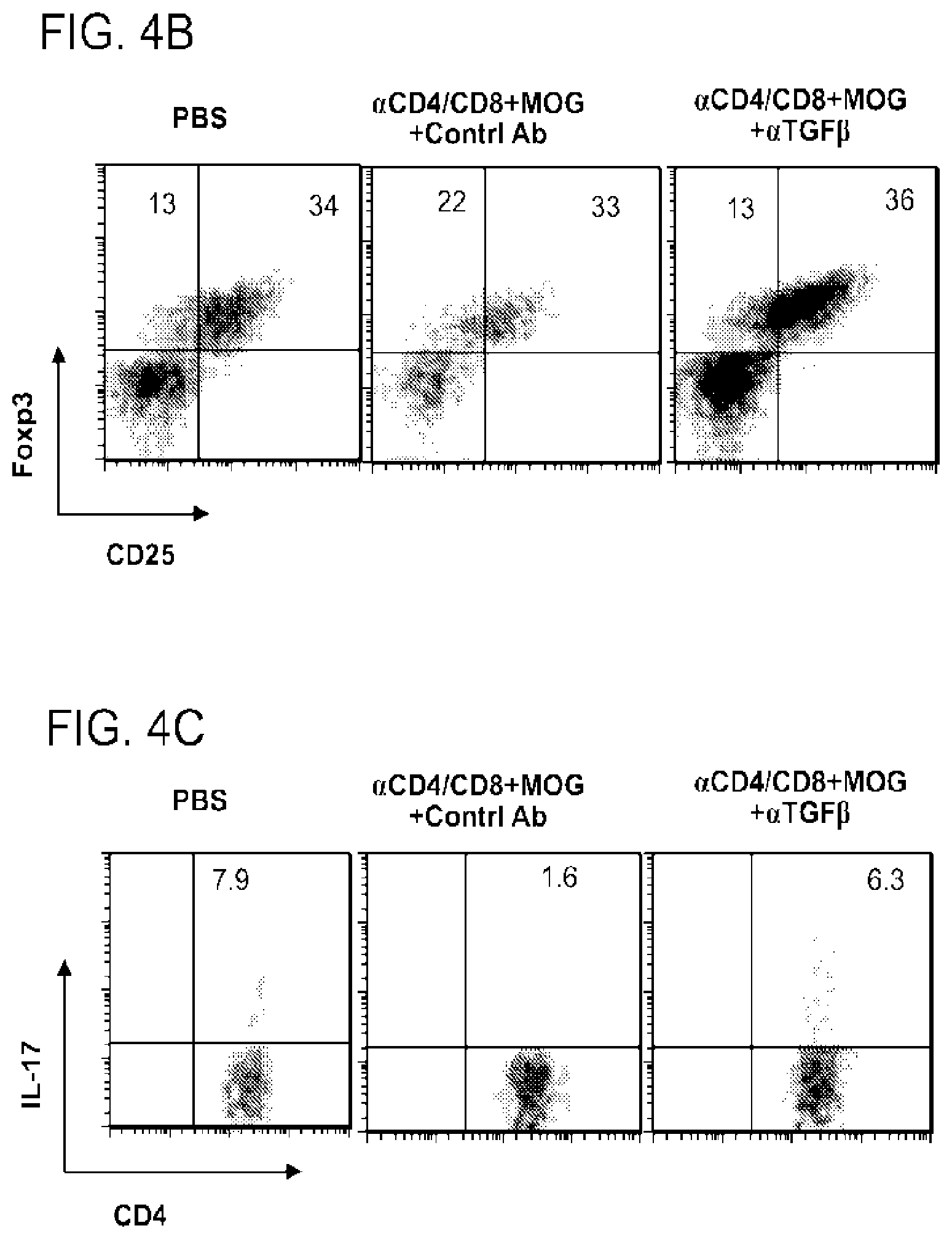
D00016

D00017

D00018

D00019

D00020

D00021

D00022

D00023

D00024

D00025

D00026

D00027

D00028

D00029

D00030

D00031

D00032

D00033

D00034

D00035

D00036

D00037

D00038

D00039

D00040

D00041

D00042

D00043

D00044
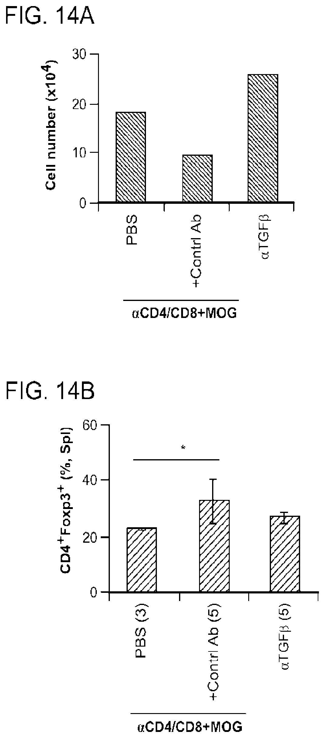
D00045

D00046

D00047

D00048

D00049
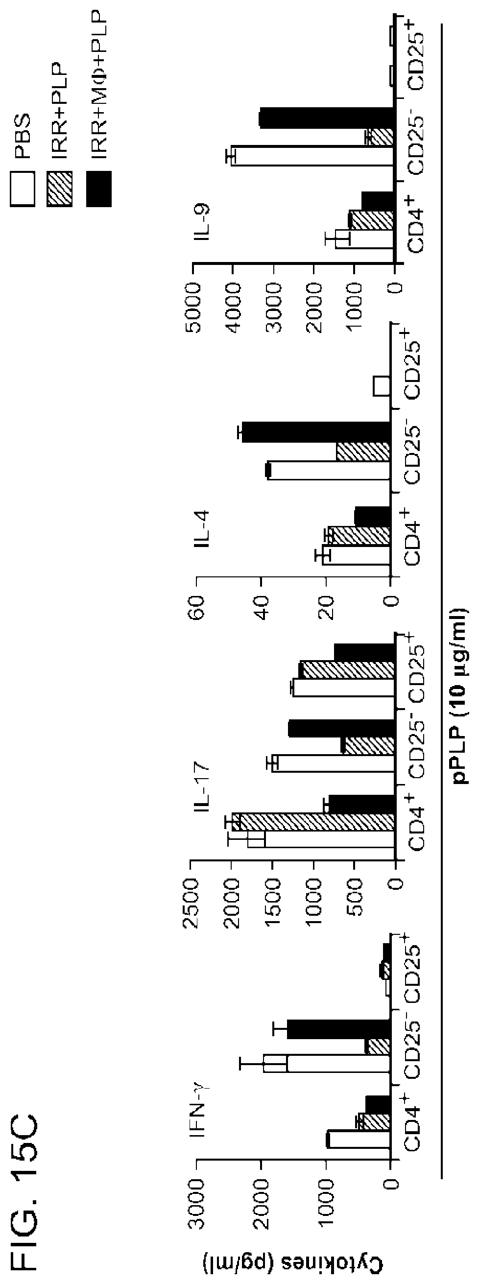
D00050

D00051

D00052

D00053

D00054

D00055

D00056

D00057

D00058

D00059

D00060

D00061

D00062
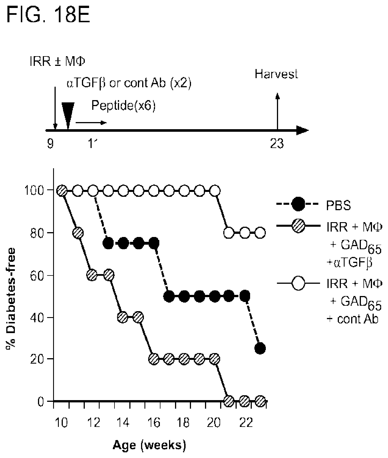
D00063

D00064

D00065

D00066

D00067

D00068

D00069

D00070

D00071

D00072

D00073

D00074

D00075

D00076

D00077

D00078

D00079

D00080

D00081

D00082

D00083

D00084

D00085

D00086

D00087

P00999

S00001
XML
uspto.report is an independent third-party trademark research tool that is not affiliated, endorsed, or sponsored by the United States Patent and Trademark Office (USPTO) or any other governmental organization. The information provided by uspto.report is based on publicly available data at the time of writing and is intended for informational purposes only.
While we strive to provide accurate and up-to-date information, we do not guarantee the accuracy, completeness, reliability, or suitability of the information displayed on this site. The use of this site is at your own risk. Any reliance you place on such information is therefore strictly at your own risk.
All official trademark data, including owner information, should be verified by visiting the official USPTO website at www.uspto.gov. This site is not intended to replace professional legal advice and should not be used as a substitute for consulting with a legal professional who is knowledgeable about trademark law.