A Universal Platform For For Preparing An Inhibitory Chimeric Antigen Receptor (icar)
Gross; Gideon ; et al.
U.S. patent application number 16/652019 was filed with the patent office on 2020-08-20 for a universal platform for for preparing an inhibitory chimeric antigen receptor (icar). The applicant listed for this patent is ImmPACT-Bio Ltd. GAVISH-GALILEE BIO APPLICATIONS LTD.. Invention is credited to Merav Beiman, Dvir Dahary, Will Gibson, Gideon Gross.
| Application Number | 20200261499 16/652019 |
| Document ID | 20200261499 / US20200261499 |
| Family ID | 1000004867463 |
| Filed Date | 2020-08-20 |
| Patent Application | download [pdf] |


View All Diagrams
| United States Patent Application | 20200261499 |
| Kind Code | A1 |
| Gross; Gideon ; et al. | August 20, 2020 |
A UNIVERSAL PLATFORM FOR FOR PREPARING AN INHIBITORY CHIMERIC ANTIGEN RECEPTOR (ICAR)
Abstract
The present invention provides a method of identifying a target for preparing an inhibitory chimeric antigen receptor (iCAR) or a protective chimeric antigen receptor (pCAR) capable of preventing or attenuating undesired activation of an effector immune cell. Also provided are a list of iCAR targets, as well as vectors and transduced effector immune cells comprising the nucleic acid molecule and methods for treatment of cancer comprising administering the transduced effector immune cells are further provided.
| Inventors: | Gross; Gideon; (Moshav Almagor, IL) ; Gibson; Will; (Boston, MA) ; Dahary; Dvir; (Tel Aviv, IL) ; Beiman; Merav; (Ness Ziona, IL) | ||||||||||
| Applicant: |
|
||||||||||
|---|---|---|---|---|---|---|---|---|---|---|---|
| Family ID: | 1000004867463 | ||||||||||
| Appl. No.: | 16/652019 | ||||||||||
| Filed: | September 28, 2018 | ||||||||||
| PCT Filed: | September 28, 2018 | ||||||||||
| PCT NO: | PCT/US2018/053583 | ||||||||||
| 371 Date: | March 27, 2020 |
Related U.S. Patent Documents
| Application Number | Filing Date | Patent Number | ||
|---|---|---|---|---|
| 62649429 | Mar 28, 2018 | |||
| 62564454 | Sep 28, 2017 | |||
| Current U.S. Class: | 1/1 |
| Current CPC Class: | C07K 16/2803 20130101; C12N 15/1093 20130101; C12N 15/1055 20130101; A61K 35/12 20130101; A61P 35/00 20180101; C07K 14/70578 20130101; C07K 14/70521 20130101; C07K 14/7051 20130101 |
| International Class: | A61K 35/12 20060101 A61K035/12; A61P 35/00 20060101 A61P035/00; C07K 14/725 20060101 C07K014/725; C07K 14/705 20060101 C07K014/705; C07K 16/28 20060101 C07K016/28; C12N 15/10 20060101 C12N015/10 |
Claims
1. A method of identifying a target for preparing an inhibitory chimeric antigen receptor (iCAR) or a protective chimeric antigen receptor (pCAR) capable of preventing or attenuating undesired activation of an effector immune cell, wherein the target is identified by a method comprising: (i) identifying a gene with at least two expressed alleles that encodes a protein comprising an extracellular polymorphic epitope; (ii) determining that at least one of the expressed alleles exhibits an amino acid sequence change in the extracellular polymorphic epitope sequence relative to an extracellular polymorphic epitope reference sequence; (iii) determining that the gene is located in a chromosomal region which undergoes loss of heterozygosity (LOH) in a tumor type; and (iv) determining that the gene is expressed in the tissue-of-origin of the tumor type in which the chromosomal region was found to undergo LOH.
2. The method of claim 1, wherein the LOH position is selected from the group consisting of a substitution, deletion, and insertion.
3. The method of claim 1, wherein the LOH position is a SNP.
4. The method of claim 1, wherein the gene comprising the extracellular polymorphic epitope is an HLA gene.
5. The method of claim 4, wherein the gene comprising the extracellular polymorphic epitope is an HLA-A, HLA-B, HLA-C, HLA-G, HLA-E, HLA-F, HLA-K, HLA-L, HLA-DM, HLA-DO, HLA-DP, HLA_DQ, or HLA-DR gene.
6. The method of claim 5, wherein the gene comprising the extracellular polymorphic epitope is an HLA-A gene.
7. The method of claim 5, wherein the gene comprising the extracellular polymorphic epitope is an HLA-B gene.
8. The method of claim 5, wherein the gene comprising the extracellular polymorphic epitope is an HLA-C gene.
9. The method of claim 5, wherein the gene comprising the extracellular polymorphic epitope is an HLA-G gene.
10. The method of claim 5, wherein the gene comprising the extracellular polymorphic epitope is an HLA-E gene.
11. The method of claim 5, wherein the gene comprising the extracellular polymorphic epitope is an HLA-F gene.
12. The method of claim 5, wherein the gene comprising the extracellular polymorphic epitope is an HLA-K gene.
13. The method of claim 5, wherein the gene comprising the extracellular polymorphic epitope is an HLA-L gene.
14. The method of claim 5, wherein the gene comprising the extracellular polymorphic epitope is an HLA-DM gene.
15. The method of claim 5, wherein the gene comprising the extracellular polymorphic epitope is an HLA-DO gene.
16. The method of claim 5, wherein the gene comprising the extracellular polymorphic epitope is an HLA-DP gene.
17. The method of claim 5, wherein the gene comprising the extracellular polymorphic epitope is an HLA_DQ gene.
18. The method of claim 5, wherein the gene comprising the extracellular polymorphic epitope is an HLA-DR gene.
19. The method of claim 1, wherein the gene comprising the extracellular polymorphic epitope is selected from the group consisting of ABCA4, ADAM30, AQP10, ASTN1, C1orf101, CACNA1S, CATSPER4, CD101, CD164L2, CD1A, CD1C, CD244, CD34, CD46, CELSR2, CHRNB2, CLCA2, CLDN19, CLSTN1, CR1, CR2, CRB1, CSF3R, CSMD2, ECE1, ELTD1, EMC1, EPHA10, EPHA2, EPHA8, ERMAP, FCAMR, FCER1A, FCGR1B, FCGR2A, FCGR2B, FCGR3A, FCRL1, FCRL3, FCRL4, FCRL5, FCRL6, GJB4, GPA33, GPR157, GPR37L1, GPR88, HCRTR1, IGSF3, IGSF9, IL22RA1, IL23R, ITGA10, KIAA1324, KIAA2013, LDLRAD2, LEPR, LGR6, LRIG2, LRP8, LRRC52, LRRC8B, LRRN2, LY9, MIA3, MR1, MUC1, MXRA8, NCSTN, NFASC, NOTCH2, NPR1, NTRK1, OPN3, OR10J1, OR10J4, OR10K1, OR10R2, OR10T2, OR10X1, OR11L1, OR14A16, OR1411, OR14K1, OR2AK2, OR2C3, OR2G2, OR2G3, OR2L2, OR2M7, OR2T12, OR2T27, OR2T1, OR2T3, OR2T29, OR2T33, OR2T34, OR2T35, OR2T3, OR2T4, OR2T5, OR2T6, OR2T7, OR2T8, OR2W3, OR6F1, OR6K2, OR6K3, OR6K6, OR6N1, OR6P1, OR6Y1, PDPN, PEARl, PIGR, PLXNA2, PTCH2, PTCHD2, PTGFRN, PTPRC, PTPRF, PVRL4, RHBG, RXFP4, S1PR1, SCNN1D, SDC3, SELE, SELL, SELP, SEMA4A, SEMA6C, SLAMF7, SLAMF9, SLC2A7, SLC5A9, TACSTD2, TAS1R2, TIE1, TLR5, TMEM81, TNFRSF14, TNFRSF1B, TRABD2B, USH2A, VCAM1, and ZP4.
20. The method of claim 1, wherein the gene comprising the extracellular polymorphic epitope is selected from the group consisting of ABCG5, ALK, ASPRV1, ATRAID, CD207, CD8B, CHRNG, CLEC4F, CNTNAP5, CRIM1, CXCR1, DNER, DPP10, EDAR, EPCAM, GPR113, GPR148, GPR35, GPR39, GYPC, IL1RL1, ITGA4, ITGA6, ITGAV, LCT, LHCGR, LRP1B, LRP2, LY75, MARCO, MERTK, NRP2, OR6B2, PLA2R1, PLB1, PROKR1, PROM2, SCN7A, SDC1, SLC23A3, SLC5A6, TGOLN2, THSD7B, TM4SF20, TMEFF2, TMEM178A, TPO, and TRABD2A.
21. The method of claim 1, wherein the gene comprising the extracellular polymorphic epitope is selected from the group consisting of ACKR2, ALCAM, ANO10, ATP13A4, BTLA, CACNA1D, CACNA2D2, CACNA2D3, CASR, CCRL2, CD200, CD200R1, CD86, CD96, CDCP1, CDHR4, CELSR3, CHL1, CLDN11, CLDN18, CLSTN2, CSPG5, CX3CR1, CXCR6, CYP8B1, DCBLD2, DRD3, EPHA6, EPHB3, GABRR3, GP5, GPR128, GPR15, GPR27, GRM2, GRM7, HEG1, HTR3C, HTR3D, HTR3E, IGSF11, IL17RC, IL17RD, IL17RE, IL5RA, IMPG2, ITGA9, ITGB5, KCNMB3, LRIG1, LRRC15, LRRN1, MST1R, NAALADL2, NRROS, OR5AC1, OR5H1, OR5H14, OR5H15, OR5H6, OR5K2, OR5K3, OR5K4, PIGX, PLXNB1, PLXND1, PRRT3, PTPRG, ROBO2, RYK, SEMA5B, SIDT1, SLC22A14, SLC33A1, SLC4A7, SLITRK3, STAB1, SUSD5, TFRC, TLR9, TMEM108, TMEM44, TMPRSS7, TNFSF10, UPK1B, VIPR1, and ZPLD1.
22. The method of claim 1, wherein the gene comprising the extracellular polymorphic epitope is selected from the group consisting of ANTXR2, BTC, CNGA1, CORIN, EGF, EMCN, ENPEP, EPHA5, ERVMER34-1, EVC2, FAT1, FAT4, FGFRL1, FRAS1, GPR125, GRID2, GYPA, GYPB, KDR, KIAA0922, KLB, MFSD8, PARM1, PDGFRA, RNF150, TENM3, TLR10, TLR1, TLR6, TMEM156, TMPRSS11A, TMPRSS11B, TMPRSS11E, TMPRSS11F, UGT2A1, and UNC5C.
23. The method of claim 1, wherein the gene comprising the extracellular polymorphic epitope is selected from the group consisting of ADAM19, ADRB2, BTNL3, BTNL8, BTNL9, C5orfl5, CATSPER3, CD180, CDH12, CDHR2, COL23A1, CSF1R, F2RL2, FAM174A, FAT2, FGFR4, FLT4, GABRA6, GABRG2, GPR151, GPR98, GRM6, HAVCR1, HAVCR2, IL31RA, IL6ST, IL7R, IQGAP2, ITGA1, ITGA2, KCNMB 1, LIFR, LNPEP, MEGF10, NIPAL4, NPR3, NRG2, OR2V1, OR2Y1, OSMR, PCDH12, PCDH1, PCDHA1, PCDHA2, PCDHA4, PCDHA8, PCDHA9, PCDHB 10, PCDHB 11, PCDHB 13, PCDHB 14, PCDHB 15, PCDHB 16, PCDHB2, PCDHB3, PCDHB4, PCDHB5, PCDHB6, PCDHGA1, PCDHGA4, PDGFRB, PRLR, SEMA5A, SEMA6A, SGCD, SLC1A3, SLC22A4, SLC22A5, SLC23A1, SLC36A3, SLC45A2, SLC6A18, SLC6A19, SLCO6A1, SV2C, TENM2, TIMD4, and UGT3A1.
24. The method of claim 1, wherein the gene comprising the extracellular polymorphic epitope is selected from the group consisting of BAI3, BTN1A1, BTN2A1, BTN2A2, BTN3A1, BTN3A2, BTNL2, CD83, DCBLD1, DLL1, DPCR1, ENPP1, ENPP3, ENPP4, EPHA7, GABBR1, GABRR1, GCNT6, GFRAL, GJB7, GLP1R, GPR110, GPR111, GPR116, GPR126, GPR63, GPRC6A, HFE, HLA-A, HLA-B, HLA-C, HLA-DOA, HLA-DPA1, HLA-DPB 1, HLA-DQA1, HLA-DQA2, HLA-DQB 1, HLA-DQB2, HLA-DRB 1, HLA-DRB5, HLA-E, HLA-F, HLA-G, L20RA, ITPR3, KIAA0319, LMBRDI, LRFN2, LRP11, MAS1L, MEP1A, MICA, MICB, MOG, MUC21, MUC22, NCR2, NOTCH4, OPRM1, OR10C1, OR12D2, OR12D3, OR14J1, OR2B2, OR2B6, OR2J1, OR2W1, OR5V1, PDE10A, PI16, PKHD1, PTCRA, PTK7, RAET1E, RAETIG, ROS1, SDIM1, SLC16A10, SLC22A1, SLC44A4, TAAR2, TREM1, TREML1, and TREML2.
25. The method of claim 1, wherein the gene comprising the extracellular polymorphic epitope is selected from the group consisting of AQP1, C7orf50, CD36, CDHR3, CNTNAP2, DPP6, EGFR, EPHA1, EPHB6, ERVW-1, GHRHR, GJC3, GPNMB, GRM8, HUS1, HYAL4, KIAA1324L, LRRN3, MET, MUC12, MUC17, NPC1L1, NPSR1, OR2A12, OR2A14, OR2A25, OR2A42, OR2A7, OR2A2, OR2AE1, OR2F2, OR6V1, PILRA, PILRB, PKD1L1, PLXNA4, PODXL, PTPRN2, PTPRZ 1, RAMP3, SLC29A4, SMO, TAS2R16, TAS2R40, TAS2R4, TFR2, THSD7A, TMEM213, TTYH3, ZAN, and ZP3.
26. The method of claim 1, wherein the gene comprising the extracellular polymorphic epitope is selected from the group consisting of ADAM18, ADAM28, ADAM32, ADAM7, ADAM9, ADRA1A, CDH17, CHRNA2, CSMD1, CSMD3, DCSTAMP, FZD6, GPR124, NRG1, OR4F21, PKHD1L1, PRSS55, SCARA3, SCARA5, SDC2, SLC10A5, SLC39A14, SLC39A4, SLCO5A1, TNFRSF10A, and TNFRSF10B.
27. The method of claim 1, wherein the gene comprising the extracellular polymorphic epitope is selected from the group consisting of ABCA1, AQP7, ASTN2, C9orf135, CA9, CD72, CNTNAP3B, CNTNAP3, CRB2, ENTPD8, GPR144, GRIN3A, IZUMO3, KIAA1161, MAMDC4, MEGF9, MUSK, NOTCH1, OR13C2, OR13C3, OR13C5, OR13C8, OR13C9, OR13D1, OR13F1, OR1B1, OR1J2, OR1K1, OR1L1, OR1L3, OR1L6, OR1L8, OR1N1, OR1N2, OR1Q1, OR2S2, PCSK5, PDCD1LG2, PLGRKT, PTPRD, ROR2, SEMA4D, SLC31A1, TEK, TLR4, TMEM2, and VLDLR.
28. The method of claim 1, wherein the gene comprising the extracellular polymorphic epitope is selected from the group consisting of ABCC2, ADAM8, ADRB 1, ANTXRL, ATRNL1, C10orf54, CDH23, CDHR1, CNNM2, COL13A1, COL17A1, ENTPD1, FZD8, FGFR2, GPR158, GRID1, IL15RA, IL2RA, ITGA8, ITGB1, MRC1, NRG3, NPFFR1, NRP1, OPN4, PCDH15, PKD2L1, PLXDC2, PRLHR, RET, RGR, SLC16A9, SLC29A3, SLC39A12, TACR2, TCTN3, TSPAN15, UNC5B, and VSTM4.
29. The method of claim 1, wherein the gene comprising the extracellular polymorphic epitope is selected from the group consisting of AMICA1, ANO1, ANO3, APLP2, C11orf24, CCKBR, CD248, CD44, CD5, CD6, CD82, CDON, CLMP, CRTAM, DCHS1, DSCAML1, FAT3, FOLH1, GDPD4, GDPD5, GRIK4, HEPHL1, HTR3B, IFITM10, IL10RA, KIRREL3, LGR4, LRP4, LRP5, LRRC32, MCAM, MFRP, MMP26, MPEG1, MRGPRE, MRGPRF, MRGPRX2, MRGPRX3, MRGPRX4, MS4A4A, MS4A6A, MTNR1B, MUC15, NAALAD2, NAALADL1, NCAM1, NRXN2, OR10A2, OR10A5, OR10A6, OR10D3, OR10G4, OR10G7, OR10G8, OR10G9, OR10Q1, OR10S1, OR1S1, OR2AG1, OR2AG2, OR2D2, OR4A47, OR4A15, OR4A5, OR4C11, OR4C13, OR4C15, OR4C16, OR4C3, OR4C46, OR4C5, OR4D6, OR4A8P, OR4D9, OR4S2, OR4X1, OR51E1, OR51L1, OR52A1, OR52E1, OR52E2, OR52E4, OR52E6, OR52I1, OR52I2, OR52J3, OR52L1, OR52N1, OR52N2, OR52N4, OR52W1, OR56B1, OR56B4, OR5A1, OR5A2, OR5AK2, OR5AR1, OR5B17, OR5B3, OR5D14, OR5D16, OR5D18, OR5F1, OR5I1, OR5L2, OR5M11, OR5M3, OR5P2, OR5R1, OR5T2, OR5T3, OR5W2, OR6A2, OR6T1, OR6X1, OR8A1, OR8B12, OR8B2, OR8B3, OR8B4, OR8D1, OR8D2, OR8H1, OR8H2, OR8H3, OR8I2, OR8J1, OR8J2, OR8J3, OR8K1, OR8K3, OR8K5, OR8U1, OR9G1, OR9G4, OR9Q2, P2RX3, PTPRJ, ROBO3, SIGIRR, SLC22A10, SLC3A2, SLC5A12, SLCO2B1, SORL1, ST14, SYT8, TENM4, TMEM123, TMEM225, TMPRSS4, TMPRSS5, TRIM5, TRPM5, TSPAN18, and ZP1.
30. The method of claim 1, wherein the gene comprising the extracellular polymorphic epitope is selected from the group consisting of ANO4, AVPR1A, BCL2L14, CACNA2D4, CD163, CD163L1, CD27, CD4, CLEC12A, CLEC 1B, CLEC2A, CLEC4C, CLEC7A, CLECL1, CLSTN3, GPR133, GPRC5D, ITGA7, ITGB7, KLRB1, KLRC2, KLRC3, KLRC4, KLRF1, KLRF2, LRP1, LRP6, MANSCI, MANSC4, OLR1, OR10AD1, OR10P1, OR2AP1, OR6C1, OR6C2, OR6C3, OR6C4, OR6C6, OR6C74, OR6C76, OR8S1, OR9K2, ORAI1, P2RX4, P2RX7, PRR4, PTPRB, PTPRQ, PTPRR, SCNN1A, SELPLG, SLC2A14, SLC38A4, SLC5A8, SLC6A15, SLC8B1, SLCO1A2, SLCO1B1, SLCO1B7, SLCO1C1, SSPN, STAB2, TAS2R10, TAS2R13, TAS2R14, TAS2R20, TAS2R30, TAS2R31, TAS2R42, TAS2R43, TAS2R46, TAS2R7, TMEM119, TMEM132B, TMEM132C, TMEM132D, TMPRSS12, TNFRSF1A, TSPAN8, and VSIG10.
31. The method of claim 1, wherein the gene comprising the extracellular polymorphic epitope is selected from the group consisting of ATP4B, ATP7B, FLT3, FREM2, HTR2A, KL, PCDH8, RXFP2, SGCG, SHISA2, SLC15A1, SLITRK6, and TNFRSF 19.
32. The method of claim 1, wherein the gene comprising the extracellular polymorphic epitope is selected from the group consisting of ADAM21, BDKRB2, C14orf37, CLEC14A, DLK1, FLRT2, GPR135, GPR137C, JAG2, LTB4R2, MMP14, OR11G2, OR11H12, OR11H6, OR4K1, OR4K15, OR4K5, OR4L1, OR4N2, OR4N5, SLC24A4, and SYNDIG1L.
33. The method of claim 1, wherein the gene comprising the extracellular polymorphic epitope is selected from the group consisting of ANPEP, CD276, CHRNA7, CHRNB4, CSPG4, DUOX1, DUOX2, FAM174B, GLDN, IGDCC4, ITGA11, LCTL, LTK, LYSMD4, MEGF 11, NOX5, NRG4, OCA2, OR4F4, OR4M2, OR4N4, PRTG, RHCG, SCAMP5, SEMA4B, SEMA6D, SLC24A1, SLC24A5, SLC28A1, SPG11, STRA6, TRPM1, and TYRO3.
34. The method of claim 1, wherein the gene comprising the extracellular polymorphic epitope is selected from the group consisting of ATP2C2, CACNA1H, CD19, CDH11, CDH15, CDH16, CDH3, CDH5, CNGB1, CNTNAP4, GDPD3, GPR56, GPR97, IFT140, IL4R, ITFG3, ITGAL, ITGAM, ITGAX, KCNG4, MMP15, MSLNL, NOMO1, NOMO3, OR2C1, PIEZO1, PKD1, PKD1L2, QPRT, SCNN1B, SEZ6L2, SLC22A31, SLC5A11, SLC7A6, SPN, TMC5, TMC7, TMEM204, TMEM219, and TMEM8A.
35. The method of claim 1, wherein the gene comprising the extracellular polymorphic epitope is selected from the group consisting of ABCC3, ACE, AOC3, ARL17B, ASGR2, C17orf80, CD300A, CD300C, CD300E, CD300LF, CD300LG, CHRNB1, CLEC10A, CNTNAP1, CPD, CXCL16, ERBB2, FAM171A2, GCGR, GLP2R, GP1BA, GPR142, GUCY2D, ITGA2B, ITGA3, ITGAE, ITGB3, KCNJ12, LRRC37A2, LRRC37A3, LRRC37A, LRRC37B, MRC2, NGFR, OR1A2, OR1D2, OR1G1, OR3A1, OR3A2, OR4D1, OR4D2, RNF43, SCARF1, SCN4A, SDK2, SECTM1, SEZ6, SHPK, SLC26A11, SLC5A10, SPACA3, TMEM102, TMEM132E, TNFSF12, TRPV3, TTYH2, and TUSC5.
36. The method of claim 5, wherein the gene comprising the extracellular polymorphic epitope is selected from the group consisting of APCDD1, CDH19, CDH20, CDH7, COLEC12, DCC, DSC1, DSG1, DSG3, DYNAP, MEP1B, PTPRM, SIGLEC15, and TNFRSF 11A.
37. The method of claim 1, wherein the gene comprising the extracellular polymorphic epitope is selected from the group consisting of ABCA7, ACPT, BCAM, C19orf38, C19orf59, C5AR1, CATSPERD, CATSPERG, CD22, CD320, CD33, CD97, CEACAM19, CEACAM1, CEACAM21, CEACAM3, CEACAM4, CLEC4M, DLL3, EMR1, EMR2, EMR3, ERVV-1, ERVV-2, FAM187B, FCAR, FFAR3, FPR1, FXYD5, GFY, GP6, GPR42, GRIN3B, ICAM3, IGFLR1, IL12RB1, IL27RA, KIR2DL1, KIR2DL3, KIR2DL4, KIR3DL1, KIR3DL2, KIR3DL3, KIRREL2, KISS1R, LAIR1, LDLR, LILRA1, LILRA2, LILRA4, LILRA6, LILRB1, LILRB2, LILRB3, LILRB4, LILRB5, LINGO3, LPHN1, LRP3, MADCAM1, MAG, MEGF8, MUC16, NCR1, NOTCH3, NPHS1, OR10H1, OR10H2, OR10H3, OR10H4, ORII1, OR2Z1, OR7A10, OR7C1, OR7D4, OR7E24, OR7G1, OR7G2, OR7G3, PLVAP, PTGIR, PTPRH, PTPRS, PVR, SCN1B, SHISA7, SIGLEC10, SIGLEC11, SIGLEC12, SIGLEC5, SIGLEC6, SIGLEC8, SIGLEC9, SLC44A2, SLC5A5, SLC7A9, SPINT2, TARM1, TGFBR3L, TMC4, TMEM91, TMEM161A, TMPRSS9, TNFSF14, TNFSF9, TRPM4, VN1R2, VSIG10L, VSTM2B, and ZNRF4.
38. The method of claim 1, wherein the gene comprising the extracellular polymorphic epitope is selected from the group consisting of ABHD12, ADAM33, ADRAID, APMAP, ATRN, CD40, CD93, CDH22, CDH26, CDH4, FLRT3, GCNT7, GGT7, JAG1, LRRN4, NPBWR2, OCSTAMP, PTPRA, PTPRT, SEL1L2, SIGLEC1, SIRPA, SIRPB1, SIRPG, SLC24A3, SLC2A10, SLC4A11, SSTR4, and THBD.
39. The method of claim 1, wherein the gene comprising the extracellular polymorphic epitope is selected from the group consisting of CLDN8, DSCAM, ICOSLG, IFNAR1, IFNGR2, IGSF5, ITGB2, KCNJ15, NCAM2, SLC19A1, TMPRSS15, TMPRSS2, TMPRSS3, TRPM2, and UMODL1.
40. The method of claim 1, wherein the gene comprising the extracellular polymorphic epitope is selected from the group consisting of CACNAII, CELSR1, COMT, CSF2RB, GGT1, GGT5, IL2RB, KREMEN1, MCHR1, OR11H1, P2RX6, PKDREJ, PLXNB2, SCARF2, SEZ6L, SSTR3, SUSD2, TMPRSS6, and TNFRSF13C.
41. The method of claim 1, wherein the gene comprising the extracellular polymorphic epitope is selected from the group consisting of ATP6AP2, ATP7A, CNGA2, EDA2R, FMR1NB, GLRA4, GPR112, GUCY2F, HEPH, P2RY10, P2RY4, PLXNA3, PLXNB3, TLR8, VSIG4, and XG.
42. The method of any of claims 1 through 41, wherein the tumor is selected from the group consisting of a breast tumor, a prostate tumor, an ovarian tumor, a cervical tumor, a skin tumor, a pancreatic tumor, a colorectal tumor, a renal tumor, a liver tumor, a brain tumor, a lymphoma, a leukemia, a lung tumor, and a glioma.
43. The method of any of claims 1 through 41, wherein the tumor is selected from the group consisting of an adrenal gland tumor, a kidney tumor, a melanoma, DLBC, a breast tumor, a sarcoma, an ovary tumor, a lung tumor, a bladder tumor, and a liver tumor.
44. The method of claim 43, wherein the adrenal gland tumor is an adrenocortical carcinoma.
45. The method of claim 43, wherein the kidney tumor is a chromophobe renal cell carcinoma.
46. The method of claim 43, wherein the melanoma is uveal melanoma.
47. A safe effector immune cell expressing (i) an iCAR or pCAR according to any of claims 1 through 46 and (ii) an activating chimeric antigen receptor (aCAR).
48. The safe effector immune cell of claim 47, wherein the aCAR is directed against or specifically binds to a tumor-associated antigen or a non-polymorphic cell surface epitope.
49. The safe effector immune cell of claim 47, wherein the aCAR is directed against or specifically binds to a tumor associated protein, a CAR target as listed in table 1, any cell surface protein that is expressed in a tumor tissue in which the iCAR is also expressed.
50. The safe effector immune cell of claim 49, wherein the non-polymorphic cell surface epitope is selected from the group consisting of CD19, CD20, CD22, CD10, CD7, CD49f, CD56, CD74, CAIX IgK, ROR1, ROR2, CD30, LewisY, CD33, CD34, CD38, CD123, CD28, CD44v6, CD44, CD41, CD133, CD138, NKG2D-L, CD139, BCMA, GD2, GD3, hTERT, FBP, EGP-2, EGP-40, FR-.alpha., L1-CAM, ErbB2,3,4, EGFRvIII, VEGFR-2, IL-13R.alpha.2, FAP, Mesothelin, c-MET, PSMA, CEA, kRas, MAGE-A1, MUC1 MUC16, PDL1, PSCA, EpCAM, FSHR, AFP, AXL, CD80, CD89, CDH17, CLD18, GPC3, TEM8, TGFB1, NY-ESO-1, WT-1 and EGFR.
51. The safe effector immune cell of any of claims 47 to 50, wherein the safe effector immune cell is an autologous or a universal (allogeneic) effector cell.
52. The safe effector immune cell of any of claims 47 to 51, wherein the safe effector immune cell is selected from the group consisting of a T cell, a natural killer cell and a cytokine-induced killer cell.
53. The safe effector immune cell of any of claims 47 to 52, wherein the expression level of the iCAR or pCAR is greater than or equal to the expression level of the aCAR.
54. The safe effector immune cell of any of claims 47 to 53, wherein the iCAR or pCAR is expressed by a first vector and the aCAR is expressed by a second vector.
55. The safe effector immune cell of any of claims 47 to 53, wherein the iCAR or pCAR and the aCAR are both expressed by the same vector.
56. The safe effector immune cell of claim 55, wherein the nucleotide sequence encoding for the aCAR is downstream of the nucleotide sequence encoding for the iCAR or pCAR.
57. The safe effector immune cell of claim 55, wherein the nucleotide sequence comprises a viral self-cleaving 2A peptide between the nucleotide sequence encoding for the aCAR and the nucleotide sequence encoding for the iCAR or pCAR.
58. The safe effector immune cell of claim 57, wherein the viral self-cleaving 2A peptide is selected from the group consisting of T2A from Thosea asigna virus (TaV), F2A from Foot-and-mouth disease virus (FMDV), E2A from Equine rhinitis A virus (ERAV) and P2A from Porcine teschovirus-1 (PTV1).
59. The safe effector immune cell of claim 55, wherein the nucleotide sequence encoding the aCAR is linked via a flexible linker to the iCAR or pCAR.
60. The safe effector immune cell of any of claims 47 to 59, wherein the aCAR comprises at least one signal transduction element that activates or co-stimulates an effector immune cell
61. The safe effector immune cell of claim 60, wherein the at least one signal transduction element that activates or co-stimulates an effector immune cell is homolgous to an immunoreceptor tyrosine-based activation motif (ITAM) of for example CD3.zeta. or FcR.gamma. chains.
62. The safe effector immune cell of claim 60, wherein the at least one signal transduction element that activates or co-stimulates an effector immune cell is homolgous to an activating killer cell immunoglobulin-like receptor (KIR), such as KIR2DS and KIR3DS.
63. The safe effector immune cell of any of claim 60, wherein the at least one signal transduction element that activates or co-stimulates an effector immune cell is homolgous to or an adaptor molecule, such as DAP12.
64. The safe effector immune cell of claim 60, wherein the at least one signal transduction element that activates or co-stimulates an effector immune cell is homolgous to or a co-stimulatory signal transduction element of CD27, CD28, ICOS, CD137 (4-1BB), CD134 (OX40) or GITR.
65. A method for treating cancer in a patient having a tumor characterized by LOH, comprising administering to the patient a safe effector immune cell expressing the iCAR according to any of claims 1 through 64.
66. A method for treating cancer in a patient having a tumor characterized by LOH, comprising administering to the patient a safe effector immune cell according to any of claims 47 through 64.
Description
CROSS-REFERENCE TO RELATED APPLICATIONS
[0001] This application claims priority to U.S. Provisional Application No. 62/564,454, filed Sep. 28, 2017, and U.S. Provisional Application No. 62/649,429, filed Mar. 28, 2018, each of which is herein incorporated by reference.
SEQUENCE LISTING
[0002] This patent application contains a Sequence Listing which has been submitted electronically in ASCII format and are hereby incorporated herein by reference in its entirety. Said ASCII copy, created Sep. 27, 2018, is named 120575-5003_ST25.txt.
ASCII TABLE
[0003] The provisional patent application to which the current application claims priority contains a lengthy table section. A copy of the table was submitted to the U.S. Patent and Trademark Office on compact disc in ASCII format with priority U.S. Provisional Application No. 62/649,429, filed Mar. 28, 2018 and is hereby incorporated by reference, and may be employed in the practice of the invention. Said ASCII table, created Mar. 28, 2018, is as follows: 120575-5003-PR allCandExt1167Genes_5003_PR.txt, 272,719,870 bytes.
FIELD OF THE INVENTION
[0004] The invention relates to the field of cancer immunotherapy by adoptive cell transfer, employing activating chimeric antigen receptors (aCARs) recognizing antigens expressed on the surface of tumor cells, inhibitory CARs (iCARs) and protective CARs (pCARs) directed at allelic variants of the same or other cell surface antigens expressed by normal cells but not by the tumor due to loss of heterozygosity (LOH).
BACKGROUND OF THE INVENTION
[0005] The identification of targetable antigens that are exclusively expressed by tumor cells but not by healthy tissue is undoubtedly the major challenge in cancer immunotherapy today. Clinical evidence that T cells are capable of eradicating tumor cells comes from numerous studies evaluating highly diverse approaches for harnessing T cells to treat cancer (Rosenberg and Restifo, 2015). These approaches employ bone marrow transplantation with donor lymphocyte infusion, adoptive transfer of tumor-infiltrating lymphocytes (TILs), treatment with T cells genetically redirected at pre-selected antigens via CARs (Gross and Eshhar, 2016a) or T cell receptors (TCRs), the use of immune checkpoint inhibitors or active vaccination. Of these, the use of genetically engineered T cells and different strategies for active immunization entail pre-existing information on candidate antigens which are likely to exert a durable clinical response but minimal adverse effects. Yet, as stated in the title of a recent review by S. Rosenberg, "Finding suitable targets is the major obstacle to cancer gene therapy" (Rosenberg, 2014).
[0006] The concept of using chimeric antigen receptors (or CARs) to genetically redirect T cells (or other killer cells of the immune system such as natural killer (NK) cells and cytokine-induced killer cells) against antigens of choice in an MHC-independent manner was first introduced by Gross and Eshhar in the late 1980s (Gross et al., 1989). They are produced synthetically from chimeric genes encoding an extracellular single-chain antibody variable fragment (scFv) fused through a flexible hinge and transmembrane canonic motif to signaling components comprising immunoreceptor tyrosine-based activation motifs of CD3.zeta. or FcR.gamma. chains capable of T cell activation. At present, CARs are being examined in dozens of clinical trials and have so far shown exceptionally high efficacy in B cell malignancies (Dotti et al., 2014; Gill and June, 2015; Gross and Eshhar, 2016a). The safety of CAR-T cell therapy is determined, in large, by its ability to discriminate between the tumor and healthy tissue. A major risk and the direct cause for adverse autoimmune effects that have been reported in clinical and preclinical studies is off-tumor, on-target toxicity resulting from extra-tumor expression of the target antigen (dealt with in detail in our recent review (Gross and Eshhar, 2016b) and (Klebanoff et al., 2016)). Concerning this risk, shared, non-mutated cell surface antigens which are currently tested clinically or pre-clinically for CAR therapy can be generally divided into a number of categories according to their tissue distribution and mode of expression: [0007] Strictly tumor-specific antigens. Perhaps the only member in this group which is already being examined clinically is variant III of the epidermal growth factor receptor (EGFRvIII) that is frequently overexpressed in glioblastoma and is also found in non-small cell lung carcinoma and prostate, breast, head and neck and ovarian cancers but not on normal tissue. [0008] Surface antigens expressed on the tumor and on non-vital healthy tissue. Potential CAR antigens in this group are differentiation-related molecules that are mainly restricted to the B cell lineage. Prominent among these (and a target antigen in numerous clinical trials) is CD19, a pan-B cell marker acquired very early in B cell differentiation and involved in signal transduction by the B cell receptor (BCR). Membrane prostate antigens constitute another class of antigens in this category. [0009] Antigens that are typically expressed by non-malignant tumor-promoting cells. One such antigen is fibroblast activation protein (FAP), a cell surface serine protease which is almost invariably expressed by tumor-associated fibroblasts in diverse primary and metastatic cancers. Another antigen is vascular endothelial growth factor (VEGF), which is highly expressed during tumor angiogenesis and is normally expressed on vascular and lymphatic endothelial cells in many vital organs. [0010] Tumor associated antigens (TAAs) shared with vital healthy tissue.
[0011] Most other TAAs which are presently evaluated in preclinical and clinical studies are overexpressed by tumors but are also present, usually at lower level, on essential normal tissue.
[0012] The broad spectrum of strategies devised to tackle autoimmunity in CAR T cell therapy can be divided into those which seek to eliminate, or suppress transferred T cells once damage is already evident (reactive measures) and those that aim at preventing potential damage in the first place (proactive measures) (Gross and Eshhar, 2016a). Reactive approaches often use suicide genes such as herpes simplex virus thymidine kinase (HSV-tk) and iC9, a fusion polypeptide comprising a truncated human caspase 9 and a mutated FK506-binding protein. Other approaches utilize antibodies to selectively remove engineered cells which go havoc or, as recently demonstrated, a heterodimerizing small-molecule agent which governs the coupling of the CAR recognition moiety to the intracellular signaling domain (Wu et al., 2015). While some proactive measures are designed to limit the in-vivo persistence or function of CAR T cells (for example, the use of mRNA electroporation for gene delivery), others directly address the critical challenge of increasing antigenic selectivity of the therapeutic CARs so as to avoid damage to non-tumor tissue. Two of these raise particular interest, as they can potentially broaden the range of tumor antigens which can be safely targeted by CAR T cells: [0013] Combinatorial (or `split`) antigen recognition. While true tumor-specific surface antigens are rare, combinations of two different antigens, not-necessarily classified as tumor-associated antigens that are co-expressed by a given tumor, can define a new tumor-specific signature. Restricting the activity of CAR T cells to such antigen pairs provides a critical safety gauge and, consequently, extends the spectrum of tumor-specific targets and may be of substantial therapeutic value. Second and third generation CARs have been designed to provide therapeutic T cells with activation and costimulation signals upon engaging a single antigen through the tethering of two or more signaling portions at the CAR endodomain. However, if activation and costimulation are split in the same T-cell between two CARs, each specific for a different antigen, then full blown response would require the cooperation of the two complementary signals that could only be accomplished in the presence of the two antigens. This principle has been demonstrated in several preclinical studies (Kloss et al., 2013; Lanitis et al., 2013; Wilkie et al., 2012; WO 2016/126608).
[0014] While undoubtedly intriguing, this approach still faces the need in meticulous titration of the magnitude of both the activating and costimulatory signals so as to reach the optimal balance that would only allow effective on-target, on-tumor T cell reactivity. Whether such balance can be routinely attained in the clinical setting is still questionable.
[0015] An entirely new approach for limiting T cell response only to target cells that express a unique combination of two antigens was published recently (Roybal et al., 2016a). Its core element functions as a `genetic switch` which exploits the mode of action of several cell surface receptors, including Notch. Following binding of such a receptor to its ligand it undergoes dual cleavage resulting in the liberation of its intracellular domain which translocates to the cell nucleus where it functions as a transcription factor. The implementation of this principle entails the co-introduction of two genes to the effector T cells. The first one is expressed constitutively and encodes such a chimeric cleavable receptor equipped with a recognition moiety directed at the first antigen. Engagement with this antigen on the surface of a target cell will turn on the expression of the second gene encoding a conventional CAR which is directed at the second antigen. The target cell will be killed only if it co-expresses this second antigen as well.
[0016] Inhibitory CARs. Off-tumor reactivity occurs when the target antigen of CAR-redirected killer cells is shared with normal tissue. If this normal tissue expresses another surface antigen not present on the tumor, then co-expressing in the gene-modified cells an additional CAR targeting this non-shared antigen, which harbors an inhibitory signaling moiety, can prevent T-cell activation by the normal tissue.
[0017] Instead of an activating domain (such as FcR.gamma. or CD3.zeta.), an iCAR possesses a signaling domain derived from an inhibitory receptor which can antagonize T cell activation, such as CTLA-4, PD-1 or an NK inhibitory receptor. If the normal tissue which shares the candidate aCAR antigen with the tumor expresses another surface antigen not shared with the tumor, an iCAR expressed by the same T cell which targets this non-shared antigen can protect the normal tissue (FIG. 1).
[0018] Unlike T cells, each of which expresses a unique two-chain TCR encoded by somatically rearranged gene segments, NK cells do not express antigen-specific receptors. Instead, NK cells express an array of germline-encoded activating and inhibitory receptors which respectively recognize multiple activating and inhibitory ligands at the cell surface of infected and healthy cells. The protective capacity of an iCAR based on NK inhibitory receptors such as KIR3DL1 has been described (U.S. Pat. No. 9,745,368). KIR3DL1 and other NK inhibitory receptors function by dismantling the immunological synapse in a rapid and comprehensive manner. There is compelling evidence that a single NK cell can spare a resistant cell expressing both inhibitory and activating ligands yet kill a susceptible cell it simultaneously engages, which expresses only the activating ligands (Abeyweera et al., 2011; Eriksson et al., 1999; Treanor et al., 2006; Vyas et al., 2001). This exquisite ability is governed by the different spatial organization of signal transduction molecules formed at each of the respective immune synapses which consequently affects the exocytosis of cytolytic granules (see (Huse et al., 2013) for review). More recently, Fedorov et al. (Fedorov et al., 2013a; WO 2015/142314) successfully employed for this purpose the intracellular domains of PD-1 and CTLA-4. Unlike NK inhibitory receptors, the regulatory effects of these iCARs affected the entire cell. Yet, these effects were temporary, allowing full T-cell activation upon subsequent encounter with target cells expressing only the aCAR antigen.
[0019] Tissue distribution of the antigens targeted by the iCAR and aCAR dictates the optimal mode of action of the iCAR required for conferring maximal safety without compromising clinical efficacy. For example, if the anatomical sites of the tumor and the normal tissue(s) to be protected do not intersect, transient inhibition (CTLA-4- or PD-1-like) will likely suffice. Yet, if these sites do overlap, only synapse-confined inhibition (e.g., an NK mode of action) will prevent constant paralysis of the therapeutic cells and allow their effective tumoricidal activity. The approach of using iCARs to reduce on-target off-tumor reactivity suffers from a dire lack of antigens downregulated in tumor cells but present on normal tissue.
[0020] Next generation sequencing (NGS) allows the determination of the DNA sequence of all protein-coding genes (.about.1% of the entire genome) in a given tumor biopsy and the comparison of the cancer `exome` to that of a healthy tissue (usually from white blood cells) of the same patient. Exome sequencing can be completed within several days post-biopsy removal and at relatively low cost. In parallel, transcriptome analysis (RNA-seq) can provide complementary information on the genes that are actually expressed by the same cell sample.
[0021] It is becoming increasingly clear that the mutational landscape of each individual tumor is unique (Lawrence et al., 2013; Vogelstein et al., 2013). As a result of nonsynonymous mutations the tumor cell can potentially present a private set of neopeptides to the patient's immune system on one or more of his or her HLA products. Indeed, tremendous efforts are being put in recent years into identifying tumor-specific neoepitopes which can be recognized by the patient's own CD8 or CD4 T cell repertoire and serve as targets for immunotherapy (for review see (Blankenstein et al., 2015; Van Buuren et al., 2014; Heemskerk et al., 2013; Overwijk et al., 2013; Schumacher and Schreiber, 2015)). However, cumulative findings suggest that neoantigen-based T cell immunotherapies are more likely to be effective in cancers displaying higher mutational load, such as melanoma and lung cancers, but may often fail to show benefit in most cancers with fewer mutations (Savage, 2014; Schumacher and Schreiber, 2015). Furthermore, considerable intratumoral heterogeneity (Burrell et al., 2013) entails the simultaneous co-targeting of several antigens so as to avoid emergence of mutation-loss variants, a task which becomes increasingly demanding in view of the scarcity of useful immunogenic neopeptides.
[0022] All in all, the urgent need to identify suitable targets for cancer immunotherapy via the adoptive transfer of genetically redirected killer cells is still largely unmet.
BRIEF SUMMARY OF THE INVENTION
[0023] In some aspects, the present invention provides a method of identifying a target for preparing an inhibitory chimeric antigen receptor (iCAR) or a protective chimeric antigen receptor (pCAR) capable of preventing or attenuating undesired activation of an effector immune cell, wherein the target is identified by a method comprising: [0024] (i) identifying a gene with at least two expressed alleles that encodes a protein comprising an extracellular polymorphic epitope; [0025] (ii) determining that at least one of the expressed alleles exhibits an amino acid sequence change in the extracellular polymorphic epitope sequence relative to an extracellular polymorphic epitope reference sequence; [0026] (iii) determining that the gene is located in a chromosomal region which undergoes loss of heterozygosity (LOH) in a tumor type; and [0027] (iv) determining that the gene is expressed in the tissue-of-origin of the tumor type in which the chromosomal region was found to undergo LOH.
[0028] In some embodiments, the LOH position is selected from the group consisting of a substitution, deletion, and insertion. In some embodiments, the LOH position is a SNP. In some embodiments, the gene comprising the extracellular polymorphic epitope is an HLA gene.
[0029] In some embodiments, the gene comprising the extracellular polymorphic epitope is an HLA-A, HLA-B, HLA-C, HLA-G, HLA-E, HLA-F, HLA-K, HLA-L, HLA-DM, HLA-DO, HLA-DP, HLA_DQ, or HLA-DR gene. In some embodiments, the gene comprising the extracellular polymorphic epitope is an HLA-A gene. In some embodiments, the gene comprising the extracellular polymorphic epitope is an HLA-B gene. In some embodiments, the gene comprising the extracellular polymorphic epitope is an HLA-C gene. In some embodiments, the gene comprising the extracellular polymorphic epitope is an HLA-G gene. In some embodiments, the gene comprising the extracellular polymorphic epitope is an HLA-E gene. In some embodiments, the gene comprising the extracellular polymorphic epitope is an HLA-F gene. In some embodiments, the gene comprising the extracellular polymorphic epitope is an HLA-K gene. In some embodiments, the gene comprising the extracellular polymorphic epitope is an HLA-L gene. In some embodiments, the gene comprising the extracellular polymorphic epitope is an HLA-DM gene. In some embodiments, the gene comprising the extracellular polymorphic epitope is an HLA-DO gene. In some embodiments, the extracellular polymorphic epitope is an HLA-DP gene. In some embodiments, the extracellular polymorphic epitope is an HLA_DQ gene. In some embodiments, the extracellular polymorphic epitope is an HLA-DR gene.
[0030] In some embodiments, the gene comprising the extracellular polymorphic epitope is located on chromosome 1. In some embodiments, the gene comprising the extracellular polymorphic epitope is selected from the group consisting of ABCA4, ADAM30, AQP10, ASTN1, C1orf101, CACNA1S, CATSPER4, CD101, CD164L2, CD1A, CD1C, CD244, CD34, CD46, CELSR2, CHRNB2, CLCA2, CLDN19, CLSTN1, CR1, CR2, CRB1, CSF3R, CSMD2, ECE1, ELTD1, EMC1, EPHA10, EPHA2, EPHA8, ERMAP, FCAMR, FCER1A, FCGR1B, FCGR2A, FCGR2B, FCGR3A, FCRL1, FCRL3, FCRL4, FCRL5, FCRL6, GJB4, GPA33, GPR157, GPR37L1, GPR88, HCRTR1, IGSF3, IGSF9, IL22RA1, IL23R, ITGA10, KIAA1324, KIAA2013, LDLRAD2, LEPR, LGR6, LRIG2, LRP8, LRRC52, LRRC8B, LRRN2, LY9, MIA3, MR1, MUC1, MXRA8, NCSTN, NFASC, NOTCH2, NPR1, NTRK1, OPN3, OR10J1, OR10J4, OR10K1, OR10R2, OR10T2, OR10X1, OR11L1, OR14A16, OR14I11, OR14K1, OR2AK2, OR2C3, OR2G2, OR2G3, OR2L2, OR2M7, OR2T12, OR2T27, OR2T1, OR2T3, OR2T29, OR2T33, OR2T34, OR2T35, OR2T3, OR2T4, OR2T5, OR2T6, OR2T7, OR2T8, OR2W3, OR6F1, OR6K2, OR6K3, OR6K6, OR6N1, OR6P1, OR6Y1, PDPN, PEARl, PIGR, PLXNA2, PTCH2, PTCHD2, PTGFRN, PTPRC, PTPRF, PTGFRN, PVRL4, RHBG, RXFP4, S1PR1, SCNN1D, SDC3, SELE, SELL, SELP, SEMA4A, SEMA6C, SLAMF7, SLAMF9, SLC2A7, SLC5A9, TACSTD2, TAS1R2, TIE1, TLR5, TMEM81, TNFRSF14, TNFRSF1B, TRABD2B, USH2A, VCAM1, and ZP4.
[0031] In some embodiments, the gene comprising the extracellular polymorphic epitope is located on chromosome 2. In some embodiments, the gene comprising the extracellular polymorphic epitope is selected from the group consisting of ABCG5, ALK, ASPRV1, ATRAID, CD207, CD8B, CHRNG, CLEC4F, CNTNAP5, CRIM1, CXCR1, DNER, DPP10, EDAR, EPCAM, GPR113, GPR148, GPR35, GPR39, GYPC, IL1RL1, ITGA4, ITGA6, ITGAV, LCT, LHCGR, LRP1B, LRP2, LY75, MARCO, MERTK, NRP2, OR6B2, PLA2R1, PLB1, PROKR1, PROM2, SCN7A, SDC1, SLC23A3, SLC5A6, TGOLN2, THSD7B, TM4SF20, TMEFF2, TMEM178A, TPO, and TRABD2A.
[0032] In some embodiments, the gene comprising the extracellular polymorphic epitope is located on chromosome 3. In some embodiments, the gene comprising the extracellular polymorphic epitope is selected from the group consisting of ACKR2, ALCAM, ANO10, ATP13A4, BTLA, CACNA1D, CACNA2D2, CACNA2D3, CASR, CCRL2, CD200, CD200R1, CD86, CD96, CDCP1, CDHR4, CELSR3, CHL1, CLDN11, CLDN18, CLSTN2, CSPG5, CX3CR1, CXCR6, CYP8B1, DCBLD2, DRD3, EPHA6, EPHB3, GABRR3, GP5, GPR128, GPR15, GPR27, GRM2, GRM7, HEG1, HTR3C, HTR3D, HTR3E, IGSF11, IL17RC, IL17RD, IL17RE, IL5RA, IMPG2, ITGA9, ITGB5, KCNMB3, LRIG1, LRRC15, LRRN1, MST1R, NAALADL2, NRROS, OR5AC1, OR5H1, OR5H14, OR5H15, OR5H6, OR5K2, OR5K3, OR5K4, PIGX, PLXNB1, PLXND1, PRRT3, PTPRG, ROBO2, RYK, SEMA5B, SIDT1, SLC22A14, SLC33A1, SLC4A7, SLITRK3, STAB1, SUSD5, TFRC, TLR9, TMEM108, TMEM44, TMPRSS7, TNFSF10, UPK1B, VIPR1, and ZPLD1.
[0033] In some embodiments, the gene comprising the extracellular polymorphic epitope is located on chromosome 4. In some embodiments, the gene comprising the extracellular polymorphic epitope is selected from the group consisting of ANTXR2, BTC, CNGA1, CORIN, EGF, EMCN, ENPEP, EPHA5, ERVMER34-1, EVC2, FAT1, FAT4, FGFRL1, FRAS1, GPR125, GRID2, GYPA, GYPB, KDR, KIAA0922, KLB, MFSD8, PARM1, PDGFRA, RNF150, TENM3, TLR10, TLR1, TLR6, TMEM156, TMPRSS11A, TMPRSS11B, TMPRSS11E, TMPRSS11F, UGT2A1, and UNC5C.
[0034] In some embodiments, the gene comprising the extracellular polymorphic epitope is located on chromosome 5. In some embodiments, the gene comprising the extracellular polymorphic epitope is selected from the group consisting of ADAM19, ADRB2, BTNL3, BTNL8, BTNL9, C5orfl5, CATSPER3, CD180, CDH12, CDHR2, COL23A1, CSF1R, F2RL2, FAM174A, FAT2, FGFR4, FLT4, GABRA6, GABRG2, GPR151, GPR98, GRM6, HAVCR1, HAVCR2, IL31RA, IL6ST, IL7R, IQGAP2, ITGA1, ITGA2, KCNMB1, LIFR, LNPEP, MEGF10, NIPAL4, NPR3, NRG2, OR2V1, OR2Y1, OSMR, PCDH12, PCDH1, PCDHA1, PCDHA2, PCDHA4, PCDHA8, PCDHA9, PCDHB 10, PCDHB11, PCDHB13, PCDHB14, PCDHB 15, PCDHB16, PCDHB2, PCDHB3, PCDHB4, PCDHB5, PCDHB6, PCDHGA1, PCDHGA4, PDGFRB, PRLR, SEMA5A, SEMA6A, SGCD, SLC1A3, SLC22A4, SLC22A5, SLC23A1, SLC36A3, SLC45A2, SLC6A18, SLC6A19, SLCO6A1, SV2C, TENM2, TIMD4, and UGT3A1.
[0035] In some embodiments, the gene comprising the extracellular polymorphic epitope is located on chromosome 6. In some embodiments, the gene comprising the extracellular polymorphic epitope is selected from the group consisting of BAI3, BTN1A1, BTN2A1, BTN2A2, BTN3A1, BTN3A2, BTNL2, CD83, DCBLD1, DLL1, DPCR1, ENPP1, ENPP3, ENPP4, EPHA7, GABBR1, GABRR1, GCNT6, GFRAL, GJB7, GLP1R, GPR110, GPR111, GPR116, GPR126, GPR63, GPRC6A, HFE, HLA-A, HLA-B, HLA-C, HLA-DOA, HLA-DPA1, HLA-DPB1, HLA-DQA1, HLA-DQA2, HLA-DQB1, HLA-DQB2, HLA-DRB 1, HLA-DRB5, HLA-E, HLA-F, HLA-G, IL20RA, ITPR3, KIAA0319, LMBRD1, LRFN2, LRP11, MAS1L, MEP1A, MICA, MICB, MOG, MUC21, MUC22, NCR2, NOTCH4, OPRM1, OR10C1, OR12D2, OR12D3, OR14J1, OR2B2, OR2B6, OR2J1, OR2W1, OR5V1, PDE10A, PI16, PKHD1, PTCRA, PTK7, RAET1E, RAETIG, ROS1, SDIM1, SLC16A10, SLC22A1, SLC44A4, TAAR2, TREM1, TREML1, and TREML2.
[0036] In some embodiments, the gene comprising the extracellular polymorphic epitope is located on chromosome 7. In some embodiments, the gene comprising the extracellular polymorphic epitope is selected from the group consisting of AQP1, C7orf50, CD36, CDHR3, CNTNAP2, DPP6, EGFR, EPHA1, EPHB6, ERVW-1, GHRHR, GJC3, GPNMB, GRM8, HUS1, HYAL4, KIAA1324L, LRRN3, MET, MUC12, MUC17, NPC1L1, NPSR1, OR2A12, OR2A14, OR2A25, OR2A42, OR2A7, OR2A2, OR2AE1, OR2F2, OR6V1, PILRA, PILRB, PKD1L1, PLXNA4, PODXL, PTPRN2, PTPRZ1, RAMP3, SLC29A4, SMO, TAS2R16, TAS2R40, TAS2R4, TFR2, THSD7A, TMEM213, TTYH3, ZAN, and ZP3.
[0037] In some embodiments, the gene comprising the extracellular polymorphic epitope is located on chromosome 8. In some embodiments, the gene comprising the extracellular polymorphic epitope is selected from the group consisting of ADAM18, ADAM28, ADAM32, ADAM7, ADAM9, ADRA1A, CDH17, CHRNA2, CSMD1, CSMD3, DCSTAMP, FZD6, GPR124, NRG1, OR4F21, PKHD1L1, PRSS55, SCARA3, SCARA5, SDC2, SLC10A5, SLC39A14, SLC39A4, SLCO5A1, TNFRSF 10A, and TNFRSF10B.
[0038] In some embodiments, the gene comprising the extracellular polymorphic epitope is located on chromosome 9. In some embodiments, the gene comprising the extracellular polymorphic epitope is selected from the group consisting of ABCA1, AQP7, ASTN2, C9orfl35, CA9, CD72, CNTNAP3B, CNTNAP3, CRB2, ENTPD8, GPR144, GRIN3A, IZUMO3, KIAA1161, MAMDC4, MEGF9, MUSK, NOTCH1, OR13C2, OR13C3, OR13C5, OR13C8, OR13C9, OR13D1, OR13F1, OR1B1, OR1J2, OR1K1, OR1L1, OR1L3, OR1L6, OR1L8, OR1N1, OR1N2, OR1Q1, OR2S2, PCSK5, PDCD1LG2, PLGRKT, PTPRD, ROR2, SEMA4D, SLC31A1, TEK, TLR4, TMEM2, and VLDLR.
[0039] In some embodiments, the gene comprising the extracellular polymorphic epitope is located on chromosome 10. In some embodiments, the gene comprising the extracellular polymorphic epitope is selected from the group consisting of ABCC2, ADAM8, ADRB1, ANTXRL, ATRNL1, C10orf54, CDH23, CDHR1, CNNM2, COL13A1, COL17A1, ENTPD1, FZD8, FGFR2, GPR158, GRID1, IL15RA, IL2RA, ITGA8, ITGB1, MRC1, NRG3, NPFFR1, NRP1, OPN4, PCDH15, PKD2L1, PLXDC2, PRLHR, RET, RGR, SLC16A9, SLC29A3, SLC39A12, TACR2, TCTN3, TSPAN15, UNC5B, and VSTM4.
[0040] In some embodiments, the gene comprising the extracellular polymorphic epitope is located on chromosome 11. In some embodiments, the gene comprising the extracellular polymorphic epitope is selected from the group consisting of AMICA1, ANO1, ANO3, APLP2, C11orf24, CCKBR, CD248, CD44, CD5, CD6, CD82, CDON, CLMP, CRTAM, DCHS1, DSCAML1, FAT3, FOLH1, GDPD4, GDPD5, GRIK4, HEPHL1, HTR3B, IFITM10, IL10RA, KIRREL3, LGR4, LRP4, LRP5, LRRC32, MCAM, MFRP, MMP26, MPEG1, MRGPRE, MRGPRF, MRGPRX2, MRGPRX3, MRGPRX4, MS4A4A, MS4A6A, MTNR1B, MUC15, NAALAD2, NAALADL1, NCAM1, NRXN2, OR10A2, OR10A5, OR10A6, OR10D3, OR10G4, OR10G7, OR10G8, OR10G9, OR10Q1, OR10S1, OR1S1, OR2AG1, OR2AG2, OR2D2, OR4A47, OR4A15, OR4A5, OR4C11, OR4C13, OR4C15, OR4C16, OR4C3, OR4C46, OR4C5, OR4D6, OR4A8P, OR4D9, OR4S2, OR4X1, OR51E1, OR51L1, OR52A1, OR52E1, OR52E2, OR52E4, OR52E6, OR5211, OR52I2, OR52J3, OR52L1, OR52N1, OR52N2, OR52N4, OR52W1, OR56B1, OR56B4, OR5A1, OR5A2, OR5AK2, OR5AR1, OR5B 17, OR5B3, OR5D14, OR5D16, OR5D18, OR5F1, OR5I1, OR5L2, OR5M11, OR5M3, OR5P2, OR5R1, OR5T2, OR5T3, OR5W2, OR6A2, OR6T1, OR6X1, OR8A1, OR8B12, OR8B2, OR8B3, OR8B4, OR8D1, OR8D2, OR8H1, OR8H2, OR8H3, OR8I2, OR8J1, OR8J2, OR8J3, OR8K1, OR8K3, OR8K5, OR8U1, OR9G1, OR9G4, OR9Q2, P2RX3, PTPRJ, ROBO3, SIGIRR, SLC22A10, SLC3A2, SLC5A12, SLCO2B1, SORL1, ST14, SYT8, TENM4, TMEM123, TMEM225, TMPRSS4, TMPRSS5, TRIM5, TRPM5, TSPAN18, and ZP1.
[0041] In some embodiments, the gene comprising the extracellular polymorphic epitope is located on chromosome 12. In some embodiments, the gene comprising the extracellular polymorphic epitope is selected from the group consisting of ANO4, AVPR1A, BCL2L14, CACNA2D4, CD163, CD163L1, CD27, CD4, CLEC12A, CLEC1B, CLEC2A, CLEC4C, CLEC7A, CLECL1, CLSTN3, GPR133, GPRC5D, ITGA7, ITGB7, KLRB1, KLRC2, KLRC3, KLRC4, KLRF1, KLRF2, LRP1, LRP6, MANSC1, MANSC4, OLR1, OR10AD1, OR10P1, OR2AP1, OR6C1, OR6C2, OR6C3, OR6C4, OR6C6, OR6C74, OR6C76, OR8S1, OR9K2, ORAI1, P2RX4, P2RX7, PRR4, PTPRB, PTPRQ, PTPRR, SCNN1A, SELPLG, SLC2A14, SLC38A4, SLC5A8, SLC6A15, SLC8B1, SLCO1A2, SLCO1B1, SLCO1B7, SLCO1C1, SSPN, STAB2, TAS2R10, TAS2R13, TAS2R14, TAS2R20, TAS2R30, TAS2R31, TAS2R42, TAS2R43, TAS2R46, TAS2R7, TMEM119, TMEM132B, TMEM132C, TMEM132D, TMPRSS12, TNFRSF1A, TSPAN8, and VSIG10.
[0042] In some embodiments, the gene comprising the extracellular polymorphic epitope is located on chromosome 13. In some embodiments, the gene comprising the extracellular polymorphic epitope is selected from the group consisting of ATP4B, ATP7B, FLT3, FREM2, HTR2A, KL, PCDH8, RXFP2, SGCG, SHISA2, SLC15A1, SLITRK6, and TNFRSF19.
[0043] In some embodiments, the gene comprising the extracellular polymorphic epitope is located on chromosome 14. In some embodiments, the gene comprising the extracellular polymorphic epitope is selected from the group consisting of ADAM21, BDKRB2, C14orf37, CLEC14A, DLK1, FLRT2, GPR135, GPR137C, JAG2, LTB4R2, MMP14, OR11G2, OR11H12, OR11H6, OR4K1, OR4K15, OR4K5, OR4L1, OR4N2, OR4N5, SLC24A4, and SYNDIG1L.
[0044] In some embodiments, the gene comprising the extracellular polymorphic epitope is located on chromosome 15. In some embodiments, the gene comprising the extracellular polymorphic epitope is selected from the group consisting of ANPEP, CD276, CHRNA7, CHRNB4, CSPG4, DUOX1, DUOX2, FAM174B, GLDN, IGDCC4, ITGA11, LCTL, LTK, LYSMD4, MEGF11, NOX5, NRG4, OCA2, OR4F4, OR4M2, OR4N4, PRTG, RHCG, SCAMP5, SEMA4B, SEMA6D, SLC24A1, SLC24A5, SLC28A1, SPG11, STRA6, TRPM1, and TYRO3.
[0045] In some embodiments, the gene comprising the extracellular polymorphic epitope is located on chromosome 16. In some embodiments, the gene comprising the extracellular polymorphic epitope is selected from the group consisting of ATP2C2, CACNA1H, CD19, CDH11, CDH15, CDH16, CDH3, CDH5, CNGB1, CNTNAP4, GDPD3, GPR56, GPR97, IFT140, IL4R, ITFG3, ITGAL, ITGAM, ITGAX, KCNG4, MMP15, MSLNL, NOMO1, NOMO3, OR2C1, PIEZO1, PKD1, PKD1L2, QPRT, SCNN1B, SEZ6L2, SLC22A31, SLC5A11, SLC7A6, SPN, TMC5, TMC7, TMEM204, TMEM219, and TMEM8A.
[0046] In some embodiments, the gene comprising the extracellular polymorphic epitope is located on chromosome 17. In some embodiments, the gene comprising the extracellular polymorphic epitope is selected from the group consisting of ABCC3, ACE, AOC3, ARL17B, ASGR2, C17orf80, CD300A, CD300C, CD300E, CD300LF, CD300LG, CHRNB1, CLEC10A, CNTNAP1, CPD, CXCL16, ERBB2, FAM171A2, GCGR, GLP2R, GP1BA, GPR142, GUCY2D, ITGA2B, ITGA3, ITGAE, ITGB3, KCNJ12, LRRC37A2, LRRC37A3, LRRC37A, LRRC37B, MRC2, NGFR, OR1A2, OR1D2, OR1G1, OR3A1, OR3A2, OR4D1, OR4D2, RNF43, SCARF1, SCN4A, SDK2, SECTM1, SEZ6, SHPK, SLC26A11, SLC5A10, SPACA3, TMEM102, TMEM132E, TNFSF12, TRPV3, TTYH2, and TUSC5.
[0047] In some embodiments, the gene comprising the extracellular polymorphic epitope is located on chromosome 18. In some embodiments, the gene comprising the extracellular polymorphic epitope is selected from the group consisting of APCDD1, CDH19, CDH20, CDH7, COLEC12, DCC, DSC1, DSG1, DSG3, DYNAP, MEP1B, PTPRM, SIGLEC15, and TNFRSF11A.
[0048] In some embodiments, the gene comprising the extracellular polymorphic epitope is located on chromosome 19. In some embodiments, the gene comprising the extracellular polymorphic epitope is selected from the group consisting of ABCA7, ACPT, BCAM, C19orf38, C19orf59, C5AR1, CATSPERD, CATSPERG, CD22, CD320, CD33, CD97, CEACAM19, CEACAM1, CEACAM21, CEACAM3, CEACAM4, CLEC4M, DLL3, EMR1, EMR2, EMR3, ERVV-1, ERVV-2, FAM187B, FCAR, FFAR3, FPR1, FXYD5, GFY, GP6, GPR42, GRIN3B, ICAM3, IGFLR1, IL12RB 1, IL27RA, KIR2DL1, KIR2DL3, KIR2DL4, KIR3DL1, KIR3DL2, KIR3DL3, KIRREL2, KISS1R, LAIR1, LDLR, LILRA1, LILRA2, LILRA4, LILRA6, LILRB1, LILRB2, LILRB3, LILRB4, LILRB5, LINGO3, LPHN1, LRP3, MADCAM1, MAG, MEGF8, MUC16, NCR1, NOTCH3, NPHS1, OR10H1, OR10H2, OR10H3, OR10H4, ORII1, OR2Z1, OR7A10, OR7C1, OR7D4, OR7E24, OR7G1, OR7G2, OR7G3, PLVAP, PTGIR, PTPRH, PTPRS, PVR, SCN1B, SHISA7, SIGLEC10, SIGLEC11, SIGLEC12, SIGLEC5, SIGLEC6, SIGLEC8, SIGLEC9, SLC44A2, SLC5A5, SLC7A9, SPINT2, TARM1, TGFBR3L, TMC4, TMEM91, TMEM161A, TMPRSS9, TNFSF14, TNFSF9, TRPM4, VN1R2, VSIG10L, VSTM2B, and ZNRF4.
[0049] In some embodiments, the gene comprising the extracellular polymorphic epitope is located on chromosome 20. In some embodiments, the gene comprising the extracellular polymorphic epitope is selected from the group consisting of ABHD12, ADAM33, ADRAID, APMAP, ATRN, CD40, CD93, CDH22, CDH26, CDH4, FLRT3, GCNT7, GGT7, JAG1, LRRN4, NPBWR2, OCSTAMP, PTPRA, PTPRT, SEL1L2, SIGLEC1, SIRPA, SIRPB1, SIRPG, SLC24A3, SLC2A10, SLC4A11, SSTR4, and THBD.
[0050] In some embodiments, the gene comprising the extracellular polymorphic epitope is located on chromosome 21. In some embodiments, the gene comprising the extracellular polymorphic epitope is selected from the group consisting of CLDN8, DSCAM, ICOSLG, IFNAR1, IFNGR2, IGSF5, ITGB2, KCNJ15, NCAM2, SLC19A1, TMPRSS15, TMPRSS2, TMPRSS3, TRPM2, and UMODL1.
[0051] In some embodiments, the gene comprising the extracellular polymorphic epitope is located on chromosome 22. In some embodiments, the gene comprising the extracellular polymorphic epitope is selected from the group consisting of CACNAII, CELSR1, COMT, CSF2RB, GGT1, GGT5, IL2RB, KREMEN1, MCHR1, OR11H1, P2RX6, PKDREJ, PLXNB2, SCARF2, SEZ6L, SSTR3, SUSD2, TMPRSS6, and TNFRSF13C.
[0052] In some embodiments, the gene comprising the extracellular polymorphic epitope is located on chromosome X. In some embodiments, the gene comprising the extracellular polymorphic epitope is selected from the group consisting of ATP6AP2, ATP7A, CNGA2, EDA2R, FMR1NB, GLRA4, GPR112, GUCY2F, HEPH, P2RY10, P2RY4, PLXNA3, PLXNB3, TLR8, VSIG4, and XG.
[0053] In some embodiments, the tumor is selected from the group consisting of a breast tumor, a prostate tumor, an ovarian tumor, a cervical tumor, a skin tumor, a pancreatic tumor, a colorectal tumor, a renal tumor, a liver tumor, a brain tumor, a lymphoma, a leukemia, a lung tumor, and a glioma.
[0054] In some embodiments, the tumor is selected from the group consisting of an adrenal gland tumor, a kidney tumor, a melanoma, DLBC, a breast tumor, a sarcoma, an ovary tumor, a lung tumor, a bladder tumor, and a liver tumor. In some embodiments, the adrenal gland tumor is an adrenocortical carcinoma. In some embodiments, the kidney tumor is a chromophobe renal cell carcinoma. In some embodiments, the melanoma is uveal melanoma.
[0055] The present invention also provides safe effector cells. In some embodiments, the present invention provides a safe effector immune cell expressing (i) an iCAR or pCAR according to any of claims 1 through 46 and (ii) an activating chimeric antigen receptor (aCAR).
[0056] In some embodiments, the safe effector immune cell of claim 47, wherein the aCAR is directed against or specifically binds to a tumor-associated antigen or a non-polymorphic cell surface epitope. In some embodiments, due to the protective effects of the iCAR or pCAR, the aCAR can be directed against any surface protein expressed on a cancer cell.
[0057] In some embodiments, the aCAR is directed against or specifically binds to a tumor associated protein, a CAR target as listed in table 1, any cell surface protein that is expressed in a tumor tissue in which the iCAR is also expressed.
[0058] In some embodiments, the non-polymorphic cell surface epitope is selected from the group consisting of CD19, CD20, CD22, CD10, CD7, CD49f, CD56, CD74, CAIX IgK, ROR1, ROR2, CD30, LewisY, CD33, CD34, CD38, CD123, CD28, CD44v6, CD44, CD41, CD133, CD138, NKG2D-L, CD139, BCMA, GD2, GD3, hTERT, FBP, EGP-2, EGP-40, FR-.alpha., L1-CAM, ErbB2,3,4, EGFRvIII, VEGFR-2, IL-13R.alpha.2, FAP, Mesothelin, c-MET, PSMA, CEA, kRas, MAGE-A1, MUC1, MUC16, PDL1, PSCA, EpCAM, FSHR, AFP, AXL, CD80, CD89, CDH17, CLD18, GPC3, TEM8, TGFB1, NY-ESO-1, WT-1 and EGFR.
[0059] 51. The safe effector immune cell of any of claims 47 to 50, wherein the safe effector immune cell is an autologous or a universal (allogeneic) effector cell.
[0060] In some embodiments, the safe effector immune cell is selected from the group consisting of a T cell, a natural killer cell and a cytokine-induced killer cell.
[0061] In some embodiments of the safe effector immune cell, the expression level of the iCAR or pCAR is greater than or equal to the expression level of the aCAR.
[0062] In some embodiments of the safe effector immune cell, the iCAR or pCAR is expressed by a first vector and the aCAR is expressed by a second vector.
[0063] In some embodiments of the safe effector immune cell, the iCAR or pCAR and the aCAR are both expressed by the same vector.
[0064] In some embodiments of the safe effector immune cell, the nucleotide sequence encoding for the aCAR is downstream of the nucleotide sequence encoding for the iCAR or pCAR.
[0065] In some embodiments of the safe effector immune cell, the nucleotide sequence comprises a viral self-cleaving 2A peptide between the nucleotide sequence encoding for the aCAR and the nucleotide sequence encoding for the iCAR or pCAR.
[0066] In some embodiments of the safe effector immune cell, the viral self-cleaving 2A peptide is selected from the group consisting of T2A from Thosea asigna virus (TaV), F2A from Foot-and-mouth disease virus (FMDV), E2A from Equine rhinitis A virus (ERAV) and P2A from Porcine teschovirus-1 (PTV1).
[0067] In some embodiments of the safe effector immune cell, the nucleotide sequence encoding the aCAR is linked via a flexible linker to the iCAR or pCAR.
[0068] In some embodiments of the safe effector immune cell, the aCAR comprises at least one signal transduction element that activates or co-stimulates an effector immune cell.
[0069] In some embodiments of the safe effector immune cell, the at least one signal transduction element that activates or co-stimulates an effector immune cell is homolgous to an immunoreceptor tyrosine-based activation motif (ITAM) of for example CD3.zeta. or FcR.gamma. chains.
[0070] In some embodiments of the safe effector immune cell, the at least one signal transduction element that activates or co-stimulates an effector immune cell is homolgous to an activating killer cell immunoglobulin-like receptor (KIR), such as KIR2DS and KIR3DS.
[0071] In some embodiments of the safe effector immune cell, the at least one signal transduction element that activates or co-stimulates an effector immune cell is homolgous to or an adaptor molecule, such as DAP12.
[0072] In some embodiments of the safe effector immune cell, the at least one signal transduction element that activates or co-stimulates an effector immune cell is homolgous to or a co-stimulatory signal transduction element of CD27, CD28, ICOS, CD137 (4-1BB), CD134 (OX40) or GITR.
[0073] The present invention also provides a method for treating cancer in a patient having a tumor characterized by LOH, comprising administering to the patient a safe effector immune cell expressing an iCAR as described herein.
[0074] In some embodiments, the invention further provides a method for treating cancer in a patient having a tumor characterized by LOH, comprising administering to the patient a safe effector immune cell as described herein.
[0075] In one aspect, the present invention provides a nucleic acid molecule comprising a nucleotide sequence encoding an inhibitory chimeric antigen receptor (iCAR) capable of preventing or attenuating undesired activation of an effector immune cell, wherein the iCAR comprises an extracellular domain that specifically binds to a single allelic variant of a polymorphic cell surface epitope absent from mammalian tumor cells due to loss of heterozygosity (LOH) but present at least on all cells of related mammalian normal tissue and on vital organs; and an intracellular domain comprising at least one signal transduction element that inhibits an effector immune cell. In some embodiments, the iCAR or pCAR target is expressed on all cells that the aCAR target is normally expressed in. In some embodiments, the iCAR or pCAR target is expressed in the vital organ cells the aCAR is expressed in.
[0076] In an additional aspect, the present invention provides a vector comprising a nucleic acid molecule of the invention as defined herein, and at least one control element, such as a promoter, operably linked to the nucleic acid molecule.
[0077] In another aspect, the present invention provides a method of preparing an inhibitory chimeric antigen receptor (iCAR) capable of preventing or attenuating undesired activation of an effector immune cell, according to the present invention as defined herein, the method comprising: (i) retrieving a list of human genomic variants of protein-encoding genes from at least one database of known variants; (ii) filtering the list of variants retrieved in (i) by: (a) selecting variants resulting in an amino acid sequence variation in the protein encoded by the respective gene as compared with its corresponding reference allele, (b) selecting variants of genes wherein the amino acid sequence variation is in an extracellular domain of the encoded protein, (c) selecting variants of genes that undergo loss of heterozygosity (LOH) at least in one tumor, and (d) selecting variants of genes that are expressed at least in a tissue of origin of the at least one tumor in which they undergo LOH according to (c), thereby obtaining a list of variants having an amino acid sequence variation in an extracellular domain in the protein encoded by the respective gene lost in the at least one tumor due to LOH and expressed at least in a tissue of origin of the at least one tumor; (iii) defining a sequence region comprising at least one single variant from the list obtained in (ii), sub-cloning and expressing the sequence region comprising the at least one single variant and a sequence region comprising the corresponding reference allele thereby obtaining the respective epitope peptides; (iv) selecting an iCAR binding domain, which specifically binds either to the epitope peptide encoded by the cloned sequence region, or to the epitope peptide encoded by the corresponding reference allele, obtained in (iii); and (vii) preparing iCARs as defined herein, each comprising an iCAR binding domain as defined in (iv).
[0078] In still another aspect, the present invention provides a method for preparing a safe effector immune cell comprising: (i) transfecting a TCR-engineered effector immune cell directed to a tumor-associated antigen with a nucleic acid molecule comprising a nucleotide sequence encoding an iCAR as defined herein or transducing the cells with a vector defined herein; or (ii) transfecting a naive effector immune cell with a nucleic acid molecule comprising a nucleotide sequence encoding an iCAR as defined herein and a nucleic acid molecule comprising a nucleotide sequence encoding an aCAR as defined herein; or transducing an effector immune cell with a vector as defined herein.
[0079] In yet another aspect, the present invention provides a safe effector immune cell obtained by the method of the present invention as described herein. The safe effector immune cell may be a redirected T cell expressing an exogenous T cell receptor (TCR) and an iCAR, wherein the exogenous TCR is directed to a non-polymorphic cell surface epitope of an antigen or a single allelic variant of a polymorphic cell surface epitope, wherein said epitope is a tumor-associated antigen or is shared at least by cells of related tumor and normal tissue, and the iCAR is as defined herein; or the safe effector immune cell is a redirected effector immune cell such as a natural killer cell or a T cell expressing an iCAR and an aCAR as defined herein.
[0080] In a further aspect, the present invention provides a method of selecting a personalized biomarker for a subject having a tumor characterized by LOH, the method comprising (i) obtaining a tumor biopsy from the subject; (ii) obtaining a sample of normal tissue from the subject, e.g., peripheral blood mononuclear cells (PBMCs); and (iii) identifying a single allelic variant of a polymorphic cell surface epitope that is not expressed by cells of the tumor due to LOH, but that is expressed by the cells of the normal tissue, thereby identifying a personalized biomarker for the subject.
[0081] In a further aspect, the present invention provides a method for treating cancer in a patient having a tumor characterized by LOH, comprising administering to the patient an effector immune cell as defined herein, wherein the iCAR is directed to a single allelic variant encoding a polymorphic cell surface epitope absent from cells of the tumor due to loss of heterozygosity (LOH) but present at least on all cells of related mammalian normal tissue of the patient.
[0082] In still a further aspect, the present invention is directed to a safe effector immune cell as defined herein for use in treating a patient having a tumor characterized by LOH, wherein the iCAR is directed to a single allelic variant encoding a polymorphic cell surface epitope absent from cells of the tumor due to loss of heterozygosity (LOH) but present at least on all cells of related mammalian normal tissue of the patient, including the vital organs of the patient. In some embodiments, the iCAR or pCAR is expressed on all cells that the aCAR target is normally expressed in. In some embodiments, the iCAR or pCAR is expressed in vital organ cells that the aCAR is expressed in.
[0083] In yet a further aspect, the present invention is directed to a method for treating cancer in a patient having a tumor characterized by LOH comprising: (i) identifying or receiving information identifying a single allelic variant of a polymorphic cell surface epitope that is not expressed by cells of the tumor due to LOH, but that is expressed by the cells of the normal tissue, (ii) identifying or receiving information identifying a non-polymorphic cell surface epitope of an antigen or a single allelic variant of a polymorphic cell surface epitope, wherein said epitope is a tumor-associated antigen or is shared by cells at least of related tumor and normal tissue in said cancer patient; (iii) selecting or receiving at least one nucleic acid molecule defining an iCAR as defined herein and at least one nucleic acid molecule comprising a nucleotide sequence encoding an aCAR as defined herein, or at least one vector as defined herein, wherein the iCAR comprises an extracellular domain that specifically binds to a cell surface epitope of (i) and the aCAR comprises an extracellular domain that specifically binds to a cell surface epitope of (ii); (iv) preparing or receiving at least one population of safe redirected effector immune cells by transfecting effector immune cells with the nucleic acid molecules of (iii) or transducing effector immune cells with the vectors of (iii); and (v) administering to said cancer patient at least one population of safe redirected immune effector cells of (iv).
[0084] In a similar aspect, the present invention provides at least one population of safe redirected immune effector cells for treating cancer in a patient having a tumor characterized by LOH, wherein the safe redirected immune cells are obtained by (i) identifying or receiving information identifying a single allelic variant of a polymorphic cell surface epitope that is not expressed by cells of the tumor due to LOH, but that is expressed by the cells of the normal tissue, (ii) identifying or receiving information identifying a non-polymorphic cell surface epitope of an antigen or a single allelic variant of a polymorphic cell surface epitope, wherein said epitope is a tumor-associated antigen or is shared by cells at least of related tumor and normal tissue in said cancer patient; (iii) selecting or receiving at least one nucleic acid molecule defining an iCAR as defined herein and at least one nucleic acid molecule comprising a nucleotide sequence encoding an aCAR as defined herein, or at least one vector as defined herein, wherein the iCAR comprises an extracellular domain that specifically binds to a cell surface epitope of (i) and the aCAR comprises an extracellular domain that specifically binds to a cell surface epitope of (ii); (iv) preparing or receiving at least one population of safe redirected effector immune cells by transfecting effector immune cells with the nucleic acid molecules of (iii) or transducing effector immune cells with the vectors of (iii).
[0085] In another aspect, the present invention is directed to a combination of two or more nucleic acid molecules, each one comprising a nucleotide sequence encoding a different member of a controlled effector immune cell activating system, said nucleic acid molecules being part of or forming a single continues nucleic acid molecule, or comprising two or more separate nucleic acid molecules, wherein the controlled effector immune activating system directs effector immune cells to kill tumor cells that have lost one or more chromosomes or fractions thereof due to Loss of Heterozygosity (LOH) and spares cells of related normal tissue, and wherein (a) the first member comprises an activating chimeric antigen receptor (aCAR) polypeptide comprising a first extracellular domain that specifically binds to a non-polymorphic cell surface epitope of an antigen or to a single allelic variant of a different polymorphic cell surface epitope and said non-polymorphic or polymorphic cell surface epitope is a tumor-associated antigen or is shared by cells of related abnormal and normal mammalian tissue; and (b) the second member comprises a regulatory polypeptide comprising a second extracellular domain that specifically binds to a single allelic variant of a polymorphic cell surface epitope not expressed by an abnormal mammalian tissue due to LOH but present on all cells of related mammalian normal tissue.
BRIEF DESCRIPTION OF THE DRAWINGS
[0086] FIG. 1 shows the concept of iCARs (taken from (Fedorov et al., 2013a).
[0087] FIG. 2 shows the aCAR/pCAR molecular design and mode of action. Binding of the pCAR to its antigen on normal cells, whether these express the aCAR antigen or not, is expected to result in rapid RIP and breaking of the polypeptide into 3 separate fragments.
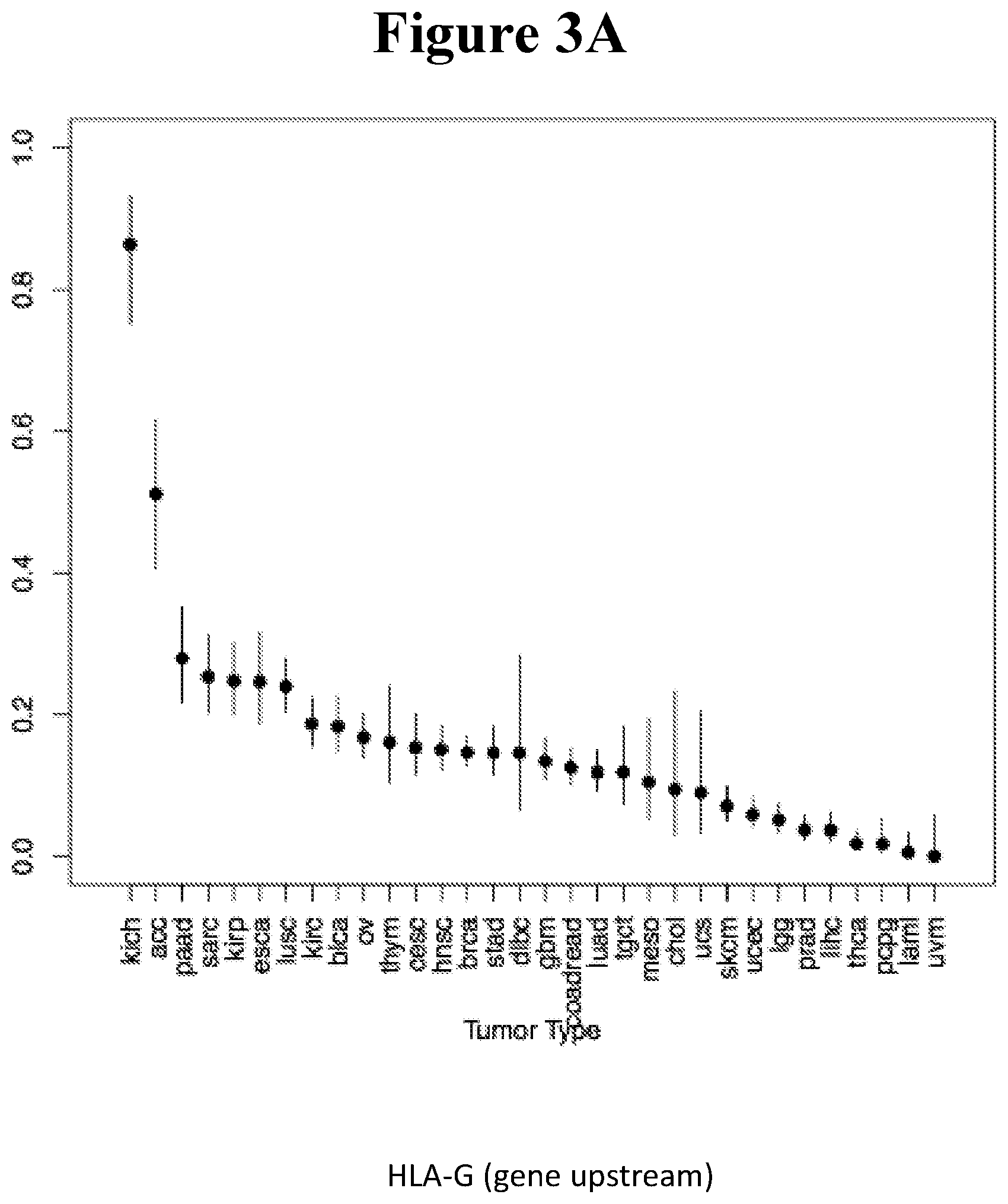
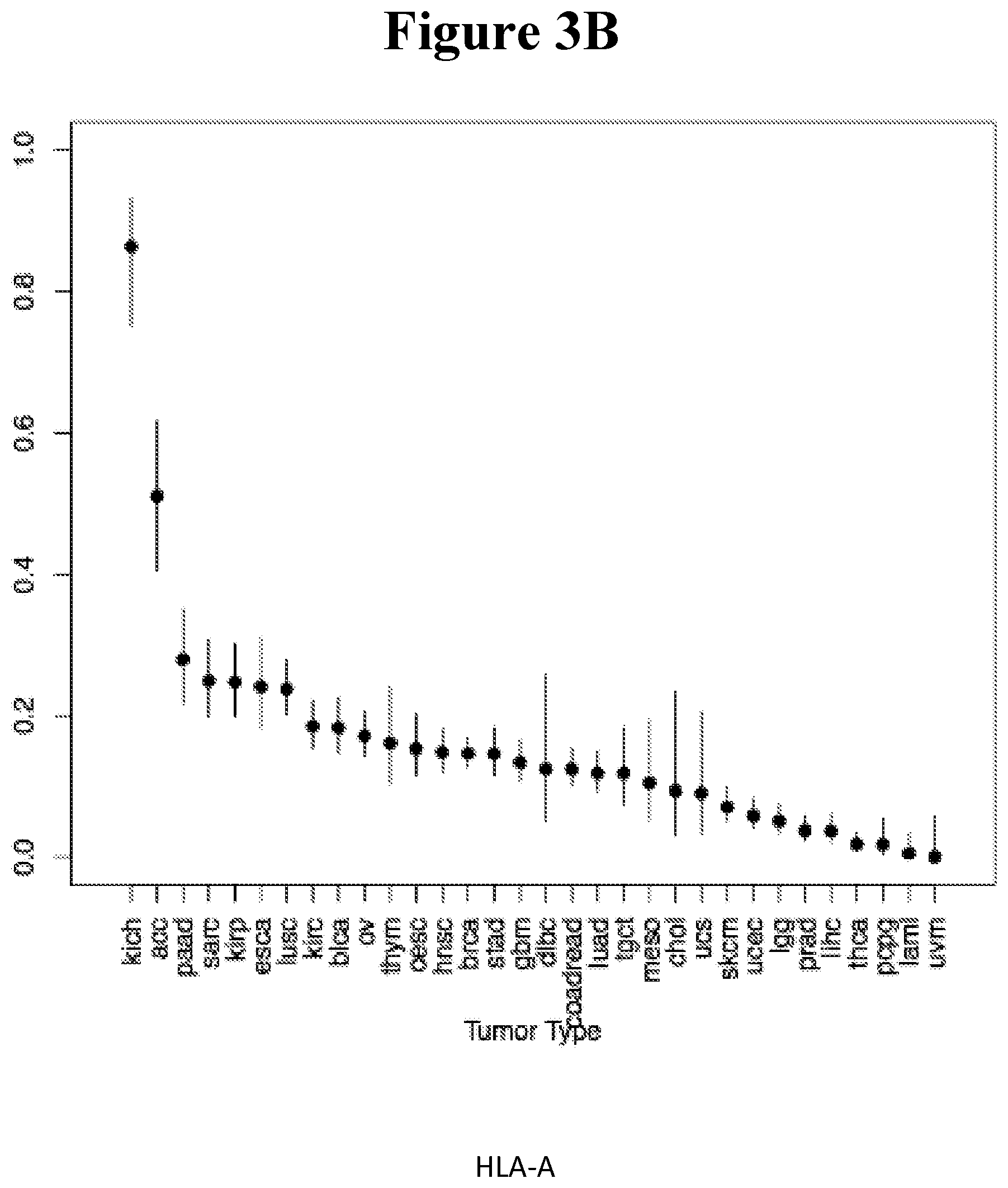
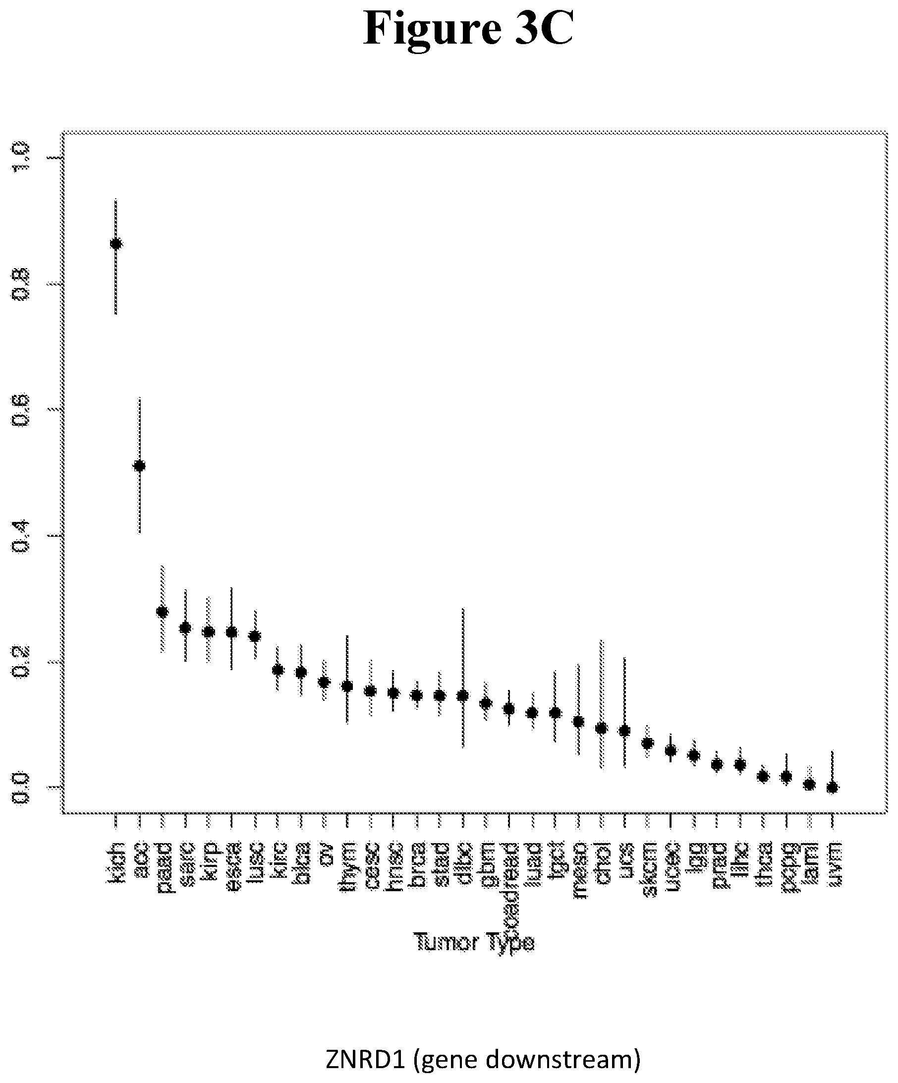
[0088] FIGS. 3A-C show the percentage of tumor samples undergoing LOH in the chromosomal region coding for the HLA class I locus. A. HLA-G, B. HLA-A, C. ZNRD1, in tumor types from the TCGA database. Kidney Chromophobe [KICH], Adrenocortical carcinoma [ACC], Pancreatic adenocarcinoma [PAAD], Sarcoma [SARC], Kidney renal papillary cell carcinoma [KIRP], Esophageal carcinoma [ESCA], Lung squamous cell carcinoma [LUSC], Kidney renal clear cell carcinoma [KIRC], Bladder Urothelial Carcinoma [BLCA], Ovarian serous cystadenocarcinoma [OV], Thymoma [THYM], Cervical squamous cell carcinoma and endocervical adenocarcinoma [CESC], Head and Neck squamous cell carcinoma [HNSC], Breast invasive carcinoma [BRCA], Stomach adenocarcinoma [STAD], Lymphoid Neoplasm Diffuse Large B-cell Lymphoma [DLBC], Glioblastoma multiforme [GBM], Colon adenocarcinoma [COAD], Rectum adenocarcinoma [READ], Lung adenocarcinoma [LUAD], Testicular Germ Cell Tumors [TGCT], Mesothelioma [MESO], Cholangiocarcinoma [CHOL], Uterine Carcinosarcoma [UCS], Skin Cutaneous Melanoma [SKCM], Uterine Corpus Endometrial Carcinoma [UCEC], Brain Lower Grade Glioma [LGG], Prostate adenocarcinoma [PRAD], Liver hepatocellular carcinoma [LIHC], Thyroid carcinoma [THCA], Pheochromocytoma and Paraganglioma [PCPG], Acute Myeloid Leukemia [LAML], Uveal Melanoma [UVM]
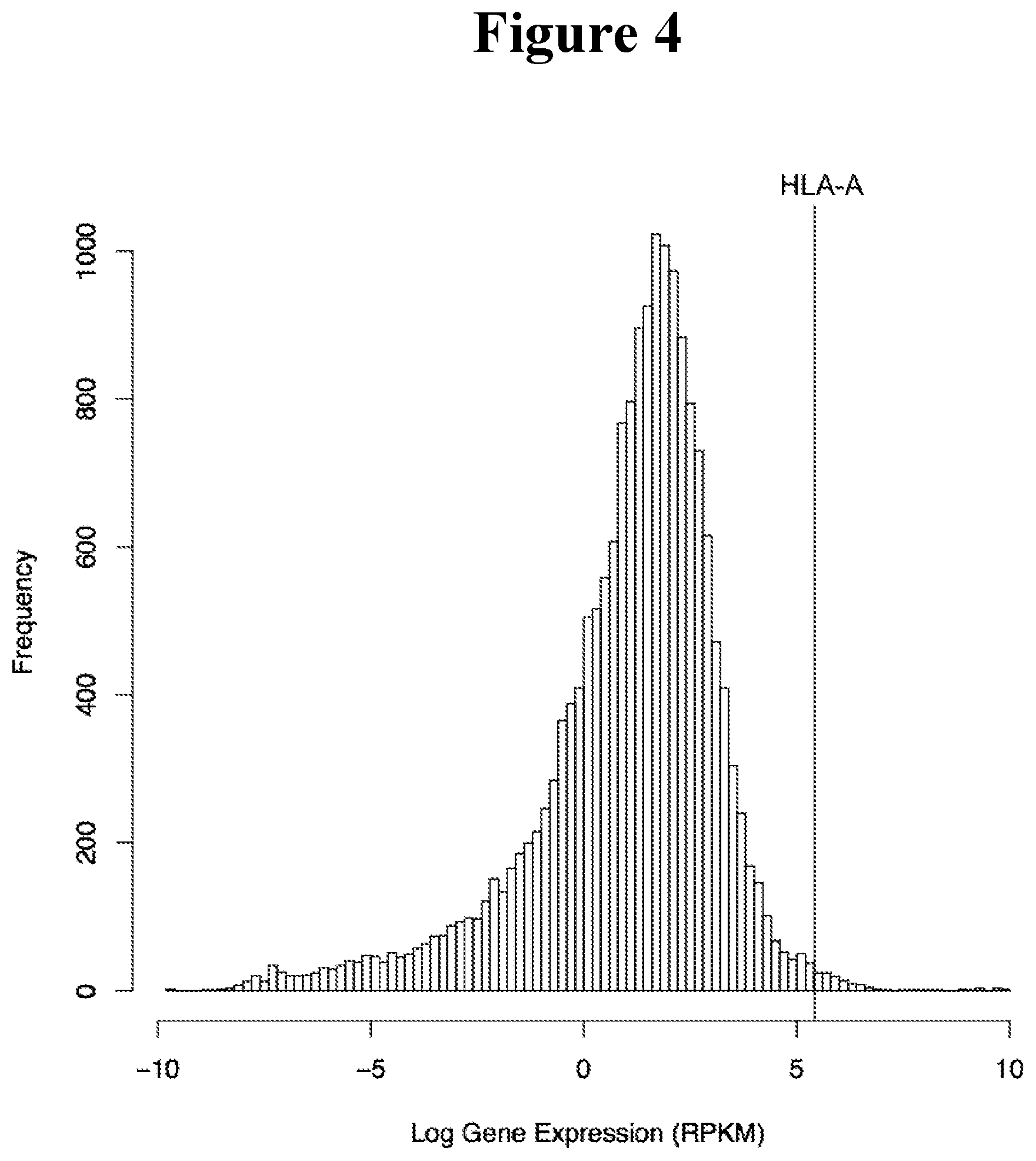
[0089] FIG. 4 shows expression of HLA-A relative to all other protein coding genes in the genome. The value for each gene reflects the mean RPKM value of tissue medians obtained from GTEX (gtexportal.org)
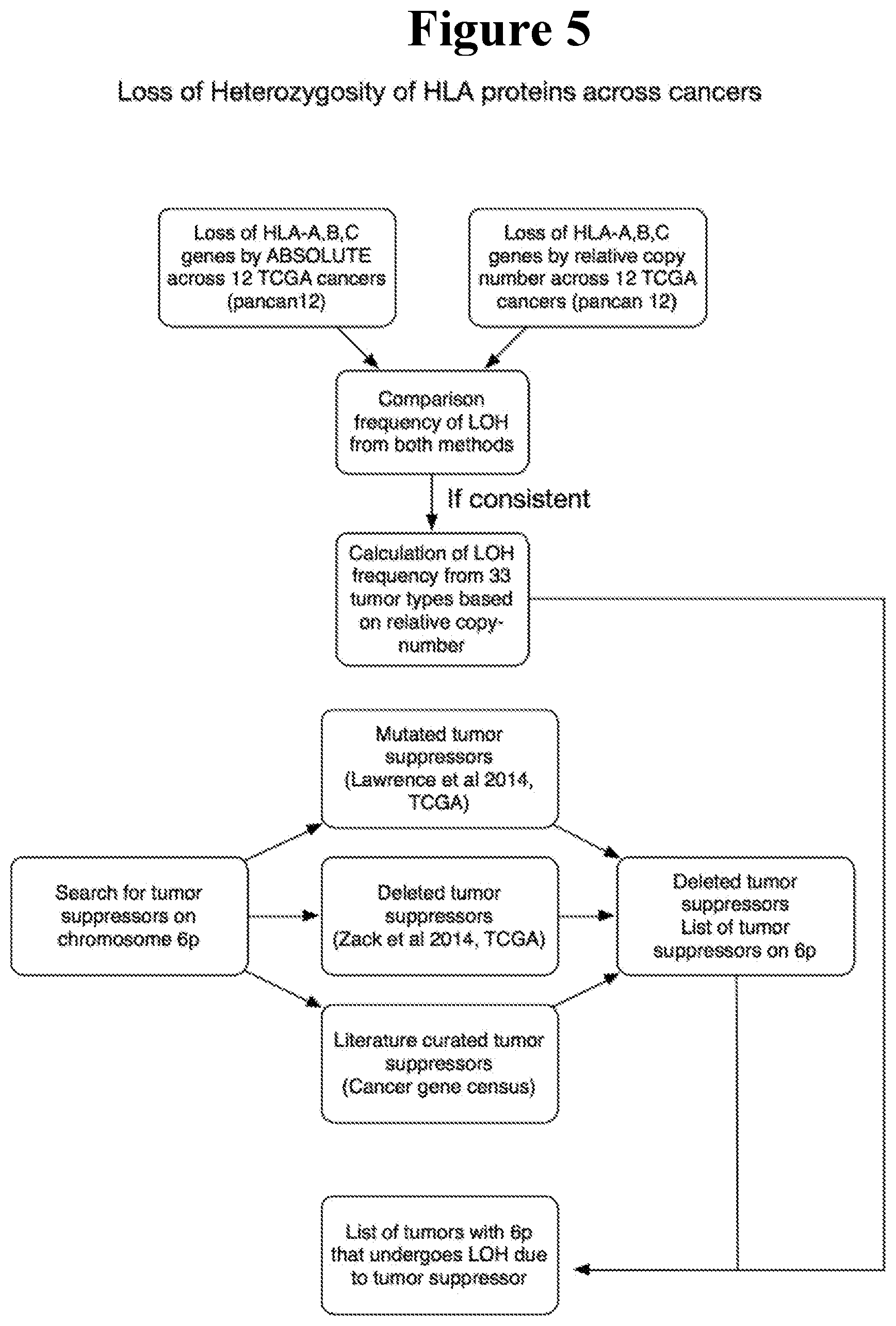
[0090] FIG. 5 shows a proposed workflow for analysis of HLA protein loss-of-heterozygosity across cancers in Example 5.
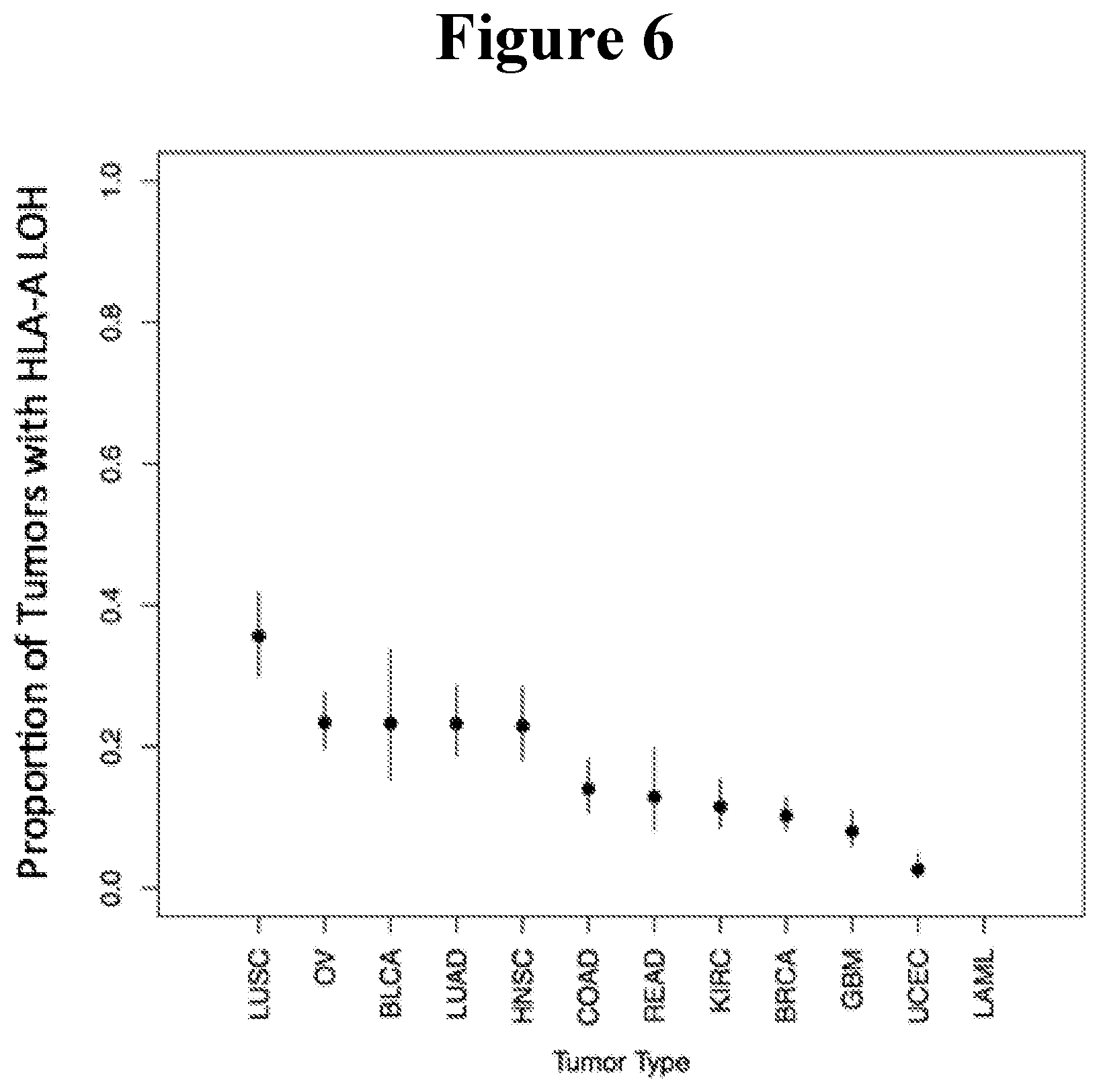
[0091] FIG. 6 shows Frequency of LOH in the pancan12 dataset using ABSOLUTE processed copy number data. Lines represent 95% binomial confidence intervals for frequency.
[0092] FIG. 7 shows the types of LOH observed in HLA-A. Of 588 episodes of HLA-A LOH, none involved a breakpoint within the HLA-A gene.
[0093] FIG. 8 shows the distribution of length (in basepairs) of deletions encompassing HLA-A. A large fraction of these deletions are greater than the length of chromosome 6p.
[0094] FIG. 9 shows the correlation between fraction of patients that have LOH of HLA-A in relative and ABSOLUTE copy number data with a threshold of -0.1.
[0095] FIG. 10A-10C shows the comparison of rate of LOH of HLA-A, HLA-B and HLA-C across 32 cancers reveals a nearly identical pattern of LOH.
[0096] FIG. 11 shows the IGV screenshot of AML copy number profiles sorted for deletion of chromosome 6p. Blue indicates deletion, red indicates amplification. There are no deletions of HLA-A.
[0097] FIG. 12 shows the proportion of uveal melanoma tumors undergoing LOH for all SNPs.
[0098] FIG. 13 provides the TCGA Study Abbreviations (also available at http s://gdc.cancer.gov/resources-tcga-users/tcga-code-tables/tcga-study-- abbreviations).
[0099] FIG. 14 depicts the loss of a chromosomal region adjacent to the tumor suppressor protein TP53, coded on chromosome 17. Genes coded on chromosome 17 which were identified as iCAR targets can be used to treat patient RC001.
[0100] FIG. 15 provides a schematic diagram of iCAR and aCAR constructs.
[0101] FIG. 16 provides data regarding IL-2 secretion as measured by ELISA. iCAR specifically inhibits IL-2 secretion upon interaction with target cells expressing iCAR target.
[0102] FIG. 17 shows that iCAR specifically inhibits IL-2 secretion upon interaction with target cells expressing iCAR target as measured by CBA.
[0103] FIG. 18 shows specific activation of CD19 aCAR Jurkat-NFAT by CD19 expressing target cells.
[0104] FIG. 19 shows specific inhibition of NFAT activation in CD19 aCAR/HLA-A2 iCAR Jurkat-NFAT
[0105] FIG. 20 shows specific inhibition of NFAT activation at different E/T ratios.
[0106] FIG. 21 provides the sequences for the iCAR and aCAR constructs of FIG. 15.
[0107] FIG. 22 provides the 1167 potential iCAR, pCAR and/or aCAR targets.
[0108] FIG. 23 provides the 3288 SNPs from the 1167 genes listed in FIG. 22.
DETAILED DESCRIPTION OF THE INVENTION
I. Introduction
[0109] Referring to the revolutionary concept of tumor suppressor genes (TSGs) that had been put forward in 1971 by A. G. Knudson (Knudson Jr., 1971), Devilee, Cleton-Jansen and Cornelisse stated in the opening paragraph of their essay titled `Ever since Knudson` (Devilee et al., 2001): "Many publications have documented LOH on many different chromosomes in a wide variety of tumors, implicating the existence of multiple TSGs. Knudson's two-hit hypothesis predicts that these LOH events are the second step in the inactivation of both alleles of a TSG". In their seminal review on genetic instabilities in human cancers (Lengauer et al., 1998), Lengauer, Kinzler and Vogelstein wrote: "Karyotypic studies have shown that the majority of cancers have lost or gained chromosomes, and molecular studies indicate that karyotypic data actually underestimate the true extent of such changes. Losses of heterozygosity, that is, losses of a maternal or paternal allele in a tumor, are widespread and are often accompanied by a gain of the opposite allele. A tumor could lose the maternal chromosome 8, for example, while duplicating the paternal chromosome 8, leaving the cell with a normal chromosome 8 karyotype but an abnormal chromosome 8 `allelotype`. The `average` cancer of the colon, breast, pancreas or prostate may lose 25% of its alleles and it is not unusual for a tumor to have lost over half of its alleles." These observations have since been reinforced and extended to almost all human cancers, including practically all carcinomas, in numerous reports (see (McGranahan et al., 2012) for review). It is now unambiguously established that nearly all individual tumors exhibit multiple losses of full chromosomes, entire chromosomal arms or sub-chromosomal regions of varying size. New algorithms are being rapidly developed (e.g., Sathirapongsasuti et al., 2011) for the determination of the LOH profile in any given cell sample based on the exome sequence data. While statistical bias may at present question the validity of some interpretations (Teo et al., 2012), such algorithms are likely to improve and replace most other methodologies for establishing LOH profiles which had been employed for this purpose in the pre-NGS era
[0110] Early LOH events can be detected in premalignant cells of the same tissue, but not in surrounding normal cells (Barrett et al., 1999). LOH is irreversible and events can only accumulate, so that tumor heterogeneity reflects the accumulation of losses throughout tumor progression. While tumor subclones can develop which differ in later LOH events, the existence of a minimal LOH signature that is shared by premalignant cells, putative tumor stem cells and all tumor subclones in a given patient, is expected to be the rule. Branches stemming from this `trunk` LOH pattern would still create a limited set of partially overlapping signatures which, together, cover all tumor cells in the same patient
[0111] An inevitable outcome of gross LOH events is the concomitant loss of all other genes residing on the deleted chromosomal material, and these naturally include many genes encoding transmembrane proteins. Concerning their identity, a catalog of 3,702 different human cell surface proteins (the `surfaceome`) has been compiled (Da Cunha et al., 2009). The expression of .quadrature.42% of surfaceome genes display broad tissue distribution while .quadrature.85 genes are expressed by all tissues examined, which is the hallmark of housekeeping genes. These genes are candidates, the different polymorphic variants of which may serve as targets for the iCARs and aCARs of the present invention
[0112] More recently, Bausch-Fluck et al. (Bausch-Fluck et al., 2015) applied their Chemoproteomic Cell Surface Capture technology to identify a combined set of 1492 cell surface glycoproteins in 41 human cell types. A large fraction of the surfaceome is expected to be expressed by any given tumor, each exhibiting a distinctive profile. Genes encoding cell surface proteins were found to be slightly enriched for single-nucleotide polymorphisms (SNPs) in their coding regions than all other genes (Da Cunha et al., 2009). Polymorphic in-frame insertions and deletions, which are rarer, further contribute to the number of variants and likely exert more robust structural effects on the polypeptide products than peptide sequence-altering (nonsynonymous) SNPs. Altogether, a typical genome contains 10,000 to 12,000 sites with nonsynonymous variants and 190-210 in-frame insertions/deletions (Abecasis et al., 2010; Auton et al., 2015). These variants are not evenly distributed throughout the genome as highly polymorphic genes such as the HLA locus (http://www.ebi.ac.uk/imgt/hla/stats.html) or certain G-protein-coupled receptor (GPCR) genes (Lee et al., 2003; Rana et al., 2001) create distinct variant `hotspots`. Another layer of LOH-related hotspots stems from the frequent loss of certain chromosomes, or chromosome arms in different cancers (e.g., 3p and 17p in small-cell lung carcinoma (Lindblad-Toh et al., 2000), 17p and 18q in colorectal cancer (Vogelstein et al., 1989), 17q and 19 in breast cancer (Li et al., 2014; Wang et al., 2004) 9p in melanoma (Stark and Hayward, 2007), 10q in glioblastoma (Ohgaki et al., 2004) and more)
[0113] A significant fraction of allelic variations in surface proteins would affect the extracellular portion of the respective gene products, potentially creating distinct allele-restricted epitopes which, in principle, can be recognized and distinguished from other variants by highly-specific mAbs. It is well documented that mAbs can be isolated that discriminate between two variants of the same protein which differ in a single amino acid only (see, for example, an early example of mAbs that recognize point mutation products of the Ras oncogene with exquisite specificity (Carney et al., 1986)). Interestingly, it was shown that two mAbs specific to a single amino acid interchange in a protein epitope can use structurally distinct variable regions from their heavy and light chain V gene pools (Stark and Caton, 1991). Recently, Skora et al. (Skora et al., 2015) reported the isolation of peptide-specific scFvs which can distinguish between HLA-I-bound neopeptides derived from mutated KRAS and EGFR proteins and their wild type counterparts, differing in both cases in one amino acid
[0114] All taken together, a unique antigenic signature of tumor cells emerges, that can allow their unequivocal discrimination from all other cells in the entire body of the individual patient. It comprises all transmembrane proteins encoded by allelic variants that are absent from the tumor cell surface owing to LOH but are present on normal cells of the cancer tissue of origin or other tissues expressing these genes. Naturally, each gene affected by LOH will be characterized by a distinct pattern of tissue distribution except for true housekeeping genes. The majority of these genes are not expected to be directly involved in tumorigenesis or maintenance of the transformed phenotype and, in this sense, their loss is of a `passenger` nature
[0115] The rationale presented above argues that a unique molecular portrayal is inevitably shaped by LOH for almost all tumors, which is marked by the absence of numerous polymorphic surface structures that are present on normal cells. Converting this postulated signature of the individual tumor to a targetable set of antigenic epitopes entails a practicable immunological strategy for translating the recognition of a particular `absence` into an activating cue capable of triggering target cell killing. Importantly, the incorporation of a safety device to assure that on-target off-tumor reactivity is strictly avoided will be highly favorable in future clinical implementation of this strategy
[0116] The present invention tackles this challenge through the co-expression in each therapeutic killer cell of a single pair of genes. One partner in this pair encodes an activating CAR (aCAR) and the other encodes a protecting CAR (pCAR) or an inhibitory CAR (iCAR)
II. Select Definitions
[0117] The term "nucleic acid molecule" as used herein refers to a DNA or RNA molecule.
[0118] The term "encoding" refers to the inherent property of specific sequences of nucleotides in a polynucleotide, such as a gene, a cDNA, or an mRNA, to serve as templates for synthesis of other polymers and macromolecules in biological processes having either a defined sequence of nucleotides (e.g., rRNA, tRNA and mRNA) or a defined sequence of amino acids and the biological properties resulting therefrom. Thus, a gene encodes a protein if transcription and translation of mRNA corresponding to that gene produces the protein in a cell or other biological system. Both the coding strand, the nucleotide sequence of which is identical to the mRNA sequence and is usually provided in sequence listings, and the non-coding strand, used as the template for transcription of a gene or cDNA, can be referred to as encoding the protein or other product of that gene or cDNA.
[0119] Unless otherwise specified, a "nucleotide sequence encoding an amino acid sequence" includes all nucleotide sequences that are degenerate versions of each other and that encode the same amino acid sequence. Nucleotide sequences that encode proteins and RNA may include introns.
[0120] The term "endogenous" refers to any material from or produced inside an organism, cell, tissue or system.
[0121] The term "exogenous" refers to any material introduced from or produced outside an organism, cell, tissue or system.
[0122] The term "expression" as used herein is defined as the transcription and/or translation of a particular nucleotide sequence driven by its promoter.
[0123] "Expression vector" refers to a vector comprising a recombinant polynucleotide comprising expression control sequences operatively linked to a nucleotide sequence to be expressed. An expression vector comprises sufficient cis-acting elements for expression; other elements for expression can be supplied by the host cell or in an in vitro expression system. Expression vectors include all those known in the art, such as cosmids, plasmids (e.g., naked or contained in liposomes) and viruses (e.g., lentiviruses, retroviruses, adenoviruses, and adeno-associated viruses) that incorporate the recombinant polynucleotide.
[0124] The term "genomic variant" as used herein refers to a change of at least one nucleotide at the genomic level in a sequenced sample compared to the reference or consensus sequence at the same genomic position.
[0125] The term "corresponding reference allele" as used herein with reference to a variant means the reference or consensus sequence or nucleotide at the same genomic position as the variant.
[0126] The term "extracellular domain" as used herein with reference to a protein means a region of the protein which is outside of the cell membrane.
[0127] The term "loss of heterozygosity" or "LOH" as used herein means the loss of chromosomal materials such as a complete chromosome or a part thereof, in one copy of the two chromosomes in a somatic cell.
[0128] The term "sequence region" as used herein with reference to a variant or a reference allele means a sequence starting upstream and ending downstream from the position of the variant, which can be translated into an "epitope peptide" that can be recognized by an antibody.
[0129] The term "CAR", as that term is used herein, refers to a chimeric polypeptide that shares structural and functional properties with a cell immune-function receptor or adaptor molecule, from e.g., a T cell or a NK cell. CARs include TCARs and NKR-CARs. Upon binding to cognate antigen, a CAR can activate or inactivate the cytotoxic cell in which it is disposed, or modulate the cell's antitumor activity or otherwise modulate the cells immune response.
[0130] The term "specific binding" as used herein in the context of an extracellular domain, such as an scFv, that specifically binds to a single allelic variant of a polymorphic cell surface epitope, refers to the relative binding of the scFv to one allelic variant and its failure to bind to the corresponding different allelic variant of the same polymorphic cell surface epitope. Since this depends on the avidity (number of CAR copies on the T cell, number of antigen molecules on the surface of target cells (or cells to be protected) and the affinity of the specific CARs used, a functional definition would be that the specific scFv would provide a significant signal in an ELISA against the single allelic variant of a polymorphic cell surface epitope to which it is specific or cells transfected with a CAR displaying the scFv would be clearly labeled with the single allelic variant of a polymorphic cell surface epitope in a FACS assay, while the same assays using the corresponding different allelic variant of the same polymorphic cell surface epitope would not give any detectable signal.
[0131] The term "treating" as used herein refers to means of obtaining a desired physiological effect. The effect may be therapeutic in terms of partially or completely curing a disease and/or symptoms attributed to the disease. The term refers to inhibiting the disease, e.g., arresting its development; or ameliorating the disease, e.g., causing regression of the disease.
[0132] As used herein, the terms "subject" or "individual" or "animal" or "patient" or "mammal," refers to any subject, particularly a mammalian subject, for whom diagnosis, prognosis, or therapy is desired, for example, a human.
[0133] The phrase "safe effector immune cell" or "safe effector cell" includes those cells described by the invention that express at least one iCAR or pCAR as described herein. In some embodiments, the "safe effector immune cell" or "safe effector cell" is capable of administration to a subject. In some embodiments, the "safe effector immune cell" or "safe effector cell" further expresses an aCAR as described herein. In some embodiments, the "safe effector immune cell" or "safe effector cell" further expresses an iCAR or a pCAR as described herein. In some embodiments, the "safe effector immune cell" or "safe effector cell" further expresses an iCAR or a pCAR as described herein and an aCAR as described herein.
[0134] Pharmaceutical compositions for use in accordance with the present invention may be formulated in conventional manner using one or more physiologically acceptable carriers or excipients. The carrier(s) must be "acceptable" in the sense of being compatible with the other ingredients of the composition and not deleterious to the recipient thereof.
[0135] The phrase "effective amount" or "therapeutically effective amount" are used interchangeably herein, and refer to an amount of a compound, formulation, material, or composition, as described herein effective to achieve a particular biological result.
[0136] The following exemplification of carriers, modes of administration, dosage forms, etc., are listed as known possibilities from which the carriers, modes of administration, dosage forms, etc., may be selected for use with the present invention. Those of ordinary skill in the art will understand, however, that any given formulation and mode of administration selected should first be tested to determine that it achieves the desired results.
[0137] Methods of administration include, but are not limited to, parenteral, e.g., intravenous, intraperitoneal, intramuscular, subcutaneous, mucosal (e.g., oral, intranasal, buccal, vaginal, rectal, intraocular), intrathecal, topical and intradermal routes. Administration can be systemic or local. In some embodiments, the pharmaceutical composition is adapted for oral administration.
[0138] The term "carrier" refers to a diluent, adjuvant, excipient, or vehicle with which the active agent is administered. The carriers in the pharmaceutical composition may comprise a binder, such as microcrystalline cellulose, polyvinylpyrrolidone (polyvidone or povidone), gum tragacanth, gelatin, starch, lactose or lactose monohydrate; a disintegrating agent, such as alginic acid, maize starch and the like; a lubricant or surfactant, such as magnesium stearate, or sodium lauryl sulphate; and a glidant, such as colloidal silicon dioxide.
[0139] The term "peripheral blood mononuclear cell (PBMC)" as used herein refers to any blood cell having a round nucleus, such as a lymphocyte, a monocyte or a macrophage. Methods for isolating PBMCs from blood are readily apparent to those skilled in the art. A non-limiting example is the extraction of these cells from whole blood using ficoll, a hydrophilic polysaccharide that separates layers of blood, with monocytes and lymphocytes forming a buffy coat under a layer of plasma or by leukapheresis, the preparation of leukocyte concentrates with the return of red cells and leukocyte-poor plasma to the donor.
[0140] The term "cancer" as used herein is defined as disease characterized by the rapid and uncontrolled growth of aberrant cells. Cancer cells can spread locally or through the bloodstream and lymphatic system to other parts of the body. Examples of various cancers include but are not limited to, breast cancer, prostate cancer, ovarian cancer, cervical cancer, skin cancer, pancreatic cancer, colorectal cancer, renal cancer, liver cancer, brain cancer, lymphoma, leukemia, lung cancer, glioma, and the like.
III. CAR-T System: iCARs, pCARs, and aCARs
[0141] It should be emphasized that the present invention provides a new avenue enabling specific targeting of tumor cells while keeping the normal cells secure. The concept presented herein provides for the identification of new targets for iCARs (or pCARs or protective CARs), these targets defined as comprising single allelic variants of polymorphic cell surface epitopes, which are lost from tumor cells due to LOH of the chromosomal region they reside in, while remaining expressed on normal tissue. Because of the polymorphic variation, it is possible to distinguish the two alleles and target only the allele missing in the tumor cells. Further, the target antigen may not necessarily itself be a tumor suppressor gene, or a gene predicted to be involved with cancer, since it is chosen for being in a region lost by LOH and could therefore simply be linked to such genes. This is conceptually different from the methods employed or suggested to date in cancer therapy, which target tumor associated antigens or antigens downregulated at tumors regardless of polymorphism. The present methods also provide for broadening the selection of aCAR beyond tumor associated anitgens, by conferring protection of normal cells through the co-expression of the iCAR and/or pCAR as described herein.
[0142] The distinction is crucial because the LOH, being a genomic event, results in a total loss of a specific variant from the tumor with a very rare probability of gaining back the lost allele. If the LOH event occurs very early in the development of tumors, it ensures a uniform target signature in all tumor cells derived from the initial pre-malignant tissue including metastatic tumors. Additionally, LOH occurs in almost all types of cancer and this concept can therefore be relied upon as a universal tool for developing markers relevant to all these cancer types. Since the LOH events are to some extent random, the present invention further provides for selection of personalized tumor markers for each individual cancer patient, based on the specific LOH events which took place in that patient. The tools relied upon to execute this concept, the aCARs and the iCARs, are well-known and can be easily prepared using methods well-known in the art as taught for example, in WO 2015/142314 and in U.S. Pat. No. 9,745,368, both incorporated by reference as if fully disclosed herein.
[0143] According to one strategy, the two CARs in every given pair specifically recognize the product of a different allelic variant of the same target gene for which the patient is heterozygous. The basic principle is as follows: the aCAR targets an allelic variant of a selected cell surface protein that is expressed by the given tumor cells and is not affected by LOH while the pCAR or iCAR targets the product encoded by the allelic variant of the same gene that has been lost from these tumor cells due to LOH. In other normal tissues of that individual patient that express the said gene, both alleles are present and are known to be equally functional, that is, expression is biallelic in all tissues (in contrast to other genes which may exhibit random monoallelic expression (Chess, 2012; Savova et al., 2016). In one scenario, the two CARs target two related epitopes residing at the same location on the protein product, which differ by one, or only few amino acids. In another scenario, the aCAR targets a non-polymorphic epitope on the same protein while the pCAR or iCAR is allele-specific. In this case the density of the aCAR epitope on normal cells would generally be two-fold higher than that of the iCAR or pCAR one. In some embodiments, a single nucleic acid vector encodes both the aCAR and iCAR or pCAR.
[0144] Another strategy utilizes as the pCAR or iCAR targets the protein products of housekeeping genes. Since, by definition, these genes are expressed on all cells in the body, they are safe targets for pCAR or iCARs. That is, if the pCAR or iCAR targets a membrane product of a housekeeping gene for which the given patient is heterozygous, all cells in the body, except the tumor cells which have lost this allele due to LOH, will be protected. This strategy allows for the uncoupling of the aCAR target gene product from the pCAR or iCAR one. In fact, the aCAR target can then be any non-polymorphic epitope expressed by the tumor. A variation of this strategy would be to utilize a known aCAR targeted to a non-polymorphic tumor-associated antigen, e.g., an aCAR in clinical use or under examination in clinical trials, in combination with an iCAR or pCAR directed against a membrane product of a gene for which the given patient is heterozygous and which is expressed in at least the tissue of origin of the tumor and preferably in additional vital normal tissues in which aCAR target antigen is expressed.
[0145] Following the same rationale which allows the uncoupling of the aCAR target antigen from the iCAR/pCAR one, the latter should not necessarily be the product of a housekeeping gene. In some embodiments, the iCAR and/or pCAR be the product of any gene the expression pattern of which is sufficiently wide so as to protect vital normal tissues expressing the aCAR target antigen in addition to the tumor. As a corollary, the aCAR antigen can be, as argued for housekeeping genes, any non-polymorphic epitope expressed by the tumor, not restricted to known `tumor-associated antigens`, a consideration which can vastly expand the list of candidate aCAR targets. In general, for both housekeeping and non-housekeeping genes, the identity of such normal vital tissues and level of expression would serve as important criteria in the prioritization of such candidate aCAR targets
[0146] Care must be taken to ensure that the inhibitory signal transmitted by the iCAR is strictly and permanently dominant over the aCAR signal and that no cross-recognition between the iCAR and the aCAR occurs. Dominance of the iCAR guarantees that activation of the killer cell upon encounter with normal cells expressing both alleles would be prevented. This default brake would, however, not operate upon engagement with tumor cells: in the absence of its target antigen the iCAR would not deliver inhibitory signals, thus unleashing the anticipated aCAR-mediated cellular activation and subsequent tumor cell lysis
[0147] The iCAR technology may be based on immune checkpoints. In this regard, the demonstration (Fedorov et al., 2013b; WO 2015/142314) that the regulatory elements of PD-1 and CTLA-4 possess a potent T cell inhibitory capacity when incorporated as iCAR signaling components is encouraging but the generality of these observations was recently questioned (Chicaybam and Bonamino, 2014, 2015). Furthermore, although the precise molecular pathways triggered by these checkpoint proteins are not fully understood, their engagement dampens T-cell activation through both proximal and distal mechanisms, rendering T cells unresponsive to concomitant activating stimuli (Nirschl and Drake, 2013). Hence, although the inactivation status secured by PD-1 and CTLA-4 iCARs is indeed temporary and reversible (Fedorov et al., 2013b), it would not allow T cell activation in tissues expressing both iCAR and aCAR targets. In contrast, the dominance of NK inhibitory receptors over activating receptors assures that healthy cells are spared from NK cell attack through a spatial, rather than temporal mechanism. (Long et al., 2013). There is compelling evidence that a single NK cell can spare a resistant cell expressing both inhibitory and activating ligands yet, kill a susceptible cell it simultaneously engages, which expresses only the activating ligands. This exquisite ability is governed by the different spatial organization of signal transduction molecules formed at each of the respective immune synapses which consequently affects the exocytosis of cytolytic granules (e.g., Abeyweera et al., 2011; Eriksson et al., 1999; Treanor et al., 2006; Vyas et al., 2001; U.S. Pat. No. 9,745,368).
[0148] The strategy based on the control asserted by iCARs depends on the dominance of the iCAR activity over the aCAR activity as explained above. In some embodiments, the present invention provides this type of iCAR, termed here a pCAR (for `protective CAR, see FIG. 2), designed to operate in CAR T cells in a synapse-selective manner and guarantee full dominance over the co-expressed aCAR. In some embodiments, the iCAR provided by the present invention is this particular type of iCAR referred to herein as a protective CAR (pCAR).
[0149] In some embodiments, the pCAR of the present invention integrates two technological feats. First, the pCAR allows for uncoupling the activating moiety of the aCAR (FcR.gamma./CD3.zeta.) from the recognition unit and the co-stimulatory element (e.g., CD28, 4-1BB, CD134 (OX40, GITR, IL2R.beta. and STAT3 binding motif (YXXQ)) by genetically placing them on two different polypeptide products. Recoupling of these elements, which is mandatory for the aCAR function, will only take place by the addition of a heterodimerizing drug which can bridge the respective binding sites incorporated onto each of the polypeptides separately (FIG. 2B). The reconstruction of a fully functional CAR by bridging similarly split recognition and activating moieties by virtue of a heterodimerizing drug has recently been reported by Wu et al. (Wu et al., 2015). For this purpose, these authors used the FK506 binding protein domain (FKBP, 104 amino acids) and the T2089L mutant of FKBP-rapamycin binding domain (FRB, 89 amino acids) that heterodimerize in the presence of the rapamycin analog AP21967 (Scheme I below). This drug possess 1000-fold less immunosuppressive activity compared to rapamycin (Bayle et al., 2006; Graef et al., 1997; Liberles et al., 1997) and is commercially available (ARGENT.TM., Regulated Heterodimerization Kit, ARIAD). In some embodiments, the drug is administered orally.
##STR00001##
[0150] Second, engrafting the pCAR recognition unit and the missing activating domain, respectively, onto the two surfaces of the transmembrane domain of a RIP-controlled receptor which contains the two intramembrane cleavage sites (FIG. 2A). Binding of the pCAR to its antigen will trigger dual cleavage of the encoded polypeptide first by a member of the extracellular disintegrin and metalloproteinase (ADAM) family which removes the ectodomain and then by intracellular .gamma.-secretase, which liberates the intracellular domain of the pCAR. This first cleavage event is predicted to disrupt the ability of the truncated aCAR to gain access to a functional, membrane-anchored configuration of its missing activating element, thus acquiring an operative mode (FIG. 2C). This principle was recently exploited in the development of new genetic switches designed to limit CAR T cell activity to simultaneous recognition of two different antigens on the tumor cell, applying either the Notch receptor (Morsut et al., 2016; Roybal et al., 2016b) or Epithelial cell adhesion molecule (EpCAM, Pizem, Y., M. Sc. thesis under the supervision of the Inventor), two well-studied receptors functioning through RIP. In these studies, binding of the RIP-based CAR to one antigen releases a genetically-engineered intracellular domain which translocates to the cell nucleus where it turns on the expression of the second CAR. Unlike the current invention which utilizes this process solely for disarming any potential aCAR activity in the presence of the protective antigen. In some embodiments, the first cleavage event disrupts the ability of the truncated aCAR to gain access to a functional, membrane-anchored configuration of its missing activating element, thus acquiring an operative mode.
[0151] The proposed mode of action described above is predicted to exert local effects so that only aCARs which reside in the same synapse are affected and are no more able to bind their antigen productively and form an immunological synapse. As a result, even when multiple interactions of the aCAR with large numbers of non-tumor cells are likely to take place, they are only expected to be transient and nonfunctional so that the cells are fully capable of further interactions.
[0152] Dominance of the pCARs over their aCARs counterparts is inherent to this system as function of the aCARs utterly depends on presence of the pCARs. Relative shortage of pCARs in a given T cell would render the aCARs non-functional due to lack of an activating domain. In some embodiments, a shortage of pCARs in a given T cell renders the aCARs non-functional due to lack of an activating domain.
[0153] It is critical that both the recognition domain and the activating one are localized to the plasma membrane (Wu et al., 2015). Therefore, the second cleavage, which detaches the activating domain from the plasma membrane, would render this domain nonfunctional and prevent unwanted cellular activation. In some embodiments, the recognition domain and the activating one are localized to the plasma membrane. In some embodiments, the second cleavage detaches the activating domain from the plasma membrane and renders this domain nonfunctional and prevents unwanted cellular activation.
[0154] The aCAR and pCAR are designed to function via mutually exclusive mechanisms. The ability of the pCAR to undergo cleavage does not depend on the strength of inhibitory signaling so no completion on signaling outcome will take place. As long as the pCARs are cleaved, the aCARs cannot function, regardless of relative avidity of their interactions with their respective antigens, a scenario which secures another crucial level of safety.
[0155] In some embodiments, the mammalian tissue is human tissue and in other embodiments the related mammalian normal tissue is normal tissue from which the tumor developed.
[0156] In some embodiments, the effector immune cell is a T cell, a natural killer cell or a cytokine-induced killer cell.
[0157] In some embodiments, the at least one signal transduction element capable of inhibiting an effector immune cell is homologous to a signal transduction element of an immune checkpoint protein, such as an immune checkpoint protein selected from the group consisting of PD1; CTLA4; BTLA; 2B4; CD160; CEACAM, such as CEACAM1; KIRs, such as KIR2DL1, KIR2DL2, KIR2DL3, KIR2DL4, KIR2DL5A, KIR2DL5B, KIR3DL1, KIR3DL2, KIR3DL3, LIR1, LIR2, LIR3, LIR5, LIR8 and CD94-NKG2A; LAG3; TIM3; V-domain Ig suppressor of T cell activation (VISTA); STimulator of INterferon Genes (STING); immunoreceptor tyrosine-based inhibitory motif (ITIM)-containing proteins, T cell immunoglobulin and ITIM domain (TIGIT), and adenosine receptor (e.g., A2aR). In some embodiments, the immune checkpoint protein is a negative immune regulator. In some embodiments, the negative immune regulator is selected from the group consisting of 2B4, LAG-3 and BTLA-4.
[0158] In some embodiments, immune checkpoint protein is a natural killer cell inhibitory receptor, e.g., KIRs, such as KIR2DL1, KIR2DL2, KIR2DL3, KIR2DL4, KIR2DL5A, KIR2DL5B, KIR3DL1, KIR3DL2, KIR3DL3; or a Leukocyte Ig-like receptor, such as LIR1, LIR2, LIR3, LIR5, LIR8; and CD94-NKG2A, a C-type lectin receptor which forms heterodimers with CD94 and contains 2 ITIMs.
[0159] The methods for preparing and using killer cell receptors in iCARs has been described in U.S. Pat. No. 9,745,368, incorporated by reference as if fully disclosed herein.
[0160] In some embodiments, the extracellular domain of any one of the above embodiments is fused through a flexible hinge and transmembrane canonic motif to said intracellular domain.
[0161] i. Target Identification: aCAR, iCAR and pCAR
[0162] The present invention provides methods for identification of aCAR, iCAR and/or pCAR targets based identification of candidate genes having extracellular polymorphic epitopes. In some embodiments, the aCAR can be directed at any extracellular protein expressed on the tumor tissue. In some embodiments, aCAR target is further expressed on non-tumor tissues and the iCAR target is also expressed on non-tumor tissues but is not expressed on tumor tissues.
[0163] In some embodiments, the method of identification of candidate genes includes first determining that the gene encodes a transmembrane protein comprising an extracellular polymorphic epitope. In some embodiments, the method of identification of candidate genes further includes determining that the gene has at least two expressed alleles. In some embodiments, these alleles exhibit at least one allelic variation. In some embodiments, the allelic variation includes, for example, the presence of one or more SNPs, insertions, and/or deletions. In some embodiments, the allelic variation found for the gene causes an amino acid change relative to the reference sequence in an extracellular region of the protein. In some embodiments, the gene is located in a chromosomal region which undergoes loss of heterozygosity (LOH). In some embodiments, the gene is located in a chromosomal region which undergoes loss of heterozygosity (LOH) in cancer. In some embodiments, the gene is expressed in a tissue-of-origin of a tumor type in which the corresponding region was found to undergo LOH. In some embodiments, the gene is expressed at least in one or more tissues that the aCAR is expressed in. In some embodiments, the iCAR or pCAR target is expressed in vital organ cells the aCAR is expressed in.
[0164] In some embodiments, the target for use in the iCAR and/or pCAR is selected based on identification of a gene having at least one extracellular polymorphic epitope and wherein said gene has at least two expressed alleles. In some embodiments, the target for use in the iCAR and/or pCAR is selected based on identification of a gene having located in a chromosomal region which undergoes loss of heterozygosity. In some embodiments, the target for use in the iCAR and/or pCAR is selected based on identification of a gene having located in a chromosomal region which undergoes loss of heterozygosity in cancer. In some embodiments, the score for a theoretical SNP is calculated and a threshold limit determined. For example, if only 32% of the SNPs had a tumor suppressor gene on the chromosome, then the percentile rank for having one would be 0.68. Further, for example, if the allele had a minor allele fraction of 0.49 (where 0.5 is the highest possible), then the percentile rank would be 0.99. If the rate of LOH was 0.10, and 75% of SNPs had more LOH than that, then the percentile rank would be 0.25. If the ratio of standard deviation of expression values across tissues to the median for the gene harboring this SNP was 1.3 and that is better than 90% of other genes, then the percentile rank is 0.9. The total score for this SNP would then be 0.68*0.99*0.25*0.9=0.15. In some embodiments, this LOH candidate score can be employed as one method for determining if a candidate gene is a suitable iCAR or pCAR target. In some embodiments, the target can be selected based on this LOH score. In some embodiments, the candidate gene is a determined to be suitable as an iCAR or pCAR target. LOH candidates based on an LOH candidate score of greater than 0.4.
[0165] In some embodiments, the target for use in the iCAR and/or pCAR is selected from a gene having at least one extracellular polymorphic epitope. In some embodiments, the target is a gene is located on chromosome 1, chromosome 2, chromosome 3, chromosome 4, chromosome 5, chromosome 6, chromosome 7, chromosome 8, chromosome 9, chromosome 10, chromosome 11, chromosome 12, chromosome 13, chromosome 14, chromosome 15, chromosome 16, chromosome 17, chromosome 18, chromosome 19, chromosome 20, chromosome 21, chromosome 22, or chromosome X.
[0166] In some embodiments, the gene comprising the extracellular polymorphic epitope is located on chromosome 1. In some embodiments, the target for use in the iCAR and/or pCAR is selected from the group consisting of ABCA4, ADAM30, AQP10, ASTN1, C1orf101, CACNA1S, CATSPER4, CD101, CD164L2, CD1A, CD1C, CD244, CD34, CD46, CELSR2, CHRNB2, CLCA2, CLDN19, CLSTN1, CR1, CR2, CRB1, CSF3R, CSMD2, ECE1, ELTD1, EMC1, EPHA10, EPHA2, EPHA8, ERMAP, FCAMR, FCER1A, FCGR1B, FCGR2A, FCGR2B, FCGR3A, FCRL1, FCRL3, FCRL4, FCRL5, FCRL6, GJB4, GPA33, GPR157, GPR37L1, GPR88, HCRTR1, IGSF3, IGSF9, IL22RA1, IL23R, ITGA10, KIAA1324, KIAA2013, LDLRAD2, LEPR, LGR6, LRIG2, LRP8, LRRC52, LRRC8B, LRRN2, LY9, MIA3, MR1, MUC1, MXRA8, NCSTN, NFASC, NOTCH2, NPR1, NTRK1, OPN3, OR10J1, OR10J4, OR10K1, OR10R2, OR10T2, OR10X1, OR11L1, OR14A16, OR1411, OR14K1, OR2AK2, OR2C3, OR2G2, OR2G3, OR2L2, OR2M7, OR2T12, OR2T27, OR2T1, OR2T3, OR2T29, OR2T33, OR2T34, OR2T35, OR2T3, OR2T4, OR2T5, OR2T6, OR2T7, OR2T8, OR2W3, OR6F1, OR6K2, OR6K3, OR6K6, OR6N1, OR6P1, OR6Y1, PDPN, PEARl, PIGR, PLXNA2, PTCH2, PTCHD2, PTGFRN, PTPRC, PTPRF, PTGFRN, PVRL4, RHBG, RXFP4, S1PR1, SCNN1D, SDC3, SELE, SELL, SELP, SEMA4A, SEMA6C, SLAMF7, SLAMF9, SLC2A7, SLC5A9, TACSTD2, TAS1R2, TIE1, TLR5, TMEM81, TNFRSF14, TNFRSF1B, TRABD2B, USH2A, VCAM1, and ZP4.
[0167] In some embodiments, the gene comprising the extracellular polymorphic epitope is located on chromosome 2. In some embodiments, the target for use in the iCAR and/or pCAR is selected from the group consisting of ABCG5, ALK, ASPRV1, ATRAID, CD207, CD8B, CHRNG, CLEC4F, CNTNAP5, CRIM1, CXCR1, DNER, DPP10, EDAR, EPCAM, GPR113, GPR148, GPR35, GPR39, GYPC, IL1RL1, ITGA4, ITGA6, ITGAV, LCT, LHCGR, LRP1B, LRP2, LY75, MARCO, MERTK, NRP2, OR6B2, PLA2R1, PLB1, PROKR1, PROM2, SCN7A, SDC1, SLC23A3, SLC5A6, TGOLN2, THSD7B, TM4SF20, TMEFF2, TMEM178A, TPO, and TRABD2A.
[0168] In some embodiments, the gene comprising the extracellular polymorphic epitope is located on chromosome 3. In some embodiments, the target for use in the iCAR and/or pCAR is selected from the group consisting of ACKR2, ALCAM, ANO10, ATP13A4, BTLA, CACNA1D, CACNA2D2, CACNA2D3, CASR, CCRL2, CD200, CD200R1, CD86, CD96, CDCP1, CDHR4, CELSR3, CHL1, CLDN11, CLDN18, CLSTN2, CSPG5, CX3CR1, CXCR6, CYP8B1, DCBLD2, DRD3, EPHA6, EPHB3, GABRR3, GP5, GPR128, GPR15, GPR27, GRM2, GRM7, HEG1, HTR3C, HTR3D, HTR3E, IGSF11, IL17RC, IL17RD, IL17RE, IL5RA, IMPG2, ITGA9, ITGB5, KCNMB3, LRIG1, LRRC15, LRRN1, MST1R, NAALADL2, NRROS, OR5AC1, OR5H1, OR5H14, OR5H15, OR5H6, OR5K2, OR5K3, OR5K4, PIGX, PLXNB1, PLXND1, PRRT3, PTPRG, ROBO2, RYK, SEMA5B, SIDT1, SLC22A14, SLC33A1, SLC4A7, SLITRK3, STAB1, SUSD5, TFRC, TLR9, TMEM108, TMEM44, TMPRSS7, TNFSF10, UPK1B, VIPR1, and ZPLD1.
[0169] In some embodiments, the gene comprising the extracellular polymorphic epitope is located on chromosome 4. In some embodiments, the target for use in the iCAR and/or pCAR is selected from the group consisting of ANTXR2, BTC, CNGA1, CORIN, EGF, EMCN, ENPEP, EPHA5, ERVMER34-1, EVC2, FAT1, FAT4, FGFRL1, FRAS1, GPR125, GRID2, GYPA, GYPB, KDR, KIAA0922, KLB, MFSD8, PARM1, PDGFRA, RNF150, TENM3, TLR10, TLR1, TLR6, TMEM156, TMPRSS11A, TMPRSS11B, TMPRSS11E, TMPRSS11F, UGT2A1, and UNC5C.
[0170] In some embodiments, the gene comprising the extracellular polymorphic epitope is located on chromosome 5. In some embodiments, the target for use in the iCAR and/or pCAR is selected from the group consisting of ADAM19, ADRB2, BTNL3, BTNL8, BTNL9, C5orfl5, CATSPER3, CD180, CDH12, CDHR2, COL23A1, CSF1R, F2RL2, FAM174A, FAT2, FGFR4, FLT4, GABRA6, GABRG2, GPR151, GPR98, GRM6, HAVCR1, HAVCR2, IL31RA, IL6ST, IL7R, IQGAP2, ITGA1, ITGA2, KCNMB1, LIFR, LNPEP, MEGF10, NIPAL4, NPR3, NRG2, OR2V1, OR2Y1, OSMR, PCDH12, PCDH1, PCDHA1, PCDHA2, PCDHA4, PCDHA8, PCDHA9, PCDHB 10, PCDHB11, PCDHB13, PCDHB14, PCDHB 15, PCDHB16, PCDHB2, PCDHB3, PCDHB4, PCDHB5, PCDHB6, PCDHGA1, PCDHGA4, PDGFRB, PRLR, SEMA5A, SEMA6A, SGCD, SLC1A3, SLC22A4, SLC22A5, SLC23A1, SLC36A3, SLC45A2, SLC6A18, SLC6A19, SLCO6A1, SV2C, TENM2, TIMD4, and UGT3A1.
[0171] In some embodiments, the gene comprising the extracellular polymorphic epitope is located on chromosome 6. In some embodiments, the target for use in the iCAR and/or pCAR is selected from the group consisting of BAI3, BTN1A1, BTN2A1, BTN2A2, BTN3A1, BTN3A2, BTNL2, CD83, DCBLD1, DLL1, DPCR1, ENPP1, ENPP3, ENPP4, EPHA7, GABBR1, GABRR1, GCNT6, GFRAL, GJB7, GLP1R, GPR110, GPR111, GPR116, GPR126, GPR63, GPRC6A, HFE, HLA-A, HLA-B, HLA-C, HLA-DOA, HLA-DPA1, HLA-DPB1, HLA-DQA1, HLA-DQA2, HLA-DQB 1, HLA-DQB2, HLA-DRB 1, HLA-DRB5, HLA-E, HLA-F, HLA-G, IL20RA, ITPR3, KIAA0319, LMBRD1, LRFN2, LRP11, MAS1L, MEP1A, MICA, MICB, MOG, MUC21, MUC22, NCR2, NOTCH4, OPRM1, OR10C1, OR12D2, OR12D3, OR14J1, OR2B2, OR2B6, OR2J1, OR2W1, OR5V1, PDE10A, PI16, PKHD1, PTCRA, PTK7, RAET1E, RAETIG, ROS1, SDIM1, SLC16A10, SLC22A1, SLC44A4, TAAR2, TREM1, TREML1, and TREML2. In some embodiments, the gene comprising the extracellular polymorphic epitope is located on chromosome 6 and comprises an HLA target. In some embodiments, the target for use in the iCAR and/or pCAR is HLA-A, HLA-B, HLA-C, HLA-DOA, HLA-DPA1, HLA-DPB 1, HLA-DQA1, HLA-DQA2, HLA-DQB1, HLA-DQB2, HLA-DRB1, HLA-DRB5, HLA-E, HLA-F, HLA-G. In some embodiments, the target for use in the iCAR and/or pCAR is HLA-A2,
[0172] In some embodiments, the gene comprising the extracellular polymorphic epitope is located on chromosome 7. In some embodiments, the target for use in the iCAR and/or pCAR is selected from the group consisting of AQP1, C7orf50, CD36, CDHR3, CNTNAP2, DPP6, EGFR, EPHA1, EPHB6, ERVW-1, GHRHR, GJC3, GPNMB, GRM8, HUS1, HYAL4, KIAA1324L, LRRN3, MET, MUC12, MUC17, NPC1L1, NPSR1, OR2A12, OR2A14, OR2A25, OR2A42, OR2A7, OR2A2, OR2AE1, OR2F2, OR6V1, PILRA, PILRB, PKD1L1, PLXNA4, PODXL, PTPRN2, PTPRZ 1, RAMP3, SLC29A4, SMO, TAS2R16, TAS2R40, TAS2R4, TFR2, THSD7A, TMEM213, TTYH3, ZAN, and ZP3.
[0173] In some embodiments, the gene comprising the extracellular polymorphic epitope is located on chromosome 8. In some embodiments, the target for use in the iCAR and/or pCAR is selected from the group consisting of ADAM18, ADAM28, ADAM32, ADAM7, ADAM9, ADRA1A, CDH17, CHRNA2, CSMD1, CSMD3, DCSTAMP, FZD6, GPR124, NRG1, OR4F21, PKHD1L1, PRSS55, SCARA3, SCARA5, SDC2, SLC10A5, SLC39A14, SLC39A4, SLCO5A1, TNFRSF10A, and TNFRSF 10B.
[0174] In some embodiments, the gene comprising the extracellular polymorphic epitope is located on chromosome 9. In some embodiments, the target for use in the iCAR and/or pCAR is selected from the group consisting of ABCA1, AQP7, ASTN2, C9orf135, CA9, CD72, CNTNAP3B, CNTNAP3, CRB2, ENTPD8, GPR144, GRIN3A, IZUMO3, KIAA1161, MAMDC4, MEGF9, MUSK, NOTCH1, OR13C2, OR13C3, OR13C5, OR13C8, OR13C9, OR13D1, OR13F1, OR1B1, OR1J2, OR1K1, OR1L1, OR1L3, OR1L6, OR1L8, OR1N1, OR1N2, OR1Q1, OR2S2, PCSK5, PDCD1LG2, PLGRKT, PTPRD, ROR2, SEMA4D, SLC31A1, TEK, TLR4, TMEM2, and VLDLR.
[0175] In some embodiments, the gene comprising the extracellular polymorphic epitope is located on chromosome 10. In some embodiments, the target for use in the iCAR and/or pCAR is selected from the group consisting of ABCC2, ADAM8, ADRB1, ANTXRL, ATRNL1, C10orf54, CDH23, CDHR1, CNNM2, COL13A1, COL17A1, ENTPD1, FZD8, FGFR2, GPR158, GRID1, IL15RA, IL2RA, ITGA8, ITGB1, MRC1, NRG3, NPFFR1, NRP1, OPN4, PCDH15, PKD2L1, PLXDC2, PRLHR, RET, RGR, SLC16A9, SLC29A3, SLC39A12, TACR2, TCTN3, TSPAN15, UNC5B, and VSTM4.
[0176] In some embodiments, the gene comprising the extracellular polymorphic epitope is located on chromosome 11. In some embodiments, the target for use in the iCAR and/or pCAR is selected from the group consisting of AMICA1, ANO1, ANO3, APLP2, C11orf24, CCKBR, CD248, CD44, CD5, CD6, CD82, CDON, CLMP, CRTAM, DCHS1, DSCAML1, FAT3, FOLH1, GDPD4, GDPD5, GRIK4, HEPHL1, HTR3B, IFITM10, IL10RA, KIRREL3, LGR4, LRP4, LRP5, LRRC32, MCAM, MFRP, MMP26, MPEG1, MRGPRE, MRGPRF, MRGPRX2, MRGPRX3, MRGPRX4, MS4A4A, MS4A6A, MTNR1B, MUC15, NAALAD2, NAALADL1, NCAM1, NRXN2, OR10A2, OR10A5, OR10A6, OR10D3, OR10G4, OR10G7, OR10G8, OR10G9, OR10Q1, OR10S1, OR1S1, OR2AG1, OR2AG2, OR2D2, OR4A47, OR4A15, OR4A5, OR4C11, OR4C13, OR4C15, OR4C16, OR4C3, OR4C46, OR4C5, OR4D6, OR4A8P, OR4D9, OR4S2, OR4X1, OR51E1, OR51L1, OR52A1, OR52E1, OR52E2, OR52E4, OR52E6, OR52I1, OR52I2, OR52J3, OR52L1, OR52N1, OR52N2, OR52N4, OR52W1, OR56B1, OR56B4, OR5A1, OR5A2, OR5AK2, OR5AR1, OR5B17, OR5B3, OR5D14, OR5D16, OR5D18, OR5F1, OR5I1, OR5L2, OR5M11, OR5M3, OR5P2, OR5R1, OR5T2, OR5T3, OR5W2, OR6A2, OR6T1, OR6X1, OR8A1, OR8B12, OR8B2, OR8B3, OR8B4, OR8D1, OR8D2, OR8H1, OR8H2, OR8H3, OR8I2, OR8J1, OR8J2, OR8J3, OR8K1, OR8K3, OR8K5, OR8U1, OR9G1, OR9G4, OR9Q2, P2RX3, PTPRJ, ROBO3, SIGIRR, SLC22A10, SLC3A2, SLC5A12, SLCO2B1, SORL1, ST14, SYT8, TENM4, TMEM123, TMEM225, TMPRSS4, TMPRSS5, TRIM5, TRPM5, TSPAN18, and ZP1.
[0177] In some embodiments, the gene comprising the extracellular polymorphic epitope is located on chromosome 12. In some embodiments, the target for use in the iCAR and/or pCAR is selected from the group consisting of ANO4, AVPR1A, BCL2L14, CACNA2D4, CD163, CD163L1, CD27, CD4, CLEC12A, CLEC1B, CLEC2A, CLEC4C, CLEC7A, CLECL1, CLSTN3, GPR133, GPRC5D, ITGA7, ITGB7, KLRB1, KLRC2, KLRC3, KLRC4, KLRF1, KLRF2, LRP1, LRP6, MANSC1, MANSC4, OLR1, OR10AD1, OR10P1, OR2AP1, OR6C1, OR6C2, OR6C3, OR6C4, OR6C6, OR6C74, OR6C76, OR8S1, OR9K2, ORAI1, P2RX4, P2RX7, PRR4, PTPRB, PTPRQ, PTPRR, SCNN1A, SELPLG, SLC2A14, SLC38A4, SLC5A8, SLC6A15, SLC8B1, SLCO1A2, SLCO1B1, SLCO1B7, SLCO1C1, SSPN, STAB2, TAS2R10, TAS2R13, TAS2R14, TAS2R20, TAS2R30, TAS2R31, TAS2R42, TAS2R43, TAS2R46, TAS2R7, TMEM119, TMEM132B, TMEM132C, TMEM132D, TMPRSS12, TNFRSF1A, TSPAN8, and VSIG10.
[0178] In some embodiments, the gene comprising the extracellular polymorphic epitope is located on chromosome 13. In some embodiments, the target for use in the iCAR and/or pCAR is selected from the group consisting of ATP4B, ATP7B, FLT3, FREM2, HTR2A, KL, PCDH8, RXFP2, SGCG, SHISA2, SLC15A1, SLITRK6, and TNFRSF 19.
[0179] In some embodiments, the gene comprising the extracellular polymorphic epitope is located on chromosome 14. In some embodiments, the target for use in the iCAR and/or pCAR is selected from the group consisting of ADAM21, BDKRB2, C14orf37, CLEC14A, DLK1, FLRT2, GPR135, GPR137C, JAG2, LTB4R2, MMP14, OR11G2, OR11H12, OR11H6, OR4K1, OR4K15, OR4K5, OR4L1, OR4N2, OR4N5, SLC24A4, and SYNDIG1L.
[0180] In some embodiments, the gene comprising the extracellular polymorphic epitope is located on chromosome 15. In some embodiments, the target for use in the iCAR and/or pCAR is selected from the group consisting of ANPEP, CD276, CHRNA7, CHRNB4, CSPG4, DUOX1, DUOX2, FAM174B, GLDN, IGDCC4, ITGA11, LCTL, LTK, LYSMD4, MEGF11, NOX5, NRG4, OCA2, OR4F4, OR4M2, OR4N4, PRTG, RHCG, SCAMP5, SEMA4B, SEMA6D, SLC24A1, SLC24A5, SLC28A1, SPG11, STRA6, TRPM1, and TYRO3.
[0181] In some embodiments, the gene comprising the extracellular polymorphic epitope is located on chromosome 16. In some embodiments, the target for use in the iCAR and/or pCAR is selected from the group consisting of ATP2C2, CACNA1H, CD19, CDH11, CDH15, CDH16, CDH3, CDH5, CNGB1, CNTNAP4, GDPD3, GPR56, GPR97, IFT140, IL4R, ITFG3, ITGAL, ITGAM, ITGAX, KCNG4, MMP15, MSLNL, NOMO1, NOMO3, OR2C1, PIEZO1, PKD1, PKD1L2, QPRT, SCNN1B, SEZ6L2, SLC22A31, SLC5A11, SLC7A6, SPN, TMC5, TMC7, TMEM204, TMEM219, and TMEM8A.
[0182] In some embodiments, the gene comprising the extracellular polymorphic epitope is located on chromosome 17. In some embodiments, the target for use in the iCAR and/or pCAR is selected from the group consisting of ABCC3, ACE, AOC3, ARL17B, ASGR2, C17orf80, CD300A, CD300C, CD300E, CD300LF, CD300LG, CHRNB1, CLEC10A, CNTNAP1, CPD, CXCL16, ERBB2, FAM171A2, GCGR, GLP2R, GP1BA, GPR142, GUCY2D, ITGA2B, ITGA3, ITGAE, ITGB3, KCNJ12, LRRC37A2, LRRC37A3, LRRC37A, LRRC37B, MRC2, NGFR, OR1A2, OR1D2, OR1G1, OR3A1, OR3A2, OR4D1, OR4D2, RNF43, SCARF1, SCN4A, SDK2, SECTM1, SEZ6, SHPK, SLC26A11, SLC5A10, SPACA3, TMEM102, TMEM132E, TNFSF12, TRPV3, TTYH2, and TUSC5.
[0183] In some embodiments, the gene comprising the extracellular polymorphic epitope is located on chromosome 18. In some embodiments, the target for use in the iCAR and/or pCAR is selected from the group consisting of APCDD1, CDH19, CDH20, CDH7, COLEC12, DCC, DSC1, DSG1, DSG3, DYNAP, MEP1B, PTPRM, SIGLEC15, and TNFRSF11A.
[0184] In some embodiments, the gene comprising the extracellular polymorphic epitope is located on chromosome 19. In some embodiments, the target for use in the iCAR and/or pCAR is selected from the group consisting of ABCA7, ACPT, BCAM, C19orf38, C19orf59, C5AR1, CATSPERD, CATSPERG, CD22, CD320, CD33, CD97, CEACAM19, CEACAM1, CEACAM21, CEACAM3, CEACAM4, CLEC4M, DLL3, EMR1, EMR2, EMR3, ERVV-1, ERVV-2, FAM187B, FCAR, FFAR3, FPR1, FXYD5, GFY, GP6, GPR42, GRIN3B, ICAM3, IGFLR1, IL12RB1, IL27RA, KIR2DL1, KIR2DL3, KIR2DL4, KIR3DL1, KIR3DL2, KIR3DL3, KIRREL2, KISS1R, LAIR1, LDLR, LILRA1, LILRA2, LILRA4, LILRA6, LILRB1, LILRB2, LILRB3, LILRB4, LILRB5, LINGO3, LPHN1, LRP3, MADCAM1, MAG, MEGF8, MUC16, NCR1, NOTCH3, NPHS1, OR10H1, OR10H2, OR10H3, OR10H4, ORII1, OR2Z1, OR7A10, OR7C1, OR7D4, OR7E24, OR7G1, OR7G2, OR7G3, PLVAP, PTGIR, PTPRH, PTPRS, PVR, SCN1B, SHISA7, SIGLEC10, SIGLEC11, SIGLEC12, SIGLEC5, SIGLEC6, SIGLEC8, SIGLEC9, SLC44A2, SLC5A5, SLC7A9, SPINT2, TARM1, TGFBR3L, TMC4, TMEM91, TMEM161A, TMPRSS9, TNFSF14, TNFSF9, TRPM4, VN1R2, VSIG10L, VSTM2B, and ZNRF4.
[0185] In some embodiments, the gene comprising the extracellular polymorphic epitope is located on chromosome 20. In some embodiments, the target for use in the iCAR and/or pCAR is selected from the group consisting of ABHD12, ADAM33, ADRAID, APMAP, ATRN, CD40, CD93, CDH22, CDH26, CDH4, FLRT3, GCNT7, GGT7, JAG1, LRRN4, NPBWR2, OCSTAMP, PTPRA, PTPRT, SEL1L2, SIGLEC1, SIRPA, SIRPB1, SIRPG, SLC24A3, SLC2A10, SLC4A11, SSTR4, and THBD.
[0186] In some embodiments, the gene comprising the extracellular polymorphic epitope is located on chromosome 21. In some embodiments, the target for use in the iCAR and/or pCAR is selected from the group consisting of CLDN8, DSCAM, ICOSLG, IFNAR1, IFNGR2, IGSF5, ITGB2, KCNJ15, NCAM2, SLC19A1, TMPRSS15, TMPRSS2, TMPRSS3, TRPM2, and UMODL1.
[0187] In some embodiments, the gene comprising the extracellular polymorphic epitope is located on chromosome 22. In some embodiments, the target for use in the iCAR and/or pCAR is selected from the group consisting of CACNAII, CELSR1, COMT, CSF2RB, GGT1, GGT5, IL2RB, KREMEN1, MCHR1, OR11H1, P2RX6, PKDREJ, PLXNB2, SCARF2, SEZ6L, SSTR3, SUSD2, TMPRSS6, and TNFRSF13C.
[0188] In some embodiments, the gene comprising the extracellular polymorphic epitope is located on chromosome X. In some embodiments, the target for use in the iCAR and/or pCAR is selected from the group consisting of ATP6AP2, ATP7A, CNGA2, EDA2R, FMR1NB, GLRA4, GPR112, GUCY2F, HEPH, P2RY10, P2RY4, PLXNA3, PLXNB3, TLR8, VSIG4, and XG.
[0189] In some embodiments, the aCAR used to treat the cancer is directed against or specifically binds to any membrane protein which is expressed on the tumor tissue as long as the iCAR is expressed on every normal tissue in which the targeted protein is expressed. In some embodiments, the aCAR can specifically bind or be directed to a tumor associated protein, tumor associated antigen and/or antigens in clinical trials, a CAR target as listed in Table 1, as well as any cell surface protein that is expressed in a tumor tissue to which an iCAR can be matched or paired with regard to target binding, according to the criteria listed in the application. In some embodiments, the aCAR can be any expressed protein with an extracellular domain, as long as the iCAR is expressed in the same tissues as the aCAR or in any vital tissues, but is lost in the tumor cells. In some embodiments, the aCAR used to treat the cancer, such as any one of the cancer types recited above, is directed against or specifically binds to, a non-polymorphic cell surface epitope selected from the antigens listed in Table 1, such as CD19. In some embodiments, the aCAR, iCAR, and/or pCAR target is any target with an extracellular domain. In some embodiments, the aCAR used to treat the cancer, is directed against or specifically binds to, a non-polymorphic cell surface epitope selected from but not limited to the following list of antigens: CD19, CD20, CD22, CD10, CD7, CD49f, CD56, CD74, CAIX IgK, ROR1, ROR2, CD30, LewisY, CD33, CD34, CD38, CD123, CD28, CD44v6, CD44, CD41, CD133, CD138, NKG2D-L, CD139, BCMA, GD2, GD3, hTERT, FBP, EGP-2, EGP-40, FR-.alpha., L1-CAM, ErbB2,3,4, EGFRvIII, VEGFR-2, IL-13R.alpha.2, FAP, Mesothelin, c-MET, PSMA, CEA, kRas, MAGE-A1, MUC1MUC16, PDL1, PSCA, EpCAM, FSHR, AFP, AXL, CD80 CD89, CDH17, CLD18, GPC3, TEM8, TGFB1, NY-ESO-1 WT-1 and EGFR. In some embodiments, the aCAR, iCAR, and/or pCAR target is an antigen listed in Table 1.
TABLE-US-00001 TABLE 1 CAR target antigens, including some evaluated in trials registered in ClinicalTrials.gov Antigen Key structural/functional features Malignancy Potential off-tumor targets Hematologic CD19 Pan-B cell marker involved in signal ALL, CLL, NHL, normal B cells malignancies transduction by the BCR HL, PLL CD20 Tetra-transmembrane, regulation of CLL, NHL normal B cells Ca transport and B-cell activation CD22 B-lineage specific adhesion receptor, ALL, NHL normal B cells sialic acid-binding Ig-type lectin family Ig.kappa. Ig light chain isotype expressed by CLL, NHL, MM normal B cells approx. 65% of normal human B cells ROR1 Type I orphan-receptor tyrosine- CLL, NHL pancreas; adipose cells kinase-like, survival-signaling receptor in tumors CD30 TNFR member, pleiotropic effects on NHL, TCL, HL resting CD8 T cells; activated B cell growth and survival involving and Th2 cells NF-.kappa.B Lewis.sup.Y (CD174) a membrane AML, MM early myeloid progenitor cells oligosaccharide harboring two fucose groups CD33 Sialic acid-binding Ig-type lectin AML hematopoietic progenitors; serving as adhesion molecule of the myelo-monocytic precursors; myelomonocytic lineage monocytes CD123 The .alpha. chain of the IL-3 receptor AML BM myeloid progenitors; DCs, B cells; mast cells, monocytes; macro-phages; megakar.; endothelial cells NKG2D-L Ligands for the NK and T-cell AML, MM gastrointestinal epithelium, activating receptor NKG2D, bearing endothelial cells and fibroblasts; similarity to MHC-I molecules; upregulated during inflammation CD139 Syndecan-1, cell surface heparan MM precursor & plasma B cells; sulfate proteoglycan, ECM receptor epithelia BCMA TNFR member, binds BAFF and MM B cells APRIL, involved in proliferation signaling TACI MM Mono-nuclear cells, heart Solid GD2 Disialoganglioside NB; sarcomas; solid skin; neurons tumors tumors FR-.alpha. GPI-linked folate receptor, functions ovarian cancer apical surface in kidney, lung, in the uptake of reduced folate thyroid, kidney & breast cofactors epithelia L1-CAM CD 171, neuronal cell adhesion NB CNS; sympathetic ganglia; molecule of the Ig superfamily adrenal medulla ErbB2 HER2, Member of the EGFR family brain, CNS, glioma, gastrointestinal, respiratory, of receptor tyrosine-protein kinases GBM, H&N, solid reproductive & urinary tracts tumors epithelia, skin, breast & placenta; hematopoietic cells EGFRvIII Splice variant, in-frame deletion in brain, CNS, gliomas, none the amplified EGFR gene encoding a GBM truncated extracellular domain that constantly delivers pro-survival signals VEGFR-2 type III transmembrane kinase solid tumors vascular and lymphatic receptor of the Ig superfamily, endothelia regulates vascular endothelial function IL-13R.alpha.2 The .alpha. chain of one of the two IL-13 brain, CNS, gliomas, astrocytes; brain; H&N tissue receptors GBM FAP Cell surface serine protease Mesothelioma fibroblasts in chronic inflammation, wound healing, tissue remodeling Mesothelin 40-kDa cell surface glycoprotein mesothelioma, peritoneal, pleural, and with unknown function pancreatic, ovarian pericardial mesothelial surfaces c-MET hepatocyte growth factor receptor TNBC liver, gastrointestinal tract, (HGFR), disulfide linked .alpha.-.beta. heterodimeric receptor tyrosine thyroid, kidney, brain kinase PSMA type II membrane glycoprotein prostate apical surface of normal possessing N-Acetylated alpha- prostate and intestinal linked acidic dipeptidase and folate epithelium and renal proximal hydrolase activity tubular cells CEA surface glycoprotein, member of the colorectal, breast, apical epithelial surface: colon, Ig superfamily and of the CEA- solid tumors stomach, esophagus & tongue related family of cell adhesion molecules EGFR ErbB1, Her1, receptor tyrosine Solid tumors tissues of epithelial, kinases signaling cell differentiation mesenchymal & neuronal origin and proliferation upon ligand binding 5T4 tumor-associated antigen which is Solid tumors tissues of epithelial origin expressed on the cell surface of m GPC3 heparan sulfate proteoglycan, Solid tumors Urine tissue ROR1 Receptor Tyrosine Kinase Like Solid tumors as well Urine, pancrease, colon, ovary, Orphan Receptor as CLL brain, monocytes MUC genes O-glycosylated protein that play an Solid tumors Colon, kidney, lung, breast, (MUC-1, MUC- essential role in forming protective pancrease urine 16) mucous barriers on epithelial surfaces PDL1 an immune inhibitory receptor ligand lung Spleen, breast that is expressed by hematopoietic and non-hematopoietic cells
TABLE-US-00002 TABLE 2 Other CAR target antigens Antigen Key structural/functional features Malignancy Hem. Malig. CD38 a surface cyclic ADP ribose hydrolase CLL, NHL, MM involved in transmembrane signaling and cell adhesion CS1 Cell surface signaling lymphocytic MM activation molecule (SLAM) Solid tumors PSCA GPI-anchored membrane glycoprotein of prostate, bladder, pancreatic the Thy-1/Ly-6 family CD44v6 alternatively spliced variant 6 of the H&N, liver, pancreatic, gastric, hyaluronate receptor CD44 breast, colon; AML, NHL, MM CD44v7/8 alternatively spliced variant 7/8 of the breast, cervical hyaluronate receptor CD44 MUC1 densely glycosylated member of the colon, lung, pancreas, breast, mucin family of glycoproteins ovarian, prostate, kidney, stomach, H&N L-11r.alpha. the .alpha. subunit of the IL-11 recepto colon, gastric, breast, prostate; osteosarcoma EphA2 erythropoietin-producing hepatocellular Glioma; breast, colon, ovarian, carcinoma A2 (EphA2) receptor, a prostate, pancreatic member of the Eph family of receptor tyrosine kinases CAIX transmembrane zinc metalloenzyme RCC; tumors under hypoxia CSPG4 high molecular weight melanoma- RCC; tumors under hypoxia associated antigen, cell surface proteoglycan
[0190] ii. Recognition Moiety: aCAR, iCAR and pCAR
[0191] The present invention also provides for recognition moieties designed to provide specific binding to the target. The recognition moiety allows for directing the specific and targeted binding of the aCAR, iCAR and/or pCAR. In some embodiments, the recognition moiety designed to provide specific binding to the target provides specific binding to an extracellular polymorphic epitope. In some embodiments, the recognition moiety is part of an extracellular domain of the aCAR, iCAR and/or pCAR.
[0192] In some embodiments, the extracellular domain comprises an antibody, derivative or fragment thereof, such as a humanized antibody; a human antibody; a functional fragment of an antibody; a single-domain antibody, such as a Nanobody; a recombinant antibody; and a single chain variable fragment (ScFv). In some embodiments, the extracellular domain comprises an antibody mimetic, such as an affibody molecule; an affilin; an affimer; an affitin; an alphabody; an anticalin; an avimer; a DARPin; a fynomer; a Kunitz domain peptide; and a monobody. In some embodiments, the extracellular domain comprises an aptamer.
[0193] Generally, any relevant technology may be used to engineer a recognition moiety that confers to the aCARs and pCAR or iCARs specific binding to their targets. For example, recognition moieties comprising this iCAR-aCAR Library may be derived from a master recognition moiety pool ideally selected from a combinatorial display library, so that: [0194] Collectively, the selected recognition moieties target the cell-surface products of an array of genes which reside on each of the two arms of all 22 human autosomes. The shorter the distance between neighboring genes the fuller the coverage hence, the greater the universality of use. [0195] For each of the selected genes a set of allele-specific recognition moieties is isolated, each allowing rigorous discrimination between different allelic variants that are prevalent in the human population. The greater the number of targeted variants, the greater the number of therapeutic gene pairs that can be offered to patients.
[0196] A given allelic product can become a potential pCAR or iCAR target in one patient and a useful aCAR target in another patient harboring the same allele, depending on the particular LOH pattern in each case. Hence, as suitable recognition moiety genes are identified, each will be engrafted onto both a pCAR or an iCAR and an aCAR gene scaffold. It is therefore desirable that all recognition moieties directed at allelic variants of the same gene possess binding affinities of a similar range. Within such a given set of recognition moieties, all possible combinations of pCAR-aCAR or iCAR-aCAR pairs can be pre-assembled so as to assure the highest coverage of potential allelic compositions of that gene in the entire population.
[0197] In some embodiments, the patient is heterozygous for the major allele and a minor one, the products of which differ in a single position along the encoded polypeptide as a result of a nonsynonymous SNP or, less frequently, an indel. In some other embodiments, a patient is heterozygous for two minor alleles which differ from the major one in two separate positions. Depending on the particular LOH event involving the said gene in individual patients, a given variant epitope can serve as an iCAR target in one patient and an aCAR target in another. In some embodiments, the variant epitope that can serve as an iCAR target is not the major allele variant. In some embodiments, the variant epitope that can serve as the iCAR target is a minor allele.
[0198] The identification of a variant-specific mAb (say, a mAb specific to the epitope encoded by the minor allele `a`) is well known in the art and is similar, in principle, to the identification of a mAb against any conventional antigenic determinant, and can usually best be accomplished via high throughput screening of a recombinant antibody scFv library, utilizing, for example, phage (Barbas et al., 2004), ribosome (Hanes et al., 1997) or yeast (Chao et al., 2006) display technologies. The antigen employed for library screening can either be a synthetic peptide spanning the position of variation between the two alleles (typically 15-20 amino acid in length or more), a recombinant full-length polypeptide which can either be commercially available or tailor-synthesized by one of the many companies operating in this field, or even entire cells expressing the said allelic variant at high level by virtue of gene transfection (e.g., electroporation of mRNA encoding the full-length cDNA cloned as template for in-vitro mRNA transcription in the pGEM4Z/A64 vector (Boczkowski et al., 2000)), following a subtraction step performed on the same cells not expressing this allele. These methods are well-known and described in e.g., Molecular Cloning: A Laboratory Manual (Fourth Edition) Green and Sambrook, Cold Spring Harbor Laboratory Press; Antibodies: A Laboratory Manual (Second Edition), Edited by Edward A. Greenfield, 2012 CSH laboratory press; Using Antibodies, A laboratory manual by Ed Harlow and David Lane, 1999 CSH laboratory press.
[0199] By definition, the corresponding epitope (at the same position) which is encoded by the major allele (`A`), creates a unique antigenic determinant that differs from that created by `a` in the identity of a single amino acid (SNP) or length (indel; for example, insertion or deletion). This determinant can, in principle, be recognized by a different set of mAbs identified by the same, or other, antibody display screening technology. The ability of distinct members in each of the two sets of identified mAbs to distinguish between the two epitopes or variants, for example, an antibody from the first set binds the product of allele `a` but not of `A` and an Ab from the second set reciprocally binds `A` but not `a` can be determined using conventional binding assays such as ELISA or flow cytometry (Skora et al., 2015) or other technique for cell staining. Alternatively, once an `a`-binding Ab is identified which does not bind `A` and its protein sequence is determined, a computational method can potentially be used to predict the sequence of a `complementary` antibody scFv which binds `A` but not `a`. For such a computational method see, for example (Sela-Culang et al., 2015a,b).
[0200] In some embodiments, for example with regard to the HLA-class I locus genes HLA-A, HLA-B, and HLA-C as the target genes, there are numerous allele-specific monoclonal antibodies available, for example, but not limited to, the antibodies listed in Example 3.
[0201] In some embodiments, the target for use in generation of a recognition moiety comprises at least one extracellular polymorphic epitope. In some embodiments, the target is the product of a gene that is located on chromosome 1, chromosome 2, chromosome 3, chromosome 4, chromosome 5, chromosome 6, chromosome 7, chromosome 8, chromosome 9, chromosome 10, chromosome 11, chromosome 12, chromosome 13, chromosome 14, chromosome 15, chromosome 16, chromosome 17, chromosome 18, chromosome 19, chromosome 20, chromosome 21, chromosome 22, or chromosome X.
[0202] In some embodiments, the recognition moiety for use in the aCAR provides specifity to at least one extracellular polymorphic epitope in a gene product from a gene selected from chromosome 1. In some embodiments, the recognition moiety for use in the aCAR provides specifity to at least one extracellular polymorphic epitope in a gene product from a gene selected from the group consisting of ABCA4, ADAM30, AQP10, ASTN1, C1orf101, CACNA1S, CATSPER4, CD101, CD164L2, CD1A, CD1C, CD244, CD34, CD46, CELSR2, CHRNB2, CLCA2, CLDN19, CLSTN1, CR1, CR2, CRB1, CSF3R, CSMD2, ECE1, ELTD1, EMC1, EPHA10, EPHA2, EPHA8, ERMAP, FCAMR, FCER1A, FCGR1B, FCGR2A, FCGR2B, FCGR3A, FCRL1, FCRL3, FCRL4, FCRL5, FCRL6, GJB4, GPA33, GPR157, GPR37L1, GPR88, HCRTR1, IGSF3, IGSF9, IL22RA1, IL23R, ITGA10, KIAA1324, KIAA2013, LDLRAD2, LEPR, LGR6, LRIG2, LRP8, LRRC52, LRRC8B, LRRN2, LY9, MIA3, MR1, MUC1, MXRA8, NCSTN, NFASC, NOTCH2, NPR1, NTRK1, OPN3, OR10J1, OR10J4, OR10K1, OR10R2, OR10T2, OR10X1, OR11L1, OR14A16, OR14I11, OR14K1, OR2AK2, OR2C3, OR2G2, OR2G3, OR2L2, OR2M7, OR2T12, OR2T27, OR2T1, OR2T3, OR2T29, OR2T33, OR2T34, OR2T35, OR2T3, OR2T4, OR2T5, OR2T6, OR2T7, OR2T8, OR2W3, OR6F1, OR6K2, OR6K3, OR6K6, OR6N1, OR6P1, OR6Y1, PDPN, PEARl, PIGR, PLXNA2, PTCH2, PTCHD2, PTGFRN, PTPRC, PTPRF, PTGFRN, PVRL4, RHBG, RXFP4, S1PR1, SCNN1D, SDC3, SELE, SELL, SELP, SEMA4A, SEMA6C, SLAMF7, SLAMF9, SLC2A7, SLC5A9, TACSTD2, TAS1R2, TIE1, TLR5, TMEM81, TNFRSF14, TNFRSF1B, TRABD2B, USH2A, VCAM1, and ZP4.
[0203] In some embodiments, the recognition moiety for use in the iCAR or pCAR provides specifity to at least one extracellular polymorphic epitope in a gene product from a gene selected from chromosome 1. In some embodiments, the recognition moiety for use in the iCAR or pCAR provides specifity to at least one extracellular polymorphic epitope in a gene product from a gene selected from the group consisting of ABCA4, ADAM30, AQP10, ASTN1, C1orf101, CACNA1S, CATSPER4, CD101, CD164L2, CD1A, CD1C, CD244, CD34, CD46, CELSR2, CHRNB2, CLCA2, CLDN19, CLSTN1, CR1, CR2, CRB1, CSF3R, CSMD2, ECE1, ELTD1, EMC1, EPHA10, EPHA2, EPHA8, ERMAP, FCAMR, FCER1A, FCGR1B, FCGR2A, FCGR2B, FCGR3A, FCRL1, FCRL3, FCRL4, FCRL5, FCRL6, GJB4, GPA33, GPR157, GPR37L1, GPR88, HCRTR1, IGSF3, IGSF9, IL22RA1, IL23R, ITGA10, KIAA1324, KIAA2013, LDLRAD2, LEPR, LGR6, LRIG2, LRP8, LRRC52, LRRC8B, LRRN2, LY9, MIA3, MR1, MUC1, MXRA8, NCSTN, NFASC, NOTCH2, NPR1, NTRK1, OPN3, OR10J1, OR10J4, OR10K1, OR10R2, OR10T2, OR10X1, OR11L1, OR14A16, OR14I11, OR14K1, OR2AK2, OR2C3, OR2G2, OR2G3, OR2L2, OR2M7, OR2T12, OR2T27, OR2T1, OR2T3, OR2T29, OR2T33, OR2T34, OR2T35, OR2T3, OR2T4, OR2T5, OR2T6, OR2T7, OR2T8, OR2W3, OR6F1, OR6K2, OR6K3, OR6K6, OR6N1, OR6P1, OR6Y1, PDPN, PEARl, PIGR, PLXNA2, PTCH2, PTCHD2, PTGFRN, PTPRC, PTPRF, PTGFRN, PVRL4, RHBG, RXFP4, S1PR1, SCNN1D, SDC3, SELE, SELL, SELP, SEMA4A, SEMA6C, SLAMF7, SLAMF9, SLC2A7, SLC5A9, TACSTD2, TAS1R2, TIE1, TLR5, TMEM81, TNFRSF14, TNFRSF1B, TRABD2B, USH2A, VCAM1, and ZP4.
[0204] In some embodiments, the recognition moiety for use in the aCAR provides specifity to at least one extracellular polymorphic epitope in a gene product from a gene selected from chromosome 2. In some embodiments, the recognition moiety for use in the aCAR provides specifity to at least one extracellular polymorphic epitope in a gene product from a gene selected from the group consisting of ABCG5, ALK, ASPRV1, ATRAID, CD207, CD8B, CHRNG, CLEC4F, CNTNAP5, CRIM1, CXCR1, DNER, DPP10, EDAR, EPCAM, GPR113, GPR148, GPR35, GPR39, GYPC, IL1RL1, ITGA4, ITGA6, ITGAV, LCT, LHCGR, LRP1B, LRP2, LY75, MARCO, MERTK, NRP2, OR6B2, PLA2R1, PLB1, PROKR1, PROM2, SCN7A, SDC1, SLC23A3, SLC5A6, TGOLN2, THSD7B, TM4SF20, TMEFF2, TMEM178A, TPO, and TRABD2A.
[0205] In some embodiments, the recognition moiety for use in the iCAR or pCAR provides specifity to at least one extracellular polymorphic epitope in a gene product from a gene selected from chromosome 2. In some embodiments, the recognition moiety for use in the iCAR or pCAR provides specifity to at least one extracellular polymorphic epitope in a gene product from a gene selected from the group consisting of ABCG5, ALK, ASPRV1, ATRAID, CD207, CD8B, CHRNG, CLEC4F, CNTNAP5, CRIM1, CXCR1, DNER, DPP10, EDAR, EPCAM, GPR113, GPR148, GPR35, GPR39, GYPC, IL1RL1, ITGA4, ITGA6, ITGAV, LCT, LHCGR, LRP1B, LRP2, LY75, MARCO, MERTK, NRP2, OR6B2, PLA2R1, PLB1, PROKR1, PROM2, SCN7A, SDC1, SLC23A3, SLC5A6, TGOLN2, THSD7B, TM4SF20, TMEFF2, TMEM178A, TPO, and TRABD2A.
[0206] In some embodiments, the recognition moiety for use in the aCAR provides specifity to at least one extracellular polymorphic epitope in a gene product from a gene selected from chromosome 3. In some embodiments, the recognition moiety for use in the aCAR provides specifity to at least one extracellular polymorphic epitope in a gene product from a gene selected from the group consisting of ACKR2, ALCAM, ANO10, ATP13A4, BTLA, CACNA1D, CACNA2D2, CACNA2D3, CASR, CCRL2, CD200, CD200R1, CD86, CD96, CDCP1, CDHR4, CELSR3, CHL1, CLDN11, CLDN18, CLSTN2, CSPG5, CX3CR1, CXCR6, CYP8B1, DCBLD2, DRD3, EPHA6, EPHB3, GABRR3, GP5, GPR128, GPR15, GPR27, GRM2, GRM7, HEG1, HTR3C, HTR3D, HTR3E, IGSF11, IL17RC, IL17RD, IL17RE, IL5RA, IMPG2, ITGA9, ITGB5, KCNMB3, LRIG1, LRRC15, LRRN1, MST1R, NAALADL2, NRROS, OR5AC1, OR5H1, OR5H14, OR5H15, OR5H6, OR5K2, OR5K3, OR5K4, PIGX, PLXNB1, PLXND1, PRRT3, PTPRG, ROBO2, RYK, SEMA5B, SIDT1, SLC22A14, SLC33A1, SLC4A7, SLITRK3, STAB1, SUSD5, TFRC, TLR9, TMEM108, TMEM44, TMPRSS7, TNFSF10, UPK1B, VIPR1, and ZPLD1.
[0207] In some embodiments, the recognition moiety for use in the iCAR or pCAR provides specifity to at least one extracellular polymorphic epitope in a gene product from a gene selected from chromosome 3. In some embodiments, the recognition moiety for use in the iCAR or pCAR provides specifity to at least one extracellular polymorphic epitope in a gene product from a gene selected from the group consisting of ACKR2, ALCAM, ANO10, ATP13A4, BTLA, CACNA1D, CACNA2D2, CACNA2D3, CASR, CCRL2, CD200, CD200R1, CD86, CD96, CDCP1, CDHR4, CELSR3, CHL1, CLDN11, CLDN18, CLSTN2, CSPG5, CX3CR1, CXCR6, CYP8B 1, DCBLD2, DRD3, EPHA6, EPHB3, GABRR3, GP5, GPR128, GPR15, GPR27, GRM2, GRM7, HEG1, HTR3C, HTR3D, HTR3E, IGSF11, IL17RC, IL17RD, IL17RE, IL5RA, IMPG2, ITGA9, ITGB5, KCNMB3, LRIG1, LRRC15, LRRN1, MST1R, NAALADL2, NRROS, OR5AC1, OR5H1, OR5H14, OR5H15, OR5H6, OR5K2, OR5K3, OR5K4, PIGX, PLXNB1, PLXND1, PRRT3, PTPRG, ROBO2, RYK, SEMA5B, SIDT1, SLC22A14, SLC33A1, SLC4A7, SLITRK3, STAB1, SUSD5, TFRC, TLR9, TMEM108, TMEM44, TMPRSS7, TNFSF10, UPK1B, VIPR1, and ZPLD1.
[0208] In some embodiments, the recognition moiety for use in the aCAR provides specifity to at least one extracellular polymorphic epitope in a gene product from a gene selected from chromosome 4. In some embodiments, the recognition moiety for use in the aCAR provides specifity to at least one extracellular polymorphic epitope in a gene product from a gene selected from the group consisting of ANTXR2, BTC, CNGA1, CORIN, EGF, EMCN, ENPEP, EPHA5, ERVMER34-1, EVC2, FAT1, FAT4, FGFRL1, FRAS1, GPR125, GRID2, GYPA, GYPB, KDR, KIAA0922, KLB, MFSD8, PARM1, PDGFRA, RNF150, TENM3, TLR10, TLR1, TLR6, TMEM156, TMPRSS11A, TMPRSS11B, TMPRSS11E, TMPRSS11F, UGT2A1, and UNC5C.
[0209] In some embodiments, the recognition moiety for use in the iCAR or pCAR provides specifity to at least one extracellular polymorphic epitope in a gene product from a gene selected from chromosome 4. In some embodiments, the recognition moiety for use in the iCAR or pCAR provides specifity to at least one extracellular polymorphic epitope in a gene product from a gene selected from the group consisting of ANTXR2, BTC, CNGA1, CORIN, EGF, EMCN, ENPEP, EPHA5, ERVMER34-1, EVC2, FAT1, FAT4, FGFRL1, FRAS1, GPR125, GRID2, GYPA, GYPB, KDR, KIAA0922, KLB, MFSD8, PARM1, PDGFRA, RNF150, TENM3, TLR10, TLR1, TLR6, TMEM156, TMPRSS11A, TMPRSS11B, TMPRSS11E, TMPRSS11F, UGT2A1, and UNC5C.
[0210] In some embodiments, the recognition moiety for use in the aCAR provides specifity to at least one extracellular polymorphic epitope in a gene product from a gene selected from chromosome 5. In some embodiments, the recognition moiety for use in the aCAR provides specifity to at least one extracellular polymorphic epitope in a gene product from a gene selected from the group consisting of ADAM19, ADRB2, BTNL3, BTNL8, BTNL9, C5orfl5, CATSPER3, CD180, CDH12, CDHR2, COL23A1, CSF1R, F2RL2, FAM174A, FAT2, FGFR4, FLT4, GABRA6, GABRG2, GPR151, GPR98, GRM6, HAVCR1, HAVCR2, IL31RA, IL6ST, IL7R, IQGAP2, ITGA1, ITGA2, KCNMB1, LIFR, LNPEP, MEGF10, NIPAL4, NPR3, NRG2, OR2V1, OR2Y1, OSMR, PCDH12, PCDH1, PCDHA1, PCDHA2, PCDHA4, PCDHA8, PCDHA9, PCDHB 10, PCDHB11, PCDHB13, PCDHB14, PCDHB 15, PCDHB16, PCDHB2, PCDHB3, PCDHB4, PCDHB5, PCDHB6, PCDHGA1, PCDHGA4, PDGFRB, PRLR, SEMA5A, SEMA6A, SGCD, SLC1A3, SLC22A4, SLC22A5, SLC23A1, SLC36A3, SLC45A2, SLC6A18, SLC6A19, SLCO6A1, SV2C, TENM2, TIMD4, and UGT3A1.
[0211] In some embodiments, the recognition moiety for use in the iCAR or pCAR provides specifity to at least one extracellular polymorphic epitope in a gene product from a gene selected from chromosome 5. In some embodiments, the recognition moiety for use in the iCAR or pCAR provides specifity to at least one extracellular polymorphic epitope in a gene product from a gene selected from the group consisting of ADAM19, ADRB2, BTNL3, BTNL8, BTNL9, C5orfl5, CATSPER3, CD180, CDH12, CDHR2, COL23A1, CSF1R, F2RL2, FAM174A, FAT2, FGFR4, FLT4, GABRA6, GABRG2, GPR151, GPR98, GRM6, HAVCR1, HAVCR2, IL31RA, IL6ST, IL7R, IQGAP2, ITGA1, ITGA2, KCNMB1, LIFR, LNPEP, MEGF 10, NIPAL4, NPR3, NRG2, OR2V1, OR2Y1, OSMR, PCDH12, PCDH1, PCDHA1, PCDHA2, PCDHA4, PCDHA8, PCDHA9, PCDHB 10, PCDHB 11, PCDHB 13, PCDHB 14, PCDHB 15, PCDHB 16, PCDHB2, PCDHB3, PCDHB4, PCDHB5, PCDHB6, PCDHGA1, PCDHGA4, PDGFRB, PRLR, SEMA5A, SEMA6A, SGCD, SLC1A3, SLC22A4, SLC22A5, SLC23A1, SLC36A3, SLC45A2, SLC6A18, SLC6A19, SLCO6A1, SV2C, TENM2, TIMD4, and UGT3A1.
[0212] In some embodiments, the recognition moiety for use in the aCAR provides specifity to at least one extracellular polymorphic epitope in a gene product from a gene selected from chromosome 6. In some embodiments, the recognition moiety for use in the aCAR provides specifity to at least one extracellular polymorphic epitope in a gene product from a gene selected from the group consisting of BAI3, BTN1A1, BTN2A1, BTN2A2, BTN3A1, BTN3A2, BTNL2, CD83, DCBLD1, DLL1, DPCR1, ENPP1, ENPP3, ENPP4, EPHA7, GABBR1, GABRR1, GCNT6, GFRAL, GJB7, GLP1R, GPR110, GPR111, GPR116, GPR126, GPR63, GPRC6A, HFE, HLA-A, HLA-B, HLA-C, HLA-DOA, HLA-DPA1, HLA-DPB1, HLA-DQA1, HLA-DQA2, HLA-DQB1, HLA-DQB2, HLA-DRB 1, HLA-DRB5, HLA-E, HLA-F, HLA-G, IL20RA, ITPR3, KIAA0319, LMBRD1, LRFN2, LRP11, MAS1L, MEP1A, MICA, MICB, MOG, MUC21, MUC22, NCR2, NOTCH4, OPRM1, OR10C1, OR12D2, OR12D3, OR14J1, OR2B2, OR2B6, OR2J1, OR2W1, OR5V1, PDE10A, P116, PKHD1, PTCRA, PTK7, RAET1E, RAETIG, ROS1, SDIM1, SLC16A10, SLC22A1, SLC44A4, TAAR2, TREM1, TREML1, and TREML2.
[0213] In some embodiments, the recognition moiety for use in the iCAR or pCAR provides specifity to at least one extracellular polymorphic epitope in a gene product from a gene selected from chromosome 6. In some embodiments, the recognition moiety for use in the iCAR or pCAR provides specifity to at least one extracellular polymorphic epitope in a gene product from a gene selected from the group consisting of BAI3, BTN1A1, BTN2A1, BTN2A2, BTN3A1, BTN3A2, BTNL2, CD83, DCBLD1, DLL1, DPCR1, ENPP1, ENPP3, ENPP4, EPHA7, GABBR1, GABRR1, GCNT6, GFRAL, GJB7, GLP1R, GPR110, GPR111, GPR116, GPR126, GPR63, GPRC6A, HFE, HLA-A, HLA-B, HLA-C, HLA-DOA, HLA-DPA1, HLA-DPB 1, HLA-DQA1, HLA-DQA2, HLA-DQB 1, HLA-DQB2, HLA-DRB 1, HLA-DRB5, HLA-E, HLA-F, HLA-G, IL20RA, ITPR3, KIAA0319, LMBRD1, LRFN2, LRP11, MAS1L, MEP1A, MICA, MICB, MOG, MUC21, MUC22, NCR2, NOTCH4, OPRM1, OR10C1, OR12D2, OR12D3, OR14J1, OR2B2, OR2B6, OR2J1, OR2W1, OR5V1, PDE10A, PI16, PKHD1, PTCRA, PTK7, RAET1E, RAETIG, ROS1, SDIM1, SLC16A10, SLC22A1, SLC44A4, TAAR2, TREM1, TREML1, and TREML2.
[0214] In some embodiments, the recognition moiety for use in the aCAR provides specifity to at least one extracellular polymorphic epitope in a gene product from a gene selected from chromosome 7. In some embodiments, the recognition moiety for use in the aCAR provides specifity to at least one extracellular polymorphic epitope in a gene product from a gene selected from the group consisting of AQP1, C7orf50, CD36, CDHR3, CNTNAP2, DPP6, EGFR, EPHA1, EPHB6, ERVW-1, GHRHR, GJC3, GPNMB, GRM8, HUS1, HYAL4, KIAA1324L, LRRN3, MET, MUC12, MUC17, NPC1L1, NPSR1, OR2A12, OR2A14, OR2A25, OR2A42, OR2A7, OR2A2, OR2AE1, OR2F2, OR6V1, PILRA, PILRB, PKD1L1, PLXNA4, PODXL, PTPRN2, PTPRZ1, RAMP3, SLC29A4, SMO, TAS2R16, TAS2R40, TAS2R4, TFR2, THSD7A, TMEM213, TTYH3, ZAN, and ZP3.
[0215] In some embodiments, the recognition moiety for use in the iCAR or pCAR provides specifity to at least one extracellular polymorphic epitope in a gene product from a gene selected from chromosome 7. In some embodiments, the recognition moiety for use in the iCAR or pCAR provides specifity to at least one extracellular polymorphic epitope in a gene product from a gene selected from the group consisting of AQP1, C7orf50, CD36, CDHR3, CNTNAP2, DPP6, EGFR, EPHA1, EPHB6, ERVW-1, GHRHR, GJC3, GPNMB, GRM8, HUS1, HYAL4, KIAA1324L, LRRN3, MET, MUC12, MUC17, NPC1L1, NPSR1, OR2A12, OR2A14, OR2A25, OR2A42, OR2A7, OR2A2, OR2AE1, OR2F2, OR6V1, PILRA, PILRB, PKD1L1, PLXNA4, PODXL, PTPRN2, PTPRZ 1, RAMP3, SLC29A4, SMO, TAS2R16, TAS2R40, TAS2R4, TFR2, THSD7A, TMEM213, TTYH3, ZAN, and ZP3.
[0216] In some embodiments, the recognition moiety for use in the aCAR provides specifity to at least one extracellular polymorphic epitope in a gene product from a gene selected from chromosome 8. In some embodiments, the recognition moiety for use in the aCAR provides specifity to at least one extracellular polymorphic epitope in a gene product from a gene selected from the group consisting of ADAM18, ADAM28, ADAM32, ADAM7, ADAM9, ADRA1A, CDH17, CHRNA2, CSMD1, CSMD3, DCSTAMP, FZD6, GPR124, NRG1, OR4F21, PKHD1L1, PRSS55, SCARA3, SCARA5, SDC2, SLC10A5, SLC39A14, SLC39A4, SLCO5A1, TNFRSF 10A, and TNFRSF10B.
[0217] In some embodiments, the recognition moiety for use in the iCAR or pCAR provides specifity to at least one extracellular polymorphic epitope in a gene product from a gene selected from chromosome 8. In some embodiments, the recognition moiety for use in the iCAR or pCAR provides specifity to at least one extracellular polymorphic epitope in a gene product from a gene selected from the group consisting of ADAM18, ADAM28, ADAM32, ADAM7, ADAM9, ADRA1A, CDH17, CHRNA2, CSMD1, CSMD3, DCSTAMP, FZD6, GPR124, NRG1, OR4F21, PKHD1L1, PRSS55, SCARA3, SCARA5, SDC2, SLC10A5, SLC39A14, SLC39A4, SLCO5A1, TNFRSF10A, and TNFRSF10B.
[0218] In some embodiments, the recognition moiety for use in the aCAR provides specifity to at least one extracellular polymorphic epitope in a gene product from a gene selected from chromosome 9. In some embodiments, the recognition moiety for use in the aCAR provides specifity to at least one extracellular polymorphic epitope in a gene product from a gene selected from the group consisting of ABCA1, AQP7, ASTN2, C9orfl35, CA9, CD72, CNTNAP3B, CNTNAP3, CRB2, ENTPD8, GPR144, GRIN3A, IZUMO3, KIAA1161, MAMDC4, MEGF9, MUSK, NOTCH1, OR13C2, OR13C3, OR13C5, OR13C8, OR13C9, OR13D1, OR13F1, OR1B1, OR1J2, OR1K1, OR1L1, OR1L3, OR1L6, OR1L8, OR1N1, OR1N2, OR1Q1, OR2S2, PCSK5, PDCD1LG2, PLGRKT, PTPRD, ROR2, SEMA4D, SLC31A1, TEK, TLR4, TMEM2, and VLDLR.
[0219] In some embodiments, the recognition moiety for use in the iCAR or pCAR provides specifity to at least one extracellular polymorphic epitope in a gene product from a gene selected from chromosome 9. In some embodiments, the recognition moiety for use in the iCAR or pCAR provides specifity to at least one extracellular polymorphic epitope in a gene product from a gene selected from the group consisting of ABCA1, AQP7, ASTN2, C9orf135, CA9, CD72, CNTNAP3B, CNTNAP3, CRB2, ENTPD8, GPR144, GRIN3A, IZUMO3, KIAA1161, MAMDC4, MEGF9, MUSK, NOTCH1, OR13C2, OR13C3, OR13C5, OR13C8, OR13C9, OR13D1, OR13F1, OR1B1, OR1J2, OR1K1, OR1L1, OR1L3, OR1L6, OR1L8, OR1N1, OR1N2, OR1Q1, OR2S2, PCSK5, PDCD1LG2, PLGRKT, PTPRD, ROR2, SEMA4D, SLC31A1, TEK, TLR4, TMEM2, and VLDLR.
[0220] In some embodiments, the recognition moiety for use in the aCAR provides specifity to at least one extracellular polymorphic epitope in a gene product from a gene selected from chromosome 10. In some embodiments, the recognition moiety for use in the aCAR provides specifity to at least one extracellular polymorphic epitope in a gene product from a gene selected from the group consisting of ABCC2, ADAM8, ADRB1, ANTXRL, ATRNL1, C10orf54, CDH23, CDHR1, CNNM2, COL13A1, COL17A1, ENTPD1, FZD8, FGFR2, GPR158, GRID1, IL15RA, IL2RA, ITGA8, ITGB1, MRC1, NRG3, NPFFR1, NRP1, OPN4, PCDH15, PKD2L1, PLXDC2, PRLHR, RET, RGR, SLC16A9, SLC29A3, SLC39A12, TACR2, TCTN3, TSPAN15, UNC5B, and VSTM4.
[0221] In some embodiments, the recognition moiety for use in the iCAR or pCAR provides specifity to at least one extracellular polymorphic epitope in a gene product from a gene selected from chromosome 10. In some embodiments, the recognition moiety for use in the iCAR or pCAR provides specifity to at least one extracellular polymorphic epitope in a gene product from a gene selected from the group consisting of ABCC2, ADAM8, ADRB1, ANTXRL, ATRNL1, C10orf54, CDH23, CDHR1, CNNM2, COL13A1, COL17A1, ENTPD1, FZD8, FGFR2, GPR158, GRID1, IL15RA, IL2RA, ITGA8, ITGB1, MRC1, NRG3, NPFFR1, NRP1, OPN4, PCDH15, PKD2L1, PLXDC2, PRLHR, RET, RGR, SLC16A9, SLC29A3, SLC39A12, TACR2, TCTN3, TSPAN15, UNC5B, and VSTM4.
[0222] In some embodiments, the recognition moiety for use in the aCAR provides specifity to at least one extracellular polymorphic epitope in a gene product from a gene selected from chromosome 11. In some embodiments, the recognition moiety for use in the aCAR provides specifity to at least one extracellular polymorphic epitope in a gene product from a gene selected from the group consisting of AMICA1, ANO1, ANO3, APLP2, C11orf24, CCKBR, CD248, CD44, CD5, CD6, CD82, CDON, CLMP, CRTAM, DCHS1, DSCAML1, FAT3, FOLH1, GDPD4, GDPD5, GRIK4, HEPHL1, HTR3B, IFITM10, IL10RA, KIRREL3, LGR4, LRP4, LRP5, LRRC32, MCAM, MFRP, MMP26, MPEG1, MRGPRE, MRGPRF, MRGPRX2, MRGPRX3, MRGPRX4, MS4A4A, MS4A6A, MTNR1B, MUC15, NAALAD2, NAALADL1, NCAM1, NRXN2, OR10A2, OR10A5, OR10A6, OR10D3, OR10G4, OR10G7, OR10G8, OR10G9, OR10Q1, OR10S1, OR1S1, OR2AG1, OR2AG2, OR2D2, OR4A47, OR4A15, OR4A5, OR4C11, OR4C13, OR4C15, OR4C16, OR4C3, OR4C46, OR4C5, OR4D6, OR4A8P, OR4D9, OR4S2, OR4X1, OR51E1, OR51L1, OR52A1, OR52E1, OR52E2, OR52E4, OR52E6, OR52I1, OR52I2, OR52J3, OR52L1, OR52N1, OR52N2, OR52N4, OR52W1, OR56B1, OR56B4, OR5A1, OR5A2, OR5AK2, OR5AR1, OR5B 17, OR5B3, OR5D14, OR5D16, OR5D18, OR5F1, OR511, OR5L2, OR5M11, OR5M3, OR5P2, OR5R1, OR5T2, OR5T3, OR5W2, OR6A2, OR6T1, OR6X1, OR8A1, OR8B12, OR8B2, OR8B3, OR8B4, OR8D1, OR8D2, OR8H1, OR8H2, OR8H3, OR8I2, OR8J1, OR8J2, OR8J3, OR8K1, OR8K3, OR8K5, OR8U1, OR9G1, OR9G4, OR9Q2, P2RX3, PTPRJ, ROBO3, SIGIRR, SLC22A10, SLC3A2, SLC5A12, SLCO2B1, SORL1, ST14, SYT8, TENM4, TMEM123, TMEM225, TMPRSS4, TMPRSS5, TRIM5, TRPM5, TSPAN18, and ZP1.
[0223] In some embodiments, the recognition moiety for use in the iCAR or pCAR provides specifity to at least one extracellular polymorphic epitope in a gene product from a gene selected from chromosome 11. In some embodiments, the recognition moiety for use in the iCAR or pCAR provides specifity to at least one extracellular polymorphic epitope in a gene product from a gene selected from the group consisting of AMICA1, ANO1, ANO3, APLP2, C11orf24, CCKBR, CD248, CD44, CD5, CD6, CD82, CDON, CLMP, CRTAM, DCHS1, DSCAML1, FAT3, FOLH1, GDPD4, GDPD5, GRIK4, HEPHL1, HTR3B, IFITM10, IL10RA, KIRREL3, LGR4, LRP4, LRP5, LRRC32, MCAM, MFRP, MMP26, MPEG1, MRGPRE, MRGPRF, MRGPRX2, MRGPRX3, MRGPRX4, MS4A4A, MS4A6A, MTNR1B, MUC15, NAALAD2, NAALADL1, NCAM1, NRXN2, OR10A2, OR10A5, OR10A6, OR10D3, OR10G4, OR10G7, OR10G8, OR10G9, OR10Q1, OR10S1, OR1S1, OR2AG1, OR2AG2, OR2D2, OR4A47, OR4A15, OR4A5, OR4C11, OR4C13, OR4C15, OR4C16, OR4C3, OR4C46, OR4C5, OR4D6, OR4A8P, OR4D9, OR4S2, OR4X1, OR51E1, OR51L1, OR52A1, OR52E1, OR52E2, OR52E4, OR52E6, OR52I1, OR52I2, OR52J3, OR52L1, OR52N1, OR52N2, OR52N4, OR52W1, OR56B1, OR56B4, OR5A1, OR5A2, OR5AK2, OR5AR1, OR5B17, OR5B3, OR5D14, OR5D16, OR5D18, OR5F1, OR51I1, OR5L2, OR5M11, OR5M3, OR5P2, OR5R1, OR5T2, OR5T3, OR5W2, OR6A2, OR6T1, OR6X1, OR8A1, OR8B12, OR8B2, OR8B3, OR8B4, OR8D1, OR8D2, OR8H1, OR8H2, OR8H3, OR8I2, OR8J1, OR8J2, OR8J3, OR8K1, OR8K3, OR8K5, OR8U1, OR9G1, OR9G4, OR9Q2, P2RX3, PTPRJ, ROBO3, SIGIRR, SLC22A10, SLC3A2, SLC5A12, SLCO2B1, SORL1, ST14, SYT8, TENM4, TMEM123, TMEM225, TMPRSS4, TMPRSS5, TRIM5, TRPM5, TSPAN18, and ZP1.
[0224] In some embodiments, the recognition moiety for use in the aCAR provides specifity to at least one extracellular polymorphic epitope in a gene product from a gene selected from chromosome 12. In some embodiments, the recognition moiety for use in the aCAR provides specifity to at least one extracellular polymorphic epitope in a gene product from a gene selected from the group consisting of ANO4, AVPR1A, BCL2L14, CACNA2D4, CD163, CD163L1, CD27, CD4, CLEC12A, CLEC1B, CLEC2A, CLEC4C, CLEC7A, CLECL1, CLSTN3, GPR133, GPRC5D, ITGA7, ITGB7, KLRB1, KLRC2, KLRC3, KLRC4, KLRF1, KLRF2, LRP1, LRP6, MANSC1, MANSC4, OLR1, OR10AD1, OR10P1, OR2AP1, OR6C1, OR6C2, OR6C3, OR6C4, OR6C6, OR6C74, OR6C76, OR8S1, OR9K2, ORAI1, P2RX4, P2RX7, PRR4, PTPRB, PTPRQ, PTPRR, SCNN1A, SELPLG, SLC2A14, SLC38A4, SLC5A8, SLC6A15, SLC8B1, SLCO1A2, SLCO1B1, SLCO1B7, SLCO1C1, SSPN, STAB2, TAS2R10, TAS2R13, TAS2R14, TAS2R20, TAS2R30, TAS2R31, TAS2R42, TAS2R43, TAS2R46, TAS2R7, TMEM119, TMEM132B, TMEM132C, TMEM132D, TMPRSS12, TNFRSF1A, TSPAN8, and VSIG10.
[0225] In some embodiments, the recognition moiety for use in the iCAR or pCAR provides specifity to at least one extracellular polymorphic epitope in a gene product from a gene selected from chromosome 12. In some embodiments, the recognition moiety for use in the iCAR or pCAR provides specifity to at least one extracellular polymorphic epitope in a gene product from a gene selected from the group consisting of ANO4, AVPR1A, BCL2L14, CACNA2D4, CD163, CD163L1, CD27, CD4, CLEC12A, CLEC1B, CLEC2A, CLEC4C, CLEC7A, CLECL1, CLSTN3, GPR133, GPRC5D, ITGA7, ITGB7, KLRB1, KLRC2, KLRC3, KLRC4, KLRF1, KLRF2, LRP1, LRP6, MANSC1, MANSC4, OLR1, OR10AD1, OR10P1, OR2AP1, OR6C1, OR6C2, OR6C3, OR6C4, OR6C6, OR6C74, OR6C76, OR8S1, OR9K2, ORAI1, P2RX4, P2RX7, PRR4, PTPRB, PTPRQ, PTPRR, SCNN1A, SELPLG, SLC2A14, SLC38A4, SLC5A8, SLC6A15, SLC8B1, SLCO1A2, SLCO1B1, SLCO1B7, SLCO1C1, SSPN, STAB2, TAS2R10, TAS2R13, TAS2R14, TAS2R20, TAS2R30, TAS2R31, TAS2R42, TAS2R43, TAS2R46, TAS2R7, TMEM119, TMEM132B, TMEM132C, TMEM132D, TMPRSS12, TNFRSF1A, TSPAN8, and VSIG10.
[0226] In some embodiments, the recognition moiety for use in the aCAR provides specifity to at least one extracellular polymorphic epitope in a gene product from a gene selected from chromosome 13. In some embodiments, the recognition moiety for use in the aCAR provides specifity to at least one extracellular polymorphic epitope in a gene product from a gene selected from the group consisting of ATP4B, ATP7B, FLT3, FREM2, HTR2A, KL, PCDH8, RXFP2, SGCG, SHISA2, SLC15A1, SLITRK6, and TNFRSF19.
[0227] In some embodiments, the recognition moiety for use in the iCAR or pCAR provides specifity to at least one extracellular polymorphic epitope in a gene product from a gene selected from chromosome 13. In some embodiments, the recognition moiety for use in the iCAR or pCAR provides specifity to at least one extracellular polymorphic epitope in a gene product from a gene selected from the group consisting of ATP4B, ATP7B, FLT3, FREM2, HTR2A, KL, PCDH8, RXFP2, SGCG, SHISA2, SLC15A1, SLITRK6, and TNFRSF19.
[0228] In some embodiments, the recognition moiety for use in the aCAR provides specifity to at least one extracellular polymorphic epitope in a gene product from a gene selected from chromosome 14. In some embodiments, the recognition moiety for use in the aCAR provides specifity to at least one extracellular polymorphic epitope in a gene product from a gene selected from the group consisting of ADAM21, BDKRB2, C14orf37, CLEC14A, DLK1, FLRT2, GPR135, GPR137C, JAG2, LTB4R2, MMP14, OR11G2, OR11H12, OR11H6, OR4K1, OR4K15, OR4K5, OR4L1, OR4N2, OR4N5, SLC24A4, and SYNDIG1L.
[0229] In some embodiments, the recognition moiety for use in the iCAR or pCAR provides specifity to at least one extracellular polymorphic epitope in a gene product from a gene selected from chromosome 14. In some embodiments, the recognition moiety for use in the iCAR or pCAR provides specifity to at least one extracellular polymorphic epitope in a gene product from a gene selected from the group consisting of ADAM21, BDKRB2, C14orf37, CLEC14A, DLK1, FLRT2, GPR135, GPR137C, JAG2, LTB4R2, MMP14, OR11G2, OR11H12, OR11H6, OR4K1, OR4K15, OR4K5, OR4L1, OR4N2, OR4N5, SLC24A4, and SYNDIG1L.
[0230] In some embodiments, the recognition moiety for use in the aCAR provides specifity to at least one extracellular polymorphic epitope in a gene product from a gene selected from chromosome 15. In some embodiments, the recognition moiety for use in the aCAR provides specifity to at least one extracellular polymorphic epitope in a gene product from a gene selected from the group consisting of ANPEP, CD276, CHRNA7, CHRNB4, CSPG4, DUOX1, DUOX2, FAM174B, GLDN, IGDCC4, ITGA11, LCTL, LTK, LYSMD4, MEGF11, NOX5, NRG4, OCA2, OR4F4, OR4M2, OR4N4, PRTG, RHCG, SCAMPS, SEMA4B, SEMA6D, SLC24A1, SLC24A5, SLC28A1, SPG11, STRA6, TRPM1, and TYRO3.
[0231] In some embodiments, the recognition moiety for use in the iCAR or pCAR provides specifity to at least one extracellular polymorphic epitope in a gene product from a gene selected from chromosome 15. In some embodiments, the recognition moiety for use in the iCAR or pCAR provides specifity to at least one extracellular polymorphic epitope in a gene product from a gene selected from the group consisting of ANPEP, CD276, CHRNA7, CHRNB4, CSPG4, DUOX1, DUOX2, FAM174B, GLDN, IGDCC4, ITGA11, LCTL, LTK, LYSMD4, MEGF11, NOX5, NRG4, OCA2, OR4F4, OR4M2, OR4N4, PRTG, RHCG, SCAMPS, SEMA4B, SEMA6D, SLC24A1, SLC24A5, SLC28A1, SPG11, STRA6, TRPM1, and TYRO3.
[0232] In some embodiments, the recognition moiety for use in the aCAR provides specifity to at least one extracellular polymorphic epitope in a gene product from a gene selected from chromosome 16. In some embodiments, the recognition moiety for use in the aCAR provides specifity to at least one extracellular polymorphic epitope in a gene product from a gene selected from the group consisting of ATP2C2, CACNA1H, CD19, CDH11, CDH15, CDH16, CDH3, CDH5, CNGB1, CNTNAP4, GDPD3, GPR56, GPR97, IFT140, IL4R, ITFG3, ITGAL, ITGAM, ITGAX, KCNG4, MMP15, MSLNL, NOMO1, NOMO3, OR2C1, PIEZO1, PKD1, PKD1L2, QPRT, SCNN1B, SEZ6L2, SLC22A31, SLC5A11, SLC7A6, SPN, TMC5, TMC7, TMEM204, TMEM219, and TMEM8A.
[0233] In some embodiments, the recognition moiety for use in the iCAR or pCAR provides specifity to at least one extracellular polymorphic epitope in a gene product from a gene selected from chromosome 16. In some embodiments, the recognition moiety for use in the iCAR or pCAR provides specifity to at least one extracellular polymorphic epitope in a gene product from a gene selected from the group consisting of ATP2C2, CACNA1H, CD19, CDH11, CDH15, CDH16, CDH3, CDH5, CNGB1, CNTNAP4, GDPD3, GPR56, GPR97, IFT140, IL4R, ITFG3, ITGAL, ITGAM, ITGAX, KCNG4, MMP15, MSLNL, NOMO1, NOMO3, OR2C1, PIEZO1, PKD1, PKD1L2, QPRT, SCNN1B, SEZ6L2, SLC22A31, SLC5A11, SLC7A6, SPN, TMC5, TMC7, TMEM204, TMEM219, and TMEM8A.
[0234] In some embodiments, the recognition moiety for use in the aCAR provides specifity to at least one extracellular polymorphic epitope in a gene product from a gene selected from chromosome 17. In some embodiments, the recognition moiety for use in the aCAR provides specifity to at least one extracellular polymorphic epitope in a gene product from a gene selected from the group consisting of ABCC3, ACE, AOC3, ARL17B, ASGR2, C17orf80, CD300A, CD300C, CD300E, CD300LF, CD300LG, CHRNB1, CLEC10A, CNTNAP1, CPD, CXCL16, ERBB2, FAM171A2, GCGR, GLP2R, GP1BA, GPR142, GUCY2D, ITGA2B, ITGA3, ITGAE, ITGB3, KCNJ12, LRRC37A2, LRRC37A3, LRRC37A, LRRC37B, MRC2, NGFR, OR1A2, OR1D2, OR1G1, OR3A1, OR3A2, OR4D1, OR4D2, RNF43, SCARF1, SCN4A, SDK2, SECTM1, SEZ6, SHPK, SLC26A11, SLC5A10, SPACA3, TMEM102, TMEM132E, TNFSF12, TRPV3, TTYH2, and TUSC5.
[0235] In some embodiments, the recognition moiety for use in the iCAR or pCAR provides specifity to at least one extracellular polymorphic epitope in a gene product from a gene selected from chromosome 17. In some embodiments, the recognition moiety for use in the iCAR or pCAR provides specifity to at least one extracellular polymorphic epitope in a gene product from a gene selected from the group consisting of ABCC3, ACE, AOC3, ARL17B, ASGR2, C17orf80, CD300A, CD300C, CD300E, CD300LF, CD300LG, CHRNB1, CLEC10A, CNTNAP1, CPD, CXCL16, ERBB2, FAM171A2, GCGR, GLP2R, GP1BA, GPR142, GUCY2D, ITGA2B, ITGA3, ITGAE, ITGB3, KCNJ12, LRRC37A2, LRRC37A3, LRRC37A, LRRC37B, MRC2, NGFR, OR1A2, OR1D2, OR1G1, OR3A1, OR3A2, OR4D1, OR4D2, RNF43, SCARF1, SCN4A, SDK2, SECTM1, SEZ6, SHPK, SLC26A11, SLC5A10, SPACA3, TMEM102, TMEM132E, TNFSF12, TRPV3, TTYH2, and TUSC5.
[0236] In some embodiments, the recognition moiety for use in the aCAR provides specifity to at least one extracellular polymorphic epitope in a gene product from a gene selected from chromosome 18. In some embodiments, the recognition moiety for use in the aCAR provides specifity to at least one extracellular polymorphic epitope in a gene product from a gene selected from the group consisting of APCDD 1, CDH19, CDH20, CDH7, COLEC12, DCC, DSC1, DSG1, DSG3, DYNAP, MEP1B, PTPRM, SIGLEC15, and TNFRSF11A.
[0237] In some embodiments, the recognition moiety for use in the iCAR or pCAR provides specifity to at least one extracellular polymorphic epitope in a gene product from a gene selected from chromosome 18. In some embodiments, the recognition moiety for use in the iCAR or pCAR provides specifity to at least one extracellular polymorphic epitope in a gene product from a gene selected from the group consisting of APCDD1, CDH19, CDH20, CDH7, COLEC12, DCC, DSC1, DSG1, DSG3, DYNAP, MEP1B, PTPRM, SIGLEC15, and TNFRSF11A.
[0238] In some embodiments, the recognition moiety for use in the aCAR provides specifity to at least one extracellular polymorphic epitope in a gene product from a gene selected from chromosome 19. In some embodiments, the recognition moiety for use in the aCAR provides specifity to at least one extracellular polymorphic epitope in a gene product from a gene selected from the group consisting of ABCA7, ACPT, BCAM, C19orf38, C19orf59, C5AR1, CATSPERD, CATSPERG, CD22, CD320, CD33, CD97, CEACAM19, CEACAM1, CEACAM21, CEACAM3, CEACAM4, CLEC4M, DLL3, EMR1, EMR2, EMR3, ERVV-1, ERVV-2, FAM187B, FCAR, FFAR3, FPR1, FXYD5, GFY, GP6, GPR42, GRIN3B, ICAM3, IGFLR1, IL12RB 1, IL27RA, KIR2DL1, KIR2DL3, KIR2DL4, KIR3DL1, KIR3DL2, KIR3DL3, KIRREL2, KISS1R, LAIR1, LDLR, LILRA1, LILRA2, LILRA4, LILRA6, LILRB1, LILRB2, LILRB3, LILRB4, LILRB5, LINGO3, LPHN1, LRP3, MADCAM1, MAG, MEGF8, MUC16, NCR1, NOTCH3, NPHS1, OR10H1, OR10H2, OR10H3, OR10H4, ORII1, OR2Z1, OR7A10, OR7C1, OR7D4, OR7E24, OR7G1, OR7G2, OR7G3, PLVAP, PTGIR, PTPRH, PTPRS, PVR, SCN1B, SHISA7, SIGLEC10, SIGLEC11, SIGLEC12, SIGLEC5, SIGLEC6, SIGLEC8, SIGLEC9, SLC44A2, SLC5A5, SLC7A9, SPINT2, TARM1, TGFBR3L, TMC4, TMEM91, TMEM161A, TMPRSS9, TNFSF14, TNFSF9, TRPM4, VN1R2, VSIG10L, VSTM2B, and ZNRF4.
[0239] In some embodiments, the recognition moiety for use in the iCAR or pCAR provides specifity to at least one extracellular polymorphic epitope in a gene product from a gene selected from chromosome 19. In some embodiments, the recognition moiety for use in the iCAR or pCAR provides specifity to at least one extracellular polymorphic epitope in a gene product from a gene selected from the group consisting of ABCA7, ACPT, BCAM, C19orf38, C19orf59, C5AR1, CATSPERD, CATSPERG, CD22, CD320, CD33, CD97, CEACAM19, CEACAM1, CEACAM21, CEACAM3, CEACAM4, CLEC4M, DLL3, EMR1, EMR2, EMR3, ERVV-1, ERVV-2, FAM187B, FCAR, FFAR3, FPR1, FXYD5, GFY, GP6, GPR42, GRIN3B, ICAM3, IGFLR1, IL12RB1, IL27RA, KIR2DL1, KIR2DL3, KIR2DL4, KIR3DL1, KIR3DL2, KIR3DL3, KIRREL2, KISS1R, LAIR1, LDLR, LILRA1, LILRA2, LILRA4, LILRA6, LILRB1, LILRB2, LILRB3, LILRB4, LILRB5, LINGO3, LPHN1, LRP3, MADCAM1, MAG, MEGF8, MUC16, NCR1, NOTCH3, NPHS1, OR10H1, OR10H2, OR10H3, OR10H4, ORII1, OR2Z1, OR7A10, OR7C1, OR7D4, OR7E24, OR7G1, OR7G2, OR7G3, PLVAP, PTGIR, PTPRH, PTPRS, PVR, SCN1B, SHISA7, SIGLEC10, SIGLEC11, SIGLEC12, SIGLEC5, SIGLEC6, SIGLEC8, SIGLEC9, SLC44A2, SLC5A5, SLC7A9, SPINT2, TARM1, TGFBR3L, TMC4, TMEM91, TMEM161A, TMPRSS9, TNFSF14, TNFSF9, TRPM4, VN1R2, VSIG10L, VSTM2B, and ZNRF4.
[0240] In some embodiments, the recognition moiety for use in the aCAR provides specifity to at least one extracellular polymorphic epitope in a gene product from a gene selected from chromosome 20. In some embodiments, the recognition moiety for use in the aCAR provides specifity to at least one extracellular polymorphic epitope in a gene product from a gene selected from the group consisting of ABHD12, ADAM33, ADRAID, APMAP, ATRN, CD40, CD93, CDH22, CDH26, CDH4, FLRT3, GCNT7, GGT7, JAG1, LRRN4, NPBWR2, OCSTAMP, PTPRA, PTPRT, SEL1L2, SIGLEC1, SIRPA, SIRPB1, SIRPG, SLC24A3, SLC2A10, SLC4A11, SSTR4, and THBD.
[0241] In some embodiments, the recognition moiety for use in the iCAR or pCAR provides specifity to at least one extracellular polymorphic epitope in a gene product from a gene selected from chromosome 20. In some embodiments, the recognition moiety for use in the iCAR or pCAR provides specifity to at least one extracellular polymorphic epitope in a gene product from a gene selected from the group consisting of ABHD12, ADAM33, ADRAID, APMAP, ATRN, CD40, CD93, CDH22, CDH26, CDH4, FLRT3, GCNT7, GGT7, JAG1, LRRN4, NPBWR2, OCSTAMP, PTPRA, PTPRT, SEL1L2, SIGLEC1, SIRPA, SIRPB1, SIRPG, SLC24A3, SLC2A10, SLC4A11, SSTR4, and THBD.
[0242] In some embodiments, the recognition moiety for use in the aCAR provides specifity to at least one extracellular polymorphic epitope in a gene product from a gene selected from chromosome 21. In some embodiments, the recognition moiety for use in the aCAR provides specifity to at least one extracellular polymorphic epitope in a gene product from a gene selected from the group consisting of CLDN8, DSCAM, ICOSLG, IFNAR1, IFNGR2, IGSF5, ITGB2, KCNJ15, NCAM2, SLC19A1, TMPRSS15, TMPRSS2, TMPRSS3, TRPM2, and UMODL1.
[0243] In some embodiments, the recognition moiety for use in the iCAR or pCAR provides specifity to at least one extracellular polymorphic epitope in a gene product from a gene selected from chromosome 21. In some embodiments, the recognition moiety for use in the iCAR or pCAR provides specifity to at least one extracellular polymorphic epitope in a gene product from a gene selected from the group consisting of CLDN8, DSCAM, ICOSLG, IFNAR1, IFNGR2, IGSF5, ITGB2, KCNJ15, NCAM2, SLC19A1, TMPRSS15, TMPRSS2, TMPRSS3, TRPM2, and UMODL1.
[0244] In some embodiments, the recognition moiety for use in the aCAR provides specifity to at least one extracellular polymorphic epitope in a gene product from a gene selected from chromosome 22. In some embodiments, the recognition moiety for use in the aCAR provides specifity to at least one extracellular polymorphic epitope in a gene product from a gene selected from the group consisting of CACNAII, CELSR1, COMT, CSF2RB, GGT1, GGT5, IL2RB, KREMEN1, MCHR1, OR11H1, P2RX6, PKDREJ, PLXNB2, SCARF2, SEZ6L, SSTR3, SUSD2, TMPRSS6, and TNFRSF13C.
[0245] In some embodiments, the recognition moiety for use in the iCAR or pCAR provides specifity to at least one extracellular polymorphic epitope in a gene product from a gene selected from chromosome 22. In some embodiments, the recognition moiety for use in the iCAR or pCAR provides specifity to at least one extracellular polymorphic epitope in a gene product from a gene selected from the group consisting of CACNAII, CELSR1, COMT, CSF2RB, GGT1, GGT5, IL2RB, KREMEN1, MCHR1, OR11H1, P2RX6, PKDREJ, PLXNB2, SCARF2, SEZ6L, SSTR3, SUSD2, TMPRSS6, and TNFRSF13C.
[0246] In some embodiments, the recognition moiety for use in the aCAR provides specifity to at least one extracellular polymorphic epitope in a gene product from a gene selected from chromosome X. In some embodiments, the recognition moiety for use in the aCAR provides specifity to at least one extracellular polymorphic epitope in a gene product from a gene selected from the group consisting of ATP6AP2, ATP7A, CNGA2, EDA2R, FMR1NB, GLRA4, GPR112, GUCY2F, HEPH, P2RY10, P2RY4, PLXNA3, PLXNB3, TLR8, VSIG4, and XG.
[0247] In some embodiments, the recognition moiety for use in the iCAR or pCAR provides specifity to at least one extracellular polymorphic epitope in a gene product from a gene selected from chromosome X. In some embodiments, the recognition moiety for use in the iCAR or pCAR provides specifity to at least one extracellular polymorphic epitope in a gene product from a gene selected from the group consisting of ATP6AP2, ATP7A, CNGA2, EDA2R, FMR1NB, GLRA4, GPR112, GUCY2F, HEPH, P2RY10, P2RY4, PLXNA3, PLXNB3, TLR8, VSIG4, and XG.
[0248] The sequences encoding the variable regions of these antibodies can easily be cloned from the relevant hybridoma and used for constructing genes encoding scFvs against any desired target, including for example, scFvs against specific HLA Class-I allelic epitope variants, and which would be suitable for incorporation into a CAR construct using tools widely available as disclosed e.g., in Molecular Cloning: A Laboratory Manual (Fourth Edition) Green and Sambrook, Cold Spring Harbor Laboratory Press; Antibodies: A Laboratory Manual (Second Edition), Edited by Edward A. Greenfield, 2012 CSH laboratory press; Using Antibodies, A laboratory manual by Ed Harlow and David Lane, 1999 CSH laboratory press.
[0249] The present invention provides a database comprising DNA sequences of polymorphic variants lost in tumor cells due to LOH, and that encode cell-surface products, wherein the variation at the DNA sequence results in a variation at the amino acid sequence in an extracellular domain of the encoded protein. The information was retrieved from several databases open to the general public, such as TCGA, available on the public National Institute of Health TCGA data portal (https://gdc.cancer.gov/), which provides, inter alia, data that can be used to infer relative copy number of the gene in a variety of tumor types and the cbio portal for TCGA data at http://www.cbioportal.org (Cerami et al., 2012, Gao et al., 2013); the Exome Aggregation Consortium (ExAC) database (exac.broadinstitute.org, Lek et al., 2016), providing, inter alia, allele frequencies of SNP variants in various populations; the Genotype-Tissue Expression (GTEX) database v6p (dbGaP Accession phs000424.v6.p1) (https://gtexportal.org/home, Consortium GT. Human genomics, 2015) which includes tissue expression data for genes; and databases providing structural information of proteins, such as the Human Protein Atlas (Uhlen et al., 2015); the Cell Surface Protein Atlas (Bausch-Fluck et al., 2015), a mass-spectrometry based database of N-glycosylated cell-surface proteins, and the UniProt database (www.uniprot.org/downloads).
[0250] The present invention further provides a method for genome-wide identification of genes that encode expressed cell-surface proteins that undergo LOH. The identified genes must meet the following criteria: 1) The gene encodes a transmembrane protein--therefore having a portion expressed on the cell surface to allow the iCAR or pCAR binding; 2) The gene has at least two expressed alleles (in at least one ethnic population checked); 3) The allelic variation found for that gene causes an amino acid change relative to the reference sequence in an extracellular region of the protein; 4) The gene is located in a chromosomal region which undergoes LOH in cancer; 5) The gene is expressed in a tissue-of-origin of a tumor type in which the corresponding region was found to undergo LOH.
[0251] In principle genes as described above, suitable to encode targets for iCAR or pCAR binding may be identified by any method known in the art, and not only by database mining. For example, the concept of LOH is not new and LOH information for specific genes, chromosomes, or genomic/chromosomal regions in specific tumors has already been published in the literature and candidate genes can therefore be derived from the available publications. Alternatively, such information can be found by whole genome hybridizations with chromosomal markers such as microsatellite probes (Medintz et al., 2000, Genome Res. 2000 August; 10(8): 1211-1218) or by any other suitable method (Ramos and Amorim, 2015, J. Bras. Patol. Med. Lab. 51(3):198-196).
[0252] Similarly, information regarding allelic variants is publicly available in various databases, and can also be easily obtained for a personalized case by genomic sequencing of a suspected region. Also, information regarding protein structure and expression pattern is publicly available and easily accessible as described above.
[0253] Accordingly, as information regarding the various criteria for many genes and SNPs is publicly available and the techniques for retrieving it are generally known, the main novelty of the application is using LOH as a criterion for choosing a target for iCAR or pCAR recognition, and the concept of personalizing treatment based on a specific allele lost in a specific patient.
[0254] As a non-limiting example, it was found according to the present invention that HLA genes, including non-classical HLA-I and HLA-II genes (e.g., HLA-A, HLA-B HLA-C, HLA-E, HLA-F, HLA-G, HLA-DM, HLA-DO, HLA-DP, HLA-DQ, HLA-DR HLA-K and/or HLA-L) LOH, at varying frequencies, is a relatively frequent event in many tumor types (see FIGS. 10A-C), which would make these genes good candidates to be used as targets for iCAR/pCAR recognition for the purpose of the present invention.
[0255] The recognition of the aCAR target on normal cells in any healthy essential tissue in the absence of the pCAR or iCAR target would be detrimental and is strictly forbidden. In this respect, the concept of pCAR-aCAR or iCAR-aCAR pairs, as proposed here, constitutes a fail-safe activation switch, as: i) cells not expressing the selected gene (in case the aCAR and the pCAR or iCAR target different products of the same gene) will not be targeted due to absence of the aCAR target antigen; ii) normal cells expressing this same gene will co-express both alleles and will not be targeted owing to the dominance of the pCAR or iCAR; iii) in case the pCAR or iCAR targets the product of a polymorphic housekeeping gene, all cells in the body will be protected; and iv) only tumor cells which express the aCAR target but not the pCAR or iCAR one will be attacked. In some embodiments, the recognition of the aCAR target on normal cells in any healthy essential tissue in the absence of the pCAR or iCAR target would be detrimental. In some embodiments, cells not expressing the selected gene (in case the aCAR and the pCAR or iCAR target different products of the same gene) will not be targeted due to absence of the aCAR target antigen. In some embodiments, normal cells expressing this same gene will co-express both alleles and will not be targeted owing to the dominance of the pCAR or iCAR. In some embodiments, when the pCAR or iCAR targets the product of a polymorphic housekeeping gene, all cells in the body will be protected. In some embodiments, only tumor cells which express the aCAR target but not the pCAR or iCAR one will be attacked. In some embodiments, cells that express both the aCAR/iCAR pair targets or both aCAR/pCAR pair targets will be protected.
[0256] As emphasized above, according to the invention there must be permanent dominance of the inhibitory signal over the activating signal. It is therefore necessary to ensure that no aCAR gene is expressed in a given killer cell, at any time, in the absence of its iCAR partner. This may be implemented through the tandem assembly of these iCAR-aCAR gene pairs as single-chain products or via a suitable bi-cistronic modality based, for example, on an internal ribosome entry site or on one of several viral self-cleaving 2A peptides. As suggested by the vast bulk of data reported on bi-cistronic expression, the iCAR gene will always be positioned upstream of its aCAR partner to guarantee favorable stoichiometry. Another option would be engineering the killer cells to express both aCAR and iCAR or pCAR by transfecting or transducing the killer cell with two independent constructs, each construct coding for either aCAR or iCAR/pCAR. Of course, this is not an issue when using a pCAR-aCAR gene pair. In some embodiments, the inhibitory signal is dominant over the activating signal. In some embodiments, the aCAR and iCAR or pCAR are expressed simultaneously in the same cell.
[0257] Another attractive option for assuring iCAR dominance is detaching the aCAR recognition moiety from its activating/costimulatory portion so that both entities can only be assembled into one functional receptor in the presence of a heterodimerizing small molecule. The ability to tightly control the operative state of such split receptors by precise timing, dosage and location was recently demonstrated in the context of antitumor CARs (Wu et al., 2015).
[0258] In addition, the expected dominance is also likely to be intrinsic to the particular composition of the iCAR signaling elements incorporated into the intracellular portion in the selected iCAR design that should `compete` with the signaling strength of the chosen aCAR platform. This capacity will also be influenced by the relative affinities of the two recognition moieties for their respective target epitopes (which was dealt with above) and the overall avidities of their interactions.
[0259] Concerning the latter, the proposed strategy secures both a favorable iCAR/aCAR stoichiometry and a balanced distribution of their respective target epitopes on normal cells. Again, this is not an issue when using a pCAR-aCAR gene pair.
[0260] To further assure safety, other conventional means currently implemented in the field of CAR and TCR immunotherapy can be employed, such as the use of suicide genes or the use of mRNA electroporation for transient expression.
[0261] While LOH often leaves the cells with only one allele of a given gene, it is frequently accompanied by duplication of the remaining chromosome, or chromosome part, resulting in `copy number neutral`-LOH (Lo et al., 2008; O'Keefe et al., 2010; Sathirapongsasuti et al., 2011). Under these circumstances, the emergence of epitope-loss variants requires two independent events and is thus less likely. Expressing several pCAR-aCAR or iCAR-aCAR pairs in different fractions of the gene-modified cells will prevent the appearance of mutational escapees even in `copy number loss` LOH cases, in which only a single copy of the target allele has been retained. Yet, as single-copy genes may become essential, their functional loss would be far less likely.
[0262] In view of the above, in one aspect, the present invention provides a nucleic acid molecule comprising a nucleotide sequence encoding an inhibitory chimeric antigen receptor (iCAR) capable of preventing or attenuating undesired activation of an effector immune cell, wherein the iCAR comprises an extracellular domain that specifically binds to a single allelic variant of a polymorphic cell surface epitope absent from mammalian tumor cells due to loss of heterozygosity (LOH) but present at least on all cells of related mammalian normal tissue, or on vital organs the aCAR is expressed in; and an intracellular domain comprising at least one signal transduction element that inhibits an effector immune cell.
[0263] In some embodiments, the polymorphic cell surface epitope is part of an antigen encoded by a tumor suppressor gene or a gene genetically linked to a tumor suppressor gene, since such genes are likely to be lost due to LOH in tumors. Additionally, the polymorphic cell surface epitope may be part of an antigen encoded by a gene normally residing on a chromosome or chromosomal arm that often undergo LOH in cancer cells such as, but not limited to, chromosomal arms 3p, 6p, 9p, 10q, 17p, 17q, or 18q, or chromosome 19. These epitopes can readily be identified in the relevant databases as described herein.
[0264] In some embodiments, the polymorphic cell surface epitope is of a housekeeping gene product, such as the unclassified AP2S1, CD81, GPAA1, LGALS9, MGAT2, MGAT4B, VAMP3; the cell adhesion proteins CTNNA1 NM_001903, CTNNB1, CTNNBIP1 NM_020248, CTNNBL1 NM_030877, CTNND1 NM_001085458 delta catenin; the channels and transporters ABCB10 NM_012089, ABCB7 NM 004299, ABCD3 NM 002857, ABCE1 NM 002939, ABCF1 NM_001090, ABCF2 NM_005692, ABCF3 NM_018358, CALM1[1][7] Calmodulin grasps calcium ions, MFSD11 NM_024311 similar to MSFD10 aka TETRAN or tetracycline transporter-like protein[1], MFSD12 NM_174983, MFSD3 NM_138431, MFSD5 NM_032889, SLC15A4 NM_145648, SLC20A1 NM_005415, SLC25A11[1] mitochondrial oxoglutarate/malate carrier, SLC25A26 NM_173471, SLC25A28 NM_031212, SLC25A3 NM 002635, SLC25A32 NM 030780, SLC25A38 NM_017875, SLC25A39 NM 016016, SLC25A44 NM 014655, SLC25A46 NM 138773, SLC25A5 NM 001152, SLC27A4 NM 005094, SLC30A1 NM 021194, SLC30A5 NM 022902, SLC30A9 NM 006345, SLC35A2 NM 005660, SLC35A4 NM 080670, SLC35B1 NM 005827, SLC35B2 NM 178148, SLC35C2 NM 015945, SLC35E1 NM 024881, SLC35E3 NM 018656, SLC35F5 NM 025181, SLC38A2 NM 018976, SLC39A1 NM 014437, SLC39A3 NM 144564, SLC39A7 NM 006979, SLC41A3 NM_017836, SLC46A3 NM_181785, SLC48A1 NM_017842, the receptors ACVR1 NM_001105 similar to ACVRL1 TGF Beta receptor family Rendu-Osler-Weber syndrome, ACVR1B NM_004302, CD23[1] FCER2 low affinity IgE receptor (lectin); and the HLA/immunoglobulin/cell recognition group BAT1 aka DDX39B which is involved in RNA splicing, BSG Basigin Immunoglobulin Superfamily, extracelluar metalloproteinase, MIF macrophage migration inhibitory factor, and/or TAPBP [Wikipedia]. In some embodiments, the housekeeping gene is an HLA type I, a G-protein-coupled receptor (GPCR), an ion channel or a receptor tyrosine kinase, preferably an HLA-A, HLA-B, HLA-C. In some embodiments, the housekeeping gene is HLA-A. In some embodiments, the housekeeping gene is HLA-B. In some embodiments, the housekeeping gene is HLA-C.
[0265] Any relevant technology may be used to engineer a recognition moiety that confers to the aCARs and pCAR or iCARs specific binding to their targets. In some embodiments, the extracellular domain comprises (i) an antibody, derivative or fragment thereof, such as a humanized antibody; a human antibody; a functional fragment of an antibody; a single-domain antibody, such as a Nanobody; a recombinant antibody; and a single chain variable fragment (ScFv); (ii) an antibody mimetic, such as an affibody molecule; an affilin; an affimer; an affitin; an alphabody; an anticalin; an avimer; a DARPin; a fynomer; a Kunitz domain peptide; and a monobody; or (iii) an aptamer. Preferably, the extracellular domain comprises an ScFv.
[0266] In some embodiments, the aCAR comprising an extracellular domain that specifically binds to a non-polymorphic cell surface epitope of an antigen or a single allelic variant of a polymorphic cell surface epitope. In some embodiments, the aCAR extracellular domain binds to an epitope that is a tumor-associated antigen epitope. In some embodiments, the aCAR extracellular domain binds to an epitope that is a tumor-associated antigen is shared at least by cells of related tumor and normal tissue, and an intracellular domain comprising at least one signal transduction element that activates and/or co-stimulates an effector immune cell. In some embodiments, the aCAR used to treat the cancer is directed against or specifically binds to any membrane protein which is expressed on the tumor tissue as long as the iCAR target is expressed on every normal tissue in which the targeted aCAR protein is expressed. In some embodiments, the aCAR is directed against or specifically binds to, a non-polymorphic cell surface epitope selected from but not limited to the following list of antigens: CD19, CD20, CD22, CD10, CD7, CD49f, CD56, CD74, CAIX IgK, ROR1, ROR2, CD30, LewisY, CD33, CD34, CD38, CD123, CD28, CD44v6, CD44, CD41, CD133, CD138, NKG2D-L, CD139, BCMA, GD2, GD3, hTERT, FBP, EGP-2, EGP-40, FR-.alpha., L1-CAM, ErbB2,3,4, EGFRvIII, VEGFR-2, IL-13R.alpha.2, FAP, Mesothelin, c-MET, PSMA, CEA, kRas, MAGE-A1, MUC1MUC16, PDL1, PSCA, EpCAM, FSHR, AFP, AXL, CD80 CD89, CDH17, CLD18, GPC3, TEM8, TGFB1, NY-ESO-1, WT-1 and EGFR In some embodiments, the aCAR binds to CD19. In some embodiments, the aCAR directed against or specifically binds to, a non-polymorphic cell surface epitope of CD19.
[0267] In some embodiments, the iCAR is directed against or specifically binds to a single allelic variant of an antigen not including the ephrin receptors (e.g., EPHA 7) and claudins. In some embodiments, the iCAR is directed against or specifically binds to an epitope encoded by a single allelic variant of an HLA gene (HLA-A gene, HLA-B gene or HLA-C gene.
[0268] iii. Intracellular Domains: aCAR, iCAR and pCAR
[0269] The present invention also provides for intracellular domains as part of the aCAR, iCAR, and/or pCAR. In some embodiments, the intracellular domain comprises at least one signal transduction element. In some embodiments, the intracellular domain comprises at least one signal transduction element that inhibits an effector immune cell.
[0270] Generally, any relevant technology may be used to engineer a signal transduction element that confers to the aCARs and pCAR or iCARs the ability to induce a cellular function, including for example, the ability to inhibit an effector immune cell or to activate or co-stimulate an effector immune cell.
[0271] In some embodiments, the at least one signal transduction element is capable of inhibiting an effector immune cell. In some embodiments, the at least one signal transduction element capable of inhibiting an effector immune cell is homologous to a signal transduction element of an immune checkpoint protein. In some embodiments, the immune checkpoint protein is selected from the group consisting of PD 1, CTLA4, BTLA, 2B4, CD160, CEACAM (including for example, CEACAM1), KIRs (including for example KIR2DL1, KIR2DL2, KIR2DL3, KIR2DL4, KIR2DL5A, KIR2DL5B, KIR3DL1, KIR3DL2, KIR3DL3, LIR1, LIR2, LIR3, LIR5, LIR8 and CD94), NKG2A; LAG3; TIM3; V-domain Ig suppressor of T cell activation (VISTA); STimulator of INterferon Genes (STING); immunoreceptor tyrosine-based inhibitory motif (ITIM)-containing proteins, T cell immunoglobulin and ITIM domain (TIGIT), and adenosine receptor (e.g. A2aR). In some embodiments, the immune checkpoint protein is a negative immune regulator. In some embodiments, the negative immune regulator is selected from the group consisting of 2B4, LAG-3 and BTLA-4.
[0272] In some embodiments, the signal transduction element is capable of activating or co-stimulating an effector immune cell. In some embodiments, the signal transduction element is an activating domain. In some embodiments, the signal transduction element is a co-stimulatory domain. In some embodiments, the signal transduction element that activates or co-stimulates an effector immune cell is homologous to an immunoreceptor tyrosine-based activation motif (ITAM), an activating killer cell immunoglobulin-like receptor, or an adaptor molecule, and/or a co-stimulatory signal transduction element. In some embodiments, the signal transduction element that activates or co-stimulates an effector immune cell is homologous to an immunoreceptor tyrosine-based activation motif (ITAM). In some embodiments, the ITAM is from a protein including but not limited to CD3 or FcR.gamma. chains. In some embodiments, the signal transduction element that activates or co-stimulates an effector immune cell is homologous to an an activating killer cell immunoglobulin-like receptor (KIR). In some embodiments, the KIR includes, for example, but is not limited to KIR2DS and KIR3DS. In some embodiments, the signal transduction element that activates or co-stimulates an effector immune cell is homologous to an adaptor molecule. In some embodiments, the adaptor molecule includes, for example, but is not limited to DAP12. In some embodiments, the signal transduction element that activates or co-stimulates an effector immune cell is homologous to a co-stimulatory signal transduction element. In some embodiments, the co-stimulatory signal transduction element is from a protein including but not limited to CD27, CD28, ICOS, CD137 (4-1BB), CD134 (OX40), and/or GITR. In some embodiments, the aCAR comprise a signal transduction element.
[0273] In some embodiments, the extracellular domain is fused through a flexible hinge and transmembrane canonic motif to said intracellular domain.
[0274] In some embodiments, the use of a pCAR allows for uncoupling for uncoupling the activating moiety of the aCAR (FcR.gamma./CD3.zeta.) from the recognition unit and the co-stimulatory element (e.g., CD28, 4-1BB). In some embodiments, these elements are genetically placed on two different polypeptide products. In some embodiments, recoupling of these elements, which is mandatory for the aCAR function, will only take place by the addition of a heterodimerizing drug which can bridge the respective binding sites incorporated onto each of the polypeptides separately.
[0275] Instead of an activating domain (such as FcRy or CD3-.zeta.), an iCAR possesses a signaling domain derived from an inhibitory receptor which can antagonize T cell activation. In some embodiments, the iCAR possesses a signaling domain derived from an inhibitory receptor which can antagonize T cell activation. In some embodiments, the iCAR signaling domain is derived from an inhibitory receptor, including for example but not limited to, a CTLA-4, a PD-1 or an NK inhibitory receptor.
[0276] iv. CAR-T Vector Construction (aCAR; iCAR; pCAR)
[0277] In some embodiments, the aCAR is encoded by a first nucleic acid vector and the iCAR or pCAR is encoded by a second nucleic acid vector. In some embodiments, the aCAR is encoded by a first nucleic acid vector and the iCAR or pCAR is encoded by a second nucleic acid vector. In some embodiments, the aCAR is encoded by a first nucleic acid vector and the iCAR or pCAR is encoded by a second nucleic acid vector. In some embodiments, the nucleotide sequence encoding for the iCAR or pCAR is on a second vector.
[0278] In some embodiments, the present invention provides a vector comprising a nucleic acid molecule of the invention as defined in any one of the above embodiments, and at least one control element, such as a promoter, operably linked to the nucleic acid molecule.
[0279] In some embodiments, the vector is a lentiviral (LV) vector. In some embodiments, the LV vector is a commercially available LV vector. In some embodiments, the LV vector includes but is not limited to pLVX-Puro, pLVX-IRES-Puro/Neo/Hygro, pLVx-EF1a-IRES (TAKARA), and/or pcLV-EF1a (Sirion). In some embodiments, the LV vector is pLVX-Puro. In some embodiments, the LV vector is pLVX-IRES-Puro/Neo/Hygro. In some embodiments, the LV vector is pLVx-EF1a-IRES (TAKARA). In some embodiments, the LV vector is pcLV-EF1a (Sirion).
[0280] In some embodiments, the vector comprises an EF1 promoter. In some embodiments, the vector comprises a CMV promoter. In some embodiments, the vector comprises an PGK promoter. In some embodiments, the vector comprises a CD8 hinge. In some embodiments, the vector comprises a CD28 TM and 41BB costimulatory domain.
[0281] In some embodiments, the vector further comprises a nucleic acid molecule comprising a nucleotide sequence encoding an aCAR comprising an extracellular domain specifically binding a non-polymorphic cell surface epitope of an antigen or a single allelic variant of a polymorphic cell surface epitope, wherein said epitope is a tumor-associated antigen or is shared at least by cells of related tumor and normal tissue, and an intracellular domain comprising at least one signal transduction element that activates and/or co-stimulates an effector immune cell.
[0282] In some embodiments, the extracellular domain of the aCAR encoded by the nucleic acid comprised in the vector specifically binds to a non-polymorphic cell surface epitope of an antigen and the extracellular domain of the iCAR specifically binds a single allelic variant of a polymorphic cell surface epitope of a different antigen than that to which the extracellular domain of said aCAR binds.
[0283] In some embodiments, the extracellular domain of the iCAR encoded by the nucleic acid comprised in the vector, is directed against or specifically binds to a single allelic variant of HLA genes, including for example, HLA-A gene, HLA-B gene or HLA-C gene; or against a single allelic variant of a gene listed Table 8.
[0284] In some embodiments, the extracellular domain of the aCAR encoded by the nucleic acid comprised in the vector, is directed against or specifically binds to, a non-polymorphic cell surface epitope selected from the antigens listed in Table 1, such as CD19. In some embodiments, the aCAR target is any target with an extracellular domain.
[0285] In some embodiments, the extracellular domain of the iCAR encoded by the nucleic acid comprised in the vector, is directed against or specifically binds to a single allelic variant of HLA genes, including for example, HLA-A gene, HLA-B gene or HLA-C gene or against a single allelic variant of a gene listed Table 8; and the extracellular domain of the aCAR encoded by the nucleic acid comprised in the vector, is directed against or specifically binds to, a non-polymorphic cell surface epitope selected from the antigens listed in Table 1, such as CD19. In some embodiments, the aCAR target is any target with an extracellular domain.
[0286] In some embodiments, the at least one signal transduction element of the aCAR that activates or co-stimulates an effector immune cell is homologous to an immunoreceptor tyrosine-based activation motif (ITAM) of for example CD3.zeta. or FcR.gamma. chains; a transmembrane domain of an activating killer cell immunoglobulin-like receptor (KIR) comprising a positively charged amino acid residue, or a positively charged side chain or an activating KIR transmembrane domain of e.g., KIR2DS and KIR3DS, or an adaptor molecule such as DAP12; or a co-stimulatory signal transduction element of for example CD27, CD28, ICOS, CD137 (4-1BB) or CD134 (OX40).
[0287] In some embodiments, the iCAR or pCAR is expressed by a first vector and the aCAR is expressed by a second vector. In some embodiments, the iCAR or pCAR and the aCAR are both expressed by the same vector.
[0288] In some embodiments, the nucleotide sequence of the vector comprises an internal ribosome entry site (IRES) between the nucleotide sequence encoding for the aCAR and the nucleotide sequence encoding for the iCAR. In general, the nucleotide sequence encoding for the aCAR and the nucleotide sequence encoding for the iCAR can be in any sequential order, but in particular embodiments, the nucleotide sequence encoding for the aCAR is downstream of the nucleotide sequence encoding for the iCAR.
[0289] In some embodiments, the nucleotide sequences encoding for the aCAR iand the iCAR are encoded on a single vector. In some embodiments, the vector comprises an internal ribosome entry site (IRES) between the nucleotide sequence encoding for the aCAR and the nucleotide sequence encoding for the iCAR. In some embodiments, the nucleotide sequence encoding for the aCAR is downstream of the nucleotide sequence encoding for the iCAR. In some embodiments, the nucleotide sequence comprises a viral self-cleaving 2A peptide located between the nucleotide sequence encoding for the aCAR and the nucleotide sequence encoding for the iCAR. In some embodiments, the nucleotide sequence of the vector comprises a viral self-cleaving 2A peptide between the nucleotide sequence encoding for the aCAR and the nucleotide sequence encoding for the iCAR. In some embodiments, the viral self-cleaving 2A peptide includes but is not limited to T2A from Thosea asigna virus (TaV), F2A from Foot-and-mouth disease virus (FMDV), E2A from Equine rhinitis A virus (ERAV) and/or P2A from Porcine teschovirus-1 (PTV1). In some embodiments, the viral self-cleaving 2A peptide is T2A from Thosea asigna virus (TaV). In some embodiments, the viral self-cleaving 2A peptide is F2A from Foot-and-mouth disease virus (FMDV). In some embodiments, the viral self-cleaving 2A peptide is E2A from Equine rhinitis A virus (ERAV). In some embodiments, the viral self-cleaving 2A peptide is P2A from Porcine teschovirus-1 (PTV1).
[0290] In some embodiments, the vector comprises a nucleotide sequence encoding the constitutive aCAR linked via a flexible linker to said iCAR.
[0291] The immune cells may be transfected with the appropriate nucleic acid molecule described herein by e.g., RNA transfection or by incorporation in a plasmid fit for replication and/or transcription in a eukaryotic cell or a viral vector. In some embodiments, the vector is selected from a retroviral or lentiviral vector.
[0292] Combinations of retroviral vector and an appropriate packaging line can also be used, where the capsid proteins will be functional for infecting human cells. Several amphotropic virus-producing cell lines are known, including PA12 (Miller, et al. (1985) Mol. Cell. Biol. 5:431-437); PA317 (Miller, et al. (1986) Mol. Cell. Bioi. 6:2895-2902); and CRIP (Danos, et ai. (1988) Proc. Nati. Acad. Sci. USA 85:6460-6464). Alternatively, non-amphotropic particles can be used, such as, particles pseudotyped with VSVG, RD 114 or GAL V envelope. Cells can further be transduced by direct co-culture with producer cells, e.g., by the method of Bregni, et ai. (1992) Blood 80: 1418-1422, or culturing with viral supernatant alone or concentrated vector stocks, e.g., by the method of Xu, et ai. (1994) Exp. Hemat. 22:223-230; and Hughes, et ai. (1992) J Clin. Invest. 89: 1817.
[0293] In another aspect, the present invention provides a method of preparing an inhibitory chimeric antigen receptor (iCAR) capable of preventing or attenuating undesired activation of an effector immune cell, according to the present invention as defined above, the method comprising: (i) retrieving a list of human genomic variants of protein-encoding genes from at least one database of known variants; (ii) filtering the list of variants retrieved in (i) by: (a) selecting variants resulting in an amino acid sequence variation in the protein encoded by the respective gene as compared with its corresponding reference allele, (b) selecting variants of genes wherein the amino acid sequence variation is in an extracellular domain of the encoded protein, (c) selecting variants of genes that undergo loss of heterozygosity (LOH) at least in one tumor, and (d) selecting variants of genes that are expressed at least in a tissue of origin of the at least one tumor in which they undergo LOH according to (c), thereby obtaining a list of variants having an amino acid sequence variation in an extracellular domain in the protein encoded by the respective gene lost in the at least one tumor due to LOH and expressed at least in a tissue of origin of the at least one tumor; (iii) defining a sequence region comprising at least one single variant from the list obtained in (ii), sub-cloning and expressing the sequence region comprising the at least one single variant and a sequence region comprising the corresponding reference allele thereby obtaining the respective epitope peptides; (iv) selecting an iCAR binding domain, which specifically binds either to the epitope peptide encoded by the cloned sequence region, or to the epitope peptide encoded by the corresponding reference allele, obtained in (iii); and (vii) preparing iCARs as defined herein above, each comprising an iCAR binding domain as defined in (iv).
[0294] In some embodiments, the candidate variants of genes that are selected undergo LOH in at least 2%, 5%, 10%, 20%, 30%, 40%, 50%, 60%, 70%, 80%, 90% or 100% in a certain tumor type.
[0295] In some embodiments, the minor allele frequency for each variant selected equals or exceeds 1, 2, 3, 4 or 5% in at least one population.
[0296] In another aspect, the present invention is directed to a combination of two or more nucleic acid molecules, each one comprising a nucleotide sequence encoding a different member of a controlled effector immune cell activating system, said nucleic acid molecules being part of or forming a single continues nucleic acid molecule, or comprising two or more separate nucleic acid molecules, wherein the controlled effector immune activating system directs effector immune cells to kill tumor cells that have lost one or more chromosomes or fractions thereof due to Loss of Heterozygosity (LOH) and spares cells of related normal tissue, and wherein (a) the first member comprises an activating chimeric antigen receptor (aCAR) polypeptide comprising a first extracellular domain that specifically binds to a non-polymorphic cell surface epitope of an antigen or to a single allelic variant of a different polymorphic cell surface epitope and said non-polymorphic or polymorphic cell surface epitope is a tumor-associated antigen or is shared by cells of related abnormal and normal mammalian tissue; and (b) the second member comprises a regulatory polypeptide comprising a second extracellular domain that specifically binds to a single allelic variant of a polymorphic cell surface epitope not expressed by an abnormal mammalian tissue due to LOH but present on all cells of related mammalian normal tissue.
[0297] In some embodiments, the first member is selected from: (a) a constitutive aCAR further comprising an intracellular domain comprising at least one signal transduction element that activates and/or co-stimulates an effector immune cell; and (b) a conditional aCAR further comprising an intracellular domain comprising a first member of a binding site for a heterodimerizing small molecule and optionally at least one co-stimulatory signal transduction element, but lacking an activating signal transduction element; and the second member is: (c) an inhibiting chimeric antigen receptor (iCAR) further comprising an intracellular domain comprising at least one signal transduction element that inhibits an effector immune cell; or (d) a protective chimeric antigen receptor (pCAR) further comprising an extracellular regulatory region comprising a substrate for a sheddase; a transmembrane canonic motif comprising a substrate for an intramembrane-cleaving protease; and an intracellular domain, said intracellular domain comprising at least one signal transduction element that activates and/or co-stimulates an effector immune cell and a second member of a binding site for a heterodimerizing small molecule.
[0298] In some embodiments (i) the extracellular domain of the iCAR or pCAR specifically binds a single allelic variant of a polymorphic cell surface epitope of an antigen, which is a different antigen than that to which the extracellular domain of the aCAR binds; (ii) the extracellular domain of said pCAR or iCAR specifically binds a single allelic variant of a different polymorphic cell surface epitope of the same antigen to which the extracellular domain of said aCAR binds; or (iii) the extracellular domain of said pCAR or iCAR specifically binds a different single allelic variant of the same polymorphic cell surface epitope to which the extracellular domain of said aCAR binds.
[0299] In some pCAR embodiments, the substrate for a sheddase is a substrate for a disintegrin and metalloproteinase (ADAM) or a beta-secretase 1 (BACE1). In some embodiments, the substrate forms part of the extracellular domain and comprises Lin 12/Notch repeats and an ADAM protease cleavage site.
[0300] It is generally accepted that there is no consistent sequence motif predicting ADAM cleavage, but Caescu et al. (Caescu et al., 2009) disclose in Table 3 a large number of ADAM10 and/or ADAM17 substrate sequences, which are hereby incorporated by reference as if fully disclosed herein, and which may serve as a substrate for ADAM in the pCAR of the present invention. In some embodiments, the ADAM substrate sequences are those of amyloid precursor protein, BTC, CD23, Collagen, DII-1, Ebola glycoprotein, E-cadherin, EGF, Epiregulin, Fas Ligand, growth hormone receptor, HB-EGF, type II interleukin-1 receptor, IL-6 receptor, L-selectin, N-cadherin, Notch, p55 TNF receptor, p75 TNF receptor, Pmel17, Prion protein, receptor-type protein tyrosine phosphatase Z, TGF-.alpha., TNF or TR (Caescu et al., 2009).
[0301] It may be advantageous to use an ADAM10 cleavage sequence in the pCAR of the present invention because ADAM 10 is constitutively present at comparably high levels on e.g., lymphocytes. In contrast to ADAM10, the close relative TACE/ADAM17 is detected at only low levels on unstimulated cells. ADAM17 surface expression on T cell blasts is rapidly induced by stimulation (Ebsen et al., 2013).
[0302] Hemming et al. (Hemming et al., 2009) report that no consistent sequence motif predicting BACE1 cleavage has been identified in substrates versus non-substrates, but discloses in Table 1 a large number of BACE1 substrates having BAC1 cleavage sequences, which are hereby incorporated by reference as if fully disclosed herein, and which may serve as a substrate for BACE1 in the pCAR of the present invention.
[0303] In some pCAR embodiments, the substrate for an intramembrane-cleaving protease is a substrate for an SP2, a .gamma.-secretase, a signal peptide peptidase (spp), a spp-like protease or a rhomboid protease.
[0304] Rawson et al. (Rawson, 2013) disclose that SP2 substrates have at least one type 2 membrane-spanning helix and include a helix-destabilizing motif, such as an Asp-Pro motif in a SP2 substrate. This paper discloses in Table 1 a number of SP2 substrates having SP2-cleavage sequences, which are hereby incorporated by reference as if fully disclosed herein, and which may serve as a substrate for SP2 in the pCAR of the present invention.
[0305] Haapasalo and Kovacs (Haapasalo and Kovacs, 2011) teach that amyloid-.beta. protein precursor (A.beta.PP) is a substrate for presenilin (PS)-dependent .gamma.-secretase (PS/.gamma.-secretase), and that at least 90 additional proteins have been found to undergo similar proteolysis by this enzyme complex. .gamma.-secretase substrates have some common features: most substrate proteins are type-I transmembrane proteins; the PS/.gamma.-secretase-mediated .gamma.-like cleavage (corresponding to the .gamma.-cleavage in AP3PP, which releases AICD) takes place at or near the boundary of the transmembrane and cytoplasmic domains. The &-like cleavage site flanks a stretch of hydrophobic amino acid sequence rich in lysine and/or arginine residues. It appears that PS/.gamma.-secretase cleavage is not dependent on a specific amino acid target sequence at or adjacent to the cleavage site, but rather perhaps on the conformational state of the transmembrane domain. Haapasalo and Kovacs disclose in Table 1 a list of .gamma.-secretase substrates, the cleavage sequences of which are hereby incorporated by reference as if fully disclosed herein, and which may serve as a substrate for .gamma.-secretases in the pCAR of the present invention.
[0306] Voss et al. (Voss et al., 2013) teach that so far no consensus cleavage site based on primary sequence elements within the substrate has been described for GxGD aspartyl proteases (spps). Transmembrane domains of membrane proteins preferentially adopt an a-helical confirmation in which their peptide bonds are hardly accessible to proteases. In order to make transmembrane domains susceptible for intramembrane proteolysis it was therefore postulated that their a-helical content needs to be reduced by helix destabilizing amino acids. Consistent with this hypothesis, various signal peptides have been shown to contain helix destabilizing amino acids within their h-region which critically influence their proteolytic processing by SPP. In addition, polar residues within the h-region of signal peptides may influence cleavage by SPP, as for instance serine and cysteine residues within the signal peptide of various HCV strains are critical for SPP cleavage. Whether these polar residues also simply affect the helical content of the signal peptides or the hydroxyl or sulfhydryl group in particular is required to trigger cleavage by SPP is not yet fully understood. Similarly, cleavage of the Bri2 transmembrane domain by SPPL2b is significantly increased when the a-helical content of the Bri2 transmembrane domain is reduced. Interestingly, only one amino acid residue out of four residues with a putative helix destabilizing potency significantly reduced the a-helical content of the Bri2 transmembrane domain in a phospholipid-based environment. This suggests that destabilization of an .alpha.-helical transmembrane domain is not simply caused by certain amino acid residues but that rather context and position of these amino acids determine their helix destabilizing potential and thus the accessibility of transmembrane domains to intramembrane proteolysis by SPP/SPPLs. Voss et al. further disclose in Table 1 a list of spp and spp-like substrates, the cleavage sequences of which are hereby incorporated by reference as if fully disclosed herein, and which may serve as a substrate for spp in the pCAR of the present invention.
[0307] Bergbold et al. (Bergbold and Lemberg, 2013) teach that for rhomboid proteases, two different models for substrate recognition have been suggested. In the first model, the conformational flexibility of the substrate peptide backbone combined with immersion of the membrane in the vicinity of the rhomboid active site is sufficient to provide specificity. For the well-characterized Drosophila substrate Spitz, a glycine-alanine motif has been shown to serve as a helix break that allows unfolding of the transmembrane domain into the rhomboid active site. The second model suggests that rhomboid proteases primarily recognize a specific sequence surrounding the cleavage site, and that transmembrane helix-destabilizing residues are a secondary feature required for some substrates only. The specific sequence has not yet been identified.
[0308] Bergbold et al. disclose in Table 3 a list of rhomboid protease substrates, the cleavage sequences of which are hereby incorporated by reference as if fully disclosed herein, and which may serve as a substrate for rhomboid proteases in the pCAR of the present invention.
[0309] In view of the above, since in most cases no consensus motif has yet been established for the intramembrane-cleaving proteases, and since assays for identifying intramembrane-cleaving protease substrates are well known in the art as described in literature cited herein above, the pCAR may comprise an amino acid sequence identified as such and may further comprise transmembrane helix-destabilizing residues.
[0310] In some embodiments, the substrate forms part of the transmembrane canonic motif and is homologous to/derived from a transmembrane domain of Notch, ErbB4, E-cadherin, N-cadherin, ephrin-B2, amyloid precursor protein or CD44.
[0311] In some embodiments, the comprises a nucleotide sequence encoding an extracellular domain and an intracellular domain of said conditional aCAR as separate proteins, wherein each domain is independently fused to a transmembrane canonic motif and comprises a different member of a binding site for a heterodimerizing small molecule.
[0312] In some embodiments, the each one of the first and second member of the binding site for a heterodimerizing small molecule is derived from a protein selected from: (i) Tacrolimus (FK506) binding protein (FKBP) and FKBP; (ii) FKBP and calcineurin catalytic subunit A (CnA); (iii) FKBP and cyclophilin; (iv) FKBP and FKBP-rapamycin associated protein (FRB); (v) gyrase B (GyrB) and GyrB; (vi) dihydrofolate reductase (DHFR) and DHFR; (vii) DmrB homodimerization domain (DmrB) and DmrB; (viii) a PYL protein (a.k.a. abscisic acid receptor and as RCAR) and ABI; and (ix) GAI Arabidopsis thaliana protein (a.k.a Gibberellic Acid Insensitive and DELLA protein GAI; GAI) and GID1 Arabidopsis thaliana protein (also known as Gibberellin receptor GID 1; GID 1).
[0313] v. Construction of Effector Cells
[0314] In still another aspect, the present invention provides a method for preparing a safe effector immune cell comprising: (i) transfecting a TCR-engineered effector immune cell directed to a tumor-associated antigen with a nucleic acid molecule comprising a nucleotide sequence encoding an iCAR or pCAR as defined herein above or transducing the cells with a vector or (ii) transfecting a naive effector immune cell with a nucleic acid molecule comprising a nucleotide sequence encoding an iCAR or pCAR as defined herein above and a nucleic acid molecule comprising a nucleotide sequence encoding an aCAR as defined herein above; or transducing an effector immune cell with a vector as defined herein above.
[0315] In some embodiments, the immune cell for use in engineering includes but is not limited to a T-cell, a natural killer cell, or a cytokine-induced killer cell. In some embodiments, the immune cell for use in engineering includes but is not limited to a Jurkat T-cell, a Jurkat-NFAT T-cell, and/or a peripheral blood mononuclear cell (PBMC).
[0316] In yet another aspect, the present invention provides a safe effector immune cell obtained by the method of the present invention as described above. The safe effector immune cell may be a redirected T cell expressing an exogenous T cell receptor (TCR) and an iCAR or pCAR, wherein the exogenous TCR is directed to a non-polymorphic cell surface epitope of an antigen or a single allelic variant of a polymorphic cell surface epitope, wherein said epitope is a tumor-associated antigen or is shared at least by cells of related tumor and normal tissue, and the iCAR or pCAR is as defined above; or the safe effector immune cell is a redirected effector immune cell such as a natural killer cell or a T cell expressing an iCAR or pCAR and an aCAR as defined above.
[0317] In some embodiments, the safe effector immune cell, expresses on its surface an aCAR comprising an extracellular domain that specifically binds to a non-polymorphic cell surface epitope of an antigen and an iCAR or pCAR comprising an extracellular domain that specifically binds a single allelic variant of a polymorphic cell surface epitope of a different antigen to which the extracellular domain of said aCAR binds. In some embodiments, the extracellular domain of the iCAR or pCAR specifically binds a single allelic variant of a different polymorphic cell surface epitope are of the same antigen to which the extracellular domain of said aCAR binds; or the extracellular domain of the iCAR or pCAR specifically binds a different single allelic variant of the same polymorphic cell surface epitope area to which the extracellular domain of said aCAR binds.
[0318] In some embodiments, the extracellular domain of the aCAR expressed on the cell surface specifically binds to a non-polymorphic cell surface epitope selected from the antigens listed in Table 1, such as CD19. In some embodiments, the target is any target with an extracellular domain.
[0319] In some embodiments, the extracellular domain of the iCAR and or pCAR expressed on the cell surface is directed against or specifically binds to a single allelic variant of an HLA-A, HLA-B, HLA-C, HLA-G, HLA-E, HLA-F, HLA-K, HLA-L, HLA-DM, HLA-DO, HLA-DP, HLA_DQ, or HLA-DR gene or against a single allelic variant of a gene listed Table 8.
[0320] In some embodiments, the extracellular domain of the iCAR and/or pCAR expressed on the cell surface is directed against or specifically binds to a single allelic variant of an HLA-A gene, HLA-B gene or HLA-C gene or against a single allelic variant of a gene listed Table 8; and the extracellular domain of the aCAR expressed on the cell surface is directed against or specifically binds to, a non-polymorphic cell surface epitope selected from the antigens listed in Table 1, such as, for example, but not limited to, CD19. In some embodiments, the aCAR target is any target with an extracellular domain.
[0321] In some embodiments, the aCAR and the iCAR are present on the cell surface as separate proteins.
[0322] In some embodiments, the expression level on the cell surface of the nucleotide sequence encoding the iCAR is greater than or equal to the expression level of the nucleotide sequence encoding the aCAR.
[0323] In some embodiments, the extracellular domain of the iCAR and/or pCAR expressed on the cell surface is directed against or specifically binds to a single allelic variant of an at least one extracellular polymorphic epitope.
[0324] In some embodiments, the extracellular domain of the iCAR and/or pCAR expressed on the cell surface is directed against or specifically binds to a single allelic variant of a gene selected from the group consisting of ABCA4, ADAM30, AQP10, ASTN1, C1orf101, CACNA1S, CATSPER4, CD101, CD164L2, CD1A, CD1C, CD244, CD34, CD46, CELSR2, CHRNB2, CLCA2, CLDN19, CLSTN1, CR1, CR2, CRB1, CSF3R, CSMD2, ECE1, ELTD1, EMC1, EPHA10, EPHA2, EPHA8, ERMAP, FCAMR, FCER1A, FCGR1B, FCGR2A, FCGR2B, FCGR3A, FCRL1, FCRL3, FCRL4, FCRL5, FCRL6, GJB4, GPA33, GPR157, GPR37L1, GPR88, HCRTR1, IGSF3, IGSF9, IL22RA1, IL23R, ITGA10, KIAA1324, KIAA2013, LDLRAD2, LEPR, LGR6, LRIG2, LRP8, LRRC52, LRRC8B, LRRN2, LY9, MIA3, MR1, MUC1, MXRA8, NCSTN, NFASC, NOTCH2, NPR1, NTRK1, OPN3, OR10J1, OR10J4, OR10K1, OR10R2, OR10T2, OR10X1, OR11L1, OR14A16, OR1411, OR14K1, OR2AK2, OR2C3, OR2G2, OR2G3, OR2L2, OR2M7, OR2T12, OR2T27, OR2T1, OR2T3, OR2T29, OR2T33, OR2T34, OR2T35, OR2T3, OR2T4, OR2T5, OR2T6, OR2T7, OR2T8, OR2W3, OR6F1, OR6K2, OR6K3, OR6K6, OR6N1, OR6P1, OR6Y1, PDPN, PEARl, PIGR, PLXNA2, PTCH2, PTCHD2, PTGFRN, PTPRC, PTPRF, PTGFRN, PVRL4, RHBG, RXFP4, S1PR1, SCNN1D, SDC3, SELE, SELL, SELP, SEMA4A, SEMA6C, SLAMF7, SLAMF9, SLC2A7, SLC5A9, TACSTD2, TAS1R2, TIE1, TLR5, TMEM81, TNFRSF14, TNFRSF1B, TRABD2B, USH2A, VCAM1, and ZP4.
[0325] In some embodiments, the extracellular domain of the iCAR and/or pCAR expressed on the cell surface is directed against or specifically binds to a single allelic variant of a gene selected from the group consisting of ABCG5, ALK, ASPRV1, ATRAID, CD207, CD8B, CHRNG, CLEC4F, CNTNAP5, CRIM1, CXCR1, DNER, DPP10, EDAR, EPCAM, GPR113, GPR148, GPR35, GPR39, GYPC, IL1RL1, ITGA4, ITGA6, ITGAV, LCT, LHCGR, LRP1B, LRP2, LY75, MARCO, MERTK, NRP2, OR6B2, PLA2R1, PLB1, PROKR1, PROM2, SCN7A, SDC1, SLC23A3, SLC5A6, TGOLN2, THSD7B, TM4SF20, TMEFF2, TMEM178A, TPO, and TRABD2A.
[0326] In some embodiments, the extracellular domain of the iCAR and/or pCAR expressed on the cell surface is directed against or specifically binds to a single allelic variant of a gene selected from the group consisting of ACKR2, ALCAM, ANO10, ATP13A4, BTLA, CACNA1D, CACNA2D2, CACNA2D3, CASR, CCRL2, CD200, CD200R1, CD86, CD96, CDCP1, CDHR4, CELSR3, CHL1, CLDN11, CLDN18, CLSTN2, CSPG5, CX3CR1, CXCR6, CYP8B1, DCBLD2, DRD3, EPHA6, EPHB3, GABRR3, GP5, GPR128, GPR15, GPR27, GRM2, GRM7, HEG1, HTR3C, HTR3D, HTR3E, IGSF11, IL17RC, IL17RD, IL17RE, IL5RA, IMPG2, ITGA9, ITGB5, KCNMB3, LRIG1, LRRC15, LRRN1, MST1R, NAALADL2, NRROS, OR5AC1, OR5H1, OR5H14, OR5H15, OR5H6, OR5K2, OR5K3, OR5K4, PIGX, PLXNB1, PLXND1, PRRT3, PTPRG, ROBO2, RYK, SEMA5B, SIDT1, SLC22A14, SLC33A1, SLC4A7, SLITRK3, STAB 1, SUSD5, TFRC, TLR9, TMEM108, TMEM44, TMPRSS7, TNFSF10, UPK1B, VIPR1, and ZPLD1.
[0327] In some embodiments, the extracellular domain of the iCAR and/or pCAR expressed on the cell surface is directed against or specifically binds to a single allelic variant of a gene selected from the group consisting of ANTXR2, BTC, CNGA1, CORIN, EGF, EMCN, ENPEP, EPHA5, ERVMER34-1, EVC2, FAT1, FAT4, FGFRL1, FRAS1, GPR125, GRID2, GYPA, GYPB, KDR, KIAA0922, KLB, MFSD8, PARM1, PDGFRA, RNF150, TENM3, TLR10, TLR1, TLR6, TMEM156, TMPRSS11A, TMPRSS11B, TMPRSS11E, TMPRSS11F, UGT2A1, and UNC5C.
[0328] In some embodiments, the extracellular domain of the iCAR and/or pCAR expressed on the cell surface is directed against or specifically binds to a single allelic variant of a gene selected from the group consisting of ADAM19, ADRB2, BTNL3, BTNL8, BTNL9, C5orfl5, CATSPER3, CD180, CDH12, CDHR2, COL23A1, CSF1R, F2RL2, FAM174A, FAT2, FGFR4, FLT4, GABRA6, GABRG2, GPR151, GPR98, GRM6, HAVCR1, HAVCR2, IL31RA, IL6ST, IL7R, IQGAP2, ITGA1, ITGA2, KCNMB 1, LIFR, LNPEP, MEGF 10, NIPAL4, NPR3, NRG2, OR2V1, OR2Y1, OSMR, PCDH12, PCDH1, PCDHA1, PCDHA2, PCDHA4, PCDHA8, PCDHA9, PCDHB10, PCDHB11, PCDHB 13, PCDHB14, PCDHB15, PCDHB16, PCDHB2, PCDHB3, PCDHB4, PCDHB5, PCDHB6, PCDHGA1, PCDHGA4, PDGFRB, PRLR, SEMA5A, SEMA6A, SGCD, SLC1A3, SLC22A4, SLC22A5, SLC23A1, SLC36A3, SLC45A2, SLC6A18, SLC6A19, SLCO6A1, SV2C, TENM2, TIMD4, and UGT3A1.
[0329] In some embodiments, the extracellular domain of the iCAR and/or pCAR expressed on the cell surface is directed against or specifically binds to a single allelic variant of a gene selected from the group consisting of BAI3, BTN1A1, BTN2A1, BTN2A2, BTN3A1, BTN3A2, BTNL2, CD83, DCBLD1, DLL1, DPCR1, ENPP1, ENPP3, ENPP4, EPHA7, GABBR1, GABRR1, GCNT6, GFRAL, GJB7, GLP1R, GPR110, GPR111, GPR116, GPR126, GPR63, GPRC6A, HFE, HLA-A, HLA-B, HLA-C, HLA-DOA, HLA-DPA1, HLA-DPB1, HLA-DQA1, HLA-DQA2, HLA-DQB 1, HLA-DQB2, HLA-DRB 1, HLA-DRB5, HLA-E, HLA-F, HLA-G, IL20RA, ITPR3, KIAA0319, LMBRD1, LRFN2, LRP11, MAS1L, MEP1A, MICA, MICB, MOG, MUC21, MUC22, NCR2, NOTCH4, OPRM1, OR10C1, OR12D2, OR12D3, OR14J1, OR2B2, OR2B6, OR2J1, OR2W1, OR5V1, PDE10A, PI16, PKHD1, PTCRA, PTK7, RAET1E, RAETIG, ROS1, SDIM1, SLC16A10, SLC22A1, SLC44A4, TAAR2, TREM1, TREML1, and TREML2.
[0330] In some embodiments, the extracellular domain of the iCAR and/or pCAR expressed on the cell surface is directed against or specifically binds to a single allelic variant of a gene selected from the group consisting of AQP1, C7orf50, CD36, CDHR3, CNTNAP2, DPP6, EGFR, EPHA1, EPHB6, ERVW-1, GHRHR, GJC3, GPNMB, GRM8, HUS1, HYAL4, KIAA1324L, LRRN3, MET, MUC12, MUC17, NPC1L1, NPSR1, OR2A12, OR2A14, OR2A25, OR2A42, OR2A7, OR2A2, OR2AE1, OR2F2, OR6V1, PILRA, PILRB, PKD1L1, PLXNA4, PODXL, PTPRN2, PTPRZ 1, RAMP3, SLC29A4, SMO, TAS2R16, TAS2R40, TAS2R4, TFR2, THSD7A, TMEM213, TTYH3, ZAN, and ZP3.
[0331] In some embodiments, the extracellular domain of the iCAR and/or pCAR expressed on the cell surface is directed against or specifically binds to a single allelic variant of a gene selected from the group consisting of ADAM18, ADAM28, ADAM32, ADAM7, ADAM9, ADRA1A, CDH17, CHRNA2, CSMD1, CSMD3, DCSTAMP, FZD6, GPR124, NRG1, OR4F21, PKHD1L1, PRSS55, SCARA3, SCARA5, SDC2, SLC10A5, SLC39A14, SLC39A4, SLCO5A1, TNFRSF10A, and TNFRSF 10B.
[0332] In some embodiments, the extracellular domain of the iCAR and/or pCAR expressed on the cell surface is directed against or specifically binds to a single allelic variant of a gene selected from the group consisting of ABCA1, AQP7, ASTN2, C9orf135, CA9, CD72, CNTNAP3B, CNTNAP3, CRB2, ENTPD8, GPR144, GRIN3A, IZUMO3, KIAA1161, MAMDC4, MEGF9, MUSK, NOTCH1, OR13C2, OR13C3, OR13C5, OR13C8, OR13C9, OR13D1, OR13F1, OR1B1, OR1J2, OR1K1, OR1L1, OR1L3, OR1L6, OR1L8, OR1N1, OR1N2, OR1Q1, OR2S2, PCSK5, PDCD1LG2, PLGRKT, PTPRD, ROR2, SEMA4D, SLC31A1, TEK, TLR4, TMEM2, and VLDLR.
[0333] In some embodiments, the extracellular domain of the iCAR and/or pCAR expressed on the cell surface is directed against or specifically binds to a single allelic variant of a gene selected from the group consisting of ABCC2, ADAM8, ADRB 1, ANTXRL, ATRNL1, C10orf54, CDH23, CDHR1, CNNM2, COL13A1, COL17A1, ENTPD1, FZD8, FGFR2, GPR158, GRID1, IL15RA, IL2RA, ITGA8, ITGB1, MRC1, NRG3, NPFFR1, NRP1, OPN4, PCDH15, PKD2L1, PLXDC2, PRLHR, RET, RGR, SLC16A9, SLC29A3, SLC39A12, TACR2, TCTN3, TSPAN15, UNC5B, and VSTM4.
[0334] In some embodiments, the extracellular domain of the iCAR and/or pCAR expressed on the cell surface is directed against or specifically binds to a single allelic variant of a gene selected from the group consisting of AMICA1, ANO1, ANO3, APLP2, C11orf24, CCKBR, CD248, CD44, CD5, CD6, CD82, CDON, CLMP, CRTAM, DCHS1, DSCAML1, FAT3, FOLH1, GDPD4, GDPD5, GRIK4, HEPHL1, HTR3B, IFITM10, IL10RA, KIRREL3, LGR4, LRP4, LRP5, LRRC32, MCAM, MFRP, MMP26, MPEG1, MRGPRE, MRGPRF, MRGPRX2, MRGPRX3, MRGPRX4, MS4A4A, MS4A6A, MTNR1B, MUC15, NAALAD2, NAALADL1, NCAM1, NRXN2, OR10A2, OR10A5, OR10A6, OR10D3, OR10G4, OR10G7, OR10G8, OR10G9, OR10Q1, OR10S1, OR1S1, OR2AG1, OR2AG2, OR2D2, OR4A47, OR4A15, OR4A5, OR4C11, OR4C13, OR4C15, OR4C16, OR4C3, OR4C46, OR4C5, OR4D6, OR4A8P, OR4D9, OR4S2, OR4X1, OR51E1, OR51L1, OR52A1, OR52E1, OR52E2, OR52E4, OR52E6, OR52I1, OR52I2, OR52J3, OR52L1, OR52N1, OR52N2, OR52N4, OR52W1, OR56B1, OR56B4, OR5A1, OR5A2, OR5AK2, OR5AR1, OR5B 17, OR5B3, OR5D14, OR5D16, OR5D18, OR5F1, OR5I1, OR5L2, OR5M11, OR5M3, OR5P2, OR5R1, OR5T2, OR5T3, OR5W2, OR6A2, OR6T1, OR6X1, OR8A1, OR8B12, OR8B2, OR8B3, OR8B4, OR8D1, OR8D2, OR8H1, OR8H2, OR8H3, OR8I2, OR8J1, OR8J2, OR8J3, OR8K1, OR8K3, OR8K5, OR8U1, OR9G1, OR9G4, OR9Q2, P2RX3, PTPRJ, ROBO3, SIGIRR, SLC22A10, SLC3A2, SLC5A12, SLCO2B1, SORL1, ST14, SYT8, TENM4, TMEM123, TMEM225, TMPRSS4, TMPRSS5, TRIM5, TRPM5, TSPAN18, and ZP1.
[0335] In some embodiments, the extracellular domain of the iCAR and/or pCAR expressed on the cell surface is directed against or specifically binds to a single allelic variant of a gene selected from the group consisting of ANO4, AVPR1A, BCL2L14, CACNA2D4, CD163, CD163L1, CD27, CD4, CLEC12A, CLEC1B, CLEC2A, CLEC4C, CLEC7A, CLECL1, CLSTN3, GPR133, GPRC5D, ITGA7, ITGB7, KLRB1, KLRC2, KLRC3, KLRC4, KLRF1, KLRF2, LRP1, LRP6, MANSC1, MANSC4, OLR1, OR10AD1, OR10P1, OR2AP1, OR6C1, OR6C2, OR6C3, OR6C4, OR6C6, OR6C74, OR6C76, OR8S1, OR9K2, ORAI1, P2RX4, P2RX7, PRR4, PTPRB, PTPRQ, PTPRR, SCNN1A, SELPLG, SLC2A14, SLC38A4, SLC5A8, SLC6A15, SLC8B1, SLCO1A2, SLCO1B1, SLCO1B7, SLCO1C1, SSPN, STAB2, TAS2R10, TAS2R13, TAS2R14, TAS2R20, TAS2R30, TAS2R31, TAS2R42, TAS2R43, TAS2R46, TAS2R7, TMEM119, TMEM132B, TMEM132C, TMEM132D, TMPRSS12, TNFRSF1A, TSPAN8, and VSIG10.
[0336] In some embodiments, the extracellular domain of the iCAR and/or pCAR expressed on the cell surface is directed against or specifically binds to a single allelic variant of a gene selected from the group consisting of ATP4B, ATP7B, FLT3, FREM2, HTR2A, KL, PCDH8, RXFP2, SGCG, SHISA2, SLC15A1, SLITRK6, and TNFRSF 19.
[0337] In some embodiments, the recognition moiety for use in the aCAR, iCAR and/or pCAR provides specifity to at least one extracellular polymorphic epitope in a gene product from a gene selected from the group consisting of ADAM21, BDKRB2, C14orf37, CLEC14A, DLK1, FLRT2, GPR135, GPR137C, JAG2, LTB4R2, MMP14, OR11G2, OR11H12, OR11H6, OR4K1, OR4K15, OR4K5, OR4L1, OR4N2, OR4N5, SLC24A4, and SYNDIG1L.
[0338] In some embodiments, the extracellular domain of the iCAR and/or pCAR expressed on the cell surface is directed against or specifically binds to a single allelic variant of a gene selected from the group consisting of ANPEP, CD276, CHRNA7, CHRNB4, CSPG4, DUOX1, DUOX2, FAM174B, GLDN, IGDCC4, ITGA11, LCTL, LTK, LYSMD4, MEGF 11, NOX5, NRG4, OCA2, OR4F4, OR4M2, OR4N4, PRTG, RHCG, SCAMP5, SEMA4B, SEMA6D, SLC24A1, SLC24A5, SLC28A1, SPG11, STRA6, TRPM1, and TYRO3.
[0339] In some embodiments, the extracellular domain of the iCAR and/or pCAR expressed on the cell surface is directed against or specifically binds to a single allelic variant of a gene selected from the group consisting of ATP2C2, CACNA1H, CD19, CDH11, CDH15, CDH16, CDH3, CDH5, CNGB1, CNTNAP4, GDPD3, GPR56, GPR97, IFT140, IL4R, ITFG3, ITGAL, ITGAM, ITGAX, KCNG4, MMP15, MSLNL, NOMO1, NOMO3, OR2C1, PIEZO1, PKD1, PKD1L2, QPRT, SCNN1B, SEZ6L2, SLC22A31, SLC5A11, SLC7A6, SPN, TMC5, TMC7, TMEM204, TMEM219, and TMEM8A.
[0340] In some embodiments, the extracellular domain of the iCAR and/or pCAR expressed on the cell surface is directed against or specifically binds to a single allelic variant of a gene selected from the group consisting of ABCC3, ACE, AOC3, ARL17B, ASGR2, C17orf80, CD300A, CD300C, CD300E, CD300LF, CD300LG, CHRNB1, CLEC10A, CNTNAP1, CPD, CXCL16, ERBB2, FAM171A2, GCGR, GLP2R, GP1BA, GPR142, GUCY2D, ITGA2B, ITGA3, ITGAE, ITGB3, KCNJ12, LRRC37A2, LRRC37A3, LRRC37A, LRRC37B, MRC2, NGFR, OR1A2, OR1D2, OR1G1, OR3A1, OR3A2, OR4D1, OR4D2, RNF43, SCARF1, SCN4A, SDK2, SECTM1, SEZ6, SHPK, SLC26A11, SLC5A10, SPACA3, TMEM102, TMEM132E, TNFSF12, TRPV3, TTYH2, and TUSC5.
[0341] In some embodiments, the extracellular domain of the iCAR and/or pCAR expressed on the cell surface is directed against or specifically binds to a single allelic variant of a gene selected from the group consisting of APCDD1, CDH19, CDH20, CDH7, COLEC12, DCC, DSC1, DSG1, DSG3, DYNAP, MEP1B, PTPRM, SIGLEC15, and TNFRSF11A.
[0342] In some embodiments, the extracellular domain of the iCAR and/or pCAR expressed on the cell surface is directed against or specifically binds to a single allelic variant of a gene selected from the group consisting of ABCA7, ACPT, BCAM, C19orf38, C19orf59, C5AR1, CATSPERD, CATSPERG, CD22, CD320, CD33, CD97, CEACAM19, CEACAM1, CEACAM21, CEACAM3, CEACAM4, CLEC4M, DLL3, EMR1, EMR2, EMR3, ERVV-1, ERVV-2, FAM187B, FCAR, FFAR3, FPR1, FXYD5, GFY, GP6, GPR42, GRIN3B, ICAM3, IGFLR1, IL12RB1, IL27RA, KIR2DL1, KIR2DL3, KIR2DL4, KIR3DL1, KIR3DL2, KIR3DL3, KIRREL2, KISS1R, LAIR1, LDLR, LILRA1, LILRA2, LILRA4, LILRA6, LILRB1, LILRB2, LILRB3, LILRB4, LILRB5, LINGO3, LPHN1, LRP3, MADCAM1, MAG, MEGF8, MUC16, NCR1, NOTCH3, NPHS1, OR10H1, OR10H2, OR10H3, OR10H4, ORII1, OR2Z1, OR7A10, OR7C1, OR7D4, OR7E24, OR7G1, OR7G2, OR7G3, PLVAP, PTGIR, PTPRH, PTPRS, PVR, SCN1B, SHISA7, SIGLEC10, SIGLEC11, SIGLEC12, SIGLEC5, SIGLEC6, SIGLEC8, SIGLEC9, SLC44A2, SLC5A5, SLC7A9, SPINT2, TARM1, TGFBR3L, TMC4, TMEM91, TMEM161A, TMPRSS9, TNFSF14, TNFSF9, TRPM4, VN1R2, VSIG10L, VSTM2B, and ZNRF4.
[0343] In some embodiments, the extracellular domain of the iCAR and/or pCAR expressed on the cell surface is directed against or specifically binds to a single allelic variant of a gene selected from the group consisting of ABHD12, ADAM33, ADRAID, APMAP, ATRN, CD40, CD93, CDH22, CDH26, CDH4, FLRT3, GCNT7, GGT7, JAG1, LRRN4, NPBWR2, OCSTAMP, PTPRA, PTPRT, SEL1L2, SIGLEC1, SIRPA, SIRPB1, SIRPG, SLC24A3, SLC2A10, SLC4A11, SSTR4, and THBD.
[0344] In some embodiments, the extracellular domain of the iCAR and/or pCAR expressed on the cell surface is directed against or specifically binds to a single allelic variant of a gene selected from the group consisting of CLDN8, DSCAM, ICOSLG, IFNAR1, IFNGR2, IGSF5, ITGB2, KCNJ15, NCAM2, SLC19A1, TMPRSS15, TMPRSS2, TMPRSS3, TRPM2, and UMODL1.
[0345] In some embodiments, the extracellular domain of the iCAR and/or pCAR expressed on the cell surface is directed against or specifically binds to a single allelic variant of a gene selected from the group consisting of CACNAII, CELSR1, COMT, CSF2RB, GGT1, GGT5, IL2RB, KREMEN1, MCHR1, OR11H1, P2RX6, PKDREJ, PLXNB2, SCARF2, SEZ6L, SSTR3, SUSD2, TMPRSS6, and TNFRSF13C.
[0346] In some embodiments, the extracellular domain of the iCAR and/or pCAR expressed on the cell surface is directed against or specifically binds to a single allelic variant of a gene selected from the group consisting of ATP6AP2, ATP7A, CNGA2, EDA2R, FMR1NB, GLRA4, GPR112, GUCY2F, HEPH, P2RY10, P2RY4, PLXNA3, PLXNB3, TLR8, VSIG4, and XG.
[0347] In some embodiments, the extracellular domain of the iCAR and/or pCAR expressed on the cell surface is directed against or specifically binds to a single allelic variant of HLA-A2. In some embodiments, the extracellular domain of the iCAR and/or pCAR expressed on the cell surface is directed against or specifically binds to a single allelic variant of CD20. In some embodiments, the iCAR will be directed toward HLA-A2. In some embodiments, the iCAR will be directed toward CD20. In some embodiments, the aCAR with be directed toward CD19. In some embodiments, the iCAR/aCAR set will be HLA-A2 and CD19 respectively. In some embodiments, the iCAR/aCAR set will include CD20 and CD19 respectively.
[0348] vi. Preparation of Target Cells
[0349] In some embodiments, the target cells are prepared and tested in an in vitro system. In some embodiments, an in vitro recombinant system will be established for testing the functionality of the iCAR and/or pCAR constructs in inhibiting the activity of the aCAR towards the off-target cells. In some embodiments, target cells expressing the aCAR epitope, iCAR epitope or both will be produced. In some embodiments, target cells expressing the aCAR epitope, pCAR epitope or both will be produced. In some embodiments, the recombinant cells expressing the aCAR epitope will represent the on-target `on-tumor` cells while the cells expressing both aCAR and iCAR epitopes would represent the on target `off-tumor` healthy cells.
[0350] In some embodiments, the iCAR/aCAR set will be HLA-A2 and CD19 respectively, recombinant cells expressing HLA-A2, CD19 or both will be produced by transfecting cell line (e.g., Hela, Hela-Luciferase or Raji) with expression vector coding for these genes. For detection of recombinant CD19 and HLA-A2 expression, both genes will be fused to a protein tag (e.g., HA or Flag or Myc etc). In some embodiments, the iCAR/aCAR target set will be CD20/CD19 and the recombinant cells will express CD19, CD20 or both.
[0351] In some embodiments, the expression vector comprising the iCAR/aCAR target set is transfected into a cell. In some embodiments, the expression vector is transfected into a cell to produce the target and off-tumor effects.
[0352] In some embodiments, the expression vector codes for a gene selected from the group consisting of ABCA4, ADAM30, AQP10, ASTN1, C1orf101, CACNA1S, CATSPER4, CD101, CD164L2, CD1A, CD1C, CD244, CD34, CD46, CELSR2, CHRNB2, CLCA2, CLDN19, CLSTN1, CR1, CR2, CRB1, CSF3R, CSMD2, ECE1, ELTD1, EMC1, EPHA10, EPHA2, EPHA8, ERMAP, FCAMR, FCER1A, FCGR1B, FCGR2A, FCGR2B, FCGR3A, FCRL1, FCRL3, FCRL4, FCRL5, FCRL6, GJB4, GPA33, GPR157, GPR37L1, GPR88, HCRTR1, IGSF3, IGSF9, IL22RA1, IL23R, ITGA10, KIAA1324, KIAA2013, LDLRAD2, LEPR, LGR6, LRIG2, LRP8, LRRC52, LRRC8B, LRRN2, LY9, MIA3, MR1, MUC1, MXRA8, NCSTN, NFASC, NOTCH2, NPR1, NTRK1, OPN3, OR10J1, OR10J4, OR10K1, OR10R2, OR10T2, OR10X1, OR11L1, OR14A16, OR14I11, OR14K1, OR2AK2, OR2C3, OR2G2, OR2G3, OR2L2, OR2M7, OR2T12, OR2T27, OR2T1, OR2T3, OR2T29, OR2T33, OR2T34, OR2T35, OR2T3, OR2T4, OR2T5, OR2T6, OR2T7, OR2T8, OR2W3, OR6F1, OR6K2, OR6K3, OR6K6, OR6N1, OR6P1, OR6Y1, PDPN, PEARl, PIGR, PLXNA2, PTCH2, PTCHD2, PTGFRN, PTPRC, PTPRF, PTGFRN, PVRL4, RHBG, RXFP4, S1PR1, SCNN1D, SDC3, SELE, SELL, SELP, SEMA4A, SEMA6C, SLAMF7, SLAMF9, SLC2A7, SLC5A9, TACSTD2, TAS1R2, TIE1, TLR5, TMEM81, TNFRSF14, TNFRSF1B, TRABD2B, USH2A, VCAM1, and ZP4.
[0353] In some embodiments, the expression vector codes for a gene selected from the group consisting of ABCG5, ALK, ASPRV1, ATRAID, CD207, CD8B, CHRNG, CLEC4F, CNTNAP5, CRIM1, CXCR1, DNER, DPP10, EDAR, EPCAM, GPR113, GPR148, GPR35, GPR39, GYPC, IL1RL1, ITGA4, ITGA6, ITGAV, LCT, LHCGR, LRP1B, LRP2, LY75, MARCO, MERTK, NRP2, OR6B2, PLA2R1, PLB1, PROKR1, PROM2, SCN7A, SDC1, SLC23A3, SLC5A6, TGOLN2, THSD7B, TM4SF20, TMEFF2, TMEM178A, TPO, and TRABD2AD2A.
[0354] In some embodiments, the expression vector codes for a gene selected from the group consisting of ACKR2, ALCAM, ANO10, ATP13A4, BTLA, CACNA1D, CACNA2D2, CACNA2D3, CASR, CCRL2, CD200, CD200R1, CD86, CD96, CDCP1, CDHR4, CELSR3, CHL1, CLDN11, CLDN18, CLSTN2, CSPG5, CX3CR1, CXCR6, CYP8B 1, DCBLD2, DRD3, EPHA6, EPHB3, GABRR3, GP5, GPR128, GPR15, GPR27, GRM2, GRM7, HEG1, HTR3C, HTR3D, HTR3E, IGSF11, IL17RC, IL17RD, IL17RE, IL5RA, IMPG2, ITGA9, ITGB5, KCNMB3, LRIG1, LRRC15, LRRN1, MST1R, NAALADL2, NRROS, OR5AC1, OR5H1, OR5H14, OR5H15, OR5H6, OR5K2, OR5K3, OR5K4, PIGX, PLXNB1, PLXND1, PRRT3, PTPRG, ROBO2, RYK, SEMA5B, SIDT1, SLC22A14, SLC33A1, SLC4A7, SLITRK3, STAB1, SUSD5, TFRC, TLR9, TMEM108, TMEM44, TMPRSS7, TNFSF10, UPK1B, VIPR1, and ZPLD1.
[0355] In some embodiments, the expression vector codes for a gene selected from the group consisting of ANTXR2, BTC, CNGA1, CORIN, EGF, EMCN, ENPEP, EPHA5, ERVMER34-1, EVC2, FAT1, FAT4, FGFRL1, FRAS1, GPR125, GRID2, GYPA, GYPB, KDR, KIAA0922, KLB, MFSD8, PARM1, PDGFRA, RNF150, TENM3, TLR10, TLR1, TLR6, TMEM156, TMPRSS11A, TMPRSS11B, TMPRSS11E, TMPRSS11F, UGT2A1, and UNC5C.
[0356] In some embodiments, the expression vector codes for a gene selected from the group consisting of ADAM19, ADRB2, BTNL3, BTNL8, BTNL9, C5orfl5, CATSPER3, CD180, CDH12, CDHR2, COL23A1, CSF1R, F2RL2, FAM174A, FAT2, FGFR4, FLT4, GABRA6, GABRG2, GPR151, GPR98, GRM6, HAVCR1, HAVCR2, IL31RA, IL6ST, IL7R, IQGAP2, ITGA1, ITGA2, KCNMB1, LIFR, LNPEP, MEGF 10, NIPAL4, NPR3, NRG2, OR2V1, OR2Y1, OSMR, PCDH12, PCDH1, PCDHA1, PCDHA2, PCDHA4, PCDHA8, PCDHA9, PCDHB 10, PCDHB 11, PCDHB 13, PCDHB 14, PCDHB 15, PCDHB 16, PCDHB2, PCDHB3, PCDHB4, PCDHB5, PCDHB6, PCDHGA1, PCDHGA4, PDGFRB, PRLR, SEMA5A, SEMA6A, SGCD, SLC1A3, SLC22A4, SLC22A5, SLC23A1, SLC36A3, SLC45A2, SLC6A18, SLC6A19, SLCO6A1, SV2C, TENM2, TIMD4, and UGT3A1.
[0357] In some embodiments, the expression vector codes for a gene selected from the group consisting of BAI3, BTN1A1, BTN2A1, BTN2A2, BTN3A1, BTN3A2, BTNL2, CD83, DCBLD1, DLL1, DPCR1, ENPP1, ENPP3, ENPP4, EPHA7, GABBR1, GABRR1, GCNT6, GFRAL, GJB7, GLP1R, GPR110, GPR111, GPR116, GPR126, GPR63, GPRC6A, HFE, HLA-A, HLA-B, HLA-C, HLA-DOA, HLA-DPA1, HLA-DPB 1, HLA-DQA1, HLA-DQA2, HLA-DQB 1, HLA-DQB2, HLA-DRB 1, HLA-DRB5, HLA-E, HLA-F, HLA-G, IL20RA, ITPR3, KIAA0319, LMBRDI, LRFN2, LRP11, MAS1L, MEP1A, MICA, MICB, MOG, MUC21, MUC22, NCR2, NOTCH4, OPRM1, OR10C1, OR12D2, OR12D3, OR14J1, OR2B2, OR2B6, OR2J1, OR2W1, OR5V1, PDE10A, PI16, PKHD1, PTCRA, PTK7, RAET1E, RAET1G, ROS1, SDIM1, SLC16A10, SLC22A1, SLC44A4, TAAR2, TREM1, TREML1, and TREML2.
[0358] In some embodiments, the expression vector codes for a gene selected from the group consisting of AQP1, C7orf50, CD36, CDHR3, CNTNAP2, DPP6, EGFR, EPHA1, EPHB6, ERVW-1, GHRHR, GJC3, GPNMB, GRM8, HUS1, HYAL4, KIAA1324L, LRRN3, MET, MUC12, MUC17, NPC1L1, NPSR1, OR2A12, OR2A14, OR2A25, OR2A42, OR2A7, OR2A2, OR2AE1, OR2F2, OR6V1, PILRA, PILRB, PKD1L1, PLXNA4, PODXL, PTPRN2, PTPRZ 1, RAMP3, SLC29A4, SMO, TAS2R16, TAS2R40, TAS2R4, TFR2, THSD7A, TMEM213, TTYH3, ZAN, and ZP3.
[0359] In some embodiments, the expression vector codes for a gene selected from the group consisting of ADAM18, ADAM28, ADAM32, ADAM7, ADAM9, ADRA1A, CDH17, CHRNA2, CSMD1, CSMD3, DCSTAMP, FZD6, GPR124, NRG1, OR4F21, PKHD1L1, PRSS55, SCARA3, SCARA5, SDC2, SLC10A5, SLC39A14, SLC39A4, SLCO5A1, TNFRSF10A, and TNFRSF10B.
[0360] In some embodiments, the expression vector codes for a gene selected from the group consisting of ABCA1, AQP7, ASTN2, C9orf135, CA9, CD72, CNTNAP3B, CNTNAP3, CRB2, ENTPD8, GPR144, GRIN3A, IZUMO3, KIAA1161, MAMDC4, MEGF9, MUSK, NOTCH1, OR13C2, OR13C3, OR13C5, OR13C8, OR13C9, OR13D1, OR13F1, OR1B1, OR1J2, OR1K1, OR1L1, OR1L3, OR1L6, OR1L8, OR1N1, OR1N2, OR1Q1, OR2S2, PCSK5, PDCD1LG2, PLGRKT, PTPRD, ROR2, SEMA4D, SLC31A1, TEK, TLR4, TMEM2, and VLDLR.
[0361] In some embodiments, the expression vector codes for a gene selected from the group consisting of ABCC2, ADAM8, ADRB1, ANTXRL, ATRNL1, C10orf54, CDH23, CDHR1, CNNM2, COL13A1, COL17A1, ENTPD1, FZD8, FGFR2, GPR158, GRID1, IL15RA, IL2RA, ITGA8, ITGB1, MRC1, NRG3, NPFFR1, NRP1, OPN4, PCDH15, PKD2L1, PLXDC2, PRLHR, RET, RGR, SLC16A9, SLC29A3, SLC39A12, TACR2, TCTN3, TSPAN15, UNC5B, and VSTM4.
[0362] In some embodiments, the expression vector codes for a gene selected from the group consisting of AMICA1, ANO1, ANO3, APLP2, C11orf24, CCKBR, CD248, CD44, CD5, CD6, CD82, CDON, CLMP, CRTAM, DCHS1, DSCAML1, FAT3, FOLH1, GDPD4, GDPD5, GRIK4, HEPHL1, HTR3B, IFITM10, IL10RA, KIRREL3, LGR4, LRP4, LRP5, LRRC32, MCAM, MFRP, MMP26, MPEG1, MRGPRE, MRGPRF, MRGPRX2, MRGPRX3, MRGPRX4, MS4A4A, MS4A6A, MTNR1B, MUC15, NAALAD2, NAALADL1, NCAM1, NRXN2, OR10A2, OR10A5, OR10A6, OR10D3, OR10G4, OR10G7, OR10G8, OR10G9, OR10Q1, OR10S1, OR1S1, OR2AG1, OR2AG2, OR2D2, OR4A47, OR4A15, OR4A5, OR4C11, OR4C13, OR4C15, OR4C16, OR4C3, OR4C46, OR4C5, OR4D6, OR4A8P, OR4D9, OR4S2, OR4X1, OR51E1, OR51L1, OR52A1, OR52E1, OR52E2, OR52E4, OR52E6, OR52I1, OR52I2, OR52J3, OR52L1, OR52N1, OR52N2, OR52N4, OR52W1, OR56B1, OR56B4, OR5A1, OR5A2, OR5AK2, OR5AR1, OR5B17, OR5B3, OR5D14, OR5D16, OR5D18, OR5F1, OR5I1, OR5L2, OR5M11, OR5M3, OR5P2, OR5R1, OR5T2, OR5T3, OR5W2, OR6A2, OR6T1, OR6X1, OR8A1, OR8B12, OR8B2, OR8B3, OR8B4, OR8D1, OR8D2, OR8H1, OR8H2, OR8H3, OR8I2, OR8J1, OR8J2, OR8J3, OR8K1, OR8K3, OR8K5, OR8U1, OR9G1, OR9G4, OR9Q2, P2RX3, PTPRJ, ROBO3, SIGIRR, SLC22A10, SLC3A2, SLC5A12, SLCO2B1, SORL1, ST14, SYT8, TENM4, TMEM123, TMEM225, TMPRSS4, TMPRSS5, TRIM5, TRPM5, TSPAN18, and ZP1.
[0363] In some embodiments, the expression vector codes for a gene selected from the group consisting of ANO4, AVPR1A, BCL2L14, CACNA2D4, CD163, CD163L1, CD27, CD4, CLEC12A, CLEC1B, CLEC2A, CLEC4C, CLEC7A, CLECL1, CLSTN3, GPR133, GPRC5D, ITGA7, ITGB7, KLRB1, KLRC2, KLRC3, KLRC4, KLRF1, KLRF2, LRP1, LRP6, MANSC1, MANSC4, OLR1, OR10AD1, OR10P1, OR2AP1, OR6C1, OR6C2, OR6C3, OR6C4, OR6C6, OR6C74, OR6C76, OR8S1, OR9K2, ORAI1, P2RX4, P2RX7, PRR4, PTPRB, PTPRQ, PTPRR, SCNN1A, SELPLG, SLC2A14, SLC38A4, SLC5A8, SLC6A15, SLC8B1, SLCO1A2, SLCO1B1, SLCO1B7, SLCO1C1, SSPN, STAB2, TAS2R10, TAS2R13, TAS2R14, TAS2R20, TAS2R30, TAS2R31, TAS2R42, TAS2R43, TAS2R46, TAS2R7, TMEM119, TMEM132B, TMEM132C, TMEM132D, TMPRSS12, TNFRSF1A, TSPAN8, and VSIG10.
[0364] In some embodiments, the expression vector codes for a gene selected from the group consisting of ATP4B, ATP7B, FLT3, FREM2, HTR2A, KL, PCDH8, RXFP2, SGCG, SHISA2, SLC15A1, SLITRK6, and TNFRSF19.
[0365] In some embodiments, the expression vector codes for a gene selected from the group consisting of ADAM21, BDKRB2, C14orf37, CLEC14A, DLK1, FLRT2, GPR135, GPR137C, JAG2, LTB4R2, MMP14, OR11G2, OR11H12, OR11H6, OR4K1, OR4K15, OR4K5, OR4L1, OR4N2, OR4N5, SLC24A4, and SYNDIG1L.
[0366] In some embodiments, the expression vector codes for a gene selected from the group consisting of ANPEP, CD276, CHRNA7, CHRNB4, CSPG4, DUOX1, DUOX2, FAM174B, GLDN, IGDCC4, ITGA11, LCTL, LTK, LYSMD4, MEGF11, NOX5, NRG4, OCA2, OR4F4, OR4M2, OR4N4, PRTG, RHCG, SCAMP5, SEMA4B, SEMA6D, SLC24A1, SLC24A5, SLC28A1, SPG11, STRA6, TRPM1, and TYRO3.
[0367] In some embodiments, the expression vector codes for a gene selected from the group consisting of ATP2C2, CACNA1H, CD19, CDH11, CDH15, CDH16, CDH3, CDH5, CNGB1, CNTNAP4, GDPD3, GPR56, GPR97, IFT140, IL4R, ITFG3, ITGAL, ITGAM, ITGAX, KCNG4, MMP15, MSLNL, NOMO1, NOMO3, OR2C1, PIEZO1, PKD1, PKD1L2, QPRT, SCNN1B, SEZ6L2, SLC22A31, SLC5A11, SLC7A6, SPN, TMC5, TMC7, TMEM204, TMEM219, and TMEM8A.
[0368] In some embodiments, the expression vector codes for a gene selected from the group consisting of ABCC3, ACE, AOC3, ARL17B, ASGR2, C17orf80, CD300A, CD300C, CD300E, CD300LF, CD300LG, CHRNB1, CLEC10A, CNTNAP1, CPD, CXCL16, ERBB2, FAM171A2, GCGR, GLP2R, GP1BA, GPR142, GUCY2D, ITGA2B, ITGA3, ITGAE, ITGB3, KCNJ12, LRRC37A2, LRRC37A3, LRRC37A, LRRC37B, MRC2, NGFR, OR1A2, OR1D2, OR1G1, OR3A1, OR3A2, OR4D1, OR4D2, RNF43, SCARF1, SCN4A, SDK2, SECTM1, SEZ6, SHPK, SLC26A11, SLC5A10, SPACA3, TMEM102, TMEM132E, TNFSF12, TRPV3, TTYH2, and TUSC5.
[0369] In some embodiments, the expression vector codes for a gene selected from the group consisting of APCDD1, CDH19, CDH20, CDH7, COLEC12, DCC, DSC1, DSG1, DSG3, DYNAP, MEP1B, PTPRM, SIGLEC15, and TNFRSF11A.
[0370] In some embodiments, the expression vector codes for a gene selected from the group consisting of ABCA7, ACPT, BCAM, C19orf38, C19orf59, C5AR1, CATSPERD, CATSPERG, CD22, CD320, CD33, CD97, CEACAM19, CEACAM1, CEACAM21, CEACAM3, CEACAM4, CLEC4M, DLL3, EMR1, EMR2, EMR3, ERVV-1, ERVV-2, FAM187B, FCAR, FFAR3, FPR1, FXYD5, GFY, GP6, GPR42, GRIN3B, ICAM3, IGFLR1, IL12RB1, IL27RA, KIR2DL1, KIR2DL3, KIR2DL4, KIR3DL1, KIR3DL2, KIR3DL3, KIRREL2, KISS1R, LAIR1, LDLR, LILRA1, LILRA2, LILRA4, LILRA6, LILRB1, LILRB2, LILRB3, LILRB4, LILRB5, LINGO3, LPHN1, LRP3, MADCAM1, MAG, MEGF8, MUC16, NCR1, NOTCH3, NPHS1, OR10H1, OR10H2, OR10H3, OR10H4, ORII1, OR2Z1, OR7A10, OR7C1, OR7D4, OR7E24, OR7G1, OR7G2, OR7G3, PLVAP, PTGIR, PTPRH, PTPRS, PVR, SCN1B, SHISA7, SIGLEC10, SIGLEC11, SIGLEC12, SIGLEC5, SIGLEC6, SIGLEC8, SIGLEC9, SLC44A2, SLC5A5, SLC7A9, SPINT2, TARM1, TGFBR3L, TMC4, TMEM91, TMEM161A, TMPRSS9, TNFSF14, TNFSF9, TRPM4, VN1R2, VSIG10L, VSTM2B, and ZNRF4.
[0371] In some embodiments, the expression vector codes for a gene selected from the group consisting of ABHD12, ADAM33, ADRAID, APMAP, ATRN, CD40, CD93, CDH22, CDH26, CDH4, FLRT3, GCNT7, GGT7, JAG1, LRRN4, NPBWR2, OCSTAMP, PTPRA, PTPRT, SEL1L2, SIGLEC1, SIRPA, SIRPB1, SIRPG, SLC24A3, SLC2A10, SLC4A11, SSTR4, and THBD.
[0372] In some embodiments, the expression vector codes for a gene selected from the group consisting of CLDN8, DSCAM, ICOSLG, IFNAR1, IFNGR2, IGSF5, ITGB2, KCNJ15, NCAM2, SLC19A1, TMPRSS15, TMPRSS2, TMPRSS3, TRPM2, and UMODL1.
[0373] In some embodiments, the expression vector codes for a gene selected from the group consisting of CACNAII, CELSR1, COMT, CSF2RB, GGT1, GGT5, IL2RB, KREMEN1, MCHR1, OR11H1, P2RX6, PKDREJ, PLXNB2, SCARF2, SEZ6L, SSTR3, SUSD2, TMPRSS6, and TNFRSF13C.
[0374] In some embodiments, the expression vector codes for a gene selected from the group consisting of ATP6AP2, ATP7A, CNGA2, EDA2R, FMR1NB, GLRA4, GPR112, GUCY2F, HEPH, P2RY10, P2RY4, PLXNA3, PLXNB3, TLR8, VSIG4, and XG.
[0375] In some embodiments, the safe effector immune cells used for treating cancer as defined above express on their surface an aCAR comprising an extracellular domain that specifically binds to a tumor-associated antigen or a cell surface epitope of an antigen and an iCAR comprising an extracellular domain that specifically binds a single allelic variant of a polymorphic cell surface epitope of an antigen expressed at least in a tissue of origin of the tumor, such as any of those listed above, which is a different antigen than that to which the extracellular domain of said aCAR binds. In some embodiments, the iCAR is expressed in the same tissue as the aCAR is expressed in. In some embodiments, the aCAR and iCAR are different alleles of the same gene. In some embodiments, the aCAR and iCAR are different proteins, and hence are different alleles.
A. In Vitro Assays
[0376] In some embodiments, the iCAR and/or pCAR will be tested for activity in effects, including effectiveness and ability to inhibit, using a variety of assays. In some embodiments, the inhibitory effect of the iCAR and/or pCAR will be tested in-vitro and/or in-vivo. In some embodiments, the inhibitory effect of the iCAR and/or pCAR will be tested in-vitro. In some embodiments, the inhibitory effect of the iCAR and/or pCAR will be tested in-vivo. In some embodiments, the in vitro assays measure cytokine secretion and/or cytotoxicity effects. In some embodiments, the in vivo assays will evaluate the iCAR and/or pCAR inhibition and protection to on-target off tumor xenografts. In some embodiments, the in vivo assays will evaluate the iCAR and/or pCAR inhibition and protection to on-target off tumor tissue and/or viral organs.
[0377] i. Luciferase Cytotoxicity Assay
[0378] In some embodiments, the iCAR and/or pCAR are evaluated using a luciferase cytotoxitiy assay. Generally, for a luciferase cytotoxic assay, recombinant target cells (which can be referred to as "T") are engineered to express firefly luciferase. In some embodiments, commercial Hela-Luc cells can be transfected with DNA coding for the target proteins. The in vitro luciferase assay can be performed according to the Bright-Glo Luciferase assay (commercially available from Promega or BPS Biosciences or other commercial vendors). Transduced effector (E) T cells (which have been transduced with both iCAR or pCAR and aCAR or aCAR or mock CAR) can be incubated for 24-48 hrs with recombinant target cells expressing HLA-A2, CD19 or both CD19 and HLA-A2, or CD20, or both CD20 and CD19 to be tested in different effector to target ratios. In some embodiments, the iCAR/aCAR or pCAR/aCAR pair comprises any of aCAR, pCAR and/or iCAR with the components as described above. In some embodiments, the iCAR/aCAR pair comprises an HLA-A2 targeted iCAR and a CD19 targeted aCAR. In some embodiments, the iCAR/aCAR pair comprises a CD20 targeted iCAR and a CD19 targeted aCAR. Cell killing will be quantified indirectly by estimating the number of live cells with the Bright-Glo Luciferase system.
[0379] In some embodiments, the `off-tumor` cytotoxicity can be optimized by sorting transduced T cell populations according to iCAR/aCAR expression level or by selecting sub population of recombinant target cells according to their target expression, including for example, expression of the gene product encoding for at least one extracellular polymorphic epitope. In some embodiments, the aCAR, iCAR, and/or pCAR target is any target with an extracellular domain. In some embodiments, the sorting is based on CD19 or HLA-A2 expression level.
[0380] In some embodiments, the iCAR and/or pCAR is examined to determine whether the iCAR transduced T cells can discriminate between the `on-tumor` cells (e.g., tumor cells) and `off-tumor` cells (e.g., non-tumor cells) in vitro. Generally, this is tested by examining the killing effect of transduced T cells incubated with a mix of `on-tumor` and `off-tumor` cells at a ratio of 1:1. In some embodiments, the ratio is 1:2, 1:3, 1:4, 1:5, 1:6, 1:7, or 1:8. The on tumor recombinant cells can be distinguished from the `off-tumor` recombinant cells by luciferase expression in embodiments where only one cell population will be engineered to express the luciferase gene at a time). Killing can be quantified after 24-48 hrs of co-incubation using the Bright-Glo Luciferase assay (Promega).
[0381] In some embodiments, the iCAR/aCAR and/or pCAR/aCAR transduced T cells exhibit about 10%, about 20%, about 30%, about 40%, about 50%, about 60%, about 70%, about 80%, about 90%, and/or about 95% less off-tumor cell killing as compared to T cells transduced with aCAR but not transduced with the iCAR and/or pCAR. In some embodiments, the iCAR/aCAR and/or pCAR/aCAR transduced T cells exhibit about 1-fold, about 2-fold, about 3-fold, about 4-fold, about 5-fold, or about 10-fold less off-tumor cell killing as compared to T cells transduced with aCAR but not transduced with the iCAR and/or pCAR.
[0382] ii. Caspase 3
[0383] In some embodiments, caspase 3-detection assays are employed to examine the iCAR and/or pCAR to determine the level of apoptis of the `on-tumor` cells (e.g., tumor cells) and `off-tumor` cells (e.g., non-tumor cells) in vitro. In some embodiments, caspase_3-detection of cytotoxic lymphocyte (CTL) induced apoptosis by an antibody to activated cleaved caspase 3 is examined.
[0384] Generally, one of the pathways by which CTLs kill target cells is by inducing apoptosis through the Fas ligand. The CASP3 protein is a member of the cysteine-aspartic acid protease (caspase) family. Typically, sequential activation of caspases plays a significant role in the execution-phase of cell apoptosis and as such, cleavage of pro-caspase 3 to caspase 3 results in conformational change and expression of catalytic activity. The cleaved activated form of caspase 3 can be recognized specifically by a monoclonal antibody.
[0385] In some embodiments, transduced T cells can be incubated with either `on-tumor` (e.g., mimicking tumor) and `off-tumor` cells (e.g., mimicking non-tumor) recombinant cells. In some embodiments, the `on-tumor` (e.g., tumor) and `off-tumor` cells (e.g., non-tumor) recombinant cells have been previously labeled with CFSE ((5(6)-Carboxyfluorescein N-hydroxysuccinimidyl ester)) or other cell tracer dye (e.g., CellTrace Violet). In some embodiments, co-incubation of target cells with effector cells occurs for about 1 hour to 6 about hours, about 2 hours to about 5 hours, or about 2 to about 4 hrs. In some embodiments, target cell apoptosis is quantified by flow cytometry. Cells can be permeabilized and fixed by an inside staining kit (Miltenyi or BD bioscience) and stained with an antibody for activated caspase 3 (BD bioscience).
[0386] In some embodiments, the iCAR/aCAR and/or pCAR/aCAR transduced T cells induce about 10%, about 20%, about 30%, about 40%, about 50%, about 60%, about 70%, about 80%, about 90%, and/or about 95% less off-tumor cell apoptosis as compared to T cells transduced with aCAR but not transduced with the iCAR and/or pCAR. In some embodiments, the aCAR/iCAR and/or aCAR/pCAR transduced T cells induce about 1-fold, about 2-fold, about 3-fold, about 4-fold, about 5-fold, or about 10-fold less off-tumor cell apoptosis as compared to T cells transduced with aCAR but not transduced with the iCAR and/or pCAR.
[0387] iii. Time-Lapse Microscopy
Time Lapse Micros CTL--
[0388] Time lapse microscopy of the iCAR and/or pCAR transduced T cells can be employed in order to discern target binding. In some embodiments, target cells will be labeled with a reporter gene (for example but not limited to a fluorescent protein such as mCherry). In some embodiments, transduced T cells are incubated with either `on-tumor` or `off-tumor` cells for up to 5 days. In some embodiments, time lapse microscopy can be used to visualize killing. In some embodiments, flow cytometry analysis using viable cell number staining and CountBright beads (Invitrogen) for determining target cell number at end-point time will be conducted.
[0389] In some embodiments, in order to determine if the aCAR/iCAR or aCAR/pCAR transduced T cells can discern targets in vitro, each recombinant target cells (`on-tumor` or `off-tumor`) is labeled with a different reporter protein (for example GFP and mCherry). In some embodiments, any report protein pair would work, so long as the reporter pair contains two reporters which are easily distinguishable. In some embodiments, transduced T cells (Effector cells) will be co-incubated with the recombinant cells (target cells) at a 1:1 ratio of E/T. In some embodiments, the ration of effector to target (E/T) includes but is not limited to 16:1, 12:1, 10:1, 8:1, 6:1, 4:1, 2:1, or 1:1. In some embodiments, the cell fate is then examined by microscopy imaging.
[0390] iv. Cytokine Release
[0391] Cytkine release can be examined in order to determine T cells activation. In some embodiments, iCAR/aCAR and/or pCAR/aCAR transduced T cells are incubated with the recombinant target cells and cytokine production for one or more cytokines is quantified, for example, either by measuring cytokine secretion in cell culture supernatant according to BioLegend's ELISA MAX.TM. Deluxe Set kit or by flow cytometry analysis of the percentage of T cells producing cytokines. For the flow cytometry analysis, a Golgi stop is generally employed to prevent the secretion of the cytokines. In some embodiments, following a 6 hour and 18 hour to 24 hour incubation of the transduced T cells with target cells, T cells will be permeabilized and fixed by an inside staining kit (Miltenyi) and stained with antibodies for the T cell markers (CD3 and CD8) and for one or more cytokines. In some embodiments, the cytokines include but are not limited to IL-2, INF.gamma., and/or TNF.alpha..
[0392] v. CD107a Staining
[0393] Staining for CD107a can also be examined in order to determine cytolytic activity of the transduced T cells. Generally, degranulating of T cells can be identified by the surface expression of CD107a, a lysosomal associated membrane protein (LAMP-1), and surface expression of LAMP-1 has been shown to correlate with CD8 T cell cytotoxicity. Further, this molecule is located on the luminal side of lysosomes. Typically, upon activation, CD107a is transferred to the cell membrane surface of activated lymphocytes. Moreover, CD107a is expressed on the cell surface transiently and is rapidly re-internalized via the endocytic pathway. Therefore, while not being bound by theory, CD107a detection is maximized by antibody staining during cell stimulation and by the addition of monensin (for example, to prevent acidification and subsequent degradation of endocytosed CD107a antibody complexes).
[0394] In some embodiments, the aCAR/iCAR and/or aCAR/pCAR transduced transduced T cells are incubated with the target cells for about 6 ours to about 24 hrs and CD107a expression on the CD8 T cells is examined. In some embodiments, the target cells expresso only one target protein recognized by aCAR (as in tumor cells) or target cells expressing both target proteins recognized by aCAR and iCAR (as in normal cells). In some embodiments, the iCAR and/or pCAR transduced transduced T cells are incubated with the target cells for about 6 ours to about 24 hrs in the presence of monensin and CD107a expression on the CD8 T cells is followed by flow cytometry using conjugated antibodies against the T cell surface markers (for example, CD3 and CD8) and a conjugated antibody for CD107a.
[0395] vi. Quantitation of Secreted Cytokines by ELISA
[0396] In some embodiments, following co-cultivation of transduced T-cells (Jurkat, or primary T-cells) expressing iCAR or aCAR or both aCAR and iCAR with modified target cells, expressing iCAR or aCAR or both aCAR and iCAR antigens on their cell surface, conditioned medium will be collected, and cytokine's concentration will be measured by cytokine ELISA. In some embodiments, the cytokine is selected from the group consisting of IL-2, INF.gamma. and/or TNF.alpha.. In some embodiments, the cytokine is selected from the group consisting of IL-2. In some embodiments, the cytokine is selected from the group consisting of INF.gamma.. In some embodiments, the cytokine is selected from the group consisting of TNF.alpha.. In some embodiments, a decrease of about 20%, about 25%, about 30%, about 35%, about 40%, about 45%, about 50%, about 55%, about 60%, about 65%, about 70%, about 75%, about 80%, about 85%, about 90%, about 95%, or about 99% is demonstrated with dual CAR (aCAR/iCAR) transduced cells.
[0397] vii. Cytokines Secretion Measured by Cytometric Bead Array (CBA) Assay
[0398] Cytometric Bead Array (CBA) is used to measure a variety of soluble and intracellular proteins, including cytokines, chemokines and growth factors. In some embodiments, T-cells (primary T-cells or Jurkat cells) transduced with aCAR or both aCAR and iCAR constructs (Effector cells) are stimulated with modified target cells expressing both iCAR and aCAR or aCAR or iCAR target antigens on their cell surface. In some embodiments, the effector to target ratio ranges from 20:1 up to 1:1. In some embodiments, the effector to target ratio ranges from 20:1, 19:1, 18:1, 17:1, 16:1, 15:1, 14:1, 13:1, 12:1, 11:1, 10:1, 9:1, 8:1, 7:1, 6:1, 5:1, 4:1, 3:1,2:1, or 1:1. In some embodiments, following several hours of co-incubation the effector cells produce and secrete cytokines which indicate their effector state. In some embodiments, the supernatant of the reaction is collected, and secreted IL-2 was measured and quantified by multiplex CBA assay.
[0399] In some embodiments, a decrease of about 20%, about 25%, about 30%, about 35%, about 40%, about 45%, about 50%, about 55%, about 60%, about 65%, about 70%, about 75%, about 80%, about 85%, about 90%, about 95%, or about 99% is demonstrated with dual CAR (aCAR/iCAR) transduced cells were co-incubated with target cells expressing both target antigens as compared to IL-2 secretion resulted from co-incubation of the same effector cells with target cells expressing only one target. In some embodiments, a decrease of about 20%, about 25%, about 30%, about 35%, about 40%, about 45%, about 50%, about 55%, about 60%, about 65%, about 70%, about 75%, about 80%, about 85%, about 90%, about 95%, or about 99% in IL-2 secretion was demonstrated when dual CAR (aCAR/iCAR) transduced cells were co-incubated with target cells expressing both target antigens as compared to IL-2 secretion resulted from co-incubation of the same effector cells with target cells expressing only one target. In some embodiments, a decrease of 86%. In some embodiments, the aCAR is a CD19 aCAR. In some embodiments, the iCAR is an HLA-A2 iCAR. In some embodiments, the iCAR is a CD20 iCAR. In some embodiments, the aCAR/iCAR pair is CD19 aCAR and HLA-A2 iCAR. In some embodiments, the aCAR/iCAR pair is CD19 aCAR and a CD20 iCAR
[0400] viii. T-Cell Degranulation Assay as Measured by CD107a Staining
[0401] In some embodiments, degranulating of T cells can be identified by the surface expression of CD107a, a lysosomal associated membrane protein (LAMP-1). In some embodiments, surface expression of LAMP-1 has been shown to correlate with CD8 T cell cytotoxicity. In some embodiments, granulation (CD107a) is a marker for killing potential.
B. In Vivo Assays
[0402] In some embodiments, the iCAR/aCAR and/or iCAR/pCAR pairs are tested for effectiveness in vivo. In some embodiments, NOD/SCID/.gamma.c- or similar mice are inoculated intravenously with tumor cells. In some embodiments, the tumor cells are CD19 positive NALM 6 (ATCC, human B-ALL cell line) cells that are engineered to express firefly luciferase. In some embodiments, for establishment of and/or differentiation between `on-target` cells and `off-tumor` cells, NALM 6 can be engineered to express the iCAR and/or pCAR epitope thereby representing the healthy cells. In some embodiments, the iCAR and/or pCAR epitope comprises at least one extracellular polymorphic epitope. In some embodiments, the iCAR and/or pCAR epitope is from HLA-A2 or CD20. Other cells that could be employed in these assays include but are not limited to Raji or any other recombinant cell lines. In some embodiments, such assays can be in a PDX (patient derived xenograft) model.
[0403] For the assay, mice will be divided into study groups; one group will be injected with the NALM 6 cells while the other will be injected with the NALM-6 expressing the iCAR epitope. Several days later, mice will be infused intravenously with T cells transduced with aCAR, aCAR/iCAR and a control group of untransduced T cells or no T cells. Mice will be sacrificed and tumor burden will be quantified according to total flux.
[0404] According to one embodiment of the assay, in order to test whether the T cells expressing the iCAR and/or pCAR construct could discriminate between the target cells and off target cells in vivo within the same organism, mice are injected with a 1:1 mixture of the `on-tumor`/` off-tumor NALM-6 cells, followed by injection of transduced T cells expressing either the aCAR alone or both aCAR and iCAR. With this embodiment, upon sacrifice of the mice the presence of the `on-tumor` and `off-tumor cells in the spleen and bone marrow will be analyzed by flow cytometry for the two markers, CD19 and the iCAR epitope.
[0405] i. In Vivo CTL Assay in Human Xenograft Mouse Models
[0406] In some embodiments, to test whether T-cells expressing both aCAR and iCAR constructs discriminate between the target cells and `off-target` cells within the same organism and effectively kill the target cells while sparing the `off-target` cells will be assessed by an in-vivo CTL assay.
[0407] In some embodiments, transduced T-cells with iCAR or aCAR or both iCAR and aCAR will be injected i.v. to naive NOD/SCID/.gamma.c- or similar mice and up to several hours later, target cells expressing iCAR, aCAR or both will be injected. In some embodiments, these targets will be labeled with either CFSE/CPDE or similar cell trace dye in different concentrations (high, medium and low) which will allow further discrimination between them. In some embodiments, percentage of specific killing will be calculated, as described in Example 5.
[0408] ii. Tumor Growth Kinetics in Human Xenograft Mouse Models
[0409] In some embodiments, the tumor cells express either the iCAR target, aCAR target or both. In some embodiments, an aCAR tumor cell line could be the CD19 positive NALM 6 (ATCC, human BALL cell line). In some embodiments, tumor cells that express both the aCAR and iCAR (i.e. `off-tumor` cells) are NALM 6 engineered to express the iCAR epitope (for example HLA-A2) thereby representing the healthy cells. In some embodiments, NALM 6 and NAlM 6-HLA-A2 can also be engineered to express a reporter gene (e.g. firefly luciferase), for easy detection.
[0410] In some embodiments, monitoring will be conducted by measuring tumor volume by mechanical means (caliper) and also by using in-vivo imaging systems (IVIS). In some embodiments, tumor burden can be quantified, and infiltrating T-cell populations can be analyzed by FACS.
[0411] iii. Toxicity and Tumor Growth Kinetics in Transgenic Mouse Models
[0412] In some embodiments, transgenic mice that express the human aCAR and iCAR targets will also be used to determine the efficacy of the transduced T-cells. In some embodiments, system will allow us to monitor efficacy and toxicity issues.
C. In Vivo Uses: Treatment, Biomarkers
[0413] In yet another aspect, the present invention provides a method of selecting a personalized biomarker for a subject having a tumor characterized by LOH, the method comprising (i) obtaining a tumor biopsy from the subject; (ii) obtaining a sample of normal tissue from the subject, e.g., PBMCs; and (iii) identifying a single allelic variant of a polymorphic cell surface epitope that is not expressed by cells of the tumor due to LOH, but that is expressed by the cells of the normal tissue, thereby identifying a personalized biomarker for the subject.
[0414] In some embodiments, the biomarker is used to customize a treatment of the subject, so the method further comprises the steps of treating cancer in a patient having a tumor characterized by LOH, comprising administering to the patient an effector immune cell as defined above, wherein the iCAR is directed to the single allelic variant identified in (iii). In some embodiments, the present invention provides a method of selecting a personalized biomarker for a subject having a tumor characterized by LOH, the method comprising (i) obtaining a tumor biopsy from the subject; (ii) obtaining a sample of normal tissue from the subject, e.g. PBMCs; (iii) identifying a single allelic variant of a polymorphic cell surface epitope that is not expressed by cells of the tumor due to LOH, but that is expressed by the cells of the normal tissue, based on the LOH candidate score, wherein an allelic variant is identified as a personalized biomarker for the subject.
[0415] In a further aspect, the present invention provides a method for treating cancer in a patient having a tumor characterized by LOH, comprising administering to the patient an effector immune cell as defined above, wherein the iCAR is directed to a single allelic variant encoding a polymorphic cell surface epitope absent from cells of the tumor due to loss of heterozygosity (LOH) but present at least on all cells of related mammalian normal tissue of the patient.
[0416] In a similar aspect, the present invention provides a method of reducing tumor burden in a subject having a tumor characterized by LOH, comprising administering to the patient an effector immune cell as defined above, wherein the iCAR is directed to a single allelic variant encoding a polymorphic cell surface epitope absent from cells of the tumor due to loss of heterozygosity (LOH) but present at least on all cells of related mammalian normal tissue of the patient or at least on vital tissues the aCAR is expressed in.
[0417] In another similar aspect, the present invention provides a method of increasing survival of a subject having a tumor characterized by LOH, comprising administering to the patient an effector immune cell as defined above, wherein the iCAR is directed to a single allelic variant encoding a polymorphic cell surface epitope absent from cells of the tumor due to loss of heterozygosity (LOH) but present at least on all cells of related mammalian normal tissue of the patient.
[0418] In still a further aspect, the present invention is directed to a safe effector immune cell as defined above for use in treating, reducing tumor burden in, or increasing survival of, a patient having a tumor characterized by LOH, wherein the iCAR is directed to a single allelic variant encoding a polymorphic cell surface epitope absent from cells of the tumor due to loss of heterozygosity (LOH) but present at least on all cells of related mammalian normal tissue of the patient.
[0419] In yet a further aspect, the present invention is directed to a method for treating cancer in a patient having a tumor characterized by LOH comprising: (i) identifying or receiving information identifying a single allelic variant of a polymorphic cell surface epitope that is not expressed by cells of the tumor due to LOH, but that is expressed by the cells of the normal tissue, (ii) identifying or receiving information identifying a non-polymorphic cell surface epitope of an antigen or a single allelic variant of a polymorphic cell surface epitope, wherein said epitope is a tumor-associated antigen or is shared by cells at least of related tumor and normal tissue in said cancer patient; (iii) selecting or receiving at least one nucleic acid molecule defining an iCAR as defined herein above and at least one nucleic acid molecule comprising a nucleotide sequence encoding an aCAR as defined herein above, or at least one vector as defined herein above, wherein the iCAR comprises an extracellular domain that specifically binds to a cell surface epitope of (i) and the aCAR comprises an extracellular domain that specifically binds to a cell surface epitope of (ii); (iv) preparing or receiving at least one population of safe redirected effector immune cells by transfecting effector immune cells with the nucleic acid molecules of (iii) or transducing effector immune cells with the vectors of (iii); and (v) administering to said cancer patient at least one population of safe redirected immune effector cells of (iv).
[0420] In a similar aspect, the present invention provides at least one population of safe redirected immune effector cells for treating cancer in a patient having a tumor characterized by LOH, wherein the safe redirected immune cells are obtained by (i) identifying or receiving information identifying a single allelic variant of a polymorphic cell surface epitope that is not expressed by cells of the tumor due to LOH, but that is expressed by the cells of the normal tissue, (ii) identifying or receiving information identifying a non-polymorphic cell surface epitope of an antigen or a single allelic variant of a polymorphic cell surface epitope, wherein said epitope is a tumor-associated antigen or is shared by cells at least of related tumor and normal tissue in said cancer patient; (iii) selecting or receiving at least one nucleic acid molecule defining an iCAR as defined herein above and at least one nucleic acid molecule comprising a nucleotide sequence encoding an aCAR as defined herein above, or at least one vector as defined herein above, wherein the iCAR comprises an extracellular domain that specifically binds to a cell surface epitope of (i) and the aCAR comprises an extracellular domain that specifically binds to a cell surface epitope of (ii); (iv) preparing or receiving at least one population of safe redirected effector immune cells by transfecting effector immune cells with the nucleic acid molecules of (iii) or transducing effector immune cells with the vectors of (iii).
[0421] In some embodiments referring to any one of the above embodiments directed to treatment of cancer or safe immune effector cells for use in treatment of cancer, (i) the extracellular domain of the iCAR specifically binds a single allelic variant of a polymorphic cell surface epitope of an antigen, which is a different antigen than that to which the extracellular domain of the aCAR binds; (ii) the extracellular domain of said iCAR specifically binds a single allelic variant of a different polymorphic cell surface epitope of the same antigen to which the extracellular domain of said aCAR binds; or (iii) the extracellular domain of said iCAR specifically binds a different single allelic variant of the same polymorphic cell surface epitope to which the extracellular domain of said aCAR binds.
[0422] In some embodiments, the treating results in reduced on-target, off-tumor reactivity, as compared with a treatment comprising administering to the cancer patient at least one population of immune effector cells expressing an aCAR of (iii) but lacking and iCAR of (iii).
[0423] In some embodiments, the safe effector immune cells used for treating cancer as defined above express on their surface an aCAR comprising an extracellular domain that specifically binds to a tumor-associated antigen or a non-polymorphic cell surface epitope of an antigen and an iCAR comprising an extracellular domain that specifically binds a single allelic variant of a polymorphic cell surface epitope of an antigen expressed at least in a tissue of origin of the tumor or of a housekeeping protein, which is a different antigen than that to which the extracellular domain of said aCAR binds.
[0424] In some embodiments, the safe effector immune cells used for treating cancer as defined above express on their surface an aCAR comprising an extracellular domain that specifically binds to a tumor-associated antigen or a non-polymorphic cell surface epitope of an antigen and an iCAR comprising an extracellular domain that specifically binds a single allelic variant of a polymorphic cell surface epitope of an antigen expressed at least in a tissue of origin of the tumor or of a housekeeping protein, such as an HLA genes (including for example, HLA-A, HLA-B, HLA-C, HLA-G, HLA-E, HLA-F, HLA-K, HLA-L, HLA-DM, HLA-DO, HLA-DP, HLA-DQ, or HLA-DR) which is a different antigen than that to which the extracellular domain of said aCAR binds.
[0425] In some embodiments, the safe effector immune cells used for treating cancer as defined above express on their surface an aCAR comprising an extracellular domain that specifically binds to a tumor-associated antigen or a non-polymorphic cell surface epitope of an antigen and an iCAR comprising an extracellular domain that specifically binds a single allelic variant of a polymorphic cell surface epitope of an antigen expressed at least in a tissue of origin of the tumor, such as an HLA-A, which is a different antigen than that to which the extracellular domain of said aCAR binds.
[0426] In some embodiments, more than one population of immune effector cells are administered, and the different populations express different pairs of aCARs and iCARs having specific binding to cell surface epitopes of different gene products.
[0427] In some embodiments, the safe effector immune cells used in the method of treating cancer are selected from T cells, natural killer cells or cytokine-induced killer cells. In some embodiments, the safe effector immune cell is autologous or universal (allogeneic) effector cells. In some embodiments, the iCAR used in any one of the methods of treating cancer defined above is directed to all tissues of the patient on which the target-antigen of the aCAR is present, wherein the target antigen of the aCAR is a non-polymorphic cell surface epitope of an antigen or a single allelic variant of a polymorphic cell surface epitope is present, and said epitope is a tumor-associated antigen or is shared at least by cells of related tumor and normal tissue.
[0428] In some embodiments, the cancer is selected from Acute Myeloid Leukemia [LAML], Adrenocortical carcinoma [ACC], Bladder Urothelial Carcinoma [BLCA], Brain Lower Grade Glioma [LGG], Breast invasive carcinoma [BRCA], Cervical squamous cell carcinoma and endocervical adenocarcinoma [CESC], Cholangiocarcinoma [CHOL], Colon adenocarcinoma [COAD], Esophageal carcinoma [ESCA], Glioblastoma multiforme [GBM], Head and Neck squamous cell carcinoma [HNSC], Kidney Chromophobe [KICH], Kidney renal clear cell carcinoma [KIRC], Kidney renal papillary cell carcinoma [KIRP], Liver hepatocellular carcinoma [LIHC], Lung adenocarcinoma [LUAD], Lung squamous cell carcinoma [LUSC], Lymphoid Neoplasm Diffuse Large B-cell Lymphoma [DLBC], Mesothelioma [MESO], Ovarian serous cystadenocarcinoma [OV], Pancreatic adenocarcinoma [PAAD], Pheochromocytoma and Paraganglioma [PCPG], Prostate adenocarcinoma [PRAD], Rectum adenocarcinoma [READ], Sarcoma [SARC], Skin Cutaneous Melanoma [SKCM], Stomach adenocarcinoma [STAD], Testicular Germ Cell Tumors [TGCT], Thymoma [THYM], Thyroid carcinoma [THCA], Uterine Carcinosarcoma [UCS], Uterine Corpus Endometrial Carcinoma [UCEC], Uveal Melanoma [UVM].
[0429] In some embodiments, the iCAR and/or pCAR for use in the treatment of cancer is any iCAR and/or pCAR described herein. In some embodiments, the iCAR and/or pCAR used to treat the cancer, such as any one of the cancer types recited above, is directed against or specifically binds to a single allelic variant of an HLA genes (including for example, HLA-A, HLA-B, HLA-C, HLA-G, HLA-E, HLA-F, HLA-K, HLA-L, HLA-DM, HLA-DO, HLA-DP, HLA-DQ, or HLA-DR, HLA-B gene or HLA-C gene or against a single allelic variant of a gene listed Table 8 In some embodiments, the iCAR used to treat the cancer, such as any one of the cancer types recited above, is directed against or specifically binds to a single allelic variant of an HLA-A gene, HLA-B gene or HLA-C gene or against a single allelic variant of a gene listed Table 8; and the aCAR used to treat the cancer, such as any one of the cancer types recited above, is directed against or specifically binds to, a non-polymorphic cell surface epitope selected from the antigens listed in Table 1, such as CD19.
[0430] For oral administration, the pharmaceutical preparation may be in liquid form, for example, solutions, syrups or suspensions, or may be presented as a drug product for reconstitution with water or other suitable vehicle before use. Such liquid preparations may be prepared by conventional means with pharmaceutically acceptable additives such as suspending agents (e.g., sorbitol syrup, cellulose derivatives or hydrogenated edible fats); emulsifying agents (e.g., lecithin or acacia); non-aqueous vehicles (e.g., almond oil, oily esters, or fractionated vegetable oils); and preservatives (e.g., methyl or propyl-p-hydroxybenzoates or sorbic acid). The pharmaceutical compositions may take the form of, for example, tablets or capsules prepared by conventional means with pharmaceutically acceptable excipients such as binding agents (e.g., pregelatinized maize starch, polyvinyl pyrrolidone or hydroxypropyl methylcellulose); fillers (e.g., lactose, microcrystalline cellulose or calcium hydrogen phosphate); lubricants (e.g., magnesium stearate, talc or silica); disintegrants (e.g., potato starch or sodium starch glycolate); or wetting agents (e.g., sodium lauryl sulphate). The tablets may be coated by methods well-known in the art.
[0431] Preparations for oral administration may be suitably formulated to give controlled release of the active compound.
[0432] For buccal administration, the compositions may take the form of tablets or lozenges formulated in conventional manner.
[0433] The compositions may be formulated for parenteral administration by injection, e.g., by bolus injection or continuous infusion. Formulations for injection may be presented in unit dosage form, e.g., in ampoules or in multidose containers, with an added preservative. The compositions may take such forms as suspensions, solutions or emulsions in oily or aqueous vehicles, and may contain formulatory agents such as suspending, stabilizing and/or dispersing agents. Alternatively, the active ingredient may be in powder form for constitution with a suitable vehicle, e.g., sterile pyrogen free water, before use.
[0434] The compositions may also be formulated in rectal compositions such as suppositories or retention enemas, e.g., containing conventional suppository bases such as cocoa butter or other glycerides.
[0435] For administration by inhalation, the compositions for use according to the present invention are conveniently delivered in the form of an aerosol spray presentation from pressurized packs or a nebulizer, with the use of a suitable propellant, e.g., dichlorodifluoromethane, trichlorofluoromethane, dichlorotetrafluoroethane, carbon dioxide or other suitable gas. In the case of a pressurized aerosol the dosage unit may be determined by providing a valve to deliver a metered amount. Capsules and cartridges of, e.g., gelatin, for use in an inhaler or insufflator may be formulated containing a powder mix of the compound and a suitable powder base such as lactose or starch.
[0436] For purposes of clarity, and in no way limiting the scope of the teachings, unless otherwise indicated, all numbers expressing quantities, percentages or proportions, and other numerical values recited herein, should be interpreted as being preceded in all instances by the term "about." Accordingly, the numerical parameters recited in the present specification are approximations that may vary depending on the desired outcome. For example, each numerical parameter may be construed in light of the number of reported significant digits and by applying ordinary rounding techniques.
[0437] The term "about" as used herein means that values of 10% or less above or below the indicated values are also included.
Exemplary Embodiments
[0438] In some embodiments, the methods of the present invention provide for the following exemplary embodiments.
1. A nucleic acid molecule comprising a nucleotide sequence encoding an inhibitory chimeric antigen receptor (iCAR) or protective chimeric antigen receptor (pCAR) capable of preventing or attenuating undesired activation of an effector immune cell, wherein the iCAR or pCAR comprises an extracellular domain that specifically binds to a single allelic variant of a polymorphic cell surface epitope absent from mammalian tumor cells due to loss of heterozygosity (LOH) but present at least on all cells of related mammalian normal tissue; and an intracellular domain comprising at least one signal transduction element that inhibits an effector immune cell. 2. The nucleic acid molecule of claim 1, wherein the polymorphic cell surface epitope is of a housekeeping gene product, such as an HLA gene, a G-protein-coupled receptor (GPCR), an ion channel or a receptor tyrosine kinase, preferably an HLA-A, HLA-B or HLA-C; or a polymorphic cell surface epitope of a gene selected from Table 8. 3. The nucleic acid molecule claim 1, wherein said extracellular domain comprises (i) an antibody, derivative or fragment thereof, such as a humanized antibody; a human antibody; a functional fragment of an antibody; a single-domain antibody, such as a Nanobody; a recombinant antibody; and a single chain variable fragment (ScFv); (ii) an antibody mimetic, such as an affibody molecule; an affilin; an affimer; an affitin; an alphabody; an anticalin; an avimer; a DARPin; a fynomer; a Kunitz domain peptide; and a monobody; or (iii) an aptamer. 4. The nucleic acid molecule of claim 1, wherein said mammalian tissue is human tissue and said related mammalian normal tissue is normal tissue from which the tumor developed. 5. The nucleic acid molecule of claim 1, wherein said effector immune cell is a T cell, a natural killer cell or a cytokine-induced killer cell. 6. The nucleic acid molecule of claim 1, wherein said at least one signal transduction element capable of inhibiting an effector immune cell is homologous to a signal transduction element of an immune checkpoint protein. 7. The nucleic acid molecule of claim 6, wherein said immune checkpoint protein is selected from the group consisting of PD1; CTLA4; BTLA; 2B4; CD160; CEACAM, such as CEACAM1; KIRs, such as KIR2DL1, KIR2DL2, KIR2DL3, KIR2DL4, KIR2DL5A, KIR2DL5B, KIR3DL1, KIR3DL2, KIR3DL3, LIR1, LIR2, LIR3, LIR5, LIR8 and CD94-NKG2A; LAG3; TIM3; V-domain Ig suppressor of T cell activation (VISTA); STimulator of INterferon Genes (STING); immunoreceptor tyrosine-based inhibitory motif (ITIM)-containing proteins, T cell immunoglobulin and ITIM domain (TIGIT), and adenosine receptor (e.g. A2aR). 8. The nucleic acid molecule of claim 1, wherein said extracellular domain is fused through a flexible hinge and transmembrane canonic motif to said intracellular domain. 9. A vector comprising a nucleic acid molecule of any one of claims 1 to 8 and at least one control element, such as a promoter, operably linked to the nucleic acid molecule. 10. The vector of claim 9, further comprising a nucleic acid molecule comprising a nucleotide sequence encoding an aCAR comprising an extracellular domain specifically binding a non-polymorphic cell surface epitope of an antigen or a single allelic variant of a polymorphic cell surface epitope, wherein said epitope is a tumor-associated antigen or is shared at least by cells of related tumor and normal tissue, and an intracellular domain comprising at least one signal transduction element that activates and/or co-stimulates an effector immune cell. 11. The vector of claim 10, wherein the extracellular domain of the aCAR specifically binds to a non-polymorphic cell surface epitope of an antigen and the extracellular domain of the iCAR specifically binds a single allelic variant of a polymorphic cell surface epitope of a different antigen than that to which the extracellular domain of said aCAR binds. 12. The vector of claim 10 or 11, wherein the extracellular domain of the aCAR specifically binds to a non-polymorphic cell surface epitope selected from the antigens listed in Table 1, such as CD19. 13. The vector of claim 10, wherein said at least one signal transduction element that activates or co-stimulates an effector immune cell is homologous to an immunoreceptor tyrosine-based activation motif (ITAM) of for example CD3 or FcR.gamma. chains; an activating killer cell immunoglobulin-like receptor (KIR), such as KIR2DS and KIR3DS, or an adaptor molecule such as DAP12; or a co-stimulatory signal transduction element of for example CD27, CD28, ICOS, CD137 (4-1BB) or CD134 (OX40). 14. The vector of claim 10, wherein the nucleotide sequence comprises an internal ribosome entry site (IRES) between the nucleotide sequence encoding for the aCAR and the nucleotide sequence encoding for the iCAR 15. The vector of claim 14, wherein the nucleotide sequence encoding for the aCAR is downstream of the nucleotide sequence encoding for the iCAR. 16. The vector of claim 10, wherein the nucleotide sequence comprises a viral self-cleaving 2A peptide between the nucleotide sequence encoding for the aCAR and the nucleotide sequence encoding for the iCAR. 17. The vector of claim 16, wherein the viral self-cleaving 2A peptide is selected from the group consisting of T2A from Thosea asigna virus (TaV), F2A from Foot-and-mouth disease virus (FMDV), E2A from Equine rhinitis A virus (ERAV) and P2A from Porcine teschovirus-1 (PTV1). 18. The vector of claim 10, comprising a nucleotide sequence encoding said constitutive aCAR linked via a flexible linker to said iCAR. 19. A method of preparing an inhibitory chimeric antigen receptor (iCAR) capable of preventing or attenuating undesired activation of an effector immune cell, as defined in claims 1 to 8, the method comprising: [0439] (i) retrieving a list of human genomic variants of protein-encoding genes from at least one database of known variants; [0440] (ii) filtering the list of variants retrieved in (i) by: [0441] (a) selecting variants resulting in an amino acid sequence variation in the protein encoded by the respective gene as compared with its corresponding reference allele, [0442] (b) selecting variants of genes wherein the amino acid sequence variation is in an extracellular domain of the encoded protein, [0443] (c) selecting variants of genes that undergo loss of heterozygosity (LOH) at least in one tumor, and [0444] (d) selecting variants of genes that are expressed at least in a tissue of origin of the at least one tumor in which they undergo LOH according to (c), thereby obtaining a list of variants having an amino acid sequence variation in an extracellular domain in the protein encoded by the respective gene lost in the at least one tumor due to LOH and expressed at least in a tissue of origin of the at least one tumor; [0445] (iii) defining a sequence region comprising at least one single variant from the list obtained in (ii), sub-cloning and expressing the sequence region comprising the at least one single variant and a sequence region comprising the corresponding reference allele thereby obtaining the respective epitope peptides; [0446] (iv) selecting an iCAR binding domain, which specifically binds either to the epitope peptide encoded by the cloned sequence region, or to the epitope peptide encoded by the corresponding reference allele, obtained in (iii); and (vii) preparing iCARs as defined in any one of claims 1 to 8, each comprising an iCAR binding domain as defined in (iv). 20. The method of claim 19, wherein the minor allele frequency for each variant equals or exceeds 1, 2, 3, 4 or 5%. 21. A method for preparing a safe effector immune cell comprising: (i) transfecting a TCR-engineered effector immune cell directed to a tumor-associated antigen with a nucleic acid molecule comprising a nucleotide sequence encoding an iCAR of any one of claims 1 to 8 or transducing the cells with a vector of claim 9; or (ii) transfecting a naive effector immune cell with a nucleic acid molecule comprising a nucleotide sequence encoding an iCAR of any one of claims 1 to 8 and a nucleic acid molecule comprising a nucleotide sequence encoding an aCAR defined in any one of claims 10 to 13; or transducing an effector immune cell with a vector of any one of claims 10 to 18. 22. A safe effector immune cell obtained by the method of claim 21. 23. The safe effector immune cell of claim 22, expressing on its surface an aCAR comprising an extracellular domain that specifically binds to a non-polymorphic cell surface epitope of an antigen and an iCAR comprising an extracellular domain that specifically binds a single allelic variant of a polymorphic cell surface epitope of a different antigen to which the extracellular domain of said aCAR binds. 24. The safe effector immune cell of claim 22 or 23, wherein the extracellular domain of the aCAR specifically binds to a non-polymorphic cell surface epitope selected from the antigens listed in Table 1, such as CD19. 25. The safe effector immune cell of claim 22, wherein the aCAR and the iCAR are present on the cell surface as separate proteins. 26. The safe effector immune cell of claim 22, wherein the expression level of said nucleotide sequence encoding the iCAR is greater than or equal to the expression level of the nucleotide sequence encoding the aCAR. 27. A method of selecting a personalized biomarker for a subject having a tumor characterized by LOH, the method comprising [0447] (i) obtaining a tumor biopsy from the subject; [0448] (ii) obtaining a sample of normal tissue from the subject, e.g. PBMCs; [0449] (iii) identifying a single allelic variant of a polymorphic cell surface epitope that is not expressed by cells of the tumor due to LOH, but that is expressed by the cells of the normal tissue, [0450] thereby identifying a personalized biomarker for the subject. 28. A method for treating cancer in a patient having a tumor characterized by LOH, comprising administering to the patient an effector immune cell of claim 22, wherein the iCAR is directed to a single allelic variant encoding a polymorphic cell surface epitope absent from cells of the tumor due to loss of heterozygosity (LOH) but present at least on all cells of related mammalian normal tissue of the patient. 29. A safe effector immune cell of claim 22 for use in treating patient having a tumor characterized by LOH, wherein the iCAR is directed to a single allelic variant encoding a polymorphic cell surface epitope absent from cells of the tumor due to loss of heterozygosity (LOH) but present at least on all cells of related mammalian normal tissue of the patient. 30. The safe effector immune cell for the use of claim 29, wherein the treating results in reduced on-target, off-tumor reactivity, as compared with a treatment comprising administering to the cancer patient at least one population of immune effector cells expressing an aCAR of (iii) but lacking and iCAR of (iii). 31. The safe effector immune cell for the use of claim 29, expressing on its surface an aCAR comprising an extracellular domain that specifically binds to a tumor-associated antigen or a non-polymorphic cell surface epitope of an antigen and an iCAR comprising an extracellular domain that specifically binds a single allelic variant of a polymorphic cell surface epitope of an antigen expressed at least in a tissue of origin of the tumor or of a housekeeping protein, such as an HLA-A, which is a different antigen than that to which the extracellular domain of said aCAR binds. 32. The safe effector immune cell for the use of claim 28, which is an autologous or a universal (allogeneic) effector cell. 33. The safe effector immune cell for the use of any one of claims 28 to 32, selected from a T cell, natural killer cell or cytokine-induced killer cell. 34. A combination of two or more nucleic acid molecules, each one comprising a nucleotide sequence encoding a different member of a controlled effector immune cell activating system, said nucleic acid molecules forming a single continues nucleic acid molecule or comprising two or more separate nucleic acid molecules, wherein the controlled effector immune activating system directs effector immune cells to kill tumor cells that have lost one or more chromosomes or fractions thereof due to Loss of Heterozygosity (LOH) and spares cells of related normal tissue, and wherein [0451] (a) the first member comprises an activating chimeric antigen receptor (aCAR) polypeptide comprising a first extracellular domain that specifically binds to a non-polymorphic cell surface epitope of an antigen or to a single allelic variant of a different polymorphic cell surface epitope and said non-polymorphic or polymorphic cell surface epitope is a tumor-associated antigen or is shared by cells of related abnormal and normal mammalian tissue; and [0452] (b) the second member comprises a regulatory polypeptide comprising a second extracellular domain that specifically binds to a single allelic variant of a polymorphic cell surface epitope not expressed by an abnormal mammalian tissue due to LOH but present on all cells of related mammalian normal tissue. 35. The combination of claim 34, wherein the first member is selected from: [0453] (a) a constitutive aCAR further comprising an intracellular domain comprising at least one signal transduction element that activates and/or co-stimulates an effector immune cell; and [0454] (b) a conditional aCAR further comprising an intracellular domain comprising a first member of a binding site for a heterodimerizing small molecule and optionally at least one co-stimulatory signal transduction element, but lacking an activating signal transduction element; and the second member is: [0455] (c) an inhibiting chimeric antigen receptor (iCAR) further comprising an intracellular domain comprising at least one signal transduction element that inhibits an effector immune cell; or [0456] (d) a protective chimeric antigen receptor (pCAR) further comprising an extracellular regulatory region comprising a substrate for a sheddase; a transmembrane canonic motif comprising a substrate for an intramembrane-cleaving protease; and an intracellular domain, said intracellular domain comprising at least one signal transduction element that activates and/or co-stimulates an effector immune cell and a second member of a binding site for a heterodimerizing small molecule. 36. The combination of claim 34 or 35, wherein: [0457] (i) the extracellular domain of the iCAR or pCAR specifically binds a single allelic variant of a polymorphic cell surface epitope of an antigen, which is a different antigen than that to which the extracellular domain of the aCAR binds [0458] (ii) the extracellular domain of said pCAR or iCAR specifically binds a single allelic variant of a different polymorphic cell surface epitope of the same antigen to which the extracellular domain of said aCAR binds; or [0459] (iii) the extracellular domain of said pCAR or iCAR specifically binds a different single allelic variant of the same polymorphic cell surface epitope to which the extracellular domain of said aCAR binds.
37. The combination of claim 34, wherein said substrate for a sheddase is a substrate for a disintegrin and metalloproteinase (ADAM) or a beta-secretase 1 (BACE1). 38. The combination of claim 37, wherein said substrate forms part of the extracellular domain and comprises Lin 12/Notch repeats and an ADAM protease cleavage site. 39. The combination of claim 34, wherein said substrate for an intramembrane-cleaving protease is a substrate for an SP2, a .gamma.-secretase, a signal peptide peptidase (spp), a spp-like protease or a rhomboid protease. 40. The combination of claim 39, wherein said substrate forms part of the transmembrane canonic motif and is homologous to/derived from a transmembrane domain of Notch, ErbB4, E-cadherin, N-cadherin, ephrin-B2, amyloid precursor protein or CD44. 41. The combination of claim 34, comprising a nucleotide sequence encoding an extracellular domain and an intracellular domain of said conditional aCAR as separate proteins, wherein each domain is independently fused to a transmembrane canonic motif and comprises a different member of a binding site for a heterodimerizing small molecule. 42. The combination of claim 34, wherein each one of said first and second member of said binding site for a heterodimerizing small molecule is derived from a protein selected from: [0460] (i) Tacrolimus (FK506) binding protein (FKBP) and FKBP; [0461] (ii) FKBP and calcineurin catalytic subunit A (CnA); [0462] (iii) FKBP and cyclophilin; [0463] (iv) FKBP and FKBP-rapamycin associated protein (FRB); [0464] (v) gyrase B (GyrB) and GyrB; [0465] (vi) dihydrofolate reductase (DHFR) and DHFR; [0466] (vii) DmrB homodimerization domain (DmrB) and DmrB; [0467] (viii) a PYL protein (a.k.a. abscisic acid receptor and as RCAR) and ABI; [0468] (ix) GAI Arabidopsis thaliana protein (a.k.a Gibberellic Acid Insensitive and DELLA protein GAI; GAI) and GID1 Arabidopsis thaliana protein (also known as Gibberellin receptor GID1; GID 1).
Lengthy Tables
[0469] The patent application contains a lengthy table section. Copies of the tables are submitted concurrently herewith on CD-ROM..sub.[MPcA1]
EXAMPLES
[0470] With regard to the examples, the following terminology is employed.
[0471] When the term chromosome is employed, this generally refers to the chromosome the SNP lies on. For the SNP analysis, position refers to the genomic position of the SNP (assembly GRCh37.p13). The snp_id when used refers to the dbSNP rs ID, where one exists.
[0472] The term "ref" refers to the reference nucleotide allele. The term "alt" refers to the alternative nucleotide allele.
[0473] The term "quality" refers to the quality score from Exome Aggregation Consortium (ExAC). The term "filter_status" refers to filter information from ExAC.
[0474] The term "allele_frequency" refers to the global allele frequency from ExAC. The term "max_allele_frequency" refers to the global allele frequency of most common alternative allele (generally, this is only relevant when the SNP has more than two alternative alleles at the same site, and this can often mean sequencing errors anyway).
[0475] The term "het_allele_count" refers to the number of participants in ExAC who were heterozygotes. The term "AFR_AF" refers to minor allele frequency from African genomes. The term "AMR_AF" refers to minor allele frequency in Latino genomes. The term "EAS_AF" refers to minor allele frequency in East Asian genomes. The term "FIN_AF" refers to minor allele frequency in Finnish genomes. The term "NFE_AF" refers to minor allele frequency in Non-Finnish-European genomes. The term "OTH_AF" refers to minor allele frequency in Other genomes. The term "SAS_AF" refers to minor allele frequency in South Asian genomes.
[0476] The term "max_AF" refers to maximum minor allele frequency amongst the populations categorized in ExAC (0.5 is maximum allowable allele frequency).
[0477] The term "gene" refers to the HUGO symbol of the gene in which the SNP falls.
[0478] The term "hgnc_ID" refers to the HUGO Gene Nomenclature Committee numeric ID of the gene in which the SNP falls.
[0479] The term "consequence" refers to the impact of the SNP on the translated protein product. Can be one of several, including: missense_variant, frameshift_variant, inframe_deletion, stop_gained.
[0480] The term "protein_consequence" reports the amino acid substitution and the location thereof on the reference protein transcript (e.g. p.Arg482Gln).
[0481] The term "aa_affected" refers to the numeric location of the affected amino acid on the consensus protein transcript.
[0482] The term "allele_1" refers to the amino acid encoded by the reference allele.
[0483] The term "allele_2" refers to the amino acid encoded by the alternative allele.
[0484] The term "sift_score" refers to the score and interpretation of the predicted functional effect of the amino acid substitution by the SIFT algorithm. Uses version sift5.2.2. Scores range from 0-1. A low score means than an amino acid substitution is more likely to be tolerated.
[0485] The term "polyphen_score" refers to the score and interpretation of the predicted functional effect of the amino acid substitution by the polyphen algorithm. Uses PolyPhen (v2.2.2). Scores range from 0-1. A low score means than an amino acid substitution is more likely to be deleterious.
[0486] The term "polyphen_numeric" refers to the extracted numeric only score from the polyphen algorithm.
[0487] The term "protein_domains_affected" refers to the predicted protein domains based on the following algorithms: Gene3D, hmmpanther, Prosite.
[0488] The term "BLOSUM_score" refers to the score for the amino acid substitution based on the BLOSUM62 matrix from https://www.ncbi.nlm.nih.gov/IEB/ToolBox/C_DOC/lxr/source/data/BLOSUM62. A negative score indicates an amino acid substitution that has occurred less frequently over time in evolution (more likely to affect protein function).
[0489] The term "allele_1_one_letter" refers to the one letter amino acid code of the reference amino acid allele.
[0490] The term "allele_2_one_letter" refers to the one letter amino acid code of the alternative amino acid allele.
[0491] The term "mono_allelic_expression" refers to whether or not the gene that the SNP falls in undergoes mono-allelic expression in humans. The database established by Savova et al. was used for this annotation.sup.7. A 1 in this column indicates that the gene displays mono-allelic expression. A 0 in this column indicates that the gene did not display mono-allelic expression in the Savova et al. database. An NA in this column means that the gene was not annotated in the Savova et al. paper.
[0492] The term "extracellular" refers to whether or not the SNP falls in an extracellular domain of the affected protein. A 1 in this column indicates that the SNP is in an extracellular domain and a 0 indicates that it is not. Uniprot was used for annotation of protein domains.
[0493] The term "Pdb_id" refers to the protein databank ID of the affected protein if it exists. In the case where many protein databank entries exist for one protein, only the first ID is included.
[0494] The term "aa_context_21aa_allele_1" refers to A 21 amino acid window surrounding the SNP amino acid on the consensus protein sequence. The sequence consists of the 10 amino acids from the preceding part of the consensus protein sequence. A check was made to ensure that the reference amino acid matched the consensus protein sequence at the affected position. If these two amino acids were not the same, then the entry reads "discrepancy with uniprot fasta based on consensus isoform".
[0495] The term "aa_context_21aa_allele_2: The same amino acid window as above, but inserting amino acid allele 2 into the middle.
[0496] The term "gtex_mean: Average gene expression across tissues (in RPKM).
[0497] This consists of the mean value of the median RPKM values across tissues from GTEX.
[0498] For example, if the values for a given gene were Lung (median)=3, Breast (median)=2, Pancreas (median)=5, then the value reported in this entry would be 3.33.
[0499] The term "gtex_min: The lowest gene expression for a tissue across all tissues.
[0500] This value is derived from the list of the median values of gene expression across all tissues. For example, if the values for a given gene were Lung (median)=3, Breast (median)=2, Pancreas (median)=5, then the value reported in this entry would be 2.
[0501] The term "gtex_max: The highest gene expression for a tissue across all tissues. This value is derived from the list of the median values of gene expression across all tissues. For example, if the values for a given gene were Lung (median)=3, Breast (median)=2, Pancreas (median)=5, then the value reported in this entry would be 5.
[0502] The term "gtex_std_dev: The standard deviation of gene expression values across tissues for a given gene. For example, if the values for a given gene were Lung (median)=3, Breast (median)=2, Pancreas (median)=5, then the value reported in this entry would be 1.5.
[0503] The term "cell_surface_protein_atlas: A binary marker for whether or not the protein was annotated as a membrane protein in the cell surface protein atlas (wlab.ethz.ch/cspa/). A 1 indicates that the gene was annotated as a membrane protein in this database.
[0504] The term "human_protein_atlas_membraneproteins: A binary marker for whether or not the protein was annotated as a membrane protein in the human protein atlas (https://www.proteinatlas.org/). A 1 indicates that the gene was annotated as a membrane protein in this database.
[0505] The term "subcellularmap_proteome_membrane_proteins: A binary marker for whether or not the protein was annotated as a membrane protein in the subcellular map of the proteome (http://science.sciencemag.org/content/early/2017/05/10/science.aal3321/)- . A 1 indicates that the gene was annotated as a membrane protein in this database.
[0506] The term "n_membrane_databases w_gene: The total number of databases with the gene annotated as a gene that is expressed on the cell membrane. Maximum=3, minimum=0.
[0507] The term "membrane_protein_call: A textual interpretation of the number of membrane databases that the included the gene. If the gene was included in one database, then the call is a "low-confidence" membrane protein. If the gene was included in two databases, then the call is a "medium-confidence" membrane protein. If the gene was included in three databases, then the call is a "high-confidence" membrane protein.
[0508] The term "ratio_gtex_std_dev_to_mean: The ratio of the standard deviation of gene expression across tissues over the mean gene expression across tissues. For example, if the values for a given gene were Lung (median)=3, Breast (median)=2, Pancreas (median)=5, then the value reported in this entry would be 1.5/3.33=0.45.
[0509] This is meant to be a measure of the uniformity of expression across tissues. A low value indicates that the gene is uniformly expressed. A high value suggests that the gene tends to be expressed in some tissues and not others.
[0510] The term "universally_expressed: A binary marker of whether a gene seems to be universally expressed. A gene is said to be universally expressed if the gtex_mean is >10, the gtex_min. The term ">1, and ratio_gtex_std_dev to_mean <1. A 1 in this column indicates that the gene in question met these criteria.
[0511] The term "disease: the TCGA barcode for the disease analyzed for LOH data in this row of the spreadsheet.
[0512] The term "mean_expression in tissue: The mean gene expression in the tissue analyzed. Several tissue categorizations may map onto a single TCGA tumor type. The mapping from tissues in GTEX to TCGA tumor types is given in the file "tcga_disease_tissue_lookup.txt". A representative sample is given below:
TABLE-US-00003 tcga_disease gtex_tissues ace Adrenal.Gland blca Bladder brca Breast . . . Mammary.Tissue cesc Cervix . . . Endocervix, Cervix . . . Ectocervix
[0513] The term "mean_expression in_other_tissues: The mean gene expression in all other tissues except for the tissue analyzed. For example, if the gene being analyzed was PSMA (a prostate specific gene), then this value would be very low when the tumor type analyzed was PRAD (prostate adenocarcinoma).
[0514] The term "cohens_d: The Cohen's d measure of the separation of the expression in the tissue analyzed vs all other tissues. This is meant to be a measure of how much this gene is uniquely expressed in the tissue analyzed. A high Cohen's d would suggest that this gene is uniquely expressed in the tissue analyzed and therefore might be a good aCAR target.
[0515] The term "proportion_w_LOH_relative: The proportion of tumors in the tumor type analyzed that display evidence of LOH. The threshold for calling a genomic segment suggesting LOH was -0.1 (in relative copy number units). The relative copy number of a segment was the log of the copy number signal in the tumor divided by the copy number signal in the matched normal. These data were obtained from the cbio portal and the technique was validated in part 1.
[0516] The term "CI_95_low_relative: The lower boundary of the 95% confidence interval on the proportion of tumors undergoing LOH at this locus. The prop.test function in R was used for this calculation. This function calculates a binomial confidence interval with Yates' continuity correction.
[0517] The term "CI_95 high_relative: The upper boundary of the 95% confidence interval on the proportion of tumors undergoing LOH at this locus. The prop.test function in R was used for this calculation. This function calculates a binomial confidence interval with Yates' continuity correction.
[0518] The term "mutsig_hits_on_chr: The genes on the same chromosome as the SNP that pass statistical significance (q-value <0.25) for being drivers in cancer. The Mutsig 2.0 algorithm was used. The format is "Gene symbol, q=q-value; Gene symbol 2, . . . "
[0519] The term "tsg_on_chr_mutated in_disease: A binary indicator variable for whether or not one of the genes passing statistically significance from mutsig is a tumor suppressor gene. The list of tumor suppressor genes used for this annotation was the list from the table published by Vogelstein et al.sup.9. A 1 in this column indicates that the gene is annotated as a tumor suppressor gene.
[0520] The term "hallmark_tsg_on_chr_mutated in_disease: A binary indicator variable for whether any of the genes identified as significantly mutated in the tumor type analyzed and on the same chromosome as the SNP are "hallmark" tumor suppressor genes. "Hallmark" tumor suppressor genes are a small list of very-well validated tumor suppressor genes that are more likely to be mutated early in tumor development. These genes were: TP53, PTEN, APC, MLL3, MLL2, VHL, CDKN2A, and RB 1. A 1 in this column indicates that one of these hallmark TSGs exists on the same chromosome as the SNP in question and is significantly mutated in the tumor type analyzed.
[0521] The term "gistic_deletion n_peaks: The number of GISTIC peaks on the chromosome on which the SNP falls. A higher number suggests (loosely) that there are more selective forces driving loss of genetic material on this chromosome.
[0522] The term "gistic_deletion_best_qvalue: The lowest GISTIC q-value for genomic loss on the chromosome on which the SNP falls. A very low q-value suggests that there is a significant selective pressure to lose genomic material somewhere on the chromosome.
[0523] The term "proportion_of patients_eligible: The estimated proportion of patients who would have i) germline heterozygosity of the SNP and ii) LOH of the SNP in tumor. The estimate of the proportion of patients with germline heterozygosity of the SNP assumes Hardy-Weinberg equilibrium, using the equation proportion heterozygote=2pq. Where p is the global allelic fraction of the SNP and q=1-p.
[0524] The term "proportion_of patients_eligible_max_ethnicity targeted: The estimated proportion of patients who would have i) germline heterozygosity of the SNP and ii) LOH of the SNP in tumor. The estimate of the proportion of patients with germline heterozygosity of the SNP assumes Hardy-Weinberg equilibrium, using the equation proportion heterozygote=2pq. Where p is the maximum population-restricted allelic fraction of the SNP and q=1-p. For example, in some cases the population used might be African and in some cases it might be South Asian.
[0525] The term "cumulative_score: A score that quantifies the degree to which a SNP is a good candidate for an iCAR target. Scores range from 0 to theoretical 1. For more information on the calculation of this score, please see the section titled "Cumulative score to rank candidate SNPs."
Example 1. Assessment of Rate of LOH of HLA Genes Across Cancers
Introduction
[0526] A therapeutic strategy is proposed to address vulnerabilities incurred by genomic loss in cancer cells. The proposed strategy uses a combination of activating-CAR T-cells (aCAR) and inhibitory-CAR T-cells (iCAR) to more safely target tumors that have lost genomic segments encoding cell-membrane proteins heterozygous for the maternal and paternal alleles (i.e., with polymorphic protein coding changes).
[0527] iCARs can decrease off-tumor toxicity of CAR-T therapy without decreasing anti-tumor efficacy if the target of the iCAR is expressed only by non-tumor tissues. One such scenario in which iCAR targets are expressed only by non-tumor cells occurs when the iCAR antigen is encoded by a portion of the genome that has been deleted in tumor cells. One gene family that is highly polymorphic and known to be expressed on all cells is HLA.
[0528] The HLA proteins are nearly universally expressed by mammalian cells to allow for the presentation of non-self antigens to cells of the immune system. HLA genes also tend to be quantitatively highly expressed, making them more amenable to therapeutic targeting. The RNA expression of the HLA genes is higher than 99.3 percent of other protein coding genes in the genome (FIG. 4). The mean tissue expression of HLA genes and their genomic locations is included in Table 3 as well as the lengthy table provided herewith on CD-ROM.
[0529] The goal of this section is to identify cancer types in which the HLA gene undergoes frequent deletion. Secondary analyses include attempts to identify drivers of genomic loss at the HLA locus.
[0530] We executed a detailed plan for identifying cancers with selective pressures that drove frequent copy-loss of HLA genes (FIG. 5).
Frequency of HLA Loss Across Tumor Types Using ABSOLUTE Data:
[0531] We used copy number profiles from the TCGA that had been processed by the ABSOLUTE algorithm to assess ground-truth estimates of the rate of allelic loss of HLA-A. Publicly available ABSOLUTE segmented copy-number data were downloaded from (https://www.synapse.org/#!Synapse: syn 1710464.2).sup.1. The ABSOLUTE algorithm outputs the integer copy level of each allelic segment within a single cancer genome. In the case of loss of a single copy of chromosome 6 (harboring the HLA locus), then the allelic copy numbers would be: 1 for the retained segment and 0 for the segment that was lost. In the case of copy-neutral loss of heterozygosity, then the retained segment would have copy number 2 and the lost segment would have copy number 0. Publically available copy number data processed by ABSOLUTE were available for 12 tumor types (Table 4). Lung squamous cell carcinoma (LUSC) had the highest frequency of HLA-A LOH compared to the other tumor types (FIG. 6). Uterine/endometrial cancers (UCEC) had the lowest frequency of HLA-A LOH of all the evaluable tumors (AML samples were not included due to ABSOLUTE data not being available). Of 588 deletions of the HLA-A gene, none had an intragenic breakpoint (FIG. 7). Most deletions of HLA-A genes encompassed large portions of the chromosome (FIG. 8). While ABSOLUTE copy number data were not available for AML samples, manual inspection of the relative copy number data in these samples revealed no deletions (FIG. 11).
Validation of Relative Copy Number Data Compared to ABSOLUTE Copy Number Data:
[0532] We sought to obtain the frequency of LOH of as many tumor types as were publicly available. However, these data had not been processed by ABSOLUTE and the raw data to process by ABSOLUTE are not publicly available. Instead, we used relative copy number data on 32 tumor types from TCGA (FIG. 13). These data were downloaded from cbioportal (cbioportal.org/data_sets.jsp). The relative copy number data were obtained from Affymetrix SNP 6.0 arrays of tumor samples.
[0533] In order to determine whether accurate estimates of LOH could be obtained from relative copy number data, we computed the rate of LOH with relative data for the tumors that had already had LOH data from ABSOLUTE. These data consisted of a segmented copy number file. Each segment is assigned a relative copy-ratio. The copy ratio is defined as the log of the ratio of density of signal in tumor compared to the matched normal (in Affymetrix arrays). The normalization to a matched control (usually from peripheral blood) helps to remove any germline copy-number variants from mistakenly being interpreted as somatic. A segment is said to have undergone genomic loss if the relative copy number of that genomic segment is below a given threshold. For example, if the relative copy number of segment 321 is -0.4 and the threshold for copy-loss is -0.3, then segment 321 is said to have undergone copy-loss and because we lack direct allelic information, it is said to have undergone LOH as well.
[0534] We first attempted to determine the optimal copy number cutoff for labeling relative copy number segments as having undergone LOH. The concordance of ABSOLUTE and relative copy number estimates of LOH was highest with a cutoff of -0.1 for relative copy number (Table 5 and FIG. 9). This threshold also happens to be the threshold used by the TCGA copy number group to define copy-loss in the TCGA Tumorscape portal (http://portals.broadinstitute.org/tcga/home). The correlation between the fraction of individuals with HLA-A LOH in relative data vs ABOSLUTE data was 0.55. This reasonably high correlation enabled us to move forward with the analysis of all tumor types with relative copy number data available.
Fraction of Patients with HLA-LOH Across 32 Tumor Types Using Relative Copy Number Data
[0535] The portion of patients that had LOH of HLA-A was computed for all 32 tumors available from TCGA (FIG. 10A; COAD and READ were analyzed together). The tumor with the highest rate of HLA-A LOH was kidney chromophobe cancer. The tumor with the lowest rate of HLA-A LOH was uveal melanoma (Table 6). To ensure that the rate of LOH we had derived in these analyses was robust to small perturbation of genomic position, we analyzed the rate of LOH of the upstream and downstream genes of HLA-A to see if their rate of HLA-LOH was similar to HLA-A. As expected, the rate of LOH of the upstream and downstream genes, HLA-G and ZNRD1 respectively was exactly the same as for HLA-A. (FIG. 3 A-C). These data demonstrate that the HLA-A LOH calls are robust to small deviations in genomic position. Next, we sought to determine whether the other HLA genes (A, B, C) had similar rates of LOH compared to HLA-A. These genes all fall within a 1.3 Mb region on chromosome 6p. In genomic distance, this is a small region. We repeated the HLA-A analysis on HLA-B and HLA-C. The pattern of LOH was nearly identical between all three HLA genes across the 32 tumors analyzed (FIG. 10A-C).
Addition of Selection Pressure to HLA-A LOH Rates
[0536] Intratumoral genomic heterogeneity is a recently appreciated feature of nearly all human cancers analyzed to date.sup.2,3. Therapies targeted to genetic alterations only present in a fraction of tumor cells may only affect the tumor cells harboring said alterations. An iCAR strategy that targets antigens not present on tumor cells may protect some tumor cells from aCAR attack if the antigen is not clonally deleted. We therefore sought to identify tumors in which HLA genes were likely to undergo clonal LOH. LOH that occurs early in evolution is likely to be driven by selective forces in tumor initiation and/or maintenance. We therefore looked for tumor suppressors on chromosome 6 (harboring HLA locus) in three ways. First, we looked for genes that were significantly mutated on chromosome 6 in each of the tumor types assessed.sup.4. The spreadsheet reports the genes with significant mutation on chromosome 6 under the "chr6_mutsig_sig_genes" column.
[0537] Second, we looked for regions of significantly deleted genes, signifying likely deleted tumor suppressors. We used the results of GISTIC2.0 run on these data. The spreadsheet reports the number of GISTIC deletion peaks on chromosome 6 (q<0.25) and the lowest q-value of these deletion peaks. Generally, the more GISTIC deletion peaks and the lower the q-value, the stronger the selection pressure. However, it is also possible to have the scenario where one very strong GISTIC peak predominates and the number of peaks would be small, but the significance of the driver is for certain. In general, the lowest q-value should be the strongest correlate of tumor suppressor driver presence on a given chromosome.
[0538] Third, we overlapped the set of genes that were significantly mutated in each tumor with a list of known tumor suppressor genes to determine if any of the mutated genes was likely to drive loss of chromosome 6.sup.5. We were able to identify two tumor types with possible mutational drivers. In adrenocortical carcinoma, the DAXX gene was significantly mutated (q=0.0571) and in Diffuse Large B Cell Lymphoma, the TNFAIP3 gene was significantly mutated (q=0.00278). DAXX encodes a histone chaperone, mutations of which are associated with longer telomeres in adrenocortical carcinoma.sup.6. TNFAIP3 encodes a negative regulator of NF-kappaB signaling. Mutations of this gene occurring in DLBCL have been shown to therefore increase NF-kappaB signaling.sup.7.
TABLE-US-00004 TABLE 3 Genomic loci analyzed for LOH. Genomic coordinates are in the hg19 human genome assembly. RNA Start End Expression Gene Protein Chromosome Position Position (RPKM) HLA-A HLA-A 6 29941260 29945884 226.6 HLA-B HLA-B 6 31353872 31357188 422.4 HLA-C HLA-C 6 31268749 31272130 193.4
TABLE-US-00005 TABLE 4 Tumor types with ABSOLUTE data Number of Number Samples TCGA of Finished Disease Name Abbreviation Samples ABSOLUTE Bladder urothelial BLCA 138 90 carcinoma Breast invasive BRCA 880 750 carcinoma Colon adenocarcinoma COAD 422 349 Glioblastoma GBM 580 485 multiforme Head and Neck HNSC 310 270 squamous cell carcinoma Kidney renal clear cell KIRC 497 373 carcinoma Acute Myeloid LAML 200 0 Leukemia Lung adenocarcinoma LUAD 357 292 Lung squamous cell LUSC 344 261 carcinoma Ovarian serous OV 567 457 cystadenocarcinoma Rectum READ 164 147 adenocarcinoma Uterine corpus UCEC 498 378 endometrial carcinoma
TABLE-US-00006 TABLE 5 Correlation (Pearson) of LOH rate by relative copy number data vs ABSOLUTE copy number data. Correlation peaks for a threshold value of -0.1. Deletion threshold Correlation (r.sup.2) 0 0.01 -0.05 0.49 -0.1 0.55 -0.15 0.53 -0.2 0.46 -0.25 0.44 -0.3 0.21 -0.35 0.10 -0.4 0.07 -0.45 0.09 -0.5 0.08
TABLE-US-00007 TABLE 6 Numbers and rates of LOH for all 32 cancers in the TCGA dataset. TCGA Total samples Number Fraction Abbreviation (n) with LOH (n) with LOH KICH 66 57 0.863636364 ACC 90 46 0.511111111 PAAD 184 51 0.277173913 KIRP 288 73 0.253472222 LUSC 501 124 0.24750499 SARC 257 63 0.245136187 ESCA 184 45 0.244565217 KIRC 528 98 0.185606061 BLCA 408 73 0.178921569 OV 579 96 0.165803109 THYM 123 20 0.162601626 HNSC 522 81 0.155172414 CESC 295 45 0.152542373 STAD 441 66 0.149659864 BRCA 1080 159 0.147222222 DLBC 48 7 0.145833333 LUAD 516 65 0.125968992 COADREAD 616 77 0.125 GBM 577 72 0.124783362 TGCT 150 18 0.12 CHOL 36 4 0.111111111 MESO 87 9 0.103448276 UCS 56 5 0.089285714 UCEC 539 31 0.057513915 LGG 513 24 0.046783626 PRAD 492 19 0.038617886 SKCM 104 4 0.038461538 LIHC 370 14 0.037837838 PCPG 162 3 0.018518519 THCA 499 9 0.018036072 UVM 80 0 0 LAML 0 0 NA
[0539] Based on the above, we concluded that HLA region LOH is a common event in many tumors, however and the percentage of LOH varies between tumor types. Therefore, HLA genes are good candidates for iCAR targets.
REFERENCES FOR EXAMPLE 1
[0540] 1. Zack T I, Schumacher S E, Carter S L, Cherniack A D, Saksena G, Tabak B, Lawrence M S, Zhsng C Z, Wala J, Mermel C H, Sougnez C, Gabriel S B, Hernandez B, Shen H, Laird P W, Getz G, Meyerson M, Beroukhim R. Pan-cancer patterns of somatic copy number alteration. Nature genetics. 2013; 45:1134-1140 [0541] 2. Gibson W J, Hoivik E A, Halle M K, Taylor-Weiner A, Cherniack A D, Berg A, Holst F, Zack T I, Werner H M, Staby K M, Rosenberg M, Stefansson I M, Kusonmano K, Chevalier A, Mauland K K, Trovik J, Krakstad C, Giannakis M, Hodis E, Woie K, Bjorge L, Vintermyr O K, Wala J A, Lawrence M S, Getz G, Carter S L, Beroukhim R, Salvesen H B. The genomic landscape and evolution of endometrial carcinoma progression and abdominopelvic metastasis. Nature genetics. 2016; 48:848-855 [0542] 3. Gerlinger M, Rowan A J, Horswell S, Math M, Larkin J, Endesfelder D, Gronroos E, Martinez P, Matthews N, Stewart A, Tarpey P, Varela I, Phillimore B, Begum S, McDonald N Q, Butler A, Jones D, Raine K, Latimer C, Santos C R, Nohadani M, Eklund A C, Spencer-Dene B, Clark G, Pickering L, Stamp G, Gore M, Szallasi Z, Downward J, Futreal P A, Swanton C. Intratumor heterogeneity and branched evolution revealed by multiregion sequencing. The New England journal of medicine. 2012; 366:883-892 [0543] 4. Lawrence M S, Stojanov P, Mermel C H, Robinson J T, Garraway L A, Golub T R, Meyerson M, Gabriel S B, Lander E S, Getz G. Discovery and saturation analysis of cancer genes across 21 tumour types. Nature. 2014; 505:495-501 [0544] 5. Vogelstein B, Papadopoulos N, Velculescu V E, Zhou S, Diaz L A, Jr., Kinzler K W. Cancer genome landscapes. Science. 2013; 339:1546-1558 [0545] 6. Zheng S, Cherniack A D, Dewal N, Moffitt R A, Danilova L, Murray B A, Lerario A M, Else T, Knijnenburg T A, Ciriello G, Kim S, Assie G, Morozova O, Akbani R, Shih J, Hoadley K A, Choueiri T K, Waldmann J, Mete O, Robertson A G, Wu H T, Raphael B J, Shao L, Meyerson M, Demeure M J, Beuschlein F, Gill A J, Sidhu S B, Almeida M Q, Fragoso M, Cope L M, Kebebew E, Habra M A, Whitsett T G, Bussey K J, Rainey W E, Asa S L, Bertherat J, Fassnacht M, Wheeler D A, Cancer Genome Atlas Research N, Hammer G D, Giordano T J, Verhaak R G W. Comprehensive pan-genomic characterization of adrenocortical carcinoma. Cancer cell. 2016; 29:723-736 [0546] 7. Compagno M, Lim W K, Grunn A, Nandula S V, Brahmachary M, Shen Q, Bertoni F, Ponzoni M, Scandurra M, Califano A, Bhagat G, Chadburn A, Dalla-Favera R, Pasqualucci L. Mutations of multiple genes cause deregulation of nf-kappab in diffuse large b-cell lymphoma. Nature. 2009; 459:717-721
Example 2. Genome-Wide Identification of Germline Alleles that Encode Expressed Cell-Surface Proteins that Undergo Loss of Heterozygosity
Introduction:
[0547] Inhibitory-CAR-T cells can decrease off-tumor toxicity of CAR-T therapy without decreasing anti-tumor efficacy if the target of the iCAR is expressed only by non-tumor tissues. One such scenario in which iCAR targets will be expressed only by non-tumor cells is where the iCAR antigen is encoded by a portion of the genome that has been deleted in tumor cells. The goal of this section of the workflow is to identify such alleles.
Allele Identification:
[0548] We used the Exome Aggregation Consortium (ExAC) database as an input to the analysis (exac.broadinstitute.org). The ExAC database is a compilation of exomes from various population-level sequencing studies totaling 60,706 exomes.sup.1. ExAC contains information about each variant including the number of counts of the reference allele compared to the alternative allele (allelic frequency). The allelic frequency information is extended to subpopulations within the database as detailed in Table 7.
TABLE-US-00008 TABLE 7 Subpopulations within the ExAC database. Population ancestry Population Abbreviation Number of Individuals African AFR 5,203 Latino AMR 5,789 East Asian EAS 4,327 Finnish FIN 3,307 Non-Finnish European NFE 33,370 South Asian SAS 8,256 Other OTH 454 Note: Not all positions in genome have sufficient coverage in exome such that all individuals in this table are represented. Source: http://exac.broadinstitute.org/faq.
[0549] The following filters were applied to variants from the ExAC database: i) the variant must affect the amino acid composition of the encoded protein ii) the variant must have a minor allele frequency of greater than 0.05 (5%) in at least one of the populations in Table 6. The analysis corrected for scenarios where the minor allele had an allele fraction greater than 0.5 (50%). If more than three alleles at a site were observed, then the most prevalent substitution was used (these sites are often sites of sequencing error and should be interpreted with caution).
[0550] A SNP was counted as having an impact on the composition of the protein if any of the SNP produced any of the following variant classes: `missense_variant`, `inframe_deletion`, `start_lost`, `stop_gained`, `inframe_insertion`, `stop_retained_variant`, `frameshift_variant`, `stop_lost`, `coding_sequence_variant`, `protein_altering variant`. The analysis started with 9,362,319 variants and 29,904 variants passed these two filters. These variants fell in 10,302 genes. All alleles matching these two filters were included in the analysis.
Identification of Expressed Genes:
[0551] We used the Genotype-Tissue Expression (GTEX) database v6p (dbGaP Accession phs000424.v6.p1) for the identification of genes that are expressed in various tissue types (https://gtexportal.org/home/).sup.2. The GTEX database consists of RNA-sequencing of 8,555 human samples from diverse healthy tissue types. Several annotations were obtained from this database. First, we determined the average expression of each gene across all tissues. The mean expression for each gene was calculated by taking the per-tissue median expression data and computing the mean of these values across tissues. These data were obtained from the file GTEx_Analysis v6pRNA-seq_RNA-SeQCvl.1.8_gene_median_rpkm.gct from available at https://gtexportal.org/home/datasets.
[0552] The mean expression of each gene corresponding to each tumor type was also included. To obtain these data we created a mapping of tumor types to corresponding normal tissues. For example, the pancreatic cancer TCGA data would be annotated with pancreas tissue from GTEX. In some cases, the mapping was more approximate. For example, the glioblastomas expression data were mapped from all tissues annotated as brain in GTEX. A table with these mappings (titled tcga_disease_tissue_lookup.txt) is attached Several measures were computed to assess the homogeneity or overexpression of each gene in each tissue/tumor type. For each tumor type, a cohen's-D score was computed to establish possible over-expression of the gene. Genes overexpressed in particular tissues, are likely to be good aCAR targets. Conversely, we measured the standard deviation of gene expression across tissues and compared this to the mean expression across all tissues. When this ratio was low, the gene is evenly expressed across all tissues. Genes with even expression across all tissues are likely to be better iCAR targets.
[0553] A gene was called "universally expressed" if it met the following criteria: (i) the mean express across tissues was greater than 10 RPKM. (ii) The tissues with the least expression had an RPKM greater than 1. (iii) The ratio of the standard deviation in median RPKM across tissues compared to the mean RPKM was less than 1. Only 1,092 genes were annotated as universally expressed.
[0554] Candidates were selected only based on the UniProt annotation. For transmembrane proteins, there is usually clear prediction for segments of the protein that are extracellular.
[0555] Table 8 presents a list of 1167 good candidate genes identified by the above method having extracellular polymorphic epitopes sorted according to chromosome location.
Annotation of Alleles
Impact of Allele on Protein Function:
[0556] For an iCAR to effectively recognize only cancer cells that have lost one allele of a membrane protein, the protein's structure out to be sufficiently different based on which allele is encoded. Several measures were taken to quantify the effect of each SNP on the resulting protein. First, the reported SNP variant class (e.g. missense, nonsense) was reported in the column `consequence`. The effect on the consensus protein translation was included in the `protein_consequence` (e.g., p.Arg482Gln) column. The SIFT algorithm attempts to predict whether a protein variant will have an effect on the protein structure, and therefore function.sup.6. The score can range from 0 (deleterious) to 1 (benign). SIFT scores (version sift5.2.2) were included for every SNP for which a score was available. Scores are not available for frameshift mutations for example. PolyPhen (v2.2.2) was also used to make prediction on the possibility that a variant may affect protein structure and function. The Polyphen algorithm reports scores in the opposite manner of SIFT, with a score of 0 corresponding to benign and a score of 1 corresponding to deleterious.
[0557] One classic measure of an amino acids substitution probability of inducing a structural change is to use the BLOSUM62 substitution matrix. We downloaded the BLOSUM62 matrix from https://www.ncbi.nlm.nih.gov/IEB/ToolBox/C_DOC/lxr/source/data/BLOSUM62. Each SNP was annotated with the BLOSUM62 score corresponding to its substitution.
Classification of Allele as Falling in the Extracellular Portion of the Protein:
[0558] For an iCAR to recognize an allele, the allele must fall on the extracellular portion of the protein. For each SNP, we extracted the position of the amino acid affected in the consensus translation and compared this to domains annotated as extracellular from the Uniprot database. The Uniprot database was downloaded from www.uniprot.org/downloads. Many false negatives are possible due to a lack of characterization of the domains of all proteins. A total of 3288 SNPs in 1167 genes were annotated as extracellular (Table 8).
Annotations of Peptide Context of SNP:
[0559] The peptide context of the alleles analyzed will likely matter when trying to generate antibodies that recognize these sequences. We include for reference the 10 amino acids preceding and flanking the amino acid encoded by the SNP (21 amino acid sequence total). The uniprot database was used for the consensus amino acid sequence. We annotate any conflicts where the uniprot database sequence did not match the amino acid encoded by either SNP at the predicted position, so as not to include any false sequences. These 21 amino acid sequences could be useful as input to B-cell epitope prediction programs such as Bepipred.
Cancer-Specific Annotations:
Proportion of Tumors Undergoing LOH
[0560] Finding patients whose tumors could benefit from the proposed therapy would require an iCAR target would be a SNP that undergoes loss of heterozygosity (LOH) in a large fraction of tumors. Segments copy number files were downloaded from the cbio cancer genomics portal http://www.cbioportal.org/.sup.8. As an example, the proportion of uveal melanoma tumors undergoing LOH for all SNPs is shown in FIG. 12.
Potential Driver Alterations on Chromosomes Harboring Candidate SNPs
[0561] One possible mechanism of resistance genomically targeted therapy is if one of the intended genomic alterations in only present in a fraction of the cancer cells. One mechanism to attempt to identify targets likely to be present in the earliest stages of tumor development is to identify driver events for each tumor. The most frequent mechanism of tumor suppressor gene inactivation is mutation and subsequent LOH of the non-mutated chromosome. We attempted to find driver genes, particularly tumor suppressor genes (TSGs) likely to undergo this process in each tumor type. We used the results of MUTSIG 2.0 run on all tumors in this analysis to identify genes significantly mutated in each tumor type. We annotated whether or not one of the genes that was significantly mutated was included in a list of"hallmark" tumor suppressor genes including TP53, PTEN, APC, MLL3, MLL2, VHL, CDKN2A, RB1. Finally, the list of driver genes, TSG, and "hallmark" TSGs were annotated onto a SNP if they fell on the same chromosome as the SNP.
[0562] While mutations in driver genes that subsequently undergo LOH is one mechanism that may mark events likely to occur early in tumor evolution, focal deletion of genomic segments containing a tumor suppressor gene is another. We used the GISTIC algorithm to identify regions of DNA that undergo genomic deletion at a rate higher than average. The GISTIC algorithm identifies "peaks" of statistical significance along chromosome arms that suggest a negative selective pressure on these regions. For each SNP, we recorded the number of deletion peaks on the chromosome that the SNP fell on. We also recorded the lowest q-value of any of these peaks. A lower q-value suggests stronger selective pressure.
Cumulative Score to Rank Candidate SNPs:
[0563] In an effort to provide a continuous "score" for the candidate SNPs, we combined several different metrics that should be associated with better SNP candidates. The score consists of the product of the percentile rank of each of the following:
1. proportion of tumors with LOH at that SNP (higher is better) 2. prevalence of the allele (higher is better) 3. ratio of the standard deviation of expression values across tissues to the median (lower is better, more consistent) 4. whether or not there is a tumor suppressor gene on the chromosome (having one is better than not having one)
[0564] To illustrate, we will calculate the score for a theoretical SNP. If only 32% of the SNPs had a tumor suppressor gene on the chromosome, then the percentile rank for having one would be 0.68. If the allele had a minor allele fraction of 0.49 (where 0.5 is the highest possible), then the percentile rank would be 0.99. If the rate of LOH was 0.10, and 75% of SNPs had more LOH than that, then the percentile rank would be 0.25. If the ratio of standard deviation of expression values across tissues to the median for the gene harboring this SNP was 1.3 and that is better than 90% of other genes, then the percentile rank is 0.9. The total score for this SNP would then be 0.68*0.99*0.25*0.9=0.15.
[0565] Any SNP with a score greater than 0.4 was considered "top-hit".
TABLE-US-00009 TABLE 8 Exemplary iCAR Targets Chr. No. Gene 1 ABCA4 1 ADAM30 1 ASTN1 1 C1orf101 1 CACNA1S 1 CATSPER4 1 CD101 1 CD164L2 1 CD1A 1 CD1C 1 CD244 1 CD34 1 CELSR2 1 CHRNB2 1 CLCA2 1 CLSTN1 1 CR1 1 CR2 1 CRB1 1 CSF3R 1 CSMD2 1 ECE1 1 ELTD1 1 EMC1 1 EPHA10 1 EPHA2 1 ERMAP 1 FCAMR 1 FCER1A 1 FCGR1B 1 FCGR2A 1 FCGR2B 1 FCGR3A 1 FCRL1 1 FCRL3 1 FCRL4 1 FCRL5 1 FCRL6 1 GJB4 1 GPA33 1 GPR157 1 GPR37L1 1 GPR88 1 HCRTR1 1 IGSF3 1 IGSF9 1 IL22RA1 1 ITGA10 1 KIAA1324 1 KIAA2013 1 LDLRAD2 1 LEPR 1 LRIG2 1 LRP8 1 LRRC52 1 LRRC8B 1 LRRN2 1 LY9 1 MR1 1 MUC1 1 MXRA8 1 NCSTN 1 NFASC 1 NOTCH2 1 NPR1 1 NTRK1 1 OPN3 1 OR10J1 1 OR10J4 1 OR10K1 1 OR10R2 1 OR10T2 1 OR10X1 1 OR11L1 1 OR14A16 1 OR14I1 1 OR14K1 1 OR2AK2 1 OR2C3 1 OR2G2 1 OR2G3 1 OR2L2 1 OR2M7 1 OR2T1 1 OR2T12 1 OR2T27 1 OR2T29 1 OR2T3 1 OR2T33 1 OR2T34 1 OR2T35 1 OR2T4 1 OR2T5 1 OR2T6 1 OR2T7 1 OR2T8 1 OR2W3 1 OR6F1 1 OR6K2 1 OR6K3 1 OR6K6 1 OR6N1 1 OR6P1 1 OR6Y1 1 PEAR1 1 PIGR 1 PLXNA2 1 PTCH2 1 PTCHD2 1 PTGFRN 1 PTPRC 1 PTPRF 1 PVRL4 1 RXFP4 1 S1PR1 1 SCNN1D 1 SDC3 1 SELE 1 SELL 1 SELP 1 SEMA4A 1 SEMA6C 1 SLAMF7 1 SLAMF9 1 SLC2A7 1 SLC5A9 1 TACSTD2 1 TAS1R2 1 TIE1 1 TLR5 1 TMEM81 1 TNFRSF14 1 TNFRSF1B 1 TRABD2B 1 USH2A 1 VCAM1 1 ZP4 2 ABCG5 2 ALK 2 ASPRV1 2 ATRAID 2 CD207 2 CHRNG 2 CLEC4F 2 CNTNAP5 2 CRIM1 2 CXCR1 2 DNER 2 DPP10 2 EDAR 2 EPCAM 2 GPR113 2 GPR148 2 GPR35 2 GPR39 2 IL1RL1 2 ITGA4 2 ITGA6 2 ITGAV 2 LCT 2 LHCGR 2 LRP1B 2 LRP2 2 LY75 2 MARCO 2 MERTK 2 NRP2 2 OR6B2 2 PLA2R1 2 PLB1 2 PROKR1 2 PROM2 2 SCN7A 2 SDC1 2 TGOLN2 2 THSD7B 2 TMEFF2 2 TMEM178A 2 TPO 2 TRABD2A 3 ACKR2 3 ALCAM 3 ANO10 3 ATP13A4 3 CACNA1D 3 CACNA2D2 3 CACNA2D3 3 CASR 3 CCRL2 3 CD200 3 CD200R1 3 CD86 3 CD96 3 CDCP1 3 CDHR4 3 CELSR3 3 CHL1 3 CLDN11 3 CLDN18 3 CLSTN2 3 CSPG5 3 CX3CR1 3 CXCR6 3 DCBLD2 3 DRD3 3 EPHB3 3 GABRR3 3 GP5 3 GPR128 3 GPR15 3 GPR27 3 GRM2 3 GRM7 3 HEG1 3 HTR3C 3 HTR3D 3 HTR3E 3 IGSF11 3 IL17RC 3 IL17RD 3 IL17RE 3 IL5RA 3 IMPG2 3 ITGA9 3 ITGB5 3 KCNMB3 3 LRIG1 3 LRRC15 3 LRRN1 3 MST1R 3 NAALADL2 3 NRROS 3 OR5AC1 3 OR5H1 3 OR5H14 3 OR5H15 3 OR5H6 3 OR5K2 3 OR5K3 3 OR5K4 3 PLXNB1 3 PLXND1 3 PRRT3 3 PTPRG 3 ROBO2
3 RYK 3 SEMA5B 3 SIDT1 3 SLC22A14 3 SLC33A1 3 SLC4A7 3 SLITRK3 3 STAB1 3 SUSD5 3 TFRC 3 TLR9 3 TMEM44 3 TMPRSS7 3 TNFSF10 3 UPK1B 3 VIPR1 3 ZPLD1 4 ANTXR2 4 BTC 4 CNGA1 4 CORIN 4 EGF 4 EMCN 4 ENPEP 4 EPHA5 4 ERVMER34-1 4 EVC2 4 FAT1 4 FAT4 4 FGFRL1 4 FRAS1 4 GPR125 4 GRID2 4 GYPA 4 GYPB 4 KDR 4 KIAA0922 4 KLB 4 MFSD8 4 PARM1 4 PDGFRA 4 RNF150 4 TENM3 4 TLR1 4 TLR10 4 TLR6 4 TMEM156 4 TMPRSS11A 4 TMPRSS11B 4 TMPRSS11E 4 TMPRSS11F 4 UNC5C 5 ADAM19 5 ADRB2 5 BTNL3 5 BTNL8 5 BTNL9 5 C5orf15 5 CATSPER3 5 CD180 5 CDH12 5 CDHR2 5 COL23A1 5 CSF1R 5 F2RL2 5 FAM174A 5 FAT2 5 FGFR4 5 FLT4 5 GABRA6 5 GABRG2 5 GPR151 5 GPR98 5 GRM6 5 HAVCR1 5 HAVCR2 5 IL31RA 5 IL6ST 5 IL7R 5 ITGA1 5 ITGA2 5 KCNMB1 5 LIFR 5 LNPEP 5 MEGF10 5 NIPAL4 5 OR2V1 5 OR2Y1 5 OSMR 5 PCDH1 5 PCDH12 5 PCDHA1 5 PCDHA2 5 PCDHA4 5 PCDHA8 5 PCDHA9 5 PCDHB10 5 PCDHB11 5 PCDHB13 5 PCDHB14 5 PCDHB15 5 PCDHB16 5 PCDHB2 5 PCDHB3 5 PCDHB4 5 PCDHB5 5 PCDHB6 5 PCDHGA1 5 PCDHGA4 5 PDGFRB 5 PRLR 5 SEMA5A 5 SEMA6A 5 SGCD 5 SLC1A3 5 SLC22A4 5 SLC22A5 5 SLC36A3 5 SLC6A18 5 SLC6A19 5 SLCO6A1 5 SV2C 5 TENM2 5 TIMD4 5 UGT3A1 6 BAI3 6 BTN1A1 6 BTN2A1 6 BTN2A2 6 BTN3A2 6 BTNL2 6 CD83 6 DCBLD1 6 DLL1 6 DPCR1 6 ENPP1 6 ENPP3 6 ENPP4 6 EPHA7 6 GABBR1 6 GABRR1 6 GCNT6 6 GFRAL 6 GJB7 6 GLP1R 6 GPR110 6 GPR111 6 GPR116 6 GPR126 6 GPR63 6 GPRC6A 6 HFE 6 HLA-A 6 HLA-B 6 HLA-C 6 HLA-DPA1 6 HLA-DPB1 6 HLA-DQA1 6 HLA-DQA2 6 HLA-DQB1 6 HLA-DQB2 6 HLA-DRB1 6 HLA-DRB5 6 HLA-E 6 HLA-F 6 HLA-G 6 IL20RA 6 ITPR3 6 KIAA0319 6 LMBRD1 6 LRFN2 6 LRP11 6 MAS1L 6 MEP1A 6 MICA 6 MICB 6 MUC21 6 MUC22 6 NCR2 6 NOTCH4 6 OPRM1 6 OR10C1 6 OR12D2 6 OR12D3 6 OR14J1 6 OR2B2 6 OR2B6 6 OR2J1 6 OR2W1 6 OR5V1 6 PKHD1 6 PTCRA 6 RAET1E 6 RAET1G 6 ROS1 6 SDIM1 6 SLC22A1 6 SLC44A4 6 TAAR2 6 TREM1 6 TREML1 6 TREML2 7 AQP1 7 CD36 7 CDHR3 7 CNTNAP2 7 DPP6 7 EGFR 7 EPHA1 7 EPHB6 7 ERVW-1 7 GHRHR 7 GJC3 7 GPNMB 7 GRM8 7 HYAL4 7 KIAA1324L 7 LRRN3 7 MET 7 MUC12 7 MUC17 7 NPC1L1 7 NPSR1 7 OR2A12 7 OR2A14 7 OR2A2 7 OR2A25 7 OR2A42 7 OR2A7 7 OR2AE1 7 OR2F2 7 OR6V1 7 PILRA 7 PKD1L1 7 PLXNA4 7 PODXL 7 PTPRN2 7 PTPRZ1 7 RAMP3 7 SLC29A4 7 SMO 7 TAS2R16 7 TAS2R4 7 TAS2R40 7 TFR2 7 THSD7A 7 TMEM213 7 TTYH3 7 ZAN 7 ZP3 8 ADAM18
8 ADAM28 8 ADAM32 8 ADAM7 8 ADAM9 8 CDH17 8 CHRNA2 8 CSMD1 8 CSMD3 8 DCSTAMP 8 FZD6 8 GPR124 8 NRG1 8 OR4F21 8 PKHD1L1 8 PRSS55 8 SCARA3 8 SCARA5 8 SDC2 8 SLC10A5 8 SLC39A14 8 SLC39A4 8 SLCO5A1 8 TNFRSF10A 8 TNFRSF10B 9 ABCA1 9 AQP7 9 C9orf135 9 CA9 9 CD72 9 CNTNAP3 9 CNTNAP3B 9 ENTPD8 9 GPR144 9 GRIN3A 9 IZUMO3 9 KIAA1161 9 MAMDC4 9 MEGF9 9 MUSK 9 NOTCH1 9 OR13C2 9 OR13C3 9 OR13C5 9 OR13C8 9 OR13C9 9 OR13D1 9 OR13F1 9 OR1B1 9 OR1J2 9 OR1K1 9 OR1L1 9 OR1L3 9 OR1L6 9 OR1L8 9 OR1N1 9 OR1N2 9 OR1Q1 9 OR2S2 9 PCSK5 9 PLGRKT 9 PTPRD 9 ROR2 9 SEMA4D 9 SLC31A1 9 TEK 9 TLR4 9 TMEM2 9 VLDLR 10 ABCC2 10 ADAM8 10 ADRB1 10 ANTXRL 10 ATRNL1 10 C10orf54 10 CDH23 10 CDHR1 10 CNNM2 10 COL13A1 10 COL17A1 10 ENTPD1 10 FGFR2 10 FZD8 10 GPR158 10 GRID1 10 IL15RA 10 IL2RA 10 ITGA8 10 ITGB1 10 MRC1 10 NPFFR1 10 NRP1 10 OPN4 10 PCDH15 10 PKD2L1 10 PLXDC2 10 PRLHR 10 RGR 10 SLC29A3 10 SLC39A12 10 TACR2 10 TCTN3 10 TSPAN15 10 UNC5B 10 VSTM4 11 AMICA1 11 ANO3 11 APLP2 11 C11orf24 11 CCKBR 11 CD248 11 CD44 11 CD5 11 CD6 11 CDON 11 CLMP 11 CRTAM 11 DCHS1 11 DSCAML1 11 FAT3 11 FOLH1 11 GDPD4 11 GDPD5 11 GRIK4 11 HEPHL1 11 HTR3B 11 IFITM10 11 IL10RA 11 KIRREL3 11 LGR4 11 LRP4 11 LRP5 11 LRRC32 11 MCAM 11 MFRP 11 MPEG1 11 MRGPRE 11 MRGPRF 11 MRGPRG 11 MRGPRX2 11 MRGPRX3 11 MRGPRX4 11 MS4A4A 11 MTNR1B 11 MUC15 11 NAALAD2 11 NAALADL1 11 NCAM1 11 NRXN2 11 OR10A2 11 OR10A5 11 OR10A6 11 OR10D3 11 OR10G4 11 OR10G7 11 OR10G8 11 OR10G9 11 OR10Q1 11 OR10S1 11 OR1S1 11 OR2AG1 11 OR2AG2 11 OR2D2 11 OR4A15 11 OR4A47 11 OR4A5 11 OR4A8P 11 OR4C11 11 OR4C13 11 OR4C15 11 OR4C16 11 OR4C3 11 OR4C46 11 OR4C5 11 OR4D6 11 OR4D9 11 OR4S2 11 OR4X1 11 OR51E1 11 OR51L1 11 OR52A1 11 OR52E1 11 OR52E2 11 OR52E4 11 OR52E6 11 OR52I1 11 OR52I2 11 OR52J3 11 OR52L1 11 OR52N1 11 OR52N2 11 OR52N4 11 OR52W1 11 OR56B1 11 OR56B4 11 OR5A1 11 OR5A2 11 OR5AK2 11 OR5AR1 11 OR5B17 11 OR5B3 11 OR5D14 11 OR5D16 11 OR5D18 11 OR5F1 11 OR5I1 11 OR5L2 11 OR5M11 11 OR5M3 11 OR5P2 11 OR5R1 11 OR5T2 11 OR5T3 11 OR5W2 11 OR6A2 11 OR6T1 11 OR6X1 11 OR8A1 11 OR8B12 11 OR8B2 11 OR8B3 11 OR8B4 11 OR8D1 11 OR8D2 11 OR8H1 11 OR8H2 11 OR8H3 11 OR8I2 11 OR8J1 11 OR8J2 11 OR8J3 11 OR8K1 11 OR8K3 11 OR8K5 11 OR8U1 11 OR9G1 11 OR9G4 11 OR9Q2 11 P2RX3 11 PTPRJ 11 ROBO3 11 SIGIRR 11 SLC22A10 11 SLC3A2 11 SLC5A12 11 SLCO2B1 11 SORL1 11 ST14 11 SYT8 11 TENM4 11 TMEM123 11 TMPRSS4
11 TMPRSS5 11 TRPM5 11 TSPAN18 11 ZP1 12 ANO4 12 AVPR1A 12 CACNA2D4 12 CD163 12 CD163L1 12 CD27 12 CD4 12 CLEC12A 12 CLEC2A 12 CLEC4C 12 CLEC7A 12 CLECL1 12 CLSTN3 12 GPR133 12 GPRC5D 12 ITGA7 12 ITGB7 12 KLRB1 12 KLRC2 12 KLRC3 12 KLRC4 12 KLRF1 12 KLRF2 12 LRP1 12 LRP6 12 MANSC1 12 MANSC4 12 OLR1 12 OR10AD1 12 OR10P1 12 OR2AP1 12 OR6C1 12 OR6C2 12 OR6C3 12 OR6C4 12 OR6C6 12 OR6C74 12 OR6C76 12 OR8S1 12 OR9K2 12 ORAI1 12 P2RX4 12 P2RX7 12 PTPRB 12 PTPRQ 12 SCNN1A 12 SELPLG 12 SLC38A4 12 SLC5A8 12 SLC6A15 12 SLC8B1 12 SLCO1B1 12 SLCO1B7 12 SSPN 12 STAB2 12 TAS2R10 12 TAS2R13 12 TAS2R20 12 TAS2R30 12 TAS2R31 12 TAS2R42 12 TAS2R43 12 TAS2R46 12 TAS2R7 12 TMEM119 12 TMEM132B 12 TMEM132C 12 TMEM132D 12 TMPRSS12 12 TNFRSF1A 12 TSPAN8 12 VSIG10 13 ATP4B 13 ATP7B 13 FLT3 13 FREM2 13 KL 13 PCDH8 13 SGCG 13 SHISA2 13 SLC15A1 13 SLITRK6 13 TNFRSF19 14 ADAM21 14 BDKRB2 14 C14orf37 14 CLEC14A 14 DLK1 14 FLRT2 14 GPR135 14 GPR137C 14 JAG2 14 LTB4R2 14 MMP14 14 OR11G2 14 OR11H12 14 OR11H6 14 OR4K1 14 OR4K15 14 OR4K5 14 OR4L1 14 OR4N2 14 OR4N5 14 OR4Q2 14 SLC24A4 14 SYNDIG1L 15 ANPEP 15 CD276 15 CHRNA7 15 CHRNB4 15 CSPG4 15 DUOX1 15 DUOX2 15 FAM174B 15 GLDN 15 IGDCC4 15 ITGA11 15 LCTL 15 LTK 15 LYSMD4 15 MEGF11 15 NRG4 15 OCA2 15 OR4F4 15 OR4M2 15 OR4N4 15 PRTG 15 RHCG 15 SCAMP5 15 SEMA4B 15 SEMA6D 15 SLC24A1 15 SLC28A1 15 TRPM1 15 TYRO3 16 ATP2C2 16 CACNA1H 16 CD19 16 CDH11 16 CDH16 16 CDH3 16 CDH5 16 CNGB1 16 CNTNAP4 16 GDPD3 16 GPR56 16 GPR97 16 IL4R 16 ITFG3 16 ITGAL 16 ITGAM 16 ITGAX 16 KCNG4 16 MMP15 16 MSLNL 16 NOMO1 16 NOMO3 16 OR2C1 16 PKD1 16 PKD1L2 16 SCNN1B 16 SEZ6L2 16 SLC22A31 16 SLC5A11 16 SLC7A6 16 SPN 16 TMC5 16 TMC7 16 TMEM204 16 TMEM219 16 TMEM8A 17 ABCC3 17 ACE 17 AOC3 17 ASGR2 17 C17orf80 17 CD300A 17 CD300C 17 CD300E 17 CD300LG 17 CHRNB1 17 CLEC10A 17 CNTNAP1 17 CPD 17 CXCL16 17 FAM171A2 17 GCGR 17 GLP2R 17 GP1BA 17 GPR142 17 GUCY2D 17 ITGA2B 17 ITGA3 17 ITGAE 17 ITGB3 17 KCNJ12 17 LRRC37A 17 LRRC37A2 17 LRRC37A3 17 LRRC37B 17 MRC2 17 NGFR 17 OR1A2 17 OR1D2 17 OR1G1 17 OR3A1 17 OR3A2 17 OR4D1 17 OR4D2 17 RNF43 17 SCN4A 17 SDK2 17 SECTM1 17 SEZ6 17 SLC26A11 17 SPACA3 17 TMEM102 17 TMEM132E 17 TNFSF12 17 TRPV3 17 TTYH2 17 TUSC5 18 APCDD1 18 CDH19 18 CDH20 18 CDH7 18 COLEC12 18 DCC 18 DSC1 18 DSG1 18 DSG3 18 DYNAP 18 MEP1B 18 PTPRM 18 SIGLEC15 18 TNFRSF11A 19 ABCA7 19 ACPT 19 BCAM 19 C19orf38 19 C19orf59 19 C5AR1 19 CATSPERD 19 CATSPERG 19 CD320 19 CD33 19 CD97
19 CEACAM1 19 CEACAM19 19 CEACAM21 19 CEACAM3 19 CEACAM4 19 CLEC4M 19 DLL3 19 EMR1 19 EMR2 19 EMR3 19 ERVV-1 19 ERVV-2 19 FAM187B 19 FCAR 19 FFAR3 19 FPR1 19 GFY 19 GP6 19 GPR42 19 GRIN3B 19 ICAM3 19 IGFLR1 19 IL12RB1 19 IL27RA 19 KIR2DL1 19 KIR2DL3 19 KIR2DL4 19 KIR3DL1 19 KIR3DL2 19 KIR3DL3 19 KIRREL2 19 KISS1R 19 LAIR1 19 LDLR 19 LILRA1 19 LILRA2 19 LILRA4 19 LILRA6 19 LILRB1 19 LILRB2 19 LILRB3 19 LILRB4 19 LILRB5 19 LINGO3 19 LPHN1 19 LRP3 19 MADCAM1 19 MAG 19 MEGF8 19 MUC16 19 NCR1 19 NOTCH3 19 NPHS1 19 OR10H1 19 OR10H2 19 OR10H3 19 OR10H4 19 OR1I1 19 OR2Z1 19 OR7A10 19 OR7C1 19 OR7D4 19 OR7E24 19 OR7G1 19 OR7G2 19 OR7G3 19 PLVAP 19 PTGIR 19 PTPRH 19 PTPRS 19 PVR 19 SCN1B 19 SHISA7 19 SIGLEC10 19 SIGLEC11 19 SIGLEC12 19 SIGLEC5 19 SIGLEC6 19 SIGLEC8 19 SIGLEC9 19 SLC44A2 19 SLC5A5 19 SLC7A9 19 TARM1 19 TGFBR3L 19 TMC4 19 TMEM91 19 TMPRSS9 19 TNFSF14 19 TNFSF9 19 TRPM4 19 VN1R2 19 VSIG10L 19 VSTM2B 20 ABHD12 20 ADAM33 20 ADRA1D 20 APMAP 20 ATRN 20 CD40 20 CD93 20 CDH22 20 CDH26 20 CDH4 20 FLRT3 20 GCNT7 20 GGT7 20 JAG1 20 LRRN4 20 NPBWR2 20 OCSTAMP 20 PTPRA 20 PTPRT 20 SEL1L2 20 SIGLEC1 20 SIRPA 20 SIRPB1 20 SIRPG 20 SLC24A3 20 SLC2A10 20 SSTR4 20 THBD 21 CLDN8 21 DSCAM 21 ICOSLG 21 IFNAR1 21 IFNGR2 21 IGSF5 21 ITGB2 21 KCNJ15 21 NCAM2 21 TMPRSS15 21 TMPRSS2 21 TMPRSS3 21 TRPM2 21 UMODL1 22 CACNA1I 22 CELSR1 22 COMT 22 CSF2RB 22 GGT1 22 GGT5 22 IL2RB 22 KREMEN1 22 MCHR1 22 OR11H1 22 P2RX6 22 PKDREJ 22 PLXNB2 22 SCARF2 22 SEZ6L 22 SSTR3 22 SUSD2 22 TMPRSS6 22 TNFRSF13C X ATP6AP2 X ATP7A X EDA2R X FMR1NB X GLRA4 X GPR112 X GUCY2F X HEPH X P2RY10 X P2RY4 X PLXNA3 X PLXNB3 X VSIG4 X XG
REFERENCES FOR EXAMPLE 2
[0566] 1. Lek M, Karczewski K J, Minikel E V, Samocha K E, Banks E, Fennell T, O'Donnell-Luria A H, Ware J S, Hill A J, Cummings B B, Tukiainen T, Birnbaum D P, Kosmicki J A, Duncan L E, Estrada K, Zhao F, Zou J, Pierce-Hoffman E, Berghout J, Cooper D N, Deflaux N, DePristo M, Do R, Flannick J, Fromer M, Gauthier L, Goldstein J, Gupta N, Howrigan D, Kiezun A, Kurki M I, Moonshine A L, Natarajan P, Orozco L, Peloso G M, Poplin R, Rivas M A, Ruano-Rubio V, Rose S A, Ruderfer D M, Shakir K, Stenson P D, Stevens C, Thomas B P, Tiao G, Tusie-Luna M T, Weisburd B, Won H H, Yu D, Altshuler D M, Ardissino D, Boehnke M, Danesh J, Donnelly S, Elosua R, Florez J C, Gabriel S B, Getz G, Glatt S J, Hultman C M, Kathiresan S, Laakso M, McCarroll S, McCarthy M I, McGovern D, McPherson R, Neale B M, Palotie A, Purcell S M, Saleheen D, Scharf J M, Sklar P, Sullivan P F, Tuomilehto J, Tsuang M T, Watkins H C, Wilson J G, Daly M J, MacArthur D G, Exome Aggregation C. Analysis of protein-coding genetic variation in 60,706 humans. Nature. 2016; 536:285-291 [0567] 2. Consortium G T. Human genomics. The genotype-tissue expression (gtex) pilot analysis: Multitissue gene regulation in humans. Science. 2015; 348:648-660 [0568] 3. [0569] 6. Ng P C, Henikoff S. Sift: Predicting amino acid changes that affect protein function. Nucleic acids research. 2003; 31:3812-3814 [0570] 7. [0571] 8. Cerami E, Gao J, Dogrusoz U, Gross B E, Sumer S O, Aksoy B A, Jacobsen A, Byrne C J, Heuer M L, Larsson E, Antipin Y, Reva B, Goldberg A P, Sander C, Schultz N. The cbio cancer genomics portal: An open platform for exploring multidimensional cancer genomics data. Cancer discovery. 2012; 2:401-404
Example 3. DNA Sequencing Analysis for Verification of HLA LOH in KICH Samples
Library Preparation and Sequencing
[0572] Purpose-based on the in-silico analysis, KICH cancer was chosen as the first tumor type for wet verification of the HLA LOH prediction. The aim was to identify the HLA genotype for each patient based on DNA derived from normal tissue, and then to analyse the HLA allotype in the cancer tissue in an attempt to identify loss of one of the HLA alleles.
[0573] For that matter, HLA allotype was determined for DNA derived from 6 frozen matche KICH samples (Normal and Cancer) RC-001-RC003, TNEABA1l, TNEABNWE, 2rDFRAUB, 2RDFRNQG, IOWT5AVJ, IOWT5N74. In addition, two DNA matched samples OG-001-OG-002 (Normal and Cancer) were also analysed. A DNA library was prepared sequence analysis was conducted in order to identify the sample's HLA typing. DNA was extracted from 6 frozen matched KICH samples (Normal and tumor) and a library was prepared as described below.
[0574] TruSight HLA Sequencing libraries were prepared using TruSight.RTM. HLA v2 Sequencing Panel (Illumina, San Diego, Calif., U.S.A.) at Genotypic Technology Pvt. Ltd., Bangalore, India.
[0575] Briefly, HLA Amplicons were generated using the primers provided in the TruSight HLA Sequencing Kit. Amplicons were confirmed on Agarose Gel followed by cleanup of the amplicons using Aample Purification Beads provided in the kit. Amplicons were normalized and fragmented by Tagmentation reaction. Post Tagmentation different amplicons of each individual sample were pooled and proceeded for enrichment PCR. Barcoding of the samples was done during enrichment PCR using Nextera XT Index Kit v2 (Illumina). Final PCR product was purified using Sample Purification Beads followed by quality control check of the libraries. Libraries were quantified by Qubit fluorometer (Thermo Fisher Scientific, MA, USA) and its fragment size distribution was analyzed on Agilent Bioanalyzer.
Illumina Adapter Sequences:
TABLE-US-00010 [0576] 5' -AATGATACGGCGACCACCGAGATCTACAC [i5] TCGTCGGCAGCG TC 5' -CAAGCAGAAGACGGCATACGAGAT [i7] GTCTCGTGGGCTCGG
[i5, i7]--Unique dual index sequence to identify sample-specific sequencing data
[0577] The table below depict the HLA genotype of the above samples.
[0578] As seen below, we can infer the lost allele from the analysis, for example, patient # RC001 exhibits loss of HLA-A30 allele in the tumor samples and becomes hemizygout to HLA-32; patient # RC003 lost HLA-1 in the tumor sample and becomes hemizygout to HLA-30. The identified lost allele will determine the relevant iCAR for each patient. Cases where tumor samples were contaminated with normal cells, could exhibit clear HLA allele loss in this method.
TABLE-US-00011 TABLE 9 HLA genotype of the matched KICH samples Sample_ID HLA-A HLA-B HLA-C OG_001_NAT_NORMAL 02:06:01:-- 7:02:01 03:04:01:-- 24:02:01:-- 15:01:01:-- 07:02:01:-- OG_001_TUM_TUMOR 02:06:01:-- 7:02:01 03:04:01:-- 24:02:01:-- 15:01:01:-- 07:02:01:-- OG_002_NAT_NORMAL 02:01:01:-- 15:01:01:-- 3:03:01 24:02:01:-- 55:01:01 X OG_002_TUM_TUMOR 02:01:01:-- 15:01:01:-- 3:03:01 24:02:01:-- 55:01:01 X RC_002_NAT_A_NORMAL 03:01:01:-- 7:02:01 06:02:01:-- 68:02:01:-- 58:02:01 7:18:00 RC_002_TUM_A_TUMOR 03:01:01:-- 7:02:01 06:02:01:-- 68:02:01:-- 58:02:01 7:18:00 RC_003_NAT_A_NORMAL 01:01:01:01 7:02:01 07:01:01:-- 30:04:01 49:01:01 07:02:01:-- RC_003_TUM_A_TUMOR 30:04:01 7:02:01 07:01:01:-- X 49:01:01 07:02:01:-- RC_001_NAT_B_NORMAL 30:04:01 53:01:01 04:01:01:-- 32:01:01 58:02:01 06:02:01:-- RC_001_TUM_B_TUMOR 32:01:01 53:01:01 04:01:01:-- X 58:02:01 06:02:01:-- 2RDFRAUB_Tumor 03:01:01:-- 7:02:01 07:02:01:-- 32:01:01 38:01:01 12:03:01:-- SO_7534_SET3_2RDFRNQG_Normal 03:01:01:-- 7:02:01 07:02:01:-- 32:01:01 38:01:01 12:03:01:-- IOWT5AVJ_Tumor 34:02:01 15:03:01:-- 02:10:01:-- 68:01:01:-- 81:01:00 8:04:01 IOWT5N74_Normal 34:02:01 15:03:01:-- 02:10:01:-- 68:01:01:-- 81:01:00 8:04:01 TNEAB1L_Tumor 02:01:01:-- 8:01:01 03:04:01:-- 03:01:01:-- 40:01:02 07:01:01:-- TNEABNWE_Normal 02:01:01:-- 8:01:01 03:04:01:-- 03:01:01:-- 40:01:02 07:01:01:-- X--no variant reads
Exome Sequencing
[0579] In addition to the HLA sequencing, we also performed exome sequencing in order to confirm HLA-LOH and to identify additional LOH events across the genome
[0580] The Illumina paired end raw reads (150X2, HiSeq) were quality checked using FastQC. Illumina raw reads were processed by Trim Galore software for adapter clipping and low quality base trimming using parameters of minimum read length 50 bp and minimum base quality 30. The processed reads were aligned to the reference human genome (hg19) using Bowtie2. Then aligned .bam files for each of the samples were processed to get the final PCR duplicate removed .bam files and alignment quality was checked using Qualimap.
[0581] Variants were identified using SAMtools and BCFtools. In this case, joint genotyping is done to identify variants in each pair of samples (each normal and tumor pair). Therefore, for each pair a merged .vcf is generated. Potential variants are identified from each of these merged .vcf files using read depth threshold >20 and mapping quality >30. From each pair of the filtered merged .vcf, sample-wise .vcf files were generated. The filtered variants were further annotated for genes, protein change and the impact of the variations using Variant Studio.
[0582] The below table describes the extent of chromosome loss for the above samples. RC001, RC002 and RC003 exhibit extensive chromosome loss including chromosome 6 which codes for HLA genes, hence, for these samples, HLA can be used as iCAR target, in addition to many other targets coded on chromosomes 1, 2, 3, 4 (for RC002), 5, 6, 8 (for RC003), 9 (RC001, RC002), 10 (RC001, RC003), 11 (RC003), 13 (RC001, RC003), 14 (RC002), 17 (RC001, RC003), 19 (RC001), 21 (RC001, RC003), 22(RC001, RC002).
TABLE-US-00012 TABLE 10 chromosome loss Chr RC001 RC002 RC003 OG001 OG002 2RD IOW TNE 1 ++ ++ ++ 2 ++ + ++ + 3 ++ ++ + 4 ++ 5 ++ ++ 6 ++ + ++ + 7 8 ++ 9 ++ ++ ++ 10 ++ ++ 11 ++ 12 + 13 ++ ++ 14 + + 15 + 16 17 ++ ++ 18 19 ++ 20 21 ++ ++ 22 ++ ++ ++ + LOH (Chr loss) for about 50% of the cells ++ LOH (Chr loss) for almost 100% of the cells
[0583] For RC001, FIG. 14 depict the loss of a chromosomal region adjacent to the tumor suppressor protein TP53, coded on chromosome 17. Genes coded on chromosome 17 which were identified as iCAR targets can be used to treat patient RC001.
Abbreviations
[0584] ADP, adenosine diphosphate; ALL, acute lymphoblastic leukemia; AML, acute myelogenous leukemia; APRIL, a proliferation-inducing ligand; BAFF, B cell activation factor of the TNF family; BCMA, B cell maturation antigen; BCR, B cell receptor; BM, bone marrow; CAIX, carbonic anhydrase IX; CAR, chimeric antigen receptor; CEA, carcinoembryonic antigen; CLL, chronic lymphocytic leukemia; CNS, central nervous system; CSPG4, chondroitin sulfate proteoglycan 4; DC, dendritic cell; ECM, extracellular matrix; EGFR, epidermal growth factor receptor; EGFRvIII, variant III of the EGFR; EphA2, erythropoietin-producing hepatocellular carcinoma A2; FAP, fibroblast activation protein; FR-.alpha., folate receptor-alpha; GBM, glioblastoma multiforme; GPI, glycophosphatidylinositol; H&N, head and neck; HL, Hodgkin's lymphoma; Ig, immunoglobulin; L1-CAM, L1 cell adhesion molecule; MM, multiple myeloma; NB, neuroblastoma; NF-KB, nuclear factor-KB; NHL, non-Hodgkin's lymphoma; NK, natural killer; NKG2D-L, NKG2D ligand; PBMC, peripheral blood mononuclear cell; PC, plasma cell; PLL, prolymphocytic leukemia; PSCA, prostate stem cell antigen; PSMA, prostate-specific membrane antigen; RCC, renal cell carcinomas; ROR1, receptor tyrosine kinase-like orphan receptor 1; TCL, T cell leukemia/lymphoma; Th2, T helper 2; TNBC, triple-negative breast cancer; TNFR, tumor necrosis factor receptor; VEGFR-2, vascular endothelial growth factor-2.
REFERENCES
[0585] Abecasis, G. R., Altshuler, D., Auton, A., Brooks, L. D., Durbin, R. M., Gibbs, R. A., Hurles, M. E., and McVean, G. A. (2010). A map of human genome variation from population-scale sequencing. Nature 467, 1061-1073. [0586] Abeyweera, T. P., Merino, E., and Huse, M. (2011). Inhibitory signaling blocks activating receptor clustering and induces cytoskeletal retraction in natural killer cells. J. Cell Biol. 192, 675-690. [0587] Auton, A., Abecasis, G. R., Altshuler, D. M., Durbin, R. M., Bentley, D. R., Chakravarti, A., Clark, A. G., Donnelly, P., Eichler, E. E., Flicek, P., et al. (2015). A global reference for human genetic variation. Nature 526, 68-74. [0588] Barbas, Carlos F., Dennis R. Burton, Jamie K. Scott, G. J. S. 2004. Phage display: a laboratory manual. Cold Spring Harbor Laboratory Press. [0589] Bausch-Fluck, D., Hofmann, A., Bock, T., Frei, A. P., Cerciello, F., Jacobs, A., Moest, H., Omasits, U., Gundry, R. L., Yoon, C., et al. (2015). A mass spectrometric-derived cell surface protein atlas. PLoS One 10. [0590] Bayle, J. H., Grimley, J. S., Stankunas, K., Gestwicki, J. E., Wandless, T. J., and Crabtree, G. R. (2006). Rapamycin analogs with differential binding specificity permit orthogonal control of protein activity. Chem. Biol. 13, 99-107. [0591] Bergbold, N., and Lemberg, M. K. (2013). Emerging role of rhomboid family proteins in mammalian biology and disease. Biochim. Biophys. Acta 1828, 2840-2848. [0592] Blankenstein, T., Leisegang, M., Uckert, W., and Schreiber, H. (2015). Targeting cancer-specific mutations by T cell receptor gene therapy. Curr. Opin. Immunol. 33, 112-119. [0593] Boczkowski, D., S. K. Nair, J. H. Nam, H. K. Lyerly, and E. Gilboa. 2000. Induction of tumor immunity and cytotoxic T lymphocyte responses using dendritic cells transfected with messenger RNA amplified from tumor cells. Cancer Res 60: 1028-34. [0594] Barrett, M. T., Sanchez, C. A., Prevo, Burrell, R. A., McGranahan, N., Bartek, J., and Swanton, C. (2013). The causes and consequences of genetic heterogeneity in cancer evolution. Nature 501, 338-345. [0595] Van Buuren, M. M., Calis, J. J. A., and Schumacher, T. N. M. (2014). High sensitivity of cancer exome-based CD8 T cell neo-antigen identification. Oncoimmunology 3. [0596] Caescu, C. I., Jeschke, G. R., and Turk, B. E. (2009). Active-site determinants of substrate recognition by the metalloproteinases TACE and ADAM10. Biochem. J. 424, 79-88. [0597] Carney, W. P., Petit, D., Hamer, P., Der, C. J., Finkel, T., Cooper, G. M., Lefebvre, M., Mobtaker, H., Delellis, R., and Tischler, A. S. (1986). Monoclonal antibody specific for an activated RAS protein. Proc. Natl. Acad. Sci. U.S.A 83, 7485-7489. [0598] Cerami E, et al. The cbio cancer genomics portal: An open platform for exploring multidimensional cancer genomics data. Cancer discovery. 2012; 2:401-404. [0599] Chao, G., W. L. Lau, B. J. Hackel, S. L. Sazinsky, S. M. Lippow, and K. D. Wittrup. 2006. Isolating and engineering human antibodies using yeast surface display. Nat. Protoc. 1. [0600] Chess, A. (2012). Mechanisms and consequences of widespread random monoallelic expression. Nat. Rev. Genet. 13, 421-428. [0601] Chicaybam, L., and Bonamino, M. H. (2014). Abstract 2797: Construction and validation of an activating and inhibitory chimeric antigen receptor (CAR) system. Cancer Res. 74, 2797-2797. [0602] Chicaybam, L., and Bonamino, M. H. (2015). Abstract 3156: Construction and validation of an activating and inhibitory chimeric antigen receptor (CAR) system. Cancer Res. 75, 3156-3156. [0603] Consortium GT. Human genomics. The genotype-tissue expression (gtex) pilot analysis: Multitissue gene regulation in humans. Science. 2015; 348:648-660. [0604] Da Cunha, J. P. C., Galante, P. A. F., De Souza, J. E., De Souza, R. F., Carvalho, P. M., Ohara, D. T., Moura, R. P., Oba-Shinja, S. M., Marie, S. K. N., Silva Jr., W. A., et al. (2009). Bioinformatics construction of the human cell surfaceome. Proc. Natl. Acad. Sci. U.S.A 106, 16752-16757. [0605] Devilee, P., Cleton-Jansen, A.-M., and Cornelisse, C. J. (2001). Ever since Knudson. Trends Genet. 17, 569-573. [0606] Dotti, G., Gottschalk, S., Savoldo, B., and Brenner, M. K. (2014). Design and development of therapies using chimeric antigen receptor-expressing T cells. Immunol. Rev. 257, 107-126. [0607] Ebsen, H., Schroder, A., Kabelitz, D., and Janssen, O. (2013). Differential surface expression of ADAM10 and ADAM17 on human T lymphocytes and tumor cells. PLoS One 8, e76853. [0608] Eriksson, M., Leitz, G., Fallman, E., Axner, O., Ryan, J. C., Nakamura, M. C., and Sentman, C. L. (1999). Inhibitory receptors alter natural killer cell interactions with target cells yet allow simultaneous killing of susceptible targets. J. Exp. Med. 190, 1005-1012. [0609] Fedorov, V. D., Themeli, M., and Sadelain, M. (2013a). PD-1- and CTLA-4-based inhibitory chimeric antigen receptors (iCARs) divert off-target immunotherapy responses. Sci. Transl. Med. 5, 215ra172. [0610] Fedorov, V. D., Themeli, M., and Sadelain, M. (2013b). PD-1- and CTLA-4-based inhibitory chimeric antigen receptors (iCARs) divert off-target immunotherapy responses. In Science Translational Medicine, (Affiliation: Center for Cell Engineering, Memorial Sloan-Kettering Cancer Center (MSKCC), New York, N.Y. 10065, United States; Affiliation: Tri-Institutional MSTP Program (MSKCC, Rockefeller University, Weill-Cornell Medical College), New York, N.Y. 10065, Un). [0611] Feenstra, M., Veltkamp, M., van Kuik, J., Wiertsema, S., Slootweg, P., van den Tweel, J., de Weger, R., and Tilanus, M. (1999). HLA class I expression and chromosomal deletions at 6p and 15q in head and neck squamous cell carcinomas. Tissue Antigens 54, 235-245. [0612] Gao J. et al, Integrative analysis of complex cancer genomics and clinical profiles using the cBio Portal. Sci Signal. 2013 2; 6(269) [0613] Gill, S., and June, C. H. (2015). Going viral: chimeric antigen receptor T-cell therapy for hematological malignancies. Immunol. Rev. 263, 68-89. [0614] Gordon, W. R., Zimmerman, B., He, L., Miles, L. J., Huang, J., Tiyanont, K., McArthur, D. G., Aster, J. C., Perrimon, N., Loparo, J. J., et al. (2015). Mechanical Allostery: Evidence for a Force Requirement in the Proteolytic Activation of Notch. Dev. Cell 33, 729-736. [0615] Graef, I. A., Holsinger, L. J., Diver, S., Schreiber, S. L., and Crabtree, G. R. (1997). Proximity and orientation underlie signaling by the non-receptor tyrosine kinase ZAP70. EMBO J. 16, 5618-5628. [0616] Gross, G., and Eshhar, Z. (2016a). Therapeutic Potential of T-Cell Chimeric Antigen Receptors in Cancer Treatment: Counteracting Off-Tumor Toxicities for Safe CAR T-Cell Therapy. Annu. Rev. Pharmacol. Toxicol. 2016. 56:59-83. [0617] Gross, G., and Eshhar, Z. (2016b). Therapeutic Potential of T Cell Chimeric Antigen Receptors (CARs) in Cancer Treatment: Counteracting Off-Tumor Toxicities for Safe CAR T Cell Therapy. Annu. Rev. Pharmacol. Toxicol. 56, 59-83. [0618] Gross, G., Waks, T., and Eshhar, Z. (1989). Expression of immunoglobulin-T-cell receptor chimeric molecules as functional receptors with antibody-type specificity. Proc Natl Acad Sci USA 86, 10024-10028. [0619] Haapasalo, A., and Kovacs, D. M. (2011). The many substrates of presenilin/.gamma.-secretase. J. Alzheimers. Dis. 25, 3-28. [0620] Hanes, J., and A. Plickthun. 1997. In vitro selection and evolution of functional proteins by using ribosome display. Proc. Natl. Acad. Sci. U.S.A 94. [0621] Heemskerk, B., Kvistborg, P., and Schumacher, T. N. M. (2013). The cancer antigenome. EMBO J. 32, 194-203. [0622] Hemming, M. L., Elias, J. E., Gygi, S. P., and Selkoe, D. J. (2009). Identification of beta-secretase (BACE1) substrates using quantitative proteomics. PLoS One 4, e8477. [0623] Huse, M., Catherine Milanoski, S., and Abeyweera, T. P. (2013). Building tolerance by dismantling synapses: inhibitory receptor signaling in natural killer cells. Immunol. Rev. 251, 143-153. [0624] Jimenez, P., Cant6n, J., Collado, A., Cabrera, T., Serrano, A., Real, L. M., Garcia, A., Ruiz-Cabello, F., and Garrido, F. (1999). Chromosome loss is the most frequent mechanism contributing to HLA haplotype loss in human tumors. Int. J. Cancer 83, 91-97. [0625] Klebanoff, C. A., Rosenberg, S. A., and Restifo, N. P. (2016). Prospects for gene-engineered T cell immunotherapy for solid cancers. Nat. Med. 22, 26-36. [0626] Kloss, C. C., Condomines, M., Cartellieri, M., Bachmann, M., and Sadelain, M. (2013). Combinatorial antigen recognition with balanced signaling promotes selective tumor eradication by engineered T cells. Nat. Biotechnol. 31, 71-75. [0627] Knudson Jr., A. G. (1971). Mutation and cancer: statistical study of retinoblastoma. Proc. Natl. Acad. Sci. U.S.A 68, 820-823. [0628] Lanitis, E., Poussin, M., Klattenhoff, A. W., Song, D., Sandaltzopoulos, R., June, C. H., and Powell Jr, D. J. (2013). Chimeric antigen receptor T cells with dissociated signaling domains exhibit focused anti-tumor activity with reduced potential for toxicity. Cancer Immunol. Res. 1, 10.1158/2326-6066.CIR-13-0008. [0629] Lawrence, M. S., Stojanov, P., Polak, P., Kryukov, G. V, Cibulskis, K., Sivachenko, A., Carter, S. L., Stewart, C., Mermel, C. H., Roberts, S. A., et al. (2013). Mutational heterogeneity in cancer and the search for new cancer-associated genes. Nature 499, 214-218. [0630] Lee, A., Rana, B. K., Schiffer, H. H., Schork, N. J., Brann, M. R., Insel, P. A., and Weiner, D. M. (2003). Distribution analysis of nonsynonymous polymorphisms within the G-protein-coupled receptor gene family. Genomics 81, 245-248. [0631] Lek M, et al., Exome Aggregation C. Analysis of protein-coding genetic variation in 60,706 humans. Nature. 2016; 536:285-291. [0632] Lengauer, C., Kinzler, K. W., and Vogelstein, B. (1998). Genetic instabilities in human cancers. Nature 396, 643-649. [0633] Li, H., Yang, B., Xing, K., Yuan, N., Wang, B., Chen, Z., He, W., and Zhou, J. (2014). A preliminary study of the relationship between breast cancer metastasis and loss of heterozygosity by using exome sequencing. Sci. Rep. 4. [0634] Liberles, S. D., Diver, S. T., Austin, D. J., and Schreiber, S. L. (1997). Inducible gene expression and protein translocation using nontoxic ligands identified by a mammalian three-hybrid screen. Proc. Natl. Acad. Sci. 94, 7825-7830. [0635] Lindblad-Toh, K., Tanenbaum, D. M., Daly, M. J., Winchester, E., Lui, W.-O., Villapakkam, A., Stanton, S. E., Larsson, C., Hudson, T. J., Johnson, B. E., et al. (2000). Loss-of-heterozygosity analysis of small-cell lung carcinomas using single-nucleotide polymorphism arrays. Nat. Biotechnol. 18, 1001-1005. [0636] Lo, K. C., Bailey, D., Burkhardt, T., Gardina, P., Turpaz, Y., and Cowell, J. K. (2008). Comprehensive analysis of loss of heterozygosity events in glioblastoma using the 100K SNP mapping arrays and comparison with copy number abnormalities defined by BAC array comparative genomic hybridization. Genes Chromosom. Cancer 47, 221-237. [0637] Long, E. O., Sik Kim, H., Liu, D., Peterson, M. E., and Rajagopalan, S. (2013). Controlling natural killer cell responses: Integration of signals for activation and inhibition. Annu. Rev. Immunol. 31, 227-258. [0638] Maleno, I., LOpez-Nevot, M. A., Cabrera, T., Salinero, J., and Garrido, F. (2002). Multiple mechanisms generate HLA class I altered phenotypes in laryngeal carcinomas: high frequency of HLA haplotype loss associated with loss of heterozygosity in chromosome region 6p21. Cancer Immunol. Immunother. 51, 389-396. [0639] Maleno, I., Cabrera, C. M., Cabrera, T., Paco, L., LOpez-Nevot, M. A., Collado, A., FerrOn, A., and Garrido, F. (2004). Distribution of HLA class I altered phenotypes in colorectal carcinomas: high frequency of HLA haplotype loss associated with loss of heterozygosity in chromosome region 6p21. Immunogenetics 56, 244-253. [0640] Maleno, I., Romero, J. M., Cabrera, T., Paco, L., Aptsiauri, N., Cozar, J. M., Tallada, M., Lopez-Nevot, M. A., and Garrido, F. (2006). LOH at 6p21.3 region and HLA class I altered phenotypes in bladder carcinomas. Immunogenetics 58, 503-510. [0641] Maleno, I., Aptsiauri, N., Cabrera, T., Gallego, A., Paschen, A., Lopez-Nevot, M. A., and Garrido, F. (2011). Frequent loss of heterozygosity in the 32-microglobulin region of chromosome 15 in primary human tumors. Immunogenetics 63, 65-71. [0642] McGranahan, N., Burrell, R. A., Endesfelder, D., Novelli, M. R., and Swanton, C. (2012). Cancer chromosomal instability: Therapeutic and diagnostic challenges. EMBO Rep. 13, 528-538. [0643] Morsut, L., Roybal, K. T., Xiong, X., Gordley, R. M., Coyle, S. M., Thomson, M., and Lim, W. A. (2016). Engineering Customized Cell Sensing and Response Behaviors Using Synthetic Notch Receptors. Cell 164, 780-791. [0644] Ng P C, Henikoff S. Sift: Predicting amino acid changes that affect protein function. Nucleic acids research. 2003; 31:3812-3814. [0645] Nirschl, C. J., and Drake, C. G. (2013). Molecular pathways: Coexpression of immune checkpoint molecules: Signaling pathways and implications for cancer immunotherapy. Clin. Cancer Res. 19, 4917-4924. [0646] O'Keefe, C., McDevitt, M. A., and Maciejewski, J. P. (2010). Copy neutral loss of heterozygosity: A novel chromosomal lesion in myeloid malignancies. Blood 115, 2731-2739. [0647] Ohgaki, H., Dessen, P., Jourde, B., Horstmann, S., Nishikawa, T., Di Patre, P.-L., Burkhard, C., Schtiler, D., Probst-Hensch, N. M., Maiorka, P. C., et al. (2004). Genetic pathways to glioblastoma: a population-based study. Cancer Res. 64, 6892-6899. [0648] Overwijk, W. W., Wang, E., Marincola, F. M., Rammensee, H. G., and Restifo, N. P. (2013). Mining the mutanome: developing highly personalized Immunotherapies based on mutational analysis of tumors. J. Immunother. Cancer 1, 11. [0649] Rana, B. K., Shiina, T., and Insel, P. A. (2001). Genetic variations and polymorphisms of G protein-coupled receptors: functional and therapeutic implications. Annu. Rev. Pharmacol. Toxicol. 41, 593-624. [0650] Rawson, R. B. (2013). The site-2 protease. Biochim. Biophys. Acta 1828, 2801-2807. Rosenberg, S. A. (2014). Finding suitable targets is the major obstacle to cancer gene therapy. Cancer Gene Ther. 21, 45-47. [0651] Rosenberg, S. A., and Restifo, N. P. (2015). Adoptive cell transfer as personalized immunotherapy for human cancer. Science 348, 62-68. [0652] Roybal, K. T., Rupp, L. J., Morsut, L., Walker, W. J., McNally, K. A., Park, J. S., and Lim, W. A. (2016a). Precision Tumor Recognition by T Cells With Combinatorial Antigen-Sensing Circuits. Cell. [0653] Roybal, K. T., Rupp, L. J., Morsut, L., Walker, W. J., McNally, K. A., Park, J. S., and Lim, W. A. (2016b). Precision Tumor Recognition by T Cells With Combinatorial Antigen-Sensing Circuits. Cell 164, 770-779. [0654] Sathirapongsasuti, J. F., Lee, H., Horst, B. A. J., Brunner, G., Cochran, A. J., Binder, S., Quackenbush, J., and Nelson, S. F. (2011). Exome sequencing-based copy-number variation and loss of heterozygosity detection: ExomeCNV. Bioinformatics 27, 2648-2654.
[0655] Savage, P. A. (2014). Tumor antigenicity revealed. Trends Immunol. 35, 47-48. Savova, V., Chun, S., Sohail, M., McCole, R. B., Witwicki, R., Gai, L., Lenz, T. L., Wu, C.-T., [0656] Sunyaev, S. R., and Gimelbrant, A. A. (2016). Genes with monoallelic expression contribute disproportionately to genetic diversity in humans. Nat. Genet. 48, 231-237. [0657] Schumacher, T. N., and Schreiber, R. D. (2015). Neoantigens in cancer immunotherapy. Science (80-.). 348, 69-74. [0658] Sela-Culang, I., Y. Ofran, and B. Peters. 2015a. Antibody specific epitope prediction --Emergence of a new paradigm. Curr. Opin. Virol. 11. [0659] Sela-Culang, I., S. Ashkenazi, B. Peters, and Y. Ofran. 2015b. PEASE: Predicting B-cell epitopes utilizing antibody sequence. Bioinformatics 31. [0660] Skora, A. D., Douglass, J., Hwang, M. S., Tam, A. J., Blosser, R. L., Gabelli, S. B., Cao, J., Diaz, L. A., Papadopoulos, N., Kinzler, K. W., et al. (2015). Generation of MANAbodies specific to HLA-restricted epitopes encoded by somatically mutated genes. Proc. Natl. Acad. Sci. U.S.A 112, 9967-9972. [0661] Stark, M., and Hayward, N. (2007). Genome-wide loss of heterozygosity and copy number analysis in melanoma using high-density single-nucleotide polymorphism arrays. Cancer Res. 67, 2632-2642. [0662] Stark, S. E., and Caton, A. J. (1991). Antibodies that are specific for a single amino acid interchange in a protein epitope use structurally distinct variable regions. J. Exp. Med. 174, 613-624. [0663] Teo, S. M., Pawitan, Y., Ku, C. S., Chia, K. S., and Salim, A. (2012). Statistical challenges associated with detecting copy number variations with next-generation sequencing. Bioinformatics 28, 2711-2718. [0664] Thul P J, et al. A subcellular map of the human proteome. Science. 2017; 356. Treanor, B., Lanigan, P. M. P., Kumar, S., Dunsby, C., Munro, I., Auksorius, E., Culley, F. J., Purbhoo, M. A., Phillips, D., Neil, M. A. A., et al. (2006). Microclusters of inhibitory killer immunoglobulin-like receptor signaling at natural killer cell immunological synapses. J. Cell Biol. 174, 153-161. [0665] Uhlen M, et al. Tissue-based map of the human proteome. Science. 2015; 347:1260419. [0666] Vogelstein, B., Fearon, E. R., Kern, S. E., Hamilton, S. R., Preisinger, A. C., Nakamura, Y., and White, R. (1989). Allelotype of colorectal carcinomas. Science (80-.). 244, 207-211. [0667] Vogelstein, B., Papadopoulos, N., Velculescu, V. E., Zhou, S., Diaz Jr., L. A., and Kinzler, K. W. (2013). Cancer genome landscapes. Science (80-.). 340, 1546-1558. [0668] Voss, M., Schroder, B., and Fluhrer, R. (2013). Mechanism, specificity, and physiology of signal peptide peptidase (SPP) and SPP-like proteases. Biochim. Biophys. Acta 1828, 2828-2839. [0669] Vyas, Y. M., Mehta, K. M., Morgan, M., Maniar, H., Butros, L., Jung, S., Burkhardt, J. K., and Dupont, B. (2001). Spatial organization of signal transduction molecules in the NK cell immune synapses during MHC class I-regulated noncytolytic and cytolytic interactions. J. Immunol. 167, 4358-4367. [0670] Wang, Z. C., Lin, M., Wei, L.-J., Li, C., Miron, A., Lodeiro, G., Harris, L., Ramaswamy, S., Tanenbaum, D. M., Meyerson, M., et al. (2004). Loss of heterozygosity and its correlation with expression profiles in subclasses of invasive breast cancers. Cancer Res. 64, 64-71. [0671] Wilkie, S., Van Schalkwyk, M. C. I., Hobbs, S., Davies, D. M., Van, D. S., Pereira, A. C. P., Burbridge, S. E., Box, C., Eccles, S. A., and Maher, J. (2012). Dual targeting of ErbB2 and MUC in breast cancer using chimeric antigen receptors engineered to provide complementary signaling. J. Clin. Immunol. 32, 1059-1070. [0672] Wu, C.-Y., Roybal, K. T., Puchner, E. M., Onuffer, J., and Lim, W. A. (2015). Remote control of therapeutic T cells through a small molecule-gated chimeric receptor. Science (80-.). 350, aab4077. [0673] Yeung, J. T., Hamilton, R. L., Ohnishi, K., Ikeura, M., Potter, D. M., Nikiforova, M. N., Ferrone, S., Jakacki, R. I., Pollack, I. F., and Okada, H. (2013). LOH in the HLA class I region at 6p21 is associated with shorter survival in newly diagnosed adult glioblastoma. Clin. Cancer Res. 19, 1816-1826.
Example 4. Verification of LOH at the Protein Level
[0674] LOH can be detected at the protein level by differential staining of normal vs. tumor cell samples using allele specific antibodies. For example, verification of HLA-LOH in cancer samples, can be done using commercial HLA antibodies specific to the patient's HLA allotype. Table 11 below details an example for available allele-specific antibodies, which can be used.
[0675] Samples will be subjected to immuno-histochemistry (IHC) staining as described in the IHC protocol below.
TABLE-US-00013 TABLE 11 Allele-specific anti-HLA antibodies Antibody Manufacturer Anti-human HLA-A2 APC (BB7.2) eBiosciences Anti-human HLA-A2 PE-cy7 (BB7.2) eBiosciences Anti-human HLA-A3 FITC (GAP A3) eBiosciences Anti-human HL A-A3 PE (GAP A3) eBiosciences Mouse Anti-HLA Class 1 Antigen A25, A32 US Biological Antibody HLA Class 1 Antigen A30, A31 MyBioSource mouse anti-human HLA-B7-PE (BB7.1) Millipore HLA-A2 antibody (BB7.2) Novus HLA B7 antibody (BB7.1) Novus Mouse anti-human HLA-B27-FITC (clone Millipore HLA.ABC.m3)
IHC Protocol
Frozen Tissues Samples--
[0676] Frozen tissues are often fixed in a formalin-based solution, and embedded in OCT (Optimal Cutting Temperature compound), that enables cryosectioning of the sample. Tissues in OCT are kept frozen at -80.degree. C. Frozen blocks are removed from -80.degree. C. prior to sectioning, equilibrated in cryostat chamber, and cut to thin sections (often 5-15 .mu.m thick). Sections are mounted on a histological slide. Slides can be stored at -20.degree. C. to -80.degree. C. Prior to
IHC staining, slides are thawed at room temperature (RT) for 10-20 min.
Paraffin-Embedded Tissues--
[0677] Tissues are embedded in a Formaldehyde Fixative Solution. Prior to addition of the paraffin wax, tissues are dehydrated by gradual immersion in increasing concentrations of ethanol (70%, 90%, 100%) and xylene for specific times and durations at RT. Then, the tissues are embedded in paraffin wax.
[0678] The paraffin-embedded tissues are cut in a microtome to a 5-15 .mu.m thick sections, floated in a 56.degree. C. water bath, and mounted on a histological slide. Slides can be kept at RT.
[0679] Prior to IHC staining, paraffin-embedded sections require a rehydration step. REHYDRATION--sections are rehydrated by immersion in xylene (2.times.10 min), followed by decreasing concentrations of ethanol--100%.times.2, each for 10 min 95% ethanol--5 min 70% ethanol--5 min 50% ethanol--5 min Rinsing in dH2O.
Immunofluorescence Detection:
[0680] Protocol: [0681] 1. Rehydrate slides in wash buffer (PBSX1) for 10 min. Drain the wash buffer. [0682] 2. Perform antigen retrieval--if needed (heat-induced antigen retrieval or enzymatic retrieval). [0683] 3. For intracellular antigens, perform permeabilization--incubate the slides in 0.1% triton X-100 in PBSX1 for 10 min at RT. [0684] 4. BLOCKING--Block the tissue in blocking buffer for 30 min. at RT. Blocking buffer depends on the detection method, usually 5% animal serum in PBSX1, or 1% BSA in PBSX1 [0685] 5. PRIMARY ANTIBODY--Dilute primary antibody in incubation buffer (e.g., 1% BSA, 1% donkey serum in PBS, other incubation buffers can also be used), according to antibody manufacturer instructions. Incubate the tissue in diluted primary antibody at 4.degree. C. overnight. The primary antibody may be a monoclonal anti-HLA-A, anti-HLA-B or anti-HLA-C allele-specific antibody as detailed above. [0686] If a conjugated primary antibody is used, protect from light, and proceed to step 8. [0687] As a negative control, incubate the tissue with incubation buffer only, with no primary antibody. [0688] Also, perform isotype matched control of the monoclonal antibody used in the experiment. [0689] 6. 6. WASH--wash slides in wash buffer--3.times.5-15 min. [0690] 7. 7. SECONDARY ANTIBODY--Dilute secondary antibody in incubation buffer according to antibody manufacturer instructions. Incubate the tissue in diluted secondary antibody for 30-60 min at RT. Protect from light. [0691] 8. 8. WASH--wash slides in wash buffer--3.times.5-15 min. [0692] 9. 9. DAPI staining--Dilute DAPI incubation buffer (.about.300 nM-3 .mu.M). Add 300p of DAPI solution to each section. Incubate at RT for 5-10 min. [0693] 10. 10. WASH--wash slide once with .times.1 PBS. [0694] 11. 11. Mount with an antifade mounting media. [0695] 12. 12. Keep slides protected from light. [0696] 13. 13. Visualize slides using a fluorescence microscope.
Chromogenic Detection:
[0697] Protocol: [0698] 1. 1. Rehydrate slides in wash buffer (PBSX1) for 10 min. Drain the wash buffer. [0699] 2. 2. Perform antigen retrieval--if needed--see above. [0700] 3. 3. For HRP reagents, block endogenous peroxidase activity with 3.0% hydrogen peroxide in methanol for at least 15 min. [0701] 4. 4. Wash the sections by immersing them in dH20 for 5 min. [0702] 5. 5. For intracellular antigens, perform permeabilization--incubate the slides in 0.1% triton X-100 in PBSX1 for 10 min at RT. [0703] 6. 6. BLOCKING--Block the tissue in blocking buffer for 30 min. at RT. Blocking buffer depends on the detection method, usually 5% animal serum in PBSX1, or 1% BSA in PBSX1. [0704] 7. 7. PRIMARY ANTIBODY--Dilute primary antibody in incubation buffer (e.g., 1% BSA, 1% donkey serum in PBS, other incubation buffers can also be used), according to antibody manufacturer instructions. Incubate the tissue in diluted primary antibody at 4.degree. C. overnight [0705] 8. 8. WASH--wash slides in wash buffer--3.times.5-15 min. [0706] 9. 9. SECONDARY ANTIBODY--Incubate the tissue in HRP-conjugated secondary antibody for 30-60 min at RT. [0707] 10. 10. WASH--wash slides in wash buffer 3.times.5-15 min. [0708] 11. 11. Add ABC-HRP reagent according to manufacturer guidelines. Incubate at RT for 60 min. [0709] 12. 12. Prepare DAB solution (or other chromogen) according to manufacturer guidelines, and apply to tissue sections. The chromogenic reaction turns the epitope sites brown (usually few seconds--10 minutes). Proceed to the next step when the intensity of the signal is appropriate for imaging [0710] 13. 13. WASH--wash slides in wash buffer--3.times.5-15 min. [0711] 14. 14. Wash slides in dH2O--2.times.5-15 min. [0712] 15. 15. Nuclei staining--add Hematoxylin solution. Incubate at RT for 5 min. [0713] 16. 16. Dehydrate tissue sections--95% ethanol--2.times.2 min. 100% ethanol--2.times.2 min. Xylene--2.times.2 min. [0714] 17. 17. Mount with an antifade mounting media [0715] 18. 18. Visualize slides using a bright-field illumination
Example 5. CAR-T Design and Construction
[0716] The purpose of the study is to create a synthetic receptor which will inhibit the on-target `off-tumor` effect of CAR-T therapy. To that extent a library of CAR constructs composed of activating and inhibitory CARs was established.
[0717] The first set of constructs included an inhibitory CAR directed at HLA type I sequence (HLA-A2) and an activating CAR directed at tumor antigen (CD19). The next set of constructs to be used for the sake of proof of concept, includes activating CAR sequences directed at CD19 and an inhibitory CAR sequences directed at CD20. Additional constructs directed at target antigens identified by future bioinformatics analysis will be constructed. Target candidates will be prioritized according to set forth criteria (exemplary criteria include but are not limited to, target expression pattern, target expression level, antigenicity and more). A CD19 aCAR, CD20 iCAR, and HLA-A2 iCAR have been constructed, as described in FIGS. 15 and 21.
[0718] iCAR constructs were designed and synthesized using commercial DNA synthesis. The transmembrane and intracellular domains up to the first annotated extracellular domain of PD-1 (amino acid 145-288) was fused downstream to HLA-A2 scFv (DNA sequence coding for HLA-A2, was retrieved from hybridoma BB7.2, (ATCC cat #: HB-82), producing anti HLA-A2).
[0719] Similar constructs with CTLA4 (amino acids 161-223) or with other sequences derived from additional negative immune regulators (for example 2B4, LAG-3 and BTLA-4) will be designed and their signaling sequences will be fused downstream to the HLA-A2 scFv.
[0720] For iCAR detection and sorting, a reporter gene (e.g., eGFP) was integrated downstream to the iCAR sequence via IRES sequences and followed by an antibiotic resistance gene (i.e., hygromycin) separated by P2A sequence, as illustrated in FIG. 15.
[0721] For the aCAR construct, CD19 scFV was fused to 2nd generation CAR construct composed of CD8 hinge sequence followed by CD28 transmembrane and 41BB co-stimulation 1 and CD3t. Additional aCAR constructs composed of other signaling or structural element will also be designed and constructed (e.g. CD28 hinge, CD28 signaling domain or both CD28 and 41BB signaling domains). For aCAR detection and sorting, RFP a reporter gene was integrated downstream to the aCAR sequence via IRES sequences followed by antibiotic resistance gene (Puromycin resistance) separated by P2A sequence (FIG. 15).
[0722] Both aCAR and iCAR sequences were cloned into lentivirus transfer vector and then used for viral particle production using HEK-293T packaging cells.
Example 6. Production of Effector Cells
[0723] To study the effect of the iCAR constructs on modulating CD19 CAR activation, recombinant Jurkat effector cells were constructed as detailed in Table 12 below. Jurkat (ATCC TIB 152), a CD4+ T-cell line and Jurkat-NFAT (a Jurkat cell-line purchased from BPS Biosciences, engineered to express a firefly luciferase protein, under the control of NFAT response elements) were transduced using retronectin-coated (Takara) lentiviral vector bound plates or in the presence of polybrene. Transduced cells were further subjected to antibiotic selection to yield the cell-lines described in Table 12. Following selection, the cells were subjected to flow cytometry analysis to verify the expression of the reporter protein coded on each construct.
TABLE-US-00014 TABLE 12 recombinant effector cell-lines Recombinant Parental Construct 1 Construct 2 effector cell-line Cell (aCAR-RFP) (iCAR-GFP) CD 19 aCAR Jurkat Jurkat CD 19 aCAR -- CD19aCAR/HLA- Jurkat CD 19 aCAR HLA-A2 iCAR A2 iCAR Jurkat HLA-A2 iCAR Jurkat -- HLA-A2 iCAR Jurkat CD19aCAR/CD20 Jurkat CD 19 aCAR CD20 iCAR iCAR Jurkat CD20 iCAR Jurkat Jurkat -- CD20 iCAR CD 19 aCAR Jurkat- Jurkat- CD 19 aCAR -- NFAT NFAT CD19aCAR/HLA- Jurkat- CD 19 aCAR HLA-A2 iCAR A2 iCAR Jurkat- NFAT NFAT HLA-A2 iCAR Jurkat- -- HLA-A2 iCAR Jurkat-NFAT NFAT CD19aCAR/CD20 Jurkat- CD 19 aCAR CD20 iCAR iCAR Jurkat-NFAT NFAT CD20 iCAR Jurkat- Jurkat- -- CD20 iCAR NFAT NFAT
[0724] In addition, activated T-cells, derived from peripheral blood obtained from healthy donors will be transduced with viral particles coding for aCAR, iCAR or both, at different multiplicity of infection (MOI). FACS selection based on reporter gene expression will be used for sorting and selection of cell population expressing different level of aCAR, iCAR or both.
Example 7. Preparation of Target Cells
[0725] An in-vitro recombinant system was established for testing the functionality of iCAR constructs in inhibiting the activity of the aCAR towards off-target cells. For this purpose, target cells expressing the aCAR epitope, iCAR epitope or both were produced. The recombinant cells expressing the aCAR epitope represent the `on-target` `on-tumor` cells, while the cells expressing both aCAR and iCAR epitopes represent the `on-target` `off-tumor` healthy cells.
[0726] As our first iCAR/aCAR set is based on HLA-A2 and CD19 respectively, recombinant cells expressing HLA-A2 or CD19 or both were produced, by transfecting cell line (e.g., Hela, ATCC CRM-CCL-2, Hela-Luciferase-GenTarget SCO32-Bsd or Raji-ATCC CCL-86) with expression vectors coding for these genes.
[0727] For detection of recombinant HLA A-2 expression, Myc tag was inserted. For the second iCAR/aCAR set comprised of CD20 iCAR/CD19 aCAR, recombinant cells expressing CD20 or CD19 or both were constructed (target cells are detailed in Table 15).
TABLE-US-00015 TABLE 13 Target cell-lines Parental Target Target Set# cell protein 1 protein 2 Purpose Modeling 1 Raji CD19 None A model for cancer cells On-tumor expressing endogenous CD19 Raji CD19 HLA-A2 A model for normal cells Off-tumor expressing endogenous CD19; recombinant HLA-A2 Thp1 None HLA_A2 A model for normal cells Negative control expressing endogenous HLA-A2 and negative to CD19 2 Hela HLA-A2 None A model for normal cells Negative control expressing endogenous HLA-A2 and negative to CD19 Hela HLA-A2 CD19 A model for normal cells Off-tumor expressing recombinant CD19; HLA-A2 4 Hela CD19 None A model for cancer cells On-tumor expressing recombinant CD19 Hela CD19 CD20 A model for normal cells Off-tumor expressing recombinant CD19; CD20 Hela CD20 None A model for normal cells Negative control expressing endogenous CD20 and negative to CD19 3 Hela- HLA-A2 None Negative control to be Negative control Luciferase used in killing assay Hela- HLA-A2 CD19 A model for normal cells Off-tumor Luciferase expressing recombinant CD19; HLA-A2 (killing assay) 5 Hela- CD19 None A model for cancer cells On-tumor Luciferase expressing recombinant CD19 (killing assay) Hela- CD19 CD20 A model for normal cells Off-tumor Luciferase expressing recombinant CD19; CD20 (killing assay) Hela- CD20 None Negative control (killing Negative control Luciferase assay)
Assays--
[0728] iCAR's inhibitory effect will be tested both in-vitro and in-vivo.
[0729] In the in-vitro assays, we will focus on measuring cytokine secretion and cytotoxicity effects, while in-vivo, we will evaluate the iCAR inhibition and protection to `on-target off-tumor` xenografts. We will limit T-cells lacking iCAR from contaminating the results by sorting T-cells to be iCAR/aCAR double positive using reporter genes. As a negative control for iCAR blocking activity, we may use T-cells transduced with CAR lacking the scFv domain (i.e. mock transduction).
Example 8. In Vitro Assays
Luciferase Cytotoxic T Lymphocyte (CTL) Assay
[0730] Assay will be performed using Hela-Luc recombinant target cells described above, engineered to express firefly luciferase and one or two CAR target antigens. In-vitro luciferase assay will be performed according to the Bright-Glo Luciferase assay manufacture's protocol (Promega) and bioluminescence as a readout.
[0731] T-cells (transduced with both iCAR and pCAR or iCAR and aCAR or aCAR or mock CAR) will be incubated for 24-48 hrs. with the recombinant target cells expressing HLA-A2 or CD19 or both HLA-A2 and CD19 or CD20 or both CD20 and CD19 in different effector to target ratios. Cell killing will be quantified with the Bright-Glo Luciferase system.
[0732] The `off-tumor` cytotoxicity is optimized by sorting transduced T-cells population according to iCAR/aCAR expression level or by selecting sub population of recombinant target cells according to their CD19, HLA-A2 or CD20 expression level. To test whether iCAR transduced T-cells can discriminate between the `on-tumor` and `off-tumor` cells in vitro, we will test the killing effect of transduced T-cells incubated with a mix of `on-tumor` and `off-tumor` cells at a ratio of 1:1 and more. The `on-tumor` recombinant cells will be distinguished from the `off-tumor` recombinant cells by Luciferase expression (only one cell population will be engineered to express the luciferase gene at a given time). Killing will be quantified after 24-48 hrs of co-incubation.
Caspase 3 Activity Assay-Detection of CTL Induced Apoptosis by an Anti-Activated Caspase 3 (CASP3).
[0733] One of the pathways by which cytotoxic T-cells kill target cells is by inducing apoptosis through the Fas ligand. Sequential activation of caspases plays a significant role in the execution-phase of cell apoptosis. Cleavage of pro-caspase 3 to caspase 3 results in conformational change and expression of catalytic activity. The cleaved activated form of caspase 3 can be specifically recognized by a monoclonal antibody.
[0734] Transduced T-cells will be co-cultured for 2-4 hrs with either `on-tumor` or `off-tumor` recombinant cells, previously labeled with CFSE or other cell tracer dye (e.g. CellTrace Violet). Following cell permeabilization and fixation by an inside staining kit (e.g. Miltenyi or BD bioscience) activated CASP3 will be detected by specific antibody staining (BD bioscience), and apoptotic target cells will be detected and quantified by flow cytometry.
Time Lapse Microscopy CTL
[0735] Transduced T-cells will be incubated with either `on-tumor` or `off-tumor` cells for up to 5 days. Time lapse microscopy will be used to visualize killing. Alternatively, flow cytometry analysis using viable cell number staining and CountBright beads (Invitrogen) for determining target cells number at end-point time will be conducted.
[0736] In order to demonstrate the effectiveness of aCAR/iCAR transduced T-cells in discerning targets in vitro, each recombinant target cells (`on-tumor` or `off-tumor`) is labeled with a different reporter protein (e.g. GFP and mCherry). Transduced T-cells (Effector cells) will be co-incubated with a mix of recombinant cells expressing one or two target antigens (Target cells) at different E/T ratios. Each cell-line's fate will be followed by microscopy imaging.
Cytokine Release
[0737] Upon T-cell activation, the cells secrete cytokines which can be quantified and used for evaluating T-cell activation and inhibition. Cytokines can be detected intracellularly by flow cytometry or by measurement of the secreted proteins in the medium by ELISA or Cytometric Bead Array (CBA).
Quantitation of Secreted Cytokines by ELISA
[0738] Following co-cultivation of transduced T-cells (Jurkat, or primary T-cells) expressing iCAR or aCAR or both aCA and iCAR with modified target cells, expressing iCAR or aCAR or both aCAR and iCAR antigens on their cell surface, conditioned medium will be collected, and cytokine's concentration will be measured by cytokine ELISA (IL-2, INF.gamma. and or TNF.alpha.) according to the manufacture instruction (e.g. BioLegened or similar), and by Cytometric Bead Array (Miltenyi or similar).
iCAR Specific Inhibition as Measured by IL-2 ELISA
[0739] Jurkat CD19 aCAR and Jurkat CD19 aCAR/HLA-A2 iCAR effector cells were co-cultured with Raji, Raji-HLA-A2 and Thp1 target cells and the corresponding supernatants were collected for IL-2 measurement by ELISA, as illustrated in FIG. 16A. Incubation of Jurkat CD19-aCAR/HLA-A2-iCAR with Raji target cells (`tumor`) expressing CD19 showed IL-2 secretion, however incubation of these effector cells with Raji-HLA-A2 target cells expressing both CD19 and HLA-A2 (`off-tumor`) resulted in more than 80% inhibition of IL-2 secretion. Conversely, IL-2 secretion was not affected when CD19 aCAR Jurkat cells were incubated with Raji or Raji-HLA-A2 target cells (FIG. 16B). This result, together with the NFAT activation assay described below points toward the potency of the iCAR construct to specifically protect normal cells expressing an antigen not expressed on tumor cells.
Quantitation of Cytokine Release by Flow Cytometry
[0740] Transduced T-cells (Jurkat, or primary T-cells) expressing iCAR or aCAR or both aCAR and iCAR co-cultured for 6-24 hrs. with recombinant target cells, expressing iCAR or aCAR or both aCAR and iCAR target antigens on their cell surface, will be subjected to Golgi transport blocker (e.g. Brefeldin A, monensin) to enable cytokine intracellular accumulation. T-cells will then be permed and fixed by an inside staining kit (e.g. Miltenyi) and stained with anti CD3 and CD8 and for IL-2 and or INF.gamma. and or TNF.alpha..
Cytokines Secretion Measured by Cytometric Bead Array (CBA) Assay
[0741] Cytometric Bead Array (CBA) is used to measure a variety of soluble and intracellular proteins, including cytokines, chemokines and growth factors.
[0742] T-cells (primary T-cells or Jurkat cells) transduced with aCAR or both aCAR and iCAR constructs (Effector cells) were stimulated with modified target cells expressing both iCAR and aCAR or aCAR or iCAR target antigens on their cell surface (FIG. 17A). Following several hours of co-incubation the effector cells produce and secrete cytokines which indicate their effector state. The supernatant of the reaction was collected, and secreted IL-2 was measured and quantified by multiplex CBA assay.
[0743] As shown in the FIG. 17B, a specific inhibition of IL-2 secretion was demonstrated for aCAR/iCAR transduced Jurkat T-cells co-cultured with target cells expressing both target antigens. A decrease of 86% in IL-2 secretion was demonstrated when dual CAR (aCAR/iCAR) transduced cells were co-incubated with target cells expressing both target antigens as compared to IL-2 secretion resulted from co-incubation of the same effector cells with target cells expressing only one target.
NFAT Activation Assay
[0744] For determination of T-cell activation as measured by NFAT activation, Jurkat-NFAT cells were transduced with different combinations of aCAR and iCAR, as detailed in Table 12. Effector Jurkat-NFAT cell-lines, expressing CD19 aCAR, HLA-A2 iCAR or both, were cocultured with target cells expressing either CD19 (Raji cells-`on-target`) both CD19 and HLA-A2 (Raji-HLA-A2 `off-tumor`) or HLA-A2 (Thp1 `off tumor`) as described in Table 13. As a positive control, effector cells were stimulated in the presence of PMA and Ionomycin, which trigger calcium release required for NFAT signaling. Following 16 hrs. incubation at 37.degree. C., luciferase was quantified using BPS Biosciences kit "One step luciferase assay system" according to the manufacturer's instructions. As expected, Jurkat NFAT cell-line expressing the CD19-CAR construct were specifically activated in the presence of Raji cell-line expressing CD19, while, no activation was shown when these cells were co-cultured with Thp1 cell-line which does not express CD19 (FIG. 18).
[0745] The inhibitory effect of HLA-A2 iCAR on CD19 aCAR induced NFAT activation can be seen in FIG. 21. Jurkat-NFAT-cell line expressing both CD19 aCAR and HLA-A2 iCAR was specifically inhibited when co-incubated with Raji-HLA-A2, expressing CD19 and HLA-A2 as compared to the activation induced by Raji cells expressing CD19 only. In contrast, Jurkat-NFAT cell-line expressing only CD19-CAR was similarly activated by both Raji and Raji-A2 cell-lines. Under these conditions, the inhibition of NFAT activation was calculated as .about.30% (FIG. 19).
[0746] The effect of different E/T ratios was tested. Assay was repeated several times with E/T ratios of 10:1, 5:1, 1:1. The results given in FIG. 20 indicate that an increased inhibitory effect can be obtained with a higher E/T ratio. The results are presented as a ratio of the mean luminescence value from co-culture each effector cell-line with `off-tumor` target cells to the mean value from coculture with `on-target` presenting cells. As shown, Jurkat-NFAT-cell line expressing both CD19 aCAR and HLA-A2 iCAR was specifically inhibited when co-incubated with Raji-HLA-A2 expressing CD19 and HLA-A2 proteins, however, no inhibition was detected when this cell-line was co-cultured with Raji cell-line expressing CD19 only. On the contrary, Jurkat-NFAT cell line expressing CD19 aCAR, was equally activated regardless of the CD19 expressing target cell line it was co-cultured with (Raji or Raji-HLA-A2).
T-Cell Degranulation Assay as Measured by CD107a Staining
[0747] Degranulating of T cells can be identified by the surface expression of CD107a, a lysosomal associated membrane protein (LAMP-1). Surface expression of LAMP-1 has been shown to correlate with CD8 T cell cytotoxicity. This molecule is located on the luminal side of lysosomes. Upon activation, CD107a is transferred to the cell membrane surface of activated lymphocytes. CD107a is expressed on the cell surface transiently and is rapidly re-internalized via the endocytic pathway. Therefore, CD107a detection is maximized by antibody staining during cell stimulation and by the addition of monensin (to prevent acidification and subsequent degradation of endocytosed CD107a antibody complexes).
[0748] The transduced T cells will be incubated with the target cells for 6-24 hrs in the presence of monensin and will follow CD107a expression on the CD8 T cells by flow cytometry using conjugated antibodies against the T cell surface markers (CD3,CD8) and a conjugated antibody for CD107a.
[0749] Granulation (CD107a) as a marker for the killing potential. The most critical function of cytolytic T cells is the ability to kill target cells. Cytotoxic CD8+ T lymphocytes mediate the killing of target cells via two major pathways: perforin-granzyme-mediated activation of apoptosis and fas-fas ligand-mediated induction of apoptosis. Induction of these pathways depends on the release of cytolytic granules from the responding CD8+ T cells. Degranulation is a prerequisite to perforin-granzyme-mediated killing and is required for immediate lytic function mediated by responding antigen-specific CD8+ T cells. Cytotoxicity does not require de novo synthesis of proteins by the effector CD8+ T cell; instead, pre-formed lytic granules located within the cytoplasm are released in a polarized fashion toward the target cell. The lytic granules are membrane-bound secretory lysosomes that contain a dense core composed of various proteins, including perforin and granzymes. The granule core is surrounded by a lipid bilayer containing numerous lysosomal-associated membrane glycoproteins (LAMPs), including CD107a (LAMP-1), CD107b (LAMP-2), and CD63 (LAMP-3). During the process of degranulation, the lytic granule membrane merges with the plasma membrane of the activated CD8+ T cell and the contents of the granule are then released into the immunological synapse between the CD8+ T cell and the target cell. As a result of this process, the granular membrane, including CD107a, CD107b, and CD63 glycoproteins therein, is incorporated into the plasma membrane of the responding CD8+ T cell. High-level expression of CD107a and b on the cell surface of activated T cells requires degranulation, because degranulation inhibitors, such as colchicine, dramatically reduce cell-surface expression of CD107a and b. Importantly, these proteins are rarely found on the surface of resting T lymphocytes. Thus, labeling responding cells with antibodies to CD107a and b and measuring their expression by flow cytometry can directly identify degranulating CD8+ T cells (Betts and Koup, 2004).
Experimental Settings:
[0750] PBMC's transduced with iCAR+aCAR/aCAR constructs (Effector cells) are stimulated with either PMA+Ionomycin (Positive Control) or modified target cells that express iCAR+aCAR/aCAR/iCAR antigens on their cell surface. During several hours of co-incubation, the effector cells degranulate, CD107a can be detected on the cell surface. This expression is transient and the CD107a is rapidly re-internalized via the endocytic pathway. Therefore, CD107a detection is maximized by antibody staining during cell stimulation and by the addition of monensin (to prevent acidification and subsequent degradation of endocytosed CD107a antibody complexes). BFA is required for optimal cytokine expression.
Example 9. In Vivo Models
In Vivo CTL Assay in Human Xenograft Mouse Models
[0751] To test whether T-cells expressing both aCAR and iCAR constructs could discriminate between the target cells and `off-target` cells within the same organism and effectively kill the target cells while sparing the `off-target` cells will be assessed by an in-vivo CTL assay.
[0752] Transduced T-cells with iCAR or aCAR or both iCAR and aCAR will be injected i.v. to naive NOD/SCID/.gamma.c- or similar mice. Several hours later, target cells expressing iCAR, aCAR or both will be injected. These targets will be labeled with either CFSE/CPDE or similar cell trace dye in different concentrations (high, medium and low) which will allow further discrimination between them. 18 hrs following targets injection, mice will be sacrificed, spleens will be harvested, and the elimination of the specific target will be assessed by FACS. Percentage of specific killing will be calculated according to the formula below:
{ 1 - [ ( % pop high ( day 1 ) % pop high ( day 0 ) ) / ( % pop medium ( day 1 ) % pop medium ( day 0 ) ) ] } .times. 1 0 0 ##EQU00001##
Tumor Growth Kinetics in Human Xenograft Mouse Models
[0753] NOD/SCID/.gamma.c- or similar mice will be inoculated with tumor cells. Inoculation can be i.p/i.v. or s.c. The tumor cells will express either the iCAR target, aCAR target or both. An example for one possible aCAR tumor cell line could be the CD19 positive NALM 6 (ATCC, human BALL cell line). Example of tumor cells that express both the aCAR and iCAR (i.e., `off-tumor` cells), is the NALM 6 engineered to express the iCAR epitope (for example HLA-A2) thereby representing the healthy cells. NALM 6 and NALM 6-HLA-A2 can also be engineered to express a reporter gene (e.g. firefly luciferase), for easy detection. Mice will be divided into several study groups inoculated with all possible combinations of target cells. As an example, one group will be injected with the NALM 6 cells while the other will be injected with the NALM-6 expressing the iCAR epitope. Several days later, while the tumor has already been established, mice will be infused intravenously with T-cells transduced with aCAR, or aCAR/iCAR, or iCAR. In addition, control groups of untransduced T-cells, no T-cells or T-cells transduced without a signaling domain will also be included. Mice will be monitored until tumor reaches the experimental end point i.e. the maximal allowed tumor volume. Monitoring will be by measuring tumor volume by mechanical means (caliper) and also by using in-vivo imaging systems (IVIS). On the end point day, mice will be sacrificed, tumor burden will be quantified, and infiltrating T-cell populations will be analyzed by FACS. To test whether the T-cells expressing the iCAR construct could discriminate between the target cells and `off-target` cells within the same organism, we will inject mice with several possible mixtures in several ratios of the `on-tumor`/`off-tumor NALM-6 cells, followed by injection of transduced T-cells expressing either the aCAR alone or both aCAR and iCAR. Upon sacrifice of the mice the presence of the `on-tumor` and `off-tumor cells in the spleen and bone marrow will be analyzed by flow cytometry for the two markers, CD19 and the iCAR epitope.
Toxicity and Tumor Growth Kinetics in Transgenic Mouse Models
[0754] Transgenic mice that express the human aCAR and iCAR targets will also be used to determine the efficacy of the transduced T-cells. Under these settings the mice have a fully functional immune system, and the potential toxicity of the iCAR/aCAR transduced T-cells can be evaluated. The CAR construct will contain scFv that matches the human antigens, while the signaling domains will be modified to activate or inhibit murine T-cells. One example for such a model is the HHD-HLA-A2 mice that express only human HLA-A2 molecule while all other proteins are solely murine. The scFv of the CD19 aCAR will be directed in this case to the murine CD19 homolog. Human target cells lacking HLA molecules (e.g. LCL 721.221 cells or C1R-neoATCC.RTM. CRL-2369.TM. or similar) will be used. The targets will be modified to express the murine CD19. This system will allow monitoring of efficacy and toxicity issues.
mAbs Production
[0755] Several pairs of preserved and lost allelic variants identified in different tumors are selected and their polypeptide products will serve for the generation of variant specific mAbs using mAb production techniques. The discriminatory power of candidate mAbs will be assayed by double staining and flow cytometry experiments or immunohistochemistry, as determined by binding to recombinant cell-lines expressing the selected alleles.
[0756] All headings and section designations are used for clarity and reference purposes only and are not to be considered limiting in any way. For example, those of skill in the art will appreciate the usefulness of combining various aspects from different headings and sections as appropriate according to the spirit and scope of the invention described herein.
[0757] All references cited herein are hereby incorporated by reference herein in their entireties and for all purposes to the same extent as if each individual publication or patent or patent application was specifically and individually indicated to be incorporated by reference in its entirety for all purposes.
[0758] Many modifications and variations of this application can be made without departing from its spirit and scope, as will be apparent to those skilled in the art. The specific embodiments and examples described herein are offered by way of example only, and the application is to be limited only by the terms of the appended claims, along with the full scope of equivalents to which the claims are entitled.
Sequence CWU 1
1
1013432DNAArtificial SequenceCD19 aCAR_IRES_RFP_P2A_Puro Synthetic
DNA 1atggccttac cagtgaccgc cttgctcctg ccgctggcct tgctgctcca
cgccgccagg 60ccggacatcc agatgacaca gactacatcc tccctgtctg cctctctggg
agacagagtc 120accatcagtt gcagggcaag tcaggacatt agtaaatatt
taaattggta tcagcagaaa 180ccagatggaa ctgttaaact cctgatctac
catacatcaa gattacactc aggagtccca 240tcaaggttca gtggcagtgg
gtctggaaca gattattctc tcaccattag caacctggag 300caagaagata
ttgccactta cttttgccaa cagggtaata cgcttccgta cacgttcgga
360ggggggacca agctggagat cacaggtggc ggtggctcgg gcggtggtgg
gtcgggtggc 420ggcggatctg aggtgaaact gcaggagtca ggacctggcc
tggtggcgcc ctcacagagc 480ctgtccgtca catgcactgt ctcaggggtc
tcattacccg actatggtgt aagctggatt 540cgccagcctc cacgaaaggg
tctggagtgg ctgggagtaa tatggggtag tgaaaccaca 600tactataatt
cagctctcaa atccagactg accatcatca aggacaactc caagagccaa
660gttttcttaa aaatgaacag tctgcaaact gatgacacag ccatttacta
ctgtgccaaa 720cattattact acggtggtag ctatgctatg gactactggg
gccaaggaac ctcagtcacc 780gtctcctcaa ccactacccc agcaccgagg
ccacccaccc cggctcctac catcgcctcc 840cagcctctgt ccctgcgtcc
ggaggcatgt agacccgcag ctggtggggc cgtgcatacc 900cggggtcttg
acttcgcctg cgatatctac atttgggccc ctctggctgg tacttgcggg
960gtcctgctgc tttcactcgt gatcactctt tactgtaagc gcggtcggaa
gaagctgctg 1020tacatcttta agcaaccctt catgaggcct gtgcagacta
ctcaagagga ggacggctgt 1080tcatgccggt tcccagagga ggaggaaggc
ggctgcgaac tgcgcgtgaa attcagccgc 1140agcgcagatg ctccagccta
caagcagggg cagaaccagc tctacaacga actcaatctt 1200ggtcggagag
aggagtacga cgtgctggac aagcggagag gacgggaccc agaaatgggc
1260gggaagccgc gcagaaagaa tccccaagag ggcctgtaca acgagctcca
aaaggataag 1320atggcagaag cctatagcga gattggtatg aaaggggaac
gcagaagagg caaaggccac 1380gacggactgt accagggact cagcaccgcc
accaaggaca cctatgacgc tcttcacatg 1440caggccctgc cgcctcggtg
agcggccgca aattccgccc ctctccctcc ccccccccta 1500acgttactgg
ccgaagccgc ttggaataag gccggtgtgc gtttgtctat atgttatttt
1560ccaccatatt gccgtctttt ggcaatgtga gggcccggaa acctggccct
gtcttcttga 1620cgagcattcc taggggtctt tcccctctcg ccaaaggaat
gcaaggtctg ttgaatgtcg 1680tgaaggaagc agttcctctg gaagcttctt
gaagacaaac aacgtctgta gcgacccttt 1740gcaggcagcg gaacccccca
cctggcgaca ggtgcctctg cggccaaaag ccacgtgtat 1800aagatacacc
tgcaaaggcg gcacaacccc agtgccacgt tgtgagttgg atagttgtgg
1860aaagagtcaa atggctctcc tcaagcgtat tcaacaaggg gctgaaggat
gcccagaagg 1920taccccattg tatgggatct gatctggggc ctcggtgcac
atgctttaca tgtgtttagt 1980cgaggttaaa aaaacgtcta ggccccccga
accacgggga cgtggttttc ctttgaaaaa 2040cacgataata ccatggtgtc
taagggcgaa gagctgatta aggagaacat gcacatgaag 2100ctgtacatgg
agggcaccgt gaacaaccac cacttcaagt gcacatccga gggcgaaggc
2160aagccctacg agggcaccca gaccatgaga atcaaggtgg tcgagggcgg
ccctctcccc 2220ttcgccttcg acatcctggc taccagcttc atgtacggca
gcagaacctt catcaaccac 2280acccagggca tccccgactt ctttaagcag
tccttccctg agggcttcac atgggagaga 2340gtcaccacat acgaagacgg
gggcgtgctg accgctaccc aggacaccag cctccaggac 2400ggctgcctca
tctacaacgt caagatcaga ggggtgaact tcccatccaa cggccctgtg
2460atgcagaaga aaacactcgg ctgggaggcc aacaccgaga tgctgtaccc
cgctgacggc 2520ggcctggaag gcagaagcga catggccctg aagctcgtgg
gcgggggcca cctgatctgc 2580aacttcaaga ccacatacag atccaagaaa
cccgctaaga acctcaagat gcccggcgtc 2640tactatgtgg accacagact
ggaaagaatc aaggaggccg acaaagagac ctacgtcgag 2700cagcacgagg
tggctgtggc cagatactgc gacctcccta gcaaactggg gcacaaactt
2760aatggatccg gcgcgacaaa ctttagcttg ctgaagcaag ctggtgacgt
ggaggagaat 2820cccggcccta tggccaccga gtacaagccc acggtgcgcc
tcgccacccg cgacgacgtc 2880ccccgggccg tacgcaccct cgccgccgcg
ttcgccgact accccgccac gcgccacacc 2940gtcgatccgg accgccacat
cgagcgggtc accgagctgc aagaactctt cctcacgcgc 3000gtcgggctcg
acatcggcaa ggtgtgggtc gcggacgacg gcgccgcggt ggcggtctgg
3060accacgccgg agagcgtcga agcgggggcg gtgttcgccg agatcggccc
gcgcatggcc 3120gagttgagcg gttcccggct ggccgcgcag caacagatgg
aaggcctcct ggcgccgcac 3180cggcccaagg agcccgcgtg gttcctggcc
accgtcggcg tctcgcccga ccaccagggc 3240aagggtctgg gcagcgccgt
cgtgctcccc ggagtggagg cggccgagcg cgccggggtg 3300cccgccttcc
tggagacctc cgcgccccgc aacctcccct tctacgagcg gctcggcttc
3360accgtcaccg ccgacgtcga ggtgcccgaa ggaccgcgca cctggtgcat
gacccgcaag 3420cccggtgcct ga 34322486PRTArtificial SequenceCD19
aCAR Synthetic protein 2Met Ala Leu Pro Val Thr Ala Leu Leu Leu Pro
Leu Ala Leu Leu Leu1 5 10 15His Ala Ala Arg Pro Asp Ile Gln Met Thr
Gln Thr Thr Ser Ser Leu 20 25 30Ser Ala Ser Leu Gly Asp Arg Val Thr
Ile Ser Cys Arg Ala Ser Gln 35 40 45Asp Ile Ser Lys Tyr Leu Asn Trp
Tyr Gln Gln Lys Pro Asp Gly Thr 50 55 60Val Lys Leu Leu Ile Tyr His
Thr Ser Arg Leu His Ser Gly Val Pro65 70 75 80Ser Arg Phe Ser Gly
Ser Gly Ser Gly Thr Asp Tyr Ser Leu Thr Ile 85 90 95Ser Asn Leu Glu
Gln Glu Asp Ile Ala Thr Tyr Phe Cys Gln Gln Gly 100 105 110Asn Thr
Leu Pro Tyr Thr Phe Gly Gly Gly Thr Lys Leu Glu Ile Thr 115 120
125Gly Gly Gly Gly Ser Gly Gly Gly Gly Ser Gly Gly Gly Gly Ser Glu
130 135 140Val Lys Leu Gln Glu Ser Gly Pro Gly Leu Val Ala Pro Ser
Gln Ser145 150 155 160Leu Ser Val Thr Cys Thr Val Ser Gly Val Ser
Leu Pro Asp Tyr Gly 165 170 175Val Ser Trp Ile Arg Gln Pro Pro Arg
Lys Gly Leu Glu Trp Leu Gly 180 185 190Val Ile Trp Gly Ser Glu Thr
Thr Tyr Tyr Asn Ser Ala Leu Lys Ser 195 200 205Arg Leu Thr Ile Ile
Lys Asp Asn Ser Lys Ser Gln Val Phe Leu Lys 210 215 220Met Asn Ser
Leu Gln Thr Asp Asp Thr Ala Ile Tyr Tyr Cys Ala Lys225 230 235
240His Tyr Tyr Tyr Gly Gly Ser Tyr Ala Met Asp Tyr Trp Gly Gln Gly
245 250 255Thr Ser Val Thr Val Ser Ser Thr Thr Thr Pro Ala Pro Arg
Pro Pro 260 265 270Thr Pro Ala Pro Thr Ile Ala Ser Gln Pro Leu Ser
Leu Arg Pro Glu 275 280 285Ala Cys Arg Pro Ala Ala Gly Gly Ala Val
His Thr Arg Gly Leu Asp 290 295 300Phe Ala Cys Asp Ile Tyr Ile Trp
Ala Pro Leu Ala Gly Thr Cys Gly305 310 315 320Val Leu Leu Leu Ser
Leu Val Ile Thr Leu Tyr Cys Lys Arg Gly Arg 325 330 335Lys Lys Leu
Leu Tyr Ile Phe Lys Gln Pro Phe Met Arg Pro Val Gln 340 345 350Thr
Thr Gln Glu Glu Asp Gly Cys Ser Cys Arg Phe Pro Glu Glu Glu 355 360
365Glu Gly Gly Cys Glu Leu Arg Val Lys Phe Ser Arg Ser Ala Asp Ala
370 375 380Pro Ala Tyr Lys Gln Gly Gln Asn Gln Leu Tyr Asn Glu Leu
Asn Leu385 390 395 400Gly Arg Arg Glu Glu Tyr Asp Val Leu Asp Lys
Arg Arg Gly Arg Asp 405 410 415Pro Glu Met Gly Gly Lys Pro Arg Arg
Lys Asn Pro Gln Glu Gly Leu 420 425 430Tyr Asn Glu Leu Gln Lys Asp
Lys Met Ala Glu Ala Tyr Ser Glu Ile 435 440 445Gly Met Lys Gly Glu
Arg Arg Arg Gly Lys Gly His Asp Gly Leu Tyr 450 455 460Gln Gly Leu
Ser Thr Ala Thr Lys Asp Thr Tyr Asp Ala Leu His Met465 470 475
480Gln Ala Leu Pro Pro Arg 4853237PRTArtificial SequenceRFP
Synthetic Protein 3Met Val Ser Lys Gly Glu Glu Leu Ile Lys Glu Asn
Met His Met Lys1 5 10 15Leu Tyr Met Glu Gly Thr Val Asn Asn His His
Phe Lys Cys Thr Ser 20 25 30Glu Gly Glu Gly Lys Pro Tyr Glu Gly Thr
Gln Thr Met Arg Ile Lys 35 40 45Val Val Glu Gly Gly Pro Leu Pro Phe
Ala Phe Asp Ile Leu Ala Thr 50 55 60Ser Phe Met Tyr Gly Ser Arg Thr
Phe Ile Asn His Thr Gln Gly Ile65 70 75 80Pro Asp Phe Phe Lys Gln
Ser Phe Pro Glu Gly Phe Thr Trp Glu Arg 85 90 95Val Thr Thr Tyr Glu
Asp Gly Gly Val Leu Thr Ala Thr Gln Asp Thr 100 105 110Ser Leu Gln
Asp Gly Cys Leu Ile Tyr Asn Val Lys Ile Arg Gly Val 115 120 125Asn
Phe Pro Ser Asn Gly Pro Val Met Gln Lys Lys Thr Leu Gly Trp 130 135
140Glu Ala Asn Thr Glu Met Leu Tyr Pro Ala Asp Gly Gly Leu Glu
Gly145 150 155 160Arg Ser Asp Met Ala Leu Lys Leu Val Gly Gly Gly
His Leu Ile Cys 165 170 175Asn Phe Lys Thr Thr Tyr Arg Ser Lys Lys
Pro Ala Lys Asn Leu Lys 180 185 190Met Pro Gly Val Tyr Tyr Val Asp
His Arg Leu Glu Arg Ile Lys Glu 195 200 205Ala Asp Lys Glu Thr Tyr
Val Glu Gln His Glu Val Ala Val Ala Arg 210 215 220Tyr Cys Asp Leu
Pro Ser Lys Leu Gly His Lys Leu Asn225 230 2354200PRTArtificial
SequencePuromycin resistance Synthetic Protein 4Met Ala Thr Glu Tyr
Lys Pro Thr Val Arg Leu Ala Thr Arg Asp Asp1 5 10 15Val Pro Arg Ala
Val Arg Thr Leu Ala Ala Ala Phe Ala Asp Tyr Pro 20 25 30Ala Thr Arg
His Thr Val Asp Pro Asp Arg His Ile Glu Arg Val Thr 35 40 45Glu Leu
Gln Glu Leu Phe Leu Thr Arg Val Gly Leu Asp Ile Gly Lys 50 55 60Val
Trp Val Ala Asp Asp Gly Ala Ala Val Ala Val Trp Thr Thr Pro65 70 75
80Glu Ser Val Glu Ala Gly Ala Val Phe Ala Glu Ile Gly Pro Arg Met
85 90 95Ala Glu Leu Ser Gly Ser Arg Leu Ala Ala Gln Gln Gln Met Glu
Gly 100 105 110Leu Leu Ala Pro His Arg Pro Lys Glu Pro Ala Trp Phe
Leu Ala Thr 115 120 125Val Gly Val Ser Pro Asp His Gln Gly Lys Gly
Leu Gly Ser Ala Val 130 135 140Val Leu Pro Gly Val Glu Ala Ala Glu
Arg Ala Gly Val Pro Ala Phe145 150 155 160Leu Glu Thr Ser Ala Pro
Arg Asn Leu Pro Phe Tyr Glu Arg Leu Gly 165 170 175Phe Thr Val Thr
Ala Asp Val Glu Val Pro Glu Gly Pro Arg Thr Trp 180 185 190Cys Met
Thr Arg Lys Pro Gly Ala 195 20053669DNAArtificial SequenceCD20
iCAR_IRES_GFP_P2A_Hygro Synthetic DNA 5atggcactgc ctgtgaccgc
cctgctgctg ccactggccc tgctgctgca cgcagccagg 60cccgacatcg tgctgacaca
gagcccagca atcctgtccg cctctcctgg agagaaggtg 120accatgacat
gccgcgccag ctcctctgtg aactacatgg attggtatca gaagaagcct
180ggcagctccc caaagccctg gatctacgcc accagcaatc tggcctccgg
cgtgccagca 240cggttcagcg gctccggctc tggcaccagc tattccctga
caatctccag agtggaggca 300gaggacgcag caacctacta ttgccagcag
tggtctttca acccccctac ctttggcggc 360ggcacaaagc tggagatcaa
gggctctaca agcggaggag gctctggagg aggcagcgga 420ggcggcggct
ctagcgaggt gcagctgcag cagagcggag cagagctggt gaagcctgga
480gcctccgtga agatgtcttg taaggccagc ggctacacct tcacatccta
taatatgcac 540tgggtgaagc agaccccagg acagggcctg gagtggatcg
gagcaatcta cccaggaaac 600ggcgacacaa gctataatca gaagtttaag
ggcaaggcca ccctgacagc cgataagtcc 660tctagcaccg cctacatgca
gctgtcctct ctgacatccg aggactctgc cgattactat 720tgtgcccggt
ccaactacta tggcagctcc tactggttct ttgacgtgtg gggagcaggc
780accacagtga ccgtgtctag caccgagagg agagcagagg tgcccacagc
acacccatct 840ccaagcccta ggccagcagg acagttccag accctggtgg
tgggagtggt gggaggcctg 900ctgggctctc tggtgctgct ggtgtgggtg
ctggccgtga tctgcagcag ggccgcccgc 960ggcaccatcg gcgccaggcg
cacaggccag cctctgaagg aggacccttc cgccgtgcca 1020gtgttctctg
tggactacgg cgagctggat tttcagtggc gggagaaaac cccagagcca
1080cctgtgccct gcgtgcctga gcagaccgag tatgccacaa tcgtgtttcc
atccggaatg 1140ggcacaagct cccctgcaag gagaggcagc gccgacggac
cacggtccgc ccagccactg 1200cggcccgagg atggccactg ttcttggccc
ctgtgacgcc cctctccccc ccccccctct 1260ccctcccccc cccctaacgt
tactggccga agccgcttgg aataaggccg gtgtgcgttt 1320gtctatatgt
tattttccac catattgccg tcttttggca atgtgagggc ccggaaacct
1380ggccctgtct tcttgacgag cattcctagg ggtctttccc ctctcgccaa
aggaatgcaa 1440ggtctgttga atgtcgtgaa ggaagcagtt cctctggaag
cttcttgaag acaaacaacg 1500tctgtagcga ccctttgcag gcagcggaac
cccccacctg gcgacaggtg cctctgcggc 1560caaaagccac gtgtataaga
tacacctgca aaggcggcac aaccccagtg ccacgttgtg 1620agttggatag
ttgtggaaag agtcaaatgg ctctcctcaa gcgtattcaa caaggggctg
1680aaggatgccc agaaggtacc ccattgtatg ggatctgatc tggggcctcg
gtgcacatgc 1740tttacatgtg tttagtcgag gttaaaaaaa cgtctaggcc
ccccgaacca cggggacgtg 1800gttttccttt gaaaaacacg atgataaggc
ttgccacaac ccgtaccaaa gatggtgtcc 1860aagggagagg agctgttcac
cggagtggtg cccatcctgg tggagctgga cggcgatgtg 1920aatggccaca
agtttagcgt gtccggagag ggagagggcg acgcaaccta cggcaagctg
1980acactgaagt tcatctgcac cacaggcaag ctgcccgtgc cttggccaac
cctggtgacc 2040acactgacat acggcgtgca gtgtttttct cgctatcccg
accacatgaa gcagcacgat 2100ttctttaaga gcgccatgcc tgagggctac
gtgcaggagc ggaccatctt ctttaaggac 2160gatggcaact ataagaccag
agccgaggtg aagttcgagg gcgacacact ggtgaacagg 2220atcgagctga
agggcatcga ctttaaggag gatggcaata tcctgggcca caagctggag
2280tacaactata attcccacaa cgtgtacatc atggccgata agcagaagaa
cggcatcaag 2340gtcaatttca agatcagaca caatatcgag gacggctctg
tgcagctggc cgatcactac 2400cagcagaaca ccccaatcgg cgacggaccc
gtgctgctgc ctgataatca ctatctgtct 2460acacagagcg ccctgtccaa
ggaccccaac gagaagaggg atcacatggt gctgctggag 2520tttgtgaccg
cagcaggaat cacactggga atggacgagc tgtataaggg cagcggcgcc
2580accaacttct ccctgctgaa gcaggcaggc gacgtggagg agaatccagg
acctatggat 2640agaagcggca agccagagct gaccgccaca tccgtggaga
agttcctgat cgagaagttt 2700gactctgtga gcgatctgat gcagctgtcc
gagggagagg agtccagggc cttctctttt 2760gatgtgggcg gcaggggata
cgtgctgagg gtgaatagct gcgccgacgg cttctataag 2820gatagatacg
tgtatagaca ctttgcctcc gccgccctgc caatcccaga ggtgctggac
2880atcggcgagt tttccgagtc tctgacctac tgtatcagcc ggagagccca
gggagtgacc 2940ctgcaggatc tgcctgagac agagctgcca gccgtgctgc
agccagtggc agaggctatg 3000gacgcaatcg ccgccgccga cctgtctcag
acaagcggct tcggcccttt tggcccacag 3060ggcatcggcc agtacaccac
atggagggac ttcatctgcg ccatcgccga tcctcacgtg 3120tatcactggc
agaccgtgat ggacgataca gtgagcgcct ccgtggcaca ggccctggac
3180gagctgatgc tgtgggccga ggattgtcca gaggtgcgcc acctggtgca
cgcagacttt 3240ggcagcaaca atgtgctgac cgataatggc cggatcacag
ccgtgatcga ctggtccgag 3300gccatgttcg gcgattctca gtacgaggtg
gccaacatct tcttttggag gccttggctg 3360gcctgcatgg agcagcagac
ccgctatttt gagaggcgcc accctgagct ggccggctct 3420ccacggctga
gagcatacat gctgcgcatc ggcctggacc agctgtatca gagcctggtg
3480gatggcaatt tcgacgatgc agcatgggca cagggccggt gcgacgcaat
cgtgagatcc 3540ggcgccggca ccgtgggccg gacacagatc gcacggcgga
gcgccgccgt gtggaccgac 3600ggatgcgtgg aggtgctggc cgattctggc
aacaggcgcc caagcacaag gccccgcgcc 3660aaggagtga
36696411PRTArtificial SequenceCD20 iCAR Synthetic Protein 6Met Ala
Leu Pro Val Thr Ala Leu Leu Leu Pro Leu Ala Leu Leu Leu1 5 10 15His
Ala Ala Arg Pro Asp Ile Val Leu Thr Gln Ser Pro Ala Ile Leu 20 25
30Ser Ala Ser Pro Gly Glu Lys Val Thr Met Thr Cys Arg Ala Ser Ser
35 40 45Ser Val Asn Tyr Met Asp Trp Tyr Gln Lys Lys Pro Gly Ser Ser
Pro 50 55 60Lys Pro Trp Ile Tyr Ala Thr Ser Asn Leu Ala Ser Gly Val
Pro Ala65 70 75 80Arg Phe Ser Gly Ser Gly Ser Gly Thr Ser Tyr Ser
Leu Thr Ile Ser 85 90 95Arg Val Glu Ala Glu Asp Ala Ala Thr Tyr Tyr
Cys Gln Gln Trp Ser 100 105 110Phe Asn Pro Pro Thr Phe Gly Gly Gly
Thr Lys Leu Glu Ile Lys Gly 115 120 125Ser Thr Ser Gly Gly Gly Ser
Gly Gly Gly Ser Gly Gly Gly Gly Ser 130 135 140Ser Glu Val Gln Leu
Gln Gln Ser Gly Ala Glu Leu Val Lys Pro Gly145 150 155 160Ala Ser
Val Lys Met Ser Cys Lys Ala Ser Gly Tyr Thr Phe Thr Ser 165 170
175Tyr Asn Met His Trp Val Lys Gln Thr Pro Gly Gln Gly Leu Glu Trp
180 185 190Ile Gly Ala Ile Tyr Pro Gly Asn Gly Asp Thr Ser Tyr Asn
Gln Lys 195 200 205Phe Lys Gly Lys Ala Thr Leu Thr Ala Asp Lys Ser
Ser Ser Thr Ala 210 215 220Tyr Met Gln Leu Ser Ser Leu Thr Ser Glu
Asp Ser Ala Asp Tyr Tyr225 230 235 240Cys Ala Arg Ser Asn Tyr Tyr
Gly Ser Ser Tyr Trp Phe Phe Asp Val 245 250 255Trp Gly Ala Gly Thr
Thr Val Thr Val Ser Ser Thr Glu Arg Arg Ala 260 265 270Glu Val Pro
Thr Ala His Pro Ser Pro Ser Pro Arg Pro Ala Gly Gln 275 280 285Phe
Gln Thr Leu Val Val Gly Val Val Gly Gly Leu Leu Gly Ser Leu 290 295
300Val Leu Leu Val Trp Val Leu Ala Val Ile Cys Ser Arg Ala Ala
Arg305 310 315
320Gly Thr Ile Gly Ala Arg Arg Thr Gly Gln Pro Leu Lys Glu Asp Pro
325 330 335Ser Ala Val Pro Val Phe Ser Val Asp Tyr Gly Glu Leu Asp
Phe Gln 340 345 350Trp Arg Glu Lys Thr Pro Glu Pro Pro Val Pro Cys
Val Pro Glu Gln 355 360 365Thr Glu Tyr Ala Thr Ile Val Phe Pro Ser
Gly Met Gly Thr Ser Ser 370 375 380Pro Ala Arg Arg Gly Ser Ala Asp
Gly Pro Arg Ser Ala Gln Pro Leu385 390 395 400Arg Pro Glu Asp Gly
His Cys Ser Trp Pro Leu 405 4107239PRTArtificial SequenceGFP
Synthetic Protein 7Met Val Ser Lys Gly Glu Glu Leu Phe Thr Gly Val
Val Pro Ile Leu1 5 10 15Val Glu Leu Asp Gly Asp Val Asn Gly His Lys
Phe Ser Val Ser Gly 20 25 30Glu Gly Glu Gly Asp Ala Thr Tyr Gly Lys
Leu Thr Leu Lys Phe Ile 35 40 45Cys Thr Thr Gly Lys Leu Pro Val Pro
Trp Pro Thr Leu Val Thr Thr 50 55 60Leu Thr Tyr Gly Val Gln Cys Phe
Ser Arg Tyr Pro Asp His Met Lys65 70 75 80Gln His Asp Phe Phe Lys
Ser Ala Met Pro Glu Gly Tyr Val Gln Glu 85 90 95Arg Thr Ile Phe Phe
Lys Asp Asp Gly Asn Tyr Lys Thr Arg Ala Glu 100 105 110Val Lys Phe
Glu Gly Asp Thr Leu Val Asn Arg Ile Glu Leu Lys Gly 115 120 125Ile
Asp Phe Lys Glu Asp Gly Asn Ile Leu Gly His Lys Leu Glu Tyr 130 135
140Asn Tyr Asn Ser His Asn Val Tyr Ile Met Ala Asp Lys Gln Lys
Asn145 150 155 160Gly Ile Lys Val Asn Phe Lys Ile Arg His Asn Ile
Glu Asp Gly Ser 165 170 175Val Gln Leu Ala Asp His Tyr Gln Gln Asn
Thr Pro Ile Gly Asp Gly 180 185 190Pro Val Leu Leu Pro Asp Asn His
Tyr Leu Ser Thr Gln Ser Ala Leu 195 200 205Ser Lys Asp Pro Asn Glu
Lys Arg Asp His Met Val Leu Leu Glu Phe 210 215 220Val Thr Ala Ala
Gly Ile Thr Leu Gly Met Asp Glu Leu Tyr Lys225 230
2358344PRTArtificial SequenceHygromycin resistance Synthetic
Protein 8Met Asp Arg Ser Gly Lys Pro Glu Leu Thr Ala Thr Ser Val
Glu Lys1 5 10 15Phe Leu Ile Glu Lys Phe Asp Ser Val Ser Asp Leu Met
Gln Leu Ser 20 25 30Glu Gly Glu Glu Ser Arg Ala Phe Ser Phe Asp Val
Gly Gly Arg Gly 35 40 45Tyr Val Leu Arg Val Asn Ser Cys Ala Asp Gly
Phe Tyr Lys Asp Arg 50 55 60Tyr Val Tyr Arg His Phe Ala Ser Ala Ala
Leu Pro Ile Pro Glu Val65 70 75 80Leu Asp Ile Gly Glu Phe Ser Glu
Ser Leu Thr Tyr Cys Ile Ser Arg 85 90 95Arg Ala Gln Gly Val Thr Leu
Gln Asp Leu Pro Glu Thr Glu Leu Pro 100 105 110Ala Val Leu Gln Pro
Val Ala Glu Ala Met Asp Ala Ile Ala Ala Ala 115 120 125Asp Leu Ser
Gln Thr Ser Gly Phe Gly Pro Phe Gly Pro Gln Gly Ile 130 135 140Gly
Gln Tyr Thr Thr Trp Arg Asp Phe Ile Cys Ala Ile Ala Asp Pro145 150
155 160His Val Tyr His Trp Gln Thr Val Met Asp Asp Thr Val Ser Ala
Ser 165 170 175Val Ala Gln Ala Leu Asp Glu Leu Met Leu Trp Ala Glu
Asp Cys Pro 180 185 190Glu Val Arg His Leu Val His Ala Asp Phe Gly
Ser Asn Asn Val Leu 195 200 205Thr Asp Asn Gly Arg Ile Thr Ala Val
Ile Asp Trp Ser Glu Ala Met 210 215 220Phe Gly Asp Ser Gln Tyr Glu
Val Ala Asn Ile Phe Phe Trp Arg Pro225 230 235 240Trp Leu Ala Cys
Met Glu Gln Gln Thr Arg Tyr Phe Glu Arg Arg His 245 250 255Pro Glu
Leu Ala Gly Ser Pro Arg Leu Arg Ala Tyr Met Leu Arg Ile 260 265
270Gly Leu Asp Gln Leu Tyr Gln Ser Leu Val Asp Gly Asn Phe Asp Asp
275 280 285Ala Ala Trp Ala Gln Gly Arg Cys Asp Ala Ile Val Arg Ser
Gly Ala 290 295 300Gly Thr Val Gly Arg Thr Gln Ile Ala Arg Arg Ser
Ala Ala Val Trp305 310 315 320Thr Asp Gly Cys Val Glu Val Leu Ala
Asp Ser Gly Asn Arg Arg Pro 325 330 335Ser Thr Arg Pro Arg Ala Lys
Glu 34093696DNAArtificial SequenceHLA-A2 iCAR_IRES_ GFP_P2A_Hygro
Synthetic DNA 9atggcactgc cagtgaccgc cctgctgctg cctctggccc
tgctgctgca cgcagccaga 60cccgagcaga agctgatctc cgaggaggac ctgcaggtgc
agctgcagca gtctggacct 120gagctggtga agccaggagc ctccgtgaag
atgtcttgca aggccagcgg ctacaccttc 180acatcttatc acatccagtg
ggtgaagcag cggcccggac agggcctgga gtggatcgga 240tggatctacc
caggcgacgg ctccacacag tataacgaga agttcaaggg caagaccaca
300ctgaccgccg ataagagcag cagcaccgcc tacatgctgc tgagcagcct
gaccagcgag 360gacagcgcca tctacttttg cgccagggag ggcacatact
atgctatgga ctattggggc 420cagggcacca gcgtgacagt gtctagcgga
ggaggaggct ccggaggagg aggctctggc 480ggcggcggca gcgacgtgct
gatgacccag acaccactga gcctgcccgt gagcctgggc 540gatcaggtga
gcatctcctg tagatcctct cagagcatcg tgcactccaa cggcaatacc
600tacctggagt ggtatctgca gaagccaggc cagtccccca agctgctgat
ctataaggtg 660tctaatcggt tcagcggcgt gcctgacaga ttttctggca
gcggctccgg caccgacttc 720accctgaaga tcagccgggt ggaggcagag
gatctgggcg tgtactattg tttccagggc 780tcccacgtgc cacgcacctt
tggcggcggc acaaagctgg agatcaagac cgagaggaga 840gcagaggtgc
ccacagcaca cccatctcca agccctaggc cagcaggaca gttccagacc
900ctggtggtgg gagtggtggg aggcctgctg ggctctctgg tgctgctggt
gtgggtgctg 960gccgtgatct gcagcagggc cgcccgcggc accatcggcg
ccaggcgcac aggccagcct 1020ctgaaggagg acccttccgc cgtgccagtg
ttctctgtgg actacggcga gctggatttt 1080cagtggcggg agaaaacccc
agagccacct gtgccctgcg tgcctgagca gaccgagtat 1140gccacaatcg
tgtttccatc cggaatgggc acaagctccc ctgcaaggag aggcagcgcc
1200gacggaccac ggtccgccca gccactgcgg cccgaggatg gccactgttc
ttggcccctg 1260tgacgcccct ctcccccccc cccctctccc tccccccccc
ctaacgttac tggccgaagc 1320cgcttggaat aaggccggtg tgcgtttgtc
tatatgttat tttccaccat attgccgtct 1380tttggcaatg tgagggcccg
gaaacctggc cctgtcttct tgacgagcat tcctaggggt 1440ctttcccctc
tcgccaaagg aatgcaaggt ctgttgaatg tcgtgaagga agcagttcct
1500ctggaagctt cttgaagaca aacaacgtct gtagcgaccc tttgcaggca
gcggaacccc 1560ccacctggcg acaggtgcct ctgcggccaa aagccacgtg
tataagatac acctgcaaag 1620gcggcacaac cccagtgcca cgttgtgagt
tggatagttg tggaaagagt caaatggctc 1680tcctcaagcg tattcaacaa
ggggctgaag gatgcccaga aggtacccca ttgtatggga 1740tctgatctgg
ggcctcggtg cacatgcttt acatgtgttt agtcgaggtt aaaaaaacgt
1800ctaggccccc cgaaccacgg ggacgtggtt ttcctttgaa aaacacgatg
ataaggcttg 1860ccacaacccg taccaaagat ggtgtccaag ggagaggagc
tgttcaccgg agtggtgccc 1920atcctggtgg agctggacgg cgatgtgaat
ggccacaagt ttagcgtgtc cggagaggga 1980gagggcgacg caacctacgg
caagctgaca ctgaagttca tctgcaccac aggcaagctg 2040cccgtgcctt
ggccaaccct ggtgaccaca ctgacatacg gcgtgcagtg tttttctcgc
2100tatcccgacc acatgaagca gcacgatttc tttaagagcg ccatgcctga
gggctacgtg 2160caggagcgga ccatcttctt taaggacgat ggcaactata
agaccagagc cgaggtgaag 2220ttcgagggcg acacactggt gaacaggatc
gagctgaagg gcatcgactt taaggaggat 2280ggcaatatcc tgggccacaa
gctggagtac aactataatt cccacaacgt gtacatcatg 2340gccgataagc
agaagaacgg catcaaggtc aatttcaaga tcagacacaa tatcgaggac
2400ggctctgtgc agctggccga tcactaccag cagaacaccc caatcggcga
cggacccgtg 2460ctgctgcctg ataatcacta tctgtctaca cagagcgccc
tgtccaagga ccccaacgag 2520aagagggatc acatggtgct gctggagttt
gtgaccgcag caggaatcac actgggaatg 2580gacgagctgt ataagggcag
cggcgccacc aacttctccc tgctgaagca ggcaggcgac 2640gtggaggaga
atccaggacc tatggataga agcggcaagc cagagctgac cgccacatcc
2700gtggagaagt tcctgatcga gaagtttgac tctgtgagcg atctgatgca
gctgtccgag 2760ggagaggagt ccagggcctt ctcttttgat gtgggcggca
ggggatacgt gctgagggtg 2820aatagctgcg ccgacggctt ctataaggat
agatacgtgt atagacactt tgcctccgcc 2880gccctgccaa tcccagaggt
gctggacatc ggcgagtttt ccgagtctct gacctactgt 2940atcagccgga
gagcccaggg agtgaccctg caggatctgc ctgagacaga gctgccagcc
3000gtgctgcagc cagtggcaga ggctatggac gcaatcgccg ccgccgacct
gtctcagaca 3060agcggcttcg gcccttttgg cccacagggc atcggccagt
acaccacatg gagggacttc 3120atctgcgcca tcgccgatcc tcacgtgtat
cactggcaga ccgtgatgga cgatacagtg 3180agcgcctccg tggcacaggc
cctggacgag ctgatgctgt gggccgagga ttgtccagag 3240gtgcgccacc
tggtgcacgc agactttggc agcaacaatg tgctgaccga taatggccgg
3300atcacagccg tgatcgactg gtccgaggcc atgttcggcg attctcagta
cgaggtggcc 3360aacatcttct tttggaggcc ttggctggcc tgcatggagc
agcagacccg ctattttgag 3420aggcgccacc ctgagctggc cggctctcca
cggctgagag catacatgct gcgcatcggc 3480ctggaccagc tgtatcagag
cctggtggat ggcaatttcg acgatgcagc atgggcacag 3540ggccggtgcg
acgcaatcgt gagatccggc gccggcaccg tgggccggac acagatcgca
3600cggcggagcg ccgccgtgtg gaccgacgga tgcgtggagg tgctggccga
ttctggcaac 3660aggcgcccaa gcacaaggcc ccgcgccaag gagtga
369610420PRTArtificial SequenceHLA-A2 iCAR Synthetic Protein 10Met
Ala Leu Pro Val Thr Ala Leu Leu Leu Pro Leu Ala Leu Leu Leu1 5 10
15His Ala Ala Arg Pro Glu Gln Lys Leu Ile Ser Glu Glu Asp Leu Gln
20 25 30Val Gln Leu Gln Gln Ser Gly Pro Glu Leu Val Lys Pro Gly Ala
Ser 35 40 45Val Lys Met Ser Cys Lys Ala Ser Gly Tyr Thr Phe Thr Ser
Tyr His 50 55 60Ile Gln Trp Val Lys Gln Arg Pro Gly Gln Gly Leu Glu
Trp Ile Gly65 70 75 80Trp Ile Tyr Pro Gly Asp Gly Ser Thr Gln Tyr
Asn Glu Lys Phe Lys 85 90 95Gly Lys Thr Thr Leu Thr Ala Asp Lys Ser
Ser Ser Thr Ala Tyr Met 100 105 110Leu Leu Ser Ser Leu Thr Ser Glu
Asp Ser Ala Ile Tyr Phe Cys Ala 115 120 125Arg Glu Gly Thr Tyr Tyr
Ala Met Asp Tyr Trp Gly Gln Gly Thr Ser 130 135 140Val Thr Val Ser
Ser Gly Gly Gly Gly Ser Gly Gly Gly Gly Ser Gly145 150 155 160Gly
Gly Gly Ser Asp Val Leu Met Thr Gln Thr Pro Leu Ser Leu Pro 165 170
175Val Ser Leu Gly Asp Gln Val Ser Ile Ser Cys Arg Ser Ser Gln Ser
180 185 190Ile Val His Ser Asn Gly Asn Thr Tyr Leu Glu Trp Tyr Leu
Gln Lys 195 200 205Pro Gly Gln Ser Pro Lys Leu Leu Ile Tyr Lys Val
Ser Asn Arg Phe 210 215 220Ser Gly Val Pro Asp Arg Phe Ser Gly Ser
Gly Ser Gly Thr Asp Phe225 230 235 240Thr Leu Lys Ile Ser Arg Val
Glu Ala Glu Asp Leu Gly Val Tyr Tyr 245 250 255Cys Phe Gln Gly Ser
His Val Pro Arg Thr Phe Gly Gly Gly Thr Lys 260 265 270Leu Glu Ile
Lys Thr Glu Arg Arg Ala Glu Val Pro Thr Ala His Pro 275 280 285Ser
Pro Ser Pro Arg Pro Ala Gly Gln Phe Gln Thr Leu Val Val Gly 290 295
300Val Val Gly Gly Leu Leu Gly Ser Leu Val Leu Leu Val Trp Val
Leu305 310 315 320Ala Val Ile Cys Ser Arg Ala Ala Arg Gly Thr Ile
Gly Ala Arg Arg 325 330 335Thr Gly Gln Pro Leu Lys Glu Asp Pro Ser
Ala Val Pro Val Phe Ser 340 345 350Val Asp Tyr Gly Glu Leu Asp Phe
Gln Trp Arg Glu Lys Thr Pro Glu 355 360 365Pro Pro Val Pro Cys Val
Pro Glu Gln Thr Glu Tyr Ala Thr Ile Val 370 375 380Phe Pro Ser Gly
Met Gly Thr Ser Ser Pro Ala Arg Arg Gly Ser Ala385 390 395 400Asp
Gly Pro Arg Ser Ala Gln Pro Leu Arg Pro Glu Asp Gly His Cys 405 410
415Ser Trp Pro Leu 420
References
-
gdc.cancer.gov/resources-tcga-users/tcga-code-tables/tcga-study-abbreviations
-
ebi.ac.uk/imgt/hla/stats.html
-
-
cbioportal.org
-
gtexportal.org/home
-
ncbi.nlm.nih.gov/IEB/ToolBox/C_DOC/lxr/source/data/BLOSUM62
-
proteinatlas.org
-
science.sciencemag.org/content/early/2017/05/10/science.aal3321
-
synapse.org/#!Synapse:syn1710464.2
-
portals.broadinstitute.org/tcga/home
-
exac.broadinstitute.org/faq
-
-
-
uniprot.org/downloads
-
D00000
D00001
D00002
D00003
D00004
D00005
D00006
D00007
D00008
D00009
D00010
D00011
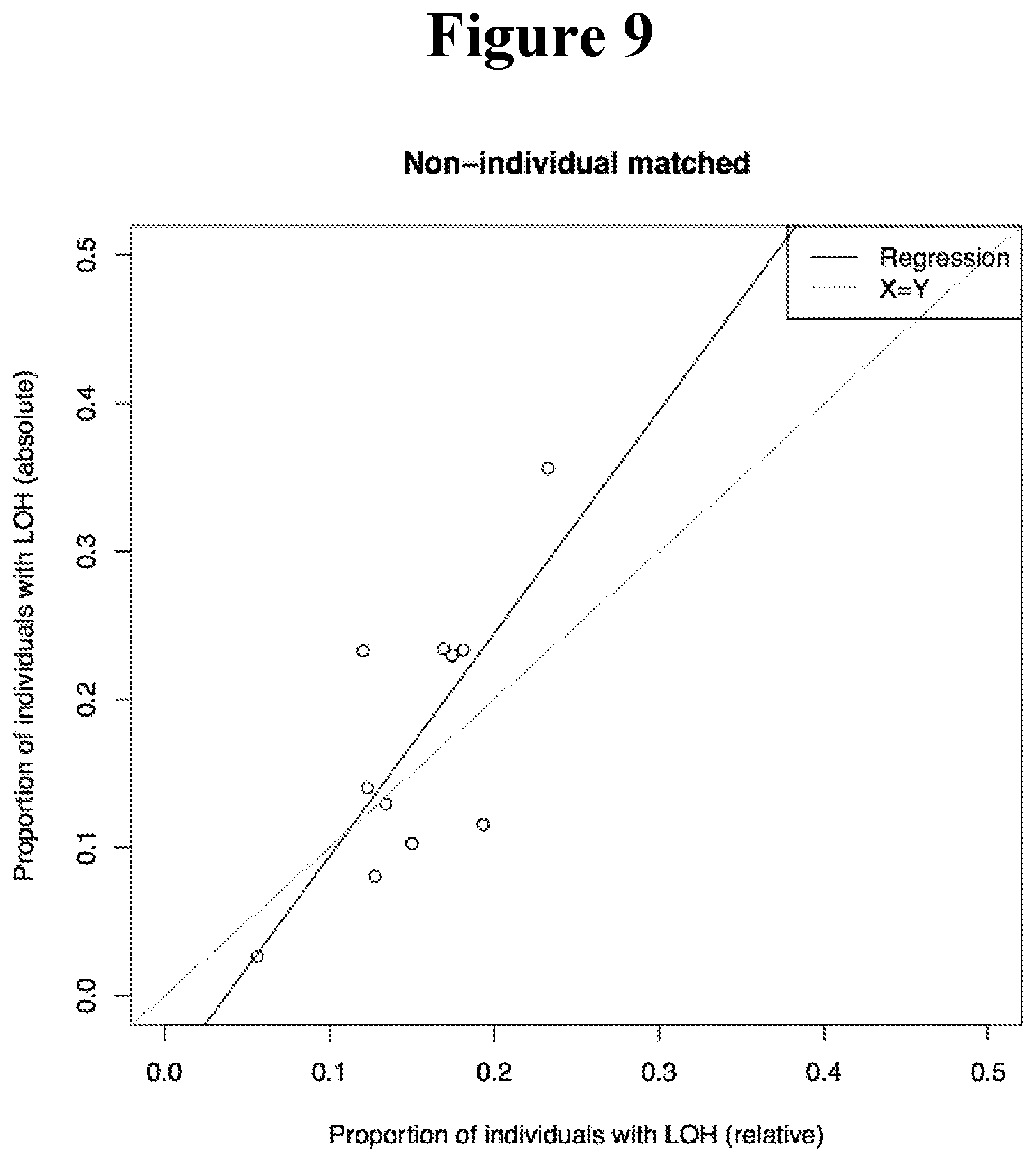
D00012
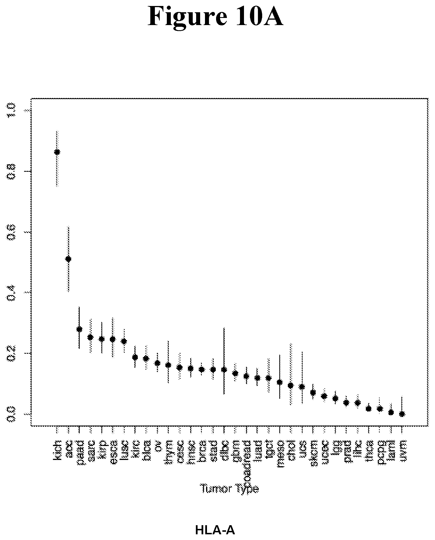
D00013
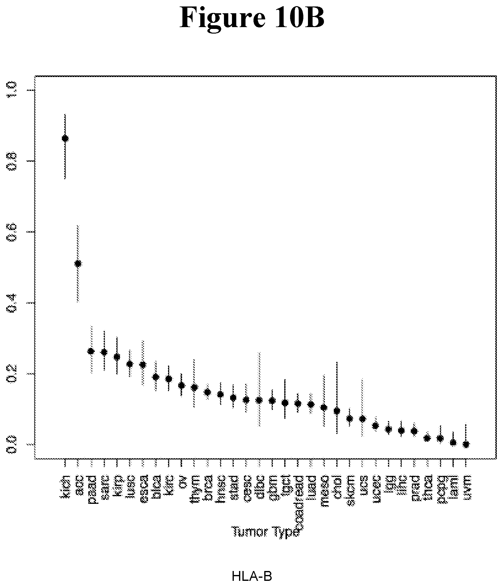
D00014
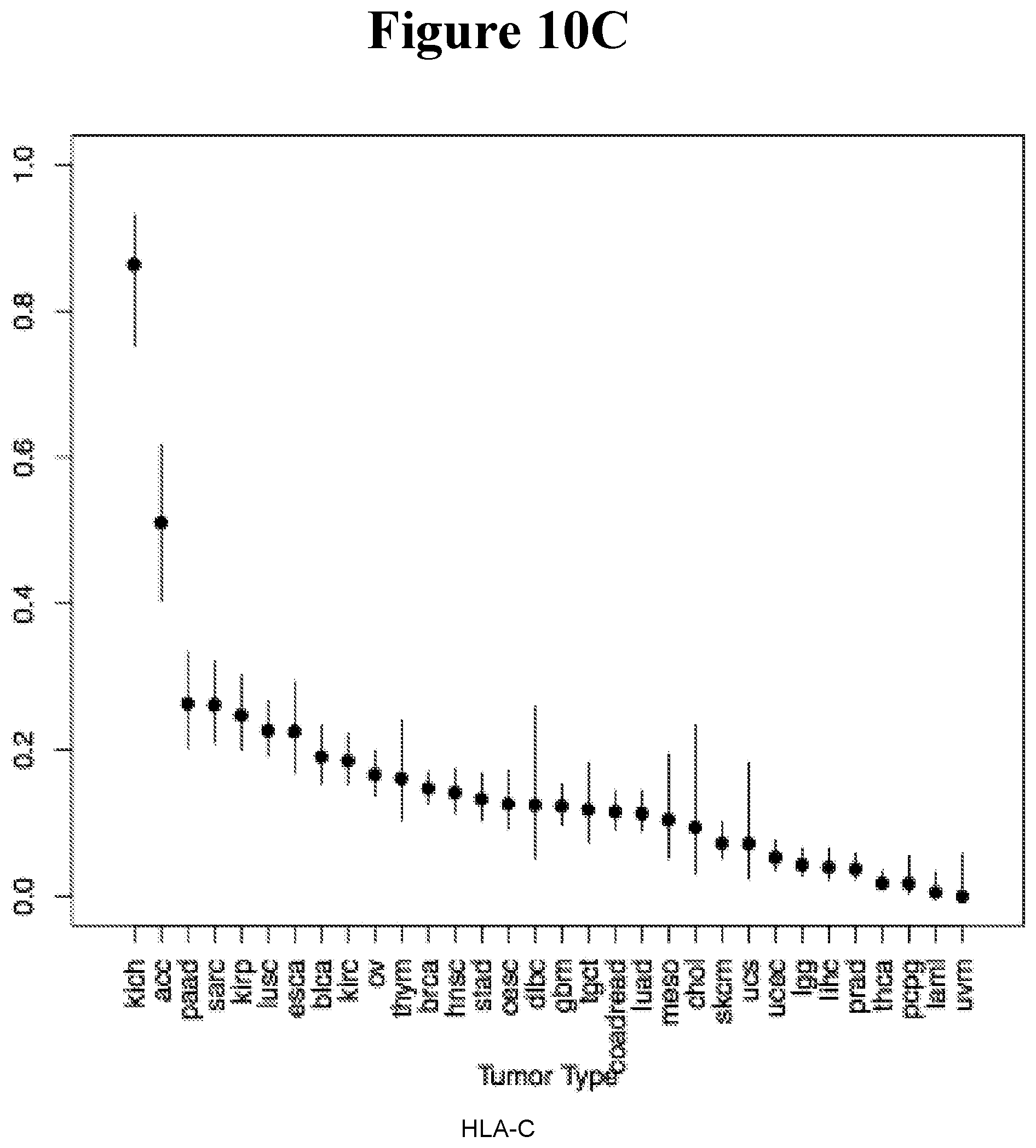
D00015
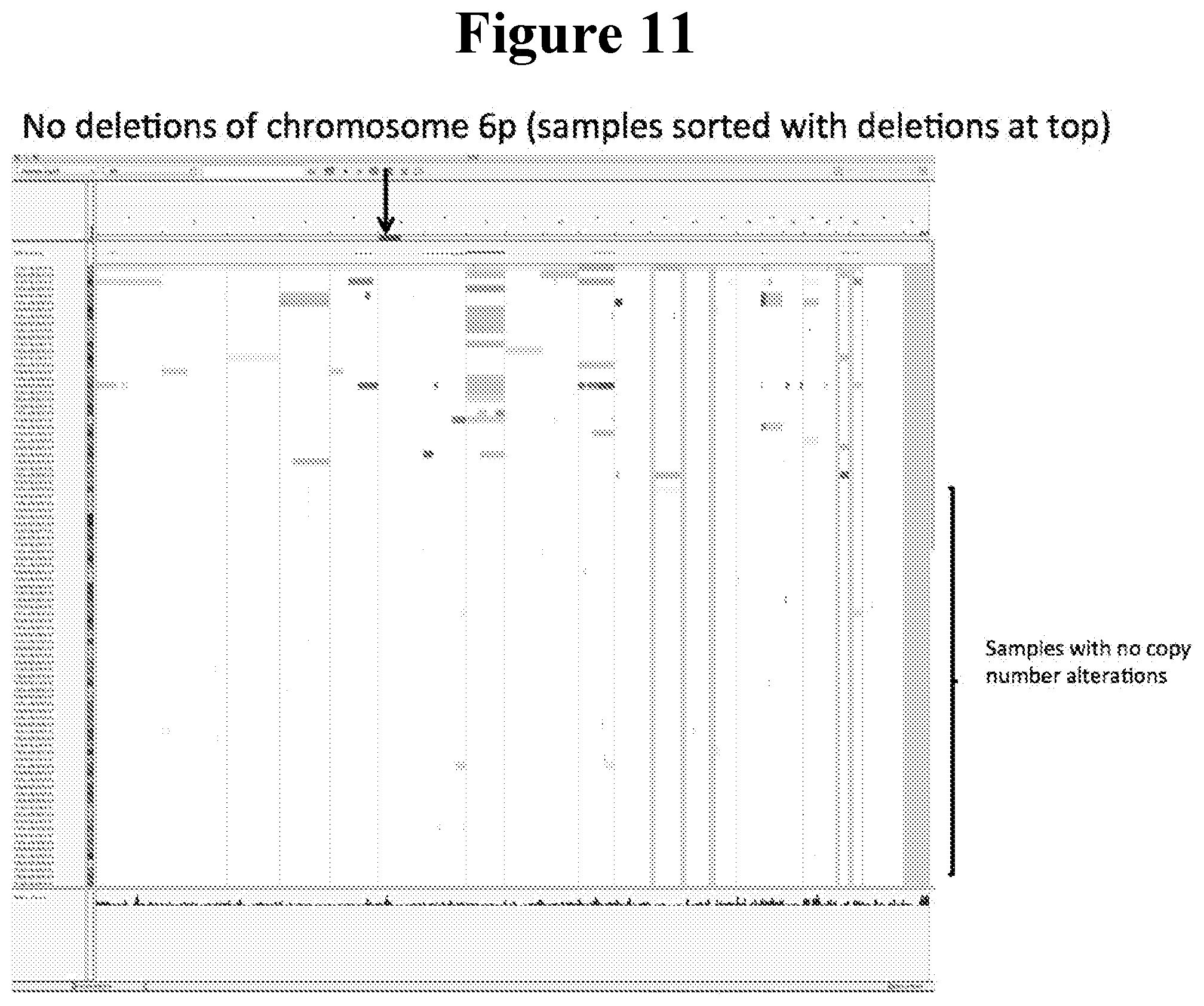
D00016
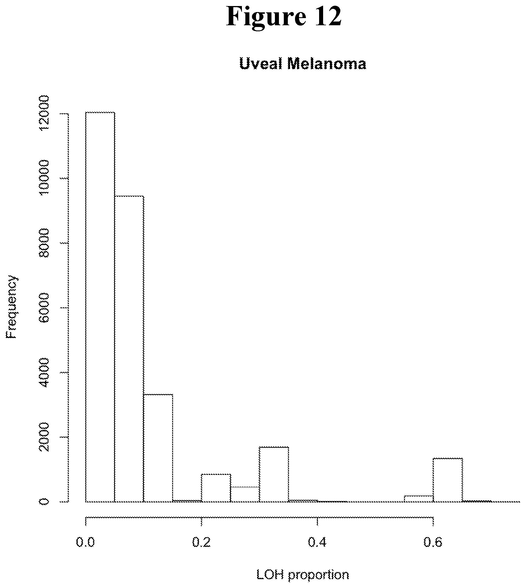
D00017
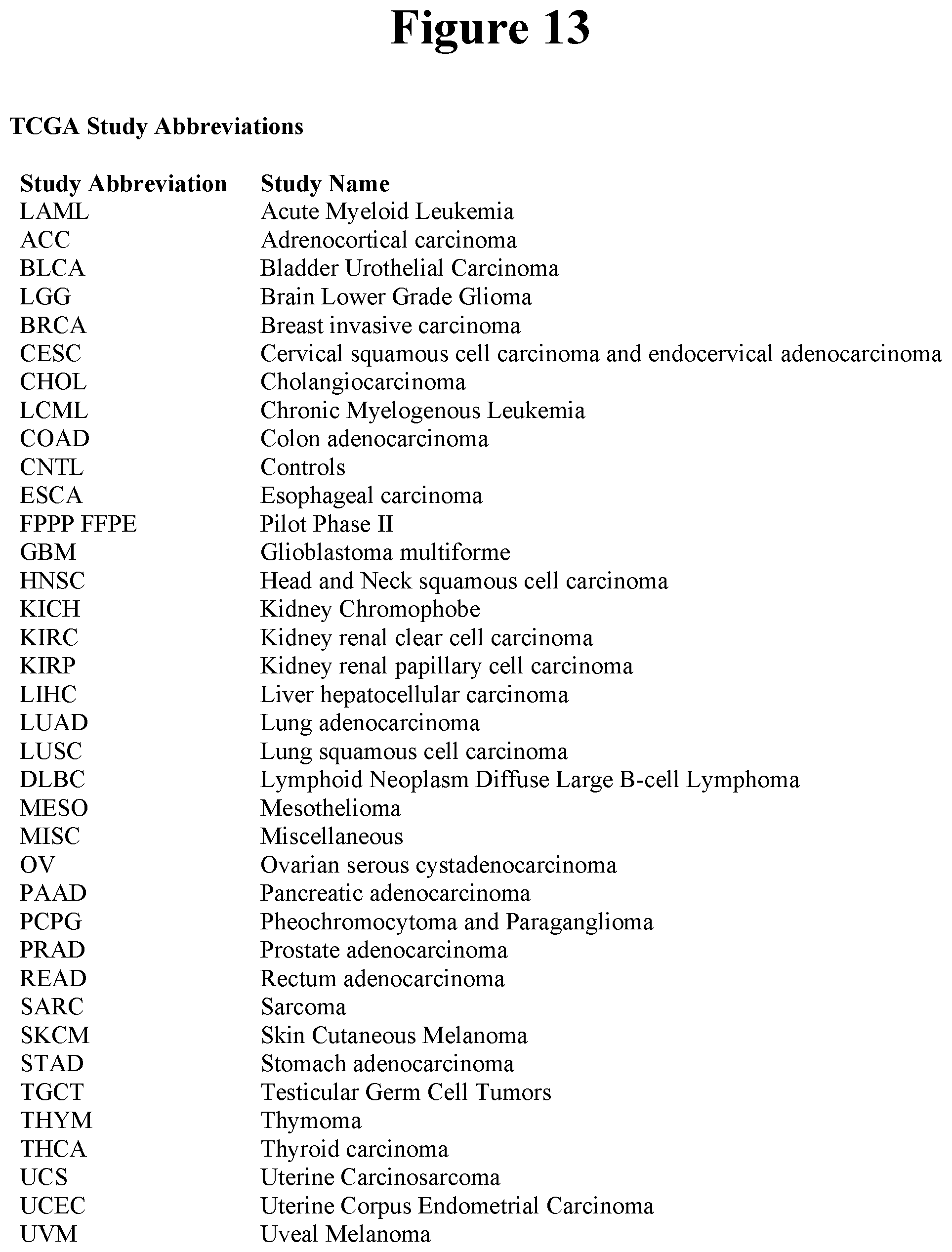
D00018

D00019
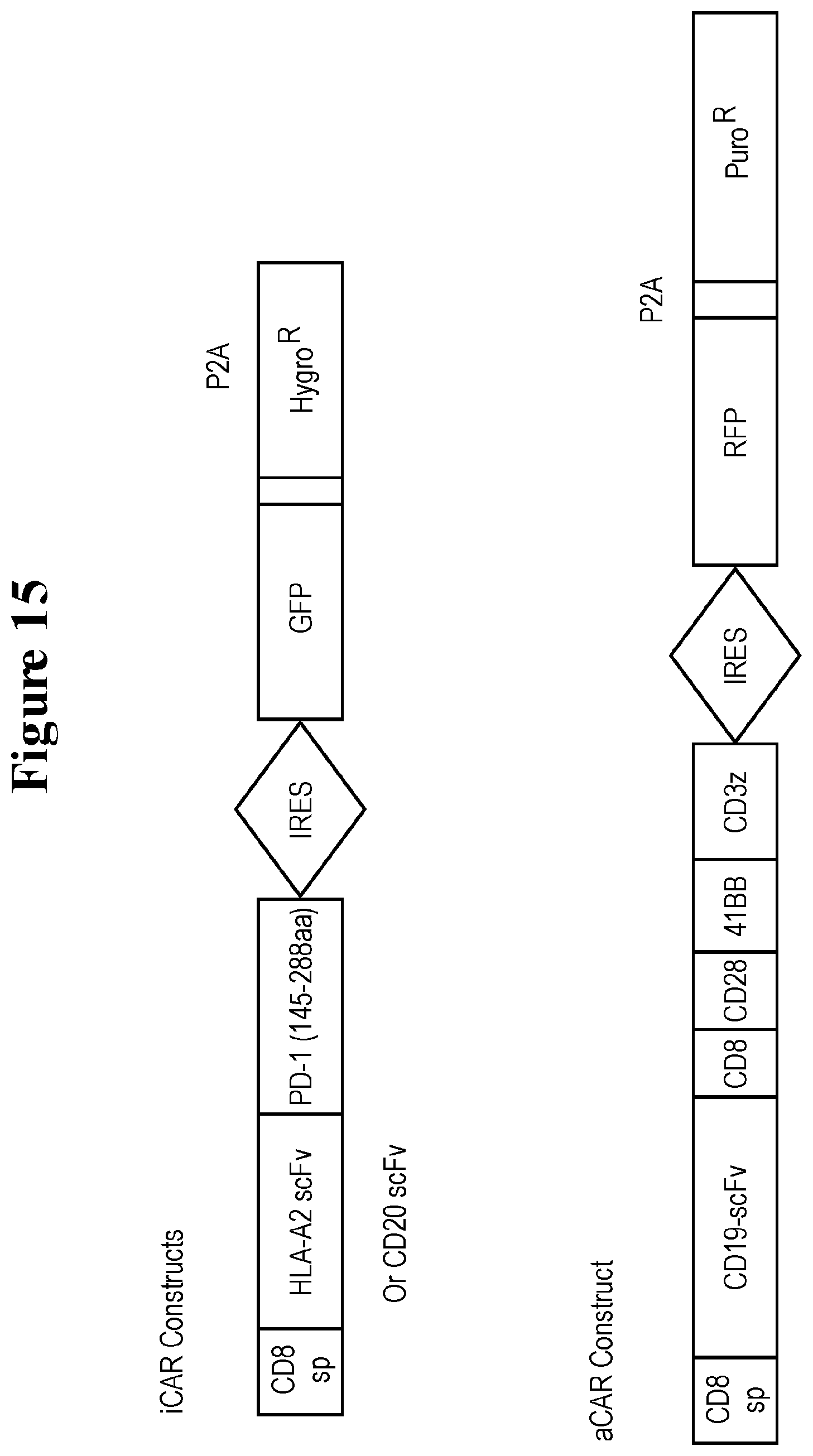
D00020
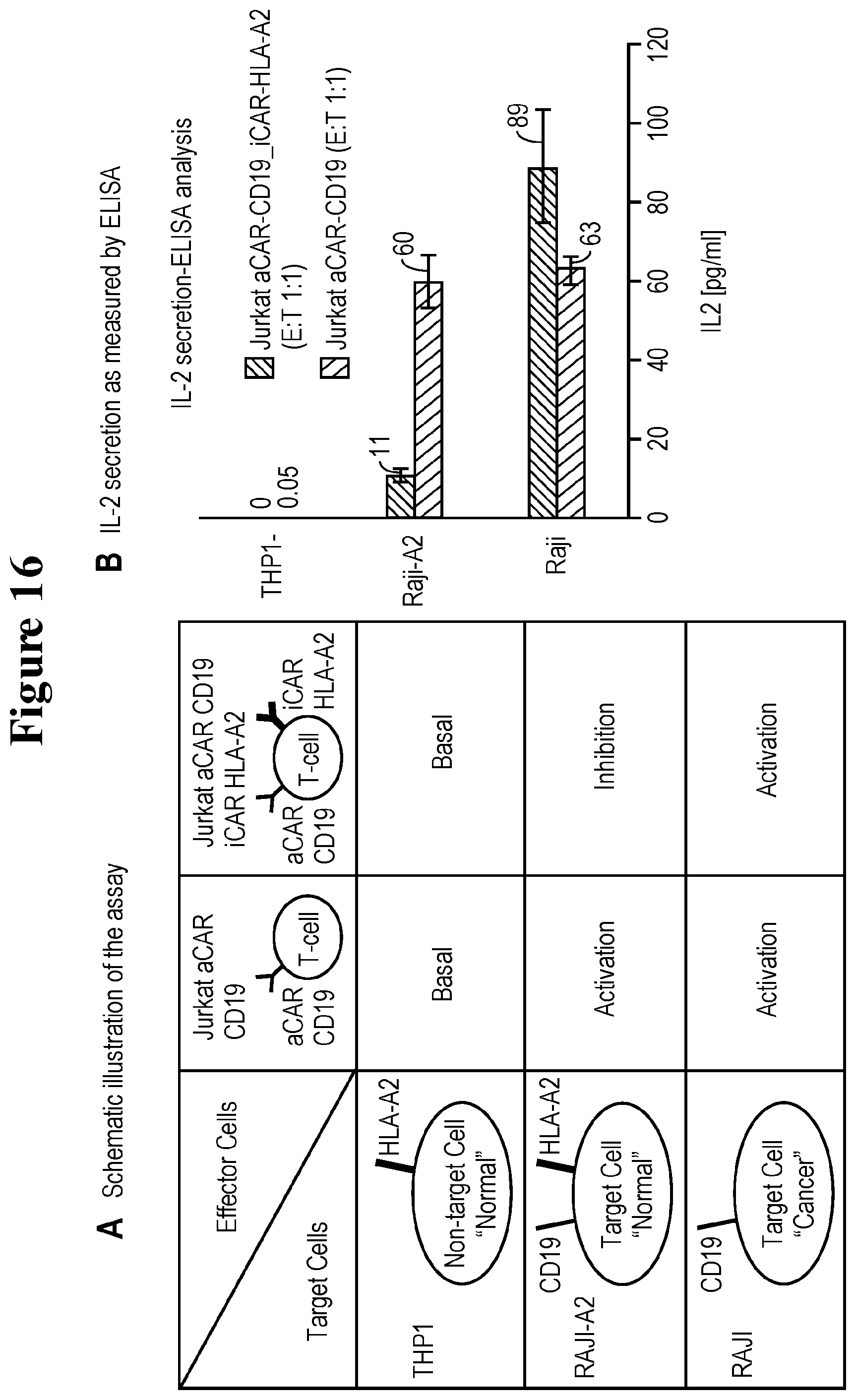
D00021

D00022
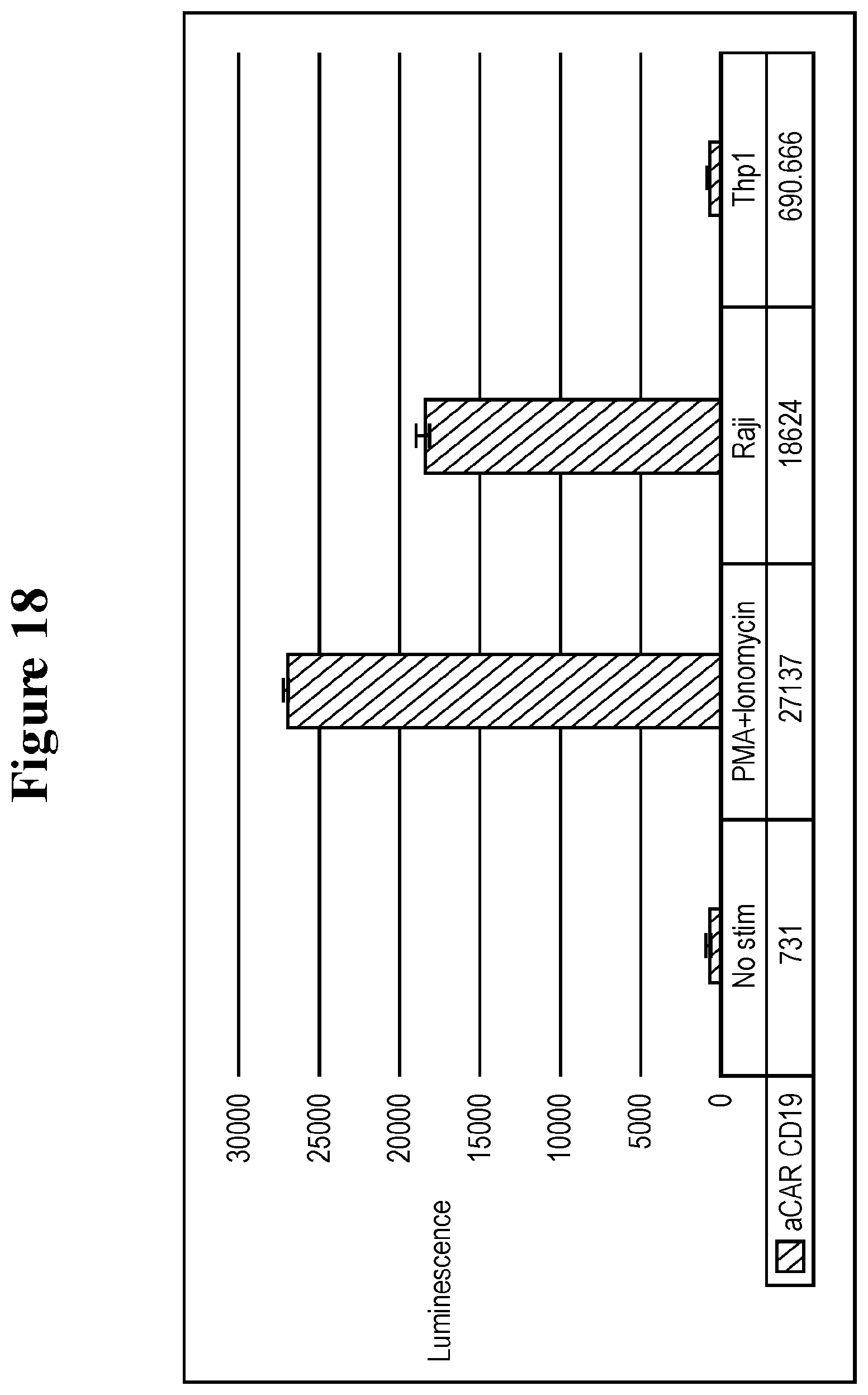
D00023
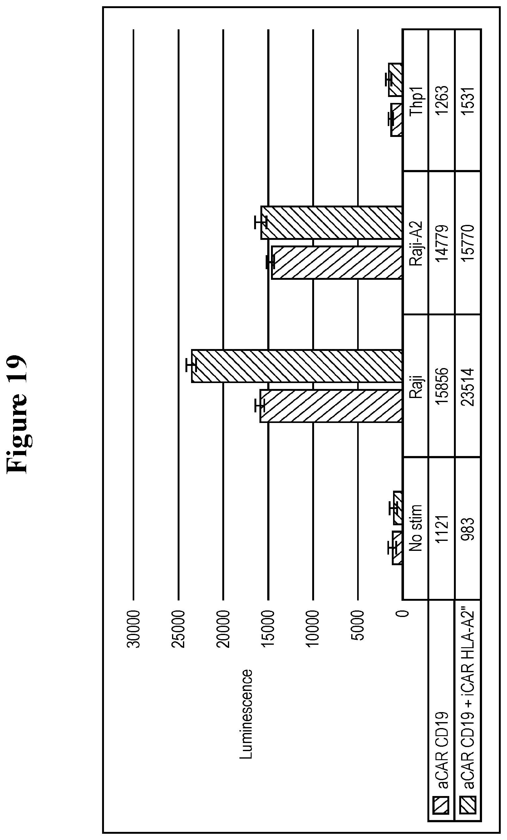
D00024
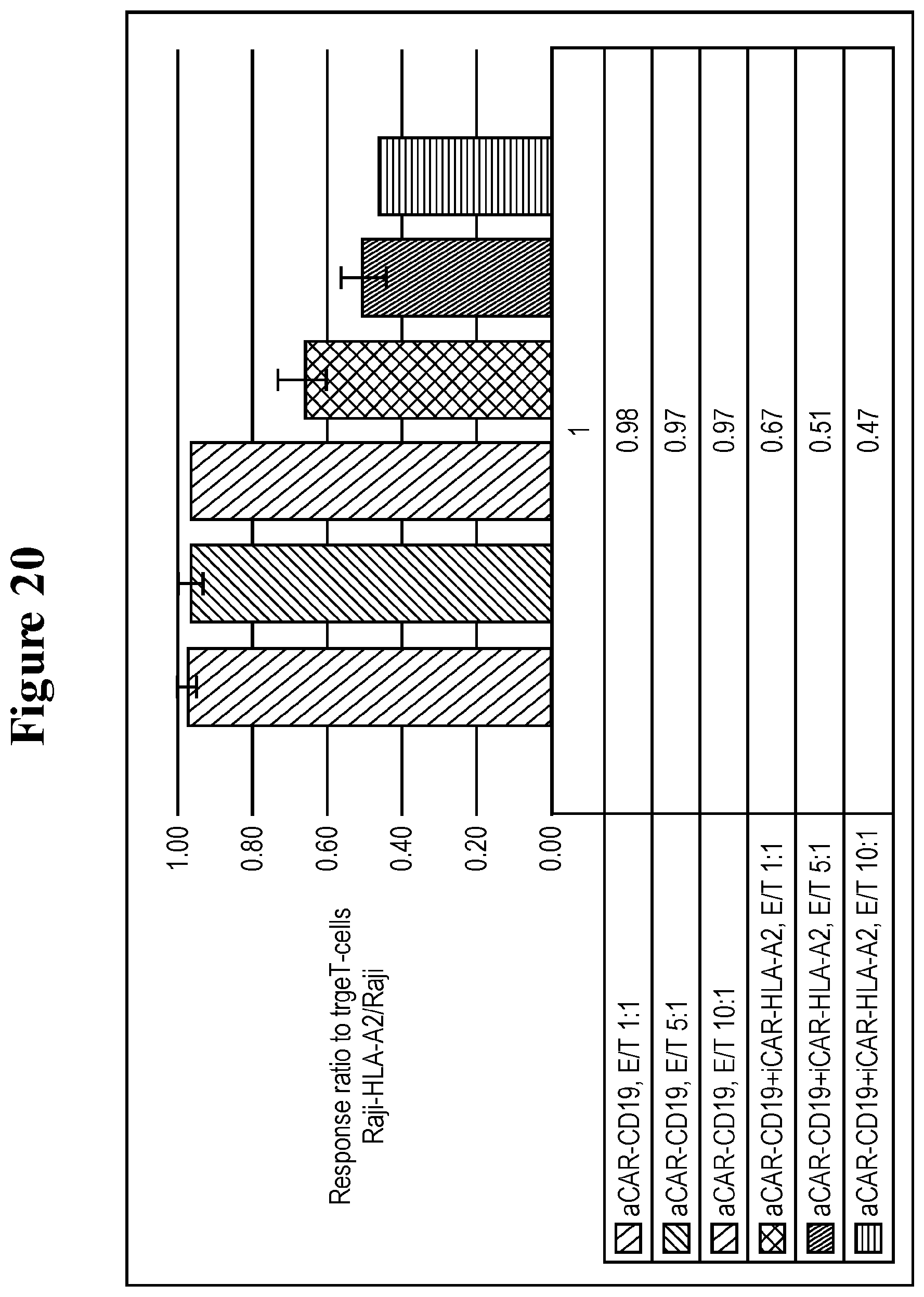
D00025

D00026
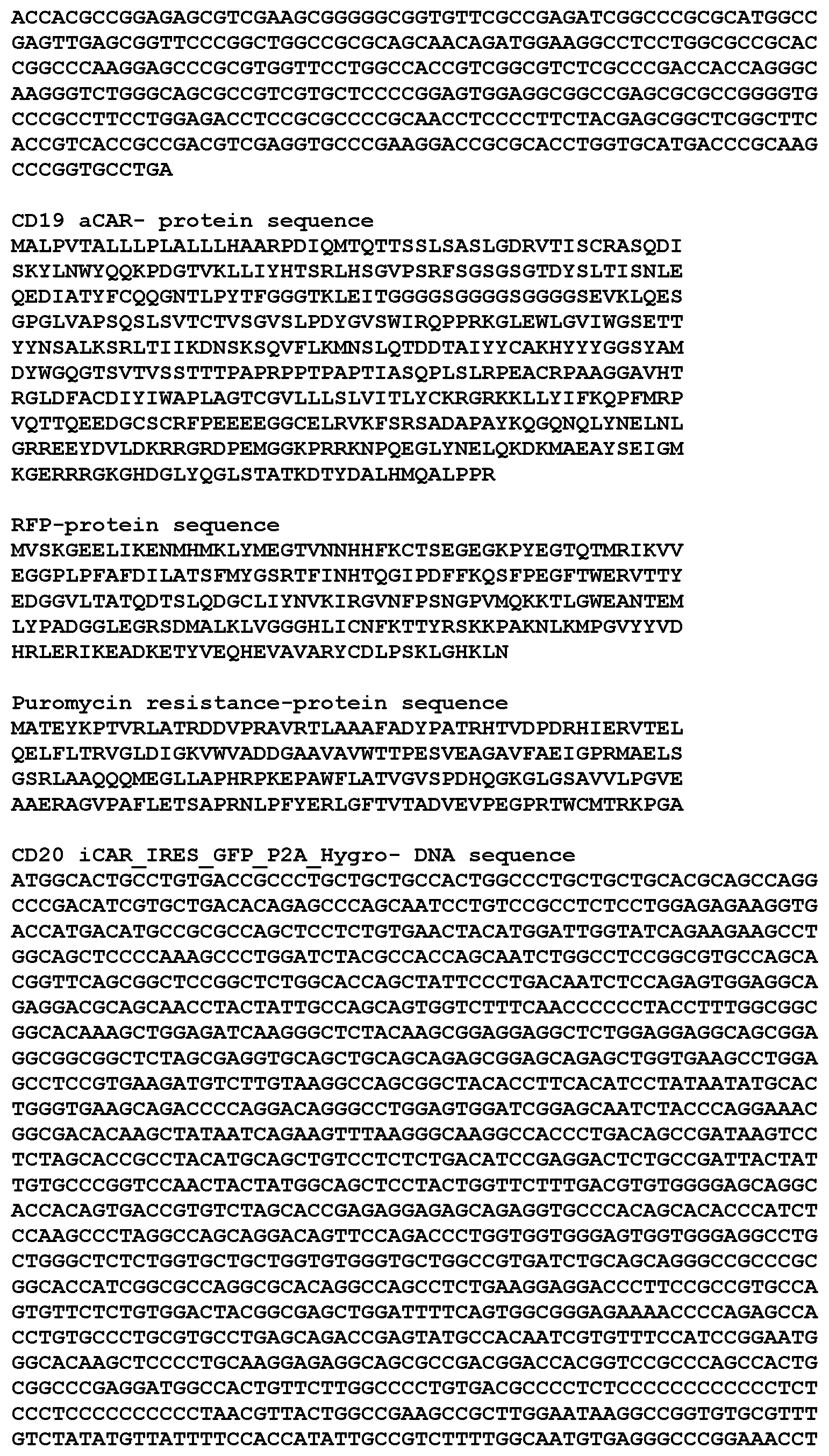
D00027
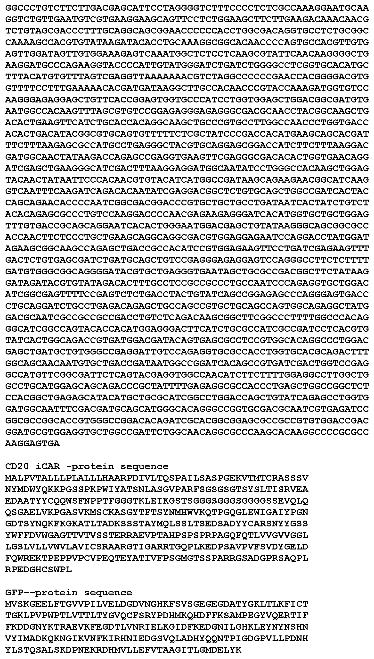
D00028
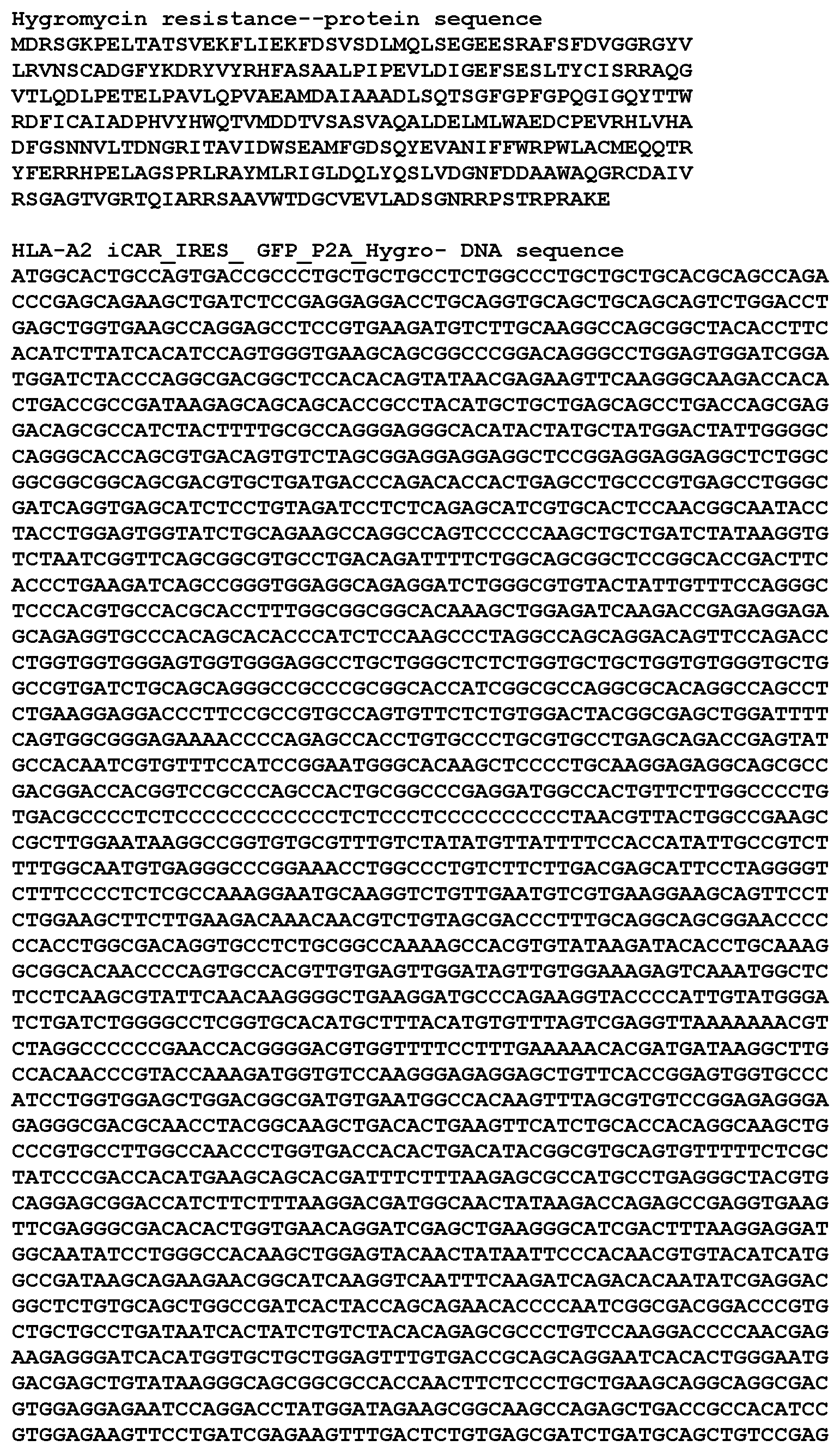
D00029
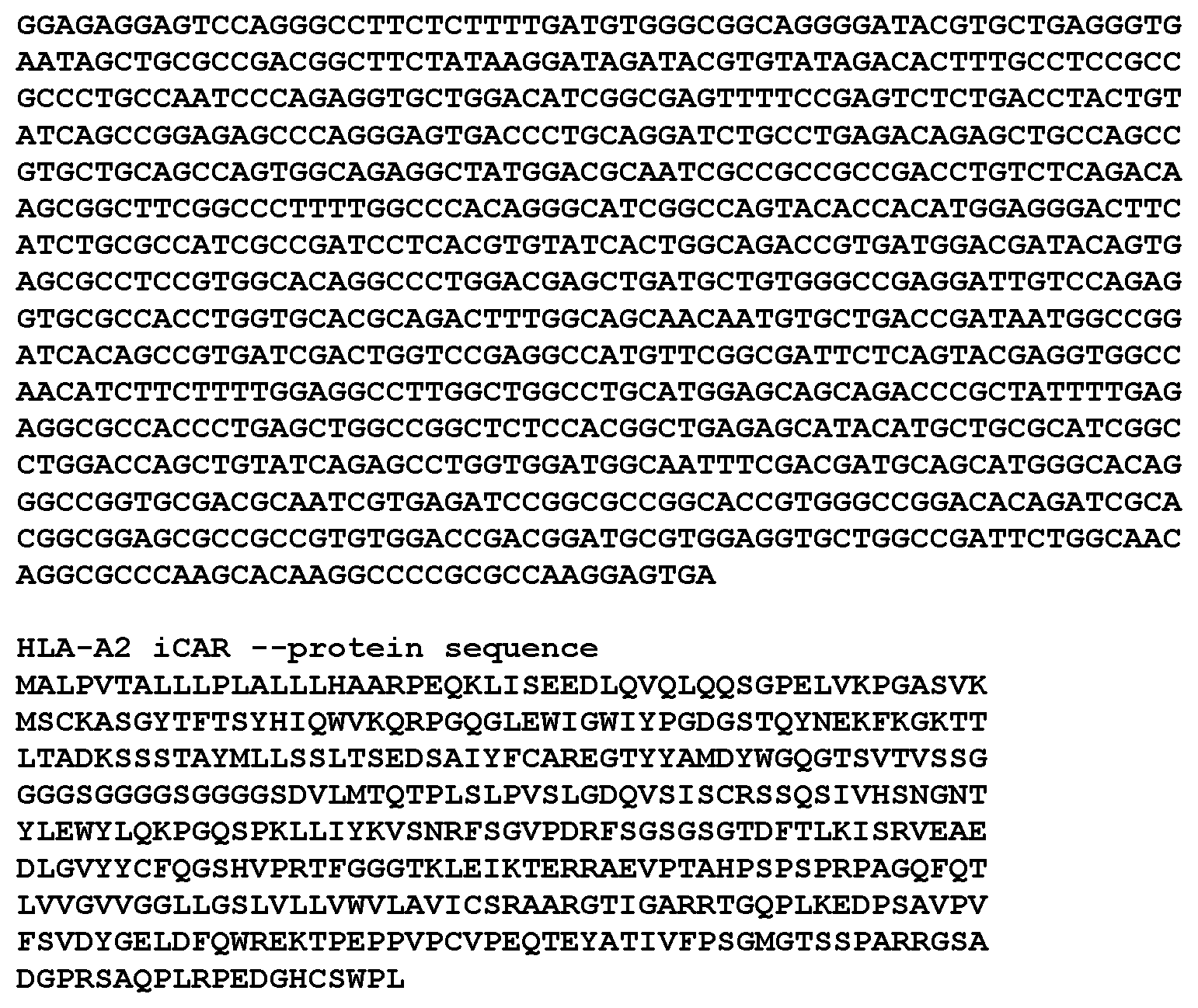
D00030
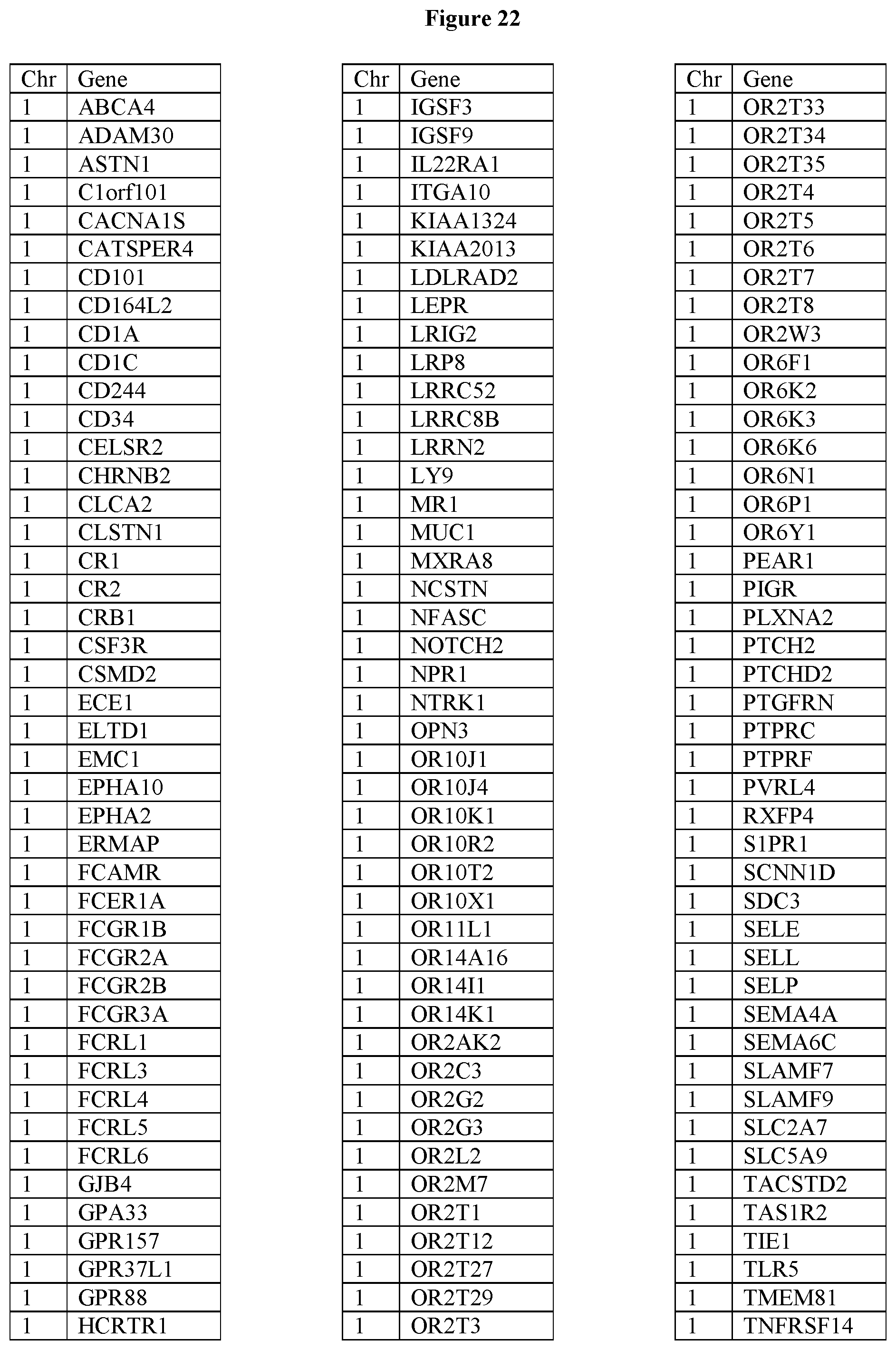
D00031
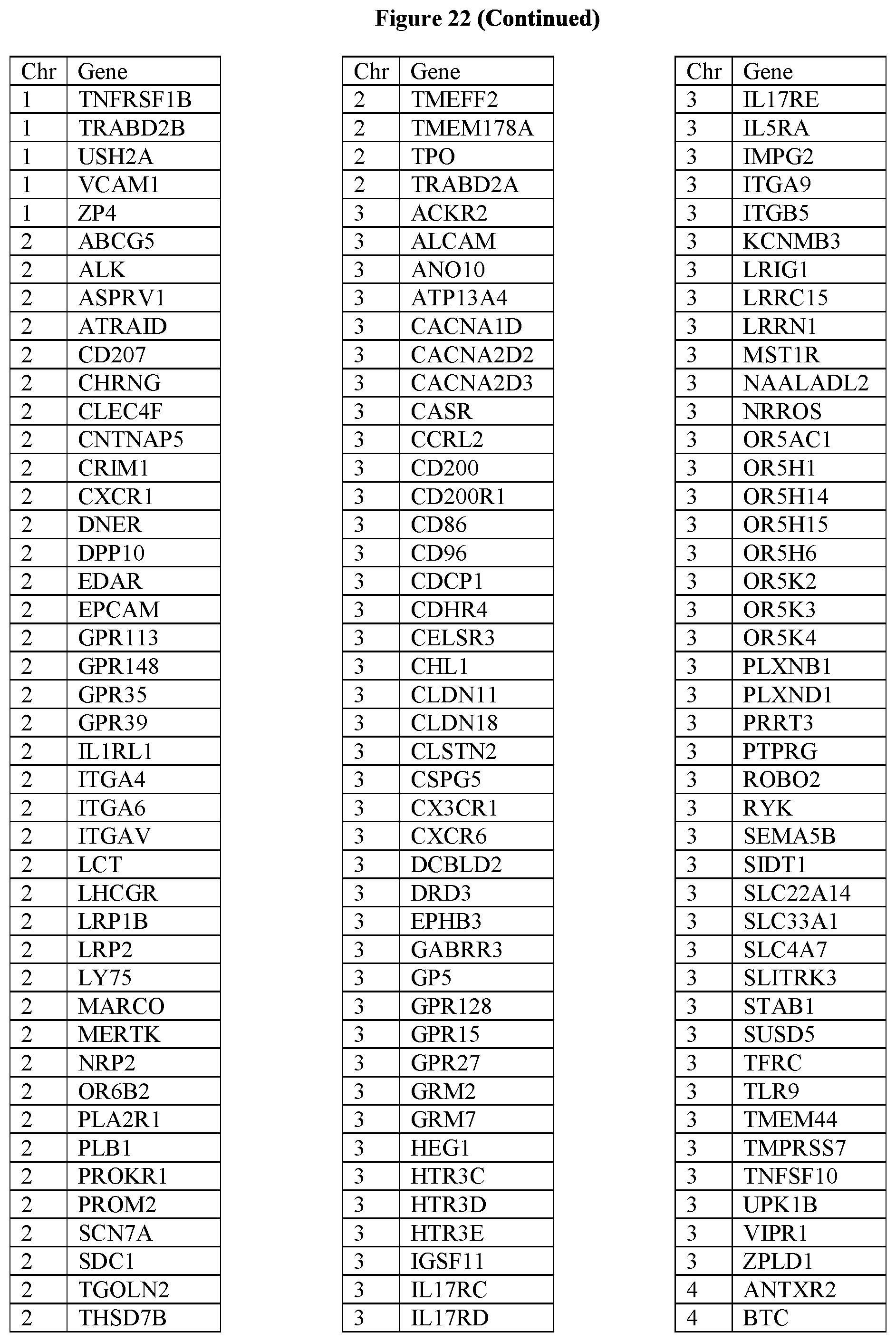
D00032

D00033

D00034
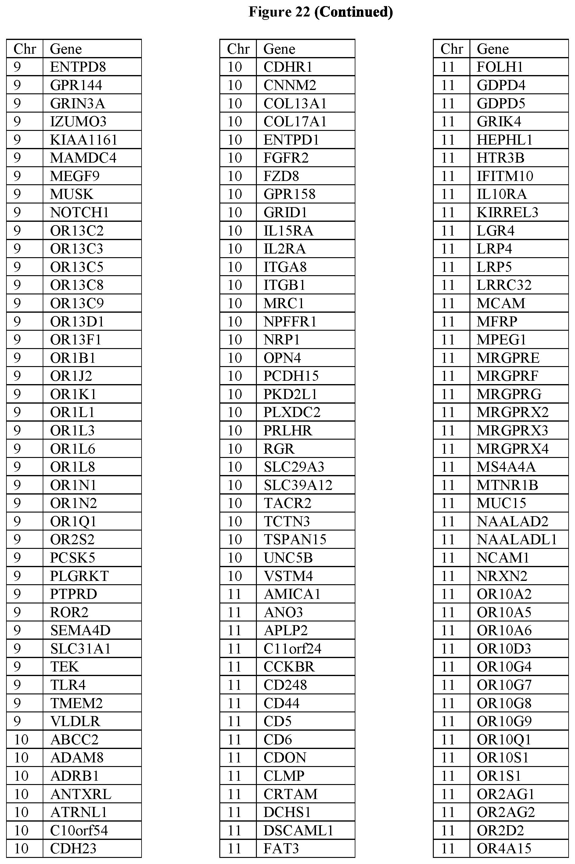
D00035
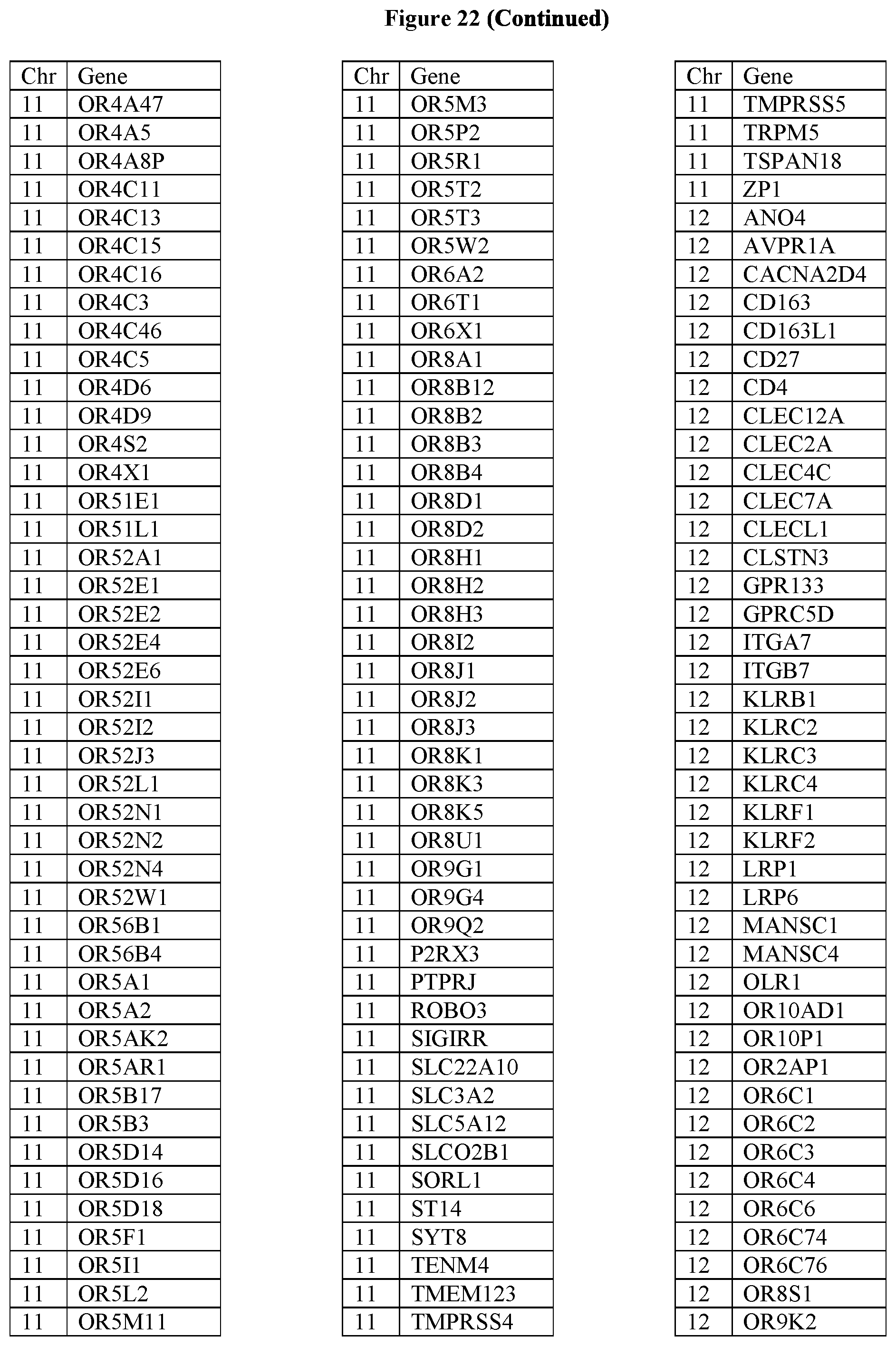
D00036

D00037

D00038
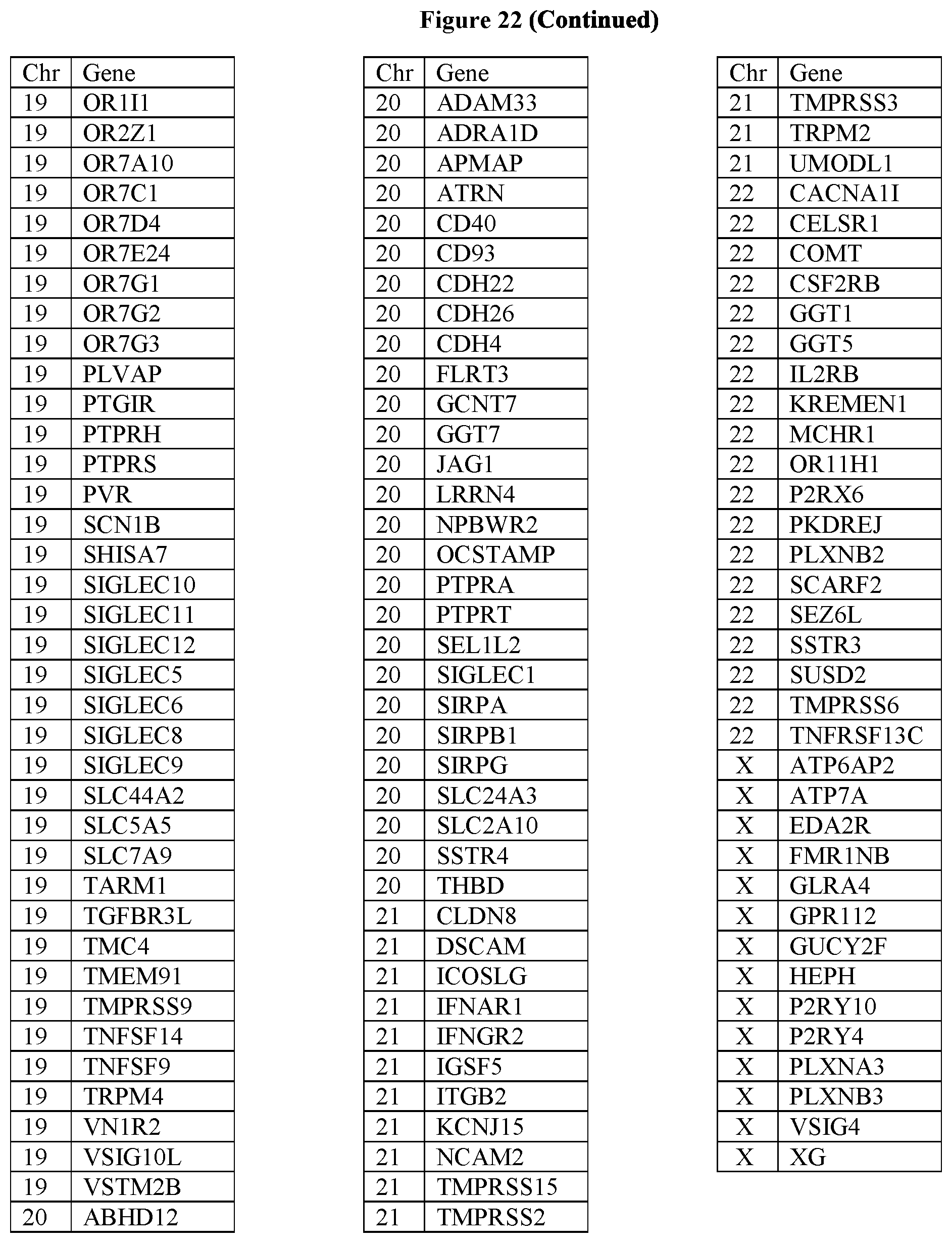
D00039
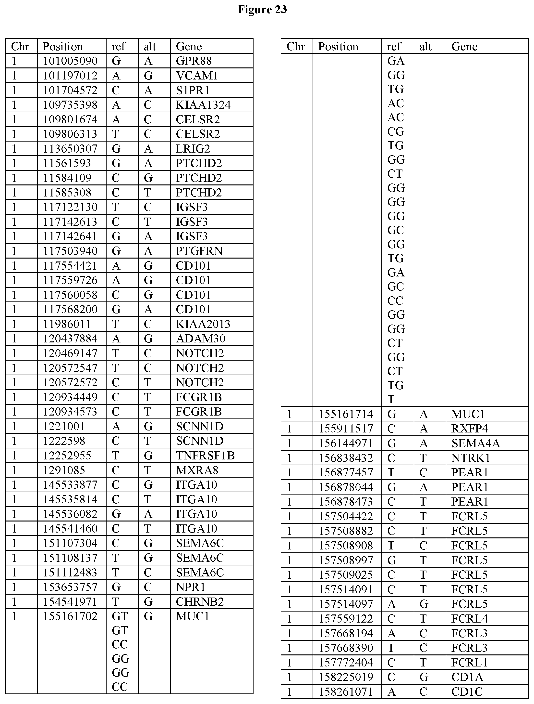
D00040
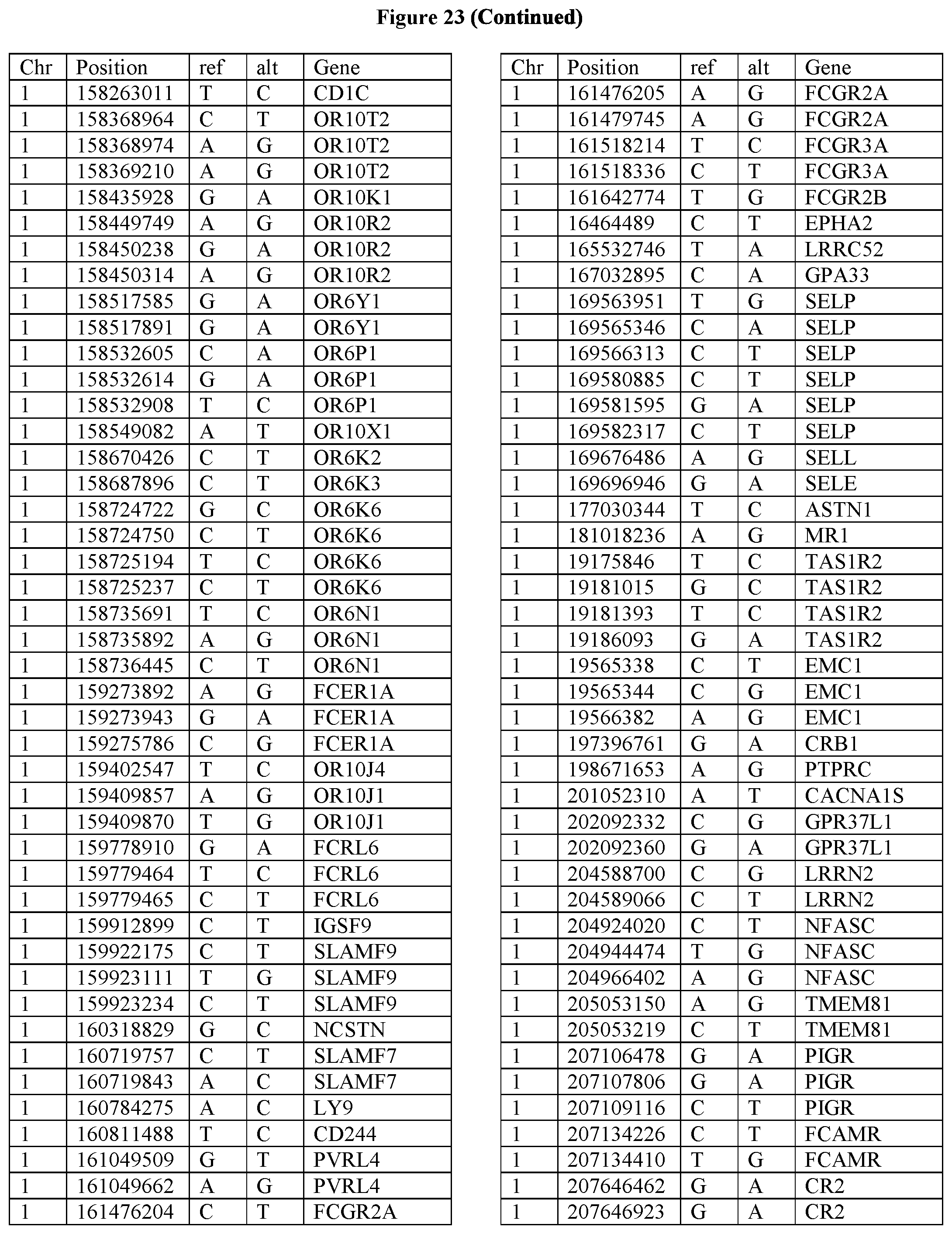
D00041

D00042

D00043
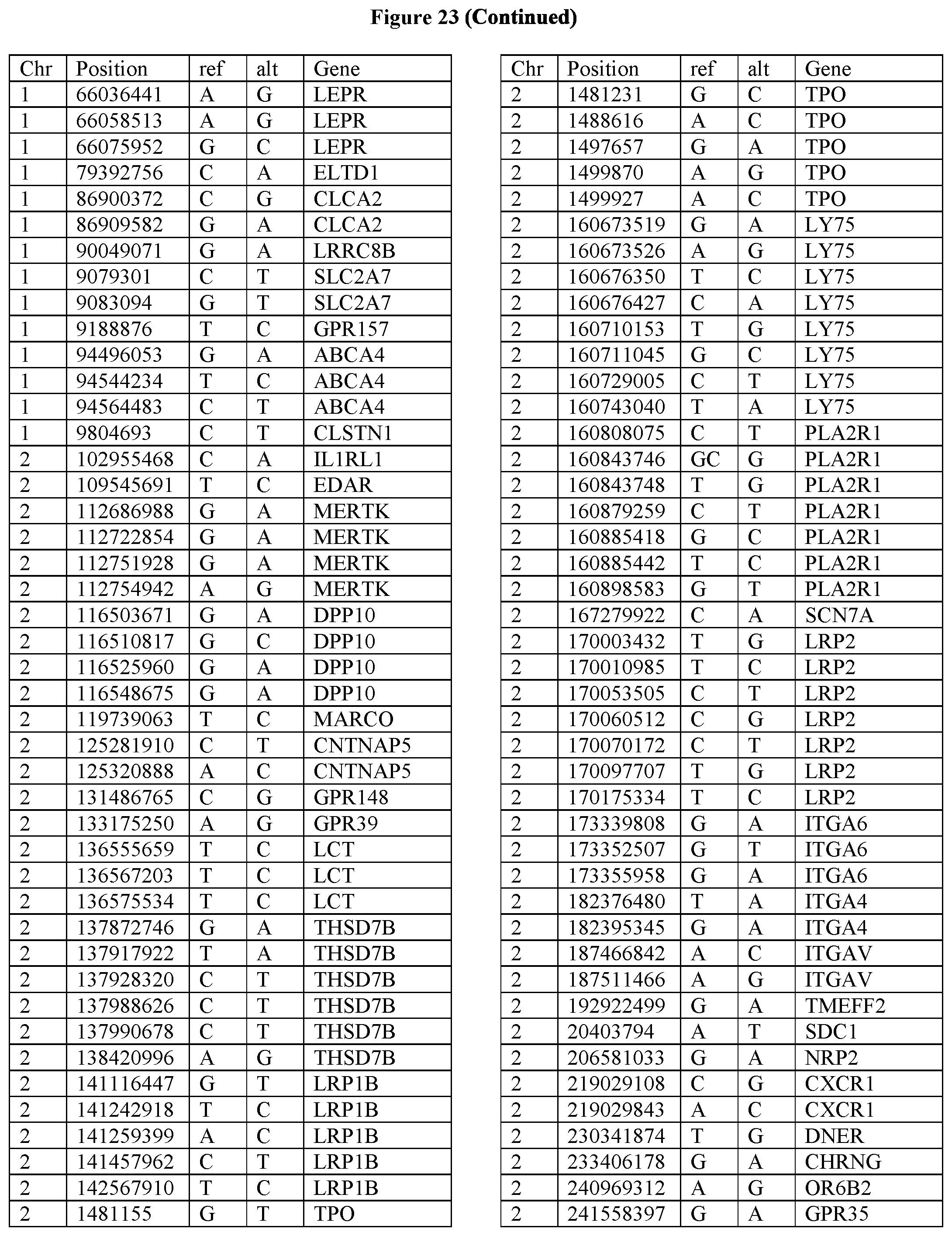
D00044
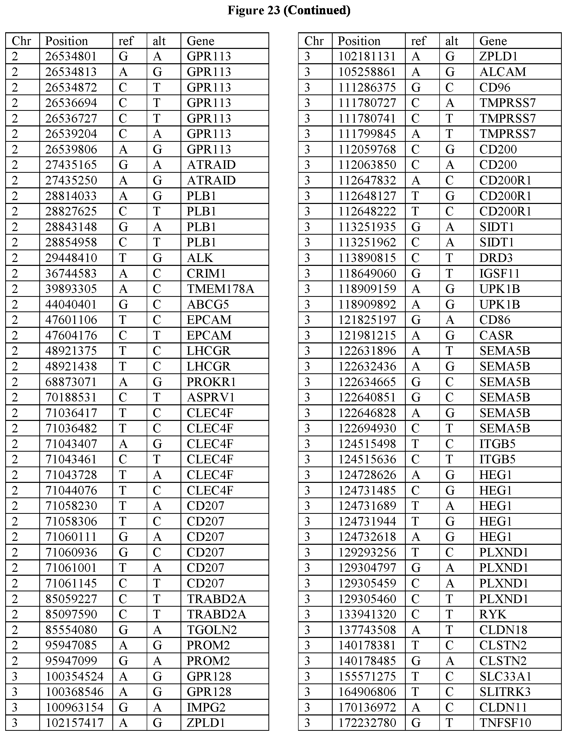
D00045
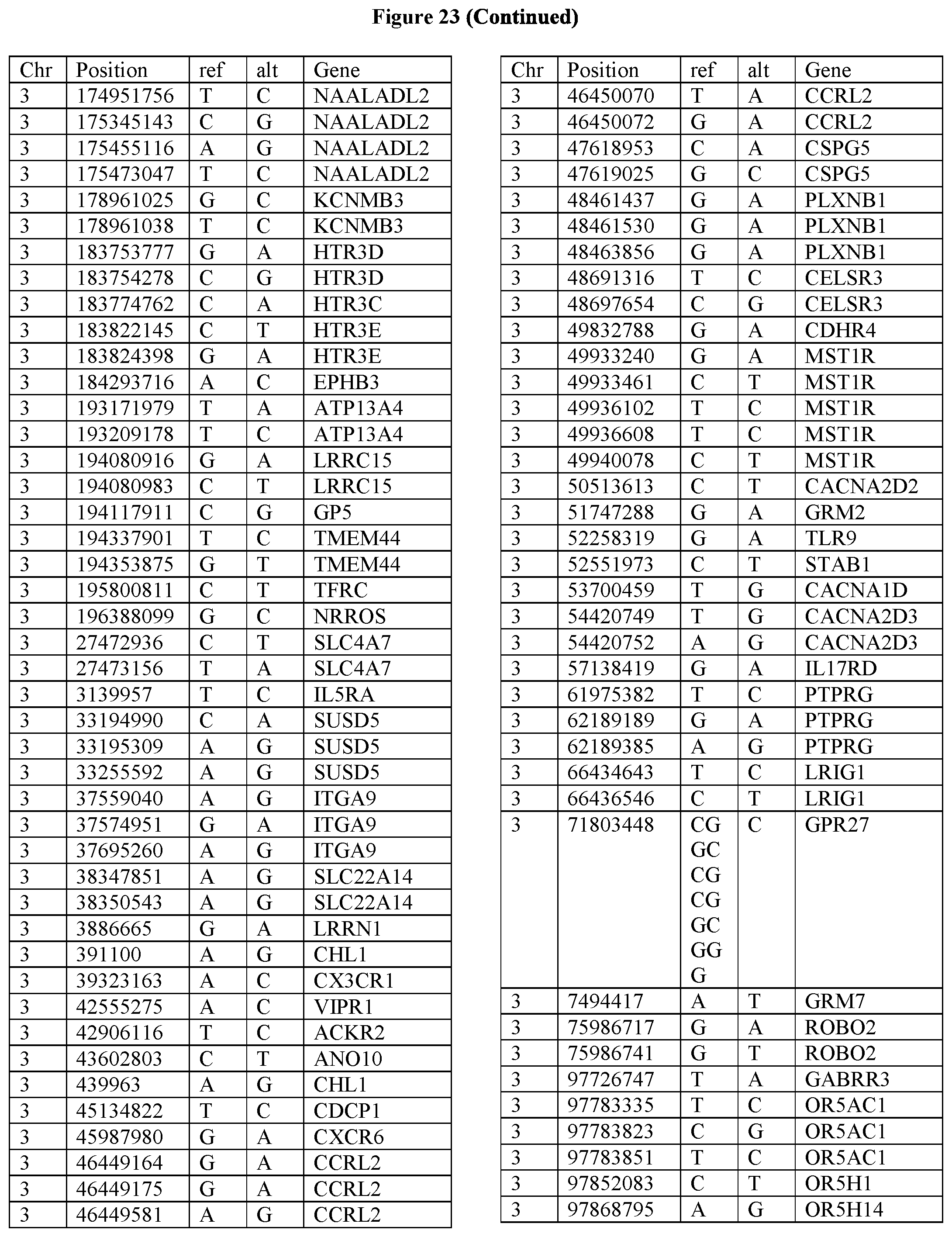
D00046
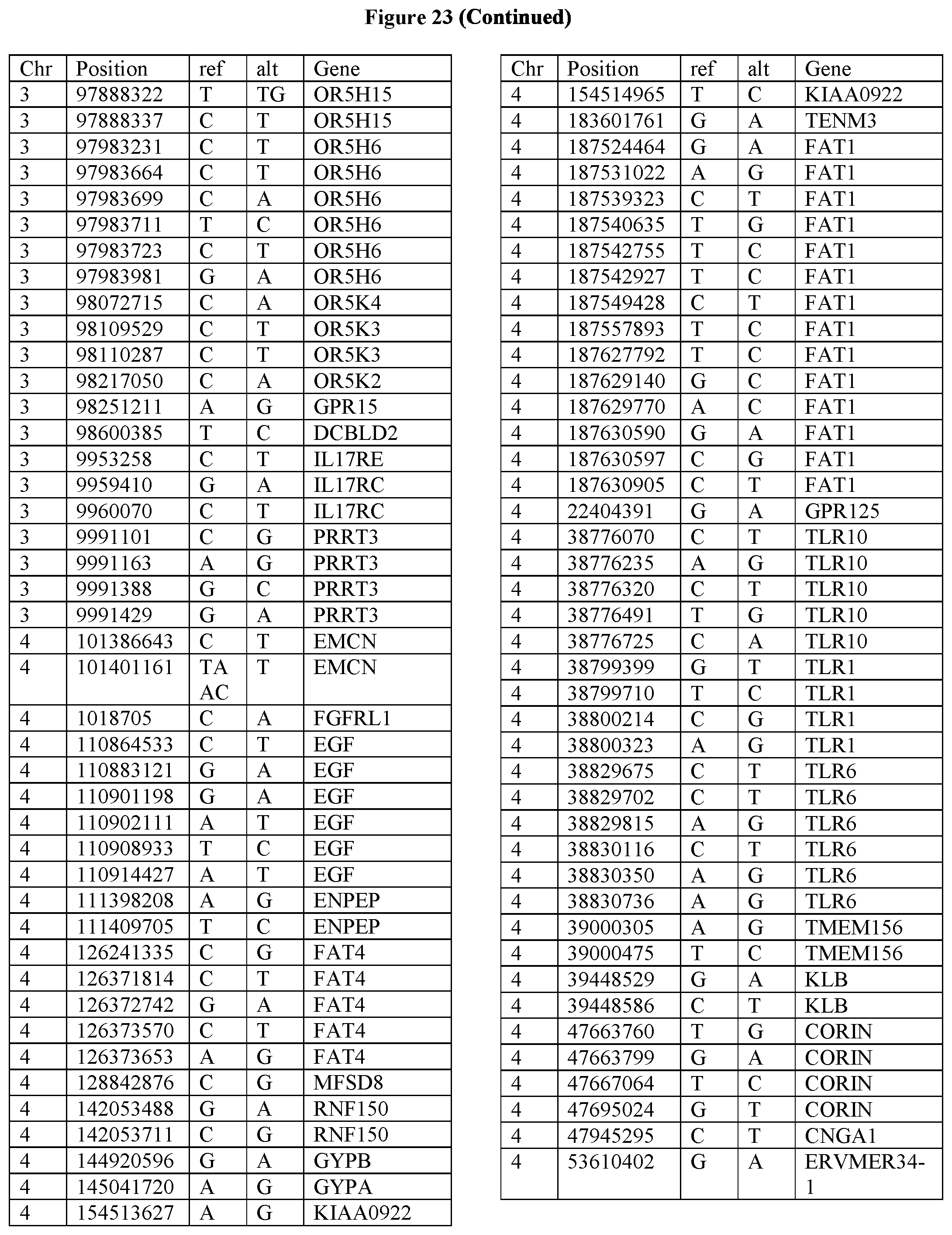
D00047

D00048
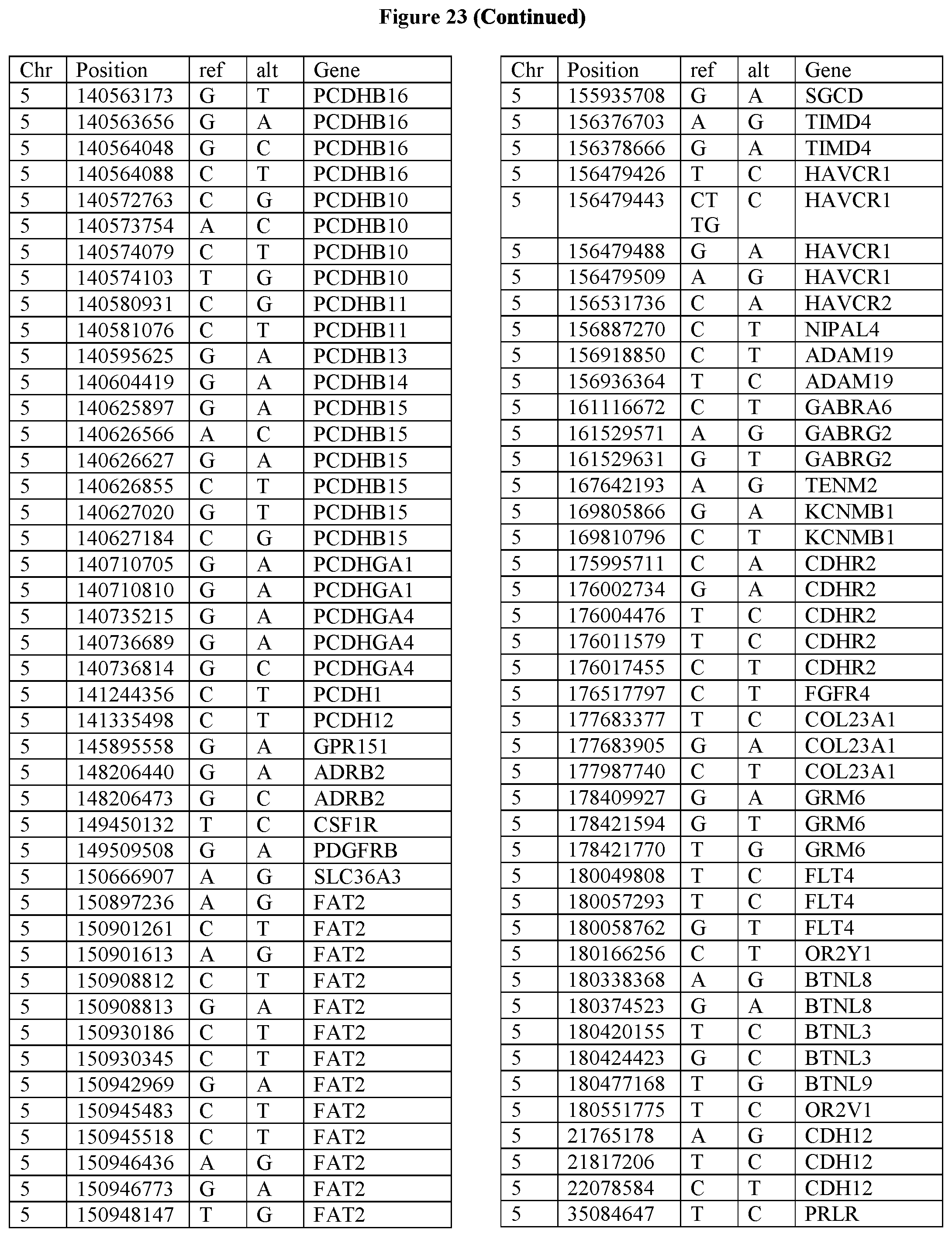
D00049

D00050
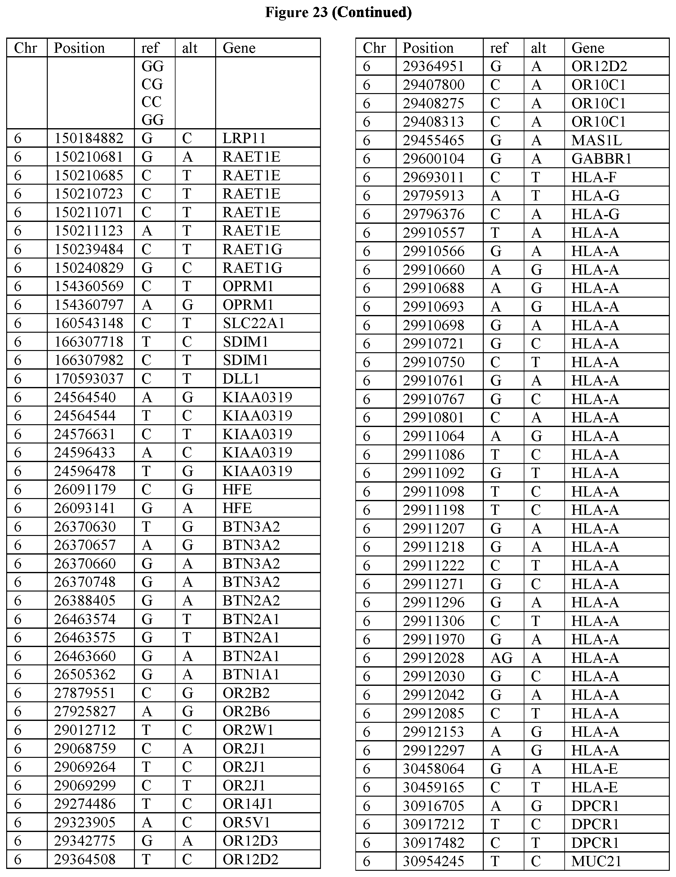
D00051
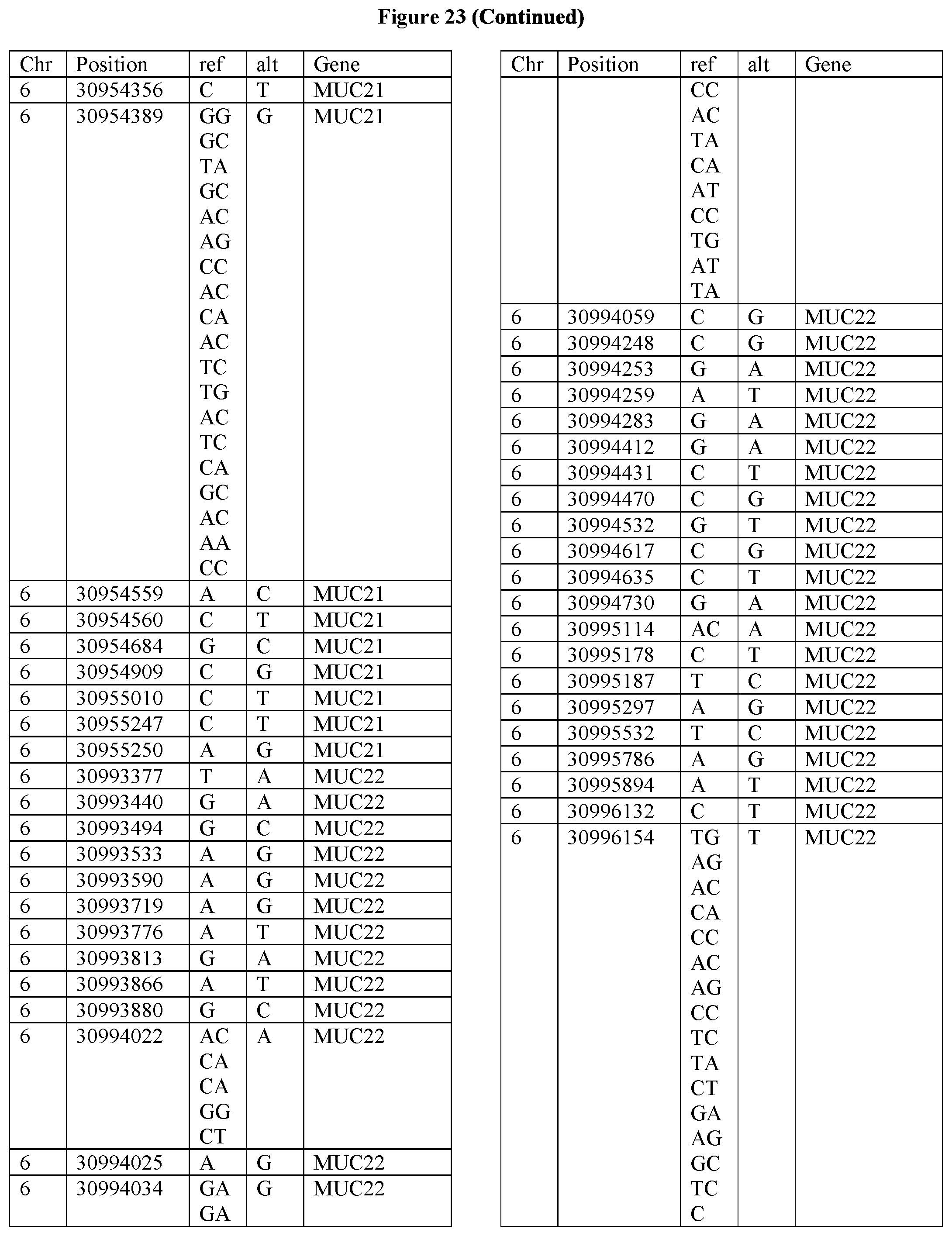
D00052
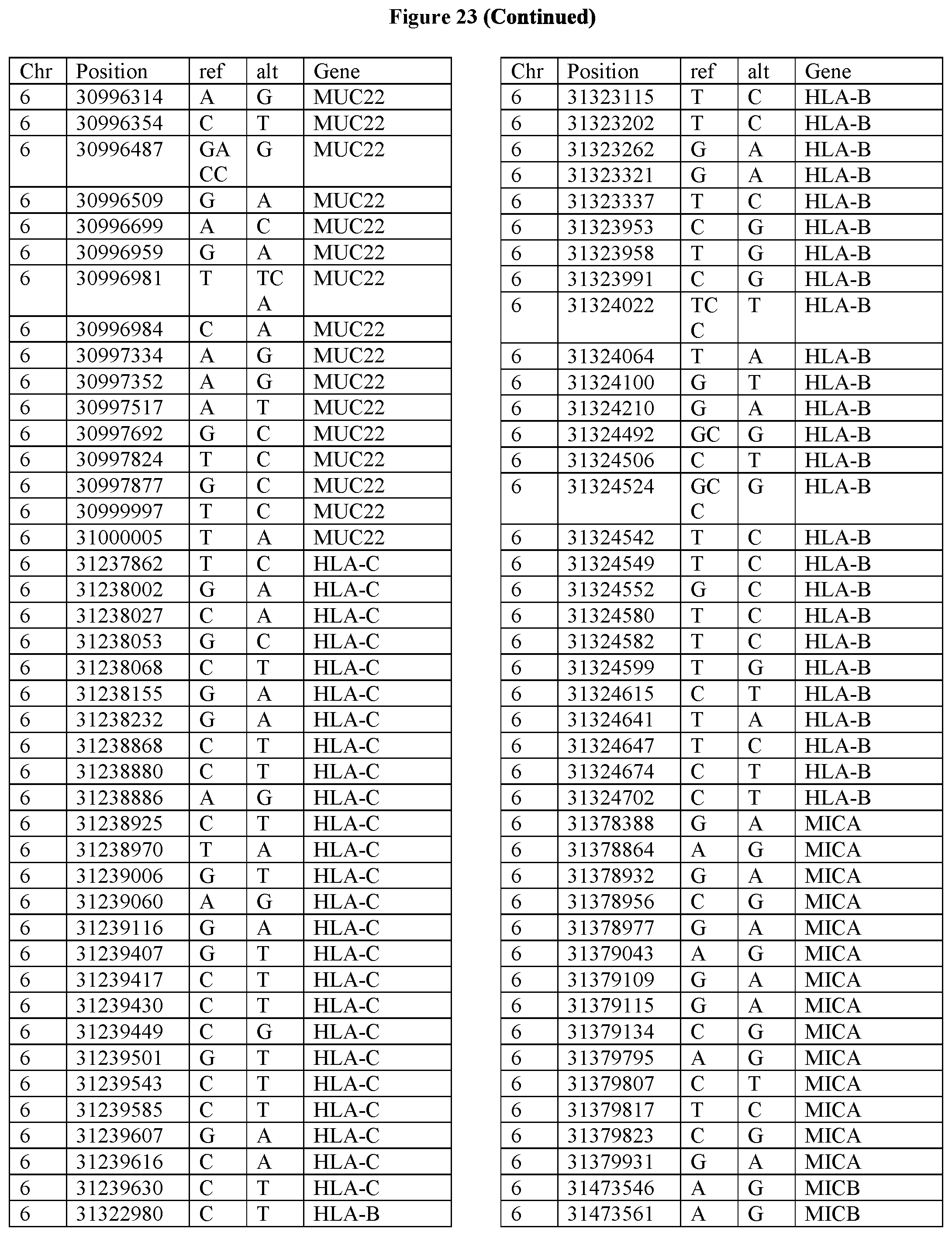
D00053

D00054

D00055

D00056

D00057

D00058

D00059

D00060
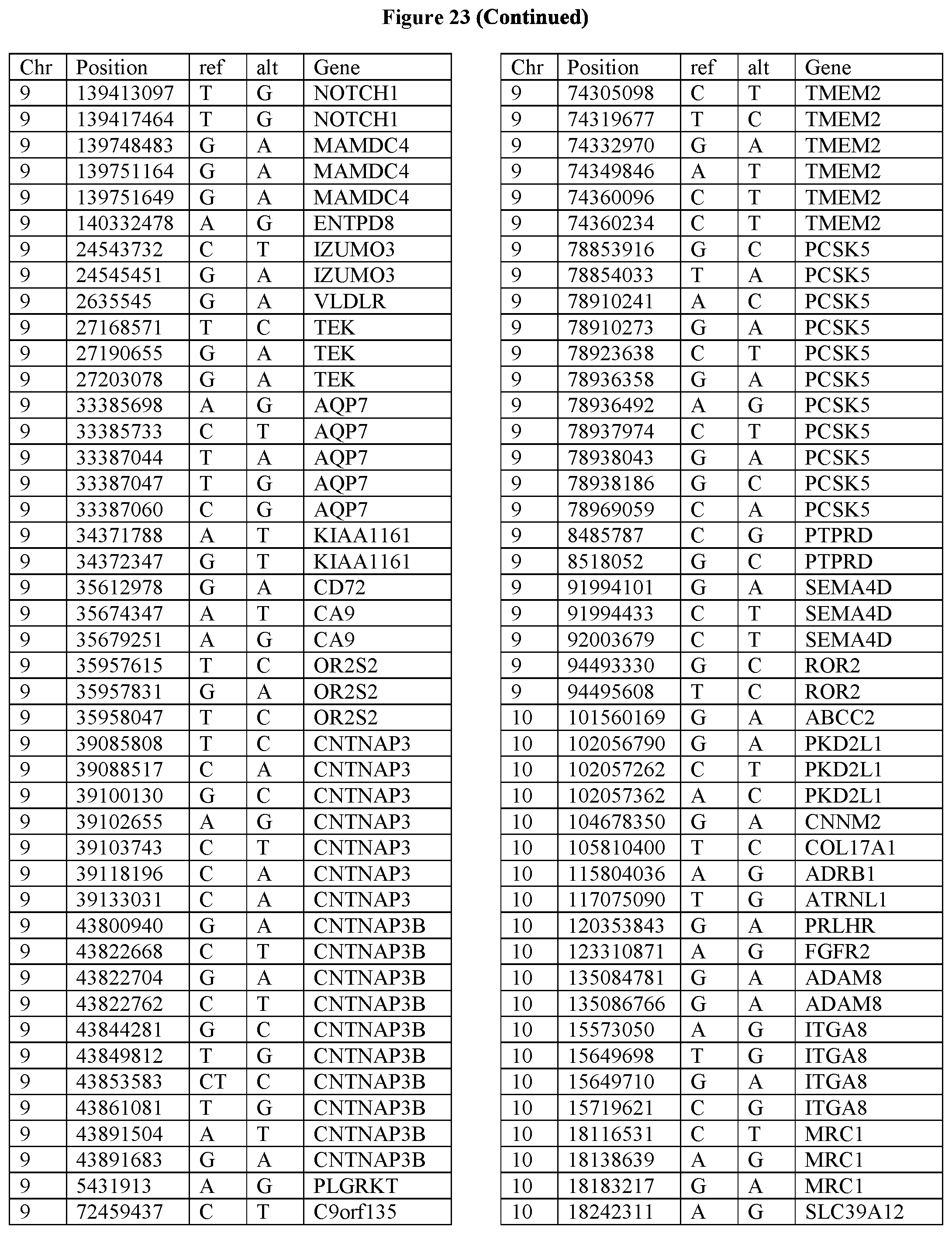
D00061
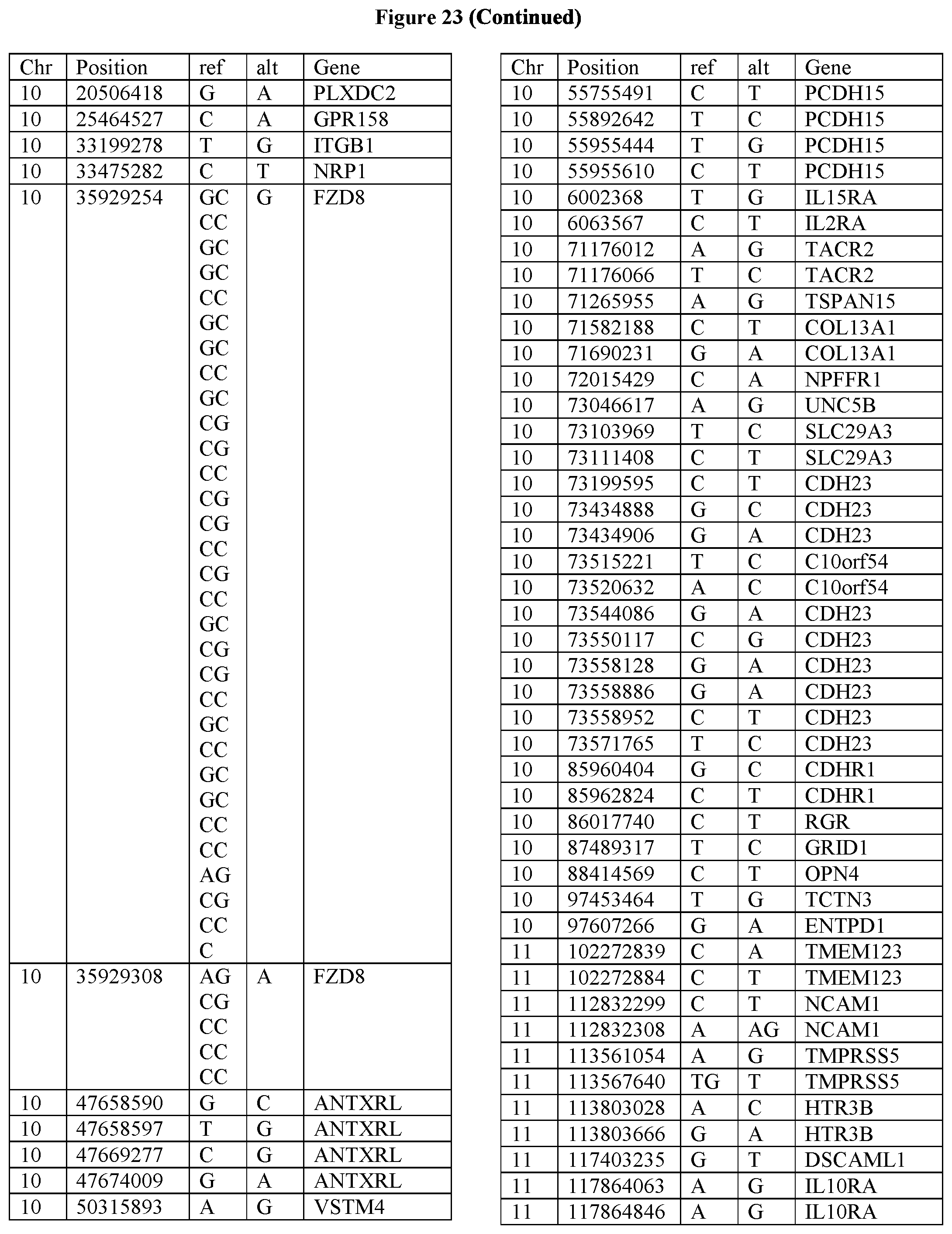
D00062

D00063

D00064
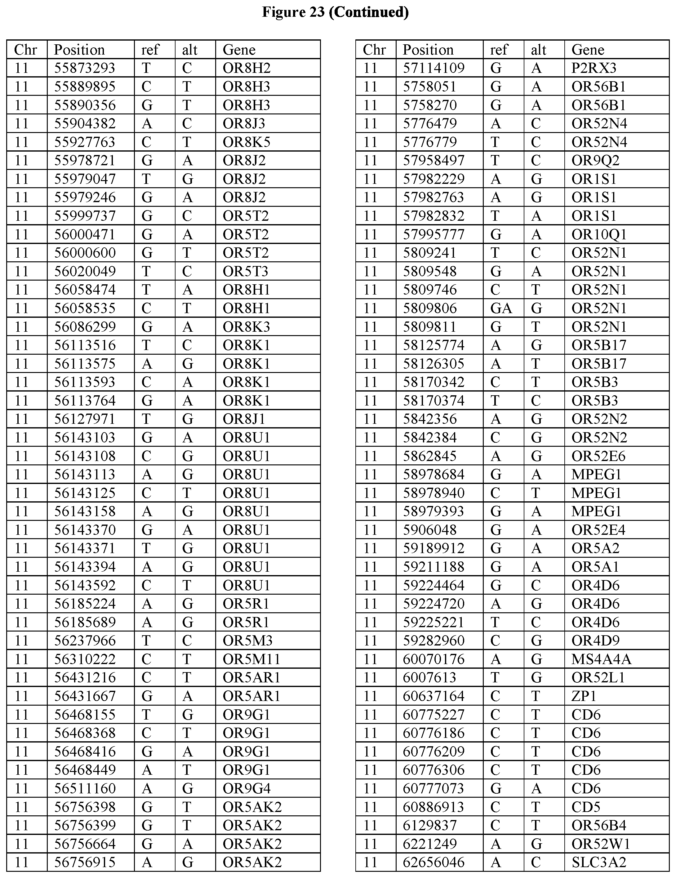
D00065

D00066
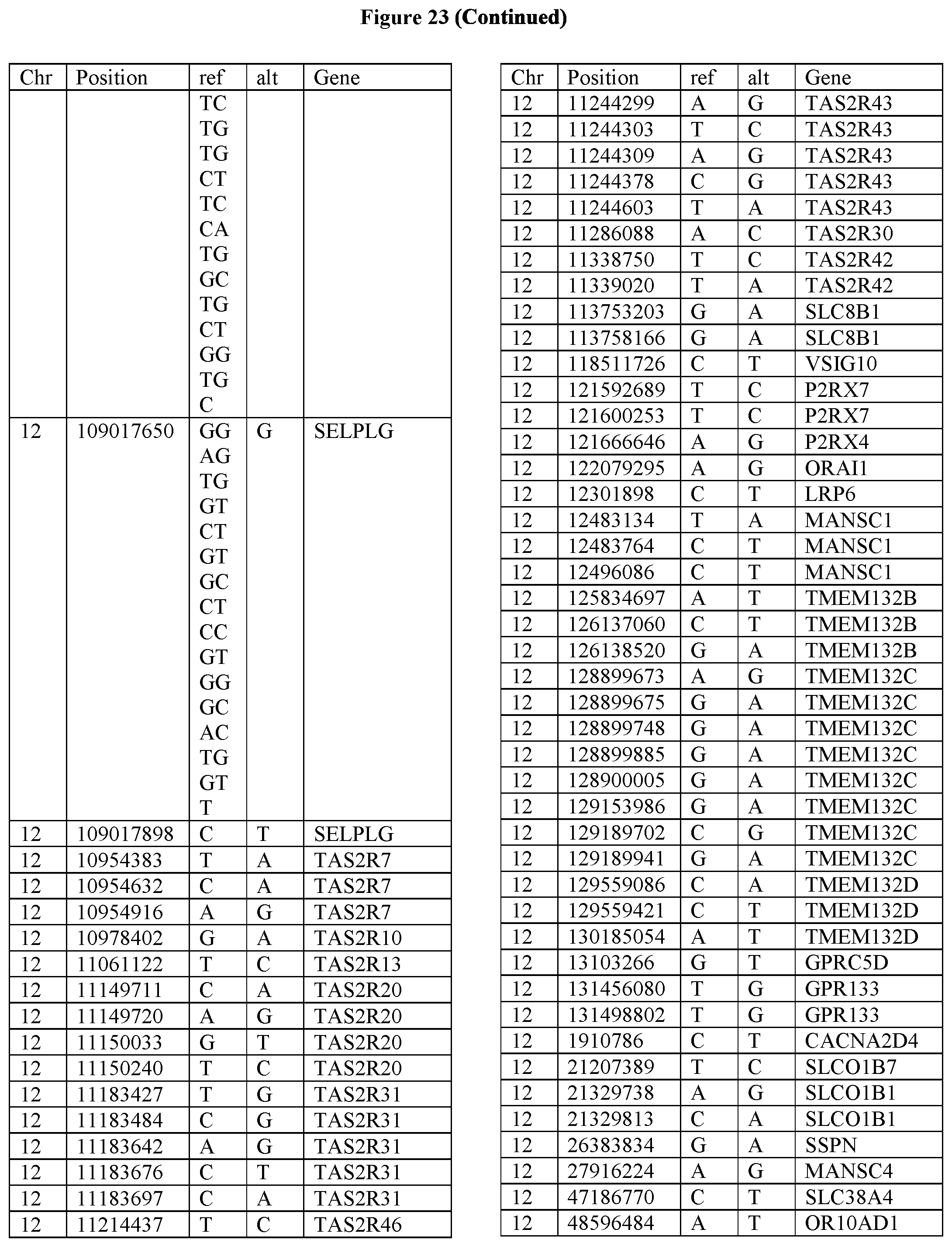
D00067

D00068

D00069

D00070

D00071
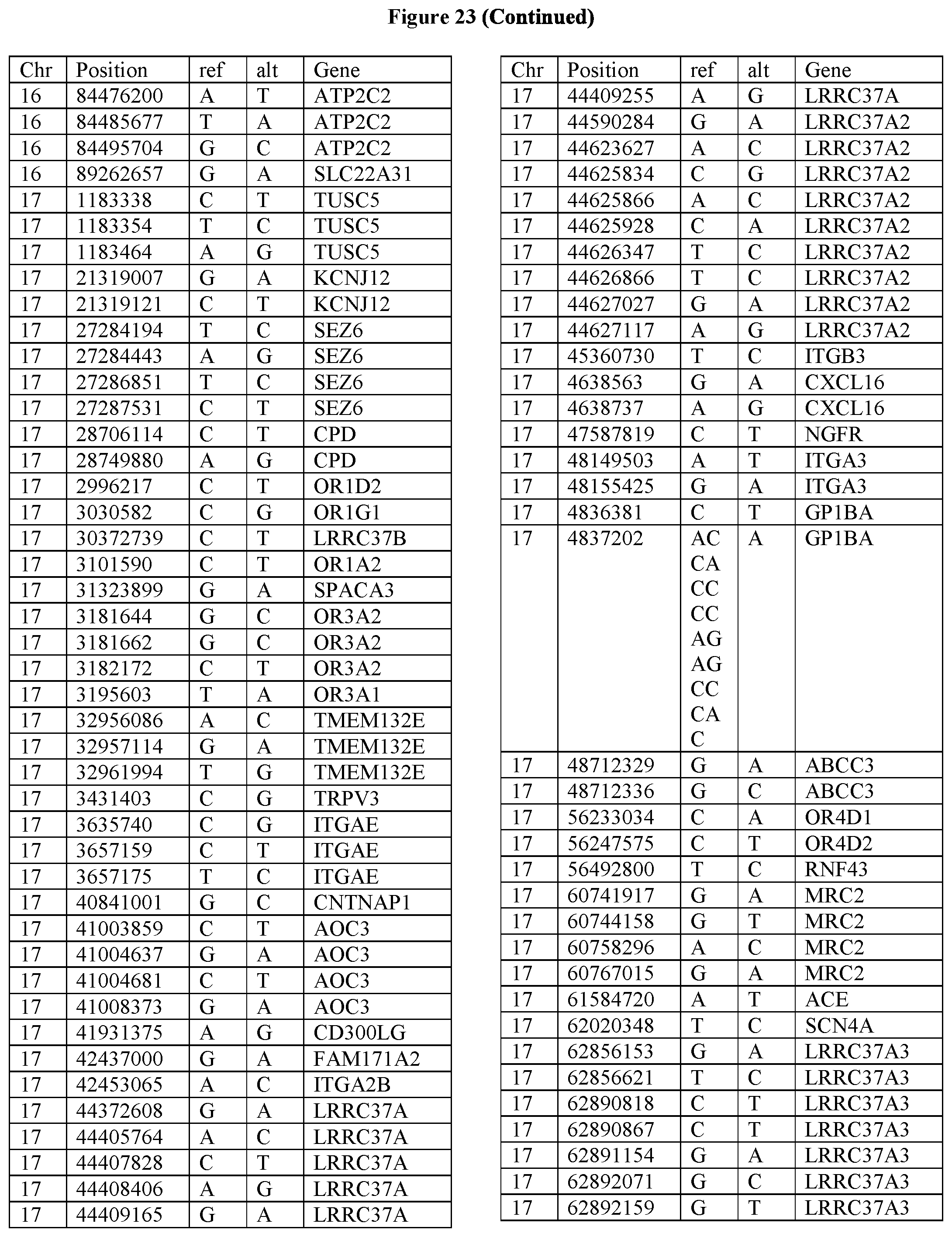
D00072
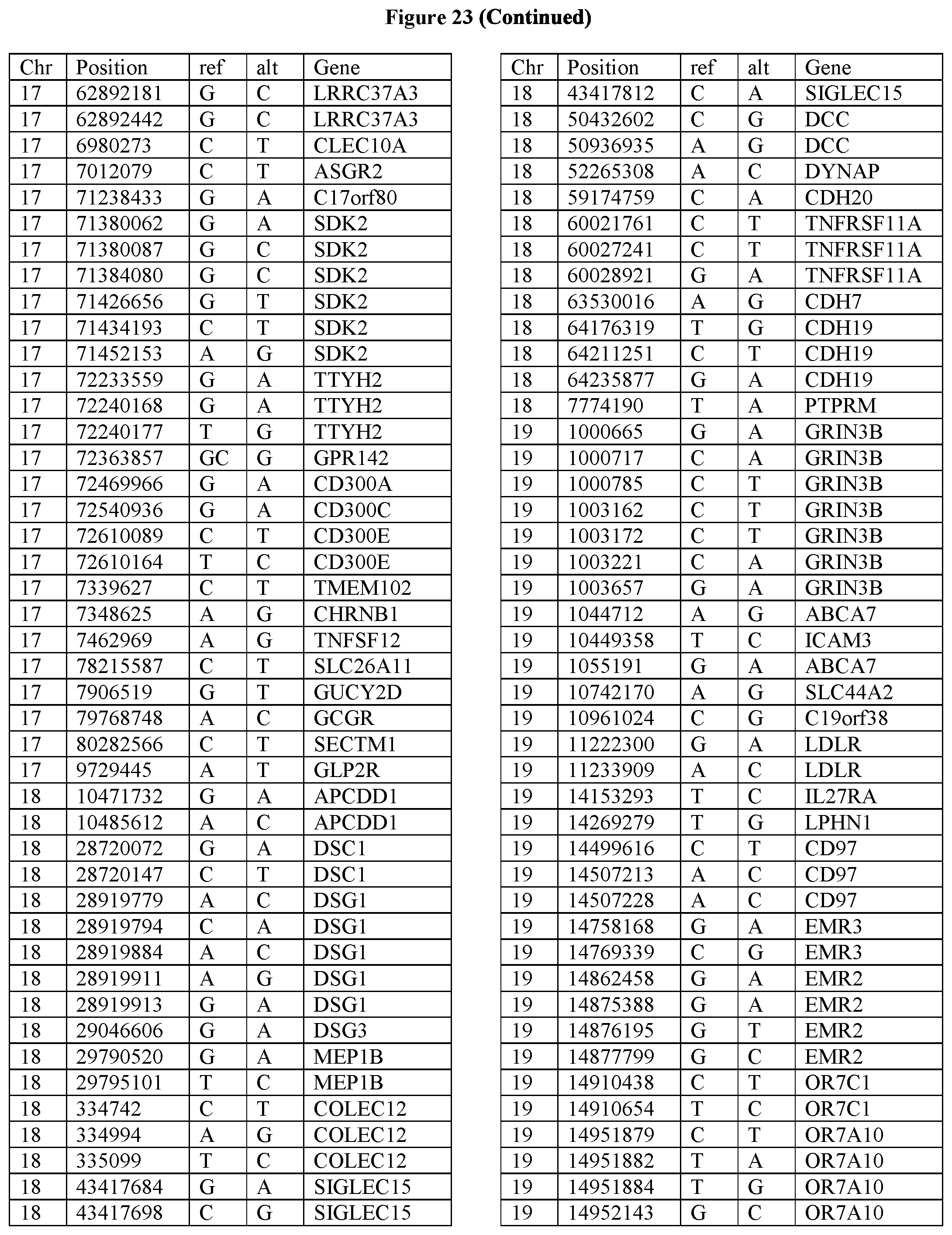
D00073

D00074

D00075
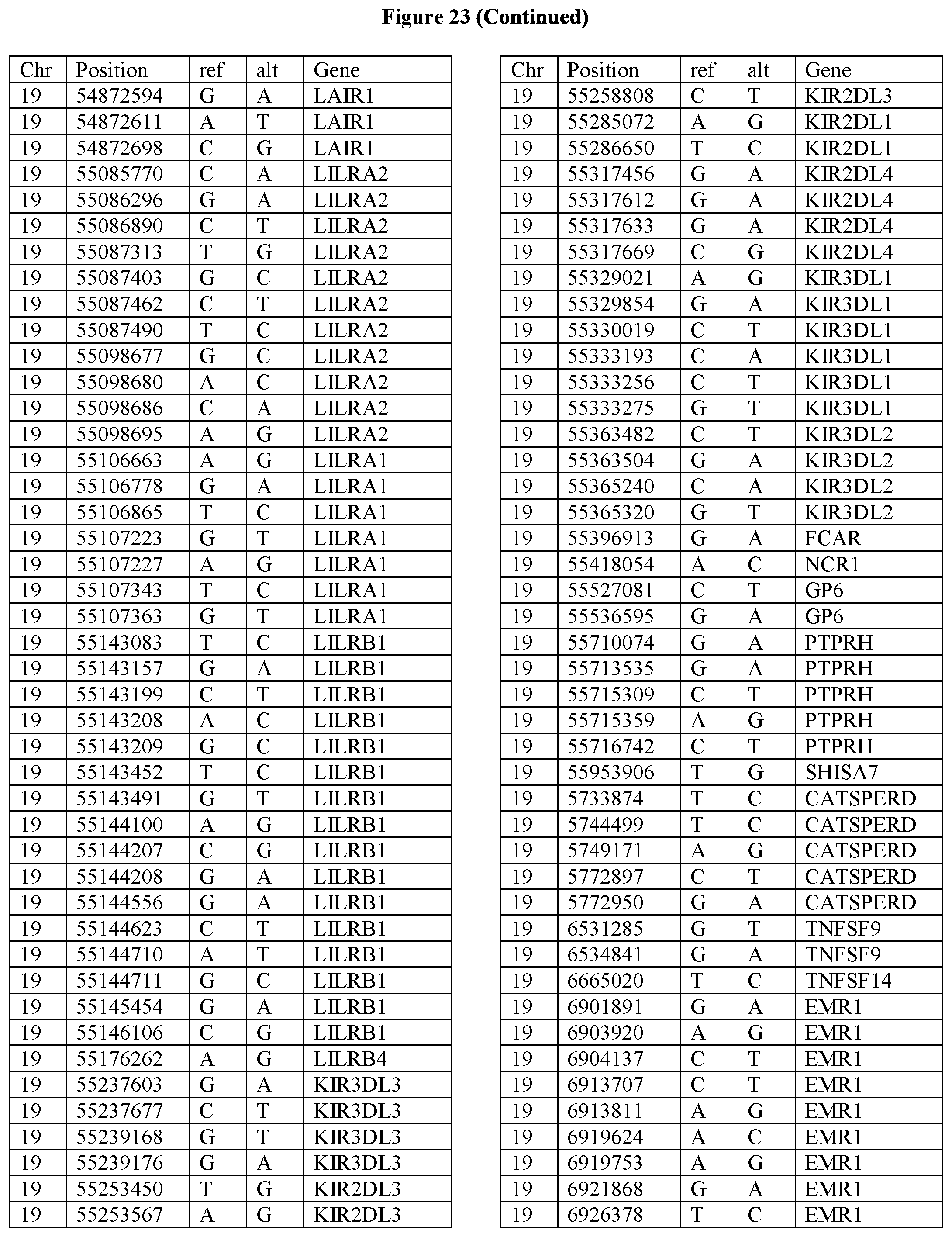
D00076
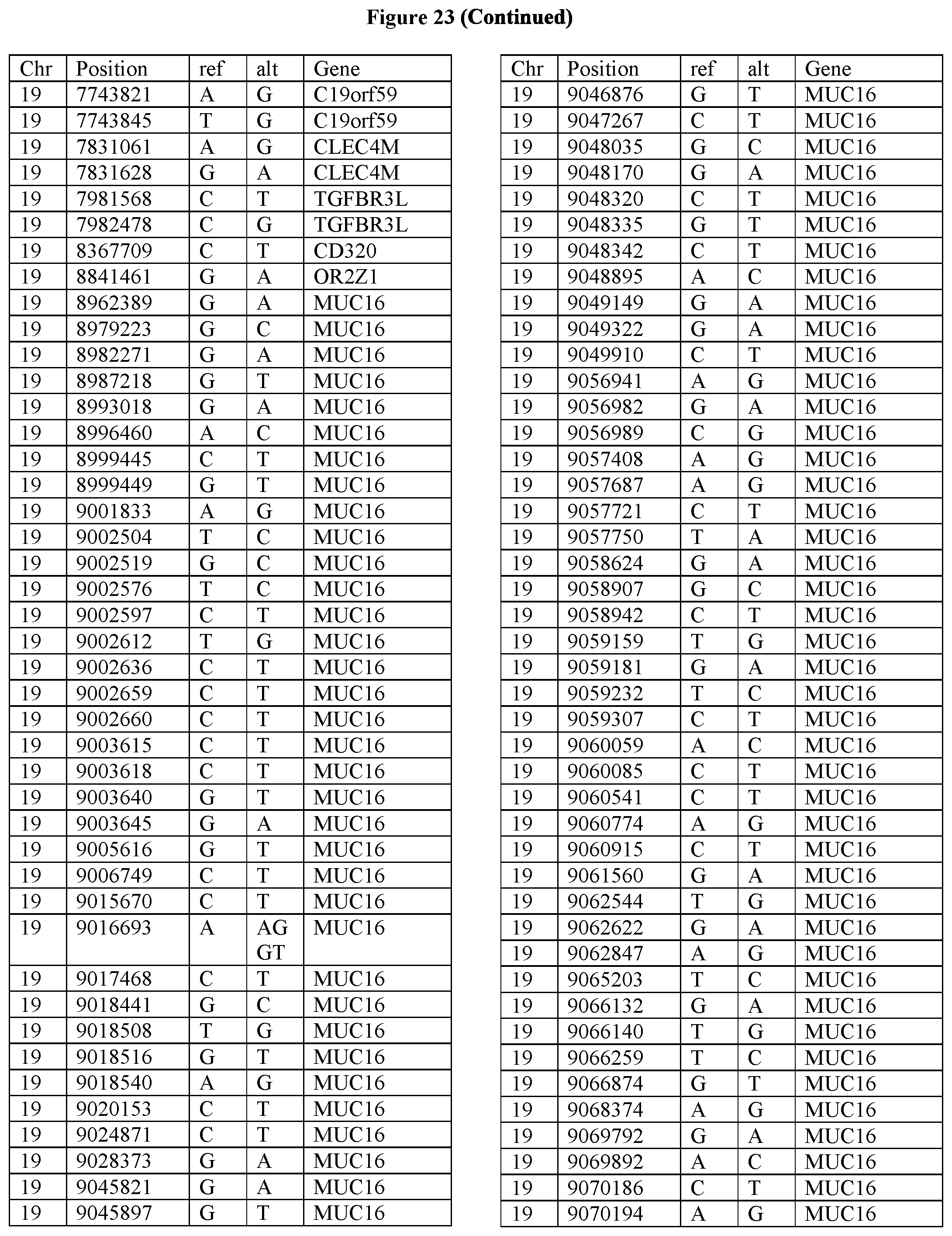
D00077
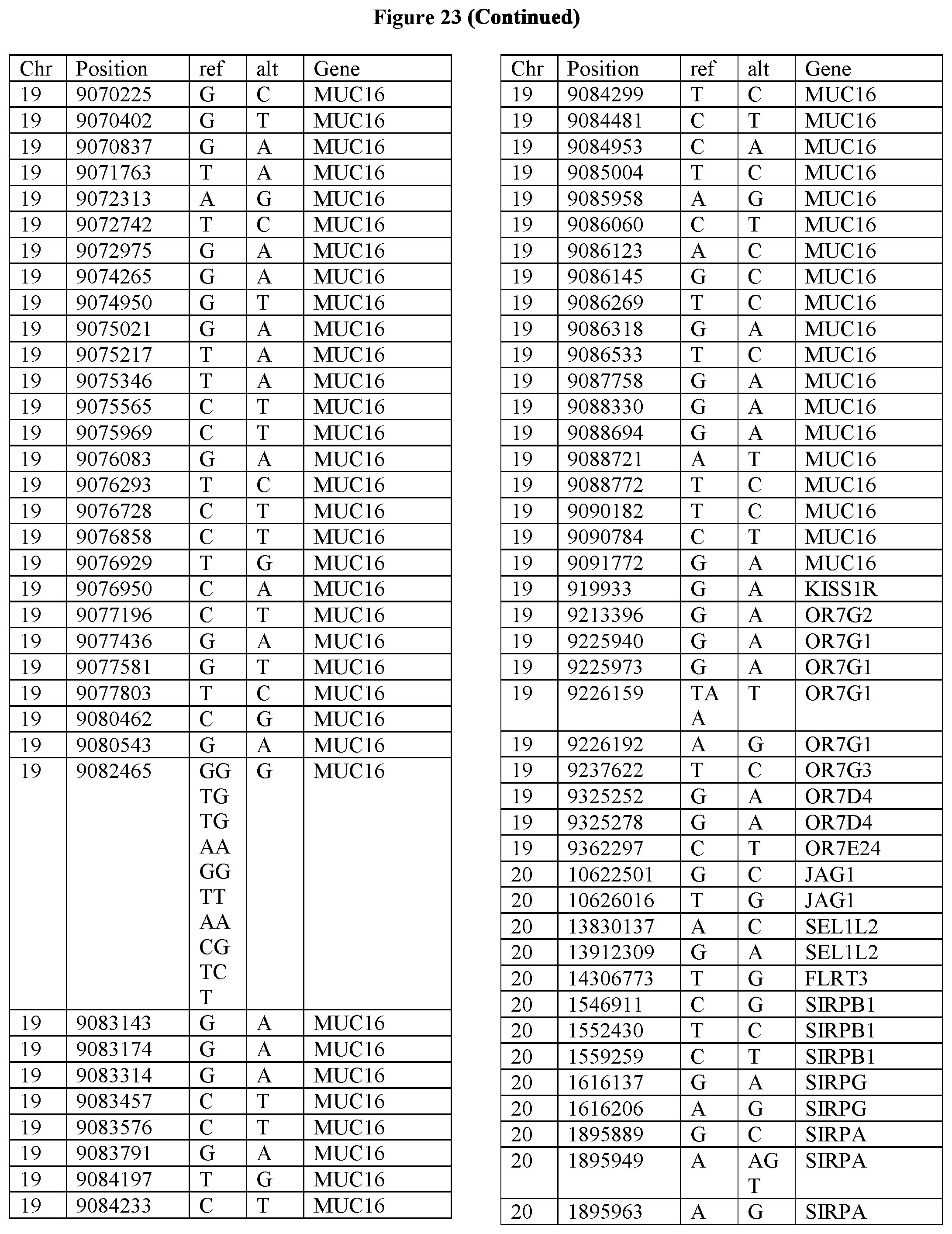
D00078

D00079


S00001
XML
uspto.report is an independent third-party trademark research tool that is not affiliated, endorsed, or sponsored by the United States Patent and Trademark Office (USPTO) or any other governmental organization. The information provided by uspto.report is based on publicly available data at the time of writing and is intended for informational purposes only.
While we strive to provide accurate and up-to-date information, we do not guarantee the accuracy, completeness, reliability, or suitability of the information displayed on this site. The use of this site is at your own risk. Any reliance you place on such information is therefore strictly at your own risk.
All official trademark data, including owner information, should be verified by visiting the official USPTO website at www.uspto.gov. This site is not intended to replace professional legal advice and should not be used as a substitute for consulting with a legal professional who is knowledgeable about trademark law.