Vaccine Composition And Adjuvant
Kind Code
U.S. patent application number 16/616957 was filed with the patent office on 2020-08-13 for vaccine composition and adjuvant. The applicant listed for this patent is Atomis Inc.. Invention is credited to Daisuke ASARI, Shinji KATO.
| Application Number | 20200254089 16/616957 |
| Document ID | 20200254089 / US20200254089 |
| Family ID | 1000004840365 |
| Filed Date | 2020-08-13 |
| Patent Application | download [pdf] |


View All Diagrams
| United States Patent Application | 20200254089 |
| Kind Code | A1 |
| ASARI; Daisuke ; et al. | August 13, 2020 |
VACCINE COMPOSITION AND ADJUVANT
Abstract
An object of the present invention is to provide an excellent vaccine composition and adjuvant. The vaccine composition according to the present invention includes an antigen for inducing immunity and a Metal Organic Framework (MOF). The adjuvant according to the present invention includes a MOF.
| Inventors: | ASARI; Daisuke; (Kyoto, JP) ; KATO; Shinji; (Kyoto, JP) | ||||||||||
| Applicant: |
|
||||||||||
|---|---|---|---|---|---|---|---|---|---|---|---|
| Family ID: | 1000004840365 | ||||||||||
| Appl. No.: | 16/616957 | ||||||||||
| Filed: | June 6, 2018 | ||||||||||
| PCT Filed: | June 6, 2018 | ||||||||||
| PCT NO: | PCT/JP2018/021695 | ||||||||||
| 371 Date: | November 25, 2019 |
| Current U.S. Class: | 1/1 |
| Current CPC Class: | C07K 2317/567 20130101; A61K 9/0019 20130101; C07K 16/00 20130101; A61K 39/39 20130101 |
| International Class: | A61K 39/39 20060101 A61K039/39; C07K 16/00 20060101 C07K016/00 |
Foreign Application Data
| Date | Code | Application Number |
|---|---|---|
| Jun 6, 2017 | JP | 2017112115 |
Claims
1. A vaccine composition comprising an antigen for inducing immunity and a Metal Organic Framework (MOF).
2. The vaccine composition according to claim 1, further comprising an immune signal transducer.
3. The vaccine composition according to claim 1, wherein at least a part of the immune signal transducer is contained in pores of the MOF.
4. The vaccine composition according to claim 3, wherein the MOF is configured to decompose in vivo to release at least a part of the immune signal transducer.
5. The vaccine composition according to claim 2, wherein the immune signal transducer is a small molecule having a molecular weight of 1000 or less.
6. The vaccine composition according to claim 5, wherein the immune signal transducer is a gas at 25.degree. C. and 100 kPa.
7. The vaccine composition according to claim 2, wherein the immune signal transducer is a factor that is configured to act on keratinocytes, monocytes, lymphocytes, or granulocytes.
8. The vaccine composition according to claim 1, wherein the MOF comprises at least one metal element selected from the group consisting of calcium, magnesium, iron, zinc, aluminum, potassium, and sodium.
9. The vaccine composition according to claim 1, wherein the vaccine composition is configured to be administered on a skin and/or a mucous membrane.
10. The vaccine composition according to claim 1, wherein the vaccine composition is configured to be administered by an intradermal injection, a subcutaneous injection, or an intramuscular injection.
11. An adjuvant comprising a Metal Organic Framework (MOF).
12. The adjuvant according to claim 11, wherein the MOF contains an immune signal transducer in pores of the MOF.
13. The adjuvant according to claim 12, wherein the MOF is configured to decompose in vivo to release at least a part of the immune signal transducer.
14. The vaccine composition according to claim 3, wherein the immune signal transducer is a small molecule having a molecular weight of 1000 or less.
15. The vaccine composition according to claim 4, wherein the immune signal transducer is a small molecule having a molecular weight of 1000 or less.
16. The vaccine composition according to claim 14, wherein the immune signal transducer is a gas at 25.degree. C. and 100 kPa.
17. The vaccine composition according to claim 15, wherein the immune signal transducer is a gas at 25.degree. C. and 100 kPa.
18. The vaccine composition according to claim 2, wherein the immune signal transducer is a factor that is configured to act on keratinocytes, monocytes, lymphocytes, or granulocytes.
19. The vaccine composition according to claim 3, wherein the immune signal transducer is a factor that is configured to act on keratinocytes, monocytes, lymphocytes, or granulocytes.
20. The vaccine composition according to claim 4, wherein the immune signal transducer is a factor that is configured to act on keratinocytes, monocytes, lymphocytes, or granulocytes.
Description
CROSS-REFERENCE TO RELATED APPLICATIONS
[0001] This is a 371 application of International Patent Application Number PCT/JP2018/021695 filed Jun. 6, 2018 claiming priority from Japanese Patent Application Number JP2017-112115 filed Jun. 6, 2017, and the disclosures of which are incorporated herein by reference in their entirety.
TECHNICAL FIELD
[0002] The present invention relates to vaccine compositions and adjuvants.
BACKGROUND ART
[0003] Various vaccine compositions have conventionally been used for the prevention or treatment of infectious diseases. Adding adjuvants to vaccine compositions is a common practice for reinforcing their antigenicity.
[0004] On the other hand, a group of materials called Metal Organic Framework (MOF) or Porous Coordination Polymer (PCP) has attracted attention in such fields as gas separation, which are distant from the community of immunology. The MOFs typically form a porous structure by combination of a metal and a multidentate ligand.
CITATION LIST
Patent Literature
[0005] [Patent Literature 1] WO2004/037895 [0006] [Patent Literature 2] WO2009/042802
Non-Patent Literature
[0006] [0007] [Non-Patent Literature 1] David Farrusseng, Metal-Organic Frameworks: Applications from Catalysis to Gas Storage, Wiley, 2011 [0008] [Non-Patent Literature 2] Yabing He et al. Methane Storage in Metal-Organic Frameworks, Chem Soc Rev., 2014
SUMMARY OF THE INVENTION
Technical Problem
[0009] An object of the present invention is to provide an excellent vaccine composition and adjuvant.
Solution to Problem
[0010] Some aspects of the present invention are as described below.
[1] A vaccine composition comprising an antigen for inducing immunity and a Metal Organic Framework (MOF). [2] The vaccine composition according [1], further comprising an immune signal transducer. [3] The vaccine composition according to [2], wherein at least a part of the immune signal transducer is contained in pores of the MOF. [4] The vaccine composition according to [3], wherein the MOF is configured to decompose in vivo to release at least a part of the immune signal transducer. [5] The vaccine composition according to any one of [2]-[4], wherein the immune signal transducer is a small molecule having a molecular weight of 1000 or less. [6] The vaccine composition according to [5], wherein the immune signal transducer is a gas at 25.degree. C. and 100 kPa. [7] The vaccine composition according to any one of [2]-[6], wherein the immune signal transducer is a factor that is configured to act on keratinocytes, monocytes, lymphocytes, or granulocytes. [8] The vaccine composition according to any one of [1]-[7], wherein the MOF comprises at least one metal element selected from the group consisting of calcium, magnesium, iron, zinc, aluminum, potassium, and sodium. [9] The vaccine composition according to any one of [1]-[8], wherein the vaccine composition is configured to be administered on a skin and/or a mucous membrane. [10] The vaccine composition according to any one of [1]-[8], wherein the vaccine composition is configured to be administered by an intradermal injection, a subcutaneous injection, or an intramuscular injection. [11] An adjuvant comprising a Metal Organic Framework (MOF). [12] The adjuvant according to [11], wherein the MOF contains an immune signal transducer in its pores. [13] The adjuvant according to [12], wherein the MOF is configured to decompose in vivo to release at least a part of the immune signal transducer.
Advantageous Effects of Invention
[0011] The present invention makes it possible to provide an excellent vaccine composition and adjuvant.
BRIEF DESCRIPTION OF DRAWINGS
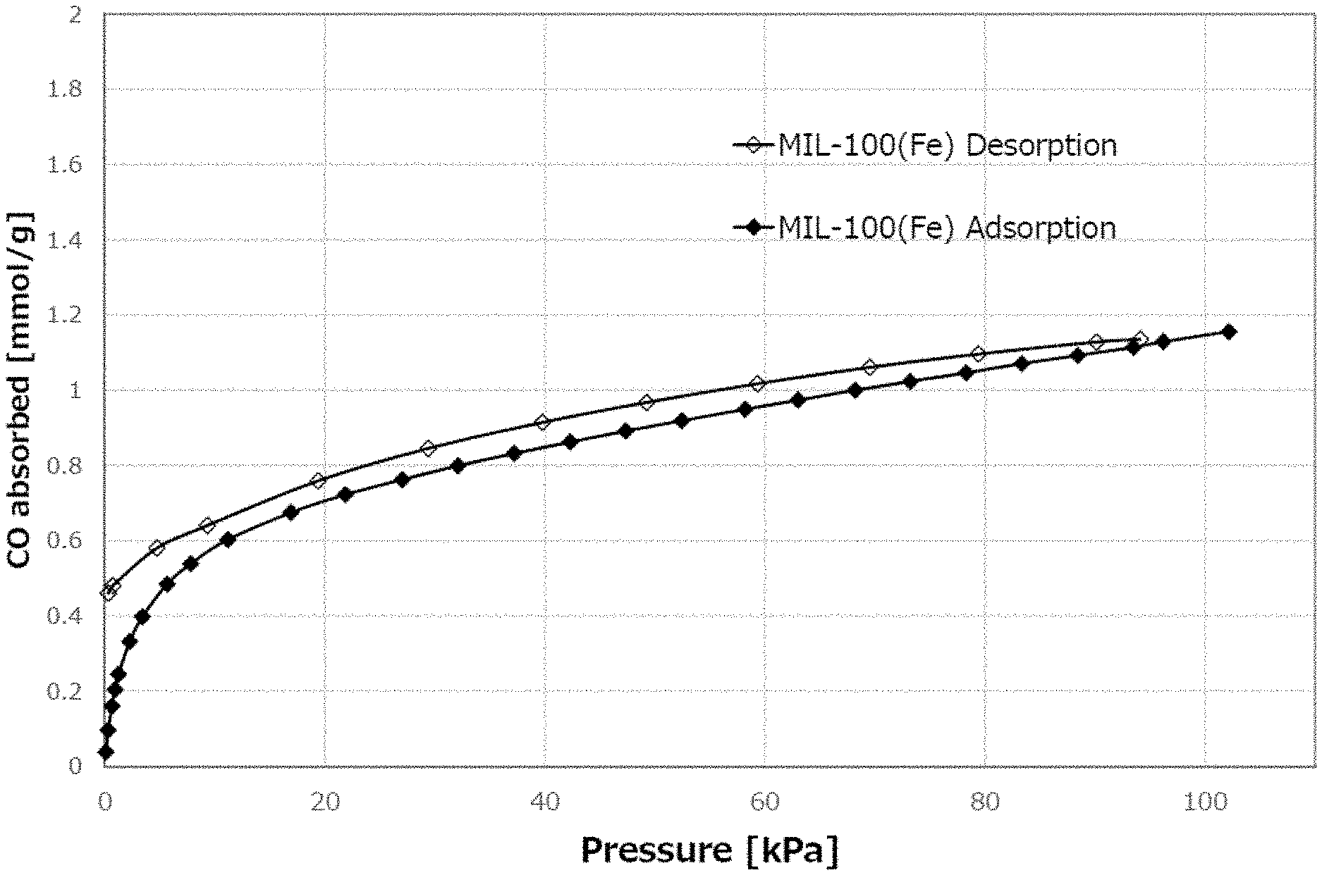
[0012] FIG. 1A is a CO adsorption profile of a metal organic framework AP004 [MIL-100 (Fe)].
[0013] FIG. 1B is a NO adsorption profile of a metal organic framework AP004 [MIL-100 (Fe)].
[0014] FIG. 2 is a NO adsorption profile of a metal organic framework AP104 (BioMIL-3).
[0015] FIG. 3 is a graph showing the results of measurement of antigen-specific antibody titers in mouse serum.
[0016] FIG. 4A is a graph showing the results of measurement of OVA-specific cytokine production.
[0017] FIG. 4B is a graph showing the results of measurement of OVA-specific cytokine production.
DESCRIPTION OF EMBODIMENTS
[0018] Vaccine compositions and adjuvants according to an embodiment of the present invention are hereinafter described.
[0019] The vaccine composition according to the present disclosure includes an antigen for inducing immunity and a Metal Organic Framework (MOF). The MOF mainly functions as an adjuvant in the composition.
[0020] The antigens can be any substances that may induce an immune response. For instance, the antigens can be proteins or peptides. Using an antigen with a low molecular weight is commonly preferred in transdermal administration where the skin permeability of the antigen is required. A peptide with about 8 to 12 amino acids may thus be preferably used in such an occasion. Other antigens such as cancer antigen peptide or an antigen derived from an infectious pathogen can also be used.
[0021] Alternatively, autoantigens (for example, antigens related to autoimmune diseases), endogenous antigens (for example, antigens derived from cancer), foreign antigens (for example, antigens related to allergies or antigens derived from viruses and bacteria), or other antigens can also be used.
[0022] Examples of the antigens related to autoimmune diseases include:
[0023] Amyloid .beta., which is believed to cause Alzheimer's disease, or its precursors, or its fragment proteins or peptides;
[0024] .alpha.-synuclein, which is believed to cause Parkinson's disease, or its fragment proteins or peptides;
[0025] .alpha.-fodrin, which is believed to cause Sjogren's syndrome, or its fragment proteins or peptides;
[0026] Thyroid hormone receptor, which is believed to cause Graves' disease, or its fragment proteins or peptides;
[0027] Ganglioside, which is believed to cause Guillain-Barre syndrome, or its fragment proteins or peptides;
[0028] DNA or its fragments, which is believed to cause systemic lupus erythematosus; Cholesterol ester transfer protein, apolipoprotein, or oxidized LDL, which are believed to cause arteriosclerosis, or their fragment proteins or peptides;
[0029] Angiotensin I/II, which is believed to cause high blood pressure, or its fragment proteins or peptides;
[0030] Insulin, GAD, or IL-1.beta., which are believed to cause type 1 diabetes, or their fragment proteins or peptides;
[0031] Acetylcholine receptor, which is believed to cause myasthenia gravis, or its fragment proteins or peptides;
[0032] TNF.alpha. or IL-6, which are believed to cause chronic rheumatoid arthritis, or their fragment proteins or peptides; and
[0033] TRANCE or RANKL, which are believed to cause osteoporosis, or their fragment proteins or peptides.
[0034] Examples of the antigens derived from cancer include WT1, PR1, GPC3, HER-2, MAGE-A1, MAGE-A2, MAGE-A3, tyrosinase, gp100, CEA, hTRT, EGF receptor, mTERT, PRAME, PSMA, PSA-1, cytochrome p450, NY-ESO-1, Survivine, MUC-1, MAGE-A10, or PAP, or proteins or peptides derived therefrom.
[0035] Examples of the Antigens Related to Allergies Include:
[0036] Allergens derived from trees (e.g. acacia, alder tree, velvet blue radish, beech, birch, maple, mountain cedar, red cedar, boxwood, cypress, American elm, Chinese elm, pseudotsuga japonica, rubber, eucalyptus, Japanese hackberry, hickory, American linden, sugar maple, mesquite, paper mulberry, konara oak, olive, pecan, pepper, pine, privet, Russian olive, American sycamore, elderberry, black walnut, or black willow);
[0037] Allergens derived from vegetations (e.g. cotton, gypsy moth, Kentucky bluegrass, bromus japonicus, corn, meadow fescue, Johnson grass, oats, moths, knuckles, barley, rice, vernal grass, timothy, amaranthaceae, red-tailed geese, red-tailed eels, red-tailed geese, tall goldenrod, kochia [firebush], lambs quarters, calendula, nettle, rough pigweed, English plantain, tall ragweed, short ragweed, false ragweed, Russian thistle, common sagebrush, licorice, or sheep sorrel);
[0038] Allergens derived from insects (e.g. silkworms, ticks, bees, wasps, ants, or cockroaches);
[0039] Allergens derived from fungi (e.g. Alternaria, Aspergillus, Botulinum, Candida, Cephalosporium, Carbaria, Epicoccum, Epidermis, Fusarium, Helminthosporumium, Chain Cladosporium, Mucoraceae, Peniculium, Pullularia pullulans, or Rhizopus);
[0040] Allergens derived from animal hair (e.g. hair of dogs, cats, or birds);
[0041] Allergens derived from house dust;
[0042] Allergens derived from foods; and
[0043] Haptens involved in metal allergy.
[0044] Examples of the diseases affected by the infectious pathogen include:
[0045] Viral diseases such as those affected by infections from Adenovirus, Herpes virus (e.g. HSV-I, HSV-II, CMV, or VZV), Poxvirus (e.g. pressure ulcer or vaccinia, or orthopox virus such as contagious molluscum), Picornavirus (e.g. rhinovirus or enterovirus), Orthomyxovirus (e.g. influenza virus), Paramyxovirus (e.g. parainfluenza virus, mumps virus, measles virus, or respiratory syncytial virus [RSV]), Coronavirus (e.g. SARS), Papovavirus (e.g. papilloma viruses such as the ones that cause genital warts, vulgaris, or plantar costus), Hepadnavirus (e.g. hepatitis B virus), Flavivirus (e.g. hepatitis C virus or dengue virus), or Retroviruses (e.g.lentivirus such as HIV);
[0046] Bacterial diseases such as those affected by infections from Escherichia, Enterobacter, Salmonella, Staphylococcus, Shigella, Listeria, Aerobacter, Helicobacter, Klebsiella, Proteus, Pseudomonas, Streptococcus, Chlamydia, Mycoplasma, Pneumococci, Neisseria, Clostridium, Bacillus, Corynebacterium, Mycobacterium, Campylobacter, Vibrio, Serratia, Providencia, Chromobacterium, Brucella, Yersinia, Haemophilus, or Bordetella;
[0047] Fungal diseases such as Chlamydia, Candidiasis, Aspergillosis, Histoplasmosis, or Cryptococcal meningitis; and
[0048] Other diseases such as Malaria, Pneumocystis carinii pneumonia, Leishmaniasis, Cryptosporidiosis, Toxoplasmosis, or Trypanosoma infection.
[0049] Particularly suitable antigens are Ovalbumin (OVA), Pneumococci, Influenza vaccines, Cryj1 (a major allergen of cedar pollen), or HPV16 recombinant protein.
[0050] Only one type of antigen may be used, or two or more types thereof may be used in combination. The content of the antigen in the vaccine composition is, for example, in the range of 1.times.10.sup.-7 to 1.times.10.sup.-1 mass %, preferably in the range of 1.times.10.sup.-6 to 1.times.10.sup.-2 mass %, more preferably in the range of 2.times.10.sup.-6 to 2.times.10.sup.-3 mass %.
[0051] As described above, the MOF is formed with a combination of metal(s) and multidentate ligand(s). The mechanism by which the MOF acts as adjuvant is not perfectly clear. The inventors however have attributed the reason to the metal and/or ligand in the MOF interacting with antigens and/or immune cells in some ways. As used herein, the "multidentate ligand" means a ligand that can form two or more coordinate bond.
[0052] Any kinds of MOFs can be used in the vaccine composition. Appropriately combining the type and coordination number of the metal ion with the type and topology of the multidentate ligand leads to a MOF with a desired structure. The MOF may be configured to decompose in vivo. The decomposition would expose the metal and the ligand constituting the MOF, by which the MOF might function as adjuvant more efficiently. The MOF can be crystalline or amorphous.
[0053] The metal elements in the MOF can be, for example, any elements belonging to alkali metals (Group 1), alkaline earth metals (Group 2), or transition metals (Groups 3 to 12). From the viewpoint of biocompatibility, it is preferable to use at least one metal element selected from the group consisting of calcium, magnesium, iron, zinc, aluminum, potassium, and sodium. However, any metal elements other than these preferable elements can also be used as long as biocompatibility of a MOF as a whole is ensured.
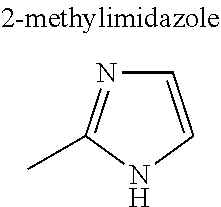
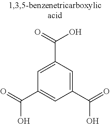
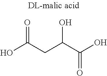
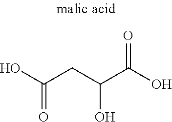
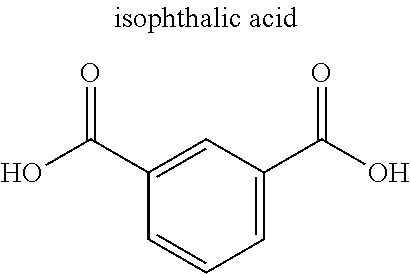
[0054] The multidentate ligand in the MOF typically is an organic ligand, examples of which include carboxylate anion and heterocyclic compound. Examples of the carboxylic acid anion include dicarboxylic acid anion and tricarboxylic acid anion. Specific examples include anions of citric acid, malic acid, terephthalic acid, isophthalic acid, trimesic acid, and derivatives thereof. Examples of the heterocyclic compound include bipyridine, imidazole, adenine, and derivatives thereof. Alternatively, the ligand may be an amine compound, a sulfonate anion, or a phosphate anion. The MOF may further contain monodentate ligand(s).
[0055] The combination of the metal and the ligand forming the MOF can be appropriately determined according to the expected function and the desired pore size. The MOF may contain two or more types of metal elements, and may contain two or more types of ligands. The MOF can be surface-modified with a polymer or other modifiers.
[0056] Specific examples of the MOF include those listed in Table 1 of the Non-Patent Literature 2. Those shown in Tables 1 to 3 below may also be used as the MOF. These are non-limiting lists, and other MOFs can also be used.
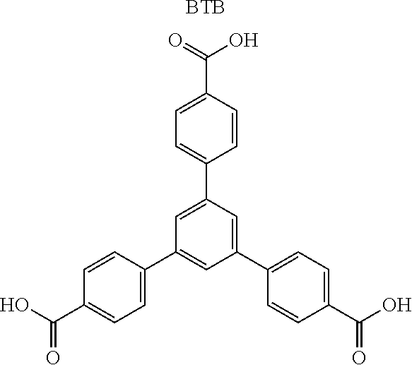
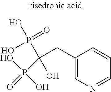
TABLE-US-00001 TABLE 1 Name/ Metal Abbreviation (Cation) Ligand (Anion) CPL-1 Cu pzdc (2,3-pyrazinedicarboxylic acid), pyz (pyrazine) Cu.sub.3(btc).sub.2 Cu BTC (trimesic acid) Zn.sub.2(14bdc).sub.2(dabco) Zn BDC (terephthalic acid), dabco (1,4- diazabicyclo[2,2,2]octane) ZIF-8 Zn imidazole HKUST-1 Cu 1,3,5-benzenetricarboxylic acid Mg.sub.3(C.sub.12O.sub.14H.sub.10) Mg citric acid Ca.sub.2(C.sub.8O.sub.12H.sub.6) Ca malic acid Ca.sub.3(C.sub.12O.sub.14H.sub.10) Ca citric acid Ca(C.sub.4O.sub.6H.sub.4) Ca malic acid Cu(IPA) Cu isophthalic acid MgBDC-1 Mg BDC (terephthalic acid) MgDHBDC-1 Mg DHBDC (2,5-dihydroxyterephthalic acid) MgOBA-1 Mg OBA (4,4'-oxobisbenzoic acid) MgBTC-1 Mg BTC (trimesic acid) MgBTB-1 Mg BTB (1,3,5-tri(4'-carboxy-4,4'- biphenyl)benzene) Mg BTB-2 Mg BTB (1,3,5-tri(4'-carboxy-4,4'- biphenyl)benzene) MgBTB-3 Mg BTB (1,3,5-tri(4'-carboxy-4,4'- biphenyl)benzene) MgBTB-4 Mg BTB (1,3,5-tri(4'-carboxy-4,4'- biphenyl)benzene) MgBBC-1 Mg BBC (4,4'-4''-benzene-1,3,5-triyl- tri-biphenylcarboxylic acid) MIL-100(Fe) Fe BTC (trimesic acid) MIL-101 Fe BDC (terephthalic acid) MIL-53 Fe BDC (terephthalic acid) BioMIL-5 Zn azelaic acid CaZol nMOF Ca zoledronic acid IRMOF-2 Zn o-Br-BDC (o-bromoterephthalic acid) IRMOF-3 Zn H.sub.2N-BDC (2-aminoterephthalic acid) IRMOF-4 Zn [C.sub.3H.sub.7O].sub.2-BDC IRMOF-5 Zn [C.sub.5H.sub.11O].sub.2-BDC IRMOF-6 Zn [C.sub.2H.sub.4]-BDC IRMOF-7 Zn 1,4-NDC (1,4-naphthalenedicarboxylic acid) IRMOF-8 Zn 2,6-NDC (2,6-naphthalenedicarboxylic acid) IRMOF-9 Zn BPDC (4,4'-biphenyldicarboxylic acid) IRMOF-10 Zn BPDC (4,4'-biphenyldicarboxylic acid) IRMOF-11 Zn HPDC (tetrahydropyrene-2,7-dicarboxylic acid) IRMOF-12 Zn HPDC (tetrahydropyrene-2,7-dicarboxylic acid) IRMOF-13 Zn PDC (pyrene dicarboxylic acid) IRMOF-14 Zn PDC (pyrene dicarboxylic acid) IRMOF-15 Zn TPDC (terphenyl dicarboxylic acid) IRMOF-16 Zn TPDC (terphenyl dicarboxylic acid)
TABLE-US-00002 TABLE 2 Name/ Metal Abbreviation (Cation) Ligand (Anion) Zn.sub.3(BTC).sub.2 Zn BTC (trimesic acid) Zn.sub.4O(NDC) Zn 1,4-NDC (1,4-naphthalenedicarboxylic acid) Mg(Formate) Mg formic acid Fe(Formate) Fe formic acid Mg(C.sub.6H.sub.4O.sub.6) Mg DHBDC (2,5-dihydroxyterephthalic acid) ZnC.sub.2H.sub.4BDC Zn [C.sub.2H.sub.4]-BDC MOF-49 Zn m-BDC BPR95A2 Zn BDC (terephthalic acid) BPR76D5 Zn BzPDC BPR68D10 Zn BTC (trimesic acid) BPR56E1 Zn BDC (terephthalic acid) BPR49B1 Zn BDC (terephthalic acid) BPR43G2 Zn BDC (terephthalic acid) NO336 Fe formic acid NO335 Fe formic acid NO333 Fe formic acid PCN-14 Nb 5,5'-(9,10-anthracenediyl) diisophosphate Zn.sub.4BNDC Zn BNDC (1,1'-binaphthyl-4,4'-dicarboxylic acid) Zn.sub.3(BPDC) Zn BPDC (4,4'-biphenyldicarboxylic acid) ZnDBP Zn DBP (dibenzyl phosphate) Zn.sub.3(PDC).sub.2.5 Zn PDC (pyrene dicarboxylic acid) Zn(HPDC) Zn HPDC (tetrahydropyrene-2,7-dicarboxylic acid) Zn(NDC) Zn 2,6-NDC (2,6-naphthalenedicarboxylic acid) MOF-37 Zn 2,6-NDC (2,6-naphthalenedicarboxylic acid) MOF-20 Zn 2,6-NDC (2,6-naphthalenedicarboxylic acid) MOF-12 Zn ATC (1,3,5,7-adamantanetetracarboxylic acid) Zn(ADC) Zn ADC (acetylenedicarboxylic acid) MOF-0 Zn BTC (trimesic acid) MOF-2 Zn BDC (terephthalic acid) MOF-3 Zn BDC (terephthalic acid) MOF-4 Zn BTC (trimesic acid) MOF-5 Zn BDC (terephthalic acid) MOF-38 Zn BTC (trimesic acid) MOF-31 Zn ADC (acetylenedicarboxylic acid) MOF-69A Zn BPDC (4,4'-biphenyldicarboxylic acid) MOF-69B Zn 2,6-NDC (2,6-naphthalenedicarboxylic acid) MOF-33 Zn ATB (adamantanetetrabenzoic acid) MOF-36 Zn MTB (methanetetrabenzoic acid) MOF-39 Zn BTB (1,3,5-tri(4'-carboxy-4,4'-biphenyl)benzene)
TABLE-US-00003 TABLE 3 Metal Name/ (Cat- Abbreviation ion) Ligand (Anion) NO305 Fe formic acid NO306A Fe formic acid BPR48A2 Zn BDC (terephthalic acid) Zn(C.sub.2O.sub.4) Zn oxalic acid MOF-48 Zn 2,6-NDC (2,6-naphthalenedicarboxylic acid) MOF-47 Zn BDC(CH.sub.3).sub.4 Zn.sub.3(BTC).sub.2 Zn BTC (trimesic acid) MOF-n Zn BTC (trimesic acid) Zehex Zn BTB (1,3,5-tri(4'-carboxy-4,4'- biphenyl)benzene) AS16 Fe BDC (terephthalic acid) AS27-3 Fe BDC (terephthalic acid) AS54-3 Fe BPDC (4,4'-biphenyldicarboxylic acid) AS61-4 Fe m-BDC AS68-7 Fe m-BDC Zn.sub.8(ad).sub.4(PDAC).sub.6(OH).sub.2 Zn adenine, PDAC (1,4-diphenyl diacrylic acid) Zn.sub.8(ad).sub.4(SBDC).sub.6(OH).sub.2 Zn adenine, SBDC (4,4'-stilbene dicarboxylic acid) Zn.sub.8(ad).sub.4(BPDC).sub.6(OH).sub.2 Zn adenine, BPDC Zn.sub.8(ad).sub.4(NDC).sub.6(OH).sub.2 Zn adenine, 2,6-NDC M-CPO-27 Mg DHBDC (2,5-dihydroxyterephthalic acid) bio-MOF-1 Zn adenine, BPDC UMCM-1 Zn BTB (1,3,5-tri(4'-carboxy-4,4'- biphenyl)benzene) UMCM-2 Zn BTB (1,3,5-tri(4'-carboxy-4,4'- biphenyl)benzene) MOF-210 Zn BTE (4,4',4''-[benzene-1,3,5-triyl-tris (ethyne-2,1-diyl)] tribenzoic acid), BPDC bio-MOF-100 Zn adenine, BPDC NU-110E Cu J. Am. Chem. Soc. 2012, 134, 15016-15021 CD-MOF-1 K .gamma.-CD (.gamma.-cyclodextrin) porph@MOM-4 Fe porphyrin, BTC porph@MOM-8 Mg porphyrin, BTC porph@MOM-9 Zn porphyrin, BTC ZnPO-MOF Zn metalloporphyrin pyridyl, TCPB (1,2,4,5-Tetrakis(4- carboxyphenyl)benzene) Uio-66 Fe DCBDT (1,4-dicarboxylbenzene-2,3- dithiolate) Mg(H.sub.2gal) Mg caustic acid (3,4,5-trihydroxybenzoic acid)
[0057] Particularly preferable MOFs include the followings.
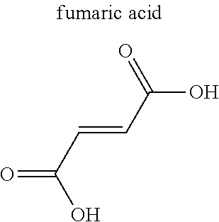
TABLE-US-00004 TABLE 4 Abbreviation Metal Ligand AP008 ZIF-8 Zn.sup.2+ ##STR00001## AP004 MIL-100(Fe) Fe.sup.3+ ##STR00002## AP006 Al(Fumarate) Al.sup.3+ ##STR00003## AP005 MIL-53(Al) Al.sup.3+ ##STR00004##
TABLE-US-00005 TABLE 5 Abbreviation Metal Ligand AP101 Ca.sup.2+ ##STR00005## AP104 BioMIL-3 Ca.sup.2+ ##STR00006## AP009 Mg(Formate) Mg.sup.2+ ##STR00007## AP014 La.sup.3+ ##STR00008##
TABLE-US-00006 TABLE 6 Abbreviation Metal Ligand AP102 Ca.sup.2+ ##STR00009## AP103 Ca.sup.2+ ##STR00010## AP105 Ca.sup.2+ ##STR00011##
TABLE-US-00007 TABLE 7 Abbreviation Metal Ligand AP107 Al.sup.3+ ##STR00012## AP106 Mg.sup.2+ ##STR00013## AP108 Ca.sup.2+ ##STR00014## AP015 Ca.sup.2+ ##STR00015##
TABLE-US-00008 TABLE 8 Abbreviation Metal Ligand AP001 Cu.sup.2+ ##STR00016## AP003 Fe-BTC Fe.sup.3+ ##STR00017## Ni-MOF-74 Ni.sup.2+ ##STR00018## Co-MOF-74 Co.sup.2+ ##STR00019##
TABLE-US-00009 TABLE 9 Abbreviation Metal Ligand MIL-88-A Fe.sup.2+ ##STR00020## MIL-88-B Fe.sup.2+ ##STR00021##
[0058] Only one type of MOF may be used, or two or more types thereof may be used in combination. The content of the MOF in the vaccine composition is, for example, in the range of 1.times.10.sup.-7 to 99.9999999 mass %, preferably in the range of 1.times.10.sup.-6 to 99.999999 mass %, and more preferably in the range of 5.times.10.sup.-6 to 99.99999 mass %.
[0059] The vaccine composition according to one embodiment of the present invention may further contain an immune signal transducer. Adopting such a configuration can further enhance the effect of administering the vaccine composition. As used herein, the "immune signal transducer" means any substance used for transmitting an immune signal for inducing activation and/or differentiation of immune cells. The immune signal transducer may be, for example, cytokines such as interleukins, chemokines, interferons, hematopoietic factors, cell growth factors, or cell necrosis factors, or may be small molecules such as gas molecules that will be described later. As used herein, the "small molecule" means a molecule having a molecular weight of 1000 or less.
[0060] The immune signal transducer is, for example, a factor that is configured to act on lymphocytes (T cells, B cells, NK cells, etc.), monocytes (macrophages, Langerhans cells, dendritic cells, etc.), granulocytes (neutrophils, eosinophils, basophils, etc.) and/or keratinocytes. The immune signal transducer is, for example, a factor that is configured to induce differentiation of helper T cells, which are a type of lymphocyte, into various lineages such as Th1 cells, Th2 cells, Treg cells, Th17 cells, Tfh cells, or memory T cells. When the immune signal transducer induces Th1 cells, the vaccine composition according to the present invention can be used, for example, for cancer vaccines or infectious disease vaccines. When the immune signal transducer induces Th2 cells, the vaccine composition according to the present invention can be used, for example, for infectious disease vaccines or lifestyle-related disease vaccines. When the immune signal transducer induces Treg cells, the vaccine composition according to the present invention can be used, for example, for allergy vaccines. When the immune signal transducer induces Th17 cells, the vaccine composition according to the present invention can be used, for example, for infectious disease vaccines. When the immune signal transducer induces Tfh cells, the vaccine composition according to the present invention can be used, for example, for infectious disease vaccines. When the immune signal transducer induces memory T cells, the vaccine composition according to the present invention can be used, for example, for infectious disease vaccines or cancer vaccines.
[0061] It is preferable that at least a part of the immune signal transducer is contained in the pores of the MOF. This allows for more stable and quantitative administration of the immune signal transducer. In such a case, the other part of the immune signal transducer may be attached to the surface of the antigen and/or the MOF. Alternatively, most of the immune signal transducer may be contained in the pores of the MOF.
[0062] When at least a part of the immune signal transducer is contained in the pores of the MOF, it is preferable that the MOF has an irreversible adsorption/desorption profile. That is, the MOF preferably retains a larger amount of guest molecules at the time of desorption than the amount of guest molecules at the time of adsorption at the same pressure. It is particularly preferable that the residual amount of the guest molecules in the MOF is non-zero after performing the adsorption process from a vacuum state to a pressurized state and then performing the desorption process from the pressurized state to the vacuum state. This enables easier retention of the immune signal transducer in the pores of the MOF under the condition of low pressure (e.g. at atmospheric pressure).
[0063] When at least a part of the immune signal transducer is contained in the pores of the MOF, it is also preferable that the MOF is configured to decompose in vivo to release at least a part of the immune signal transducer. This allows finer adjustment of the dose and the release rate of the immune signal transducer. The decomposition may also induce more exposure of the metal and the ligand of the MOF, thereby further enhancing the function of the MOF as an adjuvant.
[0064] As described above, the immune signal transducer can be a small molecule. This makes it easier to include at least a part of the immune signal transducer in the pores of the MOF. As used herein, again, the "small molecule" means a molecule having a molecular weight of 1000 or less.
[0065] More preferably, the immune signal transducer is a gas under the condition of 25.degree. C. and 100 kPa (i.e. SATP). This makes it still easier to include at least a part of the immune signal transducer in the pores of the MOF.
[0066] In recent years, it has been becoming clear that small molecules such as gas molecules function as immune signal transducers. For example, gas molecules such as nitric oxide, carbon monoxide, carbon dioxide, hydrogen sulfide, or methane have been shown to act on immunocompetent cells. However, there have been no method for stably and quantitatively administering small molecules such as gas molecules into a living body, and a person skilled in the art has not tried it yet because of its anticipated difficulty. The present inventors have however found that small molecules such as gas molecules can be stably and quantitatively administered in vivo by using small molecules such as gas molecules along with the MOF.
[0067] There are no particular limitations on the small molecules or gas molecules used as immune signal transducers. Examples of such an immune signal transducer include compounds shown in Table 10 below. These are non-limiting lists, and other small molecules or gas molecules may be used.
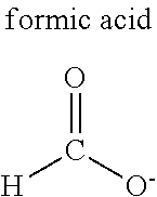
TABLE-US-00010 TABLE 10 Diatomic molecules Nitrogen, oxygen, hydrogen, fluorine, chlorine, bromine, iodine Noble gases Helium, neon, argon, krypton, xenon, radon Carbon oxides Carbon monoxide, carbon dioxide Nitrogen compounds Ammonia, nitric oxide, nitrogen dioxide, dinitrogen monoxide, dinitrogen tetroxide, dinitrogen trioxide, dinitrogen pentoxide, dimethylamine, trimethylamine Sulfur compounds Sulfur dioxide, hydrogen sulfide, methanethiol, dimethyl sulfide Alkanes Methane, ethane, propane, butane, halogenated methane Alkenes Ethylene, propylene, butadiene Alkynes Acetylene Alcohols Methanol, ethanol, propanol Aldehydes Formaldehyde, acetaldehyde Carboxylic acids Formic acid, acetic acid, citric acid, malic acid Ethers Dimethyl ether, diethyl ether Aromatic compounds Benzene, toluene Others Water, bioactive substances
[0068] Only one type of immune signal transducer may be used, or two or more types thereof may be used in combination. The content of the immune signal transducer in the vaccine composition is, for example, in the range of 1.times.10.sup.-7 to 40% by mass, preferably in the range of 1.times.10.sup.-6 to 30% by mass, and more preferably in the range of 5.times.10.sup.-5 to 25 mass %.
[0069] Any methods can be used for introducing the immune signal transducer into the pores of the MOF. For example, a solution or dispersion of a MOF may be mixed with a solution or dispersion of an immune signal transducer. Alternatively, a solid MOF may be exposed to an immune signal transducer or a solution or dispersion thereof. When the immune signal transducer is a gas, the MOF may be simply exposed to the gas.
[0070] The vaccine composition according to one embodiment of the present invention may further contain other adjuvant(s) than the MOF. The vaccine composition may also contain immunostimulant(s) such as a TLR ligand, an RLR ligand, an NLR ligand, or a cyclic dinucleotide.
[0071] The vaccine composition according to one embodiment of the present invention can be dissolved or dispersed in a solvent when in use. Examples of such solvents include physiological saline, phosphate buffered saline (PBS), glycerin, propylene glycol, polyethylene glycol, fats, or oils.
[0072] The vaccine composition according to the present invention can be administered to a subject by any method. As used herein, the "subject" refers to any animal whose immune response can be induced upon administration of vaccine composition in the practical stage. The animal typically is a mammal including humans, such as mice, rats, dogs, cats, rabbits, horses, cow, sheep, pig, goat, monkey, chimpanzee, ferret, mole, etc. A particularly preferred subject is a human.
[0073] The vaccine composition according to one embodiment of the present invention may be configured to be administered, for example, on a skin and/or mucous membrane of a subject.
[0074] In the case of transdermal administration, the vaccine composition may be any formulation commonly used for transdermal administration. For example, liquid for external use such as liniments or lotions, external sprays such as aerosols, ointments, plasters, creams, gels, or patches such as tapes or poultices can be used. The classification, definition, properties, and production method of these compositions are well known in the art, and can be found, for example, in the Japanese Pharmacopoeia 16th edition.
[0075] In the case of mucosal administration, the vaccine composition may be any formulation commonly used for mucosal administration such as sublingual, nasal, buccal, rectal or vaginal administration. For example, semi-solid preparations such as gel (jelly), cream, ointment, or plasters, liquid preparations, solid preparations such as powders, fine granules, granules, films, tablets, or orally disintegrating tablets, sprays for mucous membranes such as aerosols, or inhalants can be used. The classification, definition, properties, and production method of these compositions are well known in the art, and can be found, for example, in the Japanese Pharmacopoeia 16th edition.
[0076] The vaccine composition according to one embodiment of the present invention can also be configured to be administered, for example, by intradermal injection, subcutaneous injection, or intramuscular injection. In the case of intradermal, subcutaneous, or intramuscular administration, the composition may be in a form that has a certain fluidity that can be administered by injection, such as a liquid, suspension, cream, and the like. The classification, definition, properties, and production method of these compositions are well known in the art, and can be found, for example, in the Japanese Pharmacopoeia 16th edition.
[0077] The vaccine composition may further contain additive(s) if necessary. The additives can be selected depending, for example, upon main component of the base, compatibility with the antigen and/or the MOF, or the intended dosage regimen. Examples of the additives include skin permeability enhancers, isotonic agents, antiseptic/disinfectants, antioxidants, solubilizers, solubilizing agents, suspending agents, fillers, pH adjusters, stabilizers, absorption enhancers, release rate controllers, colorants, plasticizers, adhesives, or their combinations.
[0078] The adjuvant according to the present disclosure includes a MOF. This adjuvant may be used independently from the antigen. For example, the adjuvant may be administered separately after the antigen is administered to a subject. Alternatively, the antigen may be administered after the adjuvant is administered.
[0079] The MOF as the adjuvant may be configured to decompose in vivo. The MOF may further contain an immune signal transducer in its pores. The MOF may also be configured to decompose in the living body to release at least a part of the immune signal transducer contained in the pores. Similar methods as explained above can be used for introducing at least a part of the immune signal transducer in the pores of the MOF. Also, similar administration methods as explained above in regard to the vaccine composition can be used for administering the adjuvant.
[0080] As described above, the immune signal transducer is, for example, a factor that is configured to induce activation and/or differentiation of lymphocytes (T cells, B cells, NK cells, etc.), monocytes (macrophages, Langerhans cells, dendritic cells, etc.), granulocytes (neutrophils, eosinophils, basophils, etc.) and/or keratinocytes. The immune signal transducer is, for example, a factor that is configured to induce differentiation of naive helper T cells into various lineages such as Th1 cells, Th2 cells, Treg cells, Th17 cells, Tfh cells, or memory T cells. When the immune signal transducer induces Th1 cells, the adjuvant according to the present invention can be used, for example, for cancer vaccines, infectious disease vaccines, or as concomitant drugs with anti-cancer agents. When the immune signal transducer induces Th2 cells, the adjuvant according to the present invention can be used, for example, for infectious disease vaccines or lifestyle-related disease vaccines. When the immune signal transducer induces Treg cells, the adjuvant according to the present invention can be used, for example, for allergy vaccines or organ transplantations. When the immune signal transducer induces Th17 cells, the adjuvant according to the present invention can be used, for example, for infectious disease vaccines. When the immune signal transducer induces Tfh cells, the adjuvant according to the present invention can be used, for example, for infectious disease vaccines. When the immune signal transducer induces memory T cells, the adjuvant according to the present invention can be used, for example, for infectious disease vaccines or cancer vaccines.
EXAMPLES
[0081] [Preparation of Sample Solutions]
Example 1
[0082] NO (nitrogen monoxide, Kyoto Teijin) was bubbled into 100 mL of physiological saline (Otsuka Normal Saline, Otsuka Pharmaceutical) at room temperature for 6 hours to prepare NO-saturated physiological saline. To 10 mL of the obtained solution was added 1 mg of ZIF-8 (Basolite Z1200, Sigma-Aldrich) and 1 mg of OVA (egg-derived albumin, Wako), and these were mixed to provide a sample solution.
Example 2
[0083] Another sample solution was prepared by adding 1 mg of ZIF-8 (Basolite Z1200, Sigma-Aldrich) and 1 mg of OVA (egg-derived albumin, Wako) to 10 mL of physiological saline (Otsuka Normal Saline, Otsuka Pharmaceutical).
Comparative Example 1
[0084] Physiological saline (Otsuka Normal Saline, Otsuka Pharmaceutical) itself was used as a sample solution.
Comparative Example 2
[0085] 1 mg of OVA (egg-derived albumin, Wako) was added to and mixed with 10 mL of physiological saline (Otsuka Normal Saline, Otsuka Pharmaceutical) to obtain a sample solution.
Reference Example 1
[0086] 1 mg of ZIF-8 (Basolite Z1200, Sigma-Aldrich) was added to and mixed with 10 mL of physiological saline (Otsuka Normal Saline, Otsuka Pharmaceutical) to obtain a sample solution.
Reference Example 2
[0087] NO (nitrogen monoxide, Kyoto Teijin) was bubbled in 100 mL of physiological saline (Otsuka Normal Saline, Otsuka Pharmaceutical) at room temperature for 6 hours to prepare NO saturated physiological saline. To 10 mL of the obtained solution was added 1 mg of ZIF-8 (Basolite Z1200, Sigma-Aldrich), and these were mixed to provide a sample solution.
[0088] The above configuration is summarized in Table 11 below.
TABLE-US-00011 TABLE 11 Antigen MOF Solvent Immune Signal Transducer Concentration Concentration Amount Concentration Name [.mu.g/mL] Name [.mu.g/mL] Name [.mu.L] Name [mM] Comp. Ex. 1 -- -- -- -- Physiological saline 100 -- -- Ref. Ex. 1 -- -- ZIF-8 100 Physiological saline 100 -- -- Ref. Ex. 2 -- -- ZIF-8 100 Physiological saline 100 NO 1.8 Example 1 OVA 100 ZIF-8 100 Physiological saline 100 NO 1.8 Example 2 OVA 100 ZIF-8 100 Physiological saline 100 -- -- Comp. Ex. 2 OVA 100 -- -- Physiological saline 100 -- --
Examples 3 to 6
[0089] Sample solutions were prepared in the same manner as in Example 1 except that the antigens shown in Table 12 below were used instead of OVA.
TABLE-US-00012 TABLE 12 Antigen MOF Solvent Immune Signal Transducer Concentration Concentration Amount Concentration Name [.mu.g/mL] Name [.mu.g/mL] Name [.mu.L] Name [mM] Example 1 OVA -- ZIF-8 100 Physiological saline 100 NO 1.8 Example 3 Pneumococcus -- ZIF-8 100 Physiological saline 100 NO 1.8 Example 4 Influenza vaccine -- ZIF-8 100 Physiological saline 100 NO 1.8 Example 5 Cyj1 100 ZIF-8 100 Physiological saline 100 NO 1.8 Example 6 HPV16 recombinant protein 100 ZIF-8 100 Physiological saline 100 NO 1.8
Examples 7 to 35
[0090] Sample solutions were prepared in the same manner as in Example 1 except that the substances shown in Table 13 below were used instead of NO as immune signal transducers.
TABLE-US-00013 TABLE 13 Antigen MOF Solvent Immune Signal Transducer Concentration Concentration Amount Concentration Name [.mu.g/mL] Name [.mu.g/mL] Name [.mu.L] Name [mM] Example 1 OVA 100 ZIF-8 100 Physiological saline 100 NO Saturated Example 7 OVA 100 ZIF-8 100 Physiological saline 100 CO Saturated Example 8 OVA 100 ZIF-8 100 Physiological saline 100 CO.sub.2 Saturated Example 9 OVA 100 ZIF-8 100 Physiological saline 100 N.sub.2 Saturated Example 10 OVA 100 ZIF-8 100 Physiological saline 100 O.sub.2 Saturated Example 11 OVA 100 ZIF-8 100 Physiological saline 100 H.sub.2 Saturated Example 12 OVA 100 ZIF-8 100 Physiological saline 100 H.sub.2S Saturated Example 13 OVA 100 ZIF-8 100 Physiological saline 100 S.sub.2O Saturated Example 14 OVA 100 ZIF-8 100 Physiological saline 100 CH.sub.4 Saturated Example 15 OVA 100 ZIF-8 100 Physiological saline 100 C.sub.2H.sub.6 Saturated Example 16 OVA 100 ZIF-8 100 Physiological saline 100 C.sub.3H.sub.8 Saturated Example 17 OVA 100 ZIF-8 100 Physiological saline 100 C.sub.4H.sub.10 Saturated Example 18 OVA 100 ZIF-8 100 Physiological saline 100 C.sub.2H.sub.4 Saturated Example 19 OVA 100 ZIF-8 100 Physiological saline 100 C.sub.3H.sub.6 Saturated Example 20 OVA 100 ZIF-8 100 Physiological saline 100 C.sub.2H.sub.4 Saturated Example 21 OVA 100 ZIF-8 100 Physiological saline 100 CH.sub.3NH.sub.2 Saturated Example 22 OVA 100 ZIF-8 100 Physiological saline 100 (CH.sub.3).sub.2NH Saturated Example 23 OVA 100 ZIF-8 100 Physiological saline 100 NH.sub.3 Saturated Example 24 OVA 100 ZIF-8 100 Physiological saline 100 CH.sub.3SH Saturated Example 25 OVA 100 ZIF-8 100 Physiological saline 100 (CH.sub.3).sub.3N Saturated Example 26 OVA 100 ZIF-8 100 Physiological saline 100 CH.sub.3Cl Saturated Example 27 OVA 100 ZIF-8 100 Physiological saline 100 CH.sub.3Br Saturated Example 28 OVA 100 ZIF-8 100 Physiological saline 100 He Saturated Example 29 OVA 100 ZIF-8 100 Physiological saline 100 F.sub.2 Saturated Example 30 OVA 100 ZIF-8 100 Physiological saline 100 Ne Saturated Example 31 OVA 100 ZIF-8 100 Physiological saline 100 Cl.sub.2 Saturated Example 32 OVA 100 ZIF-8 100 Physiological saline 100 Ar Saturated Example 33 OVA 100 ZIF-8 100 Physiological saline 100 Kr Saturated Example 34 OVA 100 ZIF-8 100 Physiological saline 100 Xe Saturated Example 35 OVA 100 ZIF-8 100 Physiological saline 100 Rn Saturated
Examples 36 to 145
[0091] Sample solutions were prepared in the same manner as in Example 1 except that the substances shown in Table 14 to 16 below were used instead of ZIF-8 as MOFs (i.e. as adjuvants). Abbreviations in Tables 14 to 16 are the same as those described in Tables 1 to 3, respectively.
TABLE-US-00014 TABLE 14 Antigen MOF Solvent Immune Signal Transducer Concentration Concentration Amount Concentration Name [.mu.g/mL] Name [.mu.g/mL] Name [.mu.L] Name [mM] Example 1 OVA 100 ZIF-8 100 Physiological saline 100 NO Saturated Example 36 OVA 100 CPL-1 100 Physiological saline 100 NO Saturated Example 37 OVA 100 Cu.sub.3(btc).sub.2 100 Physiological saline 100 NO Saturated Example 38 OVA 100 Zn.sub.2(14bdc).sub.2(dabco) 100 Physiological saline 100 NO Saturated Example 39 OVA 100 ZIF-8 100 Physiological saline 100 NO Saturated Example 40 OVA 100 HKUST-1 100 Physiological saline 100 NO Saturated Example 41 OVA 100 Mg.sub.3(C.sub.12O.sub.14H.sub.10) 100 Physiological saline 100 NO Saturated Example 42 OVA 100 Ca.sub.2(C.sub.8O.sub.12H.sub.6) 100 Physiological saline 100 NO Saturated Example 43 OVA 100 Ca.sub.3(C.sub.12O.sub.14H.sub.10) 100 Physiological saline 100 NO Saturated Example 44 OVA 100 Ca(C.sub.4O.sub.6H.sub.4) 100 Physiological saline 100 NO Saturated Example 45 OVA 100 Cu(IPA) 100 Physiological saline 100 NO Saturated Example 46 OVA 100 MgBDC-1 100 Physiological saline 100 NO Saturated Example 47 OVA 100 MgDHBDC-1 100 Physiological saline 100 NO Saturated Example 48 OVA 100 MgOBA-1 100 Physiological saline 100 NO Saturated Example 49 OVA 100 MgBTC-1 100 Physiological saline 100 NO Saturated Example 50 OVA 100 MgBTB-1 100 Physiological saline 100 NO Saturated Example 51 OVA 100 MgBTB-2 100 Physiological saline 100 NO Saturated Example 52 OVA 100 MgBTB-3 100 Physiological saline 100 NO Saturated Example 53 OVA 100 MgBTB-4 100 Physiological saline 100 NO Saturated Example 54 OVA 100 MgBBC-1 100 Physiological saline 100 NO Saturated Example 55 OVA 100 MIL-100(Fe) 100 Physiological saline 100 NO Saturated Example 56 OVA 100 MIL-101 100 Physiological saline 100 NO Saturated Example 57 OVA 100 MIL-53 100 Physiological saline 100 NO Saturated Example 58 OVA 100 BioMIL-5 100 Physiological saline 100 NO Saturated Example 59 OVA 100 CaZol nMOF 100 Physiological saline 100 NO Saturated Example 60 OVA 100 IRMOF-2 100 Physiological saline 100 NO Saturated Example 61 OVA 100 IRMOF-3 100 Physiological saline 100 NO Saturated Example 62 OVA 100 IRMOF-4 100 Physiological saline 100 NO Saturated Example 63 OVA 100 IRMOF-5 100 Physiological saline 100 NO Saturated Example 64 OVA 100 IRMOF-6 100 Physiological saline 100 NO Saturated Example 65 OVA 100 IRMOF-7 100 Physiological saline 100 NO Saturated Example 66 OVA 100 IRMOF-8 100 Physiological saline 100 NO Saturated Example 67 OVA 100 IRMOF-9 100 Physiological saline 100 NO Saturated Example 68 OVA 100 IRMOF-10 100 Physiological saline 100 NO Saturated Example 69 OVA 100 IRMOF-11 100 Physiological saline 100 NO Saturated Example 70 OVA 100 IRMOF-12 100 Physiological saline 100 NO Saturated Example 71 OVA 100 IRMOF-13 100 Physiological saline 100 NO Saturated Example 72 OVA 100 IRMOF-14 100 Physiological saline 100 NO Saturated Example 73 OVA 100 IRMOF-15 100 Physiological saline 100 NO Saturated Example 74 OVA 100 IRMOF-16 100 Physiological saline 100 NO Saturated
TABLE-US-00015 TABLE 15 Antigen MOF Solvent Immune Signal Transducer Concentration Concentration Amount Concentration Name [.mu.g/mL] Name [.mu.g/mL] Name [.mu.L] Name [mM] Example 75 OVA 100 Zn.sub.3(BTC).sub.2 100 Physiological saline 100 NO Saturated Example 76 OVA 100 Zn.sub.4O(NDC) 100 Physiological saline 100 NO Saturated Example 77 OVA 100 Mg(Formate) 100 Physiological saline 100 NO Saturated Example 78 OVA 100 Fe(Formate) 100 Physiological saline 100 NO Saturated Example 79 OVA 100 Mg(C.sub.6H.sub.4O.sub.6) 100 Physiological saline 100 NO Saturated Example 80 OVA 100 ZnC.sub.2H.sub.4BDC 100 Physiological saline 100 NO Saturated Example 81 OVA 100 MOF-49 100 Physiological saline 100 NO Saturated Example 82 OVA 100 BPR95A2 100 Physiological saline 100 NO Saturated Example 83 OVA 100 BPR76D5 100 Physiological saline 100 NO Saturated Example 84 OVA 100 BPR68D10 100 Physiological saline 100 NO Saturated Example 85 OVA 100 BPR56E1 100 Physiological saline 100 NO Saturated Example 86 OVA 100 BPR49B1 100 Physiological saline 100 NO Saturated Example 87 OVA 100 BPR43G2 100 Physiological saline 100 NO Saturated Example 88 OVA 100 NO336 100 Physiological saline 100 NO Saturated Example 89 OVA 100 NO335 100 Physiological saline 100 NO Saturated Example 90 OVA 100 NO333 100 Physiological saline 100 NO Saturated Example 91 OVA 100 PCN-14 100 Physiological saline 100 NO Saturated Example 92 OVA 100 Zn.sub.4BNDC 100 Physiological saline 100 NO Saturated Example 93 OVA 100 Zn.sub.3(BPDC) 100 Physiological saline 100 NO Saturated Example 94 OVA 100 ZnDBP 100 Physiological saline 100 NO Saturated Example 95 OVA 100 Zn.sub.3(PDC).sub.2.5 100 Physiological saline 100 NO Saturated Example 96 OVA 100 Zn(HPDC) 100 Physiological saline 100 NO Saturated Example 97 OVA 100 Zn(NDC) 100 Physiological saline 100 NO Saturated Example 98 OVA 100 MOF-37 100 Physiological saline 100 NO Saturated Example 99 OVA 100 MOF-20 100 Physiological saline 100 NO Saturated Example 100 OVA 100 MOF-12 100 Physiological saline 100 NO Saturated Example 101 OVA 100 Zn(ADC) 100 Physiological saline 100 NO Saturated Example 102 OVA 100 MOF-0 100 Physiological saline 100 NO Saturated Example 103 OVA 100 MOF-2 100 Physiological saline 100 NO Saturated Example 104 OVA 100 MOF-3 100 Physiological saline 100 NO Saturated Example 105 OVA 100 MOF-4 100 Physiological saline 100 NO Saturated Example 106 OVA 100 MOF-5 100 Physiological saline 100 NO Saturated Example 107 OVA 100 MOF-38 100 Physiological saline 100 NO Saturated Example 108 OVA 100 MOF-31 100 Physiological saline 100 NO Saturated Example 109 OVA 100 MOF-69A 100 Physiological saline 100 NO Saturated Example 110 OVA 100 MOF-69B 100 Physiological saline 100 NO Saturated Example 111 OVA 100 MOF-33 100 Physiological saline 100 NO Saturated Example 112 OVA 100 MOF-36 100 Physiological saline 100 NO Saturated Example 113 OVA 100 MOF-39 100 Physiological saline 100 NO Saturated
TABLE-US-00016 TABLE 16 Antigen MOF Solvent Immune Signal Transducer Concentration Concentration Amount Concentration Name [.mu.g/mL] Name [.mu.g/mL] Name [.mu.L] Name [mM] Example 114 OVA 100 NO305 100 Physiological saline 100 NO Saturated Example 115 OVA 100 NO306A 100 Physiological saline 100 NO Saturated Example 116 OVA 100 BPR48A2 100 Physiological saline 100 NO Saturated Example 117 OVA 100 Zn(C.sub.2O.sub.4) 100 Physiological saline 100 NO Saturated Example 118 OVA 100 MOF-48 100 Physiological saline 100 NO Saturated Example 119 OVA 100 MOF-47 100 Physiological saline 100 NO Saturated Example 120 OVA 100 Zn.sub.3(BTC).sub.2 100 Physiological saline 100 NO Saturated Example 121 OVA 100 MOF-n 100 Physiological saline 100 NO Saturated Example 122 OVA 100 Zehex 100 Physiological saline 100 NO Saturated Example 123 OVA 100 AS16 100 Physiological saline 100 NO Saturated Example 124 OVA 100 AS27-3 100 Physiological saline 100 NO Saturated Example 125 OVA 100 AS54-3 100 Physiological saline 100 NO Saturated Example 126 OVA 100 AS61-4 100 Physiological saline 100 NO Saturated Example 127 OVA 100 AS68-7 100 Physiological saline 100 NO Saturated Example 128 OVA 100 Zn.sub.8(ad).sub.4(PDAC).sub.6(OH).sub.2 100 Physiological saline 100 NO Saturated Example 129 OVA 100 Zn.sub.8(ad).sub.4(SBDC).sub.6(OH).sub.2 100 Physiological saline 100 NO Saturated Example 130 OVA 100 Zn.sub.8(ad).sub.4(BPDC).sub.6(OH).sub.2 100 Physiological saline 100 NO Saturated Example 131 OVA 100 Zn.sub.8(ad).sub.4(NDC).sub.6(OH).sub.2 100 Physiological saline 100 NO Saturated Example 132 OVA 100 M-CPO-27 100 Physiological saline 100 NO Saturated Example 133 OVA 100 bio-MOF-1 100 Physiological saline 100 NO Saturated Example 134 OVA 100 UMCM-1 100 Physiological saline 100 NO Saturated Example 135 OVA 100 UMCM-2 100 Physiological saline 100 NO Saturated Example 136 OVA 100 MOF-210 100 Physiological saline 100 NO Saturated Example 137 OVA 100 bio-MOF-100 100 Physiological saline 100 NO Saturated Example 138 OVA 100 NU-110E 100 Physiological saline 100 NO Saturated Example 139 OVA 100 CD-MOF-1 100 Physiological saline 100 NO Saturated Example 140 OVA 100 porph@MOM-4 100 Physiological saline 100 NO Saturated Example 141 OVA 100 porph@MOM-8 100 Physiological saline 100 NO Saturated Example 142 OVA 100 porph@MOM-9 100 Physiological saline 100 NO Saturated Example 143 OVA 100 ZnPO-MOF 100 Physiological saline 100 NO Saturated Example 144 OVA 100 Uio-66 100 Physiological saline 100 NO Saturated Example 145 OVA 100 Mg(H.sub.2gal) 100 Physiological saline 100 NO Saturated
[0092] [Collection of Intraperitoneal Cells (PEC cells)]
[0093] A mouse was intraperitoneally administered with 2 mL of 4 wt % thioglycolic acid solution, and cells in its peritoneal cavity were taken out 3 days later. The collected cells were then washed with PBS (Phosphate Buffered Saline).
[0094] [Stimulation by Sample Solutions]
[0095] The PEC cells were dispensed in a 24-well plate at 1.times.10.sup.6 cells/well, and each sample was added thereto and incubated for 24 hours.
[0096] [Cytokine Measurement]
[0097] 50 .mu.L/well of the supernatant of the cell culture was used for an evaluation by an ELISA kit (Quantikine ELISA kit, R&D Systems) that corresponds to each cytokine (TNF-.alpha., IL-6, IFN-.gamma., IL-12p40, IL-10) to be monitored. The results are summarized in Table 17 below.
TABLE-US-00017 TABLE 17 TNF-.alpha. IL-6 IL-10 IL-12p40 IFN-g Comp. Ex. 1 - - - - - Ref. Ex. 1 + + - - - Ref. Ex. 2 ++ ++ - + + Example 1 ++ ++ - ++ + Example 2 ++ ++ - - - Comp. Ex. 2 + - - - - (-): Less than twice the amount of cytokine released in Comparative Example 1 (+): Between twice and three times the amount of cytokine released in Comparative Example 1. (++): Three or more times the amount of cytokine released in Comparative Example 1
[0098] [Measurement of OVA-Specific IgG Titer in Mouse Serum (ELISA Method)]
[0099] 100 .mu.L of OVA-containing solution (100 .mu.g/mL) diluted with carbonate buffer was added to a 96-well plate for ELISA, and allowed to stand overnight. The wells were washed three times with a washing solution (PBS containing Tween 20), and 200 .mu.L of a blocking solution obtained by diluting a blocking agent (Block Ace, Sumitomo Dainippon Pharma Co., Ltd.) with purified water to 4 g/100 mL was added and was left for 2 hours at room temperature. The wells were washed three times with the washing solution again.
[0100] Serum collected from a mouse in advance was centrifuged at 3000 g for 10 minutes at 4.degree. C., and the obtained supernatant was collected. The above-mentioned supernatant was serially diluted to two times using a solution obtained by diluting the blocking agent with a phosphate buffer (Nacalai Tesque) to 0.4 g/100 mL, and 50 .mu.L of the solution was added to each well and was left at room temperature for 2 hours.
[0101] The wells were washed three times with the washing solution, and an HRP-labeled anti-mouse IgG antibody (Goat-anti mouse IgG Fc HRP, BETHYL) was diluted to 10000 times with the solution obtained by diluting the blocking agent with a phosphate buffer (Nacalai Tesque) to 0.4 g/100 mL, and 100 .mu.L of the solution was added to each well and was left at room temperature for 1 hour. The wells were washed three times with the washing solution, and 100 .mu.L of TMB solution (ELISA POD TMB kit, Nacalai Tesque) was added, and was left in the dark for 30 minutes. 100 .mu.L of 1M sulfuric acid solution was then added, and the absorbance at 450 nm was measured for the 96-well plate with a microplate reader (SpectraMax, Molecular Device). Based on the absorbance of the serially diluted samples, the IgG antibody titer in mouse serum was determined by Log 2 scale.
[0102] [Evaluation of Humoral Immunity Using Mice]
[0103] Using a liquid prepared as described above, a mouse immunity test was conducted using a model animal for humoral immunity evaluation. 200 .mu.L of an injection sample was administered subcutaneously to the back of a mouse (BALB/c mouse, female 7 weeks old). One week after the administration, the same administration was again performed subcutaneously on the back of the mouse. Two weeks after the second administration, mouse serum was collected, and the serum OVA-specific IgG titer was measured by the ELISA method as described above.
[0104] [OVA Antigen-Specific CTL Measurement (ELISPOT Method)]
[0105] Splenocytes (3.times.10.sup.6 cells/well) and antigenic peptide (100 .mu.M) or antigenic protein (100 .mu.g/mL) were placed along with a culture solution in a well of an ELISPOT plate (R&D Systems) on which an anti-mouse IFN-.gamma. antibody is immobilized. The cells were co-cultured at 37.degree. C. under 5% CO.sub.2 for 20 hours, and the number of IFN-.gamma. producing cell spots (the number of spots per 3.times.10.sup.6 cells) was measured by ELISPOT method.
[0106] [Evaluation of Cellular Immunity Using Mice]
[0107] Using a liquid prepared as described above, a mouse immunity test was conducted using a model animal for cellular immunity evaluation. 200 .mu.L of an injection was administered subcutaneously to the back of a mouse (C57BL/6 mouse, female 7 week old). One week after the administration, the same administration was again performed subcutaneously on the back of the mouse. One week after the second administration, mouse spleen was collected, and OVA antigen-specific CTL was measured by the ELISPOT method described above.
[0108] These results are summarized in Table 18 below.
TABLE-US-00018 TABLE 18 IgG The number of CTLs Comp. Ex. 1 - - Ref. Ex. 1 - - Ref. Ex. 2 - - Example 1 +++ ++ Example 2 ++ - Comp. Ex. 2 + - (-): Less than 4 times the amount of antibody produced in Comparative Example 1, or the number of CTLs less than 30 cells/well (+): 4 times or more and less than 8 times the amount of antibody produced in Comparative Example 1, or the number of CTLs 30 cells/well or more and less than 100 cells/well (++): 8 times or more and less than 16 times the amount of antibody produced in Comparative Example 1, or the number of CTLs 100 cells/well or more and less than 300 cells/well (+++): 16 times or more the amount of antibody produced in Comparative Example 1, or the number of CTLs 300 cells/well or more
[0109] [Synthesis of MOFs]
[0110] The MOFs shown in Tables 4 to 9 were prepared. Known substances among them were synthesized according to literature methods. The unreported substances were synthesized by hydrothermal treatment of the corresponding metal nitrate and the ligand in the presence of DMF.
[0111] [Evaluation of Adsorption Properties of MOFs]
[0112] The amount of adsorption was measured by BELSORP-max12 (MicrotracBEL Co., Ltd.). The MOFs in powder form were used for the measurements. Some of the results are shown in FIG. 1A, FIG. 1B and FIG. 2 as representative examples. FIG. 1A is a CO adsorption profile of AP004 [MIL-100 (Fe)]. FIG. 1B is a NO adsorption profile of AP004 [MIL-100 (Fe)]. FIG. 2 is a NO adsorption profile of AP104 (BioMIL-3). In these examples, the adsorption/desorption profiles were irreversible. That is, when seen at the same pressure, the guest amount at the time of desorption was larger than the guest amount at the time of adsorption. Also, the residual amount of the guest in the MOFs were non-zero after performing the adsorption process from a vacuum state to a pressurized state and then performing the desorption process from the pressurized state to the vacuum state.
[0113] [Introduction of Immune Signal Transducers into MOFs]
[0114] In some of the examples below, the MOFs to which an immune signal transducer had been introduced were employed. Specifically, the degassing was performed by heating the MOF under a nitrogen flow. The sample was then returned to a room temperature and was exposed to an immune signal transducer. In particular, when the immune signal transducer was a gas, the sample returned to room temperature was exposed to a gas flow. A nitrogen flow was then performed at room temperature to discharge excess immune signal transducer. In this way, a MOF compound to which an immune signal transducer had been introduced was obtained.
[0115] The existence of the immune signal transducer in the MOF was checked by heating the sample under nitrogen flow and detecting the released immune signal transducer by a detector tube. It was thus confirmed that the immune signal transducer had effectively been introduced into the MOFs.
[0116] [Mouse Immunity Test]
[0117] An injection having the composition shown in Table 19 below was prepared. Specifically, the antigen and the MOF were weighed out in the amounts specified in Table 19, and glycerin was added thereto and mixed to obtain a vaccine composition. In the table, MOF stands for Metal Organic Framework and Gly for glycerin. In some examples, MOFs adsorbed with an immune signal transducer were used.
TABLE-US-00019 TABLE 19 Antigen Compounds (OVA) The num- Immune Concen- Concen- Concen- Subcutaneous ber of Evaluation Signal Molecular tration tration tration Sol- n- injection adminis- Antibody Cytokine MOF Transducer Weight [.mu.mol/mL] [.mu.g/mL] [.mu.g/mL] vent number [.mu.L/time] trations titer (Spleen) -- -- -- 50 Gly 6 50 2 IgG IL-4 AP104 BioMIL-3 -- 434 1 434 50 (2 wks) IgG2a IFN-.gamma. NO 0 50 AP004 MIL-100(Fe) -- 679 1 679 50 NO 50 CO 50 O.sub.2 50
[0118] Using a liquid prepared as described above, 50 .mu.L of an injection was administered subcutaneously to the back of a mouse (BALB/c mouse, female 7 weeks old). Two weeks after the administration, the same administration was again performed subcutaneously on the back of the mouse.
[0119] Two weeks after the second administration, mouse serum and spleen cells were collected, and serum OVA-specific IgG antibody and IgG2a antibody were measured by ELISA. In addition, the spleen cells were used to simultaneously evaluate the production amounts of OVA-specific IFN-.gamma. and IL-4. The specific evaluation method is as follows.
[0120] [Measurement of Antigen-Specific Antibody Titer in Mouse Serum (ELISA method)]
[0121] As an antigen, an OVA-containing solution diluted with a carbonate buffer (100 .mu.g/mL) was prepared. 100 .mu.L of the solution was added to each well of a 96-well plate for ELISA and allowed to stand overnight.
[0122] The wells were washed 3 times with a washing solution (PBS containing Tween 20). 200 .mu.L of a blocking solution obtained by diluting a blocking agent (Block Ace, Sumitomo Dainippon Pharma Co., Ltd.) with purified water to 4 g/100 mL was added and was left for 2 hours at room temperature. The wells were washed three times with the washing solution again.
[0123] Serum collected from a mouse in advance was centrifuged at 3000 g for 10 minutes at 4.degree. C., and the obtained supernatant was collected. The above-mentioned supernatant was serially diluted to two times using a solution obtained by diluting the blocking agent with a phosphate buffer (Nacalai Tesque) to 0.4 g/100 mL. 50 .mu.L of each of the obtained diluted solutions was added and left at room temperature for 2 hours.
[0124] The wells were washed three times with the washing solution once again. An HRP-labeled anti-mouse IgG antibody (Goat-anti mouse IgG Fc HRP, BETHYL) or an HRP-labeled anti-mouse IgG2a antibody (Goat-anti mouse IgG2a Fc HRP, BETHYL) was diluted to 10000 times with the solution obtained by diluting the blocking agent with a phosphate buffer (Nacalai Tesque) to 0.4 g/100 mL. 100 .mu.L of each of the obtained diluted solutions was added and left at room temperature for 1 hour.
[0125] The wells were washed three times with the washing solution, and 100 .mu.L of TMB solution (ELISA POD TMB kit, Nacalai Tesque) was added, and was left in the dark for 30 minutes.
[0126] Further, 100 .mu.L of 1M sulfuric acid solution was added, and the absorbance at 450 nm was measured for each of the 96-well plates using a microplate reader. Based on the absorbance of the serially diluted samples, the IgG antibody titer or the IgG2a antibody titer in mouse serum was determined by Log 2 scale.
[0127] These results are summarized in FIG. 3. As shown in FIG. 3, it was revealed that the immune properties could be controlled by use of MOFs. Also, it was made clear that the immune characteristics could be further changed by a combination of MOF and immune signal transducer.
[0128] [Measurement of OVA-Specific Cytokine Production (ELISA method)]
[0129] 100 .mu.L each of 4.times.10.sup.5 cells/well of spleen cells collected in advance from a mouse was added to a 96-well plate for ELISA. 100 .mu.L of an OVA-containing solution (100 .mu.g/m) diluted in RPMI medium was thereto added and allowed to stand for 72 hours. The culture supernatant was collected, and the amount of each cytokine produced was quantified using a mouse IFN-.gamma. ELISA kit and mouse IL-4 ELISA kit (R&D Systems).
[0130] These results are summarized in FIGS. 4A and 4B. As shown in FIGS. 4A and 4B, it was revealed that the immune properties could be controlled by use of MOFs. Also, it was made clear that the immune characteristics could be further changed by a combination of MOF and immune signal transducer.
* * * * *


D00000
D00001

D00002

D00003

XML
uspto.report is an independent third-party trademark research tool that is not affiliated, endorsed, or sponsored by the United States Patent and Trademark Office (USPTO) or any other governmental organization. The information provided by uspto.report is based on publicly available data at the time of writing and is intended for informational purposes only.
While we strive to provide accurate and up-to-date information, we do not guarantee the accuracy, completeness, reliability, or suitability of the information displayed on this site. The use of this site is at your own risk. Any reliance you place on such information is therefore strictly at your own risk.
All official trademark data, including owner information, should be verified by visiting the official USPTO website at www.uspto.gov. This site is not intended to replace professional legal advice and should not be used as a substitute for consulting with a legal professional who is knowledgeable about trademark law.