Automated Measurement System And Method For Coronary Artery Disease Scoring
Kind Code
U.S. patent application number 16/859239 was filed with the patent office on 2020-08-13 for automated measurement system and method for coronary artery disease scoring. The applicant listed for this patent is CathWorks Ltd.. Invention is credited to Guy Lavi, Uri Merhav.
| Application Number | 20200253489 16/859239 |
| Document ID | 20200253489 / US20200253489 |
| Family ID | 1000004783142 |
| Filed Date | 2020-08-13 |
| Patent Application | download [pdf] |


View All Diagrams
| United States Patent Application | 20200253489 |
| Kind Code | A1 |
| Lavi; Guy ; et al. | August 13, 2020 |
AUTOMATED MEASUREMENT SYSTEM AND METHOD FOR CORONARY ARTERY DISEASE SCORING
Abstract
An automated measurement device and method for coronary artery disease scoring is disclosed. An example device includes a processor configured to obtain a computerized model of a plurality of vascular segments of a patient and create an unstenosed computerized model from the computerized model by virtually enlarging at least some locations of the vascular segments of the computerized model. The processor also determines vascular state scoring tool ("VSST") scores based on characteristics of vascular locations along the vascular segments. The processor further determines a severity of stenosis for the vascular locations based on comparisons of first blood flow parameter values at the vascular locations in the computerized model to corresponding second blood flow parameter values at the same vascular locations in the unstenosed computerized model. A user interface of the device displays the severity of stenosis in conjunction with the VSST scores for the vascular locations.
| Inventors: | Lavi; Guy; (Moshav Mishmeret, IL) ; Merhav; Uri; (Rechovot, IL) | ||||||||||
| Applicant: |
|
||||||||||
|---|---|---|---|---|---|---|---|---|---|---|---|
| Family ID: | 1000004783142 | ||||||||||
| Appl. No.: | 16/859239 | ||||||||||
| Filed: | April 27, 2020 |
Related U.S. Patent Documents
| Application Number | Filing Date | Patent Number | ||
|---|---|---|---|---|
| 16291916 | Mar 4, 2019 | 10631737 | ||
| 16859239 | ||||
| 15952701 | Apr 13, 2018 | 10219704 | ||
| 16291916 | ||||
| 14437205 | Apr 21, 2015 | 9943233 | ||
| PCT/IL2013/050869 | Oct 24, 2013 | |||
| 15952701 | ||||
| 61717732 | Oct 24, 2012 | |||
| Current U.S. Class: | 1/1 |
| Current CPC Class: | G16H 50/30 20180101; A61B 5/7275 20130101; A61B 6/5217 20130101; A61B 5/7264 20130101; A61B 34/10 20160201; G16H 50/50 20180101; A61B 5/02007 20130101; A61B 5/1075 20130101; A61B 8/5223 20130101; G06T 7/0012 20130101; G06T 2207/30101 20130101; G06T 2207/10072 20130101; A61B 2034/107 20160201; A61B 6/504 20130101; A61B 2576/00 20130101; A61B 8/0891 20130101 |
| International Class: | A61B 5/02 20060101 A61B005/02; A61B 8/08 20060101 A61B008/08; A61B 6/00 20060101 A61B006/00; G06T 7/00 20060101 G06T007/00; A61B 34/10 20060101 A61B034/10; G16H 50/30 20060101 G16H050/30; G16H 50/50 20060101 G16H050/50; A61B 5/107 20060101 A61B005/107; A61B 5/00 20060101 A61B005/00 |
Claims
1. An apparatus for coronary artery disease scoring, the apparatus comprising: a vascular tree reconstruction processor configured to: obtain a computerized model that is representative of vascular segments of a patient, and create an unstenosed computerized model from the computerized model by virtually enlarging at least some locations of the vascular segments of the computerized model; a vascular state score calculator communicatively coupled to the vascular tree reconstruction processor and configured to: determine vascular state scoring tool ("VSST") scores for vascular locations along the vascular segments based on at least two of (a) a morphometric quantity of the vascular location, (b) a distance of the vascular location from a branch point in the plurality of vascular segments, (c) a sub-tree dominance of the vascular location, (d) a branch position related to the vascular location, (e) an indication of occlusion that is related to the vascular location, (f) an indication of tortuosity that is related to the vascular location, (g) an estimated calcification that is related to the vascular location, (h) an estimated thrombus that is related to the vascular location, (i) a bifurcation/trifurcation classification that is related to the vascular location, (j) a bifurcation/trifurcation angulation that is related to the vascular location, or (k) a vascular position of the vascular location, and determine a severity of stenosis for the vascular locations based on comparisons of first blood flow parameter values at the vascular locations in the computerized model to corresponding second blood flow parameter values at the same vascular locations in the unstenosed computerized model; and a user interface configured to display the severity of stenosis in conjunction with the VSST scores for the vascular locations.
2. The apparatus of claim 1, wherein the computerized model includes a three-dimensional model or a one-dimensional model.
3. The apparatus of claim 1, wherein the vascular state score calculator is configured to relate at least one of the severity of stenosis or the VSST scores of the vascular locations to corresponding locations in the computerized model or the unstenosed computerized model.
4. The apparatus of claim 1, wherein the vascular state score calculator is configured to: relate the VSST scores for the vascular locations to corresponding locations in at least one of the computerized model or the unstenosed computerized model; and relate the severity of stenosis for the vascular locations to the corresponding locations in at least one of the computerized model or the unstenosed computerized model, and wherein the user interface is configured to display at least one of the computerized model or the unstenosed computerized model with the related VSST scores and severity of stenosis shown adjacent to or overlaid on the corresponding vascular locations within the representation of the vascular segments.
5. The apparatus of claim 1, wherein the computerized model is based on vascular image data that is recorded by a medical imaging device, and wherein the vascular image data includes at least one of X-ray angiograms, multi-detector computed tomography ("MDCT") images, computed tomography ("CT") images, magnetic resonance imaging ("MRI") images, positron emission tomography ("PET") images, optical coherence tomography ("OCT") images, and intravascular ultrasound ("IVUS") images.
6. The apparatus of claim 1, wherein the vascular tree reconstruction processor is configured to: determine centerlines through the representation of the vascular segments in the computerized model; and determine at least one of a cross-sectional area, a diameter, or a radius at the vascular locations along the determined centerlines.
7. The apparatus of claim 6, wherein the vascular state score calculator is configured to use the at least one of the cross-sectional area, the diameter, or the radius at the vascular locations for determining at least one of the VSST scores for the vascular locations or the severity of stenosis for the vascular locations.
8. The apparatus of claim 6, wherein the morphometric quantity includes at least one of (i) the cross-sectional area, the diameter, or the radius at the vascular location, (ii) a vascular width at the vascular location, (iii) a vascular curvature at the vascular location, (iv) a flow capacity related to the vascular location, (v) a vascular elasticity at the vascular location, or (vi) a vascular wall composition at the vascular location.
9. The apparatus of claim 7, wherein the vascular tree reconstruction processor is configured to determine at least one of vessel curvature or tortuosity for at least some of the vascular locations.
10. The apparatus of claim 1, wherein the vascular state score calculator is configured to: determine a first recommendation for a percutaneous coronary intervention action when at least one of the VSST scores of the vascular locations is within a first range; and determine a second recommendation for a coronary artery bypass surgery action when at least one of the VSST scores of the vascular locations is within a second range that is greater in value than the first range, and wherein the user interface is configured to display at least one of the first recommendation and the second recommendation.
11. The apparatus of claim 10, wherein the vascular state score calculator is configured to determine at least one of the first recommendation and the second recommendation while the patient is undergoing catheterization.
12. The apparatus of claim 1, wherein the VSST scores include SYNTAX Scores.
13. A method for coronary artery disease scoring, the method comprising: obtaining, in a processor, a computerized model that is representative of vascular segments of a patient; creating, via the processor, an unstenosed computerized model from the computerized model by virtually enlarging at least some locations of the vascular segments of the computerized model; determining, via the processor, vascular state scoring tool ("VSST") scores for vascular locations along the vascular segments based on at least two of (a) a morphometric quantity of the vascular location, (b) a distance of the vascular location from a branch point in the plurality of vascular segments, (c) a sub-tree dominance of the vascular location, (d) a branch position related to the vascular location, (e) an indication of occlusion that is related to the vascular location, (f) an indication of tortuosity that is related to the vascular location, (g) an estimated calcification that is related to the vascular location, (h) an estimated thrombus that is related to the vascular location, (i) a bifurcation/trifurcation classification that is related to the vascular location, (j) a bifurcation/trifurcation angulation that is related to the vascular location, or (k) a vascular position of the vascular location; determining, via the processor, a severity of stenosis for the vascular locations based on comparisons of first blood flow parameter values at the vascular locations in the computerized model to corresponding second blood flow parameter values at the same vascular locations in the unstenosed computerized model; and displaying, via a user interface that is in communication with the processor, the severity of stenosis in conjunction with the VSST scores for the vascular locations.
14. The method of claim 13, wherein the representation of the vascular segments corresponds to arterial vasculature of the patient's heart, and wherein the VSST scores include SYNTAX Scores.
15. The method of claim 13, wherein the computerized model, the unstenosed computerized model, the determination of the VSST scores for the vascular locations, and the determination of the severity of stenosis for the vascular locations occur while the patient is undergoing catheterization.
16. The method of claim 13, further comprising: determining, via the processor, a first recommendation for a percutaneous coronary intervention action when at least one of the VSST scores of the vascular locations is within a first range; determining, via the processor, a second recommendation for a coronary artery bypass surgery action when at least one of the VSST scores of the vascular locations is within a second range that is greater in value than the first range; and displaying, via the user interface, at least one of the first recommendation and the second recommendation.
17. The method of claim 13, further comprising: relating, via the processor, the VSST scores for the vascular locations to corresponding locations in at least one of the computerized model or the unstenosed computerized model; relating, via the processor, the severity of stenosis for the vascular locations to the corresponding locations in at least one of the computerized model or the unstenosed computerized model; and displaying, via the user interface, at least one of the computerized model or the unstenosed computerized model with the related VSST scores and severity of stenosis shown adjacent to or overlaid on the corresponding vascular locations within the representation of the vascular segments.
18. The method of claim 13, further comprising: determining, via the processor, centerlines through the representation of the vascular segments in the computerized model; and determining, via the processor, at least one of a cross-sectional area, a diameter, or a radius at the vascular locations vascular locations.
19. The method of claim 18, wherein at least one of the VSST scores for the vascular locations or the severity of stenosis for the vascular locations is determined based, in part, on the at least one of the cross-sectional area, the diameter, or the radius of the vascular locations.
20. The method of claim 13, further comprising: receiving, in the processor, vascular image data of the vascular segments of the patient; and creating, via the processor, the computerized model that is representative of the vascular segments of the patient using the vascular image data, wherein the vascular image data includes at least one of X-ray angiograms, multi-detector computed tomography ("MDCT") images, computed tomography ("CT") images, magnetic resonance imaging ("MRI") images, positron emission tomography ("PET") images, optical coherence tomography ("OCT") images, and intravascular ultrasound ("IVUS") images.
Description
PRIORITY CLAIM
[0001] This application claims priority to and the benefit as a continuation of U.S. patent application Ser. No. 16/291,916, filed Mar. 4, 2019, which is a continuation of U.S. patent application Ser. No. 15/952,701, now U.S. Pat. No. 10,219,704, filed Apr. 13, 2018, which is a continuation of U.S. patent application Ser. No. 14/437,205, now U.S. Pat. No. 9,943,233, filed Apr. 21, 2015, which is a National Phase of PCT Patent Application No. PCT/IL2013/050869 having an International filing date of Oct. 24, 2013, which claims the benefit of priority under 35 U.S.C. .sctn. 119(e) of U.S. Provisional Patent Application No. 61/717,732 filed on Oct. 24, 2012. The contents of the above applications are all incorporated by reference as if fully set forth herein in their entirety.
FIELD AND BACKGROUND OF THE INVENTION
[0002] The present invention, in some embodiments thereof, relates to the field of heart care, and more particularly, to tools for characterizing heart disease.
[0003] Many people with cardiovascular disease suffer from complex lesions, wherein a decision must be made whether to perform percutaneous coronary intervention (PCI), such as a stent, for example, or to perform coronary artery bypass surgery (CABG). Generally, if only one or two lesions are found, and these lesions are not in the main coronary vessels, PCI is recommended. However, in cases of multiple lesions (three or more), or when a lesion is found in the left main artery, the decision is based on many factors which are weighed subjectively by the interventional cardiologist and by the cardiac surgeon.
[0004] The SYNTAX Score is an angiographic tool used to characterize the coronary vasculature disease state and predict outcomes of coronary intervention based on anatomical complexity. SYNTAX Score grades the complexity of the coronary artery disease, which also allows for comparison between patients and for more effective communication between the doctors. This scoring calculation method has been recommended by professional societies of medical heart-care specialists as an integral part of the decision making process in complex cardiovascular cases.
SUMMARY OF THE INVENTION
[0005] According to an aspect of some embodiments of the present invention, there is provided a method of mammalian vascular state scoring, comprising: receiving vascular image data; determining automatically a plurality of subscore-related vascular metrics for each of a plurality of vascular segments, based on the received image data; determining subscores of a vascular state scoring tool based on the plurality of vascular metrics; and operating a score calculator for the vascular state scoring tool to calculate a score based on the subscores, the score being applicable to surgical intervention decision-making.
[0006] According to some embodiments of the invention, the determining of the subscore-related vascular metrics comprises determination of a vascular width metric function for the imaged state of the vascular segment, and determination of a corresponding width metric function for a modeled state of the vascular segment distinct from the imaged state.
[0007] According to some embodiments of the invention, the vascular metrics comprise a metric which is a function of vascular segment position.
[0008] According to some embodiments of the invention, the function of vascular segment position describes a vascular width metric.
[0009] According to some embodiments of the invention, the vascular image data is of the arterial vasculature of the heart.
[0010] According to some embodiments of the invention, the vascular state scoring tool is SYNTAX Score.
[0011] According to some embodiments of the invention, the subscores comprise all image data-based subscores comprised within the vascular state scoring tool.
[0012] According to some embodiments of the invention, the subscores comprise at least half of all image data-based subscores comprised within the vascular state scoring tool.
[0013] According to some embodiments of the invention, the determining of the vascular metrics comprises mapping measurement values to positions within the plurality of vascular segments.
[0014] According to some embodiments of the invention, the positions are examinable to determine position relative to branch points among the plurality of vascular segments.
[0015] According to some embodiments of the invention, the positions are associated with anatomically identifying vascular segment labels.
[0016] According to some embodiments of the invention, the vascular metrics comprise measurements of vascular segment stenosis.
[0017] According to some embodiments of the invention, at least one of the subscores is based on the measurements of vascular segment stenosis in association with vessel branch points.
[0018] According to some embodiments of the invention, the mapping represents relative locations of vascular segment stenosis lesions along vessel segments.
[0019] According to some embodiments of the invention, distances between the relative locations are associated with a local vascular segment width.
[0020] According to some embodiments of the invention, the mapping comprises representation of a vessel segment stenosis lesion in association with a plurality of post-lesion vessel widths.
[0021] According to some embodiments of the invention, the determining of vascular metrics comprises determining a vascular model representation for the at least one vascular segment based on the received imaged data.
[0022] According to some embodiments of the invention, the determining of vascular metrics comprises determining an unstenosed model representation for the at least one vascular segment based on the received image data.
[0023] According to some embodiments of the invention, the determining of vascular metrics comprises determining a severity of stenosis in the at least one vascular segment based on differences between the unstenosed model representation and the vascular model representation.
[0024] According to some embodiments of the invention, the determining comprises binary segmentation of a vascular lesion contour region.
[0025] According to some embodiments of the invention, the vascular state scoring tool is a modification of the SYNTAX Score scoring tool.
[0026] According to some embodiments of the invention, the vascular image data is 3-dimensional.
[0027] According to some embodiments of the invention, the vascular image data is 2-dimensional.
[0028] According to some embodiments of the invention, the operating comprises automatic parameter entry to the scoring tool.
[0029] According to some embodiments of the invention, the determining automatically comprises determining a parameter describing an uncertainty of the at least one morphology metric.
[0030] According to some embodiments of the invention, the vascular metrics include a measure of vascular occlusion.
[0031] According to some embodiments of the invention, the vascular metrics include a measure of lesion length.
[0032] According to some embodiments of the invention, the vascular metrics include a measure of vascular tortuosity.
[0033] According to some embodiments of the invention, the vascular metrics include a measure of relative lesion positioning.
[0034] According to some embodiments of the invention, the vascular metrics include a count of vascular branches.
[0035] According to some embodiments of the invention, the vascular metrics include recognition of an area of a thrombus.
[0036] According to some embodiments of the invention, the vascular metrics include recognition of a calcification.
[0037] According to some embodiments of the invention, the vascular metrics include a measure of vascular branch diameter beyond a lesion point.
[0038] According to some embodiments of the invention, the method comprises receiving an output from said vascular state scoring tool; and determining to perform percutaneous angioplasty in an imaged patient based on said received output; wherein at the time of said determination, the patient remains catheterized from the imaging procedure producing said received vascular image data.
[0039] According to an aspect of some embodiments of the present invention, there is provided a system for automatic determination of parameters for a mammalian vascular state scoring tool, comprising: a subscore extractor functionally connectable to at least one vascular image data source--configured to determine at least one vascular disease-related metric based on vascular image data received from the image data source, and determine at least one parameter based on the at least one metric; and a vascular state score calculator--configured to receive a plurality of parameters comprised in vascular state subscores, at least from the subscore extractor, and compose the subscores into a vascular state score; the vascular state score being applicable to surgical intervention decision-making.
[0040] According to some embodiments of the invention, the subscore extractor comprises a metrics extractor, operable to receive the vascular image data and extract from it the at least one vascular disease-related metric.
[0041] According to some embodiments of the invention, the metrics extractor comprises a vascular tree reconstructor, operable to reconstruct a data structure representing a connected group of vascular segments from the vascular image data.
[0042] According to some embodiments of the invention, the data structure represents three-dimensional spatial relationships among the connected group of vascular segments.
[0043] According to some embodiments of the invention, the data structure represents two-dimensional spatial relationships among the connected group of vascular segments.
[0044] According to some embodiments of the invention, the data structure represents branch connection relationships among the connected group of vascular segments.
[0045] According to some embodiments of the invention, the metrics extractor comprises a stenosis determiner, operable to determine a degree of stenosis within vessels imaged by the vascular image data, based on a data structure representing at least one vascular segment from the vascular image data.
[0046] According to some embodiments of the invention, the metrics extractor comprises a metrics module, operable to determine for a vascular segment one or more morphometric functions along the length of the segment.
[0047] According to some embodiments of the invention, the one or more morphometric functions produce results selected from among the group of vessel diameter, vessel radius, vessel cross-section, vessel curvature, and vessel wall curvature.
[0048] According to some embodiments of the invention, the system is functionally connectable to a parameter data source for receiving at least one of the plurality of parameters.
[0049] According to some embodiments of the invention, the parameter data source comprises a user interface suitable for entering at least one of the plurality of parameters.
[0050] According to some embodiments of the invention, the subscore extractor comprises a parameter compositor, operable to determine at least one parameter based on the at least one metric.
[0051] According to some embodiments of the invention, the vascular state score calculator comprises a parameter finalizer, operable for at least one of correcting and reviewing the plurality of parameters prior to an operation to compose the subscore into a vascular state score.
[0052] According to some embodiments of the invention, the operability for at least one of correcting and reviewing comprises displaying annotations of the vascular image data based on data produced during the determination of the at least one vascular disease-related metric.
[0053] According to an aspect of some embodiments of the present invention, there is provided a method of determining an unstenosed model of a mammalian vascular tree, comprising: receiving image data of the vascular tree; reconstructing a vascular model representation for at least one vascular segment based on the received imaged data; determining an unstenosed model representation for the at least one vascular segment based on the reconstructed vascular model representation; and determining a severity of stenosis in the at least one vascular segment based on differences between the unstenosed model representation and the vascular model representation.
[0054] According to some embodiments of the invention, the determining of an unstenosed model representation comprises selecting a model representation by minimizing deviation of the unstenosed model morphometry from the vascular model morphometry according to a weighting function.
[0055] According to some embodiments of the invention, the weighting function weights unstenosed model vessel diameter deviations below the vessel diameter of the vascular model more heavily than unstenosed model diameter deviations thereabove.
[0056] According to some embodiments of the invention, the weighting function is iteratively recalculated after determining a first or subsequent unstenosed model representation, and the unstenosed model representation recalculated accordingly, until a criterion of stability for the unstenosed model representation is satisfied.
[0057] According to some embodiments of the invention, the weighting function weights locations in end portions of the vascular segment more heavily than points away from the end portions.
[0058] According to an aspect of some embodiments of the present invention, there is provided a method of determining an unstenosed model of a region of bifurcation in a mammalian vascular tree, comprising: receiving image data of the region of bifurcation; determining a vascular model representation of the region of bifurcation; generating an interpolated vascular representation passing between two branches of the bifurcation based on the vascular model representation; comparing the interpolated vascular representation with the vascular model representation to obtain an estimate of stenosis through the region of bifurcation.
[0059] According to some embodiments of the invention, the determining of a vascular model representation comprises selecting of at least one image plane intersecting the bifurcation.
[0060] According to some embodiments of the invention, the selecting comprises mapping three-dimensional coordinates obtained from the received image data to determine the image plane.
[0061] According to some embodiments of the invention, the vascular model representation comprises determination from a plurality of the image planes.
[0062] According to some embodiments of the invention, the interpolation is between at least one constraint point selected from each of the branches.
[0063] According to some embodiments of the invention, the interpolation is between at least two constraint points selected from each of the branches.
[0064] According to some embodiments of the invention, the interpolation comprises interpolation of at least two vascular wall profiles between the constraint points.
[0065] According to an aspect of some embodiments of the present invention, there is provided a method of determining an unstenosed model of a mammalian vascular tree, comprising: receiving an initial vascular segment model of a mammalian vascular tree; the initial model comprising functions of a vascular width metric for the segments described therein; composing a long segment function by an ordering of the vascular width metric functions; updating the long segment function according to a weighting function, the weighting function comprising at least one constraint for the vascular width metric; and updating the initial vascular segment model based on the updated long segment function.
[0066] According to some embodiments of the invention, the updating of the long segment function occurs iteratively.
[0067] According to some embodiments of the invention, the at least one constraint comprises a constraint for a monotonic decrease in the vascular width metric from a trunk portion of the long segment.
[0068] Unless otherwise defined, all technical and/or scientific terms used herein have the same meaning as commonly understood by one of ordinary skill in the art to which the invention pertains. Although methods and materials similar or equivalent to those described herein can be used in the practice or testing of embodiments of the invention, exemplary methods and/or materials are described below. In case of conflict, the patent specification, including definitions, will control. In addition, the materials, methods, and examples are illustrative only and are not intended to be necessarily limiting.
[0069] Implementation of the method and/or system of embodiments of the invention can involve performing or completing selected tasks manually, automatically, or a combination thereof. Moreover, according to actual instrumentation and equipment of embodiments of the method and/or system of the invention, several selected tasks could be implemented by hardware, by software or by firmware or by a combination thereof using an operating system.
[0070] For example, hardware for performing selected tasks according to embodiments of the invention could be implemented as a chip or a circuit. As software, selected tasks according to embodiments of the invention could be implemented as a plurality of software instructions being executed by a computer using any suitable operating system. In an exemplary embodiment of the invention, one or more tasks according to exemplary embodiments of method and/or system as described herein are performed by a data processor, such as a computing platform for executing a plurality of instructions. Optionally, the data processor includes a volatile memory for storing instructions and/or data and/or a non-volatile storage, for example, a magnetic hard-disk and/or removable media, for storing instructions and/or data. Optionally, a network connection is provided as well. A display and/or a user input device such as a keyboard or mouse are optionally provided as well.
BRIEF DESCRIPTION OF THE DRAWINGS
[0071] Some embodiments of the invention are herein described, by way of example only, with reference to the accompanying drawings. With specific reference now to the drawings in detail, it is stressed that the particulars shown are by way of example and for purposes of illustrative discussion of embodiments of the invention. In this regard, the description taken with the drawings makes apparent to those skilled in the art how embodiments of the invention may be practiced. In the drawings:
[0072] FIG. 1 is a block diagram illustrating a decision tree based on the SYNTAX Score outcome and the type of complex lesion, according to some exemplary embodiments of the invention;
[0073] FIG. 2 is a block-diagram illustration of operations of an algorithm for automated SYNTAX Score determination, according to some exemplary embodiments of the invention;
[0074] FIG. 3 is a schematic illustration showing principles of three-dimensional reconstruction by use of epipolar geometry, according to some exemplary embodiments of the invention;
[0075] FIGS. 4A-4B schematically illustrate an example of a stenotic determination, according to some exemplary embodiments of the invention;
[0076] FIG. 5 illustrates a three-dimensionally reconstructed stenotic area along a vessel segment, according to some exemplary embodiments of the invention;
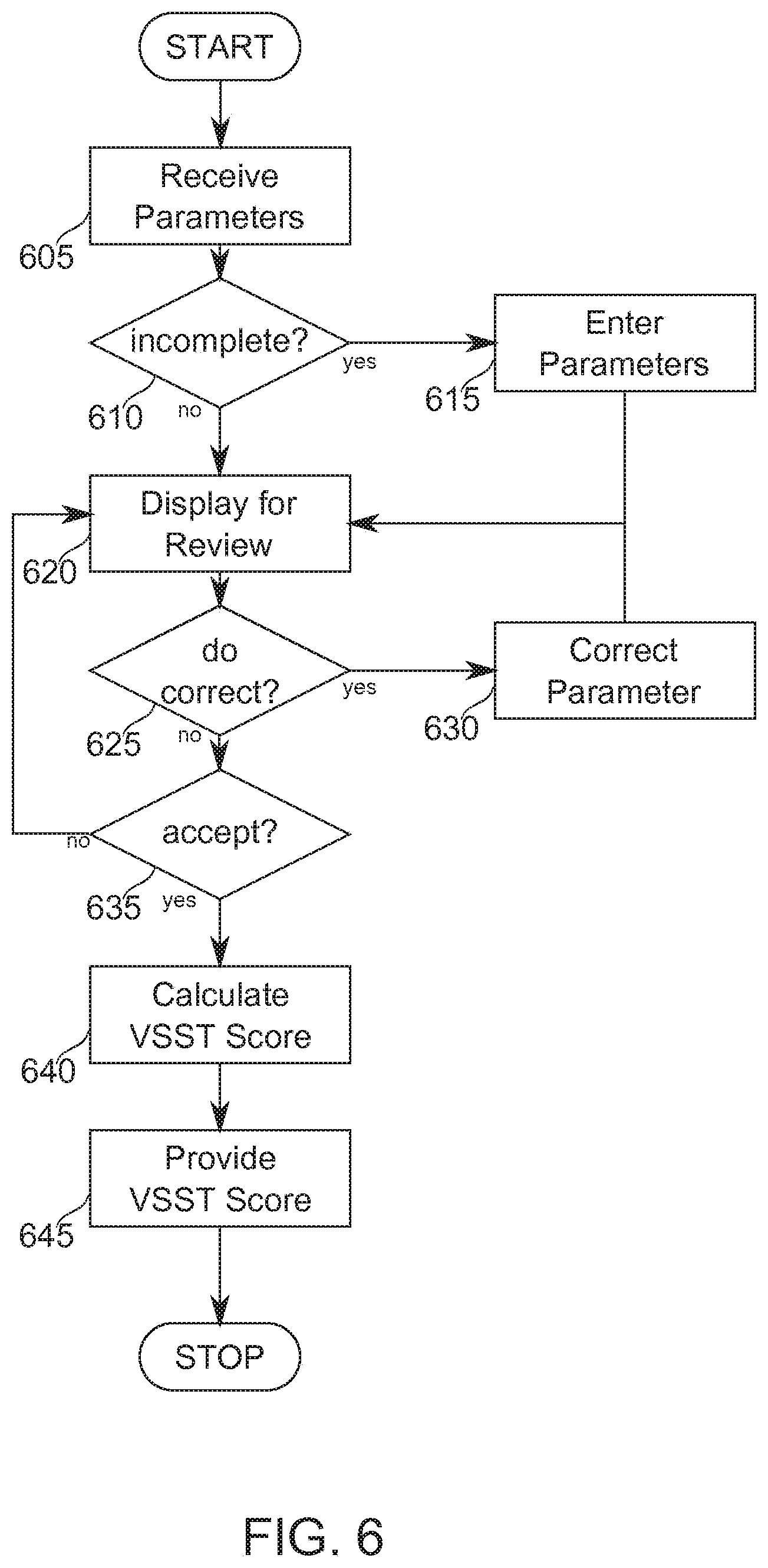
[0077] FIG. 6 is a simplified flow chart describing in outline an exemplary vascular state score determination from automatically calculated parameter values, according to some exemplary embodiments of the invention;
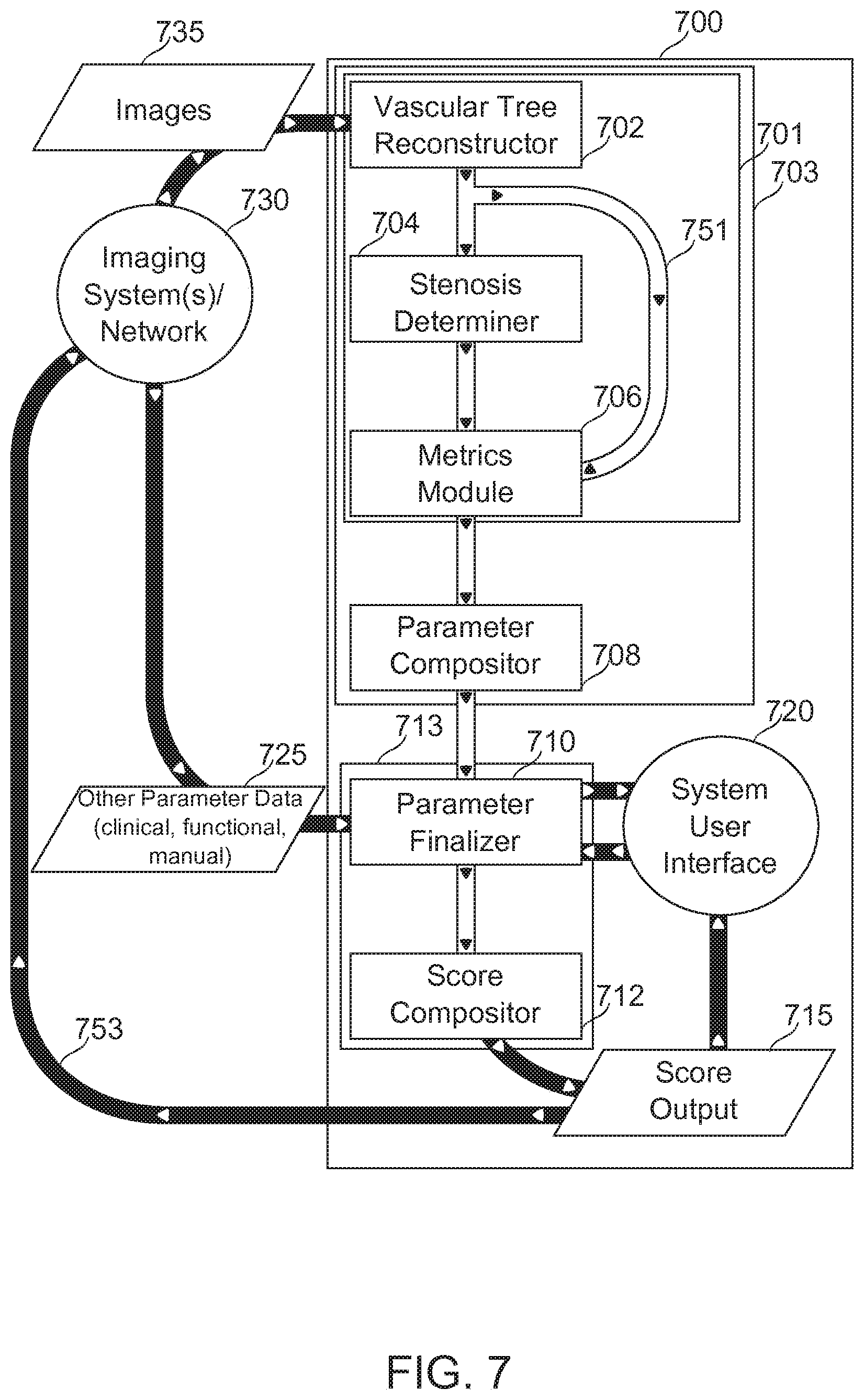
[0078] FIG. 7 is a simplified schematic of an automatic vascular state scoring tool scoring system, according to some exemplary embodiments of the invention;
[0079] FIG. 8 is a simplified flow chart of a method of determining the presence and/or associated measurements of stenotic lesions, according to some exemplary embodiments of the invention;
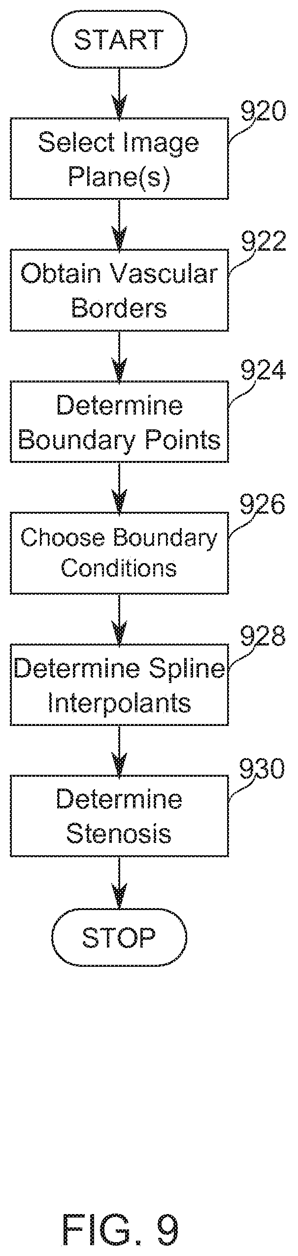
[0080] FIG. 9 is a simplified flow chart of a method of determining the presence and/or associated measurements of stenotic lesions in the region of a vessel bifurcation, according to some exemplary embodiments of the invention; and
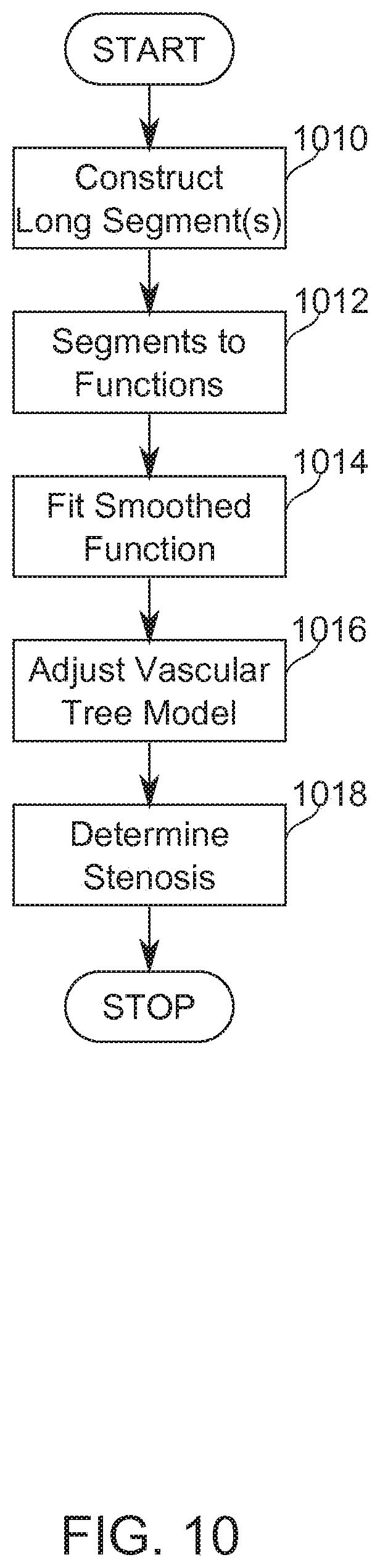
[0081] FIG. 10 is a flowchart describing in broad outline a method for refining a revascularized model of a vascular segment using information from neighbor segments, according to some exemplary embodiments of the invention.
DESCRIPTION OF SPECIFIC EMBODIMENTS OF THE INVENTION
[0082] The present invention, in some embodiments thereof, relates to the field of heart care, and more particularly, to tools for characterizing heart disease.
Overview
[0083] An aspect of some embodiments of the invention relates to automated determination of parameters based on vascular images, used to calculate a vascular disease score. In some embodiments, the imaged blood vessels are cardiac blood vessels.
[0084] In some embodiments of the invention, a cardiac disease score is calculated according to the SYNTAX Score calculation method. In some embodiments, a cardiac disease score is calculated by a SYNTAX Score alternative, derivative and/or successor vascular state scoring tool (VSST). Alternative VSST approaches potentially include, for example, a "Functional SYNTAX Score" (integrating physiological measurements--for example, vascular flow capacity, vascular elasticity, vascular autoregulatory capacity, and/or another measure of vascular function--with a SYNTAX Score-like tool), or a "Clinical SYNTAX Score" (integrating clinical variables--for example, patient history, and/or systemic and/or organ-specific test results--with a SYNTAX Score-like tool). Examples also include the AHA classification of the coronary tree segments modified for the ARTS study, the Leaman score, the ACC/AHA lesions classification system, the total occlusion classification system, and/or the Duke and ICPS classification systems for bifurcation lesions.
[0085] In some embodiments of the invention, metrics describing a vascular state are determined based on vascular imaging data. In some embodiments, the metrics are expressed, for example, as functions of vascular position (for instance, one-dimensional functions of position along a vascular segment length). The metrics express, in some embodiments, morphometric quantities such as vascular width (optionally as a diameter, radius, or cross-sectional area), vascular curvature, or another morphometric quantity. In some embodiments, the metric is of another morphological or functional measurement, such as a determined flow capacity, vascular elasticity, and/or vascular wall composition. In some embodiments, vascular state metrics determined from images comprise identifying information relative to a standard vascular atlas or other system of nomenclature.
[0086] In some embodiments, vascular state metrics are converted automatically into sub-scores for a VSST by a further operation, tailored, for example, to the specific requirements of a VSST such as SYNTAX Score. In some embodiments, sub-scores are determined based on vascular state metrics composed with operator- or network-provided information related to a subject and/or to vascular imaging data.
[0087] Potentially, automatic determination of VSST parameters reduces subjectivity and/or training variability affecting a VSST outcome. Potentially, automatic determination reduces the time and/or training required to determine a VSST score. Reducing the time and/or training required to effective determine a SYNTAX Score, for example, potentially increases compliance with vascular disease evaluation guidelines recommending using SYNTAX Score as a basis for medical decision making in cardiology. A potential advantage of reducing SYNTAX Score and/or other VSST outcome variability is increased reliability of score calculation, and/or of raw data available for future versions of SYNTAX Score and/or another VSST. A potential benefit of rapid automated or semi-automated SYNTAX Score determination, for example, is to allow a more rapid determination based on the score of a vascular intervention treatment. Potentially, the determination speed-up is sufficient to allow a single catheterization procedure to be performed comprising both diagnostic imaging and treatment intervention.
[0088] In some embodiments of the invention, the VSST score is generated entirely automatically based on provided image data and optionally other information. In some embodiments, VSST scoring is guided by an operator, for example, by selection of relevant image and/or segment regions for VSST scoring analysis. In some embodiments, operator guidance comprises segment identification, for example by providing a segment-identifying label and/or by identifying key points on a segment permitting machine identification thereof.
[0089] An aspect of some embodiments of the invention relates to the production and/or use of an astenotic model of a mammalian vasculature, or "virtual revascularization", usable, for example, in vascular disease state scoring.
[0090] In some embodiments of the invention, an astenotic vasculature model comprises a computer-generated and/or computer-stored data structure, for which relatively undiseased portions of an imaged vasculature have provided a framework for interpolating and/or extrapolating across diseased vascular regions to describe metrics relating to a relatively undiseased state therein. In some embodiments of the invention, the difference between an imaged state and a determined relatively undiseased state comprises one or more metrics of disease state. In some embodiments, the astenotic model comprises blood vessel segments extending between vascular branch points. In some embodiments, the astenotic model comprises regions of branching, for example, bifurcations and/or trifurcations.
[0091] Some embodiments of the invention described herein are contemplated for use with the SYNTAX scoring method, described, for example, at www(DOT)syntaxscore(DOT)com. However, the invention is also contemplated for use with successor and/or alternative scoring methods, including future versions of SYNTAX and/or alternative scoring methods which make use of parameters determinable as described hereinbelow. It should furthermore be understood that herein, wherever SYNTAX, SYNTAX Score, SYNTAX Score calculation, and similar terminology is used, there is implicit reference made to all such successor and/or alternative scoring methods, with changes as necessary as would be clear to one skilled in the art working on the basis of descriptions herein.
[0092] Embodiments of the invention described herein are described with particular reference to cardiac vasculature. In some embodiments--additionally or alternatively--the vasculature is of another organ, for example, a kidney, a retina, and/or a brain. It should be understood, where cardiac vasculature is described in particular, that implicit reference is also made to embodiments relating to the vasculature of another organ, with changes as necessary as would be clear to one skilled in the art working on the basis of descriptions herein.
[0093] To emphasize the breadth of scoring methods and vascular targets contemplated for some embodiments of the invention, the term VSST (Vascular State Scoring Tool) is also used herein, without detracting from the general meaning attributed to the phrase "SYNTAX Score" and its derivatives.
Definitions and Abbreviations
[0094] SYNTAX Trial
[0095] Well-known prospective multi-site clinical trial to assess PCI vs. CABG efficacy. Described, for example, by Kappetein (2006).
[0096] SYNTAX Score
[0097] Diagnostic tool, developed in association with the SYNTAX trial, for scoring complexity of coronary artery disease as an aid for planning treatment.
[0098] SYNTAX Score Outcome
[0099] A value calculated using the SYNTAX Score calculation procedure. Also called a "SYNTAX score" (no capitalization of "score") herein.
[0100] VSST
[0101] Vascular State Scoring Tool. Term used herein for general reference to scoring tools, and in particular image-based scoring tools, used for determining a vascular state. The SYNTAX Score is an example of a VSST, but versions and variants of and/or alternatives to SYNTAX relying on parameters calculated as described herein should also be understood to be encompassed by this term.
[0102] VSST Outcome
[0103] A value reflecting vascular disease state calculated using a vascular state scoring tool. Also referred to as a VSST score.
[0104] PCI
[0105] Percutaneous Coronary Intervention. Sometimes known as coronary angioplasty or angioplasty. A non-surgical procedure used to treat the stenotic coronary arteries of the heart found in coronary heart disease. PCI treatments include balloon-opening and stent implantation.
[0106] CABG
[0107] Coronary Artery Bypass Graft. Sometimes known as heart bypass or bypass surgery. Surgical procedure in which blood vessels are grafted onto heart arteries to bypass stenotic coronary arteries of the heart found in coronary heart disease.
[0108] LMS
[0109] Left Main Stem. Arterial segment between the ostium of the left coronary artery through bifurcation into left anterior descending and left circumflex branches.
[0110] 3VD
[0111] Three-vessel disease. Vascular lesion in three or more vessels.
[0112] Before explaining at least one embodiment of the invention in detail, it is to be understood that the invention is not necessarily limited in its application to the details of construction and the arrangement of the components and/or methods set forth in the following description and/or illustrated in the drawings. The invention is capable of other embodiments or of being practiced or carried out in various ways. In other instances, well-known methods, procedures, components and structures may not have been described in detail so as not to obscure the present invention.
Exemplary VSST (SYNTAX Score)
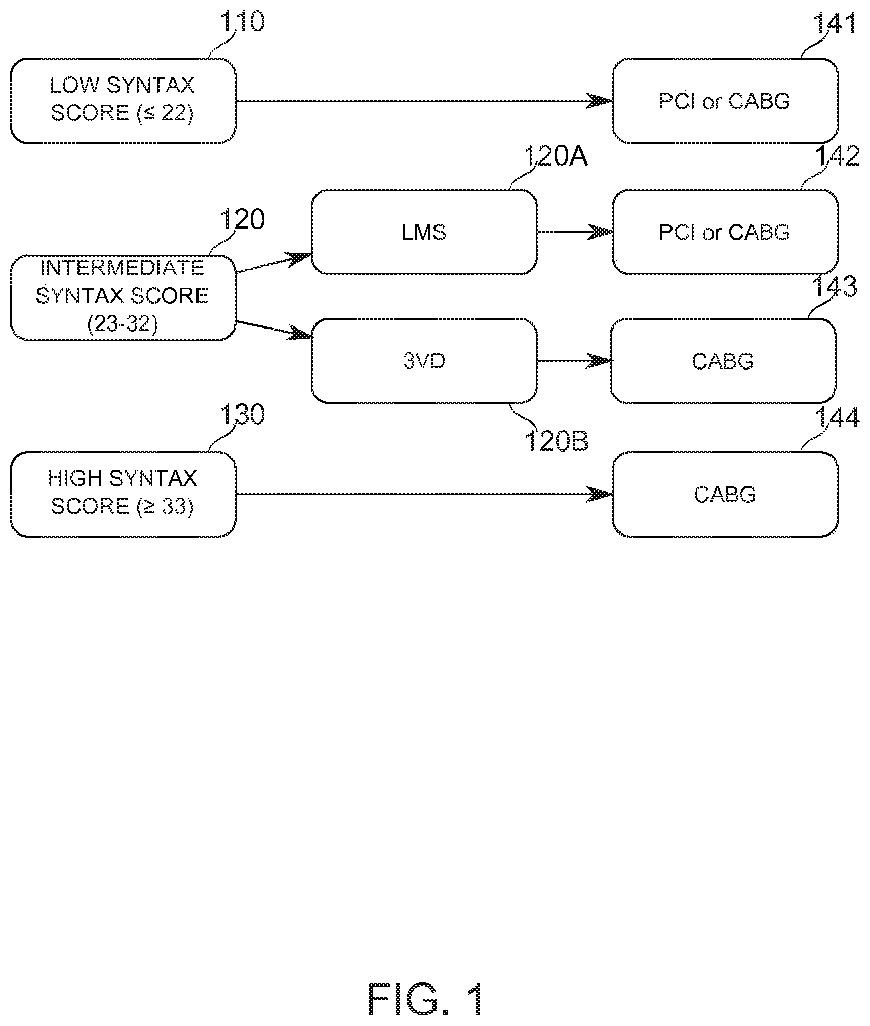
[0113] Reference is now made to FIG. 1, which is a block diagram illustrating a decision tree based on the SYNTAX Score outcome and the type of complex lesion; according to some exemplary embodiments of the invention.
[0114] At block 110, a low SYNTAX score (22) comprises an indicator for either PCI or CABG treatment (block 141). At block 130, a high SYNTAX score (33) is an indicator for preferring CABG revascularization treatment (block 144).
[0115] An intermediate SYNTAX score (23-32, block 120) comprises an indicator for either PCI or CABG (block 142) when the lesion is in the left main stem (LMS, block 120A), and is generally an indicator for CABG (block 143) when the lesion is in three or more vessels, otherwise known as 3-vessel disease (3VD, block 120B).
[0116] In calculating a SYNTAX score, a physician answers a series of questions relating to the location and size of lesions; including, for example: degree of occlusion (for example, a threshold occlusion of >50% is provided for in the scoring instructions), shape and length, presence of thrombus, and/or tortuosity of the blood vessel. Herein, the answer to each such question is referred to as a "parameter" of a scoring tool. Additionally or alternatively, the answer to the question is referred to as a "subscore" of a weighted score produced by such a scoring tool Coronary vascular images are the basis on which many of the questions are answered. Optionally, a PCI treatment is undertaken immediately or on the same day upon a decision to use this treatment. Typically, the more invasive and potentially more complicated CABG treatment is scheduled for a different patient visit. It is a potential advantage to make a scoring decision quickly, in order to release a patient and/or begin a treatment option with reduced delay. In some embodiments, the calculation of a SYNTAX Score is performed, for example, within a minute of imaging, within 2-4 minutes, within 5-10 minutes, within 5-15 minutes, or within another period of time suitable for allowing a patient to remain on a procedure table while a clinical intervention decision is determined.
[0117] A SYNTAX Score calculator is available for entering answers manually on a website (www(DOT)syntaxscore(DOT)com). In some cases, evaluation is performed immediately after imaging of a patient. Answering generally takes several minutes (20-30 minutes is typical), with speed and accuracy of answers based on the skill and/or experience of the evaluating practitioner.
[0118] In some embodiments of the invention, one or more parameters of a VSST are determined automatically, using techniques of image processing and/or analysis. In some embodiments, the automatic determination is based on two-dimensional or three-dimensional images from sources including, for example, angiographic images, CT, MM, IVUS, and/or OCT.
[0119] In some embodiments, two-dimensional images from an angiographic procedure are converted into a three-dimensional image, and lesions within the vessel are identified and entered as VSST parameters to arrive at a quick, objective SYNTAX score during the procedure. In some embodiments, VSST parameters are determined directly from two-dimensional images.
[0120] In some embodiments of the present invention, automatically determined values are provided as parameters to a VSST such as SYNTAX Score in real-time during a catheterization procedure, or following imaging.
[0121] Potentially, a reduced time of SYNTAX Score calculation provides an advantage by allowing a patient to be kept catheterized for a possible PCI treatment while waiting for a shorter period, and/or by reducing the need for recatheterization of a patient who has been temporarily released from a procedure room pending a treatment decision. Potentially, a reduced time and/or effort of scoring leads to increased use of a VSST such as SYNTAX Score as a tool for clinical decision-making.
[0122] In some embodiments of the invention, parameters of another VSST based on geometric, clinical, or functional factors are determined.
VSST Parameter Algorithm
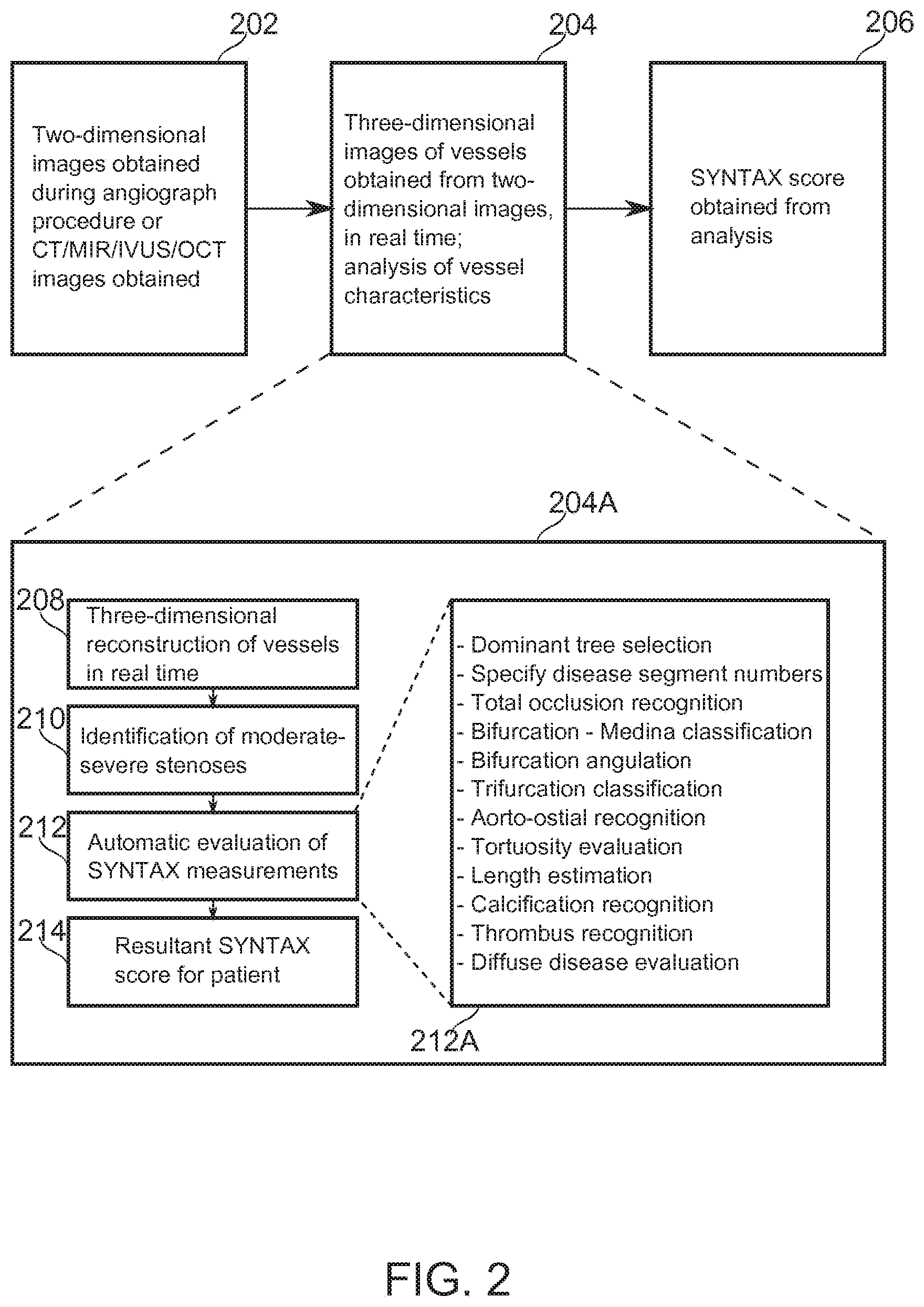
[0123] Reference is now made to FIG. 2, which is a block-diagram illustration of operations of an algorithm, according to some exemplary embodiments of the stages.
[0124] At block 202, in some embodiments, two-dimensional images comprising the coronary arteries are obtained. In some embodiments, the two-dimensional images comprise X-ray angiograms, sections of CT 3-D imagery of the coronary arteries, or imagery obtained by, for example, MM, IVUS, or OCT.
[0125] At block 204, in some embodiments, image processing and analysis and VSST score calculation is performed. Block 204A describes these operations in more detail.
[0126] At block 208, in some embodiments, a three-dimensional reconstruction of the vessels of an individual patient is performed, based on two-dimensional projections of the coronary arteries during a diagnostic catheterization. In some embodiments, this occurs in real-time, while the patient is undergoing catheterization together with imaging. In some embodiments, two-dimensional images are used directly in further image processing.
[0127] Reference is now made to FIG. 3, which is a schematic illustration showing principles of three-dimensional reconstruction by use of epipolar geometry, according to some exemplary embodiments of the invention. FIG. 3 shows two exemplary image planes 310, 312 whereupon images of a target object point P.sub.3 are projected to P.sub.1 and P.sub.2, respectively, from radiant sources S.sub.1, S.sub.2. The relative positions of P.sub.1, P.sub.2, S.sub.1, and S.sub.2 are known, while the position of P.sub.3 in space is to be determined from these known positions. P.sub.3 is between S.sub.1 and P.sub.1, so it lies somewhere along the path between them. This path in turn has a projection onto the image plane of P.sub.2, (marked Epipolar Line), determinable from the intersection of the Epipolar Plane defined by S.sub.1, S.sub.2, and P.sub.1 with the image plane P.sub.2. The position of P.sub.2 along the Epipolar Line provides the remaining information needed to locate P.sub.3 in space. This concept is applicable to multiple image planes. Approaches which comprise three-dimensional reconstruction of vascular information from two-dimensional source data have been described (Pellot, 1994; Sprague, 2006; Andriotis, 2008). Reference is also made to U.S. Patent Application 61/752,526 by the Applicant, which is incorporated herein in its entirety by reference.
[0128] In some embodiments, based on reconstruction performed, for example, as in FIG. 3, a stereo reconstruction of the coronary tree is performed, using a series of spatially separated two-dimensional projections. Reconstruction produces a unified 3-D coronary tree. In some embodiments, for each vessel the location (x,y,z) and radius (R) is defined in the reconstruction. In some embodiments, the hierarchy between vessels, for example, the connections between vessel segments and/or their position relative to vessel branching points is definable with reference to a 3-D reconstruction.
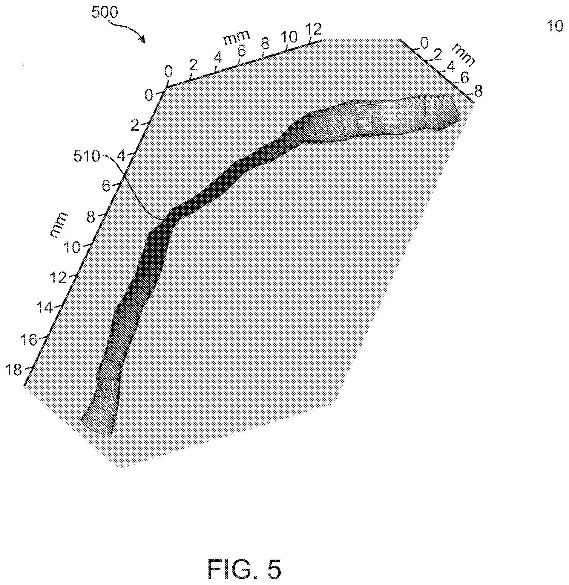
[0129] Reference is now made to FIG. 5, which illustrates a three-dimensionally reconstructed stenotic area 510 along a vessel segment 500, according to some exemplary embodiments of the invention. The gray scale indicates the radius along the vessel centerline (darker is narrower). Such a segment can be extracted, for example, from angiographic CT, MM, PET, OCT, and/or IVUS. Methods of coronary angiography imaging are reviewed, for example, in Youssef (2013).
[0130] MDCT (multi-detector computed tomography) or CT (computed tomography) measures tissue and/or contrast agent attenuation of source X-ray radiation. Typical resolution is 200-500 depending on specifics of the implementation.
[0131] MRI (magnetic resonance imaging) uses nuclear magnetic resonance properties of an endogenous or exogenously introduced contrast basis. Typical resolution may be 1 mm, down to 350 .mu.m, depending on implementation.
[0132] PET (positron emission tomography) uses detection of emitted radiation from tracers. Typical resolution is 4-5 mm
[0133] OCT (optical coherence tomography) measures backscattered light as a function of time (and/or, in some implementations, frequency). Typical resolution is 4-20 .mu.m.
[0134] IVUS (intravascular ultrasound) operates by converting the intensity of backscattered sound signals (which varies by target encountered) into image representations. Typical resolution is about 150 .mu.m.
[0135] In some embodiments of the invention, a two-dimensional vascular tree is recreated from one or more appropriate two-dimensional images or image sections. In some embodiments, manual guidance is accepted for determining which two-dimensional images comprise useful targets for image analysis of one or more vascular segments. A potential advantage of proceeding from a two-dimensional image is a reduced complexity of imaging procedure and/or a reduced computation time for vascular reconstruction from the image data.
[0136] A potential advantage of developing a vascular tree from a three-dimensional reconstruction is representation of depth information. This allows improved accuracy, for example, of measures of length and/or tortuosity, which are potentially reduced in 2-D due to foreshortening artifacts. Also for example, 3-D reconstruction potentially resolves ambiguities due to structures which cross over one another in 2-D images. Three-dimensional reconstruction also allows structural analysis from multiple angles, which potentially allows obtaining more accurate metrics for vascular features such as occlusion percentage and/or thrombus.
[0137] In some embodiments of the invention, the output of block 208 comprises a reconstruction of the complete coronary artery tree, including the right coronary artery and the left coronary arteries. In some embodiments the first stage results in a partial sub-tree reconstruction--the right coronary artery, the left coronary arteries, and/or any sub-branch of them. In some embodiments, a number/name of at least one segment is provided, for example to allow orientation of the reconstructed tree relative to the segment labeling used by the VSST.
[0138] In some embodiments of the invention, a hierarchical tree, complete or partial, of arterial centerlines is derived from the reconstructed artery tree. In some embodiments, at points along these centerlines, vessel radius, curvature and tortuosity are determined
[0139] At block 210, in some embodiments, the tree structure serves as for extracting the significant stenosis areas, based on the analysis of the vessels' radii. SYNTAX Score, for example, defines significant stenosis as to moderate to severe stenosis having >50% lumen blockage.
[0140] At block 212, in some embodiments, specific parameters corresponding to each significant stenosis located on the previous stage are determined, according to the SYNTAX Score or other VSST specifications. In some embodiments, determined parameters include one or more of the following parameters (listed at block 212A): [0141] Sub-tree dominance. [0142] Anatomical identification (for example, branch position) of the diseased segment. In some embodiments, lesions in some segments are weighted more heavily than in others. [0143] Recognition of total occlusion. In some embodiments, total occlusions are further classified, for example, according to known age, presentation of a blunt stump, proximity to side branches, and/or bridging by shunting vessels. [0144] Bifurcation-Medina classification. [0145] Bifurcation angulation. [0146] Trifurcation classification. [0147] Recognition of aorto-ostial proximity. [0148] Tortuosity evaluation. [0149] Length estimation. [0150] Calcification recognition. [0151] Thrombus recognition. [0152] Diffuse disease evaluation.
[0153] In some embodiments, most (for example, at least seven) or all of the foregoing parameters are determined Parameter calculations made based on automatic image processing and analysis operations provide a potential advantage in being not subject to subjective assessments by a practitioner.
[0154] At block 214, the results are compiled into a SYNTAX Score (or other VSST) outcome, and at block 206, the outcome is made available from the analysis.
System for Vascular State Scoring
[0155] Reference is now made to FIG. 7, which is a simplified schematic of an automatic VSST scoring system 700, according to some exemplary embodiments of the invention.
[0156] In FIG. 7, broad white pathways (for example, pathway 751) denote simplified paths of data processing through the system. Broad black pathways (for example, pathway 753) denote external data connections or connections to the system user interface 720. Black pathway data content is labeled by overlying trapezoidal blocks.
[0157] The vascular tree reconstructor 702, in some embodiments of the invention, receives image data 735 from one or more imaging systems or a system-connected network 730. Stenosis determiner 704, in some embodiments, determines the presence of stenotic vascular lesions based on the reconstructed vascular tree. In some embodiments, metrics module 706 determines additional metrics related to the disease state of the vascular tree, based on the reconstructed vascular tree and/or determined stenosis locations and other measurements.
[0158] In some embodiments, metrics extractor 701 comprises functions of vascular tree reconstructor 702, stenosis determiner 704, and/or metrics module 706. In some embodiments, metrics extractor 701 is operable to receive image data 735, and extract from it a plurality of vascular state metrics, suitable, for example, as input to parameter compositor 708.
[0159] In some embodiments, parameter compositor 708 converts determined metrics into subscore values (for example true/false values) which comprise parameters that "answer" vascular state scoring questions, and/or are otherwise are mapped to particular operations of a VSST scoring procedure.
[0160] In some embodiments, subscore extractor 703 comprises functions of vascular tree reconstructor 702, stenosis determiner 704, metrics module 706, and/or parameter compositor 708. In some embodiments, subscore extractor 703 comprises functions of metrics extractor 701. In some embodiments, subscore extractor 703 is operable to receive image data 735, and extract from it one or more VSST subscores, suitable as input for score calculator 713.
[0161] Parameter finalizer 710, in some embodiments, ensures that parameter data provided is sufficiently complete and correct to proceed to final scoring. In some embodiments, corrections to automatically determined parameters are determined at finalizer 710, optionally under operator supervision through system user interface 720. In some embodiments, lacunae in automatically provided parameter data are filled: for example, by user input from system user interface 720; or by other parameter data 725 provided, for example, from another diagnostic system or a network providing access to clinical data.
[0162] Score compositor 712, in some embodiments, composes the finalized outputs into a weighted score output 715 based on the determined parameters for the score. The score is made available, for example, over the system user interface or to networked resources 730.
[0163] In some embodiments of the invention, score calculator 713 comprises functions of the parameter finalizer 710 and/or score compositor 712. In some embodiments, score calculator 713 is operable to receive composited parameters and/or subscores (for example from parameter compositor 708 and/or subscore extractor 703), and convert them to a VSST score output 715.
[0164] In some embodiments of the invention, intermediate results of processing (for example, the reconstructed vascular tree, various metrics determined from it, and or parameter determinations) are stored in permanent or temporary storage on storage devices (not show) of the system 700, and/or on a network 730.
[0165] The scoring system 700 has been described in the context of modules which, in some embodiments of the invention, are implemented as programmed capabilities of a digital computer. It should be understood that the underlying system architecture may be implemented in various ways comprising embodiments of the invention; for example, as a single or multiple-process application and/or as client-server processes running on the same or on different computer hardware systems. In some embodiments of the invention, the system is implemented in code for execution by a general purpose processor. In some embodiments, part or all of the functionality of one or more modules is provided by an FPGA or another dedicated hardware component such as an ASIC.
[0166] To provide one example of a client-server configuration, a subscore extractor 703 is implemented as a server process (or group of server-implemented processes) on one or more machines remote to a client computer which implements modules such as the score calculator 713 and user interface 720. It should be understood that other divisions of modules described herein (or even divisions within modules) are encompassed by some embodiments of the invention. A potential advantage of such a division may be, for example, to allow high-speed dedicated hardware to perform computationally intensive portions of the scoring, while providing an economy of scale by allowing the hardware to be shared by multiple end-users. Such a distributed architecture potentially also provides advantages for maintenance and/or distribution of new software versions.
Stenosis Determination
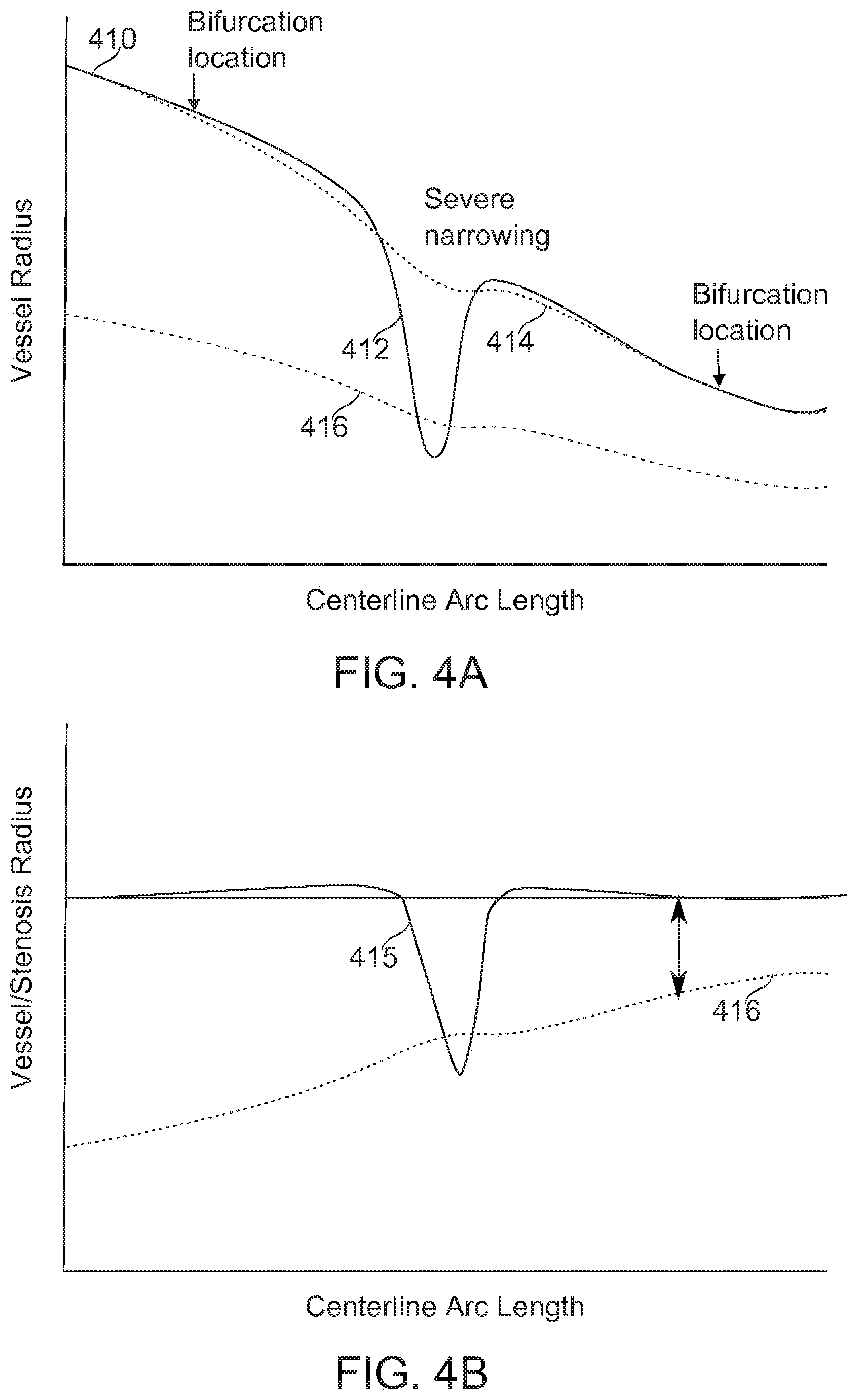
[0167] Reference is now made to FIGS. 4A-4B, which schematically illustrate an example of a stenotic determination, corresponding to the method a block 210, according to some exemplary embodiments of the invention.
[0168] The plot 410 in FIG. 4A shows radius (Vessel Radius) along a vessel segment (Centerline Arc Length). In some embodiments of the invention, measurements required for the SYNTAX Score or other VSST score can be extracted from such a one-dimensional function r=f(s), where r is the vessel radius, and s is the arc-length.
[0169] In some embodiments of the invention, for example, a severe stenosis 412 is automatically identified by means of a high-pass filter. Plot 415 is the high-pass filter result. Subtracting the plot 415 from plot 410 obtains plot 414, which approximates the un-stenosed vessel width. Plot 416 represents the half-width of plot 415, representing, for some embodiments, the threshold between a scored and an unscored stenosis. Inverted and superimposed on plot 415, a sufficiently severe stenosis reveals itself where the plot 415 crosses inverted plot 416. Additionally or alternatively, a very positive and/or negative slope along a portion of plot 415 indicates a region of abrupt change.
[0170] In some embodiments of the invention, a lesion length is determined, for example by a metric such as width at a percentage occlusion relative to the maximum occlusion. In some embodiments, this percentage is 5%, 10%, 20%, or another percentage. In some embodiments, a lesion length is determined by a slope inward from the vascular wall above a threshold, for example, a change of 1 part in 3 (occlusion depth-to-length), 1 part in 5, 1 part in 10, or another slope. In some embodiments, a second or higher slope derivative is the basis of a total lesion length determination.
Stenosis Determination--Alternative Embodiments
[0171] Reference is now made to FIG. 8, which is a simplified flow chart of a method of determining the presence and/or associated measurements of stenotic lesions, according to some exemplary embodiments of the invention.
[0172] In some embodiments of the invention, stenosis in the imaged anatomy is determined relative to a "virtually revascularized" model of the anatomy. The virtual revascularization, in some embodiments, comprises determination of a vascular tree model which removes narrowings and/or other obstructions which are determined to comprise anatomical changes due to vascular pathology.
[0173] At block 802, the flowchart starts, and each vessel segment is converted to a one-dimensional function f(s) of diameter vs. distance. Optionally, the function yields vessel radius or another metric comprising information about the vessel lumen cross-section, such as area. In some embodiments, distance is obtained from the Euclidean distance formula, integrated at points along the vessel segment, for example:
s=.intg. {square root over (dx.sup.2+dy.sup.2+dz.sup.2)}
[0174] where s is the integrated distance at a point along the vessel segment. The integral notation and other uses of "integration" herein should be understood as potentially approximated by summation of finite elements and/or other approximations appropriate for discrete image pixel (2-D) or voxel (3-D) samples. Additionally or alternatively, integration is potentially over a continuous, image-data derived function, for example one obtained by spline fitting and/or interpolation.
[0175] In some embodiments, the 3-D diameter of the vessel at a given point comprises an average of the diameters measured from a plurality (for example, all available) of 2-D projections visualizing that point. Optionally, the diameter is instead calculated based on the open area of the vessel, approximating the vessel lumen cross-section as circular. Optionally, the lumen cross-section area is used directly. Alternatively or additionally, radius is used. In the discussion that follows, it is to be understood that "diameter" is replaceable by another metric of lumen openness, with the method changed as necessary as would be understood by one skilled in the art making reference to the descriptions disclosed herein.
[0176] In some embodiments of the invention, an iterative process of virtual revascularization now begins for each segment (looping over each segment is not shown).
[0177] At block 804, in some embodiments, an initial reference diameter is chosen to comprise a statistical fit (for example by a linear, least mean squares method, optionally modified by a weighting function) to vessel diameter along the vessel segment length. In some embodiments, points near either end of the segment are weighted more than other segments. It should be noted that in some embodiments, determination of an unstenosed diameter at segment ends, for example, near a bifurcation, is carried out by a module specialized for bifurcation analysis, for example, as described hereinbelow in connection with FIG. 9. It should also be noted that in some embodiments, refinement of a determination of an unstenosed diameter for one or more segments is determined with reference to one or more constraints applied in consideration of a plurality of segments, for example, as described hereinbelow in connection with FIG. 10. Optionally, the point weightings are adjusted so that best-fit deviations from wider (potentially less-diseased) points along the segment are weighted as more important than deviations from narrower points. This provides a potential advantage by allowing less-diseased regions of the vessel to dominate the determination of the virtually revascularized vessel width. The weighting determinations are adjusted during subsequent revascularization operations.
[0178] At block 806, in some embodiments, differences between fit and measured points are determined, and statistics (for example, mean, standard deviation) are calculated based on the determined differences.
[0179] At block 808, in some embodiments, weighting adjustments are made, such that certain outliers from the linear fit are reduced in weight. The outliers are, for example, points which have statistically meaningful differences from the fit. Meaningful differences include, for example, being more than two standard deviations away from the best-fit line compared to the population of diameters overall.
[0180] At block 810, in some embodiments, the best fit (typically linear) is redetermined It should be noted that embodiments of the invention are not limited to a linear fit, but linearity is a convenient model for capturing the observation that vessels decrease in diameter more-or-less monotonically along their length away from the end proximal to the heart.
[0181] At block 812, in some embodiments, a test is performed to see if the best fit line has converged within some limit of stability. If not, processing continues with another fitting round at block 806. If the best fit has converged to a stable solution, processing continues with block 814.
[0182] At block 814, in some embodiments, the best fit function f(s) is used together with the original data function f(s) to determine stenosis, for example:
stenosis = 100 * ( 1 - f ( s ) f x ( s ) ) ##EQU00001##
[0183] At block 816, the segment function is evaluated for additional metrics related to lesion depth, length, and position, for example, as described in connection with FIGS. 4A-4B. For example, an output of the process, in some embodiments, comprises pairs of values s.sub.1, s.sub.2, such that for s.sub.1.ltoreq.s.ltoreq.s.sub.2, s is within a stenotic lesion. The flowchart then ends.
[0184] Reference is now made to FIG. 9, which is a simplified flow chart of a method of determining the presence and/or associated measurements of stenotic lesions in the region of a vessel bifurcation, according to some exemplary embodiments of the invention.
[0185] In some embodiments of the invention, diameters determined along a blood vessel segment are potentially ill-defined at a bifurcation (or trifurcation) where abrupt changes in diameter occur, or where the definition of a diameter, radius, or cross-sectional area is indeterminate. In some embodiments, a procedure is implemented whereby diameters at such boundaries are defined more clearly.
[0186] The flowchart begins, and at block 920, in some embodiments, at least one image plane passing through a bifurcation is selected as a reference plane for analysis. In some embodiments, every image plane in which a bifurcation is identified as appearing is selected during some iteration of the method. In some embodiments of the invention, determination of these image planes in turn, and/or of the region of the planes in which the bifurcation appears, proceeds from the relationship between image planes and a three-dimensionally reconstructed vascular model, generated, for example, as described in relation to FIG. 3 and in associated references given.
[0187] In some embodiments, the image section is manually selected. In some embodiments, the image plane is selected to be a plane which includes vessel center points of at least two vessels at a determined distance from the vessel (for example, 1 mm, 2 mm, 3 mm or a greater or larger distance), and a point near the center of the region of bifurcation. Optionally a different plane is selected for each pair of trunk and branch vessels (trunk and a first branch, trunk and a second branch). The method is described herein below with respect to one plane of analysis selected for one branch point vascular segment pair (a trunk and a branch), but it is to be understood that the analysis is optionally carried out on two more vascular segment pairs at a given segment junction (three, for example, in the case of a trifurcation). It is also to be understood that unstenosed diameter within more than one plane is optionally determined, and results from this plurality of determinations composed into one or more metrics describing unstenosed vascular morphology. For example, in some embodiments, a plurality of planes is selected, and an average or other statistically determined unstenosed vessel diameter selected from the set of planes analyzed. In some embodiments, unstenosed vessel widths determined in a variety of directions corresponding to different image planes are composed into an approximation of the shape of the vessel lumen circumference at different locations along its length.
[0188] At block 922, in some embodiments, for each vessel segment in a pair (for example a pair comprising a trunk vessel and a branch vessel), data sets describing each of two vessel borders (x.sub.b,y.sub.b) falling within a selected image plane are determined. The border data sets are, for example, determined by the locations along the vessel segment length which representing a transition from low contrast to high contrast. The transition point is determined, for example, by a threshold, a peak rate of contrast change, by a simple edge detection convolution, by a Frangi filter, or another appropriate boundary-finding method available to one skilled in the art. For convenience of exposition, the vessel border data sets (x.sub.b,y.sub.b) are referred to hereinbelow as the "left" border and the "right" border, it being understood that the designation of left and right in this context is potentially arbitrary.
[0189] At block 924, in some embodiments, for each of the trunk and branch segments chosen, a boundary point away from the bifurcation is chosen as a reference and/or spline interpolant termination point. The reference point may be considered as a trusted and/or anchor point, far enough from the point of a potentially lesioned bifurcation that it provides an unstenosed reference diameter for the vessel at that point. In some embodiments, the distance chosen is, for example, 1-2 mm, 2-4 mm, 1-5 mm, or another larger or smaller distance from the core of the bifurcation. In some embodiments, the distance is chosen as a function of a previously estimated vascular width, for example, 2, 3, 4 or a greater or smaller multiple of the previously estimated vascular width.
[0190] At block 926, in some embodiments, boundary conditions are determined at each of the reference points, comprising the point location. In some embodiments, a first derivative up to a derivative of order n is determined, for example by examination of border point locations from 1 to n data nodes away from the selected node point. The result, in some embodiments, is a set of four boundary conditions--two for the left wall, and two for the right; one of each wall pair being from the trunk vessel segment, and one from the branch vessel segment.
[0191] At block 928, in some embodiments, a spline interpolant is determined for each of the left and right walls which runs between the boundary conditions determined for each wall. Each such spline interpolant may be considered as an "unstenosed" border data set (x.sub.i,y.sub.i) corresponding in portions to one or the other of the original image border data sets (x.sub.b,y.sub.b) for one wall of both the trunk and branch vessel segments, and in a central portion to the region of the bifurcation. Additionally or alternatively, the left- and right-wall spline interpolants may be considered as bounding the lumen of the open or unstenosed vascular segment through the region of bifurcation.
[0192] In some embodiments, the interpolants are optimized (while preserving the boundary conditions) to maximize contrast differences across the surfaces of the interpolants. This corresponds, ideally, to adjusting the interpolant diameter to the diameter of the vascular wall, and to adjusting the interpolant center position to the center of the blood vessel. Contrast is determined, in some embodiments, by a simple edge detector, by the output of a Frangi filter, or by another means of edge detection known in the art. In some embodiments, positions within the core of the bifurcation are ignored for purposes of fit determination.
[0193] At block 930, in some embodiments, the lumen bounded by the unstenosed border data sets (x.sub.i,y.sub.i) is compared to the lumen bounded by the corresponding data-derived border data sets (x.sub.b,y.sub.b), to determine an absolute and/or relative degree of stenosis in the lumen comprised within the region of bifurcation. Optionally, the comparison is made, for example, after conversion of relative border locations to diameters, radii, areas, or another metric as a function of position along the vascular segment length and/or away from the region of bifurcation. Optionally, the two-dimensionally determined model is referred back to a three-dimensional model by making reference to 2-D to 3-D mappings determined during a phase of 3-D vascular tree reconstruction. Optionally, the degree of stenosis is analyzed as for stenotic regions in FIG. 8 and/or FIGS. 4A-4B. The flowchart ends.
[0194] Reference is now made to FIG. 10, which is a flowchart describing in broad outline a method for refining a revascularized model of a vascular segment using information from neighbor segments, according to some exemplary embodiments of the invention.
[0195] In some embodiments of the invention, a method is provided which takes into account constraints applicable to the morphometric relationships between vascular segments in determining an unstenosed vascular model. Potentially, this allows more accurate determination of an unstenosed vascular model, and/or reduces the occurrence of artifacts which do not reflect reasonable anatomical situations.
[0196] The flowchart begins, and at block 1010, one or more long segments are constructed by the concatenation of a plurality of interconnected shorter segments into single functions. In some embodiments, the shorter segments are defined by branch points, and the construction of long segments comprises trimming off different branch alternatives for different long segments. In some embodiments, each possible long segment implied by the underlying vessel segment hierarchy is constructed.
[0197] At block 1012, in some embodiments, long segments are converted to functions, for example, to a one dimensional function of arc-length. In some embodiments of the invention, the function describes radius, diameter, cross-sectional area, and/or another metric related to a degree of stenosis as a function of position along the segment.
[0198] At block 1014, in some embodiments, a smoothed function {hacek over (f)}(s) is fitted to the data of f(s). The fitting, in some embodiments, is subject to a similarity criterion, for example, minimization of |f(s)-{hacek over (f)}(s)|. The fitting, in some embodiments, is subject to a smoothness criterion, for example, minimization of |{hacek over (f)}''(s)|.
[0199] In some embodiments, the fitting further comprises the criterion of minimizing {hacek over (f)}'(s), for example, such that this value is everywhere non-positive. A potential advantage of this criterion is that it takes advantage of an observed property of healthy vascular trees, which are seen to narrow monotonically when moving from trunk to branch. Thus, for example, a case may arise in which an entire segment is narrowed, thus providing no healthy region as an internal reference. In such a case, a single undiseased downstream segment nevertheless potentially signals that the unstenosed diameter of the highly diseased upstream segment should be larger than the observed vessel diameter.
[0200] At block 1016, in some embodiments, an adjusted unstenosed model of a vascular segment is referred back to the original vascular tree model.
[0201] At block 1018, in some embodiments, a degree of stenosis is calculated from the adjusted unstenosed vascular segments, for example as described herein above with reference to FIG. 8 and/or FIGS. 4A-4B. The flowchart ends.
Total Occlusion and Thrombus
[0202] In some embodiments, features which connect vessel walls and/or comprise non-vascular inclusions within the vessel walls are determined by processing of images and/or of the reconstructed vasculature.
[0203] In some embodiments of the invention, a total occlusion of the vasculature is identified, for example, by an abrupt border between lumen and background intensities, for example, a complete break in the continuity of blood-borne contrast agent imaged along a blood vessel in the image data. In some embodiments, the occlusion is determinable to be associated or not associated with bridging (shunting) vessels. In some embodiments, bridging vessels are distinguishable from the main artery by, for example, an increase in tortuosity, a decrease in vessel diameter, and/or a sharp change in vessel direction.
[0204] In some embodiments, one or both occlusion boundaries (proximal/distal) are characterized by the morphology of one or more occlusion/lumen boundaries. For example, a degree of boundary curvature may be assessed by a parabolic fit, a spline fit, or another fitting function. In some embodiments, an occlusion morphology is determined by a relative position of predefined points along the occlusion curve (for example, center-most point, compared in longitudinal position to points within some radius percentage of the vessel wall), or another characterizing feature of the occlusion morphology.
[0205] In some embodiments, thrombotic structures in the vasculature are determinable from the presence of certain relatively contrast agent-free, potentially ovoid or spherical regions within the lumen of a blood vessel. In some embodiments, a vessel cross-section potentially containing a thrombus is identified by region comprising a sharp drop in contrast agent density, visible as an increase in brightness in some imaging modalities, or otherwise distinguishable from its surroundings. The region in the cross-section is potentially surrounded by a region of differing (for example, higher) contrast density. In some embodiments, vessel inclusions are identified, for example, by a ratio of free (unconnected to a wall) surface area, free circumference, and/or free angular arc to a wall-connected portion of the inclusion. In some embodiments, vessel inclusions such as thrombi are identified by being unconnected to a vessel wall on at least three sides.
Determination of Other Metrics
[0206] In some embodiments of the invention, metrics for conversion into VSST parameters are determined from the three- or two-dimensional reconstruction of the vasculature.
[0207] In some embodiments, metrics such as numbers and order of branching points are determined from the branch points determined during vascular reconstruction.
[0208] In some embodiments, metrics such as the size of the lesion, arc-distances from nearby bifurcations (points marked "Bifurcation location" in FIG. 4A), and/or distances between adjacent lesions are determined from the vascular reconstruction. In some embodiments, the radius, curvature and/or tortuosity of vessels is extracted along the tree from the vascular reconstruction.
[0209] In some embodiments, metrics comprising distances between reference points on the vessel (for example, lesion lengths, relative positions, and/or segment lengths) can be determined by integrating distance over the length of a segment arc. This may be, for example, according to integrated length in three dimensions:
s=.intg. {square root over (dx.sup.2+dy.sup.2+dz.sup.2)}
[0210] where s is the total distance traversed.
[0211] In some embodiments, a metric comprising a measure of vessel segment tortuosity is determined by integrating on angular deviations along a segment length.
[0212] In some embodiments, where bifurcations have been identified during the phase of artery tree construction, lesions near the bifurcations are classified according to the Medina classification of bifurcation lesion analysis.
[0213] In some embodiments, metrics comprising absolute measurements of vessel diameter are obtained from the 2D and/or 3D vessel data, for example, by making measurements perpendicular to segment direction along the vascular tree.
Conversion of Metrics into VSST Parameters
[0214] In some embodiments of the invention, one or more VSST parameters are referred to a specific, anatomically identified vessel segment as part of the scoring procedure. In some embodiments, vessel segments are automatically identified by registration of the vessel segment tree to an atlas comprising one or more standard vascular morphology patterns. In some embodiments, the determination is automatic and unguided, for example by finding a best fit pattern between the atlas and the acquired image data.
[0215] In some embodiments, deviations from an atlas standard (for example, due to increases in tortuosity, total occlusions, and/or development of shunting vessels) are identified. In some embodiments, a vascular identification of a segment comprising one or more metrics which deviate significantly from an atlas standard value is identified as such for an operator or otherwise flagged as uncertain. For example, a vessel which is longer, of a different width, or more tortuous than a corresponding atlas standard segment may be flagged according to a threshold of 50%, 100%, or another percentage of difference. Additionally or alternatively, the segment may be flagged according to a statistically unlikely (P<0.05, P<0.001, or less than some other P value) morphology.
[0216] In some embodiments, identification is partially guided, for example by prompting an operator to identify one or more vascular segments, and then automatically identifying remaining vessels by their relative position in space and/or along branch points. In some embodiments of the invention, automatic determination is displayed for potential correction by an operator through a computer user interface. In some embodiments, vascular segments where a lesion has been detected are presented to an operator for manual identification.
[0217] In some embodiments of the invention, composition of metrics into VSST parameters/subscores proceeds from determined metrics according to the specific requirements of the VSST. In the SYNTAX Score method, for example, a question requests determining which of left or right dominance (two distinctive anatomical patterns) applies to the vasculature being evaluated. In some embodiments, the parameter corresponding to this question is determined from automatic inspection of vascular branching point number and order metrics.
[0218] In some embodiments of the invention, one or more VSST questions concerns occlusive degree, vessel diameter and/or relative locations of stenotic lesions detected in received image data. In some embodiments of the invention, one or more parameters describing the number and extent of lesions are composed by applying thresholds based on criteria supplied for a VSST to vascular metric data. For example, in a SYNTAX Score evaluation, a coronary lesion is counted when it meets criteria of vessel diameter being at least 1.5 mm, and a level of stenosis being at least 50%. Also for example, relative positions of lesions are relevant to the scoring of a lesion as one or two lesions. Optionally, for example in SYNTAX Score, an inter-occlusion distance threshold for scoring as one or two lesions is a function of vessel diameter, for example three vessel diameters. In some embodiments, the number of segments that a lesion comprises is scored.
[0219] In some embodiments, one or more VSST questions concerns the presence of a total occlusion, its curve morphology, depth, and/or position relative to nearby branch points. In some embodiments, a parameter describing the presence of shunting ("bridging") vessels around a total occlusion is determined, for example, from one or metrics describing an abrupt change in vessel diameter, vessel direction, and/or vessel tortuosity.
[0220] In some embodiments, one or more VSST questions requests an abstracted description of tortuosity, which is extracted from determined metrics. For example, a tortuosity metric may comprise a quantified description of integrated tortuosity along a segment. The tortuosity parameter in the case of a SYNTAX Score, for example, requests counting "One or more bends of 90.degree. or more, or three or more bends of 45.degree. to 90.degree. proximal of the diseased segment" (www(DOT)syntaxscore(DOT)com). This parameter may be abstracted from a tortuosity metric by, for example, integrating accumulated curvature, and counting the number of curves which reach each threshold criterion.
[0221] In some embodiments of the invention, the VSST parameter description of a lesion occlusion may be more specific than "more than 50% occlusion". For example, a SYNTAX Score requests: "Estimation of the length of that portion of the stenosis that has .gtoreq.50% reduction in luminal diameter in the projection where the lesion appears to be the longest" (www(DOT)syntaxscore(DOT)com). In some embodiments, occlusion is separately determined as a metric along two or more planes to which the lesion is projected. In some such embodiments, the parameter determination uses the occlusion length in the plane for which the lesion is longest. In some embodiments, (for example, with stenosis opener modeling) occlusion is determinable across any radial direction along the segment axis, continuously, or at any number of discrete radial directions. In some embodiments, conversion of a length to a parameter comprises determining if a length (such as a lesion length) meets a threshold criterion, for example, a SYNTAX Score distinguishes lesions more than 20 mm long. In another VSST, the length is optionally longer or shorter, as specified.
[0222] In some embodiments of the invention, a VSST parameter may comprise temporal information about a lesion. For example, in a SYNTAX Score parameter, "multiple persisting opacifications" (www(DOT)syntaxscore(DOT)com) along a vascular wall appearing before the injection of contrast agent are potentially scored as "heavy calcification". In some embodiments of the invention, temporal information from an angiogram or other time-resolved image set is converted to a VSST parameter. In some embodiments, regions of opacity imaged along vessel segments before contrast agent injection are mapped to the full vascular tree by image feature alignment or by another matching algorithm. In some embodiments, an appropriate threshold function is previously determined such that a given total extent and/or number of pre-contrast agent opacities are determined to be "multiple persisting opacifications"; the parameter is determined based on said threshold function.
[0223] In some embodiments of the invention, a VSST parameter comprises a determination of the presence of a thrombus. The SYNTAX Score, for example, defines a thrombus as a lucency meeting a complex, informally described list of criteria: "Spheric, ovoid or irregular intraluminal filling defect or lucency surrounded on three sides by contrast medium seen just distal or within the coronary stenosis in multiple projections or a visible embolization of intraluminal material downstream" (www(DOT)syntaxscore(DOT)com). In some embodiments of the invention, thrombi are identified, for example, by a run of "thrombus cross-sections" terminated on at least one side by contrast running across the full width of the vessel. In some embodiments the thrombus is identified as an inclusion during vessel reconstruction. In some embodiments of the invention, a library of image examples wherein a thrombus has been identified by specialist grading is used to train a machine learning algorithm (for example, a neural network, Bayesian model, or other variably weighted algorithm) for the identification of gradients and intensity statistics typical for thrombi, as well as prior probabilities on shape characteristics, facilitating a correct classification and identification of a thrombus in the image.
[0224] In some embodiments of the invention, a VSST parameter comprises a determination of diffuse disease. The SYNTAX Score, for example, defines diffuse disease as "Present when at least 75% of the length of any segment(s) proximal to the lesion, at the site of the lesion or distal to the lesion has a vessel diameter of <2 mm." In some embodiments of the invention, the metrics comprising vessel diameters are automatically inspected relative to one or more identified lesion sites, and evaluated for meeting the criterion of diameter (for example, <2 mm), and/or total relative length meeting this criterion (for example 75%). It should be understood that the criteria chosen can be altered to fit the criteria of the VSST; the examples given are not limiting.
[0225] Examples hereinabove describe the conversion of automatically determined vascular tree metrics into VSST parameters, which are typically described in natural language originally targeted for application to non-automatic determination. The progression from image data, to automatically determined morphological metrics to automatically determined VSST parameter comprises distinctive stages in vascular state score determination which are not present as such or by implication in a VSST such as SYNTAX Score.
[0226] Some of the examples herein above are described with particular reference to parameters of the SYNTAX Score method. These examples illustrate to someone skilled in the art a range of methods--comprised in some embodiments of the invention--which are applicable, suitably modified, to other VSST parameters. For example, specific category-determining values (threshold values in particular) are readily modified. Also, it may be readily seen by someone skilled in the art that further compositions into VSST parameters of metrics including vascular anatomy distances, lengths, diameters, connectivities, tortuosities, and/or angles are obtainable based on the principles described hereinabove, to comprise embodiments of the present invention.
[0227] In some embodiments of the invention, one or more VSST parameters covering substantially the same morphometric determination are calculated. A VSST, such as the SYNTAX Score, which is originally targeted for manual determination, will often comprise questions which elicit a response having a limited range of values, often binary--presence or absence of a lesion, for example. Nevertheless, an automatic parameter determination often arrives at a final yes/no parameter value after first calculating a value in some continuous, or at least multi-valued range. Some embodiments of the invention make available both the formal VSST parameter value and one or more quantified results from which it was directly taken. This is a potential advantage, for example, to provide finer granularity for evaluation during later refinement of the recommendations made from a VSST score result, and/or for refinement of the VSST scoring method itself. For example, an occlusion score which in a VSST score is formally cut off at 50% occlusion=1 point, might be found upon further analysis to be more predictive if a range of occlusions (for example, 40-60% occlusion) were mapped to a corresponding range of partial point values.
[0228] It should be noted that embodiments of the present invention are not limited to entirely automatic production of all subscores of a given VSST score. Some embodiments of the invention comprise functionality that works together with data provided by other means for determining VSST parameters. In some embodiments of the invention, for example, parameters representing flow function, patient history, and/or other clinical data, are also converted into subscores for composition into a VSST score. In some embodiments, manual and automatic scoring relate interactively, for example to improve accuracy of results and/or provide assurance to a practitioner that the automatic scoring for a given case is generally accurate.
Composition of VSST Parameters into a Vascular State Score
[0229] Reference is now made to FIG. 6, which is a simplified flow chart describing in outline an exemplary vascular state score determination from automatically calculated parameter values, according to some exemplary embodiments of the invention.
[0230] At block 605, the flow chart starts, and, in some embodiments of the invention, one or more parameters automatically determined from an image dataset is received by a vascular scoring module.
[0231] In some embodiments, provision is made for the manual entry of parameter values, and/or correction by feedback of automatically determined values.
[0232] At block 610, in some embodiments, a determination is made whether or not the provided data is complete, or needs manual completion. If not, the flowchart continues at block 620. Otherwise, the flowchart continues at block 615.
[0233] At block 615, in some embodiments, the operator is prompted to supply missing data. The data may be, for example, ordinary clinical data such as patient vital statistics. In some embodiments, the operator input may be to supply vessel identifications. In some embodiments, one or more image-based parameters not supported by the specific embodiment of the invention is prompted for.
[0234] At block 620, in some embodiments, the operator is provided, through a user interface (such as a graphical user interface [GUI]), with the opportunity to review and/or correct parameter determinations made automatically and/or manually. In some embodiments of the invention, automatic parameter determinations are presented together with an automatically evaluated indication of confidence in parameter correctness. Such an indication may be calculated, for example, based on image signal-to-noise, the complexity of the anatomy encountered, the quality of matching of an anatomical reconstruction to a standard atlas, and/or other issues encountered during image processing which potentially indicate reduced confidence in the quality of an automatically determined result.
[0235] At block 625, in some embodiments, a determination is made for the issuance of a "correction" event (issued, for example, from the user interface). If there is such an event, the correction is made at block 630 in some embodiments, optionally using additional interface elements and/or another interface mode such as a dialog, and the method continues at block 620. Otherwise, flow continues to block 635.
[0236] At block 635, in some embodiments, a determination is made for the issuance of an "accept" event (issued, for example, from the user interface). If this event is not yet issued, flow returns to block 620. Otherwise, the parameters are passed to the vascular state calculator module and the VSST score (for example, SYNTAX Score outcome) is calculated at block 640 in some embodiments, according to the specifications of the VSST. The VSST may specify, for example, a parameter weighting such that the parameters, composed for the score, are reduced a single numeric score. In some embodiments of the invention, parameters automatically or manually flagged as uncertain are taken into account, and a range of weighted scores provided based on possible alternative values for the weighted parameters. In some embodiments, a single value of the range is highlighted as a best estimate of the VSST score for the provided image data.
[0237] It should be noted that a VSST score, in some embodiments, reduces to a recommendation to take one of a small number of alternative treatment actions, for example, one of two actions. In the case of a SYNTAX Score, for example, the decision at stake is a choice between PCI and CABG. Thus, the exact or "true" SYNTAX Score is potentially less important than consistency of results. Furthermore, the potentially most-likely cases wherein vascular anatomy is sufficiently abnormal as to cause uncertainty for automatic determination are those with the most disease complexity. Accordingly, uncertainty of an automatically determined SYNTAX Score itself potentially comprises an indication of vascular state.
[0238] At block 645, the VSST score is provided at an output (for example, to a user interface and/or a medical records database server), and the flowchart ends.
[0239] It should be noted that the above-described flowchart is for the purposes of illustration, and that the actual ordering and branching of operations may be different without change to the essentials of the method. For example, in some embodiments, the VSST score is obtained not only at the end of the procedure, but also updated upon initial parameter receipt, and upon each change to the parameter data. In some embodiments, one or more stages of user interaction (for example, blocks 615-635) are removed or streamlined In some embodiments, all image-based VSST score parameters are automatically determined In some embodiments, all VSST score parameters are automatically determined and/or obtained, for example, from a database comprising the patient's medical records.
[0240] It is expected that during the life of a patent maturing from this application many relevant vascular state scoring tools (VSSTs) will be developed and the scope of the term VSST is intended to include all such new technologies a priori.
[0241] As used herein the term "about" refers to 10%.
[0242] The terms "comprises", "comprising", "includes", "including", "having" and their conjugates mean "including but not limited to".
[0243] The term "consisting of" means "including and limited to".
[0244] The term "consisting essentially of" means that the composition, method or structure may include additional ingredients, steps and/or parts, but only if the additional ingredients, steps and/or parts do not materially alter the basic and novel characteristics of the claimed composition, method or structure.
[0245] As used herein, the singular form "a", "an" and "the" include plural references unless the context clearly dictates otherwise. For example, the term "a compound" or "at least one compound" may include a plurality of compounds, including mixtures thereof.
[0246] Throughout this application, various embodiments of this invention may be presented in a range format. It should be understood that the description in range format is merely for convenience and brevity and should not be construed as an inflexible limitation on the scope of the invention. Accordingly, the description of a range should be considered to have specifically disclosed all the possible subranges as well as individual numerical values within that range. For example, description of a range such as from 1 to 6 should be considered to have specifically disclosed subranges such as from 1 to 3, from 1 to 4, from 1 to 5, from 2 to 4, from 2 to 6, from 3 to 6 etc., as well as individual numbers within that range, for example, 1, 2, 3, 4, 5, and 6. This applies regardless of the breadth of the range.
[0247] Whenever a numerical range is indicated herein, it is meant to include any cited numeral (fractional or integral) within the indicated range. The phrases "ranging/ranges between" a first indicate number and a second indicate number and "ranging/ranges from" a first indicate number "to" a second indicate number are used herein interchangeably and are meant to include the first and second indicated numbers and all the fractional and integral numerals therebetween.
[0248] It is appreciated that certain features of the invention, which are, for clarity, described in the context of separate embodiments, may also be provided in combination in a single embodiment. Conversely, various features of the invention, which are, for brevity, described in the context of a single embodiment, may also be provided separately or in any suitable subcombination or as suitable in any other described embodiment of the invention. Certain features described in the context of various embodiments are not to be considered essential features of those embodiments, unless the embodiment is inoperative without those elements.
[0249] All publications, patents and patent applications mentioned in this specification are herein incorporated in their entirety by reference into the specification, to the same extent as if each individual publication, patent or patent application was specifically and individually indicated to be incorporated herein by reference. In addition, citation or identification of any reference in this application shall not be construed as an admission that such reference is available as prior art to the present invention. To the extent that section headings are used, they should not be construed as necessarily limiting.
REFERENCES
[0250] Andriotis A, et al., "A new method of three-dimensional coronary artery reconstruction from x-ray angiography: validation against a virtual phantom and multislice computed tomography". Catheter Cardiovasc Intery 71 (2008) 28-43. [0251] Kappetein A P et al., "Current percutaneous coronary intervention and coronary artery bypass grafting practices for three-vessel and left main coronary artery disease. Insights from the SYNTAX run-in phase". European Journal of Cardio-thoracic Surgery 29 (2006) 486-491. [0252] Pellot C et al., "A 3D reconstruction of vascular structures from two X-ray angiograms using an adapted simulated annealing algorithm". IEEE Trans Med Imaging 13 (1994) 48-60. [0253] Sprague K et al., "Coronary x-ray angiographic reconstruction and image orientation". Med Phys 33 (2006) 707. [0254] Youssef G and Budoff M. "Role of computed tomography coronary angiography in the detection of vulnerable plaque, where does it stand among others?" Angiol 1 (2013) 111.
* * * * *
D00000
D00001
D00002
D00003
D00004
D00005
D00006
D00007
D00008
D00009
D00010
XML
uspto.report is an independent third-party trademark research tool that is not affiliated, endorsed, or sponsored by the United States Patent and Trademark Office (USPTO) or any other governmental organization. The information provided by uspto.report is based on publicly available data at the time of writing and is intended for informational purposes only.
While we strive to provide accurate and up-to-date information, we do not guarantee the accuracy, completeness, reliability, or suitability of the information displayed on this site. The use of this site is at your own risk. Any reliance you place on such information is therefore strictly at your own risk.
All official trademark data, including owner information, should be verified by visiting the official USPTO website at www.uspto.gov. This site is not intended to replace professional legal advice and should not be used as a substitute for consulting with a legal professional who is knowledgeable about trademark law.