Determining A Staining-quality Parameter Of A Blood Sample
Kind Code
U.S. patent application number 16/851686 was filed with the patent office on 2020-08-06 for determining a staining-quality parameter of a blood sample. This patent application is currently assigned to S.D. Sight Diagnostics Ltd.. The applicant listed for this patent is S.D. Sight Diagnostics Ltd.. Invention is credited to Yochay Shlomo ESHEL, Dan GLUCK, Arnon HOURI YAFIN, Natalie LEZMY, Joseph Joel POLLAK.
| Application Number | 20200249459 16/851686 |
| Document ID | / |
| Family ID | 1000004777850 |
| Filed Date | 2020-08-06 |

| United States Patent Application | 20200249459 |
| Kind Code | A1 |
| ESHEL; Yochay Shlomo ; et al. | August 6, 2020 |
DETERMINING A STAINING-QUALITY PARAMETER OF A BLOOD SAMPLE
Abstract
Apparatus and methods are described including staining a blood sample with one or more stains. A plurality of microscopic images of the stained blood sample are acquired, using a microscope. Staining-quality parameters for respective microscopic images are determined, using a computer processor, the staining-quality parameters being indicative of a quality of the staining within each of the respective microscopic images. An action is performed by the computer processor, based upon the staining-quality parameters of the respective microscopic images. Other applications are also described.
| Inventors: | ESHEL; Yochay Shlomo; (Sde Warburg, IL) ; LEZMY; Natalie; (Hod Hasharon, IL) ; GLUCK; Dan; (Kadima, IL) ; HOURI YAFIN; Arnon; (Jerusalem, IL) ; POLLAK; Joseph Joel; (Neve Daniel, IL) | ||||||||||
| Applicant: |
|
||||||||||
|---|---|---|---|---|---|---|---|---|---|---|---|
| Assignee: | S.D. Sight Diagnostics Ltd. Tel Aviv IL |
||||||||||
| Family ID: | 1000004777850 | ||||||||||
| Appl. No.: | 16/851686 | ||||||||||
| Filed: | April 17, 2020 |
Related U.S. Patent Documents
| Application Number | Filing Date | Patent Number | ||
|---|---|---|---|---|
| 16657473 | Oct 18, 2019 | 10663712 | ||
| 16851686 | ||||
| 15760782 | Mar 16, 2018 | 10488644 | ||
| PCT/IL2016/051025 | Sep 15, 2016 | |||
| 16657473 | ||||
| 62219889 | Sep 17, 2015 | |||
| Current U.S. Class: | 1/1 |
| Current CPC Class: | G06T 2207/30101 20130101; G06K 9/00147 20130101; G02B 21/365 20130101; G01N 2021/8887 20130101; G06T 7/0012 20130101; G02B 21/367 20130101; G06T 2207/30024 20130101; G06T 2207/10056 20130101; G01N 21/8851 20130101; G06K 9/00134 20130101 |
| International Class: | G02B 21/36 20060101 G02B021/36; G06T 7/00 20060101 G06T007/00; G06K 9/00 20060101 G06K009/00; G01N 21/88 20060101 G01N021/88 |
Claims
1. A method comprising: staining a blood sample with one or more stains; acquiring a plurality of microscopic images of the stained blood sample, using a microscope; using at least one computer processor, determining staining-quality parameters for respective microscopic images, the staining-quality parameters being indicative of a quality of the staining within each of the respective microscopic images; and using the at least one computer processor, performing an action based upon the staining-quality parameters of the respective microscopic images.
2. The method according to claim 1, wherein performing the action based upon the staining-quality parameters of the respective microscopic images comprises discarding at least some of the microscopic images from being used in an analysis of the blood sample in response to determining that the staining quality of the at least some of the microscopic images is not sufficient.
3. The method according to claim 1, wherein performing the action based upon the staining-quality parameters of the respective microscopic images comprises adjusting one or more thresholds that are used for identifying entities within respective microscopic images, based upon the staining-quality parameters of the respective microscopic images.
4. The method according to claim 1, wherein performing the action based upon the staining-quality parameters of the respective microscopic images comprises modulating a number of times respective microscopic imaging fields of the sample are imaged by the microscope, based upon the staining-quality parameters of the respective microscopic images.
5. The method according to claim 1, wherein staining the blood sample with one or more stains comprises staining the blood sample with two or more stains, and wherein determining the staining-quality parameters for respective microscopic images comprises, for each of the respective microscopic images determining a respective staining-quality parameter for each of the two or more stains.
6. The method according to claim 5, wherein staining the blood sample with two or more stains comprises staining the blood sample with Acridine Orange and a Hoechst reagent.
7. The method according to claim 1, wherein performing the action based upon the staining-quality parameters of the respective microscopic images comprises classifying candidates that are identified within respective microscopic images at least partially based upon the staining-quality parameters of the respective microscopic images.
8. The method according to claim 7, wherein classifying candidates that are identified within respective microscopic images comprises normalizing candidate classification within respective microscopic images based upon the staining-quality parameters of the respective microscopic images.
9. The method according to claim 1, wherein performing the action based upon the staining-quality parameters of the respective microscopic images comprises modulating a parameter of image capture of the microscopic images, based upon the staining-quality parameters of the respective microscopic images.
10. The method according to claim 9, wherein modulating the parameter of image capture of the microscopic images comprises modulating a frame rate at which microscopic images of the sample are acquired, based upon the staining-quality parameters of the respective microscopic images.
11. The method according to claim 10, wherein modulating a frame rate at which microscopic images of the sample are acquired based upon the staining-quality parameters of the respective microscopic images comprises acquiring microscopic images at a greater frame rate in response to detecting that staining quality of at least some of the microscopic images is relatively low, and acquiring microscopic images at a lower frame rate in response to detecting that staining quality of at least some of the microscopic images is relatively high.
12. The method according to claim 1, wherein acquiring the plurality of microscopic images of the stained blood sample comprises acquiring a plurality of microscopic images of respective imaging fields of the stained blood sample over a time period that is such that there is variation in staining quality in the microscopic images of the respective imaging fields.
13. The method according to claim 12, wherein determining the staining-quality parameters for the respective microscopic images comprises determining an average staining quality based on staining qualities of a plurality of microscopic images, and comparing a staining quality of each of the respective microscopic images to the average staining quality.
14. The method according to claim 12, wherein acquiring the plurality of microscopic images of respective imaging fields of the stained blood sample over the time period that is such that there is variation in staining quality in the microscopic images of the respective imaging fields comprises acquiring a plurality of fluoroscopic microscopic images of respective imaging fields of the stained blood sample over a time period that is such that there is variation in staining quality of the microscopic images of the respective imaging fields due to photobleaching.
15. The method according to claim 12, wherein acquiring the plurality of microscopic images of respective imaging fields of the stained blood sample over the time period that is such that there is variation in staining quality in the microscopic images of the respective imaging fields comprises acquiring a plurality of microscopic images of respective imaging fields of the stained blood sample over a time period that is such that there is variation in staining quality in the microscopic images of the respective imaging fields due to at least one of the one or more stains diffusing through the blood sample away from targets.
16. Apparatus for analyzing a blood sample that is stained with one or more stains, the apparatus comprising: a microscope system configured to acquire a plurality of microscopic images of the stained blood sample; an output device; and at least one computer processor configured to: determine staining-quality parameters for respective microscopic images, the staining-quality parameters being indicative of a quality of the staining within each of the respective microscopic images; and perform an action based upon the staining-quality parameters of the respective microscopic images.
17. The apparatus according to claim 16, wherein the at least one computer processor is configured to perform the action based upon the staining-quality parameters of the respective microscopic images by discarding at least some of the images from being used in an analysis of the blood sample in response to determining that the staining quality of the at least some of the images is not sufficient.
18. The apparatus according to claim 16, wherein the at least one computer processor is configured to perform the action based upon the staining-quality parameters of the respective microscopic images by adjusting one or more thresholds that are used for identifying entities within respective microscopic images, based upon the staining-quality parameters of the respective microscopic images.
19. The apparatus according to claim 16, wherein the at least one computer processor is configured to perform the action based upon the staining-quality parameters of the respective microscopic images by modulating a number of times respective microscopic imaging fields of the sample are imaged by the microscope, based upon the staining-quality parameters of the respective microscopic images.
20. The apparatus according to claim 16, wherein the apparatus is for use with a blood sample that is stained with two or more stains, and wherein the at least one computer processor is configured to determine, for each of the respective microscopic images, a respective staining-quality parameter for each of the two or more stains.
21. The apparatus according to claim 20, wherein the apparatus is for use with a blood sample that is stained with Acridine Orange and a Hoechst reagent and wherein the at least one computer processor is configured to determine, for each of the respective microscopic images, a respective staining-quality parameter for each of the Acridine Orange and the Hoechst reagent.
22. The apparatus according to claim 16, wherein the at least one computer processor is configured to perform the action based upon the staining-quality parameters of the respective microscopic images by classifying candidates that are identified within respective microscopic images at least partially based upon the staining-quality parameters of the respective microscopic images.
23. The apparatus according to claim 22, wherein the at least one computer processor is configured to classify candidates that are identified within respective microscopic images by normalizing candidate classification within respective microscopic images based upon the staining-quality parameters of the respective microscopic images.
24. The apparatus according to claim 16, wherein the at least one computer processor is configured to perform the action based upon the staining-quality parameters of the respective microscopic images by modulating a parameter of image capture of the microscopic images, based upon the staining-quality parameters of the respective microscopic images.
25. The apparatus according to claim 24, wherein the at least one computer processor is configured to modulate the parameter of image capture of the microscopic images by modulating a frame rate at which microscopic images of the sample are acquired, based upon the staining-quality parameters of the respective microscopic images.
26. The apparatus according to claim 25, wherein the at least one computer processor is configured to modulate the frame rate at which microscopic images of the sample are acquired based upon the staining-quality parameters of the respective microscopic images by acquiring microscopic images at a greater frame rate in response to detecting that staining quality of at least some of the microscopic images is relatively low, and acquiring microscopic images at a lower frame rate in response to detecting that staining quality of at least some of the microscopic images is relatively high.
27. The apparatus according to claim 16, wherein the at least one computer processor is configured to acquire the plurality of microscopic images of the stained blood sample by acquiring a plurality of microscopic images of respective imaging fields of the stained blood sample over a time period that is such that there is variation in staining quality of the microscopic images of the respective imaging fields.
28. The apparatus according to claim 27, wherein the at least one computer processor is configured to determine the staining-quality parameter for respective microscopic images by determining an average staining quality based on staining qualities of a plurality of microscopic images, and comparing a staining quality of each of the respective microscopic images to the average staining quality.
29. The apparatus according to claim 27, wherein the at least one computer processor is configured to acquire the plurality of microscopic images of respective imaging fields of the stained blood sample over the time period that is such that there is variation in staining quality in the microscopic images of the respective imaging fields by acquiring a plurality of fluoroscopic microscopic images of respective imaging fields of the stained blood sample over a time period that is such that there is variation in staining quality in the microscopic images of the respective imaging fields due to photobleaching.
30. The apparatus according to claim 27, wherein the at least one computer processor is configured to acquire the plurality of microscopic images of respective imaging fields of the stained blood sample over the time period that is such that there is variation in staining quality in the microscopic images of the respective imaging fields by acquiring a plurality of microscopic images of respective imaging fields of the stained blood sample over a time period that is such that there is variation in staining quality in the microscopic images of the respective imaging fields due to at least one of the one or more stains diffusing through the blood sample away from targets.
Description
CROSS-REFERENCE TO RELATED APPLICATIONS
[0001] The present application is a continuation of U.S. application Ser. No. 16/657,473 to Eshel (published as US 2020/0049970), which is a continuation of U.S. application Ser. No. 15/760,782 to Eshel (issued as U.S. Pat. No. 10,488,644), which is the US national stage of International Application No. PCT/IL2016/051025 to Eshel (published as WO 17/046799), filed Sep. 15, 2016, which claims priority from U.S. Provisional Patent Application No. 62/219,889 to Eshel, filed Sep. 17, 2015, entitled "Methods of detecting a pathogen in a bodily sample and system thereof."
[0002] The above-referenced application is incorporated herein by reference.
FIELD OF EMBODIMENTS OF THE INVENTION
[0003] Some applications of the presently disclosed subject matter relate generally to detecting entities in a bodily sample, and in particular, to detecting pathogens automatically using image processing and classification.
BACKGROUND
[0004] The primary method of detection of certain pathogenic infections within a bodily sample (e.g., a blood sample) is the microscopic examination of the bodily sample, and visual confirmation of the presence and concentration of the pathogen. Staining a bodily sample with a stain or dye prior to microscopic examination is often used to enhance contrast in the microscopic image, and to visually highlight cells having a particular biological makeup. In particular, some fluorescent dyes have an affinity for nucleic acid in cells. When excited by fluorescent light at an appropriate wavelength, the nucleic acid will fluoresce. Accordingly, fluorescent dyes are sometimes used to differentially stain parts of a cell for detection under a microscope. For example, when excited by blue light, the fluorochrome Acridine Orange bound to DNA will emit green light, and when bound to RNA will emit red light. Blood pathogens such as Anaplasma marginale, Hemobartonella, trypanosomes, Plasmodium spp., Babesia spp. and others have all been detected with Acridine Orange.
[0005] While the primary method of detecting pathogens remains visual identification in a microscopic bright field image, fluorescent microscopy has been used as well, though to a lesser extent. However, in both cases, detection of a pathogenic infection by manual identification of pathogens suffers from two main drawbacks: many settings (especially rural) are not equipped to perform the test, and the accuracy of the results depends on both the skill of the person examining the sample and the levels of the pathogen in the sample. Accordingly, attempts have been made to automate the detection of pathogens in a bodily sample.
SUMMARY OF EMBODIMENTS
[0006] In accordance with some applications of the present invention, one or more microscope images of a bodily sample (e.g., a blood sample) are acquired, using a microscope of a microscope system. A computer processor identifies at least one element as being a pathogen candidate (i.e., a constituent element within the sample that exhibits characteristics that indicate that it may be a pathogen, and is therefore a candidate for being a pathogen) within the images. For example, the images may be images of a blood sample that were acquired while the sample was stained with a stain or dye that is configured to stain DNA and/or RNA within the sample, and the computer processor may identify the candidate by detecting stained elements (e.g., fluorescing elements) within the images. The computer processor extracts, from the one or more images, at least one candidate-informative feature associated with the pathogen candidate, and at least one sample-informative feature that is indicative of contextual information related to the bodily sample. The likelihood of the bodily sample being infected with a pathogenic infection is classified by processing the candidate-informative feature in combination with the sample-informative feature. An output is typically generated on an output device in response to the classification.
[0007] For some applications, in response to the candidate-informative feature, the computer processor performs a first classifying, in which a likelihood of the pathogen candidate being a pathogen is classified. In response to the first classifying in combination with the sample-informative feature, the computer processor a second classifying in which a likelihood of the bodily sample containing a pathogenic infection is classified. For some applications, the first classifying (in which a likelihood of the pathogen candidate being a pathogen is classified) is performed in response to the candidate-informative feature in combination with the sample-informative feature. For some applications, the computer processor classifies a pathogenic infection in the bodily sample as a given type of pathogenic infection (e.g., Plasmodium, a given strain of Plasmodium, and/or Plasmodium of a given age or age range), by processing the candidate-informative feature in combination with the sample-informative feature.
[0008] For some applications, the candidate-informative feature includes a size of the pathogen candidate (e.g. dimension, length, circumference, minimum width, maximum width, area, and/or relative size of the candidate with respect to other candidates or entities), a shape of the pathogen candidate, a motion of the pathogen candidate, an intensity of the pathogen candidate, a location of the pathogen candidate within the bodily sample (including proximity, abutment, and/or overlap of the candidate with respect to other candidates or entities), a property of a cell overlapping the pathogen candidate, a color of the pathogen candidate (including intensity and pattern of staining), a texture (e.g., contour) of the pathogen candidate, and/or a sharpness of a boundary of the pathogen candidate. Further non-limiting examples of candidate-informative features are described for example in US 2012/0169863 to Bachelet, and/or US 2015/0037806 to Pollak, both of which applications are incorporated herein by reference.
[0009] For some applications, sample-informative features include a size of one or more non-pathogen-candidate constituents in the bodily sample, a shape of one or more non-pathogen-candidate constituents within the bodily sample, an intensity of one or more non-pathogen-candidate constituents within the bodily sample, a quantity of cells of a given cell type within the bodily sample, a distribution of cells of a given cell type within the bodily sample, and/or a distribution of pathogen candidates within the bodily sample.
[0010] There is therefore provided, in accordance with some applications of the present invention, apparatus including:
[0011] a microscope system configured to acquire one or more microscope images of a bodily sample;
[0012] an output device; and
[0013] at least one computer processor configured to: [0014] identify, in the one or more images, at least one element as being a pathogen candidate, [0015] extract, from the one or more images, at least one candidate-informative feature associated with the pathogen candidate, [0016] extract, from the one or more images, at least one sample-informative feature that is indicative of contextual information related to the bodily sample; [0017] classifying a likelihood of the bodily sample being infected with a pathogenic infection, by processing the candidate-informative feature in combination with the sample-informative feature, and [0018] generate an output upon the output device, in response thereto.
[0019] In some applications:
[0020] the microscope system is configured to acquire one or more microscope images of a bodily sample that is stained with a stain; and
[0021] the at least one computer processor is configured to identify at least one element as being a pathogen candidate by identifying the at least one element as being a pathogen candidate by identifying that the at least one element is stained.
[0022] In some applications, the at least one computer processor is configured to process the candidate-informative feature in combination with the sample-informative feature by:
[0023] in response to the candidate-informative feature, performing a first classifying, in which a likelihood of the pathogen candidate being a pathogen is classified, and
[0024] in response to the first classifying in combination with the sample-informative feature, performing a second classifying in which a likelihood of the bodily sample containing a pathogenic infection is classified.
[0025] In some applications, the at least one computer processor is configured to process the candidate-informative feature in combination with the sample-informative feature by:
[0026] in response to the candidate-informative feature in combination with the sample-informative feature, performing a first classifying, in which a likelihood of the pathogen candidate being a pathogen is classified, and
[0027] at least partially in response to the first classifying, performing a second classifying in which in which a likelihood of the bodily sample containing a pathogenic infection is classified.
[0028] In some applications, the at least one computer processor is configured to extract, from the one or more images, at least one candidate-informative feature associated with the pathogen candidate by extracting, from the one or more images, at least one candidate-informative feature associated with the pathogen candidate, the candidate-informative feature being a feature selected from the group consisting of: a size of the pathogen candidate, a shape of the pathogen candidate, a motion of the pathogen candidate, an intensity of the pathogen candidate, a location of the pathogen candidate within the bodily sample, a property of a cell overlapping the pathogen candidate, a color of the pathogen candidate, a texture of the pathogen candidate, and a sharpness of a boundary of the pathogen candidate.
[0029] In some applications, the at least one computer processor is configured to extract, from the one or more images, at least one sample-informative feature that is indicative of contextual information related to the bodily sample by extracting, from the one or more images, at least one sample-informative feature selected from the group consisting of: a size of one or more non-pathogen-candidate constituents in the bodily sample, a shape of one or more non-pathogen-candidate constituents within the bodily sample, an intensity of one or more non-pathogen-candidate constituents within the bodily sample, a quantity of cells of a given cell type within the bodily sample, a distribution of cells of a given cell type within the bodily sample, and a distribution of pathogen candidates within the bodily sample.
[0030] In some applications:
[0031] the microscope system is configured to acquire the one or more microscope images of the bodily sample by acquiring one or more microscope images of a bodily sample that is stained with a stain; and
[0032] the at least one computer processor is configured to extract, from the one or more images, at least one sample-informative feature that is indicative of contextual information related to the bodily sample by extracting, from the one or more images, at least one sample-informative feature that is indicative of a quality of staining of the bodily sample by the stain.
[0033] In some applications, the at least one computer processor is configured to extract, from the one or more images, at least one sample-informative feature that is indicative of contextual information related to the bodily sample by extracting, from the one or more images, at least one sample-informative feature that is indicative of a foreign object that is present in the bodily sample.
[0034] In some applications, the bodily sample includes a bodily sample selected from the group consisting of: a blood sample, a diluted blood sample, a sample including predominantly red blood cells, and a diluted sample including predominantly red blood cells, and the microscope system is configured to acquire one or more images of the selected bodily sample.
[0035] In some applications, the at least one computer processor is configured to extract, from the one or more images, at least one sample-informative feature that is indicative of contextual information related to the bodily sample by extracting, from the one or more images, a size of one or more red blood cells that are present within the bodily sample.
[0036] In some applications, the at least one computer processor is configured to extract, from the one or more images, at least one sample-informative feature that is indicative of contextual information related to the bodily sample by extracting, from the one or more images, an indication of a presence of Howell Jolly bodies within the bodily sample.
[0037] In some applications, the at least one computer processor is configured to extract, from the one or more images, at least one sample-informative feature that is indicative of contextual information related to the bodily sample by extracting, from the one or more images, a concentration of platelets within the bodily sample.
[0038] In some applications, the at least one computer processor is configured to extract, from the one or more images, at least one sample-informative feature that is indicative of contextual information related to the bodily sample by extracting, from the one or more images, a relationship between a number of reticulocytes associated with candidates and a number of mature red blood cells associated with candidates.
[0039] In some applications, the at least one computer processor is configured to extract, from the one or more images, at least one sample-informative feature that is indicative of contextual information related to the bodily sample by extracting, from the one or more images, a concentration of reticulocyte bodies within the bodily sample.
[0040] In some applications, the at least one computer processor is configured to classify the likelihood of the bodily sample being infected with the pathogenic infection by adjusting a threshold for a positive determination of a pathogenic infection, based upon the concentration of the reticulocyte bodies within the bodily sample.
[0041] In some applications, the at least one computer processor is configured to classify a pathogenic infection in the bodily sample as containing one or more given types of pathogen, by processing the candidate-informative feature in combination with the sample-informative feature.
[0042] In some applications, the at least one computer processor is configured to classify the pathogenic infection in the bodily sample as containing one or more given types of pathogen by classifying the pathogenic infection as containing one or more categories of pathogen selected from the group consisting of: Plasmodium, a given strain of Plasmodium, Plasmodium of a given age, and Plasmodium of a given age range.
[0043] In some applications:
[0044] the bodily sample includes a bodily sample selected from the group consisting of: a blood sample, a diluted blood sample, a sample comprising predominantly red blood cells, and a diluted sample comprising predominantly red blood cells;
[0045] the at least one computer processor is configured to extract, from the one or more images, at least one sample-informative feature that is indicative of contextual information related to the bodily sample by extracting, from the one or more images, a relationship between a number of reticulocytes associated with candidates and a number of mature red blood cells associated with candidates; and
[0046] the at least one computer processor is configured to classify the pathogenic infection in the bodily sample as containing one or more given types of pathogen by classifying the pathogenic infection in the bodily sample as containing the given type of pathogen, at least partially based upon the relationship between a number of reticulocytes associated with candidates and a number of mature red blood cells associated with candidates.
[0047] In some applications:
[0048] the bodily sample includes a bodily sample selected from the group consisting of: a blood sample, a diluted blood sample, a sample comprising predominantly red blood cells, and a diluted sample comprising predominantly red blood cells;
[0049] the at least one computer processor is configured to extract, from the one or more images, at least one sample-informative feature that is indicative of contextual information related to the bodily sample by extracting, from the one or more images, shapes of red blood cells within the bodily sample, and
[0050] the at least one computer processor is configured to classify the pathogenic infection in the bodily sample as containing the given type of pathogen by classifying the pathogenic infection in the bodily sample as the given type of pathogenic infection, at least partially based upon the shapes of the red blood cells within the bodily sample.
[0051] In some applications:
[0052] the bodily sample includes a bodily sample selected from the group consisting of: a blood sample, a diluted blood sample, a sample comprising predominantly red blood cells, and a diluted sample comprising predominantly red blood cells;
[0053] the at least one computer processor is configured to extract, from the one or more images, at least one sample-informative feature that is indicative of contextual information related to the bodily sample by extracting, from the one or more images, sizes of red blood cells within the bodily sample, and
[0054] the at least one computer processor is configured to classify the pathogenic infection in the bodily sample as containing the given type of pathogen by classifying the pathogenic infection in the bodily sample as the given type of pathogenic infection, at least partially based upon the sizes of the red blood cells within the bodily sample.
[0055] There is further provided, in accordance with some applications of the present invention, a method including:
[0056] acquiring one or more microscope images of a bodily sample, using a microscope;
[0057] using at least one computer processor: [0058] in the one or more images, identifying at least one element as being a pathogen candidate; [0059] extracting, from the one or more images, at least one candidate-informative feature associated with the pathogen candidate; [0060] extracting, from the one or more images, at least one sample-informative feature that is indicative of contextual information related to the bodily sample; [0061] classifying a likelihood of the bodily sample being infected with a pathogenic infection, by processing the candidate-informative feature in combination with the sample-informative feature; and [0062] generating an output, in response thereto.
[0063] There is further provided, in accordance with some applications of the present invention, a computer software product, for use with a bodily sample, an output device, and a microscope system configured to acquire one or more microscope images of a bodily sample, the computer software product including a non-transitory computer-readable medium in which program instructions are stored, which instructions, when read by a computer cause the computer to perform the steps of: in the one or more images, identifying at least one element as being a pathogen candidate; extracting, from the one or more images, at least one candidate-informative feature associated with the pathogen candidate; extracting, from the one or more images, at least one sample-informative feature that is indicative of contextual information related to the bodily sample; classifying a likelihood of the bodily sample being infected with a pathogenic infection, by processing the candidate-informative feature in combination with the sample-informative feature; and generating an output upon the output device, in response thereto.
[0064] There is further provided, in accordance with some applications of the present invention, apparatus including:
[0065] a microscope system configured to acquire one or more microscope images of a bodily sample;
[0066] an output device; and
[0067] at least one computer processor configured to: [0068] extract, from the one or more images, at least one sample-informative feature that is indicative of contextual information related to the bodily sample, [0069] at least partially based upon the extracted sample-informative feature: [0070] identify that there is a defect associated with the bodily sample disposed in the sample carrier, and [0071] classify a source of the defect, and [0072] in response thereto, generate an output on the output device that is indicative of the source of the defect.
[0073] In some applications, the at least one computer processor is configured to classify the source of the defect by classifying the source as being at least one source selected from the group consisting of: the sample carrier, a given portion of the sample carrier, the bodily sample, and a diluent in which the sample has been diluted.
[0074] There is further provided, in accordance with some applications of the present invention, a method including:
[0075] acquiring one or more microscope images of a bodily sample disposed in a sample carrier, using a microscope;
[0076] using at least one computer processor: [0077] extracting, from the one or more images, at least one sample-informative feature that is indicative of contextual information related to the bodily sample; [0078] at least partially based upon the extracted sample-informative feature: [0079] identifying that there is a defect associated with the bodily sample disposed in the sample carrier, and [0080] classifying a source of the defect; and [0081] in response thereto, generating an output that is indicative of the source of the defect.
[0082] There is further provided, in accordance with some applications of the present invention, a computer software product, for use with a bodily sample, an output device and a microscope system configured to acquire one or more microscope images of a bodily sample, the computer software product including a non-transitory computer-readable medium in which program instructions are stored, which instructions, when read by a computer cause the computer to perform the steps of: extracting, from the one or more images, at least one sample-informative feature that is indicative of contextual information related to the bodily sample; at least partially based upon the extracted sample-informative feature: identifying that there is a defect associated with the bodily sample disposed in the sample carrier, and classifying a source of the defect; and in response thereto, generating an output on the output device that is indicative of the source of the defect.
[0083] There is further provided, in accordance with some applications of the present invention, apparatus for classifying a bodily sample, the apparatus including:
[0084] a microscope system configured to acquire one or more microscope images of the bodily sample;
[0085] an output device; and
[0086] at least one computer processor configured to: [0087] identify, in the one or more images, at least one element as being a candidate of a given entity, [0088] extract, from the one or more images, at least one candidate-informative feature associated with the identified element, [0089] extract, from the one or more images, at least one sample-informative feature that is indicative of contextual information related to the bodily sample, [0090] process the candidate-informative feature in combination with the sample-informative feature, and [0091] generate an output upon the output device, in response thereto.
[0092] In some applications, the bodily sample includes a sample that contains blood, and the computer processor is configured to identify at least one element as being a candidate of a given entity by identifying at least one element as being a candidate of a given entity within the blood.
[0093] In some applications, the computer processor is configured to identify at least one element as being a candidate of a given entity by identifying at least one element as being a pathogen candidate.
[0094] There is further provided, in accordance with some applications of the present invention, a method for classifying a bodily sample, the method including:
[0095] acquiring one or more microscope images of the bodily sample, using a microscope;
[0096] using at least one computer processor: [0097] in the one or more images, identifying at least one element as being a candidate of a given entity; [0098] extracting, from the one or more images, at least one candidate-informative feature associated with the identified element; [0099] extracting, from the one or more images, at least one sample-informative feature that is indicative of contextual information related to the bodily sample; [0100] processing the candidate-informative feature in combination with the sample-informative feature; and [0101] generating an output, in response thereto.
[0102] In some applications, the bodily sample includes a sample that contains blood, and identifying at least one element as being a candidate of a given entity includes identifying at least one element as being a candidate of a given entity within the blood.
[0103] In some applications, identifying at least one element as being a candidate of a given entity includes identifying at least one element as being a pathogen candidate.
[0104] There is further provided, in accordance with some applications of the present invention, a computer software product, for use with a bodily sample, an output device and a microscope system configured to acquire one or more microscope images of a bodily sample, the computer software product including a non-transitory computer-readable medium in which program instructions are stored, which instructions, when read by a computer cause the computer to perform the steps of: in the one or more images, identifying at least one element as being a candidate of a given entity; extracting, from the one or more images, at least one candidate-informative feature associated with the identified element; extracting, from the one or more images, at least one sample-informative feature that is indicative of contextual information related to the bodily sample; processing the candidate-informative feature in combination with the sample-informative feature; and generating an output on the output device, in response thereto.
[0105] There is further provided, in accordance with some applications of the present invention, apparatus including:
[0106] a microscope system configured to acquire one or more microscope images of a bodily sample;
[0107] an output device; and
[0108] at least one computer processor configured to: [0109] in the one or more images, identify at least one element as being a candidate of a given entity, [0110] extract, from the one or more images, at least one candidate-informative feature associated with the candidate, [0111] extract, from the one or more images, at least one sample-informative feature that is indicative of contextual information related to the bodily sample, [0112] process the candidate-informative feature in combination with the sample-informative feature, and [0113] in response thereto, perform an action selected from the group consisting of: generating an output on the output device indicating that presence of an infection within the bodily sample could not be determined with a sufficient degree of reliability, generating an output on the output device indicating that a portion of the sample should be re-imaged, generating an output on the output device indicating that a portion of the sample should be re-imaged using different settings, driving the microscope system to re-image a portion of the sample, driving the microscope system to re-image a portion of the sample using different settings, and modulating a frame rate at which microscope images are acquired by the microscope system.
[0114] There is further provided, in accordance with some applications of the present invention, a method including:
[0115] acquiring one or more microscope images of a bodily sample, using a microscope;
[0116] using at least one computer processor: [0117] in the one or more images, identifying at least one element as being a candidate of a given entity; [0118] extracting, from the one or more images, at least one candidate-informative feature associated with the candidate; [0119] extracting, from the one or more images, at least one sample-informative feature that is indicative of contextual information related to the bodily sample; [0120] processing the candidate-informative feature in combination with the sample-informative feature; and [0121] in response thereto, performing an action selected from the group consisting of: generating an output indicating that presence of an infection within the bodily sample could not be determined with a sufficient degree of reliability, generating an output indicating that a portion of the sample should be re-imaged, generating an output indicating that a portion of the sample should be re-imaged using different settings, driving the microscope to re-image a portion of the sample, driving the microscope to re-image a portion of the sample using different settings, and modulating a frame rate at which microscope images are acquired by the microscope.
[0122] There is further provided, in accordance with some applications of the present invention, a computer software product, for use with a bodily sample, an output device and a microscope system configured to acquire one or more microscope images of a bodily sample, the computer software product including a non-transitory computer-readable medium in which program instructions are stored, which instructions, when read by a computer cause the computer to perform the steps of: in the one or more images, identifying at least one element as being a candidate of a given entity; extracting, from the one or more images, at least one candidate-informative feature associated with the candidate; extracting, from the one or more images, at least one sample-informative feature that is indicative of contextual information related to the bodily sample; processing the candidate-informative feature in combination with the sample-informative feature; and in response thereto, performing an action selected from the group consisting of: generating an output on the output device indicating that presence of an infection within the bodily sample could not be determined with a sufficient degree of reliability, generating an output on the output device indicating that a portion of the sample should be re-imaged, generating an output on the output device indicating that a portion of the sample should be re-imaged using different settings, driving the microscope system to re-image a portion of the sample, driving the microscope system to re-image a portion of the sample using different settings, and modulating a frame rate at which microscope images are acquired by the microscope system.
[0123] There is further provided, in accordance with some applications of the present invention, apparatus including:
[0124] a microscope system configured to acquire one or more microscope images of a bodily sample;
[0125] an output device; and
[0126] at least one computer processor configured to: [0127] identify within one or more images of the set of images elements as being candidates of one or more given entities, [0128] extract, from the one or more images, candidate-informative features associated with the candidates, [0129] extract, from the candidate-informative features, two or more sample-informative features related to the bodily sample, [0130] determine a characteristic of the bodily sample, by processing the two or more sample-informative features, and [0131] generate an output, in response thereto.
[0132] In some applications, the bodily sample includes a bodily sample that contains blood, and the computer processor is configured to extract the candidate-informative features associated with the candidates by extracting one or more candidate-informative features associated with a pathogen candidate within the blood, and extracting one or more candidate informative features associated with platelets within the blood.
[0133] In some applications, the bodily sample includes a bodily sample that contains blood, and the computer processor is configured to extract the candidate-informative features associated with the candidates by extracting one or more candidate-informative features associated with a pathogen candidate within the blood, and extracting one or more candidate informative features associated with reticulocytes within the blood.
[0134] In some applications:
[0135] the bodily sample includes a bodily sample that contains blood,
[0136] the computer processor is configured to identify within one or more images of the set of images elements as being candidates of one or more given entities by identifying elements as being pathogen candidates, and
[0137] the computer processor is configured to extract, from the candidate-informative features, two or more sample-informative features related to the bodily sample by extracting, from the candidate-informative features, two or more sample-informative features selected from the group consisting of: number of pathogen candidates in the sample, type of pathogen candidates in the sample, brightness of the candidates relative to background brightness, a probability of candidates being pathogens, number of candidates that have a probability of being a pathogen that exceeds a threshold, number of candidates that have a probability of being a given type of pathogen that exceeds a threshold, a number of platelets in the sample, brightness of platelets, a number of reticulocytes in the sample, a number of reticulocytes infected by pathogens in the sample, a proximity of the candidates to red blood cells, and a number of red blood cells in the sample.
[0138] There is further provided, in accordance with some applications of the present invention, a method for classifying a bodily sample, the method including:
[0139] acquiring a set of microscope images of the bodily sample, using a microscope;
[0140] using at least one computer processor: [0141] identifying within one or more images of the set of images elements as being candidates of one or more given entities; [0142] extracting, from the one or more images, candidate-informative features associated with the candidates, [0143] extracting, from the candidate-informative features, two or more sample-informative features related to the bodily sample; [0144] determining a characteristic of the bodily sample, by processing the two or more sample-informative features; and [0145] generating an output, in response thereto.
[0146] There is further provided, in accordance with some applications of the present invention, a computer software product, for use with a bodily sample, an output device and a microscope system configured to acquire one or more microscope images of a bodily sample, the computer software product including a non-transitory computer-readable medium in which program instructions are stored, which instructions, when read by a computer cause the computer to perform the steps of: identifying within one or more images of the set of images elements as being candidates of one or more given entities; extracting, from the one or more images, candidate-informative features associated with the candidates; extracting, from the candidate-informative features, two or more sample-informative features related to the bodily sample; determining a characteristic of the bodily sample, by processing the two or more sample-informative features; and generating an output on the output device, in response thereto.
[0147] The present invention will be more fully understood from the following detailed description of embodiments thereof, taken together with the drawings, in which:
BRIEF DESCRIPTION OF THE DRAWINGS
[0148] FIG. 1 is a generalized functional diagram of a pathogen detection system, in accordance some applications of the present invention;
[0149] FIG. 2 is a generalized flow chart of steps that are performed, in accordance with some applications of the present invention;
[0150] FIG. 3 is a non-limiting example of imaging information that is analyzed, in accordance with some applications of the present invention; and
[0151] FIG. 4 is a non-limiting illustration of a relative location of an RNA-stained region and a DNA-stained region, in accordance with some applications of the present invention.
DETAILED DESCRIPTION OF EMBODIMENTS
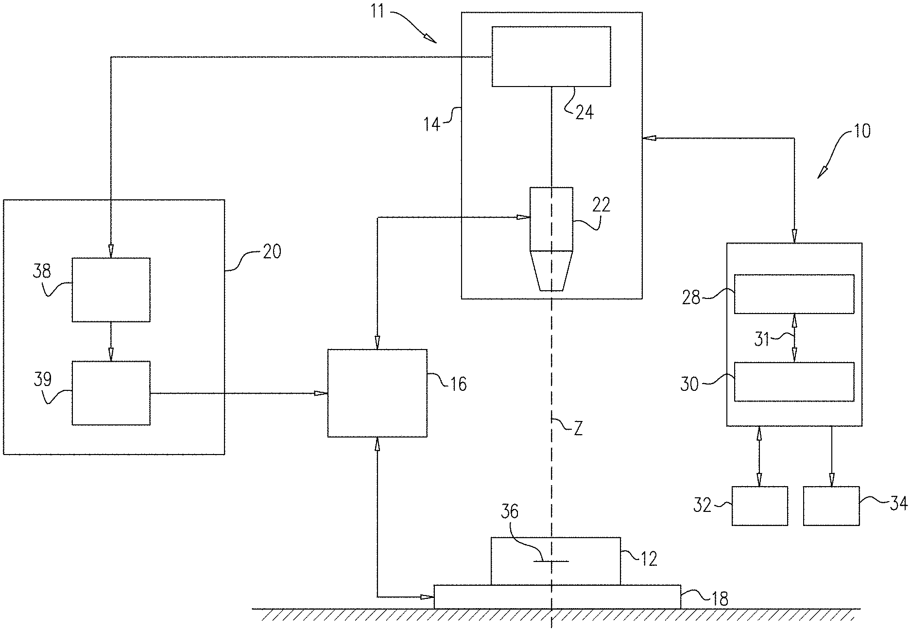
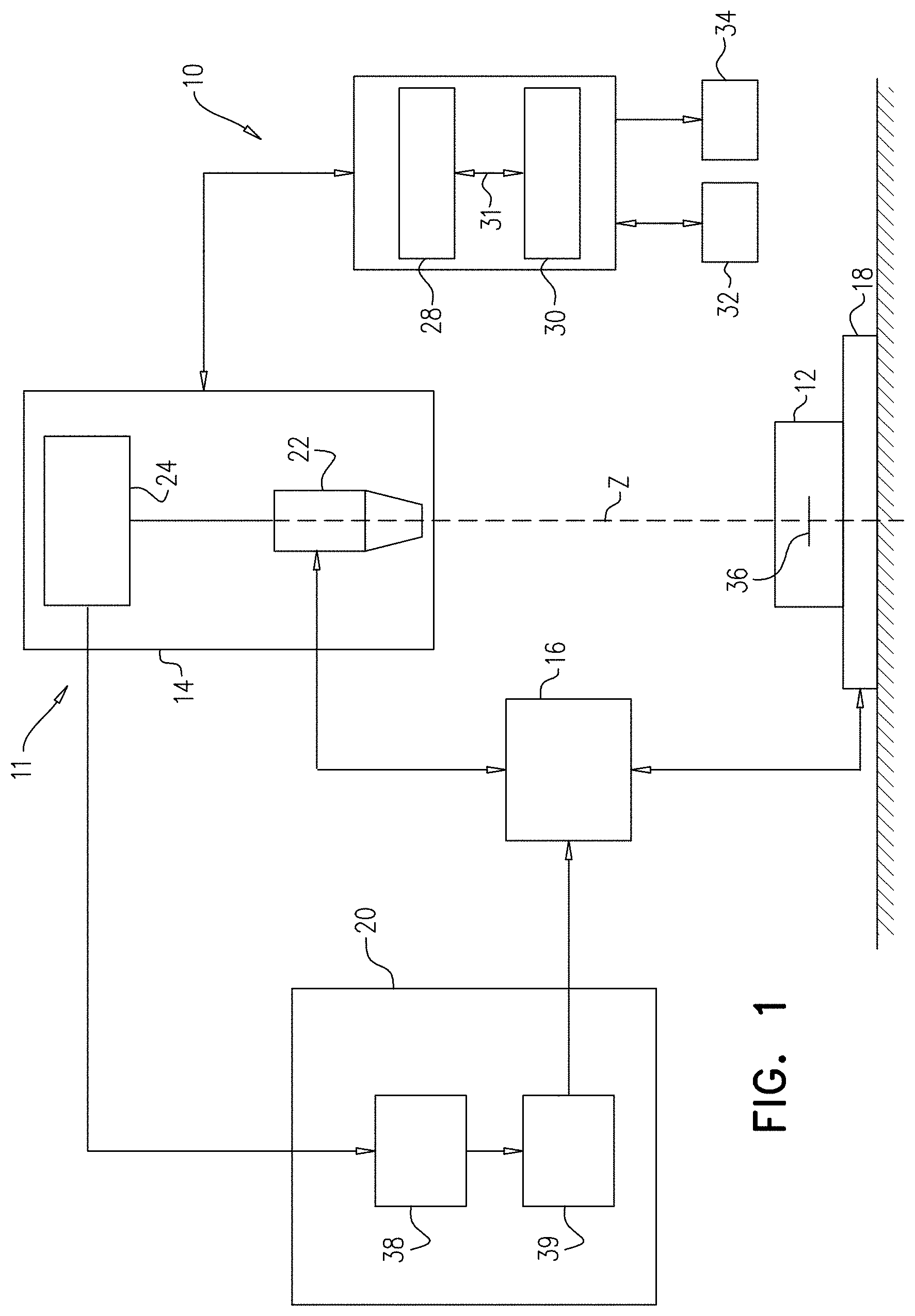
[0152] Reference is now made to FIG. 1, which is a functional diagram of a pathogen detection system 10, in accordance with some applications of the present invention. Pathogen detection system 10 includes a processor 28 operatively coupled to a memory 30, e.g. by a communication bus 31. In certain embodiments, pathogen detection system 100 can optionally include or be operatively coupled to a microscope system 11. Microscope system 11 is typically a digital microscope that includes an imaging module 14, a focus variation module 16, a sample carrier 18 and an autofocus system 20. For some applications, microscope system 11 is generally similar to the microscope system described in US 2014/0347459 to Greenfield, which is incorporated herein by reference.
[0153] Typically, imaging module 14 includes an optical unit 22 and an image sensor unit 24. Optical unit 22 is configured to form a magnified image of a bodily sample 12 (for example, a blood sample) by conjugating a focus plane 36 and an image plane. The image sensor unit 24 typically includes an image sensor, for example a charge-coupled-device (CCD), complementary metal-oxide-semiconductor (CMOS) sensor, and/or a matrix sensor, positioned in the image plane of the optical unit 22 so as to sense the magnified image.
[0154] Computer processor 28 typically receives and processes images. The computer processor communicates with memory 30, and images are received by the processor via the memory. Via a user interface 32, a user (e.g., a laboratory technician) sends instructions to the computer processor. For some applications, the user interface includes a keyboard, a mouse, a joystick, a touchscreen device (such as a smartphone or a tablet computer), a touchpad, a trackball, a voice-command interface, and/or other types of user interfaces that are known in the art. Typically, the computer processor generates an output via an output device 34. Further typically, the output device includes a display, such as a monitor, and the output includes an output that is displayed on the display. For some applications, the processor generates an output on a different type of visual, text, graphics, tactile, audio, and/or video output device, e.g., speakers, headphones, a smartphone, or a tablet computer. For some applications, user interface 32 acts as both an input interface and an output interface, i.e., it acts as an input/output interface. For some applications, the processor generates an output on a computer-readable medium (e.g., a non-transitory computer-readable medium), such as a disk, or a portable USB drive, and/or generates an output on a printer.
[0155] Microscope system 11 can, in certain embodiments, include a local processor that controls at least some of the processes of microscope system 11, for example, image acquisition and/or communication with other components, including other components of pathogen detection system 10 and components external to pathogen detection system 10. In certain other embodiments, processor 28 can control one or more processes of microscope system 11 including, e.g. image acquisition and/or communication. Optionally, pathogen detection system 10 can include or be operatively coupled to a plurality of digital microscopes. Optionally, each respective digital microscope in the plurality of digital microscopes has its own local processor.
[0156] In certain embodiments, memory 30 can be configured to store imaging information, program data and/or executable program instructions for detecting a pathogen in a bodily sample, as will be detailed below with reference to FIG. 2. Memory 30 can be, e.g., volatile memory or non-volatile memory. In certain embodiments, memory 30 is non-volatile memory, e.g. hard disk drive, flash memory, etc.
[0157] For some applications, microscope system 11 is configured to capture one or more high magnification digital images of a bodily sample. Optionally, the one or more digital images include images that cover different portions of the bodily sample. Optionally, the images do not overlap (or overlap by less than 5 percent or less than 1 percent). Optionally, the images include images that overlap and are taken at different depths of focus, and/or with different lighting conditions. Optionally, the one or more digital images include sets of images that do not overlap (or overlap by less than 5 percent or less than 1 percent), but each of the sets includes images of another set, taken with different lighting conditions. In certain embodiments, microscope system 11 is configured to capture images under a plurality of lighting conditions, including, e.g., bright field, blue light, and ultraviolet light, as will be further detailed below.
[0158] In accordance with some applications, bodily sample 12 (e.g., a blood sample) is scanned by the microscope system, such that a plurality of portions of the bodily sample are imaged. For some applications, a plurality of images are acquired of one or more portions of the bodily sample, with each of the plurality of images being acquired under respective imaging conditions. For example, two images of a portion of the bodily sample may be acquired using, respectively, imaging conditions that allow detection of cells (e.g., bright-field) and imaging conditions that allow visualization of stained bodies (e.g. appropriate fluorescent illumination).
[0159] Image sensor unit 24 may output acquired digital images to output device 34 (which may include a display) and/or to the autofocus system 20. Focus variation module 16 may be configured to vary a distance between the focus plane 36 of the optical unit 22 and the sample carrier 18. Focus variation module 16 may be operated manually or automatically via a mechanical interface which may, for example, modify the position of the sample carrier 18 along an optical axis Z of the optical unit 22. Alternatively or additionally, focus variation module 16 may be commanded by autofocus system 20. For example, the focus variation module 16 may vary the distance between the sample carrier 18 and the focus plane by (1) modifying the position of optical unit 22 along the optical axis Z, (2) modifying the position of the sample carrier 18 along the position of the optical axis Z (e.g., by moving a stage upon which the sample carrier is placed), (3) modifying the position of the focus plane by, for example, changing a focal length of the optical unit 22, or a combination thereof.
[0160] The sample carrier 18 may comprise a plate. Sample carrier 18 may be configured to accommodate bodily sample 12. The carrier may be any carrier known in the art for holding a biological sample. Optionally, the bottom surface of the carrier is essentially flat, to allow cells in contact therewith to be at about the same distance from the focal plane of the microscope. Examples include carrier slides, laboratory receptacles, dishes, plates, multi-well plates, test tubes (e.g. with a flat bottom), microfluidic cells, cartridges, and the like.
[0161] Autofocus system 20 may comprise an autofocus computation module 38 and an autofocus adaption module 39. The autofocus computation module may be connected to the image sensor unit 24 so as to receive images acquired by the imaging module 14. The autofocus adaptation module may be connected to the focus variation module 16 and may be configured to command the focus variation module 16, e.g., as described above.
[0162] For some applications, processor 28 includes one or more functional modules, such as a feature extraction module, a candidate classifier, a sample classifier, and a diagnostics module. For some applications, processor 28 is configured to process imaging information by extracting features contained within the imaging information. Typically, the processor is configured to extract at least one sample-informative feature and at least one candidate-informative feature. For some applications, the processor is further configured to process the at least one sample-informative feature to obtain contextual information, and to process the at least one candidate-informative feature to obtain candidate data, as will be further detailed below.
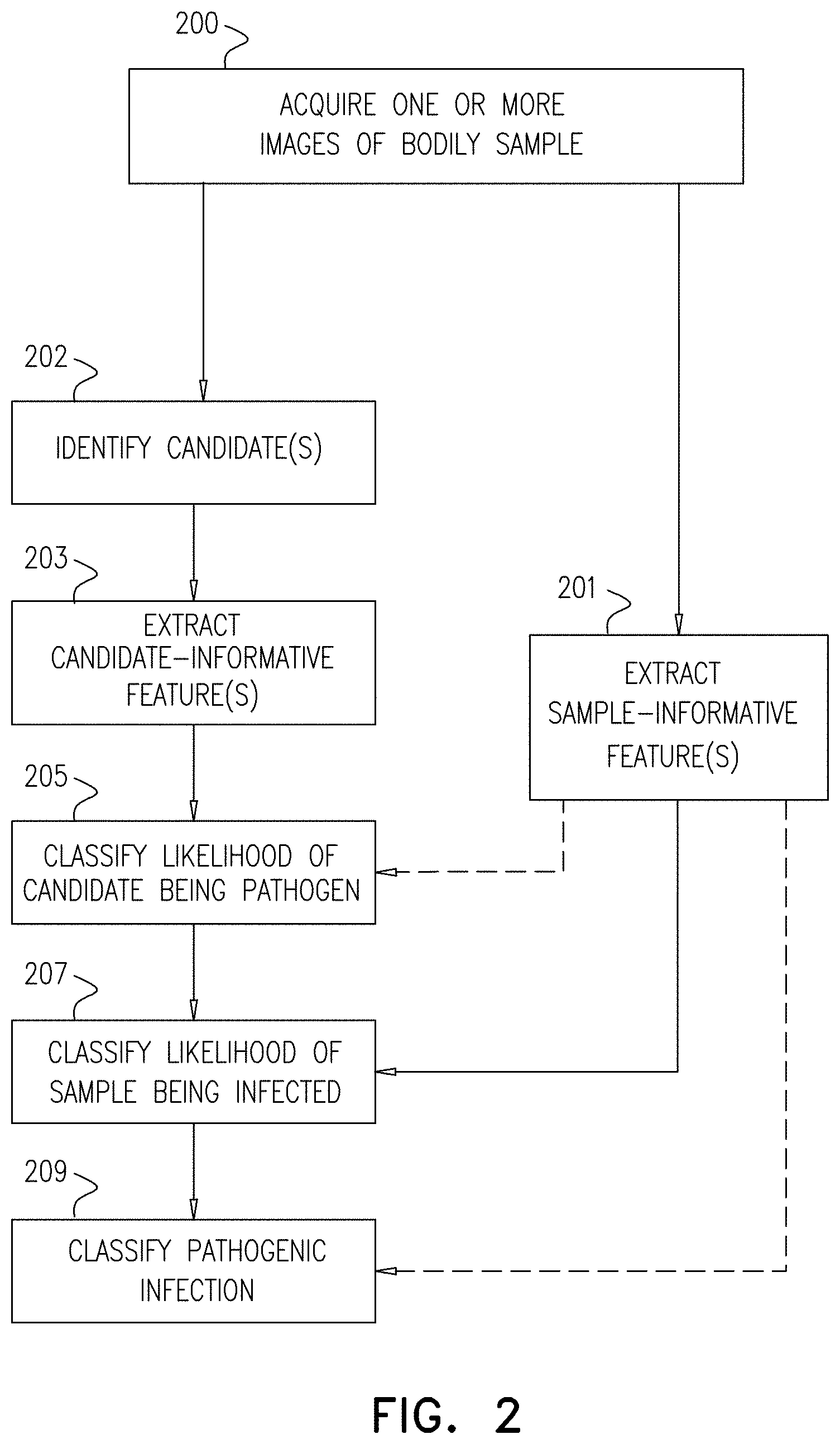
[0163] Typically, the processor is configured to classify a likelihood of a candidate (i.e., a constituent element within the sample that exhibits characteristics that indicate that it may be a pathogen, and is therefore a candidate for being a pathogen) being a pathogen at least partially based upon the at least one candidate-informative feature. Further typically, the processor is configured to classify a likelihood of the bodily sample being infected with a pathogenic infection, by processing the at least one candidate-informative feature in combination with the at least one sample-informative feature.
[0164] For some applications, the processor is programmed to classify the likelihood of a candidate being a pathogen, and/or to classify a likelihood of sample being infected with a pathogenic infection using classification and/or machine learning algorithms, e.g. support vector machines, neural networks, naive Bayes algorithms, etc. Additional examples of types of classification and/or machine learning algorithms which can be used by the processor are described in US 2012/0169863 to Bachelet and/or US 2015/0037806 to Pollak, both of which applications are incorporated herein by reference. For some applications, the computer processor is trained, in advance of being used to analyze a bodily sample, using training images of bodily samples.
[0165] For some applications, if a bodily sample is determined to be infected with a pathogenic infection (or if it is determined that the likelihood of the bodily sample being infected with a pathogenic infection exceeds a threshold), the computer processor is further configured to extract diagnostic information about the pathogenic infection in accordance with at least the at least one sample-informative feature.
[0166] It is noted that the teachings of the presently disclosed subject matter are not bound by the specific pathogen detection system described with reference to FIG. 1. Equivalent and/or modified functionality can be consolidated or divided in another manner and can be implemented in any appropriate combination of software, firmware and hardware. The processor can be implemented as a suitably programmed computer.
[0167] Reference is now made to FIG. 2, which shows a generalized flow chart of a method for detecting a pathogenic infection in a bodily sample (e.g., a blood sample), in accordance with some applications of the present invention.
[0168] In a first step 200, one or more images of the bodily sample are acquired by microscope system 11. The one or more images, data informative of one or more images, or data derived from one or more images (collectively referred to herein as "imaging information") are typically stored in memory 30. The imaging information is then analyzed by processor 28, as described in further detail hereinbelow. It is noted that in the present application, the computer processor is described as extracting features from the one or more images. This terminology should be interpreted as including extracting the features from data informative of the one or more images, or data derived from the one or more images, and should not be interpreted as being limited to directly extracting the features from the one or more images themselves.
[0169] For some applications, the imaging information is informative of at least one high magnification microscopic view of the sample. Alternatively or additionally, the imaging information is informative of a plurality of images, including, e.g., images of different portions of the sample, images of the same portion of the sample taken at different focal depths, and/or different lighting conditions, and/or at different times.
[0170] The bodily sample may be from any living creature but preferably from warm blooded animals. Typically, the bodily sample is a blood sample. The sample can be any blood sample or a portion thereof comprising one or more red blood cells. Optionally, the sample comprises predominantly red blood cells (i.e., a majority of the cells (e.g., at least 60 percent of the cells) in the sample are red blood cells). Optionally, the sample also comprises at least one of platelets and white blood cells. Optionally, the blood sample is diluted. Optionally, the dilution is performed or the sample is otherwise prepared such that the concentration of cells on the surface that is imaged is between 3,000 and 30,000 cells (e.g., red blood cells) per square mm. Optionally, the blood sample is diluted with a staining solution.
[0171] Optionally, the sample or staining solution comprises one or more suitable dyes or stains (optionally, comprising one or more fluorescent dyes). In some embodiments, the blood sample is selected from whole blood sample, red blood cell sample, buffy coat sample, plasma sample, serum sample, a sample from any other blood fraction, or any combination thereof.
[0172] Optionally, the sample forms a monolayer on the surface of sample carrier 18. In the context of the present disclosure, when referring to a monolayer of cells, it is to be understood as encompassing the distribution of cells on a surface as an essentially single layer, where at least 50 percent (at times, at least 60 percent, 70 percent, 80 percent or even 90 percent) of the cells are in direct contact with the bottom surface of the carrier and not more than 20 percent (at times, no more than 10 percent or even no more than 5 percent) of the cells overlay each other (i.e., no more than the aforementioned percentage of cells lie, partially or completely, on top of one another). Further, when referring to a monolayer, it is to be understood that at least 5 percent (at times, at least 10 percent or even at least 20 percent) of the cells touch each other on the bottom surface. For some applications, a monolayer is formed in accordance with the techniques described in U.S. Pat. No. 9,329,129 to Pollak, which is incorporated herein by reference.
[0173] For some applications, prior to being imaged, the bodily sample is stained with one or more suitable dyes or stains. Optionally, the one or more suitable dyes or stains comprise one or more fluorescent dyes or stains, and the stained sample is excited under one or more suitable lighting conditions for detecting pathogens. As used herein, the term "suitable dye or stain" should be expansively construed to include any dye or stain useful for the detection of a pathogen of interest, including any suitable fluorescent dye or stain. As used herein, a "suitable fluorescent dye or stain" should be expansively construed to include a dye or stain which is capable of selectively binding to one or more types of nucleic acid (e.g., DNA only, RNA only, both DNA and RNA, etc.) and fluoresces under one or more particular lighting conditions thereby allowing for discerning of the one or more types of nucleic acids in a bodily sample. Suitable fluorescent dyes or stains can include, for example, dyes or stains that bind to DNA and do not bind to RNA, dyes or stains that bind to RNA and do not bind to DNA, and dyes or stains that bind to both DNA and RNA. Non-limiting examples of suitable fluorescent dyes or stains include, e.g., Acridine Orange, Hoechst stain, etc.
[0174] The particular lighting condition which causes a particular suitable fluorescent dye or stain to fluoresce is referred to herein as a "suitable lighting condition," which should be expansively construed to include a lighting condition which, when used to excite a particular fluorescent dye or stain, causes fluorescing of the fluorescent dye or stain. In certain embodiments, the fluorescence emitted by the excited dye or stain may be discernible through the use of one or more different light filters which enable the discerning of fluorescence within a given wavelength range. Accordingly, suitable lighting conditions may be used in view of such filters. Non-limiting examples of suitable lighting conditions include, e.g., bright field, blue light, and ultraviolet light. Additional non-limiting examples of suitable fluorescent dyes or stains and suitable lighting conditions are described in US 2012/0169863 to Bachelet and US 2015/0037806 to Pollak, both of which applications are incorporated herein by reference.
[0175] As detailed above, in certain embodiments, the sample may be stained with one or more dyes or stains that allow differentiating between RNA and DNA in the sample (i.e., differential staining). Differential staining can be accomplished, for example, by staining the sample with one or more target-specific dyes or stains. As used herein a target-specific dye or stain (e.g., an RNA-specific or a DNA-specific) is a dye or stain that under selected conditions would detectably stain the target moiety such that it may be detected in the presence of other cellular components. In this context, detectably staining a target may mean that the dye or stain binds to the target with a higher affinity than to other cellular components and/or that it provides a stronger signal (e.g. fluorescence) when associated with the target. It is noted that some dyes or stains may stain more than one target but may be differentiated for example based on the wavelength of emitted fluorescence and/or a wavelength used for excitation of the dye or stain. In some embodiments, a target-specific dye or stain is a fluorescent dye or stain that upon binding to the target shifts its emission wavelength from an original band to a shifted band. In such cases, the target may be detected by a system configured to detect emission wavelengths within the shifted band.
[0176] Differential staining may be used to determine the relative locations of DNA and RNA, as detailed below with reference to Example 1. Optionally, a single dye or stain (e.g. Acridine Orange) may be used with different lighting conditions, to provide differential staining. Optionally, a combination of dyes or stains is used, comprising one or more DNA-specific dyes or stains (e.g., Hoechst reagent) and one or more other dyes or stains (e.g., Acridine Orange) configured to detect any nucleic acid (DNA and RNA).
[0177] For some applications, the imaging information is informative of one or more fields of the bodily sample. As used herein, a "field" is a portion of the bodily sample to be imaged. Typically, this corresponds to an area on the bottom of a sample carrier holding the sample. When the images are captured at high magnification, only a fraction of the entire blood sample can be imaged at one time. Therefore, pathogen detection system 10 virtually sub-divides an area to be imaged into a plurality of fields, and each field is imaged separately, thereby obtaining a plurality of images informative of the bodily sample, each image informative of a respective field. Optionally, the imaged fields do not overlap, or their degree of overlap is less than 5 percent or less than 1 percent of the area. In certain embodiments, each field to be imaged is imaged under one or more different lighting conditions. Optionally, an image of each field is captured a plurality of times at different lighting conditions. For example, the field may be imaged at least once in lighting conditions to detect RNA-related fluorescence, at least once in lighting conditions to detect DNA-related fluorescence, and at least once in brightfield.
[0178] Reference is now made to FIG. 3, which shows, by way of non-limiting example, imaging information 300 consisting of a field of a blood sample stained with one or more suitable fluorescent dyes and excited under a suitable lighting condition, in accordance with some applications of the present application. As may be observed, due to the dye(s), constituent elements 302 fluoresce, thereby appearing brighter (or, in some cases, a different color) than other non-fluorescing constituent elements 304 (which in this case include red blood cells) in the sample and allowing for discerning of stained regions in the sample, some features of which may be informative of some specific cell types in the sample.
[0179] In certain embodiments, the imaging information is informative of one or more sample constituent elements, including candidates (i.e., constituent elements that exhibit characteristics that indicate that they may be pathogens, and are therefore candidates for being pathogens) and non-candidates. For some applications, an element is identified as a candidate based upon the element appearing fluoresced when the sample is stained with a suitable fluorescent dye or stain and is excited by a suitable lighting condition, for example, as described in US 2012/0169863 to Bachelet, and/or in US 2015/0037806 to Pollak, both of which applications are incorporated herein by reference. Alternatively or additionally, an element may be identified as a candidate based upon other criteria, such as its size shape, color, proximity to other elements, etc. As used herein, the term "non-candidate" should be expansively construed to cover a sample constituent element that is not a candidate.
[0180] Referring again to FIG. 2, in step 201, processor 28 extracts from the one or more images, from the imaging information, and/or a portion thereof, one or more sample-informative features of the bodily sample that are indicative of contextual information related to the bodily sample. Typically, a plurality of sample-informative features are extracted. As used herein, "sample-informative features" include features of the bodily sample which are not directed to a specific candidate and are usable to provide contextual information that can be used to determine the presence, likelihood of, or characteristics of a pathogenic infection in the sample, including, in some embodiments, the classification of specific candidates. By way of non-limiting examples, sample-informative features can include, for example, features related to non-candidate constituents in the sample, or features related to the quantity and/or distribution of cells of a given type in the sample. Features related to non-candidate constituents in the sample can include, for example, size-related properties of one or more non-candidates (including relative size as compared to either an expected size, or to an observed size of one or more other cells), shape-related properties of one or more non-candidates (including relative shape as compared to either an expected shape, or to an observed shape of one or more other elements), and intensity-related properties of one or more non-candidates (including relative intensity as compared to either an expected intensity, or to an observed intensity of one or more other elements). As used herein, an "expected" value (of, for example, size, shape and/or intensity) is such value as may be known in advance of analyzing imaging information relating to a given sample. Such values include, for example, population statistic values that are known or can be calculated (for example, for all humans and/or any subgroup thereof, based, for example, on age, sex, race, ethnicity, etc.), optionally according to a specific condition (e.g. altitude, treatment of the bodily sample, etc.).
[0181] For some applications, sample-informative features include features related to the distribution of candidates or pathogens within the sample or portions thereof. For example, if the number of candidates or pathogens found in a given image (or part of an image or a group of images covering a continuous portion of the sample) is significantly higher than the number of candidates or pathogens found in other parts of the same sample, this may indicate that the high concentration of candidates or pathogens found in one part of the sample might be a result of a local effect that should not affect the diagnosis of the sample. For example, a high concentration of candidates or pathogens (e.g. a high concentration of candidates overlapping red blood cells) in one part of the sample, but not in other parts, can be indicative of contamination, e.g., from a drop of blood from another sample that entered the sample under investigation.
[0182] For some applications, some or all of step 201 is performed in a pre-processing stage in order to determine, for example, whether some of the imaging information is of poor quality as measured by predetermined criteria (e.g., brightness, focus, etc.), in which case portions of the imaging information may be excluded from further processing (for example, as described hereinbelow with reference to Example 6).
[0183] In step 202, computer processor 28 identifies one or more constituent elements within the sample as being candidates of a pathogen. As described hereinabove, an element may be identified as a candidate based upon the element appearing fluoresced when the sample is stained with a suitable fluorescent dye and excited by a suitable lighting condition, for example, as described in US 2012/0169863 to Bachelet, and/or in US 2015/0037806 to Pollak, both of which applications are incorporated herein by reference. Alternatively or additionally, an element may be identified as a candidate based upon other criteria, such as shape, size, proximity to other elements (such as red blood cells, or other candidates), etc.
[0184] In step 203, computer processor extracts from the one or more images, from the imaging information, or/or from a portion thereof, one or more candidate-informative features associated with one or more identified candidates. Typically, for each candidate, a plurality of candidate-informative features are extracted. As used herein, "candidate-informative features" include features of the candidate (or, in some cases, constituent elements in close proximity to the candidate, as will be detailed below) useable to provide information for determining the likelihood of the given candidate being a pathogen or a part of a pathogen.
[0185] By way of non-limiting example, candidate-informative features can include features related to: a size of a candidate, a shape of a candidate, a motion of a candidate (based, for example, on a comparison of at least two at least partially overlapping images captured in sequence), and/or an intensity of a candidate.
[0186] For some applications, candidate-informative features include a relative location of a candidate with respect to other sample constituents (e.g., a red blood cell). Alternatively or additionally, candidate-informative features include a property of a cell (e.g. red blood cell) that at least partially overlaps with the candidate (and, optionally, also the amount of overlap), such as a size or shape of cell overlapping the candidate. For some applications, features related to size and shape of a cell overlapping the candidate include a relative size and relative shape of the overlapping cell as compared to an expected size or expected shape. As used herein, a cell is considered to overlap with a candidate at least partially if, in the imaging information, at least a portion of the cell appears to be co-located with at least a portion of the candidate (e.g., at least 20 percent or at least 25 percent of the candidate).
[0187] Optionally, candidate-informative features can include features of other constituent elements (e.g., pathogen candidates and/or pathogens) that are found in close proximity to the candidate. In this context, "close proximity" can be predefined according to any suitable metric. For example, constituents in close proximity to the candidate may include constituents located within a distance of up to 2.times. away from the candidate, where X is an expected (e.g., average) red blood cell diameter. Accordingly, in some embodiments, candidate-informative features (including features of the candidate, of a cell overlapping the candidate, and/or features of other constituents) may include or be limited to features that are within close proximity to the candidate.
[0188] For some applications, the imaging information or a portion thereof is processed for candidate-informative feature extraction at least partly in a pre-processing stage. In certain embodiments, the pre-processing stage can include extracting sample-informative features to obtain contextual information, and determining the imaging information which is used to extract candidate-informative features in accordance with the obtained contextual information. For some applications, the portion of the imaging information which is used for extracting candidate-informative features and the portion of the imaging information which is used for extracting sample-informative features partially or completely overlaps.
[0189] It should be noted that steps 201, 202 and 203 can be performed in any order. In accordance with some applications, steps 201, 202 and 203 are performed as a single step and/or are intertwined with one another. For some applications, some or all of steps 201, 202 and 203 are performed as a plurality of distinct steps.
[0190] Typically, based upon the candidate-informative feature(s) in combination with the sample-informative feature(s), computer processor 28 classifies a likelihood of the bodily sample being infected with a pathogenic infection. For some applications, the pathogenic infection is detected by implementing the additional steps indicated in FIG. 2.
[0191] For some applications, once at least some candidate-informative features are extracted, in step 205, processor 28 classifies the likelihoods of respective candidates being pathogens, in accordance with the candidate data obtained for each respective candidate. As used herein, the term "likelihood of being a pathogen" should be expansively construed to cover either a binary determination (e.g., either a pathogen or a non-pathogen) or a scalar determination (e.g., a number, the value of which reflects the estimated likelihood that the given candidate is a pathogen). In certain embodiments, processor 28 classifies the likelihoods of respective candidates being pathogens using the extracted sample-informative features (e.g., the features extracted in step 201) in combination with the candidate-informative features, as will be further detailed below, for example, with reference to Examples 1 and 2. This is indicated by the dashed arrow connecting step 201 to step 205, indicating that step 201 is an optional input into step 205.
[0192] Typically, subsequent to candidate classifying (i.e., step 205), in step 207, processor 28 classifies a likelihood of the bodily sample being infected with a pathogenic infection. As used herein, the term "likelihood of the bodily sample being infected" should be expansively construed to cover either a binary determination (e.g. either infected or clean) or a scalar determination (e.g. a number, the value of which reflects the estimated likelihood that the given sample is infected). For some applications, processor 28 classifies the sample based on the classification of the candidates (extracted in step 205), in combination with the sample-informative features (extracted in step 201), as will be further detailed below, for example, with reference to Examples 1 and 3.
[0193] For some applications, in step 209, processor 28 classifies the pathogenic infection as containing one or more given types of pathogen, in accordance with one or more extracted sample-informative features and/or candidate-informative features. Classifying the pathogenic infection as containing one or more given types of pathogen may be performed using information and/or features that were obtained in one or more of steps 201, 203, 205, and 207, and/or by performing one or more additional steps of feature extraction and classification. For some applications, in order to classify the pathogenic infection, (a) candidates are classified as given types of pathogens, and (b) the overall pathogenic infection is classified based upon the classifications of the individual candidates. For some applications, sample-informative features are used for classifying the individual candidates as given types of pathogens, and/or for classifying the overall infection as containing given types of pathogens.
[0194] For some applications, classifying the pathogenic infection as containing one or more given types of pathogen includes, for example, classifying the pathogenic infection in order to determine species or strains of pathogens contained within the sample, for example, as further detailed below with reference to Examples 4 and 5. Such determination may include or be limited to classifying the pathogen to a single species or strain, or to a group of several possible species or strains (at least one of which is contained within the sample) and/or ruling out a given species or strain (as a species that is not contained within the sample). For some applications, processor 28 classifies the pathogenic infection as containing one or more of Plasmodium, a given strain of Plasmodium, Plasmodium of a given age, and/or Plasmodium of a given age range.
[0195] For some applications, the computer processor generates an output to the user (e.g., on the output device) indicating whether or not the sample is infected with a pathogen, and indicating a classification of the infection. For some applications, the computer processor generates an output indicating that the presence of an infection within the bodily sample could not be determined with a sufficient degree of reliability, indicating that a portion of the sample should be re-imaged, and/or indicating that a portion of the sample should be re-imaged using different settings (e.g., using different lighting, using a different stain, using a different or new sample preparation method, and/or using different microscope settings). For some applications, in response to determining that the presence of an infection within the bodily sample could not be determined with a sufficient degree of reliability, the computer processor generates an output indicating that the user should take appropriate user actions (e.g., prepare a new sample, and/or test the sample using an independent method, etc.). Alternatively or additionally, the computer processor automatically drives the microscope system to re-image a portion of the sample, drives the microscope system to re-image a portion of the sample using different settings (e.g., different focus, or different field size), and/or modulates a frame rate at which microscope images are acquired by the microscope system.
[0196] It is noted that, for some applications, sample-informative features are not necessarily derived directly from the images. For example, sample-informative features may include statistical or other information regarding the candidates and/or other entities within the sample, and/or general characteristics of the sample. In general, the scope of the present application includes analyzing a sample on two levels, first on a candidate-by-candidate level, and then on a more general level that is indicative of characteristics of the sample as a whole.
[0197] For some applications, based upon candidate-level features, two or more sample-informative features related to the bodily sample are extracted, and a characteristic of the bodily sample is determined, by processing the two or more sample-informative features. Typically, at least some of the candidates are pathogen candidates, and candidate-informative features relating to the pathogen candidates are extracted. For some applications, candidates of entities such as reticulocytes and/or platelets are additionally identified, and candidate-informative features relating to these candidates are extracted. For some applications, the sample-informative features include a number of pathogen candidates in the sample, type of pathogen candidates in the sample, brightness of the candidates relative to background brightness, a probability of candidates being pathogens, number of candidates that have a probability of being a pathogen that exceeds a threshold, number of candidates that have a probability of being a given type of pathogen that exceeds a threshold, a number of platelets in the sample, brightness of platelets, a number of reticulocytes in the sample, a number of reticulocytes infected by pathogens in the sample, a proximity of the candidates to red blood cells, and/or a number of red blood cells in the sample.
[0198] In embodiments of the presently disclosed subject matter, fewer, more and/or different stages than those shown in FIG. 2 may be executed. In embodiments of the presently disclosed subject matter, one or more stages illustrated in FIG. 2 may be executed in a different order and/or one or more groups of stages may be executed simultaneously.
[0199] A number of examples detailing specific non-limiting applications of the method detailed above will now be provided in order to better understand the disclosed subject matter.
Example 1: Using Concentration of Reticulocytes as a Sample-Informative Feature for Classifying a Candidate, and/or for Classifying a Pathogenic Infection
[0200] As detailed above, in certain embodiments, the sample is stained for discerning respective locations of DNA and RNA in the sample. Such staining may include, for example, using at least one DNA-specific dye and at least one RNA-specific dye, or at least one target-specific dye (either DNA or RNA) and at least one dye that stains both DNA and RNA. For some applications, in order to classify the likelihood of a candidate being a pathogen (step 205), the respective locations of RNA and DNA staining in sample are used by the processor to determine if the staining pattern(s) correspond(s) with the pattern(s) expected for a pathogen.
[0201] FIG. 4 schematically illustrates candidates 400a and 400b, each candidate shows an area stained for RNA (RNA portion 402) and an area stained for DNA (DNA portion 404). RNA portion 402 and DNA portion 404 may be differentially stained, e.g. using different dyes and/or different lighting, in order to discern the particular boundaries of each stained area. As is shown in FIG. 4, in candidate 400a the DNA portion 404 completely overlaps the RNA portion 402, while in candidate 400b the DNA portion 404 partially overlaps the RNA portion 402.
[0202] A candidate which appears to have at least partially overlapping DNA and RNA might be a pathogen. However, the appearance of overlapping RNA and DNA stained regions can also be caused by a different entity or entities, including, for example, a different cell type, or two separate bodies (one of which contains DNA and the other of which contains RNA) seemingly positioned on top of one another.
[0203] Mature red blood cells have no detectable DNA or RNA and therefore do not fluoresce when stained for nucleic acids. By contrast, Plasmodium trophozoites (which are a type of pathogen) may be detected as DNA-containing and RNA-containing bodies within red blood cells. Therefore, for some applications, in order to identify red blood cells that contain pathogens, a staining substance that stains both DNA and RNA (such as, Acridine Orange) is used. Alternatively or additionally, a stain that stains only DNA (such as a Hoechst stain) is used.
[0204] Howell Jolly bodies are DNA-containing bodies that may be found in red blood cells in some unhealthy conditions. In some cases, the presence of Howell Jolly bodies in a sample may increase the chance of false positive determination of a pathogen infection. Even if a DNA-specific stain is used in conjunction with a stain that stains both DNA and RNA, the Howell Jolly bodies may cause a false positive determination of a pathogen infection. Therefore, in some embodiments, differentiation between red blood cells that contain Howell Jolly bodies and red blood cells that contain pathogens may be beneficial.
[0205] Young red blood cells, termed reticulocytes, are sometimes found in blood. These cells contain RNA bodies only. A positive correlation is known between the presence of Howell Jolly bodies in red blood cells and a larger than normal amount of reticulocytes. Therefore, for some applications, sample-informative features include features that are indicative of a concentration of reticulocytes in a blood sample. (It is noted that a Plasmodium infection also raises the reticulocyte count for a patient. However, this increase (of, for example, about 5%) is typically much lower than the increase typical of patients that have a high Howell Jolly body count (which may be about ten times as great). Accordingly, a threshold for the determination of a high reticulocyte count is typically slightly higher than the average value for humans (or a given sub-population thereof).)
[0206] Based upon identifying a high concentration of reticulocytes, the likelihood of pathogen candidates being Howell Jolly bodies increases. In turn, the likelihood of the candidates being pathogens decreases, and the likelihood of the sample being infected decreases. Therefore, for some applications, the computer processor adjusts a threshold for a positive determination of an infection, based upon the concentration of reticulocytes. For example, many reticulocytes detected concomitantly with low parasitemia (e.g. less than 200 parasites/microliter blood) may be interpreted as being indicative of a high probability of a false positive (i.e., the sample being non-infected).
[0207] Alternatively or additionally, based upon the concentration of reticulocytes, in order to classify the likelihood of a candidate being a pathogen (step 205 of FIG. 2), the processor ascribes more weight to the relative positions of DNA and/or RNA within candidate given red blood cell, rather than simply the presence of DNA and/or RNA within the red blood cell. Alternatively or additionally, based upon the concentration of reticulocytes, in order to classify the likelihood of a sample being infected (step 207 of FIG. 2), the processor ascribes more weight to extracellular Plasmodium candidates, rather than intracellular Plasmodium candidates (which could be Howell Jolly bodies).
Example 2: Using Distribution of Candidates within a Sample as a Sample-Informative Feature for Classifying a Candidate
[0208] Candidates within a sample are expected to be uniformly distributed. Therefore, for some applications, a distribution of candidates within the sample that differs significantly from an expected uniform distribution is used as a sample-informative feature. For example, if there are significant candidate clusters, the clusters may be foreign bodies associated with the sample carrier rather than a portion of the blood sample, or may indicate that a different infected sample contaminated the sample being analyzed (for example, by spilling over from an adjacent chamber on a sample carrier). In response to detecting a non-uniform distribution of candidate, candidates that are within localized clusters may be given a lower score (i.e., they may be classified as being less likely to be pathogens). For example, if the sample-informative features are indicative of clustering of candidates, the processor may use distance from the cluster center(s) of any given candidate as a feature for classifying the candidate.
Example 3: Platelet Concentration as a Sample-Informative Feature for Classifying a Sample as Infected
[0209] Platelets typically appear as small extracellular RNA bodies, although some platelets may appear to be overlapped with cells because they are positioned on or under a cell when the sample is imaged. A normal concentration of platelets is typically between 150,000-400,000 platelets per microliter of whole blood.
[0210] It is known that the concentration of platelets may be affected by Plasmodium infection, its severity and the species of Plasmodium, as well as by other unrelated conditions (including medical conditions, treatments and medications). Accordingly, for some applications, the number and/or concentration of platelets in a sample is used as a sample-informative feature, and, for example, may be used as an input in classifying the likelihood of the sample being infected.
Example 4: Platelet Concentration as a Sample-Informative Feature Informative of a Species of Pathogen
[0211] As mentioned in the context of Example 3, the number and/or concentration of platelets can be correlated with a specific species of pathogen, for example a low platelet count has been shown to be correlated with a Plasmodium falciparum infection to a significantly greater extent than Plasmodium vivax infection. For some applications, in accordance with step 209 of FIG. 2, the number and/or concentration of platelets in a blood sample is used as an input for classifying a pathogenic infection as containing a given type of pathogen.
Example 5: Red Blood Cell Size and Shape as a Sample-Informative Feature
[0212] Some pathogens change the morphology of infected cells. For example, some pathogens (e.g., relatively mature trophozoites of Plasmodium vivax and Plasmodium ovale) cause an enlargement of infected red blood cells, sometimes to about two-fold that of uninfected cells. Other pathogens (e.g., Plasmodium malariae) reduce the size of infected red blood cells. Still other pathogens (e.g., Plasmodium falciparum) do not enlarge infected cells or reduce their size. For some applications, the sizes of red blood cells that appear to be infected within a blood sample are used as a sample-informative feature that is indicative of the sample being infected (e.g., in step 207 of FIG. 2), and/or is indicative of an identity of the pathogen (e.g., in step 209 of FIG. 2).
[0213] For example, a blood sample infected by Plasmodium vivax and/or Plasmodium ovale is expected to include infected red blood cells that are significantly enlarged. A blood sample infected by Plasmodium malariae, on the other hand, is expected to include infected red blood cells that are significantly diminished in size. Therefore, for some applications, detection of such enlarged and/or diminished cells is used as a sample-informative feature that is indicative of the sample being infected (e.g., in step 207 of FIG. 2) and/or is indicative of an identity of the pathogen (e.g., in step 209 of FIG. 2).
[0214] In another example, Plasmodium ovale may cause infected red blood cells to become more oval than uninfected red blood cells that tend to appear round. Accordingly, one or more of the following sample-informative features may be interpreted as being indicative of the sample being infected and/or being infected with Plasmodium ovale: the presence of oval red blood cells in the sample, the presence of a higher than expected portion of oval red blood cells, and/or the presence and/or amount of infected red blood cells that appear to be oval.
[0215] Features of oval(s) (e.g., height versus width) may be used in a classification in a weighted manner. For example, the weight given to an oval feature that is close to an expected value may be higher than if the value is closer to an expected value for uninfected red blood cell or values that are significantly more deviant than the expected value for infected red blood cells (e.g., if the oval appears to be rod like).
[0216] It should be noted that a determination that an infected red blood cell (or a group of infected red blood cells), or potentially infected red blood cell, is different in any given property (e.g. size, shape, etc.) than the general population of red blood cells (or different than uninfected cells), and/or a determination of a degree of such difference, is typically reached using any acceptable statistic. For example, an average size of two groups may be used and/or an average size of one group may be used in relation to a given percentile of the other group. Optionally, a plurality of statistics is used. Optionally, one or more of the values for red blood cells (or for uninfected red blood cells) are taken from known statistics of the general population or a subgroup thereof. In some embodiments, one or more statistics of all red blood cells in the sample (or a portion thereof) may be used, rather than using only the uninfected red blood cells. For example, this may be used in cases in which the portion of infected red blood cells within the sample is sufficiently small.
[0217] Optionally, a determination that an infected or potentially infected red blood cell is different or relatively different in any given property (e.g. size, shape, etc.) is made by comparing the given property of the infected red blood cell or potentially infected red blood cell to properties of one or more clean red blood cells in the sample. As such, properties of one or more clean red blood cells can also be used as sample-informative features for determining the likelihood that a candidate is a pathogen, for determining the likelihood that a sample is infected, and/or for classifying the species of a pathogen.
[0218] It should be noted that red blood cell features (e.g., features related to red blood cell size and/or shape) can also be used in candidate classification (e.g., as a candidate-informative feature used in step 205 of FIG. 2) when compared with an expected value. By way of non-limiting example, candidates which appear to be inside (or co-located with) red blood cells that are relatively large or small than an expected value or have a shape that is different than an expected shape (e.g. oval instead of round) are more likely to be pathogens.
[0219] It should also be noted that features of red blood cells in the sample (e.g. features related to red blood cell size and/or shape) can be used in sample classification (e.g., as a sample-informative feature used in step 207 of FIG. 2). By way of non-limiting example, such features include a statistic taken for a group of red blood cells in the sample (e.g., a statistic for seemingly infected red blood cells in the sample, a statistic for uninfected red blood cells in the sample, and/or a statistic for red blood cells in the sample in general). One non-limiting example includes comparing a statistic of seemingly infected red blood cells in the sample (e.g., size) to an expected value (e.g., average size of human red blood cell) or to a corresponding statistic for red blood cells in the sample in general. When the seemingly infected red blood cells are found to be larger or smaller than the compared value, this may be used as an indication that the sample is infected.
[0220] Echinocytes are red blood cells that have abnormal cell membranes with many small, symmetrically spaced thorny projections. Acanthocytes also have thorny abnormal projections, but they are irregular and asymmetrical.
[0221] In some cases, a Plasmodium infection causes the appearance of echinocytes or acanthocytes. Other causes for such shapes may be other pathologies and even a prolonged exposure to some solutions (e.g., dye solutions). Additionally, some strains of Plasmodium cause greater deformity than other strains. For example, Plasmodium vivax is typically more deforming than Plasmodium falciparum, while each of Plasmodium vivax and Plasmodium ovale is typically more deforming than each of Plasmodium malariae and Plasmodium falciparum. Therefore, for some applications, the presence of such shapes in a sample is used as a sample-informative feature that is indicative of the sample being infected (e.g., in step 207 of FIG. 2), and/or is indicative of an identity of the pathogen (e.g., in step 209 of FIG. 2).
[0222] In some strains of Plasmodium, there is a positive correlation between a degree of red blood cell deformity and the age of the infecting pathogens. Therefore, for some applications, the presence of such shapes in a sample is used as a sample-informative feature that is indicative of the age of the detected pathogens (e.g., in step 209 of FIG. 2).
Example 6: Using Staining Quality as a Sample-Informative Feature
[0223] Staining of a biological sample is known to be a time dependent process. Once cells are exposed to a dye it takes time for the dye to penetrate the cells and reach its target site(s) within the cells. During this period, staining may become sharper and/or more localized with sharper intensity gradients (especially if the sample is not washed before being imaged). Typically, this process follows a saturation curve. In a first phase, staining increases relatively fast until some degree of staining is reached (i.e., a fast staining phase). Thereafter, quality still increases but relatively slowly for another period of time (i.e., a slow staining phase). At a later time, staining quality may deteriorate, for example, due to photo bleaching (in a fluorescent dye), and/or due to the dye slowly diffusing through the sample, away from the target(s).
[0224] For some applications, imaging a plurality of fields (e.g., 200 fields or more, at least some of which may be imaged more than once) of a bodily sample, such as a blood sample, may take a few minutes (e.g., 2-3 minutes or more). For Hoechst staining (which is used, for example, in the detection of Plasmodium infection), the fast staining phase may take 30 minutes or more.
[0225] Accordingly, when imaging a plurality of fields is carried out over the above-described time scale, there might be a significant variation in staining quality between fields. This variation may affect the diagnostic result (e.g., by changing an intensity value, an intensity gradient value, and/or a threshold for infection). For some applications, in order to account for the variation in staining quality, staining quality of the sample is used as a sample-informative feature, in accordance with techniques described herein.
[0226] For some applications, an average staining quality across a plurality images is determined, and a staining quality parameter for each image is determined, based on the image's relative staining quality compared to the average value. The staining quality parameter can be used as a sample-informative feature, for example, for discarding, from being used in further analysis, images that were taken too early or too late, when staining is not sufficiently informative for diagnostics. Alternatively or additionally, the staining quality parameter can be used to adjust one or more thresholds for different fields or groups of fields based on the staining quality parameter, and/or introduce the staining quality parameter into the classifying of candidates, so that candidates from a plurality of fields having different staining qualities can be compared. For example, the candidate classification can be normalized using the staining quality.
[0227] For some applications, in response to the staining quality parameter, a frame rate at which images of the bodily sample are acquired is modulated. For example, in response to detecting that the staining is of a low quality, images may be acquired at a greater frame rate, and vice versa. Alternatively or additionally, in response to the staining quality parameter, the number of times each field is imaged is modulated. In general, the scope of the present invention includes using sample-informative features as an input for discarding some images from being used, for modulating a frame rate at which images are acquired, and/or for modulating the number of times that each imaging field is imaged.
[0228] Optionally, when using two or more stains, the staining quality variation may differ between the stains. For example, Acridine Orange staining may be complete, when Hoechst staining is still in the fast staining phase. Accordingly, for some applications, the staining quality parameter is treated separately for each stain, and/or the relative staining quality between the stains may be used as a staining quality parameter.
Example 7: Using Affinity of Candidates for Reticulocytes as a Sample-Informative Feature
[0229] Plasmodium vivax and Plasmodium ovale have an affinity for infecting reticulocytes, over mature red blood cells. Conversely, Plasmodium falciparum infects all red blood cells equally, while Plasmodium malariae has an affinity for mature red blood cells. Accordingly, for some applications, a relationship between the number of pathogen candidates associated with reticulocytes and the number of candidates associated with mature red blood cells is determined, and this is used as a sample-informative feature.
[0230] As described hereinabove, for some applications, in accordance with step 209 of FIG. 2, the processor classifies a pathogen as containing a given type of pathogen. For some applications, once it is determined that a sample is infected, the processor classifies the pathogen as containing a given type of pathogen, based upon the estimated ages of infected red blood cells, and/or based upon a relationship between the number of pathogen candidates associated with reticulocytes and the number of candidates associated with mature red blood cells. For example, if the pathogens have an affinity for reticulocytes, this may be used as an indication that that the detected pathogen is Plasmodium vivax. Alternatively, an essentially uniform distribution of the pathogen in red blood cells of all ages (proportional to relative abundance of the differently-aged red blood cells) may be used as an indication that that the detected pathogen is Plasmodium falciparum.
Example 8: Using Detection of Contamination as a Sample-Informative Feature
[0231] An image of a blood sample may contain foreign objects that are irrelevant for diagnosis and may be due for example to dirt or flaws in a solution or tool used to prepare or hold the blood sample (e.g., a dilution solution and/or a sample chamber used for housing the sample). Such objects may include objects that appear similar to a pathogen and/or objects that are dissimilar to a pathogen.
[0232] For some applications of the present invention, the computer processor is configured to extract, from the one or more images, at least one sample-informative feature that is indicative of contextual information related to the bodily sample. At least partially based upon the extracted sample-informative feature, the computer processor identifies that there is a defect associated with the bodily sample disposed in the sample carrier, and classifies a source of the defect (for example, as being the sample carrier, a given portion of the sample carrier, the sample itself, and/or a diluent in which the sample has been diluted). The computer processor generates an output on the output device that is indicative of the source of the defect.
[0233] For example, such an output may be generated based upon a sample-informative feature that is indicative of the presence of foreign objects within the sample. The source of the foreign objects is expected to affect their concentration and distribution in the sample. For example, if the source is the blood sample itself, then the amount of foreign objects that is detected is typically proportional to the size of the sample. For some applications, in response to detecting foreign objects having this characteristic, a threshold for diagnosis is adjusted. For example, the threshold for the number of pathogens within a sample that is sufficient to deem the sample to be infected may be a function of a relationship between the concentration of foreign objects within the sample, to that of red blood cells.
[0234] For some applications, the processor is configured to determine that the source of foreign objects is limited to a given chamber, a given set of chambers, a given field, or a given set of fields. For such cases, the computer processor may ascribe lower weight to features that are detected in the affected chambers or fields, and/or may use data from other chambers or fields as inputs to analyzing features detected in the affected chambers and fields.
[0235] For some applications, the computer processor is configured to detect that the source of foreign objects is a diluent in which the sample has been diluted (for example, in response to detecting foreign objects with characteristics that are common to a plurality of samples and/or chambers). In such cases, the processor may generate an output indicating the likely source of the foreign objects. For some applications, the computer processor is configured to detect cross-contamination between chambers, and to generate an output indicating that this is the case.
[0236] It is noted that although some applications of the present invention have been described with respect to detecting a pathogen infection within a bodily sample, the scope of the present invention includes performing similar techniques with respect to identifying other components or entities within a bodily sample. For example, similar techniques to those described herein may be used for detecting the concentration of a given entity within a blood sample, by (a) extracting one or more candidate-informative features associated with an element that is a candidate of the given entity, (b) extracting one or more sample-informative feature that are indicative of contextual information related to the bodily sample, and (c) processing the candidate-informative feature in combination with the sample-informative feature. For some applications, the sample is a sample that includes blood, and the candidates are candidates of entities within the blood, such as platelets, white blood cells, anomalous white blood cells, circulating tumor cells, red blood cells, reticulocytes, Howell Jolly bodies, etc. For some such applications, a blood count (e.g., a complete blood count) is performed by identifying such candidates, and performing techniques as described herein.
[0237] For some applications, the sample is a different bodily sample, and the techniques described herein are used for identifying a pathogen infection within the sample. For example, the techniques described herein may be used to identify Mycobacterium tuberculosis within a sample of sputum. Alternatively or additionally, the sample is a different bodily sample, and the techniques described herein are used for identifying abnormal cells within the sample. For example, the techniques described herein may be used to identify cancerous cells in a PAP smear or in a urine sample.
[0238] In general, it is noted that although some applications of the present invention have been described with respect to a blood sample, the scope of the present invention includes applying the apparatus and methods described herein to a variety of samples. For some applications, the sample is a bodily sample, such as, blood, saliva, semen, sweat, sputum, vaginal fluid, stool, breast milk, bronchoalveolar lavage, gastric lavage, tears and/or nasal discharge. The bodily sample may be from any living creature, and is typically from warm blooded animals. For some applications, the bodily sample is a sample from a mammal, e.g., from a human body. For some applications, the sample is taken from any domestic animal, zoo animals and farm animals, including but not limited to dogs, cats, horses, cows and sheep. Alternatively or additionally, the bodily sample is taken from animals that act as disease vectors including deer or rats.
[0239] For some applications, similar techniques to those described hereinabove are applied to a non-bodily sample. For some applications, the sample is an environmental sample, such as, water (e.g. groundwater) sample, surface swab, soil sample, air sample, or any combination thereof. In some embodiments, the sample is a food sample, such as, a meat sample, dairy sample, water sample, wash-liquid sample, beverage sample, and any combination thereof.
[0240] Applications of the invention described herein can take the form of a computer program product accessible from a computer-usable or computer-readable medium (e.g., a non-transitory computer-readable medium) providing program code for use by or in connection with a computer or any instruction execution system, such as computer processor 28. For the purposes of this description, a computer-usable or computer readable medium can be any apparatus that can comprise, store, communicate, propagate, or transport the program for use by or in connection with the instruction execution system, apparatus, or device. The medium can be an electronic, magnetic, optical, electromagnetic, infrared, or semiconductor system (or apparatus or device) or a propagation medium. Typically, the computer-usable or computer readable medium is a non-transitory computer-usable or computer readable medium.
[0241] Examples of a computer-readable medium include a semiconductor or solid state memory, magnetic tape, a removable computer diskette, a random access memory (RAM), a read-only memory (ROM), a rigid magnetic disk and an optical disk. Current examples of optical disks include compact disk-read only memory (CD-ROM), compact disk-read/write (CD-R/W) and DVD.
[0242] A data processing system suitable for storing and/or executing program code will include at least one processor (e.g., computer processor 28) coupled directly or indirectly to memory elements (e.g., memory 29) through a system bus. The memory elements can include local memory employed during actual execution of the program code, bulk storage, and cache memories which provide temporary storage of at least some program code in order to reduce the number of times code must be retrieved from bulk storage during execution. The system can read the inventive instructions on the program storage devices and follow these instructions to execute the methodology of the embodiments of the invention.
[0243] Network adapters may be coupled to the processor to enable the processor to become coupled to other processors or remote printers or storage devices through intervening private or public networks. Modems, cable modem and Ethernet cards are just a few of the currently available types of network adapters.
[0244] Computer program code for carrying out operations of the present invention may be written in any combination of one or more programming languages, including an object oriented programming language such as Java, Smalltalk, C++ or the like and conventional procedural programming languages, such as the C programming language or similar programming languages.
[0245] It will be understood that blocks of the flowchart shown in FIG. 2 and combinations of blocks in the flowchart, can be implemented by computer program instructions. These computer program instructions may be provided to a processor of a general purpose computer, special purpose computer, or other programmable data processing apparatus to produce a machine, such that the instructions, which execute via the processor of the computer (e.g., computer processor 28) or other programmable data processing apparatus, create means for implementing the functions/acts specified in the flowcharts and/or algorithms described in the present application. These computer program instructions may also be stored in a computer-readable medium (e.g., a non-transitory computer-readable medium) that can direct a computer or other programmable data processing apparatus to function in a particular manner, such that the instructions stored in the computer-readable medium produce an article of manufacture including instruction means which implement the function/act specified in the flowchart blocks and algorithms. The computer program instructions may also be loaded onto a computer or other programmable data processing apparatus to cause a series of operational steps to be performed on the computer or other programmable apparatus to produce a computer implemented process such that the instructions which execute on the computer or other programmable apparatus provide processes for implementing the functions/acts specified in the flowcharts and/or algorithms described in the present application.
[0246] Computer processor 28 is typically a hardware device programmed with computer program instructions to produce a special purpose computer. For example, when programmed to perform the algorithms described with reference to FIG. 2, computer processor 28 typically acts as a special purpose sample-analysis computer processor. Typically, the operations described herein that are performed by computer processor 28 transform the physical state of memory 30, which is a real physical article, to have a different magnetic polarity, electrical charge, or the like depending on the technology of the memory that is used.
[0247] Unless specifically stated otherwise, as apparent from the discussions herein, throughout the specification discussions utilizing terms such as "processing,", "executing," "obtaining," "determining," "classifying," "storing," "selecting," or the like, refer to the action(s) and/or process(es) of a computer that manipulate and/or transform data into other data, said data represented as physical, such as electronic, quantities and/or said data representing the physical objects. The terms "computer" and "processor" should be expansively construed to cover any kind of electronic device with data processing capabilities including, by way of non-limiting example, the system disclosed in the present application.
[0248] It is to be understood that the term "non-transitory" is used herein to exclude transitory, propagating signals, but to include, otherwise, any volatile or non-volatile computer memory technology suitable to the presently disclosed subject matter.
[0249] Typically, computer processor generates an output on output device 34. The output may be provided in any acceptable form, including a graph, graphic or text displayed on a monitor of a control unit, a printout, as a voice message, or on a user's smartphone display, for accepting processed data from the processing utility and displaying information relating to the structural features obtained and/or associated values determining the presence and optionally the identity of a pathogenic infection, using lists, tables, graphs etc. The output device may include a monitor that is connected to a printer for printing the output.
[0250] User interface 32 may be used to control the operation of system 10 and/or computer processor 28, including, inter alia, inputting data with respect to the examined bodily sample (e.g., source, date, place, etc.), controlling conditions of operating the system, types of dyes used, number of images to be taken, time interval between images, etc.
[0251] At times, image analysis by the computer processor may involve adjustment or normalization of image brightness on the basis of degree of staining of the sample. These may be based on, for example, identifying one or more of brightest and/or dimmest pixel values in the image or set of image (for example, corresponding to a particular sample), average brightness of brightest and/or dimmest area, and/or image histogram. Such features may be extracted from a representative image (not necessarily the one being normalized) or from statistical analysis of multiple images. The features used for normalization may be based on a single or multiple images, which may be captured using different excitation wavelengths (e.g., Acridine Orange providing different colors under different illumination wavelengths). Image brightness may also be adjusted using other control means, such as image capturing component exposure time and/or brightness of illumination.
[0252] The conditions of microscope system 11 may be such as to control the timing of the image acquisition, e.g., to allow sufficient incubation time with the one or more dyes or stains as well as the operation with different optical configurations of excitation and/or emission wavelengths, in order to image the stained sample at various colors or fluorescence spectra.
[0253] The components of the pathogen detection system, namely, imaging module 14, computer processor 28, output device 34, etc. may be directly connected to each other (e.g., directly by a wire) or one or more of the components may be remote from one or more other components. For example, the imaging module may send data to computer processor 28 over an intranet or over the internet, to allow processing at a remote location.
[0254] Examples of systems which may be used for performing the techniques of the present disclosure are described in WO 2012/090198 to Bachelet and in US 2014/0347459 to Greenfield, both of which applications are incorporated herein by reference.
[0255] There is therefore provided the following inventive concepts, in accordance with some applications of the present invention:
Inventive concept 1. A method of detecting a pathogenic infection in a bodily sample, the method comprising:
[0256] storing in a memory imaging information related to the bodily sample, at least a portion of the imaging information being informative of one or more pathogen candidates in the sample,
[0257] providing, by a processor operatively coupled to the memory, a first processing of a first part of the imaging information, the first processing including: extracting at least one sample-informative feature, and processing the extracted at least one sample-informative feature to obtain context data indicative of contextual information related to the sample,
[0258] providing, by the processor, a second processing of a second part of the imaging information, the second processing including: identifying at least one pathogen candidate in the sample; extracting at least one candidate-informative feature associated with the identified candidate, and processing the at least one extracted candidate-informative feature to obtain candidate data indicative of at least one classifying property of the candidate,
[0259] providing, by the processor, a first classifying, the first classifying including classifying the at least one identified candidate as a pathogen or a non-pathogen at least in accordance with the obtained candidate data,
[0260] providing, by the processor, a second classifying, the second classifying including classifying the sample as infected or clean at least in accordance with the results of the first classifying and the obtained context data,
[0261] wherein a pathogenic infection in the bodily sample is determined based on the results of the second classifying.
Inventive concept 2. The method of inventive concept 1 wherein the first classifying is performed further in accordance with the obtained context data. Inventive concept 3. The method of inventive concept 1 wherein the second classifying is performed further in accordance with the obtained candidate data for at least one identified candidate. Inventive concept 4. The method of any one of inventive concepts 1-3, further comprising providing, by the processor, classifying at least one pathogen in the sample at least in accordance with the obtained context data. Inventive concept 5. The method of inventive concept 4, wherein classifying the at least one pathogen includes determining the species of the at least one pathogen. Inventive concept 6. The method of any one of inventive concepts 1-5, wherein the at least one candidate-informative feature is selected from the group consisting of a feature related to: a size of the candidate, a shape of the candidate, a motion of the candidate, an intensity of the candidate, a location of the candidate within the sample, and a property of a cell overlapping the candidate. Inventive concept 7. The method of inventive concept 6, wherein the cell is a red blood cell, and the property includes at least one of: a size related property and a shape related property. Inventive concept 8. The method of any one of inventive concepts 1-7, wherein the at least one sample-informative feature is selected from the group consisting of a feature related to: a size, shape, or intensity of one or more non-candidate constituents in the sample, a quantity of cells of a given cell type, a distribution of cells of a given cell type, and a distribution of candidates. Inventive concept 9. The method of any one of inventive concepts 1-8, wherein the bodily sample is selected from a blood sample, a diluted blood sample, a sample comprising predominantly red blood cells and a diluted sample comprising predominantly red blood cells. Inventive concept 10. A method of detecting a pathogen in a bodily sample, the method comprising:
[0262] storing in a memory imaging information related to the bodily sample, at least a portion of the imaging information being informative of one or more pathogen candidates in the sample,
[0263] providing, by a processor operatively coupled to the memory, a first processing of a first part of the imaging information, the first processing including: extracting at least one sample-informative feature, and processing the extracted at least one sample-informative feature to obtain context data indicative of contextual information related to the sample,
[0264] providing, by the processor, a second processing of a second part of the imaging information, the second processing including: identifying at least one pathogen candidate in the sample; extracting at least one candidate-informative feature associated with the identified candidate, and processing the at least one extracted candidate-informative feature to obtain candidate data indicative of at least one classifying property of the candidate,
[0265] providing, by the processor, a first classifying, the first classifying including classifying the at least one identified candidate as a pathogen or a non-pathogen at least in accordance with the obtained candidate data and the obtained context data.
Inventive concept 11. The method of inventive concept 10 further comprising:
[0266] providing, by the processor, a second classifying, the second classifying including classifying at least one pathogen in the sample at least in accordance with the obtained candidate data.
Inventive concept 12. The method of inventive concept 11, wherein the second classifying includes determining the species of the at least one pathogen. Inventive concept 13. The method of any one of inventive concepts 10-12, further comprising: providing, by the processor, a pre-processing of the imaging information, the pre-processing including determining the imaging information to be included in at least one of the first part and the second part, wherein the pre-processing includes extracting at least one sample-informative feature from the imaging information, and processing the extracted at least one sample-informative feature to obtain context data indicative of contextual information related to the sample, and wherein the determining is made in accordance with the obtained context data. Inventive concept 14. The method of any one of inventive concepts 10-13, wherein the at least one candidate-informative feature is selected from the group consisting of a feature related to: a size of the candidate, a shape of the candidate, a motion of the candidate, an intensity of the candidate, a location of the candidate within the sample, and a property of a cell overlapping the candidate. Inventive concept 15. The method of inventive concept 14 wherein the cell is a red blood cell, and the property includes at least one of: a size related property and a shape related property. Inventive concept 16. The method of any one o
D00000
D00001
D00002
D00003
XML
uspto.report is an independent third-party trademark research tool that is not affiliated, endorsed, or sponsored by the United States Patent and Trademark Office (USPTO) or any other governmental organization. The information provided by uspto.report is based on publicly available data at the time of writing and is intended for informational purposes only.
While we strive to provide accurate and up-to-date information, we do not guarantee the accuracy, completeness, reliability, or suitability of the information displayed on this site. The use of this site is at your own risk. Any reliance you place on such information is therefore strictly at your own risk.
All official trademark data, including owner information, should be verified by visiting the official USPTO website at www.uspto.gov. This site is not intended to replace professional legal advice and should not be used as a substitute for consulting with a legal professional who is knowledgeable about trademark law.