Methods Of Detecting Transthyretin
Kind Code
U.S. patent application number 16/753661 was filed with the patent office on 2020-08-06 for methods of detecting transthyretin. This patent application is currently assigned to PROTHENA BIOSCIENCES LIMITED. The applicant listed for this patent is PROTHENA BIOSCIENCES LIMITED. Invention is credited to Svetlana ALEXANDER, Robin BARBOUR, Jeffrey N. HIGAKI, Jianmin LI, Tarlochan S. NIJJAR, Joshua Reginald SALMANS.
| Application Number | 20200249244 16/753661 |
| Document ID | / |
| Family ID | 1000004780591 |
| Filed Date | 2020-08-06 |


| United States Patent Application | 20200249244 |
| Kind Code | A1 |
| SALMANS; Joshua Reginald ; et al. | August 6, 2020 |
METHODS OF DETECTING TRANSTHYRETIN
Abstract
The invention provides methods of detecting transthyretin (TTR) using a capture antibody and a reporter antibody. The capture antibody binds preferentially to misfolded TTR over native tetrameric form of TTR. The capture antibody binds to an epitope within amino acid residues 89-97 or TTR or to an epitope within amino acid residues 101-109 of TTR. 9D5 and 18C5 are examples of suitable capture antibodies. The methods can be used for diagnosing diseases or disorders associated with TTR accumulation or accumulation of TTR deposits (e.g., TTR amyloidosis) and for monitoring the efficacy of TTR therapies, among other applications.
| Inventors: | SALMANS; Joshua Reginald; (South San Francisco, CA) ; ALEXANDER; Svetlana; (Sunnyvale, CA) ; BARBOUR; Robin; (Walnut Creek, CA) ; LI; Jianmin; (Castro Valley, CA) ; HIGAKI; Jeffrey N.; (San Mateo, CA) ; NIJJAR; Tarlochan S.; (Orinda, CA) | ||||||||||
| Applicant: |
|
||||||||||
|---|---|---|---|---|---|---|---|---|---|---|---|
| Assignee: | PROTHENA BIOSCIENCES
LIMITED Dublin 2 IE |
||||||||||
| Family ID: | 1000004780591 | ||||||||||
| Appl. No.: | 16/753661 | ||||||||||
| Filed: | October 5, 2018 | ||||||||||
| PCT Filed: | October 5, 2018 | ||||||||||
| PCT NO: | PCT/US2018/054723 | ||||||||||
| 371 Date: | April 3, 2020 |
Related U.S. Patent Documents
| Application Number | Filing Date | Patent Number | ||
|---|---|---|---|---|
| 62569438 | Oct 6, 2017 | |||
| 62579817 | Oct 31, 2017 | |||
| 62647582 | Mar 23, 2018 | |||
| Current U.S. Class: | 1/1 |
| Current CPC Class: | A61K 39/3955 20130101; A61K 49/0004 20130101; G01N 2458/00 20130101; G01N 33/6893 20130101; G01N 21/66 20130101; G01N 2800/32 20130101; G01N 33/54306 20130101; G01N 21/76 20130101; G01N 2800/28 20130101; G01N 33/58 20130101; A61K 2039/505 20130101 |
| International Class: | G01N 33/68 20060101 G01N033/68; A61K 39/395 20060101 A61K039/395; A61K 49/00 20060101 A61K049/00; G01N 33/543 20060101 G01N033/543; G01N 33/58 20060101 G01N033/58; G01N 21/76 20060101 G01N021/76; G01N 21/66 20060101 G01N021/66 |
Claims
1. A method of detecting misfolded transthyretin (TTR) in a biological sample, the method comprising: (a) contacting a biological sample with a capture antibody that specifically binds to an epitope within residues 89-97 of TTR and a reporter antibody that specifically binds to a different epitope of TTR; wherein if misfolded TTR is present in the sample, the capture antibody and reporter antibody bind to the misfolded TTR forming a sandwich complex; and (b) detecting the reporter antibody that forms a sandwich complex in step (a), if any, to indicate presence or absence of the misfolded TTR.
2. The method of claim 1, wherein the biological sample is from a hereditary ATTR patient.
3. The method of claim 2, wherein the biological sample is from a hereditary ATTR patient carrying a mutation selected from the group consisting of V30M, Y114C, G47R, S50I, T49S, F33V, A45T, E89K, E89Q, and V122I.
4. The method of claim 3, wherein the mutation is selected from the group consisting of V30M, Y114C, and S50I.
5. The method of claim 3, wherein the mutation is V30M.
6. The method of claim 3, wherein the mutation is Y114C.
7. The method of claim 3, wherein the mutation is S50I.
8. The method of claim 3, wherein the mutation is E89K.
9. The method of claim 3, wherein the mutation is E89Q.
10. The method of claim 1, wherein the capture antibody is 9D5.
11. The method of claim 10, wherein the capture antibody is 9D5 and the reporter antibody is a polyclonal anti-TTR antibody.
12. The method of any of claims 1-10, wherein the reporter antibody is 18C5, 8C3, 7G7, AD7F6, RT24, NI-301.35G11, MFD101, MDF102, MFD103, MFD105, MFD107, MFD108, MFD109, MFD111, MFD114, or a chimeric version or humanized version thereof or a polyclonal anti-TTR antibody.
13. The method of claim 1 or 2, wherein the capture antibody is 9D5 and the reporter antibody is 18C5, or a chimeric version or humanized version thereof.
14. The method of claim 1, wherein the capture antibody is 9D5 and the reporter antibody is 8C3, or a chimeric version or humanized version thereof.
15. The method of claim 1, wherein the capture antibody is 9D5 and the reporter antibody is 7G7, or a chimeric version or humanized version thereof
16. The method of claim 1, wherein the capture antibody is 9D5 and the reporter antibody is AD7F6, or a chimeric version or humanized version thereof.
17. The method of claim 1, wherein the capture antibody is 9D5 and the reporter antibody is RT24, or a chimeric version or humanized version thereof.
18. The method of claim 1, wherein the capture antibody is 9D5 and the reporter antibody is NI-301.35G11, or a chimeric version or humanized version thereof.
19. The method of claim 1, wherein the capture antibody is 9D5 and the reporter antibody is MFD101, or a chimeric version or humanized version thereof.
20. The method of claim 1, wherein the capture antibody is 9D5 and the reporter antibody is MDF102, or a chimeric version or humanized version thereof.
21. The method of claim 1, wherein the capture antibody is 9D5 and the reporter antibody is MFD103, or a chimeric version or humanized version thereof.
22. The method of claim 1, wherein the capture antibody is 9D5 and the reporter antibody is MFD105, or a chimeric version or humanized version thereof.
23. The method of claim 1, wherein the capture antibody is 9D5 and the reporter antibody is MFD107, or a chimeric version or humanized version thereof.
24. The method of claim 1, wherein the capture antibody is 9D5 and the reporter antibody is MFD108, or a chimeric version or humanized version thereof.
25. The method of claim 1, wherein the capture antibody is 9D5 and the reporter antibody is MFD109, or a chimeric version or humanized version thereof.
26. The method of claim 1, wherein the capture antibody is 9D5 and the reporter antibody is MFD111, or a chimeric version or humanized version thereof.
27. The method of claim 1, wherein the capture antibody is 9D5 and the reporter antibody is MFD114, or a chimeric version or humanized version thereof.
28. The method of claims 1-10, wherein the reporter antibody is an antibody that binds within residues 101-109, 118-122, 115-124, 53-63, 54-61, 36-49, 49-61, 109-121, 30-66, 70-127, 80-127, 90-127, 100-127, 110-127, or 115-127 of TTR.
29. The method of any preceding claim, wherein the misfolded TTR includes an E89K substitution.
30. The method of any preceding claim, wherein the misfolded TTR includes an E89Q substitution.
31. The method of claim 1-30, wherein the biological sample is a first aliquot of a collected sample and the method further comprises repeating step (a) and (b) on a second aliquot of the collected sample further comprising a test antibody that competes with the capture antibody for binding to TTR, wherein reduced reporter antibody forming the sandwich complex on repeating the steps provides an indication of the test antibody's ability to bind to misfolded TTR.
32. The method of claim 31, wherein the test antibody is 14G8 or a chimeric or humanized form thereof, the capture antibody is 9D5, and the reporter antibody is a polyclonal anti-TTR antibody.
33. The method of claim 32 wherein the test antibody is a humanized form of 14G8.
34. A method of determining in vivo target engagement of a test antibody administered to a subject, which target is an epitope within residues 89-97 of misfolded transthyretin (mis-TTR) by detecting the mis-TTR remaining in a biological sample from the subject treated with the test antibody, the method comprising: (a) contacting the biological sample with a capture antibody that specifically binds to an epitope within residues 89-97 and a reporter antibody that specifically binds to a different epitope within TTR from that of the capture antibody; wherein if misfolded TTR is present in the sample, the capture antibody and reporter antibody bind to the misfolded TTR forming a sandwich complex; and (b) detecting the reporter antibody forming a sandwich complex in step (a), if any, to indicate presence or absence of the misfolded TTR.
35. The method of claim 34, wherein the wherein the biological sample is from a hereditary ATTR patient.
36. The method of claim 35, wherein the biological sample is from a hereditary ATTR patient carrying a mutation selected from the group consisting of V30M, Y114C, G47R, S50I, T49S, F33V, A45T, E89K, E89Q, and V122I.
37. The method of claim 36, wherein the mutation is selected from the group consisting of V30M, Y114C, and S50I.
38. The method of claim 36, wherein the mutation is V30M.
39. The method of claim 36, wherein the mutation is Y114C.
40. The method of claim 36, wherein the mutation is S50I.
41. The method of claim 36, wherein the mutation is E89K.
42. The method of claim 36, wherein the mutation is E89Q.
43. The method of claim 34 or 35, wherein a reduction of detection of misfolded TTR in the treated subject relative to the detection of misfolded TTR in a biological sample from an untreated subject correlates with positive target engagement and treating the subject with the same or a lesser amount of the test antibody.
44. The method of claim 35, wherein the treated subject and the untreated subject are the same individual.
45. The method of claim 34, wherein the capture antibody is 9D5 and the reporter antibody is 9D5, wherein the capture antibody and the reporter antibody have different labels.
46. The method of claim 34, wherein the capture antibody is 9D5 and the reporter antibody is 14G8.
47. The method of claim 34, wherein the capture antibody is 9D5 and the reporter antibody is 5A1.
48. The method of claim 34, wherein the capture antibody is 9D5 and the reporter antibody is 6C1.
49. The method of claim 45, wherein the reporter antibody competes for binding with the capture antibody for binding multimeric misfolded transthyretin.
50. The method of any preceding claim, wherein the reporter antibody has an electrochemiluminescent label and is detected by electrochemiluminescence.
51. The method of any preceding claim, wherein the capture antibody has a biotin label.
52. The method of any preceding claim, performed qualitatively.
53. The method of any preceding claim, performed quantitatively to indicate an absolute or relative amount of the misfolded TTR.
54. The method of any preceding claim, wherein the capture antibody is bound to a solid phase before the contacting step.
55. The method of claim 54, wherein the capture antibody is attached to the solid phase via a linker.
56. The method of claim 54, wherein the solid phase comprises at least one electrode.
57. The method of any preceding claim, further comprising: comparing a signal from the reporter antibody with a signal from the reporter antibody in a control sample containing a known amount of misfolded TTR to determine the amount of misfolded TTR in the sample.
58. The method of any preceding claim, further comprising comparing a signal from the reporter antibody from a calibration curve of signal versus amount of misfolded TTR to determine the amount of misfolded TTR in the sample.
59. The method of any preceding claim, wherein a signal from the reporter antibody is proportional to the amount of misfolded TTR in the sample.
60. The method of any preceding claim, wherein the sample is a sample from a human.
61. The method of any preceding claim, wherein the sample is a body fluid.
62. The method of any preceding claim, wherein the sample is plasma of a human.
63. The method of any preceding claim, wherein presence of misfolded TTR is used to diagnose a subject from whom the sample was obtained with a transthyretin-mediated amyloidosis.
64. The method of claim 63, wherein the transthyretin-mediated amyloidosis is a familial transthyretin amyloidosis or a sporadic wild-type transthyretin amyloidosis.
65. The method of claim 64, wherein the subject has been identified as at risk of a familial transthyretin amyloidosis based on genetic testing.
66. The method of claim 64, wherein the subject does not show symptoms of a familial transthyretin amyloidosis.
67. The method of claim 66 wherein the subject does not have polyneuropathy or cardiomyopathy.
68. The method of claim 64, wherein the familial transthyretin amyloidosis is familial amyloid cardiomyopathy (FAC), familial amyloid polyneuropathy (FAP), or central nervous system selective amyloidosis (CNSA).
69. The method of any preceding claim, wherein the biological sample is from a patient receiving treatment for a transthyretin-mediated amyloidosis.
70. A method of adjusting treatment of a subject with a transthyretin-mediated amyloidosis, comprising (a) comparing the amount of misfolded TTR detected by the method of claim 34 or 35 in a sample from a subject previously treated for transthyretin mediated amyloidosis with the amount of misfolded TTR detected in the subject prior to treatment or during an earlier stage of treatment; and (b) increasing the dose or frequency of administration of the prior treatment if the amount of misfolded TTR is the same or greater than that previously detected; or (c) decreasing the dose or frequency of administration of the prior treatment or discontinuing the prior treatment if the amount of misfolded TTR is less than that previously detected.
71. The method of claim 70, wherein the prior treatment is by intravenous administration of an anti-TTR antibody and if the amount of misfolded TTR is less than that previously detected, the prior treatment is discontinued and replaced with treatment with a TTR stabilizer, antisense oligonucleotide based therapy, RNA interference therapy or combination therapy with doxycycline and tauroursodeoxycholic acid.
72. The method of claim 71, wherein the subject no longer receives treatment with the anti-TTR antibody.
73. The method of claim 71, wherein the TTR tetramer stabilizer is tafamidis or diflunisal.
74. The method of claim 71, wherein the TTR tetramer stabilizer is diflunisal.
75. The method of claim 71, wherein the antisense oligonucleotide based therapeutic is inotersen.
76. The method of claim 71, wherein the RNAi based therapeutic is patisiran or revusiran.
77. The method of claim 71, wherein the anti-TTR antibody is 14G8.
78. The method of claim 77, wherein the subject no longer receives treatment with the anti-TTR antibody.
79. The method of claim 77, wherein the TTR tetramer stabilizer is tafamidis or diflunisal.
80. The method of claim 77, wherein the TTR tetramer stabilizer is diflunisal.
81. The method of claim 77, wherein the antisense oligonucleotide based therapeutic is inotersen.
82. The method of claim 77, wherein the RNAi based therapeutic is patisiran or revusiran.
83. A method of adjusting treatment of a subject with a transthyretin-mediated amyloidosis, comprising (a) comparing the amount of reporter antibody binding detected by the method of claim 51 in a sample from a subject before initiating treatment for transthyretin mediated amyloidosis with the amount of reporter antibody binding detected in the subject after initiating treatment; and (b) increasing the dose or frequency of administration of the treatment if the amount of reporter antibody binding is the same or greater than that previously detected; or (c) decreasing the dose or frequency of administration of the treatment or discontinuing the treatment if the amount of reporter antibody binding is less than that previously detected.
84. The method of any of claims 1-69, wherein a transthyretin-mediated amyloidosis is distinguished from amyloid light-chain (AL) amyloidosis.
85. The method of any of claims 1-54 wherein the sample is from a transgenic mouse with a transgene expressing human TTR.
86. A method of determining a ratio of the level of total multimeric misfolded transthyretin (TTR) to the level of total misfolded TTR in a biological sample; the method comprising: (a) dividing a biological sample into two portions; (b) detecting total misfolded TTR in a first portion of the biological sample by (i) contacting the first portion of the biological sample with a capture antibody that specifically binds to an epitope within residues 89-97 of TTR and a reporter antibody that specifically binds to a different epitope of TTR; wherein if misfolded TTR is present in the sample, the capture antibody and reporter antibody bind to the misfolded TTR forming a sandwich complex; and (ii) detecting the reporter antibody forming a sandwich complex in step (b)(i), if any, to indicate presence or absence of the misfolded TTR; (c) detecting total multimeric misfolded TTR in a second portion of the biological sample by (i) contacting the second portion of the biological sample with a capture antibody that specifically binds to an epitope within residues 89-97 and a reporter antibody that specifically binds to an epitope within residues 89-97 of TTR; wherein if multimeric misfolded TTR is present in the sample, the capture antibody and reporter antibody bind to the multimeric misfolded TTR forming a sandwich complex; and (ii) detecting the reporter antibody forming a sandwich complex in step (c)(i), if any, to indicate presence or absence of the multimeric misfolded TTR. (d) calculating a ratio of the level of total multimeric misfolded TTR of (c) to the level of total misfolded TTR of (b).
87. A method of detecting misfolded transthyretin (TTR) in a biological sample, the method comprising: (a) contacting a biological sample with a capture antibody that specifically binds to an epitope within residues 101-109 of TTR and a reporter antibody that specifically binds to a different epitope of TTR; wherein if misfolded TTR is present in the sample, the capture antibody and reporter antibody bind to the misfolded TTR forming a sandwich complex; and (b) detecting the reporter antibody that binds to the misfolded TTR in step (a), if any, to indicate presence or absence of the misfolded TTR.
88. The method of claim 87, wherein the biological sample is from a hereditary ATTR patient.
89. The method of claim 88, wherein the biological sample is from a hereditary ATTR patient carrying a mutation selected from the group consisting of V30M, Y114C, and S50I.
90. The method of claim 89, wherein the mutation is V30M.
91. The method of claim 89, wherein the mutation is Y114C.
92. The method of claim 89, wherein the mutation is S50I.
93. The method of claim 87, wherein the capture antibody is 18C5.
94. The method of claim 93 wherein the capture antibody is 18C5 and the reporter antibody is a polyclonal anti-TTR antibody.
95. The method of any one of claims 87-93, wherein the reporter antibody is 9D5, 14G8, 5A1, 6C1, 8C3, 7G7, AD7F6, RT24, NI-301.35G11, MFD101, MDF102, MFD103, MFD105, MFD107, MFD108, MFD109, MFD111, MFD114, or a chimeric version or humanized version thereof.
96. The method of claim 87, wherein the capture antibody is 18C5 and the reporter antibody is 9D5, or a chimeric version or humanized version thereof.
97. The method of claim 87, wherein the capture antibody is 18C5 and the reporter antibody is 14G8, or a chimeric version or humanized version thereof.
98. The method of claim 87, wherein the capture antibody is 18C5 and the reporter antibody is 5A87, or a chimeric version or humanized version thereof.
99. The method of claim 87, wherein the capture antibody is 18C5 and the reporter antibody is 6C87, or a chimeric version or humanized version thereof.
100. The method of claim 87, wherein the capture antibody is 18C5 and the reporter antibody is 8C3, or a chimeric version or humanized version thereof.
101. The method of claim 87, wherein the capture antibody is 18C5 and the reporter antibody is 7G7, or a chimeric version or humanized version thereof.
102. The method of claim 87, wherein the capture antibody is 18C5 and the reporter antibody is AD7F6, or a chimeric version or humanized version thereof.
103. The method of claim 87, wherein the capture antibody is 18C5 and the reporter antibody is RT24, or a chimeric version or humanized version thereof.
104. The method of claim 87, wherein the capture antibody is 18C5 and the reporter antibody is NI-301.35G187, or a chimeric version or humanized version thereof.
105. The method of claim 87, wherein the capture antibody is 18C5 and the reporter antibody is MFD1087, or a chimeric version or humanized version thereof.
106. The method of claim 87, wherein the capture antibody is 18C5 and the reporter antibody is MDF102, or a chimeric version or humanized version thereof.
107. The method of claim 87, wherein the capture antibody is 18C5 and the reporter antibody is MFD103, or a chimeric version or humanized version thereof.
108. The method of claim 87, wherein the capture antibody is 18C5 and the reporter antibody is MFD105, or a chimeric version or humanized version thereof.
109. The method of claim 87, wherein the capture antibody is 18C5 and the reporter antibody is MFD107, or a chimeric version or humanized version thereof.
110. The method of claim 87, wherein the capture antibody is 18C5 and the reporter antibody is MFD108, or a chimeric version or humanized version thereof.
111. The method of claim 87, wherein the capture antibody is 18C5 and the reporter antibody is MFD109, or a chimeric version or humanized version thereof.
112. The method of claim 87, wherein the capture antibody is 18C5 and the reporter antibody is MFD1187, or a chimeric version or humanized version thereof.
113. The method of claim 87, wherein the capture antibody is 18C5 and the reporter antibody is MFD114, or a chimeric version or humanized version thereof.
114. The method of any one of claims 87-93, wherein the reporter antibody is an antibody that binds within residues 89-97, 118-122, 115-124, 53-63, 54-61, 36-49, 49-61, 109-121, 30-66, 70-127, 80-127, 90-127, 100-127, 110-127, or 115-127 of TTR.
115. A method of determining in vivo target engagement of a test antibody administered to a subject, which target is an epitope within residues 101-109 of misfolded transthyretin (mis-TTR) by detecting the mis-TTR remaining in a biological sample from the subject treated with the test antibody, the method comprising: (a) contacting the biological sample with a capture antibody that specifically binds to an epitope within residues 101-109 and a reporter antibody that specifically binds to a different epitope within TTR from that of the capture antibody; wherein if misfolded TTR is present in the sample, the capture antibody and reporter antibody bind to the misfolded TTR forming a sandwich complex; and (b) detecting the reporter antibody forming a sandwich complex in step (a), if any, to indicate presence or absence of the misfolded TTR.
116. The method of claim 115, wherein the biological sample is from a hereditary ATTR patient.
117. The method of claim 116, wherein the biological sample is from a hereditary ATTR patient carrying a mutation selected from the group consisting of V30M, Y114C, and S50I.
118. The method of claim 117, wherein the mutation is V30M.
119. The method of claim 117, wherein the mutation is Y114C.
120. The method of claim 117, wherein the mutation is S50I.
121. The method of claim 115 or 116, wherein the capture antibody is 18C5, wherein the capture antibody and the reporter antibody have different labels.
122. The method of claim 115, wherein the reporter antibody competes for binding with the capture antibody for binding multimeric misfolded transthyretin.
123. The method of any preceding claim, wherein the reporter antibody has an electrochemiluminescent label and is detected by electrochemiluminescence.
124. The method of any preceding claim, wherein the capture antibody has a biotin label.
125. The method of any preceding claim, performed qualitatively.
126. The method of any preceding claim, performed quantitatively to indicate an absolute or relative amount of the misfolded TTR.
127. The method of any preceding claim, wherein the capture antibody is bound to a solid phase before the contacting step.
128. The method of claim 127, wherein the capture antibody is attached to the solid phase via a linker.
129. The method of claim 127, wherein the solid phase comprises at least one electrode.
130. The method of any preceding claim, further comprising: comparing a signal from the reporter antibody with a signal from the reporter antibody in a control sample containing a known amount of misfolded TTR to determine the amount of misfolded TTR in the sample.
131. The method of any preceding claim, further comprising comparing a signal from the reporter antibody from a calibration curve of signal versus amount of misfolded TTR to determine the amount of misfolded TTR in the sample.
132. The method of any preceding claim, wherein a signal from the reporter antibody is proportional to the amount of misfolded TTR in the sample.
133. The method of any preceding claim, wherein the sample is a sample from a human.
134. The method of any preceding claim, wherein the sample is a body fluid.
135. The method of any preceding claim, wherein the sample is plasma of a human.
136. The method of any preceding claim, wherein presence of misfolded TTR is used to diagnose a subject from whom the sample was obtained with a transthyretin-mediated amyloidosis.
137. The method of claim 136, wherein the transthyretin-mediated amyloidosis is a familial transthyretin amyloidosis or a sporadic wild-type transthyretin amyloidosis.
138. The method of claim 137, wherein the subject has been identified as at risk of a familial transthyretin amyloidosis based on genetic testing.
139. The method of claim 137, wherein the subject does not show symptoms of a familial transthyretin amyloidosis.
140. The method of claim 139 wherein the subject does not have polyneuropathy or cardiomyopathy.
141. The method of claim 137, wherein the familial transthyretin amyloidosis is familial amyloid cardiomyopathy (FAC), familial amyloid polyneuropathy (FAP), or central nervous system selective amyloidosis (CNSA).
142. The method of any preceding claim, wherein the biological sample is from a patient receiving treatment for a transthyretin-mediated amyloidosis.
143. A method of adjusting treatment of a subject with a transthyretin-mediated amyloidosis, comprising (a) comparing the amount of misfolded TTR detected by the method of claim 25 in a sample from a subject previously treated for transthyretin mediated amyloidosis with the amount of misfolded TTR detected in the subject prior to treatment or during an earlier stage of treatment; and (b) increasing the dose or frequency of administration of the prior treatment if the amount of misfolded TTR is the same or greater than that previously detected; or (c) decreasing the dose or frequency of administration of the prior treatment or discontinuing the prior treatment if the amount of misfolded TTR is less than that previously detected.
144. The method of claim 143 wherein the prior treatment is by intravenous administration of an anti-TTR antibody and if the amount of misfolded TTR is less than that previously detected, the prior treatment is discontinued and replaced with treatment with a TTR stabilizer, antisense oligonucleotide based therapy, RNA interference therapy or combination therapy with doxycycline and tauroursodeoxycholic acid.
145. The method of claim 144, wherein the subject no longer receives treatment with the anti-TTR antibody.
146. The method of claim 144, wherein the TTR tetramer stabilizer is tafamidis or diflunisal.
147. The method of claim 144, wherein the TTR tetramer stabilizer is diflunisal.
148. The method of claim 144, wherein the antisense oligonucleotide based therapeutic is inotersen.
149. The method of claim 144, wherein the RNAi based therapeutic is patisiran or revusiran.
150. The method of claim 144, wherein the anti-TTR antibody is 14G8.
151. The method of claim 150, wherein the subject no longer receives treatment with the anti-TTR antibody.
152. The method of claim 150, wherein the TTR tetramer stabilizer is tafamidis or diflunisal.
153. The method of claim 150, wherein the TTR tetramer stabilizer is diflunisal.
154. The method of claim 150, wherein the antisense oligonucleotide based therapeutic is inotersen.
155. The method of claim 150, wherein the RNAi based therapeutic is patisiran or revusiran.
156. A method of adjusting treatment of a subject with a transthyretin-mediated amyloidosis, comprising (a) comparing the amount of reporter antibody binding detected by the method of claim 46 in a sample from a subject before initiating treatment for transthyretin mediated amyloidosis with the amount of reporter antibody binding detected in the subject after initiating treatment; and (b) increasing the dose or frequency of administration of the treatment if the amount of reporter antibody binding is the same or greater than that previously detected; or (c) decreasing the dose or frequency of administration of the treatment or discontinuing the treatment if the amount of reporter antibody binding is less than that previously detected.
157. The method of any of claims 87-142, wherein a transthyretin-mediated amyloidosis is distinguished from amyloid light-chain (AL) amyloidosis.
158. The method of any of claim 87, wherein the sample is from a transgenic mouse with a transgene expressing human TTR.
159. A method of determining a ratio of the level of total multimeric misfolded transthyretin (TTR) to the level of total misfolded TTR in a biological sample; the method comprising: (a) dividing a biological sample into two portions; (b) detecting total misfolded TTR in a first portion of the biological sample by (i) contacting the first portion of the biological sample with a capture antibody that specifically binds to an epitope within residues 101-109 of TTR and a reporter antibody that specifically binds to a different epitope of TTR; wherein if misfolded TTR is present in the sample, the capture antibody and reporter antibody bind to the misfolded TTR forming a sandwich complex; and (ii) detecting the reporter antibody bound to the misfolded TTR in step (i), if any, to indicate presence or absence of the misfolded TTR; (c) detecting total multimeric misfolded TTR in a second portion of the biological sample by (i) contacting the second portion of the biological sample with a capture antibody that specifically binds to an epitope within residues 101-109 and a reporter antibody that specifically binds to an epitope within residues 101-109 of TTR; wherein if multimeric misfolded TTR is present in the sample, the capture antibody and reporter antibody bind to the multimeric misfolded TTR forming a sandwich complex; and (ii) detecting the reporter antibody bound to the multimeric misfolded TTR in step (a), if any, to indicate presence or absence of the multimeric misfolded TTR. (d) calculating a ratio of the level of total multimeric misfolded TTR of (c) to the level of total misfolded TTR of (b).
Description
CROSS REFERENCE TO RELATED APPLICATIONS
[0001] This application claims the benefit under 35 USC 119(e) of U.S. Provisional Application No. 62/569,438, filed Oct. 6, 2017, of U.S. Provisional Application No. 62/579,817, filed Oct. 31, 2017, and of U.S. Provisional Application No. 62/647,582, filed Mar. 23, 2018, each of which is incorporated by reference in its entirety for all purposes.
REFERENCE TO A SEQUENCE LISTING
[0002] The Sequence Listing written in file 516713SEQLST.txt is 61.8 kilobytes, was created on Sep. 20, 2018, and is hereby incorporated by reference.
BACKGROUND
[0003] Several diseases are thought to be caused by the abnormal folding and aggregation of disease-specific proteins. These proteins can accumulate into pathologically diagnostic accumulations, known as amyloids, which are visualized by certain histologic stains. Amyloids are thought to elicit inflammatory responses and have multiple negative consequences for the involved tissues. In addition, smaller aggregates of abnormally folded protein may exist and exert cytotoxic effects.
[0004] Transthyretin (TTR) is one of the many proteins that are known to misfold and aggregate (e.g., undergo amyloidogenesis). Transthyretin-mediated amyloidosis (ATTR) encompasses two forms of disease: familial disease arising from misfolding of a mutated or variant TTR, and a sporadic, non-genetic disease caused by misfolding and aggregation of wild-type TTR. The process of TTR amyloidogenesis can cause pathology in the nervous system and/or heart, as well as in other tissues.
SUMMARY OF THE CLAIMED INVENTION
[0005] In one aspect, the invention provides a method of detecting misfolded transthyretin (TTR) in a biological sample, the method comprising: (a) contacting a biological sample with a capture antibody that specifically binds to an epitope within residues 89-97 of TTR and a reporter antibody that specifically binds to a different epitope of TTR; wherein if misfolded TTR is present in the sample, the capture antibody and reporter antibody bind to the misfolded TTR forming a sandwich complex; and (b) detecting the reporter antibody that forms a sandwich complex in step (a), if any, to indicate presence or absence of the misfolded TTR.
[0006] In some such methods, the biological sample is from a hereditary ATTR patient. In some such methods, the biological sample is from a hereditary ATTR patient carrying a mutation selected from the group consisting of V30M, Y114C, G47R, S50I, T49S, F33V, A45T, E89K, E89Q, and V122I. In some such methods, the mutation is selected from the group consisting of V30M, Y114C, and S50I. In some such methods, the mutation is V30M. In some such methods, the mutation is Y114C. In some such methods, the mutation is S50I. In some such methods, the mutation is E89K. In some such methods, the mutation is E89Q.
[0007] In some such methods, the capture antibody is 9D5. In some such methods, the capture antibody is 9D5 and the reporter antibody is a polyclonal anti-TTR antibody. In some such methods, the reporter antibody is 18C5, 8C3, 7G7, AD7F6, RT24, NI-301.35G11, MFD101, MDF102, MFD103, MFD105, MFD107, MFD108, MFD109, MFD111, MFD114, or a chimeric version or humanized version thereof, or a polyclonal anti-TTR antibody.
[0008] In some such methods, the capture antibody is 9D5 and the reporter antibody is 18C5, or a chimeric version or humanized version thereof. In some such methods, the capture antibody is 9D5 and the reporter antibody is 8C3, or a chimeric version or humanized version thereof. In some such methods, the capture antibody is 9D5 and the reporter antibody is 7G7, or a chimeric version or humanized version thereof.
[0009] In some such methods, the capture antibody is 9D5 and the reporter antibody is AD7F6, or a chimeric version or humanized version thereof. In some such methods, the capture antibody is 9D5 and the reporter antibody is RT24, or a chimeric version or humanized version thereof. In some such methods, the capture antibody is 9D5 and the reporter antibody is NI-301.35G11, or a chimeric version or humanized version thereof.
[0010] In some such methods, the capture antibody is 9D5 and the reporter antibody is MFD101, or a chimeric version or humanized version thereof. In some such methods, the capture antibody is 9D5 and the reporter antibody is MDF102, or a chimeric version or humanized version thereof. In some such methods, the capture antibody is 9D5 and the reporter antibody is MFD103, In some such methods, the capture antibody is 9D5 and the reporter antibody is MFD105, or a chimeric version or humanized version thereof. In some such methods, the capture antibody is 9D5 and the reporter antibody is MFD107, or a chimeric version or humanized version thereof. In some such methods, the capture antibody is 9D5 and the reporter antibody is MFD108, or a chimeric version or humanized version thereof. In some such methods, the capture antibody is 9D5 and the reporter antibody is MFD109, or a chimeric version or humanized version thereof. In some such methods, the capture antibody is 9D5 and the reporter antibody is MFD111, or a chimeric version or humanized version thereof. In some such methods, the capture antibody is 9D5 and the reporter antibody is MFD114, or a chimeric version or humanized version thereof.
[0011] In some such methods, the reporter antibody is an antibody that binds within residues 101-109, 118-122, 115-124, 53-63, 54-61, 36-49, 49-61, 109-121, 30-66, 70-127, 80-127, 90-127, 100-127, 110-127, or 115-127 of TTR.
[0012] Some such methods detect misfolded TTR including an E89K substitution. Some such methods detect misfolded TTR including an E89Q substitution.
[0013] In some such methods, the biological sample is a first aliquot of a collected sample and the method further comprises repeating step (a) and (b) on a second aliquot of the collected sample further comprising a test antibody that competes with the capture antibody for binding to TTR, wherein reduced reporter antibody forming the sandwich complex on repeating the steps provides an indication of the test antibody's ability to bind to misfolded TTR.
[0014] In some such methods, the test antibody is 14G8 or a chimeric or humanized form thereof, the capture antibody is 9D5, and the reporter antibody is a polyclonal anti-TTR antibody. In some such methods, the test antibody is a humanized form of 14G8.
[0015] In another aspect, the invention provides a method of determining in vivo target engagement of a test antibody administered to a subject, which target is an epitope within residues 89-97 of misfolded transthyretin (mis-TTR) by detecting the mis-TTR remaining in a biological sample from the subject treated with the test antibody, the method comprising: (a) contacting the biological sample with a capture antibody that specifically binds to an epitope within residues 89-97 and a reporter antibody that specifically binds to a different epitope within TTR from that of the capture antibody; wherein if misfolded TTR is present in the sample, the capture antibody and reporter antibody bind to the misfolded TTR forming a sandwich complex; and (b) detecting the reporter antibody forming a sandwich complex in step (a), if any, to indicate presence or absence of the misfolded TTR.
[0016] In some such methods, the biological sample is from a hereditary ATTR patient. In some such methods, the biological sample is from a hereditary ATTR patient carrying a mutation selected from the group consisting of V30M, Y114C, G47R, S50I, T49S, F33V, A45T, E89K, E89Q, and V122I. In some such methods, the mutation is selected from the group consisting of V30M, Y114C, and S50I. In some such methods, the mutation is V30M. In some such methods, the mutation is Y114C. In some such methods, the mutation is S50I. In some such methods, the mutation is E89K. In some such methods, the mutation is E89Q.
[0017] In some such methods, a reduction of detection of misfolded TTR in the treated subject relative to the detection of misfolded TTR in a biological sample from an untreated subject correlates with positive target engagement and treating the subject with the same or a lesser amount of the test antibody. In some such methods, the treated subject and the untreated subject are the same individual.
[0018] In some such methods, the capture antibody is 9D5 and the reporter antibody is 9D5, wherein the capture antibody and the reporter antibody have different labels. In some such methods, the capture antibody is 9D5 and the reporter antibody is 14G8. In some such methods, the capture antibody is 9D5 and the reporter antibody is 5A1. In some such methods, the capture antibody is 9D5 and the reporter antibody is 6C1. In some such methods, the reporter antibody competes for binding with the capture antibody for binding multimeric misfolded transthyretin.
[0019] In some embodiments, the reporter antibody has an electrochemiluminescent label and is detected by electrochemiluminescence. In some methods of the invention, the capture antibody has a biotin label.
[0020] In an embodiment, the methods are performed qualitatively. In an embodiment, the methods are performed quantitatively to indicate an absolute or relative amount of the misfolded TTR.
[0021] In some such methods, the capture antibody is bound to a solid phase before the contacting step. In some such methods, the capture antibody is attached to the solid phase via a linker. In some such methods, the solid phase comprises at least one electrode.
[0022] Some such methods further comprise comparing a signal from the reporter antibody with a signal from the reporter antibody in a control sample containing a known amount of misfolded TTR to determine the amount of misfolded TTR in the sample.
[0023] Some such methods further comprise comparing a signal from the reporter antibody from a calibration curve of signal versus amount of misfolded TTR to determine the amount of misfolded TTR in the sample. In some such methods, a signal from the reporter antibody is proportional to the amount of misfolded TTR in the sample.
[0024] In some such methods, the sample is a sample from a human. In some such methods, the sample is a body fluid. In some such methods, the sample is plasma of a human.
[0025] In an embodiment, the presence of misfolded TTR is used to diagnose a subject from whom the sample was obtained with a transthyretin-mediated amyloidosis. In an embodiment, the transthyretin-mediated amyloidosis is a familial transthyretin amyloidosis or a sporadic wild-type transthyretin amyloidosis. In an embodiment, the subject has been identified as at risk of a familial transthyretin amyloidosis based on genetic testing.
[0026] In an embodiment, the subject does not show symptoms of a familial transthyretin amyloidosis. In an embodiment, the subject does not have polyneuropathy or cardiomyopathy.
[0027] In an embodiment, the familial transthyretin amyloidosis is familial amyloid cardiomyopathy (FAC), familial amyloid polyneuropathy (FAP), or central nervous system selective amyloidosis (CNSA).
[0028] In an embodiment, the biological sample is from a patient receiving treatment for a transthyretin-mediated amyloidosis.
[0029] In another aspect, the invention provides a method of adjusting treatment of a subject with a transthyretin-mediated amyloidosis, comprising comparing the amount of misfolded TTR detected in the subject previously treated for transthyretin mediated amyloidosis with the amount of misfolded TTR detected in the subject prior to treatment or during an earlier stage of treatment; and (a) increasing the dose or frequency of administration of the prior treatment if the amount of misfolded TTR is the same or greater than that previously detected; or (b) decreasing the dose or frequency of administration of the prior treatment or discontinuing the prior treatment if the amount of misfolded TTR is less than that previously detected. In an embodiment, the misfolded TTR is detected by a method comprising contacting a biological sample with a capture antibody that specifically binds to an epitope within residues 89-97 of TTR and a reporter antibody that specifically binds to a different epitope of TTR; wherein if misfolded TTR is present in the sample, the capture antibody and reporter antibody bind to the misfolded TTR forming a sandwich complex; and detecting the reporter antibody that binds to the misfolded TTR in step (a), if any, to indicate presence or absence of the misfolded TTR.
[0030] In some such methods, the prior treatment is by intravenous administration of an anti-TTR antibody and if the amount of misfolded TTR is less than that previously detected, the prior treatment is discontinued and replaced with treatment with a TTR stabilizer, antisense oligonucleotide based therapy, RNA interference therapy or combination therapy with doxycycline and tauroursodeoxycholic acid. In some such methods, the subject no longer receives treatment with the anti-TTR antibody. In some such methods, the TTR tetramer stabilizer is tafamidis or diflunisal. In some such methods, the TTR tetramer stabilizer is diflunisal. In some such methods, the antisense oligonucleotide based therapeutic is inotersen. In some such methods, the RNAi based therapeutic is patisiran or revusiran.
[0031] In some such methods, the anti-TTR antibody is 14G8. In some such methods, the subject no longer receives treatment with the anti-TTR antibody. In some such methods, the TTR tetramer stabilizer is tafamidis or diflunisal. In some such methods, the TTR tetramer stabilizer is diflunisal. In some such methods, the antisense oligonucleotide based therapeutic is inotersen. In some such methods, the RNAi based therapeutic is patisiran or revusiran.
[0032] In another aspect, the invention provides a method of adjusting treatment of a subject with a transthyretin-mediated amyloidosis, comprising (a) comparing the amount of reporter antibody binding in a sample from a subject before initiating treatment for transthyretin mediated amyloidosis with the amount of reporter antibody binding detected in the subject after initiating treatment; and (b) increasing the dose or frequency of administration of the treatment if the amount of reporter antibody binding is the same or greater than that previously detected; or (c) decreasing the dose or frequency of administration of the treatment or discontinuing the treatment if the amount of reporter antibody binding is less than that previously detected. In an embodiment, the reporter antibody binding is detected by a method comprising contacting a biological sample with a capture antibody that specifically binds to an epitope within residues 89-97 of TTR and a reporter antibody that specifically binds to a different epitope of TTR; wherein if misfolded TTR is present in the sample, the capture antibody and reporter antibody bind to the misfolded TTR forming a sandwich complex; and detecting the reporter antibody that binds to the misfolded TTR in step (a), if any, to indicate presence or absence of the misfolded TTR.
[0033] In some methods, a transthyretin-mediated amyloidosis is distinguished from amyloid light-chain (AL) amyloidosis. In some methods, the sample is from a transgenic mouse with a transgene expressing human TTR.
[0034] In another aspect, the invention provides a method of determining a ratio of the level of total multimeric misfolded transthyretin (TTR) to the level of total misfolded TTR in a biological sample; the method comprising: [0035] (a) dividing a biological sample into two portions; [0036] (b) detecting total misfolded TTR in a first portion of the biological sample by [0037] (i) contacting the first portion of the biological sample with a capture antibody that specifically binds to an epitope within residues 89-97 of TTR and a reporter antibody that specifically binds to a different epitope of TTR; wherein if misfolded TTR is present in the sample, the capture antibody and reporter antibody bind to the misfolded TTR forming a sandwich complex; and [0038] (ii) detecting the reporter antibody forming a sandwich complex in step (b)(i), if any, to indicate presence or absence of the misfolded TTR; [0039] (c) detecting total multimeric misfolded TTR in a second portion of the biological sample by [0040] (i) contacting the second portion of the biological sample with a capture antibody that specifically binds to an epitope within residues 89-97 and a reporter antibody that specifically binds to an epitope within residues 89-97 of TTR; wherein if multimeric misfolded TTR is present in the sample, the capture antibody and reporter antibody bind to the multimeric misfolded TTR forming a sandwich complex; and [0041] (ii) detecting the reporter antibody forming a sandwich complex in step (c)(i), if any, to indicate presence or absence of the multimeric misfolded TTR. [0042] (d) calculating a ratio of the level of total multimeric misfolded TTR of (c) to the level of total misfolded TTR of (b).
[0043] In one aspect, the invention provides a method of detecting misfolded transthyretin (TTR) in a biological sample, the method comprising contacting a biological sample with a capture antibody that specifically binds to an epitope within residues 101-109 of TTR and a reporter antibody that specifically binds to a different epitope of TTR; wherein if misfolded TTR is present in the sample, the capture antibody and reporter antibody bind to the misfolded TTR forming a sandwich complex; and detecting the reporter antibody that binds to the misfolded TTR in step (a), if any, to indicate presence or absence of the misfolded TTR.
[0044] In some such methods, the capture antibody is 18C5. In some such methods, the capture antibody is 18C5 and the reporter antibody is a polyclonal anti-TTR antibody. In some such methods, the reporter antibody is 9D5, 14G8, 5A1, 6C1, 8C3, 7G7, AD7F6, RT24, NI-301.35G11, MFD101, MDF102, MFD103, MFD105, MFD107, MFD108, MFD109, MFD111, MFD114, or a chimeric version or humanized version thereof.
[0045] In some such methods, the capture antibody is 18C5 and the reporter antibody is 9D5, or a chimeric version or humanized version thereof. In some such methods, the capture antibody is 18C5 and the reporter antibody is 14G8, or a chimeric version or humanized version thereof. In some such methods, the capture antibody is 18C5 and the reporter antibody is 5A1, or a chimeric version or humanized version thereof. In some such methods, the capture antibody is 18C5 and the reporter antibody is 6C1, or a chimeric version or humanized version thereof. In some such methods, the capture antibody is 18C5 and the reporter antibody is 8C3, or a chimeric version or humanized version thereof. In some such methods, the capture antibody is 18C5 and the reporter antibody is 7G7, or a chimeric version or humanized version thereof.
[0046] In some such methods, the capture antibody is 18C5 and the reporter antibody is AD7F6, or a chimeric version or humanized version thereof. In some such methods, the capture antibody is 18C5 and the reporter antibody is RT24, or a chimeric version or humanized version thereof. In some such methods, the capture antibody is 18C5 and the reporter antibody is NI-301.35G11, or a chimeric version or humanized version thereof.
[0047] In some such methods, the capture antibody is 18C5 and the reporter antibody is MFD101, or a chimeric version or humanized version thereof. In some such methods, the capture antibody is 18C5 and the reporter antibody is MDF102, or a chimeric version or humanized version thereof. In some such methods, the capture antibody is 18C5 and the reporter antibody is MFD103, or a chimeric version or humanized version thereof. In some such methods, the capture antibody is 18C5 and the reporter antibody is MFD105, or a chimeric version or humanized version thereof. In some such methods, the capture antibody is 18C5 and the reporter antibody is MFD107, or a chimeric version or humanized version thereof. In some such methods, the capture antibody is 18C5 and the reporter antibody is MFD108, or a chimeric version or humanized version thereof. In some such methods, the capture antibody is 18C5 and the reporter antibody is MFD109, or a chimeric version or humanized version thereof. In some such methods, the capture antibody is 18C5 and the reporter antibody is MFD111, or a chimeric version or humanized version thereof. In some such methods, the capture antibody is 18C5 and the reporter antibody is MFD114, or a chimeric version or humanized version thereof.
[0048] In some such methods, the reporter antibody is an antibody that binds within residues 89-97, 118-122, 115-124, 53-63, 54-61, 36-49, 49-61, 109-121, 30-66, 70-127, 80-127, 90-127, 100-127, 110-127, or 115-127 of TTR.
[0049] In another aspect, the invention provides a method of detecting multimeric misfolded transthyretin (TTR) in a biological sample, the method comprising contacting a biological sample with a capture antibody that specifically binds to an epitope within residues 101-109 and a reporter antibody that specifically binds to an epitope within residues 101-109 of TTR; wherein if multimeric misfolded TTR is present in the sample, the capture antibody and reporter antibody bind to the multimeric misfolded TTR forming a sandwich complex; and detecting the reporter antibody that binds to the multimeric misfolded TTR in step (a), if any, to indicate presence or absence of the multimeric misfolded TTR.
[0050] In some such methods, the capture antibody is 18C5 and the reporter antibody is 18C5, wherein the capture antibody and the reporter antibody have different labels. In some such methods, the reporter antibody competes for binding with the capture antibody for binding multimeric misfolded transthyretin.
[0051] In some embodiments, the reporter antibody has an electrochemiluminescent label and is detected by electrochemiluminescence. In some methods of the invention, the capture antibody has a biotin label.
[0052] In an embodiment, the methods are performed qualitatively. In an embodiment, the methods are performed quantitatively to indicate an absolute or relative amount of the misfolded TTR.
[0053] In some such methods, the capture antibody is bound to a solid phase before the contacting step. In some such methods, the capture antibody is attached to the solid phase via a linker. In some such methods, the solid phase comprises at least one electrode.
[0054] Some such methods further comprise comparing a signal from the reporter antibody with a signal from the reporter antibody in a control sample containing a known amount of misfolded TTR to determine the amount of misfolded TTR in the sample.
[0055] Some such methods further comprise comparing a signal from the reporter antibody from a calibration curve of signal versus amount of misfolded TTR to determine the amount of misfolded TTR in the sample. In some such methods, a signal from the reporter antibody is proportional to the amount of misfolded TTR in the sample.
[0056] In some such methods, the sample is from a human. In some such methods, the sample is a body fluid. In some such methods, the sample is plasma of a human.
[0057] In an embodiment, the presence of misfolded TTR is used to diagnose a subject from whom the sample was obtained with a transthyretin-mediated amyloidosis. In an embodiment, the transthyretin-mediated amyloidosis is a familial transthyretin amyloidosis or a sporadic wild-type transthyretin amyloidosis. In an embodiment, the subject has been identified as at risk of a familial transthyretin amyloidosis based on genetic testing.
[0058] In an embodiment, the subject does not show symptoms of a familial transthyretin amyloidosis. In an embodiment, the subject does not have polyneuropathy or cardiomyopathy.
[0059] In an embodiment, the familial transthyretin amyloidosis is familial amyloid cardiomyopathy (FAC), familial amyloid polyneuropathy (FAP), or central nervous system selective amyloidosis (CNSA).
[0060] In an embodiment, the biological sample is from a patient receiving treatment for a transthyretin-mediated amyloidosis.
[0061] In another aspect, the invention provides a method of adjusting treatment of a subject with a transthyretin-mediated amyloidosis, comprising comparing the amount of misfolded TTR in a sample from a subject previously treated for transthyretin mediated amyloidosis with the amount of misfolded TTR detected in the subject prior to treatment or during an earlier stage of treatment; and increasing the dose or frequency of administration of the prior treatment if the amount of misfolded TTR is the same or greater than that previously detected; or decreasing the dose or frequency of administration of the prior treatment or discontinuing the prior treatment if the amount of misfolded TTR is less than that previously detected. In an embodiment, the misfolded TTR is detected by a method comprising contacting a biological sample with a capture antibody that specifically binds to an epitope within residues 101-109 of TTR and a reporter antibody that specifically binds to a different epitope of TTR; wherein if misfolded TTR is present in the sample, the capture antibody and reporter antibody bind to the misfolded TTR forming a sandwich complex; and detecting the reporter antibody that binds to the misfolded TTR in step (a), if any, to indicate presence or absence of the misfolded TTR.
[0062] In some such methods, the prior treatment is by intravenous administration of an anti-TTR antibody and if the amount of misfolded TTR is less than that previously detected, the prior treatment is discontinued and replaced with treatment with a TTR stabilizer, antisense oligonucleotide based therapy, RNA interference therapy or combination therapy with doxycycline and tauroursodeoxycholic acid. In some such methods, the subject no longer receives treatment with the anti-TTR antibody. In some such methods, the TTR tetramer stabilizer is tafamidis or diflunisal. In some such methods, the TTR tetramer stabilizer is diflunisal. In some such methods, the antisense oligonucleotide based therapeutic is inotersen. In some such methods, the RNAi based therapeutic is patisiran or revusiran.
[0063] In some such methods, the anti-TTR antibody is 14G8. In some such methods, the subject no longer receives treatment with the anti-TTR antibody. In some such methods, the TTR tetramer stabilizer is tafamidis or diflunisal. In some such methods, the TTR tetramer stabilizer is diflunisal. In some such methods, the antisense oligonucleotide based therapeutic is inotersen. In some such methods, the RNAi based therapeutic is patisiran or revusiran.
[0064] In another aspect, the invention provides a method of adjusting treatment of a subject with a transthyretin-mediated amyloidosis, comprising (a) comparing the amount of reporter antibody binding in a sample from a subject before initiating treatment for transthyretin mediated amyloidosis with the amount of reporter antibody binding detected in the subject after initiating treatment; and (b) increasing the dose or frequency of administration of the treatment if the amount of reporter antibody binding is the same or greater than that previously detected; or (c) decreasing the dose or frequency of administration of the treatment or discontinuing the treatment if the amount of reporter antibody binding is less than that previously detected. In an embodiment, the reporter antibody binding is detected by a method comprising contacting a biological sample with a capture antibody that specifically binds to an epitope within residues 101-109 of TTR and a reporter antibody that specifically binds to a different epitope of TTR; wherein if misfolded TTR is present in the sample, the capture antibody and reporter antibody bind to the misfolded TTR forming a sandwich complex; and detecting the reporter antibody that binds to the misfolded TTR in step (a), if any, to indicate presence or absence of the misfolded TTR.
[0065] In some methods, a transthyretin-mediated amyloidosis is distinguished from amyloid light-chain (AL) amyloidosis. In some methods, the sample is from a transgenic mouse with a transgene expressing human TTR.
[0066] In another aspect, the invention provides a method of determining a ratio of the level of total multimeric misfolded transthyretin (TTR) to the level of total misfolded TTR in a biological sample; the method comprising: [0067] (a) dividing a biological sample into two portions; [0068] (b) detecting total misfolded TTR in a first portion of the biological sample by [0069] (i) contacting the first portion of the biological sample with a capture antibody that specifically binds to an epitope within residues 101-109 of TTR and a reporter antibody that specifically binds to a different epitope of TTR; wherein if misfolded TTR is present in the sample, the capture antibody and reporter antibody bind to the misfolded TTR forming a sandwich complex; and [0070] (ii) detecting the reporter antibody bound to the misfolded TTR in step (i), if any, to indicate presence or absence of the misfolded TTR; [0071] (c) detecting total multimeric misfolded TTR in a second portion of the biological sample by [0072] (i) contacting the second portion of the biological sample with a capture antibody that specifically binds to an epitope within residues 101-109 and a reporter antibody that specifically binds to an epitope within residues 101-109 of TTR; wherein if multimeric misfolded TTR is present in the sample, the capture antibody and reporter antibody bind to the multimeric misfolded TTR forming a sandwich complex; and [0073] (ii) detecting the reporter antibody bound to the multimeric misfolded TTR in step (a), if any, to indicate presence or absence of the multimeric misfolded TTR. [0074] (d) calculating a ratio of the level of total multimeric misfolded TTR of (c) to the level of total misfolded TTR of (b).
[0075] In one aspect, the invention provides a method of detecting misfolded transthyretin (TTR) in a biological sample, the method comprising contacting a biological sample with a capture antibody that specifically binds to an epitope within residues 101-109 of TTR and a reporter antibody that specifically binds to a different epitope of TTR; wherein if misfolded TTR is present in the sample, the capture antibody and reporter antibody bind to the misfolded TTR forming a sandwich complex; and detecting the reporter antibody that binds to the misfolded TTR in step (a), if any, to indicate presence or absence of the misfolded TTR.
[0076] In some such methods, the biological sample is from a hereditary ATTR patient. In some such methods, the biological sample is from a hereditary ATTR patient carrying a mutation selected from the group consisting of V30M, Y114C, and S50I. In some such methods, the mutation is V30M. In some such methods, the mutation is Y114C. In some such methods, the mutation is S50I.
[0077] In some such methods, the capture antibody is 18C5. In some such methods, the capture antibody is 18C5 and the reporter antibody is a polyclonal anti-TTR antibody. In some such methods, the reporter antibody is 9D5, 14G8, 5A1, 6C1, 8C3, 7G7, AD7F6, RT24, NI-301.35G11, MFD101, MDF102, MFD103, MFD105, MFD107, MFD108, MFD109, MFD111, MFD114, or a chimeric version or humanized version thereof.
[0078] In some such methods, the capture antibody is 18C5 and the reporter antibody is 9D5, or a chimeric version or humanized version thereof. In some such methods, the capture antibody is 18C5 and the reporter antibody is 14G8, or a chimeric version or humanized version thereof. In some such methods, the capture antibody is 18C5 and the reporter antibody is 5A1, or a chimeric version or humanized version thereof. In some such methods, the capture antibody is 18C5 and the reporter antibody is 6C1, or a chimeric version or humanized version thereof. In some such methods, the capture antibody is 18C5 and the reporter antibody is 8C3, or a chimeric version or humanized version thereof. In some such methods, the capture antibody is 18C5 and the reporter antibody is 7G7, or a chimeric version or humanized version thereof.
[0079] In some such methods, the capture antibody is 18C5 and the reporter antibody is AD7F6, or a chimeric version or humanized version thereof. In some such methods, the capture antibody is 18C5 and the reporter antibody is RT24, or a chimeric version or humanized version thereof. In some such methods, the capture antibody is 18C5 and the reporter antibody is NI-301.35G11, or a chimeric version or humanized version thereof.
[0080] In some such methods, the capture antibody is 18C5 and the reporter antibody is MFD101, or a chimeric version or humanized version thereof. In some such methods, the capture antibody is 18C5 and the reporter antibody is MDF102, or a chimeric version or humanized version thereof. In some such methods, the capture antibody is 18C5 and the reporter antibody is MFD103, or a chimeric version or humanized version thereof. In some such methods, the capture antibody is 18C5 and the reporter antibody is MFD105, or a chimeric version or humanized version thereof. In some such methods, the capture antibody is 18C5 and the reporter antibody is MFD107, or a chimeric version or humanized version thereof. In some such methods, the capture antibody is 18C5 and the reporter antibody is MFD108, or a chimeric version or humanized version thereof. In some such methods, the capture antibody is 18C5 and the reporter antibody is MFD109, or a chimeric version or humanized version thereof. In some such methods, the capture antibody is 18C5 and the reporter antibody is MFD111, or a chimeric version or humanized version thereof. In some such methods, the capture antibody is 18C5 and the reporter antibody is MFD114, or a chimeric version or humanized version thereof.
[0081] In some such methods, the reporter antibody is an antibody that binds within residues 89-97, 118-122, 115-124, 53-63, 54-61, 36-49, 49-61, 109-121, 30-66, 70-127, 80-127, 90-127, 100-127, 110-127, or 115-127 of TTR.
[0082] In one aspect, the invention provides a method of determining in vivo target engagement of a test antibody administered to a subject, which target is an epitope within residues 101-109 of misfolded transthyretin (mis-TTR) by detecting the mis-TTR remaining in a biological sample from the subject treated with the test antibody, the method comprising: (a) contacting the biological sample with a capture antibody that specifically binds to an epitope within residues 101-109 and a reporter antibody that specifically binds to a different epitope within TTR from that of the capture antibody; wherein if misfolded TTR is present in the sample, the capture antibody and reporter antibody bind to the misfolded TTR forming a sandwich complex; and (b) detecting the reporter antibody forming a sandwich complex in step (a), if any, to indicate presence or absence of the misfolded TTR.
[0083] In some such methods, the biological sample is from a hereditary ATTR patient. In some such methods, the biological sample is from a hereditary ATTR patient carrying a mutation selected from the group consisting of V30M, Y114C, and S50I. In some such methods, the mutation is V30M. In some such methods, the mutation is Y114C. In some such methods, the mutation is S50I.
[0084] In some such methods, the capture antibody is 18C5, wherein the capture antibody and the reporter antibody have different labels. In some such methods, the reporter antibody competes for binding with the capture antibody for binding multimeric misfolded transthyretin.
[0085] In some embodiments, the reporter antibody has an electrochemiluminescent label and is detected by electrochemiluminescence. In some methods of the invention, the capture antibody has a biotin label.
[0086] In an embodiment, the methods are performed qualitatively. In an embodiment, the methods are performed quantitatively to indicate an absolute or relative amount of the misfolded TTR.
[0087] In some such methods, the capture antibody is bound to a solid phase before the contacting step. In some such methods, the capture antibody is attached to the solid phase via a linker. In some such methods, the solid phase comprises at least one electrode.
[0088] Some such methods further comprise comparing a signal from the reporter antibody with a signal from the reporter antibody in a control sample containing a known amount of misfolded TTR to determine the amount of misfolded TTR in the sample.
[0089] Some such methods further comprise comparing a signal from the reporter antibody from a calibration curve of signal versus amount of misfolded TTR to determine the amount of misfolded TTR in the sample. In some such methods, a signal from the reporter antibody is proportional to the amount of misfolded TTR in the sample.
[0090] In some such methods, the sample is from a human. In some such methods, the sample is a body fluid. In some such methods, the sample is plasma of a human.
[0091] In an embodiment, the presence of misfolded TTR is used to diagnose a subject from whom the sample was obtained with a transthyretin-mediated amyloidosis. In an embodiment, the transthyretin-mediated amyloidosis is a familial transthyretin amyloidosis or a sporadic wild-type transthyretin amyloidosis. In an embodiment, the subject has been identified as at risk of a familial transthyretin amyloidosis based on genetic testing.
[0092] In an embodiment, the subject does not show symptoms of a familial transthyretin amyloidosis. In an embodiment, the subject does not have polyneuropathy or cardiomyopathy.
[0093] In an embodiment, the familial transthyretin amyloidosis is familial amyloid cardiomyopathy (FAC), familial amyloid polyneuropathy (FAP), or central nervous system selective amyloidosis (CNSA).
[0094] In an embodiment, the biological sample is from a patient receiving treatment for a transthyretin-mediated amyloidosis.
[0095] In another aspect, the invention provides a method of adjusting treatment of a subject with a transthyretin-mediated amyloidosis, comprising comparing the amount of misfolded TTR in a sample from a subject previously treated for transthyretin mediated amyloidosis with the amount of misfolded TTR detected in the subject prior to treatment or during an earlier stage of treatment; and increasing the dose or frequency of administration of the prior treatment if the amount of misfolded TTR is the same or greater than that previously detected; or decreasing the dose or frequency of administration of the prior treatment or discontinuing the prior treatment if the amount of misfolded TTR is less than that previously detected. In an embodiment, the misfolded TTR is detected by a method comprising contacting a biological sample with a capture antibody that specifically binds to an epitope within residues 101-109 of TTR and a reporter antibody that specifically binds to a different epitope of TTR; wherein if misfolded TTR is present in the sample, the capture antibody and reporter antibody bind to the misfolded TTR forming a sandwich complex; and detecting the reporter antibody that binds to the misfolded TTR in step (a), if any, to indicate presence or absence of the misfolded TTR.
[0096] In some such methods, the prior treatment is by intravenous administration of an anti-TTR antibody and if the amount of misfolded TTR is less than that previously detected, the prior treatment is discontinued and replaced with treatment with a TTR stabilizer, antisense oligonucleotide based therapy, RNA interference therapy or combination therapy with doxycycline and tauroursodeoxycholic acid. In some such methods, the subject no longer receives treatment with the anti-TTR antibody. In some such methods, the TTR tetramer stabilizer is tafamidis or diflunisal. In some such methods, the TTR tetramer stabilizer is diflunisal. In some such methods, the antisense oligonucleotide based therapeutic is inotersen. In some such methods, the RNAi based therapeutic is patisiran or revusiran.
[0097] In some such methods, the anti-TTR antibody is 14G8. In some such methods, the subject no longer receives treatment with the anti-TTR antibody. In some such methods, the TTR tetramer stabilizer is tafamidis or diflunisal. In some such methods, the TTR tetramer stabilizer is diflunisal. In some such methods, the antisense oligonucleotide based therapeutic is inotersen. In some such methods, the RNAi based therapeutic is patisiran or revusiran.
[0098] In another aspect, the invention provides a method of adjusting treatment of a subject with a transthyretin-mediated amyloidosis, comprising comparing the amount of reporter antibody binding detected by the method of claim 46 in a sample from a subject before initiating treatment for transthyretin mediated amyloidosis with the amount of reporter antibody binding detected in the subject after initiating treatment; and increasing the dose or frequency of administration of the treatment if the amount of reporter antibody binding is the same or greater than that previously detected; or decreasing the dose or frequency of administration of the treatment or discontinuing the treatment if the amount of reporter antibody binding is less than that previously detected. In an embodiment, the misfolded TTR is detected by a method comprising contacting a biological sample with a capture antibody that specifically binds to an epitope within residues 101-109 of TTR and a reporter antibody that specifically binds to a different epitope of TTR; wherein if misfolded TTR is present in the sample, the capture antibody and reporter antibody bind to the misfolded TTR forming a sandwich complex; and detecting the reporter antibody that binds to the misfolded TTR in step (a), if any, to indicate presence or absence of the misfolded TTR.
[0099] In some methods, a transthyretin-mediated amyloidosis is distinguished from amyloid light-chain (AL) amyloidosis. In some methods, the sample is from a transgenic mouse with a transgene expressing human TTR.
[0100] In another aspect, the invention provides a method of determining a ratio of the level of total multimeric misfolded transthyretin (TTR) to the level of total misfolded TTR in a biological sample; the method comprising: [0101] (a) dividing a biological sample into two portions; [0102] (b) detecting total misfolded TTR in a first portion of the biological sample by [0103] (i) contacting the first portion of the biological sample with a capture antibody that specifically binds to an epitope within residues 101-109 of TTR and a reporter antibody that specifically binds to a different epitope of TTR; wherein if misfolded TTR is present in the sample, the capture antibody and reporter antibody bind to the misfolded TTR forming a sandwich complex; and [0104] (ii) detecting the reporter antibody bound to the misfolded TTR in step (i), if any, to indicate presence or absence of the misfolded TTR; [0105] (d) detecting total multimeric misfolded TTR in a second portion of the biological sample by [0106] (i) contacting the second portion of the biological sample with a capture antibody that specifically binds to an epitope within residues 101-109 and a reporter antibody that specifically binds to an epitope within residues 101-109 of TTR; wherein if multimeric misfolded TTR is present in the sample, the capture antibody and reporter antibody bind to the multimeric misfolded TTR forming a sandwich complex; and [0107] (ii) detecting the reporter antibody bound to the multimeric misfolded TTR in step (a), if any, to indicate presence or absence of the multimeric misfolded TTR. [0108] (e) calculating a ratio of the level of total multimeric misfolded TTR of (c) to the level of total misfolded TTR of (b).
BRIEF DESCRIPTION OF THE DRAWINGS
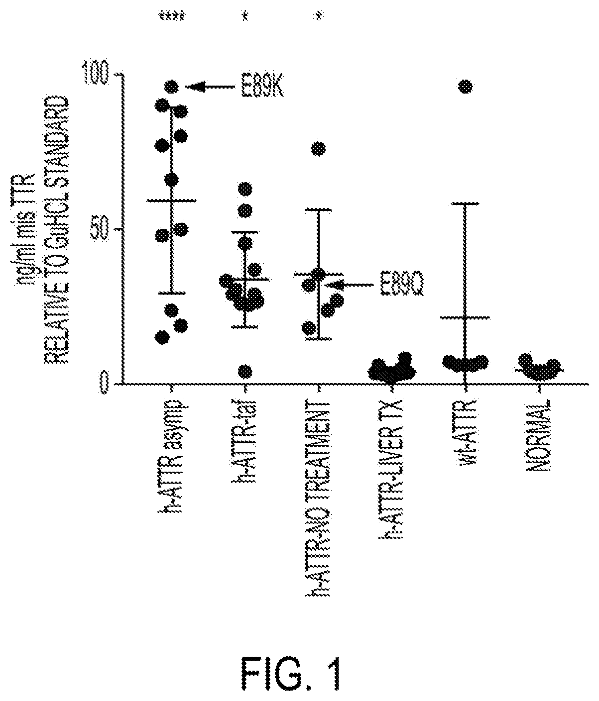
[0109] FIG. 1 depicts the results of a sandwich immunoassay employing the Meso Scale Discovery (MSD) electrochemiluminescence platform showing that 9D5 detects elevated misfolded TTR in plasma samples from transthyretin-mediated amyloidosis (ATTR) patients that had not undergone a liver transplant, in an assay using a polyclonal anti-TTR reporter antibody.
[0110] FIG. 2 depicts results of an ex vivo target engagement assay, using a 9D5 capture antibody and a polyclonal anti-TTR reporter antibody, showing that m14G8 reduces levels of free misfolded TTR when spiked into patient plasma.
[0111] FIG. 3 depicts the results of a sandwich immunoassay employing the Meso Scale Discovery (MSD) electrochemiluminescence platform showing that 9D5 detects elevated misfolded TTR in plasma samples from transthyretin-mediated amyloidosis (ATTR) patients that had not undergone a liver transplant, in an assay using an 18C5 reporter antibody.
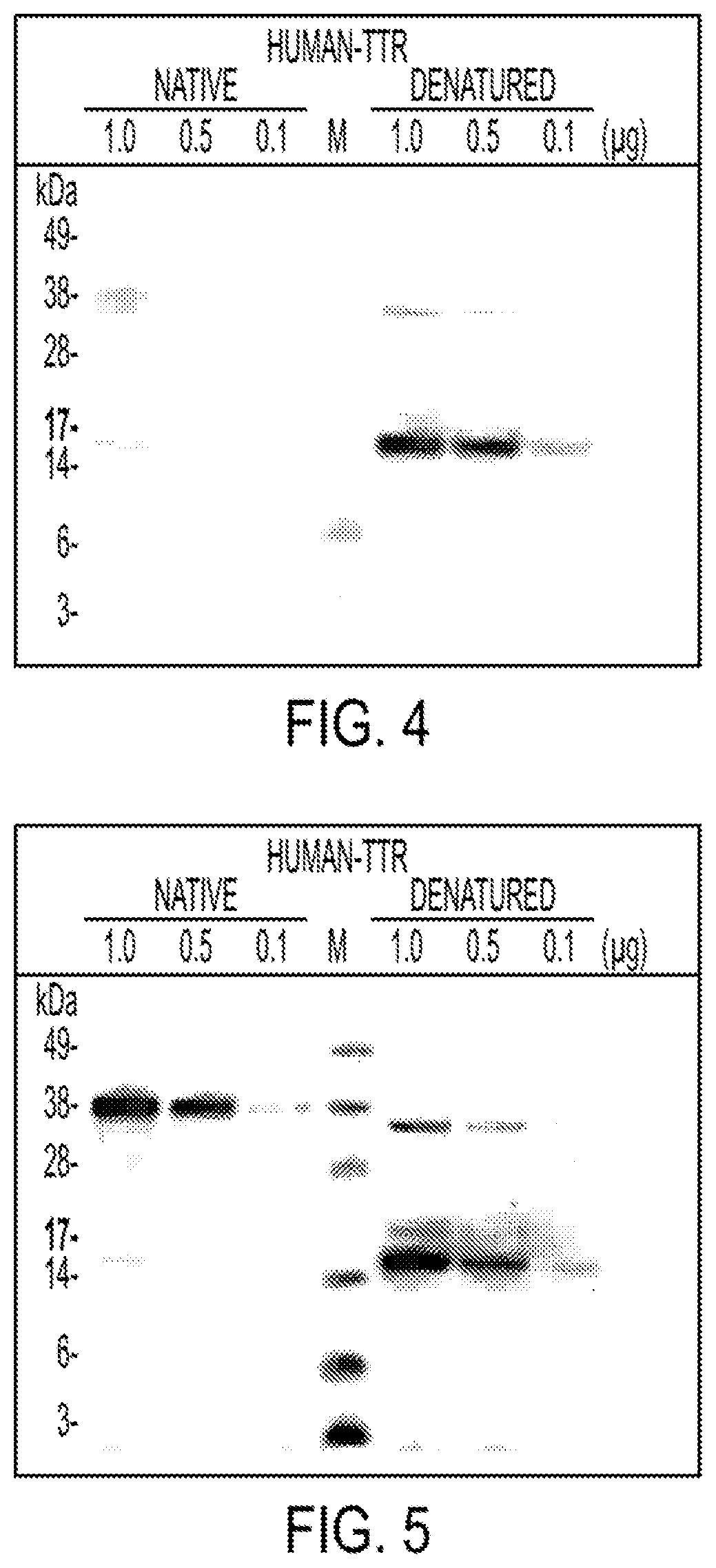
[0112] FIG. 4 depicts results of a Western blot experiment showing that 18C5 has strong reactivity toward denatured TTR monomer, minor reactivity toward denatured dimer, and very weak reactivity toward native TTR species.
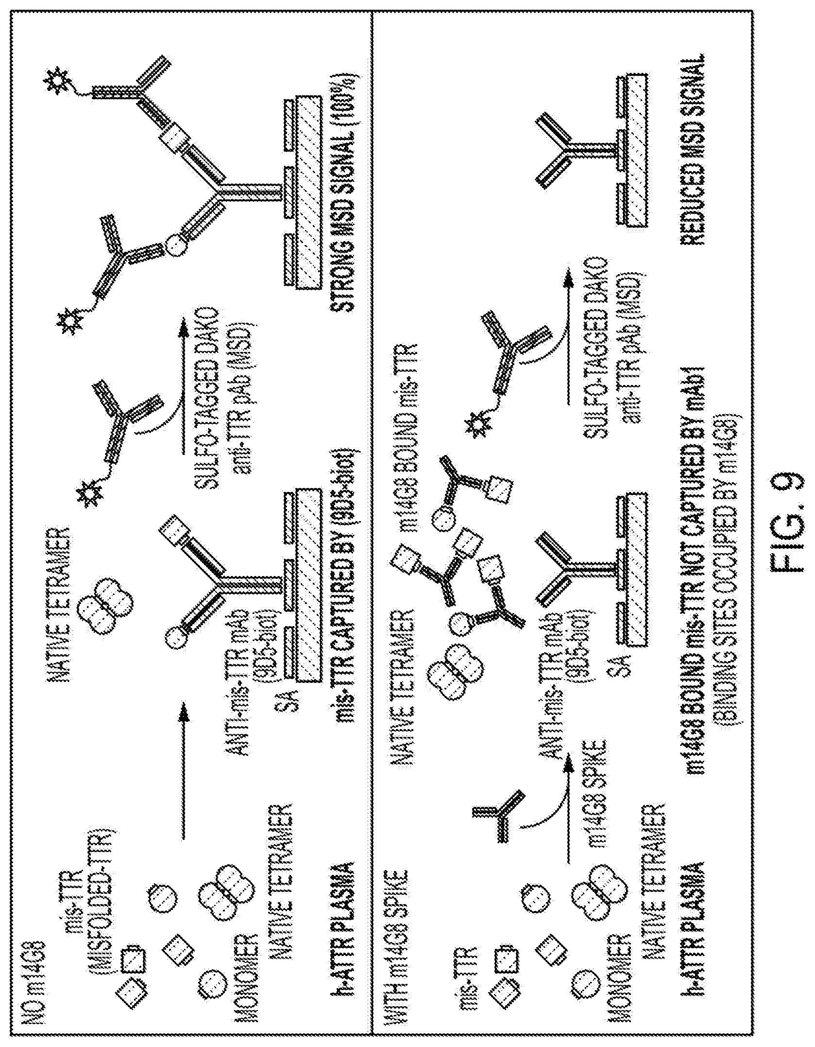
[0113] FIG. 5 depicts results of a Western blot experiment showing that a commercial TTR antibody could not distinguish between native versus denatured TTR and showed very strong reactivity toward monomeric as well as dimeric native and denatured TTR
[0114] FIG. 6 depicts the results of a sandwich immunoassay employing the Meso Scale Discovery (MSD) electrochemiluminescence platform showing that 18C5 detects elevated misfolded TTR in plasma samples from transthyretin-mediated amyloidosis (ATTR) patients that had not undergone a liver transplant.
[0115] FIG. 7 depicts the results of a sandwich immunoassay employing the Meso Scale Discovery (MSD) electrochemiluminescence platform showing that 18C5 detects elevated multimeric misfolded TTR in plasma samples from transthyretin-mediated amyloidosis (ATTR) patients that had not undergone a liver transplant.
[0116] FIG. 8 depicts the results of a Western blot experiment showing that 9D5 detects elevated levels of mis-TTR (misfolded TTR monomers and oligomers) in plasma from hereditary ATTR patients.
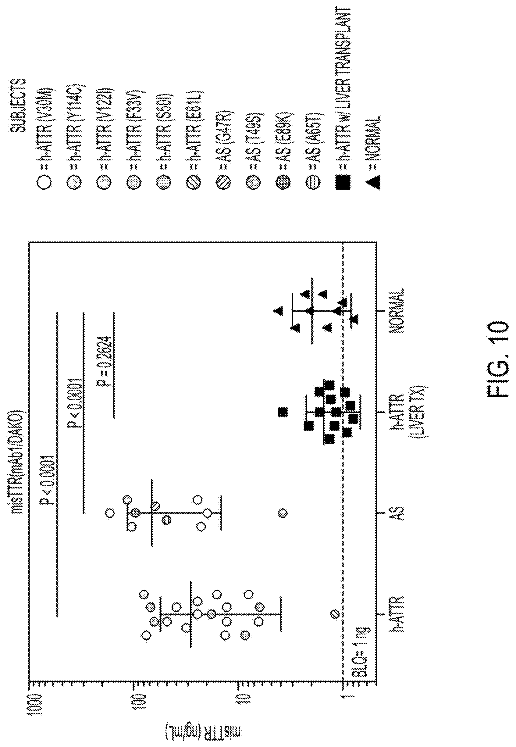
[0117] FIG. 9 depicts a diagram of a pharmacodynamic assay to measure binding of a mis-TTR mAb (m14G8) to plasma mis-TTR (target engagement).
[0118] FIG. 10 depicts the results of a sandwich immunoassay employing the Meso Scale Discovery (MSD) electrochemiluminescence platform showing that 9D5 detects elevated misfolded TTR in plasma samples from transthyretin-mediated amyloidosis (ATTR) patients that had not undergone a liver transplant, in an assay using a polyclonal anti-TTR reporter antibody.
[0119] FIG. 11 depicts an alignment of heavy chain variable regions of the mouse 18C5 antibody (SEQ ID NO: 81), human germline sequence IGHV3-48*01 (SEQ ID NO:84), human acceptor 5VZY-VH_huFrwk (Crenefab-VH) (SEQ ID NO:83), and humanized versions of the 18C5 antibody (hu18C5 VH-v1 and hu18C5 VH-v2, SEQ ID NOs: 84 and 85, respectively). CDRs defined according to Kabat/Chothia Composite are bolded in the mouse 18C5 heavy chain variable region sequence.
[0120] FIG. 12 depicts an alignment of light chain variable regions of the mouse 18C5 antibody (SEQ ID NO:87), human germline sequence IGKV2-30*02 (SEQ ID NO:90), human acceptor 5VZY-VL_huFrwk (Crenefab-VL) (SEQ ID NO:89), and humanized versions of the 18C5 antibody (hu18C5-VL-v1 and hu18C5-VL-v2, SEQ ID NOs: 91 and 92, respectively). CDRs defined according to Kabat/Chothia Composite are bolded in the mouse 18C5 light chain variable region sequence.
BRIEF DESCRIPTION OF THE SEQUENCES
[0121] SEQ ID NO:1 sets forth the amino acid sequence of a heavy chain variable region of the mouse 18C5 antibody with signal peptide.
[0122] SEQ ID NO:2 sets forth a nucleic acid sequence encoding a heavy chain variable region of the mouse 18C5 antibody with signal peptide.
[0123] SEQ ID NO:3 sets forth the amino acid sequence of a light chain variable region of the mouse 18C5 antibody with signal peptide.
[0124] SEQ ID NO:4 sets forth a nucleic acid sequence encoding a light chain variable region of the mouse 18C5 antibody with signal peptide.
[0125] SEQ ID NO:5 sets forth the amino acid sequence of a Kabat/Chothia Composite CDR-H1 of the mouse 18C5 antibody.
[0126] SEQ ID NO:6 sets forth a nucleic acid sequence encoding a Kabat/Chothia Composite composite CDR-H1 of the mouse 18C5 antibody.
[0127] SEQ ID NO:7 sets forth the amino acid sequence of a Kabat/Chothia Composite CDR-H2 of the mouse 18C5 antibody.
[0128] SEQ ID NO:8 sets forth a nucleic acid sequence encoding a Kabat/Chothia Composite CDR-H2 of the mouse 18C5 antibody.
[0129] SEQ ID NO:9 sets forth the amino acid sequence of a Kabat/Chothia Composite CDR-H3 of the mouse 18C5 antibody.
[0130] SEQ ID NO:10 sets forth a nucleic acid sequence encoding a Kabat/Chothia Composite CDR-H3 of the mouse 18C5 antibody.
[0131] SEQ ID NO:11 sets forth the amino acid sequence of a Kabat/Chothia Composite CDR-L1 of the mouse 18C5 antibody.
[0132] SEQ ID NO:12 sets forth a nucleic acid sequence encoding a Kabat/Chothia Composite CDR-L1 of the mouse 18C5 antibody.
[0133] SEQ ID NO:13 sets forth the amino acid sequence of a Kabat/Chothia Composite CDR-L2 of the mouse 18C5 antibody.
[0134] SEQ ID NO:14 sets forth a nucleic acid sequence encoding a Kabat/Chothia Composite CDR-L2 of the mouse 18C5 antibody.
[0135] SEQ ID NO:15 sets forth the amino acid sequence of a Kabat/Chothia Composite CDR-L3 of the mouse 18C5 antibody.
[0136] SEQ ID NO:16 sets forth a nucleic acid sequence encoding the a Kabat/Chothia Composite CDR-L3 of the mouse 18C5 antibody.
[0137] SEQ ID NO:17 sets forth the amino acid sequence of a chimeric 18C5 heavy chain constant region (human IgG1).
[0138] SEQ ID NO:18 sets forth a nucleic acid sequence encoding the amino acid sequence of a chimeric 18C5 heavy chain constant region (human IgG1).
[0139] SEQ ID NO:19 sets forth the amino acid sequence of a chimeric 18C5 light chain constant region (human kappa).
[0140] SEQ ID NO:20 sets forth a nucleic acid sequence encoding the amino acid sequence of a chimeric 18C5 light chain constant region (human kappa).
[0141] SEQ ID NO:21 sets forth the amino acid sequence of an exemplary IgG1 heavy chain constant region.
[0142] SEQ ID NO:22 sets forth the amino acid sequence of an exemplary IgG1 G1m3 heavy chain constant region.
[0143] SEQ ID NO:23 sets forth the amino acid sequence of an exemplary IgG1 G1m3 heavy chain constant region.
[0144] SEQ ID NO:24 sets forth the amino acid sequence of an exemplary light chain constant region with N-terminal Arginine.
[0145] SEQ ID NO:25 sets forth the amino acid sequence of an exemplary light chain constant region without N-terminal Arginine.
[0146] SEQ ID NO:26 sets forth the amino acid sequence of human transthyretin set forth in accession number P02766.1 (UniProt).
[0147] SEQ ID NO:27 sets forth the amino acid sequence of human transthyretin set forth in accession number AAB35639.1 (GenBank).
[0148] SEQ ID NO:28 sets forth the amino acid sequence of human transthyretin set forth in accession number AAB35640.1 (GenBank).
[0149] SEQ ID NO:29 sets forth the amino acid sequence of human transthyretin set forth in accession number and ABI63351.1 (GenBank).
[0150] SEQ ID NO:30 sets forth the amino acid sequence of residues 101-109 of human transthyretin.
[0151] SEQ ID NO:31 sets forth the amino acid sequence of residues 87-127 of human transthyretin.
[0152] SEQ ID NO:32 sets forth a nucleic acid sequence encoding an exemplary IgG1 G1m3 heavy chain constant region.
[0153] SEQ ID NO:33 sets forth a nucleic acid sequence encoding an exemplary light chain constant region with C-terminal Arginine.
[0154] SEQ ID NO:34 sets forth a nucleic acid sequence encoding an exemplary light chain constant region without C-terminal Arginine.
[0155] SEQ ID NO:35 sets forth the amino acid sequence of a heavy chain constant region signal peptide.
[0156] SEQ ID NO:36 sets forth a nucleic acid sequence encoding a heavy chain constant region signal peptide.
[0157] SEQ ID NO:37 sets forth the amino acid sequence of a light chain constant region signal peptide.
[0158] SEQ ID NO:38 sets forth a nucleic acid sequence encoding a light chain constant region signal peptide.
[0159] SEQ ID NO: 39 sets forth the amino acid sequence of a Kabat CDR-H1 of antibody 14G8.
[0160] SEQ ID NO: 40 sets forth the amino acid sequence of a Kabat CDR-H2 of antibody 14G8.
[0161] SEQ ID NO: 41 sets forth the amino acid sequence of a Kabat CDR-H3 of antibody 14G8.
[0162] SEQ ID NO: 42 sets forth the amino acid sequence of a Kabat CDR-L1 of antibody 14G8.
[0163] SEQ ID NO: 43 sets forth the amino acid sequence of a Kabat CDR-L2 of antibody 14G8.
[0164] SEQ ID NO: 44 sets forth the amino acid sequence of a Kabat CDR-L3 of antibody 14G8.
[0165] SEQ ID NO: 45 sets forth the amino acid sequence of an epitope of antibody 5A1.
[0166] SEQ ID NO: 46 sets forth the amino acid sequence of a Kabat CDR-H1 of antibody 5A1.
[0167] SEQ ID NO: 47 sets forth the amino acid sequence of a Kabat CDR-H2 of antibody 5A1.
[0168] SEQ ID NO: 48 sets forth the amino acid sequence of a Kabat CDR-H3 of antibody 5A1.
[0169] SEQ ID NO: 49 sets forth the amino acid sequence of a Kabat CDR-L1 of antibody 5A1.
[0170] SEQ ID NO: 50 sets forth the amino acid sequence of a Kabat CDR-L2 of antibody 5A1.
[0171] SEQ ID NO: 51 sets forth the amino acid sequence of a Kabat CDR-L3 of antibody 5A1.
[0172] SEQ ID NO: 52 sets forth the amino acid sequence of a Kabat CDR-H1 of antibody 6C1.
[0173] SEQ ID NO: 53 sets forth the amino acid sequence of a Kabat CDR-H2 of antibody 6C1.
[0174] SEQ ID NO: 54 sets forth the amino acid sequence of a Kabat CDR-H3 of antibody 6C1.
[0175] SEQ ID NO: 55 sets forth the amino acid sequence of a Kabat CDR-L1 of antibody 6C1.
[0176] SEQ ID NO: 56 sets forth the amino acid sequence of a Kabat CDR-L2 of antibody 6C1.
[0177] SEQ ID NO: 57 sets forth the amino acid sequence of a Kabat CDR-L3 of antibody 6C1.
[0178] SEQ ID NO: 58 sets forth the amino acid sequence of a VH region of antibody AD7F6.
[0179] SEQ ID NO: 59 sets forth the amino acid sequence of a VL region of antibody AD7F6.
[0180] SEQ ID NO: 60 sets forth the amino acid sequence of a CDR-H1 of antibody RT24.
[0181] SEQ ID NO: 61 sets forth the amino acid sequence of a CDR-H2 of antibody RT24.
[0182] SEQ ID NO: 62 sets forth the amino acid sequence of a CDR-H3 of antibody RT24.
[0183] SEQ ID NO:63 sets forth the amino acid sequence of a CDR-L1 of antibody RT24.
[0184] SEQ ID NO: 64 sets forth the amino acid sequence of a CDR-L2 of antibody RT24.
[0185] SEQ ID NO: 65 sets forth the amino acid sequence of a CDR-L3 of antibody RT24.
[0186] SEQ ID NO: 66 sets forth the amino acid sequence of a CDR-H1 of antibody NI-301.35G11.
[0187] SEQ ID NO:67 sets forth the amino acid sequence of a CDR-H2 of antibody NI-301.35G11.
[0188] SEQ ID NO: 68 sets forth the amino acid sequence of a CDR-H3 of antibody NI-301.35G11.
[0189] SEQ ID NO: 69 sets forth the amino acid sequence of a CDR-L1 of antibody NI-301.35G11.
[0190] SEQ ID NO: 70 sets forth the amino acid sequence of a CDR-L2 of antibody NI-301.35G11.
[0191] SEQ ID NO: 71 sets forth the amino acid sequence of a CDR-L3 of antibody NI-301.35G11.
[0192] SEQ ID NO: 72 sets forth the amino acid sequence of an epitope of antibodies MFD101, MDF102, MFD103, MFD105.
[0193] SEQ ID NO: 73 sets forth the amino acid sequence of an epitope of antibodies MFD107, MFD108, MFD109, MFD111.
[0194] SEQ ID NO: 74 sets forth the amino acid sequence of an epitope of antibody MFD114.
[0195] SEQ ID NO: 75 sets forth the amino acid sequence of a Kabat CDR-H1 of antibody 9D5.
[0196] SEQ ID NO: 76 sets forth the amino acid sequence of a Kabat CDR-H2 of antibody 9D5.
[0197] SEQ ID NO: 77 sets forth the amino acid sequence of a Kabat CDR-H3 of antibody 9D5.
[0198] SEQ ID NO: 78 sets forth the amino acid sequence of a Kabat CDR-L1 of antibody 9D5.
[0199] SEQ ID NO: 79 sets forth the amino acid sequence of a Kabat CDR-L2 of antibody 9D5.
[0200] SEQ ID NO: 80 sets forth the amino acid sequence of a Kabat CDR-L3 of antibody 9D5.
[0201] SEQ ID NO:81 sets forth the amino acid sequence of a mature heavy chain variable region of the mouse 18C5 antibody.
[0202] SEQ ID NO: 82 sets forth the amino acid sequence of a heavy chain variable region of the murine anti-pyroglutamate-Abeta antibody Fab c #17, GenBank Acc. No. 1212215935.
[0203] SEQ ID NO: 83 sets forth the amino acid sequence of a heavy chain variable region of humanized Crenezumab Fab (CreneFab) PDB: 5VZY, GenBank Acc. No. 1229749875.
[0204] SEQ ID NO: 84 sets forth the amino acid sequence of a heavy chain variable region of the human germline sequence IGHV3-48*01, GenBank Acc. No. 1FN550289.1.
[0205] SEQ ID NO: 85 sets forth the amino acid sequence of a heavy chain variable region of the humanized 18C5 antibody hu18C5-VH_1.
[0206] SEQ ID NO: 86 sets forth the amino acid sequence of a heavy chain variable region of the humanized 18C5 antibody hu18C5-VH_2.
[0207] SEQ ID NO:87 sets forth the amino acid sequence of a mature light chain variable region of the mouse 18C5 antibody.
[0208] SEQ ID NO: 88 sets forth the amino acid sequence of a light chain variable region of the murine anti-pyroglutamate-Abeta antibody Fab c #17, GenBank Acc. No. 1212215934.
[0209] SEQ ID NO: 89 sets forth the amino acid sequence of a light chain variable region of humanized Crenezumab Fab (CreneFab) PDB: 5VZY, GenBank Acc. No. 1229749876.
[0210] SEQ ID NO: 90 sets forth the amino acid sequence of a light chain variable region of the human germline sequence IGKV2-30*2, GenBank Acc. No. CAA77315.
[0211] SEQ ID NO: 91 sets forth the amino acid sequence of a light chain variable region of the humanized 18C5 antibody hu18C5-VL_1.
[0212] SEQ ID NO: 92 sets forth the amino acid sequence of a light chain variable region of the humanized 18C5 antibody hu18C5-VL_2.
[0213] SEQ ID NO: 93 sets forth the amino acid sequence of Kabat CDR-H1 of the mouse 18C5 antibody.
[0214] SEQ ID NO: 94 sets forth the amino acid sequence of Chothia CDR-H1 of the mouse 18C5 antibody.
[0215] SEQ ID NO: 95 sets forth the amino acid sequence of Contact CDR-H1 of the mouse 18C5 antibody.
[0216] SEQ ID NO: 96 sets forth the amino acid sequence of Chothia CDR-H2 of the mouse 18C5 antibody.
[0217] SEQ ID NO: 97 sets forth the amino acid sequence of AbM CDR-H2 of the mouse 18C5 antibody.
[0218] SEQ ID NO: 98 sets forth the amino acid sequence of Contact CDR-H2 of the mouse 18C5 antibody.
[0219] SEQ ID NO: 99 sets forth the amino acid sequence of Contact CDR-H3 of the mouse 18C5 antibody.
[0220] SEQ ID NO: 100 sets forth the amino acid sequence of Contact CDR-L1 of the mouse 18C5 antibody.
[0221] SEQ ID NO: 101 sets forth the amino acid sequence of Contact CDR-L2 of the mouse 18C5 antibody.
[0222] SEQ ID NO: 102 sets forth the amino acid sequence of Contact CDR-L3 of the mouse 18C5 antibody.
[0223] SEQ ID NO: 103 sets forth the amino acid sequence of a heavy chain variable region of the mouse 9D5 antibody.
[0224] SEQ ID NO: 104 sets forth the amino acid sequence of a light chain variable region of the mouse 9D5 antibody.
Definitions
[0225] Monoclonal antibodies or other biological entities are typically provided in isolated form. This means that an antibody or other biologically entity is typically at least 50% w/w pure of interfering proteins and other contaminants arising from its production or purification but does not exclude the possibility that the monoclonal antibody is combined with an excess of pharmaceutically acceptable carrier(s) or other vehicle intended to facilitate its use. Sometimes monoclonal antibodies are at least 60%, 70%, 80%, 90%, 95% or 99% w/w pure of interfering proteins and contaminants from production or purification. Often an isolated monoclonal antibody or other biological entity is the predominant macromolecular species remaining after its purification.
[0226] Specific binding of an antibody to its target antigen means an affinity of at least 10.sup.6, 10', 10.sup.8, 10.sup.9, or 10.sup.10 M.sup.-1. Specific binding is detectably higher in magnitude and distinguishable from non-specific binding occurring to at least one unrelated target. Specific binding can be the result of formation of bonds between particular functional groups or particular spatial fit (e.g., lock and key type) whereas nonspecific binding is usually the result of van der Waals forces. Specific binding does not however necessarily imply that an antibody binds one and only one target.
[0227] The basic antibody structural unit is a tetramer of subunits. Each tetramer includes two identical pairs of polypeptide chains, each pair having one "light" (about 25 kDa) and one "heavy" chain (about 50-70 kDa). The amino-terminal portion of each chain includes a variable region of about 100 to 110 or more amino acids primarily responsible for antigen recognition. This variable region is initially expressed linked to a cleavable signal peptide. The variable region without the signal peptide is sometimes referred to as a mature variable region. Thus, for example, a light chain mature variable region means a light chain variable region without the light chain signal peptide. The carboxy-terminal portion of each chain defines a constant region primarily responsible for effector function.
[0228] Light chains are classified as either kappa or lambda. Heavy chains are classified as gamma, mu, alpha, delta, or epsilon, and define the antibody's isotype as IgG, IgM, IgA, IgD and IgE, respectively. Within light and heavy chains, the variable and constant regions are joined by a "J" region of about 12 or more amino acids, with the heavy chain also including a "D" region of about 10 or more amino acids. See generally, Fundamental Immunology, Paul, W., ed., 2nd ed. Raven Press, N.Y., 1989, Ch. 7 (incorporated by reference in its entirety for all purposes).
[0229] An immunoglobulin light or heavy chain variable region (also referred to herein as a "light chain variable domain" ("VL domain") or "heavy chain variable domain" ("VH domain"), respectively) consists of a "framework" region interrupted by three "complementarity determining regions" or "CDRs." The framework regions serve to align the CDRs for specific binding to an epitope of an antigen. The CDRs include the amino acid residues of an antibody that are primarily responsible for antigen binding. From amino-terminus to carboxyl-terminus, both VL and VH domains comprise the following framework (FR) and CDR regions: FR1, CDR1, FR2, CDR2, FR3, CDR3, and FR4. CDRs 1, 2, and 3 of a VL domain are also referred to herein, respectively, as CDR-L1, CDR-L2, and CDR-L3; CDRs 1, 2, and 3 of a VH domain are also referred to herein, respectively, as CDR-H1, CDR-H2, and CDR-H3.
[0230] The assignment of amino acids to each VL and VH domain is in accordance with any conventional definition of CDRs. Conventional definitions include, the Kabat definition (Kabat, Sequences of Proteins of Immunological Interest (National Institutes of Health, Bethesda, Md., 1987 and 1991), The Chothia definition (Chothia & Lesk, J. Mol. Biol. 196:901-917, 1987; Chothia et al., Nature 342:878-883, 1989); a composite of Chothia Kabat CDR in which CDR-H1 is a composite of Chothia and Kabat CDRs; the AbM definition used by Oxford Molecular's antibody modelling software; and, the contact definition of Martin et al (bioinfo.org.uk/abs) (see Table 1). Kabat provides a widely used numbering convention (Kabat numbering) in which corresponding residues between different heavy chains or between different light chains are assigned the same number. When an antibody is said to comprise CDRs by a certain definition of CDRs (e.g., Kabat) that definition specifies the minimum number of CDR residues present in the antibody (i.e., the Kabat CDRs). It does not exclude that other residues falling within another conventional CDR definition but outside the specified definition are also present. For example, an antibody comprising CDRs defined by Kabat includes among other possibilities, an antibody in which the CDRs contain Kabat CDR residues and no other CDR residues, and an antibody in which CDR H1 is a composite Chothia-Kabat CDR H1 and other CDRs contain Kabat CDR residues and no additional CDR residues based on other definitions.
TABLE-US-00001 TABLE 1 Conventional Definitions of CDRs Using Kabat Numbering Composite of Chothia Loop Kabat Chothia & Kabat AbM Contact L1 L24--L34 L24--L34 L24--L34 L24--L34 L30--L36 L2 L50--L56 L50--L56 L50--L56 L50--L56 L46--L55 L3 L89--L97 L89--L97 L89--L97 L89--L97 L89--L96 H1 H31--H35B H26--H32 . . . H34* H26--H35B* H26--H35B H30--H35B H2 H50--H65 H52--H56 H50--H65 H50--H58 H47--H58 H3 H95--H102 H95--H102 H95--H102 H95--H102 H93--H101 *CDR-H1 by Chothia can end at H32, H33, or H34 (depending on the length of the loop). This is because the Kabat numbering scheme places insertions of extra residues at 35A and 35B, whereas Chothia numbering places them at 31A and 31B. If neither H35A nor H35B (Kabat numbering) is present, the Chothia CDR-H1 loop ends at H32. If only H35A is present, it ends at H33. If both H35A and H35B are present, it ends at H34.
[0231] The term "antibody" includes intact antibodies and binding fragments thereof. Typically, fragments compete with the intact antibody from which they were derived for specific binding to the target including separate heavy chains, light chains Fab, Fab', F(ab').sub.2, F(ab)c, Dabs, nanobodies, and Fv. Fragments can be produced by recombinant DNA techniques, or by enzymatic or chemical separation of intact immunoglobulins. The term "antibody" also includes a bispecific antibody and/or a humanized antibody. A bispecific or bifunctional antibody is an artificial hybrid antibody having two different heavy/light chain pairs and two different binding sites (see, e.g., Songsivilai and Lachmann, Clin. Exp. Immunol., 79:315-321 (1990); Kostelny et al., J. Immunol., 148:1547-53 (1992)). In some bispecific antibodies, the two different heavy/light chain pairs include a humanized 9D5 heavy chain/light chain pair and a heavy chain/light chain pair specific for a different epitope on transthyretin than that bound by 9D5. In some bispecific antibodies, the two different heavy/light chain pairs include a humanized 18C5 heavy chain/light chain pair and a heavy chain/light chain pair specific for a different epitope on transthyretin than that bound by 18C5.
[0232] In some bispecific antibodies, one heavy chain/light chain pair is a humanized 9D5 antibody or a humanized 18C5 antibody as further disclosed below and the other heavy chain/light chain pair is from an antibody that binds to a receptor expressed on the blood brain barrier, such as an insulin receptor, an insulin-like growth factor (IGF) receptor, a leptin receptor, or a lipoprotein receptor, or a transferrin receptor (Friden et al., Proc. Natl. Acad. Sci. USA 88:4771-4775, 1991; Friden et al., Science 259:373-377, 1993). Such a bispecific antibody can be transferred cross the blood brain barrier by receptor-mediated transcytosis. Brain uptake of the bispecific antibody can be further enhanced by engineering the bispecific antibody to reduce its affinity to the blood brain barrier receptor. Reduced affinity for the receptor resulted in a broader distribution in the brain (see, e.g., Atwal et al., Sci. Trans. Med. 3, 84ra43, 2011; Yu et al., Sci. Trans. Med. 3, 84ra44, 2011).
[0233] Exemplary bispecific antibodies can also be: (1) a dual-variable-domain antibody (DVD-Ig), where each light chain and heavy chain contains two variable domains in tandem through a short peptide linkage (Wu et al., Generation and Characterization of a Dual Variable Domain Immunoglobulin (DVD-Ig.TM.) Molecule, In: Antibody Engineering, Springer Berlin Heidelberg (2010)); (2) a Tandab, which is a fusion of two single chain diabodies resulting in a tetravalent bispecific antibody that has two binding sites for each of the target antigens; (3) a flexibody, which is a combination of scFvs with a diabody resulting in a multivalent molecule; (4) a so-called "dock and lock" molecule, based on the "dimerization and docking domain" in Protein Kinase A, which, when applied to Fabs, can yield a trivalent bispecific binding protein consisting of two identical Fab fragments linked to a different Fab fragment; or (5) a so-called Scorpion molecule, comprising, e.g., two scFvs fused to both termini of a human Fc-region. Examples of platforms useful for preparing bispecific antibodies include BiTE (Micromet), DART (MacroGenics), Fcab and Mab2 (F-star), Fc-engineered IgG1 (Xencor) or DuoBody (based on Fab arm exchange, Genmab).
[0234] The term "epitope" refers to a site on an antigen to which an antibody binds. An epitope can be formed from contiguous amino acids or noncontiguous amino acids juxtaposed by tertiary folding of one or more proteins. Epitopes formed from contiguous amino acids (also known as linear epitopes) are typically retained on exposure to denaturing solvents whereas epitopes formed by tertiary folding (also known as conformational epitopes) are typically lost on treatment with denaturing solvents. An epitope typically includes at least 3, and more usually, at least 5 or 8-10 amino acids in a unique spatial conformation. Methods of determining spatial conformation of epitopes include, for example, x-ray crystallography and 2-dimensional nuclear magnetic resonance. See, e.g., Epitope Mapping Protocols, in Methods in Molecular Biology, Vol. 66, Glenn E. Morris, Ed. (1996). The epitope can be linear, such as an epitope of, for example, 2-5, 3-5, 3-9, or 5-9 contiguous amino acids from SEQ ID NO:26, including for example, two or more contiguous amino acids within residues 89-97 of the mature region of SEQ ID NO:26. The epitope can also be a conformational epitope including, for example, two or more non-contiguous segments of amino acids within residues 89-97 of the mature region of SEQ ID NO:26. If an antibody is said to bind to an epitope within amino acid residues 89-97 of transthyretin (TTR) (the mature region of SEQ ID NO:26), for example, what is meant is that the epitope is within the recited range of amino acids including those defining the outer-limits of the range. It does not necessarily mean that every amino acid within the range constitutes part of the epitope. Thus, for example, an epitope within amino acid residues 89-97 of TTR may consist of amino acids 89-97, 89-96, 90-97, 89-95, 90-96, 91-97, 89-94, 90-95, 91-96, 92-97, 89-93, 90-94, 91-95, 92-96, 93-97, 89-92, 90-93, 91-94, 92-95, 93-96, 94-97, 89-91, 90-92, 91-93, 92-94, 93-95, 94-96, 95-97 of SEQ ID NO:26, among other linear segments of SEQ ID NO:45, or in the case of conformational epitopes, non-contiguous segments of amino acids of SEQ ID NO:45. Epitopes can include E89K and E89Q substitutions, E being the wildtype residue).
[0235] The epitope can be linear, such as an epitope of, for example, 2-5, 3-5, 3-9, or 5-9 contiguous amino acids from SEQ ID NO:26, including for example, two or more contiguous amino acids within residues 101-109 of the mature region of SEQ ID NO:26. The epitope can also be a conformational epitope including, for example, two or more non-contiguous segments of amino acids within residues 101-109 of the mature region of SEQ ID NO:26. If an antibody is said to bind to an epitope within amino acid residues 101-109 of transthyretin (TTR) (the mature region of SEQ ID NO:26), for example, what is meant is that the epitope is within the recited range of amino acids including those defining the outer-limits of the range. It does not necessarily mean that every amino acid within the range constitutes part of the epitope. Thus, for example, an epitope within amino acid residues 101-109 of TTR may consist of amino acids 101-109, 101-108, 102-109, 101-107, 102-108, 103-109, 101-106, 102-107, 103-108, 104-109, 101-105, 102-106, 103-107, 104-108, 105-109, 101-104, 102-105, 103-106, 104-107, 105-108, 106-109, 101-103, 102-104, 103-105, 104-106, 105-107, 106-108, 107-109, 101-102, 102-103, 103-104, 104-105, 105-106, 106-107, 107-108, or 108-109 of SEQ ID NO:26, among other linear segments of SEQ ID NO:30, or in the case of conformational epitopes, non-contiguous segments of amino acids of SEQ ID NO:30.
[0236] Antibodies that recognize the same or overlapping epitopes can be identified in a simple immunoassay showing the ability of one antibody to compete with the binding of another antibody to a target antigen. The epitope of an antibody can also be defined by X-ray crystallography of the antibody bound to its antigen to identify contact residues. Alternatively, two antibodies have the same epitope if all amino acid mutations in the antigen that reduce or eliminate binding of one antibody reduce or eliminate binding of the other. Two antibodies have overlapping epitopes if some amino acid mutations that reduce or eliminate binding of one antibody reduce or eliminate binding of the other.
[0237] Competition between antibodies is determined by an assay in which an antibody under test inhibits specific binding of a reference antibody to a common antigen (see, e.g., Junghans et al., Cancer Res. 50:1495, 1990). A test antibody competes with a reference antibody if an excess of a test antibody (e.g., at least 2.times., 5.times., 10.times., 20.times. or 100.times.) inhibits binding of the reference antibody by at least 50% as measured in a competitive binding assay. Some test antibodies inhibit binding of the references antibody by at least 75%, 90% or 99%. Antibodies identified by competition assay (competing antibodies) include antibodies binding to the same epitope as the reference antibody and antibodies binding to an adjacent epitope sufficiently proximal to the epitope bound by the reference antibody for steric hindrance to occur.
[0238] The term "native" with respect to the structure transthyretin (TTR) refers to the normal folded structure of TTR in its properly functioning state (i.e., a TTR tetramer). As TTR is a tetramer in its natively folded form, non-native forms of TTR include, for example, misfolded TTR tetramers, TTR monomers, aggregated forms of TTR, and fibril forms of TTR. Non-native forms of TTR can include molecules comprising wild-type TTR amino acid sequences or mutations.
[0239] The term "misfolded" with respect to TTR refers to the secondary and tertiary structure of a TTR polypeptide monomer or multimer, and indicates that the polypeptide has adopted a conformation that is not normal for that protein in its properly functioning state. Although TTR misfolding can be caused by mutations in the protein (e.g., deletion, substitution, or addition), wild-type TTR proteins can also be misfolded in diseases, exposing specific epitopes.
[0240] The term "pharmaceutically acceptable" means that the carrier, diluent, excipient, or auxiliary is compatible with the other ingredients of the formulation and not substantially deleterious to the recipient thereof.
[0241] The term "patient" includes human and other mammalian subjects that receive either prophylactic or therapeutic treatment.
[0242] An individual is at increased risk of a disease if the subject has at least one known risk-factor (e.g., genetic, biochemical, family history, and situational exposure) placing individuals with that risk factor at a statistically significant greater risk of developing the disease than individuals without the risk factor.
[0243] The term "biological sample" refers to a sample of biological material within or obtainable from a biological source, for example a human or mammalian subject. Such samples can be organs, organelles, tissues, sections of tissues, bodily fluids, peripheral blood, blood plasma, blood serum, cells, molecules such as proteins and peptides, and any parts or combinations derived therefrom. The term biological sample can also encompass any material derived by processing the sample. Derived material can include cells or their progeny. Processing of the biological sample may involve one or more of filtration, distillation, extraction, concentration, fixation, inactivation of interfering components, and the like.
[0244] The term "control sample" refers to a biological sample not known or suspected to include monomeric, misfolded, aggregated, or fibril forms of transthyretin (TTR), such as in TTR amyloid deposits. Control samples can be obtained from individuals not afflicted with a TTR amyloidosis or a specifically chosen type of TTR amyloidosis. Alternatively, control samples can be obtained from patients afflicted with TTR amyloidosis or a specifically chosen type of TTR amyloidosis. Such samples can be obtained at the same time as a biological sample thought to comprise the TTR amyloidosis or on a different occasion. A biological sample and a control sample can both be obtained from the same tissue (e.g., a tissue section containing both TTR amyloid deposits and surrounding normal tissue). Preferably, control samples consist essentially or entirely of tissue free of TTR amyloid deposits and can be used in comparison to a biological sample thought to comprise TTR amyloid deposits. Preferably, the tissue in the control sample is the same type as the tissue in the biological sample (e.g., cardiomyocytes in the heart).
[0245] The term "disease" refers to any abnormal condition that impairs physiological function. The term is used broadly to encompass any disorder, illness, abnormality, pathology, sickness, condition, or syndrome in which physiological function is impaired, irrespective of the nature of the etiology.
[0246] The term "symptom" refers to a subjective evidence of a disease, such as altered gait, as perceivable by a subject. A "sign" refers to objective evidence of a disease as observable by a physician.
[0247] For purposes of classifying amino acids substitutions as conservative or nonconservative, amino acids are grouped as follows: Group I (hydrophobic side chains): met, ala, val, leu, ile; Group II (neutral hydrophilic side chains): asn, gln, cys, ser, thr; Group III (acidic side chains): asp, glu; Group IV (basic side chains): his, lys, arg; Group V (residues influencing chain orientation): gly, pro; and Group VI (aromatic side chains): trp, tyr, phe. Conservative substitutions involve substitutions between amino acids in the same class. Non-conservative substitutions constitute exchanging a member of one of these classes for a member of another.
[0248] Percentage sequence identities are determined with antibody sequences maximally aligned by the Kabat numbering convention. After alignment, if a subject antibody region (e.g., the entire mature variable region of a heavy or light chain) is being compared with the same region of a reference antibody, the percentage sequence identity between the subject and reference antibody regions is the number of positions occupied by the same amino acid in both the subject and reference antibody region divided by the total number of aligned positions of the two regions, with gaps not counted, multiplied by 100 to convert to percentage.
[0249] Compositions or methods "comprising" or "including" one or more recited elements may include other elements not specifically recited. For example, a composition that "comprises" or "includes" an antibody may contain the antibody alone or in combination with other ingredients.
[0250] Designation of a range of values includes all integers within or defining the range, and all subranges defined by integers within the range.
[0251] Unless otherwise apparent from the context, the term "about" encompasses values within a standard margin of error of measurement (e.g., SEM) of a stated value.
[0252] Statistical significance means p.ltoreq.0.05.
[0253] Antibodies of the invention can be administered concomitant with another treatment for the same indication as the antibody, meaning that the other treatment is administered at least once during the period in which the antibody is administered, such period beginning one month before the first dosing and ending one month after the last dosing of the antibody. The other treatment can be administered at recurring intervals during this period, which may or may not be the same as the intervals at which the antibody is administered. The other treatment may be a symptomatic treatment.
[0254] A treatment is symptomatic if it only affects one or more symptoms of a disease, not its cause, i.e., its etiology.
[0255] The singular forms of the articles "a," "an," and "the" include plural references unless the context clearly dictates otherwise. For example, the term "a compound" or "at least one compound" can include a plurality of compounds, including mixtures thereof.
DETAILED DESCRIPTION
I. General
[0256] The invention provides antibodies that specifically bind to residues 89-97 or to residues 101-109 of transthyretin (TTR). Some antibodies bind any or all of monomeric, misfolded, aggregated, or fibril forms of TTR preferentially relative to native tetrameric form of TTR. Some antibodies bind either or both of monomeric or misfolded forms of TTR preferentially relative to native tetrameric form of TTR. The antibodies can be used for treating or effecting prophylaxis of diseases or disorders associated with TTR accumulation or accumulation of TTR deposits (e.g., TTR amyloidosis). The antibodies can also be used for diagnosing TTR amyloidosis and inhibiting or reducing aggregation of TTR. The antibodies can also be used to demonstrate pharmacodynamics effects of a transthyretin-mediated amyloidosis therapy, among other applications. Preferential binding means an association constant at least five times higher for any or all of monomeric, misfolded, aggregated, or fibril forms of TTR than for native tetrameric form of TTR. Optionally, the association constant is at least ten times higher for any or all of monomeric, misfolded, aggregated, or fibril forms of TTR than for native tetrameric form of TTR. Optionally, the antibody, such as 9D5 or 18C5, for example, lacks specific binding to native tetrameric form of TTR.
II. Target Molecules
[0257] Transthyretin (TTR) is a 127-amino acid, 55 kDa serum and cerebrospinal fluid transport protein primarily synthesized by the liver. It has also been referred to as prealbumin, thyroxine binding prealbumin, ATTR, and TBPA. In its native state, TTR exists as a tetramer. In homozygotes, the tetramers comprise identical 127-amino-acid beta-sheet-rich subunits. In heterozygotes, the TTR tetramers are made up of variant and/or wild-type subunits, typically combined in a statistical fashion.
[0258] The established function of TTR in the blood is to transport holo-retinol binding protein. Although TTR is the major carrier of thyroxine (T4) in the blood of rodents, utilizing binding sites that are orthogonal to those used for holo-retinol binding protein, the T4 binding sites are effectively unoccupied in humans.
[0259] TTR is one of at least thirty different human proteins whose extracellular misfolding and/or misassembly (amyloidogenesis) into a spectrum of aggregate structures is thought to cause degenerative diseases referred to as amyloid diseases. TTR undergoes conformational changes in order to become amyloidogenic. Dissociation of the TTR tetramer and partial unfolding exposes stretches of largely uncharged hydrophobic residues in an extended conformation that efficiently misassemble into largely unstructured spherical aggregates that ultimately undergo conformation conversion into cross-beta sheet amyloid structures.
[0260] Unless otherwise apparent from context, reference to transthyretin (TTR) or its fragments or domains includes the natural human amino acid sequences including isoforms, mutants (e.g., E89K and E89Q), and allelic variants thereof. Exemplary TTR polypeptide sequences are designated by Accession Numbers P02766.1 (UniProt) (SEQ ID NO:26), AAB35639.1 (GenBank) (SEQ ID NO:27), AAB35640.1 (GenBank) (SEQ ID NO:28), and ABI63351.1 (GenBank) (SEQ ID NO:29). Residues are numbered according to Swiss Prot P02766.1, with the first amino acid of the mature protein (i.e., not including the 20 amino acid signal sequence) designated residue 1. In any other TTR protein, residues are numbered according to the corresponding residues in P02766.1 on maximum alignment.
III. Transthyretin Amyloidosis
[0261] Transthyretin (TTR) amyloidosis is a systemic disorder characterized by pathogenic, misfolded TTR and the extracellular deposition of amyloid fibrils composed of TTR. TTR amyloidosis is generally caused by destabilization of the native TTR tetramer form (due to environmental or genetic conditions), leading to dissociation, misfolding, and aggregation of TTR into amyloid fibrils that accumulate in various organs and tissues, causing progressive dysfunction. See, e.g., Almeida and Saraiva, FEBS Letters 586:2891-2896 (2012); Ando et al., Orphanet Journal of Rare Diseases 8:31 (2013).
[0262] In humans, both wild-type TTR tetramers and mixed tetramers comprised of mutant and wild-type subunits can dissociate, misfold, and aggregate, with the process of amyloidogenesis leading to the degeneration of affected tissue. Thus, TTR amyloidoses encompass diseases caused by pathogenic misfolded TTR resulting from mutations in TTR or resulting from non-mutated, misfolded TTR.
[0263] For example, wild-type ATTR amyloidosis (also called senile systemic amyloidosis or SSA) and senile cardiac amyloidosis (SCA) are age-related types of amyloidosis that result from the deposition of wild-type TTR amyloid outside and within the cardiomyocytes of the heart. TTR amyloidosis is also the most common form of hereditary (familial) amyloidosis, which is caused by mutations that destabilize the TTR protein. The TTR amyloidoses associated with point mutations in the TTR gene include familial amyloid polyneuropathy (FAP), familial amyloid cardiomyopathy (FAC), and the rare central nervous system selective amyloidosis (CNSA). Patients with hereditary (familial) TTR amyloidosis are almost always heterozygotes, meaning that the TTR tetramers are composed of mutant and/or wild-type TTR subunits, generally statistically distributed. Hereditary (familial) versions of TTR amyloidosis are generally autosomal dominant and are typically earlier onset than the sporadic diseases (SSA and SCA).
[0264] There are over 100 mutations in the gene encoding TTR that have been implicated in the autosomal dominant disorders FAP and FAC. See, e.g., US 2014/0056904; Saraiva, Hum. Mutat. 17(6):493-503 (2001); Damas and Saraiva, J. Struct. Biol. 130:290-299; Dwulet and Benson, Biochem. Biophys. Res. Commun. 114:657-662 (1983). These amyloid-causing mutations are distributed throughout the entire molecule of TTR. Generally, the more destabilizing the mutant subunits are to the TTR tetramer structure, the earlier the onset of amyloid disease. The pathogenic potential of a TTR variant is generally determined by a combination of its instability and its cellular secretion efficiency. The initial pathology caused by some TTR variants comes from their selective destruction of cardiac tissue, whereas that from other TTR variants comes from compromising the peripheral and autonomic nervous system. The tissue damage caused by TTR amyloidogenesis appear to stem largely from the toxicity of small, diffusible TTR aggregates, although accumulation of extracellular amyloid may contribute and almost certainly compromises organ structure in the late stages of the TTR amyloidosis. Exemplary TTR mutations include V30M, Y114C, G47R, S50I, E61L, T49S, F33V, A45T, E89K, E89Q, and V122I.
[0265] TTR amyloidosis presents in many different forms, with considerable phenotypic variation across individuals and geographic locations. For example, TTR amyloidosis can present as a progressive, axonal sensory autonomic and motor neuropathy. TTR amyloidosis can also present as an infiltrative cardiomyopathy.
[0266] The age at onset of disease-related symptoms varies between the second and ninth decades of life, with great variations across different populations. The multisystem involvement of TTR amyloidosis is a clue to its diagnosis. For example, TTR amyloidosis diagnosis is considered when one or several of the following are present: (1) family history of neuropathic disease, especially associated with heart failure; (2) neuropathic pain or progressive sensory disturbances of unknown etiology; (3) carpal tunnel syndrome without obvious cause, particularly if it is bilateral and requires surgical release; (4) gastrointestinal motility disturbances or autonomic nerve dysfunction of unknown etiology (e.g., erectile dysfunction, orthostatic hypotension, neurogenic bladder); (5) cardiac disease characterized by thickened ventricular walls in the absence of hypertension; (6) advanced atrio-ventricular block of unknown origin, particularly when accompanied by a thickened heart; and (6) vitreous body inclusions of the cotton-wool type. See Ando et al., Orphanet Journal of Rare Diseases 8:31 (2013). Other symptoms can include, for example, polyneuropathy, sensory loss, pain, weakness in lower limbs, dyshidrosis, diarrhea, constipation, weight loss, and urinary incontinence/retention.
[0267] Diagnosis of TTR amyloidosis typically relies on target organ biopsies, followed by histological staining of the excised tissue with the amyloid-specific dye, Congo red. If a positive test for amyloid is observed, immunohistochemical staining and mass spectroscopic identification of TTR is subsequently performed to ensure that the precursor protein responsible for amyloid formation is indeed TTR. Antibodies disclosed herein are useful in distinguishing TTR amyloidosis from a non-TTR amyloidosis e.g. amyloid light-chain (AL) amyloidosis, also known as primary systemic amyloidosis, For familial forms of the diseases, demonstration of a mutation in the gene encoding TTR is then needed before diagnosis can be made. This can be accomplished, for example, through isoelectric focusing electrophoresis, polymerase chain reaction, or laser dissection/liquid chromatography-tandem mass spectrometry. See, e.g., US 2014/0056904; Ruberg and Berk, Circulation 126:1286-1300 (2012); Ando et al., Orphanet Journal of Rare Diseases 8:31 (2013).
IV. Antibodies
[0268] A. Binding Specificity and Functional Properties
[0269] The invention provides monoclonal antibodies binding to transthyretin (TTR) protein, more specifically, to epitopes within amino acid residues 89-97 (SEQ ID NO:45) or to epitopes within amino acid residues 101-109 (SEQ ID NO:30) of TTR. Such epitopes are buried in the native TTR tetramer and exposed in monomeric, misfolded, aggregated, or fibril forms of TTR.
[0270] Such antibodies include 9D5, 18C5, and their chimeric, veneered and humanized forms. 9D5 specifically binds within amino acid residues 89-97 (SEQ ID NO:45) of TTR. 18C5 specifically binds within amino acid residues 101-109 (SEQ ID NO: 30) of TTR. These antibodies are further characterized by their ability to bind to monomeric, misfolded, aggregated, or fibril forms of TTR but not to native tetrameric form of TTR. Ability to bind to specific proteins or fragments thereof may be demonstrated using exemplary assay formats provided in the examples. Unless otherwise apparent from the context, reference to 9D5 or 18C5 should be understood as referring to any of the mouse, chimeric, veneered or humanized forms. A hybridoma cell line that produces monoclonal antibody 9D5 was deposited with the Patent Depository of the American Type Culture Collection (ATCC), Manassas, Va., 20110-2209 on Apr. 4, 2017 and assigned Patent Deposit No. PTA-124078. A hybridoma cell line that produces monoclonal antibody 18C5 was deposited with the Patent Depository of the American Type Culture Collection (ATCC), Manassas, Va., 20110-2209 on Oct. 31, 2017 ______ and assigned Patent Deposit No. PTA-124570 ______.
[0271] Some antibodies bind to the same or overlapping epitope as an antibody designated 9D5. The sequences of the heavy and light chain mature variable regions of 9D5 are designated SEQ ID NOs: 81 and 82, respectively. Other antibodies having such a binding specificity can be produced by immunizing mice with TTR, or a portion thereof including the desired epitope (e.g., SEQ ID NO:45), and screening resulting antibodies for binding to monomeric TTR or a peptide comprising SEQ ID NO:45, optionally in competition with an antibody having the variable regions of mouse 18C5 (IgG1,kappa). Fragments of TTR including the desired epitope can be linked to a carrier that helps elicit an antibody response to the fragment and/or be combined with an adjuvant that helps elicit such a response. Such antibodies can be screened for differential binding to wild-type, monomeric versions of TTR or a fragment thereof (e.g., SEQ ID NO:26) compared with mutants of specified residues.
[0272] Some antibodies bind to the same or overlapping epitope as an antibody designated 18C5. The sequences of the heavy and light chain mature variable regions of 18C5 are designated SEQ ID NOs: 1 and 3, respectively. Other antibodies having such a binding specificity can be produced by immunizing mice with TTR, or a portion thereof including the desired epitope (e.g., SEQ ID NO:30), and screening resulting antibodies for binding to monomeric TTR or a peptide comprising SEQ ID NO:30, optionally in competition with an antibody having the variable regions of mouse 18C5 (IgG1, kappa). Fragments of TTR including the desired epitope can be linked to a carrier that helps elicit an antibody response to the fragment and/or be combined with an adjuvant that helps elicit such a response. Such antibodies can be screened for differential binding to wild-type, monomeric versions of TTR or a fragment thereof (e.g., SEQ ID NO:26) compared with mutants of specified residues.
[0273] Screening against such mutants more precisely defines the binding specificity to allow identification of antibodies whose binding is inhibited by mutagenesis of particular residues and which are likely to share the functional properties of other exemplified antibodies. The mutations can be systematic replacement substitution with alanine (or serine or glycine if an alanine is present already) one residue at a time, or more broadly spaced intervals, throughout the target or throughout a section thereof in which an epitope is known to reside. If the same set of mutations significantly reduces the binding of two antibodies, the two antibodies bind the same epitope.
[0274] Antibodies having the binding specificity of a selected murine antibody (e.g., 9D5 or 18C5) can also be produced using a variant of the phage display method. See Winter, WO 92/20791. This method is particularly suitable for producing human antibodies. In this method, either the heavy or light chain variable region of the selected murine antibody is used as a starting material. If, for example, a light chain variable region is selected as the starting material, a phage library is constructed in which members display the same light chain variable region (i.e., the murine starting material) and a different heavy chain variable region. The heavy chain variable regions can for example be obtained from a library of rearranged human heavy chain variable regions. A phage showing strong specific binding (e.g., at least 10.sup.8 and preferably at least 10.sup.9M.sup.-1) for monomeric TTR or a fragment thereof (e.g., amino acid residues 89-97 or amino acid residues 101-109) is selected. The heavy chain variable region from this phage then serves as a starting material for constructing a further phage library. In this library, each phage displays the same heavy chain variable region (i.e., the region identified from the first display library) and a different light chain variable region. The light chain variable regions can be obtained for example from a library of rearranged human variable light chain regions. Again, phage showing strong specific binding for monomeric TTR or a fragment thereof (e.g., amino acid residues 89-97 or amino acid residues 101-109) are selected. The resulting antibodies usually have the same or similar epitope specificity as the murine starting material.
[0275] Other antibodies can be obtained by mutagenesis of cDNA encoding the heavy and light chains of an exemplary antibody, such as 9D5 or 18C5. Monoclonal antibodies that are at least 70%, 80%, 90%, 95%, 96%, 97%, 98%, or 99% identical to 9D5 or 18C5 in amino acid sequence of the mature heavy and/or light chain variable regions and maintain its functional properties, and/or which differ from the respective antibody by a small number of functionally inconsequential amino acid substitutions (e.g., conservative substitutions), deletions, or insertions are also included in the invention. Monoclonal antibodies having at least one or all six CDR(s) as defined by conventional definition, but preferably Kabat, that are 90%, 95%, 99% or 100% identical to corresponding CDRs of 9D5 or 18C5 are also included.
[0276] The invention also provides antibodies having some or all (e.g., 3, 4, 5, and 6) CDRs entirely or substantially from 9D5 or 18C5. Such antibodies can include a heavy chain variable region that has at least two, and usually all three, CDRs entirely or substantially from the heavy chain variable region of 9D5 or 18C5 and/or a light chain variable region having at least two, and usually all three, CDRs entirely or substantially from the light chain variable region of 9D5 or 18C5. The antibodies can include both heavy and light chains. A CDR is substantially from a corresponding 9D5 or 18C5 CDR when it contains no more than 4, 3, 2, or 1 substitutions, insertions, or deletions, except that CDR-H2 (when defined by Kabat) can have no more than 6, 5, 4, 3, 2, or 1 substitutions, insertions, or deletions. Such antibodies can have at least 70%, 80%, 90%, 95%, 96%, 97%, 98%, or 99% identity to 9D5 or 18C5 in the amino acid sequence of the mature heavy and/or light chain variable regions and maintain their functional properties, and/or differ from 9D5 or 18C5 by a small number of functionally inconsequential amino acid substitutions (e.g., conservative substitutions), deletions, or insertions.
[0277] Kabat CDRs (CDR-H1, CDR-H2, CDR-H3) of the heavy chain of 9D5 are designated SEQ ID NOs: 75, 76, and 77, respectively, and Kabat CDRs (CDR-L1, CDR-L2, CDR-L3) of the light chain of 9D5 are designated SEQ ID NOs: 78, 79, and 80, respectively.
[0278] Kabat/Chothia Composite CDRs (CDR-H1, CDR-H2, CDR-H3) of the heavy chain of 18C5 are designated SEQ ID NOs: 5, 7, and 9, respectively, and Kabat/Chothia Composite CDRs (CDR-L1, CDR-L2, CDR-L3) of the light chain of 18C5 are designated SEQ ID NOs: 11, 13, and 15, respectively.
[0279] Table 2 indicates the 18C5 CDRs as defined by Kabat, Chothia, Composite of Chothia and Kabat (also referred to herein as "Kabat/Chothia Composite"), AbM, and Contact.
TABLE-US-00002 TABLE 2 18C5 CDRs as defined by Kabat, Chothia, Composite of Chothia and Kabat, AbM, and Contact, Using Kabat Numbering Composite of Chothia Loop Kabat Chothia & Kabat AbM Contact L1 L24--L34 L24--L34 L24--L34 L24--L34 L30--L36 SEQ ID NO: 11 SEQ ID NO: 11 SEQ ID NO: 11 SEQ ID NO: 11 SEQ ID NO: 100 L2 L50--L56 L50--L56 L50--L56 L50--L56 L46--L55 SEQ ID NO: 13 SEQ ID NO: 13 SEQ ID NO: 13 SEQ ID NO: 13 SEQ ID NO: 101 L3 L89--L97 L89--L97 L89--L97 L89--L97 L89--L96 SEQ ID NO: 15 SEQ ID NO: 15 SEQ ID NO: 15 SEQ ID NO: 15 SEQ ID NO: 102 H1 H31--H35B H26--H32 H26--H35B H26--H35B H30--H35B SEQ ID NO: 93 SEQ ID NO: 94 SEQ ID NO: 5 SEQ ID NO: 5 SEQ ID NO: 95 H2 H50--H65 H52--H56 H50--H65 H50--H58 H47--H58 SEQ ID NO: 7 SEQ ID NO: 96 SEQ ID NO: 7 SEQ ID NO: 97 SEQ ID NO: 98 H3 H95--H102 H95--H102 H95--H102 H95--H102 H93-H101 SEQ ID NO: 9 SEQ ID NO: 9 SEQ ID NO: 9 SEQ ID NO: 9 SEQ ID NO: 99
[0280] Some antibodies identified by such assays can bind to monomeric, misfolded, aggregated, or fibril forms of TTR but not to native tetrameric form of TTR, as described in the examples or otherwise. Likewise, some antibodies are immunoreactive on TTR-mediated amyloidosis tissue but not on healthy tissue.
[0281] Some antibodies can inhibit or reduce aggregation of TTR, inhibit or reduce TTR fibril formation, reduce or clear TTR deposits or aggregated TTR, or stabilize non-toxic conformations of TTR in an animal model or clinical trial. Some antibodies can treat, effect prophylaxis of, or delay the onset of a TTR amyloidosis as shown in an animal model or clinical trial. Exemplary animal models for testing activity against a TTR amyloidosis include those described in Kohno et al., Am. J Path. 150(4):1497-1508 (1997); Teng et al., Laboratory Investigations 81:385-396 (2001); Wakasugi et al., Proc. Japan Acad. 63B:344-347 (1987); Shimada et al., Mol. Biol. Med. 6:333-343 (1989); Nagata et al., J. Biochem. 117:169-175 (1995); Sousa et al., Am. J. Path. 161:1935-1948 (2002); and Santos et al., Neurobiology of Aging 31:280-289 (2010).
[0282] Anti-TTR antibodies including chimeric and humanized versions thereof, are useful in combination therapies, in bispecific antibodies, in methods of diagnosis and/or treatment of TTR associated disorders, and in methods of detecting TTR. Such anti-TTR antibodies, may include antibodies as in Table 3 below.
TABLE-US-00003 TABLE 3 Anti-TTR antibodies. Epitope on TTR VH/VL or CDRs Name as reported as reported Reference 9D5 EHAEVVFTA Kabat CDRs: WO 2016/120810 A1 (89-97) CDR-H1 SEQ ID NO: 75 ATCC Deposit (SEQ ID NO: 45) CDR-H2 SEQ ID NO: 76 No.PTA-124078 CDR-H3 SEQ ID NO: 77 Date: Apr. 4, 2017 CDR-L1 SEQ ID NO: 78 CDR-L2 SEQ ID NO: 79 CDR-L3 SEQ ID NO: 80 14G8 EHAEVVFTA Kabat CDRs WO 2016/120810 A1 (89-97) CDR-H1 SEQ ID NO: 39 ATCC Deposit No. (SEQ ID NO: 45) CDR-H2 SEQ ID NO: 40 PTA-124079 CDR-H3 SEQ ID NO: 41 Date: Apr. 4, 2017 CDR-L1 SEQ ID NO: 42 CDR-L2 SEQ ID NO: 43 CDR-L3 SEQ ID NO: 44 5A1 EHAEVVFTA Kabat CDRs WO 2016/120811 (89-97) CDR-H1 SEQ ID NO: 46 ATCC Deposit No. (SEQ ID NO: 45) CDR-H2 SEQ ID NO: 47 PTA-124080 CDR-H3 SEQ ID NO: 48 Date: Apr. 4, 2017 CDR-L1 SEQ ID NO 49 CDR-L2 SEQ ID NO: 50 CDR-L3 SEQ ID NO: 51 6C1 EHAEVVFTA Kabat CDRs WO 2016/120809 (89-97) CDR-H1 SEQ ID NO: 52 ATCC Deposit No. (SEQ ID NO: 45) CDR-H2 SEQ ID NO: 53 PTA-124077 CDR-H3 SEQ ID NO: 54 Date: Apr. 4, 2017 CDR-L1 SEQ ID NO: 55 CDR-L2 SEQ ID NO: 56 CDR-L3 SEQ ID NO: 57 18C5 GPRRYTIAA Kabat/Chothia US Provisional Appl. (101-109) Composite CDRs: 62/596,438, filed (SEQ ID NO: 30) CDR-H1 SEQ ID NO: 5 Oct. 6, 2017 CDR-H2 SEQ ID NO: 7 CDR-H3 SEQ ID NO: 9 CDR-L1 SEQ ID NO: 11 CDR-L2 SEQ ID NO: 13 CDR-L3 SEQ ID NO: 15 AD7F6 VH SEQ ID NO: 58 WO 2010/030203 A1 VL SEQ ID NO: 59 RT24 118-122, 115-124 CDR-H1 SEQ ID NO: 60 WO 2015/115331 CDR-H2 SEQ ID NO: 61 CDR-H3 SEQ ID NO: 62 CDR-L1 SEQ ID NO: 63 CDR-L2 SEQ ID NO: 64 CDR-L3 SEQ ID NO: 65 NI-301.35G11 53-63, 54-61 CDR-H1 SEQ ID NO: 66 US 2016/0355576 A1 ( CDR-H2 SEQ ID NO: 67 CDR-H3 SEQ ID NO: 68 CDR-L1 SEQ ID NO: 69 CDR-L2 SEQ ID NO: 70 CDR-L3 SEQ ID NO: 71 MFD101, ADDTWEPFASGKT US 2016/0039916 A1 MDF102, (residues 36-49) MFD103, (SEQ ID NO: 72) MFD105 MFD107, TSESGELHGLTTE US 2016/0039916 A1 MFD108, (residues 49-61) MFD109, (SEQ ID NO: 73) MFD111 MFD114 ALLSPYSYSTTAV US 2016/0039916 A1 (residues 109-121) (SEQ ID NO: 74) 30-66 US 2016/0039916 A1 70-127 US 2016/0039916 A1 80-127 US 2016/0039916 A1 90-127 US 2016/0039916 A1 100-127 US 2016/0039916 A1 110-127 US 2016/0039916 A1 115-127 US 2016/0039916 A1
[0283] B. Non-Human Antibodies
[0284] The production of other non-human antibodies, e.g., murine, guinea pig, primate, rabbit or rat, against monomeric TTR or a fragment thereof (e.g., amino acid residues 89-97 or amino acid residues 101-109) can be accomplished by, for example, immunizing the animal with TTR or a fragment thereof. See Harlow & Lane, Antibodies, A Laboratory Manual (CSHP NY, 1988) (incorporated by reference for all purposes). Such an immunogen can be obtained from a natural source, by peptide synthesis, or by recombinant expression. Optionally, the immunogen can be administered fused or otherwise complexed with a carrier protein. Optionally, the immunogen can be administered with an adjuvant. Several types of adjuvant can be used as described below. Complete Freund's adjuvant followed by incomplete adjuvant is preferred for immunization of laboratory animals. Rabbits or guinea pigs are typically used for making polyclonal antibodies. Mice are typically used for making monoclonal antibodies. Antibodies are screened for specific binding to monomeric TTR or an epitope within TTR (e.g., an epitope comprising one or more of amino acid residues 89-97 or of amino acid residues 101-109). Such screening can be accomplished by determining binding of an antibody to a collection of monomeric TTR variants, such as TTR variants containing amino acid residues 89-97 amino acid residues 101-109 or mutations within these residues, and determining which TTR variants bind to the antibody. Binding can be assessed, for example, by Western blot, FACS or ELISA.
[0285] C. Humanized Antibodies
[0286] A humanized antibody is a genetically engineered antibody in which CDRs from a non-human "donor" antibody are grafted into human "acceptor" antibody sequences (see, e.g., Queen, U.S. Pat. Nos. 5,530,101 and 5,585,089; Winter, U.S. Pat. No. 5,225,539; Carter, U.S. Pat. No. 6,407,213; Adair, U.S. Pat. No. 5,859,205; and Foote, U.S. Pat. No. 6,881,557). The acceptor antibody sequences can be, for example, a mature human antibody sequence, a composite of such sequences, a consensus sequence of human antibody sequences, or a germline region sequence. Thus, a humanized antibody is an antibody having at least three, four, five or all CDRs entirely or substantially from a donor antibody and variable region framework sequences and constant regions, if present, entirely or substantially from human antibody sequences. Similarly a humanized heavy chain has at least one, two and usually all three CDRs entirely or substantially from a donor antibody heavy chain, and a heavy chain variable region framework sequence and heavy chain constant region, if present, substantially from human heavy chain variable region framework and constant region sequences. Similarly a humanized light chain has at least one, two and usually all three CDRs entirely or substantially from a donor antibody light chain, and a light chain variable region framework sequence and light chain constant region, if present, substantially from human light chain variable region framework and constant region sequences. Other than nanobodies and dAbs, a humanized antibody comprises a humanized heavy chain and a humanized light chain. A CDR in a humanized antibody is substantially from a corresponding CDR in a non-human antibody when at least 85%, 90%, 95% or 100% of corresponding residues (as defined by any conventional definition but preferably defined by Kabat) are identical between the respective CDRs. The variable region framework sequences of an antibody chain or the constant region of an antibody chain are substantially from a human variable region framework sequence or human constant region respectively when at least 85%, 90%, 95% or 100% of corresponding residues defined by any conventional definition but preferably defined by Kabat are identical. To be classified as humanized under the 2014 World Health Organization (WHO) International non-proprietary names (INN) definition of humanized antibodies, an antibody must have at least 85% identity in the mature variable regions to human germline antibody sequences (i.e., prior to somatic hypermutation). Mixed antibodies are antibodies for which one antibody chain (e.g., heavy chain) meets the threshold but the other chain (e.g., light chain) does not meet the threshold. An antibody is classified as chimeric if neither chain meets the threshold, even though the variable framework regions for both chains were substantially human with some murine backmutations. See, Jones et al. (2016) The INNs and outs of antibody nonproprietary names, mAbs 8:1, 1-9, DOI: 10.1080/19420862.2015.1114320. See also "WHO-INN: International nonproprietary names (INN) for biological and biotechnological substances (a review)" (Internet) 2014. Available from: http://www.who.int/medicines/services/inn/BioRev2014.pdf), incorporated herein by reference. For the avoidance of doubt, the term "humanized" as used herein is not intended to be limited to the 2014 WHO INN definition of humanized antibodies. Some of the humanized antibodies provided herein have at least 85% sequence identity to human germline sequences in either or both mature variable regions and some of the humanized antibodies provided herein have less than 85% sequence identity to human germline sequences in either or both mature variable regions. Some of the mature heavy chain variable regions of the humanized antibodies provided herein have from about 60% to 100% sequence identity to human germ line sequences, such as, for example, in the range of about 60% to 69%, 70% to 79%, 80% to 84%, or 85% to 89%. Some of the mature heavy chain variable regionsheavy chains fall below the 2014 WHO INN definition and have, for example, about 64%, 65%, 66%, 67%, 68%, 69%, 70%, 71%, 72%, 73%, 74%, 75%, 76%, 77%, 78%, 79%, 80%, 81%, or 82%, 83%, or 84% sequence identity to human germ line sequences, while other mature heavy chain variable regions meet the 2014 WHO INN definition and have about 85%, 86%, 87%, 88%, 89% or greater sequence identity to human germ line sequences. Some of the mature light chain variable regions of the humanized antibodies provided herein have from about 60% to 100% sequence identity to human germ line sequences, such as, for example, in the range of about 80% to 84% or 85% to 89%. Some of the mature light chain variable regions fall below the 2014 WHO INN definition and have, for example, about 81%, 82%, 83% or 84% sequence identity to human germ line sequences, while other mature light chain variable regions meet the 2014 WHO INN definition and have about 85%, 86%, 87%, 88%, 89% or greater sequence identity to human germ line sequences. Some humanized antibodies provided herein that are "chimeric" under the 2014 WHO INN definition have mature heavy chain variable regions with less than 85% identity to human germ line sequences paired with mature light chain variable regions having less than 85% identity to human germ line sequences. Some humanized antibodies provided herein are "mixed" under the 2014 WHO INN definition, for example, having a mature heavy chain variable region with at least 85% sequence identity to human germ line sequences paired with a mature light chain variable region having less than 85% sequence identity to human germ line sequences, or vice versa. Some humanized antibodies provided herein meet the 2014 WHO INN definition of "humanized" and have a mature heavy chain variable region with at least 85% sequence identity to human germ line sequences paired with a a mature light chain variable region having at least 85% sequence identity to human germ line sequences. Exemplary 18C5 antibodies that meet the 2014 WHO INN definition of "humanized" include antibodies having a mature heavy chain variable region with an amino acid sequence of SEQ ID NO:85 or SEQ ID NO: 86 paired with a mature light chain variable region having an amino acid sequence of SEQ ID NO:91 or SEQ ID NO:92.
[0287] Although humanized antibodies often incorporate all six CDRs (preferably as defined by Kabat) from a mouse antibody, they can also be made with less than all CDRs (e.g., at least 3, 4, or 5 CDRs) from a mouse antibody (e.g., Pascalis et al., J. Immunol. 169:3076, 2002; Vajdos et al., J. of Mot. Biol., 320: 415-428, 2002; Iwahashi et al., Mol. Immunol. 36:1079-1091, 1999; Tamura et al, J. Immunol., 164:1432-1441, 2000).
[0288] In some antibodies only part of the CDRs, namely the subset of CDR residues required for binding, termed the SDRs, are needed to retain binding in a humanized antibody. CDR residues not contacting antigen and not in the SDRs can be identified based on previous studies (for example residues H60-H65 in CDR H2 are often not required), from regions of Kabat CDRs lying outside Chothia hypervariable loops (Chothia, J. Mol. Biol. 196:901, 1987), by molecular modeling and/or empirically, or as described in Gonzales et al., Mol. Immunol. 41: 863, 2004. In such humanized antibodies at positions in which one or more donor CDR residues is absent or in which an entire donor CDR is omitted, the amino acid occupying the position can be an amino acid occupying the corresponding position (by Kabat numbering) in the acceptor antibody sequence. The number of such substitutions of acceptor for donor amino acids in the CDRs to include reflects a balance of competing considerations. Such substitutions are potentially advantageous in decreasing the number of mouse amino acids in a humanized antibody and consequently decreasing potential immunogenicity. However, substitutions can also cause changes of affinity, and significant reductions in affinity are preferably avoided. Positions for substitution within CDRs and amino acids to substitute can also be selected empirically.
[0289] The human acceptor antibody sequences can optionally be selected from among the many known human antibody sequences to provide a high degree of sequence identity (e.g., 65-85% identity) between a human acceptor sequence variable region frameworks and corresponding variable region frameworks of a donor antibody chain.
[0290] An example of an acceptor sequence for the 18C5 heavy chain is the humanized Crenezumab Fab (CreneFab) VH, with PDB accession code 5VZY (SEQ ID NO:83). An example of an acceptor sequence for the 18C5 light chain is the humanized Crenezumab Fab (CreneFab) VL, with PDB accession code 5VZY (SEQ ID NO:89). Another example of an acceptor sequence for the 18C5 light chain is the human germline gene IGKV2-30*02 (SEQ ID NO:90).
[0291] If more than one human acceptor antibody sequence is selected for a chain (either light or heavy), a composite or hybrid of those acceptors can be used for that chain, and the amino acids used at different can be taken from any of the human acceptor antibody sequences used.
[0292] Certain amino acids from the human variable region framework residues can be selected for substitution based on their possible influence on CDR conformation and/or binding to antigen. Investigation of such possible influences is by modeling, examination of the characteristics of the amino acids at particular locations, or empirical observation of the effects of substitution or mutagenesis of particular amino acids.
[0293] For example, when an amino acid differs between a murine variable region framework residue and a selected human variable region framework residue, the human framework amino acid can be substituted by the equivalent framework amino acid from the mouse antibody when it is reasonably expected that the amino acid: [0294] (1) noncovalently binds antigen directly; [0295] (2) is adjacent to a CDR region or within a CDR as defined by Chothia but not Kabat; [0296] (3) otherwise interacts with a CDR region (e.g., is within about 6 .ANG. of a CDR region), (e.g., identified by modeling the light or heavy chain on the solved structure of a homologous known immunoglobulin chain); or [0297] (4) is a residue participating in the VL-VH interface.
[0298] The invention provides humanized forms of the murine 18C5 antibody including 2 exemplified humanized heavy chain mature variable regions (hu18C5-VH_v1 (SEQ ID NO:85), and hu18C5-VH_v2 (SEQ ID NO:86)), and 2 exemplified humanized light chain mature variable regions (hu18C5-VL_v1 (SEQ ID NO:91) and hu18C5-VL_v2 (SEQ ID NO:92)).
[0299] In an embodiment, humanized sequences are generated using a two-stage PCR protocol that allows introduction of multiple mutations, deletions, and insertions using QuikChange site-directed mutagenesis [Wang, W. and Malcolm, B. A. (1999) BioTechniques 26:680-682)].
[0300] Framework residues from classes (1) through (3) as defined by Queen, U.S. Pat. No. 5,530,101, are sometimes alternately referred to as canonical and vernier residues. Framework residues that help define the conformation of a CDR loop are sometimes referred to as canonical residues (Chothia & Lesk, J. Mol. Biol. 196:901-917 (1987); Thornton & Martin, J. Mol. Biol. 263:800-815 (1996)). Framework residues that support antigen-binding loop conformations and play a role in fine-tuning the fit of an antibody to antigen are sometimes referred to as vernier residues (Foote & Winter, J. Mol. Biol 224:487-499 (1992)).
[0301] Other framework residues that are candidates for substitution are residues creating a potential glycosylation site. Still other candidates for substitution are acceptor human framework amino acids that are unusual for a human immunoglobulin at that position. These amino acids can be substituted with amino acids from the equivalent position of the mouse donor antibody or from the equivalent positions of more typical human immunoglobulins.
[0302] Other framework residues that are candidates for substitution are N-terminal glutamic acid residues (E) that may be replaced with glutamine (Q).
[0303] Exemplary humanized antibodies are humanized forms of the mouse 18C5, designated Hu18C5.
[0304] The mouse antibody 18C5 comprises mature heavy and light chain variable regions having amino acid sequences comprising SEQ ID NO: 81 and SEQ ID NO:87, respectively. The invention provides 2 exemplified humanized mature heavy chain variable regions: hu18C5-VH_v1 and hu18C5-VH_v2. The invention further provides 2 exemplified human mature light chain variable regions: hu18C5-VL_v1 and hu18C5-VL_v2. Alignments of the murine 18C5 and various humanized antibodies are shown for the light chain variable regions (Table 8 and FIG. 12), and heavy chain variable regions (Table 9 and FIG. 11).
[0305] For reasons such as possible influence on CDR conformation and/or binding to antigen, mediating interaction between heavy and light chains, interaction with the constant region, being a site for desired or undesired post-translational modification, being an unusual residue for its position in a human variable region sequence and therefore potentially immunogenic, getting aggregation potential, and other reasons, the following 8 variable region framework positions of 18C5 were considered as candidates for substitutions in the 2 exemplified human mature light chain variable regions and the 2 exemplified human mature heavy chain variable regions, as further specified in Example 7: L2 (I2V), L45 (Q45R), H37 (V37A), H45 (L45Q), H47 (L47W), H48 (V48I), H49 (A49G), and H94 (S94R).
[0306] Here, as elsewhere, the first-mentioned residue is the residue of a humanized antibody formed by grafting Kabat CDRs or a composite Chothia Kabat CDR in the case of CDR-H1 into a human acceptor framework, and the second-mentioned residue is a residue being considered for replacing such residue. Thus, within variable region frameworks, the first mentioned residue is human, and within CDRs, the first mentioned residue is mouse.
[0307] Exemplified antibodies include any permutations or combinations of the exemplified mature heavy and light chain variable regions of 18C5 e.g., hu18C5VH_v1/hu18C5VL_v1, hu18C5VH_v1/hu18C5VL_v2, hu18C5VH_v2/hu18C5VL_v1, or hu18C5VH_v2/hu18C5VL_v2.
[0308] The invention provides variants of the 18C5 humanized antibody in which the humanized mature heavy chain variable region shows at least 90%, 95%, 96%, 97%, 98%, or 99% identity to any one of hu18C5-VH_v1, and hu18C5-VH_v2. (SEQ ID NOs: 85-86) and the humanized mature light chain variable region shows at least 90%, 95%, 96%, 97%, 98%, or 99% identity to any one of hu18C5-VL_v1, and hu18C5-VL_v2, (SEQ ID NOs: 91-92). In some such antibodies at least 1, 2, 3, 4, 5, 6, 7, or all 8, of the backmutations or other mutations found in SEQ ID NO:86 and SEQ ID NO:92 are retained.
[0309] In some humanized 18C5 antibodies, at least one of the following positions is occupied by the amino acid as specified: H37 is occupied by V or A, H45 is occupied by L or Q, H47 is occupied by L or W, H48 is occupied by L or I, H49 is occupied by A or G, and H94 is occupied by S or R.
[0310] In some humanized 18C5 antibodies, positions H37, H45, H47, H48, H49, and H94 in the VH region are occupied by A, Q, W, I, G, and R, respectively, as in hu18C5-VH_v2.
[0311] In some humanized 18C5 antibodies, at least one of the following positions is occupied by the amino acid as specified: L2 is occupied by I or V and L45 is occupied by Q or R.
[0312] In some humanized 18C5 antibodies, positions L2 and L45 in the VL region are occupied by V and R, respectively, as in hu18C5-VL_v2.
[0313] In some humanized 18C5 antibodies, the variable heavy chain has .gtoreq.85% identity to human sequence. In some humanized 18C5 antibodies, the variable light chain has .gtoreq.85% identity to human sequence. In some humanized 18C5 antibodies, each of the variable heavy chain and variable light chain has .gtoreq.85% identity to human germline sequence.
[0314] The CDR regions of such humanized antibodies can be identical or substantially identical to the CDR regions of the 9D5 or 18C5 mouse donor antibody. The CDR regions can be defined by any conventional definition, such as those in Table 1, but are preferably as defined by Kabat or Kabat+Chothia composite.
[0315] Variable regions framework positions are in accordance with Kabat numbering unless otherwise stated. Other such variants typically differ from the sequences of the exemplified Hu18C5 heavy and light chains by a small number (e.g., typically no more than 1, 2, 3, 5, 10, or 15) of replacements, deletions or insertions.
[0316] A possibility for additional variation in humanized 9D5 or 18C5 variants is additional backmutations in the variable region frameworks. Many of the framework residues not in contact with the CDRs in the humanized mAb can accommodate substitutions of amino acids from the corresponding positions of the donor mouse mAb or other mouse or human antibodies, and even many potential CDR-contact residues are also amenable to substitution. Even amino acids within the CDRs may be altered, for example, with residues found at the corresponding position of the human acceptor sequence used to supply variable region frameworks. In addition, alternate human acceptor sequences can be used, for example, for the heavy and/or light chain. If different acceptor sequences are used, one or more of the backmutations recommended above may not be performed because the corresponding donor and acceptor residues are already the same without backmutations.
[0317] Some replacements or backmutations in Hu9D5 or Hu18C5 variants (whether or not conservative) have no substantial effect on the binding affinity or potency of the humanized mAb, that is, its ability to bind to monomeric TTR (e.g., the potency in some or all of the assays described in the present examples of the variant humanized 9D5 or 18C5 antibody is essentially the same, i.e., within experimental error, as that of murine 9D5 or murine 18C5). Exemplary humanized 9D5 variants are described in WO 2016/120810.
[0318] D. Chimeric and Veneered Antibodies
[0319] The invention further provides chimeric and veneered forms of non-human antibodies, particularly the 9D5 or 18C5 antibodies of the examples.
[0320] A chimeric antibody is an antibody in which the mature variable regions of light and heavy chains of a non-human antibody (e.g., a mouse) are combined with human light and heavy chain constant regions. Such antibodies substantially or entirely retain the binding specificity of the mouse antibody, and are about two-thirds human sequence. In an embodiment, a chimeric 18C5 antibody has a mature heavy chain variable region amino acid sequence of SEQ ID NO:81, a mature light chain variable region amino acid sequence of SEQ ID NO:87, a human heavy chain constant region amino acid sequence of SEQ ID NO: 17, and a human light chain constant region amino acid sequence of SEQ ID NO: 19. An exemplary chimeric 9D5 antibody is described in WO 2016/120810,
[0321] A veneered antibody is a type of humanized antibody that retains some and usually all of the CDRs and some of the non-human variable region framework residues of a non-human antibody but replaces other variable region framework residues that may contribute to B- or T-cell epitopes, for example exposed residues (Padlan, Mol. Immunol. 28:489, 1991) with residues from the corresponding positions of a human antibody sequence. The result is an antibody in which the CDRs are entirely or substantially from a non-human antibody and the variable region frameworks of the non-human antibody are made more human-like by the substitutions. Veneered forms of the 9D5 or 18C5 antibody are included in the invention.
[0322] E. Human Antibodies
[0323] Human antibodies against monomeric TTR or a fragment thereof (e.g., amino acid residues 89-97 (SEQ ID NO:45) of TTR, or amino acid residues 101-109 (SEQ ID NO:30) of TTR) are provided by a variety of techniques described below. Some human antibodies are selected by competitive binding experiments, by the phage display method of Winter, above, or otherwise, to have the same epitope specificity as a particular mouse antibody, such as one of the mouse monoclonal antibodies described in the examples. Human antibodies can also be screened for particular epitope specificity by using only a fragment of TTR, such as a TTR variant containing only amino acid residues 89-97 or amino acid residues 101-109 of TTR, as the target antigen, and/or by screening antibodies against a collection of TTR variants, such as TTR variants containing various mutations within amino acid residues 89-97 or amino acid residues 101-109 of TTR.
[0324] Methods for producing human antibodies include the trioma method of Oestberg et al., Hybridoma 2:361-367 (1983); Oestberg, U.S. Pat. No. 4,634,664; and Engleman et al., U.S. Pat. No. 4,634,666, use of transgenic mice including human immunoglobulin genes (see, e.g., Lonberg et al., WO93/12227 (1993); U.S. Pat. Nos. 5,877,397; 5,874,299; 5,814,318; 5,789,650; 5,770,429; 5,661,016; 5,633,425; 5,625,126; 5,569,825; 5,545,806; Neuberger, Nat. Biotechnol. 14:826 (1996); and Kucherlapati, WO 91/10741 (1991)) and phage display methods (see, e.g., Dower et al., WO 91/17271; McCafferty et al., WO 92/01047; U.S. Pat. Nos. 5,877,218; 5,871,907; 5,858,657; 5,837,242; 5,733,743; and 5,565,332).
[0325] F. Selection of Constant Region
[0326] The heavy and light chain variable regions of chimeric, veneered or humanized antibodies can be linked to at least a portion of a human constant region. The choice of constant region depends, in part, whether antibody-dependent cell-mediated cytotoxicity, antibody dependent cellular phagocytosis and/or complement dependent cytotoxicity are desired. For example, human isotypes IgG1 and IgG3 have complement-dependent cytotoxicity and human isotypes IgG2 and IgG4 do not. Human IgG1 and IgG3 also induce stronger cell mediated effector functions than human IgG2 and IgG4. Light chain constant regions can be lambda or kappa. Numbering conventions for constant regions include EU numbering (Edelman, G. M. et al., Proc. Natl. Acad. USA, 63, 78-85 (1969)), Kabat numbering (Kabat, Sequences of Proteins of Immunological Interest (National Institutes of Health, Bethesda, Md., 1991, IMGT unique numbering (Lefranc M.-P. et al., IMGT unique numbering for immunoglobulin and T cell receptor constant domains and Ig superfamily C-like domains, Dev. Comp. Immunol., 29, 185-203 (2005), and IMGT exon numbering (Lefranc, supra).
[0327] One or several amino acids at the amino or carboxy terminus of the light and/or heavy chain, such as the C-terminal lysine of the heavy chain, may be missing or derivatized in a proportion or all of the molecules. Substitutions can be made in the constant regions to reduce or increase effector function such as complement-mediated cytotoxicity or ADCC (see, e.g., Winter et al., U.S. Pat. No. 5,624,821; Tso et al., U.S. Pat. No. 5,834,597; and Lazar et al., Proc. Natl. Acad. Sci. USA 103:4005, 2006), or to prolong half-life in humans (see, e.g., Hinton et al., J. Biol. Chem. 279:6213, 2004). Exemplary substitutions include a Gln at position 250 and/or a Leu at position 428 (EU numbering is used in this paragraph for the constant region) for increasing the half-life of an antibody. Substitution at any or all of positions 234, 235, 236 and/or 237 reduce affinity for Fc.gamma. receptors, particularly Fc.gamma.RI receptor (see, e.g., U.S. Pat. No. 6,624,821). An alanine substitution at positions 234, 235, and 237 of human IgG1 can be used for reducing effector functions. Some antibodies have alanine substitution at positions 234, 235 and 237 of human IgG1 for reducing effector functions. Optionally, positions 234, 236 and/or 237 in human IgG2 are substituted with alanine and position 235 with glutamine (see, e.g., U.S. Pat. No. 5,624,821). In some antibodies, a mutation at one or more of positions 241, 264, 265, 270, 296, 297, 322, 329, and 331 by EU numbering of human IgG1 is used. In some antibodies, a mutation at one or more of positions 318, 320, and 322 by EU numbering of human IgG1 is used. In some antibodies, positions 234 and/or 235 are substituted with alanine and/or position 329 is substituted with glycine. In some antibodies, positions 234 and 235 are substituted with alanine, such as in SEQ ID NO:23. In some antibodies, the isotype is human IgG2 or IgG4.
[0328] An exemplary human light chain kappa constant region has the amino acid sequence of SEQ ID NO:24. The N-terminal arginine of SEQ ID NO:24 can be omitted, in which case light chain kappa constant region has the amino acid sequence of SEQ ID NO:25. An exemplary human IgG1 heavy chain constant region has the amino acid sequence of SEQ ID NO:21 (with or without the C-terminal lysine). Antibodies can be expressed as tetramers containing two light and two heavy chains, as separate heavy chains, light chains, as Fab, Fab', F(ab')2, and Fv, or as single chain antibodies in which heavy and light chain mature variable domains are linked through a spacer.
[0329] Human constant regions show allotypic variation and isoallotypic variation between different individuals, that is, the constant regions can differ in different individuals at one or more polymorphic positions. Isoallotypes differ from allotypes in that sera recognizing an isoallotype bind to a non-polymorphic region of a one or more other isotypes. Thus, for example, another heavy chain constant region is of IgG1 G1m3 allotype and has the amino acid sequence of SEQ ID NO:22. Another heavy chain constant region of the IgG1 G1m3 allotype has the amino acid sequence of SEQ ID NO:23 (with or without the C-terminal lysine). Reference to a human constant region includes a constant region with any natural allotype or any permutation of residues occupying positions in natural allotypes.
[0330] G. Expression of Recombinant Antibodies
[0331] A number of methods are known for producing chimeric and humanized antibodies using an antibody-expressing cell line (e.g., hybridoma). For example, the immunoglobulin variable regions of antibodies can be cloned and sequenced using well known methods. In one method, the heavy chain variable VH region is cloned by RT-PCR using mRNA prepared from hybridoma cells. Consensus primers are employed to the VH region leader peptide encompassing the translation initiation codon as the 5' primer and a g2b constant regions specific 3' primer. Exemplary primers are described in U.S. patent publication US 2005/0009150 by Schenk et al. (hereinafter "Schenk"). The sequences from multiple, independently derived clones can be compared to ensure no changes are introduced during amplification. The sequence of the VH region can also be determined or confirmed by sequencing a VH fragment obtained by 5' RACE RT-PCR methodology and the 3' g2b specific primer.
[0332] The light chain variable VL region can be cloned in an analogous manner. In one approach, a consensus primer set is designed for amplification of VL regions using a 5' primer designed to hybridize to the VL region encompassing the translation initiation codon and a 3' primer specific for the Ck region downstream of the V-J joining region. In a second approach, 5'RACE RT-PCR methodology is employed to clone a VL encoding cDNA. Exemplary primers are described in Schenk, supra. The cloned sequences are then combined with sequences encoding human (or other non-human species) constant regions. Exemplary sequences encoding human constant regions include SEQ ID NO:32, which encodes a human IgG1 constant region, and SEQ ID NOs:33 and 34, which encode a human kappa light chain constant region.
[0333] In one approach, the heavy and light chain variable regions are re-engineered to encode splice donor sequences downstream of the respective VDJ or VJ junctions and are cloned into a mammalian expression vector, such as pCMV-h.gamma.1 for the heavy chain and pCMV-Mcl for the light chain. These vectors encode human .gamma.1 and Ck constant regions as exonic fragments downstream of the inserted variable region cassette. Following sequence verification, the heavy chain and light chain expression vectors can be co-transfected into CHO cells to produce chimeric antibodies. Conditioned media is collected 48 hours post-transfection and assayed by western blot analysis for antibody production or ELISA for antigen binding. The chimeric antibodies are humanized as described above.
[0334] Chimeric, veneered, humanized, and human antibodies are typically produced by recombinant expression. Recombinant polynucleotide constructs typically include an expression control sequence operably linked to the coding sequences of antibody chains, including naturally associated or heterologous expression control elements, such as a promoter. The expression control sequences can be promoter systems in vectors capable of transforming or transfecting eukaryotic or prokaryotic host cells. Once the vector has been incorporated into the appropriate host, the host is maintained under conditions suitable for high level expression of the nucleotide sequences and the collection and purification of the crossreacting antibodies.
[0335] These expression vectors are typically replicable in the host organisms either as episomes or as an integral part of the host chromosomal DNA. Commonly, expression vectors contain selection markers, e.g., ampicillin resistance or hygromycin resistance, to permit detection of those cells transformed with the desired DNA sequences.
[0336] E. coli is one prokaryotic host useful for expressing antibodies, particularly antibody fragments. Microbes, such as yeast, are also useful for expression. Saccharomyces is a yeast host with suitable vectors having expression control sequences, an origin of replication, termination sequences, and the like as desired. Typical promoters include 3-phosphoglycerate kinase and other glycolytic enzymes. Inducible yeast promoters include, among others, promoters from alcohol dehydrogenase, isocytochrome C, and enzymes responsible for maltose and galactose utilization.
[0337] Mammalian cells can be used for expressing nucleotide segments encoding immunoglobulins or fragments thereof. See Winnacker, From Genes to Clones, (VCH Publishers, N Y, 1987). A number of suitable host cell lines capable of secreting intact heterologous proteins have been developed, and include CHO cell lines, various COS cell lines, HeLa cells, HEK293 cells, L cells, and non-antibody-producing myelomas including Sp2/0 and NS0. The cells can be nonhuman. Expression vectors for these cells can include expression control sequences, such as an origin of replication, a promoter, an enhancer (Queen et al., Immunol. Rev. 89:49 (1986)), and necessary processing information sites, such as ribosome binding sites, RNA splice sites, polyadenylation sites, and transcriptional terminator sequences. Expression control sequences can include promoters derived from endogenous genes, cytomegalovirus, SV40, adenovirus, bovine papillomavirus, and the like. See Co et al., J. Immunol. 148:1149 (1992).
[0338] Alternatively, antibody coding sequences can be incorporated in transgenes for introduction into the genome of a transgenic animal and subsequent expression in the milk of the transgenic animal (see, e.g., U.S. Pat. Nos. 5,741,957; 5,304,489; and 5,849,992). Suitable transgenes include coding sequences for light and/or heavy chains operably linked with a promoter and enhancer from a mammary gland specific gene, such as casein or beta lactoglobulin.
[0339] The vectors containing the DNA segments of interest can be transferred into the host cell by methods depending on the type of cellular host. For example, calcium chloride transfection is commonly utilized for prokaryotic cells, whereas calcium phosphate treatment, electroporation, lipofection, biolistics, or viral-based transfection can be used for other cellular hosts. Other methods used to transform mammalian cells include the use of polybrene, protoplast fusion, liposomes, electroporation, and microinjection. For production of transgenic animals, transgenes can be microinjected into fertilized oocytes or can be incorporated into the genome of embryonic stem cells, and the nuclei of such cells transferred into enucleated oocytes.
[0340] Having introduced vector(s) encoding antibody heavy and light chains into cell culture, cell pools can be screened for growth productivity and product quality in serum-free media. Top-producing cell pools can then be subjected of FACS-based single-cell cloning to generate monoclonal lines. Specific productivities above 50 pg or 100 pg per cell per day, which correspond to product titers of greater than 7.5 g/L culture, can be used. Antibodies produced by single cell clones can also be tested for turbidity, filtration properties, PAGE, IEF, UV scan, HP-SEC, carbohydrate-oligosaccharide mapping, mass spectrometry, and binding assay, such as ELISA or Biacore. A selected clone can then be banked in multiple vials and stored frozen for subsequent use.
[0341] Once expressed, antibodies can be purified according to standard procedures of the art, including protein A capture, HPLC purification, column chromatography, gel electrophoresis and the like (see generally, Scopes, Protein Purification (Springer-Verlag, NY, 1982)).
[0342] Methodology for commercial production of antibodies can be employed, including codon optimization, selection of promoters, selection of transcription elements, selection of terminators, serum-free single cell cloning, cell banking, use of selection markers for amplification of copy number, CHO terminator, or improvement of protein titers (see, e.g., U.S. Pat. Nos. 5,786,464; 6,114,148; 6,063,598; 7,569,339; WO2004/050884; WO2008/012142; WO2008/012142; WO2005/019442; WO2008/107388; WO2009/027471; and U.S. Pat. No. 5,888,809).
[0343] H. Antibody Screening Assays
[0344] Antibodies can be subject to several screens including binding assays, functional screens, screens in animal models of diseases associated with TTR deposits, and clinical trials. Binding assays test for specific binding and, optionally, affinity and epitope specificity to monomeric TTR or a fragment thereof. For example, binding assays can screen for antibodies that bind to amino acid residues 89-97 (SEQ ID NO:45) or amino acid residues 101-109 (SEQ ID NO:30) of TTR, which which are epitopes that are buried in the native TTR tetramer and exposed in monomeric, misfolded, aggregated, or fibril forms of TTR. Antibodies can also be screened for the ability to bind pre-fibrillar, non-native conformations of TTR and TTR amyloid fibrils but not native TTR conformations. For example, antibodies can be screened for the ability to bind to monomeric forms of TTR created by dissociation or disaggregation of native tetrameric TTR, and can be counter-screened against native tetrameric TTR, as described in the examples or otherwise. Likewise, antibodies can also be screened for their immunoreactivity on TTR-mediated amyloidosis tissue but not on healthy tissue. Such screens are sometimes performed in competition with an exemplary antibody, such as an antibody having the variable regions of 9D5 or of 18C5 or IgG1 kappa isotype. Optionally, either the antibody or TTR target is immobilized in such assay.
[0345] Functional assays can be performed in cellular models including cells naturally expressing TTR or transfected with DNA encoding TTR or a fragment thereof. Suitable cells include cells derived from cardiac tissue or other tissues affected by TTR amyloidogenesis. Cells can be screened for reduced levels of monomeric, misfolded, aggregated, or fibril forms of TTR (e.g., by Western blotting or immunoprecipitation of cell extracts or supernatants) or reduced toxicity attributable to monomeric, misfolded, aggregated, or fibril forms of TTR. For example, antibodies can be tested for the ability to inhibit or reduce aggregation of TTR, inhibit or reduce TTR fibril formation, reduce TTR deposits, clear aggregated TTR, or stabilize non-toxic conformations of TTR.
[0346] Other functional assays can be performed in solution, such as testing whether an antibody is capable of disrupting or reducing TTR fibril formation when monomeric TTR or misfolded TTR intermediates in solution are contacted with the antibody. The extent of fibril formation can be probed by turbidity measurements, for example, at 400 nm on a UV-visible spectrometer equipped with a temperature control unit. Thioflavin-T can also be used to assess the extent of amyloid fibril formation. For example, a five-fold molar excess of Thioflavin-T can be added to TTR samples and left at room temperature for 30 minutes before measurements are taken. Thioflavin-T fluorescence can be monitored using a spectrofluorimeter. See US 2014/0056904.
[0347] Animal model screens test the ability of the antibody to therapeutically or prophylactically treat signs or symptoms in an animal model simulating a human disease associated with accumulation of TTR or TTR deposits. Such diseases include types of TTR amyloidosis, such as wild-type ATTR amyloidosis (also called senile systemic amyloidosis SSA), senile cardiac amyloidosis (SCA), familial amyloid polyneuropathy (FAP), familial amyloid cardiomyopathy (FAC), and central nervous system selective amyloidosis (CNSA). Suitable signs or symptoms that can be monitored include the presence and extent of amyloid deposits in various tissues, such as the gastrointestinal tract or heart. The extent of reduction of amyloid deposits can be determined by comparison with an appropriate control, such the level of TTR amyloid deposits in control animals that have received a control antibody (e.g., an isotype matched control antibody), a placebo, or no treatment at all. An exemplary animal model for testing activity against a TTR amyloidosis is a mouse model carrying a null mutation at the endogenous mouse Ttr locus and the human mutant TTR gene comprising a V30M mutation that is associated with familial amyloidotic polyneuropathy. See, e.g., Kohno et al., Am. J. Path. 150(4):1497-1508 (1997); Cardoso and Saraiva, FASEB J 20(2):234-239 (2006). Similar models also exist, including other models for familial versions of TTR amyloidosis and models for sporadic versions of TTR amyloidosis. See, e.g., Teng et al., Lab. Invest. 81(3): 385-396 (2001); Ito and Maeda, Mouse Models of Transthyretin Amyloidosis, in Recent Advances in Transthyretin Evolution, Structure, and Biological Functions, pp. 261-280 (2009) (Springer Berlin Heidelberg). Transgenic animals can include a human TTR transgene, such as a TTR transgene with a mutation associated with TTR amyloidosis or a wild-type TTR transgene. To facilitate testing in animal models, chimeric antibodies having a constant region appropriate for the animal model can be used (e.g., mouse-rat chimeras could be used for testing antibodies in rats). It can be concluded that a humanized version of an antibody will be effective if the corresponding mouse antibody or chimeric antibody is effective in an appropriate animal model and the humanized antibody has similar binding affinity (e.g., within experimental error, such as by a factor of 1.5, 2, or 3).
[0348] Clinical trials test for safety and efficacy in a human having a disease associated with TTR amyloidosis.
[0349] I. Nucleic Acids
[0350] The invention further provides nucleic acids encoding any of the heavy and light chains described above (e.g., SEQ ID NOs: 2, 4, 18, and 20). Optionally, such nucleic acids further encode a signal peptide and can be expressed with the signal peptide linked to the constant region (e.g., signal peptides having amino acid sequences of SEQ ID NOs:35 (heavy chain) and 37 (light chain) that can be encoded by SEQ ID NOs:36 (heavy chain) and 38 (light chain), respectively. Coding sequences of nucleic acids can be operably linked with regulatory sequences to ensure expression of the coding sequences, such as a promoter, enhancer, ribosome binding site, transcription termination signal, and the like. The nucleic acids encoding heavy and light chains can occur in isolated form or can be cloned into one or more vectors. The nucleic acids can be synthesized by, for example, solid state synthesis or PCR of overlapping oligonucleotides. Nucleic acids encoding heavy and light chains can be joined as one contiguous nucleic acid, e.g., within an expression vector, or can be separate, e.g., each cloned into its own expression vector.
[0351] J. Conjugated Antibodies
[0352] Conjugated antibodies that specifically bind to antigens exposed in pathogenic forms of TTR but not in native tetrameric form of TTR, such as amino acid residues 89-97 (SEQ ID NO:45) or amino acid residues 101-109 (SEQ ID NO:30) of TTR, are useful in detecting the presence of monomeric, misfolded, aggregated, or fibril forms of TTR; monitoring and evaluating the efficacy of therapeutic agents being used to treat patients diagnosed with a TTR amyloidosis; inhibiting or reducing aggregation of TTR; inhibiting or reducing TTR fibril formation; reducing or clearing TTR deposits; stabilizing non-toxic conformations of TTR; or treating or effecting prophylaxis of a TTR amyloidosis in a patient. For example, such antibodies can be conjugated with other therapeutic moieties, other proteins, other antibodies, and/or detectable labels. See WO 03/057838; U.S. Pat. No. 8,455,622.
[0353] Conjugated therapeutic moieties can be any agent that can be used to treat, combat, ameliorate, prevent, or improve an unwanted condition or disease in a patient, such as a TTR amyloidosis. Therapeutic moieties can include, for example, immunomodulators or any biologically active agents that facilitate or enhance the activity of the antibody. An immunomodulator can be any agent that stimulates or inhibits the development or maintenance of an immunologic response. If such therapeutic moieties are coupled to an antibody specific for monomeric, misfolded, aggregated, or fibril forms of TTR, such as the antibodies described herein, the coupled therapeutic moieties will have a specific affinity for non-native, pathogenic forms of TTR over native tetrameric form of TTR. Consequently, administration of the conjugated antibodies directly targets tissues comprising pathogenic forms of TTR with minimal damage to surrounding normal, healthy tissue. This can be particularly useful for therapeutic moieties that are too toxic to be administered on their own. In addition, smaller quantities of the therapeutic moieties can be used.
[0354] Examples of suitable therapeutic moieties include drugs that reduce levels of TTR, stabilize the native tetrameric structure of TTR, inhibit aggregation of TTR, disrupt TTR fibril or amyloid formation, or counteract cellular toxicity. See, e.g., Almeida and Saraiva, FEBS Letters 586:2891-2896 (2012); Saraiva, FEBS Letters 498:201-203 (2001); Ando et al., Orphanet Journal of Rare Diseases 8:31 (2013); Ruberg and Berk, Circulation 126:1286-1300 (2012); Johnson et al., J. Mol. Biol. 421(2-3):185-203 (2012, Ueda and Ando, Translational Neurodegeneration 3:19 (2014), and Hawkins et al. Annals of Medicine 47:625-638 (2015)). For example, antibodies can be conjugated to tafamidis, diflunisal, AG10, ALN-TTR01, ALNTTR02, antisense oligonucleotides such as IONIS TTRRx (inotersen), siRNAs such as patisiran or revusiran, doxycycline (doxy), tauroursodeoxycholic acid (TUDCA), Doxy-TUDCA, cyclodextrin (CyD), 4'-iodo-4'-deoxydoxorubicin (IDOX), epigallocatechin gallate (EGCG), curcumin, resveratrol (3,5,4'-trihydroxystilbene), or antibodies to serum amyloid P component (SAP). Other representative therapeutic moieties include other agents known to be useful for treatment, management, or amelioration of a TTR amyloidosis or symptoms of a TTR amyloidosis. See, e.g., Ando et al., Orphanet Journal of Rare Diseases 8:31 (2013) for common clinical symptoms of TTR amyloidosis and typical agents used to treat those symptoms.
[0355] Antibodies can also be coupled with other proteins. For example, antibodies can be coupled with Fynomers. Fynomers are small binding proteins (e.g., 7 kDa) derived from the human Fyn SH3 domain. They can be stable and soluble, and they can lack cysteine residues and disulfide bonds. Fynomers can be engineered to bind to target molecules with the same affinity and specificity as antibodies. They are suitable for creating multi-specific fusion proteins based on antibodies. For example, Fynomers can be fused to N-terminal and/or C-terminal ends of antibodies to create bi- and tri-specific FynomAbs with different architectures. Fynomers can be selected using Fynomer libraries through screening technologies using FACS, Biacore, and cell-based assays that allow efficient selection of Fynomers with optimal properties. Examples of Fynomers are disclosed in Grabulovski et al., J. Biol. Chem. 282:3196-3204 (2007); Bertschinger et al., Protein Eng. Des. Sel. 20:57-68 (2007); Schlatter et al., MAbs. 4:497-508 (2011); Banner et al., Acta. Crystallogr. D. Biol. Crystallo 69(Pt6):1124-1137 (2013); and Brack et al., Mol. Cancer Ther. 13:2030-2039 (2014).
[0356] The antibodies disclosed herein can also be coupled or conjugated to one or more other antibodies (e.g., to form antibody heteroconjugates). Such other antibodies can bind to different epitopes within TTR or a portion thereof or can bind to a different target antigen. Such anti-TTR antibodies binding to TTR epitopes different from that of 9D5 or 18C5, may include antibodies as in Table 3.
[0357] Antibodies can also be coupled with a detectable label. Such antibodies can be used, for example, for diagnosing a TTR amyloidosis, for monitoring progression of a TTR amyloidosis, and/or for assessing efficacy of treatment. Such antibodies are particularly useful for performing such determinations in subjects having or being susceptible to a TTR amyloidosis, or in appropriate biological samples obtained from such subjects. Representative detectable labels that may be coupled or linked to an antibody disclosed herein include various enzymes, such as horseradish peroxidase, alkaline phosphatase, beta-galactosidase, or acetylcholinesterase; prosthetic groups, such streptavidin, avidin or biotin; fluorescent materials, such as umbelliferone, DyLight fluors, fluorescein, fluorescein isothiocyanate, rhodamine, dichlorotriazinylamine fluorescein, dansyl chloride or phycoerythrin; luminescent materials, such as luminol; bioluminescent materials, such as luciferase, luciferin, and aequorin; radioactive materials, such as yttrium' (90Y), radiosilver-111, radiosilver-199, Bismuth.sup.213, iodine (.sup.131I, .sup.125I, .sup.123I, .sup.121I,) carbon (.sup.14C), sulfur (.sup.5S), tritium (.sup.3H), indium (.sup.115In, .sup.113In, .sup.112In, .sup.111In), technetium (.sup.99Tc), thallium (.sup.201Ti), gallium (.sup.68Ga, .sup.67Ga), palladium (.sup.103Pd), molybdenum (.sup.99Mo), xenon (.sup.133Xe), fluorine (.sup.18F), .sup.153Sm, .sup.177Lu, .sup.159Gd, .sup.149Pm, .sup.140La, .sup.175Yb, .sup.166Ho, .sup.90Y, .sup.47Sc, .sup.186Re, .sup.188Re, .sup.142Pr, .sup.105Rh, .sup.97Ru, .sup.68Ge, .sup.57Co, .sup.65Zn, .sup.85Sr, .sup.32P, .sup.153Gd, .sup.169Yb, .sup.51Cr, .sup.54Mn, .sup.75Se, .sup.133Sn, and .sup.117Tin; positron emitting metals using various positron emission tomographies; nonradioactive paramagnetic metal ions; and molecules that are radiolabelled or conjugated to specific radioisotopes. Representative detectable labels that may be coupled or linked to an antibody disclosed herein include electrochemiluminescent labels, for example MSD GOLD SULFO-TAG NETS-Ester (SULFO-TAG) (Meso Scale Diagnostics, Rockville, Md.).
[0358] Linkage of radioisotopes to antibodies may be performed with conventional bifunction chelates. For radiosilver-111 and radiosilver-199 linkage, sulfur-based linkers may be used. See Hazra et al., Cell Biophys. 24-25:1-7 (1994). Linkage of silver radioisotopes may involve reducing the immunoglobulin with ascorbic acid. For radioisotopes such as 111In and 90Y, ibritumomab tiuxetan can be used and will react with such isotopes to form 111In-ibritumomab tiuxetan and 90Y-ibritumomab tiuxetan, respectively. See Witzig, Cancer Chemother. Pharmacol., 48 Suppl 1:S91-S95 (2001).
[0359] Therapeutic moieties, other proteins, other antibodies, and/or detectable labels may be coupled or conjugated, directly or indirectly through an intermediate (e.g., a linker), to a murine, chimeric, veneered, or humanized 9D5 or 18C5 antibody using techniques known in the art. See e.g., Arnon et al., "Monoclonal Antibodies For Immunotargeting Of Drugs In Cancer Therapy," in Monoclonal Antibodies And Cancer Therapy, Reisfeld et al. (eds.), pp. 243-56 (Alan R. Liss, Inc. 1985); Hellstrom et al., "Antibodies For Drug Delivery," in Controlled Drug Delivery (2nd Ed.), Robinson et al. (eds.), pp. 623-53 (Marcel Dekker, Inc. 1987); Thorpe, "Antibody Carriers Of Cytotoxic Agents In Cancer Therapy: A Review," in Monoclonal Antibodies 84: Biological And Clinical Applications, Pinchera et al. (eds.), pp. 475-506 (1985); "Analysis, Results, And Future Prospective Of The Therapeutic Use Of Radiolabeled Antibody In Cancer Therapy," in Monoclonal Antibodies For Cancer Detection And Therapy, Baldwin et al. (eds.), pp. 303-16 (Academic Press 1985); and Thorpe et al., Immunol. Rev., 62:119-58 (1982). Suitable linkers include, for example, cleavable and non-cleavable linkers. Different linkers that release the coupled therapeutic moieties, proteins, antibodies, and/or detectable labels under acidic or reducing conditions, on exposure to specific proteases, or under other defined conditions can be employed.
V. Therapeutic Applications
[0360] The above antibodies can be used for treating or effecting prophylaxis of a disease in a patient having or at risk for the disease mediated at least in part by transthyretin (TTR), and particularly by monomeric, misfolded, aggregated, or fibril forms of TTR. Although an understanding of mechanism is not required for practice, it is believed that any or all of the following mechanisms may contribute to treatment of TTR amyloidosis using the above antibodies: antibody-mediated inhibition of TTR aggregation and fibril formation, antibody-mediated stabilization of non-toxic conformations of TTR (e.g., tetrameric forms), or antibody-mediated clearance of aggregated TTR, oligomeric TTR, or monomeric TTR. Antibody-drug conjugates can have additional mechanisms of action determined by the conjugated moiety.
[0361] Antibodies are administered in an effective regime meaning a dosage, route of administration and frequency of administration that delays the onset, reduces the severity, inhibits further deterioration, and/or ameliorates at least one sign or symptom of a disorder being treated. If a patient is already suffering from a disorder, the regime can be referred to as a therapeutically effective regime. If the patient is at elevated risk of the disorder relative to the general population but is not yet experiencing symptoms, the regime can be referred to as a prophylactically effective regime. In some instances, therapeutic or prophylactic efficacy can be observed in an individual patient relative to historical controls or past experience in the same patient. In other instances, therapeutic or prophylactic efficacy can be demonstrated in a preclinical or clinical trial in a population of treated patients relative to a control population of untreated patients.
[0362] The frequency of administration depends on the half-life of the antibody in the circulation, the condition of the patient and the route of administration among other factors. The frequency can be daily, weekly, monthly, quarterly, or at irregular intervals in response to changes in the patient's condition or progression of the disorder being treated. An exemplary frequency for intravenous administration is between weekly and quarterly over a continuous cause of treatment, although more or less frequent dosing is also possible. For subcutaneous administration, an exemplary dosing frequency is daily to monthly, although more or less frequent dosing is also possible.
[0363] The number of dosages administered depends on whether the disorder is acute or chronic and the response of the disorder to the treatment. For acute disorders or acute exacerbations of a chronic disorder, between 1 and 10 doses are often sufficient. Sometimes a single bolus dose, optionally in divided form, is sufficient for an acute disorder or acute exacerbation of a chronic disorder. Treatment can be repeated for recurrence of an acute disorder or acute exacerbation. For chronic disorders, an antibody can be administered at regular intervals, e.g., weekly, fortnightly, monthly, quarterly, every six months for at least 1, 5 or 10 years, or the life of the patient.
VI. Pharmaceutical Compositions and Methods of Use
[0364] Provided herein are several methods of diagnosing, monitoring, treating or effecting prophylaxis of diseases or conditions mediated at least in part by transthyretin (TTR), and particularly by monomeric, misfolded, aggregated, or fibril forms of TTR (e.g., TTR amyloidosis). Examples of such diseases include familial TTR amyloidoses, such as familial amyloid cardiomyopathy (FAC), familial amyloid polyneuropathy (FAP), or central nervous system selective amyloidosis (CNSA), and sporadic TTR amyloidoses, such as senile systemic amyloidosis (SSA) or senile cardiac amyloidosis (SCA). Antibodies described above can be incorporated into a pharmaceutical composition for use in such methods. In general, an antibody or pharmaceutical composition containing an antibody is administered to a subject in need thereof. Patients amenable to treatment include individuals at risk of TTR amyloidosis but not showing symptoms, as well as patients presently showing symptoms. Some patients can be treated during the prodromal stage of TTR amyloidosis.
[0365] The pharmaceutical compositions can be administered prophylactically to individuals who have a known genetic risk of TTR amyloidosis. Such individuals include those having relatives who have experienced such a disease, and those whose risk is determined by analysis of genetic or biochemical markers (e.g., mutations in TTR associated with TTR amyloidosis), including using the diagnostic methods provided herein. For example, there are over 100 mutations in the gene encoding TTR that have been implicated in TTR amyloidosis. See, e.g., US 2014/0056904; Saraiva, Hum. Mutat. 17(6):493-503 (2001); Damas and Saraiva, J. Struct. Biol. 130:290-299; Dwulet and Benson, Biochem. Biophys. Res. Commun. 114:657-662 (1983).
[0366] Individuals suffering from TTR amyloidosis can sometimes be recognized from the clinical manifestations of TTR amyloidosis, including one or more of the following: (1) family history of neuropathic disease, especially associated with heart failure; (2) neuropathic pain or progressive sensory disturbances of unknown etiology; (3) carpal tunnel syndrome without obvious cause, particularly if it is bilateral and requires surgical release; (4) gastrointestinal motility disturbances or autonomic nerve dysfunction of unknown etiology (e.g., erectile dysfunction, orthostatic hypotension, neurogenic bladder); (5) cardiac disease characterized by thickened ventricular walls in the absence of hypertension; (6) advanced atrio-ventricular block of unknown origin, particularly when accompanied by a thickened heart; and (6) vitreous body inclusions of the cotton-wool type. See Ando et al., Orphanet Journal of Rare Diseases 8:31 (2013). Definitive diagnosis of TTR amyloidosis, however, typically relies on target organ biopsies, followed by histological staining of the excised tissue with the amyloid-specific dye, Congo red. If a positive test for amyloid is observed, immunohistochemical staining and mass spectroscopic identification of TTR are subsequently performed to ensure that the precursor protein responsible for amyloid formation is indeed TTR. For familial forms of the diseases, demonstration of a mutation in the gene encoding TTR is then needed before a definitive diagnosis can be made.
[0367] The identification of the subject can occur in a clinical setting, or elsewhere, such as in the subject's home, for example, through the subject's own use of a self-testing kit. For example, the subject can be identified based on various symptoms such as peripheral neuropathy (sensory and motor), autonomic neuropathy, gastrointestinal impairment, cardiomyopathy, nephropathy, or ocular deposition. See Ando et al., Orphanet Journal of Rare Diseases 8:31 (2013). The subject can also be identified by increased levels of non-native forms of TTR in plasma samples from the subject compared to control samples, as disclosed in the examples.
[0368] As warranted by family history, genetic testing, or medical screening for TTR amyloidosis, treatment can begin at any age (e.g., 20, 30, 40, 50, 60, or 70 years of age). Treatment typically entails multiple dosages over a period of time and can be monitored by assaying antibody or activated T-cell or B-cell responses to a therapeutic agent (e.g., a truncated form of TTR comprising amino acid residues 89-97 or comprising amino acid residues 101-109) over time. If the response falls, a booster dosage is indicated.
[0369] In prophylactic applications, an antibody or a pharmaceutical composition of the same is administered to a subject susceptible to, or otherwise at risk of a disease (e.g., TTR amyloidosis) in a regime (dose, frequency and route of administration) effective to reduce the risk, lessen the severity, or delay the onset of at least one sign or symptom of the disease. In therapeutic applications, an antibody or immunogen to induce an antibody is administered to a subject suspected of, or already suffering from a disease (e.g., TTR amyloidosis) in a regime (dose, frequency and route of administration) effective to ameliorate or at least inhibit further deterioration of at least one sign or symptom of the disease.
[0370] A regime is considered therapeutically or prophylactically effective if an individual treated subject achieves an outcome more favorable than the mean outcome in a control population of comparable subjects not treated by methods disclosed herein, or if a more favorable outcome is demonstrated for a regime in treated subjects versus control subjects in a controlled clinical trial (e.g., a phase II, phase II/III, or phase III trial) or an animal model at the p <0.05 or 0.01 or even 0.001 level.
[0371] An effective regime of an antibody can be used for, e.g., inhibiting or reducing aggregation of TTR in a subject having or at risk of a condition associated with TTR accumulation; inhibiting or reducing TTR fibril formation in a subject having or at risk of a condition associated with TTR accumulation; reducing or clearing TTR deposits or aggregated TTR in a subject having or at risk of a condition associated with TTR accumulation; stabilizing non-toxic conformations of TTR in a subject having or at risk of a condition associated with TTR accumulation; inhibiting toxic effects of TTR aggregates, fibrils or deposits in a subject having or at risk of a condition associated with TTR accumulation; diagnosing the presence or absence of TTR amyloid accumulation in a tissue suspected of comprising the amyloid accumulation; determining a level of TTR deposits in a subject by detecting the presence of bound antibody in the subject following administration of the antibody; detecting the presence of monomeric, misfolded, aggregated, or fibril forms of TTR in a subject; monitoring and evaluating the efficacy of therapeutic agents being used to treat patients diagnosed with a TTR amyloidosis; inducing an immune response comprising antibodies to TTR in a subject; delaying the onset of a condition associated with TTR amyloid accumulation in a subject; or treating or effecting prophylaxis of a TTR amyloidosis in a patient.
[0372] Effective doses vary depending on many different factors, such as means of administration, target site, physiological state of the subject, whether the subject is human or an animal, other medications administered, and whether treatment is prophylactic or therapeutic.
[0373] An exemplary dose range for antibodies can be from about 0.1-20, or 0.5-5 mg/kg body weight (e.g., 0.5, 1, 2, 3, 4 or 5 mg/kg) or 10-1500 mg as a fixed dosage. The dosage depends on the condition of the patient and response to prior treatment, if any, whether the treatment is prophylactic or therapeutic and whether the disorder is acute or chronic, among other factors.
[0374] Antibody can be administered in such doses daily, on alternative days, weekly, fortnightly, monthly, quarterly, or according to any other schedule determined by empirical analysis. An exemplary treatment entails administration in multiple doses over a prolonged period, for example, of at least six months. Additional exemplary treatment regimes entail administration once per every two weeks or once a month or once every 3 to 6 months.
[0375] Antibodies can be administered via a peripheral route. Routes of administration include topical, intravenous, oral, subcutaneous, intraarterial, intracranial, intrathecal, intraperitoneal, intranasal or intramuscular. Routes for administration of antibodies can be intravenous or subcutaneous. Intravenous administration can be, for example, by infusion over a period such as 30-90 min. This type of injection is most typically performed in the arm or leg muscles. In some methods, agents are injected directly into a particular tissue where deposits have accumulated, for example intracranial injection.
[0376] Pharmaceutical compositions for parenteral administration can be sterile and substantially isotonic (250-350 mOsm/kg water) and manufactured under GMP conditions. Pharmaceutical compositions can be provided in unit dose form (i.e., the dose for a single administration). Pharmaceutical compositions can be formulated using one or more physiologically acceptable carriers, diluents, excipients or auxiliaries. The formulation depends on the route of administration chosen. For injection, antibodies can be formulated in aqueous solutions, e.g., in physiologically compatible buffers such as Hank's solution, Ringer's solution, or physiological saline or acetate buffer (to reduce discomfort at the site of injection). The solution can contain formulatory agents such as suspending, stabilizing and/or dispersing agents. Alternatively antibodies can be in lyophilized form for constitution with a suitable vehicle, e.g., sterile pyrogen-free water, before use.
[0377] The regimes can be administered in combination with, concomitantly with, or sequentially with another agent effective in treatment or prophylaxis of the disease being treated. Such agents can include siRNA to inhibit expression of TTR or Vyndaqel, a stabilizer of TTR in tetramer formation. Such agents can include TTR tetramer stabilizers such as tafamidis or diflunisal (see, .e.g., WO2011116123, U.S. Pat. No. 9,150,489), gene therapies to suppress TTR expression such as antisense oligonucleotides such as IONIS-TTRRx (inotersen) (see, e.g., U.S. Pat. Nos. 8,101,743, 8,697,860, 9,061,044, and 9,399,774; Japanese Patent No. JP5896175) or siRNAs such as patisiran or revusiran (see, e.g., WO2016033326), amyloid degrader compounds such as doxycycline (doxy), tauroursodeoxycholic acid (TUDCA), Doxy-TUDCA, cyclodextrin (CyD), 4'-iodo-4'-deoxydoxorubicin (IDOX), or antibodies to serum amyloid P component (SAP).
[0378] Another agent effective in treatment or prophylaxis of the disease being treated may be administered to a subject who has previously been treated with an antibody disclosed herein. The subject treated with another agent effective in treatment or prophylaxis of the disease being treated may no longer be receiving treatment with an antibody disclosed herein.
[0379] Treatment with antibodies disclosed herein can be combined with other treatments effective against the disorder being treated. Combination treatments can be formulated together or administered separately. Some examples of treatments useful for combination therapies include a second anti-TTR antibody that binds an epitope different from that of 9D5 or 18C5, for example an antibody as disclosed in Table 3.
[0380] After treatment, the subject's condition can be evaluated to determine the progress or efficacy of such treatment. Such methods preferably test for changes in TTR amyloid levels or levels of non-native forms of TTR. For example, TTR amyloid levels may be evaluated to determine improvement relative to the subject's TTR amyloid levels under comparable circumstances prior to treatment. The subject's TTR amyloid levels can also be compared with control populations under comparable circumstances. The control populations can be similarly afflicted, untreated subjects or normal untreated subjects (among other control subjects). Improvement relative to similarly afflicted, untreated subjects or levels approaching or reaching the levels in untreated normal subjects indicates a positive response to treatment.
[0381] TTR amyloid levels can be measured by a number of methods, including imaging techniques. Examples of suitable imaging techniques include PET scanning with radiolabeled TTR of fragments thereof, TTR antibodies or fragments thereof, Congo-red-based amyloid imaging agents, such as, e.g., PIB (US 2011/0008255), amyloid-imaging peptide p31 (Biodistribution of amyloid-imaging peptide, p31, correlates with amyloid quantitation based on Congo red tissue staining, Wall et al., Abstract No. 1573, 2011 ISNM Annual Meeting), and other PET labels. Levels of non-native forms of TTR can be measured, for example, by performing SDS-PAGE/Western blot or Meso Scale Discovery plate assays with the antibodies disclosed herein on plasma samples or biopsy samples from a subject and comparing to control samples, as described in the examples.
[0382] A. Diagnostics and Monitoring Methods
[0383] Also provided are methods of detecting an immune response against TTR in a patient suffering from or susceptible to diseases associated with TTR deposition or pathogenic forms of TTR (e.g., monomeric, misfolded, aggregated, or fibril forms of TTR). The methods can be used to monitor a course of therapeutic and prophylactic treatment with the agents provided herein. The antibody profile following passive immunization typically shows an immediate peak in antibody concentration followed by an exponential decay. Without a further dose, the decay approaches pretreatment levels within a period of days to months depending on the half-life of the antibody administered. For example, the half-life of some human antibodies is of the order of 20 days.
[0384] In some methods, a baseline measurement of antibody to TTR in the subject is made before administration, a second measurement is made soon thereafter to determine the peak antibody level, and one or more further measurements are made at intervals to monitor decay of antibody levels. When the level of antibody has declined to baseline or a predetermined percentage of the peak less baseline (e.g., 50%, 25% or 10%), administration of a further dose of antibody is administered. In some methods, peak or subsequent measured levels less background are compared with reference levels previously determined to constitute a beneficial prophylactic or therapeutic treatment regime in other subjects. If the measured antibody level is significantly less than a reference level (e.g., less than the mean minus one or, preferably, two standard deviations of the reference value in a population of subjects benefiting from treatment) administration of an additional dose of antibody is indicated.
[0385] Also provided are methods of detecting monomeric, misfolded, aggregated, or fibril forms of TTR in a subject, for example, by measuring TTR amyloid or pathogenic forms of TTR (e.g., monomeric, misfolded, aggregated, or fibril forms of TTR) in a sample from a subject or by in vivo imaging of TTR in a subject. Such methods are useful to diagnose or confirm diagnosis of diseases associated with such pathogenic forms of TTR (e.g., TTR amyloidosis), or susceptibility thereto. The methods can also be used on asymptomatic subjects. The presence of monomeric, misfolded, aggregated, or fibril forms of TTR indicates susceptibility to future symptomatic disease. The methods are also useful for monitoring disease progression and/or response to treatment in subjects who have been previously diagnosed with a TTR amyloidosis.
[0386] Biological samples obtained from a subject having, suspected of having, or at risk of having a TTR amyloidosis can be contacted with the antibodies disclosed herein to assess the presence of monomeric, misfolded, aggregated, or fibril forms of TTR. For example, levels of monomeric, misfolded, aggregated, or fibril forms of TTR in such subjects may be compared to those present in healthy subjects. Alternatively, levels of TTR amyloid or pathogenic forms of TTR (e.g., monomeric, misfolded, aggregated, or fibril forms of TTR) in such subjects receiving treatment for the disease may be compared to those of subjects who have not been treated for a TTR amyloidosis. Some such tests involve a biopsy of tissue obtained from such subjects. ELISA assays can also be used, for example, for assessing levels of monomeric, misfolded, aggregated, or fibril forms of TTR in fluid samples. Some such ELISA assays involve anti-TTR antibodies that preferentially bind monomeric, misfolded, aggregated, or fibril forms of TTR relative to native tetrameric form of TTR.
[0387] Some such tests are sandwich immunoassays. Some such immunoassays employ the Meso Scale Discovery (MSD) electrochemiluminescence platform (Meso Scale Diagnostics, Rockville, Md.) Some such immunoassays use electrochemiluminescent labels on reporter antibodies, e.g., MSD Assays (Meso Scale Diagnostics, Rockville, Md.) For example, the reporter antibody can be labeled with a SULFO-TAG label ((Meso Scale Diagnostics, Rockville, Md.). Plates useful in electrochemiluminescent assays may incorporate electrodes (e.g., MSD plates (Meso Scale Diagnostics, Rockville, Md.). Plates useful in electrochemiluminescent assays may incorporate electrodes in the bottom of each well (e.g., MSD plates, (Meso Scale Diagnostics, Rockville, Md.). Some assays employ a labeled capture antibody. For example, the labeled capture antibody can be 9D5 or 18C5 or a humanized, chimeric, or veneered variant thereof. Some assays employ a labeled reporter antibody. For example, the labeled reporter antibody can be 9D5 or 18C5 or a humanized, chimeric, or veneered variant thereof. The labeled reporter antibody can also be an antibody of Table 3, or a humanized, chimeric, or veneered variant thereof. The labeled reporter antibody can be an antibody that binds TTR with no conformational specificity. In an embodiment, the antibody that binds TTR with no conformational specificity can be 8C3 or 7G7 or a humanized, chimeric, or veneered variant thereof (See, e.g., WO 2016/120811). In an embodiment, the antibody that binds TTR with no conformational specificity can be a polyclonal antibody. In an embodiment, the polyclonal antibody is a polyclonal rabbit ami-human prealbumin (Cat. No. A000202-2, Dako, Agilent Technologies, Inc, Santa Clara, Calif.). In an embodiment, the polyclonal rabbit anti-TTR antibody is Sigma, Catalog No. HPA002550 (Sigma-Aldrich, St. Louis, Mo.),
[0388] Some assays detect all misfolded TTR in a sample (i.e., all misfolded forms of TTR including monomers and multimers). Other assays specifically detect monomeric misfolded TTR or multimeric misfolded TTR. Other assays detect all forms of TTR (misfolded forms and native tetrameric form). Some such assays employ a capture antibody that specifically binds to an epitope within residues 89-97 of TTR or an epitope within residues 101-109 of TTR and a reporter antibody that specifically binds to a different epitope of TTR; wherein if misfolded TTR is present in the sample, the capture antibody and reporter antibody bind to the misfolded TTR forming a sandwich complex; and wherein detection of the reporter antibody that binds to the misfolded TTR, if any, indicates presence or absence of all the misfolded forms of TTR present in the sample. Such reporter antibodies can include 18C5, 9D5, 14G8, 5A1, 6C1, AD7F6, RT24, NI-301.35G11, MFD101, MDF102, MFD103, MFD105, MFD107, MFD108, MFD109, MFD111, MFD114, or a chimeric version or humanized version thereof. Such reporter antibodies can include an antibody which binds within residues 89-97, 101-109, 118-122, 115-124, 53-63, 54-61, 36-49, 49-61, 109-121, 30-66, 70-127, 80-127, 90-127, 100-127, 110-127, or 115-127 of TTR. Such reporter antibodies can include 8C3 or 7G7 (see, e.g., WO 2016/120811). Such reporter antibodies can include a polyclonal rabbit anti-human prealbumin (Cat. No. A000202-2, Dako, Agilent Technologies, Inc, Santa Clara, Calif.) or a polyclonal rabbit anti-TTR antibody (Sigma, Catalog No. HPA002550, Sigma-Aldrich, St. Louis, Mo.). Some such assays detect misfolded forms of TTR in a biological sample from hereditary TTR amyloidosis patients carrying a mutation at position 89 within the 9D5 epitope of TTR. Exemplary mutations are E89K TTR and E89Q TTR. Some such assays employ a 9D5 capture antibody and a polyclonal anti-TTR reporter antibody or an 18C5 reporter antibody.
[0389] Some assays detect multimeric forms of misfolded TTR in a sample. Such assays can be configured to detect multimeric misfolded TTR preferentially or exclusively over monomeric misfolded TTR. Some such assays employ a capture antibody that specifically binds to an epitope within residues 89-97 or 101-109 of TTR and a reporter antibody that specifically binds to an epitope within residues 89-97 or 101-109 of TTR. Such a combination of capture and reporter antibodies can bind preferentially or exclusively to multimeric TTR over monomeric because the multiple copies of TTR provide multiple epitopes for antibody binding. Detection of reporter antibody binding to multimeric misfolded TTR, if any, indicates presence or absence of the multimeric misfolded TTR. In some such assays, the reporter antibody competes for binding TTR with the capture antibody and/or the reporter and capture antibody bind to the same or overlapping epitope of TTR. In some such assays the capture antibody binds a first misfolded TTR molecule in the multimeric misfolded TTR, and the reporter antibody binds a second misfolded TTR molecule in the multimeric misfolded TTR. Competition for binding between the capture and the reporter antibodies precludes or at least reduces (depending on whether competition is the result of overlapping epitopes or steric hindrance) simultaneous binding and detection of monomeric misfolded TTR. In some such assays, detection of the reporter antibody binding that binds to the second misfolded TTR molecule in the multimeric TTR indicates presence or absence of multimeric misfolded TTR.
[0390] The antibodies disclosed herein can be used in a method of determining a ratio of the level of total multimeric misfolded transthyretin (TTR) to the level of total misfolded TTR in a biological sample. A first portion of a biological sample can be assayed for all misfolded TTR in a sample (i.e., all misfolded forms of TTR including monomers and multimers) in a first assay wherein monomeric misfolded and multimeric misfolded TTR are detected. The first assay can employ a capture antibody that specifically binds to an epitope within residues 89-97 or 101-109 of TTR and a reporter antibody that specifically binds to a different epitope of TTR. If misfolded TTR is present in the sample, the capture antibody and reporter antibody bind to the misfolded TTR forming a sandwich complex. Detection of the reporter antibody that binds to the misfolded TTR, if any, indicates presence or absence of the misfolded TTR in the sample. A second portion of a biological sample can be assayed for multimeric forms of misfolded TTR a biological sample in a second assay that detects multimeric misfolded TTR preferentially over monomeric misfolded TTR. The second assay can employ a capture antibody that specifically binds to an epitope within residues 89-97 or 101-109 of TTR and a reporter antibody that specifically binds to an epitope within residues 89-97 or 101-109 of TTR. If multimeric misfolded TTR is present in the sample, the capture antibody and reporter antibody bind to the multimeric misfolded TTR forming a sandwich complex. The capture and the reporter antibody can bind simultaneously preferentially or exclusively to the multimeric misfolded TTR, if any, to indicate presence or absence of the multimeric misfolded TTR. In some such assays, the reporter antibody competes for binding TTR with the capture antibody or binds to the same or overlapping epitope as the capture antibody. In some such assays the capture antibody binds a first misfolded TTR molecule in the multimeric misfolded TTR, and the reporter antibody binds a second misfolded TTR molecule in the multimeric misfolded TTR. Competition for binding between the capture and the reporter antibodies precludes or at least reduces (depending on whether competition is the result of overlapping epitopes or steric hindrance) simultaneous binding and detection of monomeric misfolded TTR. In some such assays, detection of the reporter antibody binding that binds to the second misfolded TTR molecule in the multimeric TTR indicates presence or absence of multimeric misfolded TTR. In some assays, a ratio of multimeric misfolded TTR to all misfolded TTR is calculated.
[0391] The antibodies disclosed herein can also be used in a method of determining a ratio of the level of all misfolded TTR to total TTR (misfolded forms and native tetrameric form) in a biological sample. A first portion of a biological sample can be assayed for all misfolded TTR in a sample (i.e., all misfolded forms of TTR including monomers and multimers), in a first assay wherein monomeric misfolded and multimeric misfolded TTR are detected. The first assay can employ a capture antibody that specifically binds to an epitope within residues 89-97 or 101-109 of TTR and a reporter antibody that specifically binds to a different epitope of TTR. If misfolded TTR is present in the sample, the capture antibody and reporter antibody bind to the misfolded TTR forming a sandwich complex. Detection of the reporter antibody binding to the misfolded TTR, if any, indicates presence or absence of the misfolded TTR in the sample. A second portion of a biological sample can be assayed for total TTR (misfolded forms and native tetrameric form) in a second assay wherein total TTR is detected. The second assay can employ a capture antibody that binds TTR with no conformational specificity and a reporter antibody that binds TTR with no conformational specificity. If TTR is present in the sample, the capture antibody and reporter antibody bind to the TTR forming a sandwich complex. Detection of the reporter antibody binding to the TTR, if any, indicates presence or absence of TTR present in the sample. A ratio of all misfolded TTR to total TTR (misfolded forms and native tetrameric form) can be calculated.
[0392] The in vivo imaging methods can work by administering a reagent, such as antibody that binds to monomeric, misfolded, aggregated, or fibril forms of TTR in the subject, and then detecting the reagent after it has bound. Such antibodies typically bind to an epitope within residues 89-97 or 101-109 of TTR. If desired, the clearing response can be avoided by using antibody fragments lacking a full length constant region, such as Fabs. In some methods, the same antibody can serve as both a treatment and diagnostic reagent.
[0393] Diagnostic reagents can be administered by intravenous injection into the body of the subject, or via other routes deemed reasonable. The dose of reagent should be within the same ranges as for treatment methods. Typically, the reagent is labeled, although in some methods, the primary reagent with affinity for monomeric, misfolded, aggregated, or fibril forms of TTR is unlabeled and a secondary labeling agent is used to bind to the primary reagent. The choice of label depends on the means of detection. For example, a fluorescent label is suitable for optical detection. Use of paramagnetic labels is suitable for tomographic detection without surgical intervention. Radioactive labels can also be detected using PET or SPECT.
[0394] Diagnosis is performed by comparing the number, size, and/or intensity of labeled loci to corresponding base line values. The base line values can represent the mean levels in a population of undiseased individuals. Base line values can also represent previous levels determined in the same subject. For example, base line values can be determined in a subject before beginning treatment, and measured values thereafter compared with the base line values. A decrease in values relative to base line generally signals a positive response to treatment.
[0395] Monitoring of changes in amount of misfolded TTR, multimeric misfolded TTR, transthyretin deposits, anti-TTR antibody binding or the like allows adjustment of a treatment regime in response to the treatment. Then a determination can be made whether to adjust treatment and if desired treatment can be adjusted in response to the monitoring. A significant change means that comparison of the value of a parameter after treatment relative to basement provides some evidence that treatment has or has not resulted in a beneficial effect. In some instances, a change of values of a parameter in a patient itself provides evidence that treatment has or has not resulted in a beneficial effect. In other instances, the change of values, if any, in a patient, is compared with the change of values, if any, in a representative control population of patients not undergoing treatment. A difference in response in a particular patient from the normal response in the control patient (e.g., mean plus variance of a standard deviation) can also provide evidence that a treatment regime is or is not achieving a beneficial effect in a patient. Changes in the above TTR parameters can also be combined with other change(s) in signs or symptoms such as side effects in determining whether and how to adjust treatment.
[0396] In some patients, monitoring indicates that the amount of misfolded TTR, multimeric misfolded TTR, transthyretin deposits, or anti-TTR antibody binding is the same or greater than previously detected. In such patients, if there are no unacceptable side effects, the treatment regime can be continued as is or even increased in frequency of administration and/or dose if not already at the maximum recommended dose.
[0397] In some patients, monitoring indicates a detectable decline in amount of misfolded TTR, misfolded multimeric TTR, transthyretin deposits, anti-TTR antibody binding or the like but that amount of misfolded TTR, multimeric misfolded TTR, transthyretin deposits, or anti-TTR antibody binding remains above normal. In such patients, if there are no unacceptable side effects, the treatment regime can be continued as is or even increased in frequency of administration and/or dose if not already at the maximum recommended dose. Alternatively, in some such patients, the treatment regime can be discontinued and replaced with treatment with other agents, such as a TTR tetramer stabilizer, an antisense oligonucleotide based therapeutic, an RNA interference (RNAi) based therapeutic or doxycycline plus tauroursodeoxycholic acid.
[0398] If the monitoring indicates an amount of misfolded TTR, multimeric misfolded TTR, transthyretin deposits, anti-TTR antibody binding or the like in a patient has already been reduced to at or near a normal level of amount of misfolded TTR, multimeric misfolded TTR, transthyretin deposits, or amount of anti-TTR antibody binding, the treatment regime can be adjusted from one of induction (i.e., that reduces the level of amount of misfolded TTR, multimeric misfolded TTR, transthyretin deposits, or anti-TTR antibody binding) to one of maintenance (i.e., that maintains amount of misfolded TTR, multimeric misfolded TTR, transthyretin deposits, or anti-TTR antibody binding at an approximately constant level). Such a regime can be effected by reducing the dose and or frequency of administering the treatment. Alternatively, in some such patients, the treatment regime can be discontinued and replaced with treatment with other agents, such as a TTR tetramer stabilizer, an antisense oligonucleotide based therapeutic, an RNA interference (RNAi) based therapeutic or doxycycline plus tauroursodeoxycholic acid.
[0399] In other patients, monitoring can indicate that the treatment regime is having some beneficial effect but a suboptimal effect. An optimal effect can be defined as a percentage reduction in amount of misfolded TTR, multimeric misfolded TTR, transthyretin deposits, or anti-TTR antibody binding within the top half or quartile of the change in amount of misfolded TTR, multimeric misfolded TTR, transthyretin deposits, or amount of anti-TTR antibody binding) experienced by a representative sample of patients undergoing the treatment regime at a given time point after commencing therapy. A patient experiencing a smaller decline or a patient whose amount of misfolded TTR, multimeric misfolded TTR, transthyretin deposits, or anti-TTR antibody binding remains constant or even increases but to a lesser extent than expected in the absence the treatment regime (e.g., as inferred from a control group of patients not administered the treatment regime) can be classified as experiencing a positive but suboptimal response. Such patients can optionally be subject to an adjustment of regime in which the dose and or frequency of administration of an agent is increased. Alternatively, or additionally if upward adjustment does not result in an improved response, in some such patients, the treatment regime can be discontinued and replaced with treatment with other agents, such as a TTR tetramer stabilizer, an antisense oligonucleotide based therapeutic, an RNA interference (RNAi) based therapeutic or doxycycline plus tauroursodeoxycholic acid.
[0400] In some patients, amount of misfolded TTR, multimeric misfolded TTR, transthyretin deposits, or anti-TTR antibody binding may increase in similar or greater fashion to misfolded TTR, multimeric misfolded TTR, transthyretin deposits, or anti-TTR antibody binding in patients not receiving treatment. If such increases persist over a period of time, treatment can if desired be discontinued in favor of treatment with one or more other agents.
[0401] Diagnostic methods with antibodies disclosed herein can be performed in combination with a second anti-TTR antibody that binds an epitope different from that of 9D5 or 18C5, for example an antibody as disclosed in Table 3.
[0402] The assays disclosed herein can also be used to assess target engagement (pharmacodynamics effects) of unbound (free) misfolded-TTR in a biological sample from a subject by an antibody being used or tested for use in treatment. Such an antibody is referred to in the present assay as being a test antibody because it is being tested for its target engagement. In the present assay, the biological sample can be an aliquot of a larger sample, referred to as a collected sample, such that the assay can be run in a parallel manner of multiple aliquots of the collected sample. The test antibody competes with the capture antibody (or alternatively the reporter antibody) for binding to TTR. The test antibody can bind to the same epitope as the capture antibody (or the reporter antibody). The sandwich assay described above can be run in parallel for first and second aliquots of a collected sample, which are usually of the same volume. One aliquot is supplemented with the test antibody and both aliquots are supplemented with capture and reporter antibodies. In the aliquot without the test antibody, detection of the reporter antibody as part of a sandwich provides an indication of presence and amount of misfolded TTR in a sample. In the aliquot with the test antibody, detection of a reduced amount of reporter antibody as part of a sandwich relative to the aliquot without the test antibody provides an indication the test antibody is binding to misfolded TTR and thereby competing with either the capture or reporter antibody and reducing formation of a sandwich between the capture antibody, misfolded TTR and the reporter antibody. Such an assay can be performed on additional aliquots containing increasing amounts of the test antibody (as well as capture antibody) further to characterize binding of the test antibody to misfolded TTR. The sample can be from a subject with a TTR amyloidosis. The sample can be from a subject with a hereditary TTR amyloidosis. The subject with a hereditary TTR amyloidosis may carry a mutation selected from the group consisting of V30M, Y114C, G47R, S50I, E61L, T49S, F33V, A45T, E89K, E89Q, and V122I. In some such assays, the biological sample is a plasma sample. In some such assays, the assay is performed with a 9D5 or 18C5 capture antibody and a polyclonal anti-TTR reporter antibody. Such an assay can be used to inform on target engagement in clinical trials of an antibody intended for therapeutic use as disclosed herein. In some such assays, the test antibody is 14G8, and the assay is performed with a 9D5 or 18C5 capture antibody and a polyclonal anti-TTR reporter antibody.
[0403] The assays disclosed herein can be used to measure pharmocodynamic effects of therapies targeting misfolded forms of TTR. In some such assays, target engagement of unbound (free) mis-TTR in a biological sample is measured after ex-vivo treatment (spiking) of a biological sample with a test antibody. The assays disclosed herein can also be used to measure efficacy of a test antibody in a patient. Biological samples are collected from a patient before and after treatment with a test antibody. In some such assays, target engagement of unbound (free) mis-TTR in a biological sample is measured before and after in vivo treatment of a patient with a test antibody. In some assays the target of the test antibody is an epitope within residues 89-97 of TTR. In some assays the target of the test antibody is an epitope within residues 101-109 of TTR.
[0404] The present methods also allow distinction of a transthyretin-mediated amyloidosis from a non-TTR amyloidosis, e.g. amyloid light-chain (AL) amyloidosis, also known as primary systemic amyloidosis.
IX. Kits
[0405] The invention further provides kits (e.g., containers) comprising the 9D5 or 18C5 antibodies disclosed herein and related materials, such as instructions for use (e.g., package insert). The instructions for use may contain, for example, instructions for administration of the antibodies and optionally one or more additional agents. The containers of antibodies may be unit doses, bulk packages (e.g., multi-dose packages), or sub-unit doses.
[0406] Package insert refers to instructions customarily included in commercial packages of therapeutic products that contain information about the indications, usage, dosage, administration, contraindications and/or warnings concerning the use of such therapeutic products
[0407] Kits can also include a second container comprising a pharmaceutically-acceptable buffer, such as bacteriostatic water for injection (BWFI), phosphate-buffered saline, Ringer's solution and dextrose solution. It can also include other materials desirable from a commercial and user standpoint, including other buffers, diluents, filters, needles, and syringes.
X. Other Applications
[0408] The antibodies can be used for detecting monomeric, misfolded, aggregated, or fibril forms of transthyretin (TTR), or fragments thereof, in the context of clinical diagnosis or treatment or in research. For example, the antibodies can be used to detect the presence of monomeric, misfolded, aggregated, or fibril forms of TTR in a biological sample as an indication that the biological sample comprises TTR amyloid deposits. Binding of the antibodies to the biological sample can be compared to binding of the antibodies to a control sample. The control sample and the biological sample can comprise cells of the same tissue origin. Control samples and biological samples can be obtained from the same individual or different individuals and on the same occasion or on different occasions. If desired, multiple biological samples and multiple control samples are evaluated on multiple occasions to protect against random variation independent of the differences between the samples. A direct comparison can then be made between the biological sample(s) and the control sample(s) to determine whether antibody binding (i.e., the presence of monomeric, misfolded, aggregated, or fibril forms of TTR) to the biological sample(s) is increased, decreased, or the same relative to antibody binding to the control sample(s). Increased binding of the antibody to the biological sample(s) relative to the control sample(s) indicates the presence of monomeric, misfolded, aggregated, or fibril forms of TTR in the biological sample(s). In some instances, the increased antibody binding is statistically significant. Optionally, antibody binding to the biological sample is at least 1.5-fold, 2-fold, 3-fold, 4-fold, 5-fold, 10-fold, 20-fold, or 100-fold higher than antibody binding to the control sample.
[0409] In addition, the antibodies can be used to detect the presence of monomeric, misfolded, aggregated, or fibril forms of TTR in a biological sample to monitor and evaluate the efficacy of a therapeutic agent being used to treat a patient diagnosed with a TTR amyloidosis. A biological sample from a patient diagnosed with a TTR amyloidosis is evaluated to establish a baseline for the binding of the antibodies to the sample (i.e., a baseline for the presence of the monomeric, misfolded, aggregated, or fibril forms of TTR in the sample) before commencing therapy with the therapeutic agent. In some instances, multiple biological samples from the patient are evaluated on multiple occasions to establish both a baseline and measure of random variation independent of treatment. A therapeutic agent is then administered in a regime. The regime may include multiple administrations of the agent over a period of time. Optionally, binding of the antibodies (i.e., presence of monomeric, misfolded, aggregated, or fibril forms of TTR) is evaluated on multiple occasions in multiple biological samples from the patient, both to establish a measure of random variation and to show a trend in response to immunotherapy. The various assessments of antibody binding to the biological samples are then compared. If only two assessments are made, a direct comparison can be made between the two assessments to determine whether antibody binding (i.e., presence of monomeric, misfolded, aggregated, or fibril forms of TTR) has increased, decreased, or remained the same between the two assessments. If more than two measurements are made, the measurements can be analyzed as a time course starting before treatment with the therapeutic agent and proceeding through the course of therapy. In patients for whom antibody binding to biological samples has decreased (i.e., the presence of monomeric, misfolded, aggregated, or fibril forms of TTR), it can be concluded that the therapeutic agent was effective in treating the TTR amyloidosis in the patient. The decrease in antibody binding can be statistically significant. Optionally, binding decreases by at least 1%, 2%, 3%, 4%, 5%, 10%, 15%, 20%, 30%, 40%, 50%, 60%, 70%, 80%, 90%, or 100%. Assessment of antibody binding can be made in conjunction with assessing other signs and symptoms of TTR amyloidosis.
[0410] The antibodies can also be used as research reagents for laboratory research in detecting monomeric, misfolded, aggregated, or fibril forms of TTR, or fragments thereof. In such uses, antibodies can be labeled with fluorescent molecules, spin-labeled molecules, enzymes, or radioisotopes, and can be provided in the form of kit with all the necessary reagents to perform the detection assay. The antibodies can also be used to purify monomeric, misfolded, aggregated, or fibril forms of TTR, or binding partners of monomeric, misfolded, aggregated, or fibril forms of TTR, e.g., by affinity chromatography.
[0411] The antibodies can also be used for inhibiting or reducing aggregation of TTR, inhibiting or reducing TTR fibril formation, reducing or clearing TTR deposits or TTR aggregates, or stabilizing non-toxic conformations of TTR in a biological sample. The biological sample can comprise, for example, blood, serum, plasma, or tissue (e.g., tissue from the heart, peripheral nervous system, autonomic nervous system, kidneys, eyes, abdominal fat, or gastrointestinal tract). In some instances, TTR aggregation, TTR fibril formation, or TTR deposits are inhibited or reduced by at least 10%, 20%, 25%, 30%, 40%, 50%, or 75%, (e.g., 10%-75% or 30%-70%). Assays for detecting fibril formation are described elsewhere herein. See also US 2014/0056904.
[0412] All patent filings, websites, other publications, accession numbers and the like cited above or below are incorporated by reference in their entirety for all purposes to the same extent as if each individual item were specifically and individually indicated to be so incorporated by reference. If different versions of a sequence are associated with an accession number at different times, the version associated with the accession number at the effective filing date of this application is meant. The effective filing date means the earlier of the actual filing date or filing date of a priority application referring to the accession number if applicable. Likewise if different versions of a publication, website or the like are published at different times, the version most recently published at the effective filing date of the application is meant unless otherwise indicated. Any feature, step, element, embodiment, or aspect of the invention can be used in combination with any other unless specifically indicated otherwise. Although the present invention has been described in some detail by way of illustration and example for purposes of clarity and understanding, it will be apparent that certain changes and modifications may be practiced within the scope of the appended claims.
EXAMPLES
Example 1. Materials and Reagent Preparation
TABLE-US-00004 [0413] TABLE 4 Materials and assay reagents Identity Description Lot/Batch/Cat No. Supplier Mouse 14G8 2.95 mg/mL in 1 .times. PBS PB-0334 Prothena Goat anti-mouse IgG IRDye 800CW conjugated, reconstituted Cat. 925-32210 LI-COR with 0.5 mL H.sub.2O h14G8 49.4 mg/mL in formulation buffer PNA PRX004 MSR01 DS02 Rentschler (25 mM Histidine, 230 mM Trehalose, 0.02% Polysorbate 20, pH 6.5) Neutravidin 10 mg vial reconstituted in water Cat. 31000 ThermoFisher Scientific then diluted to 1 mg/mL in PBS Guanidine mis-TTR Standard 1 mg/mL wild-type TTR in 6M PB-0216 Prothena Guanidine HCl 1 .times. PBS 9D5 anti-misfolded-TTR-biotin mAb 1 mg/mL PS021017D Prothena Rabbit anti-Hu TTR-SULFO-TAG 1 mg/mL PS022317A Prothena polyclonal Ab (Dako) Human PreAlbumin (TTR) ELISA kit Measures Total human TTR Cat. AB 108895 Abeam SULFO-TAG-mouse-anti-misfolded TTR, PB-0386, Conjugate Prothena Clone 18C5 lot#01302017ST-JML Cat = catalog;; Hu = human; mAb = monoclonal antibody; MSD = Meso Scale Discovery; TTR = transthyretin; mis-TTR = misfolded TTR; ST SULFO-TAG; PBS Phosphate Buffered Saline pH 7.4.
[0414] Reagent Preparation
[0415] Biotinylation of 9D5: 1 mg/mL 9D5 in PBS was biotinylated following manufacturer's instructions with the exception of a 10M excess of NHS-biotin (Thermo-Fisher Cat. 21329) to antibody which was used instead of a 20M excess.
[0416] SULFO-TAG of Rabbit anti-TTR pAb: Antibody (Cat. No. A000202-2, Dako, Agilent Technologies, Inc, Santa Clara, Calif.) was diluted to 1 mg/mL in PBS then desalted using a Zeba spin column (ThermoFisher Scientific). 50 .mu.L of cold H.sub.2O was added to SULFO-TAG to reconstitute and then the material was vortexed. 2.2 .mu.L of reconstituted SULFO-TAG was added to 50 .mu.L of diluted antibody, vortexed, incubated 2 hours at RT, and stored in the dark at 4C.
[0417] Preparation of the Guanidine HCl misfolded-TTR Standard: Denatured human TTR was prepared by incubating 1 mg/mL purified recombinant human wild-type TTR in 1.times.PBS containing 6M guanidine hydrochloride for 24 hours at 4.degree. C., then aliquoted and frozen at -70.degree. C.
Example 2. ECL Method for Determination of Free Misfolded-TTR in Plasma
[0418] Free (unbound) misfolded-TTR, including monomer, was detected in human plasma using a quantitative ECL (Meso Scale Discovery; MSD) assay research method. A 96-well MSD plate was coated with 30 .mu.L per well of 4 .mu.g/mL neutravidin in PBS, incubated overnight at 4.degree. C., and washed three times in TBS containing 0.05% Tween 20, Plates had non-specific binding sites blocked with 3% MSD Blocker A (MSD #: R93BA-4) in PBS for 1 hour at room temperature with shaking, then washed three times. 30 .mu.L of 1 .mu.g/mL biotinylated 9D5 in Assay Buffer (1% MSD Blocker A/PBS+0.05% Tween-20) was added to each well and incubated 1 hour at room temperature with shaking. The standard curve was prepared from a fresh aliquot of TTR in 6M guanidine HCl diluted into Diluent 2 (MSD #: R51BB-3). Standard Curve consisted of seven 4-fold dilutions starting at 200 ng/mL ending at 49 pg/mL. Patient or control plasma was diluted 1:5 in MSD Diluent 2. Standard or diluted plasma (30 .mu.L) were added to the plates in duplicate, incubated at room temperature with shaking for 2 hours then washed. 30 .mu.L of the polyclonal anti-TTR SULFO-TAG label ((Cat. No. A000202-2, Dako, Agilent Technologies, Inc, Santa Clara, Calif.) at 1 .mu.g/mL in assay buffer was added to each well, incubated 1 hour with shaking then washed. 150 .mu.L/well of 1.times.MSD Read Buffer T (MSD #: R92TC-1) was added to each well and read within 10 min on a MSD Sector Imager S600. The top and the bottom standard were anchor points and the curve had a quantitative range in neat plasma of 1 to 500 ng/mL equivalents based on the guanidine-HCl TTR standard
Example 3. Bioanalysis of Free Misfolded-TTR in TTR Amyloidosis Patient PLASMA
[0419] Individual and mean free misfolded-TTR levels (ng-eq/mL) in TTR Amyloidosis patient and normal plasma combined from two assay occasions are shown in FIG. 1.
[0420] Results are shown in FIG. 1. The first column (labeled h-ATTR asympto") depicts results for asymptomatic patients with hereditary TTR amyloidosis. The second column (labeled h-ATTR-taf') depicts results for patients with hereditary TTR amyloidosis treated with tafamidis. The third column (labeled h-ATTR-no treatment") depicts results for untreated patients with hereditary TTR amyloidosis. The fourth column (labeled "h-ATTR liver TX") depicts results for patients with hereditary TTR amyloidosis with a liver transplant. The fifth column (labeled "WT-ATTR") depicts results for patients with wild-type ATTR amyloidosis. The sixth column (labeled "Normal") depicts results for healthy volunteers. Points are individual data and bars are mean and SD. *0.01-0.05, **0.0005-0.0099, ***0.0001-0.005, and ****<0.0001 compared to Normal control (non-parametric Kruskal-Wallis test with Dunn's multiplicity adjustment).
[0421] Abbreviations used in FIG. 1: [0422] asympt=asymptomatic; ATTR=amyloid transthyretin; eq=equivalents; GuHCL=guanidine hydrochloride; h=hereditary; mis=misfolded; std=standard; Tx=transplant; wt=wild type
[0423] Among hereditary TTR amyloidosis patients, there were significantly higher mean levels of misfolded-TTR in untreated patients, including showing asymptomatic carriers show misfolded-TTR before disease symptoms, and tafamidis-treated patients still show circulating misfolded-TTR, however in liver transplant patients levels were similar to normal controls with little or no detectable misfolded-TTR. Mean misfolded-TTR concentrations in the hereditary TTR amyloidosis patient samples with elevated levels ranged from 32 to 59 ng-eq/mL compared to 4 ng-eq/mL in normal controls. Hereditary TTR amyloidosis liver transplant patients produce normally folded TTR resulting in misfolded-TTR levels that were similar to normal, healthy subjects. The wild-type ATTR amyloidosis patient samples, with the exception of a single outlier, also had little or no detectable misfolded-TTR similar to normal plasma, suggesting that misfolding of TTR may occur locally within the affected tissue and misfolded-TTR does not readily circulate in this wild-type ATTR amyloidosis patient subset.
[0424] Among the noted outliers in the tafamidis-treated (1 low misfolded-TTR), and wild-type ATTR amyloidosis (1 high misfolded-TTR) patient samples, it is not clear if divergent values represent normal patient variation or were due to procedural error (i.e. inadvertent mis-labeling of samples). As a result, all reported values were included in data evaluations.
[0425] Additionally, the assay is able to detect misfolded-TTR in plasma from hereditary TTR amyloidosis patients carrying mutations at position 89 within the 9D5 epitope of TTR. Two mutations (E89K and E89Q) that fall within the 9D5 epitope (amino acid residues 89-97) did not affect 9D5 binding in the assay. While alanine mapping of the epitope showed this residue to be less important for antibody binding, it was unknown if substitutions that were not conserved for example acidic to basic amino acid (E89K) or charged to non-charged (E89Q) would impact antibody binding. The arrows in FIG. 1 indicate these substitutions and the suitability of the assay for mutations at this amino acid. Table 5 shows all samples tested and the results in the 9D5-Dako polyclonal assay.
TABLE-US-00005 TABLE 5 samples tested and the results in the 9D5-Dako assay Pro Collaborator TTR misfolded-TTR ID ID Age Sex Mutation Treatment (ng-eq/mL) 1 8120326 37 M V30M Liver TX 4 2 16091078 67 F V30M Untreated 24 4 3098983 44 M V30M Liver TX 4 5 98084122 44 F Y114C Tafamidis 26 6 1051830 45 F V30M Liver TX 3 7 12004496 44 M V30M Liver TX 3 8 16032804 74 M V30M Untreated 27 10 13097486 39 M V30M Tafamidis 26 11 11017266 30 F V30M Liver TX 3 12 14094510 72 M V30M Tafamidis 27 13 14053483 66 M V30M Tafamidis 29 14 96038112 53 F V30M Liver TX 2 15 11019596 48 M V30M Tafamidis 33 16 8099635 44 F V30M Liver TX 4 17 8110529 41 F V30M Tafamidis 45 18 16015447 52 M G47R Liver TX 4 19 95023663 54 F V30M Liver TX 3 20 4080271 38 M Y114C Tafamidis 29 21 16012192 34 M S50I Asymptom 19 22 84041739 54 F V30M Liver TX 3 23 98063581 38 F S50I Asymptom 24 24 16123223 24 M V30M Untreated 35 25 15085485 35 F V30M Tafamidis 30 26 5069417 37 F V30M Liver TX 6 27 8095365 48 F V30M Liver TX 4 28 8099602 70 F V30M Tafamidis 37 29 16050297 27 F G47R Asymptom 48 30 16013388 68 F V30M Tafamidis 63 31 94045408 63 F V30M Liver TX 5 32 9105775 48 M V30M Liver TX 8 33 8127510 42 F V30M Asymptom 66 34 16129566 62 M E61L Tafamidis 4 35 9007376 34 F V30M Asymptom 88 36 98029314 41 F V30M Asymptom 77 37 15030924 26 M V30M Asymptom 80 38 15030913 27 F V30M Asymptom 90 39 17032391 39 M T49S Asymptom 15 40 12120567 64 M V30M Tafamidis 56 42 13063964 41 F Y114C 44 17057097 62 M V30M 45 17090197 72 M V30M 46 14044203 27 F Y114C Asymptom 47 15048606 38 M F33V SSA-1 15090038 81 M wt-ATTR Untreated 6 SSA-2 85017908 84 F wt-ATTR Untreated 6 SSA-3 16023646 78 M wt-ATTR Untreated 6 N1 n/a 38 M Normal n/a 7 N2 n/a 22 F Normal n/a 3 N3 n/a 24 F Normal n/a 4 N4 n/a 42 M Normal n/a 4 N5 n/a 29 F Normal n/a 3 N6 n/a 34 F Normal n/a 3 N7 n/a 51 M Normal n/a 3 N8 n/a 30 M Normal n/a 4 N9 n/a 28 M Normal n/a 6 1 03PA17002004001 n/a M A45T Asymptom 50 2 557722 n/a M E89K Asymptom 96 3 913320 n/a F E89Q Untreated 32 4 03PA17000804 n/a F V122I Untreated 18 5 03PA17002702 n/a M V30M Untreated 76 6 03PA17001104 n/a M wt-ATTR Untreated 96 7 03PA17001904 n/a M wt-ATTR Untreated 7 8 03PA17000704 n/a M wt-ATTR Untreated 7 Asymptom = asymptomatic; ATTR = amyloid transthyretin; eq = equivalents; mis = misfolded; n/a = not applicable; wt = wild type
Example 4. Ex Vivo Target Engagement Assay in Selected TTR Amyloidosis Patient Samples with Elevated Misfolded-TTR Levels
[0426] To measure the ability of a potential mis-TTR therapeutic antibody (m14G8, mouse 14G8) to bind to plasma mis-TTR (target engagement), ATTR plasma (V30M) with high levels of mis-TTR were treated ex-vivo with increasing amount of m14G8. Binding of m14G8 to plasma mis-TTR (target) should decrease the amount of remaining unbound (free) mis-TTR since binding sites become occupied with m14G8, resulting in a concentration-dependent decrease in the MSD signal and evidence of target engagement by this mis-TTR mAb. FIG. 9 depicts a diagram of such a target engagement assay.
[0427] Plasma samples from 12 patients with elevated levels of misfolded-TTR as measured in the free misfolded-TTR assay were selected for analysis in a target engagement experiment to show m14G8 (mouse 14G8) reduces levels of free misfolded-TTR when spiked into patient plasma. Since 9D5 (MSD plate ligand) and m14G8 bind to the same epitope and compete for free misfolded-TTR, the assay effectively quantitates free misfolded-TTR unbound to m14G8 (i.e. bound to 9D5), resulting in a reduction in ECL signal detection when competed off by m14G8 bound to misfolded-TTR. Neat TTR amyloidosis patient plasma was spiked with a range of m14G8 concentrations (0, 12, 40, 120, 400 or 1200 .mu.g/mL) and incubated for 30 minutes at room temperature. Samples were diluted 1:5 and the assay and standard curves were run as described in Example 2.
[0428] FIG. 2 shows the antibody competition curve for exemplary TTR amyloidosis patient plasma samples with elevated misfolded-TTR levels indicating free misfolded-TTR (%) levels in the presence of increasing concentrations of m14G8.
[0429] Abbreviations used in FIG. 2: [0430] EC50=effective concentration 50% (free/unbound to m14G8); mis=misfolded
[0431] Mean (point) and SD (bar) of 12 individual h-ATTR plasma samples (each spiked sample value normalized to its pre-spike ECL signal at 0 nM m14G8 in plasma, i.e. 100% unbound misfolded-TTR). Line represents asymmetric sigmoidal nonlinear curve fit of data.
[0432] High background was compensated for by subtracting background signal at maximal competition. Since the bioassay only detects misfolded-TTR not bound to m14G8 (free misfolded-TTR), binding of misfolded-TTR by m14G8 results in decreased ECL signal with increased m14G8 in plasma. Accordingly, a m14G8 concentration-dependent reduction in free misfolded-TTR levels (%) was observed confirming m14G8 was able to target (bind) misfolded-TTR (including monomer) in h-ATTR patient plasma at an estimated EC50 of 343 nM (.about.50 .mu.g/mL). This assay is used to inform on target engagement in 14G8 trials.
Example 5: ECL Method for Determination of Free Misfolded-TTR in Plasma (9D5/18C5)
[0433] Method: Free (unbound) misfolded-TTR, including monomer, was detected in human plasma using a quantitative ECL (Meso Scale Discovery; MSD) assay research method. A 96-well MSD plate was coated with 30 .mu.L per well of 4 .mu.g/mL neutravidin in PBS, incubated overnight at 4.degree. C., and washed three times in TBS containing 0.05% Tween 20. Plates had non-specific binding sites blocked with 3% MSD Blocker A in PBS for 1 hour at room temperature with shaking, then washed three times. 30 .mu.L of 1 .mu.g/mL 9D5-biotin in Assay Buffer (1% MSD Blocker A/PB S+0.05% Tween-20) was added to each well and incubated 1 hour at room temperature with shaking. The standard curve was prepared from a fresh aliquot of TTR in 6M guanidine HCl diluted into Assay Buffer. Standard Curve consisted of seven 5-fold dilutions starting at 1000 ng/mL ending at 64 pg/mL. Patient or control plasma was diluted 1:5 in Assay Buffer. Standard or diluted plasma (30 .mu.L) were added to the plates in duplicate, incubated at room temperature with shaking for 1 hour then washed. 30 .mu.L of the SULFO-TAG labeled 18C5 at 1 .mu.g/mL in assay buffer was added to each well, incubated 1 hour with shaking then washed. 150 .mu.L/well of 1.times.MSD Read Buffer T was added to each well and read within 10 minutes on a MSD Sector Imager S600. The top and the bottom standard were anchor points and the curve had a quantitative range in neat plasma of 1.6 to 1000 ng/mL equivalents based on the guanidine-HCl TTR standard.
[0434] Results are shown in FIG. 3. The first column (labeled "h-ATTR asympto") depicts results for asymptomatic patients with hereditary TTR amyloidosis. The second column (labeled "h-ATTR-taf") depicts results for patients with hereditary TTR amyloidosis treated with tafamidis. The third column (labeled "h-ATTR-no treatment") depicts results for untreated patients with hereditary TTR amyloidosis. The fourth column (labeled "h-ATTR liver TX") depicts results for patients with hereditary TTR amyloidosis with a liver transplant. The fifth column (labeled "Normal") depicts results for normal patients.
[0435] Points are individual data and bars are mean and SD. *0.01-0.05, **0.0005-0.0099, ***0.0001-0.005, ****<0.0001, compared to Normal control (non-parametric Kruskal-Wallis test with Dunn's multiplicity adjustment).
[0436] Abbreviations used in FIG. 3: [0437] asym=asymptomatic; ATTR=amyloid transthyretin; eq=equivalents; GuHCl=guanidine hydrochloride; h=hereditary; mis=misfolded; std=standard; Tx=transplant; wt=wild type
Example 6. Preparation of the Immunogen and Immunization or Mice for 18C5
[0438] Both BALB/c and C57BL/6 female mice were injected with transthyretin engineered with a mutation at F87M and L110M referred to as transthyretin-double mutant (TTR-DM). Intraperitoneal injection of 50 .mu.g/mouse for three injections followed by 25 pg/mouse for 3 injections and followed by 10 .mu.g/mouse for 4 injections of TTR-DM emulsified in RIBI adjuvant were injected weekly using 5 BALB/c, additionally 5 C57BL/6 mice were injected for 3 injections at 50 .mu.g/mouse, 3 injections at 25 .mu.g/mouse and 4 injections at 10 .mu.g/mouse emulsified in RIBI adjuvant. Mice were titered against the TTR-DM, Native tetrameric TTR, and his-MCAM. Mice with the highest titers to TTR-DM and lowest titers to Native tetrameric TTR and his-MCAM (BALB/c #1 and 5 and C57BL/6 #4 and 5) were fused by a modification of Kohler and Milstein. Resultant hybridomas were screened against TTR-DM, native tetrameric TTR and his-MCAM. Hybridomas showing specificity to TTR-DM were cloned and further characterized. 18C5 was identified.
Example 7. Characterization of 18C5 by BIAcore
[0439] Analysis was performed using a Biacore T200 to compare the binding affinity of murine antibodies to recombinant human TTR denatured in 6M Guanidine Hydrochloride (Gu-hTTR), Cynomolgus TTR denatured in 6 M Guanidine Hydrochloride (Gu-cTTR) and native tetrameric human TTR. Anti-Mouse antibody was immobilized on sensor chip CM3 (GE Healthcare Life Sciences) via amine coupling, and murine antibodies (ligand) were captured to a level to ensure a maximum binding of analyte of 50 RU (approximately 250RU of ligand binding). Various concentrations of Gu-TTR (ranging from 0.4 nM to 100 nM) were passed over the captured ligand at 50 .mu.L/min in running buffer (HBS+0.05% P-20, 1 mg/mL BSA, 50 mM Gu-HCl) for 300 s association time and 900 s dissociation time. Regeneration of the chip surface was accomplished by 2 short injections of 10 mM Glycine-HCl at pH 1.7. Concentrations of Gu-cTTR used ranged from 1-1000 nM, while concentrations of Human TTR tetramer ranged from 10 nM to 10,000 nM. Data was blank subtracted to both a sensor not containing ligand and 0 nM analyte concentration. Analysis was performed using a global 1:1 fit with Biacore Evaluation software (v3.0) with bulk refractive index set to zero RU. Binding data are shown in Table 6.
TABLE-US-00006 TABLE 6 Binding data of 18C5 toward human misfolded TTR and Cynomolgus misfolded TTR, and human TTR tetramer. TTR ka 1/Ms kd 1/s K.sub.D nM Human misfolded TTR 1.1 .times. 10.sup.5 6.2 .times. 10.sup.-4 5.9 nM Cynomolgus misfolded 1.0 .times. 10.sup.5 9.2 .times. 10.sup.-4 Minimal binding at TTR highest concentration, hard to estimate KD Human TTR tetramer 2.2 .times. 10.sup.3 2.2 .times. 10.sup.-4 Minimal binding at highest concentration, hard to estimate KD
Example 8. Characterization of Chimeric 18C5 by BIAcore
[0440] Analysis was performed using a Biacore T200 to compare the binding affinity of murine and chimeric antibodies to recombinant human TTR denatured in 6M Guanidine Hydrochloride (Gu-hTTR). Anti-Human antibody was immobilized on flow cells 1 and 2, Anti-Mouse antibody was immobilized on flow cells 3 and 4 on sensor chip CM3 (GE Healthcare Life Sciences) via amine coupling, chimeric and murine 18C5 antibodies (ligand) were captured to a level to ensure a maximum binding of analyte of 50 RU (approximately 250RU of ligand binding). Various concentrations of Gu-TTR (ranging from 0.4 nM to 100 nM) were passed over the captured ligand at 50 .mu.L/min in running buffer (HBS+0.05% P-20, 1 mg/mL BSA, 50 mM Gu-HCl) for 300s association time and 900s dissociation time. Regeneration of the chip surface was accomplished by 2 short injections of 3M Magnesium Chloride or 2 short injections of 10 mM Glycine-HCl at pH 1.7. Data was blank subtracted to both a sensor not containing ligand and 0 nM analyte concentration. Analysis was performed using a global 1:1 fit with Biacore Evaluation software (v3.0) with bulk refractive index set to zero RU. Binding data are shown in Table 7.
[0441] A mature heavy chain variable region amino acid sequence of a chimeric 18C5 antibody is provided as SEQ ID NO:81, a mature light chain variable region amino acid sequence of a chimeric 18C5 antibody is provided as SEQ ID NO:87, a human heavy chain constant region amino acid sequence of SEQ ID NO: 17, and a human light chain constant region amino acid sequence of SEQ ID NO: 19.
TABLE-US-00007 TABLE 7 Binding data for murine and chimeric antibodies to recombinant human TTR denatured in 6M Guanidine Hydrochloride (Gu-hTTR). TTR ka 1/Ms kd 1/s K.sub.D nM Chimeric 18C5 1.3 .times. 10.sup.5 1.6 .times. 10.sup.-4 1.8 nM Murine 18C5 9.9 .times. 10.sup.4 2.1 .times. 10.sup.-4 2.2 nM
Example 9. Epitope Mapping of 18C5
[0442] Initial mapping showed the epitope for 18C5 lies within 87-127 of SEQ ID NO: 26. Further mapping showed the epitope to lie between 101-109 of SEQ ID NO:26. The much lower affinity to misfolded Cynomolgus TTR suggest that amino acid 104 is important as this is the only amino acid substitution between human and Cynomolgus TTR.
Example 10. Sequence of 18C5
[0443] 18C5 hybridoma clone LA89 18C5. A1.A1 frozen cell pellet was used for mRNA extraction and purification. mRNA isolation & purification was performed using Oligotex direct mRNA mini kit (Qiagen, cat#72022) protocol. Briefly, 9.times.10.sup.6 hybridoma clone cells were lysed and homogenized in the presence of highly denaturing guanidine-isothiocyanate buffer, which inactivates RNases. Oligotex suspension was added to the lysis mix, hybridization was allowed to take place between the oligo dT30 of the oligotex particles and the poly-A tail of the mRNA. Contaminations were then washed and poly-A+RNA was eluted. mRNA was reverse transcribed to cDNA using Marathon cDNA amplification kit (Clontech, cat#634913). An adaptor was ligated to the 5' terminus of the cDNA. 5' RACE method was used to amplify V regions. 5' V region consensus primers and constant region specific anchor primers were used for PCR. Amplified V regions were cloned into pTOPO cloning vector and transformed into Top10 E. coli. 15-20 individual colonies were grown up and purified plasmid was sequenced. A clone sequence was considered a genuine V region sequence if it met following criteria [0444] No stop codon between Met and C region [0445] The sequence contains key features of antibody V regions [0446] The sequence contains definable CDRs. [0447] Minimum three independent clones with matching ORF
[0448] Variable region sequences determined were cloned into expression vectors and transfected into CHOs cells. Purified chimeric antibody was characterized for binding using murine 18C5 as control antibody.
[0449] The mature heavy chain variable amino acid sequence of 18C5 is provided as SEQ ID NO:81, and the mature light chain variable amino acid sequence of 18C5 is provided as SEQ ID NO:87. The Kabat/Chothia Composite heavy chain CDR-H1, CDR-H2, and CDR-H3 amino acid sequences are provided as SEQ ID NOs 5, 7, and 9, respectively. The Kabat/Chothia Composite light chain CDR-L1, CDR-L2, and CDR-L3 amino acid sequences are provided as SEQ ID NOs 11, 13, and 15, respectively.
Example 11. Specificity of 18C5 Demonstrated by Western Blot Analysis
[0450] To demonstrate the conformational specificity of 18C5 toward non-native (denatured) TTR, a Western blot analysis was performed as follows:
Procedure:
[0451] SDS-Polyacrylamide Gels
[0452] Native TTR: A 1.0, 0.5, and 0.1 .mu.g sample of recombinant human TTR in LDS sample buffer (Life Technologies) was run on a 10% NuPAGE bis-tris gel (MES buffer; 90 V, 105 min) and transferred to nitrocellulose membranes for Western blot analysis.
[0453] Denatured TTR: Recombinant human TTR was prepared as described above for native TTR except the LDS sample buffer contained reducing agent and the samples were denatured by boiling prior to SDS-PAGE.
[0454] Western Blot Analysis
[0455] SDS-PAGE gels were blotted onto nitrocellulose membranes (iBlot, Life Technologies), treated with blocking buffer (LI-COR, Lincoln, Nebr.) and incubated in 1.0-.mu.g/mL primary antibody, washed with 1.times.TBS and placed in 1:20,000 dilution of IRDye 800CW-conjugated goat-anti-mouse or anti-rabbit secondary antibody (LI-COR, Lincoln, Nebr.), then imaged on an Odyssey CLx infrared imager (LI-COR, Lincoln, Nebr.).
[0456] Results and Conclusions:
[0457] Western blot of native vs denatured recombinant TTR (FIG. 4) showed that 18C5 had very weak reactivity toward native TTR species (Lanes 1-3), but very strong reactivity toward denatured TTR monomer (.about.15 kDa) with minor reactivity toward denatured dimer (.about.30 kDa) (Lanes 5-7). Lane 4 shows molecular weight markers.
[0458] In contrast, a commercial TTR antibody (Sigma, Catalog No. HPA002550) that is not conformationally specific did not distinguish between native versus denatured TTR and showed very strong reactivity toward monomeric as well as dimeric native and denatured TTR (FIG. 5). Lanes 1-3 show results for Native TTR; Lane 4 shows molecular weight markers, and Lanes 5-7 show results for Denatured TTR.
Example 12 18C5 Use in a Potential Diagnostic for TTR Amyloidosis
[0459] A sandwich immunoassay employing the Meso Scale Discovery (MSD) electrochemiluminescence platform was used to detect total misfolded TTR (Total misTTR) in patient plasma samples. Samples were from normal patients and patients with transthyretrin-mediated amyloidosis (ATTR). Patients with transthyretrin-mediated amyloidosis (ATTR) had the following TTR mutations and treatments: ATTR (V30M familial amyloid polyneuropathy (FAP)) with Liver Transplant, ATTR (V30M familial amyloid polyneuropathy (FAP)) No Treatment, ATTR (V30M familial amyloid polyneuropathy (FAP)) with Tafamidis, ATTR (Y114C familial amyloid polyneuropathy (FAP)) with Tafamidis, and ATTR (S50I) No Treatment.
[0460] Briefly, 96-well MSD standard plate was first coated with NeutrAvidin Protein (Thermo Fisher Scientific, Waltham, Mass.), followed by blocking with 3% Blocker A in PBS (MSD, Rockville, Md.). Biotinylated 18C5 was then added to each well at 0.5 .mu.g/mL to capture misfolded TTR. Plasma samples were diluted 5-fold in 1% Blocker A/PBS before transferred to the MSD plate, along with guanidine denatured TTR as reference standard. After 1-hour incubation, misfolded TTR bound to the wells was detected with SULFO-TAG.TM.-labeled Polyclonal Rabbit Anti-Human TTR (Agilent Technologies, Santa Clara, Calif.). Chemiluminescence signals proportional to the amount of misfolded TTR were generated by addition of 1.times.MSD Read Buffer and captured on a MESO SECTOR S 600 plate reader.
[0461] Results are shown in FIG. 6. The first column (labeled "ATTR") depicts results for patients with transthyretrin-mediated amyloidosis (ATTR) with either no treatment or with tafamidis. The second column (labeled "ATTR (Liver TX)" depicts results for patients with transthyretrin-mediated amyloidosis (ATTR) with a liver transplant. The third column (labeled "Normal") depicts results for normal patients. 18C5 detected elevated misfolded TTR in all but one of the transthyretrin-mediated amyloidosis (ATTR) patients that had not undergone a liver transplant. Transthyretrin-mediated amyloidosis (ATTR) patients undergoing a liver transplant and normal patients showed low levels of misfolded TTR with one exception in the Liver transplant group. It should be noted that the levels are ng/ml equivalents based off a guanidine denatured TTR standard curve. The two patients with results that were anomalous were collected on the same day and is possible the samples were switched when aliquoted.
Example 13. 18C5-18C5 for Determination of Multimeric Misfolded TTR
[0462] A sandwich immunoassay employing the Meso Scale Discovery (MSD) electrochemiluminescence platform was used to detect total multimeric misfolded TTR (Total Multimeric misTTR) in patient plasma samples. Samples were from normal patients and patients with transthyretrin-mediated amyloidosis (ATTR). Patients with transthyretrin-mediated amyloidosis (ATTR) had the following TTR mutations and treatments: ATTR (V30M familial amyloid polyneuropathy (FAP)) with Liver Transplant, ATTR (V30M familial amyloid polyneuropathy (FAP)) No Treatment, ATTR (V30M familial amyloid polyneuropathy (FAP)) with Tafamidis, ATTR (Y114C familial amyloid polyneuropathy (FAP)) with Tafamidis, and ATTR (S50I) No Treatment.
[0463] MSD plates were coated overnight with 4 .mu.g/mL NeutrAvidin. Plates were blocked for 1 hour with 3% MSD Blocker A, then washed 3.times. with Tris Buffered Saline with 0.1% Tween-20 (TBS-T). 1 .mu.g/mL biotinylated 18C5 was added for 1 hour, followed by 3.times. wash with TBS-T. Samples were diluted 5-fold in sample diluent (PBS+0.1% BSA+0.05% Triton X-100) and then were added to the plate in duplicate. A standard curve of TTR-DM was used ranging in concentration from 64 .mu.g/mL to 1 .mu.g/mL, with a 0 .mu.g/mL blank. Plates were washed 3.times. with TBS-T, followed by an addition of 1 .mu.g/mL 18C5 labeled with MSD Sulfo-tag, in sample diluent, and incubated for 1 hour. Finally, plates were washed 3.times. with TBS-T, and 150 uL of MSD Read Buffer T was added, and plates were read on the MESO SECTOR S 600 plate reader. Samples were analyzed using MSD Discovery Workbench 4.0.
[0464] Results are shown in FIG. 7. The first column (labeled "ATTR") depicts results for patients with transthyretrin-mediated amyloidosis (ATTR) with either no treatment or with tafamidis. The second column (labeled "ATTR (Liver TX)" depicts results for patients with transthyretrin-mediated amyloidosis (ATTR) with a liver transplant. The third column (labeled "Normal") depicts results for normal patients. While not as many of the samples were positive for multimeric TTR, all positives were in the TTR patient samples, there was a statistical difference using a Kruskal Wallis test with Dunn correction of P=0.04 with patient samples to patients with liver transplant and P=0.004 between patients and normal controls. There was no significance between normal controls and patients treated with liver transplant.
Example 14. 9D5 Detects Elevated Levels of Mis-TTR (Misfolded TTR Monomers and Oligomers) in Plasma from Hereditary ATTR Patients
[0465] Collection of ATTR and normal plasma
Blood from ATTR patients with a positive diagnosis of hereditary (h-ATTR) and wildtype ATTR (WT-ATTR) were collected in 7m1 Vacutainer K2-EDTA tubes and centrifuged to remove cellular debris. The plasma fractions were aliquoted into 1.5m1 LoBind Eppendorf tubes and immediately frozen. Plasma samples from healthy normal volunteers were collected in the same fashion. All samples were used immediately after thawing and not refrozen. Samples are as described in Table 5.
[0466] Analysis of ATTR plasma by Western blot
Plasma samples were diluted 10-fold into lithium dodecyl sulfate sample buffer (Life Technologies), loaded (12 .mu.L) on a 10% NuPAGE bis-tris gel, electrophoresed at 90V for 105 min., transferred to a nitrocellulose membrane and probed with 1.0 .mu.g/mL mis-TTR 9D5 followed by 1:20,000 dilution of IRDye 800CW-conjugated goat-anti-mouse secondary antibody (LICOR). The blot was then rinsed and imaged on an Odyssey CLx infrared imager (LI-COR). Results are shown in FIG. 8.
[0467] Legend of FIG. 8:
Primary mAb: 1 .mu.g/ml 9D5; M, molecular weight markers (BioRad); solid black star, non-liver transplanted TTR-V30M patients; hatched star, (Patients 5, 20) TTR-T114C; open star, (Patients 21, 23) TTR-550I; no star, liver transplantation; 1N-5N, normal (correspond to Pro IDs N1-N5 in Table 5).
[0468] The conformation-specific mis-TTR antibody 9D5 detects elevated levels of mis-TTR species (monomers and oligomers) in plasma derived from hereditary ATTR patients. In contrast, plasma from hereditary ATTR patients that had undergone liver transplantation therapy had barely detectable levels of mis-TTR similar to those in plasma from normal individuals.
Example 15. Elevated Levels of Mis-TTR in Plasma from Hereditary ATTR Patients
[0469] Collection of ATTR and normal plasma
[0470] Blood from ATTR patients with a positive diagnosis of hereditary (h-ATTR) and wildtype ATTR (WT-ATTR) were collected in 7m1 Vacutainer K2-EDTA tubes and centrifuged to remove cellular debris. The plasma fractions were aliquoted into 1.5m1 LoBind Eppendorf tubes and immediately frozen. Plasma samples from healthy normal volunteers were collected in the same fashion. All samples were used immediately after thawing and not refrozen. Samples are as described in Table 5.
[0471] Total mis-TTR in human plasma was detected using a Meso Scale Discovery (MSD) qualitative electrochemiluminescence assay. MSD plates were coated overnight with 4 .mu.g/mL NeutrAvidin followed by 1 .mu.g/mL biotinylated mis-TTR 9D5. Plasma samples were diluted 5-fold in sample diluent (PBS+0.1% BSA+0.05% Tween-20) prior to adding to the plate. Plates were washed with TBS-T, followed by the addition of 1 .mu.g/mL anti-TTR polyclonal antibody (Dako) labeled with MSD Sulfo-tag. Plates were washed with TB S-T and read on an MSD Sector S 600 after addition of 150 .mu.L/well of diluted MSD Read Buffer T. Samples were analyzed using MSD Discovery Workbench 4.0.
[0472] Results are shown in FIG. 10.
[0473] Legend of FIG. 10:
h-ATTR, hereditary ATTR; Liver TX, liver transplant. Limits of quantification (BLQ) of 1.0 ng/ml; Bars represent group means and SD; p values, Mann-Whitney test.
[0474] mis-TTR levels in plasma from hereditary ATTR patients are significantly elevated relative to normal controls. Elevated levels of mis-TTR were also observed in asymptomatic individuals positively genotyped for TTR mutations. Normal, low (<10 ng/ml to BLQ) levels of mis-TTR were observed in plasma from hereditary ATTR patients that had undergone liver transplantation treatment.
Example 7. Design of Humanized 18C5 Antibodies
[0475] The starting point or donor antibody for humanization was the mouse antibody 18C5. The heavy chain variable amino acid sequence of mature m18C5 is provided as SEQ ID NO: 81. The light chain variable amino acid sequence of mature m18C5 is provided as SEQ ID NO:87. The heavy chain Kabat/Chothia Composite CDR1, CDR2, and CDR3 amino acid sequences are provided as SEQ ID NOs:5, 7, and 9, respectively. The light chain Kabat CDR1, CDR2, and CDR3 amino acid sequences are provided as SEQ ID NOs:11, 13, and 15 respectively. Kabat numbering is used throughout.
[0476] Alignment of the variable region sequences of 18C5 with the consensus sequences of antibody variable regions from Kabat, et al. indicates that the heavy chain variable region (VH) of 18C5 belongs to mouse VH subgroup 3b, which corresponds to human VH subgroup 3. The kappa light chain variable region (VL) of 18C5 belongs to mouse Vk subgroup 2, which corresponds to human Vk subgroup 2. [Kabat E. A., et al, (1991) Sequences of Proteins of Immunological Interest, Fifth Edition. NIH Publication No. 91-3242.].
[0477] The residues at the interface between the Vk and Vh domains are the ones commonly found, except that Ala is at position 37 in heavy chain, whereas typically Val or Ile is at this position. Leu at position 95 in Vh is typically Asp, Gly or Ser. These positions are candidates for back mutations. For the Vk chain Glu 34 is typically Ala, His, Asn or Ser, and these positions are candidates for back mutations.
[0478] The CDRs of 18C5 VH and VL were identified using Martin's sequence-based CDR-identification rules. The CDRs were then assigned to the Chothia canonical classes using the summary of key residues presented in Table 3.5 of Martin (Martin ACR. (2010). In: Kontermann R and Dithel S (eds). Antibody Engineering. Heidelberg, Germany: Springer International Publishing AG.): [0479] CDR-H1 consists of 10 amino acids and is similar to Chothia canonical class 1. [0480] CDR-H2 consists of 17 amino acids and is similar to Chothia canonical class 3. [0481] CDR-H3 consists of 4 amino acids; there are no classes for CDR-H3. [0482] CDR-L1 consists of 16 amino acids and is similar to Chothia canonical class 4. [0483] CDR-L2 consists of 7 amino acids and is of Chothia canonical class 1. [0484] CDR-L3 consists of 9 amino acids and is similar to Chothia canonical class 1.
[0485] A search was made over the protein sequences in the PDB database [Deshpande et al, 2005, Nucleic Acids Research 33:D233-D237] to find structures, which would provide a rough structural model of 18C5. The crystal structure of murine anti-pyroglutamate-Abeta antibody Fab c #17 (PDB code 5MYK) [Piechotta, A. et al. ((2017) J. Biol. Chem. 292: 12713-12724) was used for both Vh and Vk structure since it had good resolution (1.6A.degree.) and overall sequence similarity to 18C5 Vh and Vk, retaining the same canonical structures for the loops. Bioluminate software was used to model a rough structure of 18C5. This software is licensed from Schrodinger Inc.
[0486] The frameworks of 18C5 VH and VL share a high degree of sequence similarity with the corresponding regions of humanized Crenezumab Fab (CreneFab) PDB: 5VZY, designed by Ultsch M, et al. ((2016) Sci Rep. 6:39374) The variable domains of 18C5 and CreneFab also share identical lengths for the CDR-H1, H2, L1, L2, and L3 loops. The framework regions of CreneFab VH (5VZY-VH) and VL (5VZY-VL) were chosen as the acceptor sequences for the CDRs of 18C5. Bioluminate software was used to model the structure. This software is licensed from Schrodinger Inc.
[0487] Heavy and light chain variant sequences resulting from antibody humanization process were further aligned to human germ line sequences using IMGT Domain GapAlign tool to assess the humanness of the heavy and light chain as outlined by WHO INN committee guidelines. (WHO-INN: International nonproprietary names (INN) for biological and biotechnological substances (a review) (Internet) 2014. Available from: http://www. who.int/medicines/services/inn/BioRev2014.pdf) Residues were changed to align with corresponding human germ line sequence, where possible, to enhance humanness. For humanized VL_v2 variant, a Q45R mutation was introduced to render the sequence more similar to human germline gene IGKV2-30*02 (GenBank acc. No. CAA77315; SEQ ID NO:90). The amino acid sequences consisting of CreneFab frameworks and 18C5 CDRs are designated hu18C5-VH_v1 and hu18C5-VL_v1.
[0488] Additional versions of hu18C5-VH and hu18C5-VL were designed to enable assessment of various framework residues for their contributions to antigen binding and immunogenicity. The positions considered for mutation include those that: [0489] define the canonical CDR conformations (summarized in Martin 2010), [0490] are within the Vernier zone (Foote J and Winter G. (1992). J Mot Biol. 224(2):487-99), [0491] localize to the VH/VL domain interface (summarized in Leger OJP and Saldanha J. (2000) Preparation of recombinant antibodies from immune rodent spleens and the design of their humanization by CDR grafting. In: Shepherd P and Dean C (eds). Monoclonal Antibodies: a Practical Approach. Oxford, UK: Oxford University Press), [0492] are susceptible to post-translational modifications, such as glycosylation or pyroglutamination, [0493] are occupied by residues that are predicted to clash with CDRs, according to the model of 18C5 CDRs grafted onto Crenezumab Fab frameworks, or [0494] are occupied by residues that are rare among sequenced human antibodies, where either the parental mouse 18C5 residue or some other residue is much more prevalent.
[0495] 2 humanized heavy chain variable region variants and 2 humanized light chain variable region variants were constructed containing different permutations of substitutions: hu18C5-VH_v1 and hu18C5-VH_v2, (SEQ ID NOs: 85-86, respectively) and hu18C5-VL_v1 and hu18C5-VL_v2 v6 (SEQ ID NOs: 91-92, respectively). (Tables 8 and 9). The exemplary humanized Vk and Vh designs, with backmutations and other mutations based on selected human frameworks, are shown in Tables 8 and 9, respectively. The bolded areas in Tables 8 and 9 indicate the CDRs as defined by Kabat/Chothia Composite. A "-" indicates no amino acid at the indicated position. SEQ ID NOs: 86 and 92 contain backmutations and other mutations as shown in Table 10. The amino acids at positions in hu18C5-VH_v1 and hu18C5-VH_v2 are listed in Table 11. The amino acids at positions in hu18C5-VL_v1 and hu18C5-VL_v2 are listed in Table 12. The percentage humanness for humanized VH chains hu18C5-VH_v1 and hu18C5-VH_v2 (SEQ ID NOs: 85-86, respectively) with respect to the most similar human germline gene IGHV3-48*01, and for humanized VL chains hu18C5-VL_v1 and hu18C5-VL_v2 (SEQ ID NOs:91-92, respectively) with respect to the most similar human germline gene IGKV2-30*02, is shown in Table 13.
TABLE-US-00008 TABLE 8 Humanized 18C5 VL Regions Acceptor 5VZY-VL_huFrwk Linear Kabat Murine 18C5 VL (CreneFab) Acc. # 5VZY hu18C5-VL_v1 hu18C5-VL_v2 residue # residue # FR or CDR (SEQ ID NO: 87) (SEQ ID NO: 89) (SEQ ID NO: 91) (SEQ ID NO: 92) 1 1 Fr1 D D D D 2 2 Fr1 V I I V 3 3 Fr1 L V V V 4 4 Fr1 M M M M 5 5 Fr1 T T T T 6 6 Fr1 Q Q Q Q 7 7 Fr1 T S S S 8 8 Fr1 P P P P 9 9 Fr1 L L L L 10 10 Fr1 S S S S 11 11 Fr1 L L L L 12 12 Fr1 P P P P 13 13 Fr1 V V V V 14 14 Fr1 S T T T 15 15 Fr1 L P P P 16 16 Fr1 G G G G 17 17 Fr1 D E E E 18 18 Fr1 Q P P P 19 19 Fr1 A A A A 20 20 Fr1 S S S S 21 21 Fr1 I I I I 22 22 Fr1 S S S S 23 23 Fr1 C C C C 24 24 CDR-L1 R R R R 25 25 CDR-L1 S S S S 26 26 CDR-L1 S S S S 27 27 CDR-L1 Q Q Q Q 28 27A CDR-L1 S S S S 29 27B CDR-L1 I L I I 30 27C CDR-L1 V V V V 31 27D CDR-L1 D Y D D 32 27E CDR-L1 S S S S 33 27F CDR-L1 -- -- -- 34 28 CDR-L1 N N N N 35 29 CDR-L1 G G G G 36 30 CDR-L1 N D N N 37 31 CDR-L1 T T T T 38 32 CDR-L1 Y Y Y Y 39 33 CDR-L1 L L L L 40 34 CDR-L1 E H E E 41 35 Fr2 W W W W 42 36 Fr2 Y Y Y Y 43 37 Fr2 L L L L 44 38 Fr2 Q Q Q Q 45 39 Fr2 K K K K 46 40 Fr2 P P P P 47 41 Fr2 G G G G 48 42 Fr2 Q Q Q Q 49 43 Fr2 S S S S 50 44 Fr2 P P P P 51 45 Fr2 K Q Q R 52 46 Fr2 L L L L 53 47 Fr2 L L L L 54 48 Fr2 I I I I 55 49 Fr2 Y Y Y Y 56 50 CDR-L2 K K K K 57 51 CDR-L2 V V V V 58 52 CDR-L2 S S S S 59 53 CDR-L2 N N N N 60 54 CDR-L2 R R R R 61 55 CDR-L2 F F F F 62 56 CDR-L2 S S S S 63 57 Fr3 G G G G 64 58 Fr3 V V V V 65 59 Fr3 P P P P 66 60 Fr3 D D D D 67 61 Fr3 R R R R 68 62 Fr3 F F F F 69 63 Fr3 S S S S 70 64 Fr3 G G G G 71 65 Fr3 S S S S 72 66 Fr3 G G G G 73 67 Fr3 S S S S 74 68 Fr3 G G G G 75 69 Fr3 T T T T 76 70 Fr3 D D D D 77 71 Fr3 F F F F 78 72 Fr3 T T T T 79 73 Fr3 L L L L 80 74 Fr3 K K K K 81 75 Fr3 I I I I 82 76 Fr3 S S S S 83 77 Fr3 R R R R 84 78 Fr3 V V V V 85 79 Fr3 E E E E 86 80 Fr3 A A A A 87 81 Fr3 E E E E 88 82 Fr3 D D D D 89 83 Fr3 L V V V 90 84 Fr3 G G G G 91 85 Fr3 I V V V 92 86 Fr3 Y Y Y Y 93 87 Fr3 Y Y Y Y 94 88 Fr3 C C C C 95 89 CDR-L3 F S F F 96 90 CDR-L3 Q Q Q Q 97 91 CDR-L3 G S G G 98 92 CDR-L3 S T S S 99 93 CDR-L3 H H H H 100 94 CDR-L3 V V V V 101 95 CDR-L3 P P P P 102 95A CDR-L3 -- -- -- -- 103 95B CDR-L3 -- -- -- -- 104 95C CDR-L3 -- -- -- -- 105 95D CDR-L3 -- -- -- -- 106 95E CDR-L3 -- -- -- -- 107 95F CDR-L3 -- -- -- -- 108 96 CDR-L3 L W L L 109 97 CDR-L3 T T T T 110 98 Fr4 F F F F 111 99 Fr4 G G G G 112 100 Fr4 A Q Q Q 113 101 Fr4 G G G G 114 102 Fr4 T T T T 115 103 Fr4 K K K K 116 104 Fr4 L V V V 117 105 Fr4 E E E E 118 106 Fr4 L I I I 119 106A Fr4 -- -- -- -- 120 107 Fr4 K K K K
TABLE-US-00009 TABLE 9 Humanized 18C5 VH Regions Acceptor 5VZY-VH_huFrwk Linear Kabat Murine 18C5 VH (CreneFab) Acc. # 5VZY hu18C5-VH_v1 hu18C5-VH_v2 residue # residue # FR or CDR (SEQ ID NO: 81) (SEQ ID NO: 83) (SEQ ID NO: 85) (SEQ ID NO: 86) 1 1 Fr1 E E E E 2 2 Fr1 V V V V 3 3 Fr1 K Q Q Q 4 4 Fr1 L L L L 5 5 Fr1 L V V V 6 6 Fr1 E E E E 7 7 Fr1 S S S S 8 8 Fr1 G G G G 9 9 Fr1 G G G G 10 10 Fr1 G G G G 11 11 Fr1 L L L L 12 12 Fr1 V V V V 13 13 Fr1 Q Q Q Q 14 14 Fr1 P P P P 15 15 Fr1 G G G G 16 16 Fr1 G G G G 17 17 Fr1 S S S S 18 18 Fr1 L L L L 19 19 Fr1 N R R R 20 20 Fr1 L L L L 21 21 Fr1 S S S S 22 22 Fr1 C C C C 23 23 Fr1 V A A A 24 24 Fr1 A A A A 25 25 Fr1 S S S S 26 26 CDR-H1 G G G G 27 27 CDR-H1 F F F F 28 28 CDR-H1 D T D D 29 29 CDR-H1 F F F F 30 30 CDR-H1 S S S S 31 31 CDR-H1 R S R R 32 32 CDR-H1 F Y F F 33 33 CDR-H1 W G W W 34 34 CDR-H1 M M M M 35 35 CDR-H1 S S S S 36 35A CDR-H1 -- -- -- -- 37 35B CDR-H1 -- -- -- -- 38 36 Fr2 W W W W 39 37 Fr2 A V V A 40 38 Fr2 R R R R 41 39 Fr2 Q Q Q Q 42 40 Fr2 A A A A 43 41 Fr2 P P P P 44 42 Fr2 G G G G 45 43 Fr2 R K K K 46 44 Fr2 G G G G 47 45 Fr2 Q L L Q 48 46 Fr2 E E E E 49 47 Fr2 W L L W 50 48 Fr2 I V V I 51 49 Fr2 G A A G 52 50 CDR-H2 E S E E 53 51 CDR-H2 I I I I 54 52 CDR-H2 N N N N 55 52A CDR-H2 P S P P 56 52B CDR-H2 -- -- -- -- 57 52C CDR-H2 -- -- -- -- 58 53 CDR-H2 G N G G 59 54 CDR-H2 S G S S 60 55 CDR-H2 S G S S 61 56 CDR-H2 T S T T 62 57 CDR-H2 I T I I 63 58 CDR-H2 N Y N N 64 59 CDR-H2 Y Y Y Y 65 60 CDR-H2 T P T T 66 61 CDR-H2 P D P P 67 62 CDR-H2 S S S S 68 63 CDR-H2 L V L L 69 64 CDR-H2 K K K K 70 65 CDR-H2 D G D D 71 66 Fr3 K R R R 72 67 Fr3 F F F F 73 68 Fr3 I T T T 74 69 Fr3 I I I I 75 70 Fr3 S S S S 76 71 Fr3 R R R R 77 72 Fr3 D D D D 78 73 Fr3 N N N N 79 74 Fr3 A A A A 80 75 Fr3 K K K K 81 76 Fr3 N N N N 82 77 Fr3 T S S S 83 78 Fr3 L L L L 84 79 Fr3 F Y Y Y 85 80 Fr3 L L L L 86 81 Fr3 Q Q Q Q 87 82 Fr3 M M M M 88 82A Fr3 S N N N 89 82B Fr3 K S S S 90 82C Fr3 V L L L 91 83 Fr3 R R R R 92 84 Fr3 S A A A 93 85 Fr3 E E E E 94 86 Fr3 D D D D 95 87 Fr3 S T T T 96 88 Fr3 A A A A 97 89 Fr3 L V V V 98 90 Fr3 Y Y Y Y 99 91 Fr3 Y Y Y Y 100 92 Fr3 C C C C 101 93 Fr3 A A A A 102 94 Fr3 R S S R 103 95 CDR-H3 L G L L 104 96 CDR-H3 G -- G G 105 97 CDR-H3 Y -- Y Y 106 98 CDR-H3 G -- G G 107 99 CDR-H3 N -- N N 108 100 CDR-H3 Y -- Y Y 109 100A CDR-H3 G -- G G 110 100B CDR-H3 W -- W W 111 100C CDR-H3 A -- A A 112 100D CDR-H3 L -- L L 113 100E CDR-H3 -- -- -- -- 114 100F CDR-H3 -- -- -- -- 115 100G CDR-H3 -- -- -- -- 116 100H CDR-H3 -- -- -- -- 117 100I CDR-H3 -- -- -- -- 118 100J CDR-H3 -- -- -- -- 119 100K CDR-H3 -- -- -- -- 120 101 CDR-H3 D D D D 121 102 CDR-H3 Y Y Y Y 122 103 Fr4 W W W W 123 104 Fr4 G G G G 124 105 Fr4 Q Q Q Q 125 106 Fr4 G G G G 126 107 Fr4 T T T T 127 108 Fr4 S T T T 128 109 Fr4 V V V V 129 110 Fr4 T T T T 130 111 Fr4 V V V V 131 112 Fr4 S S S S 132 113 Fr4 S S S S
TABLE-US-00010 TABLE 10 V.sub.H, V.sub.L Backmutations and Other Mutations for Humanized 18C5 Changes from Acceptor Framework V.sub.H or V.sub.L Residues (based on V.sub.H or V.sub.L Exon Acceptor Kabat/Chothia Variant Sequence Composite CDRs) hu18C5-VH_v1 Acceptor 5VZY-VH_huFrwk None (SEQ ID NO: 85) (CreneFab) Acc. # 5VZY (SEQ ID NO: 83) hu18C5-VH_v2 Acceptor 5VZY-VH_huFrwk H37, H45, H47, (SEQ ID NO: 86) (CreneFab) Acc. # 5VZY H48, H49, H94 (SEQ ID NO: 83) hu18C5-VL_v1 Acceptor 5VZY-VL_huFrwk none (SEQ ID NO: 91) (CreneFab) Acc. # 5VZY (SEQ ID NO: 89) hu18C5-VL_v2 Acceptor 5VZY-VL_huFrwk L2, L45 (SEQ ID NO: 92) (CreneFab) Acc. # 5VZY (SEQ ID NO: 89)
TABLE-US-00011 TABLE 11 Kabat Numbering of Framework Residues (based on Kabat/Chothia Composite CDRs) for Backmutations and Other Mutations in Heavy Chains of Humanized 18C5 Antibodies Acceptor 5VZY- VH_huFrwk Murine hu18C5- hu18C5- (CreneFab) Acc. # 18C5 VH VH_v1 VH_v2 Kabat 5VZY (SEQ (SEQ ID (SEQ ID (SEQ ID Residue # ID NO: 83) NO: 81) NO: 85) NO: 86) H37 V A V A H45 L Q L Q H47 L W L W H48 V I V I H49 A G A G H94 S R S R
TABLE-US-00012 TABLE 12 Kabat Numbering of Framework Residues (based on Kabat/Chothia Composite CDRs) for Backmutations and Other Mutations in Light Chains of Humanized 18C5 Antibodies Acceptor 5VZY- VL_huFrwk Murine hu18C5- hu18C5- (CreneFab) Acc. # 18C5 VL VL_v1 VL_v2 Kabat 5VZY (SEQ (SEQ ID (SEQ ID (SEQ ID Residue # ID NO: 89) NO: 87) NO: 91) NO: 92) L2 I V I V L45 Q K Q R
TABLE-US-00013 TABLE 13 Percentage Humanness of Heavy and Light Chains of Humanized 18C5 Antibodies V.sub.H or V.sub.L Variant % Humanness hu18C5-VH_v1 (SEQ ID NO: 85) 96.2% hu18C5-VH_v2 (SEQ ID NO: 86) 93.8% hu18C5-VL_v1 (SEQ ID NO: 91) 88.6% hu18C5-VL_v2 (SEQ ID NO: 92) 92.4%
[0496] Positions at which Chothia class canonical, vernier, or interface/packing residues differ between mouse and human acceptor sequences are candidates for substitution. Examples of Chothia class canonical residues include Kabat residue H48 in Tables 8 and 9. Examples of interface/packing (VH+VL) residues include Kabat residues H35, H37, H39, H45, H47, H91, H93, H95, H103, L34, L36, L38, L44, L46, L87, L89, L91, L96, and L98 in Tables 8 and 9.
[0497] The rationales for selection of the positions indicated in Table 8 in the light chain variable region as candidates for substitution are as follows.
I2V is a backmutation of a Chothia canonical residue. Q45R is a mutation to IGKV2-30*02 germline residue. Q is rare in human at this position. R is frequent at this position.
[0498] hu18C5-VL_v1: CDR-L1, L2, and L3 loops of 18C5-VL grafted onto the framework of CreneFab (5VZY-VL).
[0499] hu18C5-VL_v2: reverts all framework substitutions at positions that are key for defining the Chothia canonical classes, are part of the Vernier zone, or locate to the VH/VL domain interface; hu18C5-VL_v2 incorporates backmutations I2V and human acceptor to germline mutation Q45R, to enable assessment of these positions' contributions to antigen-binding affinity and immunogenicity.
[0500] Light chain variable regions:
TABLE-US-00014 hu18C5-VL_1 (SEQ ID NO: 91) DIVMTQSPLSLPVTPGEPASISCRSSQSIVDSNGNTYLEWYLQKPGQSPQ LLIYKVSNRFSGVPDRFSGSGSGTDFTLKISRVEAEDVGVYYCFQGSHVP LTFGQGTKVEIK hu18C5-VL_2 (SEQ ID NO: 92) DVVMTQSPLSLPVTPGEPASISCRSSQSIVDSNGNTYLEWYLQKPGQSPR LLIYKVSNRFSGVPDRFSGSGSGTDFTLKISRVEAEDVGVYYCFQGSHVP LTFGQGTKVEIK
[0501] The rationales for selection of the positions indicated in Table 9 in the heavy chain variable region as candidates for substitution are as follows.
V37A: is a backmutation in the vernier zone. Val shows repulsive interaction with Trp47. L45Q: is a backmutation of a core interface residue per Chothia. Leu at this position has repulsive Van der Waals interactions. L47W: is a backmutation of an interface residue. In murine 18C5 Trp is at 47 position and Trp makes hydrogen bond with Ser 35 thereby stabilizing intra-chain beta-sheets. Leu does not establish any interface with any residue and may destabilize conformation. V48I: is a backmutation of a CDR interacting Vernier zone residue to preserve this interaction. A49G: is a backmutation of a Vernier zone residue. S94R: is a backmutation of a Vernier zone residue, to preserve CDR interaction.
[0502] hu18C5-VH_v1: CDR-H1, H2, and H3 loops of 18C5-VH grafted onto the framework of CreneFab VH (5VZY-VH).
[0503] Hu18C5-VH_v2: reverts all framework substitutions at positions that are key for defining the Chothia canonical classes, are part of the Vernier zone, or localize to the VH/VL domain interface. 18C5-VH_v2 incorporates backmutations V37A, L45Q, L47W, V48I, A49G and S94R, to enable assessment of these positions' contributions to antigen-binding affinity and immunogenicity.
[0504] Heavy chain variable regions:
TABLE-US-00015 hu18C5-VH_1 (SEQ ID NO: 85) EVQLVESGGGLVQPGGSLRLSCAASGFDFSRFWMSWVRQAPGKGLELVAE INPGSSTINYTPSLKDRFTISRDNAKNSLYLQMNSLRAEDTAVYYCASLG YGNYGWALDYWGQGTTVTVSS hu18C5-VH_2 (SEQ ID NO: 86) EVQLVESGGGLVQPGGSLRLSCAASGFDFSRFWMSWARQAPGKGQEWIGE INPGSSTINYTPSLKDRFTISRDNAKNSLYLQMNSLRAEDTAVYYCARLG YGNYGWALDYWGQGTTVTVSS
TABLE-US-00016 Sequence Listing: 18C5 VH amino acid sequence with signal peptide SEQ ID NO: 1 MDFGLIFFIVALLKGVQCEVKLLESGGGLVQPGGSLNLSCVASGFDFSRF WMSWARQAPGRGQEWIGEINPGSSTINYTPSLKDKFIISRDNAKNTLFLQ MSKVRSEDSALYYCARLGYGNYGWALDYWGQGTSVTVSS nucleotide sequence encoding mouse 18C5 VH with signal peptide SEQ ID NO: 2 ATGGATTTTGGGCTGATTTTTTTCATTGTTGCCCTTTTAAAAGGGGTCCA GTGTGAGGTAAAGCTTCTCGAGTCTGGAGGTGGCCTGGTGCAGCCTGGAG GATCCCTGAATCTCTCCTGTGTAGCCTCAGGATTCGATTTTAGTAGATTC TGGATGAGTTGGGCTCGGCAGGCTCCAGGGAGAGGACAGGAATGGATTGG AGAGATTAATCCAGGAAGCAGTACGATAAACTATACGCCATCTCTGAAGG ATAAATTCATCATCTCCAGAGACAACGCCAAAAATACGCTGTTCCTGCAA ATGAGCAAAGTGAGATCTGAGGACTCAGCCCTTTATTACTGTGCAAGACT GGGGTATGGTAACTACGGATGGGCTCTGGACTACTGGGGTCAAGGAACCT CAGTCACCGTCTCCTCA 18C5 VL amino acid sequence with signal peptide SEQ ID NO: 3 MKLPVRLLVLMFWIPASRSDVLMTQTPLSLPVSLGDQASISCRSSQSIVD SNGNTYLEWYLQKPGQSPKLLIYKVSNRFSGVPDRFSGSGSGTDFTLKIS RVEAEDLGIYYCFQGSHVPLTFGAGTKLELK nucleotide sequence encoding mouse 18C5 VL with signal peptide SEQ ID NO: 4 ATGAAGTTGCCTGTTAGGCTGTTGGTGCTGATGTTCTGGATTCCTGCTTC CAGAAGTGATGTTTTGATGACCCAAACTCCACTCTCCCTGCCTGTCAGTC TTGGAGATCAAGCCTCCATCTCTTGCAGATCTAGTCAGAGCATTGTAGAT AGTAATGGAAACACCTATTTAGAATGGTACCTGCAGAAACCAGGCCAGTC TCCAAAGCTCCTGATCTACAAAGTTTCCAACCGATTTTCTGGGGTCCCAG ACAGGTTCAGTGGCAGTGGATCAGGGACAGATTTCACACTCAAGATCAGC AGAGTGGAGGCTGAGGATCTGGGAATTTATTACTGCTTTCAAGGTTCACA TGTTCCGCTCACGTTCGGTGCTGGGACCAAGTTGGAGCTGAAA amino acid sequence of an 18C5 CDR-H1 SEQ ID NO: 5 GFDFSRFWMS nucleic acid sequence encoding an 18C5 CDR-H1 SEQ ID NO: 6 GGATTCGATTTTAGTAGATTCTGGATGAGT amino acid sequence of an 18C5 CDR-H2 SEQ ID NO: 7 EINPGSSTINYTPSLKD nucleic acid sequence encoding an 18C5 CDR-H2 SEQ ID NO: 8 GAGATTAATCCAGGAAGCAGTACGATAAACTATACGCCATCTCTGAAGGA T amino acid sequence of an 18C5 CDR-H3 SEQ ID NO: 9 LGYGNYGWALDY nucleic acid sequence encoding an 18C5 CDR-H3 SEQ ID NO: 10 CTGGGGTATGGTAACTACGGATGGGCTCTGGACTAC amino acid sequence of an 18C5 CDR-L1 SEQ ID NO: 11 RSSQSIVDSNGNTYLE nucleic acid sequence encoding an 18C5 CDR-L1 SEQ ID NO: 12 AGATCTAGTCAGAGCATTGTAGATAGTAATGGAAACACCTATTTAGAA amino acid sequence of an 18C5 CDR-L2 SEQ ID NO: 13 KVSNRFS nucleic acid sequence encoding an 18C5 CDR-L2 SEQ ID NO: 14 AAAGTTTCCAACCGATTTTCT amino acid sequence of an 18C5 CDR-L3 SEQ ID NO: 15 FQGSHVPLT nucleic acid sequence encoding an 18C5 CDR-L3 SEQ ID NO: 16 TTTCAAGGTTCACATGTTCCGCTCACG amino acid sequence of a chimeric 18C5 heavy chain constant region (human IgG1) SEQ ID NO: 17 ASTKGPSVFPLAPSSKSTSGGTAALGCLVKDYFPEPVTVSWNSGALTSGV HTFPAVLQSSGLYSLSSVVTVPSSSLGTQTYICNVNHKPSNTKVDKRVEP KSCDKTHTCPPCPAPELLGGPSVFLFPPKPKDTLMISRTPEVTCVVVDVS HEDPEVKFNWYVDGVEVHNAKTKPREEQYNSTYRVVSVLTVLHQDWLNGK EYKCKVSNKALPAPIEKTISKAKGQPREPQVYTLPPSREEMTKNQVSLTC LVKGFYPSDIAVEWESNGQPENNYKTTPPVLDSDGSFFLYSKLTVDKSRW QQGNVFSCSVMHEALHNHYTQKSLSLSPGK nucleic acid encoding a chimeric 18C5 heavy chain constant region (human IgG1) SEQ ID NO: 18 GCCTCCACCAAGGGCCCATCGGTCTTCCCCCTGGCACCCTCCTCCAAGAG CACCTCTGGGGGCACAGCGGCCCTGGGCTGCCTGGTCAAGGACTACTTCC CCGAACCGGTGACGGTGTCGTGGAACTCAGGCGCCCTGACCAGCGGCGTG CACACCTTCCCGGCTGTCCTACAGTCCTCAGGACTCTACTCCCTCAGCAG CGTGGTGACCGTGCCCTCCAGCAGCTTGGGCACCCAGACCTACATCTGCA ACGTGAATCACAAGCCCAGCAACACCAAGGTGGACAAGAGAGTTGAGCCC AAATCTTGTGACAAAACTCACACATGCCCACCGTGCCCAGCACCTGAACT CCTGGGGGGACCGTCAGTCTTCCTCTTCCCCCCAAAACCCAAGGACACCC TCATGATCTCCCGGACCCCTGAGGTCACATGCGTGGTGGTGGACGTGAGC CACGAAGACCCTGAGGTCAAGTTCAACTGGTACGTGGACGGCGTGGAGGT GCATAATGCCAAGACAAAGCCGCGGGAGGAGCAGTACAACAGCACGTACC GTGTGGTCAGCGTCCTCACCGTCCTGCACCAGGACTGGCTGAATGGCAAG GAGTACAAGTGCAAGGTCTCCAACAAAGCCCTCCCAGCCCCCATCGAGAA AACCATCTCCAAAGCCAAAGGGCAGCCCCGAGAACCACAGGTGTACACGC TGCCCCCATCCCGGGAGGAGATGACCAAGAACCAGGTCAGCCTGACCTGC CTGGTCAAAGGCTTCTATCCCAGCGACATCGCCGTGGAGTGGGAGAGCAA TGGGCAGCCGGAGAACAACTACAAGACCACGCCTCCCGTGCTGGACTCCG ACGGCTCCTTCTTCCTCTATAGCAAGCTCACCGTGGACAAGAGCAGGTGG CAGCAGGGGAACGTCTTCTCATGCTCCGTGATGCATGAGGCTCTGCACAA CCACTACACGCAGAAGAGCCTCTCCCTGTCCCCGGGTAAATGA amino acid sequence of a chimeric 18C5 light chain constant region (human kappa) SEQ ID NO: 19 RTVAAPSVFIFPPSDEQLKSGTASVVCLLNNFYPREAKVQWKVDNALQSG NSQESVTEQDSKDSTYSLSSTLTLSKADYEKHKVYACEVTHQGLSSPVTK SFNRGEC nucleic acid sequence encoding an amino acid sequence of a chimeric 18C5 light chain constant region (human kappa) SEQ ID NO: 20 CGGGGAACTGTGGCTGCACCATCTGTCTTCATCTTCCCGCCATCTGATGA GCAGTTGAAATCTGGAACTGCCTCTGTTGTGTGCCTGCTGAATAACTTCT ATCCCAGAGAGGCCAAAGTACAGTGGAAGGTGGATAACGCCCTCCAATCG GGTAACTCCCAGGAGAGTGTCACAGAGCAGGACAGCAAGGACAGCACCTA CAGCCTCAGCAGCACCCTGACGCTGAGCAAAGCAGACTACGAGAAACACA AAGTCTACGCCTGCGAAGTCACCCATCAGGGCCTGAGCTCGCCCGTCACA AAGAGCTTCAACAGGGGAGAGTGTTAG amino acid sequence of an exemplary IgG1 heavy chain constant region SEQ ID NO: 21 ASTKGPSVFPLAPSSKSTSGGTAALGCLVKDYFPEPVTVSWNSGALTSGV HTFPAVLQSSGLYSLSSVVTVPSSSLGTQTYICNVNHKPSNTKVDKRVEP KSCDKTHTCPPCPAPELLGGPSVFLFPPKPKDTLMISRTPEVTCVVVDVS HEDPEVKFNWYVDGVEVHNVKTKPREEQYNSTYRVVSVLTVLHQDWLNGK EYKCKVSNKALPAPIEKTISKAKGQPREPQVYTLPPSREEMTKNQVSLTC LVKGFYPSDIAVEWESNGQPENNYKTTPPVLDSDGSFFLYSKLTVDKSRW QQGNVFSCSVMHEALHNHYTQKSLSLSPGK amino acid sequence of an exemplary IgG1 G1m3 heavy chain constant region SEQ ID NO: 22 ASTKGPSVFPLAPSSKSTSGGTAALGCLVKDYFPEPVTVSWNSGALTSGV HTFPAVLQSSGLYSLSSVVTVPSSSLGTQTYICNVNHKPSNTKVDKRVEP KSCDKTHTCPPCPAPELLGGPSVFLFPPKPKDTLMISRTPEVTCVVVDVS HEDPEVKFNWYVDGVEVHNAKTKPREEQYNSTYRVVSVLTVLHQDWLNGK EYKCKVSNKALPAPIEKTISKAKGQPREPQVYTLPPSREEMTKNQVSLTC LVKGFYPSDIAVEWESNGQPENNYKTTPPVLDSDGSFFLYSKLTVDKSRW QQGNVFSCSVMHEALHNHYTQKSLSLSPGK amino acid sequence of an exemplary IgG1 G1m3 heavy chain constant region SEQ ID NO: 23 ASTKGPSVFPLAPSSKSTSGGTAALGCLVKDYFPEPVTVSWNSGALTSGV HTFPAVLQSSGLYSLSSVVTVPSSSLGTQTYICNVNHKPSNTKVDKRVEP KSCDKTHTCPPCPAPEAAGGPSVFLFPPKPKDTLMISRTPEVTCVVVDVS HEDPEVKFNWYVDGVEVHNAKTKPREEQYNSTYRVVSVLTVLHQDWLNGK EYKCKVSNKALPAPIEKTISKAKGQPREPQVYTLPPSREEMTKNQVSLTC LVKGFYPSDIAVEWESNGQPENNYKTTPPVLDSDGSFFLYSKLTVDKSRW QQGNVFSCSVMHEALHNHYTQKSLSLSPGK amino acid sequence of an exemplary light chain constant region with N-terminal Arginine SEQ ID NO: 24 RTVAAPSVFIFPPSDEQLKSGTASVVCLLNNFYPREAKVQWKVDNALQSG
NSQESVTEQDSKDSTYSLSSTLTLSKADYEKHKVYACEVTHQGLSSPVTK SFNRGEC amino acid sequence of an exemplary light chain constant region without N-terminal Arginine SEQ ID NO: 25 TVAAPSVFIFPPSDEQLKSGTASVVCLLNNFYPREAKVQWKVDNALQSGN SQESVTEQDSKDSTYSLSSTLTLSKADYEKHKVYACEVTHQGLSSPVTKS FNRGEC amino acid sequence of human transthyretin set forth in accession number P02766.1 (UniProt) SEQ ID NO: 26 MASHRLLLLCLAGLVFVSEAGPTGTGESKCPLMVKVLDAVRGSPAINVAV HVFRKAADDTWEPFASGKTSESGELHGLTTEEEFVEGIYKVEIDTKSYWK ALGISPFHEHAEVVFTANDSGPRRYTIAALLSPYSYSTTAVVTNPKE amino acid sequence of human transthyretin set forth in accession number AAB35639.1 (GenBank) SEQ ID NO: 27 GPTGTGESKCPLMVKVLDAVRGSPAINVAVHVFRKAADDTWEPFASGKTS ESGELHGLTTEEQFVEGIYKVEIDTKSYWKALGISPFHEHAEVVFTANDS GPRRYTIAALLSPYSYSTTAVVTNPKE amino acid sequence of human transthyretin set forth in accession number AAB35640.1 (GenBank) SEQ ID NO: 28 GPTGTGESKCPLMVKVLDAVRGSPAINVAVHVFRKAADDTWEPFASGKTS ESGELHGLTTEEQFVEGIYKVEIDTKSYWKALGISPFHEHAEVVFTANDS GPRRYTIAALLSPYSYSTTAVVTNPKE amino acid sequence of human transthyretin set forth in accession number and ABI63351.1 (GenBank) SEQ ID NO: 29 MASHRLLLLCLAGLVFVSEAGPTGTGESKCPLMVKVLDAVRGSPAINVAV HVFRKAADDTWEPFASGKTSESGELHGLTTEEEFVEGIYKVEIDTKSYWK ALGISPFHEHAEVVFTANDSGPRRYSYSTTAVVTNPKE amino acid sequence of residues 101-109 of human transthyretin SEQ ID NO: 30 GPRRYTIAA amino acid sequence of residues 87-127 of human transthyretin SEQ ID NO: 31 FHEHAEVVFTANDSGPRRYTIAALLSPYSYSTTAVVTNPKE nucleic acid sequence encoding an exemplary IgG1 G1m3 heavy chain constant region SEQ ID NO: 32 GCCTCCACCAAGGGTCCATCGGTCTTCCCCCTGGCACCCTCCTCCAAGAG CACCTCTGGGGGCACAGCGGCCCTGGGCTGCCTGGTCAAGGACTACTTCC CCGAACCGGTGACGGTGTCGTGGAACTCAGGCGCCCTGACCAGCGGCGTG CACACCTTCCCGGCTGTCCTACAGTCCTCAGGACTCTACTCCCTCAGCAG CGTGGTGACCGTGCCCTCCAGCAGCTTGGGCACCCAGACCTACATCTGCA ACGTGAATCACAAGCCCAGCAACACCAAGGTGGACAAGAGAGTTGAGCCC AAATCTTGTGACAAAACTCACACATGCCCACCGTGCCCAGCACCTGAACT CCTGGGGGGACCGTCAGTCTTCCTCTTCCCCCCAAAACCCAAGGACACCC TCATGATCTCCCGGACCCCTGAGGTCACATGCGTGGTGGTGGACGTGAGC CACGAAGACCCTGAGGTCAAGTTCAACTGGTACGTGGACGGCGTGGAGGT GCATAATGCCAAGACAAAGCCGCGGGAGGAGCAGTACAACAGCACGTACC GTGTGGTCAGCGTCCTCACCGTCCTGCACCAGGACTGGCTGAATGGCAAG GAGTACAAGTGCAAGGTCTCCAACAAAGCCCTCCCAGCCCCCATCGAGAA AACCATCTCCAAAGCCAAAGGGCAGCCCCGAGAACCACAGGTGTACACCC TGCCCCCATCCCGGGAGGAGATGACCAAGAACCAGGTCAGCCTGACCTGC CTGGTCAAAGGCTTCTATCCCAGCGACATCGCCGTGGAGTGGGAGAGCAA TGGGCAGCCGGAGAACAACTACAAGACCACGCCTCCCGTGCTGGACTCCG ACGGCTCCTTCTTCCTCTATAGCAAGCTCACCGTGGACAAGAGCAGGTGG CAGCAGGGGAACGTCTTCTCATGCTCCGTGATGCATGAGGCTCTGCACAA CCACTACACGCAGAAGAGCCTCTCCCTGTCCCCGGGTAAA nucleic acid sequence encoding an exemplary light chain constant region with N-terminal Arginine SEQ ID NO: 33 CGAACTGTGGCTGCACCATCTGTCTTCATCTTCCCGCCATCTGATGAGCA GTTGAAATCTGGAACTGCCTCTGTTGTGTGCCTGCTGAATAACTTCTATC CCAGAGAGGCCAAAGTACAGTGGAAGGTGGATAACGCCCTCCAATCGGGT AACTCCCAGGAGAGTGTCACAGAGCAGGACAGCAAGGACAGCACCTACAG CCTCAGCAGCACCCTGACGCTGAGCAAAGCAGACTACGAGAAACACAAAG TCTACGCCTGCGAAGTCACCCATCAGGGCCTGAGCTCGCCCGTCACAAAG AGCTTCAACAGGGGAGAGTGT nucleic acid sequence encoding an exemplary light chain constant region without N-terminal Arginine SEQ ID NO: 34 ACTGTGGCTGCACCATCTGTCTTCATCTTCCCGCCATCTGATGAGCAGTT GAAATCTGGAACTGCCTCTGTTGTGTGCCTGCTGAATAACTTCTATCCCA GAGAGGCCAAAGTACAGTGGAAGGTGGATAACGCCCTCCAATCGGGTAAC TCCCAGGAGAGTGTCACAGAGCAGGACAGCAAGGACAGCACCTACAGCCT CAGCAGCACCCTGACGCTGAGCAAAGCAGACTACGAGAAACACAAAGTCT ACGCCTGCGAAGTCACCCATCAGGGCCTGAGCTCGCCCGTCACAAAGAGC TTCAACAGGGGAGAGTGT amino acid sequence of a heavy chain constant region signal peptide SEQ ID NO: 35 MNFGLSLIFLVLVLKGVQC nucleic acid sequence encoding a heavy chain constant region signal peptide SEQ ID NO: 36 ATGAACTTTGGGCTCAGCTTGATTTTCCTTGTCCTTGTTTTAAAAGGTGT CCAGTGT amino acid sequence of a light chain constant region signal peptide SEQ ID NO: 37 MESHTQVFVFVFLWLSGVDG nucleic acid sequence encoding a light chain constant region signal peptide SEQ ID NO: 38 ATGGAGTCACATACTCAGGTCTTTGTATTCGTGTTTCTCTGGTTGTCTGG TGTTGACGGA amino acid sequence of a Kabat CDR-H1 of 14G8 SEQ ID NO: 39 SYTMS amino acid sequence of a Kabat CDR-H2 of 14G8 SEQ ID NO: 40 EINNSGDTTYYPDTVKG amino acid sequence of a Kabat CDR-H3 of 14G8 SEQ ID NO: 41 HYYYGGGYGGWFFDV amino acid sequence of a Kabat CDR-L1 of 14G8 SEQ ID NO: 42 RSNKSLLHSNGNTYLY amino acid sequence of a Kabat CDR-L2 of 14G8 SEQ ID NO: 43 RVSNLAS amino acid sequence of a Kabat CDR-L3 of 14G8 SEQ ID NO: 44 MQHLEYPLT epitope of 5A1 SEQ ID NO: 45 EHAEVVFTA amino acid sequence of a Kabat CDR-H1 of 5A1 SEQ ID NO: 46 NYAMS amino acid sequence of a Kabat CDR-H2 of 5A1 SEQ ID NO: 47 SISSGGSTYYPDSVKG amino acid sequence of a Kabat CDR-H3 of 5A1 SEQ ID NO: 48 YYYGQYFDF amino acid sequence of a Kabat CDR-L1 of 5A1 SEQ ID NO: 49 KASQDVSTTVA amino acid sequence of a Kabat CDR-L2 of 5A1 SEQ ID NO: 50 SASYRCT amino acid sequence of a Kabat CDR-L3 of 5A1 SEQ ID NO: 51 QQHYSTPLT amino acid sequence of a Kabat CDR-H1 of 6C1 SEQ ID NO: 52 NYYMS amino acid sequence of a Kabat CDR-H2 of 6C1 SEQ ID NO: 53 YISIDGNNIYHPDSVKG amino acid sequence of a Kabat CDR-H3 of 6C1 SEQ ID NO: 54 DSDYGYFDV amino acid sequence of a Kabat CDR-L1 of 6C1 SEQ ID NO: 55 RSSQSIVHSNGNTYLE amino acid sequence of a Kabat CDR-L2 of 6C1 SEQ ID NO: 56 KVSKRFS amino acid sequence of a Kabat CDR-L3 of 6C1 SEQ ID NO: 57 FQGSHVPLT amino acid sequence of VH region of AD7F6 SEQ ID NO: 58 EVQLVESGGDLVKPGGSLKLSCAASGFTFSNYGMSWIRQTPDKRLEWVAT ISSSGTYTYYTESVKGRFTVSRDNAKNTLSLQMSNLKSDDTAMYYCTRQA YGREYFDVWGTGTTVTVSSAKTTPPSVYPLAPGCGDTTGSSVTLGCLVKG YFPESVTVTWNSGSLSSSVHTFPALLQSGLYTMSSSVTVPSSTWPSQTVT CSVAHPASSTTVDKKLEPSGPISTINPCPPCKECHKCPAPNLEGGPSVFI FPPNIKDVLMISLTPKVTCVVVDVSEDDPDVRISWFVNNVEVHTAQTQTH REDYNSTIRVVSALPIQHQDWMSGKEFKCKVNNKDLPSPIERTISKIKGL
VRAPQVYILPPPAEQLSRKDVSLTCLVVGFNPGDISVEWTSNGHTEENYK DTAPVLDSDGSYFIYSKLDIKTSTVGENRFLLMQRETRGSEKLLPEEDHL PSPGK amino acid sequence of VL region of AD7F6 SEQ ID NO: 59 DIVMSQSPSSLAVSAGEKVTMSCKSSQSLFDSRTRKNYLAWYQQKPGQSP KLLIYWASNRESGVPDRFIGSGSGTDFTLTISSVQAEDLAVYFCKQSNYL RTFGGGTRVEIKRADAAPTVSIFPPSSEQLTSGGASVVCFLNNFYPKDIN VKWKIDGSERQNGVLNSWTDQDSKDSTYSMSSTLTLTKDEYERHNSYTCE ATHKTSTSPIVKSFNRNEC amino acid sequence of CDR-H1 of RT24 SEQ ID NO: 60 RYWIT amino acid sequence of CDR-H2 of RT24 SEQ ID NO: 61 DIYPGSGRTNYNEKFKN amino acid sequence of CDR-H3 of RT24 SEQ ID NO: 62 YYGSTYFYV amino acid sequence of CDR-L1 of RT24 SEQ ID NO: 63 RSSKSLLYKDGKTYLN amino acid sequence of CDR-L2 of RT24 SEQ ID NO: 64 LMSTRAS amino acid sequence of CDR-L3 of RT24 SEQ ID NO: 65 QQLVEYPRT amino acid sequence of CDR-H1 of NI-301.35G11 SEQ ID NO: 66 SYAMS amino acid sequence of CDR-H2 of NI-301.35G11 SEQ ID NO: 67 SISGSGDTTKYTDSVKG amino acid sequence of CDR-H3 of NI-301.35G11 SEQ ID NO: 68 DGSGRIDPFAL amino acid sequence of CDR-L1 of NI-301.35G11 SEQ ID NO: 69 RSSRSLVYSDGNIYLN amino acid sequence of CDR-L2 of NI-301.35G11 SEQ ID NO: 70 KVSNRDSG amino acid sequence of CDR-L3 of NI-301.35G11 SEQ ID NO: 71 MQGTHWPRT epitope of MFD101, MDF102, MFD103, MFD105, SEQ ID NO: 72 ADDTWEPFASGKT epitope of MFD107, MFD108, MFD109, MFD111 SEQ ID NO: 73 TSESGELHGLTTE epitope of MFD114 SEQ ID NO: 74 ALLSPYSYSTTAV amino acid sequence of a Kabat CDR-H1 of antibody 9D5 SEQ ID NO: 75 SYTMS amino acid sequence of a Kabat CDR-H2 of antibody 9D5 SEQ ID NO: 76 EISNSGDTTYYPDTVKG amino acid sequence of a Kabat CDR-H3 of antibody 9D5 SEQ ID NO: 77 HYYYGGGYGGWFFDV amino acid sequence of a Kabat CDR-L1 of antibody 9D5 SEQ ID NO: 78 RSSKSLLHSNGNTYLY amino acid sequence of a Kabat CDR-L2 of antibody 9D5 SEQ ID NO: 79 RVSNLAS amino acid sequence of a Kabat CDR-L3 of antibody 9D5 SEQ ID NO: 80 MQHLEYPLT amino acid sequence of a mature heavy chain variable region of the mouse 18C5 antibody SEQ ID NO: 81 EVKLLESGGGLVQPGGSLNLSCVASGFDFSRFWMSWARQAPGRGQEWIGE INPGSSTINYTPSLKDKFIISRDNAKNTLFLQMSKVRSEDSALYYCARLG YGNYGWALDYWGQGTSVTVSS amino acid sequence of a heavy chain variable region of the murine anti-pyroglutamate-Abeta antibody Fab c#17 SEQ ID NO: 82 EVKLVESGGGLVQPGGSRKLSCAASGFTFSDYGMAWVRQAPGKGPEWVAF ISNLAYSIYYADTVTGRFTISRENAKNTLYLEMSSLRSEDTAMYYCARYD YDNILDYVMDYWGQGTSVTVSS amino acid sequence of a heavy chain variable region of humanized Crenezumab Fab (CreneFab) PDB: 5VZY SEQ ID NO: 83 EVQLVESGGGLVQPGGSLRLSCAASGFTFSSYGMSWVRQAPGKGLELVAS INSNGGSTYYPDSVKGRFTISRDNAKNSLYLQMNSLRAEDTAVYYCASGD YWGQGTTVTVSS amino acid sequence of a heavy chain variable region of the human germline sequence IGHV3-48*01 SEQ ID NO: 84 EVQLVESGGGLVQPGGSLRLSCAASGFTFSSYSMNWVRQAPGKGLEWVSY ISSSSSTIYYADSVKGRFTISRDNAKNSLYLQMNSLRAEDTAVYYCARYF DYWGQGTLVTVSS amino acid sequence of a heavy chain variable region of the humanized 18C5 antibody hu18C5-VH_1 SEQ ID NO: 85 EVQLVESGGGLVQPGGSLRLSCAASGFDFSRFWMSWVRQAPGKGLELVAE INPGSSTINYTPSLKDRFTISRDNAKNSLYLQMNSLRAEDTAVYYCASLG YGNYGWALDYWGQGTTVTVSS amino acid sequence of a heavy chain variable region of the humanized 18C5 antibody hu18C5-VH_2 SEQ ID NO: 86 EVQLVESGGGLVQPGGSLRLSCAASGFDFSRFWMSWARQAPGKGQEWIGE INPGSSTINYTPSLKDRFTISRDNAKNSLYLQMNSLRAEDTAVYYCARLG YGNYGWALDYWGQGTTVTVSS amino acid sequence of a mature light chain variable region of the mouse 18C5 antibody SEQ ID NO: 87 DVLMTQTPLSLPVSLGDQASISCRSSQSIVDSNGNTYLEWYLQKPGQSPK LLIYKVSNRFSGVPDRFSGSGSGTDFTLKISRVEAEDLGIYYCFQGSHVP LTFGAGTKLELK amino acid sequence of a light chain variable region of the murine anti-pyroglutamate-Abeta antibody Fab c#17 SEQ ID NO: 88 DVVMTQTPLSLPVSLGDQASISCRSSQSLVHSDGNTYLHWYLQKPGQSPK LLIYKVSNRFSGVPDRFSGSGSGTDFTLKISRVEAEDLGVYFCSQSTHVP PTFGGGTKLEIK amino acid sequence of a light chain variable region of humanized Crenezumab Fab (CreneFab) PDB: 5VZY SEQ ID NO: 89 DIVMTQSPLSLPVTPGEPASISCRSSQSLVYSNGDTYLHWYLQKPGQSPQ LLIYKVSNRFSGVPDRFSGSGSGTDFTLKISRVEAEDVGVYYCSQSTHVP WTFGQGTKVEIK amino acid sequence of a light chain variable region of the human germline sequence IGKV2-30*2 SEQ ID NO: 90 DVVMTQSPLSLPVTLGQPASISCRSSQSLVHSDGNTYLNWFQQRPGQSPR RLIYKVSNRDSGVPDRFSGSGSGTDFTLKISRVEAEDVGVYYCMQGTHWP WTFGQGTKVEIK amino acid sequence of a light chain variable region of the humanized 18C5 antibody hu18C5-VL_1 SEQ ID NO: 91 DIVMTQSPLSLPVTPGEPASISCRSSQSIVDSNGNTYLEWYLQKPGQSPQ LLIYKVSNRFSGVPDRFSGSGSGTDFTLKISRVEAEDVGVYYCFQGSHVP LTFGQGTKVEIK amino acid sequence of a light chain variable region of the humanized 18C5 antibody hu18C5-VL_2 SEQ ID NO: 92 DVVMTQSPLSLPVTPGEPASISCRSSQSIVDSNGNTYLEWYLQKPGQSPR LLIYKVSNRFSGVPDRFSGSGSGTDFTLKISRVEAEDVGVYYCFQGSHVP LTFGQGTKVEIK amino acid sequence of Kabat CDR-H1 of the mouse 18C5 antibody SEQ ID NO: 93 RFWMS amino acid sequence of Chothia CDR-H1 of the mouse 18C5 antibody SEQ ID NO: 94 GFDFSRF amino acid sequence of Contact CDR-H1 of the mouse 18C5 antibody SEQ ID NO: 95 SRFWMS amino acid sequence of Chothia CDR-H2 of the mouse 18C5 antibody SEQ ID NO: 96 NPGSST amino acid sequence of AbM CDR-H2 of the mouse 18C5 antibody SEQ ID NO: 97 EINPGSSTIN amino acid sequence of Contact CDR-H2 of the mouse 18C5 antibody SEQ ID NO: 98 WIGEINPGSSTIN
amino acid sequence of Contact CDR-H3 of the mouse 18C5 antibody SEQ ID NO: 99 ARLGYGNYGWALD amino acid sequence of Contact CDR-L1 of the mouse 18C5 antibody SEQ ID NO: 100 NTYLEWY amino acid sequence of Contact CDR-L2 of the mouse 18C5 antibody SEQ ID NO: 101 LLIYKVSNRF amino acid sequence of Contact CDR-L3 of the mouse 18C5 antibody SEQ ID NO: 102 FQGSHVPL 9D5 mouse VH amino acid sequence SEQ ID NO: 103 EVKLVESGGGLVQPGGSLKLSCAASGFTFSSYTMSWVRQTPEKRLELVAE ISNSGDTTYYPDTVKGRFTFSRDNAKNTLYLQMSSLKSEDTAMYYCARHY YYGGGYGGWFFDVWGTGTTVTVSS 9D5 mouse VL amino acid sequence SEQ ID NO: 104 DIVMTQAAPSVPVTPGESVSISCRSSKSLLHSNGNTYLYWFLQRPGQSPQ LLIYRVSNLASGVPDRFSGSGSGTAFTLRISRVEAEDVGVYYCMQHLEYP LTFGAGTKLELK
Sequence CWU 1
1
1041139PRTArtificial sequenceSynthesized 1Met Asp Phe Gly Leu Ile
Phe Phe Ile Val Ala Leu Leu Lys Gly Val1 5 10 15Gln Cys Glu Val Lys
Leu Leu Glu Ser Gly Gly Gly Leu Val Gln Pro 20 25 30Gly Gly Ser Leu
Asn Leu Ser Cys Val Ala Ser Gly Phe Asp Phe Ser 35 40 45Arg Phe Trp
Met Ser Trp Ala Arg Gln Ala Pro Gly Arg Gly Gln Glu 50 55 60Trp Ile
Gly Glu Ile Asn Pro Gly Ser Ser Thr Ile Asn Tyr Thr Pro65 70 75
80Ser Leu Lys Asp Lys Phe Ile Ile Ser Arg Asp Asn Ala Lys Asn Thr
85 90 95Leu Phe Leu Gln Met Ser Lys Val Arg Ser Glu Asp Ser Ala Leu
Tyr 100 105 110Tyr Cys Ala Arg Leu Gly Tyr Gly Asn Tyr Gly Trp Ala
Leu Asp Tyr 115 120 125Trp Gly Gln Gly Thr Ser Val Thr Val Ser Ser
130 1352417DNAArtificial sequenceSynthesized 2atggattttg ggctgatttt
tttcattgtt gcccttttaa aaggggtcca gtgtgaggta 60aagcttctcg agtctggagg
tggcctggtg cagcctggag gatccctgaa tctctcctgt 120gtagcctcag
gattcgattt tagtagattc tggatgagtt gggctcggca ggctccaggg
180agaggacagg aatggattgg agagattaat ccaggaagca gtacgataaa
ctatacgcca 240tctctgaagg ataaattcat catctccaga gacaacgcca
aaaatacgct gttcctgcaa 300atgagcaaag tgagatctga ggactcagcc
ctttattact gtgcaagact ggggtatggt 360aactacggat gggctctgga
ctactggggt caaggaacct cagtcaccgt ctcctca 4173131PRTArtificial
sequenceSynthesized 3Met Lys Leu Pro Val Arg Leu Leu Val Leu Met
Phe Trp Ile Pro Ala1 5 10 15Ser Arg Ser Asp Val Leu Met Thr Gln Thr
Pro Leu Ser Leu Pro Val 20 25 30Ser Leu Gly Asp Gln Ala Ser Ile Ser
Cys Arg Ser Ser Gln Ser Ile 35 40 45Val Asp Ser Asn Gly Asn Thr Tyr
Leu Glu Trp Tyr Leu Gln Lys Pro 50 55 60Gly Gln Ser Pro Lys Leu Leu
Ile Tyr Lys Val Ser Asn Arg Phe Ser65 70 75 80Gly Val Pro Asp Arg
Phe Ser Gly Ser Gly Ser Gly Thr Asp Phe Thr 85 90 95Leu Lys Ile Ser
Arg Val Glu Ala Glu Asp Leu Gly Ile Tyr Tyr Cys 100 105 110Phe Gln
Gly Ser His Val Pro Leu Thr Phe Gly Ala Gly Thr Lys Leu 115 120
125Glu Leu Lys 1304393DNAArtificial sequenceSynthesized 4atgaagttgc
ctgttaggct gttggtgctg atgttctgga ttcctgcttc cagaagtgat 60gttttgatga
cccaaactcc actctccctg cctgtcagtc ttggagatca agcctccatc
120tcttgcagat ctagtcagag cattgtagat agtaatggaa acacctattt
agaatggtac 180ctgcagaaac caggccagtc tccaaagctc ctgatctaca
aagtttccaa ccgattttct 240ggggtcccag acaggttcag tggcagtgga
tcagggacag atttcacact caagatcagc 300agagtggagg ctgaggatct
gggaatttat tactgctttc aaggttcaca tgttccgctc 360acgttcggtg
ctgggaccaa gttggagctg aaa 393510PRTArtificial sequenceSynthesized
5Gly Phe Asp Phe Ser Arg Phe Trp Met Ser1 5 10630DNAArtificial
sequenceSynthesized 6ggattcgatt ttagtagatt ctggatgagt
30717PRTArtificial sequenceSynthesized 7Glu Ile Asn Pro Gly Ser Ser
Thr Ile Asn Tyr Thr Pro Ser Leu Lys1 5 10 15Asp851DNAArtificial
sequenceSynthesized 8gagattaatc caggaagcag tacgataaac tatacgccat
ctctgaagga t 51912PRTArtificial sequenceSynthesized 9Leu Gly Tyr
Gly Asn Tyr Gly Trp Ala Leu Asp Tyr1 5 101036DNAArtificial
sequenceSynthesized 10ctggggtatg gtaactacgg atgggctctg gactac
361116PRTArtificial sequenceSynthesized 11Arg Ser Ser Gln Ser Ile
Val Asp Ser Asn Gly Asn Thr Tyr Leu Glu1 5 10 151248DNAArtificial
sequenceSynthesized 12agatctagtc agagcattgt agatagtaat ggaaacacct
atttagaa 48137PRTArtificial sequenceSynthesized 13Lys Val Ser Asn
Arg Phe Ser1 51421DNAArtificial sequenceSynthesized 14aaagtttcca
accgattttc t 21159PRTArtificial sequenceSynthesized 15Phe Gln Gly
Ser His Val Pro Leu Thr1 51627DNAArtificial sequenceSynthesized
16tttcaaggtt cacatgttcc gctcacg 2717330PRTArtificial
sequenceSynthesized 17Ala Ser Thr Lys Gly Pro Ser Val Phe Pro Leu
Ala Pro Ser Ser Lys1 5 10 15Ser Thr Ser Gly Gly Thr Ala Ala Leu Gly
Cys Leu Val Lys Asp Tyr 20 25 30Phe Pro Glu Pro Val Thr Val Ser Trp
Asn Ser Gly Ala Leu Thr Ser 35 40 45Gly Val His Thr Phe Pro Ala Val
Leu Gln Ser Ser Gly Leu Tyr Ser 50 55 60Leu Ser Ser Val Val Thr Val
Pro Ser Ser Ser Leu Gly Thr Gln Thr65 70 75 80Tyr Ile Cys Asn Val
Asn His Lys Pro Ser Asn Thr Lys Val Asp Lys 85 90 95Arg Val Glu Pro
Lys Ser Cys Asp Lys Thr His Thr Cys Pro Pro Cys 100 105 110Pro Ala
Pro Glu Leu Leu Gly Gly Pro Ser Val Phe Leu Phe Pro Pro 115 120
125Lys Pro Lys Asp Thr Leu Met Ile Ser Arg Thr Pro Glu Val Thr Cys
130 135 140Val Val Val Asp Val Ser His Glu Asp Pro Glu Val Lys Phe
Asn Trp145 150 155 160Tyr Val Asp Gly Val Glu Val His Asn Ala Lys
Thr Lys Pro Arg Glu 165 170 175Glu Gln Tyr Asn Ser Thr Tyr Arg Val
Val Ser Val Leu Thr Val Leu 180 185 190His Gln Asp Trp Leu Asn Gly
Lys Glu Tyr Lys Cys Lys Val Ser Asn 195 200 205Lys Ala Leu Pro Ala
Pro Ile Glu Lys Thr Ile Ser Lys Ala Lys Gly 210 215 220Gln Pro Arg
Glu Pro Gln Val Tyr Thr Leu Pro Pro Ser Arg Glu Glu225 230 235
240Met Thr Lys Asn Gln Val Ser Leu Thr Cys Leu Val Lys Gly Phe Tyr
245 250 255Pro Ser Asp Ile Ala Val Glu Trp Glu Ser Asn Gly Gln Pro
Glu Asn 260 265 270Asn Tyr Lys Thr Thr Pro Pro Val Leu Asp Ser Asp
Gly Ser Phe Phe 275 280 285Leu Tyr Ser Lys Leu Thr Val Asp Lys Ser
Arg Trp Gln Gln Gly Asn 290 295 300Val Phe Ser Cys Ser Val Met His
Glu Ala Leu His Asn His Tyr Thr305 310 315 320Gln Lys Ser Leu Ser
Leu Ser Pro Gly Lys 325 33018993DNAArtificial sequenceSynthesized
18gcctccacca agggcccatc ggtcttcccc ctggcaccct cctccaagag cacctctggg
60ggcacagcgg ccctgggctg cctggtcaag gactacttcc ccgaaccggt gacggtgtcg
120tggaactcag gcgccctgac cagcggcgtg cacaccttcc cggctgtcct
acagtcctca 180ggactctact ccctcagcag cgtggtgacc gtgccctcca
gcagcttggg cacccagacc 240tacatctgca acgtgaatca caagcccagc
aacaccaagg tggacaagag agttgagccc 300aaatcttgtg acaaaactca
cacatgccca ccgtgcccag cacctgaact cctgggggga 360ccgtcagtct
tcctcttccc cccaaaaccc aaggacaccc tcatgatctc ccggacccct
420gaggtcacat gcgtggtggt ggacgtgagc cacgaagacc ctgaggtcaa
gttcaactgg 480tacgtggacg gcgtggaggt gcataatgcc aagacaaagc
cgcgggagga gcagtacaac 540agcacgtacc gtgtggtcag cgtcctcacc
gtcctgcacc aggactggct gaatggcaag 600gagtacaagt gcaaggtctc
caacaaagcc ctcccagccc ccatcgagaa aaccatctcc 660aaagccaaag
ggcagccccg agaaccacag gtgtacacgc tgcccccatc ccgggaggag
720atgaccaaga accaggtcag cctgacctgc ctggtcaaag gcttctatcc
cagcgacatc 780gccgtggagt gggagagcaa tgggcagccg gagaacaact
acaagaccac gcctcccgtg 840ctggactccg acggctcctt cttcctctat
agcaagctca ccgtggacaa gagcaggtgg 900cagcagggga acgtcttctc
atgctccgtg atgcatgagg ctctgcacaa ccactacacg 960cagaagagcc
tctccctgtc cccgggtaaa tga 99319107PRTArtificial sequenceSynthesized
19Arg Thr Val Ala Ala Pro Ser Val Phe Ile Phe Pro Pro Ser Asp Glu1
5 10 15Gln Leu Lys Ser Gly Thr Ala Ser Val Val Cys Leu Leu Asn Asn
Phe 20 25 30Tyr Pro Arg Glu Ala Lys Val Gln Trp Lys Val Asp Asn Ala
Leu Gln 35 40 45Ser Gly Asn Ser Gln Glu Ser Val Thr Glu Gln Asp Ser
Lys Asp Ser 50 55 60Thr Tyr Ser Leu Ser Ser Thr Leu Thr Leu Ser Lys
Ala Asp Tyr Glu65 70 75 80Lys His Lys Val Tyr Ala Cys Glu Val Thr
His Gln Gly Leu Ser Ser 85 90 95Pro Val Thr Lys Ser Phe Asn Arg Gly
Glu Cys 100 10520327DNAArtificial sequenceSynthesized 20cggggaactg
tggctgcacc atctgtcttc atcttcccgc catctgatga gcagttgaaa 60tctggaactg
cctctgttgt gtgcctgctg aataacttct atcccagaga ggccaaagta
120cagtggaagg tggataacgc cctccaatcg ggtaactccc aggagagtgt
cacagagcag 180gacagcaagg acagcaccta cagcctcagc agcaccctga
cgctgagcaa agcagactac 240gagaaacaca aagtctacgc ctgcgaagtc
acccatcagg gcctgagctc gcccgtcaca 300aagagcttca acaggggaga gtgttag
32721330PRTArtificial sequenceSynthesized 21Ala Ser Thr Lys Gly Pro
Ser Val Phe Pro Leu Ala Pro Ser Ser Lys1 5 10 15Ser Thr Ser Gly Gly
Thr Ala Ala Leu Gly Cys Leu Val Lys Asp Tyr 20 25 30Phe Pro Glu Pro
Val Thr Val Ser Trp Asn Ser Gly Ala Leu Thr Ser 35 40 45Gly Val His
Thr Phe Pro Ala Val Leu Gln Ser Ser Gly Leu Tyr Ser 50 55 60Leu Ser
Ser Val Val Thr Val Pro Ser Ser Ser Leu Gly Thr Gln Thr65 70 75
80Tyr Ile Cys Asn Val Asn His Lys Pro Ser Asn Thr Lys Val Asp Lys
85 90 95Arg Val Glu Pro Lys Ser Cys Asp Lys Thr His Thr Cys Pro Pro
Cys 100 105 110Pro Ala Pro Glu Leu Leu Gly Gly Pro Ser Val Phe Leu
Phe Pro Pro 115 120 125Lys Pro Lys Asp Thr Leu Met Ile Ser Arg Thr
Pro Glu Val Thr Cys 130 135 140Val Val Val Asp Val Ser His Glu Asp
Pro Glu Val Lys Phe Asn Trp145 150 155 160Tyr Val Asp Gly Val Glu
Val His Asn Val Lys Thr Lys Pro Arg Glu 165 170 175Glu Gln Tyr Asn
Ser Thr Tyr Arg Val Val Ser Val Leu Thr Val Leu 180 185 190His Gln
Asp Trp Leu Asn Gly Lys Glu Tyr Lys Cys Lys Val Ser Asn 195 200
205Lys Ala Leu Pro Ala Pro Ile Glu Lys Thr Ile Ser Lys Ala Lys Gly
210 215 220Gln Pro Arg Glu Pro Gln Val Tyr Thr Leu Pro Pro Ser Arg
Glu Glu225 230 235 240Met Thr Lys Asn Gln Val Ser Leu Thr Cys Leu
Val Lys Gly Phe Tyr 245 250 255Pro Ser Asp Ile Ala Val Glu Trp Glu
Ser Asn Gly Gln Pro Glu Asn 260 265 270Asn Tyr Lys Thr Thr Pro Pro
Val Leu Asp Ser Asp Gly Ser Phe Phe 275 280 285Leu Tyr Ser Lys Leu
Thr Val Asp Lys Ser Arg Trp Gln Gln Gly Asn 290 295 300Val Phe Ser
Cys Ser Val Met His Glu Ala Leu His Asn His Tyr Thr305 310 315
320Gln Lys Ser Leu Ser Leu Ser Pro Gly Lys 325
33022330PRTArtificial sequenceSynthesized 22Ala Ser Thr Lys Gly Pro
Ser Val Phe Pro Leu Ala Pro Ser Ser Lys1 5 10 15Ser Thr Ser Gly Gly
Thr Ala Ala Leu Gly Cys Leu Val Lys Asp Tyr 20 25 30Phe Pro Glu Pro
Val Thr Val Ser Trp Asn Ser Gly Ala Leu Thr Ser 35 40 45Gly Val His
Thr Phe Pro Ala Val Leu Gln Ser Ser Gly Leu Tyr Ser 50 55 60Leu Ser
Ser Val Val Thr Val Pro Ser Ser Ser Leu Gly Thr Gln Thr65 70 75
80Tyr Ile Cys Asn Val Asn His Lys Pro Ser Asn Thr Lys Val Asp Lys
85 90 95Arg Val Glu Pro Lys Ser Cys Asp Lys Thr His Thr Cys Pro Pro
Cys 100 105 110Pro Ala Pro Glu Leu Leu Gly Gly Pro Ser Val Phe Leu
Phe Pro Pro 115 120 125Lys Pro Lys Asp Thr Leu Met Ile Ser Arg Thr
Pro Glu Val Thr Cys 130 135 140Val Val Val Asp Val Ser His Glu Asp
Pro Glu Val Lys Phe Asn Trp145 150 155 160Tyr Val Asp Gly Val Glu
Val His Asn Ala Lys Thr Lys Pro Arg Glu 165 170 175Glu Gln Tyr Asn
Ser Thr Tyr Arg Val Val Ser Val Leu Thr Val Leu 180 185 190His Gln
Asp Trp Leu Asn Gly Lys Glu Tyr Lys Cys Lys Val Ser Asn 195 200
205Lys Ala Leu Pro Ala Pro Ile Glu Lys Thr Ile Ser Lys Ala Lys Gly
210 215 220Gln Pro Arg Glu Pro Gln Val Tyr Thr Leu Pro Pro Ser Arg
Glu Glu225 230 235 240Met Thr Lys Asn Gln Val Ser Leu Thr Cys Leu
Val Lys Gly Phe Tyr 245 250 255Pro Ser Asp Ile Ala Val Glu Trp Glu
Ser Asn Gly Gln Pro Glu Asn 260 265 270Asn Tyr Lys Thr Thr Pro Pro
Val Leu Asp Ser Asp Gly Ser Phe Phe 275 280 285Leu Tyr Ser Lys Leu
Thr Val Asp Lys Ser Arg Trp Gln Gln Gly Asn 290 295 300Val Phe Ser
Cys Ser Val Met His Glu Ala Leu His Asn His Tyr Thr305 310 315
320Gln Lys Ser Leu Ser Leu Ser Pro Gly Lys 325
33023330PRTArtificial sequenceSynthesized 23Ala Ser Thr Lys Gly Pro
Ser Val Phe Pro Leu Ala Pro Ser Ser Lys1 5 10 15Ser Thr Ser Gly Gly
Thr Ala Ala Leu Gly Cys Leu Val Lys Asp Tyr 20 25 30Phe Pro Glu Pro
Val Thr Val Ser Trp Asn Ser Gly Ala Leu Thr Ser 35 40 45Gly Val His
Thr Phe Pro Ala Val Leu Gln Ser Ser Gly Leu Tyr Ser 50 55 60Leu Ser
Ser Val Val Thr Val Pro Ser Ser Ser Leu Gly Thr Gln Thr65 70 75
80Tyr Ile Cys Asn Val Asn His Lys Pro Ser Asn Thr Lys Val Asp Lys
85 90 95Arg Val Glu Pro Lys Ser Cys Asp Lys Thr His Thr Cys Pro Pro
Cys 100 105 110Pro Ala Pro Glu Ala Ala Gly Gly Pro Ser Val Phe Leu
Phe Pro Pro 115 120 125Lys Pro Lys Asp Thr Leu Met Ile Ser Arg Thr
Pro Glu Val Thr Cys 130 135 140Val Val Val Asp Val Ser His Glu Asp
Pro Glu Val Lys Phe Asn Trp145 150 155 160Tyr Val Asp Gly Val Glu
Val His Asn Ala Lys Thr Lys Pro Arg Glu 165 170 175Glu Gln Tyr Asn
Ser Thr Tyr Arg Val Val Ser Val Leu Thr Val Leu 180 185 190His Gln
Asp Trp Leu Asn Gly Lys Glu Tyr Lys Cys Lys Val Ser Asn 195 200
205Lys Ala Leu Pro Ala Pro Ile Glu Lys Thr Ile Ser Lys Ala Lys Gly
210 215 220Gln Pro Arg Glu Pro Gln Val Tyr Thr Leu Pro Pro Ser Arg
Glu Glu225 230 235 240Met Thr Lys Asn Gln Val Ser Leu Thr Cys Leu
Val Lys Gly Phe Tyr 245 250 255Pro Ser Asp Ile Ala Val Glu Trp Glu
Ser Asn Gly Gln Pro Glu Asn 260 265 270Asn Tyr Lys Thr Thr Pro Pro
Val Leu Asp Ser Asp Gly Ser Phe Phe 275 280 285Leu Tyr Ser Lys Leu
Thr Val Asp Lys Ser Arg Trp Gln Gln Gly Asn 290 295 300Val Phe Ser
Cys Ser Val Met His Glu Ala Leu His Asn His Tyr Thr305 310 315
320Gln Lys Ser Leu Ser Leu Ser Pro Gly Lys 325
33024107PRTArtificial sequenceSynthesized 24Arg Thr Val Ala Ala Pro
Ser Val Phe Ile Phe Pro Pro Ser Asp Glu1 5 10 15Gln Leu Lys Ser Gly
Thr Ala Ser Val Val Cys Leu Leu Asn Asn Phe 20 25 30Tyr Pro Arg Glu
Ala Lys Val Gln Trp Lys Val Asp Asn Ala Leu Gln 35 40 45Ser Gly Asn
Ser Gln Glu Ser Val Thr Glu Gln Asp Ser Lys Asp Ser 50 55 60Thr Tyr
Ser Leu Ser Ser Thr Leu Thr Leu Ser Lys Ala Asp Tyr Glu65 70 75
80Lys His Lys Val Tyr Ala Cys Glu Val Thr His Gln Gly Leu Ser Ser
85 90 95Pro Val Thr Lys Ser Phe Asn Arg Gly Glu Cys 100
10525106PRTArtificial sequenceSynthesized 25Thr Val Ala Ala Pro Ser
Val Phe Ile Phe Pro Pro Ser Asp Glu Gln1 5 10 15Leu Lys Ser Gly Thr
Ala Ser Val Val Cys Leu Leu Asn Asn Phe Tyr 20 25 30Pro Arg Glu Ala
Lys Val Gln Trp
Lys Val Asp Asn Ala Leu Gln Ser 35 40 45Gly Asn Ser Gln Glu Ser Val
Thr Glu Gln Asp Ser Lys Asp Ser Thr 50 55 60Tyr Ser Leu Ser Ser Thr
Leu Thr Leu Ser Lys Ala Asp Tyr Glu Lys65 70 75 80His Lys Val Tyr
Ala Cys Glu Val Thr His Gln Gly Leu Ser Ser Pro 85 90 95Val Thr Lys
Ser Phe Asn Arg Gly Glu Cys 100 10526147PRTHomo sapiens 26Met Ala
Ser His Arg Leu Leu Leu Leu Cys Leu Ala Gly Leu Val Phe1 5 10 15Val
Ser Glu Ala Gly Pro Thr Gly Thr Gly Glu Ser Lys Cys Pro Leu 20 25
30Met Val Lys Val Leu Asp Ala Val Arg Gly Ser Pro Ala Ile Asn Val
35 40 45Ala Val His Val Phe Arg Lys Ala Ala Asp Asp Thr Trp Glu Pro
Phe 50 55 60Ala Ser Gly Lys Thr Ser Glu Ser Gly Glu Leu His Gly Leu
Thr Thr65 70 75 80Glu Glu Glu Phe Val Glu Gly Ile Tyr Lys Val Glu
Ile Asp Thr Lys 85 90 95Ser Tyr Trp Lys Ala Leu Gly Ile Ser Pro Phe
His Glu His Ala Glu 100 105 110Val Val Phe Thr Ala Asn Asp Ser Gly
Pro Arg Arg Tyr Thr Ile Ala 115 120 125Ala Leu Leu Ser Pro Tyr Ser
Tyr Ser Thr Thr Ala Val Val Thr Asn 130 135 140Pro Lys
Glu14527127PRTHomo sapiens 27Gly Pro Thr Gly Thr Gly Glu Ser Lys
Cys Pro Leu Met Val Lys Val1 5 10 15Leu Asp Ala Val Arg Gly Ser Pro
Ala Ile Asn Val Ala Val His Val 20 25 30Phe Arg Lys Ala Ala Asp Asp
Thr Trp Glu Pro Phe Ala Ser Gly Lys 35 40 45Thr Ser Glu Ser Gly Glu
Leu His Gly Leu Thr Thr Glu Glu Gln Phe 50 55 60Val Glu Gly Ile Tyr
Lys Val Glu Ile Asp Thr Lys Ser Tyr Trp Lys65 70 75 80Ala Leu Gly
Ile Ser Pro Phe His Glu His Ala Glu Val Val Phe Thr 85 90 95Ala Asn
Asp Ser Gly Pro Arg Arg Tyr Thr Ile Ala Ala Leu Leu Ser 100 105
110Pro Tyr Ser Tyr Ser Thr Thr Ala Val Val Thr Asn Pro Lys Glu 115
120 12528127PRTHomo sapiens 28Gly Pro Thr Gly Thr Gly Glu Ser Lys
Cys Pro Leu Met Val Lys Val1 5 10 15Leu Asp Ala Val Arg Gly Ser Pro
Ala Ile Asn Val Ala Val His Val 20 25 30Phe Arg Lys Ala Ala Asp Asp
Thr Trp Glu Pro Phe Ala Ser Gly Lys 35 40 45Thr Ser Glu Ser Gly Glu
Leu His Gly Leu Thr Thr Glu Glu Gln Phe 50 55 60Val Glu Gly Ile Tyr
Lys Val Glu Ile Asp Thr Lys Ser Tyr Trp Lys65 70 75 80Ala Leu Gly
Ile Ser Pro Phe His Glu His Ala Glu Val Val Phe Thr 85 90 95Ala Asn
Asp Ser Gly Pro Arg Arg Tyr Thr Ile Ala Ala Leu Leu Ser 100 105
110Pro Tyr Ser Tyr Ser Thr Thr Ala Val Val Thr Asn Pro Lys Glu 115
120 12529138PRTHomo sapiens 29Met Ala Ser His Arg Leu Leu Leu Leu
Cys Leu Ala Gly Leu Val Phe1 5 10 15Val Ser Glu Ala Gly Pro Thr Gly
Thr Gly Glu Ser Lys Cys Pro Leu 20 25 30Met Val Lys Val Leu Asp Ala
Val Arg Gly Ser Pro Ala Ile Asn Val 35 40 45Ala Val His Val Phe Arg
Lys Ala Ala Asp Asp Thr Trp Glu Pro Phe 50 55 60Ala Ser Gly Lys Thr
Ser Glu Ser Gly Glu Leu His Gly Leu Thr Thr65 70 75 80Glu Glu Glu
Phe Val Glu Gly Ile Tyr Lys Val Glu Ile Asp Thr Lys 85 90 95Ser Tyr
Trp Lys Ala Leu Gly Ile Ser Pro Phe His Glu His Ala Glu 100 105
110Val Val Phe Thr Ala Asn Asp Ser Gly Pro Arg Arg Tyr Ser Tyr Ser
115 120 125Thr Thr Ala Val Val Thr Asn Pro Lys Glu 130
135309PRTHomo sapiens 30Gly Pro Arg Arg Tyr Thr Ile Ala Ala1
53141PRTHomo sapiens 31Phe His Glu His Ala Glu Val Val Phe Thr Ala
Asn Asp Ser Gly Pro1 5 10 15Arg Arg Tyr Thr Ile Ala Ala Leu Leu Ser
Pro Tyr Ser Tyr Ser Thr 20 25 30Thr Ala Val Val Thr Asn Pro Lys Glu
35 4032990DNAArtificial sequenceSynthesized 32gcctccacca agggtccatc
ggtcttcccc ctggcaccct cctccaagag cacctctggg 60ggcacagcgg ccctgggctg
cctggtcaag gactacttcc ccgaaccggt gacggtgtcg 120tggaactcag
gcgccctgac cagcggcgtg cacaccttcc cggctgtcct acagtcctca
180ggactctact ccctcagcag cgtggtgacc gtgccctcca gcagcttggg
cacccagacc 240tacatctgca acgtgaatca caagcccagc aacaccaagg
tggacaagag agttgagccc 300aaatcttgtg acaaaactca cacatgccca
ccgtgcccag cacctgaact cctgggggga 360ccgtcagtct tcctcttccc
cccaaaaccc aaggacaccc tcatgatctc ccggacccct 420gaggtcacat
gcgtggtggt ggacgtgagc cacgaagacc ctgaggtcaa gttcaactgg
480tacgtggacg gcgtggaggt gcataatgcc aagacaaagc cgcgggagga
gcagtacaac 540agcacgtacc gtgtggtcag cgtcctcacc gtcctgcacc
aggactggct gaatggcaag 600gagtacaagt gcaaggtctc caacaaagcc
ctcccagccc ccatcgagaa aaccatctcc 660aaagccaaag ggcagccccg
agaaccacag gtgtacaccc tgcccccatc ccgggaggag 720atgaccaaga
accaggtcag cctgacctgc ctggtcaaag gcttctatcc cagcgacatc
780gccgtggagt gggagagcaa tgggcagccg gagaacaact acaagaccac
gcctcccgtg 840ctggactccg acggctcctt cttcctctat agcaagctca
ccgtggacaa gagcaggtgg 900cagcagggga acgtcttctc atgctccgtg
atgcatgagg ctctgcacaa ccactacacg 960cagaagagcc tctccctgtc
cccgggtaaa 99033321DNAArtificial sequenceSynthesized 33cgaactgtgg
ctgcaccatc tgtcttcatc ttcccgccat ctgatgagca gttgaaatct 60ggaactgcct
ctgttgtgtg cctgctgaat aacttctatc ccagagaggc caaagtacag
120tggaaggtgg ataacgccct ccaatcgggt aactcccagg agagtgtcac
agagcaggac 180agcaaggaca gcacctacag cctcagcagc accctgacgc
tgagcaaagc agactacgag 240aaacacaaag tctacgcctg cgaagtcacc
catcagggcc tgagctcgcc cgtcacaaag 300agcttcaaca ggggagagtg t
32134318DNAArtificial sequenceSynthesized 34actgtggctg caccatctgt
cttcatcttc ccgccatctg atgagcagtt gaaatctgga 60actgcctctg ttgtgtgcct
gctgaataac ttctatccca gagaggccaa agtacagtgg 120aaggtggata
acgccctcca atcgggtaac tcccaggaga gtgtcacaga gcaggacagc
180aaggacagca cctacagcct cagcagcacc ctgacgctga gcaaagcaga
ctacgagaaa 240cacaaagtct acgcctgcga agtcacccat cagggcctga
gctcgcccgt cacaaagagc 300ttcaacaggg gagagtgt 3183519PRTArtificial
sequenceSynthesized 35Met Asn Phe Gly Leu Ser Leu Ile Phe Leu Val
Leu Val Leu Lys Gly1 5 10 15Val Gln Cys3657DNAArtificial
sequenceSynthesized 36atgaactttg ggctcagctt gattttcctt gtccttgttt
taaaaggtgt ccagtgt 573720PRTArtificial sequenceSynthesized 37Met
Glu Ser His Thr Gln Val Phe Val Phe Val Phe Leu Trp Leu Ser1 5 10
15Gly Val Asp Gly 203860DNAArtificial sequenceSynthesized
38atggagtcac atactcaggt ctttgtattc gtgtttctct ggttgtctgg tgttgacgga
60395PRTArtificial sequenceSynthesized 39Ser Tyr Thr Met Ser1
54017PRTArtificial sequenceSynthesized 40Glu Ile Asn Asn Ser Gly
Asp Thr Thr Tyr Tyr Pro Asp Thr Val Lys1 5 10
15Gly4115PRTArtificial sequenceSynthesized 41His Tyr Tyr Tyr Gly
Gly Gly Tyr Gly Gly Trp Phe Phe Asp Val1 5 10 154216PRTArtificial
sequenceSynthesized 42Arg Ser Asn Lys Ser Leu Leu His Ser Asn Gly
Asn Thr Tyr Leu Tyr1 5 10 15437PRTArtificial sequenceSynthesized
43Arg Val Ser Asn Leu Ala Ser1 5449PRTArtificial
sequenceSynthesized 44Met Gln His Leu Glu Tyr Pro Leu Thr1
5459PRTArtificial sequenceSynthesized 45Glu His Ala Glu Val Val Phe
Thr Ala1 5465PRTArtificial sequenceSynthesized 46Asn Tyr Ala Met
Ser1 54716PRTArtificial sequenceSynthesized 47Ser Ile Ser Ser Gly
Gly Ser Thr Tyr Tyr Pro Asp Ser Val Lys Gly1 5 10
15489PRTArtificial sequenceSynthesized 48Tyr Tyr Tyr Gly Gln Tyr
Phe Asp Phe1 54911PRTArtificial sequenceSynthesized 49Lys Ala Ser
Gln Asp Val Ser Thr Thr Val Ala1 5 10507PRTArtificial
sequenceSynthesized 50Ser Ala Ser Tyr Arg Cys Thr1
5519PRTArtificial sequenceSynthesized 51Gln Gln His Tyr Ser Thr Pro
Leu Thr1 5525PRTArtificial sequenceSynthesized 52Asn Tyr Tyr Met
Ser1 55317PRTArtificial sequenceSynthesized 53Tyr Ile Ser Ile Asp
Gly Asn Asn Ile Tyr His Pro Asp Ser Val Lys1 5 10
15Gly549PRTArtificial sequenceSynthesized 54Asp Ser Asp Tyr Gly Tyr
Phe Asp Val1 55516PRTArtificial sequenceSynthesized 55Arg Ser Ser
Gln Ser Ile Val His Ser Asn Gly Asn Thr Tyr Leu Glu1 5 10
15567PRTArtificial sequenceSynthesized 56Lys Val Ser Lys Arg Phe
Ser1 5579PRTArtificial sequenceSynthesized 57Phe Gln Gly Ser His
Val Pro Leu Thr1 558455PRTArtificial sequenceSynthesized 58Glu Val
Gln Leu Val Glu Ser Gly Gly Asp Leu Val Lys Pro Gly Gly1 5 10 15Ser
Leu Lys Leu Ser Cys Ala Ala Ser Gly Phe Thr Phe Ser Asn Tyr 20 25
30Gly Met Ser Trp Ile Arg Gln Thr Pro Asp Lys Arg Leu Glu Trp Val
35 40 45Ala Thr Ile Ser Ser Ser Gly Thr Tyr Thr Tyr Tyr Thr Glu Ser
Val 50 55 60Lys Gly Arg Phe Thr Val Ser Arg Asp Asn Ala Lys Asn Thr
Leu Ser65 70 75 80Leu Gln Met Ser Asn Leu Lys Ser Asp Asp Thr Ala
Met Tyr Tyr Cys 85 90 95Thr Arg Gln Ala Tyr Gly Arg Glu Tyr Phe Asp
Val Trp Gly Thr Gly 100 105 110Thr Thr Val Thr Val Ser Ser Ala Lys
Thr Thr Pro Pro Ser Val Tyr 115 120 125Pro Leu Ala Pro Gly Cys Gly
Asp Thr Thr Gly Ser Ser Val Thr Leu 130 135 140Gly Cys Leu Val Lys
Gly Tyr Phe Pro Glu Ser Val Thr Val Thr Trp145 150 155 160Asn Ser
Gly Ser Leu Ser Ser Ser Val His Thr Phe Pro Ala Leu Leu 165 170
175Gln Ser Gly Leu Tyr Thr Met Ser Ser Ser Val Thr Val Pro Ser Ser
180 185 190Thr Trp Pro Ser Gln Thr Val Thr Cys Ser Val Ala His Pro
Ala Ser 195 200 205Ser Thr Thr Val Asp Lys Lys Leu Glu Pro Ser Gly
Pro Ile Ser Thr 210 215 220Ile Asn Pro Cys Pro Pro Cys Lys Glu Cys
His Lys Cys Pro Ala Pro225 230 235 240Asn Leu Glu Gly Gly Pro Ser
Val Phe Ile Phe Pro Pro Asn Ile Lys 245 250 255Asp Val Leu Met Ile
Ser Leu Thr Pro Lys Val Thr Cys Val Val Val 260 265 270Asp Val Ser
Glu Asp Asp Pro Asp Val Arg Ile Ser Trp Phe Val Asn 275 280 285Asn
Val Glu Val His Thr Ala Gln Thr Gln Thr His Arg Glu Asp Tyr 290 295
300Asn Ser Thr Ile Arg Val Val Ser Ala Leu Pro Ile Gln His Gln
Asp305 310 315 320Trp Met Ser Gly Lys Glu Phe Lys Cys Lys Val Asn
Asn Lys Asp Leu 325 330 335Pro Ser Pro Ile Glu Arg Thr Ile Ser Lys
Ile Lys Gly Leu Val Arg 340 345 350Ala Pro Gln Val Tyr Ile Leu Pro
Pro Pro Ala Glu Gln Leu Ser Arg 355 360 365Lys Asp Val Ser Leu Thr
Cys Leu Val Val Gly Phe Asn Pro Gly Asp 370 375 380Ile Ser Val Glu
Trp Thr Ser Asn Gly His Thr Glu Glu Asn Tyr Lys385 390 395 400Asp
Thr Ala Pro Val Leu Asp Ser Asp Gly Ser Tyr Phe Ile Tyr Ser 405 410
415Lys Leu Asp Ile Lys Thr Ser Thr Val Gly Glu Asn Arg Phe Leu Leu
420 425 430Met Gln Arg Glu Thr Arg Gly Ser Glu Lys Leu Leu Pro Glu
Glu Asp 435 440 445His Leu Pro Ser Pro Gly Lys 450
45559219PRTArtificial sequenceSynthesized 59Asp Ile Val Met Ser Gln
Ser Pro Ser Ser Leu Ala Val Ser Ala Gly1 5 10 15Glu Lys Val Thr Met
Ser Cys Lys Ser Ser Gln Ser Leu Phe Asp Ser 20 25 30Arg Thr Arg Lys
Asn Tyr Leu Ala Trp Tyr Gln Gln Lys Pro Gly Gln 35 40 45Ser Pro Lys
Leu Leu Ile Tyr Trp Ala Ser Asn Arg Glu Ser Gly Val 50 55 60Pro Asp
Arg Phe Ile Gly Ser Gly Ser Gly Thr Asp Phe Thr Leu Thr65 70 75
80Ile Ser Ser Val Gln Ala Glu Asp Leu Ala Val Tyr Phe Cys Lys Gln
85 90 95Ser Asn Tyr Leu Arg Thr Phe Gly Gly Gly Thr Arg Val Glu Ile
Lys 100 105 110Arg Ala Asp Ala Ala Pro Thr Val Ser Ile Phe Pro Pro
Ser Ser Glu 115 120 125Gln Leu Thr Ser Gly Gly Ala Ser Val Val Cys
Phe Leu Asn Asn Phe 130 135 140Tyr Pro Lys Asp Ile Asn Val Lys Trp
Lys Ile Asp Gly Ser Glu Arg145 150 155 160Gln Asn Gly Val Leu Asn
Ser Trp Thr Asp Gln Asp Ser Lys Asp Ser 165 170 175Thr Tyr Ser Met
Ser Ser Thr Leu Thr Leu Thr Lys Asp Glu Tyr Glu 180 185 190Arg His
Asn Ser Tyr Thr Cys Glu Ala Thr His Lys Thr Ser Thr Ser 195 200
205Pro Ile Val Lys Ser Phe Asn Arg Asn Glu Cys 210
215605PRTArtificial sequenceSynthesized 60Arg Tyr Trp Ile Thr1
56117PRTArtificial sequenceSynthesized 61Asp Ile Tyr Pro Gly Ser
Gly Arg Thr Asn Tyr Asn Glu Lys Phe Lys1 5 10 15Asn629PRTArtificial
sequenceSynthesized 62Tyr Tyr Gly Ser Thr Tyr Phe Tyr Val1
56316PRTArtificial sequenceSynthesized 63Arg Ser Ser Lys Ser Leu
Leu Tyr Lys Asp Gly Lys Thr Tyr Leu Asn1 5 10 15647PRTArtificial
sequenceSynthesized 64Leu Met Ser Thr Arg Ala Ser1
5659PRTArtificial sequenceSynthesized 65Gln Gln Leu Val Glu Tyr Pro
Arg Thr1 5665PRTArtificial sequenceSynthesized 66Ser Tyr Ala Met
Ser1 56717PRTArtificial sequenceSynthesized 67Ser Ile Ser Gly Ser
Gly Asp Thr Thr Lys Tyr Thr Asp Ser Val Lys1 5 10
15Gly6811PRTArtificial sequenceSynthesized 68Asp Gly Ser Gly Arg
Ile Asp Pro Phe Ala Leu1 5 106916PRTArtificial sequenceSynthesized
69Arg Ser Ser Arg Ser Leu Val Tyr Ser Asp Gly Asn Ile Tyr Leu Asn1
5 10 15708PRTArtificial sequenceSynthesized 70Lys Val Ser Asn Arg
Asp Ser Gly1 5719PRTArtificial sequenceSynthesized 71Met Gln Gly
Thr His Trp Pro Arg Thr1 57213PRTArtificial sequenceSynthesized
72Ala Asp Asp Thr Trp Glu Pro Phe Ala Ser Gly Lys Thr1 5
107313PRTArtificial sequenceSynthesized 73Thr Ser Glu Ser Gly Glu
Leu His Gly Leu Thr Thr Glu1 5 107413PRTArtificial
sequenceSynthesized 74Ala Leu Leu Ser Pro Tyr Ser Tyr Ser Thr Thr
Ala Val1 5 10755PRTArtificial sequenceSynthesized 75Ser Tyr Thr Met
Ser1 57617PRTArtificial sequenceSynthesized 76Glu Ile Ser Asn Ser
Gly Asp Thr Thr Tyr Tyr Pro Asp Thr Val Lys1 5 10
15Gly7715PRTArtificial sequenceSynthesized 77His Tyr Tyr Tyr Gly
Gly Gly Tyr Gly Gly Trp Phe Phe Asp Val1 5 10 157816PRTArtificial
sequenceSynthesized 78Arg Ser Ser Lys Ser Leu Leu His Ser Asn Gly
Asn Thr Tyr Leu Tyr1 5 10 15797PRTArtificial sequenceSynthesized
79Arg Val Ser Asn Leu Ala Ser1 5809PRTArtificial
sequenceSynthesized 80Met Gln His Leu Glu Tyr Pro Leu Thr1
581121PRTArtificial sequenceSynthesized 81Glu Val Lys Leu Leu Glu
Ser Gly Gly Gly Leu Val Gln Pro Gly Gly1 5 10 15Ser Leu Asn Leu Ser
Cys Val Ala Ser Gly Phe Asp Phe Ser Arg Phe 20 25 30Trp Met Ser Trp
Ala Arg Gln Ala Pro Gly Arg Gly Gln Glu Trp Ile 35 40 45Gly Glu Ile
Asn Pro Gly Ser Ser Thr Ile Asn Tyr Thr Pro Ser Leu 50 55 60Lys Asp
Lys Phe Ile Ile Ser Arg Asp Asn Ala Lys Asn Thr Leu Phe65 70 75
80Leu Gln Met Ser
Lys Val Arg Ser Glu Asp Ser Ala Leu Tyr Tyr Cys 85 90 95Ala Arg Leu
Gly Tyr Gly Asn Tyr Gly Trp Ala Leu Asp Tyr Trp Gly 100 105 110Gln
Gly Thr Ser Val Thr Val Ser Ser 115 12082122PRTArtificial
sequenceSynthesized 82Glu Val Lys Leu Val Glu Ser Gly Gly Gly Leu
Val Gln Pro Gly Gly1 5 10 15Ser Arg Lys Leu Ser Cys Ala Ala Ser Gly
Phe Thr Phe Ser Asp Tyr 20 25 30Gly Met Ala Trp Val Arg Gln Ala Pro
Gly Lys Gly Pro Glu Trp Val 35 40 45Ala Phe Ile Ser Asn Leu Ala Tyr
Ser Ile Tyr Tyr Ala Asp Thr Val 50 55 60Thr Gly Arg Phe Thr Ile Ser
Arg Glu Asn Ala Lys Asn Thr Leu Tyr65 70 75 80Leu Glu Met Ser Ser
Leu Arg Ser Glu Asp Thr Ala Met Tyr Tyr Cys 85 90 95Ala Arg Tyr Asp
Tyr Asp Asn Ile Leu Asp Tyr Val Met Asp Tyr Trp 100 105 110Gly Gln
Gly Thr Ser Val Thr Val Ser Ser 115 12083112PRTArtificial
sequenceSynthesized 83Glu Val Gln Leu Val Glu Ser Gly Gly Gly Leu
Val Gln Pro Gly Gly1 5 10 15Ser Leu Arg Leu Ser Cys Ala Ala Ser Gly
Phe Thr Phe Ser Ser Tyr 20 25 30Gly Met Ser Trp Val Arg Gln Ala Pro
Gly Lys Gly Leu Glu Leu Val 35 40 45Ala Ser Ile Asn Ser Asn Gly Gly
Ser Thr Tyr Tyr Pro Asp Ser Val 50 55 60Lys Gly Arg Phe Thr Ile Ser
Arg Asp Asn Ala Lys Asn Ser Leu Tyr65 70 75 80Leu Gln Met Asn Ser
Leu Arg Ala Glu Asp Thr Ala Val Tyr Tyr Cys 85 90 95Ala Ser Gly Asp
Tyr Trp Gly Gln Gly Thr Thr Val Thr Val Ser Ser 100 105
11084113PRTHomo sapiens 84Glu Val Gln Leu Val Glu Ser Gly Gly Gly
Leu Val Gln Pro Gly Gly1 5 10 15Ser Leu Arg Leu Ser Cys Ala Ala Ser
Gly Phe Thr Phe Ser Ser Tyr 20 25 30Ser Met Asn Trp Val Arg Gln Ala
Pro Gly Lys Gly Leu Glu Trp Val 35 40 45Ser Tyr Ile Ser Ser Ser Ser
Ser Thr Ile Tyr Tyr Ala Asp Ser Val 50 55 60Lys Gly Arg Phe Thr Ile
Ser Arg Asp Asn Ala Lys Asn Ser Leu Tyr65 70 75 80Leu Gln Met Asn
Ser Leu Arg Ala Glu Asp Thr Ala Val Tyr Tyr Cys 85 90 95Ala Arg Tyr
Phe Asp Tyr Trp Gly Gln Gly Thr Leu Val Thr Val Ser 100 105
110Ser85121PRTArtificial sequenceSynthesized 85Glu Val Gln Leu Val
Glu Ser Gly Gly Gly Leu Val Gln Pro Gly Gly1 5 10 15Ser Leu Arg Leu
Ser Cys Ala Ala Ser Gly Phe Asp Phe Ser Arg Phe 20 25 30Trp Met Ser
Trp Val Arg Gln Ala Pro Gly Lys Gly Leu Glu Leu Val 35 40 45Ala Glu
Ile Asn Pro Gly Ser Ser Thr Ile Asn Tyr Thr Pro Ser Leu 50 55 60Lys
Asp Arg Phe Thr Ile Ser Arg Asp Asn Ala Lys Asn Ser Leu Tyr65 70 75
80Leu Gln Met Asn Ser Leu Arg Ala Glu Asp Thr Ala Val Tyr Tyr Cys
85 90 95Ala Ser Leu Gly Tyr Gly Asn Tyr Gly Trp Ala Leu Asp Tyr Trp
Gly 100 105 110Gln Gly Thr Thr Val Thr Val Ser Ser 115
12086121PRTArtificial sequenceSynthesized 86Glu Val Gln Leu Val Glu
Ser Gly Gly Gly Leu Val Gln Pro Gly Gly1 5 10 15Ser Leu Arg Leu Ser
Cys Ala Ala Ser Gly Phe Asp Phe Ser Arg Phe 20 25 30Trp Met Ser Trp
Ala Arg Gln Ala Pro Gly Lys Gly Gln Glu Trp Ile 35 40 45Gly Glu Ile
Asn Pro Gly Ser Ser Thr Ile Asn Tyr Thr Pro Ser Leu 50 55 60Lys Asp
Arg Phe Thr Ile Ser Arg Asp Asn Ala Lys Asn Ser Leu Tyr65 70 75
80Leu Gln Met Asn Ser Leu Arg Ala Glu Asp Thr Ala Val Tyr Tyr Cys
85 90 95Ala Arg Leu Gly Tyr Gly Asn Tyr Gly Trp Ala Leu Asp Tyr Trp
Gly 100 105 110Gln Gly Thr Thr Val Thr Val Ser Ser 115
12087112PRTArtificial sequenceSynthesized 87Asp Val Leu Met Thr Gln
Thr Pro Leu Ser Leu Pro Val Ser Leu Gly1 5 10 15Asp Gln Ala Ser Ile
Ser Cys Arg Ser Ser Gln Ser Ile Val Asp Ser 20 25 30Asn Gly Asn Thr
Tyr Leu Glu Trp Tyr Leu Gln Lys Pro Gly Gln Ser 35 40 45Pro Lys Leu
Leu Ile Tyr Lys Val Ser Asn Arg Phe Ser Gly Val Pro 50 55 60Asp Arg
Phe Ser Gly Ser Gly Ser Gly Thr Asp Phe Thr Leu Lys Ile65 70 75
80Ser Arg Val Glu Ala Glu Asp Leu Gly Ile Tyr Tyr Cys Phe Gln Gly
85 90 95Ser His Val Pro Leu Thr Phe Gly Ala Gly Thr Lys Leu Glu Leu
Lys 100 105 11088112PRTArtificial sequenceSynthesized 88Asp Val Val
Met Thr Gln Thr Pro Leu Ser Leu Pro Val Ser Leu Gly1 5 10 15Asp Gln
Ala Ser Ile Ser Cys Arg Ser Ser Gln Ser Leu Val His Ser 20 25 30Asp
Gly Asn Thr Tyr Leu His Trp Tyr Leu Gln Lys Pro Gly Gln Ser 35 40
45Pro Lys Leu Leu Ile Tyr Lys Val Ser Asn Arg Phe Ser Gly Val Pro
50 55 60Asp Arg Phe Ser Gly Ser Gly Ser Gly Thr Asp Phe Thr Leu Lys
Ile65 70 75 80Ser Arg Val Glu Ala Glu Asp Leu Gly Val Tyr Phe Cys
Ser Gln Ser 85 90 95Thr His Val Pro Pro Thr Phe Gly Gly Gly Thr Lys
Leu Glu Ile Lys 100 105 11089112PRTArtificial sequenceSynthesized
89Asp Ile Val Met Thr Gln Ser Pro Leu Ser Leu Pro Val Thr Pro Gly1
5 10 15Glu Pro Ala Ser Ile Ser Cys Arg Ser Ser Gln Ser Leu Val Tyr
Ser 20 25 30Asn Gly Asp Thr Tyr Leu His Trp Tyr Leu Gln Lys Pro Gly
Gln Ser 35 40 45Pro Gln Leu Leu Ile Tyr Lys Val Ser Asn Arg Phe Ser
Gly Val Pro 50 55 60Asp Arg Phe Ser Gly Ser Gly Ser Gly Thr Asp Phe
Thr Leu Lys Ile65 70 75 80Ser Arg Val Glu Ala Glu Asp Val Gly Val
Tyr Tyr Cys Ser Gln Ser 85 90 95Thr His Val Pro Trp Thr Phe Gly Gln
Gly Thr Lys Val Glu Ile Lys 100 105 11090112PRTHomo sapiens 90Asp
Val Val Met Thr Gln Ser Pro Leu Ser Leu Pro Val Thr Leu Gly1 5 10
15Gln Pro Ala Ser Ile Ser Cys Arg Ser Ser Gln Ser Leu Val His Ser
20 25 30Asp Gly Asn Thr Tyr Leu Asn Trp Phe Gln Gln Arg Pro Gly Gln
Ser 35 40 45Pro Arg Arg Leu Ile Tyr Lys Val Ser Asn Arg Asp Ser Gly
Val Pro 50 55 60Asp Arg Phe Ser Gly Ser Gly Ser Gly Thr Asp Phe Thr
Leu Lys Ile65 70 75 80Ser Arg Val Glu Ala Glu Asp Val Gly Val Tyr
Tyr Cys Met Gln Gly 85 90 95Thr His Trp Pro Trp Thr Phe Gly Gln Gly
Thr Lys Val Glu Ile Lys 100 105 11091112PRTArtificial
sequenceSynthesized 91Asp Ile Val Met Thr Gln Ser Pro Leu Ser Leu
Pro Val Thr Pro Gly1 5 10 15Glu Pro Ala Ser Ile Ser Cys Arg Ser Ser
Gln Ser Ile Val Asp Ser 20 25 30Asn Gly Asn Thr Tyr Leu Glu Trp Tyr
Leu Gln Lys Pro Gly Gln Ser 35 40 45Pro Gln Leu Leu Ile Tyr Lys Val
Ser Asn Arg Phe Ser Gly Val Pro 50 55 60Asp Arg Phe Ser Gly Ser Gly
Ser Gly Thr Asp Phe Thr Leu Lys Ile65 70 75 80Ser Arg Val Glu Ala
Glu Asp Val Gly Val Tyr Tyr Cys Phe Gln Gly 85 90 95Ser His Val Pro
Leu Thr Phe Gly Gln Gly Thr Lys Val Glu Ile Lys 100 105
11092112PRTArtificial sequenceSynthesized 92Asp Val Val Met Thr Gln
Ser Pro Leu Ser Leu Pro Val Thr Pro Gly1 5 10 15Glu Pro Ala Ser Ile
Ser Cys Arg Ser Ser Gln Ser Ile Val Asp Ser 20 25 30Asn Gly Asn Thr
Tyr Leu Glu Trp Tyr Leu Gln Lys Pro Gly Gln Ser 35 40 45Pro Arg Leu
Leu Ile Tyr Lys Val Ser Asn Arg Phe Ser Gly Val Pro 50 55 60Asp Arg
Phe Ser Gly Ser Gly Ser Gly Thr Asp Phe Thr Leu Lys Ile65 70 75
80Ser Arg Val Glu Ala Glu Asp Val Gly Val Tyr Tyr Cys Phe Gln Gly
85 90 95Ser His Val Pro Leu Thr Phe Gly Gln Gly Thr Lys Val Glu Ile
Lys 100 105 110935PRTArtificial sequenceSynthesized 93Arg Phe Trp
Met Ser1 5947PRTArtificial sequenceSynthesized 94Gly Phe Asp Phe
Ser Arg Phe1 5956PRTArtificial sequenceSynthesized 95Ser Arg Phe
Trp Met Ser1 5966PRTArtificial sequenceSynthesized 96Asn Pro Gly
Ser Ser Thr1 59710PRTArtificial sequenceSynthesized 97Glu Ile Asn
Pro Gly Ser Ser Thr Ile Asn1 5 109813PRTArtificial
sequenceSynthesized 98Trp Ile Gly Glu Ile Asn Pro Gly Ser Ser Thr
Ile Asn1 5 109913PRTArtificial sequenceSynthesized 99Ala Arg Leu
Gly Tyr Gly Asn Tyr Gly Trp Ala Leu Asp1 5 101007PRTArtificial
sequenceSynthesized 100Asn Thr Tyr Leu Glu Trp Tyr1
510110PRTArtificial sequenceSynthesized 101Leu Leu Ile Tyr Lys Val
Ser Asn Arg Phe1 5 101028PRTArtificial sequenceSynthesized 102Phe
Gln Gly Ser His Val Pro Leu1 5103124PRTArtificial
sequenceSynthesized 103Glu Val Lys Leu Val Glu Ser Gly Gly Gly Leu
Val Gln Pro Gly Gly1 5 10 15Ser Leu Lys Leu Ser Cys Ala Ala Ser Gly
Phe Thr Phe Ser Ser Tyr 20 25 30Thr Met Ser Trp Val Arg Gln Thr Pro
Glu Lys Arg Leu Glu Leu Val 35 40 45Ala Glu Ile Ser Asn Ser Gly Asp
Thr Thr Tyr Tyr Pro Asp Thr Val 50 55 60Lys Gly Arg Phe Thr Phe Ser
Arg Asp Asn Ala Lys Asn Thr Leu Tyr65 70 75 80Leu Gln Met Ser Ser
Leu Lys Ser Glu Asp Thr Ala Met Tyr Tyr Cys 85 90 95Ala Arg His Tyr
Tyr Tyr Gly Gly Gly Tyr Gly Gly Trp Phe Phe Asp 100 105 110Val Trp
Gly Thr Gly Thr Thr Val Thr Val Ser Ser 115 120104112PRTArtificial
sequenceSynthesized 104Asp Ile Val Met Thr Gln Ala Ala Pro Ser Val
Pro Val Thr Pro Gly1 5 10 15Glu Ser Val Ser Ile Ser Cys Arg Ser Ser
Lys Ser Leu Leu His Ser 20 25 30Asn Gly Asn Thr Tyr Leu Tyr Trp Phe
Leu Gln Arg Pro Gly Gln Ser 35 40 45Pro Gln Leu Leu Ile Tyr Arg Val
Ser Asn Leu Ala Ser Gly Val Pro 50 55 60Asp Arg Phe Ser Gly Ser Gly
Ser Gly Thr Ala Phe Thr Leu Arg Ile65 70 75 80Ser Arg Val Glu Ala
Glu Asp Val Gly Val Tyr Tyr Cys Met Gln His 85 90 95Leu Glu Tyr Pro
Leu Thr Phe Gly Ala Gly Thr Lys Leu Glu Leu Lys 100 105 110
References
D00001
D00002
D00003
D00004
D00005
D00006
D00007
D00008
D00009
D00010
D00011
S00001
XML
uspto.report is an independent third-party trademark research tool that is not affiliated, endorsed, or sponsored by the United States Patent and Trademark Office (USPTO) or any other governmental organization. The information provided by uspto.report is based on publicly available data at the time of writing and is intended for informational purposes only.
While we strive to provide accurate and up-to-date information, we do not guarantee the accuracy, completeness, reliability, or suitability of the information displayed on this site. The use of this site is at your own risk. Any reliance you place on such information is therefore strictly at your own risk.
All official trademark data, including owner information, should be verified by visiting the official USPTO website at www.uspto.gov. This site is not intended to replace professional legal advice and should not be used as a substitute for consulting with a legal professional who is knowledgeable about trademark law.