Method
Kind Code
U.S. patent application number 16/757053 was filed with the patent office on 2020-08-06 for method. The applicant listed for this patent is CSL LTD. Canadian Blood Services. Invention is credited to Andrew Crow, Fabian Kaesermann, Sandra Koernig, Alan Lazarus.
| Application Number | 20200247903 16/757053 |
| Document ID | / |
| Family ID | 1000004824952 |
| Filed Date | 2020-08-06 |


View All Diagrams
| United States Patent Application | 20200247903 |
| Kind Code | A1 |
| Lazarus; Alan ; et al. | August 6, 2020 |
METHOD
Abstract
The invention relates to an antibody to a red blood cell for use in treating or preventing an inflammatory disorder, and to methods of treating or preventing an inflammatory disorder comprising administering to a subject in need thereof a therapeutically effective amount of an antibody to a red blood cell.
| Inventors: | Lazarus; Alan; (Toronto, CA) ; Kaesermann; Fabian; (Burgdorf, CH) ; Koernig; Sandra; (Monee Ponds, CA) ; Crow; Andrew; (Ottawa, CA) | ||||||||||
| Applicant: |
|
||||||||||
|---|---|---|---|---|---|---|---|---|---|---|---|
| Family ID: | 1000004824952 | ||||||||||
| Appl. No.: | 16/757053 | ||||||||||
| Filed: | October 19, 2018 | ||||||||||
| PCT Filed: | October 19, 2018 | ||||||||||
| PCT NO: | PCT/AU2018/051136 | ||||||||||
| 371 Date: | April 17, 2020 |
| Current U.S. Class: | 1/1 |
| Current CPC Class: | C07K 16/42 20130101; C07K 16/34 20130101; C07K 2317/52 20130101; C07K 2317/76 20130101; C07K 16/2896 20130101; A61K 2039/505 20130101 |
| International Class: | C07K 16/34 20060101 C07K016/34; C07K 16/28 20060101 C07K016/28; C07K 16/42 20060101 C07K016/42 |
Foreign Application Data
| Date | Code | Application Number |
|---|---|---|
| Oct 20, 2017 | EP | 17197541.0 |
| Sep 14, 2018 | EP | 18194394.5 |
Claims
1. An antibody to a red blood cell (RBC) for use in a method of preventing or treating an inflammatory condition, wherein the inflammatory condition is an autoimmune condition that is not ITP.
2. The antibody for use in a method according to claim 1, wherein the inflammatory condition is a neurological autoimmune condition.
3. The antibody for use in a method according to claim 1, wherein the inflammatory condition is an autoimmune condition in which IL-10 is elevated as compared to a healthy subject.
4. The antibody for use of claim 1, wherein the antibody binds a RBC molecule that is found at a higher density on a RBC than on one or more other blood cell and/or a cell that is associated with the vascular system.
5. The antibody for use of any one of claims 1 to 4, wherein the antibody is monoclonal and human or humanised.
6. The antibody for use of claim 5, wherein the antibody is of type IgG, preferably IgG1, IgG2, IgG3 or IgG4.
7. The antibody for use of any one of claims 1 to 6, wherein the antibody comprises an Fc region and preferably binds to an Fc receptor, e.g. Fcy receptor (Fc.gamma.R), such as Fc.gamma.RI (CD64), Fc.gamma.RIIA (CD32), Fc.gamma.RIIB (CD32), Fc.gamma.RIIIA (CD16a), Fc.gamma.RIIIB (CD16b).
8. The antibody for use of any one of claims 1 to 7, wherein the autoimmune condition is an auto-antibody mediated autoimmune condition.
9. The antibody for use of any one of claims 1 to 8, wherein the condition is: (i) selected from chronic inflammatory demyelinating polyneuropathy (CIDP), myasthenia gravis (MG), multiple sclerosis (MS) and neuromyelitis optica (NMO), (ii) selected from rheumatoid arthritis and TRALI.
10. The antibody for use of any one of claims 1 to 8, wherein the RBC antibody binds to a peptide epitope.
11. The antibody for use of any one of claims 1 to 10, wherein the RBC antibody binds to a RBC molecule that is selected from the RhD protein, the Glycophorin A (GPA) protein, the human orthologue to TER-119 antigen (Ly76), and Band 3.
12. The antibody for use of any one of claims 1 to 11, wherein the RBC antibody binds to a RBC molecule that is found at a density of 10.sup.2-10.sup.5 copies per cell.
13. The antibody for use of any one of claims 1 to 12, wherein the antibody is administered by intravenous, intramuscular, intraperitoneal, intracerebrospinal, intracerebral, subcutaneous, intra-articular, intrasynovial, intrathecal, intrapulmonary, intranasal, intradermal topical administration or by inhalation, preferably by intravenous or subcutaneous administration.
14. The antibody for use of any one of claims 1 to 13, wherein the antibody is administered in combination with one or more other therapeutic agent(s), preferably at least one other anti-inflammatory agent, or agent that is used to treat an inflammatory condition or to alleviate the symptoms thereof, optionally wherein the one or more other therapeutic agent is selected from an anti-inflammatory, an immunosuppressant and/or an analgesic.
15. The antibody for use of any of claims 1 to 14, wherein: a. administration of the antibody does not lead to tolerance of or to an antigen, optionally wherein the antigen is protein or peptide that is administered with the antibody which is involved in or which causes the autoimmune condition, and/or b. the antibody does not contain any non-immunoglobulin sequence, preferably wherein the antibody consists of immunoglobulin sequence, and no additional sequence is present (e.g. fused to the N or C terminus), and/or c. the antibody is not a fusion protein with any additional protein or peptide.
16. The antibody for use of any of claims 1 to 14, wherein the antibody is administered to the subject in a composition, optionally wherein the composition does not comprise any cells and/or no cells are co-administered with the composition.
Description
TECHNICAL FIELD
[0001] The invention relates to an antibody to a red blood cell for use in treating or preventing an inflammatory disorder, and to methods of treating or preventing an inflammatory disorder comprising administering to a subject in need thereof a therapeutically effective amount of an antibody to a red blood cell.
BACKGROUND OF THE INVENTION
[0002] Inflammatory disorders include a vast array of diseases and conditions that are characterized by inflammation. Examples include allergy, asthma, autoimmune diseases, coeliac disease, glomerulonephritis, hepatitis and inflammatory bowel disease, amongst others.
[0003] Current treatments for inflammatory disorders are as wide ranging as the diseases themselves, however one approach is the use of intravenous immunoglobulin (IVIg) to treat these diseases. IVIg preparations, which are therapeutic preparations of pooled polyspecific IgG that is commonly obtained from the plasma of healthy individuals, have been available since the early 1980s and have been used for the treatment of primary or secondary immunodeficiencies. Due to its multiple anti-inflammatory and immunomodulatory properties, IVIg is used successfully in a wide range of autoimmune and inflammatory conditions. Recognized autoimmune indications include idiopathic thrombocytopenic purpura (ITP), Kawasaki disease, Guillain-Barre syndrome and other autoimmune neuropathies, myasthenia gravis, dermatomyositis and several rare diseases (Hartung H P et al Clin Exp Immunol. 2009;158(Suppl 1):23-33).
[0004] Other treatments also involve antibodies. For example, monoclonal antibodies (mAbs) are also used in the treatment of inflammatory diseases. Many of these mAbs target molecules that have a role in inflammation, e.g. anti-tumor necrosis factor (anti-TNF), anti-interleukin-1 (anti-IL-1) receptor, anti-IL-6 receptor, anti-.alpha.4 integrin subunit, and anti-CD20 agents, which have been approved for the therapy of several inflammatory and immune diseases, including rheumatoid arthritis, Crohn's disease, ulcerative colitis, spondyloarthropathies, juvenile arthritis, psoriasis, psoriatic arthritis, and others.
[0005] Antibodies that bind to red blood cells (RBCs) have been used therapeutically for only 2 purposes, as a first-line therapy for patients with immune thrombocytopenia (ITP), and for Rh isoimmunization in mothers who are Rh negative.
[0006] The use in the treatment of ITP was originally brought into practice based on the ability of anti-RBC antibodies such as "anti-D" (a mixture of anti-D immunoglobulins purified from human plasma) to competitively inhibit opsonized platelet clearance by phagocytic cells in the mononuclear phagocytic system (MPS, formerly known as the reticuloendothelial system (RES)), since ITP is an autoimmune disease with antibodies detectable against several platelet surface antigens and one of the defining characteristics of ITP is a low platelet count. This arises at least in part as a result of the coating of platelets with IgG autoantibodies, which in turn renders them susceptible to opsonization and phagocytosis by splenic macrophages, as well as by Kupffer cells in the liver. It has been proposed that the ITP treatment is effective because by introducing these antibodies, the RBC become coated with antibodies and are subsequently cleared by the mononuclear phagocytic system (MPS, formerly known as the RES). This competes with the clearance of opsonised platelets which occurs by the same pathway, and results in reduced clearance of autoantibody-opsonized platelets.
[0007] This theory is supported by the observation that ITP patients have minimal or no response to anti-D after splenectomy. Anti-D opsonized RBC can also prevent the in vitro phagocytosis of opsonized platelets.
[0008] Monoclonal antibodies against a number of different mouse RBC molecules (e.g. CD24 and the TER-119 antigen) have been shown to successfully ameliorate thrombocytopenia in mouse models (Song S. et al Blood. 2003;101(9):3708-3713). In mice CD24 appears to be expressed by RBC, but it is not believed to be expressed on human RBCs. In a further study, ITP patients who did not express RhD but expressed Rhc were successfully treated with anti-Rhc (Oksenhendler E et al Blood. 1988;71:1499-1502).
[0009] It has, however, been observed by the inventors, surprisingly that the amelioration of ITP by an antibody to the TER-119 antigen occurs rapidly, and before the measurable onset of anemia (induced by RBC clearance). Based on this observation, the simple MPS blockade mechanism that has previously been proposed appears inadequate to explain the antibody's effects, and further indicates that there is a broad anti-inflammatory activity involved. This has been confirmed by the inventors' demonstration in mouse models that the antibody to the TER-119 antigen can ameliorate inflammatory diseases that do not involve classical MPS function, specifically inflammatory arthritis and transfusion related acute lung injury (TRALI). The-anti TER-119 antigen antibody tested both prevents the induction of arthritis and can also ameliorate established disease in mice. In addition, it was able to prevent hypothermia and reduce pulmonary edema in a murine model of TRALI. On this basis anti RBC antibodies have significant therapeutic potential in inflammatory disorders.
DISCLOSURE OF THE INVENTION
[0010] The invention thus provides an antibody to a red blood cell for use in a method of treating or preventing an inflammatory condition.
[0011] Also provided is a method of treating or preventing an inflammatory condition in a subject, comprising administering to a subject in need thereof a therapeutically effective amount of an antibody to a red blood cell.
[0012] Also provided is the use of an antibody to a red blood cell for the manufacture of a medicament for treating or preventing an inflammatory condition.
[0013] In some embodiments, the antibody to a RBC binds specifically to a RBC molecule, preferably a RBC transmembrane molecule.
[0014] In some embodiments the antibody to a RBC is polyclonal, or monoclonal. The antibody may be monospecific or multispecific (e.g. monospecific). In some embodiments the antibody is isolated, polyclonal, monoclonal, multispecific, monospecific, mouse, human, fully human, humanized, primatized or chimeric. In one specific embodiment, the antibody to a RBC antigen is a monoclonal human or humanized antibody or a minibody (antibody fragment that is missing the constant region in the Fab portion). In some embodiments, the antibody to a RBC is selected from a Fab, Fab', F(ab')2, Fd, Fv, a single-chain Fv (scFv) and a disulfide-linked Fv (sdFv), diabodies, triabodies, tetrabodies; preferably such a fragment is linked or fused to an Fc-comprising moiety.
[0015] In some embodiments the antibody to a RBC is of the type IgG or IgM, and may in particular be a rat, mouse, human or humanised IgG or IgM, of any of type, preferably a human or humanised IgG or IgM. Human or humanised IgG may e.g. be of type IgG1, IgG2, IgG3 or IgG4. Rat or mouse IgG may also be used (e.g. rat IgG1, IgG2a, IgG2b or IgG2c, or mouse IgG2a, IgG2b, IgG2c, IgG3 or IgG4). The antibody to a RBC antigen preferably comprises an Fc region and preferably binds to an Fc receptor, e.g. Fc.gamma. receptor (Fc.gamma.R), such as Fc.gamma.RI (CD64), Fc.gamma.RIIA (CD32), Fc.gamma.RIIB (CD32), Fc.gamma.RIIIA (CD16a), Fc.gamma.RIIIB (CD16b).
[0016] In some embodiments the inflammatory condition is an autoimmune condition, e.g. an auto-antibody mediated autoimmune condition. The autoimmune condition may be a condition in which elevated IL-10 is present (e.g. as compared to a healthy subject). The autoimmune condition may be a neurological condition, which in some embodiments is not ITP. The autoimmune condition may be (i) selected from chronic inflammatory demyelinating polyneuropathy (CIDP), myasthenia gravis (MG), multiple sclerosis (MS) and neuromyelitis optica (NMO), or (ii) selected from rheumatoid arthritis and TRALI.
[0017] In some embodiments the RBC antibody binds to a peptide epitope. In some embodiments the RBC antibody binds to a RBC molecule that is selected from the RhD protein, GPA, the human orthologue of TER-119 antigen (Ly76), and Band 3. In some embodiments the RBC antibody binds to a RBC molecule that is found at a density of 10.sup.2-10.sup.5 copies per RBC. The antibody may be administered by any route, e.g. parenteral or non-parenteral. Preferred non-parenteral routes include intravenous, intramuscular, intraperitoneal, intracerebrospinal, subcutaneous, intra-articular, intrasynovial, intrathecal, topical administration or by inhalation. Typically, the antibody to a RBC is administered by intravenous or subcutaneous administration.
[0018] In some embodiments, the antibody to a RBC is administered so that an amount of antibody of about 0.001 mg/kg to about 100 mg/kg of the subject's body weight is administered in a given time scale, e.g. in one day, or one week, two weeks or one month. In certain embodiments, such weight-based dosage is chosen from about 0.01 mg/kg body weight per day or week, two weeks or one month, about 0.3 mg/kg body weight, about 1 mg/kg body weight per day or week, two weeks or one month, about 3 mg/kg body weight per day or week, two weeks or one month, and about 10 mg/kg body weight per day or week, two weeks or one month.
[0019] In some embodiments, the antibody to a RBC is administered at a fixed dosage. In a specific embodiment, the antibody to a RBC is administered so that an amount of antibody at a fixed dosage from about 50 .mu.g to about 2000 mg is administered in a given time scale, e.g. in one day, one week, two weeks or one month.
[0020] The dosage regimen is thus defined in terms of the amount of antibody that is administered to a subject in a given time scale. The frequency of administration during that time scale will determine the amount of antibody that is administered each time. For example, if the dosage is 10 mg/kg/week, this could be administered as a single 10 mg/kg dose or as multiple doses with appropriately reduced amounts of antibody (e.g. 25 mg/kg doses in one week). In some embodiments, the antibody to a RBC is administered as a single dose (e.g. daily, weekly once every two weeks or once every month), or as multiple doses more frequently if the amount of antibody is lower each time it is administered. In general administration by the subcutaneous route can be carried out more frequently (e.g. once a day) than intravenous administration (e.g. once every two weeks or once a month).
[0021] In some embodiments, methods of the invention comprise further administering to the subject a therapeutically effective amount of one or more other therapeutic agent(s), preferably at least one other anti-inflammatory agent, or agent that is used to treat an inflammatory condition or to alleviate the symptoms thereof, e.g. an anti-inflammatory agent, immunosuppressive agent or analgesic.
[0022] In some embodiments the antibody binds preferably to a RBC. For example, the RBC molecule to which the RBC antibody binds may be found at a higher density on a RBC than on one or more other blood cell and/or a cell that is associated with the vascular system.
[0023] In some embodiments the antibody causes MPS blockade in a human or in a suitable animal model in vivo, or causes haemolysis in vivo, e.g. in an animal model or in a human, or inhibits phagocytosis of opsonised platelets in an in vitro assay.
LIST OF FIGURES
[0024] FIG. 1. Antibody cloning strategy. Vector and fragments were digested using enzymes indicated and cloned together by T4 DNA ligase. The recombinant clones were selected using chloramphenicol resistance marker (CmR) located in the InTag adaptor. pCMV: CMV promoter, pA: BGH polyA, S: ER Signal sequence.
[0025] FIG. 2 shows that the amelioration of murine ITP can occur prior to detectable anemia. C57BL/6 mice were pretreated with 45 ug of rat IgG (A, B) or 45 ug of TER-119 antibody (C, D) and blood platelets as well as blood erythrocytes enumerated over the duration depicted on the x-axis. ITP was induced by 2 ug anti-platelet antibody (MWReg30) at the indicated time points on the x-axis. Platelets were enumerated 1 hour after MWReg30 injection. The left y-axis represents platelet enumeration (open square), the right y-axis represents RBC enumeration (closed triangle). Data are presented as mean.+-.SEM from 5 separate experiments, 90 mice total. For thrombocytopenia, *P<0.05, **P<0.001, ***P<0.0001.
[0026] FIG. 3 shows that the monoclonal RBC specific antibody TER-119 inhibits inflammatory arthritis and transfusion related acute lung injury. On day 0, C57BL/6 mice were assessed for basal arthritis measurements (A, B). One group of mice received 45 ug TER-119 antibody (open circle) the other group (open square) received nothing. Two hours later, all mice received an injection of K/B.times.N serum. Ankle measurements (A) and clinical score (B) were taken every day for 10 days according to Mott P J et al PLoS One. 2013;8(6):e65805. Data are expressed as mean.+-.S.E.M from 5 separate experiments. n=16 (K/B.times.N serum alone); n=13 (TER-119). P<0.005; **P<0.0001.
[0027] In independent experiments, mice received an injection of K/B.times.N serum with no pretreatment. On day 5, arthritic mice were treated (arrow) with nothing (open square), 50 ug 30F1 antibody (open triangle) or 45 ug TER-119 antibody (open circle). Ankle measurements (C) and clinical score (D) were measured on days 0, 1, 2 and 5-9 according to Mott P J et al PLoS One. 2013;8(6):e65805. Data are expressed as mean.+-.S.E.M from 4 separate experiments. n=5 (K/B.times.N serum alone); n=6 (TER-119); n=7 (30-F1). *P<0.01; **P<0.0001.
[0028] For TRALI experiments, SCID mice were injected with 40 ug of TER-119 antibody (open circle, open triangle) or left untreated (open square) for 24 hrs. Mice were then injected with 50 ug of 34-1-2s (open triangle, open square) or nothing (open circle). Rectal temperatures were measured every 30 min for 2 hr (E). Mice were subsequently sacrificed at 2 hr to assess pulmonary edema (F). Data are expressed as mean.+-.S.E.M from 4 separate experiments. n=4 (TER-119); n=5 (34-1-2S); n=14 (TER-119+34-1-2S). *P=0.006; **P=0.001.
[0029] FIG. 4. Therapeutic effect of TER-119 on collagen Ab-induced arthritis (CAbIA).
[0030] (A) Mice with established CAbIA were treated on day 5 with a single i.v. injection of 2 mg/kg TER-119 or isotype control mAb (rat IgG2b). Clinical scores were assessed according to Campbell I K et al J Immunol. 2014;192:5031-5038). Data are means.+-.SEM (n=9).
[0031] (B) Total histological scores of mice at day 12 of the experiment. Dots represent individual mice; bars show the means.+-.SEM.***P<0.001, compared to isotype control, Mann-Whitney test (2-tailed).
[0032] (C) and (D) show effects of different doses of TER119 on clinical score in collagen Ab-induced arthritis (CAbIA).
[0033] (E) To assess the number of infiltrating cells in the joints, the patellas from each mouse were collected, digested and infiltrating leukocytes enumerated by visual count.
[0034] (F) TER119 at the 1 mg/kg dose results in significantly lower bound antibody on the surface of RBC compared to 1.5 and 2 mg/kg dose, which correlates to the clinical score.
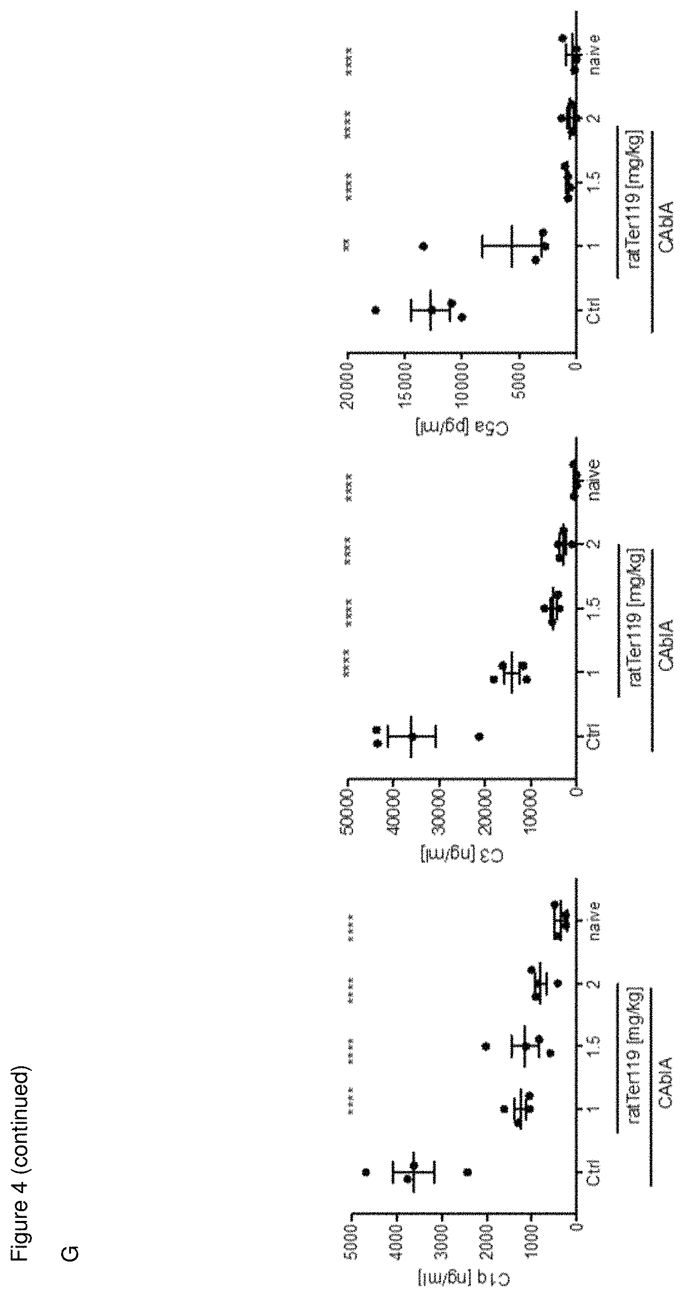
[0035] (G) All doses of TER119 antibody reduce C1q, C3, C5a levels in the joint of arthritic mice. Complement components C1q (A), C3 (B), and C5a (C) assessed from the joint fluid by ELISA. Data were analysed by a one-way ANOVA test with Holm-Sidak's multiple comparison to the control group. *P<0.05; **P<0.01; ***P<0.001; ****P<0.0001.
[0036] (H) Mice with established CAbIA were treated on day 6 with TER-119, isotype control mAb, deglycosylated TER119 or M1/69. Clinical scores and paw width were assessed. The statistical comparisons were calculated using Two-way ANOVA with Dunnett's multiple comparisons test (all groups against isotype control).
[0037] (I) Shows binding of the antibodies (0-512 ng of the primary antibody) with erythrocytes 25 from C57BL/6 mice, as assessed by flow cytometry
[0038] FIG. 5. Dose dependent phagocytic index of TER-119 opsonized RBCs. Erythrocytes were acquired from C57B/6 mice and non-opsonized (control) or opsonized with various concentrations of TER-119 then incubated with RAW264.7 macrophages for 30 minutes. Phagocytic index was calculated by counting the total number of ingested RBCs and diving this by the total number of macrophages in a field and multiplying by 100 (n=5 per group). **P<0.01, ***P<0.001.
[0039] FIG. 6. Phagocytic index of platelets incubated with TER-119 opsonized erythrocytes. RAW 264.7 cells were cultured overnight, then platelets labelled with CMFDA and opsonized with Mwreg30 were either added to RAW cells with or without TER-119 opsonized RBCs for 30 minutes at 37.degree. C. Platelet phagocytic index was calculated. ***P<0.05. (n=5 per group).
[0040] FIG. 7. Anti-erythrocyte antibody coated RBCs' ability to inhibit platelet phagocytosis. Erythrocytes were either non-opsonized or opsonized with antibody TER-119, deglycosylated TER-119, 34-3C (5 or 40 ug) and M1/69 for 1 hour then incubated with RAW 264.7 cells and MWReg30 opsonized CFMDA labelled platelets for 30 minutes. Cells were visualized by confocal microscopy and internalized platelets were counted by Imaris software version 8.0.2. (P<0.05). (n=4-6 per group).
[0041] FIG. 8. 6 TER-119 expressed as murine IgG switch variants can treat a chronic model of collagen-induced arthritis (CIA) independent upon passive-antibody transfer. DBA-1 mice immunized against type II collagen were allowed to develop arthritis and then treated (timing as denoted by the arrow) with PBS (filled circles, n=7 mice), 2 mg/kg of TER-119 expressed as a murine IgG1subtype (squares, n=6 mice) or expressed as a murine IgG2a subtype (triangles, n=6 mice) and arthritis clinical score evaluated over the course of the experiment.
[0042] FIG. 9: Therapeutic effect of 34-3C (anti-Band 3 antibody) on collagen Ab-induced arthritis (CAbIA).
[0043] (A) Mice with established CAbIA were treated on day 5 with a single i.v. injection of 2 mg/kg anti-Band 3 mAb (clone 34-3C, mouse IgG2a) or PBS. Clinical scores were assessed according to Campbell I K et al J Immunol. 2014;192:5031-5038). Data are means.+-.SEM (n=4/5).
[0044] (B) Average of clinical scores of mice between day 6 and 12 of the experiment. Dots represent individual mice; bars show the means.+-.SEM. Data were analysed by Mann-Whitney test (2-tailed). *P<0.05.
DETAILED DESCRIPTION OF THE INVENTION
[0045] The present invention relates to the use of an antibody to a RBC in the treatment of inflammatory conditions, and is based on the inventors' surprising observation that an antibody to the RBC TER-119 antigen has an effect on the inflammatory condition immune thrombocytopenia (ITP) that occurs before the haemolytic effect of this antibody. It had previously been considered that the effect of this antibody and other RBC depleting antibodies on ITP occurred as a result of opsonised RBC clearance by the mononuclear phagocytic system (MPS), which competitively inhibits the depletion of platelets by the same pathway. However, this difference in timing between the effect on RBCs and the amelioration of ITP, as assessed by platelet enumeration, is supportive of the conclusion that the anti-RBC antibody has a broad anti-inflammatory activity, and hence that there is a utility for such antibodies that extends beyond ITP therapy to other diseases involving inflammation.
[0046] This existence of this broad anti-inflammatory activity is supported by the anti-RBC antibody's ability to ameliorate three separate inflammatory diseases that do not involve classical MPS function. Firstly, the anti-RBC antibody was able to prevent the induction of rheumatoid arthritis in the well-known and well-characterised K/B.times.N mouse model of rheumatoid arthritis, in which induction of arthritis occurs after serum transfer from a K/B.times.N mouse. This was shown by treating mice prophylactically with the anti-RBC antibody, prior to induction of the disease with K/B.times.N serum. There was a clear reduction in clinical arthritic score and in ankle width, two standard parameters for assessing RA in this mouse model (Mott P J, Lazarus A H (2013) PLoS ONE 8(6):e65805) in mice that had been prophylactically treated with the anti-RBC antibody, when compared to those who had not. In addition to this, the anti-RBC antibody was also able to ameliorate established arthritic disease, again based on the parameters of clinical arthritic score and ankle width. Treatment with the anti-RBC antibody 5 days after induction of disease with K/B.times.N serum reversed clinical scores and ankle widths to normal levels 3 days later.
[0047] The anti-RBC antibody was also capable of ameliorating inflammatory arthritis in the well-known and well characterised Collagen antibody induced arthritis (CAbIA) model in mice (the most commonly studied autoimmune model of rheumatoid arthritis) (Campbell IK et al J Immunol. 2014;192:5031-5038. Campbell I K et al J Immunol. 2016;197:4392-4402). Disease development in the CAbIA model is dependent on both Fc.gamma.R engagement and activation of the complement system (Kagari T D et al J Immunol. 203;170(8):4318-4324. Nandakumar K S et al Arthritis Res Ther. 2006;8(6):223). Induction of arthritis occurs after injection of an anti-collagen mAb cocktail and injection with LPS. This was shown by treating mice with the anti- RBC antibody, after induction of the disease. There was a clear difference between the treatment groups; the treated mice were completely protected from arthritis within 24 h of injection, and a reduction in histological score was observed in mice that had been treated with the anti-RBC antibody, when compared to those who had not.
[0048] In a further mouse model for an inflammatory disease, the anti-RBC antibody was able to prevent the induction of hypothermia that is observed following infusion of an MHC Class I antibody (34-1-2S) into SCID mice, as well as ameliorating pulmonary edema. This is a mouse model for human transfusion related acute lung injury (TRALI), which is one of the most serious complications of blood transfusion. The ability of the anti-RBC antibody to prevent systemic shock, as determined by the prevention of induction of hypothermia, and to ameliorate pulmonary edema in this inflammatory disease with symptoms disparate from those in ITP and arthritis provides additional support for a broad anti-inflammatory effect of the anti-RBC antibody.
[0049] Although IVIG has been used to treat ITP for over 30 years, and polyclonal anti-D can reverse thrombocytopenia in patients with ITP who express the D antigen (e.g. being sold as Rhophylac.RTM. for this treatment), the broad anti-inflammatory effect of anti-RBC antibodies has not previously been recognised. Work has been carried out to identify monoclonal antibodies to RBC that can be used in the treatment of ITP, and certain anti-RBC monoclonal antibodies such as the anti-TER-119 referred to above and an additional anti-CD24 antibody have been shown to be effective in mouse models (Song S et al Blood. 2003;101(9):3708-3713), however, a small study in which a monoclonal anti-D antibody was tested in humans with ITP was not successful (Godeau, B. et al (1996) Transfusion;36(4):328-330).
[0050] Much of the previous work on antibodies for the treatment of ITP thus focussed on the ability of such antibodies to opsonise RBC to prevent platelet destruction, specifically by providing competition for the MPS pathway. This new work by the inventors, however, opens up a new therapeutic area for antibodies to RBCs, in the treatment of inflammation more generally. The inventors have recognized that these insights offer novel opportunities for therapeutic intervention using an antibody which binds to a RBC, and which aims at reducing inflammation, increasing cure rates, prolongation of survival and/or progression-free survival in inflammatory disorders.
[0051] The invention thus provides an antibody to a RBC for use in a method of preventing or treating an inflammatory condition, as well as a method of preventing and treating an inflammatory condition in a subject, comprising administering to a subject in need thereof a therapeutically effective amount of an antibody to a RBC. It has been shown that a similar effect can be achieved with erythrocytes sensitized with anti-D antibodies in vitro and then introduced into the patient (Ambriz-Fernandez, R., et al. (2002) "Fc receptor blockade in patients with refractory chronic immune thrombocytopenic purpura with anti-D IgG" Arch Med Res 33(6): 536-540); therefore, the invention also provides administration of RBC sensitized with an antibody to a RBC for use in a method of preventing or treating an inflammatory condition, as well as a method of preventing and treating an inflammatory condition in a subject.
Inflammatory Conditions
[0052] The invention is concerned with the treatment and/or prevention of inflammatory conditions. By "inflammatory condition" it is meant any condition characterized by destructive inflammation which may be recurrent or chronic and is not associated with normal tissue repair. The inflammation may be chronic inflammation. In chronic inflammatory conditions, neutrophils and other leukocytes are constitutively recruited by cytokines and chemokines, leading to tissue damage.
[0053] Examples of inflammatory conditions are autoimmune conditions, i.e. diseases in which the immune system attacks the body's own tissues. Such diseases particularly include "autoinflammatory diseases" in which the body's immune system causes inflammation. Such conditions may be antibody mediated, and/or T-cell mediated, and/or mediated by the body's innate immune system. In one embodiment, antibodies of the present invention are used to treat an auto-antibody mediated autoimmune condition.
[0054] Inflammatory conditions may also be complement mediated (e.g. complement mediated inflammation in reperfusion injury, or spinal cord injury).
[0055] The inflammatory condition may be an autoimmune condition in which elevated IL-10 is present, such as a condition selected from arthritis, in particular rheumatoid arthritis, Kawasaki's disease, type I diabetes, multiple sclerosis, systemic lupus erythematosus (SLE).
[0056] Alternatively, the inflammatory condition may be an autoimmune condition in which elevated IL-10 is not present, e.g. in which there are normal levels of IL-10, or in which IL-10 is reduced. ITP patients and autoimmune thyroiditis patients have lower levels of IL-10 than controls.
[0057] The disease can be, for example, inflammation associated with changes in temperature, autoimmune cytopenia (e.g. autoimmune haemolytic anaemia (AIHA), autoimmune neutropenia (AIN), autoimmune thrombocytopenia (ITP)), primary antiphospholipid syndrome, arthritis (e.g. rheumatoid arthritis, juvenile arthritis), bowel disease (e.g. ulcerative colitis, Crohn's disease, coeliac disease), Kawasaki disease, SLE, immune thrombocytopenia purpura, ischemia/reperfusion injury, Type I diabetes, inflammatory skin disorders (e.g. acne, psoriasis, lichen planus, pemphigus, pemphigoid), autoimmune thyroid disease (e.g. Graves' disease, Hashimoto' s thyroiditis), Sjogren's syndrome, pulmonary inflammation (e.g. asthma, chronic obstructive pulmonary disease (COPD), pulmonary sarcoidosis, lymphocytic alveolitis), transplant rejection, spinal cord injury, brain injury (e.g., stroke, traumatic brain injury), neurodegenerative diseases (e.g. Alzheimer's disease, Parkinson's disease, Lewy body disease), other neurological conditions (progressive multifocal leukoencephalopathy, ALS, chronic inflammatory demyelinating polyneuropathy (CIDP), inflammatory neuropathy, Guillain-Barre syndrome (GBS), motor neuron diseases (MND), multiple sclerosis, myasthenia gravis, neuromyelitis optica (NMO), other autoimmune channelopathies), gingivitis, gene therapy-induced inflammation, diseases of angiogenesis, inflammatory kidney disease (e.g., IgA nephropathy, membranoproliferative glomerulonephritis, rapidly progressive glomerulonephritis), Stevens-Johnson syndrome, autoimmune epilepsy, muscle inflammation (e.g. dermatomyositis and polymyositis), scleroderma, and atherosclerosis.
[0058] Of particular interest are lung injuries (such as acute lung injury, transfusion related acute lung injury (TRALI)), autoimmune cytopenia, idiopathic thrombocytopenic purpura/immune cytopenia (ITP), rheumatoid arthritis, systemic lupus erythematosus, asthma, Kawasaki disease, Guillain-Barre syndrome, Stevens-Johnson syndrome, Crohn's disease, colitis, diabetes (e.g. type 1 or type 2 diabetes), chronic inflammatory demyelinating polyneuropathy (CIDP), inflammatory neuropathy, neuromyelitis optica (NMO), other autoimmune channelopathies, autoimmune epilepsy, myasthenia gravis, dermatomyositis, polymyositis, scleroderma, vasculitis, uveitis, pemphigus, pemphigoid, spinal cord injury or Alzheimer's disease.
[0059] In some embodiments the inflammatory condition is a neurological condition, e.g. a neurological autoimmune disease. Examples of such conditions include chronic inflammatory demyelinating polyneuropathy (CIDP), myasthenia gravis (MG), multiple sclerosis (MS), neuromyelitis optica (NMO), or autoimmune epilepsy.
[0060] In some embodiments the inflammatory condition is selected from arthritis (e.g. rheumatoid arthritis) and TRALI.
[0061] In some embodiments the inflammatory condition is not ITP, or is not ITP or autoimmune thyroiditis. In other embodiments the inflammatory condition is not a disease where IL-10 is decreased, or is not a disease in which there are normal levels of IL-10.
[0062] IL-10 levels may be measured using standard immunoassay (e.g. ELISA) kits that are known in the art. The levels may be measured in any appropriate sample e.g. blood, serum, plasma, urine, cerebrospinal fluid and therefore where the levels of IL-10 are referred to herein it is the level in that sample that is relevant. Comparisons can be made with normal, e.g. healthy subjects.
Biological Readout/Effects of Treatment
[0063] Without being bound by any particular theory, the inventors believe that the use of an anti-RBC antibody according to the invention is useful to: (i) reduce inflammation in inflammatory conditions (ii) reduce and/or delay the clinical manifestation of the condition (which may be effects of inflammation in inflammatory conditions), (iii) extend survival of a subject with an inflammatory condition, (iv) enhance quality of life of a patient suffering from such a condition, (v) enhance the convenience of therapy for the patient, and/or (vi) enhance the efficacy of other drugs used to treat an inflammatory condition.
[0064] A method of treating an inflammatory condition is provided, said method comprising administering to the subject an effective amount of an antibody to a RBC. In some embodiments the method of the invention can be described as a method of reducing inflammation in an inflammatory condition, a method of reducing and/or delaying the clinical manifestation of the condition (e.g. the effects of inflammation in an inflammatory condition), a method of extending survival of a subject with an inflammatory condition, a method of enhancing quality of life of a patient suffering from such a condition, enhancing the convenience of therapy for the patient, and/or a method of enhancing the efficacy of one or more other drug used to treat an inflammatory condition, wherein in each case said method comprises administering to the subject in need thereof an effective amount of an antibody to a RBC.
[0065] The method of the invention may also be described as a method of treating or preventing one or more of the symptoms of an inflammatory condition, optionally treating one or more of the symptoms of an inflammatory condition, said method comprising administering to a subject in need thereof an effective amount of an antibody to a RBC.
[0066] Likewise an antibody to a RBC for use in these methods is provided, as is the use of antibody to a RBC in the manufacture of a medicament for carrying out such methods.
(i) Reduction of Inflammation in Inflammatory Conditions
[0067] The method of the invention can be described as a method of reducing inflammation in an inflammatory condition. In some embodiments, inflammation, and its effects in inflammatory conditions are assessed by standard clinical tests known in the art.
[0068] For example, disease markers are known for inflammatory conditions. The marker or markers that are used to assess the status of the disease may be a marker or a group of markers specific for the relevant disease (referred to her as a "disease marker") or may be a marker of inflammation (referred to here as an "inflammatory marker"). Examples of suitable samples for assessment include tissue, blood and urine.
[0069] Levels of one or more inflammatory markers can be assessed in a subject to provide information about the status of the inflammatory disease and about the effect of any treatment on the disease. A reduction in inflammatory markers is in general an indication of a reduction in inflammation. A biological sample can be taken from a subject at various time points (e.g. before treatment is commenced and at suitable time points after administration of the antibody of the invention) and the levels of one or more inflammatory markers can be assessed to determine the effect of the treatment on inflammation in the subject. Examples of inflammatory markers that are well known for such purposes include CRP, IL-6 and TNF-.alpha.. In one embodiment one or more inflammatory marker is reduced in the subject after administration of the antibody of the invention compared to the level of the marker before administration of the antibody of the invention. In another embodiment of the invention the method further comprises the step of determining the level of one or more inflammatory marker in the subject, and this may be before and/or after treatment.
[0070] Any reduction is preferably statistically significant. Reduction in one or more of the above markers may be by at least 5, 10, 15, 20, 25, 30, 35, 40, 45, 50% compared to the level before treatment.
(ii) Reduction and/or Delay to the Clinical Manifestation of the Inflammatory Condition (e.g. Effects of Inflammation in Inflammatory Conditions)
[0071] The method of the invention can be described as a method of reducing of the clinical manifestation of the inflammatory condition (e.g. effects of inflammation in inflammatory conditions). In some embodiments, levels of one or more disease markers can be assessed in a subject to provide information about the status of the disease and about the effect of any treatment on the disease. In many conditions the clinical manifestations of the disease arise as a result of inflammation and the associated tissue damage, but other mechanisms are also known.
[0072] Certain disease markers are known and are used by clinicians to diagnose and monitor inflammatory conditions. In general, a reduction in the level of a disease marker may be an indication of a reduction in severity of the disease (although in certain circumstances an increase in one or more disease markers may be an indication of a reduction in severity of the disease). A biological sample can be taken from a subject at various time points (e.g. before treatment is commenced and at suitable time points after administration of the antibody of the invention) and the levels of one or more disease markers can be assessed to determine the effect of the treatment on inflammation in the subject. Examples of disease markers that are known for such purposes are set out in Table 1 below. In one embodiment one or more disease marker is reduced (or increased) in the subject after administration of the antibody of the invention compared to the level of the marker before administration of the antibody of the invention. In certain embodiments the reduction (for example, inflammatory cytokines or chemokines) or increase (for example anti-inflammatory cytokines or anti-inflammatory chemokines) is associated with a decrease in severity of the disease. In another embodiment of the invention the method further comprises the step of determining the level of one or more disease marker in the subject, and this may be before and/or after treatment.
TABLE-US-00001 TABLE 1 Disease Blood disease markers Rheumatoid Rheumatoid Factor (RF) (auto-antibody directed against arthritis (RA) the Fc portion of immunoglobulin found in the blood of about 80 percent of people with rheumatoid arthritis). Anti-cyclic citrullinated peptide antibody (anti-CCP) Protein biomarker 14-3-3eta MG Anti-acetylcholine receptor antibody (anti-AChR Ab) Anti-muscle-specific tyrosine kinase antibody (anti- MuSK Ab) Systemic lupus Anti-nuclear antibody erythematosus anti-double stranded DNA (dsDNA) antibodies (SLE) anti-U1 RNP (U1 spliceosomal RNA) anti-histone antibodies
[0073] Any reduction or increase in such markers is preferably statistically significant. Reduction or increase in one or more of the above markers may be by at least 5, 10, 15, 20, 25, 30, 35, 40, 45, 50% e.g. compared to the level before treatment.
[0074] The effects of inflammation can also be assessed by standard clinical tests that are known in the art. Clinical tests may involve scoring which is carried out on the basis of the clinical manifestation of the disease or disorder that is to be treated. The treatment in one embodiment results in an improvement in the clinical score of the disease compared to the clinical score of the disease before administration of the antibody of the invention. This is analogous with the improvements seen in appropriate animal models, such as improvements in clinical score and a reduction in ankle size that is observed in the K/B.times.N mice in Examples 3 and 4 following treatment with the antibody of the invention, with the prevention of 34-1-2S induced hypothermia in Example 6, and with the improvements in clinical and histological score that is observed in the CAbIA mice in Example 5.
[0075] The improvement may be manifest in a reduction in the clinical manifestation or severity of the inflammatory condition, or a delay in the clinical manifestation of the inflammatory condition, so that the treatment affects the time course of the disease progression.
(iii) Extending Survival of a Subject with an Inflammatory Condition
[0076] The method of the invention can be described as a method of extending survival of a subject with an inflammatory condition. Many inflammatory conditions, and particularly autoimmune conditions have no cure and result in a subject having a decreased life expectancy when compared to subject who does not have such a condition. The treatment can therefore extend the survival of a subject with an inflammatory condition, e.g. by at least 1, 2, 5, 10 months or years.
(iv) Enhancing the Efficacy of Other Drugs Used to Treat an Inflammatory Condition
[0077] The method of the invention can be described as a method of enhancing the efficacy one or more other drug used to treat an inflammatory condition. Known methods of treating inflammatory conditions include three general approaches, immunosuppressive, anti-inflammatory, or palliative treatments. Examples of anti-inflammatories include anti-inflammatory pain reliever drugs (NSAIDs, e.g. aspirin, ibuprofen). Corticosteroids (such as prednisone and prednisolone), aminosalicylates, immunosuppressant drugs such as azathioprine, mercaptopurine, and methotrexate are also used. Biologic therapies with targets including cytokines, B cells, and co-stimulation molecules are now being used. Cytokine targets include tumor necrosis factor (TNF)-alpha (e.g. infliximab, adalimumab and golimumab), interleukin (IL)-1, anti-IL-6 molecules. B-cell depletion includes use of anti-CD20 antibodies (e.g. Rituximab) and B cell receptor (BCR) modulation by the B-lymphocyte stimulator (BLyS) (Belimumab).
[0078] The antibody of the invention can thus be used in combination with one or more other anti-inflammatory drug to enhance the efficacy of the other anti-inflammatory drug. Likewise, the other anti-inflammatory drug may enhance the efficacy of the antibody of the invention.
Red Blood Cell Antibody
[0079] A red blood cell (RBC) antibody binds to a RBC. The molecule to which the RBC antibody binds is referred to herein as a RBC molecule. This is therefore a RBC surface molecule, i.e. a molecule that is found on or associated with the outer surface of a RBC, so that the antibody to the RBC binds to an intact RBC. A list of proteins that have been identified in the erythrocyte membrane fraction is shown below; the RBC molecule suitable for use in the present invention may be selected from this list (Table 2).
TABLE-US-00002 TABLE 2 (from Kakhniashvili, DG et al Mol Cell Proteomics. 2004; 3(5): 501-509) Proteins identified in RBC membrane fractions Molecular Sequence No. of mass coverage identified No. Protein description (Da) GI Number (%) peptides 1 Spectrin .alpha. chain, erythrocyte 279,916.5 1174412 48.0 77* 2 Spectrin .beta. chain, erythrocyte 246,468.1 17476999 48.0 76* 3 Ankyrin 1, splice form 2 208,067.9 105397 45.0 55 4 Ankyrin 1, isoform 4, erythrocytic 209,416.6 10947036 45.0 50 5 Ankyrin 1, isoform 2, erythrocytic 189,011.2 10947042 46.0 48 6 Similar to ankyrin 1 206,254.8 13645508 51.0 48 7 Protein band 4.2, erythrocytic 70,946.5 107446 33.0 21 8 Protein band 4.1 (elliptocytosis 1, RH-linked) 66,398.5 4758274 45.0 17 9 Protein band 3, erythrocytic 101,792.3 4507021 28.0 17 10 Protein band 4.1, erythrocytic 97,016.9 14916044 32.0 16 11 Actin .beta. chain 41,812.8 461515 47.0 12* 12 Flotillin 1, erythrocytic 47,355.3 5031699 47.0 12 13 Membrane protein p55, erythrocytic, (palmitoylated) 52,296.5 4505237 35.0 11 14 Flotillin 2 47,142.3 13567629 29.0 11 15 Protein band 4.0 (dematin), erythrocytic 45,514.4 13623437 40.0 10 16 Protein band 7.2b, stomatin 32,598.8 1103842 47.0 10 17 Glyceraldehyde-3-phosphate dehydrogenase 38,054.2 31645 51.0 10 18 Tropomyosin 3, cytoskeletal 29,032.7 136096 55.0 10 19 Solute carrier family 2 (facilitated glucose 54,117.8 5730051 13.0 6 transporter), member 1 20 Similar to flotillin 2 42,565.0 13277550 15.0 6 21 Tropomyosin isoform 28,420.1 1062876 36.0 6 22 Glucose transporter glycoprotein 37,879.6 3387905 17.0 5 23 Tropomyosin .alpha. chain (smooth muscle) 26,576.7 136100 37.0 5 24 Actin .alpha. 2, aortic smooth muscle 42,108.1 1070613 20.0 5 25 Adducin .alpha. subunit, erythrocyte 80,955.1 12644231 10.0 5* 26 Rabphilin-3 A-integrating protein 80,858.2 1082757 8.0 5* 27 C-1-tetrahydrofolate synthase, cytoplasmic 101,559.2 115206 6.0 4 28 Translation initiation factor 2C, 2 66,252.2 18570004 10.0 4 29 Aldolase A 39,288.8 229674 17.0 4 30 Tropomodulin 40,569.2 4507553 16.0 3* 31 RAP2B, member of RAS oncogene family 20,504.4 11433346 43.0 3 32 Arginase type 1 erythroid variant 35,664.1 18535612 12.0 3 33 Arginase type 1 34,734.9 10947139 12.0 3 34 Creatine kinase, muscle 43,101.1 14763181 21.0 3 35 B-CAM protein 63,566.7 2134796 8.0 3 36 ATP-binding cassette half-transporter 99,712.3 11245444 5.0 2 37 RAP1A, member of RAS oncogene family or 20,997.1 4506413 14.1 2 RAP1B 20,824.7 7661678 14.1 2 38 Calcium tansporting ATPase 4 137,920.2 14286105 2.5 2 39 Rh blood D group antigen polypeptide 45,136.5 10800054 4.0 2 40 Channel-like integral membrane protein 16,239.5 1314306 15.0 2 41 Glycophorin A precursor 16,429.6 1070638 21.0 2 42 Solute carrier family 29 (nucleoside transporter), 50,219.4 4826716 3.5 2 member 1 43 Glycophorin A 14,784.8 106140 23.0 2 44 Glutathione transferase 27,053.4 809436 19.0 2 45 Glyphorin C, Isoform 1 13,810.6 4504229 20.0 1 46 Aquaporin 1 28,526.0 4502177 7.0 1 47 Erythroblast membrane-associated protein 52,604.8 17480129 3.0 1 48 Similar to glycophorin A 16,371.6 13529077 20.0 1 49 Cell surface glycoprotein CD44 39,433.8 7512338 4.0 1 50 Vesicle-associated membrane protein 2 (synaptobrevin 12,648.7 7657675 15.0 1 2) 51 Similar to adhesive plaque matrix protein precursor 106,879.1 17481669 1.0 1 52 Poly (A)-specific ribonuclease 73,451.0 4505611 3.0 1 53 Similar to RAS-related protein RAL-A 23,566.8 14740792 7.0 1 54 Presenilin-associated protein 39,862.4 6409316 6.0 1 55 Duodenal cytochrome b 31,611.2 13376257 3.5 1 56 bA421H8.2 (novel protein) 16,743.7 17402228 9.0 1 57 Similar to RAS-related protein RAB-15 23,517.9 18596861 5.0 1 58 CD59antigenp18-20 17,067.4 17473237 5.0 1 59 Rhesus D category VI type III protein 45,247.7 2765839 1.9 1 60 RAB 35, RAS oncogene family 23,025.2 5803135 6.0 1 61 Ral A binding protein 75,063.4 5803145 2.1 1 62 Hypothetical protein XP_100510 8,049.3 18577723 16.0 1 63 ATP-binding cassette, subfamily C, member 6 164,904.4 6715561 0.9 1 64 Phosphoribosyl pyrophosphate synthetase 34,834.2 4506127 7.0 1 65 Unknown protein 46,884.2 18089137 3.6 1 66 Similar to Lutheran blood group 59,287.7 18589892 3.1 1 67 Phosphatidylinositol-4-phosphate 5 kinase, type III 46,078.6 1730569 4.4 1 68 Hypothetical protein XP_100685 35,877.6 18604339 7.0 1 69 Hypothetical protein XP_100619 18,587.6 18604359 15.0 1 70 Block of proliferation 1 83,629.5 23830903 1.5 1 71 Similar to tropomyosin 10,804.3 18590249 13.0 1 72 Hypothetical protein XP_061743 or 48,719.0 17472555 2.5 1 XP_089854 31,487.0 18577194 4.0 1 73 Hypothetical protein XP_106269 12,703.8 18558481 22.0 1 74 Hypothetical protein XP_100925 22,863.4 18601384 8.0 1 75 Zone pellucida binding protein 40,169.8 5002116 3.4 1 76 2',3'-cyclic-nucleotide 3'-phosphodiesterase 4,242.9 7435185 60.0 1 77 Lyn B protein 58,033.3 2117805 4.3 1 78 KIAA0340 117,819.0 2224621 2.1 1 79 Hypothetical protein XP_091724 144,900.8 18588504 1.1 1 80 Hypothetical protein XP_091430 27,641.2 18588504 5.0 1* 81 Similar to tropomyosin 4 18,426.8 14729747 6.0 1* 82 HGTD-P 17,342.4 9295192 10.0 1* 83 Hypothetical protein XP_095919 291,206.1 18572484 0.6 1* 84 Far upstream element binding protein 87,534.4 1082624 2.5 1* 85 Hypothetical protein XP_103707 13,374.6 16551105 12.0 1* 86 Hypothetical protein XP_092517 41,409.4 18552304 2.6 1* 87 Enhancer protein 41,289.8 1345400 4.6 1* 88 Hypothetical protein 15,770.3 18551738 12.0 1* 89 KIAA1741 protein 123,305.7 12698027 1.7 1* 90 Ig heavy chain V-V region 10,995.4 87863 16.0 1* 91 DC 38 31,691.4 12005984 4.7 1* *The proteins found primarily in the low-ionic-strength spectrin extract from RBC membranes.
[0080] The RBC molecule may be attached directly or indirectly to the RBC membrane. Direct attachment of the molecule to the RBC membrane may occur as a result of the molecule being a transmembrane protein or a transmembrane glycoprotein, or directly attached to a lipid in the membrane. Indirect attachment of the molecule to the RBC may occur as a result of the molecule being bound to or associated with a molecule that is itself directly attached to the membrane (e.g. a membrane protein or glycoprotein or a protein or carbohydrate that is attached to one or more lipids in the membrane).
[0081] The RBC antibody thus binds to a RBC molecule, i.e. a RBC surface molecule, which may be a protein (e.g. a glycoprotein) or a carbohydrate, but is typically a protein (e.g. a glycoprotein). In some instances, the RBC molecule is not glycosylated.
[0082] The RBC surface molecule can also be described as a RBC antigen, however the RBC antibody need not distinguish between different isoforms of a RBC molecule such as the different isoforms of a RBC molecule that give rise to different blood groups. In other words, the RBC antibody may in some embodiments bind to more than one isoform (e.g. 2 or more, 3 or more, 4 or more isoforms) of the RBC molecule, e.g. wherein the RBC molecule has multiple isoforms that are associated with different blood groups. In such a case the antibody does not distinguish between different blood groups that arise as a result of polymorphisms in the RBC molecule. Alternatively, the RBC antibody may bind only to one isoform of the RBC molecule, so that it can distinguish between different blood groups that arise as a result of polymorphisms in the RBC molecule.
[0083] Certain RBC molecules may take different forms in different individuals, and these differences may be associated with different blood groups. For example, a protein or glycoprotein molecule may have multiple possible isoforms, wherein the different isoforms are associated with different blood groups. An example of a blood group that is based on different protein antigens is the Rhesus system. The Rhesus D protein is either present or absent, so that a given individual is RhD positive or negative, but the associated Rhesus CE protein may be present in several forms that arise as a result of amino acid polymorphisms at only five amino acid locations. The different forms of the Rhesus CE protein are associated with different Rhesus blood types and may be referred to as being different antigens. Thus, in the case of RBC molecules such as the Rhesus CE protein, where different isoforms of the protein exist, the RBC antibody may bind to all isoforms of the protein or may bind only to certain isoforms.
[0084] Likewise, different carbohydrate based blood group antigens exist. The "ABO" antigens are carbohydrate chains that are attached to numerous different proteins and lipids that lie in the RBC membrane. The ABO locus has three main allelic forms: A, B, and O. The A and B alleles each encode a glycosyltransferase that catalyses the final step in the synthesis of the A and B antigen, respectively. The A/B polymorphism arises from several SNPs in the ABO gene, which result in A and B transferases that differ by four amino acids. The O allele encodes an inactive glycosyltransferase that leaves the ABO antigen precursor (the H antigen) unmodified, whereas the A and the B antigens differ in the carbohydrate structure. The ABO antigens may be present on multiple RBC molecules. The different forms of the carbohydrate are associated with different blood types and may be referred to as being different antigens. Thus, in the case of RBC molecules that contain the ABO antigen, where different carbohydrate structures are associated with different blood types, the RBC antibody may bind only to certain carbohydrate structures or may bind to all forms of the RBC molecule (e.g. by binding to the protein portion of the RBC molecule).
[0085] In some embodiments the RBC molecule is not a molecule, the presence or absence of which or the presence of different isoforms of which gives rise to a blood type (e.g. the RBC antibody does not bind to the A or B antigen). In other embodiments the RBC molecule is a molecule, the presence or absence of which or the presence of different isoforms of which gives rise to a blood type. In such cases, the epitope to which the RBC antibody binds is typically not affected by the isoforms that give rise to the blood type, i.e. the antibody binds irrespective of the blood type.
[0086] The portion of the molecule to which the antibody binds is the epitope. Where the molecule is a glycoprotein the epitope may be on the carbohydrate portion or the protein portion of the glycoprotein, but is preferably on the protein portion, i.e. is a peptide epitope. The epitope to which the antibody binds may be a carbohydrate or a peptide epitope but is preferably a peptide epitope and is preferably not a carbohydrate epitope. A peptide epitope may be a linear or a conformational epitope.
[0087] A RBC molecule may be a protein or glycoprotein that is involved in transport. A RBC molecule that is involved in transport may e.g. be the Band 3 anion transporter (which has different isoforms that define the Diego Blood Group), the Aquaporin 1 water transporter (which defines the Colton Blood Group), Aquaporin 3, Glut1, the Kidd antigen protein, Rhesus associated glycoprotein (RhAG, CD241), the Na.sup.+/K.sup.+-ATPase, Ca.sup.2+-ATPase, the Na.sup.+K.sup.+2Cl.sup.- cotransporter, the Na.sup.+-Cl.sup.- cotransporter, the Na--H exchanger, the K--Cl cotransporter, the Gardos Channel. RBC transport proteins that are glycoproteins include but are not limited to: the Band 3 anion transporter, Aquaporin 1, Aquaporin 3, Glut1, Kidd antigen protein, RhAG (CD241), Na.sup.+/K.sup.+-ATPase, Na--H exchanger.
[0088] A RBC molecule may be a molecule involved in cell adhesion, e.g. ICAM-4 or BCAM (CD239). ICAM-4 and BCAM are both glycoproteins.
[0089] A RBC molecule may be a molecule that is believed to have a structural role in the RBC. A RBC molecule that has a structural role may establish linkages with skeletal proteins and may play an important role in regulating cohesion between the lipid bilayer and membrane skeleton, likely enabling the red blood cell to maintain its favourable membrane surface area by preventing the membrane from collapsing (vesiculating). Such molecules may be useful in accordance with the invention if they are on the surface of the erythrocyte. Cell surface molecules that have a structural role include Band 3 (this assembles various glycolytic enzymes, the presumptive CO.sub.2 transporter, and carbonic anhydrase into a macromolecular complex termed a "metabolon", which may play a key role in regulating red cell metabolism and ion and gas transport function), RhAG (CD241), proteins that are members of the rht protein 4.11R based macromolecular complex (e.g. Glycophorin C (CD236) and D (which define the Gerbich Blood Group), 10 XK, RhD (CD240D)/RhCE (CD240E), Duffy protein (CD234)), and other glycophorins such as Glycophorin A (CD235a) and B (CD235b).
[0090] RBC structural proteins that are glycoproteins include, but are not limited to: Band 3, RhAG, Glycophorins A to D, XK, RhD/RhCE, Duffy protein.
[0091] Other RBC molecules include CR1, CD99, CD147, ERMAP, CD238, CD20, CD151, DAF (CD55), AChE, Dombrock (CD297, ART4), CD108 (JMH), Emm and the human orthologue to the mouse TER-119 antigen (Ly76, glycophorin A-associated protein).
[0092] The RBC molecule may be a protein, which may be a glycoprotein, or it may be a carbohydrate, but is preferably a protein (e.g. a glycoprotein). The epitope to which the antibody binds may be a carbohydrate or a peptide epitope but is preferably a peptide epitope, and is preferably not a carbohydrate epitope.
[0093] The RBC molecule may be defined on the basis of its structure, i.e. as being a type I single pass protein, a type II single pass protein, a type III single pass protein, a multipass protein, a GPI linked protein or combinations thereof.
[0094] Examples of type I single pass RBC molecules include glycophorin A (CD235a), glycophorin B (CD235b), glycophorin C (CD236), glycophorin D, CR1, BCAM (CD239), ICAM-4 (CD242), CD99, CD147 and ERMAP.
[0095] Examples of type II single pass proteins include CD238, XK, Band3, Aquaporin 1, Kidd, Aquaporin 3, CD151.
[0096] Examples of RBC GPI linked proteins are DAF (CD55), AChE, Dombrock (CD297, ART4), CD108 (JMH), Emm.
[0097] Examples of carbohydrate antigens, which may be attached to RBC proteins and/or lipids include P1, Pk, P, ABO s, Hh, Lewis or I antigen.
RhD Antigen
[0098] A preferred RBC molecule is the RhD molecule (e.g. the human RhD molecule). This is a protein that is found in around 85% of Caucasians in Europe, and which is involved in the "Rhesus blood group system". The frequency of the Rhesus factor may be higher in other populations.
[0099] The Rhesus D molecule is highly immunogenic, eliciting anti-Rhesus D antibodies during Rhesus incompatible pregnancies and following transfusion of Rhesus incompatible blood. Modelling studies suggest that the Rhesus D molecule has 12 transmembrane domains with only very short connecting regions extending outside the cell membrane or protruding into the cytoplasm. Those individuals who express the Rhesus D molecule are said to be Rhesus positive. Individuals lacking the D molecule are called Rhesus negative. The other gene involved in the Rhesus system is the RHCE gene, which encodes the RhCE protein which contains C, E, c and e antigens and variants.
[0100] It is known that there are multiple epitopes on the D molecule, which explains the "partial D phenotypes", people who carried D antigen on their red cells but who had an alloanti-D in their sera. With at least 9 different epitopes (epD1 to epD9), it is possible for some D variant people to lack certain epitopes so that antibodies are made to the missing D epitopes. Rhesus positive individuals that make antibodies against partial D antigens have been classified into six main different categories (D'' to DVI I) each having a different abnormality in the D antigen. It has been shown that these D categories gave different patterns of reaction when tested against panels of human monoclonal anti-D antibodies (Tippett, P, et al Vox Sanguinis. 70(3):123;1996). The different reaction patterns identified the 9 epitopes and so define the different partial D categories. The number of epitopes present on the D antigen varies from one partial D category to another with the DVI category expressing the least, epD3, 4 and 9.
[0101] In one embodiment, the RBC molecule is a Rhesus D molecule. In a further embodiment the RBC molecule is a Rhesus D molecule having at least 3 of the 9 epitopes epD1 to epD9, e.g. at least 4, 5, 6, 7, 8 or all 9 of the epD1 to epD9 epitopes. In one embodiment the RBC molecule is a Rhesus D molecule having the sequence of UniProt entry Q02161.
[0102] Another Rh antigen is RhCE (UniProt entry P18577) with the C, E, c and e antigens (and variants).
Human Orthologue to the Mouse TER-119 Antigen (Ly-76)
[0103] In a preferred embodiment the RBC molecule is the human orthologue of the TER-119 antigen (Ly76). Antibodies to the TER-119 antigen have been used and found to be effective in the Examples, as set out below, in the treatment of three inflammatory conditions. A rat monoclonal antibody to TER-119 has been used in mouse models of ITP (Song S. et. al Blood. 2003;101(9):3708-3713) and has been shown alleviate ITP. The TER-119 antigen is a 52 kD glycophorin A-associated protein, also known as Ly76. It is a molecule associated with cell-surface glycophorin A.
Glycophorin A (GPA, CD135a) and B (GPB, CD235b) and Glycophorin C and D
[0104] In one embodiment the RBC molecule is glycophorin A (GPA). Glycophorin A and B are major sialoglycoproteins of the human erythrocyte membrane which bear the antigenic determinants for the MN and Ss blood groups. About 40 variant phenotypes have been identified, the UniProt entries are P02730 (GPA) and P06028 (GPB).
Band 3 (CD233)
[0105] In one embodiment the RBC molecule is Band 3 anion transport protein. Band 3 anion transport protein, also known as anion exchanger 1 (AE1) or band 3 or solute carrier family 4 member 1 (SLC4A1), is a protein that is encoded by the SLC4A1 gene in humans; the UniProt entry is P02730. It is a multi-pass membrane protein. CD233 is a phylogenetically preserved transport protein responsible for mediating the electroneutral anion exchange of chloride for bicarbonate across a plasma membrane. It is the major integral membrane glycoprotein of the erythrocyte membrane and is required for the normal flexibility and stability of the erythrocyte membrane as well as for the normal erythrocyte shape via the interactions of its cytoplasmic domain with cytoskeletal proteins, glycolytic enzymes, and haemoglobin.
Frequency of the RBC Molecule in a Population
[0106] Not all RBC molecules are found in all individuals. Indeed, it is well known that the differences between the molecules found on RBCs in different individuals are responsible for an individual's blood group. By way of example, in the ABO blood group system an individual in group A has the A antigen present on his or her RBCs and antibodies in their blood to the B antigen. An individual in group B has the B antigen present on his or her RBCs and antibodies in their blood to the A antigen. An individual in group AB has the A and the B antigen present on his or her RBCs and no antibodies to the A or B antigen in their blood. An individual in group O has the O antigen (H antigen) thus no A or B antigen present on his or her RBCs, but antibodies to both A and B antigens in their blood. From this it can be seen that using an anti-A antibody (i.e. an antibody that binds to the A carbohydrate antigen) in the method of the invention will only be effective in those patients who are in group A or group AB, and using an anti-B antibody (i.e. an antibody that binds to the B carbohydrate antigen) in the method of the invention will only be effective in those patients who are in group B or group AB. There are thus advantages associated with using an antibody to a RBC molecule that is found at high levels in all subjects, or in a given population of subjects, e.g. a given population of humans.
[0107] The molecule, or epitope may thus be found on at least 50%, 55%, 60%, 65%, 70%, 75%, 80%, 85%, 90%, 95%, 98%, 99% or 99.5% of humans, or on at least 50%, 55%, 60%, 65%, 70%, 75%, 80%, 85%, 90%, 95%, 98%, 99% or 99.5% of a human population of interest.
[0108] By way of example the RhD molecule is found in approximately 80% of humans, but may vary dependent on the population.
Molecule Density
[0109] The RBC molecule or epitope is preferably found at a density of 10.sup.2-10.sup.6 copies per cell, e.g. 10.sup.2-10.sup.5, 10.sup.2-10.sup.4, 10.sup.2-10.sup.3, 10.sup.3-10.sup.4, 10.sup.3-10.sup.5, 10.sup.4-10.sup.5 copies per cell. It may be advantageous to select a molecule with a suitable density on the RBC so that excessive haemolysis, and the adverse effects that this would have on the subject, e.g. causing anemia, can be avoided. For example, the A and B blood group antigens have a very high density on RBCs (in the region of 10.sup.6 copies per cell), whereas the RhD molecule is found at around 10.sup.3-10.sup.4 copies and the TER-119 antigen is around 10.sup.5 copies, and the density of the molecule or epitope is thus preferably 10.sup.2-10.sup.5, 10.sup.2-10.sup.4, 10.sup.2-10.sup.3, 10.sup.3-10.sup.4, 10.sup.3-10.sup.5, 10.sup.4-10.sup.5 copies per RBC.
[0110] In some cases the molecule is preferably the RhD molecule or GPA or the human orthologue to TER-119 antigen (GPA associated protein, Ly-76) or Band 3.
[0111] In certain other cases the antigen is preferably not the RhD molecule, the human orthologue to TER-119 antigen or the TER-119 antigen (Ly-76) or CD24, or is preferably not the RhD molecule or the TER-119 antigen (Ly-76) or the human homologue to the TER-119 antigen. Alternatively, the antigen is preferably not the RhD molecule, the TER-119 antigen (Ly-76), the human homologue to the TER-119 antigen, CD24 or the RhCE molecule, or is preferably not the RhD molecule or the TER-119 antigen (Ly-76) or the human homologue to the TER-119 antigen or the RhCE molecule.
[0112] The epitope is also in some embodiments not a carbohydrate epitope. It is in some embodiments not an ABO epitope, or not a P1, Pk, P, ABO s, Hh, Lewis or I epitope.
Distribution of the RBC Molecule Within the Body
[0113] The RBC molecule is preferably expressed selectively on RBCs, which can be advantageous as it means that the antibody will bind preferably to RBCs, so that off target effects may be avoided. For example, the molecule may be found at a higher density (expressed as copies of the molecule per cell) on RBCs than on one or more other cells, e.g. at a density that is at least 2, 3, 4, 5, 10, 20 or 50 fold higher than on one or more other cell. These other cells may be blood cells (e.g. white blood cells (lymphocytes, monocytes, and granulocytes) or platelets). These other cells may also be cells that are associated with the vascular system (e.g. endothelial cells or fibroblasts). It is preferred that the molecule is not expressed on white blood cells, platelets and/or cells that are associated with the vascular system, e.g. not expressed on one or more of white blood cells, platelets and cells that are associated with the vascular system. In certain embodiments the molecule is expressed at a density that is at least 2, 3, 4, 5, 10, 20 or 50 fold higher than on any other cell type, e.g. at least 2, 3, 4, 5, 10, 20, or 50 fold higher than on one or more of the cell types referred to above.
[0114] As a consequence, the antibody binds preferably to RBCs. The antibody thus binds preferably to RBCs compared to one or more other cell such as a blood cell (e.g. white blood cells (lymphocytes, monocytes, and granulocytes) or platelets) and/or to cells that are associated with the vascular system (e.g. endothelial cells or fibroblasts). It is preferred that the antibody does not bind to white blood cells, platelets and/or cells that are associated with the vascular system. In certain embodiments the antibody does not bind to any other cell type, e.g. does not bind to one or more of white blood cells, platelets and cells that are associated with the vascular system. Detection of antibody binding can be carried out using standard procedures known in the art (e.g. immunoassays detecting antibody binding to cells, such as by incubating antibody with cells and detecting bound antibody using an appropriately labelled second antibody, e.g. with flow cytometry).
[0115] Alternatively, or additionally the molecule may be expressed on RBCs and on other cell types, however in such cases these other cell types are found at a lower frequency in the body or in a region of the body which the antibody cannot access. This can be advantageous as it means that the antibody will bind preferably to RBCs, as it is statistically more likely to encounter such cells, so that off target effects may be avoided. For example, the molecule may be found on cells that are found at a lower frequency in the body or the vascular system than RBCs (e.g. there are at least 2, 3, 4, 5, 10, 20 or 50 fold fewer of these cells in the body or in the vascular system than RBCs). Additionally or alternatively these other cell types are found, for example, in the brain.
[0116] Expression of the molecule on different cell types can be assayed by standard in vitro methods known in the art (e.g. based on protein or encoding nucleic acid levels, such as immunoassays and PCR based methods), and the ability of an antibody to bind to different cell types can similarly be assayed using immunoassays in vitro. Enumeration of different cell types can also be assayed by standard methods known in the art.
Antibody
[0117] The antibody to be used is an antibody to a RBC molecule. In some embodiments it is specific for the RBC molecule. This means that the binding between the antibody and the RBC molecule is specific binding. As used herein, the term "specific binding" refers to a binding reaction between the antibody of the invention and the RBC molecule, in which the dissociation constant (KD) is 10.sup.-7M or less, in particular 10.sup.-8 M or less, 10.sup.-9 M or less or 10.sup.-10 M or less. The term "KD", as used herein, is intended to refer to the dissociation constant, which is obtained from the ratio of dissociation rate (Kd) to association rate (Ka) and is expressed as a molar concentration (M). KD values can be determined using methods well established in the art. One method for determining the association and disassociation dynamics of an antibody is by using surface plasmon resonance, e.g. by using a biosensor system such as a Biacore.TM. system.
[0118] In general, a smaller KD value is preferred. This corresponds to a higher affinity for the molecule.
[0119] The antibody of the invention typically binds to the RBC molecule with high affinity. As used herein, the term "high affinity" refers to an antibody that binds to the RBC molecule with a KD of 10.sup.-7M or less, 10.sup.-8 M or less, 10.sup.-9 M or less, or 10.sup.-10 M or less. However, "high affinity" binding can vary for different antibodies. For example, "high affinity" binding for an IgG antibody refers to a KD of 10.sup.-8 M or less, 10.sup.-9 M or less, or 10.sup.-19 M or less, whereas high affinity binding for an IgM antibody refers to an antibody having a KD of 10.sup.-7 M or less, or 10.sup.-8 M or less. In some embodiments, the antibody is a high affinity IgG antibody.
[0120] In some embodiments, the antibody for use in methods of the invention will bind to its RBC molecule with a KD value in the range of 10.sup.-7 M to 10.sup.-11 M, e.g. as determined by surface plasmon resonance (SPR) techniques (e.g. Biacore).
[0121] The affinity can also be calculated using other techniques (e.g. equilibrium binding assays). The affinity and concentration of the anti RBC antibody define how much binding to the RBCs is achieved. The binding can also be driven by the avidity of an antibody, especially when using a multivalent IgM antibody. "Avidity" refers to the accumulated strength of multiple affinities of non-covalent binding interactions.
[0122] Clone LD1/2-6-3 of the anti-RhD antibody in the IgG1 format (MDJ8s) shows an affinity to RBCs in the nanomolar range (KD=3 nM; with calculated 14'069 binding sites per cell) (Miescher S et al Br.J Haematol. 2000;111(1):157-166). TER-119 (rat IgG2b) shows an affinity of about 30 nM (calculated from FACS saturation experiments).
Functional Definitions of the Antibody
[0123] In some embodiments the antibody of the invention binds to RBCs in vitro and in vivo (e.g. to human RBCs). This can be assessed in vitro, e.g. by detecting binding of antibody to RBCs using immune based techniques. This can be carried out using standard procedures known in the art (e.g. detecting antibody binding to RBCs, such as by incubating antibody with RBCs and detecting bound antibody using an appropriately labelled second antibody, e.g. with flow cytometry, and for example as set out in Example 7). Antibody binding in vivo can also be detected, e.g. by administering antibody to a subject, and detecting antibody binding to RBCs in a sample from the subject, using an appropriately labelled second antibody, (e.g. with flow cytometry).
[0124] The antibody of the invention may additionally or alternatively cause MPS (also described as RES) blockade in a human or in a suitable animal (e.g. mouse) model in vivo. MPS blockade in mouse models can be assessed using assays that are known in the art, e.g. as described in (Song S. et. al Blood. 2003;101(9):3708-3713). Briefly, RBCs that have been taken from a suitable mouse model (e.g. SCID) are incubated with the antibody of the invention in vitro to cause opsonisation, and the opsonised RBC are labelled with a suitable marker and injected to a suitable mouse. Samples taken at time intervals after the injection are evaluated for RBC and labelled RBC number. A reduction in the number of circulating labelled RBCs over time following introduction is indicative of MPS blockade. The reduction may be e.g. to 30-80%, 40-75% or 50-65% of the circulating labelled RBCs compared to the number at the first time point assessed. MPS blockade in a human can be assessed by a surrogate assay to measure MPS function which is based on a phagocytosis assay. The clinically accepted assay has been known in the art as monocyte monolayer assay (MMA) (Tong T N & Branch D R J Vis Exp. 2017;119:55039. Tong T N et al Transfusion. 2016;56(11):2680-2690).
[0125] The antibody may additionally or alternatively cause haemolysis in vivo, e.g. in an animal model or in a human subject. This is measured e.g. by a reduction in RBC number after administration of the antibody. This can be determined by standard techniques such as obtaining a RBC count in a blood sample after antibody administration, or by measuring one or more markers of haemolysis in a blood sample (e.g. free haemoglobin). A reduction in the number of RBCs over time following introduction of the antibody is indicative of haemolysis in vivo.
[0126] Where a reduction in RBC numbers is evaluated in this method, there may be a reduction in RBC numbers to less than 99, 98, 97, 96, 95, 94, 93, 92, 91, 90, 80, 70, 60, 50% of the RBC numbers seen before administration of the antibody.
[0127] The antibody may additionally cause a reduction in platelet count or platelet concentration in vivo, e.g. in an animal model or in a human subject. This is measured e.g. by determining platelet number or concentration in a sample taken from the subject number after administration of the antibody. This can be determined by standard techniques.
[0128] The antibody may additionally or alternatively ameliorate murine ITP in a mouse ITP model, e.g. as described in Example 2. Amelioration of murine ITP in such a mouse model, as a result of administration of the antibody, is determined by e.g. comparing platelet numbers in a treated mouse compared to the level before treatment. An increase in platelet count of at least 1.25, 1.5, 1.75, 2, 2.5, 3 after 1.5 hours in treated mice, compared to the level before treatment may be indicative of amelioration of murine ITP in such a mouse model.
[0129] The antibody may additionally or alternatively ameliorate inflammatory arthritis in a mouse model of Rheumatoid arthritis, e.g. as described in Example 3. Pretreatment with the antibody of the invention 2 hours prior to injection with K/B.times.N serum may in some embodiments reduce arthritis scores and/or reduce ankle width in mice injected with K/B.times.N serum, compared to non-pretreated mice injected with K/B.times.N serum, as assessed in accordance with the standard procedure described in Mott et al (Mott P J et al PLoS One. 2013:8(6):e65805). The effects may be observed e.g. 7 days after treatment. Ankle width and/or clinical score is in some embodiments reduced by at least 5, 10, 15, 20, 30, 40, 50% compared to ankle width and/or clinical score without treatment. Clinical score may be reduced to 0 in some instances.
[0130] Likewise, the antibody may additionally or alternatively reverse established inflammatory arthritis in a mouse model of Rheumatoid arthritis, e.g. as described in Example 4. Administration of the antibody 5 days after injection of K/B.times.N serum may reduce clinical score and/or ankle width after treatment, e.g. 3 days after treatment. Ankle width and/or clinical score is in some embodiments reduced by at least 5, 10, 15, 20, 30, 40, 50% compared to ankle width and/or clinical score before treatment. Clinical score may be reduced to 0 in some instances.
[0131] The antibody may additionally or alternatively ameliorate inflammatory arthritis in the CAbIA model, e.g. as described in Example 5. Treatment with the antibody of the invention on day 5, after administration with anti-collagen mAb cocktail (day 0) and LPS (day 3) may in some embodiments protect from arthritis, measured e.g. by reduced clinical and histological arthritis scores compared to mice injected with collagen mAb cocktail (day 0) and LPS (day 3) but not treated with the antibody of the invention, as assessed in accordance with the procedure described in Example 5. The effects may be observed e.g. 1 day after treatment. Histological and/or clinical score is in some embodiments reduced to 0, or reduced by at least 50, 60, 70, 80% compared to histological and/or clinical score without treatment.
[0132] The antibody may additionally or alternatively prevent or reduce 34-1-2S induced hypothermia in a mouse model of TRALI. Injection of SCID mice with the antibody of the invention may reduce hypothermia induced by the injection 1 hour subsequently of the anti-MHC I antibody 34-1-2s (Fung Y L et al Blood. 2010;116(16):3073-3079), as assessed by rectal temperature measurement (e.g. as in Example 6). The rectal temperature measurement in mice treated with the antibody of the invention and the anti-MHC I antibody 34-1-2s may be at least 2, 3, 4, 5, 6, 7, 8, 9 or 10.degree. C. higher than the rectal temperature measurement in mice treated with the anti-MHC I antibody 34-1-2s alone 2 hours after treatment.
[0133] The antibody may additionally or alternatively reduce or prevent 34-1-2S induced pulmonary edema in a mouse model of TRALI. Injection of SCID mice with the antibody of the invention may reduce pulmonary edema induced by the injection 1 hour subsequently of the anti-MHC I antibody 34-1-2S, as assessed by post-mortem determination of wet/dry (W/D) lung weight ratios, after sacrificing mice 2 hours after treatment. Mice that receive 34-1-2S after pre-treatment with the antibody may display lung W/D ratios significantly lower than mice injected with 34-1-2S.
[0134] The antibody may additionally or alternatively inhibit phagocytosis of opsonised platelet in an in vitro assay. The ability of an antibody to inhibit phagocytosis of opsonised platelets in an in vitro assay can be assessed e.g. by comparing the amount of platelet phagocytosis in the presence of RBCs to the amount of platelet phagocytosis in the presence of RBCs which have been opsonised with the antibody of the invention, e.g. using the method of example 7. A reduction in the amount of platelet phagocytosis in the presence of RBCs which have been opsonised with the antibody of the invention compared to the amount of platelet phagocytosis in the presence of RBCs which have not been opsonised with the antibody of the invention may be expressed as a reduction in the platelet phagocytic index, e.g. of at least 20, 30, 40, 50, 60, 70, 80, 90, 100%.
[0135] To account for the fact that human and mouse RBC molecules may have differences in their primary sequence, and hence may have different binding properties to the antibody being tested, the above assays are, where possible, carried out using human RBCs (for in vitro assays). Where any mouse models are used, the mouse models may be modified (e.g. genetically manipulated) so that the appropriate human RBC molecule is expressed.
[0136] In some embodiments administration of the antibody does not lead to tolerance (e.g. immunological tolerance) of or to an antigen, e.g. an antigen involved in or which causes the autoimmune condition, such as tolerance of or to an antigen which may be a protein or peptide that is administered with the antibody. In some embodiments the antibody is not administered with another protein (e.g. a protein or peptide antigen).
Structural Antibody Definitions
[0137] The term "antibody" as used herein typically refers to both antibodies and antigen-binding fragments thereof. A naturally occurring "antibody" is a glycoprotein comprising at least two heavy (H) chains and two light (L) chains inter-connected by disulphide bonds. Each heavy chain is comprised of a heavy chain variable region (abbreviated herein as VH) and a heavy chain constant region. The heavy chain constant region is comprised of three domains, CH1, CH2 and CH3. Each light chain is comprised of a light chain variable region (abbreviated herein as VL) and a light chain constant region. The light chain constant region is comprised of one domain, CL. The VH and VL regions can be further subdivided into regions of hypervariability, termed complementarity determining regions (CDR), interspersed with regions that are more conserved, termed framework regions (FR). Each VH and VL is composed of three CDRs and four FRs. The variable regions of the heavy and light chains contain a binding domain that interacts with an antigen. The constant regions of the antibodies may mediate the binding of the immunoglobulin to host tissues or factors, including various cells of the immune system (e.g., effector cells) and the first component (C1q) of the classical complement system.
[0138] Preferably the antibody is a molecule that consists of the above specified regions/domains. The antibody may comprise only two antibody heavy chains and two antibody light chains, interconnected by disulphide bonds, e.g. wherein each antibody heavy chain consists of an antibody heavy chain variable region and three constant region domains (CH1, CH2, CH3) and each antibody light chain consists of an antibody light chain variable region and a light chain constant region.
[0139] Preferably the antibody does not contain any non-immunoglobulin sequence, e.g. it consists of immunoglobulin sequence, and no additional sequence is present (e.g. fused to the N or C terminus). This immunoglobulin sequence may be a sequence that is or corresponds to a sequence present in an antibody, or immunoglobulin, especially an IgG. The skilled person is readily able to identify such sequences based on e.g. the conserved nature of the immunoglobulin fold.
[0140] Preferably the antibody is not a fusion protein with any additional protein or peptide, e.g. the antibody is not linked to or fused with any non-antibody protein or peptide, such as an antigen. "Linked to or fused with" includes a direct or indirect linkage, but can be a bond that is a chemical bond, such as a peptide bond between the antibody and the additional protein or peptide, e.g. may be a molecular fusion. Indirect linkage may e.g. be via a particle that is attached to the antibody (e.g. a microparticle, a nanoparticle, a liposome, a polymersome or a micelle). The additional protein or peptide may e.g. be a tolerogenic antigen (e.g. an antigen that is administered in order to generate tolerance to that antigen).
[0141] Antibodies include, but are not limited to, isolated, polyclonal, monoclonal, multispecific, monospecific, mouse, human, fully human, humanized, primatized or chimeric antibodies. In one embodiment, the antibody is isolated. Typically, the antibody of the invention is a chimeric, fully human, human or humanized antibody. In a further embodiment the antibody is a human or humanized monoclonal antibody. The term antibody includes antigen binding fragments, as set out in more detail below. Alternatively, the RBC antibody may be a polyclonal preparation, e.g. a polyclonal anti-RhD preparation.
[0142] An "isolated antibody", as used herein, refers to an antibody that is substantially free of other cellular material and/or chemicals and/or to an antibody that is substantially free of other antibodies having different antigenic specificities (e.g. that bind to other antigens). Compositions as discussed elsewhere herein may in particular comprise an isolated antibody, e.g. may consist of isolated antibody (e.g. an isolated antibody preparation) and a pharmaceutically acceptable carrier or diluent as defined in more detail below. The term "isolated" may additionally apply to a polyclonal preparation, e.g. where the polyclonal antibody preparation is substantially free of other cellular material and/or chemicals and/or to an antibody that is substantially free of other antibodies having different antigenic specificities (e.g. that bind to other antigens).
[0143] A "monoclonal antibody" or "monoclonal antibody composition" as used herein is a preparation of antibody molecules of single molecular composition. A monoclonal antibody composition displays a single binding specificity and affinity for a particular epitope.
[0144] A "human antibody" is intended to include antibodies having variable regions in which the framework, CDR regions and the constant region (if present) are derived from sequences of human origin, e.g., human germline sequences, or mutated versions of human germline sequences. The human antibodies may thus include amino acid residues not encoded by human sequences (e.g., mutations introduced by random or site-specific mutagenesis in vitro or by somatic mutation in vivo).
[0145] The term "human monoclonal antibody" refers to antibodies displaying a single binding specificity which have variable regions in which both the framework and CDR regions are derived from human sequences. Such human monoclonal antibodies may be produced by a hybridoma which includes a B cell obtained from a transgenic nonhuman animal, e.g., a transgenic mouse, having a genome comprising a human heavy chain transgene and a light chain transgene fused to an immortalized cell. Fully human sequence derived antibodies have no murine or other non-human sequence, and are largely produced via two sources: phage display technologies and transgenic mice.
[0146] "Humanized antibodies" contain murine or other non-human sequence derived CDR regions that have been engrafted, along with any necessary framework back-mutations, into human sequence-derived variable regions.
[0147] Antigen binding fragments, variants and derivatives may also be used and include but are not limited to, a Fab, Fab' and F(ab')2, Fd, Fv, single-chain Fv (scFv), disulfide-linked Fv (sdFv), or minibodies (antibody fragments that are missing the constant region in the Fab portion). ScFv molecules are known in the art and are described, in, for example, U.S. Pat. No. 5,892,019. In some embodiments, the antibody is selected from the group consisting of IgG, IgM. In other embodiments a fragment such as F(ab')2, F(ab)2, Fab', Fab, ScFvs, diabodies, triabodies, tetrabodies and minibodies may be used. Should a fragment be used, it is preferably fused or linked to an appropriate Fc-comprising moiety. The antibody is preferably not an scFv or preferably does not comprise an scFv.
[0148] In some embodiments the antibody is of the type IgG or IgM. In particular, the antibody may be an IgG of any of type. In particular it may be a rat, mouse, human or humanised IgG or IgM, of any of type, preferably a human or humanised IgG or IgM. Human or humanised IgG may e.g. be of type IgG1, IgG2, IgG3 or IgG4. Rat or mouse IgG may also be used (e.g. rat IgG1, IgG2a, IgG2b or IgG2c, or mouse IgG2a, IgG2b, IgG2c, IgG3 or IgG4).
[0149] The antibody preferably comprises an Fc domain or portion thereof. As a non-limiting example, a suitable Fc domain may be derived from an immunoglobulin subclass such as IgG. In some embodiments, a suitable Fc domain or portion thereof is derived from IgG1, IgG2 IgG3, or IgG4 (e.g. human), or derived from rat or mouse IgG (e.g. rat IgG1, IgG2a, IgG2b or IgG2c, or mouse IgG2a, IgG2b, IgG2c, IgG3 or IgG4). Particularly suitable Fc domains include those derived from human or humanized antibodies.
[0150] The antibody preferably binds to an Fc receptor. This may be an Fcy receptor (e.g. Fc.gamma.Rl (CD64), Fc.gamma.RIIA (CD32), Fc.gamma.RIIB (CD32), Fc.gamma.RIIIA (CD16a), Fc.gamma.RIIIB (CD16b)). The ability to bind to an Fc receptor may in some cases depend on glycosylation of the Fc domain and as such the Fc domain or portion thereof is preferably glycosylated (or preferably not deglycosylated).
[0151] The antibody preferably has low complement activation activity. By "low complement activation activity" is meant that the antibody, when surface-bound or immune complexed, activates complement less than surface bound or immune-complexed human IgG3. The antibody preferably activates complement less than 90% than human IgG3, preferably less than 80%, 75%, 70%, 60%, 50%, 40% than human IgG3, more preferably less than 30%, 25% or 20% than human IgG3, even more preferably less than 15% or even less than 10% than human IgG3.
[0152] The antibody may be modified in the Fc region in order to reduce complement activation activity. Preferably the complement activation activity is reduced by at least 10%, 20%, 30% or 40% compared to the unmodified antibody; more preferably the complement activation activity is reduced by at least 50%, 60% or 70%, even more preferably the complement activation activity is reduced by at least 80 or even 90% when compared to the unmodified antibody.
[0153] The complement activation is determined by monitoring the generation of soluble terminal complex (sC5b-C9) during incubation of surface bound or immune-complexed antibody with a complement source; the terminal complex can be measured by a standard ELISA.
[0154] Methods for the generation and characterization of antibodies to certain RBC molecules are known in the art and have been described previously. For example, W09749809 describes anti Rhesus D antibodies, the TER-119 antibody (Kina T et al Br J Haematol. 2000;109: 280-287) has been widely used in mouse models and anti CD24 (which is a mouse RBC molecule) has also been tested in mouse models (Song S. et. al Blood. 2003; 101(9):3708-3713).
[0155] In some embodiments the RBC antibody is a polyclonal preparation of anti-D. Such anti-D polyclonal preparations are available commercially (e.g. Rhophylac.RTM.); alternatively, a cocktail of several monoclonal anti-D antibodies may be used.
[0156] In some embodiments, antibodies for use in methods of the invention are produced recombinantly.
[0157] In some embodiments, the RBC antibody comprises one or more complementarity determining regions (CDRs) as found in the TER-119 antibody as referred to in the examples (e.g. one, two, three, four, five or six, or at least one, two, three, four, five or six of these CDRs). The RBC antibody may have the sequences of the light and/or heavy chain as found in the TER-119 antibody as referred to in the examples.
[0158] In some embodiments, the RBC antibody comprises one or more complementarity determining regions (CDRs) as found in the anti-human RhD antibody as referred to in the examples (e.g. one, two, three, four, five or six, or at least one, two, three, four, five or six of these CDRs). The RBC antibody may have the sequences of the light and/or heavy chain as found in the anti-human RhD antibody as referred to in the examples.
[0159] In some embodiments, the RBC antibody comprises one or more complementarity determining regions (CDRs) as found in the anti-human GPA antibody as referred to in the examples (e.g. one, two, three, four, five or six, or at least one, two, three, four, five or six of these CDRs). The RBC antibody may have the sequences of the light and/or heavy chain as found in the anti-human GPA antibody as referred to in the examples.
Methods of Treatment
[0160] The invention provides an antibody to a RBC for use in a method of treating or preventing an inflammatory condition and a method of treating or preventing an inflammatory condition in a subject, comprising administering to a subject in need thereof a therapeutically effective amount of an antibody to a RBC.
[0161] The invention also provides a method of treating or preventing an inflammatory condition in a subject comprising administering to a subject in need thereof a therapeutically effective amount of the subject's own or (alternatively or additionally) donated human erythrocytes sensitized with an antibody to a RBC.
[0162] As used herein, the terms "subject" or "individual" or "patient" refers to someone in need of therapy. As used herein, the term "subject" includes any human or nonhuman animal. The term "nonhuman animal" includes all vertebrates, e.g. mammals and non-mammals, such as mice, rats, nonhuman primates, sheep, dogs, cats, horses and cows. Typically, however, the term "subject" refers to a human.
[0163] The terms "effective amount" or "amount effective to" or "therapeutically effective amount" includes reference to a dosage of a therapeutic agent sufficient to produce a desired result, in particular the prevention of disease progression and/or the amelioration of symptoms associated with the disease for which the subject is being treated.
[0164] As used herein, the terms "treat", "treating" or "treatment" refer to therapeutic measures, wherein the object is to reduce or slow down (lessen) an existing undesired physiological change or disorder, such as inflammation. Beneficial or desired clinical results include, but are not limited to, alleviation of symptoms, diminishment of extent of disease (including extent of inflammation), stabilized (i.e., not worsening) state of disease, delay or slowing of disease progression, amelioration or palliation of the disease state, and remission (whether partial or total). "Treatment" can also mean prolonging survival as compared to expected survival if not receiving treatment. A subject in need of treatment typically refers to a subject who is already suffering from the disease, condition or disorder for which treatment is provided, but may include a subject at risk of suffering from the disease, condition or disorder for which treatment is provided. In some embodiments the subject who is being treated has one or more symptoms of the autoimmune disease.
[0165] In the context of a disease state related to chronic inflammation, the term "treating" includes any or all of inhibiting replication or stimulation of pro-inflammatory immune cells, inhibiting or decreasing the chronic inflammatory state of a dysregulated immune system or decreasing the frequency and/or intensity of flares experienced by subjects having an autoimmune condition or disease.
[0166] As used herein "prevention" is used to mean that the disease state is not already established, and the method of the invention may therefore prevent the disease state from establishing, or may reduce or slow down (lessen) the undesired physiological change or disorder, such as the inflammation. In the context of prevention, treatment may start before the disease state is already established.
[0167] The antibody is preferably administered to the subject in a composition as defined elsewhere herein. In certain preferred embodiments the composition does not comprise any cells and/or no cells are co-administered with the composition. In other preferred embodiments the antibody is the only proteinaceous active ingredient in the composition and/or no proteinaceous active ingredient is co-administered with the composition. An active ingredient may e.g. be an ingredient in a composition that is intended to exert an effect on the subject and/or that exerts an effect on the subject. "Active ingredient" thus may exclude e.g. carriers and/or excipients.
[0168] In certain preferred embodiments the antibody is present in a composition, and the antibody in said composition that is to be administered is not bound to any antigen (e.g. is not bound to any antigen via the antibody CDRs). Alternatively stated, the antibody binds to antigen in the subject after it has been administered to the subject, e.g. only after administration of the antibody e.g. wherein antibody-RBC complexes are formed e.g. in the blood of the subject after administration of the antibody and/or wherein any antibody/RBC complexes present in the subject are formed after administration of the antibody to the subject.
[0169] In certain preferred embodiments the antibody is present in a composition, and the CDRs of said antibody in said composition that is to be administered are available for binding to antigen.
Combinations
[0170] In some embodiments, the antibody of the invention is administered in combination with one or more other therapeutic agent(s). For example, the combination therapy can include an antibody of the invention combined with at least one other anti-inflammatory agent, or agent that is used to treat an inflammatory condition or to alleviate the symptoms thereof. For example, in a specific embodiment, the method of treating or preventing an inflammatory condition includes administering to a subject in need thereof an effective amount of an antibody to a RBC in combination with one or more therapeutic agents selected from an anti-inflammatory, an immunosuppressant and/or an analgesic. Examples include NSAIDs, e.g. such as aspirin, ibuprofen), corticosteroids (such as prednisone and prednisolone), aminosalicylates, azathioprine, mercaptopurine, methotrexate and biologic therapies (e.g. other antibodies).
[0171] The multiple agents may be formulated for simultaneous or sequential use.
Dosage Regimens
[0172] Dosage regimens are typically adjusted to provide the optimum desired response (e.g. a therapeutic response). For example, a single bolus of the antibody may be administered. In other embodiments, several divided doses may be administered over time or the dose may be proportionally reduced or increased as indicated by the needs of the therapeutic situation. The antibody may be administered by any route, e.g. parenteral or enteral, or generated in vivo using DNA vaccine technology. Preferred parenteral routes include intravenous, intramuscular, intraperitoneal, intracerebrospinal, intracerebral, subcutaneous, intra-articular, intrasynovial, intrathecal, intrapulmonary (e.g nebulized), intranasal, intradermal topical administration or by inhalation. Combination of two or more recited routes may be used. In a specific embodiment, the antibody to a RBC is administered by intravenous or subcutaneous administration.
[0173] In some embodiments, the antibody can be administered intravenously (IV), e.g. as an intravenous infusion or as an intravenous bolus. The term "intravenous infusion" refers to introduction of a drug, e.g. an antibody into the vein of an animal or human patient over a period of time greater than approximately 5 minutes, for example, between approximately 30 to 90 minutes, although, according to the disclosure, intravenous infusion is alternatively administered for 10 hours or less, e.g. 5 hours or less or 2 hours or less. In one particular embodiment, the duration of the infusion is at least 60 minutes. The term "intravenous bolus" or "intravenous push" refers to drug administration, e.g. of an antibody into a vein of an animal or human such that the body receives the drug in approximately 15 minutes or less, for example, 5 minutes or less. By way of example the antibody of the invention is administered intravenously at doses of 1 mg/kg to 100 mg/kg in intervals of 1 week to 4 weeks.
[0174] In other embodiments, an antibody of the invention can be administered subcutaneously. The term "subcutaneous administration" refers to introduction of the antibody under the skin of a subject, for example, within a pocket between the skin and underlying tissue, by relatively slow, sustained delivery from a drug receptacle. The pocket can be created by pinching or drawing the skin up and away from underlying tissue. In some embodiments, a composition comprising an antibody is introduced under the surface of the skin of the patient with a hypodermic needle.
[0175] In some embodiments, the antibody is administered in a dosage dependent on the subject's body weight, for example the antibody is administered so that an amount of antibody of about 0.001 mg/kg to about 100 mg/kg of the subject's body weight is administered in a given time scale, e.g. in one day, or one week, two weeks or one month. In certain embodiments, such weight-based dosage is chosen from about 0.01 mg/kg body weight per day or week, two weeks or one month, about 0.3 mg/kg body weight, about 1 mg/kg body weight per day or week, two weeks or one month, about 3 mg/kg body weight per day or week, two weeks or one month, and about 10 mg/kg body weight per day or week, two weeks or one month.
[0176] In some embodiments, the antibody is administered at a fixed dosage. In a specific embodiment, the antibody is administered so that an amount of antibody at a fixed dosage from about 50 .mu.g to about 2000 mg is administered in a given time scale, e.g. in one day, one week, two weeks or one month.
[0177] The dosage regimen is thus defined in terms of the amount of antibody that is administered to a subject in a given time scale. The frequency of administration during that time scale will determine the amount of antibody that is administered each time. For example, if the dosage is 10 mg/kg/week, this could be administered as a single 10 mg/kg dose or as multiple doses with appropriately reduced amounts of antibody (e.g. 25 mg/kg doses in one week). In some embodiments, the antibody is administered as a single dose (e.g. daily, weekly once every two weeks or once every month), or as multiple doses more frequently if the amount of antibody is lower each time it is administered. In general administration by the subcutaneous route can be carried out more frequently (e.g. once a day) than intravenous administration (e.g. once every two weeks or once a month). In some embodiments, the antibody to a RBC is administered as a single dose; in one or more doses per week, in two or more doses once per week; once every two weeks; once every three weeks; once every four weeks; once a month; once every 3 months; or once every six months.
[0178] In some embodiments the antibody to a RBC is administered in intervals of one day to six months. In a specific embodiment, the antibody to a RBC is administered in intervals of 1 week; 2 weeks; 3 weeks; 4 weeks; 1 month; 2 months; 3 months; 4 months; 5 months; or 6 months.
[0179] In some embodiments, the antibody is administered as a single dose or is administered in two or more doses once per week, once every two weeks, once every three weeks, once every four weeks, once a month, once every 3 months, once every six months, or at varying intervals.
[0180] In some embodiments, erythrocytes, either the patient's own erythrocytes or donated human erythrocytes, are combined with the antibody in vitro, and then these "sensitized" erythrocytes, i.e. erythrocytes coated with the antibody, are administered to the patient.
Pharmaceutical Compositions
[0181] The present invention also provides a composition, e.g. a pharmaceutical composition, comprising an antibody of the invention, e.g. an isolated antibody. Such compositions may include one or a combination of (e. g. two or more different) antibodies of the invention. For example, a pharmaceutical composition of the invention can comprise two antibodies that bind to different RBC molecules, antigens, or to different antigens or to different epitopes or that have otherwise complementary activities. The compositions as discussed herein may be used in the methods of the invention. The antibody as referred to herein is preferably administered in a composition as referred to herein.
[0182] In some embodiments, the disclosure provides a pharmaceutical composition comprising one or more antibody of the invention and a pharmaceutically acceptable carrier. The term "pharmaceutically acceptable carrier" includes any and all solvents, buffers, dispersion media, coatings, antibacterial and antifungal agents, isotonic and absorption delaying agents, and the like that are physiologically compatible. Preferably, the carrier is suitable for intravenous, intramuscular, subcutaneous, parenteral, spinal or epidermal administration (e. g. by injection or infusion). For example, in some embodiments, a composition for intravenous administration typically is a solution in sterile isotonic aqueous buffer.
[0183] In certain preferred embodiments the composition, e.g. for use in the methods of the invention comprises an isolated antibody. The composition may be used in the methods of the invention wherein the active ingredient is an isolated antibody (e.g. where the only proteinaceous active ingredient (e.g. protein or peptide) is an isolated antibody, or where the only active ingredient is an isolated antibody). In certain embodiments the composition may consist of isolated antibody and a pharmaceutically acceptable carrier.
[0184] In certain embodiments the antibody is present in a composition which does not comprise any cells, e.g. any blood cells, such as red blood cells, and in particular does not comprise any red blood cells bound to the antibody. The antibody may thus be present in a composition which is substantially free of cells, e.g. any blood cells, such as red blood cells, and in particular does not contain red blood cells bound to the antibody.
[0185] In certain preferred embodiments the antibody is not encapsulated, e.g. not encapsulated in a cell, e.g. is not encapsulated in a blood cell such as a RBC.
[0186] Such pharmaceutical carriers and excipients as well as the preparation of suitable pharmaceutical formulations are well known in the art (see for example Pharmaceutical Formulation Development of Peptides and Proteins," Frokjaer et al., Taylor & Francis; Handbook of Pharmaceutical Excipients, 3rd edition, Kibbe et al., Pharmaceutical Press, 2000). In certain embodiments, a pharmaceutical composition can comprise at least one additive such as a bulking agent, buffer, or stabilizer. Standard pharmaceutical formulation techniques are well known to persons skilled in the art (see, e.g. 2005 Physicians' Desk Reference.RTM., Thomson Healthcare: Monvale, N.J., 2004; Remington: The Science and Practice of Pharmacy, 20th ed., Gennaro et al., Eds. Lippincott Williams & Wilkins: Philadelphia, Pa., 2000). Suitable pharmaceutical additives include, e.g., sugars like mannitol, sorbitol, lactose, sucrose, trehalose, or others, amino acids like histidine, arginine, lysine, glycine, alanine, leucine, serine, threonine, glutamic acid, aspartic acid, glutamine, asparagine, phenylalanine, proline, or others, additives to achieve isotonic conditions like sodium chloride or other salts, stabilizers like Polysorbate 80, Polysorbate 20, Polyethylene glycol, propylene glycol, calcium chloride, or others, physiological pH buffering agents like Tris(hydroxymethylaminomethan), and the like. In certain embodiments, the pharmaceutical compositions may contain pH buffering reagents and wetting or emulsifying agents. In further embodiments, the compositions may contain preservatives or stabilizers.
[0187] Depending on the route of administration, the antibodies according to the invention may be coated in a material to protect the compound from the action of acids and other natural conditions that may inactivate the compound.
[0188] In some embodiments, a pharmaceutical composition of the invention comprises an antibody in the form of an injectable formulation. In other embodiments, a pharmaceutical composition of the invention comprises an antibody or antigen-binding fragment thereof, which may be formulated for parenteral administration, e.g. formulated for intravenous, subcutaneous, or intramuscular administration.
[0189] In some embodiments, pharmaceutically acceptable carriers include sterile aqueous solutions or dispersions and sterile powders for the extemporaneous preparation of sterile injectable solutions or dispersion. In certain embodiments, the disclosure provides a sterile powder of antibody of the invention for the preparation of sterile injectable solutions, e.g. in a container such as a vial.
General
[0190] The term "comprising" encompasses "including" as well as "consisting" e.g. a composition "comprising" X may consist exclusively of X or may include something additional e.g. X+Y.
[0191] The term "about" in relation to a numerical value x means, for example, x.+-.10%. It will be understood that the invention has been described by way of example only and modifications may be made whilst remaining within the scope and spirit of the invention.
STATEMENTS OF INVENTION
[0192] 1. An antibody to a red blood cell (RBC) for use in a method of treating or preventing an inflammatory condition.
[0193] 2. A method of treating or preventing an inflammatory condition in a subject, comprising administering to a subject in need thereof a therapeutically effective amount of an antibody to a red blood cell (RBC).
[0194] 3. Use of an antibody to a red blood cell (RBC) for the manufacture of a medicament for treating or preventing an inflammatory condition.
[0195] 4. The antibody for use of clause 1 or the method of clause 2 or the use of clause 3, wherein the antibody binds specifically to a RBC molecule.
[0196] 5. The antibody for use of clause 1 or 4, or the method of clause 2 or 4, or the use of clause 4, wherein the antibody is isolated, polyclonal, monoclonal, multispecific, monospecific, mouse, human, fully human, humanized, primatized or chimeric.
[0197] 6. The antibody for use of any one of clauses 1 or 4 or 5 or the method of any one of clauses 2 or 4 or 5, or the use of any one of clauses 3 to 5, wherein the antibody is monoclonal and human or humanised, and is optionally isolated.
[0198] 7. The antibody for use of clause 6 or the method of clause 6, or the use of clause 6 wherein the antibody is of type IgG.
[0199] 8. The antibody for use of clause 6 or 7 or the method of clause 6 or 7, or the use of clause 6 or 7, wherein the antibody is of type IgG1.
[0200] 9. The antibody for use of clause 6 or 7 or the method of clause 6 or 7, or the use of clause 6 or 7, wherein the antibody is of type IgG2.
[0201] 10. The antibody for use of clause 6 or 7 or the method of clause 6 or 7, or the use of clause 6 or 7, wherein the antibody is of type IgG3.
[0202] 11. The antibody for use of clause 6 or 7 or the method of clause 6 or 7, or the use of clause 6 or 7, wherein the antibody is of type IgG4.
[0203] 12. The antibody for use of any one of clauses 1 or 4 to 11 or the method of any one of clauses 2 or 4 to 11, or the use of any one of clauses 3 to 11, wherein the antibody comprises an Fc region and preferably binds to an Fc receptor, e.g. Fc.gamma. receptor (Fc.gamma.R), such as Fc.gamma.RI (CD64), Fc.gamma.RIIA (CD32), Fc.gamma.RIIB (CD32), Fc.gamma.RIIIA (CD16a), Fc.gamma.RIIIB (CD16b).
[0204] 13. The antibody for use of any one of clauses 1 or 4 to 11, or the method of any one of clauses 2 or 4 to 11, or the use of any one of clauses 3 to 11, wherein the antibody has low complement activation activity.
[0205] 14. The antibody for use, the method or the use of clause 13, wherein the Fc region has been modified to reduce complement activation.
[0206] 15. The antibody for use of any one of clauses 1 or 4 to 14 or the method of any one of clauses 2 or 4 to 14, or the use of any one of clauses 4 to 14, wherein the inflammatory condition is an autoimmune condition.
[0207] 16. The antibody for use of any one of clauses 1 or 4 to 15 or the method of any one of clauses 2 or 4 to 15, or the use of any one of clauses 3 to 15, wherein the autoimmune condition is an auto-antibody mediated autoimmune condition.
[0208] 17. The antibody for use of any one of clauses 1 or 4 to 16 or the method of any one of clauses 2 or 4 to 16, or the use of any of clauses 4 to 16, wherein the autoimmune condition is a condition in which elevated IL-10 is present.
[0209] 18. The antibody for use of any one of clauses 1 or 4 to 17 or the method of any one of clauses 2 or 4 to 17, or the use of any of clauses 4 to 17, wherein the autoimmune condition is a neurological condition.
[0210] 19. The antibody for use of any one of clauses 1 or 4 to 18 or the method of any one of clauses 2 or 4 to 18, or the use of any of clauses 4 to 18, wherein the autoimmune condition is not ITP.
[0211] 20. The antibody for use of any one of clauses 1 or 4 to 19 or the method of any one of clauses 2 or 4 to 19, or the use of any one of clauses 3 to 19, wherein the condition is: [0212] (i) selected from chronic inflammatory demyelinating polyneuropathy (CIDP), myasthenia gravis (MG), multiple sclerosis (MS) and neuromyelitis optica (NMO), or [0213] (ii) selected from rheumatoid arthritis and TRALI.
[0214] 21. The antibody for use of any one of clauses 1 or 4 to 20 or the method of any one of clauses 2 or 4 to 20, or the use of any one of clauses 3 to 20, wherein the condition is chronic inflammatory demyelinating polyneuropathy (CIDP).
[0215] 22. The antibody for use of any one of clauses 1 or 4 to 20 or the method of any one of clauses 2 or 4 to 20, or the use of any one of clauses 3 to 20, wherein the condition is myasthenia gravis (MG).
[0216] 23. The antibody for use of any one of clauses 1 or 4 to 20 or the method of any one of clauses 2 or 4 to 20, or the use of any one of clauses 3 to 20, wherein the condition is multiple sclerosis (MS).
[0217] 24. The antibody for use of any one of clauses 1 or 4 to 20 or the method of any one of clauses 2 or 4 to 20, or the use of any one of clauses 3 to 20, wherein the condition is neuromyelitis optica (NMO).
[0218] 25. The antibody for use of any one of clauses 1 or 4 to 20 or the method of any one of clauses 2 or 4 to 20, or the use of any one of clauses 3 to 20, wherein the condition is rheumatoid arthritis.
[0219] 26. The antibody for use of any one of clauses 1 or 4 to 20 or the method of any one of clauses 2 or 4 to 20, or the use of any one of clauses 3 to 20, wherein the condition is TRALI.
[0220] 27. The antibody for use of any one of clauses 1 or 4 to 26 or the method of any one of clauses 2 or 4 to 26, or the use of any one of clauses 3 to 26, wherein the RBC antibody binds to a peptide epitope.
[0221] 28. The antibody for use of any one of clauses 1 or 4 to 27 or the method of any one of clauses 2 or 4 to 27, or the use of any one of clauses 3 to 27, wherein the RBC antibody binds to a RBC molecule that is selected from the RhD protein, GPA, the human orthologue of TER-119 antigen (Ly76), and Band 3.
[0222] 29. The antibody for use of any one of clauses 1 or 4 to 28 or the method of any one of clauses 2 or 4 to 28, or the use of any one of clauses 3 to 28, wherein the RBC antibody binds to a RBC molecule that is found at a density of 10.sup.2-10.sup.5 copies per RBC.
[0223] 30. The antibody for use of any one of clauses 1 or 4 to 29 or the method of any one of clauses 2 or 4 to 29, or the use of any one of clauses 3 to 29, wherein the antibody is administered by intravenous, intramuscular, intraperitoneal, intracerebrospinal, intracerebral, subcutaneous, intra-articular, intrasynovial, intrathecal, intrapulmonary, intranasal, intradermal topical administration or by inhalation, preferably by intravenous or subcutaneous administration.
[0224] 31. The antibody for use of any one of clauses 1 or 4 to 29 or the method of any one of clauses 2 or 4 to 29, or the use of any one of clauses 3 to 29, wherein the antibody is administered so that an amount of antibody of about 0.001 mg/kg to about 100 mg/kg of the subject's body weight is administered per week.
[0225] 32. The antibody for use of any one of clauses 1 or 4 to 29 or the method of any one of clauses 2 or 4 to 29, or the use of any one of clauses 3 to 29, wherein the antibody is administered so that an amount of antibody of about 0.001 mg/kg to about 100 mg/kg of the subject's body weight is administered every two weeks.
[0226] 33. The antibody for use of any one of clauses 1 or 4 to 29 or the method of any one of clauses 2 or 4 to 29, or the use of any one of clauses 3 to 29, wherein the antibody is administered so that an amount of antibody of about 0.001 mg/kg to about 100 mg/kg of the subject's body weight is administered per month.
[0227] 34. The antibody for use of any one of clauses 1 or 4 to 29 or the method of any one of clauses 2 or 4 to 29, or the use of any one of clauses 3 to 29, wherein the antibody is administered so that a fixed dosage from about 50 .mu.g to about 2000 mg is administered per week.
[0228] 35. The antibody for use of any one of clauses 1 or 4 to 29 or the method of any one of clauses 2 or 4 to 29, or the use of any one of clauses 3 to 29, wherein the antibody is administered so that a fixed dosage from about 50 .mu.g to about 2000 mg is administered per two weeks.
[0229] 36. The antibody for use of any one of clauses 1 or 4 to 29 or the method of any one of clauses 2 or 4 to 29, or the use of any one of clauses 3 to 29, wherein the antibody is administered so that a fixed dosage from about 50 .mu.g to about 2000 mg is administered per month.
[0230] 37. The antibody for use of any one of clauses 1 or 4 to 36 or the method of any one of clauses 2 or 4 to 36, or the use of any one of clauses 3 to 36, wherein the antibody is administered in combination with one or more other therapeutic agent(s), preferably at least one other anti-inflammatory agent, or agent that is used to treat an inflammatory condition or to alleviate the symptoms thereof.
[0231] 38. The antibody for use of clause 37 or the method of clause 37, or the use of clause 37 wherein the one or more other therapeutic agent includes an anti-inflammatory.
[0232] 39. The antibody for use, the method or the use of clause 37 or 38, wherein the one or more other therapeutic agent includes an immunosuppressant.
[0233] 40. The antibody for use, the method or the use of any one of clauses 35 to 39, wherein the one or more other therapeutic agents includes an analgesic.
[0234] 41. The antibody for use of any one of clauses 1 or 4 to 40 or the method of any one of clauses 2 or 4 to 40, or the use of any one of clauses 3 to 40, wherein the antibody binds preferably to a RBC.
[0235] 42. The antibody for use of any one of clauses 1 or 4 to 41 or the method of any one of clauses 2 or 4 to 41, or the use of any one of clauses 3 to 41, wherein the RBC molecule to which the RBC antibody binds is found at a higher density on a RBC than on one or more other blood cell and/or a cell that is associated with the vascular system.
[0236] 43. The antibody for use of any one of clauses 1 or 4 to 42, or the method of any one of clauses 2 or 4 to 42, or the use of any one of clauses 3 to 42, wherein the RBC molecule to which the RBC antibody binds is found at a higher density on a RBC than on a platelet, a white blood cell, and/or a cell that is associated with the vascular system.
[0237] 44. The antibody for use of any one of clauses 1 or 4 to 43 or the method of any one of clauses 2 or 4 to 43, or the use of any one of clauses 3 to 43, wherein the RBC molecule to which the RBC antibody binds is not found on a platelet.
[0238] 45. The antibody for use of any one of clauses 1 or 4 to 44 or the method of any one of clauses 2 or 4 to 44, or the use of any one of clauses 3 to 44, wherein the antibody causes MPS blockade in a human or in a suitable animal model in vivo.
[0239] 46. The antibody for use of any one of clauses 1 or 4 to 45 or the method of any one of clauses 2 or 4 to 45, or the use of any one of clauses 3 to 45, wherein the antibody causes haemolysis in vivo, e.g. in an animal model or in a human.
[0240] 47. The antibody for use of any one of clauses 1 or 4 to 46 or the method of any one of clauses 2 or 4 to 46, or the use of any one of clauses 3 to 46, wherein the antibody inhibits phagocytosis of opsonised platelets in an in vitro assay.
[0241] 48. The antibody for use of any of clauses 1 or 4 to 47 or the method of any of clauses 2 or 4 to 47 or the use of any one of clauses 3 to 47, wherein administration of the antibody does not lead to tolerance of or to an antigen.
[0242] 49. The antibody for use, method or use of clause 48 wherein the antigen is a protein or peptide that is administered with the antibody which is involved in or which causes the autoimmune condition.
[0243] 50. The antibody for use of any of clauses 1 or 4 to 49 or the method of any of clauses 2 or 4 to 49 or the use of any one of clauses 3 to 49, wherein the antibody does not contain any non-immunoglobulin sequence, preferably wherein the antibody consists of immunoglobulin sequence, and no additional sequence is present (e.g. fused to the N or C terminus).
[0244] 51. The antibody for use of any of clauses 1 or 4 to 50 or the method of any of clauses 2 or 4 to 50 or the use of any one of clauses 3 to 50 wherein the antibody is not a fusion protein with any additional protein or peptide.
[0245] 52. The antibody for use of any of clauses 1 or 4 to 51 or the method of any of clauses 2 or 4 to 51 or the use of any one of clauses 3 to 51 wherein the antibody is administered to the subject in a composition, optionally wherein the composition does not comprise any cells and/or no cells are co-administered with the composition.
[0246] 53. A method of treating or preventing an inflammatory condition in a subject, comprising administering to a subject in need thereof a therapeutically effective amount of human erythrocytes sensitized with an antibody to a red blood cell (RBC).
EXAMPLES
Methods
[0247] Reagents
[0248] C57BL/6 mice and SCID mice were from Charles River Laboratories (Kingston, N.Y., USA). MWReg30 was from BD Biosciences (Mississauga, Ont., Canada). 30-F1 was from Biolegend (San Diego, Calif., USA). 30-1-2S and TER-119 were from Bio X Cell (West Lebanon, N.H., USA).
[0249] ITP/Anemia
[0250] ITP was induced and platelets enumerated as described (Crow A R et al Blood. 2011;117(3):971-974). Anemia was induced and RBC enumerated as described (Chen X et al Transfusion. 2014;54(3):655-664). Mice were injected with 45 ug control rat IgG or 45 ug TER-119 at specific time points, their RBC enumerated, after which each group received 2 ug MWReg30. One hr post MWReg30 injection, mice were bled for platelet enumeration.
[0251] The K/B.times.N Arthritis Model
[0252] K/B.times.N arthritis was induced and scored as described (Mott P J et al PLoS One.
[0253] 2013:8(6):e65805). Mice were pretreated with nothing or 45 ug TER-119 before injection of K/B.times.N serum. Mice were monitored daily for arthritis progression. In separate experiments, mice were rendered arthritic and treated on day 5 with 45 ug TER-119 or 50 ug 30-F1.
[0254] TRALI
[0255] TRALI was induced as described (Kapur R et al Blood. 2015;126(25):2747-2751). Briefly, SCID mice were injected with 40 ug TER-119 24 hr prior to injection of 50 ug 34-1-2S. Rectal temperatures were recorded every 30 min for 2 hr, then mice were sacrificed to determine lung wet/dry weight ratios.
Example 1. Generation of Antibodies Targeting Erythrocytes (TER-119, IC3, LD1/2-6-3)
[0256] A series of expression vectors referred to as pCGC vectors was generated by introducing constant region of heavy chain (CH) of various antibody isotypes into pCMV/myc/ER vector (Invitrogen, ThermoFisher Scientific Mass., USA). DNA fragments encoding variable regions (VL and VH) of anti-TER-119 (WO2013121296A1), anti-Glycophorin A antibody IC3 (WO9324630A1) and anti-D antibody LD1/2-6-3 (WO9749809A1) were codon-optimised for CHO expression and synthesized by ThermoFisher Scientific (Mass., USA). The VL and VH fragments were then co-cloned with an appropriate InTag adaptor into a relevant pCGC vector using InTag positive selection method (Chen et al 2014 Nucleic Acids Res 42(4):e26.) as illustrated in FIG. 1. The final expression vector is a dual expression vector where the light chain's expression is driven by the first CMV promoter and where the heavy chain's expression is driven by the second CMV promoter.
TABLE-US-00003 TABLE 3 Ab3 LC HC Vector InTag Adaptor LD1263_hKG1 hCK hIgG1 pCGC1_hG1 hCK_pGBHpA_CmR_pCMV_SP LD1263_hKG2 hCK hIgG2 pCGC2_hG2 hCK_pGBHpA_CmR_pCMV_SP LD1263_hKG3 hCK hIgG3 pCGC3_hG3 hCK_pGBHpA_CmR_pCMV_SP LD1263_hKG4 hCK hIgG4p pCGC4_hG4 hCK_pGBHpA_CmR_pCMV_SP LD1263_hKG1xv90* hCK hIgG1xv90 pCGC8_hG1xv90 hCK_pGBHpA_CmR_pCMV_SP LD1263_mKG1 mCK mIgG1 pCGC6_mG1 mCK_pGBHpA_CmR_pCMV_SP LD1263_mKG2a mCK mIgG2a pCGC7_mG2a mCK_pGBHpA_CmR_pCMV_SP *human IgG1 constant region contains S239D/I332E mutations (Lazar et al Proc Natl Acad Sci USA. 2006; 103(11): 4005-4010.)
TABLE-US-00004 Amino acid sequences LD1/2-6-3 VL (anti human RhD) (SEQ ID NO: 1) VMTQSPSSLSASVGDRVTITCRASQSIIRYLNWYQHKPGKAPKLLIHTASS LQSGVPSRFSGSVSGTDFTLTISSLQPEDFATYYCQQSYTTPYTFGQGTKL QIKR LD1/2-6-3 VH (anti-human RhD) (SEQ ID NO: 2) QVKLLESGGGVVQPGGSLRVACVASGFTFRNFGMHWVRQAPGKGLEWVAFI WFDASNKGYGDSVKGRFTVSRDNSKNTLYLQMNGLRAEDTAVYYCAREKAV RGISRYNYYMDVWGKGTTVTVSS IC3 VL (anti-human GPA) (SEQ ID NO: 3) DIVMSQSPSSLAVSVGEKVSMSCKSSQSLFNSRTRKNYLTWYQQKPGQSPK PLIYWASTRESGVPDRFTGSGSGTDFTLTISSVQAEDLADYYCKQSYNLRT FGGGTKLEIKR IC3 VH (anti-human GPA) (SEQ ID NO: 4) EVRLLESGGGPVQPGGSLKLSCAASGFDFSRYWMNWVRRAPGKGLEWIGEI NQQSSTINYSPPLKDKFIISRDNAKSTLYLQMNKVRSEDTALYYCARLSLT AAGFAYWGQGTLVTVSA Anti-TER-119 VL (anti-mouse GPA-associated protein, anti-Ly76) (SEQ ID NO: 5) DIQMTQSPSVLSASVGDRVTLNCKASQNINKYLNWYQQKLGEAPKVLIYNT NNLQTGIPSRFSGSGSGTDFTLTISSLQPEDFATYFCFQHYTWPTFGGGTK LEIKR Anti-TER-119 VH (anti-mouse GPA-associated protein, anti-Ly76) (SEQ ID NO: 6) EVKLQESGGGLVQPGGSLKLSCVASGFTFRDHWMNWVRQAPGKTMEWIGDI RPDGSDTNYAPSVRNRFTISRDNARSILYLQMSNMRSDYTATYYCVRDSPT RAGLMDAWGQGTSVTVSS
[0257] Transient mAb Expression in ExpiCHO.TM. Cells
[0258] Transient transfection using the Max Titer protocol of the ExpiCHO.TM. Expression System (Gibco, Life Technologies, Carlsbad Calif., USA) was performed according to the manufacturer's instructions. Plasmid DNA (120 .mu.g) was diluted in 8 mL OptiPro.TM. SFM and mixed gently. ExpiFectamine.TM. CHO Reagent (640 .mu.L) was diluted in 7.4 mL OptiPro.TM. SFM, mixed gently and immediately combined with the diluted DNA, mixed gently and incubated at room temperature for 2 min to allow the DNA-ExpiFectamine.TM. CHO complexes to form. The DNA-Expifectamine.TM. CHO complexes were then added to a 1 L Erlenmeyer flask containing 200 mL of ExpiCHO-S.TM. cells (1.2.times.10.sup.9 cells) in ExpiCHO Expression.TM. medium. The cells were incubated in a 37.degree. C. incubator with 8% CO.sub.2 shaking at 140 rpm for approximately 20 h. A master mix consisting of 1200 .mu.L ExpiCHO.TM. Enhancer and 32 mL ExpiCHO.TM. Feed was prepared and added to each flask. The cells were incubated for further 4 days in a 32.degree. C. incubator with 5% CO.sub.2 shaking at 140 rpm. An additional 32 mL of ExpiCHO.TM. Feed was added and the cells incubated for a further 9 days. Protein was harvested from supernatant centrifugation at 4000 rpm at 4.degree. C. for 20 min and filtered into a clean vessel using a 0.45 .mu.M filter before HPLC quantification and purification.
[0259] Transient mAb Expression in Expi293F.TM. Cells
[0260] The transient transfection using Expi293F.TM. Expression System (Life Technologies, Calif., USA) was performed according to the manufacturer's instructions. Plasmid DNA (1 mg) was diluted in 50 mL Opti-MEM.TM. I Medium and mixed gently. Expifectamine.TM. 293 transfection reagent (2.7 mL) was diluted in 50 mL Opti-MEM.TM. I Medium, mixed gently and incubated for 5 min at room temperature. The diluted Expifectamine.TM. 293 transfection reagent was then added to the diluted DNA, mixed gently and incubated at room temperature for 20-30 min to allow the DNA-Expifectamine.TM.293 transfection reagent complexes to form. The DNA-Expifectamine.TM.293 transfection reagent complexes were then added to the 3 L Erlenmeyer Flask containing 817 mL of Expi293F.TM. cells (2.5.times.10.sup.9 cells). The cells were incubated in a 37.degree. C. incubator with 8% CO.sub.2 shaking at 120 rpm for approximately 19 h. A master mix consisting of 5 mL Expifectamine.TM.293 Transfection Enhancer 1 (Life Technologies, Calif., USA), 50 mL Expifectamine.TM.293 Transfection Enhancer 2 (Thermo Fisher Scientific, Calif., USA) and 25 mL of Lupin Peptone (Solabia S.A.S, France) was prepared and added to each Erlenmeyer Flask. The cells were incubated for a further 5 days in a 37.degree. C. incubator 20 with 8% CO.sub.2 shaking at 120 rpm. Protein was harvested from supernatant centrifugation at 4000 rpm for 20 min and filtered into a clean tube using a 0.45 .mu.M filter before HPLC quantification and purification.
Example 2. Time Course Experiment with Therapeutic Antibody TER-119
[0261] A time course experiment with TER-119 in the ITP model was performed. C57BLJ6 mice were pretreated with rat 45 ug IgG (FIG. 1 A, B) or 45 ug TER-119 (FIG. 2 C, D) and blood platelets as well as blood erythrocytes enumerated over the duration depicted on the x-axis of FIG. 2. ITP was induced by 2 ug anti-platelet antibody (MWReg30) at the indicated times on the x-axis. Platelets were enumerated 1 hour after MWReg30 injection.
[0262] Mice injected with control rat IgG exhibited no anemia or amelioration of anti-platelet antibody induced ITP after short term (FIG. 2A) or long term (FIG. 2B) exposure to rat IgG. In contrast, mice pretreated with TER-119 demonstrated measurable anemia commencing 3 hr after administration (FIG. 2C). Surprisingly, amelioration of ITP was seen before the measurable onset of anemia (FIG. 2C, 0.5 hr and 1.5 hr). Conversely, we did not observe significant amelioration of ITP when maximal anemia was reached (FIG. 2D, 96 hr). These data suggest that anemia is not a prerequisite for the amelioration of ITP by TER-119. This led us to speculate that TER-119's therapeutic activity in ITP might not be due solely to competitive inhibition of MPS function.
Example 3. TER-119 Can Ameliorate Inflammatory Arthritis in the K/B.times.N Model
[0263] Rheumatoid arthritis is a common autoimmune disorder that involves inflammation of the synovial joints (Colmegna I, Ohata B R, Menard H A. Clin Pharmacol Ther. 2012;91(4):607-620). The K/B.times.N arthritis model captures many of the immunological mechanisms of human rheumatoid arthritis (Kouskoff V et al Cell. 1996;87(5):811-822), and is not known as an inflammatory disease requiring splenic-sequestration as splenectomized mice are as susceptible to the disease as normal mice (Misharin A V et al Cell Rep. 2014;9(2):591-604). Therefore, we used this model to test TER-119's potential broad anti-inflammatory activity.
[0264] On day 0, C57BL/6 mice were assessed for basal arthritis measurements (FIG. 3 A and B). One group of mice received 45 ug TER-119 (open circle) the other group (open square) received nothing. Two hr later, all mice received an injection of K/B.times.N serum. Ankle measurements (A) and clinical score (B) were taken every day for 10 days according to Mott PJ et al PLoS One. 2013;8(6):e65805.
[0265] Mice injected with K/B.times.N serum developed inflammatory arthritis by day 2 post injection based on their clinical arthritic score (FIG. 2B, open square) and by day 3 based on their ankle width (FIG. 3A, open square). Disease severity increased with time, reaching a maximum at day 7 (clinical score) and day 8 (ankle width). In comparison, mice prophylactically treated with TER-119 demonstrated significantly reduced arthritis scores (FIG. 3A, open circle) and (FIG. 3B, open circle). These data demonstrate that a monoclonal antibody to RBC can ameliorate inflammatory arthritis, suggesting that TER-119 might exert broad anti-inflammatory activity beyond the treatment of ITP.
Example 4. TER-119 Can Reverse Established Arthritis in the K/B.times.N Model
[0266] We also tested TER-119's ability to ameliorate established arthritic disease. In independent experiments, mice received an injection of K/B.times.N serum with no pretreatment. On day 5, arthritic mice were treated (FIG. 3C/D, arrow) with nothing (FIG. 3C, open square), 50 ug 30F1 (a non-therapeutic anti CD24 antibody, as used e.g. in Song S. et. al Blood. 2003;101(9):3708-3713, open triangle) or 45 ug TER-119 (open circle). Ankle measurements (C) and clinical score (D) were measured on days 0, 1, 2 and 5-9.
[0267] In this set of experiments, mice developed maximal arthritis at day 5 based on their ankle width (FIG. 3C) and clinical score (FIG. 3D). Mice that were treated with TER-119 on day 5 showed a remarkable reduction in arthritic inflammation 1 day post treatment, with ankle widths and clinical scores returning to normal 3 days post treatment. Although the decrease in ankle width was not significant at 1 day post treatment (day 6, P=0.06), there was a substantial reduction in swelling. Mice receiving the RBC antibody 30-F1 (a rat IgG2c antibody which does not bind to Fc receptors and does not ameliorate murine ITP (Song S, et al Blood. 2003;101(9):3708-3713)) showed no improvement in inflammation, similar to untreated mice. These data demonstrate that TER-119 can reverse established arthritis and that an IgG subtype must be selected that can bind activating Fc receptors. This was also confirmed by deglycosylating TER-119, a process known to greatly diminish Fc receptor binding activity, and showing that deglycosylated
[0268] TER-119 did not significantly ameliorate K/B.times.N arthritis or ITP (data not shown).
Example 5. TER-119 Can Ameliorate Inflammatory Arthritis in the Collagen Antibody Induced Arthritis CAbIA Model
Experimental Outline
[0269] Erythrocyte targeting antibodies were examined for therapeutic efficacy in the collagen Ab-induced arthritis (CAbIA) model in mice (Campbell I K et al J Immunol. 2014, 192: 5031-5038. Campbell I K et al J Immunol. 2016,197:4392-4402).
[0270] Reagents [0271] Anti-type II collagen mAb cocktail (CAb), Chondrex Cat# 53100, 10 mg/ml (lot#150211). [0272] LPS (E. coli 0111:B4), Chondrex Cat# 53100, 0.5 mg/ml (lot#140243). [0273] Rat IgG2b (Isotype Ctrl), 2.75 mg/ml, 4.5.17, WEHI Antibody Facility. [0274] TER-119, 2.00 mg/ml, 4.5.17, WEHI Antibody Facility.
[0275] Mice
[0276] 30 male C57BLJ6 mice (aged 7-8 wks) were obtained from the Bio21 Animal Facility, Melbourne, Australia. The mice were allowed to acclimatise in the CSL mouse rooms at Bio21 for one week before the experiment was commenced.
[0277] Procedure
[0278] On day 0, all mice were injected i.p. with 0.2 ml anti-collagen mAb cocktail (10 mg/ml). On day 3, all mice were injected i.p. with 0.1 ml LPS (0.5 mg/ml). At day 5, arthritic mice were randomly distributed into the treatment groups (Table 4) and given a single i.v. injection with the indicated reagents. The experiment was terminated at day 12.
[0279] Histology of Arthritic Joints
[0280] At day 12, mice were killed and the rear paws were fixed in 10% neutral buffered formalin, decalcified and embedded in paraffin. Sagittal tissue sections were stained with H&E and scored blinded to the treatment groups. The ankle joints were globally scored for three features (exudate--presence of inflammatory cells within the joint space; synovitis--the degree of synovial membrane thickening and inflammatory cell infiltration; tissue destruction--cartilage and bone erosion and invasion), each out of five (0--normal, 1--minimum, 2--mild, 3--moderate, 4 --marked, 5--severe), and these were tallied for a total score out of fifteen.
TABLE-US-00005 TABLE 4 Group # Drug Dose Route n= 1 Isotype Ctrl 2 mg/kg i.v. 9 2 TER-119 2 mg/kg i.v. 9
[0281] Summary of Treatment Groups.
Results
[0282] The TER-119-treated mice were completely protected from arthritis within 24 h of injection and this was sustained until the end of the experiment at day 12 (FIG. 4a).
[0283] Blinded histological scoring of the rear right ankle joints of mice at day 12 (isotype control, n=9; TER-119, n=6) showed a clear difference between the two treatment groups (FIG. 4b). The TER-119-treated joints were normal in appearance with no signs of the inflammation and joint tissue destruction that was seen in the isotype-control mAb-treated arthritic mice. Note that 3 mice in the TER-119-treated group were euthanised prior to completion of the study due to excessive weight loss.
Effect of Different Doses of TER-119
[0284] The effect of different doses of TER-119 (1, 1.5 and 2 mg/kg) on clinical score (FIG. 4C and D) and accumulation of cells in the joint (FIG. 4E) was assessed. To assess the number of infiltrating cells in the joints the patellas from each mouse were collected, digested and infiltrating leukocytes enumerated by visual count.
[0285] 1.5 and 2 mg/kg Ter119 are effective in reducing clinical score. All doses are significantly reducing joint infiltrating cell numbers. Ter119 at the 1 mg/kg dose results in significantly lower bound antibody on the surface of RBC compared to the 1.5 and 2 mg/kg dose which correlates to the clinical score (FIG. 4F).
[0286] CAbIA results in increases in C3, and C5a in the joints (Spirig R, et al J Immunol. 2018,200:2542-2553.) and these complement components as well as C1q were decreased by TER-119 in the joints (FIG. 4G) without differences observed in these complement components in the plasma (not shown).
Effect of Different Antibodies
[0287] C57BLJ6 mice injected with the collagen antibody cocktail (day 0) and LPS (day 3) were allowed to develop arthritis and then injected (day 6; treatment) with 2 mg/kg of either TER-119, deglycosylated TER-119 (a variant without the Fc glycan, which thus is impaired for Fc receptor and complement binding), M1/69 or an IgG2b isotype control antibody and the clinical scores and paw width were evaluated daily (FIG. 4H). Only two mice were tested with M1/69.
[0288] TER-119 is specific for the glycophorin A complex on erythrocytes while M1/69 reacts with mouse CD24, also known as Heat Stable Antigen (HSA), Ly-52, or Nectadrin and is a .about.35-45 kDa glycoprotein anchored in the plasma membrane via a phosphatidylinositol linkage and is an antigen expressed on erythrocytes as well as lymphocytes, granulocytes, thymocytes, epithelial cells, neurons, and dendritic cells.
[0289] Both TER-119 and M1/69 can increase platelet counts in a murine model of ITP (Song S. et. al Blood. 2003;101(9):3708-3713). To verify the binding of these antibodies with murine erythrocytes the antibodies (0-512 ng of the primary antibody) were reacted with erythrocytes from C57BLJ6 mice followed by a secondary anti-rat Ig-phycoerythrin conjugate and assessed by flow cytometry (see FIG. 41). TER-119, deglycosylated TER-119 and M1/69 comparatively bound erythrocytes at all doses studied in comparison to the isotype control.
[0290] Clinical scores for all arthritis models and were assigned as follows: 0, normal; 0.5, swelling confined to digits; 1, mild paw swelling; 2, marked paw swelling; 3, severe paw swelling and/or ankylosis.
Conclusions
[0291] Intravenous TER-119 was therapeutically effective in blocking established CAbIA when assessed both clinically and histologically.
Example 6. TER-119 Treatment Can Significantly Prevent 34-1-2S Induced Hypothermia
[0292] Transfusion related acute lung injury (TRALI) is one of the most serious complications of blood transfusion (Chapman C E et al Transfusion. 2009;49(3):440-452). Infusion of an MHC Class I antibody (34-1-2S) (Looney M R et al J Clin Invest. 2006;116(6):1615-1623) into SCID mice induces symptoms approximating human TRALI, an inflammatory disease with symptoms disparate from those observed in ITP and arthritis (Fung Y L et al Blood. 2010;116(16):3073-3079). As we have recently found
D00000
D00001
D00002
D00003
D00004
D00005
D00006
D00007
D00008
D00009
D00010
D00011
D00012

D00013

S00001
XML
uspto.report is an independent third-party trademark research tool that is not affiliated, endorsed, or sponsored by the United States Patent and Trademark Office (USPTO) or any other governmental organization. The information provided by uspto.report is based on publicly available data at the time of writing and is intended for informational purposes only.
While we strive to provide accurate and up-to-date information, we do not guarantee the accuracy, completeness, reliability, or suitability of the information displayed on this site. The use of this site is at your own risk. Any reliance you place on such information is therefore strictly at your own risk.
All official trademark data, including owner information, should be verified by visiting the official USPTO website at www.uspto.gov. This site is not intended to replace professional legal advice and should not be used as a substitute for consulting with a legal professional who is knowledgeable about trademark law.