Compositions And Methods For Treating Inflammatory Bowel Diseases
Kind Code
U.S. patent application number 16/632332 was filed with the patent office on 2020-08-06 for compositions and methods for treating inflammatory bowel diseases. The applicant listed for this patent is THE BROAD INSTITUTE, INC. THE GENERAL HOSPITAL CORPORATION. Invention is credited to Mark DALY, Kara LASSEN, Vishnu MOHANAN, Ramnik XAVIER.
| Application Number | 20200246488 16/632332 |
| Document ID | / |
| Family ID | 1000004786317 |
| Filed Date | 2020-08-06 |


View All Diagrams
| United States Patent Application | 20200246488 |
| Kind Code | A1 |
| DALY; Mark ; et al. | August 6, 2020 |
COMPOSITIONS AND METHODS FOR TREATING INFLAMMATORY BOWEL DISEASES
Abstract
Embodiments disclosed herein provide methods for modulating intestinal epithelial cell integrity, migration, proliferation, differentiation, maintenance and/or function in which the expression of Cp1orf106 or its protein product are modulated such that the stability of the protein is altered. In certain example embodiments, increasing the stability or preventing a decrease in the stability of Cp1orf106 protein increases the overall integrity of the intestinal epithelium, thereby resulting in a decreased incidence of inflammatory disease. Increased integrity or stability of the epithelium may prevent invasion of migratory cells such as cancer cells.
| Inventors: | DALY; Mark; (Cambridge, MA) ; XAVIER; Ramnik; (Boston, MA) ; MOHANAN; Vishnu; (Boston, MA) ; LASSEN; Kara; (Cambridge, MA) | ||||||||||
| Applicant: |
|
||||||||||
|---|---|---|---|---|---|---|---|---|---|---|---|
| Family ID: | 1000004786317 | ||||||||||
| Appl. No.: | 16/632332 | ||||||||||
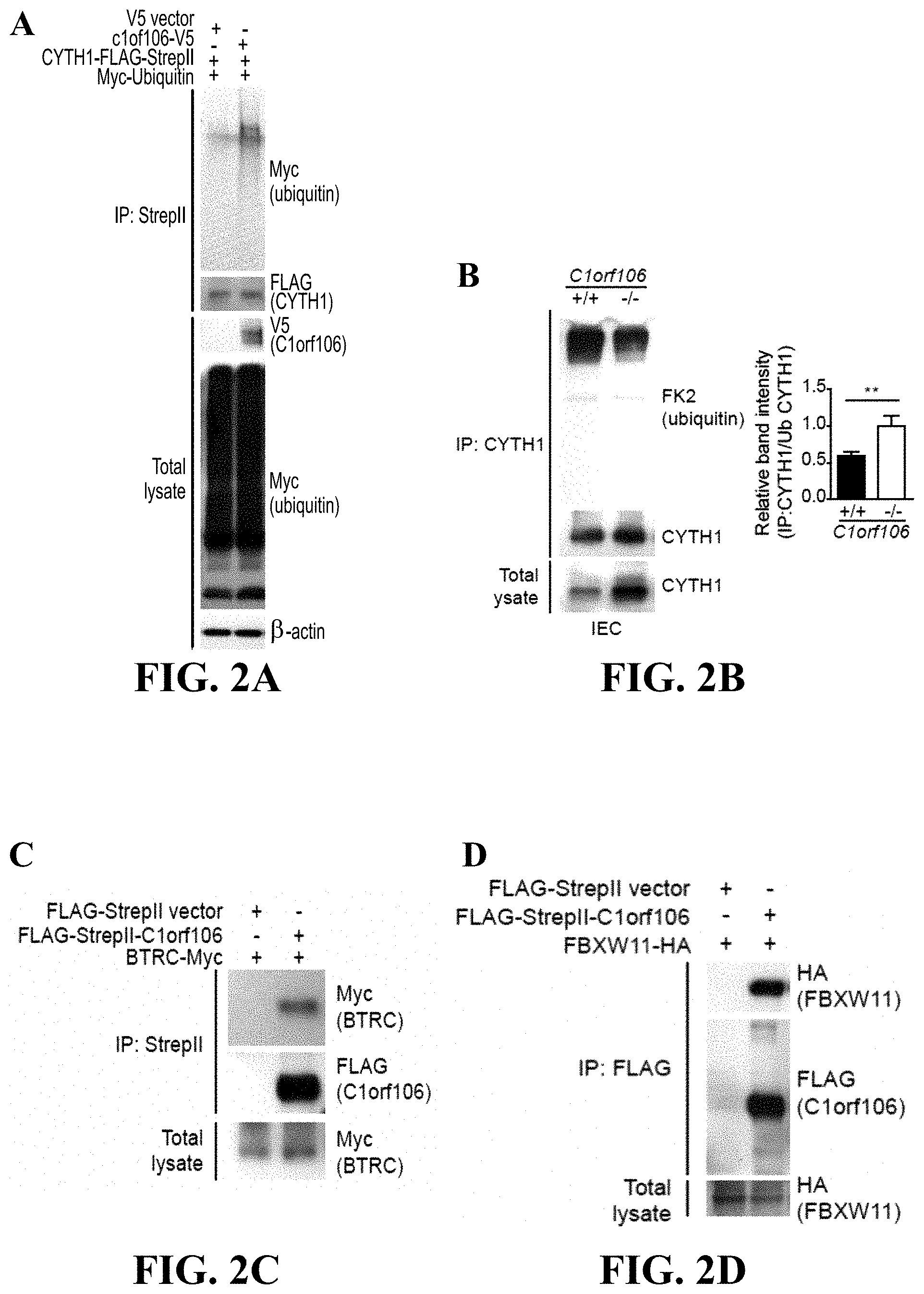
| Filed: | July 17, 2018 | ||||||||||
| PCT Filed: | July 17, 2018 | ||||||||||
| PCT NO: | PCT/US2018/042510 | ||||||||||
| 371 Date: | January 17, 2020 |
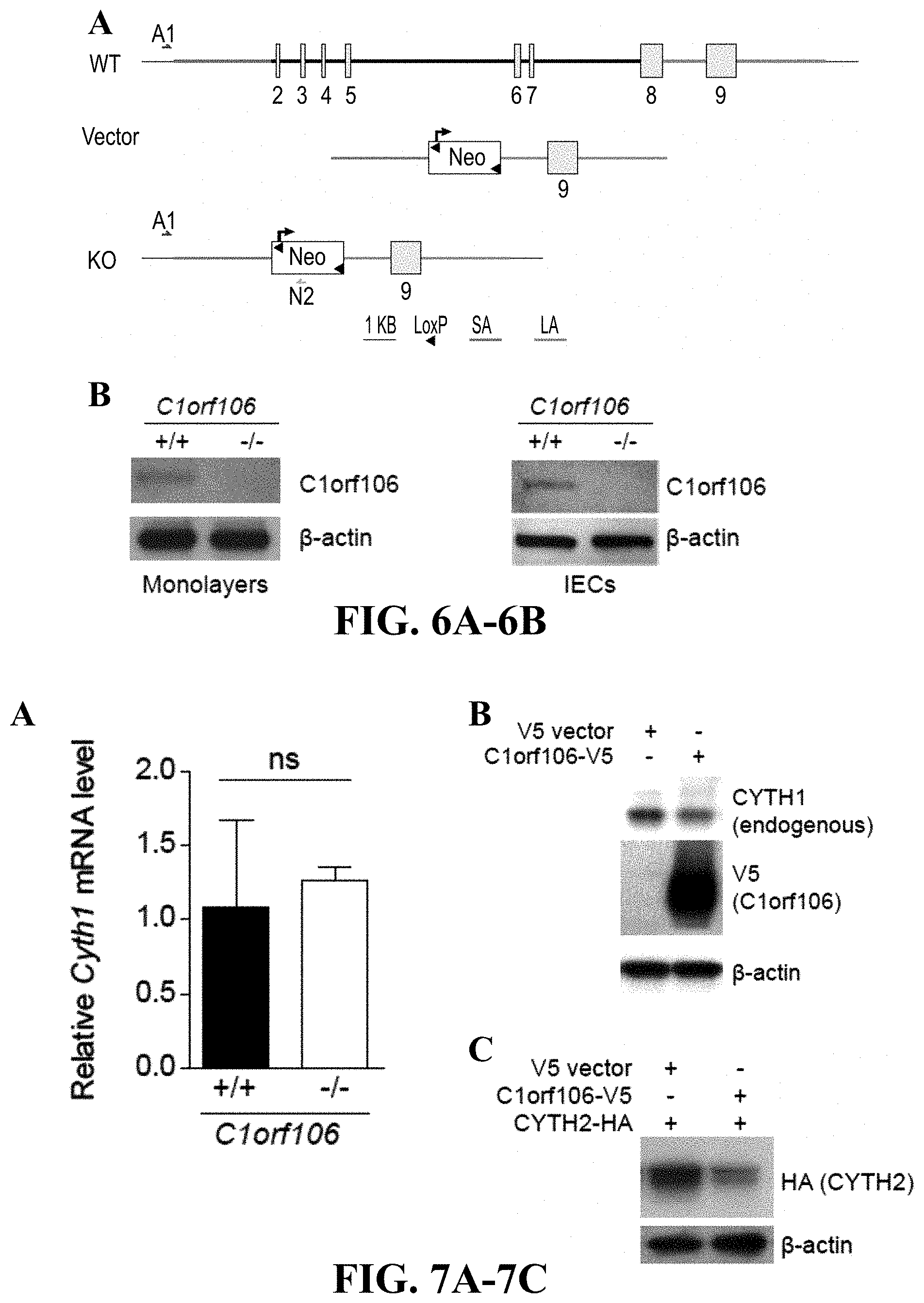
Related U.S. Patent Documents
| Application Number | Filing Date | Patent Number | ||
|---|---|---|---|---|
| 62533649 | Jul 17, 2017 | |||
| Current U.S. Class: | 1/1 |
| Current CPC Class: | C12Q 2600/158 20130101; C12N 9/22 20130101; C12N 2310/20 20170501; C12N 2800/80 20130101; C12N 15/113 20130101; C12Q 1/6883 20130101; A61K 48/0058 20130101 |
| International Class: | A61K 48/00 20060101 A61K048/00; C12N 15/113 20060101 C12N015/113; C12N 9/22 20060101 C12N009/22; C12Q 1/6883 20060101 C12Q001/6883 |
Goverment Interests
STATEMENT REGARDING FEDERALLY SPONSORED RESEARCH
[0002] This invention was made with government support under grant numbers DK043351 and DK062432 granted by National Institutes of Health. The government has certain rights in the invention.
Claims
1. A method of modulating intestinal epithelial cell integrity, migration, proliferation, differentiation, maintenance and/or function, the method comprising contacting an intestinal cell or a population of intestinal cells with a modulating agent in an amount sufficient to modify integrity, migration, proliferation, differentiation, maintenance and/or function of the intestinal cell or population of intestinal cells as compared to integrity, migration, proliferation, differentiation, maintenance and/or function of the intestinal cell or population of intestinal cells in the absence of the modulating agent, whereby the integrity, migration, proliferation, differentiation, maintenance and/or function of the intestinal cell directly influences intestinal epithelial cell integrity, migration, proliferation, differentiation, maintenance and/or function, preferably, wherein the modulating of intestinal epithelial cell integrity, migration, proliferation, differentiation, maintenance and/or function modulates inflammation of the gut.
2. (canceled)
3. The method of claim 1, wherein an agent that modulates protein stability is administered, preferably, wherein the agent that modulates protein stability modulates stability of the C1orf106 protein or a variant thereof, more preferably, wherein the C1orf106 variant protein is *333F; or an agent that modulates one or more of C1orf106 or its orthologs is administered.
4. (canceled)
5. (canceled)
6. The method of claim 1, wherein the modulating agent is a gene editing system used to restore the *333F variant to wild-type or another variant with increased protein stability compared to the *333F variant, preferably, wherein the gene editing system is a CRISPR system.
7. (canceled)
8. (canceled)
9. The method of claim 1, wherein the integrity, migration, proliferation, differentiation, maintenance and/or function of C1orf106-expressing cells in the intestines is modulated, particularly of C1orf106-expressing intestinal epithelial cells, comprising administering to a subject in need thereof an agent that modulates integrity, migration, proliferation, differentiation, maintenance and/or function of intestinal cells.
10. The method of claim 1, wherein the method is for treating an intestinal disease, wherein the method comprises: inhibiting epithelial cell migration or differentiation; or administering to a subject in need thereof a proteasome inhibitor and/or an agent that increases the stability of a C1orf106 protein.
11. (canceled)
12. The method of claim 1, wherein the method is for the treatment of an intestinal disease or condition selected from cancer, an infection, inflammation, or an immune dysfunction, preferably, wherein the inflammation is selected from inflammatory bowel disease, colitis, Crohn's disease, and food allergies; or wherein the infection or inflammation is caused by a bacterial or parasitic infection.
13. (canceled)
14. (canceled)
15. A method of identifying intestinal epithelial cells in a sample, screening one or more subjects for an inflammatory intestinal disease or determining susceptibility of a subject for an inflammatory intestinal disease comprising detecting the presence or expression level of an intestinal epithelial gene or variant thereof, preferably, wherein the intestinal epithelial gene is C1orf106 or Cp1orf106, more preferably, wherein the variant of C1orf106 is *333F; and/or wherein detecting expression of protein or mRNA of C1orf106 and/or Cp1orf106 indicates intestinal epithelial cells.
16. (canceled)
17. (canceled)
18. (canceled)
19. (canceled)
20. The method of claim 1, wherein the method is for modulating the integrity of the intestinal epithelia comprising altering the expression of an intestinal gene, wherein the integrity of the epithelia is increased or enhanced as a result of the altered expression of the intestinal epithelial gene, preferably, wherein the intestinal epithelial gene is C1orf106 or a homolog thereof; and/or wherein the intestinal epithelial protein is C1orf106 or a variant thereof, more preferably, wherein increasing the integrity of the intestinal epithelia comprises increasing the stability of the C1orf106 protein.
21. (canceled)
22. (canceled)
23. (canceled)
24. The method of claim 15, wherein the presence of the variant indicates susceptibility of the subject for the inflammatory intestinal disease, preferably, wherein the intestinal epithelial gene comprises C1orf106 or a homolog thereof; and/or wherein the intestinal epithelial protein comprises C1orf106 or a variant thereof, more preferably, wherein the variant of the intestinal epithelial protein comprises *333F.
25. (canceled)
26. (canceled)
27. (canceled)
28. The method of claim 1, wherein the method is for modeling an intestinal disease or condition comprising administering to a subject a modulating agent in an amount sufficient to modify integrity, migration, proliferation, differentiation, maintenance and/or function of the intestinal cell or population of intestinal cells as compared to integrity, migration, proliferation, differentiation, maintenance and/or function of the intestinal cell or population of intestinal cells in the absence of the modulating agent, whereby the integrity, migration, proliferation, differentiation, maintenance and/or function of the intestinal cell directly influences intestinal epithelial cell integrity, migration, proliferation, differentiation, maintenance and/or function, preferably, wherein the modulation is heritable to a progeny of the subject; and/or wherein the modulating agent modulates expression of an intestinal gene in the subject, more preferably, wherein the modulating agent reduces or eliminates expression of the intestinal gene in the subject.
29. (canceled)
30. (canceled)
31. (canceled)
32. The method of claim 28, further comprising a breeding program to produce at least a first progeny of the subject, wherein the further generation comprises modulated expression of the intestinal gene.
33. The method of claim 28, wherein the subject is an animal or a population of cells, preferably, wherein the animal is a mouse, rat, dog, pig, primate, or cells or tissue obtained therefrom.
34. (canceled)
35. The method of claim 28, wherein the modulating agent is provided to the subject using a gene editing system, preferably, wherein the gene editing system is a CRISPR system.
36. (canceled)
Description
CROSS-REFERENCE TO RELATED APPLICATIONS
[0001] This application is the U.S. National Stage of International Application No. PCT/US2018/042510, filed Jul. 17, 2018, published in English under PCT Article 21(2), which claims the priority benefit of the earlier filing date of U.S. Provisional Application No. 62/533,649, filed Jul. 17, 2017. The entire contents of the above-identified application is hereby fully incorporated herein by reference.
REFERENCE TO AN ELECTRONIC SEQUENCE LISTING
[0003] The contents of the electronic sequence listing ("BROD-2270US_ST25"; Size is 9 kilobytes and it was created on Apr. 16, 2020) is herein incorporated by reference in its entirety.
TECHNICAL FIELD
[0004] The subject matter disclosed herein is generally directed to compositions and methods for modulating, controlling, or otherwise influencing expression of an intestinal gene or protein. More particularly, the present invention relates to identifying and exploiting target genes and/or target gene products that modulate, control, or otherwise influence development of intestinal disease.
BACKGROUND
[0005] The intestinal mucosa is a complex system, comprising multiple cell types involved in a number of functions, including absorption, defense, and secretion. These cell types are rapidly renewed from intestinal stem cells. The types of cells, their differentiation, and signals controlling differentiation and activation are poorly understood. The small intestinal mucosa also possesses a large and active immune system, poised to detect antigens and bacteria at the mucosal surface and to drive appropriate responses of tolerance or an active immune response. Finally, there is complex luminal milieu which comprises a combination of diverse microbial species and their products as well as derivative products of the diet. It is increasingly clear that a functional balance between the epithelium and the constituents within the lumen plays a central role in both maintaining the normal mucosa and the pathophysiology of many gastrointestinal disorders. Many disorders, such as irritable bowel disease, Crohn's disease, and food allergies, have proven difficult to treat. The manner in which these multiple factors interact remains unclear.
SUMMARY
[0006] In certain example embodiments, methods of increasing the stability or preventing a decrease in the stability of Cp1orf106 protein increases the overall integrity of the intestinal epithelium, thereby resulting in a decreased incidence of inflammatory disease. In an embodiment, methods of modulating intestinal epithelial cell integrity, migration, proliferation, differentiation, maintenance and/or function are provided and include contacting an intestinal cell or a population of intestinal cells with a modulating agent in an amount sufficient to modify integrity, migration, proliferation, differentiation, maintenance and/or function of the intestinal cell or population of intestinal cells as compared to integrity, migration, proliferation, differentiation, maintenance and/or function of the intestinal cell or population of intestinal cells in the absence of the modulating agent. In one aspect, the methods modifying the integrity, migration, proliferation, differentiation, maintenance and/or function of the intestinal cell directly influences intestinal epithelial cell integrity, migration, proliferation, differentiation, maintenance and/or function.
[0007] In some embodiments, the modulation of intestinal epithelial cell integrity, migration, proliferation, differentiation, maintenance and/or function modulates inflammation of the gut. In some embodiments, the method of modulating includes administering an agent that modulates protein stability. In some instances, the agent that modulates protein stability modulates stability of the C1orf106 protein or a variant thereof. The C1orf106 variant protein, can be, for example *333F. In some instances, the agent modulates one or more of C1orf106 or its orthologs.
[0008] In one aspect the modulating agent is provided to one or more intestinal cells using a gene editing system. The gene editing system in one exemplary embodiment is a CRISPR system.
[0009] Methods of modulating the integrity, migration, proliferation, differentiation, maintenance and/or function of C1orf106-expressing cells in the intestines, particularly of C1orf106-expressing intestinal epithelial cells, can in some instances include administering to a subject in need thereof an agent that modulates integrity, migration, proliferation, differentiation, maintenance and/or function of intestinal cells.
[0010] Methods of treating an intestinal disease are also disclosed herein. In some instances, the methods include inhibiting epithelial cell migration or differentiation. Methods for treating an intestinal disease, in one embodiment, include administering to a subject in need thereof a proteasome inhibitor and/or an agent that increases the stability of a C1orf106 protein.
[0011] The methods of treatment of intestinal disease or condition can be, in some instances, cancer, an infection, inflammation, or an immune dysfunction. In some embodiments, the inflammation can be inflammatory bowel disease, colitis, Crohn's disease, or food allergies. In an embodiment, the infection or inflammation is caused by a bacterial or parasitic infection.
[0012] Methods for determining susceptibility of a subject for an inflammatory intestinal disease are also provided and include detecting the presence or expression level of an intestinal epithelial gene or variant thereof. In some instances, the intestinal epithelial gene is C1orf106. In some instances, the intestinal epithelial variant of C1orf106 is *333F.
[0013] Methods are also provided herein for identifying intestinal epithelial cells in a sample, in some instances, by detecting expression of protein or mRNA of C1orf106 protein or mRNA. In one aspect the expression of protein or mRNA of C1orf106 protein or mRNA indicates intestinal epithelial cells.
[0014] In an embodiment, a method of modulating the integrity of the intestinal epithelia is provided, and includes altering the expression of an intestinal gene. In some instances, the integrity of the epithelia is increased or enhanced as a result of the altered expression of the intestinal epithelial gene. In an embodiment, modulating the integrity of the intestinal epithelia includes altering the stability of an intestinal protein. In some instances, the integrity of the epithelia is increased or enhanced as a result of the altered intestinal epithelial protein. The intestinal epithelial gene in an embodiment can be a C1orf106 or a homolog thereof. In some instances, the intestinal epithelial protein is C1orf106 or a variant thereof. The methods of increasing the integrity of the intestinal epithelia can, for example, increase the stability of the C1orf106 protein.
[0015] Methods of screening one or more subjects for an inflammatory intestinal disease are also provided and include screening or detecting a variant of an intestinal epithelial gene. The presence of the variant can, in some embodiments, indicate susceptibility of the subject for the inflammatory intestinal disease. In an embodiment, the intestinal epithelial gene is a C1orf106 or a homolog thereof. In an embodiment, the variant of the intestinal epithelial protein C1orf106 includes *333F.
[0016] Methods of modeling an intestinal disease or condition are also disclosed herein and include administering to a subject a modulating agent in an amount sufficient to modify integrity, migration, proliferation, differentiation, maintenance and/or function of the intestinal cell or population of intestinal cells as compared to integrity, migration, proliferation, differentiation, maintenance and/or function of the intestinal cell or population of intestinal cells in the absence of the modulating agent. In one embodiment, the integrity, migration, proliferation, differentiation, maintenance and/or function of the intestinal cell directly influences intestinal epithelial cell integrity, migration, proliferation, differentiation, maintenance and/or function. In an embodiment, the modulating agent modulates expression of an intestinal gene in the subject, which can include reducing or eliminating expression of the intestinal gene in the subject. In an embodiment, the modulation is heritable to a progeny of the subject. In an embodiment, the method can also include a breeding program to produce at least a first progeny of the subject, wherein the further generation comprises modulated expression of the intestinal gene.
[0017] In some embodiments, the subject is an animal or a population of cells. In one embodiment, the animal is a mouse, rat, dog, pig, primate, or cells or tissue obtained therefrom. In one exemplary embodiment, the modulating agent is provided to the subject using a gene editing system, in one aspect, the gene editing system is a CRISPR system.
[0018] These and other aspects, objects, features, and advantages of the example embodiments will become apparent to those having ordinary skill in the art upon consideration of the following detailed description of illustrated example embodiments.
BRIEF DESCRIPTION OF THE DRAWINGS
[0019] An understanding of the features and advantages of the present invention will be obtained by reference to the following detailed description that sets forth illustrative embodiments, in which the principles of the invention may be utilized, and the accompanying drawings of which:
[0020] FIG. 1A-1H--C1orf106 modulates cytohesin-1 levels. FIG. 1A provides results of C1orf106 protein levels assessed during Caco-2 cell differentiation by immunoblot. Relative band intensity of C1orf106 isoform 1 at each time point was quantified and normalized to GAPDH. Each value represents the mean of two independent experiments .+-.SEM. FIG. 1B includes a scatter plot of log 2 ratios of two replicates for proteins that were enriched by FLAG antibody in HEK293T cells expressing FLAG-tagged C1orf106 (WT) compared to cells transfected with an empty vector (EV). Each dot represents log 2 ratio for a protein. Red dots, bait; blue dots, members of the SCF complex; green dots, cytohesins. FIG. 1C HEK293T cells were transiently transfected with HA-C1orf106 and either empty vector, full-length (FL) FLAG-StrepII-CYTH1 or the N- or C-terminal domains of CYTH1. Results shown are samples immunoprecipitated with anti-StrepII and probed for FLAG (CYTH1) and HA (C1orf106). FIG. 1D Caco-2 cell lysates were immunoprecipitated with anti-IgG or anti-C1orf106 and probed for CYTH1 and C1orf106. FIG. 1E HEK293T cells were transiently transfected with HA-CYTH1 and either empty vector, full-length (FL) FLAG-StrepII-C1orf106 or the N- or C-terminal domains of C1orf106. Results are shown of samples immunoprecipitated with anti-StrepII and probed for FLAG (C1orf106) and HA (CYTH1). FIG. 1F shows immunoblot analysis of intestinal epithelial cells isolated from the colon or small intestine of C1orf106+/+ and C1orf106-/- mice. Shown are samples from individual mice. Graphs denote normalized ratios of CYTH1:actin from 3 independent experiments as quantified by densitometry. Error bars indicate SD. FIG. 1G includes an immunoblot analysis of monolayers grown from colonic organoids from C1orf106+/+ and C1orf106-/- mice. Graphs denote normalized ratios of CYTH1:actin from 3 independent experiments as quantified by densitometry. Error bars indicate SD. FIG. 1H provides an immunoblot analysis of HEK293T cells co-transfected with CYTH1-FLAG-StrepII and empty vector or C1orf106-V5. Two biologic replicates are shown. Graph denotes normalized 13 ratios of CYTH1:actin from 3 independent experiments as quantified by densitometry. Error bars indicate SD. For all panels, *P<0.05, **P<0.01, ***P<0.001 (two-tailed Student's t test).
[0021] FIG. 2A-2F--C1orf106 regulates the ubiquitination of cytohesin-1 through the SCF ubiquitin ligase complex. FIG. 2A HEK293T cells were transfected with ubiquitin-Myc and CYTH1-FLAG-StrepII with or without C1orf106-V5, with results showing samples immunoprecipitated with anti-StrepII and probed for FLAG (CYTH1), V5 (C1orf106), and Myc (ubiquitin). FIG. 2B provides results of endogenous CYTH1 immunoprecipitated from C1orf106+/+ and C1orf106-/- intestinal epithelial cell (IEC) monolayers and probed for CYTH1 and ubiquitin (FK2). Graph denotes ratios of immunoprecipitated CYTH1:ubiquitinated CYTH1 from 3 independent experiments as quantified by densitometry. Error bars indicate SEM. **P<0.01 (two-tailed Student's t test). FIG. 2C HEK293T cells were transiently transfected with BTRC-Myc and either empty vector or full-length FLAG-StrepII-C1orf106. Samples were immunoprecipitated with anti-StrepII and probed for FLAG (C1orf106) and Myc (BTRC). FIG. 2D provides results of HEK293T cells transfected with FLAG-StrepII-C1orf106 and FBXW11-HA and immunoprecipitated as in 2C. FIG. 2E includes immunoblot analysis of HEK293T cells transfected with siRNAs against BTRC or FBXW11 and probed for CYTH1. Samples from two biologic replicates are shown. Graph denotes normalized ratios of CYTH1:actin from 3 independent experiments as quantified by densitometry. Error bars indicate SEM. **P<0.01 (two-tailed Student's t test). FIG. 2F includes an immunoblot analysis of HT-29 cells treated with DMSO or MLN4924 and probed for CYTH1. Actin served as a loading control. Data are representative of 3 independent experiments.
[0022] FIG. 3A-3H--C1orf106 controls surface E-cadherin levels through ARF6 activation. FIG. 3A shows results of IEC monolayers from C1orf106+/+ and C1orf106-/- mice immunoprecipitated with GGA3-PBD beads and probed with ARF6 antibody. Immunoblot is representative of 3 independent experiments. Graph denotes ratios of total ARF6:ARF6-GTP from 3 independent experiments as quantified by densitometry. Error bars indicate SD. FIG. 3B shows confocal images of colonic organoid-derived monolayers stained for ARF6, occludin, and nuclei (DAPI). Data are representative of 3 independent experiments. Arrowheads indicate ARF6 at the plasma membrane. FIG. 3C shows confocal images of colonic organoid-derived monolayers stained for E-cadherin, occludin, and nuclei (DAPI). Graph shows quantification of the percentage of cells that contained >10 intracellular E-cadherin puncta from 3 independent experiments. Error bars indicate SEM. FIG. 3D provides confocal immunofluorescence images of sections from C1orf106+/+ and C1orf106-/- mouse colon stained for E-cadherin, ZO-1, and nuclei (DAPI). FIG. 3E, 3F Freshly isolated intestinal epithelial cells FIG. 3E or organoid-derived monolayers FIG. 3F from C1orf106+/+ and C1orf106-/- mice were biotinylated to label surface proteins and immunoprecipitated with streptavidin beads. Total lysate and immunoprecipitated lysate were probed for E-cadherin. Graphs show quantification from 3 independent experiments. Error bars indicate SD. FIG. 3G provides TEER measurements during epithelial differentiation of Caco-2 cells stably expressing control shRNA or C1orf106 shRNA. A sigmoid (four parameters logistic) curve was fitted to the log(TEER) vs. time for each independent cell line. Data are representative of 3 independent experiments. Error bars indicate SEM. FIG. 3H charts quantification of cell migration in organoid-derived colonic monolayers after 48 h with or without HGF treatment. Error bars indicate SEM. *P<0.05, **P<0.01, ***P<0.001 (two-tailed Student's t test for (3A), (3C), (3E), (3F), (3H); ANOVA (3G)).
[0023] FIG. 4A-4G--C1orf106 maintains intestinal barrier function in vivo and the UC risk variant alters C1orf106 stability. FIG. 4A Bioluminescence image is provided showing colonization of bioluminescent Citrobacter rodentium in C1orf106+/+ and C1orf106-/- mice 5 days post-infection. FIG. 4B Colony forming unit (CFU) quantification of C. rodentium in the indicated organs. MLN, mesenteric lymph node. N=8 mice per genotype in 3 independent experiments. *P<0.05, **P<0.01 (two-tailed Student's t test). Error bars .+-.SEM. FIG. 4C includes immunoblot analysis of HEK293T cells transfected with FLAG-StrepII-C1orf106 or FLAG-StrepII-C1orf106 *333F. FS, FLAG-StrepII. FIG. 4D provides results from HEK293T cells transfected with Myc-ubiquitin and either empty vector, FLAG-StrepII-C1orf106, or FLAG-StrepII-C1orf106 *333F. Lysates from cells treated with 10 .mu.M MG132 were immunoprecipitated with StrepII and probed for FLAG (C1orf106) and Myc (ubiquitin). FIG. 4E graphs results of LS174T cells stably overexpressing C1orf106 WT and C1orf106 *333F treated with 50 .mu.g/ml cycloheximide for the indicated times. After immunoblot analysis densitometry was performed and results were graphed as relative C1orf106 levels normalized to .beta.-actin. The fraction of protein remaining represents the geometric mean+/-SEM of seven measurements in 4 independent experiments. FIG. 4F includes immunoblot analysis of HEK293T cells transfected with empty vector, C1orf106-V5, or C1orf106 *333F-V5 followed by transfection with CYTH1 after 48 hrs. FIG. 4G provides confocal immunofluorescence images (XZ and YZ planes) of LS174T cells stably overexpressing the indicated C1orf106 allele. Cells were stained for E-cadherin (green) and nuclei (DAPI).
[0024] FIG. 5A-5C--C1orf106 is highly expressed in epithelial cells and interacts with cytohesins. FIG. 5A Expression levels of C1orf106 in a panel of human tissues (bone marrow, heart, skeletal muscle, uterus, liver, fetal liver, spleen, thymus, thyroid, prostate, brain, lung, small intestine, and colon) and human cell lines using a custom Agilent expression array are provided. Cell lines represent models of human T lymphocytes (Jurkat), monocytes (THP-1), erythroleukemia cells (K562), promyelocytic cells (HL-60), colonic epithelial cells (HCT-15, HT-29, Caco-2), and cells from embryonic kidney (HEK293). In addition, models of differentiated colonic epithelium (Caco-2 differentiated for 21 days in culture [Caco-2 diff]), activated T lymphocytes (Jurkat cells stimulated with PMA [40 ng/ml] and ionomycin [1 .mu.g/ml) for 6 h [Jurkat stim]), and macrophages (derived from THP-1 differentiated for 24 h [THP-1 diff] with IFN.gamma. [400 U/ml] and TNF.alpha. [10 ng/ml]) were examined. Intensity values for each tissue/cell line represent the geometric mean with geometric standard deviation of 3 independent measurements; each measurement represents the geometric mean of all probes (one per exon) for each gene followed by a median normalization across all genes on the array. Dotted line indicates the threshold level for detection of basal expression. The reference sample is composed of a mixture of RNAs derived from 10 different human tissues. FIG. 5B Proteins identified by MS analysis as significantly enriched after C1orf106 immunoprecipitation. Fold change (FC) enrichment of proteins compared to cells transfected with empty vector and adjusted P value are shown. FIG. 5C HEK293T cells were transiently transfected with HA-C1orf106 and either empty vector, full-length FLAG-StrepII-CYTH2 or the N- or C-terminal domains of CYTH2; results are shown of samples immunoprecipitated with anti-StrepII and probed for FLAG (CYTH2) and HA (C1orf106).
[0025] FIG. 6A-6B--Generation of C1orf106-/- mice. FIG. 6A illustrates a schematic of the C1orf106 gene targeting strategy designed by inGenious Targeting Laboratory. A1, N2, genotyping primers; SA, short homology arm; LA, long homology arm. FIG. 6B provides immunoblot analysis of intestinal epithelial mono-layers derived from organoids and intestinal epithelial cells (IECs) isolated from the colon of C1orf106+/+ and C1orf106-/- mice and probed for C1orf106 and .beta.-actin.
[0026] FIG. 7A-7C--C1orf106 controls the levels of cytohesin protein. FIG. 7A qRT-PCR analysis of cytohes-in-1 levels in organoids derived from C1orf106+/+ and C1orf106-/- mice. ns, not significant, Student's t test. Error bars represent SD. FIG. 7B shows immunoblot analysis of HEK293T cells transfected with empty vector or C1orf106-V5 and probed for endogenous cytohesin-1. .beta.-actin served as a loading control. FIG. 7C includes immunoblot analysis of HEK293T cells co-transfected with cytohesin-2-HA and either empty vector or C1orf106-V5 and probed for cytohesin-2 using anti-HA antibody. .beta.-actin served as a loading control.
[0027] FIG. 8A-8C--C1orf106 and the SCF ubiquitin ligase complex. FIG. 8A includes immunoblot analysis of organoids from C1orf106+/+ and C1orf106-/- mice treated with MG132 or DMSO and probed for cytohesin-1. .beta.-actin served as a loading control. FIG. 8B includes results of HEK293T cells transiently transfected with SKP1-HA and either empty vector or full-length FLAG-StrepII-C1orf106. Samples were immunoprecipitated with anti-StrepII and probed for FLAG (C1orf106) and HA (SKP1). FIG. 8C includes images of HEK293T cells were transiently transfected with CUL1-Myc and either empty vector or full-length FLAG-StrepII-C1orf106. Samples were immunoprecipitated with anti-StrepII and probed for FLAG (C1orf106) and Myc (CUL1).
[0028] FIG. 9--charts efficacy of siRNAs against FBXW11 and BTRC1. qRT-PCR analysis of FBXW11 and BTRC1 in HEK293T cells transfected with control siRNA or siRNA against FBXW11 and/or BTRC1. Error bars represent SD.
[0029] FIG. 10A-10D--Increased membrane-associated ARF6 and disorganized E-cadherin in C1orf106-/- cells and organoids. FIG. 10A shows immunoblot analysis of intestinal epithelial cells derived from C1orf106+/+ and C1orf106-/- organoids. The insoluble fraction was probed for ARF6. .beta.-actin served as a loading control. **P<0.01, Student's t test. Error bars indicate SEM. FIG. 10B includes confocal immunofluorescence images of intestinal epithelial monolayers derived from C1orf106+/+ and C1orf106-/- organoids. Cells were stained for ZO-1 and DAPI. FIG. 10C includes confocal images of colonic organoids from C1orf106+/+ and C1orf106-/- mice stained for E-cadherin (green) and .alpha.4.beta. integrin (red). FIG. 10D shows results of confocal microscopy showing subcellular localization of endogenous E-cadherin in 18-day differentiated Caco-2 cells stably expressing an empty lentiviral vector or shRNA against C1orf106. Scale bars, 10 .mu.m.
[0030] FIG. 11--Internalized ARF6 colocalizes with E-cadherin in C1orf106-/- monolayers is provided in confocal immunofluorescence images of intestinal epithelial cells derived from C1orf106-/- organoids. Cells were stained for ARF6 and E-cadherin; and co-localization of ARF6 and E-cadherin was plotted using ImageJ.
[0031] FIG. 12--Recovery of E-cadherin after calcium switch is delayed in C1orf106-/- monolayers is shown in confocal images of organoid-derived monolayers left untreated or treated with 2 mM EGTA for 8 minutes. After EGTA treatment cells were allowed to recover for 2 h. Cells were stained for E-cadherin (red) and nuclei (blue).
[0032] FIG. 13A-13B--Loss of C1orf106 does not increase cytokine production following Citrobacter rodentium infection in vivo. FIG. 13A charts results of cytometric bead array was performed on media collected from colon sections from Citrobacter rodentium-infected C1orf106+/+ and C1orf106-/- mice at 5 days post-infection to quantitate levels of TNF.alpha. and IL-6. Error bars represent SD. FIG. 13B includes images of H & E-stained sections of colon from C1orf106+/+ and C1orf106-/- mice infected for 5 days with C. rodentium.
[0033] FIG. 14--charts no difference in mRNA expression of C1orf106 variants. Relative mRNA levels of C1orf106 WT and C1orf106 *333F in HEK293T cells transfected with WT-C1orf106-V5 and *333F-C1orf106-V5 plasmids respectively. Error bars represent SD.
[0034] The figures herein are for illustrative purposes only and are not necessarily drawn to scale.
DETAILED DESCRIPTION OF THE EXAMPLE EMBODIMENTS
General Definitions
[0035] Unless defined otherwise, technical and scientific terms used herein have the same meaning as commonly understood by one of ordinary skill in the art to which this disclosure pertains. Definitions of common terms and techniques in molecular biology may be found in Molecular Cloning: A Laboratory Manual, 2.sup.nd edition (1989) (Sambrook, Fritsch, and Maniatis); Molecular Cloning: A Laboratory Manual, 4.sup.th edition (2012) (Green and Sambrook); Current Protocols in Molecular Biology (1987) (F. M. Ausubel et al. eds.); the series Methods in Enzymology (Academic Press, Inc.): PCR 2: A Practical Approach (1995) (M. J. MacPherson, B. D. Hames, and G. R. Taylor eds.): Antibodies, A Laboratory Manual (1988) (Harlow and Lane, eds.): Antibodies A Laboratory Manual, 2nd edition 2013 (E. A. Greenfield ed.); Animal Cell Culture (1987) (R. I. Freshney, ed.); Benjamin Lewin, Genes IX, published by Jones and Bartlet, 2008 (ISBN 0763752223); Kendrew et al. (eds.), The Encyclopedia of Molecular Biology, published by Blackwell Science Ltd., 1994 (ISBN 0632021829); Robert A. Meyers (ed.), Molecular Biology and Biotechnology: a Comprehensive Desk Reference, published by VCH Publishers, Inc., 1995 (ISBN 9780471185710); Singleton et al., Dictionary of Microbiology and Molecular Biology 2nd ed., J. Wiley & Sons (New York, N.Y. 1994), March, Advanced Organic Chemistry Reactions, Mechanisms and Structure 4th ed., John Wiley & Sons (New York, N.Y. 1992); and Marten H. Hofker and Jan van Deursen, Transgenic Mouse Methods and Protocols, 2nd edition (2011).
[0036] As used herein, the singular forms "a," "an," and "the" include both singular and plural referents unless the context clearly dictates otherwise.
[0037] The term "optional" or "optionally" means that the subsequent described event, circumstance or substituent may or may not occur, and that the description includes instances where the event or circumstance occurs and instances where it does not.
[0038] The recitation of numerical ranges by endpoints includes all numbers and fractions subsumed within the respective ranges, as well as the recited endpoints.
[0039] The terms "about" or "approximately" as used herein when referring to a measurable value such as a parameter, an amount, a temporal duration, and the like, are meant to encompass variations of and from the specified value, such as variations of +/-10% or less, +/-5% or less, +/-1% or less, and +/-0.1% or less of and from the specified value, insofar such variations are appropriate to perform in the disclosed invention. It is to be understood that the value to which the modifier "about" or "approximately" refers is itself also specifically, and preferably, disclosed.
[0040] As used herein, a "biological sample" may contain whole cells and/or live cells and/or cell debris. The biological sample may contain (or be derived from) a "bodily fluid". The present invention encompasses embodiments wherein the bodily fluid is selected from amniotic fluid, aqueous humour, vitreous humour, bile, blood serum, breast milk, cerebrospinal fluid, cerumen (earwax), chyle, chyme, endolymph, perilymph, exudates, feces, female ejaculate, gastric acid, gastric juice, lymph, mucus (including nasal drainage and phlegm), pericardial fluid, peritoneal fluid, pleural fluid, pus, rheum, saliva, sebum (skin oil), semen, sputum, synovial fluid, sweat, tears, urine, vaginal secretion, vomit and mixtures of one or more thereof. Biological samples include cell cultures, bodily fluids, cell cultures from bodily fluids. Bodily fluids may be obtained from a mammal organism, for example by puncture, or other collecting or sampling procedures.
[0041] The terms "subject," "individual," and "patient" are used interchangeably herein to refer to a vertebrate, preferably a mammal, more preferably a human. Mammals include, but are not limited to, murines, simians, humans, farm animals, sport animals, and pets. Tissues, cells and their progeny of a biological entity obtained in vivo or cultured in vitro are also encompassed.
[0042] The term "isolated" as used throughout this specification with reference to a particular component generally denotes that such component exists in separation from--for example, has been separated from or prepared and/or maintained in separation from--one or more other components of its natural environment. More particularly, the term "isolated" as used herein in relation to a cell or cell population denotes that such cell or cell population does not contemporaneously form part of an animal or human body.
[0043] Various embodiments are described hereinafter. It should be noted that the specific embodiments are not intended as an exhaustive description or as a limitation to the broader aspects discussed herein. One aspect described in conjunction with a particular embodiment is not necessarily limited to that embodiment and can be practiced with any other embodiment(s). Reference throughout this specification to "one embodiment", "an embodiment," "an example embodiment," means that a particular feature, structure or characteristic described in connection with the embodiment is included in at least one embodiment of the present invention. Thus, appearances of the phrases "in one embodiment," "in an embodiment," or "an example embodiment" in various places throughout this specification are not necessarily all referring to the same embodiment, but may. Furthermore, the particular features, structures or characteristics may be combined in any suitable manner, as would be apparent to a person skilled in the art from this disclosure, in one or more embodiments. Furthermore, while some embodiments described herein include some but not other features included in other embodiments, combinations of features of different embodiments are meant to be within the scope of the invention. For example, in the appended claims, any of the claimed embodiments can be used in any combination.
[0044] All publications, published patent documents, and patent applications cited herein are hereby incorporated by reference to the same extent as though each individual publication, published patent document, or patent application was specifically and individually indicated as being incorporated by reference.
Overview
[0045] Single nucleotide polymorphisms in C1orf106 are associated with increased risk of inflammatory bowel disease (IBD). However, the function of C1orf106 and the consequences of disease-associated polymorphisms are unknown. While not bound by the following theory, C1orf106 may be able to regulate the stability of adherens junctions by regulating ubiquitin-mediated degradation of cytohesin-1, a guanine nucleotide exchange factor that controls activation of ARF6. By limiting cytohesin-1-dependent ARF6 activation, C1orf106 may stabilize adherens junctions. Consistent with this model, C1orf106-/- mice exhibit defects in the intestinal epithelial cell barrier, a phenotype also observed in IBD patients and that confers increased susceptibility to intestinal pathogens. Furthermore, the IBD risk variant C1orf106 *333F was found to show increased ubiquitination and turnover with consequent impairments in functional outputs. Despite the growing number of genes and polymorphisms associated with IBD and other intestinal diseases, mechanisms by which disease-associated genetic variants directly contribute to impaired epithelial barrier integrity in the intestine remain largely unknown. The present disclosure defines a critical function for a previously uncharacterized gene that is responsible for regulating the integrity of intestinal epithelial cells. For this reason, C1orf106 is also referred to herein as ROCS (regulator of cytohesin stability).
[0046] Embodiments disclosed herein provide methods for modulating intestinal epithelial cell integrity, migration, proliferation, differentiation, maintenance and/or function in which the expression of Cp1orf106 or its protein product are modulated such that the stability of the protein is altered. In certain example embodiments, increasing the stability or preventing a decrease in the stability of Cp1orf106 protein increases the overall integrity of the intestinal epithelium, thereby resulting in a decreased incidence of inflammatory disease. Increased integrity or stability of the epithelium may prevent invasion of migratory cells such as cancer cells.
Modulating Intestinal Epithelial Cell Integrity
[0047] In some embodiments, the invention provides methods of modulating intestinal epithelial cell integrity, migration, proliferation, differentiation, maintenance and/or function. In some embodiments, such a method comprises contacting an intestinal cell or a population of intestinal cells with a modulating agent in an amount sufficient to modify integrity, migration, proliferation, differentiation, maintenance and/or function of the cell or population of cells. Such methods may alter the stability of the intestinal epithelia, which may have implications for a variety of diseases as described herein. In some embodiments, modulation as described herein may alter gene expression or may alter the stability of a gene product or protein, polypeptide, or the like. Modulation may be performed by a variety of methods as described herein. In some embodiments, modulation as described herein results in altered stability of the intestinal epithelium. Increasing stability of the epithelium is beneficial for prevention of a variety of diseases as described herein. In particular embodiments, the intestinal epithelial gene C1orf106 or homologs or orthologs thereof, may be modulated as described herein. In other embodiments, the protein product of the C1orf106 gene, i.e., the C1orf106 protein, may be modulated, as described herein.
[0048] As described herein, particular variants of a gene or protein may lead to differential phenotypic or physiological effects. For example, as described herein and in the Examples, a variant of the C1orf106 protein referred to herein as *333F results in decreased stability of the protein and thereby results in decreased integrity of the intestinal epithelium. The present invention, therefore, provides methods for treating, controlling, ameliorating, or predicting diseases resulting from decreased epithelial integrity, including, but not limited to, an intestinal disease such as IBD, Crohn's disease, or cancer, by increasing the stability of C1or106, including the *333F variant, editing the *333F variant to wild type or other stable variant, and or otherwise mitigating the effect of the decreased stability of the *333F variant.
[0049] C1orf106 functions as a molecular rheostat to limit cytohesin levels through SCF complex-dependent degradation, thereby modulating epithelial barrier integrity. The finding that C1orf106 regulates the surface levels of E-cadherin is notable given that polymorphisms in both C1orf106 and CDH1 (E-cadherin) are associated with increased risk of ulcerative colitis, a form of IBD (7). Thus, complex genetic interactions can converge on single pathways, or as described in the present disclosure, on a specific gene. These findings have important implications for cancer biology, as ulcerative colitis is a risk factor for the development of colorectal cancer, and changes in E-cadherin expression and function are thought to play a crucial role in the spread of cancer cells. The data described herein demonstrate that loss of C1orf106 leads to increased cellular migration, a strategy used by tumor cells to increase invasion to surrounding tissues. Increasing the stability of C1orf106 may be used as a potential therapeutic strategy to increase the integrity of the epithelial barrier for the treatment of IBD, and could prevent cancer invasion.
[0050] In some embodiments, methods are provided for modulating the integrity, migration, proliferation, differentiation, maintenance and/or function of C1orf106-expressing cells in the intestines, particularly of C1orf106-expressing intestinal epithelial cells. As described herein, cells expressing variants of C1orf106 or its homologs or orthologs, or variants of a protein product or polypeptide of C1orf106 may be detected using methods of the present invention. In this way, one or more samples may be assayed or analyzed at one time in order to determine the presence of, for example, a disease-causing variant of an intestinal epithelial gene such as C1orf106.
[0051] In a subject having or having susceptibility to an inflammatory disease as a result of a variant of a gene or protein product as described herein, such as an intestinal disease as described herein, treatment of the disease may be performed by administering to the subject a modifying agent such that the expression of the gene or production of its protein product, or variants, homologs or orthologs thereof, is modified. Modification may be an increase or a decrease and may completely or partially ameliorate the symptoms of disease in the subject.
Inflammatory Diseases of the Gut
[0052] Inflammatory bowel disease (IBD) is a group of inflammatory conditions of the colon and small intestine, principally including Crohn's disease and ulcerative colitis, with other forms of IBD representing far fewer cases (e.g., collagenous colitis, lymphocytic colitis, diversion colitis, Behcet's disease and indeterminate colitis). Pathologically, Crohn's disease affects the full thickness of the bowel wall (e.g., transmural lesions) and can affect any part of the gastrointestinal tract, while ulcerative colitis is restricted to the mucosa (epithelial lining) of the colon and rectum. Graft-versus-host disease (GVHD) is an immune-related disease that can occur following an allogeneic tissue transplant. It is commonly associated with stem cell or bone marrow transplants, but GVHD also applies to other forms of tissue graft. In GVHD, immune cells of the tissue graft recognize the recipient host as foreign and attack the host's cells.
[0053] It has long been recognized that IBD and GVHD are diseases associated with increased immune activity. The causes of IBD, while not well understood, may be related to an aberrant immune response to the microbiota in genetically susceptible individuals. IBD affects over 1.4 million people in the United States and over 2.2 million in Europe and is on the increase. With both environmental and genetic factors playing a role in the development and progression of IBD, response to current treatments (e.g., anti-inflammatory drugs, immune system suppressors, antibiotics, surgery, and other symptom specific medications) are unpredictable. There is a need for new approaches to treating IBD.
[0054] Some of the genetic factors predisposing one to IBD are known, as described in Graham and Xavier "From Genetics of Inflammatory Bowel Disease Towards Mechanistic Insights" Trends Immunol. 2013 August; 34(8): 371-378.
[0055] In certain embodiments, the IBD is Crohn's disease or ulcerative colitis. In certain embodiments, the IBD is collagenous colitis, lymphocytic colitis, diversion colitis, Behcet's disease, indeterminate colitis, or GVHD.
[0056] In yet other embodiments, the methods of the disclosure include administering to a subject in need thereof an effective amount (e.g., therapeutically effective amount or prophylactically effective amount) of the treatments provided herein. Such treatment may be supplemented with other known treatments, such as surgery on the subject. In certain embodiments, the surgery is strictureplasty, resection (e.g., bowel resection, colon resection), colectomy, surgery for abscesses and fistulas, proctocolectomy, restorative proctocolectomy, vaginal surgery, cataract surgery, or a combination thereof.
[0057] Intestinal epithelial cells are required for gut homeostasis and are involved in numerous physiologic processes including nutrient absorption, protection against microbes and restitution following intestinal insult (1). Abnormal intestinal permeability has been observed in patients with IBD, a chronic inflammatory condition of the gastrointestinal tract (2). For several decades, it has been observed that healthy family members of some IBD patients also exhibit changes to the intestinal barrier, suggesting that host genetics can underlie cell-intrinsic defects in these barriers, though the underlying mechanisms are currently undefined (3).
[0058] The present disclosure provides a rationale for diagnosing IBD in an individual and/or determining the susceptibility of an individual for developing IBD using C1orf106, a gene associated with IBD susceptibility. A role for C1orf106 in epithelial homeostasis, along with the mechanism whereby the C1orf106 IBD-associated risk variant decreases cellular junctional integrity were determined, suggesting a mechanism by which this variant increases susceptibility to disease.
Identifying Modulators
[0059] A further aspect of the invention relates to a method for identifying an agent capable of modulating one or more phenotypic aspects of a gut cell or gut cell population as disclosed herein, comprising: a) applying a candidate agent to the cell or cell population; b) detecting modulation of one or more phenotypic aspects of the cell or cell population by the candidate agent, thereby identifying the agent.
[0060] The term "modulate" broadly denotes a qualitative and/or quantitative alteration, change or variation in that which is being modulated. Where modulation can be assessed quantitatively--for example, where modulation comprises or consists of a change in a quantifiable variable such as a quantifiable property of a cell or where a quantifiable variable provides a suitable surrogate for the modulation--modulation specifically encompasses both increase (e.g., activation) or decrease (e.g., inhibition) in the measured variable. The term encompasses any extent of such modulation, e.g., any extent of such increase or decrease, and may more particularly refer to statistically significant increase or decrease in the measured variable. By means of example, modulation may encompass an increase in the value of the measured variable by at least about 10%, e.g., by at least about 20%, preferably by at least about 30%, e.g., by at least about 40%, more preferably by at least about 50%, e.g., by at least about 75%, even more preferably by at least about 100%, e.g., by at least about 150%, 200%, 250%, 300%, 400% or by at least about 500%, compared to a reference situation without said modulation; or modulation may encompass a decrease or reduction in the value of the measured variable by at least about 10%, e.g., by at least about 20%, by at least about 30%, e.g., by at least about 40%, by at least about 50%, e.g., by at least about 60%, by at least about 70%, e.g., by at least about 80%, by at least about 90%, e.g., by at least about 95%, such as by at least about 96%, 97%, 98%, 99% or even by 100%, compared to a reference situation without said modulation. Preferably, modulation may be specific or selective, hence, one or more desired phenotypic aspects of a gut cell or gut cell population may be modulated without substantially altering other (unintended, undesired) phenotypic aspect(s).
[0061] The term "agent" broadly encompasses any condition, substance or agent capable of modulating one or more phenotypic aspects of an gut cell or gut cell population as disclosed herein. Such conditions, substances or agents may be of physical, chemical, biochemical and/or biological nature. The term "candidate agent" refers to any condition, substance or agent that is being examined for the ability to modulate one or more phenotypic aspects of an gut cell or gut cell population as disclosed herein in a method comprising applying the candidate agent to the gut cell or gut cell population (e.g., exposing the gut cell or gut cell population to the candidate agent or contacting the gut cell or gut cell population with the candidate agent) and observing whether the desired modulation takes place.
[0062] Agents may include any potential class of biologically active conditions, substances or agents, such as for instance antibodies, proteins, peptides, nucleic acids, oligonucleotides, small molecules, or combinations thereof.
[0063] By means of example but without limitation, agents can include low molecular weight compounds, but may also be larger compounds, or any organic or inorganic molecule effective in the given situation, including modified and unmodified nucleic acids such as antisense nucleic acids, RNAi, such as siRNA or shRNA, CRISPR/Cas systems, peptides, peptidomimetics, receptors, ligands, and antibodies, aptamers, polypeptides, nucleic acid analogues or variants thereof. Examples include an oligomer of nucleic acids, amino acids, or carbohydrates including without limitation proteins, oligonucleotides, ribozymes, DNAzymes, glycoproteins, siRNAs, lipoproteins, aptamers, and modifications and combinations thereof. Agents can be selected from a group comprising: chemicals; small molecules; nucleic acid sequences; nucleic acid analogues; proteins; peptides; aptamers; antibodies; or fragments thereof. A nucleic acid sequence can be RNA or DNA, and can be single or double stranded, and can be selected from a group comprising; nucleic acid encoding a protein of interest, oligonucleotides, nucleic acid analogues, for example peptide-nucleic acid (PNA), pseudo-complementary PNA (pc-PNA), locked nucleic acid (LNA), modified RNA (mod-RNA), single guide RNA etc. Such nucleic acid sequences include, for example, but are not limited to, nucleic acid sequence encoding proteins, for example that act as transcriptional repressors, antisense molecules, ribozymes, small inhibitory nucleic acid sequences, for example but are not limited to RNAi, shRNAi, siRNA, micro RNAi (mRNAi), antisense oligonucleotides, CRISPR guide RNA, for example that target a CRISPR enzyme to a specific DNA target sequence etc. A protein and/or peptide or fragment thereof can be any protein of interest, for example, but are not limited to: mutated proteins; therapeutic proteins and truncated proteins, wherein the protein is normally absent or expressed at lower levels in the cell. Proteins can also be selected from a group comprising; mutated proteins, genetically engineered proteins, peptides, synthetic peptides, recombinant proteins, chimeric proteins, antibodies, midibodies, minibodies, triabodies, humanized proteins, humanized antibodies, chimeric antibodies, modified proteins and fragments thereof. Alternatively, the agent can be intracellular within the cell as a result of introduction of a nucleic acid sequence into the cell and its transcription resulting in the production of the nucleic acid and/or protein modulator of a gene within the cell. In some embodiments, the agent is any chemical, entity or moiety, including without limitation synthetic and naturally-occurring non-proteinaceous entities. In certain embodiments, the agent is a small molecule having a chemical moiety. Agents can be known to have a desired activity and/or property, or can be selected from a library of diverse compounds.
[0064] In certain embodiments, an agent may be a hormone, a cytokine, a lymphokine, a growth factor, a chemokine, a cell surface receptor ligand such as a cell surface receptor agonist or antagonist, or a mitogen.
[0065] Non-limiting examples of hormones include growth hormone (GH), adrenocorticotropic hormone (ACTH), dehydroepiandrosterone (DHEA), cortisol, epinephrine, thyroid hormone, estrogen, progesterone, testosterone, or combinations thereof.
[0066] Non-limiting examples of cytokines include lymphokines (e.g., interferon-.gamma., IL-2, IL-3, IL-4, IL-6, granulocyte-macrophage colony-stimulating factor (GM-CSF), interferon-.gamma., leukocyte migration inhibitory factors (T-LIF, B-LIF), lymphotoxin-alpha, macrophage-activating factor (MAF), macrophage migration-inhibitory factor (MIF), neuroleukin, immunologic suppressor factors, transfer factors, or combinations thereof), monokines (e.g., IL-1, TNF-alpha, interferon-.alpha., interferon-.beta., colony stimulating factors, e.g., CSF2, CSF3, macrophage CSF or GM-CSF, or combinations thereof), chemokines (e.g., beta-thromboglobulin, C chemokines, CC chemokines, CXC chemokines, CX3C chemokines, macrophage inflammatory protein (MIP), or combinations thereof), interleukins (e.g., IL-1, IL-2, IL-3, IL-4, IL-5, IL-6, IL-7, IL-8, IL-9, IL-10, IL-11, IL-12, IL-13, IL-14, IL-15, IL-17, IL-18, IL-19, IL-20, IL-21, IL-22, IL-23, IL-24, IL-25, IL-26, IL-27, IL-28, IL-29, IL-30, IL-31, IL-32, IL-33, IL-34, IL-35, IL-36, or combinations thereof), and several related signalling molecules, such as tumour necrosis factor (TNF) and interferons (e.g., interferon-.alpha., interferon-.beta., interferon-.gamma., interferon-.lamda., or combinations thereof).
[0067] Non-limiting examples of growth factors include those of fibroblast growth factor (FGF) family, bone morphogenic protein (BMP) family, platelet derived growth factor (PDGF) family, transforming growth factor beta (TGFbeta) family, nerve growth factor (NGF) family, epidermal growth factor (EGF) family, insulin related growth factor (IGF) family, hepatocyte growth factor (HGF) family, hematopoietic growth factors (HeGFs), platelet-derived endothelial cell growth factor (PD-ECGF), angiopoietin, vascular endothelial growth factor (VEGF) family, glucocorticoids, or combinations thereof.
[0068] Non-limiting examples of mitogens include phytohaemagglutinin (PHA), concanavalin A (conA), lipopolysaccharide (LPS), pokeweed mitogen (PWM), phorbol ester such as phorbol myristate acetate (PMA) with or without ionomycin, or combinations thereof.
[0069] Non-limiting examples of cell surface receptors the ligands of which may act as agents include Toll-like receptors (TLRs) (e.g., TLR1, TLR2, TLR3, TLR4, TLR5, TLR6, TLR7, TLR8, TLR9, TLR10, TLR11, TLR12 or TLR13), CD80, CD86, CD40, CCR7, or C-type lectin receptors.
[0070] In certain embodiments, the present invention provides for gene signature screening. The concept of signature screening was introduced by Stegmaier et al. (Gene expression-based high-throughput screening (GE-HTS) and application to leukemia differentiation. Nature Genet. 36, 257-263 (2004)), who realized that if a gene-expression signature was the proxy for a phenotype of interest, it could be used to find small molecules that effect that phenotype without knowledge of a validated drug target. The signatures of the present may be used to screen for drugs that induce or reduce the signature in immune cells as described herein. The signature may be used for GE-HTS. In certain embodiments, pharmacological screens may be used to identify drugs that selectively activate gut cells.
[0071] The Connectivity Map (cmap) is a collection of genome-wide transcriptional expression data from cultured human cells treated with bioactive small molecules and simple pattern-matching algorithms that together enable the discovery of functional connections between drugs, genes and diseases through the transitory feature of common gene-expression changes (see, Lamb et al., The Connectivity Map: Using Gene-Expression Signatures to Connect Small Molecules, Genes, and Disease. Science 29 Sep. 2006: Vol. 313, Issue 5795, pp. 1929-1935, DOI: 10.1126/science.1132939; and Lamb, J., The Connectivity Map: a new tool for biomedical research. Nature Reviews Cancer January 2007: Vol. 7, pp. 54-60). In certain embodiments, Cmap can be used to screen for small molecules capable of modulating a signature of the present invention in silico.
[0072] Particular screening applications of this invention relate to the testing of pharmaceutical compounds in drug research. The reader is referred generally to the standard textbook In vitro Methods in Pharmaceutical Research, Academic Press, 1997, and U.S. Pat. No. 5,030,015. In certain aspects of this invention, the culture of the invention is used to grow and differentiate a cachectic target cell to play the role of test cells for standard drug screening and toxicity assays. Assessment of the activity of candidate pharmaceutical compounds generally involves combining the target cell (e.g., a myocyte, an adipocyte, a cardiomyocyte or a hepatocyte) with the candidate compound, determining any change in the morphology, marker phenotype, or metabolic activity of the cells that is attributable to the candidate compound (compared with untreated cells or cells treated with an inert compound, such as vehicle), and then correlating the effect of the candidate compound with the observed change. The screening may be done because the candidate compound is designed to have a pharmacological effect on the target cell, or because a candidate compound may have unintended side effects on the target cell. Alternatively, libraries can be screened without any predetermined expectations in hopes of identifying compounds with desired effects.
[0073] Cytotoxicity can be determined in the first instance by the effect on cell viability and morphology. In certain embodiments, toxicity may be assessed by observation of vital staining techniques, ELISA assays, immunohistochemistry, and the like or by analyzing the cellular content of the culture, e.g., by total cell counts, and differential cell counts or by metabolic markers such as MTT and XTT.
[0074] Additional further uses of the culture of the invention include, but are not limited to, its use in research e.g., to elucidate mechanisms leading to the identification of novel targets for therapies, and to generate genotype-specific cells for disease modeling, including the generation of new therapies customized to different genotypes. Such customization can reduce adverse drug effects and help identify therapies appropriate to the patient's genotype.
[0075] In certain embodiments, the present invention provides method for high-throughput screening. "High-throughput screening" (HTS) refers to a process that uses a combination of modern robotics, data processing and control software, liquid handling devices, and/or sensitive detectors, to efficiently process a large amount of (e.g., thousands, hundreds of thousands, or millions of) samples in biochemical, genetic or pharmacological experiments, either in parallel or in sequence, within a reasonably short period of time (e.g., days). Preferably, the process is amenable to automation, such as robotic simultaneous handling of 96 samples, 384 samples, 1536 samples or more. A typical HTS robot tests up to 100,000 to a few hundred thousand compounds per day. The samples are often in small volumes, such as no more than 1 mL, 500 .mu.l, 200 .mu.l, 100 .mu.l, 50 .mu.l or less. Through this process, one can rapidly identify active compounds, small molecules, antibodies, proteins or polynucleotides which modulate a particular biomolecular/genetic pathway. The results of these experiments provide starting points for further drug design and for understanding the interaction or role of a particular biochemical process in biology. Thus"high-throughput screening" as used herein does not include handling large quantities of radioactive materials, slow and complicated operator-dependent screening steps, and/or prohibitively expensive reagent costs, etc
Genetic Modification
[0076] In certain embodiments, one or more endogenous genes may be modified using a nuclease. The term "nuclease" as used herein broadly refers to an agent, for example a protein or a small molecule, capable of cleaving a phosphodiester bond connecting nucleotide residues in a nucleic acid molecule. In some embodiments, a nuclease may be a protein, e.g., an enzyme that can bind a nucleic acid molecule and cleave a phosphodiester bond connecting nucleotide residues within the nucleic acid molecule. A nuclease may be an endonuclease, cleaving a phosphodiester bonds within a polynucleotide chain, or an exonuclease, cleaving a phosphodiester bond at the end of the polynucleotide chain. Preferably, the nuclease is an endonuclease. Preferably, the nuclease is a site-specific nuclease, binding and/or cleaving a specific phosphodiester bond within a specific nucleotide sequence, which may be referred to as "recognition sequence", "nuclease target site", or "target site". In some embodiments, a nuclease may recognize a single stranded target site, in other embodiments a nuclease may recognize a double-stranded target site, for example a double-stranded DNA target site. Some endonucleases cut a double-stranded nucleic acid target site symmetrically, i.e., cutting both strands at the same position so that the ends comprise base-paired nucleotides, also known as blunt ends. Other endonucleases cut a double-stranded nucleic acid target sites asymmetrically, i.e., cutting each strand at a different position so that the ends comprise unpaired nucleotides. Unpaired nucleotides at the end of a double-stranded DNA molecule are also referred to as "overhangs", e.g., "5'-overhang" or "3'-overhang", depending on whether the unpaired nucleotide(s) form(s) the 5' or the 5' end of the respective DNA strand.
[0077] The nuclease may introduce one or more single-strand nicks and/or double-strand breaks in the endogenous gene, whereupon the sequence of the endogenous gene may be modified or mutated via non-homologous end joining (NHEJ) or homology-directed repair (HDR).
[0078] In certain embodiments, the nuclease may comprise (i) a DNA-binding portion configured to specifically bind to the endogenous gene and (ii) a DNA cleavage portion. Generally, the DNA cleavage portion will cleave the nucleic acid within or in the vicinity of the sequence to which the DNA-binding portion is configured to bind.
[0079] In certain embodiments, the DNA-binding portion may comprise a zinc finger protein or DNA-binding domain thereof, a transcription activator-like effector (TALE) protein or DNA-binding domain thereof, or an RNA-guided protein or DNA-binding domain thereof.
[0080] In certain embodiments, the DNA-binding portion may comprise (i) Cas9 or Cpf1 or any Cas protein described herein modified to eliminate its nuclease activity, or (ii) DNA-binding domain of Cas9 or Cpf1 or any Cas protein described herein.
[0081] In certain embodiments, the DNA cleavage portion comprises FokI or variant thereof or DNA cleavage domain of FokI or variant thereof.
[0082] In certain embodiments, the nuclease may be an RNA-guided nuclease, such as Cas9 Cas12 or Cal3 protein described herein. As Cas13 may be used to edit RNA transcripts, Cas13 provides a mechanism for addressing the variants disclosed herein wherein a more limited temporal control may be needed or desired, for example to limit the impact of side effects or in any scenario where a permanent edit of the genome may not be desired.
[0083] With respect to general information on CRISPR-Cas Systems, components thereof, and delivery of such components, including methods, materials, delivery vehicles, vectors, particles, AAV, and making and using thereof, including as to amounts and formulations, all useful in the practice of the instant invention, reference is made to: U.S. Pat. Nos. 8,999,641, 8,993,233, 8,945,839, 8,932,814, 8,906,616, 8,895,308, 8,889,418, 8,889,356, 8,871,445, 8,865,406, 8,795,965, 8,771,945 and 8,697,359; US Patent Publications US 2014-0310830 (U.S. application Ser. No. 14/105,031), US 2014-0287938 A1 (U.S. application Ser. No. 14/213,991), US 2014-0273234 A1 (U.S. application Ser. No. 14/293,674), US2014-0273232 A1 (U.S. application Ser. No. 14/290,575), US 2014-0273231 (U.S. application Ser. No. 14/259,420), US 2014-0256046 A1 (U.S. application Ser. No. 14/226,274), US 2014-0248702 A1 (U.S. application Ser. No. 14/258,458), US 2014-0242700 A1 (U.S. application Ser. No. 14/222,930), US 2014-0242699 A1 (U.S. application Ser. No. 14/183,512), US 2014-0242664 A1 (U.S. application Ser. No. 14/104,990), US 2014-0234972 A1 (U.S. application Ser. No. 14/183,471), US 2014-0227787 A1 (U.S. application Ser. No. 14/256,912), US 2014-0189896 A1 (U.S. application Ser. No. 14/105,035), US 2014-0186958 (U.S. application Ser. No. 14/105,017), US 2014-0186919 A1 (U.S. application Ser. No. 14/104,977), US 2014-0186843 A1 (U.S. application Ser. No. 14/104,900), US 2014-0179770 A1 (U.S. application Ser. No. 14/104,837) and US 2014-0179006 A1 (U.S. application Ser. No. 14/183,486), US 2014-0170753 (U.S. application Ser. No. 14/183,429); European Patents EP 2 784 162 B1 and EP 2 771 468 B1; European Patent Applications EP 2 771 468 (EP13818570.7), EP 2 764 103 (EP13824232.6), and EP 2 784 162 (EP14170383.5); and PCT Patent Publications PCT Patent Publications WO 2014/093661 (PCT/US2013/074743), WO 2014/093694 (PCT/US2013/074790), WO 2014/093595 (PCT/US2013/074611), WO 2014/093718 (PCT/US2013/074825), WO 2014/093709 (PCT/US2013/074812), WO 2014/093622 (PCT/US2013/074667), WO 2014/093635 (PCT/US2013/074691), WO 2014/093655 (PCT/US2013/074736), WO 2014/093712 (PCT/US2013/074819), WO2014/093701 (PCT/US2013/074800), WO2014/018423 (PCT/US2013/051418), WO 2014/204723 (PCT/US2014/041790), WO 2014/204724 (PCT/US2014/041800), WO 2014/204725 (PCT/US2014/041803), WO 2014/204726 (PCT/US2014/041804), WO 2014/204727 (PCT/US2014/041806), WO 2014/204728 (PCT/US2014/041808), WO 2014/204729 (PCT/US2014/041809). Reference is also made to U.S. provisional patent applications 61/758,468; 61/802,174; 61/806,375; 61/814,263; 61/819,803 and 61/828,130, filed on Jan. 30, 2013; Mar. 15, 2013; Mar. 28, 2013; Apr. 20, 2013; May 6, 2013 and May 28, 2013 respectively. Reference is also made to U.S. provisional patent application 61/836,123, filed on Jun. 17, 2013. Reference is additionally made to U.S. provisional patent applications 61/835,931, 61/835,936, 61/836,127, 61/836,101, 61/836,080 and 61/835,973, each filed Jun. 17, 2013. Further reference is made to U.S. provisional patent applications 61/862,468 and 61/862,355 filed on Aug. 5, 2013; 61/871,301 filed on Aug. 28, 2013; 61/960,777 filed on Sep. 25, 2013 and 61/961,980 filed on Oct. 28, 2013. Reference is yet further made to: PCT Patent applications Nos: PCT/US2014/041803, PCT/US2014/041800, PCT/US2014/041809, PCT/US2014/041804 and PCT/US2014/041806, each filed Jun. 10, 2014 6/10/14; PCT/US2014/041808 filed Jun. 11, 2014; and PCT/US2014/62558 filed Oct. 28, 2014, and U.S. Provisional Patent Applications Ser. Nos. 61/915,150, 61/915,301, 61/915,267 and 61/915,260, each filed Dec. 12, 2013; 61/757,972 and 61/768,959, filed on Jan. 29, 2013 and Feb. 25, 2013; 61/835,936, 61/836,127, 61/836,101, 61/836,080, 61/835,973, and 61/835,931, filed Jun. 17, 2013; 62/010,888 and 62/010,879, both filed Jun. 11, 2014; 62/010,329 and 62/010,441, each filed Jun. 10, 2014; 61/939,228 and 61/939,242, each filed Feb. 12, 2014; 61/980,012, filed Apr. 15, 2014; 62/038,358, filed Aug. 17, 2014; 62/054,490, 62/055,484, 62/055,460 and 62/055,487, each filed Sep. 25, 2014; and 62/069,243, filed Oct. 27, 2014. Reference is also made to U.S. provisional patent applications Nos. 62/055,484, 62/055,460, and 62/055,487, filed Sep. 25, 2014; U.S. provisional patent application 61/980,012, filed Apr. 15, 2014; and U.S. provisional patent application 61/939,242 filed Feb. 12, 2014. Reference is made to PCT application designating, inter alia, the United States, application No. PCT/US14/41806, filed Jun. 10, 2014. Reference is made to U.S. provisional patent application 61/930,214 filed on Jan. 22, 2014. Reference is made to U.S. provisional patent applications 61/915,251; 61/915,260 and 61/915,267, each filed on Dec. 12, 2013. Reference is made to US provisional patent application U.S. Ser. No. 61/980,012 filed Apr. 15, 2014. Reference is made to PCT application designating, inter alia, the United States, application No. PCT/US14/41806, filed Jun. 10, 2014. Reference is made to U.S. provisional patent application 61/930,214 filed on Jan. 22, 2014. Reference is made to U.S. provisional patent applications 61/915,251; 61/915,260 and 61/915,267, each filed on Dec. 12, 2013.
[0084] Mention is also made of U.S. application 62/091,455, filed, 12 Dec. 2014, PROTECTED GUIDE RNAS (PGRNAS); U.S. application 62/096,708, 24 Dec. 2014, PROTECTED GUIDE RNAS (PGRNAS); U.S. application 62/091,462, 12 Dec. 2014, DEAD GUIDES FOR CRISPR TRANSCRIPTION FACTORS; U.S. application 62/096,324, 23 Dec. 2014, DEAD GUIDES FOR CRISPR TRANSCRIPTION FACTORS; U.S. application 62/091,456, 12 Dec. 2014, ESCORTED AND FUNCTIONALIZED GUIDES FOR CRISPR-CAS SYSTEMS; U.S. application 62/091,461, 12 Dec. 2014, DELIVERY, USE AND THERAPEUTIC APPLICATIONS OF THE CRISPR-CAS SYSTEMS AND COMPOSITIONS FOR GENOME EDITING AS TO HEMATOPOETIC STEM CELLS (HSCs); U.S. application 62/094,903, 19 Dec. 2014, UNBIASED IDENTIFICATION OF DOUBLE-STRAND BREAKS AND GENOMIC REARRANGEMENT BY GENOME-WISE INSERT CAPTURE SEQUENCING; U.S. application 62/096,761, 24 Dec. 2014, ENGINEERING OF SYSTEMS, METHODS AND OPTIMIZED ENZYME AND GUIDE SCAFFOLDS FOR SEQUENCE MANIPULATION; U.S. application 62/098,059, 30 Dec. 2014, RNA-TARGETING SYSTEM; U.S. application 62/096,656, 24 Dec. 2014, CRISPR HAVING OR ASSOCIATED WITH DESTABILIZATION DOMAINS; U.S. application 62/096,697, 24 Dec. 2014, CRISPR HAVING OR ASSOCIATED WITH AAV; U.S. application 62/098,158, 30 Dec. 2014, ENGINEERED CRISPR COMPLEX INSERTIONAL TARGETING SYSTEMS; U.S. application 62/151,052, 22 Apr. 2015, CELLULAR TARGETING FOR EXTRACELLULAR EXOSOMAL REPORTING; U.S. application 62/054,490, 24 Sep. 2014, DELIVERY, USE AND THERAPEUTIC APPLICATIONS OF THE CRISPR-CAS SYSTEMS AND COMPOSITIONS FOR TARGETING DISORDERS AND DISEASES USING PARTICLE DELIVERY COMPONENTS; U.S. application 62/055,484, 25 Sep. 2014, SYSTEMS, METHODS AND COMPOSITIONS FOR SEQUENCE MANIPULATION WITH OPTIMIZED FUNCTIONAL CRISPR-CAS SYSTEMS; U.S. application 62/087,537, 4 Dec. 2014, SYSTEMS, METHODS AND COMPOSITIONS FOR SEQUENCE MANIPULATION WITH OPTIMIZED FUNCTIONAL CRISPR-CAS SYSTEMS; U.S. application 62/054,651, 24 Sep. 2014, DELIVERY, USE AND THERAPEUTIC APPLICATIONS OF THE CRISPR-CAS SYSTEMS AND COMPOSITIONS FOR MODELING COMPETITION OF MULTIPLE CANCER MUTATIONS IN VIVO; U.S. application 62/067,886, 23 Oct. 2014, DELIVERY, USE AND THERAPEUTIC APPLICATIONS OF THE CRISPR-CAS SYSTEMS AND COMPOSITIONS FOR MODELING COMPETITION OF MULTIPLE CANCER MUTATIONS IN VIVO; U.S. application 62/054,675, 24 Sep. 2014, DELIVERY, USE AND THERAPEUTIC APPLICATIONS OF THE CRISPR-CAS SYSTEMS AND COMPOSITIONS IN NEURONAL CELLS/TISSUES; U.S. application 62/054,528, 24 Sep. 2014, DELIVERY, USE AND THERAPEUTIC APPLICATIONS OF THE CRISPR-CAS SYSTEMS AND COMPOSITIONS IN IMMUNE DISEASES OR DISORDERS; U.S. application 62/055,454, 25 Sep. 2014, DELIVERY, USE AND THERAPEUTIC APPLICATIONS OF THE CRISPR-CAS SYSTEMS AND COMPOSITIONS FOR TARGETING DISORDERS AND DISEASES USING CELL PENETRATION PEPTIDES (CPP); U.S. application 62/055,460, 25 Sep. 2014, MULTIFUNCTIONAL-CRISPR COMPLEXES AND/OR OPTIMIZED ENZYME LINKED FUNCTIONAL-CRISPR COMPLEXES; U.S. application 62/087,475, 4 Dec. 2014, FUNCTIONAL SCREENING WITH OPTIMIZED FUNCTIONAL CRISPR-CAS SYSTEMS; U.S. application 62/055,487, 25 Sep. 2014, FUNCTIONAL SCREENING WITH OPTIMIZED FUNCTIONAL CRISPR-CAS SYSTEMS; U.S. application 62/087,546, 4 Dec. 2014, MULTIFUNCTIONAL CRISPR COMPLEXES AND/OR OPTIMIZED ENZYME LINKED FUNCTIONAL-CRISPR COMPLEXES; and U.S. application 62/098,285, 30 Dec. 2014, CRISPR MEDIATED IN VIVO MODELING AND GENETIC SCREENING OF TUMOR GROWTH AND METASTASIS.
[0085] Each of these patents, patent publications, and applications, and all documents cited therein or during their prosecution ("appln cited documents") and all documents cited or referenced in the appln cited documents, together with any instructions, descriptions, product specifications, and product sheets for any products mentioned therein or in any document therein and incorporated by reference herein, are hereby incorporated herein by reference, and may be employed in the practice of the invention. All documents (e.g., these patents, patent publications and applications and the appln cited documents) are incorporated herein by reference to the same extent as if each individual document was specifically and individually indicated to be incorporated by reference.
[0086] Also with respect to general information on CRISPR-Cas Systems, mention is made of the following (also hereby incorporated herein by reference): [0087] Multiplex genome engineering using CRISPR/Cas systems. Cong, L., Ran, F. A., Cox, D., Lin, S., Barretto, R., Habib, N., Hsu, P. D., Wu, X., Jiang, W., Marraffini, L. A., & Zhang, F. Science February 15; 339(6121):819-23 (2013); [0088] RNA-guided editing of bacterial genomes using CRISPR-Cas systems. Jiang W., Bikard D., Cox D., Zhang F, Marraffini L A. Nat Biotechnol March; 31(3):233-9 (2013); [0089] One-Step Generation of Mice Carrying Mutations in Multiple Genes by CRISPR/Cas-Mediated Genome Engineering. Wang H., Yang H., Shivalila C S., Dawlaty M M., Cheng A W., Zhang F., Jaenisch R. Cell May 9; 153(4):910-8 (2013); [0090] Optical control of mammalian endogenous transcription and epigenetic states. Konermann S, Brigham M D, Trevino A E, Hsu P D, Heidenreich M, Cong L, Platt R J, Scott D A, Church G M, Zhang F. Nature. August 22; 500(7463):472-6. doi: 10.1038/Nature12466. Epub 2013 Aug. 23 (2013); [0091] Double Nicking by RNA-Guided CRISPR Cas9 for Enhanced Genome Editing Specificity. Ran, F A., Hsu, P D., Lin, C Y., Gootenberg, J S., Konermann, S., Trevino, A E., Scott, D A., Inoue, A., Matoba, S., Zhang, Y., & Zhang, F. Cell August 28. pii: S0092-8674(13)01015-5 (2013-A); [0092] DNA targeting specificity of RNA-guided Cas9 nucleases. Hsu, P., Scott, D., Weinstein, J., Ran, F A., Konermann, S., Agarwala, V., Li, Y., Fine, E., Wu, X., Shalem, O., Cradick, T J., Marraffini, L A., Bao, G., & Zhang, F. Nat Biotechnol doi:10.1038/nbt.2647 (2013); [0093] Genome engineering using the CRISPR-Cas9 system. Ran, F A., Hsu, P D., Wright, J., Agarwala, V., Scott, D A., Zhang, F. Nature Protocols November; 8(11):2281-308 (2013-B); [0094] Genome-Scale CRISPR-Cas9 Knockout Screening in Human Cells. Shalem, O., Sanjana, N E., Hartenian, E., Shi, X., Scott, D A., Mikkelson, T., Heckl, D., Ebert, B L., Root, D E., Doench, J G., Zhang, F. Science December 12. (2013). [Epub ahead of print]; [0095] Crystal structure of cas9 in complex with guide RNA and target DNA. Nishimasu, H., Ran, F A., Hsu, P D., Konermann, S., Shehata, S I., Dohmae, N., Ishitani, R., Zhang, F., Nureki, O. Cell February 27, 156(5):935-49 (2014); [0096] Genome-wide binding of the CRISPR endonuclease Cas9 in mammalian cells. Wu X., Scott D A., Kriz A J., Chiu A C., Hsu P D., Dadon D B., Cheng A W., Trevino A E., Konermann S., Chen S., Jaenisch R., Zhang F., Sharp P A. Nat Biotechnol. April 20. doi: 10.1038/nbt.2889 (2014); [0097] CRISPR-Cas9 Knockin Mice for Genome Editing and Cancer Modeling. Platt R J, Chen S, Zhou Y, Yim M J, Swiech L, Kempton H R, Dahlman J E, Parnas O, Eisenhaure T M, Jovanovic M, Graham D B, Jhunjhunwala S, Heidenreich M, Xavier R J, Langer R, Anderson D G, Hacohen N, Regev A, Feng G, Sharp P A, Zhang F. Cell 159(2): 440-455 DOI: 10.1016/j.cell.2014.09.014(2014); [0098] Development and Applications of CRISPR-Cas9 for Genome Engineering, Hsu P D Lander E S Zhang F., Cell. June 5; 157(6):1262-78 (2014). [0099] Genetic screens in human cells using the CRISPR/Cas9 system, Wang T, Wei J J Sabatini D M Lander E S., Science. January 3; 343(6166): 80-84. doi:10.1126/science.1246981 (2014); [0100] Rational design of highly active sgRNAs for CRISPR-Cas9-mediated gene inactivation, Doench J G Hartenian E, Graham D B Tothova Z, Hegde M, Smith I, Sullender M, Ebert B L Xavier R J Root D E., (published online 3 Sep. 2014) Nat Biotechnol. December; 32(12):1262-7 (2014); [0101] In vivo interrogation of gene function in the mammalian brain using CRISPR-Cas9, Swiech L, Heidenreich M, Banerjee A, Habib N, Li Y, Trombetta J, Sur M, Zhang F., (published online 19 Oct. 2014) Nat Biotechnol. January; 33(1):102-6 (2015); [0102] Genome-scale transcriptional activation by an engineered CRISPR-Cas9 complex, Konermann S, Brigham M D Trevino A E Joung J, Abudayyeh O O, Barcena C, Hsu P D Habib N, Gootenberg J S Nishimasu H, Nureki O, Zhang F., Nature. January 29; 517(7536):583-8 (2015). [0103] A split-Cas9 architecture for inducible genome editing and transcription modulation, Zetsche B, Volz S E Zhang F., (published online 2 Feb. 2015) Nat Biotechnol. February; 33(2):139-42 (2015); [0104] Genome-wide CRISPR Screen in a Mouse Model of Tumor Growth and Metastasis, Chen S, Sanjana N E Zheng K, Shalem O, Lee K, Shi X, Scott D A Song J, Pan J Q Weissleder R, Lee H, Zhang F, Sharp P A. Cell 160, 1246-1260, Mar. 12, 2015 (multiplex screen in mouse), and [0105] In vivo genome editing using Staphylococcus aureus Cas9, Ran F A Cong L, Yan W X Scott D A Gootenberg J S Kriz A J Zetsche B, Shalem O, Wu X, Makarova K S Koonin E V Sharp P A Zhang F., (published online 1 Apr. 2015), Nature. April 9; 520(7546):186-91 (2015). [0106] Shalem et al., "High-throughput functional genomics using CRISPR-Cas9," Nature Reviews Genetics 16, 299-311 (May 2015). [0107] Xu et al., "Sequence determinants of improved CRISPR sgRNA design," Genome Research 25, 1147-1157 (August 2015). [0108] Parnas et al., "A Genome-wide CRISPR Screen in Primary Immune Cells to Dissect Regulatory Networks," Cell 162, 675-686 (Jul. 30, 2015). [0109] Ramanan et al., CRISPR/Cas9 cleavage of viral DNA efficiently suppresses hepatitis B virus," Scientific Reports 5:10833. doi: 10.1038/srep10833 (Jun. 2, 2015) [0110] Nishimasu et al., Crystal Structure of Staphylococcus aureus Cas9," Cell 162, 1113-1126 (Aug. 27, 2015) [0111] Zetsche et al., "Cpf1 Is a Single RNA-Guided Endonuclease of a Class 2 CRISPR-Cas System," Cell 163, 1-13 (Oct. 22, 2015) [0112] Shmakov et al., "Discovery and Functional Characterization of Diverse Class 2 CRISPR-Cas Systems," Molecular Cell 60, 1-13 (Available online Oct. 22, 2015)
[0113] each of which is incorporated herein by reference, may be considered in the practice of the instant invention, and discussed briefly below:
[0114] Cong et al. engineered type II CRISPR-Cas systems for use in eukaryotic cells based on both Streptococcus thermophilus Cas9 and also Streptococcus pyogenes Cas9 and demonstrated that Cas9 nucleases can be directed by short RNAs to induce precise cleavage of DNA in human and mouse cells. Their study further showed that Cas9 as converted into a nicking enzyme can be used to facilitate homology-directed repair in eukaryotic cells with minimal mutagenic activity. Additionally, their study demonstrated that multiple guide sequences can be encoded into a single CRISPR array to enable simultaneous editing of several at endogenous genomic loci sites within the mammalian genome, demonstrating easy programmability and wide applicability of the RNA-guided nuclease technology. This ability to use RNA to program sequence specific DNA cleavage in cells defined a new class of genome engineering tools. These studies further showed that other CRISPR loci are likely to be transplantable into mammalian cells and can also mediate mammalian genome cleavage. Importantly, it can be envisaged that several aspects of the CRISPR-Cas system can be further improved to increase its efficiency and versatility.
[0115] Jiang et al. used the clustered, regularly interspaced, short palindromic repeats (CRISPR)-associated Cas9 endonuclease complexed with dual-RNAs to introduce precise mutations in the genomes of Streptococcus pneumoniae and Escherichia coli. The approach relied on dual-RNA:Cas9-directed cleavage at the targeted genomic site to kill unmutated cells and circumvents the need for selectable markers or counter-selection systems. The study reported reprogramming dual-RNA:Cas9 specificity by changing the sequence of short CRISPR RNA (crRNA) to make single- and multinucleotide changes carried on editing templates. The study showed that simultaneous use of two crRNAs enabled multiplex mutagenesis. Furthermore, when the approach was used in combination with recombineering, in S. pneumoniae, nearly 100% of cells that were recovered using the described approach contained the desired mutation, and in E. coli, 65% that were recovered contained the mutation.
[0116] Wang et al. (2013) used the CRISPR/Cas system for the one-step generation of mice carrying mutations in multiple genes which were traditionally generated in multiple steps by sequential recombination in embryonic stem cells and/or time-consuming intercrossing of mice with a single mutation. The CRISPR/Cas system will greatly accelerate the in vivo study of functionally redundant genes and of epistatic gene interactions.
[0117] Konermann et al. (2013) addressed the need in the art for versatile and robust technologies that enable optical and chemical modulation of DNA-binding domains based CRISPR Cas9 enzyme and also Transcriptional Activator Like Effectors
[0118] Ran et al. (2013-A) described an approach that combined a Cas9 nickase mutant with paired guide RNAs to introduce targeted double-strand breaks. This addresses the issue of the Cas9 nuclease from the microbial CRISPR-Cas system being targeted to specific genomic loci by a guide sequence, which can tolerate certain mismatches to the DNA target and thereby promote undesired off-target mutagenesis. Because individual nicks in the genome are repaired with high fidelity, simultaneous nicking via appropriately offset guide RNAs is required for double-stranded breaks and extends the number of specifically recognized bases for target cleavage. The authors demonstrated that using paired nicking can reduce off-target activity by 50- to 1,500-fold in cell lines and to facilitate gene knockout in mouse zygotes without sacrificing on-target cleavage efficiency. This versatile strategy enables a wide variety of genome editing applications that require high specificity.
[0119] Hsu et al. (2013) characterized SpCas9 targeting specificity in human cells to inform the selection of target sites and avoid off-target effects. The study evaluated >700 guide RNA variants and SpCas9-induced indel mutation levels at >100 predicted genomic off-target loci in 293T and 293FT cells. The authors that SpCas9 tolerates mismatches between guide RNA and target DNA at different positions in a sequence-dependent manner, sensitive to the number, position and distribution of mismatches. The authors further showed that SpCas9-mediated cleavage is unaffected by DNA methylation and that the dosage of SpCas9 and sgRNA can be titrated to minimize off-target modification. Additionally, to facilitate mammalian genome engineering applications, the authors reported providing a web-based software tool to guide the selection and validation of target sequences as well as off-target analyses.
[0120] Ran et al. (2013-B) described a set of tools for Cas9-mediated genome editing via non-homologous end joining (NHEJ) or homology-directed repair (HDR) in mammalian cells, as well as generation of modified cell lines for downstream functional studies. To minimize off-target cleavage, the authors further described a double-nicking strategy using the Cas9 nickase mutant with paired guide RNAs. The protocol provided by the authors experimentally derived guidelines for the selection of target sites, evaluation of cleavage efficiency and analysis of off-target activity. The studies showed that beginning with target design, gene modifications can be achieved within as little as 1-2 weeks, and modified clonal cell lines can be derived within 2-3 weeks.
[0121] Shalem et al. described a new way to interrogate gene function on a genome-wide scale. Their studies showed that delivery of a genome-scale CRISPR-Cas9 knockout (GeCKO) library targeted 18,080 genes with 64,751 unique guide sequences enabled both negative and positive selection screening in human cells. First, the authors showed use of the GeCKO library to identify genes essential for cell viability in cancer and pluripotent stem cells. Next, in a melanoma model, the authors screened for genes whose loss is involved in resistance to vemurafenib, a therapeutic that inhibits mutant protein kinase BRAF. Their studies showed that the highest-ranking candidates included previously validated genes NF1 and MED12 as well as novel hits NF2, CUL3, TADA2B, and TADA1. The authors observed a high level of consistency between independent guide RNAs targeting the same gene and a high rate of hit confirmation, and thus demonstrated the promise of genome-scale screening with Cas9.
[0122] Nishimasu et al. reported the crystal structure of Streptococcus pyogenes Cas9 in complex with sgRNA and its target DNA at 2.5 A.degree. resolution. The structure revealed a bilobed architecture composed of target recognition and nuclease lobes, accommodating the sgRNA:DNA heteroduplex in a positively charged groove at their interface. Whereas the recognition lobe is essential for binding sgRNA and DNA, the nuclease lobe contains the HNH and RuvC nuclease domains, which are properly positioned for cleavage of the complementary and non-complementary strands of the target DNA, respectively. The nuclease lobe also contains a carboxyl-terminal domain responsible for the interaction with the protospacer adjacent motif (PAM). This high-resolution structure and accompanying functional analyses have revealed the molecular mechanism of RNA-guided DNA targeting by Cas9, thus paving the way for the rational design of new, versatile genome-editing technologies.
[0123] Wu et al. mapped genome-wide binding sites of a catalytically inactive Cas9 (dCas9) from Streptococcus pyogenes loaded with single guide RNAs (sgRNAs) in mouse embryonic stem cells (mESCs). The authors showed that each of the four sgRNAs tested targets dCas9 to between tens and thousands of genomic sites, frequently characterized by a 5-nucleotide seed region in the sgRNA and an NGG protospacer adjacent motif (PAM). Chromatin inaccessibility decreases dCas9 binding to other sites with matching seed sequences; thus 70% of off-target sites are associated with genes. The authors showed that targeted sequencing of 295 dCas9 binding sites in mESCs transfected with catalytically active Cas9 identified only one site mutated above background levels. The authors proposed a two-state model for Cas9 binding and cleavage, in which a seed match triggers binding but extensive pairing with target DNA is required for cleavage.
[0124] Platt et al. established a Cre-dependent Cas9 knockin mouse. The authors demonstrated in vivo as well as ex vivo genome editing using adeno-associated virus (AAV)-, lentivirus-, or particle-mediated delivery of guide RNA in neurons, immune cells, and endothelial cells.
[0125] Hsu et al. (2014) is a review article that discusses generally CRISPR-Cas9 history from yogurt to genome editing, including genetic screening of cells.
[0126] Wang et al. (2014) relates to a pooled, loss-of-function genetic screening approach suitable for both positive and negative selection that uses a genome-scale lentiviral single guide RNA (sgRNA) library.
[0127] Doench et al. created a pool of sgRNAs, tiling across all possible target sites of a panel of six endogenous mouse and three endogenous human genes and quantitatively assessed their ability to produce null alleles of their target gene by antibody staining and flow cytometry. The authors showed that optimization of the PAM improved activity and also provided an on-line tool for designing sgRNAs.
[0128] Swiech et al. demonstrate that AAV-mediated SpCas9 genome editing can enable reverse genetic studies of gene function in the brain.
[0129] Konermann et al. (2015) discusses the ability to attach multiple effector domains, e.g., transcriptional activator, functional and epigenomic regulators at appropriate positions on the guide such as stem or tetraloop with and without linkers.
[0130] Zetsche et al. demonstrates that the Cas9 enzyme can be split into two and hence the assembly of Cas9 for activation can be controlled.
[0131] Chen et al. relates to multiplex screening by demonstrating that a genome-wide in vivo CRISPR-Cas9 screen in mice reveals genes regulating lung metastasis.
[0132] Ran et al. (2015) relates to SaCas9 and its ability to edit genomes and demonstrates that one cannot extrapolate from biochemical assays.
[0133] Shalem et al. (2015) described ways in which catalytically inactive Cas9 (dCas9) fusions are used to synthetically repress (CRISPRi) or activate (CRISPRa) expression, showing. advances using Cas9 for genome-scale screens, including arrayed and pooled screens, knockout approaches that inactivate genomic loci and strategies that modulate transcriptional activity.
[0134] Xu et al. (2015) assessed the DNA sequence features that contribute to single guide RNA (sgRNA) efficiency in CRISPR-based screens. The authors explored efficiency of CRISPR/Cas9 knockout and nucleotide preference at the cleavage site. The authors also found that the sequence preference for CRISPRi/a is substantially different from that for CRISPR/Cas9 knockout.
[0135] Parnas et al. (2015) introduced genome-wide pooled CRISPR-Cas9 libraries into dendritic cells (DCs) to identify genes that control the induction of tumor necrosis factor (Tnf) by bacterial lipopolysaccharide (LPS). Known regulators of Tlr4 signaling and previously unknown candidates were identified and classified into three functional modules with distinct effects on the canonical responses to LPS.
[0136] Ramanan et al (2015) demonstrated cleavage of viral episomal DNA (cccDNA) in infected cells. The HBV genome exists in the nuclei of infected hepatocytes as a 3.2 kb double-stranded episomal DNA species called covalently closed circular DNA (cccDNA), which is a key component in the HBV life cycle whose replication is not inhibited by current therapies. The authors showed that sgRNAs specifically targeting highly conserved regions of HBV robustly suppresses viral replication and depleted cccDNA.
[0137] Nishimasu et al. (2015) reported the crystal structures of SaCas9 in complex with a single guide RNA (sgRNA) and its double-stranded DNA targets, containing the 5'-TTGAAT-3' PAM and the 5'-TTGGGT-3' PAM. A structural comparison of SaCas9 with SpCas9 highlighted both structural conservation and divergence, explaining their distinct PAM specificities and orthologous sgRNA recognition.
[0138] Zetsche et al. (2015) reported the characterization of Cpf1, a putative class 2 CRISPR effector. It was demonstrated that Cpf1 mediates robust DNA interference with features distinct from Cas9. Identifying this mechanism of interference broadens our understanding of CRISPR-Cas systems and advances their genome editing applications.
[0139] Shmakov et al. (2015) reported the characterization of three distinct Class 2 CRISPR-Cas systems. The effectors of two of the identified systems, C2c1 and C2c3, contain RuvC like endonuclease domains distantly related to Cpf1. The third system, C2c2, contains an effector with two predicted HEPN RNase domains.
[0140] Also, "Dimeric CRISPR RNA-guided FokI nucleases for highly specific genome editing", Shengdar Q. Tsai, Nicolas Wyvekens, Cyd Khayter, Jennifer A. Foden, Vishal Thapar, Deepak Reyon, Mathew J. Goodwin, Martin J. Aryee, J. Keith Joung Nature Biotechnology 32(6): 569-77 (2014), relates to dimeric RNA-guided FokI Nucleases that recognize extended sequences and can edit endogenous genes with high efficiencies in human cells.
[0141] In certain embodiments, a protospacer adjacent motif (PAM) or PAM-like motif directs binding of the effector protein complex as disclosed herein to the target locus of interest. In some embodiments, the PAM may be a 5' PAM (i.e., located upstream of the 5' end of the protospacer). In other embodiments, the PAM may be a 3' PAM (i.e., located downstream of the 5' end of the protospacer). The term "PAM" may be used interchangeably with the term "PFS" or "protospacer flanking site" or "protospacer flanking sequence".
[0142] In a preferred embodiment, the CRISPR effector protein may recognize a 3' PAM. In certain embodiments, the CRISPR effector protein may recognize a 3' PAM which is 5'H, wherein H is A, C or U.
[0143] In the context of formation of a CRISPR complex, "target sequence" refers to a sequence to which a guide sequence is designed to have complementarity, where hybridization between a target sequence and a guide sequence promotes the formation of a CRISPR complex. A target sequence may comprise RNA polynucleotides. The term "target RNA" refers to a RNA polynucleotide being or comprising the target sequence. In other words, the target RNA may be a RNA polynucleotide or a part of a RNA polynucleotide to which a part of the gRNA, i.e. the guide sequence, is designed to have complementarity and to which the effector function mediated by the complex comprising CRISPR effector protein and a gRNA is to be directed. In some embodiments, a target sequence is located in the nucleus or cytoplasm of a cell.
[0144] In certain example embodiments, the CRISPR effector protein may be delivered using a nucleic acid molecule encoding the CRISPR effector protein. The nucleic acid molecule encoding a CRISPR effector protein, may advantageously be a codon optimized CRISPR effector protein. An example of a codon optimized sequence, is in this instance a sequence optimized for expression in eukaryote, e.g., humans (i.e. being optimized for expression in humans), or for another eukaryote, animal or mammal as herein discussed; see, e.g., SaCas9 human codon optimized sequence in WO 2014/093622 (PCT/US2013/074667). Whilst this is preferred, it will be appreciated that other examples are possible and codon optimization for a host species other than human, or for codon optimization for specific organs is known. In some embodiments, an enzyme coding sequence encoding a CRISPR effector protein is a codon optimized for expression in particular cells, such as eukaryotic cells. The eukaryotic cells may be those of or derived from a particular organism, such as a plant or a mammal, including but not limited to human, or non-human eukaryote or animal or mammal as herein discussed, e.g., mouse, rat, rabbit, dog, livestock, or non-human mammal or primate. In some embodiments, processes for modifying the germ line genetic identity of human beings and/or processes for modifying the genetic identity of animals which are likely to cause them suffering without any substantial medical benefit to man or animal, and also animals resulting from such processes, may be excluded. In general, codon optimization refers to a process of modifying a nucleic acid sequence for enhanced expression in the host cells of interest by replacing at least one codon (e.g. about or more than about 1, 2, 3, 4, 5, 10, 15, 20, 25, 50, or more codons) of the native sequence with codons that are more frequently or most frequently used in the genes of that host cell while maintaining the native amino acid sequence. Various species exhibit particular bias for certain codons of a particular amino acid. Codon bias (differences in codon usage between organisms) often correlates with the efficiency of translation of messenger RNA (mRNA), which is in turn believed to be dependent on, among other things, the properties of the codons being translated and the availability of particular transfer RNA (tRNA) molecules. The predominance of selected tRNAs in a cell is generally a reflection of the codons used most frequently in peptide synthesis. Accordingly, genes can be tailored for optimal gene expression in a given organism based on codon optimization. Codon usage tables are readily available, for example, at the "Codon Usage Database" available at kazusa.orjp/codon/ and these tables can be adapted in a number of ways. See Nakamura, Y., et al. "Codon usage tabulated from the international DNA sequence databases: status for the year 2000" Nucl. Acids Res. 28:292 (2000). Computer algorithms for codon optimizing a particular sequence for expression in a particular host cell are also available, such as Gene Forge (Aptagen; Jacobus, Pa.), are also available. In some embodiments, one or more codons (e.g. 1, 2, 3, 4, 5, 10, 15, 20, 25, 50, or more, or all codons) in a sequence encoding a Cas correspond to the most frequently used codon for a particular amino acid.
[0145] In certain embodiments, the methods as described herein may comprise providing a Cas transgenic cell in which one or more nucleic acids encoding one or more guide RNAs are provided or introduced operably connected in the cell with a regulatory element comprising a promoter of one or more gene of interest. As used herein, the term "Cas transgenic cell" refers to a cell, such as a eukaryotic cell, in which a Cas gene has been genomically integrated. The nature, type, or origin of the cell are not particularly limiting according to the present invention. Also the way the Cas transgene is introduced in the cell may vary and can be any method as is known in the art. In certain embodiments, the Cas transgenic cell is obtained by introducing the Cas transgene in an isolated cell. In certain other embodiments, the Cas transgenic cell is obtained by isolating cells from a Cas transgenic organism. By means of example, and without limitation, the Cas transgenic cell as referred to herein may be derived from a Cas transgenic eukaryote, such as a Cas knock-in eukaryote. Reference is made to WO 2014/093622 (PCT/US13/74667), incorporated herein by reference. Methods of US Patent Publication Nos. 20120017290 and 20110265198 assigned to Sangamo BioSciences, Inc. directed to targeting the Rosa locus may be modified to utilize the CRISPR Cas system of the present invention. Methods of US Patent Publication No. 20130236946 assigned to Cellectis directed to targeting the Rosa locus may also be modified to utilize the CRISPR Cas system of the present invention. By means of further example reference is made to Platt et. al. (Cell; 159(2):440-455 (2014)), describing a Cas9 knock-in mouse, which is incorporated herein by reference. The Cas transgene can further comprise a Lox-Stop-polyA-Lox (LSL) cassette thereby rendering Cas expression inducible by Cre recombinase. Alternatively, the Cas transgenic cell may be obtained by introducing the Cas transgene in an isolated cell. Delivery systems for transgenes are well known in the art. By means of example, the Cas transgene may be delivered in for instance eukaryotic cell by means of vector (e.g., AAV, adenovirus, lentivirus) and/or particle and/or nanoparticle delivery, as also described herein elsewhere.
[0146] It will be understood by the skilled person that the cell, such as the Cas transgenic cell, as referred to herein may comprise further genomic alterations besides having an integrated Cas gene or the mutations arising from the sequence specific action of Cas when complexed with RNA capable of guiding Cas to a target locus.
[0147] In certain aspects the invention involves vectors, e.g. for delivering or introducing in a cell Cas and/or RNA capable of guiding Cas to a target locus (i.e. guide RNA), but also for propagating these components (e.g. in prokaryotic cells). A used herein, a "vector" is a tool that allows or facilitates the transfer of an entity from one environment to another. It is a replicon, such as a plasmid, phage, or cosmid, into which another DNA segment may be inserted so as to bring about the replication of the inserted segment. Generally, a vector is capable of replication when associated with the proper control elements. In general, the term "vector" refers to a nucleic acid molecule capable of transporting another nucleic acid to which it has been linked. Vectors include, but are not limited to, nucleic acid molecules that are single-stranded, double-stranded, or partially double-stranded; nucleic acid molecules that comprise one or more free ends, no free ends (e.g. circular); nucleic acid molecules that comprise DNA, RNA, or both; and other varieties of polynucleotides known in the art. One type of vector is a "plasmid," which refers to a circular double stranded DNA loop into which additional DNA segments can be inserted, such as by standard molecular cloning techniques. Another type of vector is a viral vector, wherein virally-derived DNA or RNA sequences are present in the vector for packaging into a virus (e.g. retroviruses, replication defective retroviruses, adenoviruses, replication defective adenoviruses, and adeno-associated viruses (AAVs)). Viral vectors also include polynucleotides carried by a virus for transfection into a host cell. Certain vectors are capable of autonomous replication in a host cell into which they are introduced (e.g. bacterial vectors having a bacterial origin of replication and episomal mammalian vectors). Other vectors (e.g., non-episomal mammalian vectors) are integrated into the genome of a host cell upon introduction into the host cell, and thereby are replicated along with the host genome. Moreover, certain vectors are capable of directing the expression of genes to which they are operatively-linked. Such vectors are referred to herein as "expression vectors." Common expression vectors of utility in recombinant DNA techniques are often in the form of plasmids.
[0148] Recombinant expression vectors can comprise a nucleic acid of the invention in a form suitable for expression of the nucleic acid in a host cell, which means that the recombinant expression vectors include one or more regulatory elements, which may be selected on the basis of the host cells to be used for expression, that is operatively-linked to the nucleic acid sequence to be expressed. Within a recombinant expression vector, "operably linked" is intended to mean that the nucleotide sequence of interest is linked to the regulatory element(s) in a manner that allows for expression of the nucleotide sequence (e.g. in an in vitro transcription/translation system or in a host cell when the vector is introduced into the host cell). With regards to recombination and cloning methods, mention is made of U.S. patent application Ser. No. 10/815,730, published Sep. 2, 2004 as US 2004-0171156 A1, the contents of which are herein incorporated by reference in their entirety. Thus, the embodiments disclosed herein may also comprise transgenic cells comprising the CRISPR effector system. In certain example embodiments, the transgenic cell may function as an individual discrete volume. In other words samples comprising a masking construct may be delivered to a cell, for example in a suitable delivery vesicle and if the target is present in the delivery vesicle the CRISPR effector is activated and a detectable signal generated.
[0149] The vector(s) can include the regulatory element(s), e.g., promoter(s). The vector(s) can comprise Cas encoding sequences, and/or a single, but possibly also can comprise at least 3 or 8 or 16 or 32 or 48 or 50 guide RNA(s) (e.g., sgRNAs) encoding sequences, such as 1-2, 1-3, 1-4 1-5, 3-6, 3-7, 3-8, 3-9, 3-10, 3-8, 3-16, 3-30, 3-32, 3-48, 3-50 RNA(s) (e.g., sgRNAs). In a single vector there can be a promoter for each RNA (e.g., sgRNA), advantageously when there are up to about 16 RNA(s); and, when a single vector provides for more than 16 RNA(s), one or more promoter(s) can drive expression of more than one of the RNA(s), e.g., when there are 32 RNA(s), each promoter can drive expression of two RNA(s), and when there are 48 RNA(s), each promoter can drive expression of three RNA(s). By simple arithmetic and well established cloning protocols and the teachings in this disclosure one skilled in the art can readily practice the invention as to the RNA(s) for a suitable exemplary vector such as AAV, and a suitable promoter such as the U6 promoter. For example, the packaging limit of AAV is -4.7 kb. The length of a single U6-gRNA (plus restriction sites for cloning) is 361 bp. Therefore, the skilled person can readily fit about 12-16, e.g., 13 U6-gRNA cassettes in a single vector. This can be assembled by any suitable means, such as a golden gate strategy used for TALE assembly (genome-engineering.org/taleffectors/). The skilled person can also use a tandem guide strategy to increase the number of U6-gRNAs by approximately 1.5 times, e.g., to increase from 12-16, e.g., 13 to approximately 18-24, e.g., about 19 U6-gRNAs. Therefore, one skilled in the art can readily reach approximately 18-24, e.g., about 19 promoter-RNAs, e.g., U6-gRNAs in a single vector, e.g., an AAV vector. A further means for increasing the number of promoters and RNAs in a vector is to use a single promoter (e.g., U6) to express an array of RNAs separated by cleavable sequences. And an even further means for increasing the number of promoter-RNAs in a vector, is to express an array of promoter-RNAs separated by cleavable sequences in the intron of a coding sequence or gene; and, in this instance it is advantageous to use a polymerase II promoter, which can have increased expression and enable the transcription of long RNA in a tissue specific manner. (see, e.g., nar.oxfordjournals.org/content/34/7/e53.short and nature.com/mt/journal/v16/n9/abs/mt2008144a.html). In an advantageous embodiment, AAV may package U6 tandem gRNA targeting up to about 50 genes. Accordingly, from the knowledge in the art and the teachings in this disclosure the skilled person can readily make and use vector(s), e.g., a single vector, expressing multiple RNAs or guides under the control or operatively or functionally linked to one or more promoters--especially as to the numbers of RNAs or guides discussed herein, without any undue experimentation.
[0150] The guide RNA(s) encoding sequences and/or Cas encoding sequences, can be functionally or operatively linked to regulatory element(s) and hence the regulatory element(s) drive expression. The promoter(s) can be constitutive promoter(s) and/or conditional promoter(s) and/or inducible promoter(s) and/or tissue specific promoter(s). The promoter can be selected from the group consisting of RNA polymerases, pol I, pol II, pol III, T7, U6, H1, retroviral Rous sarcoma virus (RSV) LTR promoter, the cytomegalovirus (CMV) promoter, the SV40 promoter, the dihydrofolate reductase promoter, the .beta.-actin promoter, the phosphoglycerol kinase (PGK) promoter, and the EF1.alpha. promoter. An advantageous promoter is the promoter is U6.
[0151] Additional effectors for use according to the invention can be identified by their proximity to cm' genes, for example, though not limited to, within the region 20 kb from the start of the cas1 gene and 20 kb from the end of the cas1 gene. In certain embodiments, the effector protein comprises at least one HEPN domain and at least 500 amino acids, and wherein the C2c2 effector protein is naturally present in a prokaryotic genome within 20 kb upstream or downstream of a Cas gene or a CRISPR array. Non-limiting examples of Cas proteins include Cas1, Cas1B, Cas2, Cas3, Cas4, Cas5, Cas6, Cas7, Cas8, Cas9 (also known as Csn1 and Csx12), Cas10, Csy1, Csy2, Csy3, Cse1, Cse2, Csc1, Csc2, Csa5, Csn2, Csm2, Csm3, Csm4, Csm5, Csm6, Cmr1, Cmr3, Cmr4, Cmr5, Cmr6, Csb1, Csb2, Csb3, Csx17, Csx14, Csx10, Csx16, CsaX, Csx3, Csx1, Csx15, Csf1, Csf2, Csf3, Csf4, homologues thereof, or modified versions thereof. In certain example embodiments, the C2c2 effector protein is naturally present in a prokaryotic genome within 20 kb upstream or downstream of a Cas 1 gene. The terms "orthologue" (also referred to as "ortholog" herein) and "homologue" (also referred to as "homolog" herein) are well known in the art. By means of further guidance, a "homologue" of a protein as used herein is a protein of the same species which performs the same or a similar function as the protein it is a homologue of. Homologous proteins may but need not be structurally related, or are only partially structurally related. An "orthologue" of a protein as used herein is a protein of a different species which performs the same or a similar function as the protein it is an orthologue of. Orthologous proteins may but need not be structurally related, or are only partially structurally related.
CRISPR Guides that May be Used in the Present Invention
[0152] As used herein, the term "crRNA" or "guide RNA" or "single guide RNA" or "sgRNA" or "one or more nucleic acid components" of a Type V or Type VI CRISPR-Cas locus effector protein comprises any polynucleotide sequence having sufficient complementarity with a target nucleic acid sequence to hybridize with the target nucleic acid sequence and direct sequence-specific binding of a nucleic acid-targeting complex to the target nucleic acid sequence. In some embodiments, the degree of complementarity, when optimally aligned using a suitable alignment algorithm, is about or more than about 50%, 60%, 75%, 80%, 85%, 90%, 95%, 97.5%, 99%, or more. Optimal alignment may be determined with the use of any suitable algorithm for aligning sequences, non-limiting example of which include the Smith-Waterman algorithm, the Needleman-Wunsch algorithm, algorithms based on the Burrows-Wheeler Transform (e.g., the Burrows Wheeler Aligner), ClustalW, Clustal X, BLAT, Novoalign (Novocraft Technologies; available at www.novocraft.com), ELAND (Illumina, San Diego, Calif.), SOAP (available at soap.genomics.org.cn), and Maq (available at maq.sourceforge.net). The ability of a guide sequence (within a nucleic acid-targeting guide RNA) to direct sequence-specific binding of a nucleic acid-targeting complex to a target nucleic acid sequence may be assessed by any suitable assay. For example, the components of a nucleic acid-targeting CRISPR system sufficient to form a nucleic acid-targeting complex, including the guide sequence to be tested, may be provided to a host cell having the corresponding target nucleic acid sequence, such as by transfection with vectors encoding the components of the nucleic acid-targeting complex, followed by an assessment of preferential targeting (e.g., cleavage) within the target nucleic acid sequence, such as by Surveyor assay as described herein. Similarly, cleavage of a target nucleic acid sequence may be evaluated in a test tube by providing the target nucleic acid sequence, components of a nucleic acid-targeting complex, including the guide sequence to be tested and a control guide sequence different from the test guide sequence, and comparing binding or rate of cleavage at the target sequence between the test and control guide sequence reactions. Other assays are possible, and will occur to those skilled in the art. A guide sequence, and hence a nucleic acid-targeting guide may be selected to target any target nucleic acid sequence. The target sequence may be DNA. The target sequence may be any RNA sequence. In some embodiments, the target sequence may be a sequence within a RNA molecule selected from the group consisting of messenger RNA (mRNA), pre-mRNA, ribosomal RNA (rRNA), transfer RNA (tRNA), micro-RNA (miRNA), small interfering RNA (siRNA), small nuclear RNA (snRNA), small nucleolar RNA (snoRNA), double stranded RNA (dsRNA), non-coding RNA (ncRNA), long non-coding RNA (lncRNA), and small cytoplasmatic RNA (scRNA). In some preferred embodiments, the target sequence may be a sequence within a RNA molecule selected from the group consisting of mRNA, pre-mRNA, and rRNA. In some preferred embodiments, the target sequence may be a sequence within a RNA molecule selected from the group consisting of ncRNA, and lncRNA. In some more preferred embodiments, the target sequence may be a sequence within an mRNA molecule or a pre-mRNA molecule.
[0153] In some embodiments, a nucleic acid-targeting guide is selected to reduce the degree secondary structure within the nucleic acid-targeting guide. In some embodiments, about or less than about 75%, 50%, 40%, 30%, 25%, 20%, 15%, 10%, 5%, 1%, or fewer of the nucleotides of the nucleic acid-targeting guide participate in self-complementary base pairing when optimally folded. Optimal folding may be determined by any suitable polynucleotide folding algorithm. Some programs are based on calculating the minimal Gibbs free energy. An example of one such algorithm is mFold, as described by Zuker and Stiegler (Nucleic Acids Res. 9 (1981), 133-148). Another example folding algorithm is the online webserver RNAfold, developed at Institute for Theoretical Chemistry at the University of Vienna, using the centroid structure prediction algorithm (see e.g., A. R. Gruber et al., 2008, Cell 106(1): 23-24; and PA Carr and GM Church, 2009, Nature Biotechnology 27(12): 1151-62).
[0154] In certain embodiments, a guide RNA or crRNA may comprise, consist essentially of, or consist of a direct repeat (DR) sequence and a guide sequence or spacer sequence. In certain embodiments, the guide RNA or crRNA may comprise, consist essentially of, or consist of a direct repeat sequence fused or linked to a guide sequence or spacer sequence. In certain embodiments, the direct repeat sequence may be located upstream (i.e., 5') from the guide sequence or spacer sequence. In other embodiments, the direct repeat sequence may be located downstream (i.e., 3') from the guide sequence or spacer sequence.
[0155] In certain embodiments, the crRNA comprises a stem loop, preferably a single stem loop. In certain embodiments, the direct repeat sequence forms a stem loop, preferably a single stem loop.
[0156] In certain embodiments, the spacer length of the guide RNA is from 15 to 35 nt. In certain embodiments, the spacer length of the guide RNA is at least 15 nucleotides. In certain embodiments, the spacer length is from 15 to 17 nt, e.g., 15, 16, or 17 nt, from 17 to 20 nt, e.g., 17, 18, 19, or 20 nt, from 20 to 24 nt, e.g., 20, 21, 22, 23, or 24 nt, from 23 to 25 nt, e.g., 23, 24, or 25 nt, from 24 to 27 nt, e.g., 24, 25, 26, or 27 nt, from 27-30 nt, e.g., 27, 28, 29, or 30 nt, from 30-35 nt, e.g., 30, 31, 32, 33, 34, or 35 nt, or 35 nt or longer.
[0157] The "tracrRNA" sequence or analogous terms includes any polynucleotide sequence that has sufficient complementarity with a crRNA sequence to hybridize. In some embodiments, the degree of complementarity between the tracrRNA sequence and crRNA sequence along the length of the shorter of the two when optimally aligned is about or more than about 25%, 30%, 40%, 50%, 60%, 70%, 80%, 90%, 95%, 97.5%, 99%, or higher. In some embodiments, the tracr sequence is about or more than about 5, 6, 7, 8, 9, 10, 11, 12, 13, 14, 15, 16, 17, 18, 19, 20, 25, 30, 40, 50, or more nucleotides in length. In some embodiments, the tracr sequence and crRNA sequence are contained within a single transcript, such that hybridization between the two produces a transcript having a secondary structure, such as a hairpin. In an embodiment of the invention, the transcript or transcribed polynucleotide sequence has at least two or more hairpins. In preferred embodiments, the transcript has two, three, four or five hairpins. In a further embodiment of the invention, the transcript has at most five hairpins. In a hairpin structure the portion of the sequence 5' of the final "N" and upstream of the loop corresponds to the tracr mate sequence, and the portion of the sequence 3' of the loop corresponds to the tracr sequence.
[0158] In general, degree of complementarity is with reference to the optimal alignment of the sca sequence and tracr sequence, along the length of the shorter of the two sequences. Optimal alignment may be determined by any suitable alignment algorithm, and may further account for secondary structures, such as self-complementarity within either the sca sequence or tracr sequence. In some embodiments, the degree of complementarity between the tracr sequence and sca sequence along the length of the shorter of the two when optimally aligned is about or more than about 25%, 30%, 40%, 50%, 60%, 70%, 80%, 90%, 95%, 97.5%, 99%, or higher.
[0159] In general, the CRISPR-Cas, CRISPR-Cas9 or CRISPR system may be as used in the foregoing documents, such as WO 2014/093622 (PCT/US2013/074667) and refers collectively to transcripts and other elements involved in the expression of or directing the activity of CRISPR-associated ("Cas") genes, including sequences encoding a Cas gene, in particular a Cas9 gene in the case of CRISPR-Cas9, a tracr (trans-activating CRISPR) sequence (e.g. tracrRNA or an active partial tracrRNA), a tracr-mate sequence (encompassing a "direct repeat" and a tracrRNA-processed partial direct repeat in the context of an endogenous CRISPR system), a guide sequence (also referred to as a "spacer" in the context of an endogenous CRISPR system), or "RNA(s)" as that term is herein used (e.g., RNA(s) to guide Cas9, e.g. CRISPR RNA and transactivating (tracr) RNA or a single guide RNA (sgRNA) (chimeric RNA)) or other sequences and transcripts from a CRISPR locus. In general, a CRISPR system is characterized by elements that promote the formation of a CRISPR complex at the site of a target sequence (also referred to as a protospacer in the context of an endogenous CRISPR system). In the context of formation of a CRISPR complex, "target sequence" refers to a sequence to which a guide sequence is designed to have complementarity, where hybridization between a target sequence and a guide sequence promotes the formation of a CRISPR complex. The section of the guide sequence through which complementarity to the target sequence is important for cleavage activity is referred to herein as the seed sequence. A target sequence may comprise any polynucleotide, such as DNA or RNA polynucleotides. In some embodiments, a target sequence is located in the nucleus or cytoplasm of a cell, and may include nucleic acids in or from mitochondrial, organelles, vesicles, liposomes or particles present within the cell. In some embodiments, especially for non-nuclear uses, NLSs are not preferred. In some embodiments, a CRISPR system comprises one or more nuclear exports signals (NESs). In some embodiments, a CRISPR system comprises one or more NLSs and one or more NESs. In some embodiments, direct repeats may be identified in silico by searching for repetitive motifs that fulfill any or all of the following criteria: 1. found in a 2 Kb window of genomic sequence flanking the type II CRISPR locus; 2. span from 20 to 50 bp; and 3. interspaced by 20 to 50 bp. In some embodiments, 2 of these criteria may be used, for instance 1 and 2, 2 and 3, or 1 and 3. In some embodiments, all 3 criteria may be used.
[0160] In embodiments of the invention the terms guide sequence and guide RNA, i.e. RNA capable of guiding Cas to a target genomic locus, are used interchangeably as in foregoing cited documents such as WO 2014/093622 (PCT/US2013/074667). In general, a guide sequence is any polynucleotide sequence having sufficient complementarity with a target polynucleotide sequence to hybridize with the target sequence and direct sequence-specific binding of a CRISPR complex to the target sequence. In some embodiments, the degree of complementarity between a guide sequence and its corresponding target sequence, when optimally aligned using a suitable alignment algorithm, is about or more than about 50%, 60%, 75%, 80%, 85%, 90%, 95%, 97.5%, 99%, or more. Optimal alignment may be determined with the use of any suitable algorithm for aligning sequences, non-limiting example of which include the Smith-Waterman algorithm, the Needleman-Wunsch algorithm, algorithms based on the Burrows-Wheeler Transform (e.g. the Burrows Wheeler Aligner), ClustalW, Clustal X, BLAT, Novoalign (Novocraft Technologies; available at www.novocraft.com), ELAND (Illumina, San Diego, Calif.), SOAP (available at soap.genomics.org.cn), and Maq (available at maq.sourceforge.net). In some embodiments, a guide sequence is about or more than about 5, 10, 11, 12, 13, 14, 15, 16, 17, 18, 19, 20, 21, 22, 23, 24, 25, 26, 27, 28, 29, 30, 35, 40, 45, 50, 75, or more nucleotides in length. In some embodiments, a guide sequence is less than about 75, 50, 45, 40, 35, 30, 25, 20, 15, 12, or fewer nucleotides in length. Preferably the guide sequence is 10 30 nucleotides long. The ability of a guide sequence to direct sequence-specific binding of a CRISPR complex to a target sequence may be assessed by any suitable assay. For example, the components of a CRISPR system sufficient to form a CRISPR complex, including the guide sequence to be tested, may be provided to a host cell having the corresponding target sequence, such as by transfection with vectors encoding the components of the CRISPR sequence, followed by an assessment of preferential cleavage within the target sequence, such as by Surveyor assay as described herein. Similarly, cleavage of a target polynucleotide sequence may be evaluated in a test tube by providing the target sequence, components of a CRISPR complex, including the guide sequence to be tested and a control guide sequence different from the test guide sequence, and comparing binding or rate of cleavage at the target sequence between the test and control guide sequence reactions. Other assays are possible, and will occur to those skilled in the art.
[0161] In some embodiments of CRISPR-Cas systems, the degree of complementarity between a guide sequence and its corresponding target sequence can be about or more than about 50%, 60%, 75%, 80%, 85%, 90%, 95%, 97.5%, 99%, or 100%; a guide or RNA or sgRNA can be about or more than about 5, 10, 11, 12, 13, 14, 15, 16, 17, 18, 19, 20, 21, 22, 23, 24, 25, 26, 27, 28, 29, 30, 35, 40, 45, 50, 75, or more nucleotides in length; or guide or RNA or sgRNA can be less than about 75, 50, 45, 40, 35, 30, 25, 20, 15, 12, or fewer nucleotides in length; and advantageously tracr RNA is 30 or 50 nucleotides in length. However, an aspect of the invention is to reduce off-target interactions, e.g., reduce the guide interacting with a target sequence having low complementarity. Indeed, in the examples, it is shown that the invention involves mutations that result in the CRISPR-Cas system being able to distinguish between target and off-target sequences that have greater than 80% to about 95% complementarity, e.g., 83%-84% or 88-89% or 94-95% complementarity (for instance, distinguishing between a target having 18 nucleotides from an off-target of 18 nucleotides having 1, 2 or 3 mismatches). Accordingly, in the context of the present invention the degree of complementarity between a guide sequence and its corresponding target sequence is greater than 94.5% or 95% or 95.5% or 96% or 96.5% or 97% or 97.5% or 98% or 98.5% or 99% or 99.5% or 99.9%, or 100%. Off target is less than 100% or 99.9% or 99.5% or 99% or 99% or 98.5% or 98% or 97.5% or 97% or 96.5% or 96% or 95.5% or 95% or 94.5% or 94% or 93% or 92% or 91% or 90% or 89% or 88% or 87% or 86% or 85% or 84% or 83% or 82% or 81% or 80% complementarity between the sequence and the guide, with it advantageous that off target is 100% or 99.9% or 99.5% or 99% or 99% or 98.5% or 98% or 97.5% or 97% or 96.5% or 96% or 95.5% or 95% or 94.5% complementarity between the sequence and the guide.
[0162] In particularly preferred embodiments according to the invention, the guide RNA (capable of guiding Cas to a target locus) may comprise (1) a guide sequence capable of hybridizing to a genomic target locus in the eukaryotic cell; (2) a tracr sequence; and (3) a tracr mate sequence. All (1) to (3) may reside in a single RNA, i.e. an sgRNA (arranged in a 5' to 3' orientation), or the tracr RNA may be a different RNA than the RNA containing the guide and tracr sequence. The tracr hybridizes to the tracr mate sequence and directs the CRISPR/Cas complex to the target sequence. Where the tracr RNA is on a different RNA than the RNA containing the guide and tracr sequence, the length of each RNA may be optimized to be shortened from their respective native lengths, and each may be independently chemically modified to protect from degradation by cellular RNase or otherwise increase stability.
[0163] The methods according to the invention as described herein comprehend inducing one or more mutations in a eukaryotic cell (in vitro, i.e. in an isolated eukaryotic cell) as herein discussed comprising delivering to cell a vector as herein discussed. The mutation(s) can include the introduction, deletion, or substitution of one or more nucleotides at each target sequence of cell(s) via the guide(s) RNA(s) or sgRNA(s). The mutations can include the introduction, deletion, or substitution of 1-75 nucleotides at each target sequence of said cell(s) via the guide(s) RNA(s) or sgRNA(s). The mutations can include the introduction, deletion, or substitution of 1, 5, 10, 11, 12, 13, 14, 15, 16, 17, 18, 19, 20, 21, 22, 23, 24, 25, 26, 27, 28, 29, 30, 35, 40, 45, 50, or 75 nucleotides at each target sequence of said cell(s) via the guide(s) RNA(s) or sgRNA(s). The mutations can include the introduction, deletion, or substitution of 5, 10, 11, 12, 13, 14, 15, 16, 17, 18, 19, 20, 21, 22, 23, 24, 25, 26, 27, 28, 29, 30, 35, 40, 45, 50, or 75 nucleotides at each target sequence of said cell(s) via the guide(s) RNA(s) or sgRNA(s). The mutations include the introduction, deletion, or substitution of 10, 11, 12, 13, 14, 15, 16, 17, 18, 19, 20, 21, 22, 23, 24, 25, 26, 27, 28, 29, 30, 35, 40, 45, 50, or 75 nucleotides at each target sequence of said cell(s) via the guide(s) RNA(s) or sgRNA(s). The mutations can include the introduction, deletion, or substitution of 20, 21, 22, 23, 24, 25, 26, 27, 28, 29, 30, 35, 40, 45, 50, or 75 nucleotides at each target sequence of said cell(s) via the guide(s) RNA(s) or sgRNA(s). The mutations can include the introduction, deletion, or substitution of 40, 45, 50, 75, 100, 200, 300, 400 or 500 nucleotides at each target sequence of said cell(s) via the guide(s) RNA(s) or sgRNA(s).
[0164] For minimization of toxicity and off-target effect, it may be important to control the concentration of Cas mRNA and guide RNA delivered. Optimal concentrations of Cas mRNA and guide RNA can be determined by testing different concentrations in a cellular or non-human eukaryote animal model and using deep sequencing the analyze the extent of modification at potential off-target genomic loci. Alternatively, to minimize the level of toxicity and off-target effect, Cas nickase mRNA (for example S. pyogenes Cas9 with the D10A mutation) can be delivered with a pair of guide RNAs targeting a site of interest. Guide sequences and strategies to minimize toxicity and off-target effects can be as in WO 2014/093622 (PCT/US2013/074667); or, via mutation as herein.
[0165] Typically, in the context of an endogenous CRISPR system, formation of a CRISPR complex (comprising a guide sequence hybridized to a target sequence and complexed with one or more Cas proteins) results in cleavage of one or both strands in or near (e.g. within 1, 2, 3, 4, 5, 6, 7, 8, 9, 10, 20, 50, or more base pairs from) the target sequence. Without wishing to be bound by theory, the tracr sequence, which may comprise or consist of all or a portion of a wild-type tracr sequence (e.g. about or more than about 20, 26, 32, 45, 48, 54, 63, 67, 85, or more nucleotides of a wild-type tracr sequence), may also form part of a CRISPR complex, such as by hybridization along at least a portion of the tracr sequence to all or a portion of a tracr mate sequence that is operably linked to the guide sequence.
[0166] Guide Modifications
[0167] In certain embodiments, guides of the invention comprise non-naturally occurring nucleic acids and/or non-naturally occurring nucleotides and/or nucleotide analogs, and/or chemically modifications. Non-naturally occurring nucleic acids can include, for example, mixtures of naturally and non-naturally occurring nucleotides. Non-naturally occurring nucleotides and/or nucleotide analogs may be modified at the ribose, phosphate, and/or base moiety. In an embodiment of the invention, a guide nucleic acid comprises ribonucleotides and non-ribonucleotides. In one such embodiment, a guide comprises one or more ribonucleotides and one or more deoxyribonucleotides. In an embodiment of the invention, the guide comprises one or more non-naturally occurring nucleotide or nucleotide analog such as a nucleotide with phosphorothioate linkage, boranophosphate linkage, a locked nucleic acid (LNA) nucleotides comprising a methylene bridge between the 2' and 4' carbons of the ribose ring, peptide nucleic acids (PNA), or bridged nucleic acids (BNA). Other examples of modified nucleotides include 2'-O-methyl analogs, 2'-deoxy analogs, 2-thiouridine analogs, N6-methyladenosine analogs, or 2'-fluoro analogs. Further examples of modified nucleotides include linkage of chemical moieties at the 2' position, including but not limited to peptides, nuclear localization sequence (NLS), peptide nucleic acid (PNA), polyethylene glycol (PEG), triethylene glycol, or tetraethyleneglycol (TEG). Further examples of modified bases include, but are not limited to, 2-aminopurine, 5-bromo-uridine, pseudouridine (.PSI.), N1-methylpseudouridine (mel.PSI.), 5-methoxyuridine (5moU), inosine, 7-methylguanosine. Examples of guide RNA chemical modifications include, without limitation, incorporation of 2'-O-methyl (M), 2'-O-methyl-3'-phosphorothioate (MS), phosphorothioate (PS), S-constrained ethyl(cEt), 2'-O-methyl-3'-thioPACE (MSP), or 2'-O-methyl-3'-phosphonoacetate (MP) at one or more terminal nucleotides. Such chemically modified guides can comprise increased stability and increased activity as compared to unmodified guides, though on-target vs. off-target specificity is not predictable. (See, Hendel, 2015, Nat Biotechnol. 33(9):985-9, doi: 10.1038/nbt.3290, published online 29 Jun. 2015; Ragdarm et al., 0215, PNAS, E7110-E7111; Allerson et al., J. Med. Chem. 2005, 48:901-904; Bramsen et al., Front. Genet., 2012, 3:154; Deng et al., PNAS, 2015, 112:11870-11875; Sharma et al., MedChemComm., 2014, 5:1454-1471; Hendel et al., Nat. Biotechnol. (2015) 33(9): 985-989; Li et al., Nature Biomedical Engineering, 2017, 1, 0066 DOI:10.1038/s41551-017-0066; Ryan et al., Nucleic Acids Res. (2018) 46(2): 792-803). In some embodiments, the 5' and/or 3' end of a guide RNA is modified by a variety of functional moieties including fluorescent dyes, polyethylene glycol, cholesterol, proteins, or detection tags. (See Kelly et al., 2016, J. Biotech. 233:74-83). In certain embodiments, a guide comprises ribonucleotides in a region that binds to a target DNA and one or more deoxyribonucleotides and/or nucleotide analogs in a region that binds to Cas9, Cpf1, or C2c1. In an embodiment of the invention, deoxyribonucleotides and/or nucleotide analogs are incorporated in engineered guide structures, such as, without limitation, 5' and/or 3' end, stem-loop regions, and the seed region. In certain embodiments, the modification is not in the 5'-handle of the stem-loop regions. Chemical modification in the 5'-handle of the stem-loop region of a guide may abolish its function (see Li, et al., Nature Biomedical Engineering, 2017, 1:0066). In certain embodiments, at least 1, 2, 3, 4, 5, 6, 7, 8, 9, 10, 11, 12, 13, 14, 15, 16, 17, 18, 19, 20, 21, 22, 23, 24, 25, 26, 27, 28, 29, 30, 35, 40, 45, 50, or 75 nucleotides of a guide is chemically modified. In some embodiments, 3-5 nucleotides at either the 3' or the 5' end of a guide is chemically modified. In some embodiments, only minor modifications are introduced in the seed region, such as 2'-F modifications. In some embodiments, 2'-F modification is introduced at the 3' end of a guide. In certain embodiments, three to five nucleotides at the 5' and/or the 3' end of the guide are chemically modified with 2'-O-methyl (M), 2'-O-methyl-3'-phosphorothioate (MS), S-constrained ethyl(cEt), 2'-O-methyl-3'-thioPACE (MSP), or 2'-O-methyl-3'-phosphonoacetate (MP). Such modification can enhance genome editing efficiency (see Hendel et al., Nat. Biotechnol. (2015) 33(9): 985-989; Ryan et al., Nucleic Acids Res. (2018) 46(2): 792-803). In certain embodiments, all of the phosphodiester bonds of a guide are substituted with phosphorothioates (PS) for enhancing levels of gene disruption. In certain embodiments, more than five nucleotides at the 5' and/or the 3' end of the guide are chemically modified with 2'-O-Me, 2'-F or 5-constrained ethyl(cEt). Such chemically modified guide can mediate enhanced levels of gene disruption (see Ragdarm et al., 0215, PNAS, E7110-E7111). In an embodiment of the invention, a guide is modified to comprise a chemical moiety at its 3' and/or 5' end. Such moieties include, but are not limited to amine, azide, alkyne, thio, dibenzocyclooctyne (DBCO), Rhodamine, peptides, nuclear localization sequence (NLS), peptide nucleic acid (PNA), polyethylene glycol (PEG), triethylene glycol, or tetraethyleneglycol (TEG). In certain embodiment, the chemical moiety is conjugated to the guide by a linker, such as an alkyl chain. In certain embodiments, the chemical moiety of the modified guide can be used to attach the guide to another molecule, such as DNA, RNA, protein, or nanoparticles. Such chemically modified guide can be used to identify or enrich cells generically edited by a CRISPR system (see Lee et al., eLife, 2017, 6:e25312, DOI:10.7554). In some embodiments, 3 nucleotides at each of the 3' and 5' ends are chemically modified. In a specific embodiment, the modifications comprise 2'-O-methyl or phosphorothioate analogs. In a specific embodiment, 12 nucleotides in the tetraloop and 16 nucleotides in the stem-loop region are replaced with 2'-O-methyl analogs. Such chemical modifications improve in vivo editing and stability (see Finn et al., Cell Reports (2018), 22: 2227-2235). In some embodiments, more than 60 or 70 nucleotides of the guide are chemically modified. In some embodiments, this modification comprises replacement of nucleotides with 2'-O-methyl or 2'-fluoro nucleotide analogs or phosphorothioate (PS) modification of phosphodiester bonds. In some embodiments, the chemical modification comprises 2'-O-methyl or 2'-fluoro modification of guide nucleotides extending outside of the nuclease protein when the CRISPR complex is formed or PS modification of 20 to 30 or more nucleotides of the 3'-terminus of the guide. In a particular embodiment, the chemical modification further comprises 2'-O-methyl analogs at the 5' end of the guide or 2'-fluoro analogs in the seed and tail regions. Such chemical modifications improve stability to nuclease degradation and maintain or enhance genome-editing activity or efficiency, but modification of all nucleotides may abolish the function of the guide (see Yin et al., Nat. Biotech. (2018), 35(12): 1179-1187). Such chemical modifications may be guided by knowledge of the structure of the CRISPR complex, including knowledge of the limited number of nuclease and RNA 2'-OH interactions (see Yin et al., Nat. Biotech. (2018), 35(12): 1179-1187). In some embodiments, one or more guide RNA nucleotides may be replaced with DNA nucleotides. In some embodiments, up to 2, 4, 6, 8, 10, or 12 RNA nucleotides of the 5'-end tail/seed guide region are replaced with DNA nucleotides. In certain embodiments, the majority of guide RNA nucleotides at the 3' end are replaced with DNA nucleotides. In particular embodiments, 16 guide RNA nucleotides at the 3' end are replaced with DNA nucleotides. In particular embodiments, 8 guide RNA nucleotides of the 5'-end tail/seed region and 16 RNA nucleotides at the 3' end are replaced with DNA nucleotides. In particular embodiments, guide RNA nucleotides that extend outside of the nuclease protein when the CRISPR complex is formed are replaced with DNA nucleotides. Such replacement of multiple RNA nucleotides with DNA nucleotides leads to decreased off-target activity but similar on-target activity compared to an unmodified guide; however, replacement of all RNA nucleotides at the 3' end may abolish the function of the guide (see Yin et al., Nat. Chem. Biol. (2018) 14, 311-316). Such modifications may be guided by knowledge of the structure of the CRISPR complex, including knowledge of the limited number of nuclease and RNA 2'-OH interactions (see Yin et al., Nat. Chem. Biol. (2018) 14, 311-316).
[0168] In one aspect of the invention, the guide comprises a modified crRNA for Cpf1, having a 5'-handle and a guide segment further comprising a seed region and a 3'-terminus. In some embodiments, the modified guide can be used with a Cpf1 of any one of Acidaminococcus sp. BV3L6 Cpf1 (AsCpf1); Francisella tularensis subsp. Novicida U112 Cpf1 (FnCpf1); L. bacterium MC2017 Cpf1 (Lb3Cpf1); Butyrivibrio proteoclasticus Cpf1 (BpCpf1); Parcubacteria bacterium GWC2011 GWC2_44_17 Cpf1 (PbCpf1); Peregrinibacteria bacterium GW2011 GWA 33_10 Cpf1 (PeCpf1); Leptospira inadai Cpf1 (LiCpf1); Smithella sp. SC KO8D17 Cpf1 (SsCpf1); L. bacterium MA2020 Cpf1 (Lb2Cpf1); Porphyromonas crevioricanis Cpf1 (PcCpf1); Porphyromonas macacae Cpf1 (PmCpf1); Candidatus Methanoplasma termitum Cpf1 (CMtCpf1); Eubacterium eligens Cpf1 (EeCpf1); Moraxella bovoculi 237 Cpf1 (MbCpf1); Prevotella disiens Cpf1 (PdCpf1); or L. bacterium ND2006 Cpf1 (LbCpf1).
[0169] In some embodiments, the modification to the guide is a chemical modification, an insertion, a deletion or a split. In some embodiments, the chemical modification includes, but is not limited to, incorporation of 2'-O-methyl (M) analogs, 2'-deoxy analogs, 2-thiouridine analogs, N6-methyladenosine analogs, 2'-fluoro analogs, 2-aminopurine, 5-bromo-uridine, pseudouridine (.PSI.), N1-methylpseudouridine (mel.PSI.), 5-methoxyuridine (5moU), inosine, 7-methylguanosine, 2'-O-methyl-3'-phosphorothioate (MS), S-constrained ethyl(cEt), phosphorothioate (PS), 2'-O-methyl-3'-thioPACE (MSP), or 2'-O-methyl-3'-phosphonoacetate (MP). In some embodiments, the guide comprises one or more of phosphorothioate modifications. In certain embodiments, at least 1, 2, 3, 4, 5, 6, 7, 8, 9, 10, 11, 12, 13, 14, 15, 16, 17, 18, 19, 20, or 25 nucleotides of the guide are chemically modified. In some embodiments, all nucleotides are chemically modified. In certain embodiments, one or more nucleotides in the seed region are chemically modified. In certain embodiments, one or more nucleotides in the 3'-terminus are chemically modified. In certain embodiments, none of the nucleotides in the 5'-handle is chemically modified. In some embodiments, the chemical modification in the seed region is a minor modification, such as incorporation of a 2'-fluoro analog. In a specific embodiment, one nucleotide of the seed region is replaced with a 2'-fluoro analog. In some embodiments, 5 or 10 nucleotides in the 3'-terminus are chemically modified. Such chemical modifications at the 3'-terminus of the Cpf1 CrRNA improve gene cutting efficiency (see Li, et al., Nature Biomedical Engineering, 2017, 1:0066). In a specific embodiment, 5 nucleotides in the 3'-terminus are replaced with 2'-fluoro analogues. In a specific embodiment, 10 nucleotides in the 3'-terminus are replaced with 2'-fluoro analogues. In a specific embodiment, 5 nucleotides in the 3'-terminus are replaced with 2'-O-methyl (M) analogs. In some embodiments, 3 nucleotides at each of the 3' and 5' ends are chemically modified. In a specific embodiment, the modifications comprise 2'-O-methyl or phosphorothioate analogs. In a specific embodiment, 12 nucleotides in the tetraloop and 16 nucleotides in the stem-loop region are replaced with 2'-O-methyl analogs. Such chemical modifications improve in vivo editing and stability (see Finn et al., Cell Reports (2018), 22: 2227-2235).
[0170] In some embodiments, the loop of the 5'-handle of the guide is modified. In some embodiments, the loop of the 5'-handle of the guide is modified to have a deletion, an insertion, a split, or chemical modifications. In certain embodiments, the loop comprises 3, 4, or 5 nucleotides. In certain embodiments, the loop comprises the sequence of UCUU, UUUU, UAUU, or UGUU. In some embodiments, the guide molecule forms a stemloop with a separate non-covalently linked sequence, which can be DNA or RNA.
[0171] Synthetically Linked Guide
[0172] In one aspect, the guide comprises a tracr sequence and a tracr mate sequence that are chemically linked or conjugated via a non-phosphodiester bond. In one aspect, the guide comprises a tracr sequence and a tracr mate sequence that are chemically linked or conjugated via a non-nucleotide loop. In some embodiments, the tracr and tracr mate sequences are joined via a non-phosphodiester covalent linker. Examples of the covalent linker include but are not limited to a chemical moiety selected from the group consisting of carbamates, ethers, esters, amides, imines, amidines, aminotrizines, hydrozone, disulfides, thioethers, thioesters, phosphorothioates, phosphorodithioates, sulfonamides, sulfonates, fulfones, sulfoxides, ureas, thioureas, hydrazide, oxime, triazole, photolabile linkages, C--C bond forming groups such as Diels-Alder cyclo-addition pairs or ring-closing metathesis pairs, and Michael reaction pairs.
[0173] In some embodiments, the tracr and tracr mate sequences are first synthesized using the standard phosphoramidite synthetic protocol (Herdewijn, P., ed., Methods in Molecular Biology Col 288, Oligonucleotide Synthesis: Methods and Applications, Humana Press, New Jersey (2012)). In some embodiments, the tracr or tracr mate sequences can be functionalized to contain an appropriate functional group for ligation using the standard protocol known in the art (Hermanson, G. T., Bioconjugate Techniques, Academic Press (2013)). Examples of functional groups include, but are not limited to, hydroxyl, amine, carboxylic acid, carboxylic acid halide, carboxylic acid active ester, aldehyde, carbonyl, chlorocarbonyl, imidazolylcarbonyl, hydrozide, semicarbazide, thio semicarbazide, thiol, maleimide, haloalkyl, sulfonyl, ally, propargyl, diene, alkyne, and azide. Once the tracr and the tracr mate sequences are functionalized, a covalent chemical bond or linkage can be formed between the two oligonucleotides. Examples of chemical bonds include, but are not limited to, those based on carbamates, ethers, esters, amides, imines, amidines, aminotrizines, hydrozone, disulfides, thioethers, thioesters, phosphorothioates, phosphorodithioates, sulfonamides, sulfonates, fulfones, sulfoxides, ureas, thioureas, hydrazide, oxime, triazole, photolabile linkages, C--C bond forming groups such as Diels-Alder cyclo-addition pairs or ring-closing metathesis pairs, and Michael reaction pairs.
[0174] In some embodiments, the tracr and tracr mate sequences can be chemically synthesized. In some embodiments, the chemical synthesis uses automated, solid-phase oligonucleotide synthesis machines with 2'-acetoxyethyl orthoester (2'-ACE) (Scaringe et al., J. Am. Chem. Soc. (1998) 120: 11820-11821; Scaringe, Methods Enzymol. (2000) 317: 3-18) or 2'-thionocarbamate (2'-TC) chemistry (Dellinger et al., J. Am. Chem. Soc. (2011) 133: 11540-11546; Hendel et al., Nat. Biotechnol. (2015) 33:985-989).
[0175] In some embodiments, the tracr and tracr mate sequences can be covalently linked using various bioconjugation reactions, loops, bridges, and non-nucleotide links via modifications of sugar, internucleotide phosphodiester bonds, purine and pyrimidine residues. Sletten et al., Angew. Chem. Int. Ed. (2009) 48:6974-6998; Manoharan, M. Curr. Opin. Chem. Biol. (2004) 8: 570-9; Behlke et al., Oligonucleotides (2008) 18: 305-19; Watts, et al., Drug. Discov. Today (2008) 13: 842-55; Shukla, et al., Chem Med Chem (2010) 5: 328-49.
[0176] In some embodiments, the tracr and tracr mate sequences can be covalently linked using click chemistry. In some embodiments, the tracr and tracr mate sequences can be covalently linked using a triazole linker. In some embodiments, the tracr and tracr mate sequences can be covalently linked using Huisgen 1,3-dipolar cycloaddition reaction involving an alkyne and azide to yield a highly stable triazole linker (He et al., Chem Bio Chem (2015) 17: 1809-1812; WO 2016/186745). In some embodiments, the tracr and tracr mate sequences are covalently linked by ligating a 5'-hexyne tracrRNA and a 3'-azide crRNA. In some embodiments, either or both of the 5'-hexyne tracrRNA and a 3'-azide crRNA can be protected with 2'-acetoxyethyl orthoester (2'-ACE) group, which can be subsequently removed using Dharmacon protocol (Scaringe et al., J. Am. Chem. Soc. (1998) 120: 11820-11821; Scaringe, Methods Enzymol. (2000) 317: 3-18).
[0177] In some embodiments, the tracr and tracr mate sequences can be covalently linked via a linker (e.g., a non-nucleotide loop) that comprises a moiety such as spacers, attachments, bioconjugates, chromophores, reporter groups, dye labeled RNAs, and non-naturally occurring nucleotide analogues. More specifically, suitable spacers for purposes of this invention include, but are not limited to, polyethers (e.g., polyethylene glycols, polyalcohols, polypropylene glycol or mixtures of ethylene and propylene glycols), polyamines group (e.g., spennine, spermidine and polymeric derivatives thereof), polyesters (e.g., poly(ethyl acrylate)), polyphosphodiesters, alkylenes, and combinations thereof. Suitable attachments include any moiety that can be added to the linker to add additional properties to the linker, such as but not limited to, fluorescent labels. Suitable bioconjugates include, but are not limited to, peptides, glycosides, lipids, cholesterol, phospholipids, diacyl glycerols and dialkyl glycerols, fatty acids, hydrocarbons, enzyme substrates, steroids, biotin, digoxigenin, carbohydrates, polysaccharides. Suitable chromophores, reporter groups, and dye-labeled RNAs include, but are not limited to, fluorescent dyes such as fluorescein and rhodamine, chemiluminescent, electrochemiluminescent, and bioluminescent marker compounds. The design of example linkers conjugating two RNA components are also described in WO 2004/015075.
[0178] The linker (e.g., a non-nucleotide loop) can be of any length. In some embodiments, the linker has a length equivalent to about 0-16 nucleotides. In some embodiments, the linker has a length equivalent to about 0-8 nucleotides. In some embodiments, the linker has a length equivalent to about 0-4 nucleotides. In some embodiments, the linker has a length equivalent to about 2 nucleotides. Example linker design is also described in WO2011/008730.
[0179] A typical Type II Cas9 sgRNA comprises (in 5' to 3' direction): a guide sequence, a poly U tract, a first complimentary stretch (the "repeat"), a loop (tetraloop), a second complimentary stretch (the "anti-repeat" being complimentary to the repeat), a stem, and further stem loops and stems and a poly A (often poly U in RNA) tail (terminator). In preferred embodiments, certain aspects of guide architecture are retained, certain aspect of guide architecture cam be modified, for example by addition, subtraction, or substitution of features, whereas certain other aspects of guide architecture are maintained. Preferred locations for engineered sgRNA modifications, including but not limited to insertions, deletions, and substitutions include guide termini and regions of the sgRNA that are exposed when complexed with CRISPR protein and/or target, for example the tetraloop and/or loop2.
[0180] In certain embodiments, guides of the invention comprise specific binding sites (e.g. aptamers) for adapter proteins, which may comprise one or more functional domains (e.g. via fusion protein). When such a guides forms a CRISPR complex (i.e. CRISPR enzyme binding to guide and target) the adapter proteins bind and, the functional domain associated with the adapter protein is positioned in a spatial orientation which is advantageous for the attributed function to be effective. For example, if the functional domain is a transcription activator (e.g. VP64 or p65), the transcription activator is placed in a spatial orientation which allows it to affect the transcription of the target. Likewise, a transcription repressor will be advantageously positioned to affect the transcription of the target and a nuclease (e.g. Fok1) will be advantageously positioned to cleave or partially cleave the target.
[0181] The skilled person will understand that modifications to the guide which allow for binding of the adapter+functional domain but not proper positioning of the adapter+functional domain (e.g. due to steric hindrance within the three dimensional structure of the CRISPR complex) are modifications which are not intended. The one or more modified guide may be modified at the tetra loop, the stem loop 1, stem loop 2, or stem loop 3, as described herein, preferably at either the tetra loop or stem loop 2, and most preferably at both the tetra loop and stem loop 2.
[0182] The repeat:anti repeat duplex will be apparent from the secondary structure of the sgRNA. It may be typically a first complimentary stretch after (in 5' to 3' direction) the poly U tract and before the tetraloop; and a second complimentary stretch after (in 5' to 3' direction) the tetraloop and before the poly A tract. The first complimentary stretch (the "repeat") is complimentary to the second complimentary stretch (the "anti-repeat"). As such, they Watson-Crick base pair to form a duplex of dsRNA when folded back on one another. As such, the anti-repeat sequence is the complimentary sequence of the repeat and in terms to A-U or C-G base pairing, but also in terms of the fact that the anti-repeat is in the reverse orientation due to the tetraloop.
[0183] In an embodiment of the invention, modification of guide architecture comprises replacing bases in stemloop 2. For example, in some embodiments, "actt" ("acuu" in RNA) and "aagt" ("aagu" in RNA) bases in stemloop2 are replaced with "cgcc" and "gcgg". In some embodiments, "actt" and "aagt" bases in stemloop2 are replaced with complimentary GC-rich regions of 4 nucleotides. In some embodiments, the complimentary GC-rich regions of 4 nucleotides are "cgcc" and "gcgg" (both in 5' to 3' direction). In some embodiments, the complimentary GC-rich regions of 4 nucleotides are "gcgg" and "cgcc" (both in 5' to 3' direction). Other combination of C and G in the complimentary GC-rich regions of 4 nucleotides will be apparent including CCCC and GGGG.
[0184] In one aspect, the stemloop 2, e.g., "ACTTgtttAAGT" (SEQ ID NO:19) can be replaced by any "XXXXgtttYYYY", e.g., where XXXX and YYYY represent any complementary sets of nucleotides that together will base pair to each other to create a stem.
[0185] In one aspect, the stem comprises at least about 4 bp comprising complementary X and Y sequences, although stems of more, e.g., 5, 6, 7, 8, 9, 10, 11 or 12 or fewer, e.g., 3, 2, base pairs are also contemplated. Thus, for example X2-12 and Y2-12 (wherein X and Y represent any complementary set of nucleotides) may be contemplated. In one aspect, the stem made of the X and Y nucleotides, together with the "gttt," will form a complete hairpin in the overall secondary structure; and, this may be advantageous and the amount of base pairs can be any amount that forms a complete hairpin. In one aspect, any complementary X:Y basepairing sequence (e.g., as to length) is tolerated, so long as the secondary structure of the entire sgRNA is preserved. In one aspect, the stem can be a form of X:Y basepairing that does not disrupt the secondary structure of the whole sgRNA in that it has a DR:tracr duplex, and 3 stemloops. In one aspect, the "gttt" tetraloop that connects ACTT and AAGT (or any alternative stem made of X:Y basepairs) can be any sequence of the same length (e.g., 4 basepair) or longer that does not interrupt the overall secondary structure of the sgRNA. In one aspect, the stemloop can be something that further lengthens stemloop2, e.g. can be MS2 aptamer. In one aspect, the stemloop3 "GGCACCGagtCGGTGC" (SEQ ID NO: 20) can likewise take on a "agtYYYYYYY" form, e.g., wherein X7 and Y7 represent any complementary sets of nucleotides that together will base pair to each other to create a stem. In one aspect, the stem comprises about 7 bp comprising complementary X and Y sequences, although stems of more or fewer basepairs are also contemplated. In one aspect, the stem made of the X and Y nucleotides, together with the "agt", will form a complete hairpin in the overall secondary structure. In one aspect, any complementary X:Y basepairing sequence is tolerated, so long as the secondary structure of the entire sgRNA is preserved. In one aspect, the stem can be a form of X:Y basepairing that doesn't disrupt the secondary structure of the whole sgRNA in that it has a DR:tracr duplex, and 3 stemloops. In one aspect, the "agt" sequence of the stemloop 3 can be extended or be replaced by an aptamer, e.g., a MS2 aptamer or sequence that otherwise generally preserves the architecture of stemloop3. In one aspect for alternative Stemloops 2 and/or 3, each X and Y pair can refer to any basepair. In one aspect, non-Watson Crick basepairing is contemplated, where such pairing otherwise generally preserves the architecture of the stemloop at that position.
[0186] In one aspect, the DR:tracrRNA duplex can be replaced with the form: gYYYYag(N)NNNNxxxxNNNN(AAN)uuRRRRu (using standard IUPAC nomenclature for nucleotides), wherein (N) and (AAN) represent part of the bulge in the duplex, and "xxxx" represents a linker sequence. NNNN on the direct repeat can be anything so long as it basepairs with the corresponding NNNN portion of the tracrRNA. In one aspect, the DR:tracrRNA duplex can be connected by a linker of any length (xxxx . . . ), any base composition, as long as it doesn't alter the overall structure.
[0187] In one aspect, the sgRNA structural requirement is to have a duplex and 3 stemloops. In most aspects, the actual sequence requirement for many of the particular base requirements are lax, in that the architecture of the DR:tracrRNA duplex should be preserved, but the sequence that creates the architecture, i.e., the stems, loops, bulges, etc., may be altered.
[0188] Aptamers
[0189] One guide with a first aptamer/RNA-binding protein pair can be linked or fused to an activator, whilst a second guide with a second aptamer/RNA-binding protein pair can be linked or fused to a repressor. The guides are for different targets (loci), so this allows one gene to be activated and one repressed. For example, the following schematic shows such an approach:
[0190] Guide 1--MS2 aptamer--MS2 RNA-binding protein--VP64 activator; and
[0191] Guide 2--PP7 aptamer--PP7 RNA-binding protein--SID4x repressor.
[0192] The present invention also relates to orthogonal PP7/MS2 gene targeting. In this example, sgRNA targeting different loci are modified with distinct RNA loops in order to recruit MS2-VP64 or PP7-SID4X, which activate and repress their target loci, respectively. PP7 is the RNA-binding coat protein of the bacteriophage Pseudomonas. Like MS2, it binds a specific RNA sequence and secondary structure. The PP7 RNA-recognition motif is distinct from that of MS2. Consequently, PP7 and MS2 can be multiplexed to mediate distinct effects at different genomic loci simultaneously. For example, an sgRNA targeting locus A can be modified with MS2 loops, recruiting MS2-VP64 activators, while another sgRNA targeting locus B can be modified with PP7 loops, recruiting PP7-SID4X repressor domains. In the same cell, dCas9 can thus mediate orthogonal, locus-specific modifications. This principle can be extended to incorporate other orthogonal RNA-binding proteins such as Q-beta.
[0193] An alternative option for orthogonal repression includes incorporating non-coding RNA loops with transactive repressive function into the guide (either at similar positions to the MS2/PP7 loops integrated into the guide or at the 3' terminus of the guide). For instance, guides were designed with non-coding (but known to be repressive) RNA loops (e.g. using the Alu repressor (in RNA) that interferes with RNA polymerase II in mammalian cells). The Alu RNA sequence was located: in place of the MS2 RNA sequences as used herein (e.g. at tetraloop and/or stem loop 2); and/or at 3' terminus of the guide. This gives possible combinations of MS2, PP7 or Alu at the tetraloop and/or stemloop 2 positions, as well as, optionally, addition of Alu at the 3' end of the guide (with or without a linker).
[0194] The use of two different aptamers (distinct RNA) allows an activator-adaptor protein fusion and a repressor-adaptor protein fusion to be used, with different guides, to activate expression of one gene, whilst repressing another. They, along with their different guides can be administered together, or substantially together, in a multiplexed approach. A large number of such modified guides can be used all at the same time, for example 10 or 20 or 30 and so forth, whilst only one (or at least a minimal number) of Cas9s to be delivered, as a comparatively small number of Cas9s can be used with a large number modified guides. The adaptor protein may be associated (preferably linked or fused to) one or more activators or one or more repressors. For example, the adaptor protein may be associated with a first activator and a second activator. The first and second activators may be the same, but they are preferably different activators. For example, one might be VP64, whilst the other might be p65, although these are just examples and other transcriptional activators are envisaged. Three or more or even four or more activators (or repressors) may be used, but package size may limit the number being higher than 5 different functional domains. Linkers are preferably used, over a direct fusion to the adaptor protein, where two or more functional domains are associated with the adaptor protein. Suitable linkers might include the GlySer linker.
[0195] It is also envisaged that the enzyme-guide complex as a whole may be associated with two or more functional domains. For example, there may be two or more functional domains associated with the enzyme, or there may be two or more functional domains associated with the guide (via one or more adaptor proteins), or there may be one or more functional domains associated with the enzyme and one or more functional domains associated with the guide (via one or more adaptor proteins).
[0196] The fusion between the adaptor protein and the activator or repressor may include a linker. For example, GlySer linkers GGGS can be used. They can be used in repeats of 3 ((GGGGS)3) (SEQ ID NO: 21) or 6, 9 or even 12 or more, to provide suitable lengths, as required. Linkers can be used between the RNA-binding protein and the functional domain (activator or repressor), or between the CRISPR Enzyme (Cas9) and the functional domain (activator or repressor). The linkers the user to engineer appropriate amounts of "mechanical flexibility".
Dead Guides: Guide RNAs Comprising a Dead Guide Sequence May be Used in the Present Invention
[0197] In one aspect, the invention provides guide sequences which are modified in a manner which allows for formation of the CRISPR complex and successful binding to the target, while at the same time, not allowing for successful nuclease activity (i.e. without nuclease activity/without indel activity). For matters of explanation such modified guide sequences are referred to as "dead guides" or "dead guide sequences". These dead guides or dead guide sequences can be thought of as catalytically inactive or conformationally inactive with regard to nuclease activity. Nuclease activity may be measured using surveyor analysis or deep sequencing as commonly used in the art, preferably surveyor analysis. Similarly, dead guide sequences may not sufficiently engage in productive base pairing with respect to the ability to promote catalytic activity or to distinguish on-target and off-target binding activity. Briefly, the surveyor assay involves purifying and amplifying a CRISPR target site for a gene and forming heteroduplexes with primers amplifying the CRISPR target site. After re-anneal, the products are treated with SURVEYOR nuclease and SURVEYOR enhancer S (Transgenomics) following the manufacturer's recommended protocols, analyzed on gels, and quantified based upon relative band intensities.
[0198] Hence, in a related aspect, the invention provides a non-naturally occurring or engineered composition Cas9 CRISPR-Cas system comprising a functional Cas9 as described herein, and guide RNA (gRNA) wherein the gRNA comprises a dead guide sequence whereby the gRNA is capable of hybridizing to a target sequence such that the Cas9 CRISPR-Cas system is directed to a genomic locus of interest in a cell without detectable indel activity resultant from nuclease activity of a non-mutant Cas9 enzyme of the system as detected by a SURVEYOR assay. For shorthand purposes, a gRNA comprising a dead guide sequence whereby the gRNA is capable of hybridizing to a target sequence such that the Cas9 CRISPR-Cas system is directed to a genomic locus of interest in a cell without detectable indel activity resultant from nuclease activity of a non-mutant Cas9 enzyme of the system as detected by a SURVEYOR assay is herein termed a "dead gRNA". It is to be understood that any of the gRNAs according to the invention as described herein elsewhere may be used as dead gRNAs/gRNAs comprising a dead guide sequence as described herein below. Any of the methods, products, compositions and uses as described herein elsewhere is equally applicable with the dead gRNAs/gRNAs comprising a dead guide sequence as further detailed below. By means of further guidance, the following particular aspects and embodiments are provided.
[0199] The ability of a dead guide sequence to direct sequence-specific binding of a CRISPR complex to a target sequence may be assessed by any suitable assay. For example, the components of a CRISPR system sufficient to form a CRISPR complex, including the dead guide sequence to be tested, may be provided to a host cell having the corresponding target sequence, such as by transfection with vectors encoding the components of the CRISPR sequence, followed by an assessment of preferential cleavage within the target sequence, such as by Surveyor assay as described herein. Similarly, cleavage of a target polynucleotide sequence may be evaluated in a test tube by providing the target sequence, components of a CRISPR complex, including the dead guide sequence to be tested and a control guide sequence different from the test dead guide sequence, and comparing binding or rate of cleavage at the target sequence between the test and control guide sequence reactions. Other assays are possible, and will occur to those skilled in the art. A dead guide sequence may be selected to target any target sequence. In some embodiments, the target sequence is a sequence within a genome of a cell.
[0200] As explained further herein, several structural parameters allow for a proper framework to arrive at such dead guides. Dead guide sequences are shorter than respective guide sequences which result in active Cas9-specific indel formation. Dead guides are 5%, 10%, 20%, 30%, 40%, 50%, shorter than respective guides directed to the same Cas9 leading to active Cas9-specific indel formation.
[0201] As explained below and known in the art, one aspect of gRNA--Cas9 specificity is the direct repeat sequence, which is to be appropriately linked to such guides. In particular, this implies that the direct repeat sequences are designed dependent on the origin of the Cas9. Thus, structural data available for validated dead guide sequences may be used for designing Cas9 specific equivalents. Structural similarity between, e.g., the orthologous nuclease domains RuvC of two or more Cas9 effector proteins may be used to transfer design equivalent dead guides. Thus, the dead guide herein may be appropriately modified in length and sequence to reflect such Cas9 specific equivalents, allowing for formation of the CRISPR complex and successful binding to the target, while at the same time, not allowing for successful nuclease activity.
[0202] The use of dead guides in the context herein as well as the state of the art provides a surprising and unexpected platform for network biology and/or systems biology in both in vitro, ex vivo, and in vivo applications, allowing for multiplex gene targeting, and in particular bidirectional multiplex gene targeting. Prior to the use of dead guides, addressing multiple targets, for example for activation, repression and/or silencing of gene activity, has been challenging and in some cases not possible. With the use of dead guides, multiple targets, and thus multiple activities, may be addressed, for example, in the same cell, in the same animal, or in the same patient. Such multiplexing may occur at the same time or staggered for a desired timeframe.
[0203] For example, the dead guides now allow for the first time to use gRNA as a means for gene targeting, without the consequence of nuclease activity, while at the same time providing directed means for activation or repression. Guide RNA comprising a dead guide may be modified to further include elements in a manner which allow for activation or repression of gene activity, in particular protein adaptors (e.g. aptamers) as described herein elsewhere allowing for functional placement of gene effectors (e.g. activators or repressors of gene activity). One example is the incorporation of aptamers, as explained herein and in the state of the art. By engineering the gRNA comprising a dead guide to incorporate protein-interacting aptamers (Konermann et al., "Genome-scale transcription activation by an engineered CRISPR-Cas9 complex," doi:10.1038/nature14136, incorporated herein by reference), one may assemble a synthetic transcription activation complex consisting of multiple distinct effector domains. Such may be modeled after natural transcription activation processes. For example, an aptamer, which selectively binds an effector (e.g. an activator or repressor; dimerized MS2 bacteriophage coat proteins as fusion proteins with an activator or repressor), or a protein which itself binds an effector (e.g. activator or repressor) may be appended to a dead gRNA tetraloop and/or a stem-loop 2. In the case of MS2, the fusion protein MS2-VP64 binds to the tetraloop and/or stem-loop 2 and in turn mediates transcriptional up-regulation, for example for Neurog2. Other transcriptional activators are, for example, VP64. P65, HSF1, and MyoD1. By mere example of this concept, replacement of the MS2 stem-loops with PP7-interacting stem-loops may be used to recruit repressive elements.
[0204] Thus, one aspect is a gRNA of the invention which comprises a dead guide, wherein the gRNA further comprises modifications which provide for gene activation or repression, as described herein. The dead gRNA may comprise one or more aptamers. The aptamers may be specific to gene effectors, gene activators or gene repressors. Alternatively, the aptamers may be specific to a protein which in turn is specific to and recruits/binds a specific gene effector, gene activator or gene repressor. If there are multiple sites for activator or repressor recruitment, it is preferred that the sites are specific to either activators or repressors. If there are multiple sites for activator or repressor binding, the sites may be specific to the same activators or same repressors. The sites may also be specific to different activators or different repressors. The gene effectors, gene activators, gene repressors may be present in the form of fusion proteins.
[0205] In an embodiment, the dead gRNA as described herein or the Cas9 CRISPR-Cas complex as described herein includes a non-naturally occurring or engineered composition comprising two or more adaptor proteins, wherein each protein is associated with one or more functional domains and wherein the adaptor protein binds to the distinct RNA sequence(s) inserted into the at least one loop of the dead gRNA.
[0206] Hence, an aspect provides a non-naturally occurring or engineered composition comprising a guide RNA (gRNA) comprising a dead guide sequence capable of hybridizing to a target sequence in a genomic locus of interest in a cell, wherein the dead guide sequence is as defined herein, a Cas9 comprising at least one or more nuclear localization sequences, wherein the Cas9 optionally comprises at least one mutation wherein at least one loop of the dead gRNA is modified by the insertion of distinct RNA sequence(s) that bind to one or more adaptor proteins, and wherein the adaptor protein is associated with one or more functional domains; or, wherein the dead gRNA is modified to have at least one non-coding functional loop, and wherein the composition comprises two or more adaptor proteins, wherein the each protein is associated with one or more functional domains.
[0207] In certain embodiments, the adaptor protein is a fusion protein comprising the functional domain, the fusion protein optionally comprising a linker between the adaptor protein and the functional domain, the linker optionally including a GlySer linker.
[0208] In certain embodiments, the at least one loop of the dead gRNA is not modified by the insertion of distinct RNA sequence(s) that bind to the two or more adaptor proteins.
[0209] In certain embodiments, the one or more functional domains associated with the adaptor protein is a transcriptional activation domain.
[0210] In certain embodiments, the one or more functional domains associated with the adaptor protein is a transcriptional activation domain comprising VP64, p65, MyoD1, HSF1, RTA or SET7/9.
[0211] In certain embodiments, the one or more functional domains associated with the adaptor protein is a transcriptional repressor domain.
[0212] In certain embodiments, the transcriptional repressor domain is a KRAB domain.
[0213] In certain embodiments, the transcriptional repressor domain is a NuE domain, NcoR domain, SID domain or a SID4X domain.
[0214] In certain embodiments, at least one of the one or more functional domains associated with the adaptor protein have one or more activities comprising methylase activity, demethylase activity, transcription activation activity, transcription repression activity, transcription release factor activity, histone modification activity, DNA integration activity RNA cleavage activity, DNA cleavage activity or nucleic acid binding activity.
[0215] In certain embodiments, the DNA cleavage activity is due to a Fok1 nuclease.
[0216] In certain embodiments, the dead gRNA is modified so that, after dead gRNA binds the adaptor protein and further binds to the Cas9 and target, the functional domain is in a spatial orientation allowing for the functional domain to function in its attributed function.
[0217] In certain embodiments, the at least one loop of the dead gRNA is tetra loop and/or loop2. In certain embodiments, the tetra loop and loop 2 of the dead gRNA are modified by the insertion of the distinct RNA sequence(s).
[0218] In certain embodiments, the insertion of distinct RNA sequence(s) that bind to one or more adaptor proteins is an aptamer sequence. In certain embodiments, the aptamer sequence is two or more aptamer sequences specific to the same adaptor protein. In certain embodiments, the aptamer sequence is two or more aptamer sequences specific to different adaptor protein.
[0219] In certain embodiments, the adaptor protein comprises MS2, PP7, Q13, F2, GA, fr, JP501, M12, R17, BZ13, JP34, JP500, KU1, M11, MX1, TW18, VK, SP, FI, ID2, NL95, TW19, AP205, .PHI.Cb5, .PHI.Cb8r, .PHI.Cb12r, .PHI.Cb23r, 7s, PRR1.
[0220] In certain embodiments, the cell is a eukaryotic cell. In certain embodiments, the eukaryotic cell is a mammalian cell, optionally a mouse cell. In certain embodiments, the mammalian cell is a human cell.
[0221] In certain embodiments, a first adaptor protein is associated with a p65 domain and a second adaptor protein is associated with a HSF1 domain.
[0222] In certain embodiments, the composition comprises a Cas9 CRISPR-Cas complex having at least three functional domains, at least one of which is associated with the Cas9 and at least two of which are associated with dead gRNA.
[0223] In certain embodiments, the composition further comprises a second gRNA, wherein the second gRNA is a live gRNA capable of hybridizing to a second target sequence such that a second Cas9 CRISPR-Cas system is directed to a second genomic locus of interest in a cell with detectable indel activity at the second genomic locus resultant from nuclease activity of the Cas9 enzyme of the system.
[0224] In certain embodiments, the composition further comprises a plurality of dead gRNAs and/or a plurality of live gRNAs.
[0225] One aspect of the invention is to take advantage of the modularity and customizability of the gRNA scaffold to establish a series of gRNA scaffolds with different binding sites (in particular aptamers) for recruiting distinct types of effectors in an orthogonal manner. Again, for matters of example and illustration of the broader concept, replacement of the MS2 stem-loops with PP7-interacting stem-loops may be used to bind/recruit repressive elements, enabling multiplexed bidirectional transcriptional control. Thus, in general, gRNA comprising a dead guide may be employed to provide for multiplex transcriptional control and preferred bidirectional transcriptional control. This transcriptional control is most preferred of genes. For example, one or more gRNA comprising dead guide(s) may be employed in targeting the activation of one or more target genes. At the same time, one or more gRNA comprising dead guide(s) may be employed in targeting the repression of one or more target genes. Such a sequence may be applied in a variety of different combinations, for example the target genes are first repressed and then at an appropriate period other targets are activated, or select genes are repressed at the same time as select genes are activated, followed by further activation and/or repression. As a result, multiple components of one or more biological systems may advantageously be addressed together.
[0226] In an aspect, the invention provides nucleic acid molecule(s) encoding dead gRNA or the Cas9 CRISPR-Cas complex or the composition as described herein.
[0227] In an aspect, the invention provides a vector system comprising: a nucleic acid molecule encoding dead guide RNA as defined herein. In certain embodiments, the vector system further comprises a nucleic acid molecule(s) encoding Cas9. In certain embodiments, the vector system further comprises a nucleic acid molecule(s) encoding (live) gRNA. In certain embodiments, the nucleic acid molecule or the vector further comprises regulatory element(s) operable in a eukaryotic cell operably linked to the nucleic acid molecule encoding the guide sequence (gRNA) and/or the nucleic acid molecule encoding Cas9 and/or the optional nuclear localization sequence(s).
[0228] In another aspect, structural analysis may also be used to study interactions between the dead guide and the active Cas9 nuclease that enable DNA binding, but no DNA cutting. In this way amino acids important for nuclease activity of Cas9 are determined. Modification of such amino acids allows for improved Cas9 enzymes used for gene editing.
[0229] A further aspect is combining the use of dead guides as explained herein with other applications of CRISPR, as explained herein as well as known in the art. For example, gRNA comprising dead guide(s) for targeted multiplex gene activation or repression or targeted multiplex bidirectional gene activation/repression may be combined with gRNA comprising guides which maintain nuclease activity, as explained herein. Such gRNA comprising guides which maintain nuclease activity may or may not further include modifications which allow for repression of gene activity (e.g. aptamers). Such gRNA comprising guides which maintain nuclease activity may or may not further include modifications which allow for activation of gene activity (e.g. aptamers). In such a manner, a further means for multiplex gene control is introduced (e.g. multiplex gene targeted activation without nuclease activity/without indel activity may be provided at the same time or in combination with gene targeted repression with nuclease activity).
[0230] For example, 1) using one or more gRNA (e.g. 1-50, 1-40, 1-30, 1-20, preferably 1-10, more preferably 1-5) comprising dead guide(s) targeted to one or more genes and further modified with appropriate aptamers for the recruitment of gene activators; 2) may be combined with one or more gRNA (e.g. 1-50, 1-40, 1-30, 1-20, preferably 1-10, more preferably 1-5) comprising dead guide(s) targeted to one or more genes and further modified with appropriate aptamers for the recruitment of gene repressors. 1) and/or 2) may then be combined with 3) one or more gRNA (e.g. 1-50, 1-40, 1-30, 1-20, preferably 1-10, more preferably 1-5) targeted to one or more genes. This combination can then be carried out in turn with 1)+2)+3) with 4) one or more gRNA (e.g. 1-50, 1-40, 1-30, 1-20, preferably 1-10, more preferably 1-5) targeted to one or more genes and further modified with appropriate aptamers for the recruitment of gene activators. This combination can then be carried in turn with 1)+2)+3)+4) with 5) one or more gRNA (e.g. 1-50, 1-40, 1-30, 1-20, preferably 1-10, more preferably 1-5) targeted to one or more genes and further modified with appropriate aptamers for the recruitment of gene repressors. As a result various uses and combinations are included in the invention. For example, combination 1)+2); combination 1)+3); combination 2)+3); combination 1)+2)+3); combination 1)+2)+3)+4); combination 1)+3)+4); combination 2)+3)+4); combination 1)+2)+4); combination 1)+2)+3)+4)+5); combination 1)+3)+4)+5); combination 2)+3)+4)+5); combination 1)+2)+4)+5); combination 1)+2)+3)+5); combination 1)+3)+5); combination 2)+3)+5); combination 1)+2)+5).
[0231] In an aspect, the invention provides an algorithm for designing, evaluating, or selecting a dead guide RNA targeting sequence (dead guide sequence) for guiding a Cas9 CRISPR-Cas system to a target gene locus. In particular, it has been determined that dead guide RNA specificity relates to and can be optimized by varying i) GC content and ii) targeting sequence length. In an aspect, the invention provides an algorithm for designing or evaluating a dead guide RNA targeting sequence that minimizes off-target binding or interaction of the dead guide RNA. In an embodiment of the invention, the algorithm for selecting a dead guide RNA targeting sequence for directing a CRISPR system to a gene locus in an organism comprises a) locating one or more CRISPR motifs in the gene locus, analyzing the 20 nt sequence downstream of each CRISPR motif by i) determining the GC content of the sequence; and ii) determining whether there are off-target matches of the 15 downstream nucleotides nearest to the CRISPR motif in the genome of the organism, and c) selecting the 15 nucleotide sequence for use in a dead guide RNA if the GC content of the sequence is 70% or less and no off-target matches are identified. In an embodiment, the sequence is selected for a targeting sequence if the GC content is 60% or less. In certain embodiments, the sequence is selected for a targeting sequence if the GC content is 55% or less, 50% or less, 45% or less, 40% or less, 35% or less or 30% or less. In an embodiment, two or more sequences of the gene locus are analyzed and the sequence having the lowest GC content, or the next lowest GC content, or the next lowest GC content is selected. In an embodiment, the sequence is selected for a targeting sequence if no off-target matches are identified in the genome of the organism. In an embodiment, the targeting sequence is selected if no off-target matches are identified in regulatory sequences of the genome.
[0232] In an aspect, the invention provides a method of selecting a dead guide RNA targeting sequence for directing a functionalized CRISPR system to a gene locus in an organism, which comprises: a) locating one or more CRISPR motifs in the gene locus; b) analyzing the 20 nt sequence downstream of each CRISPR motif by: i) determining the GC content of the sequence; and ii) determining whether there are off-target matches of the first 15 nt of the sequence in the genome of the organism; c) selecting the sequence for use in a guide RNA if the GC content of the sequence is 70% or less and no off-target matches are identified. In an embodiment, the sequence is selected if the GC content is 50% or less. In an embodiment, the sequence is selected if the GC content is 40% or less. In an embodiment, the sequence is selected if the GC content is 30% or less. In an embodiment, two or more sequences are analyzed and the sequence having the lowest GC content is selected. In an embodiment, off-target matches are determined in regulatory sequences of the organism. In an embodiment, the gene locus is a regulatory region. An aspect provides a dead guide RNA comprising the targeting sequence selected according to the aforementioned methods.
[0233] In an aspect, the invention provides a dead guide RNA for targeting a functionalized CRISPR system to a gene locus in an organism. In an embodiment of the invention, the dead guide RNA comprises a targeting sequence wherein the CG content of the target sequence is 70% or less, and the first 15 nt of the targeting sequence does not match an off-target sequence downstream from a CRISPR motif in the regulatory sequence of another gene locus in the organism. In certain embodiments, the GC content of the targeting sequence 60% or less, 55% or less, 50% or less, 45% or less, 40% or less, 35% or less or 30% or less. In certain embodiments, the GC content of the targeting sequence is from 70% to 60% or from 60% to 50% or from 50% to 40% or from 40% to 30%. In an embodiment, the targeting sequence has the lowest CG content among potential targeting sequences of the locus.
[0234] In an embodiment of the invention, the first 15 nt of the dead guide match the target sequence. In another embodiment, first 14 nt of the dead guide match the target sequence. In another embodiment, the first 13 nt of the dead guide match the target sequence. In another embodiment first 12 nt of the dead guide match the target sequence. In another embodiment, first 11 nt of the dead guide match the target sequence. In another embodiment, the first 10 nt of the dead guide match the target sequence. In an embodiment of the invention the first 15 nt of the dead guide does not match an off-target sequence downstream from a CRISPR motif in the regulatory region of another gene locus. In other embodiments, the first 14 nt, or the first 13 nt of the dead guide, or the first 12 nt of the guide, or the first 11 nt of the dead guide, or the first 10 nt of the dead guide, does not match an off-target sequence downstream from a CRISPR motif in the regulatory region of another gene locus. In other embodiments, the first 15 nt, or 14 nt, or 13 nt, or 12 nt, or 11 nt of the dead guide do not match an off-target sequence downstream from a CRISPR motif in the genome.
[0235] In certain embodiments, the dead guide RNA includes additional nucleotides at the 3'-end that do not match the target sequence. Thus, a dead guide RNA that includes the first 15 nt, or 14 nt, or 13 nt, or 12 nt, or 11 nt downstream of a CRISPR motif can be extended in length at the 3' end to 12 nt, 13 nt, 14 nt, 15 nt, 16 nt, 17 nt, 18 nt, 19 nt, 20 nt, or longer.
[0236] The invention provides a method for directing a Cas9 CRISPR-Cas system, including but not limited to a dead Cas9 (dCas9) or functionalized Cas9 system (which may comprise a functionalized Cas9 or functionalized guide) to a gene locus. In an aspect, the invention provides a method for selecting a dead guide RNA targeting sequence and directing a functionalized CRISPR system to a gene locus in an organism. In an aspect, the invention provides a method for selecting a dead guide RNA targeting sequence and effecting gene regulation of a target gene locus by a functionalized Cas9 CRISPR-Cas system. In certain embodiments, the method is used to effect target gene regulation while minimizing off-target effects. In an aspect, the invention provides a method for selecting two or more dead guide RNA targeting sequences and effecting gene regulation of two or more target gene loci by a functionalized Cas9 CRISPR-Cas system. In certain embodiments, the method is used to effect regulation of two or more target gene loci while minimizing off-target effects.
[0237] In an aspect, the invention provides a method of selecting a dead guide RNA targeting sequence for directing a functionalized Cas9 to a gene locus in an organism, which comprises: a) locating one or more CRISPR motifs in the gene locus; b) analyzing the sequence downstream of each CRISPR motif by: i) selecting 10 to 15 nt adjacent to the CRISPR motif, ii) determining the GC content of the sequence; and c) selecting the 10 to 15 nt sequence as a targeting sequence for use in a guide RNA if the GC content of the sequence is 40% or more. In an embodiment, the sequence is selected if the GC content is 50% or more. In an embodiment, the sequence is selected if the GC content is 60% or more. In an embodiment, the sequence is selected if the GC content is 70% or more. In an embodiment, two or more sequences are analyzed and the sequence having the highest GC content is selected. In an embodiment, the method further comprises adding nucleotides to the 3' end of the selected sequence which do not match the sequence downstream of the CRISPR motif. An aspect provides a dead guide RNA comprising the targeting sequence selected according to the aforementioned methods.
[0238] In an aspect, the invention provides a dead guide RNA for directing a functionalized CRISPR system to a gene locus in an organism wherein the targeting sequence of the dead guide RNA consists of 10 to 15 nucleotides adjacent to the CRISPR motif of the gene locus, wherein the CG content of the target sequence is 50% or more. In certain embodiments, the dead guide RNA further comprises nucleotides added to the 3' end of the targeting sequence which do not match the sequence downstream of the CRISPR motif of the gene locus.
[0239] In an aspect, the invention provides for a single effector to be directed to one or more, or two or more gene loci. In certain embodiments, the effector is associated with a Cas9, and one or more, or two or more selected dead guide RNAs are used to direct the Cas9-associated effector to one or more, or two or more selected target gene loci. In certain embodiments, the effector is associated with one or more, or two or more selected dead guide RNAs, each selected dead guide RNA, when complexed with a Cas9 enzyme, causing its associated effector to localize to the dead guide RNA target. One non-limiting example of such CRISPR systems modulates activity of one or more, or two or more gene loci subject to regulation by the same transcription factor.
[0240] In an aspect, the invention provides for two or more effectors to be directed to one or more gene loci. In certain embodiments, two or more dead guide RNAs are employed, each of the two or more effectors being associated with a selected dead guide RNA, with each of the two or more effectors being localized to the selected target of its dead guide RNA. One non-limiting example of such CRISPR systems modulates activity of one or more, or two or more gene loci subject to regulation by different transcription factors. Thus, in one non-limiting embodiment, two or more transcription factors are localized to different regulatory sequences of a single gene. In another non-limiting embodiment, two or more transcription factors are localized to different regulatory sequences of different genes. In certain embodiments, one transcription factor is an activator. In certain embodiments, one transcription factor is an inhibitor. In certain embodiments, one transcription factor is an activator and another transcription factor is an inhibitor. In certain embodiments, gene loci expressing different components of the same regulatory pathway are regulated. In certain embodiments, gene loci expressing components of different regulatory pathways are regulated.
[0241] In an aspect, the invention also provides a method and algorithm for designing and selecting dead guide RNAs that are specific for target DNA cleavage or target binding and gene regulation mediated by an active Cas9 CRISPR-Cas system. In certain embodiments, the Cas9 CRISPR-Cas system provides orthogonal gene control using an active Cas9 which cleaves target DNA at one gene locus while at the same time binds to and promotes regulation of another gene locus.
[0242] In an aspect, the invention provides an method of selecting a dead guide RNA targeting sequence for directing a functionalized Cas9 to a gene locus in an organism, without cleavage, which comprises a) locating one or more CRISPR motifs in the gene locus; b) analyzing the sequence downstream of each CRISPR motif by i) selecting 10 to 15 nt adjacent to the CRISPR motif, ii) determining the GC content of the sequence, and c) selecting the 10 to 15 nt sequence as a targeting sequence for use in a dead guide RNA if the GC content of the sequence is 30% more, 40% or more. In certain embodiments, the GC content of the targeting sequence is 35% or more, 40% or more, 45% or more, 50% or more, 55% or more, 60% or more, 65% or more, or 70% or more. In certain embodiments, the GC content of the targeting sequence is from 30% to 40% or from 40% to 50% or from 50% to 60% or from 60% to 70%. In an embodiment of the invention, two or more sequences in a gene locus are analyzed and the sequence having the highest GC content is selected.
[0243] In an embodiment of the invention, the portion of the targeting sequence in which GC content is evaluated is 10 to 15 contiguous nucleotides of the 15 target nucleotides nearest to the PAM. In an embodiment of the invention, the portion of the guide in which GC content is considered is the 10 to 11 nucleotides or 11 to 12 nucleotides or 12 to 13 nucleotides or 13, or 14, or 15 contiguous nucleotides of the 15 nucleotides nearest to the PAM.
[0244] In an aspect, the invention further provides an algorithm for identifying dead guide RNAs which promote CRISPR system gene locus cleavage while avoiding functional activation or inhibition. It is observed that increased GC content in dead guide RNAs of 16 to 20 nucleotides coincides with increased DNA cleavage and reduced functional activation.
[0245] It is also demonstrated herein that efficiency of functionalized Cas9 can be increased by addition of nucleotides to the 3' end of a guide RNA which do not match a target sequence downstream of the CRISPR motif. For example, of dead guide RNA 11 to 15 nt in length, shorter guides may be less likely to promote target cleavage, but are also less efficient at promoting CRISPR system binding and functional control. In certain embodiments, addition of nucleotides that don't match the target sequence to the 3' end of the dead guide RNA increase activation efficiency while not increasing undesired target cleavage. In an aspect, the invention also provides a method and algorithm for identifying improved dead guide RNAs that effectively promote CRISPRP system function in DNA binding and gene regulation while not promoting DNA cleavage. Thus, in certain embodiments, the invention provides a dead guide RNA that includes the first 15 nt, or 14 nt, or 13 nt, or 12 nt, or 11 nt downstream of a CRISPR motif and is extended in length at the 3' end by nucleotides that mismatch the target to 12 nt, 13 nt, 14 nt, 15 nt, 16 nt, 17 nt, 18 nt, 19 nt, 20 nt, or longer.
[0246] In an aspect, the invention provides a method for effecting selective orthogonal gene control. As will be appreciated from the disclosure herein, dead guide selection according to the invention, taking into account guide length and GC content, provides effective and selective transcription control by a functional Cas9 CRISPR-Cas system, for example to regulate transcription of a gene locus by activation or inhibition and minimize off-target effects. Accordingly, by providing effective regulation of individual target loci, the invention also provides effective orthogonal regulation of two or more target loci.
[0247] In certain embodiments, orthogonal gene control is by activation or inhibition of two or more target loci. In certain embodiments, orthogonal gene control is by activation or inhibition of one or more target locus and cleavage of one or more target locus.
[0248] In one aspect, the invention provides a cell comprising a non-naturally occurring Cas9 CRISPR-Cas system comprising one or more dead guide RNAs disclosed or made according to a method or algorithm described herein wherein the expression of one or more gene products has been altered. In an embodiment of the invention, the expression in the cell of two or more gene products has been altered. The invention also provides a cell line from such a cell.
[0249] In one aspect, the invention provides a multicellular organism comprising one or more cells comprising a non-naturally occurring Cas9 CRISPR-Cas system comprising one or more dead guide RNAs disclosed or made according to a method or algorithm described herein. In one aspect, the invention provides a product from a cell, cell line, or multicellular organism comprising a non-naturally occurring Cas9 CRISPR-Cas system comprising one or more dead guide RNAs disclosed or made according to a method or algorithm described herein.
[0250] A further aspect of this invention is the use of gRNA comprising dead guide(s) as described herein, optionally in combination with gRNA comprising guide(s) as described herein or in the state of the art, in combination with systems e.g. cells, transgenic animals, transgenic mice, inducible transgenic animals, inducible transgenic mice) which are engineered for either overexpression of Cas9 or preferably knock in Cas9. As a result a single system (e.g. transgenic animal, cell) can serve as a basis for multiplex gene modifications in systems/network biology. On account of the dead guides, this is now possible in both in vitro, ex vivo, and in vivo.
[0251] For example, once the Cas9 is provided for, one or more dead gRNAs may be provided to direct multiplex gene regulation, and preferably multiplex bidirectional gene regulation. The one or more dead gRNAs may be provided in a spatially and temporally appropriate manner if necessary or desired (for example tissue specific induction of Cas9 expression). On account that the transgenic/inducible Cas9 is provided for (e.g. expressed) in the cell, tissue, animal of interest, both gRNAs comprising dead guides or gRNAs comprising guides are equally effective. In the same manner, a further aspect of this invention is the use of gRNA comprising dead guide(s) as described herein, optionally in combination with gRNA comprising guide(s) as described herein or in the state of the art, in combination with systems (e.g. cells, transgenic animals, transgenic mice, inducible transgenic animals, inducible transgenic mice) which are engineered for knockout Cas9 CRISPR-Cas.
[0252] As a result, the combination of dead guides as described herein with CRISPR applications described herein and CRISPR applications known in the art results in a highly efficient and accurate means for multiplex screening of systems (e.g. network biology). Such screening allows, for example, identification of specific combinations of gene activities for identifying genes responsible for diseases (e.g. on/off combinations), in particular gene related diseases. A preferred application of such screening is cancer. In the same manner, screening for treatment for such diseases is included in the invention. Cells or animals may be exposed to aberrant conditions resulting in disease or disease like effects. Candidate compositions may be provided and screened for an effect in the desired multiplex environment. For example a patient's cancer cells may be screened for which gene combinations will cause them to die, and then use this information to establish appropriate therapies.
[0253] In one aspect, the invention provides a kit comprising one or more of the components described herein. The kit may include dead guides as described herein with or without guides as described herein.
[0254] The structural information provided herein allows for interrogation of dead gRNA interaction with the target DNA and the Cas9 permitting engineering or alteration of dead gRNA structure to optimize functionality of the entire Cas9 CRISPR-Cas system. For example, loops of the dead gRNA may be extended, without colliding with the Cas9 protein by the insertion of adaptor proteins that can bind to RNA. These adaptor proteins can further recruit effector proteins or fusions which comprise one or more functional domains.
[0255] In some preferred embodiments, the functional domain is a transcriptional activation domain, preferably VP64. In some embodiments, the functional domain is a transcription repression domain, preferably KRAB. In some embodiments, the transcription repression domain is SID, or concatemers of SID (e.g. SID4X). In some embodiments, the functional domain is an epigenetic modifying domain, such that an epigenetic modifying enzyme is provided. In some embodiments, the functional domain is an activation domain, which may be the P65 activation domain.
[0256] An aspect of the invention is that the above elements are comprised in a single composition or comprised in individual compositions. These compositions may advantageously be applied to a host to elicit a functional effect on the genomic level.
[0257] In general, the dead gRNA are modified in a manner that provides specific binding sites (e.g. aptamers) for adapter proteins comprising one or more functional domains (e.g. via fusion protein) to bind to. The modified dead gRNA are modified such that once the dead gRNA forms a CRISPR complex (i.e. Cas9 binding to dead gRNA and target) the adapter proteins bind and, the functional domain on the adapter protein is positioned in a spatial orientation which is advantageous for the attributed function to be effective. For example, if the functional domain is a transcription activator (e.g. VP64 or p65), the transcription activator is placed in a spatial orientation which allows it to affect the transcription of the target. Likewise, a transcription repressor will be advantageously positioned to affect the transcription of the target and a nuclease (e.g. Fok1) will be advantageously positioned to cleave or partially cleave the target.
[0258] The skilled person will understand that modifications to the dead gRNA which allow for binding of the adapter+functional domain but not proper positioning of the adapter+functional domain (e.g. due to steric hindrance within the three dimensional structure of the CRISPR complex) are modifications which are not intended. The one or more modified dead gRNA may be modified at the tetra loop, the stem loop 1, stem loop 2, or stem loop 3, as described herein, preferably at either the tetra loop or stem loop 2, and most preferably at both the tetra loop and stem loop 2.
[0259] As explained herein the functional domains may be, for example, one or more domains from the group consisting of methylase activity, demethylase activity, transcription activation activity, transcription repression activity, transcription release factor activity, histone modification activity, RNA cleavage activity, DNA cleavage activity, nucleic acid binding activity, and molecular switches (e.g. light inducible). In some cases it is advantageous that additionally at least one NLS is provided. In some instances, it is advantageous to position the NLS at the N terminus. When more than one functional domain is included, the functional domains may be the same or different.
[0260] The dead gRNA may be designed to include multiple binding recognition sites (e.g. aptamers) specific to the same or different adapter protein. The dead gRNA may be designed to bind to the promoter region -1000-+1 nucleic acids upstream of the transcription start site (i.e. TSS), preferably -200 nucleic acids. This positioning improves functional domains which affect gene activation (e.g. transcription activators) or gene inhibition (e.g. transcription repressors). The modified dead gRNA may be one or more modified dead gRNAs targeted to one or more target loci (e.g. at least 1 gRNA, at least 2 gRNA, at least 5 gRNA, at least 10 gRNA, at least 20 gRNA, at least 30 gRNA, at least 50 gRNA) comprised in a composition.
[0261] The adaptor protein may be any number of proteins that binds to an aptamer or recognition site introduced into the modified dead gRNA and which allows proper positioning of one or more functional domains, once the dead gRNA has been incorporated into the CRISPR complex, to affect the target with the attributed function. As explained in detail in this application such may be coat proteins, preferably bacteriophage coat proteins. The functional domains associated with such adaptor proteins (e.g. in the form of fusion protein) may include, for example, one or more domains from the group consisting of methylase activity, demethylase activity, transcription activation activity, transcription repression activity, transcription release factor activity, histone modification activity, RNA cleavage activity, DNA cleavage activity, nucleic acid binding activity, and molecular switches (e.g. light inducible). Preferred domains are Fok1, VP64, P65, HSF1, MyoD1. In the event that the functional domain is a transcription activator or transcription repressor it is advantageous that additionally at least an NLS is provided and preferably at the N terminus. When more than one functional domain is included, the functional domains may be the same or different. The adaptor protein may utilize known linkers to attach such functional domains.
[0262] Thus, the modified dead gRNA, the (inactivated) Cas9 (with or without functional domains), and the binding protein with one or more functional domains, may each individually be comprised in a composition and administered to a host individually or collectively. Alternatively, these components may be provided in a single composition for administration to a host. Administration to a host may be performed via viral vectors known to the skilled person or described herein for delivery to a host (e.g. lentiviral vector, adenoviral vector, AAV vector). As explained herein, use of different selection markers (e.g. for lentiviral gRNA selection) and concentration of gRNA (e.g. dependent on whether multiple gRNAs are used) may be advantageous for eliciting an improved effect.
[0263] On the basis of this concept, several variations are appropriate to elicit a genomic locus event, including DNA cleavage, gene activation, or gene deactivation. Using the provided compositions, the person skilled in the art can advantageously and specifically target single or multiple loci with the same or different functional domains to elicit one or more genomic locus events. The compositions may be applied in a wide variety of methods for screening in libraries in cells and functional modeling in vivo (e.g. gene activation of lincRNA and identification of function; gain-of-function modeling; loss-of-function modeling; the use the compositions of the invention to establish cell lines and transgenic animals for optimization and screening purposes).
[0264] The current invention comprehends the use of the compositions of the current invention to establish and utilize conditional or inducible CRISPR transgenic cell/animals, which are not believed prior to the present invention or application. For example, the target cell comprises Cas9 conditionally or inducibly (e.g. in the form of Cre dependent constructs) and/or the adapter protein conditionally or inducibly and, on expression of a vector introduced into the target cell, the vector expresses that which induces or gives rise to the condition of Cas9 expression and/or adaptor expression in the target cell. By applying the teaching and compositions of the current invention with the known method of creating a CRISPR complex, inducible genomic events affected by functional domains are also an aspect of the current invention. One example of this is the creation of a CRISPR knock-in /conditional transgenic animal (e.g. mouse comprising e.g. a Lox-Stop-polyA-Lox(LSL) cassette) and subsequent delivery of one or more compositions providing one or more modified dead gRNA (e.g. -200 nucleotides to TSS of a target gene of interest for gene activation purposes) as described herein (e.g. modified dead gRNA with one or more aptamers recognized by coat proteins, e.g. MS2), one or more adapter proteins as described herein (MS2 binding protein linked to one or more VP64) and means for inducing the conditional animal (e.g. Cre recombinase for rendering Cas9 expression inducible). Alternatively, the adaptor protein may be provided as a conditional or inducible element with a conditional or inducible Cas9 to provide an effective model for screening purposes, which advantageously only requires minimal design and administration of specific dead gRNAs for a broad number of applications.
[0265] In another aspect the dead guides are further modified to improve specificity. Protected dead guides may be synthesized, whereby secondary structure is introduced into the 3' end of the dead guide to improve its specificity. A protected guide RNA (pgRNA) comprises a guide sequence capable of hybridizing to a target sequence in a genomic locus of interest in a cell and a protector strand, wherein the protector strand is optionally complementary to the guide sequence and wherein the guide sequence may in part be hybridizable to the protector strand. The pgRNA optionally includes an extension sequence. The thermodynamics of the pgRNA-target DNA hybridization is determined by the number of bases complementary between the guide RNA and target DNA. By employing `thermodynamic protection`, specificity of dead gRNA can be improved by adding a protector sequence. For example, one method adds a complementary protector strand of varying lengths to the 3' end of the guide sequence within the dead gRNA. As a result, the protector strand is bound to at least a portion of the dead gRNA and provides for a protected gRNA (pgRNA). In turn, the dead gRNA references herein may be easily protected using the described embodiments, resulting in pgRNA. The protector strand can be either a separate RNA transcript or strand or a chimeric version joined to the 3' end of the dead gRNA guide sequence.
Tandem Guides and Uses in a Multiplex (Tandem) Targeting Approach
[0266] The inventors have shown that CRISPR enzymes as defined herein can employ more than one RNA guide without losing activity. This enables the use of the CRISPR enzymes, systems or complexes as defined herein for targeting multiple DNA targets, genes or gene loci, with a single enzyme, system or complex as defined herein. The guide RNAs may be tandemly arranged, optionally separated by a nucleotide sequence such as a direct repeat as defined herein. The position of the different guide RNAs is the tandem does not influence the activity. It is noted that the terms "CRISPR-Cas system", "CRISP-Cas complex" "CRISPR complex" and "CRISPR system" are used interchangeably. Also the terms "CRISPR enzyme", "Cas enzyme", or "CRISPR-Cas enzyme", can be used interchangeably. In preferred embodiments, said CRISPR enzyme, CRISP-Cas enzyme or Cas enzyme is Cas9, or any one of the modified or mutated variants thereof described herein elsewhere.
[0267] In one aspect, the invention provides a non-naturally occurring or engineered CRISPR enzyme, preferably a class 2 CRISPR enzyme, preferably a Type V or VI CRISPR enzyme as described herein, such as without limitation Cas9 as described herein elsewhere, used for tandem or multiplex targeting. It is to be understood that any of the CRISPR (or CRISPR-Cas or Cas) enzymes, complexes, or systems according to the invention as described herein elsewhere may be used in such an approach. Any of the methods, products, compositions and uses as described herein elsewhere are equally applicable with the multiplex or tandem targeting approach further detailed below. By means of further guidance, the following particular aspects and embodiments are provided.
[0268] In one aspect, the invention provides for the use of a Cas9 enzyme, complex or system as defined herein for targeting multiple gene loci. In one embodiment, this can be established by using multiple (tandem or multiplex) guide RNA (gRNA) sequences.
[0269] In one aspect, the invention provides methods for using one or more elements of a Cas9 enzyme, complex or system as defined herein for tandem or multiplex targeting, wherein said CRISP system comprises multiple guide RNA sequences. Preferably, said gRNA sequences are separated by a nucleotide sequence, such as a direct repeat as defined herein elsewhere.
[0270] The Cas9 enzyme, system or complex as defined herein provides an effective means for modifying multiple target polynucleotides. The Cas9 enzyme, system or complex as defined herein has a wide variety of utility including modifying (e.g., deleting, inserting, translocating, inactivating, activating) one or more target polynucleotides in a multiplicity of cell types. As such the Cas9 enzyme, system or complex as defined herein of the invention has a broad spectrum of applications in, e.g., gene therapy, drug screening, disease diagnosis, and prognosis, including targeting multiple gene loci within a single CRISPR system.
[0271] In one aspect, the invention provides a Cas9 enzyme, system or complex as defined herein, i.e. a Cas9 CRISPR-Cas complex having a Cas9 protein having at least one destabilization domain associated therewith, and multiple guide RNAs that target multiple nucleic acid molecules such as DNA molecules, whereby each of said multiple guide RNAs specifically targets its corresponding nucleic acid molecule, e.g., DNA molecule. Each nucleic acid molecule target, e.g., DNA molecule can encode a gene product or encompass a gene locus. Using multiple guide RNAs hence enables the targeting of multiple gene loci or multiple genes. In some embodiments the Cas9 enzyme may cleave the DNA molecule encoding the gene product. In some embodiments expression of the gene product is altered. The Cas9 protein and the guide RNAs do not naturally occur together. The invention comprehends the guide RNAs comprising tandemly arranged guide sequences. The invention further comprehends coding sequences for the Cas9 protein being codon optimized for expression in a eukaryotic cell. In a preferred embodiment the eukaryotic cell is a mammalian cell, a plant cell or a yeast cell and in a more preferred embodiment the mammalian cell is a human cell. Expression of the gene product may be decreased. The Cas9 enzyme may form part of a CRISPR system or complex, which further comprises tandemly arranged guide RNAs (gRNAs) comprising a series of 2, 3, 4, 5, 6, 7, 8, 9, 10, 15, 25, 25, 30, or more than 30 guide sequences, each capable of specifically hybridizing to a target sequence in a genomic locus of interest in a cell. In some embodiments, the functional Cas9 CRISPR system or complex binds to the multiple target sequences. In some embodiments, the functional CRISPR system or complex may edit the multiple target sequences, e.g., the target sequences may comprise a genomic locus, and in some embodiments there may be an alteration of gene expression. In some embodiments, the functional CRISPR system or complex may comprise further functional domains. In some embodiments, the invention provides a method for altering or modifying expression of multiple gene products. The method may comprise introducing into a cell containing said target nucleic acids, e.g., DNA molecules, or containing and expressing target nucleic acid, e.g., DNA molecules; for instance, the target nucleic acids may encode gene products or provide for expression of gene products (e.g., regulatory sequences).
[0272] In preferred embodiments the CRISPR enzyme used for multiplex targeting is Cas9, or the CRISPR system or complex comprises Cas9. In some embodiments, the CRISPR enzyme used for multiplex targeting is AsCas9, or the CRISPR system or complex used for multiplex targeting comprises an AsCas9. In some embodiments, the CRISPR enzyme is an LbCas9, or the CRISPR system or complex comprises LbCas9. In some embodiments, the Cas9 enzyme used for multiplex targeting cleaves both strands of DNA to produce a double strand break (DSB). In some embodiments, the CRISPR enzyme used for multiplex targeting is a nickase. In some embodiments, the Cas9 enzyme used for multiplex targeting is a dual nickase. In some embodiments, the Cas9 enzyme used for multiplex targeting is a Cas9 enzyme such as a DD Cas9 enzyme as defined herein elsewhere.
[0273] In some general embodiments, the Cas9 enzyme used for multiplex targeting is associated with one or more functional domains. In some more specific embodiments, the CRISPR enzyme used for multiplex targeting is a deadCas9 as defined herein elsewhere.
[0274] In an aspect, the present invention provides a means for delivering the Cas9 enzyme, system or complex for use in multiple targeting as defined herein or the polynucleotides defined herein. Non-limiting examples of such delivery means are e.g. particle(s) delivering component(s) of the complex, vector(s) comprising the polynucleotide(s) discussed herein (e.g., encoding the CRISPR enzyme, providing the nucleotides encoding the CRISPR complex). In some embodiments, the vector may be a plasmid or a viral vector such as AAV, or lentivirus. Transient transfection with plasmids, e.g., into HEK cells may be advantageous, especially given the size limitations of AAV and that while Cas9 fits into AAV, one may reach an upper limit with additional guide RNAs.
[0275] Also provided is a model that constitutively expresses the Cas9 enzyme, complex or system as used herein for use in multiplex targeting. The organism may be transgenic and may have been transfected with the present vectors or may be the offspring of an organism so transfected. In a further aspect, the present invention provides compositions comprising the CRISPR enzyme, system and complex as defined herein or the polynucleotides or vectors described herein. Also provides are Cas9 CRISPR systems or complexes comprising multiple guide RNAs, preferably in a tandemly arranged format. Said different guide RNAs may be separated by nucleotide sequences such as direct repeats.
[0276] Also provided is a method of treating a subject, e.g., a subject in need thereof, comprising inducing gene editing by transforming the subject with the polynucleotide encoding the Cas9 CRISPR system or complex or any of polynucleotides or vectors described herein and administering them to the subject. A suitable repair template may also be provided, for example delivered by a vector comprising said repair template. Also provided is a method of treating a subject, e.g., a subject in need thereof, comprising inducing transcriptional activation or repression of multiple target gene loci by transforming the subject with the polynucleotides or vectors described herein, wherein said polynucleotide or vector encodes or comprises the Cas9 enzyme, complex or system comprising multiple guide RNAs, preferably tandemly arranged. Where any treatment is occurring ex vivo, for example in a cell culture, then it will be appreciated that the term `subject` may be replaced by the phrase "cell or cell culture."
[0277] Compositions comprising Cas9 enzyme, complex or system comprising multiple guide RNAs, preferably tandemly arranged, or the polynucleotide or vector encoding or comprising said Cas9 enzyme, complex or system comprising multiple guide RNAs, preferably tandemly arranged, for use in the methods of treatment as defined herein elsewhere are also provided. A kit of parts may be provided including such compositions. Use of said composition in the manufacture of a medicament for such methods of treatment are also provided. Use of a Cas9 CRISPR system in screening is also provided by the present invention, e.g., gain of function screens. Cells which are artificially forced to overexpress a gene are be able to down regulate the gene over time (re-establishing equilibrium) e.g. by negative feedback loops. By the time the screen starts the unregulated gene might be reduced again. Using an inducible Cas9 activator allows one to induce transcription right before the screen and therefore minimizes the chance of false negative hits. Accordingly, by use of the instant invention in screening, e.g., gain of function screens, the chance of false negative results may be minimized.
[0278] In one aspect, the invention provides an engineered, non-naturally occurring CRISPR system comprising a Cas9 protein and multiple guide RNAs that each specifically target a DNA molecule encoding a gene product in a cell, whereby the multiple guide RNAs each target their specific DNA molecule encoding the gene product and the Cas9 protein cleaves the target DNA molecule encoding the gene product, whereby expression of the gene product is altered; and, wherein the CRISPR protein and the guide RNAs do not naturally occur together. The invention comprehends the multiple guide RNAs comprising multiple guide sequences, preferably separated by a nucleotide sequence such as a direct repeat and optionally fused to a tracr sequence. In an embodiment of the invention the CRISPR protein is a type V or VI CRISPR-Cas protein and in a more preferred embodiment the CRISPR protein is a Cas9 protein. The invention further comprehends a Cas9 protein being codon optimized for expression in a eukaryotic cell. In a preferred embodiment the eukaryotic cell is a mammalian cell and in a more preferred embodiment the mammalian cell is a human cell. In a further embodiment of the invention, the expression of the gene product is decreased.
[0279] In another aspect, the invention provides an engineered, non-naturally occurring vector system comprising one or more vectors comprising a first regulatory element operably linked to the multiple Cas9 CRISPR system guide RNAs that each specifically target a DNA molecule encoding a gene product and a second regulatory element operably linked coding for a CRISPR protein. Both regulatory elements may be located on the same vector or on different vectors of the system. The multiple guide RNAs target the multiple DNA molecules encoding the multiple gene products in a cell and the CRISPR protein may cleave the multiple DNA molecules encoding the gene products (it may cleave one or both strands or have substantially no nuclease activity), whereby expression of the multiple gene products is altered; and, wherein the CRISPR protein and the multiple guide RNAs do not naturally occur together. In a preferred embodiment the CRISPR protein is Cas9 protein, optionally codon optimized for expression in a eukaryotic cell. In a preferred embodiment the eukaryotic cell is a mammalian cell, a plant cell or a yeast cell and in a more preferred embodiment the mammalian cell is a human cell. In a further embodiment of the invention, the expression of each of the multiple gene products is altered, preferably decreased.
[0280] In one aspect, the invention provides a vector system comprising one or more vectors. In some embodiments, the system comprises: (a) a first regulatory element operably linked to a direct repeat sequence and one or more insertion sites for inserting one or more guide sequences up- or downstream (whichever applicable) of the direct repeat sequence, wherein when expressed, the one or more guide sequence(s) direct(s) sequence-specific binding of the CRISPR complex to the one or more target sequence(s) in a eukaryotic cell, wherein the CRISPR complex comprises a Cas9 enzyme complexed with the one or more guide sequence(s) that is hybridized to the one or more target sequence(s); and (b) a second regulatory element operably linked to an enzyme-coding sequence encoding said Cas9 enzyme, preferably comprising at least one nuclear localization sequence and/or at least one NES; wherein components (a) and (b) are located on the same or different vectors of the system. Where applicable, a tracr sequence may also be provided. In some embodiments, component (a) further comprises two or more guide sequences operably linked to the first regulatory element, wherein when expressed, each of the two or more guide sequences direct sequence specific binding of a Cas9 CRISPR complex to a different target sequence in a eukaryotic cell. In some embodiments, the CRISPR complex comprises one or more nuclear localization sequences and/or one or more NES of sufficient strength to drive accumulation of said Cas9 CRISPR complex in a detectable amount in or out of the nucleus of a eukaryotic cell. In some embodiments, the first regulatory element is a polymerase III promoter. In some embodiments, the second regulatory element is a polymerase II promoter. In some embodiments, each of the guide sequences is at least 16, 17, 18, 19, 20, 25 nucleotides, or between 16-30, or between 16-25, or between 16-20 nucleotides in length.
[0281] Recombinant expression vectors can comprise the polynucleotides encoding the Cas9 enzyme, system or complex for use in multiple targeting as defined herein in a form suitable for expression of the nucleic acid in a host cell, which means that the recombinant expression vectors include one or more regulatory elements, which may be selected on the basis of the host cells to be used for expression, that is operatively-linked to the nucleic acid sequence to be expressed. Within a recombinant expression vector, "operably linked" is intended to mean that the nucleotide sequence of interest is linked to the regulatory element(s) in a manner that allows for expression of the nucleotide sequence (e.g., in an in vitro transcription/translation system or in a host cell when the vector is introduced into the host cell).
[0282] In some embodiments, a host cell is transiently or non-transiently transfected with one or more vectors comprising the polynucleotides encoding the Cas9 enzyme, system or complex for use in multiple targeting as defined herein. In some embodiments, a cell is transfected as it naturally occurs in a subject. In some embodiments, a cell that is transfected is taken from a subject. In some embodiments, the cell is derived from cells taken from a subject, such as a cell line. A wide variety of cell lines for tissue culture are known in the art and exemplified herein elsewhere. Cell lines are available from a variety of sources known to those with skill in the art (see, e.g., the American Type Culture Collection (ATCC) (Manassas, Va.)). In some embodiments, a cell transfected with one or more vectors comprising the polynucleotides encoding the Cas9 enzyme, system or complex for use in multiple targeting as defined herein is used to establish a new cell line comprising one or more vector-derived sequences. In some embodiments, a cell transiently transfected with the components of a Cas9 CRISPR system or complex for use in multiple targeting as described herein (such as by transient transfection of one or more vectors, or transfection with RNA), and modified through the activity of a Cas9 CRISPR system or complex, is used to establish a new cell line comprising cells containing the modification but lacking any other exogenous sequence. In some embodiments, cells transiently or non-transiently transfected with one or more vectors comprising the polynucleotides encoding the Cas9 enzyme, system or complex for use in multiple targeting as defined herein, or cell lines derived from such cells are used in assessing one or more test compounds.
[0283] The term "regulatory element" is as defined herein elsewhere.
[0284] Advantageous vectors include lentiviruses and adeno-associated viruses, and types of such vectors can also be selected for targeting particular types of cells.
[0285] In one aspect, the invention provides a eukaryotic host cell comprising (a) a first regulatory element operably linked to a direct repeat sequence and one or more insertion sites for inserting one or more guide RNA sequences up- or downstream (whichever applicable) of the direct repeat sequence, wherein when expressed, the guide sequence(s) direct(s) sequence-specific binding of the Cas9 CRISPR complex to the respective target sequence(s) in a eukaryotic cell, wherein the Cas9 CRISPR complex comprises a Cas9 enzyme complexed with the one or more guide sequence(s) that is hybridized to the respective target sequence(s); and/or (b) a second regulatory element operably linked to an enzyme-coding sequence encoding said Cas9 enzyme comprising preferably at least one nuclear localization sequence and/or NES. In some embodiments, the host cell comprises components (a) and (b). Where applicable, a tracr sequence may also be provided. In some embodiments, component (a), component (b), or components (a) and (b) are stably integrated into a genome of the host eukaryotic cell. In some embodiments, component (a) further comprises two or more guide sequences operably linked to the first regulatory element, and optionally separated by a direct repeat, wherein when expressed, each of the two or more guide sequences direct sequence specific binding of a Cas9 CRISPR complex to a different target sequence in a eukaryotic cell. In some embodiments, the Cas9 enzyme comprises one or more nuclear localization sequences and/or nuclear export sequences or NES of sufficient strength to drive accumulation of said CRISPR enzyme in a detectable amount in and/or out of the nucleus of a eukaryotic cell.
[0286] In some embodiments, the Cas9 enzyme is a type V or VI CRISPR system enzyme. In some embodiments, the Cas9 enzyme is a Cas9 enzyme. In some embodiments, the Cas9 enzyme is derived from Francisella tularensis 1, Francisella tularensis subsp. novicida, Prevotella albensis, Lachnospiraceae bacterium MC2017 1, Butyrivibrio proteoclasticus, Peregrinibacteria bacterium GW2011 GWA2_33_10, Parcubacteria bacterium GW2011 GWC2_44_17, Smithella sp. SCADC, Acidaminococcus sp. BV3L6, Lachnospiraceae bacterium MA2020, Candidatus Methanoplasma termitum, Eubacterium eligens, Moraxella bovoculi 237, Leptospira inadai, Lachnospiraceae bacterium ND2006, Porphyromonas crevioricanis 3, Prevotella disiens, or Porphyromonas macacae Cas9, and may include further alterations or mutations of the Cas9 as defined herein elsewhere, and can be a chimeric Cas9. In some embodiments, the Cas9 enzyme is codon-optimized for expression in a eukaryotic cell. In some embodiments, the CRISPR enzyme directs cleavage of one or two strands at the location of the target sequence. In some embodiments, the first regulatory element is a polymerase III promoter. In some embodiments, the second regulatory element is a polymerase II promoter. In some embodiments, the one or more guide sequence(s) is (are each) at least 16, 17, 18, 19, 20, 25 nucleotides, or between 16-30, or between 16-25, or between 16-20 nucleotides in length. When multiple guide RNAs are used, they are preferably separated by a direct repeat sequence. In an aspect, the invention provides a non-human eukaryotic organism; preferably a multicellular eukaryotic organism, comprising a eukaryotic host cell according to any of the described embodiments. In other aspects, the invention provides a eukaryotic organism; preferably a multicellular eukaryotic organism, comprising a eukaryotic host cell according to any of the described embodiments. The organism in some embodiments of these aspects may be an animal; for example a mammal. Also, the organism may be an arthropod such as an insect. The organism also may be a plant. Further, the organism may be a fungus.
[0287] In one aspect, the invention provides a kit comprising one or more of the components described herein. In some embodiments, the kit comprises a vector system and instructions for using the kit. In some embodiments, the vector system comprises (a) a first regulatory element operably linked to a direct repeat sequence and one or more insertion sites for inserting one or more guide sequences up- or downstream (whichever applicable) of the direct repeat sequence, wherein when expressed, the guide sequence directs sequence-specific binding of a Cas9 CRISPR complex to a target sequence in a eukaryotic cell, wherein the Cas9 CRISPR complex comprises a Cas9 enzyme complexed with the guide sequence that is hybridized to the target sequence; and/or (b) a second regulatory element operably linked to an enzyme-coding sequence encoding said Cas9 enzyme comprising a nuclear localization sequence. Where applicable, a tracr sequence may also be provided. In some embodiments, the kit comprises components (a) and (b) located on the same or different vectors of the system. In some embodiments, component (a) further comprises two or more guide sequences operably linked to the first regulatory element, wherein when expressed, each of the two or more guide sequences direct sequence specific binding of a CRISPR complex to a different target sequence in a eukaryotic cell. In some embodiments, the Cas9 enzyme comprises one or more nuclear localization sequences of sufficient strength to drive accumulation of said CRISPR enzyme in a detectable amount in the nucleus of a eukaryotic cell. In some embodiments, the CRISPR enzyme is a type V or VI CRISPR system enzyme. In some embodiments, the CRISPR enzyme is a Cas9 enzyme. In some embodiments, the Cas9 enzyme is derived from Francisella tularensis 1, Francisella tularensis subsp. novicida, Prevotella albensis, Lachnospiraceae bacterium MC2017 1, Butyrivibrio proteoclasticus, Peregrinibacteria bacterium GW2011 GWA2_33_10, Parcubacteria bacterium GW2011 GWC2_44_17, Smithella sp. SCADC, Acidaminococcus sp. BV3L6, Lachnospiraceae bacterium MA2020, Candidatus Methanoplasma termitum, Eubacterium eligens, Moraxella bovoculi 237, Leptospira inadai, Lachnospiraceae bacterium ND2006, Porphyromonas crevioricanis 3, Prevotella disiens, or Porphyromonas macacae Cas9 (e.g., modified to have or be associated with at least one DD), and may include further alteration or mutation of the Cas9, and can be a chimeric Cas9. In some embodiments, the DD-CRISPR enzyme is codon-optimized for expression in a eukaryotic cell. In some embodiments, the DD-CRISPR enzyme directs cleavage of one or two strands at the location of the target sequence. In some embodiments, the DD-CRISPR enzyme lacks or substantially DNA strand cleavage activity (e.g., no more than 5% nuclease activity as compared with a wild type enzyme or enzyme not having the mutation or alteration that decreases nuclease activity). In some embodiments, the first regulatory element is a polymerase III promoter. In some embodiments, the second regulatory element is a polymerase II promoter. In some embodiments, the guide sequence is at least 16, 17, 18, 19, 20, 25 nucleotides, or between 16-30, or between 16-25, or between 16-20 nucleotides in length.
[0288] In one aspect, the invention provides a method of modifying multiple target polynucleotides in a host cell such as a eukaryotic cell. In some embodiments, the method comprises allowing a Cas9CRISPR complex to bind to multiple target polynucleotides, e.g., to effect cleavage of said multiple target polynucleotides, thereby modifying multiple target polynucleotides, wherein the Cas9CRISPR complex comprises a Cas9 enzyme complexed with multiple guide sequences each of the being hybridized to a specific target sequence within said target polynucleotide, wherein said multiple guide sequences are linked to a direct repeat sequence. Where applicable, a tracr sequence may also be provided (e.g. to provide a single guide RNA, sgRNA). In some embodiments, said cleavage comprises cleaving one or two strands at the location of each of the target sequence by said Cas9 enzyme. In some embodiments, said cleavage results in decreased transcription of the multiple target genes. In some embodiments, the method further comprises repairing one or more of said cleaved target polynucleotide by homologous recombination with an exogenous template polynucleotide, wherein said repair results in a mutation comprising an insertion, deletion, or substitution of one or more nucleotides of one or more of said target polynucleotides. In some embodiments, said mutation results in one or more amino acid changes in a protein expressed from a gene comprising one or more of the target sequence(s). In some embodiments, the method further comprises delivering one or more vectors to said eukaryotic cell, wherein the one or more vectors drive expression of one or more of: the Cas9 enzyme and the multiple guide RNA sequence linked to a direct repeat sequence. Where applicable, a tracr sequence may also be provided. In some embodiments, said vectors are delivered to the eukaryotic cell in a subject. In some embodiments, said modifying takes place in said eukaryotic cell in a cell culture. In some embodiments, the method further comprises isolating said eukaryotic cell from a subject prior to said modifying. In some embodiments, the method further comprises returning said eukaryotic cell and/or cells derived therefrom to said subject.
[0289] In one aspect, the invention provides a method of modifying expression of multiple polynucleotides in a eukaryotic cell. In some embodiments, the method comprises allowing a Cas9 CRISPR complex to bind to multiple polynucleotides such that said binding results in increased or decreased expression of said polynucleotides; wherein the Cas9 CRISPR complex comprises a Cas9 enzyme complexed with multiple guide sequences each specifically hybridized to its own target sequence within said polynucleotide, wherein said guide sequences are linked to a direct repeat sequence. Where applicable, a tracr sequence may also be provided. In some embodiments, the method further comprises delivering one or more vectors to said eukaryotic cells, wherein the one or more vectors drive expression of one or more of: the Cas9 enzyme and the multiple guide sequences linked to the direct repeat sequences. Where applicable, a tracr sequence may also be provided.
[0290] In one aspect, the invention provides a recombinant polynucleotide comprising multiple guide RNA sequences up- or downstream (whichever applicable) of a direct repeat sequence, wherein each of the guide sequences when expressed directs sequence-specific binding of a Cas9CRISPR complex to its corresponding target sequence present in a eukaryotic cell. In some embodiments, the target sequence is a viral sequence present in a eukaryotic cell. Where applicable, a tracr sequence may also be provided. In some embodiments, the target sequence is a proto-oncogene or an oncogene.
[0291] Aspects of the invention encompass a non-naturally occurring or engineered composition that may comprise a guide RNA (gRNA) comprising a guide sequence capable of hybridizing to a target sequence in a genomic locus of interest in a cell and a Cas9 enzyme as defined herein that may comprise at least one or more nuclear localization sequences.
[0292] An aspect of the invention encompasses methods of modifying a genomic locus of interest to change gene expression in a cell by introducing into the cell any of the compositions described herein.
[0293] An aspect of the invention is that the above elements are comprised in a single composition or comprised in individual compositions. These compositions may advantageously be applied to a host to elicit a functional effect on the genomic level.
[0294] As used herein, the term "guide RNA" or "gRNA" has the leaning as used herein elsewhere and comprises any polynucleotide sequence having sufficient complementarity with a target nucleic acid sequence to hybridize with the target nucleic acid sequence and direct sequence-specific binding of a nucleic acid-targeting complex to the target nucleic acid sequence. Each gRNA may be designed to include multiple binding recognition sites (e.g., aptamers) specific to the same or different adapter protein. Each gRNA may be designed to bind to the promoter region -1000-+1 nucleic acids upstream of the transcription start site (i.e. TSS), preferably -200 nucleic acids. This positioning improves functional domains which affect gene activation (e.g., transcription activators) or gene inhibition (e.g., transcription repressors). The modified gRNA may be one or more modified gRNAs targeted to one or more target loci (e.g., at least 1 gRNA, at least 2 gRNA, at least 5 gRNA, at least 10 gRNA, at least 20 gRNA, at least 30 g RNA, at least 50 gRNA) comprised in a composition. Said multiple gRNA sequences can be tandemly arranged and are preferably separated by a direct repeat.
[0295] Thus, gRNA, the CRISPR enzyme as defined herein may each individually be comprised in a composition and administered to a host individually or collectively. Alternatively, these components may be provided in a single composition for administration to a host. Administration to a host may be performed via viral vectors known to the skilled person or described herein for delivery to a host (e.g., lentiviral vector, adenoviral vector, AAV vector). As explained herein, use of different selection markers (e.g., for lentiviral sgRNA selection) and concentration of gRNA (e.g., dependent on whether multiple gRNAs are used) may be advantageous for eliciting an improved effect. On the basis of this concept, several variations are appropriate to elicit a genomic locus event, including DNA cleavage, gene activation, or gene deactivation. Using the provided compositions, the person skilled in the art can advantageously and specifically target single or multiple loci with the same or different functional domains to elicit one or more genomic locus events. The compositions may be applied in a wide variety of methods for screening in libraries in cells and functional modeling in vivo (e.g., gene activation of lincRNA and identification of function; gain-of-function modeling; loss-of-function modeling; the use the compositions of the invention to establish cell lines and transgenic animals for optimization and screening purposes).
[0296] The current invention comprehends the use of the compositions of the current invention to establish and utilize conditional or inducible CRISPR transgenic cell/animals; see, e.g., Platt et al., Cell (2014), 159(2): 440-455, or PCT patent publications cited herein, such as WO 2014/093622 (PCT/US2013/074667). For example, cells or animals such as non-human animals, e.g., vertebrates or mammals, such as rodents, e.g., mice, rats, or other laboratory or field animals, e.g., cats, dogs, sheep, etc., may be `knock-in` whereby the animal conditionally or inducibly expresses Cas9 akin to Platt et al. The target cell or animal thus comprises the CRISPR enzyme (e.g., Cas9) conditionally or inducibly (e.g., in the form of Cre dependent constructs), on expression of a vector introduced into the target cell, the vector expresses that which induces or gives rise to the condition of the CRISPR enzyme (e.g., Cas9) expression in the target cell. By applying the teaching and compositions as defined herein with the known method of creating a CRISPR complex, inducible genomic events are also an aspect of the current invention. Examples of such inducible events have been described herein elsewhere.
[0297] In some embodiments, phenotypic alteration is preferably the result of genome modification when a genetic disease is targeted, especially in methods of therapy and preferably where a repair template is provided to correct or alter the phenotype.
[0298] In some embodiments diseases that may be targeted include those concerned with disease-causing splice defects.
[0299] In some embodiments, cellular targets include Hemopoietic Stem/Progenitor Cells (CD34+); Human T cells; and Eye (retinal cells)--for example photoreceptor precursor cells.
[0300] In some embodiments Gene targets include: Human Beta Globin--HBB (for treating Sickle Cell Anemia, including by stimulating gene-conversion (using closely related HBD gene as an endogenous template)); CD3 (T-Cells); and CEP920-retina (eye).
[0301] In some embodiments disease targets also include: cancer; Sickle Cell Anemia (based on a point mutation); HBV, HIV; Beta-Thalassemia; and ophthalmic or ocular disease--for example Leber Congenital Amaurosis (LCA)-causing Splice Defect.
[0302] In some embodiments delivery methods include: Cationic Lipid Mediated "direct" delivery of Enzyme-Guide complex (RiboNucleoProtein) and electroporation of plasmid DNA.
[0303] Methods, products and uses described herein may be used for non-therapeutic purposes. Furthermore, any of the methods described herein may be applied in vitro and ex vivo.
[0304] In an aspect, provided is a non-naturally occurring or engineered composition comprising:
[0305] I. two or more CRISPR-Cas system polynucleotide sequences comprising
[0306] (a) a first guide sequence capable of hybridizing to a first target sequence in a polynucleotide locus,
[0307] (b) a second guide sequence capable of hybridizing to a second target sequence in a polynucleotide locus,
[0308] (c) a direct repeat sequence,
[0309] and
[0310] II. a Cas9 enzyme or a second polynucleotide sequence encoding it,
[0311] wherein when transcribed, the first and the second guide sequences direct sequence-specific binding of a first and a second Cas9 CRISPR complex to the first and second target sequences respectively,
[0312] wherein the first CRISPR complex comprises the Cas9 enzyme complexed with the first guide sequence that is hybridizable to the first target sequence,
[0313] wherein the second CRISPR complex comprises the Cas9 enzyme complexed with the second guide sequence that is hybridizable to the second target sequence, and
[0314] wherein the first guide sequence directs cleavage of one strand of the DNA duplex near the first target sequence and the second guide sequence directs cleavage of the other strand near the second target sequence inducing a double strand break, thereby modifying the organism or the non-human or non-animal organism. Similarly, compositions comprising more than two guide RNAs can be envisaged e.g. each specific for one target, and arranged tandemly in the composition or CRISPR system or complex as described herein.
[0315] In another embodiment, the Cas9 is delivered into the cell as a protein. In another and particularly preferred embodiment, the Cas9 is delivered into the cell as a protein or as a nucleotide sequence encoding it. Delivery to the cell as a protein may include delivery of a Ribonucleoprotein (RNP) complex, where the protein is complexed with the multiple guides.
[0316] In an aspect, host cells and cell lines modified by or comprising the compositions, systems or modified enzymes of present invention are provided, including stem cells, and progeny thereof.
[0317] In an aspect, methods of cellular therapy are provided, where, for example, a single cell or a population of cells is sampled or cultured, wherein that cell or cells is or has been modified ex vivo as described herein, and is then re-introduced (sampled cells) or introduced (cultured cells) into the organism. Stem cells, whether embryonic or induce pluripotent or totipotent stem cells, are also particularly preferred in this regard. But, of course, in vivo embodiments are also envisaged.
[0318] Inventive methods can further comprise delivery of templates, such as repair templates, which may be dsODN or ssODN, see below. Delivery of templates may be via the cotemporaneous or separate from delivery of any or all the CRISPR enzyme or guide RNAs and via the same delivery mechanism or different. In some embodiments, it is preferred that the template is delivered together with the guide RNAs and, preferably, also the CRISPR enzyme. An example may be an AAV vector where the CRISPR enzyme is AsCas9 or LbCas9.
[0319] Inventive methods can further comprise: (a) delivering to the cell a double-stranded oligodeoxynucleotide (dsODN) comprising overhangs complimentary to the overhangs created by said double strand break, wherein said dsODN is integrated into the locus of interest; or --(b) delivering to the cell a single-stranded oligodeoxynucleotide (ssODN), wherein said ssODN acts as a template for homology directed repair of said double strand break. Inventive methods can be for the prevention or treatment of disease in an individual, optionally wherein said disease is caused by a defect in said locus of interest. Inventive methods can be conducted in vivo in the individual or ex vivo on a cell taken from the individual, optionally wherein said cell is returned to the individual.
[0320] The invention also comprehends products obtained from using CRISPR enzyme or Cas enzyme or Cas9 enzyme or CRISPR-CRISPR enzyme or CRISPR-Cas system or CRISPR-Cas9 system for use in tandem or multiple targeting as defined herein.
[0321] Escorted Guides for the Cas9 CRISPR-Cas System According to the Invention
[0322] In one aspect the invention provides escorted Cas9 CRISPR-Cas systems or complexes, especially such a system involving an escorted Cas9 CRISPR-Cas system guide. By "escorted" is meant that the Cas9 CRISPR-Cas system or complex or guide is delivered to a selected time or place within a cell, so that activity of the Cas9 CRISPR-Cas system or complex or guide is spatially or temporally controlled. For example, the activity and destination of the Cas9 CRISPR-Cas system or complex or guide may be controlled by an escort RNA aptamer sequence that has binding affinity for an aptamer ligand, such as a cell surface protein or other localized cellular component. Alternatively, the escort aptamer may for example be responsive to an aptamer effector on or in the cell, such as a transient effector, such as an external energy source that is applied to the cell at a particular time.
[0323] The escorted Cas9 CRISPR-Cas systems or complexes have a gRNA with a functional structure designed to improve gRNA structure, architecture, stability, genetic expression, or any combination thereof. Such a structure can include an aptamer.
[0324] Aptamers are biomolecules that can be designed or selected to bind tightly to other ligands, for example using a technique called systematic evolution of ligands by exponential enrichment (SELEX; Tuerk C, Gold L: "Systematic evolution of ligands by exponential enrichment: RNA ligands to bacteriophage T4 DNA polymerase." Science 1990, 249:505-510). Nucleic acid aptamers can for example be selected from pools of random-sequence oligonucleotides, with high binding affinities and specificities for a wide range of biomedically relevant targets, suggesting a wide range of therapeutic utilities for aptamers (Keefe, Anthony D., Supriya Pai, and Andrew Ellington. "Aptamers as therapeutics." Nature Reviews Drug Discovery 9.7 (2010): 537-550). These characteristics also suggest a wide range of uses for aptamers as drug delivery vehicles (Levy-Nissenbaum, Etgar, et al. "Nanotechnology and aptamers: applications in drug delivery." Trends in biotechnology 26.8 (2008): 442-449; and, Hicke B J, Stephens A W. "Escort aptamers: a delivery service for diagnosis and therapy." J Clin Invest 2000, 106:923-928.). Aptamers may also be constructed that function as molecular switches, responding to a que by changing properties, such as RNA aptamers that bind fluorophores to mimic the activity of green fluorescent protein (Paige, Jeremy S., Karen Y. Wu, and Samie R. Jaffrey. "RNA mimics of green fluorescent protein." Science 333.6042 (2011): 642-646). It has also been suggested that aptamers may be used as components of targeted siRNA therapeutic delivery systems, for example targeting cell surface proteins (Zhou, Jiehua, and John J. Rossi. "Aptamer-targeted cell-specific RNA interference." Silence 1.1 (2010): 4).
[0325] Accordingly, provided herein is a gRNA modified, e.g., by one or more aptamer(s) designed to improve gRNA delivery, including delivery across the cellular membrane, to intracellular compartments, or into the nucleus. Such a structure can include, either in addition to the one or more aptamer(s) or without such one or more aptamer(s), moiety(ies) so as to render the guide deliverable, inducible or responsive to a selected effector. The invention accordingly comprehends an gRNA that responds to normal or pathological physiological conditions, including without limitation pH, hypoxia, 02 concentration, temperature, protein concentration, enzymatic concentration, lipid structure, light exposure, mechanical disruption (e.g. ultrasound waves), magnetic fields, electric fields, or electromagnetic radiation.
[0326] An aspect of the invention provides non-naturally occurring or engineered composition comprising an escorted guide RNA (egRNA) comprising:
[0327] an RNA guide sequence capable of hybridizing to a target sequence in a genomic locus of interest in a cell; and,
[0328] an escort RNA aptamer sequence, wherein the escort aptamer has binding affinity for an aptamer ligand on or in the cell, or the escort aptamer is responsive to a localized aptamer effector on or in the cell, wherein the presence of the aptamer ligand or effector on or in the cell is spatially or temporally restricted.
[0329] The escort aptamer may for example change conformation in response to an interaction with the aptamer ligand or effector in the cell.
[0330] The escort aptamer may have specific binding affinity for the aptamer ligand.
[0331] The aptamer ligand may be localized in a location or compartment of the cell, for example on or in a membrane of the cell. Binding of the escort aptamer to the aptamer ligand may accordingly direct the egRNA to a location of interest in the cell, such as the interior of the cell by way of binding to an aptamer ligand that is a cell surface ligand. In this way, a variety of spatially restricted locations within the cell may be targeted, such as the cell nucleus or mitochondria.
[0332] Once intended alterations have been introduced, such as by editing intended copies of a gene in the genome of a cell, continued CRISPR/Cas9 expression in that cell is no longer necessary. Indeed, sustained expression would be undesirable in certain casein case of off-target effects at unintended genomic sites, etc. Thus time-limited expression would be useful. Inducible expression offers one approach, but in addition Applicants have engineered a Self-Inactivating Cas9 CRISPR-Cas system that relies on the use of a non-coding guide target sequence within the CRISPR vector itself. Thus, after expression begins, the CRISPR system will lead to its own destruction, but before destruction is complete it will have time to edit the genomic copies of the target gene (which, with a normal point mutation in a diploid cell, requires at most two edits). Simply, the self inactivating Cas9 CRISPR-Cas system includes additional RNA (i.e., guide RNA) that targets the coding sequence for the CRISPR enzyme itself or that targets one or more non-coding guide target sequences complementary to unique sequences present in one or more of the following: (a) within the promoter driving expression of the non-coding RNA elements, (b) within the promoter driving expression of the Cas9 gene, (c) within 100 bp of the ATG translational start codon in the Cas9 coding sequence, (d) within the inverted terminal repeat (iTR) of a viral delivery vector, e.g., in an AAV genome.
[0333] The egRNA may include an RNA aptamer linking sequence, operably linking the escort RNA sequence to the RNA guide sequence.
[0334] In embodiments, the egRNA may include one or more photolabile bonds or non-naturally occurring residues.
[0335] In one aspect, the escort RNA aptamer sequence may be complementary to a target miRNA, which may or may not be present within a cell, so that only when the target miRNA is present is there binding of the escort RNA aptamer sequence to the target miRNA which results in cleavage of the egRNA by an RNA-induced silencing complex (RISC) within the cell.
[0336] In embodiments, the escort RNA aptamer sequence may for example be from 10 to 200 nucleotides in length, and the egRNA may include more than one escort RNA aptamer sequence.
[0337] It is to be understood that any of the RNA guide sequences as described herein elsewhere can be used in the egRNA described herein. In certain embodiments of the invention, the guide RNA or mature crRNA comprises, consists essentially of, or consists of a direct repeat sequence and a guide sequence or spacer sequence. In certain embodiments, the guide RNA or mature crRNA comprises, consists essentially of, or consists of a direct repeat sequence linked to a guide sequence or spacer sequence. In certain embodiments the guide RNA or mature crRNA comprises 19 nts of partial direct repeat followed by 23-25 nt of guide sequence or spacer sequence. In certain embodiments, the effector protein is a FnCas9 effector protein and requires at least 16 nt of guide sequence to achieve detectable DNA cleavage and a minimum of 17 nt of guide sequence to achieve efficient DNA cleavage in vitro. In certain embodiments, the direct repeat sequence is located upstream (i.e., 5') from the guide sequence or spacer sequence. In a preferred embodiment the seed sequence (i.e. the sequence essential critical for recognition and/or hybridization to the sequence at the target locus) of the FnCas9 guide RNA is approximately within the first 5 nt on the 5' end of the guide sequence or spacer sequence.
[0338] The egRNA may be included in a non-naturally occurring or engineered Cas9 CRISPR-Cas complex composition, together with a Cas9 which may include at least one mutation, for example a mutation so that the Cas9 has no more than 5% of the nuclease activity of a Cas9 not having the at least one mutation, for example having a diminished nuclease activity of at least 97%, or 100% as compared with the Cas9 not having the at least one mutation. The Cas9 may also include one or more nuclear localization sequences. Mutated Cas9 enzymes having modulated activity such as diminished nuclease activity are described herein elsewhere.
[0339] The engineered Cas9 CRISPR-Cas composition may be provided in a cell, such as a eukaryotic cell, a mammalian cell, or a human cell.
[0340] In embodiments, the compositions described herein comprise a Cas9 CRISPR-Cas complex having at least three functional domains, at least one of which is associated with Cas9 and at least two of which are associated with egRNA.
[0341] The compositions described herein may be used to introduce a genomic locus event in a host cell, such as an eukaryotic cell, in particular a mammalian cell, or a non-human eukaryote, in particular a non-human mammal such as a mouse, in vivo. The genomic locus event may comprise affecting gene activation, gene inhibition, or cleavage in a locus. The compositions described herein may also be used to modify a genomic locus of interest to change gene expression in a cell. Methods of introducing a genomic locus event in a host cell using the Cas9 enzyme provided herein are described herein in detail elsewhere. Delivery of the composition may for example be by way of delivery of a nucleic acid molecule(s) coding for the composition, which nucleic acid molecule(s) is operatively linked to regulatory sequence(s), and expression of the nucleic acid molecule(s) in vivo, for example by way of a lentivirus, an adenovirus, or an AAV.
[0342] The present invention provides compositions and methods by which gRNA-mediated gene editing activity can be adapted. The invention provides gRNA secondary structures that improve cutting efficiency by increasing gRNA and/or increasing the amount of RNA delivered into the cell. The gRNA may include light labile or inducible nucleotides.
[0343] To increase the effectiveness of gRNA, for example gRNA delivered with viral or non-viral technologies, Applicants added secondary structures into the gRNA that enhance its stability and improve gene editing. Separately, to overcome the lack of effective delivery, Applicants modified gRNAs with cell penetrating RNA aptamers; the aptamers bind to cell surface receptors and promote the entry of gRNAs into cells. Notably, the cell-penetrating aptamers can be designed to target specific cell receptors, in order to mediate cell-specific delivery. Applicants also have created guides that are inducible.
[0344] Light responsiveness of an inducible system may be achieved via the activation and binding of cryptochrome-2 and CIB1. Blue light stimulation induces an activating conformational change in cryptochrome-2, resulting in recruitment of its binding partner CIB1. This binding is fast and reversible, achieving saturation in <15 sec following pulsed stimulation and returning to baseline <15 min after the end of stimulation. These rapid binding kinetics result in a system temporally bound only by the speed of transcription/translation and transcript/protein degradation, rather than uptake and clearance of inducing agents. Crytochrome-2 activation is also highly sensitive, allowing for the use of low light intensity stimulation and mitigating the risks of phototoxicity. Further, in a context such as the intact mammalian brain, variable light intensity may be used to control the size of a stimulated region, allowing for greater precision than vector delivery alone may offer.
[0345] The invention contemplates energy sources such as electromagnetic radiation, sound energy or thermal energy to induce the guide. Advantageously, the electromagnetic radiation is a component of visible light. In a preferred embodiment, the light is a blue light with a wavelength of about 450 to about 495 nm. In an especially preferred embodiment, the wavelength is about 488 nm. In another preferred embodiment, the light stimulation is via pulses. The light power may range from about 0-9 mW/cm2. In a preferred embodiment, a stimulation paradigm of as low as 0.25 sec every 15 sec should result in maximal activation.
[0346] Cells involved in the practice of the present invention may be a prokaryotic cell or a eukaryotic cell, advantageously an animal cell a plant cell or a yeast cell, more advantageously a mammalian cell.
[0347] The chemical or energy sensitive guide may undergo a conformational change upon induction by the binding of a chemical source or by the energy allowing it act as a guide and have the Cas9 CRISPR-Cas system or complex function. The invention can involve applying the chemical source or energy so as to have the guide function and the Cas9 CRISPR-Cas system or complex function; and optionally further determining that the expression of the genomic locus is altered.
[0348] There are several different designs of this chemical inducible system: 1. ABI-PYL based system inducible by Abscisic Acid (ABA) (see, e.g., http://stke.sciencemag.org/cgi/content/abstract/sigtrans; 4/164/rs2), 2. FKBP-FRB based system inducible by rapamycin (or related chemicals based on rapamycin) (see, e.g., http://www.nature.com/nmeth/journal/v2/n6/full/nmeth763.html), 3. GID1-GAI based system inducible by Gibberellin (GA) (see, e.g., http://www.nature.com/nchembio/journal/v8/n5/full/nchembio.922.html).
[0349] Another system contemplated by the present invention is a chemical inducible system based on change in sub-cellular localization. Applicants also developed a system in which the polypeptide include a DNA binding domain comprising at least five or more Transcription activator-like effector (TALE) monomers and at least one or more half-monomers specifically ordered to target the genomic locus of interest linked to at least one or more effector domains are further linker to a chemical or energy sensitive protein. This protein will lead to a change in the sub-cellular localization of the entire polypeptide (i.e. transportation of the entire polypeptide from cytoplasm into the nucleus of the cells) upon the binding of a chemical or energy transfer to the chemical or energy sensitive protein. This transportation of the entire polypeptide from one sub-cellular compartments or organelles, in which its activity is sequestered due to lack of substrate for the effector domain, into another one in which the substrate is present would allow the entire polypeptide to come in contact with its desired substrate (i.e. genomic DNA in the mammalian nucleus) and result in activation or repression of target gene expression.
[0350] This type of system could also be used to induce the cleavage of a genomic locus of interest in a cell when the effector domain is a nuclease.
[0351] A chemical inducible system can be an estrogen receptor (ER) based system inducible by 4-hydroxytamoxifen (4OHT) (see, e.g., http://www.pnas.org/content/104/3/1027.abstract). A mutated ligand-binding domain of the estrogen receptor called ERT2 translocates into the nucleus of cells upon binding of 4-hydroxytamoxifen. In further embodiments of the invention any naturally occurring or engineered derivative of any nuclear receptor, thyroid hormone receptor, retinoic acid receptor, estrogen receptor, estrogen-related receptor, glucocorticoid receptor, progesterone receptor, androgen receptor may be used in inducible systems analogous to the ER based inducible system.
[0352] Another inducible system is based on the design using Transient receptor potential (TRP) ion channel based system inducible by energy, heat or radio-wave (see, e.g., http://www.sciencemag.org/content/336/6081/604). These TRP family proteins respond to different stimuli, including light and heat. When this protein is activated by light or heat, the ion channel will open and allow the entering of ions such as calcium into the plasma membrane. This influx of ions will bind to intracellular ion interacting partners linked to a polypeptide including the guide and the other components of the Cas9 CRISPR-Cas complex or system, and the binding will induce the change of sub-cellular localization of the polypeptide, leading to the entire polypeptide entering the nucleus of cells. Once inside the nucleus, the guide protein and the other components of the Cas9 CRISPR-Cas complex will be active and modulating target gene expression in cells.
[0353] This type of system could also be used to induce the cleavage of a genomic locus of interest in a cell; and, in this regard, it is noted that the Cas9 enzyme is a nuclease. The light could be generated with a laser or other forms of energy sources. The heat could be generated by raise of temperature results from an energy source, or from nano-particles that release heat after absorbing energy from an energy source delivered in the form of radio-wave.
[0354] While light activation may be an advantageous embodiment, sometimes it may be disadvantageous especially for in vivo applications in which the light may not penetrate the skin or other organs. In this instance, other methods of energy activation are contemplated, in particular, electric field energy and/or ultrasound which have a similar effect.
[0355] Electric field energy is preferably administered substantially as described in the art, using one or more electric pulses of from about 1 Volt/cm to about 10 kVolts/cm under in vivo conditions. Instead of or in addition to the pulses, the electric field may be delivered in a continuous manner. The electric pulse may be applied for between 1 .mu.s and 500 milliseconds, preferably between 1 .mu.s and 100 milliseconds. The electric field may be applied continuously or in a pulsed manner for 5 about minutes.
[0356] As used herein, `electric field energy` is the electrical energy to which a cell is exposed. Preferably the electric field has a strength of from about 1 Volt/cm to about 10 kVolts/cm or more under in vivo conditions (see WO97/49450).
[0357] As used herein, the term "electric field" includes one or more pulses at variable capacitance and voltage and including exponential and/or square wave and/or modulated wave and/or modulated square wave forms. References to electric fields and electricity should be taken to include reference the presence of an electric potential difference in the environment of a cell. Such an environment may be set up by way of static electricity, alternating current (AC), direct current (DC), etc, as known in the art. The electric field may be uniform, non-uniform or otherwise, and may vary in strength and/or direction in a time dependent manner.
[0358] Single or multiple applications of electric field, as well as single or multiple applications of ultrasound are also possible, in any order and in any combination. The ultrasound and/or the electric field may be delivered as single or multiple continuous applications, or as pulses (pulsatile delivery).
[0359] Electroporation has been used in both in vitro and in vivo procedures to introduce foreign material into living cells. With in vitro applications, a sample of live cells is first mixed with the agent of interest and placed between electrodes such as parallel plates. Then, the electrodes apply an electrical field to the cell/implant mixture. Examples of systems that perform in vitro electroporation include the Electro Cell Manipulator ECM600 product, and the Electro Square Porator T820, both made by the BTX Division of Genetronics, Inc (see U.S. Pat. No. 5,869,326).
[0360] The known electroporation techniques (both in vitro and in vivo) function by applying a brief high voltage pulse to electrodes positioned around the treatment region. The electric field generated between the electrodes causes the cell membranes to temporarily become porous, whereupon molecules of the agent of interest enter the cells. In known electroporation applications, this electric field comprises a single square wave pulse on the order of 1000 V/cm, of about 100 .mu.s duration. Such a pulse may be generated, for example, in known applications of the Electro Square Porator T820.
[0361] Preferably, the electric field has a strength of from about 1 V/cm to about 10 kV/cm under in vitro conditions. Thus, the electric field may have a strength of 1 V/cm, 2 V/cm, 3 V/cm, 4 V/cm, 5 V/cm, 6 V/cm, 7 V/cm, 8 V/cm, 9 V/cm, 10 V/cm, 20 V/cm, 50 V/cm, 100 V/cm, 200 V/cm, 300 V/cm, 400 V/cm, 500 V/cm, 600 V/cm, 700 V/cm, 800 V/cm, 900 V/cm, 1 kV/cm, 2 kV/cm, 5 kV/cm, 10 kV/cm, 20 kV/cm, 50 kV/cm or more. More preferably from about 0.5 kV/cm to about 4.0 kV/cm under in vitro conditions. Preferably the electric field has a strength of from about 1 V/cm to about 10 kV/cm under in vivo conditions. However, the electric field strengths may be lowered where the number of pulses delivered to the target site are increased. Thus, pulsatile delivery of electric fields at lower field strengths is envisaged.
[0362] Preferably the application of the electric field is in the form of multiple pulses such as double pulses of the same strength and capacitance or sequential pulses of varying strength and/or capacitance. As used herein, the term "pulse" includes one or more electric pulses at variable capacitance and voltage and including exponential and/or square wave and/or modulated wave/square wave forms.
[0363] Preferably the electric pulse is delivered as a waveform selected from an exponential wave form, a square wave form, a modulated wave form and a modulated square wave form.
[0364] A preferred embodiment employs direct current at low voltage. Thus, Applicants disclose the use of an electric field which is applied to the cell, tissue or tissue mass at a field strength of between 1V/cm and 20V/cm, for a period of 100 milliseconds or more, preferably 15 minutes or more.
[0365] Ultrasound is advantageously administered at a power level of from about 0.05 W/cm2 to about 100 W/cm2. Diagnostic or therapeutic ultrasound may be used, or combinations thereof.
[0366] As used herein, the term "ultrasound" refers to a form of energy which consists of mechanical vibrations the frequencies of which are so high they are above the range of human hearing. Lower frequency limit of the ultrasonic spectrum may generally be taken as about 20 kHz. Most diagnostic applications of ultrasound employ frequencies in the range 1 and 15 MHz' (From Ultrasonics in Clinical Diagnosis, P. N. T. Wells, ed., 2nd. Edition, Publ. Churchill Livingstone [Edinburgh, London & NY, 1977]).
[0367] Ultrasound has been used in both diagnostic and therapeutic applications. When used as a diagnostic tool ("diagnostic ultrasound"), ultrasound is typically used in an energy density range of up to about 100 mW/cm2 (FDA recommendation), although energy densities of up to 750 mW/cm2 have been used. In physiotherapy, ultrasound is typically used as an energy source in a range up to about 3 to 4 W/cm2 (WHO recommendation). In other therapeutic applications, higher intensities of ultrasound may be employed, for example, HIFU at 100 W/cm up to 1 kW/cm2 (or even higher) for short periods of time. The term "ultrasound" as used in this specification is intended to encompass diagnostic, therapeutic and focused ultrasound.
[0368] Focused ultrasound (FUS) allows thermal energy to be delivered without an invasive probe (see Morocz et al 1998 Journal of Magnetic Resonance Imaging Vol. 8, No. 1, pp. 136-142. Another form of focused ultrasound is high intensity focused ultrasound (HIFU) which is reviewed by Moussatov et al in Ultrasonics (1998) Vol. 36, No. 8, pp. 893-900 and TranHuuHue et al in Acustica (1997) Vol. 83, No. 6, pp. 1103-1106.
[0369] Preferably, a combination of diagnostic ultrasound and a therapeutic ultrasound is employed. This combination is not intended to be limiting, however, and the skilled reader will appreciate that any variety of combinations of ultrasound may be used. Additionally, the energy density, frequency of ultrasound, and period of exposure may be varied.
[0370] Preferably the exposure to an ultrasound energy source is at a power density of from about 0.05 to about 100 Wcm-2. Even more preferably, the exposure to an ultrasound energy source is at a power density of from about 1 to about 15 Wcm-2.
[0371] Preferably the exposure to an ultrasound energy source is at a frequency of from about 0.015 to about 10.0 MHz. More preferably the exposure to an ultrasound energy source is at a frequency of from about 0.02 to about 5.0 MHz or about 6.0 MHz. Most preferably, the ultrasound is applied at a frequency of 3 MHz.
[0372] Preferably the exposure is for periods of from about 10 milliseconds to about 60 minutes. Preferably the exposure is for periods of from about 1 second to about 5 minutes. More preferably, the ultrasound is applied for about 2 minutes. Depending on the particular target cell to be disrupted, however, the exposure may be for a longer duration, for example, for 15 minutes.
[0373] Advantageously, the target tissue is exposed to an ultrasound energy source at an acoustic power density of from about 0.05 Wcm-2 to about 10 Wcm-2 with a frequency ranging from about 0.015 to about 10 MHz (see WO 98/52609). However, alternatives are also possible, for example, exposure to an ultrasound energy source at an acoustic power density of above 100 Wcm-2, but for reduced periods of time, for example, 1000 Wcm-2 for periods in the millisecond range or less.
[0374] Preferably the application of the ultrasound is in the form of multiple pulses; thus, both continuous wave and pulsed wave (pulsatile delivery of ultrasound) may be employed in any combination. For example, continuous wave ultrasound may be applied, followed by pulsed wave ultrasound, or vice versa. This may be repeated any number of times, in any order and combination. The pulsed wave ultrasound may be applied against a background of continuous wave ultrasound, and any number of pulses may be used in any number of groups.
[0375] Preferably, the ultrasound may comprise pulsed wave ultrasound. In a highly preferred embodiment, the ultrasound is applied at a power density of 0.7 Wcm-2 or 1.25 Wcm-2 as a continuous wave. Higher power densities may be employed if pulsed wave ultrasound is used.
[0376] Use of ultrasound is advantageous as, like light, it may be focused accurately on a target. Moreover, ultrasound is advantageous as it may be focused more deeply into tissues unlike light. It is therefore better suited to whole-tissue penetration (such as but not limited to a lobe of the liver) or whole organ (such as but not limited to the entire liver or an entire muscle, such as the heart) therapy. Another important advantage is that ultrasound is a non-invasive stimulus which is used in a wide variety of diagnostic and therapeutic applications. By way of example, ultrasound is well known in medical imaging techniques and, additionally, in orthopedic therapy. Furthermore, instruments suitable for the application of ultrasound to a subject vertebrate are widely available and their use is well known in the art.
[0377] The rapid transcriptional response and endogenous targeting of the instant invention make for an ideal system for the study of transcriptional dynamics. For example, the instant invention may be used to study the dynamics of variant production upon induced expression of a target gene. On the other end of the transcription cycle, mRNA degradation studies are often performed in response to a strong extracellular stimulus, causing expression level changes in a plethora of genes. The instant invention may be utilized to reversibly induce transcription of an endogenous target, after which point stimulation may be stopped and the degradation kinetics of the unique target may be tracked.
[0378] The temporal precision of the instant invention may provide the power to time genetic regulation in concert with experimental interventions. For example, targets with suspected involvement in long-term potentiation (LTP) may be modulated in organotypic or dissociated neuronal cultures, but only during stimulus to induce LTP, so as to avoid interfering with the normal development of the cells. Similarly, in cellular models exhibiting disease phenotypes, targets suspected to be involved in the effectiveness of a particular therapy may be modulated only during treatment. Conversely, genetic targets may be modulated only during a pathological stimulus. Any number of experiments in which timing of genetic cues to external experimental stimuli is of relevance may potentially benefit from the utility of the instant invention.
[0379] The in vivo context offers equally rich opportunities for the instant invention to control gene expression. Photoinducibility provides the potential for spatial precision. Taking advantage of the development of optrode technology, a stimulating fiber optic lead may be placed in a precise brain region. Stimulation region size may then be tuned by light intensity. This may be done in conjunction with the delivery of the Cas9 CRISPR-Cas system or complex of the invention, or, in the case of transgenic Cas9 animals, guide RNA of the invention may be delivered and the optrode technology can allow for the modulation of gene expression in precise brain regions. A transparent Cas9 expressing organism, can have guide RNA of the invention administered to it and then there can be extremely precise laser induced local gene expression changes.
[0380] A culture medium for culturing host cells includes a medium commonly used for tissue culture, such as M199-earle base, Eagle MEM (E-MEM), Dulbecco MEM (DMEM), SC-UCM102, UP-SFM (GIBCO BRL), EX-CELL302 (Nichirei), EX-CELL293-S(Nichirei), TFBM-01 (Nichirei), ASF104, among others. Suitable culture media for specific cell types may be found at the American Type Culture Collection (ATCC) or the European Collection of Cell Cultures (ECACC). Culture media may be supplemented with amino acids such as L-glutamine, salts, anti-fungal or anti-bacterial agents such as Fungizone.RTM., penicillin-streptomycin, animal serum, and the like. The cell culture medium may optionally be serum-free.
[0381] The invention may also offer valuable temporal precision in vivo. The invention may be used to alter gene expression during a particular stage of development. The invention may be used to time a genetic cue to a particular experimental window. For example, genes implicated in learning may be overexpressed or repressed only during the learning stimulus in a precise region of the intact rodent or primate brain. Further, the invention may be used to induce gene expression changes only during particular stages of disease development. For example, an oncogene may be overexpressed only once a tumor reaches a particular size or metastatic stage. Conversely, proteins suspected in the development of Alzheimer's may be knocked down only at defined time points in the animal's life and within a particular brain region. Although these examples do not exhaustively list the potential applications of the invention, they highlight some of the areas in which the invention may be a powerful technology.
[0382] Protected Guides: Enzymes According to the Invention can be Used in Combination with Protected Guide RNAs
[0383] In one aspect, an object of the current invention is to further enhance the specificity of Cas9 given individual guide RNAs through thermodynamic tuning of the binding specificity of the guide RNA to target DNA. This is a general approach of introducing mismatches, elongation or truncation of the guide sequence to increase/decrease the number of complimentary bases vs. mismatched bases shared between a genomic target and its potential off-target loci, in order to give thermodynamic advantage to targeted genomic loci over genomic off-targets.
[0384] In one aspect, the invention provides for the guide sequence being modified by secondary structure to increase the specificity of the Cas9 CRISPR-Cas system and whereby the secondary structure can protect against exonuclease activity and allow for 3' additions to the guide sequence.
[0385] In one aspect, the invention provides for hybridizing a "protector RNA" to a guide sequence, wherein the "protector RNA" is an RNA strand complementary to the 5' end of the guide RNA (gRNA), to thereby generate a partially double-stranded gRNA. In an embodiment of the invention, protecting the mismatched bases with a perfectly complementary protector sequence decreases the likelihood of target DNA binding to the mismatched base pairs at the 3' end. In embodiments of the invention, additional sequences comprising an extended length may also be present.
[0386] Guide RNA (gRNA) extensions matching the genomic target provide gRNA protection and enhance specificity. Extension of the gRNA with matching sequence distal to the end of the spacer seed for individual genomic targets is envisaged to provide enhanced specificity. Matching gRNA extensions that enhance specificity have been observed in cells without truncation. Prediction of gRNA structure accompanying these stable length extensions has shown that stable forms arise from protective states, where the extension forms a closed loop with the gRNA seed due to complimentary sequences in the spacer extension and the spacer seed. These results demonstrate that the protected guide concept also includes sequences matching the genomic target sequence distal of the 20mer spacer-binding region. Thermodynamic prediction can be used to predict completely matching or partially matching guide extensions that result in protected gRNA states. This extends the concept of protected gRNAs to interaction between X and Z, where X will generally be of length 17-20 nt and Z is of length 1-30 nt. Thermodynamic prediction can be used to determine the optimal extension state for Z, potentially introducing small numbers of mismatches in Z to promote the formation of protected conformations between X and Z. Throughout the present application, the terms "X" and seed length (SL) are used interchangeably with the term exposed length (EpL) which denotes the number of nucleotides available for target DNA to bind; the terms "Y" and protector length (PL) are used interchangeably to represent the length of the protector; and the terms "Z", "E", "E'" and "EL" are used interchangeably to correspond to the term extended length (ExL) which represents the number of nucleotides by which the target sequence is extended.
[0387] An extension sequence which corresponds to the extended length (ExL) may optionally be attached directly to the guide sequence at the 3' end of the protected guide sequence. The extension sequence may be 2 to 12 nucleotides in length. Preferably ExL may be denoted as 0, 2, 4, 6, 8, 10 or 12 nucleotides in length. In a preferred embodiment the ExL is denoted as 0 or 4 nucleotides in length. In a more preferred embodiment the ExL is 4 nucleotides in length. The extension sequence may or may not be complementary to the target sequence.
[0388] An extension sequence may further optionally be attached directly to the guide sequence at the 5' end of the protected guide sequence as well as to the 3' end of a protecting sequence. As a result, the extension sequence serves as a linking sequence between the protected sequence and the protecting sequence. Without wishing to be bound by theory, such a link may position the protecting sequence near the protected sequence for improved binding of the protecting sequence to the protected sequence. It will be understood that the above-described relationship of seed, protector, and extension applies where the distal end (i.e., the targeting end) of the guide is the 5' end, e.g. a guide that functions is a Cas9 system. In an embodiment wherein the distal end of the guide is the 3' end, the relationship will be the reverse. In such an embodiment, the invention provides for hybridizing a "protector RNA" to a guide sequence, wherein the "protector RNA" is an RNA strand complementary to the 3' end of the guide RNA (gRNA), to thereby generate a partially double-stranded gRNA.
[0389] Addition of gRNA mismatches to the distal end of the gRNA can demonstrate enhanced specificity. The introduction of unprotected distal mismatches in Y or extension of the gRNA with distal mismatches (Z) can demonstrate enhanced specificity. This concept as mentioned is tied to X, Y, and Z components used in protected gRNAs. The unprotected mismatch concept may be further generalized to the concepts of X, Y, and Z described for protected guide RNAs.
[0390] In one aspect, the invention provides for enhanced Cas9 specificity wherein the double stranded 3' end of the protected guide RNA (pgRNA) allows for two possible outcomes: (1) the guide RNA-protector RNA to guide RNA-target DNA strand exchange will occur and the guide will fully bind the target, or (2) the guide RNA will fail to fully bind the target and because Cas9 target cleavage is a multiple step kinetic reaction that requires guide RNA:target DNA binding to activate Cas9-catalyzed DSBs, wherein Cas9 cleavage does not occur if the guide RNA does not properly bind. According to particular embodiments, the protected guide RNA improves specificity of target binding as compared to a naturally occurring CRISPR-Cas system. According to particular embodiments the protected modified guide RNA improves stability as compared to a naturally occurring CRISPR-Cas. According to particular embodiments the protector sequence has a length between 3 and 120 nucleotides and comprises 3 or more contiguous nucleotides complementary to another sequence of guide or protector. According to particular embodiments, the protector sequence forms a hairpin. According to particular embodiments the guide RNA further comprises a protected sequence and an exposed sequence. According to particular embodiments the exposed sequence is 1 to 19 nucleotides. More particularly, the exposed sequence is at least 75%, at least 90% or about 100% complementary to the target sequence. According to particular embodiments the guide sequence is at least 90% or about 100% complementary to the protector strand. According to particular embodiments the guide sequence is at least 75%, at least 90% or about 100% complementary to the target sequence. According to particular embodiments, the guide RNA further comprises an extension sequence. More particularly, when the distal end of the guide is the 3' end, the extension sequence is operably linked to the 3' end of the protected guide sequence, and optionally directly linked to the 3' end of the protected guide sequence. According to particular embodiments the extension sequence is 1-12 nucleotides. According to particular embodiments the extension sequence is operably linked to the guide sequence at the 3' end of the protected guide sequence and the 5' end of the protector strand and optionally directly linked to the 3' end of the protected guide sequence and the 5' end of the protector strand, wherein the extension sequence is a linking sequence between the protected sequence and the protector strand. According to particular embodiments the extension sequence is 100% not complementary to the protector strand, optionally at least 95%, at least 90%, at least 80%, at least 70%, at least 60%, or at least 50% not complementary to the protector strand. According to particular embodiments the guide sequence further comprises mismatches appended to the end of the guide sequence, wherein the mismatches thermodynamically optimize specificity.
[0391] According to the invention, in certain embodiments, guide modifications that impede strand invasion will be desirable. For example, to minimize off-target activity, in certain embodiments, it will be desirable to design or modify a guide to impede strand invasion at off-target sites. In certain such embodiments, it may be acceptable or useful to design or modify a guide at the expense of on-target binding efficiency. In certain embodiments, guide-target mismatches at the target site may be tolerated that substantially reduce off-target activity.
[0392] In certain embodiments of the invention, it is desirable to adjust the binding characteristics of the protected guide to minimize off-target CRISPR activity. Accordingly, thermodynamic prediction algorithms are used to predict strengths of binding on target and off target. Alternatively or in addition, selection methods are used to reduce or minimize off-target effects, by absolute measures or relative to on-target effects.
[0393] Design options include, without limitation, i) adjusting the length of protector strand that binds to the protected strand, ii) adjusting the length of the portion of the protected strand that is exposed, iii) extending the protected strand with a stem-loop located external (distal) to the protected strand (i.e. designed so that the stem loop is external to the protected strand at the distal end), iv) extending the protected strand by addition of a protector strand to form a stem-loop with all or part of the protected strand, v) adjusting binding of the protector strand to the protected strand by designing in one or more base mismatches and/or one or more non-canonical base pairings, vi) adjusting the location of the stem formed by hybridization of the protector strand to the protected strand, and vii) addition of a non-structured protector to the end of the protected strand.
[0394] In one aspect, the invention provides an engineered, non-naturally occurring CRISPR-Cas system comprising a Cas9 protein and a protected guide RNA that targets a DNA molecule encoding a gene product in a cell, whereby the protected guide RNA targets the DNA molecule encoding the gene product and the Cas9 protein cleaves the DNA molecule encoding the gene product, whereby expression of the gene product is altered; and, wherein the Cas9 protein and the protected guide RNA do not naturally occur together. The invention comprehends the protected guide RNA comprising a guide sequence fused to a direct repeat sequence. The invention further comprehends the CRISPR protein being codon optimized for expression in a eukaryotic cell. In a preferred embodiment the eukaryotic cell is a mammalian cell, a plant cell or a yeast cell and in a more preferred embodiment the mammalian cell is a human cell. In a further embodiment of the invention, the expression of the gene product is decreased. In some embodiments the CRISPR protein is Cas9. In some embodiments the CRISPR protein is Cas12a. In some embodiments, the Cas12a protein is Acidaminococcus sp. BV3L6, Lachnospiraceae bacterium or Francisella Novicida Cas12a, and may include mutated Cas12a derived from these organisms. The protein may be a further Cas9 or Cas12a homolog or ortholog. In some embodiments, the nucleotide sequence encoding the Csa9 or Cas12a protein is codon-optimized for expression in a eukaryotic cell. In some embodiments, the Cas9 or Cas12a protein directs cleavage of one or two strands at the location of the target sequence. In some embodiments, the first regulatory element is a polymerase III promoter. In some embodiments, the second regulatory element is a polymerase II promoter. In general, and throughout this specification, the term "vector" refers to a nucleic acid molecule capable of transporting another nucleic acid to which it has been linked. Vectors include, but are not limited to, nucleic acid molecules that are single-stranded, double-stranded, or partially double-stranded; nucleic acid molecules that comprise one or more free ends, no free ends (e.g., circular); nucleic acid molecules that comprise DNA, RNA, or both; and other varieties of polynucleotides known in the art. One type of vector is a "plasmid," which refers to a circular double stranded DNA loop into which additional DNA segments can be inserted, such as by standard molecular cloning techniques. Another type of vector is a viral vector, wherein virally-derived DNA or RNA sequences are present in the vector for packaging into a virus (e.g., retroviruses, replication defective retroviruses, adenoviruses, replication defective adenoviruses, and adeno-associated viruses). Viral vectors also include polynucleotides carried by a virus for transfection into a host cell. Certain vectors are capable of autonomous replication in a host cell into which they are introduced (e.g., bacterial vectors having a bacterial origin of replication and episomal mammalian vectors). Other vectors (e.g., non-episomal mammalian vectors) are integrated into the genome of a host cell upon introduction into the host cell, and thereby are replicated along with the host genome. Moreover, certain vectors are capable of directing the expression of genes to which they are operatively-linked. Such vectors are referred to herein as "expression vectors." Common expression vectors of utility in recombinant DNA techniques are often in the form of plasmids.
[0395] Recombinant expression vectors can comprise a nucleic acid of the invention in a form suitable for expression of the nucleic acid in a host cell, which means that the recombinant expression vectors include one or more regulatory elements, which may be selected on the basis of the host cells to be used for expression, that is operatively-linked to the nucleic acid sequence to be expressed. Within a recombinant expression vector, "operably linked" is intended to mean that the nucleotide sequence of interest is linked to the regulatory element(s) in a manner that allows for expression of the nucleotide sequence (e.g., in an in vitro transcription/translation system or in a host cell when the vector is introduced into the host cell).
[0396] Advantageous vectors include lentiviruses and adeno-associated viruses, and types of such vectors can also be selected for targeting particular types of cells.
[0397] In one aspect, the invention provides a eukaryotic host cell comprising (a) a first regulatory element operably linked to a direct repeat sequence and one or more insertion sites for inserting one or more guide sequences downstream of the direct repeat sequence, wherein when expressed, the guide sequence directs sequence-specific binding of a CRISPR complex to a target sequence in a eukaryotic cell, wherein the CRISPR complex comprises a CRISPR enzyme complexed with the guide RNA comprising the guide sequence that is hybridized to the target sequence and/or (b) a second regulatory element operably linked to an enzyme-coding sequence encoding said Cas9 enzyme comprising a nuclear localization sequence. In some embodiments, the host cell comprises components (a) and (b). In some embodiments, component (a), component (b), or components (a) and (b) are stably integrated into a genome of the host eukaryotic cell. In some embodiments, component (a) further comprises two or more guide sequences operably linked to the first regulatory element, wherein when expressed, each of the two or more guide sequences direct sequence specific binding of a CRISPR complex to a different target sequence in a eukaryotic cell. In some embodiments, the Cas9 enzyme directs cleavage of one or two strands at the location of the target sequence. In some embodiments, the Cas9 enzyme lacks DNA strand cleavage activity. In some embodiments, the first regulatory element is a polymerase III promoter. In some embodiments, the second regulatory element is a polymerase II promoter.
[0398] In an aspect, the invention provides a non-human eukaryotic organism; preferably a multicellular eukaryotic organism, comprising a eukaryotic host cell according to any of the described embodiments. In other aspects, the invention provides a eukaryotic organism; preferably a multicellular eukaryotic organism, comprising a eukaryotic host cell according to any of the described embodiments. The organism in some embodiments of these aspects may be an animal; for example a mammal. Also, the organism may be an arthropod such as an insect. The organism also may be a plant or a yeast. Further, the organism may be a fungus.
[0399] In one aspect, the invention provides a kit comprising one or more of the components described herein above. In some embodiments, the kit comprises a vector system and instructions for using the kit. In some embodiments, the vector system comprises (a) a first regulatory element operably linked to a direct repeat sequence and one or more insertion sites for inserting one or more guide sequences downstream of the direct repeat sequence, wherein when expressed, the guide sequence directs sequence-specific binding of a Cas9 CRISPR complex to a target sequence in a eukaryotic cell, wherein the CRISPR complex comprises a Cas9 enzyme complexed with the protected guide RNA comprising the guide sequence that is hybridized to the target sequence and/or (b) a second regulatory element operably linked to an enzyme-coding sequence encoding said Cas9 enzyme comprising a nuclear localization sequence. In some embodiments, the kit comprises components (a) and (b) located on the same or different vectors of the system. In some embodiments, component (a) further comprises two or more guide sequences operably linked to the first regulatory element, wherein when expressed, each of the two or more guide sequences direct sequence specific binding of a CRISPR complex to a different target sequence in a eukaryotic cell. In some embodiments, the Cas9 enzyme comprises one or more nuclear localization sequences of sufficient strength to drive accumulation of said Cas9 enzyme in a detectable amount in the nucleus of a eukaryotic cell. In some embodiments, the Cas9 enzyme is Acidaminococcus sp. BV3L6, Lachnospiraceae bacterium MA2020 or Francisella tularensis 1 Novicida Cas9, and may include mutated Cas9 derived from these organisms. The enzyme may be a Cas9 homolog or ortholog. In some embodiments, the CRISPR enzyme is codon-optimized for expression in a eukaryotic cell. In some embodiments, the CRISPR enzyme directs cleavage of one or two strands at the location of the target sequence. In some embodiments, the CRISPR enzyme lacks DNA strand cleavage activity. In some embodiments, the first regulatory element is a polymerase III promoter. In some embodiments, the second regulatory element is a polymerase II promoter.
[0400] In one aspect, the invention provides a method of modifying a target polynucleotide in a eukaryotic cell. In some embodiments, the method comprises allowing a CRISPR complex to bind to the target polynucleotide to effect cleavage of said target polynucleotide thereby modifying the target polynucleotide, wherein the CRISPR complex comprises a Cas9 enzyme complexed with protected guide RNA comprising a guide sequence hybridized to a target sequence within said target polynucleotide. In some embodiments, said cleavage comprises cleaving one or two strands at the location of the target sequence by said Cas9 enzyme. In some embodiments, said cleavage results in decreased transcription of a target gene. In some embodiments, the method further comprises repairing said cleaved target polynucleotide by non-homologous end joining (NHEJ)-based gene insertion mechanisms, more particularly with an exogenous template polynucleotide, wherein said repair results in a mutation comprising an insertion, deletion, or substitution of one or more nucleotides of said target polynucleotide. In some embodiments, said mutation results in one or more amino acid changes in a protein expressed from a gene comprising the target sequence. In some embodiments, the method further comprises delivering one or more vectors to said eukaryotic cell, wherein the one or more vectors drive expression of one or more of: the Cas9 enzyme, the protected guide RNA comprising the guide sequence linked to direct repeat sequence. In some embodiments, said vectors are delivered to the eukaryotic cell in a subject. In some embodiments, said modifying takes place in said eukaryotic cell in a cell culture. In some embodiments, the method further comprises isolating said eukaryotic cell from a subject prior to said modifying. In some embodiments, the method further comprises returning said eukaryotic cell and/or cells derived therefrom to said subject.
[0401] In one aspect, the invention provides a method of modifying expression of a polynucleotide in a eukaryotic cell. In some embodiments, the method comprises allowing a Cas9 CRISPR complex to bind to the polynucleotide such that said binding results in increased or decreased expression of said polynucleotide; wherein the CRISPR complex comprises a Cas9 enzyme complexed with a protected guide RNA comprising a guide sequence hybridized to a target sequence within said polynucleotide. In some embodiments, the method further comprises delivering one or more vectors to said eukaryotic cells, wherein the one or more vectors drive expression of one or more of: the Cas9 enzyme and the protected guide RNA.
[0402] In one aspect, the invention provides a method of generating a model eukaryotic cell comprising a mutated disease gene. In some embodiments, a disease gene is any gene associated an increase in the risk of having or developing a disease. In some embodiments, the method comprises (a) introducing one or more vectors into a eukaryotic cell, wherein the one or more vectors drive expression of one or more of: a Cas9 enzyme and a protected guide RNA comprising a guide sequence linked to a direct repeat sequence; and (b) allowing a CRISPR complex to bind to a target polynucleotide to effect cleavage of the target polynucleotide within said disease gene, wherein the CRISPR complex comprises the Cas9 enzyme complexed with the guide RNA comprising the sequence that is hybridized to the target sequence within the target polynucleotide, thereby generating a model eukaryotic cell comprising a mutated disease gene. In some embodiments, said cleavage comprises cleaving one or two strands at the location of the target sequence by said Cas9 enzyme. In some embodiments, said cleavage results in decreased transcription of a target gene. In some embodiments, the method further comprises repairing said cleaved target polynucleotide by non-homologous end joining (NHEJ)-based gene insertion mechanisms with an exogenous template polynucleotide, wherein said repair results in a mutation comprising an insertion, deletion, or substitution of one or more nucleotides of said target polynucleotide. In some embodiments, said mutation results in one or more amino acid changes in a protein expression from a gene comprising the target sequence.
[0403] In one aspect, the invention provides a method for developing a biologically active agent that modulates a cell signaling event associated with a disease gene. In some embodiments, a disease gene is any gene associated an increase in the risk of having or developing a disease. In some embodiments, the method comprises (a) contacting a test compound with a model cell of any one of the described embodiments; and (b) detecting a change in a readout that is indicative of a reduction or an augmentation of a cell signaling event associated with said mutation in said disease gene, thereby developing said biologically active agent that modulates said cell signaling event associated with said disease gene.
[0404] In one aspect, the invention provides a recombinant polynucleotide comprising a protected guide sequence downstream of a direct repeat sequence, wherein the protected guide sequence when expressed directs sequence-specific binding of a CRISPR complex to a corresponding target sequence present in a eukaryotic cell. In some embodiments, the target sequence is a viral sequence present in a eukaryotic cell. In some embodiments, the target sequence is a proto-oncogene or an oncogene.
[0405] In one aspect the invention provides for a method of selecting one or more cell(s) by introducing one or more mutations in a gene in the one or more cell (s), the method comprising: introducing one or more vectors into the cell (s), wherein the one or more vectors drive expression of one or more of: a Cas9 enzyme, a protected guide RNA comprising a guide sequence, and an editing template; wherein the editing template comprises the one or more mutations that abolish Cas9 enzyme cleavage; allowing non-homologous end joining (NHEJ)-based gene insertion mechanisms of the editing template with the target polynucleotide in the cell(s) to be selected; allowing a CRISPR complex to bind to a target polynucleotide to effect cleavage of the target polynucleotide within said gene, wherein the CRISPR complex comprises the Cas9 enzyme complexed with the protected guide RNA comprising a guide sequence that is hybridized to the target sequence within the target polynucleotide, wherein binding of the CRISPR complex to the target polynucleotide induces cell death, thereby allowing one or more cell(s) in which one or more mutations have been introduced to be selected. In a preferred embodiment of the invention the cell to be selected may be a eukaryotic cell. Aspects of the invention allow for selection of specific cells without requiring a selection marker or a two-step process that may include a counter-selection system.
[0406] With respect to mutations of the Cas9 enzyme, when the enzyme is not FnCas9, mutations may be as described herein elsewhere; conservative substitution for any of the replacement amino acids is also envisaged. In an aspect the invention provides as to any or each or all embodiments herein-discussed wherein the CRISPR enzyme comprises at least one or more, or at least two or more mutations, wherein the at least one or more mutation or the at least two or more mutations are selected from those described herein elsewhere.
[0407] In a further aspect, the invention involves a computer-assisted method for identifying or designing potential compounds to fit within or bind to CRISPR-Cas9 system or a functional portion thereof or vice versa (a computer-assisted method for identifying or designing potential CRISPR-Cas9 systems or a functional portion thereof for binding to desired compounds) or a computer-assisted method for identifying or designing potential CRISPR-Cas9 systems (e.g., with regard to predicting areas of the CRISPR-Cas9 system to be able to be manipulated--for instance, based on crystal structure data or based on data of Cas9 orthologs, or with respect to where a functional group such as an activator or repressor can be attached to the CRISPR-Cas9 system, or as to Cas9 truncations or as to designing nickases), said method comprising:
[0408] using a computer system, e.g., a programmed computer comprising a processor, a data storage system, an input device, and an output device, the steps of:
[0409] (a) inputting into the programmed computer through said input device data comprising the three-dimensional co-ordinates of a subset of the atoms from or pertaining to the CRISPR-Cas9 crystal structure, e.g., in the CRISPR-Cas9 system binding domain or alternatively or additionally in domains that vary based on variance among Cas9 orthologs or as to Cas9s or as to nickases or as to functional groups, optionally with structural information from CRISPR-Cas9 system complex(es), thereby generating a data set;
[0410] (b) comparing, using said processor, said data set to a computer database of structures stored in said computer data storage system, e.g., structures of compounds that bind or putatively bind or that are desired to bind to a CRISPR-Cas9 system or as to Cas9 orthologs (e.g., as Cas9s or as to domains or regions that vary amongst Cas9 orthologs) or as to the CRISPR-Cas9 crystal structure or as to nickases or as to functional groups;
[0411] (c) selecting from said database, using computer methods, structure(s)--e.g., CRISPR-Cas9 structures that may bind to desired structures, desired structures that may bind to certain CRISPR-Cas9 structures, portions of the CRISPR-Cas9 system that may be manipulated, e.g., based on data from other portions of the CRISPR-Cas9 crystal structure and/or from Cas9 orthologs, truncated Cas9s, novel nickases or particular functional groups, or positions for attaching functional groups or functional-group-CRISPR-Cas9 systems;
[0412] (d) constructing, using computer methods, a model of the selected structure(s); and
[0413] (e) outputting to said output device the selected structure(s);
[0414] and optionally synthesizing one or more of the selected structure(s);
[0415] and further optionally testing said synthesized selected structure(s) as or in a CRISPR-Cas9 system;
[0416] or, said method comprising: providing the co-ordinates of at least two atoms of the CRISPR-Cas9 crystal structure, e.g., at least two atoms of the herein Crystal Structure Table of the CRISPR-Cas9 crystal structure or co-ordinates of at least a sub-domain of the CRISPR-Cas9 crystal structure ("selected co-ordinates"), providing the structure of a candidate comprising a binding molecule or of portions of the CRISPR-Cas9 system that may be manipulated, e.g., based on data from other portions of the CRISPR-Cas9 crystal structure and/or from Cas9 orthologs, or the structure of functional groups, and fitting the structure of the candidate to the selected co-ordinates, to thereby obtain product data comprising CRISPR-Cas9 structures that may bind to desired structures, desired structures that may bind to certain CRISPR-Cas9 structures, portions of the CRISPR-Cas9 system that may be manipulated, truncated Cas9s, novel nickases, or particular functional groups, or positions for attaching functional groups or functional-group-CRISPR-Cas9 systems, with output thereof; and optionally synthesizing compound(s) from said product data and further optionally comprising testing said synthesized compound(s) as or in a CRISPR-Cas9 system.
[0417] The testing can comprise analyzing the CRISPR-Cas9 system resulting from said synthesized selected structure(s), e.g., with respect to binding, or performing a desired function.
[0418] The output in the foregoing methods can comprise data transmission, e.g., transmission of information via telecommunication, telephone, video conference, mass communication, e.g., presentation such as a computer presentation (e.g. POWERPOINT), internet, email, documentary communication such as a computer program (e.g. WORD) document and the like. Accordingly, the invention also comprehends computer readable media containing: atomic co-ordinate data according to the herein-referenced Crystal Structure, said data defining the three dimensional structure of CRISPR-Cas9 or at least one sub-domain thereof, or structure factor data for CRISPR-Cas9, said structure factor data being derivable from the atomic co-ordinate data of herein-referenced Crystal Structure. The computer readable media can also contain any data of the foregoing methods. The invention further comprehends methods a computer system for generating or performing rational design as in the foregoing methods containing either: atomic co-ordinate data according to herein-referenced Crystal Structure, said data defining the three dimensional structure of CRISPR-Cas9 or at least one sub-domain thereof, or structure factor data for CRISPR-Cas9, said structure factor data being derivable from the atomic co-ordinate data of herein-referenced Crystal Structure. The invention further comprehends a method of doing business comprising providing to a user the computer system or the media or the three dimensional structure of CRISPR-Cas9 or at least one sub-domain thereof, or structure factor data for CRISPR-Cas9, said structure set forth in and said structure factor data being derivable from the atomic co-ordinate data of herein-referenced Crystal Structure, or the herein computer media or a herein data transmission.
[0419] A "binding site" or an "active site" comprises or consists essentially of or consists of a site (such as an atom, a functional group of an amino acid residue or a plurality of such atoms and/or groups) in a binding cavity or region, which may bind to a compound such as a nucleic acid molecule, which is/are involved in binding.
[0420] By "fitting", is meant determining by automatic, or semi-automatic means, interactions between one or more atoms of a candidate molecule and at least one atom of a structure of the invention, and calculating the extent to which such interactions are stable. Interactions include attraction and repulsion, brought about by charge, steric considerations and the like. Various computer-based methods for fitting are described further
[0421] By "root mean square (or rms) deviation", we mean the square root of the arithmetic mean of the squares of the deviations from the mean.
[0422] By a "computer system", is meant the hardware means, software means and data storage means used to analyze atomic coordinate data. The minimum hardware means of the computer-based systems of the present invention typically comprises a central processing unit (CPU), input means, output means and data storage means. Desirably a display or monitor is provided to visualize structure data. The data storage means may be RAM or means for accessing computer readable media of the invention. Examples of such systems are computer and tablet devices running Unix, Windows or Apple operating systems.
[0423] By "computer readable media", is meant any medium or media, which can be read and accessed directly or indirectly by a computer e.g., so that the media is suitable for use in the above-mentioned computer system. Such media include, but are not limited to: magnetic storage media such as floppy discs, hard disc storage medium and magnetic tape; optical storage media such as optical discs or CD-ROM; electrical storage media such as RAM and ROM; thumb drive devices; cloud storage devices and hybrids of these categories such as magnetic/optical storage media.
[0424] The invention comprehends the use of the protected guides described herein above in the optimized functional CRISPR-Cas enzyme systems described herein.
[0425] It will be understood by the skilled person that the cell, such as the Cas transgenic cell, as referred to herein may comprise further genomic alterations besides having an integrated Cas gene or the mutations arising from the sequence specific action of Cas when complexed with RNA capable of guiding Cas to a target locus, such as for instance one or more oncogenic mutations, as for instance and without limitation described in Platt et al. (2014), Chen et al., (2014) or Kumar et al. (2009).
[0426] In some embodiments, the Cas sequence is fused to one or more nuclear localization sequences (NLSs), such as about or more than about 1, 2, 3, 4, 5, 6, 7, 8, 9, 10, or more NLSs. In some embodiments, the Cas comprises about or more than about 1, 2, 3, 4, 5, 6, 7, 8, 9, 10, or more NLSs at or near the amino-terminus, about or more than about 1, 2, 3, 4, 5, 6, 7, 8, 9, 10, or more NLSs at or near the carboxy-terminus, or a combination of these (e.g. zero or at least one or more NLS at the amino-terminus and zero or at one or more NLS at the carboxy terminus). When more than one NLS is present, each may be selected independently of the others, such that a single NLS may be present in more than one copy and/or in combination with one or more other NLSs present in one or more copies. In a preferred embodiment of the invention, the Cas comprises at most 6 NLSs. In some embodiments, an NLS is considered near the N- or C-terminus when the nearest amino acid of the NLS is within about 1, 2, 3, 4, 5, 10, 15, 20, 25, 30, 40, 50, or more amino acids along the polypeptide chain from the N- or C-terminus. Non-limiting examples of NLSs include an NLS sequence derived from: the NLS of the SV40 virus large T-antigen, having the amino acid sequence PKKKRKV (SEQ ID NO: 1; the NLS from nucleoplasmin (e.g. the nucleoplasmin bipartite NLS with the sequence KRPAATKKAGQAKKKK) (SEQ ID NO: 2); the c-myc NLS having the amino acid sequence PAAKRVKLD (SEQ ID NO: 3) or RQRRNELKRSP (SEQ ID NO: 4); the hRNPA1 M9 NLS having the sequence NQSSNFGPMKGGNFGGRSSGPYGGGGQYFAKPRNQGGY (SEQ ID NO: 5); the sequence RMRIZFKNKGKDTAELRRRRVEVSVELRKAKKDEQILKRRNV (SEQ ID NO: 6) of the IBB domain from importin-alpha; the sequences VSRKRPRP (SEQ ID NO: 7) and PPKKARED (SEQ ID NO: 8) of the myoma T protein; the sequence POPKKKPL (SEQ ID NO: 9) of human p53; the sequence SALI AP (SEQ ID NO: 10) of mouse c-abl IV; the sequences DRLRR (SEQ ID NO: 11) and PKQKKRK (SEQ ID NO: 12) of the influenza virus NS1; the sequence RKLKKKIKKL (SEQ ID NO: 13) of the Hepatitis virus delta antigen; the sequence REKKKFLKRR (SEQ ID NO: 14) of the mouse Mx1 protein; the sequence KRKGDEVDGVDEVAKKKSKK (SEQ ID NO: 15) of the human poly(ADP-ribose) polymerase; and the sequence RKCLQAGMNLEARKTKK (SEQ ID NO: 16) of the steroid hormone receptors (human) glucocorticoid. In general, the one or more NLSs are of sufficient strength to drive accumulation of the Cas in a detectable amount in the nucleus of a eukaryotic cell. In general, strength of nuclear localization activity may derive from the number of NLSs in the Cas, the particular NLS(s) used, or a combination of these factors. Detection of accumulation in the nucleus may be performed by any suitable technique. For example, a detectable marker may be fused to the Cas, such that location within a cell may be visualized, such as in combination with a means for detecting the location of the nucleus (e.g. a stain specific for the nucleus such as DAPI). Cell nuclei may also be isolated from cells, the contents of which may then be analyzed by any suitable process for detecting protein, such as immunohistochemistry, Western blot, or enzyme activity assay. Accumulation in the nucleus may also be determined indirectly, such as by an assay for the effect of CRISPR complex formation (e.g. assay for DNA cleavage or mutation at the target sequence, or assay for altered gene expression activity affected by CRISPR complex formation and/or Cas enzyme activity), as compared to a control no exposed to the Cas or complex, or exposed to a Cas lacking the one or more NLSs.
[0427] Zinc Finger and TALE
[0428] One type of programmable DNA-binding domain is provided by artificial zinc-finger (ZF) technology, which involves arrays of ZF modules to target new DNA-binding sites in the genome. Each finger module in a ZF array targets three DNA bases. A customized array of individual zinc finger domains is assembled into a ZF protein (ZFP).
[0429] ZFPs can comprise a functional domain. The first synthetic zinc finger nucleases (ZFNs) were developed by fusing a ZF protein to the catalytic domain of the Type IIS restriction enzyme FokI. (Kim, Y. G. et al., 1994, Chimeric restriction endonuclease, Proc. Natl. Acad. Sci. U.S.A. 91, 883-887; Kim, Y. G. et al., 1996, Hybrid restriction enzymes: zinc finger fusions to Fok I cleavage domain. Proc. Natl. Acad. Sci. U.S.A. 93, 1156-1160). Increased cleavage specificity can be attained with decreased off target activity by use of paired ZFN heterodimers, each targeting different nucleotide sequences separated by a short spacer. (Doyon, Y. et al., 2011, Enhancing zinc-finger-nuclease activity with improved obligate heterodimeric architectures. Nat. Methods 8, 74-79). ZFPs can also be designed as transcription activators and repressors and have been used to target many genes in a wide variety of organisms.
[0430] In advantageous embodiments of the invention, the methods provided herein use isolated, non-naturally occurring, recombinant or engineered DNA binding proteins that comprise TALE monomers or TALE monomers or half monomers as a part of their organizational structure that enable the targeting of nucleic acid sequences with improved efficiency and expanded specificity.
[0431] Naturally occurring TALEs or "wild type TALEs" are nucleic acid binding proteins secreted by numerous species of proteobacteria. TALE polypeptides contain a nucleic acid binding domain composed of tandem repeats of highly conserved monomer polypeptides that are predominantly 33, 34 or 35 amino acids in length and that differ from each other mainly in amino acid positions 12 and 13. In advantageous embodiments the nucleic acid is DNA. As used herein, the term "polypeptide monomers", "TALE monomers" or "monomers" will be used to refer to the highly conserved repetitive polypeptide sequences within the TALE nucleic acid binding domain and the term "repeat variable di-residues" or "RVD" will be used to refer to the highly variable amino acids at positions 12 and 13 of the polypeptide monomers. As provided throughout the disclosure, the amino acid residues of the RVD are depicted using the IUPAC single letter code for amino acids. A general representation of a TALE monomer which is comprised within the DNA binding domain is X1-11-(X12X13)-X14-33 or 34 or 35, where the subscript indicates the amino acid position and X represents any amino acid. X12X13 indicate the RVDs. In some polypeptide monomers, the variable amino acid at position 13 is missing or absent and in such monomers, the RVD consists of a single amino acid. In such cases the RVD may be alternatively represented as X*, where X represents X12 and (*) indicates that X13 is absent. The DNA binding domain comprises several repeats of TALE monomers and this may be represented as (X1-11-(X12X13)-X14-33 or 34 or 35)z, where in an advantageous embodiment, z is at least 5 to 40. In a further advantageous embodiment, z is at least 10 to 26.
[0432] The TALE monomers have a nucleotide binding affinity that is determined by the identity of the amino acids in its RVD. For example, polypeptide monomers with an RVD of NI preferentially bind to adenine (A), monomers with an RVD of NG preferentially bind to thymine (T), monomers with an RVD of HD preferentially bind to cytosine (C) and monomers with an RVD of NN preferentially bind to both adenine (A) and guanine (G). In yet another embodiment of the invention, monomers with an RVD of IG preferentially bind to T. Thus, the number and order of the polypeptide monomer repeats in the nucleic acid binding domain of a TALE determines its nucleic acid target specificity. In still further embodiments of the invention, monomers with an RVD of NS recognize all four base pairs and may bind to A, T, G or C. The structure and function of TALEs is further described in, for example, Moscou et al., Science 326:1501 (2009); Boch et al., Science 326:1509-1512 (2009); and Zhang et al., Nature Biotechnology 29:149-153 (2011), each of which is incorporated by reference in its entirety.
[0433] The polypeptides used in methods of the invention are isolated, non-naturally occurring, recombinant or engineered nucleic acid-binding proteins that have nucleic acid or DNA binding regions containing polypeptide monomer repeats that are designed to target specific nucleic acid sequences.
[0434] As described herein, polypeptide monomers having an RVD of HN or NH preferentially bind to guanine and thereby allow the generation of TALE polypeptides with high binding specificity for guanine containing target nucleic acid sequences. In a preferred embodiment of the invention, polypeptide monomers having RVDs RN, NN, NK, SN, NH, KN, HN, NQ, HH, RG, KH, RH and SS preferentially bind to guanine. In a much more advantageous embodiment of the invention, polypeptide monomers having RVDs RN, NK, NQ, HH, KH, RH, SS and SN preferentially bind to guanine and thereby allow the generation of TALE polypeptides with high binding specificity for guanine containing target nucleic acid sequences. In an even more advantageous embodiment of the invention, polypeptide monomers having RVDs HH, KH, NH, NK, NQ, RH, RN and SS preferentially bind to guanine and thereby allow the generation of TALE polypeptides with high binding specificity for guanine containing target nucleic acid sequences. In a further advantageous embodiment, the RVDs that have high binding specificity for guanine are RN, NH RH and KH. Furthermore, polypeptide monomers having an RVD of NV preferentially bind to adenine and guanine. In more preferred embodiments of the invention, monomers having RVDs of H*, HA, KA, N*, NA, NC, NS, RA, and S* bind to adenine, guanine, cytosine and thymine with comparable affinity.
[0435] The predetermined N-terminal to C-terminal order of the one or more polypeptide monomers of the nucleic acid or DNA binding domain determines the corresponding predetermined target nucleic acid sequence to which the polypeptides of the invention will bind. As used herein the monomers and at least one or more half monomers are "specifically ordered to target" the genomic locus or gene of interest. In plant genomes, the natural TALE-binding sites always begin with a thymine (T), which may be specified by a cryptic signal within the non-repetitive N-terminus of the TALE polypeptide; in some cases, this region may be referred to as repeat 0. In animal genomes, TALE binding sites do not necessarily have to begin with a thymine (T) and polypeptides of the invention may target DNA sequences that begin with T, A, G or C. The tandem repeat of TALE monomers always ends with a half-length repeat or a stretch of sequence that may share identity with only the first 20 amino acids of a repetitive full length TALE monomer and this half repeat may be referred to as a half-monomer (FIG. 8). Therefore, it follows that the length of the nucleic acid or DNA being targeted is equal to the number of full monomers plus two.
[0436] As described in Zhang et al., Nature Biotechnology 29:149-153 (2011), TALE polypeptide binding efficiency may be increased by including amino acid sequences from the "capping regions" that are directly N-terminal or C-terminal of the DNA binding region of naturally occurring TALEs into the engineered TALEs at positions N-terminal or C-terminal of the engineered TALE DNA binding region. Thus, in certain embodiments, the TALE polypeptides described herein further comprise an N-terminal capping region and/or a C-terminal capping region.
[0437] An exemplary amino acid sequence of a N-terminal capping region is:
TABLE-US-00001 (SEQ ID NO: 17) M D P I R S R T P S P A R E L L S G P Q P D G V Q P T A D R G V S P P A G G P L D G L P A R R T M S R T R L P S P P A P S P A F S A D S F S D L L R Q F D P S L F N T S L F D S L P P F G A H H T E A A T G E W D E V Q S G L R A A D A P P P T M R V A V T A A R P P R A K P A P R R R A A Q P S D A S P A A Q V D L R T L G Y S Q Q Q Q E K I K P K V R S T V A Q H H E A L V G H G F T H A H I V A L S Q H P A A L G T V A V K Y Q D M I A A L P E A T H E A I V G V G K Q W S G A R A L E A L L T V A G E L R G P P L Q L D T G Q L L K I A K R G G V T A V E A V H A W R N A L T G A P L N
[0438] An exemplary amino acid sequence of a C-terminal capping region is:
TABLE-US-00002 (SEQ ID NO: 18) R P A L E S I V A Q L S R P D P A L A A L T N D H L V A L A C L G G R P A L D A V K K G L P H A P A L I K R T N R R I P E R T S H R V A D H A Q V V R V L G F F Q C H S H P A Q A F D D A M T Q F G M S R H G L L Q L F R R V G V T E L E A R S G T L P P A S Q R W D R I L Q A S G M K R A K P S P T S T Q T P D Q A S L H A F A D S L E R D L D A P S P M H E G D Q T R A S
[0439] As used herein the predetermined "N-terminus" to "C terminus" orientation of the N-terminal capping region, the DNA binding domain comprising the repeat TALE monomers and the C-terminal capping region provide structural basis for the organization of different domains in the d-TALEs or polypeptides of the invention.
[0440] The entire N-terminal and/or C-terminal capping regions are not necessary to enhance the binding activity of the DNA binding region. Therefore, in certain embodiments, fragments of the N-terminal and/or C-terminal capping regions are included in the TALE polypeptides described herein.
[0441] In certain embodiments, the TALE polypeptides described herein contain a N-terminal capping region fragment that included at least 10, 20, 30, 40, 50, 54, 60, 70, 80, 87, 90, 94, 100, 102, 110, 117, 120, 130, 140, 147, 150, 160, 170, 180, 190, 200, 210, 220, 230, 240, 250, 260 or 270 amino acids of an N-terminal capping region. In certain embodiments, the N-terminal capping region fragment amino acids are of the C-terminus (the DNA-binding region proximal end) of an N-terminal capping region. As described in Zhang et al., Nature Biotechnology 29:149-153 (2011), N-terminal capping region fragments that include the C-terminal 240 amino acids enhance binding activity equal to the full length capping region, while fragments that include the C-terminal 147 amino acids retain greater than 80% of the efficacy of the full length capping region, and fragments that include the C-terminal 117 amino acids retain greater than 50% of the activity of the full-length capping region.
[0442] In some embodiments, the TALE polypeptides described herein contain a C-terminal capping region fragment that included at least 6, 10, 20, 30, 37, 40, 50, 60, 68, 70, 80, 90, 100, 110, 120, 127, 130, 140, 150, 155, 160, 170, 180 amino acids of a C-terminal capping region. In certain embodiments, the C-terminal capping region fragment amino acids are of the N-terminus (the DNA-binding region proximal end) of a C-terminal capping region. As described in Zhang et al., Nature Biotechnology 29:149-153 (2011), C-terminal capping region fragments that include the C-terminal 68 amino acids enhance binding activity equal to the full length capping region, while fragments that include the C-terminal 20 amino acids retain greater than 50% of the efficacy of the full length capping region.
[0443] In certain embodiments, the capping regions of the TALE polypeptides described herein do not need to have identical sequences to the capping region sequences provided herein. Thus, in some embodiments, the capping region of the TALE polypeptides described herein have sequences that are at least 50%, 60%, 70%, 80%, 85%, 90%, 91%, 92%, 93%, 94%, 95%, 96%, 97%, 98% or 99% identical or share identity to the capping region amino acid sequences provided herein. Sequence identity is related to sequence homology. Homology comparisons may be conducted by eye, or more usually, with the aid of readily available sequence comparison programs. These commercially available computer programs may calculate percent (%) homology between two or more sequences and may also calculate the sequence identity shared by two or more amino acid or nucleic acid sequences. In some preferred embodiments, the capping region of the TALE polypeptides described herein have sequences that are at least 95% identical or share identity to the capping region amino acid sequences provided herein.
[0444] Sequence homologies may be generated by any of a number of computer programs known in the art, which include but are not limited to BLAST or FASTA. Suitable computer program for carrying out alignments like the GCG Wisconsin Bestfit package may also be used. Once the software has produced an optimal alignment, it is possible to calculate % homology, preferably % sequence identity. The software typically does this as part of the sequence comparison and generates a numerical result.
[0445] In advantageous embodiments described herein, the TALE polypeptides of the invention include a nucleic acid binding domain linked to the one or more effector domains. The terms "effector domain" or "regulatory and functional domain" refer to a polypeptide sequence that has an activity other than binding to the nucleic acid sequence recognized by the nucleic acid binding domain. By combining a nucleic acid binding domain with one or more effector domains, the polypeptides of the invention may be used to target the one or more functions or activities mediated by the effector domain to a particular target DNA sequence to which the nucleic acid binding domain specifically binds.
[0446] In some embodiments of the TALE polypeptides described herein, the activity mediated by the effector domain is a biological activity. For example, in some embodiments the effector domain is a transcriptional inhibitor (i.e., a repressor domain), such as an mSin interaction domain (SID). SID4X domain or a Kruppel-associated box (KRAB) or fragments of the KRAB domain. In some embodiments the effector domain is an enhancer of transcription (i.e. an activation domain), such as the VP16, VP64 or p65 activation domain. In some embodiments, the nucleic acid binding is linked, for example, with an effector domain that includes but is not limited to a transposase, integrase, recombinase, resolvase, invertase, protease, DNA methyltransferase, DNA demethylase, histone acetylase, histone deacetylase, nuclease, transcriptional repressor, transcriptional activator, transcription factor recruiting, protein nuclear-localization signal or cellular uptake signal.
[0447] In some embodiments, the effector domain is a protein domain which exhibits activities which include but are not limited to transposase activity, integrase activity, recombinase activity, resolvase activity, invertase activity, protease activity, DNA methyltransferase activity, DNA demethylase activity, histone acetylase activity, histone deacetylase activity, nuclease activity, nuclear-localization signaling activity, transcriptional repressor activity, transcriptional activator activity, transcription factor recruiting activity, or cellular uptake signaling activity. Other preferred embodiments of the invention may include any combination the activities described herein.
[0448] As used herein a "signature" may encompass any gene or genes, protein or proteins, or epigenetic element(s) whose expression profile or whose occurrence is associated with a specific cell type, subtype, or cell state of a specific cell type or subtype within a population of cells (e.g., tumor cells). In certain embodiments, the signature is dependent on epigenetic modification of the genes or regulatory elements associated with the genes (e.g., methylation, ubiquitination). Thus, in certain embodiments, use of signature genes includes epigenetic modifications that may be detected or modulated. For ease of discussion, when discussing gene expression, any of gene or genes, protein or proteins, or epigenetic element(s) may be substituted. As used herein, the terms "signature", "expression profile", or "expression program" may be used interchangeably. It is to be understood that also when referring to proteins (e.g. differentially expressed proteins), such may fall within the definition of "gene" signature. Levels of expression or activity may be compared between different cells in order to characterize or identify for instance signatures specific for cell (sub)populations. Increased or decreased expression or activity or prevalence of signature genes may be compared between different cells in order to characterize or identify for instance specific cell (sub)populations. The detection of a signature in single cells may be used to identify and quantitate for instance specific cell (sub)populations. A signature may include a gene or genes, protein or proteins, or epigenetic element(s) whose expression or occurrence is specific to a cell (sub)population, such that expression or occurrence is exclusive to the cell (sub)population. A gene signature as used herein, may thus refer to any set of up- and/or down-regulated genes that are representative of a cell type or subtype. A gene signature as used herein, may also refer to any set of up- and/or down-regulated genes between different cells or cell (sub)populations derived from a gene-expression profile. For example, a gene signature may comprise a list of genes differentially expressed in a distinction of interest.
[0449] The signature as defined herein (being it a gene signature, protein signature or other genetic or epigenetic signature) can be used to indicate the presence of a cell type, a subtype of the cell type, the state of the microenvironment of a population of cells, a particular cell type population or subpopulation, and/or the overall status of the entire cell (sub)population. Furthermore, the signature may be indicative of cells within a population of cells in vivo. The signature may also be used to suggest for instance particular therapies, or to follow up treatment, or to suggest ways to modulate immune systems. The signatures of the present invention may be discovered by analysis of expression profiles of single-cells within a population of cells from isolated samples (e.g. tumor samples), thus allowing the discovery of novel cell subtypes or cell states that were previously invisible or unrecognized. The presence of subtypes or cell states may be determined by subtype specific or cell state specific signatures. The presence of these specific cell (sub)types or cell states may be determined by applying the signature genes to bulk sequencing data in a sample. The signatures of the present invention may be microenvironment specific, such as their expression in a particular spatio-temporal context. In certain embodiments, signatures as discussed herein are specific to a particular pathological context. In certain embodiments, a combination of cell subtypes having a particular signature may indicate an outcome. The signatures may be used to deconvolute the network of cells present in a particular pathological condition. The presence of specific cells and cell subtypes may also be indicative of a particular response to treatment, such as including increased or decreased susceptibility to treatment. The signature may indicate the presence of one particular cell type. In one embodiment, the novel signatures are used to detect multiple cell states or hierarchies that occur in subpopulations of cells that are linked to particular pathological condition, or linked to a particular outcome or progression of the disease, or linked to a particular response to treatment of the disease (e.g. resistance to therapy).
[0450] The signature according to certain embodiments of the present invention may comprise or consist of one or more genes, proteins and/or epigenetic elements, such as for instance 1, 2, 3, 4, 5, 6, 7, 8, 9, 10 or more. In certain embodiments, the signature may comprise or consist of two or more genes, proteins and/or epigenetic elements, such as for instance 2, 3, 4, 5, 6, 7, 8, 9, 10 or more. In certain embodiments, the signature may comprise or consist of three or more genes, proteins and/or epigenetic elements, such as for instance 3, 4, 5, 6, 7, 8, 9, 10 or more. In certain embodiments, the signature may comprise or consist of four or more genes, proteins and/or epigenetic elements, such as for instance 4, 5, 6, 7, 8, 9, 10 or more. In certain embodiments, the signature may comprise or consist of five or more genes, proteins and/or epigenetic elements, such as for instance 5, 6, 7, 8, 9, 10 or more. In certain embodiments, the signature may comprise or consist of six or more genes, proteins and/or epigenetic elements, such as for instance 6, 7, 8, 9, 10 or more. In certain embodiments, the signature may comprise or consist of seven or more genes, proteins and/or epigenetic elements, such as for instance 7, 8, 9, 10 or more. In certain embodiments, the signature may comprise or consist of eight or more genes, proteins and/or epigenetic elements, such as for instance 8, 9, 10 or more. In certain embodiments, the signature may comprise or consist of nine or more genes, proteins and/or epigenetic elements, such as for instance 9, 10 or more. In certain embodiments, the signature may comprise or consist of ten or more genes, proteins and/or epigenetic elements, such as for instance 10, 11, 12, 13, 14, 15, or more. It is to be understood that a signature according to the invention may for instance also include genes or proteins as well as epigenetic elements combined.
[0451] In certain embodiments, a signature is characterized as being specific for a particular cell or cell (sub)population if it is upregulated or only present, detected or detectable in that particular cell or cell (sub)population, or alternatively is downregulated or only absent, or undetectable in that particular cell or cell (sub)population. In this context, a signature consists of one or more differentially expressed genes/proteins or differential epigenetic elements when comparing different cells or cell (sub)populations, including comparing different immune cells or immune cell (sub)populations (e.g., T cells), as well as comparing immune cells or immune cell (sub)populations with other immune cells or immune cell (sub)populations. It is to be understood that "differentially expressed" genes/proteins include genes/proteins which are up- or down-regulated as well as genes/proteins which are turned on or off. When referring to up- or down-regulation, in certain embodiments, such up- or down-regulation is preferably at least two-fold, such as two-fold, three-fold, four-fold, five-fold, or more, such as for instance at least ten-fold, at least 20-fold, at least 30-fold, at least 40-fold, at least 50-fold, or more. Alternatively, or in addition, differential expression may be determined based on common statistical tests, as is known in the art.
[0452] As discussed herein, differentially expressed genes/proteins, or differential epigenetic elements may be differentially expressed on a single cell level, or may be differentially expressed on a cell population level. Preferably, the differentially expressed genes/proteins or epigenetic elements as discussed herein, such as constituting the gene signatures as discussed herein, when as to the cell population level, refer to genes that are differentially expressed in all or substantially all cells of the population (such as at least 80%, preferably at least 90%, such as at least 95% of the individual cells). This allows one to define a particular subpopulation of cells. As referred to herein, a "subpopulation" of cells preferably refers to a particular subset of cells of a particular cell type (e.g., proliferating) which can be distinguished or are uniquely identifiable and set apart from other cells of this cell type. The cell subpopulation may be phenotypically characterized, and is preferably characterized by the signature as discussed herein. A cell (sub)population as referred to herein may constitute of a (sub)population of cells of a particular cell type characterized by a specific cell state.
[0453] When referring to induction, or alternatively reducing or suppression of a particular signature, preferable is meant induction or alternatively reduction or suppression (or upregulation or downregulation) of at least one gene/protein and/or epigenetic element of the signature, such as for instance at least two, at least three, at least four, at least five, at least six, or all genes/proteins and/or epigenetic elements of the signature.
[0454] Various aspects and embodiments of the invention may involve analyzing gene signatures, protein signature, and/or other genetic or epigenetic signature based on single cell analyses (e.g. single cell RNA sequencing) or alternatively based on cell population analyses, as is defined herein elsewhere.
[0455] The invention further relates to various uses of the gene signatures, protein signature, and/or other genetic or epigenetic signature as defined herein. Particular advantageous uses include methods for identifying agents capable of inducing or suppressing particular tumor cell (sub)populations based on the gene signatures, protein signature, and/or other genetic or epigenetic signature as defined herein. The invention further relates to agents capable of inducing or suppressing particular tumor cell (sub)populations based on the gene signatures, protein signature, and/or other genetic or epigenetic signature as defined herein, as well as their use for modulating, such as inducing or repressing, a particular gene signature, protein signature, and/or other genetic or epigenetic signature. In one embodiment, genes in one population of cells may be activated or suppressed in order to affect the cells of another population. In related aspects, modulating, such as inducing or repressing, a particular gene signature, protein signature, and/or other genetic or epigenetic signature may modify overall tumor composition, such as immune cell composition, such as immune cell subpopulation composition or distribution, or functionality.
[0456] The signature genes of the present invention were discovered by analysis of expression profiles of single-cells within a population of tumor cells, thus allowing the discovery of novel cell subtypes that were previously invisible in a population of cells within a tumor. The presence of subtypes may be determined by subtype specific signature genes. The presence of these specific cell types may be determined by applying the signature genes to bulk sequencing data in a patient. Not being bound by a theory, many cells that make up a microenvironment, whereby the cells communicate and affect each other in specific ways. As such, specific cell types within this microenvironment may express signature genes specific for this microenvironment. Not being bound by a theory the signature genes of the present invention may be microenvironment specific, such as their expression in a tumor. The signature genes may indicate the presence of one particular cell type. In one embodiment, the expression may indicate the presence of proliferating cell types. Not being bound by a theory, a combination of cell subtypes in a subject may indicate an outcome.
Modulating Agents
[0457] As used herein the term "altered expression" may particularly denote altered production of the recited gene products by a cell. As used herein, the term "gene product(s)" includes RNA transcribed from a gene (e.g., mRNA), or a polypeptide encoded by a gene or translated from RNA.
[0458] Also, "altered expression" as intended herein may encompass modulating the activity of one or more endogenous gene products. Accordingly, "altered expression", "altering expression", "modulating expression", or "detecting expression" or similar may be used interchangeably with respectively "altered expression or activity", "altering expression or activity", "modulating expression or activity", or "detecting expression or activity" or similar. As used herein, "modulating" or "to modulate" generally means either reducing or inhibiting the activity of a target or antigen, or alternatively increasing the activity of the target or antigen, as measured using a suitable in vitro, cellular or in vivo assay. In particular, "modulating" or "to modulate" can mean either reducing or inhibiting the (relevant or intended) activity of, or alternatively increasing the (relevant or intended) biological activity of the target or antigen, as measured using a suitable in vitro, cellular or in vivo assay (which will usually depend on the target or antigen involved), by at least 5%, at least 10%, at least 25%, at least 50%, at least 60%, at least 70%, at least 80%, or 90% or more, compared to activity of the target or antigen in the same assay under the same conditions but without the presence of the inhibitor/antagonist agents or activator/agonist agents described herein.
[0459] As will be clear to the skilled person, "modulating" can also involve affecting a change (which can either be an increase or a decrease) in affinity, avidity, specificity and/or selectivity of a target or antigen, for one or more of its targets compared to the same conditions but without the presence of a modulating agent. Again, this can be determined in any suitable manner and/or using any suitable assay known per se, depending on the target. In particular, an action as an inhibitor/antagonist or activator/agonist can be such that an intended biological or physiological activity is increased or decreased, respectively, by at least 5%, at least 10%, at least 25%, at least 50%, at least 60%, at least 70%, at least 80%, or 90% or more, compared to the biological or physiological activity in the same assay under the same conditions but without the presence of the inhibitor/antagonist agent or activator/agonist agent. Modulating can also involve activating the target or antigen or the mechanism or pathway in which it is involved.
[0460] In certain embodiments, the present invention provides for gene signature screening. The concept of signature screening was introduced by Stegmaier et al. (Gene expression-based high-throughput screening (GE-HTS) and application to leukemia differentiation. Nature Genet. 36, 257-263 (2004)), who realized that if a gene-expression signature was the proxy for a phenotype of interest, it could be used to find small molecules that effect that phenotype without knowledge of a validated drug target. The signatures of the present invention may be used to screen for drugs that reduce the signatures in cancer cells or cell lines as described herein (e.g., OPC-like signature). The signature may be used for GE-HTS. In certain embodiments, pharmacological screens may be used to identify drugs that promote differentiation of OPC-like cells. In certain embodiments, drugs selectively toxic to cancer cells having an OPC-like signature or capable of differentiating OPC-like tumor cells are used for treatment of a cancer patient. Targeting only the OPC-like signature may decrease adverse side effects.
[0461] The Connectivity Map (cmap) is a collection of genome-wide transcriptional expression data from cultured human cells treated with bioactive small molecules and simple pattern-matching algorithms that together enable the discovery of functional connections between drugs, genes and diseases through the transitory feature of common gene-expression changes (see, Lamb et al., The Connectivity Map: Using Gene-Expression Signatures to Connect Small Molecules, Genes, and Disease. Science 29 Sep. 2006: Vol. 313, Issue 5795, pp. 1929-1935, DOI: 10.1126/science.1132939; and Lamb, J., The Connectivity Map: a new tool for biomedical research. Nature Reviews Cancer January 2007: Vol. 7, pp. 54-60). Cmap can be used to screen for drugs capable of modulating an OPC-like signature in silico.
[0462] As used herein, a "blocking" antibody or an antibody "antagonist" is one which inhibits or reduces biological activity of the antigen(s) it binds. In certain embodiments, the blocking antibodies or antagonist antibodies or portions thereof described herein completely inhibit the biological activity of the antigen(s).
[0463] Antibodies may act as agonists or antagonists of the recognized polypeptides. For example, the present invention includes antibodies which disrupt receptor/ligand interactions either partially or fully. The invention features both receptor-specific antibodies and ligand-specific antibodies. The invention also features receptor-specific antibodies which do not prevent ligand binding but prevent receptor activation. Receptor activation (i.e., signaling) may be determined by techniques described herein or otherwise known in the art. For example, receptor activation can be determined by detecting the phosphorylation (e.g., tyrosine or serine/threonine) of the receptor or of one of its down-stream substrates by immunoprecipitation followed by western blot analysis. In specific embodiments, antibodies are provided that inhibit ligand activity or receptor activity by at least 95%, at least 90%, at least 85%, at least 80%, at least 75%, at least 70%, at least 60%, or at least 50% of the activity in absence of the antibody.
[0464] Kits
[0465] The terms "kit" and "kit of parts" as used throughout this specification refer to a product containing components necessary for carrying out the specified methods (e.g., methods for detecting, quantifying or isolating intestinal epithelial cells, intestinal epithelial stem cells, or intestinal immune cells (preferably intestinal epithelial cells) as taught herein), packed so as to allow their transport and storage. Materials suitable for packing the components comprised in a kit include crystal, plastic (e.g., polyethylene, polypropylene, polycarbonate), bottles, flasks, vials, ampules, paper, envelopes, or other types of containers, carriers or supports. Where a kit comprises a plurality of components, at least a subset of the components (e.g., two or more of the plurality of components) or all of the components may be physically separated, e.g., comprised in or on separate containers, carriers or supports. The components comprised in a kit may be sufficient or may not be sufficient for carrying out the specified methods, such that external reagents or substances may not be necessary or may be necessary for performing the methods, respectively.
[0466] Typically, kits and kit of parts are employed in conjunction with standard laboratory equipment, such as liquid handling equipment, environment (e.g., temperature) controlling equipment, analytical instruments, etc. In addition to the recited binding agents(s) as taught herein, such as for example, antibodies, hybridisation probes, amplification and/or sequencing primers, optionally provided on arrays or microarrays, the present kits may also include some or all of solvents, buffers (such as for example but without limitation histidine-buffers, citrate-buffers, succinate-buffers, acetate-buffers, phosphate-buffers, formate buffers, benzoate buffers, TRIS (Tris(hydroxymethyl)-aminomethan) buffers or maleate buffers, or mixtures thereof), enzymes (such as for example but without limitation thermostable DNA polymerase), detectable labels, detection reagents, and control formulations (positive and/or negative), useful in the specified methods. Typically, the kits and kit of parts may also include instructions for use thereof, such as on a printed insert or on a computer readable medium. The terms may be used interchangeably with the term "article of manufacture", which broadly encompasses any man-made tangible structural product, when used in the present context.
[0467] In certain embodiments, the kit of parts or article of manufacture may comprise a microfluidic system.
Pharmaceuticals
[0468] Another aspect of the invention provides a composition, pharmaceutical composition or vaccine comprising the intestinal epithelial cells, intestinal epithelial stem cells, or intestinal immune cells (preferably intestinal epithelial cells) or populations thereof as taught herein.
[0469] A "pharmaceutical composition" refers to a composition that usually contains an excipient, such as a pharmaceutically acceptable carrier that is conventional in the art and that is suitable for administration to cells or to a subject.
[0470] The term "pharmaceutically acceptable" as used throughout this specification is consistent with the art and means compatible with the other ingredients of a pharmaceutical composition and not deleterious to the recipient thereof.
[0471] As used herein, "carrier" or "excipient" includes any and all solvents, diluents, buffers (such as, e.g., neutral buffered saline or phosphate buffered saline), solubilisers, colloids, dispersion media, vehicles, fillers, chelating agents (such as, e.g., EDTA or glutathione), amino acids (such as, e.g., glycine), proteins, disintegrants, binders, lubricants, wetting agents, emulsifiers, sweeteners, colorants, flavourings, aromatisers, thickeners, agents for achieving a depot effect, coatings, antifungal agents, preservatives, stabilisers, antioxidants, tonicity controlling agents, absorption delaying agents, and the like. The use of such media and agents for pharmaceutical active components is well known in the art. Such materials should be non-toxic and should not interfere with the activity of the cells or active components.
[0472] The precise nature of the carrier or excipient or other material will depend on the route of administration. For example, the composition may be in the form of a parenterally acceptable aqueous solution, which is pyrogen-free and has suitable pH, isotonicity and stability. For general principles in medicinal formulation, the reader is referred to Cell Therapy: Stem Cell Transplantation, Gene Therapy, and Cellular Immunotherapy, by G. Morstyn & W. Sheridan eds., Cambridge University Press, 1996; and Hematopoietic Stem Cell Therapy, E. D. Ball, J. Lister & P. Law, Churchill Livingstone, 2000.
[0473] The pharmaceutical composition can be applied parenterally, rectally, orally or topically. Preferably, the pharmaceutical composition may be used for intravenous, intramuscular, subcutaneous, peritoneal, peridural, rectal, nasal, pulmonary, mucosal, or oral application. In a preferred embodiment, the pharmaceutical composition according to the invention is intended to be used as an infuse. The skilled person will understand that compositions which are to be administered orally or topically will usually not comprise cells, although it may be envisioned for oral compositions to also comprise cells, for example when gastro-intestinal tract indications are treated. Each of the cells or active components (e.g., modulants, immunomodulants, antigens) as discussed herein may be administered by the same route or may be administered by a different route. By means of example, and without limitation, cells may be administered parenterally and other active components may be administered orally.
[0474] Liquid pharmaceutical compositions may generally include a liquid carrier such as water or a pharmaceutically acceptable aqueous solution. For example, physiological saline solution, tissue or cell culture media, dextrose or other saccharide solution or glycols such as ethylene glycol, propylene glycol or polyethylene glycol may be included.
[0475] The composition may include one or more cell protective molecules, cell regenerative molecules, growth factors, anti-apoptotic factors or factors that regulate gene expression in the cells. Such substances may render the cells independent of their environment.
[0476] Such pharmaceutical compositions may contain further components ensuring the viability of the cells therein. For example, the compositions may comprise a suitable buffer system (e.g., phosphate or carbonate buffer system) to achieve desirable pH, more usually near neutral pH, and may comprise sufficient salt to ensure isoosmotic conditions for the cells to prevent osmotic stress. For example, suitable solution for these purposes may be phosphate-buffered saline (PBS), sodium chloride solution, Ringer's Injection or Lactated Ringer's Injection, as known in the art. Further, the composition may comprise a carrier protein, e.g., albumin (e.g., bovine or human albumin), which may increase the viability of the cells.
[0477] Further suitably pharmaceutically acceptable carriers or additives are well known to those skilled in the art and for instance may be selected from proteins such as collagen or gelatine, carbohydrates such as starch, polysaccharides, sugars (dextrose, glucose and sucrose), cellulose derivatives like sodium or calcium carboxymethylcellulose, hydroxypropyl cellulose or hydroxypropylmethyl cellulose, pregeletanized starches, pectin agar, carrageenan, clays, hydrophilic gums (acacia gum, guar gum, arabic gum and xanthan gum), alginic acid, alginates, hyaluronic acid, polyglycolic and polylactic acid, dextran, pectins, synthetic polymers such as water-soluble acrylic polymer or polyvinylpyrrolidone, proteoglycans, calcium phosphate and the like.
[0478] If desired, cell preparation can be administered on a support, scaffold, matrix or material to provide improved tissue regeneration. For example, the material can be a granular ceramic, or a biopolymer such as gelatine, collagen, or fibrinogen. Porous matrices can be synthesized according to standard techniques (e.g., Mikos et al., Biomaterials 14: 323, 1993; Mikos et al., Polymer 35:1068, 1994; Cook et al., J. Biomed. Mater. Res. 35:513, 1997). Such support, scaffold, matrix or material may be biodegradable or non-biodegradable. Hence, the cells may be transferred to and/or cultured on suitable substrate, such as porous or non-porous substrate, to provide for implants.
[0479] For example, cells that have proliferated, or that are being differentiated in culture dishes, can be transferred onto three-dimensional solid supports in order to cause them to multiply and/or continue the differentiation process by incubating the solid support in a liquid nutrient medium of the invention, if necessary. Cells can be transferred onto a three-dimensional solid support, e.g. by impregnating the support with a liquid suspension containing the cells. The impregnated supports obtained in this way can be implanted in a human subject. Such impregnated supports can also be re-cultured by immersing them in a liquid culture medium, prior to being finally implanted. The three-dimensional solid support needs to be biocompatible so as to enable it to be implanted in a human. It may be biodegradable or non-biodegradable.
[0480] The cells or cell populations can be administered in a manner that permits them to survive, grow, propagate and/or differentiate towards desired cell types (e.g. differentiation) or cell states. The cells or cell populations may be grafted to or may migrate to and engraft within the intended organ.
[0481] In certain embodiments, a pharmaceutical cell preparation as taught herein may be administered in a form of liquid composition. In embodiments, the cells or pharmaceutical composition comprising such can be administered systemically, topically, within an organ or at a site of organ dysfunction or lesion.
[0482] Preferably, the pharmaceutical compositions may comprise a therapeutically effective amount of the specified intestinal epithelial cells, intestinal epithelial stem cells, or intestinal immune cells (preferably intestinal epithelial cells) and/or other active components. The term "therapeutically effective amount" refers to an amount which can elicit a biological or medicinal response in a tissue, system, animal or human that is being sought by a researcher, veterinarian, medical doctor or other clinician, and in particular can prevent or alleviate one or more of the local or systemic symptoms or features of a disease or condition being treated.
[0483] A further aspect of the invention provides a population of the intestinal epithelial cells, intestinal epithelial stem cells, or intestinal immune cells (preferably intestinal epithelial cells) as taught herein. The terms "cell population" or "population" denote a set of cells having characteristics in common. The characteristics may include in particular the one or more marker(s) or gene or gene product signature(s) as taught herein. The intestinal epithelial cells, intestinal epithelial stem cells, or intestinal immune cells (preferably intestinal epithelial cells) cells as taught herein may be comprised in a cell population. By means of example, the specified cells may constitute at least 40% (by number) of all cells of the cell population, for example, at least 45%, preferably at least 50%, at least 55%, more preferably at least 60%, at least 65%, still more preferably at least 70%, at least 75%, even more preferably at least 80%, at least 85%, and yet more preferably at least 90%, at least 95%, at least 96%, at least 97%, at least 98%, at least 99%, or even 100% of all cells of the cell population.
[0484] The isolated intestinal epithelial cells, intestinal epithelial stem cells, or intestinal immune cells (preferably intestinal epithelial cells) of populations thereof as disclosed throughout this specification may be suitably cultured or cultivated in vitro. The term "in vitro" generally denotes outside, or external to, a body, e.g., an animal or human body. The term encompasses "ex vivo."
[0485] The terms "culturing" or "cell culture" are common in the art and broadly refer to maintenance of cells and potentially expansion (proliferation, propagation) of cells in vitro. Typically, animal cells, such as mammalian cells, such as human cells, are cultured by exposing them to (i.e., contacting them with) a suitable cell culture medium in a vessel or container adequate for the purpose (e.g., a 96-, 24-, or 6-well plate, a T-25, T-75, T-150 or T-225 flask, or a cell factory), at art-known conditions conducive to in vitro cell culture, such as temperature of 37.degree. C., 5% v/v CO2 and >95% humidity.
[0486] The term "medium" as used herein broadly encompasses any cell culture medium conducive to maintenance of cells, preferably conducive to proliferation of cells. Typically, the medium will be a liquid culture medium, which facilitates easy manipulation (e.g., decantation, pipetting, centrifugation, filtration, and such) thereof.
EXAMPLES
Example 1
Methods
[0487] HEK293T cells were cultured in DMEM supplemented with 10% FBS, 2 mM L-glutamine, penicillin-streptomycin. HT29 cells were cultured in McCoy's 5A Medium supplemented with 10% FBS, 2 mM L-glutamine, penicillin-streptomycin. Cells were grown in a humidified chamber at 37.degree. C. with 5% CO2. The human colonic Caco-2 and goblet-like LS174T cells were purchased from American Type Culture Collection (ATCC). Caco-2 cells were cultured at 70-80% confluence in Eagle's Minimum Essential Medium (EMEM) containing 20% FBS, 100 U/ml penicillin, and 100 U/ml streptomycin. LS174T cells were cultured in EMEM supplemented with 10% FBS, 100 U/ml penicillin, and 100 U/ml streptomycin. Cells were incubated in a humidified 5% CO2 atmosphere at 37.degree. C.
Antibodies and Compounds
[0488] Antibodies were obtained as follows. R&D systems: mouse glyceraldehyde-3-phosphate dehydrogenase (686613); Santa Cruz: mouse E-cadherin antibody (H-108); Cell Signaling Technology: mouse monoclonal anti-myc (9B11), rabbit anti-E-Cadherin (24E10), anti-actin-HRP (5125S); Thermo Fisher Scientific: mouse anti-occludin (331500), mouse anti-cytohesin1 (2E11), rat anti-E-cadherin monoclonal (ECCD-2) (13-1900); Sigma Aldrich: rabbit anti-HA (H6908), rabbit anti-FLAG (F7425), rabbit anti-C1orf106 (HPA027499); Abcam: rabbit anti-c1orf106 (ab121945), .beta.-actin (ab8227); Enzo Life Science: FK2 mouse monoclonal anti-ubiquitin; BioLegend: rabbit anti-VS (PRB-189P); Dako: goat anti-rabbit HRP secondary (P0448), goat anti-mouse HRP secondary (P0447); Jackson ImmunoResearch: Alexa Fluor-594 rabbit anti-goat, Alexa Fluor-488 mouse anti-goat, Alexa Fluor-594 mouse anti-goat, AlexaFluor-488 rabbit anti-goat. All antibodies were used at the recommended concentrations. Pierce Streptavidin magnetic beads and Dynabeads protein G were obtained from Thermo Fisher Scientific. MG-132 was purchased from EMD Millipore.
[0489] MLN4924 was obtained from Cayman Chemicals. HGF was obtained from Thermo Fisher Scientific. EZ-Link Sulfo-NHS-Biotin was obtained from Thermo Fisher Scientific.
[0490] Cycloheximide was obtained from Sigma Aldrich. Concentrations used for specific experiments are described in the appropriate sections.
ARF6 Activity Assay
[0491] The ARF6 activity assay kit (BK033; Cytoskeleton) was used according to the manufacturer's protocol. In brief, organoids were washed with ice-cold PBS and treated with cell lysis buffer and centrifuged at 10,000.times.g at 4.degree. C. for 1 min. Supernatant was collected and protein concentration was estimated using a Bradford assay. Equal amounts of protein lysates were incubated with 20 .mu.l GGA3-PBD beads for 1 h at 4.degree. C. on a rotator. Beads were pelleted by centrifugation at 4000.times.g at 4.degree. C. for 2 min. Beads were washed twice with 600 .mu.l wash buffer.
[0492] 20 .mu.l sample buffer was added to the beads and boiled for 2 min. The beads were spun down at 10,000.times.g for 2 mins and samples were analyzed by Western blot.
Cell Migration Assay
[0493] An Oris cell migration assay kit (CMA1.101) was used for cell migration analysis. Cells were plated on 96-well plates and a rubber stopper was placed in the center of the well to create a space devoid of cells. Three days after plating, the stoppers were removed and images were taken at designated time intervals. Images were quantified in Microsoft PowerPoint by measuring the diameter of the cell-free zone.
Calcium Chelation Assay
[0494] Confluent organoid-derived monolayers were treated with 2 mM EGTA for 8 min. Fresh media was then added and confocal images were taken after 2 h. At least 10 different fields were analyzed from three independent experiments.
Immunofluorescence
[0495] Cells were washed twice with PBS, incubated in 4% paraformaldehyde for 15 min, washed three times with PBS, and blocked with blocking buffer (5% donkey serum and 0.3% Triton-X in PBS) for 1 h at room temperature. Cells were incubated in appropriate concentrations of primary antibody in blocking buffer overnight at 4.degree. C. Cell were washed three times in PBS and incubated with secondary antibody (1:300) in blocking buffer for 1 h. Cells were washed three times in PBS and mounted on coverslips containing Vectashield mounting medium with DAPI. LS174T cells were seeded on laminin-coated glass coverslips. Caco-2 cells were plated on collagen-coated polytetrafluoroethylene filters (Transwell, Corning) and were maintained 18 days in culture. Cells were washed with phenol red-free DMEM (Wisent) before fixation in a fresh 2% (w/v) paraformaldehyde solution for 15 min at room temperature, followed by permeabilization in a solution of 0.1% Triton X-100 (v/v) and 2% (w/v) donkey serum in PBS pH 7.2 for 30 min. Cells were immunolabeled with the primary antibodies against C1orf106 (1-2 .mu.g/ml) and E-cadherin (4 .mu.g/ml) overnight at 4.degree. C. and incubated with a secondary host-specific anti-IgG Alexa antibodies (1:500, Invitrogen) for 1 h. Coverslips or transwells were mounted on glass slides using 0.4% (v/v) DABCO (Sigma) diluted in glycerol. Images were acquired with a LSM 510 confocal microscope (Carl Zeiss) using a 63.times. objective as full Z-stacks and presented as single section and orthogonal section images (XZ and YZ). Resulting images were processed using the ZEN 2012 software (Carl Zeiss, Blue edition).
Biotinylation of Cell Surface Proteins and Immunoprecipitation
[0496] Cells were washed with ice-cold PBS (pH 8.0) and resuspended at a concentration of 25.times.106 cells/ml. Cells were incubated in 2 mM biotin solution in PBS at 4.degree. C. with regular flicking of tubes for 45 min. Cells were washed in 100 mM glycine in PBS three times. Washed cells were lysed with RIPA lysis buffer
[0497] (50 mM Tris-HCl, 150 mM NaCl, 1% NP-40, 0.5% sodium deoxycholate, 0.1% SDS, pH 7.4.+-.0.2, protease inhibitors). To perform immunoprecipitation of biotinylated E-cadherin, cell lysates were incubated with streptavidin beads for 1 h at room temperature. Beads were washed three times in TBST solution and eluted with 40 ml of 2.times. elution buffer (100 mM Tris-HCl pH 6.8, 4% SDS, 12% glycerol, 0.008% bromophenol blue, 2% .beta.-mercaptoethanol)) and boiled for 4 min.
Immunoblotting
[0498] Cells from culture dishes were rinsed with 1.times.PBS and the appropriate amount of RIPA lysis buffer in 4.degree. C. for 30 min. Lysates were centrifuged at 18,000.times.g at 4.degree. C. for 15 min and the supernatant was collected for protein concentration estimation using a Bradford assay. Samples were prepared using 5.times. loading buffer (250 mM Tris-HCl pH 6.8, 10% SDS, 30% glycerol, 0.02% bromophenol blue, 5% .beta.-mercaptoethanol) and boiled for 5 min. Samples were electrophoresed in 4-20% MP TGX polyacrylamide gels (Bio-Rad) and transferred onto PVDF using wet transfer at 80 V for 1 h. 5% nonfat dry milk in TBST was used to block the membrane for 1 h. Blots were incubated overnight at 4.degree. C. with antibody prepared in 1% milk.
[0499] After three washes, the membrane was incubated with HRP-conjugated secondary antibody for 60 min at room temperature. Following secondary incubation, the blot was washed three times in TBST and incubated with chemiluminescent HRP substrate (Millipore). All Western blots were performed at least three times independently. Replicates were analyzed using ImageJ. For Caco-2 and LS174T experiments, whole protein cell extractions were carried out using a lysis buffer (50 mM Tris-HCl pH 7.6, 150 mM NaCl, 1 mM EDTA, 1% NP-40 (v/v), 1% (v/v) Triton X-100) containing a protease inhibitor mixture (Complete Mini, EDTA-Free, Roche Applied Science) and phosphatase inhibitors (5 mM NaF and 1 mM Na3VO4). Lysates were centrifuged at 16,000.times.g at 4.degree. C. for 15 min and the supernatant protein concentrations were determined using the Pierce BCA protein assay (Thermo Fisher). Proteins were prepared in Laemmli sample buffer (Bio-Rad) and boiled for 10 min. Samples were separated by electrophoresis on an 8% denaturing polyacrylamide gel (Bio-Rad). Proteins were transferred to a nitrocellulose membrane (Bio-Rad) for immunoblotting. Membranes were incubated 30 min at room temperature in TBST (Tris-buffered saline (TBS)-0.1% (v/v) Tween-20) supplemented with 5% (w/v) low-fat milk powder. Membranes were probed with antibodies against C1orf106 (0.2 .mu.g/ml, 1:1000), GAPDH (0.05 .mu.g/ml, 1:10000) or .beta.-actin (0.33 .mu.g/ml, 1:1000) for 1 h at room temperature in TBST containing 5% (w/v) milk powder followed by peroxidase HRP-conjugated antibodies (Abcam, 0.5 .mu.g/ml; 1:2000) in the same buffer. Membranes were incubated with the Western Blot Lightning Plus-ECL reagents (Perkin Elmer) according to the manufacturer's instructions.
[0500] Depending on the experiment, GAPDH or .beta.-actin was used as the loading control. Band intensity quantification was performed using ImageJ software.
Protein Turnover Analysis
[0501] Protein turnover analysis. The effective dose of cycloheximide for protein synthesis cessation was first established by performing a dose-response curve in LS174T stably overexpressing C1orf106. 50 .mu.g/ml cycloheximide was found to significantly decrease the production of C1orf106 protein. LS174T stably overexpressing both C1orf106 alleles were seeded at 50% confluency on 6-mm Petri dishes (5.00E+05 cells/well) and grown for 16 h. Cells were then treated with EMEM medium containing 50 .mu.g/ml cycloheximide (diluted from 10 mg/ml stock solution prepared in DMSO) for 2, 4, 8, and 16 h and washed twice with ice-cold PBS. Cell lysates were prepared and C1orf106 protein was analyzed by Western blot.
Trans-Epithelial Electrical Resistance (TEER)
[0502] Caco-2 cells were grown as monolayers on collagen-coated polytetrafluoroethylene filters (Transwell, Corning) and were maintained 18 days in culture with medium changes every two days. TEER was determined by measuring the resistance across the monolayer using chopstick electrodes and Millicell ERS-2 Voltohmmeter (Millipore). The resistance value, measured in ohms (S2), was obtained by subtracting the TEER value of the blank insert and multiplying the difference by the growth surface area of the filter. Filters were also used for confocal microscopy.
Transfection
[0503] Transfection. Cells were transfected at 70-80% confluency using FuGENE HD according to the manufacturer's instructions.
Co-Immunoprecipitation
[0504] Cells were rinsed with 1.times.PBS and lysed using RIPA. For immunoprecipitation using protein G beads, cell lysates were incubated with antibody for 1 h at 4.degree. C. Lysate/antibody mixtures were incubated with 40 .mu.L protein A Dynabeads (Invitrogen; prewashed in PBS and lysis buffer) for 1 h at 4.degree. C. After 3 h, lysate/antibody bead mixtures were washed three times using 200 .mu.L RIPA. After washing, proteins were eluted at 100.degree. C. for 5 min with 2x reaction buffer (100 mM Tris-HCl, pH 6.8, 4% SDS, 12% glycerol, 0.008% bromophenol blue, 2% .beta.-mercaptoethanol). For immunoprecipitation using streptavidin beads, cell lysates were incubated with 20 .mu.l beads and incubated for 1 h at 4.degree. C. on a rotator. Beads were washed three times in RIPA and eluted as described above. Samples for the ubiquitination assay were prepared by lysing cells with RIPA, and excess SDS was added to bring the total SDS concentration to 1%. Lysates were boiled for 5 min at 100.degree. C. SDS-free lysis buffer was added to bring back the total concentration to <0.1%. Samples were centrifuged at 18,000.times.g at 4.degree. C. and the supernatant was collected. Eluates were electrophoresed in 4-20% MP TGX polyacrylamide gels (Bio-Rad) and analyzed by western blotting.
Lentiviral Particle Production and Transduction of LS174T and Caco-2 Cells
[0505] All shRNA vectors were obtained from Sigma (MISSION). C1orf106 shRNA (TRCN0000140233), shRNA empty control vectors (SHC001V), and pLVX-EF1a-IRES-puro/eGFP-C1orf106*Y333 or pLVX-EF1a-IRES-puro/eGFP-C1orf106*333F vectors were added to lentiviral packaging and envelope vectors (Sigma, MISSION) in a ratio of 2:2:1. Vector mixtures were transfected in HEK293T cells by calcium phosphate precipitation according to the Open Biosystems protocol. 48 h after transfection, lentivirus containing medium was collected, cell debris pelleted, and the supernatant filtered through a 0.45-.mu.m filter. The resulting supernatants were used to transduce low passage (5-10) LS174T Caco-2 cells. ORF-containing lentiviral particles were concentrated using the Lenti-X concentrator reagent from Clontech and resuspended in DMEM medium. shRNA lentiviral particles titers were determined by the QuickTiter Lentivirus-Associated p24 Titer Kit (Cell Biolabs) according to the manufacturer's protocol. Effective titration of ORF-containing lentivirus was determined by eGFP+ cell counts of HT-29 cells transduced with serial-diluted viral stocks using IN cell Analyzer 6000 Cell imaging system (GE Healthcare Life Sciences). Cells were seeded at 50% confluency 24 h prior to infection with lentiviral particles at an MOI of .about.10 in EMEM containing 1% FBS and 8 .mu.g/mL polybrene (minimal medium).
[0506] Lentivirus-containing medium was removed 24 h later and replaced with minimal medium for an additional 24 h, before launching the selection of transduced cells with the addition of an effective dose of puromycin to the cell culture medium. Transduced Caco-2 cells were selected for at least two cell passages in puromycin-containing (10 .mu.g/ml) medium. LS14T cells were selected in 3 .mu.g/ml puromycin for three days. Once the selection was completed, total RNA and protein were extracted to confirm knockdown or overexpression of C1orf106.
RNA Expression Analyses; Microarray
[0507] Expression levels of C1orf106 in 14 different human tissues (bone marrow, heart, skeletal muscle, uterus, liver, fetal liver, spleen, thymus, thyroid, prostate, brain, lung, small intestine and colon) purchased from Clontech Laboratories were determined using a custom expression array from Agilent, containing one probe for each exon of all 2,982 candidate genes involved in autoimmune and inflammatory diseases including IBD, celiac disease, systemic lupus erythematous, and multiple sclerosis. Additionally, housekeeping genes (.about.40), differentiation markers of the cell lines used (.about.150), and genes associated with cardiovascular diseases (.about.150) were included. A reference RNA sample comprised of an admixture of 10 different human tissues (adrenal gland, cerebellum, whole brain, heart, liver, prostate, spleen, thymus, colon, bone marrow) was also included in the analyses. All RNA samples tested had a RNA Integrity Number (RIN).gtoreq.8 (range 8.0-9.3), as measured by Agilent 2100 Bioanalyzer using the RNA Nano 6000 kit (Agilent Technologies), with the exception of the small intestine (RIN=7.6). Labeled complementary RNA (cRNA) was synthesized from 50 ng total RNA samples using Low Input Quick Amp WT labeling kit (Agilent Technologies) according to the manufacturer's protocol. Quantity and quality of labeled cRNA samples were assessed by NanoDrop UV-VIS Spectrophotometer. Sample hybridization was performed according to the manufacturer's standard protocol and microarrays were scanned using the Sure Scan Microarray Scanner (Agilent Technologies). An expression value was obtained for each sample (or measurement) by calculating the geometric mean of all probes within the gene, followed by a median normalization across all genes on the array. A geometric mean and standard deviation was calculated from at least 3 independent measurements for each tissue.
On-Bead Digest
[0508] The beads from immunopurification were washed once with IP lysis buffer, then three times with PBS. Three different lysates of each replicate were resuspended in 90 .mu.L digestion buffer (2 M Urea, 50 mM Tris HCl), 2 .mu.g of sequencing grade trypsin was added and then shaken for 1 h at 700 rpm. The supernatant was removed and placed in a fresh tube. Beads were washed twice with 50 .mu.L digestion buffer and combined with the supernatant. The combined supernatants were reduced (2 .mu.L 500 mM DTT, 30 min, room temperature), alkylated (4 .mu.L 500 mM IAA, 45 min, dark) and a longer overnight digestion performed, with 2 .mu.g (4 .mu.L) trypsin, shaking overnight. The samples were then quenched with 20 .mu.L 10% FA and desalted on 10-mg Oasis cartridges. iTRAQ labeling of peptides and strong cation exchange (scx) fractionation
Desalted Peptides were Labeled with iTRAQ Reagents According to the
[0509] manufacturer's instructions (AB Sciex). Peptides were dissolved in 30 .mu.l of 0.5 M TEAB pH 8.5 solution and labeling reagent was added in 70 .mu.l ethanol. After 1 h incubation the reaction was stopped with 50 mM Tris/HCl pH 7.5.
[0510] Differentially labeled peptides were mixed and subsequently desalted on 10-mg Oasis cartridges.
TABLE-US-00003 iTRAQ Labeling 114 115 116 117 Rep1 WT Empty Vector Empty Vector Mutant Rep2 WT Empty Vector Empty Vector Mutant
Channels 115 and 117 were not used in this study.
[0511] SCX fractionation of the differentially labeled and combined peptides was performed as previously described (1) with 6 pH steps (buffers all contain 25% acetonitrile) as listed below:
[0512] 1: ammonium acetate 50 mM pH 4.5
[0513] 2: ammonium acetate 50 mM pH 5.5
[0514] 3: ammonium acetate 50 mM pH 6.5
[0515] 4: ammonium bicarbonate 50 mM pH 8
[0516] 5: ammonium hydroxide 0.1% pH 9
[0517] 6: ammonium hydroxide 0.1% pH 11
MS Analysis
[0518] Reconstituted peptides were separated on an online nanoflow EASY-nLC 1000 UHPLC system (Thermo Fisher Scientific) and analyzed on a benchtop Orbitrap Q Exactive mass spectrometer (Thermo Fisher Scientific). The peptide samples were injected onto a capillary column (Picofrit with 10 .mu.m tip opening/75 .mu.m diameter, New Objective, PF360-75-10-N-5) packed in-house with 20 cm C18 silica material (1.9 .mu.m ReproSil-Pur C18-AQ medium, Dr. Maisch GmbH, r119.aq). The UHPLC setup was connected with a custom-fit microadapting tee (360 .mu.m, IDEX Health & Science, UH-753), and capillary columns were heated to 50.degree. C. in column heater sleeves (Phoenix-ST) to reduce backpressure during UHPLC separation. Injected peptides were separated at a flow rate of 200 nL/min with a linear 80 min gradient from 100% solvent A (3% acetonitrile, 0.1% formic acid) to 30% solvent B (90% acetonitrile, 0.1% formic acid), followed by a linear 6 min gradient from 30% solvent B to 90% solvent B. Each sample was run for 120 min, including sample loading and column equilibration times. The Q Exactive instrument was operated in the data-dependent mode acquiring HCD MS/MS scans (R=17,500) after each MS1 scan (R=70,000) on the 12 most abundant ions using an MS1 ion target of 3.times.106 ions and an MS2 target of 5.times.104 ions. The maximum ion time utilized for the MS/MS scans was 120 msec; the HCD-normalized collision energy was set to 27; the dynamic exclusion time was set to 20 s, and the peptide match and isotope exclusion functions were enabled.
Quantification and Identification of Peptides and Proteins
[0519] All mass spectra were processed using the Spectrum Mill software package v6.0 pre-release (Agilent Technologies) which includes modules developed by us for iTRAQ-based quantification. Precursor ion quantification was performed using extracted ion chromatograms (XICs) for each precursor ion. The peak area for the XIC of each precursor ion subjected to MS/MS was calculated automatically by the Spectrum Mill software in the intervening high-resolution MS1 scans of the LC-MS/MS runs using narrow windows around each individual member of the isotope cluster. Peak widths in both the time and m/z domains were dynamically determined based on MS scan resolution, precursor charge and m/z, subject to quality metrics on the relative distribution of the peaks in the isotope cluster vs theoretical. Similar MS/MS spectra acquired on the same precursor m/z in the same dissociation mode within .+-.60 sec were merged. MS/MS spectra with precursor charge >7 and poor quality MS/MS spectra, which failed the quality filter by not having a sequence tag length >1 (i.e., minimum of 3 masses separated by the in-chain mass of an amino acid) were excluded from searching.
[0520] For peptide identification, MS/MS spectra were searched against human Uniprot database to which a set of common laboratory contaminant proteins was appended. Search parameters included ESI-QEXACTIVE-HCD scoring parameters, trypsin enzyme specificity with a maximum of two missed cleavages, 40% minimum matched peak intensity, .+-.20 ppm precursor mass tolerance, .+-.20 ppm product mass tolerance, and carbamidomethylation of cysteines and iTRAQ labeling of lysines and peptide N-termini as fixed modifications. Allowed variable modifications were oxidation of methionine, N-terminal acetylation, pyroglutamic acid (N-term Q), deamidated (N), pyro carbamidomethyl Cys (N-term C), with a precursor MH+ shift range of -18 to 64 Da. Identities interpreted for individual spectra were automatically designated as valid by optimizing score and delta rank1-rank2 score thresholds separately for each precursor charge state in each LC-MS/MS while allowing a maximum target-decoy-based false-discovery rate (FDR) of 1.0% at the spectrum level.
[0521] In calculating scores at the protein level and reporting the identified proteins, redundancy is addressed in the following manner: the protein score is the sum of the scores of distinct peptides. A distinct peptide is the single highest scoring instance of a peptide detected through an MS/MS spectrum. MS/MS spectra for a particular peptide may have been recorded multiple times, (i.e., as different precursor charge states, isolated from adjacent SCX fractions, modified by oxidation of Met) but are still counted as a single distinct peptide. When a peptide sequence >8 residues long is contained in multiple protein entries in the sequence database, the proteins are grouped together and the highest scoring one and its accession number are reported. In some cases when the protein sequences are grouped in this manner there are distinct peptides which uniquely represent a lower scoring member of the group (isoforms or family members). Each of these instances spawns a subgroup and multiple subgroups are reported and counted towards the total number of proteins. iTRAQ ratios were obtained from the protein-comparisons export table in Spectrum Mill. To obtain iTRAQ protein ratios the median was calculated over all distinct peptides assigned to a protein subgroup in each replicate. To assign interacting proteins we used the Limma package in the R environment to calculate moderated t-test p, as described previously (2).
Mice
[0522] All experiments involving mice were carried out according to protocols approved by the Subcommittee on Research Animal Care at Massachusetts General Hospital and were performed with littermate controls including both male and female mice. Mice were maintained in specific-pathogen-free facilities at Massachusetts General Hospital. The C1orf106-/- strain was developed at inGenious Targeting Laboratory. Targeted iTL BA1 (C57BL/6.times.129/SvEv) hybrid embryonic stem cells were microinjected into C57BL/6 blastocysts. Resulting chimeras with a high percentage agouti coat color were mated to wild-type C57BL/6N mice to generate F1 heterozygous offspring. C1orf106-/- mice are viable and born in Mendelian ratios. The targeted locus spans exon 2 and exon 8 of C1orf106 (FIG. 6A). Knockout was confirmed by Southern blot and western blot.
Citrobacter rodentium Infection
[0523] Bacteria were cultured in 10 ml media overnight and subcultured in 50 ml media the following day until the OD reached 1.46. Cultures were centrifuged at 4000 rpm for 10 min, and pellets were resuspended in 5 ml PBS. 100 .mu.l of resuspended culture, containing approximately 5.times.109 bacteria, was used to gavage each mouse. Mice were deprived of food and water for 3 h before infection. Water was provided soon after gavage, and food was supplied 3 h after gavage. After 5 days, bacterial loads were detected using a bioluminescence illuminator. Stool, MLN and spleen were processed in PBS using bead beating and dilutions were plated on LB plates. Colonies were counted manually to determine bacterial concentrations.
Statistical Analysis
[0524] Each experiment was completed in at least three biological replicates. Western blots were performed on separate cell lysates at least three times. Student's t test or Welch's t test was used to analyze difference between two groups. *P<0.05 was considered significant. To compare more than 2 groups, a one-way ANOVA was used with multiple comparisons testing. For microarray analysis, the expression data was processed using GeneSpring (version 12.5). Probe fluorescence intensity was corrected to remove background, and gene expression summary was computed as the geometric mean of probe expression.
[0525] Expression data were normalized by the median and for each condition. Summary statistics per gene were computed as geometric mean and geometric standard deviation (R 3.01). For TEER analysis, we used the TEER values at every time point to estimate the maximum plateau considering that each sample might grow at different rates. TEER values were log transformed to account for increased variance at higher values and to model multiplicative effects. Technical replicates were pooled together. A sigmoid (four parameter logistic) curve was fitted to the log(TEER) vs. time data for each independent sample. The estimated top plateau was obtained from the fit and used in further analyses and comparisons. Graphical display was used to assess quality of the fit.
[0526] Plasmids
[0527] C1orf106 WT and C1orf106 Y333F (NM_018265) were obtained from Genescript in pLX_TRC304-V5 lentiviral vector. For other C1orf106 and the variant constructs, the ORF was cloned into pcDNA4/TO-FLAG-StrepII. 1-432 bp constitute the N-terminal domain of C1orf106; 414-1737 bp constitute the C-terminal domain. Cytohesin-1 and 2 were obtained from Genetic Perturbation Platform of Broad Institute. For other cytohesin-1 constructs, ORFs were cloned into pcDNA4/TO-FLAG-StrepII and pCMV-3.times.HA vectors. 1-204 bp constitute the N-terminal domain of cytohesin-1; 216-1194 bp constitute the C-terminal domain. 1-213 bp and 192-1203 bp of cytohesin-2 constitute the N- and C-terminal domains respectively. Domains were cloned into pcDNA4/TO-FLAG-StrepII. Ubiquitin cDNA was kindly provided by Dr. M. Scheffner (University of Konstanz, Germany). p4489 FLAG-betaTrCP was a gift from Peter Howley (Addgene plasmid 10865). NC14 pGLUE FBXW11 was a gift from Randall Moon (Addgene plasmid 36969). pcDNA3-myc3-CUL1 was a gift from Yue Xiong (Addgene plasmid 19896). SKP1 plasmid was obtained from Genetic Perturbation Platform of Broad Institute.
[0528] C1orf106 ORF nucleotide sequence (C1orf106-opt) was designed and synthesized by GeneArt (Thermo Fisher) using GeneOptimizer software to optimize gene expression. This DNA fragment was cloned into a pENTR-221 vector compatible for Gateway cloning. To generate C1orf106 *333F, the C1orf106-opt sequence was modified by replacing Tyr333 with phenylalanine (C1 orf106*333F-opt) and swapped by StuI digestion into the pENTRY-221-C1orf106-opt plasmid. Both alleles were then transferred into the destination pLVX-EF1a-IRES-puro/eGFP vector using the Gateway LR recombination system (Thermo Fisher). All plasmid constructs were validated by Sanger sequencing using a 3730x1 DNA Analyzer at the Genome Quebec/McGill University Innovation Center and DNA sequences analyzed using CLC DNA Workbench software (Qiagen).
Organoid Culture
[0529] Colonic organoids were isolated and cultured as previously described (3). Briefly, crypts were isolated from mice by incubation of colonic tissue in 8 mM EDTA in PBS for 60-90 min at 4.degree. C., followed by manual disruption of the tissue by pipetting. Crypts were plated in 30 .mu.l Matrigel basement membrane (Corning) and maintained in 50% L-WRN media (50% L-WRN conditioned media (3) diluted with advanced DMEM F-12 supplemented with 10% FBS, GlutaMAX and penicillin-streptomycin). Crypts typically form colonic organoids within 24 h of plating in Matrigel. For passaging, organoids were lifted into PBS and broken down into small cell clusters with Tryp1E followed by manual disruption using a P1000 pipet. Cell clusters were resuspended in Matrigel and plated in fresh plates. Media was replaced every 2 days and organoids were passaged every 3-4 days. Differentiation of colonic organoids into a 2D monolayer culture has been previously described (4). Briefly, organoids were broken down to a single-cell suspension using Tryp1E and passed through a 40-.mu.m filter to remove large clusters of cells. Single cells were suspended in 50% L-WRN media supplemented with 10 .mu.M Y27632 (R&D Systems). The single-cell suspension was plated at a density of 4.3.times.105 cells per cm2 in wells coated with a thin layer of Matrigel. After 24 h the media was replaced with 50% L-WRN lacking Y26732. After a further 24 h the media was replaced with 5% L-WRN to induce differentiation. Media was replaced daily and monolayers were maintained for up to 7 days.
DISCUSSION
[0530] C1orf106 is highly expressed in the human small intestine and colon, as well as in intestinal epithelial cell lines (FIG. 5A). In Caco-2 cells, a human colorectal cell line, C1orf106 protein expression increased as cells differentiated and formed a polarized epithelial monolayer, which is a characteristic feature of the intestinal epithelium (FIG. 1A). These data suggest that C1orf106 plays a role in polarized intestinal epithelial cells. To decipher the function of C1orf106, we first sought to identify C1orf106-interacting proteins by tandem mass spectrometry-based affinity proteomics using epitope-tagged C1orf106 immunoprecipitated from HEK293T cells. Cytohesin-1 (CYTH1) and cytohesin-2 (CYTH2) were two of the top interactors (FIG. 1B, FIG. 5B). Cytohesin-1 is a guanine exchange factor (GEF) that controls the activation of ARF6 GTPase (10). ARF6 functions to control the recycling of proteins from the plasma membrane and is thus involved in maintaining junctional integrity of epithelial cells. We first confirmed the interaction between C1orf106 and cytohesin-1/2 by co-immunoprecipitation in HEK293T cells overexpressing C1orf106 as well as by co-immunoprecipitation of endogenous proteins in Caco-2 cells (FIGS. 1C and 1D and FIG. 5C). Domain mapping experiments further indicated that the N-terminal domain of C1orf106 interacts specifically with the N-terminal domain of cytohesin-1 (FIGS. 1C and 1E).
[0531] To investigate the functional interaction between these proteins in a physiologically relevant model, Applicant generated C1orf106-/- mice (FIGS. 6A and 6B) and examined the steady-state levels of cytohesin-1 in this model system. Applicant found that protein levels of cytohesin-1 in intestinal epithelial cells isolated from C1orf106-/- mice were consistently increased 1.5-2-fold in both colon and small intestine epithelial cells compared to cells isolated from C1orf106+/+mice (FIG. 1F). Consistent with these findings, C1orf106-/- epithelial monolayers derived from colonic organoids also exhibited increased levels of cytohesin-1 protein in both membrane and cytosolic protein fractions (FIG. 1G) despite no difference in cytohesin-1 mRNA levels (FIG. 7A). Taken together, these data suggest that the increase in cytohesin-1 is post-transcriptionally regulated and is not due to differential localization of the protein in the membrane versus cytoplasmic compartments of the cells. Consistent with this hypothesis, increasing C1orf106 expression significantly decreased the levels of either overexpressed or endogenous cytohesin-1, indicating that C1orf106 expression is sufficient to regulate the steady-state levels of cytohesin-1 (FIG. 1H and FIG. 7B). Similar results were observed with cytohesin-2 (FIG. 7C). These data suggest that expression of C1orf106 limits the steady-state levels of cytohesins.
[0532] Applicant next investigated whether cytohesin-1 levels were regulated by ubiquitination and proteasomal degradation. Treatment of cells with MG132, a proteasome inhibitor, increased the steady-state levels of cytohesin-1, suggesting that cytohesin-1 is degraded by the proteasome (FIG. 8A). Interestingly, overexpression of C1orf106 was sufficient to increase the levels of ubiquitinated cytohesin-1 (FIG. 2A). Consistent with these results, analysis of colonic intestinal epithelial cells demonstrated that C1orf106-/- cells have reduced levels of ubiquitinated cytohesin-1 at steady state (FIG. 2B). These data suggest a model whereby C1orf106 expression limits cytohesin-1 levels through ubiquitin-mediated degradation.
[0533] C1orf106 has one putative domain of unknown function, DUF3338, which is predicted to be involved in protein-protein interactions but lacks enzymatic activity. Therefore, Applicant hypothesized that C1orf106 acts as a substrate adapter or cofactor for ubiquitin ligases to ubiquitinate cytohesins. To understand the mechanism of C1orf106-mediated control of cytohesin-1 protein levels, Applicant reviewed proteomic interaction data to identify proteins that form a complex with C1orf106 and that have the potential to mediate ubiquitination.
[0534] Importantly, each subunit of the SKP1-CUL1-F-box (SCF) E3 ubiquitin ligase complex as well as two F-box substrate adaptors, BTRC1 and FBXW11, were identified in the C1orf106 proteomic analysis as C1orf106 interactors (FIG. 1B, FIG. 5B). SCF ubiquitin ligase complexes play important roles in regulating the ubiquitination and subsequent degradation of specific substrate proteins (11). Substrate recognition is typically mediated by substrate-recruiting adaptors; however, this process can also require additional cofactors that increase the rate of ubiquitination (12). Applicant hypothesized that C1orf106 acts as a cofactor to mediate SCF-dependent ubiquitination of cytohesin-1. Applicant performed co-immunoprecipitation experiments to determine which protein(s) from the SCF complex interact specifically with C1orf106 (FIGS. 2C and 2D and FIGS. 8B and 8C). These results demonstrated that C1orf106 interacts specifically with the substrate adapters BTRC1 and FBXW11, suggesting that C1orf106 may serve as a substrate cofactor (FIGS. 2C and 2D).
[0535] To test the hypothesis that the SCF complex mediates the ubiquitination of cytohesin-1, Applicant knocked down expression of BTRC1 and FBXW11 and evaluated cytohesin-1 expression levels. Cells treated with FBXW11 siRNA showed significantly increased levels of cytohesin-1 (FIG. 2E, FIG. 9), suggesting that the SCF complex containing FBXW11, but not BTRC1, regulates the stability of cytohesin-1. Applicant next tested the effect of MLN4924, a small molecule inhibitor of a NEDD8-activating enzyme that is required for neddylation and activation of cullin-RING ubiquitin E3 ligases including the SCF complex. Treatment of human colon HT-29 cells with MLN4924 resulted in a dose-dependent increase in endogenous levels of cytohesin-1 (FIG. 2F) (13). Taken together, these results indicate that cytohesin-1 levels are dynamically regulated by ubiquitination by the SCF ubiquitin ligase complex and subsequent proteasomal degradation.
[0536] Applicant next sought to understand how C1orf106-mediated degradation of cytohesin-1 alters epithelial cell function. Cytohesin-1 acts as a GEF to regulate the activity of ARF6, a GTPase that controls the rate of membrane receptor recycling and mediates signaling pathways that control actin remodeling (14). Applicant therefore hypothesized that increased levels of cytohesin-1 protein in C1orf106-/- cells would increase levels of ARF6 activation. To test this hypothesis, Applicant evaluated the levels of activated ARF6 (ARF6-GTP) in organoid-derived intestinal epithelial monolayers, finding that ARF6-GTP levels were 1.5-fold higher in C1orf106-/- cells compared to C1orf106+/+cells despite comparable total levels of ARF6 (FIG. 3A). Given that activated ARF6-GTP localizes to the plasma membrane (15), Applicant next analyzed ARF6 localization in these cells. Immunostaining confirmed increased levels of ARF6 at the plasma membrane in C1orf106-/- epithelial monolayers (FIG. 3B). Analysis of insoluble membrane fractions from C1orf106+/+ and C1orf106-/- epithelial monolayers demonstrated increased levels of ARF6 in the membrane fraction in C1orf106-/- cells, further supporting the finding of increased levels of membrane-associated ARF6-GTP in these cells (FIG. 10A).
[0537] ARF6 plays a key role in regulating surface levels of critical adherens junction proteins, and ARF6 activation in epithelial cells is known to increase internalization of E-cadherin (15, 16). Applicant therefore hypothesized that increased cytohesin-1 and ARF6-GTP levels in C1orf106-/- intestinal epithelial cells would result in decreased surface levels of E-cadherin. As predicted, immunostaining for E-cadherin in C1orf106-/- intestinal epithelial monolayers revealed a >3-fold increase in the proportion of cells containing intracellular E-cadherin puncta compared to C1orf106+/+cells (FIG. 3C). An increase in intracellular E-cadherin puncta was also observed in colonic tissue sections from C1orf106-/- mice (FIG. 3D). Applicant detected no differences in the localization of occludin (FIGS. 3B and 3C) or ZO-1 (FIG. 3D, FIG. 10B), important components of epithelial tight junctions, confirming that the effect was specific for E-cadherin. The staining pattern of E-cadherin in C1orf106-/- colonic organoids revealed disorganized E-cadherin staining along the junctions and increased puncta formation in the cytosol (FIG. 10C). Moreover, disorganized E-cadherin was also observed after knockdown of C1orf106 in differentiated human Caco-2 cells (FIG. 10D). Additionally, internalized E-cadherin colocalized with intracellular ARF6 puncta, consistent with a role for ARF6 in E-cadherin internalization (FIG. 11). To confirm decreased localization of E-cadherin along the cell surface, Applicant performed biotinylation of extracellular membrane-bound proteins followed by immunoblot analysis of biotinylated E-cadherin in freshly isolated colonic intestinal epithelial cells and organoid-derived monolayers from C1orf106+/+ and C1orf106-/- mice. Despite similar total expression of E-cadherin, Applicant found a greater than 2-fold decrease in surface E-cadherin in C1orf106-/- cells compared to C1orf106+/+cells (FIGS. 3E and 3F). These data suggest a critical role for C1orf106 in maintaining adherens junctions by limiting ARF6 activation through regulated cytohesin degradation. Epithelial junction integrity is important in cellular and tissue repair after damage (17).
[0538] We next measured transepithelial electrical resistance (TEER) to assess barrier function in Caco-2 cells with stable knockdown of C1orf106 (FIG. 3G). Maximal TEER was significantly reduced in C1orf106 knockdown cells compared with control cells, indicating impaired epithelial barrier integrity (FIG. 3G). To test whether changes in E-cadherin recycling altered the ability of C1orf106-/- cells to repair epithelial junctions after injury, Applicant subjected organoid-derived monolayers to a calcium switch assay by treating cell with EGTA to disrupt extracellular E-cadherin interactions, followed by treatment with normal media; in this assay, we monitor E-cadherin staining to evaluate the reformation of junctions after 2 hours of recovery time (18). Whereas both C1orf106+/+ and C1orf106-/- monolayers were similarly disrupted by EGTA treatment, C1orf106-/- monolayers displayed a lack of reorganization compared to C1orf106+/+monolayers after 2 hours of recovery (FIG. 12). This finding indicates that C1orf106 plays an important role in the reformation of adherens junctions in response to stress.
[0539] Poor epithelial junctional integrity is known to increase the migratory capacity of epithelial cells (16). To test whether loss of C1orf106 conferred increased migration of epithelial cells, Applicant employed a cellular migration assay in organoid-derived epithelial monolayers from mice. C1orf106-/- cells had a significantly increased migratory rate at baseline and during hepatocyte growth factor (HGF)-induced cell migration compared to C1orf106+/+cells (FIG. 3H). These findings suggest that loss of C1orf106 decreases junctional integrity, resulting in increased cellular migration at steady state, and that growth factor stimulation cannot compensate for this defect.
[0540] The data described herein in freshly isolated intestinal epithelial cells and primary organoid cultures suggested that adherens junction integrity is impaired upon loss of C1orf106 due to increased internalization of E-cadherin, raising the possibility that C1orf106-/- mice are more susceptible to increased bacterial dissemination. Increased susceptibility to microbial pathogens as well as dysbiosis is commonly associated with IBD (19). To determine whether C1orf106-/- mice have compromised epithelial barrier integrity resulting in increased bacterial dissemination, Applicant challenged C1orf106+/+ and C1orf106-/- mice with the extracellular intestinal murine pathogen Citrobacter rodentium, which induces colonic lesions similar to the clinical enteropathogenic Escherichia coli strains associated with Crohn's disease (20). Additionally, epithelial defenses are critical in limiting C. rodentium early after infection. C1orf106-/- mice exhibited significantly increased bacterial loads of C. rodentium at 5 days post-infection (FIGS. 4A and 4B). Notably, translocation of C. rodentium to the mesenteric lymph nodes and spleen was also significantly increased in C1orf106-/- mice, consistent with a defect in barrier function in these mice after intestinal insult (FIGS. 4A and 4B). No differences were observed in colon cytokine release or in histological assessment, consistent with a role for C1orf106 in epithelial cell barrier function to limit bacterial colonization early after infection (FIGS. 13A and 13B).
[0541] Deep exon sequencing identified a coding variant in C1orf106, *333F, which is associated with increased risk of IBD. Expression of C1orf106 *333F was reproducibly decreased during transient transfection compared to C1orf106 WT despite comparable levels of mRNA, suggesting that the risk variant is poorly expressed or unstable (FIG. 4C and FIG. 14). To test whether the decreased levels of C1orf106 *333F protein were due to ubiquitination and degradation by the proteasome, Applicant treated cells with MG132, revealing that treatment with this proteasome inhibitor restored C1orf106 *333F protein to WT levels (FIG. 4D). Applicant also observed increased ubiquitination of C1orf106 *333F compared to WT, suggesting that the IBD risk polymorphism increases protein turnover of C1orf106, resulting in decreased expression of functional protein (FIG. 4D). Consistent with these results, Applicant found that C1orf106 *333F had a half-life of 10.2 hours compared to the C1orf106 WT half-life of almost 17 hours using a cyclohexamide assay in LS174T cells (FIG. 4E). Importantly, expression of C1orf106 *333F was not sufficient to mediate degradation of cytohesin-1 (FIG. 4F). Finally, expression of C1orf106 *333F disrupted E-cadherin organization in human intestinal cells (FIG. 4G). Taken together, these data suggest a mechanism by which the *333F polymorphism decreases C1orf106 protein stability and thus confers increased susceptibility to IBD by compromising gut epithelial integrity through impaired turnover and degradation of cytohesin-1.
[0542] Despite the growing number of genes and polymorphisms associated with IBD and other intestinal diseases, the field has made little progress in identifying the mechanisms by which disease-associated genetic variants directly contribute to impaired epithelial barrier integrity in the intestine. Applicant findings define a critical function for a previously uncharacterized gene that is responsible for regulating the integrity of intestinal epithelial cells, prompting Applicant to rename C1orf106 as ROCS (regulator of cytohesin stability). Applicant have shown that C1orf106 functions as a molecular rheostat to limit cytohesin levels through SCF complex-dependent degradation and thereby modulate barrier integrity. The finding that C1orf106 regulates the surface levels of E-cadherin is notable given that polymorphisms in both C1orf106 and CDH1 (E-cadherin) are associated with increased risk of ulcerative colitis, a form of IBD (7). Thus, these data highlight the concept that complex genetic interactions can converge on single pathways, or in this case, a specific gene. Furthermore, these findings may have important implications for cancer biology, as ulcerative colitis is a risk factor for the development of colorectal cancer, and changes in E-cadherin expression and function are thought to play a crucial role in the spread of cancer cells. Our data demonstrate that loss of C1orf106 leads to increased cellular migration, a strategy used by tumor cells to increase invasion to surrounding tissues. Increasing the stability of C1orf106 may be a potential therapeutic strategy to increase the integrity of the epithelial barrier for the treatment of IBD, and could prevent cancer invasion. Together these data highlight how human genetic variation can alter basic biological pathways in a cell type-specific context to manifest disease.
[0543] Various modifications and variations of the described methods, pharmaceutical compositions, and kits of the invention will be apparent to those skilled in the art without departing from the scope and spirit of the invention. Although the invention has been described in connection with specific embodiments, it will be understood that it is capable of further modifications and that the invention as claimed should not be unduly limited to such specific embodiments. Indeed, various modifications of the described modes for carrying out the invention that are obvious to those skilled in the art are intended to be within the scope of the invention. This application is intended to cover any variations, uses, or adaptations of the invention following, in general, the principles of the invention and including such departures from the present disclosure come within known customary practice within the art to which the invention pertains and may be applied to the essential features herein before set forth.
REFERENCES
[0544] B. Khor, A. Gardet, R. J. Xavier, Genetics and pathogenesis of inflammatory bowel disease. Nature 474, 307-317 (2011). [0545] J. Mankertz, J. D. Schulzke, Altered permeability in inflammatory bowel disease: pathophysiology and clinical implications. Curr. Opin. Gastroenterol. 23, 379-383 (2007). [0546] D. Hollander et al., Increased intestinal permeability in patients with Crohn's disease and their relatives. A possible etiologic factor. Ann. Intern. Med. 105, 883-885 (1986). [0547] C. A. Anderson et al., Meta-analysis identifies 29 additional ulcerative colitis risk loci, increasing the number of confirmed associations to 47. Nat. Genet. 43, 246-252 (2011). [0548] M. A. Rivas et al., Deep resequencing of GWAS loci identifies independent rare variants associated with inflammatory bowel disease. Nat. Genet. 43, 1066-1073 (2011). [0549] Y. Liu et al., Genome-wide interaction-based association analysis identified multiple new susceptibility Loci for common diseases. PLoS Genet. 7, e1001338 (2011). [0550] J. C. Barrett et al., Genome-wide association study of ulcerative colitis identifies three new susceptibility loci, including the HNF4A region. Nat. Genet. 41, 1330-1334 (2009). [0551] V. Pascual et al., Different Gene Expression Signatures in Children and Adults with Celiac Disease. PLoS ONE 11, e0146276 (2016). [0552] B. D. Nelms et al., CellMapper: rapid and accurate inference of gene expression in difficult-to-isolate cell types. Genome Biol. 17, 201 (2016). [0553] J. E. Casanova, Regulation of Arf activation: the Sec7 family of guanine nucleotide exchange factors. Traffic 8, 1476-1485 (2007). [0554] D. Frescas, M. Pagano, Deregulated proteolysis by the F-box proteins SKP2 and beta-TrCP: tipping the scales of cancer. Nat. Rev. Cancer 8, 438-449 (2008). [0555] J. R. Skaar, J. K. Pagan, M. Pagano, Mechanisms and function of substrate recruitment by F-box proteins. Nat. Rev. Mol. Cell Biol. 14, 369-381 (2013). [0556] T. A. Soucy et al., An inhibitor of NEDD8-activating enzyme as a new approach to treat cancer. Nature 458, 732-736 (2009). [0557] W. Kolanus, Guanine nucleotide exchange factors of the cytohesin family and their roles in signal transduction. Immunol. Rev. 218, 102-113 (2007). [0558] J. G. Donaldson, C. L. Jackson, ARF family G proteins and their regulators: roles in membrane transport, development and disease. Nat. Rev. Mol. Cell Biol. 12, 362-375 (2011).F. Palacios, L. Price, J. Schweitzer, J. G. Collard, C. D'Souza-Schorey, An essential role for ARF6-regulated membrane traffic in adherens junction turnover and epithelial cell migration. EMBO 1 20, 4973-4986 (2001). [0559] T. J. Harris, U. Tepass, Adherens junctions: from molecules to morphogenesis. Nat. Rev. Mol. Cell Biol. 11, 502-514 (2010). [0560] G. Swaminathan, C. A. Cartwright, Rack1 promotes epithelial cell-cell adhesion by regulating E-cadherin endocytosis. Oncogene 31, 376-389 (2012). [0561] D. Knights, K. G. Lassen, R. J. Xavier, Advances in inflammatory bowel disease pathogenesis: linking host genetics and the microbiome. Gut 62, 1505-1510 (2013). [0562] S. Nell, S. Suerbaum, C. Josenhans, The impact of the microbiota on the pathogenesis of IBD: lessons from mouse infection models. Nat. Rev. Microbiol. 8, 564-577 (2010).
Sequence CWU 1
1
2117PRTSimian virus 40 1Pro Lys Lys Lys Arg Lys Val1
5216PRTArtificial SequenceSynthetic 2Lys Arg Pro Ala Ala Thr Lys
Lys Ala Gly Gln Ala Lys Lys Lys Lys1 5 10 1539PRTArtificial
SequenceSynthetic 3Pro Ala Ala Lys Arg Val Lys Leu Asp1
5411PRTArtificial SequenceSynthetic 4Arg Gln Arg Arg Asn Glu Leu
Lys Arg Ser Pro1 5 10538PRTArtificial SequenceSynthetic 5Asn Gln
Ser Ser Asn Phe Gly Pro Met Lys Gly Gly Asn Phe Gly Gly1 5 10 15Arg
Ser Ser Gly Pro Tyr Gly Gly Gly Gly Gln Tyr Phe Ala Lys Pro 20 25
30Arg Asn Gln Gly Gly Tyr 35642PRTArtificial SequenceSynthetic 6Arg
Met Arg Ile Glx Phe Lys Asn Lys Gly Lys Asp Thr Ala Glu Leu1 5 10
15Arg Arg Arg Arg Val Glu Val Ser Val Glu Leu Arg Lys Ala Lys Lys
20 25 30Asp Glu Gln Ile Leu Lys Arg Arg Asn Val 35
4078PRTArtificial SequenceSynthetic 7Val Ser Arg Lys Arg Pro Arg
Pro1 588PRTArtificial SequenceSynthetic 8Pro Pro Lys Lys Ala Arg
Glu Asp1 598PRTHomo sapiensMISC_FEATURE(2)..(2)Xaa at position 2
represents pyrrolysine 9Pro Xaa Pro Lys Lys Lys Pro Leu1
51012PRTMus sp. 10Ser Ala Leu Ile Lys Lys Lys Lys Lys Met Ala Pro1
5 10115PRTInfluenza virus 11Asp Arg Leu Arg Arg1 5127PRTInfluenza
virus 12Pro Lys Gln Lys Lys Arg Lys1 51310PRTHepatitis virus 13Arg
Lys Leu Lys Lys Lys Ile Lys Lys Leu1 5 101410PRTMus sp. 14Arg Glu
Lys Lys Lys Phe Leu Lys Arg Arg1 5 101520PRTHomo sapiens 15Lys Arg
Lys Gly Asp Glu Val Asp Gly Val Asp Glu Val Ala Lys Lys1 5 10 15Lys
Ser Lys Lys 201617PRTHomo sapiens 16Arg Lys Cys Leu Gln Ala Gly Met
Asn Leu Glu Ala Arg Lys Thr Lys1 5 10 15Lys17288PRTArtificial
SequenceSynthetic 17Met Asp Pro Ile Arg Ser Arg Thr Pro Ser Pro Ala
Arg Glu Leu Leu1 5 10 15Ser Gly Pro Gln Pro Asp Gly Val Gln Pro Thr
Ala Asp Arg Gly Val 20 25 30Ser Pro Pro Ala Gly Gly Pro Leu Asp Gly
Leu Pro Ala Arg Arg Thr 35 40 45Met Ser Arg Thr Arg Leu Pro Ser Pro
Pro Ala Pro Ser Pro Ala Phe 50 55 60Ser Ala Asp Ser Phe Ser Asp Leu
Leu Arg Gln Phe Asp Pro Ser Leu65 70 75 80Phe Asn Thr Ser Leu Phe
Asp Ser Leu Pro Pro Phe Gly Ala His His 85 90 95Thr Glu Ala Ala Thr
Gly Glu Trp Asp Glu Val Gln Ser Gly Leu Arg 100 105 110Ala Ala Asp
Ala Pro Pro Pro Thr Met Arg Val Ala Val Thr Ala Ala 115 120 125Arg
Pro Pro Arg Ala Lys Pro Ala Pro Arg Arg Arg Ala Ala Gln Pro 130 135
140Ser Asp Ala Ser Pro Ala Ala Gln Val Asp Leu Arg Thr Leu Gly
Tyr145 150 155 160Ser Gln Gln Gln Gln Glu Lys Ile Lys Pro Lys Val
Arg Ser Thr Val 165 170 175Ala Gln His His Glu Ala Leu Val Gly His
Gly Phe Thr His Ala His 180 185 190Ile Val Ala Leu Ser Gln His Pro
Ala Ala Leu Gly Thr Val Ala Val 195 200 205Lys Tyr Gln Asp Met Ile
Ala Ala Leu Pro Glu Ala Thr His Glu Ala 210 215 220Ile Val Gly Val
Gly Lys Gln Trp Ser Gly Ala Arg Ala Leu Glu Ala225 230 235 240Leu
Leu Thr Val Ala Gly Glu Leu Arg Gly Pro Pro Leu Gln Leu Asp 245 250
255Thr Gly Gln Leu Leu Lys Ile Ala Lys Arg Gly Gly Val Thr Ala Val
260 265 270Glu Ala Val His Ala Trp Arg Asn Ala Leu Thr Gly Ala Pro
Leu Asn 275 280 28518183PRTArtificial SequenceSynthetic 18Arg Pro
Ala Leu Glu Ser Ile Val Ala Gln Leu Ser Arg Pro Asp Pro1 5 10 15Ala
Leu Ala Ala Leu Thr Asn Asp His Leu Val Ala Leu Ala Cys Leu 20 25
30Gly Gly Arg Pro Ala Leu Asp Ala Val Lys Lys Gly Leu Pro His Ala
35 40 45Pro Ala Leu Ile Lys Arg Thr Asn Arg Arg Ile Pro Glu Arg Thr
Ser 50 55 60His Arg Val Ala Asp His Ala Gln Val Val Arg Val Leu Gly
Phe Phe65 70 75 80Gln Cys His Ser His Pro Ala Gln Ala Phe Asp Asp
Ala Met Thr Gln 85 90 95Phe Gly Met Ser Arg His Gly Leu Leu Gln Leu
Phe Arg Arg Val Gly 100 105 110Val Thr Glu Leu Glu Ala Arg Ser Gly
Thr Leu Pro Pro Ala Ser Gln 115 120 125Arg Trp Asp Arg Ile Leu Gln
Ala Ser Gly Met Lys Arg Ala Lys Pro 130 135 140Ser Pro Thr Ser Thr
Gln Thr Pro Asp Gln Ala Ser Leu His Ala Phe145 150 155 160Ala Asp
Ser Leu Glu Arg Asp Leu Asp Ala Pro Ser Pro Met His Glu 165 170
175Gly Asp Gln Thr Arg Ala Ser 1801912DNAArtificial
SequenceSynthetic 19acttgtttaa gt 122016DNAArtificial
SequenceSynthetic 20ggcaccgagt cggtgc 162115PRTArtificial
SequenceSynthetic 21Gly Gly Gly Gly Ser Gly Gly Gly Gly Ser Gly Gly
Gly Gly Ser1 5 10 15
References
D00001
D00002
D00003
D00004
D00005
D00006
D00007
D00008
D00009
D00010
D00011
D00012
S00001
XML
uspto.report is an independent third-party trademark research tool that is not affiliated, endorsed, or sponsored by the United States Patent and Trademark Office (USPTO) or any other governmental organization. The information provided by uspto.report is based on publicly available data at the time of writing and is intended for informational purposes only.
While we strive to provide accurate and up-to-date information, we do not guarantee the accuracy, completeness, reliability, or suitability of the information displayed on this site. The use of this site is at your own risk. Any reliance you place on such information is therefore strictly at your own risk.
All official trademark data, including owner information, should be verified by visiting the official USPTO website at www.uspto.gov. This site is not intended to replace professional legal advice and should not be used as a substitute for consulting with a legal professional who is knowledgeable about trademark law.