Cancer Treatment Methods Using Thermotherapy And/Or Enhanced Immunotherapy
Kind Code
U.S. patent application number 16/843831 was filed with the patent office on 2020-08-06 for cancer treatment methods using thermotherapy and/or enhanced immunotherapy. The applicant listed for this patent is Gholam A. Peyman. Invention is credited to Gholam A. Peyman.
| Application Number | 20200246179 16/843831 |
| Document ID | / |
| Family ID | 1000004840284 |
| Filed Date | 2020-08-06 |
View All Diagrams
| United States Patent Application | 20200246179 |
| Kind Code | A1 |
| Peyman; Gholam A. | August 6, 2020 |
Cancer Treatment Methods Using Thermotherapy And/Or Enhanced Immunotherapy
Abstract
A method of therapy for a tumor or other pathology by administering thermotherapy or a combination of thermotherapy and immunotherapy optionally combined with gene delivery. The combination therapy beneficially treats the tumor and prevents tumor recurrence, either locally or at a different site, by boosting the patient's immune response both at the time of original therapy and/or for later therapy. The therapy may further include the administration of a vaccine.
| Inventors: | Peyman; Gholam A.; (Sun City, AZ) | ||||||||||
| Applicant: |
|
||||||||||
|---|---|---|---|---|---|---|---|---|---|---|---|
| Family ID: | 1000004840284 | ||||||||||
| Appl. No.: | 16/843831 | ||||||||||
| Filed: | April 8, 2020 |
Related U.S. Patent Documents
| Application Number | Filing Date | Patent Number | ||
|---|---|---|---|---|
| 16004401 | Jun 10, 2018 | |||
| 16843831 | ||||
| 15853821 | Dec 24, 2017 | 10300121 | ||
| 16004401 | ||||
| 15143981 | May 2, 2016 | 9849092 | ||
| 15853821 | ||||
| 14976321 | Dec 21, 2015 | 10136820 | ||
| 15143981 | ||||
| PCT/US2018/054880 | Oct 8, 2018 | |||
| 14976321 | ||||
| 63004256 | Apr 2, 2020 | |||
| 62614456 | Jan 7, 2018 | |||
| 62569592 | Oct 8, 2017 | |||
| 62577485 | Oct 26, 2017 | |||
| 62614456 | Jan 7, 2018 | |||
| 62720258 | Aug 21, 2018 | |||
| 62569592 | Oct 8, 2017 | |||
| 62577485 | Oct 26, 2017 | |||
| Current U.S. Class: | 1/1 |
| Current CPC Class: | A61F 7/00 20130101; A61N 5/06 20130101; A61K 9/51 20130101; C12N 2310/20 20170501; A61K 9/127 20130101; C12N 9/22 20130101; C12N 15/902 20130101 |
| International Class: | A61F 7/00 20060101 A61F007/00; C12N 9/22 20060101 C12N009/22; A61N 5/06 20060101 A61N005/06; A61K 9/127 20060101 A61K009/127; A61K 9/51 20060101 A61K009/51; C12N 15/90 20060101 C12N015/90 |
Claims
1. A cancer treatment method using controlled localized thermotherapy, the method comprising the steps of: administering a plurality of nanoparticles, liposomes, and/or micelles to a patient in need thereof so as to target a tumor in the patient, the administered nanoparticles, liposomes, and/or micelles being conjugated with antitumor antibodies or aptamers, and the administered nanoparticles, liposomes, and/or micelles containing a medication and/or gene, the plurality of nanoparticles being coated with a thermosensitive polymer, at least some of the antibody or aptamer-conjugated nanoparticles, liposomes, and/or micelles attaching to surface antigens of tumor cells of the tumor so as to form a tumor cell/nanoparticle/liposome/micelle complex; heating the plurality of nanoparticles, liposomes, and/or micelles with an energy source to a temperature of about 41.degree. C. to about 43.degree. C. so as to damage one or more tumor cell membranes at a treatment site of the tumor and melt the thermo sensitive polymer coating of the nanoparticles and/or break apart the liposomes and/or micelles, thereby releasing the medication and/or gene at the treatment site of the patient; measuring the temperature at the treatment site of the patient with photoacoustic imaging using a laser, or with an ultrasound transducer that measures a variation in the attenuation coefficient, a change in backscattered energy of a ultrasonic signal, a backscattered radio-frequency echo-shift due to a change in the speed of sound and thermal expansion of the tissue, and/or a change in the amplitudes of the acoustic harmonics; and controlling the energy source based upon the measured temperature so as to maintain the temperature at the treatment site within the range of about 41.degree. C. to about 43.degree. C.
2. The cancer treatment method according to claim 1, wherein a plurality of liposomes are administered to the patient, at least some of the plurality of the liposomes being filled with the medication and/or gene.
3. The cancer treatment method according to claim 1, wherein a plurality of nanoparticles are administered to the patient, the plurality of nanoparticles being selected from a group consisting of iron oxide gold nanoparticles, graphene oxide nanoparticles, gold nanoparticles, silicon nanoparticles, carbon nanoparticles, magnetic nanoparticles, gold nanorods, gold nanoshells, gold nanocages, iron oxide nanotubes, gold nanotubes, carbon nanotubes, quartz, and combinations thereof.
4. The cancer treatment method according to claim 3, wherein the nanoparticles are further conjugated with cell penetrating peptides (CPPs) or activatable cell-penetrating peptides (ACPPs) so to enhance cell penetration into the cells of the tumor.
5. The cancer treatment method according to claim 1, wherein the step of heating the plurality of nanoparticles, liposomes, and/or micelles releases tumor antigens in the circulation of the patient as a result of thermally damaging the tumor cells; and wherein the method further comprising the steps of: obtaining from the blood of the patient, the tumor antigens to build a vaccine against tumor specific antigens, the vaccine combined with antibody or aptamer-conjugated nanoparticles, liposomes, and/or micelles that are conjugated with checkpoint inhibitors, Rock inhibitors, IL-6 inhibitors, and/or Wnt inhibitors; administering the vaccine inside the tumor, intra-arterially, intravenously or subcutaneously with the antibody or aptamer-conjugated nanoparticles, liposomes, and/or micelles conjugated with viral-like particles (VLP) and/or oncolytic viruses while simultaneously releasing conjugated checkpoint inhibitors, Rock inhibitors, IL-6 inhibitors, and/or Wnt inhibitors from the antibody or aptamer-conjugated nanoparticles, liposomes, and/or micelles to prevent new or old tumor cells, metastatic cells, and/or tumor exosomes from being disguised from the T-lymphocytes or the patient's natural killer (NK) cells, thereby providing a vaccine for treatment of potential recurrences of the same tumor to the patient and enhancing the immune response at the specific location of one or more metastatic lesions, circulating tumor cells, or sessile tumor cells; and heating the antibody or aptamer-conjugated nanoparticles, liposomes, and/or micelles and the viral-like particles (VLP) and/or oncolytic viruses of the vaccine so as to kill the VLP and/or oncolytic viruses while leaving the antigenic foreign proteins of the VLP and/or oncolytic viruses at the tumor site to enhance an immune response of the patient.
6. The cancer treatment method according to claim 1, wherein the medication is conjugated with the nanoparticles and/or disposed inside the liposomes, the medication being selected from the group consisting of Wnt inhibitors, Rock inhibitors, IL-6 inhibitors, low molecular weight heparin, metformin, buformin, syrosingopine, phenformin, anti-vascular endothelial growth factors (anti-VEGFs), checkpoint inhibitors, macrolides, glycogen synthase kinase (GSK) inhibitors, and combinations thereof.
7. The cancer treatment method according to claim 1, wherein a plurality of liposomes are administered to the patient, at least some of the plurality of the liposomes being filled with nanoparticles coated with a slow release polymer, the slow release polymer being selected from the group consisting of polycaprolactone, polylactic acid, and polyglycolic acid, and the nanoparticles disposed in the liposomes containing doxorubicin and porphyrin as a cell penetrating agent, where the porphyrin attaches to the cell membrane proteins of the tumor cells when released from the liposomes after being heating to a temperature of about 40.degree. C. to about 43.degree. C., and the doxorubicin entering the tumor cells with ease so as to damage the tumor cells.
8. The cancer treatment method according to claim 1, wherein the energy source for heating the antibody or aptamer-conjugated nanoparticles, liposomes, and/or micelles is selected from the group consisting of ultrasound, laser, an alternating magnetic field, microwave radiation, and radiofrequency (RF) energy.
9. The cancer treatment method according to claim 1, wherein the energy source for heating the antibody or aptamer-conjugated nanoparticles, liposomes, and/or micelles is controlled by a proportional-integral-derivative (PID) controller for maintaining the temperature at the treatment site within the range of about 41.degree. C. to about 43.degree. C. temperature.
10. The cancer treatment method according to claim 1, wherein the antibody or aptamer-conjugated nanoparticles, liposomes, and/or micelles are administered inside the tumor, intra-arterially near the tumor, and/or intravenously.
11. The cancer treatment method according to claim 1, wherein the medication is conjugated with the nanoparticles and/or disposed inside the liposomes, the medication comprising low molecular weight heparin for reducing an inflammatory response in the tissue, sodium bicarbonate to reduce pH of the inflamed tissue, and IL-6 inhibitors to dampen a postoperative cytokine inflammatory response.
12. A cancer treatment method comprising administering to a patient having an early stage tumor a combination of thermotherapy and immunotherapy, where administering a plurality of nanoparticles, liposomes, and/or micelles to a patient in need thereof so as to target a tumor in the patient, the administered nanoparticles, liposomes, and/or micelles being conjugated with antitumor antibodies or aptamers, and the administered nanoparticles, liposomes, and/or micelles containing a medication and/or gene, the plurality of nanoparticles being coated with a thermosensitive polymer, at least some of the antibody or aptamer-conjugated nanoparticles, liposomes, and/or micelles attaching to surface antigens of tumor cells of the tumor so as to form a tumor cell/nanoparticle/liposome/micelle complex; heating the plurality of nanoparticles, liposomes, and/or micelles with an energy source to a temperature of about 41.degree. C. to about 43.degree. C. so as to damage one or more tumor cell membranes at a treatment site of the tumor and melt the thermo sensitive polymer coating of the nanoparticles and/or break apart the liposomes and/or micelles, thereby releasing the medication and/or gene at the treatment site of the patient; and immunotherapy comprises systemically administering the patient's natural killer (NK) cells/dendritic cells pre-sensitized in vitro to the tumor.
13. The cancer treatment method according to claim 12, wherein the method further comprises the step of: removing cytokines after the immunotherapy by electrophoresis, plasmapheresis, or plasma exchange so to prevent a cytokine storm.
14. The cancer treatment method according to claim 12, wherein the antibody or aptamer-conjugated nanoparticles, liposomes, and/or micelles are further conjugated with an inhibitory gene(s) and a CRISPR/cas9 complex to stimulate or modify tumor genes at the site of the tumor upon release from the thermosensitive polymer coating of the nanoparticles, or upon release from the liposomes or micelles, at a temperature of about 41.degree. C. to about 43.degree. C.
15. The cancer treatment method according to claim 14, where gene modification is done using CRISPR/cas9 mediated homology-independent targeted integration (HITI) or homology directed repair (HDR).
Description
[0001] This patent application claims priority to U.S. Provisional Patent Application No. 63/004,256, entitled "Cancer Treatment Methods Using Thermotherapy And Drug Delivery", filed on Apr. 2, 2020, and is a continuation-in-part of U.S. patent application Ser. No. 16/004,401, entitled "Early Cancer Detection And Enhanced Immmunotherapy", filed Jun. 10, 2018, which claims priority to U.S. Provisional Patent Application No. 62/614,456, entitled "Cancer Treatment Methods Using Thermotherapy and/or Enhanced Immunotherapy", filed on Jan. 7, 2018, and Ser. No. 16/004,401 is a continuation-in-part of application Ser. No. 15/853,821, entitled "Early Cancer Detection And Enhanced Immunotherapy", filed Dec. 24, 2017, now U.S. Pat. No. 10,300,121, which claims priority to U.S. Provisional Patent Application No. 62/569,592, entitled "Cancer Treatment Methods Using Thermotherapy and/or Enhanced Immunotherapy", filed on Oct. 8, 2017, and to U.S. Provisional Patent Application No. 62/577,485, entitled "Cancer Treatment Methods Using Thermotherapy and/or Enhanced Immunotherapy", filed on Oct. 26, 2017, and Ser. No. 15/853,821 is a continuation-in-part of application Ser. No. 15/143,981, entitled "Early Cancer Detection And Enhanced Immunotherapy", filed May 2, 2016, now U.S. Pat. No. 9,849,092, which is a continuation-in-part of application Ser. No. 14/976,321, entitled "Method to Visualize Very Early Stage Neoplasm or Other Lesions", filed December 21, 2015, now U.S. Pat. No. 10,136,820, the disclosure of each of which is hereby incorporated by reference as if set forth in their entirety herein.
[0002] This patent application also is a continuation-in-part of International Patent Application Ser. No. PCT/US2018/054880, entitled "Cancer Treatment Methods Using Thermotherapy and/or Enhanced Immunotherapy", filed Oct. 8, 2018, which claims priority to U.S. Provisional Patent Application No. 62/569,592, entitled "Cancer Treatment Methods Using Thermotherapy and/or Enhanced Immunotherapy", filed on Oct. 8, 2017, U.S. Provisional Application No. 62/577,485, entitled "Cancer Treatment Methods Using Thermotherapy and/or Enhanced Immunotherapy", filed on Oct. 26, 2017, U.S. Provisional Application No. 62/614,456, entitled "Cancer Treatment Methods Using Thermotherapy and/or Enhanced Immunotherapy", filed on Jan. 7, 2018, and U.S. Provisional Patent Application No. 62/720,258, entitled "Cancer Treatment Methods Using Thermotherapy and/or Enhanced Immunotherapy", filed on Aug. 21, 2018; the disclosure of each of which is hereby incorporated by reference as if set forth in their entirety herein.
BRIEF DESCRIPTION OF THE DRAWINGS
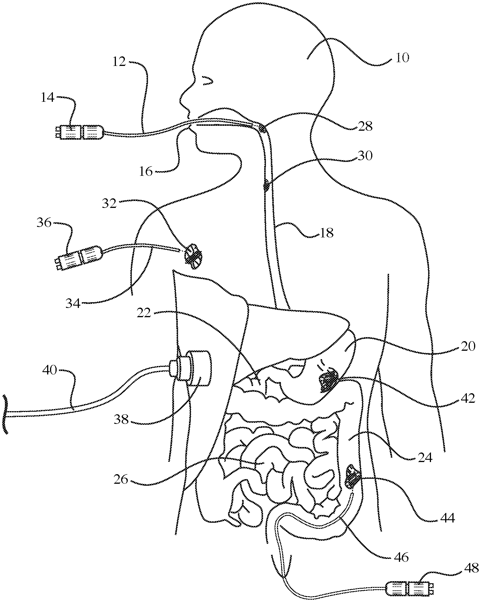
[0003] FIG. 1 is a diagrammatic illustration of a human body showing the treatment of tumors located in various areas of the body with light emitted by fiber optic devices, according to an embodiment of the invention;
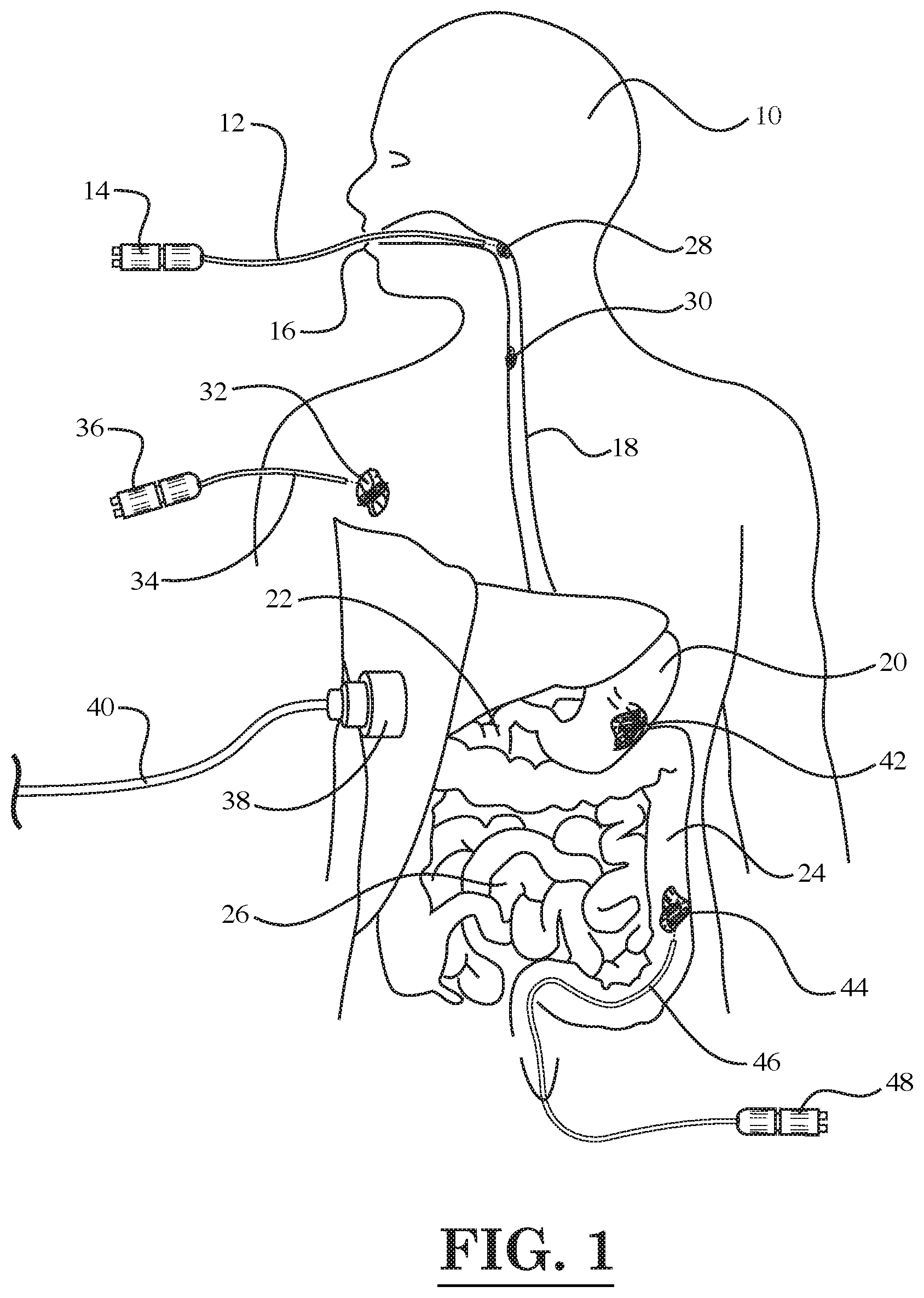
[0004] FIG. 2 is a diagrammatic illustration of a human eye showing the treatment of tumors located in several areas of the eye with light emitted by fiber optic devices, according to another embodiment of the invention;
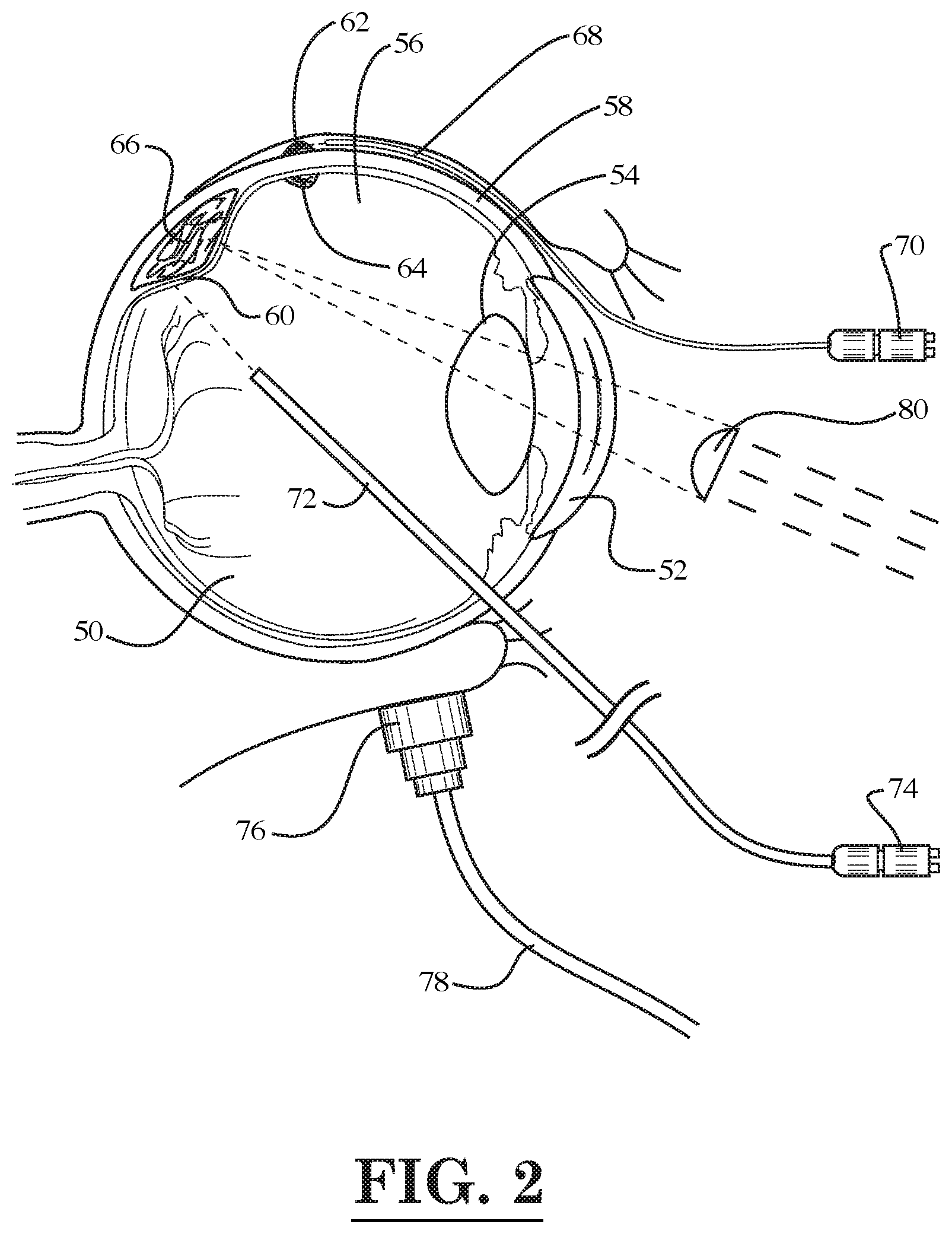
[0005] FIG. 3 is a diagrammatic illustration of several digestive organs of the human body showing the treatment of a tumor located in the pancreas with light emitted by a fiber optic device, according to yet another embodiment of the invention;
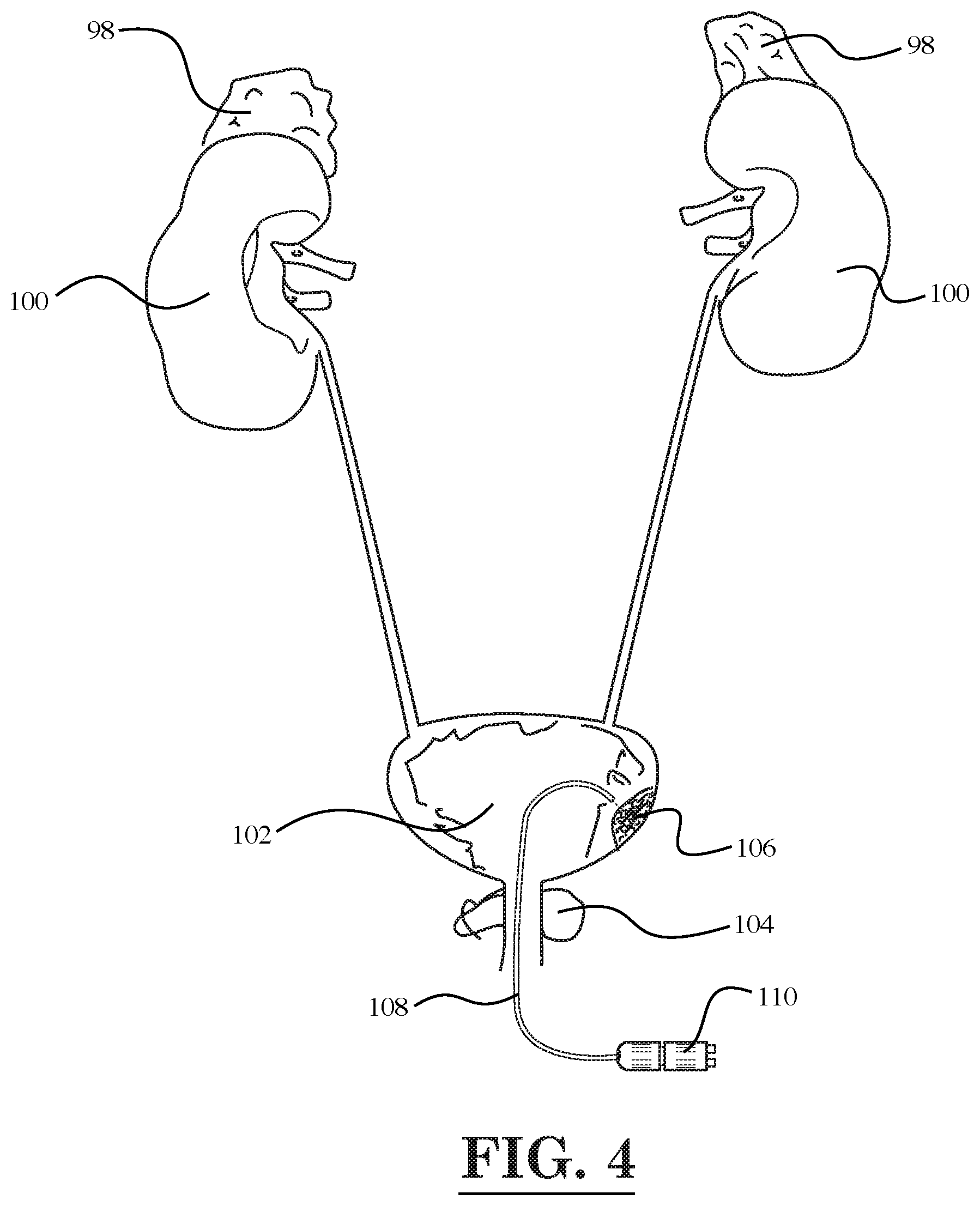
[0006] FIG. 4 is a diagrammatic illustration of the urinary system of the human body showing the treatment of a tumor located in bladder with light emitted by a fiber optic device, according to still another embodiment of the invention;
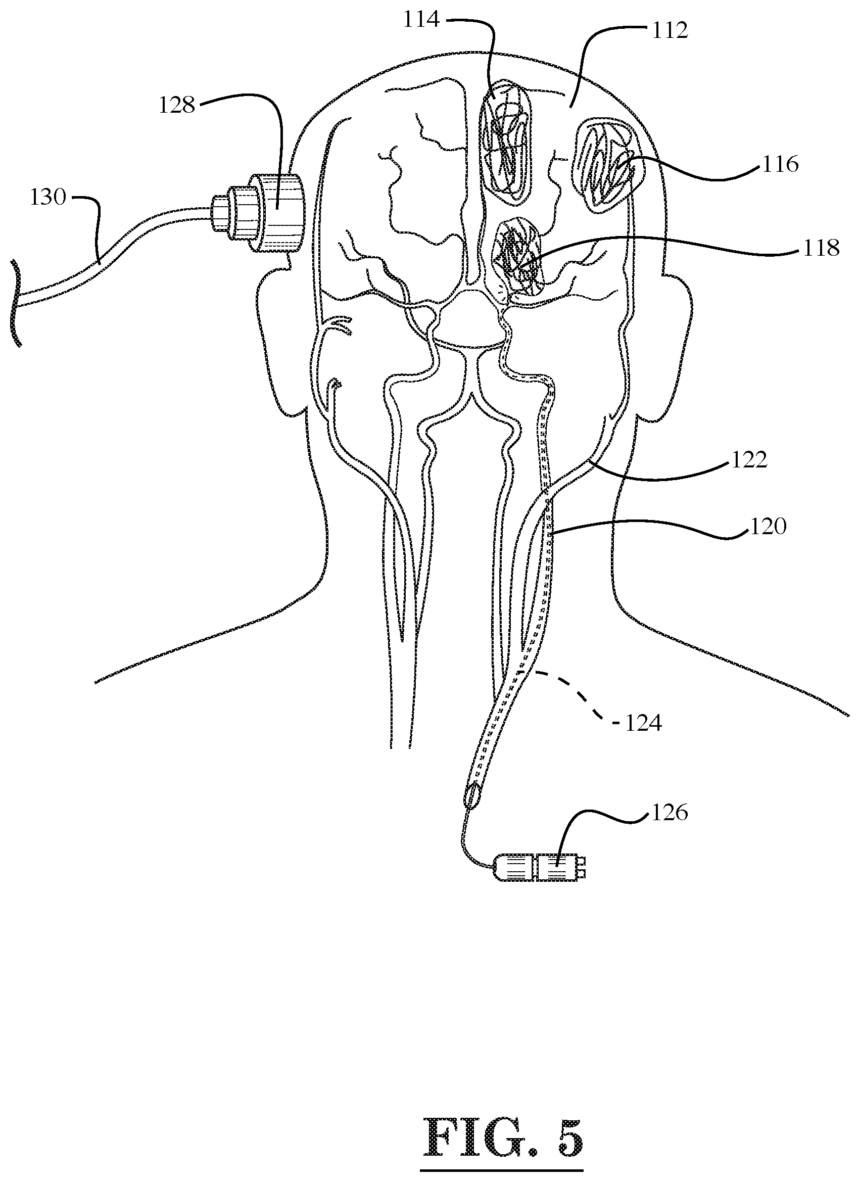
[0007] FIG. 5 is a diagrammatic illustration of the circulation system and brain of the human head showing tumors located in various areas of the brain and the treatment of one of the tumors with light emitted by a fiber optic device, according to yet another embodiment of the invention;
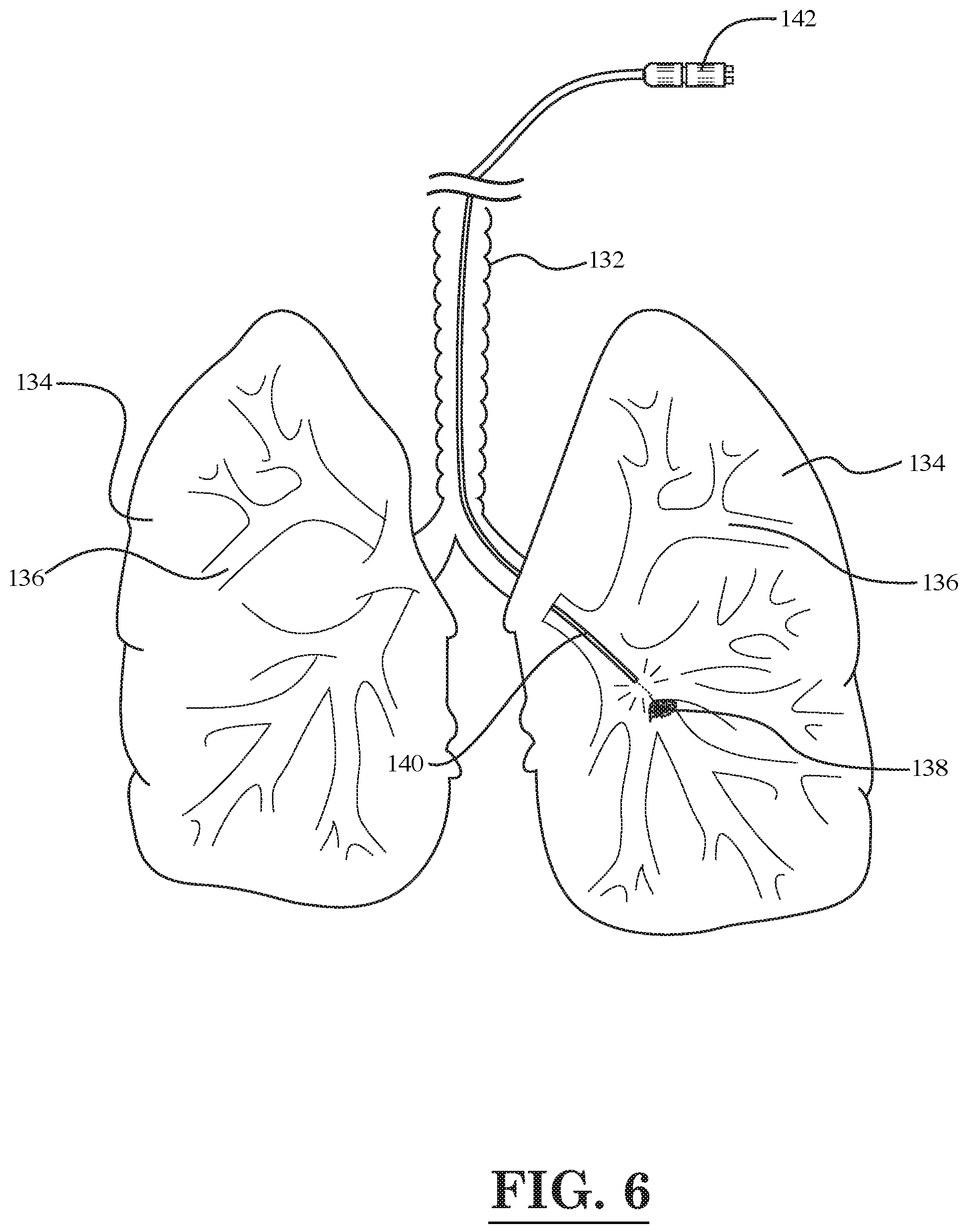
[0008] FIG. 6 is a diagrammatic illustration of the lungs and trachea of the human respiratory system showing a tumor located in one of the lungs and the treatment of the tumor with light emitted by a fiber optic device, according to still another embodiment of the invention;
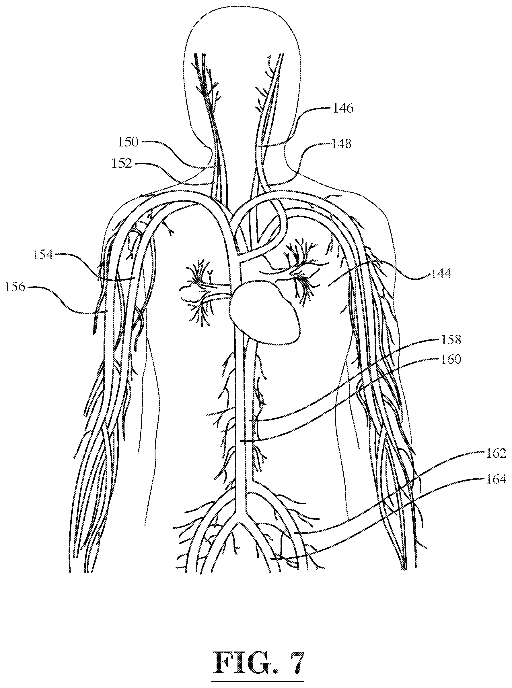
[0009] FIG. 7 is a diagrammatic illustration of the human circulatory system in the upper portion of the body;
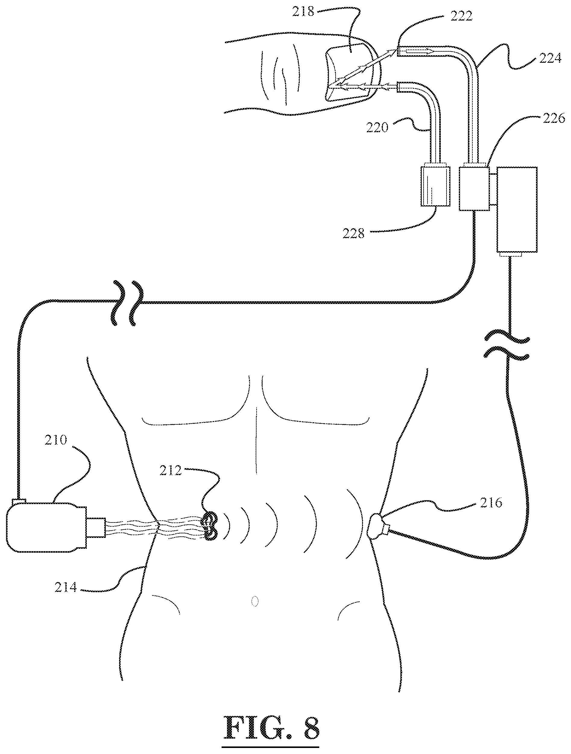
[0010] FIG. 8 illustrates a schematic diagram of a cancer treatment and evaluation system, according to an embodiment of the invention;
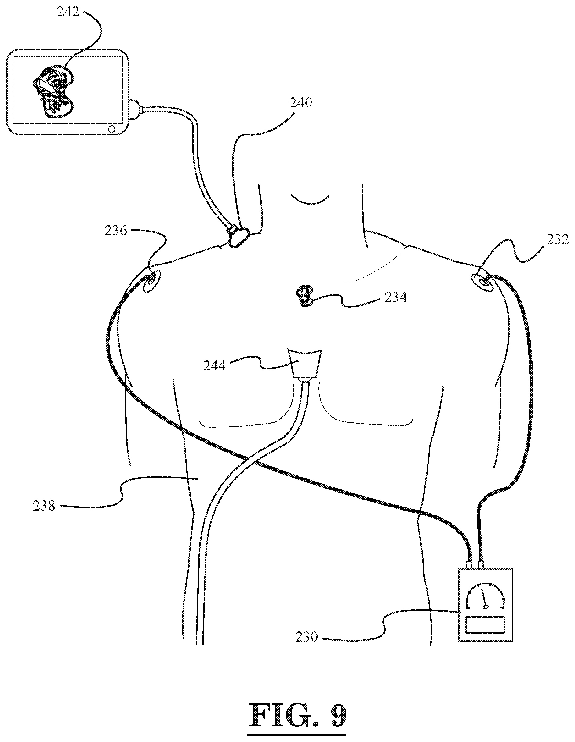
[0011] FIG. 9 illustrates a schematic diagram of a cancer treatment and imaging system, according to another embodiment of the invention;
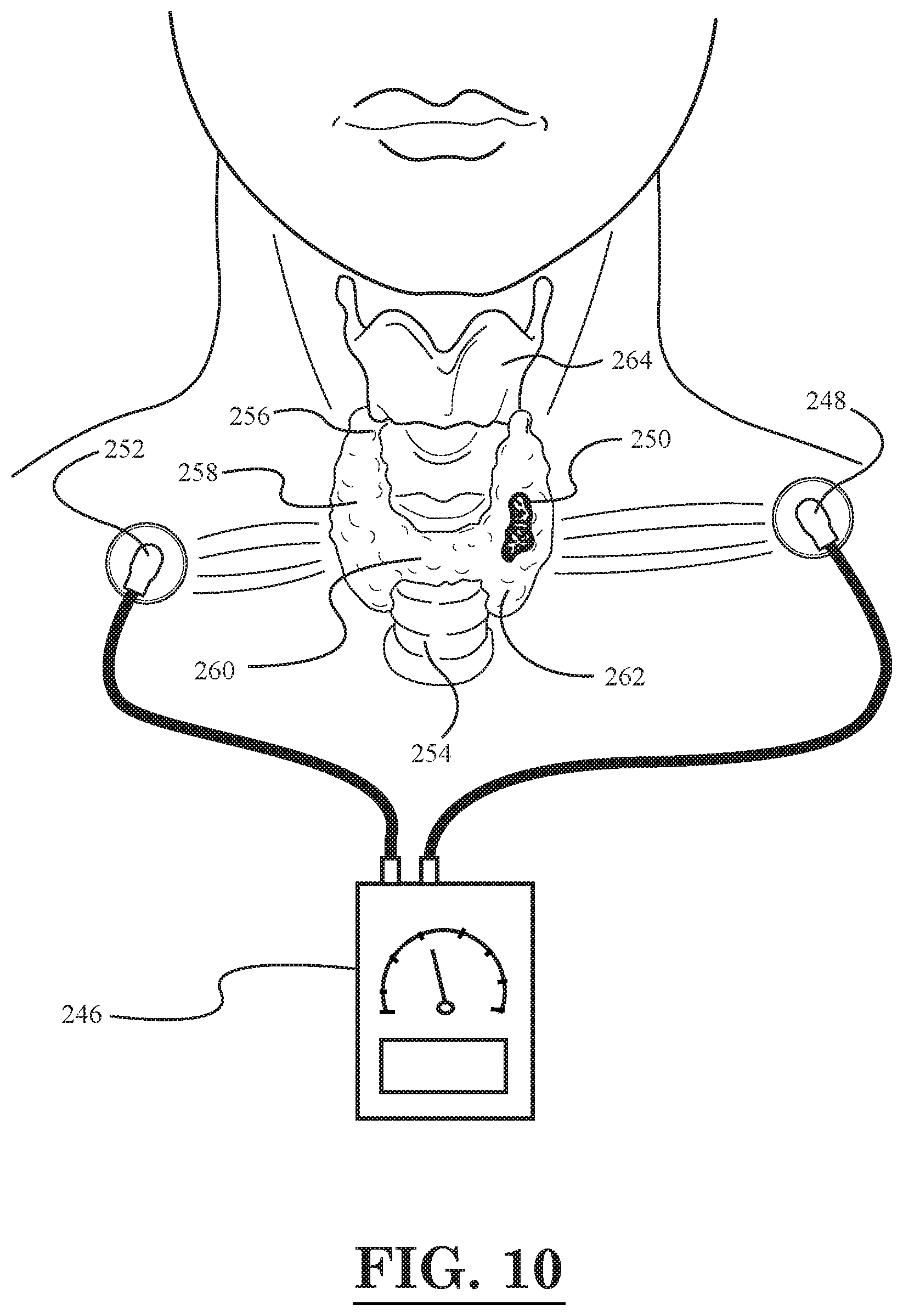
[0012] FIG. 10 illustrates a schematic diagram of a cancer treatment system, according to yet another embodiment of the invention, wherein a thyroid tumor is being treated;
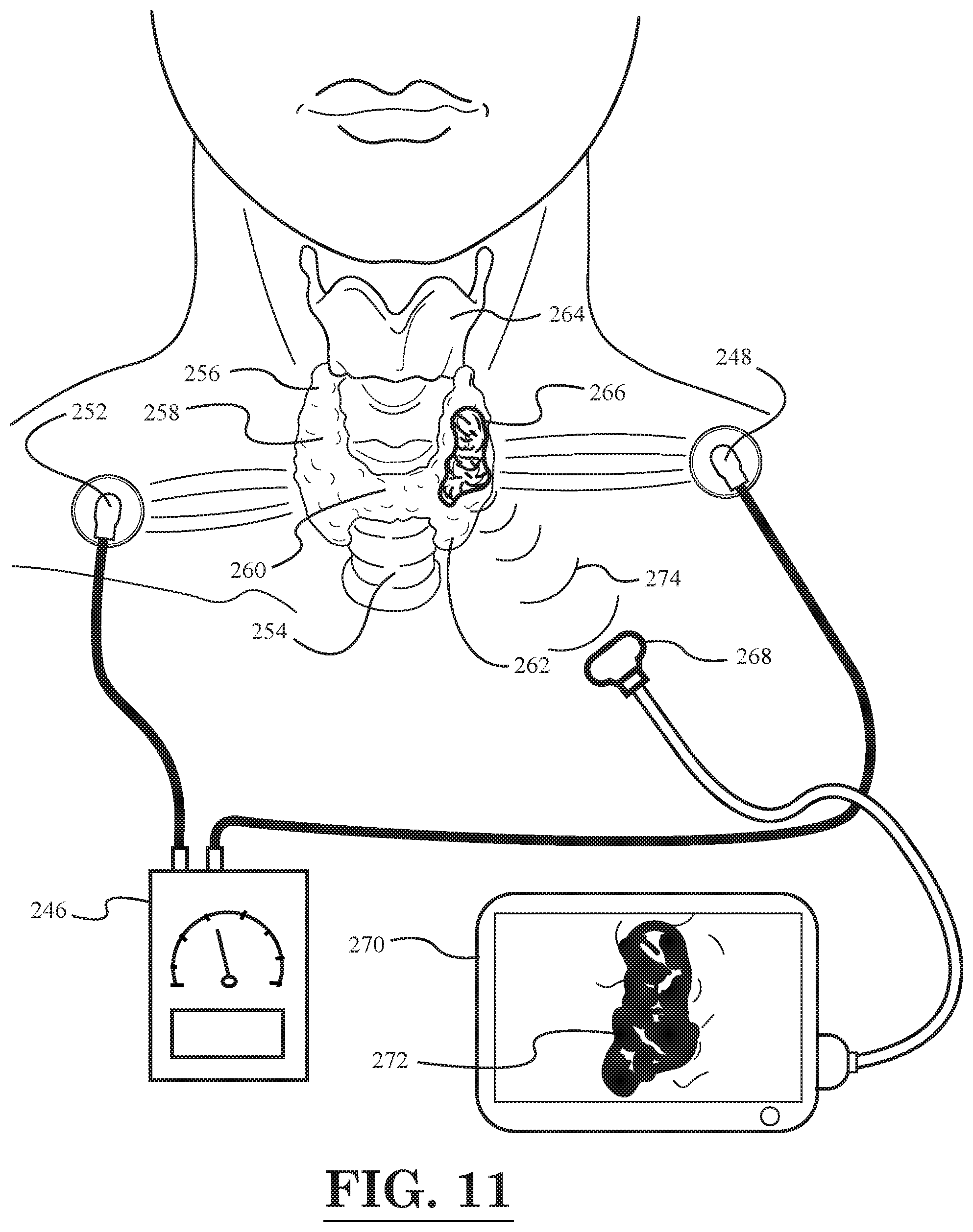
[0013] FIG. 11 illustrates a schematic diagram of a cancer treatment and imaging system, according to still another embodiment of the invention, wherein a thyroid tumor is being treated and imaged; and
[0014] FIG. 12 illustrates a schematic diagram of a cancer treatment and imaging system, according to yet another embodiment of the invention, wherein a thyroid tumor is being treated and imaged.
[0015] Various factors may lead one to suspect the presence of a small cancerous or neoplastic tumor in a patient. Such factors include the patient's genetic history, environmental conditions to which the patient is or has been exposed, the presence of biomarkers in the patient's blood, or the presence of a lesion on a patient's skin or mucosal surface. A small neoplasm of 1 to 2 millimeters (mm) in diameter, however, is often not recognized unless and until it produces some clinical symptom.
[0016] In a patient having a genetic mutation indicating a predisposition to cancer, prophylactic surgical intervention, such as a bilateral mastectomy performed in a patient having a genetic mutation indicating a predisposition to breast cancer, is seldom performed. Additionally, a genetic predisposition to one type of cancer may not lead to that type of cancer, e.g. breast cancer, but it may lead to another unsuspected type of cancer, e.g. malignant melanoma. Even if the other type of cancer is suspected, because of the finding of biomarkers in the blood, a small internal lesion may not be seen on radiography, or may not be accessible by surgery, or the collateral complications may not be acceptable. It may not suffice to just know the biomarker for a tumor, because this information may not indicate whether the tumor is a primary site or a metastatic site, the tissue of its origin, and/or its location. It is appreciated that some treatment techniques such as surgery or radiation may be useful, but only if the tumor is tissue specific. Radiation and chemotherapy also have their own side effects, and may not destroy the tumor completely. Larger tumors present a much complex problem, e.g., mutations in one area of the tumor are usually different from mutations in another area of the same tumor.
[0017] It is clearly preferable, then, to manage small early neoplasms that have not progressed to a larger tumor to provide the patient an improved clinical prognosis.
[0018] The invention includes a method of therapy for a non-surgically accessible tumor by administering a combination of thermotherapy and immunotherapy combined with gene delivery. In one or more embodiments, the gene delivery may use CRISPR-cas9 mediated Homology-Independent Targeted Integration (HITI) or Homology Directed Repair (HDR). The combination therapy beneficially treats the tumor and prevents tumor recurrence, either locally or at a different site, by boosting the patient's immune response both at the time or original therapy and/or for later therapy as a "booster" vaccine with or without viral-like particles (VLP) or adjuvants to the original therapy by administering them with antibody coated nanoparticles conjugated with checkpoint inhibitors, such as PD-1, PD-L1, CTLA-4, Jagged 1 inhibitor 15D11, etc. and anti-inflammatory agents, such as Rock inhibitors, Fasudil, etc., Wnt inhibitors, such as niclosamide etc. to enhance cellular immune response of the patient while reducing the inflammatory response and preventing an auto immune reaction or cytokine storm. In one embodiment, the vaccine with or without VLP, adjuvants, sodium bicarbonate to modify acidic tumor cell environment is combined with checkpoint inhibitors, such as PD-1, PD-L1, CTLA-4, Jagged 1 inhibitor 15D11, etc., anti-VEGF, and anti-inflammatory agents, Rock inhibitors, such as Fasudil or Botox, or Wnt inhibitor, such as niclosamide, ivermectin and Selamectin, etc., can be injected every six months or once a year, or is used in the treatment of recurrent metastatic disease. In another embodiment the antibody coated nanoparticles or solid lipid nanoparticles are coated with thermosensitive polymers which are released at the temperature or 41-42 C under thermotherapy and administered with checkpoint inhibitors, such as PD-1, PD-L1, CTLA-4, Jagged 1 inhibitor 15D11, etc. and Rock inhibitors, such as Fasudil or Botox, or Wnt inhibitor, such as niclosamide, ivermectin, or Selamectin and/or an antineoplastic medication depending on the cancer, at lower dose than is normally recommended, but is made more effective by thermotherapy.
[0019] In one embodiment, N-myristoyltransferase (NMT) in cell cycle is inhibited which is involved in cell proliferation etc. NMT conjugates with rare fatty acid and proteins. Lipid modification through inhibition of N-myristoylation interferes with cell multiplication. Numerous antimalarial, antifungal, antiparasitic, antinematodes or antifilarial, antiviral, compounds can interfere with NMT function, but specifically the compound IMP-1088 which inhibits capsid formation in viruses. However combining antibody coated nanoparticles with the compound IMP-1088, NMT inhibitors, DDD85646 and DDD100870 and cHSC70 and/or enolase can be used alone or synergistically with Wnt inhibitors or Rock inhibitors, as anticancer therapeutic agent(s) conjugated with antibody coated nanoparticles/medication injected intravenously or intra-arterially to the organ containing the tumor to inhibit the cancer cell proliferation along with controlled thermotherapy.
[0020] In one embodiment the diagnosis and precision thermo-immune therapy is performed locally or limited to an organ (e.g. head, lung, intestinal tract or extremities etc.) having radiographically or thermoacoustically imaged a small tumor, anticancer therapeutic agent conjugated with antibody coated nanoparticles/medication is injected intravenously or intra-arterially to the organ containing the tumor, while sensitizing the immune system to the tumor antigen to damage the original tumor and also attack the potentially existing circulating tumor cells, tumor exosomes, or yet invisible metastatic lesion using thermotherapy locally combined with antibody coated nanoparticles, checkpoint inhibitors, and an immune stimulator such as VLP, adjuvants, or toll like receptors, TNF alpha, etc. attached to antibody coated pluralities of nanoparticles or solid lipid nanoparticles administered simultaneously to stimulate body's immune response and eliminate the localized tumor and its circulating cells, exosomes or metastatic lesions by innate immune cells stimulation.
[0021] In one embodiment, combination antibody coated nanoparticles, such as nanoparticles or solid lipid or liposomes, dextrin, etc. conjugated with Wnt inhibitors, such as Ivermectin and Selamectin and inhibitor of N-myristoyltransferase, specifically IMP-1088, or NMT inhibitors, DDD85646 and DDD100870, checkpoint inhibitors and an immune stimulant (e.g., VLP), and sodium bicarbonate to modify acidic tumor cell environment, adjuvants, etc. administered intravenously or intra-arterially to work synergistically in preventing cancer cell proliferation of a small locally confined tumor along with controlled thermotherapy at the temperature of 40-43 degrees C. and treat simultaneous localized immune therapy.
[0022] In one embodiment, early stage tumor therapy involves the anticancer therapeutic agents conjugated with antibody coated nanoparticles/medication injected intravenously or intra-arterially to the organ containing the tumor while the thermotherapy performed with a laser fiber optic locally is brought to the tumor site through the feeding artery, or a focused ultrasound is used in a thermal or non-thermal mode from outside the body under temperature control using an imaging system or thermoacoustic temperature imaging unit.
[0023] In one embodiment, a portable photoacoustic or thermoacoustic transducer probe can be moved in any direction on the body, e.g., up and down, side to side, etc., over the skin while emitting a laser light, microwave, or focused ultrasound etc. and recording the sound waves from the antibody coated nanoparticles producing a thermal expansion of nanoparticles. Using a processor in the photoacoustic or thermoacoustic unit, one uses the photoacoustic or thermoacoustic response data to construct a two- or three-dimensional image of a tumor for diagnosis of a lesion in the body.
[0024] In one embodiment, the hand held photoacoustic/thermoacoustic probe permits scanning any bodily surface locally to diagnose presence of a tumor, including but not limited to, the skin, breast, eye, central nervous system (CNS), spinal cord, extremities, internal organs, lung, nose, chest, trachea, throat, abdomen, and urogenital organs after administration of the pluralities of antibody coated nanoparticles with cell penetrating peptides (CPP), activating CPP (ACPP), biotin, and/or streptavidin or antibody coated nanoparticles conjugated with (alpha)-cyclodextrin, (beta)-cyclodextrin, (gamma)-cyclodextrin, hydroxypropyl-b-cyclodextrin (bHPCD) or solid lipid nanoparticles injected intravenously, topically or intra-arterially, and irradiated with a source of thermal energy such as laser, focused ultrasound or an alternating magnetic field. The cyclodextrin nanoparticles may carry medication, checkpoint inhibitors, VLP, adjuvants, and/or sodium bicarbonate to modify the acidic tumor cell environment and act as slow release polymers at the tumor site, which can be released by thermal energy.
[0025] In one embodiment, a laser fiber optic can be used for diagnosis and therapy of the surface or internal lesions after administration of the pluralities of antibody-coated nanoparticles with cell penetrating peptides (CPP), activating CPP (ACPP), biotin, streptavidin or antibody coated nanoparticles conjugated with medication and (alpha)-cyclodextrin, (beta)-cyclodextrin, (gamma)-cyclodextrin, hydroxypropyl-b-cyclodextrin (bHPCD) or solid lipid nanoparticles intravenously, topically for surface tumors, skin, mucosa, or through the accessible cavities such as the eyes, vitreous cavity, bladder, mouth, throat, esophagus, stomach, duodenum, rectum, colon, small intestinal tract, and lung. In this embodiment, the antibody/cyclodextrin coated nanoparticles carry medication, such as Rock inhibitors, Wnt inhibitors and/or Inhibitors of N-myristoyltransferase, specifically IMP-1088, or NMT inhibitors, DDD85646 and DDD100870 and checkpoint inhibitors, or VLP, adjuvants, sodium bicarbonate to modify the acidic tumor cell environment and immune stimulators and act as slow release polymers delivered via a flexible fiber optic and a tube is inserted through the natural orifices of the body (see FIGS. 1 and 4) or through a feeding artery or vein (see FIG. 5) to bring the laser pulses close to the tissue containing a tumor, and deliver the nanoparticles, but also use the laser energy to heat the accessible lesion or apply thermal energy externally using microwave, focused ultrasound in thermal and non-thermal modes releasing the medication(s) and stimulate the immune system to attack the tumor cells locally or wherever they are systemically by cellular immune response. In one embodiment, the methods of delivery of antibody coated nanoparticles/medications are internal through the arteries or veins or through the body orifices as shown by the drawings (see FIGS. 1 and 4-7) or through an incision (e.g., made in the abdomen, etc. or subcutaneous or intramuscular, etc. injection in the desired location or tissue, or by topical application).
[0026] For example, as shown in FIG. 1, one laser fiber optic device 12 may be inserted through the mouth 16 of a person 10 so as to treat an esophageal tumor 28 by emitting light pulses generated a light source 14. In addition, the laser fiber optic device 12 may be further inserted into the esophagus 18 so as treat another tumor 30 located further down in the esophagus 18. In addition, as shown in FIG. 1, a skin tumor 32 located near the right shoulder of the person 10 may be treated with a laser fiber optic device 34 having a light source 36. Next, turning to the digestive system in FIG. 1, which includes the stomach 20, duodenum 22, large intestine 24, and small intestine 26, further aspects of the illustrated embodiment will be described. In FIG. 1, it can be seen that a tumor 42 is disposed in the stomach 20, and another tumor 44 is disposed in the large intestine 24. The tumor 44 in the large intestine 24 is being treated with a laser fiber optic device 46 that has been inserted into the large intestine 24 through the rectum. The laser fiber optic device 46 has a light source 48. Also, as shown in FIG. 1, a photoacoustic receiver 38 with cord 40 may be placed against the body of the person 10 in order to record a photoacoustic/thermoacoustic response resulting from the thermal expansion of nanoparticles attached to the tumor (e.g., attached to tumor 42 in stomach 20) that are heated by the laser light pulses from a fiber optic device. As described above, the nanoparticles may be delivered to the tumor site prior to the heating thereof by a tube attached to the fiber optic device.
[0027] In one embodiment, the thermotherapy is done either internally or externally using a laser applied through a fiber optic, etc. (see e.g., FIGS. 1-6). In one embodiment, the antibody coated nanoparticles/medications are delivered intra-arterially or intravenously, but the thermotherapy is done externally using focused ultrasound, microwaves, radio frequency (RF), or using an alternating magnetic field when nanoparticles are magnetic or paramagnetic.
[0028] In some situations, as in the brain, the thermotherapy can be done at a low temperature (e.g., with focused ultrasound) that make the tumor vessels leakier prior to the injection on the antibody-coated nanoparticles. In another embodiment, the nanoparticles are injected prior to the thermotherapy, etc.
[0029] In one embodiment, the source of energy is a focused ultrasound in a non-thermal or thermal mode applied from the outside the body while the pluralities of antibody-coated nanoparticles/medications are piezoelectric, such as quartz, graphene, or perovskites, nanobubbles, perfluorocarbon liquid filled vesicles, etc., and administered intra-arterially or intravenously, etc.
[0030] In one embodiment, as shown in the embodiment of FIG. 5, the delivery of antibody coated nanoparticles/medication is done through an artery and thermotherapy is done with an internal laser using a fiber optic.
[0031] In one embodiment, one can deliver the antibody coated nanoparticles/medication internally and heat up the tumor preferably externally (e.g., by using a focused ultrasound). This is preferred for internally located tumors, such as in the brain and internal organs that cannot be reached by the laser or the size of the tumors are mostly larger than 4 millimeters (mm) in diameter.
[0032] In another embodiment, any method or delivery of nanoparticles can be used depending on the location of the tumor and its thickness. Lasers are used mostly for small accessible lesions or superficial tumors, or laser accessible tumors via a fiber optic or external laser for tumors having a thickness of about 1-4 mm. All other tumors can be preferably treated by a source of energy that penetrates deep in the tissue (e.g., focused ultrasound, microwaves, radio frequency (RF), or alternating magnetic field). In all cases, thermal delivery and temperature are controlled by either a photoacoustic imaging or thermoacoustic imaging unit to the desired level of temperature which is predetermined.
[0033] In one embodiment, a miniature capsule with an imaging camera, which is equipped with a laser system, is swallowed by the patient that constantly radiates a laser pulse, as it passes through the intestinal tract and transmits recorded images to a receiver outside the body.
[0034] In one embodiment, a photoacoustic sound wave is produced when pluralities of antibody-coated nanoparticles are injected intravenously with cell penetrating peptides (CPP), activating CPP (ACPP), biotin, streptavidin, and/or the cyclodextrin antibody-coated nanoparticles carry medication, inhibition of N-myristoyltransferase such as many anti malaria parasites medication, specifically IMP-1088, or NMT inhibitors, DDD85646 and DDD100870, checkpoint inhibitors, or VLP, adjuvants, sodium bicarbonate to modify the acidic tumor cell environment and immune stimulators, such as TNF alpha, toxin, etc., and act as slow release polymeric drug release, where the nanoparticles accumulate at the site of the tumor(s) in the intestinal tract and are stimulated with the laser pulses which heat and create a photoacoustic sound that is recorded by a receiver located outside the body and in contact with body surface, such as over the abdomen, etc., and is analyzed by software while an internal capsule with a video camera is traveling through the intestine and locates the presence of a tumor.
[0035] In one embodiment, as the capsule passes in front of a lesion in the intestinal tract, which has accumulated the pluralities of antibody coated nanoparticles or solid lipid nanoparticles thereon, the laser pulse creates a photoacoustic sound that can be recorded by a receiver positioned on the trunk of the patient and records the image of the lesion and the temperature at that site as the capsule travels through the intestine.
[0036] In one embodiment, the photoacoustic sound can be correlated with the video taken by the capsule and the location of the tumor is determined even if the tumor is too small to be recognized by CT scan or radiography or too small to make any visible or physical symptom. In one embodiment, the capsule emits a significant amount of energy to increase the temperature at the tumor site to release the medication, gene, from the antibody-coated nanoparticles with cell penetrating peptides (CPP), activating CPP (ACPP), biotin, streptavidin or the cyclodextrin antibody-coated nanoparticles which carry medication, such as Rock inhibitors, Wnt inhibitors or inhibitors of N-myristoyltransferase, IMP-1088, NMT inhibitors, DDD85646 and DDD100870, and checkpoint inhibitors, and/or VLP, adjuvants, sodium bicarbonate to modify acidic tumor cell environment and other immune stimulators, such as TNF alpha, IL 2 IL-6, toll like receptor 7/8 that stimulate innate immune cell response, etc. and are released from the slow release polymers, and damage and kill the tumor cells while releasing their cellular antigens in the circulation to attract cellular immune response and kill the remaining tumor cells that are present locally or might exist in the body.
[0037] In one embodiment, a laser fiber optic with or without the camera, while pulsing laser energy, is passed through the mouth to the stomach or through the rectum into the colon or through the ureter inside the bladder, through the mouth, throat, trachea, and bronchi, etc. or through the vagina inside the uterus or further through one of the fallopian tubes toward the ovaries.
[0038] In one embodiment, the laser pulse produces a photoacoustic or thermoacoustic response from the pluralities of antibody-coated nanoparticles with cell penetrating peptides (CPP), activating CPP (ACPP), biotin, streptavidin or the cyclodextrin antibody coated nanoparticles carrying medication, such as Rock inhibitors, Wnt inhibitors or inhibitors of N-myristoyltransferase, such as many anti-malaria parasites medication, specifically IMP-1088 compound or NMT inhibitors, DDD85646 and DDD100870, and checkpoint inhibitors, or VLP, adjuvants, sodium bicarbonate to modify the acidic tumor cell environment and immune stimulators, such as TNF alpha, IL6, IL 17, toll like receptors, etc. and act as slow release polymeric drug release carrying medication to be attached to the tumor cells injected intravenously or intra-arterially 1-2 or more minutes ahead, permitting the nanoparticles to travel in the body and attach to the tumor cells that can be exposed to laser radiation, focused ultrasound, microwave or an alternating magnetic field to heat the nanoparticles and to produce a photoacoustic or thermoacoustic sound to be recorded by a photoacoustic or thermoacoustic transducer (an ultrasound receiver) located on the surface of the body to image the tumor while measuring the temperature generated at the tumor site by the laser or other energy source, such as focused ultrasound, microwave, or alternating magnetic field to image the temperature and the tumor in a 2-D and 3-D format, increasing the thermal radiation by a processor connected to the photoacoustic unit and thermal delivery unit to increase the tumor temperature and damage or kill the tumor cells at temperatures of 43 to 45-47 degrees C. and release cytoplasmic tumor antigens to attract dendritic cells, T-cells, and/or other killer cells to remove the tumor as they circulate in the body destroying the circulating or sessile tumor cells elsewhere in the body.
[0039] In one embodiment, where a tumor is inaccessible through the natural orifices, a fiber optic endoscope 94 is inserted through a small incision in the abdomen in the peritoneal cavity toward liver, spleen, pancreas, or kidney (see e.g., FIG. 3, which depicts a tumor 96 located in the pancreas 90) for diagnostic or therapeutic purposes using the laser thermal energy, to recognize the location of the tumor after injecting intravenously the antibody-coated pluralities of nanoparticles with cell penetrating peptides (CPP), activating CPP (ACPP), biotin, and/or streptavidin, or antibody-coated nanoparticles conjugated with (alpha)-cyclodextrin, (beta)-cyclodextrin, (gamma)-cyclodextrin, hydroxypropyl-b-cyclodextrin (bHPCD) conjugated with a thermosensitive polymer carrying medication and/or a gene, medication or immune stimulators. In this embodiment, shining the laser light over the suspected tumor/nanoparticle complex area creates a photoacoustic sound, which is imaged by a receiver or receivers located on the skin of the abdomen, then heating preferentially the antibody-coated pluralities of nanoparticles attached to the tumor cells by a laser, microwaves, or focused ultrasound, etc. damaging the tumor cells at a temperature of 37-43 degrees C., thereby releasing the conjugated medication, gene, toxins, Wnt inhibitors, or Rock inhibitors along with immune stimulators to kill and eliminate the cancer cells. In FIG. 3, several digestive organs of the human body are illustrated around the area of the pancreas 90 being treated by the fiber optic device 94, namely the gallbladder 82, liver 84, and duodenum 85. Also, illustrated in FIG. 3 are the bile duct 86, the pancreatic duct 88, and the opening 92 of the bile and pancreatic ducts 86, 88 into the duodenum 85.
[0040] In one embodiment, other tumors inside the body can be accessed through insertion of a fiber optic through the blood vessels, arteries, or veins of an organ (see e.g., FIG. 5) to induce a more organ specific diagnosis, and thermo-immune therapy without affecting the normal cells (e.g., in the brain, eye, or extremities) for tumors localized in the head and neck or urogenital organs, lung, etc.
[0041] In one embodiment, the thermotherapy is performed by injecting intravenously or intra-arterially, antibody or monoclonal antibody-coated pluralities of nanoparticles or solid lipid nanoparticles conjugated with thermosensitive polymers such as chitosan/medication or antibody coated nanoprticles conjugated with (alpha)-cyclodextrin, (beta)-cyclodextrin, (gamma)-cyclodextrin, hydroxypropyl-b-cyclodextrin (bHPCD, liposomes, solid lipid nanoparticles with nanowires, nanotubes, nanoshells, nanocages, periodic mesoporous organosilica nanoparticles conjugated with immune stimulator(s), such as interferon alpha, toll like receptors, IL 2, IL6, IL17, or VLP, adjuvants, sodium bicarbonate to modify the acidic tumor cell environment and CPP, with medication, inhibitors of N-myristoyltransferase, such as many anti-malaria parasites medication, specifically IMP-1088, or NMT inhibitors, DDD85646 and DDD100870, and Rock inhibitors, Wnt inhibitors which interferes with cellular proliferation or is injected through a major artery after insertion of a flexible laser fiber optic with a tube or a needle in a few steps of therapy: (a) administering the pluralities of antibody coated nanoparticles or liposomes, solid lipid nanoparticles, etc.; (b) applying thermal energy to the lesion and imaging it by a thermoacoustic unit to control the temperature to 40-43 degrees C., to release the medication from the nanoparticles or liposomes or solid lipid nanoparticles at the tumor site and irradiate the tumor cells with thermal energy; (c) continue administering antibody coated nanogel/nanoparticles, such as quantum dots or other nanoparticles such as gold or silica, magnetic, non-magnetic or hydrogels or liposomes or solid lipid nanoparticles (e.g., made of desired molar concentration and the ANG nanogel content) with or without various concentrations of a crosslinker, fibrinogen, adenosine diphosphate (ADP) to convert thrombinogen to thrombin and the fibrinogen to fibrin locally at the site of the tumor vasculature creating a precise localized vascular occlusion at the tumor site only while sparing the normal surrounding tissue.
[0042] In one embodiment, in contrast to the standard technique of embolization of a large vessel supplying a tumor which leads to indiscriminate closure of supply of a large area of an organ and damaging normal cells, the precise thermotherapy damages the endothelial cells and the tumor cells, releasing the medication from the antibody coated nanoparticles, namely releasing prothrombotic medication, checkpoint inhibitors, immune stimulators, anti-VEGF enhancing platelets binding and aggregation, and clotting; thus creating a discrete blood clot or thrombus, which obstructs the blood supply to the tumor, and starves the tumor from its blood supply while releasing the immune stimulating agents and medications slowly from the polymeric nanoparticle compounds and provide a long term local or systemic immunotherapy to the patient, and withdrawing blood after therapy one obtains increased tumor biomarkers proving the presence of a tumor even it was not initially not visible radiologically and administering antibody-coated pluralities of nanoparticles coated with dimethylacetyl-beta-cyclodextrin to inhibit excessive innate immune response locally and prevent excessive edema at the tumors surrounding site.
[0043] In one embodiment, one creates a precise local temperature increase and vascular occlusion at the site of the tumor and tumor vessels regardless of its location in the body and the size of the lesion, the injected antibody or monoclonal antibody-coated pluralities of nanoparticles conjugated with thermosensitive polymers, such as chitosan, and/or medication or antibody-coated nanoparticles conjugated with (alpha)-cyclodextrin, (beta)-cyclodextrin, (gamma)-cyclodextrin, hydroxypropyl-b-cyclodextrin (bHPCD, liposomes or solid lipid nanoparticles containing nanowires, nanotubes, nanoshells, nanocages, periodic mesoporous organosilica nanoparticles with immune stimulator(s), such as interferon alpha, toll like receptors, IL 2, IL6, IL17 or VLP, adjuvants, sodium bicarbonate to modify the acidic tumor cell environment and CPP, medication such as inhibitors of N-myristoyltransferase, such as many anti-malaria parasites medication, specifically IMP-1088 or NMT inhibitors, DDD85646 and DDD100870, Rock inhibitors, Wnt inhibitors are given intravenously but preferably through a feeding artery after insertion of a flexible laser fiber optic with a tube or just a small gauge needle in the vessel in different steps of therapy: (a) administering the pluralities of nanoparticles or combined with liposomes or solid lipid nanoparticles, etc.; (b) applying thermal energy to the lesion either with laser or focused ultrasound or an alternating magnetic field, microwave, etc. and using thermoacoustic imaging to control the energy delivery system to a temperature of 40-43 degrees C., to release the medication and damage the tumor cells membrane and the their endothelial cells membrane of their feeding vessels and capillaries supplying the tumor cells; (c) releasing from the antibody-coated nanogel/nanoparticles, such as quantum dots, liposomes, solid lipid nanoparticles, or other nanoparticles, such as gold or silica, magnetic, non-magnetic or hydrogels or liposomes or solid lipid nanoparticles or cyclodextrin, e.g., coated with a desired molar concentration, fibrinogen, adenosine diphosphate (ADP) to release them and to convert locally thrombinogen to thrombin and the fibrinogen to fibrin, producing a local thrombus locally at the site of the tumor vasculature, thus creating a precise vascular occlusion at the area of the tumor under observation. This is in contrast to the presently done embolization of a large vessel that not only supplies a tumor but large areas of an organ indiscriminatingly, such as in liver, brain, spleen, pancreas, lung, kidney, genitourinary system, or any other part of the body.
[0044] In one embodiment, after intravenous injection of antibody-coated nanoparticles, a localized thermotherapy is performed that damages the tumor cells and the endothelial cells membrane enhancing platelets binding and creating a discrete blood clot or thrombus, which obstructs the blood supply to the tumor precisely, and starves the tumor from oxygen and nutrition, while releasing from the nanoparticles, the innate body's immune stimulating compound agents, such as antibody coated nanoparticles conjugated with interferon alpha, toll like receptors, IL 2, IL6, IL17 and adjuvants and checkpoint inhibitors with CPP, anti-VEGF and medications, such as Rock inhibitors, Wnt inhibitors or inhibition of N-myristoyltransferase which interferes with cellular proliferation, to be released slowly for a long time from the polymeric compounds of the nanoparticles and provide a long term local or systemic immunotherapy to the patient followed with withdrawing blood after therapy to obtain tumor biomarkers for the future vaccine production.
[0045] In one embodiment, the injection of antibody or monoclonal antibody coated pluralities of nanoparticles conjugated with thermosensitive polymers, such as chitosan, and/or medications and antibody-coated nanoparticles conjugated with (alpha)-cyclodextrin, (beta)-cyclodextrin, (gamma)-cyclodextrin, hydroxypropyl-b-cyclodextrin (bHPCD) with anti-VEGF, immune stimulator(s), such as interferon alpha, toll like receptors, IL 2, IL6, IL17, or VLP, adjuvants, sodium bicarbonate to modify the acidic tumor cell environment and CPP is done through an artery after insertion of a flexible laser fiber optic with a tube in these different steps of therapy: (a) administering the pluralities of antibody-coated nanoparticles carrying checkpoint inhibitors, along with immune stimulators; (b) applying thermal energy to the lesion and simultaneous thermal imaging to control the temperature to 40-43 degrees C., to release the medication from the coating of the nanoparticles and damage and/or irritate the tumor cells membrane; (c) administering antibody-coated platelets from the patient's blood, coating the platelets with antibodies, or monoclonal antibodies and reintroducing the coated platelets into the patient to attach to the tumor and its associated vascular supply, creating a blood clot or thrombus, which obstructs the blood supply to the tumor, and nutritionally starves the tumor while the immune stimulating agents and medications continue to be released slowly for a long time from the polymeric compounds and provide a long term local or systemic immunotherapy to the patient, which can be also injected later in another body location to act as a vaccine.
[0046] In one embodiment, the thermotherapy of the injected antibody or monoclonal antibody-coated pluralities of nanoparticles conjugated with thermosensitive polymers, such as chitosan, and/or medication with immune stimulator(s) interferon alpha, toll like receptors, IL 2, IL 6, IL17, or VLP and CPP is done through an artery after insertion of a laser fiber optic with a tube in at least four different steps of therapy: (a) administering the pluralities of nanoparticles, (b) applying thermal energy to the lesion and imaging to control the temperature to 40-43 degrees C., to release the medication and irritate the tumor cells, (c) administering antibody coated nanogel/nanoparticles, such as quantum dots, or other nanoparticles such as gold or silica, magnetic, non-magnetic or hydrogel (e.g., made of desired molar concentration and the ANG nanogel content with or without various concentrations of a cross-linker) to attach to the tumor and its associated vascular supply, enhancing platelets binding and clotting, creating a blood clot or thrombus, which obstructs the blood supply to the tumor, and nutritionally starves the tumor while the immune stimulating agents and medications continue to be released slowly for a long time from the polymeric compounds and provide a long term local or systemic immunotherapy to the patient, and (d) withdrawing blood after therapy for increased tumor biomarker support for the existence of a tumor even it was not initially visible radiologically for creating a vaccine that can be injected at a later time subcutaneously.
[0047] In one embodiment, injection of antibody or monoclonal antibody-coated pluralities of nanoparticles conjugated with thermosensitive polymers, such as chitosan, and/or medication or antibody-coated nanoparticles conjugated with (alpha)-cyclodextrin, (beta)-cyclodextrin, (gamma)-cyclodextrin, hydroxypropyl-b-cyclodextrin (bHPCD) with immune stimulator(s), such as interferon alpha, toll like receptors, IL 2, IL6, IL17, or VLP, adjuvants, sodium bicarbonate to modify the acidic tumor cell environment and CPP, Wnt inhibitors or Rock inhibitors, anti-VEGF is done through an artery after insertion of a flexible laser fiber inside a tube imaged during its insertion in an artery for immune thermotherapy by: (a) administering the pluralities of antibody coated nanoparticles; (b) applying external or internal laser thermal energy, focused ultrasound, microwave or an alternating magnetic field to the lesion and thermoacoustic imaging to control the temperature to 40-43 degrees C., to release the medication and irritate the tumor cells and endothelial cells of the tumor; (c) administering antibody-coated nanogel/nanoparticles, such as quantum dots or other nanoparticles, such as gold or silica, magnetic non-magnetic or hydrogel (e.g., made of desired molar concentration and the ANG nanogel content with or without various concentrations of a photosensitizer) to attach to the tumor and its associated vascular supply, enhancing platelets binding and clotting, creating a blood clot or thrombus, which obstructs the blood supply to the tumor, and starves the tumor from its nutrition and oxygen while releasing immune stimulating agents and medications slowly in a continuous manner from the polymeric coated nanoparticles and providing a long term local or systemic immunotherapy to the patient; (d) withdrawing blood after therapy to obtain increased tumor biomarkers that support the presence of a tumor even it was not initially visible radiologically; and (e) administering antibody coated pluralities of nanoparticles coated with dimethylacetyl-beta-cyclodextrin to inhibit excessive innate immune response locally and prevent excessive edema at the tumors surrounding site.
[0048] In one embodiment, the laser fiber optic is inside a flexible tube through which one injects pluralities of antibody-coated nanoparticles with cell penetrating peptides (CPP), activating CPP (ACPP), biotin, and/or streptavidin or antibody-coated nanoparticles conjugated with (alpha)-cyclodextrin, (beta)-cyclodextrin, (gamma)-cyclodextrin, hydroxypropyl-b-cyclodextrin (bHPCD) conjugated with thermosensitive polymers, such as PLA, PGA, chitosan, polyanhydride, polyanhydride, porous silicon carrying medication, siRNS, DNA, RNAi, CRISPR-cas9, inhibitors of N-myristoyltransferase, such as many anti-malaria parasites medication, specifically IMP-1088, NMT inhibitors, DDD85646 and DDD100870, Wnt inhibitors, or Rock inhibitors or CAR-t cells grown in cell culture and sensitized to the tumor antigen along with immune stimulation with VLP, toll-like receptors, adjuvants, sodium bicarbonate to modify the acidic tumor cell environment, toll like receptor 7/8 or interferons antibody-coated nanoparticles dendrimers, etc. can be injected in the circulation locally, intra-arterially to attach to the tumor cells and damage their cell membranes with thermal energy, such as laser, focused ultrasound, alternating magnetic field, or microwaves, etc. increasing the nanoparticles' and/or tumor cell's temperature to 40-43 degrees C. or more, and releasing the medication, immune stimulator, locally at the tumor site and enhance local immune therapy for a long duration and innate immune stimulation for a long period of time due to the release of the immune stimulators from the nanoparticles or porous silicone nano or microparticles, and withdrawing blood after therapy to measure increased tumor biomarkers and use them for vaccine therapy.
[0049] In one embodiment, E-selectin coated pluralities of antibody-coated nanoparticles are injected intravenously with cell penetrating peptides (CPP), activating CPP (ACPP), or antibody-coated nanoparticles conjugated with (alpha)-cyclodextrin, (beta)-cyclodextrin, (gamma)-cyclodextrin, hydroxypropyl-b-cyclodextrin (bHPCD), biotin, and/or streptavidin conjugated with medication and an Wnt inhibitor binds to sialylated carbohydrates on the surface proteins of certain leukocytes, increasing the nanoparticle temperature using a source of thermal energy to 40-43 degrees C. or more, and releasing the medication locally to the tumor cells and attracting neutrophils, monocytes, eosinophils, memory-effector T-like lymphocytes, and natural killer cells to further damage and remove the tumor cells.
[0050] In one embodiment, the CAR-T cells or killer cells are grown in a tissue culture with antibody-coated nanoparticles with cell penetrating peptides (CPP), activating CPP (ACPP), biotin, and/or streptavidin, or antibody-coated nanoparticles conjugated with (alpha)-cyclodextrin, (beta)-cyclodextrin, (gamma)-cyclodextrin, hydroxypropyl-b-cyclodextrin (bHPCD) which are conjugated with E-selectin to attach to the surface of the CAR-T cells and conjugated with VLP, adjuvants, sodium bicarbonate to modify the acidic tumor cell environment or other immune stimulating agents conjugated with polymeric nanoparticles or porous silicon nanoparticles coated with slow release polymers of PAL, GA, PLGA, or chitosan administered before and after thermotherapy to attach to the tumor cells and enhance cellular immune response after thermotherapy for 2 to 3 months or more as long as the slow release polymers last in the body.
[0051] In one embodiment, the CAR-T cells or killer cells are in grown tissue culture with antibody-coated nanoparticles with cell penetrating peptides (CPP), activating CPP (ACPP), biotin, and/or streptavidin, or (alpha)-cyclodextrin, (beta)-cyclodextrin, (gamma)-cyclodextrin, hydroxypropyl-b-cyclodextrin (bHPCD) which are conjugated with E-selectin to attach to the surface of the CAR-T cells and a medications, toxins, enzymes, TNF, TRAIL, VLP, adjuvants, sodium bicarbonate to modify the acidic tumor cell environment, toxins, propranolol, a beta blocker, or an anti-VEGF to inhibit the tumor's vascular growth and can be injected intra-arterially at less than 1/10- 1/50th of the quantities used systemically, through laser fiber optic tube slowly before and after thermotherapy of a localized tumor, to be released slowly to attack repeatedly, the tumor cells in the specific organ, and induce a localized lasting immune response or induce an immune response in the body to eliminate potential existing tumor cells or invisible metastasis and autoimmune response.
[0052] In one embodiment, the release of antibody-coated pluralities of nanoparticles with cell penetrating peptides (CPP), activating CPP (ACPP), biotin, and/or streptavidin or antibody-coated (alpha)-cyclodextrin, (beta)-cyclodextrin, (gamma)-cyclodextrin, hydroxypropyl-b-cyclodextrin (bHPCD) are observed after intra-arterial injection with an imaging system such as MRI, or ultrasound to verify the position of the tumor that is being treated with controlled thermotherapy using electromagnetic radiation, microwave, radiofrequency (RF), or focused ultrasound or alternating magnetic field or an electric field, and the lesion is imaged by a photoacoustic or thermoacoustic imaging system, the temperature is controlled by the thermoacoustic unit connected via a processor to the thermal delivery system to control thermal energy delivery, and prevent over cooking of the tissue via the thermoacoustic imaging system that measures the temperature rise 100 times per second and is in communication with the thermal energy delivery device so that the desired temperature of, for example, 41-43 degrees C. is achieved or maintained for a preferred time.
[0053] In one embodiment, the laser fiber optic with the tube is inserted through the carotid artery to reach either sides of the CNS harboring a tumor, such as glioblastoma, etc. to release pluralities of antibody-coated nanoparticles with cell penetrating peptides (CPP), activating CPP (ACPP), biotin, and/or streptavidin, Wnt or Rock inhibitors or antibody coated (alpha)-cyclodextrin, (beta)-cyclodextrin, (gamma)-cyclodextrin, hydroxypropyl-b-cyclodextrin (bHPCD) conjugated with polymers, such as PLA or PGA, nanoparticles of porous silicon, chitosan attached with medication or immune stimulators in the circulation so that the majorities of nanoparticles reach at a high concentration at the tumor site and are released as needed by application of internal or external thermal energy such as laser, focused ultrasound, alternating magnetic field, etc. under observation of the tumor's temperature kept at 41-43 degrees C. for a desired time while the thermal energy delivery system is controlled via a processor that connects the thermoacoustic imaging unit that measures and records to the thermal energy delivery unit so that the predetermined temperature is achieved inside the tumor and maintained for a desired time to prevent over-cooking of the normal tissue.
[0054] In one embodiment, the laser fiber optic with the tube is inserted through the femoral artery through the abdomen and moved toward any organ, such as the kidney, intestine, spleen, liver, heart, lung or reaches the carotid artery or any other part of the brain, to release pluralities of antibody-coated nanoparticles in the circulation so that the nanoparticles/medications reach a higher concentration at the tumor site than would be reached when injected intravenously where most of the nanoparticles/medications are taken up by the liver and spleen's reticuloendothelial cells before reaching the tumor site.
[0055] In one embodiment, the laser fiber optic with the tube is inserted through the femoral or radial/femoral artery (see FIG. 7) to reach the tumor in the bone or extremities to release the pluralities of antibody-coated nanoparticles/medication/propranolol with cell penetrating peptides (CPP), activating CPP (ACPP), biotin, and/or streptavidin or antibody coated nanoparticles conjugated with (alpha)-cyclodextrin, (beta)-cyclodextrin, (gamma)-cyclodextrin, hydroxypropyl-b-cyclodextrin (bHPCD) conjugated with thermosensitive polymers such as PLA, PGA, PLGA, chitosan, polyanhydride or antibody-coated nanoparticles/microparticles of porous silicon carrying medication, siRNS, DNA, RNAi, CRISPR-Cas9, Wnt inhibitors, or Rock inhibitors or CAR-t cells grown in cell culture and sensitized to the tumor antigen along with immune stimulation with VLP, adjuvants, sodium bicarbonate to modify the acidic tumor cell environment, toll like receptors 7/8 or interferons, TNF alpha, IL2 can be injected in the circulation locally to attach to the tumor cells so as to then damage them with thermal energy such as laser, focused ultrasound, alternating magnetic field, or microwaves by increasing the nanoparticles temperature to 40-43 degrees C. or more and releasing the medication, and propranolol locally to the tumor cells.
[0056] The human circulatory system 144 is represented in diagrammatic form in FIG. 7. In FIG. 7, it can be seen that the circulatory system 144 includes left and right common carotid arteries 146, 150, left and right internal jugular veins 148, 152, a right brachial artery 154, a right vein 156, an abdominal aorta artery 158, an inferior vena cava 160, a left femoral artery 162, and a left common iliac vein 164. As described above, in one or more embodiments, the laser fiber optic with the tube carrying the antibody-coated nanoparticles may be inserted through one of the arteries or veins 146-164 depicted in FIG. 7.
[0057] In one embodiment, the flexible laser fiber optic with the tube is inserted through the radial arteries, to reach the lung or the heart to deliver pluralities of antibody-coated nanoparticles with cell penetrating peptides (CPP), activating CPP (ACPP), biotin, and/or streptavidin or antibody coated nanoparticles conjugated with (alpha)-cyclodextrin, (beta)-cyclodextrin, (gamma)-cyclodextrin, hydroxypropyl-b-cyclodextrin (bHPCD) conjugated with thermosensitive polymers, such as PLA PGA, chitosan, polyanhydride carrying medication, siRNS, DNA, RNAi, CRISPR-Cas9, Wnt inhibitors, or Rock inhibitors or CAR-t cells grown in cell culture and sensitized to the tumor antigen along with immune stimulation with VLP, adjuvants, sodium bicarbonate to modify the acidic tumor cell environment, toll-like receptors 7/8 or interferons can be injected in the circulation locally to attach to the tumor cells so as to then damage them with thermal energy such as laser, focused ultrasound, or alternating magnetic field by increasing the nanoparticles temperature to 40-43 degrees C. or more and releasing the medication locally to the tumor cells.
[0058] In one embodiment, for example, a brain tumor is located in the right or left temporal lobe of the brain, and the laser fiber/tube is inserted through the carotid artery (see FIG. 5) for delivery of pluralities of antibody-coated nanoparticles/medication with cell penetrating peptides (CPP), activating CPP (ACPP), biotin, and/or streptavidin or antibody coated nanoparticles conjugated with (alpha)-cyclodextrin, (beta)-cyclodextrin, (gamma)-cyclodextrin, hydroxypropyl-b-cyclodextrin (bHPCD) conjugated with thermosensitive polymers such as PLA, PGA, chitosan, and polyanhydride or porous silicon nanoparticles and/or microparticles carrying medication, siRNS, DNA, RNAi, CRISPR-Cas9, Wnt inhibitors, checkpoint inhibitors, or Rock inhibitors or CAR-t cells grown in cell culture and sensitized to the tumor antigen along with immune stimulation with VLP, adjuvants, sodium bicarbonate to modify the acidic tumor cell environment, toll-like receptors 7/8 or interferons, the antibody-coated nanoparticles can be injected in the circulation locally to attach to the tumor cells so as to then damage them with thermal energy, such as laser, focused ultrasound, or alternating magnetic field, thereby increasing the nanoparticles temperature to 40-43 degrees C. or more and releasing the medication locally to the tumor cells and damage the tumor cells and combining it with humoral and cellular immune therapy, while a drainage tube is placed in the jugular vein of the right or left side to collect cellular debris and toxins and pass it through a dialysis system and return the cleansed blood back to the patient eliminating the adverse effects of chemotherapy and immune therapy to the patient, thus creating a concept of lasting therapeutic intervention with potential of ease of re-injection as vaccine and re-stimulation of the immune system.
[0059] For example, as shown in FIG. 5, a laser fiber optic device 124 may be inserted through the internal carotid artery 120 in the head of a person so as to treat a tumor 118 in the brain 112 of the person by emitting light pulses generated a light source 126. Also, as shown in FIG. 5, additional tumors 114, 116 are located in the brain 112 of the person. The additional tumors 114, 116 may also be treated using a laser fiber optic device inserted through a nearby artery in the head of the person. As one example, a fiber optic device may be inserted through the external carotid artery 122 in the head of the person so as to treat the tumor 116. Also, as shown in FIG. 5, a photoacoustic receiver 128 with cord 130 may be placed against the head of the person in order to record a photoacoustic/thermoacoustic response resulting from the thermal expansion of nanoparticles attached to the tumor (e.g., attached to tumor 118 in the brain 112) that are heated by the laser light pulses from the fiber optic device 124. The nanoparticles may be delivered to the tumor site prior to the heating thereof by a tube attached to the fiber optic device 124.
[0060] In one embodiment, to prevent a severe autoimmune response after tumor immunotherapy, one uses the return blood, for example, from the jugular vein for extracorporeal plasmapheresis, the nanoparticle assisted thermotherapy and imaging system to apply heavy thermal energy to a tube containing blood cells and to achieve a temperature as high as 60 degrees C. to kill the sensitized immune cells containing nanoparticles. Blood is then passed through a dielectrophoresis system to characterize and remove dead or live T-cells, sensitized killer cells, and tumor cells prior to re-infusing blood in the patient while simultaneously administering antibody-coated nanoparticles conjugated with anti-inflammatory agents, including biologics and mycophenolic acid to reduce the severe autoimmune response often seen after tumor immunotherapy.
[0061] In one embodiment, the pluralities of antibody-coated nanoparticles or dendrimers with cell penetrating peptides (CPP), activating CPP (ACPP), biotin, and/or streptavidin or antibody-coated nanoparticles conjugated with (alpha)-cyclodextrin, (beta)-cyclodextrin, (gamma)-cyclodextrin, hydroxypropyl-b-cyclodextrin (bHPCD) conjugated with thermosensitive polymers such as PLA, PGA, PLGA, chitosan, polyanhydride, anhydride or porous silicon nanoparticles and/or microparticles carrying medication and immune stimulators, siRNS, DNA, RNAi, to modify the genetic mutation of the tumor cells using CRISPR-Cas9 to reduce the expression of checkpoint proteins without the use of a viral vector, and can be injected intravenously or preferably intra-arterially in the circulation at a dose far below the systemically used non-toxic dose so that the nanoparticles travel and attach to the tumor cells directly in an organ and eliminate the tumor cells or modify their genetic component without using a vector using controlled thermotherapy and using CRISPR-Cas9 mediated Homology-Independent Targeted Integration (HITI) or Homology Directed Repair (HDR).
[0062] In one embodiment, the pluralities of antibody-coated nanoparticles with cell penetrating peptides (CPP), activating CPP (ACPP), biotin, and/or streptavidin or antibody-coated nanoparticles/medication conjugated with (alpha)-cyclodextrin, (beta)-cyclodextrin, (gamma)-cyclodextrin, hydroxypropyl-b-cyclodextrin (bHPCD) conjugated with thermosensitive polymers, such as PLA, PGA, PLGA, chitosan, polyanhydride, anhydride or porous silicon nanoparticles and/or microparticles carrying medication, siRNS, DNA, RNAi to modify the genetic mutation of the tumor cells using CRISPR-Cas9 to reduce the expression of checkpoint proteins without the use of a viral vector but combined with Wnt inhibitors, or Rock inhibitors/antibody-coated nanoparticles can be injected intravenously or preferably intra-arterially in the circulation at a dose far below the systemically non-toxic dose or less than 1/100 the non-toxic dose used systemically so that the nanoparticles travel directly to the tumor cells locally, attach to them in an organ and eliminate the tumor cells or modify their genetic component by using CRISPR-Cas9 mediated Homology-Independent Targeted Integration (HITI) or Homology Directed Repair (HDR) component without using a viral vector.
[0063] In one embodiment, the pluralities of magnetic or paramagnetic coated nanoparticles, or magnetic luminescent porous SI nanoparticles and/or microparticles with cell penetrating peptides (CPP), activating CPP (ACPP), biotin, and/or streptavidin or antibody-coated nanoparticles conjugated with (alpha)-cyclodextrin, (beta)-cyclodextrin, (gamma)-cyclodextrin, hydroxypropyl-b-cyclodextrin (bHPCD) are administered intra-arterially and heated at the tumor site either with the laser light internally with the use of a fiber optic or from outside with a focused ultrasound in a compressive non-thermal focused mode to strip from the nanoparticles, the coating conjugated with a gene, medication or Wnt or Rock inhibitors, and then heated to 40-43 degrees C. with the thermal mode of a focused ultrasound, microwaves, or laser creating a thermal effect on the tumor while the degree of the temperature is imaged using a photoacoustic or thermoacoustic thermal imaging system where the receiver is attached to the surface of the skull, neck, or body elsewhere or the use of an MRI to locate the tumor and measure the tumor/nanoparticle temperature. The control of the temperature is achieved with an operative connection between the thermoacoustic unit and the thermal delivery unit (e.g., a hardware and software-based connection), which controls the thermal energy depending on what temperature is needed and dialed on the system.
[0064] In one embodiment, the thermal energy is provided with either an alternating magnetic field or a microwave unit of an RF unit or focused ultrasound while the thermoacoustic system controls the desired energy level and duration with a hardware/software-based connection to thermal delivery system.
[0065] In one embodiment the tumor/nanoparticles are heated to the temperature of 37-40 degrees C. or 40-43 degrees C. and maintained for 1 second to 10 minutes as needed to damage the tumor cells and release the medication.
[0066] In one embodiment, one uses the laser fiber optic/tube to induce a localized immunotherapy by administering pluralities of antibody-coated nanoparticles with cell penetrating peptides (CPP), activating CPP (ACPP), biotin, and/or streptavidin or antibody coated nanoparticles conjugated with (alpha)-cyclodextrin, (beta)-cyclodextrin, (gamma)-cyclodextrin, hydroxypropyl-b-cyclodextrin (bHPCD) conjugated with checkpoint inhibitors, and or monoclonal antibodies or aptamers, or immune stimulators with or without injection of a limited number of CAR-T cells to phagocytize the damaged tumor cells.
[0067] In one embodiment, the CAR-T cells are modified with RNAi or sRNA, etc. in vitro to silence the immune checkpoints in them so that after their administration they do not respond to the tumor cell production of checkpoint inhibitors and attack the tumor cells where they find them.
[0068] In one embodiment the pluralities of antibody coated nanoparticles with cell penetrating peptides (CPP), activating CPP (ACPP), biotin, and/or streptavidin or antibody coated nanoparticles conjugated with (alpha)-cyclodextrin, (beta)-cyclodextrin, (gamma)-cyclodextrin, hydroxypropyl-b-cyclodextrin (bHPCD) are conjugated with viral-like particles (VLP), adjuvants or toxin, viruses not only to damage the tumor cells but induce localized immune response, inflammation to attract the patient's lymphocytes, and killer cells to remove the dead tumor cells and provide systemic immunity to circulating tumor cells.
[0069] In one embodiment, the blood returning from the brain, etc. where the tumor is located is withdrawn through the jugular vein, passed through a dialysis or dielectrophoresis system to clean the blood from the dead cells and remove checkpoint inhibitors, the VLP, adjuvants, or other toxins produced by the dead tumor cells to prevent a cytokine storm.
[0070] In one embodiment, after the thermo-immune therapy, Wnt inhibitors, or Rock inhibitors are administered to the tumor by conjugating them with pluralities of antibody-coated nanoparticles/medication, with cell penetrating peptides (CPP), activating CPP (ACPP), biotin, and/or streptavidin or antibody coated nanoparticles conjugated with nanoparticles coated with (alpha)-cyclodextrin, (beta)-cyclodextrin, (gamma)-cyclodextrin, hydroxypropyl-b-cyclodextrin (bHPCD). Administering antibody coated dimethylacetyl-beta-cyclodextrin after therapy inhibits excessive innate immune response and prevent excessive edema at the tumors surrounding site, to reach the tumor area and prevent excessive inflammation and edema.
[0071] In one embodiment, the tumor is located in the eye, nose, throat, or any part of the neck and head, mucosa, skin, tongue, throat, eye, esophagus, thyroid, salivary or lacrimal glands, bladder, genitourinary system, nose, brain that can be reached through the natural body orifices or through an artery or a vein using laser tube delivery device with its fiber optic for laser delivery to the tumor.
[0072] For example, as shown in FIG. 2, one laser fiber optic device 68 may be inserted through the conjunctiva so as to treat a tumor 62 located on the sclera 58 of the eye 50 by emitting light pulses generated a light source 70. In FIG. 2, it can be seen that the eye 50 includes a cornea 52, a lens capsule 54, a vitreous cavity 56, a sclera 58, and a retina 60. In addition, as also shown in FIG. 2, another fiber optic device 72 with light source 74 may be inserted into the vitreous cavity 56 of the eye 50 so as treat another tumor 66 located proximate to the retina 60 of the eye 50. Alternatively, or in addition to the internally disposed fiber optic device 72, a light source 80 located outside of the eye 50 may be used to deliver light pulses for treating the tumor 66. In FIG. 2, it can be seen that the eye 50 contains another tumor 64 on the retina 60, which may be treated with the fiber optic device 72 or the external light source 80. Also, as shown in FIG. 2, a photoacoustic receiver 76 with cord 78 may be placed against the eyelid of the eye 50 in order to record a photoacoustic/thermoacoustic response resulting from the thermal expansion of nanoparticles attached to the tumor (e.g., attached to tumor 66 in the eye 50) that are heated by the laser light pulses from a fiber optic device 72. As described above, the nanoparticles may be delivered to the tumor site prior to the heating thereof by a tube attached to the fiber optic device 72.
[0073] As another example, as shown in FIG. 4, a laser fiber optic device 108 may be inserted through the urethra so as to treat a tumor 106 located in the bladder 102 by emitting light pulses generated a light source 110. As illustrated in FIG. 4, in addition to the bladder 102, it can be seen that the renal/urinary system illustrated therein further includes the suprarenal glands 98, kidneys 100, and prostate 104.
[0074] As yet another example, as shown in FIG. 6, a laser fiber optic device 140 may be inserted through the trachea 132 and bronchi 136 so as to treat a tumor 138 located in the lung 134 by emitting light pulses generated a light source 142.
[0075] In another embodiment, polymeric antibody-coated nanoparticles or polysaccharide or synthetic polymers conjugated with biomarkers and nanoparticles with cell penetrating peptides (CPP), activating CPP (ACPP), biotin, and/or streptavidin or antibody-coated nanoparticles conjugated with (alpha)-cyclodextrin, (beta)-cyclodextrin, (gamma)-cyclodextrin, hydroxypropyl-b-cyclodextrin (bHPCD) are administered to enhance a vaccination effect and are taken up by antigen presenting cells.
[0076] In one embodiment, simultaneous administration of vaccine adjuvants with antibody-coated pluralities of nanoparticles with cell penetrating peptides (CPP), activating CPP (ACPP), biotin, and/or streptavidin or antibody-coated nanoparticles conjugated with (alpha)-cyclodextrin, (beta)-cyclodextrin, (gamma)-cyclodextrin, hydroxypropyl-b-cyclodextrin (bHPCD) conjugated tumor lysates, VLPs, or purified antigen with or without adjuvant, interferon stimulating genes, toll-like receptors 7/8 using thermosensitive polymers or without the use of thermal energy with antibody-coated chitosan or other slow release antibody-coated nanoparticles of biodegradable polymeric nanoparticles, lactic acid, glycolic acid, or combination of them. PLGA nanoparticles or polycaprolactone, polyanhydrides, acrylamide, anhydride, or porous silicon nanoparticles and/or microparticles, etc. stimulates immune cell response initially during the thermotherapy and afterwards keep immune cell, dendritic cells, T cells, activated natural killer (NK) cells stimulation active for 3-6 months after the initial thermotherapy treatment or can be repeated as a vaccination every 6 months to kill tumor cell recurrences locally or elsewhere in the body.
[0077] In another embodiment, the thermotherapy is associated with local, intra-arterial injection supplying an organ having the tumor, administration of immune stimulating agent conjugated with antibody-coated pluralities of nanoparticles or porous silicon nanoparticles and/or microparticles with cell penetrating peptides (CPP), activating CPP (ACPP), biotin, and/or streptavidin or antibody-coated nanoparticles conjugated with (alpha)-cyclodextrin, (beta)-cyclodextrin, (gamma)-cyclodextrin, hydroxypropyl-b-cyclodextrin (bHPCD) with tumor lysates, VLPs, or purified antigens with or without adjuvants, interferon stimulating genes, toll-like receptors 7/8 using thermosensitive polymers or with or without the use of thermal energy with antibody-coated polymers, such as chitosan or other slow release antibody-coated nanoparticles of biodegradable polymeric nanoparticles, lactic acid, glycolic acid, PLGA nanoparticles, anhydride, etc. so as to expose mostly the local invasive tumors, such as glioblastoma, are bathed with the antibody-coated nanoparticles rather than the entire body, where the antibody coated nanoparticles or the immune stimulators could produce an adverse effect, such as in liver or kidney, etc.
[0078] In one embodiment, the local intra-arterial immune stimulatory therapy and thermotherapy is associated with culture grown immune cell therapy, where the number of CAR-T cell administration is kept far below 1/10 of what would be needed for the whole body intravenous administration as is done routinely, thus reducing the chance of an auto immune response.
[0079] In one embodiment, the venous drainage of an organ can be cannulated to remove blood containing the excessive cellular immune cells or nanoparticles, associated with checkpoint inhibitors or medications cleansed from the cellular elements and nanoparticles and re-infused to the patient's body to reduce or eliminate adverse effect of the systemic medication as is done routinely.
[0080] In one embodiment, Phosphoinositides 3-Kinase are involved in cell signaling pathways and play an important role in cellular growth, translation and metabolism. In general the Phosphoinositides 3-Kinase are a part of PI4K/AkT/mTOR pathway which are involved in tumor growth. Their inhibition has been an important goal of cancer therapy. However, until now, the PI4K/Aky/mTOR pathway inhibitors and other isoforms of the PI3Ks with their subunits known as p110 alpha, beta, gamma and delta inhibitors such as wortmannin, Idelalisib, Alpelesib, Buperlasib, etc. are administered systemically and not selectively, locally, intra-arterially or intravenously along with pluralities of antibody or monoclonal antibody-coated nanoparticles or antibody-coated nanoparticles conjugated with (alpha)-cyclodextrin, (beta)-cyclodextrin, (gamma)-cyclodextrin, hydroxypropyl-b-cyclodextrin (bHPCD) at far lower doses than used for systemic medication, while enhancing cellular permeability with localized thermotherapy.
[0081] In one embodiment, similarly Phosphoinositides 3-Kinase or PI4K inhibitors or Shep 2 inhibitors affect growth of certain tumors, such as intestinal, brain, lung, etc. cancers, but also affect proliferation of macrophages which interestingly support the proliferation of the tumor cells, this process of inhibition of macrophage proliferation can be countered with local administration of Phosphoinositides 3-Kinase or PI4K inhibitors, inhibitors or Shep 2 inhibitors by antibody-coated nanoparticles with cell penetrating peptides (CPP), activating CPP (ACPP), biotin, and/or streptavidin or antibody-coated nanoparticles conjugated with (alpha)-cyclodextrin, (beta)-cyclodextrin, (gamma)-cyclodextrin, hydroxypropyl-b-cyclodextrin (bHPCD) are conjugated with viral-like particles (VLP), toxins, or lytic viruses not only to damage the tumor cells but induce localized immune response, inflammation to attract the patient's lymphocytes, and killer cells and antibody-coated nanoparticles and slow release polymers of chitosans, polycaprolactone, lactic, and glycolic acid, anhydride or porous silicon nanoparticles and/or microparticles administration conjugated with immune stimulators and checkpoint inhibitors, and immune stimulators such as VLP or toxins enhancing innate humoral and other cellular response, such as natural killer cell T-lymphocytes, etc.
[0082] In one embodiment, pluralities of antibody or monoclonal antibody-coated nanoparticles or antibody-coated nanoparticles conjugated with (alpha)-cyclodextrin, (beta)-cyclodextrin, (gamma)-cyclodextrin, hydroxypropyl-b-cyclodextrin (bHPCD) are conjugated with thermosensitive nanoparticles conjugated with Phosphoinositides 3-Kinas inhibitors or in combination with AkT/mTOR inhibitors carrying immune stimulating agents, such as interferons, toll like receptors, toxins, or VLP or simultaneous checkpoint inhibitors, such as PD-1, CTLA-4 or Jagged1, which can be injected into the circulation intravenously or intra-arterially, and can be released with the medication specifically at the site of the malignant tumors at a controlled temperature of 40-43 degrees C. or applied locally or externally using laser light, focused ultrasound, or microwaves, RF, or an alternating magnetic field along with magnetic nanoparticles, or magnetic luminescent porous silicon nanoparticles and/or microparticles etc., and control of the energy by a thermoacoustic imaging unit to treat the lesions by inhibiting the PI4K/AkT/mTOR Shp 2 pathway locally and enhance vascular and cellular permeability with localized thermotherapy and simultaneously enhance systemic humoral and cellular response to kill the tumors.
[0083] In one embodiment, the treatment of cancer, such as brain cancer (e.g., glioblastoma) or hematological malignant cells, such as lymphoma or leukemia, or breast cancer of head and neck, eye, skin or mucosa cancers or lung or intestinal tract cancers, or genitourinary cancers in cases where the cancers have become therapy resistant to one medication, it requires administration of a combination of medication that when applied together, may be more beneficial to the patients, but also enhance each other's side effects of medications in these cases. In one embodiment, pluralities of antibody or monoclonal antibody-coated nanoparticles or antibody coated nanoparticles conjugated with (alpha)-cyclodextrin, (beta)-cyclodextrin, (gamma)-cyclodextrin, hydroxypropyl-b-cyclodextrin (bHPCD) are conjugated with thermosensitive nanoparticles conjugated with Phosphoinositides 3-Kinas inhibitors, or in combination with AkT/mTOR inhibitors, such as everolimus, or Wnt inhibitors or Rho inhibitors carrying immune stimulating agents, such as interferons, toll-like receptors, toxins, or VLP or simultaneous checkpoint inhibitors, such as PD-1, CTLA-4 or Jaggedl can be injected into the circulation intravenously or intra-arterially to be released specifically the medication at the site of the malignant tumors at a controlled temperature of 40-43 degrees C. or applied locally or externally using laser light, focused ultrasound, or microwave, RF or an alternating magnetic field along with magnetic nanoparticles, etc., and control of the energy by a thermoacoustic imaging unit treat the lesions by inhibiting the PI3K/AkT/mTOR or Shp 2 pathway locally and enhance vascular and cellular permeability with localized thermotherapy and simultaneously enhance systemic humoral and cellular response to kill the tumors.
[0084] With respect to gene delivery, the inventive method may be used in cancer therapy, but is not limited to such use; it will be appreciated that the inventive method may be used for gene delivery in general with antibody coated pluralities of nanoparticles with cell penetrating peptide (CPP), activating CPP (ACPP), biotin, streptavidin. For example, the inventive method facilitates cellular gene uptake by current methods that lack a thermal energy component, such as electroporation, quantum dot delivery, etc. The controlled and precise application of thermal energy enhances gene and medication transfer to any cell, whether the cell is a neoplastic cell, a pre-neoplastic cell, or a normal cell by using CRISPR-cas9 mediated Homology-Independent Targeted Integration (HITI) or Homology Directed Repair (HDR).
[0085] The inventive method provides in vitro and in vivo precision immunotherapy to decrease or eradicate a malignant neoplasm at an early stage of the disease. This method provides a vaccination effect periodically to prevent at least the same kind of cancer or to treat recurrences.
[0086] One embodiment is a method for evaluating treatment outcome in a patient having a genetic predisposition for a malignant neoplasm before clinical manifestation of the neoplasm can be seen radiographically. The method permits visualization of any tumor, whether located externally on a patient's body or located internally in the body, and as small as 2 mm in diameter, producing a biomarker, either a biomarker specific for the tumor or a general biomarker.
[0087] In general, a biomarker indicates a disease process. As subsequently described, a biomarker can be a protein, antigen, enzyme, hormone, carbohydrate, toxin, DNA, an organism such as bacteria, tumor cell, exosome, or indirectly an antibody, present in a liquid biopsy specimen. It can be produced by the plasma cells, against a tumor antigen, etc.
[0088] The method uses antibodies conjugated with nanoparticles with cell penetrating peptides (CPP), activating CPP (ACPP), biotin, and/or streptavidin, which include but are not limited to quantum dots, with the conjugated form collectively termed functionalized nanoparticles, that are heated under specified conditions to produce a photoacoustic or thermoacoustic signal that is then then recorded and visualized to locate the tumor to which the nanoparticles are attached. Nanoparticles may be used for qualitative and quantitative assessment of an analyte in the blood or other tissue using photoacoustic/thermoacoustic technology, U.S. Pat. No. 8,554,296. As previously stated, as used herein, unless specifically stated otherwise, nanoparticles include but are not limited to quantum dots.
[0089] Early stage small neoplastic cells produce biomarkers that are either specific to the tumor cells or that represent the body's response to the tumor as an antibody. The biomarkers can be proteomic, genetic, epigenetic or glycomic biomolecules. These biomolecules can be recognized in the patient's tissue samples or in the blood or fluids. Their existence can be demonstrated thus far chemically using, e.g., immunoassay or PCR methods. Quantitation of these biomarkers is also important to determine disease progression and prognosis of the disease.
[0090] Biomarkers for many diseases are found in the blood. As subsequently disclosed, biomarkers detected in a liquid biopsy sample are used to generate antibodies against them using known methods in the art. The anti-tumor antibodies are used to coat nanoparticles with cell penetrating peptides (CPP), activating CPP (ACPP), biotin, and/or streptavidin in the inventive method, where a lesion can be imaged regardless of the lesion size or location in the body. The method is not limited to tumor detection and/or therapy. As only one example, detecting an antibody against anti-beta-amyloid protein present in Alzheimer's disease in a liquid biopsy specimen, the method renders the plaque visible with the nanoparticles and accessible to the inventive treatment. As another example, the method can also be used to detect and/or treat inflammatory processes, etc.
[0091] The inventive method is applicable to any processes or diseases that produce a biomarker detectable in a liquid biopsy specimen. It is applicable to a lesion including an abscess, an ulcer, a tumor either benign or malignant, an ischemic area of a stroke and/or an area of the brain affected by a stroke whether visible or microscopically.
[0092] Well over a thousand proteins are differentially expressed in human cancers and thus may serve as biomarkers. Such proteins play a role in cancer-related processes such as angiogenesis, apoptosis, cell differentiation, cell signaling, hematopoiesis, hormonal control, immune reactions, etc. Exemplary biomarkers include, but are not limited to, CEA for both malignant pleural effusion and peritoneal cancer dissemination; HER-2/neu for stage IV breast cancer; bladder tumor antigen for urothelial cell carcinoma; thyroglobulin for thyroid cancer metastasis; .alpha.-fetoprotein for hepatocellular carcinoma; PSA for prostate cancer; CA 125 for non-small cell lung cancer; CA 19.9 for pancreatic cancer; CA 15.3 for breast cancer; the combination of leptin, prolactin, osteopontin, and IGF-II for ovarian cancer; the combination of CD98, fascin, sPIgR, and 14-3-3 eta for lung cancer; troponin I for myocardial infarction, and B-type natriuretic peptide for congestive heart failure. While the previous nine proteins are the only approved markers for cancer testing to date, they are but a small fraction of the total number of available biomarkers, and their sensitivity and specific vary.
[0093] Other common biomarkers include the estrogen receptor/progesterone receptor (ER/PR), HER-2/neu, and ESFR for breast cancer, and TIMP-1-associated with serum HER2-positive breast cancer; KRAS and UGT1A1 for colorectal cancer; HER-2/neu for gastric cancer, c-KIT, CD20 antigen, CD30, and FIP1L1-PDGRF alpha, and PDGFR for GIST; Philadelphia Chromosome (BCR/ABL)/PML/RAR alpha and TPMT/UGT1A1/ALK EGFR for leukemia/lymphoma; KRAS/EGFR for lung cancer, and BRAF and S100 for melanoma.
[0094] Other examples of biomarkers include tumor suppressors that are lost in cancers, such as BRCA1, BRCA2; RNA such as mRNA, microRNA; proteins found in body fluids or tissue such as prostate specific antigen and CA-125; gene and protein based biomarkers; and nonspecific biomarkers such as glycosaminoglycans in body fluids; alkaline phosphatase and urinary hydroxyproline in skeletal involvement; hyaluronic acid excretion and urinary hydroxyproline in bone disease, and combinations thereof.
[0095] In malignancies, the biomarkers may be released into the circulation either prior to or after the tumor has grown sufficiently to become metastatic. Small tumors (less than about 2 mm) seldom have any clinical manifestations, however even such small tumors can release chemical and/or biomarkers into the circulation.
[0096] The existence of biomarkers in the circulation has been known, but has not met the threshold for locating tumor cells that could not be imaged radiographically or by ultrasound as long as the tumors were asymptomatic. Available imaging methods such as x-ray, magnetic resonance imaging (MRI), functional MRI, computed tomography (CT) scans, CT ultrasound, etc. may not permit visualization of lesions smaller than about 3 mm in diameter. This has been the case for most malignant tumors, or when a malignant tumor is created from a benign precursor lesion such as nevus, breast unspecific cyst or unspecific scar, prostate tumors along with benign prostate hypertrophy, or uterus cancer inside the uterus fibroma, melanoma inside a skin nevus or choroidal nevus under the retina in the eye or in a seborrheic keratosis, etc. Moreover, it is often difficult to follow a cancerous tumor which has been irradiated but may still harbor malignant cells, and that can start growing with time and metastasize before it shows a local growth that is detected by conventional imaging or other methods.
[0097] The diagnosis of a malignant tumor may be extremely difficult, even when a tumor is visible clinically or radiologically, e.g. a uterus fibroma that may have some malignant transformation. Moreover, a diagnosis also affects the decision whether or not and also how to remove the tumor. As one example, accessing the uterus through a small incision, and removing the tumor piece by piece using an endoscope and a cutting probe, has a fast post-operative recovery. Such a method is in contrast to completely removing the uterus with the tumor intact out of caution that the tumor may harbor neoplastic cells, but using a large incision with significantly higher operative risks and post-operative complication probabilities. Another, more problematic example, is the decision for a woman having genetic disposition to breast cancer without any physical or radiological manifestation. The woman must endure the stress and fear not knowing if or when she may develop breast cancer, and must consider prophylactic removal of both breasts. As another example, a personal decision whether or not to undergo radiation therapy when a nevus is discovered under the retina, and biopsy results that often do not provide definitive information because of the diversity of the cells in the entire area of the tumor.
[0098] When the tumor site is unknown, locating a biomarker in the circulation may be akin to finding a needle in a hay stack. For any particular tumor or cancer, not all biomarkers are even known. Similarly, finding a micro DNA in the circulation may not provide an answer when the tumor is either invisible or has already metastasized. An example of this occurs in patients with uveal melanomas, having a mortality rate of about 50%, even if the tumors undergoes radiation, at the time the ophthalmologist discovers the tumor. This points to the fact that a malignant tumor can metastasize very early, at times even when the size of the tumor is about 2 mm in diameter which is equal to about one million cells. In general, these lesions do not have any symptoms.
[0099] The inventive method makes it possible to evaluate a patient with genetic predisposition of a malignant neoplasm before its clinical manifestation can be seen radiographically.
[0100] In one embodiment, the presence of one or more biomarkers is evaluated in any body fluid or organ. Exemplary bodily fluids include, but are not limited to, urine, blood, cerebrospinal fluid (CSF), eye cavity fluid, tear film, sputum, fluid obtained from the trachea, bronchi, abdominal cavity, vagina, uterus etc. The biomarkers are analyzed in vitro by methods known in the art, e.g., immunoassays including enzyme-linked immunoassay (ELISA), Western blots, fluorescence in situ hybridization (FISH), polymerase chain reaction (PCR), etc. The biomarkers are then conjugated with functionalized antibody coated nanoparticles and/or quantum dots, as known in the art.
[0101] In one embodiment one obtains a liquid biopsy sample. Such a sample may be obtained from, e.g., blood, urine, cerebrospinal fluid (CFS), aqueous or vitreous or abdominal cavity fluid, lymph node fluid, bladder fluid, milk duct fluid, sputum, gastric fluid, bile duct fluid, sinus fluid, etc. The patient may or may not have any clinical symptom. The patient may or may not have history of a family disposition for tumors in and/or cancer of the breast, brain, lung, prostate, ovary, pancreas, etc., or a genetic abnormality leading to progression in diseases such as, e.g., Alzheimer's, Parkinson's, post traumatic brain syndrome, brain tumor, other neurological disease, age related macular degeneration, an infectious disease, an immune response, etc. The method evaluates the components of the sample for cell free nucleic acid-based biomarkers including but not limited to microRNA and microDNA; protein-based biomarkers, extracellular vesicle (EV)-based biomarkers that are contained within exosomes, extracellular vesicles, or microvesicles, and circulating tumor cell (CTC)-based biomarkers. The method uses methodologies such as next generation sequencing (NGS) or recombinant affinity reagents fabricated into nanostructures such as carbon nanotubes, nanowires, quantum dots, or gold nanoshells, to enhance their detection with the use of, e.g., surface-enhanced Raman scattering (SERS), as known in the art.
[0102] For example, if a known tumor exists and there is a known biomarker for the tumor, one may have or prepare an antibody against the tumor to be used in both imaging and therapy. Large tumors with symptoms can be imaged, but before the inventive method, there was a problem when a biomarker was present in a liquid biopsy specimen but the tumor was invisible, e.g., an early stage of a tumor, and there was no symptomatic or radiographic evidence of the tumor.
[0103] Detecting a tumor biomarker, typically a protein or a glycoprotein, in a liquid biopsy specimen is facilitated by the inventive method. Once detected, an antibody against that tumor biomarker can be prepared. The antitumor biomarker antibody is used to locate the tumor. Antibody production is a well-known method in the art, and it will be appreciated that the antibody against either or both of the tumor biomarker and the tumor cell may be recombinant, monoclonal, polyclonal, or an aptamer. The prepared antitumor cell antibodies are conjugated with nanoparticles and administered to a patient, where they target the tumor cells and can be detected and/or treated. Detection is by photoacoustic/thermoacoustic imaging technology. Treatment is at least by one of thermal energy. The photoacoustic detection and thermal treatment is described herein.
[0104] In one embodiment, any specific tumor related biomarker may be used. One example uses trastuzumab or herceptin, a recombinant monoclonal antibody, against the oncogene HER-2, previously mentioned, which is a member of the human epidermal growth factor receptor (HER/EGFR/ERBB) family. Other examples of known monoclonal antibodies or biologics include, but are not limited to, rituximab, cetuximab, racotunomab, obinotuzumab, pertuzumab, belaniatumomab, bevacizumab, nivolumab, ofatumumab, botezomib, daratumumab, ipilumumab, pembrolizumab, and daratumumab.
[0105] In one embodiment, in the absence of a specific biomarker, antibodies against biomarkers that are shared by a number of the tumors may be used. Such biomarkers include glycosaminoglycan, which is specific for a group of cancers such as bladder, gastrointestinal, glioblastoma, etc. Antibodies against such biomarkers are then conjugated with nanoparticles, termed functionalized nanoparticles. The term "functionalized" indicates nanoparticles that have been coated to render them soluble, biocompatible, and/or targeted by conjugating them with a biomolecule such as an antibody.
[0106] In one embodiment, the pluralities of nanoparticles may be one or more of the following compounds or contain one or more of the following components: quantum dots, nanowires, nanotubes, nanoshells, nanocages, periodic mesoporous organosilica nanoparticles, perovskites, nanoparticles that are magnetic such as iron or iron oxide, paramagnetic, or nanoparticles that are non-magnetic such as gold, gold-silica, gold-iron, silica coated gold nanospheres and nanorods, copper sulfide, ferritic, quartz, graphene, carbon, zinc oxide, piezoelectric, porous silicon nano and microparticles or magnetic luminescent porous silicon nanoparticles and/or microparticles, etc. Any of these nanoparticles, alone or in combination, may be conjugated or otherwise associated with the biomarkers' antibodies, with cell penetrating peptides (CPP), activating CPP (ACPP), biotin, and/or streptavidin using methods known in the art.
[0107] In another embodiment, self-assembling bio/nano hybrid material consisting of two constituents at the nanometer or molecular level composed of inorganic and organic compounds, having amphiphilic characteristics, i.e., hydrophilic and lipophilic components or micelles, which may be radioactive (e.g., Cu.sup.64)or radioactive (e.g., tin) are prepared with biocompatible coatings with cell penetrating peptides (CPP), activating CPP (ACPP), biotin, and/or streptavidin and administered in the body for both therapy and imaging.
[0108] In one embodiment, the functionalized nanoparticles or magnetic luminescent porous silicon nanoparticles and/or microparticles travel in the body and attach to receptors of desired cells, e.g., tumors, Alzheimer's plaque, drusen of the retina, etc. These nanoparticles are imaged by applying external thermal energy and/or by applying an electromagnetic radiation, microwaves, radiofrequency waves or a reversible or alternating magnetic field. The thermal energy causes the nanoparticles to expand, producing an ultrasound wave in the tissue known as photoacoustic or thermoacoustic sound. The ultrasound wave can be detected by an ultrasonic receiver which is imaged in two to three dimensional formats as a tomogram. In another embodiment the plaques in Alzheimer's disease, and the drusen in age related macular degeneration, are rendered visible using silica coated nanoparticles<2 nm in diameter administered with turmeric, glycosaminoglycan, amyloid antibody, or percolan, etc. and are quantified. In another embodiment, the nanoparticles are conjugated with porous silicon nanoparticles and/or microparticles or magnetic luminescent porous SI nanoparticles and microparticles and/or thermosensitive polymers, such as chitosan, polylactic poly glycolic acid, acrylic derivatives, polycaprolactone, and are conjugated with are conjugated with antibodies, medications, sterols, antibiotics, antifungals, antibacterials, antiproliferative agents, medications interfering with the normal cell signaling processes, with stimulatory or inhibitory action such as Wnt inhibitors (e.g., ivernectin) or Rho kinase inhibitors known as Rock inhibitors (e.g., Fasudil or Botox) or dyes, etc. that can be released from silica coated gold nanoparticles when coated with thermosensitive polymers, e.g., chitosan coated nanoparticles heated to 40.degree. C.-42.degree. C., to treat various diseases including bacteria, fungi, parasites, plaque, drusen, inflammation, tumors, etc. In another embodiment, the plaques and drusen can be quantified by imaging using light, MRI, photoacoustic/thermoacoustic technology imaging, etc.
[0109] In another embodiment, the functionalized anti-biomarker-conjugated nanoparticle, ranges in size from 1 nm to 900 nm. In another embodiment, the functionalized biomarker ranges in size from 1 nm to 8 nm, chosen to enhance their elimination through the kidney for facilitated clearance.
[0110] In one embodiment, the nanoparticles are rendered magnetic by coating with a thin film of iron oxide prior to their conjugation with biomarkers' antibodies.
[0111] In one embodiment, the nanoparticles are rendered more biocompatible by coating with a compound, including but not limited to the following: (poly)ethylene glycol, cell penetrating peptide (CPP), activating CPP (ACPP), biotin, streptavidin, etc., as known in the art, prior to their injection in the body.
[0112] Thermal energy in the form of electromagnetic radiation, ultrasound, or an alternating magnetic field is applied, under the control of a photoacoustic/thermoacoustic imaging system, to the organ suspected of potentially harboring an as yet invisible neoplasm. The thermal energy applied increases preferentially the temperature of the exposed nanoparticle, and creates (e.g. using a laser light) a photoacoustic sound from the superficial lesions and or using focused ultrasound, microwave, RF, or an alternating magnetic field to produce a thermoacoustic sound from deep in the tissue located lesions, and to image or create a tomogram of the accumulated heated nanoparticles/tumor. This image or tomogram represents a suspected neoplasm in that organ, and is compared to an image taken without the thermal application radiographically.
[0113] In one embodiment, one administers functionalized antibody-coated nanoparticles that, once attached to tumor cells, become visible with a photoacoustic/thermoacoustic imaging unit that corroborates with an image obtained or not seen with other technology such as ultrasound, MRI, PET, CT scan, etc. In one embodiment, the images obtained with other instruments are either overlapped using a processor or are taken simultaneously during photoacoustic/thermoacoustic imaging. In one embodiment, after administration of the antibody-coated nanoparticle, porous silicon nanoparticles and/or microparticles or magnetic luminescent porous silicon nanoparticles and/or microparticles, periodic mesoporous organosilica nanoparticles an MRI image is overlapped with the photoacoustic image and compared by a processor to verify the changes in the imaged area.
[0114] In one embodiment, the nanoparticles are incorporated in liposomes or solid lipid nanoparticles. In this embodiment, they may contain medications or a dye that, upon attainment of a specific tumor temperature, are released. The type of medication is not limited, and can include anti-bacterial, anti-viral, anti-fungal, antineoplastic, anti-inflammatory such as acetyl cycline, anti-beta-amyloid protein, other antibodies, non-steroidal anti-inflammatory drugs, Rock inhibitors, Botox, Wnt inhibitors niclosamide, ivernectin, preventing inflammation or tumor growth, checkpoint inhibitors, anti-VEGF, immune stimulating agents, VLPs, anti-VEGF agents, propranolol, anti-aggregation agents, such as sterols, etc.
[0115] In another embodiment, antibody-coated nanoparticles or porous silicon nanoparticles and/or microparticles or magnetic luminescent porous silicon nanoparticles and/or microparticles or antibody coated nanoparticles conjugated with (alpha)-cyclodextrin, (beta)-cyclodextrin, (gamma)-cyclodextrin, hydroxypropyl-b-cyclodextrin (bHPCD) and conjugated with thermosensitive polymers, such as chitosan, carrying any medication including but not limited to sterol, squalamine, lanosterol, is administered to a patient having a neurologic pathology such as Alzheimer's disease, Parkinson's disease, or age related retinal drusen, etc. In this embodiment, administration is either intravenous or local in the cerebrospinal fluid or vitreous cavity, respectively, or at another local site. After controllably increasing the temperature of the functionalized nanoparticle to between 40.degree. C.-43.degree. C. by increased energy delivery through a delivery source, under the control of the photoacoustic imaging system and a processor, the temperature-sensitive coating polymers such as chitosan melts and release medications specific to the pathology. For example, a medication to dissolve amyloid plaques would be administered to a patient with Alzheimer's disease; a medication to remove retinal drusen would be administered to a patient with age related retinal disease, etc.
[0116] In one embodiment, the functionalized nanoparticle, e.g., a nanoshell, nanocage, porous silicon nanoparticles and/or microparticles or magnetic luminescent porous silicon nanoparticles and/or microparticles, etc. is combined with biodendrimers that are conjugated with biomarkers and monoclonal antibodies and/or genes, e.g., siRNA, mRNA, RNAi, CRISPR to reduce the expression of checkpoint proteins by the tumor cells etc., and for simultaneous visualization and therapy.
[0117] In another embodiment, after thermal imaging one increases the temperature of the functionalized nanoparticles. This is achieved by increased energy delivered by a thermal delivery source under the control of the photoacoustic/thermoacoustic imaging system connected to a processor. The energy delivery unit increases the temperature of the functionalized nanoparticles to 41.degree. C.-43.degree. C. to melt the temperature-sensitive coating polymers such as chitosan, liposomes or solid lipid nanoparticles, and release anticancer medications, Rock inhibitors (e.g., fasudil, exoenzyme or Y27632, Botox etc.), Wnt inhibitors (e.g., niclosamide, etc.) or inhibitory genes, siRNA, miRNA, RNAi, CRISPR to reduce the expression of checkpoint proteins by the tumor cells, etc., or checkpoint inhibitors, or introduce missing genes, or add any other genes for gene editing from the thermosensitive coating of the nanoparticles along with a CRISPR complex to modify the genetic composition of the tumor cells, etc. by using CRISPR-cas9 mediated Homology-Independent Targeted Integration (HITI) or Homology Directed Repair (HDR). In another embodiment, the temperature of the functionalized nanoparticles is increased, by the thermal delivery unit via a processor under the control of the photoacoustic/thermoacoustic imaging unit to image the temperature and control it to 45.degree. C.-47.degree. C., to 47.degree. C., or to 50.degree. C. to kill the suspected tumor to which the antibody-coated nanoparticles are attached and release tumor antigens in the circulation to attract and enhance a cellular immune response to a tumor.
[0118] In one embodiment, one synthetizes hybrid, very small (1 nm-8 nm) gold silica nanoparticles that have a dual function, the nanoparticles antibody coated for imaging, and having photovoltaic and magnetic properties, to release one or more gene(s) or medication(s) at certain temperatures, creating a photoacoustic/thermoacoustic signal after heating for imaging in the body or by laser or light stimulation in the eye for simultaneous imaging and therapy.
[0119] In one embodiment, using antibody coated quantum dots and light of a specific wavelength that is absorbed by the quantum dot and emits light of a different wavelength, one can render the moving tumor cells and extracellular vesicle visible attached to the quantum dots visible in the retinal or choroidal vessels, or vessels and tumors of the skin, or tumors located beneath the skin and their feeding vessels, by light absorbed by the quantum dots circulating in the vessels, as is done in fluorescence angiography with appropriate filters and camera.
[0120] In another embodiment, a gold quantum dot in a mesoporous silica shell or cage is coated with an antibody or a biomarker with cell penetrating peptides (CPP), activating CPP (ACPP), biotin, and/or streptavidin, or antibody coated nanoparticles conjugated with (alpha)-cyclodextrin, (beta)-cyclodextrin, (gamma)-cyclodextrin, hydroxypropyl-b-cyclodextrin (bHPCD) to any cell, e.g., neuronal or tumor cells, retinal drusen, Alzheimer plaques, etc. for delivering medication or gene modification to an organ, e.g., retina or brain by using CRISPR-cas9 mediated Homology-Independent Targeted Integration (HITI) or Homology Directed Repair (HDR).
[0121] In another embodiment, the extent of plaque or drusen, as an indicator of disease progression in the brain or eye, respectively, can be evaluated by conjugating nanoparticles with antibodies to glycosaminoglycan, heparin sulfate, glycosaminoglycan, and/or heparin sulfate proteoglycan, and injecting the composition into the circulation of the body or locally to adhere to plaques or drusen for diagnosis, quantitation, and/or therapy with antibodies and medication.
[0122] In another embodiment, the pluralities of antibody coated nanoparticles with cell penetrating peptides (CPP), activating CPP (ACPP), biotin, and/or streptavidin or antibody-coated nanoparticles conjugated with (alpha)-cyclodextrin, (beta)-cyclodextrin, (gamma)-cyclodextrin, hydroxypropyl-b-cyclodextrin (bHPCD) are used for simultaneous imaging and thermotherapy of very small tumors. The nanoparticles are heated to a temperature ranging from 41.degree. C.-43.degree. C., releasing anti-cancer medication, such as Rock inhibitors, such as Botox, exoenzyme or Y27632, or Wnt inhibitors (e.g., niclosamide, ivermectin, Selamectin and alpha lipoidic acid (ALA) precisely at the desired location, along with inhibitory siRNA, RNAi, CRISPR to reduce the expression of checkpoint proteins by the tumor cells, etc., or modify a gene using the CRISPR/cas9 system or another CRISPR system mediated Homology-Independent Targeted Integration (HITI) or Homology Directed Repair (HDR), additionally releasing checkpoint inhibitors such as CTLA-4 or PD-1 or Jagged 1 along with tumoricidal vectors, etc.
[0123] In one embodiment, the pluralities of antibody coated nanoparticles with cell penetrating peptides (CPP), activating CPP (ACPP), biotin, and/or streptavidin or antibody-coated nanoparticles conjugated with (alpha)-cyclodextrin, (beta)-cyclodextrin, (gamma)-cyclodextrin, hydroxypropyl-b-cyclodextrin (bHPCD) are rendered radioactive by coating with alpha or beta radiators that are antibody specific or nonspecific biomarkers of the tumor. The nanoparticles can also be coated with heat sensitive polymers, including but not limited to chitosan, PEG, poly amino esters, etc.
[0124] In one embodiment, checkpoint inhibitors defined as immune system components that act as co-stimulatory or co-inhibitory molecules are released from the pluralities of antibody coated nanoparticles from thermosensitive coating of the nanoparticles at temperature of 41 to 43 degrees C. along with poisons such as bee or snake venom, ant, sodium formate, or other toxic agents that damage tumor cell membranes, or genes that inhibit tumor growth, siRNA, siDNA, mi RNA, mDNA along with the CRISPR/cas 9 complex using CRISPR-cas9 mediated Homology-Independent Targeted Integration (HITI) or Homology Directed Repair (HDR) or variations of these may be used.
[0125] In one embodiment, the pluralities of antibody coated nanoparticles with cell penetrating peptides (CPP), activating CPP (ACPP), biotin, and/or streptavidin or antibody coated nanoparticles conjugated with (alpha)-cyclodextrin, (beta)-cyclodextrin, (gamma)-cyclodextrin, hydroxypropyl-b-cyclodextrin (bHPCD) are coated with a specific or a nonspecific biomarker such as glycosaminoglycan and injected into the circulation, into a body fluid such as the lymphatic system or cerebrospinal fluid (CSF), or inside a body cavity. Examples of injection sites include, but are not limited to, eye, sinuses, abdominal cavity, bladder, uterus, etc. The nanoparticles with cell penetrating peptides (CPP), activating CPP (ACPP), biotin, and/or streptavidin may also be injected into the breast ducts, e.g., through the nipple, inside the brain, into the prostate or other organ, or may even be applied topically. The injected nanoparticles circulate and seek cells bearing a receptor to their antibody, or perhaps cells with specific receptors or biomolecules, and readily attach within minutes or hours.
[0126] In one embodiment, specific or non-specific biomarkers' antibodies are conjugated with nanoparticles with cell penetrating peptides (CPP), activating CPP (ACPP), biotin, and/or streptavidin or antibody coated nanoparticles conjugated with (alpha)-cyclodextrin, (beta)-cyclodextrin, (gamma)-cyclodextrin, hydroxypropyl-b-cyclodextrin (bHPCD), and injected either into circulation or locally into a body cavity. The nanoparticles travel and seek cells bearing specific receptors or biomolecules, and attach within a few hours. The patient's body or organ is then scanned, with the thermal energy producing radiation or an alternating or reversible magnetic field, microwave radiation, a laser, radiofrequency (RF) waves or focused ultrasound to heat the nanoparticles. Using photoacoustic/thermoacoustic technology, the sound wave generated by the thermal expansion of the nanoparticle induced by absorption of the thermal energy is recorded. The sound wave signals may originate from any part of the body, or from a specific organ.
[0127] In one embodiment, an alternating magnetic field produces heat in magnetic nanoparticles as a result of rapid circular or semicircular motion of the magnetic or paramagnetic nanoparticles heating them. The patient's body is scanned within the reversible magnetic field, and the photoacoustic sound is recorded as a temperature profile of the site of the nanoparticle/cell membrane imaged and location of the lesion is verified.
[0128] In another embodiment, other sources of thermal energy are used. Such sources include, but are not limited to, electromagnetic radiation, visible light, invisible light, infrared radiation, microwaves, or radiofrequency waves, focused ultrasound (FUS), etc. The nanoparticles with cell penetrating peptides (CPP), activating CPP (ACPP), biotin, and/or streptavidin are heated from body temperature of 37.degree. C. to 40.degree. C. or 43.degree. C., or if needed to 45.degree. C. At the desired temperature, e.g., 41.degree. C.-43.degree. C., the heat sensitive coating of the nanoparticle melts, releasing its cargo of, e.g., medication, gene, etc., thus facilitating or enhancing passage of these compounds through the membrane of the neoplastic cells and by using CRISPR-cas9 mediated Homology-Independent Targeted Integration (HITI) or Homology Directed Repair (HDR).
[0129] In another embodiment, use of a photoacoustic technology unit controls the thermal delivery unit and the thermal energy delivered to the nanoparticles to maintain or reach a predetermined temperature for a desired time.
[0130] In one embodiment, the temperatures rise of the nanoparticles expands them, producing a photoacoustic or thermoacoustic sound wave. This sound wave is recorded by one or multiple ultrasonic receivers located on the patient's skin. The signal can be obtained from any part of the body, or from a specific organ, since the signal travels through the body as a wave. The signal or sound pulse is converted to an electric pulse in the receiver, then using a processor, is amplified and imaged on a monitor. A processor produces a two- or three-dimension image of the lesion, localizing the location of the sound and indicating the size of a lesion and its temperature by the amplitude of the sound pulse.
[0131] In one embodiment, photoacoustic imaging is used for a very early stage diagnosis of cancerous lesion that are less than 2 mm in diameter, which are radiographically invisible without knowing their exact location in the body.
[0132] In one embodiment using photoacoustic technology and a specific or non-specific tumor biomarker, a very small lesion (<2 mm in diameter) is imaged in the body when the tumor has not caused any clinical symptom. The inventive method thus is used to differentiate a malignant lesion from a benign lesion, even if the cancerous lesion is inside a begin lesion. It is noteworthy that biopsy of these very small tumors, even when the lesion is visible, e.g., on skin or under the retina, may not yield malignant cells if the biopsy is performed on a part of the lesion that contains benign cells. With tumors in the brain, it is most often the case that the tumors will not be noted absent a neurological symptom.
[0133] In one embodiment, the inventive method is used with specific biomarkers of a tumor such as breast cancer, prostate cancer, glioma, pancreatic malignancies, along with nonspecific biomarkers. The location and size of a malignant tumor in any organ is imaged in a patient with a genetic propensity to develop a tumor. The thermal energy may also be applied, if desired, to treat the lesion simultaneously with providing the photoacoustic effect. Subsequent evaluation of the level of these biomarkers in the blood indicate if the lesion was damaged or eliminated by the thermal energy increasing the biomarkers in the blood, including use of medicaments/dye released from the thermosensitive nanoparticle coating with cell penetrating peptides (CPP), activating CPP (ACPP), biotin, streptavidin or antibody coated nanoparticles conjugated with (alpha)-cyclodextrin, (beta)-cyclodextrin, (gamma)-cyclodextrin, hydroxypropyl-b-cyclodextrin (bHPCD) and/or other treatment agents delivered by the method as cargo in the nanoparticles.
[0134] In one embodiment, a combination of biomarkers can be used in an early stage. For example, specific or nonspecific bio-markers such as glycosaminoglycans can be used in imaging a malignant lesion using antibody-coated nanoparticles to photoacoustically image the presence of a very small early stage tumor anywhere in the body.
[0135] In another embodiment, the inventive method is employed to determine residual tumor cells that may have left at the site of a tumor resection or elsewhere in the body, and to treat or eliminate the residual tumor cells.
[0136] In another embodiment, the functionalized nanoparticles are conjugated with one of the recombinant, monoclonal, or polyclonal antibodies or aptamers known in the art and administered along with either one or more toxin(s) or antibodies, along with a medication that is provided at a much lower dose systemically to kill the already compromised tumor cells. Monoclonal antibodies that may be used include, but are not limited to, those shown in Table 1, e.g., rituximab, obinuzumab, oftumumab, etc.
TABLE-US-00001 TABLE 1 Drug Trade name Type Source Target Use 3F8 mab mouse GD2 neuroblastoma 8H9 mab mouse B7-H3 neuroblastoma, sarcoma, metastatic brain cancers Abagovomab mab mouse CA-125 ovarian cancer (imitation) Abciximab ReoPro Fab chimeric CD41 (integrin platelet alpha-IIb) aggregation inhibitor Abituzumab mab humanized CD51 cancer Abrilumab mab human integrin .alpha.4.beta.7 inflammatory bowel disease, ulcerative colitis, Crohn's disease Actoxumab mab human Clostridium Clostridium difficile difficile infection Adalimumab Humira mab human TNF-.alpha. Rheumatoid arthritis, Crohn's Disease, Plaque Psoriasis, Psoriatic Arthritis, Ankylosing Spondylitis, Juvenile Idiopathic Arthritis, Hemolytic disease of the newborn Adecatumumab mab human EpCAM prostate and breast cancer Aducanumab mab human beta-amyloid Alzheimer's disease Afelimomab F(ab').sub.2 mouse TNF-.alpha. sepsis Afutuzumab mab humanized CD20 lymphoma Alacizumab F(ab').sub.2 humanized VEGFR2 cancer pegol ALD518 ? humanized IL-6 rheumatoid arthritis Alemtuzumab Campath, mab humanized CD52 Multiple sclerosis MabCampath Alirocumab mab human NARP-1 hypercholeterolemia Altumomab Hybri-ceaker mab mouse CEA colorectal cancer pentetate (diagnosis) Amatuximab mab chimeric mesothelin cancer Anatumomab Fab mouse TAG-72 non-small cell lung mafenatox carcinoma Anetumab mab human MSLN cancer ravtansine Anifrolumab mab human interferon .alpha./.beta. systemic lupus receptor erythematosus Anrukinzumab mab humanized IL-13 ? (=IMA-638) Apolizumab mab humanized HLA-DR? hematological cancers Arcitumomab CEA-Scan Fab' mouse CEA gastrointestinal cancers (diagnosis) Ascrinvacumab mab human activin receptor- cancer like kinase 1 Aselizumab mab humanized L-selectin severely injured (CD62L) patients Atezolizumab mab humanized CD274 cancer Atinumab mab human RTN4 ? Atlizumab Actemra, mab humanized IL-6 receptor rheumatoid arthritis (=tocilizumab) RoActemra Atorolimumab mab human Rhesus factor hemolytic disease of the newborn[citation needed] Bapineuzumab mab humanized beta amyloid Alzheimer's disease Basiliximab Simulect mab chimeric CD25 (.alpha. chain of prevention of organ IL-2 receptor) transplant rejections Bavituximab mab chimeric phosphatidylserine cancer, viral infections Bectumomab LymphoScan Fab' mouse CD22 non-Hodgkin's lymphoma (detection) Begelomab mab mouse DPP4 ? Belimumab Benlysta, mab human BAFF non-Hodgkin LymphoStat-B lymphoma etc. Benralizumab mab humanized CD125 asthma Bertilimumab mab human CCL11 (eotaxin-1) severe allergic disorders Besilesomab Scintimun mab mouse CEA-related inflammatory antigen lesions and metastases (detection) Bevacizumab Avastin mab humanized VEGF-A metastatic cancer, retinopathy of prematurity Bezlotoxumab mab human Clostridium Clostridium difficile difficile infection Biciromab FibriScint Fab' mouse fibrin II, beta thromboembolism chain (diagnosis) Bimagrumab mab human ACVR2B myostatin inhibitor Bimekizumab mab humanized IL17A and IL17F ? Bivatuzumab mab humanized CD44 v6 squamous cell mertansine carcinoma Blinatumomab BiTE mouse CD19 cancer Blosozumab mab humanized SOST osteoporosis Bococizumab mab humanized neural apoptosis- dyslipidemia regulated proteinase 1 Brentuximab mab chimeric CD30 (TNFRSF8) hematologic vedotin cancers Briakinumab mab human IL-12, IL-23 psoriasis, rheumatoid arthritis, inflammatory bowel diseases, multiple sclerosis Brodalumab mab human IL-17 inflammatory diseases Brolucizumab mab humanized VEGFA ? Brontictuzumab mab Notch 1 cancer Canakinumab Ilaris mab human IL-1? rheumatoid arthritis Cantuzumab mab humanized mucin CanAg colorectal cancer mertansine etc. Cantuzumab mab humanized MUC1 cancers ravtansine Caplacizumab mab humanized VWF thrombotic thrombocytopenic purpura, thrombosis Capromab Prostascint mab mouse prostatic prostate cancer pendetide carcinoma cells (detection) Carlumab mab human MCP-1 oncology/immune indications Catumaxomab Removab 3funct rat/mouse hybrid EpCAM, CD3 ovarian cancer, malignant ascites, gastric cancer cBR96- mab humanized Lewis-Y antigen cancer doxorubicin immunoconjugate Cedelizumab mab humanized CD4 prevention of organ transplant rejections, treatment of autoimmune diseases Certolizumab Cimzia Fab' humanized TNF-.alpha. Crohn's disease pegol Cetuximab Erbitux mab chimeric EGFR metastatic colorectal cancer and head and neck cancer Ch.14.18 mab chimeric ??? neuroblastoma Citatuzumab Fab humanized EpCAM ovarian cancer and bogatox other solid tumors Cixutumumab mab human IGF-1 receptor solid tumors Clazakizumab mab humanized Oryctolagus rheumatoid arthritis cuniculus Clenoliximab mab chimeric CD4 rheumatoid arthritis Clivatuzumab hPAM4-Cide mab humanized MUC1 pancreatic cancer tetraxetan Codrituzumab mab humanized glypican 3 cancer Coltuximab mab chimeric CD19 cancer ravtansine Conatumumab mab human TRAIL-R2 cancer Concizumab mab humanized TFPI bleeding Crenezumab mab humanized 1-40-.beta.-amyloid Alzheimer's disease CR6261 mab human Influenza A infectious hemagglutinin disease/influenza A Dacetuzumab mab humanized CD40 hematologic cancers Daclizumab Zenapax mab humanized CD25 (.alpha. chain of prevention of organ IL-2 receptor) transplant rejections Dalotuzumab mab humanized insulin-like cancer etc. growth factor I receptor Dapirolizumab mab humanized CD40 ligand ? pegol Daratumuma mab human CD38 (cyclic ADP cancer ribose hydrolase) Dectrekumab mab human IL-13 ? Demcizumab mab humanized DLL4 cancer Denintuzumab mab humanized CD19 cancer mafodotin Denosumab Prolia mab human RANKL osteoporosis, bone metastases etc. Derlotuximab mab chimeric histone complex recurrent biotin glioblastoma multiforme Detumomab mab mouse B-lymphoma cell lymphoma Dinutuximab mab chimeric ganglioside GD2 neuroblastoma Diridavumab mab human hemagglutinin influenza A Dorlimomab F(ab').sub.2 mouse ? ? aritox Drozitumab mab human DR5 cancer etc. Duligotumab mab human HER3 ? Dupilumab mab human IL4 atopic diseases Durvalumab mab human CD274 cancer Dusigitumab mab human ILGF2 cancer Ecromeximab mab chimeric GD3 ganglioside malignant melanoma Eculizumab Soliris mab humanized C5 paroxysmal nocturnal hemoglobinuria Edobacomab mab mouse endotoxin sepsis caused by Gram-negative bacteria Edrecolomab Panorex mab mouse EpCAM colorectal carcinoma Efalizumab Raptiva mab humanized LFA-1 (CD11a) psoriasis (blocks T- cell migration) Efungumab Mycograb scFv human Hsp90 invasive Candida infection Eldelumab mab human interferon Crohn's disease, gamma-induced ulcerative colitis protein Elgemtumab mab human ERBB3 cancer Elotuzumab mab humanized SLAMF7 multiple myeloma Elsilimomab mab mouse IL-6 ? Emactuzumab mab humanized CSF1R cancer Emibetuzumab mab humanized HHGFR cancer Enavatuzumab mab humanized TWEAK receptor cancer etc. Enfortumab mab human AGS-22M6 cancer expressing vedotin Nectin-4 Enlimomab mab mouse ICAM-1 (CD54) ? pegol Enoblituzumab mab humanized B7-H3 cancer Enokizumab mab humanized IL9 asthma Enoticumab mab human DLL4 ? Ensituximab mab chimeric 5AC cancer Epitumomab mab mouse episialin ? cituxetan Epratuzumab mab humanized CD22 cancer, SLE Erlizumab F(ab').sub.2 humanized ITGB2 (CD18) heart attack, stroke, traumatic shock Ertumaxomab Rexomun 3funct rat/mouse hybrid HER2/neu, CD3 breast cancer etc. Etaracizumab Abegrin mab humanized integrin .alpha..sub.v .beta..sub.3 melanoma, prostate cancer, ovarian cancer etc. Etrolizumab mab humanized integrin .alpha..sub.7 B.sub.7 inflammatory bowel disease Evinacumab mab human angiopoietin 3 dyslipidemia Evolocumab mab human PCSK9 hypercholesterolemia Exbivirumab.sup.[ mab human hepatitis B hepatitis B surface antigen Fanolesomab NeutroSpec mab mouse CD15 appendicitis (diagnosis) Faralimomab mab mouse interferon ?
receptor Farletuzumab mab humanized folate receptor 1 ovarian cancer Fasinumab mab human HNGF acute sciatic pain FBTA05 Lymphomun 3funct rat/mouse hybrid CD20 chronic lymphocytic leukaemia Felvizumab mab humanized respiratory respiratory syncytial virus syncytial virus infection Fezakinumab mab human IL-22 rheumatoid arthritis, psoriasis Ficlatuzumab mab humanized HGF cancer etc. Figitumumab mab human IGF-1 receptor adrenocortical carcinoma, non- small cell lung carcinoma etc. Firivumab mab human influenza A virus ? hemagglutinin Flanvotumab mab human TYRP1(glycoprotein melanoma 75) Fletikumab mab human IL 20 rheumatoid arthritis Fontolizumab HuZAF mab humanized IFN-.gamma. Crohn's disease etc. Foralumab mab human CD3 epsilon ? Foravirumab mab human rabies virus rabies (prophylaxis) glycoprotein Fresolimumab mab human TGF-.beta. idiopathic pulmonary fibrosis, focal segmental glomerulosclerosis, cancer Fulranumab mab human NGF pain Futuximab mab chimeric EGFR ? Galiximab mab chimeric CD80 B-cell lymphoma Ganitumab mab human IGF-I cancer Gantenerumab mab human beta amyloid Alzheimer's disease Gavilimomab mab mouse CD147 (basigin) graft versus host disease Gemtuzumab Mylotarg mab humanized CD33 acute myelogenous ozogamicin leukemia Gevokizumab mab humanized IL-1.beta. diabetes etc. Girentuximab Rencarex mab chimeric carbonic clear cell renal cell anhydrase 9 carcinoma.sup.[81] (CA-IX) Glembatumumab mab human GPNMB melanoma, breast vedotin cancer Golimumab Simponi mab human TNF-.alpha. rheumatoid arthritis, psoriatic arthritis, ankylosing spondylitis Gomiliximab mab chimeric CD23 (IgE allergic asthma receptor) Guselkumab mab human IL23 psoriasis Ibalizumab mab humanized CD4 HIV infection Ibritumomab Zevalin mab mouse CD20 non-Hodgkin's tiuxetan lymphoma Icrucumab mab human VEGFR-1 cancer etc. Idarucizumab mab humanized dabigatran reversal of anticoagulant effects of dabigatran Igovomab Indimacis- F(ab').sub.2 mouse CA-125 ovarian cancer 125 (diagnosis) IMAB362 mab human CLDN18.2 gastrointestinal adenocarcinomas and pancreatic tumor Imalumab mab human MIF cancer Imciromab Myoscint mab mouse cardiac myosin cardiac imaging Imgatuzumab mab humanized EGFR cancer Inclacumab mab human selectin P ? Indatuximab mab chimeric SDC1 cancer ravtansine Indusatumab mab human GUCY2C cancer vedotin Infliximab Remicade mab chimeric TNF-.alpha. rheumatoid arthritis, ankylosing spondylitis, psoriatic arthritis, psoriasis, Crohn's disease, ulcerative colitis Intetumumab mab human CD51 solid tumors (prostate cancer, melanoma) Inolimomab mab mouse CD25 (.alpha. chain of graft versus host IL-2 receptor) disease Inotuzumab mab humanized CD22 cancer ozogamicin Ipilimumab Yervoy mab human CD152 melanoma Iratumumab mab human CD30 (TNFRSF8) Hodgkin's lymphoma Isatuximab mab chimeric CD38 cancer Itolizumab mab humanized CD6 ? Ixekizumab mab humanized IL 17A autoimmune diseases Keliximab mab chimeric CD4 chronic asthma Labetuzumab CEA-Cide mab humanized CEA colorectal cancer Lambrolizumab mab humanized PDCD1 antineoplastic agent Lampalizumab mab humanized CFD ? Lebrikizumab mab humanized IL-13 asthma Lemalesomab mab mouse NCA-90 diagnostic agent (granulocyte antigen) Lenzilumab mab human CSF2 ? Lerdelimumab mab human TGF beta 2 reduction of scarring after glaucoma surgery Lexatumumab mab human TRAIL-R2 cancer Libivirumab mab human hepatitis B hepatitis B surface antigen Lifastuzumab mab humanized phosphate- cancer vedotin sodium co- transporter Ligelizumab mab humanized IGHE severe asthma and chronic spontaneous urticaria Lilotomab mab mouse CD37 cancer satetraxetan Lintuzumab mab humanized CD33 cancer Lirilumab mab human KIR2D ? Lodelcizumab mab humanized PCSK9 hypercholesterolemia Lokivetmab mab veterinary Canis lupus ? familiaris IL31 Lorvotuzumab mab humanized CD56 cancer mertansine Lucatumumab mab human CD40 multiple myeloma, non-Hodgkin's lymphoma, Hodgkin's lymphoma Lulizumab pegol mab humanized CD28 autoimmune diseases Lumiliximab mab chimeric CD23 (IgE chronic lymphocytic receptor) leukemia Lumretuzumab mab humanized ERBB3 cancer Mapatumumab mab human TRAIL-R1 cancer Margetuximab mab humanized ch4D5 cancer Maslimomab ? mouse T-cell receptor ? Mavrilimumab mab human GMCSF receptor rheumatoid arthritis .alpha.-chain Matuzumab mab humanized EGFR colorectal, lung and stomach cancer Mepolizumab Bosatria mab humanized IL-5 asthma and white blood cell diseases Metelimumab mab human TGF beta 1 systemic scleroderma Milatuzumab mab humanized CD74 multiple myeloma and other hematological malignancies Minretumomab mab mouse TAG-72 tumor detection (and therapy?) Mirvetuximab mab chimeric folate receptor cancer soravtansine alpha Mitumomab mab mouse GD3 ganglioside small cell lung carcinoma Mogamulizumab mab humanized CCR4 cancer Morolimumab mab human Rhesus factor ? Motavizumab Numax mab humanized respiratory respiratory syncytial virus syncytial virus (prevention) Moxetumomab mab mouse CD22 cancer pasudotox Muromonab- Orthoclone mab mouse CD3 prevention of organ CD3 OKT3 transplant rejections Nacolomab Fab mouse C242 antigen colorectal cancer tafenatox Namilumab mab human CSF2 ? Naptumomab Fab mouse 5T4 non-small cell lung estafenatox carcinoma, renal cell carcinoma Narnatumab.sup.[ mab human RON cancer Natalizumab Tysabri mab humanized integrin .alpha..sub.4 multiple sclerosis, Crohn's disease Nebacumab mab human endotoxin sepsis Necitumumab mab human EGFR non-small cell lung carcinoma Nemolizumab mab humanized IL31RA ? Nerelimomab mab mouse TNF-.alpha. ? Nesvacumab mab human angiopoietin 2 cancer Nimotuzumab Theracim, mab humanized EGFR squamous cell Theraloc carcinoma, head and neck cancer, nasopharyngeal cancer, glioma Nivolumab Opdivo mab human PD-1 cancer Nofetumomab Verluma Fab mouse ? cancer (diagnosis) merpentan Obiltoxaximab mab chimeric Bacillus anthracis Bacillus anthracis anthrax spores Obinutuzumab Gazyva mab humanized CD20 Chronic lymphatic leukemia Ocaratuzumab mab humanized CD20 cancer Ocrelizumab mab humanized CD20 rheumatoid arthritis, lupus erythematosus etc. Odulimomab mab mouse LFA-1 (CD11a) prevention of organ transplant rejections, immunological diseases Ofatumumab Arzerra mab human CD20 chronic lymphocytic leukemia etc. Olaratumab mab human PDGF-R .alpha. cancer Olokizumab mab humanized IL6 ? Omalizumab Xolair mab humanized IgE Fc region allergic asthma Onartuzumab mab humanized human scatter cancer factor receptor kinase Ontuxizumab mab chimeric/humanized TEM1 cancer Opicinumab mab human LINGO-1 multiple sclerosis Oportuzumab scFv humanized EpCAM cancer monatox Oregovomab OvaRex mab mouse CA-125 ovarian cancer Orticumab mab human oxLDL ? Otelixizumab mab chimeric/humanized CD3 diabetes mellitus type 1 Otlertuzumab mab humanized CD37 cancer Oxelumab mab human OX-40 asthma Ozanezumab mab humanized NOGO-A ALS and multiple sclerosis Ozoralizumab mab humanized TNF-.alpha. inflammation Pagibaximab mab chimeric lipoteichoic acid sepsis (Staphylococcus) Palivizumab Synagis, mab humanized F protein of respiratory Abbosynagis respiratory syncytial virus syncytial virus (prevention) Panitumumab Vectibix mab human EGFR colorectal cancer Pankomab mab humanized tumor specific ovarian cancer glycosylation of MUC1 Panobacumab mab human Pseudomonas Pseudomonas aeruginosa aeruginosa infection Parsatuzumab mab human EGFL7 cancer Pascolizumab mab humanized IL-4 asthma Pasotuxizumab mab chimeric/humanized folate hydrolase cancer Pateclizumab mab humanized LTA TNF Patritumab mab human HER3 cancer Pembrolizumab mab humanized PDCD1 cancer etc. Pemtumomab Theragyn ? mouse MUC1 cancer Perakizumab mab humanized IL 17A arthritis Pertuzumab Omnitarg mab humanized HER2/neu cancer Pexelizumab scFv humanized C5 reduction of side effects of cardiac surgery Pidilizumab mab humanized PD-1 cancer and
infectious diseases Pinatuzumab mab humanized CD22 cancer vedotin Pintumomab mab mouse adenocarcinoma adenocarcinoma antigen (imaging) Placulumab mab human human TNF ? Polatuzumab mab humanized CD79B cancer vedotin Ponezumab.sup.[120] mab humanized human beta- Alzheimer's disease amyloid Priliximab mab chimeric CD4 Crohn's disease, multiple sclerosis Pritoxaximab mab chimeric E. coli shiga toxin ? type-1 Pritumumab mab human vimentin brain cancer PRO 140 ? humanized CCR5 HIV infection Ouilizumab mab humanized IGHE asthma Tetulomab mab humanized CD37 cancer.sup.[121] Racotumomab mab mouse N- cancer glycolylneuraminic acid Radretumab mab human fibronectin extra cancer domain-B Rafivirumab mab human rabies virus rabies (prophylaxis) glycoprotein Ralpancizumab.sup.[ mab humanized neural apoptosis- dyslipidemia regulated proteinase 1 Ramucirumab Cyramza mab human VEGFR2 solid tumors Ranibizumab Lucentis Fab humanized VEGF-A macular degeneration (wet form) Raxibacumab mab human anthrax toxin, anthrax protective antigen (prophylaxis and treatment) Refanezumab mab humanized myelin-associated recovery of motor glycoprotein function after stroke Regavirumab mab human cytomegalovirus cytomegalovirus glycoprotein B infection Reslizumab mab humanized IL-5 inflammations of the airways, skin and gastrointestinal tract Rilotumumab mab human HGF solid tumors Rinucumab mab human platelet-derived neovascular age- growth factor related macular receptor beta degeneration Rituximab MabThera, mab chimeric CD20 lymphomas, Rituxan leukemias, some autoimmune disorders Robatumumab mab human IGF-1 receptor cancer Roledumab mab human RHD ? Romosozumab mab humanized sclerostin osteoporosis Rontalizumab mab humanized IFN-.alpha. systemic lupus erythematosus Rovelizumab LeukArrest mab humanized CD11, CD18 haemorrhagic shock etc. Ruplizumab Antova mab humanized CD154 (CD40L) rheumatic diseases Sacituzumab mab humanized tumor-associated cancer govitecan calcium signal transducer 2 Samalizumab mab humanized CD200 cancer Sarilumab mab human IL6 rheumatoid arthritis, ankylosing spondylitis Satumomab mab mouse TAG-72 cancer (diagnosis) pendetide Secukinumab mab human IL 17A uveitis, rheumatoid arthritis psoriasis Seribantumab mab human ERBB3 cancer Setoxaximab mab chimeric E. coli shiga toxin ? type-2 Sevirumab ? human cytomegalovirus cytomegalovirus infection Sibrotuzumab mab humanized FAP cancer SGN-CD19A mab humanized CD19 acute lymphoblastic leukemia and B-cell non-Hodgkin lymphoma SGN-CD33A mab humanized CD33 Acute myeloid leukemia Sifalimumab mab humanized IFN-.alpha. SLE, dermatomyositis, polymyositis Siltuximab mab chimeric IL-6 cancer Simtuzumab mab humanized LOXL2 fibrosis Siplizumab mab humanized CD2 psoriasis, graft- versus-host disease (prevention) Sirukumab mab human IL-6 rheumatoid arthritis Sofituzumab mab humanized CA 125 ovarian cancer vedotin Solanezumab mab humanized beta amyloid Alzheimer's disease Solitomab mab mouse EpCAM ? Sonepcizumab ? humanized sphingosine-1- choroidal and phosphate retinal neovascularization Sontuzumab mab humanized episialin ? Stamulumab mab human myostatin muscular dystrophy Sulesomab LeukoScan Fab' mouse NCA-90 osteomyelitis (granulocyte (imaging) antigen) Suvizumab mab humanized HIV-1 viral infections Tabalumab mab human BAFF B-cell cancers Tacatuzumab AFP-Cide mab humanized alpha-fetoprotein cancer tetraxetan Tadocizumab Fab humanized integrin .alpha..sub.IIb.beta..sub.3 percutaneous coronary intervention Talizumab mab humanized IgE allergic reaction Tanezumab mab humanized NGF pain Taplitumomab mab mouse CD19 cancer[citation needed] paptox Tarextumab mab human Notch receptor cancer Tefibazumab Aurexis mab humanized clumping factor A Staphylococcus aureus infection Telimomab Fab mouse ? ? aritox Tenatumomab mab mouse tenascin C cancer Teneliximab mab chimeric CD40 ? Teplizumab mab humanized CD3 diabetes mellitus type 1 Teprotumumab mab human CD221 hematologic tumors Tesidolumab mab human C5 ? TGN1412 ? humanized CD28 chronic lymphocytic leukemia, rheumatoid arthritis Ticilimumab mab human CTLA-4 cancer (=tremelimumab) Tildrakizumab mab humanized IL23 immunologically mediated inflammatory disorders Tigatuzumab mab humanized TRAIL-R2 cancer TNX-650 ? humanized IL-13 Hodgkin's lymphoma Tocilizumab Actemra, mab humanized IL-6 receptor rheumatoid arthritis (=atlizumab) RoActemra Toralizumab mab humanized CD154 (CD40L) rheumatoid arthritis, lupus nephritis etc. Tosatoxumab mab human Staphylococcus ? aureus Tositumomab Bexxar ? mouse CD20 follicular lymphoma Tovetumab mab human CD140a cancer Tralokinumab mab human IL-13 asthma etc. Trastuzumab Herceptin mab humanized HER2/neu breast cancer Trastuzumab Kadcyla mab humanized HER2/neu breast cancer emtansine TRBS07 Ektomab 3funct ? GD2 melanoma Tregalizumab mab humanized CD4 ? Tremelimumab mab human CTLA-4 cancer Trevogrumab mab human growth muscle atrophy due differentiation to orthopedic factor 8 disuse and sarcopenia Tucotuzumab mab humanized EpCAM cancer celmoleukin Tuvirumab ? human hepatitis B virus chronic hepatitis B Ublituximab mab chimeric MS4A1 cancer Ulocuplumab mab human C-X-C chemokine hematologic receptor type 4 malignancies Urelumab mab human 4-1BB cancer etc. Urtoxazumab mab humanized Escherichia coli diarrhoea caused by E. coli Ustekinumab Stelara mab human IL-12, IL-23 multiple sclerosis, psoriasis, psoriatic arthritis Vandortuzumab mab humanized STEAP1 cancer vedotin Vantictumab mab human Frizzled receptor cancer Vanucizumab mab humanized angiopoietin 2 cancer Vapaliximab mab chimeric AOC3 (VAP-1) ? Varlilumab mab human CD27 ? Vatelizumab.sup.[ mab humanized ITGA2 ? Vedolizumab mab humanized integrin .alpha..sub.4.beta..sub.7 Crohn's disease, ulcerative colitis Veltuzumab mab humanized CD20 non-Hodgkin's lymphoma Vepalimomab mab mouse AOC3 (VAP-1) inflammation Vesencumab mab human NRP1 ? Visilizumab Nuvion mab humanized CD3 Crohn's disease, ulcerative colitis Volociximab mab chimeric integrin .alpha..sub.5.beta..sub.1 solid tumors Vorsetuzumab mab humanized CD70 cancer mafodotin Votumumab HumaSPECT mab human tumor antigen colorectal tumors CTAA16.88 Zalutumumab HuMax- mab human EGFR squamous cell EGFr carcinoma of the head and neck Zanolimumab HuMax-CD4 mab human CD4 rheumatoid arthritis, psoriasis, T-cell lymphoma Zatuximab mab chimeric HER1 cancer Ziralimumab mab human CD147 (basigin) ? Zolimomab mab mouse CD5 systemic lupus aritox erythematosus, graft-versus-host disease
[0137] In another embodiment using photoacoustic/thermoacoustic technology, the circulating tumor, exosomes, or extracellular vesicles in the blood are quantified non-invasively by having a thermal energy source such as laser microwave, RF, or other unit mounted on the patient's wrist, neck, etc. and a receiver to count and record the sound wave generated by circulating cells to which the antibody-coated nanoparticles are attached.
[0138] In another embodiment, the ultrasonic receiver of the photoacoustic unit is an array of ultrasonic receivers mounted on a hand held probe. The hand held probe contacts the patient's skin via a gel placed over the area suspected to contain a tumor or lesion. It simultaneously records multiple photoacoustic signals from the lesion during thermal energy application. Thermal energy applied pulses can range from one per second to a million times or more per second. Each time a thermal pulse reaches the nanoparticles, the nanoparticles expand and create a photoacoustic/thermoacoustic response that is recorded by the photoacoustic receiver.
[0139] The probe can be moved in any direction, e.g., up and down, side to side, etc., over the skin while recording the sound waves from the nanoparticles. Using a processor in the photoacoustic/thermoacoustic unit, one uses the photoacoustic response data to construct a two- or three-dimensional image of the tumor. The hand held probe permits scanning any bodily surface, including but not limited to breast, eye, CNS, spinal cord, extremities, internal organs, eye, nose, chest, trachea, throat, abdomen, and urogenital organs. The data from the ultrasonic array probe of the photoacoustic/thermoacoustic unit is stored in a computer during the probe's motion, permitting video construction showing tumor shape, structure, location, etc. for video presentation, evaluation, and archiving.
[0140] In one embodiment, the unit is capable of storing vast quantities of data from photoacoustic signals. The unit is also capable of storing vast quantities of data from non-stationary tissues, e.g., circulating tumor cells and exosomes in blood vessels, that have accumulated antibody coated nanoparticles on their cell membranes. The targeted cells can also be any normal or abnormal circulating cell in the blood or lymphatic system. The photoacoustic unit reproduces signals from these mobile cells and/or exosomes as photoacoustic cinematography/angiography or video.
[0141] In one embodiment, the cinematography or video recording is done by the photoacoustic unit recording at least 30 frames/second of photoacoustic signals, and converting them into an image of a moving object. A cinematography or video is performed by obtaining at least 30 frames of photos of a moving object per second. In photoacoustic videography or photoacoustic angiography, 30 or more frames of pulse signals from the heated nanoparticles per second are needed to reproduce or convert the still images to a moving object, e.g., blood flow, etc. by the unit. Use of such a system is known: Peyman et al. Ophthalmic Surg Laser Imaging 43 (2012) 143-51 doi: 10.3928.15428877-20120105-01 showing, however, lower resolution because no nanoparticles or photoacoustic imaging system was employed, and expressly incorporated by reference herein in its entirety.
[0142] In one embodiment the photoacoustic processor converts the microscopic still images to a video or photoacoustic angiography; since the only moving parts in the vessels that are targeted with antibody coated nanoparticles are the circulating tumor cells or exosomes, extracellular vesicles or bubbles covered with antibody coated nanoparticles that are heated by a pulse of thermal energy produces an internal ultrasonic pulse signal recorded by the photoacoustic receiver. A moving image of the cells and exosomes can be created by the unit whether the cells are on the tumor interior or exterior.
[0143] Nanoparticle assisted photoacoustic video-angiography or nanoparticle assisted photoacoustic cinematography is novel and inventive. All "photoacoustic" terminology has previously been used for describing tissue heating or the difference in the temperature between two tissues, vessels vs. skin, and has been done with light alone, not in combination with nanoparticles with cell penetrating peptide (CPP), activating CPP (ACPP), biotin, streptavidin. In one embodiment, the method is performed for therapy by providing the patient with at least one antibody-coated functionalized nanoparticle having a detectable property, with the antibody targeting the functionalized nanoparticle to a specific patient site, then heating the nanoparticles to generate a photoacoustic signal, i.e., thermal therapy, and imaging to visualize any localized nanoparticle at the site. The ultrasonic receiver of the photoacoustic unit is an array of ultrasonic receivers mounted on a hand held probe simultaneously recording multiple photoacoustic signals from the lesion during thermal energy application which in one embodiment is pulsating. The array of ultrasonic receivers of the photoacoustic unit mounted on a hand held probe simultaneously records multiple photoacoustic signals from the lesion or vessels during thermal energy application, reproducing motion of moving nanoparticles and/or cells as a nanoparticle assisted photoacoustic video-angiography or nanoparticle assisted photoacoustic cinematography.
[0144] In another embodiment, software associated with the photoacoustic/thermoacoustic unit can enhance either or both the photoacoustic signals and resulting images. Enhancement may facilitate differentiating exosomes from circulating cells due to the smaller exosome size. All exosomes or other types of extracellular vesicles are less than one micron; in contrast, tumor cells are five to twenty times larger than exosomes. The inventive system for the first time permits in vivo observation and separation of exosomes from tumor cells, and separation of circulating tumor cells from a tumor mass. The separated cells or cell structures can be observed, counted, and quantified to assess the therapeutic effect of a procedure on tumor cells.
[0145] In another embodiment, after imaging and therapy, the biomarkers are collected from liquid biopsies and compared with those obtained prior to therapy in different post-operative periods to confirm the therapeutic effect of the procedure and prognosticate the condition.
[0146] In another embodiment, the antibody coated nanoparticles are conjugated and administered with checkpoint inhibitors along with known immune therapy agents and vaccines to facilitate circulating killer cells attack and removal of tumor cells.
[0147] In another embodiment, the vaccines with or without VLP facilitate circulating killer cells attacking and removal of tumor cells, and the antibody coated nanoparticles are administered with checkpoint inhibitors, such as PD-1, PD-L1, CTLA-4, Jagged 1 inhibitor 15D11, etc. and Rock inhibitors (e.g., Fasudil or Botox) or Wnt inhibitor, such as niclosamide, ivermectin, etc., and for the future management of the tumor recurrences in the patient or treatment of metastatic disease.
[0148] In another embodiment, polymeric nanoparticles or polysaccharide or synthetic polymers or porous silicon nanoparticles and/or microparticles or magnetic luminescent porous silicon nanoparticles and/or microparticles or antibody coated nanoparticles conjugated with (alpha)-cyclodextrin, (beta)-cyclodextrin, (gamma)-cyclodextrin, hydroxypropyl-b-cyclodextrin (bHPCD) conjugated with biomarkers are administered to enhance a vaccination effect and are taken up by antigen presenting cells.
[0149] In one embodiment, genetic analysis of the patient is performed to determine a sequence of the gene that is mutated. A sample of the patient's blood is analyzed for any of the following indicia of the presence or a neoplasm or a predisposition to a neoplasm: specific tumor biomarker(s), non-specific tumor biomarker(s), extravascular vesicles, circulating tumor cells, tumor micro RNA, micro DNA, or any other tumor indicator. RNA sequencing reflects the dynamic nature of gene expression for detection of RNA fragments, including mRNA, noncoding RNA, chimeric RNA, pathogen RNA, extracellular RNA, etc.
[0150] Examples of biomarkers have been previously disclosed. Other biomarkers include DNA hypermethylation, the presence of ZNF154 in colon, lung, breast, stomach, and endometrial tumor, and the stem cell marker NANOG, a mitochondrial oxidative phosphorylation/fatty acid oxidation molecule in highly malignant tumor-initiating stem-like cells (TICs) that reprograms mitochondrial metabolism.
[0151] Use of results from a patient's genetic analysis advantageously permits selection of a therapeutic agent, along with antibody-coated nanoparticles conjugated with porous silicon nanoparticles and/or microparticles or thermosensitive polymers and thermotherapy, to provide the greatest efficacy against cancers that are smaller than 4 mm in diameter. In general, such cancers have not grown to a size whereby they show genetic differentiation of the cancer cells. Treatment of these small cancer cells can thus include treatment of the cancer stem cell(s). In one embodiment, nanoparticles with cell penetrating peptide (CPP), activating CPP (ACPP), biotin, streptavidin activated by electromagnetic radiation, either in vitro or in vivo, enhance both gene transfer and cell proliferation of any desired cell, including stem cells, and by the using CRISPR-cas9 mediated Homology-Independent Targeted Integration (HITI) or Homology Directed Repair (HDR).
[0152] In one embodiment, the patient's blood is processed to isolate the patient's own natural killer (NK) cells, i.e., a type of lymphocyte that is part of the patient's innate immune system, and dendritic cells, i.e., immune cells that process antigen material and present it on the cell surface to T cells of the immune system). NK cells and dendritic cells are isolated from a patient's blood using commercially available kits known in the art, e.g., EasySep.TM. and RosetteSep.TM. STEMCELL Technologies Inc., Tukwila Wash.; NK Cell Isolation Kit, Meltinyi Biotech, Bergisch Gladbach Germany. The natural killer cells/dendritic cells are rendered sensitized to the tumor in vitro. Sensitization is accomplished by co-culturing the patient's natural killer cells and/or dendritic cells with IL-2 and the antibody-coated nanoparticles containing the optional penetration-enhancing agents and/or thermosensitive polymers as previously described. The patient's sensitized natural killer cells/dendritic cells are then administered to the patient at intervals to provide a booster immune therapy, much as a vaccine booster injection does. The vaccine may be administered with or without VLPs. IL-2, IL17 is a protein produced by the T cells. When IL-2 is conjugated with the thermosensitive antibody coated nanoparticle, and administered with checkpoint inhibitors, such as PD-1, PD-L1, CTLA-4, Jagged 1 inhibitor, 15D11, etc. and Rock inhibitors, such as Fasudil or Botox, or Wnt inhibitors, such as niclosamide, ivermectin, and Selamectin and alpha lipoidic acid (ALA) or small molecule Wnt inhibitors upon controllable temperature release, IL-2 is systemically available to enhance a T-cell response in the patient by cell sensitization and proliferation as a vaccine or treatment of metastatic disease.
[0153] Thermal damage to the tumor cell membrane as a part of nanoparticle assisted thermotherapy releases antigens that, in vivo, activate and stimulate a dendritic cell immunogenic response. The activated dendritic cells induce a signal that additionally activates T cell-driven tumor cell damage or killing.
[0154] In one embodiment, the medium used to culture NK/dendritic cells contains viral like particles (VLP), immune stimulators with cell penetrating peptide (CPP), activating CPP (ACPP), biotin, streptavidin or antibody coated nanoparticles conjugated with (alpha)-cyclodextrin, (beta)-cyclodextrin, (gamma)-cyclodextrin, hydroxypropyl-b-cyclodextrin (bHPCD). The NK/dendritic cells pick up the VLP or immune stimulation with toll like receptor 7/8 or interferons antibody coated nanoparticles dendrimers and enhance sensitization against the tumor. If tumor cell biopsy specimens are available, NK cells/dendritic cells are cultured from these biopsy specimens which additionally contain tumor lysate, killed circulating tumor (CT) cells, and their extracellular vesicles (ECV). In one embodiment, porous silicon nanoparticles and/or microparticles or other nanoparticles, with thermosensitive polymers and conjugated with tumor antibody and VLP, immune stimulation with toll like receptor 7/8 or interferons antibody coated nanoparticles, dendrimers are administered to the patient intravenously, as the first step of tumor vaccination and therapy. The nanoparticles become attached to the tumor cells within a few minutes after administration.
[0155] In one embodiment, the tumor biomarkers from a patient's blood are identified, and anti-tumor antibodies are prepared, using conventional antibody techniques known in the art. The antibodies may be monoclonal, polyclonal, humanized, etc.; tumor antibodies also includes aptamers (oligonucleotide or peptides that bind to a specific target). The antibodies/aptamers are then coated on diagnostic or therapeutic nanoparticles or quantum dots, and conjugated with checkpoint inhibitors such as PD-1, PD-L1, CTLA-4, Jagged 1 inhibitor 15D11, etc. and Rock inhibitors, such as Fasudil, hydroxyl Fasudil, etc., botox, 3C exoenzyme etc. or Wnt inhibitors, such as niclosamide, as a vaccine or treatment of metastatic disease, which are then systemically administered to the patient. In vivo, the tumor-antibody-coated nanoparticles seek the tumor cells via the specificity of the anti-tumor antibody component. In one embodiment, adding a cell penetration enhancing agent to the polymer or other coating facilitates penetration of the tumor-antibody-coated nanoparticles into a tumor cell. Cell-penetration enhancing agents render the nanoparticle complex more biocompatible, and have been previously disclosed; they include cell penetrating peptide (CPP), activated CPP (APCC), (poly)ethylene glycol (PEG), biotin streptavidin, etc.
[0156] In one embodiment, as previously disclosed, the tumor-antibody-coated nanoparticles are also coated with a thermosensitive polymer that dissolves at a particular temperature, e.g., a polymer such as chitosan that dissolves at a temperature of 40.degree. C.-43.degree. C., and/or an arginine rich polymer, etc.
[0157] This coating, in addition to its thermosensitive properties with cell penetrating peptides (CPP), activating CPP (ACPP), biotin, streptavidin or antibody coated nanoparticles conjugated with (alpha)-cyclodextrin, (beta)-cyclodextrin, (gamma)-cyclodextrin, hydroxypropyl-b-cyclodextrin (bHPCD), is administered with checkpoint inhibitors such as PD-1, PD-L1, CTLA-4, Jagged 1 inhibitor 15D11, etc. and Rock inhibitors, such as Fasudil, Botox, 3C exoenzyme, etc., or Wnt inhibitor, such as niclosamide, etc., and includes one or more medicaments, genes, etc. thus providing additional therapy to the patient upon administration and thermotherapy as a vaccine or treatment of metastatic disease. In one embodiment, adding a phospholipase, anti-phospholipid antibody, toxin (snake, scorpion, bee venom, etc. to the polymer or other coating enhances the damage to the cell membrane from an anti-tumor antibody coated nanoparticle. This beneficially increases the hyperthermal damage to cancer and other undesirable cells due to toxin release from the nanoparticles' coating of thermosensitive polymer at 40.degree. C.-43.degree. C.
[0158] In one embodiment, genes are provided that have a stimulatory action in response to light or ultrasound. An example of such a gene is the opsin gene and members of the opsin family. In this embodiment, such genes are provided to regulate cell membrane polarization and depolarization. Such genes can thus controllably create an action potential in the membrane of an excitable cell, such as a retinal cell, or a non-excitable cell such as a tumor cell. Controllable regulation may drive a permanent depolarization state to render the cells accessible to a desired medicament for cell destruction.
[0159] In one embodiment, combinations of genes can be used for controllable regulation. As an example, genes responding to light to produce action potential, combined with genes that can modifying a defective gene(s) in the cells of an organ, e.g., eye, brain, lung, spinal cord, peripheral nerve, lung, digestive tract, can be used in combination to facilitate regulation of actions including swallowing, breathing, gland secretion, etc., to restore the normal function of the organ. As another example, genes responding to light to produce an action potential, combined with inhibitory genes such as siRNA, RNAi, microRNA, can be used to inhibit tumor function by simultaneous depolarization of the tumor cells. These genes can additionally be combined with chemotherapeutic agent to work synergistically and damage the tumor cells.
[0160] Systemic administration of tumor antibody coated nanoparticles, coated with thermosensitive polymers and a cell penetration facilitating agent, targets the nanoparticles toward the tumor cell membrane. External energy is applied by a thermal delivery device that uses energy (electromagnetic radiation, microwave radiation, radiofrequency waves, an alternating magnet, focused ultrasound, etc.) to increase the temperature of the nanoparticle. The heated nanoparticle absorbs more energy than the tissue surrounding the nanoparticle. The temperature increase causes the nanoparticles to expand. Expansion of the nanoparticles creates a photoacoustic, thermoacoustic, or ultrasound wave, whose sound wave amplitude correlates with the amount of the temperature increase, i.e., the degree of the temperature rise.
[0161] In one embodiment, the ultrasound wave is recorded by a transducer and is transmitted to a unit to image the nanoparticle increase in temperature as one-, two- or three-dimensional images. This unit is connected to the thermal delivery device via a computer to maintain the amount of thermal energy needed for the time required to heat the nanoparticles to the desired temperature and for the desired time period and thus release medicament(s), gene(s), CRISPR cas 9, to modify the mutated genes by using CRISPR-cas9 mediated Homology-Independent Targeted Integration (HITI) or Homology Directed Repair (HDR), VLP, immune stimulation, toll like receptor 7/8 or interferons antibody coated nanoparticles, dendrimers, etc. to stimulate immune response. These agents may also be against microorganisms, e.g., bacterial, viral, fungal, or parasitic agents, which have developed resistance to the therapeutic agents. For example, heated bacteria become more permeable to diffusion of appropriate medication; in contrast, non-heated bacteria remain resistant.
[0162] In one embodiment, nanoparticles coated with the desired antibody (e.g., anti-tumor antibody, anti-bacterial protein antibody, etc.) are administered to the patient to assure that the antibody-nanoparticle complex is in contact with the appropriate cells or tissues. It will be appreciated that the appropriate cells or tissues may include both circulating cells (e.g., ECV, endosomes, leukemic cells, etc.) and non-circulating cells (e.g., solid tumor).
[0163] In one embodiment, a small hand held photoacoustic unit with a small thermal delivery unit e.g. laser, microwave, or radiofrequency unit is placed externally over a subcutaneously located vessel to deliver a pulse of energy and to heat the nanoparticles attached to the circulating tumor cells and create a photoacoustic sound as they heat up. This records the sound wave each time a tumor cell passes by the external hand held unit, adjusts the temperature from 37.degree. C.-43.degree. C., thus assessing and quantifying non-invasively the circulating tumor cells using the hand held thermal imaging device.
[0164] In one embodiment, photoacoustic technology imaging is controlled to a low temperature of 37.degree. C.-43.degree. C., thus assessing and non-invasively measuring circulating cells using a hand held thermal imaging device. Imaging may be used in combination with any standard method, including but not limited to radiography, computed tomography (CT), magnetic resonance imaging (MRI), ultrasound, positron and other molecular imaging devices.
[0165] In one embodiment, nanoparticles are conjugated with VLP derived from plant viruses. In this embodiment, the VLP are used for cancer therapy by carrying sRNA, RNAi, etc. The host of these viruses are plants e.g. tobacco mosaic virus (TMV), bean yellow dwarf virus (BeYDV), etc., which cannot infect the patient. Thermal application of the antibody coated nanoparticles provides the control over when and where these particles are released to provide maximum benefit in immunotherapy. The VLP are generally immunogenic and do not require adjuncts to induce an immune response. These modified viruses are devoid of genetic components and cannot replicate in the body. However, if a specific gene of a specific protein e.g. an antibody, is conjugated with them and injected in the plant, the modified viruses produce large amounts of the antibody or protein in the plant, which can subsequently be extracted and used in human infective or non-infective diseases or to produce a vaccine to treat e.g. Alzheimer's disease etc. The thermal application at 41-43 degrees C. damages the VLP or other viruses and prevents their proliferation without reducing their immunogenicity. Once the antibody is produced, it can be used in diagnosing or guiding treatment to the affected area in combination with nanoparticles with or without VLP and drug delivery, and administering them with Rock inhibitors, such as Fasudil, botox, C3 exoenzyme, etc., or Wnt inhibitors, such as niclosamide to be used as a vaccine or for treatment of metastatic diseases as needed.
[0166] With respect to a gene(s) present in the polymer coating, e.g., an inhibitory gene such as siRNA, siDNA, RNAi, CRISPR to reduce the expression of checkpoint proteins by the tumor cells etc., or an appropriate checkpoint inhibitor, may be used with antibody coated nanoparticles with cell penetrating peptide (CPP), activating CPP (ACPP), biotin, streptavidin or antibody coated nanoparticles conjugated with (alpha)-cyclodextrin, (beta)-cyclodextrin, (gamma)-cyclodextrin, hydroxypropyl-b-cyclodextrin (bHPCD). Checkpoint inhibitors enhance cellular immune responses to tumor specific proteins in the cancer cells, as previously disclosed. In one embodiment, checkpoint inhibitors, such as PD-1, PD-L1, CTLA-4, Jagged 1 inhibitor 15D11, etc. and Rock inhibitors, such as Fasudil, botox or Wnt inhibitors, such as niclosamide, ivermectin, and Selamectin and alpha lipoidic acid (ALA) as a vaccine or for treatment of metastatic diseases needed or are combined with nanoparticle-assisted targeted immunotherapy as an adaptive T-cell transfer mechanism. These, along with the a CRISPR/cas9 or CRISPR interference (CRISPRi) complex, may perturb or modify the tumor genes by using CRISPR-cas9 mediated Homology-Independent Targeted Integration (HITI) or Homology Directed Repair (HDR).
[0167] With respect to medicament(s), the medicament(s) would be released locally present in the polymer coating or pluralities of the antibody coated nanoparticles during thermotherapy. For a medicament that is a biologic, local release permits agents to be concentrated at the desired site without being released systemically resulting in systemic toxicity the medicament may otherwise cause. As one example, anti VEGF agents, TNF inhibitors, antineoplastic medications such as taxol, antimetabolites, anti-inflammatory agents, steroids, checkpoint inhibitors, such as PD-1, PD-L1, CTLA-4, Jagged 1 inhibitor 15D11, etc. and Rock inhibitors, such as Fasudil, C3 exoenzyme, Botox, etc., or Wnt inhibitors, such as niclosamide, ivermectin, Selamectin, and alpha lipoidic acid (ALA), antibiotics, antiviral agent, etc. can be used as a vaccine or for treatment of metastatic diseases and become localize specifically at a tumor or other site at significantly higher concentrations to stop tumor neovascular growth or damage the tumor etc., without causing the known systemic complications such as heart attack, intestinal bleeding, kidney disease, liver disease, or suppressing the normal humoral or cellular immune response of the body, etc. as seen in routine chemotherapy or immune therapy. As another example, release of phospholipase enzymes can create a hole in the membrane of tumors or other cells to provide or facilitate entry of a medicament(s) and/or gene(s) entry into a cell.
[0168] In the inventive precision nanoparticle assisted-thermotherapy imaging (NATTI), the temperature of the tumor cell to which the nanoparticle is conjugated is controllably precisely increased. The temperature increases (a) releases a medicament(s) and/or gene(s) from a thermosensitive coating on the nanoparticle, and (b) enhances penetration of the medicament(s) and/or gene(s) through the open pores of the tumor cell membrane. NATTI technology includes a computer-controlled thermal energy delivery unit to ensure attainment of a desired increased temperature of the tumor for achieving the therapeutic goal. Controlling thermal energy delivery to achieve a temperature from 38.degree. C. to 42.degree. C. for drug delivery or more in the tumor-nanoparticle complex to a tumor, or to another tissue affected by a disease as directed by antibody binding to a corresponding antigen. It will be appreciated that the increased temperature may be maintained at the controlled desired level for any desired time interval, e.g., up to 1, 2, 3, 4, 5, 6, 7, 8, 9, 10, 11, 12, 13, 14, 15 minutes, or even longer, depending upon the need.
[0169] Typically, normal healthy cell membranes are comprised of the phospholipids phosphatidylserine (PS) and phosphatidylethanolamine (PE), which are located within the inner membrane and oriented toward the cell interior. However, for cancer cells, the orientations of PS and PE are flipped, so each oriented toward the cell exterior. The melting point of PS and PE is about 65.degree. C.-70.degree. C. degree. When the nanoparticles are heated to this temperature, the exposed PS and PE lipids of the cell membrane melt, and create a dehiscence in the cell membrane through which chemotherapeutic agents can freely flow into the tumor cell, killing the tumor cell.
[0170] For a medicament that is a chemotherapeutic agent, local release permits a higher concentration of a chemotherapeutic agent to be contained in the polymer coating, i.e., a supratherapeutic concentration, because it is confined to a localized site and would not result in systemic toxicity, yet still would achieve a higher therapeutic level at the tumor site. In one embodiment, a known existing antitumor chemotherapeutic agent is administered at a concentration that exceeds that of a concentration that would be administered under typical therapy, yet that does not result in patient toxicity. Similarly, one can use a toxic medicament(s) to locally perturb a cellular metabolic pathway or a specific cell cycle, e.g., local tumor cell perturbation. Such agents are generally not administered intravenously or orally because of their serious or fatal systemic toxicity and side effects. The concentration of the chemotherapeutic medicament(s) delivered by the inventive method is, in general, high locally, but less the 1/1000 of the concentration that would be required if the medicament(s) were to be effective if delivered systemically.
[0171] Use of precision nanoparticle assisted thermotherapy and imaging (NATTI) may be used to fine-tune the approach to perturb survival mechanisms of tumors or other pathological cells. It will be thus appreciated that the inventive method may be used as a rapid en mass treatment of a cancer or an organ. For example, it may be used as a preliminary treatment in advance of other therapies that in general have severe debilitating systemic complications such as immune suppression, etc. and which may take longer to obtain approval for their administration. Thus, nanoparticle assisted thermotherapy and drug delivery NATTI avoids the chemotherapy complication of damaging the patient's immune system as a result of one or multiple chemotherapeutic agents used in these late metastatic cancer patients.
[0172] The nanoparticle assisted thermoacoustic imaging technology, with cell penetrating peptides (CPP), activating CPP (ACPP), biotin, streptavidin along with thermal energy, drives the medicament(s), gene(s), CRISPR cas 9, VLP, immune stimulators combined with slow release polymers into the cancer cell, with simultaneous generation of an immune response to the cancer cell and inhibition of cancer cell proliferation by including siRNA, siDNA, etc. or RNAi, CRISPR to reduce the expression of checkpoint proteins by the tumor cells etc., along with simultaneously enhancing gene therapy by conjugating a CRISPR/cas9 or CRISPRi complex to the nanoparticle without relying on viral vectors by using CRISPR-cas9 mediated Homology-Independent Targeted Integration (HITI) or Homology Directed Repair (HDR) to correct or inhibit a genetic component of a tumor. Once inside a cell, e.g., a tumor cell or the cell of an organ, the gene(s), along with the CRISPR/cas9 complex or CRISPRi, enter the cell nucleus or mitochondria and precisely modify the gene pool of those cells. The RNA-guided bacterial endonuclease Cas9 is the effector protein of the type II CRISPR/Cas9 system that detects and subsequently generates a double-strand break (DSB) in target DNA. This may treat a disease caused by a gene deficiency, or add a new useful gene(s), or remove and possible replace a gene, in the cell nucleus or the mitochondria by the using CRISPR-cas9 mediated Homology-Independent Targeted Integration (HITI) or Homology Directed Repair (HDR).
[0173] The gene(s) and/or medicaments(s) may be delivered to a specific site, but not released in the circulation from the nanoparticles until reaching the required elevated temperature and after attaching to the tumor or other desired cells or desired location. It will be appreciated that the inventive method can be used in therapy for non-neoplastic diseases. As one example, the amyloid plaques present in Alzheimer's disease may be used to produce anti-amyloid plaque antibodies and treated by the inventive method. As another example, bacteria in patients with severe sepsis refractory to standard antibiotic therapy, e.g., patients with methicillin-resistant Staphylococcus aureus (MRSA) etc., may be used to produce anti-bacterial antibodies and treated by the inventive method. In this example, the method may be combined with extracorporeal treatment of blood, using a thermal energy delivery unit to provide electromagnetic radiation, radiofrequency waves, microwaves, focused ultrasound, an alternating magnetic field, etc., under the control of the described NATTI unit, to controllably achieve a temperature of 42.degree. C.-45.degree. C. to kill the bacteria prior to cooling the blood to the normal 37.degree. C. prior to reinfusion to the patient.
[0174] In addition, increasing the temperature of the nanoparticles incrementally from 37.degree. C.-43.degree. C. allows precision nanoparticle assisted thermotherapy and imaging (NATTI) to release a gene(s) or medicament(s) from the nanoparticles. It will be appreciated that the method beneficially permits imaging a tumor or other desired cells, such as Alzheimer's plaques, that are present in a small lesion otherwise invisible by conventional imaging methods such as radiography.
[0175] The immune response is generated by two different mechanisms. One mechanism is by releasing the VLP, immune stimulation with IL2, IL 17, toll like receptor 7/8 or interferons antibody coated nanoparticles or dendrimers with cell penetrating peptide (CPP), activating CPP (ACPP), biotin, streptavidin or antibody coated nanoparticles conjugated with (alpha)-cyclodextrin, (beta)-cyclodextrin, (gamma)-cyclodextrin, hydroxypropyl-b-cyclodextrin (bHPCD), which then stimulates a cellular immune response at the site of the tumor. The other mechanism is by the thermotherapy-damaged tumor cells releasing their antigenic material in and beyond the surrounding tissues, creating a more active cellular immune response due to the additional tumor antigens present, and drawing the patient's immune cells, dendritic cell, T-lymphocytes, B-lymphocytes, macrophages, etc., to the tumor location. This mechanism also advantageously provides immune memory functioning as an internal vaccination method. Specifically, local release of antigens from damaged tumor cells enhances a patient's immune response to a large amount of other tumor cell associated antigens, creating a form of in vivo vaccination. Such vaccination can be provided as needed, e.g., annually, at specific intervals, upon specific events, etc. to prime the patient's immune cells against any future tumor cells, and protects against reappearance of any tumors with similar antigenic components. For example, vaccination may be administered annually or biannually or between annual and biannual administration indefinitely, unless new biomarkers are discovered in the patient, necessitating additional and/or earlier therapy.
[0176] In addition, the inflammatory process created as a result of the immune therapy and cellular response increases the temperature of the tissue involved, which is also recorded using photoacoustic technology imaging to image the tumor location and its potential metastatic lesions anywhere in the body.
[0177] This embodiment results in precise, local, internally-induced immunotherapy and simultaneous vaccination. The antigen, e.g. VLP, immune stimulation with IL 3, toll like receptor 7/8 or interferons antibody coated nanoparticles, dendrimers are delivered intravenously with thermosensitive polymers conjugated with antitumor antibody coated nanoparticles. The VLP and immune stimulators/thermosensitive polymers are released from these nanoparticles only when the temperature of the nanoparticle is increased to 40-43 C. degrees, and the nanoparticles are localized only at the tumor site, due to the specificity of the anti-tumor antibody with which the nanoparticles are conjugated.
[0178] As previously disclosed, various nanoparticle types, compositions, configurations, etc. are possible, including the following non-limiting examples: organic, synthetic, metallic, non-metallic, magnetic, non-magnetic, paramagnetic, etc., configurations such as a nanosphere, nanotube, nanoshell, nanocage, nanocarbon, etc., including quantum dots, dendrimers, liposomes, or solid lipid nanoparticles, piezoelectric nanoparticles, etc.
[0179] In one embodiment, piezoelectric nanoparticles are stimulated by an ultrasonic unit, providing a therapeutic effect by inducing an electric current in cells. Depending upon the frequency, this exposure can kill cells on one hand, or it can enhance growth of specific cells on the other hand. Application of thermal energy at a frequency in the range of 1 Hz-20 Hz promotes cell growth. Application of thermal energy at a frequency greater than about 60 Hz damages cells. Cell death is desirable for treating pathologies such as cancer. However, cell proliferation is desirable to facilitating tissue regeneration. For example, in this embodiment, a patient with a stroke, or a myocardial infarction, or a spinal cord injury, may be treated to regenerate brain, heart, nerve tissue respectively. In this embodiment, the antibody used is targeted to the damaged cells, i.e., neurons, cardiac cells, etc., and treatment is with a pulsed frequency of 1 Hz-20 Hz or more is provided for 1 min-10 min. It will be appreciated that this embodiment permits stem cells to be controllably either stimulated or inhibited.
[0180] In one embodiment, the nanoparticle stimulates proliferation of in vitro cultured cells when the nanoparticle is exposed to and absorbs light pulses of low frequency, i.e., frequencies in the range of 1 Hz-20 Hz. Conversely, in one embodiment, the nanoparticle inhibits cell proliferation when the nanoparticle is exposed to and absorbs light pulses of very high frequency, i.e., frequencies in the range of >30 Hz-100 Hz). Thus, selecting the frequency of the thermotherapy, and thus the frequency to which the tumor antibody-coated nanoparticles or solid lipid nanoparticles are exposed, adds to the mechanisms of therapy the patient receives if the light pulses are at low frequencies, i.e., no higher than about 20 Hz.
[0181] In one embodiment, after sensitization of the immune cells with the tumor antigen, functionalized quantum dots with antibody coated against cell membrane of immune cells is added so that the cell membranes of the immune cells carry a marker that can be made visible with specific wavelength of light extracorporally.
[0182] In one embodiment, antibody coated nanoparticles or solid lipid nanoparticles are conjugated with thermosensitive polymers containing VLP/medication/genes, and administered with checkpoint inhibitors, such as PD-1, PD-L1, CTLA-4, Jagged 1 inhibitor 15D11, etc. and Rock inhibitors, such as Fasudil or botox, or Wnt inhibitors, such as niclosamide etc., and intravenously administered to a patient at a far lower level than would be toxic to the body (i.e., 1/10 to 1/100 of the non-toxic dose approved by the FDA). The VLP are released from the thermosensitive nanoparticles or solid lipid nanoparticles by thermal application at temperatures of 41.degree. C.-43.degree. C. The increase in temperature is achieved initially or in the post treatment tissue using, e.g., activation by light, electromagnetic radiation, microwave radiation, radiofrequency waves, focused ultrasound, or alternating magnetic field to preferentially heat the nanoparticles because of their high surface to volume ratio, and because the selected molecular composition of the nanoparticles or solid lipid nanoparticles preferentially absorbs more thermal energy than the surrounding normal cells. The tumor cells to which the nanoparticles are attached are also heated. The thermal energy may also damage the VLP without reducing its immunogenicity at a temperature or 41-43 degrees C. so that there is no chance of multiplication of the VLP in the body.
[0183] In one embodiment, the increase in the temperature of the nanoparticles or solid lipid nanoparticles results in their thermal expansion. Thermal expansion of the nanoparticles produces an ultrasonic wave that passes through the body, is captured by a receiver, the ultrasonic pulse is converted and amplified by an ultrasonic, photoacoustic, or thermoacoustic unit, imaged as a thermoacoustic signal or as nanoparticle assisted thermoacoustic signal, and converted by a computer to images, in one-, two-, or three-dimensions, of the temperature and the lesion.
[0184] In one embodiment, the photoacoustic or nanoparticle assisted thermoacoustic unit controls the thermal energy delivery unit via a processor to maintain the temperature of the nanoparticles or solid lipid nanoparticles at a predetermined temperature as a closed circuit once the nanoparticles have attached to the tumor cells. An increase in the temperature to which the nanoparticles are exposed, i.e., at the nanoparticle level, from 37.degree. C. to 41.degree. C.-43.degree. C. melts the thermosensitive polymers coating the nanoparticles or solid lipid nanoparticles, releasing under control the conjugated VLP, medication/gene, CRISPR cas 9 which are attached to the thermosensitive antibody coated nanoparticles without relying on viral vectors, locally at the desired site. This method is particularly effective in small tumors, i.e., tumors less than 4 mm in diameter, because the tumor stem cells are still present at the original tumor site and can be simultaneously either genetically modified by the gene(s) using CRISPR-cas9 mediated Homology-Independent Targeted Integration (HITI) or Homology Directed Repair (HDR) or damaged, or killed and eliminated before metastasis has occurred.
[0185] In one embodiment, a plurality of the antibody-coated nanoparticles or solid lipid nanoparticles are injected into a patient's circulation with the cultured and tumor-sensitized NK cells/dendritic cells to target the tumor. It will be appreciated that such thermal damage to tumor cells, and a NK cellular response, generates and releases relatively large quantities of lytic enzymes and other cellular contents. In the case of smaller tumors, the released substances are of smaller quantities, but for larger tumors it become necessary to remove them from the blood, by plasmaphoresis, plasma exchange, etc. These substances released into a patient's circulation, may be thought of as cellular debris or detritus, to prevent an immunogenic or cytokine storm in the body.
[0186] In one embodiment, the patient receiving the inventive therapy undergoes plasmapheresis to remove, e.g., such cytokines, enzymes, dead cells, etc. from the circulation. Plasmaphoresis is a known method to remove components from blood plasma. Because the patient's plasma is treated extracorporeally, then reinfused, in contrast to reinfusing only cellular components of the patient's blood, plasmaphoresis also beneficially detoxifies the patient's plasma without compromising blood volume and with minimal or no fluid loss. This technique avoids the serious complications and side effects of simply returning the cellular components of the blood to the patient. Additionally, all precautions are observed to avoid hypotension and loss of calcium ions in the process of citrate anticoagulation that this procedure requires. The patient can be treated initially with presently available anticoagulants such as heparin, coumadin, etc., which can be immediately neutralized post-procedure. Neutralization uses standard techniques known in the art, such as calcium, etc. Hemofiltration treatment is performed with activated carbon, treatment on non-ionic exchange resins, etc. for removing free toxin and also toxin bound with plasma proteins, etc. as in renal dialysis methods. The process may be instituted or repeated as needed, e.g., if the tumor reappears. The addition of Rock inhibitors or Wnt inhibitors along with other therapeutic agents can reduce the overt inflammatory response seen in immune therapy or an auto immune disease.
[0187] In one embodiment, to prevent a severe autoimmune response after tumor immunotherapy, one uses extracorporeal plasmapheresis. A strong pulse of light energy is applied to a tube containing blood cells to achieve a temperature up to 60.degree. C. to kill immune cells containing quantum dots. The blood is then passed through a dielectrophoresis system to characterize and remove dead or live T-cells, sensitized killer cells, and tumor cells, prior to re-infusing the same blood or performing a blood transfusion in the patient while simultaneously administering immunosuppressive agents, including a biologic, to reduce the severe autoimmune response often seen after tumor immunotherapy.
[0188] The size of the nanoparticle may vary, and may vary depending on the site of therapy and imaging as well as other factors. In one embodiment, the nanoparticle size ranges from 1 nm to 999 nm or more. In one embodiment, the nanoparticle size ranges from 1 nm to 20 nm, which is ideal for use in the eye and central nervous system to permit the nanoparticle access to the intercellular space, and also ideal for renal clearance without generating systemic side effects. Nanoparticles having a size less than 10 nm in diameter, and not bound to a tumor, i.e., nanoparticles that are free in the circulation, undergo rapid renal elimination from the body within a few hours of administration. Only nanoparticles attached to the tumor cells remain in the body. This results in a novel form of simultaneous local thermotherapy and vaccination.
[0189] The localized thermotherapy component of the method damages the tumor cells, thus disseminating tumor cell-associated antigens into the circulation, generating a cellular immune response to the various tumor biomarkers that were originally present. This dual thermotherapy and cellular response augments the effect of both immunotherapy and thermotherapy. The inventive method augments immunotherapy methods that relied on T-cells that had been sensitized to just a few tumor markers, or that relied only on checkpoint inhibitors to prevent the tumor cells' sequestration from T-lymphocytes. Previous methods of tumor vaccination used intradermal or subcutaneous antigen administration, with the antigen taken up by antigen presenting cells, e.g., dendritic cells, to generate specific killer cells only at a location remote from the specific tumor site. The inventive method augments the previous immunotherapy methods by combining immunotherapy to act synergistically with thermotherapy locally at the tumor site, thus preventing exposure of the entire body to therapeutic agents or checkpoint inhibitors that cause immune suppression or auto-immune disease.
[0190] In one embodiment, cultured killer cells sensitized to a tumor are administered simultaneously with the anti-tumor antibody coated nanoparticle-conjugated VLP to attack the tumor cells by the patients t-lymphocytes etc. and remove the dead tumor cells. For example, an intradermally administered antitumor antibody-coated nanoparticle with VLP can be administered with checkpoint inhibitors such as PD-1, PD-L1, CTLA-4, Jagged 1 inhibitor 15D11, etc., and Rock inhibitors, such as Fasudil, botox or Wnt inhibitors, such as niclosamide, in subsequent rounds of therapy during a postoperative period to induce an immune response as needed. This embodiment decreases the likelihood of, or prevents potential recurrences of the tumor or treats a recurrence of a tumor as a vaccination or therapy in recurrences. Since tumor recurrences are generally non-sensitive to ordinary therapy, it might be important in some cases that vaccination is done along with the checkpoint inhibitors to be able to attack the tumor cell recurrences that potentially have survived the previous therapy, in addition to providing Rock inhibitors or Wnt inhibitors that reduce excessive inflammatory disease and discourage tumor cell proliferation.
[0191] In one embodiment for use in larger tumors of a sufficient size for biopsy, an antibody directed to the tumor lysate (TL) is used as a source of tumor-associated antigens (TAAs), and is conjugated with the nanoparticles or solid lipid nanoparticles for generating therapeutic anti-tumor immune responses. One can generate in vivo immunity against multiple TAA simultaneously from the killed or damaged tumor cells during the thermotherapy. This embodiment broadens the repertoire of TAA-specific T-cell clones available for activation or therapy of these tumors.
[0192] In one embodiment, after an initial thermotherapy procedure, a blood sample is obtained from the patient. This blood sample contains released tumor antigens that are recoverable prior to treatment by the inventive method using various immunoassays or methods of searching for biomarkers. The tumor antigens are then used to generate, in vitro, additional T-cells that are sensitized to many TAA for future use in, along with VLP for vaccination, and are administered with checkpoint inhibitors, such as PD-1, PD-L1, CTLA-4, Jagged 1 inhibitor 15D11, etc., and Rock inhibitors, such as Fasudil, etc. or Wnt inhibitors, such as niclosamide, etc. to the same patient to enhance the immune response as a vaccine or to treat potential recurrences of the same tumor.
[0193] In one embodiment, immunostimulatory oligonucleotide-loaded cationic graphene oxide, carbon nanotube, gold/iron, iron/zinc oxide, or cadmium sulfate nanoparticles are combined with photothermally enhanced immunogenicity to achieve combined thermo-immune therapy. In one embodiment, RNA oligonucleotides/graphene or graphene oxide, or long double stranded RNA/graphene oxide induces a controlled immunostimulation in combination with oncogene silencing RNAi.
[0194] Nanoparticles, dendrimers, carbon nanotubes, lipid-based carriers, micelles, gold nanoshells/nanocages, PLGA, chitosan, PEI cationic lipid, and cationic polymers with cell penetrating peptides (CPP), activating CPP (ACPP), biotin, streptavidin or antibody coated nanoparticles conjugated with (alpha)-cyclodextrin, (beta)-cyclodextrin, (gamma)-cyclodextrin, hydroxypropyl-b-cyclodextrin (bHPCD) are useful for gene therapy, gene delivery, by using CRISPR-cas9 mediated Homology-Independent Targeted Integration (HITI) or Homology Directed Repair (HDR) and immunotherapy. These have the advantages of being easily prepared, biodegradable, non-immunogenic, non-toxic, and water soluble.
EXAMPLE 1
[0195] T cells and dendritic cells are obtained from a patient's blood, and grown in culture along with a tumor or other antigen, plus nanoparticles or solid lipid nanoparticles coated with thermosensitive polymers conjugated with antigen and VLP using culture methods known in the art.
[0196] The nanoparticle complex is injecting them along with checkpoint inhibitors and IL-2. The inventive method is applied, killing tumor cells, and increasing the response of T-cells and dendritic cell.
[0197] The patient's blood is assessed for new biomarkers from the dead cells.
[0198] The cultured T-cells and dendritic cells are harvested, along with the nanoparticle-coated antigen plus VLP or RNA or DNA phages. These are stored under appropriate conditions, and reinjected into the patient with low dose coated nanoparticles or systemic medicaments to be administered with checkpoint inhibitors such as PD-1, PD-L1, CTLA-4, Jagged 1 inhibitor 15D11, etc. and Rock inhibitors, such as Fasudil or Wnt inhibitors such as niclosamide as needed, e.g., semi-annually, annually, biannually, etc. as a vaccination or in tumor recurrences in metastatic disease, with repetition as needed. This is followed up with counting or quantifying circulating DNA, exosomes or circulating cells to recognize potential tumor recurrences.
EXAMPLE 2
[0199] A checkpoint inhibitors and rock inhibitors, e.g., Botox up to 50-100 picograms (pg), or 100 picograms (pg) to 1 nanogram (ng) or more or fasudil from 100 picograms (pg) to 10 nanograms (ng) or 50 nanograms (ng) to 1 milligrams (mg) or Wnt inhibitors, e.g., niclosamide is added to a thermosensitive polymer conjugated with antibody coated pluralities of nanoparticle for controlled release with the checkpoint inhibitor using the inventive controlled thermotherapy, NATTI, to release the medication locally at temperature of 41-43 C. degrees along immune stimulators, such as VLP to treat a patient with breast, colorectal, glioblastoma, prostate, eye or skin melanomas, pancreatic, lung cancer, and/or ovarian cancer, etc. A checkpoint inhibitor, such as nivolumab or PD-L1, CTLA-4, Jagged 1 inhibitor 15D11, etc. and Rock inhibitors, such as Fasudil or botox, or Wnt inhibitors, such as niclosamide, ivermectin, and Selamectin, and alpha lipoidic acid (ALA) is combined with pluralities of antibody coated nanoparticle assisted targeted immunotherapy for adaptive T-cell transfer to overcome the limitations of standard immunotherapy and prevent a cytokine storm. In one embodiment the Rock inhibitor Fasudil can be taken orally at the dose of 40-80 mg as needed and niclosamide 1-2 gram once or repeated in a week, ivermectin and/or Selamectin also can be given orally at a dose of 1 gram orally for a period of time during and shortly after thermotherapy for a few days as needed.
EXAMPLE 3
[0200] Nanoparticles or solid lipid nanoparticles are conjugated with a chimeric receptor, a CD19 protein that is found only on B cells, along with the T-cells cultured in vitro that expresses a chimeric antigen receptor (chimeric antigen receptor T (CAR T)-cells) to target abnormal B cells seen in leukemia along with PD-L1, CTLA-4, Jagged 1 inhibitor 15D11, etc. and Rock inhibitors, such as Fasudil or botox, or Wnt inhibitors, such as niclosamide. The reappearance of new biomarkers as neoantigens in these patients can be also treated in the postoperative period using the inventive method repeated therapy as vaccination along with Rock inhibitor encourage the abnormal mature cells to undergo apoptotic degeneration rather cell proliferation with abnormal genetic changes which is characterized by the tumors occurring as a result of aging process and not a pre-existing genetic mutation by using CRISPR-cas9 mediated Homology-Independent Targeted Integration (HITI) or Homology Directed Repair (HDR).
[0201] Plasmaphoresis is simultaneously performed or performed after treatment.
[0202] This example treats acute and chronic hematologic malignancies such as acute lymphoblastic leukemia, non-Hodgkin lymphoma, chronic lymphocytic leukemia, chronic myelogenous leukemia, etc.
EXAMPLE 4
[0203] Pluralities of antibody coated nanoparticles or solid lipid nanoparticles are conjugated with all-trans retinoic acid (ATRA) and arsenic trioxide to target leukemia cells in acute promyelocytic leukemia and used in the inventive method. The all-trans retinoic acid is released at the site of the tumor without exposing the entire body to the toxic medication, simultaneously, plasmophoresis is performed to clear all toxin released in the blood, along with leukemic cells. It is appreciated that other blood cell cancers are removed in the same session.
EXAMPLE 5
[0204] In a patient with a hematologic malignancy that is resistant to chemotherapeutic agents or immune therapy, NATTI is performed with gene delivery by using CRISPR-cas9 mediated Homology-Independent Targeted Integration (HITI) or Homology Directed Repair (HDR), along with chemotherapeutic agents, to target all immune cells initially without subjecting the patient to systemic heavy chemotherapy, followed by bone marrow transplantation, without exposing the entire body to systemic chemotherapy.
EXAMPLE 6
[0205] Pluralities of antibody coated nanoparticles or solid lipid nanoparticles or antibody coated nanoparticles conjugated with (alpha)-cyclodextrin, (beta)-cyclodextrin, (gamma)-cyclodextrin, hydroxypropyl-b-cyclodextrin (bHPCD) are conjugated with RNA that contains an aptamer, ribosomes, and siRNA or RNAi, CRISPR to reduce the expression of checkpoint proteins by the tumor cells etc., in a thermosensitive polymer and administered to using NATTI to target specific tumor cells.
EXAMPLE 7
[0206] The microenvironment of the cancer cell is modified by delivering medicaments that block the uptake of exosomal signals and prevent the uptake of ECV. Such medications include choloroquine, heparin, cytochalasin D, and ethyl-isopropyl amiloride are conjugated with polymeric coating and conjugated with antibody coated nanoparticles or solid lipid nanoparticles administered to the patient and released with NATTI. These medications are approved for patient use. The medicaments are provided using NATTI in conjunction with chemotherapeutic agents and rock inhibitors.
EXAMPLE 8
[0207] The inventive method provides nanoparticle assisted localized immunothermotherapy and thermotherapy for delivery of customized vaccines with or without VLP to target core mutations in a patient. The immune cells or T-cells that can attack those core mutations are identified via a cancer biomarker. The immune cells or T-cells are then cultured with the nanoparticles or solid lipid nanoparticles coated with thermosensitive particles and VLP adjuvants and IL-2. The antibody coated nanoparticles or antibody coated nanoparticles conjugated with (alpha)-cyclodextrin, (beta)-cyclodextrin, (gamma)-cyclodextrin, hydroxypropyl-b-cyclodextrin (bHPCD) with checkpoint inhibitors, such as PD-1, PD-L1, CTLA-4, Jagged 1 inhibitor 15D11, etc., and Rock inhibitors, such as Fasudil or Botox etc., or Wnt inhibitors, such as niclosamide, etc., are injected into the patient, controllably heated using a thermal energy source, and imaged, for specific patient, or those with metastatic disease or recurrences as immunotherapy, such as in breast cancer, prostate cancer, glioblastoma, lung cancer, melanoma, ovarian cancer, pancreatic cancer, intestinal or colon cancer, etc.
EXAMPLE 9
[0208] Antibody coated nanoparticles or antibody coated nanoparticles conjugated with (alpha)-cyclodextrin, (beta)-cyclodextrin, (gamma)-cyclodextrin, hydroxypropyl-b-cyclodextrin (bHPCD) are conjugated with RNA phage VLP, adjuvants, which is generally stable up to about 50.degree. C. VLPs or adjuvants of the related RNA phage PP7 are crosslinked with inter-subunit disulfide bonds, rendering them significantly more stable. They exhibit high immunogenicity. Such nanoparticles complement the inventive NATTI technology and can be employed in anti-cancer and antibacterial treatment. Lytic phages attach to receptors on the bacterial surface, inject their genetic material through the bacterial membrane, and overtake the bacterium's transcription and translation machinery to synthesize new phages. The application of thermotherapy damages all VLP, phages, virocides or foreign protein and eliminate their future growth and potential adverse reactions.
EXAMPLE 10
[0209] To prevent a severe autoimmune response after tumor immunotherapy, before extracorporeal plasmapheresis, one uses the nanoparticle assisted thermotherapy and imaging system to apply heavy thermal energy to a tube containing blood cells and to achieve a temperature as high as 60.degree. C. to kill the sensitized immune cells containing nanoparticles or solid lipid nanoparticles. Blood is then passed through a dielectrophoresis system to characterize and remove dead or live T-cells, sensitized killer cells, and dead tumor cells prior to re-infusing blood in the patient while simultaneously administering immunosuppressive agents and Rock inhibitors, including biologics. This reduces the severe autoimmune response often seen after tumor immunotherapy.
[0210] In one embodiment, pain is one of the most common symptoms of inflammation in the patient; pain can be observed in viral or bacterial infections, surgery or trauma, an auto immune response, e.g., rheumatoid arthritis, affecting the joints, spondylitis of the vertebrae, after spinal cord injury, diabetic neuropathy, Fibromyalgia, after radiation or traumatic injuries, or radiation therapy and chemotherapy of the malignancies, the so called radiation or chemotherapy induced neuropathy. Fibromyalgia is a chronic undefined disorder with the symptoms of widespread pain, stiffness, fatigue, tenderness, anxiety and/or depression. It can be associated with, hypothyroidism, polymyalgia rheumatic, autoimmune rheumatoid arthritis, lupus erythematosus and other inflammatory disorders. There is no cure for fibromyalgia and treatment has been so far limited to analgesics antianxiety/hypnotic agents, skeletal muscle relaxants antidepressants. In nerve injury, the aberrant nerve regeneration produces abnormal excitability or sensitivity to various mechanical or thermal or chemical stimulation and produces central sensitization and chronic pain sensation. The peripheral nerve injury regardless of its origin such as radiation neuropathy or diabetic neuropathy affects the vascular supply of the nerves and their glial cells which produce proinflammatory cytokines worsening the condition. In addition, the radiation induces vascular occlusion in the tissue with subsequent ischemia, release of TGF beta, oxygen free radicals (reactive oxygen species), inducing fibrous tissue proliferation and ultimately tissue fibrosis increase nerve compression and pain sensation. These phenomenon are a common complication of external x-ray radiation, proton beam radiation or local radioactive cobalt plaque implantation etc. radiation. The higher the radiation dose, the more likelihood of developing radiation neuropathy. The changes, depending on the radiated area, are called radiation-induced brachial plexopathy (RIBP), lumbosacral radiculo-plexopathy, acute lumbosacral plexopathy, post hysterectomy and radiation side effects, brachial neuropathy, peripheral neuropathy/fibrosis or radiation-induced peripheral neuropathy (RIPN), lid and conjunctival fibrosis and corneal scarring and cataract formation, lumbosacral radiculoplexopathy, and nerve trunk damage. Additional risk factors are diabetes mellitus or co-existence of diabetic neuropathy, arteritis, and collagen vascular diseases and simultaneous chemotherapy administered for cancer therapy. Delayed effects of radiation are extensive fibrosis within and surrounding nerve trunks, and ischemia by injury to capillary networks supplying the nerves, compensated for by neovascularization and nerve demyelination. Depending on the area that has been radiated one can observe, cranial nerve damage predominantly involving the optic nerve associated with acute loss of visual acuity, trigeminal neuropathy develops after cavernous sinus tumor therapy, radiation injuries involving the glossopharyngeal nerve with swallowing impairment; vagus nerve after thoracic radiation therapy for breast, upper limb injury with classic progressive brachial plexopathy, axial neurological injury, delayed brachial plexopathy with comorbidity of lymphedema such as in radical mastectomy with extended lymph node dissection, and simultaneous chemotherapy with cisplatin or taxol the symptoms varies from decreases sensory perception, to loss of the function affecting both the sensory and motor nerves with various degree of paresthesia, debilitating pain and often limited motor function of the affected area because of tissue fibrosis and loss of the nerve function. The therapy of these serious complications are in general limited, and a prophylactic treatment has not been done. The therapy has been limited to symptomatic treatment including non-opioid analgesics, tricyclic antidepressants and anti-epileptics or benzodiazepines, restriction of aggravating factors, Carbamazepine, inhibiting voltage-gated sodium channels, reducing the excitability of neural membranes, serotonin-norepinephrine reuptake inhibitors, Clodronate, pregabalin, topical lidocaine, opioids, analgesics, Capsaicin, N-methyl-aspartate (DMDA) inhibitor. Other topical agents such as amitriptyline, nifepidine, pentoxifyline, neuromodulators include electrical or chemical implantable and non-implantable devices that can modulate the pain sensation or nerve electrical signals. These require surgical implantation of the device that generates an electrical pulse that interferes with disease induced abnormal pulses that transmit pain sensation as with deep brain stimulation. These devices require complex surgical procedures and with time, the contact electrodes need to be changed, while the disease process continues to worsen despite these interventions.
[0211] The canonical Wnt/.beta.-catenin plays an important role in the expression of several inflammatory molecules during acute or chronic inflammatory diseases affecting skin, mucosal surfaces, subcutaneous tissue, muscle, interstitial tissue of the body including the brain, lung, liver, intestine, genitourinary system, the conjunctiva, nasal, oral, and throat including dry eye syndrome.
[0212] In one embodiment, for a patient suffering from pain after radiation or combined with chemotherapy, the antibody coated nanoparticles with Wnt inhibitors and/or Rock inhibitors are administered either locally, as an ointment, suspension or injected to the area of radiation and neuronal pain; compounds are used, such as: FH535, IWP-2, PNU-74654, IWR-1endo. IWR-exo, Demethoxy Curcumin, CCTO36477, KY02111, WAY-316606, SFRP, IWP, LGK974, C59, Ant1.4Br/Ant 1.4Cl, Ivermectin, Niclosamide, apicularen, and bafilomycin, XAV939, XAV939, G007-LK and G244-LM, NSC668036, SB-216763, gemtuzumab, etc., small molecule Wnt inhibitor PKF118-310, the Wnt/.beta.-catenin pathway inhibitor and fasudil, a rock inhibitor Fasudil (HA-1077), a selective RhoA/Rho kinase (ROCK) inhibitor, or Y-27632, a small molecule inhibitor of ROCK1 and ROCK2, Fasudil1-(5-Isoquinolinesulfonyl)-2 Methylpiperazine Calcium Channel Blockers, Membrane Transport Modulators, etc., Canakinumab ivermectin, or niclosamide, Botulinum Botox, all having a good penetration into the skin or mucosa, subcutaneous tissue, muscle, interstitial tissue of the body including the brain lung, liver, intestine, genitourinary system, intravenously or intra-arterially, or can be delivered as s slow release compound implanted or injected inside the tissue with any polymeric compound, such as the polymers previously disclosed (e.g., polycaprolactone, poly(glycolic) acid, poly(lactic) acid, polyanhydride) or lipids that may be formulated as antibody coated microspheres or antibody coated dendrimers or antibody coated nanoparticles, including quantum dots, nanotubes, or nanowires. As an illustrative example, Fasudil may be mixed with polyvinyl alcohol (PVA), the mixture then dried and coated with ethylene vinyl acetate, then cooled again with PVA. Niclosamide bound with liposomes may be applied topically, either in the form of drops or as an aqueous based cream, or may be injected into body cavities, intravenously, intra-arterially as antibody-coated nanoparticles and/or microparticles with (alpha)-cyclodextrin, or (beta)-cyclodextrin, or (gamma)-cyclodextrin, hydroxypropyl-b-cyclodextrin (bHPCD). In a formulation for topical application, the drug is slowly released over time as the liposome capsule degrades due to wear and tear from the surface of the skin or mucosa or in the tumor or interstitial tissue. In a formulation for injection and nano solid lipids, or liposome capsule degrades due to cellular digestion, other slow release polymers such as PLA, PGA, Polycaprolactone, microsphere, dendrimers) or as nanoparticles and/or microparticles of porous silicon or with (alpha)-cyclodextrin, or (beta)-cyclodextrin, or (gamma)-cyclodextrin, hydroxypropyl-b-cyclodextrin (bHPCD) are also utilized, or some such as Fasudil orally at doses of 40-80 mg. This is equal to 1 microgram/ml to 40 microgram/ml or to 80 microgram/ml or more for topical application having ranges of 40 ng/ml to 4 micrograms/ml, or 0. 1 microgram/ml to 40 microgram/ml or more for topical applications without having the side effects of steroid preparation, in addition the following compounds are readily available and some have been approved by the FDA: potent ROCK inhibitor; orally bioavailable Fasudil hydrochloride, Inhibitor of cyclic nucleotide dependent- and Rho-kinases GSK 269962, Potent and selective ROCK inhibitor GSK 429286, Selective Rho-kinase (ROCK) inhibitor H1152 dihydrochloride, Selective Rho-kinase (ROCK) inhibitor Glycyl H 1152 dihydrochloride, Selective Rho-kinase (ROCK) inhibitor; more selective analogue of H1152, Cell-permeable, selective Rho-kinase inhibitor OXA 06 dihydrochloride, potent ROCK inhibitor PKI1447 dihydrochloride, potent and selective ROCK inhibitor; antitumor SB 772077B, potent Rho-kinase inhibitor; vasodilator SR 3677 dihydrochloride, potent, selective Rho-kinase (ROCK) inhibitorTC-57001, potent and highly selective ROCK inhibitor; orally active Y-27632 dihydrochloride, Botox or botulinum toxin as injectable preparation of topical ointment or with slow release polymers described above. Available Wnt inhibitors include small molecule Wnt inhibitor PKF118-310, the Wnt/.beta.-catenin pathway inhibitor, niclosamide, ivermectin etc.
[0213] In one embodiment, the antibody coated nanoparticles containing a coated Wnt inhibitor or rock inhibitor are administered locally as an ointment, or injected subcutaneously or intra-arterially, intravenously, interstitially to multiple areas, intramuscularly, by injecting a solution of 0.01-0.5 ml or more in a physiologic PH adjusted to 7-7.5 PH and osmolarity of 280-300 mOsm close to the major neurons on a patient suffering from the symptom of inflammation. In the patient, pain can be observed, in viral or bacterial infections, surgery or trauma, an auto immune response, e.g., rheumatoid arthritis, affecting the joints, spondylitis of the vertebrae, after spinal cord injury, diabetic neuropathy, etc.
[0214] In one embodiment, the antibody coated nanoparticles containing a coated Wnt inhibitor or rock inhibitor are administered locally as ointment, or injected subcutaneously or intra-arterially, intravenously, interstitially to multiple areas, intramuscularly, by injecting a solution of 0.01-0.5 ml or more in a physiologic PH adjusted to 7-7.5 PH and osmolarity of 280-300 mOsm close to the major neurons in a patient suffering from the symptom of inflammation. In the patient, the inflammation and pain is occurring after radiation or traumatic injuries, or radiation therapy and chemotherapy of the malignancies, the so-called radiation or chemotherapy induced neuropathy.
[0215] In one embodiment, the antibody coated nanoparticles containing a coated Wnt inhibitor or rock inhibitor are administered locally as ointment, or injected subcutaneously or intra-arterially, interstitially to multiple areas, intramuscularly, using a fine needle, injecting a solution of 0.01-0.5 ml or more in a physiologic PH adjusted to 7-7.5 PH and osmolarity of 280-300 mOsm close to the major neurons as prophylactic for prevention of the radiation complications such as neuropathy and fibrosis in the areas to be irradiated or that will be in the path of the beam of radiation.
[0216] In one embodiment, the antibody coated nanoparticles containing a coated Wnt inhibitor or rock inhibitor are administered postoperatively locally as an ointment, or injected subcutaneously or intra-arterially, interstitially, using a fine 23-32 gauge needle, injecting a solution of 0.01-0.5 ml or more in a physiologic PH adjusted to 7-7.5 PH and osmolarity of 280-300 mOsm to multiple areas, intramuscularly, close to the major neurons treated, the higher the radiation dose increased the likelihood of developing radiation neuropathy at the radiated area to the brachial plexus.
[0217] In one embodiment, the antibody coated nanoparticles containing a coated Wnt inhibitor or rock inhibitor are administered postoperatively locally as an ointment, or injected subcutaneously or intra-arterially, interstitially to multiple areas, intramuscularly, using a fine 23-32 gauge needle injecting a solution of 0.01-0.5 ml or more in a physiologic PH adjusted to 7-7.5 PH and osmolarity of 280-300 mOsm close to the major neurons treated higher the radiation dose after developing radiation neuropathy at the radiated area, close or to brachial plexus area.
[0218] In one embodiment, the antibody coated nanoparticles containing a coated Wnt inhibitor or rock inhibitor are administered locally as an ointment, or injected subcutaneously or intra-arterially, interstitially to multiple areas, intramuscularly, using a fine 23-32 gauge needle injecting a solution of 0.01-0.5 ml or more in a physiologic PH adjusted to 7-7.5 PH and osmolarity of 280-300 mOsm close to the major neurons as prophylactic for prevention of the radiation complications, such as neuropathy and fibrosis in the areas to be irradiated or will be in path of the beam of radiation.
[0219] In one embodiment, the antibody coated nanoparticles containing a coated Wnt inhibitor or rock inhibitor are administered locally as an ointment, or injected subcutaneously or intra-arterially, interstitially to multiple areas, intramuscularly, using a fine 23-32 gauge needle injecting a solution of 0.01-0.5 ml or more in a physiologic PH adjusted to 7-7.5 PH and osmolarity of 280-300 mOsm close to the major neurons and the area of radiation induced vascular occlusion in the tissue with subsequent ischemia, release of TGF beta, oxygen free radicals (reactive oxygen species), inducing fibrous tissue proliferation and ultimately tissue fibrosis, increased nerve compression and pain sensation where the Rock inhibitors, such as botox, inhibits TGF beta and subsequent fibrosis.
[0220] In one embodiment, the antibody coated nanoparticles containing a coated Wnt inhibitor or Rock inhibitor are administered locally as an ointment, or injected subcutaneously or intra-arterially, interstitially to multiple areas, intramuscularly, using a fine 23-32 gauge needle injecting a solution of 0.01-0.5 ml or more in a physiologic PH adjusted to 7-7.5 PH and osmolarity of 280-300 mOsm close to the major neurons and the area of radiation at the lumbosacral radiculo-plexopathy, acute lumbosacral plexopathy, post hysterectomy and radiation side inflammation and fibrosis, brachial neuropathy, peripheral neuropathy/fibrosis or Radiation-induced peripheral neuropathy (RIPN).
[0221] In one embodiment, antibody coated nanoparticles containing a coated Wnt inhibitor or rock inhibitor are administered, using a fine 23-32 gauge needle injecting a solution of 0.01-0.5 ml or more in a physiologic PH adjusted to 7-7.5 PH and osmolarity of 280-300 mOsm to a patient who has developed radiation complications, such as neuropathy and fibrosis in the areas that have been irradiated, or have been in the path of the beam of radiation.
[0222] In one embodiment, antibody coated nanoparticles containing a coated Wnt inhibitor or rock inhibitor are administered to a patient who has developed complication of the radiation neuropathy or fibrosis in the areas that have been irradiated or have been in the path of the radiation producing symptoms of pain and neuropathy, using a fine 23-32 gauge needle and injecting a solution of 0.01-0.5 ml or more in a physiologic PH adjusted to 7-7.5 PH and osmolarity of 280-300 mOsm.
[0223] in one embodiment, Rock inhibitors are administered with antibody coated nanoparticles conjugated with thermosensitive nanoparticles and Adalimumab, a humanized antibody, administered topically or subcutaneously at a non-toxic dose.
[0224] In one embodiment, the Wnt inhibitors or Rock inhibitors are administered with nanoparticles or dendrimers coated with thermosensitive polymers and conjugated with lactic or glycolic acid or combinations thereof, or nanoparticles or microparticles of porous silicon, and administered as drops or by injecting a solution of 0.01-0.5 ml or more in a physiologic PH adjusted to 7-7.5 PH and osmolarity of 280-300 mOsm in a patient who has developed complication of the radiation neuropathy or fibrosis in the areas that have been irradiated or have been in the path of the radiation, producing symptoms of pain and neuropathy.
[0225] In one embodiment, Rock inhibitors are administered or injected using a solution of 0.01-0.5 ml or more in a physiologic PH adjusted to 7-7.5 PH and osmolarity of 280-300 mOsm with antibody coated nanoparticles, dendrimers, liposomes, solid lipid nanoparticles, micro- or nanoparticles of porous silicone, etc. to a patient who has developed complications of radiation neuropathy or fibrosis in the areas that have been irradiated or have been in the path of the radiation, producing symptoms of pain and neuropathy.
[0226] In one embodiment, a formulation of Wnt or Rock inhibitors used to treat skin, mucosal lichen planus, as well as other conditions, is disclosed. Rock inhibitors and Wnt inhibitors are used as topical drops, spray applications, or injections into the lesion or surrounding tissue, or an as implantation in or on the in the tissue. For example, a topical administration may contain between about 10 pg/ml drug to about 50 .mu.g/ml drug in a formulation which may be applied at bed time or throughout the day. For injection, a dose of about 50 pg/ml to about 200 .mu.g/ml may be used as a surgical implant, for example, in a diffusible walled reservoir close to the major nerve in the treated area, or may be contained within an inert carrier such as microspheres or liposomes, solid lipid nanoparticles of nanoparticles of porous silicon to provide a slow-release drug delivery system to the radiated areas.
[0227] In one embodiment, a formulation of nanoparticles coated with Wnt or Rock inhibitors is used from the group consisting of topical administration at a concentration of about 50 pg/ml to less than 1 .micrograms/ml, subcutaneous or submucosal injection or interstitial tissue at a dose in the range of about 1 picograms/ml to about 200 .mu.g/ml, local injection at a dose in the range of about picograms/0.1 ml to about 4 nanogram/ml to 20 .mu.g/ml, or injection close to major nerve plexus etc. at a dose in the range of about 2 nanograms to 200 nanograms/ml as a slow release medication, or injecting a solution of 0.01-0.5 ml or more in a physiologic PH adjusted to 7-7.5 PH and osmolarity of 280-300 mOsm.
[0228] In one embodiment, a formulation of Wnt or Rock inhibitors is used comprising local administering or injecting a solution of 0.01-0.5 ml or more in a physiologic PH adjusted to 7-7.5 PH and osmolarity of 280-300 mOsm to a patient after radiation with plaque therapy. In one embodiment, a formulation of Wnt or Rock inhibitors is used as a composition consisting essentially of Rock inhibitors, such as Botox, in a pharmaceutically acceptable formulation and in an amount effective to enhance post-surgical recovery to treat vascular occlusion in the area in the patient wherein the composition is administered at a concentration up to about 10 micrograms/ml by at least one of slow release polycaprolactone, polylactic or polyglycolic acid, microparticles of porous silicone, etc. at a concentration in the range between about 10 picograms/ml to less than 1 micrograms/ml to the radiated area or neuropathy injecting a solution of 0.01-0.5 ml or more in a physiologic PH adjusted to 7-7.5 PH and osmolarity of 280-300 mOsm.
[0229] In one embodiment, the composition is administered by injecting a solution of 0.01-0.5 ml or more in a physiologic PH adjusted to 7-7.5 PH and osmolarity of 280-300 mOsm subcutaneously or close to the major trunk of the irritated nerve or nerve plexus or injected at a dose in the range of about 1 picograms/ml to about 200 nanograms/ml, at a dose in the range of about 1 picograms/0.1 ml to about 20 nanograms/ml, or retrobulbar injection at a dose in the range of about 20 nanograms/ml to about 2 micrograms/ml at the site of radiation or chemotherapy induced neuropathy by injecting in a solution of 0.01-0.5 ml or more in a physiologic PH adjusted to 7-7.5 PH and osmolarity of 280-300 mOsm.
[0230] In one embodiment, the methodology involves administering or injecting a solution of 0.01-0.5 ml or more in a physiologic PH adjusted to 7-7.5 PH and osmolarity of 280-300 mOsm to a patient with a non-toxic dose of nanoparticle coated Wnt inhibitor and/or Rho Kinase inhibitor having diabetic neuropathy, induced neuropathy, and pain induced by ischemia and neuropathy of the affected area where the neuropathies occur in patients with long standing diabetes characterized by microvascular abnormalities of the nerve supply, leading to hyper- and hypoaesthesia, burning sensation in the affected area of the long or short nerve distribution, affecting worldwide more than 130 million people where the diagnosis is made by the history of long standing diabetes, poor sugar control, high glycation index, affecting the sensitivity of the skin of the foot, etc. is reduced to vibration and pressure and can lead to pins and needles sensation, or ultimately the vascular occlusion that causes ulceration and amputation of the toes or leg, etc., and when cranial nerves are affected it will lead to paresis or paralysis of the fourth, fifth, sixth, and seven nerves associated with diplopia, and the neuropathy can also affect the optic nerve and the sensation of the cornea, leading to dry eye formation and similarly the autonomous nervous system can be affected causing diarrhea, erectile dysfunction, and urinary incontinent, difficulty with swallowing dizzy and possible fainting.
[0231] In one embodiment, in patients with a neuropathy caused by any nerve injury, diabetes, radiation or chemotherapy or surgery or trauma, the standard therapy includes blood sugar control and exercise, and administration of medications, such as an ACE inhibitor to produce vasodilatation, antiepileptic medications, including tricyclic antidepressants or anticonvulsants such as pregabalin, valperoic acid, opioids, topical agents, such as Capsaicin, serotonin-neurepinephrine reuptake inhibitors, therapeutic ultrasound, or heating by laser.
[0232] In one embodiment one administers topically, or by local injection, a solution of 0.01-0.5 ml or more in a physiologic PH adjusted to 7-7.5 PH and osmolarity of 280-300 mOsm, or oral administration of Rock or Wnt inhibitors to prevent vascular damage to the nerve cells, prevent endothelial cell loss, provide oxygen to the constricted capillaries, and induce regeneration of the affected nerve axons, prevent secondary inflammatory processes in diabetes produced by diabetic vasculopathy along with glycemic control and exercise in patient with neuropathy caused by any nerve injury, diabetes, radiation or chemotherapy or surgery or trauma.
[0233] In one embodiment, in a diabetic patient with diabetic neuropathy, one administers topically as an ointment or other preparation or locally by injection or orally as a preparation of Rock or Wnt inhibitors.
[0234] In one embodiment, Wnt inhibitors at a non-toxic dose are used, by injecting a solution of 0.01-0.5 ml or more in a physiologic PH adjusted to 7-7.5 PH and osmolarity of 280-300 mOsm from the compound such as: FH535, IWP-2, PNU-74654, IWR-1endo. IWR-exo, Demethoxy Curcumin, CCT036477, KY02111, WAY-316606, SFRP, IWP, LGK974, C59, Ant1.4Br/Ant 1.4Cl, Ivermectin, Niclosamide, apicularen and bafilomycin, XAV939, XAV939, G007-LK and G244-LM, NSC668036, SB-216763, gemtuzumab etc. small molecule Wnt inhibitor PKF118-310, the Wnt/.beta.-catenin pathway inhibitor, and fasudil, a rock inhibitor, Fasudil (HA-1077), a selective RhoA/Rho kinase (ROCK) inhibitor, or Y-27632, small molecule inhibitor of ROCK1 and ROCK2, Fasudil1-(5-Isoquinolinesulfonyl)-2 Methylpiperazine Calcium Channel Blockers. Membrane Transport Modulators etc. Canakinumab ivermectin, or niclosamide, Botulinum toxine or Botax, all having a good penetration into the skin or mucosa or can be delivered as a slow release compound implanted or injected inside the affected tissue with any polymeric compound, such as the polymers previously disclosed (e.g., polycaprolactone, poly(glycolic) acid, poly(lactic) acid, polyanhydride) or lipids that may be formulated as microspheres or dendrimers. As an illustrative example, Fasudil may be mixed with polyvinyl alcohol (PVA), the mixture then dried and coated with ethylene vinyl acetate, then cooled again with PVA. Niclosamide bound with liposomes may be applied topically, either in the form of drops or as an aqueous based cream, or may be administered as an oral preparation or topically as an ointment or other preparation or locally by injection as nanoparticles and/or microparticles with (alpha)-cyclodextrin, or (beta)-cyclodextrin, or (gamma)-cyclodextrin, hydroxypropyl-b-cyclodextrin (bHPCD). In a formulation for topical application, the drug is slowly released over time as the liposome capsule degrades due to wear and tear from the surface of the skin or mucosa. In a formulation for intraocular injection, the liposome capsule degrades due to cellular digestion, other slow release polymers, such as PLA, PGA, Polycaprolactone, microsphere, dendrimers) or as nanoparticles and/or microparticles of porous silicon or with (alpha)-cyclodextrin, or (beta)-cyclodextrin, or (gamma)-cyclodextrin, hydroxypropyl-b-cyclodextrin (bHPCD) are also utilized or some such as Fasudil orally at doses of 40-80 mg. This is equal to 1 microgram/ml to 40 micrograms/ml or to 80 micrograms/ml or more for topical application having ranges of 40 nanograms/ml to 4 micrograms/ml, or 0. 1 micrograms/ml to 40 micrograms/ml or more for topical applications without having the side effects of steroid preparation, in addition the following compounds are readily available and some have been approved by the FDA: potent ROCK inhibitor; orally bioavailable Fasudil hydrochloride, Inhibitor of cyclic nucleotide dependent- and Rho-kinases GSK 269962, Potent and selective ROCK inhibitor GSK 429286, Selective Rho-kinase (ROCK) inhibitor H1152 dihydrochloride, Selective Rho-kinase (ROCK) inhibitor Glycyl H 1152 dihydrochloride, Selective Rho-kinase (ROCK) inhibitor; more selective analogue of H1152, Cell-permeable, selective Rho-kinase inhibitor OXA 06 dihydrochloride, potent ROCK inhibitor PKI1447 dihydrochloride, potent and selective ROCK inhibitor; antitumor SB 772077B, potent Rho-kinase inhibitor; vasodilator SR 3677 dihydrochloride, potent, selective Rho-kinase (ROCK) inhibitorTC-57001, potent and highly selective ROCK inhibitor; orally active Y-27632 dihydrochloride, Botox or botulinum toxin as injectable preparation of topical ointment or with slow release polymers described above. Available Wnt inhibitors include small molecule Wnt inhibitor PKF118-310, the Wnt/.beta.-catenin pathway inhibitor, niclosamide, ivermectin, etc.
[0235] In one embodiment of a patient suffering from diabetic neuropathy or radiation neuropathy, one administers the Wnt and or Rho inhibitors orally, or injected at a non-toxic dose to the main trunk of the affected nerve to not only release medication locally, but also systemically slowly and be affected for weeks, months or possible years, enhance the circulation to the area, remove the stagnated cytokine released in hypoxic tissue and encourage nerve regeneration of the tissue.
[0236] In one embodiment, a patient with rheumatoid arthritis, spondylitis, disease affecting small or large joints such as knee or shoulder, wrist or fingers or toe, neck or spine vertebrae, nerve compressive effect or inflammatory, bursitis, tendinitis, osteoarthritis, etc., the Wnt inhibitors or Rock inhibitors are used by injecting outside or inside the joint a solution of 0.01-0.5 ml or more in a physiologic PH adjusted to 7-7.5 PH and osmolarity of 280-300 mOsm from the compounds such as: FH535, IWP-2, PNU-74654, IWR-1endo. IWR-exo, Demethoxy Curcumin, CCT036477, KY02111, WAY-316606, SFRP, IWP, LGK974, C59, Ant1.4Br/Ant 1.4Cl, Ivermectin, Niclosamide, apicularen and bafilomycin, XAV939, XAV939, G007-LK and G244-LM, NSC668036, SB-216763, gemtuzumab, etc. small molecule Wnt inhibitor PKF118-310, the Wnt/.beta.-catenin pathway inhibitor and Fasudil, a rock inhibitor Fasudil (HA-1077), a selective RhoA/Rho kinase (ROCK) inhibitor, or Y-27632, small molecule inhibitor of ROCK1 and ROCK2, Fasudil1-(5-Isoquinolinesulfonyl)-2 Methylpiperazine Calcium Channel Blockers. Membrane Transport Modulators etc. Canakinumab ivermectin, or niclosamide, Botulinum toxin or Botox, all having a good penetration into the skin or mucosa or can be delivered as slow release compound implanted or injected inside the affected tissue with any polymeric compound, such as the polymers previously disclosed (e.g., polycaprolactone, poly(glycolic) acid, poly(lactic) acid, polyanhydride) or lipids that may be formulated as microspheres or dendrimers. As an illustrative example, Fasudil may be mixed with polyvinyl alcohol (PVA), the mixture then dried and coated with ethylene vinyl acetate, then cooled again with PVA. Niclosamide bound with liposomes may be applied topically, either in the form of drops or as an aqueous based cream, or may be administered as an oral preparation or topically as an ointment or other preparation or locally by injection as nanoparticles and/or microparticles with (alpha)-cyclodextrin, or (beta)-cyclodextrin, or (gamma)-cyclodextrin, hydroxypropyl-b-cyclodextrin (bHPCD). In a formulation for topical application, the drug is slowly released over time as the liposome capsule degrades due to wear and tear from the surface of the skin or mucosa. In a formulation for intraocular injection, the liposome capsule degrades due to cellular digestion, other slow release polymers such as PLA, PGA, Polycaprolactone, microsphere, dendrimers) or as nanoparticles and/or microparticles of porous silicon or with (alpha)-cyclodextrin, or (beta)-cyclodextrin, or (gamma)-cyclodextrin, hydroxypropyl-b-cyclodextrin (bHPCD) are also utilized or some such as Fasudil orally at doses of 40-80 mg. This is equal to 1 microgram/ml to 40 micrograms/ml or to 80 micrograms/ml or more for topical applications having ranges of 40 nanograms/ml to 4 micrograms/ml, or 0. 1 micrograms/ml to 40 micrograms/ml or more for topical applications without having the side effects of steroid preparation, in addition the following compounds are readily available and some have been approved by the FDA: potent ROCK inhibitor; orally bioavailable Fasudil hydrochloride, Inhibitor of cyclic nucleotide dependent- and Rho-kinases GSK 269962, Potent and selective ROCK inhibitor GSK 429286, Selective Rho-kinase (ROCK) inhibitor H1152 dihydrochloride, Selective Rho-kinase (ROCK) inhibitor Glycyl H 1152 dihydrochloride, selective Rho-kinase (ROCK) inhibitor; more selective analogue of H1152, Cell-permeable, selective Rho-kinase inhibitor OXA 06 dihydrochloride, potent ROCK inhibitor PKI1447 dihydrochloride, potent and selective ROCK inhibitor; antitumor SB 772077B, potent Rho-kinase inhibitor; vasodilator SR 3677 dihydrochloride, potent, selective Rho-kinase (ROCK) inhibitorTC-57001, potent and highly selective ROCK inhibitor; orally active Y-27632 dihydrochloride, Botox or botulinum toxin as injectable preparation of topical ointment or with slow release polymers described above. Available Wnt inhibitors include small molecule Wnt inhibitors PKF118-310, the Wnt/.beta.-catenin pathway inhibitor, niclosamide, ivermectin, etc.
[0237] In one embodiment, the procedure can be repeated if pain returns.
[0238] In one embodiment, the person suffering from fibromyalgia is treated with injection of nanoparticles coated with polymeric slow release and thermosensitive compounds, such as chitosan, liposomes or solid lipid nanoparticles, nanoparticles and/or microparticles of porous silicon to release at a non-toxic dose, NSAIDs, steroid and Rock inhibitors, e.g., Fasudil, exoenzyme, Y27632, Botox, at picogram to nanogram concentrations, etc., Wnt inhibitor Niclosamide, etc. at pictogram concentrations, injected at the site of pain, subcutaneously, intramuscularly, in the neck muscles, back, leg, arm or fore arms' muscles, close to bursae, close to or in the tendon, close to the painful large or small joints, hips, knees, writs, fingers, toes, close to the vertebrae, jaw, elbows, etc. or in the joint, at the site of prior radiation or injury or trauma, while using thermal imaging with thermal energy such as laser, microwave, or preferably with focused ultrasound in a thermal or non-thermal mode at frequencies or 2 kilohertz to 1-3 megahertz, thus increasing the temperature of the functionalized nanoparticles by a thermal delivery source under the control of the photoacoustic/thermoacoustic imaging system connected to a processor. The energy delivery unit increases the temperature of the functionalized nanoparticles from 37.degree. C. to 41.degree. C.-43.degree. C. to melt the temperature-sensitive coating polymers and release the medication combining the thermotherapy with long term release medications that inhibit inflammatory processes for a long time, and the process can be repeated as needed.
[0239] In another embodiment, ultrasound or focused ultrasound is applied with or without a thermal mode to convert the sound energy to electricity, from the injected piezoelectric nanoparticles and to generate an internal electrical current inducing internal polarization/depolarization of the cell membranes and induce muscle cell relaxation.
[0240] In another embodiment, after exposure to the focused or non-focused ultrasound waves, applied externally, to the injected antibody coated piezoelectric nanoparticles, such as, quartz or graphene, etc. conjugated with drug delivery, inside the tissue where application of focused ultrasound to the nanoparticles produces an electrical pulse that induces polarization and depolarization of the muscle cells, including the nerve cells for a short period of time at frequencies as low as 30 Hz-100 kHz for 5-15 second to relax the muscle cells by creating an internal electrical current from the piezoelectric nanoparticles under ultrasound application.
[0241] In another embodiment, there is provided a cancer treatment method using controlled localized thermotherapy. The method comprises the steps of: (i) administering a plurality of nanoparticles to a patient in need thereof so as to target a tumor in the patient, the administered nanoparticles being coated with an antitumor antibody and a thermosensitive polymer, and the administered nanoparticles containing quenched fluorescein, liposomes-filled antineoplastic medication, such as Doxorubicin, fluorescent dextrans, polymer micelles, or nanoparticles carrying a dye or another dye indicator, such as bubble liposomes containing air pocket or nanoemulsion of PFC carrying fluorescein, Doxorubicin, a gene and a drug in the thermosensitive polymer coating (e.g., poly(N-isopropylacrylamide); (ii) heating the nanoparticles with an energy source to a temperature of about 41.degree. C. to about 43.degree. C. so as to damage one or more tumor cell membranes at a treatment site of the tumor and melt the thermosensitive polymer coating of the nanoparticles, thereby releasing the fluorescein, Doxorubicin/dye into the circulation of the patient; and (iii) using either focused ultrasound, an alternating magnetic field, or laser for accessible lesions or microwaves or an alternating magnetic field while measuring the temperature with photoacoustic imaging with laser or an ultrasound transducer variation in the attenuation coefficient, the change in backscattered energy of the signal (CBE), the backscattered radio-frequency (RF) echo-shift due to change in the speed of sound and thermal expansion of the medium, and change in the amplitudes of the acoustic harmonics. In these embodiments, an ultrasound thermometry method is implemented based on the change in CBE of acoustic harmonics (hCBE), generated by nonlinear propagation of the ultrasound beam in the tissue or a combination thereof to image the temperature, detect localized heating and control temperature at that place.
[0242] In one embodiment, the antibody-coated gold nanoparticles are filled inside the antibody/aptamer conjugated nanoparticles and medication to enhance the thermal effect of laser where it can be applied by increasing the temperature in the presence of the gold nanoparticles, or enhancing the effect of radiation by the gold nanoparticles, thereby reducing the need for longer of higher dose or radiation or thermal energy.
[0243] In a further embodiment, the nanoparticles comprise perfluorocarbon liquid (PFCL) nanoparticles, and the method further comprising the steps of: (iv) additionally heating the nanoparticles with the energy source to a temperature of about 56.degree. C. so as to reach the boiling point of the PFCL nanoparticles, thereby creating a detectable cavitation sound; and (v) recording the detectable cavitation sound with an ultrasonic receiver or microphone so that a control signal is capable being sent to the energy source to indicate that the heating of target tissue in the patient is to be stopped, thereby preventing thermal damage to surrounding healthy cells.
[0244] In yet a further embodiment, the antitumor antibody of the nanoparticles/liposomes are in the form of a monoclonal antibody or aptamer.
[0245] In still a further embodiment, the thermosensitive polymer coating of the pluralities of the nanoparticles further comprises checkpoint inhibitors alone or in combination with Rock inhibitors configured to be released when the thermosensitive polymer is melted, the checkpoint inhibitors being used in a localized immunotherapy treatment procedure targeting the cells of the tumor without causing damage to the healthy cells of the patient, the Rock inhibitor reducing TGF-.beta. production after therapy and the subsequent scar formation.
[0246] In yet a further embodiment, the energy source heats the nanoparticles using focused ultrasonic energy in a thermal mode.
[0247] In still a further embodiment, the nanoparticles are selected from a group consisting of iron oxide gold nanoparticles, gold graphene oxide nanoparticles, gold nanoparticles, silicone nanoparticles, carbon nanoparticles, magnetic nanoparticles, gold nanorods, gold nanoshells, gold nanocages, iron oxide nanotubes, gold nanotubes, carbon nanotubes, and combinations thereof.
[0248] In yet a further embodiment, the step of administering a plurality of nanoparticles to a patient further comprises administering a plurality of bubble liposomes filled with the dye or indicator or administering nanoemulsions of perfluorocarbon (PFC) carrying the dye or indicator, the dye or indicator comprising fluorescein, and the liposomes further comprising an antineoplastic medication, such as Doxorubicin, etc., to be released when the local temperature reaches 40-43 degrees C.
[0249] In still a further embodiment, the step of heating the antitumor-antibody-coated nanoparticles, liposomes, or micelles releases tumor antigens in the circulation of the patient as a result of thermally damaging the tumor cells; and the method further comprising the steps of: (iv) obtaining from the blood of the patient, the tumor antigens to build a new potent vaccine against many additional tumor specific antigens, the vaccine combined with antitumor-antibody-coated nanoparticles conjugated with checkpoint inhibitors, and Rock inhibitors or Wnt inhibitors; (v) administering the vaccine with the antitumor-antibody-coated nanoparticles conjugated with viral-like particles (VLP) and/or oncolytic viruses while simultaneously releasing conjugated checkpoint inhibitors, and Rock inhibitors or Wnt inhibitors, from the antitumor-antibody-coated nanoparticles to prevent new or old tumor cells, metastatic cells, and/or tumor exosomes from being disguised from the T-lymphocytes or the patient's natural killer (NK) cells, thereby providing a vaccine for treatment of potential recurrences of the same tumor to the patient and enhancing the immune response at the specific location of one or more metastatic lesions, circulating tumor cells, or sessile tumor cells; and (vi) heating the antitumor-antibody-coated nanoparticles and the viral-like particles (VLP) and/or oncolytic viruses of the vaccine so as to kill the VLP and/or oncolytic viruses while leaving the antigenic foreign proteins of the VLP and/or oncolytic viruses at the tumor site to enhance an immune response of the patient.
[0250] In yet a further embodiment, the cancer treatment method further comprises the step of: (iv) systemically administering a monoclonal antibody/aptamer in the form of tocilizumab or appropriate aptamer so as to target interleukin-6 and ornithine phenylacetate, an ammonia scavenger, for the treatment of hepatic encephalopathy in combination with low molecular weight heparin reducing inflammatory response in the tissue along with sodium bicarbonate to reduce pH of the inflamed tissue.
[0251] In accordance with one or more other embodiments, there is provided a cancer treatment method comprising administering to a patient having an early stage tumor a combination of thermotherapy and immunotherapy, where thermotherapy comprises systemically administering tumor-antibody/aptamer-coated nanoparticles coated with a thermosensitive polymer, cell penetrating peptides (CPPs), and a Wnt inhibitor being conjugated with the thermosensitive polymer coating of the nanoparticles, the thermotherapy further comprises heating the tumor-antibody-coated nanoparticles using an energy source at the site of the tumor so as to melt the thermosensitive polymer coating of the nanoparticles, liposomes, and/or micelles and release the Wnt inhibitor to inhibit the Wnt/.beta.-catenin pathway in the cells of the tumor, wherein the heating of the nanoparticles further damages one or more tumor cell membranes and releases antigenic material in vivo that activates and stimulates an immunogenic response of the patient at the site of the tumor; and immunotherapy comprises systemically administering the patient's natural killer (NK) cells/dendritic cells pre-sensitized in vitro to the tumor.
[0252] In a further embodiment, the method further comprises the step of removing cytokines after the immunotherapy by electrophoresis, plasmapheresis, plasma exchange, or ARF6 inhibition or IL-6 inhibitors so to prevent a cytokine storm, or oral Rho-kinase inhibitors.
[0253] In yet a further embodiment, the thermosensitive polymer coating of the nanoparticles further comprises ivermectin or niclosamide configured to be released when the thermosensitive polymer is melted, the ivermectin or niclosamide inhibiting the Wnt/.beta.-catenin pathway in the cells of the tumor or the nanoparticles of ivernectin, niclosamide or other Wnt inhibitors incorporated in liposomes or a combination of antibody-conjugated liposomes and chitosan-coated nanoparticles for slow release of the medication administered locally in the tumor or intra-arterially near the tumor or intravenously to release medication slowly or with the local increase of the temperature using an energy source.
[0254] In still a further embodiment, the thermosensitive polymer coating of the nanoparticles further comprises an inhibitory gene(s) and a CRISPR/cas9 complex to stimulate or modify tumor genes at the site of the tumor upon release from the thermosensitive polymer coating of the nanoparticles at a temperature of about 41.degree. C. to about 43.degree. C.
[0255] In yet a further embodiment, gene modification is done using antibody-coated nanoparticles conjugated with CRISPR/cas9 mediated homology-independent targeted integration (HITI) or homology directed repair (HDR).
[0256] In still a further embodiment, the cancer treatment method further comprises the steps of growing a predetermined quantity of tumor cells of the patient in vitro and applying thermal or non-thermal radiation to the tumor cells so as to damage or kill the genetic component of the tumor cells, while maintaining the antigenicity of the tumor cell proteins; and administering tumor lysates conjugated with antibody/aptamer coated nanoparticles, VLP, or oncolytic viruses, one or more Rho-kinase inhibitors, and/or Wnt inhibitors inside the tumor, intra-arterially near the tumor, or intravenously to induce an immune response to the tumor cells, circulating tumor cells, and tumor exosomes.
[0257] In accordance with yet one or more other embodiments, there is provided a cancer treatment and imaging method using compressive ultrasound comprising the steps of: (i) administering a plurality of antibody-coated nanoparticles/liposomes to a patient in need thereof so as to target a tumor in the patient, the administered nanoparticles being conjugated with an antitumor antibody and a thermosensitive polymer, and the nanoparticles administered locally or systemically, containing medication and/or gene and quenched fluorescein in the thermosensitive polymer coating, at least some of the nanoparticles attaching to surface antigens of tumor cells of the tumor so as to form a tumor cell/nanoparticle complex; (ii) exciting the nanoparticles in a first compressive non-thermal mode using a ultrasound with a frequency of generally less than 100 kilohertz (kHz) or less than 300 kHz or 1 MHz and a power of less than one Watt from a power source that generates a focused compressive ultrasonic wave so as to peel off the thermosensitive polymer coating of the nanoparticles by the focused vibrational force of the ultrasonic wave, temperature by focused ultrasound and imaging it with a transducer and measuring the temperature by the change in CBE or change in acoustic harmonics (hCBE), generated by nonlinear propagation of the ultrasound beam in the tissue or a combination thereof to image the temperature and control temperature at that place, thereby releasing fluorescein into the circulation of the patient and the medication and/or gene at the tumor site; and (iii) imaging a body region of the patient so as to detect the fluorescein released into the circulation of the patient.
[0258] In still a further embodiment, in a second thermal mode, the ultrasonic wave generated by the ultrasound source has a frequency between about 150 kilohertz (kHz) and about 300 kilohertz (kHz), and a power of <1 Watt/cm.sup.2 to 50 Watts/cm.sup.2 or more.
[0259] In yet a further embodiment, the nanoparticles comprise antibody or aptamer-coated piezoelectric nanoparticles, and where the focused compressive ultrasonic wave in the first compressive non-thermal mode is delivered in a pulsed manner so as to generate an electric pulse from the piezoelectric nanoparticles that depolarizes the one or more tumor cell membranes so as to damage the tumor cells.
[0260] In still a further embodiment, antibody-coated piezoelectric nanoparticles are used to generate a sound wave from an electric pulse (from e.g., a battery) from the exposed nanoparticles and a transducer, such as in a telephone receiver, and also the piezoelectric nanoparticles may be exposed to an ultrasonic pulse, which is absorbed by the piezoelectric nanoparticles, thus converting the sound wave into an electric pulse. Further, these principles may be utilized to image a tumor using an external electric pulse and antibody-coated piezoelectric nanoparticles in filled antibody-conjugated liposomes to create a sound wave inside the body in the area to be imaged or apply the external ultrasound transducer to antibody coated piezoelectric nanoparticles conjugated with a medication to treat a tumor non-invasively inside the body by depolarizing the tumor cell membrane by the generated electrons from the piezoelectric nanoparticles and make the cell membrane permeable to the medication, e.g., anti-cancer medication, etc. by the internally generated electrical pulses.
[0261] In yet a further embodiment, the antibody/aptamer-coated piezoelectric nanoparticles or piezoelectric quantum dots may be administered intravenously, intra-arterially, topically by injecting submucosally, and/or spraying a nasal mucosa to travel through olfactory nerves to the brain, antibody/aptamer-conjugated piezoelectric nanoparticles can be rendered biocompatible by coating with zirconate titanate, perovskite-based oxides, barium titanate, polyvinylidene fluoride (PVDF), piezoelectric nanoparticles, nanoparticles coated with chitosan, PEG, biotin, streptavidin, radionuclides, cell penetrating peptides (CPPs), activatable cell penetrating peptides (ACCPs), etc. for cell penetration to deliver drugs or genes, etc.
[0262] In accordance with still one or more other embodiments, there is provided a cancer treatment and imaging method comprising the steps of: (i) systemically administering intravenously antibody-coated piezoelectric or pyroelectric nanoparticles to a patient in need thereof so as to target a tumor in the patient, the piezoelectric or pyroelectric nanoparticles being coated with a thermosensitive polymer and/or filled in antibody-conjugated liposomes, and a medication being conjugated with the thermosensitive polymer coating of the piezoelectric or pyroelectric nanoparticles, the piezoelectric or pyroelectric nanoparticles travel through the body attaching to surface antigens of tumor cells of the tumor so as to form a tumor cell/nanoparticle complex; (ii) applying a pulsed electrical current to the piezoelectric or pyroelectric nanoparticles using an electrical source at the site of the tumor so as to create an electroacoustic sound from the piezoelectric or pyroelectric nanoparticles; (iii) recording the electroacoustic sound generated by the piezoelectric or pyroelectric nanoparticles using a transducer to convert the electroacoustic sound to an electrical signal; and (iv) amplifying and transmitting the electrical signal to a processor, as is done with an ultrasonic imaging system, so that a 1-dimensional, 2-dimensional, or 3-dimensional image of the tumor structure is able to be generated from the piezoelectric nanoparticles/tumor cells to produce an electroacoustic computed tomogram.
[0263] In another embodiment, the electroacoustic system can be used to scan the entire body, by systemically administering, or intravenously administering, antibody coated piezoelectric or pyroelectric nanoparticles to a patient in need thereof so as to target a tumor in the patient, a metastatic lesion, circulating tumor cells, and their exosomes, the piezoelectric or pyroelectric nanoparticles being coated with a thermosensitive polymer, and a medication being conjugated with the thermosensitive polymer coating/CPP of the piezoelectric or pyroelectric nanoparticles, the piezoelectric or pyroelectric nanoparticles travel through the body attaching to surface antigens of tumor cells of the tumor so as to form a tumor cell/nanoparticle complex. A pulsed electrical current is applied from moveable electrodes placed stepwise from the top of the head to the bottom of feet of the patient to excite the piezoelectric or pyroelectric nanoparticles attached to the tumor cells while using an electrical pulse at the site so as to create an electroacoustic sound from the piezoelectric or pyroelectric nanoparticles attached to the tumor cells received by one or more ultrasound transducers indicating the presence of the tumor in that area, and reconstructing a full-body electroacoustic tomogram of the patient.
[0264] In one embodiment, the electrical pulses can vary from 1-30 pulses per second (30 Hz) up to 1 kHz, or up to 1 MHz or more, or the electrical pulses can be longer than one second to induce various electrical responses in the tumor to which the antibody coated piezoelectric nanoparticles are attached.
[0265] In one embodiment, the lesion discovered by electroacoustic tomogram is treated with focused ultrasound and antibody coated piezoelectric nanoparticles conjugated with either stimulating genes or inhibitory genes, or genes are replaced using CRISPR cas9 mediated Homology-Independent Targeted Integration (HITI) or Homology Directed Repair (HDR).
[0266] In another embodiment, the genetic composition of the tumor can be modified using electroacoustic tomography and administration of nanoparticle conjugated gene(s) using CRISPR cas9 mediated Homology-Independent Targeted Integration (HITI) or Homology Directed Repair (HDR).
[0267] In one embodiment, the disease being treated may be a genetic disease, such as Alzheimer's disease, retinal degeneration, Parkinson's disease, muscular dystrophy, diabetes, lung and other genetic diseases of the skin, kidney, or spinal cord, etc.
[0268] In a further embodiment, the method further comprises the step of (v) increasing the permeability of one or more tumor cell membranes of the tumor using the pulsed ultrasound, thereby initiating an electric pulse from the antibody-conjugated piezoelectric nanoparticles, e.g. quartz or perovskites, coating with zirconate titanate, perovskite-based, oxides, barium titanate, polyvinylidene fluoride (PVDF), piezoelectric nanoparticles, nanoparticles coated with chitosan, PEG, biotin, streptavidin, radionuclides, CPPs, ACCPs to depolarize the tumor cells attached to the nanoparticles, and minimally facilitating the entry of the medication into the depolarized tumor cells of the tumor.
[0269] In a further embodiment, the method further comprises the step of (v) increasing the permeability of one or more tumor cell membranes of the tumor using the pulsed focused ultrasound, thereby initiating an electric pulse from the piezoelectric nanoparticles, e.g., quartz or perovskites, conjugated with a medication, CPP, gene or VLP exposed to the ultrasound to depolarize the tumor cells, while increasing the temperature of the tissue with the focused ultrasonic waves and simultaneously measuring the tissue temperature with a second harmonic wave resulting from back scattered ultrasound generated from the heated tissue recorded with a transducer located on the patient's skin, connected to an imaging system, recording the tissue temperature and enhancing penetration of the piezoelectric nanoparticles inside the cell that release the medication, etc. to damage the tumor cells. This imaging unit, is in turn, connected via software to the initial focused ultrasound producing unit, controlling the intensity of the pulsed ultrasound keeping it at less than 100 kHz intensity and the power at less than 1 W/cm.sup.2 power to peel off and release medication from the nanoparticles, e.g. from antibody coated quartz or perovskites nanoparticles conjugated with medication (e.g., similar to the manner in which an ultrasonic watch or instrument cleaner removes the dirt and cleaning the watch or instrument) and to simultaneously depolarize the tumor cells attached to the piezoelectric nanoparticles, thus facilitating the entry of the medication into the depolarized tumor cell membranes, and as needed, to heat them under the control of power of the focused ultrasound connected to the thermal imaging system via a processor.
[0270] In yet a further embodiment, the method further comprises the step of (v) heating the piezoelectric or pyroelectric nanoparticles filled in antibody-coated liposomes using a ultrasound source operating in a thermal mode so as to raise the temperature of the tumor cell/nanoparticle complex controllably to a temperature of about 41.degree. C. to about 43.degree. C., thereby damaging one or more tumor cell membranes at the tumor site and melting the thermosensitive polymer coating or liposomes to release the nanoparticles and the medication at the tumor site.
[0271] In still a further embodiment, in a patient (e.g., a patient with a thyroid tumor), the electrical source comprises a battery device with an anode lead positioned on a first side of the body (neck) of the patient and a cathode being located on a second side of the neck of the patient, the pulsed electrical current passing through the neck of the patient from the anode to the cathode of the battery device, and where the pulsed electrical current passes through a tumor pretreated with intravenous or intra-arterial injection of the tumor supplying the artery with antibody coated piezoelectric nanoparticles conjugated with medication attached to the tumor cells, where an electrical pulse creates an ultrasonic wave from the piezoelectric nanoparticles that can be recorded by an ultrasonic transducer located on the skin, imaged and localize the tumor precisely, then the lesion is treated non-invasively with a focused ultrasound beam through the skin and simultaneously heating up the tissue to a temperature of 39-40 degrees C. to damage the tumor cells with the thermal energy and depolarizing the tumor cell membranes by converting the sound waves to an electric pulse to depolarize the tumor cells exposed to the ultrasound, making the cells permeable to the released medication/gene, etc.
[0272] In accordance with still one or more other embodiments, there is provided a cancer treatment method using focused ultrasound comprising the steps of: (i) administering a plurality of piezoelectric or pyroelectric nanoparticles filled in antibody-conjugated liposomes to a patient in need thereof so as to target a tumor in the patient, the administered piezoelectric or pyroelectric nanoparticles being coated with an antitumor antibody and a thermosensitive polymer, and the administered piezoelectric or pyroelectric nanoparticles containing medication, a gene, a checkpoint inhibitor, and/or viral-like particles (VLP), 1L-2, ACPP, toxin(s), TLK 7/8 to stimulate a cellular immune response and quenched fluorescein in the thermosensitive polymer coating, at least some of the piezoelectric or pyroelectric nanoparticles attaching to surface antigens of tumor cells of the tumor so as to form a tumor cell/nanoparticle complex; and (ii) stimulating the piezoelectric or pyroelectric nanoparticles in a thermal or non-thermal mode using a ultrasound source that generates a focused ultrasonic wave so as to produce an electrical current from the piezoelectric or pyroelectric nanoparticles that paralyses cells of the tumor, thus permitting piezoelectric or pyroelectric nanoparticles with the antitumor antibody coating to enter the cytoplasm of the tumor cells and release the medication, gene, checkpoint inhibitor, and/or VLP, etc. inside the tumor cells when the medication, gene, checkpoint inhibitor, and/or VLP, CD40 or TLR3, TLR7 is released from the thermosensitive polymer coating of the piezoelectric or pyroelectric nanoparticles upon the heating of the nanoparticles to a temperature of about 41.degree. C. to about 43.degree. C.
[0273] In a further embodiment, the antibody coated piezoelectric or pyroelectric nanoparticles are further conjugated with cell penetrating peptides (CPPs) or activatable cell-penetrating peptides (ACPPs) so to enhance cell penetration into the cells of the tumor prior to treatment to release the medication inside the tumor cells during non-thermal therapy with focused ultrasound at pulses of less than 100 kHz and a power of less than 1 Watt/cm.sup.2.
[0274] In yet a further embodiment, the nanoparticles are coated with one or more antibodies, and the antibody-coated filled liposomes with nanoparticles contain medication and the medication is selected from the group consisting of Wnt inhibitors, Rock inhibitors, GSK inhibitors, metformin, buformin, syrosingopine, phenformin, anti-VEGFs, checkpoint inhibitors, and combinations thereof further conjugated with cell penetrating peptides (CPPs) or activatable cell-penetrating peptides (ACPPs) so to enhance cell penetration into the cells of the tumor prior to treatment to release the medication inside the tumor cells during non-thermal therapy with focused ultrasound at pulses of less than 100 kHz and a power of less than 1 Watt/cm.sup.2.
[0275] In a further embodiment, the administered nanoparticles further contain one or more Rock inhibitors in the thermosensitive polymer coating; and the exciting of the nanoparticles using the non-thermal energy source further releases the one or more Rock inhibitors from the thermosensitive polymer coating, the one or more Rock inhibitors reducing TGF-.beta. production after the localized immunotherapy treatment therapy, and reducing subsequent scar formation.
[0276] In one embodiment, the antibody or aptamer-conjugated liposomes are filled with polymeric nanoparticles and slow release Rock inhibitors, Wnt inhibitors, GSK-3 inhibitors, anti-integrins, IL-6 and low molecular weight heparin are injected in the tumor tissue, intra-arterially, or intravenously, or administered orally or topically to treat after any X-ray, proton beam, helium ion radiation or other locally or externally applied radiation or any surgery, such as breast or lung etc. or surgery of the eye, such as refractive surgery, stent implantation in cardiovascular surgery, glaucoma, retinal surgery or laser therapy, cosmetic laser therapy, thermal or chemical burns to inhibit release of TGF-.beta. production and scaring the tissue.
[0277] In still a further embodiment, the circulating tumor cells and/or the tumor exosomes are carrying a checkpoint protein that disguises the circulating tumor cells and/or the tumor exosomes as healthy cells of the patient; and the checkpoint inhibitor released from the thermosensitive polymer coating of the nanoparticles blocks the checkpoint protein of the circulating tumor cells and/or the tumor exosomes so that the circulating tumor cells and/or the tumor exosomes are recognized and killed by the T-cells and killer cells of the patient.
[0278] In yet a further embodiment, when the circulating tumor cells and/or the tumor exosomes are recognized and killed by the T-cells and killer cells of the patient, tumor antigens are released in the circulation of the patient; and wherein the method further comprising the steps of: (iii) obtaining from the blood of the patient, the tumor antigens to build a new potent vaccine against many additional tumor specific antigens, the vaccine combined with antitumor-antibody-coated nanoparticles conjugated with checkpoint inhibitors, cell penetrating peptides (CPPs), and Rock inhibitors or Wnt inhibitors; and (iv) administering the vaccine with the antitumor-antibody-coated nanoparticles conjugated with viral-like particles (VLP), activatable cell-penetrating peptides (ACPPs), and/or oncolytic viruses while simultaneously releasing conjugated checkpoint inhibitors, and Rock inhibitors or Wnt inhibitors, IL-6 inhibitor, etc. from the antitumor-antibody-coated nanoparticles to prevent new or old tumor cells, metastatic cells, and/or tumor exosomes from being disguised from the T-cells and killer cells of the patient, thereby providing a vaccine for treatment of potential recurrences of the same tumor to the patient and enhancing the immune response at the specific location of one or more metastatic lesions, circulating tumor cells, or sessile tumor cells. In one embodiment, the vaccines are injected subcutaneously, or preferentially intravenously or intra-arterially or inside a lesion in regular interval of 2-3 months, every 3 months, or every six months, or every year to eliminate the potentially living tumors cells in the body and also simultaneously enhance the body's immune response with interesting benefit for the patient to fight also potential infection.
[0279] In still a further embodiment, the administered nanoparticles further contain oncolytic viruses in the thermosensitive polymer coating; and the exciting of the nanoparticles using the non-thermal energy source further releases the oncolytic viruses from the thermosensitive polymer coating, the oncolytic viruses preferentially infecting and killing the tumor cells of the tumor, the circulating tumor cells, and/or the tumor exosomes.
[0280] In yet a further embodiment, the non-thermal energy source comprises a low power of about one Watt/cm.sup.2 low frequency focused ultrasound source having a frequency between about 40 kilohertz and about 1 megahertz as low intensity focused ultrasound (LIFU) with thermal response at low power of 2-3 W cm.sup.2.
[0281] In another embodiment, a magnetic energy source in the presence of magnetic nanoparticles (MNP) generates an alternating magnetic field and heats the MNPs. The temperature values in experimental studies, depended on two factors, one is AMF current and the other is the concentration of the MNP, e.g., at a current of 25 A of AMF, produced 5 degrees C. temperature rise in 20 minutes without MNP in the tissue, whereas a concentration of just 50 microl of 20 mg/ml MNP increased the temperature by 5 degrees C. in 10 minutes. Using a software one can calculate the value of the temperature e.g. at 40-43 is achieved by a known concentration of MNP and duration of and the current in amps is known, the system can be also controlled using a PID software.
[0282] In yet a further embodiment, the multiple modality, localized immunotherapy treatment with the checkpoint inhibitor and the immune stimulator eliminates the need for CAR-T cellular therapy because thermotherapy damaged tumor cells that release their antigenic components which stimulate activation of cellular immune therapy.
[0283] In still a further embodiment, the exciting of the nanoparticles using the non-thermal energy source further increases the cell membrane permeability of the tumor cells of the tumor and/or the circulating tumor cells so as to enhance a penetration of a medication into the tumor cells of the tumor, the circulating tumor cells, and/or tumor exosomes.
[0284] In accordance with still one or more other embodiments of the present invention, there is provided a cancer treatment method using controlled localized thermotherapy, photodynamic therapy, and immunotherapy, the method comprising the steps of: (i) administering a plurality of antibody-coated nanoparticles, liposomes, and/or micelles to a patient in need thereof so as to target a tumor in the patient, the administered nanoparticles being coated with an antitumor antibody and a thermosensitive polymer conjugated with cell penetrating peptides (CPPs), and the administered nanoparticles containing a photosensitizer and a checkpoint inhibitor and/or an immune stimulator in the thermosensitive polymer coating; and (ii) heating the nanoparticles with an energy source so as to induce a photodynamic effect and melt the thermosensitive polymer coating of the nanoparticles, thereby releasing the photosensitizer and the checkpoint inhibitor and/or the immune stimulator at a site of the tumor, circulating tumor cells, and/or tumor exosomes so as to provide a multiple modality, localized thermotherapy, photodynamic therapy, and immunotherapy treatment targeting the cells of the tumor, the circulating tumor cells, and/or the tumor exosomes by enhancing an immune response of the patient without causing damage to the healthy cells of the patient, while synergistically preventing the cells of the tumor, the circulating tumor cells, and/or the tumor exosomes from evading the immune system of the patient.
[0285] In a further embodiment, the energy source comprises an external or internal fiber optic device emitting laser light that provides the photodynamic therapy to the patient by delivering the laser light to the cells of the tumor, the circulating tumor cells, and/or the tumor exosomes to which a photosensitizer has been applied, the photodynamic therapy treating the patient by killing the cells of the tumor, the circulating tumor cells, and/or the tumor exosomes.
[0286] In one embodiment, the pluralities of antibody-coated nanoparticles, liposomes, and/or micelles are conjugated with porphyrin as a cell penetrating agent, and carry with their thermosensitive polymeric coating one or multiple medications, such as cobimetinib, an MEK1 and MEK2 inhibitor, BRAF and MEK inhibitors, sorafenib, vemurafenib, dabrafenib, pembrolizumab, a check point inhibitor, ipilimumab, nivolumab, olaparib, niraparib, pazopanib, dacarbazine, temozolomide, imatinib, carmustine, cisplatin, carboplatin, and paclitaxel, or a combination thereof for treatment of brain cancers, ovarian cancer, melanoma, lung cancer, in combination with controlled thermotherapy using an internal or external energy source at temperature of 41-43 C. degree for 1-10 minutes as predetermined by the physician.
[0287] In one embodiment, a plurality of liposomes are administered to the patient, and at least some of the plurality of the liposomes are filled with nanoparticles coated with a slow release polymer, the slow release polymer being selected from the group consisting of polycaprolactone, polylactic acid, and polyglycolic acid, and the nanoparticles disposed in the liposomes contain doxorubicin and porphyrin as a cell penetrating agent (instead of CPP or ACPP), where the porphyrin attaches to the cell membrane proteins of the tumor cells when released from the liposomes after being heating to a temperature of about 40.degree. C. to about 43.degree. C. by a laser, LIFU, or AMF, and the doxorubicin entering the tumor cells with ease so as to damage the tumor cells.
[0288] In one embodiment, the pluralities of antibody coated nanoparticles including piezoelectric nanoparticles or bubble liposomes carrying fluorescein which contain air pockets or perfluorocarbon (PFC) nanoemulsions carry with their thermosensitive polymeric coating one or multiple antineoplastic medications, check point inhibitors etc. are used for treatment of cancer using a combination of focused ultrasound, or alternating magnetic field (or electromagnetic radiation, non-invasively at a temperature 41-43 degrees C., and then imaged with a thermoacoustic imaging system for control of the temperature under the control of a processor controlling thermal energy, along with low-intensity (1-3 V/cm), low frequency 1-50-kHz or intermediate frequency 100-200 kHz non-thermal, and 300 kHz to 1 MHz or more to create thermal energy, electric fields achieving thermotherapy, electroacoustic imaging using an array of ultrasound transducers, focused ultrasound, or a moving transducer etc. that can move and record the produced sounds from the piezoelectric nanoparticles transmitted to a processor or computer to 2-3D electroacoustic images or live video or reconstructed by a computer software, such as in focused ultrasound, and dielectric effect on the cellular components of the tumors, such as brain tumors, lung cancer, ovarian cancer, breast cancer, prostate cancer, and other cancers, which can be repeated numerous times post therapy to eliminate the cancer.
[0289] In one embodiment, one can diagnose and image an early stage cancer in a genetically predisposed person, using a combination of cancer antibodies attached to a pluralities of nanoparticles with activatable cell-penetrating peptides (ACPPs), injected systemically intravenously or in the cerebrospinal fluid to travel to the suspected tumor location and attach to the tumor receptors, applying external thermal energy to the suspected area or body, create a photoacoustic or thermoacoustic sound or by the change in CBE or change in acoustic harmonics (hCBE), generated by nonlinear propagation of the ultrasound beam in the tissue or a combination thereof to image the temperature and control temperature at that place that can be recorded by a transducer indicating thermal expansion of the nanoparticle and the degree of the temperature imaged, and the location indicating the presence of a lesion.
[0290] In one embodiment, the antibody-coated pluralities of nanoparticles with activatable cell-penetrating peptides (ACPPs) are selected from magnetic, paramagnetic, non-magnetic sphere, rod, nanocarbon, nanowire, nanorod, nanowire, magnetic nanoshells, nanocages, gold nanoshells or silica-gold nanoshells, silica iron oxide nanoshells, gold coated ferric oxide, quantum dots, magnetic and paramagnetic fullerene, encapsulate ferromagnetic nanoclusters, dendrimers, micelles of perfluorocarbon liquids, liposomes/micelles, liposomes in combination with the nanoparticle, nanoshells carrying quenched fluorescein, fluorescent dextrans/dye or Doxorubicin or liposomes or polymer micelles or nanoparticles usually poly(N-isopropyl acrylamide carrying a dye indicator in thermosensitive polymers that are administered to a patient to seek the potential tumor cells and attach to them exposed to the external energy source to release the fluorescein in the circulation at 41-43 degrees C. indicating the temperature at the tumor or nanoparticle site attached to the tumor.
[0291] In one embodiment, the energy delivery system is a focused ultrasound which heats up the tissue located at the focal point of the focused ultrasound using a frequency of generally about 2 MHz to 50 MHz or 2 MHz to 100 MHz and a power of more than one Watt and an antibody coated ultrasound contrast agent such as gold nanoparticles (GNPs), gold nanorods (GNRs), gold nanoshells (GNS), graphene oxides (GOs), polypyrrole (PPy) nanocapsules, or nanoshells nanocage magnetic, gold, silicone, carbon, piezoelectric nanotubes, perfluorocarbon (PFC) nanoemulsions, the mixture of magnetic nanoparticles, piezoelectric nanotransducers, boron nitride nanotubes, perovskites, and quenched fluorescein, Doxorubicin, fluorescent dextrans, or another dye indicator with the thermosensitive polymer. The temperature is measured non-invasively by the estimation of the temperature depending on acoustic harmonic from 26-46 C. degree generated by nonlinear ultrasound wave.
[0292] In one embodiment, only the non-thermal mode, such as generally at a frequency of about 300 kHz and less than one Watt of power, is used with pluralities of antibody coated ultrasound contrast agents, such as gold nanoparticles (GNPs), gold nanorods (GNRs), gold nanoshells (GNS), graphene oxides (GOs), polypyrrole (PPy) nanocapsules, or nanoshells, nanocages; magnetic, gold, silicone, carbon nanotubes, perfluorocarbon (PFC) nanoemulsions, piezoelectric nanotubes, or piezoelectric boron nitride nanotubes, nanogel, liposomes, and/or micelles, and releases antineoplastic medication/dye, a gene, and a polymeric coating conjugated with quenched fluorescein or another dye or indicator, such as polymeric materials, such as poly(.gamma.-2-(2-(2-methoxyethoxy)-ethoxy)ethoxy-.epsilon.-caprolactone)- -b-poly(.gamma.-octyloxy-.epsilon.-caprolactone) and a dye, have demonstrated significant transition temperatures, allowing improved drug release at low hyperthermia (<40.degree. C.), and the release is recognized by the presence of the fluorescein in the circulation in the body or under the nail bed or any other part of the body.
[0293] In one embodiment, the source of the energy is either a laser, ultrasound or an alternating magnetic field and the nanoparticles are respectively antibody/aptamer conjugated gold, nanoshells or magnetic nanoparticles or in combination with antibody/aptamer conjugated liposomes filled with nanoparticles/polymeric coating/medication that responds to thermal energy which is controlled with a software such as PID at temperature of 40-43 C to melt the lipid membrane of the liposomes and/or the polymeric coating of the nanoparticles, and release the medication, etc. at the desired site when administered inside a lesion or tumor, intra-arterially, or intravenously.
[0294] In one embodiment, only the thermal mode of the focused ultrasound is used with pluralities of antibody coated ultrasound contrast agents, such as antibody/aptamer-conjugated gold nanoparticles (GNPs), gold nanorods (GNRs), gold nanoshells (GNS), graphene oxides (GOs), polypyrrole (PPy) nanocapsules, or nanoshells, nanocages; magnetic, gold, silicone, carbon nanotubes, perfluorocarbon (PFC) nanoemulsions, piezoelectric nanotubes, or piezoelectric boron nitride nanotubes, nanogels, liposome and releases an antineoplastic medication/dye, a gene, and a polymeric coating conjugated with quenched fluorescein or another dye or indicator, such as polymeric materials, such as poly(.gamma.-2-(2-(2-methoxyethoxy)-ethoxy)ethoxy-.epsilon.-caprolactone)- -b-poly(.gamma.-octyloxy-.epsilon.-caprolactone) and a dye, have demonstrated significant transition temperatures, allowing improved drug release at low hyperthermia (40 to 43.degree. C.), and the release is recognized by the presence of the fluorescein in the circulation in the body or under the nail bed or any other part of the body.
[0295] In one embodiment, the non-thermal mode and thermal mode of the focused ultrasound is used sequentially with another source of energy radiation or x-ray at lower doses than normally recommended to achieve a complementary effect on the tumors using pluralities of antibody coated ultrasound contrast agents, such as antibody/aptamer-conjugated gold nanoparticles (GNPs), gold nanorods (GNRs), gold nanoshells (GNS), graphene oxides (GOs), polypyrrole (PPy) nanocapsules, or nanoshells, nanocages; magnetic, gold, silicone, carbon nanotubes, perfluorocarbon (PFC) nanoemulsions, piezoelectric nanotubes, or piezoelectric boron nitride nanotubes, nanogels, liposome and releases antineoplastic medication/dye, a gene, and a polymeric coating conjugated with quenched fluorescein another dye or indicator, such as polymeric materials, such as poly(.gamma.-2-(2-(2-methoxyethoxy)-ethoxy)ethoxy-.epsilon.-caprolacto- ne)-b-poly(.gamma.-octyloxy-.epsilon.-caprolactone) and a dye, have demonstrated significant transition temperatures, allowing improved drug release, from antibody coated nanoparticles carrying Rock inhibitors, Wnt inhibitors to reduce TGF-.beta. production after therapy and the subsequent scar formation and antibody coated nanoparticles/checkpoint inhibitors and VLP to prevent the localized or circulating tumor cells and their exosomes carrying PD-L1 from disguising themselves, thus being recognized by the T-cells, which together with killer cells phagocytose them, and at low hyperthermia (<40 to 43.degree. C.), and the release is recognized by the presence of the fluorescein in the circulation in the body or under the nail bed or any other part of the body.
[0296] In one embodiment, the energy delivery system is a focused ultrasound in a compressive non-thermal mode (e.g., at 1-100 kHz), which is visualized in the tissue by ultrasonic imaging only, the top of the cone-shaped image of the tissue is seen where the tip of the focused cone exerts the compressive or pressure force without producing significant thermal effect, or in combination with another imaging modality such as MRI or CT-scan or PET-scan that defines the focal point of the focused compressive ultrasound in relationship with the other body's structures, using an antibody coated ultrasound contrast agent such as antibody/aptamer-conjugated gold nanoparticles (GNPs), gold nanorods (GNRs), gold nanoshells (GNS), graphene oxides (GOs), polypyrrole (PPy) nanocapsules, or nanoshells nanocages, magnetic, gold, silicone, carbon nanotubes, piezoelectric nanotubes, the mixture of magnetic nanoparticles, perfluorocarbon (PFC) nanoemulsions, piezoelectric nanotransducers, boron nitride nanotubes, and quenched fluorescein, fluorescent dextrans or another dye or indicator with chitosan, liposomes, or polymer micelles or nanoparticles usually poly(N-isopropyl acrylamide carrying a dye, or bubble liposomes carrying fluorescein which contain air pockets or nanoemulsions of PFC measuring the release of medication non-invasively by the release of fluorescein in the circulation.
[0297] In one embodiment, the energy delivery system is a focused ultrasound in a thermal mode which is visualized in the tissue by ultrasonic imaging only by seeing the top of the cone shaped image of the tissue where the tip of the focused cone produces a thermal effect seen as an increase in thermal coagulation and whitening of the tissue effect or in combination with another imaging modality such as MRI that defines the focal point of the focused compressive or pressure mode ultrasound (e.g., at 1-100 kHz) in relationship with the other body's structures, using an antibody coated ultrasound contrast agent, such as antibody/aptamer-conjugated gold nanoparticles (GNPs), magnetic or paramagnetic nanoparticles, gold, silicone, carbon nanotubes perfluorocarbon (PFC) nanoemulsions, piezoelectric particles or nanotubes, the mixture of magnetic nanoparticles, piezoelectric nanotransducers, boron nitride nanotubes, and quenched fluorescein, fluorescent dextrans or another dye or indicator with chitosan, liposomes, or polymer micelles or nanoparticles usually poly(N-isopropyl acrylamide) carrying a dye or polymeric materials, such as poly(.gamma.-2-(2-(2-methoxyethoxy)-ethoxy)ethoxy-.epsilon.-caprolactone)- -b-poly(.gamma.-octyloxy-.epsilon.-caprolactone)7, which have demonstrated significant transition temperatures, allowing improved drug release at low hyperthermia (e.g., at 40.degree. C.), gold nanorods (GNRs), gold nanoshells (GNS), graphene oxides (GOs), polypyrrole (PPy) nanocapsules, or nanoshells, nanocages, with perfluorocarbon hexane, sealed nanotubes with a non-toxic gas such as SF6, that expands with thermal energy of either focused thermal ultrasound, or high intensity focused ultrasound (HIFU), or low intensity focused ultrasound (LIFU), electromagnetic radiation, RF, microwave or an alternating magnetic field in case of magnetic nanotubes etc., measuring the release of medication non-invasively at temperature of 41-43 degrees C. seen by the release of fluorescein in the circulation.
[0298] In one embodiment, the patient is exposed to external or internal thermal energy to heat the antibody coated nanoparticles using laser, electromagnetic radiation, visible or infrared light, microwave, radiofrequency, a focused ultrasound, or an alternating magnetic field to heat and damage the tumor cells and increase tumor biomarker in the circulation after the thermotherapy having an important diagnostic value (i.e. indicating presence of a tumor by increased biomarkers) and therapeutic value for vaccine production after harvesting the circulating biomarkers and vaccine production for the future management of the tumor recurrences in the patient to reactivate the immune response and kill the tumor cells.
[0299] In one embodiment, the energy source heats the antibody-coated nanoparticles conjugated with medication, etc. using a focused low power .compressive or pressure mode ultrasound, and the nanoparticles with cell penetrating agents or activatable cell penetrating agents and quenched fluorescein or another dye or indicator with polymeric materials, such as poly(.gamma.-2-(2-(2-methoxyethoxy)-ethoxy)ethoxy-.epsilon.-caprolactone)- -b-poly(.gamma.-octyloxy-.epsilon.-caprolactone) and a dye have demonstrated significant transition temperatures, allowing improved drug release at low hyperthermia (40.degree. C.), are injected in the patient's circulation, lymphatic vessels, inside a body cavity, and the focused ultrasound is applied in the compressive low power non-thermal mode 1-40 KHz, where the dye is merely peeled off the nanoparticles or the polymeric carrier by its focused vibrational force. In one embodiment, the energy source, which uses focused compressive or pressure mode ultrasound with low frequencies 1-140 kHz, and <0.3 W is used with antibody coated nanoparticles that are conjugated with a medication, a gene, etc., and contain at least one radioactive agent conjugated with cell penetrating agents or activatable cell penetrating agents and quenched fluorescein, fluorescent dextrans, liposomes filled with fluorescein or another dye or indicator, or bubble liposomes carrying fluorescein which contain air pockets or nanoemulsions of PFC. The nanoparticles are injected in the patient's circulation, lymphatic vessels, inside a body cavity, or cerebrospinal fluid, and the focused low power ultrasound is applied in the compressive or pressure mode (i.e., a non-thermal mode) where the dye is merely peeled off the nanoparticles by the focused vibrational force of the ultrasound while the tissue is imaged by the ultrasound or computer-assisted ultrasound imaging.
[0300] In one embodiment, the antibody-coated nanoparticles are injected in the body, and are exposed to thermal energy, such as electromagnetic radiation. The magnetic nanoparticles absorb the energy and expand producing a sound wave that is known as photoacoustic sound which is recorded by a transducer and converted to an electrical signal transmitted to a processor, and then converted to 1D, 2D, or 3D computerized images.
[0301] In one embodiment, antibody coated piezoelectric nanoparticles pyroelectric nanoparticles, or perfluorocarbon (PFC) nanoemulsions are injected inside the body, and the nanoparticles attach to the surface antigen of the normal cells or tumor cells and, when exposed to a pulse of electrical current, a sound is created by piezoelectric nanoparticles inside the body (call an electroacoustic sound) that can be recorded with a transducer, and then the signal is amplified and forwarded to a processor in order to be converted to a 1D, 2D, or 3D image as an electroacoustic computed tomogram of the structure using a multi-view Hilbert transformation method to recover the unipolar initial pressure for full-ring electroacoustic computed tomography.
[0302] In one embodiment, thermal and electrical energy is used to influence the permeability of the tumor cell membrane so as to utilize the effect or thermotherapy to enhance the effect of chemotherapy or immune therapy, gene therapy, gene modification using CRISPR-cas9 in benign and malignant cells, such as brain and spinal cord tumors, breast cancer, lung cancer, prostate and ovarian cancer and melanoma, glioblastoma, retinoblastoma, meduloblastoma, gastrointestinal and genitourinary tumor of sarcoma, etc. Nanoparticles are used having pyroelectric or piezoelectric characteristics, and the antibody coated nanoparticle tube complex is imaged with an electroacoustic computed tomography imaging system.
[0303] In one embodiment, the antibody coated pluralities of gold, piezoelectric, iron oxide, nanocage, nanotube, nanoshell, magnetic, paramagnetic, pyroelectric, perfluorocarbon (PFC) nanoemulsions, and piezoelectric nanoparticles are coated with thermosensitive polymers, such as chitosan, liposomes, or polymer micelles or nanoparticles usually poly(N-isopropyl acrylamide) carrying a dye or bubble liposomes carrying fluorescein which contain air pockets or nanoemulsions of PFC and conjugated with a medication and/or gene, with cell penetrating agents or activatable cell penetrating agents, and with quenched fluorescein or another dye or indicator, then injected in the patient's circulation, lymphatic vessels, inside a body cavity, cerebrospinal fluid etc. When the nanoparticles are exposed to electromagnetic radiation, microwaves, radiofrequency, focused ultrasound, or an alternating magnetic field the temperature of the piezoelectric or pyroelectric nanoparticles and the tumor cell complex is raised to 41-43 degrees C. to release the quenched fluorescein or another dye or indicator. When injected into the body, the piezoelectric or pyroelectric nanoparticles attach to the surface antigens of normal cells or the surface antigens of tumor cells and, when exposed to a pulse of electrical current with an adjustable signal frequency and voltage, an acoustic response is produced by electrical stimulation of the piezoelectric or pyroelectric nanoparticles inside the body (i.e., called an electroacoustic sound) that can be recorded by an array of transducers or a single moving transducer, and then the signal is amplified and forwarded to a processor in order to be converted to a 1D, 2D, or 3D or live real time video of a stable lesion or pulsating lesion as an electroacoustic computed tomogram using the movement of the piezoelectric nanoparticles to be evaluated by the doppler of the blood vessels for blood flow measurement.
[0304] In one embodiment, the energy source vibrates the pluralities of antibody/medication coated ferroelectric nanoparticles, piezoelectric nanotubes, or pyroelectric nanoparticles conjugated with a medication and/or gene, and quenched fluorescein, liposomes filled with fluorescein or polymer micelles or nanoparticles usually poly(N-isopropyl acrylamide) carrying another dye or indicator etc., or bubble liposomes carrying fluorescein which contain air pockets or nanoemulsions of PFC using pulses of electrical current with an adjustable signal frequency and voltage that create an acoustic response by the electrically stimulated ferroelectric nanoparticles, piezoelectric nanotubes, or pyroelectric nanoparticles inside the body, which are recorded by one or multiple transducers located in different parts of the body. The signal is then amplified and forwarded to a processor in order to be converted to a 1D, 2D, or 3D image by electroacoustic computed tomography. The lesion or lesions to which the piezoelectric nanoparticles and other nanoparticles are attached are recognized by electroacoustic computed tomography, and are radiated with pulses of non-thermal focused ultrasound low intensity focused ultrasound (LIFU), thereby releasing the dye, medication, gene etc. by vibrational force of ultrasound in a non-thermal mode under observation of the electroacoustic imaging, not only indicating the presence of tumor cells using universal back-projection (UBP) and time reversal algorithm, etc., but also the lesion is treated simultaneously with the medication/gene and imaged both by the focused ultrasound and electroacoustic imaging. The piezoelectric or pyroelectric nanoparticle, or nanotube/cell complex, which releases the medication, is verified by the release of fluorescein in the circulation.
[0305] In one embodiment, the non-thermal focused high power ultrasound energy source vibrates the antibody/medication coated magnetic nanoparticles conjugated with a medication and/or gene, and quenched fluorescein or another dye or indicator, etc.
[0306] In one embodiment, using an alternating magnetic field and antibody/aptamer-coated magnetic nanoparticles without or with producing a significant thermal response, depending on the concentrations of the MNPs coated with thermosensitive polymers, such as polycaprolactone or incorporated in the liposomes or micelles to break down at temperature of 40-43 C, thus releasing the dye, medication, gene, etc. by vibrational force or thermal energy created in the magnetic nanoparticle, and the release of dye/medication is verified by the release of the medication locally and the temperature is controlled by a software that indicates the temperature created at the site of injection of MNP predetermined depending on the concentration and time of duration of AMF and its electrical current.
[0307] In one embodiment, a robotic arm may be used to heat the tissue or the tumor at the focal point of the focused ultrasound, thus producing a 2 to 3 dimensional image of the ultrasound, while the thermal energy is controlled automatically by the software based on the ultrasound transducer variation in the attenuation coefficient, the change in backscattered energy of the signal (CBE), the backscattered radio-frequency (RF) echo-shift due to change in the speed of sound, and thermal expansion of the medium, and change in the amplitudes of the acoustic harmonics. In this embodiment, an ultrasound thermometry method is implemented based on the change in CBE of acoustic harmonics (hCBE), generated by nonlinear propagation of the ultrasound beam in the tissue or a combination thereof to image the temperature, detect localized heating and control temperature at that place, energy while the focal point is under the observation of the 2-3 D imaging system and heats up the entire tumor in a 50 micron by 50 micron size focal point, thus releasing the medication, gene, etc. from the antibody-coated nanoparticles/medication injected in the circulation throughout the tumor.
[0308] In one embodiment, non-thermal low power ultrasound (LIFU) is applied for 1-5 minutes or more and therapy is combined with MRI, thereby eliminating cavity formation of the high power high-intensity focused ultrasound (HIFU) and potential scar and bubble formation with HIFU in the path of the ultrasound, and reducing the time of the ultrasound surgery with HIFU and its complications.
[0309] In one embodiment, the antibody-coated pluralities of gold, iron oxide, silica, nanocage, nanotube, nanoshell, magnetic, paramagnetic, pyroelectric, and piezoelectric nanoparticles are coated with thermosensitive polymers, such as chitosan, liposomes, or polymer micelles or nanoparticles usually poly(N-isopropyl acrylamide) carrying a dye quenched with fluorescein or another dye or indicator etc. and conjugated with a medication and/or gene, and conjugated with Wnt inhibitors or Rock inhibitors with cell penetrating agents or activatable cell penetrating agents and quenched fluorescein injected in the patient's circulation, lymphatic vessels, inside a body cavity, cerebrospinal fluid, etc. so as to be attached to the surface antigen of normal cells or of tumor cells, and to expose the piezoelectric nanoparticles to pulses of electrical current with adjustable signal frequency and voltage thereby creating an electroacoustic sound from the piezoelectric nanoparticles that can be recorded with one or multiple transducers. The signal is then amplified and forwarded to a processor so as to be converted to a 1D, 2D, or 3D image or video as an electroacoustic computed tomogram, using simultaneously focused low power ultrasound energy in a thermal mode (LIFU) to raise the temperature of nanoparticles/tumor cells complex to 41-43 degrees C., or more thereby releasing the fluorescein and the medication using focused ultrasound energy in the thermal mode under the control of a processor controlling the thermal energy intensity and duration of the focused ultrasound, while imaging the lesion with the change in CBE of acoustic harmonics (hCBE), generated by nonlinear propagation of the ultrasound beam in the tissue or a combination thereof to image the temperature and electroacoustic computerized tomography or videography.
[0310] In one embodiment, the antibody coated pluralities of gold, piezoelectric, iron oxide, nanocage, nanotube, nanoshell, magnetic, paramagnetic, pyroelectric and piezoelectric nanoparticles are coated with thermosensitive polymers, such as chitosan, liposomes, or polymer micelles or nanoparticles usually poly(N-isopropyl acrylamide) carrying a dye quenched with fluorescein or another dye or indicator etc. or bubble liposomes carrying fluorescein which contain air pockets or nanoemulsions of PFC conjugated with a medication, and conjugated with cell penetrating agents or activatable cell penetrating agents and quenched fluorescein, and injected in the patient's circulation, lymphatic vessels, inside a body cavity, cerebrospinal fluid, etc. such that the dye is released when the nanoparticles are exposed to low power focused ultrasound energy in a non-thermal compressive mode (LIFU)1-140 kHz and 0.1-1 Watt to vibrate the piezoelectric, pyroelectric nanoparticles/tumor cells complex that are injected inside the body and are attached to the surface antigen of normal cells or of tumor cells, and when exposed to pulses of electrical current with an adjustable signal frequency and voltage, an electroacoustic sound is created inside the body from the piezoelectric nanoparticles that can be recorded with a transducer and the change in CBE of acoustic harmonics (hCBE), generated by nonlinear propagation of the ultrasound wave in the tissue or a combination thereof to image the temperature. The signal is then amplified and forwarded to a processor to be converted to a 1D, 2D, or 3D image as an electroacoustic computed tomogram and ultrasound image of the structure, and the dye/medication is released under observation of the lesion as video-electroacoustic imaging.
[0311] In one embodiment, antibody/medication coated gold, iron oxide, nanocage, nanotube, nanoshell, magnetic, paramagnetic, pyroelectric and piezoelectric nanoparticles are conjugated with a medication and checkpoint inhibitors and immune stimulators such as VLP, TLR3, TLR7, TLR8 or CD40 and quenched fluorescein or another dye or indicator, etc. so that the medications are released using high power focused ultrasound energy in a thermal mode alternating with focused ultrasound in a non-thermal compressive mode at 1-140 kHz to one MHz or more, and less than 1 Watt power (LIFU) while antibody coated nanoparticles/checkpoint inhibitors and VLP attach to the localized or circulating tumor cells and their exosomes, which are carrying a checkpoint protein, such as PD-L1 (to disguise themselves), and the tumor cells are recognized by the T-cells, which together with killer cells phagocytose them, and enhance the immune response to the tumor and its exosomes and the circulating cells. The T-cells destroy the tumor cells, circulating tumor cells, and their exosomes as a result of the VLPs and checkpoint inhibitors that are attached to the tumor cells, circulating tumor cells, and their exosomes. The VLPs make the tumor cells, circulating tumor cells, and their exosomes visible to the T-cells, which destroy them.
[0312] In one embodiment, the energy source heats the antibody/medication coated pyroelectric or piezoelectric nanoparticles conjugated with medication and checkpoint inhibitors, VLP, etc. and quenched fluorescein or another dye or indicator, etc. to be released at temperature of 41-43 degrees C. for the desired time using focused ultrasonic energy in a thermal mode under the control of a processor controlling the intensity of the focused ultrasound for a desired duration, while antibody coated nanoparticles/checkpoint inhibitors and VLP, TLR3, TLR7, TLR8 or CD40 attach to the localized or circulating tumor cells and their exosomes, which are carrying a checkpoint protein, such as PD-L1 (to disguise themselves), and the tumor cells are recognized by the T-cells, which together with killer cells phagocytose them, and enhance the immune response to the tumor and its exosomes and the circulating cells.
[0313] In one embodiment, the focused low power focused ultrasound energy source heats the antibody/medication coated gold, iron oxide, nanocage, nanotube, nanoshell, or piezoelectric nanoparticles using focused ultrasound so as to heat the tumor cell nanoparticle complex conjugated with quenched fluorescein or another dye or indicator etc., and VLP, CD40 or TLR3, TLR7 and a pulse of electric current is applied to induce a sound from the piezoelectric nanoparticles, image the lesion and to release fluorescein and medication at 41-43 degrees C., and the change in CBE of acoustic harmonics (hCBE), generated by nonlinear propagation of the ultrasound beam in the tissue or a combination thereof to image the temperature while maintaining the intensity, duration of the ultrasound energy or reducing its intensity to a desired level, and for a desired time by a software obtaining blood samples to evaluate increased biomarkers in the circulation and produce a vaccine with viral-like particles (VLPs), or oncolytic viruses, T-Vec and administering them with thermotherapy and releasing them at the temperature or 41-43 degrees C. that also damages or kills the VLP and oncolytic viruses such as T-Vec while leaving their antigenic foreign proteins in the tumor site to enhance an immune response, while releasing checkpoint inhibitors, such as PD-1, PD-11, CTLA-4, Jagged 1 inhibitor 15D11, while antibody coated nanoparticles/checkpoint inhibitors and VLP, CD40 or TLR3, TLR7 attach to the localized or circulating tumor cells and their exosomes, which are carrying a checkpoint protein, such as PD-L1 (to disguise themselves), and the tumor cells are recognized by the T-cells, which together with killer cells phagocytose them, and enhance the immune response to the tumor and its exosomes and the circulating cells, etc. and anti-VEGF Rock inhibitors, such as Fasudil, or Wnt inhibitors, such as niclosamide for future use, as needed (e.g. every six months or once a year) reducing TGF-.beta. production after therapy and the subsequent scar formation.
[0314] In one embodiment, antibody coated iron oxide, nanocage, nanotube, nano shell, magnetic, paramagnetic, pyroelectric and piezoelectric nanoparticles are conjugated with medication, checkpoint inhibitors, and quenched fluorescein or bubble liposomes carrying fluorescein which contain air pockets or nanoemulsions of PFC to be released using focused ultrasonic in a compressive mode at 1-40 kHz where the medications are from the group consisting of Wnt inhibitors, rock inhibitors, metformin, anti-VEGFs, Doxorubicin, cyclophosphamide, antibiotics, anti-inflammatory, mammalian target of rapamycin, etc.
[0315] In one embodiment, glucose metabolism activates the Ras signaling that regulates the cell proliferation, along or independently from the Wnt activation and glucose triggers activation of MEK and ERK and inhibition of glucose metabolism at the tumor cell level with metformin, etc. delivered with antibody coated nanoparticles of iron oxide, nanocage, nanotube, nanoshell, magnetic, paramagnetic, pyroelectric . nanobubbles, or microbubbles and piezoelectric nanoparticles conjugated with a medication selected from the group consisting of Wnt inhibitors, Rock inhibitors, metformin, buformin, and syrosingopine, phenformin, anti-VEGFs, and checkpoint inhibitors, VLP, IL-2, or bee toxins, etc., enzymes such as Matrix metalloproteinases as immune stimulators and quenched fluorescein or another dye or indicator, etc. are released using focused ultrasonic energy in a compressive mode at 20-50-kHz where the medications/dye are released or LIFU at temperatures of 40-43 C affecting the membranes of the cancer cells and their metabolism more significantly than the normal surrounding normal cells and release of VLP, CD40 or TLR3, TLR7 etc. as stimulators and checkpoint inhibitors enhance the immune response to the tumor locally and also their invisible metastatic lesions, circulating tumor cells and their exosomes carrying the same tumor receptors and PD-L1 on their membranes.
[0316] In one embodiment, antibody/medication coated gold, iron oxide, nanocage, nanotube, nanoshell, magnetic, paramagnetic, pyroelectric, and piezoelectric nanoparticles are conjugated with medication and checkpoint inhibitors, VLP, or immune stimulators and quenched fluorescein or another dye or indicator, etc. to be released using low intensity focused ultrasound LIFU in a thermal mode alternating with focused compressive ultrasound in a non-thermal mode.
[0317] In one embodiment, the antibody/medication piezoelectric nanoparticles, pyroelectric, or perovskites nanoparticles are re-injected to reveal the existent of a lesion or tumor after the tumor has been treated previously under pulses of electrical current with an adjustable signal frequency and voltage that produces an electroacoustic sound wave from the piezoelectric nanoparticles or pyroelectric or perovskites that can be captured, amplified, and converted to a signal producing an image or as an electroacoustic computed tomogram indicating either persistence of the tumor cells, recurrences, or metastatic lesion or the tumor while electric pulses of 30-100 Hz and more paralyses the tumor cells by depolarizing the membrane potential and making their cell membrane accessible to medication.
[0318] In one embodiment, the antibody-coated pluralities of gold, iron oxide, silica, nanocage, nanotube, nanoshell, magnetic, paramagnetic, pyroelectric and piezoelectric nanoparticles are coated with thermosensitive polymers, such as chitosan, liposomes, or polymer micelles or nanoparticles usually poly(N-isopropyl acrylamide) carry a dye quenched with fluorescein or another dye or indicator, etc. conjugated with a medication and/or gene, and conjugated with Wnt inhibitors or rock inhibitors and with cell penetrating agents or activatable cell penetrating agents and quenched fluorescein or another dye or indicator etc. injected in the patient's circulation, lymphatic vessels, inside a body cavity, cerebrospinal fluid, etc. so as to be attached to the surface antigen of normal cells or of tumor cells, and the piezoelectric nanoparticles are exposed to a pulse of electrical current, thereby creating an electroacoustic sound from the piezoelectric nanoparticles that can be recorded with a transducer. The transducer signal is then amplified and forwarded to a processor so as to be converted to a 1D, 2D, or 3D image using focused low power ultrasound energy in a non-thermal mode, thus releasing the fluorescein and the medication and/or gene etc. using the focused non-thermal mode to strip away the dye, medications, and/or gene under the control of a processor controlling the thermal ultrasonic intensity and duration of the ultrasound, while imaging the lesion with the ultrasound along with electroacoustic 2-D or 3-D images as an electroacoustic computed tomogram or in combination with another imaging modality such as MRI or CT-scan or PET-scan that defines the focal point of the focused compressive ultrasound in relationship with the other body's structures.
[0319] In one embodiment, iron oxide, nanocage, nanotube, nanoshell, magnetic, paramagnetic, pyroelectric and piezoelectric antibody/medication coated nanoparticles contain at least one radioactive agent conjugated with medication and checkpoint inhibitors Botulinum toxin, VLP, etc. Rock inhibitors, such as Fasudil, netarsudil, and quenched fluorescein or another dye or indicator, etc. so as to be released using focused ultrasonic in thermal mode alternating with a non-thermal focused compressive low power ultrasound mode and the repeated ultrasound pulses generate an electric pulse from the piezoelectric nanoparticles that damages the tumor cell by depolarizing their cell membrane while antibody coated nanoparticles/checkpoint inhibitors and VLP attach to the localized or circulating tumor cells and their exosomes, which are carrying a checkpoint protein, such as PD-L1 (to disguise themselves), and the tumor cells are recognized by the T-cells, which together with killer cells phagocytose them, and enhance the immune response to the tumor and its exosomes and the circulating cells.
[0320] In one embodiment, iron oxide, nanocage, nanotube, nanoshell, magnetic, paramagnetic, pyroelectric, and/or piezoelectric antibody coated nanoparticles are conjugated with a rock inhibitor, antineoplastic medication, RNAi, siRNA, and checkpoint inhibitors, Rock inhibitors, such as Fasudil, etc., and quenched fluorescein or another dye or indicator, etc., and are exposed to pulses of electrical current with an adjustable signal frequency and voltage so that an electroacoustic sound is created inside the body from the piezoelectric nanoparticles that can be recorded with a transducer. The transducer signal is then amplified and forwarded to a processor in order to be converted to a 1D, 2D, or 3D image as an electroacoustic computed tomogram, and to release the medication using focused Low power ultrasound in a thermal mode LIFU (e.g., at a frequency of 50-1 MHz) alternating with repeated low power focused ultrasound pulses generate an electric pulse from the antibody/aptamer-conjugated piezoelectric nanoparticles that damages the tumor cell by depolarizing their cell membrane.
[0321] In one embodiment, pluralities of iron oxide, gold, nanocage, nanotube, nanoshell, magnetic, paramagnetic, pyroelectric, and piezoelectric antibody coated nanoparticles are conjugated with rock inhibitors, an antineoplastic medication, genes, CRISPR-cas9, checkpoint inhibitors, Rock inhibitors such as Fasudil, etc. and quenched fluorescein or another dye or indicator, etc., and then exposed to pulses of electrical current with an adjustable signal frequency and voltage so that a electroacoustic sound is created inside the body from the piezoelectric nanoparticles that can be recorded with a transducer. The transducer signal is amplified and forwarded to a processor so as to be converted to a 1D, 2D, or 3D image as an electroacoustic computed tomogram, and the gene, CRISPR-cas9, or medication is alone or in combination, released using focused low power compressive ultrasound to strip the coatings off the nanoparticles by vibrational forces of the non-thermal mode of ultrasound and to modify the gene using CRISPR-cas9 mediated Homology-Independent Targeted Integration (HITI) or Homology Directed Repair (HDR).
[0322] In one embodiment, iron oxide, nanocage, nanotube, nanoshell, magnetic, paramagnetic, pyroelectric, and piezoelectric antibody coated nanoparticles conjugated with a rock inhibitor, antineoplastic medication, CRISPR-cas9, a corrective gene, and/or quenched fluorescein or another dye or indicator, etc. are exposed to pulses of electrical current with an adjustable signal frequency and voltage so that an electroacoustic sound is created inside the body from the piezoelectric nanoparticles that can be recorded with a transducer. The transducer signal is then amplified and forwarded to a processor to be converted to a 1D, 2D, or 3D image as an electroacoustic computed tomogram, and the nanoparticle coating is released using focused low power ultrasound in a compressive ultrasound mode so as to modify the gene using CRISPR-cas9 mediated Homology-Independent Targeted Integration (HITI) or Homology Directed Repair (HDR) while imaging the lesion.
[0323] In one embodiment, the transducer of a focused low power, compressive, or thermal high power ultrasound is positioned on the surface of the body to aim the focal point of the ultrasound at specific part of the lesion now made visible by electroacoustic imaging technology, such as its center or the borders of the lesion, or the focal point can move so that the entire lesion is treated with low power,compressive or low thermal focused ultrasound providing thermal energy to heat antibody/medication coated nanoparticles and its thermosensitive polymers carrying a dye or gene, checkpoint inhibitors, such as Fasudil, and its derivatives etc., VLP or oncolytic viruses, such as T-Vec, monoclonal antibodies, IL-2, bee toxins, immune stimulators along with checkpoint inhibitors, medication are released and damage already heated tumor cells at a temperature of 41-43 degrees C. and release dye and medication from the nanoparticles indicating the temperature of 41-43 degrees C. has been achieved which can be controlled by the processor or computer controlling energy delivery, under direct imaging the tumor,or the lesion or modify the genetic component of the tissue with appropriate genes and CRISPR Cas 9 mediated Homology-Independent Targeted Integration (HITI) or Homology Directed Repair (HDR) or enhance immune therapy by the dead VLP or oncolytic viruses, such as T-Vec and release other immune stimulators and checkpoint inhibitors to enhance the immune response to the tumor locally and also their invisible metastatic lesions and initiate an immune response to the tumor cells, circulating cells and their circulating exosomes and eliminate them.
[0324] In one embodiment, the antibody coated nanoparticles, nanospheres, liposomes, nanowires, nanorods, nanocages, nanoshells, magnetic, ferromagnetic, piezoelectric, pyroelectric, nanotubes, zinc oxide, barium titanate, iron oxide, gold, gold silica, strontium titanate, perovskites, and polyvinylidene fluoride, boron nitride nanotubes or a combination thereof are coated with a thermosensitive polymer, such as chitosan, polysaccharides, (e.g., glycol chitosan, poly-L-Lysine (PLL), polyethylene imine (PEI), polylactic, polyglycolic, polyaspartic acid or copolymers), a cationic polymer, such as polylysine and polyethylene imine, liposomes, or polymer micelles or nanoparticles usually poly(N-isopropyl acrylamide) carrying a dye and conjugated with a rock inhibitor, checkpoint inhibitors and immune stimulators such as VLP, IL-2, toxine, MMPases, to initiate an immune response, CRISPR-cas9 and a gene with cell penetrating agents or activatable cell penetrating agents and quenched fluorescein or another dye or indicator, etc. to be released using focused non-thermal ultrasound (LIFU) in a compressive mode to modify the defective gene using CRISPR-cas9 mediated Homology-Independent Targeted Integration (HITI) or Homology Directed Repair (HDR) under observation by an electroacoustic computed tomogram and) and induce an immune response by released of VLP, etc. as stimulators and checkpoint inhibitors enhance the immune response to the tumor locally and also their invisible metastatic lesions elsewhere and initiate an immune response to the tumor cells, circulating cells, and their exosomes and eliminate them.
[0325] In one embodiment, the pluralities of antibody coated nanoparticles, nanospheres, liposomes, nanowires, nanorods, nanocages, nanoshells, nanotubes; magnetic, ferromagnetic, piezoelectric, pyroelectric nanotubes; zinc oxide, barium titanate, iron oxide, gold, gold silica, strontium titanate, perovskites, and polyvinylidene fluoride boron nitride nanotubes or a combination thereof are coated with a thermosensitive polymer, such as chitosan, polysaccharides, e.g., glycol chitosan, poly-L-Lysine (PLL), polyethylene imine (PEI), polylactic, polyglycolic, polyaspartic acid or copolymers, a cationic polymer such as polylysine and polyethylene imine, liposomes, or polymer micelles or nanoparticles usually poly(N-isopropyl acrylamide) carrying a dye conjugated with a rock inhibitor, antineoplastic medication, CRISPR-cas9 and VLP or other immune stimulators, oncolytic viruses, such as T-Vec, monoclonal antibodies, IL-2, bee toxins or other immune stimulators, checkpoint inhibitors, and a gene with cell penetrating agents or activatable cell penetrating agents and quenched fluorescein or another dye or indicator/medication etc. to be released using focused ultrasound in a non-thermal compressive or pressure mode along with pulses of electrical current with an adjustable signal frequency and voltage inducing a sound wave in the piezoelectric nanoparticles that can be verified by a transducer attached to the patient's body confirming the location of the tumor cells attached to the antibody coated piezoelectric nanoparticles as an electroacoustic computed tomogram to modify the defective gene using CRISPR cas9 mediated Homology-Independent Targeted Integration (HITI) or Homology Directed Repair (HDR), or to release antineoplastic medication at that place or checkpoint inhibitors, VLP, Rock inhibitors, such as Fasudil, etc., Wnt inhibitors discovering a single tumor or many metastatic cells located adjacent to the original lesion, in the lymph nodes, or in the circulation under observation by an electroacoustic computed tomogram and inducing an immune response by the release of VLP, etc. as stimulators and checkpoint inhibitors enhancing the immune response to the tumor locally and also their invisible metastatic lesions elsewhere and initiate an immune response to the tumor cells, circulating cells and their exosomes.
[0326] In one embodiment, the pluralities of antibody/medication coated nanoparticles, nanospheres, liposomes, nanowires, nanorods, nanocages, nanoshells, magnetic, non-magnetic, paramagnetic, pyroelectric piezoelectric nanotubes; magnetic, ferromagnetic, piezoelectric, pyroelectric nanotubes; zinc oxide quantum dots, quartz, barium titanate, iron oxide, gold, gold silica, strontium titanate, perovskites, and polyvinylidene fluoride boron nitride nanotubes; ferroelectric, pyroelectric or piezoelectric materials enclosed by a polymeric membrane and containing a drug, a gene, and/or a combination thereof are coated with a polymer, such as chitosan, polysaccharides, e.g., glycol, chitosan, poly-L-Lysine (PLL), polyethylene imine (PEI), polylactic, polyglycolic, polyaspartic acid or copolymers, cationic polymer, such as polylysine and polyethyleneimine, polyethylene glycol, biotin, streptavidin are injected with cell penetrating agents or activatable cell penetrating agents and quenched fluorescein or liposomes, or polymer micelles or nanoparticles usually poly(N-isopropyl acrylamide) carrying fluorescein dye or another dye or indicator/medication, etc. in the patient's circulation lymphatic vessels, inside a body cavity, in cerebrospinal fluid, in the eye, in the bladder, in the uterus, in the peritoneal cavity, and stimulated with pulses of electrical current at predetermined intervals at different places in the body while listening to the production of a sound wave which has the same frequency as the frequency of the pulses of electrical current applied to the tissue indicating the presence of a suspected lesion by the production of a sound wave by the piezoelectric nanoparticle/tumor or exosomes, recording the sound waves by an ultrasonic transducer or a microphone, and converting the sound wave to an electrical signal sending it to an imaging oscilloscope, video screen, to create 1-dimensional, 2-dimensional, or 3-dimensional images as a tomogram using a computer software indicating the presence and the shape of the lesion in the body or its absence using an electroacoustic computed tomography.
[0327] In one embodiment, the abovementioned electroacoustic computed tomography may be made as a tube, half tube, partial tube like MRI, or CT-scan or PET-scan units in which the patient can lay down and the table moves the patient in or out of the system while multiple rows or electrical connection is made like an electrocardiogram recording electrode, but in this case, they represent anode or cathodes arrays of the system so that the entire patient body may be scanned or imaged using a computer controlled recording, and electroacoustic tomography is done in a less expensive and time consuming way than is needed for an MRI with its specific room or CT-scan or PET-scan, but without exposing the patient to radiation.
[0328] In one embodiment, the antibody coated pluralities of gold, piezoelectric, iron oxide, nanocage, nanotube, nanoshell, magnetic, paramagnetic, pyroelectric, and/or piezoelectric nanoparticles, etc. are coated thermosensitive polymers, such as chitosan, quenched with fluorescein or another dye or indicator/medication etc. and conjugated with an antineoplastic medication, gene, CRISPR-cas9 conjugated with cell penetrating agents or activatable cell penetrating agents injected in the patient's circulation, lymphatic vessels, inside a body cavity, cerebrospinal fluid, etc. The functionalized nanoparticles are exposed to electromagnetic radiation, microwaves, or radiofrequency radiation, or focused high power ultrasound 244 (see FIG. 9) in a thermal or non-thermal low power mode or non-focused ultrasound, or an alternating magnetic field and/or electrical current generated by a battery 230 (see FIG. 9) where low electrical current from a battery 230 passes from one side of the skin (i.e., the anode 232) through the body 238 and a lesion or tumor 234 to the cathode electrode 236 positioned on the opposite side of the skin on the body 238 to raise the temperature of the piezoelectric or pyroelectric nanoparticles that are injected inside the body 238 to be attached to the surface antigens of the normal cells or of the tumor cells and create a nanoparticle/tumor cell complex heated to 41-43 degrees C., and when exposed to pulses of electrical current generated by the battery 230 with an adjustable signal frequency and voltage, an acoustic response is produced by electrical stimulation of piezoelectric nanoparticles inside the body 238 that is called electroacoustic sounds or signals which can be captured with a transducer (e.g., ultrasound transducer 240 in FIG. 9), or microphone, converted to an electrical signal and is forwarded to a processor to be converted to a 1D, 2D, or 3D image 42 as an electroacoustic computed tomogram while the electrical pulse generated in the piezoelectric nanoparticles drives the medication, gene, in the tumor cells locally to damage the tumor cells by multiple modes of the therapy applied non-invasively under observation.
[0329] In another embodiment, in a patient with a thyroid tumor 250 (see FIG. 10), an electrical source comprises a battery device 246 with an anode 248 being positioned on a first side of the body of the patient and a cathode 252 being located on a second side of the body of the patient, the pulsed electrical current (as diagrammatically indicated by the current lines in FIG. 10) passing through the body (e.g., the neck) of the patient from the anode 248 to the cathode 252 of the battery device 246, and where the pulsed electrical current passes through the tumor 250 which has been pretreated with antibody coated piezoelectric nanoparticles conjugated with medication, and attached to the tumor cells. In FIG. 10, it can be seen that the thyroid gland 256 of the patient, which is disposed around the trachea 254, comprises the right lobe 258, the left lobe 262, and the isthmus 260 connecting the right and left lobes 258, 262. Thyroid cartilage 264 is disposed above the thyroid gland 256 in FIG. 10. In FIG. 10, the battery device 246 is operatively coupled to a controller with software for generating the pulsed electrical current passing through the body of the patient between the anode 248 and the cathode 252. Turning to FIG. 11, it can be seen that the pulsed electrical current (as diagrammatically indicated by the current lines in FIG. 11) generated by the battery device 246 creates an ultrasonic wave 274 from the piezoelectric nanoparticles that can be recorded by an ultrasonic transducer 268 located on the skin. In FIG. 11, the ultrasonic transducer 268 is connected to a processor and monitor 270, which allows an image 272 of the thyroid tumor 266 in FIG. 11 to be reconstructed from the ultrasonic wave 274 received at the transducer 268. In addition, turning to FIG. 12, it can be seen that a thyroid tumor 275 may be treated non-invasively with a focused ultrasound beam 284 generated by an ultrasound array transducer 282. The focused ultrasound beam 284 passes through the skin and simultaneously heats up the tumor tissue and attached piezoelectric nanoparticles to a temperature of 39-40 degrees C. to damage the tumor cells of the tumor 275 with the thermal energy and to depolarize the tumor cell membranes by converting the sound waves to an electric pulse to depolarize the tumor cells exposed to the ultrasound, making the cells permeable to the medication/gene used to treat the tumor 275. In FIG. 12, it can be seen that the heating of the tumor 275 by the focused ultrasound beam 284 creates harmonic backscatter ultrasonic waves 286 from the piezoelectric nanoparticles that can be recorded by an ultrasonic transducer 268 located on the skin. In FIG. 12, similar to FIG. 11, the ultrasonic transducer 268 is connected to a processor and monitor 270, which allows an image 276 of the thyroid tumor 275 to be reconstructed from the harmonic backscatter ultrasonic waves 286 received at the transducer 268. In FIG. 12, the processor and monitor 270 connected to the transducer 268 are operatively coupled to another processor 278 that executes software for controlling the energy output of the focused ultrasound delivered by the ultrasound array transducer 282. That is, the processor 278 is operatively coupled to the ultrasound power source 280 so as to enable the energy output of the focused ultrasound delivered by ultrasound array transducer 282 to be selectively varied based upon feedback from the harmonic backscatter ultrasonic waves 286 received by the ultrasonic transducer 268. In this manner, in the system of FIG. 12, the temperature at the tumor site is able to be selectively controlled by varying the energy output of the focused ultrasound delivered by ultrasound array transducer 282. Also, in FIG. 12, when the focused ultrasound is applied to the tumor 275 with piezoelectric nanoparticles attached to the tumor 275, the ultrasound activates the piezoelectric nanoparticles to produce electrons to depolarize the tumor cells, and the focused ultrasound also creates second harmonic sound waves 286 that are recorded by the transducer 268 indicating the temperature at the tumor site. The imaging system 270 in FIG. 12 is connected to the processor 278 which, in turn, is connected to the focused ultrasound power source 280 so as to control the temperature of the tumor site at 41-43 degrees C. or more, as needed. In this manner, the tumor cells are depolarized and heated simultaneously (i.e., combining cell thermotherapy with cell depolarization) and medication conjugated with the piezoelectric nanoparticles is released at the tumor site so as to damage the tumor cells. Because the tumor cells have already been rendered generally defenseless by virtue of their depolarization, the medication is able to easily pass through the damaged tumor cell membranes, thus entering the cytoplasms of the damaged tumor cells and destroying the tumor cells.
[0330] In one embodiment, thermotherapy with focused low power focused ultrasound or an alternating magnetic field, etc. is combined with the antibody-coated pluralities of gold, piezoelectric, iron oxide, magnetic nanoparticles, nanocage, nanotube, nanoshell, magnetic, paramagnetic, pyroelectric and/or piezoelectric nanoparticles, etc. are coated with thermosensitive polymers, such as chitosan, with quenched fluorescein, liposomes, or polymer micelles or nanoparticles usually poly(N-isopropyl acrylamide) carrying a dye another dye or indicator/medication, etc. conjugated with an antineoplastic medication, gene, CRISPR-cas9 conjugated with cell penetrating agents or activatable cell penetrating agents injected in the patient's circulation, lymphatic vessels, inside a body cavity, cerebrospinal fluid, etc. The functionalized nanoparticles are exposed to an alternating magnetic field and/or electrical current generated by a battery where low electrical current from a battery passes from one side of the skin (i.e., the anode) through the body and a lesion or tumor to the cathode electrode positioned on the opposite side of the skin on the body to raise the temperature of the piezoelectric or pyroelectric nanoparticles that are injected inside the body to be attached to the surface antigens of the normal cells or of tumor cells and heat the nanoparticle/tumor cell complexes to 41-43 degrees C., and when exposed to pulses of electrical current with an adjustable signal frequency and voltage, an acoustic response is produced by electrical stimulation of piezoelectric nanoparticles inside the body that we call electroacoustic sounds or signals which can be captured with a transducer, or microphone, and converted to an electrical signal and forwarded to a processor to be converted to a 1D, 2D, or 3D as an electroacoustic computed tomogram while the electrical pulse generated in the piezoelectric nanoparticles drives the medication, gene, in the tumor cells locally by depolarizing their cell membrane potential and to damage the tumor cells by multiple modes of the therapy applied non-invasively under observation.
[0331] In one embodiment, thermotherapy with focused low power ultrasound or alternating magnetic field, etc. is combined with the antibody coated pluralities of gold, piezoelectric, iron oxide, nanocage, nanotube, nanoshell, magnetic, paramagnetic, pyroelectric and/or piezoelectric nanoparticles, etc. are coated thermosensitive polymers, such as chitosan, liposomes, or polymer micelles or nanoparticles usually poly(N-isopropyl acrylamide) carrying a dye with quenched fluorescein or another dye or indicator/medication etc. conjugated with an antineoplastic medication, gene, CRISPR-cas9 conjugated with cell penetrating agents or activatable cell penetrating agents injected in the patient's circulation, lymphatic vessels, inside a body cavity, cerebrospinal fluid, etc. The functionalized nanoparticles are exposed to a non-focused ultrasound and electrical current generated by a battery where low electrical current from a battery passes from one side of the skin (i.e., the anode) through the body and a lesion or tumor to the cathode electrode positioned on the opposite side of the skin on the body to raise the temperature of the piezoelectric or pyroelectric nanoparticles that are injected inside the body to be attached to the surface antigens of the normal cells or of tumor cells and create nanoparticle/tumor cells complexes to 41-43 degrees, and when exposed to pulses of electrical current with an adjustable signal frequency and voltage, an acoustic response is produced by electrical stimulation of the piezoelectric nanoparticle inside the body that is called electroacoustic sounds or signals which can be captured with a transducer, or microphone, and converted to an electrical signal and forwarded to a processor to be converted to a 1D, 2D, or 3D image as an electroacoustic computed tomogram while the electrical pulse generated in the piezoelectric nanoparticles drives the medication, gene, in the tumor cells locally by depolarizing their cell membrane potential and to damage the tumor cells by multiple modes of the therapy applied non-invasively under observation.
[0332] In one embodiment, thermotherapy with focused low power ultrasound or alternating magnetic field, etc. is combined with the antibody coated pluralities of gold, piezoelectric, iron oxide, nanocage, nanotube, nanoshell, magnetic, paramagnetic, pyroelectric and/or piezoelectric nanoparticles, etc. are coated with thermosensitive polymers, such as chitosan, with quenched fluorescein or another dye or indicator/medication etc. conjugated with an antineoplastic medication, gene, CRISPR-cas9 conjugated with cell penetrating agents or activatable cell penetrating agents injected in the patient's circulation, lymphatic vessels, inside a body cavity, cerebrospinal fluid, etc. The functionalized nanoparticles are exposed to a focused low power ultrasound in a non-thermal mode (LIFU) or high power thermal mode (HIFU) generating an electrical current by the focused ultrasound from the piezoelectric nanoparticles and/or pyroelectric functionalized nanoparticles, thus creating an electrical signal from the piezoelectric nanoparticles that paralyses the cell permitting the functionalized nanoparticles coated with cell penetrating agents to enter the cell cytoplasm in the cells and release the medication, gene, CRISPR-cas9 inside the lesion or tumor by the focused ultrasound to raise the temperature of the piezoelectric or pyroelectric nanoparticles that are injected inside the body to be attached to the surface antigens of the normal cells or of tumor cells and create nanoparticle/tumor cell complexes to 41-43 degrees C. and a 1D, 2D, or 3D image as an acoustic computed tomogram while the nanoparticles drive the medication, gene, in the tumor cells locally by depolarizing their cell membrane potential and to damage the tumor cells by multiple modes of the therapy applied non-invasively under observation.
[0333] In one embodiment, where the thermal energy is an electromagnetic radiation, the pluralities of nanoparticles absorb the energy and expand, thereby creating a photoacoustic ultrasound that can be recorded by a photoacoustic or thermoacoustic imaging system indicating the degree of the temperature achieved, and the increased tumor biomarker in the circulation after the thermotherapy having an important diagnostic value (i.e., presence of a tumor by increased biomarkers) and therapeutic value for vaccine production after harvesting the circulating biomarkers and vaccine production with antibody coated nanoparticle with viral-like particles (VLP) or oncolytic viruses, such as T-Vec, monoclonal antibodies, IL-2, bee toxins or other immune stimulators along with checkpoint inhibitors and administering them with checkpoint inhibitors, such as PD-1, PD-11, CTLA-4, Jagged 1 inhibitor 15D11, etc. and Rock inhibitors, such as Fasudil, or Wnt inhibitors, such as niclosamide, for the future management of the tumor recurrences in the patient to reactivate the immune response and kill the tumor cells while inducing an immune response by the release of VLP, etc. as stimulators and checkpoint inhibitors enhance the immune response to the tumor locally and also their invisible metastatic lesions elsewhere.
[0334] In one embodiment, with reference to FIG. 8, the thermal output of the thermal energy delivery system 210 is a focused high power ultrasound (HIFU) which heats up the tissue/tumor 212 located deep inside the body 214 at the focal point of ultrasound using an antibody/medication coated agent, such as iron oxide gold nanoparticles (GNPs) with activatable cell-penetrating peptides (ACPPs), gold graphene oxides (GOs), gold, silicone, carbon, mixture of magnetic nanoparticles (NPs), gold nanorods (GNRs), gold nanoshells (GNS) or a nanocage filled with a fluorescein dye or located inside a liposome containing quenched fluorescein or another dye or indicator, etc. that releases the dye when the nanoparticles temperature reaches a temperature of about 41-43 degrees C., thereby releasing the fluorescein in the circulation that can be observed by taking a blood sample or radiating the nail bed 218 of the patient with UV radiation delivered via a fiber optic 220 so that now non-quenched fluorescein fluoresces as green light passing through a blue filter 222 (see FIG. 8) demonstrating a hand held system with a blue wave length radiating source, such as a laser or diode 228 providing a wavelength of 380-390 nm illuminating a finger nail 218 and the capillaries in the nail bed, which in presence of unquenched fluorescein or another dye or indicator, etc. fluoresces, etc. a green light of 410 to 420 nm passing through a blue absorbing filter 222 and reached through a fiber optic 224 and a photomultiplier that converts the light to an electrical signal, then to a processor 226 that, in turn, is connected to the thermal delivery system indicating the temperature of 41-43 degrees C. is achieved, the operator can via software maintain the energy delivery for the desired time (e.g., 1-10 minutes or more, if needed) or continue the energy delivery until the next level of 43-56 degrees C. when a focused ultrasound is used and its terminal is connected to the same processor to continue delivering energy to reach the temperature of 56 degrees C. creating a popping or cavitation sound at the boiling point of the PFCL Nps when the ultrasonic receiver or the microphone 216 records a cavitation sound to be reported, via a receiver, to the processor 226 connected to either the ultrasonic unit or the AMF unit to stop producing the thermal energy to the tissue.
[0335] In one embodiment, a patient with a malignant tumor or benign lesion is administered antibody coated magnetic nanoparticles conjugated with Rock inhibitors, or Wnt inhibitors, or GSK 269962 inhibitors conjugated with a thermosensitive polymer, such as PEG, or chitosan or other polymers or nanoparticles carrying the slow release medication from porous silicon, or polylactic acid, or polyglycolic acid, along with other medications or genes with CRISPR-cas9 that may be released under an alternating magnetic field (AMF) at a 100 kHz to 300 kHz to shake up the polymeric material, gene, and medication without significant thermal effect or increasing the alternating magnetic field to 1 MHz to 10 MHz or more to induce simultaneously a thermal energy to heat up the tissue to a desired temperature as a result of the AMF frequency. In one embodiment, the thermal energy increases the surrounding tumor temperature depending on the current of AMF, the concentration of antibody/aptamer conjugated MNP and the time of application to achieve a temperature from 37-41 degrees C. or 41-50 degrees C. or more which can be calculated by a software depending on the AMF frequency and the duration of the therapy to release the gene or medication and damage the tumor cell with increasing temperature or killing them.
[0336] In one embodiment, the external thermal energy is delivered by a combination source, such as electromagnetic radiation, alternating magnetic field, radio frequency (RF), and low power focused ultrasound either in a compressive or pressure or high power thermal mode and the nanoparticle/tumor temperature can be imaged simultaneously with the ultrasound, MRI, CT-scan, PET-scan, photoacoustic imaging unit, etc. or the change in CBE of acoustic harmonics (hCBE), generated by nonlinear propagation of the ultrasound beam in the tissue or a combination thereof to image the temperature.
[0337] In one embodiment, monoclonal antibodies (mAb) coated nanoparticles administered intravenously with checkpoint inhibitors, and antineoplastic medication, taxol derivatives or Wnt inhibitors, such as WIKI4, are conjugated with thermosensitive polymers coated pluralities of nanoparticles with activatable cell-penetrating peptides (ACPPs), nanoshells or nanocages, etc. to deliver monoclonal antibodies locally to the tumor cells while heating with a source of energy to melt the thermosensitive coating of the nanoparticle at the temperature of 41-43 degrees C. releasing the medications, and reducing the systemic side effects of intravenously or subcutaneously, intra-arterially, locally, in the body cavities, bladder, nose mouth, cerebrospinal fluid intraperitoneally delivered mAbs, checkpoint inhibitors and immune stimulators, Rock inhibitors, such as Fasudil, etc., mAbs, or utomilumab antineoplastic medication, and removing them along with the released toxins (i.e., the toxic cytokines released from the tumors out of circulation) using dielectrophoresis, plasma exchange, plasmapheresis, blood dialysis at the end of the therapy, etc. and systemic medication with tocilizumab, a monoclonal antibody that targets interleukin-6 and ornithine phenylacetate an ammonia scavenger, or low molecular weight heparin for treatment of hepatic encephalopathy, a neuropsychiatric syndrome associated with hyperammonemia or ARF6 inhibition.
[0338] In one embodiment, the method of providing antibody -coated nanoparticles/gemtuzumab to a site, or for systemic delivery, may be combined with immunologically based B-cells or T-cells that are optionally genetically modified. Such B-cells and T-cells may attack localized and/or unlocalized cancer cells or hematological cancers (e.g., leukemias, lymphomas, etc.). In one embodiment, extra-corporal dialysis and/or plasmapheresis and systemic medication with tocilizumab, a monoclonal antibody that targets interleukin-6 and ornithine phenylacetate an ammonia scavenger, for treatment of hepatic encephalopathy, a neuropsychiatric syndrome associated with hyperammonemia in combination with the inventive method of delivering antibody-coated nanoparticles with ACPP to accomplish removal of excessive tumor protein in order to protect vital organs (e.g., kidney, liver, brain). In one embodiment, one or more of these therapies are employed at lower doses when used in combination with the method of delivering antibody/medication coated nanoparticles.
[0339] In one embodiment, the application of thermotherapy to reach the temperature of 41-43 to 50 C. to 56 degrees C. temperature releases more cytokines from the damaged tumor cells activating the immune cell response more than if the medications were administered alone without thermotherapy, eliminating the need for CAR-T cellular therapy which requires a time consuming and expensive preparation with the chance of increased toxicity and an autoimmune response.
[0340] In one embodiment, monoclonal antibodies, checkpoint inhibitors or antineoplastic medications, utomilumab are conjugated with antibody coated nanoparticles, nanoshells nanocage magnetic or paramagnetic, and VLP or oncolytic viruses, such as T-Vec, monoclonal antibodies, IL-2, bee toxins or other immune stimulators along with checkpoint inhibitors such as PD-1, PD-L1, CTLA-4, Jagged 1 inhibitor 15D11, etc. to deliver them to the tumor cells by systemic or local administration, releasing them from the thermosensitive polymers of the pluralities of nanoparticles with activatable cell-penetrating peptides (ACPPs) at a controlled predetermined temperature of 41-43 degrees C. since intravenous administration of monoclonal antibodies or check point inhibitors or antineoplastic medications or immune stimulators are administered in significantly higher dose than nanoparticle conjugated medications, VLP, and immune stimulator that are released locally at the cancer cells and have many side effects such as hyperactivation of T-cells and cytokine release causing nonspecific or specific body response (e.g., checkpoint inhibitors and vaccine therapy cause recruitment of specific T-cell to attack the tumor and potentially some specific organs that share the same checkpoint inhibitors) or cause excessive cytokine release producing Cytokine release syndrome (CRS) associated with fever, tachycardia, vascular leak, oliguria, hypotension, hyper activation of T-cells and cytokine such as those seen with checkpoint Protein Inhibitors, Rock inhibitors, such as Fasudil, and its derivatives etc., or Wnt inhibitors Ipilimumab blocks CTLA-4; pembrolizumab blocks PD-1; and nivolumab also blocks PD-1, or utomilumab inducing colitis, pancreatitis, pneumonitis, hepatitis, skin reactions or mAbs induce arthralgia, enteritis, encephalitis, Guillain-Barre syndrome, myasthenia gravis-like syndrome, and autoimmune bone marrow suppression, skin-related toxicities, hepatitis and endocrinopathies and often multiple therapy, when cytokines are used, such as Recombinant human interferon alfa (IFN), one observes fever, fatigue, headache and myalgia, thrombocytopenia and leukopenia, Vitiligo, lupus, rheumatoid arthritis, polymyalgia rheumatica, autoimmunity, neurotoxicity, and myocarditis and toxicities related to adaptive cell therapy and to enhance immune therapy.
[0341] In one embodiment, a combination of nanoparticles with activatable cell-penetrating peptides (ACPPs) is used to have additional functionalities as well as imaging, thermoacoustic imaging for thermotherapy and imaging and tumor therapy by administering to a patient systemically or locally either antibody/medication coated copper sulfide (CuS) nanoparticles, or polymeric microbubbles of Polylactic acid or lactic acid microcapsules employing the water-in-oil-in-water double emulsion method or gold Nanorod-loaded PLA microcapsules for combined ultrasound contrast imaging and conjugated with a thermosensitive polymer, such as chitosan with DNA, siRNA, RNAi, with CRISPR enzyme or with or without macrolides, such as cyclosporine A, mycophenolic acid, tactolimus or ascomycin or other drugs delivery at 41-43 degrees C. temperature or monoclonal antibodies check point inhibitor, anti-neoplastic medications, or antibody coated gold nanoparticles with activatable cell-penetrating peptides (ACPPs) or antibody/medication coated gold nanoshells Perfluoro carbon liquid, or antibody coated perfluorohexane (PFH)-loaded magnetic hollow iron oxide nanoparticles (HIONs) and Fe--O stretching vibration mode of Fe3O4T, or antibody coated nanoparticles, such as antibody/medication coated SPIOs or Gold Nanorod and iron oxide coating nanocapsules that absorb thermal delivery using alternating magnetic field, focused high power ultrasound and electromagnetic radiation or imaging simultaneously with MRI or CT contrast imaging, PET-scan, while having high X-ray absorption coefficient, to have multifunctional capabilities for cancer diagnosis and thermotherapy and thermoacoustic imaging for control of the temperature.
[0342] In one embodiment, for administration inside the tumor or in the circulation, the thermosensitive polymeric/medication compound is chitosan or liposomes or liposome-coated chitosan or polycaprolactone, polyanhydrides or polyesters conjugated with monoclonal antibodies (mAbs) or checkpoint inhibitors, Rock inhibitors such as Fasudil, etc., VLP, or oncolytic viruses, monoclonal antibodies, IL-2, bee toxins or other immune stimulators along with checkpoint inhibitors, such as PD-1, PD-L1, CTLA-4, Jagged 1 inhibitor 15D11 or an antineoplastic medication, daunorubicin, ara-C or cytarabine, taxol, taxane derivatives, doxorubicin etc. that are released along with a quenched fluorescein dye or another quenched dye or indicator/medication, PFC nanobubbles, after thermal energy application using laser, focused ultrasound, AMF, microwaves, or radiofrequency radiation at the temperature of 41-43 degrees C., etc., the medication or dye is released in the circulation and can be seen in a few seconds in the circulation using a blood sample or inside a transparent butterfly tubing that fluoresces under UV radiation indicating presence of the dye indicating that the temperature 41-43 degrees C. at the site of the heated nanoparticles/tumor is achieved and the increased tumor biomarkers in the circulation after the thermotherapy has an important diagnostic value (i.e., confirming the presence of a tumor) and therapeutic value as biomarkers for the future management of the patient with recurrences by making a vaccine using the biomarkers and VLP, IL-2 conjugated with antibody coated nanoparticles and administering them with checkpoint inhibitors, such as PD-1, PD-L1, CTLA-4, Jagged 1 inhibitor 15D11, etc. to enhance immune therapy, and Rock inhibitors, such as Fasudil or Wnt inhibitors, such as niclosamide, to attach to the potential tumor cells, while applying thermal energy to damage recurrence of the tumor and induce reactivation of the immune response to eliminate them, thereby eliminating the need to repeat CAR-T cell therapy unless the patient is immunosuppressed.
[0343] In one embodiment, the administered thermosensitive polymeric compound is chitosan conjugated with mAbs or checkpoint inhibitors, or antineoplastic medication, Daunorubicin, ara-C or cytarabine, taxol, taxane derivatives, etc. that are released at the temperature of 41-43 degrees C. along with a quenched fluorescein dye or another quenched dye or indicator, etc. in the circulation and can be seen in a few seconds in the circulation using a blood sample or inside a transparent butterfly tubing that fluoresces under UV radiation indicating presence of the dye indicating that the temperature 41-43 degrees C. at the site of the heated nanoparticles/tumor is achieved and the increased tumor biomarkers in the circulation after the thermotherapy has an important diagnostic value (i.e., confirming the presence of a tumor) and therapeutic value as biomarkers for the future management of the patient with recurrences by making a vaccine using the biomarkers and VLP or oncolytic viruses, monoclonal antibodies, IL-2, bee toxins or other immune stimulators along with checkpoint inhibitors, such as PD-1, PD-L1, CTLA-4, Jagged 1 inhibitor 15D11 conjugated with antibody/medication coated nanoparticles and administering them with Rock inhibitors, such as Fasudil, or Wnt inhibitors, such as niclosamide, to attach to the potential tumor cells, while applying thermal energy to prevent recurrence of the tumor and induce reactivation of the immune response to eliminate them as enhanced immune therapy, thereby eliminating the need to repeat CAR-T cell therapy unless the patient is immunosuppressed.
[0344] In one embodiment, the administered thermosensitive polymeric compound is chitosan conjugated with mAbs or check point inhibitors, or antineoplastic medication, Artemisinins or artemisinin analogues against a variety of cancer cells, promoting apoptosis, preventing angiogenesis inhibition of Toll-like receptors, Syk, tyrosine kinase inhibitor, phospholipase C.gamma., PI3K/Akt, MAPK, STAT-1/3/5, NF-.kappa.B, Sp1, Axitinib and Nrf2/ARE signaling pathways that are released at the temperature of 41-43 degrees C. using photoacoustic or after thermal energy application, such as laser, focused ultrasound, AMF or microwave or radiofrequency radiation at the temperature of 41-43 degrees C. along with a quenched fluorescein dye or another quenched dye or indicator, etc. in the circulation and can be seen in a few seconds in the circulation using a blood sample or inside a transparent butterfly tubing that fluoresces under UV radiation indicating presence of the dye and imaging the temperature by the change in backscattered energy of the signal (CBE), the backscattered radio-frequency (RF) echo-shift due to change in the speed of sound and thermal expansion of the medium, indicating that the temperature 41-43 degrees C. has been achieved at the site of the heated nanoparticles/tumor and the increased tumor biomarkers in the circulation after the thermotherapy has an important diagnostic value (i.e., confirming the presence of a tumor) and therapeutic value as biomarkers for the future management of the patient with recurrences by making a vaccine using the biomarkers and VLP, or oncolytic viruses such as T-Vec, monoclonal antibodies, IL-2, bee toxins or other immune stimulators conjugated with antibody coated nanoparticles and administering them with checkpoint inhibitors, such as PD-1, PD-L1, CTLA-4, Jagged 1 inhibitor 15D11, to enhance immune therapy, etc., and Rock inhibitors, such as Fasudil, or Wnt inhibitors, such as niclosamide, to attach to the potential tumor cells while applying thermal energy to damage recurrence of the tumor and induce reactivation of the immune response to eliminate them, thereby eliminating the need to repeat genetically modified CAR-T cell therapy unless the patient is immunosuppressed while antibody coated nanoparticles/checkpoint inhibitors and VLP attach to the localized or circulating tumor cells and their exosomes, which are carrying a checkpoint protein, such as PD-L1 (to disguise themselves), and the tumor cells are recognized by the T cells, which together with killer cells phagocytose them, and enhance the immune response to the tumor and its exosomes and the circulating cells.
[0345] In one embodiment, the Wnt/.beta.-catenin pathway can be inhibited indirectly by inhibiting GSK-3 Glycogen synthetizing Kinase, such as lithium chloride, a simple salt, or its Phosphoaminophosphonic form at an extremely low concentration of 1-3 nano-micromolar that can only be delivered using antibody coated pluralities of nanoparticles conjugated with thermosensitive polymers, such as chitosan conjugated with ACPP and quenched fluorescein, or another quenched dye or indicator/medication, etc. and or an antineoplastic medication or monoclonal antibody to release the dye and medication by thermal energy at controlled temperature of 39-41 degrees C. to not only inhibit the Wnt pathway but also treat various Wnt/.beta.-catenin signaling in a number of diseases such as inflammation, brain cancers, glioblastoma, glioma, prostate, breast, colon cancer, ovarian cancer, and Alzheimer's disease.
[0346] In one embodiment, the Wnt/.beta.-catenin pathway inhibitors can be used with sulfasalazine inhibitor of PIK3CA mutations, enzymes that are involved in cellular growth control signals, that is known to inhibit xCT, and can be delivered using antibody coated pluralities of nanoparticles conjugated with thermosensitive polymers, such as chitosan, conjugated with ACPP and quenched fluorescein, or another quenched dye or indicator/medication, etc. and or an antineoplastic medication or monoclonal antibody to release the dye and medication by thermal energy at a controlled temperature of 41-42 degrees C. to not only inhibit the Wnt pathway but also treat various Wnt/.beta.-catenin signaling in a number of diseases inhibit cCT and PIK3CA, such as in inflammation, brain cancers, glioblastoma, glioma, prostate, breast, colon cancer, and ovarian cancer.
[0347] In one embodiment, the Wnt/.beta.-catenin pathway inhibitors or Rock inhibitors, such as Fasudil, etc., can be used with sulfasalazine inhibitor of PIK3CA mutations, enzymes that are involved in cellular growth control signals, that is known to inhibit xCT and can be delivered using antibody coated pluralities of nanoparticles conjugated with thermosensitive polymers, such as chitosan, conjugated with ACPP and quenched fluorescein or another quenched dye or indicator/medication, such as inhibition of tyrosine, monoclonal antibodies to EGFR, VEGFR, PDGFR, and c-kit Cdc42: cell division control protein 42, ERK: extracellular signal-regulated kinase, mTOR: mammalian target of rapamycin, PI3K: phosphatidylinositol 3-kinase, EGF(R): epidermal growth factor (receptor), Grb2: growth factor receptor-bound protein 2, JNK: c-Jun N-terminal kinase, MEK/MKK: mitogen-activated protein kinase kinases, PDGF(R): platelet derived growth factor (receptor), SOS: son of sevenless, TAK: TGF.beta.-activated kinase, TGF: transforming growth factor, and VEGF(R): vascular endothelial growth factor (receptor) to release the dye and medication by thermal energy at controlled temperature of 41-42 degrees C. to not only inhibit the Wnt pathway but also treat various Wnt/.beta.-catenin signaling in a number of diseases inhibit cCT and PIK3CA such as in inflammation, brain cancers, glioblastoma, glioma, glioma multiforme, prostate, breast, colon cancer, ovarian cancer.
[0348] In one embodiment, the Wnt/.beta.-catenin pathway inhibitors, such as niclosamide at 500 mg dose, ivermectin 1 gram-2 gram once a week, etc., or Rock inhibitors, such as Fasudil at 40-80 mg, etc., can be used orally at approved doses or systemically with systemic sulfasalazine inhibitor of PIK3CA mutations, enzymes that are involved in cellular growth control signals, that is known to inhibit xCT and can be delivered using antibody coated pluralities of nanoparticles conjugated with thermosensitive polymers, such as chitosan, conjugated with ACPP and quenched fluorescein or another quenched dye or indicator/medication, such as inhibition of tyrosine, monoclonal antibodies to EGFR, VEGFR, PDGFR, and c-kit Cdc42: cell division control protein 42, ERK: extracellular signal-regulated kinase, mTOR: mammalian target of rapamycin, PI3K: phosphatidylinositol 3-kinase, EGF(R): epidermal growth factor (receptor), Grb2: growth factor receptor-bound protein 2, JNK: c-Jun N-terminal kinase, MEK/MKK: mitogen-activated protein kinase kinases, PDGF(R): platelet derived growth factor (receptor), SOS: son of sevenless, TAK: TGF.beta.-activated kinase, TGF: transforming growth factor, and VEGF(R): vascular endothelial growth factor (receptor) to release the dye and medication by thermal energy at controlled temperature of 41-42 degrees C. to not only inhibit the Wnt pathway but also treat various Wnt/.beta.-catenin signaling in a number of diseases inhibit cCT and PIK3CA such as in inflammation, brain cancers, glioblastoma, glioma, multiforme, prostate, breast, colon cancer, and ovarian cancer, etc.
[0349] In one embodiment, one achieves a theranostic or diagnostic and therapeutic effect when the antibody coated pluralities of antibody/aptamer-conjugated nanoparticles with activatable cell-penetrating peptides (ACPPs) conjugated with a Wnt inhibitor, such as ivermectin, and a Rock inhibitor, such as Fasudil, Y-27632, small molecule inhibitor of ROCK1 and ROCK2 which act as an anti-inflammatory agent and inhibit Wnt activation, are encapsulated in the antibody coated liposomes or antibody coated nanoparticles conjugated with a thermosensitive polymer such as chitosan, and containing an immune stimulators, such as VLP, oncolytic viruses, IL-2, Interferons, TLR 7, TLR8, checkpoint inhibitor(s), polysaccharide K, a toxin and a photosensitizer such as Verteporfin or phthalocyanine, etc. or another dye and electromagnetic energy, such as laser, absorbing antibody coated nanoparticles with activatable cell-penetrating peptides (ACPPs), such iron oxide, gold, silicon carbon, magnetic, non-magnetic NPs or nanoshells, nanocages, nanotubes, nanorods, nanospheres, or any other shells produced (e.g., polylactic or polyglycolic acid, etc.) or piezoelectric nanoparticle administered systemically, topically, locally inside the tumor exposed to a thermal energy source such ultrasound or as laser light from 360-1000 nm or more or microwave or Radio frequency wave, to increase the temperature of the nanoparticles inducing a photoacoustic or thermoacoustic sound measuring the temperature of the heated tumor from 40-43- or more as needed and, the lipid component of the liposome and chitosan, melts, releasing medication and the dye at temperature of 41-43 C. degrees C. or more to release the medication, etc. initiating a precise photodynamic effect on the tumor cells along with VLP and checkpoint inhibitors release inducing a photodynamic effect with thermotherapy and immune stimulation to kill and remove the tumor cells by the natural killer cells, activated T-cells etc., the temperature of 41-43 or more degrees C. is achieved and the medication is released from the NPs, and the achieved temperature is recognized by photoacoustic acoustic imaging for control of the temperature communicating with the thermal delivery e.g. laser unit via a processor to control the amount of energy released by the thermal energy delivery unit via a processor to stop or increase or decrease the temperature or by increasing the temperature of the nanoparticles or nanoshells, while antibody coated nanoparticles/checkpoint inhibitors and VLP attach to the localized or circulating tumor cells and their exosomes, which are carrying a checkpoint protein, such as PD-L1 etc. (to disguise themselves), and the tumor cells and exosomes are recognized by the T-cells, which together with natural killer cells phagocytose them, and enhance immune response to the tumor, and its exosomes and the circulating cells.
[0350] In one embodiment, these biguanides administered along with aspirin can prevent cancer formation and in cancer treatment by inhibition of mTOR signaling via AMP-activated protein kinase (AMPK)-dependent and -independent path and inhibition of COX-1/COX-2 and modulation of the NF.kappa.B or STAT3 pathway and activation AMPK, and both agents may affect Notch, Wnt/.beta.-catenin when delivered through the thermosensitive polymeric coated functionalized pluralities of nanoparticles and ACPP intravenously, orally or intracavity or intra-arterially at one nanogram to 100 nanogram concentrations interfering with the glucose metabolism of the tumor stem cells proliferation causing them to starve.
[0351] In one embodiment, antibody/medication coated pluralities of nanoparticles administered with activatable cell-penetrating peptides (ACPPs) conjugated with Wnt inhibitor such as FH535, or IWP-2, or PNU-74654, or IWR-1endo, or IWR-exo, or demethoxycurcumin, or CCT036477, or KY02111, or ivermectin, niclosamide, metformin, or phenformin are encapsulated in nanogram concentrations within the chitosan coating of the nanoparticles, such iron oxide, gold, silicon carbon, magnetic, non-magnetic Nps or nanoshells, nanocages, nanotubes, nanorods, nanospheres, etc. inside the liposomes containing fluorescein and electromagnetic energy absorbing nanoparticles. When exposed to a thermal energy source such as, electromagnetic radiation, microwave, radiofrequency radiation, focused high power ultrasound, or low power focused ultrasound (LIFU) or AMF and antibody/aptamer-conjugated MNP with liposomes where the lipid component of the liposome melts at temperature of 40-43 degrees C. releasing the medication locally or in the circulation and simultaneous imaging the temperature at 41-43 degrees C. to heat up the tumor and the increased tumor biomarkers in the circulation after the thermotherapy has an important diagnostic value (i.e., confirming the presence of a tumor) and therapeutic value as biomarkers for the future management of the patient with recurrences by making a vaccine using the biomarkers and VLP, IL-2 conjugated with antibody coated nanoparticles and checkpoint inhibitors, such as PD-1, Jagged 1 inhibitor 15D11, to attach to the potential tumor cells while applying thermal energy to damage recurrence of the tumor and induce reactivation of the immune response to eliminate them, while antibody coated nanoparticles/checkpoint inhibitors and VLP attach to the localized or circulating tumor cells and their exosomes, which are carrying a checkpoint protein, such as PD-L1 (to disguise themselves), and the tumor cells are recognized by the T cells, which together with killer cells phagocytose them, and enhance the immune response to the tumor and its exosomes and the circulating cells and eliminate them.
[0352] In one embodiment, thermotherapy with focused ultrasound or alternating magnetic field, etc. is combined with antibody coated pluralities of magnetic nanoparticles (MNP) are administered with activatable cell-penetrating peptides (ACPPs) conjugated with a Wnt inhibitor, such as FH535, or IWP-2, or PNU-74654, or IWR-1endo, or IWR-exo, or demethoxycurcumin, or CCT036477, or KY02111, or ivermectin, niclosamide, metformin, or phenformin with and without syrosingopine are encapsulated in nanogram concentrations within the chitosan coating of the nanoparticles, such as iron oxide, gold, silicon, carbon, magnetic, non-magnetic nanoparticles or nanoshells, nanocages, nanotubes, nanorods, nanospheres, etc. inside the liposomes containing medication etc. and electromagnetic energy absorbing nanoparticles. When exposed to a thermal energy source, such as electromagnetic radiation, microwaves, radiofrequency radiation, high power focused ultrasound, compressive or pressure low power ultrasound or AMF, the lipid component of the liposome melts at a temperature of 40-41 degrees C., thus releasing the medication etc. locally or in the circulation.
[0353] In one embodiment, the photoacoustic or thermoacoustic unit is communicating with the thermal delivery unit via a processor to control the amount of energy released by the thermal energy delivery unit via a processor to stop or increase or decrease the temperature all below the thermal denaturation temperature of proteins which is 60 degrees C. at the site of the nanoparticles as desired to prevent excessive thermal damage to the normal surrounding cells.
[0354] In one embodiment, when the parameters of thermal heating are kept constant to prevent excess heating of the nanoparticles with activatable cell-penetrating peptides (ACPPs)/tumor is reached at 41-43 degrees C. when the quenched fluorescein is used with a thermosensitive polymer or lipid or liposomes having temperature triggered opening of a leucine zipper peptide inserted in the membrane of a dye and medication carrying liposome opens a channel through which the drug is released at the temperature or 41-43 degrees C. or opens drug-permeable pores created bubble formation from the decomposition of encapsulated ammonium bicarbonate and the fluorescent dye is recognized by an imaging unit communicating with the thermal delivery unit via a processor to control the amount of energy released by the thermal energy delivery unit via a processor to stop or increase or decrease the temperature or the temperature reaches about 56 degrees C. when the perfluorohexane (PFH) or perfluoropentane incorporation in the nanocage or nanoshell evaporates, thus causing an acoustic sound that can be recorded with an ultrasonic receiver that is below 60 degrees C. of protein denaturation.
[0355] In another embodiment, the thermosensitive polymers are lipids, liposomes, micelles, polycaprolactone, polyanhydrides, chitosan, poly-lactic acid (PLA), poly(ethylene glycol), ethyl ether methacrylate-copoly(ethylene glycol), methyl ether methacrylate, Pluronic F127 (F127)[, poly(methylmethacrylate) (PMMA)], poly-n-isopropylacrylamide (PNIPAM), crosslinked ONIPAN hydrogels loaded with Fe3O4 nanoparticles, poly(ethyleneimine)-modified poly(ethylene oxide)-poly(propylene oxide)-poly(ethylene oxide) (PEO-PPO-PEO) block copolymer and nanoparticle-assembled capsules (NACs) using Poly(allylamine hydrochloride), and chitosan-poly(N-isopropylacrylamide).
[0356] In one embodiment, the thermosensitive polymers with, for example, known pH sensitive polymers, magnetically controlled release agents, magnetoliposome agents, thermosensitive liposomes entrapping iron oxide nanoparticles with activatable cell-penetrating peptides (ACPPs), etc. are used, to achieve controlled drug release with external thermal delivery such as microwave, radio frequency (RF) or focused ultrasound, etc. under the control of a processor at a temperature of 41-43 degrees C.
[0357] In one embodiment, variable architecture thermosensitive polymers that are temperature- and pH-responsive, smart polymers, including synthetic copolymers and polymer derivatives, functionalized pluralities of nanoparticles encased in poly(N-isopropylacrylamide) (PNIPAM), poloxamers, poloxamines, polymers with thiol groups (PMA.sub.SH) are used with antineoplastic medications or monoclonal antibodies to release the medication with an external source of energy or magnetic induction under the control of a processor at temperature of 41-43 degrees C.
[0358] In one embodiment, the pluralities of functionalized pluralities of nanoparticles with activatable cell-penetrating peptides (ACPPs) and temozolomide are administered intravenously, intra-arterially, locally, etc. contain at least one radioactive isotope and coated with anti-tumor antibodies, monoclonal antibody, the nanoparticles forming an antibody labeled nanoparticle-cell complex at a target site, the radioactive nanoparticle resulting in radiation therapy at a dose sufficient to damage tumor-associated vessels at the localized target site, and the DNA of the tumor stem cells while administering thermal energy simultaneously to achieve controlled drug release with external thermal delivery such as microwave, RF, or low power ultrasound etc. under the control of a processor at temperature of 41-43 degrees C. in treatment of many brain tumors specially glioblastoma.
[0359] In one embodiment, the pluralities of radioactive functionalized nanoparticles with activatable cell-penetrating peptides (ACPPs) coated with thermosensitive polymers and antineoplastic medication, such as taxol and or temozolomide, are administered locally or systemically, intravenously, intra-arterially, inside a body cavity, and are magnetic, diamagnetic, ferromagnetic, non-ferromagnetic and/or paramagnetic nanoparticle, wherein the radioactive isotope is an alpha.-emitting isotope, a beta.-emitting isotope, or a combination thereof consisting of At.sup.211, Ac.sup.225, Bi.sup.212, Bi.sup.213, Ra.sup.223, Pb.sup.212, Tb.sup.149, I.sup.131, Cu.sup.64, I.sup.131, Bi.sup.213, and Bi.sup.212, with the dose of the isotope ranges from 10 .mu.Ci to 20,000 .mu.Ci, and thermal energy is administered simultaneously to achieve controlled drug release with an external or internal thermal delivery such as laser, microwave, RF, or low power focused ultrasound, etc. under the control of a processor at a temperature of 41-43 degrees C. to damage the tumor cells and their vascular supplies, the DNA of the tumor stem cells, and release the medication, such as temozolomide in treatment of many cancers including brain tumors.
[0360] In one embodiment, the pluralities of functionalized radioactive gold or combination of gold and ferric oxide nanoparticles with activatable cell-penetrating peptides (ACPPs) coated with thermosensitive polymers and checkpoint inhibitors, such as ipilimumab nivolumab, Rock inhibitors, such as Fasudil, etc., bevacizumab, and/or an antineoplastic medication, such as taxol and or temozolomide, are administered locally or systemically, intravenously, intra-arterially inside a body cavity, where magnetic, diamagnetic, ferromagnetic, non-ferromagnetic, and/or the paramagnetic nanoparticle is an interstitial brachytherapy nanoparticles beta radiators and thermal energy is administered simultaneously to achieve controlled drug release with an external or internal thermal delivery such as laser, microwave, RF, or high power ultrasound, etc. under the control of a processor at a temperature of 41-43 degrees C. to damage the tumor cells and their vascular supplies, the DNA of the tumor stem cells, and release the medication, such as temozolomide, in treatment of many cancers including brain tumors where thermal therapy can be repeated in the postoperative period as needed.
[0361] In one embodiment, groups of non-magnetic, paramagnetic, or magnetic functionalized pluralities of nanoparticles with activatable cell-penetrating peptides (ACPPs) and thermosensitive polymeric coating are produced where polymeric coating of these nanoparticles renders them less toxic to normal tissue such as Iodine.sup.131 may be crosslinked with antibody-coated nanoparticles to label the tumor or bacterial cells, both for therapy and imaging. Anti-integrins or anti-VEGFs may also be incorporated or the nanoparticles may be used simultaneously for drug delivery purposes against these tumor or bacterial cells, with the drug in PDA, PGLA, dextran, dendrimers, PEG, etc.
[0362] In one embodiment, a focused high energy ultrasound is used to heat the antibody/medication coated nanoparticle with activatable cell-penetrating peptides (ACPPs) conjugated with thermosensitive polymeric coating carrying a medication, dye, monoclonal antibody, or check point inhibitors alone to release the medication when a temperature of 43 degrees C. has been achieved and the combination of monoclonal antibody and thermotherapy eliminate the need for the use of genetically modified CAR-T cell that increases postoperative cytokine storm and autoimmune response, and the increased tumor biomarkers in the circulation after the therapy has an important diagnostic value (i.e., indicating presence of a tumor) and therapeutic value as biomarkers for production of vaccine and administering them with checkpoint inhibitors such as PD-1, PD-11, CTLA-4, Jagged 1 inhibitor 15D11, etc. to enhance immune therapy and oral Rock inhibitors, such as Fasudil, (40-80 mg tablets) etc. or Wnt inhibitors, such as niclosamide (<500 mg dose), ivermectin etc. 1-2 gram dose once in a week or can be repeated as needed in another month for the future management of the tumor recurrences in the patient, while the Rock inhibitor reduce TGF-.beta. production after therapy and the subsequent scar formation.
[0363] In one embodiment, the temperature of the heated functionalized liposomes/nanoparticles/tumor cells are measured when a focused low power focused ultrasound and alternating magnetic field and MNP is used to heat up the nanocage, or nanoshells to up to the boiling point of their PFCL hexane or pentane that is about 50 to 56 degrees C. and image the temperature with thermoacoustic imaging system for control of the temperature or produce the change in backscattered energy of the signal (CBE), the backscattered radio-frequency (RF) echo-shift due to change in the speed of sound and thermal expansion of the medium.
[0364] In one embodiment, either of the temperature indicators communicate via a processor that the desired temperature is achieved by a given thermal energy, to control the temperature and the duration of the desired temperature in the tissue, thereby separating the thermal drug delivery process from the cells destructive thermal delivery, as needed, via a processor that connects the temperature imaging system to the thermal delivery unit.
[0365] In one embodiment, the antibody/medication coated nanoparticle or nanoshell solution containing 2.67 nM gold nanorods, 2% human serum albumin, 0.04% (w/v) avidin and filling with PFC (C 3F 8) gas, then modified via biotin-avidin technique to result in anti VEGF (Avastin), aflibercept to target by ultrasonography, the area of angiogenesis, such as a tumor or wet form of age related macular degeneration The GNRs could induce photoacoustic or thermoacoustic imaging and thermal therapy under electromagnetic radiation or focused low power ultrasound showing the theranostic value of this modality on keeping the temperature at the desired temperature of 41-43 degrees C. not to damage the surrounding normal tissue while treating a tumor (e.g., glioblastoma) or ARMD at low temperature and release medication and Wnt inhibitor of Rock inhibitors (Fasudil, Y-27632, small molecule inhibitor of ROCK1 and ROCK2 which act as an anti-inflammatory agent and inhibit Wnt activation, from the thermosensitive nanoparticle under the control of the temperature and the increased glioblastoma biomarkers, such as co-receptor, glypican 2 (GLP2) or cell surface GPC2 in the circulation after the therapy has an important diagnostic value (i.e., indicating the presence of a tumor) and therapeutic value as biomarkers for vaccine production and administering them with antibody coated VLP, checkpoint inhibitors, such as PD-1, PD-11, CTLA-4, Jagged 1 inhibitor 15D11, etc. to enhance immune therapy and Rock inhibitors, such as Fasudil etc., or Wnt inhibitors, such as niclosamide, for the future management of the patient in case of the recurrences.
[0366] In one embodiment, the antibody/medication coated nanoparticle or nanoshell solution containing 2.67 nM gold nanorods, 2% human serum albumin, 0.04% (w/v) avidin and filling with PFC (C 3F 8) gas, then modified via biotin-avidin technique to result in anti VEGF (Avastin), aflibercept or Axitinib and quenched fluorescein, bubble liposomes carrying fluorescein/medication which contain air pockets or nanoemulsions of PFC, gen or another dye or indicator, in a thermosensitive coating of the functionalized pluralities of nanoparticles with activatable cell-penetrating peptides (ACPPs) to target by ultrasonography the area of angiogenesis, such as tumor or suspicious breast cancer or ovarian cancer, while GNRs could induce photoacoustic or thermoacoustic imaging and thermal therapy under AMF with MNP or focused low power ultrasound showing the theranostic value of this modality on keeping the temperature at the desired temperature of 41-43 degrees C., imaging the temperature with the change in backscattered energy of the signal (CBE), the backscattered radio-frequency (RF) echo-shift due to change in the speed of sound and thermal expansion of the medium, so as not to damage the surrounding normal tissue, while treating a tumor (e.g., small breast cancer lesions or ovarian tumors) at low temperature and release medication and Wnt inhibitor or Rock inhibitors, such as Fasudil (HA-1077 a selective RhoA/Rho kinase (ROCK) inhibitor), Y-27632, small molecule inhibitor of ROCK1 and ROCK2 and low molecular weight heparin which act as an anti-inflammatory agent and inhibit Wnt activation, from the thermosensitive nanoparticle under the control of the temperature to prevent excessive inflammatory response in the treated organ and the increased tumor biomarkers in the circulation after the thermotherapy has an important diagnostic (i.e., indicating presence of a tumor) and therapeutic value as biomarkers for the future management of the patient.
[0367] In one embodiment, the thermal energy is applied to the body to find out if the antibody/medication coated pluralities of nanoparticles with activatable cell-penetrating peptides (ACPPs) nanoshells with PFCL or nanocages, etc. are accumulated at a potential place, such as brain, breast, prostate, ovaries, etc., the nanoparticles when heated with thermal energy, such as AMF or focused low power ultrasound that can be recorded by a receiver and image the temperature, while recognizing the location of the potential tumor also in conjunction with MRI, or CT-scan or PET-scan, etc.
[0368] In one embodiment, the recoding or temperature imaging system is connected to the thermal delivery unit via a processor or proportional integral derivative (PID) controller to prevent increase of the temperature above predetermined level or maintaining or reducing the thermal energy or stopping it.
[0369] In one embodiment, the thermosensitive polymer of the nanoparticles or micelles carries a dye, such as fluorescein, which is released at the temperature of 41-43 degrees C. in the circulation and its presence in the serum can be verified rapidly using a UV radiation indicating that the thermal energy created a temperature of 41-43 degrees C. at the site of the nanoparticles.
[0370] In one embodiment, the functionalized pluralities of nanoparticles with activatable cell-penetrating peptides (ACPPs) are coated with thermosensitive polymers conjugated with medication, monoclonal antibodies and checkpoint inhibitors, and are injected intravenously, intra-arterially, locally, systemically, subcutaneously, subconjunctival, orally, or by inhalation. After administration, the functionalized nanoparticles find the tumor cells expressing the tumor surface antigen, attach to the cell membrane receptors of the tumor cells, during the thermotherapy at temperature of 41-43 degrees C. when the thermosensitive polymer melts, the medication, monoclonal antibodies and checkpoint inhibitors and VLP, etc. from the thermosensitive polymers are released precisely at the tumor site, eliminating the present practice of administrating the unbound medication, or unbound checkpoint inhibitors in the circulation, which can kill the exposed normal cells by the medication or attack the cells of the body which also express normally checkpoint inhibitors, Rock inhibitors, such as Fasudil, etc. to reduce inflammatory process and production of and TGF-.beta.. The latter induces various side effects as autoimmune response to various organs of the patient, such as the lung, digestive tract, skin, heart or kidney etc. that are difficult to manage while eliminating the tumor with a precise and localized immune response.
[0371] In one embodiment, the functionalized or liposomes or nanoparticles are coated with thermosensitive polymers, conjugated with a dye and with Wnt inhibitors that reduces the post-treatment severe inflammatory response and reduces the impulse for tumor cell multiplication by inactivation of the Wnt pathway, and the increased tumor biomarkers in the circulation after the thermotherapy has an important diagnostic (i.e., indicating presence of a tumor) and therapeutic value as biomarkers for the future management of the patient.
[0372] In one embodiment, the functionalized pluralities of nanoparticles with activatable cell-penetrating peptides (ACPPs) are conjugated with the rock inhibitor Fasudil (HA-1077), a selective RhoA/Rho kinase (ROCK) inhibitor, or Y-27632, small molecule inhibitor of ROCK1 and ROCK2, and low molecular weight heparin or IL-6 inhibitors etc. to dampen postoperative cytokine inflammatory response and also block the Wnt pathway inside the proliferating cancer cells or inflammatory cells seen after monoclonal antibodies or chemotherapy.
[0373] In one embodiment, the functionalized pluralities of nanoparticles with activatable cell-penetrating peptides (ACPPs) contain medications with the rock inhibitor Fasudil (HA-1077), a selective RhoA/Rho kinase (ROCK) inhibitor, or Y-27632, small molecule inhibitor of ROCK1 and ROCK2, and low molecular weight heparin or IL-6 inhibitors along with slow release polymers, such as polylactic or polyglycolic acid or combinations of them carrying immunosuppressive agents to reduce the cytokine response in CAR-T cell immunotherapy.
[0374] In one embodiment, where there are tumor recurrences after initial genetically modified CAR-T cell therapy in hematological malignancies such as lymphomas or leukemias or in solid tumors, such as melanoma, glioblastoma, ovarian, breast cancer, intestinal and colon cancer, prostate cancer, sarcoma, bone cancer, one administers intravenously, intraperitoneally, or subcutaneously, a combination of antibody coated nanoparticles conjugated with an antineoplastic medication along with Wnt inhibitors or a Rock inhibitors, and low molecular weight heparin or IL-6 inhibitors, ipilimumab, nivolumab, avastin, or aflibercept, and applies external thermal energy to heat up the functionalized pluralities of nanoparticles with activatable cell-penetrating peptides (ACPPs) to release the medications at the temperature of 41-43 degrees C. or higher to inhibit Wnt activation by the metastatic cell and kill the tumor cells by increasing the nanoparticles temperature to 56 degrees C. to kill the tumor cells and obtain the increased tumor biomarkers from the circulation after the thermotherapy, to confirm diagnosis the recurrent cancer and cold storage of the biomarkers for future management of the patient with recurrences or making a vaccine combining the biomarkers, VLP, or oncolytic viruses, monoclonal antibodies, IL-2, bee toxins or other immune stimulators conjugated with antibody/medication polymeric coated nanoparticles and checkpoint inhibitors such as PD-1, Jagged 1 inhibitor 15D11, for a lasting immune stimulation, etc., and Rock inhibitors or Wnt inhibitors, such as niclosamide, to be administered as needed, subcutaneously or systemically to attach the nanoparticles with activatable cell-penetrating peptides (ACPPs) to the potential tumor cells, applying thermal energy to release the medication from the thermosensitive polymer coated nanoparticles and damage the tumor recurrence and to reactivate the immune response for a longer time up to months and eliminate the tumor cells and its potential metastatic lesions.
[0375] In one embodiment, where there are tumor recurrences after initial genetically modified CAR-T cell therapy in hematological malignancies such as lymphomas or leukemias or in solid tumors, such as ocular or skin melanoma, glioblastoma, ovarian, breast cancer, intestinal and colon cancer, prostate cancer, sarcoma, bone cancer, one administers intravenously, intraperitoneally, or subcutaneously, a combination of antibody coated pluralities of nanoparticles conjugated with thermosensitive polymers and an antineoplastic medication along with Wnt inhibitors or a Rock inhibitors, and low molecular weight heparin or IL-6 inhibitors, and ipilimumab, nivolumab, avastin, or aflibercept, or diphenylpyrazole and application of external thermal energy to heat up the functionalized pluralities of nanoparticles with activatable cell-penetrating peptides (ACPPs) to release the medications at the temperature of 41-43 degrees C. or higher to inhibit Wnt activation by the metastatic cell and kill the tumor cells by increasing the nanoparticles temperature to 56 degrees C. to kill the tumor cells and obtain the increased tumor biomarkers from the circulation after the thermotherapy, to confirm diagnosis the recurrent cancer and cold storage of the biomarkers for future management of the patient with recurrences or making a vaccine combining the biomarkers, VLP or oncolytic viruses, monoclonal antibodies, IL-2, bee toxins or other immune stimulators and checkpoint inhibitors conjugated with antibody/medication, an .alpha.-synuclein, diphenyl-pyrazole or compound anle138b that blocks A.beta. channels leading to dysregulation of autophagy, in advanced ocular or skin melanoma, coated nanoparticles to be administered as needed, subcutaneously or systemically to attach the nanoparticles applying thermal energy to release the medication from the thermosensitive polymer coated pluralities of nanoparticles and damage the tumor/melanoma recurrence and to reactivate the immune response and eliminate the tumor while antibody coated nanoparticles/checkpoint inhibitors and VLP attach to the localized or circulating tumor cells and their exosomes, which are carrying a checkpoint protein, such as PD-L1 (to disguise themselves), and the tumor cells are recognized by the T cells, which together with killer cells phagocytose them, and enhance immune response to the tumor and its exosomes and the circulating cells.
[0376] In one embodiment, ivermectin or niclosamide can be given orally at a low dose in conjunction with the standard immunothermotherapy procedure.
[0377] The Ras oncogenes and the proteins (Ras) encoded genes have had an important roles in the pathogenesis of the human cancer affecting >50% of colon and >90% of pancreatic cancers. Ras proteins, are membrane-bound GTP-binding proteins that serve as molecular switches in mutagenic signal transduction.
[0378] In one embodiment, inhibitors, such as Farnesyl or transferase, are used to block Ras activation and other relevant pathway inhibitors of PK1 and PK8 and P13K, RAF/MEK/ERK, PI3K/AKT/mTOR and RalA/B pathways that can be inhibited by administering gemcitabine, wortmannin, LY294002 and tipifarnib alone or in combination with Gemcitabine or rapamycin and AZD8055(aTORKi), everolimus, and avastin, aflibercept and quenched dye or another indicator conjugated with antibody/medication coated thermosensitive polymers and/or pluralities of nanoparticles with ACPP intravenously, intra-arterially, in the cerebrospinal fluid, locally, intraperitoneally, and release the dye and the medication by applying external, local, or internal thermal energy and damage the tumor cells that are damaged by the temperature of 41-43 degrees C. to 56 degrees C., thereby enhancing the effect of immunotherapy or medication and prevent production of medication resistance in these diseases, such as pancreatic cancer, breast cancer and brain cancer, including glioblastoma, glioma, prostate cancer, ovarian cancer, and release the antigenic cell content in the circulation to incite an enhanced immune response and obtaining new biomarkers from the blood to produce vaccine and administering them with checkpoint inhibitors, such as PD-1, PD-11, CTLA-4, Jagged 1 inhibitor 15D11, etc., and low molecular weight heparin or IL-6 inhibitors and Rock inhibitors, such as Fasudil, or Wnt inhibitors, such as niclosamide.
[0379] In one embodiment, the pluralities of antibody/medication coated nanoparticles conjugated with either thermosensitive coated polymers or a combination of small liposomes containing functionalized pluralities of nanoparticles with activatable cell-penetrating peptides (ACPPs) that act as thermal energy contrast agents are administered intravenously, intra-arterially, locally, intra-arterially, targeting multiple cancers such as leukemia, glioblastoma with cell surface GPC2 or glioma, or retinoblastoma or ovarian cancer or breast cancer, triple-negative breast cancer cells, gastric cancer colon cancer, or melanomas, acute lymphocytic leukemia (ALL), etc. to image the lesion and the temperature using contrast agents, such as a liposome containing nanoshells, or nanocages containing, in addition, either a medication Wnt inhibitor, Avastin, aflibercept, ipilimumab nivolumab, or rock inhibitor in liposomal formulations, or buformin or phenformin, gemtuzumab or a dye or an ultrasound contrast agent such as PFCL to create a popping sound when heated under the control of the thermal energy delivery unit to reach the boiling point of the PFCL at temperature of 56 degrees C. temperature recording it by a microphone indicating the 56 degrees C. temperature has been achieved and to damage to the tumor cell, and not heat up the tissue further to limit thermal damage by heat spillover to the normal cells surrounding the tumor, and the increased tumor biomarkers in the circulation after the thermotherapy has an important diagnostic value by proving the presence of a tumor and/or use the biomarkers to make a vaccine for the future management of the patient recurrences.
[0380] In one embodiment, the antibody/medication and a-synuclein coated nanoparticle is conjugated with ACPP and a member of specific binding pair, a cellular receptor-agonist or antagonist pair, an enzyme-substrate pair, and/or an antibody-antigen pair, a virus, or virus like particles (VLP) or oncolytic viruses, monoclonal antibodies, IL-2, bee toxins or other immune stimulators and Wnt inhibitor or Rock inhibitor with or without macrolides, such as cyclosporine A, mycophenolic acid, tacrolimus or ascomycin, is injected with monoclonal antibodies against the desired cancer along with a thermosensitive polymer, such as chitosan, carrying an antineoplastic medication, etc. and unquenched fluorescein or bubble liposomes carrying fluorescein which contain air pockets or nanoemulsions of PFC to be exposed to thermal energy releasing the medications at the temperature of 41-43 degrees C. as evidenced by the release of the dye in the circulation to enhance the patient's immune response to the tumor such as glioblastoma, melanoma, ocular melanoma, metastatic melanoma, breast cancer, ovarian cancer, etc. and induce an enhanced immune therapy.
[0381] In one embodiment, the antibody-coated nanoparticle is conjugated with ACPP and a virus, or virus like particles (VLP), or oncolytic viruses, monoclonal antibodies, IL-2, bee toxins or other immune stimulators along with checkpoint inhibitors, such as PD-1, PD-L1, CTLA-4, Jagged 1 inhibitor 15D11, and Wnt inhibitors or Rock inhibitors, and low molecular weight heparin or IL-6 inhibitors and is injected with a monoclonal antibody against the desired cancer along with a thermosensitive polymer, such as chitosan, carrying an antineoplastic medication and unquenched fluorescein, or bubble liposomes carrying fluorescein which contain air pockets or nanoemulsions of PFC to be exposed to thermal energy releasing the medications, etc. at the temperature of 41-43 degrees C. as evidenced by the release of the dye in the circulation to enhance the patient's immune response to the tumor such as glioblastoma, melanoma, ocular melanoma, metastatic melanoma, breast cancer, ovarian cancer, prostate, lung cancer, etc. along with enhanced immune therapy.
[0382] In one embodiment, the antibody coated nanoparticle is conjugated with ACPP and with a member of a specific binding pair, such as a streptavidin-biotin pair, a cellular receptor-agonist or antagonist pair, an enzyme-substrate pair, and/or an antibody-antigen pair. The antibody coated nanoparticle may be associated with a virus, e.g., a modified virus, Imlygic, Telomelysin, VCP inhibitor, or virus-like particles (VLP) made from modified plant viruses or oncolytic viruses, monoclonal antibodies, IL-2, bee toxins or other immune stimulators along with checkpoint inhibitors, such as PD-1, PD-11, CTLA-4, Jagged 1 inhibitor 15D11. VLP, plant viral capsids or membranes lacking the genetic components of the virus and a Wnt inhibitor or Rock inhibitor, and low molecular weight heparin or IL6 inhibitors are injected with monoclonal antibody/medication against the desired cancer along with a thermosensitive polymer, such as chitosan, carrying an antineoplastic medication and unquenched fluorescein, or bubble liposomes carrying fluorescein which contain air pockets or nanoemulsions of PFC to be exposed to thermal energy releasing the medications, etc. at the temperature of 41-43 degrees C. as evidenced by the release of the dye in the circulation to enhance the patient's immune response to the tumor, such as glioblastoma, melanoma, ocular melanoma, metastatic melanoma, breast cancer, ovarian cancer, prostate, lung cancer, etc.
[0383] In one embodiment, the antibody coated nanoparticle is conjugated with ACPP and with a virus, e.g., a modified virus, a tumoricidal virus, and/or an adeno-associated virus (AAV) or virus like particles (VLP) made from modified plant viruses or oncolytic viruses, monoclonal antibodies, IL-2, bee toxins or other immune stimulators along with checkpoint inhibitors, such as PD-1, PD-L1, CTLA-4, Jagged 1 inhibitor 15D11. VLP, plant viral capsids or membranes lacking the genetic components of the virus, and thus unable to multiply in the organism, and a Wnt inhibitor or Rock inhibitor is injected with monoclonal antibodies against the desired cancer along with a thermosensitive polymer, such as chitosan, carrying an antineoplastic medication and unquenched fluorescein or bubble liposomes carrying medication(s) which contain air pockets or nanoemulsions of PFC to be exposed to thermal energy releasing the medications etc. at the temperature of 41-43 degrees as evidenced by the release of the medication locally or in the circulation to enhance the patient's immune response to the tumor, such as glioblastoma, melanoma, ocular melanoma, metastatic melanoma, breast cancer, ovarian cancer, prostate, lung cancer, etc.
[0384] In one embodiment, ivermectin nanoparticles conjugated with functionalized liposomes at pictogram concentrations given inside the tumor or intra-arterially causes an increase cell permeability that makes the tumor cells more susceptible to up take of chemotherapy, while addition of Rock inhibitors and diphenylpyrazole, and low molecular weight heparin or IL6 inhibitors reduce the side effects of inflammation in the immunotherapy and thermotherapy to prevent cytokine storm.
[0385] In one embodiment, ivermectin given as a non-toxic dose orally causes an increase cell permeability of the tumor makes the tumor cell more susceptible to up take of chemotherapy, while addition of Rock inhibitors, such as Fasudil, orally or intravenously and diphenylpyrazole or and low molecular weight heparin or IL-6 inhibitors, reduce the side effects of inflammation in the immunotherapy and thermotherapy to prevent cytokine storm.
[0386] In one embodiment, rock inhibitors Fasudil (HA-1077 a selective RhoA/Rho kinase (ROCK) inhibitor, or Y-27632, small molecule inhibitor of ROCK1 and ROCK2, and low molecular weight heparin or IL-6 inhibitors, etc. in liposomal preparation are administered systemically intravenously, intra-arterially locally, intra peritoneal, or in the cerebrospinal fluid with Biologic Response Modifiers using functionalized pluralities of nanoparticles with activatable cell-penetrating peptides (ACPPs) coated with rock inhibitors, Wnt inhibitors, Temozolomide, Cetuximab in thermosensitive polymers to release the medication at the desired place at a desired time or combine them with standard anti-inflammatory agents, etc., such as Steroids, Dexamethasone NASIDs, etc. and deliver pluralities of nanoparticles (i.e., biodegradable or non-biodegradable nanoparticles) administered systemically intravenously, intra-arterially locally, intra peritoneal or in the cerebrospinal fluid as thermal contrast agents to prevent a cytokine storm in cancer immune therapy and remove cytokines after the therapy by electrophoresis, plasmapheresis or plasma exchange, or ARF6 inhibition, etc. to prevent the cytokine storm.
[0387] In another embodiment, monoclonal antibody therapies and other biologics all are conjugated with the antibody/medication coated pluralities of nanoparticles with activatable cell-penetrating peptides (ACPPs), other biologics include soluble receptors, cytokines, and natural cytokine antagonist administered in the thermosensitive polymeric coating of the functionalized nanoparticles/liposomes systemically, intravenously, locally, intra-arterially, or in a body cavity to target the tumor cells precisely to prevent the cytokine storm.
[0388] In another embodiment, humanized and chimeric antibodies are administered via thermosensitive polymer coated antibody conjugated pluralities of nanoparticles with activatable cell-penetrating peptides (ACPPs) intravenously or subcutaneously, and include TNF-alpha inhibitors, anti-B lymphocyte therapy, IL-6 inhibitors, and low molecular weight heparin or cytotoxic T-lymphocyte-associated protein 4 (CTLA-4) fusion inhibitors, and IL-2 receptor antagonists and target lanthionine synthetase C-like 2 (LANCL2) for treating autoimmune disease administered in the thermosensitive polymeric coating of the functionalized nanoparticle systemically, intravenously, intra-arterially, locally, intraperitoneally, or in the cerebrospinal fluid, and release them at a desired place with thermotherapy to prevent a cytokine storm.
[0389] In one embodiment, Rock inhibitors, such as Fasudil (HA-1077 a selective RhoA/Rho kinase (ROCK) inhibitor, or Y-27632, small molecule inhibitor of ROCK1 and ROCK2 and low molecular weight heparin or IL-6 inhibitors, etc. with Infliximab are administered in a thermosensitive polymeric coating of the functionalized nanoparticle with activatable cell-penetrating peptides (ACPPs) systemically, intravenously, intra-arterially, locally, orally, intraperitoneally, or in the cerebrospinal fluid and released at a desired place with thermotherapy to prevent a cytokine storm.
[0390] In one embodiment, Rock inhibitors, such as Fasudil (HA-1077) a selective RhoA/Rho kinase (ROCK) inhibitor, or Y-27632, small molecule inhibitor of ROCK1 and ROCK2, and low molecular weight heparin or IL-6 inhibitors, etc. in liposomal preparation are administered systemically, intravenously, intra-arterially, locally, intraperitoneally, orally or in the cerebrospinal fluid, with an antibody coated plurality of nanoparticles with activatable cell-penetrating peptides (ACPPs), conjugated with thermosensitive nanoparticles and Adalimumab which is a humanized antibody administered subcutaneously, and released at a desired place with thermo-immunotherapy to prevent a cytokine storm and to enhance the effect of immunotherapy or medication by thermotherapy and prevent production of medication resistance in these cancers.
[0391] In one embodiment, Rock inhibitors, such as Fasudil (HA-1077), a selective RhoA/Rho kinase (ROCK) inhibitor, or Y-27632, a small molecule inhibitor of ROCK1 and ROCK2, and low molecular weight heparin or IL-6 inhibitors etc. in a functionalized liposomal preparation are administered systemically intravenously, intra-arterially, locally, intraperitoneally, or in the cerebrospinal fluid, and they are released at a desired place with thermotherapy to prevent a cytokine storm and to enhance the effect of immunotherapy or medication by thermotherapy while preventing production of medication resistance in these cancers.
[0392] In one embodiment, Rock inhibitors, such as Fasudil (HA-1077), a selective RhoA/Rho kinase (ROCK) inhibitor, and low molecular weight heparin or IL-6 inhibitors or Y-27632, small molecule inhibitor of ROCK1 and ROCK2 with antibody/medication coated pluralities of nanoparticles conjugated with thermosensitive nanoparticles with activatable cell-penetrating peptides (ACPPs) and Golimumab, are delivered as a subcutaneous injection in cases that are refractory to treatment with calcineurin inhibitors, corticosteroid combination therapy and release them at a desired place with thermo-immunotherapy to prevent a cytokine storm.
[0393] In one embodiment, Rock inhibitors, such as Fasudil (HA-1077), a selective RhoA/Rho kinase (ROCK) inhibitor, or Y-27632, small molecule inhibitor of ROCK1 and ROCK2, and low molecular weight heparin or IL-6 inhibitors etc. are administered in a liposomal preparation containing quenched fluorescein or bubble liposomes carrying fluorescein which contain air pockets or nanoemulsions of PFC with antibody coated pluralities of nanoparticles with activatable cell-penetrating peptides (ACPPs), conjugated with thermosensitive nanoparticles and Certolizumab pegol, dosing depends on the condition for which it is given, although in most cases it is given as a 400-mg weekly subcutaneous injection as needed and released at a desired place with thermo-immunotherapy to prevent a cytokine storm.
[0394] In one embodiment, Rock inhibitors, such as Fasudil (HA-1077), a selective RhoA/Rho kinase (ROCK) inhibitor, or Y-27632, small molecule inhibitor of ROCK1 and ROCK2, and low molecular weight heparin or IL6 inhibitors etc., Avastin, aflibercept, ipilimumab, and/or nivolumab in liposomal preparation containing quenched fluorescein or another dye or indicator/medication or bubble liposomes carrying fluorescein/gene which contain air pockets or nanoemulsions of PFC are administered with antibody coated nanoparticles with activatable cell-penetrating peptides (ACPPs),conjugated with thermosensitive nanoparticles with cell-penetrating peptides (CPPs) and Rituximab, an anti-CD20 molecule given via intravenous infusion, to kill B lymphocytes, and thus eliminate them from the inflammatory process and release them at a desired place with thermo-immunotherapy to prevent a cytokine storm.
[0395] In one embodiment, Rock inhibitors in liposomal preparation containing fluorescein are administered systemically, intravenously, intra-arterially, locally, intraperitoneally, or in the cerebrospinal fluid, with antibody coated pluralities of nanoparticles with cell-penetrating peptides (CPPs), conjugated with thermosensitive nanoparticles and the IL-6 inhibitor tocilizumab and released at a desired place with thermo-immunotherapy to prevent a cytokine storm.
[0396] In one embodiment, Rock inhibitors, such as Botox or botulinum toxin at picogram to one nanogram concentrations is given subcutaneously, locally, or less than one pictogram concentration with antibody coated pluralities of nanoparticles with cell-penetrating peptides (CPPs), conjugated with thermosensitive nanoparticles and the IL-6 inhibitor, and low molecular weight heparin or tocilizumab, and released at a desired place with thermo-immunotherapy to prevent a cytokine storm.
[0397] In one embodiment, Rock inhibitors, such as Fasudil (HA-1077), a selective RhoA/Rho kinase (ROCK) inhibitor, or Y-27632, a small molecule inhibitor of ROCK1 and ROCK2, etc. are administered systemically intravenously, intra-arterially, locally, intraperitoneally, or in the cerebrospinal fluid, in a liposomal preparation containing fluorescein with antibody coated pluralities of nanoparticles with cell-penetrating peptides (CPPs), conjugated with a thermosensitive nanoparticles and with the IL-2 receptor antagonist daclizumab and low molecular weight heparin or IL-6 inhibitors and they are released at a desired place with thermotherapy and temperature of 41-43 degrees C. to prevent a cytokine storm, and prevent production of medication resistance in these cancers.
[0398] In one embodiment, Rock inhibitors, such as Botox, at pictogram concentrations are administered with antibody coated pluralities of nanoparticles with cell-penetrating peptides (CPPs), conjugated with thermosensitive nanoparticles and Alkylating agents and ipilimumab, nivolumab, administered systemically intravenously, intra-arterially locally, intraperitoneally, and low molecular weight heparin or IL-6 inhibitors or in the cerebrospinal fluid, and they are released at a desired place with thermotherapy at the temperature of 41-43 degrees C. to prevent a cytokine storm.
[0399] In one embodiment, Rock inhibitors, such a Fasudil (HA-1077), a selective RhoA/Rho kinase (ROCK) inhibitor, or Y-27632, small molecule inhibitor of ROCK1 and ROCK2, and low molecular weight heparin or IL6 inhibitors, etc. in a liposomal preparation containing fluorescein are administered with antibody coated pluralities of nanoparticles with cell-penetrating peptides (CPPs), conjugated with thermosensitive nanoparticles and/or small molecules or Canakinumab is approved for use in juvenile idiopathic arthritis administered systemically, intravenously, intra-arterially, locally, intraperitoneally, or in the cerebrospinal fluid, and they are released at a desired place with thermotherapy at the temperature of 41-43 degrees C. to reduce inflammatory processes while prevent production of medication resistance in these cancers.
[0400] In one embodiment, Rock inhibitors, and low molecular weight heparin or IL-6 inhibitors are administered systemically, intravenously, intra-arterially, locally, intraperitoneally, or in the cerebrospinal fluid, with antibody coated pluralities of nanoparticles with cell-penetrating peptides (CPPs), conjugated with thermosensitive nanoparticles and Gevokizumab with or without small molecule Wnt inhibitor and they are released at a desired place with thermotherapy and a temperature of 41-43 degrees C.
[0401] In one embodiment, Rock inhibitors are administered systemically, intravenously, intra-arterially, locally, intraperitoneally, or in the cerebrospinal fluid, with antibody coated pluralities of nanoparticles with cell-penetrating peptides (CPPs), conjugated with thermosensitive nanoparticles and Gevokizumab with or without small molecule Wnt inhibitor, and they are released at a desired place with thermotherapy and a temperature of 41-43 degrees C. or the Rock inhibitors or Wnt inhibitors are given orally at a non-toxic dose once or can be repeated monthly if needed.
[0402] In one embodiment, Rock inhibitors, such as Fasudil (HA-1077), a selective RhoA/Rho kinase (ROCK) inhibitor, or Y-27632, a small molecule inhibitor of ROCK1 and ROCK2, and low molecular weight heparin or IL-6 inhibitors, etc. in a liposomal preparation containing medication(s) are administered with antibody coated nanoparticles with cell-penetrating peptides (CPPs), conjugated with thermosensitive nanoparticles and Alemtuzumab, which has been shown to be beneficial for use in B-cell chronic lymphocytic leukemia, and they are released at a desired place with thermo-immunotherapy at the temperature of 41-43 degrees C. with local thermotherapy to reduce inflammatory processes while preventing production of medication resistance in these cancers because the cancer cells do not survive the hyperthermia.
[0403] In one embodiment, Rock inhibitors are administered systemically, intravenously, intra-arterially, locally, intraperitoneally, or in the cerebrospinal fluid with antibody coated nanoparticles, conjugated with thermosensitive nanoparticles with cell-penetrating peptides (CPPs) and with Sarilumab a monoclonal antibody that targets IL-6, and low molecular weight heparin preventing T- and B-cell activation and differentiation and releases them at a desired place with thermotherapy at the temperature of 41-43 degrees C. to reduce inflammatory processes.
[0404] In one embodiment, Rock inhibitors, and low molecular weight heparin or IL-6 inhibitors are administered systemically intravenously, intra-arterially locally, intraperitoneally, or in the cerebrospinal fluid with antibody/medication coated pluralities of nanoparticles, conjugated with thermosensitive nanoparticles with cell-penetrating peptides (CPPs) and Sirolimus and release them at a desired place with thermotherapy at the temperature of 41-43 degrees C. to reduce inflammatory processes while preventing production of medication resistance in these cancers because the cancer cells do not survive the hyperthermia.
[0405] In one embodiment, radiolabeled antibodies, such as Ibritumomab tiuxetan, is conjugated with a thermosensitive polymer, such as chitosan, coating antibody coated nanoparticle/liposomes to deliver the medication and low molecular weight heparin or IL-6 inhibitors, to the tumor cell systemically intravenously, intra-arterially locally, intraperitoneally, or in the cerebrospinal fluid and release them at a desired place with thermotherapy at the temperature of 41-43 degrees C. to reduce side effect of tiuxetan and Ibritumomab.
[0406] In one embodiment, the thermosensitive polymer of the antibody/medication coated nanoparticles with ACPP, containing a radiolabeled medication and low molecular weight heparin or IL-6 inhibitors, are administered systemically intravenously, intra-arterially locally, intraperitoneally, or in the cerebrospinal fluid to damage the tumor cells as a combination thermoradiotherapy and reduce their side effect while preventing production of medication resistance in these cancers because the cancer cells do not survive the hyperthermia.
[0407] In one embodiment, the thermosensitive polymer of the antibody/medication coated pluralities of nanoparticles, such as gold nanoparticles that enhance the effect of external x-ray or particle beam radiation contains mebendazole, taxol derivatives, an anti-CCR2 antibody results in blockade of radiation-induced monocytic myeloid-derived suppressor cells infiltration that create radiation resistance, and fibrous tissue scars, in addition Rock inhibitors, such as Fasudil etc, and low molecular weight heparin or IL-6 inhibitors, intra-arterially locally, in the body cavity given to reduce inflammatory process and scar tissue, and quenched fluorescein or another indicator with Wnt inhibitors administered systemically intravenously, intra-arterially locally, intraperitoneally, or in the cerebrospinal fluid released with controlled thermotherapy when administered medication to damage the tumor cells as a combination thermoradiotherapy in glioblastoma, medulloblastoma, retinoblastoma, melanoma or other brain tumors, lung cancer acting on the microtubes of the cancer cells while preventing production of medication resistance in these cancers because the cancer cells do not survive the hyperthermia, etc.
[0408] In one embodiment, the thermosensitive polymer of the antibody/medication coated nanoparticles contains mebendazole, taxol derivatives, and quenched fluorescein or another indicator with Rock inhibitors, and low molecular weight heparin or IL-6 inhibitors, checkpoint inhibitors administered systemically, intravenously, intra-arterially locally, intraperitoneally, or in the cerebrospinal fluid released with controlled thermotherapy when administered medication to damage the tumor cells as a combination thermoradiotherapy in glioblastoma, medulloblastoma, retinoblastoma, melanoma or other brain tumors, lung cancer acting on the microtubes of the cancer cells while preventing production of medication resistance in these cancers because the cancer cells do not survive the hyperthermia, etc.
[0409] In one embodiment, one or multiple agents are used to treat a number of cancers alone with a monoclonal antibody conjugated with thermosensitive antibody coated pluralities of nanoparticles with cell-penetrating peptides (CPPs), nanoshell or nanocage treatment with temperature sensitive polymers conjugated with the nanoparticles having ipilimumab, nivolumab, and/or temozolomide combined a small molecule Wnt inhibitor, ivermectin, or niclosamide along with paclitaxel, or mebendazole, VLP or oncolytic viruses, monoclonal antibodies, IL-2, bee toxins or other immune stimulators along with checkpoint inhibitors, such as PD-1, PD-11, CTLA-4, Jagged 1 inhibitor 15D11 administered intravenously or intra-arterially by injection, systemically intravenously, locally, intraocularly, in the tumor, intraperitoneally or in the cerebrospinal fluid at nanogram doses to be released from the antibody/medication coated nanoparticle at the tumor site using thermotherapy with either alternating magnetic field or focused low power ultrasound, a laser and simultaneous release of a dye such as quenched fluorescein or indocyanine green from the nanoparticle's thermosensitive polymer or magnetic nanoshells containing fluorescein and PFH under observation with MRI or focused ultrasonic units, a CT-scan, or PET-scan, and release the medication at a desired place with thermotherapy at the temperature of 41-43 degrees C. or higher up to 60 or more degrees C. as recorded by a microphone or an ultrasonic receiver, and imaged with a thermoacoustic imaging system for control of the temperature and to induce an enhanced immunotherapy and to damage the tumor cells while verifying the medication/dye release from the blood samples and obtain newly released biomarkers from the tumor cells in the circulation to make a vaccine with VLP and/or IL-2 combined with antibody coated nanoparticles.
[0410] In one embodiment, the focused low power ultrasound is combined with the alternating magnetic field to simultaneously heat and image the tissue while heating and killing the tumor at a lower time duration, the pluralities of nanoparticles with cell-penetrating peptides (CPPs) are coated with thermosensitive polymers for drug release at temp. of 41-43 degrees C., imaged with a thermoacoustic imaging system for control of the temperature and the popping sound of the cavitation sound is heard at the temperature of 56 degrees C. killing the tumor cells releasing their antigenic content to stimulate immune response to remove all the tumor cells, thereby eliminating the need for cellular immune therapy, and the increased tumor biomarkers in the circulation after the thermotherapy has an important diagnostic (i.e., indicating presence of a tumor) and therapeutic value as biomarkers for the future management of the patient.
[0411] In one embodiment, the focused ultrasound can be directed by a computer controlled head to the exact location and borders of the tumor mass, which may be marked by focused high power ultrasound to heat up and create bubbles after heating the nanoshell or nanocage filled with PFCL hexane to its transition temperature of 40-43 C or higher degrees C. that can be recorded by an additional ultrasound receiver or a microphone as a cavitation sound creating a precise ablation of a tumor under ultrasonic observation and drug Wnt inhibitors and a Rho inhibitor or mebendazole, diphenylpyrazole, and low molecular weight heparin or IL-6 inhibitors release to kill the tumor while dampening the post-therapy inflammation, thus enhancing the person's immune response cells eliminating the need for cellular immune therapy, and the increased tumor biomarkers in the circulation after the thermotherapy has an important diagnostic (i.e., indicating presence of a tumor) and therapeutic value as biomarkers for the future management of the patient having glioblastoma, medulloblastoma, ovarian tumor, or breast cancer, retinoblastoma, glioma, etc.
[0412] In one embodiment, a known Wnt inhibitor, such as ivermectin or mebendazole, conjugated with ipilimumab, nivolumab, and thermosensitive polymer antibody coated nanoparticles, nanocages, nanoshells having the temperature markers fluorescein and PFCL intravenously, intra-arterially locally, or in the cerebrospinal fluid systemically intravenously, locally, intraperitoneally to attach to the tumor receptors and heated with either an alternating magnetic field, or ultrasound or microwave to heat up the tumors and release the medication under control of the temperature or 41-43 degrees C., then 56 degrees C., imaged with a thermoacoustic imaging system for control of the temperature and a processor to release the medication and kill the tumor simultaneously by thermal energy releasing more biomarkers from the tumor to enhance immune response cells by releasing increased tumor biomarkers in the circulation and eliminating the need for cellular immune therapy, and the increased tumor biomarkers in the circulation after the thermotherapy has an important diagnostic (i.e., indicating presence of a tumor) and therapeutic value as biomarkers for vaccine production for the future management of the patient having glioblastoma, medulloblastoma, ovarian tumor, or breast cancer, retinoblastoma, gliomas, and administering them along with checkpoint inhibitors, such as PD-1, Jagged 1 inhibitor 15D11, etc., and Rock inhibitors, and low molecular weight heparin or IL-6 inhibitors, or Wnt inhibitors, such as niclosamide etc.
[0413] In one embodiment, a known Wnt inhibitor, such as ivermectin and taxol or other medication, conjugated with thermosensitive polymer antibody/medication coated nanoparticles, nanocages, nanoshells having the temperature markers quenched fluorescein or another indicator and PFCL intravenously, intra-arterially locally, or in the cerebrospinal fluid to attach to the tumor receptors and heated with either an alternating magnetic field, or focused low power ultrasound or microwave to heat up the tumors and release the medication under control of the temperature and a processor connected to thermal energy delivery system and thermal imaging unit to release the medication and kill the tumor simultaneously at a defined thermal energy temperature, and the increased tumor biomarkers in the circulation after the thermotherapy has an important diagnostic (i.e., indicating presence of a tumor) and therapeutic value as biomarkers for the future management of the patient and production of vaccine for the patient's tumor and administering it with checkpoint inhibitors, such as PD-1, Jagged 1 inhibitor 15D11, etc., and Rock inhibitors, such as Fasudil, or Wnt inhibitors, such as niclosamide and low molecular weight heparin or IL-6 inhibitors.
[0414] In one embodiment, a small molecule Wnt inhibitor and taxane are conjugated with a thermosensitive polymer having ipilimumab and/or nivolumab, antibody coated nanoparticles, nanocages, or nanoshells having the temperature markers quenched fluorescein and PFCL intravenously, intra-arterially locally, in the bladder, or in the cerebrospinal fluid to attach to the tumor receptors and heated with either an alternating magnetic field, low power ultrasound, or microwaves to heat up the tumors and release the medication under control of the temperature and a processor to release the medication and kill the tumor simultaneously by thermal energy and the increased tumor biomarkers in the circulation after the thermotherapy has an important diagnostic (i.e., indicating presence of a tumor) and therapeutic value as biomarkers for the future of the patient and production of vaccine for the patient's tumor and administering it with checkpoint inhibitors such as PD-1, Jagged 1 inhibitor 15D11, etc., and Rock inhibitors, such as Fasudil, or Wnt inhibitors, such as niclosamide, and low molecular weight heparin or IL-6 inhibitors.
[0415] In one embodiment, a known Wnt inhibitor, Ivermectin PEGylated lipid nanoemulsion conjugated along with the Rock inhibitor Fasudil (HA-1077), a selective RhoA/Rho kinase (ROCK) inhibitor, or Y-27632, a small molecule inhibitor of ROCK1 and ROCK2, etc. and decitabine with thermosensitive polymer antibody coated nanoparticles, nanocages, or nanoshells having the temperature markers fluorescein and PFCL intravenously, intra-arterially locally, or in the cerebrospinal fluid to attach to the tumor receptors and heated with either an alternating magnetic field, or low power ultrasound or microwave or radiofrequency to heat up the tumors and release the medication under control of the temperature and a processor to release the medication and kill the tumor simultaneously by thermal energy 43 degrees C., imaged with a thermoacoustic imaging system for control of the temperature to reduce the production of inflammatory cytokines by macrophages and the increased tumor biomarkers in the circulation after the thermotherapy has an important diagnostic (i.e., indicating presence of a tumor) and therapeutic value as biomarkers for the future management of the patient and production of vaccine for the patient's tumor and administering it with VLP and checkpoint inhibitors, such as PD-1, Jagged 1 inhibitor 15D11, etc., and Rock inhibitors, such as Fasudil, or Wnt inhibitors, such as niclosamide to enhance immunotherapy, while Rock inhibitors and Wnt inhibtors reduce TGF-.beta. production after therapy and low molecular weight heparin or IL-6 inhibitors to reduce inflammation and scar formation.
[0416] In one embodiment, the oral dose for ivermectin is about 40-150 microgram/kg, once in 7 days and the systemic dose is (10-100 ng/ml) in 1-5 (ml) in physiologic solution, or more doses as needed, and the oral dose of Fasudil is 40 mg-80 mg and the systemic dose is 50 nanograms to a few micrograms.
[0417] In one embodiment, a small molecule Wnt inhibitor PKF118-310, the Wnt/.beta.-catenin pathway inhibitor and fasudil, a rock inhibitor Fasudil (HA-1077), a selective RhoA/Rho kinase (ROCK) inhibitor, or Y-27632, a small molecule inhibitor of ROCK1 and ROCK2, and low molecular weight heparin or IL6 inhibitors, etc. are prepared as PEGylated lipid nanoemulsion conjugated and a rock inhibitor and metformin, buformin, or phenformin and syrosingopine to inhibit glucose metabolism in the tumor cells and conjugated with a thermosensitive polymer antibody/medication coated nanoparticles, nanocages, or nanoshells having the temperature markers quenched fluorescein or another indicator and PFCL administered intravenously, intra-arterially locally, or in the cerebrospinal fluid to attach to the tumor receptors and heated with either an alternating magnetic field, low power ultrasound, or microwaves to heat up the tumors and release the medication under control of the temperature, imaged with a thermoacoustic imaging system for control of the temperature and a processor to release the medication and kill the tumor cells and starve those that might escape treatment preventing their important glucose metabolism.
[0418] In one embodiment, a known Wnt inhibitor Ivermectin and doxorubicin (Adriamycin) or taxol derivatives are conjugated with thermosensitive polymer antibody coated nanoparticles, nanocages, or nanoshells having the temperature markers quenched fluorescein or another indicator and PFCL conjugated with Niclosamide, Rock inhibitor Fasudil (HA-1077), a selective RhoA/Rho kinase (ROCK) inhibitor, or Y-27632, a small molecule inhibitor of ROCK1 and ROCK2 and femformin, etc. in liposomal preparation containing fluorescein, metformin, or buformin, and antineoplastic medication or monoclonal antibody rituximab intravenously, intra-arterially locally, or in the cerebrospinal fluid to attach to the tumor receptors and heated with either an alternating magnetic field, low power ultrasound, or microwaves to heat up the tumors and release the medication under control of the temperature with a thermoacoustic imaging system for control of the temperature initially and a processor to control the temperature, imaged at 41-43 degrees C. with the release of quenched fluorescein to release the medication and kill the tumor simultaneously by thermal energy subsequently at 50-56 degrees C. with the low power focused ultrasound receiver showing the ultrasound cavitation at a boiling point of PFCL in triple negative breast cancer or in ovarian cancer, glioblastoma, melanoma, retinoblastoma, gastric cancer, etc. and the increased tumor biomarkers in the circulation after the thermotherapy has an important diagnostic value (i.e., confirming the presence of a tumor) and therapeutic value as biomarkers for the future management of the patient with recurrences by making a vaccine using the biomarkers and VLP or oncolytic viruses, monoclonal antibodies, IL-2, bee toxins or other immune stimulators conjugated with antibody coated nanoparticles to attach to the potential tumor cells and administering it with checkpoint inhibitors, such as PD-1, Jagged 1 inhibitor 15D11, etc., and Rock inhibitors, such as Fasudil, or Wnt inhibitors, such as niclosamide, while applying thermal energy to damage recurrence of the tumor and induce reactivation of the immune response to eliminate them and prevent the tumor's important glucose metabolism with metformin, buformin, while antibody coated nanoparticles/checkpoint inhibitors and VLP attach to the localized or circulating tumor cells and their exosomes, which are carrying a checkpoint protein, such as PD-L1 (to disguise themselves), and the tumor cells are recognized by the T cells, which together with killer cells phagocytose them, and enhance the immune response to the tumor and its exosomes and the circulating cells.
[0419] In one embodiment, a known Wnt inhibitor, ivermectin and doxorubicin (Adriamycin) or taxol derivatives are conjugated with thermosensitive polymer antibody coated nanoparticles, liposomes, micelles,nanocages, or nanoshells having the temperature markers quenched fluorescein or another dye or indicator and PFCL conjugated with Niclosamide, Rock inhibitor Fasudil (HA-1077), a selective RhoA/Rho kinase (ROCK) inhibitor, or Y-27632, a small molecule inhibitor of ROCK1 and ROCK2 and femformin, etc. in liposomal preparation containing fluorescein, metformin, or buformin, or metformin and with or without syrosingopine, cinnamaldehyde and an antineoplastic medication or monoclonal antibody rituximab intravenously, intra-arterially locally, or in the cerebrospinal fluid to attach to the tumor receptors and heated with either an alternating magnetic field, ultrasound, or microwaves to heat up the tumors and release the medication under control of the temperature, imaged with a thermoacoustic imaging system for control of the temperature and a processor to control the temperature initially at 41-43 degrees C. with the release of the medication and kill the tumor simultaneously by thermal energy and its control with CBE and hCBE, or RF generated by the heated tissue in triple negative breast cancer or in ovarian cancer, glioblastoma, melanoma, retinoblastoma, gastric cancer, etc. and the increased tumor biomarkers in the circulation after the thermotherapy has an important diagnostic value (i.e., confirming the presence of a tumor) and therapeutic value as biomarkers for the future management of the patient with recurrences by making a vaccine using the biomarkers and VLP or oncolytic viruses conjugated with antibody coated nanoparticles and administering it with checkpoint inhibitors, such as PD-1, Jagged 1 inhibitor 15D11, etc., and Rock inhibitors, such Fasudil, or Wnt inhibitors, such as niclosamide, to attach to the potential tumor cells while applying thermal energy to damage recurrence of the tumor and induce reactivation of the immune response to eliminate them and prevent the tumor's important glucose metabolism with metformin, buformin and syrosingopine or cinnamaldehyde.
[0420] In one embodiment, a known Wnt inhibitor, ivermectin, niclosamide, metformin, or buformin, are conjugated with thermosensitive polymer antibody/medication coated nanoparticles, nanocages, nanoshells having the temperature markers quenched fluorescein or another indicator and PFCL intravenously, intra-arterially locally, or in the cerebrospinal fluid to attach to the tumor receptors and heated with either an alternating magnetic field, high power ultrasound, or microwaves to heat up the tumors and release the medication under control of the temperature of 41-43 degrees C. or 50-56 degrees C., imaged with a thermoacoustic imaging system for control of the temperature and a processor to release the medication and kill the tumor simultaneously by thermal energy in treatment of glioblastoma or glioma, or ovarian cancer or breast cancer and the increased tumor biomarkers in the circulation after the thermotherapy has an important diagnostic (i.e. indicating presence of a tumor) and therapeutic value as biomarkers for the future management of the patient and eliminating the need for modified CAR-T administration to the patient.
[0421] In one embodiment, a known Wnt inhibitor, niclosamide and taxol, or daunorubicin, ara-C or cytarabine or metformin, buformin or phenformin with or without syrosingopine are conjugated with a thermosensitive polymer antibody coated liposomes, micelles, nanoparticles and ACPP, nanocages or nanoshells having the temperature markers quenched fluorescein and PFCL intravenously, intra-arterially, locally in the tumor, or in the cerebrospinal fluid to attach to the tumor receptors and heated with either an alternating magnetic field, focused ultrasound, or microwaves to heat up the tumors and release the medication under control of the temperature and a processor to release the medication and kill the tumor simultaneously by localized thermal energy in treatment of glioblastoma or glioma, or ovarian cancer or breast cancer, gastric cancer, colon cancer, or melanomas, Mantle cell lymphoma preferentially killing the cancer stem cells, etc. and the increased tumor biomarkers in the circulation after the thermotherapy has an important diagnostic (i.e., indicating presence of a tumor) and therapeutic value as biomarkers for vaccine production with or without VLP attached to antibody coated nanoparticles and administering it with checkpoint inhibitors such as PD-1, Jagged 1 inhibitor 15D11, etc., and Rock inhibitors, such as Fasudil, or Wnt inhibitors, such as niclosamide, with cell-penetrating peptides (CPPs) for the management of the future recurrences in the patient and eliminating the need for genetically CAR-T administration to the patient, and preventing their important glucose metabolism with metformin, buformin or phenformin while antibody coated nanoparticles/checkpoint inhibitors and VLP attach to the localized or circulating tumor cells and their exosomes, which are carrying a checkpoint protein, such as PD-L1 (to disguise themselves), and the tumor cells are recognized by the T cells, which together with killer cells phagocytose them, and enhance the immune response to the tumor and its exosomes and the circulating cells.
[0422] In one embodiment, a known Wnt inhibitor, niclosamide and taxol, or daunorubicin, ara-C or cytarabine or metformin, buformin or phenformin with or without syrosingopine are conjugated with a thermosensitive polymer antibody/medication coated nanoparticles and ACPP, nanocages or nanoshells having the temperature markers quenched fluorescein and PFCL intravenously, intra-arterially locally, or in the cerebrospinal fluid to attach to the tumor receptors and heated with either an alternating magnetic field, low power focused ultrasound, or microwaves to heat up the tumors and release the medication under control of the temperature and a processor to release the medication and kill the tumor simultaneously by thermal energy, imaged with a thermoacoustic or ultrasound CBE or in combination with HCBE imaging system for control of the temperature in treatment of glioblastoma or glioma, or ovarian cancer or breast cancer, gastric cancer, colon cancer, or melanomas, Mantle cell lymphoma preferentially killing the cancer stem cells, etc. and the increased tumor biomarkers in the circulation after the thermotherapy has an important diagnostic (i.e., indicating presence of a tumor) and therapeutic value as biomarkers for vaccine production with or without VLP or oncolytic viruses attached to antibody coated nanoparticles and administering them with checkpoint inhibitors, such as PD-1, Jagged 1 inhibitor 15D11, etc., and Rock inhibitors, such as Fasudil or Wnt inhibitors, such as niclosamide, with cell-penetrating peptides (CPPs) for the management of the future recurrences in the patient and eliminating the need for genetically modified CAR-T administration to the patient, and preventing their important glucose metabolism with metformin, buformin or phenformin and syrosingopine.
[0423] In one embodiment, a known Wnt inhibitor, Niclosamide and VLP, and checkpoint inhibitors and metformin, or buformin are conjugated with a thermosensitive polymer antibody coated nanoparticles with ACPP, nanocages, or nanoshells having the temperature markers quenched fluorescein or another indicator and PFCL intravenously, intra-arterially locally, or in the cerebrospinal fluid to attach to the tumor receptors and heated with either an alternating magnetic field, high power ultrasound, or microwaves to heat up the tumors and release the medication under control of the temperature of 42 to 43 degrees C. and, imaged with a thermoacoustic/ultrasound imaging system for control of the temperature and a processor to release the medication and kill the tumor simultaneously by thermal energy in treatment of glioblastoma or glioma, or ovarian cancer or breast cancer and the increased tumor biomarkers in the circulation after the thermotherapy has an important diagnostic (i.e. indicating presence of a tumor) and therapeutic value as biomarkers for vaccine production with or without VLP or oncolytic viruses attached to antibody coated nanoparticles, and administering them with checkpoint inhibitors, such as PD-1, Jagged 1 inhibitor 15D11, etc., and Rock inhibitors, such as Fasudil, or Wnt inhibitors, such as niclosamide, with cell-penetrating peptides (CPPs) for the management of the future recurrences in the patient and eliminating the need for genetically modified CAR-T administration to the patient and preventing their important glucose metabolism at the tumor site and enhance immunotherapy.
[0424] In one embodiment, the Wnt inhibitor, ivermectin, may be given separately orally or systemically at low concentration of 1-90 nanograms/ml with thermotherapy of the glioblastoma, breast, ovarian, prostate cancer, lymphoma, leukemia along with thermotherapy using antibody/medication coated nanoparticles, nanocages, or nanoshells having the temperature markers quenched fluorescein and PFCL intravenously, intra-arterially locally, intra-arterially locally, in the body cavity such as bladder, mouth, nasal cavity, or in the cerebrospinal fluid to attach to the tumor receptors and heated with either an alternating magnetic field, low power ultrasound, or microwaves to heat up the tumors and release the medication under control of the temperature, imaged with a thermoacoustic ultrasound or ultrasound CBE or in combination with HCBE imaging system for control of the temperature, and a processor to release the medication and kill the tumor simultaneously by thermal energy in treatment of glioblastoma or glioma, or ovarian cancer or breast cancer and the increased tumor biomarkers in the circulation after the thermotherapy has an important diagnostic (i.e., indicating presence of a tumor) and therapeutic value as biomarkers for vaccine production with or without VLP attached to antibody coated nanoparticles, and administering them with VLP, checkpoint inhibitors, such as PD-1, Jagged 1 inhibitor 15D11, etc., and Rock inhibitors, such as Fasudil, or Wnt inhibitors, such as niclosamide, with cell-penetrating peptides (CPPs) for the management of the future recurrences in the patient and eliminating the need for genetically modified CAR-T administration to the patient.
[0425] In one embodiment, a known Wnt inhibitor, niclosamide, and rituximab are conjugated with thermosensitive polymer antibody/medication coated nanoparticles, nanocages, or liposomes, nanoshells having the temperature markers quenched fluorescein and PFCL intravenously, and checkpoint inhibitors, Rock inhibitors, such as Fasudil, or ultrasound CBE or in combination with HCBE imaging system, etc., intra-arterially locally, in the body cavity, such as in the bladder, or in the cerebrospinal fluid to attach to the tumor receptors and heated with either an alternating magnetic field, focused ultrasound, or microwaves to heat up the tumors and release the medication under control of the temperature, imaged with a thermoacoustic imaging system for control of the temperature and a processor to release the medication and kill the tumor simultaneously by thermal energy in treatment of glioblastoma or glioma, or ovarian cancer or breast cancer, gastric cancer colon cancer, or melanomas, bladder or prostate cancer, etc.
[0426] In one embodiment, a known Wnt inhibitor Niclosamide and rituximab are conjugated with thermosensitive polymer antibody/medication coated nanoparticles, nanocages, or nanoshells having the temperature markers quenched fluorescein and PFCL intravenously, and checkpoint inhibitors, but Rock inhibitors, such as Fasudil, etc., given orally or locally, intra-arterially locally, in the body cavity, such as in the bladder, or in the cerebrospinal fluid to attach to the tumor receptors and heated with either an alternating magnetic field, high power focused ultrasound, or microwaves to heat up the tumors and release the medication under control of the temperature and a processor to release the medication and kill the tumor simultaneously by thermal energy in treatment of glioblastoma or glioma, or ovarian cancer or breast cancer, gastric cancer colon cancer, or melanomas, bladder or prostate cancer, etc.
[0427] In one embodiment, a known Wnt inhibitor is conjugated with rock inhibitor Fasudil (HA-1077), a selective RhoA/Rho kinase (ROCK) inhibitor, or Y-27632, a small molecule inhibitor of ROCK1 and ROCK2, and low molecular weight heparin or IL-6 inhibitors, etc. in a liposomal preparation containing fluorescein or metformin, buformin, or phenformin and thermosensitive polymer antibody coated nanoparticles, nanocages, or nanoshells having the temperature markers quenched fluorescein or other dyes or indicators and PFCL intravenously, intra-arterially locally, or in the cerebrospinal fluid to attach to the tumor receptors and heated with either an alternating magnetic field, low power ultrasound, or microwaves, or laser of various wave length and duration from femto second to one second pulses to heat up the tumors and release the medication under control of the temperature, imaged with a photoacoustic, thermoacoustic, ultrasound imaging system for control of the temperature and a processor to release the medication targeting multiple signaling pathways (NF-.kappa.B, Wnt/.beta.-catenin, Notch, ROS, mTORC1, and Stat3 and kill the tumors simultaneously by thermal energy in the treatment of glioblastoma or glioma, or ovarian cancer or breast cancer, gastric cancer, colon cancer, or melanomas etc. and the increased tumor biomarkers in the circulation after the thermotherapy has an important diagnostic (i.e. indicating presence of a tumor) and therapeutic value as biomarkers in vaccine production along with antibody coated nanoparticles conjugated with VLP, and administering them with checkpoint inhibitors, such as PD-1, Jagged 1 inhibitor 15D11, etc. for enhanced localized immunotherapy and low molecular weight heparin or IL-6 inhibitors, Rock inhibitors, such as Fasudil or Wnt inhibitors, such as niclosamide, for the future management of the recurrences in patients and reduce the cytokine storm.
[0428] In one embodiment, ivermectin or a small molecule Wnt inhibitor PKF118-310 or Curcumin, and doxycycline derivatives, or vitamin D are conjugated with thermosensitive polymer antibody/medication coated nanoparticles with CPP, nanocages, or nanoshells having the temperature markers quenched fluorescein or other dyes or indicators and PFCL intravenously, intra-arterially, locally, or in the cerebrospinal fluid to attach to the tumor receptors and heated with either an alternating magnetic field, high power ultrasound, or microwaves to heat up the tumors and release the medication under control of the temperature 43 degrees C. to 50-56 degrees C. temperature, imaged with a thermoacoustic imaging system for control of the temperature and a processor to release the medication and kill the tumor simultaneously by thermal energy in treatment of glioblastoma or glioma, or ovarian cancer, colon cancer, prostate cancer, or breast cancer, mammary stem cells cancers and the increased tumor biomarkers in the circulation after the thermotherapy has an important diagnostic (i.e. indicating presence of a tumor) and therapeutic value as biomarkers for the future management of the patient removing the cytokine from the plasma to avoid autoantibody and low molecular weight heparin or IL-6 inhibitors reduce the cytokine storm by the use of electrophoresis, plasmapheresis, or plasma exchange or ARF6 inhibition.
[0429] In one embodiment, in cytokine storm, with elevated levels of tumor necrosis factor (TNF) .alpha., interleukin-6, granulocyte colony-stimulating factor, interleukin-1.beta., and interleukin-17 one administers systemic tocilizumab at a dose of 8 mg per kilogram and or Intravenous immune globulin (IVIG)2 g per kilogram, or nanoparticles coated with thermosensitive polymers carrying, Rock inhibitors, and low molecular weight heparin or IL6 inhibitors mycophenolic acid, cyclosporine A, or other macrolides, infliximab, an anti-TNF-.alpha. antibody.
[0430] In one embodiment, niclosamide, a salicylamide targets the Wnt/.beta.-catenin pathway prepared as a PEGylated lipid nanoemulsion conjugated with carboplatin, monoclonal antibody, metformin, buformin or phenformin coated pluralities of nanoparticles with cell-penetrating peptides (CPPs), nanoshells, or nanocages administered systemically intravenously, intra-arterially locally, intra-arterially locally, in the body cavity, such as in the bladder, or in the cerebrospinal fluid to attach to the tumor receptors and heated with either an alternating magnetic field, focused, or microwaves to heat up the tumors and release the medication under control of the temperature, imaged with a thermoacoustic, ultrasound imaging system for control of the temperature, a processor connected to the thermal delivery and thermal imaging unit to release the medication at about 41-43 degrees C. when the lipid melts targeting multiple cancers, such as leukemia, glioblastoma or glioma, or ovarian cancer, drug-resistant ovarian cancer, ovarian tumor-initiating cells (OTIC) or breast cancer, gastric cancer, colon cancer, or melanomas, etc. and the increased tumor biomarkers in the circulation after the thermotherapy has an important diagnostic (i.e., indicating presence of a tumor) and therapeutic value as biomarkers for vaccine production with or without VLP attached to antibody coated nanoparticles and administering them with, VLP, checkpoint inhibitors, such as PD-1, Jagged 1 inhibitor 15D11, etc. and Rock inhibitors, such as Fasudil, or Wnt inhibitors, such as niclosamide, with cell-penetrating peptides (CPPs) for the management the future recurrences in the patient and eliminating the need for CAR-T administration to the patient.
[0431] In one embodiment, niclosamide, a salicylamide targets the Wnt/.beta.-catenin pathway prepared as PEGylated lipid nanoemulsion conjugated with carboplatin, or other antineoplastic medications, or temozolomide, monoclonal antibody coated pluralities of nanoparticles with cell-penetrating peptides (CPPs), nanoshells, or nanocages administered systemically intravenously, intra-arterially, locally, in the body cavity, such as in the bladder, mouth, nasal cavity, intraperitoneally, or in the cerebrospinal fluid to attach to the tumor receptors and heated with either an alternating magnetic field, focused ultrasound, or microwaves to heat up the tumors and release the medication under control of the temperature, imaged with a thermoacoustic ultrasound imaging system for control of the temperature with a processor connected to the thermal delivery and thermal imaging unit to release the medication at about 41-43 degrees C. when the lipid melts targeting multiple cancers such as leukemia, glioblastoma or glioma, or ovarian cancer, drug-resistant ovarian cancer, ovarian tumor-initiating cells (OTIC) or breast cancer, gastric cancer, colon cancer, or melanomas, etc. or inhibit intracellular WNT/CTNNB1-, NOTCH-, mTOR-, and NF-.kappa.B signaling cascades and the increased tumor biomarkers in the circulation after the thermotherapy has an diagnostic (i.e., indicating presence of a tumor) and therapeutic value as biomarkers for vaccine production with or without VLP attached to antibody coated nanoparticles, and administering them with checkpoint inhibitors, such as PD-1, Jagged 1 inhibitor 15D11, etc., and Rock inhibitors, such as Fasudil, and low molecular weight heparin or IL-6 inhibitors, or Wnt inhibitors, such as niclosamide, with cell-penetrating peptides (CPPs) for the management the future recurrences in the patient and eliminating the need for CAR-T administration to the patient.
[0432] In one embodiment, niclosamide is given orally instead of intravenously at the dose of 1-2 grams or less for one day and repeated monthly as needed simultaneously with thermoimmunotherapy to achieve the killing of the tumors such as glioblastoma, ovarian cancer, breast cancer, or medulloblastoma, and the therapy is repeated as needed.
[0433] In one embodiment, in the post-operative period when tumor recurrences might occur one can still activate the genetically modified CAR-T cells present in the bone marrow of the patient who have been treated previously with CAR-T cells by stimulating their proliferate to attack the tumor recurrences by using the vaccine prepared from cold stored biomarkers with or without VLP or oncolytic viruses of the patient's blood obtained after the initial thermotherapy, and administering them with checkpoint inhibitors, such as PD-1, Jagged 1 inhibitor 15D11, etc., and Rock inhibitors, such as Fasudil, or Wnt inhibitors, such as niclosamide, conjugated with thermosensitive polymer/quenched fluorescein or other indicators/medication and the antibody coated pluralities of nanoparticles, and administering them with checkpoint inhibitors, such as PD-1, Jagged 1 inhibitor 15D11, etc., and Rock inhibitors, such as Fasudil, or and low molecular weight heparin or IL-6 inhibitors or Wnt inhibitors, such as niclosamide, with cell-penetrating peptides (CPPs) to achieve release of the vaccine/dye by controlled thermotherapy at the temperature 41-43 degrees C. to damage the tumor cells and stimulating the genetically modified CAR-T cells in the bone marrow. The vaccination may be repeated in weekly or monthly intervals, as needed, until no sign of the tumor metastasis is found demonstrated by disappearance of the blood biomarkers.
[0434] In one embodiment, a known enzyme that dissolves the tumor cells' membrane and mitochondrial cell membrane (i.e., granzyme) is conjugated with a thermosensitive polymer antibody/medication coated nanoparticles and ACPP, gold, silica, magnetic, paramagnetic, and nonmagnetic nanoparticles, nanocages or nanoshells, with or without macrolides, such as cyclosporine A, mycophenolic acid, tacrolimus or ascomycin, having the temperature markers quenched fluorescein or other indicators and PFCL, bubble liposomes containing air pockets or nanoemulsions of PFC carrying fluorescein gene intravenously, intra-arterially locally, or in the cerebrospinal fluid to attach to the tumor receptors and heated with either an alternating magnetic field, focused ultrasound, or microwaves, radiofrequency to heat up the nanoparticle/tumors complex and release the medication under control of the temperature, and imaged with a thermoacoustic imaging system for control of the temperature and a processor at 41-43 degrees C. to release the medication/dye and kill the tumor simultaneously by thermal energy in treatment of glioblastoma or glioma, or ovarian cancer or breast cancer, gastric cancer, colon cancer, or melanomas, Mantle cell lymphoma preferentially killing the cancer stem cells, etc. and the increased tumor biomarkers in the circulation after the thermotherapy has an important diagnostic (i.e., indicating presence of a tumor) and therapeutic value as biomarkers for vaccine production with or without VLP attached to antibody/medication coated nanoparticles and administering them with checkpoint inhibitors such as PD-1, Jagged 1 inhibitor 15D11, etc. and Rock inhibitor, Fasudil or Wnt inhibitors, such as niclosamide, with cell-penetrating peptides (CPPs) for the management of the future recurrences in the patient and eliminating the need for CAR-T administration to the patient.
[0435] In one embodiment, a known enzyme that dissolves the tumor cells' membrane and mitochondrial cell membrane (e.g., granzyme, metalloproteinase (MMP), chymotrypsin, as pepsin, alpha chymotrypsin, or trypsin) with or without macrolides, such as cyclosporine A, mycophenolic acid, tacrolimus or ascomycin, is conjugated with a thermosensitive polymer antibody/medication coated nanoparticles and ACPP, gold, silica, magnetic, paramagnetic, and nonmagnetic nanoparticles, nanocages or nanoshells having the temperature markers quenched fluorescein or other indicators and PFCL, or bubble liposomes containing air pockets or nanoemulsions of PFC carrying fluorescein, gene and intravenously, intra-arterially locally, or in the cerebrospinal fluid to attach to the tumor receptors and heated with either an alternating magnetic field, focused ultrasound, microwaves, or radiofrequency to heat up the nanoparticle/tumors complex and release the medication under control of the temperature, and imaged with a thermoacoustic or ultrasound imaging system for control of the temperature and a processor to release the medication/dye at 41-43 degrees C. and kill the tumor simultaneously by thermal energy in treatment of glioblastoma or glioma, or ovarian cancer or breast cancer, gastric cancer, colon cancer, or melanomas, Mantle cell lymphoma preferentially killing the cancer stem cells, etc. and the increased tumor biomarkers in the circulation after the thermotherapy has an important diagnostic (i.e., indicating presence of a tumor) and therapeutic value as biomarkers for vaccine production with or without VLP attached to antibody/medication coated nanoparticles and administering them with checkpoint inhibitors, such as PD-1, Jagged 1 inhibitor 15D11, etc. and Rock inhibitors, such as Fasudil, or Wnt inhibitors, such as niclosamide, with cell-penetrating peptides (CPPs) for the management of the future recurrences in the patient and eliminating the need for CAR-T administration to the patient.
[0436] In one embodiment, a known enzyme that dissolves the tumor cells' membrane and mitochondrial cell membrane (e.g., granzyme, metalloproteinase (MMP), chymotrypsin, as pepsin, alpha chymotrypsin, or trypsin) is conjugated with a thermosensitive polymer antibody/medication coated nanoparticles and ACPP, VLP, and checkpoint inhibitors, but Rock inhibitors, such as Fasudil, etc. given orally or locally or with gold, silica, magnetic, paramagnetic, and nonmagnetic nanoparticles, nanocages or nanoshells having the temperature markers quenched fluorescein or other indicators, such as bubble liposomes containing air pockets or nanoemulsions of PFC carrying fluorescein gene and drug, with or without macrolides such as cyclosporine A, mycophenolic acid, tacrolimus or ascomycin, intravenously, intra-arterially locally, or in the cerebrospinal fluid to attach to the tumor receptors and heated with either an alternating magnetic field, ultrasound, microwaves, or radiofrequency to heat up the nanoparticle/tumors complex and release the medication under control of the temperature, and imaged with a thermoacoustic imaging system for control of the temperature and a processor to release the medication/dye at 41-43 degrees C. and kill the tumor simultaneously by thermal energy in treatment of glioblastoma or glioma, or ovarian cancer or breast cancer, gastric cancer, colon cancer, or melanomas, Mantle cell lymphoma preferentially killing the cancer stem cells, etc. and the increased tumor biomarkers in the circulation after the thermotherapy has an important diagnostic (i.e., indicating presence of a tumor) and therapeutic value as biomarkers for vaccine production with or without VLP attached to antibody coated nanoparticles and administering them with checkpoint inhibitors such as PD-1, Jagged 1 inhibitor 15D11, etc. and Rock inhibitors, such as Fasudil or Wnt inhibitors, such as niclosamide, with cell-penetrating peptides (CPPs) for the management of the future recurrences in the patient and eliminating the need for CAR-T administration to the patient.
[0437] In one embodiment, the pluralities of antibody/medication coated nanoparticles including piezoelectric nanoparticles carry with their thermosensitive polymeric coating one or multiple antineoplastic medications, checkpoint inhibitors, Perforin, cytolysins, complement component 9(C9) granzyme, VLP etc. are used for treatment of cancer using a combination of focused ultrasound, alternating magnetic field, or electromagnetic radiation, non-invasively at a temperature 41-43 degrees C. under the control of a processor controlling the thermal energy, imaged with a thermoacoustic imaging system for control of the temperature, along with low-intensity (1-3 V/cm), intermediate frequency (100-300 kHz), electric fields achieving thermotherapy, electroacoustic imaging and dielectric effect on the cellular components of the tumors, such as brain tumors, lung cancer, ovarian cancer, breast cancer, prostate cancer, and other cancers, which can be repeated numerous times post therapy to eliminate the cancer with simultaneous aspiration of a lysed tumor mass through a 20-30 gauge needle to reduce the toxic burden on the body while the increased tumor biomarkers in the circulation after the thermotherapy has an important diagnostic (i.e., indicating presence of a tumor) and therapeutic value as biomarkers for vaccine production with or without VLP attached to antibody coated nanoparticles, and administering them with checkpoint inhibitors such as PD-1, Jagged 1 inhibitor 15D11, etc. and Rock inhibitor, such as Fasudil, and low molecular weight heparin or IL6 inhibitors or Wnt inhibitors, such as niclosamide, with cell-penetrating peptides (CPPs) for the management of the future recurrences in the patient and eliminating the need for CAR-T administration to the patient, while doing simultaneously plasmapheresis, kidney dialysis, dielectrophoresis of the blood clears tumor toxins, dead killer cells, etc. from the blood and prevents a cytotoxic toxic storm and systemic medication with tocilizumab, a monoclonal antibody that targets interleukin-6 and ornithine phenylacetate an ammonia scavenger, or ARF6 inhibition for treatment of hepatic encephalopathy, a neuropsychiatric syndrome associated with hyperammonemia.
[0438] In one embodiment, a known enzyme that dissolves the wall of the bacteria, viruses, fungi, parasites, such as MRSA, granzyme, metalloproteinase (MMP), chymotrypsin, as pepsin, alpha chymotrypsin, trypsin conjugated with a thermosensitive polymer antibody/medication coated nanoparticles of iron, iron oxide, gold, silica, magnetic, paramagnetic, and nonmagnetic nanoparticles, nanocages or nanoshells having the temperature markers quenched fluorescein or other indicators or with Liposomes having temperature triggered opening of a leucine zipper peptide inserted in the membrane of a dye and medication carrying a liposome opens a channel through which the drug is released at the temperature or 41-43 degrees C. or opens drug-permeable pores created bubble formation from the decomposition of encapsulated ammonium bicarbonate injected intravenously, intra-arterially, locally, or in the cerebrospinal fluid to attach to the bacterial surface antigens and heated with either an alternating magnetic field, ultrasound, microwaves, or radiofrequency to heat up the nanoparticle/bacteria, viruses complex and release the medication under control of the temperature, and imaged with a thermoacoustic ultrasound imaging system for control of the temperature and a processor to release the medication/dye at 41-43 degrees C. to damage the wall of bacteria, viruses, etc. kill them simultaneously by thermal energy treatment in the patient, while injecting simultaneously locally, intravenously rock inhibitors, GSK inhibitors, integrin inhibitors and low molecular weight heparin or IL-6 inhibitors to reduce inflammatory process and doing simultaneously plasmaphoresis, kidney dialysis, dielectrophoresis of the blood clears tumor toxins, dead killer cells, etc. from the blood and prevents a toxic storm and prevents damage to the kidney, liver, heart, etc. and systemic medication with tocilizumab, a monoclonal antibody that targets interleukin-6 and ornithine phenylacetate an ammonia scavenger, for treatment of hepatic encephalopathy, a neuropsychiatric syndrome associated with hyperammonemia or ARF6 inhibition.
[0439] In one embodiment, thermal and electrical energy is used to influence the permeability of the tumor cell membrane so as to utilize the effect or thermotherapy with pluralities of nanoparticles or liposomes having temperature triggered opening of a leucine zipper peptide inserted in the membrane of a dye and medication carrying liposome opens a channel through which the drug is released at the temperature or 41-43 degrees C. or opens drug-permeable pores created bubble formation from the decomposition of encapsulated ammonium bicarbonate injected in the body to enhance the effect of chemotherapy or immune therapy, gene therapy, gene modification using antibody coated nanoparticles conjugated with CRISPR-cas9 in malignant cells, such as brain and spinal cord tumors, breast cancer, lung cancer, prostate and ovarian cancer and melanoma, glioblastoma, retinoblastoma, meduloblastoma, gastrointestinal and genitourinary tumor of sarcoma, or benign expanding tumor, such as memningioma, uterus fibroma, etc., based on the antibody coated conjugated nanoparticles with CRISPR/Cas9 system, in which a DNA-cutting Cas9 enzyme targets the checkpoint inhibitor regions removing checkpoint inhibitors, but Rock inhibitors, such as Fasudil, etc. given orally or locally, or systemically, genes of the tumor cells and replace them with suicide genes defined as genes whose products cause cell death, e.g., thymidine kinase (HS-TK), cytosine deaminase, etc., pro-apoptatic genes, etc. through antibody coated nanoparticles conjugated with CRISPR cas9 mediated Homology-Independent Targeted Integration (HITI) or Homology Directed Repair (HDR) further enhancing an immune response which is not being blocked by the checkpoint inhibitors of the tumor cells. Antibody coated nanoparticles are used having pyroelectric or piezoelectric characteristics, and the antibody coated nanoparticle tube complex is imaged with an electroacoustic computed tomography imaging system.
[0440] In one embodiment, the antibody/medication coated pluralities of gold, piezoelectric, iron oxide, nanocage, nanotube, nanoshell, magnetic, paramagnetic, pyroelectric and/or piezoelectric nanoparticles are coated with thermosensitive polymers, or with liposomes having temperature triggered opening of a leucine zipper peptide inserted in the membrane of a dye and medication carrying liposome opens a channel through which the drug is released at the temperature or 41-43 degrees C. or opens drug-permeable pores created bubble formation from the decomposition of encapsulated ammonium bicarbonate or polymers, such as chitosan, quenched with fluorescein or other indicators conjugated with antineoplastic medication, gene, CRISPR-cas9 conjugated with cell penetrating agents or activatable cell penetrating agents injected in the patient's circulation, lymphatic vessels, inside a body cavity, cerebrospinal fluid, etc. is released that can be revealed when the nanoparticles are exposed to electromagnetic radiation, combined focused ultrasound in a thermal or non-thermal mode or non-focused ultrasound, or electrical current generated by a battery where low electrical current from a battery passes from one side of the skin (i.e., the anode) through the body and a lesion or tumor to the cathode electrode positioned on the opposite side of the skin on the body to raise the temperature of the pluralities of the piezoelectric or pyroelectric nanoparticles that are injected inside the body to be attached to the surface antigens of the normal cells or of tumor cells and create nanoparticle/tumor cell complexes to 41-43 degrees C., and when exposed to pulses of electrical current with an adjustable signal frequency and voltage, an acoustic response is produced by electrical stimulation of piezoelectric nanoparticle inside the body that is called an electroacoustic sound or signal which can be recorded with a transducer, or microphone, converted to an electrical signal and forwarded to a processor to be converted to a 1D, 2D, or 3D image as an electroacoustic computed tomogram while the electrical pulse generated in the piezoelectric nanoparticles drives the medication or gene in the tumor cells locally to damage the tumor cells by multiple modes of the therapy applied non-invasively based on the antibody coated nanoparticles conjugated with the CRISPR/Cas9 system, in which a DNA-cutting Cas9 enzyme targets the checkpoint inhibitor regions of the tumor cells removing checkpoint inhibitors genes and replacing them with suicide genes, defined as genes whose products cause cell death, e.g., thymidine kinase (HS-TK), cytosine deaminase, etc., pro-apoptic genes, etc. through the antibody coated nanoparticles conjugated with CRISPR cas9 mediated Homology-Independent Targeted Integration (HITI) or Homology Directed Repair (HDR) further enhancing an immune response which is not being blocked by the checkpoint inhibitors of the tumor cells in combination with, Rock inhibitors, such as Fasudil, etc. given orally or locally or systemically to reduce inflammatory cells and inhibit production of TGF-.beta. and fibrous tissue.
[0441] In one embodiment, during the thermal delivery to the antibody/medication coated nanoparticles, nanoshells, or nanocages, magnetic, non-magnetic, MNP, paramagnetic, organic piezoelectric, pyroelectric, fluorine, dendrimers, quartz or combinations thereof, or with liposomes having temperature triggered opening of a leucine zipper peptide inserted in the membrane of a dye and medication carrying liposome opens a channel through which the drug is released at the temperature or 41-43 degrees C. or opens drug-permeable pores created bubble formation from the decomposition of encapsulated ammonium bicarbonate, conjugated with a thermosensitive polymer, such as chitosan, or a medication, gene, etc., or bubble liposomes containing air pockets or nanoemulsions of PFC carrying fluorescein gene, and after their injection in the body, one can measure two distinct temperatures inside the body regardless of their location using either an electromagnetic, focused ultrasound, or alternating magnetic field, while releasing the medications, imaged with a thermoacoustic imaging system for control of the temperature at a temperature of 41-43 degrees C., this can achieve a theranostic or diagnostic and therapeutic effect when the nanoparticles with activatable cell-penetrating peptides (ACPPs) carry a thermosensitive polymer, such as chitosan, and a dye or another dye or indicator etc., such as quenched fluorescein, or other indicators that can be only detected when it is released in the blood indicating the melting temperature of the thermosensitive coating or that 41-43 degrees C. is reached and the nanoparticle release of the medication conjugated with the chitosan by detection of unquenched fluorescein or another dye or indicator present in the blood continuously passing through a flexible silicone tube, nail bed, or any other part of the body, radiated by UV radiation or light of another wavelength to fluoresce, or other dyes, such as indocyanin green at infrared wavelengths using the nail bed or any other visible vessels such as conjunctival vessels, etc. for illumination and recording the fluorescence wavelength through a filter that eliminates the radiating from the fluorescent wave length indicating release of dye, medication, gene etc. further heating as needed at the areas of the body depending on the thermal energy delivery source or combinations with LIFU mode ultrasound, laser, microwave, radiofrequency (RF), alternating magnetic field, or any other source of the energy from outside the body or inside the body, and imaged with a thermoacoustic or photoacoustic imaging system for control of the temperature.
[0442] In one embodiment, thermal and electrical energy is used to influence the permeability of the tumor cell membrane so as to utilize the effect or thermotherapy to enhance the effect of chemotherapy or immune therapy, gene therapy, gene modification using CRISPR-cas9 in malignant cells, such as brain and spinal cord tumors, breast cancer, lung cancer, prostate and ovarian cancer and melanoma, glioblastoma, retinoblastoma, meduloblastoma, gastrointestinal and genitourinary tumor of sarcoma, or benign expanding tumors, such as meningioma, uterus fibroma, etc., based on the antibody coated nanoparticles conjugated with CRISPR/Cas9 system, in which a DNA-cutting Cas9 enzyme or combination with antibody/medication coated nanoparticles or with liposomes injected in the body having temperature triggered opening of a leucine zipper peptide inserted in the membrane of a dye and medication carrying liposome opens a channel through which the drug is released at the temperature or 41-43 degrees C. or opens drug-permeable pores created bubble formation from the decomposition of encapsulated ammonium bicarbonate, targets the checkpoint inhibitor regions removing checkpoint inhibitors genes of the T-lymphocytes and killer cells or CAR-cells to make them non-responsive to the tumor checkpoint inhibitors of the tumor cells through CRISPR cas9 mediated Homology-Independent Targeted Integration (HITI) or Homology Directed Repair (HDR) further enhancing an immune response which is not being blocked by the checkpoint inhibitors of the tumor cells. Antibody coated nanoparticles are used having pyroelectric or piezoelectric characteristics, and the nanoparticle tube complex is imaged with an electroacoustic computed tomography imaging system.
[0443] In one embodiment, thermal and electrical energy is used to influence the permeability of the tumor cell membrane so as to utilize the effect or thermotherapy to enhance the effect of chemotherapy or immune therapy, gene therapy, gene modification using antibody coated nanoparticles having pyroelectric or piezoelectric characteristics, and the nanoparticle tube complex is imaged with an electroacoustic computed tomography imaging system and conjugated with CRISPR-cas9 mediated Homology-Independent Targeted Integration (HITI) or Homology Directed Repair (HDR) further enhancing an immune response which is not being blocked by the checkpoint inhibitors in malignant cells, such as brain tumors and spinal cord tumors, breast cancer, lung cancer, prostate and ovarian cancer and melanoma, glioblastoma, retinoblastoma, meduloblastoma, gastrointestinal and genitourinary tumor of sarcoma, or benign expanding tumors, such as meningioma, uterus fibroma, etc., based on the antibody coated nanoparticles and liposome with temperature triggered opening of a leucine zipper peptide inserted in the membrane of a dye and medication carrying liposome opens a channel through which the drug is released at the temperature of 41-43 degrees C. or opens drug-permeable pores created bubble formation from the decomposition of encapsulated ammonium bicarbonate, after heating with an energy source targets, or in vitro tissue culture.
[0444] In one embodiment, in vitro culture sensitization to tumor antigen and VLP coated nanoparticles and simultaneous modification of T-cells with CRISPR Cas9 with antibody/medication coated nanoparticles or with liposomes injected in the tissue culture having temperature triggered opening of a leucine zipper peptide inserted in the membrane of a dye and medication carrying liposome after heating with an energy source opens a channel through which the drug is released at the temperature of 41-43 degrees C. or opens drug-permeable pores created bubble formation from the decomposition of encapsulated ammonium bicarbonate, eliminating checkpoint inhibitors of the T-lymphocytes, killer cells, Car-cells resulting in losing their response to checkpoint inhibitors of a tumor while maintaining T-Cell action against the tumor, preventing autoimmunity formation and the attacking normal cells in the body, and using the same concept for tumor vaccination with or without presence of a new metastatic disease.
[0445] In one embodiment, the systemic administration of antibody coated nanoparticles conjugated with CRISPR Cas9 cells eliminating the body's checkpoint inhibitor of the immune responsive cells, using antibody/medication coated nanoparticles or liposomes in treatment of severe autoimmune diseases involving the digestive tract, central nervous system, such as multiple sclerosis and Gillian-barre syndrome or respiratory diseases, such as asthma and arthritis, giant cell arthritis and arteriosclerosis, etc. in vivo to replace the normal immune cells with immune cells with the immune responsive cells without checkpoint inhibitors that are not recognizing the normal cells or patient's own cells as foreign, but still maintain their response to other antigenic compounds, such as VLP or oncolytic viruses such as reoviruses, etc conjugated with an antibody to tumors, bacteria, injected systemically locally inside the tumor, intravenously or intra-arterially, etc., or in treatment of severe autoimmune diseases involving digestive tract, central nervous system, such as multiple sclerosis and Gillian-barre syndrome or respiratory diseases, such as asthma and arthritis, giant cell arthritis and arteriosclerosis, cardiovascular inflammation, psoriasis, uveitis, etc. eliminating autoimmunity in these diseases.
[0446] In one embodiment, in metastatic diseases and potential cytokine storm, one can induce in addition to vaccine therapy an oral immunity by growing the patient's tumor cell in sufficient quantity in vitro and radiating them by cobalt beta or exposing them to another source of thermal or non-thermal radiation to damage the genetic component of the tumor or kill them, while maintaining the antigenicity of the tumor cell proteins, then administer them orally after encapsulating them with Rock inhibitors such as Fasudil, and low molecular weight heparin or IL-6 inhibitors or other small molecule Rho kinase inhibitors or GSK 269962 inhibitor in addition to oral Wnt inhibitors, such as ivermectin, niclosamide, apicularen and bafilomycin, to reduce inflammatory and TGF-.beta. production after, subcutaneous, intramuscular or intravenous or intraperitoneal vaccination in the patient who have developed an autoimmune response after immune therapy.
[0447] In one embodiment, in a patient with metastatic disease where a cytokine storm can be expected after standard immunotherapy, antibody coated nanoparticles are conjugated with non-toxic dose of Rock inhibitors, and low molecular weight heparin or IL-6 inhibitors, Wnt inhibitors, combined with macrolide immunosuppressants such as cyclosporine, mycophenolic acid, tacrolimus, ascomycin etc. to suppress an excessive immune response and inflammation in the normal tissue by blocking the IL-17 from the memory Th17 cells with the existing severe immune response in the skin, gut, joint, etc.
[0448] In one embodiment, in a patient with metastatic disease, the use of antibody coated pluralities of nanoparticles can be conjugated with Rock inhibitors, and low molecular weight heparin or IL-6 inhibitors, Wnt inhibitors and GSK 269962 inhibitors along with antibody coated nanoparticles with cyclosporine A, or mycophenolic acid or tacrolimus etc. to significantly damage the tumor cells and their metastatic lesion with or without thermotherapy.
[0449] In one embodiment, the treatment of a tumor with a metastatic lesion is done with simultaneous administration of pluralities of the antibody coated nanoparticles conjugated with various medications such as Wnt inhibitors, GSK inhibitors, or Rock inhibitors with macrolide antibodies such as cyclosporine A, Ascomycin, mycophenolic acid or tacrolimus, anti VEGFs such as Avastin, etc. and simultaneous administration of checkpoint inhibitors, such as PD-1, PD-L1, CTLA-4, Jagged 1 inhibitor 15D11, etc. and immune stimulators, such as VLP, IL-2, IL-4 TLR7 etc. and simultaneous subcutaneous or intra-tumoral or intravenous vaccination with antibody coated nanoparticles conjugated with VLP, or other modified Reoviruses, or granzyme and IL2, IFN-.gamma., TLR 4,7 where after the initial therapy the vaccination will continue at weekly, then monthly, then every 3 month intervals and evaluated by presence of the circulating tumor exosome, or circulating tumor cells or presence of tumor cells that have devoured the macrophages or other normal cells (e.g., lymphocytes) thereby becoming hybrid tumor cells diagnosed by obtaining blood from the patients in the postoperative period, the presence of the patient and tumor genetic material in these cells, incubating the tumors in the culture and thermosensitive nanoparticles, administering laser radiation to kill these cells and obtain new lysates to produce a new vaccine. In one embodiment, as a routine plasmaphoresis, dielectrophoresis, and kidney dialysis is done to remove the dead normal cells and hybrid cells and exosomes, and toxins, etc. from the blood that can still cleaned and repurposed to the patient.
[0450] In one embodiment, the antibody obtained from the patient's body or in tissue culture from the hybrid cells are used to coat the nanoparticles conjugated with CRSPR cas9 and with cytotoxic siRNA to damage the hybrid tumor cell. In one embodiment, the nanoparticles carry both the tumor antibody and the hybrid cell antibody to deliver cytotoxic siRNA to both kinds of the cells and protect the normal cells. In one embodiment, this procedure is repeated with simultaneous thermotherapy with an external source of energy to damage these cells, and the treatment is repeated weekly or monthly along with either Rock inhibitors, Wnt inhibitors, or GSK 269962 inhibitor lithium along with antibody coated nanoparticles to inhibit GSK and repeated until the hybrid cells or tumor cells are not found from the patient's blood samples.
[0451] In one embodiment, the combination therapy for the late metastatic disease may produce an autoimmune response. In this case, the gene therapy is combined with Rock inhibitors, etc. and macrolides to reduce post-treatment immune and inflammatory responses.
[0452] In one embodiment, the pluralities of antibody coated nanoparticles are conjugated with Rock inhibitors, Wnt inhibitors, GSK inhibitors, anti-VEGFs, such as Avastin, NSAIDS such as Diclofenac, acetylsalicylic acid, aspirin, Lovastatins to block phosphoinositol activation as a results of hypoxia inducible factor-1 which induces VEGF production.
[0453] In one embodiment, HIF1A induces Myelodysplastic Syndromes which can be treated with antibody coated nanoparticles conjugated with Wnt inhibitors, GSK269962, inhibitor and anti-VEGFs.
[0454] In some cancer patients, there is a deficiency of cyclin-dependent Kinase 12 (CDK12) which is involved in repair of DNA that can be diagnosed by genomic analysis. The deficiency or mismatch of CDK12 has been found in numerous tumors, such as cancer of the prostate, breast, gastrointestinal tract, bladder, uterine, and ovarian cancers that respond positively to the administration of poly(adenosine diphosphate [ADN]-ribose) polymerase (PARP) inhibitors or platinum chemotherapies such as cisplatin, carboplatin. In one embodiment, pluralities of antibody coated nanoparticles are conjugated with a polymer carrying Rock or Wnt of GSK inhibitors and PARP and or platinum chemotherapy and VLP or oncolytic viruses, anti-VEGFs and checkpoint inhibitors injected locally or systemically, intravenously or preferably intra-arterially, into the feeding artery, applying and exposing the nanoparticles to the tumor cells prior to their systemic uptake by the Reticuloendothelial cells (REC) applying optionally external thermotherapy, laser, LIFU, HIFU, alternating magnetic field in non-thermal or thermal frequencies while controlling the temperature with a thermal imaging unit and a software from 37 degrees C. to 41 degrees C. or more as needed, releasing the medication to kill damaged tumor cell and their exosomes or CTC or their exosomes.
[0455] In one embodiment, excessive production of cytokines from the tumor or as a result of therapy change the tumor microenvironment, and as a result, the cytotoxic T lymphocytes become exhausted and lose their normally present anti-tumor action and, in contrast, produce on their surface membrane inhibitory makers and angiogenetic factors, and cannot produce other anti-tumor chemokines, such as IFN-.gamma. or granzyme .beta. thus, encouraging tumor resistance and development of metastatic disease or chemotherapy resistant tumor cells, release of IL-1 activates NF-.kappa.B encouraging metastatic disease and limits the use of immunotherapy with T Cell Receptor (TCR) or chimeric antigen receptor (CAR) in these patients. In one embodiment, with exhausted cytotoxic lymphocytes, the patient is treated with antibody coated nanoparticles conjugated with Rock inhibitors, such as Fasudil, etc. or Wnt inhibitors or GSK269962 inhibitors with or without macrolides and subsequent plasmaphoresis or dielectrophoresis to remove toxins and dead cells from the patient's body, and the clean blood can be re-infused to the patient. In one embodiment, the treatment is systemic, and in another embodiment, the treatment is done locally, or orally combined with anti-VEGF coated nanoparticles.
[0456] In another embodiment, the pluralities of antibody coated nanoparticles are conjugated with Wnt inhibitors, Rock inhibitors or GSK inhibitors in combination with a MAP/ERK inhibitor such as sorafenib an RAF kinase inhibitor or Raf inhibitor sB 590885, PLX4720, RAF 265, XL281, ecorafenib, darafenib,vemurafenib or MEK inhibitors such as cobimetinib, Bimimetinib, selumetinib or Trametinib or GSK 112020212 along with an anti-VEGF, such as Avastin, and checkpoint inhibitors, and IL-2, VLP, or oncolytic viruses such as Reovirus, etc. in a thermosensitive polymeric coating to release the medication after thermotherapy at temperatures of 39 to 43 degrees C. or more to damage the tumor cells but control the inflammatory process and prevent a cytokine storm formation or autoimmune response.
[0457] Although the antibodies found on the regular tumor cell can be grown on the cancer stem cells (CSC), it may not be easy to target these cells. In one embodiment, the antibody coated nanoparticles are conjugated with a slow release polymer, such as polylactic or glycolic acid or porous silicon that can stay around the tumor cells or CSC and their vasculature to damage the genetic components of the cancer stem cells and their vascular supply, while carrying VLP or oncolytic viruses, checkpoint inhibitors and Rock inhibitors, Rho-kinases GSK 269962, potent and selective Rock inhibitor GSK 429286, Selective Rho-kinase (ROCK) inhibitor H1152 dihydrochloride, Selective Rho-kinase (ROCK) inhibitor Glycyl H 1152 dihydrochloride, Selective Rho-kinase (ROCK) inhibitor; more selective analogue of H1152, Cell-permeable, selective Rho-kinase inhibitor OXA 06 dihydrochloride, potent ROCK inhibitor PKI1447 dihydrochloride, potent and selective ROCK inhibitor; antitumor SB 772077B, potent Rho-kinase inhibitor; vasodilator SR 3677 dihydrochloride, potent, selective Rho-kinase (ROCK) inhibitorTC-57001, potent and highly selective ROCK inhibitor; orally active Y-27632 dihydrochloride, and low molecular weight heparin or IL-6 inhibitors or Wnt inhibitors, such as FH535, IWP-2, PNU-74654, IWR-1endo, IWR-exo, Demethoxycurcumin, CCT036477, KY02111, WAY-316606, SFRP, IWP, LGK974, C59, Ant1.4Br/Ant 1.4Cl, ivermectin, niclosamide, apicularen and bafilomycin, XAV939, XAV939, G007-LK and G244-LM, NSC668036, SB-216763, gemtuzumab, or GSK inhibitors GSK 429286, and Avastin an anti-VEGF to induce an immune stimulation locally but control the inflammation and further damage or kill the tumor by stimulation of an immune response while the existence of the radioactive coating, such as functionalized radioactive gold or combination of gold and ferric oxide nanoparticles with activatable cell-penetrating peptides (ACPPs) or alpha or a beta radiator, of the nanoparticles helps with the continuous radiation even if they are picked up by the cytotoxic lymphocytes, macrophages, or killer cells, preventing them from becoming either a victim of the tumor cell or to be converted to a hybrid tumor cell, while continue locally radiating the CSC or tumor cells from inside them and killing the tumor cell along with the patient's immune cells by the effect of the localized radiation and kill them so as to be removed by either cellular immune response or plasmaphoresis or dielectrophoresis and remove the released toxins from the circulation.
[0458] In one embodiment, pluralities of radioactive functionalized nanoparticles are used to treat hybrid tumor cells in conjunction with an immunotherapy treatment method that administers nanoparticles containing a radioactive material to the body of a cancer patient. In this embodiment, the nanoparticles containing the radioactive material are conjugated with cell penetrating peptides (CPPs) or activatable cell penetrating peptides (ACPPs) for penetration and uptake into the hybrid tumor cells. In this embodiment, after the uptake of the nanoparticles containing the radioactive material in the hybrid tumor cells, the radioactive material is disposed inside the hybrid tumor cells. Once inside the hybrid tumor cells, the radioactive material of the nanoparticles damages the genetic component of the hybrid tumor cells, thereby preventing the proliferation of the hybrid tumor cells and enabling the hybrid tumor cells to be destroyed.
[0459] In one embodiment, viruses are causes of the development of the cancer, such as Merkel-cell carcinoma, papilloma virus, Herpes virus, Creutzfeldt-Jakob disease, Epstein-Barr virus, that can produce both skin cancer or brain cancer, or may lead to neurodegenerative processes, such as Alzheimer's disease, similarly the prions can cause inflammation in the brain, the inflammatory process caused by the viruses, or prions activates p38 MAPK synaptotoxic signaling pathway and causes neuronal damage, Rock inhibitors control the inflammatory process and prevent cell death particularly contributing to neuronal regeneration and branching of the axons, similarly GSK-3 beta inhibition by Lithium causes spreading the cerebellar neurons and has implication in treatment of Alzheimer's disease, or Parkinson disease. In one embodiment, the Rock inhibitor or GSK inhibitors conjugated with polymeric nanoparticle such as polylactic or glycolic acid can be administered intranasally to travel to the brain via the olfactory nerve.
[0460] Any of the features or attributes of the above described embodiments and variations can be used in combination with any of the other features and attributes of the above described embodiments and variations as desired. Also, as it is used throughout this disclosure, the conjunction "and/or" means that at least one item in a recited group is included, but up to all items in the recited group can be included (e.g., "nanoparticles, liposomes, and/or micelles" can mean (i) nanoparticles, liposomes, or micelles; or (ii) nanoparticles, liposomes, and micelles).
[0461] The embodiments shown and described in the specification are only specific embodiments of the inventor who is skilled in the art and are not limiting in any way. Therefore, various changes, modifications, or alterations to those embodiments may be made without departing from the spirit of the invention in the scope of the following claims. The references cited are expressly incorporated by reference herein in their entirety.
* * * * *
D00000
D00001
D00002
D00003
D00004
D00005
D00006
D00007
D00008
D00009
D00010
D00011
D00012
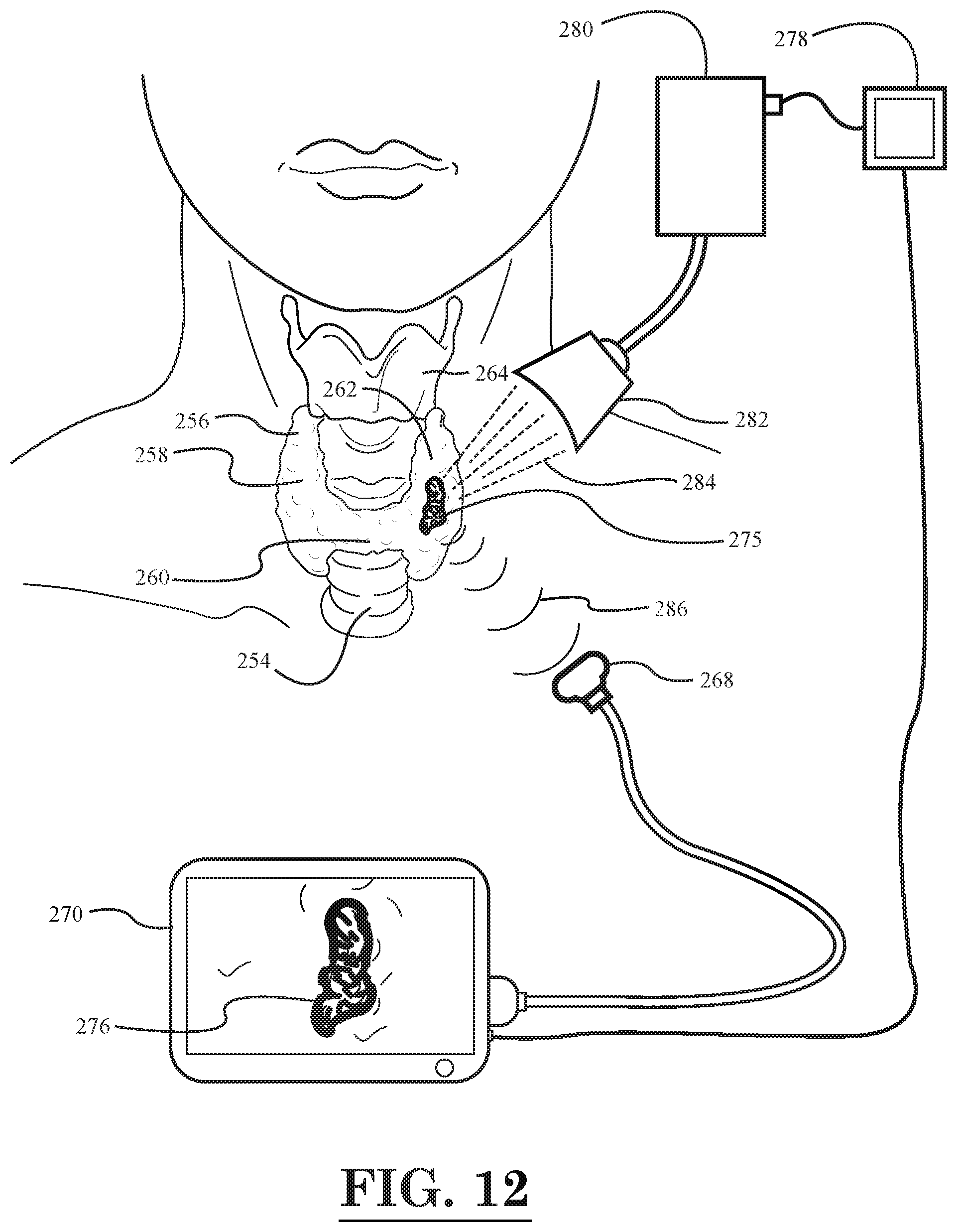
XML
uspto.report is an independent third-party trademark research tool that is not affiliated, endorsed, or sponsored by the United States Patent and Trademark Office (USPTO) or any other governmental organization. The information provided by uspto.report is based on publicly available data at the time of writing and is intended for informational purposes only.
While we strive to provide accurate and up-to-date information, we do not guarantee the accuracy, completeness, reliability, or suitability of the information displayed on this site. The use of this site is at your own risk. Any reliance you place on such information is therefore strictly at your own risk.
All official trademark data, including owner information, should be verified by visiting the official USPTO website at www.uspto.gov. This site is not intended to replace professional legal advice and should not be used as a substitute for consulting with a legal professional who is knowledgeable about trademark law.