Hairpin Loop Method For Double Strand Polynucleotide Sequencing Using Transmembrane Pores
Brown; Clive Gavin ; et al.
U.S. patent application number 16/782350 was filed with the patent office on 2020-07-30 for hairpin loop method for double strand polynucleotide sequencing using transmembrane pores. This patent application is currently assigned to Oxford Nanopore Technologies Ltd.. The applicant listed for this patent is Oxford Nanopore Technologies Ltd.. Invention is credited to Clive Gavin Brown, James Anthony Clarke, Graham Hall, Gavin Harper, Andrew John Heron, James White.
| Application Number | 20200239950 16/782350 |
| Document ID | 20200239950 / US20200239950 |
| Family ID | 1000004752181 |
| Filed Date | 2020-07-30 |
| Patent Application | download [pdf] |


View All Diagrams
| United States Patent Application | 20200239950 |
| Kind Code | A1 |
| Brown; Clive Gavin ; et al. | July 30, 2020 |
HAIRPIN LOOP METHOD FOR DOUBLE STRAND POLYNUCLEOTIDE SEQUENCING USING TRANSMEMBRANE PORES
Abstract
The invention relates to a new method of sequencing a double stranded target polynucleotide. The two strands of the double stranded target polynucleotide are linked by a bridging moiety. The two strands of the target polynucleotide are separated using a polynucleotide binding protein and the target polynucleotide is sequenced using a transmembrane pore.
| Inventors: | Brown; Clive Gavin; (Cambridge, GB) ; Clarke; James Anthony; (Oxford, GB) ; Hall; Graham; (Oxford, GB) ; Harper; Gavin; (Sonning, GB) ; Heron; Andrew John; (Oxford, GB) ; White; James; (Oxford, GB) | ||||||||||
| Applicant: |
|
||||||||||
|---|---|---|---|---|---|---|---|---|---|---|---|
| Assignee: | Oxford Nanopore Technologies
Ltd. Oxford GB |
||||||||||
| Family ID: | 1000004752181 | ||||||||||
| Appl. No.: | 16/782350 | ||||||||||
| Filed: | February 5, 2020 |
Related U.S. Patent Documents
| Application Number | Filing Date | Patent Number | ||
|---|---|---|---|---|
| 15944365 | Apr 3, 2018 | 10597713 | ||
| 16782350 | ||||
| 14234698 | Apr 25, 2014 | 9957560 | ||
| PCT/GB2012/051786 | Jul 25, 2012 | |||
| 15944365 | ||||
| 61511436 | Jul 25, 2011 | |||
| Current U.S. Class: | 1/1 |
| Current CPC Class: | G01N 27/44717 20130101; G01N 33/48721 20130101; C12Q 1/6869 20130101; G01N 27/44791 20130101 |
| International Class: | C12Q 1/6869 20060101 C12Q001/6869; G01N 27/447 20060101 G01N027/447 |
Claims
1.-35. (canceled)
36. A method of processing a polynucleotide, the method comprising: a) obtaining a sense polynucleotide strand comprising a homopolymeric region that is longer that the reading section of a nanopore; and b) synthesizing an antisense polynucleotide strand under conditions in which a nucleotide analog is incorporated at random in a reverse complement of the homopolymer region, such that the length of the homopolymer region in the antisense polynucleotide strand is shorter than the reading section of the nanopore.
37. The method of claim 36, wherein the sense polynucleotide strand comprises i) a sequence of nucleotides, wherein each nucleotide in the sequence is selected from a set of nucleotides; and ii) the homopolymeric region, wherein the homopolymeric region comprising repeated instances of a member of the set of nucleotides, and wherein step b) comprises combining a polymerase with the same set of nucleotides of step a) and a nucleotide analog, and maintaining the combination under conditions in which the nucleotide analog is incorporated at random in the reverse complement of the homopolymer region.
38. The method of claim 37 further comprising controlling the insertion rate of the nucleotide analog by varying the concentration of the nucleotide analog relative to the set of nucleotides.
39. The method of claim 36, wherein the sense polynucleotide strand is connected to a single stranded leader.
40. The method of claim 39, wherein the sense polynucleotide is connected to the single stranded leader via a stretch of abasic nucleotides.
41. The method of claim 36, wherein the sense polynucleotide strand, in step a), comprises a hairpin loop having a free 3' end.
42. The method of claim 41, wherein the antisense polynucleotide strand is synthesized using a polymerase reaction that is nucleated from the free 3' end of the hairpin loop.
43. The method of claim 36, further comprising: c) moving the antisense polynucleotide strand through the nanopore such that a proportion of the antisense polynucleotide strand interacts with the nanopore; and d) measuring the current passing through the nanopore during each interaction in step c) and thereby determining the sequence of the antisense polynucleotide strand.
44. The method of claim 43, further comprising: e) moving the sense polynucleotide strand through the nanopore such that a proportion of the sense polynucleotide strand interacts with the pore; and f) measuring the current passing through the nanopore during each interaction in step e) thereby determining the sequence of the antisense polynucleotide strand.
45. The method of claim 44, further comprising: g) combining information from steps d) and f) to determine the length of the homopolymeric region.
Description
FIELD OF THE INVENTION
[0001] The invention relates to a new method of sequencing a double stranded target polynucleotide. The two strands of the target polynucleotide are linked by a bridging moiety. The two strands of the target polynucleotide are separated by a polynucleotide binding protein. Sequencing of the target polynucleotide is carried out using a transmembrane pore.
BACKGROUND OF THE INVENTION
[0002] There is currently a need for rapid and cheap nucleic acid (e.g. DNA or RNA) sequencing technologies across a wide range of applications. Existing technologies are slow and expensive mainly because they rely on amplification techniques to produce large volumes of nucleic acid and require a high quantity of specialist fluorescent chemicals for signal detection.
[0003] Transmembrane pores (nanopores) have great potential as direct, electrical biosensors for polymers and a variety of small molecules. In particular, recent focus has been given to nanopores as a potential DNA sequencing technology.
[0004] When a potential is applied across a nanopore, there is a drop in the current flow when an analyte, such as a nucleotide, resides transiently in the barrel for a certain period of time. Nanopore detection of the nucleotide gives a current blockade of known signature and duration. The concentration of a nucleotide can then be determined by the number of blockade events (where an event is the translocation of an analyte through the nanopore) per unit time to a single pore.
[0005] In the "Strand Sequencing" method, a single polynucleotide strand is passed through the pore and the nucleotides are directly identified. Strand Sequencing can involve the use of a nucleotide handling enzyme, such as Phi29 DNA polymerase, to control the movement of the polynucleotide through the pore. Nanopore sequencing, using enzymes to control the translocation of dsDNA through the nanopore, has in the past focused on only reading one strand of a dsDNA construct. When the enzyme is used as polymerase, the portion to be sequenced is single stranded. This is fed through the nanopore and the addition of dNTPs at a primer/template junction on top of the strand pulls the single stranded portion through the nanopore in a controlled fashion. The majority of the published literature uses this approach to control strand movement (Lieberman et al. (2010) "Processive Replication of Single DNA Molecules in a Nanopore Catalyzed by phi29 DNA Polymerase" J. Am. Chem. Soc. 132(50): 17961-17972). In the polymerase mode, the complementary strand cannot be sequenced. When the enzyme is used as a double stranded exonuclease as published (Lieberman et al. (2010) supra), the unzipping of the complementary strand is accompanied by the digestion of this strand. It is therefore not possible to sequence the complementary strand with this approach. The complementary strand cannot therefore be captured and sequenced by the nanopore. Hence, only half of the DNA information in dsDNA is sequenced.
[0006] In more detail, when both polymerase and exonuclease activity are inhibited (by running without tri-phosphates bases and with excess of EDTA), enzymes such as Phi29 DNA polymerase have been shown to unzip dsDNA when pulled through a nanopore by a strong applied field (FIG. 1) (Lieberman et al. (2010) supra). This has been termed unzipping mode. Unzipping mode implies that it is the unzipping of dsDNA above or through the enzyme, and importantly, it is the requisite force required to disrupt the interactions of both strands with the enzyme and to overcome the hydrogen bonds between the hybridised strands. In the past the second complementary strand was considered to be essential for efficient enzyme binding. In addition, it was thought that the requisite force required to unzip the strand above or in the enzyme was a dominant braking effect slowing DNA through the pore. Herein we describe how enzymes such as Phi29 DNA polymerase can act as a molecular brake for ssDNA, enabling sufficient controlled movement through a nanopore for sequencing around the hairpin turns of specially designed dsDNA constructs to sequence both the sense and anti-sense strands of dsDNA (FIG. 2). Unzipping mode has in the past predominantly been performed using templates where the distal part of the analyte is blunt ended (FIG. 1). Small hairpins have occasionally been used, but were only included to simplify DNA design. Previous work has not considered the use of hairpins on long dsDNA to provide the ability to read both strands, This is because the unzipping movement model has not considered Phi29 DNA polymerase or related enzymes capable of controlling the movement of the DNA when entering ssDNA regions (i.e. when moving around the hairpin and along the anti-sense strand--FIG. 2).
SUMMARY OF THE INVENTION
[0007] The inventors have surprisingly demonstrated that both strands of a double stranded target polynucleotide can be sequenced by a nanopore when the two strands are linked by a bridging moiety and then separated. Furthermore, the inventors have also surprisingly shown that an enzyme, such as Phi29 DNA polymerase, is capable of separating the two strands of a double stranded polynucleotide, such as DNA, linked by a bridging moiety and controlling the movement of the resulting single stranded polynucleotide through the transmembrane pore.
[0008] The ability to sequence both strands of a double stranded polynucleotide by linking the two strands with a bridging moiety has a number of advantages, not least that both the sense and anti-sense strands of the polynucleotide can be sequenced. These advantages are discussed in more detail below.
[0009] Accordingly, the invention provides a method of sequencing a double stranded target polynucleotide, comprising: [0010] (a) providing a construct comprising the target polynucleotide, wherein the two strands of the target polynucleotide are linked at or near one end of the target polynucleotide by a bridging moiety; [0011] (b) separating the two strands of the target polynucleotide to provide a single stranded polynucleotide comprising one strand of the target polynucleotide linked to the other strand of the target polynucleotide by the bridging moiety; [0012] (c) moving the single stranded polynucleotide through a transmembrane pore such that a proportion of the nucleotides in the single stranded polynucleotide interact with the pore; and [0013] (d) measuring the current passing through the pore during, each interaction and thereby determining or estimating the sequence of the target polynucleotide, [0014] wherein the separating in step (b) comprises contacting the construct with a polynucleotide binding protein which separates the two strands of the target polynucleotide.
[0015] The invention also provides: [0016] a kit for preparing a double stranded target polynucleotide for sequencing comprising (a) a bridging moiety capable of linking the two strands of the target polynucleotide at or near one end and CO at least one polymer; [0017] a method of preparing a double stranded target polynucleotide for sequencing, comprising: [0018] (a) linking the two strands of the target polynucleotide at or near one end with a bridging moiety; and [0019] (b) attaching one polymer to one strand at the other end of the target polynucleotide and thereby forming a construct that allows the target polynucleotide to be sequenced using a transmembrane pore; [0020] a method of sequencing a double stranded target polynucleotide, comprising: [0021] (a) providing a construct comprising the target polynucleotide, wherein the two strands of the target polynucleotide arc linked at or near one end of the target polynucleotide by a bridging moiety; [0022] (b) separating the two strands of the target polynucleotide to provide a single stranded polynucleotide comprising one strand of the target polynucleotide linked to the other strand of the target polynucleotide by the bridging moiety; [0023] (c) synthesising a complement of the single stranded polynucleotide, such that the single stranded polynucleotide and complement form a double stranded polynucleotide; [0024] (d) linking the two strands of the double stranded polynucleotide at or near one end of the double stranded polynucleotide using a bridging moiety; [0025] (e) separating the two strands of the double stranded polynucleotide to provide a further single stranded polynucleotide comprising the original single stranded polynucleotide linked to the complement by the bridging moiety; [0026] (f) moving the complement through a transmembrane pore such that a proportion of the nucleotides in the complement interact with the pore; and [0027] (g) measuring the current passing through the pore during each interaction and thereby determining or estimating the sequence of the target polynucleotide, wherein the separating in step (e) comprises contacting the construct with a polynucleotide binding protein which separates the two strands of the target polynucleotide; [0028] an apparatus for sequencing a double stranded target polynucleotide, comprising: (a) a membrane; (b) a plurality of transmembrane pores in the membrane; (c) a plurality of polynucleotide binding proteins which are capable of separating the two strands of the target polynucleotide; and (d) instructions for carrying out the method of the invention; and [0029] an apparatus for sequencing a double stranded target polynucleotide, comprising: (a) a membrane; (b) a plurality of transmembrane pores in the membrane; and (c) plurality of polynucleotide binding proteins which are capable of separating the two strands of the target polynucleotide, wherein the apparatus is set up to carry out the method of the invention.
DESCRIPTION OF THE FIGURES
[0030] FIG. 1 shows a schematic of enzyme controlled dsDNA and ssDNA translocation through nanopore. An enzyme (e.g. Phi29 DNA polymerase)that is incubated with dsDNA having an ssDNA leader binds at the ssDNA-dsDNA interface. DNA-enzyme complexes are captured by a nanopore under an applied field. Under the field, the template strand of the DNA is slowly stripped through the enzyme in a controlled base-by-base manner, in the process unzipping the complementary primer strand of the dsDNA in or above the enzyme. Once the enzyme reaches the end of the dsDNA it falls off the DNA, releasing it through the nanopore.
[0031] FIG. 2 shows another schematic of enzyme controlled dsDNA and ssDNA translocation through a nanopore. The dsDNA has a hairpin turn linking the sense and anti-sense strands of the dsDNA. Once the enzyme reaches the hairpin it remains bound to the DNA, proceeds around the hairpin turn, and along the anti-sense strand. In the hairpin and antisense regions the enzyme functions as an ssDNA molecular brake, continuing to sufficiently control translocation of the DNA through the nanopore to sequence the DNA.
[0032] FIG. 3 shows a schematic overview of reading around a hairpin of dsDNA using the ability of the enzyme to control movement in ssDNA regions. The dsDNA has a 5'-ssDNA leader to allow capture by the nanopore. This is followed by a dsDNA section, where the sense and anti-sense strands are connected by a hairpin. The hairpin can optionally contain markers (e.g. abasic residues, shown in FIG. 3 as a cross) that are observed during sequencing, which permit simple identification of the sense and anti-sense strands during sequencing. The 3'-end of the anti-sense strand can optionally also have a 3'-ssDNA overhang, which if greater than .about.20 bases allows full reading of the anti-sense strand (the read-head of the nanopore is .about.20 bases downstrand from the top of the DNA in the enzyme).
[0033] FIG. 4 shows a schematic of the DNA-Enzyme-nanopore complex (left) sequenced in unzipping mode through MspA nanopores using Phi29 DNA polymerase, and the consensus sequence obtained from them (right). Section 1 marks the sense section of DNA, and section 2 marks the anti-sense section. This figure shows DNA sequencing of a short dsDNA construct, In this construct he dsDNA section is not connected by a hairpin, so the enzyme falls off the end of the DNA, and only the template/sense strand is sequenced (except for the last .about.20 bases).
[0034] FIG. 5 shows a schematic of the DNA-Enzyme-nanopore complex (left) sequenced in unzipping mode through MspA nanopores using Phi29 DNA polymerase, and the consensus sequence obtained from them (right). Section 1 marks the sense section of DNA, and section 2 the anti-sense section. DNA sequencing of a short dsDNA construct with a hairpin. In this construct the enzyme moves along the sense strand, around the hairpin loop, and down the anti-sense strand, permitting sequencing of both the sense and the first part of the anti-sense strand.
[0035] FIG. 6 shows a schematic of the DNA-Enzyme-nanopore complex (left) sequenced in unzipping mode through MspA nanopores using Phi29 DNA polymerase, and the consensus sequence obtained from them (right). Section 1 marks the sense section of DNA, and section 2 the anti-sense section. Similar to FIG. 5, this construct permits sequencing of both the sense and anti-sense strands, but the additional 3'-ssDNA overhang permits reading of the full length of the anti-sense strand before the enzyme falls off the end of the DNA.
[0036] FIG. 7 shows a schematic of the DNA-Enzyme-nanopore complex (left) sequenced in unzipping mode through MspA nanopores using Phi29 DNA polymerase, and the consensus sequence obtained from them (right). Section 1 marks the sense section of DNA, and section 2 the anti-sense section. Similar to FIG. 5, this construct permits sequencing of both the sense and anti-sense strands, however, this construct has a single abasic residue (shown as a cross) in the hairpin, which provides a clear marker in the DNA sequence to identify the sense and anti-sense sections.
[0037] FIG. 8 shows the consensus DNA sequence of UA02 through MspA. Section 1 marks the homopolymeric 5'-overhang initially in the nanopore. Section 2 marks the sense section of the DNA strand. Section 3 marks the turn. Section 4 marks the anti-sense region of the DNA strand. The polynucleotide sequence that corresponds to each section is shown below that section number.
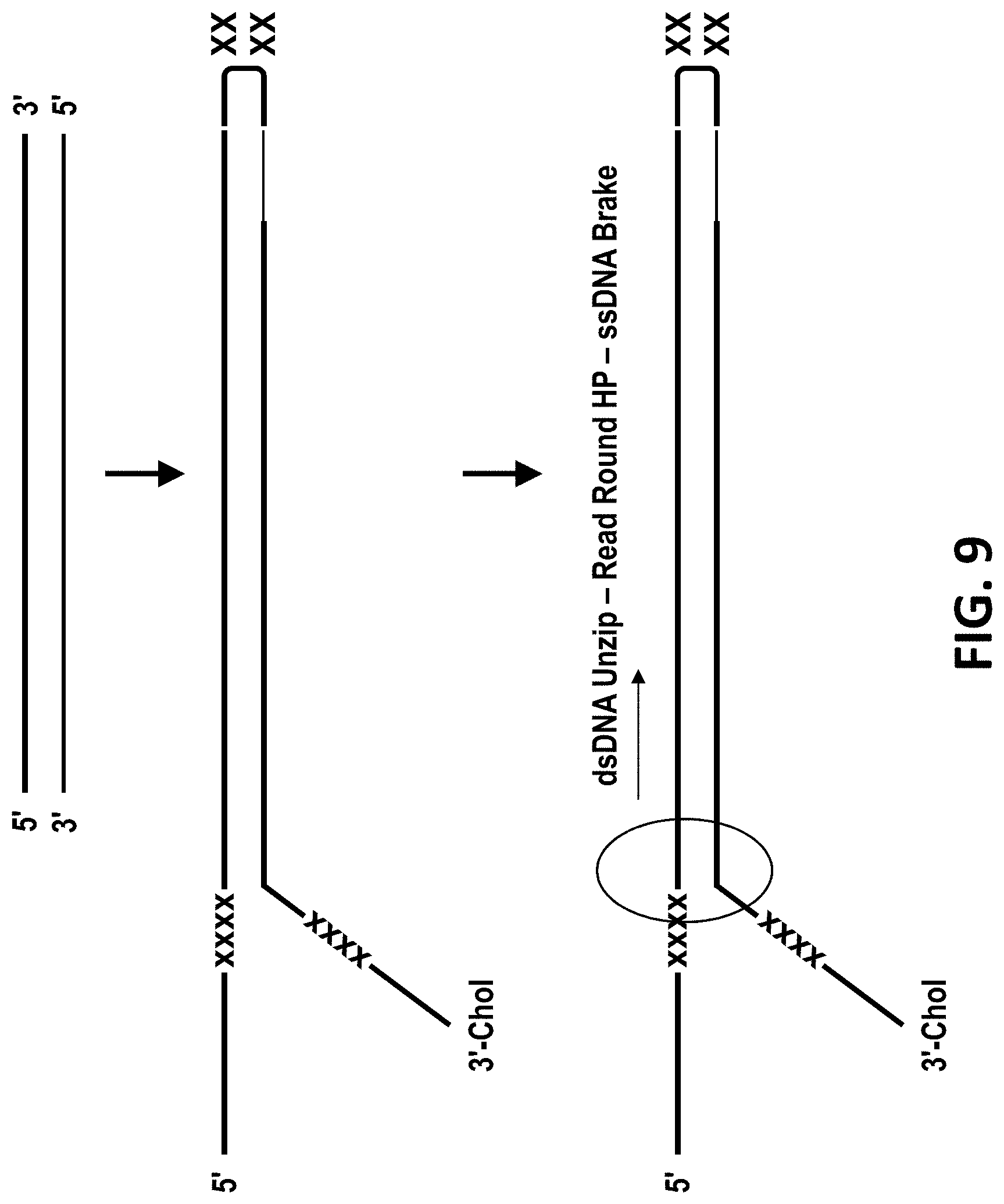
[0038] FIG. 9 shows a schematic of a genomic template for unzipping through MspA nanopores using Phi29 DNA polymerase. It shows a general design outline for creating dsDNA suitable for reading around hairpins. The constructs have a leader sequence with optional marker (e.g. abasic DNA) for capture in the nanopore, and hairpin with optional marker, and a tail for extended reading into anti-sense strand with optional marker.
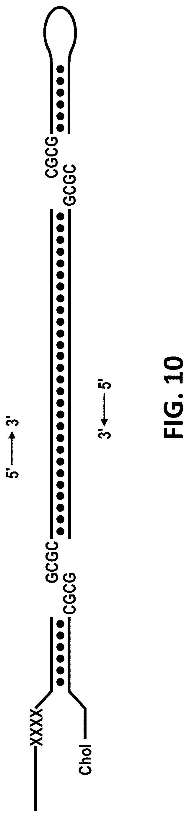
[0039] FIG. 10 shows a schematic of the adapter design for ligating ssDNA overhangs (left) and hairpin turns (right) onto genomic dsDNA. X=abasic DNA. Chol=cholesterol-TEG DNA modification.
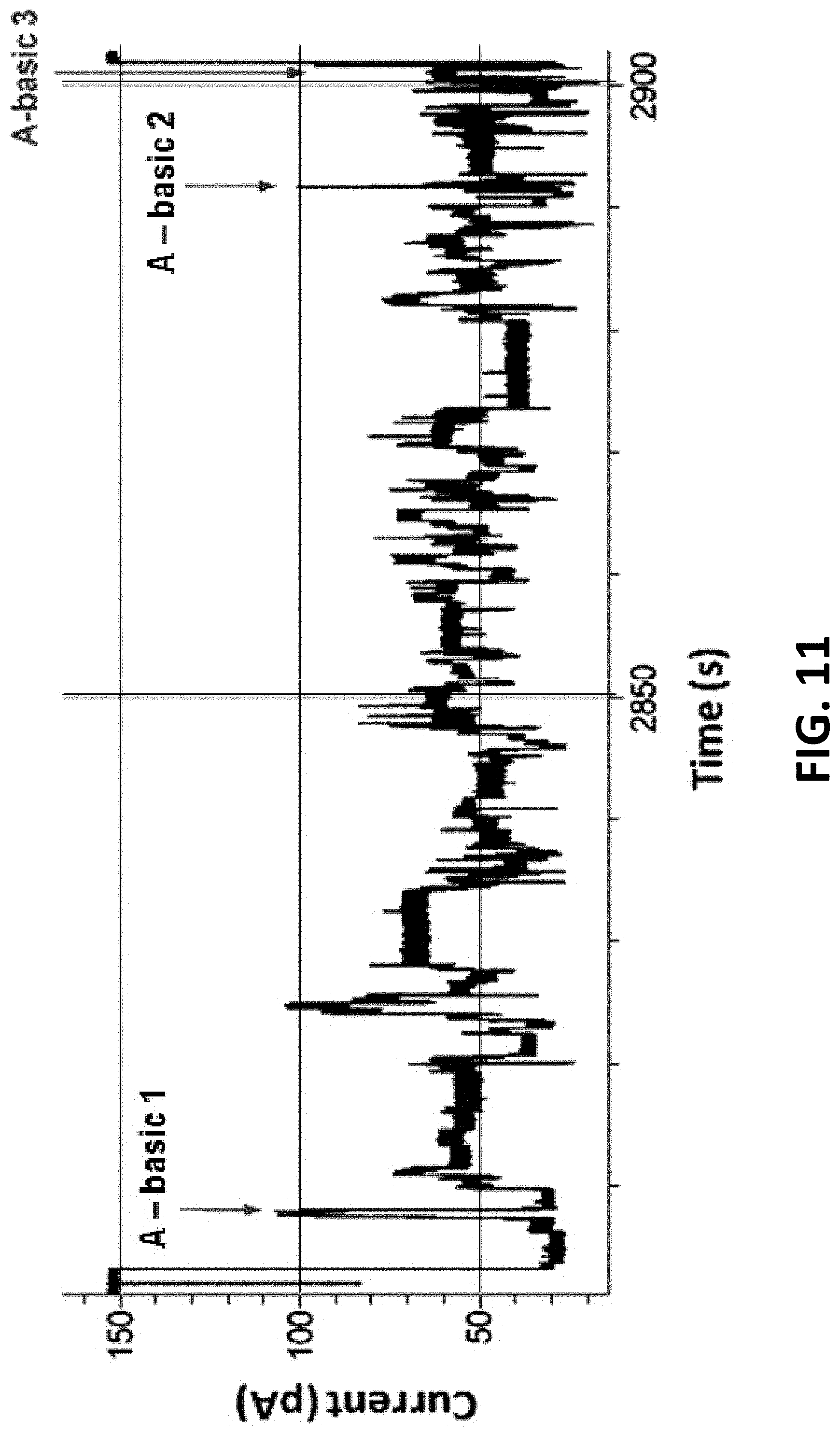
[0040] FIG. 11 shows typical polymerase controlled DNA movement of a 400mer-hairpin through MspA using Phi29 DNA polymerase. Sense region=abasic 1 to 2. Anti-sense region=abasic 2 to 3.
[0041] FIG. 12 shows a consensus DNA sequence profile from multiple polymerase controlled DNA movements of a 400mer-hairpin through MspA. Sense region=abasic 1 to 2. Anti-sense region=abasic 2 to 3.
[0042] FIG. 13 shows a schematic of an alternative sample preparation for sequencing. A construct is illustrated comprising the target polynucleotide and a bridging moiety (hairpin) linking the two strands of the target polynucleotide. The construct also comprises a leader polymer (a single stranded sequence), a tail polymer (also a single stranded sequence) and an abasic marker region within the leader. The marker may prevent the enzyme from making the template completely blunt ended i.e. filling in opposite the required leader ssDNA. A strand displacing polymerase (nucleic acid binding protein) separates the two strands of the construct, initiating either via a complementary primer or by protein primed amplification from the tail polymer. A complement is generated to the resulting single stranded polynucleotide. The complement and the original sense and antisense single stranded polynucleotide analyte can be further modified by addition of a second bridging moiety (hairpin).
[0043] FIG. 14 shows a specific preparation of the construct comprising the target polynucleotide.
[0044] FIG. 15 shows where amplification may be added as part of the sample preparation to aid the detection of epigenetic information. A nucleotide has been constructed so that the following information is read through the pore: sense (original), antisense (original), bridging moiety, sense (replicate), antisense (replicate). Information on the methylated base (mC) is therefore obtained four times.
[0045] FIG. 16 shows how RNA can he sequenced. A bridging moiety is attached to a piece of RNA and the DNA reverse complement added to the RNA via a reverse transcriptase. The RNA is read, followed by the DNA of the reverse complement.
[0046] FIG. 17 shows a schematic of helicase controlled dsDNA and ssDNA translocation through a nanopore. The dsDNA has a hairpin turn linking, the sense and anti-sense strands of the dsDNA. Once the enzyme reaches the hairpin it remains bound to the DNA, proceeds around the hairpin turn, and along the anti-sense strand. In the hairpin and antisense regions the enzyme functions as an ssDNA molecular brake, continuing to sufficiently control translocation of the DNA through the nanopore to sequence the DNA.
[0047] FIG. 18 shows the polynucleotide MONO hairpin construct (SEQ ID NOs: 29 to 35) used in Example 4.
[0048] FIG. 19 shows a typical helicase controlled DNA movement of a 400 hp hairpin (SEQ ID NOs: 29 to 35 connected as shown in FIG. 18) through an MspA nanopore (MS(B1-G75S-G77S-L88N-Q126R)8 MspA (SEQ ID NO: 2 with the mutations G75S/G77S/L88N/Q126R)). Sense=region 1. Anti-sense=region 2.
[0049] FIG. 20 shows the beginning of a typical helicase controlled DNA movement of a 400 bp hairpin (SEQ ID NOs: 29 to 35 connected as shown in FIG. 18) through an MspA nanopore (MS(B1-G75S-G77S-L88N-Q126R)8 MspA (SEQ ID NO: 2 with the mutations G75S/G77S/L88N/Q126R)). The polyT region at the beginning of the sequence is highlighted with a * and the abasic DNA bases as a #.
[0050] FIG. 21 shows the transition between the sense and antisense regions of a typical helicase controlled DNA movement of a 400 bp-hairpin (SEQ ID NOs: 29 to 35 connected as shown in FIG. 18) through an MspA nanopore (MS(B1-G75S-G77S-L88N-Q126R)8 MspA (SEQ ID NO: 2. with the mutations G75S/G77S/L88N/Q126R)). The transition region between the sense and antisense regions of the sequence is highlighted by a *, the sense region labeled 1 and the antisense region labeled 2.
[0051] FIG. 22 shows an example sample prep method for forming DUO hairpin constructs. The double stranded DNA analyte is contacted by and modified to contain a Y-shaped adapter (the sense strand (SEQ ID NO: 29 attached to SEQ II) NO: 30 via four abasic DNA bases) of this adaptor contains the 5' leader, a sequence that is complementary to the tether (SEQ ID NO: 35, which at the 3' end of the sequence has six iSp18 spacers attached to two thymine residues and a 3' cholesterol TEG) and 4 abasics and the antisense half of the adaptor contains a 3' hairpin (SEQ ID NO: 31)) at one end of the duplex and a hairpin (SEQ ID NO: 32) at the other. The Y-shaped adapter itself also carries a 3'-hairpin (SEQ ID NO: 31), which allows extension either by a polymerase or by ligation. This extension is preferentially carried out by a mesophilic polymerase that has strand displacement activity. As the polymerase extends from the 3' of the Y-shaped adapter hairpin (SEQ ID NO: 31) it copies the antisense strand (SEQ ID NO: 34) and so displaces the original sense strand (SEQ ID NO: 33). When the polymerase reaches the end of the antisense strand (SEQ ID NO: 34) it fills-in opposite the hairpin (SEQ ID NO: 32) and then begins to fill-in opposite the now single stranded and original sense strand (SEQ ID NO: 33). Extension is then halted by a section of abasic or spacer modifications (other possible modifications which could halt enzyme extension include RNA, PNA or morpholino bases and iso-dC or iso-dG) to leave the 5'-region of the Y-shaped adapter single stranded (SEQ ID NO: 29).
[0052] FIG. 23 shows the specific preparation method used in Example 5 for preparing a DUO hairpin construct (SEQ ID NOs: 29 to 36 connected as shown in FIG. 25). A .about.400 by region of PhiX 174 was PCR amplified with primers containing SacI and KpnI restriction sites (SEQ ID Nos: 27 and 28 respectively). Purified PCR product was then SacI and KpnI digested before a Y-shaped adapter (sense strand sequence (SEQ ID NO: 29 attached to ,SEQ ID NO: 30 via four abasic DNA bases) is ligated onto the 5' end of SEQ ID NO: 33 and the anti-sense strand (SEQ ID NO: 31) is ligated onto the 3' end of the SEQ ID NO: 34) and a hairpin (SEQ ID NO: 32, used to join SEQ ID NO's: 33 and 34) were ligated to either end, using T4 DNA ligase (See FIG. 18 for final DNA construct). The doubly ligated product was PAGE purified before addition of Klenow DNA polymerase, SSB and nucleotides to allow extension from the Y-shaped adapter hairpin (SEQ ID NO: 31). To screen for successful DUO product a series of mismatch restriction sites were incorporated into the adapter sequences, whereby the enzyme will cut the analyte only if the restriction site has been successfully replicated by the DUO extension process.
[0053] FIG. 24 shows that the adapter modified analyte (MONO, SEQ ID NOs: 29-35 connected as shown in FIG. 18) in the absence of polymerase does not digest with the restriction enzymes (see gel on the left, Key: M=MfeI, A=AgeI, X=XmaI, N=NgoMIV, B=BspEI), due to the fact they are mismatched to one another. However, on incubation with polymerase there is a noticeable size shift and the shifted product (DUO, SEQ ID NOs: 29-36 connected as shown in FIG. 25) now digests as expected with each of the restriction enzymes (see gel on the right. Key: M=MfeI, A=AgeI, X=XmaI, N=NgoMIV, B=BspEI).
[0054] FIG. 25 shows the polynucleotide DUO hairpin construct (SEQ ID NOs: 29 to 36) used in Examples 6.
[0055] FIG. 26 shows two typical helicase controlled DNA movements for the DUO hairpin construct (SEQ ID NOs: 29 to 36 connected as shown in FIG. 25) through an MspA nanopore (MS(B1-G75S-G77S-L88N-Q126R)8 MspA (SEQ ID NO: 2 with the mutations G75S/G77S/L88N/Q126R)). Sense original=region 1. Anti-sense original=region 2. Sense replicate=region 3. Anti-sense replicate=region 4.
[0056] FIG. 27 shows an expanded view of a typical helicase, controlled DNA movement for the DUO hairpin construct (SEQ ID NOs: 29 to 36 connected as shown in FIG. 25) through an MspA nanopore (MS(B1-(G75S-G77S-L88N-Q126R)8 MspA (SEQ ID NO: 2 with the mutations G75S/G77S/L88N/Q126R)). Sense original=region 1. Anti-sense original=region 2. Sense replicate=region 3. Anti-sense replicate region 4.
[0057] FIG. 28 shows an expanded view of a typical transition between the sense original and antisense original regions of the DUO hairpin construct (SEQ NOs: 29 to 36 connected as shown in FIG. 25) when under helicase controlled DNA movement through an MspA nanopore (MS(B1-G75S-G77S-L88N-Q126R)8 MspA (SEQ ID NO: 2 with the mutations G75S/G77S/L88N/Q126R)). Sense original=region 1. Anti-sense original=region 2.
DESCRIPTION OF THE SEQUENCE LISTING
[0058] SEQ ID NO: 1 shows the codon optimised polynucleotide sequence encoding the NNN-RRK mutant MspA monomer.
[0059] SEQ ID NO: 2 (also referred to as "B1") shows the amino acid sequence of the mature form of the NNN-RRK mutant of the MspA monomer. The mutant lacks the signal sequence and includes the following mutations: D90N, D91N, D93N, D118R, D134R and E139K. These mutations allow DNA transition through the MspA pore.
[0060] SEQ ID NO: 3 shows the polynucleotide sequence encoding one subunit of .alpha.-hemolysin-E111N/K147N (.alpha.-HL-NN; Stoddart et al., PNAS, 2009; 106(19): 7702-7707).
[0061] SEQ ID NO: 4 shows the amino acid sequence of one subunit of .alpha.-HL-NN,
[0062] SEQ ID NO: 5 shows the codon optimised polynucleotide sequence encoding the Phi29 DNA polymerase.
[0063] SEQ ID NO: 6 shows the amino acid sequence of the Phi29 DNA polymerase.
[0064] SEQ ID NOs: 7 to 9 show the amino acid sequences of the mature forms of the MspB, C and D mutants respectively. The mature forms lack the signal sequence.
[0065] SEQ ID NOs.: 10 to 15 show the sequences used to illustrate homopolymer reads.
[0066] SEQ ID NOs: 16 to 36 show the sequences used in the Examples.
DETAILED DESCRIPTION OF THE INVENTION
[0067] It is to be understood that different applications of the disclosed products and methods may be tailored to the specific needs in the art. It is also to be understood that the terminology used herein is for the purpose of describing particular embodiments of the invention only, and is not intended to be limiting.
[0068] In addition as used in this specification and the appended claims, the singular forms "a", "an", and "the" include plural referents unless the content clearly dictates otherwise. Thus, For example, reference to "a pore" includes two or more such pores, reference to "a nucleic acid sequence" includes two or more such sequences, and the like.
[0069] All publications, patents and patent applications cited herein, whether supra or infra, are hereby incorporated by reference in their entirety.
Methods of the Invention
[0070] The invention provides a method for sequencing a double stranded target polynucleotide. The method comprises linking the two strands of the target polynucleotide by a bridging moiety. The two strands of the target polynucleotide are separated to provide a single stranded polynucleotide by contacting the construct comprising the target polynucleotide with a polynucleotide binding protein. The single stranded polynucleotide is moved through a transmembrane pore. A proportion of the nucleotides in the single stranded polynucleotide interact with the pore. The current passing through the pore is measured during each interaction and the sequence of the target polynucleotide is estimated or determined. The target polynucleotide is therefore sequenced using Strand Sequencing. This method may be referred to herein as the "MONO" method.
[0071] As discussed above, linking he two strands of the target polynucleotide by a bridging moiety allows both strands of the target polynucleotide to be sequenced by the transmembrane pore. This method is advantageous because it doubles the amount of information obtained from a single double stranded target polynucleotide construct. Moreover, because the sequence in the complementary `anti-sense` strand is necessarily orthogonal to the sequence of the `sense` strand, the information from the two strands can be combined informatically. Thus, this mechanism provides an orthogonal proof-reading capability that provides higher confidence observations.
[0072] Furthermore, the other major advantages of the method of the invention are:
[0073] 1) Coverage of missed nucleotides: the method substantially minimises issues of any missed nucleotides or groups of nucleotides(e.g. due to movement issues such as the strand slipping through the pore), since any states that might be missed in one strand are likely to be covered by the orthogonal information obtained from its complement region.
[0074] 2) Coverage of problematic sequence motifs: any difficult to sequence motifs are covered by the orthogonal and opposite information in the complementary strand, which having a different sequence will not have the same sequence dependent issues. For example, this is particularly relevant for sequence motifs that produce only small changes in current, or have similar current levels--i.e. consecutive base motifs that when moved through the nanopore produce the same current block, and are therefore not Observed as there is no step change in current. Any similar current levels from one sequence motif will be covered by the entirely different current levels obtained from its orthogonal sequence in the complement strand. in addition to the advantages discussed above there are a number of special cases where the concept of reading both strands of the double stranded polynucleotide can be utilized to provide further benefits.
[0075] 1. Epigenetic Information
[0076] Being able to identify epigenetic information (such as 5-methylcytosine of 5-hydroxymenthylcytosine nucleotides) or damaged bases within a natural DNA strand is desirable in a wide range of applications. At present, it is difficult to extract this information as the majority of DNA sequencing technologies rely on DNA amplification as part of their sequencing chemistry. This information can be extracted, but requires chemical treatment followed by amplification, both of which can introduce errors.
[0077] Nanopore sequencing is also a single molecule sequencing technology and therefore can be performed without the need of DNA amplification. It has been shown that nanopores can detect modifications to the standard four DNA nucleotides. Reading both strands of the polynucleotide can be useful in detecting DNA modifications in situations where a modified base behaves in a similar way (generates a similar current signal) to another base. For example if methylcytosine (mC) behaves in a similar way to thymidine there is an error associated with assigning a mC, to a T. In the sense strand, there is a probability of the base being called a mC or a T. However, in the anti-sense strand, the corresponding base may appear as a G with a high probability. Thus by "proof reading" the anti-sense strand, it is highly likely that the base in the sense strand was a mC rather than a T.
[0078] Reading the sense and the anti-sense strand can be performed without the need of amplification or replication. However, amplification or replication may be added as part of the sample preparation to aid the detection of epigenetic information. A nucleotide strand may be constructed (described in detail below) where the following information is read through the nanopore in the following order: sense (original), antisense (original), --bridging moiety--, sense (complement), antisense (complement) (FIG. 15).
[0079] In this scheme, information on the methylated base will be obtained four times. If the epigenetic base is in the original sense strand (in this case, mC), the following information will be obtained with a high probability: sense (original)-mC, anti-sense (original)-G, sense (complement)-C, and anti-sense (compliment)-G. It is clear that the original sense read and the replicated sense read will give different results, while the both anti-sense reads will yield the same base call. This information can be used to indicate the position of the epigenetic base in the original sense strand.
[0080] 2. RNA-DNA Double Reads
[0081] A similar scheme can be used to sequence RNA. A bridging moiety can be attached to a piece of RNA and the DNA reverse complement added to the RNA via a reverse transcriptase (resulting construct shown in FIG. 16)
[0082] In this scheme, the RNA is read followed by a DNA read of the reverse complement. Information from both the RNA and the DNA reads can be combined to increase the accuracy of determining or estimating the RNA sequence. For example, if a uracil base (U) in RNA gives a similar read to a cytosine, then the corresponding base could be used to resolve this error. If the corresponding DNA base is G, then it is highly likely that the RNA base was a C, however if the DNA base is called as an A, then it is likely that the RNA base was a U.
[0083] 3. Homopolymer Reads
[0084] Homopolymer reads may be a problem for single molecule nanopore sequencing. If the homopolymer region is longer than the reading section of the pore, the length of the homopolymer section will be difficult to determine.
[0085] It has already been shown that Phi29 can be used to read around a hairpin, allowing the sense arid the antisense strand to be read. Amplification can be used to generate the antisense strand using a polymerase and a set of regular DNA triphosphates; dTTP, dGTP, dATP, dCTP. To overcome the problem of homopolymer reads, the antisense strand can be synthesised with the addition of a different base in combination with the original dTTP, dGTP, dATP, dCTP. This could be a natural base analogue such as inosine (I). The base will have a random chance of incorporating compared to the correct natural base and the insertion rates can be controlled by varying the concentration of the triphosphate species.
[0086] Through the addition of the alternative base, there will be a probability of an alternative base being inserted into the reverse complement of a homopolymer region. The result of this is that the homopolymer run will be reduced in length to a point where it can be read by the reading section of the nanopore. For example, a homopolymer group of AAAAAAAAAAAA (SEQ ID NO: 10) will have random insertions of the alternative base and may give TTTITTIITTTI (SEQ ID NO: 11) (where I is inosine)
TABLE-US-00001 Original DNA + Hairpin (SEQ ID NO: 12): 5'-TTTTTTTTTTTTTTTTTTTTXXXXXTGTACTGCCGTACGTAAAAAAA TAGCTGATCGTACTTACTAGCATGTT (abasic = X) Regular Conversion (SEQ ID NO: 13): 5'-TTTTTTTTTTTTTTTTTTTTXXXXXTGTACTGCCGTACGTAAAAAAA TAGCTGATCGTACTTACATGACGGCATGCATTTTTTTATCGACTAGCATG TT (abasic = X) Proposed Scheme 1 (G, T, A, C is randomly replaced by analogue I)(SEQ ID NO 14): 5'-TTTTTTTTTTTTTTTTTTTTXXXXXTGTACTGCCGTACGTAAAAAAA TAGCTGATCGTACTTATATTACGICATGIATTITTITATIGACTAGCATG TT (abasic = X) The base analogue could be generic (replace T, G, A, or C), or it could be specific to one base (e.g. deoxyuridine (U) just replaces T). Proposed Scheme 2 (T is randomly replaced by analogue U)(SEQ ID NO: 15): 5'-TTTTTTTTTTTTTTTTTTTTXXXXXTGTACTGCCGTACGTAAAAAAA TAGCTGATCGTACTTACAUGACCGCATGCAUTTTUTIATCGACTAGCATG TT (abasic = X)
[0087] In both scheme one and two, the homopolymer stretch has been reduced to allow individual nucleotides or groups of nucleotides to be estimated or determined. The sense strand will be a natural DNA strand, while the anti-sense will contain a mixture of natural bases and base analogues. The combination of data from the sense and the antisense reads can be used to estimate the length of the homopolymer run in the original DNA section.
Double Stranded Target Polynucleotide
[0088] The method of the invention is for sequencing a double stranded polynucleotide. A polynucleotide, such as a nucleic acid, is a macromolecule comprising two or more nucleotides. The polynucleotide or nucleic acid may comprise any combination of any nucleotides. The nucleotides can be naturally occurring or artificial. The nucleotide can be oxidized or methylated. A nucleotide typically contains a nucleobase, a sugar and at least one phosphate group. The nucleobase is typically heterocyclic. Nueleobases include, but are not limited to, purines and pyrirnidines and more specifically adenine, guanine, thytnine, uracil and cytosine. The sugar is typically a pentose sugar. Nucleotide sugars include, but are not limited to, ribose and deoxyribose. The nucleotide is typically a ribonucleotide or deoxyribonucleotide. The nucleotide typically contains a monophosphate, diphosphate or triphosphate. Phosphates may be attached on the 5' or 3' side of a nucleotide.
[0089] Nucleotides include, but are not limited to, adenosine monophosphate (AMP), adenosine diphosphate (ADP), adenosine triphosphate (ATP), guanosine monophosphate (GMP), guanosine diphosphate (CIDP), guanosine triphosphate (GTP), thymidine monophosphate (TMP), thymidine diphosphate (TDP), thymidine triphosphate (TTP), uridine monophosphate (UMP), uridine diphosphate (UDP), uridine triphosphate (UTP), cytidine monophosphate (CMP), cytidine diphosphate (CDP), cytidine triphosphate (CTP), 5-methylcytidine monophosphate, 5-methylcytidine diphosphate, 5-methylcytidine triphosphate, 5-hydroxymethylcytidine monophosphate, 5-hydroxymethylcytidine diphosphate, 5-hydroxymethylcytidine triphosphate, cyclic adenosine monophosphate (cAMP), cyclic guanosine monophosphate (cGMP), deoxyadenosine monophosphate (dAMP), deoxyadenosine diphosphate (dADP), deoxyadenosine triphosphate (dATP), deoxyguanosine monophosphate (dGMP), deoxyguanosine diphosphate (dGDP), deoxyguanosine triphosphate (dGTP), deoxythymidine monophosphate (dTMP), deoxythymidine diphosphate (dTDP), deoxythymidine triphosphate (dTTP), deoxyuridine monophosphate (BUMP), deoxyuridine diphosphate (dUDP), deoxyuridine triphosphate (dUTP), deoxycytidine monophosphate (dCMP), deoxycytidine diphosphate (dCDP) and deoxycytidine triphosphate (dCTP), 5-methyl-2'-deoxycytidine monophosphate, 5-methyl-2'-deoxycytidine diphosphate, 5-methyl-2'-deoxycytidine triphosphate, 5-hydroxymethyl-2'-deoxycytidine monophosphate, 5-hydroxymethyl-2'-deoxycytidine diphosphate and 5-hydroxymethyl-2'-deoxycytidine triphosphate. The nucleotides are preferably selected from AMP, TMP, GMP, CMP, UMP, dAMP, dTMP, dGMP or dCMP.
[0090] A nucleotide may contain a sugar and at least one phosphate group (i.e. lack a nucleobase).
[0091] The polynucleotide can be a nucleic acid, such as deoxyribonucleic acid (L)NA) or ribonucleic acid (RNA). The target polynucleotide can comprise one strand of RNA hybridized to one strand of DNA. The polynucleotide may be any synthetic nucleic acid known in the art, such as peptide nucleic acid (PNA), glycerol nucleic acid (CNA), threose nucleic acid (TNA), locked nucleic acid (LNA) or other synthetic polymers with nucleotide side chains.
[0092] The target polynucleotide can be any length. For example, the polynucleotide can be at least 10, at least 50, at least 100, at least 150, at least 200, at least 250, at least 300, at least 400 or at least 500 nucleotide pairs in length. The polynucleotide can be 1000 or more nucleotide pairs, 5000 or more nucleotide pairs in length or 100000 or more nucleotide pairs in length.
[0093] The target polynucleotide is present in any suitable sample. The invention is typically carried out on a sample that is known to contain or suspected to contain the target polynucleotide. Alternatively, the invention may be carried out on a sample to confirm the identity of one or more target polynucleotides whose presence in the sample is known or expected.
[0094] The sample may be a biological sample. The invention may be carried out in vitro on a sample obtained from or extracted from any organism or microorganism. The organism or microorganism is typically archean, prokaryotic or eukaryotic and typically belongs to one the five kingdoms: plantae, animalia, fungi, monera and protista. The invention may he carried out in vitro on a sample obtained from or extracted from any virus. The sample is preferably a fluid sample. The sample typically comprises a be fluid of the patient. The sample may be urine, lymph, saliva, mucus or amniotic fluid but is preferably blood, plasma or serum. Typically, the sample is human in origin, but alternatively it may be from another mammal animal such as from commercially farmed animals such as horses, cattle, sheep or pigs or may alternatively be pets such as cats or dogs. Alternatively a sample of plant origin is typically obtained from a commercial crop, such as a cereal, legume, fruit or vegetable, for example wheat, barley, oats, canola, maize, soya, rice, bananas, apples, tomatoes, potatoes, grapes, tobacco, beans, lentils, sugar cane, cocoa, cotton, tea, coffee.
[0095] The sample may be a non-biological sample. The non-biological sample is preferably a fluid sample. Examples of a non-biological sample include surgical fluids, water such as drinking water, sea water or river water, and reagents for laboratory tests.
[0096] The sample is typically processed prior to being assayed, for example by centrifugation or by passage through a membrane that filters out unwanted molecules or cells, such as red blood cells. The sample may be measured immediately upon being taken. The sample may also be typically stored prior to assay, preferably below -70.degree. C.
[0097] If the target polynucleotide, is coupled to the membrane as discussed in more detail below, the method of the invention is particularly advantageous for human DNA sequencing because only small amounts of purified DNA can be obtained from human blood. The method preferably allows sequencing of a target polynucleotide that is present at a concentration of from about 0.1 pM to about 1 nM, such as less than 1 pM, less than 10 pM or less than 100 pM.
Construct
[0098] The method of the invention involves providing a construct comprising the double stranded target nucleotide to be sequenced. The construct typically allows both strands of the target polynucleotide to be sequenced by a transmembrane pore.
[0099] The construct comprises a bridging moiety which is capable of linking the two strands of the target polynucleotide. The bridging moiety typically covalently links the two strands of the target polynucleotide. The bridging moiety can be anything that is capable of linking the two strands of the target polynucleotide, provided that the bridging moiety does not interfere with movement of the single stranded polynucleotide through the transmembrane pore. Suitable bridging moieties include, but are not limited to a polymeric linker, a chemical linker, a polynucleotide or a polypeptide. Preferably, the bridging moiety comprises DNA, RNA, modified DNA (such as abasic DNA), RNA, PNA, LNA or PEG. The bridging moiety is more preferably DNA or RNA.
[0100] The bridging moiety is most preferably a hairpin loop. The hairpin loop is typically 4 to 100 nucleotides in length, preferably 4 to 8 nucleotides in length.
[0101] The bridging moiety is linked to the target polynucleotide construct by any suitable means known in the art. The bridging moiety may be synthesized separately and chemically attached or enzymatically ligated to the target polynucleotide. Alternatively, the bridging moiety may he generated in the processing of the target polynucleotide.
[0102] The bridging moiety is linked to the target polynucleotide at or near one end of the target polynucleotide. The bridging moiety is preferably linked to the target polynucleotide within 10 nucleotides of the end of the target polynucleotide.
[0103] The construct comprising the target polynucleotide also preferably comprises at least one polymer at the opposite end of the target polynucleotide to the bridging moiety. Such polymer(s) aid the sequencing method of the invention as discussed in more detail below. Suitable polymers include polynucleotides (DNA/RNA), modified polynucleotides such as modified DNA, PNA, LNA, PEG or polypeptides.
[0104] The construct preferably comprises a leader polymer. The leader polymer is linked to the target polynucleotide at the opposite end to the bridging moiety. The leader polymer helps the double stranded target polynucleotide to engage with the transmembrane pore or with a polynucleotide binding protein, such as Phi29 DNA polymerase, that helps to separate the two strands and/or controls the movement of the single stranded polynucleotide through the pore. Transmembrane pores and polynucleotide binding proteins are discussed in more detail below.
[0105] The leader polymer can be a polynucleotide such as DNA or RNA, a modified polynucleotide (such as abasic DNA), PNA, LNA, PEG or a polypeptide. The leader polymer is preferably a polynucleotide and is more preferably a single stranded polynucleotide. The leader polymer can be any of the polynucleotides discussed above. The single stranded leader polymer is most preferably a single strand of DNA. The leader polymer can be any length, but is typically 27 to 150 nucleotides in length, such as from 50 to 150 nucleotides in length.
[0106] The addition of sections of single stranded polynucleotide to a double stranded polynucleotide can be performed in various ways. A chemical or enzymatic ligation can be done. In addition, the Nextera method by Epicentre is suitable. The inventors have developed a PCR method using a sense primer that, as usual contains a complementary section to the start of the target region of genomic DNA, but was additionally preceded with a 50 polyT section. To prevent the polymerase from extending the complementary strand opposite the polyT section and thereby create a blunt ended PCR product (as is normal), four abasic sites were added between the polyT section and the complimentary priming section. These abasic sites wilt prevent the polymerase from extending beyond this region and so the polyT section will remain as 5' single stranded DNA on each of the amplified copies. Other possible modifications which could also stop polymerase extension include RNA, PNA or morpholino bases, iso-dC or iso-dG.
[0107] The construct preferably further comprises a polymer tail (also linked to the target polynucleotide at the opposite end to the bridging moiety). The polymer tail aids sequencing of the target construct by the transmembrane pore. In particular, the polymer tail typically ensures that the entirety of the double stranded polynucleotide (i.e. all of both strands) can be read and sequenced by the transmembrane pore. As discussed below, polynucleotide binding proteins, such as Phi29 DNA polymerase, can control the movement of the single stranded polynucleotides through the transmembrane pore. The protein typically slows the movement of the polynucleotide through the pore. For instance, Phi29 DNA polymerase acts like a brake slowing the movement of the polynucleotide through the pore along the potential applied across the membrane. Once the polynucleotide is no longer in contact with the binding protein, it is free to move through the pore at such a rate that sequence information is difficult to obtain. Since there is normally a short distance from the protein to the pore, typically approximately 20 nucleotides some sequence information (approximately equal to that distance) may be missed. A tail polymer "extends" the length of the single stranded polynucleotide such that its movement may be controlled by the nucleic acid binding protein while all of both strands of the target polynucleotide pass through the pore and are sequenced. Such embodiments ensure that sequence information can be obtained from the entirety of both strands in the target polynucleotide. The tail polymer may also provide a site for a primer to bind, which allows the nucleic acid binding protein to separate the two strands of the target polynucleotide.
[0108] The tail polymer can be a polynucleotide such as DNA or RNA, a modified polynucleotide (such as abasic DNA), PNA, LNA, PEG or a polypeptide. The tail polymer is preferably a polynucleotide and is more preferably a single stranded polynucleotide. The tail polymer can be any of the polynucleotides discussed above.
[0109] The construct preferably also comprises one or more markers, which result in a distinctive current (characteristic signature current) when passed through the transmembrane pore. The markers are typically used to allow the position of the single stranded polynucleotide in relation to the pore to be estimated or determined. For instance, the signal from a marker positioned between both strands of the target polynucleotide indicates that one strand has been sequenced and the other is about to enter the pore. Hence, such markers can be used to differentiate between the sense and anti-sense strands of target DNA. The marker(s) may also be used to identify the source of the target polynucleotide. Suitable markers include, but are not limited to abasic regions, specific sequences of nucleotides, unnatural nucleotides, fluorophores or cholesterol. The markers are preferably an abasic region or a specific sequence of nucleotides.
[0110] The marker(s) may be positioned anywhere in the construct. The marker(s) can be positioned in the bridging moiety. The marker(s) can also be positioned near the bridging moiety. Near the bridging moiety preferably refers to within 10 to 100 nucleotides of the bridging moiety.
[0111] The markers can also be positioned within the leader polymer or the tail polymer.
[0112] The construct may be coupled to the membrane using any known method. If the membrane is an amphiphilic layer, such as a lipid bilayer (as discussed in detail below), the construct is preferably coupled to the membrane via a polypeptide present in the membrane or a hydrophobic anchor present in the membrane. The hydrophobic anchor is preferably a lipid, fatty acid, sterol, carbon nanotube or amino acid.
[0113] The construct may be coupled directly to the membrane. The construct is preferably coupled to the membrane via a linker. Preferred linkers include, but are not limited to, polymers, such as polynucleotides, polyethylene glycols (PEGs) and polypeptides. If a polynucleotide is coupled directly to the membrane, then some sequence data will be lost as the sequencing run cannot continue to the end of the polynucleotide due to the distance between the membrane and the detector. If a linker is used, then the polynucleotide can be processed to completion. If a linker is used, the linker may be attached to the construct at any position. The linker is preferably attached to the polynucleotide at the tail polymer.
[0114] The coupling may be stable or transient. For certain applications, the transient nature of the coupling is preferred. If a stable coupling molecule were attached directly to either the 5' or 3' end of a polynucleotide, then some sequence data will be lost as the sequencing run cannot continue to the end of the polynucleotide due to the distance between the bilayer and the enzymes active site. If the coupling is transient, then when the coupled end randomly becomes free of the bilayer, then the polynucleotide can be processed to completion. Chemical groups that form stable or transient links with the membrane are discussed in more detail below. The construct may be transiently coupled to an amphiphilic layer or lipid bilayer using cholesterol or a fatty acyl chain. Any fatty acyl chain having a length of from 6 to 30 carbon atoms, such as hexadecanoic acid, may be used.
[0115] In preferred embodiments, construct is coupled to an amphiphilic layer such as a lipid bilayer. Coupling of polynucleotides to synthetic lipid bilayers has been carried out previously with various different tethering strategies. These are summarised in Table 1 below.
TABLE-US-00002 TABLE 1 Attachment Type of group coupling Reference Thio 1 Stable Yoshina-Ishii, C. and S. G. Boxer (2003). "Arrays of mobile tethered vesicles on supported lipid bilayers." J Am Chem Soc 125(13): 3696-7. Biotin Stable Nikolov, V., R. Lipowsky, et al. (2007). "Behavior of giant vesicles with anchored DNA molecules." Biophys J 92(12): 4356-68 Cholesterol Transient Pfeiffer, I. and F. Hook (2004). "Bivalent cholesterol-based coupling of oligonucletides to lipid membrane assemblies." J Am Chem Soc 126(33): 10224-5 Lipid Stable van Lengerich, B., R. J. Rawle, et al. "Covalent attachment of lipid vesicles to a fluid-supported bilayer allows observation of DNA-mediated vesicle interactions." Langmuir 26(11): 8666-72
[0116] Polynucleotides may be functionalized using a modified phosphoramidite in the synthesis reaction, which is easily compatible for the addition of reactive groups, such as thiol, cholesterol, lipid and biotin groups. These different attachment chemistries give a suite of attachment options for polynucleotides. Each different modification group tethers the polynucleotide in a slightly different way and coupling is not always permanent so giving different dwell times for the polynucleotide to the bilayer. The advantages of transient coupling are discussed above.
[0117] Coupling of polynucleotides can also be achieved by a number of other means provided that a reactive group can be added to the polynucleotide. The addition of reactive groups to either end o f DNA has been reported previously. A thiol group can be added to the 5' of ssDNA using polynucleotide kinase and ATP.gamma.S (Grant, G. P. and P. Z. Qin (2007). "A facile method for attaching nitroxide spin labels at the 5' terminus of nucleic acids." Nucleic Acids Res 35(10): e77). A more diverse selection of chemical groups, such as biotin, thiols and fluorophores, can be added using terminal transferase to incorporate modified oligonucleotides to the 3' of ssDNA (Kumar, A., P. Tchen, et al. (1988). "Nonradioactive labeling of synthetic oligonucleotide probes with terminal deoxynucleotidyl transferase." Anal Biochem 169(2): 376-82).
[0118] Alternatively, the reactive group could he considered to be the addition of a short piece of DNA complementary to one already coupled to the bilayer, so that attachment can be achieved via hybridisation. Ligation of short pieces of ssDNA have been reported using T4 RNA ligase I (Troutt, A. B., M. G. McHeyzer-Williams, et al. (1992). "Ligation-anchored PCR: a simple amplification technique with single-sided specificity." Proc Natl Acad Sci U S A 89(20): 9823-5). Alternatively either ssDNA or dsDNA could be ligated to native dsDNA and then the two strands separated by thermal or chemical denaturation. To native dsDNA, it is possible to add either a piece of ssDNA to one or both of the ends of the duplex, or dsDNA to one or both ends. Then, when the duplex is melted, each single strand will have either a 5' or 3' modification if ssDNA was used for ligation or a modification at the 5' end, the 3' end or both if dsDNA was used for ligation. If the polynucleotide is a synthetic strand, the coupling chemistry can be incorporated during the chemical synthesis of the polynucleotide. For instance, the polynucleotide can be synthesized using a primer with a reactive group attached to it.
[0119] A common technique for the amplification of sections of genomic DNA is using polymerase chain reaction (PCR). Here, using two synthetic oligonucleotide primers, a number of copies of the same section of DNA can be generated, where for each copy the 5' of each strand in the duplex will be a synthetic polynucleotide. By using an antisense primer that has a reactive group, such as a cholesterol, thiol, biotin or lipid, each copy of the target DNA amplified v, ill contain a reactive group for coupling.
Separating
[0120] The two strands of the target polynucleotide are separated using a polynucleotide binding protein.
[0121] The polynucleotide binding protein is preferably derived from a polynucleotide handling enzyme. However, the enzyme may be used under conditions in which is does not catalyze a reaction. For instance, a protein derived from Phi29 DNA polymerase may be run in an unzipping mode as discussed in more detail below.
[0122] A polynucleotide handling enzyme is a polypeptide that is capable of interacting with and modifying at least one property of a polynucleotide. The enzyme may modify the polynucleotide by cleaving it to form individual nucleotides or shorter chains of nucleotides, such as di- or trinucleotides. The enzyme may modify the polynucleotide by orienting it or moving it to a specific position. The polynucleotide handling enzyme does not need to display enzymatic activity as long as it is capable of binding the target polynucleotide and preferably controlling its movement through the pore. For instance, the enzyme may be modified to remove its enzymatic activity or may be used under conditions which prevent it from acting as an enzyme. Such conditions are discussed in more detail below.
[0123] The polynucleotide binding protein is typically derived from the Picovirinae virus family. Suitable viruses include, but are not limited to, AHJD-like viruses and Phi29 like viruses. The polynucleotide binding protein is preferably derived from Phi29 DNA polymerase or a helicase.
[0124] A protein derived from Phi29 DNA polymerase comprises the sequence shown in SEQ ID NO: 6 or a variant thereof. Wild-type Phi29 DNA polymerase has polymerase and exonuclease activity. It may also unzip double stranded polynucleotides under the correct conditions, Hence, the enzyme may work in three modes, This is discussed in more detail below. A variant of SEQ NOs: 6 is an enzyme that has an amino acid sequence which varies from that of SEQ ID NO: 6 and which retains polynucleotide binding activity. The variant must work in at least one of the three modes discussed below. Preferably, the variant works in all three modes. The variant may include modifications that facilitate handling of the polynucleotide and/or facilitate its activity at high salt concentrations and/or room temperature. The variant may include Fidelity Systems' TOPO modification, which improves enzyme salt tolerance.
[0125] Over the entire length of the amino acid sequence of SEQ ID NO: 6, a variant will preferably be at least 40% homologous to that sequence based on amino acid identity. More preferably, the variant polypeptide may be at least 50%, at least 55%, at least 60%, at least 65%, at least 70%, at least 75%, at least 80%, at least 85%, at least 90% and more preferably at least 95%, 97% or 99% homologous based on amino acid identity to the amino acid sequence ref SEQ ID NO: 6 over the entire sequence. There may be at least 80%, fir example at least 85%, 90% or 95%, amino acid identity over a stretch of 200 or more, for example 230, 250, 270 or 280 or more, contiguous amino acids ("hard homology"). Homology is determined as described below. The variant may differ from the wild-type sequence in any of the ways discussed below with reference to SEQ ID NO: 2. The enzyme may be covalently attached to the pore as discussed below.
[0126] The method is preferably carried out using the protein derived from Phi29 DNA polymerase in unzipping mode. In this embodiment, steps (b), (c) and (d) are carried out in the absence of free nucleotides and the absence of an enzyme cofactor such that the polymerase controls the movement of the single stranded polynucleotide through the pore with the field resulting from the applied voltage (as it is unzipped). In this embodiment, the polymerase acts like a brake preventing the single stranded polynucleotide from moving through the pore too quickly under the influence of the applied voltage. The method preferably further comprises (e) lowering the voltage applied across the pore such that the single stranded polynucleotide moves through the pore in the opposite direction to that in steps (c) and (d) (i.e. as it re-anneals) and a proportion of the nucleotides in the polynucleotide interacts with the pore and (f) measuring the current passing through the pore during each interaction and thereby proof reading the sequence of the target polynucleotide obtained in step (d), wherein steps (e) and (f) are also carried out with a voltage applied across the pore.
[0127] The two strands of the target polynucleotide can be separated and duplicated at any stage before sequencing is carried out and as many times as necessary. For example, after separating the two strands of a first target polynucleotide construct as described above, a complementary strand to the resulting single stranded polynucleotide can be generated to form another double stranded polynucleotide. The two strands of this double stranded polynucleotide can then be linked using a bridging moiety to form a second construct. This may be referred to herein as the "DUO" method. This construct may then be used in the invention, in such an embodiment, one strand of the double stranded polynucleotide in the resulting construct contains both strands of the original target double stranded polynucleotide (in the first construct) linked by a bridging moiety. The sequence of the original target double stranded polynucleotide or the complement strand can be estimated or determined. This process of replication can be repeated as many times as necessary and provides additional proof reading as the target polynucleotide is in effect being read multiple times.
Membrane
[0128] Any membrane may be used in accordance with the invention. Suitable membranes are well-known in the art. The membrane is preferably an amphiphilic layer. An amphiphilic layer is a layer formed from amphiphilic molecules, such as phospholipids, which have both hydrophilic and lipophilic properties. The amphiphilic molecules may be synthetic or naturally occurring. Non-naturally occurring amphiphiles and amphiphiles which form a monolayer are known in the art and include, for example, block copolymers (Gonzalez-Perez et al., Langmuir, 2009, 25, 10447-10450). Block copolymers are polymeric materials in which two or more monomer sub-units that are polymerized together to create a single polymer chain. Block copolymers typically have properties that are contributed by each monomer sub-unit. However, a block copolymer may have unique properties that polymers formed from the individual sub-units do not possess. Block copolymers can be engineered such that one of the monomer sub-units is hydrophobic (i.e. lipophilic), whilst the other sub-unit(s) are hydrophilic whilst in aqueous media. In this case, the block copolymer may possess amphiphilic properties and may form a structure that mimics a biological membrane. The block copolymer may be a diblock (consisting of two monomer sub-units), but may also be constructed from more than two monomer sub-units to form more complex arrangements that behave as amphiphiles. The copolymer may be a triblock, tetrablock or pentablock copolymer.
[0129] Archaebacterial bipolar tetraether lipids are naturally occurring lipids that are constructed such that the lipid forms a monolayer membrane. These lipids are generally found in extremophiles that survive in harsh biological environments, thermophiles, halophiles and acidophiles. Their stability is believed to derive from the fused nature of the final bilayer. It is straightforward to construct block copolymer materials that mimic these biological entities by creating a triblock polymer that has the general motif hydrophilic-hydrophobic-hydrophilic. This material may form monomeric membranes that behave similarly to lipid bilayers and encompasse a range of phase behaviours from vesicles through to laminar membranes. Membranes formed from these triblock copolymers hold several advantages over biological lipid membranes. Because the triblock copolymer is synthesized, the exact construction can be carefully controlled to provide the correct chain lengths and properties required to form membranes and to interact with pores and other proteins.
[0130] Block copolymers may also be constructed from sub-units that are not classed as lipid sub-materials; for example a hydrophobic polymer may be made from siloxane or other non-hydrocarbon based monomers. The hydrophilic sub-section of block copolymer can also possess low protein binding properties, which allows the creation of a membrane that is highly resistant when exposed to raw biological samples. This head group unit may also be derived from non-classical lipid head-groups.
[0131] Triblock copolymer membranes also have increased mechanical and environmental stability compared with biological lipid membranes, for example a much higher operational temperature or pH range. The synthetic nature of the block copolymers provides a platform to customize polymer based membranes for a wide range of applications.
[0132] The amphiphilic molecules may be chemically-modified or functionalised to facilitate coupling of the analyte.
[0133] The amphiphilic layer may be a monolayer or a bilayer. The amphiphilic layer is typically planar. The amphiphilic layer may be non-planar such as curved.
[0134] The amphiphilic layer is typically a lipid bilayer. Lipid bilayers are models of cell membranes and serve as excellent platforms for a range of experimental studies. For example, lipid bilayers can be used for in vitro investigation of membrane proteins by single-channel recording. Alternatively, lipid bilayers can be used as biosensors to detect the presence of a range of substances. The lipid bilayer may be any lipid bilayer. Suitable lipid bilayers include, but are not limited to, a planar lipid bilayer, a supported bilayer or a liposome. The lipid bilayer is preferably a planar lipid bilayer. Suitable lipid bilayers are disclosed in International Application No. PCT/GF-308/000563 (published as WO 2008/102121), International Application No. PCF/GB08/004127 (published as WO 2009/077734) and International Application No. PCT/GB20061001057 (published as WO 2006/100484).
[0135] Methods for forming lipid bilayers are known in the art. Suitable methods are disclosed in the Example. Lipid bilayers are commonly formed by the method of Montal and Mueller (Proc. Natl. Acad. Sci. USA., 1972; 69: 3561-3566), in which a lipid monolayer is carried on aqueous solution/air interface past either side of an aperture which is perpendicular to that interface.
[0136] The method of Montal & Mueller is popular because it is a cost-effective and relatively straightforward method of forming good quality lipid bilayers that are suitable for protein pore insertion. Other common methods of bilayer formation include tip-dipping, painting bilayers and patch-clamping of Liposome bilayers.
[0137] In a preferred embodiment, the lipid Mayer is formed as described in International Application No. PCT/GB08/004127 (published as WO 2009/077734). Advantageously in this method, the lipid bilayer is formed from dried lipids. In a most preferred embodiment, the lipid bilayer is formed across an opening as described in WO2009/077734 (PCT/GB08/004127).
[0138] In another preferred embodiment, the membrane is a solid state layer. A solid-state layer is not of biological origin. In other words, a solid state layer is not derived from or isolated from a biological environment such as an organism or cell, or a synthetically manufactured version of a biologically available structure. Solid state layers can be formed from both organic and inorganic materials including, but not limited to, microelectronic materials, insulating materials such as Si3N4, Al2O3, and SiO, organic and inorganic polymers such as polyimide, plastics such as Teflon.RTM. or elastomers such as two-component addition-cure silicone rubber, and glasses. The solid state layer may be formed from graphene. Suitable graphene layers are disclosed in International Application No. PCT/US2008/010637 (published as WO 2009/035647).
Transmembrane Pore
[0139] A transmembrane pore is a structure that permits hydrated ions driven by an applied potential to flow from one side of the membrane to the other side of the membrane.
[0140] The transmembrane pore is preferably a transmembrane protein pore. A transmembrane protein pore is a polypeptide or a collection of polypeptides that permits hydrated ions, such as analyte, to flow from one side of a membrane to the other side of the membrane. In the present invention, the transmembrane protein pore is capable of forming a pore that permits hydrated ions driven by an applied potential to flow from one side of the membrane to the other. The transmembrane protein pore preferably permits analyte such as nucleotides to flow from one side of the membrane, such as a lipid bilayer, to the other. The transmembrane protein pore allows a polynucleotide, such as DNA or RNA, to be moved through the pore.
[0141] The transmembrane protein pore may be a monomer or an oligomer. The pore is preferably made up of several repeating subunits, such as 6, 7 or 8 subunits. The pore is more preferably a heptameric or octameric pore.
[0142] The transmembrane protein pore typically comprises a barrel or channel through which the ions may flow. The subunits of the pore typically surround a central axis and contribute strands to a transmembrane .beta. barrel or channel or a transmembrane .alpha.-helix bundle or channel.
[0143] The barrel or channel of the transmembrane protein pore typically comprises amino acids that facilitate interaction with analyte, such as nucleotides, polynucleotides or nucleic acids. These amino acids are preferably located near a constriction of the barrel or channel. The transmembrane protein pore typically comprises one or more positively charged amino acids, such as arginine, lysine or histidine, or aromatic amino acids, such as tyrosine or tryptophan. These amino acids typically facilitate the interaction between the pore and nucleotides, polynucleotides or nucleic acids.
[0144] Transmembrane protein pores for use in accordance with the invention can be derived from .beta.-barrel pores or .alpha.-helix bundle pores. .beta.-barrel pores comprise a barrel or channel that is formed from .beta.-strands. Suitable .beta.-barrel pores include, but are not limited to, .beta.-toxins, such as .alpha.-hemolysin, anthrax toxin and leukocidins, and outer membrane proteins/porins of bacteria, such as Mycobacterium smegmatis porin (Msp), for example MspA, outer membrane porin F (OmpF), outer membrane porin G (0mpG), outer membrane phospholipase A and Neisseria autotransporter lipoprotein (NalP). .alpha.-helix bundle pores comprise a barrel or channel that is formed from .alpha.-helices. Suitable .alpha.-helix bundle pores include, but are not limited to, inner membrane proteins and a outer membrane proteins, such as WZA and ClyA toxin. The transmembrane pore may be derived from Msp or from .alpha.-hemolysin (.alpha.-HL).
[0145] The transmembrane protein pore is preferably derived from Msp, preferably from MspA. Such a pore will be oligomeric and typically comprises 7, 8, 9 or 10 monomers derived from Msp. The pore may be a homo-oligomeric pore derived from Msp comprising identical monomers. Alternatively, the pore may be a hetero-oligomeric pore derived from Msp comprising at least one monomer that differs from the others. The pore may also comprise one or :more constructs which comprise two or more covalently attached monomers derived from Msp. Suitable pores are disclosed in US Provisional Application No. 61/441,718 (filed 11 Feb. 2011). Preferably the pore is derived from MspA or a homolog or paralog thereof.
[0146] A monomer derived from Msp comprises the sequence shown in SEQ ID NO: 2 or a variant thereof. SEQ ID NO: 2 is the NNN-RRK mutant of the MspA monomer. It includes the following mutations: D90N, D91N, D93N, D118R, D134R and E139K. A variant of SEQ ID NO: 2 is a polypeptide that has an amino acid sequence which varies from that of SEQ ID NO: 2 and which retains its ability to form a pore. The ability of a variant to form a pore can be assayed using any method known in the art. For instance, the variant may be inserted into a lipid bilayer along with other appropriate subunits and its ability to oligomerise to form a pore may he determined. Methods are known in the art for inserting subunits into membranes, such as lipid bilayers. For example, subunits may be suspended in a purified form in a solution containing a lipid bilayer such that it diffuses to the lipid bilayer and is inserted by binding to the lipid bilayer and assembling into a functional state. Alternatively, subunits may be directly inserted into the membrane using the "pick and place" method described in M. A. Holden, H. Bayley. J. Am. Chem. Soc. 2005, 127, 6502-6503 and International Application No. PCT/GB2006/001057 (published as WO 2006/100484).
[0147] Preferred variants are disclosed in International Application No. PCT/GB2012/050301 (claiming priority from U.S. Provisional Application No. 61/441,718). Particularly preferred variants include, but are not limited to, those comprising the following substitution(s): L88N; L88S; L88Q; L88T; D90S; D90Q; D90Y; I105L; I105S; Q126R; G75S; G77S; G75S, G77S, L88N and Q126R; G75S, G77S, L88N, D90Q and Q126R; D90Q and Q126R, L88N, D90Q and Q126R; L88S and D90Q; L88N and D90Q; E59R; G75Q; G75N; G75S; G75T; G77Q; G77N, G77S; G77T; I78L; S81N; T83N; N86S; N86T; I87F; I87V; I87L; L88N; L88S; L88Y; L88F; L88V; L88Q; L88T; I89F; I89V; I89L; N90S; N90Q; N90L; N90Y; N91S; N91Q; N91L; N91M; N91I; N91A; N91V; N91G; G92A; G92S; N93S; N93A; N93T; I94L; T95V; A96R; A96D; A96V; A96N; A96S; A96T; P97S; P98S, F99S; G100S; L101F; N102K; N102S; N102T; S103A; S103Q; S103N; S103G; S103T; V104I; I105Y; I105L; I105A; I105G; I105Q; I105N; I105S; I105T; T106F; T106I; T106V; T106S; N108P; N108S; D90Q and I105A; D90S and G92S; L88T and D90S; I87Q and D90S; I89Y and D90S; L88N and I89F; L88N and I89Y; D90S and G92A; D90S and I94N; D90S and V104I; L88D and I105K; L88N and Q126R; L88N, D90Q and D91R; L88N, D90Q and D91S; L88N, D90Q and I105V; D90Q, D93S and I105A; N91Y; N90Y and N91G; N90G and N91Y; N90G and N91G; I05G; N90R; N91R; N90R and N91R; N90K; N91K; N90K and N91K; N90Q and N91G; N90G and N91Q; N90Q and N91Q; R118N; N91C; N90C; N90W; N91W; N90K; N91K; N90R; N91R; N90S and N91S; N90Y and I105A; N90OG and I105A; N90Q and I105A; N90S and I105A; L88A and I105A; L88S and I105S; L88N and I105N; N90G and N93G; N90G; N93G; N90G and N91A; I105K; I105R; I105V; I105P; I105W; L88R; L88A; L88G: L88N; N90R and I105A; N90S and I105A; L88A and I105A; L88S and I105S; L88N and I105N; L88C; S103C; I105C; D134R.
[0148] In addition to the specific mutations discussed above, the variant may include other mutations. Over the entire length of the amino acid sequence of SEQ ID NO: 2, a variant will preferably be at least 50% homologous to that sequence based on amino acid identity. More preferably, the variant may be at Least 55%, at least 60%, at least 65%, at least 70%, at least 75%, at least 80%, at least 85%, at least 90% and more preferably at least 95%, 97% or 99% homologous based on amino acid identity to the amino acid sequence of SEQ ID NO: 2 over the entire sequence. There may be at least 80%, for example at least 85%, 90% or 95%, amino acid identity over a stretch of 100 or more, for example 125, 150, 175 or 200 or more, contiguous amino acids ("hard homology").
[0149] Standard methods in the art may be used to determine homology. For example the UWGCG Package provides the BESTFIT program which can be used to calculate homology, for example used on its default settings (Devereux et al (1984) Nucleic Acids Research 12, p387-395). The PILEUP and BLAST algorithms can be used to calculate homology or line up sequences (such as identifying equivalent residues or corresponding sequences (typically on their default settings)), for example as described in Altschul S. F. (1993) J Mol Evol 36:290-300; Altschul, S. F. et al (1990) J Mol Biol 215:403-10. Software for performing BLAST analyses is publicly available through the National Center for Biotechnology Information (http://www.ncbi.nlm.nih.gov/).
[0150] SEQ ID NO: 2 is the NNN-RRK mutant of the MspA monomer. The variant may comprise any of the mutations in the MspB, C or 1) monomers compared with MspA. The mature forms of MspB, C and D are shown in SEQ ID NOs: 7 to 9. In particular, the variant may comprise the following substitution present in MspB: A138P. The variant may comprise one or more of the following substitutions present in MspC: A96G, N102E and A138P. The variant may comprise one or more of the following mutations present in MspD: Deletion of G1, L2V, E5Q, L8V, D13G, W21A, D22E, K47T, I491H, I68V, D91G, A96Q, N102D, S103T, V104I, S136K and G141A. The variant may comprise combinations of one or more of the mutations and substitutions from Msp B, C and D.
[0151] Amino acid substitutions may be made to the amino acid sequence of SEQ ID NO: 2 in addition to those discussed above, for example up to 1, 2, 3, 4, 5, 10, 20 or 30 substitutions. Conservative substitutions replace amino acids with other amino acids of similar chemical structure, similar chemical properties or similar side-chain volume. The amino acids introduced may have similar polarity, hydrophilicity, hydrophobicity, basicity, acidity, neutrality or charge to the amino acids they replace. Alternatively, the conservative substitution may introduce another amino acid that is aromatic or aliphatic in the place of a pre-existing aromatic or aliphatic amino acid. Conservative amino acid changes are well-known in the art and may be selected in accordance with the properties of the 20 main ammo acids as defined in Table 2 below. Where amino acids have similar polarity, this can also be determined by reference to the hydropathy scale for amino acid side chains in Table 3.
TABLE-US-00003 TABLE 2 Chemical properties of amino acids Ala aliphatic, hydrophobic, Met hydrophobic, neutral neutral Cys polar, hydrophobic, neutral Asn polar, hydrophilic, neutral Asp polar, hydrophilic, charged Pro hydrophobic, neutral (-) Glu polar, hydrophilic, charged Gln polar, hydrophilic, neutral (-) Phe aromatic, hydrophobic, Arg polar, hydrophilic, charged neutral (+) Gly aliphatic, neutral Ser polar, hydrophilic, neutral His aromatic, polar, Thr polar, hydrophilic, neutral hydrophilic, charged (+) Ile aliphatic, hydrophobic, Val aliphatic, hydrophobic, neutral neutral Lys polar, hydrophilic, charged Trp aromatic, hydrophobic, (+) neutral Leu aliphatic, hydrophobic, Tyr aromatic, polar, neutral hydrophobic
TABLE-US-00004 TABLE 3 Hydropathy scale Side Chain Hydropathy Ile 4.5 Val 4.2 Leu 3.8 Phe 2.8 Cys 2.5 Met 1.9 Ala 1.8 Gly -0.4 Thr -0.7 Ser -0.8 Trp -0.9 Tyr -1.3 Pro -1.6 His -3.2 Glu -3.5 Gln -3.5 Asp -3.5 Asn -3.5 Lys -3.9 Arg -4.5
[0152] One or more amino acid residues of the amino acid sequence of SEQ ID NO: 2 may additionally be deleted from the polypeptides described above. Up to 1, 2, 3, 4, 5, 10, 20 or 30 residues may be deleted, or more.
[0153] Variants may include fragments of SEQ ID NO: 2. Such fragments retain pore forming activity. Fragments may be at least 50, 100, 150 or 200 amino acids in length. Such fragments may be used to produce the pores. A fragment preferably comprises the pore forming domain of SEQ ID NO: 2. Fragments must include one of residues 88, 90, 91, 105, 118 and 134 of SEQ ID NO: 2. Typically, fragments include all of residues 88, 90, 91, 105, 118 and 134 of SEQ ID NO: 2.
[0154] One or more amino acids may be alternatively or additionally added to the polypeptides described above. An extension may be provided at the amino terminal or carboxy terminal of the amino acid sequence of SEQ ID NO: 2 or polypeptide variant or fragment thereof. The extension may be quite short, for example from 1 to 10 amino acids in length. Alternatively, the extension may be longer, for example up to 50 or 100 amino acids. A carrier protein may be fused to an amino acid sequence according to the invention. Other fusion proteins are discussed in more detail below.
[0155] As discussed above, a variant is a polypeptide that has an amino acid sequence which varies from that of SEQ ID NO: 2 and which retains its ability to form a pore. A variant typically contains the regions of SEQ ID NO: 2 that are responsible for pore formation. The pore forming ability of Msp, which contains a .beta.-barrel, is provided by .beta.-sheets in each subunit. A variant of SEQ ID NO: 2 typically comprises the regions in SEQ ID NO: 2 that form .beta.-sheets. One or more modifications can be made to the regions of SEQ ID NO: 2 that form .beta.-sheets as long as the resulting variant retains its ability to form a pore. A variant of SEQ ID NO: 2 preferably includes one or more modifications, such as substitutions, additions or deletions, within its .alpha.-helices and/or loop regions.
[0156] The monomers derived from Msp may be modified to assist their identification or purification, for example by the addition of histidine residues (a hist tag), aspartic acid residues (an asp tag), a streptavidin tag or a flag tag, or by the addition of a signal sequence to promote their secretion from a cell where the polypeptide does not naturally contain such a sequence. An alternative to introducing a genetic tag is to chemically react a tag onto a native or engineered position on the pore. An example of this would be to react a gel-shift reagent to a cysteine engineered on the outside of the pore. This has been demonstrated as a method for separating hemolysin hetero-oligomers (Chem Biol. 1997 July; 4(7):497-505).
[0157] The monomer derived from Msp may be labelled with a revealing label. The revealing label may be any suitable label which allows the pore to be detected. Suitable labels include, but are not limited to, fluorescent molecules, radioisotopes, e.g. .sup.125I, .sup.35S, enzymes, antibodies, antigens, polynucleotides and ligands such as biotin.
[0158] The monomer derived from Msp may also be produced using D-amino acids. For instance, the monomer derived from Msp may comprise a mixture of L-amino acids and D-amino acids. This is conventional in the art for producing such proteins or peptides.
[0159] The monomer derived from Msp contains one or more specific modifications to facilitate nucleotide discrimination. The monomer derived from Msp may also contain other non-specific modifications as long as they do not interfere with pore formation. A number of non-specific side chain modifications are known in the art and may be made to the side chains of the monomer derived from Msp. Such modifications include, for example, reductive alkylation of amino acids by reaction with an aldehyde followed by reduction with NaBH.sub.4, amidination with methylacetimidate or acylation with acetic anhydride.
[0160] The monomer derived from Msp can be produced using standard methods known in the art. The monomer derived from Msp may be made synthetically or by recombinant means. For example, the pore may be synthesized by in vitro translation and transcription (IVTT). Suitable methods for producing pores are discussed in International Application Nos. PCT/GB09/001690 (published as WO 2010/004273), PCT/GB09/001679 (published as WO 2010/004265) or PCT/GB10/000133 (published as WO 2010/086603). Methods for inserting pores into membranes are discussed.
[0161] The transmembrane protein pore is also preferably derived from .alpha.-hemolysin (.alpha.-HL). The wild type .alpha.-HL pore is formed of seven identical monomers or subunits (i.e. it is heptameric). The sequence of one monomer or subunit of .alpha.-hemolysin-NN is shown in SEQ ID NO: 4. The transmembrane protein pore preferably comprises seven monomers each comprising the sequence shown in SEQ ID NO: 4 or a variant thereof. Amino acids 1, 7 to 21, 31 to 34, 45 to 51, 63 to 66, 72, 92 to 97, 104 to 111, 124 to 136, 149 to 153, 160 to 164, 173 to 206, 210 to 213, 217, 218, 223 to 228, 236 to 242, 262 to 265, 272 to 274, 287 to 290 and 294 of SEQ ID NO: 4 form loop regions. Residues 113 and 147 of SEQ ID NO: 4 form part of a constriction of the barrel or channel of .alpha.-HL.
[0162] In such embodiments, a pore comprising seven proteins or monomers each comprising the sequence shown in SEQ ID NO: 4 or a variant thereof are preferably used in the method of the invention. The seven proteins may be the same (homoheptamer) or different (heteroheptamer).
[0163] A variant of SEQ ID NO: 4 is a protein that has an amino acid sequence which varies from that of SEQ ID NO: 4 and which retains its pore forming ability. The ability of a variant to form a pore can be assayed using any method known in the art. For instance, the variant may be inserted into a lipid bilayer along with other appropriate subunits and its ability to oligomerise to form a pore may be determined. Methods are known in the art for inserting subunits into membranes, such as lipid bilayers. Suitable methods are discussed above.
[0164] The variant may include modifications that facilitate covalent attachment to or interaction with a nucleic acid binding protein. The variant preferably comprises one or more reactive cysteine residues that facilitate attachment to the nucleic acid binding protein. For instance, the variant may include a cysteine at one or more of positions 8, 9, 17, 18, 19, 44, 45, 50, 51, 237, 239 and 287 and/or on the amino or carboxy terminus of SEQ ID NO: 4. Preferred variants comprise a substitution of the residue at position 8, 9, 17, 237, 239 and 287 of SEQ ID NO: 4 with cysteine (A8C, T9C, N17C, K237C, S239C or E287C). The variant is preferably any one of the variants described in International Application No. PCT/GB09/001690 (published as WO 2010/004273), PCT/GB09/001679 (published as WO 2010/004265) or PCT/GB10/000133 (published as WO 2010/086603).
[0165] The variant may also include modifications that facilitate any interaction with nucleotides.
[0166] The variant may be a naturally occurring variant which is expressed naturally by an organism, for instance by a Staphylococcus bacterium. Alternatively, the variant may be expressed in vitro or recombinantly by a bacterium such as Escherichia coli. Variants also include non-naturally occurring variants produced by recombinant technology. Over the entire length of the amino acid sequence of SEQ ID NO: 4, a variant will preferably- be at least 50% homologous to that sequence based on amino acid identity. More preferably, the variant polypeptide may be at least 55%, at least 60%, at least 65%, at least 70%, at least 75%, at least 80%, at least 85%, at least 90% and more preferably at least 95%, 97% or 99% -homologous based on amino acid identity to the amino acid sequence of SEQ ID NO: 4 over the entire sequence. There may be at least 80%, for example at least 85%, 90% or 95%, amino acid identity over a stretch of 200 or more, for example 230, 250, 270 or 280 or more, contiguous amino acids ("hard homology"). Homology can be determined as discussed above.
[0167] Amino acid substitutions may be made to the amino acid sequence of SEQ ID NO: 4 in addition to those discussed above, for example up to 1, 2, 3, 4, 5, 10, 20 or 30 substitutions. Conservative substitutions may be made as discussed above.
[0168] One or more amino acid residues of the amino acid sequence of SEQ ID NO: 4 may additionally be deleted from the polypeptides described above. Up to 1, 2, 3, 4, 5, 10, 20 or 30 residues may be deleted, or more.
[0169] Variants may include fragments of SEQ ID NO: 4. Such fragments retain pore-forming activity. Fragments may be at least 50, 100, 200 or 250 amino acids in length. A fragment preferably comprises the pore-forming domain of SEQ II) NO: 4. Fragments typically include residues 119, 121, 135. 113 and 139 of SEQ ID NO: 4.
[0170] One or more amino acids may be alternatively or additionally added to the polypeptides described above. An extension may be provided at the amino terminus or carboxy terminus of the amino acid sequence of SEQ ID NO: 4 or a variant or fragment thereof. The extension may be quite short, for example from 1 to 10 amino acids in length. Alternatively, the extension may be longer, for example up to 50 or 100 amino acids. A carrier protein may be fused to a pore or variant.
[0171] As discussed above, a variant of SEQ ID NO: 4 is a subunit that has an amino acid sequence which varies from that of SEQ ID NO: 4 and which retains its ability to form a pore. A variant typically contains the regions of SEQ ID NO: 4 that are responsible for pore formation. The pore forming ability of .alpha.-HL, which contains a .beta.-barrel, is provided by .beta.-strands in each subunit. A variant of SEQ ID NO: 4 typically comprises the regions in SEQ ID NO: 4 that form .beta.-strands. The amino acids of SEQ ID NO: 4 that form 13-strands are discussed above. One or more modifications can be made to the regions of SEQ ID NO: 4 that form .beta.-strands as long as the resulting variant retains its ability to form a pore. Specific modifications that can be made to the .beta.-strand regions of SEQ ID NO: 4 are discussed above.
[0172] A variant of SEQ ID NO: 4 preferably includes one or more modifications, such as substitutions, additions or deletions, within its a-helices and/or loop regions. Amino acids that form .alpha.-helices and loops are discussed above.
[0173] The variant may he modified to assist its identification or purification as discussed above.
[0174] Pores derived from .alpha.-HL can be made as discussed above with reference to pores derived from Msp.
[0175] In some embodiments, the transmembrane protein pore is chemically modified. The pore can be chemically modified in any way and at any site. The transmembrane protein pore is preferably chemically modified by attachment of a molecule to one or more cysteines (cysteine linkage), attachment of a molecule to one or more lysines, attachment of a molecule to one or more non-natural amino acids, enzyme modification of an epitope or modification of a terminus. Suitable methods for carrying out such modifications are well-known in the art. The transmembrane protein pore may be chemically modified by the attachment of any molecule. For instance, the pore may be chemically modified by attachment of a dye or a fluorophore.
[0176] Any number of die monomers in the pore may be chemically modified. One or more, such as 2, 3, 4, 5, 6, 7, 8, 9 or 10, of the monomers is preferably chemically modified as discussed above.
[0177] The reactivity of cysteine residues may be enhanced by modification of the adjacent residues. For instance, the basic groups of flanking arginine, histidine or lysine residues will change the pKa of the cysteines thiol group to that of the more reactive S.sup.+ group. The reactivity of cysteine residues may be protected by thiol protective groups such as dTNB. These may be reacted with one or more cysteine residues of the pore before a linker is attached.
[0178] The molecule (with which the pore is chemically modified) may be attached directly to the pore or attached via a linker as disclosed in International Application Nos. PCT/GB09/001690 (published as WO 2010/004273), PCT/GB09/001679 (published as WO 2010/004265) or PCT/GB10/000133 (published as WO 2010/086603).
Moving
[0179] In the method of the invention, the single stranded polynucleotide is moved through the transmembrane pore. Moving the single stranded polynucleotide through the transmembrane pore refers to moving the polynucleotide from one side of the pore to the other. Movement of the single stranded polynucleotide through the pore can be driven or controlled by potential or enzymatic action or both. The movement can be unidirectional or can allow both backwards and forwards movement.
[0180] A polynucleotide binding protein is preferably used to control the movement of the single stranded polynucleotide through the pore. This protein is preferably the same protein that separates the two strands of the polynucleotide. Mme preferably, this protein is Phi29 DNA polymerase. The three modes of Phi29 DNA polymerase, as discussed above, can be used to control the movement of the single stranded polynucleotide through the pore. Preferably, Phi29 DNA polymerase separates the target polynucleotide and controls the movement of the resulting single stranded polynucleotide through the transmembrane pore.
[0181] In some embodiments, the entire target polynucleotide (as a single stranded polynucleotide comprising the one strand of the target polynucleotide linked to the other strand of the target polynucleotide by the bridging moiety) will move through the pore. Thus, the entire target polynucleotide is moved through the pore and sequenced. In other embodiments, only part of the target polynucleotide moves through the pore. Such embodiments where only part of the target polynucleotide moves through the pore may be as follows: [0182] (i) part of one strand of the target polynucleotide (for example part of the sense I 0 strand of DNA) [0183] (ii) all of one strand of the target polynucleotide (for example all of the sense strand of DNA) [0184] (iii) all of one strand (for example all of the sense strand of DNA) and part of the second strand (for example part of the anti-sense strand of DNA)
[0185] In embodiments where only part of one strand, or all of one strand and part of the other strand, moves through the pore it is irrelevant which of the original two strands (i.e. the sense and anti-sense strands) is fully or partially moved through the pore. Furthermore, the order of movement of the sense and antisense strands through the pore does not matter.
[0186] In some embodiments, as discussed above and shown in FIG. 13, after linking of the two strands of a double stranded analyte and separating the two linked strands into a single stranded target polynucleotide, a complementary strand to the single stranded target polynucleotide is generated to form a second construct. The two strands of the second construct may be linked together as described herein. The complementary strand of the second construct is then separated from the single stranded target polynucleotide. In this situation, the original single stranded target polynucleotide may move through the pore and/or the complementary strand may move through the pore. In some instances, only the complementary strand is sequenced. As described above, this process of separation and complementary strand generation can be repeated as many times as necessary. This may be referred to herein as the "DUO" method.
[0187] When the construct further comprises a leader polymer and a tail polymer, the single stranded target polynucleotide created after separating the two strands of the target polynucleotide preferably moves through the pore in the order of: (1) the leader polymer; (2) the one strand of the target polynucleotide; (3) the bridging moiety; (4) the other strand of the target polynucleotidc; and (5) the tail polymer. This is an example or a sequencing a construct made according to the `MONO` method.
[0188] In an alternative embodiment a construct produced according to the DUO method may pass through the pore in the order of: (1) the leader polymer; (2) the first strand of the target polynucleotide; (3) the first bridging moiety; (4) the second strand of the target polynucleotide; (5) the second bridging moiety; (6) the complement of the second strand of the target polynucleotide; (7) the complement of the first bridging moiety; (8) the complement of the first strand of the target polynucletode and (9) the tail polymer.0
Methods of Sequencing a Double Stranded Target Polynucleotide
[0189] The method of the invention comprises moving the single stranded polynucleotide through a transmembrane pore such that a proportion of the nucleotides in the single stranded polynucleotide interact with the pore.
[0190] The method may be carried out using any suitable membrane as discussed above, preferably a lipid bilayer system in which a pore is inserted into a lipid bilayer. The method is typically carried out using (i) an artificial bilayer comprising, a pore, (ii) an isolated, naturally-occurring lipid bilayer comprising a pore, or (iii) a cell having a pore inserted therein. The method is preferably carried out using an artificial lipid bilayer. The bilayer may comprise other transmembrane and/or intramembrane proteins as well as other molecules in addition to the pore. Suitable apparatus and conditions are discussed below with reference to the sequencing embodiments of the invention. The method of the invention is typically carried out in vitro.
[0191] The present invention provides methods of sequencing a double stranded target polynucleotide. As discussed above, a polynucleotide is a macromolecule comprising two or more pairs of nucleotides. The nucleotides may be any of those discussed above. The polynucleotide is preferably a nucleic acid.
[0192] These methods are possible because transmembrane protein pores can be used to differentiate nucleotides of similar structure on the basis of the different effects they have on the current passing through the pore. Individual nucleotides can be identified at the single molecule level from their current amplitude when they interact with the pore. The nucleotide is present in the pore if the current flows through the pore in a manner specific for the nucleotide (i.e. if a distinctive current associated with the nucleotide is detected flowing through the pore). Successive identification of the nucleotides in a target polynucleotide allows the sequence of the polynucleotide to be estimated or determined.
[0193] The method comprises (a) providing a construct comprising the target polynucleotide, wherein the two strands of the target polynucleotide are linked by the bridging moiety; (b) separating the two strands of the target polynucleotide by contacting the construct with a nucleic acid binding protein; (c) moving the resulting single stranded polynucleotide through the transmembrane pore; and (d) measuring the current passing through the pore during each interaction and thereby determining or estimating the sequence of the target polynucleotide. Hence, the method involves transmembrane pore sensing of a proportion of the nucleotides in a target polynucleotide as the nucleotides individually pass through the barrel or channel in order to sequence the target polynucleotide. As discussed above, this is Strand Sequencing.
[0194] The whole or only part of the target polynucleotide may be sequenced using this method. The polynucleotide can be any length. For example, the polynucleotide can be at least 10, at least 50, at least 100, at least 150, at least 200, at least 250, at least 300, at least 400 or at least 500 nucleotide pairs in length. The polynucleotide can be 1000 or more nucleotide pairs, 5000 or more nucleotide pairs or 100000 or more nucleotide pairs in length. The polynucleotide can be naturally occurring or artificial. For instance, the method may be used to verify the sequence of a manufactured oligonucleotide. The methods are typically carried out in vitro.
[0195] The single stranded polynucleotide may interact with the pore on either side of the membrane. The single stranded polynucleotide may interact with the pore in any manner and at any site.
[0196] During the interaction between a nucleotide in the single stranded polynucleotide and the pore, the nucleotide affects the current flowing through the pore in a mariner specific for that nucleotide. For example, a particular nucleotide will reduce the current flowing through the pore for a particular mean time period and to a particular extent. In other words, the current flowing through the pore is distinctive for a particular nucleotide. Control experiments may be carried out to determine the effect a particular nucleotide has on the current flowing through the pore. Results from carrying out the method of the invention on a test sample can then be compared with those derived from such a control experiment in order to determine or estimate the sequence of the target polynucleotide.
[0197] The sequencing methods may be carried out using, any suitable membrane/pore system in which a pore is inserted into a membrane. The methods are typically carried out using a membrane comprising naturally-occurring or synthetic lipids. The membrane is typically formed in vitro. The methods are preferably not carried out using an isolated, naturally occurring membrane comprising a pore, or a cell expressing a pore. The methods arc preferably carried out using an artificial membrane. The membrane may comprise other transmembrane and/or intramembrane proteins as well as other molecules in addition to the pore.
[0198] The membrane forms a barrier to the flow of ions, nucleotides and polynucleotides. The membrane is preferably an amphiphilic layer such as a lipid bilayer. Lipid bilayers suitable for use in accordance with the invention are described above.
[0199] The sequencing methods of the invention are typically carried out in vitro.
[0200] The sequencing methods may be carried out using any apparatus that is suitable for investigating a membrane/pore system in which a pore is inserted into a membrane. The method may be carried out using any apparatus that is suitable for transmembrane pore sensing. For example, the apparatus comprises a chamber comprising an aqueous solution and a barrier that separates the chamber into two sections. The barrier has an aperture in which the membrane containing the pore is formed.
[0201] The sequencing methods may be carried out using the apparatus described in International Application No. PCT/GB08/000562.
[0202] The methods of the invention involve measuring the current passing through the pore during interaction with the nucleotide(s). Therefore the apparatus also comprises an electrical circuit capable of applying a potential and measuring an electrical signal across the membrane and pore. The methods may be carried out using a patch clamp or a voltage clamp. The methods preferably involve the use of a voltage clamp.
[0203] The sequencing methods of the invention involve the measuring of a current passing through the pore during interaction with the nucleotide. Suitable conditions for measuring ionic currents through transmembrane protein pores are known in the art and disclosed in the Example. The method is typically carried out with a voltage applied across the membrane and pore. The voltage used is typically from -400 mV to +400 mV. The voltage used is preferably in a range having a lower limit selected from -400 mV, -300mV, -200 mV, -150 mV, -100 mV, -50 mV, -20 mV and 0 mV and an upper limit independently selected from +10 mV, +20 mV, +50 mV, +100 mV, +150 mV, +200 mV, +300 mV and +400 mV. The voltage used is more preferably in the range 100 mV to 240 mV and most preferably in the range of 160 mV to 240 mV. It is possible to increase discrimination between different nucleotides by a pore by using an increased applied potential.
[0204] The sequencing methods are typically carried out in the presence of any alkali metal chloride salt, In the exemplary apparatus discussed above, the salt is present in the aqueous solution in the chamber. Potassium chloride (KCl), sodium chloride (NaCl), caesium chloride (CsCl) or a mixture of potassium ferrocyanide and potassium ferricyanide is typically used. KCl, NaCl and a mixture of potassium ferrocyanide and potassium ferricyanide arc preferred. The salt concentration may be at saturation. The salt concentration is typically from 0.1 to 2.5M, from 0.3 to 1.9M, from 0.5 to 1.8M, from 0.7 to 1.7M, from 0.9 to 1.6M or from 1M to 1.4M. The salt concentration is preferably from 150 mM to 1M, High salt concentrations provide a high signal to noise ratio and allow for currents indicative of the presence of a nucleotide to be identified against the background of normal current fluctuations. Lower salt concentrations may be used if nucleotide detection is carried out in the presence of an enzyme. This is discussed in more detail below.
[0205] The methods are typically carried out in the presence of a buffer. in the exemplary apparatus discussed above, the buffer is present in the aqueous solution in the chamber. Any buffer may be used in the method of the invention. Typically, the buffer is HEPES. Another suitable buffer is Tris-HCl buffer. The methods are typically carried out at a pH of from 4.0 to 12.0, from 4.5 to 10.0, from 5.0 to 9.0, from 5.5 to 8.8, from 6.0 to 8.7 or from 7.0 to 8.8 or 7.5 to 8.5. The pH used is preferably about 7.5.
[0206] The methods may be carried out at from 0.degree. C. to 100.degree. C., from 15.degree. C. to 95.degree. C., from 16.degree. C. to 90.degree. C., from 17.degree. C. to 85.degree. C., from 18.degree. C. to 80.degree. C., 19.degree. C. to 70.degree. C., or from 20.degree. C. to 60.degree. C. The methods are typically carried out at room temperature. The methods are optionally carried out at a temperature that supports enzyme function, such as about 37.degree. C.
[0207] As mentioned above, good nucleotide discrimination can be achieved at low salt concentrations lithe temperature is increased. In addition to increasing the solution temperature, there are a number of other strategies that can be employed to increase the conductance of the solution, while maintaining conditions that are suitable for enzyme activity. One such strategy is to use the lipid bilayer to divide two different concentrations of salt solution, a low salt concentration of salt on the enzyme side and a higher concentration on the opposite side. One example of this approach is to use 200 mM of KCl on the cis side of the membrane and 500 mM KCl in the trans chamber. At these conditions, the conductance through the pore is expected to be roughly equivalent to 400 mM KCl under normal conditions, and the enzyme only experiences 200 mM if placed on the cis side. Another possible benefit of using asymmetric salt conditions is the osmotic gradient induced across the pore. This net flow of water could he used to pull nucleotides into the pore for detection. A similar effect can be achieved using a neutral osmolyte, such as sucrose, glycerol or PEG. Another possibility is to use a solution with relatively low levels of KCl and rely on an additional charge carrying species that is less disruptive to enzyme activity.
[0208] The target polynucleotide being analysed can be combined with known protecting chemistries to protect the polynucleotide from being acted upon by the binding protein while in the bulk solution. The pore can then be used to remove the protecting chemistry. This call be achieved either by using protecting groups that are unhybridised by the pore, binding protein or enzyme under an applied potential (WO 2008/124107) or by using protecting chemistries that are removed by the binding protein or enzyme when held in close proximity to the pore (J Am Chem) Soc. 2010 Dec. 22; 132(50):17961-72).
[0209] The Strand Sequencing method of the invention uses a polynucleotide binding protein to separate the two strands of the target polynucleotide. More preferably, the polynucleotide binding protein also controls the movement of the target polynucleotide through the pore. Examples of such proteins are given and discussed above.
[0210] The two strategies for single strand sequencing are the translocation of the single stranded polynucleotide through the transmembrane pore, both cis to trans and trans to cis, either with or against an applied potential. The most advantageous mechanism for strand sequencing is the controlled translocation of a single stranded polynucleotide through the nanopore under an applied potential. Exonucleases that act progressively or processively on double stranded polynucleotides can be used on the cis side of the pore to feed the remaining single strand through under an applied potential or the trans side under a reverse potential. Likewise, a helicase that unwinds the double stranded polynucleotide can also be used in a similar manner. There are also possibilities for sequencing applications that require strand translocation against an applied potential, but the polynucleotide must be first "caught" by the enzyme under a reverse or no potential. With the potential then switched back following binding the strand will pass cis to trans through the pore and be held in an extended conformation by the current flow. The single strand polynucleotide exonucleases or single strand polynucleotide dependent polymerases can act as molecular motors to pull the recently translocated single strand back through the pore in a controlled stepwise manner, trans to cis, against the applied potential. Alternatively, the single strand DNA dependent polymerases can act as molecular brake slowing down the movement of a polynucleotide through the pore.
[0211] In the most preferred embodiment, Strand Sequencing is carried out using a pore derived from Msp and a Phi29 DNA polymerase. The method comprises (a) providing the double stranded target polynucleotide construct; (b) allowing the target polynucleotide to interact with a Phi29 DNA polymerase, such that the strands are separated and the polymerase controls the movement of the target polynucleotide through the Msp pore and a proportion of the nucleotides in the target polynucleotide interacts with the pore; and (c) measuring the current passing through the pore during each interaction and thereby estimating or determining the sequence of the target polynucleotide, wherein steps (b) and (c) arc carried out with a voltage applied across the pore.
[0212] When the target polynucleotide is contacted with a Phi29 DNA polymerase and a pore derived from Msp, the target polynucleotide firstly forms a complex with the Phi29 DNA polymerase. When the voltage is applied across the pore the target polynucleotide/Phi29 DNA polymerase complex forms a complex with the pore and controls the movement of the single stranded polynucleotide through the pore.
[0213] This embodiment has three unexpected advantages. First, the target polynucleotide moves through the pore at a rate that is commercially viable yet allows effective sequencing. The target polynucleotide moves through the Msp pore more quickly than it does through a hemolysin pore. Second, an increased current range is observed as the polynucleotide moves through the pore allowing the sequence to be estimated or determined more easily. Third, a decreased current variance is observed when the specific pore and polymerase are used together thereby increasing the signal-to-noise ratio.
[0214] Any polynucleotide described above may be sequenced.
[0215] The pore may be any of the pores discussed above. The pore may comprise eight monomers comprising the sequence shown in SEQ ID NO: 2 or a variant thereof.
[0216] As discussed above, wild-type Phi29 DNA polymerase has polymerase and exonuclease activity. It may also unzip double stranded polynucleotides under the correct conditions. Hence, the enzyme may work in three modes (as discussed above). The method of the invention preferably involves an Msp pore and Phi29 DNA polymerase. The Phi29 DNA polymerase preferably separates the double stranded target polynucleotide and controls the movement of the resulting single stranded polynucleotide through the pore.
[0217] Any of the systems, apparatus or conditions discussed above may be used in accordance with this preferred embodiment. The salt concentration is typically from 0.15M to 0.6M. The salt is preferably KCl.
Kits
[0218] The present invention also provides kits for preparing a double stranded target polynucleotide for sequencing. The kit comprises (a) a bridging moiety capable of linking the two strands of the target polynucleotide and (b) at least one polymer.
[0219] In a preferred embodiment, the kit further comprises a leader polymer and a tail polymer. Leader polymers and tail polymers are described in detail above. If the leader and tail polymers are polynucleotides, the leader and tail polymers can be provided as a single unit. In this unit, a portion of the leader polymer and a portion of the tail polymer form a double strand. This double stranded region may typically be from 5 to 20 nucleotide pairs in length. The end of the double stranded portion of this unit is linked to the double stranded target polynucleotide. Suitable methods for linking two double stranded polynucleotides are known in the art. The remainder of the leader and tail polymer remain as single stranded polynucleotides.
[0220] The kit also preferably further comprises one or more markers that produce a, distinctive current when they interact with a transmembrane pore. Such markers are described in detail above.
[0221] The kit preferably also comprises means to couple the target polynucleotide to a membrane. Means of coupling the target polynucleotide to a membrane are described above. The means of coupling preferably comprises a reactive group. Suitable groups include, but are not limited to, thiol, cholesterol, lipid and biotin groups.
[0222] The kit may further comprise the components of a membrane, such as the phospholipids needed to form a lipid bilayer.
[0223] Any of the embodiments discussed above with reference to the methods of the invention are equally applicable to the kits of the invention.
[0224] The kits of the invention may additionally comprise one or more other reagents or instruments which enable any of the embodiments mentioned above to be carried out. Such reagents or instruments include one or more of the following: suitable buffer(s) (aqueous solutions), means to obtain a sample from a subject (such as a vessel or an instrument comprising a needle), means to amplify and/or express polynucleotides, a membrane as defined above or voltage or patch clamp apparatus. Reagents may be present in the kit in a dry state such that a fluid sample resuspends the reagents. The kit may also, optionally, comprise instructions to enable the kit to be used in the method of the invention or details regarding which patients the method may be used for. The kit may, optionally, comprise nucleotides.
Method of Preparing a Target Polynucleotide for Sequencing
[0225] The invention also provides a method of preparing a double stranded target polynucleotide for sequencing. This method generates the construct that allows the target polynucleotide to be sequenced. In this method, the two strands of the target polynucleotide are linked by a bridging moiety and a polymer is attached to one strand at the other end of the target polynucleotide. The polymer is preferably a leader polymer and the method also preferably further comprises attaching a tail polymer to the other strand of the target polynucleotide (i.e. at the same end as the leader polymer). Leader polymers and tail polymers are discussed in detail above.
[0226] The method preferably also further comprises attaching a means to couple the construct to the membrane to the construct. Such means arc described above.
[0227] The bridging moiety may be synthesized separately and then chemically attached or enzymatically ligated to the target polynucleotide. Means for doing so are known in the art. Alternatively, the bridging moiety may be generated in the processing of the target polynucleotide. Again, suitable means are known in the art.
[0228] A suitable means for preparing a target polynucleotide for sequencing is illustrated in Example 3.
Apparatus
[0229] The invention also provides an apparatus for sequencing a double stranded target polynucleotide. The apparatus comprises (a) a membrane, (b) a plurality of transmembrane pores in the membrane, (c) a plurality of polynucleotide binding proteins capable of separating the two strands of the target polynucleotide and (d) instructions for carrying out the method of the invention. The apparatus may be any conventional apparatus for polynucleotide analysis, such as an array or a chip. Any of the embodiments discussed above with reference to the methods of the invention are equally applicable to the kits of the invention.
[0230] The apparatus is preferably set up to carry out the method of the invention.
[0231] Suitable nucleic acid binding proteins, such as Phi29 DNA polymerase, are described above.
[0232] The apparatus preferably comprises:
[0233] a sensor device that is capable of supporting; the membrane and plurality of pores and being operable to perform polynucleotide sequencing using the pores and proteins;
[0234] at least one reservoir for holding material for performing the sequencing;
[0235] a fluidics system configured to controllably supply material from the at least one reservoir to the sensor device; and
[0236] a plurality of containers for receiving respective samples, the fluidics system being configured to supply the samples selectively from the containers to the sensor device. The apparatus may be any of those described in International Application No. No. PCT/GB08/004127 (published as WO 2009/077734), PCT/GB10/000789 (published as WO 2010/122293), International Application No. PCT/GB10/002206 (not yet published) or International Application No. PCT/US99/25679 (published as WO 00/28312).
[0237] The following Examples illustrate the invention:
EXAMPLE 1
Reading Around dsDNA Hairpins
[0238] The ability of an enzyme such as Phi29 DNA polymerase to act as a molecular brake along ssDNA, but still also functionally pass along dsDNA sections, can be exploited to read around the hairpin turn of dsDNA constructs, permitting DNA/RNA sequencing of both the sense and anti-sense strands. FIG. 3 illustrates how both the sense and anti-sense strands of dsDNA constructs with hairpin turns can be sequenced with an enzyme such as Ph129 DNA polymerase. In this implementation based on Phi29 DNA polymerase, the dsDNA constructs contain a 5'-ssDNA leader to enable capture under an applied field by a nanopore. This is followed by a dsDNA section that is linked by a hairpin turn. The hairpin turn can optionally contain a marker (X in FIG. 3) in the turn that creates a characteristic current signature to aid in identification of the sense strand region from the anti-sense region. Since the last .about.20 bases in the current implementation are not sequenced because the read-head is .about.20 bases downstrand of the enzyme when it falls off the end of the DNA, the constructs could also optionally contain a 3'-ssDNA extension to permit reading to the end of the anti-sense region.
[0239] The hairpin turns that link the two dsDNA sections could be made of, but are not limited to, sections of DNA/RNA, modified DNA or RNA, PNA, LNA, PEG, other polymer linkers, or short chemical linkers. The hairpin linkers could be synthesised separately and chemically attached or enzymatically ligated to dsDNA, or could be generated in processing of the genomic DNA.
[0240] Methods:
[0241] DNA: Four separate DNA constructs were prepared as shown in FIGS. 4-7 from synthetic DNA (Table 4).
TABLE-US-00005 TABLE 4 Synthetic DNA used in experiments of Example 1 ONT Name DNA sequence UZ07 5'- CCCCCCCCCCCCCCCACCCCCCCCCCCCCCCCCCCTATTCTGTTTATGTTTCTTG TTTGTTAGCCTTTTGGCTAACAAACAAGAAACATAAACAGAATAG-3' (SEQ ID NO: 16) UZ08 5'-CCCCCCCCCCCCCCCACCCCCCCCCCCCCCCCCCCTATTCTGTTTATGTTTCTTG TTTGTTAGCC-3' (SEQ ID NO: 17) UZ12 5'-GGCTAACAAACAAGAAACATAAACAGAATAG-3' (SEQ ID NO: 18) UA02 5'-CCCCCCCCCCCCCCCACCCCCCCCCCCCCCCCCCCTATTCTGTTTATGTTTCTTG TTTGTTAGCCTTTTTGGCTAACAAACAAGAAACATAAACAGAATAGCCCCCCCCCCT CAGATCTCACTATC-3' (SEQ ID NO: 19) MS23 5'-CCCCCCCCCCCCCCCACCCCCCCCCCCCCCCCCCCTATTCTGTTTATGTTTCTTG TTTGTTAGCCTTXTTGGCTAACAAACAAGAAACATAAACAGAATAG-3' (SEQ ID NO: 20)
[0242] All DNA were purchased from integrated DNA Technologies (IDT) as a PAGE purified dry pellet, and were resolvated in pure water to a final concentration of 100 .mu.M. The short dsDNA construct was prepared by hybridizing UZ08 to UZ12 (Table 4). To hybridize, equal quantities of the 100 .mu.M DNA solutions were mixed together, heated to 95.degree. C. on a hot plate, held at 95.degree. C. for 10 min, then allowed to slowly cool to room temperature over the course of .about.2 hours. This yields a final solution of hybridized DNA complex at 50 uM. The UZ07, UA02 and MS23 DNA constructs are hairpins with 4T turns (Table 4). To hybridize the sense and anti-sense regions, the 100 .mu.M DNA solutions were heated to 95.degree. C. on a hot plate, held at 95.degree. C. for 10 min, then rapidly cooled to 4.degree. C. by placing the samples in a refrigerator. The rapid cooling enhances intra-molecular hairpin formation over inter-molecular hybridization. The process yields a final solution of hybridized DNA hairpins at 100 .mu.M.
[0243] MspA production: Purified MspA (NNNRRK) oligomers were made in a cell-free Escherichia coli in vitro transcription translation system (Promega). Purified oligomers were obtained by cutting the appropriate oligomer band from a gel after SDS-PAGE, then re-solvating in TE buffer.
[0244] Unzipping experiments: Electrical measurements were acquired from single MspA nanopores inserted in 1.2-diphytanoyl-glycero-3-phosphocholine lipid (Avanti Polar Lipids) bilayers. Bilayers were formed across .about.100 .mu.m diameter apertures in 20 .mu.m thick PTFE films (in custom Delrin chambers) via the Montal-Mueller technique, separating two 1 ml, buffered solutions. All experiments were carried out in a Strand EP buffer of 400 mM KCl, 10 mM Hepes, 1 mM EDTA, 1 mM DTT at pH 8.0. Single-channel currents were measured on Axopatch 200B amplifiers (Molecular Device) equipped with 1440A digitizers. Ag/AgCl electrodes were connected to the buffered solutions so that the cis compartment (to which both nanopore and enzyme/DNA are added) is connected to the ground of the Axopatch headstage, and the trans compartment is connected to the active electrode of the headstage.
[0245] DNA construct and Phi29 DNA polymerase (Enzymatics, 150 .mu.M) were added to 100 .mu.L: of strand EP buffer and pre-incubated for 5 mins (DNA=1 .mu.M, Enzyme=2 .mu.M). This pre-incubation mix was added to 900 .mu.L of buffer in the cis compartment: of the electrophysiology chamber to initiate capture and unzipping of the complexes in the MspA nanopore (to give final concentrations of DNA=0.1 .mu.M, Enzyme=0.2 .mu.M). Only one type of DNA was added into the system in a single experiment. Unzipping experiments were carried out at a constant potential of 180 mV.
[0246] Results:
[0247] Characteristic and consistent polymerase controlled DNA movements were observed when the dsDNA constructs with and without hairpins were unzipped through MspA nanopores using Phi29 DNA polymerase (FIGS. 4-7). FIGS. 4-7 show the consensus DNA sequence profiles obtained from multiple single translocations of an analyte through the nanopore) for each of the DNA constructs shown.
[0248] The dsDNA construct (UZ08-t-UZ12) with no hairpin shows a small number of sequence dependent states (typically .about.10, FIG. 4). This is consistent with .about.10-15 bases of the 31 in the dsDNA section passing through the read-head of the nanopore before the enzyme falls off the 3'-end of the DNA (.about.20 bases upstrand of the read-head), and the last .about.20 bases translocate un-braked through the nanopore, too fast to be resolved.
[0249] UZ07 (Table 4) contains the same DNA sequence as UZ08-UZ12, but is a hairpin construct with a 4T turn connecting the sense and anti-sense strands. The consensus sequence obtained from UZ07 (FIG. 5) shares the same initial profile as UZ08+UZ12, but shows many more sequence states (typically >30) than that for UZ08+UZ12 dsDNA (FIG. 4). This shows that the enzyme is proceeding around the hairpin of the sense strand, and along the anti-sense strand. This allows downstrand reading of the entire sense strand, and part of the anti-sense strand (except the last .about.20 bases before the enzyme falls of the 3'-end).
[0250] UA02 (Table 4) has the same sequence as UZ07, but with the addition of an extra 25 bases of non-complementary ssDNA on the 3'-end of the construct. The consensus sequence obtained from UA02 (FIG. 6) shows a closely matching sequence profile to UZ07 (FIG. 5), but with an additional .about.20 states at the end. The additional 25 bases on the 3'-end permits the full length of anti-sense to be read before the up-strand enzyme falls off the end of the DNA (.about.20 bases upstrand of the read-head). FIG. 8 shows the consensus sequence from UA02--the homopolymeric 5'-overhang initially in the nanopore (section 1), the sense (section 2), turn (section 3) and antisense (section 4) regions.
[0251] Markers can be placed in or near the hairpin turn, that when sequenced can produce a characteristic signal that permits simple identification of the sense and anti-sense regions of unknown DNA sequences. MS23 (Table 4) has the same sequence as UZ07, but with the addition of an abasic marker in the 4T turn of the hairpin separating the sense and anti-sense strands. The consensus sequence Obtained from MS23 (FIG. 7) shows a closely matching sequence profile to UZ07 (FIG. 5), but with an altered large upwards spike in current in the turn region (marked with *) as a result of the abasic passing through the nanopore read-head at this point. This large upwards spike in current is characteristic of the reduced ionic blocking of abasic residues in the nanopore constriction relative to normal bases, and provides a clear signal by which to separate the sense and anti sense regions.
[0252] Summary:
[0253] These experiments demonstrate that Phi29 DNA polymerase is able to read around the hairpin turn of dsDNA constructs, due to its ability to act as an efficient molecular brake along the ssDNA section.
[0254] The read-head of MspA nanopores in this implementation is 20bases downstrand from the DNA at the entrance to the Phi29 enzyme. As a result, when the enzyme gets to the end of a DNA strand and releases the substrate, the remaining 20 bases translocate in an uncontrolled manner through the nanopore too quickly to be resolved/sequenced. However, optional 3'-extensions (in this 5' to 3' reading direction) can be added to the DNA constructs to extend the reading distance, which permits full sequencing of the entire anti-sense strand.
[0255] Markers that produce characteristics current signatures can optionally be placed in or near the hairpin turn of a DNA construct to aid in identification of the sense and anti-sense regions of the sequence. The markers could be, but are not limited to, unique known sequence motifs of normal bases, or unnatural or modified bases that produce alternative current signatures.
EXAMPLE 2
Reading Around dsDNA Hairpins on Genomic DNA
[0256] Reading around hairpins using Phi29 DNA polymerase can be extended to long genomic dsDNA with ligated hairpins (FIG. 9). FIG. 9 Shows a general design outline for creating dsDNA suitable for reading around hairpins. The constructs have a leader sequence with optional marker (e.g. abasic DNA) for capture in the nanopore, and hairpin with optional marker, and a tail for extended reading into anti-sense strand with optional marker.
[0257] Methods:
[0258] DNA: A 400 base-pair section of PhiX 174 RF1 genomic DNA was amplified using PCR primers containing defined restriction sites. Following KasI and MluI restriction endonuclease digestion of the 400 bp fragment, DNA adapters containing complimentary ends were then ligated (FIG. 10). The desired product was finally isolated by PAGE purification and quantified by absorbance at A260 nm.
[0259] For ease of analysis each adapter piece contained set abasic markers so that the progress of the DNA through the nanopore could be tracked. The sequences of all primers and adapters are given in Table 5
TABLE-US-00006 TABLE 5 Primer and adapter DNA for creating the genomic hairpin DNA constructs in Example 2. Abasic DNA bases (abasic = X) in the adapters provide markers for easily identifying the start of the sense strand, the hairpin turn, and the end of the anti-sense strand. ONT Name DNA sequence KasI Sense 5'-TTTTTTTTTTGGCGCCCTGCCGTTTCTGATAAGTTGCTT-3' (SEQ ID NO: 21) Primer MluI Antisense 5'-AAAAAAAAAAACGCGTAAACCTGCTGTTGCTTGGAAAG-3' (SEQ ID NO: 22) Primer KasI Sense 5'-TTTTTTTTTTTTTTTTTTTTTTTTTTTTTTTTTTTTTTTTTTTTTTTTTTXXXXTTTTT Adapter TTTTTGGCTAA CAAACAAGAAACATAAACAGAATAG-3' (SEQ IC NO: 23) KasI Antisense 5'-GCGCCTATTCTGTTTATGTTTCTTGTTTGTTAGCCTTTTTTXXXXTTTTTTTTTTT Adapter TTTTTTTTTTTT TTTTTTTT-3' (SEQ ID NO: 24) MluI HP 5'-CTCTCTATTCTGTTTATGTTTCTTGTTTGTTAGCCXXXXGGCTAACAAACAAGA AACATAAAC AGAATAG-3' (SEQ ID NO: 25)
[0260] MspA production: See Example 1.
[0261] Unzipping experiments: See Example 1.
[0262] The genomic DNA constructs were incubated with Phi29 DNA polymerase (Enzymatics, 150 .mu.M) in 100 .mu.L of strand EP buffer and pre-incubated for 5 mins (DNA=5 nM, Enzyme=2 .mu.M). This pre-incubation mix was added to 900 .mu.L of buffer in the cis compartment of the electrophysiology chamber to initiate capture and unzipping of the complexes in the MspA nanopore (to give final concentrations of DNA=0.5 nM, Enzyme=0.2 .mu.M). Only one type of DNA was added into the system in a single experiment. Unzipping experiments were carried out at a constant potential of +180 mV.
[0263] Results:
[0264] 400mer-No3(which has a 3' cholesterol TEG) (Table 6) added to MspA nanopores with Phi29 DNA polymerase resulted in unzipping of the DNA and polymerase controlled DNA movement lasting 1-3 mins with a large number of sequence dependent states (FIGS. 11 and 12). The abasic markers, at the start of the sense strand, in the middle of the hairpin turn, and at the end of the anti-sense strand, permit easy identification of the separate sections of the sequence. FIGS. 11 and 12 clearly show 3 abasic peaks, demonstrating the ability to read around hairpins ligated to long genomic DNA, and thus sequence both the sense and anti-sense strands of the dsDNA.
TABLE-US-00007 TABLE 6 Full DNA sequence of genomic construct with ligated adapters used in this example. oNT Name DNA sequence 400 mer No3 5'- TTTTTTTTTTTTTTTTTTTTTTTTTTTTTTTTTTTTTTTTTTTTTTTTTTXXXXTTTTT TTTTTGGCTAACAAACAAGAAACATAAACAGAATAGGCGCCCTGCCGTTTCTG ATAAGTTGCTTGATTTGGTTGGACTTGGTGGCAAGTCTGCCGCTGATAAAGGA AAGGATACTCGTGATTATCTTGCTGCTGCATTTCCTGAGCTTAATGCTTGGGAG CGTGCTGGTGCTGATGCTTCCTCTGCTGGTATGGTTGACGCCGGATTTGAGAAT CAAAAAGAGCTTACTAAAATGCAACTGGACAATCAGAAAGAGATTGCCGAGA TGCAAAATGAGACTCAAAAAGAGATTGCTGGCATTCAGTCGGCGACTTCACGC CAGAATACGAAAGACCAGGTATATGCACAAAATGAGATGCTTGCTTATCAAC AGAAGGAGTCTACTGCTCGCGTTGCGTCTATTATGGAAAACACCAATCTTTCC AAGCAACAGCAGGTTTACGCGCTATTCTGTTTATGTTTCTTGTTTGTTAGCCXX XXGGCTAACAAACAAGAAACATAAACAGAATAGCGCGTAAACCTGCTGTTGC TTGGAAAGATTGGTGTTTTCCATAATAGACGCAACGCGAGCAGTAGACTCCTT CTGTTGATAAGCAAGCATCTCATTTTGTGCATATACCTGGTCTTTCGTATTCTG GCGTGAAGTCGCCGACTGAATGCCAGCAATCTCTTTTTGAGTCTCATTTTGCAT CTCGGCAATCTCTTTCTGATTGTCCAGTTGCATTTTAGTAAGCTCTTTTTGATTC TCAAATCCGGCGTCAACCATACCAGCAGAGGAAGCATCAGCACCAGCACGCT CCCAAGCATTAAGCTCAGGAAATGCAGCAGCAAGATAATCACGAGTATCCTTT CCTTTATCAGCGGCAGACTTGCCACCAAGTCCAACCAAATCAAGCAACTTATC AGAAACGGCAGGGCGCCTATTCTGTTTATGTTTCTTGTTTCTTAGCCTTTTTTX XXXTTTTTTTTTTTTTTTTTTTTTTTTTTTTTTT- 3' (SEQ ID NO: 26)
EXAMPLE 3
Sample Preparation for Sequencing
[0265] A 400 base-pair section of PhiX 174 RF1 genomic DNA was amplified using PCR primers containing defined restriction sites. Following KasI and MluI restriction endonuclease digestion of the 400 by fragment, DNA adapters containing complimentary ends were then ligated. The desired product was finally isolated by PAGE purification and quantified by absorbance at A260 nm.
[0266] For ease of analysis each adapter piece contained set abasic markers so that the progress of the DNA through the nanopore could be tracked. The sequences of all primers and adapters are given below (X=abasic modification):
TABLE-US-00008 KasI Sense Primer (SEQ ID NO. 21) TTTTTTTTTTCIGCGCCCTGCCGTTTCTGATAAGTTGCTT MluI Antisense Primer (SEQ ID NO. 22) AAAAAAAAAAACGCGTAAACCTGGTGTTGCTTGGAAAG KasI Sense Adapter (SEQ ID NO. 23) TTTTTTTTTTTTTTTTTTTTTTTTTTTTTTTTTTTTTTTTTTTTTTTTTT XXXXTTTTTTTTTTGGCTAACAAACAAGAAACATAAACAGAATAG KasI Antisense Adapter (SEQ ID NO. 24) GCGCCTATTCTGTTTATGTTTCTTGTTTGTTAGCCTTTTTTXXXXTTTTT TTTTTTTTTTTTTTTTTTTTTTTTTT MluI HP (SEQ ID NO. 25) CGCGCTATTCTGTTTATGTTTCTTGTTTGTTAGCCXXXXGGCTAACAAAC AAGAAACATAAACAGAATAG
The construct is shown in FIG. 10.
[0267] If the above adapters contain non-complimentary bases instead of abasic sites then it is possible to open out the analyte using a primer complimentary to the ssDNA section of the antisense Y-shaped adapter and a strand displacing polymerase (such as Phi29 or Klenow). If the primer and Y-shaped adapter complimentary region also contain a restriction site then another hairpin can be ligated, after the amplification, and the process repeated to expand the template further. This gives the ability to sequence the molecule twice; the original sense-antisense in a fully dsDNA unzipping mode and then the amplified sense-antisense strand in fully ssDNA unzipping mode.
[0268] Ligation of DNA adapters to target DNA has been well described previously and this is still the most widespread technique. Ligation of the adapters could occur either by single strand ligation using T4 RNA ligase 1 or by double strand ligation of annealed adapters using T4 DNA ligase. To prevent target dimer and adapter dimer formation the target can be first dA-tailed using a polymerase such as Klenow exo-.
[0269] More recently, advances with artificial transposons (such as the Nextera system) have begun to show promise in rapidly speeding up adapter attachment while also simultaneously fragmenting the DNA. In theory a similar approach might be achieved using homologous recombination, such as the Cre LoxP system (NEB), providing compatible sequences lie within the DNA. Advances in chemical ligation have also improved recently, demonstrated with highest success by the successful amplification of DNA strands containing an unnatural triazole linkage (Sagheer and Brown, 2009). For chemical ligation the modification of either the 5' or 3' of the DNA is usually first required to include a suitable reactive group. Groups can be easily added to the 3' using a modified dNTP and terminal transferase. Modification of the 5' end of DNA has also been demonstrated but this has so far been limited to thiol groups using using T4 Polynucleotide kinase. Some success for direct coupling of molecules to the 5' of DNA via chemical means has been demonstrated using carboiimide coupling and such kits are commercially available. However side products are a frequent problem with this chemistry.
EXAMPLE 4
Reading Around dsDNA Hairpins of Genomic DNA using a Helicase
[0270] Reading around DNA strands, which consist of connected long genomic dsDNA ligated by hairpins, using a helicase enzyme was investigated (FIG. 17). The constructs used have a leader sequence with optional marker (e.g. abasic DNA) for capture in the nanopore, a hairpin with optional marker, and a tail which has an extended reading sequence and a cholesterol tether attached to the end.
[0271] Methods:
[0272] To link the sense and antisense strands a bridging hairpin (SEQ ID NO: 32) was ligated to one end. A synthetic Y-adaptor was ligated on to allow enzyme binding and threading into the nanopore: the sense strand (SEQ ID NO: 29 attached to SEQ ID NO: 30 via four abasic DNA bases, see FIG. 18) of this adaptor contains the 5' leader, a sequence that is complementary to the tether sequence (SEQ ID NO: 35, which at the 3' end of the sequence has six iSp18 spacers attached to two thymine residues and a 3' cholesterol TEG) and 4 abasics. The antisense half of the adaptor also has a 3' hairpin which will act as an intramolecular primer for later conversion to a DUO analyte (SEQ ID NO: 31, see FIG. 22 starter template). MONO analyte DNA was prepared using a .about.400 bp region of PhiX 174 (Sense strand sequence=SEQ ID NO: 33 and antisense strand sequence=SEQ ID NO: 34). The region of interest was PCR amplified with primers containing SacI and KpnI restriction sites (SEQ ID NO's: 27 and 28 respectively). Purified PCR product was then SacI and KpnI digested before a Y-shaped adapter (sense strand sequence (SEQ ID NO: 29 attached to SEQ ID NO: 30 via four abasic DNA bases) is ligated onto the 5' end of SEQ ID NO: 33 and the anti-sense strand (SEQ ID NO: 31) is ligated onto the 3' end of the SEQ ID NO: 34) and a hairpin (SEQ ID NO: 32, used to join SEQ ID NO's: 33 and 34) were ligated to either end, using T4 DNA ligase (See FIG. 18 for final DNA construct). The product was purified from a 5% TBE PAGE gel and eluted by crush and soak method into TE buffer.
[0273] MspA production: Purified MspA oligomers of the mutant MspA pore MS(B1-G75S-G77S-L88N-Q126R)8 MspA (SEQ ID NO: 2 with the mutations G75S/G77S/L88N/Q126R) were made in a cell-free Escherichia coli in vitro transcription translation system (Promega). Purified oligomers were obtained by cutting the appropriate oligomer band from a gel after SDS-PAGE, then re-solvating in TE buffer.
[0274] Helicase experiments--Electrical measurements were acquired from single MspA nanopores (MS(B1-G75S-G77S-L88N-Q126R)8 MspA (SEQ ID NO: 2 with the mutations G75S/G77S/L88N/Q126R)) inserted in 1,2-diphytanoyl-glycero-3-phosphocholine lipid (Avanti Polar Lipids) bilayers. Bilayers were formed across .about.100 .about.m diameter apertures in 20 .mu.m thick PTFE films (in custom Delrin chambers) via the Montal-Mueller technique, separating two 1 mL buffered solutions. All experiments were carried out in a buffer of 400 mM NaCl, 100 mM Hopes, 10 mM potassium ferrocyanide, 10 mM potassium ferricyanide, pH8.0, at an applied potential of +140 mV. Single-channel currents were measured on Axopatch 200B amplifiers (Molecular Devices) equipped with 1440A digitizers. Platinum electrodes were connected to the buffered solutions so that the cis compartment (to which both nanopore and enzyme/DNA are added) is connected to the ground of the Axopatch headstage, and the trans compartment is connected to the active electrode of the headstage.
[0275] A single pore was obtained before MgCl2 and dTTP were added to the cis chamber to give final concentrations of 10 mM and 5 mM respectively. Data was obtained for 5 mins at +140 mV before DNA (SEQ ID NOs: 29-35 connected as shown in FIG. 18) was added to the cis chamber for a final concentration of 0.1 nM and data obtained for a further 5 mins. Helicase was added to the cis chamber to a final concentration of 100 nM and any helicase controlled DNA movements were recorded at +140 MV,
[0276] Results:
[0277] The 400 by sense/antisense hairpin construct (SEQ ID NO's: 29-35 connected as shown in FIG. 18) when added to an MspA nanopore (MS(B1-G75S-G77S-L88N-Q126R)8 MspA (SEQ ID NO: 2 with the mutations G75S/G77S/L88N/Q126R)) with a helicase resulted in unzipping of the DNA and helicase controlled DNA movement, with a large number of sequence dependent states (FIGS. 19 to 21). The 400 bp sense/antisense hairpin construct (SEQ ID NO's: 29-35 connected as shown in FIG. 18) produced helicase controlled DNA movement that permitted easy identification of the start of the sequence, as the polyT region and the abasic DNA bases at the start of the sense strand can be observed (highlighted with a * and a # respectively in FIG. 20). Therefore, it was possible to show that the helicase could control the movement and unzipping of a 400 bp hairpin. The clear change in speed, between the sense and antisense regions, highlights the point where the enzyme passed around the corner and is a useful marker between these regions (FIG. 21, the change front reading the sense region (1) to reading the anti-sense region (2) is shown with a *). This alteration in speed eliminates the need for markers to mark the hairpin turn. This demonstrates the ability to read around hairpins ligated to long genomic DNA with a helicase, and thus sequence both the sense and anti-sense strands of the dsDNA.
EXAMPLE 5
Production of DUO Polynucleotide Hairpin Strands
[0278] It has been demonstrated already that linking the information from the sense and the antisense strands is possible by ligating a synthetic hairpin to one end of the DNA. This serves to give a read of the natural sense and antisense strands from one molecule at the same time, so making base-calling more accurate as one gets two chances to call a single position.
[0279] To link the sense and antisense strands a bridging hairpin (SEQ ID NO: can be ligated to one end. It is also possible to ligate on a synthetic Y-adaptor to allow enzyme binding and threading into the nanopore: the sense strand (SEQ ID NO: 29 attached to SEQ ID NO: 30 via four abasic DNA bases, see FIG. 22 starter template) of this adaptor contains the 5' leader, a sequence that is complementary to the tether sequence (SEQ ID NO: 35, which at the 3' end of the sequence has six iSp18 spacers attached to two thymine residues and a 3' cholesterol TEG) and 4 abasics, the antisense half of the adaptor also has a 3' hairpin (SEQ ID NO: 31) which will act as an intramolecular primer for later conversion to a DUO analyte (see FIG. 22 starter template).
[0280] When the bridging hairpin (SEQ ID NO: 32) is ligated it is also possible to ligate on a synthetic Y-adaptor: the sense strand (SEQ ID NO: 29 attached to SEQ ID NO: 30 via four abasic DNA bases, see FIG. 22 starter template) of this adaptor contains the 5' leader, a sequence that is complementary to the tether sequence (SEQ ID NO: 35, which at the 3' end of the sequence has six iSp18 spacers attached to two thymine residues and a 3' cholesterol TEG) and 4 abasics, and the antisense half of the adaptor has a 3' hairpin which will act as an intramolecular primer (SEQ ID NO: 31, see FIG. 22 starter template) this affords us the opportunity to farther expand the template by copying the entire, now linked sense and antisense, using a strand displacing polymerase that binds to the 3' end of the Y-adaptor (Step 2 of FIG. 22). The Y-shaped and hairpin adaptors contain mis-matched restriction sites (not sensitive to restriction digest, see top of FIG. 23). When the analyte is subsequently filled-in and expanded (see FIG. 22 steps 2 to 3), the restriction sites are completed (See bottom of FIG. 23), therefore, the fully filled-in analyte (SEQ ID NO: 29-36 connected as shown in FIG. 25) can be digested using site specific restriction endonucleases to confirm successful fill-in.
[0281] DUO analyte was prepared from the MONO analyte disclosed in Example 4 above. The doubly ligated MONO PAGE purified analyte (SEQ ID NO's: 29-35 connected as shown in FIG. 18) was further incubated with Klenow DNA polymerase, SSB and nucleotides to allow extension from the Y-shaped adapter hairpin (SEQ ID NO: 31). To screen for successful DUO product (SEQ D NOs: 29-36 connected as shown in FIG. 25) a series of mismatch restriction sites were incorporated into the adapter sequences, whereby the enzyme will cut the analyte only if the restriction site has been successfully replicated by the DUO extension process (See FIG. 23, MONO analyte at the top and DUO analyte at the bottom).
[0282] FIG. 24 shows that the adapter modified analyte (MONO, SEQ ID NO: 29-35) in the absence of polymerase does not digest with the restriction enzymes (see gel on the left in FIG. 24, Key: M=MfeI, A=AgeI, X=XmaI, N=NgoMIV, B=BspEI), due to the fact they are mismatched to one another, as shown in FIG. 23 top. However, on incubation with polymerase there is a noticeable size shift and the shifted product (DUO) now digests as expected with each of the restriction enzymes (see gel on the right in FIG. 24, Key: M=MfeI, A=AgeI, X=XmaI, N=NgoMIV, B=BspEI). This shows that using the described method it is possible to produce DUO product (SEQ ID NOs: 29-36 connected as shown in FIG. 25).
EXAMPLE 6
Reading around dsDNA DUO Polynucleotide Hairpins using a Helicase
[0283] Reading around DUO hairpins constructs (SEQ ID NO's: 29-36 connected as shown in FIG. 25), which consist of original sense (SEQ ID NO: 33) and anti-sense strands (SEQ ID NO: 34) as well as replicate sense and replicate strands (SEQ ID NO: 36), using a helicase enzyme was investigated.
[0284] Methods: The DNA construct used in this experiment was produced by the method disclosed in Example 5 above.
[0285] MspA production: The MspA pore MS(B1-G75S-G77S-L88N-Q126R)8 MspA (SEQ ID NO: 2 with the mutations G75S/G77S/L88N/Q126R) was produced by the method described in Example 4.
[0286] Unzipping experiments--Electrical measurements were acquired, as described in Example 4, from single MspA nanopores (MS(B1-G75S-G77S-L88N-Q126R)8 MspA (SEQ ID NO: 2 with the mutations G75S/G77S/L88N/Q126R)) inserted in 1,2-diphytanoyl-glycero-3-phosphocholine lipid (Avanti Polar Lipids) bilayers in buffer solution (400 mM NaCl, 100 mM Hepes, 10 mM potassium ferrocyanide, 10 mM potassium ferricyanide, pH8.0) at an applied potential of +140 mV.
[0287] Initially, MgCl2 (10 mM) and dTTP (5 mM) were added to the cis compartment and a control experiment run for 5 mins. Secondly, the DNA construct (0.1 nN, SEQ ID NOs: 29-36 connected as shown in FIG. 25) was added to the cis compartment and a further control experiment run for 5 mins. Finally, the helicase (100 nM) was added to the electrophysiology chamber to initiate helicase activity. All unzipping experiments were carried out at a constant potential of 1-140 mV.
[0288] Results:
[0289] The DUO hairpin construct (SEQ ID NOs: 29-36 connected as shown in FIG. 25) when added to an MspA nanopore (MS(B1-G75S-G77S-L88N-Q126R)8 MspA (SEQ ID NO: 2 with the mutations G75S/G77S/L88N/Q126R)) with a helicase resulted in unzipping of the DNA and helicase controlled DNA movement, with a large number of sequence dependent states (FIGS. 26 to 28). FIG. 26 shows two typical helicase controlled DNA movements, the regions which correspond to the original sense section, original antisense section, the replicate sense region and the replicate antisense sections are labeled 1 to 4 respectively. FIG. 27 shows a magnified view of one of the helicase controlled DNA movement from FIG. 26 and FIG. 28 shows another magnified view of the transition between the original sense and anti sense strands. The change in speed between the helicase controlling the movement of the sense strand in comparison to the antisense strand is clearly visible (FIGS. 26-28). This alteration in speed eliminates the need for markers to mark the hairpin turn. This demonstrates the ability to read around DUO hairpin constructs (SEQ ID NOs: 29-36 connected as shown in FIG. 25), and thus sequence both the sense and anti-sense strands of the dsDNA twice. This makes base-calling more accurate as one gets four chances to call a single position.
Sequence CWU 1
1
361558DNAArtificial SequenceSynthetic polynucleotide 1atgggtctgg
ataatgaact gagcctggtg gacggtcaag atcgtaccct gacggtgcaa 60caatgggata
cctttctgaa tggcgttttt ccgctggatc gtaatcgcct gacccgtgaa
120tggtttcatt ccggtcgcgc aaaatatatc gtcgcaggcc cgggtgctga
cgaattcgaa 180ggcacgctgg aactgggtta tcagattggc tttccgtggt
cactgggcgt tggtatcaac 240ttctcgtaca ccacgccgaa tattctgatc
aacaatggta acattaccgc accgccgttt 300ggcctgaaca gcgtgattac
gccgaacctg tttccgggtg ttagcatctc tgcccgtctg 360ggcaatggtc
cgggcattca agaagtggca acctttagtg tgcgcgtttc cggcgctaaa
420ggcggtgtcg cggtgtctaa cgcccacggt accgttacgg gcgcggccgg
cggtgtcctg 480ctgcgtccgt tcgcgcgcct gattgcctct accggcgaca
gcgttacgac ctatggcgaa 540ccgtggaata tgaactaa 5582184PRTArtificial
SequenceSynthetic polypeptide 2Gly Leu Asp Asn Glu Leu Ser Leu Val
Asp Gly Gln Asp Arg Thr Leu1 5 10 15Thr Val Gln Gln Trp Asp Thr Phe
Leu Asn Gly Val Phe Pro Leu Asp 20 25 30Arg Asn Arg Leu Thr Arg Glu
Trp Phe His Ser Gly Arg Ala Lys Tyr 35 40 45Ile Val Ala Gly Pro Gly
Ala Asp Glu Phe Glu Gly Thr Leu Glu Leu 50 55 60Gly Tyr Gln Ile Gly
Phe Pro Trp Ser Leu Gly Val Gly Ile Asn Phe65 70 75 80Ser Tyr Thr
Thr Pro Asn Ile Leu Ile Asn Asn Gly Asn Ile Thr Ala 85 90 95Pro Pro
Phe Gly Leu Asn Ser Val Ile Thr Pro Asn Leu Phe Pro Gly 100 105
110Val Ser Ile Ser Ala Arg Leu Gly Asn Gly Pro Gly Ile Gln Glu Val
115 120 125Ala Thr Phe Ser Val Arg Val Ser Gly Ala Lys Gly Gly Val
Ala Val 130 135 140Ser Asn Ala His Gly Thr Val Thr Gly Ala Ala Gly
Gly Val Leu Leu145 150 155 160Arg Pro Phe Ala Arg Leu Ile Ala Ser
Thr Gly Asp Ser Val Thr Thr 165 170 175Tyr Gly Glu Pro Trp Asn Met
Asn 1803885DNAArtificial SequenceSynthetic polynucleotide
3atggcagatt ctgatattaa tattaaaacc ggtactacag atattggaag caatactaca
60gtaaaaacag gtgatttagt cacttatgat aaagaaaatg gcatgcacaa aaaagtattt
120tatagtttta tcgatgataa aaatcacaat aaaaaactgc tagttattag
aacaaaaggt 180accattgctg gtcaatatag agtttatagc gaagaaggtg
ctaacaaaag tggtttagcc 240tggccttcag cctttaaggt acagttgcaa
ctacctgata atgaagtagc tcaaatatct 300gattactatc caagaaattc
gattgataca aaaaactata tgagtacttt aacttatgga 360ttcaacggta
atgttactgg tgatgataca ggaaaaattg gcggccttat tggtgcaaat
420gtttcgattg gtcatacact gaactatgtt caacctgatt tcaaaacaat
tttagagagc 480ccaactgata aaaaagtagg ctggaaagtg atatttaaca
atatggtgaa tcaaaattgg 540ggaccatacg atcgagattc ttggaacccg
gtatatggca atcaactttt catgaaaact 600agaaatggtt ctatgaaagc
agcagataac ttccttgatc ctaacaaagc aagttctcta 660ttatcttcag
ggttttcacc agacttcgct acagttatta ctatggatag aaaagcatcc
720aaacaacaaa caaatataga tgtaatatac gaacgagttc gtgatgatta
ccaattgcat 780tggacttcaa caaattggaa aggtaccaat actaaagata
aatggacaga tcgttcttca 840gaaagatata aaatcgattg ggaaaaagaa
gaaatgacaa attaa 8854293PRTArtificial SequenceSynthetic polypeptide
4Ala Asp Ser Asp Ile Asn Ile Lys Thr Gly Thr Thr Asp Ile Gly Ser1 5
10 15Asn Thr Thr Val Lys Thr Gly Asp Leu Val Thr Tyr Asp Lys Glu
Asn 20 25 30Gly Met His Lys Lys Val Phe Tyr Ser Phe Ile Asp Asp Lys
Asn His 35 40 45Asn Lys Lys Leu Leu Val Ile Arg Thr Lys Gly Thr Ile
Ala Gly Gln 50 55 60Tyr Arg Val Tyr Ser Glu Glu Gly Ala Asn Lys Ser
Gly Leu Ala Trp65 70 75 80Pro Ser Ala Phe Lys Val Gln Leu Gln Leu
Pro Asp Asn Glu Val Ala 85 90 95Gln Ile Ser Asp Tyr Tyr Pro Arg Asn
Ser Ile Asp Thr Lys Asn Tyr 100 105 110Met Ser Thr Leu Thr Tyr Gly
Phe Asn Gly Asn Val Thr Gly Asp Asp 115 120 125Thr Gly Lys Ile Gly
Gly Leu Ile Gly Ala Asn Val Ser Ile Gly His 130 135 140Thr Leu Asn
Tyr Val Gln Pro Asp Phe Lys Thr Ile Leu Glu Ser Pro145 150 155
160Thr Asp Lys Lys Val Gly Trp Lys Val Ile Phe Asn Asn Met Val Asn
165 170 175Gln Asn Trp Gly Pro Tyr Asp Arg Asp Ser Trp Asn Pro Val
Tyr Gly 180 185 190Asn Gln Leu Phe Met Lys Thr Arg Asn Gly Ser Met
Lys Ala Ala Asp 195 200 205Asn Phe Leu Asp Pro Asn Lys Ala Ser Ser
Leu Leu Ser Ser Gly Phe 210 215 220Ser Pro Asp Phe Ala Thr Val Ile
Thr Met Asp Arg Lys Ala Ser Lys225 230 235 240Gln Gln Thr Asn Ile
Asp Val Ile Tyr Glu Arg Val Arg Asp Asp Tyr 245 250 255Gln Leu His
Trp Thr Ser Thr Asn Trp Lys Gly Thr Asn Thr Lys Asp 260 265 270Lys
Trp Thr Asp Arg Ser Ser Glu Arg Tyr Lys Ile Asp Trp Glu Lys 275 280
285Glu Glu Met Thr Asn 29051830DNAbacteriophage phi AR29
5atgaaacaca tgccgcgtaa aatgtatagc tgcgcgtttg aaaccacgac caaagtggaa
60gattgtcgcg tttgggccta tggctacatg aacatcgaag atcattctga atacaaaatc
120ggtaacagtc tggatgaatt tatggcatgg gtgctgaaag ttcaggcgga
tctgtacttc 180cacaacctga aatttgatgg cgcattcatt atcaactggc
tggaacgtaa tggctttaaa 240tggagcgcgg atggtctgcc gaacacgtat
aataccatta tctctcgtat gggccagtgg 300tatatgattg atatctgcct
gggctacaaa ggtaaacgca aaattcatac cgtgatctat 360gatagcctga
aaaaactgcc gtttccggtg aagaaaattg cgaaagattt caaactgacg
420gttctgaaag gcgatattga ttatcacaaa gaacgtccgg ttggttacaa
aatcaccccg 480gaagaatacg catacatcaa aaacgatatc cagatcatcg
cagaagcgct gctgattcag 540tttaaacagg gcctggatcg catgaccgcg
ggcagtgata gcctgaaagg tttcaaagat 600atcatcacga ccaaaaaatt
caaaaaagtg ttcccgacgc tgagcctggg tctggataaa 660gaagttcgtt
atgcctaccg cggcggtttt acctggctga acgatcgttt caaagaaaaa
720gaaattggcg agggtatggt gtttgatgtt aatagtctgt atccggcaca
gatgtacagc 780cgcctgctgc cgtatggcga accgatcgtg ttcgagggta
aatatgtttg ggatgaagat 840tacccgctgc atattcagca catccgttgt
gaatttgaac tgaaagaagg ctatattccg 900accattcaga tcaaacgtag
tcgcttctat aagggtaacg aatacctgaa aagctctggc 960ggtgaaatcg
cggatctgtg gctgagtaac gtggatctgg aactgatgaa agaacactac
1020gatctgtaca acgttgaata catcagcggc ctgaaattta aagccacgac
cggtctgttc 1080aaagatttca tcgataaatg gacctacatc aaaacgacct
ctgaaggcgc gattaaacag 1140ctggccaaac tgatgctgaa cagcctgtat
ggcaaattcg cctctaatcc ggatgtgacc 1200ggtaaagttc cgtacctgaa
agaaaatggc gcactgggtt ttcgcctggg cgaagaagaa 1260acgaaagatc
cggtgtatac cccgatgggt gttttcatta cggcctgggc acgttacacg
1320accatcaccg cggcccaggc atgctatgat cgcattatct actgtgatac
cgattctatt 1380catctgacgg gcaccgaaat cccggatgtg attaaagata
tcgttgatcc gaaaaaactg 1440ggttattggg cccacgaaag tacgtttaaa
cgtgcaaaat acctgcgcca gaaaacctac 1500atccaggata tctacatgaa
agaagtggat ggcaaactgg ttgaaggttc tccggatgat 1560tacaccgata
tcaaattcag tgtgaaatgc gccggcatga cggataaaat caaaaaagaa
1620gtgaccttcg aaaacttcaa agttggtttc agccgcaaaa tgaaaccgaa
accggtgcag 1680gttccgggcg gtgtggttct ggtggatgat acgtttacca
ttaaatctgg cggtagtgcg 1740tggagccatc cgcagttcga aaaaggcggt
ggctctggtg gcggttctgg cggtagtgcc 1800tggagccacc cgcagtttga
aaaataataa 18306608PRTbacteriophage phi AR29 6Met Lys His Met Pro
Arg Lys Met Tyr Ser Cys Ala Phe Glu Thr Thr1 5 10 15Thr Lys Val Glu
Asp Cys Arg Val Trp Ala Tyr Gly Tyr Met Asn Ile 20 25 30Glu Asp His
Ser Glu Tyr Lys Ile Gly Asn Ser Leu Asp Glu Phe Met 35 40 45Ala Trp
Val Leu Lys Val Gln Ala Asp Leu Tyr Phe His Asn Leu Lys 50 55 60Phe
Asp Gly Ala Phe Ile Ile Asn Trp Leu Glu Arg Asn Gly Phe Lys65 70 75
80Trp Ser Ala Asp Gly Leu Pro Asn Thr Tyr Asn Thr Ile Ile Ser Arg
85 90 95Met Gly Gln Trp Tyr Met Ile Asp Ile Cys Leu Gly Tyr Lys Gly
Lys 100 105 110Arg Lys Ile His Thr Val Ile Tyr Asp Ser Leu Lys Lys
Leu Pro Phe 115 120 125Pro Val Lys Lys Ile Ala Lys Asp Phe Lys Leu
Thr Val Leu Lys Gly 130 135 140Asp Ile Asp Tyr His Lys Glu Arg Pro
Val Gly Tyr Lys Ile Thr Pro145 150 155 160Glu Glu Tyr Ala Tyr Ile
Lys Asn Asp Ile Gln Ile Ile Ala Glu Ala 165 170 175Leu Leu Ile Gln
Phe Lys Gln Gly Leu Asp Arg Met Thr Ala Gly Ser 180 185 190Asp Ser
Leu Lys Gly Phe Lys Asp Ile Ile Thr Thr Lys Lys Phe Lys 195 200
205Lys Val Phe Pro Thr Leu Ser Leu Gly Leu Asp Lys Glu Val Arg Tyr
210 215 220Ala Tyr Arg Gly Gly Phe Thr Trp Leu Asn Asp Arg Phe Lys
Glu Lys225 230 235 240Glu Ile Gly Glu Gly Met Val Phe Asp Val Asn
Ser Leu Tyr Pro Ala 245 250 255Gln Met Tyr Ser Arg Leu Leu Pro Tyr
Gly Glu Pro Ile Val Phe Glu 260 265 270Gly Lys Tyr Val Trp Asp Glu
Asp Tyr Pro Leu His Ile Gln His Ile 275 280 285Arg Cys Glu Phe Glu
Leu Lys Glu Gly Tyr Ile Pro Thr Ile Gln Ile 290 295 300Lys Arg Ser
Arg Phe Tyr Lys Gly Asn Glu Tyr Leu Lys Ser Ser Gly305 310 315
320Gly Glu Ile Ala Asp Leu Trp Leu Ser Asn Val Asp Leu Glu Leu Met
325 330 335Lys Glu His Tyr Asp Leu Tyr Asn Val Glu Tyr Ile Ser Gly
Leu Lys 340 345 350Phe Lys Ala Thr Thr Gly Leu Phe Lys Asp Phe Ile
Asp Lys Trp Thr 355 360 365Tyr Ile Lys Thr Thr Ser Glu Gly Ala Ile
Lys Gln Leu Ala Lys Leu 370 375 380Met Leu Asn Ser Leu Tyr Gly Lys
Phe Ala Ser Asn Pro Asp Val Thr385 390 395 400Gly Lys Val Pro Tyr
Leu Lys Glu Asn Gly Ala Leu Gly Phe Arg Leu 405 410 415Gly Glu Glu
Glu Thr Lys Asp Pro Val Tyr Thr Pro Met Gly Val Phe 420 425 430Ile
Thr Ala Trp Ala Arg Tyr Thr Thr Ile Thr Ala Ala Gln Ala Cys 435 440
445Tyr Asp Arg Ile Ile Tyr Cys Asp Thr Asp Ser Ile His Leu Thr Gly
450 455 460Thr Glu Ile Pro Asp Val Ile Lys Asp Ile Val Asp Pro Lys
Lys Leu465 470 475 480Gly Tyr Trp Ala His Glu Ser Thr Phe Lys Arg
Ala Lys Tyr Leu Arg 485 490 495Gln Lys Thr Tyr Ile Gln Asp Ile Tyr
Met Lys Glu Val Asp Gly Lys 500 505 510Leu Val Glu Gly Ser Pro Asp
Asp Tyr Thr Asp Ile Lys Phe Ser Val 515 520 525Lys Cys Ala Gly Met
Thr Asp Lys Ile Lys Lys Glu Val Thr Phe Glu 530 535 540Asn Phe Lys
Val Gly Phe Ser Arg Lys Met Lys Pro Lys Pro Val Gln545 550 555
560Val Pro Gly Gly Val Val Leu Val Asp Asp Thr Phe Thr Ile Lys Ser
565 570 575Gly Gly Ser Ala Trp Ser His Pro Gln Phe Glu Lys Gly Gly
Gly Ser 580 585 590Gly Gly Gly Ser Gly Gly Ser Ala Trp Ser His Pro
Gln Phe Glu Lys 595 600 6057184PRTMycobacterium smegmatis 7Gly Leu
Asp Asn Glu Leu Ser Leu Val Asp Gly Gln Asp Arg Thr Leu1 5 10 15Thr
Val Gln Gln Trp Asp Thr Phe Leu Asn Gly Val Phe Pro Leu Asp 20 25
30Arg Asn Arg Leu Thr Arg Glu Trp Phe His Ser Gly Arg Ala Lys Tyr
35 40 45Ile Val Ala Gly Pro Gly Ala Asp Glu Phe Glu Gly Thr Leu Glu
Leu 50 55 60Gly Tyr Gln Ile Gly Phe Pro Trp Ser Leu Gly Val Gly Ile
Asn Phe65 70 75 80Ser Tyr Thr Thr Pro Asn Ile Leu Ile Asp Asp Gly
Asp Ile Thr Ala 85 90 95Pro Pro Phe Gly Leu Asn Ser Val Ile Thr Pro
Asn Leu Phe Pro Gly 100 105 110Val Ser Ile Ser Ala Asp Leu Gly Asn
Gly Pro Gly Ile Gln Glu Val 115 120 125Ala Thr Phe Ser Val Asp Val
Ser Gly Pro Ala Gly Gly Val Ala Val 130 135 140Ser Asn Ala His Gly
Thr Val Thr Gly Ala Ala Gly Gly Val Leu Leu145 150 155 160Arg Pro
Phe Ala Arg Leu Ile Ala Ser Thr Gly Asp Ser Val Thr Thr 165 170
175Tyr Gly Glu Pro Trp Asn Met Asn 1808184PRTMycobacterium
smegmatis 8Gly Leu Asp Asn Glu Leu Ser Leu Val Asp Gly Gln Asp Arg
Thr Leu1 5 10 15Thr Val Gln Gln Trp Asp Thr Phe Leu Asn Gly Val Phe
Pro Leu Asp 20 25 30Arg Asn Arg Leu Thr Arg Glu Trp Phe His Ser Gly
Arg Ala Lys Tyr 35 40 45Ile Val Ala Gly Pro Gly Ala Asp Glu Phe Glu
Gly Thr Leu Glu Leu 50 55 60Gly Tyr Gln Ile Gly Phe Pro Trp Ser Leu
Gly Val Gly Ile Asn Phe65 70 75 80Ser Tyr Thr Thr Pro Asn Ile Leu
Ile Asp Asp Gly Asp Ile Thr Gly 85 90 95Pro Pro Phe Gly Leu Glu Ser
Val Ile Thr Pro Asn Leu Phe Pro Gly 100 105 110Val Ser Ile Ser Ala
Asp Leu Gly Asn Gly Pro Gly Ile Gln Glu Val 115 120 125Ala Thr Phe
Ser Val Asp Val Ser Gly Pro Ala Gly Gly Val Ala Val 130 135 140Ser
Asn Ala His Gly Thr Val Thr Gly Ala Ala Gly Gly Val Leu Leu145 150
155 160Arg Pro Phe Ala Arg Leu Ile Ala Ser Thr Gly Asp Ser Val Thr
Thr 165 170 175Tyr Gly Glu Pro Trp Asn Met Asn
1809183PRTMycobacterium smegmatis 9Val Asp Asn Gln Leu Ser Val Val
Asp Gly Gln Gly Arg Thr Leu Thr1 5 10 15Val Gln Gln Ala Glu Thr Phe
Leu Asn Gly Val Phe Pro Leu Asp Arg 20 25 30Asn Arg Leu Thr Arg Glu
Trp Phe His Ser Gly Arg Ala Thr Tyr His 35 40 45Val Ala Gly Pro Gly
Ala Asp Glu Phe Glu Gly Thr Leu Glu Leu Gly 50 55 60Tyr Gln Val Gly
Phe Pro Trp Ser Leu Gly Val Gly Ile Asn Phe Ser65 70 75 80Tyr Thr
Thr Pro Asn Ile Leu Ile Asp Gly Gly Asp Ile Thr Gln Pro 85 90 95Pro
Phe Gly Leu Asp Thr Ile Ile Thr Pro Asn Leu Phe Pro Gly Val 100 105
110Ser Ile Ser Ala Asp Leu Gly Asn Gly Pro Gly Ile Gln Glu Val Ala
115 120 125Thr Phe Ser Val Asp Val Lys Gly Ala Lys Gly Ala Val Ala
Val Ser 130 135 140Asn Ala His Gly Thr Val Thr Gly Ala Ala Gly Gly
Val Leu Leu Arg145 150 155 160Pro Phe Ala Arg Leu Ile Ala Ser Thr
Gly Asp Ser Val Thr Thr Tyr 165 170 175Gly Glu Pro Trp Asn Met Asn
1801012DNAArtificial SequenceSynthetic polynucleotide 10aaaaaaaaaa
aa 121112DNAArtificial SequenceSynthetic
polynucleotidemisc_feature(4)..(4)n = imisc_feature(7)..(8)n =
imisc_feature(12)..(12)n = i 11tttnttnntt tn 121273DNAArtificial
SequenceSynthetic polynucleotidemisc_feature(21)..(25)n = abasic
nucleotide 12tttttttttt tttttttttt nnnnntgtac tgccgtacgt aaaaaaatag
ctgatcgtac 60ttactagcat gtt 731399DNAArtificial SequenceSynthetic
polynucleotidemisc_feature(21)..(25)n = abasic nucleotide
13tttttttttt tttttttttt nnnnntgtac tgccgtacgt aaaaaaatag ctgatcgtac
60ttacatgacg gcatgcattt ttttatcgac tagcatgtt 991499DNAArtificial
SequenceSynthetic polynucleotidemisc_feature(21)..(25)n = abasic
nucleotidemisc_feature(64)..(64)n = imisc_feature(67)..(67)n =
imisc_feature(71)..(71)n = imisc_feature(76)..(76)n =
imisc_feature(80)..(80)n = imisc_feature(83)..(83)n =
imisc_feature(87)..(87)n = i 14tttttttttt tttttttttt nnnnntgtac
tgccgtacgt aaaaaaatag ctgatcgtac 60ttanatnacg ncatgnattn ttntatngac
tagcatgtt 991599DNAArtificial SequenceSynthetic
polynucleotidemisc_feature(21)..(25)n = abasic
nucleotidemisc_feature(66)..(66)n =
deoxyuridinemisc_feature(78)..(78)n =
deoxyuridinemisc_feature(82)..(82)n = deoxyuridine 15tttttttttt
tttttttttt nnnnntgtac tgccgtacgt aaaaaaatag ctgatcgtac
60ttacangacg
gcatgcantt tyttatcgac tagcatgtt 9916100DNAArtificial
SequenceSynthetic polynucleotide 16cccccccccc cccccacccc cccccccccc
ccccctattc tgtttatgtt tcttgtttgt 60tagccttttg gctaacaaac aagaaacata
aacagaatag 1001765DNAArtificial SequenceSynthetic polynucleotide
17cccccccccc cccccacccc cccccccccc ccccctattc tgtttatgtt tcttgtttgt
60tagcc 651831DNAArtificial SequenceSynthetic polynucleotide
18ggctaacaaa caagaaacat aaacagaata g 3119125DNAArtificial
SequenceSynthetic polynucleotide 19cccccccccc cccccacccc cccccccccc
ccccctattc tgtttatgtt tcttgtttgt 60tagccttttg gctaacaaac aagaaacata
aacagaatag cccccccccc tcagatctca 120ctatc 12520101DNAArtificial
SequenceSynthetic polynucleotidemisc_feature(68)..(68)n = abasic
nucleotide 20cccccccccc cccccacccc cccccccccc ccccctattc tgtttatgtt
tcttgtttgt 60tagccttntt ggctaacaaa caagaaacat aaacagaata g
1012139DNAArtificial SequenceSynthetic polynucleotide 21tttttttttt
ggcgccctgc cgtttctgat aagttgctt 392238DNAArtificial
SequenceSynthetic polynucleotide 22aaaaaaaaaa acgcgtaaac ctgctgttgc
ttggaaag 382395DNAArtificial SequenceSynthetic
polynucleotidemisc_feature(51)..(54)n = abasic nucleotide
23tttttttttt tttttttttt tttttttttt tttttttttt tttttttttt nnnntttttt
60ttttggctaa caaacaagaa acataaacag aatag 952476DNAArtificial
SequenceSynthetic polynucleotidemisc_feature(42)..(45)n = abasic
nucleotide 24gcgcctattc tgtttatgtt tcttgtttgt tagccttttt tnnnnttttt
tttttttttt 60tttttttttt tttttt 762570DNAArtificial
SequenceSynthetic polynucleotidemisc_feature(36)..(39)n = abasic
nucleotide 25cgcgctattc tgtttatgtt tcttgtttgt tagccnnnng gctaacaaac
aagaaacata 60aacagaatag 70261051DNAArtificial SequenceSynthetic
polynucleotidemisc_feature(51)..(54)n = abasic
nucleotidemisc_feature(536)..(539)n = abasic
nucleotidemisc_feature(1017)..(1020)n = abasic nucleotide
26tttttttttt tttttttttt tttttttttt tttttttttt tttttttttt nnnntttttt
60ttttggctaa caaacaagaa acataaacag aataggcgcc ctgccgtttc tgataagttg
120cttgatttgg ttggacttgg tggcaagtct gccgctgata aaggaaagga
tactcgtgat 180tatcttgctg ctgcatttcc tgagcttaat gcttgggagc
gtgctggtgc tgatgcttcc 240tctgctggta tggttgacgc cggatttgag
aatcaaaaag agcttactaa aatgcaactg 300gacaatcaga aagagattgc
cgagatgcaa aatgagactc aaaaagagat tgctggcatt 360cagtcggcga
cttcacgcca gaatacgaaa gaccaggtat atgcacaaaa tgagatgctt
420gcttatcaac agaaggagtc tactgctcgc gttgcgtcta ttatggaaaa
caccaatctt 480tccaagcaac agcaggttta cgcgctattc tgtttatgtt
tcttgtttgt tagccnnnng 540gctaacaaac aagaaacata aacagaatag
cgcgtaaacc tgctgttgct tggaaagatt 600ggtgttttcc ataatagacg
caacgcgagc agtagactcc ttctgttgat aagcaagcat 660ctcattttgt
gcatatacct ggtctttcgt attctggcgt gaagtcgccg actgaatgcc
720agcaatctct ttttgagtct cattttgcat ctcggcaatc tctttctgat
tgtccagttg 780cattttagta agctcttttt gattctcaaa tccggcgtca
accataccag cagaggaagc 840atcagcacca gcacgctccc aagcattaag
ctcaggaaat gcagcagcaa gataatcacg 900agtatccttt cctttatcag
cggcagactt gccaccaagt ccaaccaaat caagcaactt 960atcagaaacg
gcagggcgcc tattctgttt atgtttcttg tttgttagcc ttttttnnnn
1020tttttttttt tttttttttt tttttttttt t 10512748DNAArtificial
SequenceSynthetic polynucleotide 27tttttttttt accggtggta ccttggttgt
ttctgttggt gctgatat 482843DNAArtificial SequenceSynthetic
polynucleotide 28tttttttttt accggtgagc tcgaccgcct ccaaacaatt tag
432970DNAArtificial SequenceSynthetic polynucleotide 29tttttttttt
tttttttttt tttttttttt tttttttttt tttttttttt tctgttggtg 60ctgatattgc
703026DNAArtificial SequenceSynthetic polynucleotide 30gtttctgtcc
cgggcttttg atgtac 263162DNAArtificial SequenceSynthetic
polynucleotide 31atcaaaagac cggtacagaa accaattgga tcggaagagc
ggtttttacc gctcttccga 60tc 623278DNAArtificial SequenceSynthetic
polynucleotide 32cggttgtttc tgtgccggcc gatattgctt ttgattttta
tcaaaagcaa tatcgtccgg 60aacagaaaca accgagct 7833408DNAArtificial
SequenceSynthetic polynucleotide 33cttggttgtt tctgttggtg ctgatattgc
ttttgatgcc gaccctaaat tttttgcctg 60tttggttcgc tttgagtctt cttcggttcc
gactaccctc ccgactgcct atgatgttta 120tcctttgaat ggtcgccatg
atggtggtta ttataccgtc aaggactgtg tgactattga 180cgtccttccc
cgtacgccgg gcaataacgt ttatgttggt ttcatggttt ggtctaactt
240taccgctact aaatgccgcg gattggtttc gctgaatcag gttattaaag
agattatttg 300tctccagcca cttaagtgag gtgatttatg tttggtgcta
ttgctggcgg tattgcttct 360gctcttgctg gtggcgccat gtctaaattg
tttggaggcg gtcgagct 40834408DNAArtificial SequenceSynthetic
polynucleotide 34cgaccgcctc caaacaattt agacatggcg ccaccagcaa
gagcagaagc aataccgcca 60gcaatagcac caaacataaa tcacctcact taagtggctg
gagacaaata atctctttaa 120taacctgatt cagcgaaacc aatccgcggc
atttagtagc ggtaaagtta gaccaaacca 180tgaaaccaac ataaacgtta
ttgcccggcg tacggggaag gacgtcaata gtcacacagt 240ccttgacggt
ataataacca ccatcatggc gaccattcaa aggataaaca tcataggcag
300tcgggagggt agtcggaacc gaagaagact caaagcgaac caaacaggca
aaaaatttag 360ggtcggcatc aaaagcaata tcagcaccaa cagaaacaac caaggtac
4083529DNAArtificial SequenceSynthetic polynucleotide 35gcaatatcag
caccaacaga aacaacctt 2936948DNAArtificial SequenceSynthetic
polynucleotide 36caattggttt ctgtaccggt cttttgatgt accttggttg
tttctgttgg tgctgatatt 60gcttttgatg ccgaccctaa attttttgcc tgtttggttc
gctttgagtc ttcttcggtt 120ccgactaccc tcccgactgc ctatgatgtt
tatcctttga atggtcgcca tgatggtggt 180tattataccg tcaaggactg
tgtgactatt gacgtccttc cccgtacgcc gggcaataac 240gtttatgttg
gtttcatggt ttggtctaac tttaccgcta ctaaatgccg cggattggtt
300tcgctgaatc aggttattaa agagattatt tgtctccagc cacttaagtg
aggtgattta 360tgtttggtgc tattgctggc ggtattgctt ctgctcttgc
tggtggcgcc atgtctaaat 420tgtttggagg cggtcgagct cggttgtttc
tgttccggac gatattgctt ttgataaaaa 480tcaaaagcaa tatcggccgg
cacagaaaca accgagctcg accgcctcca aacaatttag 540acatggcgcc
accagcaaga gcagaagcaa taccgccagc aatagcacca aacataaatc
600acctcactta agtggctgga gacaaataat ctctttaata acctgattca
gcgaaaccaa 660tccgcggcat ttagtagcgg taaagttaga ccaaaccatg
aaaccaacat aaacgttatt 720gcccggcgta cggggaagga cgtcaatagt
cacacagtcc ttgacggtat aataaccacc 780atcatggcga ccattcaaag
gataaacatc ataggcagtc gggagggtag tcggaaccga 840agaagactca
aagcgaacca aacaggcaaa aaatttaggg tcggcatcaa aagcaatatc
900agcaccaaca gaaacaacca aggtacatca aaagcccggg acagaaac 948
References
D00000
D00001
D00002
D00003
D00004
D00005
D00006
D00007
D00008
D00009
D00010
D00011
D00012
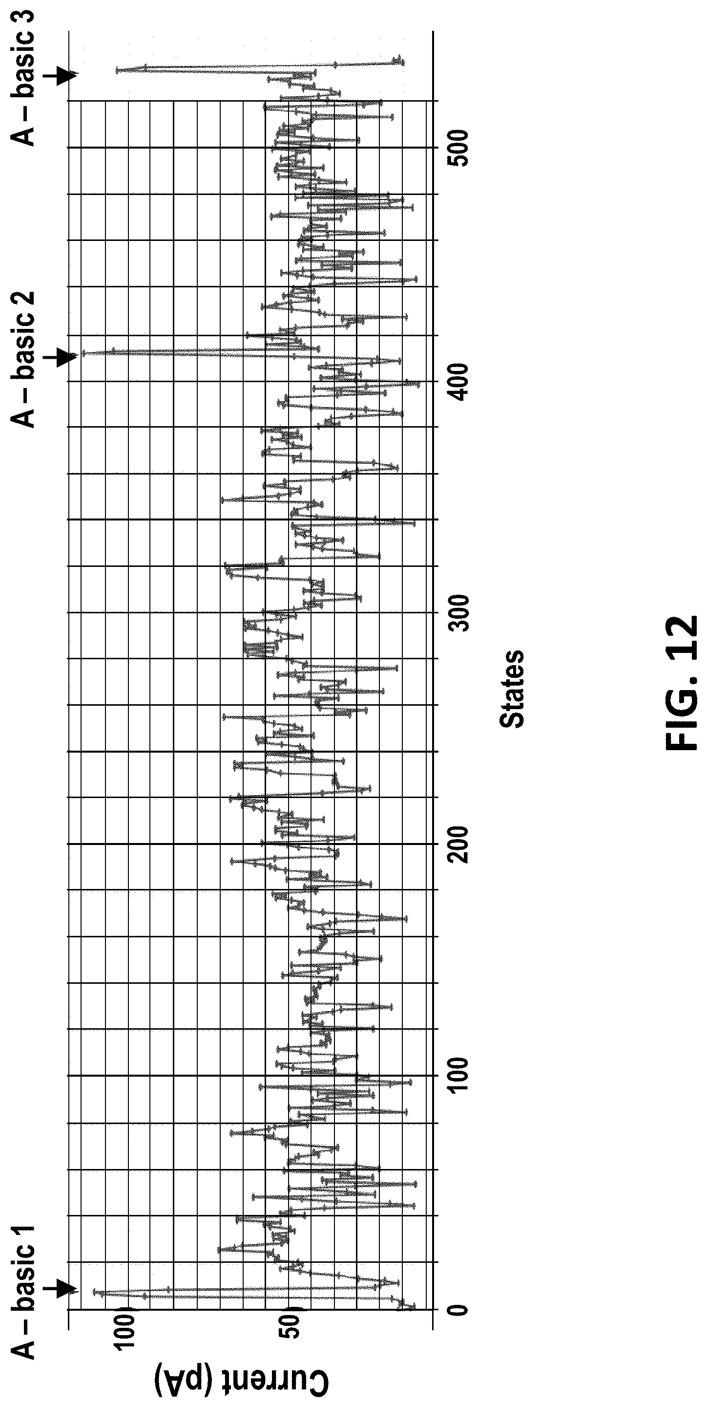
D00013

D00014

D00015

D00016

D00017

D00018

D00019

D00020

D00021

D00022

D00023

D00024
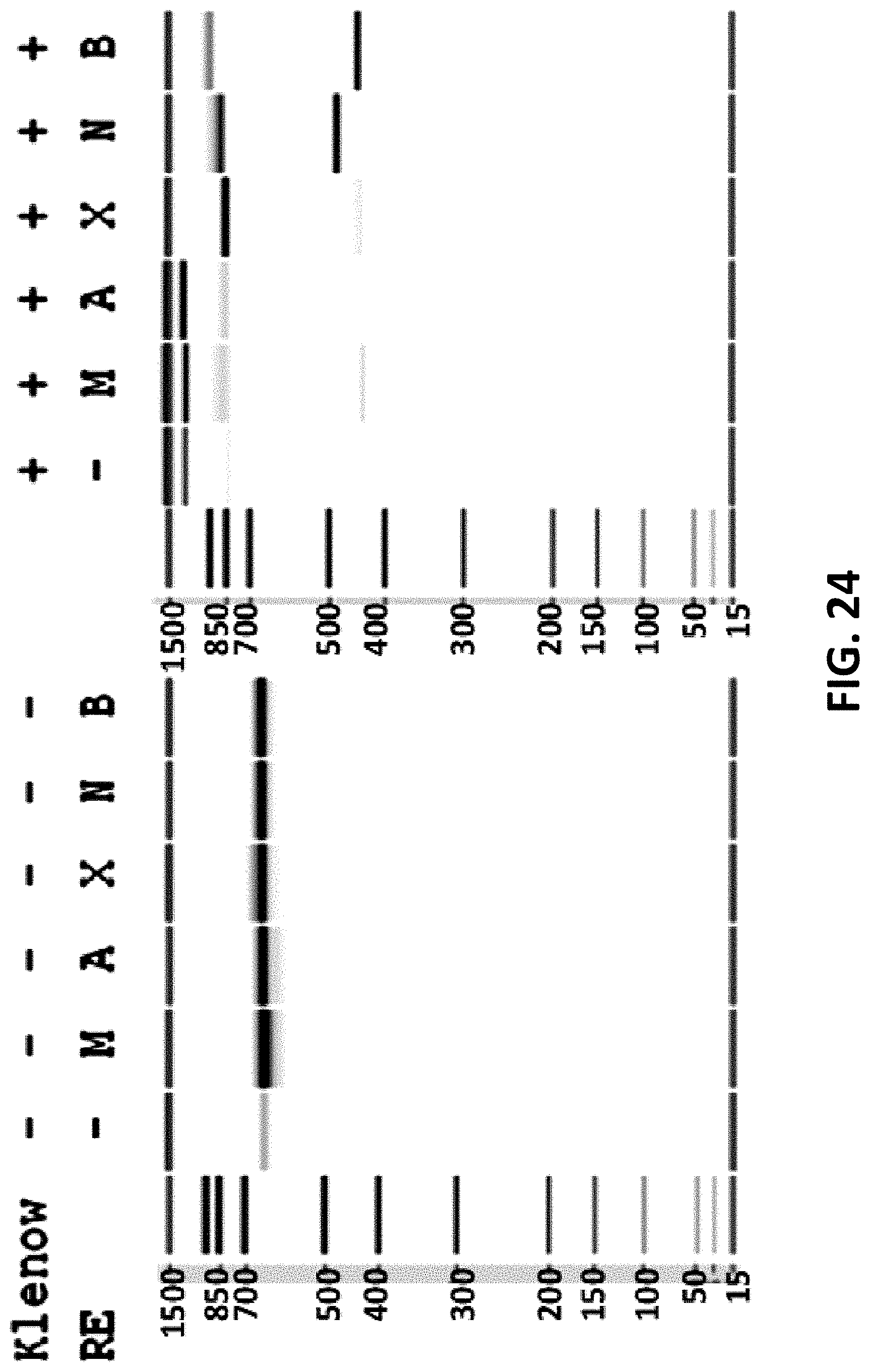
D00025

D00026

D00027

D00028

S00001
XML
uspto.report is an independent third-party trademark research tool that is not affiliated, endorsed, or sponsored by the United States Patent and Trademark Office (USPTO) or any other governmental organization. The information provided by uspto.report is based on publicly available data at the time of writing and is intended for informational purposes only.
While we strive to provide accurate and up-to-date information, we do not guarantee the accuracy, completeness, reliability, or suitability of the information displayed on this site. The use of this site is at your own risk. Any reliance you place on such information is therefore strictly at your own risk.
All official trademark data, including owner information, should be verified by visiting the official USPTO website at www.uspto.gov. This site is not intended to replace professional legal advice and should not be used as a substitute for consulting with a legal professional who is knowledgeable about trademark law.