Antibodies To Tip1 And Methods Of Use Thereof
Hallahan; Dennis ; et al.
U.S. patent application number 16/485100 was filed with the patent office on 2020-07-30 for antibodies to tip1 and methods of use thereof. The applicant listed for this patent is Dennis Kapoor Hallahan. Invention is credited to Dennis Hallahan, Vaishali Kapoor, Heping Yan.
| Application Number | 20200239590 16/485100 |
| Document ID | 20200239590 / US20200239590 |
| Family ID | 1000004815342 |
| Filed Date | 2020-07-30 |
| Patent Application | download [pdf] |


View All Diagrams
| United States Patent Application | 20200239590 |
| Kind Code | A1 |
| Hallahan; Dennis ; et al. | July 30, 2020 |
ANTIBODIES TO TIP1 AND METHODS OF USE THEREOF
Abstract
The disclosure provides antigen binding proteins useful in the recognition of tumor cells and tumor specific delivery of drugs and therapies.
| Inventors: | Hallahan; Dennis; (St. Louis, MO) ; Kapoor; Vaishali; (St. Louis, MO) ; Yan; Heping; (St. Louis, MO) | ||||||||||
| Applicant: |
|
||||||||||
|---|---|---|---|---|---|---|---|---|---|---|---|
| Family ID: | 1000004815342 | ||||||||||
| Appl. No.: | 16/485100 | ||||||||||
| Filed: | February 9, 2018 | ||||||||||
| PCT Filed: | February 9, 2018 | ||||||||||
| PCT NO: | PCT/US2018/017696 | ||||||||||
| 371 Date: | August 9, 2019 |
Related U.S. Patent Documents
| Application Number | Filing Date | Patent Number | ||
|---|---|---|---|---|
| 62457624 | Feb 10, 2017 | |||
| Current U.S. Class: | 1/1 |
| Current CPC Class: | C07K 2317/73 20130101; C07K 16/30 20130101; A61P 35/00 20180101; A61K 2039/505 20130101; C07K 2317/622 20130101; C07K 2317/76 20130101; C07K 2317/77 20130101 |
| International Class: | C07K 16/30 20060101 C07K016/30; A61P 35/00 20060101 A61P035/00 |
Goverment Interests
GOVERNMENTAL RIGHTS
[0002] This invention was made with government support under CA210687 and CA196002 awarded by the National Institutes of Health. The government has certain rights in the invention.
Claims
1. An isolated antigen binding protein that specifically binds an epitope in the PDZ domain of TIP1, wherein the epitope comprises SEQ ID NO:3.
2.-3. (canceled)
4. The isolated antigen binding protein of claim 1, wherein the antigen binding protein comprises a heavy chain variable region comprising SEQ ID NO:4, SEQ ID NO:5 and SEQ ID NO:6 and a light chain variable region comprising SEQ ID NO:7, SEQ ID NO:8 and SEQ ID NO:9 or SEQ ID NO:10.
5.-6. (canceled)
7. The isolated antigen binding protein of claim 1, wherein the antigen binding protein comprises a heavy chain variable region comprising SEQ ID NO:14, SEQ ID NO:15 and SEQ ID NO:16 and a light chain variable region comprising SEQ ID NO:17, SEQ ID NO:18 and SEQ ID NO:19.
8.-9. (canceled)
10. The isolated antigen binding protein of claim 1, wherein the antigen binding protein comprises a heavy chain variable region comprising SEQ ID NO:24, SEQ ID NO:25 and SEQ ID NO:26 and a light chain variable region comprising SEQ ID NO:27, SEQ ID NO:28 and SEQ ID NO:29.
11.-12. (canceled)
13. The isolated antigen binding protein of claim 1, wherein the antigen binding protein comprises a heavy chain variable region comprising SEQ ID NO:34, SEQ ID NO:35 and SEQ ID NO:36 and a light chain variable region comprising SEQ ID NO:37, SEQ ID NO:38 and SEQ ID NO:39.
14.-15. (canceled)
16. The isolated antigen binding protein of claim 1, wherein the antigen binding protein comprises a heavy chain variable region comprising SEQ ID NO:44, SEQ ID NO:45 and SEQ ID NO:46 and a light chain variable region comprising SEQ ID NO:47, SEQ ID NO:48 and SEQ ID NO:49.
17.-18. (canceled)
19. The isolated antigen binding protein of claim 1, wherein the antigen binding protein comprises a heavy chain variable region comprising a sequence selected from the group consisting of SEQ ID NO:20, SEQ ID NO:30, SEQ ID NO:40 and SEQ ID NO:50 and a light chain variable region comprising a sequence selected from the group consisting of SEQ ID NO:21, SEQ ID NO:31, SEQ ID NO:41 and SEQ ID NO:51.
20. The isolated antigen binding protein of claim 1, wherein the antigen binding protein comprises a heavy chain variable region comprising SEQ ID NO:20 and a light chain variable region comprising SEQ ID NO:21.
21. The isolated antigen binding protein of claim 1, wherein the antigen binding protein comprises a heavy chain variable region comprising SEQ ID NO:30 and a light chain variable region comprising SEQ ID NO:31.
22. The isolated antigen binding protein of claim 1, wherein the antigen binding protein comprises a heavy chain variable region comprising SEQ ID NO:40 and a light chain variable region comprising SEQ ID NO:41.
23. The isolated antigen binding protein of claim 1, wherein the antigen binding protein comprises a heavy chain variable region comprising SEQ ID NO:50 and a light chain variable region comprising SEQ ID NO:51.
24. The isolated antigen binding protein of claim 1, wherein the antigen binding protein is selected from the group consisting of a single-chain antibody, an antibody fragment, a chimeric antibody, or a humanized antibody.
25. The isolated antigen binding protein of claim 1, wherein the antigen binding protein is conjugated directly or indirectly to a payload selected from the group consisting of a detectable label, a therapeutic agent, or a combination thereof.
26. The isolated antigen binding protein of claim 25, wherein the detectable label and/or therapeutic agent is a radionuclide.
27. A method of detecting a tumor in a subject, the method comprising: a) exposing a target area of the subject where the presence of a tumor is suspected to ionizing radiation; b) administering to the subject a composition to detect the presence of TIP in the target area, wherein the composition comprises an antigen binding protein binding protein that specifically binds an epitope in the PDZ domain of TIP1, wherein the epitope comprises SEQ ID NO:3 that is conjugated to a detectable label; and c) detecting the detectable label to detect the presence of TIP1, wherein the presence of TIP1 epsilon indicates the presence of a tumor in the target area of the subject.
28. The method of claim 27, wherein the exposing comprises exposing the tumor to less than about 2 Gy ionizing radiation.
29. The method of claim 27, wherein the exposing comprises exposing the tumor to at least about 2 Gy ionizing radiation.
30. The method of claim 27, wherein the exposing comprises exposing the tumor to about 10 Gy to about 20 Gy ionizing radiation.
31. The method of claim 27, wherein the administering comprises administering the antibody subsequent to radiation exposure.
32. The method of claim 27, wherein the administering comprises administering the antibody 0 hours to about 24 hours following radiation exposure.
33.-41. (canceled)
Description
CROSS REFERENCE TO RELATED APPLICATIONS
[0001] This application claims the benefit of U.S. Provisional Application No. 62/457,624, filed on Feb. 10, 2017, the disclosure of which is hereby incorporated by reference in its entirety.
FIELD OF THE INVENTION
[0003] The disclosure provides antigen binding proteins useful in the recognition of tumor cells and tumor specific delivery of drugs and therapies.
BACKGROUND OF THE INVENTION
[0004] Glioblastoma multiforme (GBM) and non-small-cell lung cancer (NSCLC) are among the most difficult and challenging cancers to treat. Lung cancer is the most common type of malignancy and is one of the leading causes of cancer-related deaths worldwide. According to 2016 cancer statistics, an estimated total of 221,200 new lung cancer cases and 158,040 lung cancer deaths occurred in the United States in 2015. Of all lung cancer cases, 85% are NSCLC. Although advancements in diagnosis and treatment have improved, the survival of patients with lung cancer, the 5-year overall survival rate is only .about.15%. GBM is a highly malignant form of brain tumor with an annual incidence of approximately 12,000 in the United States. It is reported to have a five-year survival rate of about five percent. Thus, there is an urgent need to develop novel treatment strategies for NSCLC and GBM patients.
[0005] Exposure of tumor cells to ionizing radiation (IR) is widely known to induce a number of cellular changes. One way that IR can affect tumor cells is through the development of neoantigens which are new molecules that tumor cells express at the cell membrane following some insult or change to the cell. There have been numerous reports in the literature of changes in both tumor and tumor vasculature cell surface molecule expression following treatment with IR. The usefulness of neoantigens for imaging and therapeutic applications lies in the fact that they are differentially expressed on the surface of irradiated tumor cells to a greater extent than on normal tissues. This differential expression provides a mechanism by which tumor cells can be "marked" by radiation for further targeting. Drug delivery vehicles or imaging agents conjugated to ligands that recognize and interact with the neoantigens can help to improve tumor-specific targeting and reduce systemic toxicity with cancer drugs.
[0006] Other objects, advantages and features of the present invention will become apparent from the following specification taken in conjunction with the accompanying figures.
SUMMARY OF THE INVENTION
[0007] In an aspect, the present invention encompasses cell line that expresses an antigen binding protein comprising an amino acid sequence selected from the group consisting of SEQ ID NO:4, SEQ ID NO:5, SEQ ID NO:6 SEQ ID NO:7, SEQ ID NO:8, SEQ ID NO:9, SEQ ID NO:10, SEQ ID NO:14, SEQ ID NO:15, SEQ ID NO: 16, SEQ ID NO:17, SEQ ID NO:18, SEQ ID NO:19, SEQ ID NO:20, SEQ ID NO:21, SEQ ID NO:24, SEQ ID NO:25, SEQ ID NO:26, SEQ ID NO:27, SEQ ID NO:28, SEQ ID NO:29, SEQ ID NO:30, SEQ ID NO:31, SEQ ID NO:34, SEQ ID NO:35, SEQ ID NO: 36, SEQ ID NO:37, SEQ ID NO:38, SEQ ID NO:39, SEQ ID NO: 40, SEQ ID NO:41, SEQ ID NO:44, SEQ ID NO:45, SEQ ID NO:46, SEQ ID NO:47, SEQ ID NO:48, SEQ ID NO:49, SEQ ID NO:50 and SEQ ID NO:51, wherein the antigen binding protein specifically bind to tax interacting protein 1 (TIP1). Specifically, antigen binding proteins of the disclosure bind to the PSD-95/DIgA/ZO-1 (PDZ) domain of TIP1.
[0008] In another aspect, the present invention encompasses an isolated antigen binding protein that specifically binds TIP1 and comprises a heavy chain variable region comprising SEQ ID NO:4, SEQ ID NO:5, and SEQ ID NO:6. In another aspect, the present invention encompasses an isolated antigen binding protein that specifically binds TIP1 and comprises a heavy chain variable region comprising SEQ ID NO:14, SEQ ID NO:15, and SEQ ID NO:16. In another aspect, the present invention encompasses an isolated antigen binding protein that specifically binds TIP1 and comprises a heavy chain variable region comprising SEQ ID NO:24, SEQ ID NO:25, and SEQ ID NO:26. In another aspect, the present invention encompasses an isolated antigen binding protein that specifically binds TIP1 and comprises a heavy chain variable region comprising SEQ ID NO:34, SEQ ID NO:35, and SEQ ID NO:36. In another aspect, the present invention encompasses an isolated antigen binding protein that specifically binds TIP1 and comprises a heavy chain variable region comprising SEQ ID NO:44, SEQ ID NO:45, and SEQ ID NO:46.
[0009] In still another aspect, the present invention encompasses an isolated antigen binding protein that specifically binds TIP1 and comprises a light chain variable region comprising SEQ ID NO:7, SEQ ID NO:8, and SEQ ID NO:9 or SEQ ID NO:10. In another aspect, the present invention encompasses an isolated antigen binding protein that specifically binds TIP1 and comprises a light chain variable region comprising SEQ ID NO:17, SEQ ID NO:18, and SEQ ID NO:19. In another aspect, the present invention encompasses an isolated antigen binding protein that specifically binds TIP1 and comprises a light chain variable region comprising SEQ ID NO:27, SEQ ID NO:28, and SEQ ID NO:29. In another aspect, the present invention encompasses an isolated antigen binding protein that specifically binds TIP1 and comprises a light chain variable region comprising SEQ ID NO:37, SEQ ID NO:38, and SEQ ID NO:39. In another aspect, the present invention encompasses an isolated antigen binding protein that specifically binds TIP1 and comprises a light chain variable region comprising SEQ ID NO:47, SEQ ID NO:48, and SEQ ID NO:49.
[0010] In another aspect, the present invention encompasses an isolated antigen binding protein that specifically binds TIP1 and comprises a heavy chain CDR3 comprising the amino acid sequence of SEQ ID NO:6 with zero to two amino acid substitutions. In another aspect, the present invention encompasses an isolated antigen binding protein that specifically binds TIP1 and comprises a heavy chain CDR3 comprising the amino acid sequence of SEQ ID NO:16 with zero to two amino acid substitutions. In another aspect, the present invention encompasses an isolated antigen binding protein that specifically binds TIP1 and comprises a heavy chain CDR3 comprising the amino acid sequence of SEQ ID NO:26 with zero to two amino acid substitutions. In another aspect, the present invention encompasses an isolated antigen binding protein that specifically binds TIP1 and comprises a heavy chain CDR3 comprising the amino acid sequence of SEQ ID NO:36 with zero to two amino acid substitutions. In another aspect, the present invention encompasses an isolated antigen binding protein that specifically binds TIP1 and comprises a heavy chain CDR3 comprising the amino acid sequence of SEQ ID NO:46 with zero to two amino acid substitutions.
[0011] In yet another aspect, the present invention encompasses an antigen binding protein comprising a heavy chain variable region comprising a sequence selected from the group consisting of SEQ ID NO:20, SEQ ID NO:30, SEQ ID NO:40 and SEQ ID NO:50 and/or a light chain variable region comprising a sequence selected from the group consisting of SEQ ID NO:21, SEQ ID NO:31, SEQ ID NO:41 and SEQ ID NO:51.
[0012] In yet still another aspect, the present invention encompasses a method of detecting a tumor in a subject. The method comprises exposing a target area of the subject where the presence of a tumor is suspected to ionizing radiation; administering to the subject a composition to detect the presence of TIP1 in the target area, wherein the composition comprises one or more targeting antigen binding proteins wherein each targeting antigen binding protein specifically binds to the PDZ domain of TIP1 exposed on an irradiated cell and is conjugated to a detectable label; and detecting the detectable label to detect the presence of TIP1, wherein the presence of TIP1 indicates the presence of a tumor in the target area of the subject.
[0013] In a different aspect, the present invention encompasses a method of enhancing radiotherapy in a subject. The method comprises administering a pharmacologically effective amount of an isolated antigen binding protein of the invention to the subject, such that radiotherapy is enhanced.
[0014] In other aspects, the present invention encompasses a method of delivering a therapeutic agent to a cell expressing TIP1 in a subject. The method comprises exposing a target area of the subject where the presence of a tumor is suspected to ionizing radiation; and administering an isolated antigen binding protein of the invention to the subject.
[0015] In still other aspects, the present invention encompasses a method of detecting TIP1 in a sample. The method comprises obtaining a sample, and detecting and/or measuring the amount of TIP1 in the sample using an antigen binding protein of the invention.
[0016] In yet still in other aspects, the present invention encompasses a method of attenuating proliferation of a tumor cell and/or killing a tumor cells. The method comprises contacting the tumor cell with an isolated antigen binding protein of the invention. Specifically, an isolated antigen binding protein which binds to the PDZ domain of TIP1.
[0017] While multiple embodiments are disclosed, still other embodiments of the present invention will become apparent to those skilled in the art from the following detailed description, which shows and describes illustrative embodiments of the invention. Accordingly, the figures and detailed description are to be regarded as illustrative in nature and not restrictive.
BRIEF DESCRIPTION OF THE FIGURES
[0018] The application file contains at least one drawing executed in color. Copies of this patent application publication with color drawing(s) will be provided by the Office upon request and payment of the necessary fee. Figures represented herein are not limitations to the various embodiments according to the invention and are presented for exemplary illustration of the invention.
[0019] FIG. 1A, FIG. 1B, FIG. 1C, FIG. 1D, FIG. 1E and FIG. 1F depict graphs showing anti-PDZ antibody treatment reduces cancer cell proliferation in a time-dependent manner. Cells were treated with 1 .mu.g/ml of the anti-PDZ antibody or isotype control, antibody for 24 h, 48 h, 72 h, and 96 h. Proliferating cells (Viable) were evaluated by trypan blue dye exclusion assay at the indicated time points. Shown are the mean fold change in cell number relative to control and SD from three different experiments. (FIG. 1A) A549 cells, (FIG. 1B) D54 cells, (FIG. 1C) H460 cells, and (FIG. 1D) U251 cells. (FIG. 1E and FIG. 1F) Proliferation of A549 and U251 cells which were transduced with either CRISPR control vector or TIP1 sgRNAs was evaluated by the trypan blue dye exclusion assay at the indicated time points. Shown are the mean fold change in cell number relative to CRISPR control and SD from three different experiments *P<0.05, ***P<0.001, ****P<0.0001
[0020] FIG. 2A, FIG. 2B, FIG. 2C and FIG. 2D depict graphs showing that the PDZ domain of TIP1 is important for cell proliferation. (FIG. 2A) NSCLC (A549 and LLC) and GBM (D54 and GL261) cell proliferation following treatment with non-PDZ antibody or anti-PDZ antibody for 96 h. Proliferating cells were evaluated using Trypan blue dye exclusion assays. Shown are the mean percentages of proliferating cells relative to the isotype control and SD from three treatments. (FIG. 2B) Anti-PDZ Ab treatment does not alter cell viability in normal lung (MRC-5) and endothelial (HUVEC) cells. Cells were treated with 1 .mu.g/ml anti-PDZ or isotype control antibody for 96 hours. Trypan blue dye exclusion assay was performed to count the viable cells. Shown are the percent viable cells as bar graph with SD from three different experiments. (FIG. 2C) TIP1 knockout abrogates the effect of the anti-PDZ/TIP1 antibody on cell proliferation. A549 and U251 cells having TIP1 KO were treated with anti-PDZ/TIP1 antibody and cell proliferation was evaluated. Shown are the mean percentages of proliferating cells relative to the untreated CRISPR control cells and SD from three treatments. (FIG. 2C and FIG. 2D) TIP1 knockout abrogates the effect of the anti-PDZ/TIP1 antibody on cell proliferation. A549 and U251 cells having TIP1 KO were treated with anti-PDZ/TIP1 antibody and cell proliferation was evaluated. Shown are the mean percentages of proliferating cells relative to the untreated CRISPR control cells and SD from three treatments.
[0021] FIG. 3A, FIG. 3B, FIG. 3C and FIG. 3D depict images showing the structural depiction of the anti-PDZ and non-PDZ antibody epitopes. Surface representation of the TIP1 3D structure showing the anti-PDZ antibody epitopes as a (FIG. 3A) ribbon representation and (FIG. 3B) stick representation. The epitope of the non-PDZ antibody as a (FIG. 3C) ribbon and (FIG. 3D) stick representation. White arrows depict the PDZ binding pocket/groove involved in binding with interacting partners.
[0022] FIG. 4A, FIG. 4B, FIG. 4C, FIG. 4D, FIG. 4E, FIG. 4F, FIG. 4G and FIG. 4H depict images and graphs showing that anti-PDZ antibody treatment induces apoptosis following internalization in NSCLC and GBM cell lines. (FIG. 4A, FIG. 4B, FIG. 4C, FIG. 4D) Live cell fluorescent imaging showing internalization of anti-PDZ antibody. A549 cells were labeled with cell mask orange dye and treated with Alexa-Flour 488 labeled anti-PDZ antibody. Live cell images were captured every 5 minutes in a spinning-disk fluorescent microscope. Shown are representative images at various time points. Yellow spots indicate the internalized antibodies. (FIG. 4E, FIG. 4F, FIG. 4G, FIG. 4H) Annexin V/PI assays with lung cancer (A549 and H460, FIG. 4E and FIG. 4G, respectively) and glioblastoma (D54 and U251, FIG. 4F and FIG. 4H, respectively) cells. A549, H460, D54 and U251 cells were treated with 1 .mu.g/ml of anti-PDZ antibody. Cells were stained with Annexin V and PI 96 h after treatment and analyzed by flow cytometry. Shown are the mean percentage of late apoptotic cells (Annexin V and PI positive) and dead cells (PI positive) with SD from three different experiments ****P<0.0001
[0023] FIG. 5A and FIG. 5B depict antibodies against PDZ domain of TIP-1 suppress AKT/mTOR signaling in NSCLC and GBM cell lines. Immunoblot analysis of survival pathways. Lung cancer (A549 and H460) and glioblastoma (D54 and U251) cells were treated with 1 .mu.g/ml of anti-PDZ antibody for 96 h. (FIG. 5A) Total cellular proteins were immunoblotted using antibodies against phospho-Akt (S473), total Akt, and GAPDH was used as a loading control. (FIG. 5B) Total cellular proteins were immunoblotted using antibodies against phospho-mTOR (S2448), total mTOR, phospo-P70S6 (T389) total-P70S6 phospo-4EBP1 (T70), 4EBP1 (T70) and GAPDH was used as a loading control.
[0024] FIG. 6A, FIG. 6B, FIG. 6C, FIG. 6D, FIG. 6E, FIG. 6F, FIG. 6G, FIG. 6H, FIG. 6I, FIG. 6J, FIG. 6K, FIG. 6L, FIG. 6M and FIG. 6N depict graphs showing that the anti-PDZ antibody enhances the efficacy of XRT and delays growth of A549 and U251 tumor xenografts implanted into hind limbs of athymic nude mice. (FIG. 6A, FIG. 6B, FIG. 6C, FIG. 6D) Colony formation assays with lung cancer (A549 and H460, FIG. 6A and FIG. 6C, respectively) and glioblastoma (D54 and U251, FIG. 6B and FIG. 6D, respectively) cells. A549, H460, D54 and U251 cells were treated with 1 .mu.g/ml of anti-PDZ antibody or isotype control for 96 h and plated to measure colony formation. Cells were then irradiated with 3 Gy and incubated for 10-14 days. Colonies comprising of 50 or more were scored. Shown are the mean surviving fraction normalized to the isotype control with SD from three different experiments *P<0.05, **P<0.01, ***P<0.001, ****P<0.0001 A549 (FIG. 6E, FIG. 6G) and U251 (FIG. 6F, FIG. 6H) cells were implanted into the left and right flank of athymic nude mice. Once the tumors were palpable (.about.200 mm.sup.3), the tumors on the right flank were irradiated with 2 Gy for 5 consecutive days for a total of 10 Gy. The tumors on the left flank served as un-irradiated sham control. The tumor bearing mice were treated either with isotype control antibody or anti-TIP-1 antibody on days 1 and 4 after initiating the XRT treatment. Tumor volumes were measured using digital calipers. Shown are the mean tumor volumes of A549 and U251 with SD from 5 mice in each treatment **P<0.01, ****P<0.0001. (FIG. 6I and FIG. 6J) Western blot analysis of tumor lysates showing downregulation of phospho- and total AKT and phospho-, total-mTOR and 4EBP-1. (FIG. 6K, FIG. 6L, FIG. 6M and FIG. 6N) Lung cancer (A549 and H460) and glioblastoma (D54 and U251) cells were treated with 1 .mu.g/ml antibody and irradiated with 3Gy or OGy. Trypan blue dye exclusion assay was performed after 96 h. Shown are the percent viable cells as a bar graph with SD from three treatments.
[0025] FIG. 7 depicts the localization of designed PDZ peptide (SEQ ID NO:3) in the whole Tip1 protein sequence (SEQ ID NO:1).
[0026] FIG. 8A and FIG. 8B depict images showing a dot blot assay of 24 positive clones out of 384 primary screened clones. (FIG. 8A) PDZ peptide on NC membrane reacted with 24 positive clones. (FIG. 8B) Tip1 peptide on NC membrane reacted with 24 positive clones.
[0027] FIG. 9A and FIG. 9B depict graphs showing ELISA data of 6 purified anti-PDZ peptide scFvs. (FIG. 9A) PDZ scFv test: peptide on ELISA plate. (FIG. 9B) PDZ peptide scFv test: Tip 1 on ELISA plate.
[0028] FIG. 10A and FIG. 10B depict Western-blot assays of anti PDZ scFv clones. (FIG. 10A) A549 3Gyx3 cell lysate. (FIG. 10B) Recombinant Tip1 protein.
[0029] FIG. 11A, FIG. 11B and FIG. 11C depicts graphs showing tumor cell viability by CCK-8 assay. (FIG. 11A) Treatment with PDZ scFv G14. (FIG. 11B) Treatment with PDZ scFv H2. (FIG. 11C) Treatment with PDZ scFv D2.
[0030] FIG. 12A and FIG. 12B depict graphs showing results from a tumor cell viability assay. (FIG. 12A) Effect of PDZ scFv on cell proliferation using A549 cells._(FIG. 12B) Effect of PDZ scFv on cell proliferation using U251 cells.
[0031] FIG. 13A depicts optical imaging on tumor bearing nude mice treated with anti-Tip1 PDZ domain scFv. FIG. 13B depicts optical imaging on tumor bearing nude mice treated with control scFv.
[0032] FIG. 14A depicts the CDRs of PDZ scFv clone G14. SEQ ID NO:4 (GFTFSNYA), SEQ ID NO:5 (VSGSGAST), SEQ ID NO:6 (AKHGTRFDYWGQRTLVTVS) and SEQ ID NO:7 (QSVSSY), SEQ ID NO:8 (GAS), and SEQ ID NO:9 (QQT). FIG. 14B depicts the CDRs of PDZ scFv clone H2. SEQ ID NO:4 (GFTFSNYA), SEQ ID NO:5 (VSGSGAST), SEQ ID NO:6 (AKHGTRFDYWGQRTLVTVS) and SEQ ID NO:7 (QSVSSY), SEQ ID NO:8 (GAS), and SEQ ID NO:10 (QQTYPLTFGRWKIK).
[0033] FIG. 15A and FIG. 15B depict graphs showing viable cells following treatment with scFvs of the disclosure. FIG. 15A shows viable cells following treatment with 10 .mu.g of antibody. FIG. 15B shows viable cells following treatment with 15 .mu.g of antibody.
[0034] FIG. 16A, FIG. 16B and FIG. 16C show the specificity of 2F10, 3D6, 7H5, and 4612 TIP1 PDZ domain binding proteins. FIG. 16A depicts an ELISA assay with the TIP1 mouse monoclonal hybridoma supernatants showing saturation curves. FIG. 16B shows the affinity of the purified mouse monoclonal anti-TIP antibodies as detected by Biacore. FIG. 16C shows the mean fluorescence intensity of the anti-TIP1 antibodies showing cell surface binding by flow cytometry.
[0035] FIG. 17A depicts the heavy chain DNA sequence of 2F10 with a leader sequence (SEQ ID NO:22). FIG. 17B depicts the heavy chain amino acid sequence of 2F10 with a leader sequence (SEQ ID NO:20), including heavy chain CDR1 (SEQ ID NO:14), CDR2 (SEQ ID NO:15), and CDR3 (SEQ ID NO:16). FIG. 17C depicts the light chain DNA sequence of 2F10 with a leader sequence (SEQ ID NO:23). FIG. 17D depicts the light chain amino acid sequence of 2F10 with a leader sequence (SEQ ID NO:21), including heavy chain CDR1 (SEQ ID NO:17), CDR2 (SEQ ID NO:18), and CDR3 (SEQ ID NO:19).
[0036] FIG. 18A depicts the heavy chain DNA sequence of 3D6 with a leader sequence (SEQ ID NO:32). FIG. 18B depicts the heavy chain amino acid sequence of 3D6 with a leader sequence (SEQ ID NO:30), including heavy chain CDR1 (SEQ ID NO:24), CDR2 (SEQ ID NO:25), and CDR3 (SEQ ID NO:26). FIG. 18C depicts the light chain DNA sequence of 3D6 with a leader sequence (SEQ ID NO:33). FIG. 18D depicts the light chain amino acid sequence of 3D6 with a leader sequence (SEQ ID NO:31), including heavy chain CDR1 (SEQ ID NO:27), CDR2 (SEQ ID NO:28), and CDR3 (SEQ ID NO:29).
[0037] FIG. 19A depicts the heavy chain DNA sequence of 4612 with a leader sequence (SEQ ID NO:52). FIG. 19B depicts the heavy chain amino acid sequence of 4612 with a leader sequence (SEQ ID NO:50), including heavy chain CDR1 (SEQ ID NO:44), CDR2 (SEQ ID NO:45), and CDR3 (SEQ ID NO:46). FIG. 19C depicts the light chain DNA sequence of 4B12 with a leader sequence (SEQ ID NO:53). FIG. 19D depicts the light chain amino acid sequence of 4B12 with a leader sequence (SEQ ID NO:51), including heavy chain CDR1 (SEQ ID NO:47), CDR2 (SEQ ID NO:48), and CDR3 (SEQ ID NO:49).
[0038] FIG. 20A depicts the heavy chain DNA sequence of 7H5 with a leader sequence (SEQ ID NO:42). FIG. 20B depicts the heavy chain amino acid sequence of 7H5 with a leader sequence (SEQ ID NO:40), including heavy chain CDR1 (SEQ ID NO:34), CDR2 (SEQ ID NO:35), and CDR3 (SEQ ID NO:36). FIG. 20C depicts the light chain DNA sequence of 7H5 with a leader sequence (SEQ ID NO:43). FIG. 20D depicts the light chain amino acid sequence of 7H5 with a leader sequence (SEQ ID NO:41), including heavy chain CDR1 (SEQ ID NO:37), CDR2 (SEQ ID NO:38), and CDR3 (SEQ ID NO:39).
DETAILED DESCRIPTION OF THE INVENTION
[0039] The present disclosure provides antigen binding proteins that recognize tumor cells. The antigen binding proteins may be used to provide tumor specific delivery, for instance, of drugs or therapeutic agents, as well as enhancing the efficacy of radiotherapy. In particular, the present disclosure provides for antigen binding proteins that bind to tax interacting protein 1 (TIP1). Advantageously, these antigen binding proteins specifically bind tumor cells and not normal cells. In an exemplary embodiment, antigen binding proteins of the disclosure specifically bind to epitopes exposed on irradiated tumor related cells. For instance, antigen binding proteins of the disclosure may bind to extracellular, transmembrane or intracellular epitopes on irradiated tumor related cells. Specifically, antigen binding proteins of the disclosure bind to the PSD-95/DIgA/ZO-1 (PDZ) domain of TIP1 on irradiated tumor related cells.
[0040] The antibodies and methods of their use are described in further detail below.
I. Anti-Tip1 Antigen Binding Proteins
[0041] In an aspect, anti-TIP1 antigen binding proteins include antigen binding proteins that specifically bind an epitope within the PDZ domain. More specifically, anti-TIP1 antigen binding proteins include antigen binding proteins that specifically bind an epitope within the PDZ domain of TIP1. Generally speaking, the epitope is detectable on the surface of a tumor cell following irradiation. The epitope may or may not be detectable on the cell surface in the absence of irradiation. Alternatively, an epitope may be detectable on the surface of a tumor cell both in the absence of irradiation and following irradiation, though the detectable signal is greater following irradiation. The PDZ domain spans about 80 to about 100 amino acid residues and is comprised of six .beta.-sheets and two .alpha.-helices. TIP1 comprises a single PDZ domain from about residue 13 to about residue 112 of the 124-amino acid protein (SEQ ID NO:1; MSYIPGQPVTAVVQRVEIHKLRQGENLILGFSIGGGIDQDPSQNPFSEDK TDKGIYVTRVSEGGPAEIAGLQIGDKIMQVNGWDMTMVTHDQARKRLTKRSEEVV RLLVTRQSLQKAVQQSMLS). Accordingly, in certain embodiments, anti-TIP1 antigen binding proteins include antigen binding proteins that specifically bind an epitope within SEQ ID NO:2 (VQRVEIHKLRQGENLILGFSIGGGIDQDPSQNPFSEDKTDKGIYVTRV SEGGPAEIAGLQIGDKIMQVNGWDMTMVTHDQARKRLTKRSEEVVRLLVTRQ). The epitope may be about 5, about 6, about 7, about 8, about 9, about 10, about 11, about 12, about 13, about 14, about 15, about 16, about 17, about 18, about 19, about 20, about 21, about 22, about 23, about 24, about 25, about 26, about 27, about 28, about 29, or about 30 amino acids within SEQ ID NO:2. Specifically, the epitope may be about 13, about 14, about 15, about 16, about 17, about 18, about 19, about 20, about 21, about 22, or about 23 amino acids within SEQ ID NO:2. The epitope may be linear or conformational. In an embodiment, the epitope may be about 5, about 6, about 7, about 8, about 9, about 10, about 11, about 12, about 13, about 14, about 15, about 16, about 17, about 18 amino acids within SEQ ID NO:3 (IDQDPSQNPFSEDKTDKG). In a specific embodiment, anti-TIP1 antigen binding proteins include antigen binding proteins that specifically bind an epitope consisting of SEQ ID NO:3. In a specific embodiment, anti-TIP1 antigen binding proteins include antigen binding proteins that specifically bind an epitope consisting of SEQ ID NO:11 (IDQDPSQNPF).
[0042] The phrase "specifically binds" herein means antigen binding proteins bind to the protein with an affinity constant or affinity of interaction (K.sub.D) of less than 300 nM, less than 250 nM, less than 200 nM, less than 150 nM, less than 100 nM, less than 75 nM, less than 50 nM, less than 25 nM, less than 20 nM, less than 15 nM, less than 10 nM, less than 5 nM, or less than 1 nM. In one embodiment, an anti-TIP1 antigen binding protein binds to an epitope within the PDZ domain with a K.sub.D of less than 250 nM. In an embodiment, an anti-TIP1 antigen binding protein binds to an epitope within the PDZ domain with a K.sub.D of less than 100 nM. In another embodiment, an anti-TIP1 antigen binding protein binds to an epitope within the PDZ domain with a K.sub.D of less than 50 nM. In still another embodiment, an anti-TIP1 antigen binding protein binds to an epitope within the PDZ domain with a K.sub.D of less than 20 nM. In still yet another embodiment, an anti-TIP1 antigen binding protein binds to an epitope within the PDZ domain with a K.sub.D of less than 10 nM. Methods of determining whether an antigen binding protein binds to the PDZ domain of TIP1 are known in the art.
[0043] The term "antigen binding protein" refers to any form of antibody or fragment thereof that exhibits the desired biological activity. Thus, it is used in the broadest sense and specifically covers monoclonal antibodies (including full length monoclonal antibodies), polyclonal antibodies, multispecific antibodies (e.g. bispecific antibodies), and antibody fragments so long as they exhibit the desired biological activity.
[0044] The term "monoclonal antibody" refers to an antibody that is derived from a single copy or clone, including e.g., any eukaryotic, prokaryotic, or phage clone. Monoclonal antibodies are obtained from a population of substantially homogeneous antibodies, i.e., the individual antibodies comprising the population are identical except for possible naturally occurring mutations or post-translational modification that may be present in minor amounts. Monoclonal antibodies are highly specific, being directed against a single antigenic epitope. "Monoclonal antibody" is not limited to antibodies produced through hybridoma technology. Monoclonal antibodies can be produced using e.g., hybridoma techniques well known in the art, as well as recombinant technologies, phage display technologies, synthetic technologies or combinations of such technologies and other technologies readily known in the art. Furthermore, the monoclonal antibody may be labeled with a detectable label, immobilized on a solid phase and/or conjugated with a heterologous compound (e.g., an enzyme or toxin) according to methods known in the art.
[0045] The term "fragment thereof" encompasses a fragment or a derivative of an antibody that still substantially retain its biological activity. Therefore, the term "antibody fragment" or "fragment thereof" refers to a portion of a full length antibody, generally the antigen binding or variable region thereof. Examples of an immunologically effective fragment thereof include Fab, Fab', F(ab').sub.2 and Fv fragments, diabodies, linear antibodies, single-chain molecules, and multispecific antibodies formed from antibody fragments. In some contexts herein, fragments will be mentioned specifically for emphasis; nevertheless, it will be understood that regardless of whether fragments are specified, the term "antibody" includes such fragments.
[0046] Also included within the definition "antibody" for example are single chain forms, generally designated Fv, regions, of antibodies with this specificity. These scFvs are comprised of the heavy and light chain variable regions connected by a linker. In most instances, but not all, the linker may be a peptide. A linker peptide is preferably from about 10 to 25 amino acids in length. Preferably, a linker peptide is rich in glycine, as well as serine or threonine. ScFvs can be used to facilitate phage display or can be used for flow cytometry, immunohistochemistry, or as targeting domains. Methods of making and using scFvs are known in the art. In a preferred embodiment, the scFvs of the present disclosure are conjugated to a human constant domain. In some embodiments, the heavy constant domain is derived from an IgG domain, such as IgG1, IgG2, IgG3, or IgG4. In other embodiments, the heavy chain constant domain may be derived from IgA, IgM, or IgE.
[0047] The term "antibody" also includes bispecific monoclonal antibodies (i.e. a protein that comprises fragments of two different monoclonal antibodies and consequently binds two different antigens). A specific example of a bispecific monoclonal antibody may be a Bi-specific T-cell Engager (BiTE) which is a fusion protein consisting of two single-chain variable fragments (scFvs) of different antibodies. In certain embodiments, BiTEs from a link between T cells and infected cells. Accordingly, one scFv is a specific for TIP1 and one scFv binds a T cell. Additionally, an antibody of the disclosure may be a chimeric antigen receptor (CAR), also referred to as an artificial T cell receptor, a chimeric T cell receptor, or a chimeric immunoreceptor. CARs are engineered receptors, which graft an arbitrary specificity onto an immune effector cell. Typically, these receptors are used to graft the specificity of a monoclonal antibody onto a T cell. Additionally, included within the definition "antibody" are single-domain antibodies, generally designated sdAb, which is an antibody fragment consisting of a single monomeric variable antibody domain. A sdAb antibody may be derived from camelids (V.sub.HH fragments) or cartilaginous fishes (V.sub.NAR fragments). As long as the protein retains the ability specifically to bind its intended target, it is included within the term "antibody."
[0048] Preferably, but not necessarily, the antibodies useful in the discovery are produced recombinantly, as manipulation of the typically murine or other non-human antibodies with the appropriate specificity is required in order to convert them to humanized form. Antibodies may or may not be glycosylated, though glycosylated antibodies are preferred. Antibodies are properly cross-linked via disulfide bonds, as is known.
[0049] The basic antibody structural unit of an antibody useful herein comprises a tetramer. Each tetramer is composed of two identical pairs of polypeptide chains, each pair having one "light" (about 25 kDa) and one "heavy" chain (about 50-70 kDa). The amino-terminal portion of each chain includes a variable region of about 100 to 110 or more amino acid sequences primarily responsible for antigen recognition. The carboxy-terminal portion of each chain defines a constant region primarily responsible for effector function.
[0050] Light chains are classified as gamma, mu, alpha, and lambda. Heavy chains are classified as gamma, mu, alpha, delta, or epsilon, and define the antibody's isotype as IgG, IgM, IgA, IgD and IgE, respectively. Within light and heavy chains, the variable and constant regions are joined by a "J" region of about 12 or more amino acid sequences, with the heavy chain also including a "D" region of about 10 more amino acid sequences.
[0051] The variable regions of each light/heavy chain pair form the antibody binding site. Thus, an intact antibody has two binding sites, although recombinant versions can be of higher valency. The chains exhibit the same general structure of relatively conserved framework regions (FR) joined by three hypervariable regions, also called complementarity determining regions (hereinafter referred to as "CDRs"). The CDRs from the two chains are aligned by the framework regions, enabling binding to a specific epitope. From N-terminal to C-terminal, both light and heavy chains comprise the domains FR1, CDR1, FR2, CDR2, FR3, CDR3 and FR4 respectively. The assignment of amino acid sequences to each domain is in accordance with known conventions (See, Kabat "Sequences of Proteins of Immunological Interest" National Institutes of Health, Bethesda, Md., 1987 and 1991; Chothia, et al, J. Mol. Bio. (1987) 196:901-917; Chothia, et al., Nature (1989) 342:878-883). For example, Kabat, Chothia, combinations thereof, or other known methods of determining CDRs may be used.
[0052] Additionally, an antibody of the disclosure can be modified to optimize or minimize effector function. Further, an antibody of the disclosure can be modified to extend half-life. Still further, an antibody of the disclosure can be modified to improve binding affinity. Methods of modifying an antibody to improve the aforementioned characteristics are known in the art. For example, the crystal structures disclosed herein may be used to rationally alter amino acids to optimize contact with the antibody and antigen.
[0053] In an aspect, monoclonal anti-TIP1 antibodies are generated with appropriate specificity by standard techniques of immunization of mammals, forming hybridomas from the antibody-producing cells of said mammals or otherwise immortalizing them, and culturing the hybridomas or immortalized cells to assess them for the appropriate specificity. In the present case, such antibodies could be generated by immunizing a human, rabbit, rat or mouse, for example, with a peptide representing an epitope encompassing the PDZ domain or an appropriate subregion thereof. Specifically, such antibodies could be generated by immunizing a human, rabbit, rat or mouse, for example, with a peptide comprising SEQ ID NO:2. More specifically, such antibodies could be generated by immunizing a human, rabbit, rat or mouse, for example, with a peptide comprising SEQ ID NO:3. Materials for recombinant manipulation can be obtained by retrieving the nucleotide sequences encoding the desired antibody from the hybridoma or other cell that produces it. These nucleotide sequences can then be manipulated and isolated, characterized, purified and, recovered to provide them in humanized form, for use herein if desired.
[0054] As used herein "humanized antibody" includes an anti-TIP1 antibody that is composed partially or fully of amino acid sequences derived from a human antibody germline by altering the sequence of an antibody having non-human complementarity determining regions ("CDR"). The simplest such alteration may consist simply of substituting the constant region of a human antibody for the murine constant region, thus resulting in a human/murine chimera which may have sufficiently low immunogenicity to be acceptable for pharmaceutical use. Preferably, however, the variable region of the antibody and even the CDR is also humanized by techniques that are by now well known in the art. The framework regions of the variable regions are substituted by the corresponding human framework regions leaving the non-human CDR substantially intact, or even replacing the CDR with sequences derived from a human genome. CDRs may also be randomly mutated such that binding activity and affinity for the PDZ domain of TIP1 is maintained or enhanced in the context of fully human germline framework regions or framework regions that are substantially human. Substantially human frameworks have at least 90%, 95%, or 99% sequence identity with a known human framework sequence. Fully useful human antibodies are produced in genetically modified mice whose immune systems have been altered to correspond to human immune systems. As mentioned above, it is sufficient for use in the methods of this discovery, to employ an immunologically specific fragment of the antibody, including fragments representing single chain forms.
[0055] Further, as used herein the term "humanized antibody" refers to an anti-TIP1 antibody comprising a human framework, at least one CDR from a nonhuman antibody, and in which any constant region present is substantially identical to a human immunoglobulin constant region, i.e., at least about 85-90%, preferably at least 95% identical. Hence, all parts of a humanized antibody, except possibly the CDRs, are substantially identical to corresponding pairs of one or more native human immunoglobulin sequences.
[0056] If desired, the design of humanized immunoglobulins may be carried out as follows. When an amino acid sequence falls under the following category, the framework amino acid sequence of a human immunoglobulin to be used (acceptor immunoglobulin) is replaced by a framework amino acid sequence from a CDR-providing nonhuman immunoglobulin (donor immunoglobulin): (a) the amino acid sequence in the human framework region of the acceptor immunoglobulin is unusual for human immunoglobulin at that position, whereas the corresponding amino acid sequence in the donor immunoglobulin is typical for human immunoglobulin at that position; (b) the position of the amino acid sequence is immediately adjacent to one of the CDRs; or (c) any side chain atom of a framework amino acid sequence is within about 5-6 angstroms (center-to-center) of any atom of a CDR amino acid sequence in a three dimensional immunoglobulin model (Queen, et al., op. cit., and Co, ct al, Proc. Natl. Acad. Sci. USA (1991) 88:2869). When each of the amino acid sequences in the human framework region of the acceptor immunoglobulin and a corresponding amino acid sequence in the donor immunoglobulin is unusual for human immunoglobulin at that position, such an amino acid sequence is replaced by an amino acid sequence typical for human immunoglobulin at that position.
[0057] The antigen binding proteins of the present disclosure may also be conjugated to a payload, such as a therapeutic agent, a detectable, and/or a delivery device (including, but not limited to, a liposome or a nanoparticle) containing the drug or detectable label. Methods of conjugating an antigen binding protein to a therapeutic agent, a detectable label, a liposome, a nanoparticle or other delivery device are known in the art. Generally speaking, the conjugation should not interfere with the antigen binding protein recognizing its target, and should not interfere with the active site of the target. In some instances, an antigen binding protein may be generated with a cleavable linkage between the antibody and the payload. Such a linker may allow release of the payload at a specific cellular location. Suitable linkers include, but are not limited to, amino acid chains and alkyl chains functionalized with reactive groups for conjugating to both the antigen binding protein of the disclosure and the detectable label and/or therapeutic agent.
[0058] A preferred antigen binding protein is a scFv antibody derived from a clone designated G14, H2, 2F10, 3D6, 4B12 or 7H5. As used herein, the term "derived from" means that the "derived" antibody comprises at least one CDR region from the antibody produced by G14, H2, 2F10, 3D6, 4B12 or 7H5. Stated another way, the "derived antibody" comprises at least one amino acid sequence selected from the group consisting of SEQ ID NO:4, SEQ ID NO:5, SEQ ID NO:6, SEQ ID NO:7, SEQ ID NO:8, SEQ ID NO:9, SEQ ID NO:10, SEQ ID NO:14, SEQ ID NO:15, SEQ ID NO:16, SEQ ID NO:17, SEQ ID NO:18, SEQ ID NO:19, SEQ ID NO:24, SEQ ID NO:25, SEQ ID NO:26, SEQ ID NO:27, SEQ ID NO:28, SEQ ID NO:29, SEQ ID NO:34, SEQ ID NO:35, SEQ ID NO:36, SEQ ID NO:37, SEQ ID NO:38, SEQ ID NO:39, SEQ ID NO:44, SEQ ID NO:45, SEQ ID NO:46, SEQ ID NO:47, SEQ ID NO:48, or SEQ ID NO:49.
[0059] In one embodiment, an antigen binding protein of the disclosure may be derived from the clone 2F10, and may comprise an amino acid sequence with 90, 91, 92, 93, 94, 95, 96, 97, 98, or 99% identity to the heavy chain variable region of SEQ ID NO:20, and/or may comprise an amino acid sequence with 90, 91, 92, 93, 94, 95, 96, 97, 98, or 99% identity to the light chain variable region of SEQ ID NO:21. In another embodiment, an antigen binding protein of the disclosure may be derived from the clone 3D6, and may comprise an amino acid sequence with 90, 91, 92, 93, 94, 95, 96, 97, 98, or 99% identity to the heavy chain variable region of SEQ ID NO:30, and/or may comprise an amino acid sequence with 90, 91, 92, 93, 94, 95, 96, 97, 98, or 99% identity to the light chain variable region of SEQ ID NO:31. In one embodiment, an antigen binding protein of the disclosure may be derived from the clone 7H5, and may comprise an amino acid sequence with 90, 91, 92, 93, 94, 95, 96, 97, 98, or 99% identity to the heavy chain variable region of SEQ ID NO:40, and/or may comprise an amino acid sequence with 90, 91, 92, 93, 94, 95, 96, 97, 98, or 99% identity to the light chain variable region of SEQ ID NO:41. In another embodiment, an antigen binding protein of the disclosure may be derived from the clone 4B12, and may comprise an amino acid sequence with 90, 91, 92, 93, 94, 95, 96, 97, 98, or 99% identity to the heavy chain variable region of SEQ ID NO:50, and/or may comprise an amino acid sequence with 90, 91, 92, 93, 94, 95, 96, 97, 98, or 99% identity to the light chain variable region of SEQ ID NO:51.
[0060] In an exemplary embodiment, an antigen binding protein of the disclosure that binds to the PDZ domain of TIP1 comprises the heavy chain amino acid sequence of SEQ ID NO:20 and the light chain amino acid sequence of SEQ ID NO:21 [i.e. the scFv antibody referred to as 2F10]. In another exemplary embodiment, an antigen binding protein of the disclosure that binds to the PDZ domain of TIP1 comprises the heavy chain amino acid sequence of SEQ ID NO:30 and the light chain amino acid sequence of SEQ ID NO:31 [i.e. the scFv antibody referred to as 3D6].
[0061] In yet another exemplary embodiment, an antigen binding protein of the disclosure that binds to the PDZ domain of TIP1 comprises the heavy chain amino acid sequence of SEQ ID NO:40 and the light chain amino acid sequence of SEQ ID NO:41 [i.e. the scFv antibody referred to as 7H5]. In another exemplary embodiment, an antigen binding protein of the disclosure that binds to the PDZ domain of TIP1 comprises the heavy chain amino acid sequence of SEQ ID NO:50 and the light chain amino acid sequence of SEQ ID NO:51 [i.e. the scFv antibody referred to as 4B12].
[0062] In one embodiment, an antigen binding protein of the disclosure may comprise a light chain CDR1, such as antibody 1, 70, 118, 166, and 214 of Table A. In another embodiment, an antigen binding protein of the disclosure may comprise a light chain CDR2, such as antibody 4, 73, 121, 169, and 217 of Table A. In yet another embodiment, an antigen binding protein of the disclosure may comprise a light chain CDR3, such as antibody 6, 51, 75, 123, 171, and 219 of Table A. In an alternative embodiment, an antigen binding protein of the disclosure may comprise a combination of two or three light chain CDRs, such as the antibodies 2, 3, 5, 49, 50, 71, 72, 74, 119, 120, 122, 167, 168, 170, 215, 216, and 218 of Table A.
[0063] Similarly, in one embodiment, an antigen binding protein of the disclosure may comprise a heavy chain CDR1, such as antibody 7, 76, 124, 172, and 220 of Table A. In another embodiment, an antigen binding protein of the disclosure may comprise a heavy chain CDR2, such as antibody 10, 79, 127, 175 and 223 of Table A. In yet another embodiment, an antigen binding protein of the disclosure may comprise a heavy chain CDR3, such as antibody 12, 81, 129, 177, and 225 of Table A. In an alternative embodiment, an antigen binding protein of the disclosure may comprise a combination of two or three heavy chain CDRs, such as the antibodies 8, 9, 11, 77, 78, 80, 125, 126, 128, 173, 174, 176, 221, 222 and 224 of Table A.
[0064] Alternatively, an antigen binding protein of the disclosure may comprise one or more light chain CDRs and one or more heavy chain CDRs, such as the antibodies 13-48, 52-69, 82-117, 130-165, 178-213, and 226-261 of Table A.
TABLE-US-00001 TABLE A Light Chain Heavy Chain Antibody CDR1 CDR2 CDR3 CDR1 CDR2 CDR3 1 SEQ ID NO: 7 2 SEQ ID NO: 7 SEQ ID NO: 8 3 SEQ ID NO: 7 SEQ ID NO: 8 SEQ ID NO: 9 4 SEQ ID NO: 8 5 SEQ ID NO: 8 SEQ ID NO: 9 6 SEQ ID NO: 9 7 SEQ ID NO: 4 8 SEQ ID NO: 4 SEQ ID NO: 5 9 SEQ ID NO: 4 SEQ ID NO: 5 SEQ ID NO: 6 10 SEQ ID NO: 5 11 SEQ ID NO: 5 SEQ ID NO: 6 12 SEQ ID NO: 6 13 SEQ ID NO: 7 SEQ ID NO: 4 14 SEQ ID NO: 7 SEQ ID NO: 4 SEQ ID NO: 5 15 SEQ ID NO: 7 SEQ ID NO: 4 SEQ ID NO: 5 SEQ ID NO: 6 16 SEQ ID NO: 7 SEQ ID NO: 5 17 SEQ ID NO: 7 SEQ ID NO: 5 SEQ ID NO: 6 18 SEQ ID NO: 7 SEQ ID NO: 6 19 SEQ ID NO: 7 SEQ ID NO: 8 SEQ ID NO: 4 20 SEQ ID NO: 7 SEQ ID NO: 8 SEQ ID NO: 4 SEQ ID NO: 5 21 SEQ ID NO: 7 SEQ ID NO: 8 SEQ ID NO: 4 SEQ ID NO: 5 SEQ ID NO: 6 22 SEQ ID NO: 7 SEQ ID NO: 8 SEQ ID NO: 5 23 SEQ ID NO: 7 SEQ ID NO: 8 SEQ ID NO: 5 SEQ ID NO: 6 24 SEQ ID NO: 7 SEQ ID NO: 8 SEQ ID NO: 6 25 SEQ ID NO: 7 SEQ ID NO: 8 SEQ ID NO: 9 SEQ ID NO: 4 26 SEQ ID NO: 7 SEQ ID NO: 8 SEQ ID NO: 9 SEQ ID NO: 4 SEQ ID NO: 5 27 SEQ ID NO: 7 SEQ ID NO: 8 SEQ ID NO: 9 SEQ ID NO: 4 SEQ ID NO: 5 SEQ ID NO: 6 28 SEQ ID NO: 7 SEQ ID NO: 8 SEQ ID NO: 9 SEQ ID NO: 5 29 SEQ ID NO: 7 SEQ ID NO: 8 SEQ ID NO: 9 SEQ ID NO: 5 SEQ ID NO: 6 30 SEQ ID NO: 7 SEQ ID NO: 8 SEQ ID NO: 9 SEQ ID NO: 6 31 SEQ ID NO: 8 SEQ ID NO: 4 32 SEQ ID NO: 8 SEQ ID NO: 4 SEQ ID NO: 5 33 SEQ ID NO: 8 SEQ ID NO: 4 SEQ ID NO: 5 SEQ ID NO: 6 34 SEQ ID NO: 8 SEQ ID NO: 5 35 SEQ ID NO: 8 SEQ ID NO: 5 SEQ ID NO: 6 36 SEQ ID NO: 8 SEQ ID NO: 6 37 SEQ ID NO: 8 SEQ ID NO: 9 SEQ ID NO: 4 38 SEQ ID NO: 8 SEQ ID NO: 9 SEQ ID NO: 4 SEQ ID NO: 5 39 SEQ ID NO: 8 SEQ ID NO: 9 SEQ ID NO: 4 SEQ ID NO: 5 SEQ ID NO: 6 40 SEQ ID NO: 8 SEQ ID NO: 9 SEQ ID NO: 5 41 SEQ ID NO: 8 SEQ ID NO: 9 SEQ ID NO: 5 SEQ ID NO: 6 42 SEQ ID NO: 8 SEQ ID NO: 9 SEQ ID NO: 6 43 SEQ ID NO: 9 SEQ ID NO: 4 44 SEQ ID NO: 9 SEQ ID NO: 4 SEQ ID NO: 5 45 SEQ ID NO: 9 SEQ ID NO: 4 SEQ ID NO: 5 SEQ ID NO: 6 46 SEQ ID NO: 9 SEQ ID NO: 5 47 SEQ ID NO: 9 SEQ ID NO: 5 SEQ ID NO: 6 48 SEQ ID NO: 9 SEQ ID NO: 6 49 SEQ ID NO: 7 SEQ ID NO: 8 SEQ ID NO: 10 50 SEQ ID NO: 8 SEQ ID NO: 10 51 SEQ ID NO: 10 52 SEQ ID NO: 7 SEQ ID NO: 8 SEQ ID NO: 10 SEQ ID NO: 4 53 SEQ ID NO: 7 SEQ ID NO: 8 SEQ ID NO: 10 SEQ ID NO: 4 SEQ ID NO: 5 54 SEQ ID NO: 7 SEQ ID NO: 8 SEQ ID NO: 10 SEQ ID NO: 4 SEQ ID NO: 5 SEQ ID NO: 6 55 SEQ ID NO: 7 SEQ ID NO: 8 SEQ ID NO: 10 SEQ ID NO: 5 56 SEQ ID NO: 7 SEQ ID NO: 8 SEQ ID NO: 10 SEQ ID NO: 5 SEQ ID NO: 6 57 SEQ ID NO: 7 SEQ ID NO: 8 SEQ ID NO: 10 SEQ ID NO: 6 58 SEQ ID NO: 8 SEQ ID NO: 10 SEQ ID NO: 4 59 SEQ ID NO: 8 SEQ ID NO: 10 SEQ ID NO: 4 SEQ ID NO: 5 60 SEQ ID NO: 8 SEQ ID NO: 10 SEQ ID NO: 4 SEQ ID NO: 5 SEQ ID NO: 6 61 SEQ ID NO: 8 SEQ ID NO: 10 SEQ ID NO: 5 62 SEQ ID NO: 8 SEQ ID NO: 10 SEQ ID NO: 5 SEQ ID NO: 6 63 SEQ ID NO: 8 SEQ ID NO: 10 SEQ ID NO: 6 64 SEQ ID NO: 10 SEQ ID NO: 4 65 SEQ ID NO: 10 SEQ ID NO: 4 SEQ ID NO: 5 66 SEQ ID NO: 10 SEQ ID NO: 4 SEQ ID NO: 5 SEQ ID NO: 6 67 SEQ ID NO: 10 SEQ ID NO: 5 68 SEQ ID NO: 10 SEQ ID NO: 5 SEQ ID NO: 6 69 SEQ ID NO: 10 SEQ ID NO: 6 70 SEQ ID NO: 17 71 SEQ ID NO: 17 SEQ ID NO: 18 72 SEQ ID NO: 17 SEQ ID NO: 18 SEQ ID NO: 19 73 SEQ ID NO: 18 74 SEQ ID NO: 18 SEQ ID NO: 19 75 SEQ ID NO: 19 76 SEQ ID NO: 14 77 SEQ ID NO: 14 SEQ ID NO: 15 78 SEQ ID NO: 14 SEQ ID NO: 15 SEQ ID NO: 16 79 SEQ ID NO: 15 80 SEQ ID NO: 15 SEQ ID NO: 16 81 SEQ ID NO: 16 82 SEQ ID NO: 17 SEQ ID NO: 14 83 SEQ ID NO: 17 SEQ ID NO: 14 SEQ ID NO: 15 84 SEQ ID NO: 17 SEQ ID NO: 14 SEQ ID NO: 15 SEQ ID NO: 16 85 SEQ ID NO: 17 SEQ ID NO: 15 86 SEQ ID NO: 17 SEQ ID NO: 15 SEQ ID NO: 16 87 SEQ ID NO: 17 SEQ ID NO: 16 88 SEQ ID NO: 17 SEQ ID NO: 18 SEQ ID NO: 14 89 SEQ ID NO: 17 SEQ ID NO: 18 SEQ ID NO: 14 SEQ ID NO: 15 90 SEQ ID NO: 17 SEQ ID NO: 18 SEQ ID NO: 14 SEQ ID NO: 15 SEQ ID NO: 16 91 SEQ ID NO: 17 SEQ ID NO: 18 SEQ ID NO: 15 92 SEQ ID NO: 17 SEQ ID NO: 18 SEQ ID NO: 15 SEQ ID NO: 16 93 SEQ ID NO: 17 SEQ ID NO: 18 SEQ ID NO: 16 94 SEQ ID NO: 17 SEQ ID NO: 18 SEQ ID NO: 19 SEQ ID NO: 14 95 SEQ ID NO: 17 SEQ ID NO: 18 SEQ ID NO: 19 SEQ ID NO: 14 SEQ ID NO: 15 96 SEQ ID NO: 17 SEQ ID NO: 18 SEQ ID NO: 19 SEQ ID NO: 14 SEQ ID NO: 15 SEQ ID NO: 16 97 SEQ ID NO: 17 SEQ ID NO: 18 SEQ ID NO: 19 SEQ ID NO: 15 98 SEQ ID NO: 17 SEQ ID NO: 18 SEQ ID NO: 19 SEQ ID NO: 15 SEQ ID NO: 16 99 SEQ ID NO: 17 SEQ ID NO: 18 SEQ ID NO: 19 SEQ ID NO: 16 100 SEQ ID NO: 18 SEQ ID NO: 14 101 SEQ ID NO: 18 SEQ ID NO: 14 SEQ ID NO: 15 102 SEQ ID NO: 18 SEQ ID NO: 14 SEQ ID NO: 15 SEQ ID NO: 16 103 SEQ ID NO: 18 SEQ ID NO: 15 104 SEQ ID NO: 18 SEQ ID NO: 15 SEQ ID NO: 16 105 SEQ ID NO: 18 SEQ ID NO: 16 106 SEQ ID NO: 18 SEQ ID NO: 19 SEQ ID NO: 14 107 SEQ ID NO: 18 SEQ ID NO: 19 SEQ ID NO: 14 SEQ ID NO: 15 108 SEQ ID NO: 18 SEQ ID NO: 19 SEQ ID NO: 14 SEQ ID NO: 15 SEQ ID NO: 16 109 SEQ ID NO: 18 SEQ ID NO: 19 SEQ ID NO: 15 110 SEQ ID NO: 18 SEQ ID NO: 19 SEQ ID NO: 15 SEQ ID NO: 16 111 SEQ ID NO: 18 SEQ ID NO: 19 SEQ ID NO: 16 112 SEQ ID NO: 19 SEQ ID NO: 14 113 SEQ ID NO: 19 SEQ ID NO: 14 SEQ ID NO: 15 114 SEQ ID NO: 19 SEQ ID NO: 14 SEQ ID NO: 15 SEQ ID NO: 16 115 SEQ ID NO: 19 SEQ ID NO: 15 116 SEQ ID NO: 19 SEQ ID NO: 15 SEQ ID NO: 16 117 SEQ ID NO: 19 SEQ ID NO: 16 118 SEQ ID NO: 27 119 SEQ ID NO: 27 SEQ ID NO: 28 120 SEQ ID NO: 27 SEQ ID NO: 28 SEQ ID NO: 29 121 SEQ ID NO: 28 122 SEQ ID NO: 28 SEQ ID NO: 29 123 SEQ ID NO: 29 124 SEQ ID NO: 24 125 SEQ ID NO: 24 SEQ ID NO: 25 126 SEQ ID NO: 24 SEQ ID NO: 25 SEQ ID NO: 26 127 SEQ ID NO: 25 128 SEQ ID NO: 25 SEQ ID NO: 26 129 SEQ ID NO: 26 130 SEQ ID NO: 27 SEQ ID NO: 24 131 SEQ ID NO: 27 SEQ ID NO: 24 SEQ ID NO: 25 132 SEQ ID NO: 27 SEQ ID NO: 24 SEQ ID NO: 25 SEQ ID NO: 26 133 SEQ ID NO: 27 SEQ ID NO: 25 134 SEQ ID NO: 27 SEQ ID NO: 25 SEQ ID NO: 26 135 SEQ ID NO: 27 SEQ ID NO: 26 136 SEQ ID NO: 27 SEQ ID NO: 28 SEQ ID NO: 24 137 SEQ ID NO: 27 SEQ ID NO: 28 SEQ ID NO: 24 SEQ ID NO: 25 138 SEQ ID NO: 27 SEQ ID NO: 28 SEQ ID NO: 24 SEQ ID NO: 25 SEQ ID NO: 26 139 SEQ ID NO: 27 SEQ ID NO: 28 SEQ ID NO: 25 140 SEQ ID NO: 27 SEQ ID NO: 28 SEQ ID NO: 25 SEQ ID NO: 26 141 SEQ ID NO: 27 SEQ ID NO: 28 SEQ ID NO: 26 142 SEQ ID NO: 27 SEQ ID NO: 28 SEQ ID NO: 29 SEQ ID NO: 24 143 SEQ ID NO: 27 SEQ ID NO: 28 SEQ ID NO: 29 SEQ ID NO: 24 SEQ ID NO: 25 144 SEQ ID NO: 27 SEQ ID NO: 28 SEQ ID NO: 29 SEQ ID NO: 24 SEQ ID NO: 25 SEQ ID NO: 26 145 SEQ ID NO: 27 SEQ ID NO: 28 SEQ ID NO: 29 SEQ ID NO: 25 146 SEQ ID NO: 27 SEQ ID NO: 28 SEQ ID NO: 29 SEQ ID NO: 25 SEQ ID NO: 26 147 SEQ ID NO: 27 SEQ ID NO: 28 SEQ ID NO: 29 SEQ ID NO: 26 148 SEQ ID NO: 28 SEQ ID NO: 24 149 SEQ ID NO: 28 SEQ ID NO: 24 SEQ ID NO: 25 150 SEQ ID NO: 28 SEQ ID NO: 24 SEQ ID NO: 25 SEQ ID NO: 26 151 SEQ ID NO: 28 SEQ ID NO: 25 152 SEQ ID NO: 28 SEQ ID NO: 25 SEQ ID NO: 26 153 SEQ ID NO: 28 SEQ ID NO: 26 154 SEQ ID NO: 28 SEQ ID NO: 29 SEQ ID NO: 24 155 SEQ ID NO: 28 SEQ ID NO: 29 SEQ ID NO: 24 SEQ ID NO: 25 156 SEQ ID NO: 28 SEQ ID NO: 29 SEQ ID NO: 24 SEQ ID NO: 25 SEQ ID NO: 26 157 SEQ ID NO: 28 SEQ ID NO: 29 SEQ ID NO: 25 158 SEQ ID NO: 28 SEQ ID NO: 29 SEQ ID NO: 25 SEQ ID NO: 26 159 SEQ ID NO: 28 SEQ ID NO: 29 SEQ ID NO: 26 160 SEQ ID NO: 29 SEQ ID NO: 24 161 SEQ ID NO: 29 SEQ ID NO: 24 SEQ ID NO: 25 162 SEQ ID NO: 29 SEQ ID NO: 24 SEQ ID NO: 25 SEQ ID NO: 26 163 SEQ ID NO: 29 SEQ ID NO: 25 164 SEQ ID NO: 29 SEQ ID NO: 25 SEQ ID NO: 26 165 SEQ ID NO: 29 SEQ ID NO: 26 166 SEQ ID NO: 37 167 SEQ ID NO: 37 SEQ ID NO: 38 168 SEQ ID NO: 37 SEQ ID NO: 38 SEQ ID NO: 39 169 SEQ ID NO: 38 170 SEQ ID NO: 38 SEQ ID NO: 39 171 SEQ ID NO: 39 172 SEQ ID NO: 34 173 SEQ ID NO: 34 SEQ ID NO: 35 174 SEQ ID NO: 34 SEQ ID NO: 35 SEQ ID NO: 36 175 SEQ ID NO: 35 176 SEQ ID NO: 35 SEQ ID NO: 36 177 SEQ ID NO: 36 178 SEQ ID NO: 37 SEQ ID NO: 34 179 SEQ ID NO: 37 SEQ ID NO: 34 SEQ ID NO: 35 180 SEQ ID NO: 37 SEQ ID NO: 34 SEQ ID NO: 35 SEQ ID NO: 36 181 SEQ ID NO: 37 SEQ ID NO: 35 182 SEQ ID NO: 37 SEQ ID NO: 35 SEQ ID NO: 36 183 SEQ ID NO: 37 SEQ ID NO: 36 184 SEQ ID NO: 37 SEQ ID NO: 38 SEQ ID NO: 34 185 SEQ ID NO: 37 SEQ ID NO: 38 SEQ ID NO: 34 SEQ ID NO: 35 186 SEQ ID NO: 37 SEQ ID NO: 38 SEQ ID NO: 34 SEQ ID NO: 35 SEQ ID NO: 36 187 SEQ ID NO: 37 SEQ ID NO: 38 SEQ ID NO: 35 188 SEQ ID NO: 37 SEQ ID NO: 38 SEQ ID NO: 35 SEQ ID NO: 36 189 SEQ ID NO: 37 SEQ ID NO: 38 SEQ ID NO: 36 190 SEQ ID NO: 37 SEQ ID NO: 38 SEQ ID NO: 39 SEQ ID NO: 34 191 SEQ ID NO: 37 SEQ ID NO: 38 SEQ ID NO: 39 SEQ ID NO: 34 SEQ ID NO: 35 192 SEQ ID NO: 37 SEQ ID NO: 38 SEQ ID NO: 39 SEQ ID NO: 34 SEQ ID NO: 35 SEQ ID NO: 36 193 SEQ ID NO: 37 SEQ ID NO: 38 SEQ ID NO: 39 SEQ ID NO: 35 194 SEQ ID NO: 37 SEQ ID NO: 38 SEQ ID NO: 39 SEQ ID NO: 35 SEQ ID NO: 36 195 SEQ ID NO: 37 SEQ ID NO: 38 SEQ ID NO: 39 SEQ ID NO: 36 196 SEQ ID NO: 38 SEQ ID NO: 34 197 SEQ ID NO: 38 SEQ ID NO: 34 SEQ ID NO: 35 198 SEQ ID NO: 38 SEQ ID NO: 34 SEQ ID NO: 35 SEQ ID NO: 36 199 SEQ ID NO: 38 SEQ ID NO: 35 200 SEQ ID NO: 38 SEQ ID NO: 35 SEQ ID NO: 36 201 SEQ ID NO: 38 SEQ ID NO: 36 202 SEQ ID NO: 38 SEQ ID NO: 39 SEQ ID NO: 34 203 SEQ ID NO: 38 SEQ ID NO: 39 SEQ ID NO: 34 SEQ ID NO: 35 204 SEQ ID NO: 38 SEQ ID NO: 39 SEQ ID NO: 34 SEQ ID NO: 35 SEQ ID NO: 36 205 SEQ ID NO: 38 SEQ ID NO: 39 SEQ ID NO: 35 206 SEQ ID NO: 38 SEQ ID NO: 39 SEQ ID NO: 35 SEQ ID NO: 36 207 SEQ ID NO: 38 SEQ ID NO: 39 SEQ ID NO: 36 208 SEQ ID NO: 39 SEQ ID NO: 34 209 SEQ ID NO: 39 SEQ ID NO: 34 SEQ ID NO: 35 210 SEQ ID NO: 39 SEQ ID NO: 34 SEQ ID NO: 35 SEQ ID NO: 36 211 SEQ ID NO: 39 SEQ ID NO: 35 SEQ ID NO: 35 212 SEQ ID NO: 39 SEQ ID NO: 35 SEQ ID NO: 36 213 SEQ ID NO: 39 SEQ ID NO: 36 214 SEQ ID NO: 47 215 SEQ ID NO: 47 SEQ ID NO: 48 216 SEQ ID NO: 47 SEQ ID NO: 48 SEQ ID NO: 49 217 SEQ ID NO: 48 218 SEQ ID NO: 48 SEQ ID NO: 49 219 SEQ ID NO: 49 220 SEQ ID NO: 44 221 SEQ ID NO: 44 SEQ ID NO: 45 222 SEQ ID NO: 44 SEQ ID NO: 45 SEQ ID NO: 46 223 SEQ ID NO: 45 224 SEQ ID NO: 45 SEQ ID NO: 46 225 SEQ ID NO: 46 226 SEQ ID NO: 47 SEQ ID NO: 44 227 SEQ ID NO: 47 SEQ ID NO: 44 SEQ ID NO: 45 228 SEQ ID NO: 47 SEQ ID NO: 44 SEQ ID NO: 45 SEQ ID NO: 46 229 SEQ ID NO: 47 SEQ ID NO: 45 230 SEQ ID NO: 47 SEQ ID NO: 45 SEQ ID NO: 46 231 SEQ ID NO: 47 SEQ ID NO: 46 232 SEQ ID NO: 47 SEQ ID NO: 48 SEQ ID NO: 44 233 SEQ ID NO: 47 SEQ ID NO: 48 SEQ ID NO: 44 SEQ ID NO: 45
234 SEQ ID NO: 47 SEQ ID NO: 48 SEQ ID NO: 44 SEQ ID NO: 45 SEQ ID NO: 46 235 SEQ ID NO: 47 SEQ ID NO: 48 SEQ ID NO: 45 236 SEQ ID NO: 47 SEQ ID NO: 48 SEQ ID NO: 45 SEQ ID NO: 46 237 SEQ ID NO: 47 SEQ ID NO: 48 SEQ ID NO: 46 238 SEQ ID NO: 47 SEQ ID NO: 48 SEQ ID NO: 49 SEQ ID NO: 44 239 SEQ ID NO: 47 SEQ ID NO: 48 SEQ ID NO: 49 SEQ ID NO: 44 SEQ ID NO: 45 240 SEQ ID NO: 47 SEQ ID NO: 48 SEQ ID NO: 49 SEQ ID NO: 44 SEQ ID NO: 45 SEQ ID NO: 46 241 SEQ ID NO: 47 SEQ ID NO: 48 SEQ ID NO: 49 SEQ ID NO: 45 242 SEQ ID NO: 47 SEQ ID NO: 48 SEQ ID NO: 49 SEQ ID NO: 45 SEQ ID NO: 46 243 SEQ ID NO: 47 SEQ ID NO: 48 SEQ ID NO: 49 SEQ ID NO: 46 244 SEQ ID NO: 48 SEQ ID NO: 44 245 SEQ ID NO: 48 SEQ ID NO: 44 SEQ ID NO: 45 246 SEQ ID NO: 48 SEQ ID NO: 44 SEQ ID NO: 45 SEQ ID NO: 46 247 SEQ ID NO: 48 SEQ ID NO: 45 248 SEQ ID NO: 48 SEQ ID NO: 45 SEQ ID NO: 46 249 SEQ ID NO: 48 SEQ ID NO: 46 250 SEQ ID NO: 48 SEQ ID NO: 49 SEQ ID NO: 44 251 SEQ ID NO: 48 SEQ ID NO: 49 SEQ ID NO: 44 SEQ ID NO: 45 252 SEQ ID NO: 48 SEQ ID NO: 49 SEQ ID NO: 44 SEQ ID NO: 45 SEQ ID NO: 46 253 SEQ ID NO: 48 SEQ ID NO: 49 SEQ ID NO: 45 254 SEQ ID NO: 48 SEQ ID NO: 49 SEQ ID NO: 45 SEQ ID NO: 46 255 SEQ ID NO: 48 SEQ ID NO: 49 SEQ ID NO: 46 256 SEQ ID NO: 49 SEQ ID NO: 44 257 SEQ ID NO: 49 SEQ ID NO: 44 SEQ ID NO: 45 258 SEQ ID NO: 49 SEQ ID NO: 44 SEQ ID NO: 45 SEQ ID NO: 46 259 SEQ ID NO: 49 SEQ ID NO: 45 260 SEQ ID NO: 49 SEQ ID NO: 45 SEQ ID NO: 46 261 SEQ ID NO: 49 SEQ ID NO: 46
[0065] In various embodiments, an antigen binding protein of the disclosure is humanized. For instance, in one embodiment, a humanized antigen binding protein of the disclosure may comprise a light chain variable region comprising SEQ ID NO:7 with zero to two amino acid substitutions, SEQ ID NO:8 with zero to two amino acid substitutions, and SEQ ID NO:9 or SEQ ID NO:10 with zero to two amino acid substitutions, and/or may comprise a heavy chain variable region comprising SEQ ID NO:4 with zero to two amino acid substitutions, SEQ ID NO:5 with zero to two amino acid substitutions, and SEQ ID NO:6 with zero to two amino acid substitutions. In a preferred embodiment, a humanized antigen binding protein of the disclosure may comprise a light chain variable region SEQ ID NO:7 with zero to two amino acid substitutions, SEQ ID NO:8 with zero to two amino acid substitutions, SEQ ID NO:9 or SEQ ID NO:10 with zero to two amino acid substitutions, a heavy chain variable region comprising SEQ ID NO:4 with zero to two amino acid substitutions, SEQ ID NO:5 with zero to two amino acid substitutions, and SEQ ID NO:6 with zero to two amino acid substitutions. In an exemplary embodiment, a humanized antigen binding protein of the disclosure may comprise a light chain variable region comprising SEQ ID NO:7, SEQ ID NO:8, SEQ ID NO:9 or SEQ ID NO:10, and a heavy chain variable region comprising SEQ ID NO:4, SEQ ID NO:5, and SEQ ID NO:6. The disclosure also encompasses the corresponding nucleic acid sequences of SEQ ID NO:4, SEQ ID NO:5, SEQ ID NO:6, SEQ ID NO:7, SEQ ID NO:8, SEQ ID NO:9 and SEQ ID NO:10, which can readily be determined by one of skill in the art, and may be incorporated into a vector or other large DNA molecule, such as a chromosome, in order to express an antigen binding protein of the disclosure.
[0066] In various embodiments, an antigen binding protein of the disclosure is humanized. For instance, in one embodiment, a humanized antigen binding protein of the disclosure may comprise a light chain variable region comprising SEQ ID NO:17 with zero to two amino acid substitutions, SEQ ID NO:18 with zero to two amino acid substitutions, and SEQ ID NO:19 with zero to two amino acid substitutions, and/or may comprise a heavy chain variable region comprising SEQ ID NO:14 with zero to two amino acid substitutions, SEQ ID NO:15 with zero to two amino acid substitutions, and SEQ ID NO:16 with zero to two amino acid substitutions. In a preferred embodiment, a humanized antigen binding protein of the disclosure may comprise a light chain variable region SEQ ID NO:17 with zero to two amino acid substitutions, SEQ ID NO:18 with zero to two amino acid substitutions, SEQ ID NO:19 with zero to two amino acid substitutions, a heavy chain variable region comprising SEQ ID NO:14 with zero to two amino acid substitutions, SEQ ID NO:15 with zero to two amino acid substitutions, and SEQ ID NO:16 with zero to two amino acid substitutions. In an exemplary embodiment, a humanized antigen binding protein of the disclosure may comprise a light chain variable region comprising SEQ ID NO:17, SEQ ID NO:18, and SEQ ID NO:19, and a heavy chain variable region comprising SEQ ID NO:14, SEQ ID NO:15, and SEQ ID NO:16. The disclosure also encompasses the corresponding nucleic acid sequences of SEQ ID NO:14, SEQ ID NO:15, SEQ ID NO:16, SEQ ID NO:17, SEQ ID NO:18, and SEQ ID NO:19, which can readily be determined by one of skill in the art. In one embodiment, the disclosure encompasses a light chain nucleotide sequence of SEQ ID NO:23 and/or comprise a heavy chain nucleotide sequence of SEQ ID NO:22 and may be incorporated into a vector or other large DNA molecule, such as a chromosome, in order to express an antigen binding protein of the disclosure.
[0067] In various embodiments, an antigen binding protein of the disclosure is humanized. For instance, in one embodiment, a humanized antigen binding protein of the disclosure may comprise a light chain variable region comprising SEQ ID NO:27 with zero to two amino acid substitutions, SEQ ID NO:28 with zero to two amino acid substitutions, and SEQ ID NO:29 with zero to two amino acid substitutions, and/or may comprise a heavy chain variable region comprising SEQ ID NO:24 with zero to two amino acid substitutions, SEQ ID NO:25 with zero to two amino acid substitutions, and SEQ ID NO:26 with zero to two amino acid substitutions. In a preferred embodiment, a humanized antigen binding protein of the disclosure may comprise a light chain variable region SEQ ID NO:27 with zero to two amino acid substitutions, SEQ ID NO:28 with zero to two amino acid substitutions, SEQ ID NO:29 with zero to two amino acid substitutions, a heavy chain variable region comprising SEQ ID NO:24 with zero to two amino acid substitutions, SEQ ID NO:25 with zero to two amino acid substitutions, and SEQ ID NO:26 with zero to two amino acid substitutions. In an exemplary embodiment, a humanized antigen binding protein of the disclosure may comprise a light chain variable region comprising SEQ ID NO:27, SEQ ID NO:28, and SEQ ID NO:29, and a heavy chain variable region comprising SEQ ID NO:24, SEQ ID NO:25, and SEQ ID NO:26. The disclosure also encompasses the corresponding nucleic acid sequences of SEQ ID NO:24, SEQ ID NO:25, SEQ ID NO:26, SEQ ID NO:27, SEQ ID NO:28, and SEQ ID NO:29, which can readily be determined by one of skill in the art. In one embodiment, the disclosure encompasses a light chain nucleotide sequence of SEQ ID NO:33 and/or comprise a heavy chain nucleotide sequence of SEQ ID NO:32 and may be incorporated into a vector or other large DNA molecule, such as a chromosome, in order to express an antigen binding protein of the disclosure.
[0068] In various embodiments, an antigen binding protein of the disclosure is humanized. For instance, in one embodiment, a humanized antigen binding protein of the disclosure may comprise a light chain variable region comprising SEQ ID NO:37 with zero to two amino acid substitutions, SEQ ID NO:38 with zero to two amino acid substitutions, and SEQ ID NO:39 with zero to two amino acid substitutions, and/or may comprise a heavy chain variable region comprising SEQ ID NO:34 with zero to two amino acid substitutions, SEQ ID NO:35 with zero to two amino acid substitutions, and SEQ ID NO:36 with zero to two amino acid substitutions. In a preferred embodiment, a humanized antigen binding protein of the disclosure may comprise a light chain variable region SEQ ID NO:37 with zero to two amino acid substitutions, SEQ ID NO:38 with zero to two amino acid substitutions, SEQ ID NO:39 with zero to two amino acid substitutions, a heavy chain variable region comprising SEQ ID NO:34 with zero to two amino acid substitutions, SEQ ID NO:35 with zero to two amino acid substitutions, and SEQ ID NO:36 with zero to two amino acid substitutions. In an exemplary embodiment, a humanized antigen binding protein of the disclosure may comprise a light chain variable region comprising SEQ ID NO:37, SEQ ID NO:38, and SEQ ID NO:39, and a heavy chain variable region comprising SEQ ID NO:34, SEQ ID NO:35, and SEQ ID NO:36. The disclosure also encompasses the corresponding nucleic acid sequences of SEQ ID NO:34, SEQ ID NO:35, SEQ ID NO:36, SEQ ID NO:37, SEQ ID NO:38, and SEQ ID NO:39, which can readily be determined by one of skill in the art. In one embodiment, the disclosure encompasses a light chain nucleotide sequence of SEQ ID NO:43 and/or comprise a heavy chain nucleotide sequence of SEQ ID NO:42 and may be incorporated into a vector or other large DNA molecule, such as a chromosome, in order to express an antigen binding protein of the disclosure.
[0069] In various embodiments, an antigen binding protein of the disclosure is humanized. For instance, in one embodiment, a humanized antigen binding protein of the disclosure may comprise a light chain variable region comprising SEQ ID NO:47 with zero to two amino acid substitutions, SEQ ID NO:48 with zero to two amino acid substitutions, and SEQ ID NO:49 with zero to two amino acid substitutions, and/or may comprise a heavy chain variable region comprising SEQ ID NO:44 with zero to two amino acid substitutions, SEQ ID NO:45 with zero to two amino acid substitutions, and SEQ ID NO:46 with zero to two amino acid substitutions. In a preferred embodiment, a humanized antigen binding protein of the disclosure may comprise a light chain variable region SEQ ID NO:47 with zero to two amino acid substitutions, SEQ ID NO:48 with zero to two amino acid substitutions, SEQ ID NO:49 with zero to two amino acid substitutions, a heavy chain variable region comprising SEQ ID NO:44 with zero to two amino acid substitutions, SEQ ID NO:45 with zero to two amino acid substitutions, and SEQ ID NO:46 with zero to two amino acid substitutions. In an exemplary embodiment, a humanized antigen binding protein of the disclosure may comprise a light chain variable region comprising SEQ ID NO:47, SEQ ID NO:48, and SEQ ID NO:49, and a heavy chain variable region comprising SEQ ID NO:44, SEQ ID NO:45, and SEQ ID NO:46. The disclosure also encompasses the corresponding nucleic acid sequences of SEQ ID NO:44, SEQ ID NO:45, SEQ ID NO:46, SEQ ID NO:47, SEQ ID NO:48, and SEQ ID NO:49, which can readily be determined by one of skill in the art. In one embodiment, the disclosure encompasses a light chain nucleotide sequence of SEQ ID NO:53 and/or comprise a heavy chain nucleotide sequence of SEQ ID NO:22 and may be incorporated into a vector or other large DNA molecule, such as a chromosome, in order to express an antigen binding protein of the disclosure.
[0070] The disclosure also encompasses a vector comprising a nucleic acid sequence capable of encoding an antigen binding protein of the disclosure. As used herein, a "vector" is defined as a nucleic acid molecule used as a vehicle to transfer genetic material. Vectors include but are not limited to, plasmids, phasmids, cosmids, transposable elements, viruses (bacteriophage, animal viruses, and plant viruses), and artificial chromosomes (e.g., YACs), such as retroviral vectors (e.g. derived from Moloney murine leukemia virus vectors (MoMLV), MSCV, SFFV, MPSV, SNV etc), lentiviral vectors (e.g. derived from HIV-1, HIV-2, SIV, BIV, FIV etc.), adenoviral (Ad) vectors including replication competent, replication deficient and gutless forms thereof, adeno-associated viral (AAV) vectors, simian virus 40 (SV-40) vectors, bovine papilloma virus vectors, Epstein-Barr virus, herpes virus vectors, vaccinia virus vectors, Harvey murine sarcoma virus vectors, murine mammary tumor virus vectors, Rous sarcoma virus vectors. An expression vector encoding an antigen binding protein of the disclosure may be delivered to the cell using a viral vector or via a non-viral method of transfer. Viral vectors suitable for introducing nucleic acids into cells include retroviruses, adenoviruses, adeno-associated viruses, rhabdoviruses, and herpes viruses. Non-viral methods of nucleic acid transfer include naked nucleic acid, liposomes, and protein/nucleic acid conjugates. An expression construct encoding an antigen binding protein of the disclosure that is introduced to the cell may be linear or circular, may be single-stranded or double-stranded, and may be DNA, RNA, or any modification or combination thereof. The disclosure also encompasses a cell line comprising a vector comprising a nucleic acid sequence capable of encoding an antigen binding protein of the disclosure. In some embodiments, the cell line is an immortalized cell line. In preferred embodiments, the cell line is a hybridoma. Methods of generating hybridomas capable of producing antibodies are known in the art.
(a) Detectable Label
[0071] In an aspect, an antigen binding protein of the disclosure may be conjugated to a detectable label. A detectable label may be directly conjugated to an antigen binding protein of the disclosure or may be indirectly conjugated to an antigen binding protein of the disclosure. In an embodiment, a detectable label may be complexed with a chelating agent that is conjugated to an antigen binding protein of the disclosure. In another embodiment, a detectable label may be complexed with a chelating agent that is conjugated to a linker that is conjugated to an antigen binding protein of the disclosure. In still another embodiment, a detectable label may be conjugated to a linker that is conjugated to an antigen binding protein of the disclosure. In still yet another embodiment, a detectable label may be indirectly attached to an antigen binding protein of the disclosure by the ability of the label to be specifically bound by a second molecule. One example of this type of an indirectly attached label is a biotin label that can be specifically bound by the second molecule, streptavidin or other biotin binding protein. Single, dual or multiple labeling may be advantageous. An isolated antigen binding protein of the disclosure may be conjugated to one, two, three, four, or five types of detectable labels.
[0072] As used herein, a "detectable label" is any type of label which, when attached to an antigen binding protein of the disclosure renders the antigen binding protein detectable. A detectable label may also be toxic to cells or cytotoxic. Accordingly, a detectable label may also be a therapeutic agent or cytotoxic agent. In general, detectable labels may include luminescent molecules, chemiluminescent molecules, fluorochromes, fluorophores, fluorescent quenching agents, colored molecules, radioisotopes, radionuclides, cintillants, massive labels such as a metal atom (for detection via mass changes), biotin, avidin, streptavidin, protein A, protein G, antibodies or fragments thereof, Grb2, polyhistidine, Ni.sup.2+, Flag tags, myc tags, heavy metals, enzymes, alkaline phosphatase, peroxidase, luciferase, electron donors/acceptors, acridinium esters, and colorimetric substrates. The skilled artisan would readily recognize other useful labels that are not mentioned above, which may be employed in the operation of the present disclosure.
[0073] A detectable label emits a signal that can be detected by a signal transducing machine. In some cases, the detectable label can emit a signal spontaneously, such as when the detectable label is a radionuclide. In other cases the detectable label emits a signal as a result of being stimulated by an external field such as when the detectable label is a relaxivity metal. Examples of signals include, without limitation, gamma rays, X-rays, visible light, infrared energy, and radiowaves. Examples of signal transducing machines include, without limitation, gamma cameras including SPECT/CT devices, PET scanners, fluorimeters, and Magnetic Resonance Imaging (MRI) machines. As such, the detectable label comprises a label that can be detected using magnetic resonance imaging, scintigraphic imaging, ultrasound, or fluorescence. In a specific embodiment, the detectable label comprises a label that can be detected using positron emission tomography, single photon emission computed tomography, gamma camera imaging, or rectilinear scanning.
[0074] Suitable fluorophores include, but are not limited to, fluorescein isothiocyante (FITC), fluorescein thiosemicarbazide, rhodamine, Texas Red, CyDyes (e.g., Cy3, Cy5, Cy5.5), Alexa Fluors (e.g., Alexa488, Alexa555, Alexa594; Alexa647), near infrared (NIR) (700-900 nm) fluorescent dyes, and carbocyanine and aminostyryl dyes. B.sub.12 or an analog thereof can be labeled for fluorescence detection by labeling the agent with a fluorophore using techniques well known in the art (see, e.g., Lohse et al., Bioconj Chem 8:503-509 (1997)). For example, many known dyes are capable of being coupled to NH.sub.2-terminal groups. Alternatively, a fluorochrome such as fluorescein may be bound to a lysine residue of a peptide linker. In a specific embodiment, an alkyne modified dye, such an Alexa Fluor dye, may be clicked to an azido modified B.sub.12 using, for example, Sharpless click chemistry (Kolb et al., Angew Chem Int Ed 2001; 40: 2004-2021, which incorporated by reference in its entirety).
[0075] A radionuclide may be a .gamma.-emitting radionuclide, Auger-emitting radionuclide, .beta.-emitting radionuclide, an .alpha.-emitting radionuclide, or a positron-emitting radionuclide. A radionuclide may be a detectable label and/or a therapeutic agent. Non-limiting examples of suitable radionuclides may include carbon-11, nitrogen-13, oxygen-15, fluorine-18, fluorodeoxyglucose-18, phosphorous-32, scandium-47, copper-64, 65 and 67, gallium-67 and 68, bromine-75, 77 and 80m, rubidium-82, strontium-89, zirconium-89, yttrium-86 and 90, ruthenium-95, 97,103 and 105, rhenium-99m, 101, 105, 186 and 188, technetium-99m, rhodium-105, mercury-107, palladium-109, indium-111, silver-111, indium-113m, lanthanide-114m, tin-117m, tellurium-121m, 122m and 125m, iodine-122, 123, 124, 125, 126, 131 and 133, praseodymium-142, promethium-149, samarium-153, gadolinium-159, thulium-165, 167 and 168, dysprosium-165, holmium-166, lutetium-177, rhenium-186 and 188, iridium-192, platinum-193 and 195m, gold-199, thallium-201, titanium-201, astatine-211, bismuth-212 and 213, lead-212, radium-223, actinium-225, and nitride or oxide forms derived there from. In a specific embodiment, a radionuclide is selected from the group consisting of copper-64, zirconium-89, yttrium-86, yttrium-90, technetium-99m, iodine-125, iodine-131, lutetium-177, rhenium-186 and rhenium-188.
[0076] A variety of metal atoms may be used as a detectable label. The metal atom may generally be selected from the group of metal atoms comprised of metals with an atomic number of twenty or greater. For instance, the metal atoms may be calcium atoms, scandium atoms, titanium atoms, vanadium atoms, chromium atoms, manganese atoms, iron atoms, cobalt atoms, nickel atoms, copper atoms, zinc atoms, gallium atoms, germanium atoms, arsenic atoms, selenium atoms, bromine atoms, krypton atoms, rubidium atoms, strontium atoms, yttrium atoms, zirconium atoms, niobium atoms, molybdenum atoms, technetium atoms, ruthenium atoms, rhodium atoms, palladium atoms, silver atoms, cadmium atoms, indium atoms, tin atoms, antimony atoms, tellurium atoms, iodine atoms, xenon atoms, cesium atoms, barium atoms, lanthanum atoms, hafnium atoms, tantalum atoms, tungsten atoms, rhenium atoms, osmium atoms, iridium atoms, platinum atoms, gold atoms, mercury atoms, thallium atoms, lead atoms, bismuth atoms, francium atoms, radium atoms, actinium atoms, cerium atoms, praseodymium atoms, neodymium atoms, promethium atoms, samarium atoms, europium atoms, gadolinium atoms, terbium atoms, dysprosium atoms, holmium atoms, erbium atoms, thulium atoms, ytterbium atoms, lutetium atoms, thorium atoms, protactinium atoms, uranium atoms, neptunium atoms, plutonium atoms, americium atoms, curium atoms, berkelium atoms, californium atoms, einsteinium atoms, fermium atoms, mendelevium atoms, nobelium atoms, or lawrencium atoms. In some embodiments, the metal atoms may be selected from the group comprising alkali metals with an atomic number greater than twenty. In other embodiments, the metal atoms may be selected from the group comprising alkaline earth metals with an atomic number greater than twenty. In one embodiment, the metal atoms may be selected from the group of metals comprising the lanthanides. In another embodiment, the metal atoms may be selected from the group of metals comprising the actinides. In still another embodiment, the metal atoms may be selected from the group of metals comprising the transition metals. In yet another embodiment, the metal atoms may be selected from the group of metals comprising the poor metals. In other embodiments, the metal atoms may be selected from the group comprising gold atoms, bismuth atoms, tantalum atoms, and gadolinium atoms. In preferred embodiments, the metal atoms may be selected from the group comprising metals with an atomic number of 53 (i.e. iodine) to 83 (i.e. bismuth). In an alternative embodiment, the metal atoms may be atoms suitable for magnetic resonance imaging. In another alternative embodiment, the metal atoms may be selected from the group consisting of metals that have a K-edge in the x-ray energy band of CT. Preferred metal atoms include, but are not limited to, manganese, iron, gadolinium, gold, and iodine.
[0077] The metal atoms may be metal ions in the form of +1, +2, or +3 oxidation states. For instance, non-limiting examples include Ba.sup.2+, Bi.sup.3+, Cs.sup.+, Ca.sup.2+, Cr.sup.2+, Cr.sup.3+, Cr.sup.6+, Co.sup.2+, Co.sup.3+, Cu.sup.+, Cu.sup.2+, Cu.sup.3+, Ga.sup.3+, Gd.sup.3+, Au.sup.+, Au.sup.3+, Fe.sup.2+, Fe.sup.3+, F.sup.3+, Pb.sup.2+, Mn.sup.2+, Mn.sup.3+, Mn.sup.4+, Mn.sup.7+, Hg.sup.2+, Ni.sup.2+, Ni.sup.3+, Ag.sup.+, Sr.sup.2+, Sn.sup.2+, Sn.sup.4+, and Zn.sup.2+. The metal atoms may comprise a metal oxide. For instance, non-limiting examples of metal oxides may include iron oxide, manganese oxide, or gadolinium oxide. Additional examples may include magnetite, maghemite, or a combination thereof.
[0078] In an embodiment where an antigen binding protein of the disclosure is conjugated to a non-radioactive isotope, it may be used in neutron capture therapy (NCT). Neutron capture therapy (NCT) is a noninvasive therapeutic modality for treating locally invasive malignant tumors. NCT is a two-step procedure: first, the subject is injected with a tumor localizing drug containing a non-radioactive isotope that has a high propensity or cross section (.sigma.) to capture slow neutrons. The cross section of the capture agent is many times greater than that of the other elements present in tissues such as hydrogen, oxygen, and nitrogen. In the second step, the subject is radiated with epithermal neutrons, which after losing energy as they penetrate tissue, are absorbed by the capture agent, which subsequently emits high-energy charged particles, thereby resulting in a biologically destructive nuclear reaction. In certain embodiments, the non-radioactive isotope may be boron-10 or gadolinium.
(b) Therapeutic Agent
[0079] In an aspect, an antigen binding protein of the disclosure may be conjugated to a therapeutic agent, such that the therapeutic agent can be selectively targeted to a cell expressing TIP1. In a specific embodiment, the therapeutic agent can be selectively targeted to an irradiated tumor cell expressing TIP1. The therapeutic agent may be directly conjugated to an antigen binding protein of the disclosure or may be indirectly conjugated to an antigen binding protein of the disclosure. In an embodiment, the therapeutic agent may be complexed with a chelating agent that is conjugated to an antigen binding protein of the disclosure. In another embodiment, the therapeutic agent may be complexed with a chelating agent that is conjugated to a linker that is conjugated to an antigen binding protein of the disclosure. In still another embodiment, the therapeutic agent may be conjugated to a linker that is conjugated to an antigen binding protein of the disclosure. In still yet another embodiment, the therapeutic agent may be conjugated to a linker that is conjugated to a chelating agent that is complexed with a detectable label and conjugated to an antigen binding protein of the disclosure.
[0080] A "therapeutic agent" is any compound known in the art that is used in the detection, diagnosis, or treatment of a condition or disease. Such compounds may be naturally-occurring, modified, or synthetic. Non-limiting examples of therapeutic agents may include drugs, therapeutic compounds, toxins, genetic materials, metals (such as radioactive isotopes), proteins, peptides, carbohydrates, lipids, steroids, nucleic acid based materials, or derivatives, analogues, or combinations thereof in their native form or derivatized with hydrophobic or charged moieties to enhance incorporation or adsorption into a cell. Such therapeutic agents may be water soluble or may be hydrophobic. Non-limiting examples of therapeutic agents may include immune-related agents, thyroid agents, respiratory products, antineoplastic agents, anti-helmintics, anti-malarials, mitotic inhibitors, hormones, toxins, anti-protozoans, anti-tuberculars, cardiovascular products, blood products, biological response modifiers, anti-fungal agents, vitamins, peptides, anti-allergic agents, anti-coagulation agents, circulatory drugs, metabolic potentiators, anti-virals, anti-anginals, antibiotics, anti-inflammatories, anti-rheumatics, narcotics, cardiac glycosides, neuromuscular blockers, sedatives, local anesthetics, general anesthetics, or radioactive atoms or ions. Non-limiting examples of therapeutic agents are described below. In a specific embodiment, a therapeutic agent may be a compound used in the detection diagnosis or treatment of cancer. The therapeutic agent preferably reduces or interferes with tumor growth or otherwise reduces the effect of the tumor within the body or organism. A therapeutic agent that reduces the symptoms produced by the tumor or reduces tumor growth is suitable for the present disclosure. Additionally, any therapeutic agent that reduces the symptoms associated with tumor cell growth will work for purposes of the present disclosure.
[0081] An antigen binding protein of the disclosure may be conjugated to one, two, three, four, or five therapeutic agents. A linker may or may not be used to conjugate a therapeutic agent to an antigen binding protein of the disclosure. Generally speaking, the conjugation should not interfere with the antigen binding protein binding to TIP1. In some instances, an antigen binding protein of the disclosure may be generated with a cleavable linkage between the antigen binding protein and therapeutic agent. Such a linker may allow release of the therapeutic agent at a specific cellular location. In other instances, an antigen binding protein of the disclosure may be generated with an enzyme linked to it to create a prodrug. For example, cytidine deaminase may be linked to an antigen binding protein of the disclosure. The cytidine deaminase then cleaves the prodrug to create a cytotoxic drug.
[0082] A therapeutic agent of the disclosure may be a toxin. The terra "toxin" means the toxic material or product of plants, animals, microorganisms (including, but not limited to, bacteria, viruses, fungi, rickettsiae or protozoa), or infectious substances, or a recombinant or synthesized molecule, whatever their origin and method of production. A toxin may be a small molecule, peptide, or protein that is capable of causing disease on contact with or absorption by body tissues interacting with biological macromolecules such as enzymes or cellular receptors. A toxin may be a "biotoxin" which is used to explicitly identify the toxin as from biological origin. Biotoxins may be further classified into fungal biotoxins, or short mycotoxins, microbial biotoxins, plant biotoxins, short phytotoxins and animal biotoxins. Non-limiting examples of biotoxins include: endotoxins produced by bacteria, such as Pseudomonas endotoxin; cyanotoxins produced by cyanobacteria, such as microcystins, nodularins, anatoxin-a, cylindrospermopsins, lyngbyatoxin-a, saxitoxin, lipopolysaccharides, aplysiatoxins, BMAA; dinotoxins produced by dinoflagellates, such as saxitoxins and gonyautoxins; necrotoxins produced by, for example, the brown recluse or "fiddle back" spider, most rattlesnakes and vipers, the puff adder, Streptococcus pyogenes; neurotoxins produced by, for example, the black widow spider, most scorpions, the box jellyfish, elapid snakes, the cone snail, the Blue-ringed octopus, venomous fish, frogs, palythoa coral, various different types of algae, cyanobacteria and dinoflagellates, such as botulinum toxin (e.g. Botox), tetanus toxin, tetrodotoxin, chlorotoxin, conotoxin, anatoxin-a, bungarotoxin, caramboxin, curare; myotoxins, found in, for example, snake and lizard venoms; and cytotoxins such as ricin, from castor beans, apitoxin, from honey bees, and T-2 mycotoxin, from certain toxic mushrooms. In certain embodiments, a toxin is a cytotoxin. In an embodiment, a cytotoxin is an endotoxin from Pseudomonas.
[0083] A therapeutic agent of the disclosure may be a small molecule therapeutic, a therapeutic antibody, a therapeutic nucleic acid, or a chemotherapeutic agent. Non-limiting examples of therapeutic antibodies may include muromomab, abciximab, rituximab, daclizumab, basiliximab, palivizumab, infliximab, trastuzumab, etanercept, gemtuzumab, alemtuzumab, ibritomomab, adalimumab, alefacept, omalizumab, tositumomab, efalizumab, cetuximab, bevacizumab, natalizumab, ranibizumab, panitumumab, eculizumab, and certolizumab. A representative therapeutic nucleic acid may encode a polypeptide having an ability to induce an immune response and/or an anti-angiogenic response in vivo. Representative therapeutic proteins with immunostimulatory effects include but are not limited to cytokines (e.g., an interleukin (IL) such as IL2, IL4, IL7, IL12, interferons, granulocyte-macrophage colony-stimulating factor (GM-CSF), tumor necrosis factor alpha (TNF-.alpha.)), immunomodulatory cell surface proteins (e.g., human leukocyte antigen (HLA proteins), co-stimulatory molecules, and tumor-associated antigens. See Kirk & Mule, 2000; Mackensen et al., 1997; Walther & Stein, 1999; and references cited therein. Representative proteins with anti-angiogenic activities that can be used in accordance with the presently disclosed subject matter include: thrombospondin I (Kosfeld & Frazier, 1993; Tolsma et al., 1993; Dameron et al., 1994), metallospondin proteins (Carpizo & Iruela-Arispe, 2000), class I interferons (Albini et al., 2000), IL12 (Voest et al., 1995), protamine (lngber et al., 1990), angiostatin (O'Reilly et al., 1994), laminin (Sakamoto et al., 1991), endostatin (O'Reilly et al., 1997), and a prolactin fragment (Clapp et al., 1993). In addition, several anti-angiogenic peptides have been isolated from these proteins (Maione et al., 1990; Eijan et al., 1991; Woltering et al., 1991). Representative proteins with both immunostimulatory and anti-angiogenic activities may include ID 2, interferon-.gamma., or a chemokine. Other therapeutic nucleic acids that may be useful for cancer therapy include but are not limited to nucleic acid sequences encoding tumor suppressor gene products/antigens, antimetabolites, suicide gene products, and combinations thereof.
[0084] A chemotherapeutic agent refers to a chemical compound that is useful in the treatment of cancer. The compound may be a cytotoxic agent that affects rapidly dividing cells in general, or it may be a targeted therapeutic agent that affects the deregulated proteins of cancer cells. A cytotoxic agent is any naturally-occurring, modified, or synthetic compound that is toxic to tumor cells. Such agents are useful in the treatment of neoplasms, and in the treatment of other symptoms or diseases characterized by cell proliferation or a hyperactive cell population. The chemotherapeutic agent may be an alkylating agent, an anti-metabolite, an anti-tumor antibiotic, an anti-cytoskeletal agent, a topoisomerase inhibitor, an anti-hormonal agent, a targeted therapeutic agent, a photodynamic therapeutic agent, or a combination thereof. In an exemplary embodiment, the chemotherapeutic agent is selected from the group consisting of liposomal doxorubicin and nanoparticle albumin docetaxel.
[0085] Non-limiting examples of suitable alkylating agents may include altretamine, benzodopa, busulfan, carboplatin, carboquone, carmustine (BCNU), chlorambucil, chlornaphazine, cholophosphamide, chlorozotocin, cisplatin, cyclosphosphamide, dacarbazine (DTIC), estramustine, fotemustine, ifosfamide, improsulfan, lipoplatin, lomustine (CCNU), mafosfamide, mannosulfan, mechlorethamine, mechlorethamine oxide hydrochloride, melphalan, meturedopa, mustine (mechlorethamine), mitobronitol, nimustine, novembichin, oxaliplatin, phenesterine, piposulfan, prednimustine, ranimustine, satraplatin, semustine, temozolomide, thiotepa, treosulfan, triaziquone, triethylenemelamine, triethylenephosphoramide (TEPA), triethylenethiophosphaoramide (thiotepa), trimethylolomelamine, trofosfamide, uracil mustard and uredopa.
[0086] Suitable anti-metabolites may include, but are not limited to aminopterin, ancitabine, azacitidine, 8-azaguanine, 6-azauridine, capecitabine, carmofur (1-hexylcarbomoyl-5-fluorouracil), cladribine, clofarabine, cytarabine (cytosine arabinoside (Ara-C)), decitabine, denopterin, dideoxyuridine, doxifluridine, enocitabine, floxuridine, fludarabine, 5-fluorouracil, gemcitabine, hydroxyurea (hydroxycarbamide), leucovorin (folinic acid), 6-mercaptopurine, methotrexate, nafoxidine, nelarabine, oblimersen, pemetrexed, pteropterin, raltitrexed, tegofur, tiazofurin, thiamiprine, tioguanine (thioguanine), and trimetrexate.
[0087] Non-limiting examples of suitable anti-tumor antibiotics may include aclacinomysin, aclarubicin, actinomycins, adriamycin, aurostatin (for example, monomethyl auristatin E), authramycin, azaserine, bleomycins, cactinomycin, calicheamicin, carabicin, caminomycin, carzinophilin, chromomycins, dactinomycin, daunorubicin, detorubicin, 6-diazo-5-oxo-L-norleucine, doxorubicin, epirubicin, epoxomicin, esorubicin, idarubicin, marcellomycin, mitomycins, mithramycin, mycophenolic acid, nogalamycin, olivomycins, peplomycin, plicamycin, potfiromycin, puromycin, quelamycin, rodorubicin, sparsomycin, streptonigrin, streptozocin, tubercidin, valrubicin, ubenimex, zinostatin, and zorubicin.
[0088] Non-limiting examples of suitable anti-cytoskeletal agents may include cabazitaxel, colchicines, demecolcine, docetaxel, epothilones, ixabepilone, macromycin, omacetaxine mepesuccinate, ortataxel, paclitaxel (for example, DHA-paclitaxel), taxane, tesetaxel, vinblastine, vincristine, vindesine, and vinorelbine.
[0089] Suitable topoisomerase inhibitors may include, but are not limited to, amsacrine, etoposide (VP-16), irinotecan, mitoxantrone, RFS 2000, teniposide, and topotecan.
[0090] Non-limiting examples of suitable anti-hormonal agents may include aminoglutethimide, antiestrogen, aromatase inhibiting 4(5)-imidazoles, bicalutamide, finasteride, flutamide, fluvestrant, goserelin, 4-hydroxytamoxifen, keoxifene, leuprolide, LY117018, mitotane, nilutamide, onapristone, raloxifene, tamoxifen, toremifene, and trilostane.
[0091] Examples of targeted therapeutic agents may include, without limit, monoclonal antibodies such as alemtuzumab, cartumaxomab, edrecolomab, epratuzumab, gemtuzumab, gemtuzumab ozogamicin, glembatumumab vedotin, ibritumomab tiuxetan, reditux, rituximab, tositumomab, and trastuzumab; protein kinase inhibitors such as bevacizumab, cetuximab, crizonib, dasatinib, erlotinib, gefitinib, imatinib, lapatinib, mubritinib, nilotinib, panitumumab, pazopanib, sorafenib, sunitinib, toceranib, and vandetanib.
[0092] Non limiting examples of angiogeneisis inhibitors may include angiostatin, bevacizumab, denileukin diftitox, endostatin, everolimus, genistein, interferon alpha, interleukin-2, interleukin-12, pazopanib, pegaptanib, ranibizumab, rapamycin (sirolimus), temsirolimus, and thalidomide.
[0093] Non limiting examples of growth inhibitory polypeptides may include bortazomib, erythropoietin, interleukins (e.g., IL-1, IL-2, IL-3, IL-6), leukemia inhibitory factor, interferons, romidepsin, thrombopoietin, TNF-.alpha., CD30 ligand, 4-1 BB ligand, and Apo-1 ligand.
[0094] Non-limiting examples of photodynamic therapeutic agents may include aminolevulinic acid, methyl aminolevulinate, retinoids (alitretinon, tamibarotene, tretinoin), and temoporfin.
[0095] Other antineoplastic agents may include anagrelide, arsenic trioxide, asparaginase, bexarotene, bropirimine, celecoxib, chemically linked Fab, efaproxiral, etoglucid, ferruginol, lonidamide, masoprocol, miltefosine, mitoguazone, talapanel, trabectedin, and vorinostat.
[0096] Also included are pharmaceutically acceptable salts, acids, or derivatives of any of the above listed agents. The dose of the chemotherapeutic agent can and will vary depending upon the agent and the type of tumor or neoplasm. A skilled practitioner will be able to determine the appropriate dose of the chemotherapeutic agent.
[0097] Other therapeutic agents may comprise a virus or a viral genome such as an oncolytic virus. An oncolytic virus comprises a naturally occurring virus that is capable of killing a cell in the target tissue (for example, by lysis) when it enters such a cell.
(c) Delivery Vehicle
[0098] An antigen binding protein of the disclosure may be conjugated to a vehicle for cellular delivery. In these embodiments, typically an antibody of the disclosure, which may or may not be conjugated to a detectable label and/or therapeutic agent, is encapsulated in a suitable vehicle to either aid in the delivery of the compound to target cells, to increase the stability of the antibody, or to minimize potential toxicity of the antigen binding protein. As will be appreciated by a skilled artisan, a variety of vehicles are suitable for delivering an antigen binding protein of the disclosure. Non-limiting examples of suitable structured fluid delivery systems may include nanoparticles, liposomes, microemulsions, micelles, dendrimers and other phospholipid-containing systems. Methods of incorporating antibodies into delivery vehicles are known in the art. Although various embodiments are presented below, it will be appreciate that other methods known in the art to incorporate an antigen binding protein of the disclosure into a delivery vehicle are contemplated.
[0099] In one alternative embodiment, a liposome delivery vehicle may be utilized. Liposomes, depending upon the embodiment, are suitable for delivery of the antigen binding protein of the disclosure in view of their structural and chemical properties. Generally speaking, liposomes are spherical vesicles with a phospholipid bilayer membrane. The lipid bilayer of a liposome may fuse with other bilayers (e.g., the cell membrane), thus delivering the contents of the liposome to cells. In this manner, the antigen binding protein of the disclosure may be selectively delivered to a cell by encapsulation in a liposome that fuses with the targeted cell's membrane.
[0100] Liposomes may be comprised of a variety of different types of phosolipids having varying hydrocarbon chain lengths. Phospholipids generally comprise two fatty acids linked through glycerol phosphate to one of a variety of polar groups. Suitable phospholids include phosphatidic acid (PA), phosphatidylserine (PS), phosphatidylinositol (PI), phosphatidylglycerol (PG), diphosphatidylglycerol (DPG), phosphatidylcholine (PC), and phosphatidylethanolamine (PE). The fatty acid chains comprising the phospholipids may range from about 6 to about 26 carbon atoms in length, and the lipid chains may be saturated or unsaturated. Suitable fatty acid chains include (common name presented in parentheses) n-dodecanoate (laurate), n-tretradecanoate (myristate), n-hexadecanoate (palmitate), n-octadecanoate (stearate), n-eicosanoate (arachidate), n-docosanoate (behenate), n-tetracosanoate (lignocerate), cis-9-hexadecenoate (palmitoleate), cis-9-octadecanoate (oleate), cis,cis-9,12-octadecandienoate (linoleate), all cis-9, 12, 15-octadecatrienoate (linolenate), and all cis-5,8,11,14-eicosatetraenoate (arachidonate). The two fatty acid chains of a phospholipid may be identical or different. Acceptable phospholipids include dioleoyl PS, dioleoyl PC, distearoyl PS, distearoyl PC, dimyristoyl PS, dimyristoyl PC, dipalmitoyl PG, stearoyl, oleoyl PS, palmitoyl, linolenyl PS, and the like.
[0101] The phospholipids may come from any natural source, and, as such, may comprise a mixture of phospholipids. For example, egg yolk is rich in PC, PG, and PE, soy beans contains PC, PE, PI, and PA, and animal brain or spinal cord is enriched in PS. Phospholipids may come from synthetic sources too. Mixtures of phospholipids having a varied ratio of individual phospholipids may be used. Mixtures of different phospholipids may result in liposome compositions having advantageous activity or stability of activity properties. The above mentioned phospholipids may be mixed, in optimal ratios with cationic lipids, such as N-(1-(2,3-dioleolyoxy)propyl)-N,N,N-trimethyl ammonium chloride, 1,1'-dioctadecyl-3,3,3',3'-tetramethylindocarbocyanine perchloarate, 3,3'-deheptyloxacarbocyanine iodide, 1,1'-dedodecyl-3,3,3',3'-tetramethylindocarbocyanine perchloarate, 1,1'-dioleyl-3,3,3',3'-tetramethylindo carbocyanine methanesulfonate, N-4-(delinoleylaminostyryl)-N-methylpyridinium iodide, or 1,1,-dilinoleyl-3,3,3',3'-tetramethylindocarbocyanine perchloarate.
[0102] Liposomes may optionally comprise sphingolipids, in which spingosine is the structural counterpart of glycerol and one of the one fatty acids of a phosphoglyceride, or cholesterol, a major component of animal cell membranes. Liposomes may optionally, contain pegylated lipids, which are lipids covalently linked to polymers of polyethylene glycol (PEG). PEGs may range in size from about 500 to about 10,000 daltons.
[0103] Liposomes may further comprise a suitable solvent. The solvent may be an organic solvent or an inorganic solvent. Suitable solvents include, but are not limited to, dimethylsulfoxide (DMSO), methylpyrrolidone, N-methylpyrrolidone, acetronitrile, alcohols, dimethylformamide, tetrahydrofuran, or combinations thereof.
[0104] Liposomes carrying the antigen binding protein of the disclosure may be prepared by any known method of preparing liposomes for drug delivery, such as, for example, detailed in U.S. Pat. Nos. 4,241,046, 4,394,448, 4,529,561, 4,755,388, 4,828,837, 4,925,661, 4,954,345, 4,957,735, 5,043,164, 5,064,655, 5,077,211 and 5,264,618, the disclosures of which are hereby incorporated by reference in their entirety. For example, liposomes may be prepared by sonicating lipids in an aqueous solution, solvent injection, lipid hydration, reverse evaporation, or freeze drying by repeated freezing and thawing. In a preferred embodiment the liposomes are formed by sonication. The liposomes may be multilamellar, which have many layers like an onion, or unilamellar. The liposomes may be large or small. Continued high-shear sonication tends to form smaller unilamellar liposomes.
[0105] As would be apparent to one of ordinary skill, all of the parameters that govern liposome formation may be varied. These parameters include, but are not limited to, temperature, pH, concentration of methionine compound, concentration and composition of lipid, concentration of multivalent cations, rate of mixing, presence of and concentration of solvent.
[0106] In another embodiment, an antigen binding protein of the disclosure may be delivered to a cell as a microemulsion. Microemulsions are generally clear, thermodynamically stable solutions comprising an aqueous solution, a surfactant, and "oil." The "oil" in this case, is the supercritical fluid phase. The surfactant rests at the oil-water interface. Any of a variety of surfactants are suitable for use in microemulsion formulations including those described herein or otherwise known in the art. The aqueous microdomains suitable for use in the invention generally will have characteristic structural dimensions from about 5 nm to about 100 nm. Aggregates of this size are poor scatterers of visible light and hence, these solutions are optically clear. As will be appreciated by a skilled artisan, microemulsions can and will have a multitude of different microscopic structures including sphere, rod, or disc shaped aggregates. In one embodiment, the structure may be micelles, which are the simplest microemulsion structures that are generally spherical or cylindrical objects. Micelles are like drops of oil in water, and reverse micelles are like drops of water in oil. In an alternative embodiment, the microemulsion structure is the lamellae. It comprises consecutive layers of water and oil separated by layers of surfactant. The "oil" of microemulsions optimally comprises phospholipids. Any of the phospholipids detailed above for liposomes are suitable for embodiments directed to microemulsions. The antigen binding protein of the disclosure may be encapsulated in a microemulsion by any method generally known in the art.
[0107] In yet another embodiment, an antigen binding protein of the disclosure may be delivered in a dendritic macromolecule, or a dendrimer. Generally speaking, a dendrimer is a branched tree-like molecule, in which each branch is an interlinked chain of molecules that divides into two new branches (molecules) after a certain length. This branching continues until the branches (molecules) become so densely packed that the canopy forms a globe. Generally, the properties of dendrimers are determined by the functional groups at their surface. For example, hydrophilic end groups, such as carboxyl groups, would typically make a water-soluble dendrimer. Alternatively, phospholipids may be incorporated in the surface of a dendrimer to facilitate absorption across the skin. Any of the phospholipids detailed for use in liposome embodiments are suitable for use in dendrimer embodiments. Any method generally known in the art may be utilized to make dendrimers and to encapsulate antigen binding protein of the disclosure therein. For example, dendrimers may be produced by an iterative sequence of reaction steps, in which each additional iteration leads to a higher order dendrimer. Consequently, they have a regular, highly branched 3D structure, with nearly uniform size and shape. Furthermore, the final size of a dendrimer is typically controlled by the number of iterative steps used during synthesis. A variety of dendrimer sizes are suitable for use in the invention. Generally, the size of dendrimers may range from about 1 nm to about 100 nm.
TABLE-US-00002 TABLE B SEQ ID NO: Description SEQUENCE 1 TIP1 MSYIPGQPVT AVVQRVEIHK LRQGENLILG FSIGGGIDQD PSQNPFSEDK TDKGIYVTRV SEGGPAEIAG LQIGDKIMQV NGWDMTMVTH DQARKRLTKR SEEVVRLLVT RQSLQKAVQQ SMLS 2 PDZ domain VQRVEIHK LRQGENLILG FSIGGGIDQD PSQNPFSEDK TDKGIYVTRV SEGGPAEIAG of TIP1 LQIGDKIMQV NGWDMTMVTH DQARKRLTKR SEEVVRLLVT RQ 3 PDZ domain IDQD PSQNPFSEDK TDKG epitope 4 G14/H2: GFTFSNYA CDR1-HC 5 G14/H2: VSGSGAST CDR2-HC 6 G14/H2: AKHGTRFDYWGQRTLVTVS CDR3-HC 7 G14/H2: QSVSSY CDR1-LC 8 G14/H2: GAS CDR2-LC 9 G14: QQT CDR3-LC 10 H2: QQTYPLTFGRWKIK CDR3-LC 11 PDZ domain IDQD PSQNPF epitope 12 sgRNA1 CCAGGGTATTTATGTCACAC 13 sgRNA2 CATTGGAGGTGGAATCGACC 14 2F10: SSGIS CDR1-HC 15 2F10: EIYPRSGNTYYNEKFKA CDR2-HC 16 2F10: SKTYYSKYGGFFDY CDR3-HC 17 2F10: RSSQSIVHGNGNTYLE CDR1-LC 18 2F10: VSNRFS CDR2-LC 19 2F10: FQGSHVPPT CDR3-LC 20 2F10-HC aa MEWIWIFLFILSGTAGVQSQVQLQQSGAELARPGASVKLSCKASGYTFTSSGISWVIQKTGQ with leader GLEWIGEIYPRSGNTYYNEKFKAKAALTADKSSSTAHMELRSLTSEDSAVYFCARSKTYYSK sequence YGGFFDYWGQGTPLTVSS 21 2F10-LC aa MKLPVRLLVLMFWIPASSSDVLMTQTPLSLPVSLGDQASISCRSSQSIVHGNGNTYLEWYLQ with leader KPGQSPKLLIHKVSNRFSGVPDRFSGSGSGTDFTLKISRVEAEDLGVYYCFQGSHVPPTFG sequence GGTKLEIK 22 2F10-HC ATGGAATGGATCTGGATCTTTCTCTTCATCCTGTCAGGAACTGCAGGTGTCCAATCCCAG DNA with GTTCAGCTGCAGCAGTCTGGAGCTGAGCTGGCGAGGCCTGGGGCTTCAGTGAAGCTGT leader CCTGCAAGGCTTCTGGCTACACCTTCACAAGCTCTGGTATAAGCTGGGTGATTCAAAAA sequence ACTGGACAGGGCCTTGAGTGGATTGGAGAGATTTATCCTAGAAGTGGTAATACTTACTA CAATGAGAAGTTCAAGGCCAAGGCCGCACTGACTGCAGACAAATCCTCCAGCACAGCG CACATGGAGCTCCGCAGCCTGACATCTGAGGACTCTGCGGTCTATTTCTGTGCAAGATC GAAGACCTACTATAGTAAATACGGAGGGTTTTTTGACTACTGGGGCCAAGGCACCCCTC TCACAGTCTCCTCA 23 2F10-LC ATGAAGTTGCCTGTTAGGCTGTTGGTGCTGATGTTCTGGATTCCTGCTTCCAGCAGTGAT DNA GTTTTGATGACCCAAACTCCACTCTCCCTGCCTGTCAGTCTTGGAGATCAAGCCTCCATC TCTTGCAGATCTAGTCAGAGCATTGTACATGGTAATGGAAACACCTATTTAGAATGGTAC CTGCAGAAACCAGGCCAGTCTCCAAAGCTCCTGATCCACAAAGTTTCCAACCGATTTTCT GGGGTCCCAGACAGGTTCAGTGGCAGTGGATCAGGGACAGATTTCACACTCAAGATCA GCAGAGTGGAGGCTGAGGATCTGGGAGTTTATTACTGCTTTCAAGGTTCACATGTTCCT CCCACGTTCGGAGGGGGGACCAAGCTGGAAATAAAA 24 3D6: CDR1- NSGIS HC 25 3D6: CDR2- EIYPGSGNIYYNEKFKV HC 26 3D6: CDR3- SRTYYSKYGGLFDY HC 27 3D6: CDR1- RSSGSLLHNNGNTYLE LC 28 3D6: CDR2- KVSSRFS LC 29 3D6: CDR3- FQGSHIPPT LC 30 3D6-HC aa MEWIWIFLFILSGTAGVQSQVQLHQSGAELARPGAAVKLTCKASGYTLTNSGISWVKQRSG with leader QGLEWIGEIYPGSGNIYYNEKFKVKATLTADKSSSTTYMELRSLTSEDSAVYFCARSRTYYSK sequence YGGLFDYWGQGATLTVSS 31 3D6-LC aa MKLPVRLLVLMFWIPGSNSDVLMTQTPLSLPVRLGDQASISCRSSQSLLHNNGNTYLEWYL with leader QKPGQSPKLLIYKVSSRFSGVPDRFSGSGSGTDFTLKISRVEAKDLGVYYCFQGSHIPPTFG sequence GGTKLEIK 32 3D6-HC ATGGAATGGATCTGGATCTTTCTCTTCATCCTGTCAGGAACTGCAGGTGTCCAATCCCAG DNA with GTTCAGTTGCACCAGTCAGGAGCTGAGCTGGCGAGGCCTGGGGCTGCAGTGAAGCTGA leader CCTGCAAGGCTTCTGGCTACACCCTCACAAACTCCGGTATAAGCTGGGTGAAGCAGAGA sequence TCTGGACAGGGCCTTGAGTGGATTGGAGAGATTTATCCTGGAAGTGGTAATATTTACTAT AATGAGAAGTTCAAGGTCAAGGCCACACTGACTGCAGACAAATCCTCCAGTACAACGTA CATGGAGCTCCGCAGCCTGACATCTGAGGACTCTGCGGTCTATTTCTGTGCAAGATCGA GGACCTACTATAGTAAATATGGAGGGCTTTTTGACTACTGGGGCCAGGGCGCCACTCTC ACTGTCTCCTCA 33 3D6-LC ATGAAGTTGCCTGTTAGGCTGTTGGTGCTGATGTTCTGGATTCCTGGTTCCAACAGTGAT DNA with GTTTTGATGACCCAAACTCCACTCTCCCTGCCTGTCCGTCTTGGAGATCAAGCCTCCATC leader TCTTGCAGATCTAGTCAGAGCCTTCTACATAATAATGGAAACACCTATTTAGAATGGTAC sequence CTGCAGAAACCAGGCCAGTCTCCAAAGCTCCTGATCTACAAAGTTTCCAGCCGATTTTCT GGGGTCCCAGACAGGTTCAGTGGCAGTGGATCAGGGACAGATTTCACACTCAAGATCA GCAGAGTGGAGGCTAAGGATCTGGGAGTTTATTACTGCTTTCAAGGTTCACATATTCCTC CCACGTTCGGAGGGGGGACCAAGCTGGAAATAAAA 34 7H5: CDR1- SSGIS HC 35 7H5: CDR2- EIYPRSGNTYYNEKFKG HC 36 7H5: CDR3- SKTFYSKYGGVFDYW HC 37 7H5: CDR1- AGATCTAGTCAGAACATTGTACATAATAATGGAAACACCTATTTAGAT LC 38 7H5: CDR2- AAAGTTTCCAACCGATTTTCG LC 39 7H5: CDR3- TTTCAAGGTTCACATGTTCCTCCCACG LC 40 7H5-HC aa MERIWIFLFILSGTAGVQSQAQLQQSGAELARPGASVRLSCKASGYILTSSGISWVKQRTGQ with leader GLEWIGEIYPRSGNTYYNEKFKGKATLTADKSSSIAYMDLRTLTSEDSAVYFCARSKTFYSKY sequence GGVFDYWGQGTTLTVSS 41 7H5-LC aa MKLPVRLLVLMFWIPASSSDVLMTQTPLSLPVSLGDQASISCRSSQNIVHNNGNTYLDWYLQ with leader KPGQSPKLLIYKVSNRFSGVPDRFSGSGSGADFTLKISRVEAEDLGVYYCFQGSHVPPTFG sequence GGTKLEIK 42 7H5-HC ATGGAAAGGATCTGGATCTTTCTCTTCATCCTGTCAGGAACTGCAGGTGTCCAATCCCA DNA with GGCTCAGCTGCAGCAGTCTGGAGCTGAACTGGCGAGGCCTGGGGCTTCAGTGAGGCT leader GTCCTGCAAGGCTTCTGGCTACATCCTCACAAGTTCTGGTATAAGCTGGGTGAAGCAGA sequence GAACTGGACAGGGCCTTGAGTGGATTGGAGAGATTTATCCTAGAAGTGGCAATACTTAC TACAATGAGAAGTTCAAGGGCAAGGCCACACTGACTGCAGACAAATCCTCCAGTATAGC GTACATGGACCTCCGCACCCTGACATCTGAGGACTCTGCGGTCTATTTCTGCGCAAGAT CGAAGACGTTCTATAGTAAATACGGAGGGGTTTTTGACTACTGGGGCCAAGGCACCACT CTCACAGTCTCCTCA 43 7H5-LC ATGAAGTTGCCTGTTAGGCTGTTGGTGCTGATGTTCTGGATTCCTGCTTCCAGCAGTGAT DNA with GTTTTGATGACCCAAACTCCACTCTCCCTGCCTGTCAGTCTTGGAGATCAAGCCTCCATC leader TCTTGCAGATCTAGTCAGAACATTGTACATAATAATGGAAACACCTATTTAGATTGGTACC sequence TGCAGAAACCAGGCCAGTCTCCAAAGCTCCTGATCTACAAAGTTTCCAACCGATTTTCG GGGGTCCCAGACAGGTTCAGTGGCAGTGGATCAGGGGCAGATTTCACACTCAAGATCA GCAGAGTGGAGGCTGAGGATCTGGGAGTTTATTACTGCTTTCAAGGTTCACATGTTCCT CCCACGTTCGGAGGGGGGACCAAGCTGGAAATAAAA 44 4612: SYGIS CDR1-HC 45 4612: EIYFRSGNIYYNEKFKG CDR2-HC 46 4612: SKTFYSNYGGVFDY CDR3-HC 47 4612: AGATCTAGTCGGAGCATTGTTCATAATAATGGAAACACCTATTTAGAA CDR1-LC 48 4612: AAAGTTTCCAACCGATTTTCT CDR2-LC 49 4612: TTTCAAGGTTCACATGTTCCTCCCACG CDR3-LC 50 4612-HC aa MEWIWIFLFILSGTAGVHSQFQLQQSGAELARPGASVKLSCKASGYTFTSYGISWVKQRTG with leader QGLEWIGEIYFRSGNIYYNEKFKGKATLTADKSSSTAYMELRSLTSEDSAVYFCARSKTFYSN sequence YGGVFDYWGQGTTLTVSS 51 4612-LC aa MKLPVRLLVLMFWIPVSTCDVLVTQTPLSLPVSLGDQASISCRSSRSIVHNNGNTYLEWYLQ with leader KPGQSPKLLIYKVSNRFSGVPDRFSGSGSGTDFTLKISRVEAEDLGVYYCFQGSHVPPTFGG sequence GTRLEIK 52 4612-HC ATGGAATGGATCTGGATCTTTCTCTTCATCCTGTCAGGAACTGCAGGTGTCCACTCCCA DNA with GTTTCAACTACAGCAGTCTGGAGCTGAGCTGGCGAGGCCTGGGGCTTCAGTGAAGCTG leader TCCTGCAAGGCTTCTGGCTACACCTTCACAAGCTATGGTATAAGCTGGGTGAAGCAGAG sequence AACTGGACAGGGCCTTGAGTGGATTGGAGAGATTTATTTTAGAAGTGGTAATATTTACTA CAATGAGAAATTTAAGGGCAAGGCCACACTGACTGCAGACAAATCCTCCAGCACAGCGT ACATGGAGCTCCGCAGCCTGACATCTGAGGACTCTGCGGTCTATTTCTGTGCAAGATCG AAGACCTTCTATAGTAACTACGGAGGGGTTTTTGACTACTGGGGCCAAGGCACCACTCT CACAGTCTCCTCA 53 4B12-LC ATGAAGTTGCCTGTTAGGCTGTTGGTGCTGATGTTCTGGATTCCTGTTTCCACCTGTGAT DNA with GTTTTGGTGACCCAAACTCCACTCTCCCTGCCTGTCAGTCTTGGAGATCAAGCCTCCAT leader CTCTTGCAGATCTAGTCGGAGCATTGTTCATAATAATGGAAACACCTATTTAGAATGGTA sequence CCTGCAGAAACCAGGCCAGTCTCCAAAGCTCCTGATCTACAAAGTTTCCAACCGATTTTC TGGGGTCCCAGACAGGTTCAGTGGCAGTGGATCAGGGACAGATTTCACACTCAAGATCA GCAGAGTGGAGGCTGAGGATCTGGGAGTTTATTACTGCTTTCAAGGTTCACATGTTCCT CCCACGTTCGGAGGGGGGACCAGGCTGGAAATAAAA
II. Methods
[0108] In an aspect, the present disclosure provides anti-TIP1 antigen binding proteins to detect TIP1 in vitro and/or in vivo. For example, anti-TIP1 antigen binding proteins may be used to detect and measure the amount of TIP1 in a biological sample. Alternatively, anti-TIP1 antigen binding proteins may be used to detect and measure the amount of TIP1 in a subject. In another aspect, the present disclosure provides anti-TIP1 antigen binding proteins that can be used to provide tumor specific delivery, for instance, of drugs, therapeutic agents or imaging agents as well as enhancing the efficacy of radiotherapy. In still another aspect, the present disclosure provides anti-TIP1 antigen binding proteins that can be used to detect a tumor in a subject.
(a) Methods to Detect and Measure TIP1 in a Biological Sample
[0109] In an aspect, the disclosure provides means to detect TIP1 in a sample. In another aspect, the disclosure provides means to measure the amount of TIP1 in a sample. The method generally comprises (i) obtaining a sample from, and (ii) detecting and/or measuring the amount of TIP1 in the sample using an antigen binding protein that specifically binds the PDZ domain of TIP1. Suitable antigen binding proteins are described above in Section I. The sample may be obtained from a subject (i.e. biological sample) or may be an immortalized cell line.
[0110] As used herein, the term "biological sample" refers to a sample obtained from a subject. Any biological sample containing TIP1 is suitable. Numerous types of biological samples are known in the art. Suitable biological sample may include, but are not limited to, tissue samples or bodily fluids. In some embodiments, the sample may be a bodily fluid comprising a cell expressing TIP on the cell surface. Non-limiting examples of suitable bodily fluids include blood, plasma, serum, lung aspirate, pleural fluid, and sputum. The fluid may be used "as is", the cellular components may be isolated from the fluid, or a protein fraction may be isolated from the fluid using standard techniques. In preferred embodiments, the biological sample is a tissue sample such as a tissue biopsy. The tissue biopsy may be a biopsy of tumor. The biopsied tissue may be fixed, embedded in paraffin or plastic, and sectioned, or the biopsied tissue may be frozen and cryosectioned. Alternatively, the biopsied tissue may be processed into individual cells or an explant, or processed into a homogenate, a cell extract, a membranous fraction, or a protein extract. The sample may also be primary and/or transformed cell cultures derived from tissue from the subject.
[0111] Suitable subjects include, but are not limited to, a human, a livestock animal, a companion animal, a lab animal, and a zoological animal. A subject may or may not be known to have a tumor comprising a cell expressing TIP1 on the cell surface. In one embodiment, the subject may be a rodent, e.g. a mouse, a rat, a guinea pig, etc. In another embodiment, the subject may be a livestock animal. Non-limiting examples of suitable livestock animals may include pigs, cows, horses, goats, sheep, llamas and alpacas. In yet another embodiment, the subject may be a companion animal. Non-limiting examples of companion animals may include pets such as dogs, cats, rabbits, and birds. In yet another embodiment, the subject may be a zoological animal. As used herein, a "zoological animal" refers to an animal that may be found in a zoo. Such animals may include non-human primates, large cats, wolves, and bears. In a preferred embodiment, the animal is a laboratory animal. Non-limiting examples of a laboratory animal may include rodents, canines, felines, and non-human primates. In another preferred embodiment, the subject is a human.
[0112] As will be appreciated by a skilled artisan, the method of collecting a biological sample can and will vary depending upon the nature of the biological sample and the type of analysis to be performed. Any of a variety of methods generally known in the art may be utilized to collect a biological sample. Generally speaking, the method preferably maintains the integrity of the sample such that TIP1 can be accurately detected and the amount measured according to the disclosure.
[0113] Once a sample is obtained, it is processed in vitro to detect and measure the amount of TIP using an anti-TIP1 antigen binding protein of the disclosure. All suitable methods for detecting and measuring an amount of protein using an antigen binding protein (i.e. antibody) known to one of skill in the art are contemplated within the scope of the disclosure. Methods for detecting and measuring an amount of protein using an antigen binding protein (i.e. "antibody-based methods") are well known in the art. Non-limiting examples include an ELISA, a sandwich immunoassay, a radioimmunoassay, an immunoblot or Western blot, flow cytometry, immunohistochemistry, and an array.
[0114] In general, an antibody-based method of detecting and measuring an amount of TIP1 comprises contacting some of the sample, or all of the sample, comprising TIP1 with an anti-TIP1 antigen binding protein of the disclosure under conditions effective to allow for formation of a complex between the antigen binding protein and the TIP1 protein. Typically, the entire sample is not needed, allowing one skilled in the art to repeatedly detect and measure the amount of TIP1 in the sample. The method may occur in solution, or the antigen binding protein or TIP1 protein comprising the sample may be immobilized on a solid surface. Non-limiting examples of suitable surfaces may include microtitre plates, test tubes, slides, beads, resins, and other polymers. Attachment to the substrate may occur in a wide variety of ways, as will be appreciated by those in the art. For example, the substrate and the antigen binding protein may be derivatized with chemical functional groups for subsequent attachment of the two. For example, the substrate may be derivatized with a chemical functional group including, but not limited to, amino groups, carboxyl groups, oxo groups or thiol groups. Using these functional groups, the antigen binding protein may be attached directly using the functional groups or indirectly using linkers. An anti-TIP1 antigen binding protein of the disclosure may also be attached to the substrate non-covalently. For example, a biotinylated anti-TIP1 antigen binding protein of the disclosure may be prepared, which may bind to surfaces covalently coated with streptavidin, resulting in attachment. Alternatively, an antigen binding protein may be synthesized on the surface using techniques such as photopolymerization and photolithography.
[0115] Contacting the sample with an antigen binding protein under effective conditions for a period of time sufficient to allow formation of a complex generally involves adding the anti-TIP1 antigen binding protein of the disclosure to the sample and incubating the mixture for a period of time long enough for the anti-TIP1 antigen binding protein to bind to any antigen present. After this time, the complex will be washed and the complex may be detected and the amount measured by any method well known in the art. Methods of detecting and measuring an amount of an antigen binding protein--polypeptide complex are generally based on the detection of a label or marker. The term "label", as used herein, refers to any substance attached to an antigen binding protein, or other substrate material, in which the substance is detectable by a detection method. Non-limiting examples of suitable labels include luminescent molecules, chemiluminescent molecules, fluorochromes, fluorescent quenching agents, colored molecules, radioisotopes, scintillants, biotin, avidin, stretpavidin, protein A, protein G, antibodies or fragments thereof, polyhistidine, Ni.sup.2+, Flag tags, myc tags, heavy metals, and enzymes (including alkaline phosphatase, peroxidase, glucose oxidase, and luciferase). Methods of detecting and measuring an amount of an antigen binding protein-polypeptide complex based on the detection of a label or marker are well known in the art.
[0116] In some embodiments, an antibody-based method is an immunoassay. Immunoassays can be run in a number of different formats. Generally speaking, immunoassays can be divided into two categories: competitive immunoassays and non-competitive immunoassays. In a competitive immunoassay, an unlabeled analyte in a sample competes with labeled analyte to bind an antigen binding protein. Unbound analyte is washed away and the bound analyte is measured. In a non-competitive immunoassay, the antigen binding protein is labeled, not the analyte. Non-competitive immunoassays may use one antigen binding protein (e.g. the capture antibody is labeled) or more than one antigen binding protein (e.g. at least one capture antibody which is unlabeled and at least one "capping" or detection antibody which is labeled.) Suitable labels are described above.
[0117] In other embodiments, an antibody-based method is an immunoblot or Western blot. In yet other embodiments, an antibody-based method is flow cytometry. In different embodiments, an antibody-based method is immunohistochemistry (IHC). IHC uses an antigen binding protein to detect and quantify antigens in intact tissue samples. The tissue samples may be fresh-frozen and/or formalin-fixed, paraffin-embedded (or plastic-embedded) tissue blocks prepared for study by IHC. Methods of preparing tissue block for study by IHC, as well as methods of performing IHC are well known in the art.
(b) Tumor Specific Delivery
[0118] In another aspect, the present disclosure provides a method of delivering a therapeutic agent to a cell expressing TIP1. Accordingly, an antigen binding protein of the present disclosure, as described in Section I, may be used in treating, stabilizing and preventing cancer and associated diseases in a subject. By "treating, stabilizing, or preventing cancer" is meant causing a reduction in the size of a tumor or in the number of cancer cells, slowing or preventing an increase in the size of a tumor or cancer cell proliferation, increasing the disease-free survival time between the disappearance of a tumor or other cancer and its reappearance, preventing an initial or subsequent occurrence of a tumor or other cancer, or reducing an adverse symptom associated with a tumor or other cancer. The inventors have shown that an antigen binding protein of the disclosure activates phagocytosis of cells bound by the antigen binding protein thereby reducing the amount of cancer cells expressing TIP1. In a desired embodiment, the percent of tumor or cancerous cells surviving the treatment is at least 20, 40, 60, 80, or 100% lower than the initial number of tumor or cancerous cells, as measured using any standard assay (e.g., caspase assays, TUNEL and DNA fragmentation assays, cell permeability assays, and Annexin V assays). Desirably, the decrease in the number of tumor or cancerous cells induced by administration of an antibody of the invention is at least 2, 5, 10, 20, or 50-fold greater than the decrease in the number of non-tumor or non-cancerous cells. Desirably, the methods of the present invention result in a decrease of 20, 40, 60, 80, or 100% in the size of a tumor or in the number of cancerous cells, as determined using standard methods. Desirably, at least 20, 40, 60, 80, 90, or 95% of the treated subjects have a complete remission in which all evidence of the tumor or cancer disappears. Desirably, the tumor or cancer does not reappear or reappears after at least 5, 10, 15, or 20 years.
[0119] An antigen binding protein of the present disclosure may be indirectly or directly coupled to radionuclides or chemotherapeutic agents as described above in order to provide specific delivery of radiation and chemotherapy to the site of a tumor. Further, the composition of the present disclosure may be part of a combination therapy. Preferably, a combination therapy would include the use of the antigen binding protein of the present disclosure along with a radiation therapy or chemotherapy course of treatment. It has also been suggested that antigen binding proteins, such as those described herein, may increase the susceptibility of tumor cells to the effects of chemotherapy or radiation. In preferred embodiments, the composition of the disclosure may be used to enhance the efficacy of cancer radiotherapy.
[0120] In yet another aspect, the present disclosure provides a method of detecting a tumor in a subject. The method comprises exposing a target area of the subject where the presence of a tumor is suspected to ionizing radiation, administering to the subject a composition to detect the presence of TIP1 in the target area, wherein the composition comprises an antigen binding protein that specifically binds to the PDZ domain of TIP1 exposed on an irradiated cell and is conjugated to a detectable label, and detecting the detectable label to detect the presence of TIP1, wherein the presence of TIP1 indicates the presence of a tumor in the target area of the subject. In preferred embodiments, the method may be used to diagnose or image a cancer in a subject. In some embodiments, a method for detecting a tumor can comprise (a) exposing a suspected tumor to ionizing radiation; (b) biopsing a suspected tumor; (c) contacting an antigen binding protein of the disclosure with the suspected tumor in vitro; and (d) detecting the detectable label, whereby a tumor is diagnosed.
[0121] Binding may be detected using microscopy (fluorescent microscopy, confocal microscopy, or electron microscopy), magnetic resonance imaging (including MTI, MRS, DWI and fMRI), scintigraphic imaging (SPECT (Single Photon Emission Computed Tomography), PET (Positron Emission Tomography), gamma camera imaging, and rectilinear scanning), radiography, or ultrasound. The detectable label may be detectable in situ, in vivo, ex vivo, and in vitro.
[0122] The antigen binding proteins are as described in Section I above. The subject is described in Section II(a) above. The cancer and the administration of the antigen binding proteins are described below.
i. Tumor
[0123] An antigen binding protein of the disclosure may be used to treat or recognize tumor derived from a neoplasm or a cancer. The neoplasm may be malignant or benign, the cancer may be primary or metastatic; the neoplasm or cancer may be early stage or late stage. Non-limiting examples of neoplasms or cancers that may be treated include acute lymphoblastic leukemia, acute myeloid leukemia, adrenocortical carcinoma, AIDS-related cancers, AIDS-related lymphoma, anal cancer, appendix cancer, astrocytomas (childhood cerebellar or cerebral), basal cell carcinoma, bile duct cancer, bladder cancer, bone cancer, brainstem glioma, brain tumors (cerebellar astrocytoma, cerebral astrocytoma/malignant glioma, ependymoma, medulloblastoma, supratentorial primitive neuroectodermal tumors, visual pathway and hypothalamic gliomas), breast cancer, bronchial adenomas/carcinoids, Burkitt lymphoma, carcinoid tumors (childhood, gastrointestinal), carcinoma of unknown primary, central nervous system lymphoma (primary), cerebellar astrocytoma, cerebral astrocytoma/malignant glioma, cervical cancer, childhood cancers, chronic lymphocytic leukemia, chronic myelogenous leukemia, chronic myeloproliferative disorders, colon cancer, cutaneous T-cell lymphoma, desmoplastic small round cell tumor, endometrial cancer, ependymoma, esophageal cancer, Ewing's sarcoma in the Ewing family of tumors, extracranial germ cell tumor (childhood), extragonadal germ cell tumor, extrahepatic bile duct cancer, eye cancers (intraocular melanoma, retinoblastoma), gallbladder cancer, gastric (stomach) cancer, gastrointestinal carcinoid tumor, gastrointestinal stromal tumor, germ cell tumors (childhood extracranial, extragonadal, ovarian), gestational trophoblastic tumor, gliomas (adult, childhood brain stem, childhood cerebral astrocytoma, childhood visual pathway and hypothalamic), gastric carcinoid, hairy cell leukemia, head and neck cancer, hepatocellular (liver) cancer, Hodgkin lymphoma, hypopharyngeal cancer, hypothalamic and visual pathway glioma (childhood), intraocular melanoma, islet cell carcinoma, Kaposi sarcoma, kidney cancer (renal cell cancer), laryngeal cancer, leukemias (acute lymphoblastic, acute myeloid, chronic lymphocytic, chronic myelogenous, hairy cell), lip and oral cavity cancer, liver cancer (primary), lung cancers (non-small cell, small cell), lymphomas (AIDS-related, Burkitt, cutaneous T-cell, Hodgkin, non-Hodgkin, primary central nervous system), macroglobulinemia (Waldenstrom), malignant fibrous histiocytoma of bone/osteosarcoma, medulloblastoma (childhood), melanoma, intraocular melanoma, Merkel cell carcinoma, mesotheliomas (adult malignant, childhood), metastatic squamous neck cancer with occult primary, mouth cancer, multiple endocrine neoplasia syndrome (childhood), multiple myeloma/plasma cell neoplasm, mycosis fungoides, myelodysplastic syndromes, myelodysplastic/myeloproliferative diseases, myelogenous leukemia (chronic), myeloid leukemias (adult acute, childhood acute), multiple myeloma, myeloproliferative disorders (chronic), nasal cavity and paranasal sinus cancer, nasopharyngeal carcinoma, neuroblastoma, non-Hodgkin lymphoma, non-small cell lung cancer, oral cancer, oropharyngeal cancer, osteosarcoma/malignant fibrous histiocytoma of bone, ovarian cancer, ovarian epithelial cancer (surface epithelial-stromal tumor), ovarian germ cell tumor, ovarian low malignant potential tumor, pancreatic cancer, pancreatic cancer (islet cell), paranasal sinus and nasal cavity cancer, parathyroid cancer, penile cancer, pharyngeal cancer, pheochromocytoma, pineal astrocytoma, pineal germinoma, pineoblastoma and supratentorial primitive neuroectodermal tumors (childhood), pituitary adenoma, plasma cell neoplasia, pleuropulmonary blastoma, primary central nervous system lymphoma, prostate cancer, rectal cancer, renal cell carcinoma (kidney cancer), renal pelvis and ureter transitional cell cancer, retinoblastoma, rhabdomyosarcoma (childhood), salivary gland cancer, sarcoma (Ewing family of tumors, Kaposi, soft tissue, uterine), Sezary syndrome, skin cancers (nonmelanoma, melanoma), skin carcinoma (Merkel cell), small cell lung cancer, small intestine cancer, soft tissue sarcoma, squamous cell carcinoma, squamous neck cancer with occult primary (metastatic), stomach cancer, supratentorial primitive neuroectodermal tumor (childhood), T-Cell lymphoma (cutaneous), testicular cancer, throat cancer, thymoma (childhood), thymoma and thymic carcinoma, thyroid cancer, thyroid cancer (childhood), transitional cell cancer of the renal pelvis and ureter, trophoblastic tumor (gestational), unknown primary site (adult, childhood), ureter and renal pelvis transitional cell cancer, urethral cancer, uterine cancer (endometrial), uterine sarcoma, vaginal cancer, visual pathway and hypothalamic glioma (childhood), vulvar cancer, Waldenstrom macroglobulinemia, and Wilms tumor (childhood). In a specific embodiment, the neoplasm or cancer is non-small cell lung carcinoma or glioblastoma.
ii. Administration
[0124] In an aspect, the method comprises exposing a target area of a subject where the presence of a tumor is suspected to ionizing radiation. Low doses of radiation can be used for selective targeting using the antigen binding protein compositions disclosed herein. In some embodiments, the dose of radiation comprises up to about 2 Gy ionizing radiation. Higher radiation doses can also be used, especially in the case of local radiation treatment as described herein below.
[0125] Radiation can be localized to a tumor using conformal irradiation, brachytherapy, or stereotactic irradiation. The threshold dose for inductive changes can thereby be exceeded in the target tissue but avoided in surrounding normal tissues. A "target tissue" as used herein refers to an intended site for accumulation of an antigen binding protein following administration to a subject. For example, the methods disclosed herein can employ a target tissue comprising an irradiated tumor. A "control tissue" as used herein refers to a site suspected to substantially lack binding and/or accumulation of an administered antigen binding protein. For example, in accordance with the methods of the presently disclosed subject matter, a non-irradiated tumor and a non-cancerous tissue are control tissues. In some embodiments, doses of at least about 2 Gy ionizing radiation can be used, and in some embodiments a dose of about 10 Gy to about 20 Gy ionizing radiation can be used. For treatment of a subject having two or more tumors, local irradiation enables differential drug administration and/or dose at each of the two or more tumors. Alternatively, whole body irradiation can be used, as permitted by the low doses of radiation required for targeting of antibodies disclosed herein. Radiotherapy methods suitable for use in the practice of the presently disclosed subject matter can be found in Leibel & Phillips, 1998, among other sources.
[0126] In an embodiment, the radiation treatment comprises administration of less than about 2 Gy ionizing radiation. In another embodiment, the radiation treatment comprises at least about 2 Gy ionizing radiation, in some embodiments about 2 Gy to about 3 Gy ionizing radiation, and in some embodiments about 2 Gy to about 6 Gy ionizing radiation. In other embodiments, radiation treatment comprises about 10 Gy to about 20 Gy ionizing radiation.
[0127] Administration of a composition to a subject can be performed by irradiating the tumor prior to, concurrent with, or subsequent to administration of a composition of the disclosure. Accordingly, the tumor is irradiated in some embodiments 0 hours to about 24 hours before administration of the composition, and in some embodiments about 4 hours to about 24 hours before administration of the composition.
[0128] In certain aspects, a pharmacologically effective amount of an antigen binding protein of the disclosure, including immunologically reactive fragments, may be administered to a subject. Administration is performed using standard effective techniques, including peripherally (i.e. not by administration into the central nervous system) or locally to the central nervous system. Peripheral administration includes but is not limited to intravenous, intraperitoneal, subcutaneous, pulmonary, transdermal, intramuscular, intranasal, buccal, sublingual, or suppository administration. Local administration, including directly into the central nervous system (CNS) includes but is not limited to via a lumbar, intraventricular or intraparenchymal catheter or using a surgically implanted controlled release formulation.
[0129] Pharmaceutical compositions for effective administration are deliberately designed to be appropriate for the selected mode of administration, and pharmaceutically acceptable excipients such as compatible dispersing agents, buffers, surfactants, preservatives, solubilizing agents, isotonicity agents, stabilizing agents and the like are used as appropriate. Remington's Pharmaceutical Sciences, Mack Publishing Co., Easton Pa., 16Ed ISBN: 0-912734-04-3, latest edition, incorporated herein by reference in its entirety, provides a compendium of formulation techniques as are generally known to practitioners. It may be particularly useful to alter the solubility characteristics of the antigen binding proteins useful in this disclosure, making them more lipophilic, for example, by encapsulating them in liposomes or by blocking polar groups.
[0130] Effective peripheral systemic delivery by intravenous or intraperitoneal or subcutaneous injection is a preferred method of administration to a living patient. Suitable vehicles for such injections are straightforward. In addition, however, administration may also be effected through the mucosal membranes by means of nasal aerosols or suppositories. Suitable formulations for such modes of administration are well known and typically include surfactants that facilitate cross-membrane transfer. Such surfactants are often derived from steroids or are cationic lipids, such as N-[1-(2,3-dioleoyl)propyl]-N,N,N-trimethyl ammonium chloride (DOTMA) or various compounds such as cholesterol hemisuccinate, phosphatidyl glycerols and the like.
[0131] The concentration of antigen binding protein in formulations to be administered is an effective amount and ranges from as low as about 0.1% by weight to as much as about 15 or about 20% by weight and will be selected primarily based on fluid volumes, viscosities, and so forth, in accordance with the particular mode of administration selected if desired. A typical composition for injection to a living patient could be made up to contain 1 mL sterile buffered water of phosphate buffered saline and about 1-1000 mg of any one of or a combination of the humanized antigen binding protein of the present disclosure. In a specific embodiment, the antigen binding protein composition may have 100-300 mg of antigen binding protein per administration. The formulation could be sterile filtered after making the formulation, or otherwise made microbiologically acceptable. A typical composition for intravenous infusion could have volumes between 1-250 mL of fluid, such as sterile Ringer's solution, and 1-100 mg per ml, or more in anti-TIP1 antigen binding protein concentration. Therapeutic agents of the discovery can be frozen or lyophilized for storage and reconstituted in a suitable sterile carrier prior to use. Lyophilization and reconstitution may lead to varying degrees of antigen binding protein activity loss (e.g. with conventional immune globulins, IgM antibodies tend to have greater activity loss than IgG antibodies). Dosages administered are effective dosages and may have to be adjusted to compensate. The pH of the formulations generally pharmaceutical grade quality, will be selected to balance antigen binding protein stability (chemical and physical) and comfort to the patient when administered. Generally, a pH between 4 and 8 is tolerated. Doses will vary from individual to individual based on size, weight, and other physiobiological characteristics of the individual receiving the successful administration.
[0132] For therapeutic applications, a therapeutically effective amount of a composition of the disclosure is administered to a subject. The term "therapeutically effective amount" as used herein refers to an amount, which has a therapeutic effect or is the amount required to produce a therapeutic effect in a subject. For example, a therapeutically or pharmaceutically effective amount of a composition of the disclosure is the amount of antigen binding protein required to produce a desired therapeutic effect as may be judged by clinical trial results, model animal studies, and/or in vitro studies (e.g. cell lines). The effective or pharmaceutically effective amount depends on several factors, including but not limited to, the cancer, characteristics of the subject (for example height, weight, sex, age and medical history), severity of cancer, and the composition used. For prophylactic treatments, a therapeutically or prophylactically effective amount is that amount which would be effective to prevent cancer or prevent cancer recurrence. In some embodiments, a minimal dose is administered, and dose is escalated in the absence of dose-limiting toxicity. Determination and adjustment of a therapeutically effective dose, as well as evaluation of when and how to make such adjustments, are known to those of ordinary skill in the art of medicine. In an aspect, a typical dose contains from about 0.01 mg/kg to about 100 mg/kg of an anti-TIP1 antigen binding protein described herein. Doses can range from about 0.05 mg/kg to about 50 mg/kg, more preferably from about 0.1 mg/kg to about 25 mg/kg.
[0133] For diagnostic applications, a detectable amount of a composition of the disclosure is administered to a subject. A "detectable amount", as used herein to refer to a diagnostic composition, refers to a dose of such a composition that the presence of the composition can be determined in vivo or in vitro. A detectable amount will vary according to a variety of factors, including but not limited to chemical features of the drug being labeled, the detectable label, labeling methods, the method of imaging and parameters related thereto, metabolism of the labeled drug in the subject, the stability of the label (e.g. the half-life of a radionuclide label), the time elapsed following administration of the drug and/or labeled antigen binding protein prior to imaging, the route of drug administration, the physical condition and prior medical history of the subject, and the size and longevity of the tumor or suspected tumor. Thus, a detectable amount can vary and can be tailored to a particular application. After study of the present disclosure, and in particular the Examples, it is within the skill of one in the art to determine such a detectable amount.
[0134] In some embodiments, when the antigen binding protein is an anti-TIP1 antibody labeled with .sup.64Cu, the dose administered may be about 0.01, 0.02, 0.03, 0.04, 0.05 0.06, 0.07, 0.08, 0.09, 0.1, 0.011, 0.012, 0.013, 0.013, 0.014, 0.015, 0.016, 0.017, 0.018, 0.019, 0.02, 0.021, 0.022, 0.023, 0.023, 0.024, 0.025, 0.026, 0.027, 0.028, 0.029, 0.03, 0.031, 0.032, 0.033, 0.033, 0.034, 0.035, 0.036, 0.037, 0.038, 0.039, 0.04, 0.041, 0.042, 0.043, 0.043, 0.044, 0.045, 0.046, 0.047, 0.048, 0.049, 0.05, 0.051, 0.052, 0.053, 0.053, 0.054, 0.055, 0.056, 0.057, 0.058, 0.059, 0.06, 0.061, 0.062, 0.063, 0.063, 0.064, 0.065, 0.066, 0.067, 0.068, 0.069, 0.07, 0.071, 0.072, 0.073, 0.073, 0.074, 0.075, 0.076, 0.077, 0.078, 0.079, 0.08, 0.081, 0.082, 0.083, 0.083, 0.084, 0.085, 0.086, 0.087, 0.088, 0.089, 0.09, 0.091, 0.092, 0.093, 0.093, 0.094, 0.095, 0.096, 0.097, 0.098, 0.099, or about 0.1 rem/mCi.
[0135] The frequency of dosing may be daily or once, twice, three times or more per week or per month, as needed as to effectively treat the symptoms. The timing of administration of the treatment relative to the disease itself and duration of treatment will be determined by the circumstances surrounding the case. Treatment could begin immediately, such as at the site of the injury as administered by emergency medical personnel. Treatment could begin in a hospital or clinic itself, or at a later time after discharge from the hospital or after being seen in an outpatient clinic. Duration of treatment could range from a single dose administered on a one-time basis to a life-long course of therapeutic treatments.
[0136] Although the foregoing methods appear the most convenient and most appropriate and effective for administration of proteins such as humanized antigen binding proteins, by suitable adaptation, other effective techniques for administration, such as intraventricular administration, transdermal administration and oral administration may be employed provided proper formulation is utilized herein. In addition, it may be desirable to employ controlled release formulations using biodegradable films and matrices, or osmotic mini-pumps, or delivery systems based on dextran beads, alginate, or collagen.
[0137] Additionally, the antigen binding proteins disclosed herein may be used in combination with standard treatment for cancer or standard treatment for symptoms associated with cancer. Suitable therapeutic agents for neoplasms and cancers are known in the art, and will depend upon the type and stage of cancer. Suitable therapeutic agents are described in Section I. Summaries of cancer drugs, including information regarding approved indications, may be found via the National Cancer Institute at the National Institutes of Health (www.cancer.gov/cancertopics/druginfo/alphalist) and the FDA Approved Drug Product database (www.accessdata.fda.gov/scripts/cder/drugsatfda/). In a specific embodiment, the cancer is non-small cell lung carcinoma. Suitable therapeutics agents for the treatment of non-small cell lung carcinoma include, but are not limited to, EGFR inhibitors, VEGF inhibitors, tyrosine kinase inhibitors, and chemotherapeutics. Non-limiting examples of drugs approved for the treatment of NSCLC include methotrexate, Abraxane, Afatinib, Alimta, Avastin, Bevacizumab, Carboplatin, Cisplatin, Crizotinib, Erlotinib hydrochloride, Folex, Folex PFS, gefitinib, Gilotrif, Gemcitabine hydrochloride, Gemzar, Iressa, Methotrexate, Mexate, Mexate-AQ, Paclitaxel, Paclitaxel-albumin stabilized nanoparticle formulation, Paraplat, Paraplatin, Platinol, Tarceva, Taxol, and Xalkori.
III. Kits
[0138] In a another aspect, a kit is provided for use in diagnostic or therapeutic embodiments of the invention. The kit includes an antigen binding protein to the invention, and a detectable label, therapeutic agent, chelating agent, and/or a linker, as described in Section I. In an embodiment, each component of the kit (an antigen binding protein and a detectable label, therapeutic agent, chelating agent, and/or linker) is separately packaged in the kit.
[0139] In another preferred embodiment, the kit includes a predetermined amount of the antibody and the detectable label, therapeutic agent, chelating agent, and/or linker (e.g., an amount sufficient for diagnosing or treating cancer in a subject). The antigen binding protein and the detectable label, therapeutic agent, chelating agent, and/or linker can be lyophilized to enable long-term storage. The antigen binding protein and detectable label, therapeutic agent, chelating agent, and/or linker may be sealed in a sterilized container. The kit preferably includes instructions for using the kit and its contents.
EXAMPLES
[0140] The following examples are included to demonstrate various embodiments of the present disclosure. It should be appreciated by those of skill in the art that the techniques disclosed in the examples that follow represent techniques discovered by the inventors to function well in the practice of the invention, and thus can be considered to constitute preferred modes for its practice. However, those of skill in the art should, in light of the present disclosure, appreciate that many changes can be made in the specific embodiments which are disclosed and still obtain a like or similar result without departing from the spirit and scope of the invention.
Introduction to Examples 1-6
[0141] Glioblastoma multiforme (GBM) and non-small-cell lung cancer (NSCLC) are cancers that are difficult and challenging to treat. Despite advancements in diagnosis and treatment, the 5-year overall survival rate in NSCLC is 15% and in GBM is 5%. Lung cancer is the most common type of malignancy and is one of the leading causes of cancer-related deaths worldwide. According to 2016 cancer statistics, an estimated total of 221,200 new lung cancer cases and 158,040 lung cancer deaths occurred in the United States in 2015. Of all lung cancer cases, 85% are NSCLC. GBM is a highly malignant form of brain tumor with an annual incidence of approximately 12,000 in the United States.
[0142] Targeted therapies are gaining importance for cancer treatment since they usually cause less harm to normal cells than chemotherapy or radiation therapy. Monoclonal antibodies and tyrosine kinase inhibitors are the two main types of targeted therapy being used to treat advanced, metastatic, or recurrent NSCLC and GBM. Epidermal growth factor receptor (EGFR) blockers erlotinib, afatinib, and gefitinib; angiogenesis blocker bevacizumab and recently approved PD-1 checkpoint inhibitors nivolumab and pembrolizumab are examples of targeted therapies currently approved for NSCLC. In 2009, bevacizumab was approved for the treatment of GBM patients whose cancers had recurred. Although 26% of patients who received bevacizumab had partial responses, most lasted less than six months, and there was no evidence of improvement in overall survival. Thus, there have been limited improvements in therapeutic efficacy with these targets. For this reason, identifying additional molecular targets in NSCLC and GBM is an important issue.
[0143] Tax interacting protein 1 (TIP1) is highly expressed in several different cancers. TIP1 functions in a variety of biological processes through selective interaction with proteins such as .beta.-catenin, FAS, rhotekin, Kir 2.3 and Rho. TIP1 modulates trafficking of intracellular proteins. It is required for the HPV16 E6 oncoprotein-induced cell transformation. It is also important for tumor cell adhesion, migration, and metastasis. TIP1 levels correlated with progression and poor prognosis in several cancers. Furthermore, TIP1 was found to contribute to radio resistance. These data indicate that TIP1 is a plausible molecular target for developing interventions for cancer therapy.
[0144] The functional domain of TIP1 involved in protein-protein interactions is the PSD-95/DIgA/ZO-1 (PDZ) domain. The PDZ-domain-containing proteins are involved in cell signaling events as well as membrane protein trafficking in cancer. The PDZ domains normally span 80-100 amino acid residues and are comprised of six .beta.-sheets and two .alpha.-helices. PDZ domains recognize a specific C-terminal sequence motif present in their target proteins. TIP1 consists of a single PDZ domain encompassing residues 13-112 of the 124-amino acid protein. It recognizes proteins containing an X-S/T-X-I/L/V-COOH C-terminal recognition motif and a recently identified -S/T-X-L/V-D- internal motif.
[0145] Specific short-hairpin RNAs (shRNAs) against TIP1 inhibit tumor growth in vivo. However, RNA-mediated knockdown of TIP1 cannot be efficiently translated for cancer therapy. Therefore, the PDZ domain of TIP1 was targeted using antibodies for the treatment of NSCLC and GBM. The efficacy of the anti-PDZ antibodies on cancer cell proliferation and survival in vitro and in vivo was investigated. The following examples demonstrate for the first time that cancer-specific targeting and direct cytotoxicity can be achieved using antibodies against the PDZ domain of TIP1.
Example 1. Treatment with an Antibody Targeting TIP-1 and Knockout of TIP-1 Attenuates Cell Proliferation
[0146] TIP1 is a 14 kDa protein that is important for tumor cell adhesion, migration and pulmonary metastasis. Elevated TIP1 levels have been reported to correlate with progression and poor prognosis of human cancers. TIP1 was targeted using an antibody that is specific to the PDZ binding groove (anti-PDZ Ab). Pharmacodynamics of the anti-PDZ antibody on GBM (U251 and D54) and NSCLC (A549 and H460) cell lines was determined. The U251, D54, A549 and H460 cells were treated with various concentration of anti-PDZ antibody ranging from 0.25 .mu.g/ml to 5 .mu.g/ml and cell proliferation at 96 h was measured. A dose-dependent reduction in proliferation in all cell lines with an optimum concentration of 1 .mu.g/ml was observed. The time-dependent effect of this concentration (1 .mu.g/ml) of anti-PDZ antibody on A549, D54, H460, and U251, at 24, 48, 72 and 96 h (FIG. 1A, FIG. 1B, FIG. 1C and FIG. 1D) was determined next. It was observed that treatment with anti-PDZ antibody reduced cell proliferation in a time-dependent manner in all cell lines (FIG. 1). Anti-PDZ antibody treatment reduced proliferation by 1.7-fold in A549 (P<0.0001), 2.9-fold in H460 (P<0.0001), 1.54-fold in U251 (P<0.001) and by 2-fold in D54 (P<0.001) when compared to isotype antibody treatment at 72 h. Furthermore, anti-PDZ antibody treatment reduced proliferation by 3.3-fold in A549 (P<0.0001), 11.2 fold in H460 (P<0.0001), 5-fold in U251 (P<0.001) and by 3.4-fold inD54 (P<0.001) when compared to isotype antibody treatment at 96 h (FIG. 1). These results indicate that the anti-PDZ antibody treatment attenuates cell proliferation in GBM and NSCLC cells in a time-dependent manner.
[0147] Next, CRISPR/Cas9 mediated knockout of TIP1 in A549 and U251 cells were generated. Protein analysis of A549 and U251 using western immunoblots showed knockout of TIP1 by both the sgRNAs. The proliferation of this TIP1-knockout (TIP1 KO) A549 and U251 cells at 24, 48, 72 and 96 h were examined. The A549 and U251 cells having TIP1 KO showed significantly reduced proliferation compared to cells transduced with CRISPR control vector in a time-dependent manner (FIG. 1E and FIG. 1F). The reduction in proliferation in TIP1 KO A549 and U251 was similar to A549 and U251 cells treated with the anti-PDZ/TIP1 antibody.
Example 2. PDZ Domain of TIP1 Plays a Role in Cell Proliferation
[0148] TIP1 functions in a wide variety of biological events through selective interaction with cellular proteins such as .beta.-catenin, FAS, rhotekin, Kir 2.3 and Rho. These interactions are facilitated through the PDZ domain of TIP1. To determine if the PDZ domain played a vital role in cancer cell toxicity, the NSCLC and GBM cells were treated with 1 .mu.g/ml anti-PDZ antibody, or 1 .mu.g/ml anti-non PDZ antibody (2C6F3) or isotype control (FIG. 2A). Anti-PDZ antibody was able to significantly reduce proliferation of A549, LLC, D54 and GL261 cell lines when compared to the non-PDZ antibody or isotype control (FIG. 2A). The anti-PDZ antibody did not alter the cell viability of normal lung (MRC-5) and endothelial (HUVEC) cells (FIG. 2B). Due to its ability to reduce proliferation in cancer cells while sparing normal lung and endothelial cells, the anti-PDZ/TIP1 antibody was selected for all further experiments.
[0149] Cell proliferation in A549 and U251 cells lacking TIP1 by knockout with CRISPR was determined after they were treated with the anti-PDZ/TIP1 antibody for 96 h (FIG. 2C and FIG. 2 D). A549 and U251 cells with TIP1 (CRISPR control) showed a significant reduction (p<0.001) in proliferation after treatment with anti-PDZ/TIP1 antibody. Knockout of TIP1 using CRISPR/Cas9 led to a reduction of proliferation of A549 cells by 50% and U251 cells by 65% (FIG. 2C). The addition of the anti-PDZ/TIP1 antibody to TIP1 knockout A549 and U251 cells did not reduce proliferation further. When TIP1 was overexpressed in A549, and U251 cells, the effect of the anti-PDZ/TIP1 antibody was abrogated (data not shown). These results confirm the target specificity of the anti-PDZ/TIP1 antibody.
Example 3. Anti-PDZ Antibody Epitope Maps to the PDZ Binding Groove of TIP-1
[0150] To determine if binding of the anti-PDZ antibody may be blocking the binding of TIP1 ligands, the PYMOL software was used to depict the 3D surface structure of the TIP1 protein and identify the positions of the anti-PDZ antibody and non-PDZ antibody epitopes. Interacting partners bind TIP1 in the PDZ binding groove lies between the .beta.2-strand and .alpha.2-helix, as shown by the white arrows in FIG. 3A, FIG. 3B, FIG. 3C, and FIG. 3D. The PDZ binding groove in TIP1 contains a short .beta.-hairpin composed of two anti-parallel .beta.-strands (.beta.a and .beta.b) which are critical for the ligand binding and absent in other PDZ domains. The first epitopes of anti-PDZ antibody spanned the .beta.a, .beta.b, loop between .beta.a and .beta.b, ILGF motif, .beta.2 and loop following .beta.2 (FIG. 3A and FIG. 3B). The second epitope spanned the end of .alpha.1 containing the loop region followed by .beta.4 (FIG. 3A and FIG. 3B) A similar representation of the non-PDZ antibody (the antibody that did not induce any cytotoxicity), showed that its epitope spanned the .beta.1 sheet which has not been reported to be involved in any interactions (FIG. 3C and FIG. 3D).
Example 4. Anti-PDZ Antibody is Internalized and Induces Cell Death by Apoptosis in Cancer Cells
[0151] Live cell imaging to track anti-PDZ antibody after treatment of A549 cells was performed. The A549 cells were labeled with a lipophilic dye (Cell Mask Orange) to stain the cell membranes red. The anti-PDZ antibody was labeled with Alexa-Flour 488 fluorescent dye. FIG. 4A, FIG. 4B, FIG. 4C, and FIG. 4D show representative images at 0, 2, 9 and 12 h respectively, after the addition of Alexa-Flour 488 conjugated anti-PDZ antibody. The antibody accumulation within the cells was seen as early as 2 h and saturated at 9-12 h (FIG. 4B). The overlaid images show the internalization of the antibody (Yellow). The intracellular accumulation of the antibody was still saturated at 24 h.
[0152] It was evaluated if anti-PDZ antibody treatment would induce cell death by apoptosis using annexin-V/PI assay. NSCLC (A549, H460) and GBM (U251 and D54) cells were treated with 1 .mu.g/ml anti-PDZ antibody for 96 h. The cells were then stained with Annexin V and PI and analyzed using a flow cytometer. Percentages of cells undergoing late apoptosis (Annexin-V positive, PI positive) and necrosis (PI positive) are shown in FIG. 4B. It was found that anti-PDZ antibody was able to significantly induce apoptosis in A549 (3.5%, P<0.05), D54 (10.5%, P<0.0001), H460 (37.3%, P<0.0001), and U251 (3.5%) (FIG. 4E, FIG. 4F, FIG. 4G, and FIG. 4H). The percentage of cells undergoing necrosis following anti-PDZ antibody treatment was A549 (4.6%), H460 (38.3%, P<0.0001), U251 (20.7%, P<0.0001) and D54 (6.7%, P<0.0001).
[0153] Cell cycle analysis was performed to determine the effect of the anti-PDZ antibody on A549, H460, D54 and U251 cells. All the cells tested showed an increase in the sub-G1 population confirming the induction of apoptosis after antibody treatment. However, significant differences were not observed in the percentage of cells in G1, S and G2 phase of the cell cycle after treatment with anti-PDZ/TIP1antibody compared to the isotype control.
Example 5. Antibody Targeting TIP-1 Suppresses AKT and mTOR Signaling
[0154] The PI3K/AKT pathway is a mediator of pro-survival signaling. As a major regulator of cell proliferation and survival, AKT has been implicated in modulating cell survival after insults with therapeutic agents. To identify the mechanisms by which anti-PDZ antibody is modulating cell survival in GBM and NSCLC, AKT and mTOR signaling that regulates cell proliferation were evaluated. GBM (U251 and D54) and NSCLC (A549 and H460) cells were treated with 1 .mu.g/ml anti-PDZ antibody for 96 h, and soluble proteins were immunoblotted with antibodies against the AKT and mTOR signaling pathways. The A549, H460, U251 and D54 cells treated with anti-PDZ antibody showed reduced abundance of phospho-AKT and total AKT (FIG. 5A). Furthermore, attenuation of phosphorylated and total levels of mTOR, p70S6 kinase, and 4EBP1 were observed (FIG. 5B). Since p70S6 kinase and 4EBP1 are downstream targets of mTOR, these results indicate that AKT/mTOR and some of its downstream effectors are inhibited by targeting TIP1.
Example 6. Antibody Targeting the PDZ Domain of TIP-1 Enhances the Efficacy of Radiation and Delays Tumor Growth in Mouse Xenograft Models
[0155] Radiation in combination with chemotherapy is the standard of care for GB and NSCLC. The efficacy of combining the anti-PDZ/TIP1 antibody with radiation on GB and NSCLC using proliferation and colony formation assays was evaluated. It was found that anti-PDZ antibody alone was able to significantly reduce proliferation in A549 cells (53%, P=0.0002), H460 cells (7.3%, P<0.0001), U251 cells (65%, P=0.0001) and D54 cells (41%) compared to isotype (FIG. 6K, FIG. 6L, FIG. 6M and FIG. 6N). Similarly, radiation alone reduced proliferation in A549 cells (40%), H460 cells (36%), U251 cells (73%) and D54 cells (38%) compared to isotype (FIG. 6K, FIG. 6L, FIG. 6M and FIG. 6N). Combining anti-PDZ antibody with radiation led to reduced proliferation in A549 (25%, P<0.05), H460 (18%, P<0.05), U251 (42%, P<0.05) and D54 cells (30%) when compared to either anti-PDZ antibody, or irradiation or isotype alone (FIG. 6K, FIG. 6L, FIG. 6M and FIG. 6N).
[0156] Colony formation assays with GBM (U251 and D54) and NSCLC (A549 and H460) cell lines were performed. A549, H460, U251 and D54 cells were treated with 1 .mu.g/ml anti-PDZ antibody and irradiated with 3Gy and incubated for 96 h. The colony formation assays showed that anti-PDZ antibody attenuated the surviving fraction in A549 (0.25, P<0.0001), D54 (0.57, P<0.05), H460 (0, P<0.0001), and U251 (0, P<0.001). Radiation alone also reduced the surviving fraction in A549 (0.8), H460 (0.2), U251 (0.25) and D54 (0.11). The combination of radiation with anti-PDZ antibody showed a reduced surviving fraction in A549 (0.15, P<0.0001), H460 (0, P<0.0001), U251 (0, P<0.0001) and D54 (0.03) when compared to either antibody, radiation alone or isotype alone (FIG. 6A, FIG. 6B, FIG. 6C, FIG. 6D). These results demonstrate that the combination of the anti-PDZ antibody with radiation led to enhanced cell kill when compared to treatment with either anti-PDZ antibody or radiation alone in both GBM and NSCLC cells.
[0157] The effect of treatment with the anti-PDZ antibody on the growth of heterotopic tumors of NSCLC (A549) and glioblastoma (U251) was investigated. Tumor-bearing mice were treated with 300 .mu.g of anti-PDZ or isotype antibody with or without irradiation (5 fractions of 2 Gy). The efficacy of the PDZ antibody treatment was determined by measuring the tumor growth on day 45 (A549) and day 35 (U251) shown in FIG. 6E, FIG. 6F, FIG. 6G, FIG. 6H. The mice treated with anti-PDZ antibody had an average tumor volume of 1590 mm3 which was significantly smaller than mice treated with the isotype control antibody which had an average tumor volume of 2890 mm3 (P<0.0001) on day 45. The mice treated with a combination of anti-PDZ and radiation resulted in an average tumor volume of 932 mm3 which was significantly smaller (P=0.0040) than the mice treated with a combination of isotype control antibody and radiation (1753 mm3) or PDZ antibody alone (1590 mm3). The mice treated with either anti-TIP-1 antibody, anti-TIP-1 antibody in combination with radiation or isotype control antibody in combination with radiation did not attain an average tumor volume of 2500 mm3 even at day 63 (FIG. 6E). The combination treatment of anti-PDZ antibody with radiation had the greatest effect in delaying tumor growth, followed by anti-PDZ antibody alone.
[0158] The mice bearing U251 tumor treated with anti-PDZ antibody had an average tumor volume of 725 mm3 (P<0.0001) which was significantly smaller when compared to mice treated with isotype control antibody which had an average tumor volume of 1768 mm3 on day 35. The mice treated with a combination of anti-PDZ and radiation had an average tumor volume of 381 mm3 which was significantly less than the tumors of the mice treated with isotype control antibody in combination with radiation (882 mm3; P<0.0001). The tumors of the mice treated with either anti-PDZ antibody, anti-PDIZ antibody in combination with radiation or isotype control antibody in combination with radiation did not reach a volume of 2000 mm3 even after 50 days (FIG. 6F).
[0159] Similar to the results obtained with the NSCLC tumor model, the combination of the anti-PDZ antibody with radiation had the greatest effect on delaying GBM tumor growth, followed by the anti-PDZ antibody alone.
[0160] Tumors from mice were harvested after the completion of the tumor growth delay experiments and probed for AKT/mTOR signaling components (FIG. 6I and FIG. 6J). We observed downregulation of phospho-AKT and total AKT in tumors from mice treated with the anti-PDZ/TIP1 antibody compared to the tumors from mice treated with isotype control antibody (FIG. 6I). A similar reduction was also observed in the phospho-mTOR, total mTOR, and downstream targets of mTOR, phospho- and total 4EBP1 (FIG. 6J).
Discussion for Examples 1-6
[0161] TIP1 is a 14 kDa protein that is overexpressed in various cancer. It plays a vital role in tumor cell adhesion, migration and metastasis. TIP1 protein levels correlate with tumor progression and poor prognosis. TIP1 is composed of a single type I classic PDZ domain which selectively recognizes a C-terminal S/T-X-V/L-COOH motif of its interacting partners. TIP1 binding proteins include enzymes that participate in cell viability signaling pathways like PLC, PKC, GPCR, and Rho. Through these protein interactions, TIP1 demonstrates its versatility in biological functions, such as mediating the cellular response to serum starvation, inhibiting beta-catenin regulated gene transcription and cell proliferation, establishing polarity of epithelial cell, and protecting tumor cells from ionizing radiation-induced cell death. All these studies have indicated that TIP1 is a suitable molecular target for anti-cancer therapy.
[0162] The effect of anti-TIP1 antibody specifically binding to the PDZ domain on cell proliferation, colony formation, cell death, and tumor growth delay in vivo was evaluated. A dose and time-dependent reduction in the proliferation of U251, D54, A549 and H460 cell lines treated with the anti-PDZ antibody was observed (FIG. 1). Treatment of normal cells with the anti-PDZ antibody had no cytotoxic effect (FIG. 2). The site-specific cytotoxicity was observed with two antibodies, one of them specifically bound in the PDZ domain (anti-PDZ antibody) and the other that bound outside the PDZ domain (non-PDZ antibody) also referred as 2C6F3. Antibody-mediated cytotoxicity was observed only in treatments with the anti-PDZ Ab (FIG. 2). This indicated that the PDZ domain/binding
D00000
D00001
D00002
D00003
D00004
D00005
D00006
D00007
D00008
D00009
D00010
D00011
D00012

D00013

D00014

D00015

D00016
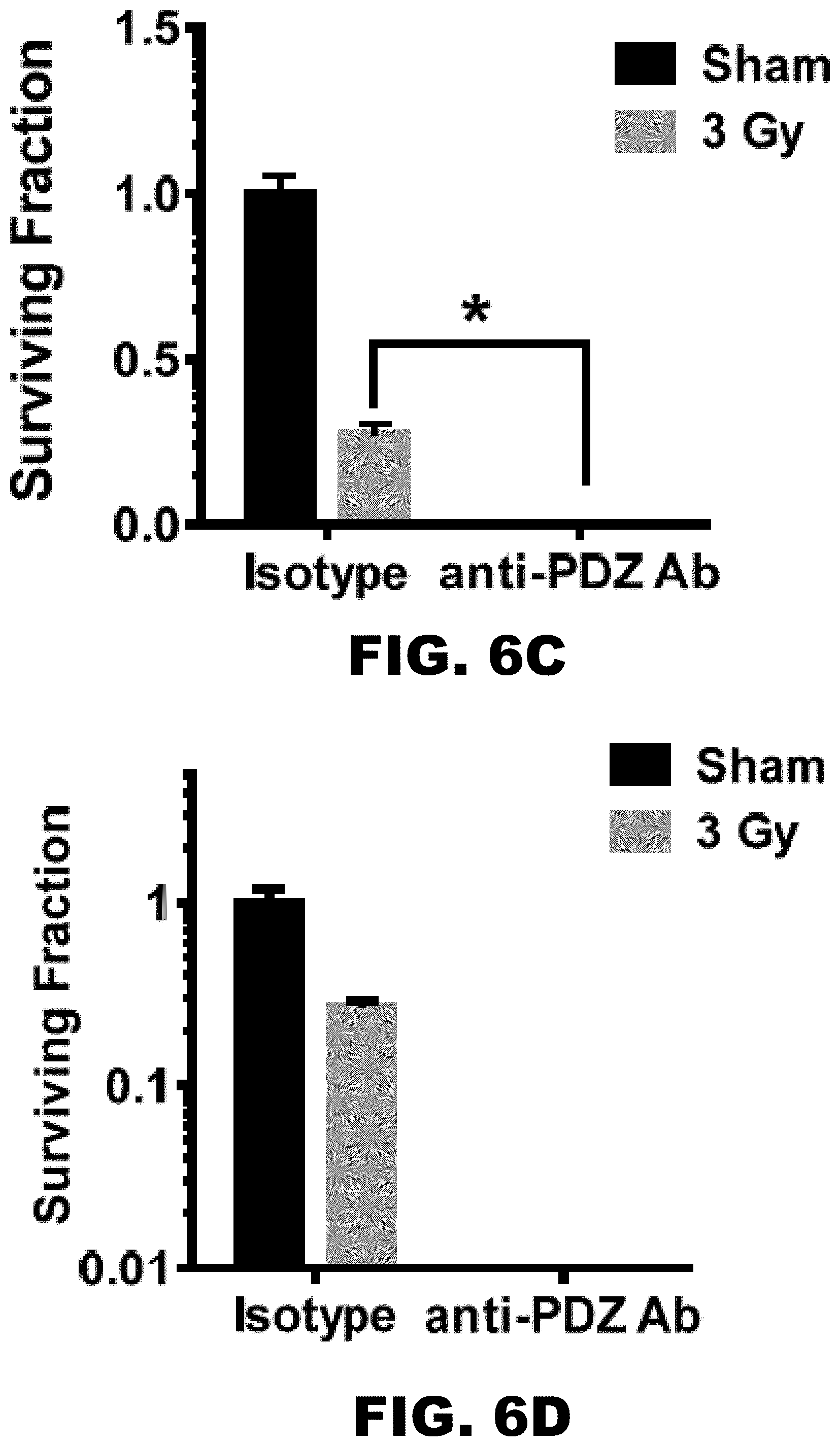
D00017
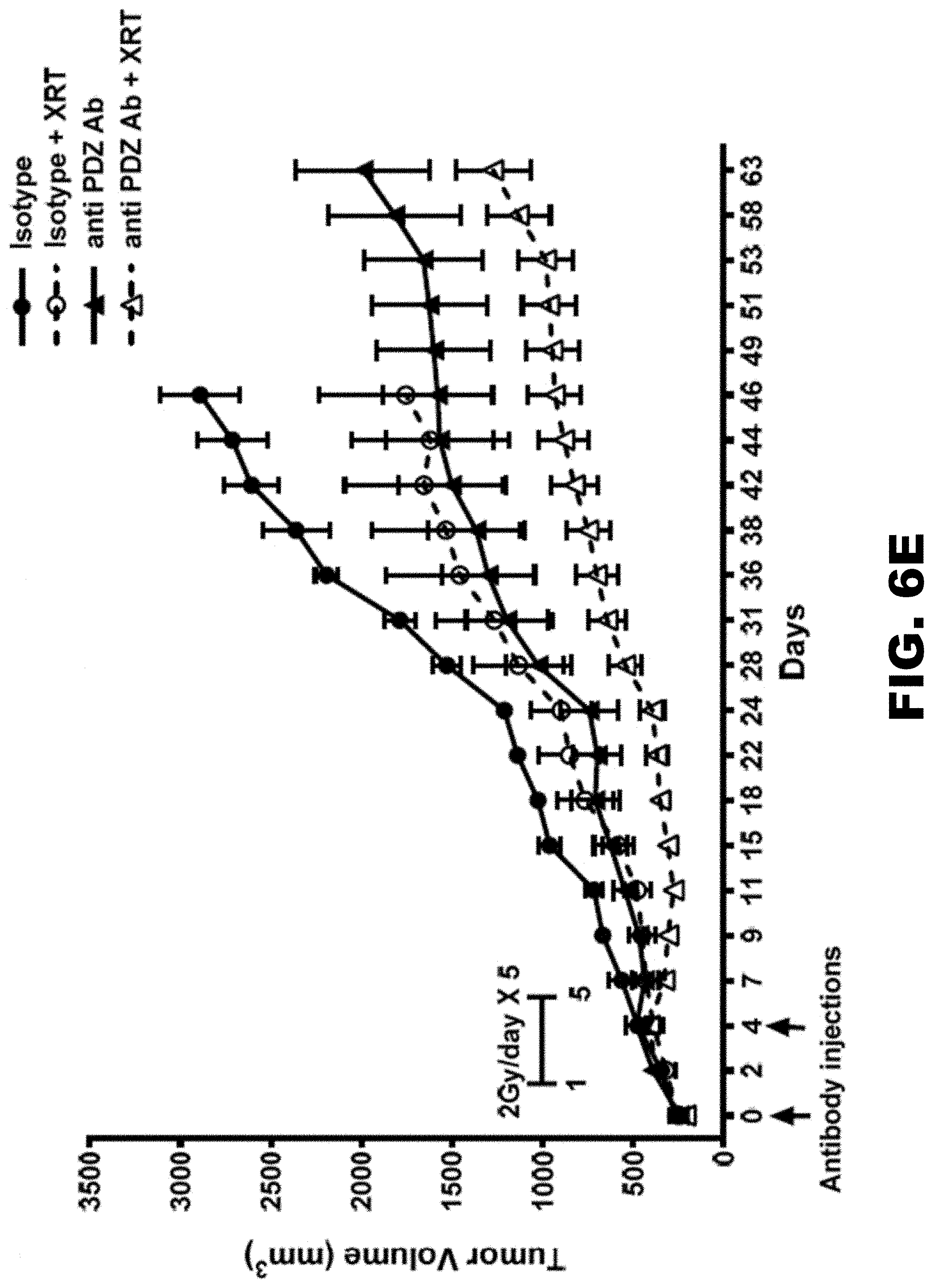
D00018

D00019
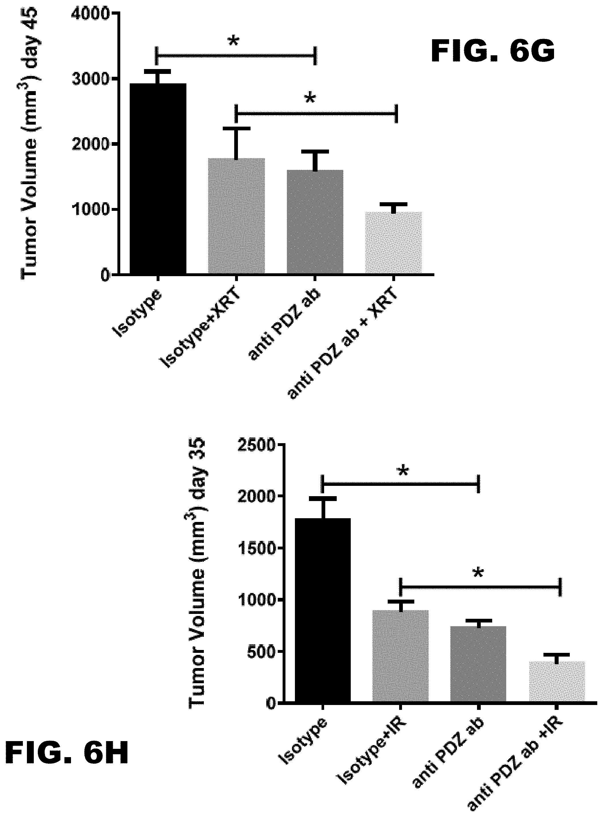
D00020

D00021

D00022
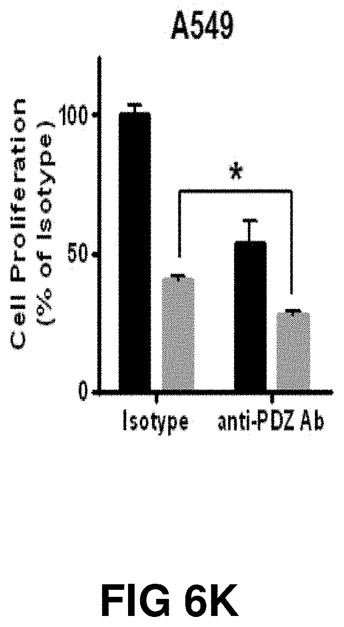
D00023

D00024

D00025
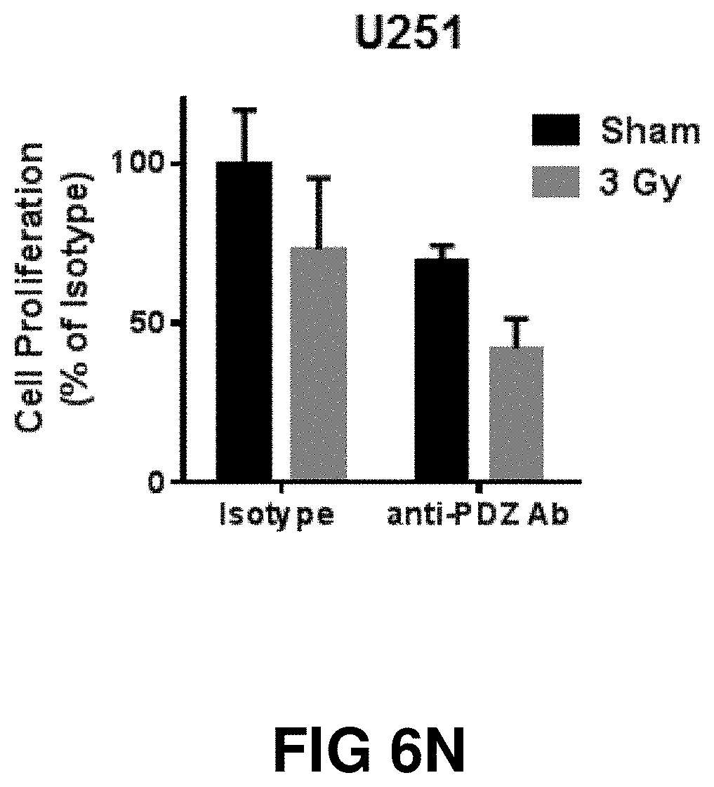
D00026
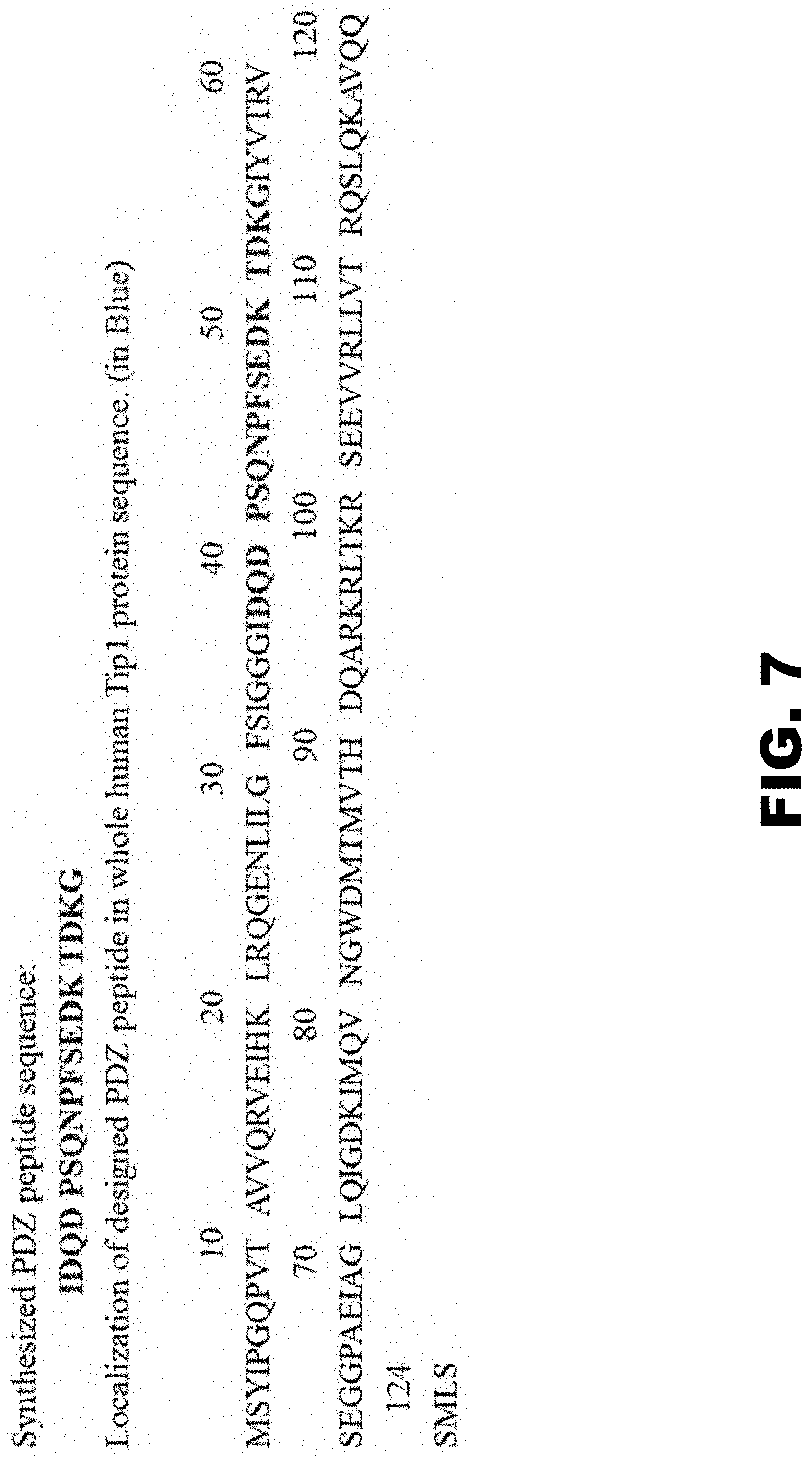
D00027

D00028

D00029

D00030

D00031

D00032
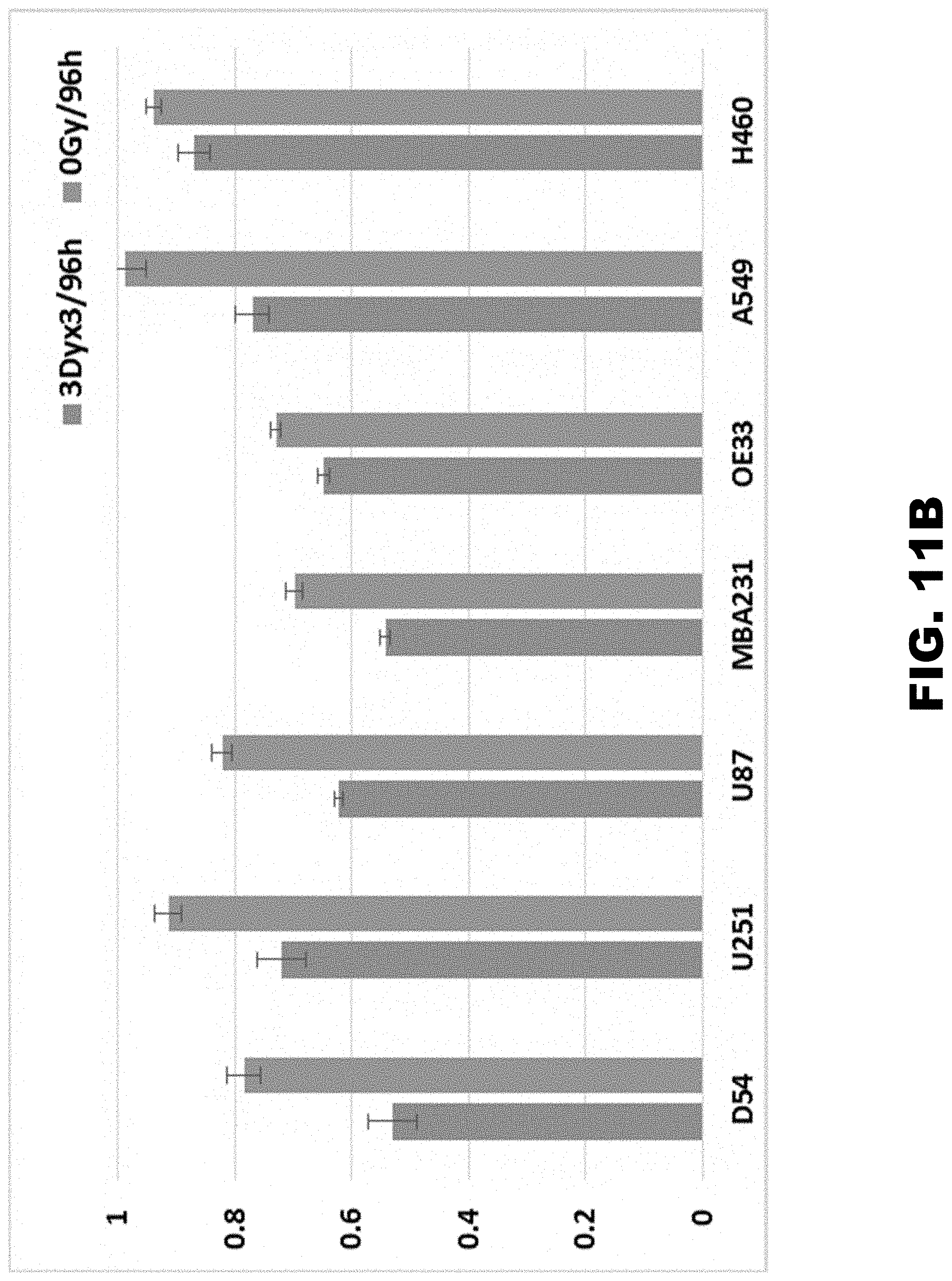
D00033

D00034

D00035
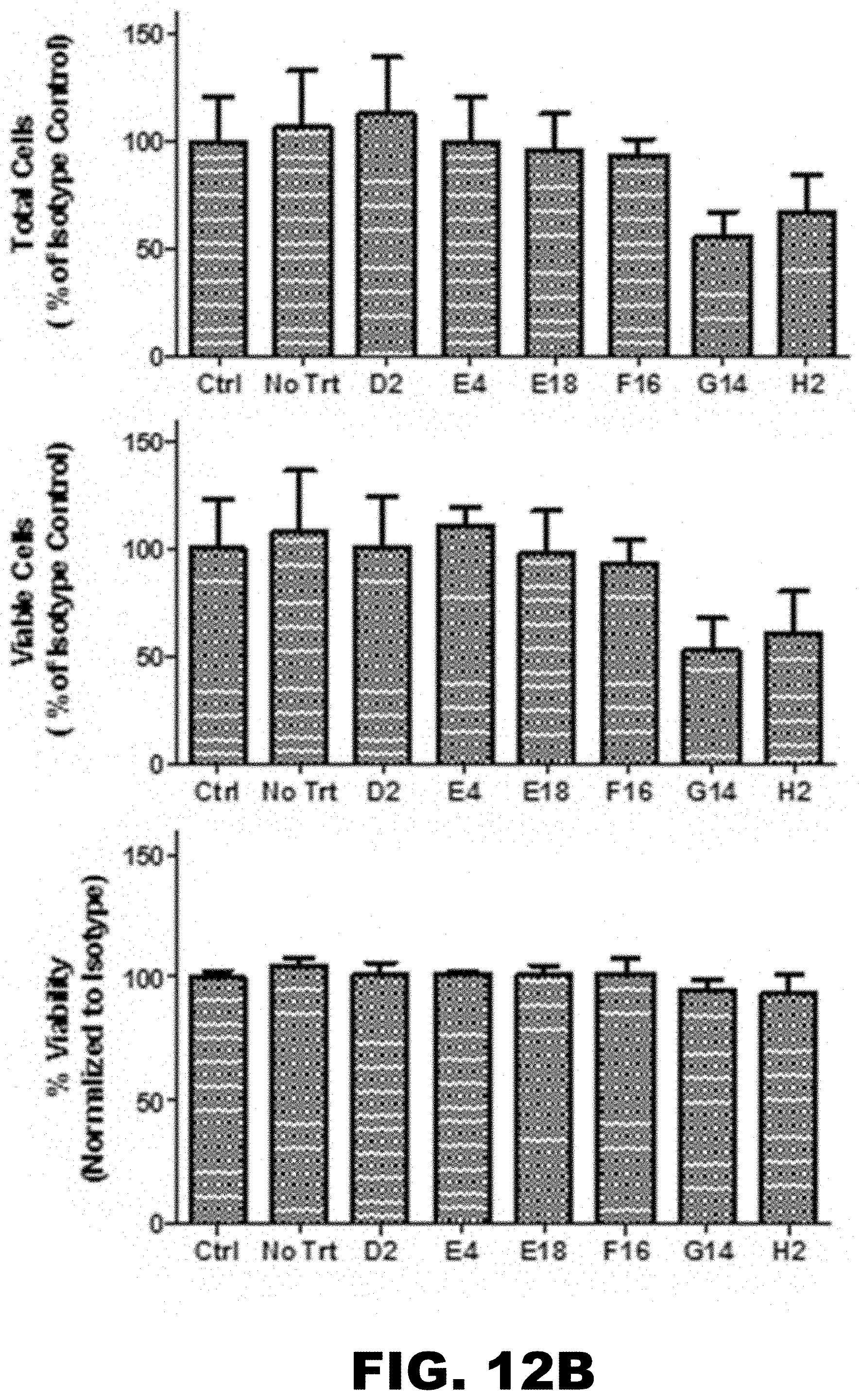
D00036

D00037

D00038

D00039

D00040
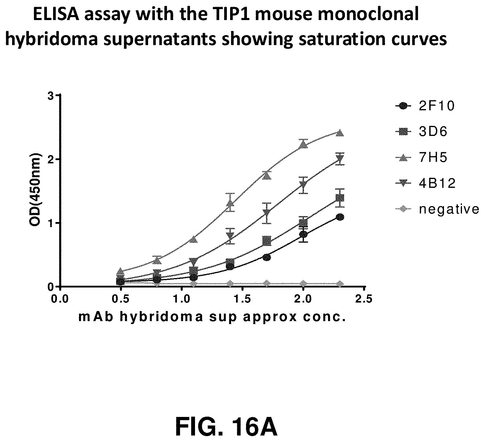
D00041

D00042
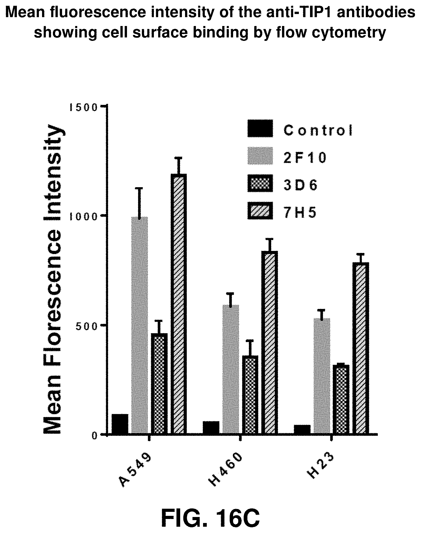
D00043

D00044

D00045

D00046

D00047

D00048

D00049
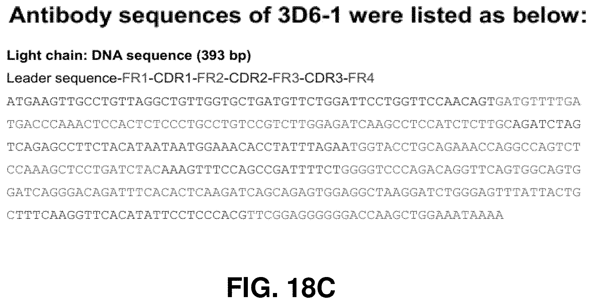
D00050

D00051

D00052
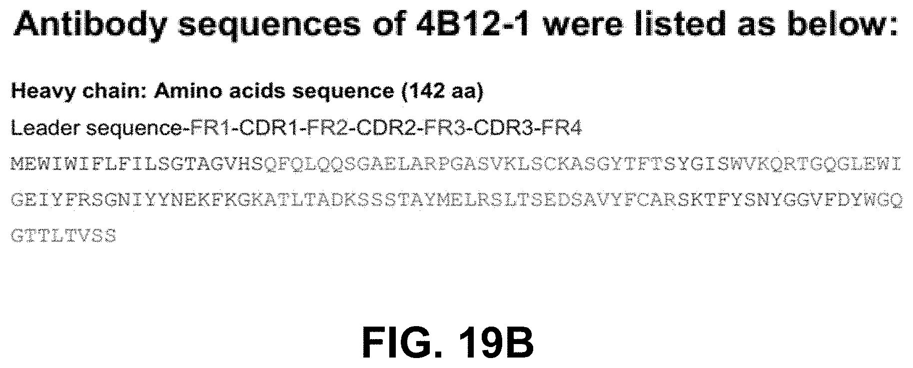
D00053

D00054

D00055
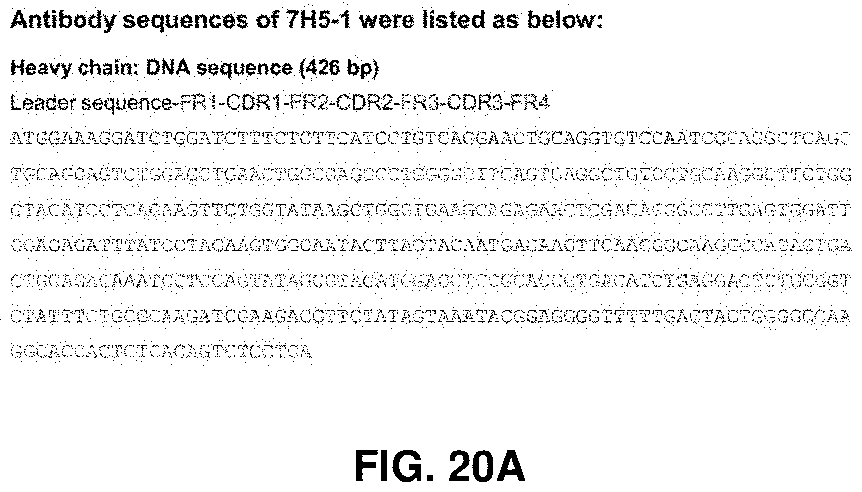
D00056

D00057

D00058

S00001
XML
uspto.report is an independent third-party trademark research tool that is not affiliated, endorsed, or sponsored by the United States Patent and Trademark Office (USPTO) or any other governmental organization. The information provided by uspto.report is based on publicly available data at the time of writing and is intended for informational purposes only.
While we strive to provide accurate and up-to-date information, we do not guarantee the accuracy, completeness, reliability, or suitability of the information displayed on this site. The use of this site is at your own risk. Any reliance you place on such information is therefore strictly at your own risk.
All official trademark data, including owner information, should be verified by visiting the official USPTO website at www.uspto.gov. This site is not intended to replace professional legal advice and should not be used as a substitute for consulting with a legal professional who is knowledgeable about trademark law.