NOXA-DERIVED, CELL DEATH-INDUCING PEPTIDE eMTD
KIM; Tae-Hyoung ; et al.
U.S. patent application number 16/774423 was filed with the patent office on 2020-07-30 for noxa-derived, cell death-inducing peptide emtd. The applicant listed for this patent is INDUSTRY-ACADEMIC COOPERATION FOUNDATION, CHOSUN UNIVERSITY. Invention is credited to Ji-Hye HAN, Tae-Hyoung KIM, Seung-Hyun MYUNG, Junghee PARK.
| Application Number | 20200239521 16/774423 |
| Document ID | 20200239521 / US20200239521 |
| Family ID | 1000004643020 |
| Filed Date | 2020-07-30 |
| Patent Application | download [pdf] |


View All Diagrams
| United States Patent Application | 20200239521 |
| Kind Code | A1 |
| KIM; Tae-Hyoung ; et al. | July 30, 2020 |
NOXA-DERIVED, CELL DEATH-INDUCING PEPTIDE eMTD
Abstract
Disclosed herein is a cell death-inducing peptide that rapidly acts. The peptide is derived from Noxa protein and comprises 16 amino acid residues including MTD. The peptide is designated extended MTD (eMTD) because it contains the 10-mer MTD. eMTD rapidly exhibits potent necrotic cell death in various cell lines and, as such, can be applied to the treatment of various diseases including cancer when used in conjugation with peptides or materials for targeting specific cells.
| Inventors: | KIM; Tae-Hyoung; (Gwangju, KR) ; PARK; Junghee; (Gwangju, KR) ; HAN; Ji-Hye; (Gwangju, KR) ; MYUNG; Seung-Hyun; (Gwangju, KR) | ||||||||||
| Applicant: |
|
||||||||||
|---|---|---|---|---|---|---|---|---|---|---|---|
| Family ID: | 1000004643020 | ||||||||||
| Appl. No.: | 16/774423 | ||||||||||
| Filed: | January 28, 2020 |
| Current U.S. Class: | 1/1 |
| Current CPC Class: | A61P 35/00 20180101; A61K 47/62 20170801; C07K 7/08 20130101; A61K 38/00 20130101 |
| International Class: | C07K 7/08 20060101 C07K007/08; A61P 35/00 20060101 A61P035/00; A61K 47/62 20170101 A61K047/62 |
Foreign Application Data
| Date | Code | Application Number |
|---|---|---|
| Jan 28, 2019 | KR | 10-2019-0010678 |
Claims
1. A peptide consisting of the amino acid sequence of SEQ ID NO: 1 or 2.
2. The peptide of claim 1, wherein the peptide further comprising a tumor-homing peptide (THP) conjugated to the N- or c-terminal of the peptide.
3. The peptide of claim 2, wherein the tumor-homing peptide (THP) comprises an amino acid sequence selected from SEQ ID NOS: 5 to 31.
4. A polynucleotide coding for the peptide of any one of claims 1 to 3.
5. A recombinant vector carrying the polynucleotide of claim 4.
6. A transformed cell transformed with the recombinant vector of claim 5.
7. A composition comprising the peptide of any one of claims 1 to 3.
8. Method for prevention or treatment of cancer comprising: administering to a subject a composition comprising the peptide of any one of claims 1 to 3.
9. The method of claim 8, wherein the cancer is lung cancer, breast cancer, liver cancer, melanoma, stomach cancer, pancreatic cancer, colorectal cancer, ovarian cancer, renal cell carcinoma, prostate cancer, or brain tumor.
Description
SEQUENCE LISTING
[0001] The instant application contains a Sequence Listing which has been submitted electronically in ASCII format and is hereby incorporated by reference in its entirety. Said ASCII copy, created on Jan. 9, 2020 is named 50413-219001_Sequence_Listing_1.23.20_ST25 and is 6,973 bytes in size.
TECHNICAL FIELD
[0002] The present disclosure relates to a cell death-inducing peptide that acts rapidly and, more particularly, to a Noxa-derived, cell death-inducing peptide comprising 16 amino acids inclusive of MTD. The peptide of the present disclosure rapidly exhibits potent necrotic cell death in various cell lines and, as such, can be applied to the treatment of various diseases including cancer when used in conjugation with peptides or materials for targeting specific cells.
BACKGROUND ART
[0003] There are various types of cell death. Active research into cell death has reported and defined various types of cell death, including: necrosis, which is the longest, passive concept of cell death; apoptosis, which is programmed, non-inflammatory cell death; necroptosis, which is a programmed form of necrosis; pyroptosis, which is a highly inflammatory form of programmed cell death; and ferroptosis, which is a type of programmed cell death dependent on intracellular iron ions. Although having different causes and characteristics, these types of cell death are not perfectly discriminated, but physiologically interact with each other, keeping homeostasis.
[0004] The regulation of cell death is very important. Aberrant regulation of cell death provokes various problems. Cancer is representative of diseases caused with the disruption of cell death regulation ability. Cancer exhibits abnormal cell growth with the potential to invade or spread to other parts of the body and is composed mainly of cells in which normal cell death functions do not work. A variety of autoimmune diseases results from failure to regulate cell death. When inflammatory cells cannot move elsewhere or a number of inflammatory cells remain due to failure to regulate cell death, inflammation may continue beyond what is necessary.
[0005] Broad spectrums of drugs that induce cell death are already used. Representative among the drugs are anticancer agents. Many anticancer agents take necrotic and apoptotic mechanisms in treating cancer. By way of example, there are medications that induce cell death through alkylation (e.g., cisplatin, etoposide, etc.) and medications used as metabolic antagonists (e.g., methotrexate, fluorouracil, etc.). Such medications are many also applied to the treatment of autoimmune diseases including rheumatoid arthritis (methotrexate), lupus (cyclophosphamide), and multiple sclerosis (cyclophosphamide).
[0006] Noxa is an apoptotic protein. Noxa expression can be induced in a p53-dependent manner upon DNA damage and genotoxic stress. Noxa can also be induced in a p53-independent manner by alternative mechanisms including hypoxic condition and proteasome inhibition. The Bcl-2 homology 3 (BH3) domain of Noxa binds Mcl-1 and Bcl2A1 to inactivate their anti-apoptotic activities. Consequently, BAX and BAK proteins are activated, causing cytochrome-c to leak into the cytosol, where the caspase system completes the apoptotic process.
DETAILED DESCRIPTION OF THE INVENTION
Technical Problem
[0007] A purpose of the present disclosure is to provide a necrosis-inducing peptide specific for cancer cells.
[0008] Another purpose of the present disclosure is to a polynucleotide coding for a necrosis-inducing peptide specific for cancer cells.
[0009] Another purpose of the present disclosure is to provide a recombinant vector carrying a polynucleotide coding for a necrosis-inducing peptide specific for cancer cells.
[0010] Another purpose of the present disclosure is to provide a cell transformed with a recombinant vector carrying a polynucleotide coding for a necrosis-inducing peptide specific for cancer cells.
[0011] Another purpose of the present disclosure is to provide a pharmaceutical composition, comprising a polynucleotide coding for a necrosis-inducing peptide specific for cancer cells, for prevention or treatment of cancer.
[0012] Another purpose of the present disclosure is to provide a use of a necrosis-inducing peptide specific for cancer cells in preventing or treating cancer.
[0013] Another purpose of the present disclosure is to provide a method for preventing or treating cancer, using a necrosis-inducing peptide specific for cancer cells.
[0014] Another purpose of the present disclosure is to provide a method for preparing a necrosis-inducing peptide specific for cancer cells.
Technical Solution
[0015] Disclosed is a peptide that induces rapid and potent necrosis in cells. Intensive and through research conducted by the present inventors resulted in the finding that extended MTD (eMTD), derived from Noxa, comprising the mitochondria targeting domain (MTD) can kill cancer cells. In this regard, the present inventors successfully extended the MTD to construct a peptide capable of inducing cell death without being fused to a cell-penetrating peptide or other targeting peptides. The amino acid sequence of the peptide is derived from that of Noxa protein and consists of at least 16 amino acids.
[0016] Accordingly, the present disclosure pertains to a cell death-inducing peptide using eMTD, a polynucleotide coding for the same, a recombinant vector carrying the polynucleotide, a transformed cell transformed with the recombinant vector, and a pharmaceutical composition comprising the peptide
[0017] Hereinafter, a detailed description will be given of the present disclosure.
[0018] An aspect of the present disclosure pertains to a necrosis-inducing peptide.
[0019] In the present disclosure, the necrosis-inducing peptide may be a peptide comprising the amino acid sequence of SEQ ID NO: 1 or 2.
[0020] In the present disclosure, the necrosis-inducing peptide may further comprise a tumor-homing peptide (THP) conjugated at the N- and/or C-terminal thereof.
[0021] In the present disclosure, the tumor-homing peptide may comprise an amino acid sequence selected from the amino acid sequences of SEQ ID NOS: 5 to 31, but is not limited thereto.
[0022] In the present disclosure, the tumor-homing peptide may be conjugated to the necrosis-inducing peptide via a linker, but without limitations thereto.
[0023] In the present disclosure, the linker may include as a connector an amino acid sequence so long as not to disturb the induction of necrosis or a chemical connecting group degradable within a cell.
[0024] Available are various linkers known in the art (Huston, et al., Methods in Enzymology, 203:46-88 (1991) and Whitlow, et al., Protein Eng., 6:989 (1993)). For example, the linker may be composed of 1 to 5 amino acids including glycine or serine and other amino acids.
TABLE-US-00001 TABLE 1 SEQ ID NO. Name Sequencing List 5 RGD RGD 6 NGR CNGRCVSGCAGRC 7 RGD-4C CDCRGDCFC 8 iRGD CRGDKGPDC 9 RGDGWK RGDGWK 10 Cilengitide -RGDfV- 11 iNGR CRNGRGPDC 12 Lyp-1(tLyp-1) CGNKRTR 13 F3 KDEPQRRSARLSAKPA PPKPEPKPKKAPAKK 14 TMTP1 KLAKLAK 15 IF7 IFLLWQR 16 Lyp-1 CGNKRTRGC 17 REA CREAGRKAC 18 AGR CAGRRSAYC 19 LSD CLSDGKRKC 20 SP5-52 SVSVGMKPSPRP 21 D-SP5 PRPSPKMGVSVS 22 PC5-2 TDSILRSYDWTY 23 4R22 CSNIDARAC 24 GX1 CGNSNPKSC 25 RGR CRGRRST 26 DUP-1 FRPNRAQDYNTN 27 SP94 SFSIIHTPILPL 28 RPMrel CPIEDRPMC 29 TCP-1 CTPSPFSHC 30 HN-1 TSPLNIHNGQKL 31 CREKA CREKA
[0025] As used herein, the term "peptide" refers to a linear molecule which is formed as amino acid residues are bonded to each other via a peptide bond.
[0026] The peptide in the present disclosure may be directly synthesized in a chemical manner using solid-phase peptide synthesis or may be biologically prepared by inserting DNA encoding the peptide a vector, expressing the DNA through transcription and translation in vitro, and purifying the peptide, but without limitations thereto.
[0027] Another aspect of the present disclosure pertains to a polynucleotide coding for the necrosis-inducing peptide.
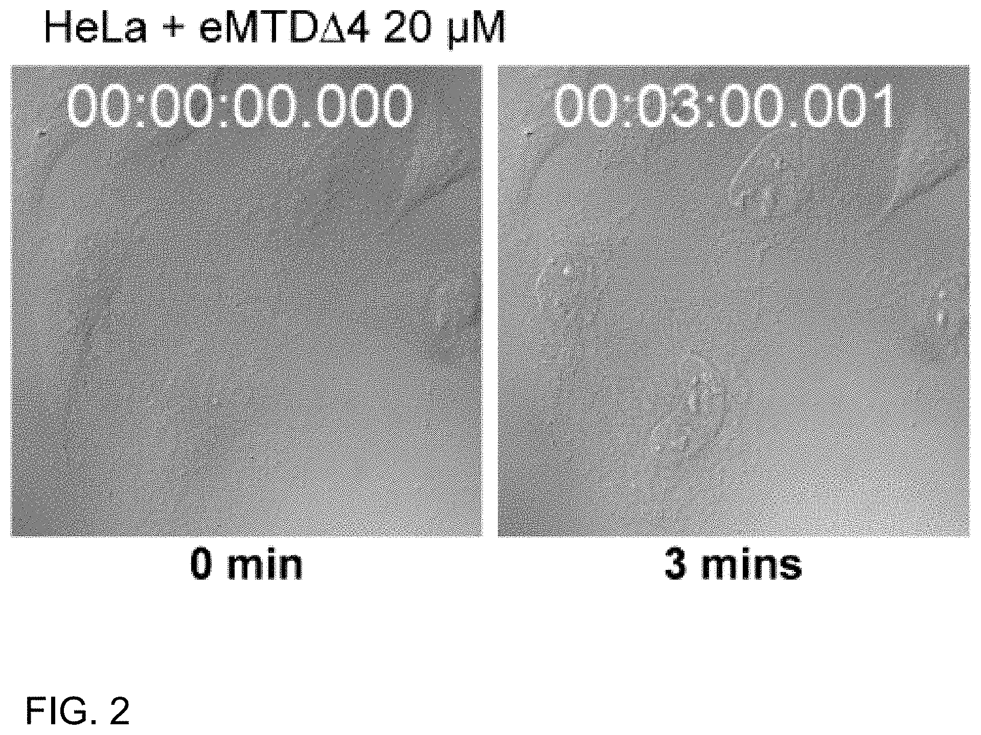
[0028] In the present disclosure, the polynucleotide may be chemically synthesized with the aid of an automated synthesizer on the basis of codons of the necrosis-inducing peptide or may be prepared through PCR amplification or transcription of the synthesized gene, but without limitations thereto.
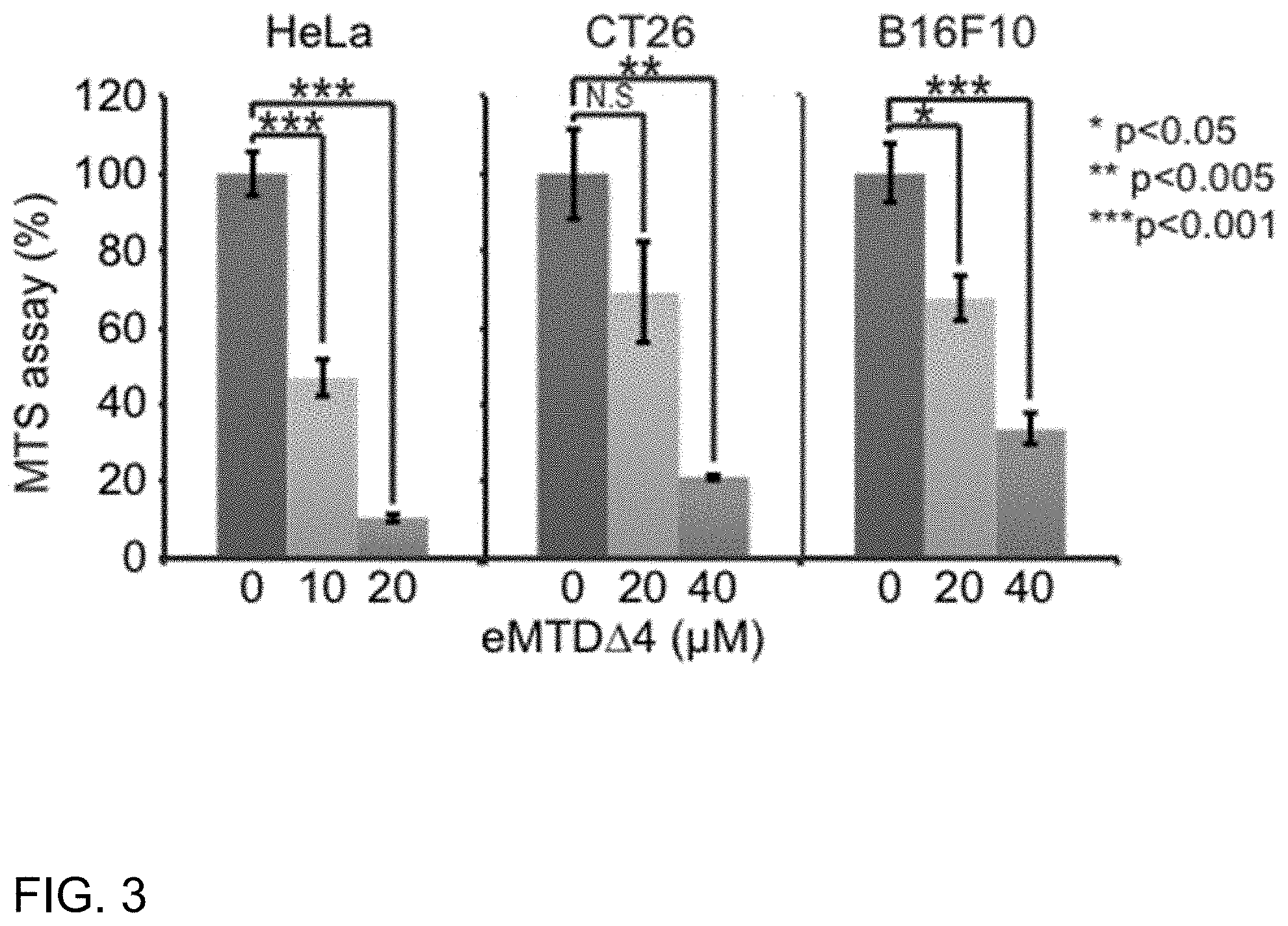
[0029] In the present disclosure, the polynucleotide may encode a necrosis-inducing peptide including the amino acid sequence of SEQ ID NO: 1 or 2.
[0030] In the present disclosure, the necrosis-inducing peptide may further comprise a tumor-homing peptide (THP) conjugated to the N- and/or C-terminal thereof.
[0031] In the present disclosure, the tumor-homing peptide may comprise an amino acid selected from SEQ ID NOS: 5 to 31, but is not limited thereto.
[0032] In the present disclosure, the tumor-homing peptide may be conjugated to the necrosis-inducing peptide via a linker, but without limitations thereto.
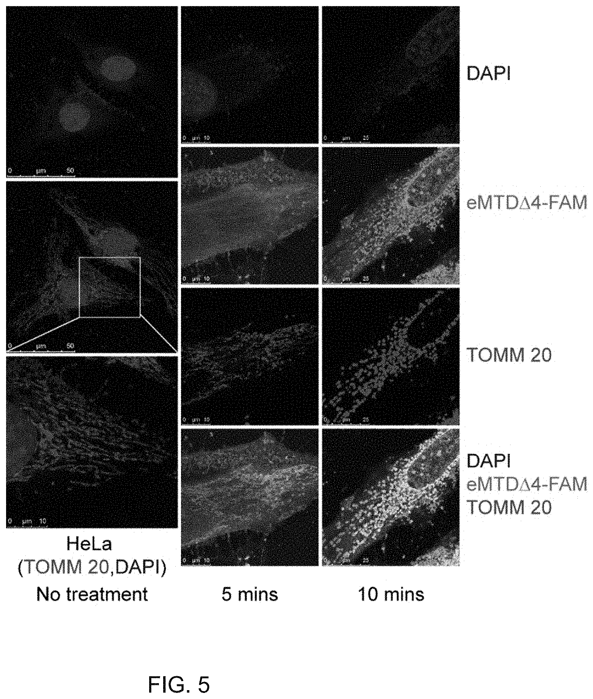
[0033] In the present disclosure, the linker may include as a connector an amino acid sequence so short as not to disturb the induction of necrosis or a chemical connecting group degradable within a cell.
[0034] Another aspect of the present disclosure pertains to a recombinant vector carrying a polynucleotide coding for a necrosis-inducing peptide.
[0035] In the present disclosure, the polynucleotide may encode a necrosis-inducing peptide including the amino acid sequence of SEQ ID NO: 1 or 2.
[0036] In the present disclosure, the necrosis-inducing peptide may further comprise a tumor-homing peptide (THP) conjugated to the N- and/or C-terminal thereof.
[0037] In the present disclosure, the tumor-homing peptide may comprise an amino acid selected from SEQ ID NOS: 5 to 31, but is not limited thereto.
[0038] In the present disclosure, the tumor-homing peptide may be conjugated to the necrosis-inducing peptide via a linker, but without limitations thereto.
[0039] In the present disclosure, the linker may include as a connector an amino acid sequence so short as not to disturb the induction of necrosis or a chemical connecting group degradable within a cell.
[0040] As used herein, the term "vector" refers to a means for expressing a gene of interest in a host cell. Examples of the vector include plasmid vectors, cosmid vectors, and viral vectors such as bacteriophage vectors, adenovirus vectors, retroviral vectors, and adeno-associated virus vectors. For use as the recombinant vector, a plasmid (e.g., pSC101, pGV1106, pACYC177, ColE1, pKT230, pME290, pBR322, pUC8/9, pUC6, pBD9, pHC79, pIJ61, pLAFR1, pHV14, pGEX series, pET series, pUC19, etc.), a phage (e.g., .lamda.gt4.lamda.B, .lamda.-Charon, .lamda..DELTA.z1, M13, etc.) or a virus (e.g., SV40, etc.) may be manipulated.
[0041] The recombinant vector may be typically constructed into a cloning or expression vector. The expression vector may be a typical one that is used in the art to express a foreign protein in plants, animals, or microorganisms. The recombinant vector may be constructed in various manners known in the art.
[0042] For the construction of the recombinant vector, a prokaryote or eukaryote may serve as a host. For example, in the case where an expression vector is used with a eukaryote serving as a host, the vector generally includes a potent promoter capable of performing transcription (e.g., pL.sup..lamda. promoter, CMV promoter, trp promoter, lac promoter, tac promoter, T7 promoter, etc.), a ribosomal binding site for initiating translation, and transcription/translation stop sequences. For eukaryotes serving as hosts, the expression vector is constructed to have a replication origin examples of which include an f1 replication origin, an SV40 replication origin, a pMB1 replication origin, an adeno replication origin, an AAV replication origin, and a BBV replication origin, but is not limited thereto. In addition, the vector may employ a promoter derived from the genome of mammalian cells (e.g., a metallothionein promoter) or from mammalian cell-grown virus (e.g., an adenovirus late promoter, a vaccinia virus 7.5K promoter, an SV40 promoter, a cytomegalovirus promoter, and an HSV tk promoter) and typically contains a polyadenylation sequence as a transcription stop sequence.
[0043] Another aspect of the present disclosure pertains to a cell transformed with a recombinant vector carrying a polynucleotide coding for a necrosis-inducing peptide specific for cancer cells.
[0044] In the present disclosure, the polynucleotide may encode a necrosis-inducing peptide including the amino acid sequence of SEQ ID NO: 1 or 2.
[0045] In the present disclosure, the necrosis-inducing peptide may further comprise a tumor-homing peptide (THP) conjugated to the N- and/or C-terminal thereof.
[0046] In the present disclosure, the tumor-homing peptide may comprise an amino acid selected from SEQ ID NOS: 5 to 31, but is not limited thereto.
[0047] In the present disclosure, the tumor-homing peptide may be conjugated to the necrosis-inducing peptide via a linker, but without limitations thereto.
[0048] In the present disclosure, the linker may include as a connector an amino acid sequence so short as not to disturb the induction of necrosis or a chemical connecting group degradable within a cell.
[0049] Within the scope of the host cell used in the present disclosure, E. coli, yeasts, animal cells, plant cells, and insect cells fall, as exemplified by prokaryotic enterobacteriaceae strains including E. coli JM109, E. coli BL21, E. coli RR1, E. coli LE392, E. coli B, E. coli X 1776, E. coli W3110, Bacillus spp. such as Bacillus subtilis and Bacillus churingiensis, Salmonella typhimurium, Seratia marcescens, and various Pseudomonas spp. As eukaryotic host cells to be transformed, yeast (Saccharomyce cerevisiae), insect cells, plant cells, and animal cells may be used. Examples of the eukaryotic cells include Sp2/0, CHO (Chinese hamster ovary) K1, CHO DG44, PER.C6, W138, BHK, COS7, 293, HepG2, Huh7, 3T3, RIN, and MDCK cell lines, but are not limited thereto.
[0050] The delivery (introduction) of the polynucleotide or the recombinant vector carrying the same into a host cell may be achieved using a delivery method well known in the art. When the host cell is prokaryotic, the delivery method may be a CaCl.sub.2 method or electroporation. For eukaryotes as host cells, microinjection, calcium phosphate precipitation, electroporation, liposome-mediated transfection, or genetic bombardment may be used, without limitations thereto.
[0051] Selection of the transformed host cells may be easily performed according to well-known methods using the phenotypes expressed by selection markers used. For example, when a gene resistant to a specific antibiotic is used as a selection marker, the transformant may be easily selected by being cultured on a medium containing the antibiotic.
[0052] Another aspect of the present disclosure pertains to a pharmaceutical composition for prevention or treatment of cancer, the composition comprising a polynucleotide coding for a necrosis-inducing peptide specific for cancer cells.
[0053] In the present disclosure, the necrosis-inducing peptide may further comprise a tumor-homing peptide (THP) conjugated to the N- and/or C-terminal thereof.
[0054] In the present disclosure, the tumor-homing peptide may comprise an amino acid selected from SEQ ID NOS: 5 to 31, but is not limited thereto.
[0055] In the present disclosure, the tumor-homing peptide may be conjugated to the necrosis-inducing peptide via a linker, but without limitations thereto.
[0056] In the present disclosure, the linker may include as a connector an amino acid sequence so short as not to disturb the induction of necrosis or a chemical connecting group degradable within a cell.
[0057] In the present disclosure, the cancer may be lung cancer, breast cancer, liver cancer, melanoma, stomach cancer, pancreatic cancer, colorectal cancer, ovarian cancer, renal cell carcinoma, prostate cancer, or brain tumor, but is not limited thereto.
[0058] The content of the necrosis-inducing peptide as an effective ingredient in the composition may be appropriately adjusted depending on usage form and purpose, patient's condition, kinds and severity of symptoms and may range from 0.001 to 99.9% by weight or from 0.1 to 99.9% by weight, preferably from 0.1 to 50% by weight or from 0.1 to 40% by weight, based on the weight of the solid content, but without limitations thereto.
[0059] The pharmaceutical composition according to the present disclosure may be administered via various routes to mammals including humans. Administration may be conducted in any typical mode. For example, the composition may be administered via an oral, dermal, intravenous, intramuscular, or subcutaneous route. Alternatively, the composition may take a topical spray form administrable intranasally.
[0060] According to typical methods, the composition of the present disclosure may be formulated into oral dosage forms such as powders, granules, tablets, capsules, ointments, suspensions, emulsions, syrups, aerosols, etc., or parenteral dosage forms such as transdermal agents, sprays, suppositories, and sterile injections.
[0061] In addition to the cancer cell-specific, necrosis-inducing fusion peptide, the pharmaceutical composition of the present disclosure may further comprise a pharmaceutically suitable and biologically acceptable auxiliary agent such as carriers, excipients, and diluents.
[0062] Examples of the carrier, excipient, or diluent available in the pharmaceutical composition of the present disclosure include lactose, dextrose, sucrose, sorbitol, mannitol, xylitol, erythritol, maltitol, starch, acacia gum, alginate, gelatin, calcium phosphate, calcium silicate, cellulose, methyl cellulose, microcrystalline cellulose, polyvinyl pyrrolidone, water, methylhydroxy benzoate, propylhydroxy benzoate, talc, magnesium stearate, and mineral oil.
[0063] For formulating the composition, a typical diluent or excipient, such as filler, a thickener, a binder, a humectant, a disintegrant, a surfactant, and the like, may be used. Solid formulations for oral administration include tablets, pills, powder, granules, and capsules, and can be prepared by mixing the above extract with one or more excipients such as starch, calcium carbonate, sucrose, lactose, or gelatin. In addition, lubricants such as magnesium stearate and talc can be used instead of simple excipients.
[0064] Liquid formulations for oral administration include a suspension, a liquid for internal use, an emulsion, syrup, and a gel, and frequently used water, liquid paraffin and other excipients such as humectants, sweeteners, aromatics, and preservatives can also be used.
[0065] In the formulations for non-oral administration, a sterilized aqueous solution, a non-aqueous solvent, a suspension, an emulsion, a lyophilized formulation, a suppository, and a transcutaneous formulation are included.
[0066] Propylene glycol, polyethylene glycol, vegetable oil, such as olive oil, and injectable esters, such as ethyloleate, can be used as a non-aqueous solvent or a suspension. The base for suppository includes Witepsol, Macrogol, Tween 61, cacao oil, laurin oil, glycerol, and gelatin.
[0067] The pharmaceutical composition of the present disclosure may be administered to humans, alone or in mixture with pharmaceutical carriers selected in consideration of general protocols and the standard pharmaceutical practice.
[0068] By way of example, the pharmaceutical composition of the present disclosure may be orally, buccally, or sublingually administered in the form of tablets containing starch or lactose, in the form of capsules containing the composition alone or in mixture with excipients, or in the form of elixirs or suspensions with chemicals for taste masking or coloration.
[0069] Such liquid formulations can be formulated with pharmaceutically acceptable additives such as suspending agents (e.g., methyl cellulose, semisynthetic glyceride such as Witepsol, a mixture of apricot kernel oil and PEG-6 esters, and a glyceride mixture such as PEG-8 and caprylic/capric glyceride mixture).
[0070] The administration dose of the pharmaceutical composition of the present disclosure may vary depending on the age, weight, health status and seriousness of the disease of the patient, and the composition may be administered in a single dose or multiple discrete doses at regular time intervals a day, depending on the judgment of physicians or pharmacists.
[0071] For instance, daily dose of the effective ingredient can be 0.1 to 500 mg/kg, and preferably 0.5 to 300 mg/kg. The above dose is only an average value, and the dose can be higher or lower depending on the personal differences.
[0072] It is preferable to have the above dose range not only because a lower daily dose of the pharmaceutical composition of the present disclosure does not guarantee a meaningful effect, but also because a higher dose is economically disadvantageous and may cause undesirable side effects.
Advantageous Effects
[0073] Disclosed herein is a cell death-inducing peptide that rapidly acts. The peptide is derived from Noxa protein and comprises 16 amino acid residues including MTD. The peptide is designated extended MTD (eMTD) because it contains the 10-mer MTD. eMTD rapidly exhibits potent necrotic cell death in various cell lines and, as such, can be applied to the treatment of various diseases including cancer when used in conjugation with peptides or materials for targeting specific cells.
BRIEF DESCRIPTION OF THE DRAWINGS
[0074] FIG. 1 shows relative cell viability after treatment of HeLa cell line with eMTD peptide or eMTDA4 peptide according to an embodiment of the present disclosure, as measured by MTS assay;
[0075] FIG. 2 shows microscopic images of HeLa cells after or before treatment with 20 of eMTDA4 for 3 min;
[0076] FIG. 3 shows relative cell viability after treatment of HeLa, CT26, and B16F10 cell lines with eMTDA4 peptide according to an embodiment of the present disclosure, as measured by MTS assay;
[0077] FIG. 4a shows time-lapse, confocal microscopic images of HeLa cells stained with the calcium indicator Fluo-4 and then treated with or without 20 .mu.M of eMTD.DELTA.4 according to an embodiment of the present disclosure;
[0078] FIG. 4b is a graph showing Fluo-4 intensities within ROI (region of interest) of time-lapse confocal microscopic images of HeLa cells stained with the calcium indicator Fluo-4 without eMTD.DELTA.4 treatment according to an embodiment of the present disclosure;
[0079] FIG. 4c is a graph showing Fluo-4 intensities within ROI (region of interest) of time-lapse confocal microscopic images of HeLa cells stained with the calcium indicator Fluo-4 and then treated with 20 of eMTD.DELTA.4 according to an embodiment of the present disclosure;
[0080] FIG. 5 shows confocal microscopic images of HeLa cells fixed 5 and 10 min after treatment with the peptide eMTD.DELTA.4 conjugated with a fluorescent (Fluorescein; FAM) according to an embodiment of the present disclosure;
[0081] FIG. 6a shows mitochondrial swelling as analyzed by absorbance read at 540 nm after treatment of mitochondria isolated from livers of BalB/C mice with the peptide eMTD.DELTA.4 according to an embodiment of the present disclosure;
[0082] FIG. 6b shows mitochondrial swelling as observed by TEM (transmission electron microscopy) after treatment of mitochondria isolated from livers of BalB/C mice with the peptide eMTD.DELTA.4 according to an embodiment of the present disclosure;
[0083] FIG. 7a shows time-lapse, confocal microscopic images of HeLa cells to identify mitochondrial PTP opening after only mitochondria are stained with calcein AM and cobalt ions without treatment with the peptide eMTD.DELTA.4 according to an embodiment of the present disclosure;
[0084] FIG. 7b shows time-lapse, confocal microscopic images of HeLa cells to identify mitochondrial PTP opening after only mitochondria are stained with calcein AM and cobalt ions and the cells are treated with the peptide eMTD.DELTA.4 according to an embodiment of the present disclosure;
[0085] FIG. 8a shows time-lapse, confocal microscopic images of HeLa cells after the cells are stained with the mitochondrial reactive oxygen species indicator MitoSox and then treated with or without 20 of the peptide eMTD.DELTA.4 according to an embodiment of the present disclosure;
[0086] FIG. 8b is a graph showing MitoSox intensities within ROI (region of interest) of time-lapse confocal microscopic images of HeLa cells stained with the mitochondrial reactive oxygen species indicator MitoSox without eMTD.DELTA.4 treatment according to an embodiment of the present disclosure;
[0087] FIG. 8c is a graph showing MitoSox intensities within ROI (region of interest) of time-lapse confocal microscopic images of HeLa cells stained with the mitochondrial reactive oxygen species indicator MitoSox and then treated with 20 of eMTD.DELTA.4 according to an embodiment of the present disclosure;
[0088] FIG. 9 shows time-lapse, confocal microscopic images of Hela cells treated with the peptide eMTD.DELTA.4 conjugated with a fluorescent (Fluorescein; FAM) according to an embodiment of the present embodiment;
[0089] FIG. 10a shows degrees of damage on the lipid membrane of liposomes as analyzed by two-step assay according to an embodiment of the present disclosure.
[0090] FIG. 10b shows the damage of the peptide eMTD.DELTA.4 on the cell membrane in time-lapse atomic force microscopic images according to an embodiment of the present disclosure;
[0091] FIG. 10c shows the damage of the peptide eMTD.DELTA.4 on the cell membrane and the depth of the cell membrane damage in time-lapse atomic force microscopic images according to an embodiment of the present disclosure; and
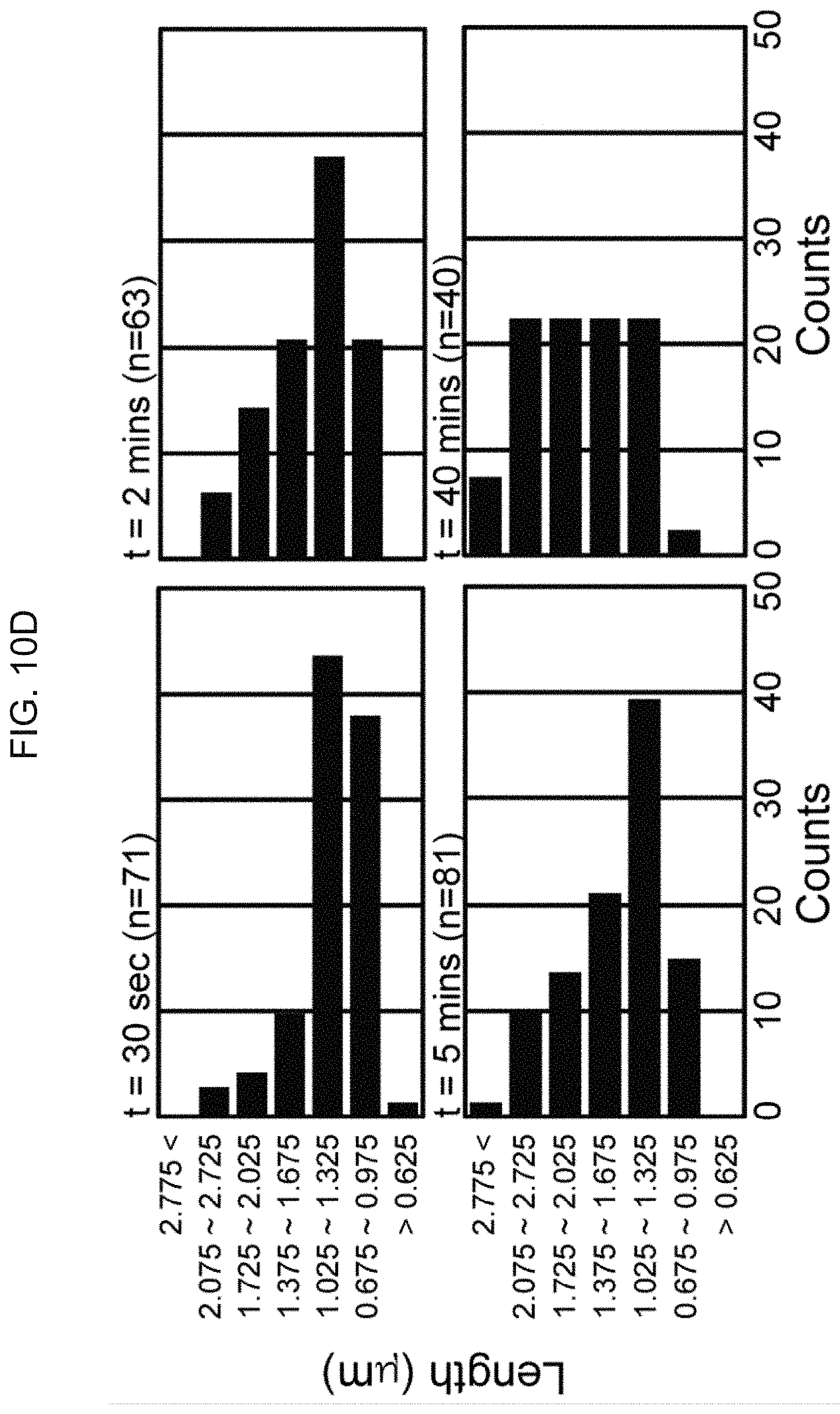
[0092] FIG. 10d shows statistical data of damage, photographically taken by atomic force microscopy, of the peptide eMTD.DELTA.4 on the cell membrane.
MODE FOR CARRYING OUT THE INVENTION
[0093] A better understanding of the present disclosure may be obtained through the following examples which are set forth to illustrate, but are not to be construed as limiting the present disclosure.
Example 1: Peptide Synthesis
[0094] Peptide synthesis was entrusted to Anygen in which peptides were synthesized using solid-phase peptide synthesis, purified by high-capacity HPLC, dissolved at a concentration of 1 mM in 50% aqueous DMSO (dimethyl sulfoxide) solution, and then stored at -20.degree. C. Amino acids sequences of the peptides are given in Table 2, below.
TABLE-US-00002 TABLE 2 SEQ ID NO: Name Sequencing List 1 eMTD KLNFRQKLLNLISKLFCSGT 2 eMTD.DELTA.4 KLNFRQKLLNLISKLF 3 MTD KLLNLISKLF 4 Noxa MPGKKARKNAQPSPARAPAELEVECAT QLRRFGDKLNFRQKLLNLISKLFCSGT
Example 2: Culture of Cancer Cell Line
[0095] Individual cell lines (HeLa, CT26, and B16F10) were purchased from the Korean Cell Line Bank. Dulbecco's modified Eagle's medium (DMEM), trypsin-EDTA, fetal bovine serum (FBS), and Hank's balanced salt solution (HBSS) were products from Gibco (Thermo Fisher).
[0096] Each of the cell lines was cultured in DMEM supplemented with 10% FBS in an incubator maintained at 37.degree. C. and 5% CO.sub.2.
Example 3: Assay for Cell Death Induction of eMTD Peptide
[0097] 3-1. Assay for Activity of Cell Death Induction in Uterine Cervical Cancer
[0098] For use in assaying the peptides eMTD and eMTD.DELTA.4 synthesized in Example 1 for activity of inducing cell death, the cell line HeLa was grown to about 90% confluency in 96-well plates. The cells were washed once with HBSS buffer before treatment with the peptides of the present disclosure eMTD and eMTD.DELTA.4 at concentrations of 0, 20, and 40 .mu.M for one hour. Thereafter, the cells were incubated with an MTS assay agent (Promega) for one hour, followed by reading absorbance to calculate relative viability of cells. The results are given in FIG. 1 and Table 3.
TABLE-US-00003 TABLE 3 0 .mu.M 20 .mu.M 40 .mu.M fl-Noxa Viability (%) 100 101 106 Standard deviation 0.079 0.037 0.050 eMTD Viability (%) 100 61 63 Standard deviation 0.079 0.007 0.023 eMTD.DELTA.4 Viability (%) 100 53 40 Standard deviation 0.079 0.082 0.044
[0099] As is understood from the data of FIG. 1 and Table 3, eMTD and eMTD.DELTA.4 peptides exhibit potent cell death activity.
[0100] 3-2. Microscopic Observation of Morphological Change in Cells
[0101] In order to observe morphological changes in cells in detail, the cells were incubated with 20 .mu.M of the peptide eMTD.DELTA.4 and observed with the aid of a microscope (Leica). The results are depicted in FIG. 2.
[0102] As shown in FIG. 2, most of the cells were observed to undergo necrosis with the consequent rupture of cell membranes after 3 min of incubation with the peptide.
[0103] 3-3. Assay for Activity of Cell Death Induction in Other Cancer Cell Lines
[0104] Examination was made to see whether the peptide has the activity of inducing cell death in other cancer cell lines. In this regard, the cancer cell lines HeLa, CT26, and B16F10 were treated with eMTD.DELTA.4 at concentrations of 0, 20, and 40 .mu.M. Cell viability was measured by MTS assay and the results are given in FIG. 3 and Table 4.
TABLE-US-00004 TABLE 4 eMTD.DELTA.4 HeLa CT26 B16F10 0 .mu.M Viability (%) 100 100 100 Standard 5.72 11.81 7.73 deviation 10 .mu.M Viability (%) 47 69 68 Standard 5.07 13.08 5.71 deviation 20 .mu.M Viability (%) 11 21 33 Standard 0.98 0.69 3.99 deviation
[0105] As can be understood from the data of FIG. 3 and Table 4, the peptide eMTD.DELTA.4 was found to induce cell death in HeLa, which is a human uterine cervical cancer cell line as well as in CT26, which is derived from murine colon carcinoma, and B16F10, which is derived from murine melanoma cells.
Example 4: Induction of Cell Death and Intracellular Calcium Level Change by Peptide eMTD.DELTA.4
[0106] In order to examine the mechanism in which the peptide eMTD induces cell death in cancer cell lines, a change in intracellular calcium level, which is known as an important factor for cell death, was observed using the calcium indicator Fluo-4 under a confocal microscope (Leica).
[0107] In brief, HeLa cells were grown to about 70% confluency in a Lab-Tek chamber glass a day before the experiment. Immediately before the experiment, of the calcium indicator Fluo-4 was diluted to a concentration of 5 .mu.M in HBSS. HeLa cells were incubated with the dilution for 10 min in an incubator and then treated with the peptide eMTD.DELTA.4. Results are given in FIGS. 4A and 4C.
[0108] As shown in FIGS. 4A to 4C, almost no changes in intracellular calcium level were found in HeLa cells that had not been treated with the peptide. In contrast, the HeLa cells were observed to allow calcium influx into the cytosols thereof within less than one min after treatment with the peptide eMTD.DELTA.4. In this regard, the calcium levels are depicted in a saw-toothed pattern in a graph. When the cell membrane bubbles and is damaged, the calcium influx is reduced, with the cell contents being expelled. As a result, the intracellular calcium level becomes lower than the original level.
Example 5: Intracellular Migration of Peptide eMTDL4 to Target
[0109] In order to investigate the source that allowed the intracellular calcium influx observed in Example 4, examination was made of the site at which the peptide eMTD.DELTA.4 acts. In this regard, the fluorescent tracer fluorescein was labeled to the C-terminal of the peptide eMTD.DELTA.4 (eMTD.DELTA.4-FAM). Then, HeLa cells grown on Lab-Tek chamber glass were incubated with eMTD.DELTA.4-FAM for 5 and 10 min before fixation with 1% paraformaldehyde.
[0110] Mitochondria were visualized using an antibody to TOM20, which localizes to the outer membrane of the mitochondrion because MTD is known to migrate to mitochondria, while nuclei were visualized using DAPI. Confocal micrographic images are shown in FIG. 5.
[0111] As indicated by the images of FIG. 5, the localization of eMTD.DELTA.4-FAM is consistent with the visualized positions of mitochondria. In addition, after treatment with eMTD.DELTA.4-FAM, mitochondrial fragmentation was observed morphologically.
Example 6: Observation of eMTD.DELTA.4 Activity in Mitochondrion
[0112] 6-1. Mitochondrial Swelling
[0113] In Example 5, eMTD.DELTA.4-FAM was observed to localize to mitochondria and to induce mitochondrial fragmentation. Accordingly, examination was further made to see how eMTD.DELTA.4 practically works at mitochondria in greater detail.
[0114] First, livers excised from 6-week-old BalB/C mice were immersed in buffer (250 mM mannitol, 70 mM EGTA, 5 mM HEPES, pH 7.4, 0.1 mM PMSF and 4 .mu.M rotenone) and completely homogenized using a Teflon Potter-Elvehjem grinder (Sigma). The ground liver was centrifuged at 1000.times.g for 10 min at 4.degree. C. and the supernatants were centrifuged at 10,000.times.g for 10 min at 4.degree. C. The pellets thus obtained were suspended in a regeneration buffer (250 mM sucrose, 10 mM HEPES, pH 7.4, 5 mM sodium succinate, 2 mM potassium phosphate, 0.1 mM PMSF, 25 .mu.M EGTA, and 4 rotenone).
[0115] The mitochondria thus obtained were treated with 25 .mu.M of eMTD or eMTD.DELTA.4, followed by reading absorbance at 540 nm to deteLmine mitochondrial swelling. The results are given in FIG. 6A and Table 5, below. For a positive control, the mitochondria were treated with 200 .mu.M of Ca.sup.2+ while co-treatment with 200 .mu.M of Ca.sup.2+ and 20 .mu.M of CsA was conducted for a negative control.
[0116] For direct identification, the mitochondria were treated with eMTD.DELTA.4 (25 .mu.M), Ca.sup.2+ (200 .mu.M), or Ca.sup.2+ (200 .mu.M)+CsA (20 .mu.M) and then observed by transmission electron microscopy. The results are given in FIG. 6B.
TABLE-US-00005 TABLE 5 Minute 0 10 20 30 40 50 60 None 100% 98% 97% 95% 95% 95% 95% cNoxa del4 100% 79% 74% 70% 68% 66% 65% cNoxa 100% 86% 78% 71% 68% 68% 67% Ca 100% 77% 75% 73% 72% 70% 70% CsA 100% 98% 98% 97% 97% 96% 96% Ca + CsA 100% 98% 97% 96% 95% 94% 93% None 0% 0% 0% 0% 0% 0% 0% cNoxa del4 0% 0% 0% 0% 0.22%.sup. 0% 0% cNoxa 0% 4% 4% 6% 5% 5% 5% Ca 0% 0% 1% 1% 1% 1% 0% CsA 0% 0% 0% 0% 0% 0% 0% Ca + CsA 0% 0% 0% 0% 0% 0% 0%
[0117] As indicated by the data of FIGS. 6A and 6B and Table 5, eMTD.DELTA.4 was observed to induce mitochondrial swelling.
[0118] 6-2. Mitochondrial PTP
[0119] Mitochondria are known to open permeability transition pores (PTP) to allow cytosolic calcium influx. The observation that eMTD.DELTA.4 migrates to mitochondria and induces cytosolic calcium influx led to investigating the association of the action of eMTD.DELTA.4 with PTP. To this end, PTP opening was monitored using calcein AM and cobalt ions.
[0120] In brief, HeLa cells were grown on a Lab-Tek chamber glass slide one day before the experiment. Immediately before the experiment, the cells were stained with 1 of calcein AM and 2 mM of cobalt ions for 20 min and then with 0.1 of MitoTracker Red for 2 min. Time-lapse confocal images were taken and are given in FIGS. 7A and 7B. When PTP is opened, mitochondrial calcein AM meets cobalt ions so that the fluorescence of calcein AM is quenched.
[0121] As is understood from the images of FIGS. 7A and 7B, the fluorescence of mitochondrial calcein AM was quenched upon treatment with eMTD.DELTA.4, implying that the eMTD.DELTA.4-induced cytosolic calcium influx is conducted by PTP opening.
Example 7: Peptide eMTDL4-Induced Cell Death and ROS Generation
[0122] In addition to an increase in intracellular calcium level, reactive oxygen species (ROS) generation is an important factor for cell death. Examination was made of the role of ROS in the process of eMTD.DELTA.4-induced cell death.
[0123] In brief, HeLa cells were grown to 70% confluency on a Lab-Tek chamber glass slide one day before the experiment. The cells were stained for 10 min with 5 .mu.M of the mitochondrial ROS indicator MitoSox (Invitrogen) in order to visualize ROS generation. Then, the cells were incubated with eMTD.DELTA.4 for 10 min during which confocal micrographic images were taken to monitor cell morphologies and ROS generation. The results are given in FIGS. 8A to 8C.
[0124] As shown in FIGS. 8A to 8C, the non-treated HeLa cells scarcely changed in cell morphology and ROS level whereas eMTD.DELTA.4-treated HeLa cells abruptly increased in ROS level just after the cell membrane bubbling. Although not demonstrating that ROS generated in mitochondria is a direct cause of cell death, this result indicates that the peptide eMTD.DELTA.4 of the present disclosure causes ROS generation in mitochondria in the light of the ROS generation just after the bubbling (cell membrane damage).
Example 8: Peptide eMTDL4-Induced Cell Membrane Damage
[0125] 8-1. Confocal Microscopy
[0126] In order to investigate the temporal order and cause relation between the introduction of eMTD.DELTA.4 and the damage of mitochondrial membrane and cell membrane, mitochondria were visualized by transfecting Mito-DsRed2 into HeLa cells one day before the experiment. Thereafter, eMTD.DELTA.4-FAM was applied to the HeLa cells images of which were then taken every five seconds for 10 min under a confocal microscope. The results are given in FIG. 9.
[0127] As shown in FIG. 9, eMTD.DELTA.4-FAM diffused into the cytosol through the cell membrane bubbles from the time when the bubbles were formed and after eMTD.DELTA.4 reached thereto, the mitochondria started to undergo fragmentation.
[0128] These data imply that eMTD.DELTA.4 is more likely to diffuse into the cytosol after cell membrane damage than to be introduced through channels or other receptors. The eMTD.DELTA.4 introduced by diffusion is considered to localize to mitochondria to introduce cell death.
[0129] 8-2. Two-Step Assay
[0130] The mechanism in which eMTD.DELTA.4 directly damages cell membranes was examined using a two-step assay.
[0131] In brief, DOPS, DOPE, and DOPC (Avanti Polar Lipids) were dissolved at the ratio of 2:4:3 in chloroform, followed by vaporization. The residue was again dissolved at the concentration of 2 mg/mL in TbCl.sub.3 buffer (15 mM TbCl.sub.3, 50 mM sodium citrate, 20 mM HEPES, 150 mM NaCl, pH 7.4). The lipid mixture was extruded by Mini extruder (Avanti Polar Lipids) with a 400 nm to prepare liposomes which were washed with washing buffer (150 mM NaCl, 20 mM HEPES, pH 7.4) and then resuspended in an assay buffer (50 DPA, 150 M NaCl, 20 mM HEPES, pH 7.4). The suspension was plated into 96-well plates and treated with 0, 10, or 25 of eMTD or eMTD.DELTA.4. After stimulation with 276 nm laser, fluorescence was read at 490 nm. The results are given in FIGS. 10A to 10C and Table 6.
TABLE-US-00006 TABLE 6 eMTD eMTD .DELTA.4 0 .mu.M Relative Fluorescence Intensity 0% 0% Standard Deviation 0% 0% 10 .mu.M Relative Fluorescence Intensity 27% 25% Standard Deviation 1% 1% 25 .mu.M Relative Fluorescence Intensity 55% 51% Standard Deviation 3% 1%
[0132] As is understood from data of FIGS. 10A to 10C and Table 6, higher fluorescence intensity was detected upon treatment with eMTD or eMTD.DELTA.4, indicating that TbCl.sub.3 was released through damaged portions of lipid membranes of the liposomes. Accordingly, it is concluded that eMTD and eMTD.DELTA.4 directly act on cell membrane to damage the lipid structure of the cell membrane.
[0133] This result was directly observed. In this regard, MDA-MB-231 cells were treated with eMTD.DELTA.4 and pores formed on the cell membrane were observed using an atomic force microscope. Measurements were statistically treated according time and the data thus obtained are given in FIG. 10D and Table 7.
TABLE-US-00007 TABLE 7 Counts Counts Counts Counts Length t = 30 sec t = 2 min t = 5 min t = 40 min >0.625 1.408451 0 0 0 0.675~0.975 38.02817 20.63492 14.81481 2.5 1.025~1.325 43.66197 38.09524 39.50617 22.5 1.375~1.675 9.859155 20.63492 20.98765 22.5 1.725~2.025 4.225352 14.28571 13.58025 22.5 2.075~2.725 2.816901 6.349206 9.876543 22.5 2.775< 0 0 1.234568 7.5 S.D 18.47933 13.65448 13.38917 10.47957
[0134] As can be seen in FIG. 10d and Table 7, larger pores were formed with time after treatment of MDA-MB-231 cells with eMTD.DELTA.4, demonstrating that eMTD.DELTA.4 gave damage to the surface of the MDA-MB-231 cell membrane.
[0135] As described hitherto, the present disclosure pertains to a cell death-inducing peptide that rapidly acts and, more particularly, to a peptide, derived from Noxa protein, consisting of 16 amino acid residues including MTD. The peptide is designated extended MTD (eMTD) because it contains the 10-mer MTD. eMTD rapidly exhibits potent necrotic cell death in various cell lines and, as such, can be applied to the treatment of various diseases including cancer when used in conjugation with peptides or materials for targeting specific cells.
Sequence CWU 1
1
31120PRTArtificial SequenceSynthetic Construct 1Lys Leu Asn Phe Arg
Gln Lys Leu Leu Asn Leu Ile Ser Lys Leu Phe1 5 10 15Cys Ser Gly Thr
20216PRTArtificial SequenceSynthetic Construct 2Lys Leu Asn Phe Arg
Gln Lys Leu Leu Asn Leu Ile Ser Lys Leu Phe1 5 10
15310PRTArtificial SequenceSynthetic Construct 3Lys Leu Leu Asn Leu
Ile Ser Lys Leu Phe1 5 10454PRTArtificial SequenceSynthetic
Construct 4Met Pro Gly Lys Lys Ala Arg Lys Asn Ala Gln Pro Ser Pro
Ala Arg1 5 10 15Ala Pro Ala Glu Leu Glu Val Glu Cys Ala Thr Gln Leu
Arg Arg Phe 20 25 30Gly Asp Lys Leu Asn Phe Arg Gln Lys Leu Leu Asn
Leu Ile Ser Lys 35 40 45Leu Phe Cys Ser Gly Thr 5053PRTArtificial
SequenceSynthetic Construct 5Arg Gly Asp1613PRTArtificial
SequenceSynthetic Construct 6Cys Asn Gly Arg Cys Val Ser Gly Cys
Ala Gly Arg Cys1 5 1079PRTArtificial SequenceSynthetic Construct
7Cys Asp Cys Arg Gly Asp Cys Phe Cys1 589PRTArtificial
SequenceSynthetic Construct 8Cys Arg Gly Asp Lys Gly Pro Asp Cys1
596PRTArtificial SequenceSynthetic Construct 9Arg Gly Asp Gly Trp
Lys1 5105PRTArtificial SequenceSynthetic Construct 10Arg Gly Asp
Phe Val1 5119PRTArtificial SequenceSynthetic Construct 11Cys Arg
Asn Gly Arg Gly Pro Asp Cys1 5127PRTArtificial SequenceSynthetic
Construct 12Cys Gly Asn Lys Arg Thr Arg1 51331PRTArtificial
SequenceSynthetic Construct 13Lys Asp Glu Pro Gln Arg Arg Ser Ala
Arg Leu Ser Ala Lys Pro Ala1 5 10 15Pro Pro Lys Pro Glu Pro Lys Pro
Lys Lys Ala Pro Ala Lys Lys 20 25 30147PRTArtificial
SequenceSynthetic Construct 14Lys Leu Ala Lys Leu Ala Lys1
5157PRTArtificial SequenceSynthetic Construct 15Ile Phe Leu Leu Trp
Gln Arg1 5169PRTArtificial SequenceSynthetic Construct 16Cys Gly
Asn Lys Arg Thr Arg Gly Cys1 5179PRTArtificial SequenceSynthetic
Construct 17Cys Arg Glu Ala Gly Arg Lys Ala Cys1 5189PRTArtificial
SequenceSynthetic Construct 18Cys Ala Gly Arg Arg Ser Ala Tyr Cys1
5199PRTArtificial SequenceSynthetic Construct 19Cys Leu Ser Asp Gly
Lys Arg Lys Cys1 52012PRTArtificial SequenceSynthetic Construct
20Ser Val Ser Val Gly Met Lys Pro Ser Pro Arg Pro1 5
102112PRTArtificial SequenceSynthetic Construct 21Pro Arg Pro Ser
Pro Lys Met Gly Val Ser Val Ser1 5 102212PRTArtificial
SequenceSynthetic Construct 22Thr Asp Ser Ile Leu Arg Ser Tyr Asp
Trp Thr Tyr1 5 10239PRTArtificial SequenceSynthetic Construct 23Cys
Ser Asn Ile Asp Ala Arg Ala Cys1 5249PRTArtificial
SequenceSynthetic Construct 24Cys Gly Asn Ser Asn Pro Lys Ser Cys1
5257PRTArtificial SequenceSynthetic Construct 25Cys Arg Gly Arg Arg
Ser Thr1 52612PRTArtificial SequenceSynthetic Construct 26Phe Arg
Pro Asn Arg Ala Gln Asp Tyr Asn Thr Asn1 5 102712PRTArtificial
SequenceSynthetic Construct 27Ser Phe Ser Ile Ile His Thr Pro Ile
Leu Pro Leu1 5 10289PRTArtificial SequenceSynthetic Construct 28Cys
Pro Ile Glu Asp Arg Pro Met Cys1 5299PRTArtificial
SequenceSynthetic Construct 29Cys Thr Pro Ser Pro Phe Ser His Cys1
53012PRTArtificial SequenceSynthetic Construct 30Thr Ser Pro Leu
Asn Ile His Asn Gly Gln Lys Leu1 5 10315PRTArtificial
SequenceSynthetic Construct 31Cys Arg Glu Lys Ala1 5
D00001
D00002
D00003
D00004
D00005
D00006
D00007
D00008
D00009
D00010
D00011
D00012
D00013

D00014

D00015

D00016

D00017

D00018

D00019
S00001
XML
uspto.report is an independent third-party trademark research tool that is not affiliated, endorsed, or sponsored by the United States Patent and Trademark Office (USPTO) or any other governmental organization. The information provided by uspto.report is based on publicly available data at the time of writing and is intended for informational purposes only.
While we strive to provide accurate and up-to-date information, we do not guarantee the accuracy, completeness, reliability, or suitability of the information displayed on this site. The use of this site is at your own risk. Any reliance you place on such information is therefore strictly at your own risk.
All official trademark data, including owner information, should be verified by visiting the official USPTO website at www.uspto.gov. This site is not intended to replace professional legal advice and should not be used as a substitute for consulting with a legal professional who is knowledgeable about trademark law.