Pd-1-binding Molecules And Methods Of Use Thereof
Shah; Kalpana ; et al.
U.S. patent application number 16/752464 was filed with the patent office on 2020-07-23 for pd-1-binding molecules and methods of use thereof. This patent application is currently assigned to MACROGENICS, INC. The applicant listed for this patent is MACROGENICS, INC. Invention is credited to Ezio Bonvini, Leslie S. Johnson, Scott Koenig, Paul A. Moore, Ross La Motte-Mohs, Kalpana Shah, Douglas H. Smith.
| Application Number | 20200231675 16/752464 |
| Document ID | / |
| Family ID | 57884977 |
| Filed Date | 2020-07-23 |


View All Diagrams
| United States Patent Application | 20200231675 |
| Kind Code | A1 |
| Shah; Kalpana ; et al. | July 23, 2020 |
PD-1-BINDING MOLECULES AND METHODS OF USE THEREOF
Abstract
The present invention is directed to selected anti-PD-1 antibodies capable of binding to both cynomolgus monkey PD-1 and to human PD-1: PD-1 mAb 1, PD-1 mAb 2, PD-1 mAb 3, PD-1 mAb 4, PD-1 mAb 5, PD-1 mAb 6, PD-1 mAb 7, PD-1 mAb 8, PD-1 mAb 9, PD-1 mAb 10, PD-1 mAb 11, PD-1 mAb 12, PD-1 mAb 13, PD-1 mAb 14, or PD-1 mAb 15, and to humanized and chimeric versions of such antibodies. The invention additionally pertains to PD-1-binding molecules that comprise PD-1 binding fragments of such anti-PD-1 antibodies, immunocongugates, and to bispecific molecules, including diabodies, BiTEs, bispecific antibodies, etc., that comprise (i) such PD-1-binding fragments, and (ii) a domain capable of binding an epitope of a molecule involved in regulating an immune check point present on the surface of an immune cells. The present invention also pertains to methods of using molecules that bind PD-1 for stimulating immune responses, as well as methods of detecting PD-1.
| Inventors: | Shah; Kalpana; (Boyds, MD) ; Smith; Douglas H.; (Douglas, CA) ; Motte-Mohs; Ross La; (Boyds, CA) ; Johnson; Leslie S.; (Darnestown, MD) ; Moore; Paul A.; (North Potomac, MD) ; Bonvini; Ezio; (Potomac, MD) ; Koenig; Scott; (Rockville, MD) | ||||||||||
| Applicant: |
|
||||||||||
|---|---|---|---|---|---|---|---|---|---|---|---|
| Assignee: | MACROGENICS, INC Rockville MD |
||||||||||
| Family ID: | 57884977 | ||||||||||
| Appl. No.: | 16/752464 | ||||||||||
| Filed: | January 24, 2020 |
Related U.S. Patent Documents
| Application Number | Filing Date | Patent Number | ||
|---|---|---|---|---|
| 15748458 | Jan 29, 2018 | 10577422 | ||
| PCT/US16/44430 | Jul 28, 2016 | |||
| 16752464 | ||||
| 62198867 | Jul 30, 2015 | |||
| 62239559 | Oct 9, 2015 | |||
| 62255140 | Nov 13, 2015 | |||
| 62322974 | Apr 15, 2016 | |||
| Current U.S. Class: | 1/1 |
| Current CPC Class: | A61P 31/04 20180101; A61P 15/00 20180101; A61P 5/18 20180101; A61P 31/10 20180101; C07K 2317/31 20130101; C07K 2317/92 20130101; A61P 13/10 20180101; A61P 31/00 20180101; A61P 35/00 20180101; C07K 2317/52 20130101; A61P 25/00 20180101; A61P 37/04 20180101; A61P 1/04 20180101; A61P 11/00 20180101; A61P 13/12 20180101; A61P 33/00 20180101; C07K 16/2803 20130101; A61P 35/04 20180101; C07K 16/2818 20130101; C07K 2317/24 20130101; C07K 2317/76 20130101; A61P 5/38 20180101; A61P 1/16 20180101; A61P 21/00 20180101; A61P 13/08 20180101; A61P 19/00 20180101; A61P 35/02 20180101; A61P 1/18 20180101; A61P 17/00 20180101; A61P 31/12 20180101; C07K 2317/33 20130101 |
| International Class: | C07K 16/28 20060101 C07K016/28 |
Claims
1-27. (canceled)
28. A bispecific binding molecule capable of binding to PD-1 and LAG-3, wherein the bispecific binding molecule comprises: (I) a PD-1-binding domain comprising a Variable Heavy Chain Domain and a Variable Light Chain Domain, wherein the Variable Heavy Chain Domain of the PD-1-binding domain comprises a CDR.sub.H1 Domain, a CDR.sub.H2 Domain and a CDR.sub.H3 Domain, and the Variable Light Chain Domain of the PD-1-binding domain comprises a CDR.sub.L1 Domain, a CDR.sub.L2 Domain, and a CDR.sub.L3 Domain, wherein: (A) the CDR.sub.H1 Domain, CDR.sub.H2 Domain, and CDR.sub.H3 Domain of the PD-1-binding domain are the Heavy Chain CDRs of hPD-1 mAb 7(1.2), and have the amino acid sequences SEQ ID NO:139, SEQ ID NO:140, and SEQ ID NO:141, respectively; and (B) the CDR.sub.L1 Domain, CDR.sub.L2 Domain, and CDR.sub.L3 Domain of the PD-1-binding domain are the Light Chain CDRs of hPD-1 mAb 7(1.2), and, respectively have the amino acid sequences SEQ ID NO:157, SEQ ID NO:145, and SEQ ID NO:146, respectively; and (II) a LAG-3 binding domain comprising a Variable Heavy Chain Domain and a Variable Light Chain Domain, wherein the Variable Heavy Chain Domain of the LAG-3-binding domain comprises a CDR.sub.H1 Domain, a CDR.sub.H2 Domain, and a CDR.sub.H3 Domain, and the Variable Light Chain Domain of the LAG-3-binding domain comprises a CDR.sub.L1 Domain, a CDR.sub.L2 Domain, and a CDR.sub.L3 Domain.
29. The bispecific binding molecule of claim 28, wherein the Variable Heavy Chain Domain of the PD-1-binding domain comprises the amino acid sequence set forth in SEQ ID NO:147.
30. The bispecific binding molecule of claim 28, wherein the Light Chain Variable Domain of the PD-1-binding domain comprises the amino acid sequence set forth in SEQ ID NO:153.
31. The bispecific binding molecule of claim 28, wherein the Heavy Chain Variable Domain of the PD-1-binding domain comprises the amino acid sequence set forth in SEQ ID NO:147, and the Light Chain Variable Domain of the PD-1-binding domain comprises the amino acid sequence set forth in SEQ ID NO:153.
32. The bispecific binding molecule of claim 28, wherein: (A) the CDR.sub.H1 Domain, CDR.sub.H2 Domain, and CDR.sub.H3 Domain of the Variable Heavy Chain of the LAG-3-binding domain are the Heavy Chain CDRs of hLAG-3 mAb 6 VH1, and have the amino acid sequences SEQ ID NO:57, SEQ ID NO:58, and SEQ ID NO:59, respectively; and (B) the CDR.sub.L1 Domain, CDR.sub.L2 Domain, and CDR.sub.L3 Domain of the Variable Light Chain of the LAG-3-binding domain are the Light Chain CDRs of hLAG-3 mAb 6 VL1, having the amino acid sequences SEQ ID NO:298, SEQ ID NO:62, and SEQ ID NO:63, respectively.
33. The bispecific binding molecule of claim 32, wherein the Variable Heavy Chain Domain of the LAG-3-binding domain comprises the amino acid sequence set forth in SEQ ID NO:294.
34. The bispecific binding molecule of claim 32, wherein the Variable Light Chain Domain of the LAG-3-binding domain comprises the amino acid sequence set forth in SEQ ID NO:296.
35. The bispecific binding molecule of claim 32, wherein the Variable Heavy Chain Domain of the LAG-3-binding domain comprises the amino acid sequence set forth in SEQ ID NO:294, and the Variable Light Chain Domain of the LAG-3-binding domain comprises the amino acid sequence set forth in SEQ ID NO:296.
36. The bispecific binding molecule of claim 28, wherein the molecule is: (A) a diabody, the diabody being a covalently bonded complex that comprises four or five polypeptide chains; or (B) a bispecific antibody.
37. The bispecific binding molecule of claim 32, wherein the molecule is: (A) a diabody, the diabody being a covalently bonded complex that comprises four or five polypeptide chains; or (B) a bispecific antibody.
38. The bispecific binding molecule of claim 28, wherein the molecule comprises an Fc Region that is of the IgG1, IgG2, IgG3, or IgG4 isotype; wherein when the Fc Region is of the IgG4 isotype, the bispecific binding molecule comprises a Hinge Domain of the IgG4 isotype that comprises a stabilizing mutation.
39. The bispecific binding molecule of claim 32, wherein the molecule comprises an Fc Region that is of the IgG1, IgG2, IgG3, or IgG4 isotype; wherein when the Fc Region is of the IgG4 isotype, the bispecific binding molecule comprises a Hinge Domain of the IgG4 isotype that comprises a stabilizing mutation.
40. The bispecific binding molecule of claim 28, wherein the molecule comprises a variant Fc Region that comprises: (a) one or more amino acid modifications that reduces the affinity of the variant Fc Region for an Fc.gamma.R, wherein the one or more modifications that reduces the affinity of the variant Fc Region for an Fc.gamma.R comprise the substitution of L234A; L235A; or L234A and L235A, wherein the numbering is that of the EU index as in Kabat; and/or (b) one or more amino acid modifications that enhances the serum half-life of the variant Fc Region, wherein the one or more modifications that enhances the serum half-life of the variant Fc Region comprise the substitution of M252Y; M252Y and S254T; M252Y and T256E; M252Y, S254T and T256E; or K288D and H435K, wherein the numbering is that of the EU index as in Kabat.
41. The bispecific binding molecule of claim 32, wherein the molecule comprises a variant Fc Region that comprises: (a) one or more amino acid modifications that reduces the affinity of the variant Fc Region for an Fc.gamma.R, wherein the one or more modifications that reduces the affinity of the variant Fc Region for an Fc.gamma.R comprise the substitution of L234A; L235A; or L234A and L235A, wherein the numbering is that of the EU index as in Kabat; and/or (b) one or more amino acid modifications that enhances the serum half-life of the variant Fc Region, wherein the one or more modifications that enhances the serum half-life of the variant Fc Region comprise the substitution of M252Y; M252Y and S254T; M252Y and T256E; M252Y, S254T and T256E; or K288D and H435K, wherein the numbering is that of the EU index as in Kabat.
42. The bispecific binding molecule of claim 41, wherein the molecule comprises a variant IgG1 Fc Region that comprises the substitution of L234A and L235A, and wherein the variant optionally further comprises the substitution of M252Y, S254T and T256E, wherein the numbering is that of the EU index as in Kabat.
43. The bispecific binding molecule of claim 42, wherein the molecule comprises a variant IgG1 Fc Region that comprises the substitution of L234A and L235A, and wherein the variant optionally further comprises the substitution of M252Y, S254T and T256E, wherein the numbering is that of the EU index as in Kabat.
44. The bispecific binding molecule of claim 41, wherein the molecule comprises: (A) a variant IgG4 Fc Region that comprises the substitution of M252Y, S254T and T256E; and (B) a Hinge Domain of the IgG4 isotype that comprises the substitution S228P, wherein the numbering is that of the EU index as in Kabat.
45. The bispecific binding molecule of claim 42, wherein the molecule comprises: (A) a variant IgG4 Fc Region that comprises the substitution of M252Y, S254T and T256E; and (B) a Hinge Domain of the IgG4 isotype that comprises the substitution S228P, wherein the numbering is that of the EU index as in Kabat.
46. The bispecific binding molecule of claim 28, wherein the bispecific binding molecule is a diabody comprising SEQ ID NO:290 and SEQ ID NO:291.
47. A bispecific diabody capable of binding to PD-1 and LAG-3, wherein the bispecific diabody is a covalently bonded complex that comprises four polypeptide chains, wherein two of the polypeptide chains comprise SEQ ID NO:290, and two of the polypeptide chains comprise SEQ ID NO:291.
48. A pharmaceutical composition comprising the bispecific binding molecule of claim 28 and a pharmaceutically acceptable carrier.
49. A pharmaceutical composition comprising the bispecific binding molecule of claim 32 and a pharmaceutically acceptable carrier.
50. A pharmaceutical composition comprising the bispecific binding molecule of claim 47 and a pharmaceutically acceptable carrier.
Description
CROSS-REFERENCE TO RELATED APPLICATIONS
[0001] This application is a continuation of U.S. patent application Ser. No. 15/748,458, filed Jan. 29, 2018, which is the U.S. National Stage of International Patent Application No. PCT/US2016/044430, filed Jul. 28, 2016, which claims priority from U.S. Provisional Patent Application Nos. 62/322,974, filed Apr. 15, 2016, 62/255,140, filed Nov. 13, 2015, 62/239,559, filed Oct. 9, 2015, and 62/198,867, filed Jul. 30, 2015. The contents of these applications are incorporated herein by reference in their entirety.
REFERENCE TO SEQUENCE LISTING
[0002] This application includes one or more Sequence Listings pursuant to 37 C.F.R. 1.821 et seq., which are disclosed in computer-readable media (file name: 1301_0122PCT_Sequence_Listing_ST25.txt, created on Jul. 1, 2016, and having a size of 282,789 bytes), which file is herein incorporated by reference in its entirety.
FIELD OF THE INVENTION
[0003] The present invention is directed to PD-1 binding molecules that comprise the PD-1-binding domain of selected anti-PD-1 antibodies capable of binding to both cynomolgus monkey PD-1 and to human PD-1: PD-1 mAb 1, PD-1 mAb 2, PD-1 mAb 3, PD-1 mAb 4, PD-1 mAb 5, PD-1 mAb 6, PD-1 mAb 7, PD-1 mAb 8, PD-1 mAb 9, PD-1 mAb 10, PD-1 mAb 11, PD-1 mAb 12, PD-1 mAb 13, PD-1 mAb 14, or PD-1 mAb 15. The invention particularly concerns PD-1 binding molecules that are humanized or chimeric versions of such antibodies, or that comprise PD-1 binding-fragments of such anti-PD-1 antibodies (especially immunocongugates, diabodies, BiTEs, bispecific antibodies, etc.). The invention particularly concerns such PD-1-binding molecules that are additionally capable of binding an epitope of a molecule involved in regulating an immune check point that is present on the surface of an immune cell. The present invention also pertains to methods of using such PD-1-binding molecules to detect PD-1 or to stimulate an immune response. The present invention also pertains to methods of combination therapy in which a PD-1-binding molecule that comprises one or more PD-1-binding domain(s) of such selected anti-PD-1 antibodies is administered in combination with one or more additional molecules that are effective in stimulating an immune 4847-6634-0280.1 response and/or in combination with one or more additional molecules that specifically bind a cancer antigen.
BACKGROUND OF THE INVENTION
I. Cell Mediated Immune Responses
[0004] The immune system of humans and other mammals is responsible for providing protection against infection and disease. Such protection is provided both by a humoral immune response and by a cell-mediated immune response. The humoral response results in the production of antibodies and other biomolecules that are capable of recognizing and neutralizing foreign targets (antigens). In contrast, the cell-mediated immune response involves the activation of macrophages, Natural Killer cells (NK), and antigen specific cytotoxic T-lymphocytes by T-cells, and the release of various cytokines in response to the recognition of an antigen (Dong, C. et al. (2003) "Immune Regulation by Novel Costimulatory Molecules," Immunolog. Res. 28(1):39-48).
[0005] The ability of T-cells to optimally mediate an immune response against an antigen requires two distinct signaling interactions (Viglietta, V. et al. (2007) "Modulating Co-Stimulation," Neurotherapeutics 4:666-675; Korman, A. J. et al. (2007) "Checkpoint Blockade in Cancer Immunotherapy," Adv. Immunol. 90:297-339). First, antigen that has been arrayed on the surface of Antigen-Presenting Cells (APC) must be presented to an antigen-specific naive CD4.sup.+ T-cell. Such presentation delivers a signal via the T-Cell Receptor (TCR) that directs the T-cell to initiate an immune response that will be specific to the presented antigen. Second, a series of costimulatory and inhibitory signals, mediated through interactions between the APC and distinct T-cell surface molecules, triggers first the activation and proliferation of the T-cells and ultimately their inhibition. Thus, the first signal confers specificity to the immune response whereas the second signal serves to determine the nature, magnitude and duration of the response.
[0006] The immune system is tightly controlled by costimulatory and co-inhibitory ligands and receptors. These molecules provide the second signal for T-cell activation and provide a balanced network of positive and negative signals to maximize immune responses against infection while limiting immunity to self (Wang, L. et al. (Mar. 7, 2011) "VISTA, A Novel Mouse Ig Superfamily Ligand That Negatively Regulates T-Cell Responses," J. Exp. Med. 10.1084/jem.20100619:1-16; Lepenies, B. et al. (2008) "The Role Of Negative Costimulators During Parasitic Infections," Endocrine, Metabolic & Immune Disorders--Drug Targets 8:279-288). Of particular importance is binding between the B7.1 (CD80) and B7.2 (CD86) ligands of the Antigen-Presenting Cell and the CD28 and CTLA-4 receptors of the CD4+T lymphocyte (Sharpe, A. H. et al. (2002) "The B7-CD28 Superfamily," Nature Rev. Immunol. 2:116-126; Dong, C. et al. (2003) "Immune Regulation by Novel Costimulatory Molecules," Immunolog. Res. 28(1):39-48; Lindley, P. S. et al. (2009) "The Clinical Utility Of Inhibiting CD28 Mediated Costimulation," Immunol. Rev. 229:307-321). Binding of B7.1 or of B7.2 to CD28 stimulates T-cell activation; binding of B7.1 or B7.2 to CTLA-4 inhibits such activation (Dong, C. et al. (2003) "Immune Regulation by Novel Costimulatory Molecules," Immunolog. Res. 28(1):39-48; Lindley, P.S. et al. (2009) "The Clinical Utility Of InhibitingCD28Mediated Costimulation," Immunol. Rev. 229:307-321; Greenwald, R. J. et al. (2005) "The B7 Family Revisited," Ann. Rev. Immunol. 23:515-548). CD28 is constitutively expressed on the surface of T-cells (Gross, J., et al. (1992) "Identification And Distribution Of The Costimulatory Receptor CD28 In The Mouse," J. Immunol. 149:380-388), whereas CTLA-4 expression is rapidly upregulated following T-cell activation (Linsley, P. et al. (1996) "Intracellular Trafficking Of CTLA4 And Focal Localization Towards Sites Of TCR Engagement," Immunity 4:535-543). Since CTLA-4 is the higher affinity receptor (Sharpe, A. H. et al. (2002) "The B7-CD28 Superfamily," Nature Rev. Immunol. 2:116-126), binding first initiates T-cell proliferation (via CD28) and then inhibits it (via nascent expression of CTLA-4), thereby dampening the effect when proliferation is no longer needed.
[0007] Further investigations into the ligands of the CD28 receptor have led to the identification and characterization of a set of related B7 molecules (the "B7 Superfamily") (Coyle, A. J. et al. (2001) "The Expanding B7 Superfamily: Increasing Complexity In Costimulatory Signals Regulating T-Cell Function," Nature Immunol. 2(3):203-209; Sharpe, A. H. et al. (2002) "The B7-CD28 Superfamily," Nature Rev. Immunol. 2:116-126; Greenwald, R. J. et al. (2005) "The B7 Family Revisited," Ann. Rev. Immunol. 23:515-548; Collins, M. et al. (2005) "The B7 Family Of Immune-Regulatory Ligands," Genome Biol. 6:223.1-223.7; Loke, P. et al. (2004) "Emerging Mechanisms Of Immune Regulation: The Extended B7 Family And Regulatory T-Cells." Arthritis Res. Ther. 6:208-214; Korman, A. J. et al. (2007) "Checkpoint Blockade in Cancer Immunotherapy," Adv. Immunol. 90:297-339; Flies, D. B. et al. (2007) "The New B7s: Playing a Pivotal Role in Tumor Immunity," J. Immunother. 30(3):251-260; Agarwal, A. et al. (2008) "The Role Of Positive Costimulatory Molecules In Transplantation And Tolerance," Curr. Opin. Organ Transplant. 13:366-372; Lenschow, D. J. et al. (1996) "CD28/B7 System of T-Cell Costimulation," Ann. Rev. Immunol. 14:233-258; Wang, S. et al. (2004) "Co-Signaling Molecules Of The B7-CD28 Family In Positive And Negative Regulation Of T Lymphocyte Responses," Microbes Infect. 6:759-766). There are currently several known members of the family: B7.1 (CD80), B7.2 (CD86), the inducible co-stimulator ligand (ICOS-L), the programmed death-1 ligand (PD-L1; B7-H1), the programmed death-2 ligand (PD-L2; B7-DC), B7-H3, B7-H4 and B7-H6 (Collins, M. et al. (2005) "The B7 Family Of Immune-Regulatory Ligands," Genome Biol. 6:223.1-223.7; Flajnik, M. F. et al. (2012) "Evolution Of The B7 Family: Co-Evolution Of B7H6 And Nkp30, Identification Of A New B7 Family Member, B7H7, And Of B7's Historical Relationship With The MHC," Immunogenetics epub doi.org/10.1007/s00251-012-0616-2).
II. Programmed Death-1 ("PD-1")
[0008] Programmed Death-1 ("PD-1," also known as "CD279") is an approximately 31 kD type I membrane protein member of the extended CD28/CTLA-4 family of T-cell regulators that broadly negatively regulates immune responses (Ishida, Y. et al. (1992) "Induced Expression Of PD-1, A Novel Member Of The Immunoglobulin Gene Superfamily, Upon Programmed Cell Death," EMBO J. 11:3887-3895; United States Patent Application Publication No. 2007/0202100; 2008/0311117; 2009/00110667; U.S. Pat. Nos. 6,808,710; 7,101,550; 7,488,802; 7,635,757; 7,722,868; PCT Publication No. WO 01/14557).
[0009] PD-1 is expressed on activated T-cells, B-cells, and monocytes (Agata, Y. et al. (1996) "Expression Of The PD-1 Antigen On The Surface Of Stimulated Mouse T And B Lymphocytes," Int. Immunol. 8(5):765-772; Yamazaki, T. et al. (2002) "Expression Of Programmed Death 1 Ligands By Murine T-Cells And APC," J. Immunol. 169:5538-5545) and at low levels in natural killer (NK) T-cells (Nishimura, H. et al. (2000) "Facilitation Of Beta Selection And Modification Of Positive Selection In The Thymus Of PD-1-Deficient Mice," J. Exp. Med. 191:891-898; Martin-Orozco, N. et al. (2007) "Inhibitory Costimulation And Anti-Tumor Immunity," Semin. Cancer Biol. 17(4):288-298).
[0010] The extracellular region of PD-1 consists of a single immunoglobulin (Ig)V domain with 23% identity to the equivalent domain in CTLA-4 (Martin-Orozco, N. et al. (2007) "Inhibitory Costimulation And Anti-Tumor Immunity," Semin. Cancer Biol. 17(4):288-298). The extracellular IgV domain is followed by a transmembrane region and an intracellular tail. The intracellular tail contains two phosphorylation sites located in an immunoreceptor tyrosine-based inhibitory motif and an immunoreceptor tyrosine-based switch motif, which suggests that PD-1 negatively regulates TCR signals (Ishida, Y. et al. (1992) "Induced Expression Of PD-1, A Novel Member Of The Immunoglobulin Gene Superfamily, Upon Programmed Cell Death," EMBO J. 11:3887-3895; Blank, C. et al. (2006) "Contribution Of The PD-L1/PD-1 Pathway To T-Cell Exhaustion: An Update On Implications For Chronic Infections And Tumor Evasion Cancer," Immunol. Immunother. 56(5):739-745).
[0011] PD-1 mediates its inhibition of the immune system by binding to B7-H1 and B7-DC (Flies, D. B. et al. (2007) "The New B7s: Playing a Pivotal Role in Tumor Immunity," J. Immunother. 30(3):251-260; U.S. Pat. Nos. 6,803,192; 7,794,710; United States Patent Application Publication Nos. 2005/0059051; 2009/0055944; 2009/0274666; 2009/0313687; PCT Publication Nos. WO 01/39722; WO 02/086083).
[0012] B7-H1 and B7-DC are broadly expressed on the surfaces of human and murine tissues, such as heart, placenta, muscle, fetal liver, spleen, lymph nodes, and thymus as well as murine liver, lung, kidney, islets cells of the pancreas and small intestine (Martin-Orozco, N. et al. (2007) "Inhibitory Costimulation And Anti-Tumor Immunity," Semin. Cancer Biol. 17(4):288-298). In humans, B7-H1 protein expression has been found in human endothelial cells (Chen, Y. et al. (2005) "Expression of B7-H1 in Inflammatory Renal Tubular Epithelial Cells," Nephron. Exp. Nephrol. 102:e81-e92; de Haij, S. et al. (2005) "Renal Tubular Epithelial Cells Modulate T-Cell Responses Via ICOS-L And B7-H1" Kidney Int. 68:2091-2102; Mazanet, M. M. et al. (2002) "B7-H1 Is Expressed By Human Endothelial Cells And Suppresses T-Cell Cytokine Synthesis," J. Immunol. 169:3581-3588), myocardium (Brown, J. A. et al. (2003) "Blockade Of Programmed Death-1 Ligands On Dendritic Cells Enhances T-Cell Activation And Cytokine Production," J. Immunol. 170:1257-1266), syncyciotrophoblasts (Petroff, M. G. et al. (2002) "B7 Family Molecules: Novel Immunomodulators At The Maternal-Fetal Interface," Placenta 23:S95-S101). The molecules are also expressed by resident macrophages of some tissues, by macrophages that have been activated with interferon (IFN)-.gamma. or tumor necrosis factor (TNF)-.alpha. (Latchman, Y. et al. (2001) "PD-L2 Is A Second Ligand For PD-1 And Inhibits T-Cell Activation," Nat. Immunol 2:261-268), and in tumors (Dong, H. (2003) "B7-H1 Pathway And Its Role In The Evasion Of Tumor Immunity," J. Mol. Med. 81:281-287).
[0013] The interaction between B7-H1 and PD-1 has been found to provide a crucial negative costimulatory signal to T and B-cells (Martin-Orozco, N. et al. (2007) "Inhibitory Costimulation And Anti-Tumor Immunity," Semin. Cancer Biol. 17(4):288-298) and functions as a cell death inducer (Ishida, Y. et al. (1992) "Induced Expression Of PD-1, A Novel Member Of The Immunoglobulin Gene Superfamily, Upon Programmed Cell Death," EMBO J. 11:3887-3895; Subudhi, S. K. et al. (2005) "The Balance Of Immune Responses: Costimulation Verse Coinhibition," J. Molec. Med. 83:193-202). More specifically, interaction between low concentrations of the PD-1 receptor and the B7-H1 ligand has been found to result in the transmission of an inhibitory signal that strongly inhibits the proliferation of antigen-specific CD8.sup.+ T-cells; at higher concentrations the interactions with PD-1 do not inhibit T-cell proliferation but markedly reduce the production of multiple cytokines (Sharpe, A. H. et al. (2002) "The B7-CD28 Superfamily," Nature Rev. Immunol. 2:116-126). T-cell proliferation and cytokine production by both resting and previously activated CD4 and CD8 T-cells, and even naive T-cells from umbilical-cord blood, have been found to be inhibited by soluble B7-H1-Fc fusion proteins (Freeman, G. J. et al. (2000) "Engagement Of The PD-1 Immunoinhibitory Receptor By A Novel B7 Family Member Leads To Negative Regulation Of Lymphocyte Activation," J. Exp. Med. 192:1-9; Latchman, Y. et al. (2001) "PD-L2 Is A Second Ligand For PD-1 And Inhibits T-Cell Activation," Nature Immunol. 2:261-268; Carter, L. et al. (2002) "PD-1:PD-L Inhibitory Pathway Affects Both CD4(+) and CD8(+) T-cells And Is Overcome By IL-2," Eur. J. Immunol. 32(3):634-643; Sharpe, A. H. et al. (2002) "The B7-CD28 Superfamily," Nature Rev. Immunol. 2:116-126).
[0014] The role of B7-H1 and PD-1 in inhibiting T-cell activation and proliferation has suggested that these biomolecules might serve as therapeutic targets for treatments of inflammation and cancer. Thus, the use of anti-PD-1 antibodies to treat infections and tumors and up-modulate an adaptive immune response has been proposed (see, United States Patent Application Publication Nos. 2010/0040614; 2010/0028330; 2004/0241745; 2008/0311117; 2009/0217401; U.S. Pat. Nos. 7,521,051; 7,563,869; 7,595,048; PCT Publications Nos. WO 2004/056875; WO 2008/083174). Antibodies capable of specifically binding to PD-1 have been reported by Agata, T. et al. (1996) "Expression Of The PD-1 Antigen On The Surface Of Stimulated Mouse T And B Lymphocytes," Int. Immunol. 8(5):765-772; and Berger, R. et al. (2008) "Phase I Safety And Pharmacokinetic Study Of CT-011, A Humanized Antibody Interacting With PD-1, In Patients With Advanced Hematologic Malignancies," Clin. Cancer Res. 14(10):3044-3051 (see, also, U.S. Pat. Nos. 8,008,449 and 8,552,154; US Patent Publication Nos. 2007/0166281; 2012/0114648; 2012/0114649; 2013/0017199; 2013/0230514 and 2014/0044738; and PCT Patent Publication Nos. WO 2003/099196; WO 2004/004771; WO 2004/056875; WO 2004/072286; WO 2006/121168; WO 2007/005874; WO 2008/083174; WO 2009/014708; WO 2009/073533; WO 2012/135408, WO 2012/145549; and WO 2013/014668).
[0015] However, despite all such prior advances, a need remains for improved compositions capable of more vigorously directing the body's immune system to attack cancer cells or pathogen-infected cells, especially at lower therapeutic concentrations. For although the adaptive immune system can be a potent defense mechanism against cancer and disease, it is often hampered by immune suppressive mechanisms in the tumor microenvironment, such as the expression of PD-1. Furthermore, co-inhibitory molecules expressed by tumor cells, immune cells, and stromal cells in the tumor milieu can dominantly attenuate T-cell responses against cancer cells. Thus, a need remains for potent PD-1-binding molecules. In particular, a need exists for potent PD-1-binding molecules having a desirable binding kinetic profile and that antagonize the PD-1/PD-L1 axis by blocking the PD-1/PD-L1 interaction, which could provide improved therapeutic value to patients suffering from cancer or other diseases and conditions. The present invention is directed to these and other goals.
SUMMARY OF THE INVENTION
[0016] The present invention is directed to PD-1 binding molecules that comprise the PD-1-binding domain of selected anti-PD-1 antibodies capable of binding to both cynomolgus monkey PD-1 and to human PD-1: PD-1 mAb 1, PD-1 mAb 2, PD-1 mAb 3, PD-1 mAb 4, PD-1 mAb 5, PD-1 mAb 6, PD-1 mAb 7, PD-1 mAb 8, PD-1 mAb 9, PD-1 mAb 10, PD-1 mAb 11, PD-1 mAb 12, PD-1 mAb 13, PD-1 mAb 14, or PD-1 mAb 15. The invention particularly concerns PD-1 binding molecules that are humanized or chimeric versions of such antibodies, or that comprise PD-1 binding-fragments of such anti-PD-1 antibodies (especially immunocongugates, diabodies, BiTEs, bispecific antibodies, etc.). The invention particularly concerns such PD-1-binding molecules that are additionally capable of binding an epitope of a molecule involved in regulating an immune check point that is present on the surface of an immune cell. The present invention also pertains to methods of using such PD-1-binding molecules to detect PD-1 or to stimulate an immune response. The present invention also pertains to methods of combination therapy in which a PD-1-binding molecule that comprises one or more PD-1-binding domain(s) of such selected anti-PD-1 antibodies is administered in combination with one or more additional molecules that are effective in stimulating an immune response and/or in combination with one or more additional molecules that specifically bind a cancer antigen.
[0017] In detail, the invention provides an anti-human PD-1-binding molecule that comprises the three Heavy Chain CDR Domains, CDR.sub.H1, CDR.sub.H2 and CDR.sub.H3 and the three Light Chain CDR Domains, CDR.sub.L1, CDR.sub.L2, and CDR.sub.L3, wherein: [0018] (A) (1) the CDR.sub.H1 Domain, CDR.sub.H2 Domain, and CDR.sub.H3 Domain are the Heavy Chain CDRs of PD-1 mAb 1, and respectively have the amino acid sequences: SEQ ID NO:71, SEQ ID NO:72, and SEQ ID NO:73; and [0019] (2) the CDR.sub.L1 Domain, CDR.sub.L2 Domain, and CDR.sub.L3 Domain are the Light Chain CDRs of PD-1 mAb 1, and respectively have the amino acid sequences: SEQ ID NO:76, SEQ ID NO:77, and SEQ ID NO:78; [0020] or [0021] (B) (1) the CDR.sub.H1 Domain, CDR.sub.H2 Domain, and CDR.sub.H3 Domain are the Heavy Chain CDRs of PD-1 mAb 2, and respectively have the amino acid sequences: SEQ ID NO:85, SEQ ID NO:86, and SEQ ID NO:87; and [0022] (2) the CDR.sub.L1 Domain, CDR.sub.L2 Domain, and CDR.sub.L3 Domain are the Light Chain CDRs of PD-1 mAb 2, and, respectively have the amino acid sequences: SEQ ID NO:90, SEQ ID NO:91, and SEQ ID NO:92; [0023] or [0024] (C) (1) the CDR.sub.H1 Domain, CDR.sub.H2 Domain, and CDR.sub.H3 Domain are the Heavy Chain CDRs of PD-1 mAb 3, and respectively have the amino acid sequences: SEQ ID NO:99, SEQ ID NO:100, and SEQ ID NO:101; and [0025] (2) the CDR.sub.L1 Domain, CDR.sub.L2 Domain, and CDR.sub.L3 Domain are the Light Chain CDRs of PD-1 mAb 3, and, respectively have the amino acid sequences: SEQ ID NO:104, SEQ ID NO:105, and SEQ ID NO:106; [0026] or [0027] (D) (1) the CDR.sub.H1 Domain, CDR.sub.H2 Domain, and CDR.sub.H3 Domain are the Heavy Chain CDRs of PD-1 mAb 4, and respectively have the amino acid sequences: SEQ ID NO:109, SEQ ID NO:110, and SEQ ID NO:111; and [0028] (2) the CDR.sub.L1 Domain, CDR.sub.L2 Domain, and CDR.sub.L3 Domain are the Light Chain CDRs of PD-1 mAb 4, and, respectively have the amino acid sequences: SEQ ID NO:114, SEQ ID NO:115, and SEQ ID NO:116; [0029] or [0030] (E) (1) the CDR.sub.H1 Domain, CDR.sub.H2 Domain, and CDR.sub.H3 Domain are the Heavy Chain CDRs of PD-1 mAb 5, and respectively have the amino acid sequences: SEQ ID NO:119, SEQ ID NO:120, and SEQ ID NO:121; and [0031] (2) the CDR.sub.L1 Domain, CDR.sub.L2 Domain, and CDR.sub.L3 Domain are the Light Chain CDRs of PD-1 mAb 5, and, respectively have the amino acid sequences: SEQ ID NO:124, SEQ ID NO:125, and SEQ ID NO:126; [0032] or [0033] (F) (1) the CDR.sub.H1 Domain, CDR.sub.H2 Domain, and CDR.sub.H3 Domain are the Heavy Chain CDRs of PD-1 mAb 6, and respectively have the amino acid sequences: SEQ ID NO:129, SEQ ID NO:130, and SEQ ID NO:131; and [0034] (2) the CDR.sub.L1 Domain, CDR.sub.L2 Domain, and CDR.sub.L3 Domain are the Light Chain CDRs of PD-1 mAb 6, and, respectively have the amino acid sequences: SEQ ID NO:134, SEQ ID NO:135, and SEQ ID NO:136; [0035] or [0036] (G) (1) the CDR.sub.H1 Domain, CDR.sub.H2 Domain, and CDR.sub.H3 Domain are the Heavy Chain CDRs of PD-1 mAb 7, and respectively have the amino acid sequences: SEQ ID NO:139, SEQ ID NO:140, and SEQ ID NO:141; and [0037] (2) the CDR.sub.L1 Domain, CDR.sub.L2 Domain, and CDR.sub.L3 Domain are the Light Chain CDRs of PD-1 mAb 7, and, respectively have the amino acid sequences: SEQ ID NO:144, SEQ ID NO:145, and SEQ ID NO:146; [0038] or [0039] (H) (1) the CDR.sub.H1 Domain, CDR.sub.H2 Domain, and CDR.sub.H3 Domain are the Heavy Chain CDRs of PD-1 mAb 8, and respectively have the amino acid sequences: SEQ ID NO:161, SEQ ID NO:162, and SEQ ID NO:163; and [0040] (2) the CDR.sub.L1 Domain, CDR.sub.L2 Domain, and CDR.sub.L3 Domain are the Light Chain CDRs of PD-1 mAb 8, and, respectively have the amino acid sequences: SEQ ID NO:166, SEQ ID NO:167, and SEQ ID NO:168; [0041] or [0042] (I) (1) the CDR.sub.H1 Domain, CDR.sub.H2 Domain, and CDR.sub.H3 Domain are the Heavy Chain CDRs of PD-1 mAb 9, and respectively have the amino acid sequences: SEQ ID NO:171, SEQ ID NO:172, and SEQ ID NO:173; and [0043] (2) the CDR.sub.L1 Domain, CDR.sub.L2 Domain, and CDR.sub.L3 Domain are the Light Chain CDRs of PD-1 mAb 9, and, respectively have the amino acid sequences: SEQ ID NO:176, SEQ ID NO:177, and SEQ ID NO:178; [0044] or [0045] (J) (1) the CDR.sub.H1 Domain, CDR.sub.H2 Domain, and CDR.sub.H3 Domain are the Heavy Chain CDRs of PD-1 mAb 10, and respectively have the amino acid sequences: SEQ ID NO:192, SEQ ID NO:193, and SEQ ID NO:194; and [0046] (2) the CDR.sub.L1 Domain, CDR.sub.L2 Domain, and CDR.sub.L3 Domain are the Light Chain CDRs of PD-1 mAb 10, and, respectively have the amino acid sequences: SEQ ID NO:197, SEQ ID NO:198, and SEQ ID NO:199; [0047] or [0048] (K) (1) the CDR.sub.H1 Domain, CDR.sub.H2 Domain, and CDR.sub.H3 Domain are the Heavy Chain CDRs of PD-1 mAb 11, and respectively have the amino acid sequences: SEQ ID NO:202, SEQ ID NO:203, and SEQ ID NO:204; and [0049] (2) the CDR.sub.L1 Domain, CDR.sub.L2 Domain, and CDR.sub.L3 Domain are the Light Chain CDRs of PD-1 mAb 11, and, respectively have the amino acid sequences: SEQ ID NO:207, SEQ ID NO:208, and SEQ ID NO:209; [0050] or [0051] (L) (1) the CDR.sub.H1 Domain, CDR.sub.H2 Domain, and CDR.sub.H3 Domain are the Heavy Chain CDRs of PD-1 mAb 12, and respectively have the amino acid sequences: SEQ ID NO:212, SEQ ID NO:213, and SEQ ID NO:214; and [0052] (2) the CDR.sub.L1 Domain, CDR.sub.L2 Domain, and CDR.sub.L3 Domain are the Light Chain CDRs of PD-1 mAb 12, and, respectively have the amino acid sequences: SEQ ID NO:217, SEQ ID NO:218, and SEQ ID NO:219 or [0053] (M) (1) the CDR.sub.H1 Domain, CDR.sub.H2 Domain, and CDR.sub.H3 Domain are the Heavy Chain CDRs of PD-1 mAb 13, and respectively have the amino acid sequences: SEQ ID NO:222, SEQ ID NO:223, and SEQ ID NO:224; and [0054] (2) the CDR.sub.L1 Domain, CDR.sub.L2 Domain, and CDR.sub.L3 Domain are the Light Chain CDRs of PD-1 mAb 13, and, respectively have the amino acid sequences: SEQ ID NO:227, SEQ ID NO:228, and SEQ ID NO:229; [0055] or [0056] (N) (1) the CDR.sub.H1 Domain, CDR.sub.H2 Domain, and CDR.sub.H3 Domain are the Heavy Chain CDRs of PD-1 mAb 14, and respectively have the amino acid sequences: SEQ ID NO:232, SEQ ID NO:233, and SEQ ID NO:234; and [0057] (2) the CDR.sub.L1 Domain, CDR.sub.L2 Domain, and CDR.sub.L3 Domain are the Light Chain CDRs of PD-1 mAb 14, and, respectively have the amino acid sequences: SEQ ID NO:237, SEQ ID NO:238, and SEQ ID NO:239; [0058] or [0059] (O) (1) the CDR.sub.H1 Domain, CDR.sub.H2 Domain, and CDR.sub.H3 Domain are the Heavy Chain CDRs of PD-1 mAb 15, and respectively have the amino acid sequences: SEQ ID NO:242, SEQ ID NO:243, and SEQ ID NO:244; and [0060] (2) the CDR.sub.L1 Domain, CDR.sub.L2 Domain, and CDR.sub.L3 Domain are the Light Chain CDRs of PD-1 mAb 15, and, respectively have the amino acid sequences: SEQ ID NO:247, SEQ ID NO:248, and SEQ ID NO:249; [0061] or [0062] (P) (1) the CDR.sub.H1 Domain, CDR.sub.H2 Domain, and CDR.sub.H3 Domain are the Heavy Chain CDRs of hPD-1 mAb 7(1.2), and respectively have the amino acid sequences: SEQ ID NO:139, SEQ ID NO:140, and SEQ ID NO:141; and [0063] (2) the CDR.sub.L1 Domain, CDR.sub.L2 Domain, and CDR.sub.L3 Domain are the Light Chain CDRs of hPD-1 mAb 7(1.2), and, respectively have the amino acid sequences: SEQ ID NO:157, SEQ ID NO:145, and SEQ ID NO:146; [0064] or [0065] (Q) (1) the CDR.sub.H1 Domain, CDR.sub.H2 Domain, and CDR.sub.H3 Domain are the Heavy Chain CDRs of hPD-1 mAb 7(1.3), and respectively have the amino acid sequences: SEQ ID NO:139, SEQ ID NO:140, and SEQ ID NO:141; and [0066] (2) the CDR.sub.L1 Domain, CDR.sub.L2 Domain, and CDR.sub.L3 Domain are the Light Chain CDRs of hPD-1 mAb 7(1.3), and, respectively have the amino acid sequences: SEQ ID NO:157, SEQ ID NO:158, and SEQ ID NO:145; [0067] or [0068] (R) (1) the CDR.sub.H1 Domain, CDR.sub.H2 Domain, and CDR.sub.H3 Domain are the Heavy Chain CDRs of hPD-1 mAb 9(2.2), and respectively have the amino acid sequences: SEQ ID NO:183, SEQ ID NO:172, and SEQ ID NO:173; and [0069] (2) the CDR.sub.L1 Domain, CDR.sub.L2 Domain, and CDR.sub.L3 Domain are the Light Chain CDRs of hPD-1 mAb 9(2.2), and, respectively have the amino acid sequences: SEQ ID NO:188, SEQ ID NO:189, and SEQ ID NO:178.
[0070] The invention further concerns the embodiments of all such anti-human PD-1-binding molecules wherein the molecule is an antibody, and especially wherein the molecule is a chimeric antibody or a humanized antibody.
[0071] The invention further concerns the embodiments of such anti-human PD-1-binding molecules wherein the Heavy Chain Variable Domain has the amino acid sequence of SEQ ID NO:79, SEQ ID NO:93, SEQ ID NO:147, SEQ ID NO:149, SEQ ID NO:179, SEQ ID NO:181, or SEQ ID NO:250.
[0072] The invention further concerns the embodiments of such anti-human PD-1-binding molecules wherein the Light Chain Variable Domain has the amino acid sequence of SEQ ID NO:81, SEQ ID NO:95, SEQ ID NO:151, SEQ ID NO:153, SEQ ID NO:155, SEQ ID NO:184, SEQ ID NO:186, or SEQ ID NO:251.
[0073] The invention further concerns the embodiment wherein the anti-human PD-1-binding molecule is a bispecific binding molecule, capable of simultaneously binding to human PD-1 and to a second epitope, and particularly concerns the embodiment wherein the second epitope is an epitope of a molecule involved in regulating an immune check point present on the surface of an immune cell (especially wherein the second epitope is an epitope of B7-H3, B7-H4, BTLA, CD40, CD40L, CD47, CD70, CD80, CD86, CD94, CD137, CD137L, CD226, CTLA-4, Galectin-9, GITR, GITRL, HHLA2, ICOS, ICOSL, KIR, LAG-3, LIGHT, MHC class I or II, NKG2a, NKG2d, OX40, OX40L, PD1H, PD-1, PD-L1, PD-L2, PVR, SIRPa, TCR, TIGIT, TIM-3 or VISTA, and most particularly wherein the second epitope is an epitope of CD137, CTLA-4, LAG-3, OX40, TIGIT, or TIM-3).
[0074] The invention further concerns the embodiments wherein the anti-human PD-1-binding molecule is a bispecific molecule comprising a LAG-3 epitope-binding site, particularly wherein the LAG-3 epitope-binding site comprises: [0075] (A) (1) the CDR.sub.H1 Domain, CDR.sub.H2 Domain, and CDR.sub.H3 Domain of the Variable Heavy Chain of LAG-3 mAb 1, having the amino acid sequences: SEQ ID NO:42, SEQ ID NO:43, and SEQ ID NO:44, respectively; and [0076] (2) the CDR.sub.L1 Domain, CDR.sub.L2 Domain, and CDR.sub.L3 Domain of the Variable Light Chain of LAG-3 mAb 1, having the amino acid sequences: SEQ ID NO:46, SEQ ID NO:47, and SEQ ID NO:48, respectively; [0077] or [0078] (B) (1) the CDR.sub.H1 Domain, CDR.sub.H2 Domain, and CDR.sub.H3 Domain of the Variable Heavy Chain of hLAG-3 mAb 1 VH1, having the amino acid sequences: SEQ ID NO:42, SEQ ID NO:43, and SEQ ID NO:44, respectively; and [0079] (2) the CDR.sub.L1 Domain, CDR.sub.L2 Domain, and CDR.sub.L3 Domain of the Variable Light Chain of hLAG-3 mAb 1 VL4, having the amino acid sequences: SEQ ID NO:55, SEQ ID NO:47, and SEQ ID NO:48, respectively; [0080] or [0081] (C) (1) the CDR.sub.H1 Domain, CDR.sub.H2 Domain, and CDR.sub.H3 Domain of the Variable Heavy Chain of LAG-3 mAb 6, having the amino acid sequences: SEQ ID NO:57, SEQ ID NO:58, and SEQ ID NO:59, respectively; and [0082] (2) the CDR.sub.L1 Domain, CDR.sub.L2 Domain, and CDR.sub.L3 Domain of the Variable Light Chain of LAG-3 mAb 6, having the amino acid sequences: SEQ ID NO:61, SEQ ID NO:62, and SEQ ID NO:63, respectively; [0083] or [0084] (D) (1) the CDR.sub.H1 Domain, CDR.sub.H2 Domain, and CDR.sub.H3 Domain of the Variable Heavy Chain of hLAG-3 mAb 6 VH1, having the amino acid sequences: SEQ ID NO:57, SEQ ID NO:58, and SEQ ID NO:59, respectively; and [0085] (2) the CDR.sub.L1 Domain, CDR.sub.L2 Domain, and CDR.sub.L3 Domain of the Variable Light Chain of LAG-3 mAb 6, having the amino acid sequences: SEQ ID NO:298, SEQ ID NO:62, and SEQ ID NO:63, respectively.
[0086] The invention further concerns the embodiment of such anti-human PD-1-binding molecules wherein the molecule is a diabody, and especially, wherein the diabody is a covalently bonded complex that comprises two, or three, or four, or five polypeptide chains. The invention further concerns the embodiment of such anti-human PD-1-binding molecules wherein the molecule is a trivalent binding molecule, and especially wherein the trivalent binding molecule is a covalently bonded complex that comprises three, four, five or more than five polypeptide chains. The invention additionally concerns the embodiment of such anti-human PD-1-binding molecules in which the molecule comprises an Fc Region. The invention additionally concerns the embodiment of such anti-human PD-1-binding molecules in which the molecule comprises an Albumin-Binding Domain, and especially a deimmunized Albumin-Binding Domain.
[0087] The invention further concerns the embodiments of all such anti-human PD-1-binding molecules wherein the molecule comprises an Fc Region, and wherein the Fc Region is a variant Fc Region that comprises one or more amino acid modifications that reduces the affinity of the variant Fc Region for an Fc.gamma.R and/or enhances the serum half-life, and more particularly, wherein the modifications comprise at least one amino acid substitution selected from the group consisting of:
(1) L234A; L235A;
(2) L234A and L235A;
(3) M252Y; M252Y and S254T;
(4) M252Y and T256E;
(5) M252Y, S254T and T256E; or
(6) K288D and H435K;
[0088] wherein the numbering is that of the EU index as in Kabat.
[0089] The invention further concerns the embodiments in which any of the above-described PD-1-binding molecules is used to stimulate a T-cell mediate immune response. The invention additionally concerns the embodiments in which any of the above-described PD-1-binding molecules is used in the treatment of a disease or condition associated with a suppressed immune system, especially cancer or an infection.
[0090] The invention particularly concerns such use in the treatment or diagnosis or prognosis of cancer, wherein the cancer is characterized by the presence of a cancer cell selected from the group consisting of a cell of: an adrenal gland tumor, an AIDS-associated cancer, an alveolar soft part sarcoma, an astrocytic tumor, bladder cancer, bone cancer, a brain and spinal cord cancer, a metastatic brain tumor, a breast cancer, a carotid body tumors, a cervical cancer, a chondrosarcoma, a chordoma, a chromophobe renal cell carcinoma, a clear cell carcinoma, a colon cancer, a colorectal cancer, a cutaneous benign fibrous histiocytoma, a desmoplastic small round cell tumor, an ependymoma, a Ewing's tumor, an extraskeletal myxoid chondrosarcoma, a fibrogenesis imperfecta ossium, a fibrous dysplasia of the bone, a gallbladder or bile duct cancer, gastric cancer, a gestational trophoblastic disease, a germ cell tumor, a head and neck cancer, hepatocellular carcinoma, an islet cell tumor, a Kaposi's Sarcoma, a kidney cancer, a leukemia, a lipoma/benign lipomatous tumor, a liposarcoma/malignant lipomatous tumor, a liver cancer, a lymphoma, a lung cancer, a medulloblastoma, a melanoma, a meningioma, a multiple endocrine neoplasia, a multiple myeloma, a myelodysplastic syndrome, a neuroblastoma, a neuroendocrine tumors, an ovarian cancer, a pancreatic cancer, a papillary thyroid carcinoma, a parathyroid tumor, a pediatric cancer, a peripheral nerve sheath tumor, a phaeochromocytoma, a pituitary tumor, a prostate cancer, a posterious uveal melanoma, a rare hematologic disorder, a renal metastatic cancer, a rhabdoid tumor, a rhabdomysarcoma, a sarcoma, a skin cancer, a soft-tissue sarcoma, a squamous cell cancer, a stomach cancer, a synovial sarcoma, a testicular cancer, a thymic carcinoma, a thymoma, a thyroid metastatic cancer, and a uterine cancer.
[0091] The invention particularly concerns such use in the treatment or diagnosis or prognosis of cancer, wherein the cancer is colorectal cancer, hepatocellular carcinoma, glioma, kidney cancer, breast cancer, multiple myeloma, bladder cancer, neuroblastoma; sarcoma, non-Hodgkin's lymphoma, non-small cell lung cancer, ovarian cancer, pancreatic cancer, a rectal cancer, acute myeloid leukemia (AML), chronic myelogenous leukemia (CML), acute B lymphoblastic leukemia (B-ALL), chronic lymphocytic leukemia (CLL), hairy cell leukemia (HCL), blastic plasmacytoid dendritic cell neoplasm (BPDCN), non-Hodgkin's lymphomas (NHL), including mantel cell leukemia (MCL), and small lymphocytic lymphoma (SLL), Hodgkin's lymphoma, systemic mastocytosis, or Burkitt's lymphoma.
[0092] The invention further concerns the embodiments in which any of the above-described PD-1-binding molecules is detectably labeled and is used in the detection of PD-1.
BRIEF DESCRIPTION OF THE DRAWINGS
[0093] FIG. 1 provides a schematic of a representative covalently bonded diabody having two epitope-binding sites composed of two polypeptide chains, each having an E-coil or K-coil Heterodimer-Promoting Domain. A cysteine residue may be present in a linker and/or in the Heterodimer-Promoting Domain as shown in FIG. 3B. VL and VH Domains that recognize the same epitope are shown using the same shading or fill pattern.
[0094] FIG. 2 provides a schematic of a representative covalently bonded diabody molecule having two epitope-binding sites composed of two polypeptide chains, each having a CH2 and CH3 Domain, such that the associated chains form all or part of an Fc Region. VL and VH Domains that recognize the same epitope are shown using the same shading or fill pattern.
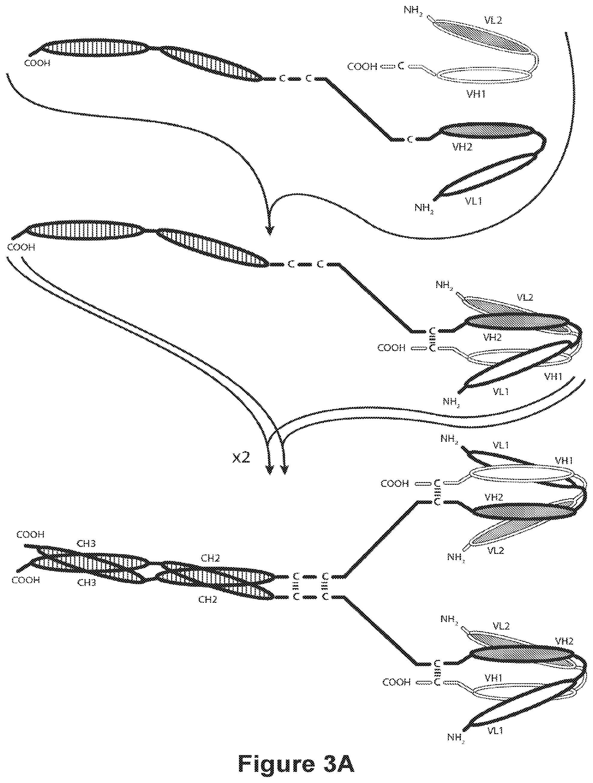
[0095] FIGS. 3A-3C provide schematics showing representative tetravalent diabodies having four epitope-binding sites composed of two pairs of polypeptide chains (i.e., four polypeptide chains in all). One polypeptide of each pair possesses a CH2 and CH3 Domain, such that the associated chains form all or part of an Fc Region. VL and VH Domains that recognize the same epitope are shown using the same shading or fill pattern. The two pairs of polypeptide chains may be same. In such embodiments wherein the VL and VH Domains recognize different epitopes (as shown in FIGS. 3A-3C), the resulting molecule possesses four epitope-binding sites and is bispecific and bivalent with respect to each bound epitope. In such embodiments wherein the VL and VH Domains recognize the same epitope (e.g., the same VL Domain CDRs and the same VH Domain CDRs are used on both chains), the resulting molecule possesses four epitope-binding sites and is monospecific and tetravalent with respect to a single epitope. Alternatively, the two pairs of polypeptides may be different. In such embodiments wherein the VL and VH Domains of each pair of polypeptides recognize different epitopes (as shown in FIGS. 3A-3C), the resulting molecule possesses four epitope-binding sites and is tetraspecific and monovalent with respect to each bound epitope. FIG. 3A shows an Fc diabody which contains a peptide Heterodimer-Promoting Domain comprising a cysteine residue. FIG. 3B shows an Fc Region-containing diabody, which contains E-coil and K-coil Heterodimer-Promoting Domains comprising a cysteine residue and a linker (with an optional cysteine residue). FIG. 3C, shows an Fc-Region-Containing diabody, which contains antibody CH1 and CL domains.
[0096] FIGS. 4A and 4B provide schematics of a representative covalently bonded diabody molecule having two epitope-binding sites composed of three polypeptide chains. Two of the polypeptide chains possess a CH2 and CH3 Domain, such that the associated chains form all or part of an Fc Region. The polypeptide chains comprising the VL and VH Domain further comprise a Heterodimer-Promoting Domain. VL and VH Domains that recognize the same epitope are shown using the same shading or fill pattern.
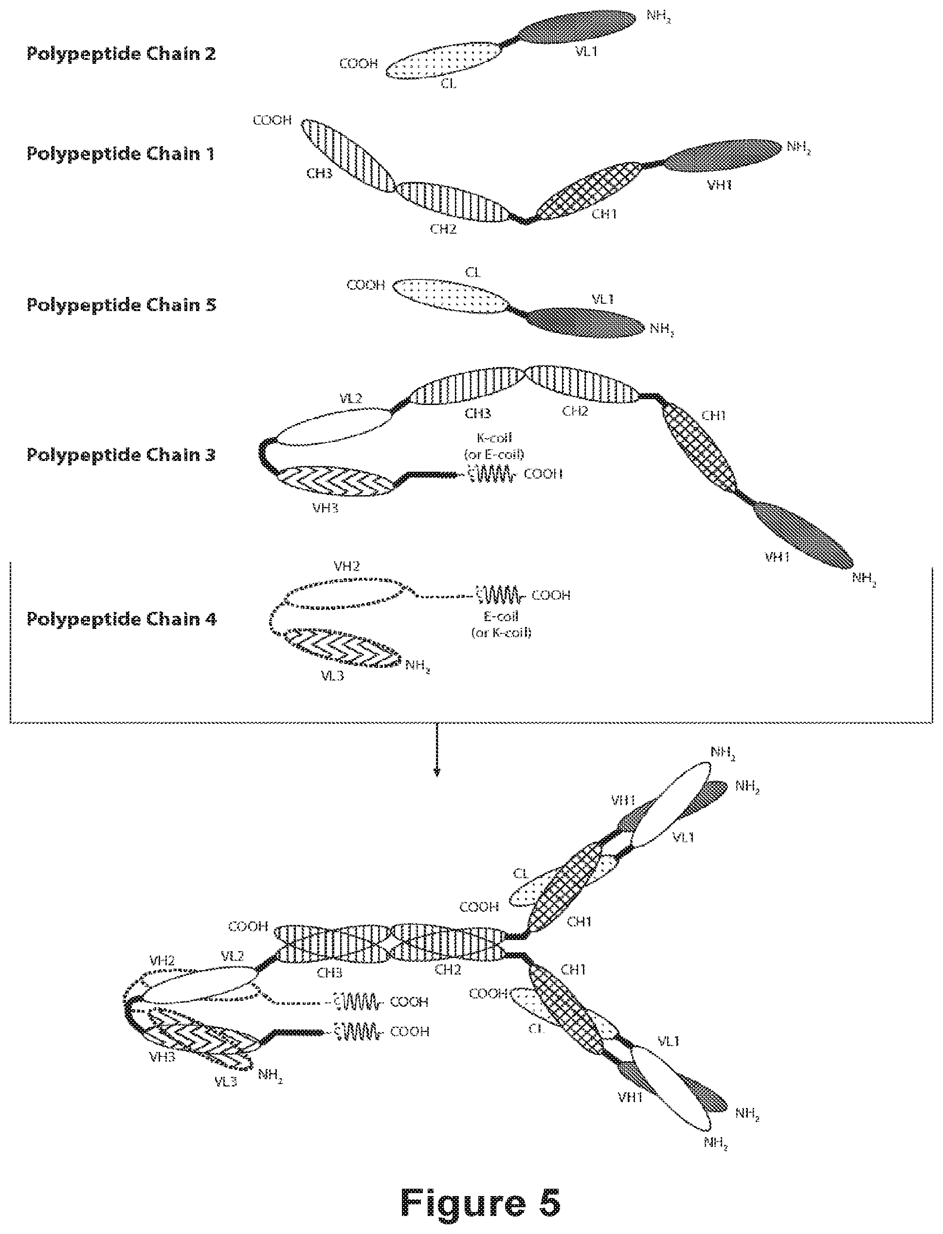
[0097] FIG. 5 provides the schematics of a representative covalently bonded diabody molecule having four epitope-binding sites composed of five polypeptide chains. Two of the polypeptide chains possess a CH2 and CH3 Domain, such that the associated chains form an Fc Region that comprises all or part of an Fc Region. The polypeptide chains comprising the linked VL and VH Domains further comprise a Heterodimer-Promoting Domain. VL and VH Domains that recognize the same epitope are shown using the same shading or fill pattern.
[0098] FIGS. 6A-6F provide schematics of representative Fc Region-containing trivalent binding molecules having three epitope-binding sites. FIGS. 6A and 6B, respectively, illustrate schematically the domains of trivalent binding molecules comprising two diabody-type binding domains and a Fab-type binding domain having different domain orientations in which the diabody-type binding domains are N-terminal or C-terminal to an Fc Region. The molecules in FIGS. 6A and 6B comprise four chains. FIGS. 6C and 6D, respectively, illustrate schematically the domains of trivalent binding molecules comprising two diabody-type binding domains N-terminal to an Fc Region, and a Fab-type binding domain in which the light chain and heavy chain are inked via a polypeptide spacer, or an scFv-type binding domain. The trivalent binding molecules in FIGS. 6E and 6F, respectively illustrate schematically the domains of trivalent binding molecules comprising two diabody-type binding domains C-terminal to an Fc Region, and a linked Fab-type binding domain, or an scFv-type binding domain in which the diabody-type binding domains are. The trivalent binding molecules in FIGS. 6C-6F comprise three chains. VL and VH Domains that recognize the same epitope are shown using the same shading or fill pattern.
[0099] FIGS. 7A-7D shows that the anti-PD-1 antibodies PD-1 mAb 1-15 bind to human PD-1. Binding curves for binding to shPD-1-His are shown in FIG. 7A (PD-1 mAb 1, PD-1 mAb 2, PD-1 mAb 4 and PD-1 mAb 9), FIG. 7B (PD-1 mAb 5, PD-1 mAb 6, and PD-1 mAb 7), and FIG. 7C (PD-1 mAb 3, PD-1 mAb 8, PD-1 mAb 10, PD-1 mAb 11, PD-1 mAb 12, PD-1 mAb 13, PD-1 mAb 14, and PD-1 mAb 15). Binding curves for binding to shPD-1-human Fc are shown in FIG. 7D (PD-1 mAb 3, PD-1 mAb 8, PD-1 mAb 10, PD-1 mAb 11, PD-1 mAb 12, PD-1 mAb 13, PD-1 mAb 14, and PD-1 mAb 15).
[0100] FIGS. 8A-8C shows that the anti-PD-1 antibodies PD-1 mAb 1-15 bind to cynomolgus monkey PD-1. Binding curves for binding to scynoPD-1-hFc are shown in FIG. 8A (PD-1 mAb 1, PD-1 mAb 2, PD-1 mAb 4, PD-1 mAb 5, PD-1 mAb 6, PD-1 mAb 7), FIG. 8B (PD-1 mAb 9), and FIG. 8C (PD-1 mAb 3, PD-1 mAb 8, PD-1 mAb 10, PD-1 mAb 11, PD-1 mAb 12, PD-1 mAb 13, PD-1 mAb 14, and PD-1 mAb 15).
[0101] FIGS. 9A-9D show the ability of the anti-PD-1 antibodies PD-1 mAb 1-15 to block the binding of human PD-L1 to human PD-1. Inhibition curves are shown in FIG. 9A (PD-1 mAb 1, PD-1 mAb 2, PD-1 mAb 3, PD-1 mAb 15, and PD-1 mAb A), FIG. 9B (PD-1 mAb 4), FIG. 9C (PD-1 mAb 5, PD-1 mAb 6, PD-1 mAb 7, and PD-1 mAb A), and FIG. 9D (PD-1 mAb 3, PD-1 mAb 8, PD-1 mAb 10, PD-1 mAb 11, PD-1 mAb 12, PD-1 mAb 13, PD-1 mAb 14, PD-1 mAb 15, and PD-1 mAb A).
[0102] FIGS. 10A-10B show the tissue specificity of the anti-human PD-1 antibody PD-1 mAb 7. FIG. 10A shows histological stains of normal colon (Panels i and vii), liver (Panels ii and viii), lung (Panels iii and ix), pancreas (Panels iv and x), kidney (Panels v and xi) and heart (Panels vi and xii) tissue. FIG. 10A, Panels i-vi show the results of tissue incubated with labeled PD-1 mAb 7 (0.313 .mu.g/mL). FIG. 10A, Panels vii-xii show the results of tissue incubated with labeled isotype control mAb (0.314 .mu.g/mL). FIG. 10B shows histological stains of skin (Panels i and iv), tonsils (Panels ii and v), and NSO cells expressing PD-1 (Panels iii and vi). FIG. 10B, Panels i-iii show the results of tissue incubated with labeled PD-1 mAb 7 (0.313 .mu.g/mL).
[0103] FIG. 11 shows the binding profiles of humanized anti-human PD-1 antibodies hPD-1 mAb 2, hPD-1 mAb 7(1.1), hPD-1 mAb 7(1.2), hPD-1 mAb 9(1.1), and the reference anti-PD-1 antibodies PD-1 mAb A and PD-1 mAb B having IgG1 (AA) or IgG4 (P) for binding to cell surface PD-1.
[0104] FIGS. 12A-12B show the ability of humanized anti-PD antibodies hPD-1 mAb 2, hPD-1 mAb 7(1.1), hPD-1 mAb 7(1.2), hPD-1 mAb 9(1.1), and the reference anti-PD-1 antibodies PD-1 mAb A and PD-1 mAb B, having IgG1 (AA) or IgG4 (P) to block the binding of soluble human PD-L1 (FIG. 12A) and soluble human PD-L2 (FIG. 12B) to cell surface human PD-1.
[0105] FIG. 13 shows the ability of humanized anti-PD antibodies hPD-1 mAb 2, hPD-1 mAb 7(1.1), hPD-1 mAb 7(1.2), hPD-1 mAb 9(1.1), and the reference anti-PD-1 antibodies PD-1 mAb A and PD-1 mAb B, having IgG1 (AA) or IgG4 (P) to antagonize the PD-1/PD-L1 axis by blocking the PD-1/PD-L1 interaction and preventing down-regulation of T-cell responses in a Jurkat-luc-NFAT/CHO-PD-L1 luciferase reporter assay.
[0106] FIG. 14 shows that PD-1 mAb 2, PD-1 mAb 7, PD-1 mAb 9 and PD-1 mAb 15 are able to stimulate cytokine production to levels comparable or higher than the referenced anti-PD-1 antibodies (PD-1 mAb A and PD-1 mAb B) and that treatment with PD-1 mAb 2, PD-1 mAb 7, PD-1 mAb 9 and PD-1 mAb 15 in combination with LAG-3 mAb 1 provided the largest enhancement of cytokine release. IFN.gamma. secretion profiles from Staphylococcal enterotoxin B (SEB)-stimulated PBMCs treated with anti-PD-1 and anti-LAG-3 antibodies alone and in combination.
[0107] FIGS. 15A-15B show the ability of humanized anti-PD antibodies hPD-1 mAb 2, hPD-1 mAb 7(1.2), hPD-1 mAb 9(1.1), and the reference anti-PD-1 antibodies PD-1 mAb A and PD-1 mAb B, having IgG1 (AA) or IgG4 (P) to stimulate cytokine production. IFN.gamma. (FIG. 15A) and TNF.alpha. (FIG. 15B), secretion profiles from SEB-stimulated PBMCs treated with anti-PD-1 antibodies.
[0108] FIGS. 16A-16B show that the PD-1.times.LAG-3 bispecific diabody constructs DART A, DART D, DART E, DART F, DART G and DART H, are able to stimulate cytokine production to levels comparable or higher than that observed upon the administration of the combination of an anti-PD-1 mAb+an anti-LAG-3 mAb (PD-1 mAb A+LAG-3 mAb A), and that the PD-1.times.LAG-3 bispecific diabody constructs DART A, DART D, DART E, DART F and DART G provided the largest enhancement of cytokine release. IFN.gamma. secretion profiles of PBMCs stimulated with a low concentration of SEB (0.2 ng/mL treated with PD-1.times.LAG-3 bispecific diabodies, or anti-PD-1 and anti-LAG-3 antibodies alone and in combination are plotted. The results using PBMCs from two representative donors are shown in FIG. 16A and FIG. 16B.
[0109] FIGS. 17A-17B show that the PD-1.times.LAG-3 bispecific diabody constructs DART A, DART B and DART C are able to stimulate cytokine production to levels higher than that observed upon the administration of the combination of an anti-PD-1 mAb+an anti-LAG-3 mAb (PD-1 mAb A+LAG-3 mAb A). IFN.gamma. secretion profiles of PBMCs from two representative donors, stimulated with a high concentration of SEB (85 ng/mL) treated with PD-1.times.LAG-3 bispecific diabodies, or anti-PD-1 and anti-LAG-3 antibodies alone and in combination are plotted. The results using PBMCs from two representative donors are shown in FIG. 17A and FIG. 17B.
[0110] FIGS. 18A-18B show that the PD-1.times.LAG-3 bispecific diabody constructs DART A, DART B and DART C are able to stimulate cytokine production to levels higher than that observed upon the administration of the combination of an anti-PD-1 mAb+an anti-LAG-3 mAb (PD-1 mAb A+LAG-3 mAb A). IFN.gamma. secretion profiles of PBMCs from two representative donors, stimulated with a middle concentration of SEB (0.5 ng/mL) treated with PD-1.times.LAG-3 bispecific diabodies, or anti-PD-1 and anti-LAG-3 antibodies alone and in combination are plotted. The results using PBMCs from two representative donors are shown in FIG. 18A and FIG. 18B.
[0111] FIG. 19 shows that the PD-1.times.LAG-3 bispecific diabody constructs DART D and DART H are able to stimulate cytokine production to levels comparable or higher than that observed upon the administration of the combination of an anti-PD-1 mAb+an anti-LAG-3 mAb (PD-1 mAb A+LAG-3 mAb A), and that DART D provided the largest enhancement of cytokine release. IL-2 secretion profiles of PBMCs from a representative donor stimulated with a high concentration of SEB (85 ng/mL) treated with PD-1.times.LAG-3 bispecific diabodies, or anti-PD-1 and anti-LAG-3 antibodies alone and in combination are plotted.
[0112] FIG. 20 shows that the PD-1.times.LAG-3 bispecific diabody constructs DART B and DART I are able to stimulate cytokine production to levels higher than that observed upon the administration of the combination of an anti-PD-1 mAb+an anti-LAG-3 mAb (PD-1 mAb A+LAG-3 mAb A, hPD-1 mAb 7(1.2)+hLAG-3 mAb 1(1.4), hPD-1 mAb 7(1.2)+hLAG-3 mAb 6(1.1)). IFN.gamma. secretion profiles of PBMCs from a representative donor, stimulated with a middle concentration of SEB (0.5 ng/mL) treated with PD-1.times.LAG-3 bispecific diabodies, or anti-PD-1 and anti-LAG-3 antibodies alone and in combination are plotted.
[0113] FIGS. 21A-21D show that the that the PD-1.times.LAG-3 bispecific diabody DART I is able to stimulate cytokine production to levels higher than that observed upon the administration of the combination of an anti-PD-1 mAb+an anti-LAG-3 mAb (PD-1 mAb A+LAG-3 mAb A). IFN.gamma. (FIGS. 21A and 21C) and IL-2 (FIGS. 21B and 21D) secretion profiles of CD4 memory cells from two representative donors, stimulated with tetanus toxoid (5 .mu.g/mL) treated with the PD-1.times.LAG-3 bispecific diabody DART-I, anti-PD-1 and anti-LAG-3 antibodies in combination, or an isotype control are plotted. The results at day 7 using CD4 memory T cells from two representative donors are shown in FIGS. 21A-B and FIGS. 21C-D.
[0114] FIG. 22 shows that the the pharmacokinetics of the PD-1.times.LAG-3 bispecific molecule, DART I are comparable to those of the anti-PD-1 antibody, PD-1 mAb A IgG4 (P) in cynomolgus monkey. The lines indicate the mean serum concentration of DART I (solid) and PD-1 mAb A (dashed). The individual values for the male (filled) and female (open) monkeys are plotted for DART I (triangles) and PD-1 mAb A (circles).
[0115] FIGS. 23A-23C show serum antibody concentrations and percentage of bound PD-1 on the surface of CD4+ or CD8+ T cells over time in animals following treatment with different anti-PD-1 antibodies. The percentage of bound PD 1 on the surface of CD4+ or CD8+ T cells following anti-PD 1 mAb treatment is plotted on the right y-axes; symbols represent % bound PD 1 on T cells for each individual animal and dashed lines represent the mean values. Serum mAb concentrations are plotted on the left y-axes; symbols represent serum levels for each individual animal and solid lines represent nonlinear fits of the data. Each panel presents data for animals (n=1/sex/group) administered 10 mg/kg hPD-1 mAb 7 (1.2) IgG4 (P) (FIG. 23A), PD-1 mAb A IgG4 (P) (FIG. 23B), or PD-1 mAb B IgG4 (P) (FIG. 23B) by IV infusion on Day 1.
DETAILED DESCRIPTION OF THE INVENTION
[0116] The present invention is directed to PD-1-binding molecules that comprise the PD-1-binding domain of selected anti-PD-1 antibodies capable of binding to both cynomolgus monkey PD-1 and to human PD-1: PD-1 mAb 1, PD-1 mAb 2, PD-1 mAb 3, PD-1 mAb 4, PD-1 mAb 5, PD-1 mAb 6, PD-1 mAb 7, PD-1 mAb 8, PD-1 mAb 9, PD-1 mAb 10, PD-1 mAb 11, PD-1 mAb 12, PD-1 mAb 13, PD-1 mAb 14, or PD-1 mAb 15. The invention particularly concerns PD-1-binding molecules that are humanized or chimeric versions of such antibodies, or that comprise PD-1-binding fragments of such anti-PD-1 antibodies (especially immunocongugates, diabodies (including but not limited to DART-A, DART-B, DART-C, DART-D, DART-E, DART-F, DART-G, DART-H, DART-I, and DART-J), BiTEs, bispecific antibodies, etc.). The invention particularly concerns such PD-1-binding molecules that are additionally capable of binding an epitope of a molecule involved in regulating an immune check point that is present on the surface of an immune cell. The present invention also pertains to methods of using such PD-1-binding molecules to detect PD-1 or to stimulate an immune response. The present invention also pertains to methods of combination therapy in which a PD-1-binding molecule that comprises one or more PD-1-binding domain(s) of such selected anti-PD-1 antibodies is administered in combination with one or more additional molecules that are effective in stimulating an immune response and/or in combination with one or more additional molecules that specifically bind a cancer antigen.
I. Antibodies and their Binding Domains
[0117] The antibodies of the present invention are immunoglobulin molecules capable of specific binding to a target, such as a carbohydrate, polynucleotide, lipid, polypeptide, etc., through at least one antigen recognition site, located in the Variable Domain of the immunoglobulin molecule. As used herein, the terms "antibody" and "antibodies" refer to monoclonal antibodies, multispecific antibodies, human antibodies, humanized antibodies, synthetic antibodies, chimeric antibodies, polyclonal antibodies, camelized antibodies, single-chain Fvs (scFv), single-chain antibodies, Fab fragments, F(ab') fragments, disulfide-linked bispecific Fvs (sdFv), intrabodies, and epitope-binding fragments of any of the above. In particular, antibodies include immunoglobulin molecules and immunologically active fragments of immunoglobulin molecules, i.e., molecules that contain an antigen-binding site. Immunoglobulin molecules can be of any type (e.g., IgG, IgE, IgM, IgD, IgA and IgY), class (e.g., IgG.sub.1, IgG.sub.2, IgG.sub.3, IgG.sub.4, IgA.sub.1 and IgA.sub.2) or subclass. In addition to their known uses in diagnostics, antibodies have been shown to be useful as therapeutic agents. Antibodies are capable of immunospecifically binding to a polypeptide or protein or a non-protein molecule due to the presence on such molecule of a particular domain or moiety or conformation (an "epitope"). An epitope-containing molecule may have immunogenic activity, such that it elicits an antibody production response in an animal; such molecules are termed "antigens"). The last few decades have seen a revival of interest in the therapeutic potential of antibodies, and antibodies have become one of the leading classes of biotechnology-derived drugs (Chan, C. E. et al. (2009) "The Use Of Antibodies In The Treatment Of Infectious Diseases," Singapore Med. J. 50(7):663-666). Over 200 antibody-based drugs have been approved for use or are under development.
[0118] The term "monoclonal antibody" refers to a homogeneous antibody population wherein the monoclonal antibody is comprised of amino acids (naturally occurring and non-naturally occurring) that are involved in the selective binding of an antigen. Monoclonal antibodies are highly specific, being directed against a single epitope (or antigenic site). The term "monoclonal antibody" encompasses not only intact monoclonal antibodies and full-length monoclonal antibodies, but also fragments thereof (such as Fab, Fab', F(ab').sub.2 Fv), single-chain (scFv), mutants thereof, fusion proteins comprising an antibody portion, humanized monoclonal antibodies, chimeric monoclonal antibodies, and any other modified configuration of the immunoglobulin molecule that comprises an antigen recognition site of the required specificity and the ability to bind to an antigen. It is not intended to be limited as regards to the source of the antibody or the manner in which it is made (e.g., by hybridoma, phage selection, recombinant expression, transgenic animals, etc.). The term includes whole immunoglobulins as well as the fragments etc. described above under the definition of "antibody." Methods of making monoclonal antibodies are known in the art. One method which may be employed is the method of Kohler, G. et al. (1975) "Continuous Cultures Of Fused Cells Secreting Antibody Of Predefined Specificity," Nature 256:495-497 or a modification thereof. Typically, monoclonal antibodies are developed in mice, rats or rabbits. The antibodies are produced by immunizing an animal with an immunogenic amount of cells, cell extracts, or protein preparations that contain the desired epitope. The immunogen can be, but is not limited to, primary cells, cultured cell lines, cancerous cells, proteins, peptides, nucleic acids, or tissue. Cells used for immunization may be cultured for a period of time (e.g., at least 24 hours) prior to their use as an immunogen. Cells may be used as immunogens by themselves or in combination with a non-denaturing adjuvant, such as Ribi (see, e.g., Jennings, V.M. (1995) "Review of Selected Adjuvants Used in Antibody Production," ILAR J. 37(3): 119-125). In general, cells should be kept intact and preferably viable when used as immunogens. Intact cells may allow antigens to be better detected than ruptured cells by the immunized animal. Use of denaturing or harsh adjuvants, e.g., Freud's adjuvant, may rupture cells and therefore is discouraged. The immunogen may be administered multiple times at periodic intervals such as, bi-weekly, or weekly, or may be administered in such a way as to maintain viability in the animal (e.g., in a tissue recombinant). Alternatively, existing monoclonal antibodies and any other equivalent antibodies that are immunospecific for a desired pathogenic epitope can be sequenced and produced recombinantly by any means known in the art. In one embodiment, such an antibody is sequenced and the polynucleotide sequence is then cloned into a vector for expression or propagation. The sequence encoding the antibody of interest may be maintained in a vector in a host cell and the host cell can then be expanded and frozen for future use. The polynucleotide sequence of such antibodies may be used for genetic manipulation to generate the monospecific or multispecific (e.g., bispecific, trispecific and tetraspecific) molecules of the invention as well as an affinity optimized, a chimeric antibody, a humanized antibody, and/or a caninized antibody, to improve the affinity, or other characteristics of the antibody. The general principle in humanizing an antibody involves retaining the basic sequence of the antigen-binding portion of the antibody, while swapping the non-human remainder of the antibody with human antibody sequences.
[0119] Natural antibodies (such as IgG antibodies) are composed of two Light Chains complexed with two Heavy Chains. Each light chain contains a Variable Domain (VL) and a Constant Domain (CL). Each heavy chain contains a Variable Domain (VH), three Constant Domains (CH1, CH2 and CH3), and a hinge domain located between the CH1 and CH2 Domains. The basic structural unit of naturally occurring immunoglobulins (e.g., IgG) is thus a tetramer having two light chains and two heavy chains, usually expressed as a glycoprotein of about 150,000 Da. The amino-terminal ("N-terminal") portion of each chain includes a Variable Domain of about 100 to 110 or more amino acids primarily responsible for antigen recognition. The carboxy-terminal ("C-terminal") portion of each chain defines a constant region, with light chains having a single Constant Domain and heavy chains usually having three Constant Domains and a Hinge Domain. Thus, the structure of the light chains of an IgG molecule is n-VL-CL-c and the structure of the IgG heavy chains is n-VH-CH1-H-CH2-CH3-c (where H is the hinge domain, and n and c represent, respectively, the N-terminus and the C-terminus of the polypeptide). The Variable Domains of an IgG molecule consist of the complementarity determining regions (CDR), which contain the residues in contact with epitope, and non-CDR segments, referred to as framework segments (FR), which in general maintain the structure and determine the positioning of the CDR loops so as to permit such contacting (although certain framework residues may also contact antigen). Thus, the VL and VH Domains have the structure n-FR1-CDR1-FR2-CDR2-FR3-CDR3-FR4-c. Polypeptides that are (or may serve as) the first, second and third CDR of an antibody Light Chain are herein respectively designated CDR.sub.L1 Domain, CDR.sub.L2 Domain, and CDR.sub.L3 Domain. Similarly, polypeptides that are (or may serve as) the first, second and third CDR of an antibody heavy chain are herein respectively designated CDR.sub.H1 Domain, CDR.sub.H2 Domain, and CDR.sub.H3 Domain. Thus, the terms CDR.sub.L1 Domain, CDR.sub.L2 Domain, CDR.sub.L3 Domain, CDR.sub.H1 Domain, CDR.sub.H2 Domain, and CDR.sub.H3 Domain are directed to polypeptides that when incorporated into a protein cause that protein to be able to bind to a specific epitope regardless of whether such protein is an antibody having light and heavy chains or a diabody or a single-chain binding molecule (e.g., an scFv, a BiTe, etc.), or is another type of protein. Accordingly, as used herein, the term "epitope-binding fragment" means a fragment of an antibody capable of immunospecifically binding to an epitope, and the term "epitope-binding site" refers to that portion of a molecule comprising an epitope-binding fragment that is responsible for epitope binding. An epitope-binding site may contain 1, 2, 3, 4, 5 or all 6 of the CDR Domains of such antibody and, although capable of immunospecifically binding to such epitope, may exhibit an immunospecificity, affinity or selectivity toward such epitope that differs from that of such antibody. Preferably, however, an epitope-binding fragment will contain all 6 of the CDR Domains of such antibody. An epitope-binding fragment of an antibody may be a single polypeptide chain (e.g., an scFv), or may comprise two or more polypeptide chains, each having an amino terminus and a carboxy terminus (e.g., a diabody, a Fab fragment, an F(ab').sub.2 fragment, etc.).
[0120] The invention particularly encompasses single-chain Variable Domain fragments ("scFv") of the anti-PD-1 antibodies of this invention and multispecific binding molecules comprising the same. Single-chain Variable Domain fragments are made by linking Light and/or Heavy chain Variable Domain by using a short linking peptide. Bird et al. (1988) ("Single-Chain Antigen-Binding Proteins," Science 242:423-426) describes example of linking peptides which bridge approximately 3.5 nm between the carboxy terminus of one Variable Domain and the amino terminus of the other Variable Domain. Linkers of other sequences have been designed and used (Bird et al. (1988) "Single-Chain Antigen-Binding Proteins," Science 242:423-426). Linkers can in turn be modified for additional functions, such as attachment of drugs or attachment to solid supports. The single-chain variants can be produced either recombinantly or synthetically. For synthetic production of scFv, an automated synthesizer can be used. For recombinant production of scFv, a suitable plasmid containing polynucleotide that encodes the scFv can be introduced into a suitable host cell, either eukaryotic, such as yeast, plant, insect or mammalian cells, or prokaryotic, such as E. coli. Polynucleotides encoding the scFv of interest can be made by routine manipulations such as ligation of polynucleotides. The resultant scFv can be isolated using standard protein purification techniques known in the art.
[0121] The invention also particularly encompasses humanized variants of the anti-PD-1 antibodies of the invention and multispecific binding molecules comprising the same. The term "humanized" antibody refers to a chimeric molecule, generally prepared using recombinant techniques, having an antigen-binding site of an immunoglobulin from a non-human species and a remaining immunoglobulin structure of the molecule that is based upon the structure and/or sequence of a human immunoglobulin. The anti-human PD-1 antibodies of the present invention include humanized, chimeric or caninized variants of antibodies PD-1 mAb 1, PD-1 mAb 2, PD-1 mAb 3, PD-1 mAb 4, PD-1 mAb 5, PD-1 mAb 6, PD-1 mAb 7, PD-1 mAb 8, PD-1 mAb 9, PD-1 mAb 10, PD-1 mAb 11, PD-1 mAb 12, PD-1 mAb 13, PD-1 mAb 14, or PD-1 mAb 15. The polynucleotide sequence of the variable domains of such antibodies may be used for genetic manipulation to generate such derivatives and to improve the affinity, or other characteristics of such antibodies. The general principle in humanizing an antibody involves retaining the basic sequence of the antigen-binding portion of the antibody, while swapping the non-human remainder of the antibody with human antibody sequences. There are four general steps to humanize a monoclonal antibody. These are: (1) determining the nucleotide and predicted amino acid sequence of the starting antibody light and heavy variable domains (2) designing the humanized antibody or caninized antibody, i.e., deciding which antibody framework region to use during the humanizing or canonizing process (3) the actual humanizing or caninizing methodologies/techniques and (4) the transfection and expression of the humanized antibody. See, for example, U.S. Pat. Nos. 4,816,567; 5,807,715; 5,866,692; and 6,331,415.
[0122] The antigen-binding site may comprise either a complete Variable Domain fused to a Constant Domain or only the complementarity determining regions (CDRs) of such Variable Domain grafted to appropriate framework regions. Antigen-binding sites may be wild-type or modified by one or more amino acid substitutions. This eliminates the constant region as an immunogen in human individuals, but the possibility of an immune response to the foreign variable domain remains (LoBuglio, A. F. et al. (1989) "Mouse/Human Chimeric Monoclonal Antibody In Man: Kinetics And Immune Response," Proc. Natl. Acad. Sci. (U.S.A.) 86:4220-4224). Another approach focuses not only on providing human-derived constant regions, but modifying the variable domains as well so as to reshape them as closely as possible to human form. It is known that the variable domains of both heavy and light chains contain three complementarity determining regions (CDRs) which vary in response to the antigens in question and determine binding capability, flanked by four framework regions (FRs) which are relatively conserved in a given species and which putatively provide a scaffolding for the CDRs. When non-human antibodies are prepared with respect to a particular antigen, the variable domains can be "reshaped" or "humanized" by grafting CDRs derived from non-human antibody on the FRs present in the human antibody to be modified. Application of this approach to various antibodies has been reported by Sato, K. et al. (1993) Cancer Res 53:851-856. Riechmann, L. et al. (1988) "Reshaping Human Antibodies for Therapy," Nature 332:323-327; Verhoeyen, M. et al. (1988) "Reshaping Human Antibodies: Grafting An Antilysozyme Activity," Science 239:1534-1536; Kettleborough, C. A. et al. (1991) "Humanization Of A Mouse Monoclonal Antibody By CDR-Grafting: The Importance Of Framework Residues On Loop Conformation," Protein Engineering 4:773-3783; Maeda, H. et al. (1991) "Construction Of Reshaped Human Antibodies With HIV-Neutralizing Activity," Human Antibodies Hybridoma 2:124-134; Gorman, S. D. et al. (1991) "Reshaping A Therapeutic CD4 Antibody," Proc. Natl. Acad. Sci. (U.S.A.) 88:4181-4185; Tempest, P. R. et al. (1991) "Reshaping A Human Monoclonal Antibody To Inhibit Human Respiratory Syncytial Virus Infection in vivo," Bio/Technology 9:266-271; Co, M. S. et al. (1991) "Humanized Antibodies For Antiviral Therapy," Proc. Natl. Acad. Sci. (U.S.A.) 88:2869-2873; Carter, P. et al. (1992) "Humanization Of An Anti-p185her2 Antibody For Human Cancer Therapy," Proc. Natl. Acad. Sci. (U.S.A.) 89:4285-4289; and Co, M. S. et al. (1992) "Chimeric And Humanized Antibodies With Specificity For The CD33 Antigen," J. Immunol. 148:1149-1154. In some embodiments, humanized antibodies preserve all CDR sequences (for example, a humanized mouse antibody which contains all six CDRs from the mouse antibodies). In other embodiments, humanized antibodies have one or more CDRs (one, two, three, four, five, or six) which differ in sequence relative to the original antibody.
[0123] A number of "humanized" antibody molecules comprising an antigen-binding site derived from a non-human immunoglobulin have been described, including chimeric antibodies having rodent or modified rodent Variable Domain and their associated complementarity determining regions (CDRs) fused to human Constant Domains (see, for example, Winter et al. (1991) "Man-made Antibodies," Nature 349:293-299; Lobuglio et al. (1989) "Mouse/Human Chimeric Monoclonal Antibody In Man: Kinetics And Immune Response," Proc. Natl. Acad. Sci. (U.S.A.) 86:4220-4224 (1989), Shaw et al. (1987) "Characterization Of A Mouse/Human Chimeric Monoclonal Antibody (17-1A) To A Colon Cancer Tumor Associated Antigen," J. Immunol. 138:4534-4538, and Brown et al. (1987) "Tumor-Specific Genetically Engineered Murine/Human Chimeric Monoclonal Antibody," Cancer Res. 47:3577-3583). Other references describe rodent CDRs grafted into a human supporting framework region (FR) prior to fusion with an appropriate human antibody Constant Domain (see, for example, Riechmann, L. et al. (1988) "Reshaping Human Antibodies for Therapy," Nature 332:323-327; Verhoeyen, M. et al. (1988) "Reshaping Human Antibodies: Grafting An Antilysozyme Activity," Science 239:1534-1536; and Jones et al. (1986) "Replacing The Complementarity-Determining Regions In A Human Antibody With Those From A Mouse," Nature 321:522-525). Another reference describes rodent CDRs supported by recombinantly veneered rodent framework regions. See, for example, European Patent Publication No. 519,596. These "humanized" molecules are designed to minimize unwanted immunological response towards rodent anti-human antibody molecules, which limits the duration and effectiveness of therapeutic applications of those moieties in human recipients. Other methods of humanizing antibodies that may also be utilized are disclosed by Daugherty et al. (1991) "Polymerase Chain Reaction Facilitates The Cloning, CDR-Grafting, And Rapid Expression Of A Murine Monoclonal Antibody Directed Against The CD18 Component Of Leukocyte Integrins," Nucl. Acids Res. 19:2471-2476 and in U.S. Pat. Nos. 6,180,377; 6,054,297; 5,997,867; and 5,866,692.
II. Fc.gamma. Receptors (Fc.gamma.Rs)
[0124] The CH2 and CH3 Domains of the two heavy chains interact to form the Fc Region, which is a domain that is recognized by cellular Fc Receptors, including but not limited to Fc gamma Receptors (Fc.gamma.Rs). As used herein, the term "Fc Region" is used to define a C-terminal region of an IgG heavy chain. The amino acid sequence of the CH2-CH3 Domain of an exemplary human IgG1 is (SEQ ID NO:1):
TABLE-US-00001 231 240 250 260 270 APELLGGPSV FLFPPKPKDT LMISRTPEVT CVVVDVSHED 280 290 300 310 PEVKFNWYVD GVEVHNAKTK PREEQYNSTY RVVSVLTVLH 320 330 340 350 QDWLNGKEYK CKVSNKALPA PIEKTISKAK GQPREPQVYT 360 370 380 390 LPPSREEMTK NQVSLTCLVK GFYPSDIAVE WESNGQPENN 400 410 420 430 YKTTPPVLDS DGSFFLYSKL TVDKSRWQQG NVFSCSVMHE 440 447 ALHNHYTQKS LSLSPGX
as numbered by the EU index as set forth in Kabat, wherein, X is a lysine (K) or is absent.
[0125] The amino acid sequence of the CH2-CH3 Domain of an exemplary human IgG2 is (SEQ ID NO:2):
TABLE-US-00002 231 240 250 260 270 APPVA-GPSV FLFPPKPKDT LMISRTPEVT CVVVDVSHED 280 290 300 310 PEVQFNWYVD GVEVHNAKTK PREEQFNSTF RVVSVLTVVH 320 330 340 350 QDWLNGKEYK CKVSNKGLPA PIEKTISKTK GQPREPQVYT 360 370 380 390 LPPSREEMTK NQVSLTCLVK GFYPSDISVE WESNGQPENN 400 410 420 430 YKTTPPMLDS DGSFFLYSKL TVDKSRWQQG NVFSCSVMHE 440 447 ALHNHYTQKS LSLSPGX
as numbered by the EU index as set forth in Kabat, wherein, X is a lysine (K) or is absent.
[0126] The amino acid sequence of the CH2-CH3 Domain of an exemplary human IgG3 is (SEQ ID NO:3):
TABLE-US-00003 231 240 250 260 270 APELLGGPSV FLFPPKPKDT LMISRTPEVT CVVVDVSHED 280 290 300 310 PEVQFKWYVD GVEVHNAKTK PREEQYNSTF RVVSVLTVLH 320 330 340 350 QDWLNGKEYK CKVSNKALPA PIEKTISKTK GQPREPQVYT 360 370 380 390 LPPSREEMTK NQVSLTCLVK GFYPSDIAVE WESSGQPENN 400 410 420 430 YNTTPPMLDS DGSFFLYSKL TVDKSRWQQG NIFSCSVMHE 440 447 ALHNRFTQKS LSLSPGX
as numbered by the EU index as set forth in Kabat, wherein, X is a lysine (K) or is absent. The amino acid sequence of the CH2-CH3 Domain of an exemplary human IgG4 is (SEQ ID NO:4):
TABLE-US-00004 231 240 250 260 270 APEFLGGPSV FLFPPKPKDT LMISRTPEVT CVVVDVSQED 280 290 300 310 PEVQFNWYVD GVEVHNAKTK PREEQFNSTY RVVSVLTVLH 320 330 340 350 QDWLNGKEYK CKVSNKGLPS SIEKTISKAK GQPREPQVYT 360 370 380 390 LPPSQEEMTK NQVSLTCLVK GFYPSDIAVE WESNGQPENN 400 410 420 430 YKTTPPVLDS DGSFFLYSRL TVDKSRWQEG NVFSCSVMHE 440 447 ALHNHYTQKS LSLSLGX
as numbered by the EU index as set forth in Kabat, wherein, X is a lysine (K) or is absent.
[0127] Throughout the present specification, the numbering of the residues in the constant region of an IgG heavy chain is that of the EU index as in Kabat et al., Sequences of Proteins of Immunological Interest, 5.sup.th Ed. Public Health Service, NH1, MD (1991) ("Kabat"), expressly incorporated herein by references. The term "EU index as in Kabat" refers to the numbering of the human IgG1 EU antibody. Amino acids from the Variable Domains of the mature heavy and light chains of immunoglobulins are designated by the position of an amino acid in the chain. Kabat described numerous amino acid sequences for antibodies, identified an amino acid consensus sequence for each subgroup, and assigned a residue number to each amino acid, and the CDRs are identified as defined by Kabat (it will be understood that CDR.sub.H1 as defined by Chothia, C. & Lesk, A. M. ((1987) "Canonical structures for the hypervariable regions of immunoglobulins,". J. Mol. Biol. 196:901-917) begins five residues earlier). Kabat's numbering scheme is extendible to antibodies not included in his compendium by aligning the antibody in question with one of the consensus sequences in Kabat by reference to conserved amino acids. This method for assigning residue numbers has become standard in the field and readily identifies amino acids at equivalent positions in different antibodies, including chimeric or humanized variants. For example, an amino acid at position 50 of a human antibody light chain occupies the equivalent position to an amino acid at position 50 of a mouse antibody light chain.
[0128] Polymorphisms have been observed at a number of different positions within antibody constant regions (e.g., CH1 positions, including but not limited to positions 192, 193, and 214; Fc positions, including but not limited to positions 270, 272, 312, 315, 356, and 358 as numbered by the EU index as set forth in Kabat), and thus slight differences between the presented sequence and sequences in the prior art can exist. Polymorphic forms of human immunoglobulins have been well-characterized. At present, 18 Gm allotypes are known: Glm (1, 2, 3, 17) or Glm (a, x, f, z), G2m (23) or G2m (n), G3m (5, 6, 10, 11, 13, 14, 15, 16, 21, 24, 26, 27, 28) or G3m (b1, c3, b3, b0, b3, b4, s, t, g1, c5, u, v, g5) (Lefranc, et al., "The Human IgG Subclasses: Molecular Analysis Of Structure, Function And Regulation." Pergamon, Oxford, pp. 43-78 (1990); Lefranc, G. et al., 1979, Hum. Genet.: 50, 199-211). It is specifically contemplated that the antibodies of the present invention may be incorporate any allotype, isoallotype, or haplotype of any immunoglobulin gene, and are not limited to the allotype, isoallotype or haplotype of the sequences provided herein. Furthermore, in some expression systems the C-terminal amino acid residue (bolded above) of the CH3 Domain may be post-translationally removed. Accordingly, the C-terminal residue of the CH3 Domain is an optional amino acid residue in the PD-1-binding molecules of the invention. Specifically encompassed by the instant invention are PD-1-binding molecules lacking the C-terminal residue of the CH3 Domain. Also specifically encompassed by the instant invention are such constructs comprising the C-terminal lysine residue of the CH3 Domain.
[0129] Activating and inhibitory signals are transduced through the ligation of an Fc region to a cellular Fc gamma Receptor (Fc.gamma.R). The ability of such ligation to result in diametrically opposing functions results from structural differences among the different Fc.gamma.Rs. Two distinct domains within the cytoplasmic signaling domains of the receptor called immunoreceptor tyrosine-based activation motifs (ITAMs) and immunoreceptor tyrosine-based inhibitory motifs (ITIMS) account for the different responses. The recruitment of different cytoplasmic enzymes to these structures dictates the outcome of the Fc.gamma.R-mediated cellular responses. ITAM-containing Fc.gamma.R complexes include Fc.gamma.RI, Fc.gamma.RIIA, Fc.gamma.RIIIA, whereas ITIM-containing complexes only include Fc.gamma.RIIB Human neutrophils express the Fc.gamma.RIIA gene. Fc.gamma.RIIA clustering via immune complexes or specific antibody cross-linking serves to aggregate ITAMs along with receptor-associated kinases which facilitate ITAM phosphorylation. ITAM phosphorylation serves as a docking site for Syk kinase, activation of which results in activation of downstream substrates (e.g., PI.sub.3K). Cellular activation leads to release of proinflammatory mediators. The Fc.gamma.RIIB gene is expressed on B lymphocytes; its extracellular domain is 96% identical to Fc.gamma.RIIA and binds IgG complexes in an indistinguishable manner. The presence of an ITIM in the cytoplasmic domain of Fc.gamma.RIIB defines this inhibitory subclass of Fc.gamma.R. Recently the molecular basis of this inhibition was established. When co-ligated along with an activating Fc.gamma.R, the ITIM in Fc.gamma.RIIB becomes phosphorylated and attracts the SH2 domain of the inositol polyphosphate 5'-phosphatase (SHIP), which hydrolyzes phosphoinositol messengers released as a consequence of ITAM-containing Fc.gamma.R-mediated tyrosine kinase activation, consequently preventing the influx of intracellular Ca.sup.++. Thus cross-linking of Fc.gamma.RIIB dampens the activating response to Fc.gamma.R ligation and inhibits cellular responsiveness. B-cell activation, B-cell proliferation and antibody secretion is thus aborted.
III. Bispecific Antibodies, Multispecific Diabodies and DART.RTM. Diabodies
[0130] The ability of an antibody to bind an epitope of an antigen depends upon the presence and amino acid sequence of the antibody's VL and VH Domains. Interaction of an antibody light chain and an antibody heavy chain and, in particular, interaction of its VL and VH Domains forms one of the two epitope-binding sites of a natural antibody. Natural antibodies are capable of binding to only one epitope species (i.e., they are monospecific), although they can bind multiple copies of that species (i.e., exhibiting bivalency or multivalency).
[0131] The binding domains of the present invention bind to epitopes in an "immunospecific" manner. As used herein, an antibody, diabody or other epitope-binding molecule is said to "immunospecifically" bind a region of another molecule (i.e., an epitope) if it reacts or associates more frequently, more rapidly, with greater duration and/or with greater affinity with that epitope relative to alternative epitopes. For example, an antibody that immunospecifically binds to a viral epitope is an antibody that binds this viral epitope with greater affinity, avidity, more readily, and/or with greater duration than it immunospecifically binds to other viral epitopes or non-viral epitopes. It is also understood by reading this definition that, for example, an antibody (or moiety or epitope) that immunospecifically binds to a first target may or may not specifically or preferentially bind to a second target. As such, "immunospecific binding" does not necessarily require (although it can include) exclusive binding. Generally, but not necessarily, reference to binding means "specific" binding. Two molecules are said to be capable of binding to one another in a "physiospecific" manner, if such binding exhibits the specificity with which receptors bind to their respective ligands.
[0132] The functionality of antibodies can be enhanced by generating multispecific antibody-based molecules that can simultaneously bind two separate and distinct antigens (or different epitopes of the same antigen) and/or by generating antibody-based molecule having higher valency (i.e., more than two binding sites) for the same epitope and/or antigen.
[0133] In order to provide molecules having greater capability than natural antibodies, a wide variety of recombinant bispecific antibody formats have been developed (see, e.g., PCT Publication Nos. WO 2008/003116, WO 2009/132876, WO 2008/003103, WO 2007/146968, WO 2009/018386, WO 2012/009544, WO 2013/070565), most of which use linker peptides either to fuse a further epitope-binding fragment (e.g., an scFv, VL, VH, etc.) to, or within the antibody core (IgA, IgD, IgE, IgG or IgM), or to fuse multiple epitope-binding fragments (e.g., two Fab fragments or scFvs). Alternative formats use linker peptides to fuse an epitope-binding fragment (e.g., an scFv, VL, VH, etc.) to an a dimerization domain such as the CH2-CH3 Domain or alternative polypeptides (WO 2005/070966, WO 2006/107786A WO 2006/107617A, WO 2007/046893). Typically, such approaches involve compromises and trade-offs. For example, PCT Publications Nos. WO 2013/174873, WO 2011/133886 and WO 2010/136172 disclose that the use of linkers may cause problems in therapeutic settings, and teaches a trispecific antibody in which the CL and CH1 Domains are switched from their respective natural positions and the VL and VH Domains have been diversified (WO 2008/027236; WO 2010/108127) to allow them to bind to more than one antigen. Thus, the molecules disclosed in these documents trade binding specificity for the ability to bind additional antigen species. PCT Publications Nos. WO 2013/163427 and WO 2013/119903 disclose modifying the CH2 Domain to contain a fusion protein adduct comprising a binding domain. The document notes that the CH2 Domain likely plays only a minimal role in mediating effector function. PCT Publications Nos. WO 2010/028797, WO2010028796 and WO 2010/028795 disclose recombinant antibodies whose Fc Regions have been replaced with additional VL and VH Domains, so as to form trivalent binding molecules. PCT Publications Nos. WO 2003/025018 and WO2003012069 disclose recombinant diabodies whose individual chains contain scFv Domains. PCT Publications No. WO 2013/006544 discloses multivalent Fab molecules that are synthesized as a single polypeptide chain and then subjected to proteolysis to yield heterodimeric structures. Thus, the molecules disclosed in these documents trade all or some of the capability of mediating effector function for the ability to bind additional antigen species. PCT Publications Nos. WO 2014/022540, WO 2013/003652, WO 2012/162583, WO 2012/156430, WO 2011/086091, WO 2008/024188, WO 2007/024715, WO 2007/075270, WO 1998/002463, WO 1992/022583 and WO 1991/003493 disclose adding additional binding domains or functional groups to an antibody or an antibody portion (e.g., adding a diabody to the antibody's light chain, or adding additional VL and VH Domains to the antibody's light and heavy chains, or adding a heterologous fusion protein or chaining multiple Fab Domains to one another). Thus, the molecules disclosed in these documents trade native antibody structure for the ability to bind additional antigen species.
[0134] The art has additionally noted the capability to produce diabodies that differ from such natural antibodies in being capable of binding two or more different epitope species (i.e., exhibiting bispecificity or multispecificity in addition to bivalency or multivalency) (see, e.g., Holliger et al. (1993) "`Diabodies`: Small Bivalent And Bispecific Antibody Fragments," Proc. Natl. Acad. Sci. (U.S.A.) 90:6444-6448; US 2004/0058400 (Hollinger et al.); US 2004/0220388/WO 02/02781 (Mertens et al.); Alt et al. (1999) FEBS Lett. 454(1-2):90-94; Lu, D. et al. (2005) "A Fully Human Recombinant IgG-Like Bispecific Antibody To Both The Epidermal Growth Factor Receptor And The Insulin-Like Growth Factor Receptor For Enhanced Antitumor Activity," J. Biol. Chem. 280(20):19665-19672; WO 02/02781 (Mertens et al.); Olafsen, T. et al. (2004) "Covalent Disulfide-Linked Anti-CEA Diabody Allows Site-Specific Conjugation And Radiolabeling For Tumor Targeting Applications," Protein Eng. Des. Sel. 17(1):21-27; Wu, A. et al. (2001) "Multimerization Of A Chimeric Anti-CD20 Single Chain Fv-Fv Fusion Protein Is Mediated Through Variable Domain Exchange," Protein Engineering 14(2):1025-1033; Asano et al. (2004) "A Diabody For Cancer Immunotherapy And Its Functional Enhancement By Fusion Of Human Fc Domain," Abstract 3P-683, J. Biochem. 76(8):992; Takemura, S. et al. (2000) "Construction Of A Diabody (Small Recombinant Bispecific Antibody) Using A Refolding System," Protein Eng. 13(8):583-588; Baeuerle, P. A. et al. (2009) "Bispecific T-Cell Engaging Antibodies For Cancer Therapy," Cancer Res. 69(12):4941-4944).
[0135] The design of a diabody is based on the antibody derivative known as a single-chain Variable Domain fragment (scFv). Such molecules are made by linking Light and/or Heavy chain Variable Domains by using a short linking peptide. Bird et al. (1988) ("Single-Chain Antigen-Binding Proteins," Science 242:423-426) describes example of linking peptides which bridge approximately 3.5 nm between the carboxy terminus of one Variable Domain and the amino terminus of the other Variable Domain. Linkers of other sequences have been designed and used (Bird et al. (1988) "Single-Chain Antigen-Binding Proteins," Science 242:423-426). Linkers can in turn be modified for additional functions, such as attachment of drugs or attachment to solid supports. The single-chain variants can be produced either recombinantly or synthetically. For synthetic production of scFv, an automated synthesizer can be used. For recombinant production of scFv, a suitable plasmid containing polynucleotide that encodes the scFv can be introduced into a suitable host cell, either eukaryotic, such as yeast, plant, insect or mammalian cells, or prokaryotic, such as E. coli. Polynucleotides encoding the scFv of interest can be made by routine manipulations such as ligation of polynucleotides. The resultant scFv can be isolated using standard protein purification techniques known in the art.
[0136] The provision of non-monospecific diabodies provides a significant advantage over antibodies, including but not limited to, the capacity to co-ligate and co-localize cells that express different epitopes. Bispecific diabodies thus have wide-ranging applications including therapy and immunodiagnosis. Bispecificity allows for great flexibility in the design and engineering of the diabody in various applications, providing enhanced avidity to multimeric antigens, the cross-linking of differing antigens, and directed targeting to specific cell types relying on the presence of both target antigens. Due to their increased valency, low dissociation rates and rapid clearance from the circulation (for diabodies of small size, at or below .about.50 kDa), diabody molecules known in the art have also shown particular use in the field of tumor imaging (Fitzgerald et al. (1997) "Improved Tumour Targeting By Disulphide Stabilized Diabodies Expressed In Pichia pastoris," Protein Eng. 10:1221).
[0137] The bispecificity of diabodies has led to their use for co-ligating differing cells, for example, the cross-linking of cytotoxic T-cells to tumor cells (Staerz et al. (1985) "Hybrid Antibodies Can Target Sites For Attack By T Cells," Nature 314:628-631, and Holliger et al. (1996) "Specific Killing Of Lymphoma Cells By Cytotoxic T-Cells Mediated By A Bispecific Diabody," Protein Eng. 9:299-305; Marvin et al. (2005) "Recombinant Approaches To IgG-Like Bispecific Antibodies," Acta Pharmacol. Sin. 26:649-658). Alternatively, or additionally, bispecific diabodies can be used to co-ligate receptors on the surface of different cells or on a single cell. Co-ligation of different cells and/or receptors is useful to modulation effector functions and/or immune cell signaling. Multispecific molecules (e.g., bispecific diabodies) comprising epitope-binding sites may be directed to a surface determinant of any immune cell such as B7-H3 (CD276), B7-H4 (VTCN1), BTLA (CD272), CD3, CD8, CD16, CD27, CD32, CD40, CD40L, CD47, CD64, CD70 (CD27L), CD80 (B7-1), CD86 (B7-2), CD94 (KLRD1), CD137 (4-1BB), CD137L (4-1BBL), CD226, CTLA-4 (CD152), Galectin-9, GITR, GITRL, HHLA2, ICOS (CD278), ICOSL (CD275), Killer Activation Receptor (KIR), LAG-3 (CD223), LIGHT (TNFSF14, CD258), WIC class I or II, NKG2a, NKG2d, OX40 (CD134), OX40L (CD134L), PD1H, PD-1 (CD279), PD-L1 (B7-H1, CD274), PD-L2 (B7-CD, CD273), PVR (NECLS, CD155), SIRPa, TCR, TIGIT, TIM-3 (HAVCR2), and/or VISTA (PD-1H), which are expressed on T lymphocytes, Natural Killer (NK) cells, Antigen-presenting cells or other mononuclear cell. In particular, epitope-binding sites directed to a cell surface receptor that is involved in regulating an immune checkpoint (or the ligand thereof) are useful in the generation of bispecific or multispecific binding molecules which antagonize or block the inhibitory signaling of immune checkpoint molecules and thereby stimulate, upregulate or enhance, immune responses in a subject. Molecules involved in regulating immune checkpoints include, but are not limited to B7-H3, B7-H4, BTLA, CD40, CD40L, CD47, CD70, CD80, CD86, CD94, CD137, CD137L, CD226, CTLA-4, Galectin-9, GITR, GITRL, HHLA2, ICOS, ICOSL, KIR, LAG-3, LIGHT, MHC class I or II, NKG2a, NKG2d, OX40, OX40L, PD1H, PD-1, PD-L1, PD-L2, PVR, SIRPa, TCR, TIGIT, TIM-3 and/or VISTA.
[0138] However, the above advantages come at a salient cost. The formation of such non-monospecific diabodies requires the successful assembly of two or more distinct and different polypeptides (i.e., such formation requires that the diabodies be formed through the heterodimerization of different polypeptide chain species). This fact is in contrast to monospecific diabodies, which are formed through the homodimerization of identical polypeptide chains. Because at least two dissimilar polypeptides (i.e., two polypeptide species) must be provided in order to form a non-monospecific diabody, and because homodimerization of such polypeptides leads to inactive molecules (Takemura, S. et al. (2000) "Construction Of A Diabody (Small Recombinant Bispecific Antibody) Using A Refolding System," Protein Eng. 13(8):583-588), the production of such polypeptides must be accomplished in such a way as to prevent covalent bonding between polypeptides of the same species (i.e., so as to prevent homodimerization) (Takemura, S. et al. (2000) "Construction Of A Diabody (Small Recombinant Bispecific Antibody) Using A Refolding System," Protein Eng. 13(8):583-588). The art has therefore taught the non-covalent association of such polypeptides (see, e.g., Olafsen et al. (2004) "Covalent Disulfide-Linked Anti-CEA Diabody Allows Site-Specific Conjugation And Radiolabeling For Tumor Targeting Applications," Prot. Engr. Des. Sel. 17:21-27; Asano et al. (2004) "A Diabody For Cancer Immunotherapy And Its Functional Enhancement By Fusion Of Human Fc Domain," Abstract 3P-683, J. Biochem. 76(8):992; Takemura, S. et al. (2000) "Construction Of A Diabody (Small Recombinant Bispecific Antibody) Using A Refolding System," Protein Eng. 13(8):583-588; Lu, D. et al. (2005) "A Fully Human Recombinant IgG-Like Bispecific Antibody To Both The Epidermal Growth Factor Receptor And The Insulin-Like Growth Factor Receptor For Enhanced Antitumor Activity," J. Biol. Chem. 280(20):19665-19672).
[0139] However, the art has recognized that bispecific diabodies composed of non-covalently associated polypeptides are unstable and readily dissociate into non-functional monomers (see, e.g., Lu, D. et al. (2005) "A Fully Human Recombinant IgG-Like Bispecific Antibody To Both The Epidermal Growth Factor Receptor And The Insulin-Like Growth Factor Receptor For Enhanced Antitumor Activity," J. Biol. Chem. 280(20):19665-19672).
[0140] In the face of this challenge, the art has succeeded in developing stable, covalently bonded heterodimeric non-monospecific diabodies, termed DART.RTM. (Dual Affinity Re-Targeting Reagents) diabodies; see, e.g., United States Patent Publications No. 2013-0295121; 2010-0174053 and 2009-0060910; European Patent Publication No. EP 2714079; EP 2601216; EP 2376109; EP 2158221 and PCT Publications No. WO 2012/162068; WO 2012/018687; WO 2010/080538; and Sloan, D. D. et al. (2015) "Targeting HIV Reservoir in Infected CD4 T Cells by Dual-Affinity Re-targeting Molecules (DARTs) that Bind HIV Envelope and Recruit Cytotoxic T Cells," PLoS Pathog. 11(11):e1005233. doi: 10.1371/journal.ppat.1005233; Al Hussaini, M. et al. (2015) "Targeting CD123 In AML Using A T-Cell Directed Dual-Affinity Re-Targeting (DART.RTM.) Platform," Blood pii: blood-2014-05-575704; Chichili, G. R. et al. (2015) "A CD3.times.CD123 Bispecific DART For Redirecting Host T Cells To Myelogenous Leukemia: Preclinical Activity And Safety In Nonhuman Primates," Sci. Transl. Med. 7(289):289ra82; Moore, P. A. et al. (2011) "Application Of Dual Affinity Retargeting Molecules To Achieve Optimal Redirected T-Cell Killing Of B-Cell Lymphoma," Blood 117(17):4542-4551; Veri, M. C. et al. (2010) "Therapeutic Control Of B Cell Activation Via Recruitment Of Fcgamma Receptor IIb (CD32B) Inhibitory Function With A Novel Bispecific Antibody Scaffold," Arthritis Rheum. 62(7):1933-1943; Johnson, S. et al. (2010) "Effector Cell Recruitment With Novel Fv-Based Dual-Affinity Re-Targeting Protein Leads To Potent Tumor Cytolysis And in vivo B-Cell Depletion," J. Mol. Biol. 399(3):436-449). Such diabodies comprise two or more covalently complexed polypeptides and involve engineering one or more cysteine residues into each of the employed polypeptide species that permit disulfide bonds to form and thereby covalently bond two polypeptide chains. For example, the addition of a cysteine residue to the C-terminus of such constructs has been shown to allow disulfide bonding between the polypeptide chains, stabilizing the resulting heterodimer without interfering with the binding characteristics of the bivalent molecule.
[0141] Each of the two polypeptides of the simplest bispecific DART.RTM. diabody comprises three domains. The first polypeptide comprises (in the N-terminal to C-terminal direction): (i) a First Domain that comprises a binding region of a Light Chain Variable Domain of a first immunoglobulin (VL1), (ii) a Second Domain that comprises a binding region of a Heavy Chain Variable Domain of a second immunoglobulin (VH2), and (iii) a Third Domain that contains a cysteine residue (or a cysteine-containing domain) and a Heterodimer-Promoting Domain that serves to promote heterodimerization with the second polypeptide of the diabody and to covalently bond the diabody's first and second polypeptides to one another. The second polypeptide contains (in the N-terminal to C-terminal direction): (i) a First Domain that comprises a binding region of a Light Chain Variable Domain of the second immunoglobulin (VL2), (ii) a Second Domain that comprises a binding region of a Heavy Chain Variable Domain of the first immunoglobulin (VH1), and (iii) a Third Domain that contains a cysteine residue (or a cysteine-containing domain) and a complementary Heterodimer-Promoting Domain that complexes with the Heterodimer-Promoting Domain of the first polypeptide chain in order to promote heterodimerization with the first polypeptide chain. The cysteine residue (or a cysteine-containing domain) of the third domain of the second polypeptide chain serves to promote the covalent bonding of the second polypeptide chain to the first polypeptide chain of the diabody. Such molecules are stable, potent and have the ability to simultaneously bind two or more antigens. In one embodiment, the Third Domains of the first and second polypeptides each contain a cysteine residue, which serves to bind the polypeptides together via a disulfide bond. FIG. 1 provides a schematic of such a diabody, which utilizes E-coil/K-coil Heterodimer-Promoting domains and a cysteine containing linker for covalent bonding. As provided in FIG. 2 and FIGS. 3A-3C, one or both of the polypeptides may additionally possesses the sequence of a CH2-CH3 Domain, such that complexing between the two diabody polypeptides forms an Fc Region that is capable of binding to the Fc receptor of cells (such as B lymphocytes, dendritic cells, natural killer cells, macrophages, neutrophils, eosinophils, basophils and mast cells). As provided in more detail below, the CH2 and/or CH3 Domains of such polypeptide chains need not be identical in sequence, and advantageously are modified to foster complexing between the two polypeptide chains.
[0142] Many variations of such molecules have been described (see, e.g., United States Patent Publications No. 2015/0175697; 2014/0255407; 2014/0099318; 2013/0295121; 2010/0174053 and 2009/0060910; European Patent Publication No. EP 2714079; EP 2601216; EP 2376109; EP 2158221 and PCT Publications No. WO 2012/162068; WO 2012/018687; WO 2010/080538). These Fc Region-containing DART.RTM. diabodies may comprise two pairs of polypeptide chains. The first polypeptide chain comprises (in the N-terminal to C-terminal direction): (i) a First Domain that comprises a binding region of a Light Chain Variable Domain of a first immunoglobulin (VL1), (ii) a Second Domain that comprises a binding region of a Heavy Chain Variable Domain of a second immunoglobulin (VH2), (iii) a Third Domain that contains a cysteine residue (or a cysteine-containing domain) and a serves to promote heterodimerization with the second polypeptide of the diabody and to covalently bond the diabody's first and second polypeptides to one another, and (iv) a CH2-CH3 Domain. The second polypeptide contains (in the N-terminal to C-terminal direction): (i) a First Domain that comprises a binding region of a Light Chain Variable Domain of the second immunoglobulin (VL2), (ii) a Second Domain that comprises a binding region of a Heavy Chain Variable Domain of the first immunoglobulin (VH1), and (iii)) a Third Domain that contains a cysteine residue (or a cysteine-containing domain) and a Heterodimer-Promoting Domain that promotes heterodimerization with the first polypeptide chain. Here two first polypeptides complex with each other to form an Fc Region. FIGS. 3A-3C provide schematics of three variations of such diabodies utilizing different Heterodimer-Promoting Domains.
[0143] Other Fc-Region-containing DART.RTM. diabodies may comprise three polypeptide chains. The first polypeptide of such DART.RTM. diabodies contains three domains: (i) a VL1-containing Domain, (ii) a VH2-containing Domain and (iii) a Domain containing a CH2-CH3 sequence. The second polypeptide of such DART.RTM. diabodies contains: (i) a VL2-containing Domain, (ii) a VH1-containing Domain and (iii) a Domain that promotes heterodimerization and covalent bonding with the diabody's first polypeptide chain. The third polypeptide of such DART.RTM. diabodies comprises a CH2-CH3 sequence. Thus, the first and second polypeptide chains of such DART.RTM. diabodies associate together to form a VL1/VH1 binding site that is capable of binding to the epitope, as well as a VL2/VH2 binding site that is capable of binding to the second epitope. Such more complex DART.RTM. molecules also possess cysteine-containing domains which function to form a covalently bonded complex. Thus, the first and second polypeptides are bonded to one another through a disulfide bond involving cysteine residues in their respective Third Domains. Notably, the first and third polypeptide chains complex with one another to form an Fc Region that is stabilized via a disulfide bond. FIGS. 4A-4B provide schematics of such diabodies comprising three polypeptide chains.
[0144] Still other Fc-Region-containing DART.RTM. diabodies may comprise five polypeptide chains which may comprise the binding regions from the Light and Heavy Chain Variable Domains of up to three different immunoglobulins (referred to as VL1/VH1, VL2/VH2 and VL3/VH3). For example, the first polypeptide chain of such diabodies may contain: (i) a VH1-containing domain, (ii) a CH1-containing domain, and (iii) a Domain containing a CH2-CH3 sequence. The second and fifth polypeptide chains of such diabodies may contain: (i) a VL1-containing domain, and (ii) a CL-containing domain. The third polypeptide chain of such diabodies may contain: (i) a VH1-containing domain, (ii) a CH1-containing domain, (iii) a Domain containing a CH2-CH3 sequence, (iv) a VL2-containing Domain, (v) a VH3-containing Domain and (vi) a Heterodimer-Promoting Domain, where the Heterodimer-Promoting Domains promote the dimerization of the third chain with the fourth chain. The fourth polypeptide of such diabodies may contain: (i) a VL3-containing Domain, (ii) a VH2-containing Domain and (iii) a Domain that promotes heterodimerization and covalent bonding with the diabody's third polypeptide chain. Here the first and third polypeptides complex with each other to form an Fc Region. Such more complex DART.RTM. molecules also possess cysteine-containing domains which function to form a covalently bonded complex, such that each polypeptide chain is bonded to at least one addition polypeptide chain through a disulfide bond involving cysteine residues. Preferably, such domains are ordered in the N-terminal to C-terminal direction. FIG. 5 provides schematics of such diabodies comprising five polypeptide chains.
[0145] Alternative constructs are known in the art for applications where a tetravalent molecule is desirable but an Fc is not required including, but not limited to, tetravalent tandem antibodies, also referred to as "TandAbs" (see, e.g. United States Patent Publications Nos. 2005-0079170, 2007-0031436, 2010-0099853, 2011-020667 2013-0189263; European Patent Publication Nos. EP 1078004, EP 2371866, EP 2361936 and EP 1293514; PCT Publications Nos. WO 1999/057150, WO 2003/025018, and WO 2013/013700) which are formed by the homo-dimerization of two identical chains each possessing a VH1, VL2, VH2, and VL2 Domain.
[0146] Recently, trivalent structures incorporating two diabody-type binding domains and one non-diabody-type domain and an Fc Region have been described (see, e.g., PCT Application No: PCT/US15/33076, titled "Tri-Specific Binding Molecules and Methods of Use Thereof," filed May 29, 2015; and PCT/US15/33081, titled "Tri-Specific Binding Molecules That Specifically Bind to Multiple Cancer Antigens and Methods of Use Thereof," filed May 29, 2015). Such trivalent molecules may be utilized to generate monospecific, bispecific or trispecific molecules. FIGS. 6A-6F provide schematics of such trivalent molecules comprising 3 or 4 polypeptide chains.
IV. The Anti-Human PD-1-Binding Molecules of the Present Invention
[0147] The preferred PD-1-binding molecules of the present invention include antibodies, diabodies, BiTEs, etc. and are capable of binding to a continuous or discontinuous (e.g., conformational) portion (epitope) of human PD-1 (CD279). The PD-1-binding molecules of the present invention will preferably also exhibit the ability to bind to PD-1 molecules of one or more non-human species, in particular, primate species (and especially a primate species, such as cynomolgus monkey). A representative human PD-1 polypeptide (NCBI Sequence NP 005009.2; including a 20 amino acid residue signal sequence (shown underlined) and the 268 amino acid residue mature protein) has the amino acid sequence (SEQ ID NO:68):
TABLE-US-00005 MQIPQAPWPV VWAVLQLGWR PGWFLDSPDR PWNPPTFSPA LLVVTEGDNA TFTCSFSNTS ESFVLNWYRM SPSNQTDKLA AFPEDRSQPG QDCRFRVTQL PNGRDFHMSV VRARRNDSGT YLCGAISLAP KAQIKESLRA ELRVTERRAE VPTAHPSPSP RPAGQFQTLV VGVVGGLLGS LVLLVWVLAV ICSRAARGTI GARRTGQPLK EDPSAVPVFS VDYGELDFQW REKTPEPPVP CVPEQTEYAT IVFPSGMGTS SPARRGSADG PRSAQPLRPE DGHCSWPL
[0148] In certain embodiments the anti-human PD-1-binding molecules of the invention are characterized by any (one or more) of the following criteria: [0149] (1) specifically binds human PD-1 as endogenously expressed on the surface of a stimulated human T-cell; [0150] (2) specifically binds human PD-1 with an equilibrium binding constant (K.sub.D) of 40 nM or less; [0151] (3) specifically binds human PD-1 with an equilibrium binding constant (K.sub.D) of 5 nM or less; [0152] (4) specifically binds human PD-1 with an on rate (k.sub.a) of 1.5.times.10.sup.4 M.sup.-1 min.sup.-1 or more; [0153] (5) specifically binds human PD-1 with an on rate (k.sub.a) of 90.0.times.10.sup.4 M.sup.-1 min.sup.-1 or more; [0154] (6) specifically binds human PD-1 with an off rate (k.sub.d) of 7.times.10.sup.-4 min.sup.-1 or less; [0155] (7) specifically binds human PD-1 with an off rate (k.sub.d) of 2.times.10.sup.-4 min.sup.-1 or less; [0156] (8) specifically binds non-human primate PD-1 (e.g., PD-1 of cynomolgus monkey); [0157] (9) inhibits (i.e., blocks or interferes with) the binding/the inhibitory activity) of PD-1 ligand (PD-L1/PD-L2) to PD-1; [0158] (10) stimulates an immune response; and/or [0159] (11) synergizes with an anti-human LAG-3 antibody to stimulate an antigen specific T-cell response.
[0160] As used here the term "antigen specific T-cell response" refers to responses by a T-cell that result from stimulation of the T-cell with the antigen for which the T-cell is specific. Non-limiting examples of responses by a T-cell upon antigen specific stimulation include proliferation and cytokine production (e.g., TNF-.alpha., IFN-.gamma. production). The ability of a molecule to stimulate an antigen specific T-cell response may be determined, for example, using the Staphylococcus aureus Enterotoxin type B antigen ("SEB")-stimulated PBMC assay described herein.
[0161] The preferred anti-human PD-1-binding molecules of the present invention possess the VH and/or VL Domains of murine anti-human PD-1 monoclonal antibodies "PD-1 mAb 1," "PD-1 mAb 2," "PD-1 mAb 3," "PD-1 mAb 4," "PD-1 mAb 5," "PD-1 mAb 6," "PD-1 mAb 7," "PD-1 mAb 8," "PD-1 mAb 9," "PD-1 mAb 10," "PD-1 mAb 11," "PD-1 mAb 12," "PD-1 mAb 13," "PD-1 mAb 14," or "PD-1 mAb 15," and more preferably possess 1, 2 or all 3 of the CDR.sub.Hs of the VH Domain and/or 1, 2 or all 3 of the CDR.sub.Ls of the VL Domain of such anti-human PD-1 monoclonal antibodies. Such preferred anti-human PD-1-binding molecules include bispecific (or multispecific) antibodies, chimeric or humanized antibodies, BiTes, diabodies, etc, and such binding molecules having variant Fc Regions.
[0162] The invention particularly relates to PD-1-binding molecules comprising a PD-1 binding domain that possess: [0163] (A) (1) the three CDR.sub.Hs of the VH Domain of PD-1mAb 1; [0164] (2) the three CDR.sub.Ls of the VL Domain of PD-1 mAb 1; [0165] (3) the three CDR.sub.Hs of the VH Domain of PD-1 mAb 1 and the three CDR.sub.Ls of the VL Domain of PD-1 mAb 1; [0166] (4) the VH Domain of hPD-1 mAb 1 VH1; [0167] (5) the VL Domain of hPD-1 mAb 1 VL1; [0168] (6) the VH and VL Domains of hPD-1 mAb 1; [0169] (B) (1) the three CDR.sub.Hs of the VH Domain of PD-1 mAb 2; [0170] (2) the three CDR.sub.Ls of the VL Domain of the PD-1 mAb 2; [0171] (3) the three CDR.sub.Hs of the VH Domain of PD-1 mAb 2 and the three CDR.sub.Ls of the VL Domain of PD-1 mAb 2; [0172] (4) the VH Domain of hPD-1 mAb 2 VH1; [0173] (5) the VL Domain of hPD-1 mAb 2 VL1; [0174] (6) the VH and VL Domains of hPD-1 mAb 2; [0175] (C) (1) the three CDR.sub.Hs of the VH Domain of PD-1 mAb 3; [0176] (2) the three CDR.sub.Ls of the VL Domain of PD-1 mAb 3; [0177] (3) the three CDR.sub.Hs of the VH Domain of PD-1 mAb 3 and the three CDR.sub.Ls of the VL Domain of PD-1 mAb 3; [0178] (D) (1) the three CDR.sub.Hs of the VH Domain of PD-1 mAb 4; [0179] (2) the three CDR.sub.Ls of the VL Domain of PD-1 mAb 4; [0180] (3) the three CDR.sub.Hs of the VH Domain of PD-1 mAb 4 and the three CDR.sub.Ls of the VL Domain of PD-1 mAb 4; [0181] (E) (1) the three CDR.sub.Hs of the VH Domain of PD-1 mAb 5; [0182] (2) the three CDR.sub.Ls of the VL Domain of PD-1 mAb 5; [0183] (3) the three CDR.sub.Hs of the VH Domain of PD-1 mAb 5 and the three CDR.sub.Ls of the VL Domain of PD-1 mAb 5; [0184] (F) (1) the three CDR.sub.Hs of the VH Domain of PD-1 mAb 6; [0185] (2) the three CDR.sub.Ls of the VL Domain of PD-1 mAb 6; [0186] (3) the three CDR.sub.Hs of the VH Domain of PD-1 mAb 6 and the three CDR.sub.Ls of the VL Domain of PD-1 mAb 6; [0187] (G) (1) the three CDR.sub.Hs of the VH Domain of PD-1 mAb 7; [0188] (2) the three CDR.sub.Ls of the VL Domain of PD-1 mAb 7, or hPD-1 mAb 7 VL2, or hPD-1 mAb 7 VL3; [0189] (3) the three CDR.sub.Hs of the VH Domain of PD-1 mAb 7 and the three CDR.sub.Ls of the VL Domain of PD-1 mAb 7, or hPD-1 mAb 7 VL2, hPD-1 mAb 7 VL3; [0190] (4) the VH Domain of hPD-1 mAb 7 VH1, or hPD-1 mAb 7 VH2; [0191] (5) the VL Domain of hPD-1 mAb 7 VL1, or hPD-1 mAb 7 VL2, or hPD-1 mAb 7 VL 3; [0192] (6) the VH and VL Domains of the hPD-1 mAb 7(1.1), or hPD-1 mAb 7(1.2), or hPD-1 mAb 7(1.3), or hPD-1 mAb 7(2.1), or hPD-1 mAb 7(2.2), or hPD-1 mAb 7(2.3); [0193] (H) (1) the three CDR.sub.Hs of the VH Domain of PD-1 mAb 8; [0194] (2) the three CDR.sub.Ls of the VL Domain of PD-1 mAb 8; [0195] (3) the three CDR.sub.Hs of the VH Domain of PD-1 mAb 8 and the three CDR.sub.Ls of the VL Domain of PD-1 mAb 8; [0196] (I) (1) the three CDR.sub.Hs of the VH Domain of PD-1 mAb 9, or hPD-1 mAb 9 VH2; [0197] (2) the three CDR.sub.Ls of the VL Domain of PD-1 mAb 9, or hPD-1 mAb 9 VL2; [0198] (3) the three CDR.sub.Hs of the VH Domain of PD-1 mAb 9, or hPD-1 mAb 9 VH2 and the three CDR.sub.Ls of the VL Domain of PD-1 mAb 9, or hPD-1 mAb 9 VL2; [0199] (4) the VH Domain of hPD-1 mAb 9 VH1, or hPD-1 mAb 9 VH2; [0200] (5) the VL Domain of hPD-1 mAb 9 VL1, or hPD-1 mAb 9 VL2; [0201] (6) the VH and VL Domains of the hPD-1 mAb 9(1.1), or hPD-1 mAb 9(1.2), or hPD-1 mAb 9(2.1), or hPD-1 mAb 9(2.2); [0202] (J) (1) the three CDR.sub.Hs of the VH Domain of PD-1 mAb 10; [0203] (2) the three CDR.sub.Ls of the VL Domain of PD-1 mAb 10; [0204] (3) the three CDR.sub.Hs of the VH Domain of PD-1 mAb 10 and the three CDR.sub.Ls of the VL Domain of PD-1 mAb 10; [0205] (K) (1) the three CDR.sub.Hs of the VH Domain of PD-1 mAb 11; [0206] (2) the three CDR.sub.Ls of the VL Domain of PD-1 mAb 11; [0207] (3) the three CDR.sub.Hs of the VH Domain of PD-1 mAb 11 and the three CDR.sub.Ls of the VL Domain of PD-1 mAb 11; [0208] (L) (1) the three CDR.sub.Hs of the VH Domain of PD-1 mAb 12; [0209] (2) the three CDR.sub.Ls of the VL Domain of the PD-1 mAb 12; [0210] (3) the three CDR.sub.Hs of the VH Domain of the PD-1 mAb 12 and the three CDR.sub.Ls of the VL Domain of PD-1 mAb 12; [0211] (M) (1) the three CDR.sub.Hs of the VH Domain of PD-1 mAb 13; [0212] (2) the three CDR.sub.Ls of the VL Domain of PD-1 mAb 13; [0213] (3) the three CDR.sub.Hs of the VH Domain of PD-1 mAb 13 and the three CDR.sub.Ls of the VL Domain of PD-1 mAb 13; [0214] (N) (1) the three CDR.sub.Hs of the VH Domain of PD-1 mAb 14; [0215] (2) the three CDR.sub.Ls of the VL Domain of the PD-1 mAb 14; [0216] (3) the three CDR.sub.Hs of the VH Domain of the PD-1 mAb 14 and the three CDR.sub.Ls of the VL Domain of PD-1 mAb 14; [0217] (O) (1) the three CDR.sub.Hs of the VH Domain of PD-1 mAb 15; [0218] (2) the three CDR.sub.Ls of the VL Domain of PD-1 mAb 15; [0219] (3) the three CDR.sub.Hs of the VH Domain of PD-1 mAb 15 and the three CDR.sub.Ls of the VL Domain of PD-1 mAb 15; [0220] (4) the VH Domain of hPD-1 mAb 15 VH1; [0221] (5) the VL Domain of hPD-1 mAb 15 VL1; [0222] (6) the VH and VL Domains of hPD-1 mAb 15; [0223] or that binds, or competes for binding with, the same epitope as PD-1 mAb 1, PD-1 mAb 2, PD-1 mAb 3, PD-1 mAb 4, PD-1 mAb 5, PD-1 mAb 6, PD-1 mAb 7, PD-1 mAb 8, PD-1 mAb 9, PD-1 mAb 10, PD-1 mAb 11, PD-1 mAb 12, PD-1 mAb 13, PD-1 mAb 14, or PD-1 mAb 15.
[0224] A. The Anti-Human PD-1 Antibody PD-1 mAb 1
[0225] 1. Murine Anti-Human PD-1 Antibody PD-1 mAb 1
[0226] The amino acid sequence of the VH Domain of PD-1 mAb 1 (SEQ ID NO:69) is shown below (CDR.sub.H residues are shown underlined).
TABLE-US-00006 DVQLQESGPG RVKPSQSLSL TCTVTGFSIT NDYAWNWIRQ FPGNKLEWMG HITYSGSTSY NPSLKSRISI TRDTSKNHFF LQLSSVTPED TATYYCARDY GSGYPYTLDY WGQGTSVTVS S CDRH1 of PD-1 mAb 1 (SEQ ID NO: 71): NDYAWN CDRH2 of PD-1 mAb 1 (SEQ ID NO: 72): HITYSGSTSYNPSLKS CDRH3 of PD-1 mAb 1 (SEQ ID NO: 73): DYGSGYPYTLDY
[0227] An exemplary polynucleotide that encodes the VH Domain of PD-1 mAb 1 is SEQ ID NO:70 (nucleotides encoding the CDR.sub.H residues are shown underlined):
TABLE-US-00007 cagatccagt gatgtgcagc ttcaggagtc gggacctggc cgggtgaaac cttctcagtc tctgtccctc acctgcactg tcactggctt ctcaatcacc aatgattatg cctggaactg gatccgacag tttccaggaa acaaactgga gtggatgggc cacataacct acagtggcag cactagctac aacccatctc tcaaaagtcg aatctctatc actcgggaca catccaagaa ccacttcttc ctgcagttga gttctgtgac tcctgaggac acagccacat attactgtgc aagagattac ggtagtggct acccctatac tttggactac tggggtcaag gtacctcagt caccgtctcc tcc
[0228] The amino acid sequence of the VL Domain of PD-1 mAb 1 (SEQ ID NO:74) is shown below (CDR.sub.L residues are shown underlined):
TABLE-US-00008 QIVLTQSPAL MSASPGEKVT MTCSATSIVS YVYWYQQKPG SSPQPWIYLT SNLASGVPAR FSGSGSGTSY SLTISSMEAE DAATYYCQQW SDNPYTFGGG TKLEIK CDRL1 of PD-1 mAb 1 (SEQ ID NO: 76): SATSIVSYVY CDRL2 of PD-1 mAb 1 (SEQ ID NO: 77): LTSNLAS CDRL3 of PD-1 mAb 1 (SEQ ID NO: 78): QQWSDNPYT
[0229] An exemplary polynucleotide that encodes the VL Domain of PD-1 mAb 1 is SEQ ID NO:75 (nucleotides encoding the CDR.sub.L residues are shown underlined):
TABLE-US-00009 caaattgttc tcacccagtc tccagcactc atgtctgcat ctccagggga gaaggtcacc atgacctgca gtgccacctc aattgtaagt tacgtttact ggtaccagca gaagcctgga tcctcccccc aaccctggat ttatctcaca tccaacctgg cttctggagt ccctgctcgc ttcagtggca gtgggtctgg gacctcttac tctctcacaa tcagcagcat ggaggctgaa gatgctgcca cttattactg ccagcagtgg agtgataacc cgtacacgtt cggagggggg accaagctgg aaataaaa
[0230] 2. Humanization of the Anti-Human PD-1 Antibody PD-1 mAb 1 to Form "hPD-1 mAb 1"
[0231] The above-described murine anti-human PD-1 antibody PD-1 mAb 1 was humanized and further deimmunized when antigenic epitopes were identified in order to demonstrate the capability of humanizing an anti-human PD-1 antibody so as to decrease its antigenicity upon administration to a human recipient. The humanization yielded one humanized VH Domain, designated herein as "hPD-1 mAb 1 VH1," and one humanized VL Domain designated herein as "hPD-1 mAb 1 VL1." Accordingly, an antibody comprising the humanized VL Domains paired with the humanized VH Domain is referred to as "hPD-1 mAb 1."
[0232] The amino acid sequence of the VH Domain of hPD-1 mAb 1 VH1 (SEQ ID NO:79) is shown below (CDR.sub.H residues are shown underlined):
TABLE-US-00010 DVQLQESGPG LVKPSQTLSL TCTVSGFSIS NDYAWNWIRQ PPGKGLEWIG HITYSGSTSY NPSLKSRLTI TRDTSKNQFV LTMTNMDPVD TATYYCARDY GSGYPYTLDY WGQGTTVTVS S
[0233] An exemplary polynucleotide that encodes hPD-1 mAb 1 VH1 is SEQ ID NO:80 (nucleotides encoding the CDR.sub.H residues are shown underlined):
TABLE-US-00011 gacgtacagc tccaggaaag tggcccaggt ctggtgaagc catcccagac actgagcctg acttgcaccg tgagtggctt ctccatctca aatgactacg cctggaattg gattaggcag cctcccggta aagggctgga gtggatcggc cacatcacat acagcggctc cacatcatat aatcccagtc tgaagagccg tcttaccatt actcgcgaca ctagtaagaa ccagtttgtt ctgaccatga ccaacatgga ccctgtggat actgcaacat actattgtgc tcgagattat ggttctggtt acccttatac actcgactac tggggacagg gaaccactgt gaccgtgagc tcc
[0234] The amino acid sequence of the VL Domain of hPD-1 mAb 1 VL1 (SEQ ID NO:81) is shown below (CDR.sub.H residues are shown underlined):
TABLE-US-00012 EIVLTQSPAT LSVSPGEKVT ITCSATSIVS YVYWYQQKPG QAPQPLIYLT SNLASGIPAR FSGSGSGTDF TLTISSLEAE DAATYYCQQW SDNPYTFGGG TKVEIK
[0235] An exemplary polynucleotide that encodes hPD-1 mAb 1 VL1 is SEQ ID NO:82 (nucleotides encoding the CDR.sub.H residues are shown underlined):
TABLE-US-00013 gaaatcgttc tgacccagag cccagcaacc ctgtctgtct cccccggaga aaaggtcacc attacttgct ctgctacttc tatcgtgtcc tacgtgtact ggtatcagca gaagcccggt caggctcccc agccattgat atatctgacc agcaacctgg cttctggtat cccagctcgt ttttccggta gcgggtccgg gactgatttc actttgacta tcagctctct ggaggcagaa gacgccgcca cctattattg tcaacagtgg tcagacaatc catacacttt tggcggtggc accaaagtcg aaataaag
[0236] B. The Anti-Human PD-1 Antibody PD-1 mAb 2
[0237] 1. Murine Anti-Human PD-1 Antibody PD-1 mAb 2
[0238] The amino acid sequence of the VH Domain of PD-1 mAb 2 (SEQ ID NO:83) is shown below (CDR.sub.H residues are shown underlined).
TABLE-US-00014 DVQLVESGGG LVQPGGSRKL SCAASGFVFS SFGMHWVRQA PEKGLEWVAY ISSGSMSISY ADTVKGRFTV TRDNAKNTLF LQMTSLRSED TAIYYCASLS DYFDYWGQGT TLTVSS CDR.sub.H1 of PD-1 mAb 2 (SEQ ID NO: 85): SFGMH CDR.sub.H2 of PD-1 mAb 2 (SEQ ID NO: 86): YISSGSMSISYADTVKG CDR.sub.H3 of PD-1 mAb 2 (SEQ ID NO: 87): LSDYFDY
[0239] An exemplary polynucleotide that encodes the VH Domain of PD-1 mAb 2 is SEQ ID NO:84 (nucleotides encoding the CDR.sub.H residues are shown underlined):
TABLE-US-00015 gatgtgcagc tcgtggagtc tgggggaggc ttagtgcagc ctggagggtc ccggaaactc tcctgtgcag cctctggatt cgttttcagt agctttggaa tgcactgggt tcgtcaggct ccagagaagg ggctggagtg ggtcgcatac atcagtagtg gcagtatgag catttcctat gcagacacag tgaagggccg attcaccgtc accagagaca atgccaagaa caccctgttc ctgcaaatga ccagtctaag gtctgaggac acggccattt attactgtgc atccctgagt gactactttg actactgggg ccaaggcacc actctcacag tctcctcc
[0240] The amino acid sequence of the VL Domain of PD-1 mAb 2 (SEQ ID NO:88) is shown below (CDR.sub.L residues are shown underlined):
TABLE-US-00016 DVVMSQTPLS LPVSLGDQAS ISCRSSQSLV HSTGNTYLHW YLQKPGQSPK LLIYRVSNRF SGVPDRFSGS GSGTDFTLKI SRVEAEDLGV FFCSQTTHVP WTFGGGTKLE IK CDR.sub.L1 of PD-1 mAb 2 (SEQ ID NO: 90): RSSQSLVHSTGNTYLH CDR.sub.L2 of PD-1 mAb 2 (SEQ ID NO: 91): RVSNRFS CDR.sub.L3 of PD-1 mAb 2 (SEQ ID NO: 92): SQTTHVPWT
[0241] An exemplary polynucleotide that encodes the VL Domain of PD-1 mAb 2 is SEQ ID NO:89 (nucleotides encoding the CDR.sub.L residues are shown underlined):
TABLE-US-00017 gatgttgtga tgtcccaaac tccactctcc ctgcctgtca gtcttggaga tcaagcctcc atctcttgca gatctagtca gagccttgtt cacagtactg gaaacaccta tttacattgg tacctgcaga agccaggcca gtctccaaag ctcctgatct acagggtttc taaccgattt tctggggtcc ccgacaggtt cagtggcagt ggatcaggga cagatttcac actcaagatc agtagagtgg aggctgagga tctgggagtt tttttctgct ctcaaactac acatgttccg tggacgttcg gtggaggcac caagctggaa atcaaa
[0242] 2. Humanization of the Anti-Human PD-1 Antibody PD-1 mAb 2 to Form "hPD-1 mAb 2"
[0243] The above-described murine anti-human PD-1 antibody PD-1 mAb 2 was humanized and further deimmunized when antigenic epitopes were identified in order to demonstrate the capability of humanizing an anti-human PD-1 antibody so as to decrease its antigenicity upon administration to a human recipient. The humanization yielded one humanized VH Domain, designated herein as "hPD-1 mAb 2 VH1," and one humanized VL Domains designated herein as "hPD-1 mAb 1 VL1." Accordingly, any antibody comprising the humanized VL Domains paired with the humanized VH Domain is referred to as "hPD-1 mAb 2."
[0244] The amino acid sequence of the VH Domain of hPD-1 mAb 2 VH1 (SEQ ID NO:93 is shown below (CDR.sub.H residues are shown underlined):
TABLE-US-00018 EVQLVESGGG LVQPGGSLRL SCAASGFVFS SFGMHWVRQA PGKGLEWVAY ISSGSMSISY ADTVKGRFTI SRDNAKNTLY LQMNSLRTED TALYYCASLS DYFDYWGQGT TVTVSS
[0245] An exemplary polynucleotide that encodes hPD-1 mAb 2 VH1 is SEQ ID NO:94 (nucleotides encoding the CDR.sub.H residues are shown underlined):
TABLE-US-00019 gaagtgcaat tggttgagag tggtggtggc ctggtgcagc caggtggaag tctgcggttg tcctgtgcag caagcggatt tgtgttcagc tcttttggga tgcattgggt gcgccaggct cccggcaagg gtctcgagtg ggtagcatac atctccagcg ggtccatgtc tattagttat gccgacacag tgaaaggcag gtttactatc tcccgtgaca atgcaaaaaa cacactgtac ctgcaaatga atagcctgcg caccgaggac accgccttgt actactgcgc ttccctgtct gattacttcg actactgggg tcagggcaca actgtgacag tttcttcc
[0246] The amino acid sequence of the VL Domain of hPD-1 mAb 2 VL1 (SEQ ID NO:95 is shown below (CDR.sub.H residues are shown underlined):
TABLE-US-00020 DVVMTQSPLS LPVTLGQPAS ISCRSSQSLV HSTGNTYLHW YLQKPGQSPQ LLIYRVSNRF SGVPDRFSGS GSGTDFTLKI SRVEAEDVGV YYCSQTTHVP WTFGQGTKLE IK
[0247] An exemplary polynucleotide that encodes hPD-1 mAb 2 VL1 is SEQ ID NO:96 (nucleotides encoding the CDR.sub.H residues are shown underlined):
TABLE-US-00021 gacgttgtga tgacacagtc accactgagt ctgccagtta ccctgggcca gccagccagt atttcttgtc ggagttcaca gagtctggta cattccacag gaaatacata tctccattgg tacctgcaaa aaccagggca gagcccccag ctgctgattt atagagtgtc taatcgattt tctggcgtgc cagatcggtt cagcggcagc gggtctggca ctgatttcac actgaaaatc tctagggtgg aggcagagga cgtaggcgtt tactactgta gtcagaccac ccatgtaccc tggacttttg gccaaggtac taagctggaa atcaag
[0248] C. Murine Anti-Human PD-1 Antibody PD-1 mAb 3
[0249] The amino acid sequence of the VH Domain of PD-1 mAb 3 (SEQ ID NO:97) is shown below (CDR.sub.H residues are shown underlined).
TABLE-US-00022 QVQLQQSGAE LVRPGASVTL SCKASGYTFT DYVMHWVKQT PVHGLEWIGT IDPETGGTAY NQKFKGKAIL TADKSSNTAY MELRSLTSED SAVYYFTREK ITTIVEGTYW YFDVWGTGTT VTVSS CDR.sub.H1 of PD-1 mAb 3 (SEQ ID NO: 99): DYVMH CDR.sub.H2 of PD-1 mAb 3 (SEQ ID NO: 100): TIDPETGGTAYNQKFKG CDR.sub.H3 of PD-1 mAb 3 (SEQ ID NO: 101): EKITTIVEGTYWYFDV
[0250] An exemplary polynucleotide that encodes the VH Domain of PD-1 mAb 3 is SEQ ID NO:98 (nucleotides encoding the CDR.sub.H residues are shown underlined):
TABLE-US-00023 caggttcaac tgcaacagtc tggggctgag ctggtgaggc ctggggcttc agtgacgctg tcctgcaagg cttcgggcta cacatttact gactatgtaa tgcactgggt gaagcagaca cctgtgcatg gcctggaatg gattggaact attgatcctg aaactggtgg tactgcctac aatcagaagt tcaagggcaa ggccatactg actgcagaca agtcctccaa cacagcctac atggagctcc gcagcctgac atctgaggac tctgccgtct attactttac aagagagaag attactacga tagtagaggg gacatactgg tacttcgatg tctggggcac agggaccacg gtcaccgtct cctca
[0251] The amino acid sequence of the VL Domain of PD-1 mAb 3 (SEQ ID NO:102) is shown below (CDR.sub.L residues are shown underlined):
TABLE-US-00024 DVLLTQTPLS LPVSLGDQAS ISCRSSQNIV HSNGDTYLEW YLQKPGQSPK LLIYKVSNRF SGVPDRFSGS GSGTDFTLKI SRVEAEDLGV YYCFQGSHLP YTFGGGTKLE IK CDR.sub.L1 of PD-1 mAb 3 (SEQ ID NO: 104): RSSQNIVHSNGDTYLE CDR.sub.L2 of PD-1 mAb 3 (SEQ ID NO: 105): KVSNRFS CDR.sub.L3 of PD-1 mAb 3 (SEQ ID NO: 106): FQGSHLPYT
[0252] An exemplary polynucleotide that encodes the VL Domain of PD-1 mAb 3 is SEQ ID NO:103 (nucleotides encoding the CDR.sub.L residues are shown underlined):
TABLE-US-00025 gatgttttgc tgacccaaac tccactctcc ctgcctgtca gtcttggaga tcaagcctcc atctcttgca gatctagtca gaacattgta catagtaatg gagacaccta tttggaatgg tacctgcaga aaccaggcca gtctccaaag ctcctgatct ataaagtttc caaccgattt tctggggtcc cagacaggtt cagtggcagt gggtcaggga cagattttac actcaaaatc agcagagtgg aggctgagga tctgggagtt tattactgct ttcaaggttc acatcttccg tacacgttcg gaggggggac caagctggaa ataaaa
[0253] D. Murine Anti-Human PD-1 Antibody PD-1 mAb 4
[0254] The amino acid sequence of the VH Domain of PD-1 mAb 4 (SEQ ID NO:107) is shown below (CDR.sub.H residues are shown underlined).
TABLE-US-00026 DVQLVESGGG LVQPGGSRKL SCAASGFVFS SFGMHWVRQA PEKGLEWVAY ISSGSMSISY ADTVKGRFTV TRDNAKNTLF LQMTSLRSED TAIYYCASLT DYFDYWGQGT TLTVSS CDR.sub.H1 of PD-1 mAb 4 (SEQ ID NO: 109): SFGMH CDR.sub.H2 of PD-1 mAb 4 (SEQ ID NO: 110): YISSGSMSISYADTVKG CDR.sub.H3 of PD-1 mAb 4 (SEQ ID NO: 111): LTDYFDY
[0255] An exemplary polynucleotide that encodes the VH Domain of PD-1 mAb 4 is SEQ ID NO:108 (nucleotides encoding the CDR.sub.H residues are shown underlined):
TABLE-US-00027 gatgtgcagc tcgtggagtc tgggggaggc ttagtgcagc ctggagggtc ccggaaactc tcctgtgcag cctctggatt cgttttcagt agctttggaa tgcactgggt tcgtcaggct ccagagaagg ggctggagtg ggtcgcatat attagtagtg gcagtatgag tatttcctat gcagacacag tgaagggccg attcaccgtc accagagaca atgccaagaa caccctgttc ctgcaaatga ccagtctaag gtctgaggac acggccattt attactgtgc atccctgact gactactttg actactgggg ccaaggcacc actctcacag tctcctca
[0256] The amino acid sequence of the VL Domain of PD-1 mAb 4 (SEQ ID NO:112) is shown below (CDR.sub.L residues are shown underlined):
TABLE-US-00028 DVVMSQTPLS LPVSLGDQAS ISCRSSQSLV HSTGNTYFHW YLQKPGQSPK LLIYRVSNRF SGVPDRFSGS GSGTDFTLKI SRVEAEDLGV YFCSQTTHVP WTFGGGTKLE IK CDR.sub.L1 of PD-1 mAb 4 (SEQ ID NO: 114): RSSQSLVHSTGNTYFH CDR.sub.L2 of PD-1 mAb 4 (SEQ ID NO: 115): RVSNRFS CDR.sub.L3 of PD-1 mAb 4 (SEQ ID NO: 116): SQTTHVPWT
[0257] An exemplary polynucleotide that encodes the VL Domain of PD-1 mAb 4 is SEQ ID NO:113 (nucleotides encoding the CDR.sub.L residues are shown underlined):
TABLE-US-00029 gatgttgtga tgtcccaaac tccactctcc ctgcctgtca gtcttggaga tcaagcctcc atctcctgca gatctagtca gagccttgtt cacagtactg gaaacaccta tttccattgg tacctgcaga agccaggcca gtctccaaag ctcctgatct acagggtttc taaccgattt tctggggtcc ccgacaggtt cagtggcagt ggatcaggga cagatttcac actcaagatc agcagagtgg aggctgagga tctgggagtt tatttctgct ctcaaactac acatgttccg tggacgttcg gtggaggcac caagctggaa atcaaa
[0258] E. Murine Anti-Human PD-1 Antibody PD-1 mAb 5
[0259] The amino acid sequence of the VH Domain of PD-1 mAb 5 (SEQ ID NO:117) is shown below (CDR.sub.H residues are shown underlined).
TABLE-US-00030 QVQLQQPGVE LVRPGASVKL SCKASGYSFT AYWMNWMKQR PGQGLEWIGV IHPSDSETWL NQKFKDKATL TVDKSSSTAY MQLISPTSED SAVYYCAREH YGSSPFAYWG QGTLVTVSA CDR.sub.H1 of PD-1 mAb 5 (SEQ ID NO: 119): AYWMN CDR.sub.H2 of PD-1 mAb 5 (SEQ ID NO: 120): VIHPSDSETWLNQKFED CDR.sub.H3 of PD-1 mAb 5 (SEQ ID NO: 121): EHYGSSPFAY
[0260] An exemplary polynucleotide that encodes the VH Domain of PD-1 mAb 5 is SEQ ID NO:118 (nucleotides encoding the CDR.sub.H residues are shown underlined):
TABLE-US-00031 caggtccaac tgcagcagcc tggggttgaa ctggtgaggc ctggagcttc agtgaagctg tcctgcaagg cttctggcta ctccttcacc gcctactgga tgaactggat gaaacagagg cctggacaag gccttgagtg gattggcgtg attcatcctt ccgatagtga aacttggtta aatcagaagt tcaaggacaa ggccacattg actgtagaca aatcctccag cacagcctac atgcaactca tcagcccgac atctgaggac tctgcggtct attactgtgc aagagagcac tacggtagta gcccgtttgc ttactggggc caagggactc tggtcactgt ctctgca
[0261] The amino acid sequence of the VL Domain of PD-1 mAb 5 (SEQ ID NO:122) is shown below (CDR.sub.L residues are shown underlined):
TABLE-US-00032 DIVLTQSPAS LAVSLGQRAT ISCRANESVD NYGMSFMNWF QQKPGQPPKL LIYAASNQGS GVPARFSGSG SGTDFSLNIH PMEEDDTAMY FCQQSKEVPY TFGGGTKLEI K CDR.sub.L1 of PD-1 mAb 5 (SEQ ID NO: 124): RANESVDNYGMSFMN CDR.sub.L2 of PD-1 mAb 5 (SEQ ID NO: 125): AASNQGS CDR.sub.L3 of PD-1 mAb 5 (SEQ ID NO: 126): QQSKEVPYT
[0262] An exemplary polynucleotide that encodes the VL Domain of PD-1 mAb 5 is SEQ ID NO:123 (nucleotides encoding the CDR.sub.L residues are shown underlined):
TABLE-US-00033 gacattgtgc tgacccaatc tccagcttct ttggctgtgt ctctagggca gagggccacc atctcctgca gagccaacga aagtgttgat aattatggca tgagttttat gaactggttc caacagaaac caggacagcc acccaaactc ctcatctatg ctgcatccaa ccaaggatcc ggggtccctg ccaggtttag tggcagtggg tctgggacag atttcagcct caacatccat cctatggagg aggatgatac tgcaatgtat ttctgtcagc aaagtaagga ggttccgtac acgttcggag gggggaccaa gctggaaata aaa
[0263] F. Murine Anti-Human PD-1 Antibody PD-1 mAb 6
[0264] The amino acid sequence of the VH Domain of PD-1 mAb 6 (SEQ ID NO:127) is shown below (CDR.sub.H residues are shown underlined).
TABLE-US-00034 EVKLVESGGG LVNPGGSLKL SCAASGFTFS SYGMSWVRQT PEKRLEWVAT ISGGGSDTYY PDSVKGRFTI SRDNAKNNLY LQMSSLRSED TALYYCARQK ATTWFAYWGQ GTLVTVST CDR.sub.H1 of PD-1 mAb 6 (SEQ ID NO: 129): SYGMS CDR.sub.H2 of PD-1 mAb 6 (SEQ ID NO: 130): TISGGGSDTYYPDSVKG CDR.sub.H3 of PD-1 mAb 6 (SEQ ID NO: 131): QKATTWFAY
[0265] An exemplary polynucleotide that encodes the VH Domain of PD-1 mAb 6 is SEQ ID NO:128 (nucleotides encoding the CDR.sub.H residues are shown underlined):
TABLE-US-00035 gaaatcgtac tcacccagtc acctgcaacc ctttctctga gccccggtga acgtgccact ctcagctgca gagcaagtga gagtgtggac aattacggca tgtccttcat gaactggttt cagcagaagc ctgggcagcc acctaagctg ctcatccacg ccgcctctaa ccgcggatct ggggtgcctt cacgtttttc tggatcagga agtggcactg acttcaccct tacaatcagc tctctggagc cagaggactt tgccgtctat ttctgccagc aatctaaaga ggtgccctat acttttggtg gcgggaccaa ggttgagatc aaa
[0266] The amino acid sequence of the VL Domain of PD-1 mAb 6 (SEQ ID NO:132) is shown below (CDR.sub.L residues are shown underlined):
TABLE-US-00036 DIVLTQSPAS LAVSLGQRAT ISCRASESVD NYGISFMNWF QQKPGQPPKL LIYPASNQGS GVPARFSGSG SGTDFSLNIH PMEEDDAAMY FCQQSKEVPW TFGGGTKLEI K CDR.sub.L1 of PD-1 mAb 6 (SEQ ID NO: 134): RASESVDNYGISFMN CDR.sub.L2 of PD-1 mAb 6 (SEQ ID NO: 135): PASNQGS CDR.sub.L3 of PD-1 mAb 6 (SEQ ID NO: 136): QQSKEVPWT
[0267] An exemplary polynucleotide that encodes the VL Domain of PD-1 mAb 6 is SEQ ID NO:133 (nucleotides encoding the CDR.sub.L residues are shown underlined):
TABLE-US-00037 gacattgtgc tgacccaatc tccagcttct ttggctgtgt ctctagggca gagggccacc atctcctgca gagccagcga aagtgttgat aattatggca ttagttttat gaactggttc caacagaaac caggacagcc acccaaactc ctcatctatc ctgcatccaa ccaaggatcc ggggtccctg ccaggtttag tggcagtggg tctgggacag acttcagcct caacatccat cctatggagg aggatgatgc tgcaatgtat ttctgtcagc aaagtaagga ggttccgtgg acgttcggtg gaggcaccaa gctggaaatc aaa
[0268] G. The Anti-Human PD-1 Antibody PD-1 mAb 7
[0269] 1. Murine Anti-Human PD-1 Antibody PD-1 mAb 7
[0270] The amino acid sequence of the VH Domain of PD-1 mAb 7 (SEQ ID NO:137) is shown below (CDR.sub.H residues are shown underlined).
TABLE-US-00038 QVQLQQPGAE LVRPGASVKL SCKASGYSFT SYWMNWVKQR PGQGLEWIGV IHPSDSETWL DQKFKDKATL TVDKSSTTAY MQLISPTSED SAVYYCAREH YGTSPFAYWG QGTLVTVSS CDR.sub.H1 of PD-1 mAb 7 (SEQ ID NO: 139): SYWMN CDR.sub.H2 of PD-1 mAb 7 (SEQ ID NO: 140): VIHPSDSETWLDQKFED CDR.sub.H3 of PD-1 mAb 7 (SEQ ID NO: 141): EHYGTSPFAY
[0271] An exemplary polynucleotide that encodes the VH Domain of PD-1 mAb 7 is SEQ ID NO:138 (nucleotides encoding the CDR.sub.H residues are shown underlined):
TABLE-US-00039 gaggtccaac tgcagcagcc tggggctgaa ctggtgaggc ctggagcttc agtgaagctg tcctgcaagg cttctggcta ctccttcacc agctactgga tgaactgggt gaagcagagg cctggacaag gccttgagtg gattggcgtg attcatcctt ccgatagtga aacttggtta gatcagaagt tcaaggacaa ggccacattg actgtagaca aatcctccac cacagcctac atgcaactca tcagcccgac atctgaggac tctgcggtct attactgtgc aagggagcac tacggtacta gcccgtttgc ttactggggc caagggactc tggtcactgt gtcttcc
[0272] The amino acid sequence of the VL Domain of PD-1 mAb 7 (SEQ ID NO:142) is shown below (CDR.sub.L residues are shown underlined):
TABLE-US-00040 DIVLTQSPAS LAVSLGQRAT ISCRANESVD NYGMSFMNWF QQKPGQPPKL LIHAASNQGS GVPARFSGSG FGTDFSLNIH PMEEDDAAMY FCQQSKEVPY TFGGGTKLEI K CDR.sub.L1 of PD-1 mAb 7 (SEQ ID NO: 144): RANESVDNYGMSFMN CDR.sub.L2 of PD-1 mAb 7 (SEQ ID NO: 145): AASNQGS CDR.sub.L3 of PD-1 mAb 7 (SEQ ID NO: 146): QQSKEVPYT
[0273] An exemplary polynucleotide that encodes the VL Domain of PD-1 mAb 7 is SEQ ID NO:143 (nucleotides encoding the CDR.sub.L residues are shown underlined):
TABLE-US-00041 gacattgtgc tgacccaatc tccagcttct ttggctgtgt ctctagggca gagggccacc atctcctgca gagccaacga aagtgttgat aattatggca tgagttttat gaactggttc caacagaaac caggacagcc acccaaactc ctcatccatg ctgcatccaa ccaaggatcc ggggtccctg ccaggtttag tggcagtggg tttgggacag acttcagcct caacatccat cctatggagg aggatgatgc tgcaatgtat ttctgtcagc aaagtaagga ggttccgtac acgttcggag gggggaccaa gctggaaata aaa
[0274] 2. Humanization of the Anti-Human PD-1 Antibody PD-1 mAb 7 to Form "hPD-1 mAb 7"
[0275] The above-described murine anti-human PD-1 antibody PD-1 mAb 7 was humanized and further deimmunized when antigenic epitopes were identified in order to demonstrate the capability of humanizing an anti-human PD-1 antibody so as to decrease its antigenicity upon administration to a human recipient. The humanization yielded two humanized VH Domains, designated herein as "hPD-1 mAb 7 VH1," and "hPD-1 mAb 7 VH2," and three humanized VL Domains designated herein as "hPD-1 mAb 7 VL1," "hPD-1 mAb 7 VL2," and "hPD-1 mAb 7 VL3." Any of the humanized VL Domains may be paired with either of the humanized VH Domains. Accordingly, any antibody comprising one of the humanized VL Domains paired with the humanized VH Domain is referred to generically as "hPD-1 mAb 7," and particular combinations of humanized VH/VL Domains are referred to by reference to the specific VH/VL Domains, for example a humanized antibody comprising hPD-1 mAb 7 VH1 and hPD-1 mAb 1 VL2 is specifically referred to as "hPD-1 mAb 7(1.2)."
[0276] The amino acid sequence of the VH Domain of hPD-1 mAb 7 VH1 (SEQ ID NO:147) is shown below (CDR.sub.H residues are shown underlined):
TABLE-US-00042 QVQLVQSGAE VKKPGASVKV SCKASGYSFT SYWMNWVRQA PGQGLEWIGV IHPSDSETWL DQKFKDRVTI TVDKSTSTAY MELSSLRSED TAVYYCAREH YGTSPFAYWG QGTLVTVSS
[0277] An exemplary polynucleotide that encodes hPD-1 mAb 7 VH1 is SEQ ID NO:148 (nucleotides encoding the CDR.sub.H residues are shown underlined):
TABLE-US-00043 caagttcaat tggtacagag cggggcagag gtgaagaaac ccggcgccag tgttaaggtg tcctgcaaag ccagcggtta cagctttaca agctattgga tgaattgggt gcgtcaagca ccagggcagg gtctggaatg gattggggtg atacatcctt ctgacagcga aacatggttg gaccagaaat ttaaagatcg tgtgacaatt acagtcgata agtccacaag cactgcttac atggaactct ccagcttgcg gtccgaggac accgctgtgt attattgcgc cagagagcac tacggcacat caccttttgc atactggggc cagggaactc tcgtaaccgt atcctcc
[0278] The amino acid sequence of the VH Domain of hPD-1 mAb 7 VH2 (SEQ ID NO:149) is shown below (CDR.sub.H residues are shown underlined):
TABLE-US-00044 QVQLVQSGAE VKKPGASVKV SCKASGYSFT SYWMNWVRQA PGQGLEWAGV IHPSDSETWL DQKFKDRVTI TVDKSTSTAY MELSSLRSED TAVYYCAREH YGTSPFAYWG QGTLVTVSS
[0279] An exemplary polynucleotide that encodes hPD-1 mAb 7 VH2 is SEQ ID NO:150 (nucleotides encoding the CDR.sub.H residues are shown underlined):
TABLE-US-00045 caagttcaat tggtacagag cggggcagag gtgaagaaac ccggcgccag tgttaaggtg tcctgcaaag ccagcggtta cagctttaca agctattgga tgaattgggt gcgtcaagca ccagggcagg gtctggaatg ggctggggtg atacatcctt ctgacagcga aacatggttg gaccagaaat ttaaagatcg tgtgacaatt acagtcgata agtccacaag cactgcttac atggaactct ccagcttgcg gtccgaggac accgctgtgt attattgcgc cagagagcac tacggcacat caccttttgc atactggggc cagggaactc tcgtaaccgt atcctcc
[0280] The amino acid sequence of the VL Domain of hPD-1 mAb 7 VL1 (SEQ ID NO:151) is shown below (CDR.sub.H residues are shown underlined):
TABLE-US-00046 EIVLTQSPAT LSLSPGERAT LSCRANESVD NYGMSFMNWF QQKPGQPPKL LIHAASNQGS GVPSRFSGSG SGTDFTLTIS SLEPEDFAVY FCQQSKEVPY TFGGGTKVEI K
[0281] An exemplary polynucleotide that encodes hPD-1 mAb 7 VL1 is SEQ ID NO:152 (nucleotides encoding the CDR.sub.H residues are shown underlined):
TABLE-US-00047 gaaatcgtac tcacccagtc acctgcaacc ctttctctga gccccggtga acgtgccact ctcagctgca gagcaaatga gagtgtggac aattacggca tgtccttcat gaactggttt cagcagaagc ctgggcagcc acctaagctg ctcatccacg ccgcctctaa ccagggatct ggggtgcctt cacgtttttc tggatcagga agtggcactg acttcaccct tacaatcagc tctctggagc cagaggactt tgccgtctat ttctgccagc aatctaaaga ggtgccctat acttttggtg gcgggaccaa ggttgagatc aaa
[0282] The amino acid sequence of the VL Domain of hPD-1 mAb 7 VL2 (SEQ ID NO:153) is shown below (CDR.sub.H residues are shown underlined):
TABLE-US-00048 EIVLTQSPAT LSLSPGERAT LSCRASESVD NYGMSFMNWF QQKPGQPPKL LIHAASNQGS GVPSRFSGSG SGTDFTLTIS SLEPEDFAVY FCQQSKEVPY TFGGGTKVEI K
[0283] An exemplary polynucleotide that encodes hPD-1 mAb 7 VL2 is SEQ ID NO:154 (nucleotides encoding the CDR.sub.H residues are shown underlined):
TABLE-US-00049 gaaatcgtac tcacccagtc acctgcaacc ctttctctga gccccggtga acgtgccact ctcagctgca gagcaagtga gagtgtggac aattacggca tgtccttcat gaactggttt cagcagaagc ctgggcagcc acctaagctg ctcatccacg ccgcctctaa ccagggatct ggggtgcctt cacgtttttc tggatcagga agtggcactg acttcaccct tacaatcagc tctctggagc cagaggactt tgccgtctat ttctgccagc aatctaaaga ggtgccctat acttttggtg gcgggaccaa ggttgagatc aaa
[0284] The amino acid sequence of the VL Domain of hPD-1 mAb 7 VL3 (SEQ ID NO:155) is shown below (CDR.sub.H residues are shown underlined):
TABLE-US-00050 EIVLTQSPAT LSLSPGERAT LSCRASESVD NYGMSFMNWF QQKPGQPPKL LIHAASNRGS GVPSRFSGSG SGTDFTLTIS SLEPEDFAVY FCQQSKEVPY TFGGGTKVEI K
[0285] An exemplary polynucleotide that encodes hPD-1 mAb 7 VL3 is SEQ ID NO:156 (nucleotides encoding the CDR.sub.H residues are shown underlined):
TABLE-US-00051 gaaatcgtac tcacccagtc acctgcaacc ctttctctga gccccggtga acgtgccact ctcagctgca gagcaagtga gagtgtggac aattacggca tgtccttcat gaactggttt cagcagaagc ctgggcagcc acctaagctg ctcatccacg ccgcctctaa ccgcggatct ggggtgcctt cacgtttttc tggatcagga agtggcactg acttcaccct tacaatcagc tctctggagc cagaggactt tgccgtctat ttctgccagc aatctaaaga ggtgccctat acttttggtg gcgggaccaa ggttgagatc aaa
[0286] The CDR.sub.L1 of the VL Domain of both hPD-1 mAb 7 VL2 and hPD-1 mAb 7 VL3 comprises an asparagine to serine amino acid substitution and has the amino acid sequence: RASESVDNYGMSFMN ((SEQ ID NO:157), the substituted serine is shown underlined). It is contemplated that a similar substitution may be incorporated into any of the PD-1 mAb 7 CDR.sub.L1 Domains described above.
[0287] In addition, the CDR.sub.L2 of the VL Domain of hPD-1 mAb 7 VL3 comprises a glutamine to arginine amino acid substitution and has the amino acid sequence: AASNRGS ((SEQ ID NO:158), the substituted arginine is shown underlined). It is contemplated that a similar substitution may be incorporated into any of the PD-1 mAb 7 CDR.sub.L2 Domains described above.
[0288] H. Murine Anti-Human PD-1 Antibody PD-1 mAb 8
[0289] The amino acid sequence of the VH Domain of PD-1 mAb 8 (SEQ ID NO:159) is shown below (CDR.sub.H residues are shown underlined).
TABLE-US-00052 EGQLQQSGPE LVKPGASVKI SCKASGYTFT DYYMNWVKQN HGKSLEWIGD INPKNGDTHY NQKFKGEATL TVDKSSTTAY MELRSLTSED SAVYYCASDF DYWGQGTTLT VSS CDR.sub.H1 of PD-1 mAb 8 (SEQ ID NO: 161): DYYMN CDR.sub.H2 of PD-1 mAb 8 (SEQ ID NO: 162): DINPKNGDTHYNQKFKG CDR.sub.H3 of PD-1 mAb 8 (SEQ ID NO: 163): DFDY
[0290] An exemplary polynucleotide that encodes the VH Domain of PD-1 mAb 8 is SEQ ID NO:160 (nucleotides encoding the CDR.sub.H residues are shown underlined):
TABLE-US-00053 gagggccagc tgcaacaatc tggacctgag ctggtgaagc ctggggcttc agtgaagata tcctgtaagg cttctggata cacgttcact gactactaca tgaactgggt gaagcagaac catggaaaga gccttgagtg gattggagat attaatccta aaaatggtga cactcactac aaccagaagt tcaagggcga ggccacattg actgtagaca agtcctccac cacagcctac atggagctcc gcagcctgac atctgaggac tctgcagtct attactgtgc gagcgatttt gactactggg gccaaggcac cactctcaca gtctcctcc
[0291] The amino acid sequence of the VL Domain of PD-1 mAb 8 (SEQ ID NO:164) is shown below (CDR.sub.L residues are shown underlined):
TABLE-US-00054 DVVMTQTPLS LPVGLGDQAS ISCRSSQTLV YSNGNTYLNW FLQKPGQSPK LLIYKVSNRF SGVPDRFSGS GSGTDFTLKI SRVEAEDLGV YFCSQSTHVP FTFGSGTKLE IK CDR.sub.L1 of PD-1 mAb 8 (SEQ ID NO: 166): RSSQTLVYSNGNTYLN CDR.sub.L2 of PD-1 mAb 8 (SEQ ID NO: 167): KVSNRFS CDR.sub.L3 of PD-1 mAb 8 (SEQ ID NO: 168): SQSTHVPFT
[0292] An exemplary polynucleotide that encodes the VL Domain of PD-1 mAb 8 is SEQ ID NO:165 (nucleotides encoding the CDR.sub.L residues are shown underlined):
TABLE-US-00055 gatgttgtga tgacccaaac tccactctcc ctgcctgtcg gtcttggaga tcaagcctcc atctcttgca gatctagtca gacccttgta tatagtaatg gaaacaccta tttaaattgg ttcctgcaga agccaggcca gtctccaaag ctcctgatct acaaagtttc caaccgattt tctggggtcc cagacaggtt cagtggcagt ggatcaggga cagatttcac actcaagatc agcagagtgg aggctgagga tctgggagtt tatttctgct ctcaaagtac acatgttcca ttcacgttcg gctcggggac aaagttggaa ataaaa
[0293] I. The Anti-Human PD-1 Antibody PD-1 mAb 9
[0294] 1. Murine Anti-Human PD-1 Antibody PD-1 mAb 9
[0295] The amino acid sequence of the VH Domain of PD-1 mAb 9 (SEQ ID NO:169) is shown below (CDR.sub.H residues are shown underlined).
TABLE-US-00056 EVMLVESGGG LVKPGGSLKL SCAASGFTFS SYLVSWVRQT PEKRLEWVAT ISGGGGNTYY SDSVKGRFTI SRDNAKNTLY LQISSLRSED TALYYCARYG FDGAWFAYWG QGTLVTVSS CDR.sub.H1 of PD-1 mAb 9 (SEQ ID NO: 171): SYLVS CDR.sub.H2 of PD-1 mAb 9 (SEQ ID NO: 172): TISGGGGNTYYSDSVKG CDR.sub.H3 of PD-1 mAb 9 (SEQ ID NO: 173): YGFDGAWFAY
[0296] An exemplary polynucleotide that encodes the VH Domain of PD-1 mAb 9 is SEQ ID NO:170 (nucleotides encoding the CDR.sub.H residues are shown underlined):
TABLE-US-00057 gaagtgatgc tggtggagtc tgggggaggc ttagtgaagc ctggagggtc cctgaaactc tcctgtgcag cctctggatt cactttcagt agttatcttg tgtcttgggt tcgccagact ccggagaaga ggctggagtg ggtcgcaacc attagtggtg gtggtggtaa cacctactat tcagacagtg tgaagggtcg attcaccatc tccagagaca atgccaagaa caccctgtac ctgcaaatca gcagtctgag gtctgaggac acggccttgt attactgtgc aaggtatggt ttcgacggcg cctggtttgc ttactggggc caagggactc tggtcactgt ctcttcc
[0297] The amino acid sequence of the VL Domain of PD-1 mAb 9 (SEQ ID NO:174) is shown below (CDR.sub.L residues are shown underlined):
TABLE-US-00058 DIQMTQSPAS LSASVGDIVT ITCRASENIY SYLAWYQQKQ EKSPQLLVYN AKTLAAGVPS RFSGSGSGTQ FSLTINSLQP EDFGNYYCQH HYAVPWTFGG GTRLEIT CDR.sub.L1 of PD-1 mAb 9 (SEQ ID NO: 176): RASENIYSYLA CDR.sub.L2 of PD-1 mAb 9 (SEQ ID NO: 177): NAKTLAA CDR.sub.L3 of PD-1 mAb 9 (SEQ ID NO: 178): QHHYAVPWT
[0298] An exemplary polynucleotide that encodes the VL Domain of PD-1 mAb 9 is SEQ ID NO:175 (nucleotides encoding the CDR.sub.L residues are shown underlined):
TABLE-US-00059 gacatccaga tgactcagtc tccagcctcc ctatctgcat ctgtgggaga tattgtcacc atcacatgtc gagcaagtga gaatatttac agttatttag catggtatca gcagaaacag gaaaaatctc ctcagctcct ggtctataat gcaaaaacct tggcagcagg tgtgccatca aggttcagtg gcagtggatc aggcacacag ttttctctga ccatcaacag cctgcagcct gaagattttg ggaattatta ctgtcagcat cattatgctg ttccgtggac gttcggtgga ggcaccagac tggaaatcac a
[0299] 2. Humanization of the Anti-Human PD-1 Antibody PD-1 mAb 9 to Form "hPD-1 mAb 9"
[0300] The above-described murine anti-human PD-1 antibody PD-1 mAb 9 was humanized and further deimmunized when antigenic epitopes were identified in order to demonstrate the capability of humanizing an anti-human PD-1 antibody so as to decrease its antigenicity upon administration to a human recipient. The humanization yielded two humanized VH Domains, designated herein as "hPD-1 mAb 9 VH1," and "hPD-1 mAb 9 VH2," and two humanized VL Domains designated herein as "hPD-1 mAb 9 VL1," and "hPD-1 mAb 9 VL2." Any of the humanized VL Domains may be paired with the humanized VH Domains. Accordingly, any antibody comprising one of the humanized VL Domains paired with the humanized VH Domain is referred to generically as "hPD-1 mAb 9," and particular combinations of humanized VH/VL Domains are referred to by reference to the specific VH/VL Domains, for example a humanized antibody comprising hPD-1 mAb 9 VH1 and hPD-1 mAb 9 VL2 is specifically referred to as "hPD-1 mAb 9(1.2)."
[0301] The amino acid sequence of the VH Domain of hPD-1 mAb 9 VH1 (SEQ ID NO:179) is shown below (CDR.sub.H residues are shown underlined):
TABLE-US-00060 EVQLVESGGG LVRPGGSLKL SCAASGFTFS SYLVSWVRQA PGKGLEWVAT ISGGGGNTYY SDSVKGRFTI SRDNAKNSLY LQMNSLRAED TATYYCARYG FDGAWFAYWG QGTLVTVSS
[0302] An exemplary polynucleotide that encodes hPD-1 mAb 9 VH1 is SEQ ID NO:180 (nucleotides encoding the CDR.sub.H residues are shown underlined):
TABLE-US-00061 gaggtgcagc tggtggaaag tgggggcggc ctggtgcgac ccgggggaag tctgaaactg tcctgtgcag catcaggatt tactttttca tcttatctcg tgtcttgggt aagacaagca cccggaaaag gcttggaatg ggtggccact atctccggtg gaggtggcaa cacctactat agcgacagtg tcaagggaag atttaccatc agtcgcgaca acgctaagaa tagcctgtac ctccagatga actccctgcg cgccgaggac accgccacct attactgtgc acgctatgga tttgacggcg catggtttgc ctactgggga cagggcacat tggtaaccgt tagctcc
[0303] The amino acid sequence of the VH Domain of hPD-1 mAb 9 VH2 (SEQ ID NO:181) is shown below (CDR.sub.H residues are shown underlined):
TABLE-US-00062 EVQLVESGGG LARPGGSLKL SCAASGFTFS SYLVGWVRQA PGKGLEWTAT ISGGGGNTYY SDSVKGRFTI SRDNAKNSLY LQMNSARAED TATYYCARYG FDGAWFAYWG QGTLVTVSS
[0304] An exemplary polynucleotide that encodes hPD-1 mAb 9 VH2 is SEQ ID NO:182 (nucleotides encoding the CDR.sub.H residues are shown underlined):
TABLE-US-00063 gaggtgcagc tggtggaaag tgggggcggc ctggcgcgac ccgggggaag tctgaaactg tcctgtgcag catcaggatt tactttttca tcttatctcg tgggctgggt aagacaagca cccggaaaag gcttggaatg gacggccact atctccggtg gaggtggcaa cacctactat agcgacagtg tcaagggaag atttaccatc agtcgcgaca acgctaagaa tagcctgtac ctccagatga actccgcacg cgccgaggac accgccacct attactgtgc acgctatgga tttgacggcg catggtttgc ctactgggga cagggcacat tggtaaccgt tagctcc
[0305] The CDR.sub.H1 of the VH Domain of hPD-1 mAb 9 VH2 comprises a serine to glycine amino acid substitution and has the amino acid sequence: SYLVG ((SEQ ID NO:183), the substituted glycine is shown underlined). It is contemplated that a similar substitution may be incorporated into any of the PD-1 mAb 9 CDR.sub.H1 Domains described above.
[0306] The amino acid sequence of the VL Domain of hPD-1 mAb 9 VL1 (SEQ ID NO:184) is shown below (CDR.sub.H residues are shown underlined):
TABLE-US-00064 DIQMTQSPSS LSASVGDRVT ITCRASENIY SYLAWYQQKP GKAPKLLIYN AKTLAAGVPS RFSGSGSGTD FTLTISSLQP EDFATYYCQH HYAVPWTFGQ GTKLEIK
[0307] An exemplary polynucleotide that encodes hPD-1 mAb 9 VL1 is SEQ ID NO:185 (nucleotides encoding the CDR.sub.H residues are shown underlined):
TABLE-US-00065 gacattcaga tgactcagtc tcccagcagt ctgtccgcat ccgtggggga tcgggtcacc atcacctgcc gtgcctcaga aaacatctat tcatacctcg cctggtatca acagaaacct ggtaaagccc caaaattgct catttacaac gccaagaccc tcgcagctgg cgtgccaagt aggttctcag gcagcggctc agggacagat ttcaccctca ccatatcctc actgcagccc gaggattttg ccacttacta ctgccagcat cattacgcag tgccctggac cttcggacaa ggcactaagc tcgagatcaa a
[0308] The amino acid sequence of the VL Domain of hPD-1 mAb 9 VL2 (SEQ ID NO:186) is shown below (CDR.sub.H residues are shown underlined):
TABLE-US-00066 DIQMTQSPSS LSASVGDRVT ITCRASENIY NYLAWYQQKP GKAPKLLIYD AKTLAAGVPS RFSGSGSGTD FTLTISSLQP EDFATYYCQH HYAVPWTFGQ GTKLEIK
[0309] An exemplary polynucleotide that encodes hPD-1 mAb 9 VL2 is SEQ ID NO:187 (nucleotides encoding the CDR.sub.H residues are shown underlined):
TABLE-US-00067 gacattcaga tgactcagtc tcccagcagt ctgtccgcat ccgtggggga tcgggtcacc atcacctgcc gtgcctcaga aaacatctat aactacctcg cctggtatca acagaaacct ggtaaagccc caaaattgct catttacgac gccaagaccc tcgcagctgg cgtgccaagt aggttctcag gcagcggctc agggacagat ttcaccctca ccatatcctc actgcagccc gaggattttg ccacttacta ctgccagcat cattacgcag tgccctggac cttcggacaa ggcactaagc tcgagatcaa a
[0310] The CDR.sub.L1 of the VL Domain of hPD-1 mAb 9 VL2 comprises a serine to asparagine amino acid substitution and has the amino acid sequence: RASENIYNYLA (SEQ ID NO:188), the substituted asparagine is shown underlined). It is contemplated that a similar substitution may be incorporated into any of the PD-1 mAb 9 CDR.sub.L1 Domains described above.
[0311] The CDR.sub.L2 of the VL Domain of hPD-1 mAb 9 VL2 comprises an asparagine to aspartate amino acid substitution and has the amino acid sequence: DAKTLAA ((SEQ ID NO:189), the substituted aspartate is shown underlined). It is contemplated that a similar substitution may be incorporated into any of the PD-1 mAb 7 CDR.sub.L2 Domains described above.
[0312] J. Murine Anti-Human PD-1 Antibody PD-1 mAb 10
[0313] The amino acid sequence of the VH Domain of PD-1 mAb 10 (SEQ ID NO:190) is shown below (CDR.sub.H residues are shown underlined).
TABLE-US-00068 EVILVESGGG LVKPGGSLKL SCAASGFTFS NYLMSWVRQT PEKRLEWVAS ISGGGSNIYY PDSVKGRFTI SRDNAKNTLY LQMNSLRSED TALYYCARQE LAFDYWGQGT TLTVSS CDR.sub.H1 of PD-1 mAb 10 (SEQ ID NO: 192): NYLMS CDR.sub.H2 of PD-1 mAb 10 (SEQ ID NO: 193): SISGGGSNIYYPDSVKG CDR.sub.H3 of PD-1 mAb 10 (SEQ ID NO: 194): QELAFDY
[0314] An exemplary polynucleotide that encodes the VH Domain of PD-1 mAb 10 is SEQ ID NO:191 (nucleotides encoding the CDR.sub.H residues are shown underlined):
TABLE-US-00069 gaagtgatac tggtggagtc tgggggaggc ttagtgaagc ctggagggtc cctgaaactc tcctgtgcag cctctggatt cactttcagt aactatctca tgtcttgggt tcgccagact ccggagaaga ggctggagtg ggtcgcaagt attagtggtg gtggtagtaa tatctactat ccagacagtg tgaagggtcg attcaccata tccagggaca atgccaagaa caccctgtac ctgcaaatga acagtctgag gtctgaggac acggccttgt attactgtgc aagacaagaa ctggcttttg actactgggg ccaaggcacc actctcacag tctcctcc
[0315] The amino acid sequence of the VL Domain of PD-1 mAb 10 (SEQ ID NO:195) is shown below (CDR.sub.L residues are shown underlined):
TABLE-US-00070 DIQMTQTTSS LSASLGDRVT ISCRTSQDIS NFLNWYQQKP DGTIKLLIYY TSRLHSGVPS RFSGSGSGTD YSLTISNLEQ EDIATYFCQQ GSTLPWTFGG GTKLEII CDR.sub.L1 of PD-1 mAb 10 (SEQ ID NO: 197): RTSQDISNFLN CDR.sub.L2 of PD-1 mAb 10 (SEQ ID NO: 198): YTSRLHS CDR.sub.L3 of PD-1 mAb 10 (SEQ ID NO: 199): QQGSTLPWT
[0316] An exemplary polynucleotide that encodes the VL Domain of PD-1 mAb 10 is SEQ ID NO:196 (nucleotides encoding the CDR.sub.L residues are shown underlined):
TABLE-US-00071 gatatccaga tgacacagac tacatcctcc ctgtctgcct ctctgggaga cagagtcacc atcagttgca ggacaagtca ggacattagc aattttttaa actggtatca gcagaaacca gatggaacta ttaaactcct gatctactac acatcaagat tacactcagg agtcccatca aggttcagtg gcagtgggtc tggaacagat tattctctca ccattagcaa cctggagcaa gaagatattg ccacttactt ttgccaacag ggtagtacgc ttccgtggac gttcggtgga ggcaccaagc tggaaatcat a
[0317] K. Murine Anti-Human PD-1 Antibody PD-1 mAb 11
[0318] The amino acid sequence of the VH Domain of PD-1 mAb 11 (SEQ ID NO:200) is shown below (CDR.sub.H residues are shown underlined).
TABLE-US-00072 EVQLQQSGTV LARPGASVKM SCKTSGYTFT GYWMHWVKQR PGQGLKWMGA IYPGNSDTHY NQKFKGKAKL TAVTSASTAY MELSSLTNED SAIYYCTTGT YSYFDVWGTG TTVTVSS CDR.sub.H1 of PD-1 mAb 11 (SEQ ID NO: 202): GYWMH CDR.sub.H2 of PD-1 mAb 11 (SEQ ID NO: 203): AIYPGNSDTHYNQKFKG CDR.sub.H3 of PD-1 mAb 11 (SEQ ID NO: 204): GTYSYFDV
[0319] An exemplary polynucleotide that encodes the VH Domain of PD-1 mAb 11 is SEQ ID NO:201 (nucleotides encoding the CDR.sub.H residues are shown underlined):
TABLE-US-00073 gaggttcagc tccagcagtc tgggactgtg ctggcaaggc ctggggcttc agtgaagatg tcctgcaaga cttctggcta cacatttacc ggctactgga tgcactgggt aaaacagagg cctggacagg gtctgaaatg gatgggggct atttatcctg gaaatagtga tactcactac aaccagaagt tcaagggcaa ggccaaactg actgcagtca catccgccag cactgcctac atggagctca gcagcctgac aaatgaggac tctgcgatct attactgtac tactgggacc tactcgtact tcgatgtctg gggcacaggg accacggtca ccgtctcctc a
[0320] The amino acid sequence of the VL Domain of PD-1 mAb 11 (SEQ ID NO:205) is shown below (CDR.sub.L residues are shown underlined):
TABLE-US-00074 DILLTQSPAI LSVSPGERVS FSCRASQSIG TSIHWYQHRT NGSPRLLIKY ASESISGIPS RFSGSGSGTD FTLSINSVES EDIADYYCQQ SNSWLTFGAG TKLELK CDR.sub.L1 of PD-1 mAb 11 (SEQ ID NO: 207): RASQSIGTSIH CDR.sub.L2 of PD-1 mAb 11 (SEQ ID NO: 208): YASESIS CDR.sub.L3 of PD-1 mAb 11 (SEQ ID NO: 209): QQSNSWLT
[0321] An exemplary polynucleotide that encodes the VL Domain of PD-1 mAb 11 is SEQ ID NO:206 (nucleotides encoding the CDR.sub.L residues are shown underlined):
TABLE-US-00075 gacatcttgc tgactcagtc tccagccatc ctgtctgtga gtccaggaga aagagtcagt ttctcctgca gggccagtca gagcattggc acaagcatac actggtatca gcacagaaca aatggttctc caaggcttct cataaagtat gcttctgagt ctatctctgg gatcccttcc aggtttagtg gcagtggatc agggactgat tttactctta gcatcaacag tgtggagtct gaagatattg cagattatta ctgtcaacaa agtaatagct ggctcacgtt cggtgctggg accaagctgg agctgaaa
[0322] L. Murine Anti-Human PD-1 Antibody PD-1 mAb 12
[0323] The amino acid sequence of the VH Domain of PD-1 mAb 12 (SEQ ID NO:210) is shown below (CDR.sub.H residues are shown underlined).
TABLE-US-00076 QGHLQQSGAE LVRPGASVTL SCKASGFTFT DYEMHWVKQT PVHGLEWIGT IDPETGGTAY NQKFKGKAIL TVDKSSTTTY MELRSLTSED SAVFYCSRER ITTVVEGAYW YFDVWGTGTT VTVSS CDR.sub.H1 of PD-1 mAb 12 (SEQ ID NO: 212): DYEMH CDR.sub.H2 of PD-1 mAb 12 (SEQ ID NO: 213): TIDPETGGTAYNQKFKG CDR.sub.H3 of PD-1 mAb 12 (SEQ ID NO: 214): ERITTVVEGAYWYFDV
[0324] An exemplary polynucleotide that encodes the VH Domain of PD-1 mAb 12 is SEQ ID NO:211 (nucleotides encoding the CDR.sub.H residues are shown underlined):
TABLE-US-00077 cagggtcacc tgcagcagtc tggggctgag ctggtgaggc ctggggcttc agtgacgctg tcctgcaagg cttcgggctt cacatttact gactatgaga tgcactgggt gaaacagaca cctgtgcatg gcctggaatg gattgggact attgatcctg aaactggtgg tactgcctac aatcagaagt tcaagggcaa ggccatactg acagtagaca aatcttccac tacaacctac atggagctcc gcagcctgac atctgaggac tctgccgtct tttattgttc aagagagagg attactacgg ttgttgaggg ggcatactgg tacttcgatg tctggggcac agggaccacg gtcaccgtct cctca
[0325] The amino acid sequence of the VL Domain of PD-1 mAb 12 (SEQ ID NO:215) is shown below (CDR.sub.L residues are shown underlined):
TABLE-US-00078 DVLMTQTPLS LPVSLGDQAS ISCRSSQNIV HSNGNTYLEW YLQKPGQSPK LLICKVSTRF SGVPDRFSGS GSGTDFTLKI SRVEAEDLGV YYCFQGSHVP YTFGGGTKLE IK CDR.sub.L1 of PD-1 mAb 12 (SEQ ID NO: 217): RSSQNIVHSNGNTYLE CDR.sub.L2 of PD-1 mAb 12 (SEQ ID NO: 218): KVSTRFS CDR.sub.L3 of PD-1 mAb 12 (SEQ ID NO: 219): FQGSHVPYT
[0326] An exemplary polynucleotide that encodes the VL Domain of PD-1 mAb 12 is SEQ ID NO:216 (nucleotides encoding the CDR.sub.L residues are shown underlined):
TABLE-US-00079 gatgttttga tgacccagac tccactctcc ctgcctgtca gtcttggaga tcaagcctcc atctcttgca gatctagtca gaacattgta catagtaatg gaaacaccta tttagaatgg tacctgcaga aaccaggcca gtctccaaag ctcctgatct gcaaagtttc cacccgattt tctggggtcc cagacaggtt cagtggcagt ggatcaggga cagatttcac actcaagatc agcagagtgg aggctgagga tctgggagtt tattattgct ttcaaggttc acatgttccg tacacgttcg gaggggggac caagctggaa ataaaa
[0327] M. Murine Anti-Human PD-1 Antibody PD-1 mAb 13
[0328] The amino acid sequence of the VH Domain of PD-1 mAb 13 (SEQ ID NO:220) is shown below (CDR.sub.H residues are shown underlined).
TABLE-US-00080 EVMLVESGGG LVKPGGSLKL SCAASGFTFS SHTMSWVRQT PEKRLEWVAT ISGGGSNIYY PDSVKGRFTI SRDNAKNTLY LQMSSLRSED TALYYCARQA YYGNYWYFDV WGTGTTVTVS S CDR.sub.H1 of PD-1 mAb 13 (SEQ ID NO: 222): SHTMS CDR.sub.H2 of PD-1 mAb 13 (SEQ ID NO: 223): TISGGGSNIYYPDSVKG CDR.sub.H3 of PD-1 mAb 13 (SEQ ID NO: 224): QAYYGNYWYFDV
[0329] An exemplary polynucleotide that encodes the VH Domain of PD-1 mAb 13 is SEQ ID NO:221 (nucleotides encoding the CDR.sub.H residues are shown underlined):
TABLE-US-00081 gaagtgatgc tggtggagtc tgggggaggc ttagtgaagc ctggagggtc cctgaaactc tcctgtgcag cctctggatt cactttcagt agccatacca tgtcttgggt tcgccagact ccggagaaga ggctggagtg ggtcgcaacc attagtggtg gtggttctaa tatctactat ccagacagtg tgaagggtcg attcaccatc tccagagaca atgccaagaa caccctgtac ctgcaaatga gcagtctgag gtctgaggac acggccttgt attactgtgc aagacaagct tactacggta attactggta cttcgatgtc tggggcacag ggaccacggt caccgtctcc tcc
[0330] The amino acid sequence of the VL Domain of PD-1 mAb 13 (SEQ ID NO:225) is shown below (CDR.sub.L residues are shown underlined):
TABLE-US-00082 DIQMTQSPAT QSASLGESVT ITCLASQTIG TWLAWYQQKP GKSPQLLIYA ATSLADGVPS RFSGSGSGTK FSFKISSLQA EDFVSYYCQQ LDSIPWTFGG GTKLEIK CDR.sub.L1 of PD-1 mAb 13 (SEQ ID NO: 227): LASQTIGTWLA CDR.sub.L2 of PD-1 mAb 13 (SEQ ID NO: 228): AATSLAD CDR.sub.L3 of PD-1 mAb 13 (SEQ ID NO: 229): QQLDSIPWT
[0331] An exemplary polynucleotide that encodes the VL Domain of PD-1 mAb 13 is SEQ ID NO:226 (nucleotides encoding the CDR.sub.L residues are shown underlined):
TABLE-US-00083 gacattcaga tgacccagtc tcctgccacc cagtctgcat ctctgggaga aagtgtcacc atcacgtgcc tggcaagtca gaccattggt acatggttag catggtatca gcagaaacca gggaaatctc ctcagctcct gatttatgct gcaaccagct tggcagatgg ggtcccatca aggttcagtg gtagtggatc tggcacaaaa ttttctttca agatcagcag cctacaggct gaagattttg taagttatta ctgtcaacaa cttgacagta ttccgtggac gttcggtgga ggcaccaagc tggaaatcaa a
[0332] N. Murine Anti-Human PD-1 Antibody PD-1 mAb 14
[0333] The amino acid sequence of the VH Domain of PD-1 mAb 14 (SEQ ID NO:230) is shown below (CDR.sub.H residues are shown underlined).
TABLE-US-00084 QVQLQQPGAE LVKPGASVKM SCKASGYNFI SYWITWVKQR PGQGLQWIGN IYPGTDGTTY NEKFKSKATL TVDTSSSTAY MHLSRLTSED SAVYYCATGL HWYFDVWGTG TTVTVSS CDR.sub.H1 of PD-1 mAb 14 (SEQ ID NO: 232): SYWIT CDR.sub.H2 of PD-1 mAb 14 (SEQ ID NO: 233): NIYPGTDGTTYNEKFKS CDR.sub.H3 of PD-1 mAb 14 (SEQ ID NO: 234): GLHWYFDV
[0334] An exemplary polynucleotide that encodes the VH Domain of PD-1 mAb 14 is SEQ ID NO:231 (nucleotides encoding the CDR.sub.H residues are shown underlined):
TABLE-US-00085 caggtccaac tgcagcagcc tggggctgag cttgtgaagc ctggggcttc agtgaagatg tcctgcaagg cttctggcta caacttcatc agctactgga taacctgggt gaaacagagg cctggacaag gccttcagtg gattggaaat atttatcctg gtactgatgg tactacctac aatgagaagt tcaagagcaa ggccacactg actgtagaca catcctccag cacagcctac atgcacctca gtcgcctgac atctgaggac tctgcggtct attactgtgc aactgggcta cactggtact tcgatgtctg gggcacaggg accacggtca ccgtctcctc c
[0335] The amino acid sequence of the VL Domain of PD-1 mAb 14 (SEQ ID NO:235) is shown below (CDR.sub.L residues are shown underlined):
TABLE-US-00086 DIVMTQSQKF MSTSVGDRVS VTCKASQSVG TNVAWYQQKP GQSPKALIYS ASSRFSGVPD RFTGSGSGTD FTLTISNVQS EDLAEYFCQQ YNSYPYTFGG GTKLEIK CDR.sub.L1 of PD-1 mAb 14 (SEQ ID NO: 237): KASQSVGTNVA CDR.sub.L2 of PD-1 mAb 14 (SEQ ID NO: 238): SASSRFS CDR.sub.L3 of PD-1 mAb 14 (SEQ ID NO: 239): QQYNSYPYT
[0336] An exemplary polynucleotide that encodes the VL Domain of PD-1 mAb 14 is SEQ ID NO:236 (nucleotides encoding the CDR.sub.L residues are shown underlined):
TABLE-US-00087 gacattgtga tgacccagtc tcaaaaattc atgtccacat cagtaggaga cagggtcagt gtcacctgca aggccagtca gagtgtgggt actaatgtag cctggtatca acagaagccc ggtcaatctc ctaaagcact gatttactcg gcatcctccc gattcagtgg cgtccctgat cgcttcacag gcagtggatc tgggacagat ttcactctca ccatcagtaa tgtgcagtct gaagacttgg cagagtattt ctgtcagcaa tataacagct atccgtacac gttcggaggg gggaccaagc tggaaataaa a
[0337] O. The Anti-Human PD-1 Antibody PD-1 mAb 15
[0338] 1. Murine Anti-Human PD-1 Antibody PD-1 mAb 15
[0339] The amino acid sequence of the VH Domain of PD-1 mAb 15 (SEQ ID NO:240) is shown below (CDR.sub.H residues are shown underlined).
TABLE-US-00088 EVMLVESGGG LVKPGGSLKL SCAASGFIFS SYLISWVRQT PEKRLEWVAA ISGGGADTYY ADSVKGRFTI SRDNAKNTLY LQMSSLRSED TALYYCTRRG TYAMDYWGQG TSVTVSS CDR.sub.H1 of PD-1 mAb 15 (SEQ ID NO: 242): SYLIS CDR.sub.H2 of PD-1 mAb 15 (SEQ ID NO: 243): AISGGGADTYYADSVKG CDR.sub.H3 of PD-1 mAb 15 (SEQ ID NO: 244): RGTYAMDY
[0340] An exemplary polynucleotide that encodes the VH Domain of PD-1 mAb 15 is SEQ ID NO:241 (nucleotides encoding the CDR.sub.H residues are shown underlined):
TABLE-US-00089 gaagtgatgc tggtggagtc tgggggaggc ttagtgaagc ctggagggtc cctgaaactc tcctgtgcag cctctggatt cattttcagt agctatctca tctcttgggt tcgccagact ccggagaaga ggctggagtg ggtcgctgcc attagtggtg gtggtgctga cacctactat gccgacagtg tgaagggtcg attcaccatc tccagagaca atgccaagaa caccctgtat ctgcaaatga gcagtctgag gtctgaggac acggccttat attactgtac aagacgaggg acctatgcta tggactactg gggtcaagga acctcagtca ccgtctcctc c
[0341] The amino acid sequence of the VL Domain of PD-1 mAb 15 (SEQ ID NO:245) is shown below (CDR.sub.L residues are shown underlined):
TABLE-US-00090 DIQMTQSPAS QSASLGESVT ITCLASQTIG TWLAWYQQKP GKSPQLLIYA ATSLADGVPS RFSGSGSGTK FSFKISSLQA EDFVNYYCQQ LYSIPWTFGG GTKLEIK CDR.sub.L1 of PD-1 mAb 15 (SEQ ID NO: 247): LASQTIGTWLA CDR.sub.L2 of PD-1 mAb 15 (SEQ ID NO: 248): AATSLAD CDR.sub.L3 of PD-1 mAb 15 (SEQ ID NO: 249): QQLYSIPWT
[0342] An exemplary polynucleotide that encodes the VL Domain of PD-1 mAb 15 is SEQ ID NO:246 (nucleotides encoding the CDR.sub.L residues are shown underlined):
TABLE-US-00091 gacattcaga tgacccagtc tcccgcctcc cagtctgcat ctctgggaga aagtgtcacc atcacatgcc tggcaagtca gaccattggt acatggttag catggtatca gcagaaacca gggaaatctc ctcagctcct gatttatgct gcaaccagct tggcagatgg ggtcccatca aggttcagtg gtagtggatc tggcacaaaa ttttctttca agatcagcag cctacaggct gaagattttg taaattatta ctgtcaacaa ctttacagta ttccgtggac gttcggtgga ggcaccaagc tggaaatcaa a
[0343] 2. Humanization of the Anti-Human PD-1 Antibody PD-1 mAb 15 to Form "hPD-1 mAb 15"
[0344] The above-described murine anti-human PD-1 antibody PD-1 mAb 15 was humanized and further deimmunized when antigenic epitopes were identified in order to demonstrate the capability of humanizing an anti-human PD-1 antibody so as to decrease its antigenicity upon administration to a human recipient. The humanization yielded one humanized VH Domain, designated herein as "hPD-1 mAb 2 VH1," and one humanized VL Domains designated herein as "hPD-1 mAb 1 VL1." An antibody comprising the humanized VL Domain paired with the humanized VH Domain is referred to as "hPD-1 mAb 15."
[0345] The amino acid sequence of the VH Domain of hPD-1 mAb 15 VH1 (SEQ ID NO:250) is shown below (CDR.sub.H residues are shown underlined):
TABLE-US-00092 EVQLVESGGG LVRPGGSLRL SCAASGFTFS SYLISWVRQA PGKGLEWVAA ISGGGADTYY ADSVKGRFTI SRDNAKNSLY LQMNSLRAED TATYYCARRG TYAMDYWGQG TLVTVSS
[0346] An exemplary polynucleotide that encodes hPD-1 mAb 15 VH1 is SEQ ID NO:251 (nucleotides encoding the CDR.sub.H residues are shown underlined):
TABLE-US-00093 gaagtgcaac tggttgaaag tggcggcggg ctggtgcggc caggtggttc actcagactg tcttgtgcag cttcaggctt tacattctcc tcttatctta tctcttgggt gcgccaagcc ccaggtaagg gccttgaatg ggtcgccgcc attagtgggg gtggtgccga tacatattat gccgacagcg tcaagggacg tttcaccatc agcagggaca acgccaagaa tagcctttac ctgcagatga actcacttag agctgaagac accgctactt attactgtgc ccggcgcggg acttacgcta tggactattg gggccagggc accttggtca ctgtctcatc c
[0347] The amino acid sequence of the VH Domain of hPD-1 mAb 15 VL1 (SEQ ID NO:252) is shown below (CDR.sub.H residues are shown underlined):
TABLE-US-00094 DIQMTQSPSS LSASVGDRVT ITCLASQTIG TWLAWYQQKP GKAPKLLIYA ATSLADGVPS RFSGSGSGTD FTFTISSLQP EDFATYYCQQ LYSIPWTFGQ GTKLEIK
[0348] An exemplary polynucleotide that encodes hPD-1 mAb 15 VL1 is SEQ ID NO:253 (nucleotides encoding the CDR.sub.H residues are shown underlined):
TABLE-US-00095 gatatccaga tgacccagtc tcccagctct ctcagtgcaa gcgtaggcga ccgtgtgacc atcacctgtc tggccagtca gaccattgga acctggctcg cctggtatca gcagaaacct ggcaaggccc ctaagctgct gatttacgcc gccacctccc tcgcagatgg agtgccctcc cgatttagcg ggtccgggtc cggcaccgac ttcacattca caatcagcag cctccagccc gaggatttcg ctacatacta ctgtcaacag ctctactcca ttccatggac ctttggtcag ggtactaaac tggagatcaa a
V. Anti-Human PD-1 Antibodies PD-1 mAb 1-15, and Their Derivatives Having an Engineered Fc Region
[0349] In traditional immune function, the interaction of antibody-antigen complexes with cells of the immune system results in a wide array of responses, ranging from effector functions such as antibody dependent cytotoxicity, mast cell degranulation, and phagocytosis to immunomodulatory signals such as regulating lymphocyte proliferation and antibody secretion. All of these interactions are initiated through the binding of the Fc Region of antibodies or immune complexes to specialized cell surface receptors on hematopoietic cells. The diversity of cellular responses triggered by antibodies and immune complexes results from the structural heterogeneity of the three Fc receptors: Fc.gamma.RI (CD64), Fc.gamma.RII (CD32), and Fc.gamma.RIII (CD16). Fc.gamma.RI (CD64), Fc.gamma.RIIA (CD32A) and Fc.gamma.RIII (CD16) are activating (i.e., immune system enhancing) receptors; Fc.gamma.RIIB (CD32B) is an inhibiting (i.e., immune system dampening) receptor. In addition, interaction with the neonatal Fc Receptor (FcRn) mediates the recycling of IgG molecules from the endosome to the cell surface and release into the blood. The amino acid sequence of exemplary wild-type IgG1 (SEQ ID NO:1), IgG2 (SEQ ID NO:2), IgG3 (SEQ ID NO:3), and IgG4 (SEQ ID NO:4) are presented above.
[0350] Modification of the Fc Region normally leads to an altered phenotype, for example altered serum half-life, altered stability, altered susceptibility to cellular enzymes or altered effector function. It may be desirable to modify an antibody or other binding molecule of the present invention with respect to effector function, for example, so as to enhance the effectiveness of such molecule in treating cancer. Reduction or elimination of effector function is desirable in certain cases, for example in the case of antibodies whose mechanism of action involves blocking or antagonism, but not killing of the cells bearing a target antigen. Increased effector function is generally desirable when directed to undesirable cells, such as tumor and foreign cells, where the Fc.gamma.Rs are expressed at low levels, for example, tumor-specific B cells with low levels of Fc.gamma.RIIB (e.g., non-Hodgkins lymphoma, CLL, and Burkitt's lymphoma). In said embodiments, molecules of the invention with conferred or altered effector function activity are useful for the treatment and/or prevention of a disease, disorder or infection where an enhanced efficacy of effector function activity is desired.
[0351] In certain embodiments, the PD-1-binding molecules of the present invention comprise an Fc Region that possesses one or more modifications (e.g., substitutions, deletions, or insertions) to the sequence of amino acids of a wild-type Fc Region (e.g., SEQ ID NO:1), which reduce the affinity and avidity of the Fc Region and, thus, the molecule of the invention, for one or more Fc.gamma.R receptors. In other embodiments, the molecules of the invention comprise an Fc Region that possesses one or more modifications to the amino acids of the wild-type Fc Region, which increase the affinity and avidity of the Fc Region and, thus, the molecule of the invention, for one or more Fc.gamma.R receptors. In other embodiments, the molecules comprise a variant Fc Region wherein said variant confers or mediates increased antibody dependent cell mediated cytotoxicity (ADCC) activity and/or an increased binding to Fc.gamma.RIIA, relative to a molecule comprising no Fc Region or comprising a wild-type Fc Region. In alternate embodiments, the molecules comprise a variant Fc Region wherein said variant confers or mediates decreased ADCC activity (or other effector function) and/or an increased binding to Fc.gamma.RIIB, relative to a molecule comprising no Fc Region or comprising a wild-type Fc Region. In some embodiments, the invention encompasses PD-1-binding molecules comprising a variant Fc Region, which variant Fc Region does not show a detectable binding to any Fc.gamma.R, relative to a comparable molecule comprising the wild-type Fc Region. In other embodiments, the invention encompasses PD-1-binding molecules comprising a variant Fc Region, which variant Fc Region only binds a single Fc.gamma.R, preferably one of Fc.gamma.RIIA, Fc.gamma.RIIB, or Fc.gamma.RIIIA Any such increased affinity and/or avidity is preferably assessed by measuring in vitro the extent of detectable binding to the Fc.gamma.R or Fc.gamma.R-related activity in cells that express low levels of the Fc.gamma.R when binding activity of the parent molecule (without the modified Fc Region) cannot be detected in the cells, or in cells which express non-Fc.gamma.R receptor target antigens at a density of 30,000 to 20,000 molecules/cell, at a density of 20,000 to 10,000 molecules/cell, at a density of 10,000 to 5,000 molecules/cell, at a density of 5,000 to 1,000 molecules/cell, at a density of 1,000 to 200 molecules/cell or at a density of 200 molecules/cell or less (but at least 10, 50, 100 or 150 molecules/cell).
[0352] The PD-1-binding molecules of the present invention may comprise a variant Fc Region having altered affinities for an activating and/or inhibitory Fc.gamma. receptor. In one embodiment, the PD-1-binding molecule comprises a variant Fc Region that has increased affinity for Fc.gamma.RIIB and decreased affinity for Fc.gamma.RIIIA and/or Fc.gamma.RIIA, relative to a comparable molecule with a wild-type Fc Region. In another embodiment, the PD-1-binding molecule of the present invention comprise a variant Fc Region, which has decreased affinity for Fc.gamma.RIIB and increased affinity for Fc.gamma.RIIIA and/or Fc.gamma.RIIA, relative to a comparable molecule with a wild-type Fc Region. In yet another embodiment, the PD-1-binding molecules of the present invention comprise a variant Fc Region that has decreased affinity for Fc.gamma.RIIB and decreased affinity for Fc.gamma.RIIIA and/or Fc.gamma.RIIA, relative to a comparable molecule with a wild-type Fc Region. In still another embodiment, the PD-1-binding molecules of the present invention comprise a variant Fc Region, which has unchanged affinity for Fc.gamma.RIIB and decreased (or increased) affinity for Fc.gamma.RIIIA and/or Fc.gamma.RIIA, relative to a comparable molecule with a wild-type Fc Region.
[0353] In certain embodiments, the PD-1-binding molecules of the present invention comprise a variant Fc Region having an altered affinity for Fc.gamma.RIIIA and/or Fc.gamma.RIIA such that the immunoglobulin has an enhanced effector function. Non-limiting examples of effector cell functions include antibody dependent cell mediated cytotoxicity, antibody dependent phagocytosis, phagocytosis, opsonization, opsonophagocytosis, cell binding, rosetting, C1q binding, and complement dependent cell mediated cytotoxicity.
[0354] In a preferred embodiment, the alteration in affinity or effector function is at least 2-fold, preferably at least 4-fold, at least 5-fold, at least 6-fold, at least 7-fold, at least 8-fold, at least 9-fold, at least 10-fold, at least 50-fold, or at least 100-fold, relative to a comparable molecule comprising a wild-type Fc Region. In other embodiments of the invention, the variant Fc Region immunospecifically binds one or more FcRs with at least 65%, preferably at least 70%, 75%, 80%, 85%, 90%, 95%, 100%, 125%, 150%, 175%, 200%, 225%, or 250% greater affinity relative to a molecule comprising a wild-type Fc Region. Such measurements can be in vivo or in vitro assays, and in a preferred embodiment are in vitro assays such as ELISA or surface plasmon resonance assays.
[0355] In different embodiments, the PD-1-binding molecules of the present invention comprise a variant Fc Region wherein said variant agonizes at least one activity of an Fc.gamma.R receptor, or antagonizes at least one activity of an Fc.gamma.R receptor. In a preferred embodiment, the molecules comprise a variant that antagonizes one or more activities of Fc.gamma.RIIB, for example, B-cell receptor-mediated signaling, activation of B-cells, B-cell proliferation, antibody production, intracellular calcium influx of B cells, cell cycle progression, Fc.gamma.RIIB-mediated inhibition of FccRI signaling, phosphorylation of Fc.gamma.RIIB, SHIP recruitment, SHIP phosphorylation and association with Shc, or activity of one or more downstream molecules (e.g., MAP kinase, JNK, p38, or Akt) in the Fc.gamma.RIIB signal transduction pathway. In another embodiment, the PD-1-binding molecules of the present invention comprise a variant that agonizes one or more activities of FccRI, for example, mast cell activation, calcium mobilization, degranulation, cytokine production, or serotonin release.
[0356] In certain embodiments, the molecules comprise an Fc Region comprising regions from two or more IgG isotypes (e.g., IgG1, IgG2, IgG3 and IgG4). As used herein, an Fc Region is said to be of a particular IgG isotype if its amino acid sequence is most homologous to that isotype relative to other IgG isotypes. The various IgG isotypes exhibit differing physical and functional properties including serum half-life, complement fixation, Fc.gamma.R binding affinities and effector function activities (e.g., ADCC, CDC, etc.) due to differences in the amino acid sequences of their hinge and/or Fc Regions, for example as described in Flesch and Neppert (1999) J. Clin. Lab. Anal. 14:141-156; Chappel et al. (1993) J. Biol. Chem. 33:25124-25131; Chappel et al. (1991) Proc. Natl. Acad. Sci. (U.S.A.) 88:9036-9040; or Bruggemann et al. (1987) J. Exp. Med 166:1351-1361. This type of variant Fc Region may be used alone, or in combination with an amino acid modification, to affect Fc-mediated effector function and/or binding activity. In combination, the amino acid modification and IgG hinge/Fc Region may display similar functionality (e.g., increased affinity for Fc.gamma.RIIA) and may act additively or, more preferably, synergistically to modify the effector functionality in the molecule of the invention, relative to a molecule of the invention comprising a wild-type Fc Region. In other embodiments, the amino acid modification and IgG Fc Region may display opposite functionality (e.g., increased and decreased affinity for Fc.gamma.RIIA, respectively) and may act to selectively temper or reduce a specific functionality in the molecule of the invention, relative to a molecule of the invention not comprising an Fc Region or comprising a wild-type Fc Region of the same isotype.
[0357] In a preferred specific embodiment, the PD-1-binding molecules of the present invention comprise a variant Fc Region, wherein said variant Fc Region comprises at least one amino acid modification relative to a wild-type Fc Region, such that said molecule has an altered affinity for an FcR, provided that said variant Fc Region does not have a substitution at positions that make a direct contact with Fc.gamma.R based on crystallographic and structural analysis of Fc-FcR interactions such as those disclosed by Sondermann et al. (2000) Nature 406:267-73. Examples of positions within the Fc Region that make a direct contact with Fc.gamma.R are amino acid residues 234-239, amino acid residues 265-269 (B/C loop), amino acid residues 297-299 (C'/E loop), and amino acid residues 327-332 (F/G loop). In some embodiments, the molecules of the invention comprise variant Fc Regions comprise modification of at least one residue that does not make a direct contact with an Fc.gamma.R based on structural and crystallographic analysis, e.g., is not within the Fc-Fc.gamma.R binding site.
[0358] Variant Fc Regions are well known in the art, and any known variant Fc Region may be used in the present invention to confer or modify the effector function exhibited by a molecule of the invention comprising an Fc Region (or portion thereof) as functionally assayed, e.g., in an NK dependent or macrophage dependent assay. For example, Fc Region variants identified as altering effector function are disclosed in PCT Publications No. WO 04/063351; WO 06/088494; WO 07/024249; WO 06/113665; WO 07/021841; WO 07/106707; and WO 2008/140603, and any suitable variant disclosed therein may be used in the present molecules.
[0359] In certain embodiments, the PD-1-binding molecules of the present invention comprise a variant Fc Region, having one or more amino acid modifications in one or more regions, which modification(s) alter (relative to a wild-type Fc Region) the Ratio of Affinities of the variant Fc Region to an activating Fc.gamma.R (such as Fc.gamma.RIIA or Fc.gamma.RIIIA) relative to an inhibiting Fc.gamma.R (such as Fc.gamma.RIIB).
Ratio of Affinities = Wild - Type to Variant Change in Affinity to Fc .gamma. R Activating Wild - Type to Variant Change in Affinity to Fc .gamma. R Inhibiting ##EQU00001##
[0360] Particularly preferred are PD-1-binding molecules of the present invention that possess a variant Fc Region (relative to the wild-type Fc Region) in which the variant Fc Region has a Ratio of Affinities greater than 1. Such molecules have particular use in providing a therapeutic or prophylactic treatment of a disease, disorder, or infection, or the amelioration of a symptom thereof, where an enhanced efficacy of effector cell function (e.g., ADCC) mediated by Fc.gamma.R is desired, e.g., cancer or infectious disease. In contrast, a variant Fc Region having a Ratio of Affinities less than 1 mediates decreased efficacy of effector cell function. Table 1 lists exemplary single, double, triple, quadruple and quintuple mutations by whether their Ratio of Affinities is greater than or less than 1.
TABLE-US-00096 TABLE 1 Exemplary Single and Multiple Mutations Listed by Ratio of Affinities Single Double Triple Quadruple Quintuple Ratio of Affinities > 1 F243L F243L & F243L, P247L & L234F, F243L, L235V, F243L, R292P, R292P N421K R292P & Y300L Y300L & P396L D270E F243L & F243L, R292P & L235I, F243L, L235P, F243L, R292P, Y300L Y300L R292P & Y300L Y300L & P396L R292G F243L & F243L, R292P & L235Q, F243L, F243L, R292P, V305I, P396L V305I R292P & Y300L Y300L & P396L R292P D270E & F243L, R292P & F243L, P247L, P396L P396L D270E & N421K R292P & F243L, Y300L & F243L, R255L, Y300L P396L D270E & P396L R292P & P247L, D270E & F243L, D270E, V305I N421K G316D & R416G R292P & R255L, D270E & F243L, D270E, P396L P396L K392T & P396L Y300L & D270E, G316D & F243L, D270E, P396L R416G P396L & Q419H P396L & D270E, K392T & F243L, R292P, Q419H P396L Y300L, & P396L D270E, P396L & F243L, R292P, Q419H V3051 & P396L V284M, R292L P247L, D270E, & K370N Y300L & N421K R292P, Y300L & R255L, D270E, P396L R292G & P396L R255L, D270E, Y300L & P396L D270E, G316D, P396L & R416G Ratio of Affinities < 1 Y300L F243L & F243L, R292P & P396L V305I P396L P247L & N421K R255L & P396L R292P & V305I K392T & P396L P396L & Q419H
[0361] In a specific embodiment, in variant Fc Regions, any amino acid modifications (e.g., substitutions) at any of positions 235, 240, 241, 243, 244, 247, 262, 263, 269, 298, 328, or 330 and preferably one or more of the following residues: A240, I240, L241, L243, H244, N298, I328 or V330. In a different specific embodiment, in variant Fc Regions, any amino acid modifications (e.g., substitutions) at any of positions 268, 269, 270, 272, 276, 278, 283, 285, 286, 289, 292, 293, 301, 303, 305, 307, 309, 331, 333, 334, 335, 337, 338, 340, 360, 373, 376, 416, 419, 430, 434, 435, 437, 438 or 439 and preferably one or more of the following residues: H280, Q280, Y280, G290, S290, T290, Y290, N294, K295, P296, D298, N298, P298, V298, I300 or L300.
[0362] In a preferred embodiment, in variant Fc Regions that bind an Fc.gamma.R with an altered affinity, any amino acid modifications (e.g., substitutions) at any of positions 255, 256, 258, 267, 268, 269, 270, 272, 276, 278, 280, 283, 285, 286, 289, 290, 292, 293, 294, 295, 296, 298, 300, 301, 303, 305, 307, 309, 312, 320, 322, 326, 329, 330, 332, 331, 333, 334, 335, 337, 338, 339, 340, 359, 360, 373, 376, 416, 419, 430, 434, 435, 437, 438 or 439. Preferably, the variant Fc Region has any of the following residues: A256, N268, Q272, D286, Q286, 5286, A290, S290, A298, M301, A312, E320, M320, Q320, R320, E322, A326, D326, E326, N326, S326, K330, T339, A333, A334, E334, H334, L334, M334, Q334, V334, K335, Q335, A359, A360 or A430.
[0363] In a different embodiment, in variant Fc Regions that bind an Fc.gamma.R (via its Fc Region) with a reduced affinity, any amino acid modifications (e.g., substitutions) at any of positions 252, 254, 265, 268, 269, 270, 278, 289, 292, 293, 294, 295, 296, 298, 300, 301, 303, 322, 324, 327, 329, 333, 335, 338, 340, 373, 376, 382, 388, 389, 414, 416, 419, 434, 435, 437, 438 or 439.
[0364] In a different embodiment, in variant Fc Regions that bind an Fc.gamma.R (via its Fc Region) with an enhanced affinity, any amino acid modifications (e.g., substitutions) at any of positions 280, 283, 285, 286, 290, 294, 295, 298, 300, 301, 305, 307, 309, 312, 315, 331, 333, 334, 337, 340, 360, 378, 398 or 430. In a different embodiment, in variant Fc Regions that binds Fc.gamma.RIIA with an enhanced affinity, any of the following residues: A255, A256, A258, A267, A268, N268, A272, Q272, A276, A280, A283, A285, A286, D286, Q286, S286, A290, S290, M301, E320, M320, Q320, R320, E322, A326, D326, E326, S326, K330, A331, Q335, A337 or A430.
[0365] Preferred variants include one or more modifications at any of positions: 228, 230, 231, 232, 233, 234, 235, 239, 240, 241, 243, 244, 245, 247, 262, 263, 264, 265, 266, 271, 273, 275, 281, 284, 291, 296, 297, 298, 299, 302, 304, 305, 313, 323, 325, 326, 328, 330 or 332.
[0366] Particularly preferred variants include one or more modifications selected from groups A-AI:
TABLE-US-00097 A 228E, 228K, 228Y or 228G; B 230A, 230E, 230Y or 230G; C 231E, 231K, 231Y, 231P or 231G; D 232E, 232K, 232Y, 232G; E 233D; F 234I or 234F; G 235D, 235Q, 235P, 235I or 235V; H 239D, 239E, 239N or 239Q; I 240A, 240I, 240M or 240T; J 243R, 243, 243Y, 243L, 243Q, 243W, 243H or 243I; K 244H; L 245A; M 247G, 247V or 247L; N 262A, 262E, 2621, 262T, 262E or 262F; O 263A, 263I, 263M or 263T; P 264F, 264E, 264R, 264I, 264A, 264T or 264W; Q 265F, 265Y, 265H, 265I, 265L, 265T, 265V, 265N or 265Q; R 266A, 266I, 266M or 266T; S 271D, 271E, 271N, 271Q, 271K, 271R, 271S, 271T, 271H, 271A, 271V, 271L, 271I, 271F, 271M, 271Y, 271W or 271G; T 273I; U 275L or 275W; V 281D, 281K, 281Y or 281P; W 284E, 284N, 284T, 284L, 284Y or 284M; X 291D, 291E, 291Q, 291T, 291H, 291I or 291G; Y 299A, 299D, 299E, 299F, 299G, 299H, 299I, 299K, 299L, 299M, 299N, 299P, 299Q, 299R, 299S, 299V, 299W or 299Y; Z 302I; AA 304D, 304N, 304T, 304H or 304L AB 305I; AC 313F; AD 323I; AE 325A, 325D, 325E, 325G, 325H, 325I, 325L, 325K, 325R, 325S, 325F, 325M, 325T, 325V, 325Y, 325W or 325P; AF 328D, 328Q, 328K, 328R, 328S, 328T, 328V, 328I, 328Y, 328W, 328P, 328G, 328A, 328E, 328F, 328H, 328M or 328N; AG 330L, 330Y, 330I or 330V; AH 332A, 332D, 332E, 332H, 332N, 332Q, 332T, 332K, 332R, 332S, 332V, 332L, 332F, 332M, 332W, 332P, 332G or 332Y; and AI 336E, 336K or 336Y
[0367] Still more particularly preferred variants include one or more modifications selected from Groups 1-105:
TABLE-US-00098 Group Variant 1 A330L/I332E 2 D265F/N297E/I332E 3 D265Y/N297D/I332E 4 D265Y/N297D/T299L/I332E 5 F241E/F243Q/V262T/V264F 6 F241E/F243Q/V262T/V264E/ I332E 7 F241E/F243R/V262E/V264R 8 F241E/F243R/V262E/V264R/ I332E 9 F241E/F243Y/V262T/V264R 10 F241E/F243Y/V262T/V264R/ I332E 11 F241L/F243L/V262I/V264I 12 F241L/V262I 13 F241R/F243Q/V262T/V264R 14 F241R/F243Q/V262T/V264R/ I332E 15 F241W/F243W/V262A/ V264A 16 F241Y/F243Y/V262T/V264T 17 F241Y/F243Y/V262T/V264T/ N297D/I332E 18 F243L/V262I/V264W 19 P243L/V264I 20 L328D/I332E 21 L328E/I332E 22 L328H/I332E 23 L328I/I332E 24 L328M/I332E 25 L328N/I332E 26 L328Q/I332E 27 L328T/I332E 28 L328V/I332E 29 N297D/A330Y/I332E 30 N297D/I332E 31 N297D/I332E/S239D/A330L 32 N297D/S298A/A330Y/I332E 33 N297D/T299L/I332E 34 N297D/T299F/I332E/N297D/ T299H/I332E 35 N297D/T299I/I332E 36 N297D/T299L/I332E 37 N297D/T299V/I332E 38 N297E/I332E 39 N297S/I332E 40 P230A/E233D/I332E 41 P244H/P245A/P247V 42 S239D/A330L/I332E 43 S239D/A330Y/I332E 44 S239D/A330Y/I332E/K326E 45 S239D/A330Y/I332E/K326T 46 S239D/A330Y/I332E/L234I 47 S239D/A330Y/I332E/L235D 48 S239D/A330Y/I332E/V240I 49 S239D/A330Y/I332E/V264T 50 S239D/A330Y/I332E/V266I 51 S239D/D265F/N297D/I332E 52 S239D/D265H/N297D/I332E 53 S239D/D265I/N297D/I332E 54 S239D/D265L/N297D/I332E 55 S239D/D265T/N297D/I332E 56 S239D/D265V/N297D/I332E 57 S239D/D265Y/N297D/I332E 58 S239D/I332D 59 S239D/I332E 60 S239D/I332E/A330I 61 S239D/I332N 62 S239D/I332Q 63 S239D/N297D/I332E 64 S239D/N297D/I332E/A330Y 65 S239D/N297D/I332E/A330Y/ F241S/F243H/V262T/ V264T 66 S239D/N297D/I332E/K326E 67 S239D/N297D/I332E/L235D 68 S239D/S298A/I332E 69 S239D/V264I/A330L/I332E 70 S239D/V264I/I332E 71 S239D/V264I/S298A/I332E 72 S239E/D265N 73 S239E/D265Q 74 S239E/I332D 75 S239E/I332E 76 S239E/I332N 77 S239E/I332Q 78 S239E/N297D/I332E 79 S239E/V264I/A330Y/1332E 80 S239E/V264I/1332E 81 S239E/V264I/S298A/A330Y/ I332E 82 S239N/A330L/I332E 83 S239N/A330Y/1332E 84 S239N/I332D 85 S239N/I332E 86 S239N/I332N 87 S239N/I332Q 88 S239N1S298A/I332E 89 S239Q/I332D 90 S239Q/I332E 91 S239Q/I332N 92 S239Q/I332Q 93 S239Q/V264I/I332E 94 S298A/I332E 95 V264E/N297D/I332E 96 V264I/A330L/I332E 97 V264I/A330Y/I332E 98 V264I/I332E 99 V264I/S298A/I332E 100 Y296D/N297D/I332E 101 Y296E/N297D/I332E 102 Y296H/N297D/I332E 103 Y296N/N297D/I332E 104 Y296Q/N297I/I332E 105 Y296T/N297D/I332E
[0368] In one embodiment, a PD-1-binding molecule of the invention will comprise a variant Fc Region having at least one modification in the Fc Region. In certain embodiments, the variant Fc Region comprises at least one substitution selected from the group consisting of L235V, F243L, R292P, Y300L, V305I, and P396L.
[0369] In a specific embodiment, the variant Fc Region comprises: [0370] (A) at least one substitution selected from the group consisting of F243L, R292P, Y300L, V305I, and P396L; [0371] (B) at least two substitutions selected from the group consisting of: [0372] (1) F243L and P396L; [0373] (2) F243L and R292P; and [0374] (3) R292P and V305I; [0375] (C) at least three substitutions selected from the group consisting of: [0376] (1) F243L, R292P and Y300L; [0377] (2) F243L, R292P and V305I; [0378] (3) F243L, R292P and P396L; and [0379] (4) R292P, V305I and P396L; [0380] (D) at least four substitutions selected from the group consisting of: [0381] (1) F243L, R292P, Y300L and P396L; and [0382] (2) F243L, R292P, V305I and P396L; or [0383] (E) at least the five substitutions selected from the group consisting of: [0384] (1) F243L, R292P, Y300L, V305I and P396L; and [0385] (2) L235V, F243L, R292P, Y300L and P396L.
[0386] In another specific embodiment, the variant Fc Region comprises substitutions of: [0387] (A) F243L, R292P, and Y300L; [0388] (B) L235V, F243L, R292P, Y300L, and P396L; or [0389] (C) F243L, R292P, Y300L, V305I, and P396L.
[0390] In one embodiment, a PD-1-binding molecule of the invention comprises a variant Fc Region that exhibits decreased (or substantially no) binding to Fc.gamma.RIA (CD64), Fc.gamma.RIIA (CD32A), Fc.gamma.RIIB (CD32B), Fc.gamma.RIIIA (CD16a) or Fc.gamma.RIIIB (CD16b) (relative to the binding exhibited by the wild-type IgG1 Fc Region (SEQ ID NO:1)). In one embodiment, a PD-1-binding molecule of the invention will comprise a variant Fc Region that exhibits reduced (or substantially no) binding to an Fc.gamma.R (e.g., Fc.gamma.RIIIA) and reduced (or substantially no) ADCC effector function. In certain embodiments, the variant Fc Region comprises at least one substitution selected from the group consisting of L234A, L235A, D265A,N297Q, and N297G. In a specific embodiment, the variant Fc Region comprises the substitution of L234A; L235A; L234A and L235A; D265A; N297Q, or N297G.
[0391] A preferred IgG1 sequence for the CH2 and CH3 Domains of the PD-1-binding molecules of the invention will have the L234A/L235A substitutions (SEQ ID NO:5):
TABLE-US-00099 APEAAGGPSV FLFPPKPKDT LMISRTPEVT CVVVDVSHED PEVKFNWYVD GVEVHNAKTK PREEQYNSTY RVVSVLTVLH QDWLNGKEYK CKVSNKALPA PIEKTISKAK GQPREPQVYT LPPSREEMTK NQVSLTCLVK GFYPSDIAVE WESNGQPENN YKTTPPVLDS DGSFFLYSKL TVDKSRWQQG NVFSCSVMHE ALHNHYTQKS LSLSPGX
[0392] wherein, X is a lysine (K) or is absent.
[0393] In a different embodiment, a PD-1-binding molecule of the invention comprises an Fc Region which inherently exhibits decreased (or substantially no) binding to Fc.gamma.RIIIA (CD16a) and/or reduced effector function (relative to the binding exhibited by the wild-type IgG1 Fc Region (SEQ ID NO:1)). In a specific embodiment, a PD-1-binding molecule of the present invention comprises an IgG2 Fc Region (SEQ ID NO:2) or an IgG4 Fc Region (SEQ ID:NO:4). When an IgG4 Fc Region in utilized, the instant invention also encompasses the introduction of a stabilizing mutation, such the IgG4 hinge region S228P substitution (see, e.g., SEQ ID NO:13: ESKYGPPCPPCP, (Lu et al., (2008) "The Effect Of A Point Mutation On The Stability Of Igg4 As Monitored By Analytical Ultracentrifugation," J. Pharmaceutical Sciences 97:960-969) to reduce the incidence of strand exchange. Other stabilizing mutations known in the art may be introduced into an IgG4 Fc Region (Peters, P et al., (2012) "Engineering an Improved IgG4 Molecule with Reduced Disulfide Bond Heterogeneity and Increased Fab Domain Thermal Stability," J. Biol. Chem., 287:24525-24533; PCT Patent Publication No: WO 2008/145142).
[0394] In other embodiments, the invention encompasses the use of any variant Fc Region known in the art, such as those disclosed in Jefferis, B. J. et al. (2002) "Interaction Sites On Human IgG-Fc For FcgammaR: Current Models," Immunol. Lett. 82:57-65; Presta, L. G. et al. (2002) "Engineering Therapeutic Antibodies For Improved Function," Biochem. Soc. Trans. 30:487-90; Idusogie, E. E. et al. (2001) "Engineered Antibodies With Increased Activity To Recruit Complement," J. Immunol. 166:2571-75; Shields, R. L. et al. (2001) "High Resolution Mapping Of The Binding Site On Human IgG1 For Fc Gamma RI, Fc Gamma RII, Fc Gamma NIL And FcRn And Design Of IgG1 Variants With Improved Binding To The Fc gamma R," J. Biol. Chem. 276:6591-6604; Idusogie, E. E. et al. (2000) "Mapping Of The C1q Binding Site On Rituxan, A Chimeric Antibody With A Human IgG Fc," J. Immunol. 164:4178-84; Reddy, M. P. et al. (2000) "Elimination Of Fc Receptor-Dependent Effector Functions Of A Modified IgG4 Monoclonal Antibody To Human CD4," J. Immunol. 164:1925-1933; Xu, D. et al. (2000) "In Vitro Characterization of Five Humanized OKT3 Effector Function Variant Antibodies," Cell. Immunol. 200:16-26; Armour, K. L. et al. (1999) "Recombinant human IgG Molecules Lacking Fcgamma Receptor I Binding And Monocyte Triggering Activities," Eur. J. Immunol. 29:2613-24; Jefferis, R. et al. (1996) "Modulation Of Fc(Gamma)R And Human Complement Activation By IgG3-Core Oligosaccharide Interactions," Immunol. Lett. 54:101-04; Lund, J. et al. (1996) "Multiple Interactions Of IgG With Its Core Oligosaccharide Can Modulate Recognition By Complement And Human Fc Gamma Receptor I And Influence The Synthesis Of Its Oligosaccharide Chains," J. Immunol. 157:4963-4969; Hutchins et al. (1995) "Improved Biodistribution, Tumor Targeting, And Reduced Immunogenicity In Mice With A Gamma 4 Variant Of Campath-1H," Proc. Natl. Acad. Sci. (U.S.A.) 92:11980-84; Jefferis, R. et al. (1995) "Recognition Sites On Human IgG For Fc Gamma Receptors: The Role Of Glycosylation," Immunol. Lett. 44:111-17; Lund, J. et al. (1995) "Oligosaccharide-Protein Interactions In IgG Can Modulate Recognition By Fc Gamma Receptors," FASEB J. 9:115-19; Alegre, M. L. et al. (1994) "A Non Activating "Humanized" Anti-CD3 Monoclonal Antibody Retains Immunosuppressive Properties In Vivo," Transplantation 57:1537-1543; Lund et al. (1992) "Multiple Binding Sites On The CH2 Domain Of IgG For Mouse Fc Gamma R11," Mol. Immunol. 29:53-59; Lund et al. (1991) "Human Fc Gamma RI And Fc Gamma RH Interact With Distinct But Overlapping Sites On Human IgG," J. Immunol. 147:2657-2662; Duncan, A. R. et al. (1988) "Localization Of The Binding Site For The Human High-Affinity Fc Receptor On IgG," Nature 332:563-564; U.S. Pat. Nos. 5,624,821; 5,885,573; 6,194,551; 7,276,586; and 7,317,091; and PCT Publications WO 00/42072 and PCT WO 99/58572.
[0395] In some embodiments, the molecules of the invention further comprise one or more glycosylation sites, so that one or more carbohydrate moieties are covalently attached to the molecule. Preferably, the molecules of the invention with one or more glycosylation sites and/or one or more modifications in the Fc Region confer or have an enhanced antibody mediated effector function, e.g., enhanced ADCC activity, compared to the unmodified antibody. In some embodiments, the invention further comprises molecules comprising one or more modifications of amino acids that are directly or indirectly known to interact with a carbohydrate moiety of the Fc Region, including but not limited to amino acids at positions 241, 243, 244, 245, 245, 249, 256, 258, 260, 262, 264, 265, 296, 299, and 301. Amino acids that directly or indirectly interact with a carbohydrate moiety of an Fc Region are known in the art, see, e.g., Jefferis et al., 1995 Immunology Letters, 44: 111-7, which is incorporated herein by reference in its entirety.
[0396] In another embodiment, the invention encompasses molecules that have been modified by introducing one or more glycosylation sites into one or more sites of the molecules, preferably without altering the functionality of the molecules, e.g., binding activity to target antigen or Fc.gamma.R. Glycosylation sites may be introduced into the variable and/or constant region of the molecules of the invention. As used herein, "glycosylation sites" include any specific amino acid sequence in an antibody to which an oligosaccharide (i.e., carbohydrates containing two or more simple sugars linked together) will specifically and covalently attach. Oligosaccharide side chains are typically linked to the backbone of an antibody via either N- or O-linkages. N-linked glycosylation refers to the attachment of an oligosaccharide moiety to the side chain of an asparagine residue. O-linked glycosylation refers to the attachment of an oligosaccharide moiety to a hydroxyamino acid, e.g., serine, threonine. The molecules of the invention may comprise one or more glycosylation sites, including N-linked and O-linked glycosylation sites. Any glycosylation site for N-linked or O-linked glycosylation known in the art may be used in accordance with the instant invention. An exemplary N-linked glycosylation site that is useful in accordance with the methods of the present invention is the amino acid sequence: Asn-X-Thr/Ser, wherein X may be any amino acid and Thr/Ser indicates a threonine or a serine. Such a site or sites may be introduced into a molecule of the invention using methods well known in the art to which this invention pertains (see for example, IN VITRO MUTAGENESIS, RECOMBINANT DNA: A SHORT COURSE, J. D. Watson, et al. W. H. Freeman and Company, New York, 1983, chapter 8, pp. 106-116, which is incorporated herein by reference in its entirety. An exemplary method for introducing a glycosylation site into a molecule of the invention may comprise: modifying or mutating an amino acid sequence of the molecule so that the desired Asn-X-Thr/Ser sequence is obtained.
[0397] In some embodiments, the invention encompasses methods of modifying the carbohydrate content of a molecule of the invention by adding or deleting a glycosylation site. Methods for modifying the carbohydrate content of antibodies (and molecules comprising antibody domains, e.g., Fc Region) are well known in the art and encompassed within the invention, see, e.g., U.S. Pat. No. 6,218,149; EP 0 359 096 B1; U.S. Publication No. US 2002/0028486; WO 03/035835; U.S. Publication No. 2003/0115614; U.S. Pat. Nos. 6,218,149; 6,472,511; all of which are incorporated herein by reference in their entirety. In other embodiments, the invention encompasses methods of modifying the carbohydrate content of a molecule of the invention by deleting one or more endogenous carbohydrate moieties of the molecule. In a specific embodiment, the invention encompasses shifting the glycosylation site of the Fc Region of an antibody, by modifying positions adjacent to 297. In a specific embodiment, the invention encompasses modifying position 296 so that position 296 and not position 297 is glycosylated.
[0398] Effector function can also be modified by techniques such as by introducing one or more cysteine residues into the Fc Region, thereby allowing interchain disulfide bond formation in this region to occur, resulting in the generation of a homodimeric antibody that may have improved internalization capability and/or increased complement-mediated cell killing and ADCC (Caron, P. C. et al. (1992) "Engineered Humanized Dimeric Forms Of IgG Are More Effective Antibodies," J. Exp. Med. 176:1191-1195; Shopes, B. (1992) "A Genetically Engineered Human IgG Mutant With Enhanced Cytolytic Activity," J. Immunol. 148(9):2918-2922. Homodimeric antibodies with enhanced antitumor activity may also be prepared using heterobifunctional cross-linkers as described in Wolff, E. A. et al. (1993) "Monoclonal Antibody Homodimers: Enhanced Antitumor Activity In Nude Mice," Cancer Research 53:2560-2565. Alternatively, an antibody can be engineered which has dual Fc Regions and may thereby have enhanced complement lysis and ADCC capabilities (Stevenson, G. T. et al. (1989) "A Chimeric Antibody With Dual Fc Regions (bisFabFc) Prepared By Manipulations At The IgG Hinge," Anti-Cancer Drug Design 3:219-230).
[0399] The serum half-life of the molecules of the present invention comprising Fc Regions may be increased by increasing the binding affinity of the Fc Region for FcRn. The term "half-life" as used herein means a pharmacokinetic property of a molecule that is a measure of the mean survival time of the molecules following their administration. Half-life can be expressed as the time required to eliminate fifty percent (50%) of a known quantity of the molecule from a subject's body (e.g., human patient or other mammal) or a specific compartment thereof, for example, as measured in serum, i.e., circulating half-life, or in other tissues. In general, an increase in half-life results in an increase in mean residence time (MRT) in circulation for the molecule administered.
[0400] In some embodiments, the PD-1-binding molecules of the present invention comprise a variant Fc Region, wherein said variant Fc Region comprises at least one amino acid modification relative to a wild-type Fc Region, such that said molecule has an increased half-life (relative to a wild-type Fc Region).
[0401] In some embodiments, the PD-1-binding molecules of the present invention comprise a variant Fc Region, wherein said variant Fc Region comprises a half-live extending amino acid substitution at one or more positions selected from the group consisting of 238, 250, 252, 254, 256, 257, 256, 265, 272, 286, 288, 303, 305, 307, 308, 309, 311, 312, 317, 340, 356, 360, 362, 376, 378, 380, 382, 413, 424, 428, 433, 434, 435, and 436. Numerous specific mutations capable of increasing the half-life of an Fc Region-containing molecule are known in the art and include, for example M252Y, S254T, T256E, and combinations thereof. For example, see the mutations described in U.S. Pat. Nos. 6,277,375, 7,083,784; 7,217,797, 8,088,376; U.S. Publication Nos. 2002/0147311; 2007/0148164; and International Publication Nos. WO 98/23289; WO 2009/058492; and WO 2010/033279, which are herein incorporated by reference in their entireties. Fc Region-containing molecules with enhanced half-life also include those with substitutions at two or more of Fc Region residues 250, 252, 254, 256, 257, 288, 307, 308, 309, 311, 378, 428, 433, 434, 435 and 436. In particular, two or more substitutions selected from: T250Q, M252Y, S254T, T256E, K288D, T307Q, V308P, A378V, M428L, N434A, H435K, and Y436I.
[0402] In a specific embodiment, the variant Fc Region comprises substitutions of: [0403] (A) M252Y, S254T and T256E; [0404] (B) M252Y and S254T; [0405] (C) M252Y and T256E; [0406] (D) T250Q and M428L; [0407] (E) T307Q and N434A; [0408] (F) A378V and N434A; [0409] (G) N434A and Y436I; [0410] (H) V308P and N434A; or [0411] (I) K288D and H435K.
[0412] The instant invention further encompasses variant Fc Regions comprising: [0413] (A) one or more mutations which alter effector function and/or Fc.gamma.R; and [0414] (B) one or more mutations which extend serum half-life.
VI. Bispecific Anti-Human PD-1-Binding Molecules
[0415] One embodiment of the present invention relates to bispecific binding molecules that are capable of binding to a "first epitope" and a "second epitope," wherein the first epitope is an epitope of human PD-1 and the second epitope is the same or a different epitope of PD-1, or is an epitope of another molecule that is present on the surface of an immune cell (such as a T lymphocyte) and is involved in regulating an immune checkpoint. In one embodiment, the second epitope is an epitope of B7-H3, B7-H4, BTLA, CD3, CD8, CD16, CD27, CD32, CD40, CD40L, CD47, CD64, CD70, CD80, CD86, CD94, CD137, CD137L, CD226, CTLA-4, Galectin-9, GITR, GITRL, HHLA2, ICOS, ICOSL, KIR, LAG-3, LIGHT, MHC class I or II, NKG2a, NKG2d, OX40, OX40L, PD1H, PD-1, PD-L1, PD-L2, PVR, SIRPa, TCR, TIGIT, TIM-3 or VISTA. In one embodiment, the second epitope not an epitope of PD-1. In a specific embodiment, the second epitope is CD137, CTLA-4, LAG-3, OX40, TIGIT, or TIM-3. In certain embodiments, a bispecific molecule comprises more than two epitope binding sites. Such bispecific molecules may bind two or more different epitopes of LAG-3 and at least one epitope of a molecule that is not LAG-3.
[0416] The instant invention encompasses bispecific antibodies capable of simultaneously binding to PD-1 and the second epitope (e.g. B7-H3, B7-H4, BTLA, CD40, CD80, CD86, CD137, CTLA-4, ICOS, KIR, LAG-3, MHC class I or II, OX40, PD-L1, TCR, TIM-3, etc.). In some embodiments, the bispecific antibody capable of simultaneously binding to PD-1 and the second epitope is produced using any of the methods described in PCT Publication Nos. WO 1998/002463, WO 2005/070966, WO 2006/107786 WO 2007/024715, WO 2007/075270, WO 2006/107617, WO 2007/046893, WO 2007/146968, WO 2008/003103, WO 2008/003116, WO 2008/027236, WO 2008/024188, WO 2009/132876, WO 2009/018386, WO 2010/028797, WO2010028796, WO 2010/028795, WO 2010/108127, WO 2010/136172, WO 2011/086091, WO 2011/133886, WO 2012/009544, WO 2013/003652, WO 2013/070565, WO 2012/162583, WO 2012/156430, WO 2013/174873, and WO 2014/022540, each of which is hereby incorporated herein by reference in its entirety.
[0417] A. Bispecific Diabodies Lacking Fc Regions
[0418] One embodiment of the present invention relates to bispecific diabodies that comprise, and most preferably are composed of, a first polypeptide chain and a second polypeptide chain, whose sequences permit the polypeptide chains to covalently bind to each other to form a covalently associated diabody that is capable of simultaneously binding to a first epitope and a second epitope, such epitopes not being identical to one another. Such bispecific diabodies thus comprise "VL1"/"VH1" domains that are capable of binding to the first epitope and "VL2"/"VH2" domains that are capable of binding to the second epitope. The notation "VL1" and "VH1" denote respectively, the Variable Light Chain Domain and Variable Heavy Chain Domain that bind the "first" epitope of such bispecific diabody. Similarly, the notation "VL2" and "VH2" denote respectively, the Variable Light Chain Domain and Variable Heavy Chain Domain that bind the "second" epitope of such bispecific diabody. It is irrelevant whether a particular epitope is designated as the first vs. the second epitope; such notation having relevance only with respect to the presence and orientation of domains of the polypeptide chains of the binding molecules of the present invention. In one embodiment, one of such epitopes is an epitope of PD-1 and the other of such epitopes is not an epitope of PD-1 (for example, an epitope of B7-H3, B7-H4, BTLA, CD40, CD80, CD86, CD137, CTLA-4, ICOS, KIR, LAG-3, MEW class I or II, OX40, PD-L1, TCR, TIM-3, etc.).
[0419] The VL Domain of the first polypeptide chain interacts with the VH Domain of the second polypeptide chain to form a first functional antigen-binding site that is specific for a first antigen (i.e., either PD-1 or an antigen that contains the second epitope). Likewise, the VL Domain of the second polypeptide chain interacts with the VH Domain of the first polypeptide chain in order to form a second functional antigen-binding site that is specific for a second antigen (i.e., either an antigen that contains the second epitope or PD-1). Thus, the selection of the VL and VH Domains of the first and second polypeptide chains is coordinated, such that the two polypeptide chains of the diabody collectively comprise VL and VH Domains capable of binding to both an epitope of PD-1 and to the second epitope (i.e., they comprise VL.sub.PD-1/VH.sub.PD-1 and VL2/VH2, wherein PD-1 is the "first" epitope, or VL1/VH1 and VL.sub.PD-1/VH.sub.PD-1, wherein PD-1 is the "second" epitope).
[0420] The first polypeptide chain of an embodiment of such bispecific diabodies comprises, in the N-terminal to C-terminal direction, an N-terminus, the VL1 Domain of a monoclonal antibody capable of binding to either the first or second epitope (i.e., either VL.sub.PD-1 or VL.sub.Epitope 2), a first intervening spacer peptide (Linker 1), a VH2 Domain of a monoclonal antibody capable of binding to either the second epitope (if such first polypeptide chain contains VL.sub.PD-1) or the first epitope (if such first polypeptide chain contains VL.sub.Epitope 2), a second intervening spacer peptide (Linker 2) optionally containing a cysteine residue, a Heterodimer-Promoting Domain and a C-terminus (FIG. 1).
[0421] The second polypeptide chain of this embodiment of bispecific diabodies comprises, in the N-terminal to C-terminal direction, an N-terminus, a VL2 Domain of a monoclonal antibody capable of binding to either PD-1 or the second epitope (i.e., either VL.sub.PD-1 or VL.sub.Epitope 2, and being the VL Domain not selected for inclusion in the first polypeptide chain of the diabody), an intervening linker peptide (Linker 1), a VH1 Domain of a monoclonal antibody capable of binding to either the second epitope (if such second polypeptide chain contains VL.sub.PD-1) or to PD-1 (if such second polypeptide chain contains VL.sub.Epitope 2), a second intervening spacer peptide (Linker 2) optionally containing a cysteine residue, a Heterodimer-Promoting Domain, and a C-terminus (FIG. 1).
[0422] Most preferably, the length of the intervening linker peptide (e.g., Linker 1) that separates such VL and VH Domains is selected to substantially or completely prevent the VL and VH Domains of the polypeptide chain from binding to one another. Thus the VL and VH Domains of the first polypeptide chain are substantially or completely incapable of binding to one another. Likewise, the VL and VH Domains of the second polypeptide chain are substantially or completely incapable of binding to one another. A preferred intervening spacer peptide (Linker 1) has the sequence (SEQ ID NO:14): GGGSGGGG.
[0423] The length and composition of the second intervening linker peptide (Linker 2) is selected based on the choice of heterodimer-promoting domains. Typically, the second intervening linker peptide (Linker 2) will comprise 3-20 amino acid residues. In particular, where the heterodimer-promoting domains do not comprise a cysteine residue a cysteine-containing second intervening linker peptide (Linker 2) is utilized. A cysteine-containing second intervening spacer peptide (Linker 2) will contain 1, 2, 3 or more cysteines. A preferred cysteine-containing spacer peptide (Linker 2) has the sequence is SEQ ID NO:15: GGCGGG. Alternatively, Linker 2 does not comprise a cysteine (e.g., GGG, GGGS (SEQ ID NO:29), LGGGSG (SEQ ID NO:261), GGGSGGGSGGG (SEQ ID NO:262), ASTKG (SEQ ID NO:30), LEPKSS (SEQ ID NO:33), APSSS (SEQ ID NO:34), etc.) and a Cysteine-Containing Heterodimer-Promoting Domain, as described below is used. Optionally, both a cysteine-containing Linker 2 and a cysteine-containing Heterodimer-Promoting Domain are used.
[0424] The Heterodimer-Promoting Domains may be GVEPKSC (SEQ ID NO:16) or VEPKSC (SEQ ID NO:17) or AEPKSC (SEQ ID NO:18) on one polypeptide chain and GFNRGEC (SEQ ID NO:19) or FNRGEC (SEQ ID NO:20) on the other polypeptide chain (US2007/0004909).
[0425] More preferably, however, the Heterodimer-Promoting Domains of such diabodies are formed from one, two, three or four tandemly repeated coil domains of opposing charge that comprise a sequence of at least six, at least seven or at least eight amino acid residues such that the Heterodimer-Promoting Domain possesses a net charge (Apostolovic, B. et al. (2008) "pH-Sensitivity of the E3/K3 Heterodimeric Coiled Coil," Biomacromolecules 9:3173-3180; Arndt, K. M. et al. (2001) "Helix-stabilized Fv (hsFv) Antibody Fragments: Substituting the Constant Domains of a Fab Fragment for a Heterodimeric Coiled-coil Domain," J. Molec. Biol. 312:221-228; Arndt, K. M. et al. (2002) "Comparison of In Vivo Selection and Rational Design of Heterodimeric Coiled Coils," Structure 10:1235-1248; Boucher, C. et al. (2010) "Protein Detection By Western Blot Via Coiled-Coil Interactions," Analytical Biochemistry 399:138-140; Cachia, P. J. et al. (2004) "Synthetic Peptide Vaccine Development: Measurement Of Polyclonal Antibody Affinity And Cross Reactivity Using A New Peptide Capture And Release System For Surface Plasmon Resonance Spectroscopy," J. Mol. Recognit. 17:540-557; De Crescenzo, G. D. et al. (2003) "Real-Time Monitoring of the Interactions of Two-Stranded de novo Designed Coiled-Coils: Effect of Chain Length on the Kinetic and Thermodynamic Constants of Binding," Biochemistry 42:1754-1763; Fernandez-Rodriquez, J. et al. (2012) "Induced Heterodimerization And Purification Of Two Target Proteins By A Synthetic Coiled-Coil Tag," Protein Science 21:511-519; Ghosh, T. S. et al. (2009) "End-To-End And End-To-Middle Interhelical Interactions: New Classes Of Interacting Helix Pairs In Protein Structures," Acta Crystallographica D65:1032-1041; Grigoryan, G. et al. (2008) "Structural Specificity In Coiled-Coil Interactions," Curr. Opin. Struc. Biol. 18:477-483; Litowski, J. R. et al. (2002) "Designing Heterodimeric Two-Stranded a-Helical Coiled-Coils: The Effects Of Hydrophobicity And a-Helical Propensity On Protein Folding, Stability, And Specificity," J. Biol. Chem. 277:37272-37279; Steinkruger, J. D. et al. (2012) "The d'--d--d' Vertical Triad is Less Discriminating Than the a'--a--a' Vertical Triad in the Antiparallel Coiled-coil Dimer Motif," J. Amer. Chem. Soc. 134(5):2626-2633; Straussman, R. et al. (2007) "Kinking the Coiled Coil--Negatively Charged Residues at the Coiled-coil Interface," J. Molec. Biol. 366:1232-1242; Tripet, B. et al. (2002) "Kinetic Analysis of the Interactions between Troponin C and the C-terminal Troponin I Regulatory Region and Validation of a New Peptide Delivery/Capture System used for Surface Plasmon Resonance," J. Molec. Biol. 323:345-362; Woolfson, D. N. (2005) "The Design Of Coiled-Coil Structures And Assemblies," Adv. Prot. Chem. 70:79-112; Zeng, Y. et al. (2008) "A Ligand-Pseudoreceptor System Based On de novo Designed Peptides For The Generation Of Adenoviral Vectors With Altered Tropism," J. Gene Med. 10:355-367).
[0426] Such repeated coil domains may be exact repeats or may have substitutions. For example, the coil domain of the Heterodimer-Promoting Domain of the first polypeptide chain may comprise a sequence of eight amino acid residues selected to confer a negative charge to such Heterodimer-Promoting Domain, and the coil domain of the Heterodimer-Promoting Domain of the second polypeptide chain may comprise a sequence of eight amino acid residues selected to confer a positive charge to such Heterodimer-Promoting Domain. It is immaterial which coil is provided to the first or second polypeptide chains, provided that a coil of opposite charge is used for the other polypeptide chain. The positively charged amino acid may be lysine, arginine, histidine, etc. and/or the negatively charged amino acid may be glutamic acid, aspartic acid, etc. The positively charged amino acid is preferably lysine and/or the negatively charged amino acid is preferably glutamic acid. It is possible for only a single Heterodimer-Promoting Domain to be employed (since such domain will inhibit homodimerization and thereby promote heterodimerization), however, it is preferred for both the first and second polypeptide chains of the diabodies of the present invention to contain Heterodimer-Promoting Domains.
[0427] In a preferred embodiment, one of the Heterodimer-Promoting Domains will comprise four tandem "E-coil" helical domains (SEQ ID NO:21: EVAALEK-EVAALEK-EVAALEK-EVAALEK), whose glutamate residues will form a negative charge at pH 7, while the other of the Heterodimer-Promoting Domains will comprise four tandem "K-coil" domains (SEQ ID NO:22: KVAALKE-KVAALKE-KVAALKE-KVAALKE), whose lysine residues will form a positive charge at pH 7. The presence of such charged domains promotes association between the first and second polypeptides, and thus fosters heterodimer formation. Especially preferred is a Heterodimer-Promoting Domain in which one of the four tandem "E-coil" helical domains of SEQ ID NO:21 has been modified to contain a cysteine residue: EVAACEK-EVAALEK-EVAALEK-EVAALEK (SEQ ID NO:23). Likewise, especially preferred is a Heterodimer-Promoting Domain in which one of the four tandem "K-coil" helical domains of SEQ ID NO:22 has been modified to contain a cysteine residue: KVAACKE-KVAALKE-KVAALKE-KVAALKE (SEQ ID NO:24).
[0428] As disclosed in WO 2012/018687, in order to improve the in vivo pharmacokinetic properties of diabodies, a diabody may be modified to contain a polypeptide portion of a serum-binding protein at one or more of the termini of the diabody. Most preferably, such polypeptide portion of a serum-binding protein will be installed at the C-terminus of the diabody. Albumin is the most abundant protein in plasma and has a half-life of 19 days in humans. Albumin possesses several small molecule binding sites that permit it to non-covalently bind to other proteins and thereby extend their serum half-lives. The Albumin-Binding Domain 3 (ABD3) of protein G of Streptococcus strain G148 consists of 46 amino acid residues forming a stable three-helix bundle and has broad albumin-binding specificity (Johansson, M. U. et al. (2002) "Structure, Specificity, And Mode Of Interaction For Bacterial Albumin-Binding Modules," J. Biol. Chem. 277(10):8114-8120. Thus, a particularly preferred polypeptide portion of a serum-binding protein for improving the in vivo pharmacokinetic properties of a diabody is the Albumin-Binding Domain (ABD) from streptococcal protein G, and more preferably, the Albumin-Binding Domain 3 (ABD3) of protein G of Streptococcus dysgalactiae strain G148 (SEQ ID NO:25): LAEAKVLANR ELDKYGVSDY YKNLIDNAKS AEGVKALIDE ILAALP.
[0429] As disclosed in WO 2012/162068 (herein incorporated by reference), "deimmunized" variants of SEQ ID NO:25 have the ability to attenuate or eliminate MHC class II binding. Based on combinational mutation results, the following combinations of substitutions are considered to be preferred substitutions for forming such a deimmunized ABD: 66D/70S+71A; 66S/70S+71A; 66S/70S+79A; 64A/65A/71A; 64A/65A/71A+66S; 64A/65A/71A+66D; 64A/65A/71A+66E; 64A/65A/79A+66S; 64A/65A/79A+66D; 64A/65A/79A+66E. Variant ABDs having the modifications L64A, I65A and D79A or the modifications N66S, T70S and D79A. Variant deimmunized ABD having the amino acid sequence:
TABLE-US-00100 (SEQ ID NO: 26) LAEAKVLANR ELDKYGVSDY YKNLID.sub.66NAKS.sub.70 A.sub.71EGVKALIDE ILAALP, or the amino acid sequence: (SEQ ID NO: 27) LAEAKVLANR ELDKYGVSDY YKNA.sub.64A.sub.65NNAKT VEGVKALIA.sub.79E ILAALP, or the amino acid sequence: (SEQ ID NO: 28) LAEAKVLANR ELDKYGVSDY YKNLIS.sub.66NAKS.sub.70 VEGVKALIA.sub.79E ILAALP,
are particularly preferred as such deimmunized ABD exhibit substantially wild-type binding while providing attenuated MHC class II binding. Thus, the first polypeptide chain of such a diabody having an ABD contains a peptide linker preferably positioned C-terminally to the E-coil (or K-coil) Domain of such polypeptide chain so as to intervene between the E-coil (or K-coil) Domain and the ABD (which is preferably a deimmunized ABD). A preferred sequence for such a peptide linker is SEQ ID NO:29: GGGS.
[0430] B. Bispecific Diabodies Containing Fc Regions
[0431] One embodiment of the present invention relates to bispecific diabodies comprising an Fc Region capable of simultaneously binding to PD-1 and a second epitope (e.g. B7-H3, B7-H4, BTLA, CD40, CD80, CD86, CD137, CTLA-4, ICOS, KIR, LAG-3, MEW class I or II, OX40, PD-1, PD-L1, TCR, TIM-3, etc.). The addition of an IgG CH2-CH3 Domain to one or both of the diabody polypeptide chains, such that the complexing of the diabody chains results in the formation of an Fc Region, increases the biological half-life and/or alters the valency of the diabody. Incorporating an IgG CH2-CH3 Domains onto both of the diabody polypeptides will permit a two-chain bispecific Fc-Region-containing diabody to form (FIG. 2).
[0432] Alternatively, incorporating an IgG CH2-CH3 Domains onto only one of the diabody polypeptides will permit a more complex four-chain bispecific Fc Region-containing diabody to form (FIGS. 3A-3C). FIG. 3C shows a representative four-chain diabody possessing the Constant Light (CL) Domain and the Constant Heavy CH1 Domain, however fragments of such domains as well as other polypeptides may alternatively be employed (see, e.g., FIGS. 3A and 3B, United States Patent Publications No. 2013-0295121; 2010-0174053 and 2009-0060910; European Patent Publication No. EP 2714079; EP 2601216; EP 2376109; EP 2158221 and PCT Publications No. WO 2012/162068; WO 2012/018687; WO 2010/080538). Thus, for example, in lieu of the CH1 Domain, one may employ a peptide having the amino acid sequence GVEPKSC (SEQ ID NO:16) VEPKSC (SEQ ID NO:17), or AEPKSC (SEQ ID NO:18), derived from the hinge domain of a human IgG, and in lieu of the CL Domain, one may employ the C-terminal 6 amino acids of the human kappa light chain, GFNRGEC (SEQ ID NO:19) or FNRGEC (SEQ ID NO:20). A representative peptide containing four-chain diabody is shown in FIG. 3A. Alternatively, or in addition, one may employ a peptide comprising tandem coil domains of opposing charge such as the "E-coil" helical domains (SEQ ID NO:21: EVAALEK-EVAALEK-EVAALEK-EVAALEK or SEQ ID NO:23: EVAACEK-EVAALEK-EVAALEK-EVAALEK); and the "K-coil" domains (SEQ ID NO:22: KVAALKE-KVAALKE-KVAALKE-KVAALKE or SEQ ID NO:24: KVAACKE KVAALKE-KVAALKE-KVAALKE). A representative coil domain containing four-chain diabody is shown in FIG. 3B.
[0433] The Fc Region-containing diabody molecules of the present invention generally include additional intervening linker peptides (Linkers). Typically, the additional Linkers will comprise 3-20 amino acid residues. Additional or alternative linkers that may be employed in the Fc Region-containing diabody molecules of the present invention include: GGGS (SEQ ID NO:29), LGGGSG (SEQ ID NO:261), GGGSGGGSGGG (SEQ ID NO:262), ASTKG (SEQ ID NO:30), DKTHTCPPCP (SEQ ID NO:31), EPKSCDKTHTCPPCP (SEQ ID NO:32), LEPKSS (SEQ ID NO:33), APSSS (SEQ ID NO:34), and APSSSPME (SEQ ID NO:35), LEPKSADKTHTCPPC SEQ ID NO:36), GGC, and GGG. SEQ ID NO:33 may be used in lieu of GGG or GGC for ease of cloning. Additionally, the amino acids GGG, or SEQ ID NO:33 may be immediately followed by SEQ ID NO:31 to form the alternate linkers: GGGDKTHTCPPCP (SEQ ID NO:263); and LEPKSSDKTHTCPPCP (SEQ ID NO:37). Fc Region-containing diabody molecule of the present invention may incorporate an IgG hinge region in addition to or in place of a linker. Exemplary hinge regions include: EPKSCDKTHTCPPCP (SEQ ID NO:32) from IgG1, ERKCCVECPPCP (SEQ ID NO:11) from IgG2, ESKYGPPCPSCP (SEQ ID NO:12) from IgG4, and ESKYGPPCPPCP (SEQ ID NO:13) an IgG4 hinge variant comprising a stabilizing substitute to reduce strand exchange.
[0434] As provided in FIG. 3A-3C, diabodies of the invention may comprise four different chains. The first and third polypeptide chains of such a diabody contain three domains: (i) a VL1-containing Domain, (ii) a VH2-containing Domain, (iii) Heterodimer-Promoting Domain and (iv) a Domain containing a CH2-CH3 sequence. The second and fourth polypeptide chains contain: (i) a VL2-containing Domain, (ii) a VH1-containing Domain and (iii) a Heterodimer-Promoting Domain, where the Heterodimer-Promoting Domains promote the dimerization of the first/third polypeptide chains with the second/fourth polypeptide chains. The VL and/or VH Domains of the third and fourth polypeptide chains, and VL and/or VH Domains of the first and second polypeptide chains may be the same or different so as to permit tetravalent binding that is either monospecific, bispecific or tetraspecific. The notation "VL3" and "VH3" denote respectively, the Variable Light Chain Domain and Variable Heavy Chain Domain that bind the "third" epitope of such diabody. Similarly, the notation "VL4" and "VH4" denote respectively, the Variable Light Chain Domain and Variable Heavy Chain Domain that bind the "fourth" epitope of such diabody. The general structure of the polypeptide chains of a representative four-chain Fc Region-containing diabodies of invention is provided in Table 2:
TABLE-US-00101 TABLE 2 Bispecific 2.sup.nd Chain NH.sub.2--VL2--VH1--HPD--COOH 1.sup.st Chain NH.sub.2--VL1--VH2--HPD--CH2--CH3--COOH 1.sup.st Chain NH.sub.2--VL1--VH2--HPD--CH2--CH3--COOH 2.sup.nd Chain NH.sub.2--VL2--VH1--HPD--COOH Tetra- 2.sup.nd Chain NH.sub.2--VL2--VH1--HPD--COOH specific 1.sup.st Chain NH.sub.2--VL1--VH2--HPD--CH2--CH3--COOH 3.sup.rd Chain NH.sub.2--VL3--VH4--HPD--CH2--CH--COOH 4.sup.th Chain NH.sub.2--VL4--VH3--HPD--COOH HPD = Heterodimer-Promoting Domain
[0435] In a specific embodiment, diabodies of the present invention are bispecific, tetravalent (i.e., possess four epitope-binding sites), Fc-containing diabodies (FIGS. 3A-3C) that are composed of four total polypeptide chains. The bispecific, tetravalent, Fc-containing diabodies of the invention comprise two epitope-binding sites immunospecific for PD-1 (which may be capable of binding to the same epitope of PD-1 or to different epitopes of PD-1), and two epitope-binding sites specific for a second epitope (e.g., B7-H3, B7-H4, BTLA, CD40, CD80, CD86, CD137, CTLA-4, ICOS, KIR, LAG-3 MHC class I or II, OX40, PD-L1, TCR, TIM-3, etc.).
[0436] In a further embodiment, the bispecific Fc Region-containing diabodies may comprise three polypeptide chains. The first polypeptide of such a diabody contains three domains: (i) a VL1-containing Domain, (ii) a VH2-containing Domain and (iii) a Domain containing a CH2-CH3 sequence. The second polypeptide of such diabodies contains: (i) a VL2-containing Domain, (ii) a VH1-containing Domain and (iii) a Domain that promotes heterodimerization and covalent bonding with the diabody's first polypeptide chain. The third polypeptide of such diabodies comprises a CH2-CH3 sequence. Thus, the first and second polypeptide chains of such diabodies associate together to form a VL1/VH1 binding site that is capable of binding to the first epitope, as well as a VL2/VH2 binding site that is capable of binding to the second epitope. The first and second polypeptides are bonded to one another through a disulfide bond involving cysteine residues in their respective Third Domains. Notably, the first and third polypeptide chains complex with one another to form an Fc Region that is stabilized via a disulfide bond. Such diabodies have enhanced potency. FIGS. 4A and 4B illustrate the structures of such diabodies. Such Fc-Region-containing bispecific diabodies may have either of two orientations (Table 3):
TABLE-US-00102 TABLE 3 First 3.sup.rd Chain NH.sub.2--CH2--CH3--COOH Orientation 1.sup.st Chain NH.sub.2--VL1--VH2--HPD--CH2--CH3--COOH 2.sup.nd Chain NH.sub.2--VL2--VH1--HPD-COOH Second 3.sup.rd Chain NH.sub.2--CH2--CH3--COOH Orientation 1.sup.st Chain NH.sub.2--CH2--CH3--VL1--VH2--HPD--COOH 2.sup.nd Chain NH.sub.2--VL2--VH1--HPD--COOH HPD = Heterodimer-Promoting Domain
[0437] In a specific embodiment, diabodies of the present invention are bispecific, bivalent (i.e., possess two epitope-binding sites), Fc-containing diabodies (FIGS. 4A-4B) that are composed of three total polypeptide chains. The bispecific, bivalent Fc-containing diabodies of the invention comprise one epitope-binding site immunospecific for PD-1, and one epitope-binding site specific for a second epitope (e.g., B7-H3, B7-H4, BTLA, CD40, CD80, CD86, CD137, CTLA-4, ICOS, KIR, LAG-3 MHC class I or II, OX40, PD-L1, TCR, TIM-3, etc.).
[0438] In a further embodiment, the bispecific Fc Region-containing diabodies may comprise a total of five polypeptide chains. In a particular embodiment, two of said five polypeptide chains have the same amino acid sequence. The first polypeptide chain of such diabodies contains: (i) a VH1-containing domain, (ii) a CH1-containing domain, and (iii) a Domain containing a CH2-CH3 sequence. The first polypeptide chain may be the heavy chain of an antibody that contains a VH1 and a heavy chain constant region. The second and fifth polypeptide chains of such diabodies contain: (i) a VL1-containing domain, and (ii) a CL-containing domain. The second and/or fifth polypeptide chains of such diabodies may be light chains of an antibody that contains a VL1 complementary to the VH1 of the first/third polypeptide chain. The first, second and/or fifth polypeptide chains may be isolated from naturally occurring antibodies. Alternatively, they may be constructed recombinantly. The third polypeptide chain of such diabodies contains: (i) a VH1-containing domain, (ii) a CH1-containing domain, (iii) a Domain containing a CH2-CH3 sequence, (iv) a VL2-containing Domain, (v) a VH3-containing Domain and (vi) a Heterodimer-Promoting Domain, where the Heterodimer-Promoting Domains promote the dimerization of the third chain with the fourth chain. The fourth polypeptide of such diabodies contains: (i) a VL3-containing Domain, (ii) a VH2-containing Domain and (iii) a Domain that promotes heterodimerization and covalent bonding with the diabody's third polypeptide chain.
[0439] Thus, the first and second, and the third and fifth, polypeptide chains of such diabodies associate together to form two VL1/VH1 binding sites capable of binding a first epitope. The third and fourth polypeptide chains of such diabodies associate together to form a VL2/VH2 binding site that is capable of binding to a second epitope, as well as a VL3/VH3 binding site that is capable of binding to a third epitope. The first and third polypeptides are bonded to one another through a disulfide bond involving cysteine residues in their respective constant regions. Notably, the first and third polypeptide chains complex with one another to form an Fc Region. Such diabodies have enhanced potency. FIG. 5 illustrates the structure of such diabodies. It will be understood that the VL1/VH1, VL2/VH2, and VL3/VH3 Domains may be the same or different so as to permit binding that is monospecific, bispecific or trispecific. However, as provided herein, these domains are preferably selected so as to bind PD-1 and a second epitope (e.g., B7-H3, B7-H4, BTLA, CD40, CD80, CD86, CD137, CTLA-4, ICOS, KIR, LAG-3 MEW class I or II, OX40, PD-L1, TCR, TIM-3, etc.).
[0440] The VL and VH Domains of the polypeptide chains are selected so as to form VL/VH binding sites specific for a desired epitope. The VL/VH binding sites formed by the association of the polypeptide chains may be the same or different so as to permit tetravalent binding that is monospecific, bispecific, trispecific or tetraspecific. In particular, the VL and VH Domains maybe selected such that a bispecific diabody may comprise two binding sites for a first epitope and two binding sites for a second epitope, or three binding sites for a first epitope and one binding site for a second epitope, or two binding sites for a first epitope, one binding site for a second epitope and one binding site for a third epitope (as depicted in FIG. 5). The general structure of the polypeptide chains of representative five-chain Fc Region-containing diabodies of invention is provided in Table 4:
TABLE-US-00103 TABLE 4 Bispecific (2 .times. 2) 2.sup.nd Chain NH.sub.2--VL1--CL--COOH 1.sup.st Chain NH.sub.2--VH1--CH1--CH2--CH3--COOH 3.sup.rd Chain NH.sub.2--VH1--CH1--CH2--CH3--VL2--VH2--HPD--COOH 5.sup.nd Chain NH.sub.2--VL1--CL--COOH 4.sup.th Chain NH.sub.2--VL2--VH2--HPD--COOH Bispecific (3 .times. 1) 2.sup.nd Chain NH.sub.2--VL1--CL--COOH 1.sup.st Chain NH.sub.2--VH1--CH1--CH2--CH3--COOH 3.sup.rd Chain NH.sub.2--VH1--CH1--CH2--CH3--VL1--VH2--HPD--COOH 5.sup.nd Chain NH.sub.2--VL1--CL--COOH 4.sup.th Chain NH.sub.2--VL2--VH1--HPD--COOH Trispecific (2 .times. 1 .times. 1) 2.sup.nd Chain NH.sub.2--VL1--CL--COOH 1.sup.st Chain NH.sub.2--VH1--CH1--CH2--CH3--COOH 3.sup.rd Chain NH.sub.2--VH1--CH1--CH2--CH3--VL2--VH3--HPD--COOH 5.sup.nd Chain NH.sub.2--VL1--CL--COOH 4.sup.th Chain NH.sub.2--VL3--VH2--HPD--COOH HPD = Heterodimer-Promoting Domain
[0441] In a specific embodiment, diabodies of the present invention are bispecific, tetravalent (i.e., possess four epitope-binding sites), Fc-containing diabodies that are composed of five total polypeptide chains having two binding sites for a first epitope and two binding sites for a second epitope. In one embodiment, the bispecific, tetravalent, Fc-containing diabodies of the invention comprise two epitope-binding sites immunospecific for PD-1 (which may be capable of binding to the same epitope of PD-1 or to different epitopes of PD-1), and two epitope-binding sites specific for a second epitope (e.g., B7-H3, B7-H4, BTLA, CD40, CD80, CD86, CD137, CTLA-4, ICOS, KIR, LAG-3 MHC class I or II, OX40, PD-L1, TCR, TIM-3, etc.). In another embodiment, the bispecific, tetravalent, Fc-containing diabodies of the invention comprise three epitope-binding sites immunospecific for PD-1 which may be capable of binding to the same epitope of PD-1 or to different epitopes of PD-1), and one epitope-binding sites specific for a second epitope (e.g., B7-H3, B7-H4, BTLA, CD40, CD80, CD86, CD137, CTLA-4, ICOS, KIR, LAG-3 MHC class I or II, OX40, PD-L1, TCR, TIM-3, etc.). In another embodiment, the bispecific, tetravalent, Fc-containing diabodies of the invention comprise one epitope-binding sites immunospecific for PD-1, and three epitope-binding sites specific for a second epitope (e.g., B7-H3, B7-H4, BTLA, CD40, CD80, CD86, CD137, CTLA-4, ICOS, KIR, LAG-3 MHC class I or II, OX40, PD-L1, TCR, TIM-3, etc.).
[0442] C. Bispecific Trivalent Binding Molecules Containing Fc Regions
[0443] A further embodiment of the present invention relates to bispecific, trivalent binding molecules, comprising an Fc Region, and being capable of simultaneously binding to a first epitope, a second epitope and a third epitope, wherein at least one of such epitopes is not identical to another. Such bispecific diabodies thus comprise "VL1"/"VH1" domains that are capable of binding to the first epitope, "VL2"/"VH2" domains that are capable of binding to the second epitope and "VL3"/"VH3" domains that are capable of binding to the third epitope. In one embodiment, one or two of such epitopes is an epitope of PD-1 and another (or the other) of such epitopes is not an epitope of PD-1 (for example, an epitope of B7-H3, B7-H4, BTLA, CD40, CD80, CD86, CD137, CTLA-4, ICOS, KIR, LAG-3, MHC class I or II, OX40, PD-1, PD-L1, TCR, TIM-3, etc.). Such bispecific trivalent binding molecules comprise three epitope-binding sites, two of which are diabody-type binding domains, which provide binding Site A and binding Site B, and one of which is a non-diabody-type binding domain, which provides binding Site C (see, e.g., FIGS. 6A-6F, and PCT Application No: PCT/US15/33081; and PCT/US15/33076).
[0444] Typically, the trivalent binding molecules of the present invention will comprise four different polypeptide chains (see FIGS. 6A-6B), however, the molecules may comprise fewer or greater numbers of polypeptide chains, for example by fusing such polypeptide chains to one another (e.g., via a peptide bond) or by dividing such polypeptide chains to form additional polypeptide chains, or by associating fewer or additional polypeptide chains via disulfide bonds. FIGS. 6B-6F illustrate this aspect of the present invention by schematically depicting such molecules having three polypeptide chains. As provided in FIGS. 6A-6F, the trivalent binding molecules of the present invention may have alternative orientations in which the diabody-type binding domains are N-terminal (FIGS. 6A, 6C and 6D) or C-terminal (FIGS. 6B, 6E and 6F) to an Fc Region.
[0445] In certain embodiments, the first polypeptide chain of such trivalent binding molecules of the present invention contains: (i) a VL1-containing Domain, (ii) a VH2-containing Domain, (iii) a Heterodimer-Promoting Domain, and (iv) a Domain containing a CH2-CH3 sequence. The VIA and VL2 Domains are located N-terminal or C-terminal to the CH2-CH3-containing domain as presented in Table 5 (FIGS. 6A and 6B). The second polypeptide chain of such embodiments contains: (i) a VL2-containing Domain, (ii) a VH1-containing Domain, and (iii) a Heterodimer-Promoting Domain. The third polypeptide chain of such embodiments contains: (i) a VH3-containing Domain, (ii) a CH1-containing Domain and (iii) a Domain containing a CH2-CH3 sequence. The third polypeptide chain may be the heavy chain of an antibody that contains a VH3 and a heavy chain constant region. The fourth polypeptide of such embodiments contains: (i) a VL3-containing Domain and (ii) a CL-containing Domain. The fourth polypeptide chains may be a light chain of an antibody that contains a VL3 complementary to the VH3 of the third polypeptide chain. The third or fourth polypeptide chains may be isolated from naturally occurring antibodies. Alternatively, they may be constructed recombinantly, synthetically or by other means.
[0446] The Variable Light Chain Domain of the first and second polypeptide chains are separated from the Variable Heavy Chain Domains of such polypeptide chains by an intervening spacer linker having a length that is too short to permit their VL1/VH2 (or their VL2/VH1) domains to associate together to form epitope-binding site capable of binding to either the first or second epitope. A preferred intervening spacer peptide (Linker 1) for this purpose has the sequence (SEQ ID NO:14): GGGSGGGG. Other Domains of the trivalent binding molecules may be separated by one or more intervening spacer peptides, optionally comprising a cysteine residue. Exemplary linkers useful for the generation of trivalent binding molecules are provided herein and are also provided in PCT Application Nos: PCT/US15/33081; and PCT/US15/33076. Thus, the first and second polypeptide chains of such trivalent binding molecules associate together to form a VL1/VH1 binding site capable of binding a first epitope, as well as a VL2/VH2 binding site that is capable of binding to a second epitope. The third and fourth polypeptide chains of such trivalent binding molecules associate together to form a VL3/VH3 binding site that is capable of binding to a third epitope. It will be understood that the VL1/VH1, VL2/VH2, and VL3/VH3 Domains may be the same or different so as to permit binding that is monospecific, bispecific or trispecific.
[0447] As described above, the trivalent binding molecules of the present invention may comprise three polypeptides. Trivalent binding molecules comprising three polypeptide chains may be obtained by linking the domains of the fourth polypeptide N-terminal to the VH3-containing Domain of the third polypeptide. Alternatively, a third polypeptide chain of a trivalent binding molecule of the invention containing the following three domains is utilized: (i) a VL3-containing Domain, (ii) a VH3-containing Domain, and (iii) a Domain containing a CH2-CH3 sequence, wherein the VL3 and VH3 are spaced apart from one another by an intervening spacer peptide that is sufficiently long (at least 9 or more amino acid residues) so as to allow the association of these domains to form an epitope-binding site.
[0448] It will be understood that the VL1/VH1, VL2/VH2, and VL3/VH3 Domains may be the same or different so as to permit binding that is monospecific, bispecific or trispecific. However, as provided herein, these domains are preferably selected so as to bind PD-1 and a second epitope (or a second and third epitope) (preferably, such epitopes are epitopes of B7-H3, B7-H4, BTLA, CD40, CD80, CD86, CD137, CTLA-4, ICOS, KIR, LAG-3 MHC class I or II, OX40, PD-L1, TCR, TIM-3, etc.).
[0449] In particular, the VL and VH Domains may be selected such that a trivalent binding molecule comprises two binding sites for a first epitope and one binding sites for a second epitope, or one binding site for a first epitope and two binding sites for a second epitope, or one binding site for a first epitope, one binding site for a second epitope and one binding site for a third epitope. The general structure of the polypeptide chains of representative trivalent binding molecules of invention is provided in FIGS. 6A-6F and in Table 5:
TABLE-US-00104 TABLE 5 Four Chain 2.sup.nd Chain NH.sub.2--VL2--VH1--HPD--COOH 1.sup.st 1.sup.st Chain NH.sub.2--VL1--VH2--HPD--CH2--CH3--COOH Orientation 3.sup.rd Chain NH.sub.2--VH3--CH1--CH2--CH3--COOH 4.sup.th Chain NH.sub.2--VL3--CL--COOH Four Chain 2.sup.nd Chain NH.sub.2--VL2--VH1--HPD--COOH 2.sup.nd 1.sup.st Chain NH.sub.2--CH2--CH3----VL1--VH2--HPD COOH Orientation 3.sup.rd Chain NH.sub.2--VH3--CH1--CH2--CH3--COOH 4.sup.th Chain NH.sub.2--VL3--CL--COOH Three Chain 2.sup.nd Chain NH.sub.2--VL2--VH1--HPD--COOH 1.sup.st 1.sup.st Chain NH.sub.2--VL1--VH2--HPD--CH2--CH3--COOH Orientation 3.sup.rd Chain NH.sub.2--VL3--VH3--HPD--CH2--CH3--COOH Three Chain 2.sup.nd Chain NH.sub.2--VL2--VH1--HPD--COOH 2.sup.nd 1.sup.st Chain NH.sub.2--CH2--CH3--VL1--VH2--HPD COOH Orientation 3.sup.rd Chain NH.sub.2--VL3--VH3--HPD--CH2--CH3--COOH HPD = Heterodimer-Promoting Domain
[0450] One embodiment of the present invention relates to bispecific trivalent binding molecules that comprise two epitope-binding sites for PD-1 and one epitope-binding site for the second epitope present on a molecule other than PD-1 (e.g. B7-H3, B7-H4, BTLA, CD40, CD80, CD86, CD137, CTLA-4, ICOS, KIR, LAG-3, MHC class I or II, OX40, PD-L1, TCR, TIM-3, etc.). The two epitope-binding sites for PD-1 may bind the same epitope or different epitopes. Another embodiment of the present invention relates to bispecific trivalent binding molecules that comprise, one epitope-binding site for PD-1 and two epitope-binding sites that bind a second antigen present on a molecule other than PD-1 (e.g. B7-H3, B7-H4, BTLA, CD40, CD80, CD86, CD137, CTLA-4, ICOS, KIR, LAG-3, MHC class I or II, OX40, PD-L1, TCR, TIM-3, etc.). The two epitope-binding sites for the second antigen may bind the same epitope or different epitopes of the antigen (e.g., the same or different epitopes of LAG-3). As provided above, such bispecific trivalent binding molecules may comprise three or four polypeptide chains.
VII. Constant Domains and Fc Regions
[0451] Provided herein are antibody Constant Domains useful in the generation of the PD-1-binding molecules (e.g., antibodies, diabodies, trivalent binding molecules, etc.) of the invention.
[0452] A preferred CL Domain is a human IgG CL Kappa Domain. The amino acid sequence of an exemplary human CL Kappa Domain is (SEQ ID NO:8):
TABLE-US-00105 RTVAAPSVFI FPPSDEQLKS GTASVVCLLN NFYPREAKVQ WKVDNALQSG NSQESVTEQD SKDSTYSLSS TLTLSKADYE KHKVYACEVT HQGLSSPVTK SFNRGEC
[0453] Alternatively, an exemplary CL Domain is a human IgG CL Lambda Domain. The amino acid sequence of an exemplary human CL Kappa Domain is (SEQ ID NO:9):
TABLE-US-00106 QPKAAPSVTL FPPSSEELQA NKATLVCLIS DFYPGAVTVA WKADSSPVKA GVETTPSKQS NNKYAASSYL SLTPEQWKSH RSYSCQVTHE GSTVEKTVAP TECS
[0454] As provided herein, the PD-1-binding molecules of the invention may comprise an Fc Region. The Fc Region of such molecules of the invention may be of any isotype (e.g., IgG1, IgG2, IgG3, or IgG4). The PD-1-binding molecules of the invention may further comprise a CH1 Domain and/or a hinge region. When present, the CH1 Domain and/or hinge region may be of any isotype (e.g., IgG1, IgG2, IgG3, or IgG4), and is preferably of the same isotype as the desired Fc Region.
[0455] An exemplary CH1 Domain is a human IgG1 CH1 Domain. The amino acid sequence of an exemplary human IgG1 CH1 Domain is (SEQ ID NO:10):
TABLE-US-00107 ASTKGPSVFP LAPSSKSTSG GTAALGCLVK DYFPEPVTVS WNSGALTSGV HTFPAVLQSS GLYSLSSVVT VPSSSLGTQT YICNVNHKPS NTKVDKRV
[0456] An exemplary CH1 Domain is a human IgG2 CH1 Domain. The amino acid sequence of an exemplary human IgG2 CH1 Domain is (SEQ ID NO:257):
TABLE-US-00108 ASTKGPSVFP LAPCSRSTSE STAALGCLVK DYFPEPVTVS WNSGALTSGV HTFPAVLQSS GLYSLSSVVT VPSSNFGTQT YTCNVDHKPS NTKVDKTV
[0457] An exemplary CH1 Domain is a human IgG4 CH1 Domain. The amino acid sequence of an exemplary human IgG4 CH1 Domain is (SEQ ID NO:254):
TABLE-US-00109 ASTKGPSVFP LAPCSRSTSE STAALGCLVK DYFPEPVTVS WNSGALTSGV HTFPAVLQSS GLYSLSSVVT VPSSSLGTKT YTCNVDHKPS NTKVDKRV
[0458] One exemplary hinge region is a human IgG1 hinge region. The amino acid sequence of an exemplary human IgG1 hinge region is (SEQ ID NO:32): EPKSCDKTHTCPPCP.
[0459] Another exemplary hinge region is a human IgG2 hinge region. The amino acid sequence of an exemplary human IgG2 hinge region is (SEQ ID NO:11): ERKCCVECPPCP.
[0460] Another exemplary hinge region is a human IgG4 hinge region. The amino acid sequence of an exemplary human IgG4 hinge region is (SEQ ID NO:12): ESKYGPPCPSCP. As described herein, an IgG4 hinge region may comprise a stabilizing mutation such as the S228P substitution. The amino acid sequence of an exemplary stabilized IgG4 hinge region is (SEQ ID NO:13): ESKYGPPCPPCP.
[0461] The Fc Region of the Fc Region-containing molecules (e.g., antibodies, diabodies, and trivalent molecules) of the present invention may be either a complete Fc Region (e.g., a complete IgG Fc Region) or only a fragment of an Fc Region. Optionally, the Fc Region of the Fc Region-containing molecules of the present invention lacks the C-terminal lysine amino acid residue. In particular, the Fc Region of the Fc Region-containing molecules of the present invention may be an engineered variant Fc Region. Although the Fc Region of the bispecific Fc Region-containing molecules of the present invention may possess the ability to bind to one or more Fc receptors (e.g., Fc.gamma.R(s)), more preferably such variant Fc Region have altered binding to Fc.gamma.RIA (CD64), Fc.gamma.RIIA (CD32A), Fc.gamma.RIIB (CD32B), Fc.gamma.RIIIA (CD16a) or Fc.gamma.RIIIB (CD16b) (relative to the binding exhibited by a wild-type Fc Region) or will have substantially reduced or no ability to bind to inhibitory receptor(s). Thus, the Fc Region of the Fc Region-containing molecules of the present invention may include some or all of the CH2 Domain and/or some or all of the CH3 Domain of a complete Fc Region, or may comprise a variant CH2 and/or a variant CH3 sequence (that may include, for example, one or more insertions and/or one or more deletions with respect to the CH2 or CH3 domains of a complete Fc Region). Such Fc Regions may comprise non-Fc polypeptide portions, or may comprise portions of non-naturally complete Fc Regions, or may comprise non-naturally occurring orientations of CH2 and/or CH3 Domains (such as, for example, two CH2 domains or two CH3 domains, or in the N-terminal to C-terminal direction, a CH3 Domain linked to a CH2 Domain, etc.).
[0462] Fc Region modifications identified as altering effector function are known in the art, including modifications that increase binding to activating receptors (e.g., Fc.gamma.RIIA (CD16A) and reduce binding to inhibitory receptors (e.g., Fc.gamma.RIIB (CD32B) (see, e.g., Stavenhagen, J. B. et al. (2007) "Fc Optimization Of Therapeutic Antibodies Enhances Their Ability To Kill Tumor Cells In Vitro And Controls Tumor Expansion In Vivo Via Low Affinity Activating Fcgamma Receptors," Cancer Res. 57(18):8882-8890). Exemplary variants of human IgG1 Fc Regions with reduced binding to CD32B and/or increased binding to CD16A contain F243L, R292P, Y300L, V305I or P296L substitutions. These amino acid substitutions may be present in a human IgG1 Fc Region in any combination or sub-combination. In one embodiment, the human IgG1 Fc Region variant contains a F243L, R292P and Y300L substitution. In another embodiment, the human IgG1 Fc Region variant contains F243L, R292P, Y300L, V305I and P296L substitutions.
[0463] In particular, it is preferred for the Fc Regions of the polypeptide chains of the Fc Region-containing molecules of the present invention to exhibit decreased (or substantially no) binding to Fc.gamma.RIA (CD64), Fc.gamma.RIIA (CD32A), Fc.gamma.RIIB (CD32B), Fc.gamma.RIIIA (CD16a) or Fc.gamma.RIIIB (CD16b) (relative to the binding exhibited by the wild-type IgG1 Fc Region (SEQ ID NO:1). Variant Fc Regions and mutant forms capable of mediating such altered binding are described above. In a specific embodiment, the Fc Region-containing molecules of the present invention comprise an IgG Fc Region that exhibits reduced ADCC effector function. In a preferred embodiment the CH2-CH3 Domain of the first and/or third polypeptide chains of such Fc Region-containing molecules include any 1, 2, or 3, of the substitutions: L234A, L235A, N297Q, and N297G. In another embodiment, the human IgG Fc Region variant contains an N297Q substitution, an N297G substitution, L234A and L235A substitutions or a D265A substitution, as these mutations abolish FcR binding. Alternatively, a CH2-CH3 Domain of an Fc region which inherently exhibits decreased (or substantially no) binding to Fc.gamma.RIIIA (CD16a) and/or reduced effector function (relative to the binding exhibited by the wild-type IgG1 Fc Region (SEQ ID NO:1)) is utilized. In a specific embodiment, the Fc Region-containing molecules of the present invention comprise an IgG2 Fc Region (SEQ ID NO:2) or an IgG4 Fc Region (SEQ ID:NO:4). When an IgG4 Fc Region in utilized, the instant invention also encompasses the introduction of a stabilizing mutation, such as the hinge region S228P substitution described above (see, e.g., SEQ ID NO:13). Since the N297G, N297Q, L234A, L235A and D265A substitutions abolish effector function, in circumstances in which effector function is desired, these substitutions would preferably not be employed.
[0464] In particular, it is preferred for the Fc Regions of the polypeptide chains of the Fc Region-containing molecules of the present invention to exhibit increased serum half-life (relative to the half-life exhibited by the corresponding wild-type Fc). Variant Fc Regions and mutant forms exhibiting extended serum half-life are described above. In a preferred embodiment the CH2-CH3 Domain of the first and/or third polypeptide chains of such Fc Region-containing molecules include any 1, 2, or 3, of the substitutions: M252Y, S254T and T256E. The invention further encompasses Fc Region-containing molecules of the present invention comprising variant Fc Regions comprising: [0465] (A) one or more mutations which alter effector function and/or Fc.gamma.R; and [0466] (B) one or more mutations which extend serum half-life.
[0467] A preferred IgG1 sequence for the CH2 and CH3 Domains of the Fc Region-containing molecules of the present invention will comprise the substitutions L234A/L235A/M252Y/S254T/T256E (SEQ ID NO:258):
TABLE-US-00110 APEAAGGPSV FLFPPKPKDT LYITREPEVT CVVVDVSHED PEVKFNWYVD GVEVHNAKTK PREEQYNSTY RVVSVLTVLH QDWLNGKEYK CKVSNKALPA PIEKTISKAK GQPREPQVYT LPPSREEMTK NQVSLTCLVK GFYPSDIAVE WESNGQPENN YKTTPPVLDS DGSFFLYSKL TVDKSRWQQG NVFSCSVMHE ALHNHYTQKS LSLSPGX
[0468] wherein, X is a lysine (K) or is absent.
[0469] A preferred IgG4 sequence for the CH2 and CH3 Domains of the Fc Region-containing molecules of the present invention will comprise the M252Y/S254T/T256E substitutions (SEQ ID NO:259):
TABLE-US-00111 APEFLGGPSV FLFPPKPKDT LYITREPEVT CVVVDVSQED PEVQFNWYVD GVEVHNAKTK PREEQFNSTY RVVSVLTVLH QDWLNGKEYK CKVSNKGLPS SIEKTISKAK GQPREPQVYT LPPSQEEMTK NQVSLTCLVK GFYPSDIAVE WESNGQPENN YKTTPPVLDS DGSFFLYSRL TVDKSRWQEG NVFSCSVMHE ALHNHYTQKS LSLSLGX
[0470] wherein, X is a lysine (K) or is absent.
[0471] For diabodies and trivalent binding molecules whose first and third polypeptide chains are not identical), it is desirable to reduce or prevent homodimerization from occurring between the CH2-CH3 Domains of two first polypeptide chains or between the CH2-CH3 Domains of two third polypeptide chains. The CH2 and/or CH3 Domains of such polypeptide chains need not be identical in sequence, and advantageously are modified to foster complexing between the two polypeptide chains. For example, an amino acid substitution (preferably a substitution with an amino acid comprising a bulky side group forming a "knob", e.g., tryptophan) can be introduced into the CH2 or CH3 Domain such that steric interference will prevent interaction with a similarly mutated domain and will obligate the mutated domain to pair with a domain into which a complementary, or accommodating mutation has been engineered, i.e., "the hole" (e.g., a substitution with glycine). Such sets of mutations can be engineered into any pair of polypeptides comprising CH2-CH3 Domains that forms an Fc Region. Methods of protein engineering to favor heterodimerization over homodimerization are well known in the art, in particular with respect to the engineering of immunoglobulin-like molecules, and are encompassed herein (see e.g., Ridgway et al. (1996) "`Knobs-Into-Holes` Engineering Of Antibody CH3 Domains For Heavy Chain Heterodimerization," Protein Engr. 9:617-621, Atwell et al. (1997) "Stable Heterodimers From Remodeling The Domain Interface Of A Homodimer Using A Phage Display Library," J. Mol. Biol. 270: 26-35, and Xie et al. (2005) "A New Format Of Bispecific Antibody: Highly Efficient Heterodimerization, Expression And Tumor Cell Lysis," J. Immunol. Methods 296:95-101; each of which is hereby incorporated herein by reference in its entirety). Preferably the "knob" is engineered into the CH2-CH3 Domains of the first polypeptide chain and the "hole" is engineered into the CH2-CH3 Domains of the third polypeptide chain of diabodies comprising three polypeptide chains. Thus, the "knob" will help in preventing the first polypeptide chain from homodimerizing via its CH2 and/or CH3 Domains. As the third polypeptide chain preferably contains the "hole" substitution it will heterodimerize with the first polypeptide chain as well as homodimerize with itself. This strategy may be utilized for diabodies and trivalent binding molecules comprising three, four or five chains as detailed above, where the "knob" is engineered into the CH2-CH3 Domains of the first polypeptide chain and the "hole" is engineered into the CH2-CH3 Domains the third polypeptide chain.
[0472] A preferred knob is created by modifying an IgG Fc Region to contain the modification T366W. A preferred hole is created by modifying an IgG Fc Region to contain the modification T366S, L368A and Y407V. To aid in purifying the hole-bearing third polypeptide chain homodimer from the final bispecific heterodimeric Fc Region-containing molecule, the protein A binding site of the hole-bearing CH2 and CH3 Domains of the third polypeptide chain is preferably mutated by amino acid substitution at position 435 (H435R). Thus, the hole-bearing third polypeptide chain homodimer will not bind to protein A, whereas the bispecific heterodimer will retain its ability to bind protein A via the protein A binding site on the first polypeptide chain. In an alternative embodiment, the hole-bearing third polypeptide chain may incorporate amino acid substitutions at positions 434 and 435 (N434A/N435K).
[0473] A preferred IgG1 amino acid sequence for the CH2 and CH3 Domains of the first polypeptide chain of an Fc Region-containing molecule of the present invention will have the "knob-bearing" sequence (SEQ ID NO:6):
TABLE-US-00112 APEAAGGPSV FLFPPKPKDT LMISRTPEVT CVVVDVSHED PEVKFNWYVD GVEVHNAKTK PREEQYNSTY RVVSVLTVLH QDWLNGKEYK CKVSNKALPA PIEKTISKAK GQPREPQVYT LPPSREEMTK NQVSLWCLVK GFYPSDIAVE WESNGQPENN YKTTPPVLDS DGSFFLYSKL TVDKSRWQQG NVFSCSVMHE ALHNHYTQKS LSLSPGX
[0474] wherein, X is a lysine (K) or is absent.
[0475] A preferred IgG1 amino acid sequence for the CH2 and CH3 Domains of the second polypeptide chain of an Fc Region-containing molecule of the present invention having two polypeptide chains (or the third polypeptide chain of an Fc Region-containing molecule having three, four, or five polypeptide chains) will have the "hole-bearing" sequence (SEQ ID NO:7):
TABLE-US-00113 APEAAGGPSV FLFPPKPKDT LMISRTPEVT CVVVDVSHED PEVKFNWYVD GVEVHNAKTK PREEQYNSTY RVVSVLTVLH QDWLNGKEYK CKVSNKALPA PIEKTISKAK GQPREPQVYT LPPSREEMTK NQVSLSCAVK GFYPSDIAVE WESNGQPENN YKTTPPVLDS DGSFFLVSKL TVDKSRWQQG NVFSCSVMHE ALHNRYTQKS LSLSPGX
[0476] wherein, X is a lysine (K) or is absent.
[0477] As will be noted, the CH2-CH3 Domains of SEQ ID NO:6, and SEQ ID NO:7 include a substitution at position 234 with alanine and 235 with alanine, and thus form an Fc Region exhibit decreased (or substantially no) binding to Fc.gamma.RIA (CD64), Fc.gamma.RIIA (CD32A), Fc.gamma.RIIB (CD32B), Fc.gamma.RIIIA (CD16a) or Fc.gamma.RIIIB (CD16b) (relative to the binding exhibited by the wild-type Fc Region (SEQ ID NO:1). The invention also encompasses such CH2-CH3 Domains, which comprise alternative and/or additional substitutions which modify effector function and/or F.gamma.R binding activity of the Fc region. The invention also encompasses such CH2-CH3 Domains, which further comprise one or more half-live extending amino acid substitutions. In particular, the invention encompasses such hole-bearing and such knob-bearing CH2-CH3 Domains which further comprise the M252Y/S254T/T256E.
[0478] It is preferred that the first polypeptide chain will have a "knob-bearing" CH2-CH3 sequence, such as that of SEQ ID NO:6. However, as will be recognized, a "hole-bearing" CH2-CH3 Domain (e.g., SEQ ID NO:7) could be employed in the first polypeptide chain, in which case, a "knob-bearing" CH2-CH3 Domain (e.g., SEQ ID NO:6) would be employed in the second polypeptide chain of an Fc Region-containing molecule of the present invention having two polypeptide chains (or in the third polypeptide chain of an Fc Region-containing molecule having three, four, or five polypeptide chains).
[0479] As detailed above the invention encompasses Fc Region-containing molecules (e.g., antibodies and Fc Region-containing diabodies) having wild type CH2 and CH3 Domains, or having CH2 and CH3 Domains comprising combinations of the substitutions described above. An exemplary amino acid sequence of an IgG1 CH2-CH3 Domain encompassing such variations is (SEQ ID NO:260):
TABLE-US-00114 APEX.sub.1X.sub.2GGPSV FLFPPKPKDT LX.sub.3IX.sub.4RX.sub.5PEVT CVVVDVSHED PEVKFNWYVD GVEVHNAKTK PREEQYNSTY RVVSVLTVLH QDWLNGKEYK CKVSNKALPA PIEKTISKAK GQPREPQVYT LPPSREEMTK NQVSLX.sub.6CX.sub.7VK GFYPSDIAVE WESNGQPENN YKTTPPVLDS DGSFFLX.sub.8SKL TVDKSRWQQG NVFSCSVMHE ALHX.sub.9X.sub.10YTQKS LSLSPGX.sub.11
[0480] wherein: [0481] (a) X.sub.1 and X.sub.2 are both L (wild type), or are both A (decreased Fc.gamma.R binding); [0482] (b) X.sub.3, X.sub.4, and X.sub.5 respectively are M, S and T (wild type), or are Y, T and E (extended half-life), [0483] (c) X.sub.6, X.sub.7, and X.sub.8 respectively are: T, L and Y (wild type), or are W, L and Y (knob), or S, A and V (hole); [0484] (d) x.sub.9 and X.sub.10 respectively are N and H (wild type), or are N and R (no protein A binding), or A and K (no protein A binding); and [0485] (e) X.sub.11 is K or is absent.
[0486] In other embodiments, the invention encompasses PD-1-binding molecules comprising CH2 and/or CH3 Domains that have been engineered to favor heterodimerization over homodimerization using mutations known in the art, such as those disclosed in PCT Publication No. WO 2007/110205; WO 2011/143545; WO 2012/058768; WO 2013/06867, all of which are incorporated herein by reference in their entirety.
VIII. PD-1.times.LAG-3 Bispecific Binding Molecules
[0487] The present invention particularly relates to PD-1.times.LAG-3 bispecific binding molecules (e.g., bispecific antibodies, bispecific diabodies, etc.) comprising an epitope-binding fragment of an anti-PD-1 antibody, and preferably one of the novel anti-human PD-1 antibodies provided herein, and an epitope-binding fragment of an anti-human LAG-3 antibody, preferably one of the novel anti-human LAG-3 antibodies provided herein. The preferred PD-1.times.LAG-3 bispecific binding molecules of the present invention possess epitope-binding fragments of antibodies that enable them to be able to coordinately bind to two different epitopes: an epitope of PD-1 and an epitope of LAG-3, so as to attenuate the inhibitory activities of such molecules. As used herein, such attenuation refers to a decrease of at least 20%, a decrease of at least 50%, a decrease of at least 80%, or a decrease of at least 90% in detectable PD-1 and/or LAG-3 inhibitory activity, or the complete elimination of detectable PD-1 and/or LAG-3 inhibitory activity. Selection of the epitope-binding fragments (e.g., VL and VH Domains) of the anti-human PD-1 antibody and anti-LAG-3 antibody is coordinated such that the polypeptides chains that make up such PD-1.times.LAG-3 bispecific binding molecules assemble to form at least one functional antigen binding site that is specific for the first antigen (i.e., either PD-1 or LAG-3) and at least one functional antigen binding site that is specific for the second antigen (i.e., either PD-1 or LAG-3, depending upon the identity of the first antigen).
[0488] In a particular embodiment, a PD-1.times.LAG-3 bispecific binding molecule of the instant invention is a bispecific diabody, which preferably comprises two, three, four, or five polypeptide chains as described herein. In another particular embodiment, a PD-1.times.LAG-3 bispecific binding molecule of the instant invention is a bispecific antibody, which preferably comprises two, three, or four polypeptide chains as described herein (also see, e.g., WO 2007/024715; WO2007/110205; WO 2009/080251; WO 2009/080254; WO 2009/089004; WO 2011/069104; WO 2011/117329; WO 2011/131746; WO 2011/133886; WO 2011/143545; WO 2012/023053; WO 2013/060867, all of which descriptions are incorporated herein by reference in their entirety).
[0489] A. Anti-Human LAG-3 Antibodies
[0490] Exemplary antibodies that are immunospecific for human LAG-3 are provided below. Additional desired antibodies may be made by isolating antibody-secreting hybridomas elicited using LAG-3 or a peptide fragment thereof, or by screening recombinant antibody libraries for binding to LAG-3 or a peptide fragment thereof. Human LAG-3 (including a 28 amino acid residue signal sequence (shown underlined) and the 497 amino acid residue mature protein) has the amino acid sequence (SEQ ID NO:38):
TABLE-US-00115 MWEAQFLGLL FLQPLWVAPV KPLQPGAEVP VVWAQEGAPA QLPCSPTIPL QDLSLLRRAG VTWQHQPDSG PPAAAPGHPL APGPHPAAPS SWGPRPRRYT VLSVGPGGLR SGRLPLQPRV QLDERGRQRG DFSLWLRPAR RADAGEYRAA VHLRDRALSC RLRLRLGQAS MTASPPGSLR ASDWVILNCS FSRPDRPASV HWFRNRGQGR VPVRESPHHH LAESFLFLPQ VSPMDSGPWG CILTYRDGFN VSIMYNLTVL GLEPPTPLTV YAGAGSRVGL PCRLPAGVGT RSFLTAKWTP PGGGPDLLVT GDNGDFTLRL EDVSQAQAGT YTCHIHLQEQ QLNATVTLAI ITVTPKSFGS PGSLGKLLCE VTPVSGQERF VWSSLDTPSQ RSFSGPWLEA QEAQLLSQPW QCQLYQGERL LGAAVYFTEL SSPGAQRSGR APGALPAGHL LLFLILGVLS LLLLVTGAFG FHLWRRQWRP RRFSALEQGI HPPQAQSKIE ELEQEPEPEP EPEPEPEPEP EPEQL
[0491] 1. LAG-3 mAb A
[0492] The anti-human LAG-3 antibody BMS-986016 (25F7; Medarex/BMS), designated herein as "LAG-3 mAb A," and variants thereof have been described (see, e.g., WO 2014/008218). The amino acid sequence of the Heavy Chain Variable Domain of LAG-3 mAb A has the amino acid sequence (SEQ ID NO:39) (CDRs are shown underlined):
TABLE-US-00116 QVQLQQWGAG LLKPSETLSL TCAVYGGSFS DYYWNWIRQP PGKGLEWIGE INHNGNTNSN PSLKSRVTLS LDTSKNQFSL KLRSVTAADT AVYYCAFGYS DYEYNWFDPW GQGTLVTVSS
[0493] The amino acid sequence of the Light Chain Variable Domain of LAG-3 mAb A has the amino acid sequence (SEQ ID NO:40) (CDRs are shown underlined):
TABLE-US-00117 EIVLTQSPAT LSLSPGERAT LSCRASQSIS SYLAWYQQKP GQAPRLLIYD ASNRATGIPA RFSGSGSGTD FTLTISSLEP EDFAVYYCQQ RSNWPLTFGQ GTNLEIK
[0494] Additional murine anti-human LAG-3 antibodies possessing unique binding characteristics have recently been identified (see, U.S. Patent Application No. 62/172,277). Preferred PD-1.times.LAG-3 bispecific binding molecules of the present invention comprise the epitope-binding fragments of the anti-human LAG-3 antibody LAG-3 mAb 1 or LAG-3 mAb 6, antibodies, which bind a novel epitope and do not compete with BMS-986016 for LAG-3 binding. Particularly preferred, are PD-1.times.LAG-3 bispecific binding molecules of the present invention which possess a humanized VH and/or VL Domains of LAG-3 mAb 1 or LAG-3 mAb 6.
[0495] 2. LAG-1 mAb 1
[0496] The amino acid sequence of the VH Domain of LAG-3 mAb 1 (SEQ ID NO:41) is shown below (CDR.sub.H residues are shown underlined).
TABLE-US-00118 QIQLVQSGPE LKKPGETVKI SCKASGYTFR NYGMNWVKQA PGKVLKWMGW INTYTGESTY ADDFEGRFAF SLGTSASTAY LQINILKNED TATYFCARES LYDYYSMDYW GQGTSVTVSS CDR.sub.H1 of LAG-3 mAb 1 (SEQ ID NO: 42): RNYGMN CDR.sub.H2 of LAG-3 mAb 1 (SEQ ID NO: 43): WINTYTGESTYADDFEG CDR.sub.H3 of LAG-3 mAb 1 (SEQ ID NO: 44): ESLYDYYSMDY
[0497] The amino acid sequence of the VL Domain of LAG-3 mAb 1 (SEQ ID NO:45) is shown below (CDR.sub.L residues are shown underlined):
TABLE-US-00119 DVVVTQTPLT LSVTIGQPAS ISCKSSQSLL HSDGKTYLNW LLQRPGQSPE RLIYLVSELD SGVPDRFTGS GSGTDFTLKI SRVEAEDLGV YYCWQGTHFP YTFGGGTKLE IK CDR.sub.L1 of LAG-3 mAb 1 (SEQ ID NO: 46): KSSQSLLHSDGKTYLN CDR.sub.L2 of LAG-3 mAb 1 (SEQ ID NO: 47): LVSELDS CDR.sub.L3 of LAG-3 mAb 1 (SEQ ID NO: 48): WQGTHFPYT
[0498] Two exemplary humanized VH Domains of LAG-3 mAb 1 designated herein as "hLAG-3 mAb 1 VH1," and "hLAG-3 mAb 1 VH2," and four exemplary humanized VL Domains of LAG-3 mAb 1 "hLAG-3 mAb 1 VL1," "hLAG-3 mAb 1 VL2," "hLAG-3 mAb 1 VL3," and "hLAG-3 mAb 1 VL4," are provided below. Any of the humanized VL Domains may be paired with any of the humanized VH Domains to generate a LAG-3 binding domain. Accordingly, any antibody comprising one of the humanized VL Domains paired with the humanized VH Domain is referred to generically as "hLAG-3 mAb 1," and particular combinations of humanized VH/VL Domains are referred to by reference to the specific VH/VL Domains, for example a humanized antibody comprising hLAG-3 mAb 1 VH1 and hLAG-3 mAb 1 VL2 is specifically referred to as "hLAG-3 mAb 1(1.2)."
[0499] The amino acid sequence of the VH Domain of hLAG-3 mAb 1 VH1 (SEQ ID NO:49) is shown below (CDR.sub.H residues are shown underlined):
TABLE-US-00120 QVQLVQSGAE VKKPGASVKV SCKASGYTFT NYGMNWVRQA PGQGLEWMGW INTYTGESTY ADDFEGRFVF SMDTSASTAY LQISSLKAED TAVYYCARES LYDYYSMDYW GQGTTVTVSS
[0500] The amino acid sequence of the VH Domain of hLAG-3 mAb 1 VH2 (SEQ ID NO:50) is shown below (CDR.sub.H residues are shown underlined):
TABLE-US-00121 QVQLVQSGAE VKKPGASVKV SCKASGYTFT NYGMNWVRQA PGQGLEWMGW INTYTGESTY ADDFEGRFVF SMDTSASTAY LQISSLKAED TAVYFCARES LYDYYSMDYW GQGTTVTVSS
[0501] The amino acid sequence of the VL Domain of hLAG-3 mAb 1 VL1 (SEQ ID NO:51) is shown below (CDR.sub.L residues are shown underlined):
TABLE-US-00122 DIVMTQTPLS LSVTPGQPAS ISCKSSQSLL HSDGKTYLNW LLQKPGQSPE RLIYLVSELD SGVPDRFSGS GSGTDFTLKI SRVEAEDVGV YYCWQGTHFP YTFGGGTKVE IK
[0502] The amino acid sequence of the VL Domain of hLAG-3 mAb 1 VL2 (SEQ ID NO:52) is shown below (CDR.sub.L residues are shown underlined):
TABLE-US-00123 DIVMTQTPLS LSVTPGQPAS ISCKSSQSLL HSDGKTYLNW LLQRPGQSPE RLIYLVSELD SGVPDRFSGS GSGTDFTLKI SRVEAEDVGV YYCWQGTHFP YTFGGGTKVE IK
[0503] The amino acid sequence of the VL Domain of hLAG-3 mAb 1 VL3 (SEQ ID NO:53) is shown below (CDR.sub.L residues are shown underlined):
TABLE-US-00124 DIVMTQTPLS LSVTPGQPAS ISCKSSQSLL HSDGKTYLNW LLQKPGQPPE RLIYLVSELD SGVPDRFSGS GSGTDFTLKI SRVEAEDVGV YYCWQGTHFP YTFGGGTKVE IK
[0504] The amino acid sequence of the VL Domain of hLAG-3 mAb 1 VL4 (SEQ ID NO:54) is shown below (CDR.sub.L residues are shown underlined):
TABLE-US-00125 DIVMTQTPLS LSVTPGQPAS ISCKSSQSLL HSDAKTYLNW LLQKPGQPPE RLIYLVSELD SGVPDRFSGS GSGTDFTLKI SRVEAEDVGV YYCWQGTHFP YTFGGGTKVE IK
[0505] The CDR.sub.L1 of the VL Domain of hLAG-3 mAb 1 VL4 comprises an glycine to alanine amino acid substitution and has the amino acid sequence: KSSQSLLHSDAKTYLN (SEQ ID NO:55), the substituted alanine is shown underlined). It is contemplated that a similar substitution may be incorporated into any of the LAG-3 mAb 1 CDR.sub.L1 Domains described above.
[0506] 3. LAG-3 mAb 6
[0507] The amino acid sequence of the VH Domain of LAG-3 mAb 6 (SEQ ID NO:56) is shown below (CDR.sub.H residues are shown underlined):
TABLE-US-00126 EVLLQQSGPE LVKPGASVKI PCKASGYTFT DYNMDWVKQS HGESLEWIGD INPDNGVTIY NQKFEGKATL TVDKSSSTAY MELRSLTSED TAVYYCAREA DYFYFDYWGQ GTTLTVSS CDR.sub.H1 of LAG-3 mAb 6 (SEQ ID NO: 57): DYNMD CDR.sub.H2 of LAG-3 mAb 6 (SEQ ID NO: 58): DINPDNGVTIYNQKFEG CDR.sub.H3 of LAG-3 mAb 6 (SEQ ID NO: 59): EADYFYFDY
[0508] The amino acid sequence of the VL Domain of LAG-3 mAb 6 (SEQ ID NO:60) is shown below (CDR residues are shown underlined):
TABLE-US-00127 DIVMTQSHRF MSTSVGDRVS ITCKASQDVS SVVAWYQQKP GQSPKLLIFS ASYRYTGVPD RFTGSGSGTD FTFTISSVQA ADLAVYYCQQ HYSTPWTFGG GTKLEIK CDR.sub.L1 of LAG-3 mAb 6 (SEQ ID NO: 61): KASQDVSSVVA CDR.sub.L2 of LAG-3 mAb 6 (SEQ ID NO: 62): SASYRYT CDR.sub.L3 of LAG-3 mAb 6 (SEQ ID NO: 63): HYSTPWT
[0509] Two exemplary humanized VH Domains of LAG-3 mAb 6 designated herein as "hLAG-3 mAb 6 VH1," and "hLAG-3 mAb 6 VH2," and two exemplary humanized VL Domains of LAG-3 mAb 6 "hLAG-3 mAb 1 VL1," and "hLAG-3 mAb 1 VL2," are provided below. Any of the humanized VL Domains may be paired with any of the humanized VH Domains to generate a LAG-3 binding domain. Accordingly, any antibody comprising one of the humanized VL Domains paired with the humanized VH Domain is referred to generically as "hLAG-3 mAb 6," and particular combinations of humanized VH/VL Domains are referred to by reference to the specific VH/VL Domains, for example a humanized antibody comprising hLAG-3 mAb 6 VH1 and hLAG-3 mAb 6 VL2 is specifically referred to as "hLAG-3 mAb 6(1.2)."
[0510] The amino acid sequence of the VH Domain of hLAG-3 mAb 6 VH1 (SEQ ID NO:294) is shown below (CDR.sub.H residues are shown underlined):
TABLE-US-00128 QVQLVQSGAE VKKPGASVKV SCKASGYTFT DYNMDWVRQA PGQGLEWMGD INPDNGVTIY NQKFEGRVTM TTDTSTSTAY MELRSLRSDD TAVYYCAREA DYFYFDYWGQ GTTLTVSS
[0511] An amino acid sequence of the VH Domain of hLAG-3 mAb 6 VH2 (SEQ ID NO:295) is shown below (CDR.sub.H residues are shown underlined):
TABLE-US-00129 EVQLVESGGG LVKPGGSLRL SCAASGFTFS DYNMDWVRQA PGKGLEWVSD INPDNGVTIY NQKFEGRFTI SRDNAKNSLY LQMNSLRAED TAVYYCAREA DYFYFDYWGQ GTTLTVSS
[0512] The amino acid sequence of the VL Domain of hLAG-3 mAb 6 VL1 (SEQ ID NO:296) is shown below (CDR.sub.L residues are shown underlined):
TABLE-US-00130 DIQMTQSPSS LSASVGDRVT ITCRASQDVS SVVAWYQQKP GKAPKLLIYS ASYRYTGVPS RFSGSGSGTD FTLTISSLQP EDFATYYCQQ HYSTPWTFGG GTKLEIK
[0513] The amino acid sequence of the VL Domain of hLAG-3 mAb 6 VL2 (SEQ ID NO:297) is shown below (CDR.sub.L residues are shown underlined):
TABLE-US-00131 DIVMTQSPSS LSASVGDRVT ITCRASQDVS SVVAWYQQKP GKAPKLLIYS ASYRYTGVPD RFSGSGSGTD FTFTISSLQP EDIAVYYCQQ HYSTPWTFGG GTKLEIK
[0514] The CDR.sub.L1 of the VL Domain of hLAG-3 mAb 6 VL1 and VL2 comprises a lysine to arginine amino acid substitution and has the amino acid sequence: RASQDVSSVVA (SEQ ID NO:298), the substituted arginine is shown underlined). It is contemplated that a similar substitution may be incorporated into any of the LAG-3 mAb 6 CDR.sub.L1 Domains described above.
[0515] B. Exemplary Four Chain Fc Region-Containing Diabodies Having E/K-Coils
[0516] Four exemplary PD-1.times.LAG-3 bispecific, four chain Fc Region-containing diabodies comprising E/K-coil Heterodimer-Promoting Domains (designated "DART A," "DART B," "DART C," and "DART I") were generated. The structure of these Fc Region-containing diabodies is detailed below. These exemplary PD-1.times.LAG-3 diabodies are intended to illustrate, but in no way limit, the scope of the invention.
[0517] 1. DART A
[0518] DART A is a bispecific, four chain, Fc Region-containing diabody having two binding sites specific for PD-1, two binding sites specific for LAG-3, a variant IgG4 Fc Region engineered for extended half-life, and cysteine-containing E/K-coil Heterodimer-Promoting Domains. The first and third polypeptide chains of DART A comprise, in the N-terminal to C-terminal direction: an N-terminus, a VL Domain of a monoclonal antibody capable of binding to LAG-3 (VL.sub.LAG-3 hLAG-3 mAb 1 VL4) (SEQ ID NO:54); an intervening linker peptide (Linker 1: GGGSGGGG (SEQ ID NO:14)); a VH Domain of a monoclonal antibody capable of binding to PD-1 (VH.sub.PD-1 hPD-1 mAb 7 VH1) (SEQ ID NO:147); a cysteine-containing intervening linker peptide (Linker 2: GGCGGG (SEQ ID NO:15)); a cysteine-containing Heterodimer-Promoting (E-coil) Domain (EVAACEK-EVAALEK-EVAALEK-EVAALEK (SEQ ID NO:23)); a stabilized IgG4 hinge region (SEQ ID NO:13); a variant IgG4 CH2-CH3 Domain comprising substitutions M252Y/S254T/T256E and lacking the C-terminal residue (SEQ ID NO:259); and a C-terminus.
[0519] The amino acid sequence of the first and third polypeptide chains of DART A is a variant of SEQ ID NO:267:
TABLE-US-00132 DIVMTQTPLS LSVTPGQPAS ISCKSSQSLL HSDX.sub.1KTYLNW LLQKPGQPPE RLIYLVSELD SGVPDRFSGS GSGTDFTLKI SRVEAEDVGV YYCWQGTHFP YTFGGGTKVE IKGGGSGGGG QVQLVQSGAE VKKPGASVKV SCKASGYSFT SYWMNWVRQA PGQGLEWIGV IHPSDSETWL DQKFKDRVTI TVDKSTSTAY MELSSLRSED TAVYYCAREH YGTSPFAYWG QGTLVTVSSG GCGGGEVAAC EKEVAALEKE VAALEKEVAA LEKESKYGPP CPPCPAPEFL GGPSVFLFPP KPKDTLX.sub.2IX.sub.3R X.sub.4PEVTCVVVD VSQEDPEVQF NWYVDGVEVH NAKTKPREEQ FNSTYRVVSV LTVLHQDWLN GKEYKCKVSN KGLPSSIEKT ISKAKGQPRE PQVYTLPPSQ EEMTKNQVSL TCLVKGFYPS DIAVEWESNG QPENNYKTTP PVLDSDGSFF LYSRLTVDKS RWQEGNVFSC SVMHEALHNH YTQKSLSLSL G
[0520] wherein X.sub.1, X.sub.2, X.sub.3 and X.sub.4 are independently selected, and wherein X.sub.1 is A or G; X.sub.2 is Y or M; X.sub.3 is T or S; and X.sub.4 is E or T.
[0521] The amino acid sequences of the first and third polypeptide chains of DART A is SEQ ID NO:267, wherein X.sub.1 is A; X.sub.2 is Y; X.sub.3 is T; and X.sub.4 is E.
[0522] The second and fourth polypeptide chains of DART A comprise, in the N-terminal to C-terminal direction: an N-terminus, a VL Domain of a monoclonal antibody capable of binding to PD-1 (VL.sub.PD-1 hPD-1 mAb 7 VL2) (SEQ ID NO:153); an intervening linker peptide (Linker 1: GGGSGGGG (SEQ ID NO:14)); a VH Domain of a monoclonal antibody capable of binding LAG-3 (VH.sub.LAG-3 hLAG-3 mAb 1 VH1) (SEQ ID NO:49); a cysteine-containing intervening linker peptide (Linker 2: GGCGGG (SEQ ID NO:15)); a cysteine-containing Heterodimer-Promoting (K-coil) Domain (KVAACKE-KVAALKE-KVAALKE-KVAALKE (SEQ ID NO:24); and a C-terminus.
[0523] The amino acid sequence of the second and fourth polypeptide chains of DART A is (SEQ ID NO:268):
TABLE-US-00133 EIVLTQSPAT LSLSPGERAT LSCRASESVD NYGMSFMNWF QQKPGQPPKL LIHAASNQGS GVPSRFSGSG SGTDFTLTIS SLEPEDFAVY FCQQSKEVPY TFGGGTKVEI KGGGSGGGGQ VQLVQSGAEV KKPGASVKVS CKASGYTFTN YGMNWVRQAP GQGLEWMGWI NTYTGESTYA DDFEGRFVFS MDTSASTAYL QISSLKAEDT AVYYCARESL YDYYSMDYWG QGTTVTVSSG GCGGGKVAAC KEKVAALKEK VAALKEKVAA LKE
[0524] 2. DART B
[0525] DART B is identical to DART A, except that the first and third polypeptide chains of DART B comprise the VL Domain of hLAG-3 mAb 1 VL3 (SEQ ID NO:53), which comprises an amino acid substitution in CDR.sub.L1. Thus, the first and third polypeptide chains of DART B comprise, in the N-terminal to C-terminal direction: an N-terminus; a VL Domain of a monoclonal antibody capable of binding to LAG-3 (VL.sub.LAG-3 hLAG-3 mAb 1 VL3) (SEQ ID NO:53); an intervening linker peptide (Linker 1: GGGSGGGG (SEQ ID NO:14)); a VH Domain of a monoclonal antibody capable of binding to PD-1 (VH.sub.PD-1 hPD-1 mAb 7 VH1) (SEQ ID NO:147); an intervening linker peptide (Linker 2: GGCGGG (SEQ ID NO:15)); a cysteine-containing Heterodimer-Promoting (E-coil) Domain (EVAACEK-EVAALEK-EVAALEK-EVAALEK (SEQ ID NO:23)); a stabilized IgG4 hinge region (SEQ ID NO:13); a variant of IgG4 CH2-CH3 Domain comprising substitutions M252Y/S254T/T256E and lacking the C-terminal residue (SEQ ID NO:259); and a C-terminus.
[0526] The amino acid sequence of the first and third polypeptide chains of DART B is SEQ ID NO:267, wherein X.sub.1 is G; X.sub.2 is Y; X.sub.3 is T; and X.sub.4 is E.
[0527] The amino acid sequence of the second and fourth polypeptide chains of DART B is SEQ ID NO:268.
[0528] 3. DART C
[0529] DART C is identical to DART B, except that the first and third polypeptide chains of DART B comprise a wild type IgG4 CH2-CH3 Domain lacking the C-terminal residue (SEQ ID NO:4). Thus, the first and third polypeptide chains of DART C comprise, in the N-terminal to C-terminal direction: an N-terminus, a VL Domain of a monoclonal antibody capable of binding to LAG-3 (VL.sub.LAG-3 hLAG-3 mAb 1 VL3) (SEQ ID NO:53); an intervening linker peptide (Linker 1: GGGSGGGG (SEQ ID NO:14)); a VH Domain of a monoclonal antibody capable of binding to PD-1 (VH.sub.PD-1 hPD-1 mAb 7 VH1) (SEQ ID NO:147); an intervening linker peptide (Linker 2: GGCGGG (SEQ ID NO:15)); a cysteine-containing Heterodimer-Promoting (E-coil) Domain (EVAACEK-EVAALEK-EVAALEK-EVAALEK (SEQ ID NO:23)); a stabilized IgG4 hinge region (SEQ ID NO:13); an IgG4 CH2-CH3 Domain lacking the C-terminal residue (SEQ ID NO:4); and a C-terminus.
[0530] The amino acid sequence of the first and third polypeptide chains of DART C is SEQ ID NO:267, wherein X.sub.1 is G; X.sub.2 is M; X.sub.3 is S; and X.sub.4 is T.
[0531] The amino acid sequence of the second and fourth polypeptide chains of DART C is SEQ ID NO:268.
[0532] 4. DART I
[0533] DART I is a bispecific, four chain, Fc Region-containing diabody having two binding sites specific for PD-1, two binding sites specific for LAG-3, a variant IgG4 Fc Region engineered for extended half-life, and cysteine-containing E/K-coil Heterodimer-Promoting Domains. The first and third polypeptide chains of DART I comprise, in the N-terminal to C-terminal direction: an N-terminus, a VL Domain of a monoclonal antibody capable of binding to LAG-3 (VL.sub.LAG-3 hLAG-3 mAb 6 VL1) (SEQ ID NO:296); an intervening linker peptide (Linker 1: GGGSGGGG (SEQ ID NO:14)); a VH Domain of a monoclonal antibody capable of binding to PD-1 (VH.sub.PD-1 hPD-1 mAb 7 VH1) (SEQ ID NO:147); a cysteine-containing intervening linker peptide (Linker 2: GGCGGG (SEQ ID NO:15)); a cysteine-containing Heterodimer-Promoting (E-coil) Domain (EVAACEK-EVAALEK-EVAALEK-EVAALEK (SEQ ID NO:23)); a stabilized IgG4 hinge region (SEQ ID NO:13); a variant IgG4 CH2-CH3 Domain comprising substitutions M252Y/S254T/T256E and lacking the C-terminal residue (SEQ ID NO:259); and a C-terminus.
[0534] The amino acid sequence of the first and third polypeptide chains of DART I is (SEQ ID NO:290):
TABLE-US-00134 DIQMTQSPSS LSASVGDRVT ITCRASQDVS SVVAWYQQKP GKAPKLLIYS ASYRYTGVPS RFSGSGSGTD FTLTISSLQP EDFATYYCQQ HYSTPWTFGG GTKLEIKGGG SGGGGQVQLV QSGAEVKKPG ASVKVSCKAS GYSFTSYWMN WVRQAPGQGL EWIGVIHPSD SETWLDQKFK DRVTITVDKS TSTAYMELSS LRSEDTAVYY CAREHYGTSP FAYWGQGTLV TVSSGGCGGG EVAACEKEVA ALEKEVAALE KEVAALEKES KYGPPCPPCP APEFLGGPSV FLFPPKPKDT LYITREPEVT CVVVDVSQED PEVQFNWYVD GVEVHNAKTK PREEQFNSTY RVVSVLTVLH QDWLNGKEYK CKVSNKGLPS SIEKTISKAK GQPREPQVYT LPPSQEEMTK NQVSLTCLVK GFYPSDIAVE WESNGQPENN YKTTPPVLDS DGSFFLYSRL TVDKSRWQEG NVFSCSVMHE ALHNHYTQKS LSLSLG
[0535] The second and fourth polypeptide chains of DART I comprise, in the N-terminal to C-terminal direction: an N-terminus, a VL Domain of a monoclonal antibody capable of binding to PD-1 (VL.sub.PD-1 hPD-1 mAb 7 VL2) (SEQ ID NO:153); an intervening linker peptide (Linker 1: GGGSGGGG (SEQ ID NO:14)); a VH Domain of a monoclonal antibody capable of binding LAG-3 (VH.sub.LAG-3 hLAG-3 mAb 6 VH1) (SEQ ID NO:294); a cysteine-containing intervening linker peptide (Linker 2: GGCGGG (SEQ ID NO:15)); a cysteine-containing Heterodimer-Promoting (K-coil) Domain (KVAACKE-KVAALKE-KVAALKE-KVAALKE (SEQ ID NO:24); and a C-terminus.
[0536] The amino acid sequence of the second and fourth polypeptide chains of DART I is (SEQ ID NO:291):
TABLE-US-00135 EIVLTQSPAT LSLSPGERAT LSCRASESVD NYGMSFMNWF QQKPGQPPKL LIHAASNQGS GVPSRFSGSG SGTDFTLTIS SLEPEDFAVY FCQQSKEVPY TFGGGTKVEI KGGGSGGGGQ VQLVQSGAEV KKPGASVKVS CKASGYTFTD YNMDWVRQAP GQGLEWMGDI NPDNGVTIYN QKFEGRVTMT TDTSTSTAYM ELRSLRSDDT AVYYCAREAD YFYFDYWGQG TTLTVSSGGC GGGKVAACKE KVAALKEKVA ALKEKVAALK E
[0537] C. Exemplary Four Chain Fc Region-Containing Diabodies Having CL/CH1 Domains
[0538] Four exemplary PD-1.times.LAG-3 bispecific, four chain Fc Region-containing diabodies comprising CL/CH1 Domains designated "DART D," "DART E," "DART J" and "DART 1" were generated. The structure of these Fc Region-containing diabodies is detailed below. These exemplary PD-1.times.LAG-3 diabodies are intended to illustrate, but in no way limit, the scope of the invention.
[0539] 1. DART D
[0540] DART D is a bispecific, four chain, Fc Region-containing diabody having two binding sites specific for PD-1, two binding sites specific for LAG-3, CL/CH1 Domains, and a variant IgG4 Fc Region engineered for extended half-life. The first and third polypeptide chains of DART D comprise, in the N-terminal to C-terminal direction: an N-terminus; a VL Domain of a monoclonal antibody capable of binding to PD-1 (VL.sub.PD-1 hPD-1 mAb 7 VL2) (SEQ ID NO:153); an intervening linker peptide (Linker 1: GGGSGGGG (SEQ ID NO:14)); a VH Domain of a monoclonal antibody capable of binding to LAG-3 (VH.sub.LAG-3 hLAG-3 mAb 1 VH1) (SEQ ID NO:49); an intervening linker peptide (Linker 2: LGGGSG (SEQ ID NO:261)); an IgG4 CH1 Domain (SEQ ID NO:254); a stabilized IgG4 hinge region (SEQ ID NO: 13); a variant of an IgG4 CH2-CH3 Domain comprising substitutions M252Y/S254T/T256E and lacking the C-terminal residue (SEQ ID NO:259); and a C-terminus.
[0541] The amino acid sequence of the first and third polypeptide chains of DART D is (SEQ ID NO:269):
TABLE-US-00136 EIVLTQSPAT LSLSPGERAT LSCRASESVD NYGMSFMNWF QQKPGQPPKL LIHAASNQGS GVPSRFSGSG SGTDFTLTIS SLEPEDFAVY FCQQSKEVPY TFGGGTKVEI KGGGSGGGGQ VQLVQSGAEV KKPGASVKVS CKASGYTFTN YGMNWVRQAP GQGLEWMGWI NTYTGESTYA DDFEGRFVFS MDTSASTAYL QISSLKAEDT AVYYCARESL YDYYSMDYWG QGTTVTVSSL GGGSGASTKG PSVFPLAPCS RSTSESTAAL GCLVKDYFPE PVTVSWNSGA LTSGVHTFPA VLQSSGLYSL SSVVTVPSSS LGTKTYTCNV DHKPSNTKVD KRVESKYGPP CPPCPAPEFL GGPSVFLFPP KPKDTLYITR EPEVTCVVVD VSQEDPEVQF NWYVDGVEVH NAKTKPREEQ FNSTYRVVSV LTVLHQDWLN GKEYKCKVSN KGLPSSIEKT ISKAKGQPRE PQVYTLPPSQ EEMTKNQVSL TCLVKGFYPS DIAVEWESNG QPENNYKTTP PVLDSDGSFF LYSRLTVDKS RWQEGNVFSC SVMHEALHNH YTQKSLSLSL G
[0542] The second and fourth polypeptide chains of DART D comprise, in the N-terminal to C-terminal direction: an N-terminus; a VL Domain of a monoclonal antibody capable of binding to LAG-3 (VL.sub.LAG-3 hLAG-3 mAb 1 VL4) (SEQ ID NO:54); an intervening linker peptide (Linker 1: GGGSGGGG (SEQ ID NO:14)); a VH Domain of a monoclonal antibody capable of binding PD-1 (VH.sub.PD-1 hPD-1 mAb 7 VH1) (SEQ ID NO:147); an intervening linker peptide (Linker 2: LGGGSG (SEQ ID NO:261)); a Kappa CL Domain (SEQ ID NO:8); and a C-terminus.
[0543] The amino acid sequence of the second and fourth polypeptide chains of DART D is (SEQ ID NO:270):
TABLE-US-00137 DIVMTQTPLS LSVTPGQPAS ISCKSSQSLL HSDAKTYLNW LLQKPGQPPE RLIYLVSELD SGVPDRFSGS GSGTDFTLKI SRVEAEDVGV YYCWQGTHFP YTFGGGTKVE IKGGGSGGGG QVQLVQSGAE VKKPGASVKV SCKASGYSFT SYWMNWVRQA PGQGLEWIGV IHPSDSETWL DQKFKDRVTI TVDKSTSTAY MELSSLRSED TAVYYCAREH YGTSPFAYWG QGTLVTVSSL GGGSGRTVAA PSVFIFPPSD EQLKSGTASV VCLLNNFYPR EAKVQWKVDN ALQSGNSQES VTEQDSKDST YSLSSTLTLS KADYEKHKVY ACEVTHQGLS SPVTKSFNRG EC
[0544] 2. DART E
[0545] DART E is another bispecific, four chain, Fc Region-containing diabody having two binding sites specific for PD-1, two binding sites specific for LAG-3, CL/CH1 Domains, and a variant IgG4 Fc Region engineered for extended half-life. The position of the PD-1 and LAG-3 binding sites of DART E is reversed as compared to DART D.
[0546] The first and third polypeptide chains of DART E comprise, in the N-terminal to C-terminal direction: an N-terminus, a VL Domain of a monoclonal antibody capable of binding to LAG-3 (VL.sub.LAG-3 hLAG-3 mAb 1 VL4) (SEQ ID NO:54); an intervening linker peptide (Linker 1: GGGSGGGG (SEQ ID NO:14)); a VH Domain of a monoclonal antibody capable of binding PD-1 (VH.sub.PD-1 hPD-1 mAb 7 VH1) (SEQ ID NO:147); an intervening linker peptide (Linker 2: LGGGSG (SEQ ID NO:261)); an IgG4 CH1 Domain (SEQ ID NO:254); a stabilized IgG4 hinge region (SEQ ID NO: 13); a variant of an IgG4 CH2-CH3 Domain comprising substitutions M252Y/S254T/T256E and lacking the C-terminal residue (SEQ ID NO:259); and a C-terminus.
[0547] The amino acid sequence of the first and third polypeptide chains of DART E is (SEQ ID NO:271):
TABLE-US-00138 DIVMTQTPLS LSVTPGQPAS ISCKSSQSLL HSDAKTYLNW LLQKPGQPPE RLIYLVSELD SGVPDRFSGS GSGTDFTLKI SRVEAEDVGV YYCWQGTHFP YTFGGGTKVE IKGGGSGGGG QVQLVQSGAE VKKPGASVKV SCKASGYSFT SYWMNWVRQA PGQGLEWIGV IHPSDSETWL DQKFKDRVTI TVDKSTSTAY MELSSLRSED TAVYYCAREH YGTSPFAYWG QGTLVTVSSL GGGSGASTKG PSVFPLAPCS RSTSESTAAL GCLVKDYFPE PVTVSWNSGA LTSGVHTFPA VLQSSGLYSL SSVVTVPSSS LGTKTYTCNV DHKPSNTKVD KRVESKYGPP CPPCPAPEFL GGPSVFLFPP KPKDTLYITR EPEVTCVVVD VSQEDPEVQF NWYVDGVEVH NAKTKPREEQ FNSTYRVVSV LTVLHQDWLN GKEYKCKVSN KGLPSSIEKT ISKAKGQPRE PQVYTLPPSQ EEMTKNQVSL TCLVKGFYPS DIAVEWESNG QPENNYKTTP PVLDSDGSFF LYSRLTVDKS RWQEGNVFSC SVMHEALHNH YTQKSLSLSL G
[0548] The second and fourth polypeptide chains of DART E comprise, in the N-terminal to C-terminal direction: an N-terminus; a VL Domain of a monoclonal antibody capable of binding to PD-1 (VL.sub.PD-1 hPD-1 mAb 7 VL2) (SEQ ID NO:153); an intervening linker peptide (Linker 1: GGGSGGGG (SEQ ID NO:14)); a VH Domain of a monoclonal antibody capable of binding to LAG-3 (VH.sub.LAG-3 hLAG-3 mAb 1 VH1) (SEQ ID NO:49); an intervening linker peptide (Linker 2: LGGGSG (SEQ ID NO:261)); a Kappa CL Domain (SEQ ID NO:8), and a C-terminus.
[0549] The amino acid sequence of the second and fourth polypeptide chains of DART E is (SEQ ID NO:272):
TABLE-US-00139 EIVLTQSPAT LSLSPGERAT LSCRASESVD NYGMSFMNWF QQKPGQPPKL LIHAASNQGS GVPSRFSGSG SGTDFTLTIS SLEPEDFAVY FCQQSKEVPY TFGGGTKVEI KGGGSGGGGQ VQLVQSGAEV KKPGASVKVS CKASGYTFTN YGMNWVRQAP GQGLEWMGWI NTYTGESTYA DDFEGRFVFS MDTSASTAYL QISSLKAEDT AVYYCARESL YDYYSMDYWG QGTTVTVSSL GGGSGRTVAA PSVFIFPPSD EQLKSGTASV VCLLNNFYPR EAKVQWKVDN ALQSGNSQES VTEQDSKDST YSLSSTLTLS KADYEKHKVY ACEVTHQGLS SPVTKSFNRG EC
[0550] 3. DART J
[0551] DART J is a bispecific, four chain, Fc Region-containing diabody having two binding sites specific for PD-1, two binding sites specific for LAG-3, CL/CH1 Domains, and a variant IgG4 Fc Region engineered for extended half-life. The first and third polypeptide chains of DART J comprise, in the N-terminal to C-terminal direction: an N-terminus; a VL Domain of a monoclonal antibody capable of binding to LAG-3 (VL.sub.LAG-3 hLAG-3 mAb 6 VL1) (SEQ ID NO:296); an intervening linker peptide (Linker 1: GGGSGGGG (SEQ ID NO:14)); a VH Domain of a monoclonal antibody capable of binding PD-1 (VH.sub.PD-1 hPD-1 mAb 7 VH1) (SEQ ID NO:147); an intervening linker peptide (Linker 2: LGGGSG (SEQ ID NO:261)); an IgG4 CH1 Domain (SEQ ID NO:254); a stabilized IgG4 hinge region (SEQ ID NO: 13); a variant of an IgG4 CH2-CH3 Domain comprising substitutions M252Y/S254T/T256E and lacking the C-terminal residue (SEQ ID NO:259); and a C-terminus.
[0552] The amino acid sequence of the first and third polypeptide chains of DART J is (SEQ ID NO:292):
TABLE-US-00140 DIQMTQSPSS LSASVGDRVT ITCRASQDVS SVVAWYQQKP GKAPKLLIYS ASYRYTGVPS RFSGSGSGTD FTLTISSLQP EDFATYYCQQ HYSTPWTFGG GTKLEIKGGG SGGGGQVQLV QSGAEVKKPG ASVKVSCKAS GYSFTSYWMN WVRQAPGQGL EWIGVIHPSD SETWLDQKFK DRVTITVDKS TSTAYMELSS LRSEDTAVYY CAREHYGTSP FAYWGQGTLV TVSSLGGGSG ASTKGPSVFP LAPCSRSTSE STAALGCLVK DYFPEPVTVS WNSGALTSGV HTFPAVLQSS GLYSLSSVVT VPSSSLGTKT YTCNVDHKPS NTKVDKRVES KYGPPCPPCP APEFLGGPSV FLFPPKPKDT LYITREPEVT CVVVDVSQED PEVQFNWYVD GVEVHNAKTK PREEQFNSTY RVVSVLTVLH QDWLNGKEYK CKVSNKGLPS SIEKTISKAK GQPREPQVYT LPPSQEEMTK NQVSLTCLVK GFYPSDIAVE WESNGQPENN YKTTPPVLDS DGSFFLYSRL TVDKSRWQEG NVFSCSVMHE ALHNHYTQKS LSLSLG
[0553] The second and fourth polypeptide chains of DART J comprise, in the N-terminal to C-terminal direction: an N-terminus; a VL Domain of a monoclonal antibody capable of binding to PD-1 (VL.sub.PD-1 hPD-1 mAb 7 VL2) (SEQ ID NO:153); an intervening linker peptide (Linker 1: GGGSGGGG (SEQ ID NO:14)); a VH Domain of a monoclonal antibody capable of binding to LAG-3 (VH.sub.LAG-3 hLAG-3 mAb 6 VH1) (SEQ ID NO:294); an intervening linker peptide (Linker 2: LGGGSG (SEQ ID NO:261)); a Kappa CL Domain (SEQ ID NO:8); and a C-terminus.
[0554] The amino acid sequence of the second and fourth polypeptide chains of DART J is (SEQ ID NO:293):
TABLE-US-00141 EIVLTQSPAT LSLSPGERAT LSCRASESVD NYGMSFMNWF QQKPGQPPKL LIHAASNQGS GVPSRFSGSG SGTDFTLTIS SLEPEDFAVY FCQQSKEVPY TFGGGTKVEI KGGGSGGGGQ VQLVQSGAEV KKPGASVKVS CKASGYTFTD YNMDWVRQAP GQGLEWMGDI NPDNGVTIYN QKFEGRVTMT TDTSTSTAYM ELRSLRSDDT AVYYCAREAD YFYFDYWGQG TTLTVSSLGG GSGRTVAAPS VFIFPPSDEQ LKSGTASVVC LLNNFYPREA KVQWKVDNAL QSGNSQESVT EQDSKDSTYS LSSTLTLSKA DYEKHKVYAC EVTHQGLSSP VTKSFNRGEC
[0555] 4. DART 1
[0556] DART 1 is a bispecific, four chain, Fc Region-containing diabody having two binding sites specific for PD-1, two binding sites specific for LAG-3, CL/CH1 Domains, and a variant IgG1 Fc Region engineered for reduced Fc.gamma.R binding. The first and third polypeptide chains of DART 1 comprise, in the N-terminal to C-terminal direction: an N-terminus; a VL Domain of a monoclonal antibody capable of binding to PD-1 (VL.sub.PD-1 PD-1 mAb A VL) (SEQ ID NO:65); an intervening linker peptide (Linker 1: GGGSGGGG (SEQ ID NO:14)); a VH Domain of a monoclonal antibody capable of binding to LAG-3 (VH.sub.LAG-3 LAG-3 mAb A VH1) (SEQ ID NO:39); an intervening linker peptide (Linker 2: LGGGSG (SEQ ID NO:261)); an IgG1 CH1 Domain (SEQ ID NO:10); an IgG1 hinge region (SEQ ID NO: 32); a variant of an IgG1 CH2-CH3 Domain comprising substitutions L234A/L235A and lacking the C-terminal residue (SEQ ID NO:5); and a C-terminus.
[0557] The amino acid sequence of the first and third polypeptide chains of DART 1 is (SEQ ID NO:284):
TABLE-US-00142 EIVLTQSPAT LSLSPGERAT LSCRASQSIS SYLAWYQQKP GQAPRLLIYD ASNRATGIPA RFSGSGSGTD FTLTISSLEP EDFAVYYCQQ RSNWPLTFGQ GTNLEIKGGG SGGGGQVQLV ESGGGVVQPG RSLRLDCKAS GITFSNSGMH WVRQAPGKGL EWVAVIWYDG SKRYYADSVK GRFTISRDNS KNTLFLQMNS LRAEDTAVYY CATNDDYWGQ GTLVTVSSLG GGSGASTKGP SVFPLAPSSK STSGGTAALG CLVKDYFPEP VTVSWNSGAL TSGVHTFPAV LQSSGLYSLS SVVTVPSSSL GTQTYICNVN HKPSNTKVDK RVEPKSCDKT HTCPPCPAPE AAGGPSVFLF PPKPKDTLYI TREPEVTCVV VDVSHEDPEV KFNWYVDGVE VHNAKTKPRE EQYNSTYRVV SVLTVLHQDW LNGKEYKCKV SNKALPAPIE KTISKAKGQP REPQVYTLPP SREEMTKNQV SLTCLVKGFY PSDIAVEWES NGQPENNYKT TPPVLDSDGS FFLYSKLTVD KSRWQQGNVF SCSVMHEALH NHYTQKSLSL SPG
[0558] The second and fourth polypeptide chains of DART 1 comprise, in the N-terminal to C-terminal direction: an N-terminus; a VL Domain of a monoclonal antibody capable of binding to LAG-3 (VL.sub.LAG-3 LAG-3 mAb A VL) (SEQ ID NO:40); an intervening linker peptide (Linker 1: GGGSGGGG (SEQ ID NO:14)); a VH Domain of a monoclonal antibody capable of binding PD-1 (VH.sub.PD-1 PD-1 mAb A VH) (SEQ ID NO:64); an intervening linker peptide (Linker 2: LGGGSG (SEQ ID NO:261)); a Kappa CL Domain (SEQ ID NO:8); and a C-terminus.
[0559] The amino acid sequence of the second and fourth polypeptide chains of DART 1 is (SEQ ID NO:285):
TABLE-US-00143 EIVLTQSPAT LSLSPGERAT LSCRASQSVS SYLAWYQQKP GQAPRLLIYD ASNRATGIPA RFSGSGSGTD FTLTISSLEP EDFAVYYCQQ SSNWPRTFGQ GTKVEIKGGG SGGGGQVQLQ QWGAGLLKPS ETLSLTCAVY GGSFSDYYWN WIRQPPGKGL EWIGEINHNG NTNSNPSLKS RVTLSLDTSK NQFSLKLRSV TAADTAVYYC AFGYSDYEYN WFDPWGQGTL VTVSSLGGGS GRTVAAPSVF IFPPSDEQLK SGTASVVCLL NNFYPREAKV QWKVDNALQS GNSQESVTEQ DSKDSTYSLS STLTLSKADY EKHKVYACEV THQGLSSPVT KSFNRGEC
[0560] D. Exemplary Five Chain Fc Region-Containing Diabodies
[0561] Two exemplary PD-1.times.LAG-3 bispecific, five chain Fc Region-containing diabodies comprising CL/CH1 Domains and E/K-coil Heterodimer-Promoting Domains designated "DART F," and "DART G" were generated. The structure of these Fc Region-containing diabodies is detailed below. These exemplary PD-1.times.LAG-3 diabodies are intended to illustrate, but in no way limit, the scope of the invention.
[0562] 1. DART F
[0563] DART F is a bispecific, five chain, Fc Region-containing diabody having three binding sites specific for PD-1, one binding site specific for LAG-3, CL/CH1 Domains, a variant knob/hole-bearing IgG1 Fc Region engineered for reduced Fc.gamma.R binding and extended half-life, and E/K-coil Heterodimer-Promoting Domains. The first polypeptide chain of DART F comprises, in the N-terminal to C-terminal direction: an N-terminus; a VH Domain of a monoclonal antibody capable of binding PD-1 (VH.sub.PD-1 hPD-1 mAb 7 VH1) (SEQ ID NO:147); an IgG1 CH1 Domain (SEQ ID NO:10); an IgG1 hinge region (SEQ ID NO:32); a hole-bearing IgG1 CH2-CH3 Domain comprising substitutions L234A/L235A/M252Y/S254T/T256E/N434A/H435K and lacking the C-terminal residue (SEQ ID NO:260, wherein X.sub.1 is A, X.sub.2 is A; X.sub.3 is Y, X.sub.4 is T, X.sub.5 is E, X.sub.6 is S, X.sub.7 is A, X.sub.8 is V, X.sub.9 is A, X.sub.10 is K, and X.sub.11 is absent); and a C-terminus.
[0564] The amino acid sequence of the first polypeptide chain of DART F is (SEQ ID NO:273):
TABLE-US-00144 QVQLVQSGAE VKKPGASVKV SCKASGYSFT SYWMNWVRQA PGQGLEWIGV IHPSDSETWL DQKFKDRVTI TVDKSTSTAY MELSSLRSED TAVYYCAREH YGTSPFAYWG QGTLVTVSSA STKGPSVFPL APSSKSTSGG TAALGCLVKD YFPEPVTVSW NSGALTSGVH TFPAVLQSSG LYSLSSVVTV PSSSLGTQTY ICNVNHKPSN TKVDKRVEPK SCDKTHTCPP CPAPEAAGGP SVFLFPPKPK DTLYITREPE VTCVVVDVSH EDPEVKFNWY VDGVEVHNAK TKPREEQYNS TYRVVSVLTV LHQDWLNGKE YKCKVSNKAL PAPIEKTISK AKGQPREPQV YTLPPSREEM TKNQVSLSCA VKGFYPSDIA VEWESNGQPE NNYKTTPPVL DSDGSFFLVS KLTVDKSRWQ QGNVFSCSVM HEALHAKYTQ KSLSLSPG
[0565] The second and fifth polypeptide chains of DART F comprise, in the N-terminal to C-terminal direction: an N-terminus; a VL Domain of a monoclonal antibody capable of binding to PD-1 (VL.sub.PD-1 hPD-1 mAb 7 VL2) (SEQ ID NO:153), a Kappa CL Domain (SEQ ID NO:8), and a C-terminus.
[0566] The amino acid sequence of the second and fifth polypeptide chain of DART F is (SEQ ID NO:274):
TABLE-US-00145 EIVLTQSPAT LSLSPGERAT LSCRASESVD NYGMSFMNWF QQKPGQPPKL LIHAASNQGS GVPSRFSGSG SGTDFTLTIS SLEPEDFAVY FCQQSKEVPY TFGGGTKVEI KRTVAAPSVF IFPPSDEQLK SGTASVVCLL NNFYPREAKV QWKVDNALQS GNSQESVTEQ DSKDSTYSLS STLTLSKADY EKHKVYACEV THQGLSSPVT KSFNRGEC
[0567] The third polypeptide chain of DART F comprises, in the N-terminal to C-terminal direction: an N-terminus; a VH Domain of a monoclonal antibody capable of binding PD-1 (VH.sub.PD-1 hPD-1 mAb 7 VH1) (SEQ ID NO:147); an IgG1 CH1 Domain (SEQ ID NO:10); an IgG1 hinge region (SEQ ID NO:32); a knob-bearing IgG1 CH2-CH3 Domain comprising substitutions L234A/L235A/M252Y/S254T/T256E and lacking the C-terminal residue (SEQ ID NO:260, wherein X.sub.1 is A, X.sub.2 is A; X.sub.3 is Y, X.sub.4 is T, X.sub.5 is E, X.sub.6 is W, X.sub.7 is L, X.sub.8 is Y, X.sub.9 is N, X.sub.10 is H, and X.sub.11 is absent); an intervening linker peptide (GGGSGGGSGGG (SEQ ID NO:262)); a VL Domain of a monoclonal antibody capable of binding to LAG-3 (VL.sub.LAG-3 hLAG-3 mAb 1 VL4) (SEQ ID NO:54); an intervening linker peptide (Linker 1: GGGSGGGG (SEQ ID NO:14)); a VH Domain of a monoclonal antibody capable of binding PD-1 (VH.sub.PD-1 hPD-1 mAb 7 VH1) (SEQ ID NO:147); a cysteine-containing intervening linker peptide (Linker 2: GGCGGG (SEQ ID NO:15)); a Heterodimer-Promoting (E-coil) Domain (EVAALEK-EVAALEK-EVAALEK-EVAALEK (SEQ ID NO:21)); and a C-terminus.
[0568] The amino acid sequence of the third polypeptide chain of DART F is (SEQ ID NO:275):
TABLE-US-00146 QVQLVQSGAE VKKPGASVKV SCKASGYSFT SYWMNWVRQA PGQGLEWIGV IHPSDSETWL DQKFKDRVTI TVDKSTSTAY MELSSLRSED TAVYYCAREH YGTSPFAYWG QGTLVTVSSA STKGPSVFPL APSSKSTSGG TAALGCLVKD YFPEPVTVSW NSGALTSGVH TFPAVLQSSG LYSLSSVVTV PSSSLGTQTY ICNVNHKPSN TKVDKRVEPK SCDKTHTCPP CPAPEAAGGP SVFLFPPKPK DTLYITREPE VTCVVVDVSH EDPEVKFNWY VDGVEVHNAK TKPREEQYNS TYRVVSVLTV LHQDWLNGKE YKCKVSNKAL PAPIEKTISK AKGQPREPQV YTLPPSREEM TKNQVSLWCL VKGFYPSDIA VEWESNGQPE NNYKTTPPVL DSDGSFFLYS KLTVDKSRWQ QGNVFSCSVM HEALHNHYTQ KSLSLSPGGG GSGGGSGGGD IVMTQTPLSL SVTPGQPASI SCKSSQSLLH SDAKTYLNWL LQKPGQPPER LIYLVSELDS GVPDRFSGSG SGTDFTLKIS RVEAEDVGVY YCWQGTHFPY TFGGGTKVEI KGGGSGGGGQ VQLVQSGAEV KKPGASVKVS CKASGYSFTS YWMNWVRQAP GQGLEWIGVI HPSDSETWLD QKFKDRVTIT VDKSTSTAYM ELSSLRSEDT AVYYCAREHY GTSPFAYWGQ GTLVTVSSGG CGGGEVAALE KEVAALEKEV AALEKEVAAL EK
[0569] The fourth polypeptide chain of DART F comprises, in the N-terminal to C-terminal direction: an N-terminus; a VL Domain of a monoclonal antibody capable of binding to PD-1 (VL.sub.PD-1 hPD-1 mAb 7 VL2) (SEQ ID NO:153); an intervening linker peptide (Linker 1: GGGSGGGG (SEQ ID NO:14)); a VH Domain of a monoclonal antibody capable of binding to LAG-3 (VH.sub.LAG-3 hLAG-3 mAb 1 VH1) (SEQ ID NO:49); a cysteine-containing intervening linker peptide (Linker 2: GGCGGG (SEQ ID NO:15)); a Heterodimer-Promoting (K-coil) Domain (KVAALKE-KVAALKE-KVAALKE-KVAALKE (SEQ ID NO:22)); and a C-terminus.
[0570] The amino acid sequence of the fourth polypeptide chains of DART F is (SEQ ID NO:276):
TABLE-US-00147 EIVLTQSPAT LSLSPGERAT LSCRASESVD NYGMSFMNWF QQKPGQPPKL LIHAASNQGS GVPSRFSGSG SGTDFTLTIS SLEPEDFAVY FCQQSKEVPY TFGGGTKVEI KGGGSGGGGQ VQLVQSGAEV KKPGASVKVS CKASGYTFTN YGMNWVRQAP GQGLEWMGWI NTYTGESTYA DDFEGRFVFS MDTSASTAYL QISSLKAEDT AVYYCARESL YDYYSMDYWG QGTTVTVSSG GCGGGKVAAL KEKVAALKEK VAALKEKVAA LKE
[0571] 2. DART G
[0572] DART G is a bispecific, five chain, Fc Region-containing diabody having two binding sites specific for PD-1, two binding sites specific for LAG-3, CL/CH1 Domains, a variant knob/hole-bearing IgG1 Fc Region engineered for reduced Fc.gamma.R binding and extended half-life, and E/K-coil Heterodimer-Promoting Domains. The first polypeptide chain of DART G comprises, in the N-terminal to C-terminal direction: an N-terminus; a VH Domain of a monoclonal antibody capable of binding to LAG-3 (VH.sub.LAG-3 hLAG-3 mAb 1 VH1) (SEQ ID NO:49); an IgG1 CH1 Domain (SEQ ID NO:10); an IgG1 hinge region (SEQ ID NO:32); a hole-bearing IgG1 CH2-CH3 Domain comprising substitutions L234A/L235A/M252Y/S254T/T256E/N434A/H435K and lacking the C-terminal residue (SEQ ID NO:260, wherein X.sub.1 is A, X.sub.2 is A; X.sub.3 is Y, X.sub.4 is T, X.sub.5 is E, X.sub.6 is S, X.sub.7 is A, X.sub.8 is V, X.sub.9 is A, X.sub.10 is K, and X.sub.11 is absent); and a C-terminus.
[0573] The amino acid sequence of the first polypeptide chain of DART G is (SEQ ID NO:277):
TABLE-US-00148 QVQLVQSGAE VKKPGASVKV SCKASGYTFT NYGMNWVRQA PGQGLEWMGW INTYTGESTY ADDFEGRFVF SMDTSASTAY LQISSLKAED TAVYYCARES LYDYYSMDYW GQGTTVTVSS ASTKGPSVFP LAPSSKSTSG GTAALGCLVK DYFPEPVTVS WNSGALTSGV HTFPAVLQSS GLYSLSSVVT VPSSSLGTQT YICNVNHKPS NTKVDKRVEP KSCDKTHTCP PCPAPEAAGG PSVFLFPPKP KDTLYITREP EVTCVVVDVS HEDPEVKFNW YVDGVEVHNA KTKPREEQYN STYRVVSVLT VLHQDWLNGK EYKCKVSNKA LPAPIEKTIS KAKGQPREPQ VYTLPPSREE MTKNQVSLSC AVKGFYPSDI AVEWESNGQP ENNYKTTPPV LDSDGSFFLV SKLTVDKSRW QQGNVFSCSV MHEALHAKYT QKSLSLSPG
[0574] The second and fifth polypeptide chains of DART G comprise, in the N-terminal to C-terminal direction: an N-terminus; a VL Domain of a monoclonal antibody capable of binding to LAG-3 (VL.sub.LAG-3 hLAG-3 mAb 1 VL4) (SEQ ID NO:54), a Kappa CL Domain (SEQ ID NO:8), and a C-terminus.
[0575] The amino acid sequence of the second and fifth polypeptide chain of DART G is (SEQ ID NO:278):
TABLE-US-00149 DIVMTQTPLS LSVTPGQPAS ISCKSSQSLL HSDAKTYLNW LLQKPGQPPE RLIYLVSELD SGVPDRFSGS GSGTDFTLKI SRVEAEDVGV YYCWQGTHFP YTFGGGTKVE IKRTVAAPSV FIFPPSDEQL KSGTASVVCL LNNFYPREAK VQWKVDNALQ SGNSQESVTE QDSKDSTYSL SSTLTLSKAD YEKHKVYACE VTHQGLSSPV TKSFNRGEC
[0576] The third polypeptide chain of DART G comprises, in the N-terminal to C-terminal direction: an N-terminus; a VH Domain of a monoclonal antibody capable of binding to LAG-3 (VH.sub.LAG-3 hLAG-3 mAb 1 VH1) (SEQ ID NO:49); an IgG1 CH1 Domain (SEQ ID NO:10); an IgG1 hinge region (SEQ ID NO:32); a knob-bearing IgG1 CH2-CH3 Domain comprising substitutions L234A/L235A/M252Y/S254T/T256E and lacking the C-terminal residue (SEQ ID NO:260, wherein X.sub.1 is A, X.sub.2 is A; X.sub.3 is Y, X.sub.4 is T, X.sub.5 is E, X.sub.6 is W, X.sub.7 is L, X.sub.8 is Y, X.sub.9 is N, X.sub.10 is H, and X.sub.11 is absent); an intervening linker peptide (GGGSGGGSGGG (SEQ ID NO:262)); a VL Domain of a monoclonal antibody capable of binding to PD-1 (VL.sub.PD-1 hPD-1 mAb 7 VL2) (SEQ ID NO:153); an intervening linker peptide (Linker 1: GGGSGGGG (SEQ ID NO:14)); a VH Domain of a monoclonal antibody capable of binding PD-1 (VH.sub.PD-1 hPD-1 mAb 7 VH1) (SEQ ID NO:147); a cysteine-containing intervening linker peptide (Linker 2: GGCGGG (SEQ ID NO:15)); a Heterodimer-Promoting (E-coil) Domain (EVAALEK-EVAALEK-EVAALEK-EVAALEK (SEQ ID NO:21)); and a C-terminus.
[0577] The amino acid sequence of the third polypeptide chain of DART G is (SEQ ID NO:279):
TABLE-US-00150 QVQLVQSGAE VKKPGASVKV SCKASGYTFT NYGMNWVRQA PGQGLEWMGW INTYTGESTY ADDFEGRFVF SMDTSASTAY LQISSLKAED TAVYYCARES LYDYYSMDYW GQGTTVTVSS ASTKGPSVFP LAPSSKSTSG GTAALGCLVK DYFPEPVTVS WNSGALTSGV HTFPAVLQSS GLYSLSSVVT VPSSSLGTQT YICNVNHKPS NTKVDKRVEP KSCDKTHTCP PCPAPEAAGG PSVFLFPPKP KDTLYITREP EVTCVVVDVS HEDPEVKFNW YVDGVEVHNA KTKPREEQYN STYRVVSVLT VLHQDWLNGK EYKCKVSNKA LPAPIEKTIS KAKGQPREPQ VYTLPPSREE MTKNQVSLWC LVKGFYPSDI AVEWESNGQP ENNYKTTPPV LDSDGSFFLY SKLTVDKSRW QQGNVFSCSV MHEALHNHYT QKSLSLSPGG GGSGGGSGGG EIVLTQSPAT LSLSPGERAT LSCRASESVD NYGMSFMNWF QQKPGQPPKL LIHAASNQGS GVPSRFSGSG SGTDFTLTIS SLEPEDFAVY FCQQSKEVPY TFGGGTKVEI KGGGSGGGGQ VQLVQSGAEV KKPGASVKVS CKASGYSFTS YWMNWVRQAP GQGLEWIGVI HPSDSETWLD QKFKDRVTIT VDKSTSTAYM ELSSLRSEDT AVYYCAREHY GTSPFAYWGQ GTLVTVSSGG CGGGEVAALE KEVAALEKEV AALEKEVAAL EK
[0578] The fourth polypeptide chain of DART G comprises, in the N-terminal to C-terminal direction: an N-terminus; a VL Domain of a monoclonal antibody capable of binding to PD-1 (VL.sub.PD-1 hPD-1 mAb 7 VL2) (SEQ ID NO:153); an intervening linker peptide (Linker 1: GGGSGGGG (SEQ ID NO:14)); a VH Domain of a monoclonal antibody capable of binding PD-1 (VH.sub.PD-1 hPD-1 mAb 7 VH1) (SEQ ID NO:147); a cysteine-containing intervening linker peptide (Linker 2: GGCGGG (SEQ ID NO:15)); a Heterodimer-Promoting (K-coil) Domain (KVAALKE-KVAALKE-KVAALKE-KVAALKE (SEQ ID NO:22); and a C-terminus.
[0579] The amino acid sequence of the fourth polypeptide chains of DART G is (SEQ ID NO:280):
TABLE-US-00151 EIVLTQSPAT LSLSPGERAT LSCRASESVD NYGMSFMNWF QQKPGQPPKL LIHAASNQGS GVPSRFSGSG SGTDFTLTIS SLEPEDFAVY FCQQSKEVPY TFGGGTKVEI KGGGSGGGGQ VQLVQSGAEV KKPGASVKVS CKASGYSFTS YWMNWVRQAP GQGLEWIGVI HPSDSETWLD QKFKDRVTIT VDKSTSTAYM ELSSLRSEDT AVYYCAREHY GTSPFAYWGQ GTLVTVSSGG CGGGKVAALK EKVAALKEKV AALKEKVAAL KE
[0580] E. Exemplary Three Chain Fc Region-Containing Diabody Having E/K-Coils
[0581] The present invention additionally provides PD-1.times.LAG-3 bispecific, three chain Fc Region-containing diabodiesy comprising E/K-coil Heterodimer-Promoting Domains. An exemplary PD-1.times.LAG-3 bispecific, three chain Fc Region-containing diabody comprising E/K-coil Heterodimer-Promoting Domains designated "DART H" was generated. The structure of this Fc Region-containing diabodies is detailed below. This exemplary PD-1.times.LAG-3 diabody is intended to illustrate, but in no way limit, the scope of the invention.
[0582] DART H is a bispecific, three chain, Fc Region-containing diabody having one binding site specific for PD-1, one binding site specific for LAG-3, a variant knob/hole-bearing IgG1 Fc Region engineered for reduced Fc.gamma.R binding, and E/K-coil Heterodimer-Promoting Domains.
[0583] The first polypeptide chain of DART H comprises, in the N-terminal to C-terminal direction: an N-terminus; a VL Domain of a monoclonal antibody capable of binding to PD-1 (VL.sub.PD-1 hPD-1 mAb 7 VL2) (SEQ ID NO:153); an intervening linker peptide (Linker 1: GGGSGGGG (SEQ ID NO:14)); a VH Domain of a monoclonal antibody capable of binding to LAG-3 (VH.sub.LAG-3 hLAG-3 mAb 1 VH1) (SEQ ID NO:49); a cysteine-containing intervening linker peptide (Linker 2: GGCGGG (SEQ ID NO:15)); a Heterodimer-Promoting (E-coil) Domain (EVAALEK-EVAALEK-EVAALEK-EVAALEK (SEQ ID NO:21)); an intervening linker (Spacer-Linker 3: GGGDKTHTCPPCP (SEQ ID NO:263)); a knob-bearing IgG1 CH2-CH3 Domain comprising substitutions L234A/L235A and having the C-terminal lysine residue (SEQ ID NO:6); and a C-terminus.
[0584] The amino acid sequence of the first polypeptide chain of DART H is (SEQ ID NO:281):
TABLE-US-00152 EIVLTQSPAT LSLSPGERAT LSCRASESVD NYGMSFMNWF QQKPGQPPKL LIHAASNQGS GVPSRFSGSG SGTDFTLTIS SLEPEDFAVY FCQQSKEVPY TFGGGTKVEI KGGGSGGGGQ VQLVQSGAEV KKPGASVKVS CKASGYTFTN YGMNWVRQAP GQGLEWMGWI NTYTGESTYA DDFEGRFVFS MDTSASTAYL QISSLKAEDT AVYYCARESL YDYYSMDYWG QGTTVTVSSG GCGGGEVAAL EKEVAALEKE VAALEKEVAA LEKGGGDKTH TCPPCPAPEA AGGPSVFLFP PKPKDTLMIS RTPEVTCVVV DVSHEDPEVK FNWYVDGVEV HNAKTKPREE QYNSTYRVVS VLTVLHQDWL NGKEYKCKVS NKALPAPIEK TISKAKGQPR EPQVYTLPPS REEMTKNQVS LWCLVKGFYP SDIAVEWESN GQPENNYKTT PPVLDSDGSF FLYSKLTVDK SRWQQGNVFS CSVMHEALHN HYTQKSLSLS PGK
[0585] The second polypeptide chain of DART H comprises, in the N-terminal to C-terminal direction: an N-terminus; a VL Domain of a monoclonal antibody capable of binding to LAG-3 (VL.sub.LAG-3 hLAG-3 mAb 1 VL4) (SEQ ID NO:54); an intervening linker peptide (Linker 1: GGGSGGGG (SEQ ID NO:14)); a VH Domain of a monoclonal antibody capable of binding to PD-1 (VH.sub.PD-1 hPD-1 mAb 7 VH1) (SEQ ID NO:147); a cysteine-containing intervening linker peptide (Linker 2: GGCGGG (SEQ ID NO:15)); a Heterodimer-Promoting (K-coil) Domain (EVAALKE-KVAALKE-KVAALKE-KVAALKE (SEQ ID NO:22)); and a C-terminus.
[0586] The amino acid sequence of the second polypeptide chain of DART H is (SEQ ID NO:282):
TABLE-US-00153 DIVMTQTPLS LSVTPGQPAS ISCKSSQSLL HSDAKTYLNW LLQKPGQPPE RLIYLVSELD SGVPDRFSGS GSGTDFTLKI SRVEAEDVGV YYCWQGTHFP YTFGGGTKVE IKGGGSGGGG QVQLVQSGAE VKKPGASVKV SCKASGYSFT SYWMNWVRQA PGQGLEWIGV IHPSDSETWL DQKFKDRVTI TVDKSTSTAY MELSSLRSED TAVYYCAREH YGTSPFAYWG QGTLVTVSSG GCGGGKVAAL KEKVAALKEK VAALKEKVAA LKE
[0587] The third polypeptide chain of DART H comprises, in the N-terminal to C-terminal direction: an N-terminus; a hinge region (DKTHTCPPCP (SEQ ID NO:31); a hole-bearing IgG1 CH2-CH3 Domain comprising substitutions L234A/L235A and having the C-terminal lysine residue (SEQ ID NO:7); and a C-terminus.
[0588] The amino acid sequence of the third polypeptide chain of DART H is (SEQ ID NO:283):
TABLE-US-00154 DKTHTCPPCP APEAAGGPSV FLFPPKPKDT LMISRTPEVT CVVVDVSHED PEVKFNWYVD GVEVHNAKTK PREEQYNSTY RVVSVLTVLH QDWLNGKEYK CKVSNKALPA PIEKTISKAK GQPREPQVYT LPPSREEMTK NQVSLSCAVK GFYPSDIAVE WESNGQPENN YKTTPPVLDS DGSFFLVSKL TVDKSRWQQG NVFSCSVMHE ALHNRYTQKS LSLSPGK
[0589] F. Exemplary Bispecific Antibody
[0590] An exemplary PD-1.times.LAG-3 four chain bispecific antibody designated "BSAB A" was generated. The structure of this bispecific antibody is detailed below. This exemplary PD-1.times.LAG-3 bispecific antibody is intended to illustrate, but in no way limit, the scope of the invention.
[0591] BSAB A is a bispecific antibody having one binding site specific for PD-1, one binding site specific for LAG-3, a variant IgG1 Fc Region engineered to reduce Fc.gamma.R binding and to foster complexing between the two different heavy chain polypeptides (see, e.g., WO 2011/143545).
[0592] The first polypeptide chain of BSAB A comprises, in the N-terminal to C-terminal direction: an N-terminus; a VH Domain of a monoclonal antibody capable of binding to PD-1 (VH.sub.PD-1 hPD-1 mAb 7 VH1) (SEQ ID NO:147); an IgG1 CH1 Domain (SEQ ID NO:10); a variant IgG1 hinge region comprising substitutions D221E/P228E (numbered by the EU index as in Kabat and underlined in SEQ ID NO:286, below); a variant IgG1 CH2-CH3 Domain comprising substitutions L234A/L235A/L368E (underlined in SEQ ID NO:286, below) and lacking the C-terminal residue; and a C-terminus.
[0593] The amino acid sequence of the first polypeptide chain of BSAB A is (SEQ ID NO:286):
TABLE-US-00155 QVQLVQSGAE VKKPGASVKV SCKASGYSFT SYWMNWVRQA PGQGLEWIGV IHPSDSETWL DQKFKDRVTI TVDKSTSTAY MELSSLRSED TAVYYCAREH YGTSPFAYWG QGTLVTVSSA STKGPSVFPL APSSKSTSGG TAALGCLVKD YFPEPVTVSW NSGALTSGVH TFPAVLQSSG LYSLSSVVTV PSSSLGTQTY ICNVNHKPSN TKVDKRVEPK SCEKTHTCPE CPAPEAAGGP SVFLFPPKPK DTLMISRTPE VTCVVVDVSH EDPEVKFNWY VDGVEVHNAK TKPREEQYNS TYRVVSVLTV LHQDWLNGKE YKCKVSNKAL PAPIEKTISK AKGQPREPQV YTLPPSREEM TKNQVSLTCE VKGFYPSDIA VEWESNGQPE NNYKTTPPVL DSDGSFFLYS KLTVDKSRWQ QGNVFSCSVM HEALHNHYTQ KSLSLSPG
[0594] The second polypeptide chain of BSAB comprises, in the N-terminal to C-terminal direction: an N-terminus; a VL Domain of a monoclonal antibody capable of binding to PD-1 (VL.sub.PD-1 hPD-1 mAb 7 VL2) (SEQ ID NO:153); a Kappa CL Domain (SEQ ID NO:8), and a C-terminus.
[0595] The amino acid sequence of the second polypeptide chain of BSAB is (SEQ ID NO:287):
TABLE-US-00156 EIVLTQSPAT LSLSPGERAT LSCRASESVD NYGMSFMNWF QQKPGQPPKL LIHAASNQGS GVPSRFSGSG SGTDFTLTIS SLEPEDFAVY FCQQSKEVPY TFGGGTKVEI KRTVAAPSVF IFPPSDEQLK SGTASVVCLL NNFYPREAKV QWKVDNALQS GNSQESVTEQ DSKDSTYSLS STLTLSKADY EKHKVYACEV THQGLSSPVT KSFNRGEC
[0596] The third polypeptide chain of BSAB A comprises, in the N-terminal to C-terminal direction: an N-terminus; a VH Domain of a monoclonal antibody capable of binding to LAG-3 (VH.sub.LAG-3 hLAG-3 mAb 1 VH1) (SEQ ID NO:49); an IgG1 CH1 Domain (SEQ ID NO:10); a variant IgG1 hinge region comprising substitutions D221R/P228R (underlined in SEQ ID NO:288, below); a variant IgG1 CH2-CH3 Domain comprising substitutions L234A/L235A/L409R (underlined in SEQ ID NO:288, below) and lacking the C-terminal residue; and a C-terminus.
[0597] The amino acid sequence of the third polypeptide chain of BSAB A is (SEQ ID NO:288):
TABLE-US-00157 QVQLVQSGAE VKKPGASVKV SCKASGYTFT NYGMNWVRQA PGQGLEWMGW INTYTGESTY ADDFEGRFVF SMDTSASTAY LQISSLKAED TAVYYCARES LYDYYSMDYW GQGTTVTVSS ASTKGPSVFP LAPSSKSTSG GTAALGCLVK DYFPEPVTVS WNSGALTSGV HTFPAVLQSS GLYSLSSVVT VPSSSLGTQT YICNVNHKPS NTKVDKRVEP KSCRKTHTCP RCPAPEAAGG PSVFLFPPKP KDTLMISRTP EVTCVVVDVS HEDPEVKFNW YVDGVEVHNA KTKPREEQYN STYRVVSVLT VLHQDWLNGK EYKCKVSNKA LPAPIEKTIS KAKGQPREPQ VYTLPPSREE MTKNQVSLTC LVKGFYPSDI AVEWESNGQP ENNYKTTPPV LDSDGSFFLY SRLTVDKSRW QQGNVFSCSV MHEALHNHYT QKSLSLSPG
[0598] The fourth polypeptide chain of BSAB A comprises, in the N-terminal to C-terminal direction: an N-terminus; a VL Domain of a monoclonal antibody capable of binding to LAG-3 (VL.sub.LAG-3 hLAG-3 mAb 1 VL4) (SEQ ID NO:54); a Kappa CL Domain (SEQ ID NO:8), and a C-terminus.
[0599] The amino acid sequence of the fourth polypeptide chain of BSAB A is (SEQ ID NO:289):
TABLE-US-00158 DIVMTQTPLS LSVTPGQPAS ISCKSSQSLL HSDAKTYLNW LLQKPGQPPE RLIYLVSELD SGVPDRFSGS GSGTDFTLKI SRVEAEDVGV YYCWQGTHFP YTFGGGTKVE IKRTVAAPSV FIFPPSDEQL KSGTASVVCL LNNFYPREAK VQWKVDNALQ SGNSQESVTE QDSKDSTYSL SSTLTLSKAD YEKHKVYACE VTHQGLSSPV TKSFNRGEC
IX. Reference Antibodies
[0600] A. Reference Anti-Human PD-1 Antibodies
[0601] In order to assess and characterize the novel anti-human PD-1-binding molecules of the present invention, the following reference antibodies were employed: nivolumab (also known as 5C4, BMS-936558, ONO-4538, MDX-1106, and marketed as OPDIVO.RTM. by Bristol-Myers Squibb), a human IgG4 antibody designated herein as "PD-1 mAb A;" and pembrolizumab (formerly known as lambrolizumab, also known as MK-3475, SCH-900475, and marketed as KEYTRUDA.RTM. by Merck) a humanized IgG4 antibody designated herein as "PD-1 mAb B."
[0602] 1. Nivolumab ("PD-1 mAb A")
[0603] The amino acid sequence of the Heavy Chain Variable Domain of PD-1 mAb A has the amino acid sequence (SEQ ID NO:64) (CDR.sub.H residues are shown underlined):
TABLE-US-00159 QVQLVESGGG VVQPGRSLRL DCKASGITFS NSGMHWVRQA PGKGLEWVAV IWYDGSKRYY ADSVKGRFTI SRDNSKNTLF LQMNSLRAED TAVYYCATND DYWGQGTLVT VSS
[0604] The amino acid sequence of the Light Chain Variable Domain of PD-1 mAb A has the amino acid sequence (SEQ ID NO:65) (CDR.sub.L residues are shown underlined):
TABLE-US-00160 EIVLTQSPAT LSLSPGERAT LSCRASQSVS SYLAWYQQKP GQAPRLLIYD ASNRATGIPA RFSGSGSGTD FTLTISSLEP EDFAVYYCQQ SSNWPRTFGQ GTKVEIK
[0605] 2. Pembrolizumab ("PD-1 mAb B")
[0606] The amino acid sequence of the Heavy Chain Variable Domain of PD-1 mAb B has the amino acid sequence (SEQ ID NO:66) (CDR.sub.H residues are shown underlined):
TABLE-US-00161 QVQLVQSGVE VKKPGASVKV SCKASGYTFT NYYMYWVRQA PGQGLEWMGG INPSNGGTNF NEKFKNRVTL TTDSSTTTAY MELKSLQFDD TAVYYCARRD YRFDMGFDYW GQGTTVTVSS
[0607] The amino acid sequence of the Light Chain Variable Domain of PD-1 mAb B has the amino acid sequence (SEQ ID NO:67) (CDR.sub.L residues are shown underlined):
TABLE-US-00162 EIVLTQSPAT LSLSPGERAT LSCRASKGVS TSGYSYLHWY QQKPGQAPRL LIYLASYLES GVPARFSGSG SGTDFTLTIS SLEPEDFAVY YCQHSRDLPL TFGGGTKVEIK
X. Methods of Production
[0608] An anti-human PD-1 polypeptide, and other PD-1 agonists, antagonists and modulators can be created from the polynucleotides and/or sequences of the anti-PD-1 antibodies PD-1 mAb 1-15 by methods known in the art, for example, synthetically or recombinantly. One method of producing such peptide agonists, antagonists and modulators involves chemical synthesis of the polypeptide, followed by treatment under oxidizing conditions appropriate to obtain the native conformation, that is, the correct disulfide bond linkages. This can be accomplished using methodologies well known to those skilled in the art (see, e.g., Kelley, R. F. et al. (1990) In: GENETIC ENGINEERING PRINCIPLES AND METHODS, Setlow, J. K. Ed., Plenum Press, N.Y., vol. 12, pp 1-19; Stewart, J. M et al. (1984) SOLID PHASE PEPTIDE SYNTHESIS, Pierce Chemical Co., Rockford, Ill.; see also U.S. Pat. Nos. 4,105,603; 3,972,859; 3,842,067; and 3,862,925).
[0609] Polypeptides of the invention may be conveniently prepared using solid phase peptide synthesis (Merrifield, B. (1986) "Solid Phase Synthesis," Science 232(4748):341-347; Houghten, R. A. (1985) "General Method For The Rapid Solid-Phase Synthesis Of Large Numbers Of Peptides: Specificity Of Antigen-Antibody Interaction At The Level Of Individual Amino Acids," Proc. Natl. Acad. Sci. (U.S.A.) 82(15):5131-5135; Ganesan, A. (2006) "Solid-Phase Synthesis In The Twenty First Century," Mini Rev. Med. Chem. 6(1):3-10).
[0610] In yet another alternative, fully human antibodies having one or more of the CDRs of PD-1 mAb 1, PD-1 mAb 2, PD-1 mAb 3, PD-1 mAb 4, PD-1 mAb 5, PD-1 mAb 6, PD-1 mAb 7, PD-1 mAb 8, PD-1 mAb 9, PD-1 mAb 10, PD-1 mAb 11, PD-1 mAb 12, PD-1 mAb 13, PD-1 mAb 14, or PD-1 mAb 15, or which compete with PD-1 mAb 1, PD-1 mAb 2, PD-1 mAb 3, PD-1 mAb 4, PD-1 mAb 5, PD-1 mAb 6, PD-1 mAb 7, PD-1 mAb 8, PD-1 mAb 9, PD-1 mAb 10, PD-1 mAb 11, PD-1 mAb 12, PD-1 mAb 13, PD-1 mAb 14, or PD-1 mAb 15, for binding to human PD-1 or a soluble form thereof may be obtained through the use of commercially available mice that have been engineered to express specific human immunoglobulin proteins. Transgenic animals that are designed to produce a more desirable (e.g., fully human antibodies) or more robust immune response may also be used for generation of humanized or human antibodies. Examples of such technology are XENOMOUSE.TM. (Abgenix, Inc., Fremont, Calif.) and HUMAB-MOUSE.RTM. and TC MOUSE.TM. (both from Medarex, Inc., Princeton, N.J.).
[0611] In an alternative, antibodies may be made recombinantly and expressed using any method known in the art. Antibodies may be made recombinantly by first isolating the antibodies made from host animals, obtaining the gene sequence, and using the gene sequence to express the antibody recombinantly in host cells (e.g., CHO cells). Another method that may be employed is to express the antibody sequence in plants {e.g., tobacco) or transgenic milk. Suitable methods for expressing antibodies recombinantly in plants or milk have been disclosed (see, for example, Peeters et al. (2001) "Production Of Antibodies And Antibody Fragments In Plants," Vaccine 19:2756; Lonberg, N. et al. (1995) "Human Antibodies From Transgenic Mice," Int. Rev. Immunol 13:65-93; and Pollock et al. (1999) "Transgenic Milk As A Method For The Production Of Recombinant Antibodies," J. Immunol Methods 231:147-157). Suitable methods for making derivatives of antibodies, e.g., humanized, single-chain, etc. are known in the art. In another alternative, antibodies may be made recombinantly by phage display technology (see, for example, U.S. Pat. Nos. 5,565,332; 5,580,717; 5,733,743; 6,265,150; and Winter, G. et al. (1994) "Making Antibodies By Phage Display Technology," Annu. Rev. Immunol. 12.433-455).
[0612] The antibodies or protein of interest may be subjected to sequencing by Edman degradation, which is well known to those of skill in the art. The peptide information generated from mass spectrometry or Edman degradation can be used to design probes or primers that are used to clone the protein of interest.
[0613] An alternative method of cloning the protein of interest is by "panning" using purified PD-1 or portions thereof for cells expressing an antibody or protein of interest that possesses one or more of the CDRs of PD-1 mAb 1, PD-1 mAb 2, PD-1 mAb 3, PD-1 mAb 4, PD-1 mAb 5, PD-1 mAb 6, PD-1 mAb 7, PD-1 mAb 8, PD-1 mAb 9, PD-1 mAb 10, PD-1 mAb 11, PD-1 mAb 12, PD-1 mAb 13, PD-1 mAb 14, or PD-1 mAb 15, or of an antibody that competes with PD-1 mAb 1, PD-1 mAb 2, PD-1 mAb 3, PD-1 mAb 4, PD-1 mAb 5, PD-1 mAb 6, PD-1 mAb 7, PD-1 mAb 8, PD-1 mAb 9, PD-1 mAb 10, PD-1 mAb 11, PD-1 mAb 12, PD-1 mAb 13, PD-1 mAb 14, or PD-1 mAb 15, for binding to human PD-1. The "panning" procedure may be conducted by obtaining a cDNA library from tissues or cells that express PD-1, overexpressing the cDNAs in a second cell type, and screening the transfected cells of the second cell type for a specific binding to PD-1 in the presence or absence of PD-1 mAb 1, PD-1 mAb 2, PD-1 mAb 3, PD-1 mAb 4, PD-1 mAb 5, PD-1 mAb 6, PD-1 mAb 7, PD-1 mAb 8, PD-1 mAb 9, PD-1 mAb 10, PD-1 mAb 11, PD-1 mAb 12, PD-1 mAb 13, PD-1 mAb 14, or PD-1 mAb 15. Detailed descriptions of the methods used in cloning mammalian genes coding for cell surface proteins by "panning" can be found in the art (see, for example, Aruffo, A. et al. (1987) "Molecular Cloning Of A CD28 cDNA By A High-Efficiency COS Cell Expression System," Proc. Natl. Acad. Sci. (U.S.A.) 84:8573-8577 and Stephan, J. et al. (1999) "Selective Cloning Of Cell Surface Proteins Involved In Organ Development: Epithelial Glycoprotein Is Involved In Normal Epithelial Differentiation," Endocrinol. 140:5841-5854).
[0614] Vectors containing polynucleotides of interest can be introduced into the host cell by any of a number of appropriate means, including electroporation, transfection employing calcium chloride, rubidium chloride, calcium phosphate, DEAE-dextran, or other substances; microprojectile bombardment; lipofection; and infection (e.g., where the vector is an infectious agent such as vaccinia virus). The choice of introducing vectors or polynucleotides will often depend on features of the host cell.
[0615] Any host cell capable of overexpressing heterologous DNAs can be used for the purpose of isolating the genes encoding the antibody, polypeptide or protein of interest. Non-limiting examples of suitable mammalian host cells include but are not limited to COS, HeLa, and CHO cells. Preferably, the host cells express the cDNAs at a level of about 5-fold higher, more preferably 10-fold higher, even more preferably 20-fold higher than that of the corresponding endogenous antibody or protein of interest, if present, in the host cells. Screening the host cells for a specific binding to PD-1 is effected by an immunoassay or FACS. A cell overexpressing the antibody or protein of interest can be identified.
[0616] The invention includes polypeptides comprising an amino acid sequence of the antibodies of this invention. The polypeptides of this invention can be made by procedures known in the art. The polypeptides can be produced by proteolytic or other degradation of the antibodies, by recombinant methods (i.e., single or fusion polypeptides) as described above or by chemical synthesis. Polypeptides of the antibodies, especially shorter polypeptides up to about 50 amino acids, are conveniently made by chemical synthesis. Methods of chemical synthesis are known in the art and are commercially available. For example, an anti-human PD-1 polypeptide could be produced by an automated polypeptide synthesizer employing the solid phase method.
[0617] The invention includes variants of PD-1 mAb 1, PD-1 mAb 2, PD-1 mAb 3, PD-1 mAb 4, PD-1 mAb 5, PD-1 mAb 6, PD-1 mAb 7, PD-1 mAb 8, PD-1 mAb 9, PD-1 mAb 10, PD-1 mAb 11, PD-1 mAb 12, PD-1 mAb 13, PD-1 mAb 14, or PD-1 mAb 15 antibodies and their polypeptide fragments that bind to PD-1, including functionally equivalent antibodies and fusion polypeptides that do not significantly affect the properties of such molecules as well as variants that have enhanced or decreased activity. Modification of polypeptides is routine practice in the art and need not be described in detail herein. Examples of modified polypeptides include polypeptides with conservative substitutions of amino acid residues, one or more deletions or additions of amino acids which do not significantly deleteriously change the functional activity, or use of chemical analogs. Amino acid residues that can be conservatively substituted for one another include but are not limited to: glycine/alanine; serine/threonine; valine/isoleucine/leucine; asparagine/glutamine; aspartic acid/glutamic acid; lysine/arginine; and phenylalanine/tyrosine. These polypeptides also include glycosylated and non-glycosylated polypeptides, as well as polypeptides with other post-translational modifications, such as, for example, glycosylation with different sugars, acetylation, and phosphorylation. Preferably, the amino acid substitutions would be conservative, i.e., the substituted amino acid would possess similar chemical properties as that of the original amino acid. Such conservative substitutions are known in the art, and examples have been provided above. Amino acid modifications can range from changing or modifying one or more amino acids to complete redesign of a region, such as the Variable Domain. Changes in the Variable Domain can alter binding affinity and/or specificity. Other methods of modification include using coupling techniques known in the art, including, but not limited to, enzymatic means, oxidative substitution and chelation. Modifications can be used, for example, for attachment of labels for immunoassay, such as the attachment of radioactive moieties for radioimmunoassay. Modified polypeptides are made using established procedures in the art and can be screened using standard assays known in the art.
[0618] The invention encompasses fusion proteins comprising one or more of the polypeptides or PD-1 mAb 1, PD-1 mAb 2, PD-1 mAb 3, PD-1 mAb 4, PD-1 mAb 5, PD-1 mAb 6, PD-1 mAb 7, PD-1 mAb 8, PD-1 mAb 9, PD-1 mAb 10, PD-1 mAb 11, PD-1 mAb 12, PD-1 mAb 13, PD-1 mAb 14, or PD-1 mAb 15 antibodies of this invention. In one embodiment, a fusion polypeptide is provided that comprises a light chain, a heavy chain or both a light and heavy chain. In another embodiment, the fusion polypeptide contains a heterologous immunoglobulin constant region. In another embodiment, the fusion polypeptide contains a Light Chain Variable Domain and a Heavy Chain Variable Domain of an antibody produced from a publicly-deposited hybridoma. For purposes of this invention, an antibody fusion protein contains one or more polypeptide domains that specifically bind to PD-1 and another amino acid sequence to which it is not attached in the native molecule, for example, a heterologous sequence or a homologous sequence from another region.
XI. Uses of the PD-1-Binding Molecules of the Present Invention
[0619] The present invention encompasses compositions, including pharmaceutical compositions, comprising the PD-1-binding molecules of the present invention (e.g., anti-PD-1 antibodies, anti-PD-1 bispecific diabodies, etc.), polypeptides derived from such molecules, polynucleotides comprising sequences encoding such molecules or polypeptides, and other agents as described herein.
[0620] A. Therapeutic Uses
[0621] As discussed above, PD-1 plays an important role in negatively regulating T-cell proliferation, function and homeostasis. Certain of the PD-1-binding molecules of the present invention have the ability to inhibit PD-1 function, and thus reverse the PD-1-mediated immune system inhibition. As such, PD-1 mAb 1, PD-1 mAb 3, PD-1 mAb 5, PD-1 mAb 6, PD-1 mAb 7, PD-1 mAb 8, PD-1 mAb 9, PD-1 mAb 10, PD-1 mAb 11, PD-1 mAb 12, PD-1 mAb 13, PD-1 mAb 14, and PD-1 mAb 15, their humanized derivatives, and molecules comprising their PD-1-binding fragments (e.g., bispecific antibodies, bispecific diabodies (including, but not limited to, DART-A, DART-B, DART-C, DART-D, DART-E, DART-F, DART-G, DART-H, DART-I, and DART-J), etc.), or that compete for binding with such antibodies, may be used to block PD-1-mediated immune system inhibition, and thereby promote the activation of the immune system.
[0622] Such bispecific PD-1-binding molecules of the present invention that bind to PD-1 and another molecule involved in regulating an immune check point present on the cell surface (e.g., LAG-3) augment the immune system by blocking immune system inhibition mediated by PD-1 and such immune check point molecules. Thus, such PD-1-binding molecules of the invention are useful for augmenting an immune response (e.g., the T-cell mediated immune response) of a subject. In particular, such PD-1-binding molecules of the invention and may be used to treat any disease or condition associated with an undesirably suppressed immune system, including cancer and diseases that are associated with the presence of a pathogen (e.g., a bacterial, fungal, viral or protozoan infection).
[0623] The cancers that may be treated by such PD-1-binding molecules of the present invention include cancers characterized by the presence of a cancer cell selected from the group consisting of a cell of: an adrenal gland tumor, an AIDS-associated cancer, an alveolar soft part sarcoma, an astrocytic tumor, bladder cancer, bone cancer, a brain and spinal cord cancer, a metastatic brain tumor, a breast cancer, a carotid body tumors, a cervical cancer, a chondrosarcoma, a chordoma, a chromophobe renal cell carcinoma, a clear cell carcinoma, a colon cancer, a colorectal cancer, a cutaneous benign fibrous histiocytoma, a desmoplastic small round cell tumor, an ependymoma, a Ewing's tumor, an extraskeletal myxoid chondrosarcoma, a fibrogenesis imperfecta ossium, a fibrous dysplasia of the bone, a gallbladder or bile duct cancer, gastric cancer, a gestational trophoblastic disease, a germ cell tumor, a head and neck cancer, hepatocellular carcinoma, an islet cell tumor, a Kaposi's Sarcoma, a kidney cancer, a leukemia, a lipoma/benign lipomatous tumor, a liposarcoma/malignant lipomatous tumor, a liver cancer, a lymphoma, a lung cancer, a medulloblastoma, a melanoma, a meningioma, a multiple endocrine neoplasia, a multiple myeloma, a myelodysplastic syndrome, a neuroblastoma, a neuroendocrine tumors, an ovarian cancer, a pancreatic cancer, a papillary thyroid carcinoma, a parathyroid tumor, a pediatric cancer, a peripheral nerve sheath tumor, a phaeochromocytoma, a pituitary tumor, a prostate cancer, a posterious uveal melanoma, a rare hematologic disorder, a renal metastatic cancer, a rhabdoid tumor, a rhabdomysarcoma, a sarcoma, a skin cancer, a soft-tissue sarcoma, a squamous cell cancer, a stomach cancer, a synovial sarcoma, a testicular cancer, a thymic carcinoma, a thymoma, a thyroid metastatic cancer, and a uterine cancer.
[0624] In particular, such PD-1-binding molecules of the present invention may be used in the treatment of colorectal cancer, hepatocellular carcinoma, glioma, kidney cancer, breast cancer, multiple myeloma, bladder cancer, neuroblastoma; sarcoma, non-Hodgkin's lymphoma, non-small cell lung cancer, ovarian cancer, pancreatic cancer and rectal cancer.
[0625] Pathogen-associated diseases that may be treated by such PD-1-binding molecules of the present invention include chronic viral, bacterial, fungal and parasitic infections. Chronic infections that may be treated by the PD-1-binding molecules of the present invention include Epstein Barr virus, Hepatitis A Virus (HAV); Hepatitis B Virus (HBV); Hepatitis C Virus (HCV); herpes viruses (e.g. HSV-1, HSV-2, CMV), Human Immunodeficiency Virus (HIV), Vesicular Stomatitis Virus (VSV), Bacilli, Citrobacter, Cholera, Diphtheria, Enterobacter, Gonococci, Helicobacter pylori, Klebsiella, Legionella, Meningococci, mycobacteria, Pseudomonas, Pneumonococci, rickettsia bacteria, Salmonella, Serratia, Staphylococci, Streptococci, Tetanus, Aspergillus (fumigatus, niger, etc.), Blastomyces dermatitidis, Candida (albicans, krusei, glabrata, tropicalis, etc.), Cryptococcus neoformans, Genus Mucorales (mucor, absidia, rhizopus), Sporothrix schenkii, Paracoccidioides brasiliensis, Coccidioides immitis, Histoplasma capsulatum, Leptospirosis, Borrelia burgdorferi, helminth parasite (hookworm, tapeworms, flukes, flatworms (e.g. Schistosomia), Giardia Zambia, trichinella, Dientamoeba Fragilis, Trypanosoma brucei, Trypanosoma cruzi, and Leishmania donovani.
[0626] Such PD-1-binding molecules of the invention can be combined with other anti-cancer agents, in particular, molecules that specifically bind a cancer antigen (e.g., antibodies, diabodies). Anti-cancer therapies that may be combined with the PD-1-binding molecules of the invention include molecules which specifically bind one more cancer antigens including: 19.9 as found in colon cancer, gastric cancer mucins; 4.2; A33 (a colorectal carcinoma antigen; Almqvist, Y. 2006, Nucl Med Biol. November; 33(8):991-998); ADAM-9 (United States Patent Publication No. 2006/0172350; PCT Publication No. WO 06/084075); AH6 as found in gastric cancer; ALCAM (PCT Publication No. WO 03/093443); APO-1 (malignant human lymphocyte antigen) (Trauth et al. (1989) "Monoclonal Antibody-Mediated Tumor Regression By Induction Of Apoptosis," Science 245:301-304); B1 (Egloff, A. M. et al. 2006, Cancer Res. 66(1):6-9); B7-113 (Collins, M. et al. (2005) "The B7 Family Of Immune-Regulatory Ligands," Genome Biol. 6:223.1-223.7). Chapoval, A. et al. (2001) "B7-H3: A Costimulatory Molecule For T Cell Activation and IFN-.gamma. Production," Nature Immunol. 2:269-274; Sun, M. et al. (2002) "Characterization of Mouse and Human B7-H3 Genes," J. Immunol. 168:6294-6297); BAGE (Bodey, B. 2002 Expert Opin Biol Ther. 2(6):577-84); beta-catenin (Prange W. et al. 2003 J Pathol. 201(2):250-9); blood group ALe.sup.b/Le.sup.y as found in colonic adenocarcinoma; Burkitt's lymphoma antigen-38.13, C14 as found in colonic adenocarcinoma; CA125 (ovarian carcinoma antigen) (Bast, R. C. Jr. et al. 2005 Int J Gynecol Cancer 15 Suppl 3:274-81; Yu et al. (1991) "Coexpression Of Different Antigenic Markers On Moieties That Bear CA 125 Determinants," Cancer Res. 51(2):468-475); Carboxypeptidase M (United States Patent Publication No. 2006/0166291); CD5 (Calin, G. A. et al. 2006 Semin Oncol. 33(2): 167-73; CD19 (Ghetie et al. (1994) "Anti-CD19 Inhibits The Growth Of Human B-Cell Tumor Lines In Vitro And Of Daudi Cells In SCID Mice By Inducing Cell Cycle Arrest," Blood 83:1329-1336; Troussard, X. et al. 1998 Hematol Cell Ther. 40(4): 139-48); CD20 (Reff et al. (1994) "Depletion Of B Cells In Vivo By A Chimeric Mouse Human Monoclonal Antibody To CD20," Blood 83:435-445; Thomas, D. A. et al. 2006 Hematol Oncol Clin North Am. 20(5):1125-36); CD22 (Kreitman, R. J. 2006 AAPS J. 18; 8(3):E532-51); CD23 (Rosati, S. et al. 2005 Curr Top Microbiol Immunol. 5; 294:91-107); CD25 (Troussard, X. et al. 1998 Hematol Cell Ther. 40(4):139-48); CD27 (Bataille, R. 2006 Haematologica 91(9):1234-40); CD28 (Bataille, R. 2006 Haematologica 91(9):1234-40); CD33 (Sgouros et al. (1993) "Modeling And Dosimetry Of Monoclonal Antibody M195 (Anti-CD33) In Acute Myelogenous Leukemia," J. Nucl. Med. 34:422-430); CD36 (Ge, Y. 2005 Lab Hematol. 11(1):31-7); CD40/CD154 (Messmer, D. et al. 2005 Ann NY Acad Sci. 1062:51-60); CD45 (Jurcic, J. G. 2005 Curr Oncol Rep. 7(5):339-46); CD56 (Bataille, R. 2006 Haematologica 91(9):1234-40); CD46 (U.S. Pat. No. 7,148,038; PCT Publication No. WO 03/032814); CD52 (Eketorp, S. S. et al. (2014) "Alemtuzumab (Anti-CD52 Monoclonal Antibody) As Single-Agent Therapy In Patients With Relapsed/Refractory Chronic Lymphocytic Leukaemia (CLL)-A Single Region Experience On Consecutive Patients," Ann Hematol. 93(10):1725-1733; Suresh, T. et al. (2014) "New Antibody Approaches To Lymphoma Therapy," J. Hematol. Oncol. 7:58; Hoelzer, D. (2013) "Targeted Therapy With Monoclonal Antibodies In Acute Lymphoblastic Leukemia," Curr. Opin. Oncol. 25(6):701-706); CD56 (Bataille, R. 2006 Haematologica 91(9):1234-40); CD79a/CD79b (Troussard, X. et al. 1998 Hematol Cell Ther. 40(4):139-48; Chu, P. G. et al. 2001 Appl Immunohistochem Mol Morphol. 9(2):97-106); CD103 (Troussard, X. et al. 1998 Hematol Cell Ther. 40(4):139-48); CD317 (Kawai, S. et al. (2008) "Interferon-A Enhances CD317 Expression And The Antitumor Activity Of Anti-CD317 Monoclonal Antibody In Renal Cell Carcinoma Xenograft Models," Cancer Science 99(12):2461-2466; Wang, W. et al. (2009) HM1.24 (CD317) Is A Novel Target Against Lung Cancer For Immunotherapy Using Anti-HM1.24 Antibody," Cancer Immunology, Immunotherapy 58(6):967-976; Wang, W. et al. (2009) "Chimeric And Humanized Anti-HM1.24 Antibodies Mediate Antibody-Dependent Cellular Cytotoxicity Against Lung Cancer Cells. Lung Cancer," 63(1):23-31; Sayeed, A. et al. (2013) "Aberrant Regulation Of The BST2 (Tetherin) Promoter Enhances Cell Proliferation And Apoptosis Evasion In High Grade Breast Cancer Cells," PLoS ONE 8(6)e67191, pp. 1-10); CDK4 (Lee, Y. M. et al. 2006 Cell Cycle 5(18):2110-4); CEA (carcinoembryonic antigen; Foon et al. (1995) "Immune Response To The Carcinoembryonic Antigen In Patients Treated With An Anti-Idiotype Antibody Vaccine," J. Clin. Invest. 96(1):334-42); Mathelin, C. 2006 Gynecol Obstet Fertil. 34(7-8):638-46; Tellez-Avila, F. I. et al. 2005 Rev Invest Clin. 57(6):814-9); CEACAM9/CEACAM6 (Zheng, C. et al. (2011) "A Novel Anti-CEACAM5 Monoclonal Antibody, CC4, Suppresses Colorectal Tumor Growth and Enhances NK Cells-Mediated Tumor Immunity," PLoS One 6(6):e21146, pp. 1-11); C017-1A (Ragnhammar et al. (1993) "Effect Of Monoclonal Antibody 17-1A And GM-CSF In Patients With Advanced Colorectal Carcinoma--Long-Lasting, Complete Remissions Can Be Induced," Int. J. Cancer 53:751-758); CO-43 (blood group Le.sup.b); CO-514 (blood group Le.sup.a) as found in adenocarcinoma; CTA-1; CTLA-4 (Peggs, K. S. et al. 2006 Curr Opin Immunol. 18(2):206-13); Cytokeratin 8 (PCT Publication No. WO 03/024191); D1.1; D.sub.156-22; DR5 (Abdulghani, J. et al. (2010) "TRAIL Receptor Signaling And Therapeutics," Expert Opin. Ther. Targets 14(10):1091-1108; Andera, L. (2009) "Signaling Activated By The Death Receptors Of The TNFR Family," Biomed. Pap. Med. Fac. Univ. Palacky Olomouc Czech. Repub. 153(3):173-180; Carlo-Stella, C. et al. (2007) "Targeting TRAIL Agonistic Receptors for Cancer Therapy," Clin, Cancer 13(8):2313-2317; Chaudhari, B. R. et al. (2006) "Following the TRAIL to Apoptosis," Immunologic Res. 35(3):249-262); E.sub.1 series (blood group B) as found in pancreatic cancer; EGFR (Epidermal Growth Factor Receptor; Adenis, A. et al. 2003 Bull Cancer. 90 Spec No:5228-32); Ephrin receptors (and in particular EphA2 (U.S. Pat. No. 7,569,672; PCT Publication No. WO 06/084226); Erb (ErbB1; ErbB3; ErbB4; Zhou, H. et al. 2002 Oncogene 21(57):8732-8740; Rimon, E. et al. 2004 Int J Oncol. 24(5):1325-1338); GAGE (GAGE-1; GAGE-2; Akcakanat, A. et al. 2006 Int J Cancer. 118(1):123-128); GD2/GD3/GM2 (Livingston, P. O. et al. 2005 Cancer Immunol Immunother. 54(10):1018-1025); ganglioside GD2 (G.sub.D2; Saleh et al. (1993) "Generation Of A Human Anti-Idiotypic Antibody That Mimics The GD2 Antigen," J. Immunol., 151, 3390-3398); ganglioside GD3 (GD3; Shitara et al. (1993) "A Mouse/Human Chimeric Anti-(Ganglioside GD3) Antibody With Enhanced Antitumor Activities," Cancer Immunol. Immunother. 36:373-380); ganglioside GM2 (G.sub.M2; Livingston et al. (1994) "Improved Survival In Stage III Melanoma Patients With GM2 Antibodies: A Randomized Trial Of Adjuvant Vaccination With GM2 Ganglioside," J. Clin. Oncol. 12:1036-1044); ganglioside GM3 (GM3; Hoon et al. (1993) "Molecular Cloning Of A Human Monoclonal Antibody Reactive To Ganglioside GM3 Antigen On Human Cancers," Cancer Res. 53:5244-5250); GICA 19-9 (Herlyn et al. (1982) "Monoclonal Antibody Detection Of A Circulating Tumor-Associated Antigen. I. Presence Of Antigen In Sera Of Patients With Colorectal, Gastric, And Pancreatic Carcinoma," J. Clin. Immunol. 2:135-140); gp100 (Lotem, M. et al. 2006 J Immunother. 29(6):616-27); Gp37 (human leukemia T cell antigen; Bhattacharya-Chatterjee et al. (1988) "Idiotype Vaccines Against Human T Cell Leukemia. II. Generation And Characterization Of A Monoclonal Idiotype Cascade (Ab1, Ab2, and Ab3)," J. Immunol. 141:1398-1403); gp75 (melanoma antigen; Vijayasardahl et al. (1990) "The Melanoma Antigen Gp75 Is The Human Homologue Of The Mouse B (Brown) Locus Gene Product," J. Exp. Med. 171(4):1375-1380); gpA33 (Heath, J. K. et al. (1997) "The Human A33 Antigen Is A Transmembrane Glycoprotein And A Novel Member Of The Immunoglobulin Superfamily," Proc. Natl. Acad. Sci. (U.S.A.) 94(2):469-474; Ritter, G. et al. (1997) "Characterization Of Posttranslational Modifications Of Human A33 Antigen, A Novel Palmitoylated Surface Glycoprotein Of Human Gastrointestinal Epithelium," Biochem. Biophys. Res. Commun. 236(3):682-686; Wong, N. A. et al. (2006) "EpCAM and gpA33 Are Markers Of Barrett's Metaplasia," J. Clin. Pathol. 59(3):260-263); HER2 antigen (HER2/neu, p185.sup.HER2; Kumar, Pal S et al. 2006 Semin Oncol. 33(4):386-91); HMFG (human milk fat globule antigen; WO1995015171); human papillomavirus-E6/human papillomavirus-E7 (DiMaio, D. et al. 2006 Adv Virus Res. 66:125-59; HMW-MAA (high molecular weight melanoma antigen; Natali et al. (1987) "Immunohistochemical Detection Of Antigen In Human Primary And Metastatic Melanomas By The Monoclonal Antibody 140.240 And Its Possible Prognostic Significance," Cancer 59:55-63; Mittelman et al. (1990) "Active Specific Immunotherapy In Patients With Melanoma. A Clinical Trial With Mouse Antiidiotypic Monoclonal Antibodies Elicited With Syngeneic Anti-High-Molecular-Weight-Melanoma-Associated Antigen Monoclonal Antibodies," J. Clin. Invest. 86:2136-2144); I antigen (differentiation antigen; Feizi (1985) "Demonstration By Monoclonal Antibodies That Carbohydrate Structures Of Glycoproteins And Glycolipids Are Onco-Developmental Antigens," Nature 314:53-57); IL13R.alpha.2 (PCT Publication No. WO 2008/146911; Brown, C. E. et al. (2013) "Glioma IL13R.alpha.2 Is Associated With Mesenchymal Signature Gene Expression And Poor Patient Prognosis," PLoS One. 18; 8(10):e77769; Barderas, R. et al. (2012) "High Expression Of IL-13 Receptor A2 In Colorectal Cancer Is Associated With Invasion, Liver Metastasis, And Poor Prognosis," Cancer Res. 72(11):2780-2790; Kasaian, M. T. et al. (2011) "IL-13 Antibodies Influence IL-13 Clearance In Humans By Modulating Scavenger Activity Of IL-13R.alpha.2," J. Immunol. 187(1):561-569; Bozinov, O. et al. (2010) "Decreasing Expression Of The Interleukin-13 Receptor IL-13Ralpha2 In Treated Recurrent Malignant Gliomas," Neurol. Med. Chir. (Tokyo) 50(8):617-621; Fujisawa, T. et al. (2009) "A novel role of interleukin-13 receptor alpha2 in pancreatic cancer invasion and metastasis," Cancer Res. 69(22):8678-8685); Integrin D6 (PCT Publication No. WO 03/087340); JAM-3 (PCT Publication No. WO 06/084078); KID3 (PCT Publication No. WO 05/028498); KID31 (PCT Publication No. WO 06/076584); KS 1/4 pan-carcinoma antigen (Perez et al. (1989) "Isolation And Characterization Of A cDNA Encoding The Ks1/4 Epithelial Carcinoma Marker," J. Immunol. 142:3662-3667; Moller et al. (1991) "Bi-specific-Monoclonal-Antibody-Directed Lysis Of Ovarian Carcinoma Cells By Activated Human T Lymphocytes," Cancer Immunol. Immunother. 33(4):210-216; Ragupathi, G. 2005 Cancer Treat Res. 123:157-80); L6 and L20 (human lung carcinoma antigens; Hellstrom et al. (1986) "Monoclonal Mouse Antibodies Raised Against Human Lung Carcinoma," Cancer Res. 46:3917-3923); LEA; LUCA-2 (United States Patent Publication No. 2006/0172349; PCT Publication No. WO 06/083852); M1:22:25:8; M18; M39; MAGE (MAGE-1; MAGE-3; (Bodey, B. 2002 Expert Opin Biol Ther. 2(6):577-84); MART (Kounalakis, N. et al. 2005 Curr Oncol Rep. 7(5):377-82; mesothelin (Chang K, and Pastan I. 1996 "Molecular cloning of mesothelin, a differentiation antigen present on mesothelium, mesotheliomas, and ovarian cancers," Proc Natl Acad Sci USA 93:136-40), MUC-1 (Mathelin, C. 2006 Gynecol Obstet Fertil. 34(7-8):638-46); MUM-(Castelli, C. et al. 2000 J Cell Physiol. 182(3):323-31); Myl; N-acetylglucosaminyltransferase (Dennis, J. W. 1999 Biochim Biophys Acta. 6; 1473(1): 21-34); neoglycoprotein; NS-10 as found in adenocarcinomas; OFA-1; OFA-2; Oncostatin M (Oncostatin Receptor Beta; U.S. Pat. No. 7,572,896; PCT Publication No. WO 06/084092); p15 (Gil, J. et al. 2006 Nat Rev Mol Cell Biol. 7(9):667-77); p97 (melanoma-associated antigen; Estin et al. (1989) "Transfected Mouse Melanoma Lines That Express Various Levels Of Human Melanoma-Associated Antigen p97," J. Natl. Cancer Instit. 81(6):445-454); PEM (polymorphic epithelial mucin; Hilkens et al. (1992) "Cell Membrane-Associated Mucins And Their Adhesion-Modulating Property," Trends in Biochem. Sci. 17:359-363); PEMA (polymorphic epithelial mucin antigen); PIPA (U.S. Pat. No. 7,405,061; PCT Publication No. WO 04/043239); PSA (prostate-specific antigen; Henttu et al. (1989) "cDNA Coding For The Entire Human Prostate Specific Antigen Shows High Homologies To The Human Tissue Kallikrein Genes," Biochem. Biophys. Res. Comm. 10(2):903-910; Israeli et al. (1993) "Molecular Cloning Of A Complementary DNA Encoding A Prostate-Specific Membrane Antigen," Cancer Res. 53:227-230; Cracco, C. M. et al. 2005 Minerva Urol Nefrol. 57(4):301-11); PSMA (prostate-specific membrane antigen; Ragupathi, G. 2005 Cancer Treat Res. 123:157-180); prostatic acid phosphate (Tailor et al. (1990) "Nucleotide Sequence Of Human Prostatic Acid Phosphatase Determined From A Full-Length cDNA Clone," Nucl. Acids Res. 18(16):4928); R24 as found in melanoma; ROR1 (U.S. Pat. No. 5,843,749); sphingolipids; SSEA-1; SSEA-3; SSEA-4; sTn (Holmberg, L. A. 2001 Expert Opin Biol Ther. 1(5):881-91); T cell receptor derived peptide from a cutaneous T cell lymphoma (see Edelson (1998) "Cutaneous T-Cell Lymphoma: A Model For Selective Immunotherapy," Cancer J Sci Am. 4:62-71); T.sub.5A.sub.7 found in myeloid cells; TAG-72 (Yokota et al. (1992) "Rapid Tumor Penetration Of A Single-Chain Fv And Comparison With Other Immunoglobulin Forms," Cancer Res. 52:3402-3408); TL5 (blood group A); TNF-receptor (TNF-.alpha. receptor, TNF-.beta. receptor; TNF-.gamma. receptor (van Horssen, R. et al. 2006 Oncologist. 11(4):397-408; Gardnerova, M. et al. 2000 Curr Drug Targets. 1(4):327-64); TRA-1-85 (blood group H); Transferrin Receptor (U.S. Pat. No. 7,572,895; PCT Publication No. WO 05/121179); 5T4 (TPBG, trophoblast glycoprotein; Boghaert, E. R. et al. (2008) "The Oncofetal Protein, 5T4, Is A Suitable Target For Antibody-Guided Anti-Cancer Chemotherapy With Calicheamicin," Int. J. Oncol. 32(1):221-234; Eisen, T. et al. (2014) "Naptumomab Estafenatox: Targeted Immunotherapy with a Novel Immunotoxin," Curr. Oncol. Rep. 16:370, pp. 1-6); TSTA (tumor-specific transplantation antigen) such as virally-induced tumor antigens including T-antigen DNA tumor viruses and envelope antigens of RNA tumor viruses, oncofetal antigen-alpha-fetoprotein such as CEA of colon, bladder tumor oncofetal antigen (Hellstrom et al. (1985) "Monoclonal Antibodies To Cell Surface Antigens Shared By Chemically Induced Mouse Bladder Carcinomas," Cancer. Res. 45:2210-2188); VEGF (Pietrantonio, F. et al. (2015) "Bevacizumab-Based Neoadjuvant Chemotherapy For Colorectal Cancer Liver Metastases: Pitfalls And Helpful Tricks In A Review For Clinicians
," Crit. Rev. Oncol. Hematol. 95(3):272-281; Grabowski, J. P. (2015) "Current Management Of Ovarian Cancer," Minerva Med. 106(3):151-156; Field, K. M. (2015) "Bevacizumab And Glioblastoma: Scientific Review, Newly Reported Updates, And Ongoing Controversies," Cancer 121(7):997-1007; Suh, D. H. et al. (2015) "Major Clinical Research Advances In Gynecologic Cancer In 2014," J. Gynecol. Oncol. 26(2):156-167; Liu, K. J. et al. (2015) "Bevacizumab In Combination With Anticancer Drugs For Previously Treated Advanced Non-Small Cell Lung Cancer," Tumour Biol. 36(3):1323-1327; Di Bartolomeo, M. et al. (2015) "Bevacizumab treatment in the elderly patient with metastatic colorectal cancer," Clin. Interv. Aging 10:127-133); VEGF Receptor (O'Dwyer. P. J. 2006 Oncologist. 11(9):992-998); VEP8; VEP9; VIM-D5; and Y hapten, Le.sup.y as found in embryonal carcinoma cells.
[0627] In certain embodiments, such anti-PD-1-binding molecules of the invention are used in combination with one or more molecules that specifically bind 5T4, B7H3, CD19, CD20, CD51, CD123, DR5, EGFR, EpCam, GD2, gpA33, HER2, ROR-1, TAG-72, VEGF-A antibody, and/or VEGFR2.
[0628] Such PD-1-binding molecules of the invention can be combined with an immunogenic agent such as a tumor vaccine. Such vaccines may comprise purified tumor antigens (including recombinant proteins, peptides, and carbohydrate molecules), autologous or allogeneic tumor cells. A number of tumor vaccine strategies have been described (see for example, Palena, C., et al., (2006) "Cancer vaccines: preclinical studies and novel strategies," Adv. Cancer Res. 95, 115-145; Mellman, I., et al. (2011) "Cancer immunotherapy comes of age," Nature 480, 480-489; Zhang, X. M. et al. (2008) "The anti-tumor immune response induced by a combination of MAGE-3/MAGE-n-derived peptides," Oncol. Rep. 20, 245-252; Disis, M. L. et al. (2002) "Generation of T-cell immunity to the HER-2/neu protein after active immunization with HER-2/neu peptide-based vaccines," J. Clin. Oncol. 20, 2624-2632; Vermeij, R. et al. (2012) "Potentiation of a p53-SLP vaccine by cyclophosphamide in ovarian cancer: a single-arm phase II study." Int. J. Cancer 131, E670-E680). Such PD-1-binding molecules of the invention can be combined with chemotherapeutic regimes. In these instances, it may be possible to reduce the dose of chemotherapeutic reagent administered (Mokyr et al. (1998) Cancer Research 58: 5301-5304).
[0629] Such PD-1-binding molecules of the invention can be combined with other immunostimulatory molecules such as antibodies which activate host immune responsiveness to provide for increased levels of T-cell activation. In particular, anti-PD-1 antibodies, anti-PD-L1 antibodies and/or an anti-CTLA-4 antibodies have been demonstrated to active the immune system (see, e.g., del Rio, M-L. et al. (2005) "Antibody-Mediated Signaling Through PD-1 Costimulates T Cells And Enhances CD28-Dependent Proliferation," Eur. J. Immunol 35:3545-3560; Barber, D. L. et al. (2006) "Restoring function in exhausted CD8 T cells during chronic viral infection," Nature 439, 682-687; Iwai, Y. et al. (2002) "Involvement Of PD-L1 On Tumor Cells In The Escape From Host Immune System And Tumor Immunotherapy By PD-L1 Blockade," Proc. Natl Acad. Sci. USA 99, 12293-12297; Leach, D. R., et al., (1996) "Enhancement Of Antitumor Immunity By CTLA-4 Blockade," Science 271, 1734-1736). Additional immunostimulatory molecules that may be combined with the PD-1-binding molecules of the invention include antibodies to molecules on the surface of dendritic cells that activate dendritic cell (DC) function and antigen presentation, anti-CD40 antibodies able to substitute for T-cell helper activity, and activating antibodies to T-cell costimulatory molecules such as PD-L1, CTLA-4, OX-40 4-1BB, and ICOS (see, for example, Ito et al. (2000) "Effective Priming Of Cytotoxic T Lymphocyte Precursors By Subcutaneous Administration Of Peptide Antigens In Liposomes Accompanied By Anti-CD40 And Anti-CTLA-4 Antibodies," Immunobiology 201:527-40; U.S. Pat. No. 5,811,097; Weinberg et al. (2000) "Engagement of the OX-40 Receptor In Vivo Enhances Antitumor Immunity," Immunol 164:2160-2169; Melero et al. (1997) "Monoclonal Antibodies Against The 4-1BB T-Cell Activation Molecule Eradicate Established Tumors," Nature Medicine 3: 682-685; Hutloff et al. (1999) "ICOS Is An Inducible T-Cell Co-Stimulator Structurally And Functionally Related To CD28," Nature 397: 263-266; and Moran, A. E. et al. (2013) "The TNFRs OX40, 4-1BB, and CD40 As Targets For Cancer Immunotherapy," Curr Opin Immunol. 2013 April; 25(2): 10.1016/j.coi.2013.01.004), and/or stimulatory Chimeric Antigen Receptors (CARs) comprising an antigen binding domain directed against a disease antigen fused to one or more intracellular signaling domains from various costimulatory protein receptors (e.g., CD28, 4-1BB, ICOS, OX40, etc.) which serve to stimulate T-cells upon antigen binding (see, for example, Tettamanti, S. et al. (2013) "Targeting Of Acute Myeloid Leukaemia By Cytokine-Induced Killer Cells Redirected With A Novel CD123-Specific Chimeric Antigen Receptor," Br. J. Haematol. 161:389-401; Gill, S. et al. (2014) "Efficacy Against Human Acute Myeloid Leukemia And Myeloablation Of Normal Hematopoiesis In A Mouse Model Using Chimeric Antigen Receptor Modified T Cells," Blood 123(15): 2343-2354; Mardiros, A. et al. (2013) "T Cells Expressing CD123-Specific Chimeric Antigen Receptors Exhibit Specific Cytolytic Effector Functions And Antitumor Effects Against Human Acute Myeloid Leukemia," Blood 122:3138-3148; Pizzitola, I. et al. (2014) "Chimeric Antigen Receptors Against CD33/CD123 Antigens Efficiently Target Primary Acute Myeloid Leukemia Cells in vivo," Leukemia doi:10.1038/1eu.2014.62).
[0630] Such PD-1-binding molecules of the invention can be combined with inhibitory Chimeric Antigen Receptors (iCARs) to divert off target immunotherapy responses. iCARs an antigen binding domain directed against a disease antigen fused to one or more intracellular signaling domains from various inhibitory protein receptors (e.g., CTLA-4, PD-1, etc.) which serve to constrain T-cell responses upon antigen binding (see, for example, Fedorov V. D. (2013) "PD-1- and CTLA-4-Based Inhibitory Chimeric Antigen Receptors (iCARs) Divert Off-Target Immunotherapy Responses," Sci. Tranl. Med. 5: 215ra172 doi:10.1126/scitranslmed.3006597.
[0631] In particular, such anti-PD-1-binding molecules of the invention are used in combination with an anti-CD137 antibody, an anti-CTLA-4 antibody, an anti-OX40 antibody, an anti-LAG-3 antibody, an anti-PD-L1 antibody, an anti-TIGIT antibody, an anti TIM-3 antibody and/or a cancer vaccine.
[0632] B. Diagnostic and Theranostic Utility
[0633] Certain of the PD-1-binding molecules of the present invention exhibit little of no ability to block binding between PD-1 and the PD-1L ligand. As such, antibodies PD-1 mAb 2 and PD-1 mAb 4, their humanized derivatives, and molecules comprising their PD-1-binding fragments (e.g., bispecific diabodies, etc.) or that compete for binding with such antibodies may be detectably labeled (e.g., with radioactive, enzymatic, fluorescent, chemiluminescent, paramagnetic, diamagnetic, or other labelling moieties) and used in the detection of PD-1 in samples or in the imaging of PD-1 on cells. Since such molecules do not affect the biological activity of PD-1, they are particularly useful in methods of determining the extent, location and change in PD-1 expression in subjects (e.g., subjects being treated for cancer associated with the expression or targeting of PD-1).
XII. Pharmaceutical Compositions
[0634] The compositions of the invention include bulk drug compositions useful in the manufacture of pharmaceutical compositions (e.g., impure or non-sterile compositions) and pharmaceutical compositions (i.e., compositions that are suitable for administration to a subject or patient) that can be used in the preparation of unit dosage forms. Such compositions comprise a prophylactically or therapeutically effective amount of the PD-1-binding molecules of the present invention, or a combination of such agents and a pharmaceutically acceptable carrier. Preferably, compositions of the invention comprise a prophylactically or therapeutically effective amount of the PD-1-binding molecules of the present invention and a pharmaceutically acceptable carrier. The invention particularly encompasses such pharmaceutical compositions in which the PD-1-binding molecule is: a PD-1 mAb 1, PD-1 mAb 2, PD-1 mAb 3, PD-1 mAb 4, PD-1 mAb 5, PD-1 mAb 6, PD-1 mAb 7, PD-1 mAb 8, PD-1 mAb 9, PD-1 mAb 10, PD-1 mAb 11, PD-1 mAb 12, PD-1 mAb 13, PD-1 mAb 14, or PD-1 mAb 15; a humanized PD-1 mAb 1; PD-1 mAb 2, PD-1 mAb 3, PD-1 mAb 4, PD-1 mAb 5, PD-1 mAb 6, PD-1 mAb 7, PD-1 mAb 8, PD-1 mAb 9, PD-1 mAb 10, PD-1 mAb 11, PD-1 mAb 12, PD-1 mAb 13, PD-1 mAb 14, or PD-1 mAb 15; a PD-1-binding fragment of any such antibody; or in which the PD-1-binding molecule is a bispecific PD-1 diabody (e.g., a PD-1.times.LAG-3 bispecific diabody). Especially encompassed are such molecules that comprise the 3 CDR.sub.Ls and the 3 CDR.sub.Hs of PD-1 mAb 1, PD-1 mAb 2, PD-1 mAb 3, PD-1 mAb 4, PD-1 mAb 5, PD-1 mAb 6, PD-1 mAb 7, PD-1 mAb 8, PD-1 mAb 9, PD-1 mAb 10, PD-1 mAb 11, PD-1 mAb 12, PD-1 mAb 13, PD-1 mAb 14, or PD-1 mAb 15 antibody; a humanized PD-1 mAb 1; PD-1 mAb 2, PD-1 mAb 3, PD-1 mAb 4, PD-1 mAb 5, PD-1 mAb 6, PD-1 mAb 7, PD-1 mAb 8, PD-1 mAb 9, PD-1 mAb 10, PD-1 mAb 11, PD-1 mAb 12, PD-1 mAb 13, PD-1 mAb 14, or PD-1 mAb 15.
[0635] The invention also encompasses such pharmaceutical compositions that additionally include a second therapeutic antibody (e.g., tumor-specific monoclonal antibody) that is specific for a particular cancer antigen, and a pharmaceutically acceptable carrier.
[0636] In a specific embodiment, the term "pharmaceutically acceptable" means approved by a regulatory agency of the Federal or a state government or listed in the U.S. Pharmacopeia or other generally recognized pharmacopeia for use in animals, and more particularly in humans. The term "carrier" refers to a diluent, adjuvant (e.g., Freund's adjuvant (complete and incomplete), excipient, or vehicle with which the therapeutic is administered. Such pharmaceutical carriers can be sterile liquids, such as water and oils, including those of petroleum, animal, vegetable or synthetic origin, such as peanut oil, soybean oil, mineral oil, sesame oil and the like. Water is a preferred carrier when the pharmaceutical composition is administered intravenously. Saline solutions and aqueous dextrose and glycerol solutions can also be employed as liquid carriers, particularly for injectable solutions. Suitable pharmaceutical excipients include starch, glucose, lactose, sucrose, gelatin, malt, rice, flour, chalk, silica gel, sodium stearate, glycerol monostearate, talc, sodium chloride, dried skim milk, glycerol, propylene, glycol, water, ethanol and the like. The composition, if desired, can also contain minor amounts of wetting or emulsifying agents, or pH buffering agents. These compositions can take the form of solutions, suspensions, emulsion, tablets, pills, capsules, powders, sustained-release formulations and the like.
[0637] Generally, the ingredients of compositions of the invention are supplied either separately or mixed together in unit dosage form, for example, as a dry lyophilized powder or water free concentrate in a hermetically sealed container such as an ampoule or sachette indicating the quantity of active agent. Where the composition is to be administered by infusion, it can be dispensed with an infusion bottle containing sterile pharmaceutical grade water or saline. Where the composition is administered by injection, an ampoule of sterile water for injection or saline can be provided so that the ingredients may be mixed prior to administration.
[0638] The compositions of the invention can be formulated as neutral or salt forms. Pharmaceutically acceptable salts include, but are not limited to those formed with anions such as those derived from hydrochloric, phosphoric, acetic, oxalic, tartaric acids, etc., and those formed with cations such as those derived from sodium, potassium, ammonium, calcium, ferric hydroxides, isopropylamine, triethylamine, 2-ethylamino ethanol, histidine, procaine, etc.
[0639] The invention also provides a pharmaceutical pack or kit comprising one or more containers filled with a PD-1-binding molecule of the present invention (and more preferably, a PD-1 mAb 1, PD-1 mAb 2, PD-1 mAb 3, PD-1 mAb 4, PD-1 mAb 5, PD-1 mAb 6, PD-1 mAb 7, PD-1 mAb 8, PD-1 mAb 9, PD-1 mAb 10, PD-1 mAb 11, PD-1 mAb 12, PD-1 mAb 13, PD-1 mAb 14, or PD-1 mAb 15; a humanized PD-1 mAb 1, PD-1 mAb 2, PD-1 mAb 3, PD-1 mAb 4, PD-1 mAb 5, PD-1 mAb 6, PD-1 mAb 7, PD-1 mAb 8, PD-1 mAb 9, PD-1 mAb 10, PD-1 mAb 11, PD-1 mAb 12, PD-1 mAb 13, PD-1 mAb 14, or PD-1 mAb 15 antibody; a PD-1-binding fragment of any such antibody; or in which the PD-1-binding molecule is a bispecific PD-1 diabody (e.g., a PD-1.times.LAG-3 bispecific diabody)). Especially encompassed are such molecules that comprise the 3 CDR.sub.Ls and the 3 CDR.sub.Hs of PD-1 mAb 1, PD-1 mAb 2, PD-1 mAb 3, PD-1 mAb 4, PD-1 mAb 5, PD-1 mAb 6, PD-1 mAb 7, PD-1 mAb 8, PD-1 mAb 9, PD-1 mAb 10, PD-1 mAb 11, PD-1 mAb 12, PD-1 mAb 13, PD-1 mAb 14, or PD-1 mAb 15, alone or with such pharmaceutically acceptable carrier. Additionally, one or more other prophylactic or therapeutic agents useful for the treatment of a disease can also be included in the pharmaceutical pack or kit. The invention also provides a pharmaceutical pack or kit comprising one or more containers filled with one or more of the ingredients of the pharmaceutical compositions of the invention. Optionally associated with such container(s) can be a notice in the form prescribed by a governmental agency regulating the manufacture, use or sale of pharmaceuticals or biological products, which notice reflects approval by the agency of manufacture, use or sale for human administration.
[0640] The present invention provides kits that can be used in the above methods. A kit can comprise any of the PD-1-binding molecules of the present invention. The kit can further comprise one or more other prophylactic and/or therapeutic agents useful for the treatment of cancer, in one or more containers; and/or the kit can further comprise one or more cytotoxic antibodies that bind one or more cancer antigens associated with cancer. In certain embodiments, the other prophylactic or therapeutic agent is a chemotherapeutic. In other embodiments, the prophylactic or therapeutic agent is a biological or hormonal therapeutic.
XIII. Methods of Administration
[0641] The compositions of the present invention may be provided for the treatment, prophylaxis, and amelioration of one or more symptoms associated with a disease, disorder or infection by administering to a subject an effective amount of a fusion protein or a conjugated molecule of the invention, or a pharmaceutical composition comprising a fusion protein or a conjugated molecule of the invention. In a preferred aspect, such compositions are substantially purified (i.e., substantially free from substances that limit its effect or produce undesired side effects). In a specific embodiment, the subject is an animal, preferably a mammal such as non-primate (e.g., bovine, equine, feline, canine, rodent, etc.) or a primate (e.g., monkey such as, a cynomolgus monkey, human, etc.). In a preferred embodiment, the subject is a human.
[0642] Various delivery systems are known and can be used to administer the compositions of the invention, e.g., encapsulation in liposomes, microparticles, microcapsules, recombinant cells capable of expressing the antibody or fusion protein, receptor-mediated endocytosis (See, e.g., Wu et al. (1987) "Receptor Mediated In Vitro Gene Transformation By A Soluble DNA Carrier System," J. Biol. Chem. 262:4429-4432), construction of a nucleic acid as part of a retroviral or other vector, etc.
[0643] Methods of administering a molecule of the invention include, but are not limited to, parenteral administration (e.g., intradermal, intramuscular, intraperitoneal, intravenous and subcutaneous), epidural, and mucosal (e.g., intranasal and oral routes). In a specific embodiment, the PD-1-binding molecules of the present invention are administered intramuscularly, intravenously, or subcutaneously. The compositions may be administered by any convenient route, for example, by infusion or bolus injection, by absorption through epithelial or mucocutaneous linings (e.g., oral mucosa, rectal and intestinal mucosa, etc.) and may be administered together with other biologically active agents. Administration can be systemic or local. In addition, pulmonary administration can also be employed, e.g., by use of an inhaler or nebulizer, and formulation with an aerosolizing agent. See, e.g., U.S. Pat. Nos. 6,019,968; 5,985,320; 5,985,309; 5,934,272; 5,874,064; 5,855,913; 5,290,540; and 4,880,078; and PCT Publication Nos. WO 92/19244; WO 97/32572; WO 97/44013; WO 98/31346; and WO 99/66903, each of which is incorporated herein by reference in its entirety.
[0644] The invention also provides that the PD-1-binding molecules of the present invention are packaged in a hermetically sealed container such as an ampoule or sachette indicating the quantity of the molecule. In one embodiment, such molecules are supplied as a dry sterilized lyophilized powder or water free concentrate in a hermetically sealed container and can be reconstituted, e.g., with water or saline to the appropriate concentration for administration to a subject. Preferably, the PD-1-binding molecules of the present invention are supplied as a dry sterile lyophilized powder in a hermetically sealed container.
[0645] The lyophilized PD-1-binding molecules of the present invention should be stored at between 2.degree. C. and 8.degree. C. in their original container and the molecules should be administered within 12 hours, preferably within 6 hours, within 5 hours, within 3 hours, or within 1 hour after being reconstituted. In an alternative embodiment, such molecules are supplied in liquid form in a hermetically sealed container indicating the quantity and concentration of the molecule, fusion protein, or conjugated molecule. Preferably, such PD-1-binding molecules when provided in liquid form are supplied in a hermetically sealed container.
[0646] The amount of the composition of the invention which will be effective in the treatment, prevention or amelioration of one or more symptoms associated with a disorder can be determined by standard clinical techniques. The precise dose to be employed in the formulation will also depend on the route of administration, and the seriousness of the condition, and should be decided according to the judgment of the practitioner and each patient's circumstances. Effective doses may be extrapolated from dose-response curves derived from in vitro or animal model test systems.
[0647] As used herein, an "effective amount" of a pharmaceutical composition, in one embodiment, is an amount sufficient to effect beneficial or desired results including, without limitation, clinical results such as decreasing symptoms resulting from the disease, attenuating a symptom of infection (e.g., viral load, fever, pain, sepsis, etc.) or a symptom of cancer (e.g., the proliferation, of cancer cells, tumor presence, tumor metastases, etc.), thereby increasing the quality of life of those suffering from the disease, decreasing the dose of other medications required to treat the disease, enhancing the effect of another medication such as via targeting and/or internalization, delaying the progression of the disease, and/or prolonging survival of individuals.
[0648] An effective amount can be administered in one or more administrations. For purposes of this invention, an effective amount of drug, compound, or pharmaceutical composition is an amount sufficient to reduce the proliferation of (or the effect of) viral presence and to reduce and/or delay the development of the viral disease, either directly or indirectly. In some embodiments, an effective amount of a drug, compound, or pharmaceutical composition may or may not be achieved in conjunction with another drug, compound, or pharmaceutical composition. Thus, an "effective amount" may be considered in the context of administering one or more chemotherapeutic agents, and a single agent may be considered to be given in an effective amount if, in conjunction with one or more other agents, a desirable result may be or is achieved. While individual needs vary, determination of optimal ranges of effective amounts of each component is within the skill of the art.
[0649] For the PD-1-binding molecules encompassed by the invention, the dosage administered to a patient is preferably determined based upon the body weight (kg) of the recipient subject. For the PD-1-binding molecules encompassed by the invention, the dosage administered to a patient is typically at least about 0.01 .mu.g/kg, at least about 0.05 .mu.g/kg, at least about 0.1 .mu.g/kg, at least about 0.2 .mu.g/kg, at least about 0.5 .mu.g/kg, at least about 1 .mu.g/kg, at least about 2 .mu.g/kg, at least about 5 .mu.g/kg, at least about 10 .mu.g/kg, at least about 20 .mu.g/kg, at least about 50 .mu.g/kg, at least about 0.1 mg/kg, at least about 1 mg/kg, at least about 3 mg/kg, at least about 5 mg/kg, at least about 10 mg/kg, at least about 30 mg/kg, at least about 50 mg/kg, at least about 75 mg/kg, at least about 100 mg/kg, at least about 125 mg/kg, at least about 150 mg/kg or more of the subject's body weight.
[0650] The dosage and frequency of administration of a PD-1-binding molecule of the present invention may be reduced or altered by enhancing uptake and tissue penetration of the molecule by modifications such as, for example, lipidation.
[0651] The dosage of a PD-1-binding molecule of the invention administered to a patient may be calculated for use as a single agent therapy. Alternatively, the molecule may be used in combination with other therapeutic compositions and the dosage administered to a patient are lower than when said molecules are used as a single agent therapy.
[0652] The pharmaceutical compositions of the invention may be administered locally to the area in need of treatment; this may be achieved by, for example, and not by way of limitation, local infusion, by injection, or by means of an implant, said implant being of a porous, non-porous, or gelatinous material, including membranes, such as sialastic membranes, or fibers. Preferably, when administering a molecule of the invention, care must be taken to use materials to which the molecule does not absorb.
[0653] The compositions of the invention can be delivered in a vesicle, in particular a liposome (See Langer (1990) "New Methods Of Drug Delivery," Science 249:1527-1533); Treat et al., in LIPOSOMES IN THE THERAPY OF INFECTIOUS DISEASE AND CANCER, Lopez-Berestein and Fidler (eds.), Liss, New York, pp. 353-365 (1989); Lopez-Berestein, ibid., pp. 3 17-327).
[0654] The compositions of the invention can be delivered in a controlled-release or sustained-release system. Any technique known to one of skill in the art can be used to produce sustained-release formulations comprising one or more of the PD-1-binding molecule(s) of the invention. See, e.g., U.S. Pat. No. 4,526,938; PCT publication WO 91/05548; PCT publication WO 96/20698; Ning et al. (1996) "Intratumoral Radioimmunotheraphy Of A Human Colon Cancer Xenograft Using A Sustained-Release Gel," Radiotherapy & Oncology 39:179-189, Song et al. (1995) "Antibody Mediated Lung Targeting Of Long-Circulating Emulsions," PDA Journal of Pharmaceutical Science & Technology 50:372-397; Cleek et al. (1997) "Biodegradable Polymeric Carriers For A bFGF Antibody For Cardiovascular Application," Pro. Int'l. Symp. Control. Rel. Bioact. Mater. 24:853-854; and Lam et al. (1997) "Microencapsulation Of Recombinant Humanized Monoclonal Antibody For Local Delivery," Proc. Int'l. Symp. Control Rel. Bioact. Mater. 24:759-760, each of which is incorporated herein by reference in its entirety. In one embodiment, a pump may be used in a controlled-release system (See Langer, supra; Sefton, (1987) "Implantable Pumps," CRC Crit. Rev. Biomed. Eng. 14:201-240; Buchwald et al. (1980) "Long-Term, Continuous Intravenous Heparin Administration By An Implantable Infusion Pump In Ambulatory Patients With Recurrent Venous Thrombosis," Surgery 88:507-516; and Saudek et al. (1989) "A Preliminary Trial Of The Programmable Implantable Medication System For Insulin Delivery," N. Engl. J. Med. 321:574-579). In another embodiment, polymeric materials can be used to achieve controlled-release of the molecules (see e.g., MEDICAL APPLICATIONS OF CONTROLLED RELEASE, Langer and Wise (eds.), CRC Pres., Boca Raton, Fla. (1974); CONTROLLED DRUG BIOAVAILABILITY, DRUG PRODUCT DESIGN AND PERFORMANCE, Smolen and Ball (eds.), Wiley, New York (1984); Levy et al. (1985) "Inhibition Of Calcification Of Bioprosthetic Heart Valves By Local Controlled-Release Diphosphonate," Science 228:190-192; During et al. (1989) "Controlled Release Of Dopamine From A Polymeric Brain Implant: In Vivo Characterization," Ann. Neurol. 25:351-356; Howard et al. (1989) "Intracerebral Drug Delivery In Rats With Lesion Induced Memory Deficits," J. Neurosurg. 7(1):105-112); U.S. Pat. Nos. 5,679,377; 5,916,597; 5,912,015; 5,989,463; 5,128,326; PCT Publication No. WO 99/15154; and PCT Publication No. WO 99/20253). Examples of polymers used in sustained-release formulations include, but are not limited to, poly(-hydroxy ethyl methacrylate), poly(methyl methacrylate), poly(acrylic acid), poly(ethylene-co-vinyl acetate), poly(methacrylic acid), polyglycolides (PLG), polyanhydrides, poly(N-vinyl pyrrolidone), poly(vinyl alcohol), polyacrylamide, poly(ethylene glycol), polylactides (PLA), poly(lactide-co-glycolides) (PLGA), and polyorthoesters. A controlled-release system can be placed in proximity of the therapeutic target (e.g., the lungs), thus requiring only a fraction of the systemic dose (see, e.g., Goodson, in MEDICAL APPLICATIONS OF CONTROLLED RELEASE, supra, vol. 2, pp. 115-138 (1984)). Polymeric compositions useful as controlled-release implants can be used according to Dunn et al. (See U.S. Pat. No. 5,945,155). This particular method is based upon the therapeutic effect of the in situ controlled-release of the bioactive material from the polymer system. The implantation can generally occur anywhere within the body of the patient in need of therapeutic treatment. A non-polymeric sustained delivery system can be used, whereby a non-polymeric implant in the body of the subject is used as a drug delivery system. Upon implantation in the body, the organic solvent of the implant will dissipate, disperse, or leach from the composition into surrounding tissue fluid, and the non-polymeric material will gradually coagulate or precipitate to form a solid, microporous matrix (See U.S. Pat. No. 5,888,533).
[0655] Controlled-release systems are discussed in the review by Langer (1990, "New Methods Of Drug Delivery," Science 249:1527-1533). Any technique known to one of skill in the art can be used to produce sustained-release formulations comprising one or more therapeutic agents of the invention. See, e.g., U.S. Pat. No. 4,526,938; International Publication Nos. WO 91/05548 and WO 96/20698; Ning et al. (1996) "Intratumoral Radioimmunotheraphy Of A Human Colon Cancer Xenograft Using A Sustained-Release Gel," Radiotherapy & Oncology 39:179-189, Song et al. (1995) "Antibody Mediated Lung Targeting Of Long-Circulating Emulsions," PDA Journal of Pharmaceutical Science & Technology 50:372-397; Cleek et al. (1997) "Biodegradable Polymeric Carriers For A bFGF Antibody For Cardiovascular Application," Pro. Int'l. Symp. Control. Rel. Bioact. Mater. 24:853-854; and Lam et al. (1997) "Microencapsulation Of Recombinant Humanized Monoclonal Antibody For Local Delivery," Proc. Int'l. Symp. Control Rel. Bioact. Mater. 24:759-760, each of which is incorporated herein by reference in its entirety.
[0656] Where the composition of the invention is a nucleic acid encoding a PD-1-binding molecule of the present invention, the nucleic acid can be administered in vivo to promote expression of its encoded PD-1-binding molecule by constructing it as part of an appropriate nucleic acid expression vector and administering it so that it becomes intracellular, e.g., by use of a retroviral vector (See U.S. Pat. No. 4,980,286), or by direct injection, or by use of microparticle bombardment (e.g., a gene gun; Biolistic, Dupont), or coating with lipids or cell surface receptors or transfecting agents, or by administering it in linkage to a homeobox-like peptide which is known to enter the nucleus (See e.g., Joliot et al. (1991) "Antennapedia Homeobox Peptide Regulates Neural Morphogenesis," Proc. Natl. Acad. Sci. (U.S.A.) 88:1864-1868), etc. Alternatively, a nucleic acid can be introduced intracellularly and incorporated within host cell DNA for expression by homologous recombination.
[0657] Treatment of a subject with a therapeutically or prophylactically effective amount of a PD-1-binding molecule of the present invention can include a single treatment or, preferably, can include a series of treatments. In a preferred example, a subject is treated with such a diabody one time per week for between about 1 to 10 weeks, preferably between 2 to 8 weeks, more preferably between about 3 to 7 weeks, and even more preferably for about 4, 5, or 6 weeks. The pharmaceutical compositions of the invention can be administered once a day, twice a day, or three times a day. Alternatively, the pharmaceutical compositions can be administered once a week, twice a week, once every two weeks, once a month, once every six weeks, once every two months, twice a year or once per year. It will also be appreciated that the effective dosage of the molecules used for treatment may increase or decrease over the course of a particular treatment.
EXAMPLES
[0658] The following examples illustrate various methods for compositions in the diagnostic or treatment methods of the invention. The examples are intended to illustrate, but in no way limit, the scope of the invention.
Example 1
Characterization of Anti-Human PD-1 Monoclonal Antibodies
[0659] Fifteen murine monoclonal antibodies were isolated as being capable specifically binding to both human and cynomolgus monkey PD-1, and accorded the designations "PD-1 mAb 1," "PD-1 mAb 2," "PD-1 mAb 3," "PD-1 mAb 4," "PD-1 mAb 5," "PD-1 mAb 6," "PD-1 mAb 7," "PD-1 mAb 8," "PD-1 mAb 9," "PD-1 mAb 10," "PD-1 mAb 11," "PD-1 mAb 12," "PD-1 mAb 13," "PD-1 mAb 14," and "PD-1 mAb 15." The CDRs of these antibodies were found to differ and are provided above. Binding to the extracellular domain of human and cynomologus monkey PD-1 was evaluated as follows, flat bottom maxisorb 96-well plates were coated with soluble human or cynomolgus monkey PD-1 (the extracellular domain of human PD-1 fused to a His tag (shPD-1 His) or to a human Fc Region (shPD-1 hFc), or the extracellular domain of cynomolgus monkey PD-1 fused to a human Fc Region (scyno-PD1 Fc)) each at 0.5 or 1 .mu.g/mL, the plates were washed and incubated with one of the isolated anti-PD-1 antibodies PD-1 mAb 1-15. For these studies the anti-PD-1 antibodies were utilized at 3, 1.0, 0.3333, 0.1111, 0.0370, 0.0123, or 0.0041 .mu.g/mL (three fold serial dilutions). The amount of antibody binding to the immobilized PD-1 (human or cynomolgus monkey) was assessed using a goat anti-mouse IgG-HRP secondary antibody. All samples were analyzed on a plate reader (Victor 2 Wallac, Perkin Elmers). Representative binding curves for soluble human and soluble cynomolgus PD-1 are shown in FIGS. 7A-7D and FIGS. 8A-8C, respectively.
[0660] The results of these binding assays (FIGS. 7A-7D and FIGS. 8A-8C) show that all the anti-PD-1 antibodies PD-1 mAb 1-15 bind to both soluble human and soluble cynomolgus monkey PD-1.
[0661] In order to further characterize the murine anti-PD-1 antibodies their ability to block binding of soluble human PD-L1 to soluble human PD-1 was assessed in two different assays. In one assay the ability of the antibodies to block the binding of human PD-1 to PD-L1 immobilized on a surface was examined. For this assay each of the anti-PD-1 antibodies PD-1 mAb 1-15, or a reference anti-PD-1 antibody (PD-1 mAb A) was mixed with shPD-1 His fusion protein, (at 2.5 .mu.g/mL) and was separately incubated with biotin labeled soluble human PD-L1 (the extracellular domain of PD-L1 fused to human Fc (sPD-L1)) at 1 .mu.g/mL immobilized on a streptavidin coated plate. For these studies the anti-PD-1 antibodies were utilized at 10, 5.0, 2.5, 1.25, 0.625, 0.3125, or 0.1563 .mu.g/mL (two fold serial dilutions). The amount of shPD-1 His binding to the immobilized sPD-L1 was assessed via the His-Tag using an anti-His-Tag-HRP secondary antibody. All samples were analyzed on a plate reader (Victor 2 Wallac, Perkin Elmers). The results of this experiment are shown in FIGS. 9A-9D.
[0662] The results of these inhibition assays (FIGS. 9A-9D) show that the anti-PD-antibodies PD-1 mAb 1, PD-1 mAb 3, PD-1 mAb 5, PD-1 mAb 6, PD-1 mAb 7, PD-1 mAb 8, PD-1 mAb 9, PD-1 mAb 10, PD-1 mAb 11, PD-1 mAb 12, PD-1 mAb 13, PD-1 mAb 14, and PD-1 mAb 15, were able to block the binding of soluble human PD-L1 to soluble human PD-1 to varying degrees while PD-1 mAb 2 and PD-1 mAb 4 exhibited little to no blocking activity in this assay format.
[0663] In the second assay the ability of the murine anti-PD-1 antibodies PD-1 mAb 1-15 to block binding of PD-1 ligand (i.e., human PD-L1 or human PD-L2) to PD-1 expressed on the surface of NSO cells was examined. For this assay each of the anti-PD-1 antibodies PD-1 mAb 1-15, or a reference anti-PD-1 antibody (PD-1 mAb A or PD-1 mAb B) was separately mixed with a biotinylated-soluble human PD-L1 (shPD-L1 fusion protein) or biotinylated-soluble human PD-L2-mulgFc fusion protein (shPD-L2; Ancell Cat #573-030), each at 0.1 .mu.g/test, and incubated with NSO cells expressing human PD-1 (.about.250,000 cells/well) in blocking buffer (FACS+10% human serum albumin). For these studies the anti-PD-1 antibodies were utilized at 4.0, 1.0, 2.5.times.10.sup.-1, 6.25.times.10.sup.-2, 1.56.times.10.sup.-2, 3.90.times.10.sup.-3, 9.76.times.10.sup.-4, 2.4.times.10.sup.-4, 0.6.times.10.sup.-4 .mu.g/test (four fold serial dilutions). The amount of shPD-L1 (or shPD-L2) binding to the surface of the NSO cells was determined using a PE-conjugated Streptavidin secondary antibody by FACS analysis. The IC50 values for inhibition of PD-1/PD-L1 binding were determined and the sample mean (x) of at least two experiments are provided (except where indicated) in Table 6.
TABLE-US-00163 TABLE 6 Anti-PD-1 Antibody IC50 (.mu.g/test) PD-1 mAb A 0.0044 PD-1 mAb B 0.0064 PD-1 mAb 1 0.0048 PD-1 mAb 2 0.0110 PD-1 mAb 3 0.0361 .dagger-dbl. PD-1 mAb 4 0.0156 .dagger-dbl. PD-1 mAb 5 0.0039 PD-1 mAb 6 0.0051 PD-1 mAb 7 0.0024 PD-1 mAb 8 0.6611 PD-1 mAb 9 0.0154 PD-1 mAb 10 0.0057 PD-1 mAb 11 0.0259 .dagger-dbl. PD-1 mAb 12 0.0238 .dagger-dbl. PD-1 mAb 13 0.0117 PD-1 mAb 14 0.0149 .dagger-dbl. PD-1 mAb 15 0.0060 .dagger-dbl. Results from a single experiment
[0664] The results of the shPD-L1 inhibition assays (Table 6) show that the anti-PD-1 antibodies PD-1 mAb 1, PD-1 mAb 2, PD-1 mAb 3, PD-1 mAb 4, PD-1 mAb 5, PD-1 mAb 6, PD-1 mAb 7, PD-1 mAb 9, PD-1 mAb 10, PD-1 mAb 11, PD-1 mAb 12, PD-1 mAb 13, PD-1 mAb 14, and PD-1 mAb 15, were able to block the binding of human PD-L1 to human PD-1 expressed on the surface of NSO cells. In particular, PD-1 mAb 1, PD-1 mAb 5, PD-1 mAb 6, PD-1 mAb 7, PD-1 mAb 10, and PD-1 mAb 15 blocked shPD-L1 binding as well as or better than the reference PD-1 antibodies (PD-1 mAb A, PD-1 mAb B), while PD-1 mAb 8 was essentially non-blocking in this assay format. Both PD-1 mAb 2 and PD-1 mAb 4 were able to block PD-1/PD-L1 binding in this assay format.
[0665] Similarly, the anti-PD-1 antibodies PD-1 mAb 1, PD-1 mAb 2, and PD-1 mAb 3, PD-1 mAb 4, PD-1 mAb 5, PD-1 mAb 6, PD-1 mAb 7, PD-1 mAb 9, PD-1 mAb 10, PD-1 mAb 12, PD-1 mAb 13, PD-1 mAb 14, were able to block the binding of human PD-L2 to human PD-1 expressed on the surface of NSO cells, while PD-1 mAb 8 was essentially non-blocking in this assay format. In particular, PD-1 mAb 1, PD-1 mAb 5, PD-1 mAb 6, PD-1 mAb 7, and PD-1 mAb 10 blocked shPD-L2 binding as well as, or better than the reference PD-1 antibodies (PD-1 mAb A, PD-1 mAb B). The PD-1 antibodies PD-1 mAb 11 and PD-1 mAb 15 were not tested in this assay. The results for several humanized anti-PD-1 antibodies including hPD-1 mAb 15 are provided below.
Example 2
Humanization and Further Characterization
[0666] The Variable Domains of the anti-PD-1 antibodies PD-1 mAb 1, PD-1 mAb 2, PD-1 mAb 7, PD-1 mAb 9, and PD-1 mAb 15 were humanized, where antigenic epitopes were identified the antibodies were further deimmunized to generate the final humanized Variable Domains. Humanization of PD-1 mAb 1, PD-1 mAb 2, and PD-1 mAb 15 yielded one humanized VH Domain and one humanized VL Domain for each antibody designated herein as "hPD-1 mAb 1 VH1," and "hPD-1 mAb 1 VL1;" "hPD-1 mAb 2 VH1," and "hPD-1 mAb 2 VL1;" and "hPD-1 mAb 15 VH1," and "hPD-1 mAb 15 VL1." Humanization of PD-1 mAb 7 yielded two humanized VH Domains, designated herein as "hPD-1 mAb 7 VH1," and "hPD-1 mAb 7 VH2," and three humanized VL Domains designated herein as "hPD-1 mAb 1 VL1," "hPD-1 mAb 7 VL2," and "hPD-1 mAb 7 VL3." Humanization of PD-1 mAb 9 yielded two humanized VH Domains, designated herein as "hPD-1 mAb 9 VH1," and "hPD-1 mAb 9 VH2," and two humanized VL Domains designated herein as "hPD-1 mAb 9 VL1," and "hPD-1 mAb 1 VL2." Where multiple humanized Variable Domains were generated the humanized heavy and light chain Variable Domains of a particular anti-PD-1 antibody (e.g., PD-1 mAb 7) may be used in any combination and particular combinations of humanized chains are referred to by reference to the specific VH/VL Domains, for example a humanized antibody comprising hPD-1 mAb 7 VH1 and hPD-1 mAb 7 VL2 is specifically referred to as "hPD-1 mAb 7(1.2)." Full length humanized antibodies were generated with either a human IgG1 constant region comprising the L234A/L235A substitutions (IgG1 (AA)) or a human IgG4 constant region comprising the S228P substitution (IgG4 (P)).
[0667] Full length IgG1 humanized antibody heavy chains were constructed as follows: the C-terminus of the humanized VH Domain was fused to the N-terminus of a human IgG1 Constant Region having a variant CH2-CH3 Domain (comprising the L234A/L235A (AA) substitutions) and lacking the C-terminal lysine residue (SEQ ID NO:255):
TABLE-US-00164 ASTKGPSVFP LAPSSKSTSG GTAALGCLVK DYFPEPVTVS WNSGALTSGV HTFPAVLQSS GLYSLSSVVT VPSSSLGTQT YICNVNHKPS NTKVDKRVEP KSCDKTHTCP PCPAPEAAGG PSVFLFPPKP KDTLMISRTP EVTCVVVDVS HEDPEVKFNW YVDGVEVHNA KTKPREEQYN STYRVVSVLT VLHQDWLNGK EYKCKVSNKA LPAPIEKTIS KAKGQPREPQ VYTLPPSREE MTKNQVSLTC LVKGFYPSDI AVEWESNGQP ENNYKTTPPV LDSDGSFFLY SKLTVDKSRW QQGNVFSCSV MHEALHNHYT QKSLSLSPG
[0668] In SEQ ID NO:255, amino acid residues 1-98 correspond to the IgG1 CH1 Domain (SEQ ID NO: 10), amino acid residues 99-113 correspond to the IgG1 hinge region (SEQ ID NO: 32) and amino acid residues 114-329 correspond to the IgG1 CH2-CH3 Domain comprising the L234A/L235A substitutions (underlined) (SEQ ID NO:5) but lacking the C-terminal lysine residue.
[0669] The amino acid sequence of a heavy chain of an exemplary humanized antibody ((hPD-1 mAb 7(1.2)) having an IgG1 heavy chain constant region comprising the L234A/L235A mutation and lacking the C-terminal lysine residue is (SEQ ID NO:265):
TABLE-US-00165 QVQLVQSGAE VKKPGASVKV SCKASGYSFT SYWMNWVRQA PGQGLEWIGV IHPSDSETWL DQKFKDRVTI TVDKSTSTAY MELSSLRSED TAVYYCAREH YGTSPFAYWG QGTLVTVSSA STKGPSVFPL APSSKSTSGG TAALGCLVKD YFPEPVTVSW NSGALTSGVH TFPAVLQSSG LYSLSSVVTV PSSSLGTQTY ICNVNHKPSN TKVDKRVEPK SCDKTHTCPP CPAPEAAGGP SVFLFPPKPK DTLMISRTPE VTCVVVDVSH EDPEVKFNWY VDGVEVHNAK TKPREEQYNS TYRVVSVLTV LHQDWLNGKE YKCKVSNKAL PAPIEKTISK AKGQPREPQV YTLPPSREEM TKNQVSLTCL VKGFYPSDIA VEWESNGQPE NNYKTTPPVL DSDGSFFLYS KLTVDKSRWQ QGNVFSCSVM HEALHNHYTQ KSLSLSPG
[0670] In SEQ ID NO: 265, amino acid residues 1-119 correspond to the VH Domain of hPD-1 mAb 7 VH1 (SEQ ID NO: 147), amino acid residues 120-217 correspond to the IgG1 CH1 Domain (SEQ ID NO: 10), residues 218-232 correspond to the IgG1 hinge region (SEQ ID NO: 32) and residues 233-448 correspond to the IgG1 CH2-CH3 Domain comprising the L234A/L235A substitutions (underlined) (SEQ ID NO:5) but lacking the C-terminal lysine residue.
[0671] Full length IgG4 humanized antibody heavy chains were constructed as follows: the C-terminus of the humanized VH Domain was fused to the N-terminus of a human IgG4 Constant Region having a stabilized hinge region (comprising the S228P substitution) and lacking the C-terminal lysine residue (SEQ ID NO:256):
TABLE-US-00166 ASTKGPSVFP LAPCSRSTSE STAALGCLVK DYFPEPVTVS WNSGALTSGV HTFPAVLQSS GLYSLSSVVT VPSSSLGTKT YTCNVDHKPS NTKVDKRVES KYGPPCPPCP APEFLGGPSV FLFPPKPKDT LMISRTPEVT CVVVDVSQED PEVQFNWYVD GVEVHNAKTK PREEQFNSTY RVVSVLTVLH QDWLNGKEYK CKVSNKGLPS SIEKTISKAK GQPREPQVYT LPPSQEEMTK NQVSLTCLVK GFYPSDIAVE WESNGQPENN YKTTPPVLDS DGSFFLYSRL TVDKSRWQEG NVFSCSVMHE ALHNHYTQKS LSLSLG
[0672] In SEQ ID NO:256, amino acid residues 1-98 correspond to the IgG4 CH1 Domain (SEQ ID NO:254), amino acid residues 99-110 correspond to the stabilized IgG4 hinge region comprising the S228P substitutions (underlined) (SEQ ID NO: 13) and amino acid residues 111-326 correspond to the IgG4 CH2-CH3 Domain (SEQ ID NO:4) but lacking the C-terminal lysine residue.
[0673] The amino acid sequence of a heavy chain of an exemplary humanized antibody ((hPD-1 mAb 7(1.2)) having an IgG4 heavy chain constant region comprising a stabilized hinge region having the S228P mutation and lacking the C-terminal lysine residue is (SEQ ID NO:266):
TABLE-US-00167 QVQLVQSGAE VKKPGASVKV SCKASGYSFT SYWMNWVRQA PGQGLEWIGV IHPSDSETWL DQKFKDRVTI TVDKSTSTAY MELSSLRSED TAVYYCAREH YGTSPFAYWG QGTLVTVSSA STKGPSVFPL APCSRSTSES TAALGCLVKD YFPEPVTVSW NSGALTSGVH TFPAVLQSSG LYSLSSVVTV PSSSLGTKTY TCNVDHKPSN TKVDKRVESK YGPPCPPCPA PEFLGGPSVF LFPPKPKDTL MISRTPEVTC VVVDVSQEDP EVQFNWYVDG VEVHNAKTKP REEQFNSTYR VVSVLTVLHQ DWLNGKEYKC KVSNKGLPSS IEKTISKAKG QPREPQVYTL PPSQEEMTKN QVSLTCLVKG FYPSDIAVEW ESNGQPENNY KTTPPVLDSD GSFFLYSRLT VDKSRWQEGN VFSCSVMHEA LHNHYTQKSL SLSLG
[0674] In SEQ ID NO:266, amino acid residues 1-119 correspond to the VH Domain of hPD-1 mAb 7 VH1 (SEQ ID NO:147), amino acid residues 120-217 correspond to the IgG4 CH1 Domain (SEQ ID NO:254), amino acid residues 218-229 correspond to the stabilized IgG4 hinge region comprising the S228P substitutions (underlined) (SEQ ID NO:13) and amino acid residues 230-445 correspond to the IgG4 CH2-CH3 Domain (SEQ ID NO:4) but lacking the C-terminal lysine residue.
[0675] Full length humanized antibody light chains were constructed as follows: the C-terminus of the humanized VL Domain was fused to the N-terminus of a human light chain kappa region (SEQ ID NO:8). The same light chain is paired with the IgG1 (AA) and the IgG4 (P) heavy chains.
[0676] The amino acid sequence of a light chain of an exemplary humanized PD-1 antibody (hPD-1 mAb 7(1.2)) having a kappa constant region is (SEQ ID NO:264):
TABLE-US-00168 EIVLTQSPAT LSLSPGERAT LSCRASESVD NYGMSFMNWF QQKPGQPPKL LIHAASNQGS GVPSRFSGSG SGTDFTLTIS SLEPEDFAVY FCQQSKEVPY TFGGGTKVEI KRTVAAPSVF IFPPSDEQLK SGTASVVCLL NNFYPREAKV QWKVDNALQS GNSQESVTEQ DSKDSTYSLS STLTLSKADY EKHKVYACEV THQGLSSPVT KSFNRGEC
[0677] In SEQ ID NO:264, amino acid residues 1-111 correspond to the VL Domain of hPD-1 mAb 7 VL2 (SEQ ID NO:151), and amino acid residues 112-218 correspond to the light chain kappa constant region (SEQ ID NO: 8).
[0678] Anti-PD-1 antibodies having alternative Constant Regions, for example Engineered Fc Regions, are readily generated by incorporating different Constant Regions and/or by introducing one or more amino acid substitutions, additions or deletions. For example, where a bispecific antibody is desired knob-bearing and hole-bearing CH2-CH3 domains are used to facilitate heterodimerization. Chimeric anti-PD-1 antibodies comprising the murine Variable Domains and human Constant Regions are generated as described above.
[0679] The humanized antibodies (IgG1 (AA) and/or IgG4 (P)) were tested for binding and blocking activity as described in above. The binding to human PD-1 (shPD-1 His, and shPD-1 hFc) and cynomolgus monkey PD-1 (shPD-L1 hFc) of the humanized antibodies was comparable to that of the corresponding murine antibody. In addition, the humanized antibodies retained the ability to block the binding of human PD-L1 to human PD-1 in an ELISA assay.
[0680] The binding kinetics of the murine antibodies PD-1 mAb 2, PD-1 mAb 7, PD-1 mAb 9, PD-1 mAb 15, the humanized antibodies hPD-1 mAb 2, hPD-1 mAb 7(1.2), hPD-1 mAb 9(1.1), hPD-1 mAb 15, and the reference anti-PD-1 antibodies PD-1 mAb A and PD-1 mAb B was investigated using Biacore analysis. The anti-PD-1 antibodies were captured on immobilized Protein A and were incubated with His-tagged soluble human PD-1 (shPD-1-His) or soluble human cynomolgus monkey PD-1 Fc fusion (scyno PD-1 hFc) cleaved to remove Fc portion, and the kinetics of binding was determined via Biacore analysis. In additional studies the anti-PD-1 antibodies hPD-1 mAb 7(1.2) IgG1 (AA), hPD-1 mAb 7(1.2) IgG4 (P), hPD-1 mAb 9(1.1) IgG1 (AA), hPD-1 mAb 9(1.1) IgG4 (P), PD-1 mAb A IgG1 (AA), PD-1 mAb A IgG4 (P), PD-1 mAb B IgG1 (AA), and PD-1 mAb B IgG4 (P), were captured on immobilized F(ab).sub.2 goat anti-human Fc and the binding kinetics were determined by Biacore analysis as described above. The calculated ka, kd and KD from these studies are presented in Table 7.
TABLE-US-00169 TABLE 7 Human.sup.a Cynomolgus Monkey.sup.b Anti-PD-1 k.sub.a k.sub.d KD k.sub.a k.sub.d KD Antibody (.times.10.sup.4) (.times.10.sup.-4) (nM) (.times.10.sup.4) (.times.10.sup.-4) (nM) Protein A Capture PD-1 mAb A 60 18 3 14 9.6 6.9 PD-1 mAb B 140 35 2.5 37 12 3.2 PD-1 mAb 7 21 2.8 1.3 17 6 3.5 hPD-1 mAb 110 4.3 0.39 37 6.4 1.7 7(1.2) PD-1 mAb 9 4.3 4.2 9.8 2.2 16 72.7 hPD-1 mAb 1.8 6.5 36.1 1.5 11 73.3 9(1.1) PD-1 mAb 15 4.5 1.3 2.9 2.7 11 40.7 hPD-1 mAb 15 2.4 3.2 13.3 2.3 18 78.3 PD-1 mAb 2 5.5 5.6 10.2 4.2 6.0 14.3 hPD-1 mAb 2 3.2 1.6 5.0 2.3 3.9 17 F(ab).sub.2 goat anti-human Fc Capture PD-1 mAb A 13 8.4 6.5 8.1 4.5 5.6 IgG1 (AA) PD-1 mAb A 13 7.9 6.1 8.4 5.0 6.0 IgG4 (P) PD-1 mAb B 25 28 11.2 20 6.4 3.2 IgG1 (AA) PD-1 mAb B 26 25 9.6 20 7.9 4.0 IgG4 (P) hPD-1 mAb 25 3.8 1.5 16 7.8 4.9 7(1.2) IgG1 (AA) hPD-1 mAb 27 4.1 1.5 17 7.8 4.6 7(1.2) IgG4 (P) hPD-1 mAb 5.6 6.1 10.9 5.6 5.2 9.3 9(1.1) IgG1 (AA) hPD-1 mAb 6.1 5.8 9.5 4.9 7.4 15.1 9(1.1) IgG4 (P) .sup.aHis tagged soluble human PD-1 (shPD-1 His) .sup.bsoluble cynomolgus monkey PD-1 (scyno PD-1 hFc) cleaved
[0681] The results demonstrate that PD-1 mAb 7 and the humanized hPD-1 mAb 7(1.2) exhibit better binding kinetics relative to the reference anti-PD-1 antibodies PD-1 mAb A and PD-1 mAb B. PD-1 mAb 2, and hPD-1 mAb 2 exhibit binding kinetics within about two fold of the reference anti-PD-1 antibodies while PD-1 mAb 9, hPD-1 mAb 9(1.1), PD-1 mAb 15, and hPD-1 mAb 15 exhibit binding kinetics within about 2-6 fold of the reference anti-PD-1 antibodies.
[0682] The tissue specificity of the anti-human PD-1 antibody PD-1 mAb 7 was investigated. Normal tissue was contacted with PD-1 mAb 7 or with an isotype control (0.313 .mu.g/mL) and the extent of staining was visualized. Bloxall used for endogenous enzyme block to reduce non-specific mucin staining in the colon tissue. As shown in FIG. 10A, Panels i-xii, PD-1 mAb 7 and the isotype control both failed to label cells of normal colon, liver, lung, pancreas, kidney and heart tissue. In addition, PD-1 mAb 7 and the isotype control failed to stain normal skin (FIG. 10B, Panels i-ii). In contrast, PD-1 mAb 7 was found to strongly label lymphocytes present in normal tonsil tissue and PDCD1 transfected NSO cells expressing PD-1 (FIG. 10B, Panels iii and v), while the isotype control failed to label either (FIG. 10B, Panels iv and vi). The results presented in FIGS. 10A-10B thus indicate that PD-1 mAb 7 was capable of specifically binding to lymphocytes and cells expressing PD-1.
[0683] The binding saturation profiles of hPD-1 mAb 2 IgG1 (AA), hPD-1 mAb 7(1.1) IgG1 (AA), hPD-1 mAb 7(1.2) IgG1, (AA), hPD-1 mAb 7(1.2) IgG4 (P), hPD-1 mAb 9(1.1) IgG1 (AA), hPD-1 mAb 9(1.1) IgG4 (P), hPD-1 mAb 15 IgG1 (AA), and the reference anti-PD-1 antibodies PD-1 mAb A and PD-1 mAb B was examined. Briefly, each of the anti-PD-1 antibodies, PD-1 mAb 1-15, or the reference anti-PD-1 antibodies (PD-1 mAb A and PD-1 mAb B) was mixed with NSO cells expressing human PD-1 (.about.250,000 cells/well) in blocking buffer (FACS+10% human serum albumin). For these studies the anti-PD-1 antibodies were utilized at 50, 12.5, 3.13, 2.0.times.10.sup.-1, 4.9.times.10.sup.-2, 1.2.times.10.sup.-2, 3.0.times.10.sup.-3, 1.9.times.10.sup.-4, 7.6.times.10.sup.-4, 4.75.times.10.sup.-5, or 1.19.times.10.sup.-5m/test (four fold serial dilutions). The amount of antibody binding to the surface of the NSO cells was determined using goat anti-human-APC secondary antibody by FACS analysis. Representative saturation curves are shown in FIG. 11. The EC50 and EC90 values were determined and the sample mean (SM) and standard deviation (SD a) from four separate experiments are provided in Table 8.
TABLE-US-00170 TABLE 8 Saturation Binding EC50 (.mu.g/test) EC90 (.mu.g/test) Anti-PD-1 Antibody SM SD .sigma. SM SD .sigma. PD-1 mAb A IgG1 (AA) 0.1991 0.1309 1.4528 0.8040 PD-1 mAb A IgG4 (P) 0.1581 0.1161 1.5464 1.7690 PD-1 mAb B IgG1 (AA) 0.1347 0.0681 1.3917 0.9573 PD-1 mAb B IgG4 (P) 0.1398 0.0951 1.1619 1.2681 hPD-1 mAb 2 IgG1 (AA) 0.4431 0.1997 2.4374 1.2637 hPD-1 mAb 7(1.1) IgG1 (AA) 0.1069 0.0500 0.9102 0.5476 hPD-1 mAb 7(1.2) IgG1 (AA) 0.1872 0.1553 0.6810 0.3226 hPD-1 mAb 7(1.2) IgG4 (P) 0.1376 0.0926 0.6609 0.3437 hPD-1 mAb 9(1.1) IgG1 (AA) 0.3123 0.2291 1.6486 0.9117 hPD-1 mAb 9(1.1) IgG4 (P) 0.5128 0.2228 3.0563 0.9437 hPD-1 mAb 15 IgG1 (AA) 0.2927 0.1333 2.0640 0.6096
[0684] The binding saturation studies demonstrate that the humanized versions of PD-1 mAb 2, PD-1 mAb 7, PD-1 mAb 9, and PD-1 mAb 15 have favorable profile for binding to cell surface PD-1. In particular, humanized PD-1 mAb 7 (hPD-1 mAb 7(1.1), and hPD-1 mAb 7(1.2) having either an IgG1 (AA) or an IgG4 (P) Fc Region) have the lowest EC90 values of all the antibodies examined.
[0685] In order to further characterize the humanized anti-PD-1 antibodies hPD-1 mAb 2 IgG1 (AA), hPD-1 mAb 7(1.1) IgG1 (AA), hPD-1 mAb 7(1.2) IgG1, (AA), hPD-1 mAb 7(1.2) IgG4 (P), hPD-1 mAb 9(1.1) IgG1 (AA), hPD-1 mAb 9(1.1) IgG4 (P), and hPD-1 mAb 15 IgG1 (AA), their ability to block binding of human PD-L1 (shPD-L1) and human PD-L2 (shPD-L2) to PD-1 expressed on the surface of NSO cells was examined. These assays were performed essentially as described above. Representative curves for inhibition of sPD-L1 and sPD-L2 binding to PD-1 expressed on NSO cells are shown in FIGS. 12A and 12B, respectively. The IC50 and IC90 values were determined and the sample mean (SM) and standard deviation (SD a) from three separate experiments are provided in Table 9.
TABLE-US-00171 TABLE 9 sPD-L1 sPD-L2 Anti-PD-1 IC50 (.mu.g/test) IC90 (.mu.g/test) IC50 (.mu.g/test) IC90 (.mu.g/test) Antibody SM SD .sigma. SM SD .sigma. SM SD .sigma. SM SD .sigma. PD-1 mAb A 0.0203 0.0089 0.2985 0.3279 0.0414 0.0124 0.1601 0.066 IgG1 (AA) PD-1 mAb A 0.0156 0.0096 0.0776 0.0208 0.0280 0.0070 0.1594 0.1153 IgG4 (P) PD-1 mAb B 0.0148 0.0008 0.1034 0.0100 0.0280 0.0059 0.1190 0.060 IgG1 (AA) PD-1 mAb B 0.0143 0.0013 0.0798 0.0239 0.0280 0.0055 0.0924 0.0065 IgG4 (P) hPD-1 mAb 2 0.0578 0.0124 0.2480 0.050 0.1294 0.0143 0.3813 0.0656 IgG1 (AA) hPD-1 mAb 0.0166 0.0032 0.0674 0.0041 0.0283 0.0147 0.0886 0.0166 7(1.1) IgG1 (AA) hPD-1 mAb 0.0118 0.0027 0.0678 0.0031 0.0212 0.0031 0.0672 0.0043 7(1.2) IgG1 (AA) hPD-1 mAb 0.0103 0.0023 0.0520 0.0033 0.0213 0.0019 0.0616 0.0063 7(1.2) IgG4 (P) hPD-1 mAb 0.0593 0.0036 0.3238 0.0508 0.4002 0.5000 0.4573 0.1805 9(1.1) IgG1 (AA) hPD-1 mAb 0.0460 0.0118 0.2461 0.0513 0.1105 0.0146 0.2914 0.0526 9(1.1) IgG4 (P) hPD-1 mAb 15 0.0440 0.0092 0.2068 0.035 0.0945 0.0022 0.3093 0.0588 IgG1 (AA)
[0686] The ligand binding inhibition studies demonstrate that the humanized versions of PD-1 mAb 2, PD-1 mAb 7, PD-1 mAb 9, and PD-1 mAb 15 are able to inhibit the binding of sPD-L1 and sPD-L2 to PD-1 on the cell surface. In particular, humanized PD-1 mAb 7 (hPD-1 mAb 7(1.1), and hPD-1 mAb 7(1.2)) have the lowest IC90 values of all the antibodies examined.
Example 3
Blockade of the PD-1/PD-L1 Checkpoint by Humanized Anti-Human PD-1 Antibodies
[0687] The ability of hPD-1 mAb 2 IgG1 (AA), hPD-1 mAb 7(1.1) IgG1 (AA), hPD-1 mAb 7(1.2) IgG1, (AA), hPD-1 mAb 7(1.2) IgG4 (P), hPD-1 mAb 9(1.1) IgG1 (AA), hPD-1 mAb 9(1.1) IgG4 (P), hPD-1 mAb 15 IgG1 (AA), and the reference anti-PD-1 antibodies PD-1 mAb A and PD-1 mAb B to antagonize the PD-1/PD-L1 axis (i.e., block the PD-1/PD-L1 interaction and prevent down-regulation of T-cell responses) was examined in a Jurkat-luc-NFAT/CHO-PD-L1 luciferase reporter assay. Briefly, CHO cells expressing PD-L1 (CHO/PD-L1) were plated at 40,000/well in 100 .mu.L of culture medium (RPMI+10% FBS+100 .mu.g/mL Hygromycine B+100 .mu.g/mL G418) and incubated overnight. The next day the media was removed and MNFAT-luc2/PD-1 Jurkat cells (Promega) at 50,000 cells/well in 40 .mu.L in assay buffer (RPMI+2% FBS), and the anti-PD-1 antibodies PD-1 mAb 1-15, or a reference anti-PD-1 antibodies (PD-1 mAb A and PD-1 mAb B) (0-25 .mu.g/mL; eight 2.5 fold serial dilutions in assay buffer) were added to each well and incubated for 6 hours at 37.degree. C. followed by a 5-10 minutes incubated at ambient temperature. 80 .mu.L of BioGlo Substrate (Promega) was then added to each well and the plate was incubated for an additional 5-10 minutes at ambient temperature, the luminescence was measured in a Victor Plate Reader. Representative saturation curves are shown in FIG. 13. The EC50 and EC90 values were determined and the sample mean (SM) and standard deviation (SD a) from four separate experiments are provided in Table 10.
TABLE-US-00172 TABLE 10 Reporter Signaling EC50 (.mu.g/test) EC90 (.mu.g/test) Anti-PD-1 Antibody SM SD .sigma. SM SD .sigma. PD-1 mAb A IgG1 (AA) 0.2549 0.0480 2.4474 1.2228 PD-1 mAb A IgG4 (P) 0.2049 0.0719 2.5535 1.2139 PD-1 mAb B IgG1 (AA) 0.2119 0.1781 2.2036 2.0118 PD-1 mAb B IgG4 (P) 0.1142 0.0323 0.9418 0.2863 hPD-1 mAb 2 IgG1 (AA) 0.3539 0.0983 3.8975 2.0054 hPD-1 mAb 7(1.1) IgG1 (AA) 0.1080 0.0386 1.1992 0.5103 hPD-1 mAb 7(1.2) IgG1 (AA) 0.0944 0.0153 0.6452 0.2615 hPD-1 mAb 7(1.2) IgG4 (P) 0.0965 0.0169 0.6885 .01858 hPD-1 mAb 9 IgG1 (AA) 0.2835 0.0530 2.9968 0.8866 hPD-1 mAb 9 IgG4 (P) 0.3154 0.0872 5.0940 4.0496 hPD-1 mAb 15 IgG1 (AA) 0.2585 0.0592 3.3138 1.0532
[0688] The reporter signaling studies demonstrate that the humanized versions of PD-1 mAb 2, PD-1 mAb 7, PD-1 mAb 9, and PD-1 mAb 15 can block the PD-1/PD-L1 axis and will prevent down-regulation of T-cell responses. In particular, humanized PD-1 mAb 7 (hPD-1 mAb 7(1.1), and hPD-1 mAb 7(1.2) having either an IgG1 (AA) or an IgG4 (P) Fc Region) have the lowest EC50/EC90 values.
Example 4
Functional Activity of Anti-Human PD-1 Antibodies
[0689] Staphylococcus aureus enterotoxin type B (SEB) is a microbial superantigen capable of activating a large proportion of T-cells (5-30%) in SEB-responsive donors. SEB binds to MHC II outside the peptide binding grove and thus is MHC II dependent, but unrestricted and TCR mediated. SEB-stimulation of T-cells results in oligoclonal T-cell proliferation and cytokine production (although donor variability may be observed and some donors will not respond). Within 48 hours of SEB-stimulation PMBCs upregulate PD-1 and LAG-3 with a further enhancement see at day 5, post-secondary culture in 96-well plate with SEB-stimulation. Upregulation of the immune check point proteins PD-1 and LAG-3 following SEB-stimulation of PBMCs limits cytokine release upon restimulation. The ability of anti-PD-1 antibodies alone and in combination with anti-LAG-3 antibodies to enhance cytokine release through checkpoint inhibition was examined.
[0690] Briefly, PBMCs were purified using the Ficoll-Paque Plus (GE Healthcare) density gradient centrifugation method according to manufacturer's instructions from whole blood obtained under informed consent from healthy donors (Biological Specialty Corporation) and T cells were then purified using the Dynabeads.RTM. Untouched Human T Cells Kit (Life Technologies) according to manufacturer's instructions. Purified PBMCs were cultured in RPMI-media+10% heat inactivated FBS+1% Penicillin/Streptomycin in T-25 bulk flasks for 2-3 days alone or with SEB (Sigma-Aldrich) at 0.1 ng/mL (primary stimulation). At the end of the first round of SEB-stimulation, PBMCs were washed twice with PBS and immediately plated in 96-well tissue culture plates at a concentration of 1-5.times.10.sup.5 cells/well in media alone, media with a control or an anti-PD-1 antibody, media with SEB at 0.1 ng/mL (secondary stimulation) and no antibody, or media with SEB and a control IgG or an anti-PD-1 antibody +/- an anti-LAG-3 mAb, and cultured for an additional 2-3 days. At the end of the second stimulation, supernatants were harvested to measure cytokine secretion using human DuoSet ELISA Kits for IFN.gamma., TNF.alpha., IL-10, and IL-4 (R&D Systems) according to the manufacturer's instructions.
[0691] The ability of PD-1 mAb 2, PD-1 mAb 7, PD-1 mAb 9, and PD-1 mAb 15 alone, or in combination with the unique anti-LAG-3 antibody LAG-3 mAb 1 to enhance cytokine release through checkpoint inhibition was examined. These studies also included one or more of the following reference anti-PD-1 antibodies: PD-1 mAb A; PD-1 mAb B; and LAG-3 mAb A, alone or in combination. FIG. 14 shows the IFN.gamma. secretion profiles from SEB-stimulated (0.1 ng/mL) PBMCs from a representative responding donor (D:38941), treated with: no antibody; isotype control antibody; PD-1 mAb 7 and/or LAG-3 mAb 7; PD-1 mAb 9 and/or LAG-3 mAb 1; PD-1 mAb 15 and/or LAG-3 mAb 1; PD-1 mAb 2 and/or LAG-3 mAb 1; or the reference anti-PD-1 antibodies PD-1 mAb B and/or LAG-3 mAb A (antibodies were used at 10 .mu.g/mL).
[0692] In additional studies the ability of the humanized versions of PD-1 mAb 2, PD-1 mAb 7, PD-1 mAb 9, and PD-1 mAb 15 (comprising a human IgG1 (AA) or a human IgG4 (P) Fc Region) as well as the reference anti-PD-1 antibodies PD-1 mAb A and PD-1 mAb B to enhance cytokine release through checkpoint inhibition was examined. For these studies the antibodies were utilized at 0.625, 2.5, and 10 .mu.g/mL. FIGS. 15A-15B shows the IFN.gamma. (FIG. 15A) and TNF.alpha. (FIG. 15B), secretion profiles from SEB-stimulated (0.2 ng/mL) PBMCs from a representative responding donor (D:57709), treated with no antibody or one of the following antibodies: isotype control; hPD-1 mAb 2 IgG1 (AA); hPD-1 mAb 7(1.2) IgG1 (AA); hPD-1 mAb 7(1.2) IgG4 (P); hPD-1 mAb 9(1.1) IgG1 (AA); hPD-1 mAb 9(1.1) IgG4 (P); hPD-1 mAb 15 IgG1 (AA); or the reference anti-PD-1 antibodies PD-1 mAb A IgG1 (AA), PD-1 mAb A IgG4 (P), PD-1 mAb B IgG1 (AA), PD-1 mAb B IgG4 (P). The total pg/mg of IFN.gamma. in samples treated with SEB+Ab were determined for the samples treated with the anti-PD-1 antibodies at 0.625, 2.5 and 10 .mu.g/mL and the sample mean (SM) and standard deviation (SD a) from 3 different responding donors (except where noted) are provided in Table 11. The ratio of IFN.gamma. secreted in sample treated with the humanized versions of PD-1 mAb 2, PD-1 mAb 7, PD-1 mAb 9, and PD-1 mAb 15 (comprising a human IgG1 (AA) or a human IgG4 (P) Fc Region) over the reference anti-PD-1 antibodies PD-1 mAb A and PD-1 mAb B (i.e., humanized anti-PD-1/PD-1 mAb A, and humanized anti-PD-1/PD-1 mAb B) is presented in Table 12 and Table 13, respectively.
TABLE-US-00173 TABLE 11 IFN.gamma. Secretion (pg/mL) .mu.g/mL anti-PD1 antibody 0.625 .mu.g/mL 2.5 .mu.g/mL 10 .mu.g/mL anti-PD-1 Antibody SM SD .sigma. SM SD .sigma. SM SD .sigma. PD-1 mAb A IgG1 (AA) 221.18 110.89 341.13 247.93 347.46 144.72 PD-1 mAb A IgG4 (P) 281.36 132.65 495.15 190.57 399.41 117.56 PD-1 mAb B IgG1 (AA) 366.69 196.64 387.682 215.51 387.32 282.81 PD-1 mAb B IgG4 (P) 348.40 185.96 433.382 163.23 551.68 125.08 hPD-1 mAb 7(1.2) IgG1 (AA) 302.05 185.71 610.70 209.77 414.63 272.65 hPD-1 mAb 7(1.2) IgG4 (P) 384.57.dagger-dbl. 323.79.dagger-dbl. 411.40 398.59 370.06 108.12 hPD-1 mAb 9(1.1) IgG1 (AA) 340.81 207.76 442.598 303.70 655.29 567.91 hPD-1 mAb 9(1.1) IgG4 (P) 309.82 130.30 468.62 350.15 424.35 288.95 hPD-1 mAb 15 IgG1 (AA) 360.00 274.28 373.32 160.25 541.83 444.22 hPD-1 mAb 2 IgG1 (AA) 275.88 135.23 372.73 53.53 496.70 235.37 Control IgG 137.14 76.61 100.65 48.67 138.10 120.81 No Antibody 120.05 73.90 120.05 73.90 109.46 85.18 .dagger-dbl.Results from two responding donors
TABLE-US-00174 TABLE 12 Ratio IFN.gamma. Secretion (New Anti-PD-1/ PD-1 mAb A) .mu.g/mL anti-PD1 antibody 0.625 .mu.g/mL 2.5 .mu.g/mL 10 .mu.g/mL anti-PD-1 Antibody SM SD .sigma. SM SD .sigma. SM SD .sigma. PD-1 mAb A IgG1 (AA) 1.00 0.00 1.00 0.00 1.00 0.00 PD-1 mAb A IgG4 (P) 1.00 0.00 1.00 0.00 1.00 0.00 PD-1 mAb B IgG1 (AA) 1.77 0.92 1.28 0.36 1.07 0.42 PD-1 mAb B IgG4 (P) 1.23 0.16 0.92 0.27 1.40 0.12 hPD-1 mAb 7(1.2) IgG1 (AA) 1.36 0.37 2.46 1.85 1.17 0.41 hPD-1 mAb 7(1.2) IgG4 (P) 1.20.dagger-dbl. 0.35.dagger-dbl. 0.79 0.54 0.95 0.22 hPD-1 mAb 9(1.1) IgG1 (AA) 1.48 0.19 1.46 0.71 1.70 0.84 hPD-1 mAb 9(1.1) IgG4 (P) 1.13 0.13 0.91 0.42 1.02 0.46 hPD-1 mAb 15 IgG1 (AA) 1.50 0.39 1.51 1.23 1.48 0.71 hPD-1 mAb 2 IgG1 (AA) 1.32 0.53 1.48 0.86 1.42 0.12 Control IgG 0.63 0.2 0.33 0.08 0.39 0.24 No Antibody 0.54 0.12 0.39 0.14 0.31 0.17 .dagger-dbl.Results from two responding donors
TABLE-US-00175 TABLE 13 Ratio IFN.gamma. Secretion (New Anti-PD-1/ PD-1 mAb B) .mu.g/mL anti-PD1 antibody 0.625 .mu.g/mL 2.5 .mu.g/mL 10 .mu.g/mL anti-PD-1 Antibody SM SD .sigma. SM SD .sigma. SM SD .sigma. PD-1 mAb A IgG1 (AA) 0.37 0.37 0.82 0.20 1.06 0.48 PD-1 mAb A IgG4 (P) 0.82 0.12 1.16 0.38 0.72 0.07 PD-1 mAb B IgG1 (AA) 1.0 0.00 1.0 0.00 1.0 0.00 PD-1 mAb B IgG4 (P) 1.0 0.00 1.0 0.00 1.0 0.00 hPD-1 mAb 7(1.2) IgG1 (AA) 0.84 0.22 1.77 0.81 1.11 0.07 hPD-1 mAb 7(1.2) IgG4 (P) 0.91.dagger-dbl. 0.26.dagger-dbl. 0.83 0.50 0.68 0.17 hPD-1 mAb 9(1.1) IgG1 (AA) 1.04 0.59 1.12 0.29 1.60 0.42 hPD-1 mAb 9(1.1) IgG4 (P) 0.92 0.09 0.99 0.36 0.75 0.39 hPD-1 mAb 15 IgG1 (AA) 1.01 0.48 1.07 0.57 1.34 0.15 hPD-1 mAb 2 IgG1 (AA) 0.78 0.12 1.10 0.38 1.46 0.53 Control IgG 0.39 0.08 0.27 0.08 0.34 0.13 No Antibody 0.34 0.11 0.31 0.03 0.28 0.08 .dagger-dbl.Results from two responding donors
[0693] The results of these studies demonstrate that the PD-1 antibodies PD-1 mAb 2, PD-1 mAb 7, PD-1 mAb 9, and PD-1 mAb 15 dramatically enhanced IFN.gamma. (FIGS. 14 and 15A, and Tables 11-13), and TNF.alpha. (FIG. 15B) production from SEB-stimulated PBMCs upon restimulation. In addition, the combination of anti-PD-1 antibodies with anti-LAG-3 antibodies resulted in a further enhancement of cytokine release (FIG. 14) from SEB-stimulated PBMCs upon restimulation. In particular, the combination of PD-1 mAb 2, PD-1 mAb 7, PD-1 mAb 9, or PD-1 mAb 15 with the unique anti-LAG-3 antibody LAG-3 mAb 1 provided the largest enhancement.
Example 5
PD-1.times.LAG-3 Bispecific Molecules Binding Studies
[0694] A number of PD-1.times.LAG-3 bispecific molecules were generated, including Fc Region-containing diabodies comprising three, four, and five chains and a bispecific antibody. Four diabodies having four chains and comprising E/K-coil Heterodimer-Promoting Domains were generated and accorded the designations "DART A," "DART B," "DART C, and "DART I." Four diabodies having four chains and comprising CH1/CL Domains were generated and accorded the designations "DART D," "DART E," "DART J," and "DART 1." Two diabodies having five chains and comprising E/K-coil Heterodimer-Promoting Domains and CH1/CL Domains were generated and accorded the designations "DART F," and "DART G." One diabody having three chains and comprising E/K-coil Heterodimer-Promoting Domains was generated and accorded the designation "DART H." One bispecific antibody having four chains was generated and accorded the designation "BSAB A." The structure and amino acid sequences of these PD-1.times.LAG-3 bispecific molecules are provided above and are summarized in Table 14 below.
TABLE-US-00176 TABLE 14 Other Name Parental mAbs Fc.sup..dagger-dbl. Chains SEQ ID NOs: Components DART A hPD-1 mAb 7(1.2) IgG4 4 267 (X.sub.1 = A; E/K-Coils; see hLAG-3 mAb 1(1.4) (YTE) X.sub.2 = Y; X.sub.3 = T; FIG. 3B X.sub.4 = E) and 268 DART B hPD-1 mAb 7(1.2) IgG4 4 267 (X.sub.1 = G; E/K-Coils; see hLAG-3mAb 1(1.3) (YTE) X.sub.2 = Y; X.sub.3 = T; FIG. 3B X.sub.4 = E) and 268 DART C hPD-1 mAb 7(1.2) IgG4 4 267 (X.sub.1 = G; E/K-Coils; see hLAG-3 mAb 1(1.3) X.sub.2 = M; X.sub.3 = S; FIG. 3B X.sub.4 = T) and 268 DART D hPD-1 mAb 7(1.2) IgG4 4 269 and 270 CL/CH1; see hLAG-3 mAb 1(1.4) (YTE) FIG. 3C DART E hPD-1 mAb 7(1.2) IgG4 4 271 and 272 CL/CH1; see hLAG-3 mAb 1(1.4) (YTE) FIG. 3C DART F hPD-1 mAb 7(1.2) IgG1 5 273, 274, 275, and CL/CH1 and E/K- hLAG-3 mAb 1(1.4) (AA/ 276 Coils; see FIG. YTE) 5 DART G hPD-1 mAb 7(1.2) IgG1 5 277, 278, 279, and CL/CH1 and E/K- hLAG-3 mAb 1(1.4) (AA/ 280 Coils; see FIG. YTE) 5 DART H hPD-1 mAb 7(1.2) IgG1 3 281, 282, and E/K Coils; See hLAG-3 mAb 1(1.4) (AA) 283 FIG. 4A DART I hPD-1 mAb 7(1.2) IgG4 4 290 and 291 E/K-Coils; see hLAG-3 mAb 6(1.1) (YTE) FIG. 3B DART J hPD-1 mAb 7(1.2) IgG4 4 292 and 293 CL/CH1; see hLAG-3 mAb 6(1.1) (YTE) FIG. 3C DART 1 PD-1 mAb A IgG1 4 284 and 285 CL/CH1; see LAG-3 mAb A (AA) FIG. 3C BSAB A hPD-1 mAb 7(1.2) IgG1 4 286, 287, 288, mAb with charge hLAG-3 mAb 1(1.4) (AA) and 289 engineered Fc Region .sup..dagger-dbl.Molecules incorporating IgG4 Fc regions also incorporate a stabilized IgG4 hinge region.
[0695] Additional PD-1.times.LAG-3 bispecific molecules comprising alternative PD-1 and/or LAG-3 epitope-binding sites may be readily generated by incorporating different VH and VL Domains. Similarly, molecules binding an antigen other than LAG-3 may be generated by incorporating the VH and VL having the desired specificity.
[0696] The binding saturation profiles of the PD-1.times.LAG-3 bispecific diabody constructs: DART A, DART B, DART D, DART E, DART F, DART G, DART H, DART I, and DART 1; the anti-PD-1 antibodies: hPD-1 mAb 7(1.2) IgG4 (P), hPD-1 mAb 7(1.2) IgG1 (AA), PD-1 mAb A IgG1 (AA) and PD-1 mAb A IgG4 (P); and the anti-LAG-3 antibodies: hLAG-3 mAb 1(1.4) IgG4 (P), LAG-3 mAb A IgG4 (P), hLAG-3 mAb 1(1.4) IgG1 (AA), and LAG-3 mAb A IgG1 (AA) were examined essentially as described above. The PD-1.times.LAG-3 bispecific diabody constructs were tested for both PD-1 and LAG-3 binding, while the anti-PD-1 and anti-LAG-3 antibodies were only tested for binding to their respective antigens. For these studies NSO cells expressing PD-1 or LAG-3 were utilized. The diabodies and antibodies were utilized (170.0-0.013 .mu.M or 85.0-0.0021 .mu.M (four fold serial dilutions). The EC50 and EC90 values were determined and are presented in Tables 15-16. The sample mean (SM) and standard deviation (SD a) are provided where 2 or more separate experiments were performed.
TABLE-US-00177 TABLE 15 Saturation Binding PD-1 EC50 (.mu.M) EC90 (.mu.M) Molecule SM SD .sigma. SM SD .sigma. DART A 1.9297 0.4324 9.6027 0.4801 DART B 1.764.sup..sctn. 12.2700.sup..sctn. DART D 2.2267 0.4140 10.9313 2.6351 DART E 3.2180 0.5742 23.840 3.2385 DART F 1.4320.sup..sctn. 14.5800.sup..sctn. DART G 1.1488 0.6227 3.4220 2.4600 DART H 4.5310.sup..sctn. 22.6600.sup..sctn. DART I 1.3232 0.4890 7.8135 4.0821 DART 1 2.1329 1.4850 13.8113 9.0256 hPD-1 mAb 7(1.2) IgG4 (P) 1.2083 0.8112 3.9340 1.8746 PD-1 mAb A IgG4 (P) 2.3470 1.2362 22.7770 15.0690 h1PD-1 mAb 7(1.2) IgG1 (AA) 1.0879 0.3958 7.4153 3.0794 PD-1 mAb A IgG1 (AA) 1.6733 0.5464 9.9543 6.6569 .sup..sctn.results from a single experiment
TABLE-US-00178 TABLE 16 Saturation Binding LAG-3 EC50 (.mu.M) EC90 (.mu.M) Molecule SM SD .sigma. SM SD .sigma. DART A 0.8402 0.2231 4.4448 2.4770 DART B 1.075.sup..sctn. 9.858.sup..sctn. DART D 0.8985 0.5326 5.7967 4.7329 DART E 0.9250 0.8075 5.6450 5.6809 DART F 5.0090 0.5770 19.3350 4.7447 DART G 0.9396 0.3045 8.5507 4.7448 DART H 2.384.sup..sctn. 9.7810 4.2412 DART I 0.5321 0.0547 4.198 3.2188 DART 1 20.0233 2.1454 115.97 15.2425 hLAG-3 mAb 1(1.4) IgG4 (P) 1.0057 0.1969 5.1360 4.7904 LAG-3 mAb A IgG4 (P) 0.5968 0.1376 2.0833 0.3244 hLAG-3 mAb 1(1.4) IgG1 (AA) 0.6069 0.3430 3.6373 2.4762 LAG-3 mAb A IgG1 (AA) 0.4523 0.1660 2.0187 0.7035 .sup..sctn.results from a single experiment
[0697] The binding saturation studies demonstrate that the PD-1.times.LAG-3 bispecific diabody constructs retain binding to PD-1 and have binding profiles that are similar to the binding profiles of the parental anti-PD-1 antibodies. Similarly, the PD-1.times.LAG-3 bispecific diabody constructs retain binding to LAG-3 and, with the exception of DART 1, have binding profiles that are similar to the binding profiles of the parental anti-LAG-3 antibodies.
Example 6
PD-1.times.LAG-3 Bispecific Molecules Inhibition Studies
[0698] The ability of the PD-1.times.LAG-3 bispecific molecules: DART A, DART B, DART D, DART E, DART F, DART G, DART H, DART I, DART 1 and BSAB A; and the anti-PD-1 antibodies: hPD-1 mAb 7(1.2) IgG4 (P), hPD-1 mAb 7(1.2) IgG1 (AA), PD-1 mAb A IgG1 (AA) and PD-1 mAb A IgG4 (P), to block binding of human PD-L1 (shPD-L1) and human PD-L2 (shPD-L2) to PD-1 expressed on the surface of NSO cells was examined essentially as described above. The diabodies and antibodies were utilized at 33.75-0.002 .mu.M or 107.5-0.0001 .mu.M (four fold serial dilutions).
[0699] The IC50 and IC90 values were determined and are presented in Table 17. The sample mean (SM) and standard deviation (SD a) are provided where 2 or more separate experiments were performed.
TABLE-US-00179 TABLE 17 block sPD-L1/PD-1 binding block sPD-L2/PD-1 binding IC50 (.mu.M) IC90 (.mu.M) IC50 (.mu.M) IC90 (.mu.M) Molecule SM SD .sigma. SM SD .sigma. SM SD .sigma. SM SD .sigma. DART A 0.9645 0.1485 5.6312 1.5247 1.6273 0.4285 6.9335 3.9849 DART B 1.1515 0.0007 4.8615 0.2199 2.1150 0.3154 7.9550 0.0933 DART D 1.5548 0.1692 7.8950 2.5135 3.1255 0.5869 9.2973 5.5426 DART E 1.6533 0.3307 7.8470 1.1642 2.9460 0.7736 6.6135 0.0177 DART F 0.5697 0.1729 2.0360 0.1174 0.8389 0.0846 1.7995 0.2171 DART G 1.6013 0.3581 8.1953 1.5708 2.5540 0.7891 7.4810 0.2333 DART H 3.3950 0.1018 18.640 9.5742 6.2065 3.6847 29.395 3.8679 DART I 0.8363 0.1302 5.3115 0.3125 1.286 0.3125 6.2485 1.3951 DART 1 1.7467 0.3097 5.4533 1.0214 2.8355 1.8250 7.2735 3.9831 BSAB A 2.1590 0.3097 11.075 0.8132 4.8775 0.5438 15.580 1.3294 hPD-1 mAb 0.5186 0.1668 3.8050 1.2227 1.0425 0.2563 3.4880 0.5459 7(1.2) IgG4 (P) PD-1 mAb A 0.9209 0.3256 4.3023 0.7069 1.3859 0.3882 5.1675 0.2943 IgG4 (P) hPD-1 mAb 0.7320 0.2337 3.2048 1.1479 0.9769 0.2893 2.8437 1.4801 7(1.2) IgG1(AA) PD-1 mAb A 1.0765 0.2393 5.2775 0.9933 1.9510 0.8814 5.0880 1.3831 IgG1 (AA)
[0700] The ligand binding inhibition studies demonstrate that the PD-1.times.LAG-3 bispecific diabody constructs retain the ability to inhibit the binding of sPD-L1 and sPD-L2 to PD-1 on the cell surface.
[0701] In addition, the ability of the PD-1.times.LAG-3 bispecific molecules: DART A, DART B, DART D, DART E, DART F, DART G, DART H, DART I, DART 1 and BSAB A; and the anti-LAG-3 antibodies: hLAG-3 mAb 1(1.4) IgG4 (P), LAG-3 mAb A IgG4 (P), hLAG-3 mAb 1(1.4) IgG1 (AA), and LAG-3 mAb A IgG1 (AA), to block binding of human LAG-3 to native MHC class II on the surface of Daudi cells was examined. Briefly, each PD-1.times.LAG-3 bispecific molecule and control anti-LAG-3 antibody was mixed with a biotinylated-soluble human LAG-3-Fc fusion protein (shLAG-3), (at 0.5 .mu.g/ml) and were separately incubated with MHC II-positive Daudi cells (2.5.times.10.sup.6 cells). The amount of LAG-3 binding to the surface of the Daudi cells was determined using a PE-conjugated Streptavidin secondary antibody by FACS analysis. The diabodies and antibodies were utilized at 27.5-0.026 .mu.M (two fold serial dilutions) or 107.5-0.0001 .mu.M (four fold serial dilutions), or 35-0.002 .mu.M (four fold serial dilutions).
[0702] The IC50 and IC90 values were determined and are presented in Table 18. The sample mean (SM) and standard deviation (SD a) are provided where 2 or more separate experiments were performed.
TABLE-US-00180 TABLE 18 Block shLAG-3/MHC Class II Binding EC50 (.mu.M) EC90 (.mu.M) Molecule SM SD .sigma. SM SD .sigma. DART A 1.3835 1.6465 8.396102 8.3962 DART B 0.4081 0.1104 3.0645 0.3924 DART D 1.1843 1.1398 8.0041 7.3317 DART E 3.2706 2.9177 28.9683 24.1694 DART F 1.5347 1.2674 10.3920 11.2555 DART G 2.0618 3.3552 11.4422 12.4964 DART H 2.8967 4.9817 17.2533 21.1420 DART I 0.4864 0.1549 2.339 1.1780 DART 1 15.9610 14.0883 87.1486 109.533 BSAB A 0.7101 0.0571 7.2470 1.0706 hLAG-3 mAb 1(1.4) 0.4815 0.2176 3.4837 1.7564 IgG4 (P) LAG-3 mAb A IgG4 (P) 0.7011 0.1900 2.4232 0.3481 hLAG-3 mAb 1(1.4) 0.3637 0.1409 9.4422 7.9319 IgG1 (AA) LAG-3 mAb A IgG1 0.5923 0.3407 2.1451 1.1139 (AA)
[0703] The ligand binding inhibition studies demonstrate that the PD-1.times.LAG-3 bispecific diabody constructs retain the ability to inhibit the binding of a shLAG-3-Fc fusion protein to MHC class II on the cell surface. With the exception of DART 1 the PD-1.times.LAG-3 bispecific molecules have similar inhibition profiles as the parental anti-LAG-3 antibodies.
Example 7
Blockade of the PD-1/PD-L1 Checkpoint by PD-1.times.LAG-3 Bispecific Molecules
[0704] The ability of the PD-1.times.LAG-3 bispecific molecules: DART A, DART B, DART D, DART E, DART F, DART G, DART H, DART I, DART 1 and BSAB A; and the anti-PD-1 antibodies: hPD-1 mAb 7(1.2) IgG4 (P), hPD-1 mAb 7(1.2) IgG1 (AA), PD-1 mAb A IgG1 (AA) and PD-1 mAb A IgG4 (P), to antagonize the PD-1/PD-L1 axis (i.e., block the PD-1/PD-L 1 interaction and prevent down-regulation of T-cell responses) was examined in a Jurkat-luc2-NFAT/CHO-PD-L1 luciferase reporter assay (using CHO/PD-L1 cells and MNFAT-luc2/PD-1 Jurkat cells) essentially as described above. The diabodies and antibodies were utilized at 100-0.0065 .mu.M (four fold serial dilutions) or 100-0.0013 .mu.M (five fold serial dilutions).
[0705] The IC50 and IC90 values were determined and are presented in Table 19. The sample mean (SM) and standard deviation (SD a) are provided where 2 or more separate experiments were performed.
TABLE-US-00181 TABLE 19 Reporter Signaling IC50 (.mu.M) IC90 (.mu.M) Molecule SM SD .sigma. SM SD .sigma. DART A 0.8804 0.1949 7.9115 1.3232 DART B 1.079 0.1535 7.5413 3.1483 DART D 1.4044 0.2584 12.0786 3.6616 DART E 1.4060 0.1222 13.7867 1.4981 DART F 0.3404 0.0103 1.8710 0.481 DART G 0.6914 0.0206 4.2090 0.7331 DART H 36.6167 20.8078 968.300 811.8471 DART I 1.3335 0.3641 12.146 6.8787 DART 1 11.8807 3.4905 1048.2000 1508.9992 BSAB A 9.7825 1.0288 113.3350 22.2951 hPD-1 mAb 7(1.2) IgG4 (P) 0.6460 0.3035 6.0736 2.5513 PD-1 mAb A IgG4 (P) 1.328 0.7439 16.5138 9.7149 h1PD-1 mAb 7(1.2) 0.5214 0.1541 4.7592 2.1044 IgG1(AA) PD-1 mAb A IgG1 (AA) 1.4514 1.0049 35.7382 40.9858
[0706] The reporter signaling studies demonstrate that the majority of the PD-1.times.LAG-3 bispecific diabody constructs retain the ability to inhibit the binding of sPD-L1 to PD-1 on the cell surface. The tetravalent PD-1.times.LAG-3 bispecific diabody constructs, DART A, DART B, DART D, DART-E, DART F, DART G and DART I were the strongest inhibitors in this assay. Similar results were obtained for several of these bispecific constructs examined in a PD-L2 reporter assay.
Example 8
Functional Activity of PD-1.times.LAG-3 Bispecific Molecules
[0707] The ability of PD-1.times.LAG-3 bispecific molecules to enhance cytokine release through checkpoint inhibition was examined in SEB-stimulated PBMCs upon restimulation essentially as described above except where noted.
[0708] In initial studies the ability of the PD-1.times.LAG-3 bispecific molecules: DART A, DART D, DART E, DART F, DART G, DART H; and the anti-PD-1 and anti-LAG antibodies: PD-1 mAb A IgG4 (P) and LAG-3 mAb A IgG4 (P), alone or in combination to enhance cytokine release through checkpoint inhibition was examined. In these assays the PD-1.times.LAG-3 bispecific molecules and antibodies were used at a total concentration of 3.125, 12.5, or 50 nM, and the PBMCs were stimulated with 0.2 ng/mL of SEB (previous studies used 0.1 ng/mL). For these studies, where a combination of antibodies is used each antibody is provided at one half of the total concentration, (i.e., 1.563, 6.25, or 25 nM). FIGS. 16A and 16B shows the IFN.gamma. secretion profiles from SEB-stimulated PBMCs from two representative responding donors, D: 35644 and D: 59697, respectively.
[0709] As noted, not all donors respond to SEB at 0.1 or 0.2 ng/mL. To enhance SEB stimulation of PBMCs from a wider number of donors SEB was used at a high concentration of 85 ng/mL, or a middle concentration of 0.5 ng/mL in additional studies. At these concentrations SEB stimulation is more robust across more donors, although donor to donor variability may still be seen.
[0710] In one such study the ability of the PD-1.times.LAG-3 bispecific molecules: DART A, DART B; the anti-PD-1 antibody: hPD-1 mAb 7(1.2) IgG4(P); the anti-LAG-3 antibody: LAG-3 mAb 1(1.4) IgG4(P); and the combination of: PD-1 mAb A IgG4 (P) and LAG-3 mAb A IgG4 (P), to enhance cytokine release through checkpoint inhibition was examined. In these assays the PD-1.times.LAG-3 bispecific molecules and antibodies were used at a concentration of 0.019, 0.078, 0.3125, 1.25, 5, or 20 nM and the PBMCs were stimulated with 85 ng/mL of SEB. For this assay where a combination of antibodies is used each antibody was provided at the indicated concentration and thus the total antibody concentration is twice the concentration used for each antibody (i.e., 0.038, 0.156, 0.625, 2.5, 10, or 40 nM). FIGS. 17A and 17B show the IFN.gamma. secretion profiles from SEB-stimulated PBMCs from two representative donors, D: 55515 and D: 54024, respectively.
[0711] In another study the PD-1.times.LAG-3 bispecific molecules: DART A, DART B, DART C; the anti-PD-1 antibody: hPD-1 mAb 7(1.2) IgG4(P); the anti-LAG-3 antibody: LAG-3 mAb 1(1.4) IgG4(P); and the combination of: PD-1 mAb A IgG4 (P) and LAG-3 mAb A IgG4 (P), to enhance cytokine release through checkpoint inhibition was examined. In these assays the PD-1.times.LAG-3 bispecific molecules and antibodies were used at a total concentration of 0.048, 0.195, 0.78, 3.125, 12.5, or 50 nM and the PBMCs were stimulated with 0.5 ng/mL of SEB. For these studies, where a combination of antibodies is used each antibody is provided at one half of the total concentration (i.e., 0.024, 0.098, 0.39, 1.563, 6.25, or 25 nM). FIGS. 18A and 18B show the IFN.gamma. secretion profiles from SEB-stimulated PBMCs from two representative donors, D: 20990 and D: 54947, respectively).
[0712] In a further study, the release of the cytokine IL-2 was examined. Specifically, the PD-1.times.LAG-3 bispecific molecules: DART D, DART H; the anti-PD-1 antibodies: PD-1 mAb A IgG4 (P), hPD-1 mAb 7(1.2) IgG4(P); the anti-LAG-3 antibodies: LAG-3 mAb A IgG4 (P) and LAG-3 mAb 1(1.4) IgG4(P); and the combination of: PD-1 mAb A IgG4 (P) and LAG-3 mAb A IgG4 (P), and hPD-1 mAb 7(1.2) IgG4(P) and LAG-3 mAb 1(1.4) IgG4(P), to enhance IL-2 release through checkpoint inhibition was examined. In these assays the PD-1.times.LAG-3 bispecific molecules and antibodies were used at a total concentration of 3.125, 12.5, or 50 nM and the PBMCs were stimulated with the high 85 ng/mL concentration of SEB. For these studies, where a combination of antibodies is used each antibody is provided at one half of the total concentration (i.e., 1.563, 6.25, or 25 nM). FIG. 19 shows the IL-2 secretion profile from SEB-stimulated PBMCs from a representative donor (D: 54024).
[0713] In additional studies the PD-1.times.LAG-3 bispecific molecules: DART B, and DART I; the anti-PD-1 antibodies: PD-1 mAb A IgG4 (P), and hPD-1 mAb 7(1.2) IgG4(P); the anti-LAG-3 antibodies: LAG-3 mAb A IgG4 (P), hLAG-3 mAb 1(1.4) IgG4(P), and hLAG-3 mAb 6(1.1) IgG4 (P); and the combinations of: PD-1 mAb A IgG4 (P) and LAG-3 mAb A IgG4 (P), hPD-1 mAb 7(1.2) IgG4(P) and hLAG-3 mAb 1(1.4) IgG4(P), and hPD-1 mAb 7(1.2) IgG4(P) and hLAG-3 mAb 6(1.1) IgG4 (P) to enhance cytokine release through checkpoint inhibition was examined. In these assays the PD-1.times.LAG-3 bispecific molecules and antibodies were used ata concentration of 0.0061, 0.024, 0.09, 0.39, 1.56, 6.25 or 25 nM and the PBMCs were stimulated with 0.5 ng/mL of SEB. For these studies, where a combination of antibodies is used each antibody is provided at the indicated concentration and thus the total antibody concentration is twice the concentration used for each antibody (i.e., 0.0122, 0.048, 0.18, 0.78, 3.12, 12.5 or 50 nM). FIG. 20 shows the IFN.gamma. secretion profiles from SEB-stimulated PBMCs from a representative donor D: 56041).
[0714] The ability of the PD-1.times.LAG-3 bispecific molecule DART I; the combination of the anti-PD-1 antibody PD-1 mAb A IgG4 and the anti-LAG-3 antibody LAG-3 mAb A IgG4 (P); and a negative control antibody to enhance antigen-specific T cell responses was examined using a Tetanus-Toxoid Recall Assay. In particular, the response of antigen-specific enhanced secretion of cytokines was measured using tetanus toxoid as a recall antigen in coculture assay system. Briefly, CD4 memory T cells (0.5-1.0.times.10.sup.5 cells/well) were isolated using negative selection isolation kits (Miltenyi Biotec, San Diego, Calif. and Invitrogen, Carlsbad, Calif.) from human peripheral blood and cultured for 5-7 days with irradiated monocytes (0.01-0.05.times.10.sup.5 cells/well, 3500 rads) from the same donor in the presence or absence of 5 .mu.g/mL the recall antigen tetanus toxoid (TTd) and dilution (starting at 25 nM) of DART I, PD-1 mAb A IgG4+LAG-3 mAb A IgG4(P), or an isotype control. In parallel plates, proliferation was measured through the incorporation of tritiated thymidine and IL-2 and IFN.gamma. was measured using ELISA (R&D systems, Minneapolis, Minn.) at days 5-7. FIGS. 21A-D show the IFN.gamma. (FIG. 21A, 21C) and IL-2 (FIG. 21B, 21D) secretion profiles at day 7, from two representative donors (D50702 and D54267).
[0715] The results of these studies demonstrate that the PD-1.times.LAG-3 bispecific molecules dramatically enhanced IFN.gamma. (FIGS. 16A-16B, 17A-17B, 18A-18B, 20), and IL-2 (FIG. 19) production from SEB-stimulated PBMCs upon restimulation. In addition, the PD-1.times.LAG-3 bispecific molecules dramatically enhanced IFN.gamma. production (FIGS. 21A and 21C) from CD4 memory T cells stimulated with tetanus toxoid. In particular, the tetravalent PD-1.times.LAG-3 bispecific molecules provided a greater enhancement than the combination of anti-PD-1 antibodies with anti-LAG-3 antibodies.
Example 9
Pharmacokinetics of PD-1.times.LAG-3 Bispecific Molecules
[0716] The pharmacokinetics of a representative PD-1.times.LAG-3 bispecific molecule, DART I and a representative anti-PD-1 antibody, PD-1 mAb A were examined in Cynomolgus monkeys. Briefly, two cynomolgus monkeys (one male and one female) were infused with a single dose of DART I (5 mg/kg) or PD-1 mAb A (10 mg/kg) and the serum concentration of the molecules was monitored over time using a sandwich ELISA assay. Briefly, maxisorb 96-well assay plates were coated with soluble human PD-1 (shPD-1), blocked with bovine serum albumin, washed and incubated with calibration standards, quality control standards and diluted serum samples. The amount of captured DART I and PD-1 mAb A was assessed by the sequential addition of a goat anti-human IgG Fc-biotin secondary and streptavidin-horseradish peroxidase (SA-HRP). HRP activity was detected using TMB substrate. All samples were analyzed a microplate reader (SpectraMax M2e, Molecular Device, Sunnyvale, Calif.) and the OD signals produced by the standard calibrators were used in the four-parameter logistic model using SoftMax Pro software (Version 5.4, Molecular Devices). The concentrations of PD-1 mAb A, or DART I were determined from the interpolation of the samples' OD signal data with the equation describing the standard curve. The lower limit of quantitation (LLOQ) for this assay was estimated at 9.775 ng/mL.
[0717] FIG. 22 shows the serum concentration over time, the lines represents the mean of both male (filled symbols) and female (open symbols) monkeys infused with DART I (solid line, triangles) or PD-1 mAb A (dashed line, circles). These data demonstrate that the pharmacokinetics of a PD-1.times.LAG-3 bispecific molecule are comparable to those of an anti-PD-1 antibody in cynomolgus monkeys.
Example 10
Toxicology Study of PD-1 Antibodies and PD-1.times.LAG-3 Bispecific Molecules
[0718] The safety profile of a representative a representative anti-PD1 antibody, hPD-1 mAb 7 (1.2) IgG4 (P), and a representative PD1.times.LAG3 bispecific molecule, DART I, was assessed in a non-GLP (Good Laboratory Practice) dosing study in cynomolgus monkeys.
[0719] In this study the potential toxicity and toxicokinetics of the anti-PD-1 antibody (hPD-1 mAb 7 (1.2) IgG4 (P)), when administered by multiple intravenous infusions was evaluated. In addition, the potential toxicity and pharmacokinetics of the PD-1.times.LAG-3 DART molecule (DART I), when administered by single intravenous infusion was also evaluated. The study design is presented in Table 20.
TABLE-US-00182 TABLE 20 Dose Dose Dose No. Group Level Dosing Vol- (mg/ of Animals No. Test Material (mg/kg) Days ume mL) Males Females 1 Control 0 1, 8, 15 5 0 1.sup.a 1.sup.a 2A hPD-1 mAb 7 1 1, 8, 15 5 0.2 1.sup.a 1.sup.a (1.2) IgG4 (P) 2B hPD-1 mAb 7 1 1, 8, 15 5 0.2 1.sup.b 1.sup.b (1.2) IgG4 (P) 3A hPD-1 mAb 7 100 1, 8, 15 5 20 1.sup.a 1.sup.a (1.2) IgG4 (P) 3B hPD-1 mAb 7 100 1, 8, 15 5 20 1.sup.b 1.sup.b (1.2) IgG4 (P) 4 DART I 5 1 5 1 1.sup.c 1.sup.c .sup.aGroups 1, 2A, and 3A were dosed beginning on Day 1 and necropsied 72 hours following their last (third) dose on Day 18. .sup.bGroups 2B and 3B were dosed beginning on Day 1 and necropsied 7 days following their last (third) dose on Day 22. .sup.cGroup 4 was dosed beginning on Day 1 and followed for 28 days post single dose administration (to Day 29); animals were then returned to colony.
[0720] The following parameters and endpoints were evaluated in this study: clinical signs, body weights, food consumption, body temperature, clinical pathology parameters (hematology, coagulation, and clinical chemistry), bioanalysis and toxicokinetic parameters, anti-drug antibody analysis, flow cytometry, cytokine, gross necropsy findings, organ weights, and histopathologic examinations.
[0721] All animals survived until scheduled euthanasia on Day 18 or 22 or release from study on Day 29. For hPD-1 mAb 7 (1.2) IgG4 (P) there were no test article-related changes in clinical signs, food consumption, body weights, body temperature, hematology, coagulation, or clinical chemistry parameters, or gross necropsy findings. At Days 18 and 22, increases in spleen weight and a dose-dependent mild to moderate lymphohistiocytic infiltrate of the red pulp were evident in animals receiving hPD-1 mAb 7 (1.2) IgG4 (P) at 1 or 100 mg/kg. As compared to surrounding lymphocytes, the lymphohistiocytic cells had pale cytoplasm and irregular nuclei. Rare mitotic figures were evident. The infiltrate was a microscopic correlate for the increased spleen weight.
[0722] The serum concentration-time profiles for animals given hPD-1 mAb 7 (1.2) IgG4 (P) show the profile expected for an antibody in this species, with a few exceptions. The slopes of the curves after the third dose dropped more sharply than after the first dose for two animals in the 1 mg/kg dose group and two animals in the 100 mg/kg dose group, indicating the possible emergence of anti-drug antibodies (ADA) at the later cycles. Analysis showed that 2/4 animals developed ADA in the 1 mg/kg group and 1/4 animals developed ADA in the 100 mg/kg group.
[0723] In conclusion, administration of hPD-1 mAb 7 (1.2) IgG4 (P) by intravenous infusion once weekly for 3 weeks (Days 1, 8, and 15) was well-tolerated in cynomolgus monkeys at levels of 1 and 100 mg/kg. A dose-dependent mild to moderate lymphohistiocytic cellular infiltrate of the splenic red pulp was present at 1 and 100 mg/kg hPD-1 mAb 7 (1.2) IgG4 (P).
[0724] For DART I, there were no test article-related changes in clinical signs, food consumption, body weights, body temperature, hematology, or coagulation parameters. DART I-related changes in clinical chemistry parameters included non-adverse, transient elevations in aspartate aminotransferase (AST) and lactate dehydrogenase (LDH) on Day 2. The average AST change was 3.2.times. vehicle-treated control animals and 7.8.times. prestudy levels, with levels above the control reference range2. The average LDH change was 2.5.times. vehicle-treated control animals and 6.9.times. prestudy levels. Both parameters returned to near baseline levels on Day 8. In conclusion, single administration of DART-I by intravenous infusion was well tolerated in cynomolgus monkeys at a level of 5 mg/kg.
Example 11
Single Dose PK Study with anti-PD-1 Antibodies
[0725] A single-dose PK study with selected toxicological endpoints was conducted in cynomolgus monkeys. In this study, hPD-1 mAb 7 (1.2) IgG4 (P) was compared to two other anti-PD1 IgG4 (P), .kappa. mAbs: PD-1 mAb A IgG4 (P) and PD-1 mAb B IgG4 (P). Each antibody was administered at 10 mg/kg by 1-hour intravenous infusion to 2 monkeys (1M, 1F) and animals were monitored for 65 days.
[0726] There were no test article-related clinical signs, changes in body weight, food consumption, cytokine, or immunophenotyping associated with administration of hPD-1 mAb 7 (1.2) IgG4 (P) or PD-1 mAb A IgG4 (P). Data were similar for PD-1 mAb B IgG4 (P) with the exception that elevations in IL-5 were observed following PD-1 mAb B IgG4 (P) administration.
[0727] Anti-PD-1 antibody binding to PD-1 on the surface of T cells was determined by flow cytometry using a competition method in which the mean fluorescence intensity (MFI) of fluorescently labeled hPD-1 mAb 7 (1.2) IgG4 (P) binding to T cells in the absence (PBS control) or presence of excess competitor (unlabeled hPD-1 mAb 7 (1.2) IgG4 (P)) for the full time course of blood samples collected from the cynomolgus monkeys treated with hPD-1 mAb 7 (1.2) IgG4 (P), PD-1 mAb A IgG4 (P) or PD-1 mAb B IgG4 (P). As shown in FIGS. 23A-23C, hPD-1 mAb 7 (1.2) IgG4 (P) and PD-1 mAb B IgG4 (P) demonstrated prolonged binding to PD-1 on the surface of CD4+ and CD8+ T cells (PD-1 binding maintained at .gtoreq.80% for 28 days or more) (FIGS. 23A and 23C, respectively) compared to PD-1 mAb A IgG4 (P) (PD-1 binding maintained at .gtoreq.80% for 21 days or less) (FIG. 23B). For each of the anti-PD-1 antibodies, the T-cell PD-1 binding data correlated with their serum concentrations.
Example 12
Repeat Dose Toxicology Studies
[0728] To assess the safety, toxicokinetic, and pharmacodynamic profile of the therapeutic molecules of the present invention, an exemplary molecule (hPD-1 mAb 7 (1.2) IgG4 (P)) was administered to cynomolgus monkeys and a GLP (Good Laboratory Practice) dosing study was performed. In this study, four groups of animals (10 per group, 5 males, and 5 females) were treated with hPD-1 mAb 7 (1.2) IgG4 (P) or a control article, once weekly by infusion at 3 dose levels. The animals were evaluated for any potential toxicity during a 4-week drug dosing period followed by monitoring during an additional 10-week drug-free period. The experimental design of this study is presented in Table 21. Animals were dosed once weekly via a one-hour intravenous infusion using a calibrated infusion pump on Study Days 1, 8, 15, and 22. One male and one female from each group were sacrificed on Day 25, the remaining animals were sacrificed on Study Day 95. The effects of hPD-1 mAb 7 (1.2) IgG4 (P) administration on the leukocyte subpopulations in circulation, including the occupancy of PD-1 receptors on T-lymphocytes were assessed. In addition, the anti-drug antibody (ADA) profiles were determined.
TABLE-US-00183 TABLE 21 No. of Animals.sup.b Dose Dose Main Recovery Group Test Level Volume Dose Study Study No. Material.sup.a (mg/kg) (mL/kg) (mg/mL) M F M F 1 Control 0 5.88 0 3 3 2 2 2 hPD-1 mAb 7 10 5.88 1.7 3 3 2 2 (1.2) IgG4 (P) 3 hPD-1 mAb 7 40 5.88 6.8 3 3 2 2 (1.2) IgG4 (P) 4 hPD-1 mAb 7 150 5.88 25.5 3 3 2 2 (1.2) IgG4 (P) .sup.aControl and hPD-1 mAb 7 (1.2) IgG4 (P) were administered weekly via intravenous infusion .sup.bSix monkeys (3M/3F) per group were necropsied on Day 25, while the remaining recovery group monkeys (2M/2F) were necropsied on Day 95
[0729] Once weekly intravenous (IV) infusions of hPD-1 mAb 7 (1.2) IgG4 (P) at 0, 10, 40, and 150 mg/kg in cynomolgus monkeys were well tolerated and all animals survived to their scheduled euthanasia on Days 25 or 95. There were no hPD-1 mAb 7 (1.2) IgG4 (P)-related changes in clinical signs, food consumption, body weights, physical, ophthalmic, and neurological examinations, electrocardiology, body temperatures, respiratory rates, blood pressure and heart rates, coagulation, clinical chemistry, and urinalysis parameters, organ weights, or gross necropsy findings.
[0730] hPD-1 mAb 7 (1.2) IgG4 (P)-related changes in hematology parameters included transient decreases in lymphocyte titers. Lymphocyte titers were moderately decreased compared to pre-study (Day 1 predose) on Day 2 (23 hours post infusion) in males and females at .gtoreq.10 mg/kg, statistically significant for males at 10 and 40 mg/kg and females at 40 and 150 mg/kg compared to controls. Lymphocyte titers returned to near prestudy levels on Day 8 predose but were mildly decreased for some individual males and females at all dose levels (0.47.times. to 0.68.times. prestudy) on Day 9 (23 hours post infusion). Lymphocyte titers increased prior to dosing on Days 15 and 22, but decreased for some individual males and females (0.36.times. to 0.54.times. prestudy) on Days 16 and 23 (23 hours post infusion).
[0731] A dose-independent, transient decline in circulating immune cell populations, including total leukocytes, T cells, B cells, and NK cells, was observed 23 hours following the end of infusion in hPD-1 mAb 7 (1.2) IgG4 (P)-treated animals compared with the control group. The largest magnitude in change was observed following the first dose administration on Day 1; smaller magnitude changes were transiently observed following subsequent doses on Days 8, 15, or 22. Immune cell populations generally recovered to at or near baseline values by 72 hours post-EOI and throughout the recovery phase. No changes in circulating monocytes were observed in hPD-1 mAb 7 (1.2) IgG4 (P)-treated animals compared with the control group.
[0732] Maximal hPD-1 mAb 7 (1.2) IgG4 (P) binding to PD-1+/CD4+ and PD-1+/CD8+ cells was observed during the hPD-1 mAb 7 (1.2) IgG4 (P) treatment phase of the study at all doses tested (10, 40 or 150 mg/kg). In recovery, animals that did not develop anti-drug antibody (ADA) responses, serum hPD-1 mAb 7 (1.2) IgG4 (P) concentrations remained above 29 .mu.g/mL and maximal hPD-1 mAb 7 (1.2) IgG4 (P) binding to PD-1+/CD4+ and PD-1+/CD8+ T cells was maintained during the entire 10-week recovery period. In these animals, there was no evidence of PD-1 modulation on the T cells. In recovery animals that developed ADA responses, the frequency of MGD012-bound PD-1+ T cells declined to baseline levels. The declines from maximal hPD-1 mAb 7 (1.2) IgG4 (P) binding on PD-1+/CD4+ and PD-1+/CD8+ cells of ADA-positive animals generally occurred when the apparent serum hPD-1 mAb 7 (1.2) IgG4 (P) concentrations dropped below approximately 25 .mu.g/mL. However, it is not known if this apparent threshold relationship applies to ADA-negative animals, since the presence of ADA in ADA-positive animals may contribute to blocking the binding of PD-1 antibodies to PD-1.
[0733] There were minimal sex-associated differences in the pharmacokinetic responses of hPD-1 mAb 7 (1.2) IgG4 (P), which were linear across the dose range evaluated (10 to 150 mg/kg). For hPD-1 mAb 7 (1.2) IgG4 (P) at 10, 40, and 150 mg/kg, the gender combined mean C.sub.max was 240 .mu.g/mL (0.240 mg/mL), 1078 .mu.g/mL (1.08 mg/mL), and 3938 .mu.g/mL (3.94 mg/mL) and the AUC was 47310 h.mu.g/mL (47.3 hmg/mL), 205723 h.mu.g/mL (206 hmg/mL), and 745681 h.mu.g/mL (746 hmg/mL), respectively. Mean clearance by non-compartmental analysis (NCA) of the first cycle of hPD-1 mAb 7 (1.2) IgG4 (P) before the emergence of ADA, was 0.21 mL/h/kg, substantially lower than the glomerular filtration rate of cynomolgus monkeys, as would be expected for a large molecular weight protein. Mean steady-state volume of distribution by NCA of the first cycle of hPD-1 mAb 7 (1.2) IgG4 (P) was 68 mL/kg, approximately 1.5 times the serum volume, but less than the extracellular water space. This suggests that hPD-1 mAb 7 (1.2) IgG4 (P) extravasates from the vascular compartment into the tissue extracellular space, but that not all of the extracellular space was accessible to this molecule. The average value of the mean residence time (MRT) by NCA of the first cycle of hPD-1 mAb 7 (1.2) IgG4 (P) was 335 hours or approximately 14 days. Emergence of ADA decreased the concentrations of hPD-1 mAb 7 (1.2) IgG4 (P) in Cycles 2 to 4. Evidence of decreased hPD-1 mAb 7 (1.2) IgG4 (P) serum concentrations following repeated doses of hPD-1 mAb 7 (1.2) IgG4 (P) were observed in 7/10, 4/10, and 3/10 animals in the 10, 40, and 150 mg/kg dose groups, respectively. The presence of ADA against hPD-1 mAb 7 (1.2) IgG4 (P) was confirmed in 4, 2, and 1 of these animals in the 10, 40, and 150 mg/kg dose groups, respectively; all the animals in which ADA was not confirmed were in the terminal necropsy group during which hPD-1 mAb 7 (1.2) IgG4 (P) serum concentrations likely interfered with the ability to detect ADA. Accordingly, in subsequent TK analysis, when a trough concentration was lower than the preceding trough concentration, data from this time forward were censored. From two-compartment modeling of data across all cycles for the 3 dose groups, excluding points that were affected by ADA, mean values for the primary TK parameters for a 2-compartment model were 0.22 mL/h/kg for clearance, 38.5 mL/kg for initial volume of distribution (V.sub.1), and 33.8 mL/kg for V.sub.2, which yielded a mean steady-state volume of distribution (V.sub.ss) of 72.3 mL/kg, and an MRT of 329 hours. These values were consistent with parameters obtained from NCA of the first dose. In the absence of ADA, simulations predict that with weekly dosing, steady state would be achieved in cynomolgus monkeys after the 5th dose and the accumulation index would be 2.4.
[0734] On Day 25, hPD-1 mAb 7 (1.2) IgG4 (P)-related minimal multifocal perivascular mononuclear cell infiltrates were present in the superficial dermis of the IV injection site in males at .gtoreq.40 mg/kg and in females at .gtoreq.10 mg/kg and were an expected reaction to repeated injection of a foreign protein (monoclonal antibody). On Day 95, there were no hPD-1 mAb 7 (1.2) IgG4 (P)-related microscopic changes noted, indicating recovery of the test article-related change present on Day 25.
[0735] In summary, the results of this study indicate that administration of hPD-1 mAb 7 (1.2) IgG4 (P) via intravenous infusion once weekly (Days 1, 8, 15, and 22) was clinically well tolerated in cynomolgus monkeys at levels of 10, 40, or 150 mg/kg. Effects observed were limited to transient decreases in circulating lymphocytes and minimal injection-site changes related to injection of a foreign protein. Based on these results, the no-observed-adverse-effect level (NOAEL) was considered to be 150 mg/kg (gender combined mean C.sub.max of 3.94 mg/mL and AUC of 746 hmg/mL).
[0736] All publications and patents mentioned in this specification are herein incorporated by reference to the same extent as if each individual publication or patent application was specifically and individually indicated to be incorporated by reference in its entirety. While the invention has been described in connection with specific embodiments thereof, it will be understood that it is capable of further modifications and this application is intended to cover any variations, uses, or adaptations of the invention following, in general, the principles of the invention and including such departures from the present disclosure as come within known or customary practice within the art to which the invention pertains and as may be applied to the essential features hereinbefore set forth.
Sequence CWU 1
1
2981217PRTHomo sapiensMISC_FEATURE(1)..(217)Human IgG1 CH2-CH3
DomainMISC_FEATURE(217)..(217)Xaa is a lysine (K) or is absent 1Ala
Pro Glu Leu Leu Gly Gly Pro Ser Val Phe Leu Phe Pro Pro Lys1 5 10
15Pro Lys Asp Thr Leu Met Ile Ser Arg Thr Pro Glu Val Thr Cys Val
20 25 30Val Val Asp Val Ser His Glu Asp Pro Glu Val Lys Phe Asn Trp
Tyr 35 40 45Val Asp Gly Val Glu Val His Asn Ala Lys Thr Lys Pro Arg
Glu Glu 50 55 60Gln Tyr Asn Ser Thr Tyr Arg Val Val Ser Val Leu Thr
Val Leu His65 70 75 80Gln Asp Trp Leu Asn Gly Lys Glu Tyr Lys Cys
Lys Val Ser Asn Lys 85 90 95Ala Leu Pro Ala Pro Ile Glu Lys Thr Ile
Ser Lys Ala Lys Gly Gln 100 105 110Pro Arg Glu Pro Gln Val Tyr Thr
Leu Pro Pro Ser Arg Glu Glu Met 115 120 125Thr Lys Asn Gln Val Ser
Leu Thr Cys Leu Val Lys Gly Phe Tyr Pro 130 135 140Ser Asp Ile Ala
Val Glu Trp Glu Ser Asn Gly Gln Pro Glu Asn Asn145 150 155 160Tyr
Lys Thr Thr Pro Pro Val Leu Asp Ser Asp Gly Ser Phe Phe Leu 165 170
175Tyr Ser Lys Leu Thr Val Asp Lys Ser Arg Trp Gln Gln Gly Asn Val
180 185 190Phe Ser Cys Ser Val Met His Glu Ala Leu His Asn His Tyr
Thr Gln 195 200 205Lys Ser Leu Ser Leu Ser Pro Gly Xaa 210
2152216PRTHomo sapiensMISC_FEATURE(1)..(216)Human IgG2 CH2-CH3
DomainMISC_FEATURE(216)..(216)Xaa is a lysine (K) or is absent 2Ala
Pro Pro Val Ala Gly Pro Ser Val Phe Leu Phe Pro Pro Lys Pro1 5 10
15Lys Asp Thr Leu Met Ile Ser Arg Thr Pro Glu Val Thr Cys Val Val
20 25 30Val Asp Val Ser His Glu Asp Pro Glu Val Gln Phe Asn Trp Tyr
Val 35 40 45Asp Gly Val Glu Val His Asn Ala Lys Thr Lys Pro Arg Glu
Glu Gln 50 55 60Phe Asn Ser Thr Phe Arg Val Val Ser Val Leu Thr Val
Val His Gln65 70 75 80Asp Trp Leu Asn Gly Lys Glu Tyr Lys Cys Lys
Val Ser Asn Lys Gly 85 90 95Leu Pro Ala Pro Ile Glu Lys Thr Ile Ser
Lys Thr Lys Gly Gln Pro 100 105 110Arg Glu Pro Gln Val Tyr Thr Leu
Pro Pro Ser Arg Glu Glu Met Thr 115 120 125Lys Asn Gln Val Ser Leu
Thr Cys Leu Val Lys Gly Phe Tyr Pro Ser 130 135 140Asp Ile Ser Val
Glu Trp Glu Ser Asn Gly Gln Pro Glu Asn Asn Tyr145 150 155 160Lys
Thr Thr Pro Pro Met Leu Asp Ser Asp Gly Ser Phe Phe Leu Tyr 165 170
175Ser Lys Leu Thr Val Asp Lys Ser Arg Trp Gln Gln Gly Asn Val Phe
180 185 190Ser Cys Ser Val Met His Glu Ala Leu His Asn His Tyr Thr
Gln Lys 195 200 205Ser Leu Ser Leu Ser Pro Gly Xaa 210
2153217PRTHomo sapiensMISC_FEATURE(1)..(217)Human IgG3 CH2-CH3
DomainMISC_FEATURE(217)..(217)Xaa is a lysine (K) or is absent 3Ala
Pro Glu Leu Leu Gly Gly Pro Ser Val Phe Leu Phe Pro Pro Lys1 5 10
15Pro Lys Asp Thr Leu Met Ile Ser Arg Thr Pro Glu Val Thr Cys Val
20 25 30Val Val Asp Val Ser His Glu Asp Pro Glu Val Gln Phe Lys Trp
Tyr 35 40 45Val Asp Gly Val Glu Val His Asn Ala Lys Thr Lys Pro Arg
Glu Glu 50 55 60Gln Tyr Asn Ser Thr Phe Arg Val Val Ser Val Leu Thr
Val Leu His65 70 75 80Gln Asp Trp Leu Asn Gly Lys Glu Tyr Lys Cys
Lys Val Ser Asn Lys 85 90 95Ala Leu Pro Ala Pro Ile Glu Lys Thr Ile
Ser Lys Thr Lys Gly Gln 100 105 110Pro Arg Glu Pro Gln Val Tyr Thr
Leu Pro Pro Ser Arg Glu Glu Met 115 120 125Thr Lys Asn Gln Val Ser
Leu Thr Cys Leu Val Lys Gly Phe Tyr Pro 130 135 140Ser Asp Ile Ala
Val Glu Trp Glu Ser Ser Gly Gln Pro Glu Asn Asn145 150 155 160Tyr
Asn Thr Thr Pro Pro Met Leu Asp Ser Asp Gly Ser Phe Phe Leu 165 170
175Tyr Ser Lys Leu Thr Val Asp Lys Ser Arg Trp Gln Gln Gly Asn Ile
180 185 190Phe Ser Cys Ser Val Met His Glu Ala Leu His Asn Arg Phe
Thr Gln 195 200 205Lys Ser Leu Ser Leu Ser Pro Gly Xaa 210
2154217PRTHomo sapiensMISC_FEATURE(1)..(217)Human IgG4 CH2-CH3
DomainMISC_FEATURE(217)..(217)Xaa is a lysine (K) or is absent 4Ala
Pro Glu Phe Leu Gly Gly Pro Ser Val Phe Leu Phe Pro Pro Lys1 5 10
15Pro Lys Asp Thr Leu Met Ile Ser Arg Thr Pro Glu Val Thr Cys Val
20 25 30Val Val Asp Val Ser Gln Glu Asp Pro Glu Val Gln Phe Asn Trp
Tyr 35 40 45Val Asp Gly Val Glu Val His Asn Ala Lys Thr Lys Pro Arg
Glu Glu 50 55 60Gln Phe Asn Ser Thr Tyr Arg Val Val Ser Val Leu Thr
Val Leu His65 70 75 80Gln Asp Trp Leu Asn Gly Lys Glu Tyr Lys Cys
Lys Val Ser Asn Lys 85 90 95Gly Leu Pro Ser Ser Ile Glu Lys Thr Ile
Ser Lys Ala Lys Gly Gln 100 105 110Pro Arg Glu Pro Gln Val Tyr Thr
Leu Pro Pro Ser Gln Glu Glu Met 115 120 125Thr Lys Asn Gln Val Ser
Leu Thr Cys Leu Val Lys Gly Phe Tyr Pro 130 135 140Ser Asp Ile Ala
Val Glu Trp Glu Ser Asn Gly Gln Pro Glu Asn Asn145 150 155 160Tyr
Lys Thr Thr Pro Pro Val Leu Asp Ser Asp Gly Ser Phe Phe Leu 165 170
175Tyr Ser Arg Leu Thr Val Asp Lys Ser Arg Trp Gln Glu Gly Asn Val
180 185 190Phe Ser Cys Ser Val Met His Glu Ala Leu His Asn His Tyr
Thr Gln 195 200 205Lys Ser Leu Ser Leu Ser Leu Gly Xaa 210
2155217PRTArtificial SequenceHuman IgG1 CH2-CH3 Domain Having
L234A/L235A SubstitutionsMISC_FEATURE(217)..(217)Xaa is a lysine
(K) or is absent 5Ala Pro Glu Ala Ala Gly Gly Pro Ser Val Phe Leu
Phe Pro Pro Lys1 5 10 15Pro Lys Asp Thr Leu Met Ile Ser Arg Thr Pro
Glu Val Thr Cys Val 20 25 30Val Val Asp Val Ser His Glu Asp Pro Glu
Val Lys Phe Asn Trp Tyr 35 40 45Val Asp Gly Val Glu Val His Asn Ala
Lys Thr Lys Pro Arg Glu Glu 50 55 60Gln Tyr Asn Ser Thr Tyr Arg Val
Val Ser Val Leu Thr Val Leu His65 70 75 80Gln Asp Trp Leu Asn Gly
Lys Glu Tyr Lys Cys Lys Val Ser Asn Lys 85 90 95Ala Leu Pro Ala Pro
Ile Glu Lys Thr Ile Ser Lys Ala Lys Gly Gln 100 105 110Pro Arg Glu
Pro Gln Val Tyr Thr Leu Pro Pro Ser Arg Glu Glu Met 115 120 125Thr
Lys Asn Gln Val Ser Leu Thr Cys Leu Val Lys Gly Phe Tyr Pro 130 135
140Ser Asp Ile Ala Val Glu Trp Glu Ser Asn Gly Gln Pro Glu Asn
Asn145 150 155 160Tyr Lys Thr Thr Pro Pro Val Leu Asp Ser Asp Gly
Ser Phe Phe Leu 165 170 175Tyr Ser Lys Leu Thr Val Asp Lys Ser Arg
Trp Gln Gln Gly Asn Val 180 185 190Phe Ser Cys Ser Val Met His Glu
Ala Leu His Asn His Tyr Thr Gln 195 200 205Lys Ser Leu Ser Leu Ser
Pro Gly Xaa 210 2156217PRTArtificial Sequence"Knob-Bearing" Human
IgG1 CH2-CH3 DomainMISC_FEATURE(217)..(217)Xaa is a lysine (K) or
is absent 6Ala Pro Glu Ala Ala Gly Gly Pro Ser Val Phe Leu Phe Pro
Pro Lys1 5 10 15Pro Lys Asp Thr Leu Met Ile Ser Arg Thr Pro Glu Val
Thr Cys Val 20 25 30Val Val Asp Val Ser His Glu Asp Pro Glu Val Lys
Phe Asn Trp Tyr 35 40 45Val Asp Gly Val Glu Val His Asn Ala Lys Thr
Lys Pro Arg Glu Glu 50 55 60Gln Tyr Asn Ser Thr Tyr Arg Val Val Ser
Val Leu Thr Val Leu His65 70 75 80Gln Asp Trp Leu Asn Gly Lys Glu
Tyr Lys Cys Lys Val Ser Asn Lys 85 90 95Ala Leu Pro Ala Pro Ile Glu
Lys Thr Ile Ser Lys Ala Lys Gly Gln 100 105 110Pro Arg Glu Pro Gln
Val Tyr Thr Leu Pro Pro Ser Arg Glu Glu Met 115 120 125Thr Lys Asn
Gln Val Ser Leu Trp Cys Leu Val Lys Gly Phe Tyr Pro 130 135 140Ser
Asp Ile Ala Val Glu Trp Glu Ser Asn Gly Gln Pro Glu Asn Asn145 150
155 160Tyr Lys Thr Thr Pro Pro Val Leu Asp Ser Asp Gly Ser Phe Phe
Leu 165 170 175Tyr Ser Lys Leu Thr Val Asp Lys Ser Arg Trp Gln Gln
Gly Asn Val 180 185 190Phe Ser Cys Ser Val Met His Glu Ala Leu His
Asn His Tyr Thr Gln 195 200 205Lys Ser Leu Ser Leu Ser Pro Gly Xaa
210 2157217PRTArtificial Sequence"Hole-Bearing" Human IgG1 CH2-CH3
DomainMISC_FEATURE(217)..(217)Xaa is a lysine (K) or is absent 7Ala
Pro Glu Ala Ala Gly Gly Pro Ser Val Phe Leu Phe Pro Pro Lys1 5 10
15Pro Lys Asp Thr Leu Met Ile Ser Arg Thr Pro Glu Val Thr Cys Val
20 25 30Val Val Asp Val Ser His Glu Asp Pro Glu Val Lys Phe Asn Trp
Tyr 35 40 45Val Asp Gly Val Glu Val His Asn Ala Lys Thr Lys Pro Arg
Glu Glu 50 55 60Gln Tyr Asn Ser Thr Tyr Arg Val Val Ser Val Leu Thr
Val Leu His65 70 75 80Gln Asp Trp Leu Asn Gly Lys Glu Tyr Lys Cys
Lys Val Ser Asn Lys 85 90 95Ala Leu Pro Ala Pro Ile Glu Lys Thr Ile
Ser Lys Ala Lys Gly Gln 100 105 110Pro Arg Glu Pro Gln Val Tyr Thr
Leu Pro Pro Ser Arg Glu Glu Met 115 120 125Thr Lys Asn Gln Val Ser
Leu Ser Cys Ala Val Lys Gly Phe Tyr Pro 130 135 140Ser Asp Ile Ala
Val Glu Trp Glu Ser Asn Gly Gln Pro Glu Asn Asn145 150 155 160Tyr
Lys Thr Thr Pro Pro Val Leu Asp Ser Asp Gly Ser Phe Phe Leu 165 170
175Val Ser Lys Leu Thr Val Asp Lys Ser Arg Trp Gln Gln Gly Asn Val
180 185 190Phe Ser Cys Ser Val Met His Glu Ala Leu His Asn Arg Tyr
Thr Gln 195 200 205Lys Ser Leu Ser Leu Ser Pro Gly Xaa 210
2158107PRTHomo sapiensMISC_FEATURE(1)..(107)Human IgG CL Kappa
Domain 8Arg Thr Val Ala Ala Pro Ser Val Phe Ile Phe Pro Pro Ser Asp
Glu1 5 10 15Gln Leu Lys Ser Gly Thr Ala Ser Val Val Cys Leu Leu Asn
Asn Phe 20 25 30Tyr Pro Arg Glu Ala Lys Val Gln Trp Lys Val Asp Asn
Ala Leu Gln 35 40 45Ser Gly Asn Ser Gln Glu Ser Val Thr Glu Gln Asp
Ser Lys Asp Ser 50 55 60Thr Tyr Ser Leu Ser Ser Thr Leu Thr Leu Ser
Lys Ala Asp Tyr Glu65 70 75 80Lys His Lys Val Tyr Ala Cys Glu Val
Thr His Gln Gly Leu Ser Ser 85 90 95Pro Val Thr Lys Ser Phe Asn Arg
Gly Glu Cys 100 1059104PRTHomo sapiensMISC_FEATURE(1)..(104)Human
IgG CL Lambda Domain 9Gln Pro Lys Ala Ala Pro Ser Val Thr Leu Phe
Pro Pro Ser Ser Glu1 5 10 15Glu Leu Gln Ala Asn Lys Ala Thr Leu Val
Cys Leu Ile Ser Asp Phe 20 25 30Tyr Pro Gly Ala Val Thr Val Ala Trp
Lys Ala Asp Ser Ser Pro Val 35 40 45Lys Ala Gly Val Glu Thr Thr Pro
Ser Lys Gln Ser Asn Asn Lys Tyr 50 55 60Ala Ala Ser Ser Tyr Leu Ser
Leu Thr Pro Glu Gln Trp Lys Ser His65 70 75 80Arg Ser Tyr Ser Cys
Gln Val Thr His Glu Gly Ser Thr Val Glu Lys 85 90 95Thr Val Ala Pro
Thr Glu Cys Ser 1001098PRTHomo sapiensMISC_FEATURE(1)..(98)Human
IgG1 CH1 Domain 10Ala Ser Thr Lys Gly Pro Ser Val Phe Pro Leu Ala
Pro Ser Ser Lys1 5 10 15Ser Thr Ser Gly Gly Thr Ala Ala Leu Gly Cys
Leu Val Lys Asp Tyr 20 25 30Phe Pro Glu Pro Val Thr Val Ser Trp Asn
Ser Gly Ala Leu Thr Ser 35 40 45Gly Val His Thr Phe Pro Ala Val Leu
Gln Ser Ser Gly Leu Tyr Ser 50 55 60Leu Ser Ser Val Val Thr Val Pro
Ser Ser Ser Leu Gly Thr Gln Thr65 70 75 80Tyr Ile Cys Asn Val Asn
His Lys Pro Ser Asn Thr Lys Val Asp Lys 85 90 95Arg Val1112PRTHomo
sapiensMISC_FEATURE(1)..(12)Human IgG2 Hinge Region 11Glu Arg Lys
Cys Cys Val Glu Cys Pro Pro Cys Pro1 5 101212PRTHomo
sapiensMISC_FEATURE(1)..(12)Human IgG4 Hinge Region 12Glu Ser Lys
Tyr Gly Pro Pro Cys Pro Ser Cys Pro1 5 101312PRTArtificial
SequenceStabilized IgG4 Hinge Region 13Glu Ser Lys Tyr Gly Pro Pro
Cys Pro Pro Cys Pro1 5 10148PRTArtificial SequenceLinker 1 14Gly
Gly Gly Ser Gly Gly Gly Gly1 5156PRTArtificial
SequenceCysteine-containing Linker 2 15Gly Gly Cys Gly Gly Gly1
5167PRTArtificial SequenceHeterodimer-Promoting Domain 16Gly Val
Glu Pro Lys Ser Cys1 5176PRTArtificial
SequenceHeterodimer-Promoting Domain 17Val Glu Pro Lys Ser Cys1
5186PRTArtificial SequenceHeterodimer-Promoting Domain 18Ala Glu
Pro Lys Ser Cys1 5197PRTArtificial SequenceHeterodimer-Promoting
Domain 19Gly Phe Asn Arg Gly Glu Cys1 5206PRTArtificial
SequenceHeterodimer-Promoting Domain 20Phe Asn Arg Gly Glu Cys1
52128PRTArtificial SequenceHeterodimer-Promoting (E-coil) Domain
21Glu Val Ala Ala Leu Glu Lys Glu Val Ala Ala Leu Glu Lys Glu Val1
5 10 15Ala Ala Leu Glu Lys Glu Val Ala Ala Leu Glu Lys 20
252228PRTArtificial SequenceHeterodimer-Promoting "K-coil" Domain
22Lys Val Ala Ala Leu Lys Glu Lys Val Ala Ala Leu Lys Glu Lys Val1
5 10 15Ala Ala Leu Lys Glu Lys Val Ala Ala Leu Lys Glu 20
252328PRTArtificial SequenceCysteine-Containing
Heterodimer-Promoting "E-coil" Domain 23Glu Val Ala Ala Cys Glu Lys
Glu Val Ala Ala Leu Glu Lys Glu Val1 5 10 15Ala Ala Leu Glu Lys Glu
Val Ala Ala Leu Glu Lys 20 252428PRTArtificial
SequenceCysteine-Containing Heterodimer-Promoting "K-coil" Domain
24Lys Val Ala Ala Cys Lys Glu Lys Val Ala Ala Leu Lys Glu Lys Val1
5 10 15Ala Ala Leu Lys Glu Lys Val Ala Ala Leu Lys Glu 20
252546PRTStreptococcus
dysgalactiaeMISC_FEATURE(1)..(46)Albumin-Binding Domain 3 of
Protein G of Streptococcus Strain G148 25Leu Ala Glu Ala Lys Val
Leu Ala Asn Arg Glu Leu Asp Lys Tyr Gly1 5 10 15Val Ser Asp Tyr Tyr
Lys Asn Leu Ile Asp Asn Ala Lys Ser Ala Glu 20 25 30Gly Val Lys Ala
Leu Ile Asp Glu Ile Leu Ala Ala Leu Pro 35 40 452646PRTArtificial
SequenceVariant deimmunized Albumin Binding Domain 26Leu Ala Glu
Ala Lys Val Leu Ala Asn Arg Glu Leu Asp Lys Tyr Gly1 5 10 15Val Ser
Asp Tyr Tyr Lys Asn Leu Ile Asp Asn Ala Lys Ser Ala Glu 20 25 30Gly
Val Lys Ala Leu Ile Asp Glu Ile Leu Ala Ala Leu Pro 35 40
452746PRTArtificial SequenceVariant Deimmunized Albumin Binding
Domain 27Leu Ala Glu Ala Lys Val Leu Ala Asn Arg Glu Leu Asp Lys
Tyr Gly1 5 10 15Val Ser Asp Tyr Tyr Lys Asn Ala Ala Asn Asn Ala Lys
Thr Val Glu 20 25 30Gly Val Lys Ala Leu Ile Ala Glu Ile Leu Ala Ala
Leu
Pro 35 40 452846PRTArtificial SequenceVariant deimmunized Albumin
Binding Domain 28Leu Ala Glu Ala Lys Val Leu Ala Asn Arg Glu Leu
Asp Lys Tyr Gly1 5 10 15Val Ser Asp Tyr Tyr Lys Asn Leu Ile Ser Asn
Ala Lys Ser Val Glu 20 25 30Gly Val Lys Ala Leu Ile Ala Glu Ile Leu
Ala Ala Leu Pro 35 40 45294PRTArtificial SequenceAlternative Linker
2 29Gly Gly Gly Ser1305PRTArtificial SequenceAlternative Linker 2
30Ala Ser Thr Lys Gly1 53110PRTArtificial SequenceLinker 31Asp Lys
Thr His Thr Cys Pro Pro Cys Pro1 5 103215PRTArtificial
SequenceLinker 32Glu Pro Lys Ser Cys Asp Lys Thr His Thr Cys Pro
Pro Cys Pro1 5 10 15336PRTArtificial SequenceAlternative Linker 2
33Leu Glu Pro Lys Ser Ser1 5345PRTArtificial SequenceAlternative
Linker 2 34Ala Pro Ser Ser Ser1 5358PRTArtificial SequenceLinker
35Ala Pro Ser Ser Ser Pro Met Glu1 53615PRTArtificial
SequenceLinker 36Leu Glu Pro Lys Ser Ala Asp Lys Thr His Thr Cys
Pro Pro Cys1 5 10 153716PRTArtificial SequenceLinker 37Leu Glu Pro
Lys Ser Ser Asp Lys Thr His Thr Cys Pro Pro Cys Pro1 5 10
1538525PRTHomo sapiensMISC_FEATURE(1)..(525)Human LAG-3 (Signaling
Sequence and Mature Protein)MISC_FEATURE(1)..(28)Signaling
SequenceMISC_FEATURE(29)..(525)Mature Protein 38Met Trp Glu Ala Gln
Phe Leu Gly Leu Leu Phe Leu Gln Pro Leu Trp1 5 10 15Val Ala Pro Val
Lys Pro Leu Gln Pro Gly Ala Glu Val Pro Val Val 20 25 30Trp Ala Gln
Glu Gly Ala Pro Ala Gln Leu Pro Cys Ser Pro Thr Ile 35 40 45Pro Leu
Gln Asp Leu Ser Leu Leu Arg Arg Ala Gly Val Thr Trp Gln 50 55 60His
Gln Pro Asp Ser Gly Pro Pro Ala Ala Ala Pro Gly His Pro Leu65 70 75
80Ala Pro Gly Pro His Pro Ala Ala Pro Ser Ser Trp Gly Pro Arg Pro
85 90 95Arg Arg Tyr Thr Val Leu Ser Val Gly Pro Gly Gly Leu Arg Ser
Gly 100 105 110Arg Leu Pro Leu Gln Pro Arg Val Gln Leu Asp Glu Arg
Gly Arg Gln 115 120 125Arg Gly Asp Phe Ser Leu Trp Leu Arg Pro Ala
Arg Arg Ala Asp Ala 130 135 140Gly Glu Tyr Arg Ala Ala Val His Leu
Arg Asp Arg Ala Leu Ser Cys145 150 155 160Arg Leu Arg Leu Arg Leu
Gly Gln Ala Ser Met Thr Ala Ser Pro Pro 165 170 175Gly Ser Leu Arg
Ala Ser Asp Trp Val Ile Leu Asn Cys Ser Phe Ser 180 185 190Arg Pro
Asp Arg Pro Ala Ser Val His Trp Phe Arg Asn Arg Gly Gln 195 200
205Gly Arg Val Pro Val Arg Glu Ser Pro His His His Leu Ala Glu Ser
210 215 220Phe Leu Phe Leu Pro Gln Val Ser Pro Met Asp Ser Gly Pro
Trp Gly225 230 235 240Cys Ile Leu Thr Tyr Arg Asp Gly Phe Asn Val
Ser Ile Met Tyr Asn 245 250 255Leu Thr Val Leu Gly Leu Glu Pro Pro
Thr Pro Leu Thr Val Tyr Ala 260 265 270Gly Ala Gly Ser Arg Val Gly
Leu Pro Cys Arg Leu Pro Ala Gly Val 275 280 285Gly Thr Arg Ser Phe
Leu Thr Ala Lys Trp Thr Pro Pro Gly Gly Gly 290 295 300Pro Asp Leu
Leu Val Thr Gly Asp Asn Gly Asp Phe Thr Leu Arg Leu305 310 315
320Glu Asp Val Ser Gln Ala Gln Ala Gly Thr Tyr Thr Cys His Ile His
325 330 335Leu Gln Glu Gln Gln Leu Asn Ala Thr Val Thr Leu Ala Ile
Ile Thr 340 345 350Val Thr Pro Lys Ser Phe Gly Ser Pro Gly Ser Leu
Gly Lys Leu Leu 355 360 365Cys Glu Val Thr Pro Val Ser Gly Gln Glu
Arg Phe Val Trp Ser Ser 370 375 380Leu Asp Thr Pro Ser Gln Arg Ser
Phe Ser Gly Pro Trp Leu Glu Ala385 390 395 400Gln Glu Ala Gln Leu
Leu Ser Gln Pro Trp Gln Cys Gln Leu Tyr Gln 405 410 415Gly Glu Arg
Leu Leu Gly Ala Ala Val Tyr Phe Thr Glu Leu Ser Ser 420 425 430Pro
Gly Ala Gln Arg Ser Gly Arg Ala Pro Gly Ala Leu Pro Ala Gly 435 440
445His Leu Leu Leu Phe Leu Ile Leu Gly Val Leu Ser Leu Leu Leu Leu
450 455 460Val Thr Gly Ala Phe Gly Phe His Leu Trp Arg Arg Gln Trp
Arg Pro465 470 475 480Arg Arg Phe Ser Ala Leu Glu Gln Gly Ile His
Pro Pro Gln Ala Gln 485 490 495Ser Lys Ile Glu Glu Leu Glu Gln Glu
Pro Glu Pro Glu Pro Glu Pro 500 505 510Glu Pro Glu Pro Glu Pro Glu
Pro Glu Pro Glu Gln Leu 515 520 52539120PRTMus
musculusMISC_FEATURE(1)..(120)Heavy Chain Variable Domain of LAG-3
mAb A 39Gln Val Gln Leu Gln Gln Trp Gly Ala Gly Leu Leu Lys Pro Ser
Glu1 5 10 15Thr Leu Ser Leu Thr Cys Ala Val Tyr Gly Gly Ser Phe Ser
Asp Tyr 20 25 30Tyr Trp Asn Trp Ile Arg Gln Pro Pro Gly Lys Gly Leu
Glu Trp Ile 35 40 45Gly Glu Ile Asn His Asn Gly Asn Thr Asn Ser Asn
Pro Ser Leu Lys 50 55 60Ser Arg Val Thr Leu Ser Leu Asp Thr Ser Lys
Asn Gln Phe Ser Leu65 70 75 80Lys Leu Arg Ser Val Thr Ala Ala Asp
Thr Ala Val Tyr Tyr Cys Ala 85 90 95Phe Gly Tyr Ser Asp Tyr Glu Tyr
Asn Trp Phe Asp Pro Trp Gly Gln 100 105 110Gly Thr Leu Val Thr Val
Ser Ser 115 12040107PRTMus musculusMISC_FEATURE(1)..(107)Light
Chain Variable Domain of LAG-3 mAb A 40Glu Ile Val Leu Thr Gln Ser
Pro Ala Thr Leu Ser Leu Ser Pro Gly1 5 10 15Glu Arg Ala Thr Leu Ser
Cys Arg Ala Ser Gln Ser Ile Ser Ser Tyr 20 25 30Leu Ala Trp Tyr Gln
Gln Lys Pro Gly Gln Ala Pro Arg Leu Leu Ile 35 40 45Tyr Asp Ala Ser
Asn Arg Ala Thr Gly Ile Pro Ala Arg Phe Ser Gly 50 55 60Ser Gly Ser
Gly Thr Asp Phe Thr Leu Thr Ile Ser Ser Leu Glu Pro65 70 75 80Glu
Asp Phe Ala Val Tyr Tyr Cys Gln Gln Arg Ser Asn Trp Pro Leu 85 90
95Thr Phe Gly Gln Gly Thr Asn Leu Glu Ile Lys 100 10541120PRTMus
musculusMISC_FEATURE(1)..(107)Heavy Chain Variable Domain of LAG-3
mAb 1 41Gln Ile Gln Leu Val Gln Ser Gly Pro Glu Leu Lys Lys Pro Gly
Glu1 5 10 15Thr Val Lys Ile Ser Cys Lys Ala Ser Gly Tyr Thr Phe Arg
Asn Tyr 20 25 30Gly Met Asn Trp Val Lys Gln Ala Pro Gly Lys Val Leu
Lys Trp Met 35 40 45Gly Trp Ile Asn Thr Tyr Thr Gly Glu Ser Thr Tyr
Ala Asp Asp Phe 50 55 60Glu Gly Arg Phe Ala Phe Ser Leu Gly Thr Ser
Ala Ser Thr Ala Tyr65 70 75 80Leu Gln Ile Asn Ile Leu Lys Asn Glu
Asp Thr Ala Thr Tyr Phe Cys 85 90 95Ala Arg Glu Ser Leu Tyr Asp Tyr
Tyr Ser Met Asp Tyr Trp Gly Gln 100 105 110Gly Thr Ser Val Thr Val
Ser Ser 115 120426PRTMus musculusMISC_FEATURE(1)..(6)CDRH1 of LAG-3
mAb 1 42Arg Asn Tyr Gly Met Asn1 54317PRTMus
musculusMISC_FEATURE(1)..(17)CDRH2 of LAG-3 mAb 1 43Trp Ile Asn Thr
Tyr Thr Gly Glu Ser Thr Tyr Ala Asp Asp Phe Glu1 5 10
15Gly4411PRTMus musculusMISC_FEATURE(1)..(11)CDRH3 of LAG-3 mAb 1
44Glu Ser Leu Tyr Asp Tyr Tyr Ser Met Asp Tyr1 5 1045112PRTMus
musculusMISC_FEATURE(1)..(112)VL Domain of LAG-3 mAb 1 45Asp Val
Val Val Thr Gln Thr Pro Leu Thr Leu Ser Val Thr Ile Gly1 5 10 15Gln
Pro Ala Ser Ile Ser Cys Lys Ser Ser Gln Ser Leu Leu His Ser 20 25
30Asp Gly Lys Thr Tyr Leu Asn Trp Leu Leu Gln Arg Pro Gly Gln Ser
35 40 45Pro Glu Arg Leu Ile Tyr Leu Val Ser Glu Leu Asp Ser Gly Val
Pro 50 55 60Asp Arg Phe Thr Gly Ser Gly Ser Gly Thr Asp Phe Thr Leu
Lys Ile65 70 75 80Ser Arg Val Glu Ala Glu Asp Leu Gly Val Tyr Tyr
Cys Trp Gln Gly 85 90 95Thr His Phe Pro Tyr Thr Phe Gly Gly Gly Thr
Lys Leu Glu Ile Lys 100 105 1104616PRTMus
musculusMISC_FEATURE(1)..(16)CDRL1 of LAG-3 mAb 1 46Lys Ser Ser Gln
Ser Leu Leu His Ser Asp Gly Lys Thr Tyr Leu Asn1 5 10 15477PRTMus
musculusMISC_FEATURE(1)..(7)CDRL2 of LAG-3 mAb 1 47Leu Val Ser Glu
Leu Asp Ser1 5489PRTMus musculusMISC_FEATURE(1)..(9)CDRL3 of LAG-3
mAb 1 48Trp Gln Gly Thr His Phe Pro Tyr Thr1 549120PRTArtificial
SequenceVH Domain of hLAG-3 mAb 1 VH1 49Gln Val Gln Leu Val Gln Ser
Gly Ala Glu Val Lys Lys Pro Gly Ala1 5 10 15Ser Val Lys Val Ser Cys
Lys Ala Ser Gly Tyr Thr Phe Thr Asn Tyr 20 25 30Gly Met Asn Trp Val
Arg Gln Ala Pro Gly Gln Gly Leu Glu Trp Met 35 40 45Gly Trp Ile Asn
Thr Tyr Thr Gly Glu Ser Thr Tyr Ala Asp Asp Phe 50 55 60Glu Gly Arg
Phe Val Phe Ser Met Asp Thr Ser Ala Ser Thr Ala Tyr65 70 75 80Leu
Gln Ile Ser Ser Leu Lys Ala Glu Asp Thr Ala Val Tyr Tyr Cys 85 90
95Ala Arg Glu Ser Leu Tyr Asp Tyr Tyr Ser Met Asp Tyr Trp Gly Gln
100 105 110Gly Thr Thr Val Thr Val Ser Ser 115
12050120PRTArtificial SequenceVH Domain of hLAG-3 mAb 1 VH2 50Gln
Val Gln Leu Val Gln Ser Gly Ala Glu Val Lys Lys Pro Gly Ala1 5 10
15Ser Val Lys Val Ser Cys Lys Ala Ser Gly Tyr Thr Phe Thr Asn Tyr
20 25 30Gly Met Asn Trp Val Arg Gln Ala Pro Gly Gln Gly Leu Glu Trp
Met 35 40 45Gly Trp Ile Asn Thr Tyr Thr Gly Glu Ser Thr Tyr Ala Asp
Asp Phe 50 55 60Glu Gly Arg Phe Val Phe Ser Met Asp Thr Ser Ala Ser
Thr Ala Tyr65 70 75 80Leu Gln Ile Ser Ser Leu Lys Ala Glu Asp Thr
Ala Val Tyr Phe Cys 85 90 95Ala Arg Glu Ser Leu Tyr Asp Tyr Tyr Ser
Met Asp Tyr Trp Gly Gln 100 105 110Gly Thr Thr Val Thr Val Ser Ser
115 12051112PRTArtificial SequenceVH Domain of hLAG-3 mAb 1 VL1
51Asp Ile Val Met Thr Gln Thr Pro Leu Ser Leu Ser Val Thr Pro Gly1
5 10 15Gln Pro Ala Ser Ile Ser Cys Lys Ser Ser Gln Ser Leu Leu His
Ser 20 25 30Asp Gly Lys Thr Tyr Leu Asn Trp Leu Leu Gln Lys Pro Gly
Gln Ser 35 40 45Pro Glu Arg Leu Ile Tyr Leu Val Ser Glu Leu Asp Ser
Gly Val Pro 50 55 60Asp Arg Phe Ser Gly Ser Gly Ser Gly Thr Asp Phe
Thr Leu Lys Ile65 70 75 80Ser Arg Val Glu Ala Glu Asp Val Gly Val
Tyr Tyr Cys Trp Gln Gly 85 90 95Thr His Phe Pro Tyr Thr Phe Gly Gly
Gly Thr Lys Val Glu Ile Lys 100 105 11052112PRTArtificial
SequenceVL Domain of hLAG-3 mAb 1 VL2 52Asp Ile Val Met Thr Gln Thr
Pro Leu Ser Leu Ser Val Thr Pro Gly1 5 10 15Gln Pro Ala Ser Ile Ser
Cys Lys Ser Ser Gln Ser Leu Leu His Ser 20 25 30Asp Gly Lys Thr Tyr
Leu Asn Trp Leu Leu Gln Arg Pro Gly Gln Ser 35 40 45Pro Glu Arg Leu
Ile Tyr Leu Val Ser Glu Leu Asp Ser Gly Val Pro 50 55 60Asp Arg Phe
Ser Gly Ser Gly Ser Gly Thr Asp Phe Thr Leu Lys Ile65 70 75 80Ser
Arg Val Glu Ala Glu Asp Val Gly Val Tyr Tyr Cys Trp Gln Gly 85 90
95Thr His Phe Pro Tyr Thr Phe Gly Gly Gly Thr Lys Val Glu Ile Lys
100 105 11053112PRTArtificial SequenceVL Domain of hLAG-3 mAb 1 VL3
53Asp Ile Val Met Thr Gln Thr Pro Leu Ser Leu Ser Val Thr Pro Gly1
5 10 15Gln Pro Ala Ser Ile Ser Cys Lys Ser Ser Gln Ser Leu Leu His
Ser 20 25 30Asp Gly Lys Thr Tyr Leu Asn Trp Leu Leu Gln Lys Pro Gly
Gln Pro 35 40 45Pro Glu Arg Leu Ile Tyr Leu Val Ser Glu Leu Asp Ser
Gly Val Pro 50 55 60Asp Arg Phe Ser Gly Ser Gly Ser Gly Thr Asp Phe
Thr Leu Lys Ile65 70 75 80Ser Arg Val Glu Ala Glu Asp Val Gly Val
Tyr Tyr Cys Trp Gln Gly 85 90 95Thr His Phe Pro Tyr Thr Phe Gly Gly
Gly Thr Lys Val Glu Ile Lys 100 105 11054112PRTArtificial
SequenceVL Domain of hLAG-3 mAb 1 VL4 54Asp Ile Val Met Thr Gln Thr
Pro Leu Ser Leu Ser Val Thr Pro Gly1 5 10 15Gln Pro Ala Ser Ile Ser
Cys Lys Ser Ser Gln Ser Leu Leu His Ser 20 25 30Asp Ala Lys Thr Tyr
Leu Asn Trp Leu Leu Gln Lys Pro Gly Gln Pro 35 40 45Pro Glu Arg Leu
Ile Tyr Leu Val Ser Glu Leu Asp Ser Gly Val Pro 50 55 60Asp Arg Phe
Ser Gly Ser Gly Ser Gly Thr Asp Phe Thr Leu Lys Ile65 70 75 80Ser
Arg Val Glu Ala Glu Asp Val Gly Val Tyr Tyr Cys Trp Gln Gly 85 90
95Thr His Phe Pro Tyr Thr Phe Gly Gly Gly Thr Lys Val Glu Ile Lys
100 105 1105516PRTArtificial SequenceCDRL1 of the VL Domain of
hLAG-3 mAb 1 VL4 55Lys Ser Ser Gln Ser Leu Leu His Ser Asp Ala Lys
Thr Tyr Leu Asn1 5 10 1556118PRTMus
musculusMISC_FEATURE(1)..(118)VH Domain of LAG-3 mAb 6 56Glu Val
Leu Leu Gln Gln Ser Gly Pro Glu Leu Val Lys Pro Gly Ala1 5 10 15Ser
Val Lys Ile Pro Cys Lys Ala Ser Gly Tyr Thr Phe Thr Asp Tyr 20 25
30Asn Met Asp Trp Val Lys Gln Ser His Gly Glu Ser Leu Glu Trp Ile
35 40 45Gly Asp Ile Asn Pro Asp Asn Gly Val Thr Ile Tyr Asn Gln Lys
Phe 50 55 60Glu Gly Lys Ala Thr Leu Thr Val Asp Lys Ser Ser Ser Thr
Ala Tyr65 70 75 80Met Glu Leu Arg Ser Leu Thr Ser Glu Asp Thr Ala
Val Tyr Tyr Cys 85 90 95Ala Arg Glu Ala Asp Tyr Phe Tyr Phe Asp Tyr
Trp Gly Gln Gly Thr 100 105 110Thr Leu Thr Val Ser Ser 115575PRTMus
musculusMISC_FEATURE(1)..(5)CDRH1 of LAG-3 mAb 6 57Asp Tyr Asn Met
Asp1 55817PRTMus musculusMISC_FEATURE(1)..(17)CDRH2 of LAG-3 mAb 6
58Asp Ile Asn Pro Asp Asn Gly Val Thr Ile Tyr Asn Gln Lys Phe Glu1
5 10 15Gly599PRTMus musculusMISC_FEATURE(1)..(9)CDRH3 of LAG-3 mAb
6 59Glu Ala Asp Tyr Phe Tyr Phe Asp Tyr1 560107PRTMus
musculusMISC_FEATURE(1)..(107)VL Domain of LAG-3 mAb 6 60Asp Ile
Val Met Thr Gln Ser His Arg Phe Met Ser Thr Ser Val Gly1 5 10 15Asp
Arg Val Ser Ile Thr Cys Lys Ala Ser Gln Asp Val Ser Ser Val 20 25
30Val Ala Trp Tyr Gln Gln Lys Pro Gly Gln Ser Pro Lys Leu Leu Ile
35 40 45Phe Ser Ala Ser Tyr Arg Tyr Thr Gly Val Pro Asp Arg Phe Thr
Gly 50 55 60Ser Gly Ser Gly Thr Asp Phe Thr Phe Thr Ile Ser Ser Val
Gln Ala65 70 75 80Ala Asp Leu Ala Val Tyr Tyr Cys Gln Gln His Tyr
Ser Thr Pro Trp 85 90 95Thr Phe Gly Gly Gly Thr Lys Leu Glu Ile Lys
100 1056111PRTMus musculusMISC_FEATURE(1)..(11)CDRL1 of LAG-3 mAb 6
61Lys Ala Ser Gln Asp Val Ser Ser Val Val Ala1 5 10627PRTMus
musculusMISC_FEATURE(1)..(7)CDRL2 of LAG-3 mAb 6 62Ser Ala Ser Tyr
Arg
Tyr Thr1 5637PRTMus musculusMISC_FEATURE(1)..(7)CDRL3 of LAG-3 mAb
6 63His Tyr Ser Thr Pro Trp Thr1 564113PRTHomo
sapiensMISC_FEATURE(1)..(113)Heavy Chain Variable Domain of PD-1
mAb A 64Gln Val Gln Leu Val Glu Ser Gly Gly Gly Val Val Gln Pro Gly
Arg1 5 10 15Ser Leu Arg Leu Asp Cys Lys Ala Ser Gly Ile Thr Phe Ser
Asn Ser 20 25 30Gly Met His Trp Val Arg Gln Ala Pro Gly Lys Gly Leu
Glu Trp Val 35 40 45Ala Val Ile Trp Tyr Asp Gly Ser Lys Arg Tyr Tyr
Ala Asp Ser Val 50 55 60Lys Gly Arg Phe Thr Ile Ser Arg Asp Asn Ser
Lys Asn Thr Leu Phe65 70 75 80Leu Gln Met Asn Ser Leu Arg Ala Glu
Asp Thr Ala Val Tyr Tyr Cys 85 90 95Ala Thr Asn Asp Asp Tyr Trp Gly
Gln Gly Thr Leu Val Thr Val Ser 100 105 110Ser65107PRTHomo
sapiensMISC_FEATURE(1)..(107)Light Chain Variable Domain of PD-1
mAb A 65Glu Ile Val Leu Thr Gln Ser Pro Ala Thr Leu Ser Leu Ser Pro
Gly1 5 10 15Glu Arg Ala Thr Leu Ser Cys Arg Ala Ser Gln Ser Val Ser
Ser Tyr 20 25 30Leu Ala Trp Tyr Gln Gln Lys Pro Gly Gln Ala Pro Arg
Leu Leu Ile 35 40 45Tyr Asp Ala Ser Asn Arg Ala Thr Gly Ile Pro Ala
Arg Phe Ser Gly 50 55 60Ser Gly Ser Gly Thr Asp Phe Thr Leu Thr Ile
Ser Ser Leu Glu Pro65 70 75 80Glu Asp Phe Ala Val Tyr Tyr Cys Gln
Gln Ser Ser Asn Trp Pro Arg 85 90 95Thr Phe Gly Gln Gly Thr Lys Val
Glu Ile Lys 100 10566120PRTHomo sapiensMISC_FEATURE(1)..(120)Heavy
Chain Variable Domain of PD-1 mAb B 66Gln Val Gln Leu Val Gln Ser
Gly Val Glu Val Lys Lys Pro Gly Ala1 5 10 15Ser Val Lys Val Ser Cys
Lys Ala Ser Gly Tyr Thr Phe Thr Asn Tyr 20 25 30Tyr Met Tyr Trp Val
Arg Gln Ala Pro Gly Gln Gly Leu Glu Trp Met 35 40 45Gly Gly Ile Asn
Pro Ser Asn Gly Gly Thr Asn Phe Asn Glu Lys Phe 50 55 60Lys Asn Arg
Val Thr Leu Thr Thr Asp Ser Ser Thr Thr Thr Ala Tyr65 70 75 80Met
Glu Leu Lys Ser Leu Gln Phe Asp Asp Thr Ala Val Tyr Tyr Cys 85 90
95Ala Arg Arg Asp Tyr Arg Phe Asp Met Gly Phe Asp Tyr Trp Gly Gln
100 105 110Gly Thr Thr Val Thr Val Ser Ser 115 12067111PRTHomo
sapiensMISC_FEATURE(1)..(111)Light Chain Variable Domain of PD-1
mAb B 67Glu Ile Val Leu Thr Gln Ser Pro Ala Thr Leu Ser Leu Ser Pro
Gly1 5 10 15Glu Arg Ala Thr Leu Ser Cys Arg Ala Ser Lys Gly Val Ser
Thr Ser 20 25 30Gly Tyr Ser Tyr Leu His Trp Tyr Gln Gln Lys Pro Gly
Gln Ala Pro 35 40 45Arg Leu Leu Ile Tyr Leu Ala Ser Tyr Leu Glu Ser
Gly Val Pro Ala 50 55 60Arg Phe Ser Gly Ser Gly Ser Gly Thr Asp Phe
Thr Leu Thr Ile Ser65 70 75 80Ser Leu Glu Pro Glu Asp Phe Ala Val
Tyr Tyr Cys Gln His Ser Arg 85 90 95Asp Leu Pro Leu Thr Phe Gly Gly
Gly Thr Lys Val Glu Ile Lys 100 105 11068288PRTHomo
sapiensMISC_FEATURE(1)..(288)Human PD-1 Polypeptide (NCI Sequence
NP 005009.2)MISC_FEATURE(1)..(20)Human PD-1 Signal Sequence 68Met
Gln Ile Pro Gln Ala Pro Trp Pro Val Val Trp Ala Val Leu Gln1 5 10
15Leu Gly Trp Arg Pro Gly Trp Phe Leu Asp Ser Pro Asp Arg Pro Trp
20 25 30Asn Pro Pro Thr Phe Ser Pro Ala Leu Leu Val Val Thr Glu Gly
Asp 35 40 45Asn Ala Thr Phe Thr Cys Ser Phe Ser Asn Thr Ser Glu Ser
Phe Val 50 55 60Leu Asn Trp Tyr Arg Met Ser Pro Ser Asn Gln Thr Asp
Lys Leu Ala65 70 75 80Ala Phe Pro Glu Asp Arg Ser Gln Pro Gly Gln
Asp Cys Arg Phe Arg 85 90 95Val Thr Gln Leu Pro Asn Gly Arg Asp Phe
His Met Ser Val Val Arg 100 105 110Ala Arg Arg Asn Asp Ser Gly Thr
Tyr Leu Cys Gly Ala Ile Ser Leu 115 120 125Ala Pro Lys Ala Gln Ile
Lys Glu Ser Leu Arg Ala Glu Leu Arg Val 130 135 140Thr Glu Arg Arg
Ala Glu Val Pro Thr Ala His Pro Ser Pro Ser Pro145 150 155 160Arg
Pro Ala Gly Gln Phe Gln Thr Leu Val Val Gly Val Val Gly Gly 165 170
175Leu Leu Gly Ser Leu Val Leu Leu Val Trp Val Leu Ala Val Ile Cys
180 185 190Ser Arg Ala Ala Arg Gly Thr Ile Gly Ala Arg Arg Thr Gly
Gln Pro 195 200 205Leu Lys Glu Asp Pro Ser Ala Val Pro Val Phe Ser
Val Asp Tyr Gly 210 215 220Glu Leu Asp Phe Gln Trp Arg Glu Lys Thr
Pro Glu Pro Pro Val Pro225 230 235 240Cys Val Pro Glu Gln Thr Glu
Tyr Ala Thr Ile Val Phe Pro Ser Gly 245 250 255Met Gly Thr Ser Ser
Pro Ala Arg Arg Gly Ser Ala Asp Gly Pro Arg 260 265 270Ser Ala Gln
Pro Leu Arg Pro Glu Asp Gly His Cys Ser Trp Pro Leu 275 280
28569121PRTMus musculusMISC_FEATURE(1)..(121)VH Domain of PD-1 mAb
1 69Asp Val Gln Leu Gln Glu Ser Gly Pro Gly Arg Val Lys Pro Ser
Gln1 5 10 15Ser Leu Ser Leu Thr Cys Thr Val Thr Gly Phe Ser Ile Thr
Asn Asp 20 25 30Tyr Ala Trp Asn Trp Ile Arg Gln Phe Pro Gly Asn Lys
Leu Glu Trp 35 40 45Met Gly His Ile Thr Tyr Ser Gly Ser Thr Ser Tyr
Asn Pro Ser Leu 50 55 60Lys Ser Arg Ile Ser Ile Thr Arg Asp Thr Ser
Lys Asn His Phe Phe65 70 75 80Leu Gln Leu Ser Ser Val Thr Pro Glu
Asp Thr Ala Thr Tyr Tyr Cys 85 90 95Ala Arg Asp Tyr Gly Ser Gly Tyr
Pro Tyr Thr Leu Asp Tyr Trp Gly 100 105 110Gln Gly Thr Ser Val Thr
Val Ser Ser 115 12070373DNAMus
musculusmisc_feature(1)..(373)Polynucleotide Encoding VH Domain of
PD-1 mAb 1 70cagatccagt gatgtgcagc ttcaggagtc gggacctggc cgggtgaaac
cttctcagtc 60tctgtccctc acctgcactg tcactggctt ctcaatcacc aatgattatg
cctggaactg 120gatccgacag tttccaggaa acaaactgga gtggatgggc
cacataacct acagtggcag 180cactagctac aacccatctc tcaaaagtcg
aatctctatc actcgggaca catccaagaa 240ccacttcttc ctgcagttga
gttctgtgac tcctgaggac acagccacat attactgtgc 300aagagattac
ggtagtggct acccctatac tttggactac tggggtcaag gtacctcagt
360caccgtctcc tcc 373716PRTMus musculusMISC_FEATURE(1)..(6) 71Asn
Asp Tyr Ala Trp Asn1 57216PRTMus musculusMISC_FEATURE(1)..(16)CDRH2
of PD-1 mAb 1 72His Ile Thr Tyr Ser Gly Ser Thr Ser Tyr Asn Pro Ser
Leu Lys Ser1 5 10 157312PRTMus musculusMISC_FEATURE(1)..(12)CDRH3
of PD-1 mAb 1 73Asp Tyr Gly Ser Gly Tyr Pro Tyr Thr Leu Asp Tyr1 5
1074106PRTMus musculusMISC_FEATURE(1)..(106)VL Domain of PD-1 mAb 1
74Gln Ile Val Leu Thr Gln Ser Pro Ala Leu Met Ser Ala Ser Pro Gly1
5 10 15Glu Lys Val Thr Met Thr Cys Ser Ala Thr Ser Ile Val Ser Tyr
Val 20 25 30Tyr Trp Tyr Gln Gln Lys Pro Gly Ser Ser Pro Gln Pro Trp
Ile Tyr 35 40 45Leu Thr Ser Asn Leu Ala Ser Gly Val Pro Ala Arg Phe
Ser Gly Ser 50 55 60Gly Ser Gly Thr Ser Tyr Ser Leu Thr Ile Ser Ser
Met Glu Ala Glu65 70 75 80Asp Ala Ala Thr Tyr Tyr Cys Gln Gln Trp
Ser Asp Asn Pro Tyr Thr 85 90 95Phe Gly Gly Gly Thr Lys Leu Glu Ile
Lys 100 10575318DNAMus musculusmisc_feature(1)..(318)Polynucleotide
Encoding VL Domain of PD-1 mAb 1 75caaattgttc tcacccagtc tccagcactc
atgtctgcat ctccagggga gaaggtcacc 60atgacctgca gtgccacctc aattgtaagt
tacgtttact ggtaccagca gaagcctgga 120tcctcccccc aaccctggat
ttatctcaca tccaacctgg cttctggagt ccctgctcgc 180ttcagtggca
gtgggtctgg gacctcttac tctctcacaa tcagcagcat ggaggctgaa
240gatgctgcca cttattactg ccagcagtgg agtgataacc cgtacacgtt
cggagggggg 300accaagctgg aaataaaa 3187610PRTMus
musculusMISC_FEATURE(1)..(10)CDRL1 of PD-1 mAb 1 76Ser Ala Thr Ser
Ile Val Ser Tyr Val Tyr1 5 10777PRTMus
musculusMISC_FEATURE(1)..(7)CDRL2 of PD-1 mAb 1 77Leu Thr Ser Asn
Leu Ala Ser1 5789PRTMus musculusMISC_FEATURE(1)..(9)CDRL3 of PD-1
mAb 1 78Gln Gln Trp Ser Asp Asn Pro Tyr Thr1 579121PRTArtificial
SequenceVH Domain of hPD-1 mAb 1 VH1 79Asp Val Gln Leu Gln Glu Ser
Gly Pro Gly Leu Val Lys Pro Ser Gln1 5 10 15Thr Leu Ser Leu Thr Cys
Thr Val Ser Gly Phe Ser Ile Ser Asn Asp 20 25 30Tyr Ala Trp Asn Trp
Ile Arg Gln Pro Pro Gly Lys Gly Leu Glu Trp 35 40 45Ile Gly His Ile
Thr Tyr Ser Gly Ser Thr Ser Tyr Asn Pro Ser Leu 50 55 60Lys Ser Arg
Leu Thr Ile Thr Arg Asp Thr Ser Lys Asn Gln Phe Val65 70 75 80Leu
Thr Met Thr Asn Met Asp Pro Val Asp Thr Ala Thr Tyr Tyr Cys 85 90
95Ala Arg Asp Tyr Gly Ser Gly Tyr Pro Tyr Thr Leu Asp Tyr Trp Gly
100 105 110Gln Gly Thr Thr Val Thr Val Ser Ser 115
12080363DNAArtificial SequencePolynucleotide Encoding hPD-1 mAb 1
VH1 80gacgtacagc tccaggaaag tggcccaggt ctggtgaagc catcccagac
actgagcctg 60acttgcaccg tgagtggctt ctccatctca aatgactacg cctggaattg
gattaggcag 120cctcccggta aagggctgga gtggatcggc cacatcacat
acagcggctc cacatcatat 180aatcccagtc tgaagagccg tcttaccatt
actcgcgaca ctagtaagaa ccagtttgtt 240ctgaccatga ccaacatgga
ccctgtggat actgcaacat actattgtgc tcgagattat 300ggttctggtt
acccttatac actcgactac tggggacagg gaaccactgt gaccgtgagc 360tcc
36381106PRTArtificial SequenceVL Domain of hPD-1 mAb 1 VL1 81Glu
Ile Val Leu Thr Gln Ser Pro Ala Thr Leu Ser Val Ser Pro Gly1 5 10
15Glu Lys Val Thr Ile Thr Cys Ser Ala Thr Ser Ile Val Ser Tyr Val
20 25 30Tyr Trp Tyr Gln Gln Lys Pro Gly Gln Ala Pro Gln Pro Leu Ile
Tyr 35 40 45Leu Thr Ser Asn Leu Ala Ser Gly Ile Pro Ala Arg Phe Ser
Gly Ser 50 55 60Gly Ser Gly Thr Asp Phe Thr Leu Thr Ile Ser Ser Leu
Glu Ala Glu65 70 75 80Asp Ala Ala Thr Tyr Tyr Cys Gln Gln Trp Ser
Asp Asn Pro Tyr Thr 85 90 95Phe Gly Gly Gly Thr Lys Val Glu Ile Lys
100 10582318DNAArtificial SequencePolynucleotide Encoding hPD-1 mAb
1 VL1 82gaaatcgttc tgacccagag cccagcaacc ctgtctgtct cccccggaga
aaaggtcacc 60attacttgct ctgctacttc tatcgtgtcc tacgtgtact ggtatcagca
gaagcccggt 120caggctcccc agccattgat atatctgacc agcaacctgg
cttctggtat cccagctcgt 180ttttccggta gcgggtccgg gactgatttc
actttgacta tcagctctct ggaggcagaa 240gacgccgcca cctattattg
tcaacagtgg tcagacaatc catacacttt tggcggtggc 300accaaagtcg aaataaag
31883116PRTMus musculusMISC_FEATURE(1)..(116)VH Domain of PD-1 mAb
2 83Asp Val Gln Leu Val Glu Ser Gly Gly Gly Leu Val Gln Pro Gly
Gly1 5 10 15Ser Arg Lys Leu Ser Cys Ala Ala Ser Gly Phe Val Phe Ser
Ser Phe 20 25 30Gly Met His Trp Val Arg Gln Ala Pro Glu Lys Gly Leu
Glu Trp Val 35 40 45Ala Tyr Ile Ser Ser Gly Ser Met Ser Ile Ser Tyr
Ala Asp Thr Val 50 55 60Lys Gly Arg Phe Thr Val Thr Arg Asp Asn Ala
Lys Asn Thr Leu Phe65 70 75 80Leu Gln Met Thr Ser Leu Arg Ser Glu
Asp Thr Ala Ile Tyr Tyr Cys 85 90 95Ala Ser Leu Ser Asp Tyr Phe Asp
Tyr Trp Gly Gln Gly Thr Thr Leu 100 105 110Thr Val Ser Ser
11584348DNAMus musculusmisc_feature(1)..(348)Polynucleotide
Encoding the VH Domain of PD-1 mAb 2 84gatgtgcagc tcgtggagtc
tgggggaggc ttagtgcagc ctggagggtc ccggaaactc 60tcctgtgcag cctctggatt
cgttttcagt agctttggaa tgcactgggt tcgtcaggct 120ccagagaagg
ggctggagtg ggtcgcatac atcagtagtg gcagtatgag catttcctat
180gcagacacag tgaagggccg attcaccgtc accagagaca atgccaagaa
caccctgttc 240ctgcaaatga ccagtctaag gtctgaggac acggccattt
attactgtgc atccctgagt 300gactactttg actactgggg ccaaggcacc
actctcacag tctcctcc 348855PRTMus musculusMISC_FEATURE(1)..(5)CDRH1
of PD-1 mAb 2 85Ser Phe Gly Met His1 58617PRTMus
musculusMISC_FEATURE(1)..(17)CDRH2 of PD-1 mAb 2 86Tyr Ile Ser Ser
Gly Ser Met Ser Ile Ser Tyr Ala Asp Thr Val Lys1 5 10
15Gly877PRTMus musculusMISC_FEATURE(1)..(7)CDRH3 of PD-1 mAb 2
87Leu Ser Asp Tyr Phe Asp Tyr1 588112PRTMus
musculusMISC_FEATURE(1)..(112)VL Domain of PD-1 mAb 2 88Asp Val Val
Met Ser Gln Thr Pro Leu Ser Leu Pro Val Ser Leu Gly1 5 10 15Asp Gln
Ala Ser Ile Ser Cys Arg Ser Ser Gln Ser Leu Val His Ser 20 25 30Thr
Gly Asn Thr Tyr Leu His Trp Tyr Leu Gln Lys Pro Gly Gln Ser 35 40
45Pro Lys Leu Leu Ile Tyr Arg Val Ser Asn Arg Phe Ser Gly Val Pro
50 55 60Asp Arg Phe Ser Gly Ser Gly Ser Gly Thr Asp Phe Thr Leu Lys
Ile65 70 75 80Ser Arg Val Glu Ala Glu Asp Leu Gly Val Phe Phe Cys
Ser Gln Thr 85 90 95Thr His Val Pro Trp Thr Phe Gly Gly Gly Thr Lys
Leu Glu Ile Lys 100 105 11089336DNAMus
musculusmisc_feature(1)..(336)Polynucleotide Encoding the VL Domain
of PD-1 mAb 2 89gatgttgtga tgtcccaaac tccactctcc ctgcctgtca
gtcttggaga tcaagcctcc 60atctcttgca gatctagtca gagccttgtt cacagtactg
gaaacaccta tttacattgg 120tacctgcaga agccaggcca gtctccaaag
ctcctgatct acagggtttc taaccgattt 180tctggggtcc ccgacaggtt
cagtggcagt ggatcaggga cagatttcac actcaagatc 240agtagagtgg
aggctgagga tctgggagtt tttttctgct ctcaaactac acatgttccg
300tggacgttcg gtggaggcac caagctggaa atcaaa 3369016PRTMus
musculusMISC_FEATURE(1)..(16)CDRL1 of PD-1 mAb 2 90Arg Ser Ser Gln
Ser Leu Val His Ser Thr Gly Asn Thr Tyr Leu His1 5 10 15917PRTMus
musculusMISC_FEATURE(1)..(7)CDRL2 of PD-1 mAb 2 91Arg Val Ser Asn
Arg Phe Ser1 5929PRTMus musculusMISC_FEATURE(1)..(9)CDRL3 of PD-1
mAb 2 92Ser Gln Thr Thr His Val Pro Trp Thr1 593116PRTArtificial
SequenceVH Domain of hPD-1 mAb 2 VH1 93Glu Val Gln Leu Val Glu Ser
Gly Gly Gly Leu Val Gln Pro Gly Gly1 5 10 15Ser Leu Arg Leu Ser Cys
Ala Ala Ser Gly Phe Val Phe Ser Ser Phe 20 25 30Gly Met His Trp Val
Arg Gln Ala Pro Gly Lys Gly Leu Glu Trp Val 35 40 45Ala Tyr Ile Ser
Ser Gly Ser Met Ser Ile Ser Tyr Ala Asp Thr Val 50 55 60Lys Gly Arg
Phe Thr Ile Ser Arg Asp Asn Ala Lys Asn Thr Leu Tyr65 70 75 80Leu
Gln Met Asn Ser Leu Arg Thr Glu Asp Thr Ala Leu Tyr Tyr Cys 85 90
95Ala Ser Leu Ser Asp Tyr Phe Asp Tyr Trp Gly Gln Gly Thr Thr Val
100 105 110Thr Val Ser Ser 11594348DNAArtificial
SequencePolynucleotide Encoding hPD-1 mAb 2 VH1 94gaagtgcaat
tggttgagag tggtggtggc ctggtgcagc caggtggaag tctgcggttg 60tcctgtgcag
caagcggatt tgtgttcagc tcttttggga tgcattgggt gcgccaggct
120cccggcaagg gtctcgagtg ggtagcatac atctccagcg ggtccatgtc
tattagttat 180gccgacacag tgaaaggcag gtttactatc tcccgtgaca
atgcaaaaaa cacactgtac 240ctgcaaatga atagcctgcg caccgaggac
accgccttgt actactgcgc ttccctgtct 300gattacttcg actactgggg
tcagggcaca actgtgacag tttcttcc 34895112PRTArtificial SequenceVL
Domain of hPD-1 mAb 2 VL1 95Asp Val Val Met Thr Gln Ser Pro Leu Ser
Leu Pro Val Thr Leu Gly1 5 10 15Gln Pro Ala Ser Ile Ser Cys Arg Ser
Ser Gln Ser Leu Val His Ser 20 25 30Thr Gly Asn Thr Tyr Leu His Trp
Tyr Leu Gln Lys Pro Gly Gln Ser
35 40 45Pro Gln Leu Leu Ile Tyr Arg Val Ser Asn Arg Phe Ser Gly Val
Pro 50 55 60Asp Arg Phe Ser Gly Ser Gly Ser Gly Thr Asp Phe Thr Leu
Lys Ile65 70 75 80Ser Arg Val Glu Ala Glu Asp Val Gly Val Tyr Tyr
Cys Ser Gln Thr 85 90 95Thr His Val Pro Trp Thr Phe Gly Gln Gly Thr
Lys Leu Glu Ile Lys 100 105 11096336DNAArtificial
SequencePolynucleotide Encoding hPD-1 mAb 2 VL1 96gacgttgtga
tgacacagtc accactgagt ctgccagtta ccctgggcca gccagccagt 60atttcttgtc
ggagttcaca gagtctggta cattccacag gaaatacata tctccattgg
120tacctgcaaa aaccagggca gagcccccag ctgctgattt atagagtgtc
taatcgattt 180tctggcgtgc cagatcggtt cagcggcagc gggtctggca
ctgatttcac actgaaaatc 240tctagggtgg aggcagagga cgtaggcgtt
tactactgta gtcagaccac ccatgtaccc 300tggacttttg gccaaggtac
taagctggaa atcaag 33697125PRTMus musculusMISC_FEATURE(1)..(125)VH
Domain of PD-1 mAb 3 97Gln Val Gln Leu Gln Gln Ser Gly Ala Glu Leu
Val Arg Pro Gly Ala1 5 10 15Ser Val Thr Leu Ser Cys Lys Ala Ser Gly
Tyr Thr Phe Thr Asp Tyr 20 25 30Val Met His Trp Val Lys Gln Thr Pro
Val His Gly Leu Glu Trp Ile 35 40 45Gly Thr Ile Asp Pro Glu Thr Gly
Gly Thr Ala Tyr Asn Gln Lys Phe 50 55 60Lys Gly Lys Ala Ile Leu Thr
Ala Asp Lys Ser Ser Asn Thr Ala Tyr65 70 75 80Met Glu Leu Arg Ser
Leu Thr Ser Glu Asp Ser Ala Val Tyr Tyr Phe 85 90 95Thr Arg Glu Lys
Ile Thr Thr Ile Val Glu Gly Thr Tyr Trp Tyr Phe 100 105 110Asp Val
Trp Gly Thr Gly Thr Thr Val Thr Val Ser Ser 115 120 12598375DNAMus
musculusmisc_feature(1)..(375)Polynucleotide Encoding the VH Domain
of PD-1 mAb 3 98caggttcaac tgcaacagtc tggggctgag ctggtgaggc
ctggggcttc agtgacgctg 60tcctgcaagg cttcgggcta cacatttact gactatgtaa
tgcactgggt gaagcagaca 120cctgtgcatg gcctggaatg gattggaact
attgatcctg aaactggtgg tactgcctac 180aatcagaagt tcaagggcaa
ggccatactg actgcagaca agtcctccaa cacagcctac 240atggagctcc
gcagcctgac atctgaggac tctgccgtct attactttac aagagagaag
300attactacga tagtagaggg gacatactgg tacttcgatg tctggggcac
agggaccacg 360gtcaccgtct cctca 375995PRTMus
musculusMISC_FEATURE(1)..(5)CDRH1 of PD-1 mAb 3 99Asp Tyr Val Met
His1 510017PRTMus musculusMISC_FEATURE(1)..(17)CDRH2 of PD-1 mAb 3
100Thr Ile Asp Pro Glu Thr Gly Gly Thr Ala Tyr Asn Gln Lys Phe Lys1
5 10 15Gly10116PRTMus musculusMISC_FEATURE(1)..(16)CDRH3 of PD-1
mAb 3 101Glu Lys Ile Thr Thr Ile Val Glu Gly Thr Tyr Trp Tyr Phe
Asp Val1 5 10 15102112PRTMus musculusMISC_FEATURE(1)..(112)VL
Domain of PD-1 mAb 3 102Asp Val Leu Leu Thr Gln Thr Pro Leu Ser Leu
Pro Val Ser Leu Gly1 5 10 15Asp Gln Ala Ser Ile Ser Cys Arg Ser Ser
Gln Asn Ile Val His Ser 20 25 30Asn Gly Asp Thr Tyr Leu Glu Trp Tyr
Leu Gln Lys Pro Gly Gln Ser 35 40 45Pro Lys Leu Leu Ile Tyr Lys Val
Ser Asn Arg Phe Ser Gly Val Pro 50 55 60Asp Arg Phe Ser Gly Ser Gly
Ser Gly Thr Asp Phe Thr Leu Lys Ile65 70 75 80Ser Arg Val Glu Ala
Glu Asp Leu Gly Val Tyr Tyr Cys Phe Gln Gly 85 90 95Ser His Leu Pro
Tyr Thr Phe Gly Gly Gly Thr Lys Leu Glu Ile Lys 100 105
110103336DNAMus musculusmisc_feature(1)..(336)Polynucleotide
Encoding the VL Domain of PD-1 mAb 3 103gatgttttgc tgacccaaac
tccactctcc ctgcctgtca gtcttggaga tcaagcctcc 60atctcttgca gatctagtca
gaacattgta catagtaatg gagacaccta tttggaatgg 120tacctgcaga
aaccaggcca gtctccaaag ctcctgatct ataaagtttc caaccgattt
180tctggggtcc cagacaggtt cagtggcagt gggtcaggga cagattttac
actcaaaatc 240agcagagtgg aggctgagga tctgggagtt tattactgct
ttcaaggttc acatcttccg 300tacacgttcg gaggggggac caagctggaa ataaaa
33610416PRTMus musculusMISC_FEATURE(1)..(16)CDRL1 of PD-1 mAb 3
104Arg Ser Ser Gln Asn Ile Val His Ser Asn Gly Asp Thr Tyr Leu Glu1
5 10 151057PRTMus musculusMISC_FEATURE(1)..(7)CDRL2 of PD-1 mAb 3
105Lys Val Ser Asn Arg Phe Ser1 51069PRTMus
musculusMISC_FEATURE(1)..(9)CDRL3 of PD-1 mAb 3 106Phe Gln Gly Ser
His Leu Pro Tyr Thr1 5107116PRTMus musculusMISC_FEATURE(1)..(116)VH
Domain of PD-1 mAb 4 107Asp Val Gln Leu Val Glu Ser Gly Gly Gly Leu
Val Gln Pro Gly Gly1 5 10 15Ser Arg Lys Leu Ser Cys Ala Ala Ser Gly
Phe Val Phe Ser Ser Phe 20 25 30Gly Met His Trp Val Arg Gln Ala Pro
Glu Lys Gly Leu Glu Trp Val 35 40 45Ala Tyr Ile Ser Ser Gly Ser Met
Ser Ile Ser Tyr Ala Asp Thr Val 50 55 60Lys Gly Arg Phe Thr Val Thr
Arg Asp Asn Ala Lys Asn Thr Leu Phe65 70 75 80Leu Gln Met Thr Ser
Leu Arg Ser Glu Asp Thr Ala Ile Tyr Tyr Cys 85 90 95Ala Ser Leu Thr
Asp Tyr Phe Asp Tyr Trp Gly Gln Gly Thr Thr Leu 100 105 110Thr Val
Ser Ser 115108348DNAMus
musculusmisc_feature(1)..(348)Polynucleotide Encoding the VH Domain
of PD-1 mAb 4 108gatgtgcagc tcgtggagtc tgggggaggc ttagtgcagc
ctggagggtc ccggaaactc 60tcctgtgcag cctctggatt cgttttcagt agctttggaa
tgcactgggt tcgtcaggct 120ccagagaagg ggctggagtg ggtcgcatat
attagtagtg gcagtatgag tatttcctat 180gcagacacag tgaagggccg
attcaccgtc accagagaca atgccaagaa caccctgttc 240ctgcaaatga
ccagtctaag gtctgaggac acggccattt attactgtgc atccctgact
300gactactttg actactgggg ccaaggcacc actctcacag tctcctca
3481095PRTMus musculusMISC_FEATURE(1)..(5)CDRH1 of PD-1 mAb 4
109Ser Phe Gly Met His1 511017PRTMus
musculusMISC_FEATURE(1)..(17)CDRH2 of PD-1 mAb 4 110Tyr Ile Ser Ser
Gly Ser Met Ser Ile Ser Tyr Ala Asp Thr Val Lys1 5 10
15Gly1117PRTMus musculusMISC_FEATURE(1)..(7)CDRH3 of PD-1 mAb 4
111Leu Thr Asp Tyr Phe Asp Tyr1 5112112PRTMus
musculusMISC_FEATURE(1)..(112)VL Domain of PD-1 mAb 4 112Asp Val
Val Met Ser Gln Thr Pro Leu Ser Leu Pro Val Ser Leu Gly1 5 10 15Asp
Gln Ala Ser Ile Ser Cys Arg Ser Ser Gln Ser Leu Val His Ser 20 25
30Thr Gly Asn Thr Tyr Phe His Trp Tyr Leu Gln Lys Pro Gly Gln Ser
35 40 45Pro Lys Leu Leu Ile Tyr Arg Val Ser Asn Arg Phe Ser Gly Val
Pro 50 55 60Asp Arg Phe Ser Gly Ser Gly Ser Gly Thr Asp Phe Thr Leu
Lys Ile65 70 75 80Ser Arg Val Glu Ala Glu Asp Leu Gly Val Tyr Phe
Cys Ser Gln Thr 85 90 95Thr His Val Pro Trp Thr Phe Gly Gly Gly Thr
Lys Leu Glu Ile Lys 100 105 110113336DNAMus
musculusmisc_feature(1)..(336)Polynucleotide Encoding the VL Domain
of PD-1 mAb 113gatgttgtga tgtcccaaac tccactctcc ctgcctgtca
gtcttggaga tcaagcctcc 60atctcctgca gatctagtca gagccttgtt cacagtactg
gaaacaccta tttccattgg 120tacctgcaga agccaggcca gtctccaaag
ctcctgatct acagggtttc taaccgattt 180tctggggtcc ccgacaggtt
cagtggcagt ggatcaggga cagatttcac actcaagatc 240agcagagtgg
aggctgagga tctgggagtt tatttctgct ctcaaactac acatgttccg
300tggacgttcg gtggaggcac caagctggaa atcaaa 33611416PRTMus
musculusMISC_FEATURE(1)..(16)CDRL1 of PD-1 mAb 4 114Arg Ser Ser Gln
Ser Leu Val His Ser Thr Gly Asn Thr Tyr Phe His1 5 10 151157PRTMus
musculusMISC_FEATURE(1)..(7)CDRL2 of PD-1 mAb 4 115Arg Val Ser Asn
Arg Phe Ser1 51169PRTMus musculusMISC_FEATURE(1)..(9)CDRL3 of PD-1
mAb 4 116Ser Gln Thr Thr His Val Pro Trp Thr1 5117119PRTMus
musculusMISC_FEATURE(1)..(119)VH Domain of PD-1 mAb 5 117Gln Val
Gln Leu Gln Gln Pro Gly Val Glu Leu Val Arg Pro Gly Ala1 5 10 15Ser
Val Lys Leu Ser Cys Lys Ala Ser Gly Tyr Ser Phe Thr Ala Tyr 20 25
30Trp Met Asn Trp Met Lys Gln Arg Pro Gly Gln Gly Leu Glu Trp Ile
35 40 45Gly Val Ile His Pro Ser Asp Ser Glu Thr Trp Leu Asn Gln Lys
Phe 50 55 60Lys Asp Lys Ala Thr Leu Thr Val Asp Lys Ser Ser Ser Thr
Ala Tyr65 70 75 80Met Gln Leu Ile Ser Pro Thr Ser Glu Asp Ser Ala
Val Tyr Tyr Cys 85 90 95Ala Arg Glu His Tyr Gly Ser Ser Pro Phe Ala
Tyr Trp Gly Gln Gly 100 105 110Thr Leu Val Thr Val Ser Ala
115118357DNAMus musculusmisc_feature(1)..(357)Polynucleotide
Encoding the VH Domain of PD-1 mAb 5 118caggtccaac tgcagcagcc
tggggttgaa ctggtgaggc ctggagcttc agtgaagctg 60tcctgcaagg cttctggcta
ctccttcacc gcctactgga tgaactggat gaaacagagg 120cctggacaag
gccttgagtg gattggcgtg attcatcctt ccgatagtga aacttggtta
180aatcagaagt tcaaggacaa ggccacattg actgtagaca aatcctccag
cacagcctac 240atgcaactca tcagcccgac atctgaggac tctgcggtct
attactgtgc aagagagcac 300tacggtagta gcccgtttgc ttactggggc
caagggactc tggtcactgt ctctgca 3571195PRTMus
musculusMISC_FEATURE(1)..(5)CDRH1 of PD-1 mAb 5 119Ala Tyr Trp Met
Asn1 512017PRTMus musculusMISC_FEATURE(1)..(17)CDRH2 of PD-1 mAb 5
120Val Ile His Pro Ser Asp Ser Glu Thr Trp Leu Asn Gln Lys Phe Lys1
5 10 15Asp12110PRTMus musculusMISC_FEATURE(1)..(10)CDRH3 of PD-1
mAb 5 121Glu His Tyr Gly Ser Ser Pro Phe Ala Tyr1 5 10122111PRTMus
musculusMISC_FEATURE(1)..(111)VL Domain of PD-1 mAb 5 122Asp Ile
Val Leu Thr Gln Ser Pro Ala Ser Leu Ala Val Ser Leu Gly1 5 10 15Gln
Arg Ala Thr Ile Ser Cys Arg Ala Asn Glu Ser Val Asp Asn Tyr 20 25
30Gly Met Ser Phe Met Asn Trp Phe Gln Gln Lys Pro Gly Gln Pro Pro
35 40 45Lys Leu Leu Ile Tyr Ala Ala Ser Asn Gln Gly Ser Gly Val Pro
Ala 50 55 60Arg Phe Ser Gly Ser Gly Ser Gly Thr Asp Phe Ser Leu Asn
Ile His65 70 75 80Pro Met Glu Glu Asp Asp Thr Ala Met Tyr Phe Cys
Gln Gln Ser Lys 85 90 95Glu Val Pro Tyr Thr Phe Gly Gly Gly Thr Lys
Leu Glu Ile Lys 100 105 110123333DNAMus
musculusmisc_feature(1)..(333)Polynucleotide Encoding the VL Domain
of PD-1 mAb 5 123gacattgtgc tgacccaatc tccagcttct ttggctgtgt
ctctagggca gagggccacc 60atctcctgca gagccaacga aagtgttgat aattatggca
tgagttttat gaactggttc 120caacagaaac caggacagcc acccaaactc
ctcatctatg ctgcatccaa ccaaggatcc 180ggggtccctg ccaggtttag
tggcagtggg tctgggacag atttcagcct caacatccat 240cctatggagg
aggatgatac tgcaatgtat ttctgtcagc aaagtaagga ggttccgtac
300acgttcggag gggggaccaa gctggaaata aaa 33312415PRTMus
musculusMISC_FEATURE(1)..(15)CDRL1 of PD-1 mAb 5 124Arg Ala Asn Glu
Ser Val Asp Asn Tyr Gly Met Ser Phe Met Asn1 5 10 151257PRTMus
musculusMISC_FEATURE(1)..(7)CDRL2 of PD-1 mAb 5 125Ala Ala Ser Asn
Gln Gly Ser1 51269PRTMus musculusMISC_FEATURE(1)..(9)CDRL3 of PD-1
mAb 5 126Gln Gln Ser Lys Glu Val Pro Tyr Thr1 5127118PRTMus
musculusMISC_FEATURE(1)..(118)VH Domain of PD-1 mAb 6 127Glu Val
Lys Leu Val Glu Ser Gly Gly Gly Leu Val Asn Pro Gly Gly1 5 10 15Ser
Leu Lys Leu Ser Cys Ala Ala Ser Gly Phe Thr Phe Ser Ser Tyr 20 25
30Gly Met Ser Trp Val Arg Gln Thr Pro Glu Lys Arg Leu Glu Trp Val
35 40 45Ala Thr Ile Ser Gly Gly Gly Ser Asp Thr Tyr Tyr Pro Asp Ser
Val 50 55 60Lys Gly Arg Phe Thr Ile Ser Arg Asp Asn Ala Lys Asn Asn
Leu Tyr65 70 75 80Leu Gln Met Ser Ser Leu Arg Ser Glu Asp Thr Ala
Leu Tyr Tyr Cys 85 90 95Ala Arg Gln Lys Ala Thr Thr Trp Phe Ala Tyr
Trp Gly Gln Gly Thr 100 105 110Leu Val Thr Val Ser Thr
115128333DNAMus musculusmisc_feature(1)..(333)Polynucleotide
Encoding the VH Domain of PD-1 mAb 6 128gaaatcgtac tcacccagtc
acctgcaacc ctttctctga gccccggtga acgtgccact 60ctcagctgca gagcaagtga
gagtgtggac aattacggca tgtccttcat gaactggttt 120cagcagaagc
ctgggcagcc acctaagctg ctcatccacg ccgcctctaa ccgcggatct
180ggggtgcctt cacgtttttc tggatcagga agtggcactg acttcaccct
tacaatcagc 240tctctggagc cagaggactt tgccgtctat ttctgccagc
aatctaaaga ggtgccctat 300acttttggtg gcgggaccaa ggttgagatc aaa
3331295PRTMus musculusMISC_FEATURE(1)..(5)CDRH1 of PD-1 mAb 6
129Ser Tyr Gly Met Ser1 513017PRTMus
musculusMISC_FEATURE(1)..(17)CDRH2 of PD-1 mAb 6 130Thr Ile Ser Gly
Gly Gly Ser Asp Thr Tyr Tyr Pro Asp Ser Val Lys1 5 10
15Gly1319PRTMus musculusMISC_FEATURE(1)..(9)CDRH3 of PD-1 mAb 6
131Gln Lys Ala Thr Thr Trp Phe Ala Tyr1 5132111PRTMus
musculusMISC_FEATURE(1)..(111)VL Domain of PD-1 mAb 6 132Asp Ile
Val Leu Thr Gln Ser Pro Ala Ser Leu Ala Val Ser Leu Gly1 5 10 15Gln
Arg Ala Thr Ile Ser Cys Arg Ala Ser Glu Ser Val Asp Asn Tyr 20 25
30Gly Ile Ser Phe Met Asn Trp Phe Gln Gln Lys Pro Gly Gln Pro Pro
35 40 45Lys Leu Leu Ile Tyr Pro Ala Ser Asn Gln Gly Ser Gly Val Pro
Ala 50 55 60Arg Phe Ser Gly Ser Gly Ser Gly Thr Asp Phe Ser Leu Asn
Ile His65 70 75 80Pro Met Glu Glu Asp Asp Ala Ala Met Tyr Phe Cys
Gln Gln Ser Lys 85 90 95Glu Val Pro Trp Thr Phe Gly Gly Gly Thr Lys
Leu Glu Ile Lys 100 105 110133333DNAMus
musculusmisc_feature(1)..(333)Polynucleotide Encoding the VL Domain
of PD-1 mAb 6 133gacattgtgc tgacccaatc tccagcttct ttggctgtgt
ctctagggca gagggccacc 60atctcctgca gagccagcga aagtgttgat aattatggca
ttagttttat gaactggttc 120caacagaaac caggacagcc acccaaactc
ctcatctatc ctgcatccaa ccaaggatcc 180ggggtccctg ccaggtttag
tggcagtggg tctgggacag acttcagcct caacatccat 240cctatggagg
aggatgatgc tgcaatgtat ttctgtcagc aaagtaagga ggttccgtgg
300acgttcggtg gaggcaccaa gctggaaatc aaa 33313415PRTMus
musculusMISC_FEATURE(1)..(15)CDRL1 of PD-1 mAb 6 134Arg Ala Ser Glu
Ser Val Asp Asn Tyr Gly Ile Ser Phe Met Asn1 5 10 151357PRTMus
musculusMISC_FEATURE(1)..(7)CDRL2 of PD-1 mAb 6 135Pro Ala Ser Asn
Gln Gly Ser1 51369PRTMus musculusMISC_FEATURE(1)..(9)CDRL3 of PD-1
mAb 6 136Gln Gln Ser Lys Glu Val Pro Trp Thr1 5137119PRTMus
musculusMISC_FEATURE(1)..(119)VH Domain of PD-1 mAb 7 137Gln Val
Gln Leu Gln Gln Pro Gly Ala Glu Leu Val Arg Pro Gly Ala1 5 10 15Ser
Val Lys Leu Ser Cys Lys Ala Ser Gly Tyr Ser Phe Thr Ser Tyr 20 25
30Trp Met Asn Trp Val Lys Gln Arg Pro Gly Gln Gly Leu Glu Trp Ile
35 40 45Gly Val Ile His Pro Ser Asp Ser Glu Thr Trp Leu Asp Gln Lys
Phe 50 55 60Lys Asp Lys Ala Thr Leu Thr Val Asp Lys Ser Ser Thr Thr
Ala Tyr65 70 75 80Met Gln Leu Ile Ser Pro Thr Ser Glu Asp Ser Ala
Val Tyr Tyr Cys 85 90 95Ala Arg Glu His Tyr Gly Thr Ser Pro Phe Ala
Tyr Trp Gly Gln Gly 100 105 110Thr Leu Val Thr Val Ser Ser
115138357DNAMus musculusmisc_feature(1)..(357)Polynucleotide
Encoding the VH Domain of PD-1 mAb 7 138gaggtccaac tgcagcagcc
tggggctgaa ctggtgaggc ctggagcttc agtgaagctg 60tcctgcaagg cttctggcta
ctccttcacc agctactgga tgaactgggt gaagcagagg 120cctggacaag
gccttgagtg gattggcgtg attcatcctt ccgatagtga aacttggtta
180gatcagaagt tcaaggacaa ggccacattg actgtagaca aatcctccac
cacagcctac 240atgcaactca tcagcccgac atctgaggac tctgcggtct
attactgtgc aagggagcac 300tacggtacta gcccgtttgc ttactggggc
caagggactc tggtcactgt gtcttcc 3571395PRTMus
musculusMISC_FEATURE(1)..(5)CDRH1 of PD-1 mAb 7 139Ser Tyr Trp Met
Asn1 514017PRTMus musculusMISC_FEATURE(1)..(17)CDRH2 of PD-1 mAb 7
140Val Ile His Pro Ser Asp Ser Glu Thr Trp Leu Asp Gln Lys Phe Lys1
5
10 15Asp14110PRTMus musculusMISC_FEATURE(1)..(10)CDRH3 of PD-1 mAb
7 141Glu His Tyr Gly Thr Ser Pro Phe Ala Tyr1 5 10142111PRTMus
musculusMISC_FEATURE(1)..(111)VL Domain of PD-1 mAb 7 142Asp Ile
Val Leu Thr Gln Ser Pro Ala Ser Leu Ala Val Ser Leu Gly1 5 10 15Gln
Arg Ala Thr Ile Ser Cys Arg Ala Asn Glu Ser Val Asp Asn Tyr 20 25
30Gly Met Ser Phe Met Asn Trp Phe Gln Gln Lys Pro Gly Gln Pro Pro
35 40 45Lys Leu Leu Ile His Ala Ala Ser Asn Gln Gly Ser Gly Val Pro
Ala 50 55 60Arg Phe Ser Gly Ser Gly Phe Gly Thr Asp Phe Ser Leu Asn
Ile His65 70 75 80Pro Met Glu Glu Asp Asp Ala Ala Met Tyr Phe Cys
Gln Gln Ser Lys 85 90 95Glu Val Pro Tyr Thr Phe Gly Gly Gly Thr Lys
Leu Glu Ile Lys 100 105 110143333DNAMus
musculusmisc_feature(1)..(333)Polynucleotide Encoding the VL Domain
of PD-1 mAb 7 143gacattgtgc tgacccaatc tccagcttct ttggctgtgt
ctctagggca gagggccacc 60atctcctgca gagccaacga aagtgttgat aattatggca
tgagttttat gaactggttc 120caacagaaac caggacagcc acccaaactc
ctcatccatg ctgcatccaa ccaaggatcc 180ggggtccctg ccaggtttag
tggcagtggg tttgggacag acttcagcct caacatccat 240cctatggagg
aggatgatgc tgcaatgtat ttctgtcagc aaagtaagga ggttccgtac
300acgttcggag gggggaccaa gctggaaata aaa 33314415PRTMus
musculusMISC_FEATURE(1)..(15)CDRL1 of PD-1 mAb 7 144Arg Ala Asn Glu
Ser Val Asp Asn Tyr Gly Met Ser Phe Met Asn1 5 10 151457PRTMus
musculusMISC_FEATURE(1)..(7)CDRL2 of PD-1 mAb 7 145Ala Ala Ser Asn
Gln Gly Ser1 51469PRTMus musculusMISC_FEATURE(1)..(9)CDRL3 of PD-1
mAb 7 146Gln Gln Ser Lys Glu Val Pro Tyr Thr1 5147119PRTArtificial
SequenceVH Domain of hPD-1 mAb 7 VH1 147Gln Val Gln Leu Val Gln Ser
Gly Ala Glu Val Lys Lys Pro Gly Ala1 5 10 15Ser Val Lys Val Ser Cys
Lys Ala Ser Gly Tyr Ser Phe Thr Ser Tyr 20 25 30Trp Met Asn Trp Val
Arg Gln Ala Pro Gly Gln Gly Leu Glu Trp Ile 35 40 45Gly Val Ile His
Pro Ser Asp Ser Glu Thr Trp Leu Asp Gln Lys Phe 50 55 60Lys Asp Arg
Val Thr Ile Thr Val Asp Lys Ser Thr Ser Thr Ala Tyr65 70 75 80Met
Glu Leu Ser Ser Leu Arg Ser Glu Asp Thr Ala Val Tyr Tyr Cys 85 90
95Ala Arg Glu His Tyr Gly Thr Ser Pro Phe Ala Tyr Trp Gly Gln Gly
100 105 110Thr Leu Val Thr Val Ser Ser 115148357DNAArtificial
SequencePolynucleotide Encoding hPD-1 mAb 7 VH1 148caagttcaat
tggtacagag cggggcagag gtgaagaaac ccggcgccag tgttaaggtg 60tcctgcaaag
ccagcggtta cagctttaca agctattgga tgaattgggt gcgtcaagca
120ccagggcagg gtctggaatg gattggggtg atacatcctt ctgacagcga
aacatggttg 180gaccagaaat ttaaagatcg tgtgacaatt acagtcgata
agtccacaag cactgcttac 240atggaactct ccagcttgcg gtccgaggac
accgctgtgt attattgcgc cagagagcac 300tacggcacat caccttttgc
atactggggc cagggaactc tcgtaaccgt atcctcc 357149119PRTArtificial
SequenceVH Domain of hPD-1 mAb 7 VH2 149Gln Val Gln Leu Val Gln Ser
Gly Ala Glu Val Lys Lys Pro Gly Ala1 5 10 15Ser Val Lys Val Ser Cys
Lys Ala Ser Gly Tyr Ser Phe Thr Ser Tyr 20 25 30Trp Met Asn Trp Val
Arg Gln Ala Pro Gly Gln Gly Leu Glu Trp Ala 35 40 45Gly Val Ile His
Pro Ser Asp Ser Glu Thr Trp Leu Asp Gln Lys Phe 50 55 60Lys Asp Arg
Val Thr Ile Thr Val Asp Lys Ser Thr Ser Thr Ala Tyr65 70 75 80Met
Glu Leu Ser Ser Leu Arg Ser Glu Asp Thr Ala Val Tyr Tyr Cys 85 90
95Ala Arg Glu His Tyr Gly Thr Ser Pro Phe Ala Tyr Trp Gly Gln Gly
100 105 110Thr Leu Val Thr Val Ser Ser 115150357DNAArtificial
SequencePolynucleotide Encoding hPD-1 mAb 7 VH2 150caagttcaat
tggtacagag cggggcagag gtgaagaaac ccggcgccag tgttaaggtg 60tcctgcaaag
ccagcggtta cagctttaca agctattgga tgaattgggt gcgtcaagca
120ccagggcagg gtctggaatg ggctggggtg atacatcctt ctgacagcga
aacatggttg 180gaccagaaat ttaaagatcg tgtgacaatt acagtcgata
agtccacaag cactgcttac 240atggaactct ccagcttgcg gtccgaggac
accgctgtgt attattgcgc cagagagcac 300tacggcacat caccttttgc
atactggggc cagggaactc tcgtaaccgt atcctcc 357151111PRTArtificial
SequenceVL Domain of hPD-1 mAb 7 VL1 151Glu Ile Val Leu Thr Gln Ser
Pro Ala Thr Leu Ser Leu Ser Pro Gly1 5 10 15Glu Arg Ala Thr Leu Ser
Cys Arg Ala Asn Glu Ser Val Asp Asn Tyr 20 25 30Gly Met Ser Phe Met
Asn Trp Phe Gln Gln Lys Pro Gly Gln Pro Pro 35 40 45Lys Leu Leu Ile
His Ala Ala Ser Asn Gln Gly Ser Gly Val Pro Ser 50 55 60Arg Phe Ser
Gly Ser Gly Ser Gly Thr Asp Phe Thr Leu Thr Ile Ser65 70 75 80Ser
Leu Glu Pro Glu Asp Phe Ala Val Tyr Phe Cys Gln Gln Ser Lys 85 90
95Glu Val Pro Tyr Thr Phe Gly Gly Gly Thr Lys Val Glu Ile Lys 100
105 110152333DNAArtificial SequencePolynucleotide Encoding hPD-1
mAb 7 VL1 152gaaatcgtac tcacccagtc acctgcaacc ctttctctga gccccggtga
acgtgccact 60ctcagctgca gagcaaatga gagtgtggac aattacggca tgtccttcat
gaactggttt 120cagcagaagc ctgggcagcc acctaagctg ctcatccacg
ccgcctctaa ccagggatct 180ggggtgcctt cacgtttttc tggatcagga
agtggcactg acttcaccct tacaatcagc 240tctctggagc cagaggactt
tgccgtctat ttctgccagc aatctaaaga ggtgccctat 300acttttggtg
gcgggaccaa ggttgagatc aaa 333153111PRTArtificial SequenceVL Domain
of hPD-1 mAb 7 VL2 153Glu Ile Val Leu Thr Gln Ser Pro Ala Thr Leu
Ser Leu Ser Pro Gly1 5 10 15Glu Arg Ala Thr Leu Ser Cys Arg Ala Ser
Glu Ser Val Asp Asn Tyr 20 25 30Gly Met Ser Phe Met Asn Trp Phe Gln
Gln Lys Pro Gly Gln Pro Pro 35 40 45Lys Leu Leu Ile His Ala Ala Ser
Asn Gln Gly Ser Gly Val Pro Ser 50 55 60Arg Phe Ser Gly Ser Gly Ser
Gly Thr Asp Phe Thr Leu Thr Ile Ser65 70 75 80Ser Leu Glu Pro Glu
Asp Phe Ala Val Tyr Phe Cys Gln Gln Ser Lys 85 90 95Glu Val Pro Tyr
Thr Phe Gly Gly Gly Thr Lys Val Glu Ile Lys 100 105
110154333DNAArtificial SequencePolynucleotide Encoding hPD-1 mAb 7
VL2 154gaaatcgtac tcacccagtc acctgcaacc ctttctctga gccccggtga
acgtgccact 60ctcagctgca gagcaagtga gagtgtggac aattacggca tgtccttcat
gaactggttt 120cagcagaagc ctgggcagcc acctaagctg ctcatccacg
ccgcctctaa ccagggatct 180ggggtgcctt cacgtttttc tggatcagga
agtggcactg acttcaccct tacaatcagc 240tctctggagc cagaggactt
tgccgtctat ttctgccagc aatctaaaga ggtgccctat 300acttttggtg
gcgggaccaa ggttgagatc aaa 333155111PRTArtificial SequenceVL Domain
of hPD-1 mAb 7 VL3 155Glu Ile Val Leu Thr Gln Ser Pro Ala Thr Leu
Ser Leu Ser Pro Gly1 5 10 15Glu Arg Ala Thr Leu Ser Cys Arg Ala Ser
Glu Ser Val Asp Asn Tyr 20 25 30Gly Met Ser Phe Met Asn Trp Phe Gln
Gln Lys Pro Gly Gln Pro Pro 35 40 45Lys Leu Leu Ile His Ala Ala Ser
Asn Arg Gly Ser Gly Val Pro Ser 50 55 60Arg Phe Ser Gly Ser Gly Ser
Gly Thr Asp Phe Thr Leu Thr Ile Ser65 70 75 80Ser Leu Glu Pro Glu
Asp Phe Ala Val Tyr Phe Cys Gln Gln Ser Lys 85 90 95Glu Val Pro Tyr
Thr Phe Gly Gly Gly Thr Lys Val Glu Ile Lys 100 105
110156333DNAArtificial SequencePolynucleotide Encoding hPD-1 mAb 7
VL3 156gaaatcgtac tcacccagtc acctgcaacc ctttctctga gccccggtga
acgtgccact 60ctcagctgca gagcaagtga gagtgtggac aattacggca tgtccttcat
gaactggttt 120cagcagaagc ctgggcagcc acctaagctg ctcatccacg
ccgcctctaa ccgcggatct 180ggggtgcctt cacgtttttc tggatcagga
agtggcactg acttcaccct tacaatcagc 240tctctggagc cagaggactt
tgccgtctat ttctgccagc aatctaaaga ggtgccctat 300acttttggtg
gcgggaccaa ggttgagatc aaa 33315715PRTArtificial SequenceCDRL1 of
the VL Domain of hPD-1 mAb 7 VL2 and hPD-1 mAb 7 VL3 157Arg Ala Ser
Glu Ser Val Asp Asn Tyr Gly Met Ser Phe Met Asn1 5 10
151587PRTArtificial SequenceCDRL2 of the VL Domain of hPD-1 mAb 7
VL3 158Ala Ala Ser Asn Arg Gly Ser1 5159113PRTMus
musculusMISC_FEATURE(1)..(113)VH Domain of PD-1 mAb 8 159Glu Gly
Gln Leu Gln Gln Ser Gly Pro Glu Leu Val Lys Pro Gly Ala1 5 10 15Ser
Val Lys Ile Ser Cys Lys Ala Ser Gly Tyr Thr Phe Thr Asp Tyr 20 25
30Tyr Met Asn Trp Val Lys Gln Asn His Gly Lys Ser Leu Glu Trp Ile
35 40 45Gly Asp Ile Asn Pro Lys Asn Gly Asp Thr His Tyr Asn Gln Lys
Phe 50 55 60Lys Gly Glu Ala Thr Leu Thr Val Asp Lys Ser Ser Thr Thr
Ala Tyr65 70 75 80Met Glu Leu Arg Ser Leu Thr Ser Glu Asp Ser Ala
Val Tyr Tyr Cys 85 90 95Ala Ser Asp Phe Asp Tyr Trp Gly Gln Gly Thr
Thr Leu Thr Val Ser 100 105 110Ser160339DNAMus
musculusmisc_feature(1)..(339)Polynucleotide Encoding the VH Domain
of PD-1 mAb 8 160gagggccagc tgcaacaatc tggacctgag ctggtgaagc
ctggggcttc agtgaagata 60tcctgtaagg cttctggata cacgttcact gactactaca
tgaactgggt gaagcagaac 120catggaaaga gccttgagtg gattggagat
attaatccta aaaatggtga cactcactac 180aaccagaagt tcaagggcga
ggccacattg actgtagaca agtcctccac cacagcctac 240atggagctcc
gcagcctgac atctgaggac tctgcagtct attactgtgc gagcgatttt
300gactactggg gccaaggcac cactctcaca gtctcctcc 3391615PRTMus
musculusMISC_FEATURE(1)..(5)CDRH1 of PD-1 mAb 8 161Asp Tyr Tyr Met
Asn1 516217PRTMus musculusMISC_FEATURE(1)..(17)CDRH2 of PD-1 mAb 8
162Asp Ile Asn Pro Lys Asn Gly Asp Thr His Tyr Asn Gln Lys Phe Lys1
5 10 15Gly1634PRTMus musculusMISC_FEATURE(1)..(4)CDRH3 of PD-1 mAb
8 163Asp Phe Asp Tyr1164112PRTMus musculusMISC_FEATURE(1)..(112)VL
Domain of PD-1 mAb 8 164Asp Val Val Met Thr Gln Thr Pro Leu Ser Leu
Pro Val Gly Leu Gly1 5 10 15Asp Gln Ala Ser Ile Ser Cys Arg Ser Ser
Gln Thr Leu Val Tyr Ser 20 25 30Asn Gly Asn Thr Tyr Leu Asn Trp Phe
Leu Gln Lys Pro Gly Gln Ser 35 40 45Pro Lys Leu Leu Ile Tyr Lys Val
Ser Asn Arg Phe Ser Gly Val Pro 50 55 60Asp Arg Phe Ser Gly Ser Gly
Ser Gly Thr Asp Phe Thr Leu Lys Ile65 70 75 80Ser Arg Val Glu Ala
Glu Asp Leu Gly Val Tyr Phe Cys Ser Gln Ser 85 90 95Thr His Val Pro
Phe Thr Phe Gly Ser Gly Thr Lys Leu Glu Ile Lys 100 105
110165336DNAMus musculusmisc_feature(1)..(336)Polynucleotide
Encoding the VL Domain of PD-1 mAb 8 165gatgttgtga tgacccaaac
tccactctcc ctgcctgtcg gtcttggaga tcaagcctcc 60atctcttgca gatctagtca
gacccttgta tatagtaatg gaaacaccta tttaaattgg 120ttcctgcaga
agccaggcca gtctccaaag ctcctgatct acaaagtttc caaccgattt
180tctggggtcc cagacaggtt cagtggcagt ggatcaggga cagatttcac
actcaagatc 240agcagagtgg aggctgagga tctgggagtt tatttctgct
ctcaaagtac acatgttcca 300ttcacgttcg gctcggggac aaagttggaa ataaaa
33616616PRTMus musculusMISC_FEATURE(1)..(16)CDRL1 of PD-1 mAb 8
166Arg Ser Ser Gln Thr Leu Val Tyr Ser Asn Gly Asn Thr Tyr Leu Asn1
5 10 151677PRTMus musculusMISC_FEATURE(1)..(7)CDRL2 of PD-1 mAb 8
167Lys Val Ser Asn Arg Phe Ser1 51689PRTMus
musculusMISC_FEATURE(1)..(9)CDRL3 of PD-1 mAb 8 168Ser Gln Ser Thr
His Val Pro Phe Thr1 5169119PRTMus musculusMISC_FEATURE(1)..(119)VH
Domain of PD-1 mAb 9 169Glu Val Met Leu Val Glu Ser Gly Gly Gly Leu
Val Lys Pro Gly Gly1 5 10 15Ser Leu Lys Leu Ser Cys Ala Ala Ser Gly
Phe Thr Phe Ser Ser Tyr 20 25 30Leu Val Ser Trp Val Arg Gln Thr Pro
Glu Lys Arg Leu Glu Trp Val 35 40 45Ala Thr Ile Ser Gly Gly Gly Gly
Asn Thr Tyr Tyr Ser Asp Ser Val 50 55 60Lys Gly Arg Phe Thr Ile Ser
Arg Asp Asn Ala Lys Asn Thr Leu Tyr65 70 75 80Leu Gln Ile Ser Ser
Leu Arg Ser Glu Asp Thr Ala Leu Tyr Tyr Cys 85 90 95Ala Arg Tyr Gly
Phe Asp Gly Ala Trp Phe Ala Tyr Trp Gly Gln Gly 100 105 110Thr Leu
Val Thr Val Ser Ser 115170357DNAMus
musculusmisc_feature(1)..(357)Polynucleotide Encoding the VH Domain
of PD-1 mAb 9 170gaagtgatgc tggtggagtc tgggggaggc ttagtgaagc
ctggagggtc cctgaaactc 60tcctgtgcag cctctggatt cactttcagt agttatcttg
tgtcttgggt tcgccagact 120ccggagaaga ggctggagtg ggtcgcaacc
attagtggtg gtggtggtaa cacctactat 180tcagacagtg tgaagggtcg
attcaccatc tccagagaca atgccaagaa caccctgtac 240ctgcaaatca
gcagtctgag gtctgaggac acggccttgt attactgtgc aaggtatggt
300ttcgacggcg cctggtttgc ttactggggc caagggactc tggtcactgt ctcttcc
3571715PRTMus musculusMISC_FEATURE(1)..(5)CDRH1 of PD-1 mAb 9
171Ser Tyr Leu Val Ser1 517217PRTMus
musculusMISC_FEATURE(1)..(17)CDRH2 of PD-1 mAb 9 172Thr Ile Ser Gly
Gly Gly Gly Asn Thr Tyr Tyr Ser Asp Ser Val Lys1 5 10
15Gly17310PRTMus musculusMISC_FEATURE(1)..(10)CDRH3 of PD-1 mAb 9
173Tyr Gly Phe Asp Gly Ala Trp Phe Ala Tyr1 5 10174107PRTMus
musculusMISC_FEATURE(1)..(107)VL Domain of PD-1 mAb 9 174Asp Ile
Gln Met Thr Gln Ser Pro Ala Ser Leu Ser Ala Ser Val Gly1 5 10 15Asp
Ile Val Thr Ile Thr Cys Arg Ala Ser Glu Asn Ile Tyr Ser Tyr 20 25
30Leu Ala Trp Tyr Gln Gln Lys Gln Glu Lys Ser Pro Gln Leu Leu Val
35 40 45Tyr Asn Ala Lys Thr Leu Ala Ala Gly Val Pro Ser Arg Phe Ser
Gly 50 55 60Ser Gly Ser Gly Thr Gln Phe Ser Leu Thr Ile Asn Ser Leu
Gln Pro65 70 75 80Glu Asp Phe Gly Asn Tyr Tyr Cys Gln His His Tyr
Ala Val Pro Trp 85 90 95Thr Phe Gly Gly Gly Thr Arg Leu Glu Ile Thr
100 105175321DNAMus musculusmisc_feature(1)..(321)Polynucleotide
Encoding the VL Domain of PD-1 mAb 9 175gacatccaga tgactcagtc
tccagcctcc ctatctgcat ctgtgggaga tattgtcacc 60atcacatgtc gagcaagtga
gaatatttac agttatttag catggtatca gcagaaacag 120gaaaaatctc
ctcagctcct ggtctataat gcaaaaacct tggcagcagg tgtgccatca
180aggttcagtg gcagtggatc aggcacacag ttttctctga ccatcaacag
cctgcagcct 240gaagattttg ggaattatta ctgtcagcat cattatgctg
ttccgtggac gttcggtgga 300ggcaccagac tggaaatcac a 32117611PRTMus
musculusMISC_FEATURE(1)..(11)CDRL1 of PD-1 mAb 9 176Arg Ala Ser Glu
Asn Ile Tyr Ser Tyr Leu Ala1 5 101777PRTMus
musculusMISC_FEATURE(1)..(7)CDRL2 of PD-1 mAb 9 177Asn Ala Lys Thr
Leu Ala Ala1 51789PRTMus musculusMISC_FEATURE(1)..(9)CDRL3 of PD-1
mAb 9 178Gln His His Tyr Ala Val Pro Trp Thr1 5179119PRTArtificial
SequenceVH Domain of hPD-1 mAb 9 VH1 179Glu Val Gln Leu Val Glu Ser
Gly Gly Gly Leu Val Arg Pro Gly Gly1 5 10 15Ser Leu Lys Leu Ser Cys
Ala Ala Ser Gly Phe Thr Phe Ser Ser Tyr 20 25 30Leu Val Ser Trp Val
Arg Gln Ala Pro Gly Lys Gly Leu Glu Trp Val 35 40 45Ala Thr Ile Ser
Gly Gly Gly Gly Asn Thr Tyr Tyr Ser Asp Ser Val 50 55 60Lys Gly Arg
Phe Thr Ile Ser Arg Asp Asn Ala Lys Asn Ser Leu Tyr65 70 75 80Leu
Gln Met Asn Ser Leu Arg Ala Glu Asp Thr Ala Thr Tyr Tyr Cys 85 90
95Ala Arg Tyr Gly Phe Asp Gly Ala Trp Phe Ala Tyr Trp Gly Gln Gly
100 105 110Thr Leu Val Thr Val Ser Ser 115180357DNAArtificial
SequencePolynucleotide Encoding hPD-1 mAb 9 VH1 180gaggtgcagc
tggtggaaag tgggggcggc ctggtgcgac ccgggggaag tctgaaactg 60tcctgtgcag
catcaggatt tactttttca tcttatctcg tgtcttgggt aagacaagca
120cccggaaaag gcttggaatg ggtggccact atctccggtg gaggtggcaa
cacctactat 180agcgacagtg tcaagggaag atttaccatc agtcgcgaca
acgctaagaa tagcctgtac 240ctccagatga actccctgcg cgccgaggac
accgccacct attactgtgc acgctatgga
300tttgacggcg catggtttgc ctactgggga cagggcacat tggtaaccgt tagctcc
357181119PRTArtificial SequenceVH Domain of hPD-1 mAb 9 VH2 181Glu
Val Gln Leu Val Glu Ser Gly Gly Gly Leu Ala Arg Pro Gly Gly1 5 10
15Ser Leu Lys Leu Ser Cys Ala Ala Ser Gly Phe Thr Phe Ser Ser Tyr
20 25 30Leu Val Gly Trp Val Arg Gln Ala Pro Gly Lys Gly Leu Glu Trp
Thr 35 40 45Ala Thr Ile Ser Gly Gly Gly Gly Asn Thr Tyr Tyr Ser Asp
Ser Val 50 55 60Lys Gly Arg Phe Thr Ile Ser Arg Asp Asn Ala Lys Asn
Ser Leu Tyr65 70 75 80Leu Gln Met Asn Ser Ala Arg Ala Glu Asp Thr
Ala Thr Tyr Tyr Cys 85 90 95Ala Arg Tyr Gly Phe Asp Gly Ala Trp Phe
Ala Tyr Trp Gly Gln Gly 100 105 110Thr Leu Val Thr Val Ser Ser
115182357DNAArtificial SequencePolynucleotide Encoding hPD-1 mAb 9
VH2 182gaggtgcagc tggtggaaag tgggggcggc ctggcgcgac ccgggggaag
tctgaaactg 60tcctgtgcag catcaggatt tactttttca tcttatctcg tgggctgggt
aagacaagca 120cccggaaaag gcttggaatg gacggccact atctccggtg
gaggtggcaa cacctactat 180agcgacagtg tcaagggaag atttaccatc
agtcgcgaca acgctaagaa tagcctgtac 240ctccagatga actccgcacg
cgccgaggac accgccacct attactgtgc acgctatgga 300tttgacggcg
catggtttgc ctactgggga cagggcacat tggtaaccgt tagctcc
3571835PRTArtificial SequenceCDRH1 of the VH Domain of hPD-1 mAb 9
VH2 Having a Serine to Glycine Amino Acid Substitution 183Ser Tyr
Leu Val Gly1 5184107PRTArtificial SequenceVL Domain of hPD-1 mAb 9
VL1 184Asp Ile Gln Met Thr Gln Ser Pro Ser Ser Leu Ser Ala Ser Val
Gly1 5 10 15Asp Arg Val Thr Ile Thr Cys Arg Ala Ser Glu Asn Ile Tyr
Ser Tyr 20 25 30Leu Ala Trp Tyr Gln Gln Lys Pro Gly Lys Ala Pro Lys
Leu Leu Ile 35 40 45Tyr Asn Ala Lys Thr Leu Ala Ala Gly Val Pro Ser
Arg Phe Ser Gly 50 55 60Ser Gly Ser Gly Thr Asp Phe Thr Leu Thr Ile
Ser Ser Leu Gln Pro65 70 75 80Glu Asp Phe Ala Thr Tyr Tyr Cys Gln
His His Tyr Ala Val Pro Trp 85 90 95Thr Phe Gly Gln Gly Thr Lys Leu
Glu Ile Lys 100 105185321DNAArtificial SequencePolynucleotide
Encoding hPD-1 mAb 9 VL1 185gacattcaga tgactcagtc tcccagcagt
ctgtccgcat ccgtggggga tcgggtcacc 60atcacctgcc gtgcctcaga aaacatctat
tcatacctcg cctggtatca acagaaacct 120ggtaaagccc caaaattgct
catttacaac gccaagaccc tcgcagctgg cgtgccaagt 180aggttctcag
gcagcggctc agggacagat ttcaccctca ccatatcctc actgcagccc
240gaggattttg ccacttacta ctgccagcat cattacgcag tgccctggac
cttcggacaa 300ggcactaagc tcgagatcaa a 321186107PRTArtificial
SequenceVL Domain of hPD-1 mAb 9 VL2 186Asp Ile Gln Met Thr Gln Ser
Pro Ser Ser Leu Ser Ala Ser Val Gly1 5 10 15Asp Arg Val Thr Ile Thr
Cys Arg Ala Ser Glu Asn Ile Tyr Asn Tyr 20 25 30Leu Ala Trp Tyr Gln
Gln Lys Pro Gly Lys Ala Pro Lys Leu Leu Ile 35 40 45Tyr Asp Ala Lys
Thr Leu Ala Ala Gly Val Pro Ser Arg Phe Ser Gly 50 55 60Ser Gly Ser
Gly Thr Asp Phe Thr Leu Thr Ile Ser Ser Leu Gln Pro65 70 75 80Glu
Asp Phe Ala Thr Tyr Tyr Cys Gln His His Tyr Ala Val Pro Trp 85 90
95Thr Phe Gly Gln Gly Thr Lys Leu Glu Ile Lys 100
105187321DNAArtificial SequencePolynucleotide Encoding hPD-1 mAb 9
VL2 187gacattcaga tgactcagtc tcccagcagt ctgtccgcat ccgtggggga
tcgggtcacc 60atcacctgcc gtgcctcaga aaacatctat aactacctcg cctggtatca
acagaaacct 120ggtaaagccc caaaattgct catttacgac gccaagaccc
tcgcagctgg cgtgccaagt 180aggttctcag gcagcggctc agggacagat
ttcaccctca ccatatcctc actgcagccc 240gaggattttg ccacttacta
ctgccagcat cattacgcag tgccctggac cttcggacaa 300ggcactaagc
tcgagatcaa a 32118811PRTArtificial SequenceCDRL1 of the VL Domain
of hPD-1 mAb 9 VL2 188Arg Ala Ser Glu Asn Ile Tyr Asn Tyr Leu Ala1
5 101897PRTArtificial SequenceCDRL2 of the VL Domain of hPD-1 mAb 9
VL2 189Asp Ala Lys Thr Leu Ala Ala1 5190116PRTMus
musculusMISC_FEATURE(1)..(116)VH Domain of PD-1 mAb 10 190Glu Val
Ile Leu Val Glu Ser Gly Gly Gly Leu Val Lys Pro Gly Gly1 5 10 15Ser
Leu Lys Leu Ser Cys Ala Ala Ser Gly Phe Thr Phe Ser Asn Tyr 20 25
30Leu Met Ser Trp Val Arg Gln Thr Pro Glu Lys Arg Leu Glu Trp Val
35 40 45Ala Ser Ile Ser Gly Gly Gly Ser Asn Ile Tyr Tyr Pro Asp Ser
Val 50 55 60Lys Gly Arg Phe Thr Ile Ser Arg Asp Asn Ala Lys Asn Thr
Leu Tyr65 70 75 80Leu Gln Met Asn Ser Leu Arg Ser Glu Asp Thr Ala
Leu Tyr Tyr Cys 85 90 95Ala Arg Gln Glu Leu Ala Phe Asp Tyr Trp Gly
Gln Gly Thr Thr Leu 100 105 110Thr Val Ser Ser 115191348DNAMus
musculusmisc_feature(1)..(348)Polynucleotide Encoding the VH Domain
of PD-1 mAb 10 191gaagtgatac tggtggagtc tgggggaggc ttagtgaagc
ctggagggtc cctgaaactc 60tcctgtgcag cctctggatt cactttcagt aactatctca
tgtcttgggt tcgccagact 120ccggagaaga ggctggagtg ggtcgcaagt
attagtggtg gtggtagtaa tatctactat 180ccagacagtg tgaagggtcg
attcaccata tccagggaca atgccaagaa caccctgtac 240ctgcaaatga
acagtctgag gtctgaggac acggccttgt attactgtgc aagacaagaa
300ctggcttttg actactgggg ccaaggcacc actctcacag tctcctcc
3481925PRTMus musculusMISC_FEATURE(1)..(5)CDRH1 of PD-1 mAb 10
192Asn Tyr Leu Met Ser1 519317PRTMus
musculusMISC_FEATURE(1)..(17)CDRH2 of PD-1 mAb 10 193Ser Ile Ser
Gly Gly Gly Ser Asn Ile Tyr Tyr Pro Asp Ser Val Lys1 5 10
15Gly1947PRTMus musculusMISC_FEATURE(1)..(7)CDRH3 of PD-1 mAb 10
194Gln Glu Leu Ala Phe Asp Tyr1 5195107PRTMus
musculusMISC_FEATURE(1)..(107)VL Domain of PD-1 mAb 10 195Asp Ile
Gln Met Thr Gln Thr Thr Ser Ser Leu Ser Ala Ser Leu Gly1 5 10 15Asp
Arg Val Thr Ile Ser Cys Arg Thr Ser Gln Asp Ile Ser Asn Phe 20 25
30Leu Asn Trp Tyr Gln Gln Lys Pro Asp Gly Thr Ile Lys Leu Leu Ile
35 40 45Tyr Tyr Thr Ser Arg Leu His Ser Gly Val Pro Ser Arg Phe Ser
Gly 50 55 60Ser Gly Ser Gly Thr Asp Tyr Ser Leu Thr Ile Ser Asn Leu
Glu Gln65 70 75 80Glu Asp Ile Ala Thr Tyr Phe Cys Gln Gln Gly Ser
Thr Leu Pro Trp 85 90 95Thr Phe Gly Gly Gly Thr Lys Leu Glu Ile Ile
100 105196321DNAMus musculusmisc_feature(1)..(321)Polynucleotide
Encoding the VL Domain of PD-1 mAb 10 196gatatccaga tgacacagac
tacatcctcc ctgtctgcct ctctgggaga cagagtcacc 60atcagttgca ggacaagtca
ggacattagc aattttttaa actggtatca gcagaaacca 120gatggaacta
ttaaactcct gatctactac acatcaagat tacactcagg agtcccatca
180aggttcagtg gcagtgggtc tggaacagat tattctctca ccattagcaa
cctggagcaa 240gaagatattg ccacttactt ttgccaacag ggtagtacgc
ttccgtggac gttcggtgga 300ggcaccaagc tggaaatcat a 32119711PRTMus
musculusMISC_FEATURE(1)..(11)CDRL1 of PD-1 mAb 10 197Arg Thr Ser
Gln Asp Ile Ser Asn Phe Leu Asn1 5 101987PRTMus
musculusMISC_FEATURE(1)..(7)CDRL2 of PD-1 mAb 10 198Tyr Thr Ser Arg
Leu His Ser1 51999PRTMus musculusMISC_FEATURE(19)..()CDRL3 of PD-1
mAb 10 199Gln Gln Gly Ser Thr Leu Pro Trp Thr1 5200117PRTMus
musculusMISC_FEATURE(1)..(117)VH Domain of PD-1 mAb 11 200Glu Val
Gln Leu Gln Gln Ser Gly Thr Val Leu Ala Arg Pro Gly Ala1 5 10 15Ser
Val Lys Met Ser Cys Lys Thr Ser Gly Tyr Thr Phe Thr Gly Tyr 20 25
30Trp Met His Trp Val Lys Gln Arg Pro Gly Gln Gly Leu Lys Trp Met
35 40 45Gly Ala Ile Tyr Pro Gly Asn Ser Asp Thr His Tyr Asn Gln Lys
Phe 50 55 60Lys Gly Lys Ala Lys Leu Thr Ala Val Thr Ser Ala Ser Thr
Ala Tyr65 70 75 80Met Glu Leu Ser Ser Leu Thr Asn Glu Asp Ser Ala
Ile Tyr Tyr Cys 85 90 95Thr Thr Gly Thr Tyr Ser Tyr Phe Asp Val Trp
Gly Thr Gly Thr Thr 100 105 110Val Thr Val Ser Ser 115201351DNAMus
musculusmisc_feature(1)..(351)Polynucleotide Encoding the VH Domain
of PD-1 mAb 11 201gaggttcagc tccagcagtc tgggactgtg ctggcaaggc
ctggggcttc agtgaagatg 60tcctgcaaga cttctggcta cacatttacc ggctactgga
tgcactgggt aaaacagagg 120cctggacagg gtctgaaatg gatgggggct
atttatcctg gaaatagtga tactcactac 180aaccagaagt tcaagggcaa
ggccaaactg actgcagtca catccgccag cactgcctac 240atggagctca
gcagcctgac aaatgaggac tctgcgatct attactgtac tactgggacc
300tactcgtact tcgatgtctg gggcacaggg accacggtca ccgtctcctc a
3512025PRTMus musculusMISC_FEATURE(1)..(5)CDRH1 of PD-1 mAb 11
202Gly Tyr Trp Met His1 520317PRTMus
musculusMISC_FEATURE(1)..(17)CDRH2 of PD-1 mAb 11 203Ala Ile Tyr
Pro Gly Asn Ser Asp Thr His Tyr Asn Gln Lys Phe Lys1 5 10
15Gly2048PRTMus musculusMISC_FEATURE(1)..(8)CDRH3 of PD-1 mAb 11
204Gly Thr Tyr Ser Tyr Phe Asp Val1 5205106PRTMus
musculusMISC_FEATURE(1)..(106)VL Domain of PD-1 mAb 11 205Asp Ile
Leu Leu Thr Gln Ser Pro Ala Ile Leu Ser Val Ser Pro Gly1 5 10 15Glu
Arg Val Ser Phe Ser Cys Arg Ala Ser Gln Ser Ile Gly Thr Ser 20 25
30Ile His Trp Tyr Gln His Arg Thr Asn Gly Ser Pro Arg Leu Leu Ile
35 40 45Lys Tyr Ala Ser Glu Ser Ile Ser Gly Ile Pro Ser Arg Phe Ser
Gly 50 55 60Ser Gly Ser Gly Thr Asp Phe Thr Leu Ser Ile Asn Ser Val
Glu Ser65 70 75 80Glu Asp Ile Ala Asp Tyr Tyr Cys Gln Gln Ser Asn
Ser Trp Leu Thr 85 90 95Phe Gly Ala Gly Thr Lys Leu Glu Leu Lys 100
105206318DNAMus musculusmisc_feature(1)..(318)Polynucleotide
Encoding the VL Domain of PD-1 mAb 11 206gacatcttgc tgactcagtc
tccagccatc ctgtctgtga gtccaggaga aagagtcagt 60ttctcctgca gggccagtca
gagcattggc acaagcatac actggtatca gcacagaaca 120aatggttctc
caaggcttct cataaagtat gcttctgagt ctatctctgg gatcccttcc
180aggtttagtg gcagtggatc agggactgat tttactctta gcatcaacag
tgtggagtct 240gaagatattg cagattatta ctgtcaacaa agtaatagct
ggctcacgtt cggtgctggg 300accaagctgg agctgaaa 31820711PRTMus
musculusMISC_FEATURE(1)..(11)CDRL1 of PD-1 mAb 11 207Arg Ala Ser
Gln Ser Ile Gly Thr Ser Ile His1 5 102087PRTMus
musculusMISC_FEATURE(1)..(7)CDRL2 of PD-1 mAb 11 208Tyr Ala Ser Glu
Ser Ile Ser1 52098PRTMus musculusMISC_FEATURE(1)..(8)CDRL3 of PD-1
mAb 11 209Gln Gln Ser Asn Ser Trp Leu Thr1 5210125PRTMus
musculusMISC_FEATURE(1)..(125)VH Domain of PD-1 mAb 12 210Gln Gly
His Leu Gln Gln Ser Gly Ala Glu Leu Val Arg Pro Gly Ala1 5 10 15Ser
Val Thr Leu Ser Cys Lys Ala Ser Gly Phe Thr Phe Thr Asp Tyr 20 25
30Glu Met His Trp Val Lys Gln Thr Pro Val His Gly Leu Glu Trp Ile
35 40 45Gly Thr Ile Asp Pro Glu Thr Gly Gly Thr Ala Tyr Asn Gln Lys
Phe 50 55 60Lys Gly Lys Ala Ile Leu Thr Val Asp Lys Ser Ser Thr Thr
Thr Tyr65 70 75 80Met Glu Leu Arg Ser Leu Thr Ser Glu Asp Ser Ala
Val Phe Tyr Cys 85 90 95Ser Arg Glu Arg Ile Thr Thr Val Val Glu Gly
Ala Tyr Trp Tyr Phe 100 105 110Asp Val Trp Gly Thr Gly Thr Thr Val
Thr Val Ser Ser 115 120 125211375DNAMus
musculusmisc_feature(1)..(375)Polynucleotide Encoding the VH Domain
of PD-1 mAb 12 211cagggtcacc tgcagcagtc tggggctgag ctggtgaggc
ctggggcttc agtgacgctg 60tcctgcaagg cttcgggctt cacatttact gactatgaga
tgcactgggt gaaacagaca 120cctgtgcatg gcctggaatg gattgggact
attgatcctg aaactggtgg tactgcctac 180aatcagaagt tcaagggcaa
ggccatactg acagtagaca aatcttccac tacaacctac 240atggagctcc
gcagcctgac atctgaggac tctgccgtct tttattgttc aagagagagg
300attactacgg ttgttgaggg ggcatactgg tacttcgatg tctggggcac
agggaccacg 360gtcaccgtct cctca 3752125PRTMus
musculusMISC_FEATURE(1)..(5)CDRH1 of PD-1 mAb 12 212Asp Tyr Glu Met
His1 521317PRTMus musculusMISC_FEATURE(1)..(17)CDRH2 of PD-1 mAb 12
213Thr Ile Asp Pro Glu Thr Gly Gly Thr Ala Tyr Asn Gln Lys Phe Lys1
5 10 15Gly21416PRTMus musculusMISC_FEATURE(1)..(16)CDRH3 of PD-1
mAb 12 214Glu Arg Ile Thr Thr Val Val Glu Gly Ala Tyr Trp Tyr Phe
Asp Val1 5 10 15215112PRTMus musculusMISC_FEATURE(1)..(112)VL
Domain of PD-1 mAb 4 215Asp Val Leu Met Thr Gln Thr Pro Leu Ser Leu
Pro Val Ser Leu Gly1 5 10 15Asp Gln Ala Ser Ile Ser Cys Arg Ser Ser
Gln Asn Ile Val His Ser 20 25 30Asn Gly Asn Thr Tyr Leu Glu Trp Tyr
Leu Gln Lys Pro Gly Gln Ser 35 40 45Pro Lys Leu Leu Ile Cys Lys Val
Ser Thr Arg Phe Ser Gly Val Pro 50 55 60Asp Arg Phe Ser Gly Ser Gly
Ser Gly Thr Asp Phe Thr Leu Lys Ile65 70 75 80Ser Arg Val Glu Ala
Glu Asp Leu Gly Val Tyr Tyr Cys Phe Gln Gly 85 90 95Ser His Val Pro
Tyr Thr Phe Gly Gly Gly Thr Lys Leu Glu Ile Lys 100 105
110216336DNAMus musculusmisc_feature(1)..(336)Polynucleotide
Encoding the VL Domain of PD-1 mAb 12 216gatgttttga tgacccagac
tccactctcc ctgcctgtca gtcttggaga tcaagcctcc 60atctcttgca gatctagtca
gaacattgta catagtaatg gaaacaccta tttagaatgg 120tacctgcaga
aaccaggcca gtctccaaag ctcctgatct gcaaagtttc cacccgattt
180tctggggtcc cagacaggtt cagtggcagt ggatcaggga cagatttcac
actcaagatc 240agcagagtgg aggctgagga tctgggagtt tattattgct
ttcaaggttc acatgttccg 300tacacgttcg gaggggggac caagctggaa ataaaa
33621716PRTMus musculusMISC_FEATURE(1)..(16)CDRL1 of PD-1 mAb 12
217Arg Ser Ser Gln Asn Ile Val His Ser Asn Gly Asn Thr Tyr Leu Glu1
5 10 152187PRTMus musculusMISC_FEATURE(1)..(7)CDRL2 of PD-1 mAb 12
218Lys Val Ser Thr Arg Phe Ser1 52199PRTMus
musculusMISC_FEATURE(1)..(9)CDRL3 of PD-1 mAb 12 219Phe Gln Gly Ser
His Val Pro Tyr Thr1 5220121PRTMus musculusMISC_FEATURE(1)..(121)VH
Domain of PD-1 mAb 13 220Glu Val Met Leu Val Glu Ser Gly Gly Gly
Leu Val Lys Pro Gly Gly1 5 10 15Ser Leu Lys Leu Ser Cys Ala Ala Ser
Gly Phe Thr Phe Ser Ser His 20 25 30Thr Met Ser Trp Val Arg Gln Thr
Pro Glu Lys Arg Leu Glu Trp Val 35 40 45Ala Thr Ile Ser Gly Gly Gly
Ser Asn Ile Tyr Tyr Pro Asp Ser Val 50 55 60Lys Gly Arg Phe Thr Ile
Ser Arg Asp Asn Ala Lys Asn Thr Leu Tyr65 70 75 80Leu Gln Met Ser
Ser Leu Arg Ser Glu Asp Thr Ala Leu Tyr Tyr Cys 85 90 95Ala Arg Gln
Ala Tyr Tyr Gly Asn Tyr Trp Tyr Phe Asp Val Trp Gly 100 105 110Thr
Gly Thr Thr Val Thr Val Ser Ser 115 120221363DNAMus
musculusmisc_feature(1)..(363)Polynucleotide Encoding the VH Domain
of PD-1 mAb 13 221gaagtgatgc tggtggagtc tgggggaggc ttagtgaagc
ctggagggtc cctgaaactc 60tcctgtgcag cctctggatt cactttcagt agccatacca
tgtcttgggt tcgccagact 120ccggagaaga ggctggagtg ggtcgcaacc
attagtggtg gtggttctaa tatctactat 180ccagacagtg tgaagggtcg
attcaccatc tccagagaca atgccaagaa caccctgtac 240ctgcaaatga
gcagtctgag gtctgaggac acggccttgt attactgtgc aagacaagct
300tactacggta attactggta cttcgatgtc tggggcacag ggaccacggt
caccgtctcc 360tcc 3632225PRTMus musculusMISC_FEATURE(1)..(5)CDRH1
of PD-1 mAb 13 222Ser His Thr Met Ser1 522317PRTMus
musculusMISC_FEATURE(1)..(17)CDRH2 of PD-1 mAb 13 223Thr Ile Ser
Gly Gly Gly Ser Asn Ile Tyr Tyr Pro Asp Ser Val Lys1 5 10
15Gly22412PRTMus musculusMISC_FEATURE(1)..(12)CDRH3 of PD-1 mAb 13
224Gln Ala Tyr Tyr Gly Asn Tyr Trp Tyr Phe Asp Val1 5
10225107PRTMus musculusMISC_FEATURE(1)..(107)VL Domain of PD-1 mAb
13
225Asp Ile Gln Met Thr Gln Ser Pro Ala Thr Gln Ser Ala Ser Leu Gly1
5 10 15Glu Ser Val Thr Ile Thr Cys Leu Ala Ser Gln Thr Ile Gly Thr
Trp 20 25 30Leu Ala Trp Tyr Gln Gln Lys Pro Gly Lys Ser Pro Gln Leu
Leu Ile 35 40 45Tyr Ala Ala Thr Ser Leu Ala Asp Gly Val Pro Ser Arg
Phe Ser Gly 50 55 60Ser Gly Ser Gly Thr Lys Phe Ser Phe Lys Ile Ser
Ser Leu Gln Ala65 70 75 80Glu Asp Phe Val Ser Tyr Tyr Cys Gln Gln
Leu Asp Ser Ile Pro Trp 85 90 95Thr Phe Gly Gly Gly Thr Lys Leu Glu
Ile Lys 100 105226321DNAMus
musculusmisc_feature(1)..(321)Polynucleotide Encoding the VL Domain
of PD-1 mAb 13 226gacattcaga tgacccagtc tcctgccacc cagtctgcat
ctctgggaga aagtgtcacc 60atcacgtgcc tggcaagtca gaccattggt acatggttag
catggtatca gcagaaacca 120gggaaatctc ctcagctcct gatttatgct
gcaaccagct tggcagatgg ggtcccatca 180aggttcagtg gtagtggatc
tggcacaaaa ttttctttca agatcagcag cctacaggct 240gaagattttg
taagttatta ctgtcaacaa cttgacagta ttccgtggac gttcggtgga
300ggcaccaagc tggaaatcaa a 32122711PRTMus
musculusMISC_FEATURE(1)..(11)CDRL1 of PD-1 mAb 13 227Leu Ala Ser
Gln Thr Ile Gly Thr Trp Leu Ala1 5 102287PRTMus
musculusMISC_FEATURE(1)..(7)CDRL2 of PD-1 mAb 13 228Ala Ala Thr Ser
Leu Ala Asp1 52299PRTMus musculusMISC_FEATURE(1)..(9)CDRL3 of PD-1
mAb 13 229Gln Gln Leu Asp Ser Ile Pro Trp Thr1 5230117PRTMus
musculusMISC_FEATURE(1)..(117)VH Domain of PD-1 mAb 14 230Gln Val
Gln Leu Gln Gln Pro Gly Ala Glu Leu Val Lys Pro Gly Ala1 5 10 15Ser
Val Lys Met Ser Cys Lys Ala Ser Gly Tyr Asn Phe Ile Ser Tyr 20 25
30Trp Ile Thr Trp Val Lys Gln Arg Pro Gly Gln Gly Leu Gln Trp Ile
35 40 45Gly Asn Ile Tyr Pro Gly Thr Asp Gly Thr Thr Tyr Asn Glu Lys
Phe 50 55 60Lys Ser Lys Ala Thr Leu Thr Val Asp Thr Ser Ser Ser Thr
Ala Tyr65 70 75 80Met His Leu Ser Arg Leu Thr Ser Glu Asp Ser Ala
Val Tyr Tyr Cys 85 90 95Ala Thr Gly Leu His Trp Tyr Phe Asp Val Trp
Gly Thr Gly Thr Thr 100 105 110Val Thr Val Ser Ser 115231351DNAMus
musculusmisc_feature(1)..(351)Polynucleotide Encoding the VH Domain
of PD-1 mAb 14 231caggtccaac tgcagcagcc tggggctgag cttgtgaagc
ctggggcttc agtgaagatg 60tcctgcaagg cttctggcta caacttcatc agctactgga
taacctgggt gaaacagagg 120cctggacaag gccttcagtg gattggaaat
atttatcctg gtactgatgg tactacctac 180aatgagaagt tcaagagcaa
ggccacactg actgtagaca catcctccag cacagcctac 240atgcacctca
gtcgcctgac atctgaggac tctgcggtct attactgtgc aactgggcta
300cactggtact tcgatgtctg gggcacaggg accacggtca ccgtctcctc c
3512325PRTMus musculusMISC_FEATURE(1)..(5)CDRH1 of PD-1 mAb 14
232Ser Tyr Trp Ile Thr1 523317PRTMus
musculusMISC_FEATURE(1)..(17)CDRH2 of PD-1 mAb 14 233Asn Ile Tyr
Pro Gly Thr Asp Gly Thr Thr Tyr Asn Glu Lys Phe Lys1 5 10
15Ser2348PRTMus musculusMISC_FEATURE(1)..(8)CDRH3 of PD-1 mAb 14
234Gly Leu His Trp Tyr Phe Asp Val1 5235107PRTMus
musculusMISC_FEATURE(1)..(107)VL Domain of PD-1 mAb 14 235Asp Ile
Val Met Thr Gln Ser Gln Lys Phe Met Ser Thr Ser Val Gly1 5 10 15Asp
Arg Val Ser Val Thr Cys Lys Ala Ser Gln Ser Val Gly Thr Asn 20 25
30Val Ala Trp Tyr Gln Gln Lys Pro Gly Gln Ser Pro Lys Ala Leu Ile
35 40 45Tyr Ser Ala Ser Ser Arg Phe Ser Gly Val Pro Asp Arg Phe Thr
Gly 50 55 60Ser Gly Ser Gly Thr Asp Phe Thr Leu Thr Ile Ser Asn Val
Gln Ser65 70 75 80Glu Asp Leu Ala Glu Tyr Phe Cys Gln Gln Tyr Asn
Ser Tyr Pro Tyr 85 90 95Thr Phe Gly Gly Gly Thr Lys Leu Glu Ile Lys
100 105236321DNAMus musculusmisc_feature(1)..(321)Polynucleotide
Encoding the VL Domain of PD-1 mAb 14 236gacattgtga tgacccagtc
tcaaaaattc atgtccacat cagtaggaga cagggtcagt 60gtcacctgca aggccagtca
gagtgtgggt actaatgtag cctggtatca acagaagccc 120ggtcaatctc
ctaaagcact gatttactcg gcatcctccc gattcagtgg cgtccctgat
180cgcttcacag gcagtggatc tgggacagat ttcactctca ccatcagtaa
tgtgcagtct 240gaagacttgg cagagtattt ctgtcagcaa tataacagct
atccgtacac gttcggaggg 300gggaccaagc tggaaataaa a 32123711PRTMus
musculusMISC_FEATURE(1)..(11)CDRL1 of PD-1 mAb 14 237Lys Ala Ser
Gln Ser Val Gly Thr Asn Val Ala1 5 102387PRTMus
musculusMISC_FEATURE(1)..(7)CDRL2 of PD-1 mAb 14 238Ser Ala Ser Ser
Arg Phe Ser1 52399PRTMus musculusMISC_FEATURE(1)..(9)CDRL3 of PD-1
mAb 14 239Gln Gln Tyr Asn Ser Tyr Pro Tyr Thr1 5240117PRTMus
musculusMISC_FEATURE(1)..(117)VH Domain of PD-1 mAb 15 240Glu Val
Met Leu Val Glu Ser Gly Gly Gly Leu Val Lys Pro Gly Gly1 5 10 15Ser
Leu Lys Leu Ser Cys Ala Ala Ser Gly Phe Ile Phe Ser Ser Tyr 20 25
30Leu Ile Ser Trp Val Arg Gln Thr Pro Glu Lys Arg Leu Glu Trp Val
35 40 45Ala Ala Ile Ser Gly Gly Gly Ala Asp Thr Tyr Tyr Ala Asp Ser
Val 50 55 60Lys Gly Arg Phe Thr Ile Ser Arg Asp Asn Ala Lys Asn Thr
Leu Tyr65 70 75 80Leu Gln Met Ser Ser Leu Arg Ser Glu Asp Thr Ala
Leu Tyr Tyr Cys 85 90 95Thr Arg Arg Gly Thr Tyr Ala Met Asp Tyr Trp
Gly Gln Gly Thr Ser 100 105 110Val Thr Val Ser Ser 115241351DNAMus
musculusmisc_feature(1)..(351)Polynucleotide Encoding the VH Domain
of PD-1 mAb 15 241gaagtgatgc tggtggagtc tgggggaggc ttagtgaagc
ctggagggtc cctgaaactc 60tcctgtgcag cctctggatt cattttcagt agctatctca
tctcttgggt tcgccagact 120ccggagaaga ggctggagtg ggtcgctgcc
attagtggtg gtggtgctga cacctactat 180gccgacagtg tgaagggtcg
attcaccatc tccagagaca atgccaagaa caccctgtat 240ctgcaaatga
gcagtctgag gtctgaggac acggccttat attactgtac aagacgaggg
300acctatgcta tggactactg gggtcaagga acctcagtca ccgtctcctc c
3512425PRTMus musculusMISC_FEATURE(1)..(5)CDRH1 of PD-1 mAb 15
242Ser Tyr Leu Ile Ser1 524317PRTMus
musculusMISC_FEATURE(1)..(17)CDRH2 of PD-1 mAb 15 243Ala Ile Ser
Gly Gly Gly Ala Asp Thr Tyr Tyr Ala Asp Ser Val Lys1 5 10
15Gly2448PRTMus musculusMISC_FEATURE(1)..(8)CDRH3 of PD-1 mAb 15
244Arg Gly Thr Tyr Ala Met Asp Tyr1 5245107PRTMus
musculusMISC_FEATURE(1)..(107)VL Domain of PD-1 mAb 15 245Asp Ile
Gln Met Thr Gln Ser Pro Ala Ser Gln Ser Ala Ser Leu Gly1 5 10 15Glu
Ser Val Thr Ile Thr Cys Leu Ala Ser Gln Thr Ile Gly Thr Trp 20 25
30Leu Ala Trp Tyr Gln Gln Lys Pro Gly Lys Ser Pro Gln Leu Leu Ile
35 40 45Tyr Ala Ala Thr Ser Leu Ala Asp Gly Val Pro Ser Arg Phe Ser
Gly 50 55 60Ser Gly Ser Gly Thr Lys Phe Ser Phe Lys Ile Ser Ser Leu
Gln Ala65 70 75 80Glu Asp Phe Val Asn Tyr Tyr Cys Gln Gln Leu Tyr
Ser Ile Pro Trp 85 90 95Thr Phe Gly Gly Gly Thr Lys Leu Glu Ile Lys
100 105246321DNAMus musculusmisc_feature(1)..(321)Polynucleotide
Encoding the VL Domain of PD-1 mAb 15 246gacattcaga tgacccagtc
tcccgcctcc cagtctgcat ctctgggaga aagtgtcacc 60atcacatgcc tggcaagtca
gaccattggt acatggttag catggtatca gcagaaacca 120gggaaatctc
ctcagctcct gatttatgct gcaaccagct tggcagatgg ggtcccatca
180aggttcagtg gtagtggatc tggcacaaaa ttttctttca agatcagcag
cctacaggct 240gaagattttg taaattatta ctgtcaacaa ctttacagta
ttccgtggac gttcggtgga 300ggcaccaagc tggaaatcaa a 32124711PRTMus
musculusMISC_FEATURE(1)..(11)CDRL1 of PD-1 mAb 15 247Leu Ala Ser
Gln Thr Ile Gly Thr Trp Leu Ala1 5 102487PRTMus
musculusMISC_FEATURE(1)..(7)CDRL2 of PD-1 mAb 15 248Ala Ala Thr Ser
Leu Ala Asp1 52499PRTMus musculusMISC_FEATURE(1)..(9)CDRL3 of PD-1
mAb 15 249Gln Gln Leu Tyr Ser Ile Pro Trp Thr1 5250117PRTArtificial
SequenceVH Domain of hPD-1 mAb 15 VH1 250Glu Val Gln Leu Val Glu
Ser Gly Gly Gly Leu Val Arg Pro Gly Gly1 5 10 15Ser Leu Arg Leu Ser
Cys Ala Ala Ser Gly Phe Thr Phe Ser Ser Tyr 20 25 30Leu Ile Ser Trp
Val Arg Gln Ala Pro Gly Lys Gly Leu Glu Trp Val 35 40 45Ala Ala Ile
Ser Gly Gly Gly Ala Asp Thr Tyr Tyr Ala Asp Ser Val 50 55 60Lys Gly
Arg Phe Thr Ile Ser Arg Asp Asn Ala Lys Asn Ser Leu Tyr65 70 75
80Leu Gln Met Asn Ser Leu Arg Ala Glu Asp Thr Ala Thr Tyr Tyr Cys
85 90 95Ala Arg Arg Gly Thr Tyr Ala Met Asp Tyr Trp Gly Gln Gly Thr
Leu 100 105 110Val Thr Val Ser Ser 115251351DNAArtificial
SequencePolynucleotide Encoding hPD-1 mAb 15 VH1 251gaagtgcaac
tggttgaaag tggcggcggg ctggtgcggc caggtggttc actcagactg 60tcttgtgcag
cttcaggctt tacattctcc tcttatctta tctcttgggt gcgccaagcc
120ccaggtaagg gccttgaatg ggtcgccgcc attagtgggg gtggtgccga
tacatattat 180gccgacagcg tcaagggacg tttcaccatc agcagggaca
acgccaagaa tagcctttac 240ctgcagatga actcacttag agctgaagac
accgctactt attactgtgc ccggcgcggg 300acttacgcta tggactattg
gggccagggc accttggtca ctgtctcatc c 351252107PRTArtificial
SequenceVH Domain of hPD-1 mAb 15 VL1 252Asp Ile Gln Met Thr Gln
Ser Pro Ser Ser Leu Ser Ala Ser Val Gly1 5 10 15Asp Arg Val Thr Ile
Thr Cys Leu Ala Ser Gln Thr Ile Gly Thr Trp 20 25 30Leu Ala Trp Tyr
Gln Gln Lys Pro Gly Lys Ala Pro Lys Leu Leu Ile 35 40 45Tyr Ala Ala
Thr Ser Leu Ala Asp Gly Val Pro Ser Arg Phe Ser Gly 50 55 60Ser Gly
Ser Gly Thr Asp Phe Thr Phe Thr Ile Ser Ser Leu Gln Pro65 70 75
80Glu Asp Phe Ala Thr Tyr Tyr Cys Gln Gln Leu Tyr Ser Ile Pro Trp
85 90 95Thr Phe Gly Gln Gly Thr Lys Leu Glu Ile Lys 100
105253321DNAArtificial SequencePolynucleotide encoding hPD-1 mAb 15
VL1 253gatatccaga tgacccagtc tcccagctct ctcagtgcaa gcgtaggcga
ccgtgtgacc 60atcacctgtc tggccagtca gaccattgga acctggctcg cctggtatca
gcagaaacct 120ggcaaggccc ctaagctgct gatttacgcc gccacctccc
tcgcagatgg agtgccctcc 180cgatttagcg ggtccgggtc cggcaccgac
ttcacattca caatcagcag cctccagccc 240gaggatttcg ctacatacta
ctgtcaacag ctctactcca ttccatggac ctttggtcag 300ggtactaaac
tggagatcaa a 32125498PRTHomo sapiensMISC_FEATURE(1)..(98)Human IgG4
CH1 Domain 254Ala Ser Thr Lys Gly Pro Ser Val Phe Pro Leu Ala Pro
Cys Ser Arg1 5 10 15Ser Thr Ser Glu Ser Thr Ala Ala Leu Gly Cys Leu
Val Lys Asp Tyr 20 25 30Phe Pro Glu Pro Val Thr Val Ser Trp Asn Ser
Gly Ala Leu Thr Ser 35 40 45Gly Val His Thr Phe Pro Ala Val Leu Gln
Ser Ser Gly Leu Tyr Ser 50 55 60Leu Ser Ser Val Val Thr Val Pro Ser
Ser Ser Leu Gly Thr Lys Thr65 70 75 80Tyr Thr Cys Asn Val Asp His
Lys Pro Ser Asn Thr Lys Val Asp Lys 85 90 95Arg
Val255329PRTArtificial SequenceIgG1 Humanized Antibody Heavy Chain
255Ala Ser Thr Lys Gly Pro Ser Val Phe Pro Leu Ala Pro Ser Ser Lys1
5 10 15Ser Thr Ser Gly Gly Thr Ala Ala Leu Gly Cys Leu Val Lys Asp
Tyr 20 25 30Phe Pro Glu Pro Val Thr Val Ser Trp Asn Ser Gly Ala Leu
Thr Ser 35 40 45Gly Val His Thr Phe Pro Ala Val Leu Gln Ser Ser Gly
Leu Tyr Ser 50 55 60Leu Ser Ser Val Val Thr Val Pro Ser Ser Ser Leu
Gly Thr Gln Thr65 70 75 80Tyr Ile Cys Asn Val Asn His Lys Pro Ser
Asn Thr Lys Val Asp Lys 85 90 95Arg Val Glu Pro Lys Ser Cys Asp Lys
Thr His Thr Cys Pro Pro Cys 100 105 110Pro Ala Pro Glu Ala Ala Gly
Gly Pro Ser Val Phe Leu Phe Pro Pro 115 120 125Lys Pro Lys Asp Thr
Leu Met Ile Ser Arg Thr Pro Glu Val Thr Cys 130 135 140Val Val Val
Asp Val Ser His Glu Asp Pro Glu Val Lys Phe Asn Trp145 150 155
160Tyr Val Asp Gly Val Glu Val His Asn Ala Lys Thr Lys Pro Arg Glu
165 170 175Glu Gln Tyr Asn Ser Thr Tyr Arg Val Val Ser Val Leu Thr
Val Leu 180 185 190His Gln Asp Trp Leu Asn Gly Lys Glu Tyr Lys Cys
Lys Val Ser Asn 195 200 205Lys Ala Leu Pro Ala Pro Ile Glu Lys Thr
Ile Ser Lys Ala Lys Gly 210 215 220Gln Pro Arg Glu Pro Gln Val Tyr
Thr Leu Pro Pro Ser Arg Glu Glu225 230 235 240Met Thr Lys Asn Gln
Val Ser Leu Thr Cys Leu Val Lys Gly Phe Tyr 245 250 255Pro Ser Asp
Ile Ala Val Glu Trp Glu Ser Asn Gly Gln Pro Glu Asn 260 265 270Asn
Tyr Lys Thr Thr Pro Pro Val Leu Asp Ser Asp Gly Ser Phe Phe 275 280
285Leu Tyr Ser Lys Leu Thr Val Asp Lys Ser Arg Trp Gln Gln Gly Asn
290 295 300Val Phe Ser Cys Ser Val Met His Glu Ala Leu His Asn His
Tyr Thr305 310 315 320Gln Lys Ser Leu Ser Leu Ser Pro Gly
325256326PRTArtificial SequenceIgG4 Humanized Antibody Heavy Chain
with Stabilized Hinge Region 256Ala Ser Thr Lys Gly Pro Ser Val Phe
Pro Leu Ala Pro Cys Ser Arg1 5 10 15Ser Thr Ser Glu Ser Thr Ala Ala
Leu Gly Cys Leu Val Lys Asp Tyr 20 25 30Phe Pro Glu Pro Val Thr Val
Ser Trp Asn Ser Gly Ala Leu Thr Ser 35 40 45Gly Val His Thr Phe Pro
Ala Val Leu Gln Ser Ser Gly Leu Tyr Ser 50 55 60Leu Ser Ser Val Val
Thr Val Pro Ser Ser Ser Leu Gly Thr Lys Thr65 70 75 80Tyr Thr Cys
Asn Val Asp His Lys Pro Ser Asn Thr Lys Val Asp Lys 85 90 95Arg Val
Glu Ser Lys Tyr Gly Pro Pro Cys Pro Pro Cys Pro Ala Pro 100 105
110Glu Phe Leu Gly Gly Pro Ser Val Phe Leu Phe Pro Pro Lys Pro Lys
115 120 125Asp Thr Leu Met Ile Ser Arg Thr Pro Glu Val Thr Cys Val
Val Val 130 135 140Asp Val Ser Gln Glu Asp Pro Glu Val Gln Phe Asn
Trp Tyr Val Asp145 150 155 160Gly Val Glu Val His Asn Ala Lys Thr
Lys Pro Arg Glu Glu Gln Phe 165 170 175Asn Ser Thr Tyr Arg Val Val
Ser Val Leu Thr Val Leu His Gln Asp 180 185 190Trp Leu Asn Gly Lys
Glu Tyr Lys Cys Lys Val Ser Asn Lys Gly Leu 195 200 205Pro Ser Ser
Ile Glu Lys Thr Ile Ser Lys Ala Lys Gly Gln Pro Arg 210 215 220Glu
Pro Gln Val Tyr Thr Leu Pro Pro Ser Gln Glu Glu Met Thr Lys225 230
235 240Asn Gln Val Ser Leu Thr Cys Leu Val Lys Gly Phe Tyr Pro Ser
Asp 245 250 255Ile Ala Val Glu Trp Glu Ser Asn Gly Gln Pro Glu Asn
Asn Tyr Lys 260 265 270Thr Thr Pro Pro Val Leu Asp Ser Asp Gly Ser
Phe Phe Leu Tyr Ser 275 280 285Arg Leu Thr Val Asp Lys Ser Arg Trp
Gln Glu Gly Asn Val Phe Ser 290 295 300Cys Ser Val Met His Glu Ala
Leu His Asn His Tyr Thr Gln Lys Ser305 310 315 320Leu Ser Leu Ser
Leu Gly 32525798PRTHomo sapiensMISC_FEATURE(1)..(98)Human IgG2 CH1
Domain 257Ala Ser Thr Lys Gly Pro Ser Val Phe Pro Leu Ala Pro Cys
Ser Arg1 5 10 15Ser Thr Ser Glu Ser Thr Ala Ala Leu Gly Cys Leu Val
Lys Asp Tyr 20 25 30Phe Pro Glu Pro Val Thr Val Ser Trp Asn Ser Gly
Ala Leu Thr Ser 35 40 45Gly Val His Thr Phe Pro Ala Val Leu Gln Ser
Ser Gly Leu Tyr Ser 50 55 60Leu Ser Ser Val Val Thr Val Pro
Ser Ser Asn Phe Gly Thr Gln Thr65 70 75 80Tyr Thr Cys Asn Val Asp
His Lys Pro Ser Asn Thr Lys Val Asp Lys 85 90 95Thr
Val258217PRTArtificial SequenceIgG1 Sequence for the CH2 and CH3
DomainsMISC_FEATURE(217)..(217)Xaa is a lysine (K) or is absent
258Ala Pro Glu Ala Ala Gly Gly Pro Ser Val Phe Leu Phe Pro Pro Lys1
5 10 15Pro Lys Asp Thr Leu Tyr Ile Thr Arg Glu Pro Glu Val Thr Cys
Val 20 25 30Val Val Asp Val Ser His Glu Asp Pro Glu Val Lys Phe Asn
Trp Tyr 35 40 45Val Asp Gly Val Glu Val His Asn Ala Lys Thr Lys Pro
Arg Glu Glu 50 55 60Gln Tyr Asn Ser Thr Tyr Arg Val Val Ser Val Leu
Thr Val Leu His65 70 75 80Gln Asp Trp Leu Asn Gly Lys Glu Tyr Lys
Cys Lys Val Ser Asn Lys 85 90 95Ala Leu Pro Ala Pro Ile Glu Lys Thr
Ile Ser Lys Ala Lys Gly Gln 100 105 110Pro Arg Glu Pro Gln Val Tyr
Thr Leu Pro Pro Ser Arg Glu Glu Met 115 120 125Thr Lys Asn Gln Val
Ser Leu Thr Cys Leu Val Lys Gly Phe Tyr Pro 130 135 140Ser Asp Ile
Ala Val Glu Trp Glu Ser Asn Gly Gln Pro Glu Asn Asn145 150 155
160Tyr Lys Thr Thr Pro Pro Val Leu Asp Ser Asp Gly Ser Phe Phe Leu
165 170 175Tyr Ser Lys Leu Thr Val Asp Lys Ser Arg Trp Gln Gln Gly
Asn Val 180 185 190Phe Ser Cys Ser Val Met His Glu Ala Leu His Asn
His Tyr Thr Gln 195 200 205Lys Ser Leu Ser Leu Ser Pro Gly Xaa 210
215259217PRTArtificial SequenceIgG4 Sequence for the CH2 and CH3
DomainsMISC_FEATURE(217)..(217)Xaa is a lysine (K) or is absent
259Ala Pro Glu Phe Leu Gly Gly Pro Ser Val Phe Leu Phe Pro Pro Lys1
5 10 15Pro Lys Asp Thr Leu Tyr Ile Thr Arg Glu Pro Glu Val Thr Cys
Val 20 25 30Val Val Asp Val Ser Gln Glu Asp Pro Glu Val Gln Phe Asn
Trp Tyr 35 40 45Val Asp Gly Val Glu Val His Asn Ala Lys Thr Lys Pro
Arg Glu Glu 50 55 60Gln Phe Asn Ser Thr Tyr Arg Val Val Ser Val Leu
Thr Val Leu His65 70 75 80Gln Asp Trp Leu Asn Gly Lys Glu Tyr Lys
Cys Lys Val Ser Asn Lys 85 90 95Gly Leu Pro Ser Ser Ile Glu Lys Thr
Ile Ser Lys Ala Lys Gly Gln 100 105 110Pro Arg Glu Pro Gln Val Tyr
Thr Leu Pro Pro Ser Gln Glu Glu Met 115 120 125Thr Lys Asn Gln Val
Ser Leu Thr Cys Leu Val Lys Gly Phe Tyr Pro 130 135 140Ser Asp Ile
Ala Val Glu Trp Glu Ser Asn Gly Gln Pro Glu Asn Asn145 150 155
160Tyr Lys Thr Thr Pro Pro Val Leu Asp Ser Asp Gly Ser Phe Phe Leu
165 170 175Tyr Ser Arg Leu Thr Val Asp Lys Ser Arg Trp Gln Glu Gly
Asn Val 180 185 190Phe Ser Cys Ser Val Met His Glu Ala Leu His Asn
His Tyr Thr Gln 195 200 205Lys Ser Leu Ser Leu Ser Leu Gly Xaa 210
215260217PRTArtificial SequenceWild-type and Variant Human IgG1 CH2
and CH3 DomainsMISC_FEATURE(4)..(5)Xaa4 and Xaa5 are both L (wild
type), or are both A (decreased FcgammaR
binding)MISC_FEATURE(22)..(26)Xaa22, Xaa24 and Xaa26 respectively
are M, S and T (wild type), or are Y, T and E (extended
half-life)MISC_FEATURE(136)..(177)Xaa136, Xaa138, and Xaa177
respectively are T, L and Y (wild type), or are W, L and Y (knob),
or S, A and V (hole)MISC_FEATURE(204)..(205)Xaa204 and Xaa205
respectively are N and H (wild type), or are N and R (no protein A
binding), or A and K (no protein A
binding)MISC_FEATURE(217)..(217)Xaa217 is K or is absent 260Ala Pro
Glu Xaa Xaa Gly Gly Pro Ser Val Phe Leu Phe Pro Pro Lys1 5 10 15Pro
Lys Asp Thr Leu Xaa Ile Xaa Arg Xaa Pro Glu Val Thr Cys Val 20 25
30Val Val Asp Val Ser His Glu Asp Pro Glu Val Lys Phe Asn Trp Tyr
35 40 45Val Asp Gly Val Glu Val His Asn Ala Lys Thr Lys Pro Arg Glu
Glu 50 55 60Gln Tyr Asn Ser Thr Tyr Arg Val Val Ser Val Leu Thr Val
Leu His65 70 75 80Gln Asp Trp Leu Asn Gly Lys Glu Tyr Lys Cys Lys
Val Ser Asn Lys 85 90 95Ala Leu Pro Ala Pro Ile Glu Lys Thr Ile Ser
Lys Ala Lys Gly Gln 100 105 110Pro Arg Glu Pro Gln Val Tyr Thr Leu
Pro Pro Ser Arg Glu Glu Met 115 120 125Thr Lys Asn Gln Val Ser Leu
Xaa Cys Xaa Val Lys Gly Phe Tyr Pro 130 135 140Ser Asp Ile Ala Val
Glu Trp Glu Ser Asn Gly Gln Pro Glu Asn Asn145 150 155 160Tyr Lys
Thr Thr Pro Pro Val Leu Asp Ser Asp Gly Ser Phe Phe Leu 165 170
175Xaa Ser Lys Leu Thr Val Asp Lys Ser Arg Trp Gln Gln Gly Asn Val
180 185 190Phe Ser Cys Ser Val Met His Glu Ala Leu His Xaa Xaa Tyr
Thr Gln 195 200 205Lys Ser Leu Ser Leu Ser Pro Gly Xaa 210
2152616PRTArtificial SequenceAlternative Linker 2 261Leu Gly Gly
Gly Ser Gly1 526211PRTArtificial SequenceLinker 3 262Gly Gly Gly
Ser Gly Gly Gly Ser Gly Gly Gly1 5 1026313PRTArtificial
SequenceLinker 3 263Gly Gly Gly Asp Lys Thr His Thr Cys Pro Pro Cys
Pro1 5 10264218PRTArtificial SequenceLight Chain of a Humanized
PD-1 Antibody (hPD-1 mAb 7(1.2)) 264Glu Ile Val Leu Thr Gln Ser Pro
Ala Thr Leu Ser Leu Ser Pro Gly1 5 10 15Glu Arg Ala Thr Leu Ser Cys
Arg Ala Ser Glu Ser Val Asp Asn Tyr 20 25 30Gly Met Ser Phe Met Asn
Trp Phe Gln Gln Lys Pro Gly Gln Pro Pro 35 40 45Lys Leu Leu Ile His
Ala Ala Ser Asn Gln Gly Ser Gly Val Pro Ser 50 55 60Arg Phe Ser Gly
Ser Gly Ser Gly Thr Asp Phe Thr Leu Thr Ile Ser65 70 75 80Ser Leu
Glu Pro Glu Asp Phe Ala Val Tyr Phe Cys Gln Gln Ser Lys 85 90 95Glu
Val Pro Tyr Thr Phe Gly Gly Gly Thr Lys Val Glu Ile Lys Arg 100 105
110Thr Val Ala Ala Pro Ser Val Phe Ile Phe Pro Pro Ser Asp Glu Gln
115 120 125Leu Lys Ser Gly Thr Ala Ser Val Val Cys Leu Leu Asn Asn
Phe Tyr 130 135 140Pro Arg Glu Ala Lys Val Gln Trp Lys Val Asp Asn
Ala Leu Gln Ser145 150 155 160Gly Asn Ser Gln Glu Ser Val Thr Glu
Gln Asp Ser Lys Asp Ser Thr 165 170 175Tyr Ser Leu Ser Ser Thr Leu
Thr Leu Ser Lys Ala Asp Tyr Glu Lys 180 185 190His Lys Val Tyr Ala
Cys Glu Val Thr His Gln Gly Leu Ser Ser Pro 195 200 205Val Thr Lys
Ser Phe Asn Arg Gly Glu Cys 210 215265448PRTArtificial
SequenceHeavy Chain of a Humanized Antibody ((hPD-1 mAb 7(1.2))
265Gln Val Gln Leu Val Gln Ser Gly Ala Glu Val Lys Lys Pro Gly Ala1
5 10 15Ser Val Lys Val Ser Cys Lys Ala Ser Gly Tyr Ser Phe Thr Ser
Tyr 20 25 30Trp Met Asn Trp Val Arg Gln Ala Pro Gly Gln Gly Leu Glu
Trp Ile 35 40 45Gly Val Ile His Pro Ser Asp Ser Glu Thr Trp Leu Asp
Gln Lys Phe 50 55 60Lys Asp Arg Val Thr Ile Thr Val Asp Lys Ser Thr
Ser Thr Ala Tyr65 70 75 80Met Glu Leu Ser Ser Leu Arg Ser Glu Asp
Thr Ala Val Tyr Tyr Cys 85 90 95Ala Arg Glu His Tyr Gly Thr Ser Pro
Phe Ala Tyr Trp Gly Gln Gly 100 105 110Thr Leu Val Thr Val Ser Ser
Ala Ser Thr Lys Gly Pro Ser Val Phe 115 120 125Pro Leu Ala Pro Ser
Ser Lys Ser Thr Ser Gly Gly Thr Ala Ala Leu 130 135 140Gly Cys Leu
Val Lys Asp Tyr Phe Pro Glu Pro Val Thr Val Ser Trp145 150 155
160Asn Ser Gly Ala Leu Thr Ser Gly Val His Thr Phe Pro Ala Val Leu
165 170 175Gln Ser Ser Gly Leu Tyr Ser Leu Ser Ser Val Val Thr Val
Pro Ser 180 185 190Ser Ser Leu Gly Thr Gln Thr Tyr Ile Cys Asn Val
Asn His Lys Pro 195 200 205Ser Asn Thr Lys Val Asp Lys Arg Val Glu
Pro Lys Ser Cys Asp Lys 210 215 220Thr His Thr Cys Pro Pro Cys Pro
Ala Pro Glu Ala Ala Gly Gly Pro225 230 235 240Ser Val Phe Leu Phe
Pro Pro Lys Pro Lys Asp Thr Leu Met Ile Ser 245 250 255Arg Thr Pro
Glu Val Thr Cys Val Val Val Asp Val Ser His Glu Asp 260 265 270Pro
Glu Val Lys Phe Asn Trp Tyr Val Asp Gly Val Glu Val His Asn 275 280
285Ala Lys Thr Lys Pro Arg Glu Glu Gln Tyr Asn Ser Thr Tyr Arg Val
290 295 300Val Ser Val Leu Thr Val Leu His Gln Asp Trp Leu Asn Gly
Lys Glu305 310 315 320Tyr Lys Cys Lys Val Ser Asn Lys Ala Leu Pro
Ala Pro Ile Glu Lys 325 330 335Thr Ile Ser Lys Ala Lys Gly Gln Pro
Arg Glu Pro Gln Val Tyr Thr 340 345 350Leu Pro Pro Ser Arg Glu Glu
Met Thr Lys Asn Gln Val Ser Leu Thr 355 360 365Cys Leu Val Lys Gly
Phe Tyr Pro Ser Asp Ile Ala Val Glu Trp Glu 370 375 380Ser Asn Gly
Gln Pro Glu Asn Asn Tyr Lys Thr Thr Pro Pro Val Leu385 390 395
400Asp Ser Asp Gly Ser Phe Phe Leu Tyr Ser Lys Leu Thr Val Asp Lys
405 410 415Ser Arg Trp Gln Gln Gly Asn Val Phe Ser Cys Ser Val Met
His Glu 420 425 430Ala Leu His Asn His Tyr Thr Gln Lys Ser Leu Ser
Leu Ser Pro Gly 435 440 445266445PRTArtificial SequenceHeavy Chain
of a Humanized Antibody ((hPD-1 mAb 7(1.2)) 266Gln Val Gln Leu Val
Gln Ser Gly Ala Glu Val Lys Lys Pro Gly Ala1 5 10 15Ser Val Lys Val
Ser Cys Lys Ala Ser Gly Tyr Ser Phe Thr Ser Tyr 20 25 30Trp Met Asn
Trp Val Arg Gln Ala Pro Gly Gln Gly Leu Glu Trp Ile 35 40 45Gly Val
Ile His Pro Ser Asp Ser Glu Thr Trp Leu Asp Gln Lys Phe 50 55 60Lys
Asp Arg Val Thr Ile Thr Val Asp Lys Ser Thr Ser Thr Ala Tyr65 70 75
80Met Glu Leu Ser Ser Leu Arg Ser Glu Asp Thr Ala Val Tyr Tyr Cys
85 90 95Ala Arg Glu His Tyr Gly Thr Ser Pro Phe Ala Tyr Trp Gly Gln
Gly 100 105 110Thr Leu Val Thr Val Ser Ser Ala Ser Thr Lys Gly Pro
Ser Val Phe 115 120 125Pro Leu Ala Pro Cys Ser Arg Ser Thr Ser Glu
Ser Thr Ala Ala Leu 130 135 140Gly Cys Leu Val Lys Asp Tyr Phe Pro
Glu Pro Val Thr Val Ser Trp145 150 155 160Asn Ser Gly Ala Leu Thr
Ser Gly Val His Thr Phe Pro Ala Val Leu 165 170 175Gln Ser Ser Gly
Leu Tyr Ser Leu Ser Ser Val Val Thr Val Pro Ser 180 185 190Ser Ser
Leu Gly Thr Lys Thr Tyr Thr Cys Asn Val Asp His Lys Pro 195 200
205Ser Asn Thr Lys Val Asp Lys Arg Val Glu Ser Lys Tyr Gly Pro Pro
210 215 220Cys Pro Pro Cys Pro Ala Pro Glu Phe Leu Gly Gly Pro Ser
Val Phe225 230 235 240Leu Phe Pro Pro Lys Pro Lys Asp Thr Leu Met
Ile Ser Arg Thr Pro 245 250 255Glu Val Thr Cys Val Val Val Asp Val
Ser Gln Glu Asp Pro Glu Val 260 265 270Gln Phe Asn Trp Tyr Val Asp
Gly Val Glu Val His Asn Ala Lys Thr 275 280 285Lys Pro Arg Glu Glu
Gln Phe Asn Ser Thr Tyr Arg Val Val Ser Val 290 295 300Leu Thr Val
Leu His Gln Asp Trp Leu Asn Gly Lys Glu Tyr Lys Cys305 310 315
320Lys Val Ser Asn Lys Gly Leu Pro Ser Ser Ile Glu Lys Thr Ile Ser
325 330 335Lys Ala Lys Gly Gln Pro Arg Glu Pro Gln Val Tyr Thr Leu
Pro Pro 340 345 350Ser Gln Glu Glu Met Thr Lys Asn Gln Val Ser Leu
Thr Cys Leu Val 355 360 365Lys Gly Phe Tyr Pro Ser Asp Ile Ala Val
Glu Trp Glu Ser Asn Gly 370 375 380Gln Pro Glu Asn Asn Tyr Lys Thr
Thr Pro Pro Val Leu Asp Ser Asp385 390 395 400Gly Ser Phe Phe Leu
Tyr Ser Arg Leu Thr Val Asp Lys Ser Arg Trp 405 410 415Gln Glu Gly
Asn Val Phe Ser Cys Ser Val Met His Glu Ala Leu His 420 425 430Asn
His Tyr Thr Gln Lys Ser Leu Ser Leu Ser Leu Gly 435 440
445267501PRTArtificial SequenceFirst and Third Polypeptide Chains
of DART AMISC_FEATURE(34)..(34)Xaa34 is A or
GMISC_FEATURE(307)..(307)Xaa307 is Y or
MMISC_FEATURE(309)..(309)Xaa309 is T or
SMISC_FEATURE(311)..(311)Xaa311 is E or T 267Asp Ile Val Met Thr
Gln Thr Pro Leu Ser Leu Ser Val Thr Pro Gly1 5 10 15Gln Pro Ala Ser
Ile Ser Cys Lys Ser Ser Gln Ser Leu Leu His Ser 20 25 30Asp Xaa Lys
Thr Tyr Leu Asn Trp Leu Leu Gln Lys Pro Gly Gln Pro 35 40 45Pro Glu
Arg Leu Ile Tyr Leu Val Ser Glu Leu Asp Ser Gly Val Pro 50 55 60Asp
Arg Phe Ser Gly Ser Gly Ser Gly Thr Asp Phe Thr Leu Lys Ile65 70 75
80Ser Arg Val Glu Ala Glu Asp Val Gly Val Tyr Tyr Cys Trp Gln Gly
85 90 95Thr His Phe Pro Tyr Thr Phe Gly Gly Gly Thr Lys Val Glu Ile
Lys 100 105 110Gly Gly Gly Ser Gly Gly Gly Gly Gln Val Gln Leu Val
Gln Ser Gly 115 120 125Ala Glu Val Lys Lys Pro Gly Ala Ser Val Lys
Val Ser Cys Lys Ala 130 135 140Ser Gly Tyr Ser Phe Thr Ser Tyr Trp
Met Asn Trp Val Arg Gln Ala145 150 155 160Pro Gly Gln Gly Leu Glu
Trp Ile Gly Val Ile His Pro Ser Asp Ser 165 170 175Glu Thr Trp Leu
Asp Gln Lys Phe Lys Asp Arg Val Thr Ile Thr Val 180 185 190Asp Lys
Ser Thr Ser Thr Ala Tyr Met Glu Leu Ser Ser Leu Arg Ser 195 200
205Glu Asp Thr Ala Val Tyr Tyr Cys Ala Arg Glu His Tyr Gly Thr Ser
210 215 220Pro Phe Ala Tyr Trp Gly Gln Gly Thr Leu Val Thr Val Ser
Ser Gly225 230 235 240Gly Cys Gly Gly Gly Glu Val Ala Ala Cys Glu
Lys Glu Val Ala Ala 245 250 255Leu Glu Lys Glu Val Ala Ala Leu Glu
Lys Glu Val Ala Ala Leu Glu 260 265 270Lys Glu Ser Lys Tyr Gly Pro
Pro Cys Pro Pro Cys Pro Ala Pro Glu 275 280 285Phe Leu Gly Gly Pro
Ser Val Phe Leu Phe Pro Pro Lys Pro Lys Asp 290 295 300Thr Leu Xaa
Ile Xaa Arg Xaa Pro Glu Val Thr Cys Val Val Val Asp305 310 315
320Val Ser Gln Glu Asp Pro Glu Val Gln Phe Asn Trp Tyr Val Asp Gly
325 330 335Val Glu Val His Asn Ala Lys Thr Lys Pro Arg Glu Glu Gln
Phe Asn 340 345 350Ser Thr Tyr Arg Val Val Ser Val Leu Thr Val Leu
His Gln Asp Trp 355 360 365Leu Asn Gly Lys Glu Tyr Lys Cys Lys Val
Ser Asn Lys Gly Leu Pro 370 375 380Ser Ser Ile Glu Lys Thr Ile Ser
Lys Ala Lys Gly Gln Pro Arg Glu385 390 395 400Pro Gln Val Tyr Thr
Leu Pro Pro Ser Gln Glu Glu Met Thr Lys Asn 405 410 415Gln Val Ser
Leu
Thr Cys Leu Val Lys Gly Phe Tyr Pro Ser Asp Ile 420 425 430Ala Val
Glu Trp Glu Ser Asn Gly Gln Pro Glu Asn Asn Tyr Lys Thr 435 440
445Thr Pro Pro Val Leu Asp Ser Asp Gly Ser Phe Phe Leu Tyr Ser Arg
450 455 460Leu Thr Val Asp Lys Ser Arg Trp Gln Glu Gly Asn Val Phe
Ser Cys465 470 475 480Ser Val Met His Glu Ala Leu His Asn His Tyr
Thr Gln Lys Ser Leu 485 490 495Ser Leu Ser Leu Gly
500268273PRTArtificial SequenceSecond and Fourth Polypeptide Chains
of DART A 268Glu Ile Val Leu Thr Gln Ser Pro Ala Thr Leu Ser Leu
Ser Pro Gly1 5 10 15Glu Arg Ala Thr Leu Ser Cys Arg Ala Ser Glu Ser
Val Asp Asn Tyr 20 25 30Gly Met Ser Phe Met Asn Trp Phe Gln Gln Lys
Pro Gly Gln Pro Pro 35 40 45Lys Leu Leu Ile His Ala Ala Ser Asn Gln
Gly Ser Gly Val Pro Ser 50 55 60Arg Phe Ser Gly Ser Gly Ser Gly Thr
Asp Phe Thr Leu Thr Ile Ser65 70 75 80Ser Leu Glu Pro Glu Asp Phe
Ala Val Tyr Phe Cys Gln Gln Ser Lys 85 90 95Glu Val Pro Tyr Thr Phe
Gly Gly Gly Thr Lys Val Glu Ile Lys Gly 100 105 110Gly Gly Ser Gly
Gly Gly Gly Gln Val Gln Leu Val Gln Ser Gly Ala 115 120 125Glu Val
Lys Lys Pro Gly Ala Ser Val Lys Val Ser Cys Lys Ala Ser 130 135
140Gly Tyr Thr Phe Thr Asn Tyr Gly Met Asn Trp Val Arg Gln Ala
Pro145 150 155 160Gly Gln Gly Leu Glu Trp Met Gly Trp Ile Asn Thr
Tyr Thr Gly Glu 165 170 175Ser Thr Tyr Ala Asp Asp Phe Glu Gly Arg
Phe Val Phe Ser Met Asp 180 185 190Thr Ser Ala Ser Thr Ala Tyr Leu
Gln Ile Ser Ser Leu Lys Ala Glu 195 200 205Asp Thr Ala Val Tyr Tyr
Cys Ala Arg Glu Ser Leu Tyr Asp Tyr Tyr 210 215 220Ser Met Asp Tyr
Trp Gly Gln Gly Thr Thr Val Thr Val Ser Ser Gly225 230 235 240Gly
Cys Gly Gly Gly Lys Val Ala Ala Cys Lys Glu Lys Val Ala Ala 245 250
255Leu Lys Glu Lys Val Ala Ala Leu Lys Glu Lys Val Ala Ala Leu Lys
260 265 270Glu269571PRTArtificial SequenceFirst and Third
Polypeptide Chains of DART D 269Glu Ile Val Leu Thr Gln Ser Pro Ala
Thr Leu Ser Leu Ser Pro Gly1 5 10 15Glu Arg Ala Thr Leu Ser Cys Arg
Ala Ser Glu Ser Val Asp Asn Tyr 20 25 30Gly Met Ser Phe Met Asn Trp
Phe Gln Gln Lys Pro Gly Gln Pro Pro 35 40 45Lys Leu Leu Ile His Ala
Ala Ser Asn Gln Gly Ser Gly Val Pro Ser 50 55 60Arg Phe Ser Gly Ser
Gly Ser Gly Thr Asp Phe Thr Leu Thr Ile Ser65 70 75 80Ser Leu Glu
Pro Glu Asp Phe Ala Val Tyr Phe Cys Gln Gln Ser Lys 85 90 95Glu Val
Pro Tyr Thr Phe Gly Gly Gly Thr Lys Val Glu Ile Lys Gly 100 105
110Gly Gly Ser Gly Gly Gly Gly Gln Val Gln Leu Val Gln Ser Gly Ala
115 120 125Glu Val Lys Lys Pro Gly Ala Ser Val Lys Val Ser Cys Lys
Ala Ser 130 135 140Gly Tyr Thr Phe Thr Asn Tyr Gly Met Asn Trp Val
Arg Gln Ala Pro145 150 155 160Gly Gln Gly Leu Glu Trp Met Gly Trp
Ile Asn Thr Tyr Thr Gly Glu 165 170 175Ser Thr Tyr Ala Asp Asp Phe
Glu Gly Arg Phe Val Phe Ser Met Asp 180 185 190Thr Ser Ala Ser Thr
Ala Tyr Leu Gln Ile Ser Ser Leu Lys Ala Glu 195 200 205Asp Thr Ala
Val Tyr Tyr Cys Ala Arg Glu Ser Leu Tyr Asp Tyr Tyr 210 215 220Ser
Met Asp Tyr Trp Gly Gln Gly Thr Thr Val Thr Val Ser Ser Leu225 230
235 240Gly Gly Gly Ser Gly Ala Ser Thr Lys Gly Pro Ser Val Phe Pro
Leu 245 250 255Ala Pro Cys Ser Arg Ser Thr Ser Glu Ser Thr Ala Ala
Leu Gly Cys 260 265 270Leu Val Lys Asp Tyr Phe Pro Glu Pro Val Thr
Val Ser Trp Asn Ser 275 280 285Gly Ala Leu Thr Ser Gly Val His Thr
Phe Pro Ala Val Leu Gln Ser 290 295 300Ser Gly Leu Tyr Ser Leu Ser
Ser Val Val Thr Val Pro Ser Ser Ser305 310 315 320Leu Gly Thr Lys
Thr Tyr Thr Cys Asn Val Asp His Lys Pro Ser Asn 325 330 335Thr Lys
Val Asp Lys Arg Val Glu Ser Lys Tyr Gly Pro Pro Cys Pro 340 345
350Pro Cys Pro Ala Pro Glu Phe Leu Gly Gly Pro Ser Val Phe Leu Phe
355 360 365Pro Pro Lys Pro Lys Asp Thr Leu Tyr Ile Thr Arg Glu Pro
Glu Val 370 375 380Thr Cys Val Val Val Asp Val Ser Gln Glu Asp Pro
Glu Val Gln Phe385 390 395 400Asn Trp Tyr Val Asp Gly Val Glu Val
His Asn Ala Lys Thr Lys Pro 405 410 415Arg Glu Glu Gln Phe Asn Ser
Thr Tyr Arg Val Val Ser Val Leu Thr 420 425 430Val Leu His Gln Asp
Trp Leu Asn Gly Lys Glu Tyr Lys Cys Lys Val 435 440 445Ser Asn Lys
Gly Leu Pro Ser Ser Ile Glu Lys Thr Ile Ser Lys Ala 450 455 460Lys
Gly Gln Pro Arg Glu Pro Gln Val Tyr Thr Leu Pro Pro Ser Gln465 470
475 480Glu Glu Met Thr Lys Asn Gln Val Ser Leu Thr Cys Leu Val Lys
Gly 485 490 495Phe Tyr Pro Ser Asp Ile Ala Val Glu Trp Glu Ser Asn
Gly Gln Pro 500 505 510Glu Asn Asn Tyr Lys Thr Thr Pro Pro Val Leu
Asp Ser Asp Gly Ser 515 520 525Phe Phe Leu Tyr Ser Arg Leu Thr Val
Asp Lys Ser Arg Trp Gln Glu 530 535 540Gly Asn Val Phe Ser Cys Ser
Val Met His Glu Ala Leu His Asn His545 550 555 560Tyr Thr Gln Lys
Ser Leu Ser Leu Ser Leu Gly 565 570270352PRTArtificial
SequenceSecond and Fourth Polypeptide Chains of DART D 270Asp Ile
Val Met Thr Gln Thr Pro Leu Ser Leu Ser Val Thr Pro Gly1 5 10 15Gln
Pro Ala Ser Ile Ser Cys Lys Ser Ser Gln Ser Leu Leu His Ser 20 25
30Asp Ala Lys Thr Tyr Leu Asn Trp Leu Leu Gln Lys Pro Gly Gln Pro
35 40 45Pro Glu Arg Leu Ile Tyr Leu Val Ser Glu Leu Asp Ser Gly Val
Pro 50 55 60Asp Arg Phe Ser Gly Ser Gly Ser Gly Thr Asp Phe Thr Leu
Lys Ile65 70 75 80Ser Arg Val Glu Ala Glu Asp Val Gly Val Tyr Tyr
Cys Trp Gln Gly 85 90 95Thr His Phe Pro Tyr Thr Phe Gly Gly Gly Thr
Lys Val Glu Ile Lys 100 105 110Gly Gly Gly Ser Gly Gly Gly Gly Gln
Val Gln Leu Val Gln Ser Gly 115 120 125Ala Glu Val Lys Lys Pro Gly
Ala Ser Val Lys Val Ser Cys Lys Ala 130 135 140Ser Gly Tyr Ser Phe
Thr Ser Tyr Trp Met Asn Trp Val Arg Gln Ala145 150 155 160Pro Gly
Gln Gly Leu Glu Trp Ile Gly Val Ile His Pro Ser Asp Ser 165 170
175Glu Thr Trp Leu Asp Gln Lys Phe Lys Asp Arg Val Thr Ile Thr Val
180 185 190Asp Lys Ser Thr Ser Thr Ala Tyr Met Glu Leu Ser Ser Leu
Arg Ser 195 200 205Glu Asp Thr Ala Val Tyr Tyr Cys Ala Arg Glu His
Tyr Gly Thr Ser 210 215 220Pro Phe Ala Tyr Trp Gly Gln Gly Thr Leu
Val Thr Val Ser Ser Leu225 230 235 240Gly Gly Gly Ser Gly Arg Thr
Val Ala Ala Pro Ser Val Phe Ile Phe 245 250 255Pro Pro Ser Asp Glu
Gln Leu Lys Ser Gly Thr Ala Ser Val Val Cys 260 265 270Leu Leu Asn
Asn Phe Tyr Pro Arg Glu Ala Lys Val Gln Trp Lys Val 275 280 285Asp
Asn Ala Leu Gln Ser Gly Asn Ser Gln Glu Ser Val Thr Glu Gln 290 295
300Asp Ser Lys Asp Ser Thr Tyr Ser Leu Ser Ser Thr Leu Thr Leu
Ser305 310 315 320Lys Ala Asp Tyr Glu Lys His Lys Val Tyr Ala Cys
Glu Val Thr His 325 330 335Gln Gly Leu Ser Ser Pro Val Thr Lys Ser
Phe Asn Arg Gly Glu Cys 340 345 350271571PRTArtificial
SequenceFirst and Third Polypeptide Chains of DART E 271Asp Ile Val
Met Thr Gln Thr Pro Leu Ser Leu Ser Val Thr Pro Gly1 5 10 15Gln Pro
Ala Ser Ile Ser Cys Lys Ser Ser Gln Ser Leu Leu His Ser 20 25 30Asp
Ala Lys Thr Tyr Leu Asn Trp Leu Leu Gln Lys Pro Gly Gln Pro 35 40
45Pro Glu Arg Leu Ile Tyr Leu Val Ser Glu Leu Asp Ser Gly Val Pro
50 55 60Asp Arg Phe Ser Gly Ser Gly Ser Gly Thr Asp Phe Thr Leu Lys
Ile65 70 75 80Ser Arg Val Glu Ala Glu Asp Val Gly Val Tyr Tyr Cys
Trp Gln Gly 85 90 95Thr His Phe Pro Tyr Thr Phe Gly Gly Gly Thr Lys
Val Glu Ile Lys 100 105 110Gly Gly Gly Ser Gly Gly Gly Gly Gln Val
Gln Leu Val Gln Ser Gly 115 120 125Ala Glu Val Lys Lys Pro Gly Ala
Ser Val Lys Val Ser Cys Lys Ala 130 135 140Ser Gly Tyr Ser Phe Thr
Ser Tyr Trp Met Asn Trp Val Arg Gln Ala145 150 155 160Pro Gly Gln
Gly Leu Glu Trp Ile Gly Val Ile His Pro Ser Asp Ser 165 170 175Glu
Thr Trp Leu Asp Gln Lys Phe Lys Asp Arg Val Thr Ile Thr Val 180 185
190Asp Lys Ser Thr Ser Thr Ala Tyr Met Glu Leu Ser Ser Leu Arg Ser
195 200 205Glu Asp Thr Ala Val Tyr Tyr Cys Ala Arg Glu His Tyr Gly
Thr Ser 210 215 220Pro Phe Ala Tyr Trp Gly Gln Gly Thr Leu Val Thr
Val Ser Ser Leu225 230 235 240Gly Gly Gly Ser Gly Ala Ser Thr Lys
Gly Pro Ser Val Phe Pro Leu 245 250 255Ala Pro Cys Ser Arg Ser Thr
Ser Glu Ser Thr Ala Ala Leu Gly Cys 260 265 270Leu Val Lys Asp Tyr
Phe Pro Glu Pro Val Thr Val Ser Trp Asn Ser 275 280 285Gly Ala Leu
Thr Ser Gly Val His Thr Phe Pro Ala Val Leu Gln Ser 290 295 300Ser
Gly Leu Tyr Ser Leu Ser Ser Val Val Thr Val Pro Ser Ser Ser305 310
315 320Leu Gly Thr Lys Thr Tyr Thr Cys Asn Val Asp His Lys Pro Ser
Asn 325 330 335Thr Lys Val Asp Lys Arg Val Glu Ser Lys Tyr Gly Pro
Pro Cys Pro 340 345 350Pro Cys Pro Ala Pro Glu Phe Leu Gly Gly Pro
Ser Val Phe Leu Phe 355 360 365Pro Pro Lys Pro Lys Asp Thr Leu Tyr
Ile Thr Arg Glu Pro Glu Val 370 375 380Thr Cys Val Val Val Asp Val
Ser Gln Glu Asp Pro Glu Val Gln Phe385 390 395 400Asn Trp Tyr Val
Asp Gly Val Glu Val His Asn Ala Lys Thr Lys Pro 405 410 415Arg Glu
Glu Gln Phe Asn Ser Thr Tyr Arg Val Val Ser Val Leu Thr 420 425
430Val Leu His Gln Asp Trp Leu Asn Gly Lys Glu Tyr Lys Cys Lys Val
435 440 445Ser Asn Lys Gly Leu Pro Ser Ser Ile Glu Lys Thr Ile Ser
Lys Ala 450 455 460Lys Gly Gln Pro Arg Glu Pro Gln Val Tyr Thr Leu
Pro Pro Ser Gln465 470 475 480Glu Glu Met Thr Lys Asn Gln Val Ser
Leu Thr Cys Leu Val Lys Gly 485 490 495Phe Tyr Pro Ser Asp Ile Ala
Val Glu Trp Glu Ser Asn Gly Gln Pro 500 505 510Glu Asn Asn Tyr Lys
Thr Thr Pro Pro Val Leu Asp Ser Asp Gly Ser 515 520 525Phe Phe Leu
Tyr Ser Arg Leu Thr Val Asp Lys Ser Arg Trp Gln Glu 530 535 540Gly
Asn Val Phe Ser Cys Ser Val Met His Glu Ala Leu His Asn His545 550
555 560Tyr Thr Gln Lys Ser Leu Ser Leu Ser Leu Gly 565
570272352PRTArtificial SequenceSecond and Fourth Polypeptide Chains
of DART E 272Glu Ile Val Leu Thr Gln Ser Pro Ala Thr Leu Ser Leu
Ser Pro Gly1 5 10 15Glu Arg Ala Thr Leu Ser Cys Arg Ala Ser Glu Ser
Val Asp Asn Tyr 20 25 30Gly Met Ser Phe Met Asn Trp Phe Gln Gln Lys
Pro Gly Gln Pro Pro 35 40 45Lys Leu Leu Ile His Ala Ala Ser Asn Gln
Gly Ser Gly Val Pro Ser 50 55 60Arg Phe Ser Gly Ser Gly Ser Gly Thr
Asp Phe Thr Leu Thr Ile Ser65 70 75 80Ser Leu Glu Pro Glu Asp Phe
Ala Val Tyr Phe Cys Gln Gln Ser Lys 85 90 95Glu Val Pro Tyr Thr Phe
Gly Gly Gly Thr Lys Val Glu Ile Lys Gly 100 105 110Gly Gly Ser Gly
Gly Gly Gly Gln Val Gln Leu Val Gln Ser Gly Ala 115 120 125Glu Val
Lys Lys Pro Gly Ala Ser Val Lys Val Ser Cys Lys Ala Ser 130 135
140Gly Tyr Thr Phe Thr Asn Tyr Gly Met Asn Trp Val Arg Gln Ala
Pro145 150 155 160Gly Gln Gly Leu Glu Trp Met Gly Trp Ile Asn Thr
Tyr Thr Gly Glu 165 170 175Ser Thr Tyr Ala Asp Asp Phe Glu Gly Arg
Phe Val Phe Ser Met Asp 180 185 190Thr Ser Ala Ser Thr Ala Tyr Leu
Gln Ile Ser Ser Leu Lys Ala Glu 195 200 205Asp Thr Ala Val Tyr Tyr
Cys Ala Arg Glu Ser Leu Tyr Asp Tyr Tyr 210 215 220Ser Met Asp Tyr
Trp Gly Gln Gly Thr Thr Val Thr Val Ser Ser Leu225 230 235 240Gly
Gly Gly Ser Gly Arg Thr Val Ala Ala Pro Ser Val Phe Ile Phe 245 250
255Pro Pro Ser Asp Glu Gln Leu Lys Ser Gly Thr Ala Ser Val Val Cys
260 265 270Leu Leu Asn Asn Phe Tyr Pro Arg Glu Ala Lys Val Gln Trp
Lys Val 275 280 285Asp Asn Ala Leu Gln Ser Gly Asn Ser Gln Glu Ser
Val Thr Glu Gln 290 295 300Asp Ser Lys Asp Ser Thr Tyr Ser Leu Ser
Ser Thr Leu Thr Leu Ser305 310 315 320Lys Ala Asp Tyr Glu Lys His
Lys Val Tyr Ala Cys Glu Val Thr His 325 330 335Gln Gly Leu Ser Ser
Pro Val Thr Lys Ser Phe Asn Arg Gly Glu Cys 340 345
350273448PRTArtificial SequenceFirst Polypeptide Chain of DART F
273Gln Val Gln Leu Val Gln Ser Gly Ala Glu Val Lys Lys Pro Gly Ala1
5 10 15Ser Val Lys Val Ser Cys Lys Ala Ser Gly Tyr Ser Phe Thr Ser
Tyr 20 25 30Trp Met Asn Trp Val Arg Gln Ala Pro Gly Gln Gly Leu Glu
Trp Ile 35 40 45Gly Val Ile His Pro Ser Asp Ser Glu Thr Trp Leu Asp
Gln Lys Phe 50 55 60Lys Asp Arg Val Thr Ile Thr Val Asp Lys Ser Thr
Ser Thr Ala Tyr65 70 75 80Met Glu Leu Ser Ser Leu Arg Ser Glu Asp
Thr Ala Val Tyr Tyr Cys 85 90 95Ala Arg Glu His Tyr Gly Thr Ser Pro
Phe Ala Tyr Trp Gly Gln Gly 100 105 110Thr Leu Val Thr Val Ser Ser
Ala Ser Thr Lys Gly Pro Ser Val Phe 115 120 125Pro Leu Ala Pro Ser
Ser Lys Ser Thr Ser Gly Gly Thr Ala Ala Leu 130 135 140Gly Cys Leu
Val Lys Asp Tyr Phe Pro Glu Pro Val Thr Val Ser Trp145 150 155
160Asn Ser Gly Ala Leu Thr Ser Gly Val His Thr Phe Pro Ala Val Leu
165 170 175Gln Ser Ser Gly Leu Tyr Ser Leu Ser Ser Val Val Thr Val
Pro Ser 180 185 190Ser Ser Leu Gly Thr Gln Thr Tyr Ile Cys Asn Val
Asn His Lys Pro 195 200 205Ser
Asn Thr Lys Val Asp Lys Arg Val Glu Pro Lys Ser Cys Asp Lys 210 215
220Thr His Thr Cys Pro Pro Cys Pro Ala Pro Glu Ala Ala Gly Gly
Pro225 230 235 240Ser Val Phe Leu Phe Pro Pro Lys Pro Lys Asp Thr
Leu Tyr Ile Thr 245 250 255Arg Glu Pro Glu Val Thr Cys Val Val Val
Asp Val Ser His Glu Asp 260 265 270Pro Glu Val Lys Phe Asn Trp Tyr
Val Asp Gly Val Glu Val His Asn 275 280 285Ala Lys Thr Lys Pro Arg
Glu Glu Gln Tyr Asn Ser Thr Tyr Arg Val 290 295 300Val Ser Val Leu
Thr Val Leu His Gln Asp Trp Leu Asn Gly Lys Glu305 310 315 320Tyr
Lys Cys Lys Val Ser Asn Lys Ala Leu Pro Ala Pro Ile Glu Lys 325 330
335Thr Ile Ser Lys Ala Lys Gly Gln Pro Arg Glu Pro Gln Val Tyr Thr
340 345 350Leu Pro Pro Ser Arg Glu Glu Met Thr Lys Asn Gln Val Ser
Leu Ser 355 360 365Cys Ala Val Lys Gly Phe Tyr Pro Ser Asp Ile Ala
Val Glu Trp Glu 370 375 380Ser Asn Gly Gln Pro Glu Asn Asn Tyr Lys
Thr Thr Pro Pro Val Leu385 390 395 400Asp Ser Asp Gly Ser Phe Phe
Leu Val Ser Lys Leu Thr Val Asp Lys 405 410 415Ser Arg Trp Gln Gln
Gly Asn Val Phe Ser Cys Ser Val Met His Glu 420 425 430Ala Leu His
Ala Lys Tyr Thr Gln Lys Ser Leu Ser Leu Ser Pro Gly 435 440
445274218PRTArtificial SequenceSecond and Fifth Polypeptide Chains
of DART F 274Glu Ile Val Leu Thr Gln Ser Pro Ala Thr Leu Ser Leu
Ser Pro Gly1 5 10 15Glu Arg Ala Thr Leu Ser Cys Arg Ala Ser Glu Ser
Val Asp Asn Tyr 20 25 30Gly Met Ser Phe Met Asn Trp Phe Gln Gln Lys
Pro Gly Gln Pro Pro 35 40 45Lys Leu Leu Ile His Ala Ala Ser Asn Gln
Gly Ser Gly Val Pro Ser 50 55 60Arg Phe Ser Gly Ser Gly Ser Gly Thr
Asp Phe Thr Leu Thr Ile Ser65 70 75 80Ser Leu Glu Pro Glu Asp Phe
Ala Val Tyr Phe Cys Gln Gln Ser Lys 85 90 95Glu Val Pro Tyr Thr Phe
Gly Gly Gly Thr Lys Val Glu Ile Lys Arg 100 105 110Thr Val Ala Ala
Pro Ser Val Phe Ile Phe Pro Pro Ser Asp Glu Gln 115 120 125Leu Lys
Ser Gly Thr Ala Ser Val Val Cys Leu Leu Asn Asn Phe Tyr 130 135
140Pro Arg Glu Ala Lys Val Gln Trp Lys Val Asp Asn Ala Leu Gln
Ser145 150 155 160Gly Asn Ser Gln Glu Ser Val Thr Glu Gln Asp Ser
Lys Asp Ser Thr 165 170 175Tyr Ser Leu Ser Ser Thr Leu Thr Leu Ser
Lys Ala Asp Tyr Glu Lys 180 185 190His Lys Val Tyr Ala Cys Glu Val
Thr His Gln Gly Leu Ser Ser Pro 195 200 205Val Thr Lys Ser Phe Asn
Arg Gly Glu Cys 210 215275732PRTArtificial SequenceThird
Polypeptide Chain of DART F 275Gln Val Gln Leu Val Gln Ser Gly Ala
Glu Val Lys Lys Pro Gly Ala1 5 10 15Ser Val Lys Val Ser Cys Lys Ala
Ser Gly Tyr Ser Phe Thr Ser Tyr 20 25 30Trp Met Asn Trp Val Arg Gln
Ala Pro Gly Gln Gly Leu Glu Trp Ile 35 40 45Gly Val Ile His Pro Ser
Asp Ser Glu Thr Trp Leu Asp Gln Lys Phe 50 55 60Lys Asp Arg Val Thr
Ile Thr Val Asp Lys Ser Thr Ser Thr Ala Tyr65 70 75 80Met Glu Leu
Ser Ser Leu Arg Ser Glu Asp Thr Ala Val Tyr Tyr Cys 85 90 95Ala Arg
Glu His Tyr Gly Thr Ser Pro Phe Ala Tyr Trp Gly Gln Gly 100 105
110Thr Leu Val Thr Val Ser Ser Ala Ser Thr Lys Gly Pro Ser Val Phe
115 120 125Pro Leu Ala Pro Ser Ser Lys Ser Thr Ser Gly Gly Thr Ala
Ala Leu 130 135 140Gly Cys Leu Val Lys Asp Tyr Phe Pro Glu Pro Val
Thr Val Ser Trp145 150 155 160Asn Ser Gly Ala Leu Thr Ser Gly Val
His Thr Phe Pro Ala Val Leu 165 170 175Gln Ser Ser Gly Leu Tyr Ser
Leu Ser Ser Val Val Thr Val Pro Ser 180 185 190Ser Ser Leu Gly Thr
Gln Thr Tyr Ile Cys Asn Val Asn His Lys Pro 195 200 205Ser Asn Thr
Lys Val Asp Lys Arg Val Glu Pro Lys Ser Cys Asp Lys 210 215 220Thr
His Thr Cys Pro Pro Cys Pro Ala Pro Glu Ala Ala Gly Gly Pro225 230
235 240Ser Val Phe Leu Phe Pro Pro Lys Pro Lys Asp Thr Leu Tyr Ile
Thr 245 250 255Arg Glu Pro Glu Val Thr Cys Val Val Val Asp Val Ser
His Glu Asp 260 265 270Pro Glu Val Lys Phe Asn Trp Tyr Val Asp Gly
Val Glu Val His Asn 275 280 285Ala Lys Thr Lys Pro Arg Glu Glu Gln
Tyr Asn Ser Thr Tyr Arg Val 290 295 300Val Ser Val Leu Thr Val Leu
His Gln Asp Trp Leu Asn Gly Lys Glu305 310 315 320Tyr Lys Cys Lys
Val Ser Asn Lys Ala Leu Pro Ala Pro Ile Glu Lys 325 330 335Thr Ile
Ser Lys Ala Lys Gly Gln Pro Arg Glu Pro Gln Val Tyr Thr 340 345
350Leu Pro Pro Ser Arg Glu Glu Met Thr Lys Asn Gln Val Ser Leu Trp
355 360 365Cys Leu Val Lys Gly Phe Tyr Pro Ser Asp Ile Ala Val Glu
Trp Glu 370 375 380Ser Asn Gly Gln Pro Glu Asn Asn Tyr Lys Thr Thr
Pro Pro Val Leu385 390 395 400Asp Ser Asp Gly Ser Phe Phe Leu Tyr
Ser Lys Leu Thr Val Asp Lys 405 410 415Ser Arg Trp Gln Gln Gly Asn
Val Phe Ser Cys Ser Val Met His Glu 420 425 430Ala Leu His Asn His
Tyr Thr Gln Lys Ser Leu Ser Leu Ser Pro Gly 435 440 445Gly Gly Gly
Ser Gly Gly Gly Ser Gly Gly Gly Asp Ile Val Met Thr 450 455 460Gln
Thr Pro Leu Ser Leu Ser Val Thr Pro Gly Gln Pro Ala Ser Ile465 470
475 480Ser Cys Lys Ser Ser Gln Ser Leu Leu His Ser Asp Ala Lys Thr
Tyr 485 490 495Leu Asn Trp Leu Leu Gln Lys Pro Gly Gln Pro Pro Glu
Arg Leu Ile 500 505 510Tyr Leu Val Ser Glu Leu Asp Ser Gly Val Pro
Asp Arg Phe Ser Gly 515 520 525Ser Gly Ser Gly Thr Asp Phe Thr Leu
Lys Ile Ser Arg Val Glu Ala 530 535 540Glu Asp Val Gly Val Tyr Tyr
Cys Trp Gln Gly Thr His Phe Pro Tyr545 550 555 560Thr Phe Gly Gly
Gly Thr Lys Val Glu Ile Lys Gly Gly Gly Ser Gly 565 570 575Gly Gly
Gly Gln Val Gln Leu Val Gln Ser Gly Ala Glu Val Lys Lys 580 585
590Pro Gly Ala Ser Val Lys Val Ser Cys Lys Ala Ser Gly Tyr Ser Phe
595 600 605Thr Ser Tyr Trp Met Asn Trp Val Arg Gln Ala Pro Gly Gln
Gly Leu 610 615 620Glu Trp Ile Gly Val Ile His Pro Ser Asp Ser Glu
Thr Trp Leu Asp625 630 635 640Gln Lys Phe Lys Asp Arg Val Thr Ile
Thr Val Asp Lys Ser Thr Ser 645 650 655Thr Ala Tyr Met Glu Leu Ser
Ser Leu Arg Ser Glu Asp Thr Ala Val 660 665 670Tyr Tyr Cys Ala Arg
Glu His Tyr Gly Thr Ser Pro Phe Ala Tyr Trp 675 680 685Gly Gln Gly
Thr Leu Val Thr Val Ser Ser Gly Gly Cys Gly Gly Gly 690 695 700Glu
Val Ala Ala Leu Glu Lys Glu Val Ala Ala Leu Glu Lys Glu Val705 710
715 720Ala Ala Leu Glu Lys Glu Val Ala Ala Leu Glu Lys 725
730276273PRTArtificial SequenceFourth Polypeptide Chain of DART F
276Glu Ile Val Leu Thr Gln Ser Pro Ala Thr Leu Ser Leu Ser Pro Gly1
5 10 15Glu Arg Ala Thr Leu Ser Cys Arg Ala Ser Glu Ser Val Asp Asn
Tyr 20 25 30Gly Met Ser Phe Met Asn Trp Phe Gln Gln Lys Pro Gly Gln
Pro Pro 35 40 45Lys Leu Leu Ile His Ala Ala Ser Asn Gln Gly Ser Gly
Val Pro Ser 50 55 60Arg Phe Ser Gly Ser Gly Ser Gly Thr Asp Phe Thr
Leu Thr Ile Ser65 70 75 80Ser Leu Glu Pro Glu Asp Phe Ala Val Tyr
Phe Cys Gln Gln Ser Lys 85 90 95Glu Val Pro Tyr Thr Phe Gly Gly Gly
Thr Lys Val Glu Ile Lys Gly 100 105 110Gly Gly Ser Gly Gly Gly Gly
Gln Val Gln Leu Val Gln Ser Gly Ala 115 120 125Glu Val Lys Lys Pro
Gly Ala Ser Val Lys Val Ser Cys Lys Ala Ser 130 135 140Gly Tyr Thr
Phe Thr Asn Tyr Gly Met Asn Trp Val Arg Gln Ala Pro145 150 155
160Gly Gln Gly Leu Glu Trp Met Gly Trp Ile Asn Thr Tyr Thr Gly Glu
165 170 175Ser Thr Tyr Ala Asp Asp Phe Glu Gly Arg Phe Val Phe Ser
Met Asp 180 185 190Thr Ser Ala Ser Thr Ala Tyr Leu Gln Ile Ser Ser
Leu Lys Ala Glu 195 200 205Asp Thr Ala Val Tyr Tyr Cys Ala Arg Glu
Ser Leu Tyr Asp Tyr Tyr 210 215 220Ser Met Asp Tyr Trp Gly Gln Gly
Thr Thr Val Thr Val Ser Ser Gly225 230 235 240Gly Cys Gly Gly Gly
Lys Val Ala Ala Leu Lys Glu Lys Val Ala Ala 245 250 255Leu Lys Glu
Lys Val Ala Ala Leu Lys Glu Lys Val Ala Ala Leu Lys 260 265
270Glu277449PRTArtificial SequenceFirst Polypeptide Chain of DART G
277Gln Val Gln Leu Val Gln Ser Gly Ala Glu Val Lys Lys Pro Gly Ala1
5 10 15Ser Val Lys Val Ser Cys Lys Ala Ser Gly Tyr Thr Phe Thr Asn
Tyr 20 25 30Gly Met Asn Trp Val Arg Gln Ala Pro Gly Gln Gly Leu Glu
Trp Met 35 40 45Gly Trp Ile Asn Thr Tyr Thr Gly Glu Ser Thr Tyr Ala
Asp Asp Phe 50 55 60Glu Gly Arg Phe Val Phe Ser Met Asp Thr Ser Ala
Ser Thr Ala Tyr65 70 75 80Leu Gln Ile Ser Ser Leu Lys Ala Glu Asp
Thr Ala Val Tyr Tyr Cys 85 90 95Ala Arg Glu Ser Leu Tyr Asp Tyr Tyr
Ser Met Asp Tyr Trp Gly Gln 100 105 110Gly Thr Thr Val Thr Val Ser
Ser Ala Ser Thr Lys Gly Pro Ser Val 115 120 125Phe Pro Leu Ala Pro
Ser Ser Lys Ser Thr Ser Gly Gly Thr Ala Ala 130 135 140Leu Gly Cys
Leu Val Lys Asp Tyr Phe Pro Glu Pro Val Thr Val Ser145 150 155
160Trp Asn Ser Gly Ala Leu Thr Ser Gly Val His Thr Phe Pro Ala Val
165 170 175Leu Gln Ser Ser Gly Leu Tyr Ser Leu Ser Ser Val Val Thr
Val Pro 180 185 190Ser Ser Ser Leu Gly Thr Gln Thr Tyr Ile Cys Asn
Val Asn His Lys 195 200 205Pro Ser Asn Thr Lys Val Asp Lys Arg Val
Glu Pro Lys Ser Cys Asp 210 215 220Lys Thr His Thr Cys Pro Pro Cys
Pro Ala Pro Glu Ala Ala Gly Gly225 230 235 240Pro Ser Val Phe Leu
Phe Pro Pro Lys Pro Lys Asp Thr Leu Tyr Ile 245 250 255Thr Arg Glu
Pro Glu Val Thr Cys Val Val Val Asp Val Ser His Glu 260 265 270Asp
Pro Glu Val Lys Phe Asn Trp Tyr Val Asp Gly Val Glu Val His 275 280
285Asn Ala Lys Thr Lys Pro Arg Glu Glu Gln Tyr Asn Ser Thr Tyr Arg
290 295 300Val Val Ser Val Leu Thr Val Leu His Gln Asp Trp Leu Asn
Gly Lys305 310 315 320Glu Tyr Lys Cys Lys Val Ser Asn Lys Ala Leu
Pro Ala Pro Ile Glu 325 330 335Lys Thr Ile Ser Lys Ala Lys Gly Gln
Pro Arg Glu Pro Gln Val Tyr 340 345 350Thr Leu Pro Pro Ser Arg Glu
Glu Met Thr Lys Asn Gln Val Ser Leu 355 360 365Ser Cys Ala Val Lys
Gly Phe Tyr Pro Ser Asp Ile Ala Val Glu Trp 370 375 380Glu Ser Asn
Gly Gln Pro Glu Asn Asn Tyr Lys Thr Thr Pro Pro Val385 390 395
400Leu Asp Ser Asp Gly Ser Phe Phe Leu Val Ser Lys Leu Thr Val Asp
405 410 415Lys Ser Arg Trp Gln Gln Gly Asn Val Phe Ser Cys Ser Val
Met His 420 425 430Glu Ala Leu His Ala Lys Tyr Thr Gln Lys Ser Leu
Ser Leu Ser Pro 435 440 445Gly278219PRTArtificial SequenceSecond
and Fifth Polypeptide Chain of DART G 278Asp Ile Val Met Thr Gln
Thr Pro Leu Ser Leu Ser Val Thr Pro Gly1 5 10 15Gln Pro Ala Ser Ile
Ser Cys Lys Ser Ser Gln Ser Leu Leu His Ser 20 25 30Asp Ala Lys Thr
Tyr Leu Asn Trp Leu Leu Gln Lys Pro Gly Gln Pro 35 40 45Pro Glu Arg
Leu Ile Tyr Leu Val Ser Glu Leu Asp Ser Gly Val Pro 50 55 60Asp Arg
Phe Ser Gly Ser Gly Ser Gly Thr Asp Phe Thr Leu Lys Ile65 70 75
80Ser Arg Val Glu Ala Glu Asp Val Gly Val Tyr Tyr Cys Trp Gln Gly
85 90 95Thr His Phe Pro Tyr Thr Phe Gly Gly Gly Thr Lys Val Glu Ile
Lys 100 105 110Arg Thr Val Ala Ala Pro Ser Val Phe Ile Phe Pro Pro
Ser Asp Glu 115 120 125Gln Leu Lys Ser Gly Thr Ala Ser Val Val Cys
Leu Leu Asn Asn Phe 130 135 140Tyr Pro Arg Glu Ala Lys Val Gln Trp
Lys Val Asp Asn Ala Leu Gln145 150 155 160Ser Gly Asn Ser Gln Glu
Ser Val Thr Glu Gln Asp Ser Lys Asp Ser 165 170 175Thr Tyr Ser Leu
Ser Ser Thr Leu Thr Leu Ser Lys Ala Asp Tyr Glu 180 185 190Lys His
Lys Val Tyr Ala Cys Glu Val Thr His Gln Gly Leu Ser Ser 195 200
205Pro Val Thr Lys Ser Phe Asn Arg Gly Glu Cys 210
215279732PRTArtificial SequenceThird Polypeptide Chain of DART G
279Gln Val Gln Leu Val Gln Ser Gly Ala Glu Val Lys Lys Pro Gly Ala1
5 10 15Ser Val Lys Val Ser Cys Lys Ala Ser Gly Tyr Thr Phe Thr Asn
Tyr 20 25 30Gly Met Asn Trp Val Arg Gln Ala Pro Gly Gln Gly Leu Glu
Trp Met 35 40 45Gly Trp Ile Asn Thr Tyr Thr Gly Glu Ser Thr Tyr Ala
Asp Asp Phe 50 55 60Glu Gly Arg Phe Val Phe Ser Met Asp Thr Ser Ala
Ser Thr Ala Tyr65 70 75 80Leu Gln Ile Ser Ser Leu Lys Ala Glu Asp
Thr Ala Val Tyr Tyr Cys 85 90 95Ala Arg Glu Ser Leu Tyr Asp Tyr Tyr
Ser Met Asp Tyr Trp Gly Gln 100 105 110Gly Thr Thr Val Thr Val Ser
Ser Ala Ser Thr Lys Gly Pro Ser Val 115 120 125Phe Pro Leu Ala Pro
Ser Ser Lys Ser Thr Ser Gly Gly Thr Ala Ala 130 135 140Leu Gly Cys
Leu Val Lys Asp Tyr Phe Pro Glu Pro Val Thr Val Ser145 150 155
160Trp Asn Ser Gly Ala Leu Thr Ser Gly Val His Thr Phe Pro Ala Val
165 170 175Leu Gln Ser Ser Gly Leu Tyr Ser Leu Ser Ser Val Val Thr
Val Pro 180 185 190Ser Ser Ser Leu Gly Thr Gln Thr Tyr Ile Cys Asn
Val Asn His Lys 195 200 205Pro Ser Asn Thr Lys Val Asp Lys Arg Val
Glu Pro Lys Ser Cys Asp 210 215 220Lys Thr His Thr Cys Pro Pro Cys
Pro Ala Pro Glu Ala Ala Gly Gly225 230 235 240Pro Ser Val Phe Leu
Phe Pro Pro Lys Pro Lys Asp Thr Leu Tyr Ile 245 250 255Thr Arg Glu
Pro Glu Val Thr Cys Val Val Val Asp Val Ser His Glu 260 265 270Asp
Pro Glu Val Lys Phe Asn Trp Tyr Val Asp Gly Val Glu Val His 275 280
285Asn Ala Lys Thr
Lys Pro Arg Glu Glu Gln Tyr Asn Ser Thr Tyr Arg 290 295 300Val Val
Ser Val Leu Thr Val Leu His Gln Asp Trp Leu Asn Gly Lys305 310 315
320Glu Tyr Lys Cys Lys Val Ser Asn Lys Ala Leu Pro Ala Pro Ile Glu
325 330 335Lys Thr Ile Ser Lys Ala Lys Gly Gln Pro Arg Glu Pro Gln
Val Tyr 340 345 350Thr Leu Pro Pro Ser Arg Glu Glu Met Thr Lys Asn
Gln Val Ser Leu 355 360 365Trp Cys Leu Val Lys Gly Phe Tyr Pro Ser
Asp Ile Ala Val Glu Trp 370 375 380Glu Ser Asn Gly Gln Pro Glu Asn
Asn Tyr Lys Thr Thr Pro Pro Val385 390 395 400Leu Asp Ser Asp Gly
Ser Phe Phe Leu Tyr Ser Lys Leu Thr Val Asp 405 410 415Lys Ser Arg
Trp Gln Gln Gly Asn Val Phe Ser Cys Ser Val Met His 420 425 430Glu
Ala Leu His Asn His Tyr Thr Gln Lys Ser Leu Ser Leu Ser Pro 435 440
445Gly Gly Gly Gly Ser Gly Gly Gly Ser Gly Gly Gly Glu Ile Val Leu
450 455 460Thr Gln Ser Pro Ala Thr Leu Ser Leu Ser Pro Gly Glu Arg
Ala Thr465 470 475 480Leu Ser Cys Arg Ala Ser Glu Ser Val Asp Asn
Tyr Gly Met Ser Phe 485 490 495Met Asn Trp Phe Gln Gln Lys Pro Gly
Gln Pro Pro Lys Leu Leu Ile 500 505 510His Ala Ala Ser Asn Gln Gly
Ser Gly Val Pro Ser Arg Phe Ser Gly 515 520 525Ser Gly Ser Gly Thr
Asp Phe Thr Leu Thr Ile Ser Ser Leu Glu Pro 530 535 540Glu Asp Phe
Ala Val Tyr Phe Cys Gln Gln Ser Lys Glu Val Pro Tyr545 550 555
560Thr Phe Gly Gly Gly Thr Lys Val Glu Ile Lys Gly Gly Gly Ser Gly
565 570 575Gly Gly Gly Gln Val Gln Leu Val Gln Ser Gly Ala Glu Val
Lys Lys 580 585 590Pro Gly Ala Ser Val Lys Val Ser Cys Lys Ala Ser
Gly Tyr Ser Phe 595 600 605Thr Ser Tyr Trp Met Asn Trp Val Arg Gln
Ala Pro Gly Gln Gly Leu 610 615 620Glu Trp Ile Gly Val Ile His Pro
Ser Asp Ser Glu Thr Trp Leu Asp625 630 635 640Gln Lys Phe Lys Asp
Arg Val Thr Ile Thr Val Asp Lys Ser Thr Ser 645 650 655Thr Ala Tyr
Met Glu Leu Ser Ser Leu Arg Ser Glu Asp Thr Ala Val 660 665 670Tyr
Tyr Cys Ala Arg Glu His Tyr Gly Thr Ser Pro Phe Ala Tyr Trp 675 680
685Gly Gln Gly Thr Leu Val Thr Val Ser Ser Gly Gly Cys Gly Gly Gly
690 695 700Glu Val Ala Ala Leu Glu Lys Glu Val Ala Ala Leu Glu Lys
Glu Val705 710 715 720Ala Ala Leu Glu Lys Glu Val Ala Ala Leu Glu
Lys 725 730280272PRTArtificial SequenceFourth Polypeptide Chain of
DART G 280Glu Ile Val Leu Thr Gln Ser Pro Ala Thr Leu Ser Leu Ser
Pro Gly1 5 10 15Glu Arg Ala Thr Leu Ser Cys Arg Ala Ser Glu Ser Val
Asp Asn Tyr 20 25 30Gly Met Ser Phe Met Asn Trp Phe Gln Gln Lys Pro
Gly Gln Pro Pro 35 40 45Lys Leu Leu Ile His Ala Ala Ser Asn Gln Gly
Ser Gly Val Pro Ser 50 55 60Arg Phe Ser Gly Ser Gly Ser Gly Thr Asp
Phe Thr Leu Thr Ile Ser65 70 75 80Ser Leu Glu Pro Glu Asp Phe Ala
Val Tyr Phe Cys Gln Gln Ser Lys 85 90 95Glu Val Pro Tyr Thr Phe Gly
Gly Gly Thr Lys Val Glu Ile Lys Gly 100 105 110Gly Gly Ser Gly Gly
Gly Gly Gln Val Gln Leu Val Gln Ser Gly Ala 115 120 125Glu Val Lys
Lys Pro Gly Ala Ser Val Lys Val Ser Cys Lys Ala Ser 130 135 140Gly
Tyr Ser Phe Thr Ser Tyr Trp Met Asn Trp Val Arg Gln Ala Pro145 150
155 160Gly Gln Gly Leu Glu Trp Ile Gly Val Ile His Pro Ser Asp Ser
Glu 165 170 175Thr Trp Leu Asp Gln Lys Phe Lys Asp Arg Val Thr Ile
Thr Val Asp 180 185 190Lys Ser Thr Ser Thr Ala Tyr Met Glu Leu Ser
Ser Leu Arg Ser Glu 195 200 205Asp Thr Ala Val Tyr Tyr Cys Ala Arg
Glu His Tyr Gly Thr Ser Pro 210 215 220Phe Ala Tyr Trp Gly Gln Gly
Thr Leu Val Thr Val Ser Ser Gly Gly225 230 235 240Cys Gly Gly Gly
Lys Val Ala Ala Leu Lys Glu Lys Val Ala Ala Leu 245 250 255Lys Glu
Lys Val Ala Ala Leu Lys Glu Lys Val Ala Ala Leu Lys Glu 260 265
270281503PRTArtificial SequenceFirst Polypeptide Chain of DART H
281Glu Ile Val Leu Thr Gln Ser Pro Ala Thr Leu Ser Leu Ser Pro Gly1
5 10 15Glu Arg Ala Thr Leu Ser Cys Arg Ala Ser Glu Ser Val Asp Asn
Tyr 20 25 30Gly Met Ser Phe Met Asn Trp Phe Gln Gln Lys Pro Gly Gln
Pro Pro 35 40 45Lys Leu Leu Ile His Ala Ala Ser Asn Gln Gly Ser Gly
Val Pro Ser 50 55 60Arg Phe Ser Gly Ser Gly Ser Gly Thr Asp Phe Thr
Leu Thr Ile Ser65 70 75 80Ser Leu Glu Pro Glu Asp Phe Ala Val Tyr
Phe Cys Gln Gln Ser Lys 85 90 95Glu Val Pro Tyr Thr Phe Gly Gly Gly
Thr Lys Val Glu Ile Lys Gly 100 105 110Gly Gly Ser Gly Gly Gly Gly
Gln Val Gln Leu Val Gln Ser Gly Ala 115 120 125Glu Val Lys Lys Pro
Gly Ala Ser Val Lys Val Ser Cys Lys Ala Ser 130 135 140Gly Tyr Thr
Phe Thr Asn Tyr Gly Met Asn Trp Val Arg Gln Ala Pro145 150 155
160Gly Gln Gly Leu Glu Trp Met Gly Trp Ile Asn Thr Tyr Thr Gly Glu
165 170 175Ser Thr Tyr Ala Asp Asp Phe Glu Gly Arg Phe Val Phe Ser
Met Asp 180 185 190Thr Ser Ala Ser Thr Ala Tyr Leu Gln Ile Ser Ser
Leu Lys Ala Glu 195 200 205Asp Thr Ala Val Tyr Tyr Cys Ala Arg Glu
Ser Leu Tyr Asp Tyr Tyr 210 215 220Ser Met Asp Tyr Trp Gly Gln Gly
Thr Thr Val Thr Val Ser Ser Gly225 230 235 240Gly Cys Gly Gly Gly
Glu Val Ala Ala Leu Glu Lys Glu Val Ala Ala 245 250 255Leu Glu Lys
Glu Val Ala Ala Leu Glu Lys Glu Val Ala Ala Leu Glu 260 265 270Lys
Gly Gly Gly Asp Lys Thr His Thr Cys Pro Pro Cys Pro Ala Pro 275 280
285Glu Ala Ala Gly Gly Pro Ser Val Phe Leu Phe Pro Pro Lys Pro Lys
290 295 300Asp Thr Leu Met Ile Ser Arg Thr Pro Glu Val Thr Cys Val
Val Val305 310 315 320Asp Val Ser His Glu Asp Pro Glu Val Lys Phe
Asn Trp Tyr Val Asp 325 330 335Gly Val Glu Val His Asn Ala Lys Thr
Lys Pro Arg Glu Glu Gln Tyr 340 345 350Asn Ser Thr Tyr Arg Val Val
Ser Val Leu Thr Val Leu His Gln Asp 355 360 365Trp Leu Asn Gly Lys
Glu Tyr Lys Cys Lys Val Ser Asn Lys Ala Leu 370 375 380Pro Ala Pro
Ile Glu Lys Thr Ile Ser Lys Ala Lys Gly Gln Pro Arg385 390 395
400Glu Pro Gln Val Tyr Thr Leu Pro Pro Ser Arg Glu Glu Met Thr Lys
405 410 415Asn Gln Val Ser Leu Trp Cys Leu Val Lys Gly Phe Tyr Pro
Ser Asp 420 425 430Ile Ala Val Glu Trp Glu Ser Asn Gly Gln Pro Glu
Asn Asn Tyr Lys 435 440 445Thr Thr Pro Pro Val Leu Asp Ser Asp Gly
Ser Phe Phe Leu Tyr Ser 450 455 460Lys Leu Thr Val Asp Lys Ser Arg
Trp Gln Gln Gly Asn Val Phe Ser465 470 475 480Cys Ser Val Met His
Glu Ala Leu His Asn His Tyr Thr Gln Lys Ser 485 490 495Leu Ser Leu
Ser Pro Gly Lys 500282273PRTArtificial SequenceSecond Polypeptide
Chain of DART H 282Asp Ile Val Met Thr Gln Thr Pro Leu Ser Leu Ser
Val Thr Pro Gly1 5 10 15Gln Pro Ala Ser Ile Ser Cys Lys Ser Ser Gln
Ser Leu Leu His Ser 20 25 30Asp Ala Lys Thr Tyr Leu Asn Trp Leu Leu
Gln Lys Pro Gly Gln Pro 35 40 45Pro Glu Arg Leu Ile Tyr Leu Val Ser
Glu Leu Asp Ser Gly Val Pro 50 55 60Asp Arg Phe Ser Gly Ser Gly Ser
Gly Thr Asp Phe Thr Leu Lys Ile65 70 75 80Ser Arg Val Glu Ala Glu
Asp Val Gly Val Tyr Tyr Cys Trp Gln Gly 85 90 95Thr His Phe Pro Tyr
Thr Phe Gly Gly Gly Thr Lys Val Glu Ile Lys 100 105 110Gly Gly Gly
Ser Gly Gly Gly Gly Gln Val Gln Leu Val Gln Ser Gly 115 120 125Ala
Glu Val Lys Lys Pro Gly Ala Ser Val Lys Val Ser Cys Lys Ala 130 135
140Ser Gly Tyr Ser Phe Thr Ser Tyr Trp Met Asn Trp Val Arg Gln
Ala145 150 155 160Pro Gly Gln Gly Leu Glu Trp Ile Gly Val Ile His
Pro Ser Asp Ser 165 170 175Glu Thr Trp Leu Asp Gln Lys Phe Lys Asp
Arg Val Thr Ile Thr Val 180 185 190Asp Lys Ser Thr Ser Thr Ala Tyr
Met Glu Leu Ser Ser Leu Arg Ser 195 200 205Glu Asp Thr Ala Val Tyr
Tyr Cys Ala Arg Glu His Tyr Gly Thr Ser 210 215 220Pro Phe Ala Tyr
Trp Gly Gln Gly Thr Leu Val Thr Val Ser Ser Gly225 230 235 240Gly
Cys Gly Gly Gly Lys Val Ala Ala Leu Lys Glu Lys Val Ala Ala 245 250
255Leu Lys Glu Lys Val Ala Ala Leu Lys Glu Lys Val Ala Ala Leu Lys
260 265 270Glu283227PRTArtificial SequenceThird Polypeptide Chain
of DART H 283Asp Lys Thr His Thr Cys Pro Pro Cys Pro Ala Pro Glu
Ala Ala Gly1 5 10 15Gly Pro Ser Val Phe Leu Phe Pro Pro Lys Pro Lys
Asp Thr Leu Met 20 25 30Ile Ser Arg Thr Pro Glu Val Thr Cys Val Val
Val Asp Val Ser His 35 40 45Glu Asp Pro Glu Val Lys Phe Asn Trp Tyr
Val Asp Gly Val Glu Val 50 55 60His Asn Ala Lys Thr Lys Pro Arg Glu
Glu Gln Tyr Asn Ser Thr Tyr65 70 75 80Arg Val Val Ser Val Leu Thr
Val Leu His Gln Asp Trp Leu Asn Gly 85 90 95Lys Glu Tyr Lys Cys Lys
Val Ser Asn Lys Ala Leu Pro Ala Pro Ile 100 105 110Glu Lys Thr Ile
Ser Lys Ala Lys Gly Gln Pro Arg Glu Pro Gln Val 115 120 125Tyr Thr
Leu Pro Pro Ser Arg Glu Glu Met Thr Lys Asn Gln Val Ser 130 135
140Leu Ser Cys Ala Val Lys Gly Phe Tyr Pro Ser Asp Ile Ala Val
Glu145 150 155 160Trp Glu Ser Asn Gly Gln Pro Glu Asn Asn Tyr Lys
Thr Thr Pro Pro 165 170 175Val Leu Asp Ser Asp Gly Ser Phe Phe Leu
Val Ser Lys Leu Thr Val 180 185 190Asp Lys Ser Arg Trp Gln Gln Gly
Asn Val Phe Ser Cys Ser Val Met 195 200 205His Glu Ala Leu His Asn
Arg Tyr Thr Gln Lys Ser Leu Ser Leu Ser 210 215 220Pro Gly
Lys225284563PRTArtificial SequenceFirst and Third Polypeptide
Chains of DART 1 284Glu Ile Val Leu Thr Gln Ser Pro Ala Thr Leu Ser
Leu Ser Pro Gly1 5 10 15Glu Arg Ala Thr Leu Ser Cys Arg Ala Ser Gln
Ser Ile Ser Ser Tyr 20 25 30Leu Ala Trp Tyr Gln Gln Lys Pro Gly Gln
Ala Pro Arg Leu Leu Ile 35 40 45Tyr Asp Ala Ser Asn Arg Ala Thr Gly
Ile Pro Ala Arg Phe Ser Gly 50 55 60Ser Gly Ser Gly Thr Asp Phe Thr
Leu Thr Ile Ser Ser Leu Glu Pro65 70 75 80Glu Asp Phe Ala Val Tyr
Tyr Cys Gln Gln Arg Ser Asn Trp Pro Leu 85 90 95Thr Phe Gly Gln Gly
Thr Asn Leu Glu Ile Lys Gly Gly Gly Ser Gly 100 105 110Gly Gly Gly
Gln Val Gln Leu Val Glu Ser Gly Gly Gly Val Val Gln 115 120 125Pro
Gly Arg Ser Leu Arg Leu Asp Cys Lys Ala Ser Gly Ile Thr Phe 130 135
140Ser Asn Ser Gly Met His Trp Val Arg Gln Ala Pro Gly Lys Gly
Leu145 150 155 160Glu Trp Val Ala Val Ile Trp Tyr Asp Gly Ser Lys
Arg Tyr Tyr Ala 165 170 175Asp Ser Val Lys Gly Arg Phe Thr Ile Ser
Arg Asp Asn Ser Lys Asn 180 185 190Thr Leu Phe Leu Gln Met Asn Ser
Leu Arg Ala Glu Asp Thr Ala Val 195 200 205Tyr Tyr Cys Ala Thr Asn
Asp Asp Tyr Trp Gly Gln Gly Thr Leu Val 210 215 220Thr Val Ser Ser
Leu Gly Gly Gly Ser Gly Ala Ser Thr Lys Gly Pro225 230 235 240Ser
Val Phe Pro Leu Ala Pro Ser Ser Lys Ser Thr Ser Gly Gly Thr 245 250
255Ala Ala Leu Gly Cys Leu Val Lys Asp Tyr Phe Pro Glu Pro Val Thr
260 265 270Val Ser Trp Asn Ser Gly Ala Leu Thr Ser Gly Val His Thr
Phe Pro 275 280 285Ala Val Leu Gln Ser Ser Gly Leu Tyr Ser Leu Ser
Ser Val Val Thr 290 295 300Val Pro Ser Ser Ser Leu Gly Thr Gln Thr
Tyr Ile Cys Asn Val Asn305 310 315 320His Lys Pro Ser Asn Thr Lys
Val Asp Lys Arg Val Glu Pro Lys Ser 325 330 335Cys Asp Lys Thr His
Thr Cys Pro Pro Cys Pro Ala Pro Glu Ala Ala 340 345 350Gly Gly Pro
Ser Val Phe Leu Phe Pro Pro Lys Pro Lys Asp Thr Leu 355 360 365Tyr
Ile Thr Arg Glu Pro Glu Val Thr Cys Val Val Val Asp Val Ser 370 375
380His Glu Asp Pro Glu Val Lys Phe Asn Trp Tyr Val Asp Gly Val
Glu385 390 395 400Val His Asn Ala Lys Thr Lys Pro Arg Glu Glu Gln
Tyr Asn Ser Thr 405 410 415Tyr Arg Val Val Ser Val Leu Thr Val Leu
His Gln Asp Trp Leu Asn 420 425 430Gly Lys Glu Tyr Lys Cys Lys Val
Ser Asn Lys Ala Leu Pro Ala Pro 435 440 445Ile Glu Lys Thr Ile Ser
Lys Ala Lys Gly Gln Pro Arg Glu Pro Gln 450 455 460Val Tyr Thr Leu
Pro Pro Ser Arg Glu Glu Met Thr Lys Asn Gln Val465 470 475 480Ser
Leu Thr Cys Leu Val Lys Gly Phe Tyr Pro Ser Asp Ile Ala Val 485 490
495Glu Trp Glu Ser Asn Gly Gln Pro Glu Asn Asn Tyr Lys Thr Thr Pro
500 505 510Pro Val Leu Asp Ser Asp Gly Ser Phe Phe Leu Tyr Ser Lys
Leu Thr 515 520 525Val Asp Lys Ser Arg Trp Gln Gln Gly Asn Val Phe
Ser Cys Ser Val 530 535 540Met His Glu Ala Leu His Asn His Tyr Thr
Gln Lys Ser Leu Ser Leu545 550 555 560Ser Pro
Gly285348PRTArtificial SequenceSecond and Fourth Polypeptide Chains
of DART 1 285Glu Ile Val Leu Thr Gln Ser Pro Ala Thr Leu Ser Leu
Ser Pro Gly1 5 10 15Glu Arg Ala Thr Leu Ser Cys Arg Ala Ser Gln Ser
Val Ser Ser Tyr 20 25 30Leu Ala Trp Tyr Gln Gln Lys Pro Gly Gln Ala
Pro Arg Leu Leu Ile 35 40 45Tyr Asp Ala Ser Asn Arg Ala Thr Gly Ile
Pro Ala Arg Phe Ser Gly 50 55 60Ser Gly Ser Gly Thr Asp Phe Thr Leu
Thr Ile Ser Ser Leu Glu Pro65 70 75 80Glu Asp Phe Ala Val Tyr Tyr
Cys Gln Gln Ser Ser Asn Trp Pro Arg 85 90 95Thr Phe Gly Gln Gly Thr
Lys Val Glu Ile Lys Gly Gly Gly Ser Gly 100 105 110Gly Gly Gly Gln
Val Gln Leu Gln Gln Trp Gly Ala Gly Leu Leu Lys 115 120 125Pro Ser
Glu Thr Leu Ser Leu Thr Cys Ala Val Tyr Gly Gly Ser Phe 130 135
140Ser Asp Tyr Tyr Trp Asn Trp Ile Arg Gln Pro Pro Gly Lys Gly
Leu145 150 155 160Glu Trp Ile Gly Glu Ile Asn His Asn Gly Asn Thr
Asn Ser Asn Pro 165 170 175Ser Leu Lys Ser Arg Val Thr Leu Ser Leu
Asp Thr Ser Lys Asn Gln 180 185 190Phe Ser Leu Lys Leu Arg Ser Val
Thr Ala Ala Asp Thr Ala Val Tyr 195 200 205Tyr Cys Ala Phe Gly Tyr
Ser Asp Tyr Glu Tyr Asn Trp Phe Asp Pro 210 215 220Trp Gly Gln Gly
Thr Leu Val Thr Val Ser Ser Leu Gly Gly Gly Ser225 230 235 240Gly
Arg Thr Val Ala Ala Pro Ser Val Phe Ile Phe Pro Pro Ser Asp 245 250
255Glu Gln Leu Lys Ser Gly Thr Ala Ser Val Val Cys Leu Leu Asn Asn
260 265 270Phe Tyr Pro Arg Glu Ala Lys Val Gln Trp Lys Val Asp Asn
Ala Leu 275 280 285Gln Ser Gly Asn Ser Gln Glu Ser Val Thr Glu Gln
Asp Ser Lys Asp 290 295 300Ser Thr Tyr Ser Leu Ser Ser Thr Leu Thr
Leu Ser Lys Ala Asp Tyr305 310 315 320Glu Lys His Lys Val Tyr Ala
Cys Glu Val Thr His Gln Gly Leu Ser 325 330 335Ser Pro Val Thr Lys
Ser Phe Asn Arg Gly Glu Cys 340 345286448PRTArtificial
SequenceFirst Polypeptide Chain of BSAB A 286Gln Val Gln Leu Val
Gln Ser Gly Ala Glu Val Lys Lys Pro Gly Ala1 5 10 15Ser Val Lys Val
Ser Cys Lys Ala Ser Gly Tyr Ser Phe Thr Ser Tyr 20 25 30Trp Met Asn
Trp Val Arg Gln Ala Pro Gly Gln Gly Leu Glu Trp Ile 35 40 45Gly Val
Ile His Pro Ser Asp Ser Glu Thr Trp Leu Asp Gln Lys Phe 50 55 60Lys
Asp Arg Val Thr Ile Thr Val Asp Lys Ser Thr Ser Thr Ala Tyr65 70 75
80Met Glu Leu Ser Ser Leu Arg Ser Glu Asp Thr Ala Val Tyr Tyr Cys
85 90 95Ala Arg Glu His Tyr Gly Thr Ser Pro Phe Ala Tyr Trp Gly Gln
Gly 100 105 110Thr Leu Val Thr Val Ser Ser Ala Ser Thr Lys Gly Pro
Ser Val Phe 115 120 125Pro Leu Ala Pro Ser Ser Lys Ser Thr Ser Gly
Gly Thr Ala Ala Leu 130 135 140Gly Cys Leu Val Lys Asp Tyr Phe Pro
Glu Pro Val Thr Val Ser Trp145 150 155 160Asn Ser Gly Ala Leu Thr
Ser Gly Val His Thr Phe Pro Ala Val Leu 165 170 175Gln Ser Ser Gly
Leu Tyr Ser Leu Ser Ser Val Val Thr Val Pro Ser 180 185 190Ser Ser
Leu Gly Thr Gln Thr Tyr Ile Cys Asn Val Asn His Lys Pro 195 200
205Ser Asn Thr Lys Val Asp Lys Arg Val Glu Pro Lys Ser Cys Glu Lys
210 215 220Thr His Thr Cys Pro Glu Cys Pro Ala Pro Glu Ala Ala Gly
Gly Pro225 230 235 240Ser Val Phe Leu Phe Pro Pro Lys Pro Lys Asp
Thr Leu Met Ile Ser 245 250 255Arg Thr Pro Glu Val Thr Cys Val Val
Val Asp Val Ser His Glu Asp 260 265 270Pro Glu Val Lys Phe Asn Trp
Tyr Val Asp Gly Val Glu Val His Asn 275 280 285Ala Lys Thr Lys Pro
Arg Glu Glu Gln Tyr Asn Ser Thr Tyr Arg Val 290 295 300Val Ser Val
Leu Thr Val Leu His Gln Asp Trp Leu Asn Gly Lys Glu305 310 315
320Tyr Lys Cys Lys Val Ser Asn Lys Ala Leu Pro Ala Pro Ile Glu Lys
325 330 335Thr Ile Ser Lys Ala Lys Gly Gln Pro Arg Glu Pro Gln Val
Tyr Thr 340 345 350Leu Pro Pro Ser Arg Glu Glu Met Thr Lys Asn Gln
Val Ser Leu Thr 355 360 365Cys Glu Val Lys Gly Phe Tyr Pro Ser Asp
Ile Ala Val Glu Trp Glu 370 375 380Ser Asn Gly Gln Pro Glu Asn Asn
Tyr Lys Thr Thr Pro Pro Val Leu385 390 395 400Asp Ser Asp Gly Ser
Phe Phe Leu Tyr Ser Lys Leu Thr Val Asp Lys 405 410 415Ser Arg Trp
Gln Gln Gly Asn Val Phe Ser Cys Ser Val Met His Glu 420 425 430Ala
Leu His Asn His Tyr Thr Gln Lys Ser Leu Ser Leu Ser Pro Gly 435 440
445287218PRTArtificial SequenceSecond Polypeptide Chain of BSAB A
287Glu Ile Val Leu Thr Gln Ser Pro Ala Thr Leu Ser Leu Ser Pro Gly1
5 10 15Glu Arg Ala Thr Leu Ser Cys Arg Ala Ser Glu Ser Val Asp Asn
Tyr 20 25 30Gly Met Ser Phe Met Asn Trp Phe Gln Gln Lys Pro Gly Gln
Pro Pro 35 40 45Lys Leu Leu Ile His Ala Ala Ser Asn Gln Gly Ser Gly
Val Pro Ser 50 55 60Arg Phe Ser Gly Ser Gly Ser Gly Thr Asp Phe Thr
Leu Thr Ile Ser65 70 75 80Ser Leu Glu Pro Glu Asp Phe Ala Val Tyr
Phe Cys Gln Gln Ser Lys 85 90 95Glu Val Pro Tyr Thr Phe Gly Gly Gly
Thr Lys Val Glu Ile Lys Arg 100 105 110Thr Val Ala Ala Pro Ser Val
Phe Ile Phe Pro Pro Ser Asp Glu Gln 115 120 125Leu Lys Ser Gly Thr
Ala Ser Val Val Cys Leu Leu Asn Asn Phe Tyr 130 135 140Pro Arg Glu
Ala Lys Val Gln Trp Lys Val Asp Asn Ala Leu Gln Ser145 150 155
160Gly Asn Ser Gln Glu Ser Val Thr Glu Gln Asp Ser Lys Asp Ser Thr
165 170 175Tyr Ser Leu Ser Ser Thr Leu Thr Leu Ser Lys Ala Asp Tyr
Glu Lys 180 185 190His Lys Val Tyr Ala Cys Glu Val Thr His Gln Gly
Leu Ser Ser Pro 195 200 205Val Thr Lys Ser Phe Asn Arg Gly Glu Cys
210 215288449PRTArtificial SequenceThird Polypeptide Chain of BSAB
A 288Gln Val Gln Leu Val Gln Ser Gly Ala Glu Val Lys Lys Pro Gly
Ala1 5 10 15Ser Val Lys Val Ser Cys Lys Ala Ser Gly Tyr Thr Phe Thr
Asn Tyr 20 25 30Gly Met Asn Trp Val Arg Gln Ala Pro Gly Gln Gly Leu
Glu Trp Met 35 40 45Gly Trp Ile Asn Thr Tyr Thr Gly Glu Ser Thr Tyr
Ala Asp Asp Phe 50 55 60Glu Gly Arg Phe Val Phe Ser Met Asp Thr Ser
Ala Ser Thr Ala Tyr65 70 75 80Leu Gln Ile Ser Ser Leu Lys Ala Glu
Asp Thr Ala Val Tyr Tyr Cys 85 90 95Ala Arg Glu Ser Leu Tyr Asp Tyr
Tyr Ser Met Asp Tyr Trp Gly Gln 100 105 110Gly Thr Thr Val Thr Val
Ser Ser Ala Ser Thr Lys Gly Pro Ser Val 115 120 125Phe Pro Leu Ala
Pro Ser Ser Lys Ser Thr Ser Gly Gly Thr Ala Ala 130 135 140Leu Gly
Cys Leu Val Lys Asp Tyr Phe Pro Glu Pro Val Thr Val Ser145 150 155
160Trp Asn Ser Gly Ala Leu Thr Ser Gly Val His Thr Phe Pro Ala Val
165 170 175Leu Gln Ser Ser Gly Leu Tyr Ser Leu Ser Ser Val Val Thr
Val Pro 180 185 190Ser Ser Ser Leu Gly Thr Gln Thr Tyr Ile Cys Asn
Val Asn His Lys 195 200 205Pro Ser Asn Thr Lys Val Asp Lys Arg Val
Glu Pro Lys Ser Cys Arg 210 215 220Lys Thr His Thr Cys Pro Arg Cys
Pro Ala Pro Glu Ala Ala Gly Gly225 230 235 240Pro Ser Val Phe Leu
Phe Pro Pro Lys Pro Lys Asp Thr Leu Met Ile 245 250 255Ser Arg Thr
Pro Glu Val Thr Cys Val Val Val Asp Val Ser His Glu 260 265 270Asp
Pro Glu Val Lys Phe Asn Trp Tyr Val Asp Gly Val Glu Val His 275 280
285Asn Ala Lys Thr Lys Pro Arg Glu Glu Gln Tyr Asn Ser Thr Tyr Arg
290 295 300Val Val Ser Val Leu Thr Val Leu His Gln Asp Trp Leu Asn
Gly Lys305 310 315 320Glu Tyr Lys Cys Lys Val Ser Asn Lys Ala Leu
Pro Ala Pro Ile Glu 325 330 335Lys Thr Ile Ser Lys Ala Lys Gly Gln
Pro Arg Glu Pro Gln Val Tyr 340 345 350Thr Leu Pro Pro Ser Arg Glu
Glu Met Thr Lys Asn Gln Val Ser Leu 355 360 365Thr Cys Leu Val Lys
Gly Phe Tyr Pro Ser Asp Ile Ala Val Glu Trp 370 375 380Glu Ser Asn
Gly Gln Pro Glu Asn Asn Tyr Lys Thr Thr Pro Pro Val385 390 395
400Leu Asp Ser Asp Gly Ser Phe Phe Leu Tyr Ser Arg Leu Thr Val Asp
405 410 415Lys Ser Arg Trp Gln Gln Gly Asn Val Phe Ser Cys Ser Val
Met His 420 425 430Glu Ala Leu His Asn His Tyr Thr Gln Lys Ser Leu
Ser Leu Ser Pro 435 440 445Gly289219PRTArtificial SequenceFourth
Polypeptide Chain of BSAB A 289Asp Ile Val Met Thr Gln Thr Pro Leu
Ser Leu Ser Val Thr Pro Gly1 5 10 15Gln Pro Ala Ser Ile Ser Cys Lys
Ser Ser Gln Ser Leu Leu His Ser 20 25 30Asp Ala Lys Thr Tyr Leu Asn
Trp Leu Leu Gln Lys Pro Gly Gln Pro 35 40 45Pro Glu Arg Leu Ile Tyr
Leu Val Ser Glu Leu Asp Ser Gly Val Pro 50 55 60Asp Arg Phe Ser Gly
Ser Gly Ser Gly Thr Asp Phe Thr Leu Lys Ile65 70 75 80Ser Arg Val
Glu Ala Glu Asp Val Gly Val Tyr Tyr Cys Trp Gln Gly 85 90 95Thr His
Phe Pro Tyr Thr Phe Gly Gly Gly Thr Lys Val Glu Ile Lys 100 105
110Arg Thr Val Ala Ala Pro Ser Val Phe Ile Phe Pro Pro Ser Asp Glu
115 120 125Gln Leu Lys Ser Gly Thr Ala Ser Val Val Cys Leu Leu Asn
Asn Phe 130 135 140Tyr Pro Arg Glu Ala Lys Val Gln Trp Lys Val Asp
Asn Ala Leu Gln145 150 155 160Ser Gly Asn Ser Gln Glu Ser Val Thr
Glu Gln Asp Ser Lys Asp Ser 165 170 175Thr Tyr Ser Leu Ser Ser Thr
Leu Thr Leu Ser Lys Ala Asp Tyr Glu 180 185 190Lys His Lys Val Tyr
Ala Cys Glu Val Thr His Gln Gly Leu Ser Ser 195 200 205Pro Val Thr
Lys Ser Phe Asn Arg Gly Glu Cys 210 215290496PRTArtificial
SequenceFirst and Third Polypeptide Chains of DART I 290Asp Ile Gln
Met Thr Gln S
D00001
D00002
D00003
D00004
D00005
D00006
D00007
D00008
D00009
D00010
D00011
D00012
D00013

D00014
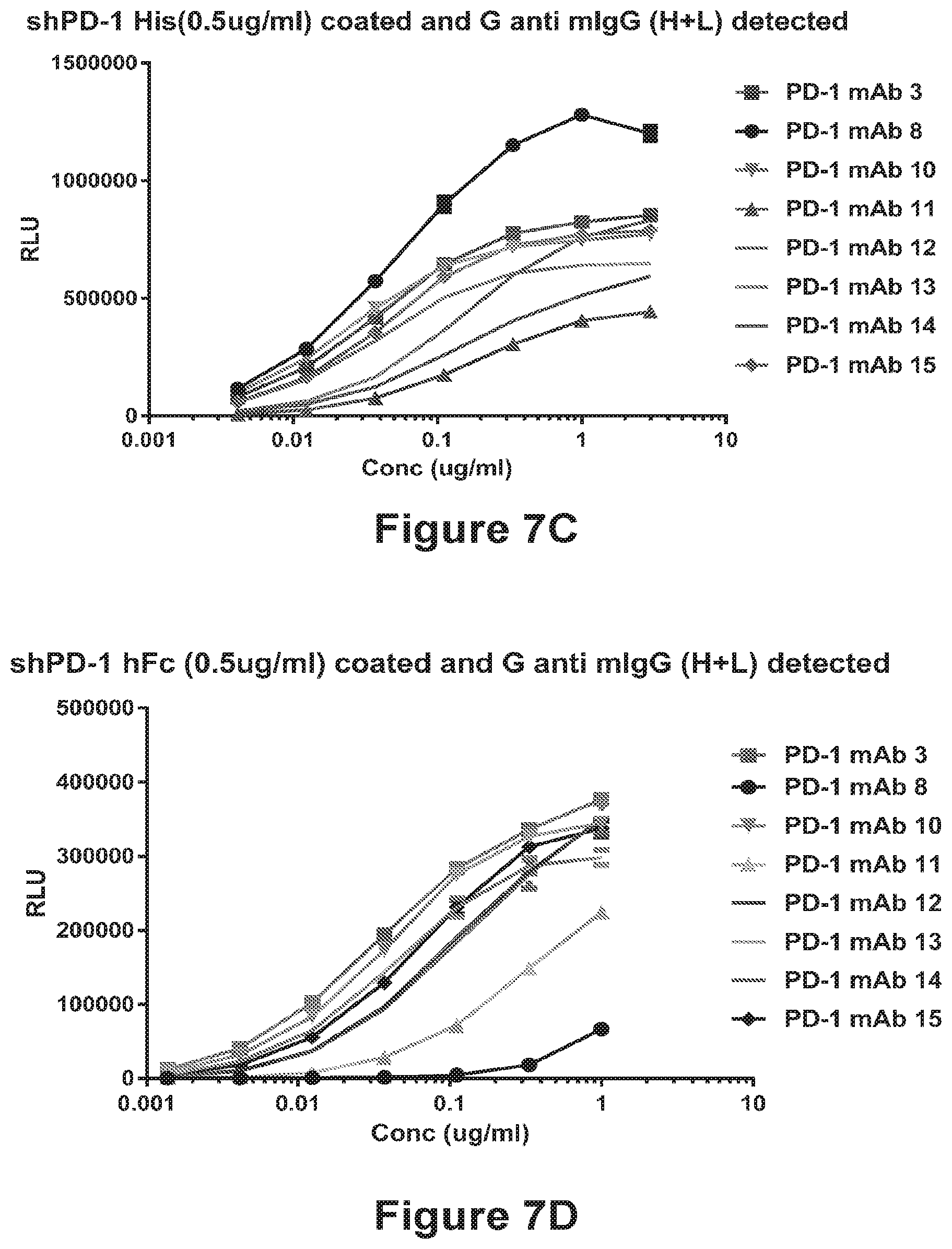
D00015

D00016

D00017
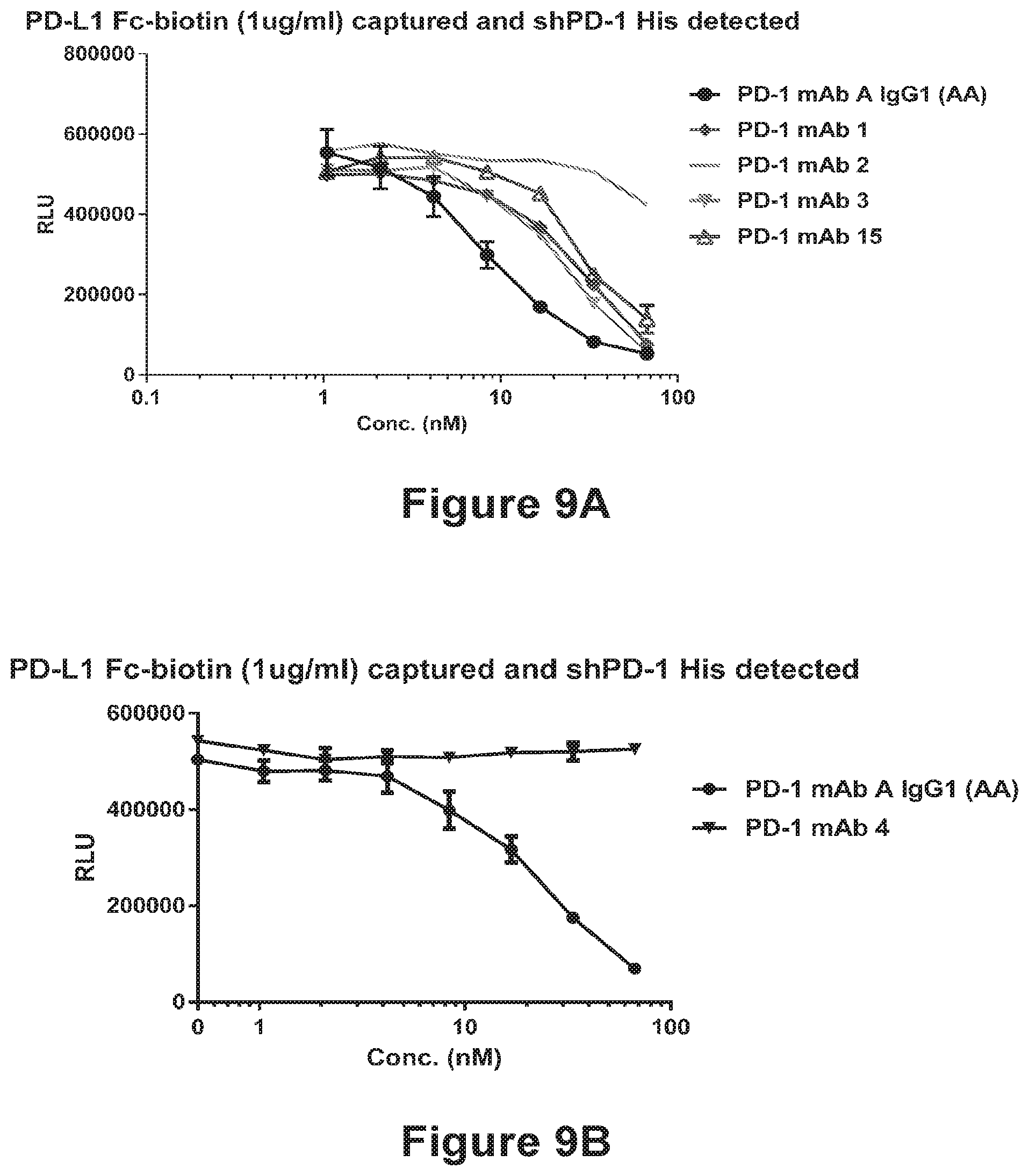
D00018

D00019

D00020

D00021
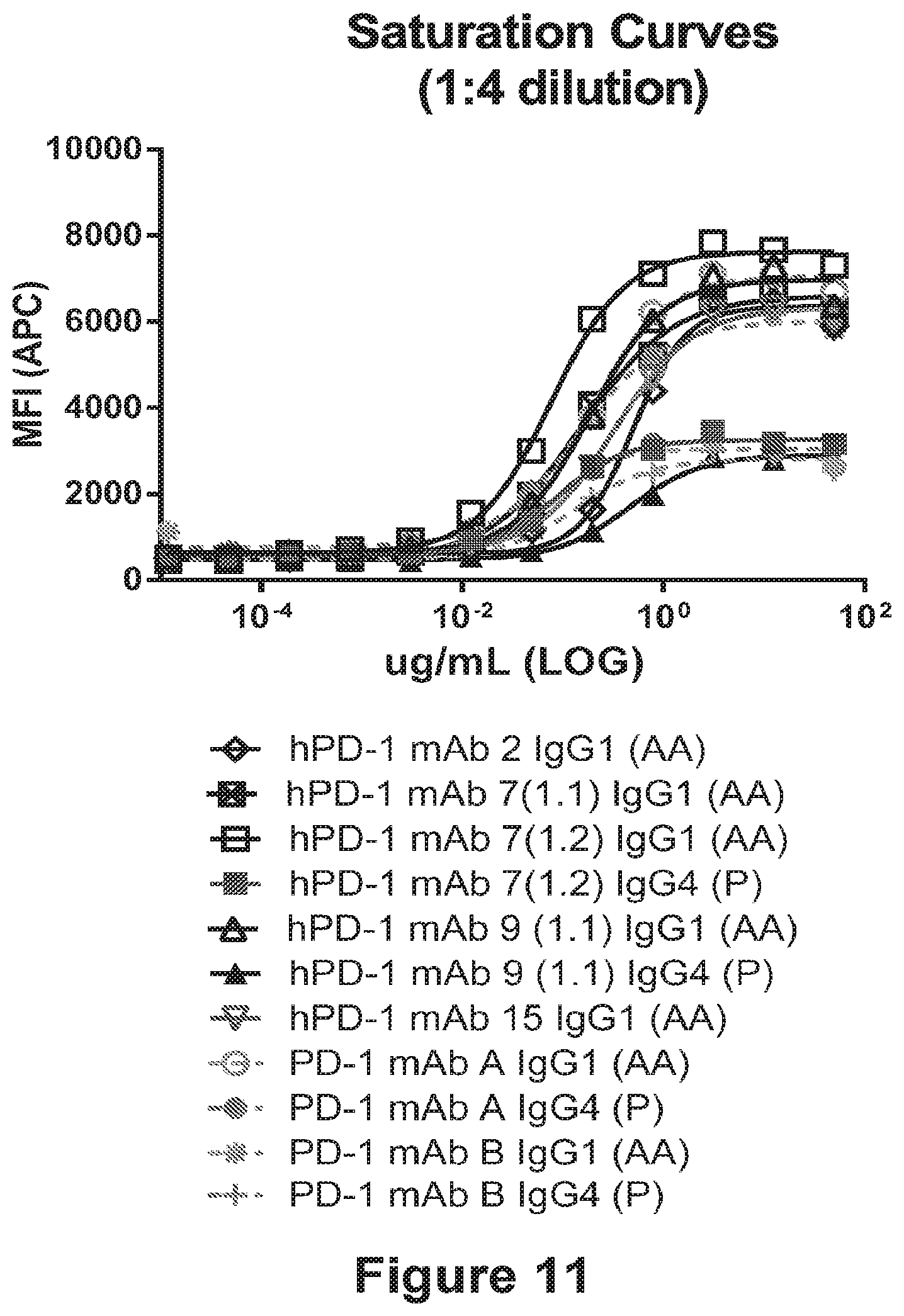
D00022

D00023

D00024

D00025

D00026

D00027

D00028
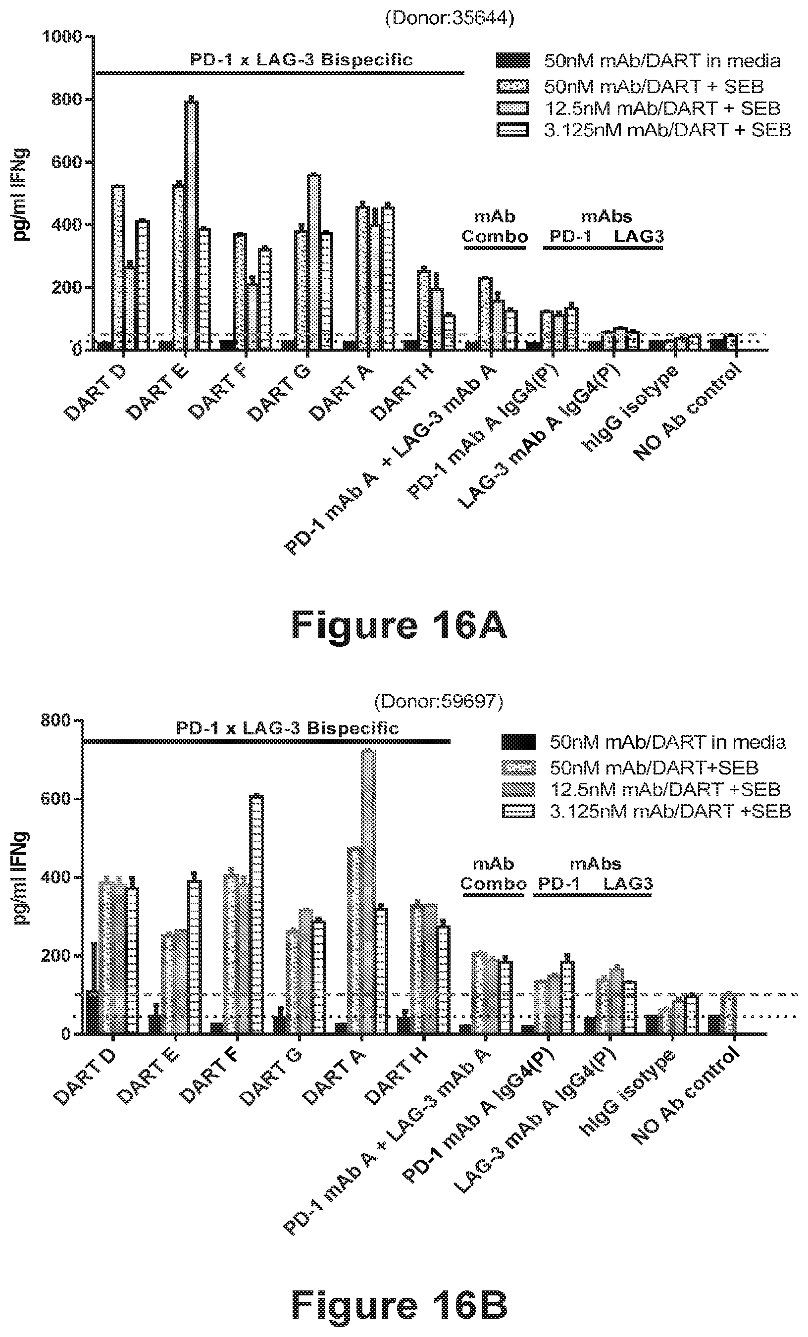
D00029

D00030

D00031

D00032

D00033

D00034

D00035

D00036
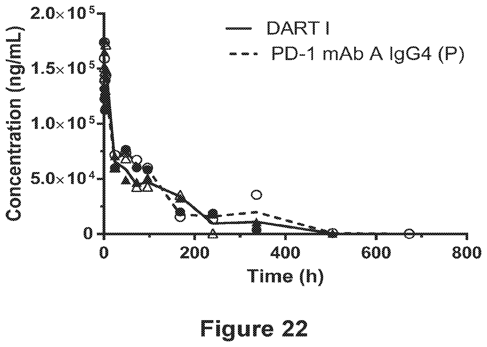
D00037

D00038


S00001
XML
uspto.report is an independent third-party trademark research tool that is not affiliated, endorsed, or sponsored by the United States Patent and Trademark Office (USPTO) or any other governmental organization. The information provided by uspto.report is based on publicly available data at the time of writing and is intended for informational purposes only.
While we strive to provide accurate and up-to-date information, we do not guarantee the accuracy, completeness, reliability, or suitability of the information displayed on this site. The use of this site is at your own risk. Any reliance you place on such information is therefore strictly at your own risk.
All official trademark data, including owner information, should be verified by visiting the official USPTO website at www.uspto.gov. This site is not intended to replace professional legal advice and should not be used as a substitute for consulting with a legal professional who is knowledgeable about trademark law.