NANOBODIES AGAINST CYSTIC FIBROSIS TRANSMEMBRANE CONDUCTANCE REGULATOR (CFTR) INHIBITORY FACTOR (Cif)
HAMMOCK; Bruce D. ; et al.
U.S. patent application number 16/616930 was filed with the patent office on 2020-07-23 for nanobodies against cystic fibrosis transmembrane conductance regulator (cftr) inhibitory factor (cif). The applicant listed for this patent is THE REGENTS OF THE UNIVERSITY OF CALIFORNIA TRUSTEES OF DARMOUTH COLLEGE. Invention is credited to Jiexian DONG, Bruce D. HAMMOCK, Dean R. MADDEN, Christophe MORISSEAU, Natalia VASYLIEVA.
| Application Number | 20200231659 16/616930 |
| Document ID | / |
| Family ID | 64456419 |
| Filed Date | 2020-07-23 |


View All Diagrams
| United States Patent Application | 20200231659 |
| Kind Code | A1 |
| HAMMOCK; Bruce D. ; et al. | July 23, 2020 |
NANOBODIES AGAINST CYSTIC FIBROSIS TRANSMEMBRANE CONDUCTANCE REGULATOR (CFTR) INHIBITORY FACTOR (Cif)
Abstract
Provided are VHH or nanobodies that specifically bind to cystic fibrosis transmembrane conductance regulator (CFTR) inhibitory factor (Cif), and uses thereof for diagnosis and treatment of Pseudomonas infection.
| Inventors: | HAMMOCK; Bruce D.; (Davis, CA) ; VASYLIEVA; Natalia; (Davis, CA) ; DONG; Jiexian; (Davis, CA) ; MORISSEAU; Christophe; (West Sacramento, CA) ; MADDEN; Dean R.; (Hanover, NH) | ||||||||||
| Applicant: |
|
||||||||||
|---|---|---|---|---|---|---|---|---|---|---|---|
| Family ID: | 64456419 | ||||||||||
| Appl. No.: | 16/616930 | ||||||||||
| Filed: | May 29, 2018 | ||||||||||
| PCT Filed: | May 29, 2018 | ||||||||||
| PCT NO: | PCT/US2018/034878 | ||||||||||
| 371 Date: | November 25, 2019 |
Related U.S. Patent Documents
| Application Number | Filing Date | Patent Number | ||
|---|---|---|---|---|
| 62512711 | May 30, 2017 | |||
| Current U.S. Class: | 1/1 |
| Current CPC Class: | C07K 16/40 20130101; C07K 2317/92 20130101; C07K 2317/76 20130101; A61K 31/166 20130101; C07K 2317/22 20130101; C07K 16/1214 20130101; C07K 2317/569 20130101; G01N 33/56911 20130101; G01N 2333/21 20130101; A61K 31/166 20130101; A61K 2300/00 20130101 |
| International Class: | C07K 16/12 20060101 C07K016/12; C07K 16/40 20060101 C07K016/40; G01N 33/569 20060101 G01N033/569 |
Goverment Interests
STATEMENT OF GOVERNMENTAL SUPPORT
[0002] This invention was made with government support under Grant Nos P42ES004699, R01ES002710 and AI091699, awarded by the National Institutes of Health. The government has certain rights in the invention.
Claims
1. An isolated, recombinant, synthetic and/or non-natural VHH molecule that specifically binds to cystic fibrosis transmembrane conductance regulator (CFTR) inhibitory factor (Cif).
2. The VHH molecule of claim 1, wherein the Cif is produced by a Pseudomonas bacterium.
3. The VHH molecule of any one of claims 1 to 2, wherein the Cif is produced by a Pseudomonas aeruginosa bacterium.
4. The VHH molecule of any one of claims 1 to 3, wherein the VHH binds to Cif with a K.sub.D of 0.2 nM or less.
5. The VHH molecule of any one of claims 1 to 4, wherein the VHH reduces or inhibits the enzymatic activity of Cif.
6. The VHH molecule of claim 5, wherein the VHH inhibits the enzymatic activity of Cif with and IC50 concentration of 3.0 .mu.M or less.
7. The VHH molecule of claim 5, wherein the VHH inhibits the enzymatic activity of Cif with and IC90 concentration of 4.0 .mu.M or less.
8. The VHH molecule of any one of claims 1 to 7, wherein the VHH comprises: a) a CDR1 comprising SEQ ID NO:2, a CDR2 comprising SEQ ID NO:3 and a CDR3 comprising SEQ ID NO:4; b) a CDR1 comprising SEQ ID NO:6, a CDR2 comprising SEQ ID NO:7 and a CDR3 comprising SEQ ID NO:8; c) a CDR1 comprising SEQ ID NO:10, a CDR2 comprising SEQ ID NO:11 and a CDR3 comprising SEQ ID NO:12; d) a CDR1 comprising SEQ ID NO:14, a CDR2 comprising SEQ ID NO:15 and a CDR3 comprising SEQ ID NO:16; e) a CDR1 comprising SEQ ID NO:18, a CDR2 comprising SEQ ID NO:19 and a CDR3 comprising SEQ ID NO:20; f) a CDR1 comprising SEQ ID NO:22, a CDR2 comprising SEQ ID NO:23 and a CDR3 comprising SEQ ID NO:24; g) a CDR1 comprising SEQ ID NO:26, a CDR2 comprising SEQ ID NO:27 and a CDR3 comprising SEQ ID NO:28; h) a CDR1 comprising SEQ ID NO:30, a CDR2 comprising SEQ ID NO:31 and a CDR3 comprising SEQ ID NO:32; i) a CDR1 comprising SEQ ID NO:34, a CDR2 comprising SEQ ID NO:35 and a CDR3 comprising SEQ ID NO:36; j) a CDR1 comprising SEQ ID NO:38, a CDR2 comprising SEQ ID NO:39 and a CDR3 comprising SEQ ID NO:40; k) a CDR1 comprising SEQ ID NO:42, a CDR2 comprising SEQ ID NO:43 and a CDR3 comprising SEQ ID NO:44; l) a CDR1 comprising SEQ ID NO:46, a CDR2 comprising SEQ ID NO:47 and a CDR3 comprising SEQ ID NO:48; m) a CDR1 comprising SEQ ID NO:50, a CDR2 comprising SEQ ID NO:51 and a CDR3 comprising SEQ ID NO:52; n) a CDR1 comprising SEQ ID NO:54, a CDR2 comprising SEQ ID NO:55 and a CDR3 comprising SEQ ID NO:56; o) a CDR1 comprising SEQ ID NO:58, a CDR2 comprising SEQ ID NO:59 and a CDR3 comprising SEQ ID NO:60; p) a CDR1 comprising SEQ ID NO:62, a CDR2 comprising SEQ ID NO:63 and a CDR3 comprising SEQ ID NO:64; q) a CDR1 comprising SEQ ID NO:66, a CDR2 comprising SEQ ID NO:67 and a CDR3 comprising SEQ ID NO:68; or r) a CDR1 comprising SEQ ID NO:70, a CDR2 comprising SEQ ID NO:71 and a CDR3 comprising SEQ ID NO:72.
9. The VHH molecule of any one of claims 1 to 8, wherein the VHH comprises an amino acid sequence having at least 80% sequence identity to an amino acid selected from the group consisting of: a) amino acid residues 1-134 of SEQ ID NO:1; b) amino acid residues 1-127 of SEQ ID NO:5; c) amino acid residues 1-132 of SEQ ID NO:9; d) amino acid residues 1-133 of SEQ ID NO:13; e) amino acid residues 1-126 of SEQ ID NO:17; f) amino acid residues 1-127 of SEQ ID NO:21; g) amino acid residues 1-129 of SEQ ID NO:25; h) amino acid residues 1-127 of SEQ ID NO:29; i) amino acid residues 1-127 of SEQ ID NO:33; j) amino acid residues 1-127 of SEQ ID NO:37; k) amino acid residues 1-127 of SEQ ID NO:41; l) amino acid residues 1-127 of SEQ ID NO:45; m) amino acid residues 1-126 of SEQ ID NO:49; n) amino acid residues 1-133 of SEQ ID NO:53; o) amino acid residues 1-127 of SEQ ID NO:57; p) amino acid residues 1-126 of SEQ ID NO:61; q) amino acid residues 1-127 of SEQ ID NO:65; and r) amino acid residues 1-127 of SEQ ID NO:69.
10. A polynucleotide encoding a VHH molecule of any one of claims 1 to 9.
11. A fusion protein comprising a VHH molecule of any one of claims 1 to 9.
12. The fusion protein of claim 11, comprising a detectable tag.
13. The fusion protein of claim 11, comprising a VHH molecule that specifically binds to a complement receptor.
14. The fusion protein of any one of claims 11 to 13, comprising a VHH molecule that specifically binds to the complement receptor CD11b/CD18 (Mac-1).
15. A polynucleotide encoding the fusion protein of any one of claims 11 to 14.
16. A conjugate comprising a VHH molecule of any one of claims 1 to 9 and a small organic compound that is an inhibitor of Cif enzymatic activity.
17. The conjugate of claim 16, wherein the small organic compound that is an inhibitor of Cif enzymatic activity is selected from the group consisting of compounds 1a, 1k, 8c, 8d, 8f, 8h, 8j, 18f and 18NH.sub.2 as provided in Table 7.
18. A conjugate comprising a VHH molecule of any one of claims 1 to 9 and a detectable label.
19. The conjugate of claim 18, wherein the detectable label is a fluorophore, a chemiluminescent moiety, an enzyme or a radioisotope.
20. A nanoparticle or liposome comprising a VHH molecule of any one of claims 1 to 9, a fusion protein of any one of claims 11 to 14, and/or a conjugate of any one of claims 16 to 17.
21. A kit comprising a VHH molecule of any one of claims 1 to 9, a fusion protein of any one of claims 11 to 14, a conjugate of any one of claims 16 to 17 and/or a nanoparticle or liposome of claim 20.
22. A host cell comprising a polynucleotide of any one of claim 10 or 15.
23. A method of making a VHH molecule of any one of claims 1 to 9 or a fusion protein of any one of claims 11 to 14, comprising recombinantly expressing in a host cell a polynucleotide of any one of claim 10 or 15.
24. A method of identifying Pseudomonas infection in a subject in need thereof, comprising: a) identifying a subject exhibiting symptoms consistent with a Pseudomonas infection; b) contacting a biological sample suspected of comprising Cif molecules and obtained from the subject with a VHH molecule of any one of claims 1 to 9, or a conjugate of claim 18 under conditions that allow the VHH molecule to bind to the Cif molecules potentially in the sample; c) identifying the subject as having a Pseudomonas infection upon positive detection of binding of the VHH molecule to the Cif molecules in the sample.
25. The method of claim 24, wherein the Pseudomonas infection is a Pseudomonas aeruginosa infection.
26. The method of any one of claims 24 to 25, wherein the biological sample comprises saliva, sputum, bronchoalveolar lavage fluid (BALF), cheek swab, mucus, blood, sweat, tears, serum, plasma, urine, skin, cerebral spinal fluid (CSF), lymph, Eustachian tube fluid, bone marrow or feces.
27. A method of reducing, inhibiting, mitigating and/or reversing one or more symptoms associated with or caused by a Pseudomonas infection, comprising administering to the subject an effective amount of an agent selected from the group consisting of a VHH molecule of any one of claims 1 to 9, a fusion protein of any one of claims 11 to 14, a conjugate of any one of claims 16 to 17 or a nanoparticle or liposome of claim 20.
28. The method of claim 27, wherein the Pseudomonas infection is a Pseudomonas aeruginosa infection.
29. The method of any one of claims 27 to 28, wherein the agent is administered via a route selected from the group consisting of intrapulmonary, inhalational, intravenous, intramuscular, subcutaneous, intradermal, transcutaneous, topical, intrathecal, intralesional, transmucosal, intra-arterial, intraperitoneal, intraventricular and intracranial.
30. A method of screening for inhibitors of Cif enzymatic or catalytic activity, the method comprising: a) providing a solid support coated with polyclonal antibodies raised against Cif and bound to Cif; b) concurrently exposing the solid support to a VHH molecule of any one of claims 1 to 9 and a candidate inhibitor of Cif; c) identifying inhibitors of Cif that compete with or displace binding of the VHH molecule to the Cif bound to the polyclonal antibodies coated on the solid support.
31. The method of claim 30, wherein the VHH molecule is attached to a detectable label, and detection of the label is used to measure displacement of the VHH molecule by the Cif inhibitor.
32. The method of claim 30, wherein the VHH molecule further comprises a detectable tag, and an antibody against the tag is used to measure displacement of the VHH molecule by the Cif inhibitor.
33. The method of any one of claims 30 to 32, wherein the Cif inhibitor is a small organic compound.
34. The method of any one of claims 30 to 33, wherein the Cif inhibitor inhibits Cif enzymatic or catalytic activity with a greater potency than the VHH molecule.
35. The method of any one of claims 30 to 34, wherein the solid support is a bead, a microwell plate, a chip or a microfluidics device.
36. A method of screening for inhibitors of enzymatic or catalytic activity of an enzyme, the method comprising: a) providing a solid support coated with polyclonal antibodies raised against the enzyme and bound to the enzyme; b) concurrently exposing the solid support to a VHH molecule that binds to and inhibits the enzymatic or catalytic activity of the enzyme and a candidate inhibitor of the enzyme; c) identifying inhibitors of the enzyme that compete with or displace binding of the VHH molecule to the enzyme bound to the polyclonal antibodies coated on the solid support.
37. The method of claim 36, wherein the VHH molecule is attached to a detectable label, and detection of the label is used to measure displacement of the VHH molecule by the enzyme inhibitor.
38. The method of claim 36, wherein the VHH molecule further comprises a detectable tag, and an antibody against the tag is used to measure displacement of the VHH molecule by the enzyme inhibitor.
39. The method of any one of claims 36 to 38, wherein the candidate enzyme inhibitor is a small organic compound.
40. The method of any one of claims 36 to 39, wherein the candidate enzyme inhibitor inhibits enzymatic or catalytic activity with a greater potency than the VHH molecule.
41. The method of any one of claims 36 to 40, wherein the solid support is a bead, a microwell plate, a chip or a microfluidics device.
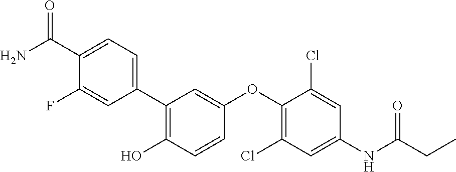
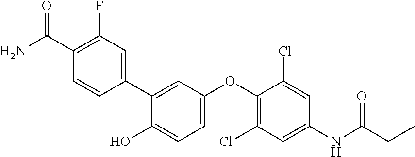
42. An inhibitor of cystic fibrosis transmembrane conductance regulator (CFTR) inhibitory factor (Cif) comprising the structure selected from the group consisting of: TABLE-US-00010 7 (18NH.sub.2) ##STR00014## 5'-(4-amino-2,6-dichlorophenoxy)-2'- hydroxy-[1,1'-biphenyl]-4-carboxamide, and 8 (18f) ##STR00015## 5'-(2,6-dichloro-4- propionamidophenoxy)-3-fluoro-2'- hydroxy-[1,1'-biphenyl]-4-carboxamide
43. A composition comprising one or both Cif inhibitors of claim 42, or a pharmaceutically acceptable salt thereof, in a pharmaceutically acceptable carrier.
44. A method of reducing, inhibiting, mitigating and/or reversing one or more symptoms associated with or caused by a Pseudomonas infection, comprising administering to the subject an effective amount of a composition of claim 43.
45. The method of claim 44, wherein the Pseudomonas infection is a Pseudomonas aeruginosa infection.
46. The method of any one of claims 44 to 45, wherein the agent is administered via a route selected from the group consisting of intrapulmonary, inhalational, intravenous, intramuscular, subcutaneous, intradermal, transcutaneous, topical, intrathecal, intralesional, transmucosal, intra-arterial, intraperitoneal, intraventricular and intracranial.
Description
CROSS-REFERENCE TO RELATED APPLICATIONS
[0001] This application claims the benefit under 35 U.S.C. .sctn. 119(e) of U.S. Provisional Patent Application No. 62/512,711, filed on May 30, 2017, which is hereby incorporated herein by reference in its entirety for all purposes.
SEQUENCE LISTING
[0003] The instant application contains a Sequence Listing which has been submitted electronically in ASCII format and is hereby incorporated by reference in its entirety. Said ASCII copy, created on May 25, 2018, is named UCDVP145WO_SL.txt and is 47,930 bytes in size.
BACKGROUND
[0004] Drug screening is a complex laborious yet critical step in drug identification. With biologics quickly developing and gaining a big part of the drug market, the small molecule drugs remain a major tool for health managing. To screen effectively small synthetic molecules for their potency on desired biomolecular targets, a number of approaches have been developed. The assays, including scintillation proximity assay (1,2), fluorescence resonance energy transfer (FRET) (3) and fluorescence polarization (4) are successfully used for high throughput screening for drug candidates in academia and in industry. In majority, the screening schemes use the high catalytic activity of the target proteins, as for example protein kinases (5). A fast turnover of these enzymes ensures rapid signal development in the assay and therefore its high sensitivity. Consequently, these techniques can be performed in homogeneous mix-and-measure format that allow the addition of all reaction and detection components in one step without need for separation and wash steps. In contrast, the enzymes with low activity, for example family of p450 enzymes, cannot provide sufficient sensitivity in the corresponding assays. Only highly sensitive analytical detection methods can be employed in the screening process of the slow enzymes. The most employed approach relies on a method of fluorescence generation from an appropriate reporter as a result of enzymatic reaction (6). Since the fluorescent signal is accumulating over the time it gives improved sensitivity compared to other screening formats. However, for drug screening purposes higher sensitivities are often required. Therefore, screening assays coupled with LC-MS/MS detection have been established when large number of compounds needs to be analyzed (7). In turn, affinity measuring methods, like Surface Plasmon Resonance (SPR) or Bio-layer interferometry (Octet), are employed for the target validation. Despite the fact, that these methods indeed provide high sensitivity in the assays with the slow enzymes, they suffer from being laborious, time-consuming and involving expensive instrumentation.
[0005] Along with enzymatic activity of the target enzyme, the antibodies are important component of screening assays. A wide variety of conventional and recombinant antibodies are available for these purposes. However, for the past 20 years, the novel variable fragments of heavy chain only antibodies, named VHHs or Nanobodies.RTM., have been extensively studied (8). Due to their small size, unique physical and chemical properties they have found use in a variety of research fields. They were successfully employed in different immunoassay formats (10), including biosensors (9), for the detection of small molecules as well as large proteins (10,11). Nanobodies are often used in molecular and structural biology, for example as chaperons to facilitate protein crystallization. A number of studies focus on studying, evaluation and characterization of nanobodies as biologics to treat a variety of diseases (12,13). Indeed, nanobodies offer advantageous possibilities for drug development: they are easily amenable for genetic and chemical engineering; they possess high selectivity toward their target and thus have low offsite activity compared to small molecule drugs; high solubility of VHHs is another strong advantage compared to small molecule drugs or conventional antibodies etc. (10). In addition to these characteristics, the nanobodies also possess a unique long CDR3 region that forms a long loop often thought to be responsible for analyte recognition (14,15). A small size of VHHs combined with highly selective CDR3 is a unique property allowing development of nanobodies to the cavities, cleft and catalytic sited on the proteins in general, and enzymes, receptors and membrane proteins in particular (14). Such nanobodies have been shown to be effective in selective detection of infection with trypanosomes (16). They also have potential application in tumor imaging and cancer therapy (12,13,17). The nanobodies that selectively bind to the active site of the enzyme and inhibit its catalytic activity are very valuable as drug themselves. However, they can also be used as reagents in screening assay. Nanobodies are unique reagents, and their potential is not yet fully explored. Here we report a novel assay format involving nanobodies, that can be used as a tool for screening of small molecule inhibitors applied to slow enzymes and non-catalytic proteins.
[0006] As a model system, we used cystic fibrosis transmembrane conductance regulator (CFTR) inhibitory factor (Cif) (18) This factor is found in numerous lung pathogen bacteria 19 and particularly characteristic to P. aeruginosa (20). It is an epoxide hydrolase, which through its activity affects the host defenses (21), resulting in an environment that permits the bacterium to form biofilms and to establish a chronic infection of the lung. It was recently identified, that Cif is required for P. aeruginosa to infect effectively human tissues (22). Some work has been done toward synthesis of small molecule inhibitors targeting Cif (23). However, current methods available for monitoring inhibitors' potency are limited in sensitivity (23).
SUMMARY
[0007] In one aspect, provided are recombinant, synthetic and/or non-natural VHH molecules that specifically bind to cystic fibrosis transmembrane conductance regulator (CFTR) inhibitory factor (Cif). In some embodiments, the Cif is produced by a Pseudomonas bacterium. In some embodiments, the Cif is produced by a Pseudomonas aeruginosa bacterium. In some embodiments, the VHH binds to Cif with a KD of 0.2 nM or less, e.g., 0.19 nM, 0.18 nM, 0.17 nM, 0.16 nM, 0.15 nM, 0.14 nM, 0.13 nM, 0.12 nM, 0.11 nM, 0.10 nM, 0.09 nM, 0.08 nM, or less. In some embodiments, the VHH reduces or inhibits the enzymatic activity of Cif. In some embodiments, the VHH inhibits the enzymatic activity of Cif with and IC50 concentration of 3.0 .mu.M or less, e.g., 2.9 .mu.M, 2.8 .mu.M, 2.7 .mu.M, 2.6 .mu.M, 2.5 .mu.M, 2.4 .mu.M, 2.3 .mu.M, 2.2 .mu.M, 2.1 .mu.M, 2.0 .mu.M, 1.9 .mu.M, 1.8 .mu.M, 1.7 .mu.M, 1.6 .mu.M, 1.5 .mu.M, 1.4 .mu.M, 1.3 .mu.M, 1.2 .mu.M, 1.1 .mu.M, 1.0 .mu.M, 0.9 .mu.M, 0.8 .mu.M, 0.7 .mu.M, 0.6 .mu.M, 0.5 .mu.M, or less. In some embodiments, the VHH inhibits the enzymatic activity of Cif with and IC90 concentration of 4.0 .mu.M or less, e.g., 3.9 .mu.M, 3.8 .mu.M, 3.7 .mu.M, 3.6 .mu.M, 3.5 .mu.M, 3.4 .mu.M, 3.3 .mu.M, 3.2 .mu.M, 3.1 .mu.M, 3.0 .mu.M, 2.9 .mu.M, 2.8 .mu.M, 2.7 .mu.M, 2.6 .mu.M, 2.5 .mu.M, or less. In some embodiments, the VHH comprises:
[0008] a) a CDR1 comprising SEQ ID NO:2, a CDR2 comprising SEQ ID NO:3 and a CDR3 comprising SEQ ID NO:4;
[0009] b) a CDR1 comprising SEQ ID NO:6, a CDR2 comprising SEQ ID NO:7 and a CDR3 comprising SEQ ID NO:8;
[0010] c) a CDR1 comprising SEQ ID NO:10, a CDR2 comprising SEQ ID NO:11 and a CDR3 comprising SEQ ID NO:12;
[0011] d) a CDR1 comprising SEQ ID NO:14, a CDR2 comprising SEQ ID NO:15 and a CDR3 comprising SEQ ID NO:16;
[0012] e) a CDR1 comprising SEQ ID NO:18, a CDR2 comprising SEQ ID NO:19 and a CDR3 comprising SEQ ID NO:20;
[0013] f) a CDR1 comprising SEQ ID NO:22, a CDR2 comprising SEQ ID NO:23 and a CDR3 comprising SEQ ID NO:24;
[0014] g) a CDR1 comprising SEQ ID NO:26, a CDR2 comprising SEQ ID NO:27 and a CDR3 comprising SEQ ID NO:28;
[0015] h) a CDR1 comprising SEQ ID NO:30, a CDR2 comprising SEQ ID NO:31 and a CDR3 comprising SEQ ID NO:32;
[0016] i) a CDR1 comprising SEQ ID NO:34, a CDR2 comprising SEQ ID NO:35 and a CDR3 comprising SEQ ID NO:36;
[0017] j) a CDR1 comprising SEQ ID NO:38, a CDR2 comprising SEQ ID NO:39 and a CDR3 comprising SEQ ID NO:40;
[0018] k) a CDR1 comprising SEQ ID NO:42, a CDR2 comprising SEQ ID NO:43 and a CDR3 comprising SEQ ID NO:44;
[0019] l) a CDR1 comprising SEQ ID NO:46, a CDR2 comprising SEQ ID NO:47 and a CDR3 comprising SEQ ID NO:48;
[0020] m) a CDR1 comprising SEQ ID NO:50, a CDR2 comprising SEQ ID NO:51 and a CDR3 comprising SEQ ID NO:52;
[0021] n) a CDR1 comprising SEQ ID NO:54, a CDR2 comprising SEQ ID NO:55 and a CDR3 comprising SEQ ID NO:56;
[0022] o) a CDR1 comprising SEQ ID NO:58, a CDR2 comprising SEQ ID NO:59 and a CDR3 comprising SEQ ID NO:60;
[0023] p) a CDR1 comprising SEQ ID NO:62, a CDR2 comprising SEQ ID NO:63 and a CDR3 comprising SEQ ID NO:64;
[0024] q) a CDR1 comprising SEQ ID NO:66, a CDR2 comprising SEQ ID NO:67 and a CDR3 comprising SEQ ID NO:68; or
[0025] r) a CDR1 comprising SEQ ID NO:70, a CDR2 comprising SEQ ID NO:71 and a CDR3 comprising SEQ ID NO:72. In some embodiments, the VHH comprises an amino acid sequence having at least 80% sequence identity, e.g., at least 85%, 90%, 91%, 92%, 93%, 94%, 95%, 96%, 97%, 98%, 99% or 100% sequence identity, to an amino acid selected from the group consisting of:
[0026] a) amino acid residues 1-134 of SEQ ID NO:1;
[0027] b) amino acid residues 1-127 of SEQ ID NO:5;
[0028] c) amino acid residues 1-132 of SEQ ID NO:9;
[0029] d) amino acid residues 1-133 of SEQ ID NO:13;
[0030] e) amino acid residues 1-126 of SEQ ID NO:17;
[0031] f) amino acid residues 1-127 of SEQ ID NO:21;
[0032] g) amino acid residues 1-129 of SEQ ID NO:25;
[0033] h) amino acid residues 1-127 of SEQ ID NO:29;
[0034] i) amino acid residues 1-127 of SEQ ID NO:33;
[0035] j) amino acid residues 1-127 of SEQ ID NO:37;
[0036] k) amino acid residues 1-127 of SEQ ID NO:41;
[0037] l) amino acid residues 1-127 of SEQ ID NO:45;
[0038] m) amino acid residues 1-126 of SEQ ID NO:49;
[0039] n) amino acid residues 1-133 of SEQ ID NO:53;
[0040] o) amino acid residues 1-127 of SEQ ID NO:57;
[0041] p) amino acid residues 1-126 of SEQ ID NO:61;
[0042] q) amino acid residues 1-127 of SEQ ID NO:65; and
[0043] r) amino acid residues 1-127 of SEQ ID NO:69.
[0044] In a further aspect, provided is a polynucleotide encoding an anti-Cif VHH molecule as described above and herein. Also contemplated are expression cassettes and vectors comprising one or more polynucleotides encoding one or more anti-Cif VHH molecules, as described above and herein.
[0045] In another aspect, provided is a fusion protein comprising an anti-Cif VHH molecule as described above and herein. In some embodiments, the fusion protein comprises one or more detectable tags, e.g, a protein tag or a peptide tag, e.g., hemagglutinin (HA) tag, FLAG tag, c-myc, poly histidine tag. In some embodiments, the fusion protein further comprises a VHH molecule that specifically binds to a complement receptor (e.g., a bispecific VHH fusion that can concurrently binds to Cif and a complement receptor). In some embodiments, the fusion protein further comprises a VHH molecule that specifically binds to the complement receptor CD11b/CD18 (Mac-1) (e.g., a bispecific or dimeric VHH fusion that can concurrently binds to Cif and Mac-1). Also contemplated are polynucleotides encoding the fusion proteins.
[0046] In another aspect, provided is a conjugate comprising an anti-Cif VHH molecule, as described above and herein, and a small organic compound that is an inhibitor of Cif enzymatic activity (e.g., Cif inhibitor compounds 1a, 1k, 8c, 8d, 8f, 8h, 8j, 18f and 18NH2 as provided in Table 7). In a related aspect, provided is a conjugate comprising an anti-Cif VHH molecule, as described above and herein, and a detectable label. In some embodiments, the detectable label is a fluorophore, a chemiluminescent moiety, an enzyme or a radioisotope.
[0047] In a further aspect, provided is a nanoparticle or liposome comprising an anti-Cif VHH molecule, a fusion protein and/or a conjugate, as described above and herein.
[0048] In another aspect, further provided are kits. In some embodiments, the kits comprise a VHH molecule, a fusion protein, a conjugate and/or a nanoparticle or liposome, as described above and herein.
[0049] In another aspect, further provided are host cells comprising one or more polynucleotides encoding the VHH molecules and/or the fusion proteins, as described above and herein. In another aspect, further provided are methods of making a VHH molecule or a fusion protein as described above and herein. In some embodiments, the methods comprise recombinantly expressing in a host cell one or more polynucleotides encoding the VHH molecules and/or the fusion proteins, as described above and herein.
[0050] In another aspect, provided are methods of identifying a Pseudomonas infection in a subject in need thereof. In some embodiments, the methods comprise:
[0051] a) identifying a subject exhibiting symptoms consistent with a Pseudomonas infection;
[0052] b) contacting a biological sample suspected of comprising Cif molecules and obtained from the subject with an anti-Cif VHH molecule, as described above and herein, or a conjugate comprising such an anti-Cif VHH molecule, under conditions that allow the VHH molecule to bind to the Cif molecules potentially in the sample;
[0053] c) identifying the subject as having a Pseudomonas infection upon positive detection of binding of the VHH molecule to the Cif molecules in the sample. In some embodiments, the Pseudomonas infection is a Pseudomonas aeruginosa infection. In some embodiments, the biological sample is from saliva, sputum, bronchoalveolar lavage fluid (BALF), cheek swab, mucus, blood, sweat, tears, serum, plasma, urine, skin, cerebral spinal fluid (CSF), lymph, Eustachian tube fluid, bone marrow and/or feces.
[0054] In another aspect, provided are methods of reducing, inhibiting, mitigating and/or reversing one or more symptoms associated with or caused by a Pseudomonas infection. In some embodiments, the methods comprise administering to the subject an effective amount of an agent selected from the group consisting of an anti-Cif VHH molecule, as described above and herein, or a fusion protein, a conjugate or a nanoparticle or liposome comprising such an anti-Cif VHH molecule, as described above and herein. In varying embodiment, the Pseudomonas infection is a Pseudomonas aeruginosa infection. In some embodiments, the agent is administered via a route selected from the group consisting of intrapulmonary, inhalational, intravenous, intramuscular, subcutaneous, intradermal, transcutaneous, topical (including to the eyes), intrathecal, intralesional, transmucosal (including through tissues around the eyes), intra-arterial, intraperitoneal, intraventricular and intracranial.
[0055] In a further aspect, provided are methods of screening for inhibitors of Cif enzymatic activity. In some embodiments, the method comprises:
[0056] a) providing a solid support coated with polyclonal antibodies raised against Cif and bound to Cif;
[0057] b) concurrently exposing the solid support to an anti-Cif VHH molecule, as described above and herein, and a candidate inhibitor of Cif;
[0058] c) identifying inhibitors of Cif that compete with or displace binding of the VHH molecule to the Cif bound to the polyclonal antibodies coated on the solid support. In some embodiments, the anti-Cif VHH molecule is attached to a detectable label, and detection of the label is used to measure displacement of the VHH molecule by the Cif inhibitor, e.g., used to measure unbound VHH molecule. In some embodiments, the anti-Cif VHH molecule further comprises one or more detectable tags (e.g., hemagglutinin (HA) tag, FLAG tag, c-myc, poly histidine tag), and an antibody against the tag is used to measure displacement of the VHH molecule by the Cif inhibitor. In some embodiments, the Cif inhibitor is a small organic compound. In some embodiments, the screening methods identifies a Cif inhibitor that inhibits Cif enzymatic or catalytic activity with a greater potency than the VHH molecule. In some embodiments, the solid support is a bead, a microwell plate, a chip or a microfluidics device.
[0059] In a further aspect, provided are methods of screening for inhibitors of enzymatic or catalytic activity of an enzyme. In some embodiments, the method comprises:
[0060] a) providing a solid support coated with polyclonal antibodies raised against the enzyme and bound to the enzyme;
[0061] b) concurrently exposing the solid support to a VHH molecule that binds to and inhibits the enzymatic or catalytic activity of the enzyme and a candidate inhibitor of the enzyme;
[0062] c) identifying inhibitors of the enzyme that compete with or displace binding of the VHH molecule to the enzyme bound to the polyclonal antibodies coated on the solid support. In some embodiments, the VHH molecule is attached to a detectable label, and detection of the label is used to measure displacement of the VHH molecule by the enzyme inhibitor. In some embodiments, the VHH molecule further comprises a detectable tag, and an antibody against the tag is used to measure displacement of the VHH molecule by the enzyme inhibitor. In some embodiments, the candidate enzyme inhibitor is a small organic compound. In some embodiments, the screening method identifies a candidate enzyme inhibitor that inhibits enzymatic or catalytic activity with a greater potency than the VHH molecule. In some embodiments, the solid support is a bead, a microwell plate, a chip or a microfluidics device.
[0063] In a further aspect, provided is an inhibitor of cystic fibrosis transmembrane conductance regulator (CFTR) inhibitory factor (Cif) comprising the structure selected from the group consisting of:
TABLE-US-00001 7 (18NH.sub.2) ##STR00001## 5'-(4-amino-2,6-dichlorophenoxy)-2'- hydroxy-[1,1'-biphenyl]-4-carboxamide, and 8 (18f) ##STR00002## 5'-(2,6-dichloro-4- propionamidophenoxy)-3-fluoro-2'- hydroxy-[1,1'-biphenyl]-4-carboxamide
[0064] In another aspect, provided is a pharmaceutical composition comprising one or both Cif inhibitors, e.g., compounds 7 and/or 8 as provided above and herein, or a pharmaceutically acceptable salt thereof, in a pharmaceutically acceptable carrier. In a further aspect, provided is a method of reducing, inhibiting, mitigating and/or reversing one or more symptoms associated with or caused by a Pseudomonas infection, comprising administering to the subject an effective amount of a composition comprising one or both Cif inhibitors, e.g., compounds 7 and/or 8 as provided above and herein. In some embodiments, the Pseudomonas infection is a Pseudomonas aeruginosa infection. IN some embodiments, the agent is administered via a route selected from the group consisting of intrapulmonary, inhalational, intravenous, intramuscular, subcutaneous, intradermal, transcutaneous, topical, intrathecal, intralesional, transmucosal, intra-arterial, intraperitoneal, intraventricular and intracranial.
Definitions
[0065] The terms "single domain antibody," "nanobody" refers to an antibody comprising one variable domain (VH) of a heavy-chain antibody ("VHH") obtained from camelids. Antibody proteins obtained from members of the camel and dromedary (Camelus baclrianus and Camelus dromaderius) family including new world members such as llama species (Lama paccos, Lama glama and Lama vicugna) have been characterized with respect to size, structural complexity and antigenicity for human subjects. Certain IgG antibodies from this family of mammals as found in nature lack light chains, and are thus structurally distinct from the typical four chain quaternary structure having two heavy and two light chains, for antibodies from other animals. See, PCT/EP93/02214 (published as WO 94/04678).
[0066] The term `VHH` refers to the single heavy chain variable domain antibodies devoid of light chains. Generally, a VHH is an antibody of the type that can be found in Camelidae or cartilaginous fish which are naturally devoid of light chains or to a synthetic and non-immunized VHH which can be constructed accordingly. Each heavy chain comprises a variable region encoded by V-, D- and J exons. The VHH may be a natural VHH antibody, preferably a Camelid antibody, or a recombinant protein comprising a heavy chain variable domain.
[0067] Structurally, "cystic fibrosis transmembrane conductance regulator (CFTR) inhibitory factor," "CFTR inhibitory factor," or "Cif" interchangeably refer to nucleic acids and polypeptide polymorphic variants, alleles, mutants, and interspecies homologs that: (1) have an amino acid sequence that has greater than about 80% amino acid sequence identity, for example, 81%, 82%, 83%, 84%, 85%, 86%, 87%, 88%, 89%, 90%, 91%, 92%, 93%, 94%, 95%, 96%, 97%, 98% or 99% or greater amino acid sequence identity, preferably over a region of at least about 25, 50, 100, 200, 300, or more amino acid residues, or over the full-length, to an Cif amino acid sequence, (e.g., NCBI Reference Sequence: NP_251624.1 and GenBank Accession No. CTQ36962.1). (2) bind to antibodies, e.g., polyclonal or monoclonal antibodies, raised against an immunogen comprising an amino acid sequence of a Cif polypeptide; or an amino acid sequence encoded by a Cif nucleic acid, and conservatively modified variants thereof; (3) specifically hybridize under stringent hybridization conditions to an anti-sense strand corresponding to a nucleic acid sequence encoding a Cif protein, and conservatively modified variants thereof; (4) have a nucleic acid sequence that has greater than about 80%, preferably greater than about 81%, 82%, 83%, 84%, 85%, 86%, 87%, 88%, 89%, 90%, 91%, 92%, 93%, 94%, 95%, 96%, 97%, 98%, 99%, or higher nucleotide sequence identity, preferably over a region of at least about 25, 50, 100, 200, 500, or more nucleotides, or over the full-length, to a Cif nucleic acid. Functionally, Cif refers to an epoxide hydrolase virulence factor secreted by Pseudomonas bacteria, particularly Pseudomonas aeruginosa bacteria. Cif can decrease levels of CFTR on the membrane of host airway epithelial cells, resulting in an altered microenvironment and lower host immune defense, facilitating Pseudomonas biofilm formation and chronic infection. See, e.g., Bahl, et al., Cornea. (2017) 36(3):358-362; Bahl, et al., Biochemistry. (2016) 55(5):788-97; and Bahl, et al., Angew Chem Int Ed Engl. (2015) 54(34):9881-5.
[0068] The phrase "sequence identity," in the context of two nucleic acids or polypeptides, refers to two or more sequences or subsequences that have a certain level of nucleotide or amino acid residue identity, when compared and aligned for maximum correspondence, as measured using one of the following sequence comparison algorithms or by visual inspection. Preferably, the aligned sequences share at least 90% sequence identity, for example, at least 95%, 96%, 97%, 98%, 99% or 100% sequence identity. The sequence identity can exist over a region of the sequences that is at least about 10, 20 or 50 residues in length, sometimes over a region of at least about 100 or 150 residues. In some embodiments, the sequences share a certain level of sequence identity over the entire length of the sequence of interest.
[0069] For sequence comparison, typically one sequence acts as a reference sequence (e.g., SEQ ID NOs: 1-72), to which test sequences are compared. When using a sequence comparison algorithm, test and reference sequences are input into a computer, subsequence coordinates are designated, if necessary, and sequence algorithm program parameters are designated. The sequence comparison algorithm then calculates the percent sequence identity for the test sequence(s) relative to the reference sequence, based on the designated program parameters. Optimal alignment of sequences for comparison can be conducted, e.g., by the local homology algorithm of Smith & Waterman, Adv. Appl. Math. 2:482 (1981), by the homology alignment algorithm of Needleman & Wunsch, J. Mol. Biol. 48:443 (1970), by the search for similarity method of Pearson & Lipman, Proc. Nat'l. Acad. Sci. USA 85:2444 (1988), by computerized implementations of these algorithms (GAP, BESTFIT, FASTA, and TFASTA in the Wisconsin Genetics Software Package, Genetics Computer Group, 575 Science Dr., Madison, Wis.), or by visual inspection (see generally Ausubel, et al. Editor, Current Protocols in Molecular Biology, USA, 1984-2017). Another example of algorithm that is suitable for determining percent sequence identity and sequence similarity is the BLAST algorithm, which is described in Altschul et al., J. Mol. Biol. 215:403-410 (1990). Software for performing BLAST analyses is publicly available through the National Center for Biotechnology Information (on the World Wide Web at ncbi.nhn nih.gov/) (see Henikoff & Henikoff, Proc. Natl. Acad. Sci. USA 89:10915 (1989)).
[0070] With respect to the numbering of positions in a given amino acid polymer or nucleic acid polymer, the terms "corresponding to," "corresponds to," is in "reference to," or is "relative to" the numbering of a selected amino acid polymer or nucleic acid polymer refers to the position of any given polymer component (e.g., amino acid, nucleotide, also referred to generically as a "residue") as designated by reference to the same or to an equivalent position in the selected amino acid or nucleic acid polymer, rather than by the actual numerical position of the component in the given polymer. Thus, for example, the numbering of a given amino acid position in a given polypeptide sequence corresponds to the same or equivalent amino acid position in a selected polypeptide sequence used as a reference sequence.
[0071] An "equivalent position" (for example, an "equivalent amino acid position" or "equivalent nucleic acid position" or "equivalent residue position") is defined herein as a position (such as, an amino acid position or nucleic acid position or residue position) of a test polypeptide (or test polynucleotide) sequence which aligns with a corresponding position of a reference polypeptide (or reference polynucleotide) sequence, when optimally aligned using an alignment algorithm as described herein. The equivalent amino acid position of the test polypeptide need not have the same numerical position number as the corresponding position of the reference polypeptide; likewise, the equivalent nucleic acid position of the test polynucleotide need not have the same numerical position number as the corresponding position of the reference polynucleotide.
[0072] Two polypeptide sequences are "optimally aligned" or in "optimal alignment" when they are aligned using defined parameters, i.e., a defined amino acid substitution matrix, gap existence penalty (also termed gap open penalty), and gap extension penalty, so as to arrive at the highest similarity score possible for that pair of sequences. The BLOSUM62 matrix (Henikoff and Henikoff (1992) Proc. Natl. Acad. Sci. USA 89(22):10915-10919) is often used as a default scoring substitution matrix in polypeptide sequence alignment algorithms (such as BLASTP). The gap existence penalty is imposed for the introduction of a single amino acid gap in one of the aligned sequences, and the gap extension penalty is imposed for each residue position in the gap. Unless otherwise stated, alignment parameters employed herein are: BLOSUM62 scoring matrix, gap existence penalty=11, and gap extension penalty=1. The alignment score is defined by the amino acid positions of each sequence at which the alignment begins and ends (e.g. the alignment window), and optionally by the insertion of a gap or multiple gaps into one or both sequences, so as to arrive at the highest possible similarity score.
[0073] With respect to the determination of an amino acid position by optimal alignment with a reference sequence, the amino acid position in a test amino acid sequence corresponds to the position in the reference sequence with which the residue is paired in the alignment. The "position" is denoted by a number that sequentially identifies each amino acid in the reference sequence based on its position relative to the N-terminus. Owing to deletions, insertions, truncations, fusions, and the like that must be taken into account when determining an optimal alignment, in general the amino acid residue number in a test sequence is determined by simply counting from the N-terminal will not necessarily be the same as the number of its corresponding position in the reference sequence. For example, in a case where there is a deletion in an aligned test sequence, there will be no amino acid that corresponds to a position in the reference sequence at the site of deletion. Where there is an insertion in an aligned reference sequence, that insertion will not correspond to any amino acid position in the reference sequence. In the case of truncations or fusions there can be stretches of amino acids in either the reference or aligned sequence that do not correspond to any amino acid in the corresponding sequence.
[0074] Amino acids can be referred to herein by either their commonly known three letter symbols or by the one-letter symbols recommended by the IUPAC-IUB Biochemical Nomenclature Commission. Nucleotides, likewise, can be referred to by their commonly accepted single-letter codes.
[0075] "Conservatively modified variants" as used herein applies to amino acid sequences. One of skill will recognize that individual substitutions, deletions or additions to a nucleic acid, peptide, polypeptide, or protein sequence which alters, adds or deletes a single amino acid or a small percentage of amino acids in the encoded sequence is a "conservatively modified variant" where the alteration results in the substitution of an amino acid with a chemically similar amino acid. Conservative substitution tables providing functionally similar amino acids are well known in the art. Such conservatively modified variants are in addition to and do not exclude polymorphic variants, interspecies homologs, and alleles.
[0076] The following eight groups each contain amino acids that are conservative substitutions for one another:
[0077] 1) Alanine (A), Glycine (G);
[0078] 2) Aspartic acid (D), Glutamic acid (E);
[0079] 3) Asparagine (N), Glutamine (Q);
[0080] 4) Arginine (R), Lysine (K);
[0081] 5) Isoleucine (I), Leucine (L), Methionine (M), Valine (V);
[0082] 6) Phenylalanine (F), Tyrosine (Y), Tryptophan (W);
[0083] 7) Serine (S), Threonine (T); and
[0084] 8) Cysteine (C), Methionine (M) (see, e.g., Creighton, Proteins (1984)).
[0085] An "expression cassette" is a nucleic acid construct, generated recombinantly or synthetically, with a series of specified nucleic acid elements that permit transcription of a particular polynucleotide sequence in a host cell. An expression cassette may be part of a plasmid, viral genome, or nucleic acid fragment. Typically, an expression cassette includes a polynucleotide to be transcribed, operably linked to a promoter.
[0086] The term "polynucleotide" or "nucleic acid," as used interchangeably herein, refers to polymers of nucleotides of any length, and include DNA and RNA. The nucleotides can be deoxyribonucleotides, ribonucleotides, modified nucleotides or bases, and/or their analogs, or any substrate that can be incorporated into a polymer by DNA or RNA polymerase. A polynucleotide may comprise modified nucleotides, such as methylated nucleotides and their analogs.
[0087] As used herein, "operably linked" as used herein refers to a functional relationship between two or more nucleic acid (e.g., DNA) segments. Typically, it refers to the functional relationship of transcriptional regulatory sequence to a transcribed sequence. For example, a promoter is operably linked to a coding sequence, such as a heterologous polynucleotide encoding a polypeptide of interest (e.g., a therapeutic polypeptide, e.g., an antibody or antibody fragment), if it stimulates or modulates the transcription of the coding sequence in an appropriate host cell or other expression system. Generally, promoter transcriptional regulatory sequences that are operably linked to a transcribed sequence are physically contiguous to the transcribed sequence, i.e., they are cis-acting. However, some transcriptional regulatory sequences, such as enhancers, need not be physically contiguous or located in close proximity to the coding sequences whose transcription they enhance.
[0088] As used herein, "vector" means a construct, which is capable of delivering, and preferably expressing, one or more gene(s) or sequence(s) of interest (e.g., a therapeutic polypeptide) in a host cell. Examples of vectors include, but are not limited to, viral vectors, naked DNA or RNA expression vectors, plasmid, cosmid or phage vectors, DNA or RNA expression vectors associated with cationic condensing agents, DNA or RNA expression vectors encapsulated in liposomes, and certain eukaryotic cells, such as producer cells. Suitable vectors are those which are compatible with the host cell employed. Suitable vectors can be derived, for example, from a bacterium, a virus (such as bacteriophage T7 or a M-13 derived phage), a cosmid, a yeast, or a plant. Protocols for obtaining and using such vectors are known to those in the art (see, for example, Green and Sambrook, Molecular Cloning: A Laboratory Manual, 4th ed., Cold Spring Harbor, 2012).
[0089] As used herein, "administering" refers to local and systemic administration, e.g., including enteral, parenteral, pulmonary, and topical/transdermal administration. Routes of administration for the anti-Cif VHH molecules, fusion proteins and/or functional variants thereof described herein include, e.g., oral (per os (P.O.)) administration, nasal or inhalation administration, administration as a suppository, topical contact (including to and around the eyes), transdermal delivery (e.g., via a transdermal patch), intrathecal (IT) administration, intravenous ("iv") administration, intraperitoneal ("ip") administration, intramuscular ("im") administration, intralesional administration, or subcutaneous ("sc") administration, or the implantation of a slow-release device e.g., a mini-osmotic pump, a depot formulation, etc., to a subject. Administration can be by any route including parenteral and transmucosal (e.g., oral, nasal, vaginal, rectal, or transdermal). Parenteral administration includes, e.g., intrapulmonary, intravenous, intramuscular, intra-arterial, intradermal, subcutaneous, intraperitoneal, intraventricular, ionophoretic and intracranial. Other modes of delivery include, but are not limited to, the use of liposomal formulations, intravenous infusion, transdermal patches, etc.
[0090] The terms "systemic administration" and "systemically administered" refer to a method of administering the anti-Cif VHH molecules, fusion proteins and/or functional variants thereof to a mammal so that the compound or composition is delivered to sites in the body, including the targeted site of pharmaceutical action, via the circulatory system. Systemic administration includes, but is not limited to, oral, intrapulmonary, intranasal, rectal and parenteral (e.g., other than through the alimentary tract, such as, inhalational, intravenous, intramuscular, intra-arterial, intraperitoneal, intraventricular, transdermal and subcutaneous) administration.
[0091] The term "co-administering" or "concurrent administration", when used, for example with respect to the anti-Cif VHH molecules, fusion proteins and/or functional variants thereof, refers to administration of the compound and/or analogs and the active agent such that both can simultaneously achieve a physiological effect. The two agents, however, need not be administered together. In certain embodiments, administration of one agent can precede administration of the other. Simultaneous physiological effect need not necessarily require presence of both agents in the circulation at the same time. However, in certain embodiments, co-administering typically results in both agents being simultaneously present in the body (e.g., in the plasma) at a significant fraction (e.g., 20% or greater, preferably 30% or 40% or greater, more preferably 50% or 60% or greater, most preferably 70% or 80% or 90% or greater) of their maximum serum concentration for any given dose.
[0092] The term "effective amount" or "pharmaceutically effective amount" refer to the amount and/or dosage, and/or dosage regime of the anti-Cif VHH molecules, fusion proteins and/or functional variants thereof necessary to bring about the desired result e.g., an amount sufficient to mitigating in a mammal one or more symptoms associated a Pseudomonas infection, or an amount sufficient to lessen the severity or delay the progression of a Pseudomonas infection, an amount sufficient to reduce the risk or delaying the onset, and/or reduce the ultimate severity of a Pseudomonas infection (e.g., prophylactically effective amounts).
[0093] The phrase "cause to be administered" refers to the actions taken by a medical professional (e.g., a physician), or a person controlling medical care of a subject, that control and/or permit the administration of the anti-Cif VHH molecules, fusion proteins and/or functional variants thereof to the subject. Causing to be administered can involve diagnosis and/or determination of an appropriate therapeutic or prophylactic regimen, and/or prescribing particular anti-Cif VHH molecules, fusion proteins and/or functional variants thereof for a subject. Such prescribing can include, for example, drafting a prescription form, annotating a medical record, and the like.
[0094] As used herein, the terms "treating" and "treatment" refer to delaying the onset of, retarding or reversing the progress of, reducing the severity of, or alleviating or preventing either the disease or condition to which the term applies, or one or more symptoms of such disease or condition (e.g., a Pseudomonas infection).
[0095] The term "mitigating" refers to reduction or elimination of one or more symptoms of that pathology or disease, and/or a reduction in the rate or delay of onset or severity of one or more symptoms of that pathology or disease, and/or the prevention of that pathology or disease (e.g., a Pseudomonas infection).
[0096] As used herein, the phrase "consisting essentially of" refers to the genera or species of active pharmaceutical agents recited in a method or composition, and further can include other agents that, on their own do not substantial activity for the recited indication or purpose.
[0097] The terms "subject," "individual," and "patient" interchangeably refer to a mammal, preferably a human or a non-human primate, but also domesticated mammals (e.g., canine or feline), laboratory mammals (e.g., mouse, rat, rabbit, hamster, guinea pig) and agricultural mammals (e.g., equine, bovine, porcine, ovine). In various embodiments, the subject can be a human (e.g., adult male, adult female, adolescent male, adolescent female, male child, female child) under the care of a physician or other healthworker in a hospital, psychiatric care facility, as an outpatient, or other clinical context. In certain embodiments the subject may not be under the care or prescription of a physician or other healthworker.
BRIEF DESCRIPTION OF THE DRAWINGS
[0098] FIG. 1 illustrates a schematic of the fluorescent assay described herein. Cif hydrolyzes a reporter releasing a fluorescent probe.
[0099] FIGS. 2A-B illustrate amino acid sequences of 18 Cif-selective VHHs (SEQ ID NOS 1, 5, 9, 13, 17, 21, 25, 29, 33, 37, 41, 45, 49, 53, 57, 61, 65, and 69, respectively, in order of appearance). FR indicates framework, or conserved domain of the antibody; CDR is the complementarity-determining region, a variable domain of the antibody responsible for selective recognition of the target analyte; TAG includes 6.times.Histag (SEQ ID NO: 90) and hemagglutinin (HA) tag.
[0100] FIG. 3 illustrates ELISA curves for 18 positive clones. Conditions: pAb anti-Cif 0.3 .mu.g/well; serial dilution of Cif in PBS; VHH 50 ng/well in PBS; anti-HA mAb dilution 1:3000 in PBST.
[0101] FIGS. 4A-B illustrate surface plasmon resonance (SPR) curves. A. Clone 113, concentrations tested: 0.05, 0.5, 2, 8, 20 nM; B. Clone 219, concentrations tested 0.2, 1, 3, 10, 20 nM. Binding assays were performed at a flow rate of 30 .mu.L/min with 300 s injection time of VHHs followed by washing with buffer for 1200 s. Sodium phosphate buffer (20 mM pH 7.0) containing 50 mM NaCl and 0.05% (v/v) Tween 20 was used as a running buffer in the assay.
[0102] FIG. 5 illustrates amino acid sequences for inhibitory nanobodies 113 (SEQ ID NO: 13) and 219 (SEQ ID NO: 41). FR indicates framework, or conserved domain of the nanobody; CDR stands for complementarity-determining region, a variable domain of the nanobody responsible for selective recognition of the target analyte; TAG includes 6.times.His tag (SEQ ID NO: 90) and hemagglutinin (HA) tag.
[0103] FIGS. 6A-B illustrate a schematic of displacement sandwich ELISA (dsELISA). A. Workflow scheme of the assay: the plate is coated with anti-Cif pAb and Cif, followed by incubation with the small molecule inhibitor and the nanobody. Unbound material is washed out and the bound nanobody is reveled with secondary anti-HA antibody labeled with HRP. Conditions: anti-Cif pAb 0.3 .mu.g/well; Cif 20 ng/mL in PBS; VHH 5 ng/well in PBS; anti-HA mAb dilution 1:3000 in PBST. B. Inhibition curves for the tested compounds reveal high competitive capacity of the compound 8, followed by 7 and 6.Blue arrows indicate the order of increasing potency, where compound 8 is the most potent and compound 1 is the less potent. Compounds with higher potency have inhibitory curves shifted to the left of the graph.
[0104] FIG. 7 illustrates day-to-day variation of the signal normalized to the signal of the compound 8c (data include measurements some performed over period of 5 months and some within a week), mean.+-.SD, n=3-4.
DETAILED DESCRIPTION
1. Introduction
[0105] Drug identification and development is a research direction that always requires faster, cheaper, more selective and sensitive tools to keep advancing and improving health care. High-throughput screening methods for identification of selective small molecule inhibitors of biologically active proteins often rely on their efficient catalytic properties. However, there is lack of screening tools combining low cost and high sensitivity for drugs targeting low activity proteins. Here we report a screening and detection method involving nanobodies allowing sensitive screening of small molecule compounds targeting active site of the cystic fibrosis transmembrane conductance regulator (CFTR) inhibitory factor (Cif), that is associated with the establishment of chronic inflammation in the lung following colonization with Pseudomonas Aeruginosa.
[0106] A nanobody inhibiting the catalytic activity of Cif was selected and used in a displacement sandwich ELISA (dsELISA), where the inhibitory nanobody also interferes with the binding of a small-molecule inhibitor to the active site of Cif. The dsELISA assay correlates strongly with a conventional fluorogenic assay in predicting the inhibitory potency of the tested compounds. However, the novel dsELISA is an order of magnitude more sensitive and allows the identification and ranking of potent inhibitors missed by the classic assay method. These data were supported with surface plasmon resonance and Octet biolayer measurement techniques. The novel method described herein relies solely on the binding properties of the specific neutralizing nanobody, and thus is applicable to any pharmacological target for which such a nanobody can be found, independent of any requirement for catalytic activity.
[0107] Using seven compounds known inhibitors of Cif we showed a strong agreement between conventional fluorescent method and csELISA in predicting inhibitory potency of the tested compounds. The csELISA method allowed identification of two new compounds, one with 10-fold higher inhibitory potency. These data were confirmed with surface plasmon resonance and Octet biolayer measurement techniques. In addition to the catalytic proteins like enzymes, the competitive sandwich immunoassay can be applied to a larger class of proteins, including receptors, transporters, other non-catalytic proteins since the screening method does not rely on other properties except the chemistry of the binding site.
2. Compositions
[0108] Provided are VHH or nanobodies that specifically bind to cystic fibrosis transmembrane conductance regulator (CFTR) inhibitory factor (Cif), a protein that is produced by Pseudomonas bacteria, particularly Pseudomonas aeruginosa, and which contributes to Pseudomonas infection and pathogenicity in a host. Further provided are fusion proteins, conjugates, nanoparticles and liposomes comprising the anti-Cif VHH and polynucleotides encoding the anti-Cif VHH and fusion proteins thereof.
[0109] Nanobodies are single domain antibodies (sdAb) typically consisting of a single monomeric variable antibody domain. Like whole antibodies (intact immunoglobulins), nanobodies are able to bind selectively to a specific antigen. With a molecular weight typically ranging from about 12 kDa to about 15 kDa, the single-domain nanobodies are much smaller than intact immunoglobulins which are typically composed of two heavy protein chains and two light chains. Nanobodies are also typically smaller than Fab fragments (.about.50 kDa, one light chain and half a heavy chain) and single-chain variable fragments (.about.25 kDa, two variable domains, one from a light and one from a heavy chain).
[0110] Methods of producing nanobodies are described, inter alia, by Harmsen and Haard (2007) Appl. Microbiol. Biotechnol. 77 (1): 13-22).
[0111] Initially, nanobodies were engineered from heavy-chain antibodies found in camelids. These are called VHH fragments. Cartilaginous fishes also have heavy-chain antibodies (immunoglobulin new antigen receptor (IgNAR)'), from which single-domain antibodies called V.sub.NAR fragments can be obtained. An alternative approach is to split the dimeric variable domains from a common human or other mammal (e.g., mice, rabbits, etc.) into monomers. Although most research into single-domain antibodies is based on heavy chain variable domains, nanobodies derived from light chains have also been shown to bind specifically to target epitopes (see, e.g., Moller et al. (2010) J. Biol. Chem. 285(49): 38348-38361). Single-domain camelids antibodies have been shown to be just as specific as a regular antibody and in some cases they are more robust. As well, they are easily isolated using the same phage panning procedure used for traditional antibodies, allowing them to cultured in vitro in large concentrations. The smaller size and single domain make these antibodies easier to transform into bacterial cells for bulk production, making them particularly useful for research purposes.
[0112] Typically the single-domain antibody is a peptide chain about 110 amino acids long, comprising one variable domain (VH) of a heavy-chain antibody, or of a common IgG. These peptides have similar affinity to antigens as whole antibodies, but are more heat-resistant and stable towards detergents and high concentrations of urea. Those derived from camelid and fish antibodies are less lipophilic and more soluble in water, which, without being bound to a particular theory, is believed to be due to their complementarity determining region 3 (CDR3), which forms an extended loop covering the lipophilic site that normally binds to a light chain (see, e.g., Dolk et al. (2005) Appl. Environ. Microbiol. 71(1): 442-450; Stanfield et al. (2004) Science, 305(5691): 1770-1773).
[0113] The comparatively low molecular mass of nanobodies often leads to better permeability in tissues, and to a short plasma half-life since they are eliminated renally. Unlike whole antibodies, they do not show complement system triggered cytotoxicity because they lack an Fc region. However, in certain embodiments, it is contemplated that an immunoglobulin Fc region (or variant Fc region) can be fused to the nanobody to provide additional functionality. Camelid and fish derived sdAbs are able to bind to hidden antigens that are may not be accessible to whole antibodies, for example to the active sites of enzymes. It is believed that this property has been shown to result from their extended CDR3 loop, which is able to penetrate such sites (see, e.g., Stanfield et al. (2004) Science 305(5691): 1770-1773; Desmyter et al. (1996) Nat. Struct. Biol. 3(9): 803-811).
[0114] a. Anti-Cif VHH or Anti-Cif Nanobodies
[0115] Provided are recombinant, synthetic and/or non-natural VHH or nanobody molecules that specifically bind to cystic fibrosis transmembrane conductance regulator (CFTR) inhibitory factor (Cif). In some embodiments, the Cif is produced by a Pseudomonas bacterium. In some embodiments, the Cif is produced by a Pseudomonas aeruginosa bacterium.
[0116] Functionally, in some embodiments, the anti-Cif VHH or nanobody binds to Cif with a KD of 0.2 nM or less, e.g., 0.19 nM, 0.18 nM, 0.17 nM, 0.16 nM, 0.15 nM, 0.14 nM, 0.13 nM, 0.12 nM, 0.11 nM, 0.10 nM, 0.09 nM, 0.08 nM, or less. In some embodiments, the VHH reduces or inhibits the enzymatic activity of Cif. In some embodiments, the VHH inhibits the enzymatic activity of Cif with and IC50 concentration of 3.0 .mu.M or less, e.g., 2.9 .mu.M, 2.8 .mu.M, 2.7 .mu.M, 2.6 .mu.M, 2.5 .mu.M, 2.4 .mu.M, 2.3 .mu.M, 2.2 .mu.M, 2.1 .mu.M, 2.0 .mu.M, 1.9 .mu.M, 1.8 .mu.M, 1.7 .mu.M, 1.6 .mu.M, 1.5 .mu.M, 1.4 .mu.M, 1.3 .mu.M, 1.2 .mu.M, 1.1 .mu.M, 1.0 .mu.M, 0.9 .mu.M, 0.8 .mu.M, 0.7 .mu.M, 0.6 .mu.M, 0.5 .mu.M, or less. In some embodiments, the VHH inhibits the enzymatic activity of Cif with and IC90 concentration of 4.0 .mu.M or less, e.g., 3.9 .mu.M, 3.8 .mu.M, 3.7 .mu.M, 3.6 .mu.M, 3.5 .mu.M, 3.4 .mu.M, 3.3 .mu.M, 3.2 .mu.M, 3.1 .mu.M, 3.0 .mu.M, 2.9 .mu.M, 2.8 .mu.M, 2.7 .mu.M, 2.6 .mu.M, 2.5 .mu.M, or less.
[0117] Structurally, in some embodiments, the VHH comprises:
[0118] a) a CDR1 comprising SEQ ID NO:2, a CDR2 comprising SEQ ID NO:3 and a CDR3 comprising SEQ ID NO:4;
[0119] b) a CDR1 comprising SEQ ID NO:6, a CDR2 comprising SEQ ID NO:7 and a CDR3 comprising SEQ ID NO:8;
[0120] c) a CDR1 comprising SEQ ID NO:10, a CDR2 comprising SEQ ID NO:11 and a CDR3 comprising SEQ ID NO:12;
[0121] d) a CDR1 comprising SEQ ID NO:14, a CDR2 comprising SEQ ID NO:15 and a CDR3 comprising SEQ ID NO:16;
[0122] e) a CDR1 comprising SEQ ID NO:18, a CDR2 comprising SEQ ID NO:19 and a CDR3 comprising SEQ ID NO:20;
[0123] f) a CDR1 comprising SEQ ID NO:22, a CDR2 comprising SEQ ID NO:23 and a CDR3 comprising SEQ ID NO:24;
[0124] g) a CDR1 comprising SEQ ID NO:26, a CDR2 comprising SEQ ID NO:27 and a CDR3 comprising SEQ ID NO:28;
[0125] h) a CDR1 comprising SEQ ID NO:30, a CDR2 comprising SEQ ID NO:31 and a CDR3 comprising SEQ ID NO:32;
[0126] i) a CDR1 comprising SEQ ID NO:34, a CDR2 comprising SEQ ID NO:35 and a CDR3 comprising SEQ ID NO:36;
[0127] j) a CDR1 comprising SEQ ID NO:38, a CDR2 comprising SEQ ID NO:39 and a CDR3 comprising SEQ ID NO:40;
[0128] k) a CDR1 comprising SEQ ID NO:42, a CDR2 comprising SEQ ID NO:43 and a CDR3 comprising SEQ ID NO:44;
[0129] l) a CDR1 comprising SEQ ID NO:46, a CDR2 comprising SEQ ID NO:47 and a CDR3 comprising SEQ ID NO:48;
[0130] m) a CDR1 comprising SEQ ID NO:50, a CDR2 comprising SEQ ID NO:51 and a CDR3 comprising SEQ ID NO:52;
[0131] n) a CDR1 comprising SEQ ID NO:54, a CDR2 comprising SEQ ID NO:55 and a CDR3 comprising SEQ ID NO:56;
[0132] o) a CDR1 comprising SEQ ID NO:58, a CDR2 comprising SEQ ID NO:59 and a CDR3 comprising SEQ ID NO:60;
[0133] p) a CDR1 comprising SEQ ID NO:62, a CDR2 comprising SEQ ID NO:63 and a CDR3 comprising SEQ ID NO:64;
[0134] q) a CDR1 comprising SEQ ID NO:66, a CDR2 comprising SEQ ID NO:67 and a CDR3 comprising SEQ ID NO:68; or
[0135] r) a CDR1 comprising SEQ ID NO:70, a CDR2 comprising SEQ ID NO:71 and a CDR3 comprising SEQ ID NO:72.
[0136] Structurally, in some embodiments, the VHH comprises an amino acid sequence having at least 80% sequence identity, e.g., at least 85%, 90%, 91%, 92%, 93%, 94%, 95%, 96%, 97%, 98%, 99% or 100% sequence identity, to an amino acid selected from the group consisting of:
[0137] a) amino acid residues 1-134 of SEQ ID NO:1;
[0138] b) amino acid residues 1-127 of SEQ ID NO:5;
[0139] c) amino acid residues 1-132 of SEQ ID NO:9;
[0140] d) amino acid residues 1-133 of SEQ ID NO:13;
[0141] e) amino acid residues 1-126 of SEQ ID NO:17;
[0142] f) amino acid residues 1-127 of SEQ ID NO:21;
[0143] g) amino acid residues 1-129 of SEQ ID NO:25;
[0144] h) amino acid residues 1-127 of SEQ ID NO:29;
[0145] i) amino acid residues 1-127 of SEQ ID NO:33;
[0146] j) amino acid residues 1-127 of SEQ ID NO:37;
[0147] k) amino acid residues 1-127 of SEQ ID NO:41;
[0148] l) amino acid residues 1-127 of SEQ ID NO:45;
[0149] m) amino acid residues 1-126 of SEQ ID NO:49;
[0150] n) amino acid residues 1-133 of SEQ ID NO:53;
[0151] o) amino acid residues 1-127 of SEQ ID NO:57;
[0152] p) amino acid residues 1-126 of SEQ ID NO:61;
[0153] q) amino acid residues 1-127 of SEQ ID NO:65; and
[0154] r) amino acid residues 1-127 of SEQ ID NO:69.
[0155] b. Fusion Proteins
[0156] Further provided are fusion proteins comprising an anti-Cif VHH operably linked to a second polypeptide or peptide. In some embodiments, the second polypeptide comprises a detectable protein tag or peptide tag. Such tags can be useful for purification and/or detection.
[0157] Numerous peptide tags are known in the art and find use. Illustrative peptide tags that can be operably linked to an anti-Cif VHH include without limitation AviTag (BirA biotinylation sequence; GLNDIFEAQKIEWHE, SEQ ID NO:73); Calmodulin-tag (KRRWKKNFIAVSAANRFKKISSSGAL, SEQ ID NO:74); polyglutamate tag (EEEEEE, SEQ ID NO:75); E-tag (GAPVPYPDPLEPR, SEQ ID NO:76); FLAG-tag (DYKDDDDK, SEQ ID NO:77); hemagglutinin (HA)-tag (YPYDVPDYA, SEQ ID NO:78); His-tag, 5-10 histidines (His.sub.5-10, SEQ ID NO:79), Myc-tag (EQKLISEEDL, SEQ ID NO:80); NE-tag (TKENPRSNQEESYDDNES, SEQ ID NO:81); S-tag (KETAAAKFERQHMDS, SEQ ID NO:82); streptavidin binding protein (SBP)-tag (MDEKTTGWRGGHVVEGLAGELEQLRARLEHHPQGQREP, SEQ ID NO:83); Softag 1 (SLAELLNAGLGGS, SEQ ID NO:84); Softag 3 (TQDPSRVG; SEQ ID NO:85); Strep-tag (WSHPQFEK, SEQ ID NO:86); V5 tag (GKPIPNPLLGLDST, SEQ ID NO:87); VSV-tag (YTDIEMNRLGK; SEQ ID NO:88); and Xpress tag (DLYDDDDK, SEQ ID NO:89).
[0158] Additionally, numerous protein tags are known in the art and find use. Illustrative protein tags that can be operably linked to an anti-Cif VHH include without limitation BCCP (Biotin Carboxyl Carrier Protein), Glutathione-S-transferase-tag, a fluorescent protein-tag (including, e.g., green fluorescent protein, yellow fluorescent protein, red fluorescent protein (mCherry, mEos2, mRuby2, mRuby3, mClover3, mApple, mKate2, mMaple, mCardinal, mNeptune), mTurquoise, mVenus), HaloTag, Maltose binding protein-tag, Nus-tag, Thioredoxin-tag and an immunoglobulin Fc-tag.
[0159] In embodiments where the anti-Cif is to be used for therapy, e.g., to prevent, reduce, inhibit, mitigate or reverse a Pseudomonas infection, particularly a Pseudomonas aeruginosa infection, the fusion protein can be a bi-specific VHH or bi-specific immunoglobulin, comprising an anti-Cif VHH and a second VHH molecule or immunoglobulin that specifically binds to a complement receptor (e.g., a bispecific VHH fusion that can concurrently binds to Cif and a complement receptor). In some embodiments, the fusion protein further comprises a VHH molecule that specifically binds to the complement receptor CD11b/CD18 (Mac-1) (e.g., a bispecific VHH fusion that can concurrently binds to Cif and Mac-1).
[0160] c. Polynucleotides Encoding Anti-Cif VHH or Anti-Cif Nanobodies, and Fusion Proteins Thereof
[0161] Further provided are polynucleotides encoding the anti-Cif VHH and fusion proteins thereof, as described above and herein. The polynucleotides can be synthetically or recombinantly produced, according to methods known in the art. Further, the sequences of the polynucleotides can be codon-biased or optimized, utilizing degeneracy of genetic coding, to increase levels of expression in a desired host cell (e.g., bacterial, mammalian, insect, plant or algae).
[0162] d. Conjugates
[0163] In some embodiments, the anti-Cif VHH are provided as a conjugate. The conjugate can be a therapeutic conjugate and/or a diagnostic conjugate.
[0164] In diagnostic conjugates, the anti-Cif VHH is linked or attached, covalently or non-covalently, to a detectable label. Illustrative detectable labels include fluorophores, chemoluminescent moieties, radioactive isotopes, quantum dots, and enzymes.
[0165] In therapeutic conjugates, the anti-Cif VHH is linked or attached, covalently or non-covalently, to a pharmacologically active agent. Illustrative agents include inhibitors of Cif and antibiotics. Antibiotics useful against Pseudomonas aeruginosa include without limitation, aminoglycosides (e.g., gentamicin, amikacin, tobramycin), quinolones (e.g., ciprofloxacin, levofloxacin,), cephalosporins (e.g., ceftazidime, cefepime, cefoperazone, cefpirome, ceftobiprole), carboxypenicillins (e.g., carbenicillin and ticarcillin), ureidopenicillins (e.g., mezlocillin, azlocillin, and piperacillin), carbapenems (e.g., meropenem, imipenem, doripenem), polymyxins (e.g., polymyxin B and colistin), and monobactams (e.g., aztreonam). Illustrative Cif inhibitors include without limitation the compounds listed in Table 7. Additional Cif inhibitors that can be linked or attached to the anti-Cif VHH are identified in Kitamura, et al., J Med Chem. (2016) 59(10):4790-; Intl. Patent Publ. No. WO 2013/155047 and U.S. Patent Publ. No. 2015/0045389.
[0166] e. Nanoparticles/Liposomes
[0167] In various embodiments, the anti-Cif VHH, conjugate or fusion protein is attached to or encapsulated within a liposome. Liposomes include emulsions, foams, micelles, insoluble monolayers, liquid crystals, phospholipid dispersions, lamellar layers and the like. In these preparations the composition to be delivered is incorporated as part of a liposome, alone or in conjunction with a molecule which binds to a desired target, such as antibody, or with other therapeutic or immunogenic compositions. Liposomes for use are formed from standard vesicle-forming lipids, which generally include neutral and negatively charged phospholipids and a sterol, such as cholesterol. The selection of lipids is generally guided by consideration of, e.g., liposome size, acid lability and stability of the liposomes in the blood stream. A variety of methods are available for preparing liposomes, as described in, e.g., Szoka, et al., Ann. Rev. Biophys. Bioeng. 9:467 (1980), U.S. Pat. Nos. 4,235,871; 4,501,728; 4,837,028 and 5,019,369. The anti-Cif VHH, conjugate or fusion protein can be integrated into, attached or conjugated directly to the liposome using methods known in the art. Anti-CD22 antibodies conjugated to liposome-encapsulated doxorubicin has been tested in in vivo animal models. See, e.g., O'Donnell, et al., Invest New Drugs. (2010) 28(3):260-7; O'Donnell, et al., Cancer Immunol Immunother. (2009) 58(12):2051-8 and Tuscano, et al., Clin Cancer Res. (2010) 16(10):2760-8. Those of skill in the art will readily appreciate that the anti-CD22 targeting moiety can be replaced with an anti-Cif VHH, and the doxorubicin can be exchanged with another therapeutic agent(s) of interest.
[0168] In some embodiments, the anti-Cif VHH, conjugate or fusion protein is attached or conjugated to, or encapsulated within a liposome or a nanoparticle that encapsulates the therapeutic agent. Nanoparticles for encapsulation and delivery of a therapeutic agent are known in the art and can find use. Illustrative nanoparticles include without limitation, e.g., semiconductor quantum dots (QDs), silicon (Si) nanoparticles (Park, et al., Nature Materials (2009) 8:331-336; Tu, et al., JACS, (2010) 132:2016-2023; Zhang, et al., JACS, (2007) 129:10668; Singh M P et al., ACS Nano, (2012) In press, (DOI: 10.1021/nn301536n); Rosso-Vasic, et al., J. Mater. Chem. (2009) 19:5926-5933; Bhattacharjee S., et al., Nanotoxicology, (2011) DOI 10.3109/17435390.2011.633714, 1-14; Chandra, et al., Nanoscale (2011) 3:1533-1540), polylactide-co-glycolide nanoparticles for controlled delivery of anticancer agents (Dinarvand, et al., Int J Nanomedicine. 2011; 6:877-95); polyethyleneimine (PEI)-As(2)O(3)/Mn(0.5)Zn(0.5)Fe(2)O(4) magnetic nanoliposomes (Wang, et al., Int J Nanomedicine. 2011; 6:871-5); redox-responsive poly(ethylene glycol)-b-poly(lactic acid) (MPEG-SS-PLA) nanoparticles (Song, et al., Colloids Surf B Biointerfaces. 2011, PMID 21719259); Thiolated Pluronic (Plu-SH) nanoparticles (Abdullah-Al-Nahain, et al., Macromol Biosci. 2011, PMID 21717576); and mesoporous silica nanoparticles (MSNs) (Wu, et al., Chem Commun (Camb). 2011, PMID 21716992). In one embodiment, the anti-Cif VHH, conjugate or fusion protein is conjugated to or encapsulated within biocompatible nanomicelles comprised of cholic acid, lysine and polyethylene glycol (PEG) covalently conjugated together, e.g., described in Xiao, et al., Biomaterials (2009) 30:6006-6016; and Luo, et al., Bioconjug Chem (2010) 21:1216-1224. Further nanomicelles that find use are described, e.g., in PCT Patent Publ. WO 2010/039496.
[0169] In one embodiment, the anti-Cif VHH, conjugate or fusion protein is attached or conjugated to or encapsulated within biocompatible nanomicelles comprised of cholic acid, lysine and polyethylene glycol (PEG) covalently conjugated together, e.g., as described in Xiao, et al., Biomaterials (2009) 30:6006-6016; and Luo, et al., Bioconjug Chem (2010) 21:1216-1224. Recently, a biocompatible nanomicelle drug delivery system comprised of a unique amphiphilic polymers called telodendrimers was developed [Xiao, et al., Biomaterials (2009) 30:6006-6016; Luo, et al., Bioconjug Chem (2010) 21:1216-1224]. Telodendrimers consist of cholic acid, lysine and polyethylene glycol (PEG) covalently conjugated together, which impart the ability to self-assemble into a water-soluble spheroid with a hydrophobic core capable of sequestering many types of drugs. Cholic acid, a primary component of bile acid, possesses a facial amphiphilic structure: a rigid steroid scaffold with four hydrophilic groups on one surface, and hydrophobic methyl groups on the other surface of the scaffold. Lysine is a natural amino acid. PEG is biocompatible and has been used to improve the pharmacokinetics of therapeutic drugs. This nanocarrier system has many attractive characteristics for drug delivery, such as high drug loading capacity, narrow polydispersity, well-defined structure, easy chemical modification, superior physical, chemical stability and biocompatibility.
[0170] In some embodiments, the anti-Cif VHH, conjugate or fusion protein in conjunction with a therapeutic agent is formulated as a nanoparticle. Nanoparticle conjugates are known in the art and described, e.g., in Musacchio, et al., Front Biosci. (2011) 16:1388-412; Cuong, et al., Curr Cancer Drug Targets. (2011) 11(2):147-55; Jain, BMC Med. (2010) 8:83; Sunderland, et al., Drug Development Research (2006) 67(1):70-93; Gu, et al., Nanotoday (2007) 2(3):14-21; Alexis, et al., ChemMedChem. (2008) 3(12):1839-43; Fay, et al., Immunotherapy. (2011) 3(3):381-394; Minko, et al., Methods Mol Biol. (2010) 624:281-94; and PCT Publ. Nos. WO 2011/046842; WO 2010/040062; WO 2010/047765; and WO 2010/120385, the disclosures of which are hereby incorporated herein by reference in their entirety for all purposes. Known nanoparticle cores find use in encapsulating a therapeutic agent (e.g., a chemotherapeutic agent or an anti-neoplastic agent) for delivering to a lung cancer cell and/or to a prostate cancer cell. The anti-Cif VHH, conjugate or fusion protein can be integrated into, attached or conjugated directly to the nanoparticle core using methods known in the art. In some embodiments, the encapsulating nanoparticle is a cylindrical PRINT nanoparticle, e.g., as described in Gratton, et al., Proc Natl Acad Sci USA. (2008) 105(33):11613-8. The nanoparticle can be biodegradable or non-biodegradable, as appropriate or desired. Poly(lactic acid-co-glycolic acid) (PLGA), biodegradable poly(L lactic acid) (PLLA) and PEG-based hydrogels find use as a matrix material in particle drug delivery systems because they are biocompatible, bioabsorbable, and have already shown promise in medical applications.
[0171] Peptide nanoparticles and methods for their preparation are known in the art and described, e.g., in U.S. Patent Publication No. 2006/0251726, U.S. Patent Publication No. 2004/0126900, U.S. Patent Publication No. 2005/0112089, U.S. Patent Publication No. 2010/0172943, U.S. Patent Publication No. 2010/0055189, U.S. Patent Publication No. 2009/0306335, U.S. Patent Publication No. 2009/0156480, and U.S. Patent Publication No. 2008/0213377, each of which is hereby incorporated herein by reference in its entirety for all purposes. Further nanoparticle formulations that find use are described, e.g., in Emerich and Thanos, Curr Opin Mol Ther (2008) 10(2):132-9; Kogan, et al., Nanomedicine (2007) 2(3):287-306; Zhang, et al., Bioconjug Chem (2008) 19(1):145-152; Scarberry, et al., J Am Chem Soc (2008) 130(31):10258-10262; Fraysse-Ailhas, et al., Eur Cells Materials (2007) 14(Suppl. 3):115; Corrias F, Lai F., Recent Pat Drug Deliv Formul. 2011 Aug. 12, PMID:21834772; Wang, et al., Biomaterials. (2011) 32(32):8281-90; and Kaur, et al., Artif Cells Blood Substit Immobil Biotechnol. 2011 Aug. 2., PMID:21806501.
3. Methods
[0172] a. Methods of Making
[0173] Anti-Cif VHH can be derived from any immunoglobulin naturally devoid of light chains, such that the antigen-binding capacity and specificity is located exclusively in the heavy chain variable domain.
[0174] In certain embodiments an anti-Cif single-domain antibody can be obtained by immunization of dromedaries, camels, llamas, alpacas or sharks with the desired antigen and subsequent isolation of the mRNA coding for heavy-chain antibodies. By reverse transcription and polymerase chain reaction, a gene library of single-domain antibodies containing several million clones is produced. Screening techniques like phage display and ribosome display help to identify the clones binding the antigen (e.g. Cif). See, e.g., WO94/04678.
[0175] In one embodiment, a camelid is immunized with Cif to create a VHH library. VHH libraries from non-immunized camelids can also be screened. A VHH phage library can be built, as described herein or as known in the art. VHH phage libraries from immunized and non-immunized camelids are described, e.g., in Sabir, et al., C R Biol. (2014) 337(4):244-9; Nakayama, et al., Monoclon Antib Immunodiagn Immunother. (2016) 35(4):231-4; Yan, et al., J Transl Med. (2014) December 10; 12:343; Klooster, et al., Methods Mol Biol. (2012) 911:241-53; Comor, et al., Microb Cell Fact. (2017) 16(1):13; Jiang, et al., Plant J. (2013) 76(4):709-17 and Koh, et al., J Biol Chem. (2010) 285(25):19116-24. In some embodiments, anti-Cif VHH molecules are obtained from non-immunized libraries of camelid VHH sequences, for example by screening such a library against the antigen or at least one part, fragment, antigenic determinant or epitope thereof using one or more screening techniques known. Such libraries and techniques are for example described in WO99/37681, WO 01/90190, WO 03/025020 and WO 03/035694. Alternatively, synthetic or semi-synthetic libraries derived from non-immune VHH libraries may be used, such as VHH libraries obtained from naive VHH libraries by techniques such as random mutagenesis and/or CDR shuffling, as for example described in WO 00/43507.
[0176] Another technique useful for obtaining anti-Cif VHH involves suitably immunizing a transgenic mammal that is capable of expressing heavy chain antibodies (i.e. so as to raise an immune response and/or heavy chain antibodies directed against), obtaining a suitable sample from the transgenic mammal (such as a blood sample, or sample of B-cells), and then generating VHH sequences directed against starting from the sample, using any suitable technique known. For example, for this purpose, the heavy chain antibody-expressing mice and the further methods and techniques described in WO 02/085945 and in WO 04/049794 can be used.
[0177] In some embodiments, anti-Cif VHHs may be prepared by a method which does not require the donor previously to have been immunized with the target antigen. In some embodiments, the method comprises the use of a non-immunized library as described in EP1934611 A2. Such naive or non-immunized libraries usually contain only antibodies with low affinity to the desired antigen. Accordingly affinity maturation by, for example, random mutagenesis may be utilized as an additional step. In one embodiment, the framework regions of the anti-Cif VHH domains may conveniently be derived from a non-immunized library of VHH domains. This allows the natural variability in these sequence segments to be reflected in the expression library. Such methods are well known in the art and described for instance in EP1934611 A2. In certain embodiments, the anti-Cif VHH are constructed with an amino acid sequence that corresponds to the amino acid sequence of a naturally occurring VHH domain, but that has been "humanized," as explained in EP1934611 A2. Humanization is generally not problematic because of the homology between camelid VHH and human VH fragments.
[0178] Other suitable ways and techniques for obtaining the VHH and/or nucleic acids encoding the same, starting from naturally occurring VHH sequences, will be clear to the skilled person, and may for example comprise combining one or more parts of one or more naturally occurring VH sequences (such as one or more FR sequences and/or CDR sequences), one or more parts of one or more naturally occurring VHH sequences (such as one or more FR sequences or CDR sequences), and/or one or more synthetic or semi-synthetic sequences, in a suitable manner, so as to provide a VHH or a nucleotide sequence or nucleic acid encoding the same.
[0179] The anti-Cif VHH can be generated in any manner known, which will be clear to the skilled person. Generally, this will involve at least one step of selecting anti-Cif VHH which bind to Cif, and in certain embodiments, a further step of selecting anti-Cif VHH capable of inhibiting Cif epoxide hydrolase activity. The first selection step can be performed in any manner known for selecting VHH or antibodies against a desired antigen, such as the techniques reviewed by Hoogenboom, Nature Biotechnology, 23, 9, 1105-1116 (2005), the so-called SLAM technology (as for example described in EP 0 542 810). The subsequent step(s) can generally be performed using any suitable in vitro, cell-based or in vivo assay (depending on the specific growth factor or antagonist thereto or material of an implant) and suitable assays will be clear to the skilled person based on the disclosure herein.
[0180] Typically in selections starting with an immune library the number of phages is reduced from 10.sup.7 to 10.sup.4; whereas in selections starting with a non-immunized library the number of phages is reduced from about 10.sup.9 to 10.sup.4. The selection is based on binding of the phage, (or of a yeast in a yeast display) to the antigen of choice. In the subsequent steps, consisting of DNA finger printing of the selected VHH genes, production in a production system, e.g., E. coli, and a lower eukaryote to evaluate the folding properties of the selected VHH in vivo, the number of positive phages is generally reduced from 10.sup.4 to 10.sup.2. The screening on in vivo folding properties of the selected VHHs selects for their functionality in and outside cells. It has been found that there is a strong correlation between correct folding in vivo and the refolding of VHHs in vitro. After this screening typically 20-50 VHHs remain suitable candidates and from these candidate VHHs the nucleotide sequences are determined, which also provide the amino acid sequences. The positive anti-Cif VHHs are then tested for desired property, including affinity and/or specificity for Cif and/or the ability to inhibit the epoxide hydrolase enzymatic activity of Cif. One applicable method for selection of anti-Cif VHH from libraries involves phage display technology, as described in EP1934611 A2.
[0181] Once selected, anti-Cif VHHs can be produced synthetically or recombinantly in host cells. In some embodiments, polynucleotides encoding anti-Cif VHHs are recombinantly expressed in host cells. In some embodiments, the host cell is a eukaryotic cell, e.g., a mammalian cell, a human cell, a yeast cell, a plant cell, an insect cell or an algal cell. In some embodiments, the host cell is a prokaryotic cell, e.g., a bacterial cell, e.g., an E. coli cell. In some embodiments, the host cell is a mammalian or human lymphoid cell or B cell. Methods for producing VHHs using B cells or cell lines are well known in the art. Production of VHH in filamentous fungi is described by Joosten et al., J Biotechnol 120:347-359 (2005). Methods for producing VHHs in Saccharomyces cerevisiae is described by Frenken et al. (2000) J Biotechnol 78:11-21. Additionally, VHH production in yeast expression host Pichia pastoris is described by Rahbarizadeh et al. (2006) J Mol Immunol 43:426-435. Polynucleotides encoding the anti-Cif VHH can be codon optimized or biased for improved recombinant expression in the desired host cell.
[0182] In some embodiments, the anti-VHH molecule comprises an N-linked glycosylation site. An advantage thereof is that this causes an increase in the production levels in yeast. Another advantage is that glycosylation sites can be advantageously used to chemically cross link, e.g., with carbohydrates or can be used as an anchor for the anti-Cif VHH in a dense polymer network as observed in hydrogels. In one embodiment, the anti-Cif VHH comprises a glycosyl group attached at one of the VHH's short loops present at the opposite site of the antigen binding domain of the VHH. An advantage of the presence of a glycosyl group is that this results in a slower release of the VHH when it is incorporated in a hydrogel.
[0183] b. Methods of Diagnosis
[0184] In view of the creation of anti-Cif VHH that bind Cif, in certain embodiments the anti-Cif VHH described herein can be used for the detection and/or quantitation of Cif in a sample, respectively in vivo, or in vitro. In certain embodiments such detection can have utility in a diagnostic context.
[0185] In some embodiments, the diagnostic or Cif detection methods involve contacting an anti-Cif VHH to a biological sample and detecting binding of the anti-Cif VHH to Cif Detection of the anti-Cif VHH bound to Cif in biological samples may occur with any method known in the art, including, but not limited to immunofluorescence microscopy, immunocytochemistry, immunohistochemistry, ELISA, FACS analysis, immunoprecipitation, micro-positron emission tomography, and the like.
[0186] For example, a biological sample may be stained with a labeled anti-Cif VHH, as described herein, in order to visualize, and/or detect, and/or quantify Cif in the specimen. The detection method may involve quantification of the anti-Cif VHH bound to Cif. Antibody/protein detection in biological samples may occur with any method known in the art, including immunofluorescence microscopy, immunocytochemistry, immunohistochemistry, ELISA, FACS analysis, immunoprecipitation, or micro-positron emission tomography. In certain embodiments, the antibody and/or multimeric protein is radiolabeled, for example with Fluorine, and subsequently detected utilizing micro-positron emission tomography analysis. In certain embodiments the antibody and/or multimeric protein is labeled with a chromophore or fluorophore and detected using microscopy and/or immunocytochemistry.
[0187] In certain embodiments an anti-Cif VHH can be used to detect and/or quantify and/or visualize Cif, as correlated with Pseudomonas infection, in vivo. For example, the anti-Cif VHH can be labeled, with a radio-opaque label, an MRI label, a PET label, administered to a subject. The label can then be detected, localized, and visualized by x-ray or CAT scan, by MRI, or by PET scanning.
[0188] Accordingly in certain embodiments, methods of detecting Cif are provided where the methods involve contacting an anti-Cif VHH described herein with a test sample to form a mixture; incubating the mixture under conditions suitable for the formation of a complex between the anti-Cif VHH and Cif, if any, which is present in the sample, and detecting the presence, if any, and/or quantifying, if present, the complex formed between the anti-Cif VHH and Cif.
[0189] In certain embodiments, the anti-Cif VHH is immobilized on a particle or solid surface, and the binding of anti-Cif VHH to Cif immobilizes the Cif. The bound Cif can then be contacted with a "first" antibody that binds to the Cif and/or to multimeric anti-Cif VHH/Cif complex and detecting/quantifying binding of that antibody. In certain embodiments a detectable label is attached to the "first" antibody and the detectable label is assayed to indicate the presence and/or quantity of Cif. In certain embodiments a "second" antibody is provided that binds to the first antibody where the second antibody is attached to a detectable label and this detectable label is assayed to indicate the presence and/or quantity of Cif.
[0190] Similarly, in certain embodiments, methods of detecting a Cif are provided where the methods involve contacting an anti-Cif VHH described herein with a test sample to form a mixture; incubating the mixture under conditions suitable for the formation of a complex between the anti-Cif VHH and Cif, if any, that are present in the sample, and detecting and/or quantifying the presence, if any, of the anti-Cif VHH.
[0191] In certain embodiments, a sandwich immunoassay is employed. In such embodiments, the anti-Cif VHH can be used as a capture antibody and is immobilized on a particle or solid surface, and the binding of the anti-Cif VHH to Cif immobilizes the bound Cif. The bound Cif can then be contacted with a "first" detection antibody that binds to the Cif and/or to anti-Cif VHH/Cif complex and detecting/quantifying binding of that antibody. In certain embodiments a detectable label is attached to the "first" antibody and the detectable label is assayed to indicate the presence and/or quantity of Cif. In certain embodiments a "second" antibody is provided that binds to the first antibody where the second antibody is attached to a detectable label and this detectable label is assayed to indicate the presence and/or quantity of Cif. Alternatively, the "first" antibody can be used as a capture antibody, and the anti-Cif VHH can be used as a detection antibody.
[0192] Essentially any biological sample can be assayed for the presence and/or amount of Cif, e.g., as correlated with a Pseudomonas infection. Suitable samples include, but are not limited to saliva, sputum, bronchoalveolar lavage fluid (BALF), cheek swab, mucus, blood, sweat, tears, serum, plasma, urine, skin, cerebral spinal fluid (CSF), lymph, Eustachian tube fluid, bone marrow or feces, and the like. Generally, the sample is from a subject suspected of suffering from a Pseudomonas infection, particularly a Pseudomonas aeruginosa infection.
[0193] One of skill in the art will recognize that these samples and detecting methods are illustrative and not limiting. Using the teaching provided herein, numerous assays for Cif will be available to one of skill in the art.
[0194] c. Methods of Treatment
[0195] In certain embodiments, the anti-Cif VHH and/or Cif inhibitor compounds re utilized in various therapeutic contexts. Thus, for example, in certain embodiments, the anti-Cif VHH can be used in methods of reducing, inhibiting, mitigating and/or reversing one or more symptoms associated with or caused by a Pseudomonas infection, particularly a Pseudomonas aeruginosa infection. Typically, treatment involves administering to the subject an effective amount (e.g., an amount to reduce, inhibit, mitigate and/or reverse one or more symptoms) of one or more anti-Cif VHH molecules and/or Cif inhibitor compounds, or conjugates, or fusion proteins, or functional variants thereof, or nanoparticles/liposomes comprising the anti-Cif VHH molecules and/or Cif inhibitor compounds, or conjugates, or fusion proteins, as described herein. In some embodiments, the anti-Cif VHH molecule and/or Cif inhibitor is administered via a route selected from the group consisting of intrapulmonary, inhalational, intravenous, intramuscular, subcutaneous, intradermal, transcutaneous, topical, intrathecal, intralesional, transmucosal, intra-arterial, intraperitoneal, intraventricular and intracranial.
[0196] Accordingly, pharmaceutical compositions are provided containing one or a combination of anti-Cif VHH and/or Cif inhibitor compound, described herein, formulated together with a pharmaceutically acceptable carrier (e.g., excipient). In certain embodiments, a pharmaceutical composition comprising one or more anti-Cif VHH and/or Cif inhibitor compound is administered to a mammal in need thereof (e.g., an animal suffering a Pseudomonas infection, particularly a Pseudomonas aeruginosa infection).
[0197] As used herein, "pharmaceutically acceptable carrier" includes any and all solvents, dispersion media, coatings, antibacterial and antifungal agents, isotonic and absorption delaying agents, and the like that are physiologically compatible. Preferably, the carrier is suitable for intravenous, intramuscular, subcutaneous, parenteral, spinal or epidermal administration (e.g., by injection or infusion). Depending on the route of administration, the anti-Cif VHH and/or Cif inhibitor compound may be coated in a material to protect the compound from the action of acids and other natural conditions that may inactivate the compound.
[0198] In certain embodiments the anti-Cif VHH and/or Cif inhibitor compound is provided in its native form, or, if desired, in the form of salts, esters, amides, prodrugs, derivatives, and the like, provided the salt, ester, amide, prodrug or derivative is suitable pharmacologically, i.e., effective in the present method(s). Salts, esters, amides, prodrugs and other derivatives of the anti-Cif VHH and/or Cif inhibitor compound can be prepared using standard procedures known to those skilled in the art of synthetic organic chemistry and described, for example, by March (1992) Advanced Organic Chemistry; Reactions, Mechanisms and Structure, 4th Ed. N.Y. Wiley-Interscience.
[0199] A "pharmaceutically acceptable salt" refers to a salt that retains the desired biological activity of the parent compound and does not impart any undesired toxicological effects (see e.g., Berge et al. (1977) J. Pharm. Sci. 66: 1-19). Examples of such salts include acid addition salts and base addition salts. Acid addition salts include those derived from nontoxic inorganic acids, such as hydrochloric, nitric, phosphoric, sulfuric, hydrobromic, hydroiodic, phosphorous and the like, as well as from nontoxic organic acids such as aliphatic mono- and dicarboxylic acids, phenyl-substituted alkanoic acids, hydroxy alkanoic acids, aromatic acids, aliphatic and aromatic sulfonic acids and the like. Base addition salts include those derived from alkaline earth metals, such as sodium, potassium, magnesium, calcium and the like, as well as from nontoxic organic amines, such as N,N'-dibenzylethylenediamine, N-methylglucamine, chloroprocaine, choline, diethanolamine, ethylenediamine, procaine and the like.
[0200] Methods of formulating such derivatives are known to those of skill in the art. For example, the disulfide salts of a number of delivery agents are described in PCT Publication WO 2000/059863 and are well known to those of skill in the art. For example, acid salts of anti-Cif VHH and/or Cif inhibitor compounds can be prepared from the free base using conventional methodology that typically involves reaction with a suitable acid. Generally, the base form of the drug is dissolved in a polar organic solvent such as methanol or ethanol and the acid is added thereto. The resulting salt either precipitates or can be brought out of solution by addition of a less polar solvent. Suitable acids for preparing acid addition salts include, but are not limited to both organic acids, e.g., acetic acid, propionic acid, glycolic acid, pyruvic acid, oxalic acid, malic acid, malonic acid, succinic acid, maleic acid, fumaric acid, tartaric acid, citric acid, benzoic acid, cinnamic acid, mandelic acid, methanesulfonic acid, ethanesulfonic acid, p-toluenesulfonic acid, salicylic acid, and the like, as well as inorganic acids, e.g., hydrochloric acid, hydrobromic acid, sulfuric acid, nitric acid, phosphoric acid, and the like. An acid addition salt can be reconverted to the free base by treatment with a suitable base. Certain acid addition salts of the anti-Cif VHH and/or Cif inhibitor compounds described herein include halide salts, such as may be prepared using hydrochloric or hydrobromic acids. Conversely, preparation of basic salts of the anti-Cif VHH and/or Cif inhibitor compounds described herein are prepared in a similar manner using a pharmaceutically acceptable base such as sodium hydroxide, potassium hydroxide, ammonium hydroxide, calcium hydroxide, trimethylamine, or the like. In certain embodiments basic salts include alkali metal salts, e.g., the sodium salt, and copper salts.
[0201] For the preparation of salt forms of basic drugs, the pKa of the counterion is preferably at least about 2 pH lower than the pKa of the drug (e.g., anti-Cif VHH). Similarly, for the preparation of salt forms of acidic drugs, the pKa of the counterion is preferably at least about 2 pH higher than the pKa of the drug. This permits the counterion to bring the solution's pH to a level lower than the pH.sub.max to reach the salt plateau, at which the solubility of salt prevails over the solubility of free acid or base. The generalized rule of difference in pKa units of the ionizable group in the active pharmaceutical ingredient (API) and in the acid or base is meant to make the proton transfer energetically favorable. When the pKa of the API and counterion are not significantly different, a solid complex may form but may rapidly disproportionate (i.e., break down into the individual entities of drug and counterion) in an aqueous environment.
[0202] In certain embodiments, the counterion is a pharmaceutically acceptable counterion. Suitable anionic salt forms include, but are not limited to acetate, benzoate, benzylate, bitartrate, bromide, carbonate, chloride, citrate, edetate, edisylate, estolate, fumarate, gluceptate, gluconate, hydrobromide, hydrochloride, iodide, lactate, lactobionate, malate, maleate, mandelate, mesylate, methyl bromide, methyl sulfate, mucate, napsylate, nitrate, pamoate (embonate), phosphate and diphosphate, salicylate and disalicylate, stearate, succinate, sulfate, tartrate, tosylate, triethiodide, valerate, and the like, while suitable cationic salt forms include, but are not limited to aluminum, benzathine, calcium, ethylene diamine, lysine, magnesium, meglumine, potassium, procaine, sodium, tromethamine, zinc, and the like.
[0203] In various embodiments preparation of esters typically involves functionalization of hydroxyl and/or carboxyl groups that are present within the molecular structure of the anti-Cif VHH and/or Cif inhibitor compounds. In certain embodiments, the esters are typically acyl-substituted derivatives of free alcohol groups, i.e., moieties that are derived from carboxylic acids of the formula RCOOH where R is alky, and preferably is lower alkyl. Esters can be reconverted to the free acids, if desired, by using conventional hydrogenolysis or hydrolysis procedures.
[0204] Amides can also be prepared using techniques known to those skilled in the art or described in the pertinent literature. For example, amides may be prepared from esters, using suitable amine reactants, or they may be prepared from an anhydride or an acid chloride by reaction with ammonia or a lower alkyl amine.
[0205] In various embodiments, the anti-Cif VHH and/or Cif inhibitor compounds identified herein are useful for parenteral, oral, nasal (or otherwise inhaled), rectal, or transdermal administration. In certain embodiments the anti-Cif VHH and/or Cif inhibitor compounds are formulated for injection (e.g., for intravenous injection, intra-arterial injection, for intramuscular injection, for subdural injection, for subcutaneous injection, etc.). In certain embodiments the anti-Cif VHH and/or Cif inhibitor compounds are formulated for inhalation and in other embodiments, the anti-Cif VHH and/or Cif inhibitor compounds are formulated for oral administration. The compositions can be administered in a variety of unit dosage forms depending upon the method of administration. Suitable unit dosage forms, include, but are not limited to powders, tablets, pills, capsules, lozenges, suppositories, patches, sprays, injectables, implantable sustained-release formulations, lipid complexes, etc.
[0206] The active agents (e.g., anti-Cif VHH and/or Cif inhibitor compounds) described herein can also be combined with a pharmaceutically acceptable carrier (excipient) to form a pharmacological composition. In certain embodiments, pharmaceutically acceptable carriers include those approved by a regulatory agency of the Federal or a state government or listed in the U.S. Pharmacopeia or other generally recognized pharmacopeia for use in/on animals, and more particularly in/on humans. A "carrier" refers to, for example, a diluent, adjuvant, excipient, auxiliary agent or vehicle with which an active agent is administered.
[0207] Pharmaceutically acceptable carriers can contain one or more physiologically acceptable compound(s) that act, for example, to stabilize the composition or to increase or decrease the absorption of the active agent(s). Physiologically acceptable compounds can include, for example, carbohydrates, such as glucose, sucrose, or dextrans, antioxidants, such as ascorbic acid or glutathione, chelating agents, low molecular weight proteins, protection and uptake enhancers such as lipids, compositions that reduce the clearance or hydrolysis of the active agents, or excipients or other stabilizers and/or buffers.
[0208] Other physiologically acceptable compounds, particularly of use in the preparation of tablets, capsules, gel caps, and the like include, but are not limited to binders, diluent/fillers, disentegrants, lubricants, suspending agents, and the like.
[0209] In certain embodiments, to manufacture an oral dosage form (e.g., a tablet), an excipient (e.g., lactose, sucrose, starch, mannitol, etc.), an optional disintegrator (e.g. calcium carbonate, carboxymethylcellulose calcium, sodium starch glycollate, crospovidone etc.), a binder (e.g. alpha-starch, gum arabic, microcrystalline cellulose, carboxymethylcellulose, polyvinylpyrrolidone, hydroxypropylcellulose, cyclodextrin, etc.), and an optional lubricant (e.g., talc, magnesium stearate, polyethylene glycol 6000, etc.), for instance, are added to the active component or components (e.g., anti-Cif VHH and/or Cif inhibitor compounds) and the resulting composition is compressed. Where necessary the compressed product is coated, e.g., known methods for masking the taste or for enteric dissolution or sustained release. Suitable coating materials include, but are not limited to ethyl-cellulose, hydroxymethylcellulose, polyoxyethylene glycol, cellulose acetate phthalate, hydroxypropylmethylcellulose phthalate, and Eudragit (Rohm & Haas, Germany; methacrylic-acrylic copolymer).
[0210] Other physiologically acceptable compounds include wetting agents, emulsifying agents, dispersing agents or preservatives that are particularly useful for preventing the growth or action of microorganisms. Various preservatives are well known and include, for example, phenol and ascorbic acid. One skilled in the art would appreciate that the choice of pharmaceutically acceptable carrier(s), including a physiologically acceptable compound depends, for example, on the route of administration of the active agent(s) and on the particular physio-chemical characteristics of the active agent(s).
[0211] In some embodiments, the anti-Cif VHH and/or Cif inhibitor compounds are administered to the eyes. Administration of pharmacologically active agents to the eyes is well known, and considerable information is set forth in standard works, such as Zimmerman et al. (eds.), TEXTBOOK OF OCULAR PHARMACOLOGY, Lippincott Williams & Wilkins (1997); Jannus et al., (eds.), CLINICAL OCULAR PHARMACOLOGY, Butterworth-Heinemann (4th Ed., 2001), and Mauger and Craig, HAVENER'S OCULAR PHARMACOLOGY, Mosby-Year Book (6th Ed., 1994), Grosvenor, PRIMARY CARE OPTOMETRY, Butterworth-Heinemann, (4th Ed., 2001), Duvall and Kerschner, OPHTHALMIC MEDICATIONS AND PHARMACOLOGY, SLACK Inc., Thorofare, N.J. (1998), and Fechner and Teichmann, OCULAR THERAPEUTICS: PHARMACOLOGY AND CLINICAL APPLICATION, SLACK Inc., Thorofare, N.J. (1997). These well-known techniques can be readily applied to prepare and administer agents that increase the compliance of TM tissue to persons in need thereof.
[0212] In certain embodiments the excipients are sterile and generally free of undesirable matter. These compositions can be sterilized by conventional, well-known sterilization techniques. For various oral dosage form excipients such as tablets and capsules sterility is not required. The USP/NF standard is usually sufficient.
[0213] For preparing pharmaceutical compositions, pharmaceutically acceptable carriers can be either solid or liquid. The carriers may also act, for example, as diluents, binders, or preservatives.
[0214] Liquid form preparations include solutions, suspensions, and emulsions, for example, water or water/propylene glycol solutions. Other typical forms for administration of the agents, or combinations thereof are liquid paraffin, polyvinyl alcohol, povidine, carbomers, hypromellose, hydroxyethylcellulose, hydroxypropylcellulose, and carboxymethylcellulose.
[0215] Formulations for intravitreous injection are also known in the art. Intravitreal injection is typically performed in the outpatient setting using topical anesthesia and a small-bore needle (e.g., 27 or 30 gauge) to deliver the medication into the vitreous cavity of the eye via the pars plana portion of the globe. Typically, the agents, or combinations thereof are administered as a sterile, preservative-free aqueous solution, which may optionally contain sodium chloride, monobasic sodium phosphate monohydrate, dibasic sodium phosphate heptahydrate, hydrochloric acid, and/or sodium hydroxide and other agents to adjust the viscosity and pH.
[0216] In certain therapeutic applications, the compositions are administered, e.g., parenterally (e.g., intrapulmonary, intranasal, intramuscular, intravenous), orally, intraperitoneally, topically (including to the eyes) to a patient suffering from, or a at risk for a Pseudomonas infection in an amount sufficient to prevent and/or slow, and/or cure and/or at least partially prevent or arrest the disease and/or its complications. An amount adequate to accomplish this is defined as a "therapeutically effective dose." Amounts effective for this use will depend upon the severity of the disease and the general state of the patient's health. Single or multiple administrations of the compositions may be administered depending on the dosage and frequency as required and tolerated by the patient. In any event, the composition should provide a sufficient quantity of the active agents of the formulations to effectively treat (ameliorate one or more symptoms in) the patient.
[0217] The pharmaceutical compositions comprising one or more anti-Cif VHH and/or Cif inhibitor compounds can be administered alone or in combination therapy, i.e., combined with other agents (e.g., one or more antibiotics). For example, the combination therapy can include a composition described herein with at least one or more additional therapeutic agents, such as various agents shown in Table 7.
[0218] In certain embodiments it is especially advantageous to formulate parenteral compositions in dosage unit form for ease of administration and uniformity of dosage. Dosage unit form as used herein refers to physically discrete units suited as unitary dosages for the subjects to be treated; each unit contains a predetermined quantity of active compound calculated to produce the desired therapeutic effect in association with the required pharmaceutical carrier. The specification for the dosage unit forms are dictated by and directly dependent on (a) the unique characteristics of the active compound (the anti-Cif VHH and/or Cif inhibitor compounds) and the particular therapeutic effect to be achieved, and (b) the limitations inherent in the art of compounding such an active compound for the treatment of sensitivity in individuals.
[0219] The amount of active ingredient that can be combined with a carrier material to produce a single dosage form will vary depending upon the subject being treated, and the particular mode of administration. The amount of active ingredient which can be combined with a carrier material to produce a single dosage form will generally be that amount of the composition which produces a therapeutic effect. Generally, out of one hundred percent, this amount will range from about 0.001 percent to about ninety percent of active ingredient, preferably from about 0.005 percent to about 70 percent, most preferably from about 0.01 percent to about 30 percent.
[0220] In certain embodiments when the anti-Cif VHH and/or Cif inhibitor compounds described herein are administered as pharmaceuticals, to humans and animals, they can be given alone or as a pharmaceutical composition containing, for example, 0.001 to 90% (more preferably, 0.005 to 70%, such as 0.01 to 30%) of active ingredient in combination with a pharmaceutically acceptable carrier.
[0221] Actual dosage levels of the active ingredients in the pharmaceutical compositions may be varied so as to obtain an amount of the active ingredient which is effective to achieve the desired therapeutic response for a particular patient, composition, and mode of administration, without being toxic to the patient. The selected dosage level will depend upon a variety of pharmacokinetic factors including the activity of the particular compositions employed, or the ester, salt or amide thereof, the route of administration, the time of administration, the rate of excretion of the particular compound being employed, the duration of the treatment, other drugs, compounds and/or materials used in combination with the particular compositions employed, the age, sex, weight, condition, general health and prior medical history of the patient being treated, and like factors well known in the medical arts. A physician or veterinarian having ordinary skill in the art can readily determine and prescribe the effective amount of the pharmaceutical composition required. For example, the physician or veterinarian could start doses of the anti-Cif VHH and/or Cif inhibitor compounds employed in the pharmaceutical composition at levels lower than that required in order to achieve the desired therapeutic effect and gradually increase the dosage until the desired effect is achieved. In general, a suitable daily dose of a composition will be that amount of the compound which is the lowest dose effective to produce a therapeutic effect. Such an effective dose will generally depend upon the factors described above.
[0222] Non-limiting examples of suitable dosage ranges and regiments include 2-50 mg/kg (body weight of the subject) administered once a week, or twice a week or once every three days, or once every two weeks, and 1-100 mg/kg administered once a week, or twice a week or once every three days, or once every two weeks. In various embodiments, an anti-Cif VHH and/or Cif inhibitor compounds is administered at a dosage of 3.2 mg/kg, 6 mg/kg, 10 mg/kg, 15 mg/kg, 20 mg/kg, 25 mg/kg, 30 mg/kg, 35 mg/kg or 40 mg/kg at a timing of once a week, or twice a week or once every three days, or once every two weeks. Additional dosage ranges include: 1-1000 mg/kg, 1-500 mg/kg, 1-400 mg/kg, 1-300 mg/kg and 1-200 mg/kg. Suitable dosage schedules include once daily, twice daily, three times daily, four times daily, five times daily, once every three days, once every five days, once every seven days (i.e., once a week), once every 10 days, once every 14 days (i.e., once every two weeks), once every 21 days (i.e., once every three weeks), once every 28 days (i.e., once every four weeks) and once a month.
[0223] Use of the anti-Cif VHH and/or Cif inhibitor compounds in combination with an additional therapeutic agent (e.g., an antibiotic or Cif inhibitor compound) can lead to an additive or synergistic effect for treatment and/or prophylaxis of a Pseudomonas infection or recurrence thereof. Accordingly, for combination therapy, suboptimal dosages of the anti-Cif VHH and/or Cif inhibitor compounds, the second therapeutic agent, or both, can be used to achieve a desired therapeutic. For example, when used in combination with another therapeutic agent, in various embodiments the anti-Cif VHH and/or Cif inhibitor compounds described herein may be administered at a dosage that is 90%, or 80%, or 70% or 60% or 50% of the dosage used when the anti-Cif VHH and/or Cif inhibitor compounds are administered alone.
[0224] As desired, the effective daily dose of a therapeutic composition may be administered as two, three, four, five, six or more sub-doses administered separately at appropriate intervals throughout the day, optionally, in unit dosage forms. While it is possible for the anti-Cif VHH to be administered alone, it is preferable to administer the compound as a pharmaceutical formulation (composition).
[0225] In certain embodiments therapeutic compositions comprising the anti-Cif VHH and/or Cif inhibitor compounds can be administered with medical devices known in the art. For example, in certain embodiments, the anti-Cif VHH and/or Cif inhibitor compounds described herein can be administered with a needleless hypodermic injection device, such as the devices disclosed in U.S. Pat. Nos. 5,399,163, 5,383,851, 5,312,335, 5,064,413, 4,941,880, 4,790,824, or 4,596,556. Examples of well-known implants and modules for delivery of an active agent (e.g., an anti-Cif VHH and/or Cif inhibitor compounds) those described in U.S. Pat. No. 4,487,603, which discloses an implantable micro-infusion pump for dispensing medication at a controlled rate, those described in U.S. Pat. No. 4,486,194, which discloses a therapeutic device for administering medications through the skin; those disclosed in U.S. Pat. No. 4,447,233, which discloses a medication infusion pump for delivering medication at a precise infusion rate; those disclosed in U.S. Pat. No. 4,447,224, which discloses a variable flow implantable infusion apparatus for continuous drug delivery, those disclosed in U.S. Pat. No. 4,439,196, which discloses an osmotic drug delivery system having multi-chamber compartments; those disclosed in U.S. Pat. No. 4,475,196, which discloses an osmotic drug delivery system, and the like. Many other such implants, delivery systems, and modules are known to those skilled in the art.
[0226] In certain embodiments, the anti-Cif VHH and/or Cif inhibitor compounds can be formulated to ensure proper distribution in vivo. For example, the blood-brain barrier (BBB) excludes many highly hydrophilic compounds. In certain embodiments to ensure that the therapeutic compounds cross the BBB (if desired), they can be formulated, for example, in liposomes. For methods of manufacturing liposomes, see, e.g., U.S. Pat. Nos. 4,522,811; 5,374,548; and 5,399,331. The liposomes may comprise one or more moieties that are selectively transported into specific cells or organs, thus enhance targeted drug delivery (see, e.g., Ranade (1989) J. Clin. Pharmacol. 29: 685). Exemplary targeting moieties include, but are not limited to folate or biotin (see, e.g., U.S. Pat. No. 5,416,016); mannosides (see, e.g., Umezawa et al., (1988) Biochem. Biophys. Res. Commun. 153:1038); antibodies (see, e.g., Bloeman et al. (1995) FEBS Lett. 357:140; M. Owais et al. (1995) Antimicrob. Agents Chemother. 39:180); surfactant protein A receptor (see, e.g., Briscoe et al. (1995) Am. J. Physiol. 1233:1234), different species of which may comprise the formulations.
[0227] d. Methods of Screening Cif Inhibitors
[0228] Further provided are methods of screening for inhibitors of Cif enzymatic activity. In some embodiments, the methods comprise:
[0229] a) providing a solid support coated with polyclonal antibodies raised against Cif and bound to Cif;
[0230] b) concurrently exposing the solid support to an anti-Cif VHH molecule, as described above and herein, and a candidate inhibitor of Cif;
[0231] c) identifying inhibitors of Cif that compete with or displace binding of the anti-VHH molecule to the Cif bound to the polyclonal antibodies coated on the solid support. In some embodiments, the screening method identifies a Cif inhibitor that inhibits Cif enzymatic or catalytic activity with a greater potency than the VHH molecule. In some embodiments, the anti-VHH molecule is attached to a detectable label, and detection of the label is used to measure displacement of unbound anti-VHH molecule by the Cif inhibitor. In some embodiments, the Cif inhibitor is attached to a detectable label, and detection of the label is used to measure binding of the Cif inhibitor to Cif immobilized on the solid support. In some embodiments, the anti-VHH molecule further comprises a detectable tag, and an antibody against the tag is used to measure displacement of the unbound anti-VHH molecule by the Cif inhibitor. In some embodiments, the candidate Cif inhibitor is a small organic compound.
[0232] Further provided are methods of screening for VHH molecules that are inhibitors of Cif enzymatic activity. In some embodiments, the methods comprise:
[0233] a) providing a solid support coated with polyclonal antibodies raised against Cif and bound to Cif;
[0234] b) concurrently exposing the solid support to a known small organic inhibitor of Cif enzymatic activity and a candidate anti-Cif VHH molecule;
[0235] c) identifying anti-Cif VHH that compete with or displace binding of the known small organic inhibitor of Cif enzymatic activity to the Cif bound to the polyclonal antibodies coated on the solid support. In some embodiments, the anti-VHH molecule is attached to a detectable label, and detection of the label is used to measure binding of the anti-Cif VHH to Cif immobilized on the solid support. In some embodiments, the screening method identifies an anti-Cif VHH that inhibits Cif enzymatic or catalytic activity with a greater potency than the Cif inhibitor. In some embodiments, the anti-VHH molecule further comprises a detectable tag, and an antibody against the tag is used to measure binding of the anti-Cif VHH to Cif immobilized on the solid support. In some embodiments, the Cif inhibitor is attached to a detectable label, and detection of the label is used to measure displacement of unbound Cif inhibitor by the anti-Cif VHH molecule. In some embodiments, the known small organic inhibitor is a compound listed in Table 7.
[0236] Further provided are methods of screening for inhibitors of enzymatic or catalytic activity of an enzyme. In some embodiments, the methods comprise:
[0237] a) providing a solid support coated with polyclonal antibodies raised against the enzyme and bound to the enzyme;
[0238] b) concurrently exposing the solid support to a VHH molecule that binds to and inhibits the enzymatic or catalytic activity of the enzyme and a candidate inhibitor of the enzyme;
[0239] c) identifying inhibitors of the enzyme that compete with or displace binding of the VHH molecule to the enzyme bound to the polyclonal antibodies coated on the solid support. In some embodiments, the VHH molecule is attached to a detectable label, and detection of the label is used to measure displacement of the VHH molecule by the enzyme inhibitor. In some embodiments, the VHH molecule further comprises a detectable tag, and an antibody against the tag is used to measure displacement of the VHH molecule by the enzyme inhibitor. In some embodiments, the candidate enzyme inhibitor is a small organic compound. In some embodiments, the candidate enzyme inhibitor inhibits enzymatic or catalytic activity with a greater potency than the VHH molecule.
[0240] In some embodiments of the screening methods, the solid support is a bead, a microwell plate, a chip or a microfluidics device. The screening methods can be performed using any known immunoassay format known in the art. In some embodiments, the screening methods are performed employing ELISA, solid phase radioimmunoassay (SPRIA), immunoprecipitation or surface plasmon resonance (SPR).
4. Kits
[0241] In certain embodiments, kits are provided, e.g., for the detection of Cif or for the treatment of a Pseudomonas infection, particularly a Pseudomonas aeruginosa infection. In certain embodiments the kits comprise a container containing one or more anti-Cif VHH, fusion proteins, or conjugates thereof, as described herein. In certain embodiments, the anti-Cif VHH is provided in a pharmaceutical formulation.
[0242] In certain embodiments, the kits additionally include a container containing a second therapeutic agent. In certain embodiments the second therapeutic agent comprises and antibiotic or a Cif inhibitor compound, as described above and herein.
[0243] In certain embodiments the kits include labeling and/or instructional materials disclosing means of use of the anti-Cif VHH and/or pharmaceutical formulations for the detection of Cif in a biological sample or for the treatment of a Pseudomonas infection. In certain embodiments the instructional materials include recommended concentrations, incubation conditions and detection techniques. In certain embodiments the instructional materials include recommended dosages, methods of dosing, and/or counter-indications for the therapeutic agent, and the like.
[0244] While the instructional materials in the various kits typically comprise written or printed materials they are not limited to such. Any medium capable of storing such instructions and communicating them to an end user is contemplated. Such media include, but are not limited to electronic storage media (e.g., magnetic discs, tapes, cartridges, chips), optical media (e.g., CD ROM), and the like. Such media may include addresses to internet sites that provide such instructional materials.
EXAMPLES
[0245] The following examples are offered to illustrate, but not to limit the claimed invention.
Example 1
Nanobodies that Specifically Bind to Cystic Fibrosis Transmembrane Conductance Regulator (CFTR) Inhibitory Factor (Cif)
Materials and Methods
[0246] All chemicals were of analytical grade and were purchased either from Fisher Scientific Co. (Chicago, Ill.) or from Sigma-Aldrich Co (St. Louis. Mo.) unless otherwise stated. Helper phage M13K07 and SfiI were purchased from New England Biolabs (Ipswich, Mass.). (Ipswich, Mass.). Mouse anti-M13 phage mAb-horseradish peroxidase (HRP) was purchased from GE Healthcare (Piscataway, N.J.). Chemically competent TOP10F' cells were obtained from Invitrogen (Carlsbad, Calif.). The plasmid purification kit, gel purification kit, PCR purification kit, and 6.times.His tag (SEQ ID NO: 90) purification resins were obtained from Qiagen (Valencia, Calif.). Electrocompetent ER2738 E. coli cells were purchased from Lucigen Corporation (Middleton, Wis.). B-PER lysis solution was purchased from Thermo Pierce Scientific (Rockford, Ill.). CIF protein and purified polyclonal anti-CIF antibody were generously provided by Dr. Dean Madden (Dartmouth Medical School).
[0247] Buffers.
[0248] All buffers and water solutions were prepared with ultrapure deionized water; phosphate-buffered saline (PBS, 10 mM, pH 7.5); wash buffer PBST (PBS containing 0.05% Tween 20); coating buffer (14 mM Na.sub.2CO.sub.3, 35 mM NaHCO.sub.3, pH 9.8); blocking buffer (1% BSA in PBST); substrate buffer (0.1 M sodium citrate/acetate buffer, pH 5.5). Substrate solution contained 0.2 mL of 0.6% TMB (in dimethyl sulfoxide, DMSO w/v), 0.05 mL of 1% H.sub.2O.sub.2 in 12.5 mL of substrate buffer. Stop solution was 2M H.sub.2SO.sub.4.
[0249] General.
[0250] All reagents and solvents were purchased from commercial suppliers and were used without further purification. All reactions were performed in an inert atmosphere of dry nitrogen or argon. 1H NMR spectra were collected using a Varian 600 MHz or Bruker DRX-600 MHz spectrometer with chemical shifts reported relative to residual deuterated solvent peaks or a tetramethylsilane internal standard. Accurate masses were measured using an LTQ orbitrap hybrid mass spectrometer (HRMS). The purity of the compounds that were tested in the assay was determined by reverse phase HPLC-DAD and found to be >95% based on monitoring absorption at 254 nm. Reactions were monitored on TLC plates (silica gel matrix, fluorescent indicator, Sigma-Aldrich, 99569), and spots were either monitored under UV light (254 mm) or stained with phosphomolybdic acid. The same TLC system was used to test purity, and all final products showed a single spot on TLC with both phosphomolybdic acid and UV absorbance.
5'-(2,6-dichloro-4-propionamidophenoxy)-3-fluoro-2'-hydroxy-[1,1'-biphenyl- ]-4-carboxamide (18f)
##STR00003##
[0252] Compound 18f was synthesized by the method described previously (40) using 2-fluoro-4-(4,4,5,5-tetramethyl-1,3,2-dioxaborolan-2-yl) benzamide to give the desired product (8 mg, 10%) as an off-white powder; .sup.1H NMR (600 MHz, DMSO-d.sub.6) .delta. 10.22 (s, 1H), 9.63 (s, 1H), 7.83 (s, 2H), 7.69-7.65 (m, 2H), 7.62 (s, 1H), 7.42 (d, J=12.1 Hz, 1H), 7.38 (d, J=8.0 Hz, 1H), 6.91 (d, J=9.2 Hz, 1H), 6.81 (d, J=3.1 Hz, 1H), 6.66 (dd, J=8.9, 3.1 Hz, 1H), 2.35 (q, J=7.5 Hz, 2H), 1.09 (td, J=7.4, 1.2 Hz, 3H). .sup.13C NMR (151 MHz, DMSO) .delta. 172.6, 165.1, 158.9 (d, J.sub.CF=-249 Hz), 149.8, 149.6, 142.1, 141.5, 137.7, 130.0, 128.6, 126.2, 124.8, 121.9 (d, J.sub.CCF=15 Hz), 119.2, 117.2, 116.4 (d, J.sub.CCF=24 Hz), 115.9, 115.4, 29.6, 9.4. Purity (HPLC-UV): >99% (tR=9.99 min). HRMS (+) calcd for C.sub.22H.sub.18C.sub.12FN.sub.2O.sub.4 (M-H)+463.0622. Found 463.0620.
5'-(4-amino-2,6-dichlorophenoxy)-2'-hydroxy-[1,1'-biphenyl]-4-carboxamide (18NH.sub.2)
##STR00004##
[0254] Compound 18NH.sub.2 was synthesized by the method described previously 40 using Suzuki-Miyaura coupling between 4-(4-amino-2,6-dichlorophenoxy)-2-iodophenol and (4-carbamoylphenyl) boronic acid to give the desired product (378 mg, 38%) as an off-white powder; .sup.1H NMR (600 MHz, DMSO-d6) .delta. 9.38 (s, 1H), 7.95 (s, 1H), 7.88 (dd, J=8.4, 1.6 Hz, 2H), 7.60-7.48 (m, 2H), 7.33 (s, 1H), 6.88 (d, J=8.8 Hz, 1H), 6.69 (d, J=1.3 Hz, 3H), 6.62 (dd, J=8.8, 3.0 Hz, 1H), 5.60 (s, 2H). .sup.13C NMR (151 MHz, DMSO) .delta. 167.7, 150.3, 149.4, 147.5, 140.9, 135.6, 132.6, 128.69, 128.68, 127.4, 127.3, 117.0, 115.7, 114.7, 113.4. Purity (HPLC-UV): >99% (tR=9.25 min). HRMS (+) calcd for C.sub.19H.sub.18C.sub.12N.sub.2O.sub.3 (M-H)+389.0454. Found 389.0450.
[0255] Purity assessment of the inhibitors by HPLC-UV. Purity determination of synthetic compounds was performed on an Agilent 1200 Series HPLC with a G1322A degasser, a G1311A Quatpump, and a G1315D Agilent detector. The Varian Pursuit5 C18 RP HPLC column (150 mm.times.2.0 mm, particle size 5 .mu.m) was used. The UV absorption between 190 nm and 400 nm was monitored, and the purity was determined by the peak area at 254 nm. Gradients are described in Table 1. In addition, all final products showed a single spot on TLC (silica gel matrix, fluorescent indicator, Sigma-Aldrich, 99569) under UV light (254 mm) and >95% purity on .sup.1H NMR.
TABLE-US-00002 TABLE 1 HPLC solvent gradient for the purity determination Aqueous Organic Flow rate Time phase.sup.a phase.sup.b (mL/min) 0.00 90 10 0.3 15.00 0 100 0.6 25.00 0 100 0.6 25.01 90 10 0.3 35.00 90 10 0.3 .sup.aMilli-Q water 99.9, formic acid 0.1, volume %, .sup.bAcetonitrile 99.9, formic acid 0.1, volume %.
[0256] Immunization and Library Construction.
[0257] An alpaca was immunized with CIF protein (200 .mu.g per injection) in complete Freud's adjuvant and boosted four times with the same dose in incomplete Freund's adjuvant. Blood was drawn every two weeks to assess the blood titer. A VHH phage display library was built as previously described (41). The mRNA was extracted using LeukoLOCK.TM. Total RNA Isolation System (Thermofisher) and transcribed to cDNA with SuperScript III RT System (Invitrogen). The cDNA was then used as template for PCR amplification of the VHH genes using the forward primers, VH1, VH3, VH4, VHH1 Back, VHH 6 Back, F and F2D and the reverse primer JH (Table 2) (42). The amplified DNA was digested with SfiI and ligated into pComb3X (gift from Dr. Carlos Barbas, The Scripps Research Institute, La Jolla, USA). The resulting vector was electroporated in electrocompetent cells E. coli ER2738. The cells were cultured and the phage library displaying VHH repertoire was created by superinfection with helper phage M13K07 (Pharmacia Biotech, Uppsala, Sweden). Library diversity was evaluated through sequencing of 20 random clones.
TABLE-US-00003 TABLE 2 Primers sequences for library construction Primer name Sequence in 5' -> 3' Forward VHH1BACK GT TAT TACT CGC GGCCCAGGCGGCC ATG GCC CAG GTS MAR CTG CAG SAG TCW GG (SEQ ID NO: 91) VHH6BACK T GTT ATT ATC TGC GGCCCAGGCGGC C ATG GCC GAT GTG CAG CTG CAG GCG TCT GGR GGA GG (SEQ ID NO: 92) VH1 CAT GCC ATG ACT CGC GGCCCAGGCGGC C ATG GCC CAG GTG CAG CTG GTG CAG TCT GG (SEQ ID NO: 93) VH3 CAT GCC ATG ACT CGC GGCCCAGGCGGCC ATG GCC GAG GTG CAG CTG GTG GAG TCT GG (SEQ ID NO: 94) VH4 CAT GCC ATG ACT CGC GGCCCAGGCGGCC ATG GCC CAG GTG CAG CTG CAG GAG TCG GG (SEQ ID NO: 95) F2-D CAT GACTGTGGCCCAGGCGGC CATGCAGGTGCAGCTCGTGGASWCHGGNGGAGGMTTG GT (SEQ ID NO: 96) F1 CATGCCATGACTGTGGCCCAGGCGGCCCAGKTGCAGC TCGTGGAGTC (SEQ ID NO: 97) Reverse JH CCA CGA TTC TGGCCGGCCTGGCCT GAG GAG ACR GTG ACC TGG GTC C (SEQ ID NO: 98) Restriction site SfiI is underlined
[0258] Phage Library Panning for CIF Selective Clones.
[0259] Panning procedure was performed based on protocols described by Barbas (43). Panning was performed by two methods, described in detail below.
[0260] Method 1.
[0261] Library was panned against Cif initially bound to the purified polyclonal anti-Cif antibody. Two wells of a microtiter plate were coated with pAb (0.3 .mu.g/well) in coating buffer at room temperature (RT) for 1 h. Then these two plus additional 4 wells were blocked/coated with 200 .mu.L of 1% BSA in PBS for at least 2 h at RT. One of the wells coated with pAb was then incubated with decreasing amount of Cif at each round of panning. An aliquot of the phage library (100 .mu.L) was added to the well coated with pAb only, incubated for 1 h at RT (removal of non-specific binders to pAb). All unbound phage was transferred to the BSA only coated well, 25 .mu.L per well (removal of nonspecific BSA bounders). After another 1 h of incubation, unbound phages were transferred to the well coated with pAb/Cif and incubated for 1 h at RT. After washing with PBST, this well was eluted with 100 .mu.L of trypsin at 10 mg/mL in PBS at 37.degree. C. for 30 min.
[0262] Method 2.
[0263] Library was panned against Cif directly bound to the plate. The procedure is similar to the method 1 but does not involve pAb steps. Details on panning conditions are provided in Table 3. The eluent was collected and amplified with addition of the M13KO7 helper phage (1.times.10.sup.12 cfu/mL). The titer was assessed for the output library after panning and for the input library after amplification. ER2738 E. coli were infected with the eluted or amplified phages and titered on LB-carbenicillin (CA) agar plates. 100 .mu.L of the amplified phages was employed again in the next round of panning. For the second, third, and fourth rounds, the same procedure was used, except the concentration of Cif was gradually decreased. After the final round of panning, individual clones were screened to identify positive clones by performing a phage ELISA.
TABLE-US-00004 TABLE 3 Summary of Panning Conditions pAb anti- Trypsin CIF CIF number elution Round of conc. conc. of conc panning .mu.g/mL .mu.g/mL washes mg/mL input output Approach 1. 1.sup.st 2.8 10 5 10 8 .times. 10.sup.11 6.5 .times. 10.sup.7 2.sup.nd 2.8 5 10 10 10.sup.13 9 .times. 10.sup.8 3.sup.rd 2.8 1 10 10 8 .times. 10.sup.12 4 .times. 10.sup.8 4.sup.th 2.8 1 10 10 3 .times. 10.sup.12 1.5 .times. 10.sup.8 Approach 2. 1.sup.st No 10 5 10 8 .times. 10.sup.11 3.7 .times. 10.sup.7 2.sup.nd No 5 10 10 3.8 .times. 10.sup.12 8 .times. 10.sup.7 3.sup.rd No 1 10 10 2 .times. 10.sup.13 4 .times. 10.sup.6 4.sup.th No 1 10 10 10.sup.12 5 .times. 10.sup.7
[0264] Expression and Purification of CIF VHHs.
[0265] From the agar plate containing the fourth elution output titer, 24 individual clones from Method 1 and Method 2 were randomly selected and grown individually in cultures overnight. Cultures for each clone were spun down at 3000 g for 10 min. For the protein extraction, the bacterial protein extraction reagent kit (BPER) was employed and the obtained protein was further characterized by ELISA. For positive clones the plasmid was extracted from cultures with Qiagen Mini Prep kit and the sequences were submitted to the UC Davis DNA Sequencing Facility. The plasmids pComb3X, containing positive VHH clones, were transformed by heat shock into Top 10F' cells. The expression and purification of VHHs was the same as previously described (43-45).
[0266] A 1-mL aliquot of overnight culture was diluted in 100 mL of Super Broth with 50 .mu.g/mL carbenicillin. After OD reached 0.5-1, the culture was induced with 1 mM IPTG and incubated in a shaker at 37.degree. C. overnight. The culture was centrifuged, and the cell pellet was lysed with B-PER lysis buffer (4 mL/g pellet) containing protease inhibitors at ambient temperature for 10 min. The cell lysate supernatants were collected by centrifugation at 13000.times.g for 10 min, followed by purifying on a 1-mL Ni-NTA resin column. The column was equilibrated and washed with 40 mM imidazole (dissolved in 10 mM PBS, pH 7.4). The VHH was eluted with 150 mM imidazole, and the purified VHH was stored at -20.degree. C. after desalting on Zeba desalting column (Thermofisher).
[0267] High Yield Expression of Inhibitory VHHs.
[0268] For expression, the VHH genes were cloned in the pET 28a(+), flanked by the coding sequences of the ompA signal peptide at the 5' end, and the 6.times.His tag (SEQ ID NO: 90) and the HA epitope coding sequences (vector generously provided by Dr. Gonzalez-Sapienza, UDELAR, Uruguay). The vector was transformed into BL21(DE3) E. coli, individual clones were grown in LB-kanamicyn (40 .mu.g/mL) plates and antibody expression was induced with 10 .mu.M IPTG during 4 hours at 37.degree. C. Cells were pelleted and the periplasmic proteins were extracted by osmotic shock as described previously (46). Antibody purification was performed on Ni-NTA columns in the FPLC purification system (Bio-Rad) according to the manufacturer's instructions.
[0269] Fluorescent-Based Cif Inhibitory Assay.
[0270] Activity of enzyme was measured using a sensitive fluorescent-based assay similar to the method previously described for other EHs (29,30). Cyano(6-methoxynaphthalen-2-yl)methyl glycidyl carbonate (CMNGC) was used as a fluorescent reporter substrate. Recombinant Cif (0.6 .mu.M) was incubated with VHHs for 5 min in sodium phosphate buffer (20 mM, pH 7.0) containing 50 mM NaCl and 0.1 mg/mL of BSA at 37.degree. C. prior to substrate introduction ([S]=25 .mu.M). A schematic of the assay is depicted in FIG. 1.
[0271] Activity was measured by determining the appearance of the 6-methoxy-2-naphthaldehyde with an excitation wavelength of 330 nm and an emission wavelength of 465 nm for 10 min. Reported IC50 values are the average of triplicates with at least two data points above and at least two below the IC50. The fluorescent-based assay as performed here has a standard error between 10% and 20%, suggesting that differences of 2-fold or greater are significant. Control experiments without Cif (inhibitor/VHH and substrate) were used to evaluate the intrinsic fluorescence of inhibitors.
[0272] VHH-Based Assay.
[0273] The optimal concentration of the coating anti-Cif pAb was determined as a minimal amount of the pAb sufficient to saturate the surface of the well and was 0.3 .mu.g/well. The optimal concentrations of Cif and VHHs for both types of assay were determined from a checkerboard titration.
[0274] 1). Immunoassay.
[0275] A 96-well plate was coated with pAb at 3 .mu.g/mL, 100 .mu.L per well at RT for 1 h. The plate was blocked with 1% skimmed milk in PBST at RT for at least 2 h. Cif in PBS was loaded on the plate with increasing concentration and incubated for 1 h at RT. Following 5 times wash with PBST, VHH at 0.5 .mu.g/mL in PBS, 100 .mu.L per well (or 50 ng/well) was loaded. The plate was incubated for 1 h at RT and then washed 5 times with wash buffer. Mouse anti-HA-HRP conjugate was added at 100 .mu.l/well in a 1:3000 dilution as instructed by manufacturer. The plate was incubated for 1 h at RT and washed 5 times. The plate was developed for 10 min with substrate solution added at 100 .mu.l/well. The reaction was stopped by addition of 2M H.sub.2SO.sub.4 (50 .mu.l/well) and absorbance was read at 450 nm. SigmaPlot 11.0 software was used for curve fitting and data analysis.
[0276] 2). Competitive or Displacement Sandwich Assay.
[0277] A 96-well plate was coated with pAb at 3 .mu.g/mL, 100 .mu.L per well at RT for 1 h. The plate was blocked with 1% skimmed milk in PBST at RT for at least 2 h. Cif in PBS was loaded at 20 ng/mL, 100 .mu.L/well and incubated for 1 h at RT. Following 5 times wash with PBST, a serial dilution of small molecule synthetic inhibitors in PBS containing 20% methanol at 50 .mu.L/well was loaded, followed by 50 .mu.L/well of VHH at 0.1 .mu.g/mL (or 5 ng/well) in PBS and incubated for 1 h at RT. Mouse anti-HA-HRP conjugate was added at 100 .mu.l/well in a 1:3000 dilution as instructed by manufacturer. The plate was incubated for 1 h at RT and washed 5 times. The plate was developed for 10 min with substrate solution added at 100 .mu.l/well. The reaction was stopped by addition of 2M H.sub.2SO.sub.4(50 .mu.l/well) and absorbance was read at 450 nm. SigmaPlot 11.0 software was used for curve fitting and data analysis.
[0278] Surface Plasmon Resonance (SPR).
[0279] The interactions between Cif and inhibitors were analyzed by surface plasmon resonance using a BIAcore T100. The running buffer for immobilization was HBS-P (pH 7.4), which contains 10 mM HEPES, 150 mM NaCl, and 0.05% (v/v) Tween 20 surfactant. Cif was coupled to the surface of a CMS sensor chip using standard amine-coupling chemistry with a 2 min injection at 25 .mu.L/min flow of Cif (1.5 .mu.g/mL) diluted in 10 mM sodium acetate (pH 4.5). Cif protein was immobilized at 100 RU. Remaining activated groups were blocked with a 7 min injection at 10 .mu.L/min of 1 M ethanolamine HCl (pH 8.5). Binding assays were performed at a flow rate of 30 .mu.L/min with a 400 s injection of VHHs followed by washing with buffer for 1000 s. Sodium phosphate buffer (20 mM pH 7.0) containing 50 mM NaCl and 0.05% (v/v) Tween 20 was used as a running buffer in the assay. Experimental data were analyzed using BIAevaluation 1.0 software (BIAcore).
[0280] Octet Bio-Layer Interferometry:
[0281] KD of Cif and their inhibitors was determined by Bio-Layer Interferometry using an Octet Red instrument (ForteBio). Recombinant Cif-His was prepared as described previously (20,29). Stocks of purified proteins were stored at 4.degree. C. until used. Cif-His at -1 .mu.M in kinetics buffer (sodium phosphatebuffer (20 mM pH 7.0) containing 50 mM NaCl, 4% DMSO and 0.02% Triton X-100 was loaded onto Ni-NTA biosensors and incubated with varying concentrations of small molecule in solution (39 nM-10 .mu.M). All binding data were collected at 30.degree. C. The experiments comprised five steps: (1) baseline acquisition (60 s); (2) Cif loading onto sensor (1800 s); (3) second baseline acquisition (120 s); (4) association of small molecule (120 s); and (5) dissociation of small molecule (120 s). Baseline and dissociation steps were carried out in buffer only. The KD was calculated based on steady state analysis.
Results
[0282] Selection of Anti-CIF VHHs.
[0283] Two approaches of panning for Cif-selective nanobodies have been used. Approach 1 employed polyclonal anti-Cif antibody to present Cif to the phages library of nanobodies. Cif protein is thought to preserve better its 3D configuration when bound to the coating polyclonal antibody. On the other hand approach 2 involved direct coating of the plate with Cif-protein presumably resulting in deformed and partially denatured protein. Panning in both approaches was performed with Cif concentration decreasing in each round thus promoting selection of clones with higher affinity (Table 3).
[0284] As expected, for approach 1 after 4 rounds of panning ELISA performed with amplified enriched phage library showed a very strong signal selective to Cif as compared to a non-panned library. However, for approach 2, selective signal to Cif increased after 2 rounds of panning and then decreased dramatically approaching non-specific signal as in a non-panned library. Therefore, 24 individual clones where picked after 4 rounds of panning for approach 1 and 30 clones from the second elution for approach 2. All selected phage clones showed recognition of Cif in phage ELISA. The DNA of corresponding clones was isolated, and sequencing revealed that 9 out of 24 clones obtained from approach 1 were unique. Three out of nine selected clones did not show satisfactory protein expression in later experiments and were omitted. In approach 2, 12 out of 30 clones were unique and different from those obtained from the approach 1. Between two approached 5 clones were identified as similar or with minor difference in the sequence of constant domain. Therefore, total of 18 unique clones selective to Cif were identified. Corresponding sequences are shown in FIG. 2. The phagemid vectors containing DNA of unique clones were transformed in TOP10 F' cells and expressed VHHs were purified on Ni-NTA affinity column. The size and purity of the proteins were verified on a 12% SDS-PAGE gel with a major band at MW around 17 kDa.
[0285] To evaluate produced recombinant antibodies, classical sandwich ELISA was performed using purified rabbit polyclonal anti-Cif antibodies as a capturing antibody (FIG. 3). Its concentration at 0.3 .mu.g/well was selected by checkerboard titration as a minimal amount of antibody necessary to fully cover the well. Any further increase in the amount of loaded antibody did not result in higher signal tested with secondary anti-rabbit antibody conjugated to HRP. Cif protein was loaded in a serial dilution range of 0.05-100 ng/mL and VHHs were added at 50 ng per well. Concentration of VHHs used in the assay was not optimized and was chosen based on previously published literature (24,25) Sensitivity of clones varied by one order with EC50 ranging at 6-51 ng/mL (FIG. 3).
[0286] Identification and Characterization of Inhibitory Nanobodies.
[0287] Enzyme activity assay was used to determine inhibitory potency of the nanobodies. Small molecule inhibitor was used as a positive control, while non-Cif nanobody was used as a negative control. Eight nanobodies were identified to have inhibitory properties, where nanobodies 113 and 219 were identified as the most potent inhibitors of Cif-enzyme having IC50 values of 0.4 and 0.3 .mu.M respectively (Table 4). Interestingly, they appeared to be more potent than a small molecule inhibitor with IC50 at 14.8 .mu.M (Table 4). For the rest of nanobodies (Table 5) only the estimate of inhibition was given. Importantly, the positive control compound as well as 7 inhibitory nanobodies gave detectable values of IC90, within 2-4 .mu.M, while negative control nanobody and the rest of Cif-selective nanobodies did not allow determining this parameter. The following studies with SPR were continued only with the best performing nanobodies 113 and 219.
TABLE-US-00005 TABLE 4 Inhibitory potency of Cif selective VHHs IC.sub.50 IC.sub.90 Nanobody # (.mu.M) (.mu.M) KB2115* 14.8 .+-. 3.8 >100 113 0.4 .+-. 0.0 1 .+-. 0.1 219 0.3 .+-. 0.0 0.9 .+-. 0.1 313 1.5 .+-. 0.0 4.1 .+-. 0.1 114 0.9 .+-. 0.0 2.4 .+-. 0.1 212 0.6 .+-. 0.0 1.6 .+-. 0.1 101 6.4 .+-. 0.3 >14.7 222 0.6 .+-. 0.1 2.0 .+-. 0.6 214 0.5 .+-. 0.0 1.6 .+-. 0.1 *Small molecule, positive control.
TABLE-US-00006 TABLE 5 Screening anti-Cif nanobodies for inhibitory activity. Nanobody [nab] tested % Nanobody [nab] tested % Nanobody [nab] tested % # (mg/mL) inhibition # (mg/mL) inhibition # (mg/mL) inhibition 320 0.23 9 .+-. 4 114 0.21 103 .+-. 2 116 0.275 11 .+-. 15 214 0.125 102 .+-. 7 313 0.135 111 .+-. 5 213 0.55 0 .+-. 6 222 0.19 108 .+-. 4 108 0.275 19 .+-. 8 220 0.475 0 .+-. 8 101 0.23 94 .+-. 3 302 0.5 0 .+-. 3 223 0.075 5 .+-. 13 212 0.15 106 .+-. 4 103 0.5 2 .+-. 6 3PBAVHH12 0.38 0 .+-. 9 314 0.09 16 .+-. 10 205 0.125 0 .+-. 1 Buffer 0 0 .+-. 4 Conditions: [Cif] = 0.6 .mu.M, substrate [MNCG] = 25 .mu.M, 37.degree. C., mean .+-. SD, n = 3, # negative control, nanobody specific to a different target.
[0288] To analyze the binding kinetics, single-cycle kinetics was measured with various concentrations of the nanobodies (Nbs): 0.2, 1, 3, 10, 20 nM for Nb-219, and 0.05, 0.5, 2, 8, 20 nM for Nb-113. The Nbs showed very tight binding to the Cif-protein evidenced by long dissociation time (FIG. 4). Therefore, the rate of association rate (ka) was measured from 5 forward reactions where contact time was set at 5 min each, and the dissociation rate (kd) was measured from one reverse reaction with data collection for 20 min for a smooth fit. The curve was fitted with a 1:1 binding model according to the simplest model for the interaction between antigen and antibody. The equilibrium dissociation rate constant (KD) was calculated as a ratio of kd/ka. We determined the binding kinetics of Nb 113 with ka and kd of 5.3.times.10.sup.5 M.sup.-1 s- and 6.9.times.10.sup.-5 M.sup.-1 s.sup.-1, respectively. For Nb 219, ka and kd were determined at 7.4.times.10.sup.5 M.sup.-1 s.sup.-1 and 5.8.times.10.sup.-5 M.sup.-1 s.sup.-1. The equilibrium dissociation constant (Kd) was estimated at 0.13 and 0.08 nM for Nb 113 and 219, respectively. (Table 6, FIG. 4).
TABLE-US-00007 TABLE 6 Kinetic parameters obtained from surface plasmon resonance experiments Clone Ka (1/Ms) Kd (1/s) K.sub.D (nM) VHH-113 526740.1 6.91*10.sup.-5 0.13 VHH-219 743688.8 5.85*10.sup.-5 0.08
[0289] Development of Nanobody-Based Screening Assay.
[0290] A sandwich competitive assay was designed to screen for potent inhibitors of Cif enzyme. In this assay, the plate was coated with polyclonal anti-Cif antibody (pAb) that captured Cif-protein. Inhibitory anti-Cif Nb in role of a reporter was added at constant concentration together with a serial dilution of a tested inhibitor. The inhibitor of interest was competing with Nb for the active site of the enzyme (FIG. 5A). The concentration of the inhibitor that resulted in 50% decrease of Nb binding to the plate-coated Cif was named as EC50 concentration (FIG. 5B). The inhibitor with higher potency is expected to produce a smaller EC50. In this assay, the amount of anti-Cif pAb loaded in the plate was optimized first. For this purpose, a serial dilution of pAb was loaded on the plate and after incubation period, the amount of pAb bound to the plate was revealed with secondary antibody loaded in excess. The concentration of pAb at 3 .mu.g/mL was identified as the maximum amount needed and further increase in antibody concentration did not produce further increase in signal. Second, the amount of Cif-protein captured on pAb was optimized. Performed similarly to the first experiment, 20 ng/mL of Cif was sufficient to cover all the pAb and obtain the signal of the secondary antibody reaching a plateau. Nanobody concentration in the assay (5 ng/well) was also optimized to provide the maximum amount necessary to cover all the bound CIF, however avoiding the excess. Both nanobodies 113 and 219 were evaluated in the inhibitory assay. Even though the concept of inhibitory sandwich assay worked with both nanobodies, Nb113 was constantly showing highly variable signal and the curve was not fitted smoothly. Therefore, further experiments were performed with Nb219.
[0291] The sandwich competitive assay with Nb 219 was used to analyze a range of small molecule inhibitors including commercially available KB2115 as a positive control, non-specific compound 1 k as a negative control and synthetic compounds, 8c, 8d, 8f, 8h and 8j all with known affinity to Cif.23 In addition, two new compounds the potential inhibitors of Cif were included in this study, compounds 18f and 18NH.sub.2. FIG. 5B demonstrates competition curves obtained with sandwich ELISA while Table 7 provides structures of the tested compounds as well as EC50 values obtained with the Nb-based assay and IC50 values obtained with fluorescent assay.
TABLE-US-00008 TABLE 7 Potency comparison of small molecule inhibitors in VHH-based (dsELISA) and fluorescent assays, and equilibrium parameter (KD) obtained from bio-layer interferometry experiments. Fluorescent Cmpd. dsELISA assay K.sub.D.sup.a # Structure Chemical Name EC.sub.50,.mu.M IC.sub.50,.mu.M (.mu.M) 1.sup.T (1k) ##STR00005## 3-((3,5-dichloro-4-(4- hydroxyphenoxy) phenyl)amino)- 3-oxopropanoic acid >200 >250 KB2115 (1a) ##STR00006## 3-((3,5-dibromo- 4-(4-hydroxy-3- isopropylphenoxy) phenyl)amino)- 3-oxopropanoic acid 13 .+-. 1 2.6 2.sup.T (8d) ##STR00007## 5'-(4-acetamido-2,6- dichlorophenoxy)- 2'-hydroxy- [1,1'-biphenyl]- 3-carboxamide 3.1 .+-. 1.0 0.46 3.sup.T (8f) ##STR00008## N-(3,5-dichloro-4-((3'- cyano-6-hydroxy-[1,1'- biphenyl]-3- yl)oxy)phenyl) acetamide 3.0 .+-. 1.1 0.58 4.sup.T (8h) ##STR00009## N-(3,5-dichloro-4- ((6-hydroxy-4'- ureido-[1,1'- biphenyl]-3- yl)oxy)phenyl) acetamide 2.5 .+-. 0.9 0.29 2.1 .+-. 0.7 5.sup.T (8j) ##STR00010## N-(4-((4'-acetyl-6- hydroxy-[1,1'- biphenyl]-3-yl)oxy)- 3,5-dichlorophenyl) acetamide 1.5 .+-. 0.7 0.35 6.sup.T (8c) ##STR00011## 5'-(4-acetamido-2,6- dichlorophenoxy)- 2'-hydroxy- [1,1'-biphenyl]- 4-carboxamide 1.2 .+-. 0.3 0.35 0.5 .+-. 0.1 7* (18NH.sub.2) ##STR00012## 5'-(4-amino-2,6- dichlorophenoxy)- 2'-hydroxy-[1,1'- biphenyl]-4- carboxamide 0.7 .+-. 0.3 0.55 0.9 .+-. 0.1 8* (18f) ##STR00013## 5'-(2,6-dichloro-4- propionamidophenoxy)- 3-fluoro-2'- hydroxy-[1,1'- biphenyl]- 4-carboxamide 0.2 .+-. 0.1 0.47 0.2 .+-. 0.0 Results shown as mean .+-. SD (n = 2 or 3). .sup.aData calculated based on steady state analysis; .sup.Tsynthesized and reported in 14, *synthesized and reported herein.
[0292] The sandwich competitive/displacment assay and enzyme activity assay have different principles of interaction, thus absolute values for IC50 and EC50 are different. Nevertheless, IC50 from enzyme activity assay correlated well with EC50 of sandwich competitive assay. For the positive control KB2115, both assays gave a positive readout, while the assays with a negative control 1 k, both assays gave relatively high values of EC50/IC50 values, corresponding to low inhibitory activity in the enzyme activity assay and low competitive potency in the Nb-based assay. For the compounds 8d, 8f, 8j and 8h there was a trend in decreasing IC50 values from the fluorescent assay and decrease in EC50 values from the Nb-based assay, suggesting that both methods detect similarly increase in potency of the tested inhibitors. For the compound 8c EC50 was further decreasing while IC50 was around 0.35 .mu.M. Both assays were also applied to study new compounds 18NH.sub.2 and 18f. Their IC50 values were low, around 0.5 .mu.M suggesting that they are good inhibitors, but less potent since 8j, 8h and 8c had an IC50 around 0.3 .mu.M. On the other hand, the Nb-based assay showed that 18NH.sub.2 was as potent as 8c and better than 8h and 8j. In addition, Nb-based assay identified the compound 18f as far more efficient inhibitor compared to the rest of the tested compounds, having EC50 of 0.2 .mu.M.
[0293] To verify these findings we measured the affinity toward Cif-protein of the new compounds 18f and 18NH.sub.2, as well as 8c and 8h, as a control, with Octet bio-layer interferometry (Octet). We found that 8c had higher affinity to Cif with KD of 0.5.+-.0.1 .mu.M compared to 8h with KD of 2.1.+-.0.7 .mu.M. Indeed, Nb-based assay also indicated that 8c was more potent than 8h, with EC50 1.0 vs 1.7 .mu.M, while fluorescent assay reached its sensitivity limit and showed IC50 for both compounds at 0.3 .mu.M. Most importantly, similarly to Nb-based assay, Octet experiment showed that the compound 18f was indeed the most potent inhibitor among all tested candidates with KD of 0.16.+-.0.02 .mu.M. The results are summarized in Table 7.
[0294] To further evaluate the assay performance, inter- and intra-day variation was assessed as well as the reproducibility of the results. To determine the intra-day variation, we analyzed results obtained on the same plate and day in duplicates and tested on two different days. The intra-day variation was generally below 20%. Inter-day variation was calculated by using results obtained within one week, in addition to results obtained over a five-month period. From these data variations in absolute EC50 value were found to be up to 45% (FIG. 7).
Discussion
[0295] A number of high-throughput techniques are developed for drug screening. When the drugs aim to inhibit the enzymatic activity of the protein, this catalytic activity is often involved in the screening process. For example, any fast method including scintillation proximity assay, homogeneous time-resolved fluorescence assay (HTRF) or fluorescent polarization assay, for screening of inhibitory small molecules for kinases involves kinase mediated phosphorylation of the standard peptide attached to a reported. Then, this phosphorylated peptide undergoes the next steps of the method. For instance, in fluorescence polarization, the phosphorylated peptide binds to an anti-phospho antibody, and gives rise to a high polarization signal (26,27). To be efficient and fast, these methods are therefore generally used for fast enzymes. In contrast, slow enzymes with low activity, as for example family of p450 enzymes, would give assays with low sensitivity. Therefore, drug evaluation on these enzymes is performed in combination of enzymatic reaction and highly sensitive analytical detection methods like LC-MS/MS (7)
[0296] The use of inhibitory nanobodies in a competitive sandwich format appear as an interesting alternative to existing screening methods. The developed assay involves a competition between a nanobody and a studied small molecule inhibitor for the active site of the protein immobilized on the support. To develop the assay, we first took advantage of the phage display panning procedure to isolate nanobody clones with high affinity to Cif. As a part of the biopanning procedure, a pool of phages with individual VHH protein expressed in fusion with pIII surface protein of the phage was exposed to the Cif protein, coated on the solid support. Cif-selective nanobodies bound to the Cif-coated support, while the rest of phages bearing non-specific nanobodies were washed away. We then performed a number of panning cycles where concentration of the Cif on the surface support was decreasing, to ensure selection of only clones with very high affinity, and washing was performed with increasingly stringent conditions, to eliminate non-specific binders. Such panning strategy has been shown to be effective for phage pool enrichment with phages selective to the target analyte (28). In addition, panning with increasingly stringent conditions (increased wash and decreased coating antigen) is more frequently used in panning for selective small molecule binders, while for proteins panning is done with constant concentration of the coating protein. Unlike published reports, we used low amount of Cif even at the first round of panning (starting with 10 .mu.g/mL and lower). We assume that all these improvements in the panning procedure applied to Cif, had a significant contribution to the success of isolation of positive clones.
[0297] We also tried two different approached for panning, where Cif was either coated directly on the solid support or on the polyclonal anti-Cif antibody absorbed on the solid support. Interestingly, we observed dramatically different outcomes for these approaches. When the Cif was coated through pAb (and after elution post-panned in pAb only coated well to remove binders of pAb) the overall panning process followed a common path resulting in a pool of phages enriched in selective binders after 4 rounds of panning. However, when the Cif was directly coated on the plate, the phage pool of selective binders increased after 2 rounds of panning but after 3rd and 4th rounds all selective binders were lost. It is therefore possible that the low concentration of Cif that we used compared to the published reports and extensive wash might resulted in isolation of only few clones that have been lost due to poor amplification efficiency compared to non-selective binders that are always present in output of the panning round. Therefore, for successful isolation of positive clones either higher concentration of the protein should be used for coating or efficient assisted presentation to the phage pool should be employed (e.g., exposure through selective pAb, or streptavidin for biotinylated protein etc.). Assisted presentation may be particularly useful for the panning directed to a specific part of the molecule, when the site of interested is better exposed to the phage while the other part of the molecule is "hidden".
[0298] We identified 18 out of 54 tested clones with unique sequences (with at least 2 different amino acids in the CDR domain, FIGS. 2 and 6). The nanobodies were confirmed to recognize Cif with sandwich ELISA where pAb served as a capturing agent and corresponding VHH as a detection agent. All clones showed recognition of the Cif protein and the sensitivity of clones varied by one order with EC50 ranging at 6-51 ng/mL (FIG. 3). This sensitivity level is largely appropriate for diagnostic purposes. For example, using western blot analysis, Flitter, et al. (21) reported Cif concentration in the range of 200-2000 ng/mL in BALF of patients with cystic fibrosis.
[0299] We then aimed in identification of the nanobody clones that could inhibit epoxy hydrolyzing activity of the Cif. Therefore, for the screening and identification of such clones, we employed well-established and characterized a fluorescent based assay (29,30), where a non-fluorescent reporter containing epoxide group is hydrolyzed by Cif to give an intermediate that spontaneously transforms in the fluorescent probe. By adding inhibitors the rate of hydrolysis decreases in function of inhibitor potency. As discussed earlier, the sensitivity of such assay depends on nature of the enzyme. For example, the sensitivity of the assay with active soluble epoxide hydrolase (sEH) is in the low nanomolar range (See, e.g., Jones, et al., Anal Biochem. 343(1):66-75), while a similar assay for a slow Cif enzyme has a sensitivity of 0.3 .mu.M (23). When in the fluorescence assay the Cif is used at concentration lower than 0.6 .mu.M, the initial fluorescent signal is very low and thus inhibition with small molecule inhibitor is hard to detect. Therefore, Cif enzymatic activity assay was run with Cif at 0.6 .mu.M. At this condition, the minimal theoretical sensitivity (IC50) that can be obtained is 0.3 .mu.M. Consequently, the inhibitors with IC50 lower than 0.3 .mu.M could not be identified. From 18 nanobodies expressed, two nanobodies (Nb 113 and Nb 219) showed strong inhibiting efficiency with a potency close to small molecule inhibitor (Table 4). Other proteins did not show inhibition or the IC50 value could not be determined. Affinity studies with SPR technique showed that developed inhibitory nanobodies 113 and 219 had very high affinity to the Cif with KD values 0.130 and 0.08 nM, respectively. This is a very high affinity compared to the published literature data on affinity of nanobodies to their protein substrates, with values usually in the range of 10-500 nM (11,17,31,32). Interestingly, Rossotti, et al. (11) reported nanobodies with affinities comparable to our results. They obtained nanobodies to human sEH with KD around 0.5 nM and the authors also used a sophisticated panning procedure including extensive wash.
[0300] A crystallization of nanobody with Cif is being made. However, co-crystallization of two proteins is a challenging task. There are few possible mechanisms of inhibition. Nanobodies are known to have the CDR3 regions unusually long and they have been shown to possess the extraordinary capacity to form convex extensions that extend into cavities like active sites of enzymes, binding pockets of the transporters or receptors. A number of recent studies successfully demonstrated that nanobodies block the enzymatic (32-34), cytotoxic activity (35) of proteins and modulate the receptor functioning (36). Another very often described phenomenon is the ability of nanobodies to inhibit enzymes through an allosteric modulation of the enzymatic activity (37-39). The binding to an allosteric site, a site different from the active catalytic site of the enzyme, may induce conformational changes within the protein structure and thus change of catalytic site resulting in decrease in substrate recognition. Finally, nanobody may create a steric hindrance of the entrance to the active site thus preventing the substrate diffusion to the active site. For example, Zhu, et al. (32) demonstrated that a nanobody to furin inhibited the cleavage of furin substrates, while small peptide substrates still could enter the catalytic cleft of furin and was leaved. Therefore, they concluded that nanobody did not bind directly to the catalytic pocket but rather at some distance from the active site. In our study, the mechanism of inhibition is unknown so far.
[0301] Based on inhibitory properties of the selected nanobodies we designed an immunoassay capable to differentiate small molecule inhibitors based on their inhibitory potency. To evaluate the performance of the assay we initially tested a number of characterized compound. The results in Table 7 show very good agreement between fluorescent and nanobody-based assays. In both format, inhibitor 1 k tested as a negative control competed poorly with a reporter or nanobody respectively. A positive control, a commercial compound 1a, had a moderate inhibitory potency in both assays. A library of synthetic compounds was previously prepared and reported by Kitamura, et al. (23). The authors measured the inhibitory efficiency with fluorescent assay and confirmed measurements by surface plasmon resonance (SPR). From the available library we selected the most potent inhibitors 8c, 8j and 8h, as well as less potent 8f and 8d for comparison purposes (Table 7). Similarly to fluorescent assay, all synthetic compounds showed strong inhibition of the nanobody-based assay. Interestingly, compounds 8d and 8h showed similar activity in the nanobody-based assay, while they had certain difference in potency when tested with fluorescent assay. Furthermore, we observed increase in potency within 8h, 8j and 8c inhibitors with nanobody-based assay, while fluorescent based assay signal varied around 0.3 values, that it is its limit of detection. Therefore, with nanobody-based assay we were able to see the difference in the inhibitory potencies of the compounds. These results are supported by the data published by Kitamura, et al. (23) where using SPR they demonstrated that the compound 8c was the most potent inhibitor.
[0302] Next, we applied the developed nanobody-based inhibitory sandwich ELISA to screen a library of new synthetic compounds. Two compounds 18NH.sub.2 and 18f were identified as highly potent inhibitors, with 18f being 1 fold more potent then 8c identified by Kitamura, et al. (23). On the other hand, fluorescent based assay gave IC50 values higher than theoretical limit of detection, 0.55 and 0.47 vs 0.3 .mu.M. It is possible that the error of the fluorescent based is high when the potency of the inhibitor approaches 0.3 .mu.M resulting in inaccurate estimation of the IC50 value. Bio-layer interferometry with Octet instrument confirmed high affinity of the identified compounds to Cif-protein. Interestingly, despite different mechanism of interaction and measurements in Octet experiments and Nb-based ELISA, the KD and EC50 values were remarkably close between methods with KD being 0.5, 2.1, 0.9 and 0.2 .mu.M vs EC50 of 1.2, 2.5, 0.7 and 0.2 .mu.M for compounds 8c, 8h, 18NH.sub.2 and 18f, respectively.
[0303] To conclude, using inhibitory VHHs raised against CIF we developed a novel approach and an assay to sort small molecule inhibitors according to their efficacy to compete with antibody for the binding site. Competing efficacy of the small inhibitors was shown to be correlating with their enzyme inhibitory properties as well as Cif binding affinity. The developed assay is advantageous over currently existing fluorescent based assay since it provides higher sorting efficiency with at least 10 times higher sensitivity. This assay can also be used as relatively high throughput technique for enzymes with slow turn over, that is a cheap and fast alternative to currently employed method combining the enzymatic hydrolysis reaction with subsequent LC-MS/MS analysis. The nanobody-based competitive assay does not directly involve the catalytic site of the enzyme and therefore, it can be generally translated to non-catalytic proteins. This important conclusion suggests that the developed approach can be used for the screening of small molecule inhibitors for the receptors, cell membrane proteins and other relevant protein targets. Therefore, the nanobody-assay can act as a convenient alternative tool for drug screening process.
REFERENCES
[0304] 1 Wu, S. & Liu, B. Application of scintillation proximity assay in drug discovery. BioDrugs 19, 383-392 (2005). [0305] 2 Glickman, J. F., Schmid, A. & Fernand, S. Scintillation proximity assays in high-throughput screening. Assay Drug Dev Technol 6, 433-455, doi:10.1089/adt.2008.135 (2008). [0306] 3 Tian, H., Ip, L., Luo, H., Chang, D. C. & Luo, K. Q. A high throughput drug screen based on fluorescence resonance energy transfer (FRET) for anticancer activity of compounds from herbal medicine. British journal of pharmacology 150, 321-334, doi:10.1038/sj.bjp.0706988 (2007). [0307] 4 Lea, W. A. & Simeonov, A. Fluorescence polarization assays in small molecule screening. Expert Opin Drug Discov 6, 17-32, doi:10.1517/17460441.2011.537322 (2011). [0308] 5 von Ahsen, 0. & Bomer, U. High-throughput screening for kinase inhibitors. Chembiochem 6, 481-490, doi:10.1002/cbic.200400211 (2005). [0309] 6 Donato, M. T., Jimenez, N., Castell, J. V. & Gomez-Lechon, M. J. Fluorescence-based assays for screening nine cytochrome P450 (P450) activities in intact cells expressing individual human P450 enzymes. Drug Metab Dispos 32, 699-706, doi:DOI 10.1124/dmd.32.7.699 (2004). [0310] 7 Qin, C. Z. et al. A high-throughput inhibition screening of major human cytochrome P450 enzymes using an in vitro cocktail and liquid chromatography-tandem mass spectrometry. Biomed Chromatogr 28, 197-203, doi:10.1002/bmc.3003 (2014). [0311] 8 Hamers-Casterman, C. et al. Naturally-Occurring Antibodies Devoid of Light-Chains. Nature 363, 446-448, doi:Doi 10.1038/363446a0 (1993). [0312] 9 Chen, A. et al. Smartphone-interfaced lab-on-a-chip devices for field-deployable enzyme-linked immunosorbent assay. Biomicrofluidics 8, 064101, doi:10.1063/1.4901348 (2014). [0313] 10 Bever, C. S. et al. VHH antibodies: emerging reagents for the analysis of environmental chemicals. Anal Bioanal Chem 408, 5985-6002, doi:10.1007/s00216-016-9585-x (2016). [0314] 11 Rossotti, M. A. et al. Method for Sorting and Pairwise Selection of Nanobodies for the Development of Highly Sensitive Sandwich Immunoassays. Anal Chem 87, 11907-11914, doi:10.1021/acs.analchem.5b03561 (2015). [0315] 12 Nam, D. H., Rodriguez, C., Remacle, A. G., Strongin, A. Y. & Ge, X. Active-site MMP-selective antibody inhibitors discovered from convex paratope synthetic libraries. P Natl Acad Sci USA 113, 14970-14975, doi:10.1073/pnas.1609375114 (2016). [0316] 13 Maussang, D. et al. Llama-derived Single Variable Domains (Nanobodies) Directed against Chemokine Receptor CXCR7 Reduce Head and Neck Cancer Cell Growth in Vivo. J Biol Chem 288, 29562-29572, doi:10.1074/jbc.M113.498436 (2013). [0317] 14 Wesolowski, J. et al. Single domain antibodies: promising experimental and therapeutic tools in infection and immunity. Med Microbiol Immun 198, 157-174, doi:10.1007/s00430-009-0116-7 (2009). [0318] 15 Dmitriev, O. Y., Lutsenko, S. & Muyldermans, S. Nanobodies as Probes for Protein Dynamics in Vitro and in Cells. J Biol Chem 291, 3767-3775, doi:10.1074/jbc.R115.679811 (2016). [0319] 16 Obishakin, E. et al. Generation of a Nanobody Targeting the Paraflagellar Rod Protein of Trypanosomes. Plos One 9, doi:ARTN e11589310.1371/journal.pone.0115893 (2014). [0320] 17 Schmitz, K. R., Bagchi, A., Roovers, R. C., Henegouwen, P. M. P. V. E. & Ferguson, K. M. Structural Evaluation of EGFR Inhibition Mechanisms for Nanobodies/VHH Domains. Structure 21, 1214-1224, doi:10.1016/j.str.2013.05.008 (2013). [0321] 18 Bahl, C. D. et al. Crystal structure of the cystic fibrosis transmembrane conductance regulator inhibitory factor Cif reveals novel active-site features of an epoxide hydrolase virulence factor. Journal of bacteriology 192, 1785-1795, doi:10.1128/JB.01348-09 (2010). [0322] 19 Bahl, C. D. & Madden, D. R. Pseudomonas aeruginosa Cif defines a distinct class of alpha/beta epoxide hydrolases utilizing a His/Tyr ring-opening pair. Protein and peptide letters 19, 186-193 (2012). [0323] 20 MacEachran, D. P. et al. The Pseudomonas aeruginosa secreted protein PA2934 decreases apical membrane expression of the cystic fibrosis transmembrane conductance regulator. Infection and immunity 75, 3902-3912, doi:10.1128/IAI.00338-07 (2007). [0324] 21 Flitter, B. A. et al. Pseudomonas aeruginosa sabotages the generation of host proresolving lipid mediators. P Natl Acad Sci USA 114, 136-141, doi:10.1073/pnas.1610242114 (2017). [0325] 22 Bahl, C. D. et al. The cif Virulence Factor Gene Is Present in Isolates From Patients With Pseudomonas aeruginosa Keratitis Cornea 36, 358-362 (2017). [0326] 23 Kitamura, S. et al. Rational Design of Potent and Selective Inhibitors of an Epoxide Hydrolase Virulence Factor from Pseudomonas aeruginosa. J Med Chem 59, 4790-4799, doi:10.1021/acs.jmedchem.6b00173 (2016). [0327] 24 Bever, C. R. S. et al. Development and Utilization of Camelid VHH Antibodies from Alpaca for 2,2',4,4'-Tetrabrominated Diphenyl Ether Detection. Anal Chem 86, 7875-7882, doi:10.1021/ac501807j (2014). [0328] 25 Wang, J. et al. Heterologous Antigen Selection of Camelid Heavy Chain Single Domain Antibodies against Tetrabromobisphenol A. Anal Chem 86, 8296-8302, doi:10.1021/ac5017437 (2014). [0329] 26 Fowler, A. et al. An evaluation of fluorescence polarization and lifetime discriminated polarization for high throughput screening of serine/threonine kinases. Anal Biochem 308, 223-231, doi:Pii 50003-2697(02)00245-2Doi 10.1016/S0003-2697(02)00245-2 (2002). [0330] 27 Turek-Etienne, T. C. et al. Use of red-shifted dyes in a fluorescence polarization AKT kinase assay for detection of biological activity in natural product extracts. J Biomol Screen 9, 52-61, doi:10.1177/1087057103259346 (2004). [0331] 28 Tabares-da Rosa, S. et al. Competitive Selection from Single Domain Antibody Libraries Allows Isolation of High-Affinity Antihapten Antibodies That Are Not Favored in the llama Immune Response. Anal Chem 83, 7213-7220, doi:10.1021/ac201824z (2011). [0332] 29 Bahl, C. D. et al. Inhibiting an Epoxide Hydrolase Virulence Factor from Pseudomonas aeruginosa Protects CFTR. Angew Chem Int Edit 54, 9881-9885, doi:10.1002/anie.201503983 (2015). [0333] 30 Morisseau, C. et al. Development of fluorescent substrates for microsomal epoxide hydrolase and application to inhibition studies. Anal Biochem 414, 154-162, doi:10.1016/j.ab.2011.02.038 (2011). [0334] 31 Ghannam, A., Kumari, S., Muyldermans, S. & Abbady, A. Q. Camelid nanobodies with high affinity for broad bean mottle virus: a possible promising tool to immunomodulate plant resistance against viruses. Plant Mol Biol 87, 355-369, doi:10.1007/s11103-015-0282-5 (2015). [0335] 32 Zhu, J. J. et al. Generation and characterization of non-competitive furin-inhibiting nanobodies. Biochem J 448, 73-82, doi:10.1042/Bj20120537 (2012). [0336] 33 Buelens, K., Hassanzadeh-Ghassabeh, G., Muyldermans, S., Gils, A. & Declerck, P. J. Generation and characterization of inhibitory nanobodies towards thrombin activatable fibrinolysis inhibitor. J Thromb Haemost 8, 1302-1312, doi:10.1111/j.1538-7836.2010.03816.x (2010). [0337] 34 Menzel, S., Rissiek, B., Haag, F., Goldbaum, F. A. & Koch-Nolte, F. The art of blocking ADP-ribosyltransferases (ARTs): nanobodies as experimental and therapeutic tools to block mammalian and toxin ARTs. The FEBS journal 280, 3543-3550, doi:10.1111/febs.12313 (2013). [0338] 35 Unger, M. et al. Selection of Nanobodies that Block the Enzymatic and Cytotoxic Activities of the Binary Clostridium Difficile Toxin CDT. Sci Rep-Uk 5, doi:Artn 785010.1038/Srep07850 (2015). [0339] 36 Jahnichen, S. et al. CXCR4 nanobodies (VHH-based single variable domains) potently inhibit chemotaxis and HIV-1 replication and mobilize stem cells. P Natl Acad Sci USA 107, 20565-20570, doi:10.1073/pnas.1012865107 (2010). [0340] 37 Barlow, J. N., Conrath, K. & Steyaert, J. Substrate-dependent modulation of enzyme activity by allosteric effector antibodies. Bba-Proteins Proteom 1794, 1259-1268, doi:10.1016/j.bbapap.2009.03.019 (2009). [0341] 38 Sohier, J. S. et al. Allosteric inhibition of VIM metallo-beta-lactamases by a camelid nanobody. Biochem J 450, 477-486, doi:10.1042/BJ20121305 (2013). [0342] 39 Oyen, D., Srinivasan, V., Steyaert, J. & Barlow, J. N. Constraining enzyme conformational change by an antibody leads to hyperbolic inhibition. J Mol Biol 407, 138-148, doi:10.1016/j.jmb.2011.01.017 (2011). [0343] 40 Kitamura, S. et al. Rational Design of Potent and Selective Inhibitors of an Epoxide Hydrolase Virulence Factor from Pseudomonas aeruginosa. J. Med. Chem. 59, 4790-4799, doi:10.1021/acs.jmedchem.6b00173 (2016). [0344] 41 Tabares-da Rosa, S. et al. Competitive selection from single domain antibody libraries allows isolation of high-affinity antihapten antibodies that are not favored in the llama immune response. Analytical chemistry 83, 7213-7220 (2011). [0345] 42 Zarebski, L. M., Urrutia, M. & Goldbaum, F. A. Llama single domain antibodies as a tool for molecular mimicry. Journal of molecular biology 349, 814-824 (2005). [0346] 43 Barbas, C. F. Phage Display: A Laboratory Manual. (Cold Spring Harbor Laboratory Press, 2001). [0347] 44 Wang, J. et al. Heterologous antigen selection of camelid heavy chain single domain antibodies against tetrabromobisphenol A. Analytical chemistry 86, 8296-8302 (2014). [0348] 45 Bever, C. R. et al. Development and Utilization of Camelid VHH Antibodies from Alpaca for 2, 2', 4, 4'-Tetrabrominated Diphenyl Ether Detection. Analytical chemistry 86, 7875-7882 (2014). [0349] 46 Lorimer, I. A. J. et al. Recombinant immunotoxins specific for a mutant epidermal growth factor receptor: Targeting with a single chain antibody variable domain isolated by phage display. P Natl Acad Sci USA 93, 14815-14820, doi:DOI 10.1073/pnas.93.25.14815 (1996).
[0350] It is understood that the examples and embodiments described herein are for illustrative purposes only and that various modifications or changes in light thereof will be suggested to persons skilled in the art and are to be included within the spirit and purview of this application and scope of the appended claims. All publications, patents, and patent applications cited herein are hereby incorporated by reference in their entirety for all purposes.
TABLE-US-00009 SEQUENCE LISTING SEQ ID NO: 1 - amino acid sequence of VHH CLONE 101; CDR1, CDR2 and CDR3 double underlined; HISTAG and hemagglutinin (HA) tag underlined MAEVQLVESGGGLVQAGGSLRLT GWFRQAPGKEREFVA IADSVKGRFTIFRDTAKNTVYLQMNSLKPEDTAVYYCN SWGQG TQVTVSSGQAGQHHHHHHGAYPYDVPDYAS SEQ ID NO: 2 - CDR1 OF VHH CLONE 101 CAASAGSFRGYAM SEQ ID NO: 3 - CDR2 OF VHH CLONE 101 AVSVLTWSGDSTN SEQ ID NO: 4 - CDR3 OF VHH CLONE 101 GASEIGALQSGASLW SEQ ID NO: 5 - amino acid sequence of VHH CLONE 103; CDR1, CDR2 and CDR3 double underlined; HISTAG and hemagglutinin (HA) tag underlined MAEVQLMESAGGLVRPGGSLRLS GWYRQAPGKQRELVA GDSVLGRFTISRDKTKGTVSLEMNSLKPEDTAVYYCN YWGQGTQVTVSS GQAGQHHHHHHGAYPYDVPDYAS SEQ ID NO: 6 - CDR1 OF VHH CLONE 103 CVASGSIGDIYSM SEQ ID NO: 7 - CDR2 OF VHH CLONE 103 TVGAGGLTD SEQ ID NO: 8 - CDR3 OF VHH CLONE 103 ADVSIGPTGLRV SEQ ID NO: 9 - amino acid sequence of VHH CLONE 108; CDR1, CDR2 and CDR3 double underlined; HISTAG and hemagglutinin (HA) tag underlined MAEVQLVESGGGLVQPGGSLRLS GWFRQAPGKEREGIA YA ESVEGRFTISTDIANNAVYLQMNSLKPEDTAVYYCA SWGRGTQ VTVSSGQAGQHHHHHHGAYPYDVPDYAS SEQ ID NO: 10 - CDR1 OF VHH CLONE 108 CEATGNFDDRGI SEQ ID NO: 11 - CDR2 OF VHH CLONE 108 CITTRGRTH SEQ ID NO: 12 - CDR3 OF VHH CLONE 108 KAIRLTTDRTQCVAFPGV SEQ ID NO: 13 - amino acid sequence of VHH CLONE 113; CDR1, CDR2 and CDR3 double underlined; HISTAG and hemagglutinin (HA) tag underlined MQVQLVESGGGLVPAGGSLRLS GWFRQAPGKEREFVA VADSVAGRFTIFRDNAKNIVYLQMNSLKPEDTAVYYCN SWGHGT QVTVSSGQAGQHHHHHHGAYPYDVPDYAS SEQ ID NO: 14 - CDR1 OF VHH CLONE 113 CTTSERAFRSNAM SEQ ID NO: 15 - CDR2 OF VHH CLONE 113 AVSVLSWSGDSAV SEQ ID NO: 16 - CDR3 OF VHH CLONE 113 GASDIGALQSGASSW SEQ ID NO: 17 - amino acid sequence of VHH CLONE 114; CDR1, CDR2 and CDR3 double underlined; HISTAG and hemagglutinin (HA) tag underlined MAQVKLQESGGGLVQPGESLTLS GWYRQAPGKQREMVA Y LDSVKGRFTVSRDNTKNTVKLQMNSLKPEDTAIYYCN YWGQGTQVTVSSG QAGQHHHHHHGAYPYDVPDYAS SEQ ID NO: 18 - CDR1 OF VHH CLONE 114 CAVSVRLSGITTM SEQ ID NO: 19 - CDR2 OF VHH CLONE 114 SISRGGSTV SEQ ID NO: 20 - CDR3 OF VHH CLONE 114 AKILLVASTDD SEQ ID NO: 21 - amino acid sequence of VHH CLONE 116; CDR1, CDR2 and CDR3 double underlined; HISTAG and hemagglutinin (HA) tag underlined MAEVQLVESGGGLVQPGGSLRLS DWYRQAPGKQRDLVA Y TDSVKGRFTISRDDAKNTAYLQMSSLKPEDTAVYYCN YWGQGTQVTVSS GQAGQHHHHHHGAYPYDVPDYAS SEQ ID NO: 22 - CDR1 OF VHH CLONE 116 CTASGSILKIYTM SEQ ID NO: 23 - CDR2 OF VHH CLONE 116 TVSSRGDTD SEQ ID NO: 24 - CDR3 OF VHH CLONE 116 FYIQSRTAPFND SEQ ID NO: 25 - amino acid sequence of VHH CLONE 205; CDR1, CDR2 and CDR3 double underlined; HISTAG and hemagglutinin (HA) tag underlined MADVQLQASGGGLVQPGGSLRLS GWYRQAPGKLRELVA Y ADDVDVSGRFTISRDSAKNMVYLQMNSLKPEDTAVYYCN YWGQGTQVTV SSGQAGQHHHHHHGAYPYDVPDYAS SEQ ID NO: 26 - CDR1 OF VHH CLONE 205 CAATGSTDRIAFM SEQ ID NO: 27 - CDR2 OF VHH CLONE 205 GITSDGRTN SEQ ID NO: 28 - CDR3 OF VHH CLONE 205 ADITLAMGGLRV SEQ ID NO: 29 - amino acid sequence of VHH CLONE 212; CDR1, CDR2 and CDR3 double underlined; HISTAG and hemagglutinin (HA) tag underlined MAQVQLQQSGGGLVQPGGSLRLS GWYRQAPGKQRELVA Y ADSVKGRFTISRDNARNTVFLQMNSLTTEDTAVYYCN YWGQGTQVTVSS GQAGQHHHHHHGAYPYDVPDYAS SEQ ID NO: 30 - CDR1 OF VHH CLONE 212 CTTSTSLFSITTM SEQ ID NO: 31 - CDR2 OF VHH CLONE 212 GIKRGGATN SEQ ID NO: 32 - CDR3 OF VHH CLONE 212 AQILAYTGGETN SEQ ID NO: 33 - amino acid sequence of VHH CLONE 213; CDR1, CDR2 and CDR3 double underlined; HISTAG and hemagglutinin (HA) tag underlined MAEVQLVESVGGLVQPGGSLRLS GWYRQAPGKQRELVA F VDSVKGRFTISRDKSKGTVSLEMNSLKPEDTAVYYCN YWGQGTQVIVSS GQAGQHHHHHHGAYPYDVPDYAS SEQ ID NO: 34 - CDR1 OF VHH CLONE 213 CVASGNIGDIYTM SEQ ID NO: 35 - CDR2 OF VHH CLONE 213 TVGDGGLTN SEQ ID NO: 36 - CDR3 OF VHH CLONE 213 ADVRIGPTGLRV SEQ ID NO: 37 - amino acid sequence of VHH CLONE 214; CDR1, CDR2 and CDR3 double underlined; HISTAG and hemagglutinin (HA) tag underlined MAEVQLVESGGGLVQPGGSLTLS GWYRQAPGKQRELI Y ADSVKGRFTISRDNARNTVFLQMNSLTTEDTAVYYCN YWGQGTQVIVSS GQAGQHHHHHHGAYPYDVPDYAS SEQ ID NO: 38 - CDR1 OF VHH CLONE 214 CATSTSIFSITAM SEQ ID NO: 39 - CDR2 OF VHH CLONE 214 AGIKRGGSTN SEQ ID NO: 40 - CDR3 OF VHH CLONE 214 AQILSYVGEITN SEQ ID NO: 41 - amino acid sequence of VHH CLONE 219; CDR1, CDR2 and CDR3 double underlined; HISTAG and hemagglutinin (HA) tag underlined MAEVQLVESGGGLVQPGGSLRLS GWYRQAPGKQRELVA Y ADSMKGRFTISRDNARNTVFLEMNNLTTEDTAVYYCN YWGQGTQVTVSS GQAGQHHHHHHGAYPYDVPDYAS SEQ ID NO: 42 - CDR1 OF VHH CLONE 219 CTTSTSLFSITTM SEQ ID NO: 43 - CDR2 OF VHH CLONE 219 SIKRGGGTN SEQ ID NO: 44 - CDR3 OF VHH CLONE 219 AAILAYTGEVTN SEQ ID NO: 45 - amino acid sequence of VHH CLONE 220; CDR1, CDR2 and CDR3 double underlined; HISTAG and hemagglutinin (HA) tag underlined MAEVQLVESGGGLVQPGGSLRLS GWYRQAPGKQRELVA Y VDSVKGRFTISRDKSKGTVSLEMNSLKPEDTAVYYCN YWGQGTQVIVSS GQAGQHHHHHHGAYPYDVPDYAS SEQ ID NO: 46 - CDR1 OF VHH CLONE 220 CVASGNIGNIYTM SEQ ID NO: 47 - CDR2 OF VHH CLONE 220 AVGAGELTN SEQ ID NO: 48 - CDR3 OF VHH CLONE 220 ADVSIGPTGLRV SEQ ID NO: 49 - amino acid sequence of VHH CLONE 222; CDR1, CDR2 and CDR3 double underlined; HISTAG and hemagglutinin (HA) tag underlined MAQVKLQESGGGLVQPGGSLRLS GWYRQAPGKQRELVA Y ADSVKGRFTISRDDAKNTVFLQMNSLTTEDTAVYYCN YWGQGTQVTVSSG QAGQHHHHHHGAYPYDVPDYAS SEQ ID NO: 50 - CDR1 OF VHH CLONE 222 CASSVPIFAITVM SEQ ID NO: 51 - CDR2 OF VHH CLONE 222 GIKRSGDTN SEQ ID NO: 52 - CDR3 OF VHH CLONE 222 AQILSWMGGTD SEQ ID NO: 53 - amino acid sequence of VHH CLONE 223; CDR1, CDR2 and CDR3 double underlined; HISTAG and hemagglutinin (HA) tag underlined MAEXQLVESGGGLXEXGGSXRLS XXFRQAPGKEREGVS YTDSAKGRFTXSVDYXXNTXXLQXSTXKPEDTAXXXCA YXGQGT QVXVSSGQAGQHHHHHHGXYPYDXPXYAX SEQ ID NO: 54 - CDR1 OF VHH CLONE 223 XGGXGFTXXGXGI SEQ ID NO: 55 - CDR2 OF VHH CLONE 223 XISNDDGRTX SEQ ID NO: 56 - CDR3 OF VHH CLONE 223 ASNYMNSXXVXYGNGNE SEQ ID NO: 57 - amino acid sequence of VHH CLONE 302; CDR1, CDR2 and CDR3 double underlined; HISTAG and hemagglutinin (HA) tag underlined MAQVKLQESGGGLVQPGGSLRLS GWYRQAPGKQRELVA F VDSVKGRFTISRDKSKGTVSLEMNSLKPEDTAVYYCN YWGQGTQVTVSS GQAGQHHHHHHGAYPYDVPDYAS SEQ ID NO: 58 - CDR1 OF VHH CLONE 302 CVASGNIGDIYTM SEQ ID NO: 59 - CDR2 OF VHH CLONE 302 TVGDGGLTN SEQ ID NO: 60 - CDR3 OF VHH CLONE 302 ADVRIGPTGLRV SEQ ID NO: 61 - amino acid sequence of VHH CLONE 313; CDR1, CDR2 and CDR3 double underlined; HISTAG and hemagglutinin (HA) tag underlined MAQVQLQESGGGLVQPGESMTLS GWYRQAPGKQREMVA Y LDSLKDRFTISRDNTKNTVNLQMNSLKPEDTAIYYCN YWGQGTQVTVSSG QAGQHHHHHHGAYPYDVPDYAS
SEQ ID NO: 62 - CDR1 OF VHH CLONE 313 CAVSVRLSSITTM SEQ ID NO: 63 - CDR2 OF VHH CLONE 313 SIRRGGSTA SEQ ID NO: 64 - CDR3 OF VHH CLONE 313 AKILLVASSED SEQ ID NO: 65 - amino acid sequence of VHH CLONE 314; CDR1, CDR2 and CDR3 double underlined; HISTAG and hemagglutinin (HA) tag underlined MAQVQLVQSGGGLVQPGGSLRLS GWYRQAPGKQRELVA Y ADSVKGRFTISRDNARNTVFLQMNSLTTEDTAVYYCN YWGQGTQVTVSS GQAGQHHHHHHGAYPYDVPDYAS SEQ ID NO: 66 - CDR1 OF VHH CLONE 314 CTTSTSLFSITTM SEQ ID NO: 67 - CDR2 OF VHH CLONE 314 GIKRGGATN SEQ ID NO: 68 - CDR3 OF VHH CLONE 314 AQILAYTGGETN SEQ ID NO: 69 - amino acid sequence of VHH CLONE 320; CDR1, CDR2 and CDR3 double underlined; HISTAG and hemagglutinin (HA) tag underlined MADVQLQASGGGLVQPGGSLRLS GWYRQAPGKQRELVA Y ADSVKGRFTISRDNARNTVFLQMNNLTTEDTAVYYCN YWGQGTQVIVSS GQAGQHHHHHHGAYPYDVPDYAS SEQ ID NO: 70 - CDR1 OF VHH CLONE 320 CATSTSVFSITTL SEQ ID NO: 71 - CDR2 OF VHH CLONE 320 SMKRGGDTN SEQ ID NO: 72 - CDR3 OF VHH CLONE 320 AAILAYTGAVTN
Sequence CWU 1
1
981152PRTArtificial SequenceDescription of Artificial Sequence
Synthetic polypeptide 1Met Ala Glu Val Gln Leu Val Glu Ser Gly Gly
Gly Leu Val Gln Ala1 5 10 15Gly Gly Ser Leu Arg Leu Thr Cys Ala Ala
Ser Ala Gly Ser Phe Arg 20 25 30Gly Tyr Ala Met Gly Trp Phe Arg Gln
Ala Pro Gly Lys Glu Arg Glu 35 40 45Phe Val Ala Ala Val Ser Val Leu
Thr Trp Ser Gly Asp Ser Thr Asn 50 55 60Ile Ala Asp Ser Val Lys Gly
Arg Phe Thr Ile Phe Arg Asp Thr Ala65 70 75 80Lys Asn Thr Val Tyr
Leu Gln Met Asn Ser Leu Lys Pro Glu Asp Thr 85 90 95Ala Val Tyr Tyr
Cys Asn Gly Ala Ser Glu Ile Gly Ala Leu Gln Ser 100 105 110Gly Ala
Ser Leu Trp Ser Trp Gly Gln Gly Thr Gln Val Thr Val Ser 115 120
125Ser Gly Gln Ala Gly Gln His His His His His His Gly Ala Tyr Pro
130 135 140Tyr Asp Val Pro Asp Tyr Ala Ser145 150213PRTArtificial
SequenceDescription of Artificial Sequence Synthetic peptide 2Cys
Ala Ala Ser Ala Gly Ser Phe Arg Gly Tyr Ala Met1 5
10313PRTArtificial SequenceDescription of Artificial Sequence
Synthetic peptide 3Ala Val Ser Val Leu Thr Trp Ser Gly Asp Ser Thr
Asn1 5 10415PRTArtificial SequenceDescription of Artificial
Sequence Synthetic peptide 4Gly Ala Ser Glu Ile Gly Ala Leu Gln Ser
Gly Ala Ser Leu Trp1 5 10 155145PRTArtificial SequenceDescription
of Artificial Sequence Synthetic polypeptide 5Met Ala Glu Val Gln
Leu Met Glu Ser Ala Gly Gly Leu Val Arg Pro1 5 10 15Gly Gly Ser Leu
Arg Leu Ser Cys Val Ala Ser Gly Ser Ile Gly Asp 20 25 30Ile Tyr Ser
Met Gly Trp Tyr Arg Gln Ala Pro Gly Lys Gln Arg Glu 35 40 45Leu Val
Ala Thr Val Gly Ala Gly Gly Leu Thr Asp Tyr Gly Asp Ser 50 55 60Val
Leu Gly Arg Phe Thr Ile Ser Arg Asp Lys Thr Lys Gly Thr Val65 70 75
80Ser Leu Glu Met Asn Ser Leu Lys Pro Glu Asp Thr Ala Val Tyr Tyr
85 90 95Cys Asn Ala Asp Val Ser Ile Gly Pro Thr Gly Leu Arg Val Tyr
Trp 100 105 110Gly Gln Gly Thr Gln Val Thr Val Ser Ser Gly Gln Ala
Gly Gln His 115 120 125His His His His His Gly Ala Tyr Pro Tyr Asp
Val Pro Asp Tyr Ala 130 135 140Ser145613PRTArtificial
SequenceDescription of Artificial Sequence Synthetic peptide 6Cys
Val Ala Ser Gly Ser Ile Gly Asp Ile Tyr Ser Met1 5
1079PRTArtificial SequenceDescription of Artificial Sequence
Synthetic peptide 7Thr Val Gly Ala Gly Gly Leu Thr Asp1
5812PRTArtificial SequenceDescription of Artificial Sequence
Synthetic peptide 8Ala Asp Val Ser Ile Gly Pro Thr Gly Leu Arg Val1
5 109150PRTArtificial SequenceDescription of Artificial Sequence
Synthetic polypeptide 9Met Ala Glu Val Gln Leu Val Glu Ser Gly Gly
Gly Leu Val Gln Pro1 5 10 15Gly Gly Ser Leu Arg Leu Ser Cys Glu Ala
Thr Gly Asn Phe Asp Asp 20 25 30Arg Gly Ile Gly Trp Phe Arg Gln Ala
Pro Gly Lys Glu Arg Glu Gly 35 40 45Ile Ala Cys Ile Thr Thr Arg Gly
Arg Thr His Tyr Ala Glu Ser Val 50 55 60Glu Gly Arg Phe Thr Ile Ser
Thr Asp Ile Ala Asn Asn Ala Val Tyr65 70 75 80Leu Gln Met Asn Ser
Leu Lys Pro Glu Asp Thr Ala Val Tyr Tyr Cys 85 90 95Ala Lys Ala Ile
Arg Leu Thr Thr Asp Arg Thr Gln Cys Val Ala Phe 100 105 110Pro Gly
Val Ser Trp Gly Arg Gly Thr Gln Val Thr Val Ser Ser Gly 115 120
125Gln Ala Gly Gln His His His His His His Gly Ala Tyr Pro Tyr Asp
130 135 140Val Pro Asp Tyr Ala Ser145 1501012PRTArtificial
SequenceDescription of Artificial Sequence Synthetic peptide 10Cys
Glu Ala Thr Gly Asn Phe Asp Asp Arg Gly Ile1 5 10119PRTArtificial
SequenceDescription of Artificial Sequence Synthetic peptide 11Cys
Ile Thr Thr Arg Gly Arg Thr His1 51218PRTArtificial
SequenceDescription of Artificial Sequence Synthetic peptide 12Lys
Ala Ile Arg Leu Thr Thr Asp Arg Thr Gln Cys Val Ala Phe Pro1 5 10
15Gly Val13151PRTArtificial SequenceDescription of Artificial
Sequence Synthetic polypeptide 13Met Gln Val Gln Leu Val Glu Ser
Gly Gly Gly Leu Val Pro Ala Gly1 5 10 15Gly Ser Leu Arg Leu Ser Cys
Thr Thr Ser Glu Arg Ala Phe Arg Ser 20 25 30Asn Ala Met Gly Trp Phe
Arg Gln Ala Pro Gly Lys Glu Arg Glu Phe 35 40 45Val Ala Ala Val Ser
Val Leu Ser Trp Ser Gly Asp Ser Ala Val Val 50 55 60Ala Asp Ser Val
Ala Gly Arg Phe Thr Ile Phe Arg Asp Asn Ala Lys65 70 75 80Asn Thr
Val Tyr Leu Gln Met Asn Ser Leu Lys Pro Glu Asp Thr Ala 85 90 95Val
Tyr Tyr Cys Asn Gly Ala Ser Asp Ile Gly Ala Leu Gln Ser Gly 100 105
110Ala Ser Ser Trp Ser Trp Gly His Gly Thr Gln Val Thr Val Ser Ser
115 120 125Gly Gln Ala Gly Gln His His His His His His Gly Ala Tyr
Pro Tyr 130 135 140Asp Val Pro Asp Tyr Ala Ser145
1501413PRTArtificial SequenceDescription of Artificial Sequence
Synthetic peptide 14Cys Thr Thr Ser Glu Arg Ala Phe Arg Ser Asn Ala
Met1 5 101513PRTArtificial SequenceDescription of Artificial
Sequence Synthetic peptide 15Ala Val Ser Val Leu Ser Trp Ser Gly
Asp Ser Ala Val1 5 101615PRTArtificial SequenceDescription of
Artificial Sequence Synthetic peptide 16Gly Ala Ser Asp Ile Gly Ala
Leu Gln Ser Gly Ala Ser Ser Trp1 5 10 1517144PRTArtificial
SequenceDescription of Artificial Sequence Synthetic polypeptide
17Met Ala Gln Val Lys Leu Gln Glu Ser Gly Gly Gly Leu Val Gln Pro1
5 10 15Gly Glu Ser Leu Thr Leu Ser Cys Ala Val Ser Val Arg Leu Ser
Gly 20 25 30Ile Thr Thr Met Gly Trp Tyr Arg Gln Ala Pro Gly Lys Gln
Arg Glu 35 40 45Met Val Ala Ser Ile Ser Arg Gly Gly Ser Thr Val Tyr
Leu Asp Ser 50 55 60Val Lys Gly Arg Phe Thr Val Ser Arg Asp Asn Thr
Lys Asn Thr Val65 70 75 80Lys Leu Gln Met Asn Ser Leu Lys Pro Glu
Asp Thr Ala Ile Tyr Tyr 85 90 95Cys Asn Ala Lys Ile Leu Leu Val Ala
Ser Thr Asp Asp Tyr Trp Gly 100 105 110Gln Gly Thr Gln Val Thr Val
Ser Ser Gly Gln Ala Gly Gln His His 115 120 125His His His His Gly
Ala Tyr Pro Tyr Asp Val Pro Asp Tyr Ala Ser 130 135
1401813PRTArtificial SequenceDescription of Artificial Sequence
Synthetic peptide 18Cys Ala Val Ser Val Arg Leu Ser Gly Ile Thr Thr
Met1 5 10199PRTArtificial SequenceDescription of Artificial
Sequence Synthetic peptide 19Ser Ile Ser Arg Gly Gly Ser Thr Val1
52011PRTArtificial SequenceDescription of Artificial Sequence
Synthetic peptide 20Ala Lys Ile Leu Leu Val Ala Ser Thr Asp Asp1 5
1021145PRTArtificial SequenceDescription of Artificial Sequence
Synthetic polypeptide 21Met Ala Glu Val Gln Leu Val Glu Ser Gly Gly
Gly Leu Val Gln Pro1 5 10 15Gly Gly Ser Leu Arg Leu Ser Cys Thr Ala
Ser Gly Ser Ile Leu Lys 20 25 30Ile Tyr Thr Met Asp Trp Tyr Arg Gln
Ala Pro Gly Lys Gln Arg Asp 35 40 45Leu Val Ala Thr Val Ser Ser Arg
Gly Asp Thr Asp Tyr Thr Asp Ser 50 55 60Val Lys Gly Arg Phe Thr Ile
Ser Arg Asp Asp Ala Lys Asn Thr Ala65 70 75 80Tyr Leu Gln Met Ser
Ser Leu Lys Pro Glu Asp Thr Ala Val Tyr Tyr 85 90 95Cys Asn Phe Tyr
Ile Gln Ser Arg Thr Ala Pro Phe Asn Asp Tyr Trp 100 105 110Gly Gln
Gly Thr Gln Val Thr Val Ser Ser Gly Gln Ala Gly Gln His 115 120
125His His His His His Gly Ala Tyr Pro Tyr Asp Val Pro Asp Tyr Ala
130 135 140Ser1452213PRTArtificial SequenceDescription of
Artificial Sequence Synthetic peptide 22Cys Thr Ala Ser Gly Ser Ile
Leu Lys Ile Tyr Thr Met1 5 10239PRTArtificial SequenceDescription
of Artificial Sequence Synthetic peptide 23Thr Val Ser Ser Arg Gly
Asp Thr Asp1 52412PRTArtificial SequenceDescription of Artificial
Sequence Synthetic peptide 24Phe Tyr Ile Gln Ser Arg Thr Ala Pro
Phe Asn Asp1 5 1025147PRTArtificial SequenceDescription of
Artificial Sequence Synthetic polypeptide 25Met Ala Asp Val Gln Leu
Gln Ala Ser Gly Gly Gly Leu Val Gln Pro1 5 10 15Gly Gly Ser Leu Arg
Leu Ser Cys Ala Ala Thr Gly Ser Thr Asp Arg 20 25 30Ile Ala Phe Met
Gly Trp Tyr Arg Gln Ala Pro Gly Lys Leu Arg Glu 35 40 45Leu Val Ala
Gly Ile Thr Ser Asp Gly Arg Thr Asn Tyr Ala Asp Asp 50 55 60Val Asp
Val Ser Gly Arg Phe Thr Ile Ser Arg Asp Ser Ala Lys Asn65 70 75
80Met Val Tyr Leu Gln Met Asn Ser Leu Lys Pro Glu Asp Thr Ala Val
85 90 95Tyr Tyr Cys Asn Ala Asp Ile Thr Leu Ala Met Gly Gly Leu Arg
Val 100 105 110Tyr Trp Gly Gln Gly Thr Gln Val Thr Val Ser Ser Gly
Gln Ala Gly 115 120 125Gln His His His His His His Gly Ala Tyr Pro
Tyr Asp Val Pro Asp 130 135 140Tyr Ala Ser1452613PRTArtificial
SequenceDescription of Artificial Sequence Synthetic peptide 26Cys
Ala Ala Thr Gly Ser Thr Asp Arg Ile Ala Phe Met1 5
10279PRTArtificial SequenceDescription of Artificial Sequence
Synthetic peptide 27Gly Ile Thr Ser Asp Gly Arg Thr Asn1
52812PRTArtificial SequenceDescription of Artificial Sequence
Synthetic peptide 28Ala Asp Ile Thr Leu Ala Met Gly Gly Leu Arg
Val1 5 1029145PRTArtificial SequenceDescription of Artificial
Sequence Synthetic polypeptide 29Met Ala Gln Val Gln Leu Gln Gln
Ser Gly Gly Gly Leu Val Gln Pro1 5 10 15Gly Gly Ser Leu Arg Leu Ser
Cys Thr Thr Ser Thr Ser Leu Phe Ser 20 25 30Ile Thr Thr Met Gly Trp
Tyr Arg Gln Ala Pro Gly Lys Gln Arg Glu 35 40 45Leu Val Ala Gly Ile
Lys Arg Gly Gly Ala Thr Asn Tyr Ala Asp Ser 50 55 60Val Lys Gly Arg
Phe Thr Ile Ser Arg Asp Asn Ala Arg Asn Thr Val65 70 75 80Phe Leu
Gln Met Asn Ser Leu Thr Thr Glu Asp Thr Ala Val Tyr Tyr 85 90 95Cys
Asn Ala Gln Ile Leu Ala Tyr Thr Gly Gly Glu Thr Asn Tyr Trp 100 105
110Gly Gln Gly Thr Gln Val Thr Val Ser Ser Gly Gln Ala Gly Gln His
115 120 125His His His His His Gly Ala Tyr Pro Tyr Asp Val Pro Asp
Tyr Ala 130 135 140Ser1453013PRTArtificial SequenceDescription of
Artificial Sequence Synthetic peptide 30Cys Thr Thr Ser Thr Ser Leu
Phe Ser Ile Thr Thr Met1 5 10319PRTArtificial SequenceDescription
of Artificial Sequence Synthetic peptide 31Gly Ile Lys Arg Gly Gly
Ala Thr Asn1 53212PRTArtificial SequenceDescription of Artificial
Sequence Synthetic peptide 32Ala Gln Ile Leu Ala Tyr Thr Gly Gly
Glu Thr Asn1 5 1033145PRTArtificial SequenceDescription of
Artificial Sequence Synthetic polypeptide 33Met Ala Glu Val Gln Leu
Val Glu Ser Val Gly Gly Leu Val Gln Pro1 5 10 15Gly Gly Ser Leu Arg
Leu Ser Cys Val Ala Ser Gly Asn Ile Gly Asp 20 25 30Ile Tyr Thr Met
Gly Trp Tyr Arg Gln Ala Pro Gly Lys Gln Arg Glu 35 40 45Leu Val Ala
Thr Val Gly Asp Gly Gly Leu Thr Asn Phe Val Asp Ser 50 55 60Val Lys
Gly Arg Phe Thr Ile Ser Arg Asp Lys Ser Lys Gly Thr Val65 70 75
80Ser Leu Glu Met Asn Ser Leu Lys Pro Glu Asp Thr Ala Val Tyr Tyr
85 90 95Cys Asn Ala Asp Val Arg Ile Gly Pro Thr Gly Leu Arg Val Tyr
Trp 100 105 110Gly Gln Gly Thr Gln Val Thr Val Ser Ser Gly Gln Ala
Gly Gln His 115 120 125His His His His His Gly Ala Tyr Pro Tyr Asp
Val Pro Asp Tyr Ala 130 135 140Ser1453413PRTArtificial
SequenceDescription of Artificial Sequence Synthetic peptide 34Cys
Val Ala Ser Gly Asn Ile Gly Asp Ile Tyr Thr Met1 5
10359PRTArtificial SequenceDescription of Artificial Sequence
Synthetic peptide 35Thr Val Gly Asp Gly Gly Leu Thr Asn1
53612PRTArtificial SequenceDescription of Artificial Sequence
Synthetic peptide 36Ala Asp Val Arg Ile Gly Pro Thr Gly Leu Arg
Val1 5 1037145PRTArtificial SequenceDescription of Artificial
Sequence Synthetic polypeptide 37Met Ala Glu Val Gln Leu Val Glu
Ser Gly Gly Gly Leu Val Gln Pro1 5 10 15Gly Gly Ser Leu Thr Leu Ser
Cys Ala Thr Ser Thr Ser Ile Phe Ser 20 25 30Ile Thr Ala Met Gly Trp
Tyr Arg Gln Ala Pro Gly Lys Gln Arg Glu 35 40 45Leu Ile Ala Gly Ile
Lys Arg Gly Gly Ser Thr Asn Tyr Ala Asp Ser 50 55 60Val Lys Gly Arg
Phe Thr Ile Ser Arg Asp Asn Ala Arg Asn Thr Val65 70 75 80Phe Leu
Gln Met Asn Ser Leu Thr Thr Glu Asp Thr Ala Val Tyr Tyr 85 90 95Cys
Asn Ala Gln Ile Leu Ser Tyr Val Gly Glu Ile Thr Asn Tyr Trp 100 105
110Gly Gln Gly Thr Gln Val Thr Val Ser Ser Gly Gln Ala Gly Gln His
115 120 125His His His His His Gly Ala Tyr Pro Tyr Asp Val Pro Asp
Tyr Ala 130 135 140Ser1453813PRTArtificial SequenceDescription of
Artificial Sequence Synthetic peptide 38Cys Ala Thr Ser Thr Ser Ile
Phe Ser Ile Thr Ala Met1 5 103910PRTArtificial SequenceDescription
of Artificial Sequence Synthetic peptide 39Ala Gly Ile Lys Arg Gly
Gly Ser Thr Asn1 5 104012PRTArtificial SequenceDescription of
Artificial Sequence Synthetic peptide 40Ala Gln Ile Leu Ser Tyr Val
Gly Glu Ile Thr Asn1 5 1041145PRTArtificial SequenceDescription of
Artificial Sequence Synthetic polypeptide 41Met Ala Glu Val Gln Leu
Val Glu Ser Gly Gly Gly Leu Val Gln Pro1 5 10 15Gly Gly Ser Leu Arg
Leu Ser Cys Thr Thr Ser Thr Ser Leu Phe Ser 20 25 30Ile Thr Thr Met
Gly Trp Tyr Arg Gln Ala Pro Gly Lys Gln Arg Glu 35 40 45Leu Val Ala
Ser Ile Lys Arg Gly Gly Gly Thr Asn Tyr Ala Asp Ser 50 55 60Met Lys
Gly Arg Phe Thr Ile Ser Arg Asp Asn Ala Arg Asn Thr Val65 70 75
80Phe Leu Glu Met Asn Asn Leu Thr Thr Glu Asp Thr Ala Val Tyr Tyr
85 90 95Cys Asn Ala Ala Ile Leu Ala Tyr Thr Gly Glu Val Thr Asn Tyr
Trp 100 105 110Gly Gln Gly Thr Gln Val Thr Val Ser Ser Gly Gln Ala
Gly Gln His 115 120 125His His His His His Gly Ala Tyr Pro Tyr Asp
Val Pro Asp Tyr Ala 130 135 140Ser1454213PRTArtificial
SequenceDescription of Artificial Sequence Synthetic peptide 42Cys
Thr Thr Ser Thr Ser Leu Phe Ser Ile Thr Thr Met1 5
10439PRTArtificial
SequenceDescription of Artificial Sequence Synthetic peptide 43Ser
Ile Lys Arg Gly Gly Gly Thr Asn1 54412PRTArtificial
SequenceDescription of Artificial Sequence Synthetic peptide 44Ala
Ala Ile Leu Ala Tyr Thr Gly Glu Val Thr Asn1 5 1045145PRTArtificial
SequenceDescription of Artificial Sequence Synthetic polypeptide
45Met Ala Glu Val Gln Leu Val Glu Ser Gly Gly Gly Leu Val Gln Pro1
5 10 15Gly Gly Ser Leu Arg Leu Ser Cys Val Ala Ser Gly Asn Ile Gly
Asn 20 25 30Ile Tyr Thr Met Gly Trp Tyr Arg Gln Ala Pro Gly Lys Gln
Arg Glu 35 40 45Leu Val Ala Ala Val Gly Ala Gly Glu Leu Thr Asn Tyr
Val Asp Ser 50 55 60Val Lys Gly Arg Phe Thr Ile Ser Arg Asp Lys Ser
Lys Gly Thr Val65 70 75 80Ser Leu Glu Met Asn Ser Leu Lys Pro Glu
Asp Thr Ala Val Tyr Tyr 85 90 95Cys Asn Ala Asp Val Ser Ile Gly Pro
Thr Gly Leu Arg Val Tyr Trp 100 105 110Gly Gln Gly Thr Gln Val Thr
Val Ser Ser Gly Gln Ala Gly Gln His 115 120 125His His His His His
Gly Ala Tyr Pro Tyr Asp Val Pro Asp Tyr Ala 130 135
140Ser1454613PRTArtificial SequenceDescription of Artificial
Sequence Synthetic peptide 46Cys Val Ala Ser Gly Asn Ile Gly Asn
Ile Tyr Thr Met1 5 10479PRTArtificial SequenceDescription of
Artificial Sequence Synthetic peptide 47Ala Val Gly Ala Gly Glu Leu
Thr Asn1 54812PRTArtificial SequenceDescription of Artificial
Sequence Synthetic peptide 48Ala Asp Val Ser Ile Gly Pro Thr Gly
Leu Arg Val1 5 1049144PRTArtificial SequenceDescription of
Artificial Sequence Synthetic polypeptide 49Met Ala Gln Val Lys Leu
Gln Glu Ser Gly Gly Gly Leu Val Gln Pro1 5 10 15Gly Gly Ser Leu Arg
Leu Ser Cys Ala Ser Ser Val Pro Ile Phe Ala 20 25 30Ile Thr Val Met
Gly Trp Tyr Arg Gln Ala Pro Gly Lys Gln Arg Glu 35 40 45Leu Val Ala
Gly Ile Lys Arg Ser Gly Asp Thr Asn Tyr Ala Asp Ser 50 55 60Val Lys
Gly Arg Phe Thr Ile Ser Arg Asp Asp Ala Lys Asn Thr Val65 70 75
80Phe Leu Gln Met Asn Ser Leu Thr Thr Glu Asp Thr Ala Val Tyr Tyr
85 90 95Cys Asn Ala Gln Ile Leu Ser Trp Met Gly Gly Thr Asp Tyr Trp
Gly 100 105 110Gln Gly Thr Gln Val Thr Val Ser Ser Gly Gln Ala Gly
Gln His His 115 120 125His His His His Gly Ala Tyr Pro Tyr Asp Val
Pro Asp Tyr Ala Ser 130 135 1405013PRTArtificial
SequenceDescription of Artificial Sequence Synthetic peptide 50Cys
Ala Ser Ser Val Pro Ile Phe Ala Ile Thr Val Met1 5
10519PRTArtificial SequenceDescription of Artificial Sequence
Synthetic peptide 51Gly Ile Lys Arg Ser Gly Asp Thr Asn1
55211PRTArtificial SequenceDescription of Artificial Sequence
Synthetic peptide 52Ala Gln Ile Leu Ser Trp Met Gly Gly Thr Asp1 5
1053151PRTArtificial SequenceDescription of Artificial Sequence
Synthetic polypeptideMOD_RES(4)..(4)Any amino
acidMOD_RES(14)..(14)Any amino acidMOD_RES(16)..(16)Any amino
acidMOD_RES(20)..(20)Any amino acidMOD_RES(24)..(24)Any amino
acidMOD_RES(27)..(27)Any amino acidMOD_RES(31)..(32)Any amino
acidMOD_RES(34)..(34)Any amino acidMOD_RES(37)..(38)Any amino
acidMOD_RES(52)..(52)Any amino acidMOD_RES(61)..(61)Any amino
acidMOD_RES(72)..(72)Any amino acidMOD_RES(77)..(78)Any amino
acidMOD_RES(81)..(82)Any amino acidMOD_RES(85)..(85)Any amino
acidMOD_RES(88)..(88)Any amino acidMOD_RES(95)..(97)Any amino
acidMOD_RES(107)..(108)Any amino acidMOD_RES(110)..(110)Any amino
acidMOD_RES(118)..(118)Any amino acidMOD_RES(125)..(125)Any amino
acidMOD_RES(141)..(141)Any amino acidMOD_RES(146)..(146)Any amino
acidMOD_RES(148)..(148)Any amino acidMOD_RES(151)..(151)Any amino
acid 53Met Ala Glu Xaa Gln Leu Val Glu Ser Gly Gly Gly Leu Xaa Glu
Xaa1 5 10 15Gly Gly Ser Xaa Arg Leu Ser Xaa Gly Gly Xaa Gly Phe Thr
Xaa Xaa 20 25 30Gly Xaa Gly Ile Xaa Xaa Phe Arg Gln Ala Pro Gly Lys
Glu Arg Glu 35 40 45Gly Val Ser Xaa Ile Ser Asn Asp Asp Gly Arg Thr
Xaa Tyr Thr Asp 50 55 60Ser Ala Lys Gly Arg Phe Thr Xaa Ser Val Asp
Tyr Xaa Xaa Asn Thr65 70 75 80Xaa Xaa Leu Gln Xaa Ser Thr Xaa Lys
Pro Glu Asp Thr Ala Xaa Xaa 85 90 95Xaa Cys Ala Ala Ser Asn Tyr Met
Asn Ser Xaa Xaa Val Xaa Tyr Gly 100 105 110Asn Gly Asn Glu Tyr Xaa
Gly Gln Gly Thr Gln Val Xaa Val Ser Ser 115 120 125Gly Gln Ala Gly
Gln His His His His His His Gly Xaa Tyr Pro Tyr 130 135 140Asp Xaa
Pro Xaa Tyr Ala Xaa145 1505413PRTArtificial SequenceDescription of
Artificial Sequence Synthetic peptideMOD_RES(1)..(1)Any amino
acidMOD_RES(4)..(4)Any amino acidMOD_RES(8)..(9)Any amino
acidMOD_RES(11)..(11)Any amino acid 54Xaa Gly Gly Xaa Gly Phe Thr
Xaa Xaa Gly Xaa Gly Ile1 5 105510PRTArtificial SequenceDescription
of Artificial Sequence Synthetic peptideMOD_RES(1)..(1)Any amino
acidMOD_RES(10)..(10)Any amino acid 55Xaa Ile Ser Asn Asp Asp Gly
Arg Thr Xaa1 5 105617PRTArtificial SequenceDescription of
Artificial Sequence Synthetic peptideMOD_RES(8)..(9)Any amino
acidMOD_RES(11)..(11)Any amino acid 56Ala Ser Asn Tyr Met Asn Ser
Xaa Xaa Val Xaa Tyr Gly Asn Gly Asn1 5 10 15Glu57145PRTArtificial
SequenceDescription of Artificial Sequence Synthetic polypeptide
57Met Ala Gln Val Lys Leu Gln Glu Ser Gly Gly Gly Leu Val Gln Pro1
5 10 15Gly Gly Ser Leu Arg Leu Ser Cys Val Ala Ser Gly Asn Ile Gly
Asp 20 25 30Ile Tyr Thr Met Gly Trp Tyr Arg Gln Ala Pro Gly Lys Gln
Arg Glu 35 40 45Leu Val Ala Thr Val Gly Asp Gly Gly Leu Thr Asn Phe
Val Asp Ser 50 55 60Val Lys Gly Arg Phe Thr Ile Ser Arg Asp Lys Ser
Lys Gly Thr Val65 70 75 80Ser Leu Glu Met Asn Ser Leu Lys Pro Glu
Asp Thr Ala Val Tyr Tyr 85 90 95Cys Asn Ala Asp Val Arg Ile Gly Pro
Thr Gly Leu Arg Val Tyr Trp 100 105 110Gly Gln Gly Thr Gln Val Thr
Val Ser Ser Gly Gln Ala Gly Gln His 115 120 125His His His His His
Gly Ala Tyr Pro Tyr Asp Val Pro Asp Tyr Ala 130 135
140Ser1455813PRTArtificial SequenceDescription of Artificial
Sequence Synthetic peptide 58Cys Val Ala Ser Gly Asn Ile Gly Asp
Ile Tyr Thr Met1 5 10599PRTArtificial SequenceDescription of
Artificial Sequence Synthetic peptide 59Thr Val Gly Asp Gly Gly Leu
Thr Asn1 56012PRTArtificial SequenceDescription of Artificial
Sequence Synthetic peptide 60Ala Asp Val Arg Ile Gly Pro Thr Gly
Leu Arg Val1 5 1061144PRTArtificial SequenceDescription of
Artificial Sequence Synthetic polypeptide 61Met Ala Gln Val Gln Leu
Gln Glu Ser Gly Gly Gly Leu Val Gln Pro1 5 10 15Gly Glu Ser Met Thr
Leu Ser Cys Ala Val Ser Val Arg Leu Ser Ser 20 25 30Ile Thr Thr Met
Gly Trp Tyr Arg Gln Ala Pro Gly Lys Gln Arg Glu 35 40 45Met Val Ala
Ser Ile Arg Arg Gly Gly Ser Thr Ala Tyr Leu Asp Ser 50 55 60Leu Lys
Asp Arg Phe Thr Ile Ser Arg Asp Asn Thr Lys Asn Thr Val65 70 75
80Asn Leu Gln Met Asn Ser Leu Lys Pro Glu Asp Thr Ala Ile Tyr Tyr
85 90 95Cys Asn Ala Lys Ile Leu Leu Val Ala Ser Ser Glu Asp Tyr Trp
Gly 100 105 110Gln Gly Thr Gln Val Thr Val Ser Ser Gly Gln Ala Gly
Gln His His 115 120 125His His His His Gly Ala Tyr Pro Tyr Asp Val
Pro Asp Tyr Ala Ser 130 135 1406213PRTArtificial
SequenceDescription of Artificial Sequence Synthetic peptide 62Cys
Ala Val Ser Val Arg Leu Ser Ser Ile Thr Thr Met1 5
10639PRTArtificial SequenceDescription of Artificial Sequence
Synthetic peptide 63Ser Ile Arg Arg Gly Gly Ser Thr Ala1
56411PRTArtificial SequenceDescription of Artificial Sequence
Synthetic peptide 64Ala Lys Ile Leu Leu Val Ala Ser Ser Glu Asp1 5
1065145PRTArtificial SequenceDescription of Artificial Sequence
Synthetic polypeptide 65Met Ala Gln Val Gln Leu Val Gln Ser Gly Gly
Gly Leu Val Gln Pro1 5 10 15Gly Gly Ser Leu Arg Leu Ser Cys Thr Thr
Ser Thr Ser Leu Phe Ser 20 25 30Ile Thr Thr Met Gly Trp Tyr Arg Gln
Ala Pro Gly Lys Gln Arg Glu 35 40 45Leu Val Ala Gly Ile Lys Arg Gly
Gly Ala Thr Asn Tyr Ala Asp Ser 50 55 60Val Lys Gly Arg Phe Thr Ile
Ser Arg Asp Asn Ala Arg Asn Thr Val65 70 75 80Phe Leu Gln Met Asn
Ser Leu Thr Thr Glu Asp Thr Ala Val Tyr Tyr 85 90 95Cys Asn Ala Gln
Ile Leu Ala Tyr Thr Gly Gly Glu Thr Asn Tyr Trp 100 105 110Gly Gln
Gly Thr Gln Val Thr Val Ser Ser Gly Gln Ala Gly Gln His 115 120
125His His His His His Gly Ala Tyr Pro Tyr Asp Val Pro Asp Tyr Ala
130 135 140Ser1456613PRTArtificial SequenceDescription of
Artificial Sequence Synthetic peptide 66Cys Thr Thr Ser Thr Ser Leu
Phe Ser Ile Thr Thr Met1 5 10679PRTArtificial SequenceDescription
of Artificial Sequence Synthetic peptide 67Gly Ile Lys Arg Gly Gly
Ala Thr Asn1 56812PRTArtificial SequenceDescription of Artificial
Sequence Synthetic peptide 68Ala Gln Ile Leu Ala Tyr Thr Gly Gly
Glu Thr Asn1 5 1069145PRTArtificial SequenceDescription of
Artificial Sequence Synthetic polypeptide 69Met Ala Asp Val Gln Leu
Gln Ala Ser Gly Gly Gly Leu Val Gln Pro1 5 10 15Gly Gly Ser Leu Arg
Leu Ser Cys Ala Thr Ser Thr Ser Val Phe Ser 20 25 30Ile Thr Thr Leu
Gly Trp Tyr Arg Gln Ala Pro Gly Lys Gln Arg Glu 35 40 45Leu Val Ala
Ser Met Lys Arg Gly Gly Asp Thr Asn Tyr Ala Asp Ser 50 55 60Val Lys
Gly Arg Phe Thr Ile Ser Arg Asp Asn Ala Arg Asn Thr Val65 70 75
80Phe Leu Gln Met Asn Asn Leu Thr Thr Glu Asp Thr Ala Val Tyr Tyr
85 90 95Cys Asn Ala Ala Ile Leu Ala Tyr Thr Gly Ala Val Thr Asn Tyr
Trp 100 105 110Gly Gln Gly Thr Gln Val Thr Val Ser Ser Gly Gln Ala
Gly Gln His 115 120 125His His His His His Gly Ala Tyr Pro Tyr Asp
Val Pro Asp Tyr Ala 130 135 140Ser1457013PRTArtificial
SequenceDescription of Artificial Sequence Synthetic peptide 70Cys
Ala Thr Ser Thr Ser Val Phe Ser Ile Thr Thr Leu1 5
10719PRTArtificial SequenceDescription of Artificial Sequence
Synthetic peptide 71Ser Met Lys Arg Gly Gly Asp Thr Asn1
57212PRTArtificial SequenceDescription of Artificial Sequence
Synthetic peptide 72Ala Ala Ile Leu Ala Tyr Thr Gly Ala Val Thr
Asn1 5 107315PRTArtificial SequenceDescription of Artificial
Sequence Synthetic peptide 73Gly Leu Asn Asp Ile Phe Glu Ala Gln
Lys Ile Glu Trp His Glu1 5 10 157426PRTArtificial
SequenceDescription of Artificial Sequence Synthetic peptide 74Lys
Arg Arg Trp Lys Lys Asn Phe Ile Ala Val Ser Ala Ala Asn Arg1 5 10
15Phe Lys Lys Ile Ser Ser Ser Gly Ala Leu 20 25756PRTArtificial
SequenceDescription of Artificial Sequence Synthetic peptide 75Glu
Glu Glu Glu Glu Glu1 57613PRTArtificial SequenceDescription of
Artificial Sequence Synthetic peptide 76Gly Ala Pro Val Pro Tyr Pro
Asp Pro Leu Glu Pro Arg1 5 10778PRTArtificial SequenceDescription
of Artificial Sequence Synthetic peptide 77Asp Tyr Lys Asp Asp Asp
Asp Lys1 5789PRTArtificial SequenceDescription of Artificial
Sequence Synthetic peptide 78Tyr Pro Tyr Asp Val Pro Asp Tyr Ala1
57910PRTArtificial SequenceDescription of Artificial Sequence
Synthetic peptideMISC_FEATURE(1)..(10)This sequence may encompass
5-10 residues 79His His His His His His His His His His1 5
108010PRTArtificial SequenceDescription of Artificial Sequence
Synthetic peptide 80Glu Gln Lys Leu Ile Ser Glu Glu Asp Leu1 5
108118PRTArtificial SequenceDescription of Artificial Sequence
Synthetic peptide 81Thr Lys Glu Asn Pro Arg Ser Asn Gln Glu Glu Ser
Tyr Asp Asp Asn1 5 10 15Glu Ser8215PRTArtificial
SequenceDescription of Artificial Sequence Synthetic peptide 82Lys
Glu Thr Ala Ala Ala Lys Phe Glu Arg Gln His Met Asp Ser1 5 10
158338PRTArtificial SequenceDescription of Artificial Sequence
Synthetic polypeptide 83Met Asp Glu Lys Thr Thr Gly Trp Arg Gly Gly
His Val Val Glu Gly1 5 10 15Leu Ala Gly Glu Leu Glu Gln Leu Arg Ala
Arg Leu Glu His His Pro 20 25 30Gln Gly Gln Arg Glu Pro
358413PRTArtificial SequenceDescription of Artificial Sequence
Synthetic peptide 84Ser Leu Ala Glu Leu Leu Asn Ala Gly Leu Gly Gly
Ser1 5 10858PRTArtificial SequenceDescription of Artificial
Sequence Synthetic peptide 85Thr Gln Asp Pro Ser Arg Val Gly1
5868PRTArtificial SequenceDescription of Artificial Sequence
Synthetic peptide 86Trp Ser His Pro Gln Phe Glu Lys1
58714PRTArtificial SequenceDescription of Artificial Sequence
Synthetic peptide 87Gly Lys Pro Ile Pro Asn Pro Leu Leu Gly Leu Asp
Ser Thr1 5 108811PRTArtificial SequenceDescription of Artificial
Sequence Synthetic peptide 88Tyr Thr Asp Ile Glu Met Asn Arg Leu
Gly Lys1 5 10898PRTArtificial SequenceDescription of Artificial
Sequence Synthetic peptide 89Asp Leu Tyr Asp Asp Asp Asp Lys1
5906PRTArtificial SequenceDescription of Artificial Sequence
Synthetic 6xHis tag 90His His His His His His1 59154DNAArtificial
SequenceDescription of Artificial Sequence Synthetic primer
91gttattactc gcggcccagg cggccatggc ccaggtsmar ctgcagsagt cwgg
549261DNAArtificial SequenceDescription of Artificial Sequence
Synthetic primer 92tgttattatc tgcggcccag gcggccatgg ccgatgtgca
gctgcaggcg tctggrggag 60g 619357DNAArtificial SequenceDescription
of Artificial Sequence Synthetic primer 93catgccatga ctcgcggccc
aggcggccat ggcccaggtg cagctggtgc agtctgg 579457DNAArtificial
SequenceDescription of Artificial Sequence Synthetic primer
94catgccatga ctcgcggccc aggcggccat ggccgaggtg cagctggtgg agtctgg
579557DNAArtificial SequenceDescription of Artificial Sequence
Synthetic primer 95catgccatga ctcgcggccc aggcggccat ggcccaggtg
cagctgcagg agtcggg 579660DNAArtificial SequenceDescription of
Artificial Sequence Synthetic primermodified_base(49)..(49)a, c, t,
g, unknown or other 96catgactgtg gcccaggcgg ccatgcaggt gcagctcgtg
gaswchggng gaggmttggt 609747DNAArtificial SequenceDescription of
Artificial Sequence Synthetic primer 97catgccatga ctgtggccca
ggcggcccag ktgcagctcg tggagtc 479846DNAArtificial
SequenceDescription of Artificial Sequence Synthetic primer
98ccacgattct ggccggcctg gcctgaggag acrgtgacct gggtcc 46

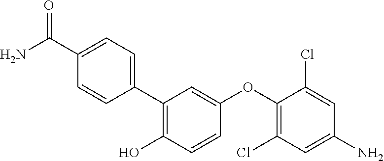

D00000
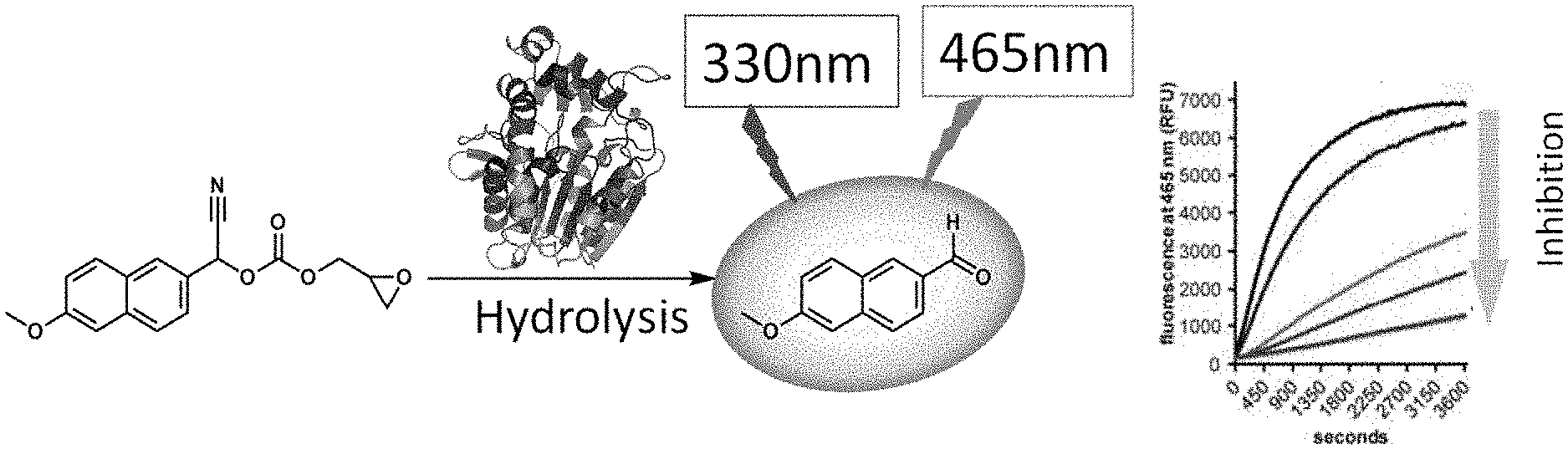
D00001
D00002

D00003
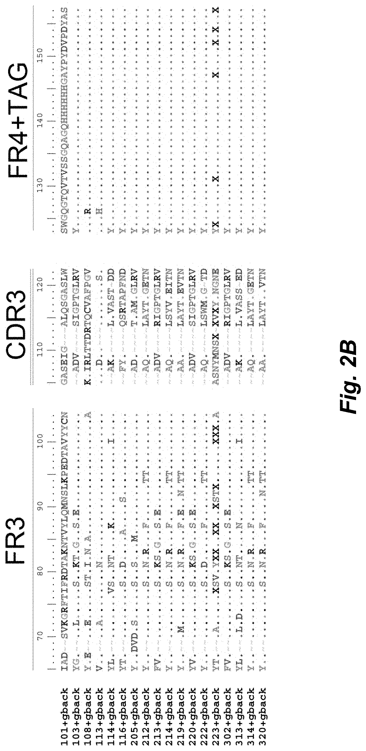
D00004

D00005

D00006
D00007

D00008
D00009
P00001

P00002

P00003

P00004

P00005

P00006

P00007

P00008

P00009

P00010

P00011

P00012

P00013

P00014

P00015

P00016

P00017

P00018

P00019

P00020

P00021

P00022

P00023

P00024

P00025

P00026

P00027

P00028

P00029

P00030

P00031

P00032

P00033

P00034

P00035

P00036

P00037

P00038

P00039

P00040

P00041

P00042

P00043

P00044

P00045

P00046

P00047

P00048

P00049

P00050

P00051

P00052

P00053

P00054

P00055

P00056

S00001
XML
uspto.report is an independent third-party trademark research tool that is not affiliated, endorsed, or sponsored by the United States Patent and Trademark Office (USPTO) or any other governmental organization. The information provided by uspto.report is based on publicly available data at the time of writing and is intended for informational purposes only.
While we strive to provide accurate and up-to-date information, we do not guarantee the accuracy, completeness, reliability, or suitability of the information displayed on this site. The use of this site is at your own risk. Any reliance you place on such information is therefore strictly at your own risk.
All official trademark data, including owner information, should be verified by visiting the official USPTO website at www.uspto.gov. This site is not intended to replace professional legal advice and should not be used as a substitute for consulting with a legal professional who is knowledgeable about trademark law.